US7132399B2 - Mammalian cell surface DNA receptor - Google Patents
Mammalian cell surface DNA receptor Download PDFInfo
- Publication number
- US7132399B2 US7132399B2 US10/619,992 US61999203A US7132399B2 US 7132399 B2 US7132399 B2 US 7132399B2 US 61999203 A US61999203 A US 61999203A US 7132399 B2 US7132399 B2 US 7132399B2
- Authority
- US
- United States
- Prior art keywords
- dna
- cells
- binding
- protein
- cell
- Prior art date
- Legal status (The legal status is an assumption and is not a legal conclusion. Google has not performed a legal analysis and makes no representation as to the accuracy of the status listed.)
- Expired - Fee Related, expires
Links
- 108010069941 DNA receptor Proteins 0.000 title claims description 254
- 210000004962 mammalian cell Anatomy 0.000 title claims description 13
- 230000004568 DNA-binding Effects 0.000 claims description 64
- 239000012634 fragment Substances 0.000 claims description 57
- 150000001413 amino acids Chemical class 0.000 claims description 40
- 125000003275 alpha amino acid group Chemical group 0.000 claims description 14
- 239000000203 mixture Substances 0.000 claims description 14
- 210000004027 cell Anatomy 0.000 abstract description 268
- 108090000623 proteins and genes Proteins 0.000 abstract description 76
- 241000282414 Homo sapiens Species 0.000 abstract description 62
- 238000000034 method Methods 0.000 abstract description 53
- 102000004169 proteins and genes Human genes 0.000 abstract description 45
- 230000027455 binding Effects 0.000 abstract description 41
- 108020004635 Complementary DNA Proteins 0.000 abstract description 39
- 238000010804 cDNA synthesis Methods 0.000 abstract description 36
- 239000002299 complementary DNA Substances 0.000 abstract description 36
- 238000003259 recombinant expression Methods 0.000 abstract description 23
- 101150054335 DNA-R gene Proteins 0.000 abstract description 16
- 150000001875 compounds Chemical class 0.000 abstract description 16
- 238000012216 screening Methods 0.000 abstract description 13
- 210000003527 eukaryotic cell Anatomy 0.000 abstract description 12
- 108020004999 messenger RNA Proteins 0.000 abstract description 12
- 238000000338 in vitro Methods 0.000 abstract description 11
- 210000001236 prokaryotic cell Anatomy 0.000 abstract description 6
- 238000002955 isolation Methods 0.000 abstract description 5
- 238000012512 characterization method Methods 0.000 abstract description 3
- 230000000144 pharmacologic effect Effects 0.000 abstract description 2
- 102000017063 Catecholamine Receptors Human genes 0.000 abstract 1
- 108010013659 Catecholamine Receptors Proteins 0.000 abstract 1
- 239000000556 agonist Substances 0.000 abstract 1
- 239000005557 antagonist Substances 0.000 abstract 1
- 239000013612 plasmid Substances 0.000 description 60
- 230000014509 gene expression Effects 0.000 description 54
- 235000018102 proteins Nutrition 0.000 description 43
- 108010070675 Glutathione transferase Proteins 0.000 description 39
- 102000005720 Glutathione transferase Human genes 0.000 description 39
- 235000001014 amino acid Nutrition 0.000 description 39
- 108020003175 receptors Proteins 0.000 description 37
- 102000005962 receptors Human genes 0.000 description 36
- GRRMZXFOOGQMFA-UHFFFAOYSA-J YoYo-1 Chemical compound [I-].[I-].[I-].[I-].C12=CC=CC=C2C(C=C2N(C3=CC=CC=C3O2)C)=CC=[N+]1CCC[N+](C)(C)CCC[N+](C)(C)CCC[N+](C1=CC=CC=C11)=CC=C1C=C1N(C)C2=CC=CC=C2O1 GRRMZXFOOGQMFA-UHFFFAOYSA-J 0.000 description 31
- 150000007523 nucleic acids Chemical class 0.000 description 31
- 102000039446 nucleic acids Human genes 0.000 description 27
- 210000001519 tissue Anatomy 0.000 description 27
- 108020004707 nucleic acids Proteins 0.000 description 26
- 201000003883 Cystic fibrosis Diseases 0.000 description 22
- 239000000523 sample Substances 0.000 description 21
- 238000011282 treatment Methods 0.000 description 21
- 238000002474 experimental method Methods 0.000 description 19
- 238000001943 fluorescence-activated cell sorting Methods 0.000 description 18
- 241000283973 Oryctolagus cuniculus Species 0.000 description 17
- 108020001507 fusion proteins Proteins 0.000 description 17
- 210000004072 lung Anatomy 0.000 description 17
- 239000002773 nucleotide Substances 0.000 description 17
- 125000003729 nucleotide group Chemical group 0.000 description 17
- 238000002360 preparation method Methods 0.000 description 17
- 102000037865 fusion proteins Human genes 0.000 description 16
- 230000001965 increasing effect Effects 0.000 description 16
- 108090000765 processed proteins & peptides Proteins 0.000 description 16
- 206010061218 Inflammation Diseases 0.000 description 15
- 238000003556 assay Methods 0.000 description 15
- 230000004054 inflammatory process Effects 0.000 description 15
- 239000003814 drug Substances 0.000 description 14
- RWSXRVCMGQZWBV-WDSKDSINSA-N glutathione Chemical group OC(=O)[C@@H](N)CCC(=O)N[C@@H](CS)C(=O)NCC(O)=O RWSXRVCMGQZWBV-WDSKDSINSA-N 0.000 description 14
- 241000699666 Mus <mouse, genus> Species 0.000 description 13
- 238000004458 analytical method Methods 0.000 description 13
- 238000011534 incubation Methods 0.000 description 13
- 210000004379 membrane Anatomy 0.000 description 13
- 239000012528 membrane Substances 0.000 description 13
- 229940079593 drug Drugs 0.000 description 12
- 241000894007 species Species 0.000 description 12
- 239000011701 zinc Substances 0.000 description 12
- 229910052725 zinc Inorganic materials 0.000 description 12
- 108700026244 Open Reading Frames Proteins 0.000 description 11
- HCHKCACWOHOZIP-UHFFFAOYSA-N Zinc Chemical compound [Zn] HCHKCACWOHOZIP-UHFFFAOYSA-N 0.000 description 11
- 102000004127 Cytokines Human genes 0.000 description 10
- 108090000695 Cytokines Proteins 0.000 description 10
- 241000588724 Escherichia coli Species 0.000 description 10
- 102000004889 Interleukin-6 Human genes 0.000 description 10
- 108090001005 Interleukin-6 Proteins 0.000 description 10
- 108091034117 Oligonucleotide Proteins 0.000 description 10
- 210000002966 serum Anatomy 0.000 description 10
- 239000013598 vector Substances 0.000 description 10
- 238000002965 ELISA Methods 0.000 description 9
- WSFSSNUMVMOOMR-UHFFFAOYSA-N Formaldehyde Chemical compound O=C WSFSSNUMVMOOMR-UHFFFAOYSA-N 0.000 description 9
- 241001465754 Metazoa Species 0.000 description 9
- 241001529936 Murinae Species 0.000 description 9
- 229920002684 Sepharose Polymers 0.000 description 9
- 238000001727 in vivo Methods 0.000 description 9
- 206010022000 influenza Diseases 0.000 description 9
- 239000002609 medium Substances 0.000 description 9
- 230000015861 cell surface binding Effects 0.000 description 8
- 230000000694 effects Effects 0.000 description 8
- 239000002158 endotoxin Substances 0.000 description 8
- 238000004519 manufacturing process Methods 0.000 description 8
- 230000001404 mediated effect Effects 0.000 description 8
- 238000001262 western blot Methods 0.000 description 8
- 108020004414 DNA Proteins 0.000 description 7
- KYRVNWMVYQXFEU-UHFFFAOYSA-N Nocodazole Chemical compound C1=C2NC(NC(=O)OC)=NC2=CC=C1C(=O)C1=CC=CS1 KYRVNWMVYQXFEU-UHFFFAOYSA-N 0.000 description 7
- 206010035226 Plasma cell myeloma Diseases 0.000 description 7
- 108700019146 Transgenes Proteins 0.000 description 7
- 102000004142 Trypsin Human genes 0.000 description 7
- 108090000631 Trypsin Proteins 0.000 description 7
- 239000000427 antigen Substances 0.000 description 7
- 108091007433 antigens Proteins 0.000 description 7
- 102000036639 antigens Human genes 0.000 description 7
- 244000309466 calf Species 0.000 description 7
- 210000000170 cell membrane Anatomy 0.000 description 7
- 235000018417 cysteine Nutrition 0.000 description 7
- 201000010099 disease Diseases 0.000 description 7
- 208000037265 diseases, disorders, signs and symptoms Diseases 0.000 description 7
- 229960003180 glutathione Drugs 0.000 description 7
- 210000004408 hybridoma Anatomy 0.000 description 7
- 239000002502 liposome Substances 0.000 description 7
- 230000007246 mechanism Effects 0.000 description 7
- 201000000050 myeloid neoplasm Diseases 0.000 description 7
- 238000007899 nucleic acid hybridization Methods 0.000 description 7
- 102000004196 processed proteins & peptides Human genes 0.000 description 7
- 230000028327 secretion Effects 0.000 description 7
- 210000001541 thymus gland Anatomy 0.000 description 7
- 239000012588 trypsin Substances 0.000 description 7
- 108010024636 Glutathione Proteins 0.000 description 6
- 241000699670 Mus sp. Species 0.000 description 6
- 125000000151 cysteine group Chemical class N[C@@H](CS)C(=O)* 0.000 description 6
- LOKCTEFSRHRXRJ-UHFFFAOYSA-I dipotassium trisodium dihydrogen phosphate hydrogen phosphate dichloride Chemical compound P(=O)(O)(O)[O-].[K+].P(=O)(O)([O-])[O-].[Na+].[Na+].[Cl-].[K+].[Cl-].[Na+] LOKCTEFSRHRXRJ-UHFFFAOYSA-I 0.000 description 6
- 239000012530 fluid Substances 0.000 description 6
- 238000010353 genetic engineering Methods 0.000 description 6
- 238000009396 hybridization Methods 0.000 description 6
- 210000000440 neutrophil Anatomy 0.000 description 6
- 239000002953 phosphate buffered saline Substances 0.000 description 6
- 238000012546 transfer Methods 0.000 description 6
- 210000004881 tumor cell Anatomy 0.000 description 6
- 108091003079 Bovine Serum Albumin Proteins 0.000 description 5
- 102000053602 DNA Human genes 0.000 description 5
- 101000742883 Homo sapiens Roquin-2 Proteins 0.000 description 5
- 108010001336 Horseradish Peroxidase Proteins 0.000 description 5
- 206010028980 Neoplasm Diseases 0.000 description 5
- 108091028043 Nucleic acid sequence Proteins 0.000 description 5
- 241000700159 Rattus Species 0.000 description 5
- 102100038059 Roquin-2 Human genes 0.000 description 5
- 230000003321 amplification Effects 0.000 description 5
- 230000001580 bacterial effect Effects 0.000 description 5
- 238000010367 cloning Methods 0.000 description 5
- 238000010586 diagram Methods 0.000 description 5
- 108010067396 dornase alfa Proteins 0.000 description 5
- 239000012894 fetal calf serum Substances 0.000 description 5
- 239000001963 growth medium Substances 0.000 description 5
- 201000001441 melanoma Diseases 0.000 description 5
- 238000003199 nucleic acid amplification method Methods 0.000 description 5
- 239000013615 primer Substances 0.000 description 5
- 210000000952 spleen Anatomy 0.000 description 5
- 238000002560 therapeutic procedure Methods 0.000 description 5
- 241000699800 Cricetinae Species 0.000 description 4
- AOJJSUZBOXZQNB-TZSSRYMLSA-N Doxorubicin Chemical compound O([C@H]1C[C@@](O)(CC=2C(O)=C3C(=O)C=4C=CC=C(C=4C(=O)C3=C(O)C=21)OC)C(=O)CO)[C@H]1C[C@H](N)[C@H](O)[C@H](C)O1 AOJJSUZBOXZQNB-TZSSRYMLSA-N 0.000 description 4
- 238000012413 Fluorescence activated cell sorting analysis Methods 0.000 description 4
- 239000000020 Nitrocellulose Substances 0.000 description 4
- 208000006664 Precursor Cell Lymphoblastic Leukemia-Lymphoma Diseases 0.000 description 4
- FAPWRFPIFSIZLT-UHFFFAOYSA-M Sodium chloride Chemical compound [Na+].[Cl-] FAPWRFPIFSIZLT-UHFFFAOYSA-M 0.000 description 4
- 238000013459 approach Methods 0.000 description 4
- 201000011510 cancer Diseases 0.000 description 4
- 238000004113 cell culture Methods 0.000 description 4
- 230000001684 chronic effect Effects 0.000 description 4
- 230000012202 endocytosis Effects 0.000 description 4
- 238000001415 gene therapy Methods 0.000 description 4
- 230000005764 inhibitory process Effects 0.000 description 4
- 238000011081 inoculation Methods 0.000 description 4
- 208000032839 leukemia Diseases 0.000 description 4
- 208000003747 lymphoid leukemia Diseases 0.000 description 4
- 210000002540 macrophage Anatomy 0.000 description 4
- 229920001220 nitrocellulos Polymers 0.000 description 4
- 238000003752 polymerase chain reaction Methods 0.000 description 4
- 230000008569 process Effects 0.000 description 4
- 239000000047 product Substances 0.000 description 4
- 230000017854 proteolysis Effects 0.000 description 4
- 210000004989 spleen cell Anatomy 0.000 description 4
- 239000012581 transferrin Substances 0.000 description 4
- 230000014616 translation Effects 0.000 description 4
- 108091032973 (ribonucleotides)n+m Proteins 0.000 description 3
- 108020000948 Antisense Oligonucleotides Proteins 0.000 description 3
- 241000894006 Bacteria Species 0.000 description 3
- 108020000946 Bacterial DNA Proteins 0.000 description 3
- 208000011691 Burkitt lymphomas Diseases 0.000 description 3
- 108091026890 Coding region Proteins 0.000 description 3
- 108020004705 Codon Proteins 0.000 description 3
- 241000282412 Homo Species 0.000 description 3
- 208000026350 Inborn Genetic disease Diseases 0.000 description 3
- QNAYBMKLOCPYGJ-REOHCLBHSA-N L-alanine Chemical compound C[C@H](N)C(O)=O QNAYBMKLOCPYGJ-REOHCLBHSA-N 0.000 description 3
- 108020004711 Nucleic Acid Probes Proteins 0.000 description 3
- 108020005187 Oligonucleotide Probes Proteins 0.000 description 3
- ONIBWKKTOPOVIA-UHFFFAOYSA-N Proline Natural products OC(=O)C1CCCN1 ONIBWKKTOPOVIA-UHFFFAOYSA-N 0.000 description 3
- MTCFGRXMJLQNBG-UHFFFAOYSA-N Serine Natural products OCC(N)C(O)=O MTCFGRXMJLQNBG-UHFFFAOYSA-N 0.000 description 3
- 108020004682 Single-Stranded DNA Proteins 0.000 description 3
- 238000002105 Southern blotting Methods 0.000 description 3
- 108020004566 Transfer RNA Proteins 0.000 description 3
- 241000700605 Viruses Species 0.000 description 3
- JLCPHMBAVCMARE-UHFFFAOYSA-N [3-[[3-[[3-[[3-[[3-[[3-[[3-[[3-[[3-[[3-[[3-[[5-(2-amino-6-oxo-1H-purin-9-yl)-3-[[3-[[3-[[3-[[3-[[3-[[5-(2-amino-6-oxo-1H-purin-9-yl)-3-[[5-(2-amino-6-oxo-1H-purin-9-yl)-3-hydroxyoxolan-2-yl]methoxy-hydroxyphosphoryl]oxyoxolan-2-yl]methoxy-hydroxyphosphoryl]oxy-5-(5-methyl-2,4-dioxopyrimidin-1-yl)oxolan-2-yl]methoxy-hydroxyphosphoryl]oxy-5-(6-aminopurin-9-yl)oxolan-2-yl]methoxy-hydroxyphosphoryl]oxy-5-(6-aminopurin-9-yl)oxolan-2-yl]methoxy-hydroxyphosphoryl]oxy-5-(6-aminopurin-9-yl)oxolan-2-yl]methoxy-hydroxyphosphoryl]oxy-5-(6-aminopurin-9-yl)oxolan-2-yl]methoxy-hydroxyphosphoryl]oxyoxolan-2-yl]methoxy-hydroxyphosphoryl]oxy-5-(5-methyl-2,4-dioxopyrimidin-1-yl)oxolan-2-yl]methoxy-hydroxyphosphoryl]oxy-5-(4-amino-2-oxopyrimidin-1-yl)oxolan-2-yl]methoxy-hydroxyphosphoryl]oxy-5-(5-methyl-2,4-dioxopyrimidin-1-yl)oxolan-2-yl]methoxy-hydroxyphosphoryl]oxy-5-(5-methyl-2,4-dioxopyrimidin-1-yl)oxolan-2-yl]methoxy-hydroxyphosphoryl]oxy-5-(6-aminopurin-9-yl)oxolan-2-yl]methoxy-hydroxyphosphoryl]oxy-5-(6-aminopurin-9-yl)oxolan-2-yl]methoxy-hydroxyphosphoryl]oxy-5-(4-amino-2-oxopyrimidin-1-yl)oxolan-2-yl]methoxy-hydroxyphosphoryl]oxy-5-(4-amino-2-oxopyrimidin-1-yl)oxolan-2-yl]methoxy-hydroxyphosphoryl]oxy-5-(4-amino-2-oxopyrimidin-1-yl)oxolan-2-yl]methoxy-hydroxyphosphoryl]oxy-5-(6-aminopurin-9-yl)oxolan-2-yl]methoxy-hydroxyphosphoryl]oxy-5-(4-amino-2-oxopyrimidin-1-yl)oxolan-2-yl]methyl [5-(6-aminopurin-9-yl)-2-(hydroxymethyl)oxolan-3-yl] hydrogen phosphate Polymers Cc1cn(C2CC(OP(O)(=O)OCC3OC(CC3OP(O)(=O)OCC3OC(CC3O)n3cnc4c3nc(N)[nH]c4=O)n3cnc4c3nc(N)[nH]c4=O)C(COP(O)(=O)OC3CC(OC3COP(O)(=O)OC3CC(OC3COP(O)(=O)OC3CC(OC3COP(O)(=O)OC3CC(OC3COP(O)(=O)OC3CC(OC3COP(O)(=O)OC3CC(OC3COP(O)(=O)OC3CC(OC3COP(O)(=O)OC3CC(OC3COP(O)(=O)OC3CC(OC3COP(O)(=O)OC3CC(OC3COP(O)(=O)OC3CC(OC3COP(O)(=O)OC3CC(OC3COP(O)(=O)OC3CC(OC3COP(O)(=O)OC3CC(OC3COP(O)(=O)OC3CC(OC3COP(O)(=O)OC3CC(OC3COP(O)(=O)OC3CC(OC3CO)n3cnc4c(N)ncnc34)n3ccc(N)nc3=O)n3cnc4c(N)ncnc34)n3ccc(N)nc3=O)n3ccc(N)nc3=O)n3ccc(N)nc3=O)n3cnc4c(N)ncnc34)n3cnc4c(N)ncnc34)n3cc(C)c(=O)[nH]c3=O)n3cc(C)c(=O)[nH]c3=O)n3ccc(N)nc3=O)n3cc(C)c(=O)[nH]c3=O)n3cnc4c3nc(N)[nH]c4=O)n3cnc4c(N)ncnc34)n3cnc4c(N)ncnc34)n3cnc4c(N)ncnc34)n3cnc4c(N)ncnc34)O2)c(=O)[nH]c1=O JLCPHMBAVCMARE-UHFFFAOYSA-N 0.000 description 3
- 235000004279 alanine Nutrition 0.000 description 3
- 125000000539 amino acid group Chemical group 0.000 description 3
- 230000003172 anti-dna Effects 0.000 description 3
- 239000000074 antisense oligonucleotide Substances 0.000 description 3
- 238000012230 antisense oligonucleotides Methods 0.000 description 3
- 238000007796 conventional method Methods 0.000 description 3
- 230000001419 dependent effect Effects 0.000 description 3
- 238000001514 detection method Methods 0.000 description 3
- 238000011161 development Methods 0.000 description 3
- 238000005516 engineering process Methods 0.000 description 3
- 208000016361 genetic disease Diseases 0.000 description 3
- 230000002068 genetic effect Effects 0.000 description 3
- 235000014304 histidine Nutrition 0.000 description 3
- 150000002411 histidines Chemical class 0.000 description 3
- 210000005260 human cell Anatomy 0.000 description 3
- 238000001114 immunoprecipitation Methods 0.000 description 3
- 230000003993 interaction Effects 0.000 description 3
- 230000003834 intracellular effect Effects 0.000 description 3
- 229920006008 lipopolysaccharide Polymers 0.000 description 3
- 230000007774 longterm Effects 0.000 description 3
- 230000004199 lung function Effects 0.000 description 3
- 239000006166 lysate Substances 0.000 description 3
- 230000004048 modification Effects 0.000 description 3
- 238000012986 modification Methods 0.000 description 3
- 239000002853 nucleic acid probe Substances 0.000 description 3
- 239000002751 oligonucleotide probe Substances 0.000 description 3
- 230000001575 pathological effect Effects 0.000 description 3
- 238000002203 pretreatment Methods 0.000 description 3
- 125000002924 primary amino group Chemical group [H]N([H])* 0.000 description 3
- 238000002415 sodium dodecyl sulfate polyacrylamide gel electrophoresis Methods 0.000 description 3
- 239000007787 solid Substances 0.000 description 3
- 238000010186 staining Methods 0.000 description 3
- 239000000126 substance Substances 0.000 description 3
- 238000003786 synthesis reaction Methods 0.000 description 3
- 230000009466 transformation Effects 0.000 description 3
- 238000013519 translation Methods 0.000 description 3
- YXHLJMWYDTXDHS-IRFLANFNSA-N 7-aminoactinomycin D Chemical compound C[C@H]1OC(=O)[C@H](C(C)C)N(C)C(=O)CN(C)C(=O)[C@@H]2CCCN2C(=O)[C@@H](C(C)C)NC(=O)[C@H]1NC(=O)C1=C(N)C(=O)C(C)=C2OC(C(C)=C(N)C=C3C(=O)N[C@@H]4C(=O)N[C@@H](C(N5CCC[C@H]5C(=O)N(C)CC(=O)N(C)[C@@H](C(C)C)C(=O)O[C@@H]4C)=O)C(C)C)=C3N=C21 YXHLJMWYDTXDHS-IRFLANFNSA-N 0.000 description 2
- 108700012813 7-aminoactinomycin D Proteins 0.000 description 2
- 206010003445 Ascites Diseases 0.000 description 2
- 206010006458 Bronchitis chronic Diseases 0.000 description 2
- 241000283707 Capra Species 0.000 description 2
- 206010008342 Cervix carcinoma Diseases 0.000 description 2
- 238000012287 DNA Binding Assay Methods 0.000 description 2
- 239000003155 DNA primer Substances 0.000 description 2
- 102000052510 DNA-Binding Proteins Human genes 0.000 description 2
- 101710096438 DNA-binding protein Proteins 0.000 description 2
- 108010053770 Deoxyribonucleases Proteins 0.000 description 2
- 102000016911 Deoxyribonucleases Human genes 0.000 description 2
- KCXVZYZYPLLWCC-UHFFFAOYSA-N EDTA Chemical compound OC(=O)CN(CC(O)=O)CCN(CC(O)=O)CC(O)=O KCXVZYZYPLLWCC-UHFFFAOYSA-N 0.000 description 2
- ZHNUHDYFZUAESO-UHFFFAOYSA-N Formamide Chemical compound NC=O ZHNUHDYFZUAESO-UHFFFAOYSA-N 0.000 description 2
- 108010043121 Green Fluorescent Proteins Proteins 0.000 description 2
- 102000004144 Green Fluorescent Proteins Human genes 0.000 description 2
- 101001086428 Homo sapiens Olfactory receptor 1K1 Proteins 0.000 description 2
- ONIBWKKTOPOVIA-BYPYZUCNSA-N L-Proline Chemical compound OC(=O)[C@@H]1CCCN1 ONIBWKKTOPOVIA-BYPYZUCNSA-N 0.000 description 2
- 208000019693 Lung disease Diseases 0.000 description 2
- 206010058467 Lung neoplasm malignant Diseases 0.000 description 2
- 102000018697 Membrane Proteins Human genes 0.000 description 2
- 108010052285 Membrane Proteins Proteins 0.000 description 2
- 238000000636 Northern blotting Methods 0.000 description 2
- 241001494479 Pecora Species 0.000 description 2
- 208000007452 Plasmacytoma Diseases 0.000 description 2
- 239000002202 Polyethylene glycol Substances 0.000 description 2
- 241000288906 Primates Species 0.000 description 2
- 206010036790 Productive cough Diseases 0.000 description 2
- 108700008625 Reporter Genes Proteins 0.000 description 2
- 108010083644 Ribonucleases Proteins 0.000 description 2
- 102000006382 Ribonucleases Human genes 0.000 description 2
- 241000283984 Rodentia Species 0.000 description 2
- 240000004808 Saccharomyces cerevisiae Species 0.000 description 2
- 235000014680 Saccharomyces cerevisiae Nutrition 0.000 description 2
- 238000012300 Sequence Analysis Methods 0.000 description 2
- 102000007238 Transferrin Receptors Human genes 0.000 description 2
- 108010033576 Transferrin Receptors Proteins 0.000 description 2
- 238000002835 absorbance Methods 0.000 description 2
- 239000000443 aerosol Substances 0.000 description 2
- 210000001552 airway epithelial cell Anatomy 0.000 description 2
- 208000037883 airway inflammation Diseases 0.000 description 2
- 230000003110 anti-inflammatory effect Effects 0.000 description 2
- 230000000692 anti-sense effect Effects 0.000 description 2
- 239000002246 antineoplastic agent Substances 0.000 description 2
- 229940041181 antineoplastic drug Drugs 0.000 description 2
- 235000003704 aspartic acid Nutrition 0.000 description 2
- 210000003719 b-lymphocyte Anatomy 0.000 description 2
- 244000052616 bacterial pathogen Species 0.000 description 2
- 239000011324 bead Substances 0.000 description 2
- 230000009286 beneficial effect Effects 0.000 description 2
- 230000004071 biological effect Effects 0.000 description 2
- 206010006451 bronchitis Diseases 0.000 description 2
- 230000015556 catabolic process Effects 0.000 description 2
- 208000019065 cervical carcinoma Diseases 0.000 description 2
- 238000006243 chemical reaction Methods 0.000 description 2
- 210000000349 chromosome Anatomy 0.000 description 2
- 208000007451 chronic bronchitis Diseases 0.000 description 2
- 208000029742 colonic neoplasm Diseases 0.000 description 2
- XUJNEKJLAYXESH-UHFFFAOYSA-N cysteine Natural products SCC(N)C(O)=O XUJNEKJLAYXESH-UHFFFAOYSA-N 0.000 description 2
- 238000006731 degradation reaction Methods 0.000 description 2
- 238000010790 dilution Methods 0.000 description 2
- 239000012895 dilution Substances 0.000 description 2
- 231100000673 dose–response relationship Toxicity 0.000 description 2
- 239000000284 extract Substances 0.000 description 2
- 238000000684 flow cytometry Methods 0.000 description 2
- MHMNJMPURVTYEJ-UHFFFAOYSA-N fluorescein-5-isothiocyanate Chemical compound O1C(=O)C2=CC(N=C=S)=CC=C2C21C1=CC=C(O)C=C1OC1=CC(O)=CC=C21 MHMNJMPURVTYEJ-UHFFFAOYSA-N 0.000 description 2
- 230000004927 fusion Effects 0.000 description 2
- 238000001476 gene delivery Methods 0.000 description 2
- 239000005090 green fluorescent protein Substances 0.000 description 2
- 238000002744 homologous recombination Methods 0.000 description 2
- 230000006801 homologous recombination Effects 0.000 description 2
- 230000002209 hydrophobic effect Effects 0.000 description 2
- 238000002169 hydrotherapy Methods 0.000 description 2
- 230000028993 immune response Effects 0.000 description 2
- 230000003053 immunization Effects 0.000 description 2
- 230000006872 improvement Effects 0.000 description 2
- 239000003112 inhibitor Substances 0.000 description 2
- 238000002347 injection Methods 0.000 description 2
- 239000007924 injection Substances 0.000 description 2
- 230000000266 injurious effect Effects 0.000 description 2
- 238000011835 investigation Methods 0.000 description 2
- 210000004185 liver Anatomy 0.000 description 2
- 201000005296 lung carcinoma Diseases 0.000 description 2
- 210000005265 lung cell Anatomy 0.000 description 2
- 206010025135 lupus erythematosus Diseases 0.000 description 2
- 210000001616 monocyte Anatomy 0.000 description 2
- 229940126619 mouse monoclonal antibody Drugs 0.000 description 2
- 239000013642 negative control Substances 0.000 description 2
- 229950006344 nocodazole Drugs 0.000 description 2
- 108091027963 non-coding RNA Proteins 0.000 description 2
- 102000042567 non-coding RNA Human genes 0.000 description 2
- 210000001672 ovary Anatomy 0.000 description 2
- 210000003819 peripheral blood mononuclear cell Anatomy 0.000 description 2
- 229920002401 polyacrylamide Polymers 0.000 description 2
- 229920001223 polyethylene glycol Polymers 0.000 description 2
- 230000000770 proinflammatory effect Effects 0.000 description 2
- 238000000159 protein binding assay Methods 0.000 description 2
- 238000000746 purification Methods 0.000 description 2
- 230000009257 reactivity Effects 0.000 description 2
- 230000002829 reductive effect Effects 0.000 description 2
- 238000007894 restriction fragment length polymorphism technique Methods 0.000 description 2
- 238000002741 site-directed mutagenesis Methods 0.000 description 2
- 239000011780 sodium chloride Substances 0.000 description 2
- 239000000243 solution Substances 0.000 description 2
- 208000024794 sputum Diseases 0.000 description 2
- 210000003802 sputum Anatomy 0.000 description 2
- UCSJYZPVAKXKNQ-HZYVHMACSA-N streptomycin Chemical compound CN[C@H]1[C@H](O)[C@@H](O)[C@H](CO)O[C@H]1O[C@@H]1[C@](C=O)(O)[C@H](C)O[C@H]1O[C@@H]1[C@@H](NC(N)=N)[C@H](O)[C@@H](NC(N)=N)[C@H](O)[C@H]1O UCSJYZPVAKXKNQ-HZYVHMACSA-N 0.000 description 2
- 239000000758 substrate Substances 0.000 description 2
- 229940124597 therapeutic agent Drugs 0.000 description 2
- 230000001225 therapeutic effect Effects 0.000 description 2
- 238000013518 transcription Methods 0.000 description 2
- 230000035897 transcription Effects 0.000 description 2
- 241001430294 unidentified retrovirus Species 0.000 description 2
- 230000009385 viral infection Effects 0.000 description 2
- 239000013603 viral vector Substances 0.000 description 2
- 238000005406 washing Methods 0.000 description 2
- MTCFGRXMJLQNBG-REOHCLBHSA-N (2S)-2-Amino-3-hydroxypropansäure Chemical compound OC[C@H](N)C(O)=O MTCFGRXMJLQNBG-REOHCLBHSA-N 0.000 description 1
- FWMNVWWHGCHHJJ-SKKKGAJSSA-N 4-amino-1-[(2r)-6-amino-2-[[(2r)-2-[[(2r)-2-[[(2r)-2-amino-3-phenylpropanoyl]amino]-3-phenylpropanoyl]amino]-4-methylpentanoyl]amino]hexanoyl]piperidine-4-carboxylic acid Chemical compound C([C@H](C(=O)N[C@H](CC(C)C)C(=O)N[C@H](CCCCN)C(=O)N1CCC(N)(CC1)C(O)=O)NC(=O)[C@H](N)CC=1C=CC=CC=1)C1=CC=CC=C1 FWMNVWWHGCHHJJ-SKKKGAJSSA-N 0.000 description 1
- QWVRTSZDKPRPDF-UHFFFAOYSA-N 5-(piperidin-1-ylmethyl)-3-pyridin-3-yl-5,6-dihydro-2h-1,2,4-oxadiazine Chemical compound C1CCCCN1CC(N=1)CONC=1C1=CC=CN=C1 QWVRTSZDKPRPDF-UHFFFAOYSA-N 0.000 description 1
- 101150044182 8 gene Proteins 0.000 description 1
- 208000030507 AIDS Diseases 0.000 description 1
- 102100033792 ALX homeobox protein 1 Human genes 0.000 description 1
- 102100033350 ATP-dependent translocase ABCB1 Human genes 0.000 description 1
- 102000015404 Amino Acid Receptors Human genes 0.000 description 1
- 108010025177 Amino Acid Receptors Proteins 0.000 description 1
- 101100439665 Arabidopsis thaliana SWI2 gene Proteins 0.000 description 1
- 206010053555 Arthritis bacterial Diseases 0.000 description 1
- 241000972773 Aulopiformes Species 0.000 description 1
- 208000004736 B-Cell Leukemia Diseases 0.000 description 1
- 208000010839 B-cell chronic lymphocytic leukemia Diseases 0.000 description 1
- 108010077805 Bacterial Proteins Proteins 0.000 description 1
- 208000035143 Bacterial infection Diseases 0.000 description 1
- 241000283690 Bos taurus Species 0.000 description 1
- 101150084711 CTH1 gene Proteins 0.000 description 1
- 241000244203 Caenorhabditis elegans Species 0.000 description 1
- 101100243951 Caenorhabditis elegans pie-1 gene Proteins 0.000 description 1
- OYPRJOBELJOOCE-UHFFFAOYSA-N Calcium Chemical group [Ca] OYPRJOBELJOOCE-UHFFFAOYSA-N 0.000 description 1
- 108090000565 Capsid Proteins Proteins 0.000 description 1
- 208000002177 Cataract Diseases 0.000 description 1
- 108010001857 Cell Surface Receptors Proteins 0.000 description 1
- 102000000844 Cell Surface Receptors Human genes 0.000 description 1
- 241000282552 Chlorocebus aethiops Species 0.000 description 1
- 238000011537 Coomassie blue staining Methods 0.000 description 1
- 241000699802 Cricetulus griseus Species 0.000 description 1
- 108020003215 DNA Probes Proteins 0.000 description 1
- 108010008286 DNA nucleotidylexotransferase Proteins 0.000 description 1
- 239000003298 DNA probe Substances 0.000 description 1
- 241000450599 DNA viruses Species 0.000 description 1
- 102100029764 DNA-directed DNA/RNA polymerase mu Human genes 0.000 description 1
- 241000702421 Dependoparvovirus Species 0.000 description 1
- 101000802103 Drosophila melanogaster Protein TIS11 Proteins 0.000 description 1
- 206010059866 Drug resistance Diseases 0.000 description 1
- 239000006144 Dulbecco’s modified Eagle's medium Substances 0.000 description 1
- 102100026246 E3 ubiquitin-protein ligase NRDP1 Human genes 0.000 description 1
- 206010049466 Erythroblastosis Diseases 0.000 description 1
- 108700024394 Exon Proteins 0.000 description 1
- 208000001382 Experimental Melanoma Diseases 0.000 description 1
- 241000282326 Felis catus Species 0.000 description 1
- 229920001917 Ficoll Polymers 0.000 description 1
- 101150066002 GFP gene Proteins 0.000 description 1
- 108700028146 Genetic Enhancer Elements Proteins 0.000 description 1
- WQZGKKKJIJFFOK-GASJEMHNSA-N Glucose Natural products OC[C@H]1OC(O)[C@H](O)[C@@H](O)[C@@H]1O WQZGKKKJIJFFOK-GASJEMHNSA-N 0.000 description 1
- 206010053759 Growth retardation Diseases 0.000 description 1
- HVLSXIKZNLPZJJ-TXZCQADKSA-N HA peptide Chemical group C([C@@H](C(=O)N[C@@H](CC(O)=O)C(=O)N[C@@H](C(C)C)C(=O)N1[C@@H](CCC1)C(=O)N[C@@H](CC(O)=O)C(=O)N[C@@H](CC=1C=CC(O)=CC=1)C(=O)N[C@@H](C)C(O)=O)NC(=O)[C@H]1N(CCC1)C(=O)[C@@H](N)CC=1C=CC(O)=CC=1)C1=CC=C(O)C=C1 HVLSXIKZNLPZJJ-TXZCQADKSA-N 0.000 description 1
- 241000708754 Hauffenia media Species 0.000 description 1
- 101710154606 Hemagglutinin Proteins 0.000 description 1
- 101000779615 Homo sapiens ALX homeobox protein 1 Proteins 0.000 description 1
- 101000692706 Homo sapiens E3 ubiquitin-protein ligase NRDP1 Proteins 0.000 description 1
- 101000848625 Homo sapiens E3 ubiquitin-protein ligase TRIM23 Proteins 0.000 description 1
- 101000830933 Homo sapiens TNF receptor-associated factor 4 Proteins 0.000 description 1
- 101000802094 Homo sapiens mRNA decay activator protein ZFP36L1 Proteins 0.000 description 1
- HEFNNWSXXWATRW-UHFFFAOYSA-N Ibuprofen Chemical compound CC(C)CC1=CC=C(C(C)C(O)=O)C=C1 HEFNNWSXXWATRW-UHFFFAOYSA-N 0.000 description 1
- 108010058683 Immobilized Proteins Proteins 0.000 description 1
- 108060003951 Immunoglobulin Proteins 0.000 description 1
- 208000004575 Infectious Arthritis Diseases 0.000 description 1
- 102100034343 Integrase Human genes 0.000 description 1
- CKLJMWTZIZZHCS-REOHCLBHSA-N L-aspartic acid Chemical compound OC(=O)[C@@H](N)CC(O)=O CKLJMWTZIZZHCS-REOHCLBHSA-N 0.000 description 1
- ZDXPYRJPNDTMRX-VKHMYHEASA-N L-glutamine Chemical compound OC(=O)[C@@H](N)CCC(N)=O ZDXPYRJPNDTMRX-VKHMYHEASA-N 0.000 description 1
- 229930182816 L-glutamine Natural products 0.000 description 1
- 108091026898 Leader sequence (mRNA) Proteins 0.000 description 1
- 241000239218 Limulus Species 0.000 description 1
- 208000035752 Live birth Diseases 0.000 description 1
- 206010025323 Lymphomas Diseases 0.000 description 1
- 241000124008 Mammalia Species 0.000 description 1
- 108010047230 Member 1 Subfamily B ATP Binding Cassette Transporter Proteins 0.000 description 1
- 229940122255 Microtubule inhibitor Drugs 0.000 description 1
- 241000711408 Murine respirovirus Species 0.000 description 1
- 230000004988 N-glycosylation Effects 0.000 description 1
- 108091061960 Naked DNA Proteins 0.000 description 1
- 102000043276 Oncogene Human genes 0.000 description 1
- 108700020796 Oncogene Proteins 0.000 description 1
- 206010033078 Otitis media Diseases 0.000 description 1
- 101710093908 Outer capsid protein VP4 Proteins 0.000 description 1
- 101710135467 Outer capsid protein sigma-1 Proteins 0.000 description 1
- 206010033963 Parathyroid tumour Diseases 0.000 description 1
- 229930182555 Penicillin Natural products 0.000 description 1
- JGSARLDLIJGVTE-MBNYWOFBSA-N Penicillin G Chemical compound N([C@H]1[C@H]2SC([C@@H](N2C1=O)C(O)=O)(C)C)C(=O)CC1=CC=CC=C1 JGSARLDLIJGVTE-MBNYWOFBSA-N 0.000 description 1
- 241000223960 Plasmodium falciparum Species 0.000 description 1
- 229920002534 Polyethylene Glycol 1450 Polymers 0.000 description 1
- 101710176177 Protein A56 Proteins 0.000 description 1
- 108010092799 RNA-directed DNA polymerase Proteins 0.000 description 1
- 238000010240 RT-PCR analysis Methods 0.000 description 1
- 108020004511 Recombinant DNA Proteins 0.000 description 1
- 208000004756 Respiratory Insufficiency Diseases 0.000 description 1
- 101100221606 Saccharomyces cerevisiae (strain ATCC 204508 / S288c) COS7 gene Proteins 0.000 description 1
- 108010090804 Streptavidin Proteins 0.000 description 1
- 208000037065 Subacute sclerosing leukoencephalitis Diseases 0.000 description 1
- 206010042297 Subacute sclerosing panencephalitis Diseases 0.000 description 1
- 241000282887 Suidae Species 0.000 description 1
- 208000031673 T-Cell Cutaneous Lymphoma Diseases 0.000 description 1
- 206010042971 T-cell lymphoma Diseases 0.000 description 1
- 208000027585 T-cell non-Hodgkin lymphoma Diseases 0.000 description 1
- 210000001744 T-lymphocyte Anatomy 0.000 description 1
- 229920004890 Triton X-100 Polymers 0.000 description 1
- 239000013504 Triton X-100 Substances 0.000 description 1
- 208000006105 Uterine Cervical Neoplasms Diseases 0.000 description 1
- 241000251539 Vertebrata <Metazoa> Species 0.000 description 1
- 208000036142 Viral infection Diseases 0.000 description 1
- 101710185494 Zinc finger protein Proteins 0.000 description 1
- 102100023597 Zinc finger protein 816 Human genes 0.000 description 1
- 230000005856 abnormality Effects 0.000 description 1
- 230000009471 action Effects 0.000 description 1
- 230000004913 activation Effects 0.000 description 1
- 230000001154 acute effect Effects 0.000 description 1
- 229940009456 adriamycin Drugs 0.000 description 1
- 230000002411 adverse Effects 0.000 description 1
- 238000001261 affinity purification Methods 0.000 description 1
- 210000001132 alveolar macrophage Anatomy 0.000 description 1
- 239000003242 anti bacterial agent Substances 0.000 description 1
- 239000002260 anti-inflammatory agent Substances 0.000 description 1
- 229940121363 anti-inflammatory agent Drugs 0.000 description 1
- 229940088710 antibiotic agent Drugs 0.000 description 1
- 230000000890 antigenic effect Effects 0.000 description 1
- 239000003963 antioxidant agent Substances 0.000 description 1
- 230000006907 apoptotic process Effects 0.000 description 1
- 150000001510 aspartic acids Chemical class 0.000 description 1
- 230000003416 augmentation Effects 0.000 description 1
- 208000022362 bacterial infectious disease Diseases 0.000 description 1
- OQFSQFPPLPISGP-UHFFFAOYSA-N beta-carboxyaspartic acid Natural products OC(=O)C(N)C(C(O)=O)C(O)=O OQFSQFPPLPISGP-UHFFFAOYSA-N 0.000 description 1
- 238000002306 biochemical method Methods 0.000 description 1
- 230000003115 biocidal effect Effects 0.000 description 1
- 230000031018 biological processes and functions Effects 0.000 description 1
- 230000015572 biosynthetic process Effects 0.000 description 1
- 230000000903 blocking effect Effects 0.000 description 1
- 210000004369 blood Anatomy 0.000 description 1
- 239000008280 blood Substances 0.000 description 1
- 239000012888 bovine serum Substances 0.000 description 1
- 210000000424 bronchial epithelial cell Anatomy 0.000 description 1
- 201000009267 bronchiectasis Diseases 0.000 description 1
- 210000004899 c-terminal region Anatomy 0.000 description 1
- 239000011575 calcium Substances 0.000 description 1
- 229910052791 calcium Inorganic materials 0.000 description 1
- 125000003178 carboxy group Chemical group [H]OC(*)=O 0.000 description 1
- 150000001768 cations Chemical class 0.000 description 1
- 230000007910 cell fusion Effects 0.000 description 1
- 230000010261 cell growth Effects 0.000 description 1
- 239000013592 cell lysate Substances 0.000 description 1
- 230000006037 cell lysis Effects 0.000 description 1
- 210000003855 cell nucleus Anatomy 0.000 description 1
- 230000030570 cellular localization Effects 0.000 description 1
- 201000010881 cervical cancer Diseases 0.000 description 1
- 239000007795 chemical reaction product Substances 0.000 description 1
- 239000003153 chemical reaction reagent Substances 0.000 description 1
- 239000003795 chemical substances by application Substances 0.000 description 1
- 208000037976 chronic inflammation Diseases 0.000 description 1
- 230000006020 chronic inflammation Effects 0.000 description 1
- 238000003776 cleavage reaction Methods 0.000 description 1
- 230000008045 co-localization Effects 0.000 description 1
- 201000010897 colon adenocarcinoma Diseases 0.000 description 1
- 230000009137 competitive binding Effects 0.000 description 1
- 230000002860 competitive effect Effects 0.000 description 1
- 230000000295 complement effect Effects 0.000 description 1
- 238000010276 construction Methods 0.000 description 1
- 239000003246 corticosteroid Substances 0.000 description 1
- 229960001334 corticosteroids Drugs 0.000 description 1
- 239000012228 culture supernatant Substances 0.000 description 1
- 238000012258 culturing Methods 0.000 description 1
- 201000007241 cutaneous T cell lymphoma Diseases 0.000 description 1
- 208000035250 cutaneous malignant susceptibility to 1 melanoma Diseases 0.000 description 1
- 230000016396 cytokine production Effects 0.000 description 1
- 230000001086 cytosolic effect Effects 0.000 description 1
- 230000006378 damage Effects 0.000 description 1
- 230000003247 decreasing effect Effects 0.000 description 1
- 238000003745 diagnosis Methods 0.000 description 1
- BFMYDTVEBKDAKJ-UHFFFAOYSA-L disodium;(2',7'-dibromo-3',6'-dioxido-3-oxospiro[2-benzofuran-1,9'-xanthene]-4'-yl)mercury;hydrate Chemical compound O.[Na+].[Na+].O1C(=O)C2=CC=CC=C2C21C1=CC(Br)=C([O-])C([Hg])=C1OC1=C2C=C(Br)C([O-])=C1 BFMYDTVEBKDAKJ-UHFFFAOYSA-L 0.000 description 1
- 238000009826 distribution Methods 0.000 description 1
- 238000012137 double-staining Methods 0.000 description 1
- 229960004679 doxorubicin Drugs 0.000 description 1
- 238000009510 drug design Methods 0.000 description 1
- 238000007877 drug screening Methods 0.000 description 1
- 241001493065 dsRNA viruses Species 0.000 description 1
- 230000001909 effect on DNA Effects 0.000 description 1
- 210000001163 endosome Anatomy 0.000 description 1
- 230000002708 enhancing effect Effects 0.000 description 1
- 210000002919 epithelial cell Anatomy 0.000 description 1
- 210000003743 erythrocyte Anatomy 0.000 description 1
- 238000010195 expression analysis Methods 0.000 description 1
- 239000013604 expression vector Substances 0.000 description 1
- 210000002458 fetal heart Anatomy 0.000 description 1
- 238000009472 formulation Methods 0.000 description 1
- 239000012737 fresh medium Substances 0.000 description 1
- 210000004987 germinal b lymphocyte Anatomy 0.000 description 1
- 239000008103 glucose Substances 0.000 description 1
- 230000036433 growing body Effects 0.000 description 1
- 231100000001 growth retardation Toxicity 0.000 description 1
- 101150090192 how gene Proteins 0.000 description 1
- 229960001680 ibuprofen Drugs 0.000 description 1
- 210000002865 immune cell Anatomy 0.000 description 1
- 238000002649 immunization Methods 0.000 description 1
- 238000010166 immunofluorescence Methods 0.000 description 1
- 238000003125 immunofluorescent labeling Methods 0.000 description 1
- 102000018358 immunoglobulin Human genes 0.000 description 1
- 230000002621 immunoprecipitating effect Effects 0.000 description 1
- 230000003308 immunostimulating effect Effects 0.000 description 1
- 238000007901 in situ hybridization Methods 0.000 description 1
- 238000000099 in vitro assay Methods 0.000 description 1
- 238000011065 in-situ storage Methods 0.000 description 1
- 208000015181 infectious disease Diseases 0.000 description 1
- 230000004941 influx Effects 0.000 description 1
- 230000002401 inhibitory effect Effects 0.000 description 1
- 230000000977 initiatory effect Effects 0.000 description 1
- 238000009830 intercalation Methods 0.000 description 1
- 230000002452 interceptive effect Effects 0.000 description 1
- 239000000543 intermediate Substances 0.000 description 1
- 230000010189 intracellular transport Effects 0.000 description 1
- 210000003734 kidney Anatomy 0.000 description 1
- 210000003292 kidney cell Anatomy 0.000 description 1
- 231100000518 lethal Toxicity 0.000 description 1
- 230000001665 lethal effect Effects 0.000 description 1
- 230000000670 limiting effect Effects 0.000 description 1
- 231100000516 lung damage Toxicity 0.000 description 1
- 210000003712 lysosome Anatomy 0.000 description 1
- 230000001868 lysosomic effect Effects 0.000 description 1
- 102100034702 mRNA decay activator protein ZFP36L1 Human genes 0.000 description 1
- 201000004792 malaria Diseases 0.000 description 1
- 239000003550 marker Substances 0.000 description 1
- 230000000873 masking effect Effects 0.000 description 1
- 238000005259 measurement Methods 0.000 description 1
- 231100000782 microtubule inhibitor Toxicity 0.000 description 1
- 238000013508 migration Methods 0.000 description 1
- 230000005012 migration Effects 0.000 description 1
- 238000010369 molecular cloning Methods 0.000 description 1
- 239000003068 molecular probe Substances 0.000 description 1
- 238000012544 monitoring process Methods 0.000 description 1
- 210000003205 muscle Anatomy 0.000 description 1
- 238000002703 mutagenesis Methods 0.000 description 1
- 231100000350 mutagenesis Toxicity 0.000 description 1
- 201000005962 mycosis fungoides Diseases 0.000 description 1
- 208000025113 myeloid leukemia Diseases 0.000 description 1
- 230000031942 natural killer cell mediated cytotoxicity Effects 0.000 description 1
- 238000011587 new zealand white rabbit Methods 0.000 description 1
- 239000000041 non-steroidal anti-inflammatory agent Substances 0.000 description 1
- 229940021182 non-steroidal anti-inflammatory drug Drugs 0.000 description 1
- 238000012758 nuclear staining Methods 0.000 description 1
- 210000004940 nucleus Anatomy 0.000 description 1
- 235000016709 nutrition Nutrition 0.000 description 1
- 238000005457 optimization Methods 0.000 description 1
- 229940124624 oral corticosteroid Drugs 0.000 description 1
- 244000045947 parasite Species 0.000 description 1
- 230000036961 partial effect Effects 0.000 description 1
- 244000052769 pathogen Species 0.000 description 1
- 229940049954 penicillin Drugs 0.000 description 1
- 239000000137 peptide hydrolase inhibitor Substances 0.000 description 1
- 230000002093 peripheral effect Effects 0.000 description 1
- 210000003200 peritoneal cavity Anatomy 0.000 description 1
- 239000000546 pharmaceutical excipient Substances 0.000 description 1
- 238000000554 physical therapy Methods 0.000 description 1
- 230000008884 pinocytosis Effects 0.000 description 1
- 229920000036 polyvinylpyrrolidone Polymers 0.000 description 1
- 239000001267 polyvinylpyrrolidone Substances 0.000 description 1
- 235000013855 polyvinylpyrrolidone Nutrition 0.000 description 1
- 239000013641 positive control Substances 0.000 description 1
- 230000004481 post-translational protein modification Effects 0.000 description 1
- 229940124606 potential therapeutic agent Drugs 0.000 description 1
- 230000003389 potentiating effect Effects 0.000 description 1
- 208000025638 primary cutaneous T-cell non-Hodgkin lymphoma Diseases 0.000 description 1
- 230000000750 progressive effect Effects 0.000 description 1
- 230000035755 proliferation Effects 0.000 description 1
- 125000001500 prolyl group Chemical group [H]N1C([H])(C(=O)[*])C([H])([H])C([H])([H])C1([H])[H] 0.000 description 1
- 210000002307 prostate Anatomy 0.000 description 1
- 238000001243 protein synthesis Methods 0.000 description 1
- 230000002797 proteolythic effect Effects 0.000 description 1
- 230000006337 proteolytic cleavage Effects 0.000 description 1
- 230000002285 radioactive effect Effects 0.000 description 1
- 230000010076 replication Effects 0.000 description 1
- 201000004193 respiratory failure Diseases 0.000 description 1
- 230000004044 response Effects 0.000 description 1
- 238000003757 reverse transcription PCR Methods 0.000 description 1
- 230000002441 reversible effect Effects 0.000 description 1
- 210000003705 ribosome Anatomy 0.000 description 1
- 235000019515 salmon Nutrition 0.000 description 1
- 229920006395 saturated elastomer Polymers 0.000 description 1
- 230000007017 scission Effects 0.000 description 1
- 201000001223 septic arthritis Diseases 0.000 description 1
- 238000012163 sequencing technique Methods 0.000 description 1
- 238000013207 serial dilution Methods 0.000 description 1
- 210000000813 small intestine Anatomy 0.000 description 1
- 239000001509 sodium citrate Substances 0.000 description 1
- AJPJDKMHJJGVTQ-UHFFFAOYSA-M sodium dihydrogen phosphate Chemical compound [Na+].OP(O)([O-])=O AJPJDKMHJJGVTQ-UHFFFAOYSA-M 0.000 description 1
- 229910000162 sodium phosphate Inorganic materials 0.000 description 1
- 238000001899 southwestern blot Methods 0.000 description 1
- 230000004936 stimulating effect Effects 0.000 description 1
- 229960005322 streptomycin Drugs 0.000 description 1
- 239000006228 supernatant Substances 0.000 description 1
- 208000024891 symptom Diseases 0.000 description 1
- 238000010189 synthetic method Methods 0.000 description 1
- 230000009885 systemic effect Effects 0.000 description 1
- 201000000596 systemic lupus erythematosus Diseases 0.000 description 1
- 230000008685 targeting Effects 0.000 description 1
- 238000012360 testing method Methods 0.000 description 1
- 210000001550 testis Anatomy 0.000 description 1
- 230000036962 time dependent Effects 0.000 description 1
- 231100000027 toxicology Toxicity 0.000 description 1
- 230000002103 transcriptional effect Effects 0.000 description 1
- 238000001890 transfection Methods 0.000 description 1
- 238000011426 transformation method Methods 0.000 description 1
- 230000001131 transforming effect Effects 0.000 description 1
- 230000032258 transport Effects 0.000 description 1
- HRXKRNGNAMMEHJ-UHFFFAOYSA-K trisodium citrate Chemical compound [Na+].[Na+].[Na+].[O-]C(=O)CC(O)(CC([O-])=O)C([O-])=O HRXKRNGNAMMEHJ-UHFFFAOYSA-K 0.000 description 1
- 229940038773 trisodium citrate Drugs 0.000 description 1
- 208000017997 tumor of parathyroid gland Diseases 0.000 description 1
- 241000701161 unidentified adenovirus Species 0.000 description 1
- 210000004291 uterus Anatomy 0.000 description 1
- 229960005486 vaccine Drugs 0.000 description 1
- 239000003981 vehicle Substances 0.000 description 1
- 210000003501 vero cell Anatomy 0.000 description 1
- 230000035899 viability Effects 0.000 description 1
- 230000003612 virological effect Effects 0.000 description 1
- 238000005303 weighing Methods 0.000 description 1
- 239000012130 whole-cell lysate Substances 0.000 description 1
- 210000005253 yeast cell Anatomy 0.000 description 1
- 150000003751 zinc Chemical class 0.000 description 1
Images


Classifications
-
- C—CHEMISTRY; METALLURGY
- C07—ORGANIC CHEMISTRY
- C07K—PEPTIDES
- C07K14/00—Peptides having more than 20 amino acids; Gastrins; Somatostatins; Melanotropins; Derivatives thereof
- C07K14/435—Peptides having more than 20 amino acids; Gastrins; Somatostatins; Melanotropins; Derivatives thereof from animals; from humans
- C07K14/705—Receptors; Cell surface antigens; Cell surface determinants
-
- A—HUMAN NECESSITIES
- A61—MEDICAL OR VETERINARY SCIENCE; HYGIENE
- A61P—SPECIFIC THERAPEUTIC ACTIVITY OF CHEMICAL COMPOUNDS OR MEDICINAL PREPARATIONS
- A61P11/00—Drugs for disorders of the respiratory system
-
- A—HUMAN NECESSITIES
- A61—MEDICAL OR VETERINARY SCIENCE; HYGIENE
- A61P—SPECIFIC THERAPEUTIC ACTIVITY OF CHEMICAL COMPOUNDS OR MEDICINAL PREPARATIONS
- A61P29/00—Non-central analgesic, antipyretic or antiinflammatory agents, e.g. antirheumatic agents; Non-steroidal antiinflammatory drugs [NSAID]
-
- C—CHEMISTRY; METALLURGY
- C07—ORGANIC CHEMISTRY
- C07K—PEPTIDES
- C07K16/00—Immunoglobulins [IGs], e.g. monoclonal or polyclonal antibodies
- C07K16/18—Immunoglobulins [IGs], e.g. monoclonal or polyclonal antibodies against material from animals or humans
- C07K16/28—Immunoglobulins [IGs], e.g. monoclonal or polyclonal antibodies against material from animals or humans against receptors, cell surface antigens or cell surface determinants
-
- A—HUMAN NECESSITIES
- A61—MEDICAL OR VETERINARY SCIENCE; HYGIENE
- A61K—PREPARATIONS FOR MEDICAL, DENTAL OR TOILETRY PURPOSES
- A61K38/00—Medicinal preparations containing peptides
-
- A—HUMAN NECESSITIES
- A61—MEDICAL OR VETERINARY SCIENCE; HYGIENE
- A61K—PREPARATIONS FOR MEDICAL, DENTAL OR TOILETRY PURPOSES
- A61K39/00—Medicinal preparations containing antigens or antibodies
-
- G—PHYSICS
- G01—MEASURING; TESTING
- G01N—INVESTIGATING OR ANALYSING MATERIALS BY DETERMINING THEIR CHEMICAL OR PHYSICAL PROPERTIES
- G01N2500/00—Screening for compounds of potential therapeutic value
- G01N2500/10—Screening for compounds of potential therapeutic value involving cells
-
- G—PHYSICS
- G01—MEASURING; TESTING
- G01N—INVESTIGATING OR ANALYSING MATERIALS BY DETERMINING THEIR CHEMICAL OR PHYSICAL PROPERTIES
- G01N2800/00—Detection or diagnosis of diseases
- G01N2800/38—Pediatrics
- G01N2800/382—Cystic fibrosis
Definitions
- This invention relates to cell membrane-associated DNA binding proteins (termed DNA-R herein) from mammalian species and the genes corresponding to such receptors. Specifically, the invention relates to the isolation, cloning and sequencing of complementary DNA (cDNA) copies of messenger RNA (mRNA) encoding a novel mammalian DNA-R gene. The invention also relates to the construction of recombinant expression constructs comprising cDNA of this novel DNA-R gene, said recombinant expression constructs being capable of expressing DNA-R protein in cultures of transformed prokaryotic and eukaryotic cells. Production of the receptor protein in such cultures is also provided, as well as the production of fragments thereof having biological activity.
- cDNA complementary DNA
- mRNA messenger RNA
- the invention relates to the use of such cultures of such transformed cells to produce homogeneous compositions of the novel DNA-R protein.
- the invention also provides cultures of such cells producing this DNA-R protein for the characterization of novel and useful drugs. Antibodies against and epitopes of this novel DNA-R protein are also provided by the invention.
- Extracellular DNA is a potent biological signal, being capable of initiating a wide range of immune responses in vivo and in vitro; including cytokine production, influx of neutrophils, IgM secretion, B-cell proliferation and enhanced natural killer activity. These properties of extracellular DNA enable naked DNA to be used as vaccines, in some instances. In addition, extracellular DNA has been used to introduce new genetic information into cells, both in vivo and in vitro.
- Gene transfer therapy offers the potential for treatment of a variety of diseases.
- the ability to provide safe, efficient, and selective in vivo gene delivery will be a critical component of future protocols.
- Gene transfer by injection of either plasmid DNA or DNA/liposome complexes has been demonstrated to be safe and permits expression of gene products.
- the uptake of DNA/liposome complexes does not depend upon specific cell-surface receptors while the mechanism mediating uptake of plasmid DNA by cells remains unknown.
- Non-viral methods of gene delivery include liposomes, the so-called “gene gun”, and direct injection. Gene transfer with liposomes has been shown to result in uptake and expression of DNA. Although DNA/liposomes are effectively taken up and the cDNA on the plasmid expressed, the process is believed to be nonspecific with limited possibility of targeting selected tissue.
- An alternative is to administer plasmid DNA directly, without a delivery system. Cells lines in tissue culture have demonstrated in vitro uptake of plasmid DNA and the expression of the transgene on the plasmid. It has also been shown that DNA, injected directly in vivo, has been taken up and the encoded genes have been expressed.
- ODN Antisense oligonucleotides
- ODN are another form of extracellular DNA of great importance.
- ODN are considered potential therapeutic agents against various pathogens and oncogenes due to their ability to specifically inhibit gene expression.
- ODN When injected into tissues, ODN are internalized by cells and bind to complementary region of mRNA to inhibit translation of proteins in a highly specific manner.
- Different antisense ODN to HIV RNA have been shown to inhibit the infectivity of the virus in cultured human leukemia cells.
- Cystic fibrosis is the most common lethal genetic disease in North America. It affects one in 2500 live births and affected individuals have a median life expectancy of 28 years (Davis et al., 1996, Amer. J. Respir. Crit Care Med. 157: 1234–1239). There is a growing body of evidence showing that inflammation, particularly the injurious products of neutrophils, may be responsible for lung damage (Doring, 1997, Ped. Pulmonol. Supp. 16: 271–272); it is now recognized that most of the morbidity and over 90% of the mortality results from chronic progressive inflammation of the lungs.
- Corticosteroids have abroad anti-inflammatory effect, particularly on neutrophils.
- a multicenter trial showed beneficial effects of oral corticosteroids on lung function.
- adverse effects such as growth retardation, glucose abnormalities and cataracts prelude this treatment as a long-term option (Eigen et al., 1995, J. Ped. 126: 515–523).
- the nonsteroidal anti-inflammatory drug, ibuprofen has also been studied (Konstan et al., 1995, N. Engl. J. Med. 332: 848–854).
- the drug is beneficial, but continued monitoring is needed to determine the safety of long-term, high dose therapy.
- Other therapies that treat the injurious products of neutrophils for example, antiproteases and antioxidants, are currently under investigation (Konstan, 1998, Clin. Chest Med. 19: 505–513).
- the vicious airway fluid characteristic of CF can obstruct airflow and provides a viable growth medium for pathogenic bacteria, and cell lysis of these bacteria can produce extracellular DNA that causes inflammation.
- Recombinant human Dnase rhDNase
- the rhDNase administered by inhalation, has been used to cleave the extracellular airway DNA and reduce the viscosity of the airway fluid.
- Treatment with rhDNase produces a small improvement in lung function (Cramer & Bosso, 1996, Ann. Pharmacol. 30: 656–661).
- patients can deteriorate to a point below their previous baseline (Bush, 1998, Ped.
- extracellular DNA binds to immune lung cells in the lungs and induces the secretion of pro-inflammatory cytokines and neutrophic migration to the lung, leading to severe airway inflammation.
- Extracellular DNA binding to immune cells in the lung such as alveolar macrophages are stimulated to produce pro-inflammatory cytokines that recruit and activate neutrophils leading to inflammation. When these neutrophils undergo apoptosis and release their DNA the cycle is repeated and inflammation is maintained or increased.
- methods and reagents that block DNA binding to cytokine producing cells may therefore provide better treatment of CF patients than are currently available.
- the present invention relates to the cloning, expression and functional characterization of a mammalian DNA-R gene.
- the invention comprises nucleic acids having a nucleotide sequence of a novel mammalian DNA-R gene.
- the nucleic acids provided by the invention comprise a complementary DNA (cDNA) copy of the corresponding mRNA transcribed in vivo from the DNA-R genes of the invention.
- the mammalian DNA-R is a human DNA-R.
- This invention in a first aspect provides nucleic acids, nucleic acid hybridization probes, recombinant eukaryotic expression constructs capable of expressing the DNA-Rs of the invention in cultures of transformed cells, and such cultures of transformed eukaryotic cells that synthesize the DNA-Rs of the invention.
- the invention provides homogeneous compositions of the DNA-R proteins of the invention, homogeneous compositions of fragments of said DNA-R, most preferably a fragment comprising amino acids 1–575 of the DNA-R, as well as fusion proteins between the DNA-R or fragments thereof and, inter alia, epitope markers, and membrane preparations from cells expressing the DNA-R proteins of the invention, and also antibodies against and epitopes of the DNA-R proteins or fragments thereof of the invention.
- the invention in another aspect provides methods for making said homogenous preparations and membrane preparations using cells transformed with the recombinant expression constructs of the invention and expressing said DNA-R proteins thereby. Methods for characterizing the receptor and biochemical properties of these receptor proteins and methods for using these proteins in the development of agents having pharmacological uses related to the DNA-R of the invention are also provided.
- the invention provides a nucleic acid having a nucleotide sequence encoding a mammalian DNA-R.
- the nucleic acid encodes a human DNA-R.
- the nucleotide sequence comprises 4351 nucleotides of human DNA-R cDNA comprising 3576 nucleotides of coding sequence, 601 nucleotides of 5′ untranslated sequence and 177 nucleotides of 3′ untranslated sequence.
- the nucleotide sequence of the DNA-R is the nucleotide sequence depicted in FIG. 1 (SEQ ID No:1). The sequence shown in FIG.
- tRNAs transfer RNAs
- embodiments of the multiplicity of nucleotide sequences encoding the amino acid sequence of the human DNA-R protein of the invention that are optimized for expression in specific prokaryotic and eukaryotic cells are also encompassed by the claimed invention.
- Isolated nucleic acid derived from human genomic DNA and isolated by conventional methods using the human cDNA provided by the invention is also within the scope of the claimed invention.
- allelic variations of the human DNA-R including naturally occurring and in vitro modifications thereof are within the scope of this invention. Each such variant will be understood to have essentially the same amino acid sequence as the sequence of the human DNA-R disclosed herein.
- Mammalian DNA-R proteins corresponding to the-human cDNA of the invention are a second aspect of the claimed invention.
- the mammalian DNA-R protein is a human DNA-R having a deduced amino acid sequence shown in FIG. 1 (SEQ ID No.:2).
- said human DNA-R protein comprising a membrane preparation from a cell, most preferably a recombinant cell, expressing a nucleic acid encoding a human DNA-R of the invention.
- Species of the protein genetically engineered to lack the transmembrane region of the DNA-R as described herein, and thereby providing soluble forms of the DNA-R of the invention, are also within the scope of this aspect of the invention and are provided herein.
- This invention provides both nucleotide and amino acid probes derived from the sequences herein provided.
- the invention includes probes isolated from either cDNA or genomic DNA, as well as probes made synthetically with the sequence information derived therefrom.
- the invention specifically includes but is not limited to oligonucleotide, nick-translated, random primed, or in vitro amplified probes made using cDNA or genomic clone of the invention encoding a mammalian DNA-R or fragment thereof, and oligonucleotide and other synthetic probes synthesized chemically using the nucleotide sequence information of cDNA or genomic clone embodiments of the invention.
- nucleic acid hybridization probes to determine the pattern, amount and extent of expression of the DNA-R gene in various tissues of mammals, including humans. It is also an object of the present invention to provide nucleic acid hybridization probes derived from the sequences of mammalian DNA-R genes of the invention to be used for the detection and diagnosis of genetic diseases. It is an object of this invention to provide nucleic acid hybridization probes derived from the nucleic acid sequences of the mammalian DNA-R genes herein disclosed to be used for the detection of novel related receptor genes.
- the present invention also includes synthetic peptides made using the nucleotide sequence information comprising the cDNA embodiments of the invention.
- the invention includes either naturally occurring or synthetic peptides which may be used as antigens for the production of DNA-R-specific antibodies, or useful as competitors of DNA-R molecules for nucleic acid binding, or to be used for the production of inhibitors of nucleic acid binding to such DNA-R molecules.
- the present invention also provides antibodies against and epitopes of the mammalian DNA-R molecules of the invention. It is an object of the present invention to provide antibodies that are immunologically reactive to the DNA-Rs of the invention. It is a particular object to provide monoclonal antibodies against these DNA-Rs. Hybridoma cell lines producing such antibodies are also objects of the invention. It is envisioned at such hybridoma cell lines may be produced as the result of fusion between a non-immunoglobulin producing mouse myeloma cell line and spleen cells derived from a mouse immunized with a cell line which expresses antigens or epitopes of a mammalian DNA-R of the invention.
- the present invention also provides hybridoma cell lines that produce such antibodies, and can be injected into a living mouse to provide an ascites fluid from the mouse that is comprised of such antibodies. It is a further object of the invention to provide immunologically-active epitopes of the mammalian DNA-R proteins of the invention. Chimeric antibodies immunologically reactive against the DNA-R proteins of the invention are also within the scope of this invention.
- the present invention provides recombinant expression constructs comprising a nucleic acid encoding a mammalian DNA-R of the invention wherein the construct is capable of expressing the encoded DNA-R in cultures of cells transformed with the construct.
- a preferred embodiment of such constructs comprises a human DNA-R cDNA depicted in FIG. 1 (SEQ ID No.:1), such constructs being capable of expressing the human DNA-R encoded therein in cells transformed with the construct.
- recombinant expression constructs encoding fragments of said DNA-R, most preferably an amino-terminal fragment comprising amino acid residues 1–575 and fragments genetically engineered to lack the transmembrane domain of said DNA-R, there by providing for production of soluble forms of the DNA-R
- the recombinant expression construct encodes a DNA-R fused to epitope sequences recognized by conventional antibodies known in the art.
- the recombinant expression constructs of the invention are capable of expressing the human DNA-R encoded therein or fragment thereof in cells transformed with the construct.
- the invention also provides prokaryotic and more preferably eukaryotic cells transformed with the recombinant expression constructs of the invention, each such cells being capable of and indeed expressing the mammalian DNA-R or fragment or epitope-modified species encoded in the transforming construct, as well as methods for preparing mammalian DNA-R proteins using said transformed cells.
- the present invention also includes within its scope protein preparations of prokaryotic and eukaryotic cell membranes containing the DNA-R protein of the invention, or fragment or epitope-modified species thereof, derived from cultures of prokaryotic or eukaryotic cells, respectively transformed with the recombinant expression constructs of the invention.
- the invention also provides methods for screening compounds for their ability to inhibit, facilitate or modulate the biochemical activity of the mammalian DNA-R molecules of the invention, in particular nucleic acid binding thereto.
- the methods of the invention relate to binding of DNA, particularly double-stranded DNA, and oligonucleotides.
- the methods of the invention are particularly directed towards identifying compounds that influence DNA or oligonucleotide uptake into cells expressing the DNA-R.
- the compounds identified by the methods of the invention influence DNA or oligonucleotide uptake by pinocytosis or endocytosis.
- the compounds influence DNA or oligonucleotide uptake by increasing the amount of DNA or oligonucleotide that reaches the nucleus of the cell in a form that can be expressed therein.
- Preferred compounds of the invention are identified by detecting increased uptake or increased expression of a gene, most preferably a reporter gene, encoded by said DNA.
- cells transformed with a recombinant expression construct of the invention are contacted with such a compound, and the amount of DNA or oligonucleotide taken up by the cell, or the frequency or amount of gene expression, most preferably reporter gene expression, in the cell is assayed.
- FIG. 1 is a schematic diagram illustrating cloning of the cDNA for the DNA-R of the invention.
- Antisera from a patient with systemic lupus erythroblastosis (SLE) and that inhibits cell surface DNA binding was used to screen ⁇ gt11 library from peripheral blood mononuclear cells.
- a positive clone (clone #88) containing an open reading frame (ORF) was obtained; the open reading frame remained open at the 5′ end of the clone.
- Analysis of the nucleotide sequence of the clone identified a transmembrane region on the 3′ end of the clone.
- a 731 bp probe from was used to screen a ⁇ gt11 cDNA library made from Raji cell line (human lymphoma cell line).
- Clone 97D42 which contained 462 bp of additional 5′ ORF sequence was obtained from this clone.
- a modification of the polymerase chain reaction (5′random amplification of cDNA ends (RACE-PCR) was used to obtain the remainder of the 5′ sequences from HeLa (human cervix carcinoma) and MOLT-4 (human lymphoblastic leukemia) cell lines. This sequence was compiled to produce an ORF of 3543 bp that encoded a protein with a calculated molecular weight of 130.5 kDa.
- FIGS. 2A and 2B show Northern analysis of human cancer cell lines ( FIG. 2A ) and human tissues ( FIG. 2B ).
- FIG. 3 is a schematic diagram of the human DNA-R of the receptor showing the location of the RING finger, zinc finger, proline rich and hydrophobic regions.
- An * denotes the N-linked glycosylation sites at amino acid positions 122, 394, 430, 451, 466, 468 and 1150.
- FIG. 4A is an alignment of conserved cysteines, histidines and aspartic acids of Membrane-associated DNA binding protein of the invention (MNAB, amino acid No. 9 to 58 of SEQ ID No.2), the C3HC3D RING finger in Homo sapiens ARD1 GTP-binding protein (Gene Bank Accession 422756; amino acid No. 26 to 80 of SEQ ID No.7), H. sapiens CART1 protein (Gene Bank Accession 951276, SEQ ID No.12; amino acid No. 13 to 62 of SEQ ID No.13), H. sapiens SBBI03 hypothetical protein (Gene Bank Accession 5032071; amino acid No.
- MNAB conserved cysteines, histidines and aspartic acids of Membrane-associated DNA binding protein of the invention
- FIG. 4B is an alignment of conserved cysteines and histidines of the C3H type zinc finger in Membrane-associated DNA binding protein of the invention (MNAB, amino acid No. 401 to 448 of SEQ ID No.2), C. elegans PIE-1 (Gene Bank U62896, SEQ ID No.14; amino acid No. 96 to 136 of SEQ ID No.15), Drosophila melanogaster DTIS 11 (Gene Bank U13397, SEQ ID No.16; amino acid No. 130 to 166 of SEQ ID No.17), H sapiens TIS11B Buryrate response factors (EFT-Response factor) (Gene Bank X79067, second exon, SEQ ID No.18; amino acid No. 93 to 125 of SEQ ID No.19), Saccharomyces cerevisiae CTH1 Zinc finger protein (Gene Bank L42133, SEQ ID No.20; amino acid No. 195 to 235 of SEQ ID No.21).
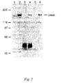
- FIG. 5 is an analysis of DNA-R protein expression in mammalian cells.
- DNA-R was immunoprecipitated both as the native molecule and in an HA-tagged embodiment from stably-transfected human 293 cells (DNA-R/flu cells).
- Lane 1 lysate of 293 cells; lane 2, lysate of 293-DNA-R/flu cells; lanes 3–6, immunoprecipitation of 293-DNA-R/flu cell lysates with: rabbit preimmune serum (lane 3) or anti-DNA-R (lane 4), control mouse monoclonal antibody (lane 5) or anti-HA (lane 6).
- FIG. 6 shows the intracellular location of the DNA-R protein, associated with cell membranes in 293-MNAB/flu cells as detected by Western blotting with anti-HA (left half) or anti-DNA-R (right half).
- T Triton X-100 whole-cell lysate; M, crude membrane fraction; C, cytosolic fraction.
- FIG. 7 illustrates immunofluorescence staining using anti-DNA-R and anti-transferrin antibodies on fixed, permeablized A549 cells.
- A Double stained with rabbit and sheep nonimmune sera.
- B anti-DNA-R.
- C anti-transferrin receptor.
- D double stained for anti-DNA-R (red) and anti-transferrin receptor (green); colocalized staining appears yellow.
- FIG. 8A shows the results of antibody staining of 293 cell surface using polyclonal rabbit antisera raised against an amino-terminal fragment (aas 1–575) of the DNA-R of the invention.
- 293 cells were incubated with preimmune serum (black bars) or immune serum (white bars).
- Antibody binding was detected with FITC conjugated goat anti-rabbit IgG by flow cytometry.
- FIG. 8B shows antibody binding to the cell surface of Raji and 293 cells using antibodies raised against an amino-terminal fragment (1–575).
- Cells (Raji, gray bars; 293, white bars) were incubated with serial dilutions of rabbit antisera (#41 bleed 2) produced against the amino terminal portion (amino acids 1–575) of the DNA receptor. The cells were then incubated with FITC-goat anti-rabbit IgG and the fluorescence intensity measure by FACS. Fluorescence due to preimmune sera has been subtracted.
- FIG. 9A is a schematic diagram of the preparation of the soluble DNA-R protein from the full length DNA-R.
- FIG. 9B is an SDS-PAGE analysis of expression, affinity purification, and proteolysis of a fusion protein (termed GST/DNA-R) created between glutathione-S-transferase (GST) and an amino-terminal fragment (1–575) of the DNA-R of the invention.
- Lane 1 whole cell extract of E. coli expressing GST/DNA-R; lane 2, GST/DNA-R bound to glutathione (GSH)-sepharose; lane 3, site-specific proteolysis of GST/DNA-R while bound to GSH-sepharose; lane 4, eluate from GSH-sepharose following on-gel proteolysis of GST/DNA-R containing highly purified DNA-R (1–575) peptide.
- FIGS. 10A through 10E show that GST/DNA-R but not control proteins immobilized on glutathione sepharose bind a fluorescently-labeled plasmid DNA (YOYO/pGEM4Z). Shown in the figure is glutathione sepharose without immobilized protein ( FIG. 10A ), or with immobilized GST ( FIG. 10B ), GST/HST.1 ( FIG. 10C ), GST/CBD ( FIG. 10D ), or GST/DNAR ( FIG. 10E ) incubated with YOYO/pGEM4Z for 30 min at 4° C. After washing, the fluorescence intensity was measured by FACS.
- FIGS. 11A and 11B is the result of a “Southwestern” blot of nucleic acid binding to DNA-R.
- the experiments were performed by SDS-PAGE analysis, transfer to nitrocellulose and then incubated with ( FIG. 11A ) or without ( FIG. 11B ) biotinylated DNA, followed by binding to streptavidin conjugated with horse radish peroxidase (HRP) and reaction with colorimetric substrate. Absorbance was measured at 450 nm. These results showed that GST/DNA-R but not GST bound biotinylated DNA.
- HRP horse radish peroxidase
- FIG. 12 shows the results of an enzyme linked immunosorbent assay (ELISA) in which purified DNA-R fragment (1–575) bound to immobilized DNA.
- DNA-R fragment (at concentrations of 0, 1, and 10 ⁇ /mL) was incubated with immobilized plasmid DNA in ELISA plates. The plates were then incubated with anti-DNA antibodies at a 1:100 dilution, followed by secondary antibody conjugated to HRP and reaction with colorimetric substrate. Absorbance was measured at 450 nm.
- ELISA enzyme linked immunosorbent assay
- FIG. 13 shows that a DNA-R fragment comprising a zinc finger domain (at amino acids 416–435) participates in DNA binding by GST/DNA-R(1–575).
- Cysteine residues (at positions 416 and 431) were independently altered to serine (and termed C416S and C431S, respectively) or alanine (and termed C416A and C431A, respectively) and ELISA performed to evaluate DNA binding.
- the zinc finger cysteines at either 416 or 431 were altered to either a serine (C416S, C431S) or an alanine (C416A, C431A).
- Binding 100 ng/well) of wild-type (GST/DNA-R) or mutant GST-fusion proteins or GST alone to immobilized DNA was detected using anti-GST by ELISA. Data are the mean ⁇ s.d. of triplicate determinations.
- FIG. 14 illustrates results of experiments demonstrating that a soluble form of the DNA-R of the invention binds DNA with high affinity using a nitrocellulose filter binding assay.
- Soluble DNA-R sDNA-R
- labeled DNA 200 pM, 1 ⁇ 10 6 cpm/pmol
- Data are the mean ⁇ s.d. of triplicate determinations.
- Inset Scatchard transformation of the binding data.
- FIG. 15 illustrates competitive binding of plasmid DNA with soluble DNA-R. All samples have 0.25 nM (YOYO labeled) pGEM-DNA. The diagonal bars have varying amounts of soluble DNA-R added to block DNA binding. The horizontal line bar is control GST protein.
- FIG. 16 are the results of fluorescence activated cell sorting (FACS) experiments illustrating cell-surface binding of YOYO labeled plasmid DNA in A549 cells.
- Cells were incubated in the presence (dashed line) and absence (solid line) of 5 ⁇ g/mL YOYO-labeled pGEM4Z DNA.
- the geometric mean fluorescence of untreated and treated cells is 13 and 34 respectively.
- the difference between the two values (21) is the increase in fluorescence intensity due to YOYO/pGEM4Z. This method of analysis is used for all YOYO/DNA binding analyses by FACS in subsequent figures.
- FIGS. 17A and 17B are the results of FACS analysis of YOYO labeled plasmid DNA binding to A549 cells in the presence of excess unlabeled DNA. Cell surface binding of YOYO/DNA to A549 cells.
- FIG. 17A left panel
- A549 cells were incubated with YOYO/pGEM4Z in the presence (solid line) and absence (dashed line) of a 25–100 fold excess calf thymus DNA for 2 hr at 4° C.
- FIG. 17B right panel
- specific DNA binding to A549 cells is shown as the difference in fluorescence intensity of YOYO/pGEM4Z bound to the cells using the data from the left panel. Data are the mean ⁇ SEM of 4–9 determinations.
- FIG. 18 shows the effects of trypsin treatment on DNA binding to A549 cells.
- A549 cells were incubated at 4° C. for 30 min with YOYO/pGEM4Z (0.5 to 4 ⁇ g/ml). After incubation the cells were washed (white bars) or trypsinized and washed (gray bars), then the fluorescence intensity was measured by FACS. The data are the mean ⁇ SD of triplicate determinations. Trypsin treatment was found to remove most cell-surface DNA binding.
- FIG. 19 shows the results of experiments indicating that DNA binding to the DNA-R of the receptor is calcium dependent.
- B16 cells were incubated with 1 ⁇ g/mL YOYO/pGEM4Z in PBS with (white bar) or without (gray bar) 1 mMCaCl 2 , then the fluorescence intensity was measured by FACS. The data are mean ⁇ SD of triplicate measurements.
- FIG. 20 shows a time course of plasmid DNA uptake by cells expressing the DNA-R of the invention.
- B16 cells were incubated at 37° C. with YOYO/pEGFP-N1 (0.6 to 12 ⁇ g/mL) for 1 hr (solid circles), 3 hr (open circles), or 5 hr (solid squares). The cells were then trypsinized to remove DNA bound to the cell-surface, and fluorescence intensity was measured by FACS.
- FIG. 21 shows that excess unlabeled DNA blocks internalization of YOYO/DNA by A549 cells.
- Cells were incubated for 2 hr at 37° C. with YOYO/pGEM4Z (1 to 25 ⁇ g/mL) in the presence (solid line) and absence (dashed line) of a 25- to 100-fold excess of calf thymus DNA. The cells were then washed and the fluorescence measured by FACS. Data are the mean ⁇ SEM of 5 experiments.
- FIG. 22 illustrates that internalization of plasmid DNA in cells expressing the DNA-R of the invention is temperature dependent.
- B16 cells were incubated with YOYO/pGEM4Z (12 ⁇ g/mL) at 4° C. (white bars) or 37° C. (gray bars) for the indicated times. The cells were then trypsinized to remove cell-surface bound plasmid, washed, and the fluorescence measured by FACS. The data are the mean ⁇ SD of triplicate determinations.
- FIGS. 23A and 23B show that cell-surface DNA binding to the DNA-R of the invention is related to DNA uptake.
- 293 cells (solid circles) and G361 cells (open circles) were incubated for 3.5 hr with YOYO/pGEM4Z (at concentrations of from 0.3 to 2.5 ⁇ g/mL) at 4° C. for binding ( FIG. 23A , left panel) or 37° C. for uptake ( FIG. 23B , right panel). The cells were then washed (binding) or trypsinized and washed (uptake), and the fluorescence measured by FACS.
- FIG. 24 shows expression in B16 cells of green fluorescence protein (GFP) transgene from plasmid DNA.
- GFP green fluorescence protein
- FIG. 25 show that nocodazole increases GFP transgene expression from plasmid DNA in A549 cells.
- A549 cells were incubated for 5 hr at 37° C. with 25 ⁇ g/mL of pEGFP-N1 in the presence (dashed line) and absence (solid line) of 33 ⁇ M nocodazole. After 24 hr the cells were trypsinized and fluorescence measured by FACS.
- mammalian DNA-R refers to proteins consisting essentially of, and having substantially the same biological activity as, the protein encoded by the amino acid depicted in FIG. 1 (SEQ ID No.:2); This definition is intended to encompass natural allelic variations in the disclosed DNA-R.
- Cloned nucleic acid provided by the present invention may encode DNA-R protein of any species of origin, including, for example, mouse, rat, rabbit, cat, and human, but preferably the nucleic acid provided by the invention encodes DNA-Rs of mammalian, most preferably human, origin.
- the nucleic acids provided by the invention comprise DNA or RNA having a nucleotide sequence encoding a mammalian DNA-R. Specific embodiments of said nucleic acids are depicted in FIG. 1 (SEQ ID No.:1), and include any nucleotide sequence encoding a mammalian DNA-R having an amino acid sequence as depicted in FIG. 1 (SEQ ID No.: 2).
- Nucleic hybridization probes as provided by the invention comprise any portion of a nucleic acid of the invention effective in nucleic acid hybridization under stringency conditions sufficient for specific hybridization. Mixtures of such nucleic acid hybridization probes are also within the scope of this embodiment of the invention.
- Nucleic acid probes as provided herein are useful for isolating mammalian species analogues of the specific embodiments of the nucleic acids provided by the invention.
- Nucleic acid probes as provided herein are also useful for detecting mammalian DNA-R gene expression in cells and tissues using techniques well-known in the art, including but not limited to Northern blot hybridization, in situ hybridization and Southern hybridization to reverse transcriptase-polymerase chain reaction product DNAs.
- the probes provided by the present invention including oligonucleotides probes derived therefrom, are also useful for Southern hybridization of mammalian, preferably human, genomic DNA for screening for restriction fragment length polymorphism (RFLP) associated with certain genetic disorders.
- RFLP restriction fragment length polymorphism
- Nucleic acid encoding a DNA-R may be obtained, in view of the instant disclosure, by chemical synthesis, by screening reverse transcripts of mRNA from appropriate cells or cell line cultures, by screening genomic libraries from appropriate cells, or by combinations of these procedures, in accordance with known procedures as illustrated below. Screening of mRNA or genomic DNA may be carried out with oligonucleotide probes generated from the nucleic acid sequence information from mammalian DNA-R nucleic acid as disclosed herein. Probes may be labeled with a detectable group such as a fluorescent group, a radioactive atom or a chemiluminescent group in accordance with know procedures and used in conventional hybridization assays, as described in greater detail in the Examples below.
- a detectable group such as a fluorescent group, a radioactive atom or a chemiluminescent group
- mammalian DNA-R nucleic acid sequences may be obtained by use of the polymerase chain reaction (PCR) procedure, using PCR oligonucleotide primers corresponding to nucleic acid sequence information derived from a DNA-R as provided herein. See U.S. Pat. No. 4,683,195 to Mullis et al. and U.S. Pat. No. 4,683,202 to Mullis.
- PCR polymerase chain reaction
- Mammalian DNA-R protein may be synthesized in host cells transformed with a recombinant expression construct comprising a nucleic acid encoding the DNA-R nucleic acid, comprising genomic DNA or cDNA.
- a recombinant expression construct can also be comprised of a vector that is a replicable DNA construct.
- Vectors are used herein either to amplify DNA encoding a DNA-R and/or to express DNA encoding a DNA-R gene.
- a recombinant expression construct is a replicable DNA construct in which a nucleic acid encoding a DNA-R is operably linked to suitable control sequences capable of effecting the expression of the DNA-R in a suitable host.
- control sequences include a transcriptional promoter, an optional operator or enhancer sequence to control transcription, a sequence encoding suitable mRNA ribosomal binding sites, and sequences which control the termination of transcription and translation.
- Amplification vectors do not require expression control domains. All that is needed is the ability to replicate in a host, usually conferred by an origin of replication, and a selection gene to facilitate recognition of transformants. See, Sambrook et al., 2001, Molecular Cloning: A Laboratory Manual (Cold Spring Harbor Press: New York).
- Vectors useful for practicing the present invention include plasmids, viruses (including phage and mammalian DNA and RNA viruses), retroviruses, and integratable DNA fragments (i.e., fragments integratable into the host genome by homologous recombination).
- the vector can replicate the gene of interest and function independently of the host genome, or can, in some instances, integrate into the genome itself. Suitable vectors will contain replicon and control sequences which are derived from species compatible with the intended expression host.
- Transformed host cells are cells which have been transformed or transfected with recombinant expression constructs made using recombinant DNA techniques and comprising nucleic acid encoding a DNA-R protein.
- Preferred host cells are HEK293 cells, COS-7 cells (Gluzman, 1981, Cell 23:175–182) and Ltk ⁇ cells.
- Transformed host cells may express the DNA-R protein, but host cells transformed for purposes of cloning or amplifying nucleic acid hybridization probe DNA need not express the receptor.
- the DNA-R of the invention will typically be located in the host cell membrane. Accordingly, the invention provides preparations of said cell membranes comprising the DNA-R protein of the invention, as well as purified, homogeneous preparations of the receptor protein itself. See, Sambrook et al., ibid.
- HEK293 cell, COS-7 cells and Ltk ⁇ cells are preferred.
- the invention provides homogeneous compositions of mammalian DNA-R protein produced by transformed eukaryotic cells as provided herein.
- Each such homogeneous composition is intended to be comprised of a DNA-R protein that comprises at least 75%, more preferably at least 80%, and most preferably at least 90% of the protein in such a homogenous composition; in said homogeneous preparations, individual contaminating protein species are expected to comprise less than 5%, more preferably less than 2% and most preferably less than 1% of the preparation.
- the invention also provides membrane preparations from cells expressing mammalian DNA-R protein as the result of transformation with a recombinant expression construct, as described herein.
- fragments of the DNA-R of the invention are fragments of the DNA-R of the invention, most preferably DNA binding fragments thereof.
- said fragments include soluble forms of the receptor lacking the transmembrane domain and an amino-terminal fragment (most preferably amino acids 1–575) comprising zinc finger and RING sequence motifs known in the art to be related to DNA-protein binding.
- Mammalian DNA-R proteins made from cloned genes in accordance with the present invention may be used for screening compounds that effect DNA binding to cells in vivo and in vitro, as more fully described herein, and that affect DNA uptake and expression of genes encoded thereby.
- host cells may be transformed with a recombinant expression construct of the present invention, a mammalian DNA-R expressed in those host cells, and the cells or membranes thereof used to screen compounds for their effect on DNA binding.
- pure preparations of membranes containing the receptor can be obtained.
- the recombinant expression constructs of the present invention are useful in molecular biology to transform cells which do not ordinarily express a DNA-R to thereafter express this receptor. Such cells are useful as intermediates for making cell membrane preparations useful for receptor binding activity assays, which are in turn useful for drug screening.
- the recombinant expression constructs of the present invention thus provide a method for screening potentially useful drugs at advantageously lower cost than conventional animal screening protocols. While not completely eliminating the need for ultimate in vivo activity and toxicology assays, the constructs and cultures of the invention provide an important first screening step for the vast number of potentially useful drugs synthesized, discovered or extracted from natural sources each year.
- the recombinant expression constructs of the present invention may also be useful in gene therapy.
- Cloned genes of the present invention, or fragments thereof, may also be used in gene therapy carried out homologous recombination or site-directed mutagenesis. See generally Thomas & Capecchi, 1987, Cell 51: 503–512; Bertling, 1987, Bioscience Reports 7: 107–112; Smithies et al., 1985, Nature 317: 230–234.
- Nucleic acid and oligonucleotide probes as provided by the present invention are useful as diagnostic tools for probing DNA-R gene expression in tissues of humans and other animals.
- tissues are probed in situ with oligonucleotide probes carrying detectable groups by conventional autoradiographic or other detection techniques, to investigate native expression of this receptor or pathological conditions relating thereto.
- chromosomes can be probed to investigate the presence or absence of the corresponding DNA-R gene, and potential pathological conditions related thereto.
- Oligonucleotides, particularly antisense oligonucleotides are also useful for decreasing expression of the DNA-R in cells that overexpress the receptor or whose expression is disadvantageous in a host organism, either generally or in specific tissues. An example of the latter instance is in airway epithelial cells and macrophages in lung tissues in cystic fibrosis patients, as set forth more fully herein.
- the invention also provides antibodies that are immunologically reactive to the DNA-R protein or epitopes thereof provided by the invention.
- the antibodies provided by the invention may be raised, using methods well known in the art, in animals by inoculation with cells that express a DNA-R or epitopes thereof, cell membranes from such cells, whether crude membrane preparations or membranes purified using methods well known in the art, or purified preparations of proteins, including protein fragments and fusion proteins, particularly fusion proteins comprising epitopes of the DNA-R protein of the invention fused to heterologous proteins and expressed using genetic engineering means in bacterial, yeast or eukaryotic cells, said proteins being isolated from such cells to varying degrees of homogeneity using conventional biochemical methods.
- Synthetic peptides made using established synthetic methods in vitro and optionally conjugated with heterologous sequences of amino acids, are also encompassed in these methods to produce the antibodies of the invention.
- Animals that are useful for such inoculations include individuals from species comprising cows, sheep, pigs, mice, rats, rabbits, hamsters, goats and primates.
- Preferred animals for inoculation are rodents (including mice, rats, hamsters) and rabbits. The most preferred animal is the mouse.
- Cells that can be used for such inoculations, or for any of the other means used in the invention include any cell line which naturally expresses the DNA-R provided by the invention, or more preferably any cell or cell line that expresses the DNA-R of the invention, or any epitope thereof, as a result of molecular or genetic engineering, or that has been treated to increase the expression of an endogenous or heterologous DNA-R protein by physical, biochemical or genetic means.
- Preferred cells are mammalian cells, most preferably cells syngeneic with a rodent, most preferably a mouse host, that have been transformed with a recombinant expression construct of the invention encoding a DNA-R protein, and that express the receptor therefrom.
- the present invention also provides monoclonal antibodies that are immunologically reactive with an epitope derived from a DNA-R of the invention, or fragment thereof, present on the surface of such cells or in membrane preparations thereof or used after varying degrees of biochemical purification.
- Particularly useful are soluble fragments of the DNA-R of the invention, including for example species of the receptor genetically engineered to remove the transmembrane domain, and amino-terminal fragments, most preferably DNA binding fragments of the receptor.
- Such antibodies are made using methods and techniques well known to those of skill in the art.
- Monoclonal antibodies provided by the present invention are produced by hybridoma cell lines, which are also provided by the invention and are made by methods well known in the art.
- Hybridoma cell lines are made by fusing individual cells of a myeloma cell line with spleen cells derived from animals immunized with cells expressing a DNA-R of the invention, as described above.
- the myeloma cell lines used in the invention include lines derived from myelomas of mice, rats, hamsters, primates and humans.
- Preferred myeloma cell lines are from mouse, and the most preferred mouse myeloma cell line is P3 ⁇ 63-Ag8.653.
- the animals from whom spleens are obtained after immunization are rats, mice and hamsters, preferably mice, most preferably Balb/c mice.
- Spleen cells and myeloma cells are fused using a number of methods well known in the art, including but not limited to incubation with inactivated Sendai virus and incubation in the presence of polyethylene glycol (PEG).
- PEG polyethylene glycol
- the most preferred method for cell fusion is incubation in the presence of a solution of 45% (w/v) PEG-1450.
- Monoclonal antibodies produced by hybridoma cell lines can be harvested from cell culture supernatant fluids from in vitro cell growth; alternatively, hybridoma cells can be injected subcutaneously and/or into the peritoneal cavity of an animal, most preferably a mouse, and the monoclonal antibodies obtained from blood and/or ascites fluid.
- Monoclonal antibodies provided by the present invention are also produced by recombinant genetic methods well known to those of skill in the art, and the present invention encompasses antibodies made by such methods that are immunologically reactive with an epitope of an amino acid receptor of the invention.
- the present invention also encompasses antigen-binding fragments, including but not limited to F v , F(ab) and F(ab)′ 2 fragments, of such antibodies. Fragments are produced by any number of methods, including but not limited to proteolytic or chemical cleavage, chemical synthesis or preparation of such fragments by means of genetic engineering technology.
- the present invention also encompasses single-chain antibodies that are immunologically reactive with an epitope of a DNA-R, made by methods known to those of skill in the art.
- the present invention also encompasses an epitope of a DNA-R of the invention, comprised of sequences and/or a conformation of sequences present in the receptor molecule.
- This epitope may be naturally occurring, or may be the result of chemical or proteolytic cleavage of a receptor molecule and isolation of an epitope-containing peptide or may be obtained by chemical or in vitro synthesis of an epitope-containing peptide using methods well known to those skilled in the art.
- the present invention also encompasses epitope peptides produced as a result of genetic engineering technology and synthesized by genetically engineered prokaryotic or eukaryotic cells.
- the invention also includes chimeric antibodies, comprised of light chain and heavy chain peptides immunologically reactive to a DNA-R-derived epitope.
- the chimeric antibodies embodied in the present invention include those that are derived from naturally occurring antibodies as well as chimeric antibodies made by means of genetic engineering technology well known to those of skill in the art.
- Nucleic acids encoding the receptor, the DNA-R and DNA-binding fragments thereof are advantageously used to modulate expression or activity of the receptor in cells in vivo and in vitro.
- the DNA-R of the invention particularly soluble embodiments thereof, can competitively bind DNA to reduce said binding to cells expressing the DNA-R.
- DNA binding to the DNA-R in certain cells, such as airway epithelial cells and macrophages in lung, is associated with the activation of inflammatory processes that cause a significant proportion of the morbidity and mortality associated with cystic fibrosis, chronic bronchitis and other chronic lung diseases.
- the invention provides a variety of methods for reducing said morbidity and mortality by interfering with DNA binding to cells in the lung.
- soluble DNA-R species can be administered, most preferably by aerosol administration using formulations, excipients and vehicles well known in the art, directly to lung tissue, and competitive DNA binding achieved thereby.
- antisense oligonucleotides can be delivered to lung tissue, most preferably by aerosol administration, and expression of the DNA-R in target cells of the lung repressed thereby.
- antibodies, most preferably monoclonal antibodies, that specifically inhibit DNA binding to the DNA-R of the invention can be used to inhibit DNA binding to said lung cells.
- the DNA-R of the invention are also useful in treating other inflammation-associated diseases and conditions, including otitis media, septic arthritis and any bacterial or viral infection that causes inflammation by interaction with the DNA-R
- the DNA-R of the invention can be used to screen compounds that modulate DNA binding, uptake and expression.
- Introduction of DNA, particularly DNA encoding a desired gene is a methodology well known in the art.
- DNA introduction methods have been developed empirically and without any understanding of the molecular bases of DNA uptake.
- identification of the DNA-R of the invention thus provides a novel target for developing compounds and methods for increasing efficiency of DNA uptake and expression of genes encoded thereby.
- DNA-R expressed in tumor cells to facilitate delivery of DNA-binding anticancer drugs to tumor cells.
- Drugs such as Adriamycin (Doxorubicin) are in clinical use for the treatment of cancer patients.
- Enhanced extracellular DNA uptake in tumor cells expression the DNA-R of the invention would facilitate uptake of such DNA-binding anticancer drugs by using extracellular DNA as a carrier of the drug into the cell.
- the association of the drug with the extracellular DNA might enable the drug to avoid active efflux produced in tumor cells, inter alia, by drug resistance mediators such as P-glycoprotein.
- the selective augmentation of DNA binding receptors on tumor cells would enhance uptake of DNA-binding drugs and result in an increased therapeutic effect.
- other diseases, such as malaria can be treated in a similar fashion, based on the development of cell-surface DNA binding in red blood cells parasitized with the malarial parasite Plasmodium falciparum.
- DNA-R Human Membrane-Associated DNA Receptor
- DNA binding to cells had been observed in the art, and the behavior of said binding suggested the existence of a DNA binding protein expressed at the cell surface.
- serum from a patient with systemic lupus erythematosus (SLE) treated to deplete the sera of anti-DNA antibodies by multiple (6 ⁇ ) passages over a DNA sepharose column, was used to screen a ⁇ gt11 cDNA expression library made from liposaccharide stimulated human monocytes. This serum has been shown to have anti-DNA receptor activities (defined by the blocking of DNA binding to cells; Bennet et al., 1992. J. Clin. Invest. 76: 2182–2190).
- the full length cDNA for the putative DNA-R was obtained in segments from peripheral blood mononuclear cells, human Burkitt lymphoma cells (Raji; Accession No. CCL 86, American Type Culture Collection, Manassas, Va.), human cervical carcinoma cells (HeLa; ATCC Accession No. CCL 2), and human lymphoblastic leukemia cells (MOLT-4; ATCC Accession No. CRL 1582).
- a 731 base pair (bp) DNA probe from clone 88 was used to screen a ⁇ gt11 phage library from a Raji cell line (the library was obtained from Clonetech Labs, Palo Alto, Calif.).
- a 2409 bp clone which contained an additional 462 bp of 5′ open reading frame (ORF) sequence was obtained from this screening. Additional sequence from the 5′ extent of the cDNA was isolated using two variations of the 5′ RACE (rapid amplification of cDNA ends) method. In the first 5′ RACE method, single stranded DNA (ssDNA) was synthesized from HeLa cell mRNA using polyT primers and reverse transcriptase. A polyA tail was added to the 5′ end of the ssDNA by terminal transferase. The single stranded cDNA was amplified using a gene specific primer and a polyT primer. This clone contained 753 bp of additional sequence 5′ of the previously obtained sequence.
- ssDNA single stranded DNA
- the remainder of the 5′ sequence of the cDNA was obtained from MOLT-4 cDNA by Marathon Race cDNA amplification (Clonetech Labs, Palo Alto, Calif.) according to the manufacturer's instructions. This procedure produced an additional 1290 bp clone consisting of 340 bp of ORF and a 950 bp 5′ untranslated region. Combining the results from these screening and amplification experiments produced the predicted full length cDNA encoding the DNA-R of the invention.
- cDNA for the putative DNA receptor was cloned as a single RT-PCR product from MOLT-4 mRNA using oligonucleotide primers having the following sequence:
- Tissue-specific and cell line-specific expression patterns of its corresponding mRNA in various human tissues was analyzed by Northern blot analysis on RNA isolated from various tissues and cancer cell lines. The results of these experiments are shown in FIG. 2 .
- a panel of tissue samples was examined by Northern hybridization analysis performed under low stringency conditions, defined as hybridization at 42° C. in 5 ⁇ SSPE (0.75M NaCl, 0.05 mM NaH 2 PO 4 , 5 mM EDTA), 10 ⁇ Denhardt's solution (0.2% Ficoll, 0.2% polyvinylpyrrolidone, 0.2% bovine serum albumen), 100 ⁇ g/mL salmon sperm DNA, 2% SDS and 50% deionized formamide and 1–2 ⁇ 10 6 cpm random-primed, 32 P labeled probe, followed by washing in 0.1 ⁇ SSC (15 mM NaCl, 1.5 mM trisodium citrate, 0.1% SDS).
- 5 ⁇ SSPE 0.75M NaCl, 0.05 mM NaH 2 PO 4 , 5 mM EDTA
- 10 ⁇ Denhardt's solution 0.2% Ficoll, 0.2% polyvinylpyrrolidone, 0.2% bovine serum albumen
- the blots were hybridized with aprobe consisting of 442 bp of sequence from the 3′ end of the coding sequence from the DNA-R gene to determine the distribution of receptor mRNA.
- This analysis revealed two major transcripts of 9.5 and 6.8 kb in all human tissues and cancer cell lines examined. Transcript expression was relatively abundant in spleen, testis, ovary, and small intestine. Several smaller transcript sizes were also observed in some of the tissues and cell lines examined ( FIG. 2 ).
- a homology search against human genomic sequence placed the DNA receptor on chromosome 9q34 (GenBank Accession number AC007066, marker HIM9.89 on Contig CHR9.SL27).
- the genomic sequence which covered 85% of the cDNA starting from the 5′ end, revealed the location of 16 complete exons and the beginning of a 17 th exon.
- a BLAST search of the expressed tag sequence (EST) database indicated wide expression of this gene in normal human tissue (liver/spleen, prostate epithelial, germinal B cell, white adipose, pregnant uterus, fetal heart/liver and spleen) and in tumor and transformed human cells (Jurkat, HL60, 293, G361, B-cell lymphocytic leukemia, colon tumor, melanoma, and parathyroid tumor).
- normal human tissue liver/spleen, prostate epithelial, germinal B cell, white adipose, pregnant uterus, fetal heart/liver and spleen
- tumor and transformed human cells Jurkat, HL60, 293, G361, B-cell lymphocytic leukemia, colon tumor, melanoma, and parathyroid tumor.
- FIG. 3 provides a schematic diagram of the structure of the DNA-R protein encoded by SEQ ID No. 1.
- Hydropathy analysis identified a 38 amino acid hydrophobic region near the carboxy terminus of the protein (amino acids 1133–1171) which is a potential transmembrane domain. Expression of a soluble species of this receptor by deleting these amino acids supported identification of this region as a transmembrane domain.
- seven consensus sites for linked glycolsylation have been identified (amino acid positions 122, 394, 430, 451, 466, 468, and 1150) and there is a proline rich (20% of the residues are proline) region spanning amino acids 549–809 ( FIG. 3 ).
- the calculated isoelectric point of the DNA receptor protein is 6.4.
- the BLAST search also identified two art-recognized amino acid sequence motifs in the DNA-R sequence: a C3HC3D Ring finger subtype located near the amino terminus (amino acids 14–50) and a C3H zinc finger located near the center of the protein sequence (amino acids 416–435).
- An alignment of several ring finger motifs is shown in FIG. 4A ; DNA-R differs from the originally identified C3HC4 Ring finger motif by the replacement of the last cysteine with an aspartic acid.
- the alignment of the conserved cysteines and histidines of the C3H zinc finger motif is shown in FIG. 4B .
- the DNA-R of the invention was produced recombinantly as follows.
- a BamHI-HpaI cDNA fragment containing the coding sequence for amino acids 1–1190 (i.e., missing the two most carboxylterminal amino acids) of the DNA-R of the invention was cloned into pTriplFlu (obtained from J. Epstein, University of Pennsylvania, Philadelphia, Pa.).
- This vector contains a sequence encoding an epitope tag from the influenza hemagglutinin gene in triplicate inserted immediately 3′ of the multiple cloning site of the parent vector, pcDNA3, and which are in-frame with the inserted DNA-R cDNA sequence.
- Transfected cells (293-DNA-R/flu) were selected by culturing in growth media (DMEM supplemented with 10% fetal calf serum, 2 mM L-glutamine, 100 U/mL penicillin and 100 g/mL streptomycin) supplemented with 500 ⁇ g/mL G418.
- growth media DMEM supplemented with 10% fetal calf serum, 2 mM L-glutamine, 100 U/mL penicillin and 100 g/mL streptomycin
- Polyclonal antibodies were produced to a purified fragment of the DNA-R (comprising amino acids 1–575) using conventional techniques.
- Three female New Zealand White rabbits (Western Oregon Rabbit Company), weighing 2.3–3.0 kg, were injected subcutaneously with 50 ⁇ g of the DNA-R peptide that was produced in bacteria as a GST fusion protein (described in Example 4) and purified from its fusion partner.
- the antigen was emulsified with Titre-Max (CytRx Corp., Norcross, Ga.) in a final volume of 0.5 mL.
- the rabbits were boosted 4 weeks later with 15 ⁇ g of antigen/Titre-Max mixture, again 2 weeks later, and were maintained on a once-a-month booster schedule thereafter.
- the rabbits were bled 7–10 days after each boost with antigen and the sera analyzed for reactivity to the immunizing antigen.
- the polyclonal antisera obtained from the inoculated rabbits was used in Western blot analyses.
- a protein of Mr ⁇ 1.5 ⁇ 10 5 was identified by the anti-DNA-R antibody in most cells tested (including 293, COS7, G361, HeLa, HRE605, MOLT-4, Raji, A549, B16).
- a protein with a similar mobility was detected in lysates of genetically-engineered human 293 cells (293-DNA-R/flu) that were stably transfected with an expression vector for a carboxy-terminal HA-tagged DNA-R (pDNA-R/flu). As shown in FIG.
- this protein was detected by immunoprecipitation and/or Western blot analysis with either the rabbit polyclonal anti-DNA-R (1–575) antisera described above or with a mouse monoclonal antibody (anti-HA) specific for the carboxyl-terminal HA tag in the recombinantly-produced protein.
- Double staining with anti-DNA-R and anti-transferrin receptor antibodies showed partial colocalization of the DNA-R and transferrin receptor, however the DNA-R did not colocalize with the transferrin receptor in peripheral endosomes ( FIG. 7 ).
- These results indicate that extracellular DNA is taken up by cells expressing the DNA-R of the invention by endocytosis, and suggest that compounds that influence intracellular trafficking of molecules taken by endocytosis are useful for modulating the intracellular fate (such as degradation in lysosomes or transport to the cell nucleus) of extracellular DNA.
- DNA-R protein either natively expressed or expressed from the cloned cDNA of the invention, or genetically-engineered embodiments thereof, localized to cell membranes as predicted by the hydropathy plot of the carboxyl terminus.
- Soluble DNA-R Fusion Protein Binds DNA with High Affinity
- the capacity of the DNA-R of the invention to bind DNA, and particularly the capacity of a soluble form of the DNA-R protein to bind DNA was determined.
- a fusion protein between the amino terminal portion of the DNA receptor (amino acids 1–575), lacking the transmembrane region but containing both the RING and zinc finger domains, was produced using the pGEX vector system (Pharmacia, Kalamazoo, Mich.) for expression of glutathione-S-transferase (GST)-fusion proteins in E. coli and named GST/DNA-R (1–575).
- FIG. 9A A schematic diagram of the production of this protein fragment and its structure relative to the full-length DNA-R of the invention is shown in FIG. 9A .
- the calculated molecular weights of the GST/DNA-R fusion protein and the DNA-R peptide are 90 kDa and 63 kDa respectively.
- the purified GST/DNA-R fusion protein was then examined for its ability to bind plasmid DNA. Three independent in vitro assays were used to assess DNA binding by the fusion protein. First, the ability of GST/DNA-R, bound to glutathione sepharose beads, to bind YOYO-labeled plasmid DNA was determined by incubation with 0.9 ⁇ g YOYO/DNA in 0.5 mL of medium.
- YOYO-1 is an intercalating fluorochrome that is flourescent only when bound to DNA, obtained from Molecular Probes, Eugene, Oreg.
- Beads (3.5 ⁇ 10 5 ) and YOYO/DNA were incubated for 30 minutes at 4° C., washed once and then fluorescence intensity analyzed by FACS.
- the GST/DNA-R fusion protein was extremely efficient in binding DNA whereas purified GST protein alone and two additional, unrelated GST-fusion proteins (GST-CBD and GST-HST. 1, gifts from Dr. Roland Kwok, Vollum Institute, Portland Oreg.) failed to show any DNA binding capability.
- GST/DNA-R fusion protein was a DNA-binding molecule.
- the purified GST/DNA-R fusion protein and GST protein alone were electrophoresed on a polyacrylamide gel, electrophoretically transferred to nitrocellulose and then probed with biotinylated DNA. DNA binding was visualized by addition of steptavidin conjugated with horse radish peroxidase (HRP) using conventional methods.
- HRP horse radish peroxidase
- FIG. 11B lanes 1
- FIG. 11A Lanes in FIG. 11A represent no added DNA.
- DNA receptor gene of the invention encodes a protein that specifically binds DNA, and that the DNA binding portion of the molecule resided in the protein fragment having amino acid sequence 1–575 of the native protein.
- DNA binding by the soluble DNA-R fragment is not simply a nonspecific charge related interaction, but rather is mediated by specific a DNA-binding motifs in the protein, including at least the zinc finger motif.
- Soluble DNA-R Protein Inhibits DNA-Induced Cytokine Secretion and Blocks Binding of DNA to Cells
- Extracellular DNA is known in the art to cause lung macrophages and other cells to release cytokines that mediate inflammation as part of the chronic symptomology of cystic fibrosis patients.
- the DNA-R protein of the invention is expressed in lung tissues, specifically in epithelial cells of the lung. This suggests that the DNA-R receptor protein of the invention is involved in inflammation by triggering the release of inflammation-causing cytokines.
- this protein fragment could compete for binding extracellular DNA in cystic fibrosis patients and would be useful thereby as a therapeutic agent.
- soluble DNA-R protein was examined for inhibition of CF-DNA-induced IL-6 release from J774 murine monocyte/macrophage cells in culture. In the absence of stimulating DNA, DNA-R did not induce IL-6 secretion (shown in Table I).
- DNA isolated and purified from a patient with cystic fibrosis (CF DNA) induced 611 pg/mL of IL-6 from J774 cells. When CF DNA was incubated first with DNA-R protein (10 ng/mL) and then added to J774 cells, the amount of IL-6 was reduced by 36% in the presence of the soluble DNA-R protein (10 ng/mL).
- calf thymus DNA failed to induce detectable IL-6.
- a Limulus amoebacyte assay was performed, and the CF DNA had ⁇ 0.25 ng/mL of contaminating endotoxin. In control experiments, this amount of LPS induced only 4 pg/mL of IL-6.
- the second experiment also shown in Table 1), contaminating endotoxin was removed from the soluble DNA-R, permitting the use of increased DNA-R concentrations.
- Soluble DNA-R protein (used in the range 10 ng/mL–50 ng/mL) was incubated with J774 cells and 50 ⁇ g/mL of E. coli DNA. Cell-free supernatants were collected and IL-6 quantified by ELISA. In the absence of bacterial DNA soluble DNA-R did not induce IL-6 secretion. When bacterial DNA was added to the system, however, soluble DNA receptor protein inhibited IL-6 secretion in a dose-dependent manner (Table I).
- J774 cells (5 ⁇ 10 5 cells) and YOYO labeled pGEM-DNA were incubated with either the soluble DNA-R protein fragment or control GST protein. Cells were incubated for 30 minutes at 4° C., centrifuged and washed twice with assay media, resuspended and incubated with 7-amino actinomycin D (7AAD) on ice for 20 minutes in order to assess viability. The samples were assessed for DNA binding by FACScan (Becton-Dickinson, Franklin Lanes, N.J.). Results showed a dose-dependent inhibition of DNA binding to J774 cells ( FIG. 15 ). Similar results were observed using human 293 cells. Additionally, the soluble DNA-R protein/DNA complex does not bind to the cell surface. The soluble DNA-R protein bound to DNA and is effective at preventing the association of DNA with the cell surface.
- the soluble DNA-R fragment provided by this invention is useful for inhibiting cytokine release, and inflammation consequent thereto, by competitively binding either bacterial or mammalian extracellular DNA and reducing the amount of such DNA bound by cytokine-producing cells expressing the DNA-R of the invention.
- A549 human lung carcinoma cells were harvested from log-phase cultures and treated with DNase and RNase to remove exogenous cell-surface bound nucleic acids. After treatment, the cells were washed with 10 mM EDTA and phosphate buffered saline (PBS) to stop the action of DNase and RNase. The cells were then plated in V-bottom 96-well plates at 10 6 cells/well in PBS containing 1% fetal calf serum (FCS) and 1 mM Ca ++ Mg ++ .
- FCS fetal calf serum
- YOYO-labeled pGEM4Z plasmid DNA was added at concentrations from 0–25 ⁇ g/mL in 0.2 mL media containing 1% FCS and 1 mM Ca ++ Mg ++ .
- the cells plus labeled plasmid were incubated for 30 minutes at 4° C., to minimize internalization of plasmid DNA.
- the cells were washed with 2 ⁇ in PBS containing 1% FCS and 1 mM Ca ++ Mg ++ and resuspended in 0.3 mL of PBS. Cells were then fixed in 1% formaldehyde and cell-surface binding of plasmid DNA quantified by FACS.
- FIG. 16 This representative FACS histogram demonstrates the A549 cell profiles seen when comparing cells incubated with either medium ( FIG. 16 , curve on the left) or cells incubated with 5 ⁇ g/mL of YOYO/pGem4Z plasmid ( FIG. 16 , curve shifted to the right).
- the geometric mean of the intensity is used to describe the cell populations.
- the geometric mean of the A549 cell population, treated with medium only was 13 and increased to 34 when incubated with YOYO-labeled plasmid DNA.
- a binding curve for A549 cells was then generated using a range of plasmid DNA from 0–25 ⁇ g/mL ( FIG. 17A and 17B ).
- the Y-axis of the graph in FIG. 17A represents the geometric mean of the fluorescence intensity of the cell populations in the graph.
- Cell surface binding of plasmid DNA to A549 cells began to show saturation at approximately 10 ⁇ g/mL of DNA.
- Treatment of cells with a 25–100 fold excess of unlabeled DNA significantly blocked the binding of YOYO/DNA to the cell surface ( FIG. 17A ).
- the specific cell-surface binding to A549 cells represented as the difference between total binding seen with excess unlabeled DNA, shows a binding curve with a characteristic saturation profile ( FIG. 17B ).
- cell-surface plasmid DNA binding profiles for a variety of tumor cell lines, including B16 murine melanoma cells, MOLT-4 human lymphoblastic leukemia cells, and the human Raji Burkitt lymphoma cells.
- cell-surface DNA saturable binding profiles were obtained, consistent with a receptor-mediated mechanism of binding.
- percent of cells in the population capable of binding DNA above the background level as detected by FACS ranged from greater than 70% (S49, DHL-6, MOLT-4) to less than 10% (D10.S, HUT-78, K562 and G361).
- DNA-R protein of the invention mediates cell surface binding of extracellular DNA in mammalian cells.
- Example 6 The experiments described in Example 6 established that extracellular DNA was specifically bound to the DNA-R of the invention. Internalization of DNA into cells by the receptor was characterized using the following assay.
- YOYO-labeled plasmid DNA was used to examine the kinetics of plasmid DNA internalization.
- the plasmid used in these assays was pEGFP-N1, encoding green fluorescent protein (Clontech, Palo Alto, Calif.).
- the assay required that cell surface binding of labeled DNA be distinguished from internalized plasmid DNA. This was accomplished by treatment of cells with trypsin to remove cell-surface proteins after incubation with plasmid DNA. This procedure permitted cell surface-bound plasmid DNA to be distinguished from internalized plasmid DNA, since trypsin treatment abolished cell surface bound DNA but not internalized plasmid DNA.
- cells were plated in 24 well plates and incubated in culture media for 24 h. Media was then removed and various concentrations (0–25 ⁇ g/mL) of YOYO-labeled pEGFP-N1 plasmid DNA were added. The cells plus plasmid DNA were incubated for various times (0.5 to 5 hours) at 37° C. Thereafter, the media was removed, cells were treated with trypsin, washed, and then fixed with 1% formaldehyde. FACS analysis was used to quantify fluorescence intensity.
- FIG. 20 B16 murine melanoma cells were examined for internalization of YOYO/DNA using the above protocol after incubation for 1, 3, and 5 hours ( FIG. 20 ). Internalization of pEGFP-N1 were found to be both dose- and time-dependent. An increasing amount of internalized plasmid DNA was seen with increasing dose of DNA and increasing time of incubation. Internalization of plasmid DNA by A549 cells was evaluated both with and without pre-treatment with unlabeled DNA. This assay was repeated with A549 cells, and similar results were obtained ( FIG. 21 ). Moreover, pre-treatment of the A549 cells with a 25–100-fold excess of unlabeled calf thymus DNA significantly inhibited subsequent internalization of plasmid DNA ( FIG.
- GFP green fluorescent protein
- cells (1.25 ⁇ 10 5 /well) were plated in 24-well plates and incubated overnight under mammalian cell culture conditions. On the next day, media was removed and the cells incubated for 3 hours at 37° C. in 5% CO 2 with plasmid DNA in 0.3 mL of growth medium. DNA was then removed and fresh medium added to the cells. In some cases 0.3 mL of growth medium was added to cells without removing the DNA.
- pEGFP-N1 plasmid was delivered by liposomes (Lipofectamine, Gibco-BRL, Gaithersburg, Md.) and resulted in significant GFP fluorescence within 24 hours. Representative data using the B16 cell incubated with either pEGFP-N1 alone or pEGFP-N1 delivered by liposomes shows the difference in GFP expression between these two techniques ( FIG. 24 ).
Abstract
The present invention relates to novel mammalian DNA-R proteins and genes that encode such proteins. The invention is directed toward the isolation and characterization of mammalian DNA-R proteins. The invention specifically provides isolated complementary DNA copies of mRNA corresponding to rat and human homologues of a mammalian DNA-R gene. Also provided are recombinant expression constructs capable of expressing the mammalian DNA-R genes of the invention in cultures of transformed prokaryotic and eukaryotic cells, as well as such cultures of transformed cells that synthesize the mammalian catecholamine receptor proteins encoded therein. The invention also provides methods for screening compounds in vitro that are capable of binding to the mammalian DNA-R proteins of the invention, and further characterizing the binding properties of such compounds in comparison with known DNA-R agonists and antagonists. Improved methods of pharmacological screening are provided thereby.
Description
This application is a divisional of U.S. patent application, Ser. No. 09/921,099, filed Aug. 1, 2001, now U.S. Pat. No. 6,602,707 which application claims priority to U.S. Provisional Application Ser. No. 60/222,624, filed Aug. 1, 2000, the entirety of which is incorporated by reference.
1. Field of the Invention
This invention relates to cell membrane-associated DNA binding proteins (termed DNA-R herein) from mammalian species and the genes corresponding to such receptors. Specifically, the invention relates to the isolation, cloning and sequencing of complementary DNA (cDNA) copies of messenger RNA (mRNA) encoding a novel mammalian DNA-R gene. The invention also relates to the construction of recombinant expression constructs comprising cDNA of this novel DNA-R gene, said recombinant expression constructs being capable of expressing DNA-R protein in cultures of transformed prokaryotic and eukaryotic cells. Production of the receptor protein in such cultures is also provided, as well as the production of fragments thereof having biological activity. The invention relates to the use of such cultures of such transformed cells to produce homogeneous compositions of the novel DNA-R protein. The invention also provides cultures of such cells producing this DNA-R protein for the characterization of novel and useful drugs. Antibodies against and epitopes of this novel DNA-R protein are also provided by the invention.
2. Background of the Invention
Extracellular DNA is a potent biological signal, being capable of initiating a wide range of immune responses in vivo and in vitro; including cytokine production, influx of neutrophils, IgM secretion, B-cell proliferation and enhanced natural killer activity. These properties of extracellular DNA enable naked DNA to be used as vaccines, in some instances. In addition, extracellular DNA has been used to introduce new genetic information into cells, both in vivo and in vitro.
One important aspect of extracellular DNA transfer into mammalian cells is gene therapy. Gene transfer therapy offers the potential for treatment of a variety of diseases. The ability to provide safe, efficient, and selective in vivo gene delivery will be a critical component of future protocols. Gene transfer by injection of either plasmid DNA or DNA/liposome complexes has been demonstrated to be safe and permits expression of gene products. The uptake of DNA/liposome complexes does not depend upon specific cell-surface receptors while the mechanism mediating uptake of plasmid DNA by cells remains unknown.
In order to realize the full potential of this technology, safe delivery and efficient transgene expression of DNA in selected tissues and cells must be achieved. One approach to target DNA to tissue is the use of a receptor-mediated mechanism for the binding and internalization of DNA. Viral (retrovirus, adenovirus, adeno-associated virus) delivery of DNA to cells is via a receptor-mediated mechanism, however this technique has limited in vivo clinical application. Viral vectors have been most frequently used for ex vivo gene therapy, but the technical problems associated with transplanting transduced cells remain a serious obstacle. In addition, viral vectors have the potential to lead to virus infection or to induce an immune response against antigenic viral coat proteins.
Non-viral methods of gene delivery include liposomes, the so-called “gene gun”, and direct injection. Gene transfer with liposomes has been shown to result in uptake and expression of DNA. Although DNA/liposomes are effectively taken up and the cDNA on the plasmid expressed, the process is believed to be nonspecific with limited possibility of targeting selected tissue. An alternative is to administer plasmid DNA directly, without a delivery system. Cells lines in tissue culture have demonstrated in vitro uptake of plasmid DNA and the expression of the transgene on the plasmid. It has also been shown that DNA, injected directly in vivo, has been taken up and the encoded genes have been expressed. While this approach has been shown to be a safe and free from problems associated with DNA delivery by viruses, the therapeutic potential of this technology is often limited by poor transgene expression from plasmid DNA in many tissues. In addition, the mechanism by which plasmid DNA is bound and internalized into cells is not well established. Knowledge of the mechanism of plasmid DNA binding to the cell surface, and how DNA is internalized and expressed, will be critical to enhancing transgene methods that also have the potential to target selected tissues.
Antisense oligonucleotides (ODN) are another form of extracellular DNA of great importance. ODN are considered potential therapeutic agents against various pathogens and oncogenes due to their ability to specifically inhibit gene expression. When injected into tissues, ODN are internalized by cells and bind to complementary region of mRNA to inhibit translation of proteins in a highly specific manner. Different antisense ODN to HIV RNA have been shown to inhibit the infectivity of the virus in cultured human leukemia cells. Although human clinical trials using ODN to treat AIDS and other diseases are ongoing, the lack of a precise understanding of where and how gene expression is effected hinders the optimization of this technique.
Extracellular DNA is also associated with human diseases, such as cystic fibrosis. Cystic fibrosis (CF) is the most common lethal genetic disease in North America. It affects one in 2500 live births and affected individuals have a median life expectancy of 28 years (Davis et al., 1996, Amer. J. Respir. Crit Care Med. 157: 1234–1239). There is a growing body of evidence showing that inflammation, particularly the injurious products of neutrophils, may be responsible for lung damage (Doring, 1997, Ped. Pulmonol. Supp. 16: 271–272); it is now recognized that most of the morbidity and over 90% of the mortality results from chronic progressive inflammation of the lungs. Corticosteroids have abroad anti-inflammatory effect, particularly on neutrophils. A multicenter trial showed beneficial effects of oral corticosteroids on lung function. However, adverse effects such as growth retardation, glucose abnormalities and cataracts prelude this treatment as a long-term option (Eigen et al., 1995, J. Ped. 126: 515–523). The nonsteroidal anti-inflammatory drug, ibuprofen, has also been studied (Konstan et al., 1995, N. Engl. J. Med. 332: 848–854). The drug is beneficial, but continued monitoring is needed to determine the safety of long-term, high dose therapy. Other therapies that treat the injurious products of neutrophils, for example, antiproteases and antioxidants, are currently under investigation (Konstan, 1998, Clin. Chest Med. 19: 505–513).
The vicious airway fluid characteristic of CF can obstruct airflow and provides a viable growth medium for pathogenic bacteria, and cell lysis of these bacteria can produce extracellular DNA that causes inflammation. Recombinant human Dnase (rhDNase) has been clinical use since 1994 (Kontsan, 1998, ibid.). The rhDNase, administered by inhalation, has been used to cleave the extracelular airway DNA and reduce the viscosity of the airway fluid. Treatment with rhDNase produces a small improvement in lung function (Cramer & Bosso, 1996, Ann. Pharmacol. 30: 656–661). However, when treatment is stopped, patients can deteriorate to a point below their previous baseline (Bush, 1998, Ped. Pulmonol. 25: 79–82). In addition, a recent report showed that despite improvements in lung function, there were no changes in airway inflammation (Henry et al., 1998, Ped. Pulmonol. 26: 97–100). Although the DNA is broken down by the Dnase, it is not entirely degraded, and hydrolized fragments are still potentially immunostimulatory and can contribute to inflammation. Thus rhDNase may be masking the process of on-going lung destruction.
There are also a variety of conventional treatments for CF including physiotherapy, nutritional support and drugs (Bilton & Mahadeva, 1997, J. Royal Soc. Med. 90: Suppl. 31,2–5). Because the events that trigger and sustain inflammation in patients with CF are not clearly understood, a variety of approaches have been developed to treat different components of the disease. Antibiotics, anti-inflammatories, and therapies to reduce the viscosity of the airway fluid are all approaches that are being used and investigated. Aggressive antibiotic therapy has helped the acute control of infection, but rarely if ever are the bacteria in the airways of patients with CF completely eradicated. These pathogenic bacteria chronically stimulate and exacerbate inflammation. Although some of the currently-available treatments can help to alleviate symptoms and slow the progression of disease, none of the current treatments can prevent ultimate respiratory failure.
One important clinical observation is that greatly increased amounts of extracellular DNA, of host and bacterial origin, are present in the airway of patients with cystic fibrosis. Recent investigation has demonstrated that extracellular DNA, purified from sputum of patients with CF, will directly induce inflammation in the mouse lung (Schwartz et al., 1997, J. Clin. Invest. 100: 68–73). The DNA purified from the sputum of patients with cystic fibrosis has been shown to be composed primarily of host-derived DNA and only a small fraction appears to be bacterial DNA (Schwartz et al., 1997, ibid.). One possible explanation is that extracellular DNA binds to immune lung cells in the lungs and induces the secretion of pro-inflammatory cytokines and neutrophic migration to the lung, leading to severe airway inflammation. Extracellular DNA binding to immune cells in the lung, such as alveolar macrophages are stimulated to produce pro-inflammatory cytokines that recruit and activate neutrophils leading to inflammation. When these neutrophils undergo apoptosis and release their DNA the cycle is repeated and inflammation is maintained or increased. Thus, methods and reagents that block DNA binding to cytokine producing cells may therefore provide better treatment of CF patients than are currently available.
Although there have been several reports in the art that DNA could bind to cell surfaces (Bennett, 1993, Antisense Res. Develop. 3: 235–241; Bennett et al., 1986, J. Rheumatol. 13: 679–685; Gabor & Bennett, 1984, Biochem Biophys. Res. Commun. 122:1034–1039; Hefeneider et al., 1990, J. Invest. Dermatol. 94: 79S–84S; Bennett et al., 1987, J. Exp. Med. 166: 850–863; Bennett et al., 1991, Clin. Exp. Immunol. 86:374–379; Bennett et al., 1992, Clin. Exp. Immunol 90: 428–433; Bennett et al., 1985, J. Clin. Invest. 76: 2182–2190; Hefeneider et al., 1992, Lupus 1: 167–173; Hefeneider et al., 1992, Clin. Immunol. Immunopath. 63: 245–251; Reid & Chalson, 1979, Intl. Rev. Cytol. 60: 27–52; Lerner et al., 1971, Proc. Natl. Acad. Sci. USA 68: 1212–1216; Pancer et al., 1981, J. Immunol. 127: 98–104; Meinke & Goldstein, 1974, J. Molec. Biol. 86: 757–773; Sudar et al., 1986, Cell. Molec. Biol. 32: 87–91; Gasparro et al., 1990, Photochem & Photobiol. 52: 315–321; Emlen et al., 1988, Amer. J. Pathol. 133: 54–60), the art lacks an understanding of how cells mediate extracellular DNA binding. Thus, an understanding of the mechanisms by which eukaryotic cells, particularly mammalian cells, take up extracellular DNA would be important in improving a variety of biological processes.
The present invention relates to the cloning, expression and functional characterization of a mammalian DNA-R gene. The invention comprises nucleic acids having a nucleotide sequence of a novel mammalian DNA-R gene. The nucleic acids provided by the invention comprise a complementary DNA (cDNA) copy of the corresponding mRNA transcribed in vivo from the DNA-R genes of the invention. In a preferred embodiment, the mammalian DNA-R is a human DNA-R. Also provided are the deduced amino acid sequence of the cognate proteins of the cDNAs provided by the invention, methods of making said cognate proteins by expressing the cDNAs in cells transformed with recombinant expression constructs comprising said cDNAs, and said recombinant expression constructs and cells transformed thereby.
This invention in a first aspect provides nucleic acids, nucleic acid hybridization probes, recombinant eukaryotic expression constructs capable of expressing the DNA-Rs of the invention in cultures of transformed cells, and such cultures of transformed eukaryotic cells that synthesize the DNA-Rs of the invention. In another aspect, the invention provides homogeneous compositions of the DNA-R proteins of the invention, homogeneous compositions of fragments of said DNA-R, most preferably a fragment comprising amino acids 1–575 of the DNA-R, as well as fusion proteins between the DNA-R or fragments thereof and, inter alia, epitope markers, and membrane preparations from cells expressing the DNA-R proteins of the invention, and also antibodies against and epitopes of the DNA-R proteins or fragments thereof of the invention. The invention in another aspect provides methods for making said homogenous preparations and membrane preparations using cells transformed with the recombinant expression constructs of the invention and expressing said DNA-R proteins thereby. Methods for characterizing the receptor and biochemical properties of these receptor proteins and methods for using these proteins in the development of agents having pharmacological uses related to the DNA-R of the invention are also provided.
In a first aspect, the invention provides a nucleic acid having a nucleotide sequence encoding a mammalian DNA-R. In a first preferred embodiment, the nucleic acid encodes a human DNA-R. In this embodiment of the invention, the nucleotide sequence comprises 4351 nucleotides of human DNA-R cDNA comprising 3576 nucleotides of coding sequence, 601 nucleotides of 5′ untranslated sequence and 177 nucleotides of 3′ untranslated sequence. In this embodiment of the invention, the nucleotide sequence of the DNA-R is the nucleotide sequence depicted in FIG. 1 (SEQ ID No:1). The sequence shown in FIG. 1 will be understood to represent one specific embodiment of a multiplicity of nucleotide sequences that encode the human DNA-R amino acid sequence of 1192 amino acids (SEQ ID No.:2) of the invention and that these different nucleotide sequences are functionally equivalent and are intended to be encompassed by the claimed invention. In addition, it will be understood that different organisms and cells derived therefrom express preferentially certain transfer RNAs (tRNAs) corresponding to subsets of the degenerate collection of tRNAs capable of encoding certain of the naturally-occurring amino acids, and that embodiments of the multiplicity of nucleotide sequences encoding the amino acid sequence of the human DNA-R protein of the invention that are optimized for expression in specific prokaryotic and eukaryotic cells are also encompassed by the claimed invention. Isolated nucleic acid derived from human genomic DNA and isolated by conventional methods using the human cDNA provided by the invention is also within the scope of the claimed invention. Finally, it will be understood that allelic variations of the human DNA-R, including naturally occurring and in vitro modifications thereof are within the scope of this invention. Each such variant will be understood to have essentially the same amino acid sequence as the sequence of the human DNA-R disclosed herein.
Mammalian DNA-R proteins corresponding to the-human cDNA of the invention are a second aspect of the claimed invention. In a first embodiment, the mammalian DNA-R protein is a human DNA-R having a deduced amino acid sequence shown in FIG. 1 (SEQ ID No.:2). In a second embodiment is provided said human DNA-R protein comprising a membrane preparation from a cell, most preferably a recombinant cell, expressing a nucleic acid encoding a human DNA-R of the invention.
As provided in this aspect of the invention is a homogeneous composition of a mammalian DNA-R having a molecular weight of about 150 kD or derivative thereof that is a human DNA-R having an amino acid sequence shown in FIG. 1 and identified by SEQ ID No.:2, said size being understood to be the predicted size of the protein before any post-translational modifications thereof. Also provided is a homogeneous composition of An amino-terminal fragment of the human DNA-R comprising amino acid residues 1–575 of the sequence identified as SEQ ID No.: 2. Species of the protein genetically engineered to lack the transmembrane region of the DNA-R as described herein, and thereby providing soluble forms of the DNA-R of the invention, are also within the scope of this aspect of the invention and are provided herein.
This invention provides both nucleotide and amino acid probes derived from the sequences herein provided. The invention includes probes isolated from either cDNA or genomic DNA, as well as probes made synthetically with the sequence information derived therefrom. The invention specifically includes but is not limited to oligonucleotide, nick-translated, random primed, or in vitro amplified probes made using cDNA or genomic clone of the invention encoding a mammalian DNA-R or fragment thereof, and oligonucleotide and other synthetic probes synthesized chemically using the nucleotide sequence information of cDNA or genomic clone embodiments of the invention.
It is a further object of this invention to provide such nucleic acid hybridization probes to determine the pattern, amount and extent of expression of the DNA-R gene in various tissues of mammals, including humans. It is also an object of the present invention to provide nucleic acid hybridization probes derived from the sequences of mammalian DNA-R genes of the invention to be used for the detection and diagnosis of genetic diseases. It is an object of this invention to provide nucleic acid hybridization probes derived from the nucleic acid sequences of the mammalian DNA-R genes herein disclosed to be used for the detection of novel related receptor genes.
The present invention also includes synthetic peptides made using the nucleotide sequence information comprising the cDNA embodiments of the invention. The invention includes either naturally occurring or synthetic peptides which may be used as antigens for the production of DNA-R-specific antibodies, or useful as competitors of DNA-R molecules for nucleic acid binding, or to be used for the production of inhibitors of nucleic acid binding to such DNA-R molecules.
The present invention also provides antibodies against and epitopes of the mammalian DNA-R molecules of the invention. It is an object of the present invention to provide antibodies that are immunologically reactive to the DNA-Rs of the invention. It is a particular object to provide monoclonal antibodies against these DNA-Rs. Hybridoma cell lines producing such antibodies are also objects of the invention. It is envisioned at such hybridoma cell lines may be produced as the result of fusion between a non-immunoglobulin producing mouse myeloma cell line and spleen cells derived from a mouse immunized with a cell line which expresses antigens or epitopes of a mammalian DNA-R of the invention. The present invention also provides hybridoma cell lines that produce such antibodies, and can be injected into a living mouse to provide an ascites fluid from the mouse that is comprised of such antibodies. It is a further object of the invention to provide immunologically-active epitopes of the mammalian DNA-R proteins of the invention. Chimeric antibodies immunologically reactive against the DNA-R proteins of the invention are also within the scope of this invention.
The present invention provides recombinant expression constructs comprising a nucleic acid encoding a mammalian DNA-R of the invention wherein the construct is capable of expressing the encoded DNA-R in cultures of cells transformed with the construct. A preferred embodiment of such constructs comprises a human DNA-R cDNA depicted in FIG. 1 (SEQ ID No.:1), such constructs being capable of expressing the human DNA-R encoded therein in cells transformed with the construct. Also provided are recombinant expression constructs encoding fragments of said DNA-R, most preferably an amino-terminal fragment comprising amino acid residues 1–575 and fragments genetically engineered to lack the transmembrane domain of said DNA-R, there by providing for production of soluble forms of the DNA-R In alternative embodiments, the recombinant expression construct encodes a DNA-R fused to epitope sequences recognized by conventional antibodies known in the art. In each instantce, the recombinant expression constructs of the invention are capable of expressing the human DNA-R encoded therein or fragment thereof in cells transformed with the construct.
The invention also provides prokaryotic and more preferably eukaryotic cells transformed with the recombinant expression constructs of the invention, each such cells being capable of and indeed expressing the mammalian DNA-R or fragment or epitope-modified species encoded in the transforming construct, as well as methods for preparing mammalian DNA-R proteins using said transformed cells.
The present invention also includes within its scope protein preparations of prokaryotic and eukaryotic cell membranes containing the DNA-R protein of the invention, or fragment or epitope-modified species thereof, derived from cultures of prokaryotic or eukaryotic cells, respectively transformed with the recombinant expression constructs of the invention.
The invention also provides methods for screening compounds for their ability to inhibit, facilitate or modulate the biochemical activity of the mammalian DNA-R molecules of the invention, in particular nucleic acid binding thereto. In preferred embodiments, the methods of the invention relate to binding of DNA, particularly double-stranded DNA, and oligonucleotides. The methods of the invention are particularly directed towards identifying compounds that influence DNA or oligonucleotide uptake into cells expressing the DNA-R. In preferred embodiments, the compounds identified by the methods of the invention influence DNA or oligonucleotide uptake by pinocytosis or endocytosis. In preferred embodiments, the compounds influence DNA or oligonucleotide uptake by increasing the amount of DNA or oligonucleotide that reaches the nucleus of the cell in a form that can be expressed therein. Preferred compounds of the invention are identified by detecting increased uptake or increased expression of a gene, most preferably a reporter gene, encoded by said DNA. In preferred embodiments, cells transformed with a recombinant expression construct of the invention are contacted with such a compound, and the amount of DNA or oligonucleotide taken up by the cell, or the frequency or amount of gene expression, most preferably reporter gene expression, in the cell is assayed.
Specific preferred embodiments of the present invention will become evident from the following more detailed description of certain preferred embodiments and the claims.
The patent or application file contains at least one drawing executed in color. Copies of this patent or patent application publication with color drawing(s) will be provided by the Office upon request and payment of the necessary fee.
An understanding of the invention is facilitated by reference to the drawings.
The term “mammalian DNA-R” as used herein refers to proteins consisting essentially of, and having substantially the same biological activity as, the protein encoded by the amino acid depicted in FIG. 1 (SEQ ID No.:2); This definition is intended to encompass natural allelic variations in the disclosed DNA-R. Cloned nucleic acid provided by the present invention may encode DNA-R protein of any species of origin, including, for example, mouse, rat, rabbit, cat, and human, but preferably the nucleic acid provided by the invention encodes DNA-Rs of mammalian, most preferably human, origin.
The nucleic acids provided by the invention comprise DNA or RNA having a nucleotide sequence encoding a mammalian DNA-R. Specific embodiments of said nucleic acids are depicted in FIG. 1 (SEQ ID No.:1), and include any nucleotide sequence encoding a mammalian DNA-R having an amino acid sequence as depicted in FIG. 1 (SEQ ID No.: 2). Nucleic hybridization probes as provided by the invention comprise any portion of a nucleic acid of the invention effective in nucleic acid hybridization under stringency conditions sufficient for specific hybridization. Mixtures of such nucleic acid hybridization probes are also within the scope of this embodiment of the invention. Nucleic acid probes as provided herein are useful for isolating mammalian species analogues of the specific embodiments of the nucleic acids provided by the invention. Nucleic acid probes as provided herein are also useful for detecting mammalian DNA-R gene expression in cells and tissues using techniques well-known in the art, including but not limited to Northern blot hybridization, in situ hybridization and Southern hybridization to reverse transcriptase-polymerase chain reaction product DNAs. The probes provided by the present invention, including oligonucleotides probes derived therefrom, are also useful for Southern hybridization of mammalian, preferably human, genomic DNA for screening for restriction fragment length polymorphism (RFLP) associated with certain genetic disorders.
The production of proteins such as mammalian DNA-R from cloned genes by genetic engineering means is well known in this art. The discussion which follows is accordingly intended as an overview of this field, and is not intended to reflect the full state of the art.
Nucleic acid encoding a DNA-R may be obtained, in view of the instant disclosure, by chemical synthesis, by screening reverse transcripts of mRNA from appropriate cells or cell line cultures, by screening genomic libraries from appropriate cells, or by combinations of these procedures, in accordance with known procedures as illustrated below. Screening of mRNA or genomic DNA may be carried out with oligonucleotide probes generated from the nucleic acid sequence information from mammalian DNA-R nucleic acid as disclosed herein. Probes may be labeled with a detectable group such as a fluorescent group, a radioactive atom or a chemiluminescent group in accordance with know procedures and used in conventional hybridization assays, as described in greater detail in the Examples below. In the alternative, mammalian DNA-R nucleic acid sequences may be obtained by use of the polymerase chain reaction (PCR) procedure, using PCR oligonucleotide primers corresponding to nucleic acid sequence information derived from a DNA-R as provided herein. See U.S. Pat. No. 4,683,195 to Mullis et al. and U.S. Pat. No. 4,683,202 to Mullis.
Mammalian DNA-R protein may be synthesized in host cells transformed with a recombinant expression construct comprising a nucleic acid encoding the DNA-R nucleic acid, comprising genomic DNA or cDNA. Such recombinant expression constructs can also be comprised of a vector that is a replicable DNA construct. Vectors are used herein either to amplify DNA encoding a DNA-R and/or to express DNA encoding a DNA-R gene. For the purposes of this invention, a recombinant expression construct is a replicable DNA construct in which a nucleic acid encoding a DNA-R is operably linked to suitable control sequences capable of effecting the expression of the DNA-R in a suitable host.
The need for such control sequences will vary depending upon the host selected and the transformation method chosen. Generally, control sequences include a transcriptional promoter, an optional operator or enhancer sequence to control transcription, a sequence encoding suitable mRNA ribosomal binding sites, and sequences which control the termination of transcription and translation. Amplification vectors do not require expression control domains. All that is needed is the ability to replicate in a host, usually conferred by an origin of replication, and a selection gene to facilitate recognition of transformants. See, Sambrook et al., 2001, Molecular Cloning: A Laboratory Manual (Cold Spring Harbor Press: New York).
Vectors useful for practicing the present invention include plasmids, viruses (including phage and mammalian DNA and RNA viruses), retroviruses, and integratable DNA fragments (i.e., fragments integratable into the host genome by homologous recombination). The vector can replicate the gene of interest and function independently of the host genome, or can, in some instances, integrate into the genome itself. Suitable vectors will contain replicon and control sequences which are derived from species compatible with the intended expression host. Transformed host cells are cells which have been transformed or transfected with recombinant expression constructs made using recombinant DNA techniques and comprising nucleic acid encoding a DNA-R protein. Preferred host cells are HEK293 cells, COS-7 cells (Gluzman, 1981, Cell 23:175–182) and Ltk− cells. Transformed host cells may express the DNA-R protein, but host cells transformed for purposes of cloning or amplifying nucleic acid hybridization probe DNA need not express the receptor. When expressed, the DNA-R of the invention will typically be located in the host cell membrane. Accordingly, the invention provides preparations of said cell membranes comprising the DNA-R protein of the invention, as well as purified, homogeneous preparations of the receptor protein itself. See, Sambrook et al., ibid.
Cultures of cells derived from multicellular organisms are a desirable host for recombinant DNA-R protein synthesis. In principal, any higher eukaryotic cell culture is useful, whether from vertebrate or invertebrate culture. However, mammalian cells are preferred, as illustrated in the Examples. Propagation of such cells in cell culture has become a routine procedure. See Tissue Culture, Academic Press, Kruse & Patterson, editors (1973). Examples of useful host cell lines are human embryonic kidney (HEK) 293 cells, VERO and HeLa cells, Chinese hamster ovary (CHO) cell lines, mouse Ltk− cell lines and WI138, BHK, COS-7, CV, and MDCK cell lines. HEK293 cell, COS-7 cells and Ltk− cells are preferred.
The invention provides homogeneous compositions of mammalian DNA-R protein produced by transformed eukaryotic cells as provided herein. Each such homogeneous composition is intended to be comprised of a DNA-R protein that comprises at least 75%, more preferably at least 80%, and most preferably at least 90% of the protein in such a homogenous composition; in said homogeneous preparations, individual contaminating protein species are expected to comprise less than 5%, more preferably less than 2% and most preferably less than 1% of the preparation. The invention also provides membrane preparations from cells expressing mammalian DNA-R protein as the result of transformation with a recombinant expression construct, as described herein. Also specifically provided by the invention are fragments of the DNA-R of the invention, most preferably DNA binding fragments thereof. In preferred embodiments, said fragments include soluble forms of the receptor lacking the transmembrane domain and an amino-terminal fragment (most preferably amino acids 1–575) comprising zinc finger and RING sequence motifs known in the art to be related to DNA-protein binding.
Mammalian DNA-R proteins made from cloned genes in accordance with the present invention may be used for screening compounds that effect DNA binding to cells in vivo and in vitro, as more fully described herein, and that affect DNA uptake and expression of genes encoded thereby. For example, host cells may be transformed with a recombinant expression construct of the present invention, a mammalian DNA-R expressed in those host cells, and the cells or membranes thereof used to screen compounds for their effect on DNA binding. By selection of host cells that do not ordinarily express a DNA-R, pure preparations of membranes containing the receptor can be obtained.
The recombinant expression constructs of the present invention are useful in molecular biology to transform cells which do not ordinarily express a DNA-R to thereafter express this receptor. Such cells are useful as intermediates for making cell membrane preparations useful for receptor binding activity assays, which are in turn useful for drug screening. The recombinant expression constructs of the present invention thus provide a method for screening potentially useful drugs at advantageously lower cost than conventional animal screening protocols. While not completely eliminating the need for ultimate in vivo activity and toxicology assays, the constructs and cultures of the invention provide an important first screening step for the vast number of potentially useful drugs synthesized, discovered or extracted from natural sources each year. This utility thereby enables rational drug design of novel therapeutically-active drugs using currently-available techniques (see Walters, “Computer-Assisted Modeling of Drugs”, in Klegerman & Groves, eds., 1993, Pharmaceutical Biotechnology, Interpharm Press: Buffalo Grove, Ill., pp. 165–174).
The recombinant expression constructs of the present invention may also be useful in gene therapy. Cloned genes of the present invention, or fragments thereof, may also be used in gene therapy carried out homologous recombination or site-directed mutagenesis. See generally Thomas & Capecchi, 1987, Cell 51: 503–512; Bertling, 1987, Bioscience Reports 7: 107–112; Smithies et al., 1985, Nature 317: 230–234.
Nucleic acid and oligonucleotide probes as provided by the present invention are useful as diagnostic tools for probing DNA-R gene expression in tissues of humans and other animals. For example, tissues are probed in situ with oligonucleotide probes carrying detectable groups by conventional autoradiographic or other detection techniques, to investigate native expression of this receptor or pathological conditions relating thereto. Further, chromosomes can be probed to investigate the presence or absence of the corresponding DNA-R gene, and potential pathological conditions related thereto. Oligonucleotides, particularly antisense oligonucleotides, are also useful for decreasing expression of the DNA-R in cells that overexpress the receptor or whose expression is disadvantageous in a host organism, either generally or in specific tissues. An example of the latter instance is in airway epithelial cells and macrophages in lung tissues in cystic fibrosis patients, as set forth more fully herein.
The invention also provides antibodies that are immunologically reactive to the DNA-R protein or epitopes thereof provided by the invention. The antibodies provided by the invention may be raised, using methods well known in the art, in animals by inoculation with cells that express a DNA-R or epitopes thereof, cell membranes from such cells, whether crude membrane preparations or membranes purified using methods well known in the art, or purified preparations of proteins, including protein fragments and fusion proteins, particularly fusion proteins comprising epitopes of the DNA-R protein of the invention fused to heterologous proteins and expressed using genetic engineering means in bacterial, yeast or eukaryotic cells, said proteins being isolated from such cells to varying degrees of homogeneity using conventional biochemical methods. Synthetic peptides made using established synthetic methods in vitro and optionally conjugated with heterologous sequences of amino acids, are also encompassed in these methods to produce the antibodies of the invention. Animals that are useful for such inoculations include individuals from species comprising cows, sheep, pigs, mice, rats, rabbits, hamsters, goats and primates. Preferred animals for inoculation are rodents (including mice, rats, hamsters) and rabbits. The most preferred animal is the mouse.
Cells that can be used for such inoculations, or for any of the other means used in the invention, include any cell line which naturally expresses the DNA-R provided by the invention, or more preferably any cell or cell line that expresses the DNA-R of the invention, or any epitope thereof, as a result of molecular or genetic engineering, or that has been treated to increase the expression of an endogenous or heterologous DNA-R protein by physical, biochemical or genetic means. Preferred cells are mammalian cells, most preferably cells syngeneic with a rodent, most preferably a mouse host, that have been transformed with a recombinant expression construct of the invention encoding a DNA-R protein, and that express the receptor therefrom.
The present invention also provides monoclonal antibodies that are immunologically reactive with an epitope derived from a DNA-R of the invention, or fragment thereof, present on the surface of such cells or in membrane preparations thereof or used after varying degrees of biochemical purification. Particularly useful are soluble fragments of the DNA-R of the invention, including for example species of the receptor genetically engineered to remove the transmembrane domain, and amino-terminal fragments, most preferably DNA binding fragments of the receptor. Such antibodies are made using methods and techniques well known to those of skill in the art. Monoclonal antibodies provided by the present invention are produced by hybridoma cell lines, which are also provided by the invention and are made by methods well known in the art.
Hybridoma cell lines are made by fusing individual cells of a myeloma cell line with spleen cells derived from animals immunized with cells expressing a DNA-R of the invention, as described above. The myeloma cell lines used in the invention include lines derived from myelomas of mice, rats, hamsters, primates and humans. Preferred myeloma cell lines are from mouse, and the most preferred mouse myeloma cell line is P3×63-Ag8.653. The animals from whom spleens are obtained after immunization are rats, mice and hamsters, preferably mice, most preferably Balb/c mice. Spleen cells and myeloma cells are fused using a number of methods well known in the art, including but not limited to incubation with inactivated Sendai virus and incubation in the presence of polyethylene glycol (PEG). The most preferred method for cell fusion is incubation in the presence of a solution of 45% (w/v) PEG-1450. Monoclonal antibodies produced by hybridoma cell lines can be harvested from cell culture supernatant fluids from in vitro cell growth; alternatively, hybridoma cells can be injected subcutaneously and/or into the peritoneal cavity of an animal, most preferably a mouse, and the monoclonal antibodies obtained from blood and/or ascites fluid.
Monoclonal antibodies provided by the present invention are also produced by recombinant genetic methods well known to those of skill in the art, and the present invention encompasses antibodies made by such methods that are immunologically reactive with an epitope of an amino acid receptor of the invention. The present invention also encompasses antigen-binding fragments, including but not limited to Fv, F(ab) and F(ab)′2 fragments, of such antibodies. Fragments are produced by any number of methods, including but not limited to proteolytic or chemical cleavage, chemical synthesis or preparation of such fragments by means of genetic engineering technology. The present invention also encompasses single-chain antibodies that are immunologically reactive with an epitope of a DNA-R, made by methods known to those of skill in the art.
The present invention also encompasses an epitope of a DNA-R of the invention, comprised of sequences and/or a conformation of sequences present in the receptor molecule. This epitope may be naturally occurring, or may be the result of chemical or proteolytic cleavage of a receptor molecule and isolation of an epitope-containing peptide or may be obtained by chemical or in vitro synthesis of an epitope-containing peptide using methods well known to those skilled in the art. The present invention also encompasses epitope peptides produced as a result of genetic engineering technology and synthesized by genetically engineered prokaryotic or eukaryotic cells.
The invention also includes chimeric antibodies, comprised of light chain and heavy chain peptides immunologically reactive to a DNA-R-derived epitope. The chimeric antibodies embodied in the present invention include those that are derived from naturally occurring antibodies as well as chimeric antibodies made by means of genetic engineering technology well known to those of skill in the art.
Nucleic acids encoding the receptor, the DNA-R and DNA-binding fragments thereof, are advantageously used to modulate expression or activity of the receptor in cells in vivo and in vitro. As provided herein, the DNA-R of the invention, particularly soluble embodiments thereof, can competitively bind DNA to reduce said binding to cells expressing the DNA-R. DNA binding to the DNA-R in certain cells, such as airway epithelial cells and macrophages in lung, is associated with the activation of inflammatory processes that cause a significant proportion of the morbidity and mortality associated with cystic fibrosis, chronic bronchitis and other chronic lung diseases. Thus, the invention provides a variety of methods for reducing said morbidity and mortality by interfering with DNA binding to cells in the lung. In one embodiment, soluble DNA-R species can be administered, most preferably by aerosol administration using formulations, excipients and vehicles well known in the art, directly to lung tissue, and competitive DNA binding achieved thereby. In alternative embodiments, antisense oligonucleotides can be delivered to lung tissue, most preferably by aerosol administration, and expression of the DNA-R in target cells of the lung repressed thereby. In further alternatives, antibodies, most preferably monoclonal antibodies, that specifically inhibit DNA binding to the DNA-R of the invention can be used to inhibit DNA binding to said lung cells.
The DNA-R of the invention, particularly soluble embodiments and DNA-binding fragments thereof, are also useful in treating other inflammation-associated diseases and conditions, including otitis media, septic arthritis and any bacterial or viral infection that causes inflammation by interaction with the DNA-R
Additionally, the DNA-R of the invention can be used to screen compounds that modulate DNA binding, uptake and expression. Introduction of DNA, particularly DNA encoding a desired gene, is a methodology well known in the art. However, DNA introduction methods have been developed empirically and without any understanding of the molecular bases of DNA uptake. Specifically, heretofore specific DNA binding to a DNA-R as disclosed herein and uptake thereby by endocytosis was unappreciated in the art. Identification of the DNA-R of the invention thus provides a novel target for developing compounds and methods for increasing efficiency of DNA uptake and expression of genes encoded thereby.
Another advantageous method provided by this invention is the use of DNA-R expressed in tumor cells to facilitate delivery of DNA-binding anticancer drugs to tumor cells. Drugs such as Adriamycin (Doxorubicin) are in clinical use for the treatment of cancer patients. Enhanced extracellular DNA uptake in tumor cells expression the DNA-R of the invention would facilitate uptake of such DNA-binding anticancer drugs by using extracellular DNA as a carrier of the drug into the cell. The association of the drug with the extracellular DNA might enable the drug to avoid active efflux produced in tumor cells, inter alia, by drug resistance mediators such as P-glycoprotein. Employing the same rationale as with gene transfer, the selective augmentation of DNA binding receptors on tumor cells would enhance uptake of DNA-binding drugs and result in an increased therapeutic effect. In alternative embodiments, other diseases, such as malaria, can be treated in a similar fashion, based on the development of cell-surface DNA binding in red blood cells parasitized with the malarial parasite Plasmodium falciparum.
The Examples which follow are illustrative of specific embodiments of the invention, and various uses thereof. They set forth for explanatory purposes only, and are not to be taken as limiting the invention.
As described in the specification above, DNA binding to cells had been observed in the art, and the behavior of said binding suggested the existence of a DNA binding protein expressed at the cell surface. In order to isolate a novel DNA-binding protein from human cells, serum from a patient with systemic lupus erythematosus (SLE), treated to deplete the sera of anti-DNA antibodies by multiple (6×) passages over a DNA sepharose column, was used to screen a λgt11 cDNA expression library made from liposaccharide stimulated human monocytes. This serum has been shown to have anti-DNA receptor activities (defined by the blocking of DNA binding to cells; Bennet et al., 1992. J. Clin. Invest. 76: 2182–2190).
From approximately one million plaques screened with this sera, ten positive phage clones were identified and isolated according to the technique of Young and Davis (1983, Proc. Natl. Acad. Sci. USA 80: 1194–1198). The clones were grouped into two classes, based on Southern blot and Western blot analyses using eluted antibodies. Sequence analysis of the 1.4 kilobase (kb) insert of one clone (clone 88), which was highly reactive on Western blots with SLE serum, revealed an open reading frame that was open at the 5′ end of the clone and contained a translation stop codon at the 3′ end. This open reading frame coded for a 46.7 kiloDalton (kDa) protein fragment.
The full length cDNA for the putative DNA-R was obtained in segments from peripheral blood mononuclear cells, human Burkitt lymphoma cells (Raji; Accession No. CCL 86, American Type Culture Collection, Manassas, Va.), human cervical carcinoma cells (HeLa; ATCC Accession No. CCL 2), and human lymphoblastic leukemia cells (MOLT-4; ATCC Accession No. CRL 1582). A 731 base pair (bp) DNA probe from clone 88 was used to screen a λgt11 phage library from a Raji cell line (the library was obtained from Clonetech Labs, Palo Alto, Calif.). A 2409 bp clone which contained an additional 462 bp of 5′ open reading frame (ORF) sequence was obtained from this screening. Additional sequence from the 5′ extent of the cDNA was isolated using two variations of the 5′ RACE (rapid amplification of cDNA ends) method. In the first 5′ RACE method, single stranded DNA (ssDNA) was synthesized from HeLa cell mRNA using polyT primers and reverse transcriptase. A polyA tail was added to the 5′ end of the ssDNA by terminal transferase. The single stranded cDNA was amplified using a gene specific primer and a polyT primer. This clone contained 753 bp of additional sequence 5′ of the previously obtained sequence. The remainder of the 5′ sequence of the cDNA was obtained from MOLT-4 cDNA by Marathon Race cDNA amplification (Clonetech Labs, Palo Alto, Calif.) according to the manufacturer's instructions. This procedure produced an additional 1290 bp clone consisting of 340 bp of ORF and a 950 bp 5′ untranslated region. Combining the results from these screening and amplification experiments produced the predicted full length cDNA encoding the DNA-R of the invention.
A complete, full-length cDNA for the putative DNA receptor was cloned as a single RT-PCR product from MOLT-4 mRNA using oligonucleotide primers having the following sequence:
| (SEQ ID NO: 5) |
| | |
| and | |
| (SEQ ID NO: 6) |
| |
The complete sequence was 4351 nucleotides (SEQ ID NO: 1) in length with a defined open reading frame of 3576 nucleotides encoding a protein of 1192 amino acids (SEQ ID NO: 2). The isolation protocol is schematically illustrated in
Tissue-specific and cell line-specific expression patterns of its corresponding mRNA in various human tissues was analyzed by Northern blot analysis on RNA isolated from various tissues and cancer cell lines. The results of these experiments are shown in FIG. 2 .
A panel of tissue samples was examined by Northern hybridization analysis performed under low stringency conditions, defined as hybridization at 42° C. in 5× SSPE (0.75M NaCl, 0.05 mM NaH2PO4, 5 mM EDTA), 10× Denhardt's solution (0.2% Ficoll, 0.2% polyvinylpyrrolidone, 0.2% bovine serum albumen), 100 μg/mL salmon sperm DNA, 2% SDS and 50% deionized formamide and 1–2×10⁶ cpm random-primed, 32P labeled probe, followed by washing in 0.1×SSC (15 mM NaCl, 1.5 mM trisodium citrate, 0.1% SDS). The blots were hybridized with aprobe consisting of 442 bp of sequence from the 3′ end of the coding sequence from the DNA-R gene to determine the distribution of receptor mRNA. This analysis revealed two major transcripts of 9.5 and 6.8 kb in all human tissues and cancer cell lines examined. Transcript expression was relatively abundant in spleen, testis, ovary, and small intestine. Several smaller transcript sizes were also observed in some of the tissues and cell lines examined (FIG. 2 ).
A homology search against human genomic sequence placed the DNA receptor on chromosome 9q34 (GenBank Accession number AC007066, marker HIM9.89 on Contig CHR9.SL27). The genomic sequence, which covered 85% of the cDNA starting from the 5′ end, revealed the location of 16 complete exons and the beginning of a 17th exon. A BLAST search of the expressed tag sequence (EST) database indicated wide expression of this gene in normal human tissue (liver/spleen, prostate epithelial, germinal B cell, white adipose, pregnant uterus, fetal heart/liver and spleen) and in tumor and transformed human cells (Jurkat, HL60, 293, G361, B-cell lymphocytic leukemia, colon tumor, melanoma, and parathyroid tumor).
The DNA-R of the invention was produced recombinantly as follows. A BamHI-HpaI cDNA fragment containing the coding sequence for amino acids 1–1190 (i.e., missing the two most carboxylterminal amino acids) of the DNA-R of the invention was cloned into pTriplFlu (obtained from J. Epstein, University of Pennsylvania, Philadelphia, Pa.). This vector contains a sequence encoding an epitope tag from the influenza hemagglutinin gene in triplicate inserted immediately 3′ of the multiple cloning site of the parent vector, pcDNA3, and which are in-frame with the inserted DNA-R cDNA sequence. This vector was introduced into human 293 cells by transfection using Lipofectamine (Life Technologies, Gaithersburg, Md.) according to the manufacturer's instructions. Transfected cells (293-DNA-R/flu) were selected by culturing in growth media (DMEM supplemented with 10% fetal calf serum, 2 mM L-glutamine, 100 U/mL penicillin and 100 g/mL streptomycin) supplemented with 500 μg/mL G418.
In order to characterize DNA-R protein expression in mammalian cells, immunoprecipitation and Western blotting experiments were performed with protein extracts isolated from several cell lines using polyclonal antisera raised against an amino-terminal fragment of the DNA-R of the invention, comprising amino acid residues 1–575.
Polyclonal antibodies were produced to a purified fragment of the DNA-R (comprising amino acids 1–575) using conventional techniques. Three female New Zealand White rabbits (Western Oregon Rabbit Company), weighing 2.3–3.0 kg, were injected subcutaneously with 50 μg of the DNA-R peptide that was produced in bacteria as a GST fusion protein (described in Example 4) and purified from its fusion partner. The antigen was emulsified with Titre-Max (CytRx Corp., Norcross, Ga.) in a final volume of 0.5 mL. The rabbits were boosted 4 weeks later with 15 μg of antigen/Titre-Max mixture, again 2 weeks later, and were maintained on a once-a-month booster schedule thereafter. The rabbits were bled 7–10 days after each boost with antigen and the sera analyzed for reactivity to the immunizing antigen.
The polyclonal antisera obtained from the inoculated rabbits was used in Western blot analyses. A protein of Mr˜1.5×10⁵ was identified by the anti-DNA-R antibody in most cells tested (including 293, COS7, G361, HeLa, HRE605, MOLT-4, Raji, A549, B16). A protein with a similar mobility was detected in lysates of genetically-engineered human 293 cells (293-DNA-R/flu) that were stably transfected with an expression vector for a carboxy-terminal HA-tagged DNA-R (pDNA-R/flu). As shown in FIG. 5 , this protein was detected by immunoprecipitation and/or Western blot analysis with either the rabbit polyclonal anti-DNA-R (1–575) antisera described above or with a mouse monoclonal antibody (anti-HA) specific for the carboxyl-terminal HA tag in the recombinantly-produced protein.
In order to determine cellular localization of the DNA-R protein, crude membrane fractions from recombinant 293-DNA-R/flu cells were examined by Western blot analysis with either anti-DNA-R or anti-HA antibodies. The results shown in FIG. 6 indicated that essentially all the DNA-R protein in those cells was associated with the membrane fraction. Indirect immunofluorescence on fixed, permeabilized cells showed anti-DNA-R staining was predominantly localized to the perinuclear region of the cell, although no nuclear staining was observed (FIG. 7 ). Double staining with anti-DNA-R and anti-transferrin receptor antibodies showed partial colocalization of the DNA-R and transferrin receptor, however the DNA-R did not colocalize with the transferrin receptor in peripheral endosomes (FIG. 7 ). These results indicate that extracellular DNA is taken up by cells expressing the DNA-R of the invention by endocytosis, and suggest that compounds that influence intracellular trafficking of molecules taken by endocytosis are useful for modulating the intracellular fate (such as degradation in lysosomes or transport to the cell nucleus) of extracellular DNA.
To determine if DNA-R is located on the cell surface, cells were incubated with anti-DNA-R (1–575) immune rabbit serum. Antibody binding was detected by flow cytometry with FITC labeled secondary antibodies to rabbit IgG. At all serum dilutions the fluorescence intensity of the cells incubated with immune serum was significantly higher than that of cells incubated with preimmune serum (p<0.003) suggesting that DNA-R is expressed on the cell surface (FIG. 8 ).
These results demonstrated that the DNA-R protein, either natively expressed or expressed from the cloned cDNA of the invention, or genetically-engineered embodiments thereof, localized to cell membranes as predicted by the hydropathy plot of the carboxyl terminus.
The capacity of the DNA-R of the invention to bind DNA, and particularly the capacity of a soluble form of the DNA-R protein to bind DNA (which would be useful for the development of a therapeutic agent as described more particularly below) was determined. For these experiments, a fusion protein between the amino terminal portion of the DNA receptor (amino acids 1–575), lacking the transmembrane region but containing both the RING and zinc finger domains, was produced using the pGEX vector system (Pharmacia, Kalamazoo, Mich.) for expression of glutathione-S-transferase (GST)-fusion proteins in E. coli and named GST/DNA-R (1–575). A schematic diagram of the production of this protein fragment and its structure relative to the full-length DNA-R of the invention is shown in FIG. 9A . Polyacrylamide gel analysis of the production, proteolysis, and purification of the recombinant DNA-R peptide is shown in FIG. 9B . The calculated molecular weights of the GST/DNA-R fusion protein and the DNA-R peptide are 90 kDa and 63 kDa respectively.
The purified GST/DNA-R fusion protein was then examined for its ability to bind plasmid DNA. Three independent in vitro assays were used to assess DNA binding by the fusion protein. First, the ability of GST/DNA-R, bound to glutathione sepharose beads, to bind YOYO-labeled plasmid DNA was determined by incubation with 0.9 μg YOYO/DNA in 0.5 mL of medium. (YOYO-1 is an intercalating fluorochrome that is flourescent only when bound to DNA, obtained from Molecular Probes, Eugene, Oreg.) Beads (3.5×10⁵) and YOYO/DNA were incubated for 30 minutes at 4° C., washed once and then fluorescence intensity analyzed by FACS. As seen in FIG. 10 , the GST/DNA-R fusion protein was extremely efficient in binding DNA whereas purified GST protein alone and two additional, unrelated GST-fusion proteins (GST-CBD and GST-HST. 1, gifts from Dr. Roland Kwok, Vollum Institute, Portland Oreg.) failed to show any DNA binding capability. Following FACS analysis an aliquot of glutathione sepharose-bound protein from each sample used in the DNA binding assay was analyzed by SDS-PAGE followed by Coomassieblue staining. An approximately equivalent amount of each GST-fusion protein was shown to be present in each sample.
To further assess whether the GST/DNA-R fusion protein was a DNA-binding molecule, a Southwestern blot was performed. The purified GST/DNA-R fusion protein and GST protein alone were electrophoresed on a polyacrylamide gel, electrophoretically transferred to nitrocellulose and then probed with biotinylated DNA. DNA binding was visualized by addition of steptavidin conjugated with horse radish peroxidase (HRP) using conventional methods. As seen in FIG. 11 , purified GST/DNA-R fusion protein (FIG. 11B , lane 1), but not GST protein alone (FIG. 11B , lane 2) bound biotinylated plasmid DNA. Other peptides seen to react with biotinylated DNA/streptavidin-HRP in the GST/DNA-R samples (FIG. 11B , lanes 1) probably represent partially degraded GST/DNA-R peptides and/or traces of contaminating bacterial proteins. Lanes in FIG. 11A represent no added DNA.
Third, as a final assessment of the DNA binding ability of the purified DNA receptor fragment (amino acids 1–575) the ability of the purified peptide to bind to ELISA plates coated with plasmid DNA (VARELISA dsDNA kit, Pharmacia) was determined. Binding of the DNA receptor peptide was detected using the rabbit anti-DNA-R polyclonal antisera described Example 3. As shown in FIG. 12 , purified DNA-R peptide bound to DNA coated plates when tested at both 1 μg/mL and 10 μg/mL. Negative controls not including the DNA-R fragment showed no reactivity.
These results demonstrate that the DNA receptor gene of the invention encodes a protein that specifically binds DNA, and that the DNA binding portion of the molecule resided in the protein fragment having amino acid sequence 1–575 of the native protein.
Having demonstrated that the protein encoded by the cloned cDNA of the invention bound DNA, the affinity of soluble GST-DNA-R for DNA was estimated using a nitrocellulose filter-binding assay. The assay was performed using cold DNA competition where known amounts of GST/DNA-R (2 nM) and labeled DNA (200 pM) were titrated with increasing amounts of unlabeled calf thymus DNA. These results demonstrated that DNA binding to the DNA-R of the invention was saturable, consistent with its identification as a specific receptor. A Scatchard transformation of the data yield a KD˜4 nM (FIG. 14 ).
To demonstrate that the binding of DNA by the soluble form of the DNA-R (amino acids 1–575) was not due to monospecific charge-related interactions, the role in DNA binding of the zinc finger domain at amino acids 416–435 was examined. Using site-directed mutagenesis, the codon for the conserved zinc finger cysteines at either amino acids 416 or 431 were altered to a codon for either alanine or serine. The mutagenized GST/DNA-R fusion proteins were expressed in E. coli and affinity purified on glutathione sepharose, then tested for their ability to bind to immobilized DNA by ELISA, all substantially as described above. Purified GST/DNA-R (1–575) fusion protein bound to ELISA plates coated with calf thymus DNA (Magiwel, United Biotech, Mountain View, Calif.), as shown in FIG. 13 . Mutagenesis of either cysteine 416 or 431 reduced DNA binding to approximately 50% of the level observed for wild-type GST/DNA-R fusion protein, strongly suggesting that this zinc finger domain is involved in specific DNA binding FIG. 13 .
These results demonstrated that DNA binding by the soluble DNA-R fragment is not simply a nonspecific charge related interaction, but rather is mediated by specific a DNA-binding motifs in the protein, including at least the zinc finger motif.
The presence of extracellular DNA in lung tissue of several chronic lung diseases, including cystic fibrosis, chronic bronchitis and bronchiectasis, causes or contributes to chronic inflammation of lung tissues with long-term pathological consequences. Extracellular DNA is known in the art to cause lung macrophages and other cells to release cytokines that mediate inflammation as part of the chronic symptomology of cystic fibrosis patients. As described in Example 2, the DNA-R protein of the invention is expressed in lung tissues, specifically in epithelial cells of the lung. This suggests that the DNA-R receptor protein of the invention is involved in inflammation by triggering the release of inflammation-causing cytokines. Thus, the ability of a soluble form of the DNA-R to bind DNA suggested that this protein fragment could compete for binding extracellular DNA in cystic fibrosis patients and would be useful thereby as a therapeutic agent.
To determine if the soluble DNA-R fragment of the invention inhibits DNA-induced cytokine secretion, soluble DNA-R protein was examined for inhibition of CF-DNA-induced IL-6 release from J774 murine monocyte/macrophage cells in culture. In the absence of stimulating DNA, DNA-R did not induce IL-6 secretion (shown in Table I). DNA isolated and purified from a patient with cystic fibrosis (CF DNA) induced 611 pg/mL of IL-6 from J774 cells. When CF DNA was incubated first with DNA-R protein (10 ng/mL) and then added to J774 cells, the amount of IL-6 was reduced by 36% in the presence of the soluble DNA-R protein (10 ng/mL). As a negative control, calf thymus DNA failed to induce detectable IL-6. To eliminate the possibility that cytokine release was caused by the presence of contaminating endotoxin, a Limulus amoebacyte assay was performed, and the CF DNA had <0.25 ng/mL of contaminating endotoxin. In control experiments, this amount of LPS induced only 4 pg/mL of IL-6. In the second experiment (also shown in Table 1), contaminating endotoxin was removed from the soluble DNA-R, permitting the use of increased DNA-R concentrations. Soluble DNA-R protein (used in the range 10 ng/mL–50 ng/mL) was incubated with J774 cells and 50 μg/mL of E. coli DNA. Cell-free supernatants were collected and IL-6 quantified by ELISA. In the absence of bacterial DNA soluble DNA-R did not induce IL-6 secretion. When bacterial DNA was added to the system, however, soluble DNA receptor protein inhibited IL-6 secretion in a dose-dependent manner (Table I).
| TABLE I | |||
| Stimulus | Treatment | IL-6 (pg/mL) | % Inhibition |
| — | |
0 | — |
| — | DNA-R (10 ng/mL) | 0 | — |
| CF DNA (10 μg/mL) | Medium | 611 | — |
| CF DNA (10 μg/mL) | DNA-R (10 ng/mL) | 438 | 22 |
| E. coli (10 μg/mL) | Medium | 1467 | — |
| E. coli (10 μg/mL) | DNA-R (10 ng/mL) | 945 | 36 |
| E. coli (50 μg/mL) | Medium | 2390 ± 344 | — |
| E. coli (50 μg/mL) | DNA-R (10 ng/mL) | 1193 ± 128 | 50.1 |
| E. coli (50 μg/mL) | DNA-R (20 ng/mL) | 983 ± 212 | 58.9 |
| E. coli (50 μg/mL) | DNA-R (50 ng/mL) | 652 ± 76 | 72.7 |
| CT DNA1 | Medium | 0 | — |
| LPS2 | Medium | 4 | — |
| 1CT = calf thymus DNA | |||
| 2LPS = bacterial lipopolysaccharide (endotoxin) | |||
To determine whether soluble DNA-R protein fragment was capable of preventing DNA binding to cells, J774 cells (5×10⁵ cells) and YOYO labeled pGEM-DNA were incubated with either the soluble DNA-R protein fragment or control GST protein. Cells were incubated for 30 minutes at 4° C., centrifuged and washed twice with assay media, resuspended and incubated with 7-amino actinomycin D (7AAD) on ice for 20 minutes in order to assess viability. The samples were assessed for DNA binding by FACScan (Becton-Dickinson, Franklin Lanes, N.J.). Results showed a dose-dependent inhibition of DNA binding to J774 cells (FIG. 15 ). Similar results were observed using human 293 cells. Additionally, the soluble DNA-R protein/DNA complex does not bind to the cell surface. The soluble DNA-R protein bound to DNA and is effective at preventing the association of DNA with the cell surface.
These results indicate that the soluble DNA-R fragment provided by this invention is useful for inhibiting cytokine release, and inflammation consequent thereto, by competitively binding either bacterial or mammalian extracellular DNA and reducing the amount of such DNA bound by cytokine-producing cells expressing the DNA-R of the invention.
The experimental results disclosed above established that the soluble DNA-R fragment comprising amino acids 1–575 of the DNA-R of the invention was capable of binding DNA. Further experiments were performed to characterize DNA binding to the receptor, particularly whether the native receptor protein was capable of binding extracellular DNA at the cell surface, and whether binding is consistent with a receptor-mediated process.
In these experiments, A549 human lung carcinoma cells were harvested from log-phase cultures and treated with DNase and RNase to remove exogenous cell-surface bound nucleic acids. After treatment, the cells were washed with 10 mM EDTA and phosphate buffered saline (PBS) to stop the action of DNase and RNase. The cells were then plated in V-bottom 96-well plates at 106 cells/well in PBS containing 1% fetal calf serum (FCS) and 1 mM Ca++Mg++. YOYO-labeled pGEM4Z plasmid DNA was added at concentrations from 0–25 μg/mL in 0.2 mL media containing 1% FCS and 1 mM Ca++Mg++. The cells plus labeled plasmid were incubated for 30 minutes at 4° C., to minimize internalization of plasmid DNA. Upon completion of the 30 minute incubation, the cells were washed with 2× in PBS containing 1% FCS and 1 mM Ca++Mg++ and resuspended in 0.3 mL of PBS. Cells were then fixed in 1% formaldehyde and cell-surface binding of plasmid DNA quantified by FACS.
The results of these experiments are shown in FIG. 16 . This representative FACS histogram demonstrates the A549 cell profiles seen when comparing cells incubated with either medium (FIG. 16 , curve on the left) or cells incubated with 5 μg/mL of YOYO/pGem4Z plasmid (FIG. 16 , curve shifted to the right). The geometric mean of the intensity is used to describe the cell populations. In this example, the geometric mean of the A549 cell population, treated with medium only, was 13 and increased to 34 when incubated with YOYO-labeled plasmid DNA.
A binding curve for A549 cells was then generated using a range of plasmid DNA from 0–25 μg/mL (FIG. 17A and 17B ). The Y-axis of the graph in FIG. 17A represents the geometric mean of the fluorescence intensity of the cell populations in the graph. Cell surface binding of plasmid DNA to A549 cells began to show saturation at approximately 10 μg/mL of DNA. Treatment of cells with a 25–100 fold excess of unlabeled DNA significantly blocked the binding of YOYO/DNA to the cell surface (FIG. 17A ). The specific cell-surface binding to A549 cells, represented as the difference between total binding seen with excess unlabeled DNA, shows a binding curve with a characteristic saturation profile (FIG. 17B ).
Also examined were the cell-surface plasmid DNA binding profiles for a variety of tumor cell lines, including B16 murine melanoma cells, MOLT-4 human lymphoblastic leukemia cells, and the human Raji Burkitt lymphoma cells. In all cells examined, cell-surface DNA saturable binding profiles were obtained, consistent with a receptor-mediated mechanism of binding. Under optimal DNA binding conditions the percent of cells in the population capable of binding DNA above the background level as detected by FACS, ranged from greater than 70% (S49, DHL-6, MOLT-4) to less than 10% (D10.S, HUT-78, K562 and G361).
| TABLE II | ||
| Cell Type | % Cells binding DNA | Lineage |
| S49 | 98 | Murine T-cell lymphoma |
| MOLT-4 | 79 | Human lymphoblastic leukemia |
| DHL-6 | 70 | Human B-cell |
| A549 | 55 | Human lung carcinoma |
| Dami | 44 | Human leukemia |
| B16 | 32 | Murine leukemia |
| B9 | 21 | Murine plasmacytoma |
| COS-7 | 20 | African green monkey |
| HBE014 | ||
| 20 | Human bronchial epithelial cell | |
| MO-7 | 16 | Human leukemia |
| NOR-10 | 16 | |
| J558 | ||
| 15 | | |
| RAJI | ||
| 15 | Human | |
| HeLa | ||
| 12 | Human | |
| SW480 | ||
| 12 | Human colon adenocarcinoma | |
| HUT-78 | 7 | Human cutaneous T- |
| K562 | ||
| 5 | Human myelogenous | |
| D10.S | ||
| 3 | Murine | |
| G361 | ||
| 3 | Human | |
| Spleen | ||
| 80 | Normal mouse spleen cells | |
To determine if DNA binding is mediated by a cell-surface protein, the experiments were performed substantially as described after cells were treated with trypsin. Cell-surface DNA-binding of plasmid DNA on A549 cells was significantly inhibited by treatment of cells with trypsin after binding with YOYO-labeled DNA at 4° C. (FIG. 18 ).
Finally, the effect of divalent cations on cell surface DNA binding was examined using B16 melanoma cells. These studies demonstrated a four-fold increase in fluorescence intensity when Ca++ is added to the binding media (FIG. 19 ).
These results indicate that the DNA-R protein of the invention mediates cell surface binding of extracellular DNA in mammalian cells.
The experiments described in Example 6 established that extracellular DNA was specifically bound to the DNA-R of the invention. Internalization of DNA into cells by the receptor was characterized using the following assay.
YOYO-labeled plasmid DNA was used to examine the kinetics of plasmid DNA internalization. The plasmid used in these assays was pEGFP-N1, encoding green fluorescent protein (Clontech, Palo Alto, Calif.). The assay required that cell surface binding of labeled DNA be distinguished from internalized plasmid DNA. This was accomplished by treatment of cells with trypsin to remove cell-surface proteins after incubation with plasmid DNA. This procedure permitted cell surface-bound plasmid DNA to be distinguished from internalized plasmid DNA, since trypsin treatment abolished cell surface bound DNA but not internalized plasmid DNA. In this assay, cells were plated in 24 well plates and incubated in culture media for 24 h. Media was then removed and various concentrations (0–25 μg/mL) of YOYO-labeled pEGFP-N1 plasmid DNA were added. The cells plus plasmid DNA were incubated for various times (0.5 to 5 hours) at 37° C. Thereafter, the media was removed, cells were treated with trypsin, washed, and then fixed with 1% formaldehyde. FACS analysis was used to quantify fluorescence intensity.
B16 murine melanoma cells were examined for internalization of YOYO/DNA using the above protocol after incubation for 1, 3, and 5 hours (FIG. 20 ). Internalization of pEGFP-N1 were found to be both dose- and time-dependent. An increasing amount of internalized plasmid DNA was seen with increasing dose of DNA and increasing time of incubation. Internalization of plasmid DNA by A549 cells was evaluated both with and without pre-treatment with unlabeled DNA. This assay was repeated with A549 cells, and similar results were obtained (FIG. 21 ). Moreover, pre-treatment of the A549 cells with a 25–100-fold excess of unlabeled calf thymus DNA significantly inhibited subsequent internalization of plasmid DNA (FIG. 21 ). Similar inhibition of internalization by pre-treatment of cells with excess unlabeled DNA was observed using a number of other cell lines (including B16, Raji, and MOLT-4). This demonstration of saturable DNA binding and internalization indicates that the cell-surface DNA receptor of the invention mediates internalization of extracellular plasmid DNA.
Internalization of plasmid DNA was also observed to be a temperature-dependent process. Treatment of B16 cells at 4° C. significantly inhibited the amount of plasmid DNA that was internalized as compared to cells maintained at 37° C. (FIG. 22 ).
In order to ascertain whether the amount of DNA-R expressed on the cell surface influences the extent of extracellular DNA binding or DNA internalization, binding and internalization of plasmid DNA was compared in two cell lines: human melanoma G361 and the 293 human cell line. The G361 cells bound relatively low amounts of DNA, while 293 cells bound larger amounts of plasmid DNA as assessed by the cell-surface DNA binding assay (FIG. 23 ). Consistent with the binding results were the results obtained in these cells for DNA internalization, which showed that G361 cells internalized less plasmid DNA then 293 cells (FIG. 23 ). These data are consistent with identification of the DNA-R of the invention as a cell surface DNA receptor protein.
Conditions for transgene expression using DNA internalized by the DNA-R of the invention were developed.
The experiments described above established plasmid DNA concentrations that saturated cell-surface binding. Used the pEGFP-N1 plasmid coding for green fluorescent protein (GFP), which was used because GFP remains exclusively intracellular. FACS analysis was used to quantify GFP expression. In this assay, cells (1.25×10⁵/well) were plated in 24-well plates and incubated overnight under mammalian cell culture conditions. On the next day, media was removed and the cells incubated for 3 hours at 37° C. in 5% CO2 with plasmid DNA in 0.3 mL of growth medium. DNA was then removed and fresh medium added to the cells. In some cases 0.3 mL of growth medium was added to cells without removing the DNA. After 24–72 hours further incubation media was removed and cells washed once and then fixed with formaldehyde. Fluorescence intensity in the fixed cells was determined by FACS. However, no GFP expression was detected, even when using several different concentrations of pEGFP-N1 plasmid and incubation times. This result was consistently obtained, using a variety of cell lines (A459, B16, Raji), incubation times (24–72) hours), and ranges of plasmid DNA concentrations (0.1 to 100 μg/mL). This result was obtrained using cell lines that bind relatively higher levels of DNA on their cell-surface and those that bind lower levels of DNA. In positive controls, pEGFP-N1 plasmid was delivered by liposomes (Lipofectamine, Gibco-BRL, Gaithersburg, Md.) and resulted in significant GFP fluorescence within 24 hours. Representative data using the B16 cell incubated with either pEGFP-N1 alone or pEGFP-N1 delivered by liposomes shows the difference in GFP expression between these two techniques (FIG. 24 ).
In view of these results, the experiments were repeated with A549 cell in the presence of nocodazol, a microtubule inhibitor. Use of this inhibitor was indicated becausse one possible explanation of the unsuccessful experiments is that the DNA internalized by the DNA-R of the invention had been degraded, and nocodazol treatment was expected to reduce the extent of such degradation. Treatment of A549 cell with nocodazol prior to incubation with pEGFP-N1 resulted in a significant increase in expression of GFP as compared to cells that were not treated with nocodazol and incubated with pEGFP-N1 (FIG. 25 ). Cells which were not treated with nocodazol failed to demonstrate detectable expression of GFP (FIG. 25 ).
These results indicated that uptake of extracellular DNA mediated by the DNA-R of the invention required additional treatment to result in expression of genes encoded therein, and the above assay provides a prototype of assays for identifying such compounds. In these assays, an amount of GFP-encoding plasmid DNA known to reliably produce detectable GFP expression is contacted with a mammalian cell expression the DNA-R of the invention at levels known to mediate efficient uptake of extracellular DNA. GFP gene expression is then assayed in the presence and absence of a test compound to detect increased gene expression in the presence of the compound.
It should be understood that the foregoing disclosure emphasizes certain specific embodiments of the invention and that all modifications or alternatives equivalent thereto are within the spirit and scope of the invention as set forth in the appended claims.
Claims (3)
1. An isolated homogenous composition of a mammalian cell surface DNA receptor (DNA-R) or a DNA-binding fragment thereof, wherein the DNA-R comprises the amino acid sequence identified by SEQ ID No.:2 and wherein the DNA-binding fragment comprises amino acids 575 of SEQ ID No.:2.
2. An isolated homogenous composition of a DNA-binding fragment of a mammalian cell surface DNA receptor (DNA-R) comprising the amino acid sequence identified by amino acids 1–575 of SEQ ID No.:2.
3. An isolated homogenous composition of a soluble mammalian cell surface DNA receptor (DNA-R) comprising the amino acid sequence identified by SEQ ID No.:2 wherein amino acids 1133–1171 are deleted therefrom.
Priority Applications (1)
| Application Number | Priority Date | Filing Date | Title |
|---|---|---|---|
| US10/619,992 US7132399B2 (en) | 2000-08-01 | 2003-07-15 | Mammalian cell surface DNA receptor |
Applications Claiming Priority (3)
| Application Number | Priority Date | Filing Date | Title |
|---|---|---|---|
| US22262400P | 2000-08-01 | 2000-08-01 | |
| US09/921,099 US6602707B2 (en) | 2000-08-01 | 2001-08-01 | Mammalian cell surface DNA receptor-encoding nucleic acid |
| US10/619,992 US7132399B2 (en) | 2000-08-01 | 2003-07-15 | Mammalian cell surface DNA receptor |
Related Parent Applications (1)
| Application Number | Title | Priority Date | Filing Date |
|---|---|---|---|
| US09/921,099 Division US6602707B2 (en) | 2000-08-01 | 2001-08-01 | Mammalian cell surface DNA receptor-encoding nucleic acid |
Publications (2)
| Publication Number | Publication Date |
|---|---|
| US20050239163A1 US20050239163A1 (en) | 2005-10-27 |
| US7132399B2 true US7132399B2 (en) | 2006-11-07 |
Family
ID=22833011
Family Applications (2)
| Application Number | Title | Priority Date | Filing Date |
|---|---|---|---|
| US09/921,099 Expired - Fee Related US6602707B2 (en) | 2000-08-01 | 2001-08-01 | Mammalian cell surface DNA receptor-encoding nucleic acid |
| US10/619,992 Expired - Fee Related US7132399B2 (en) | 2000-08-01 | 2003-07-15 | Mammalian cell surface DNA receptor |
Family Applications Before (1)
| Application Number | Title | Priority Date | Filing Date |
|---|---|---|---|
| US09/921,099 Expired - Fee Related US6602707B2 (en) | 2000-08-01 | 2001-08-01 | Mammalian cell surface DNA receptor-encoding nucleic acid |
Country Status (6)
| Country | Link |
|---|---|
| US (2) | US6602707B2 (en) |
| EP (1) | EP1307553A2 (en) |
| JP (1) | JP2005503101A (en) |
| AU (1) | AU2001279164A1 (en) |
| CA (1) | CA2417087A1 (en) |
| WO (1) | WO2002010392A2 (en) |
Cited By (5)
| Publication number | Priority date | Publication date | Assignee | Title |
|---|---|---|---|---|
| US20040109888A1 (en) * | 2002-10-09 | 2004-06-10 | Insert Therapeutics, Inc. | Cyclodextrin-based materials, compositions and uses related thereto |
| US20060182795A1 (en) * | 2000-12-19 | 2006-08-17 | California Institute Of Technology | Compositions containing inclusion complexes |
| US8110179B2 (en) | 2002-09-06 | 2012-02-07 | Cerulean Pharma Inc. | Cyclodextrin-based polymers for therapeutics delivery |
| US8497365B2 (en) | 2007-01-24 | 2013-07-30 | Mark E. Davis | Cyclodextrin-based polymers for therapeutics delivery |
| US11464871B2 (en) | 2012-10-02 | 2022-10-11 | Novartis Ag | Methods and systems for polymer precipitation and generation of particles |
Families Citing this family (4)
| Publication number | Priority date | Publication date | Assignee | Title |
|---|---|---|---|---|
| US6743619B1 (en) * | 2001-01-30 | 2004-06-01 | Nuvelo | Nucleic acids and polypeptides |
| US20070197431A1 (en) * | 2002-02-21 | 2007-08-23 | Kermit Carraway | Novel protein for inhibiting tumor progression and increasing nerve regeneration |
| RU2249820C1 (en) * | 2003-08-18 | 2005-04-10 | Лактионов Павел Петрович | Early diagnosis method for detecting diseases related to cellular genetic apparatus disorders |
| US11702689B2 (en) * | 2020-04-24 | 2023-07-18 | Microsoft Technology Licensing, Llc | Homopolymer primers for amplification of polynucleotides created by enzymatic synthesis |
Citations (1)
| Publication number | Priority date | Publication date | Assignee | Title |
|---|---|---|---|---|
| WO2001034628A1 (en) | 1999-11-12 | 2001-05-17 | Human Genome Sciences, Inc. | 35 human secreted proteins |
Family Cites Families (3)
| Publication number | Priority date | Publication date | Assignee | Title |
|---|---|---|---|---|
| US4683202A (en) | 1985-03-28 | 1987-07-28 | Cetus Corporation | Process for amplifying nucleic acid sequences |
| US4683195A (en) | 1986-01-30 | 1987-07-28 | Cetus Corporation | Process for amplifying, detecting, and/or-cloning nucleic acid sequences |
| CA2402563A1 (en) * | 1999-12-23 | 2001-07-26 | Hyseq, Inc. | Novel nucleic acids and polypeptides |
-
2001
- 2001-08-01 CA CA002417087A patent/CA2417087A1/en not_active Abandoned
- 2001-08-01 JP JP2002516308A patent/JP2005503101A/en active Pending
- 2001-08-01 US US09/921,099 patent/US6602707B2/en not_active Expired - Fee Related
- 2001-08-01 WO PCT/US2001/024351 patent/WO2002010392A2/en not_active Application Discontinuation
- 2001-08-01 AU AU2001279164A patent/AU2001279164A1/en not_active Abandoned
- 2001-08-01 EP EP01957416A patent/EP1307553A2/en not_active Withdrawn
-
2003
- 2003-07-15 US US10/619,992 patent/US7132399B2/en not_active Expired - Fee Related
Patent Citations (1)
| Publication number | Priority date | Publication date | Assignee | Title |
|---|---|---|---|---|
| WO2001034628A1 (en) | 1999-11-12 | 2001-05-17 | Human Genome Sciences, Inc. | 35 human secreted proteins |
Non-Patent Citations (58)
| Title |
|---|
| Bennett et al., "Anti-DNA Antibodies React with DNA Expressed on the Surface of Monocytes and B Lymphocytes," The Journal of Rheumatology 13(4):679-685 (1986). |
| Bennett et al., "As Nature Intended? The Uptake of DNA and Oligonucleotides by Eukaryotic Cells," Antisense Research and Development 3(3):235-241 (1993). |
| Bennett et al., "Autoimmunity to a 28-30 kD cell membrane DNA binding protein: occurrence in selected sera from patients with SLE and mixed connective tissue disease (MCTD)," Clinical and Experimental Immunology 86(3):374-379 (1991). |
| Bennett et al., "DNA Binding to Human Leukocytes," Journal of Clinical Invest. 76:2182-2190 (1985). |
| Bennett et al., "DNA Receptor Dysfunction in System Lupus Erythematosus And Kindred Disorders," Journal of Experimental Medicine 166:850-863 (1987). |
| Bennett et al., "Idiotypic Mimicry of a cell surface DNA receptor: evidence for anti-DNA antibodies being a subset of anti-anti-DNA receptor antibodies," Clinical and Experimental Immunology 90(3):428-433 (1992). |
| Bilton et al., "New treatment in adult cystic fibrosis," Journal of the Royal Society of Medicine 90:2-5 (1997). |
| Bush et al., "Early Treatment With Dornase Alfa in Cystic Fibrosis: What Are the Issues?," Pediatric Pulmonology 25:79-82 (1998). |
| Cramer et al., "The role of dornase alfa in the treatment of cystic fibrosis," The Annals of Pharmacotherapy 30(6):656-661 (1996). |
| Davis et al., "Cystic Fibrosis," American Journal of Respiratory & Critical Care Medicine 154(5):1229-1256 (1996). |
| Doring G. "Anit-Inflammatory Therapy," Pediatric Pulmonology, Supplement 16:271-272 (1997). |
| Eigen et al., "A multicenter study of alternate-day prednisone therapy in patients with cystic fibrosis," The Journal of Pediatrics 126(4):515-523 (1995). |
| Emlen et al., "Hepatic Binding of DNA is Mediated by a Receptor on Nonparenchymal Cells," American Journa of Pathology 133(1):54-60 (1988). |
| Fantin et al. Characterization of insulin receptor substrate 4 in human embryonic kidney 293 cells. J. Biol. Chem. 273:10726-10732, 1998. * |
| Gabor & Bennett, 1984, "Biochem Biophys. Res. Commun." 122:1034-1039. |
| Gasparro et al., "Cell Membrane DNA: A new target for Psoralen Photoadduct Formation," Photochemistry and Photobiology 52(2):315-321 (1990). |
| Gene Bank Accession No. 2496825. |
| Gene Bank Accession No. 3878739. |
| Gene Bank Accession No. 3879246. |
| Gene Bank Accession No. 422756. |
| Gene Bank Accession No. 5032071. |
| Gene Bank Accession No. 951276. |
| Gene Bank Accession No. L42133. |
| Gene Bank Accession No. U13397. |
| Gene Bank Accession No. U62896. |
| Gene Bank Accession No. X79066. |
| Hefeneider et al. Identification of a cell-surface DNA receptor and its association with systemic lupus erythematosus. J. Invest. Dermatol. 94:79S-84S, 1990. * |
| Hefeneider et al., "DNA Binding to Mouse Cells is Mediated by Cell-Surface Molecules: The Role of These DNA-Binding Molecules as Target Antigens in Murine Lupus," Lupus 1:167-173 (1992). |
| Hefeneider et al., "Identification of a Cell-Surface DNA Receptor and Its Association with Systemic Lupus Erythematosus," The Journal of Investigative Dermatology 94(6):79S-84S (1990). |
| Hefeneider et al., "Nucleosomes and DNA Bind to Specific Cell-Surface Molecules on Murine Cells and Induce Cytokine Production," Clinical Immunology and Immunopathology 63(3):245-251 (1992). |
| Hefeneider et al., "The cloning and sequencing of a human 130 kDa cell surface DNA binding protein reactive with lupus antibodies," Arthritis & Rheumatism 39(9):S35 (1996) [XP002205492]. |
| Hegde et al. 2000 "Assessment of Gene Expression Patterns in a Model of Colon Tumor", XP002205495. |
| Henry et al., "Airway Inflammation After Treatment With Aerosolized Deoxyribonuclease in Cystic Fibrosis," Pediatric Pulmonology 26:97-100 (1998). |
| International Search Report from PCT/US01/24351 mailed Aug. 23, 2002. |
| Konstan et al., "Effect of high-dose Ibuprofen in patients with Cystic Fibrosis," The New England Journal of Medicine 332:848-854 (1995). |
| Konstan et al., "Therapies aimed at airway inflammation in cystic fibrosis," Clinics in Chest Medicine 19(3):505-513 (1998). |
| Lerner et al., "Membrane-Associated DNA in the Cytoplasm of Diploid Human Lymphocytes," Porc. Nat. Acad. Sci. 68(6):1212-1216 (1971). |
| Meinke et al., "Reassociation and Dissociation of Cytoplasmic Membrane-associated DNA," Journal of Molecular Biology 86(4):757-773 (1974). |
| Pancer et al., "Immunogenicity and Characterization of Supernatant DNA Released by Murine Spleen Cells," The Journal of Immunology 127(1):98-104 (1981). |
| Reid et al., "Cytoplasmic and Cell Surface Deoxyribonucleic Acids with Consideration of Their Origin," International Review of Cytology 60:27-52 (1979). |
| Schwartz et al., CpG Motifs in Bacterial DNA Cause Inflammation in the Lower Respiratory Tract, The Journal of Clinical Investigation 100(1):68-73 (1997). |
| Siess et al., "Expression of a candidate cell-surface DNA receptor gene," Arthritis & Rheumatism 41(9):S143 (1998) [XP002205491]. |
| Siess et al., 2000 "Journal of Biological Chemistry", XP002205493 275(43):33655-33662. |
| Siess, et al. A human gene coding for a membrane-associated nucleic acid-binding protein. J. Biol. Chem. 275:33655-33662, 2000. * |
| Strausberg, 2000, "National Institutes of Health, Mammalian Gene Collection", XP002205494. |
| Sudar et al., "Localization and Internalization of Cell Surface DNA in Macrophages," Cellular and Molecular Biology 32(1):87-91 (1986). |
| Tang, et al., 2001 "Database Geneseq" Database Accession No. AAI59510, XP002205808. |
| Tang, et al., 2001 "Database Geneseq" Database Accession No. AAI59511, XP002205809. |
| Tang, et al., 2001 "Database Geneseq" Database Accession No. AAI61296, XP002205812. |
| Tang, et al., 2001 "Database Geneseq" Database Accession No. AAI61297, XP002205813. |
| Tang, et al., 2001 "Database Geneseq" Database Accession No. AAM40354, XP002205810. |
| Tang, et al., 2001 "Database Geneseq" Database Accession No. AAM40355, XP002205811. |
| Tang, et al., 2001 "Database Geneseq" Database Accession No. AAM42140, XP002205814. |
| Tang, et al., 2001 "Database Geneseq" Database Accession No. AAM42141, XP002205815. |
| Tanigami et al., 2000 "NEDO Human cDNA Sequening Project", XP002205496. |
| U.S. Appl. No. 60/222,624, filed Aug. 1, 2000, Siess et al. |
| Yakov Gluzman. "SV40-Transformed Simian Cells Support the Replication of Early SV40 Mutants," Cell 23:175-182 (1981). |
| Young et al., "Efficient isolation of genes by using antibody probes," Proc. Natl. Acad. Sci. 80:1194-1198 (1983). |
Cited By (27)
| Publication number | Priority date | Publication date | Assignee | Title |
|---|---|---|---|---|
| US8277846B2 (en) | 2000-12-19 | 2012-10-02 | California Institute Of Technology | Complexing agents for compositions containing inclusion complexes |
| US20060182795A1 (en) * | 2000-12-19 | 2006-08-17 | California Institute Of Technology | Compositions containing inclusion complexes |
| US20070128167A1 (en) * | 2000-12-19 | 2007-06-07 | California Institute Of Technology | Complexing agents for compositions containing inclusion complexes |
| US7807198B2 (en) * | 2000-12-19 | 2010-10-05 | California Institute Of Technology | Compositions containing inclusion complexes |
| US7968123B2 (en) | 2000-12-19 | 2011-06-28 | California Institute Of Technology | Complexing agents for compositions containing inclusion complexes |
| US20110182864A1 (en) * | 2000-12-19 | 2011-07-28 | California Institute Of Technology | Compositions containing inclusion complexes |
| US8092833B2 (en) | 2000-12-19 | 2012-01-10 | California Institute Of Technology | Compositions containing inclusion complexes |
| US8389499B2 (en) | 2002-09-06 | 2013-03-05 | Cerulean Pharma Inc. | Cyclodextrin-based polymers for therapeutics delivery |
| US8404662B2 (en) | 2002-09-06 | 2013-03-26 | Cerulean Pharma Inc. | Cyclodextrin-based polymers for therapeutics delivery |
| US8110179B2 (en) | 2002-09-06 | 2012-02-07 | Cerulean Pharma Inc. | Cyclodextrin-based polymers for therapeutics delivery |
| US8314230B2 (en) | 2002-09-06 | 2012-11-20 | Cerulean Pharma Inc. | Cyclodextrin-based polymers for therapeutics delivery |
| US9550860B2 (en) | 2002-09-06 | 2017-01-24 | Cerulean Pharma Inc. | Cyclodextrin-based polymers for therapeutics delivery |
| US8252276B2 (en) | 2002-09-06 | 2012-08-28 | Cerulean Pharma Inc. | Cyclodextrin-based polymers for therapeutics delivery |
| US8399431B2 (en) | 2002-09-06 | 2013-03-19 | Cerulean Pharma Inc. | Cyclodextrin-based polymers for therapeutics delivery |
| US8609081B2 (en) | 2002-09-06 | 2013-12-17 | Cerulean Pharma Inc. | Cyclodextrin-based polymers for therapeutics delivery |
| US8475781B2 (en) | 2002-09-06 | 2013-07-02 | Cerulean Pharma Inc. | Cyclodextrin-based polymers for therapeutics delivery |
| US8680202B2 (en) | 2002-09-06 | 2014-03-25 | Cerulean Pharma Inc. | Cyclodextrin-based polymers for therapeutics delivery |
| US8518388B2 (en) | 2002-09-06 | 2013-08-27 | Cerulean Pharma Inc. | Cyclodextrin-based polymers for therapeutics delivery |
| US8580244B2 (en) | 2002-09-06 | 2013-11-12 | Cerulean Pharma Inc. | Cyclodextrin-based polymers for therapeutics delivery |
| US8580243B2 (en) | 2002-09-06 | 2013-11-12 | Cerulean Pharma Inc. | Cyclodextrin-based polymers for therapeutics delivery |
| US8580242B2 (en) | 2002-09-06 | 2013-11-12 | Cerulean Pharma Inc. | Cyclodextrin-based polymers for therapeutics delivery |
| US8603454B2 (en) | 2002-09-06 | 2013-12-10 | Cerulean Pharma Inc. | Cyclodextrin-based polymers for therapeutics delivery |
| US20040109888A1 (en) * | 2002-10-09 | 2004-06-10 | Insert Therapeutics, Inc. | Cyclodextrin-based materials, compositions and uses related thereto |
| US8357377B2 (en) | 2002-10-09 | 2013-01-22 | Suzie Hwang Pun | Cyclodextrin-based materials, compositions and uses related thereto |
| US8497365B2 (en) | 2007-01-24 | 2013-07-30 | Mark E. Davis | Cyclodextrin-based polymers for therapeutics delivery |
| US9610360B2 (en) | 2007-01-24 | 2017-04-04 | Ceruliean Pharma Inc. | Polymer drug conjugates with tether groups for controlled drug delivery |
| US11464871B2 (en) | 2012-10-02 | 2022-10-11 | Novartis Ag | Methods and systems for polymer precipitation and generation of particles |
Also Published As
| Publication number | Publication date |
|---|---|
| WO2002010392A9 (en) | 2003-08-21 |
| WO2002010392A2 (en) | 2002-02-07 |
| JP2005503101A (en) | 2005-02-03 |
| WO2002010392A3 (en) | 2002-10-31 |
| CA2417087A1 (en) | 2002-02-07 |
| AU2001279164A1 (en) | 2002-02-13 |
| US6602707B2 (en) | 2003-08-05 |
| EP1307553A2 (en) | 2003-05-07 |
| US20020107372A1 (en) | 2002-08-08 |
| US20050239163A1 (en) | 2005-10-27 |
Similar Documents
| Publication | Publication Date | Title |
|---|---|---|
| PT1887014E (en) | Human toll homologues | |
| JP2003508088A (en) | 52 human secreted proteins | |
| WO2005103288A1 (en) | Screening method | |
| US20110224133A1 (en) | Highly Potent Peptides To Control Cancer And Neurodegenerative Diseases | |
| US7132399B2 (en) | Mammalian cell surface DNA receptor | |
| US20130316958A1 (en) | Highly potent peptides to control cancer and neurodegenerative diseases | |
| JPH03164183A (en) | Brain-derived membrane protein expression system | |
| JP2002526094A (en) | Cadherin-like asymmetric protein-1 and methods for using it | |
| JPH11243960A (en) | Human chemokine cc eotaxin 3 | |
| TW200418876A (en) | Methods of modulating inflammation by administration of interleukin-19 and inhibitors of il-19 binding | |
| WO2001046415A1 (en) | Novel tachykinin-like polypeptides and use thereof | |
| JP2002530058A (en) | 31 human secretory proteins | |
| JP2006518182A (en) | Prostate cancer-related compositions, methods and kits based on DNA macroarray proteomics platforms | |
| US20060115816A1 (en) | Splice variant cannabinoid receptor (cb1b) | |
| EP2265636B1 (en) | Mzb1, a novel b cell factor, and uses thereof | |
| JP2001149084A (en) | Phosphodiesterase | |
| JP2002503466A (en) | Retinoblastoma protein complex and retinoblastoma interacting protein | |
| EP1600165A1 (en) | Medicinal use of mip-3alpha inhibitor and method of screening brain/nerve cell protective agent | |
| RU2772733C2 (en) | Hmgb1 mutants | |
| JPWO2005118631A1 (en) | Novel protein complex and its use | |
| JP4320151B2 (en) | Novel polypeptides and uses thereof | |
| WO2003070946A1 (en) | Molecules associating to c-terminal domain in receptor cell | |
| JPWO2003030936A1 (en) | Life-style related diseases or anorexia remedies and screening methods thereof | |
| JP2006525244A (en) | Insulin-inducible genes as therapeutic targets for diabetes | |
| JP4300008B2 (en) | Novel protein and its DNA |
Legal Events
| Date | Code | Title | Description |
|---|---|---|---|
| FEPP | Fee payment procedure |
Free format text: PAYOR NUMBER ASSIGNED (ORIGINAL EVENT CODE: ASPN); ENTITY STATUS OF PATENT OWNER: SMALL ENTITY |
|
| AS | Assignment |
Owner name: NATIONAL INSTITUTES OF HEALTH (NIH), U.S. DEPT. OF Free format text: CONFIRMATORY LICENSE;ASSIGNOR:OREGON HEALTH AND SCIENCE UNIVERSITY;REEL/FRAME:022383/0562 Effective date: 20081229 |
|
| FPAY | Fee payment |
Year of fee payment: 4 |
|
| REMI | Maintenance fee reminder mailed | ||
| LAPS | Lapse for failure to pay maintenance fees | ||
| STCH | Information on status: patent discontinuation |
Free format text: PATENT EXPIRED DUE TO NONPAYMENT OF MAINTENANCE FEES UNDER 37 CFR 1.362 |
|
| FP | Lapsed due to failure to pay maintenance fee |
Effective date: 20141107 |