US7083106B2 - Locally storing biological specimen data to a slide - Google Patents
Locally storing biological specimen data to a slide Download PDFInfo
- Publication number
- US7083106B2 US7083106B2 US10/656,092 US65609203A US7083106B2 US 7083106 B2 US7083106 B2 US 7083106B2 US 65609203 A US65609203 A US 65609203A US 7083106 B2 US7083106 B2 US 7083106B2
- Authority
- US
- United States
- Prior art keywords
- specimen
- cytological specimen
- storage device
- carrier
- data storage
- Prior art date
- Legal status (The legal status is an assumption and is not a legal conclusion. Google has not performed a legal analysis and makes no representation as to the accuracy of the status listed.)
- Expired - Lifetime, expires
Links
- 238000013500 data storage Methods 0.000 claims abstract description 67
- 238000004458 analytical method Methods 0.000 claims abstract description 44
- 238000000034 method Methods 0.000 claims abstract description 12
- 230000003287 optical effect Effects 0.000 claims abstract description 10
- 230000002380 cytological effect Effects 0.000 claims description 53
- 238000003384 imaging method Methods 0.000 claims description 32
- 238000010191 image analysis Methods 0.000 claims description 2
- 230000001413 cellular effect Effects 0.000 claims 6
- 238000012552 review Methods 0.000 description 24
- 230000003211 malignant effect Effects 0.000 description 9
- 230000015654 memory Effects 0.000 description 9
- 238000012360 testing method Methods 0.000 description 9
- 239000000853 adhesive Substances 0.000 description 5
- 230000001070 adhesive effect Effects 0.000 description 5
- 206010028980 Neoplasm Diseases 0.000 description 3
- 201000011510 cancer Diseases 0.000 description 3
- 239000000969 carrier Substances 0.000 description 2
- 238000005530 etching Methods 0.000 description 2
- 239000011521 glass Substances 0.000 description 2
- 239000003550 marker Substances 0.000 description 2
- 238000009595 pap smear Methods 0.000 description 2
- 239000013589 supplement Substances 0.000 description 2
- 241001465754 Metazoa Species 0.000 description 1
- 230000004075 alteration Effects 0.000 description 1
- 239000011248 coating agent Substances 0.000 description 1
- 238000000576 coating method Methods 0.000 description 1
- 238000013523 data management Methods 0.000 description 1
- 230000003247 decreasing effect Effects 0.000 description 1
- 230000003111 delayed effect Effects 0.000 description 1
- 238000001514 detection method Methods 0.000 description 1
- 238000010586 diagram Methods 0.000 description 1
- 201000010099 disease Diseases 0.000 description 1
- 208000037265 diseases, disorders, signs and symptoms Diseases 0.000 description 1
- 238000005516 engineering process Methods 0.000 description 1
- 238000012423 maintenance Methods 0.000 description 1
- 230000007257 malfunction Effects 0.000 description 1
- 238000012986 modification Methods 0.000 description 1
- 230000004048 modification Effects 0.000 description 1
- 238000012545 processing Methods 0.000 description 1
- 239000011253 protective coating Substances 0.000 description 1
- 238000011120 smear test Methods 0.000 description 1
- 238000006467 substitution reaction Methods 0.000 description 1
Images


Classifications
-
- G—PHYSICS
- G16—INFORMATION AND COMMUNICATION TECHNOLOGY [ICT] SPECIALLY ADAPTED FOR SPECIFIC APPLICATION FIELDS
- G16H—HEALTHCARE INFORMATICS, i.e. INFORMATION AND COMMUNICATION TECHNOLOGY [ICT] SPECIALLY ADAPTED FOR THE HANDLING OR PROCESSING OF MEDICAL OR HEALTHCARE DATA
- G16H10/00—ICT specially adapted for the handling or processing of patient-related medical or healthcare data
- G16H10/60—ICT specially adapted for the handling or processing of patient-related medical or healthcare data for patient-specific data, e.g. for electronic patient records
- G16H10/65—ICT specially adapted for the handling or processing of patient-related medical or healthcare data for patient-specific data, e.g. for electronic patient records stored on portable record carriers, e.g. on smartcards, RFID tags or CD
-
- G—PHYSICS
- G02—OPTICS
- G02B—OPTICAL ELEMENTS, SYSTEMS OR APPARATUS
- G02B21/00—Microscopes
- G02B21/34—Microscope slides, e.g. mounting specimens on microscope slides
-
- G—PHYSICS
- G16—INFORMATION AND COMMUNICATION TECHNOLOGY [ICT] SPECIALLY ADAPTED FOR SPECIFIC APPLICATION FIELDS
- G16H—HEALTHCARE INFORMATICS, i.e. INFORMATION AND COMMUNICATION TECHNOLOGY [ICT] SPECIALLY ADAPTED FOR THE HANDLING OR PROCESSING OF MEDICAL OR HEALTHCARE DATA
- G16H10/00—ICT specially adapted for the handling or processing of patient-related medical or healthcare data
- G16H10/40—ICT specially adapted for the handling or processing of patient-related medical or healthcare data for data related to laboratory analysis, e.g. patient specimen analysis
Definitions
- the present invention relates to the analysis of biological specimens, and more particularly, to storing and accessing data on a data storage device attached to a specimen carrier.
- cytological specimens placed on viewing slides to analyze whether a patient has or may have a particular medical condition or disease. For example, a cytological specimen is examined to detect malignant or pre-malignant cells as part of a Papanicolaou (Pap) smear test and other cancer detection tests.
- Pap Papanicolaou
- automated systems have been developed that focus the technician's attention on the most pertinent cells or groups of cells, while discarding less relevant cells from further review.
- a typical automated system includes an imaging system and an automated optical microscope or review scope.
- the imaging system scans the specimen and generates images of sections of the specimen. These images are processed to identify the cells and cell clusters that are of diagnostic interest, which in some systems includes identifying those cells mostly likely have attributes consistent with malignant or pre-malignant cells, and their locations (x-y coordinates) on the slide.
- This information x-y coordinate information is provided to the microscope, which sequentially steps through the identified x-y coordinates, placing the cells or clusters of cells within the field of view of the technician.
- the technician may identify specific sections or images of the specimen that appear to be malignant or raise other concerns. These identified sections can be marked by the technician for further review.
- X-Y coordinate data related to the analysis is transmitted to an external storage component, such as a database or server, that is accessed through an Ethernet or other network connection or to storage media, such as a Compact Disc (CD).
- an external storage component such as a database or server
- storage media such as a Compact Disc (CD).
- Each patient test slide is assigned an identification number or other identifier.
- the slide is marked with the identification number, or a label with the identifier is applied to the slide.
- a bar-code label with the identifier is applied to the slide or the technician can manually write the identifier on the slide using a marker.
- the same identifier or number is used as a pointer in the database to link the stored images and data to the corresponding slide or specimen.
- the identification number, reference coordinates, coordinates of specimen sections identified by the imaging system, and coordinates of specimen sections marked by a technician are transmitted by the imaging system or review scope over the network and to the database referenced by the identifier.
- the technician also connects to the database through the network to review or update the stored data.
- a bar-code typically stores limited amounts of information, and even less information can be stored by manually marking a slide.
- conventional systems typically are not able to overwrite or supplement existing data with new data.
- a technician may be required to print a new bar-code. With hand-written text or numbers, the old data is crossed out, and new data is written over the crossed-out data. Otherwise, a new slide must be prepared. These steps are inconvenient and time consuming.
- apparatus for analyzing a biological specimen is provided with a means for locally storing data related to a biological specimen and/or its analysis.
- the apparatus includes a biological specimen carrier, such as a slide, vial, bottle, or other container, and a read/write data storage device that is attached to the carrier. Data relating to the specimen and/or analysis of the specimen may be stored in, and be accessible from, the data storage device.
- FIG. 1 is a perspective view of a data storage device attached to a biological specimen carrier, such as a test slide, according to the present invention
- FIG. 2A illustrates a Radio Frequency Identification (RFID) data storage device that is embedded in an adhesive label that is attached to a biological specimen carrier;
- RFID Radio Frequency Identification
- FIG. 2B illustrates components of a typical RFID tag
- FIG. 2C illustrates a typical RFID tag system
- FIG. 3 illustrates a magnetic data storage device attached to a biological specimen carrier
- FIG. 4A illustrates an optical storage device in the form of a bar-code label attached to a biological specimen carrier
- FIGS. 4B–C illustrate an optical data storage device in the form of etchings formed within a surface of a biological specimen carrier
- FIGS. 5A–B illustrate a label with printed carrier or specimen information that is attached to a biological specimen carrier
- FIG. 6 shows a system for generating and analyzing specimen images and locally storing and retrieving data to a data storage device attached to a biological specimen carrier;
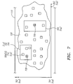
- FIG. 7 shows how images or objects of interest can be organized for review by a technician
- FIG. 8 is a flow diagram illustrating a method of generating and analyzing specimen images and locally storing and retrieving data from a data storage device attached to a biological specimen carrier.
- Embodiments of a system and apparatus for providing necessary specimen and analysis information to a technician in an efficient manner, while eliminating the need to access such data through an external database and network, will now be described.
- the described system and apparatus allow for locally storing sufficient information on a biological specimen carrier to enable a technician to quickly and easily identify the sections of the specimen that warrant further analysis and allow the information on the slide to be locally updated as needed.
- an apparatus 100 for locally storing data related to analysis of a biological specimen includes a biological specimen carrier 110 with a specimen 120 , and a data storage device 130 (shown generally as block 130 ) attached to the biological specimen carrier 110 .
- the data storage device 130 can be attached directly or indirectly to the slide using, for example, an adhesive or a label.
- an adhesive or a label for example, an adhesive or a label.
- the biological specimen carrier 110 can be a slide as shown in the Figures, such as a glass or plastic slide.
- the biological specimen carrier can also be a bottle, a vial, or other containers and objects for holding, storing or supporting a biological specimen.
- the biological specimen 120 can be a human cytological specimen that is analyzed to identify cancerous conditions or other medical conditions.
- the specimen 120 can also be an animal specimen that is analyzed by a veterinarian.
- cytological e.g., PAP smear
- the invention is not so limited and can be utilized with the collection and analysis of various other specimens and biological specimen carriers.
- the data storage device 130 can be a read-only or a read/write storage device and store different types of data.
- Exemplary data storage devices include, for example, a Radio Frequency Identification (RFID) tag, and magnetic and optical storage devices and media.
- RFID Radio Frequency Identification
- the data can include, but is not limited to, data relating to the biological specimen, specific sections of the specimen, the carrier, the patient, the imaging equipment, the review or analysis equipment, and time and date data. Persons of ordinary skill in the art will recognize that other data can be stored to the storage device 130 depending on the particular patient, test, and analysis being performed.
- the apparatus 100 provides flexibility in the selection of the imaging and review equipment. For example, preferred embodiments of the invention allow for imaging and analysis of the specimen 120 to be conducted with various review and analysis equipment since it is not necessary to utilize specific imaging and analysis equipment that are proximately located or configured for a particular database and network connection.
- the data storage device 130 can be attached to different sections or locations of the slide 100 .
- the data storage device 130 can be attached to a top surface 111 , a bottom surface 112 , a first side 113 , a second or opposite side 114 , a first edge 115 , a second or opposite edge 116 and end sections 117 .
- Top surfaces of some slides include “frosted” end sections 117 that provide higher friction surfaces and allow a technician to handle the slide more easily.
- the data storage device 130 is attached to an end section 117 or top surface 111 of the slide 110 , but the invention is not so limited since different storage devices 130 may be suitable for other slide 110 locations.
- the specimen 120 is typically placed in a middle or specimen section 118 between the ends of frosted sections 117 of the slide 110 , but the specimen 120 can also be placed at an end 117 of the slide 100 so that an opposite end is handled by a technician. Accordingly, the configuration shown in FIG. 1 is merely illustrative of different possible specimen 120 and data storage device 130 arrangements.
- RFID Radio Frequency Identification
- Various RFID tags 200 are suitable, such as a Tag-IT ® RFID tag available from Texas Instruments, Radio Frequency Identification Systems, 6550 Chase Oaks Blvd., MS 8470, Plano, Tex. 75023 or an Intellitag® label having an RFID insert, available from Intermec Technologies Corporation, Identification Systems Division, 9290 Le Saint Drive, Fairfield, Ohio 45104.
- These exemplary RFID 200 devices are preferably embedded in a label 210 , as shown in FIG. 2A , which is attached to the slide 110 .
- the RFID tag 200 can also be attached to a slide in different manners, for example, by using a protective coating (not shown) that is applied over the RFID tag 200 to secure the RFID tag 200 to the slide 110 .
- a protective coating (not shown) that is applied over the RFID tag 200 to secure the RFID tag 200 to the slide 110 .
- Various adhesives can also be utilized to attach the RFID tag 200 to the slide 110 .
- a RFID tag 200 typically includes an Integrated Circuit (IC) 220 , such as an Application Specific Integrated Circuit (ASIC), that includes a memory 230 for storing data. Different sizes of memories 230 can be used to store different amounts of specimen information.
- IC Integrated Circuit
- ASIC Application Specific Integrated Circuit
- a typical RFID tag system 240 includes the RFID tag 200 , as previously discussed, a reader 242 , and an antenna 244 .
- the RFID tag 200 is activated by an instruction or signal from the reader 242 , which is sent through the antenna 244 .
- the reader 242 sends a signal to the IC 220 via the antenna 310 to wirelessly write data to or read data from the memory 230 of the IC 210 .
- a user is usually permitted to activate the RFID tag 200 to read data from the memory 230 .
- Data can also be written to the memory 230 depending on the read/write configuration of the memory 230 . Further, data stored in the memory 230 can be updated to supplement or overwrite existing data depending on the memory 230 configuration.
- the RFID tag 200 can be attached to various slide 100 surfaces and locations since a line of sight between the RFID tag 200 and reader 230 is not necessary.
- the RFID tag 200 is attached to a top end 117 of a slide 110 so that the tag 200 does not interfere with the analysis of the specimen 120 .
- the data storage device 130 is a magnetic media 300 .
- exemplary magnetic devices 300 include magnetic encoded strips, available from ID Tech, 1047 South Placentia Avenue, Fullerton, Calif. These exemplary magnetic coded strips and related readers are based encoding characteristics based on ANSI, and ISO/IEC Standards and can store various types of alpha-numeric specimen data.
- the data storage device 130 is an optical data storage device.
- One exemplary optical data storage device is a bar-code 400 or other binary modulation symbol or device, as shown in FIG. 4A .
- the bar-code 400 can be printed on an adhesive label 410 , which is attached to the slide 110 , e.g., at an end 117 of the slide 110 .
- a surface of the slide 110 can be marked or etched 420 with information.
- the etchings 420 can be lines, patterns, text or numbers 420 that contain or represent different types of cytological or biological specimen information.
- specimen information can also be printed directly onto a label 500 , which is then attached to a slide 110 .
- Different letters, numbers, symbols or other markings 510 can be typed in a sufficiently small font or symbology to fit on the label 500 .
- the markings 510 can be readable by the naked eye or with the assistance of a magnification device, such as a magnifying glass or microscope.
- the size of the markings 510 and thus, the quantity of information stored on the label 500 , can be increased or decreased depending on the amount of information to be stored on the slide 110 and the available viewing devices.
- Data storage devices 130 can include different amounts of memory to store different quantities of data. Further, data storage devices can be configured to write and/or read data to/from the data storage device in the even that stored data is to be updated or supplemented. Thus, embodiments of present invention provide enhanced data storage capabilities and read/write options.
- FIG. 6 generally illustrates a system 600 for generating and analyzing images of a specimen 120 , and storing data to and retrieving data from a storage device 130 attached to a slide 100 .
- An exemplary system 600 includes the slide 110 with a data storage device 130 , as previously described, an imaging device or system 610 , and an automated or semi-automated review device or system, such as a microscope 620 or other suitable device.
- the imaging system 610 scans the specimen 120 and generates or prepares a series of images 630 of sections of the specimen 120 utilizing, for example, a software program 612 .
- the system 600 can also include if necessary, a filter or processor 640 that eliminates extraneous data from the images 630 before they are reviewed using the microscope 620 .
- Different types of information and data related to the imaging system 610 can be recorded to the data storage device 230 including, but not limited to, a slide, patient or specimen identifier 650 (if not assigned already), such as an alpha, numeric, or alpha-numeric identifier, imaging system identifier 651 , a timestamp 652 representing a date and time that the images 630 were generated with the imaging system 610 , a version 653 of the imaging system software 612 that was used to generate the images 630 , and coordinates (x,y) of Fields Of Interest (FOIs) 654 , described below.
- a slide, patient or specimen identifier 650 if not assigned already
- imaging system identifier 651 such as an alpha, numeric, or alpha-numeric identifier
- timestamp 652 representing a date and time that the images 630 were generated with the imaging system 610
- a version 653 of the imaging system software 612 that was used to generate the images 630
- the imaging system 610 also processes or analyzes images 630 to identify and select images having cells and cell clusters that most likely have attributes that warrant further consideration by a Cytotechnologist. In one embodiment, these attributes may be those that are consistent with malignant or pre-malignant cells. In either case, these identified cells may be referred to as Objects of Interest (OOI) 710 .
- the image analysis can be performed using, for example, a processor implemented program.
- One or more OOIs 710 can be organized within a defined boundary or Field Of Interest (FOI) 720 .
- a FOI 720 can be defined in various ways and with various geometries to include different numbers of OOIs 710 .
- a FOI 720 can have shapes and dimensions, such as rectangles, ellipses, triangles, and polygons, that cover different portions of the specimen 120 and different numbers of OOIs 710 .
- FIG. 7 shows FOIs as squares or rectangles for purposes of illustration.
- four (x,y) coordinates can be used to identify the boundaries of each FOI.
- An OOI 710 can be assigned to a FOI 720 based on different criteria. For example, an OOI 710 can be assigned to an FOI 720 if the subject OOI 710 was not previously assigned to an FOI 720 .
- An FOI 720 can also be defined to include all of the OOIs 710 , or to maximize or minimize the number of OOIs 720 in a limited FOI 720 .
- OOIs 710 can also be ranked based on, for example, the likelihood that the OOI 710 includes cells of diagnostic interest, and can be selected for inclusion in a particular FOI 720 based on the ranking.
- the analysis system 610 establishes 22 FOIs 720 , i.e., 22 (X,Y) coordinates, relative to the reference coordinate system 700 , that identify or define the FOIs 720 with one or more OOIs 710 .
- a technician analyzes the OOIs 710 organized or allocated to the 22 FOIs 720 with the microscope 620 , which proceeds or steps through the x-y coordinates of each FOI 720 identified by the imaging system 610 and places them within the field of view of the technician.
- the order in which FOIs are presented for review is governed by a version of a so-called “traveling salesman” algorithm, resulting in the least amount of movement of the microscope stage.
- the technician may identify specific FOIs 720 that are likely to contain malignant or pre-malignant cells or raise other concerns.
- the coordinates of these identified FOIs 720 can be marked or highlighted by the technician and are referred to as Marked Target Zones (MTZs) 730 .
- MTZs Marked Target Zones
- different types of information related to reviewing and analyzing the images 630 can also be stored in the data storage device 130 on the slide 110 , including but not limited to, the slide or patient identifier 650 , an identifier 660 of the review scope 620 , a timestamp 661 with the date and time indicating when the review scope 620 was utilized, an identification 662 of the technician who performed the review scope analysis 620 , a version 663 of the review scope software 662 , a number of MTZs 664 , coordinates 665 of MTZs, an indication 666 of whether the MTZ 730 falls within a FOI 720 , and whether an autoscan 667 was conducted. Additional information on systems and methods of reviewing and analyzing slide specimens may be found in U.S. Pat. No. 6,562,299, which is assigned to the assignee of the present invention, the entire disclosure of which is expressly incorporated by reference herein.
- a description of the method of locally storing data related to specimen analysis to and reading the data from a data storage device attached to a test slide is provided.
- a data storage device is attached to a biological specimen carrier, such as a slide.
- the data storage device can be attached to a label which is applied to the slide, or attached with a coating or an adhesive.
- a plurality of images of the specimen are generated by the imaging system.
- one or more images of the plurality of images are analyzed.
- step 815 data relating to the specimen or specimen analysis is locally stored to the data storage device such as, for example, a specimen or slide identifier, data related to an imaging system (e.g., identifier, software version, timestamp), and data related to a review system or review of the specimen (e.g., identifiers of the technician and review system, software version, timestamp, Fields of Interest, coordinates, Marker Target Zones).
- data from an imaging system is recorded to the data storage device.
- step 825 data from a review scope is recorded to the data storage device.
- the recorded data can be updated as needed.
- the locally stored data in the storage device can be accessed by a technician without connecting to an external database or network.
- the previously described apparatus, system and method embodiments of the invention provide a number of enhancements to conventional test slides, slide imaging and analysis systems, and analysis processes, which eliminate the need to access an external database or separate media device such as a CD, to retrieve necessary slide, specimen or analysis data. Instead, these embodiments of the invention allow a technician to directly access pertinent carrier and specimen data that is stored locally on the slide. As a result, a technician can analyze specimen images more efficiently. Further, since the slide with a data storage device can serve as a “self-contained database,” the slides can be moved between laboratories and be analyzed in different locations without the restrictions associated with external databases and network equipment. The data stored on the slide can also be supplemented and updated as needed.
Abstract
Description
Claims (22)
Priority Applications (13)
| Application Number | Priority Date | Filing Date | Title |
|---|---|---|---|
| US10/656,092 US7083106B2 (en) | 2003-09-05 | 2003-09-05 | Locally storing biological specimen data to a slide |
| JP2006525362A JP2007504458A (en) | 2003-09-05 | 2004-08-23 | Local storage of biological specimen data on slides |
| EP09171701A EP2164012A1 (en) | 2003-09-05 | 2004-08-23 | Locally storing biological specimen data to a slide |
| PCT/US2004/027439 WO2005027014A2 (en) | 2003-09-05 | 2004-08-23 | Locally storing biological specimen data to a slide |
| KR1020067004300A KR20060072131A (en) | 2003-09-05 | 2004-08-23 | Locally storing biological specimen data to a slide |
| MXPA06002487A MXPA06002487A (en) | 2003-09-05 | 2004-08-23 | Locally storing biological specimen data to a slide. |
| EP04782014A EP1665109A2 (en) | 2003-09-05 | 2004-08-23 | Locally storing biological specimen data to a slide |
| AU2004273436A AU2004273436B2 (en) | 2003-09-05 | 2004-08-23 | Locally storing biological specimen data to a slide |
| CA002536619A CA2536619A1 (en) | 2003-09-05 | 2004-08-23 | Locally storing biological specimen data to a slide |
| BRPI0414142-3A BRPI0414142A (en) | 2003-09-05 | 2004-08-23 | locally storing biological specimen data on a slide |
| CNB2004800252929A CN100470562C (en) | 2003-09-05 | 2004-08-23 | Locally storing biological specimen data to a slide |
| US11/423,660 US7395974B2 (en) | 2003-09-05 | 2006-06-12 | Locally storing biological specimen data to a slide |
| AU2009222449A AU2009222449A1 (en) | 2003-09-05 | 2009-09-28 | Locally storing biological specimen data to a slide |
Applications Claiming Priority (1)
| Application Number | Priority Date | Filing Date | Title |
|---|---|---|---|
| US10/656,092 US7083106B2 (en) | 2003-09-05 | 2003-09-05 | Locally storing biological specimen data to a slide |
Related Child Applications (1)
| Application Number | Title | Priority Date | Filing Date |
|---|---|---|---|
| US11/423,660 Continuation US7395974B2 (en) | 2003-09-05 | 2006-06-12 | Locally storing biological specimen data to a slide |
Publications (2)
| Publication Number | Publication Date |
|---|---|
| US20050051614A1 US20050051614A1 (en) | 2005-03-10 |
| US7083106B2 true US7083106B2 (en) | 2006-08-01 |
Family
ID=34226280
Family Applications (2)
| Application Number | Title | Priority Date | Filing Date |
|---|---|---|---|
| US10/656,092 Expired - Lifetime US7083106B2 (en) | 2003-09-05 | 2003-09-05 | Locally storing biological specimen data to a slide |
| US11/423,660 Expired - Lifetime US7395974B2 (en) | 2003-09-05 | 2006-06-12 | Locally storing biological specimen data to a slide |
Family Applications After (1)
| Application Number | Title | Priority Date | Filing Date |
|---|---|---|---|
| US11/423,660 Expired - Lifetime US7395974B2 (en) | 2003-09-05 | 2006-06-12 | Locally storing biological specimen data to a slide |
Country Status (10)
| Country | Link |
|---|---|
| US (2) | US7083106B2 (en) |
| EP (2) | EP2164012A1 (en) |
| JP (1) | JP2007504458A (en) |
| KR (1) | KR20060072131A (en) |
| CN (1) | CN100470562C (en) |
| AU (2) | AU2004273436B2 (en) |
| BR (1) | BRPI0414142A (en) |
| CA (1) | CA2536619A1 (en) |
| MX (1) | MXPA06002487A (en) |
| WO (1) | WO2005027014A2 (en) |
Cited By (27)
| Publication number | Priority date | Publication date | Assignee | Title |
|---|---|---|---|---|
| US20050159982A1 (en) * | 2003-07-17 | 2005-07-21 | Wayne Showalter | Laboratory instrumentation information management and control network |
| US20050276728A1 (en) * | 2004-04-08 | 2005-12-15 | Biomatrica, Inc. | Integration of sample storage and sample management for life science |
| US20060099567A1 (en) * | 2004-04-08 | 2006-05-11 | Biomatrica, Inc. | Integration of sample storage and sample management for life science |
| US20070014693A1 (en) * | 2005-07-18 | 2007-01-18 | Kantrowitz Allen B | Modular hospital cart |
| US20080145887A1 (en) * | 2006-12-19 | 2008-06-19 | Cytyc Corporation | Cytological filter with data storage |
| US20080176209A1 (en) * | 2004-04-08 | 2008-07-24 | Biomatrica, Inc. | Integration of sample storage and sample management for life science |
| US20090009290A1 (en) * | 2007-07-05 | 2009-01-08 | Baxter International Inc. | Radio frequency auto-identification system |
| US20090168161A1 (en) * | 2007-12-27 | 2009-07-02 | Cytyc Corporation | Apparatus for single-handed control of microscope functions |
| US20090168160A1 (en) * | 2007-12-27 | 2009-07-02 | Cytyc Corporation | Methods and systems for controlably scanning a cytological specimen |
| US20100166268A1 (en) * | 2008-12-30 | 2010-07-01 | Ebm Technologies Incorporated | Storage system for storing the sampling data of pathological section and method thereof |
| US7767152B2 (en) | 2003-08-11 | 2010-08-03 | Sakura Finetek U.S.A., Inc. | Reagent container and slide reaction retaining tray, and method of operation |
| US20100315205A1 (en) * | 2007-12-10 | 2010-12-16 | Egbert William C | Associated set of radio frequency identfication ("rfid") tagged containers for specimens from a patient |
| US20110031139A1 (en) * | 2006-07-26 | 2011-02-10 | James Joseph Macor | Protection, authentication, identification device for a physical object specimen |
| US20110079640A1 (en) * | 2006-08-04 | 2011-04-07 | Ikonisys, Inc. | Methods for detecting fluorescent signals in a biological sample |
| US20120242817A1 (en) * | 2008-12-30 | 2012-09-27 | Ebm Technologies Incorporated | System and method for identifying a pathological tissue image |
| US8459509B2 (en) | 2006-05-25 | 2013-06-11 | Sakura Finetek U.S.A., Inc. | Fluid dispensing apparatus |
| US8580568B2 (en) | 2011-09-21 | 2013-11-12 | Sakura Finetek U.S.A., Inc. | Traceability for automated staining system |
| US8752732B2 (en) | 2011-02-01 | 2014-06-17 | Sakura Finetek U.S.A., Inc. | Fluid dispensing system |
| US8932543B2 (en) | 2011-09-21 | 2015-01-13 | Sakura Finetek U.S.A., Inc. | Automated staining system and reaction chamber |
| US9376709B2 (en) | 2010-07-26 | 2016-06-28 | Biomatrica, Inc. | Compositions for stabilizing DNA and RNA in blood and other biological samples during shipping and storage at ambient temperatures |
| US9518899B2 (en) | 2003-08-11 | 2016-12-13 | Sakura Finetek U.S.A., Inc. | Automated reagent dispensing system and method of operation |
| US9725703B2 (en) | 2012-12-20 | 2017-08-08 | Biomatrica, Inc. | Formulations and methods for stabilizing PCR reagents |
| US9845489B2 (en) | 2010-07-26 | 2017-12-19 | Biomatrica, Inc. | Compositions for stabilizing DNA, RNA and proteins in saliva and other biological samples during shipping and storage at ambient temperatures |
| US10064404B2 (en) | 2014-06-10 | 2018-09-04 | Biomatrica, Inc. | Stabilization of thrombocytes at ambient temperatures |
| WO2018165630A1 (en) | 2017-03-09 | 2018-09-13 | Hologic, Inc. | Systems and methods for automated preparation of biological specimens |
| US10369573B2 (en) * | 2004-03-19 | 2019-08-06 | Applied Biosystems, Llc | Methods and systems for using RFID in biological field |
| US10568317B2 (en) | 2015-12-08 | 2020-02-25 | Biomatrica, Inc. | Reduction of erythrocyte sedimentation rate |
Families Citing this family (24)
| Publication number | Priority date | Publication date | Assignee | Title |
|---|---|---|---|---|
| US7199712B2 (en) * | 2004-06-17 | 2007-04-03 | Tafas Triantafyllos P | System for automatically locating and manipulating positions on an object |
| EP1771716A4 (en) * | 2004-06-29 | 2012-04-25 | Dako Denmark As | Method of pre-treatment and staining of and support device for a biological sample |
| US7277223B2 (en) * | 2004-07-26 | 2007-10-02 | Meade Instruments Corporation | Apparatus and methods for focusing and collimating telescopes |
| US7556777B2 (en) * | 2005-03-08 | 2009-07-07 | Cytyc Corporation | Specimen vial cap handler and slide labeler |
| GB0526452D0 (en) * | 2005-12-23 | 2006-02-08 | Hughes Thomas F | A laboratory slide |
| AU2007226647A1 (en) * | 2006-03-13 | 2007-09-20 | Ikonisys, Inc. | Automated microscope slide read system |
| SE530789C2 (en) * | 2007-01-17 | 2008-09-09 | Hemocue Ab | Apparatus and method for position determination of objects contained in a sample |
| US20080239478A1 (en) * | 2007-03-29 | 2008-10-02 | Tafas Triantafyllos P | System for automatically locating and manipulating positions on an object |
| TW200912309A (en) * | 2007-09-04 | 2009-03-16 | Kaiwood Technology Co Ltd | System configuration method of color indicating chip analyzer |
| EP2232319B8 (en) * | 2007-12-19 | 2017-10-18 | Diagnostic Vision Corporation | Method and system for identifying biological specimen slides using unique slide fingerprints |
| JP2009175031A (en) * | 2008-01-25 | 2009-08-06 | A & T Corp | Substrate type reagent, apparatus, method, and program for manufacturing inspection, apparatus, method, and program for analysis, server, method, and program for management, and recording medium |
| DE102009005307B4 (en) * | 2009-01-16 | 2011-06-01 | Waldemar Knittel Glasbearbeitungs Gmbh | slides |
| ZA201007121B (en) * | 2009-10-09 | 2011-06-29 | Drew Bellamy | Sampling system and method |
| CN101763466B (en) * | 2010-01-20 | 2011-08-24 | 西安电子科技大学 | Biological information recognition method based on dynamic sample selection integration |
| DE102012005587A1 (en) * | 2012-03-20 | 2013-09-26 | Metasystems Hard & Software Gmbh | Automatic calibration of a microscope scanning system |
| US9618428B2 (en) * | 2012-11-30 | 2017-04-11 | Ge Healthcare Uk Limited | Biometric device and means for electronic storage and retrieval of biometric data |
| CN103837977A (en) * | 2014-01-16 | 2014-06-04 | 麦克奥迪实业集团有限公司 | Microscopy device with NFC modules |
| US10579959B2 (en) | 2014-09-10 | 2020-03-03 | Cerner Innovation, Inc. | Intelligent routing of radio-frequency identification data |
| CN113358860A (en) * | 2015-07-23 | 2021-09-07 | 中尺度技术有限责任公司 | Automated analysis system and method for performing analysis in such a system |
| CN105510609A (en) * | 2015-11-26 | 2016-04-20 | 宁波美康保生生物医学工程有限公司 | In vitro test piece and application method thereof |
| US10203491B2 (en) | 2016-08-01 | 2019-02-12 | Verily Life Sciences Llc | Pathology data capture |
| DE102016117051A1 (en) * | 2016-09-12 | 2018-03-15 | DüRR DENTAL AG | System and method for providing acquisition parameters |
| WO2021050359A1 (en) * | 2019-09-13 | 2021-03-18 | Celly.AI | Artificial intelligence (ai) powered analysis of objects observable through a microscope |
| US11200671B2 (en) * | 2019-12-31 | 2021-12-14 | International Business Machines Corporation | Reference image guided object detection in medical image processing |
Citations (12)
| Publication number | Priority date | Publication date | Assignee | Title |
|---|---|---|---|---|
| GB1423185A (en) | 1972-09-27 | 1976-01-28 | Bancsich J Hadrian W | Specimen holder and arrangement for registration and read out thereof |
| US5561556A (en) * | 1994-04-21 | 1996-10-01 | Compucyte Corporation | Slide analysis system with slide having self contained microscope analysis information |
| WO1997022901A1 (en) | 1994-04-21 | 1997-06-26 | Compucyte Corporation | Slide analysis system with slide having self contained microscope analysis information |
| US5693368A (en) | 1994-09-30 | 1997-12-02 | General Electric Company | Low temperature chemical vapor deposition method for cleaning substrate and depositing protective coating |
| WO1999010763A1 (en) | 1997-08-21 | 1999-03-04 | Carl Zeiss Jena Gmbh | Sample holder which can process data and method for analysing samples |
| US6198299B1 (en) * | 1998-08-27 | 2001-03-06 | The Micromanipulator Company, Inc. | High Resolution analytical probe station |
| WO2001031566A2 (en) | 1999-10-29 | 2001-05-03 | Cytyc Corporation | Apparatus and methods for verifying the location of areas of interest within a sample in an imaging system |
| WO2002021425A2 (en) | 2000-09-05 | 2002-03-14 | Capitol Vial, Inc. | A system and method for maintaining, tracking and identifying the integrity of a disposable specimen container with a re-usable transponder |
| US6535626B1 (en) * | 2000-01-14 | 2003-03-18 | Accumed International, Inc. | Inspection system with specimen preview |
| US6615763B2 (en) | 2000-05-12 | 2003-09-09 | Innovative Science Limited | Printing on microscope slides and histology cassettes |
| US6631203B2 (en) * | 1999-04-13 | 2003-10-07 | Chromavision Medical Systems, Inc. | Histological reconstruction and automated image analysis |
| US6905823B2 (en) * | 1998-10-28 | 2005-06-14 | Abbott Laboratories | Cellular arrays and methods of detecting and using genetic disorder markers |
Family Cites Families (5)
| Publication number | Priority date | Publication date | Assignee | Title |
|---|---|---|---|---|
| US5561558A (en) | 1993-10-18 | 1996-10-01 | Matsushita Electric Industrial Co., Ltd. | Diffractive optical device |
| US6562299B1 (en) | 1998-09-18 | 2003-05-13 | Cytyc Corporation | Method and apparatus for preparing cytological specimens |
| US6650704B1 (en) | 1999-10-25 | 2003-11-18 | Irvine Sensors Corporation | Method of producing a high quality, high resolution image from a sequence of low quality, low resolution images that are undersampled and subject to jitter |
| US6535826B2 (en) | 2001-02-16 | 2003-03-18 | Micro Motion, Inc. | Mass flowmeter methods, apparatus, and computer program products using correlation-measure-based status determination |
| GB0122325D0 (en) * | 2001-09-14 | 2001-11-07 | Hughes Thomas F | Laboratory slide |
-
2003
- 2003-09-05 US US10/656,092 patent/US7083106B2/en not_active Expired - Lifetime
-
2004
- 2004-08-23 AU AU2004273436A patent/AU2004273436B2/en active Active
- 2004-08-23 MX MXPA06002487A patent/MXPA06002487A/en active IP Right Grant
- 2004-08-23 JP JP2006525362A patent/JP2007504458A/en active Pending
- 2004-08-23 EP EP09171701A patent/EP2164012A1/en not_active Withdrawn
- 2004-08-23 EP EP04782014A patent/EP1665109A2/en not_active Ceased
- 2004-08-23 CN CNB2004800252929A patent/CN100470562C/en active Active
- 2004-08-23 KR KR1020067004300A patent/KR20060072131A/en not_active Application Discontinuation
- 2004-08-23 BR BRPI0414142-3A patent/BRPI0414142A/en not_active IP Right Cessation
- 2004-08-23 WO PCT/US2004/027439 patent/WO2005027014A2/en active Application Filing
- 2004-08-23 CA CA002536619A patent/CA2536619A1/en not_active Abandoned
-
2006
- 2006-06-12 US US11/423,660 patent/US7395974B2/en not_active Expired - Lifetime
-
2009
- 2009-09-28 AU AU2009222449A patent/AU2009222449A1/en not_active Abandoned
Patent Citations (12)
| Publication number | Priority date | Publication date | Assignee | Title |
|---|---|---|---|---|
| GB1423185A (en) | 1972-09-27 | 1976-01-28 | Bancsich J Hadrian W | Specimen holder and arrangement for registration and read out thereof |
| US5561556A (en) * | 1994-04-21 | 1996-10-01 | Compucyte Corporation | Slide analysis system with slide having self contained microscope analysis information |
| WO1997022901A1 (en) | 1994-04-21 | 1997-06-26 | Compucyte Corporation | Slide analysis system with slide having self contained microscope analysis information |
| US5693368A (en) | 1994-09-30 | 1997-12-02 | General Electric Company | Low temperature chemical vapor deposition method for cleaning substrate and depositing protective coating |
| WO1999010763A1 (en) | 1997-08-21 | 1999-03-04 | Carl Zeiss Jena Gmbh | Sample holder which can process data and method for analysing samples |
| US6198299B1 (en) * | 1998-08-27 | 2001-03-06 | The Micromanipulator Company, Inc. | High Resolution analytical probe station |
| US6905823B2 (en) * | 1998-10-28 | 2005-06-14 | Abbott Laboratories | Cellular arrays and methods of detecting and using genetic disorder markers |
| US6631203B2 (en) * | 1999-04-13 | 2003-10-07 | Chromavision Medical Systems, Inc. | Histological reconstruction and automated image analysis |
| WO2001031566A2 (en) | 1999-10-29 | 2001-05-03 | Cytyc Corporation | Apparatus and methods for verifying the location of areas of interest within a sample in an imaging system |
| US6535626B1 (en) * | 2000-01-14 | 2003-03-18 | Accumed International, Inc. | Inspection system with specimen preview |
| US6615763B2 (en) | 2000-05-12 | 2003-09-09 | Innovative Science Limited | Printing on microscope slides and histology cassettes |
| WO2002021425A2 (en) | 2000-09-05 | 2002-03-14 | Capitol Vial, Inc. | A system and method for maintaining, tracking and identifying the integrity of a disposable specimen container with a re-usable transponder |
Non-Patent Citations (3)
| Title |
|---|
| PCT International Search Report for PCT/US2004/027439, Applicant: Cytyc Corporation, Forms PCT/ISA/210 and 220, dated Mar. 31, 2005 (6 pages). |
| PCT Written Opinion of the International Search Authority for PCT/US2004/027439, Applicant: Cytyc Corporation, Form PCT/ISA/237, dated Mar. 31, 2005 (5 pages). |
| Zebra Technologies Corp. RFID: The Next Generation of AIDC, Application White Paper. GSA#: GS-35F-0268N, 11315L Rev 4 (Sep. 2003). |
Cited By (46)
| Publication number | Priority date | Publication date | Assignee | Title |
|---|---|---|---|---|
| US20050159982A1 (en) * | 2003-07-17 | 2005-07-21 | Wayne Showalter | Laboratory instrumentation information management and control network |
| US7860727B2 (en) * | 2003-07-17 | 2010-12-28 | Ventana Medical Systems, Inc. | Laboratory instrumentation information management and control network |
| US8812329B2 (en) * | 2003-07-17 | 2014-08-19 | Ventana Medical Systems, Inc. | Laboratory instrumentation information management and control network |
| US20070196909A1 (en) * | 2003-07-17 | 2007-08-23 | Wayne Showalter | Laboratory instrumentation information management and control network |
| US7767152B2 (en) | 2003-08-11 | 2010-08-03 | Sakura Finetek U.S.A., Inc. | Reagent container and slide reaction retaining tray, and method of operation |
| US9518899B2 (en) | 2003-08-11 | 2016-12-13 | Sakura Finetek U.S.A., Inc. | Automated reagent dispensing system and method of operation |
| US10369573B2 (en) * | 2004-03-19 | 2019-08-06 | Applied Biosystems, Llc | Methods and systems for using RFID in biological field |
| US20080176209A1 (en) * | 2004-04-08 | 2008-07-24 | Biomatrica, Inc. | Integration of sample storage and sample management for life science |
| US8900856B2 (en) | 2004-04-08 | 2014-12-02 | Biomatrica, Inc. | Integration of sample storage and sample management for life science |
| US20060099567A1 (en) * | 2004-04-08 | 2006-05-11 | Biomatrica, Inc. | Integration of sample storage and sample management for life science |
| US20050276728A1 (en) * | 2004-04-08 | 2005-12-15 | Biomatrica, Inc. | Integration of sample storage and sample management for life science |
| US9078426B2 (en) * | 2004-04-08 | 2015-07-14 | Biomatrica, Inc. | Integration of sample storage and sample management for life science |
| US20070014693A1 (en) * | 2005-07-18 | 2007-01-18 | Kantrowitz Allen B | Modular hospital cart |
| US8287818B2 (en) | 2005-07-18 | 2012-10-16 | Kantrowitz Allen B | Modular hospital cart |
| US9914124B2 (en) | 2006-05-25 | 2018-03-13 | Sakura Finetek U.S.A., Inc. | Fluid dispensing apparatus |
| US8459509B2 (en) | 2006-05-25 | 2013-06-11 | Sakura Finetek U.S.A., Inc. | Fluid dispensing apparatus |
| US20110031139A1 (en) * | 2006-07-26 | 2011-02-10 | James Joseph Macor | Protection, authentication, identification device for a physical object specimen |
| US20110079640A1 (en) * | 2006-08-04 | 2011-04-07 | Ikonisys, Inc. | Methods for detecting fluorescent signals in a biological sample |
| US20080145887A1 (en) * | 2006-12-19 | 2008-06-19 | Cytyc Corporation | Cytological filter with data storage |
| US8330579B2 (en) | 2007-07-05 | 2012-12-11 | Baxter International Inc. | Radio-frequency auto-identification system for dialysis systems |
| US20090009290A1 (en) * | 2007-07-05 | 2009-01-08 | Baxter International Inc. | Radio frequency auto-identification system |
| US20100315205A1 (en) * | 2007-12-10 | 2010-12-16 | Egbert William C | Associated set of radio frequency identfication ("rfid") tagged containers for specimens from a patient |
| US8174763B2 (en) | 2007-12-27 | 2012-05-08 | Cytyc Corporation | Methods and systems for controlably scanning a cytological specimen |
| US8325414B2 (en) | 2007-12-27 | 2012-12-04 | Cytyc Corporation | Apparatus for single-handed control of microscope functions |
| US20090168160A1 (en) * | 2007-12-27 | 2009-07-02 | Cytyc Corporation | Methods and systems for controlably scanning a cytological specimen |
| US20090168161A1 (en) * | 2007-12-27 | 2009-07-02 | Cytyc Corporation | Apparatus for single-handed control of microscope functions |
| US20120242817A1 (en) * | 2008-12-30 | 2012-09-27 | Ebm Technologies Incorporated | System and method for identifying a pathological tissue image |
| US20100166268A1 (en) * | 2008-12-30 | 2010-07-01 | Ebm Technologies Incorporated | Storage system for storing the sampling data of pathological section and method thereof |
| US9845489B2 (en) | 2010-07-26 | 2017-12-19 | Biomatrica, Inc. | Compositions for stabilizing DNA, RNA and proteins in saliva and other biological samples during shipping and storage at ambient temperatures |
| US9376709B2 (en) | 2010-07-26 | 2016-06-28 | Biomatrica, Inc. | Compositions for stabilizing DNA and RNA in blood and other biological samples during shipping and storage at ambient temperatures |
| US9999217B2 (en) | 2010-07-26 | 2018-06-19 | Biomatrica, Inc. | Compositions for stabilizing DNA, RNA, and proteins in blood and other biological samples during shipping and storage at ambient temperatures |
| US8752732B2 (en) | 2011-02-01 | 2014-06-17 | Sakura Finetek U.S.A., Inc. | Fluid dispensing system |
| US9016526B2 (en) | 2011-02-01 | 2015-04-28 | Sakura Finetek U.S.A., Inc. | Fluid dispensing system |
| US10295444B2 (en) | 2011-09-21 | 2019-05-21 | Sakura Finetek U.S.A., Inc. | Automated staining system and reaction chamber |
| US9005980B2 (en) | 2011-09-21 | 2015-04-14 | Sakura Finetek U.S.A., Inc. | Traceability for automated staining system |
| US8932543B2 (en) | 2011-09-21 | 2015-01-13 | Sakura Finetek U.S.A., Inc. | Automated staining system and reaction chamber |
| US8580568B2 (en) | 2011-09-21 | 2013-11-12 | Sakura Finetek U.S.A., Inc. | Traceability for automated staining system |
| US9725703B2 (en) | 2012-12-20 | 2017-08-08 | Biomatrica, Inc. | Formulations and methods for stabilizing PCR reagents |
| US10772319B2 (en) | 2014-06-10 | 2020-09-15 | Biomatrica, Inc. | Stabilization of thrombocytes at ambient temperatures |
| US10064404B2 (en) | 2014-06-10 | 2018-09-04 | Biomatrica, Inc. | Stabilization of thrombocytes at ambient temperatures |
| US11672247B2 (en) | 2014-06-10 | 2023-06-13 | Biomatrica, Inc. | Stabilization of thrombocytes at ambient temperatures |
| US10568317B2 (en) | 2015-12-08 | 2020-02-25 | Biomatrica, Inc. | Reduction of erythrocyte sedimentation rate |
| US11116205B2 (en) | 2015-12-08 | 2021-09-14 | Biomatrica, Inc. | Reduction of erythrocyte sedimentation rate |
| WO2018165630A1 (en) | 2017-03-09 | 2018-09-13 | Hologic, Inc. | Systems and methods for automated preparation of biological specimens |
| US10900875B2 (en) | 2017-03-09 | 2021-01-26 | Hologic, Inc. | Systems and methods for automated preparation of biological specimens |
| US11536634B2 (en) | 2017-03-09 | 2022-12-27 | Hologic, Inc. | Systems and methods for automated preparation of biological specimens |
Also Published As
| Publication number | Publication date |
|---|---|
| CN100470562C (en) | 2009-03-18 |
| BRPI0414142A (en) | 2006-10-31 |
| AU2004273436B2 (en) | 2009-07-16 |
| KR20060072131A (en) | 2006-06-27 |
| EP1665109A2 (en) | 2006-06-07 |
| CN1846216A (en) | 2006-10-11 |
| AU2004273436A1 (en) | 2005-03-24 |
| CA2536619A1 (en) | 2005-03-24 |
| JP2007504458A (en) | 2007-03-01 |
| WO2005027014A3 (en) | 2005-05-26 |
| US20050051614A1 (en) | 2005-03-10 |
| US7395974B2 (en) | 2008-07-08 |
| EP2164012A1 (en) | 2010-03-17 |
| AU2009222449A1 (en) | 2009-10-15 |
| WO2005027014A2 (en) | 2005-03-24 |
| MXPA06002487A (en) | 2006-06-20 |
| US20060219795A1 (en) | 2006-10-05 |
Similar Documents
| Publication | Publication Date | Title |
|---|---|---|
| US7083106B2 (en) | Locally storing biological specimen data to a slide | |
| US10228311B2 (en) | Automated lean methods in anatomical pathology | |
| AU2005259981B2 (en) | Method of pre-treatment and staining of and support device for a biological sample | |
| US20110217731A1 (en) | Pathology sample processing workstation | |
| EP1922403A2 (en) | Pathology sample processing workstation | |
| US8315445B2 (en) | Tissue sample identification system and apparatus | |
| CN102334085A (en) | Systems and methods for processing tissue samples for histopathology | |
| KR20200062207A (en) | Biopsy tissue sample cassette and related systems and methods | |
| WO2007106789A2 (en) | Automated microscope slide read system | |
| US20100315205A1 (en) | Associated set of radio frequency identfication ("rfid") tagged containers for specimens from a patient | |
| EP1902321A2 (en) | Marking sample carriers | |
| CN113222078A (en) | Sample library management method and device, computer equipment and storage medium | |
| US20230169300A1 (en) | System for identifying, localizing and storing histology cassettes and slides | |
| US7906337B2 (en) | Method of generating and using biological qualification slides to qualify biological screening devices |
Legal Events
| Date | Code | Title | Description |
|---|---|---|---|
| AS | Assignment |
Owner name: CYTYC CORPORATION, MASSACHUSETTS Free format text: ASSIGNMENT OF ASSIGNORS INTEREST;ASSIGNOR:ALBANY, PETER;REEL/FRAME:014490/0865 Effective date: 20030829 |
|
| FEPP | Fee payment procedure |
Free format text: PAYOR NUMBER ASSIGNED (ORIGINAL EVENT CODE: ASPN); ENTITY STATUS OF PATENT OWNER: LARGE ENTITY |
|
| STCF | Information on status: patent grant |
Free format text: PATENTED CASE |
|
| AS | Assignment |
Owner name: GOLDMAN SACHS CREDIT PARTNERS L.P., CALIFORNIA Free format text: PATENT SECURITY AGREEMENT;ASSIGNOR:CYTYC CORPORATION;REEL/FRAME:020018/0529 Effective date: 20071022 Owner name: GOLDMAN SACHS CREDIT PARTNERS L.P.,CALIFORNIA Free format text: PATENT SECURITY AGREEMENT;ASSIGNOR:CYTYC CORPORATION;REEL/FRAME:020018/0529 Effective date: 20071022 |
|
| AS | Assignment |
Owner name: GOLDMAN SACHS CREDIT PARTNERS L.P., AS COLLATERAL Free format text: PATENT SECURITY AGREEMENT;ASSIGNOR:CYTYC CORPORATION;REEL/FRAME:021301/0879 Effective date: 20080717 |
|
| FPAY | Fee payment |
Year of fee payment: 4 |
|
| AS | Assignment |
Owner name: CYTYC PRENATAL PRODUCTS CORP., MASSACHUSETTS Free format text: TERMINATION OF PATENT SECURITY AGREEMENTS AND RELEASE OF SECURITY INTERESTS;ASSIGNOR:GOLDMAN SACHS CREDIT PARTNERS, L.P., AS COLLATERAL AGENT;REEL/FRAME:024944/0315 Effective date: 20100819 Owner name: BIOLUCENT, LLC, CALIFORNIA Free format text: TERMINATION OF PATENT SECURITY AGREEMENTS AND RELEASE OF SECURITY INTERESTS;ASSIGNOR:GOLDMAN SACHS CREDIT PARTNERS, L.P., AS COLLATERAL AGENT;REEL/FRAME:024944/0315 Effective date: 20100819 Owner name: CYTYC SURGICAL PRODUCTS II LIMITED PARTNERSHIP, MA Free format text: TERMINATION OF PATENT SECURITY AGREEMENTS AND RELEASE OF SECURITY INTERESTS;ASSIGNOR:GOLDMAN SACHS CREDIT PARTNERS, L.P., AS COLLATERAL AGENT;REEL/FRAME:024944/0315 Effective date: 20100819 Owner name: CYTYC SURGICAL PRODUCTS III, INC., MASSACHUSETTS Free format text: TERMINATION OF PATENT SECURITY AGREEMENTS AND RELEASE OF SECURITY INTERESTS;ASSIGNOR:GOLDMAN SACHS CREDIT PARTNERS, L.P., AS COLLATERAL AGENT;REEL/FRAME:024944/0315 Effective date: 20100819 Owner name: R2 TECHNOLOGY, INC., CALIFORNIA Free format text: TERMINATION OF PATENT SECURITY AGREEMENTS AND RELEASE OF SECURITY INTERESTS;ASSIGNOR:GOLDMAN SACHS CREDIT PARTNERS, L.P., AS COLLATERAL AGENT;REEL/FRAME:024944/0315 Effective date: 20100819 Owner name: THIRD WAVE TECHNOLOGIES, INC., WISCONSIN Free format text: TERMINATION OF PATENT SECURITY AGREEMENTS AND RELEASE OF SECURITY INTERESTS;ASSIGNOR:GOLDMAN SACHS CREDIT PARTNERS, L.P., AS COLLATERAL AGENT;REEL/FRAME:024944/0315 Effective date: 20100819 Owner name: DIRECT RADIOGRAPHY CORP., DELAWARE Free format text: TERMINATION OF PATENT SECURITY AGREEMENTS AND RELEASE OF SECURITY INTERESTS;ASSIGNOR:GOLDMAN SACHS CREDIT PARTNERS, L.P., AS COLLATERAL AGENT;REEL/FRAME:024944/0315 Effective date: 20100819 Owner name: CYTYC CORPORATION, MASSACHUSETTS Free format text: TERMINATION OF PATENT SECURITY AGREEMENTS AND RELEASE OF SECURITY INTERESTS;ASSIGNOR:GOLDMAN SACHS CREDIT PARTNERS, L.P., AS COLLATERAL AGENT;REEL/FRAME:024944/0315 Effective date: 20100819 Owner name: HOLOGIC, INC., MASSACHUSETTS Free format text: TERMINATION OF PATENT SECURITY AGREEMENTS AND RELEASE OF SECURITY INTERESTS;ASSIGNOR:GOLDMAN SACHS CREDIT PARTNERS, L.P., AS COLLATERAL AGENT;REEL/FRAME:024944/0315 Effective date: 20100819 Owner name: CYTYC SURGICAL PRODUCTS LIMITED PARTNERSHIP, MASSA Free format text: TERMINATION OF PATENT SECURITY AGREEMENTS AND RELEASE OF SECURITY INTERESTS;ASSIGNOR:GOLDMAN SACHS CREDIT PARTNERS, L.P., AS COLLATERAL AGENT;REEL/FRAME:024944/0315 Effective date: 20100819 Owner name: SUROS SURGICAL SYSTEMS, INC., INDIANA Free format text: TERMINATION OF PATENT SECURITY AGREEMENTS AND RELEASE OF SECURITY INTERESTS;ASSIGNOR:GOLDMAN SACHS CREDIT PARTNERS, L.P., AS COLLATERAL AGENT;REEL/FRAME:024944/0315 Effective date: 20100819 |
|
| AS | Assignment |
Owner name: GOLDMAN SACHS BANK USA, NEW YORK Free format text: SECURITY AGREEMENT;ASSIGNORS:HOLOGIC, INC.;BIOLUCENT, LLC;CYTYC CORPORATION;AND OTHERS;REEL/FRAME:028810/0745 Effective date: 20120801 |
|
| FPAY | Fee payment |
Year of fee payment: 8 |
|
| AS | Assignment |
Owner name: CYTYC SURGICAL PRODUCTS, LIMITED PARTNERSHIP, MASSACHUSETTS Free format text: SECURITY INTEREST RELEASE REEL/FRAME 028810/0745;ASSIGNOR:GOLDMAN SACHS BANK USA, AS COLLATERAL AGENT;REEL/FRAME:035820/0239 Effective date: 20150529 Owner name: GEN-PROBE INCORPORATED, MASSACHUSETTS Free format text: SECURITY INTEREST RELEASE REEL/FRAME 028810/0745;ASSIGNOR:GOLDMAN SACHS BANK USA, AS COLLATERAL AGENT;REEL/FRAME:035820/0239 Effective date: 20150529 Owner name: CYTYC SURGICAL PRODUCTS, LIMITED PARTNERSHIP, MASS Free format text: SECURITY INTEREST RELEASE REEL/FRAME 028810/0745;ASSIGNOR:GOLDMAN SACHS BANK USA, AS COLLATERAL AGENT;REEL/FRAME:035820/0239 Effective date: 20150529 Owner name: BIOLUCENT, LLC, MASSACHUSETTS Free format text: SECURITY INTEREST RELEASE REEL/FRAME 028810/0745;ASSIGNOR:GOLDMAN SACHS BANK USA, AS COLLATERAL AGENT;REEL/FRAME:035820/0239 Effective date: 20150529 Owner name: CYTYC CORPORATION, MASSACHUSETTS Free format text: SECURITY INTEREST RELEASE REEL/FRAME 028810/0745;ASSIGNOR:GOLDMAN SACHS BANK USA, AS COLLATERAL AGENT;REEL/FRAME:035820/0239 Effective date: 20150529 Owner name: HOLOGIC, INC., MASSACHUSETTS Free format text: SECURITY INTEREST RELEASE REEL/FRAME 028810/0745;ASSIGNOR:GOLDMAN SACHS BANK USA, AS COLLATERAL AGENT;REEL/FRAME:035820/0239 Effective date: 20150529 Owner name: THIRD WAVE TECHNOLOGIES, INC., MASSACHUSETTS Free format text: SECURITY INTEREST RELEASE REEL/FRAME 028810/0745;ASSIGNOR:GOLDMAN SACHS BANK USA, AS COLLATERAL AGENT;REEL/FRAME:035820/0239 Effective date: 20150529 Owner name: SUROS SURGICAL SYSTEMS, INC., MASSACHUSETTS Free format text: SECURITY INTEREST RELEASE REEL/FRAME 028810/0745;ASSIGNOR:GOLDMAN SACHS BANK USA, AS COLLATERAL AGENT;REEL/FRAME:035820/0239 Effective date: 20150529 |
|
| AS | Assignment |
Owner name: BANK OF AMERICA, N.A., AS COLLATERAL AGENT, NORTH CAROLINA Free format text: SECURITY AGREEMENT;ASSIGNORS:HOLOGIC, INC.;BIOLUCENT, LLC;CYTYC CORPORATION;AND OTHERS;REEL/FRAME:036307/0199 Effective date: 20150529 Owner name: BANK OF AMERICA, N.A., AS COLLATERAL AGENT, NORTH Free format text: SECURITY AGREEMENT;ASSIGNORS:HOLOGIC, INC.;BIOLUCENT, LLC;CYTYC CORPORATION;AND OTHERS;REEL/FRAME:036307/0199 Effective date: 20150529 |
|
| AS | Assignment |
Owner name: CYTYC SURGICAL PRODUCTS, LIMITED PARTNERSHIP, MASSACHUSETTS Free format text: CORRECTIVE ASSIGNMENT TO CORRECT THE INCORRECT PATENT NO. 8081301 PREVIOUSLY RECORDED AT REEL: 035820 FRAME: 0239. ASSIGNOR(S) HEREBY CONFIRMS THE SECURITY INTEREST RELEASE;ASSIGNOR:GOLDMAN SACHS BANK USA, AS COLLATERAL AGENT;REEL/FRAME:044727/0529 Effective date: 20150529 Owner name: GOLDMAN SACHS BANK USA, NEW YORK Free format text: CORRECTIVE ASSIGNMENT TO CORRECT THE INCORRECT PATENT NO. 8081301 PREVIOUSLY RECORDED AT REEL: 028810 FRAME: 0745. ASSIGNOR(S) HEREBY CONFIRMS THE SECURITY AGREEMENT;ASSIGNORS:HOLOGIC, INC.;BIOLUCENT, LLC;CYTYC CORPORATION;AND OTHERS;REEL/FRAME:044432/0565 Effective date: 20120801 Owner name: GEN-PROBE INCORPORATED, MASSACHUSETTS Free format text: CORRECTIVE ASSIGNMENT TO CORRECT THE INCORRECT PATENT NO. 8081301 PREVIOUSLY RECORDED AT REEL: 035820 FRAME: 0239. ASSIGNOR(S) HEREBY CONFIRMS THE SECURITY INTEREST RELEASE;ASSIGNOR:GOLDMAN SACHS BANK USA, AS COLLATERAL AGENT;REEL/FRAME:044727/0529 Effective date: 20150529 Owner name: BIOLUCENT, LLC, MASSACHUSETTS Free format text: CORRECTIVE ASSIGNMENT TO CORRECT THE INCORRECT PATENT NO. 8081301 PREVIOUSLY RECORDED AT REEL: 035820 FRAME: 0239. ASSIGNOR(S) HEREBY CONFIRMS THE SECURITY INTEREST RELEASE;ASSIGNOR:GOLDMAN SACHS BANK USA, AS COLLATERAL AGENT;REEL/FRAME:044727/0529 Effective date: 20150529 Owner name: SUROS SURGICAL SYSTEMS, INC., MASSACHUSETTS Free format text: CORRECTIVE ASSIGNMENT TO CORRECT THE INCORRECT PATENT NO. 8081301 PREVIOUSLY RECORDED AT REEL: 035820 FRAME: 0239. ASSIGNOR(S) HEREBY CONFIRMS THE SECURITY INTEREST RELEASE;ASSIGNOR:GOLDMAN SACHS BANK USA, AS COLLATERAL AGENT;REEL/FRAME:044727/0529 Effective date: 20150529 Owner name: THIRD WAVE TECHNOLOGIES, INC., MASSACHUSETTS Free format text: CORRECTIVE ASSIGNMENT TO CORRECT THE INCORRECT PATENT NO. 8081301 PREVIOUSLY RECORDED AT REEL: 035820 FRAME: 0239. ASSIGNOR(S) HEREBY CONFIRMS THE SECURITY INTEREST RELEASE;ASSIGNOR:GOLDMAN SACHS BANK USA, AS COLLATERAL AGENT;REEL/FRAME:044727/0529 Effective date: 20150529 Owner name: CYTYC SURGICAL PRODUCTS, LIMITED PARTNERSHIP, MASS Free format text: CORRECTIVE ASSIGNMENT TO CORRECT THE INCORRECT PATENT NO. 8081301 PREVIOUSLY RECORDED AT REEL: 035820 FRAME: 0239. ASSIGNOR(S) HEREBY CONFIRMS THE SECURITY INTEREST RELEASE;ASSIGNOR:GOLDMAN SACHS BANK USA, AS COLLATERAL AGENT;REEL/FRAME:044727/0529 Effective date: 20150529 Owner name: CYTYC CORPORATION, MASSACHUSETTS Free format text: CORRECTIVE ASSIGNMENT TO CORRECT THE INCORRECT PATENT NO. 8081301 PREVIOUSLY RECORDED AT REEL: 035820 FRAME: 0239. ASSIGNOR(S) HEREBY CONFIRMS THE SECURITY INTEREST RELEASE;ASSIGNOR:GOLDMAN SACHS BANK USA, AS COLLATERAL AGENT;REEL/FRAME:044727/0529 Effective date: 20150529 Owner name: HOLOGIC, INC., MASSACHUSETTS Free format text: CORRECTIVE ASSIGNMENT TO CORRECT THE INCORRECT PATENT NO. 8081301 PREVIOUSLY RECORDED AT REEL: 035820 FRAME: 0239. ASSIGNOR(S) HEREBY CONFIRMS THE SECURITY INTEREST RELEASE;ASSIGNOR:GOLDMAN SACHS BANK USA, AS COLLATERAL AGENT;REEL/FRAME:044727/0529 Effective date: 20150529 |
|
| MAFP | Maintenance fee payment |
Free format text: PAYMENT OF MAINTENANCE FEE, 12TH YEAR, LARGE ENTITY (ORIGINAL EVENT CODE: M1553) Year of fee payment: 12 |