US7071005B1 - Method and device for concentrating selected groups of microorganisms - Google Patents
Method and device for concentrating selected groups of microorganisms Download PDFInfo
- Publication number
- US7071005B1 US7071005B1 US09/763,578 US76357801A US7071005B1 US 7071005 B1 US7071005 B1 US 7071005B1 US 76357801 A US76357801 A US 76357801A US 7071005 B1 US7071005 B1 US 7071005B1
- Authority
- US
- United States
- Prior art keywords
- enclosure
- beads
- suspension
- microorganisms
- size
- Prior art date
- Legal status (The legal status is an assumption and is not a legal conclusion. Google has not performed a legal analysis and makes no representation as to the accuracy of the status listed.)
- Expired - Lifetime
Links
Images
Classifications
-
- G—PHYSICS
- G01—MEASURING; TESTING
- G01N—INVESTIGATING OR ANALYSING MATERIALS BY DETERMINING THEIR CHEMICAL OR PHYSICAL PROPERTIES
- G01N33/00—Investigating or analysing materials by specific methods not covered by groups G01N1/00 - G01N31/00
- G01N33/48—Biological material, e.g. blood, urine; Haemocytometers
- G01N33/50—Chemical analysis of biological material, e.g. blood, urine; Testing involving biospecific ligand binding methods; Immunological testing
- G01N33/53—Immunoassay; Biospecific binding assay; Materials therefor
- G01N33/569—Immunoassay; Biospecific binding assay; Materials therefor for microorganisms, e.g. protozoa, bacteria, viruses
-
- G—PHYSICS
- G01—MEASURING; TESTING
- G01N—INVESTIGATING OR ANALYSING MATERIALS BY DETERMINING THEIR CHEMICAL OR PHYSICAL PROPERTIES
- G01N33/00—Investigating or analysing materials by specific methods not covered by groups G01N1/00 - G01N31/00
- G01N33/48—Biological material, e.g. blood, urine; Haemocytometers
- G01N33/50—Chemical analysis of biological material, e.g. blood, urine; Testing involving biospecific ligand binding methods; Immunological testing
- G01N33/53—Immunoassay; Biospecific binding assay; Materials therefor
- G01N33/543—Immunoassay; Biospecific binding assay; Materials therefor with an insoluble carrier for immobilising immunochemicals
- G01N33/54313—Immunoassay; Biospecific binding assay; Materials therefor with an insoluble carrier for immobilising immunochemicals the carrier being characterised by its particulate form
Definitions
- the present invention relates to products and processes used for the detection of microbes in a sample. More specifically, the present invention provides a method and device for aiding in the detection of the presence of specific microbial contamination in food samples, clinical specimens and other products.
- This step is followed by a selective enrichment step where the bacteria of interest are allowed to grow while the indigenous microflora is suppressed.
- the enrichment procedure is followed either by conventional plating methodology or a variety of more modern and rapid methods such as DNA amplification or immunoassay.
- One such approach is the utilization of the immuno-magnetic separation technique, involving the utilization of immuno-magnetic particles specific for the target organisms.
- Magnetic beads with antibodies affixed to their surfaces are mixed with the sample containing the target organism. This organism will bind to the bead surfaces via the antibodies.
- the organism-bead complex is pulled out of the solution by a magnet, to concentrate the microorganisms.
- U.S. Pat. No. 4,230,685 describes magnetically responsive microspheres having protein A associated with the outer surface.
- the microspheres are reacted with antibodies selective to cells, bacteria or viruses to be separated from a mixed population.
- the microorganism will attach to the antibody and thereby to the microspheres, and the microspheres are then used in a magnetic separation procedure.
- the preferred microspheres are prepared from a mixture of albumin, Protein A, and magnetic particles.
- the microspheres are prepared so that the Protein A is present in the exterior surface of the antibody binding.
- U.S. Pat. No. 4,695,393 describes a process for the preparation of such magnetic beads, which can be used in separation of microorganisms.
- U.S. Pat. Nos. 5,491,068 and 5,695,946 describe a method characterized by antibody capture of the organism of interest by the application of specialized magnetic beads. It entails the incubation of the capture cells to form colonies; removal of material from the colonies with colony lift membrane; and detection of the colony material on the membrane sheet by the use of labeled antibodies, PCR or nucleic acid probes.
- the main problem with this method is the low sensitivity of one organism per gram. This low sensitivity is inherent in the methodology and is 50–100 fold lower than the desired sensitivity for most food pathogens.
- U.S. Pat. No. 4,677,055 describes a process for concentrating bacteria utilizing magnetic gel to which anti-specific antigenic determinant antibodies are coupled. It involves the steps of obtaining medium containing the organisms possessing specific antigenic determinants and bringing them in contact with particles of the magnetic gel. This step is followed by the separation of the gel from the medium by magnetic means and inoculation into new medium.
- an object of the invention to provide a method and device that can be utilized with a large volume of media, to concentrate a target organism. It is another object of the invention to provide a method that is less labor intensive, more rapid and will lend itself to automation.
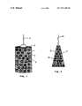
- FIG. 1 shows a side view of the preferred device utilized to concentrate target organisms.
- FIG. 2 shows a side view of another design of the device utilized to concentrate target organisms.
- FIG. 1 shows the preferred embodiment of a device for the separation of the target organisms from a suspension containing a mixture of organisms.
- Beads 1 are made of materials such as nylon, polystyrene or glass. The beads are coated with antibodies to specific microorganisms such as Salmonella, E. coli 0157:H7 and Listeria .
- a cylindrical enclosure 2 is designed to contain the beads. The enclosure is constructed from a frame 3 supporting a grid 4 covering the frame. The grid's pore size is smaller than the size of the beads to assure that the beads stay within the enclosure 2 . However, the pore size is large enough to allow bacteria to freely pass into the enclosure.
- a rod 5 is attached to the upper part of the enclosure. The rod 5 allows the enclosure 2 to move in the solution and for subsequent removal of the device from the solution.
- FIG. 2 shows a different design of the device.
- the beads 11 coated with antibodies are contained in the enclosure 12 made of a grid 13 , shaped like a tea bag.
- a non-wicking string 14 is attached to the upper part of the enclosure 12 allowing movement of the enclosure 12 in the solution, while disallowing the solution containing bacteria to wick up the string.
- the grid's 13 pore size is smaller than the size of the beads to assure that the beads stay within the enclosure. However, the pore size is large enough to allow bacteria to freely pass into the enclosure.
- the food sample to be tested for the presence of the target organism is mixed with the appropriate pre-enrichment broth.
- the pre-enrichment broth is incubated at an appropriate temperature.
- the enclosure 2 is immersed into the broth containing the sample thereby exposing the beads having immobilized thereon monoclonal or polyclonal antibodies to the selected bacteria of interest. This is accomplished by lowering the device 2 into the solution and agitating it for at least 30 minutes and up to several hours. This step allows cell capture by the beads, and the creation of bead-target microbial cell complexes.
- the next step involves the separation of the beads with the bound target cells from the suspension containing the food particles and other mixed flora.
- the device can be inserted into a new growth broth that includes a dye indicator and the changes in the dye characteristics can be utilized to determine presence or absence of the target organism.
- the microorganisms do not need to be detached from the beads since attachment to the beads has no effect on their growth. Therefore cells can continue to multiply in the appropriate medium.
- the beads can be removed from the enclosure and inoculated onto the surface of appropriate selective or differential agar.
- Another approach is to utilize an immunoassay. Most immunoassays require 10 3 –10 5 cells ml ⁇ 1 , therefore the beads should contain enough cells to perform a direct immunoassay.
- this method can be combined with DNA hybridization and amplification techniques such as PCR.
- the method of the invention is particularly characterized by the use of immunological beads contained in an enclosure to select out target microorganisms from the sample.
- the beads must be capable of effectively capturing the target microorganisms from the test sample, while not capturing significant numbers of other organisms that might be present at much higher numbers.
- the antibody used need not be totally specific to the target organism since an additional selection step is available at the end of the assay.
- the antibodies must be oriented with their binding sites outward to allow contact between the binding portion of the antibody and the target organism.
- the size of the beads must be larger than the size of the microorganism, to remain contained in the enclosure, while allowing the target organism to enter the enclosure and attach to the beads.
- the contact time between the beads and the target organism must be long enough to allow strong interaction. Several hours of interaction was found to yield the best results, i.e. the creation of strong interactions to produce high capture efficiency.
- the beads are removed from the solution, by the removal of the enclosure in which they are contained. The enclosure and the beads are washed several times, and the beads are transferred into the detection system.
- the new method and device can be utilized with a large volume of media, to concentrate a target organism, without the need to utilize only a portion of the pre-enrichment broth or a small volume of enrichment broth as required for magnetic beads.
- the invention provides a method and device that is less labor intensive, more rapid and lends itself to automation. Many different designs, for containing the beads during the various steps of the assay, can be utilized.
Abstract
Description
Claims (15)
Priority Applications (1)
| Application Number | Priority Date | Filing Date | Title |
|---|---|---|---|
| US09/763,578 US7071005B1 (en) | 1998-08-24 | 1999-08-23 | Method and device for concentrating selected groups of microorganisms |
Applications Claiming Priority (3)
| Application Number | Priority Date | Filing Date | Title |
|---|---|---|---|
| US9762798P | 1998-08-24 | 1998-08-24 | |
| PCT/US1999/018618 WO2000010702A1 (en) | 1998-08-24 | 1999-08-23 | Method and device for concentrating selected groups of microorganisms |
| US09/763,578 US7071005B1 (en) | 1998-08-24 | 1999-08-23 | Method and device for concentrating selected groups of microorganisms |
Publications (1)
| Publication Number | Publication Date |
|---|---|
| US7071005B1 true US7071005B1 (en) | 2006-07-04 |
Family
ID=36613689
Family Applications (1)
| Application Number | Title | Priority Date | Filing Date |
|---|---|---|---|
| US09/763,578 Expired - Lifetime US7071005B1 (en) | 1998-08-24 | 1999-08-23 | Method and device for concentrating selected groups of microorganisms |
Country Status (1)
| Country | Link |
|---|---|
| US (1) | US7071005B1 (en) |
Cited By (3)
| Publication number | Priority date | Publication date | Assignee | Title |
|---|---|---|---|---|
| US20100273209A1 (en) * | 2008-01-17 | 2010-10-28 | Biolumix Inc. | Co2 optical sensor for detection and enumeration of microorganisms |
| WO2012075508A3 (en) * | 2010-12-03 | 2012-11-08 | Blood Cell Storage, Inc. | Processes for isolating microorganisms |
| US9850457B2 (en) | 2010-05-05 | 2017-12-26 | Neogen Corporation | Microbial growth detector |
Citations (30)
| Publication number | Priority date | Publication date | Assignee | Title |
|---|---|---|---|---|
| US3840345A (en) | 1972-06-23 | 1974-10-08 | Co Ind De Procedes & D Applic | Self agglomerating fluidized bed reacting apparatus |
| US4230685A (en) * | 1979-02-28 | 1980-10-28 | Northwestern University | Method of magnetic separation of cells and the like, and microspheres for use therein |
| US4677055A (en) * | 1982-12-09 | 1987-06-30 | Institut Pasteur | Immunological detection of bacterial pathogens with antibody-containing magnetic gel |
| US4695393A (en) * | 1983-05-12 | 1987-09-22 | Advanced Magnetics Inc. | Magnetic particles for use in separations |
| US4818687A (en) | 1985-02-28 | 1989-04-04 | Massachusetts Institute Of Technology | Affinity column and process for detection of low molecular weight toxic substances |
| US4859611A (en) | 1985-02-28 | 1989-08-22 | Massachusetts Institute Of Technology | Affinity column and process for detection of low molecular weight toxic substances |
| US4931401A (en) | 1988-09-01 | 1990-06-05 | La Societe De Recherche Snc Inc. | Bioreactor |
| US4959307A (en) | 1986-09-05 | 1990-09-25 | Syntex (U.S.A.) Inc. | Immunoseparating strip |
| US4963468A (en) | 1986-09-05 | 1990-10-16 | Syntex (U.S.A.) Inc. | Immunoseparating strip |
| US4983517A (en) * | 1986-08-22 | 1991-01-08 | Battelle Memorial Institute | Reacting materials |
| US5009852A (en) | 1987-12-11 | 1991-04-23 | Charbonnages De France | Cooled fluidization grid |
| US5081035A (en) * | 1988-04-18 | 1992-01-14 | The University Of Michigan | Bioreactor system |
| US5085987A (en) | 1986-09-05 | 1992-02-04 | Syntex (U.S.A.) Inc. | Immunoseparating strip |
| US5085988A (en) | 1986-09-05 | 1992-02-04 | Syntex (U.S.A.) Inc. | Immunoseparating strip |
| US5139933A (en) | 1989-09-26 | 1992-08-18 | Vicam, L.P. | Assay method for detecting listeria |
| US5186824A (en) | 1991-09-04 | 1993-02-16 | Large Scale Biology Corporation | System for solid phase reactions |
| US5260194A (en) | 1986-09-05 | 1993-11-09 | Syntex (U.S.A.) Inc. | Immunoseparating strip |
| US5260193A (en) | 1986-09-05 | 1993-11-09 | Syntex (U.S.A.) Inc. | Immunoseparating strip |
| US5409822A (en) * | 1990-07-03 | 1995-04-25 | Martin Marietta Energy Systems, Inc. | Biparticle fluidized bed reactor |
| WO1995021241A1 (en) * | 1994-02-04 | 1995-08-10 | Difco Laboratories | Microbiological culture bottle, and method of making and using same |
| US5491068A (en) * | 1991-02-14 | 1996-02-13 | Vicam, L.P. | Assay method for detecting the presence of bacteria |
| US5556756A (en) | 1988-01-13 | 1996-09-17 | Nycomed As | Gold sol of less than 5 nm test method and reagent kit therefor |
| US5567615A (en) * | 1993-12-23 | 1996-10-22 | Pall Corporation | Affinity separation method |
| US5705390A (en) * | 1992-05-05 | 1998-01-06 | Interpharm Laboratories Ltd. | Bioreactor |
| US5776710A (en) | 1992-10-30 | 1998-07-07 | Becton Dickinson And Co. | Assay of blood or other biologic samples for target analytes |
| US5866006A (en) * | 1990-07-09 | 1999-02-02 | Upfront Chromatography A/S | Coated single particles and their use in fluid bed chromatography |
| US5998184A (en) * | 1997-10-08 | 1999-12-07 | Unisyn Technologies, Inc. | Basket-type bioreactor |
| US6153373A (en) | 1997-07-01 | 2000-11-28 | Vicam, L.P. | Method for sex determination of mammalian offspring |
| US6395537B1 (en) * | 2001-08-04 | 2002-05-28 | Ruth F. Eden | Double container device and method for detecting and enumerating microorganisms in a sample |
| US20040043116A1 (en) | 2002-08-29 | 2004-03-04 | Cohen Barb A. | Alpha-amylase assay and uses thereof |
-
1999
- 1999-08-23 US US09/763,578 patent/US7071005B1/en not_active Expired - Lifetime
Patent Citations (33)
| Publication number | Priority date | Publication date | Assignee | Title |
|---|---|---|---|---|
| US3840345A (en) | 1972-06-23 | 1974-10-08 | Co Ind De Procedes & D Applic | Self agglomerating fluidized bed reacting apparatus |
| US4230685A (en) * | 1979-02-28 | 1980-10-28 | Northwestern University | Method of magnetic separation of cells and the like, and microspheres for use therein |
| US4677055A (en) * | 1982-12-09 | 1987-06-30 | Institut Pasteur | Immunological detection of bacterial pathogens with antibody-containing magnetic gel |
| US4695393A (en) * | 1983-05-12 | 1987-09-22 | Advanced Magnetics Inc. | Magnetic particles for use in separations |
| US4818687A (en) | 1985-02-28 | 1989-04-04 | Massachusetts Institute Of Technology | Affinity column and process for detection of low molecular weight toxic substances |
| US4859611A (en) | 1985-02-28 | 1989-08-22 | Massachusetts Institute Of Technology | Affinity column and process for detection of low molecular weight toxic substances |
| US4983517A (en) * | 1986-08-22 | 1991-01-08 | Battelle Memorial Institute | Reacting materials |
| US5260194A (en) | 1986-09-05 | 1993-11-09 | Syntex (U.S.A.) Inc. | Immunoseparating strip |
| US5260193A (en) | 1986-09-05 | 1993-11-09 | Syntex (U.S.A.) Inc. | Immunoseparating strip |
| US4959307A (en) | 1986-09-05 | 1990-09-25 | Syntex (U.S.A.) Inc. | Immunoseparating strip |
| US5085987A (en) | 1986-09-05 | 1992-02-04 | Syntex (U.S.A.) Inc. | Immunoseparating strip |
| US5085988A (en) | 1986-09-05 | 1992-02-04 | Syntex (U.S.A.) Inc. | Immunoseparating strip |
| US4963468A (en) | 1986-09-05 | 1990-10-16 | Syntex (U.S.A.) Inc. | Immunoseparating strip |
| US5009852A (en) | 1987-12-11 | 1991-04-23 | Charbonnages De France | Cooled fluidization grid |
| US5556756A (en) | 1988-01-13 | 1996-09-17 | Nycomed As | Gold sol of less than 5 nm test method and reagent kit therefor |
| US5616467A (en) | 1988-01-13 | 1997-04-01 | Nycomed As | Method and kit for analyte detection employing gold-sol bound antibodies |
| US5081035A (en) * | 1988-04-18 | 1992-01-14 | The University Of Michigan | Bioreactor system |
| US4931401A (en) | 1988-09-01 | 1990-06-05 | La Societe De Recherche Snc Inc. | Bioreactor |
| US5139933A (en) | 1989-09-26 | 1992-08-18 | Vicam, L.P. | Assay method for detecting listeria |
| US5409822A (en) * | 1990-07-03 | 1995-04-25 | Martin Marietta Energy Systems, Inc. | Biparticle fluidized bed reactor |
| US5866006A (en) * | 1990-07-09 | 1999-02-02 | Upfront Chromatography A/S | Coated single particles and their use in fluid bed chromatography |
| US5491068A (en) * | 1991-02-14 | 1996-02-13 | Vicam, L.P. | Assay method for detecting the presence of bacteria |
| US5695946A (en) * | 1991-02-14 | 1997-12-09 | Vicam, Lp | Assay method for detecting presence of bacteria |
| US5186824A (en) | 1991-09-04 | 1993-02-16 | Large Scale Biology Corporation | System for solid phase reactions |
| US5705390A (en) * | 1992-05-05 | 1998-01-06 | Interpharm Laboratories Ltd. | Bioreactor |
| US5776710A (en) | 1992-10-30 | 1998-07-07 | Becton Dickinson And Co. | Assay of blood or other biologic samples for target analytes |
| US5567615A (en) * | 1993-12-23 | 1996-10-22 | Pall Corporation | Affinity separation method |
| WO1995021241A1 (en) * | 1994-02-04 | 1995-08-10 | Difco Laboratories | Microbiological culture bottle, and method of making and using same |
| US6153373A (en) | 1997-07-01 | 2000-11-28 | Vicam, L.P. | Method for sex determination of mammalian offspring |
| US6489092B1 (en) | 1997-07-01 | 2002-12-03 | Vicam, L.P. | Method for sex determination of mammalian offspring |
| US5998184A (en) * | 1997-10-08 | 1999-12-07 | Unisyn Technologies, Inc. | Basket-type bioreactor |
| US6395537B1 (en) * | 2001-08-04 | 2002-05-28 | Ruth F. Eden | Double container device and method for detecting and enumerating microorganisms in a sample |
| US20040043116A1 (en) | 2002-08-29 | 2004-03-04 | Cohen Barb A. | Alpha-amylase assay and uses thereof |
Non-Patent Citations (5)
| Title |
|---|
| Lewis et al., "Continuous propionic acid fermentation by immobilized Propionibacterium acidipropionici in a novel packed-bed bioreactor," Biotechnology and Bioengineering, vol. 40, pp. 465-474 (1992). |
| Preliminary Examination Report for International Application PCT/US99/18618. |
| Search Report for International Application PCT/US99/18618. |
| Yang et al., "Kinetics and Stability of GM-CSF Production by Recombinant Yeast Cells Immobilized in a Fibrous-Bed Bioreactor," Biotechnol Prog. vol. 12, pp. 449-456 (1996). |
| Yang et al., "Xanthan Gum Fermentation by Xanthomonas campestris Immobilized in a Novel Centrifugal Fibrous-Bed Bioreactor," Biotechnol Prog. vol. 12, pp. 630-637 (1996). |
Cited By (4)
| Publication number | Priority date | Publication date | Assignee | Title |
|---|---|---|---|---|
| US20100273209A1 (en) * | 2008-01-17 | 2010-10-28 | Biolumix Inc. | Co2 optical sensor for detection and enumeration of microorganisms |
| US9012209B2 (en) | 2008-01-17 | 2015-04-21 | Neogen Corporation | CO2 optical sensor for detection and enumeration of microorganisms |
| US9850457B2 (en) | 2010-05-05 | 2017-12-26 | Neogen Corporation | Microbial growth detector |
| WO2012075508A3 (en) * | 2010-12-03 | 2012-11-08 | Blood Cell Storage, Inc. | Processes for isolating microorganisms |
Similar Documents
| Publication | Publication Date | Title |
|---|---|---|
| US5587286A (en) | Methods and kits for detection of cells in food materials | |
| US4677055A (en) | Immunological detection of bacterial pathogens with antibody-containing magnetic gel | |
| JP5480505B2 (en) | Biological component concentration unit and concentration method | |
| CA2112603C (en) | Assay method for detecting the presence of bacteria | |
| Brewster et al. | Filtration capture and immunoelectrochemical detection for rapid assay of Escherichia coli O157: H7 | |
| US10352834B2 (en) | Method for treating at least one biological sample containing a target microorganism | |
| CN101971032B (en) | Method for the real-time detection of microorganisms in a liquid culture medium by agglutination | |
| CN110229918A (en) | A kind of method and its kit of quick detection Staphylococcus aureus in food | |
| WO1993019372A1 (en) | Method of detecting microorganisms | |
| US20200300737A1 (en) | SAMPLE PREPARATION AND SPECIFIC CAPTURE FOR MULTIPLEX DETECTION OF TARGET ANALYTES (i.e., BACTERIA, VIRUSES, ETC.) | |
| Huang et al. | Evaluation of culture enrichment procedures for use with Salmonella detection immunoassay | |
| Rodrigues et al. | Rapid detection of salmonellas in raw meats using a fluorescent antibody‐microcolony technique | |
| Hanai et al. | Comparison of commercially available kits with standard methods for detection of Salmonella strains in foods | |
| US20130344477A1 (en) | Methods of capturing bindable targets from liquids | |
| AU773645B2 (en) | Method and device for concentrating selected groups of microorganisms | |
| US7071005B1 (en) | Method and device for concentrating selected groups of microorganisms | |
| JP3773633B2 (en) | Analysis method and reagent for E. coli O157 | |
| WU et al. | RAPID PROTOCOL (5.25 H) FOR THE DETECTION OF ESCHERICHIA COLI O157: H7 IN RAW GROUND BEEF BY AN IMMUNO‐CAPTURE SYSTEM (PATHATRIX) IN COMBINATION WITH COLORTRIX AND CT‐SMAC 1 | |
| US20150079597A1 (en) | Method for capturing and concentrating a microorganism in a biological sample | |
| JP2001004631A (en) | Rapid assay for microbe using magnetic bead immobilized antibody | |
| JP4189950B2 (en) | Rapid detection of toxic substances | |
| Porter et al. | Magnetic particle-based separation techniques for monitoring bacteria from natural environments | |
| JPH11248709A (en) | Antigen measuring method | |
| Garcia | New Approaches to Microbial Detection in Brucella Diagnostics | |
| Pincomb | Detection of salmonellae in whole egg |
Legal Events
| Date | Code | Title | Description |
|---|---|---|---|
| AS | Assignment |
Owner name: CENTRUS INTERNATIONAL, INC., MICHIGAN Free format text: ASSIGNMENT OF ASSIGNORS INTEREST;ASSIGNOR:EDEN, RUTH F.;REEL/FRAME:017290/0066 Effective date: 20051115 |
|
| AS | Assignment |
Owner name: CENTRUS INTERNATIONAL, INC., MICHIGAN Free format text: ASSIGNMENT OF ASSIGNORS INTEREST;ASSIGNOR:EDEN, RUTH F.;REEL/FRAME:017118/0965 Effective date: 20051115 |
|
| STCF | Information on status: patent grant |
Free format text: PATENTED CASE |
|
| FEPP | Fee payment procedure |
Free format text: PAYOR NUMBER ASSIGNED (ORIGINAL EVENT CODE: ASPN); ENTITY STATUS OF PATENT OWNER: LARGE ENTITY |
|
| FPAY | Fee payment |
Year of fee payment: 4 |
|
| FEPP | Fee payment procedure |
Free format text: PAT HOLDER NO LONGER CLAIMS SMALL ENTITY STATUS, ENTITY STATUS SET TO UNDISCOUNTED (ORIGINAL EVENT CODE: STOL); ENTITY STATUS OF PATENT OWNER: LARGE ENTITY |
|
| REFU | Refund |
Free format text: REFUND - PAYMENT OF MAINTENANCE FEE, 8TH YR, SMALL ENTITY (ORIGINAL EVENT CODE: R2552); ENTITY STATUS OF PATENT OWNER: LARGE ENTITY |
|
| FEPP | Fee payment procedure |
Free format text: PAYER NUMBER DE-ASSIGNED (ORIGINAL EVENT CODE: RMPN); ENTITY STATUS OF PATENT OWNER: LARGE ENTITY |
|
| FPAY | Fee payment |
Year of fee payment: 8 |
|
| AS | Assignment |
Owner name: NEOGEN CORPORATION, MICHIGAN Free format text: ASSIGNMENT OF ASSIGNORS INTEREST;ASSIGNOR:CENTRUS INTERNATIONAL, INC.;REEL/FRAME:035534/0762 Effective date: 20150429 |
|
| AS | Assignment |
Owner name: NEOGEN CORPORATION, MICHIGAN Free format text: CORRECTIVE ASSIGNMENT TO REMOVE APPL. NO. 11/658,431 PREVIOUSLY RECORDED AT REEL: 035534 FRAME: 0762. ASSIGNOR(S) HEREBY CONFIRMS THE ASSIGNMENT;ASSIGNOR:CENTRUS INTERNATIONAL, INC.;REEL/FRAME:035560/0872 Effective date: 20150429 |
|
| MAFP | Maintenance fee payment |
Free format text: PAYMENT OF MAINTENANCE FEE, 12TH YEAR, LARGE ENTITY (ORIGINAL EVENT CODE: M1553) Year of fee payment: 12 |