US6887470B1 - Protection of endogenous therapeutic peptides from peptidase activity through conjugation to blood components - Google Patents
Protection of endogenous therapeutic peptides from peptidase activity through conjugation to blood components Download PDFInfo
- Publication number
- US6887470B1 US6887470B1 US09/657,276 US65727600A US6887470B1 US 6887470 B1 US6887470 B1 US 6887470B1 US 65727600 A US65727600 A US 65727600A US 6887470 B1 US6887470 B1 US 6887470B1
- Authority
- US
- United States
- Prior art keywords
- fmoc
- peptide
- lys
- peptides
- therapeutic
- Prior art date
- Legal status (The legal status is an assumption and is not a legal conclusion. Google has not performed a legal analysis and makes no representation as to the accuracy of the status listed.)
- Expired - Fee Related
Links
- 108090000765 processed proteins & peptides Proteins 0.000 title claims abstract description 561
- 102000035195 Peptidases Human genes 0.000 title claims abstract description 46
- 108091005804 Peptidases Proteins 0.000 title claims abstract description 46
- 235000019833 protease Nutrition 0.000 title claims abstract description 40
- 230000001225 therapeutic effect Effects 0.000 title claims description 239
- 102000004196 processed proteins & peptides Human genes 0.000 title claims description 238
- 230000000694 effects Effects 0.000 title abstract description 70
- 239000012503 blood component Substances 0.000 title abstract description 51
- 230000021615 conjugation Effects 0.000 title description 13
- 150000001413 amino acids Chemical class 0.000 claims abstract description 124
- 238000000034 method Methods 0.000 claims abstract description 59
- 238000001727 in vivo Methods 0.000 claims abstract description 42
- 125000003275 alpha amino acid group Chemical group 0.000 claims description 52
- 125000003178 carboxy group Chemical group [H]OC(*)=O 0.000 claims description 50
- 125000002924 primary amino group Chemical group [H]N([H])* 0.000 claims description 41
- 230000008878 coupling Effects 0.000 claims description 30
- 238000010168 coupling process Methods 0.000 claims description 30
- 238000005859 coupling reaction Methods 0.000 claims description 30
- 102000009027 Albumins Human genes 0.000 claims description 25
- 108010088751 Albumins Proteins 0.000 claims description 25
- KDXKERNSBIXSRK-UHFFFAOYSA-N Lysine Natural products NCCCCC(N)C(O)=O KDXKERNSBIXSRK-UHFFFAOYSA-N 0.000 claims description 22
- 125000005647 linker group Chemical group 0.000 claims description 18
- 239000004472 Lysine Substances 0.000 claims description 13
- 210000004899 c-terminal region Anatomy 0.000 claims description 13
- 230000015556 catabolic process Effects 0.000 claims description 12
- 238000006731 degradation reaction Methods 0.000 claims description 10
- 125000005439 maleimidyl group Chemical group C1(C=CC(N1*)=O)=O 0.000 claims description 2
- 108091005601 modified peptides Proteins 0.000 abstract description 14
- 230000002633 protecting effect Effects 0.000 abstract description 8
- 229940024606 amino acid Drugs 0.000 description 153
- 235000001014 amino acid Nutrition 0.000 description 149
- -1 aminolethicin Chemical class 0.000 description 129
- ZMXDDKWLCZADIW-UHFFFAOYSA-N N,N-Dimethylformamide Chemical compound CN(C)C=O ZMXDDKWLCZADIW-UHFFFAOYSA-N 0.000 description 101
- 210000004027 cell Anatomy 0.000 description 62
- 229920005989 resin Polymers 0.000 description 57
- 239000011347 resin Substances 0.000 description 57
- 239000012634 fragment Substances 0.000 description 55
- 108090000623 proteins and genes Proteins 0.000 description 46
- 235000018417 cysteine Nutrition 0.000 description 45
- NOESYZHRGYRDHS-UHFFFAOYSA-N insulin Chemical compound N1C(=O)C(NC(=O)C(CCC(N)=O)NC(=O)C(CCC(O)=O)NC(=O)C(C(C)C)NC(=O)C(NC(=O)CN)C(C)CC)CSSCC(C(NC(CO)C(=O)NC(CC(C)C)C(=O)NC(CC=2C=CC(O)=CC=2)C(=O)NC(CCC(N)=O)C(=O)NC(CC(C)C)C(=O)NC(CCC(O)=O)C(=O)NC(CC(N)=O)C(=O)NC(CC=2C=CC(O)=CC=2)C(=O)NC(CSSCC(NC(=O)C(C(C)C)NC(=O)C(CC(C)C)NC(=O)C(CC=2C=CC(O)=CC=2)NC(=O)C(CC(C)C)NC(=O)C(C)NC(=O)C(CCC(O)=O)NC(=O)C(C(C)C)NC(=O)C(CC(C)C)NC(=O)C(CC=2NC=NC=2)NC(=O)C(CO)NC(=O)CNC2=O)C(=O)NCC(=O)NC(CCC(O)=O)C(=O)NC(CCCNC(N)=N)C(=O)NCC(=O)NC(CC=3C=CC=CC=3)C(=O)NC(CC=3C=CC=CC=3)C(=O)NC(CC=3C=CC(O)=CC=3)C(=O)NC(C(C)O)C(=O)N3C(CCC3)C(=O)NC(CCCCN)C(=O)NC(C)C(O)=O)C(=O)NC(CC(N)=O)C(O)=O)=O)NC(=O)C(C(C)CC)NC(=O)C(CO)NC(=O)C(C(C)O)NC(=O)C1CSSCC2NC(=O)C(CC(C)C)NC(=O)C(NC(=O)C(CCC(N)=O)NC(=O)C(CC(N)=O)NC(=O)C(NC(=O)C(N)CC=1C=CC=CC=1)C(C)C)CC1=CN=CN1 NOESYZHRGYRDHS-UHFFFAOYSA-N 0.000 description 44
- 230000028327 secretion Effects 0.000 description 43
- 102000004169 proteins and genes Human genes 0.000 description 40
- 230000004048 modification Effects 0.000 description 39
- 238000012986 modification Methods 0.000 description 39
- 235000018102 proteins Nutrition 0.000 description 39
- 239000000047 product Substances 0.000 description 38
- 210000004369 blood Anatomy 0.000 description 37
- 239000008280 blood Substances 0.000 description 37
- 230000015572 biosynthetic process Effects 0.000 description 36
- 239000000562 conjugate Substances 0.000 description 35
- 239000000199 parathyroid hormone Substances 0.000 description 35
- 125000006239 protecting group Chemical group 0.000 description 33
- 210000001519 tissue Anatomy 0.000 description 33
- 125000000151 cysteine group Chemical class N[C@@H](CS)C(=O)* 0.000 description 32
- 229960001319 parathyroid hormone Drugs 0.000 description 32
- 238000003786 synthesis reaction Methods 0.000 description 32
- 108090000445 Parathyroid hormone Proteins 0.000 description 30
- 102000003982 Parathyroid hormone Human genes 0.000 description 30
- 150000001875 compounds Chemical class 0.000 description 29
- XUJNEKJLAYXESH-REOHCLBHSA-N L-Cysteine Chemical compound SC[C@H](N)C(O)=O XUJNEKJLAYXESH-REOHCLBHSA-N 0.000 description 28
- NQRYJNQNLNOLGT-UHFFFAOYSA-N Piperidine Chemical compound C1CCNCC1 NQRYJNQNLNOLGT-UHFFFAOYSA-N 0.000 description 28
- 125000003088 (fluoren-9-ylmethoxy)carbonyl group Chemical group 0.000 description 27
- NQTADLQHYWFPDB-UHFFFAOYSA-N N-Hydroxysuccinimide Chemical compound ON1C(=O)CCC1=O NQTADLQHYWFPDB-UHFFFAOYSA-N 0.000 description 27
- 230000001965 increasing effect Effects 0.000 description 27
- 229940088597 hormone Drugs 0.000 description 26
- 239000005556 hormone Substances 0.000 description 26
- 229920001184 polypeptide Polymers 0.000 description 25
- 102000005962 receptors Human genes 0.000 description 25
- 108020003175 receptors Proteins 0.000 description 25
- QTBSBXVTEAMEQO-UHFFFAOYSA-N Acetic acid Chemical compound CC(O)=O QTBSBXVTEAMEQO-UHFFFAOYSA-N 0.000 description 24
- JGFZNNIVVJXRND-UHFFFAOYSA-N N,N-Diisopropylethylamine (DIPEA) Chemical compound CCN(C(C)C)C(C)C JGFZNNIVVJXRND-UHFFFAOYSA-N 0.000 description 24
- UMRUUWFGLGNQLI-QFIPXVFZSA-N (2s)-2-(9h-fluoren-9-ylmethoxycarbonylamino)-6-[(2-methylpropan-2-yl)oxycarbonylamino]hexanoic acid Chemical compound C1=CC=C2C(COC(=O)N[C@@H](CCCCNC(=O)OC(C)(C)C)C(O)=O)C3=CC=CC=C3C2=C1 UMRUUWFGLGNQLI-QFIPXVFZSA-N 0.000 description 23
- 108090001061 Insulin Proteins 0.000 description 22
- 102000004877 Insulin Human genes 0.000 description 22
- 229940125396 insulin Drugs 0.000 description 22
- 239000000306 component Substances 0.000 description 21
- 238000004519 manufacturing process Methods 0.000 description 21
- 238000010647 peptide synthesis reaction Methods 0.000 description 21
- 238000011282 treatment Methods 0.000 description 21
- 108050009340 Endothelin Proteins 0.000 description 20
- 238000006243 chemical reaction Methods 0.000 description 20
- XUJNEKJLAYXESH-UHFFFAOYSA-N cysteine Natural products SCC(N)C(O)=O XUJNEKJLAYXESH-UHFFFAOYSA-N 0.000 description 20
- 239000003814 drug Substances 0.000 description 20
- CBPJQFCAFFNICX-IBGZPJMESA-N (2s)-2-(9h-fluoren-9-ylmethoxycarbonylamino)-4-methylpentanoic acid Chemical compound C1=CC=C2C(COC(=O)N[C@@H](CC(C)C)C(O)=O)C3=CC=CC=C3C2=C1 CBPJQFCAFFNICX-IBGZPJMESA-N 0.000 description 19
- HNICLNKVURBTKV-NDEPHWFRSA-N (2s)-5-[[amino-[(2,2,4,6,7-pentamethyl-3h-1-benzofuran-5-yl)sulfonylamino]methylidene]amino]-2-(9h-fluoren-9-ylmethoxycarbonylamino)pentanoic acid Chemical compound C12=CC=CC=C2C2=CC=CC=C2C1COC(=O)N[C@H](C(O)=O)CCCN=C(N)NS(=O)(=O)C1=C(C)C(C)=C2OC(C)(C)CC2=C1C HNICLNKVURBTKV-NDEPHWFRSA-N 0.000 description 19
- YMWUJEATGCHHMB-UHFFFAOYSA-N Dichloromethane Chemical compound ClCCl YMWUJEATGCHHMB-UHFFFAOYSA-N 0.000 description 19
- 102000002045 Endothelin Human genes 0.000 description 19
- KDXKERNSBIXSRK-YFKPBYRVSA-N L-lysine Chemical compound NCCCC[C@H](N)C(O)=O KDXKERNSBIXSRK-YFKPBYRVSA-N 0.000 description 19
- 108010022152 Corticotropin-Releasing Hormone Proteins 0.000 description 18
- 102000012289 Corticotropin-Releasing Hormone Human genes 0.000 description 18
- SJRJJKPEHAURKC-UHFFFAOYSA-N N-Methylmorpholine Chemical compound CN1CCOCC1 SJRJJKPEHAURKC-UHFFFAOYSA-N 0.000 description 18
- 230000027455 binding Effects 0.000 description 18
- NSQLIUXCMFBZME-MPVJKSABSA-N carperitide Chemical class C([C@H]1C(=O)NCC(=O)NCC(=O)N[C@@H](CCCNC(N)=N)C(=O)N[C@@H](CCSC)C(=O)N[C@@H](CC(O)=O)C(=O)N[C@@H](CCCNC(N)=N)C(=O)N[C@H](C(NCC(=O)N[C@@H](C)C(=O)N[C@@H](CCC(N)=O)C(=O)N[C@@H](CO)C(=O)NCC(=O)N[C@@H](CC(C)C)C(=O)NCC(=O)N[C@@H](CSSC[C@@H](C(=O)N1)NC(=O)[C@H](CO)NC(=O)[C@H](CO)NC(=O)[C@H](CCCNC(N)=N)NC(=O)[C@H](CCCNC(N)=N)NC(=O)[C@H](CC(C)C)NC(=O)[C@@H](N)CO)C(=O)N[C@@H](CC(N)=O)C(=O)N[C@@H](CO)C(=O)N[C@@H](CC=1C=CC=CC=1)C(=O)N[C@@H](CCCNC(N)=N)C(=O)N[C@@H](CC=1C=CC(O)=CC=1)C(O)=O)=O)[C@@H](C)CC)C1=CC=CC=C1 NSQLIUXCMFBZME-MPVJKSABSA-N 0.000 description 18
- 230000001105 regulatory effect Effects 0.000 description 18
- 101800001288 Atrial natriuretic factor Proteins 0.000 description 17
- 239000000055 Corticotropin-Releasing Hormone Substances 0.000 description 17
- DTQVDTLACAAQTR-UHFFFAOYSA-N Trifluoroacetic acid Chemical compound OC(=O)C(F)(F)F DTQVDTLACAAQTR-UHFFFAOYSA-N 0.000 description 17
- 230000009471 action Effects 0.000 description 17
- 238000003776 cleavage reaction Methods 0.000 description 17
- 229960000258 corticotropin Drugs 0.000 description 17
- 230000006870 function Effects 0.000 description 17
- 230000007017 scission Effects 0.000 description 17
- 239000000243 solution Substances 0.000 description 17
- XLYOFNOQVPJJNP-UHFFFAOYSA-N water Substances O XLYOFNOQVPJJNP-UHFFFAOYSA-N 0.000 description 17
- ZPGDWQNBZYOZTI-SFHVURJKSA-N (2s)-1-(9h-fluoren-9-ylmethoxycarbonyl)pyrrolidine-2-carboxylic acid Chemical compound OC(=O)[C@@H]1CCCN1C(=O)OCC1C2=CC=CC=C2C2=CC=CC=C21 ZPGDWQNBZYOZTI-SFHVURJKSA-N 0.000 description 16
- JAUKCFULLJFBFN-VWLOTQADSA-N (2s)-2-(9h-fluoren-9-ylmethoxycarbonylamino)-3-[4-[(2-methylpropan-2-yl)oxy]phenyl]propanoic acid Chemical compound C1=CC(OC(C)(C)C)=CC=C1C[C@@H](C(O)=O)NC(=O)OCC1C2=CC=CC=C2C2=CC=CC=C21 JAUKCFULLJFBFN-VWLOTQADSA-N 0.000 description 16
- NDKDFTQNXLHCGO-UHFFFAOYSA-N 2-(9h-fluoren-9-ylmethoxycarbonylamino)acetic acid Chemical compound C1=CC=C2C(COC(=O)NCC(=O)O)C3=CC=CC=C3C2=C1 NDKDFTQNXLHCGO-UHFFFAOYSA-N 0.000 description 16
- 239000000275 Adrenocorticotropic Hormone Substances 0.000 description 16
- 101800000414 Corticotropin Proteins 0.000 description 16
- 108010052343 Gastrins Proteins 0.000 description 16
- 230000004913 activation Effects 0.000 description 16
- 238000001994 activation Methods 0.000 description 16
- IDLFZVILOHSSID-OVLDLUHVSA-N corticotropin Chemical compound C([C@@H](C(=O)N[C@@H](CO)C(=O)N[C@@H](CCSC)C(=O)N[C@@H](CCC(O)=O)C(=O)N[C@@H](CC=1NC=NC=1)C(=O)N[C@@H](CC=1C=CC=CC=1)C(=O)N[C@@H](CCCNC(N)=N)C(=O)N[C@@H](CC=1C2=CC=CC=C2NC=1)C(=O)NCC(=O)N[C@@H](CCCCN)C(=O)N1[C@@H](CCC1)C(=O)N[C@@H](C(C)C)C(=O)NCC(=O)N[C@@H](CCCCN)C(=O)N[C@@H](CCCCN)C(=O)N[C@@H](CCCNC(N)=N)C(=O)N[C@@H](CCCNC(N)=N)C(=O)N1[C@@H](CCC1)C(=O)N[C@@H](C(C)C)C(=O)N[C@@H](CCCCN)C(=O)N[C@@H](C(C)C)C(=O)N[C@@H](CC=1C=CC(O)=CC=1)C(=O)N1[C@@H](CCC1)C(=O)N[C@@H](CC(N)=O)C(=O)NCC(=O)N[C@@H](C)C(=O)N[C@@H](CCC(O)=O)C(=O)N[C@@H](CC(O)=O)C(=O)N[C@@H](CCC(O)=O)C(=O)N[C@@H](CO)C(=O)N[C@@H](C)C(=O)N[C@@H](CCC(O)=O)C(=O)N[C@@H](C)C(=O)N[C@@H](CC=1C=CC=CC=1)C(=O)N1[C@@H](CCC1)C(=O)N[C@@H](CC(C)C)C(=O)N[C@@H](CCC(O)=O)C(=O)N[C@@H](CC=1C=CC=CC=1)C(O)=O)NC(=O)[C@@H](N)CO)C1=CC=C(O)C=C1 IDLFZVILOHSSID-OVLDLUHVSA-N 0.000 description 16
- 239000003112 inhibitor Substances 0.000 description 16
- 239000007787 solid Substances 0.000 description 16
- NHXLMOGPVYXJNR-ATOGVRKGSA-N somatostatin Chemical compound C([C@H]1C(=O)N[C@H](C(N[C@@H](CO)C(=O)N[C@@H](CSSC[C@@H](C(=O)N[C@@H](CCCCN)C(=O)N[C@@H](CC(N)=O)C(=O)N[C@@H](CC=2C=CC=CC=2)C(=O)N[C@@H](CC=2C=CC=CC=2)C(=O)N[C@@H](CC=2C3=CC=CC=C3NC=2)C(=O)N[C@@H](CCCCN)C(=O)N[C@H](C(=O)N1)[C@@H](C)O)NC(=O)CNC(=O)[C@H](C)N)C(O)=O)=O)[C@H](O)C)C1=CC=CC=C1 NHXLMOGPVYXJNR-ATOGVRKGSA-N 0.000 description 16
- 239000000126 substance Substances 0.000 description 16
- 229910001868 water Inorganic materials 0.000 description 16
- 102000005157 Somatostatin Human genes 0.000 description 15
- 108010056088 Somatostatin Proteins 0.000 description 15
- 208000037265 diseases, disorders, signs and symptoms Diseases 0.000 description 15
- 238000004128 high performance liquid chromatography Methods 0.000 description 15
- 238000002372 labelling Methods 0.000 description 15
- 229960000553 somatostatin Drugs 0.000 description 15
- 125000003396 thiol group Chemical group [H]S* 0.000 description 15
- 102400000921 Gastrin Human genes 0.000 description 14
- 241000282414 Homo sapiens Species 0.000 description 14
- PEEHTFAAVSWFBL-UHFFFAOYSA-N Maleimide Chemical compound O=C1NC(=O)C=C1 PEEHTFAAVSWFBL-UHFFFAOYSA-N 0.000 description 14
- 125000003277 amino group Chemical group 0.000 description 14
- AOXOCDRNSPFDPE-UKEONUMOSA-N chembl413654 Chemical compound C([C@H](C(=O)NCC(=O)N[C@H](CC=1C2=CC=CC=C2NC=1)C(=O)N[C@H](CCSC)C(=O)N[C@H](CC(O)=O)C(=O)N[C@H](CC=1C=CC=CC=1)C(N)=O)NC(=O)[C@@H](C)NC(=O)[C@@H](CCC(O)=O)NC(=O)[C@@H](CCC(O)=O)NC(=O)[C@@H](CCC(O)=O)NC(=O)[C@H](CCC(O)=O)NC(=O)[C@H](CCC(O)=O)NC(=O)[C@H](CC(C)C)NC(=O)[C@H](CC=1C2=CC=CC=C2NC=1)NC(=O)[C@H]1N(CCC1)C(=O)CNC(=O)[C@@H](N)CCC(O)=O)C1=CC=C(O)C=C1 AOXOCDRNSPFDPE-UKEONUMOSA-N 0.000 description 14
- 125000000524 functional group Chemical group 0.000 description 14
- 238000001556 precipitation Methods 0.000 description 14
- 239000007790 solid phase Substances 0.000 description 14
- FODJWPHPWBKDON-IBGZPJMESA-N (2s)-2-(9h-fluoren-9-ylmethoxycarbonylamino)-4-[(2-methylpropan-2-yl)oxy]-4-oxobutanoic acid Chemical compound C1=CC=C2C(COC(=O)N[C@@H](CC(=O)OC(C)(C)C)C(O)=O)C3=CC=CC=C3C2=C1 FODJWPHPWBKDON-IBGZPJMESA-N 0.000 description 13
- 102400001282 Atrial natriuretic peptide Human genes 0.000 description 13
- 101800001890 Atrial natriuretic peptide Proteins 0.000 description 13
- KBZOIRJILGZLEJ-LGYYRGKSSA-N argipressin Chemical compound C([C@H]1C(=O)N[C@@H](CCC(N)=O)C(=O)N[C@@H](CC(N)=O)C(=O)N[C@@H](CSSC[C@@H](C(N[C@@H](CC=2C=CC(O)=CC=2)C(=O)N1)=O)N)C(=O)N1[C@@H](CCC1)C(=O)N[C@@H](CCCN=C(N)N)C(=O)NCC(N)=O)C1=CC=CC=C1 KBZOIRJILGZLEJ-LGYYRGKSSA-N 0.000 description 13
- 210000004185 liver Anatomy 0.000 description 13
- 210000002966 serum Anatomy 0.000 description 13
- 229960003726 vasopressin Drugs 0.000 description 13
- HEDRZPFGACZZDS-UHFFFAOYSA-N Chloroform Chemical compound ClC(Cl)Cl HEDRZPFGACZZDS-UHFFFAOYSA-N 0.000 description 12
- 108700020797 Parathyroid Hormone-Related Proteins 0.000 description 12
- 102000043299 Parathyroid hormone-related Human genes 0.000 description 12
- ISWSIDIOOBJBQZ-UHFFFAOYSA-N Phenol Chemical compound OC1=CC=CC=C1 ISWSIDIOOBJBQZ-UHFFFAOYSA-N 0.000 description 12
- 102000002852 Vasopressins Human genes 0.000 description 12
- 108010004977 Vasopressins Proteins 0.000 description 12
- 230000006907 apoptotic process Effects 0.000 description 12
- 210000004556 brain Anatomy 0.000 description 12
- 239000003795 chemical substances by application Substances 0.000 description 12
- 238000010511 deprotection reaction Methods 0.000 description 12
- 210000003016 hypothalamus Anatomy 0.000 description 12
- 229960003646 lysine Drugs 0.000 description 12
- 239000002243 precursor Substances 0.000 description 12
- 238000002360 preparation method Methods 0.000 description 12
- 230000004044 response Effects 0.000 description 12
- 238000004007 reversed phase HPLC Methods 0.000 description 12
- 229940124597 therapeutic agent Drugs 0.000 description 12
- QWXZOFZKSQXPDC-NSHDSACASA-N (2s)-2-(9h-fluoren-9-ylmethoxycarbonylamino)propanoic acid Chemical compound C1=CC=C2C(COC(=O)N[C@@H](C)C(O)=O)C3=CC=CC=C3C2=C1 QWXZOFZKSQXPDC-NSHDSACASA-N 0.000 description 11
- 150000003923 2,5-pyrrolediones Chemical class 0.000 description 11
- 125000000539 amino acid group Chemical group 0.000 description 11
- 229960004666 glucagon Drugs 0.000 description 11
- 210000003734 kidney Anatomy 0.000 description 11
- 210000004379 membrane Anatomy 0.000 description 11
- 239000012528 membrane Substances 0.000 description 11
- 210000000496 pancreas Anatomy 0.000 description 11
- 239000000758 substrate Substances 0.000 description 11
- 125000000999 tert-butyl group Chemical group [H]C([H])([H])C(*)(C([H])([H])[H])C([H])([H])[H] 0.000 description 11
- 150000003573 thiols Chemical class 0.000 description 11
- JDDWRLPTKIOUOF-UHFFFAOYSA-N 9h-fluoren-9-ylmethyl n-[[4-[2-[bis(4-methylphenyl)methylamino]-2-oxoethoxy]phenyl]-(2,4-dimethoxyphenyl)methyl]carbamate Chemical compound COC1=CC(OC)=CC=C1C(C=1C=CC(OCC(=O)NC(C=2C=CC(C)=CC=2)C=2C=CC(C)=CC=2)=CC=1)NC(=O)OCC1C2=CC=CC=C2C2=CC=CC=C21 JDDWRLPTKIOUOF-UHFFFAOYSA-N 0.000 description 10
- 102000044503 Antimicrobial Peptides Human genes 0.000 description 10
- 108700042778 Antimicrobial Peptides Proteins 0.000 description 10
- 108010065372 Dynorphins Proteins 0.000 description 10
- 108090000790 Enzymes Proteins 0.000 description 10
- 102000004190 Enzymes Human genes 0.000 description 10
- 102000051325 Glucagon Human genes 0.000 description 10
- 108060003199 Glucagon Proteins 0.000 description 10
- 108090000723 Insulin-Like Growth Factor I Proteins 0.000 description 10
- 102000004218 Insulin-Like Growth Factor I Human genes 0.000 description 10
- 108010041872 Islet Amyloid Polypeptide Proteins 0.000 description 10
- 102000036770 Islet Amyloid Polypeptide Human genes 0.000 description 10
- 206010028980 Neoplasm Diseases 0.000 description 10
- 108090000189 Neuropeptides Proteins 0.000 description 10
- 102100024622 Proenkephalin-B Human genes 0.000 description 10
- 102000007562 Serum Albumin Human genes 0.000 description 10
- 108010071390 Serum Albumin Proteins 0.000 description 10
- 210000001744 T-lymphocyte Anatomy 0.000 description 10
- 238000011161 development Methods 0.000 description 10
- 230000018109 developmental process Effects 0.000 description 10
- 229940088598 enzyme Drugs 0.000 description 10
- 239000012530 fluid Substances 0.000 description 10
- MASNOZXLGMXCHN-ZLPAWPGGSA-N glucagon Chemical compound C([C@@H](C(=O)N[C@H](C(=O)N[C@@H](CCC(N)=O)C(=O)N[C@@H](CC=1C2=CC=CC=C2NC=1)C(=O)N[C@@H](CC(C)C)C(=O)N[C@@H](CCSC)C(=O)N[C@@H](CC(N)=O)C(=O)N[C@@H]([C@@H](C)O)C(O)=O)C(C)C)NC(=O)[C@H](CC(O)=O)NC(=O)[C@H](CCC(N)=O)NC(=O)[C@H](C)NC(=O)[C@H](CCCNC(N)=N)NC(=O)[C@H](CCCNC(N)=N)NC(=O)[C@H](CO)NC(=O)[C@H](CC(O)=O)NC(=O)[C@H](CC(C)C)NC(=O)[C@H](CC=1C=CC(O)=CC=1)NC(=O)[C@H](CCCCN)NC(=O)[C@H](CO)NC(=O)[C@H](CC=1C=CC(O)=CC=1)NC(=O)[C@H](CC(O)=O)NC(=O)[C@H](CO)NC(=O)[C@@H](NC(=O)[C@H](CC=1C=CC=CC=1)NC(=O)[C@@H](NC(=O)CNC(=O)[C@H](CCC(N)=O)NC(=O)[C@H](CO)NC(=O)[C@@H](N)CC=1NC=NC=1)[C@@H](C)O)[C@@H](C)O)C1=CC=CC=C1 MASNOZXLGMXCHN-ZLPAWPGGSA-N 0.000 description 10
- 239000000122 growth hormone Substances 0.000 description 10
- 230000005764 inhibitory process Effects 0.000 description 10
- 125000003588 lysine group Chemical group [H]N([H])C([H])([H])C([H])([H])C([H])([H])C([H])([H])C([H])(N([H])[H])C(*)=O 0.000 description 10
- 239000000203 mixture Substances 0.000 description 10
- URPYMXQQVHTUDU-OFGSCBOVSA-N nucleopeptide y Chemical compound C([C@@H](C(=O)N[C@@H]([C@@H](C)CC)C(=O)N[C@@H](CC(N)=O)C(=O)N[C@@H](CC(C)C)C(=O)N[C@@H]([C@@H](C)CC)C(=O)N[C@@H]([C@@H](C)O)C(=O)N[C@@H](CCCNC(N)=N)C(=O)N[C@@H](CCC(N)=O)C(=O)N[C@@H](CCCNC(N)=N)C(=O)N[C@@H](CC=1C=CC(O)=CC=1)C(N)=O)NC(=O)[C@H](CC=1NC=NC=1)NC(=O)[C@H](CCCNC(N)=N)NC(=O)[C@H](CC(C)C)NC(=O)[C@H](C)NC(=O)[C@H](CO)NC(=O)[C@H](CC=1C=CC(O)=CC=1)NC(=O)[C@H](CC=1C=CC(O)=CC=1)NC(=O)[C@H](CCCNC(N)=N)NC(=O)[C@H](C)NC(=O)[C@H](CC(C)C)NC(=O)[C@H](CC(O)=O)NC(=O)[C@H](CCC(O)=O)NC(=O)[C@H](C)NC(=O)[C@H]1N(CCC1)C(=O)[C@H](C)NC(=O)[C@H](CC(O)=O)NC(=O)[C@H](CCC(O)=O)NC(=O)CNC(=O)[C@H]1N(CCC1)C(=O)[C@H](CC(N)=O)NC(=O)[C@H](CC(O)=O)NC(=O)[C@H]1N(CCC1)C(=O)[C@H](CCCCN)NC(=O)[C@H](CO)NC(=O)[C@H]1N(CCC1)C(=O)[C@@H](N)CC=1C=CC(O)=CC=1)C1=CC=C(O)C=C1 URPYMXQQVHTUDU-OFGSCBOVSA-N 0.000 description 10
- 210000000056 organ Anatomy 0.000 description 10
- 229960002101 secretin Drugs 0.000 description 10
- 230000002992 thymic effect Effects 0.000 description 10
- KJYAFJQCGPUXJY-UMSFTDKQSA-N (2s)-2-(9h-fluoren-9-ylmethoxycarbonylamino)-4-oxo-4-(tritylamino)butanoic acid Chemical compound C([C@@H](C(=O)O)NC(=O)OCC1C2=CC=CC=C2C2=CC=CC=C21)C(=O)NC(C=1C=CC=CC=1)(C=1C=CC=CC=1)C1=CC=CC=C1 KJYAFJQCGPUXJY-UMSFTDKQSA-N 0.000 description 9
- QXVFEIPAZSXRGM-DJJJIMSYSA-N (2s,3s)-2-(9h-fluoren-9-ylmethoxycarbonylamino)-3-methylpentanoic acid Chemical compound C1=CC=C2C(COC(=O)N[C@@H]([C@@H](C)CC)C(O)=O)C3=CC=CC=C3C2=C1 QXVFEIPAZSXRGM-DJJJIMSYSA-N 0.000 description 9
- CUKWUWBLQQDQAC-VEQWQPCFSA-N (3s)-3-amino-4-[[(2s)-1-[[(2s)-1-[[(2s)-1-[[(2s,3s)-1-[[(2s)-1-[(2s)-2-[[(1s)-1-carboxyethyl]carbamoyl]pyrrolidin-1-yl]-3-(1h-imidazol-5-yl)-1-oxopropan-2-yl]amino]-3-methyl-1-oxopentan-2-yl]amino]-3-(4-hydroxyphenyl)-1-oxopropan-2-yl]amino]-3-methyl-1-ox Chemical compound C([C@@H](C(=O)N[C@@H]([C@@H](C)CC)C(=O)N[C@@H](CC=1NC=NC=1)C(=O)N1[C@@H](CCC1)C(=O)N[C@@H](C)C(O)=O)NC(=O)[C@@H](NC(=O)[C@H](CCCN=C(N)N)NC(=O)[C@@H](N)CC(O)=O)C(C)C)C1=CC=C(O)C=C1 CUKWUWBLQQDQAC-VEQWQPCFSA-N 0.000 description 9
- 101800000733 Angiotensin-2 Proteins 0.000 description 9
- 102400000345 Angiotensin-2 Human genes 0.000 description 9
- 101800004490 Endothelin-1 Proteins 0.000 description 9
- 108010051696 Growth Hormone Proteins 0.000 description 9
- 101000715592 Homo sapiens Cocaine- and amphetamine-regulated transcript protein Proteins 0.000 description 9
- 241000725303 Human immunodeficiency virus Species 0.000 description 9
- 101710151321 Melanostatin Proteins 0.000 description 9
- 241001465754 Metazoa Species 0.000 description 9
- 102400000064 Neuropeptide Y Human genes 0.000 description 9
- ZMANZCXQSJIPKH-UHFFFAOYSA-N Triethylamine Chemical compound CCN(CC)CC ZMANZCXQSJIPKH-UHFFFAOYSA-N 0.000 description 9
- GXBMIBRIOWHPDT-UHFFFAOYSA-N Vasopressin Natural products N1C(=O)C(CC=2C=C(O)C=CC=2)NC(=O)C(N)CSSCC(C(=O)N2C(CCC2)C(=O)NC(CCCN=C(N)N)C(=O)NCC(N)=O)NC(=O)C(CC(N)=O)NC(=O)C(CCC(N)=O)NC(=O)C1CC1=CC=CC=C1 GXBMIBRIOWHPDT-UHFFFAOYSA-N 0.000 description 9
- 239000002253 acid Substances 0.000 description 9
- 150000001412 amines Chemical class 0.000 description 9
- 229950006323 angiotensin ii Drugs 0.000 description 9
- 230000036772 blood pressure Effects 0.000 description 9
- 150000001732 carboxylic acid derivatives Chemical class 0.000 description 9
- 210000000170 cell membrane Anatomy 0.000 description 9
- 210000003169 central nervous system Anatomy 0.000 description 9
- BJDCWCLMFKKGEE-CMDXXVQNSA-N chembl252518 Chemical compound C([C@@](OO1)(C)O2)C[C@H]3[C@H](C)CC[C@@H]4[C@@]31[C@@H]2O[C@H](O)[C@@H]4C BJDCWCLMFKKGEE-CMDXXVQNSA-N 0.000 description 9
- 230000002496 gastric effect Effects 0.000 description 9
- 210000001035 gastrointestinal tract Anatomy 0.000 description 9
- 239000003102 growth factor Substances 0.000 description 9
- 230000001817 pituitary effect Effects 0.000 description 9
- 229940097325 prolactin Drugs 0.000 description 9
- OWMZNFCDEHGFEP-NFBCVYDUSA-N secretin human Chemical compound C([C@@H](C(=O)N[C@H](C(=O)N[C@@H](CO)C(=O)N[C@@H](CCC(O)=O)C(=O)N[C@@H](CC(C)C)C(=O)N[C@@H](CO)C(=O)N[C@@H](CCCNC(N)=N)C(=O)N[C@@H](CC(C)C)C(=O)N[C@@H](CCCNC(N)=N)C(=O)N[C@@H](CCC(O)=O)C(=O)NCC(=O)N[C@@H](C)C(=O)N[C@@H](CCCNC(N)=N)C(=O)N[C@@H](CC(C)C)C(=O)N[C@@H](CCC(N)=O)C(=O)N[C@@H](CCCNC(N)=N)C(=O)N[C@@H](CC(C)C)C(=O)N[C@@H](CC(C)C)C(=O)N[C@@H](CCC(N)=O)C(=O)NCC(=O)N[C@@H](CC(C)C)C(=O)N[C@@H](C(C)C)C(N)=O)[C@@H](C)O)NC(=O)[C@@H](NC(=O)CNC(=O)[C@H](CC(O)=O)NC(=O)[C@H](CO)NC(=O)[C@@H](N)CC=1NC=NC=1)[C@@H](C)O)C1=CC=CC=C1 OWMZNFCDEHGFEP-NFBCVYDUSA-N 0.000 description 9
- 238000006467 substitution reaction Methods 0.000 description 9
- 239000003053 toxin Substances 0.000 description 9
- 231100000765 toxin Toxicity 0.000 description 9
- 108700012359 toxins Proteins 0.000 description 9
- MDNSLPICAWKNAG-UHFFFAOYSA-N 2-(2,5-dioxopyrrol-1-yl)propanoic acid Chemical compound OC(=O)C(C)N1C(=O)C=CC1=O MDNSLPICAWKNAG-UHFFFAOYSA-N 0.000 description 8
- 102000012421 C-Type Natriuretic Peptide Human genes 0.000 description 8
- 101800000060 C-type natriuretic peptide Proteins 0.000 description 8
- 102100035932 Cocaine- and amphetamine-regulated transcript protein Human genes 0.000 description 8
- 102000008130 Cyclic AMP-Dependent Protein Kinases Human genes 0.000 description 8
- 108010049894 Cyclic AMP-Dependent Protein Kinases Proteins 0.000 description 8
- IVOMOUWHDPKRLL-KQYNXXCUSA-N Cyclic adenosine monophosphate Chemical compound C([C@H]1O2)OP(O)(=O)O[C@H]1[C@@H](O)[C@@H]2N1C(N=CN=C2N)=C2N=C1 IVOMOUWHDPKRLL-KQYNXXCUSA-N 0.000 description 8
- WQZGKKKJIJFFOK-GASJEMHNSA-N Glucose Natural products OC[C@H]1OC(O)[C@H](O)[C@@H](O)[C@@H]1O WQZGKKKJIJFFOK-GASJEMHNSA-N 0.000 description 8
- NTYJJOPFIAHURM-UHFFFAOYSA-N Histamine Chemical compound NCCC1=CN=CN1 NTYJJOPFIAHURM-UHFFFAOYSA-N 0.000 description 8
- 101001090065 Homo sapiens Peroxiredoxin-2 Proteins 0.000 description 8
- MWUXSHHQAYIFBG-UHFFFAOYSA-N Nitric oxide Chemical compound O=[N] MWUXSHHQAYIFBG-UHFFFAOYSA-N 0.000 description 8
- 108090000573 Osteocalcin Proteins 0.000 description 8
- 102000007079 Peptide Fragments Human genes 0.000 description 8
- 108010033276 Peptide Fragments Proteins 0.000 description 8
- 102100034763 Peroxiredoxin-2 Human genes 0.000 description 8
- 108010057464 Prolactin Proteins 0.000 description 8
- 102000003946 Prolactin Human genes 0.000 description 8
- 102100037505 Secretin Human genes 0.000 description 8
- 108010086019 Secretin Proteins 0.000 description 8
- 102100038803 Somatotropin Human genes 0.000 description 8
- IVOMOUWHDPKRLL-UHFFFAOYSA-N UNPD107823 Natural products O1C2COP(O)(=O)OC2C(O)C1N1C(N=CN=C2N)=C2N=C1 IVOMOUWHDPKRLL-UHFFFAOYSA-N 0.000 description 8
- 241000700605 Viruses Species 0.000 description 8
- 125000001584 benzyloxycarbonyl group Chemical group C(=O)(OCC1=CC=CC=C1)* 0.000 description 8
- 230000033228 biological regulation Effects 0.000 description 8
- 210000000988 bone and bone Anatomy 0.000 description 8
- 229940095074 cyclic amp Drugs 0.000 description 8
- 238000010586 diagram Methods 0.000 description 8
- 150000002148 esters Chemical class 0.000 description 8
- 238000004108 freeze drying Methods 0.000 description 8
- 239000008103 glucose Substances 0.000 description 8
- 230000012010 growth Effects 0.000 description 8
- 125000002887 hydroxy group Chemical group [H]O* 0.000 description 8
- 230000002401 inhibitory effect Effects 0.000 description 8
- 230000000144 pharmacologic effect Effects 0.000 description 8
- 238000002953 preparative HPLC Methods 0.000 description 8
- 238000005556 structure-activity relationship Methods 0.000 description 8
- UGNIYGNGCNXHTR-SFHVURJKSA-N (2s)-2-(9h-fluoren-9-ylmethoxycarbonylamino)-3-methylbutanoic acid Chemical compound C1=CC=C2C(COC(=O)N[C@@H](C(C)C)C(O)=O)C3=CC=CC=C3C2=C1 UGNIYGNGCNXHTR-SFHVURJKSA-N 0.000 description 7
- LZOLWEQBVPVDPR-VLIAUNLRSA-N (2s,3r)-2-(9h-fluoren-9-ylmethoxycarbonylamino)-3-[(2-methylpropan-2-yl)oxy]butanoic acid Chemical compound C1=CC=C2C(COC(=O)N[C@@H]([C@H](OC(C)(C)C)C)C(O)=O)C3=CC=CC=C3C2=C1 LZOLWEQBVPVDPR-VLIAUNLRSA-N 0.000 description 7
- 102100022704 Amyloid-beta precursor protein Human genes 0.000 description 7
- 102000004506 Blood Proteins Human genes 0.000 description 7
- 108010017384 Blood Proteins Proteins 0.000 description 7
- RTZKZFJDLAIYFH-UHFFFAOYSA-N Diethyl ether Chemical compound CCOCC RTZKZFJDLAIYFH-UHFFFAOYSA-N 0.000 description 7
- 102400000686 Endothelin-1 Human genes 0.000 description 7
- 108010067306 Fibronectins Proteins 0.000 description 7
- 102000016359 Fibronectins Human genes 0.000 description 7
- 239000000579 Gonadotropin-Releasing Hormone Substances 0.000 description 7
- 108020001621 Natriuretic Peptide Proteins 0.000 description 7
- 102000004571 Natriuretic peptide Human genes 0.000 description 7
- 102000004067 Osteocalcin Human genes 0.000 description 7
- 210000004404 adrenal cortex Anatomy 0.000 description 7
- 150000001408 amides Chemical class 0.000 description 7
- 125000001797 benzyl group Chemical group [H]C1=C([H])C([H])=C(C([H])=C1[H])C([H])([H])* 0.000 description 7
- 230000004071 biological effect Effects 0.000 description 7
- 210000001772 blood platelet Anatomy 0.000 description 7
- 230000001413 cellular effect Effects 0.000 description 7
- 206010012601 diabetes mellitus Diseases 0.000 description 7
- 208000035475 disorder Diseases 0.000 description 7
- 229940079593 drug Drugs 0.000 description 7
- ZUBDGKVDJUIMQQ-UBFCDGJISA-N endothelin-1 Chemical compound C([C@@H](C(=O)N[C@@H](CC(C)C)C(=O)N[C@@H](CC(O)=O)C(=O)N[C@@H]([C@@H](C)CC)C(=O)N[C@@H]([C@@H](C)CC)C(=O)N[C@@H](CC=1C2=CC=CC=C2NC=1)C(O)=O)NC(=O)[C@H]1NC(=O)[C@H](CC=2C=CC=CC=2)NC(=O)[C@@H](CC=2C=CC(O)=CC=2)NC(=O)[C@H](C(C)C)NC(=O)[C@H]2CSSC[C@@H](C(N[C@H](CO)C(=O)N[C@@H](CO)C(=O)N[C@H](CC(C)C)C(=O)N[C@@H](CCSC)C(=O)N[C@H](CC(O)=O)C(=O)N[C@@H](CCCCN)C(=O)N[C@@H](CCC(O)=O)C(=O)N2)=O)NC(=O)[C@@H](CO)NC(=O)[C@H](N)CSSC1)C1=CNC=N1 ZUBDGKVDJUIMQQ-UBFCDGJISA-N 0.000 description 7
- 210000003743 erythrocyte Anatomy 0.000 description 7
- NPZTUJOABDZTLV-UHFFFAOYSA-N hydroxybenzotriazole Substances O=C1C=CC=C2NNN=C12 NPZTUJOABDZTLV-UHFFFAOYSA-N 0.000 description 7
- 210000000936 intestine Anatomy 0.000 description 7
- 210000000265 leukocyte Anatomy 0.000 description 7
- 230000007246 mechanism Effects 0.000 description 7
- 102000006240 membrane receptors Human genes 0.000 description 7
- 239000000692 natriuretic peptide Substances 0.000 description 7
- 239000002858 neurotransmitter agent Substances 0.000 description 7
- 239000012071 phase Substances 0.000 description 7
- 230000003389 potentiating effect Effects 0.000 description 7
- 150000003839 salts Chemical class 0.000 description 7
- IZTQOLKUZKXIRV-YRVFCXMDSA-N sincalide Chemical compound C([C@@H](C(=O)N[C@@H](CCSC)C(=O)NCC(=O)N[C@@H](CC=1C2=CC=CC=C2NC=1)C(=O)N[C@@H](CCSC)C(=O)N[C@@H](CC(O)=O)C(=O)N[C@@H](CC=1C=CC=CC=1)C(N)=O)NC(=O)[C@@H](N)CC(O)=O)C1=CC=C(OS(O)(=O)=O)C=C1 IZTQOLKUZKXIRV-YRVFCXMDSA-N 0.000 description 7
- 230000004936 stimulating effect Effects 0.000 description 7
- 230000000638 stimulation Effects 0.000 description 7
- PVGATNRYUYNBHO-UHFFFAOYSA-N (2,5-dioxopyrrolidin-1-yl) 4-(2,5-dioxopyrrol-1-yl)butanoate Chemical compound O=C1CCC(=O)N1OC(=O)CCCN1C(=O)C=CC1=O PVGATNRYUYNBHO-UHFFFAOYSA-N 0.000 description 6
- REITVGIIZHFVGU-IBGZPJMESA-N (2s)-2-(9h-fluoren-9-ylmethoxycarbonylamino)-3-[(2-methylpropan-2-yl)oxy]propanoic acid Chemical compound C1=CC=C2C(COC(=O)N[C@@H](COC(C)(C)C)C(O)=O)C3=CC=CC=C3C2=C1 REITVGIIZHFVGU-IBGZPJMESA-N 0.000 description 6
- SJVFAHZPLIXNDH-QFIPXVFZSA-N (2s)-2-(9h-fluoren-9-ylmethoxycarbonylamino)-3-phenylpropanoic acid Chemical compound C([C@@H](C(=O)O)NC(=O)OCC1C2=CC=CC=C2C2=CC=CC=C21)C1=CC=CC=C1 SJVFAHZPLIXNDH-QFIPXVFZSA-N 0.000 description 6
- CURLTUGMZLYLDI-UHFFFAOYSA-N Carbon dioxide Chemical compound O=C=O CURLTUGMZLYLDI-UHFFFAOYSA-N 0.000 description 6
- 102100025841 Cholecystokinin Human genes 0.000 description 6
- 101800001982 Cholecystokinin Proteins 0.000 description 6
- QOSSAOTZNIDXMA-UHFFFAOYSA-N Dicylcohexylcarbodiimide Chemical compound C1CCCCC1N=C=NC1CCCCC1 QOSSAOTZNIDXMA-UHFFFAOYSA-N 0.000 description 6
- 102000012673 Follicle Stimulating Hormone Human genes 0.000 description 6
- 108010079345 Follicle Stimulating Hormone Proteins 0.000 description 6
- 102000004862 Gastrin releasing peptide Human genes 0.000 description 6
- 108090001053 Gastrin releasing peptide Proteins 0.000 description 6
- DTHNMHAUYICORS-KTKZVXAJSA-N Glucagon-like peptide 1 Chemical compound C([C@@H](C(=O)N[C@@H]([C@@H](C)CC)C(=O)N[C@@H](C)C(=O)N[C@@H](CC=1C2=CC=CC=C2NC=1)C(=O)N[C@@H](CC(C)C)C(=O)N[C@@H](C(C)C)C(=O)N[C@@H](CCCCN)C(=O)NCC(=O)N[C@@H](CCCNC(N)=N)C(N)=O)NC(=O)[C@H](CCC(O)=O)NC(=O)[C@H](CCCCN)NC(=O)[C@H](C)NC(=O)[C@H](C)NC(=O)[C@H](CCC(N)=O)NC(=O)CNC(=O)[C@H](CCC(O)=O)NC(=O)[C@H](CC(C)C)NC(=O)[C@H](CC=1C=CC(O)=CC=1)NC(=O)[C@H](CO)NC(=O)[C@H](CO)NC(=O)[C@@H](NC(=O)[C@H](CC(O)=O)NC(=O)[C@H](CO)NC(=O)[C@@H](NC(=O)[C@H](CC=1C=CC=CC=1)NC(=O)[C@@H](NC(=O)CNC(=O)[C@H](CCC(O)=O)NC(=O)[C@H](C)NC(=O)[C@@H](N)CC=1N=CNC=1)[C@@H](C)O)[C@@H](C)O)C(C)C)C1=CC=CC=C1 DTHNMHAUYICORS-KTKZVXAJSA-N 0.000 description 6
- 239000000095 Growth Hormone-Releasing Hormone Substances 0.000 description 6
- 229910004373 HOAc Inorganic materials 0.000 description 6
- MHAJPDPJQMAIIY-UHFFFAOYSA-N Hydrogen peroxide Chemical compound OO MHAJPDPJQMAIIY-UHFFFAOYSA-N 0.000 description 6
- 102000002746 Inhibins Human genes 0.000 description 6
- 108010004250 Inhibins Proteins 0.000 description 6
- KFZMGEQAYNKOFK-UHFFFAOYSA-N Isopropanol Chemical compound CC(C)O KFZMGEQAYNKOFK-UHFFFAOYSA-N 0.000 description 6
- 102000009151 Luteinizing Hormone Human genes 0.000 description 6
- 108010073521 Luteinizing Hormone Proteins 0.000 description 6
- 239000000637 Melanocyte-Stimulating Hormone Substances 0.000 description 6
- 208000002193 Pain Diseases 0.000 description 6
- 108010004684 Pituitary adenylate cyclase-activating polypeptide Proteins 0.000 description 6
- 102000002808 Pituitary adenylate cyclase-activating polypeptide Human genes 0.000 description 6
- 108090000783 Renin Proteins 0.000 description 6
- VYPSYNLAJGMNEJ-UHFFFAOYSA-N Silicium dioxide Chemical compound O=[Si]=O VYPSYNLAJGMNEJ-UHFFFAOYSA-N 0.000 description 6
- 102100022831 Somatoliberin Human genes 0.000 description 6
- 101710142969 Somatoliberin Proteins 0.000 description 6
- 230000008901 benefit Effects 0.000 description 6
- 229960004424 carbon dioxide Drugs 0.000 description 6
- 235000011089 carbon dioxide Nutrition 0.000 description 6
- 239000003153 chemical reaction reagent Substances 0.000 description 6
- 229940107137 cholecystokinin Drugs 0.000 description 6
- 230000007812 deficiency Effects 0.000 description 6
- 201000010099 disease Diseases 0.000 description 6
- 210000002889 endothelial cell Anatomy 0.000 description 6
- 229940028334 follicle stimulating hormone Drugs 0.000 description 6
- XLXSAKCOAKORKW-AQJXLSMYSA-N gonadorelin Chemical compound C([C@@H](C(=O)NCC(=O)N[C@@H](CC(C)C)C(=O)N[C@@H](CCCNC(N)=N)C(=O)N1[C@@H](CCC1)C(=O)NCC(N)=O)NC(=O)[C@H](CO)NC(=O)[C@H](CC=1C2=CC=CC=C2NC=1)NC(=O)[C@H](CC=1N=CNC=1)NC(=O)[C@H]1NC(=O)CC1)C1=CC=C(O)C=C1 XLXSAKCOAKORKW-AQJXLSMYSA-N 0.000 description 6
- 230000006698 induction Effects 0.000 description 6
- 238000002347 injection Methods 0.000 description 6
- 239000007924 injection Substances 0.000 description 6
- NRYBAZVQPHGZNS-ZSOCWYAHSA-N leptin Chemical group O=C([C@H](CO)NC(=O)[C@H](CC(C)C)NC(=O)[C@H](CC(O)=O)NC(=O)[C@H](CC(C)C)NC(=O)[C@H](CCC(N)=O)NC(=O)[C@H](CC=1C2=CC=CC=C2NC=1)NC(=O)[C@H](CC(C)C)NC(=O)[C@@H](NC(=O)[C@H](CC(O)=O)NC(=O)[C@H](CCC(N)=O)NC(=O)[C@H](CC(C)C)NC(=O)[C@H](CO)NC(=O)CNC(=O)[C@H](CCC(N)=O)NC(=O)[C@@H](N)CC(C)C)CCSC)N1CCC[C@H]1C(=O)NCC(=O)N[C@@H](CS)C(O)=O NRYBAZVQPHGZNS-ZSOCWYAHSA-N 0.000 description 6
- 229940040129 luteinizing hormone Drugs 0.000 description 6
- 229960001723 oxytocin Drugs 0.000 description 6
- 230000036407 pain Effects 0.000 description 6
- HNKJADCVZUBCPG-UHFFFAOYSA-N thioanisole Chemical compound CSC1=CC=CC=C1 HNKJADCVZUBCPG-UHFFFAOYSA-N 0.000 description 6
- ASOKPJOREAFHNY-UHFFFAOYSA-N 1-Hydroxybenzotriazole Chemical compound C1=CC=C2N(O)N=NC2=C1 ASOKPJOREAFHNY-UHFFFAOYSA-N 0.000 description 5
- 101710137189 Amyloid-beta A4 protein Proteins 0.000 description 5
- 101710151993 Amyloid-beta precursor protein Proteins 0.000 description 5
- 102400000344 Angiotensin-1 Human genes 0.000 description 5
- 101800000734 Angiotensin-1 Proteins 0.000 description 5
- 241000894006 Bacteria Species 0.000 description 5
- 108010078791 Carrier Proteins Proteins 0.000 description 5
- LFQSCWFLJHTTHZ-UHFFFAOYSA-N Ethanol Chemical compound CCO LFQSCWFLJHTTHZ-UHFFFAOYSA-N 0.000 description 5
- 108010073385 Fibrin Proteins 0.000 description 5
- 102000009123 Fibrin Human genes 0.000 description 5
- 108091006027 G proteins Proteins 0.000 description 5
- 102000030782 GTP binding Human genes 0.000 description 5
- 108091000058 GTP-Binding Proteins 0.000 description 5
- 102000003886 Glycoproteins Human genes 0.000 description 5
- 108090000288 Glycoproteins Proteins 0.000 description 5
- 108060003951 Immunoglobulin Proteins 0.000 description 5
- 102000016267 Leptin Human genes 0.000 description 5
- 108010092277 Leptin Proteins 0.000 description 5
- 108010007013 Melanocyte-Stimulating Hormones Proteins 0.000 description 5
- 208000008589 Obesity Diseases 0.000 description 5
- 102400000050 Oxytocin Human genes 0.000 description 5
- 101800000989 Oxytocin Proteins 0.000 description 5
- XNOPRXBHLZRZKH-UHFFFAOYSA-N Oxytocin Natural products N1C(=O)C(N)CSSCC(C(=O)N2C(CCC2)C(=O)NC(CC(C)C)C(=O)NCC(N)=O)NC(=O)C(CC(N)=O)NC(=O)C(CCC(N)=O)NC(=O)C(C(C)CC)NC(=O)C1CC1=CC=C(O)C=C1 XNOPRXBHLZRZKH-UHFFFAOYSA-N 0.000 description 5
- 102000018886 Pancreatic Polypeptide Human genes 0.000 description 5
- 102100028255 Renin Human genes 0.000 description 5
- FAPWRFPIFSIZLT-UHFFFAOYSA-M Sodium chloride Chemical compound [Na+].[Cl-] FAPWRFPIFSIZLT-UHFFFAOYSA-M 0.000 description 5
- 101000857870 Squalus acanthias Gonadoliberin Proteins 0.000 description 5
- 230000001919 adrenal effect Effects 0.000 description 5
- 125000000217 alkyl group Chemical group 0.000 description 5
- DZHSAHHDTRWUTF-SIQRNXPUSA-N amyloid-beta polypeptide 42 Chemical compound C([C@@H](C(=O)N[C@@H](C)C(=O)N[C@@H](CCC(O)=O)C(=O)N[C@@H](CC(O)=O)C(=O)N[C@H](C(=O)NCC(=O)N[C@@H](CO)C(=O)N[C@@H](CC(N)=O)C(=O)N[C@@H](CCCCN)C(=O)NCC(=O)N[C@@H](C)C(=O)N[C@H](C(=O)N[C@@H]([C@@H](C)CC)C(=O)NCC(=O)N[C@@H](CC(C)C)C(=O)N[C@@H](CCSC)C(=O)N[C@@H](C(C)C)C(=O)NCC(=O)NCC(=O)N[C@@H](C(C)C)C(=O)N[C@@H](C(C)C)C(=O)N[C@@H]([C@@H](C)CC)C(=O)N[C@@H](C)C(O)=O)[C@@H](C)CC)C(C)C)NC(=O)[C@H](CC=1C=CC=CC=1)NC(=O)[C@@H](NC(=O)[C@H](CC(C)C)NC(=O)[C@H](CCCCN)NC(=O)[C@H](CCC(N)=O)NC(=O)[C@H](CC=1N=CNC=1)NC(=O)[C@H](CC=1N=CNC=1)NC(=O)[C@@H](NC(=O)[C@H](CCC(O)=O)NC(=O)[C@H](CC=1C=CC(O)=CC=1)NC(=O)CNC(=O)[C@H](CO)NC(=O)[C@H](CC(O)=O)NC(=O)[C@H](CC=1N=CNC=1)NC(=O)[C@H](CCCNC(N)=N)NC(=O)[C@H](CC=1C=CC=CC=1)NC(=O)[C@H](CCC(O)=O)NC(=O)[C@H](C)NC(=O)[C@@H](N)CC(O)=O)C(C)C)C(C)C)C1=CC=CC=C1 DZHSAHHDTRWUTF-SIQRNXPUSA-N 0.000 description 5
- ORWYRWWVDCYOMK-HBZPZAIKSA-N angiotensin I Chemical compound C([C@@H](C(=O)N[C@@H]([C@@H](C)CC)C(=O)N[C@@H](CC=1NC=NC=1)C(=O)N1[C@@H](CCC1)C(=O)N[C@@H](CC=1C=CC=CC=1)C(=O)N[C@@H](CC=1NC=NC=1)C(=O)N[C@@H](CC(C)C)C(O)=O)NC(=O)[C@@H](NC(=O)[C@H](CCCN=C(N)N)NC(=O)[C@@H](N)CC(O)=O)C(C)C)C1=CC=C(O)C=C1 ORWYRWWVDCYOMK-HBZPZAIKSA-N 0.000 description 5
- 239000005557 antagonist Substances 0.000 description 5
- 230000006399 behavior Effects 0.000 description 5
- 230000008602 contraction Effects 0.000 description 5
- PUBCCFNQJQKCNC-XKNFJVFFSA-N gastrin-releasingpeptide Chemical compound C([C@@H](C(=O)N[C@@H](CC(C)C)C(=O)N[C@@H](CCSC)C(N)=O)NC(=O)CNC(=O)[C@@H](NC(=O)[C@H](C)NC(=O)[C@H](CC=1C2=CC=CC=C2NC=1)NC(=O)[C@H](CC=1N=CNC=1)NC(=O)[C@H](CC(N)=O)NC(=O)CNC(=O)[C@H](CCCN=C(N)N)NC(=O)[C@H]1N(CCC1)C(=O)[C@H](CC=1C=CC(O)=CC=1)NC(=O)[C@H](CCSC)NC(=O)[C@H](CCCCN)NC(=O)[C@@H](NC(=O)[C@H](CC(C)C)NC(=O)[C@@H](NC(=O)[C@@H](NC(=O)CNC(=O)CNC(=O)CNC(=O)[C@H](C)NC(=O)[C@H]1N(CCC1)C(=O)[C@H](CC(C)C)NC(=O)[C@H]1N(CCC1)C(=O)[C@@H](N)C(C)C)[C@@H](C)O)C(C)C)[C@@H](C)O)C(C)C)C1=CNC=N1 PUBCCFNQJQKCNC-XKNFJVFFSA-N 0.000 description 5
- 230000014509 gene expression Effects 0.000 description 5
- 229940035638 gonadotropin-releasing hormone Drugs 0.000 description 5
- 230000002267 hypothalamic effect Effects 0.000 description 5
- 102000018358 immunoglobulin Human genes 0.000 description 5
- 208000015181 infectious disease Diseases 0.000 description 5
- 239000000893 inhibin Substances 0.000 description 5
- 230000000968 intestinal effect Effects 0.000 description 5
- 230000003834 intracellular effect Effects 0.000 description 5
- 229940039781 leptin Drugs 0.000 description 5
- 230000014759 maintenance of location Effects 0.000 description 5
- 238000007726 management method Methods 0.000 description 5
- 108010077372 mast cell degranulating peptide Proteins 0.000 description 5
- 239000000463 material Substances 0.000 description 5
- 230000001404 mediated effect Effects 0.000 description 5
- 108020004084 membrane receptors Proteins 0.000 description 5
- 230000001537 neural effect Effects 0.000 description 5
- 210000002569 neuron Anatomy 0.000 description 5
- 235000020824 obesity Nutrition 0.000 description 5
- 210000002997 osteoclast Anatomy 0.000 description 5
- 230000003647 oxidation Effects 0.000 description 5
- 238000007254 oxidation reaction Methods 0.000 description 5
- XNOPRXBHLZRZKH-DSZYJQQASA-N oxytocin Chemical compound C([C@H]1C(=O)N[C@H](C(N[C@@H](CCC(N)=O)C(=O)N[C@@H](CC(N)=O)C(=O)N[C@@H](CSSC[C@H](N)C(=O)N1)C(=O)N1[C@@H](CCC1)C(=O)N[C@@H](CC(C)C)C(=O)NCC(N)=O)=O)[C@@H](C)CC)C1=CC=C(O)C=C1 XNOPRXBHLZRZKH-DSZYJQQASA-N 0.000 description 5
- 230000035790 physiological processes and functions Effects 0.000 description 5
- 230000008569 process Effects 0.000 description 5
- 230000002829 reductive effect Effects 0.000 description 5
- 230000003248 secreting effect Effects 0.000 description 5
- 238000010532 solid phase synthesis reaction Methods 0.000 description 5
- 230000009885 systemic effect Effects 0.000 description 5
- 231100000419 toxicity Toxicity 0.000 description 5
- 230000001988 toxicity Effects 0.000 description 5
- QDZOEBFLNHCSSF-PFFBOGFISA-N (2S)-2-[[(2R)-2-[[(2S)-1-[(2S)-6-amino-2-[[(2S)-1-[(2R)-2-amino-5-carbamimidamidopentanoyl]pyrrolidine-2-carbonyl]amino]hexanoyl]pyrrolidine-2-carbonyl]amino]-3-(1H-indol-3-yl)propanoyl]amino]-N-[(2R)-1-[[(2S)-1-[[(2R)-1-[[(2S)-1-[[(2S)-1-amino-4-methyl-1-oxopentan-2-yl]amino]-4-methyl-1-oxopentan-2-yl]amino]-3-(1H-indol-3-yl)-1-oxopropan-2-yl]amino]-1-oxo-3-phenylpropan-2-yl]amino]-3-(1H-indol-3-yl)-1-oxopropan-2-yl]pentanediamide Chemical compound C([C@@H](C(=O)N[C@H](CC=1C2=CC=CC=C2NC=1)C(=O)N[C@@H](CC(C)C)C(=O)N[C@@H](CC(C)C)C(N)=O)NC(=O)[C@@H](CC=1C2=CC=CC=C2NC=1)NC(=O)[C@H](CCC(N)=O)NC(=O)[C@@H](CC=1C2=CC=CC=C2NC=1)NC(=O)[C@H]1N(CCC1)C(=O)[C@H](CCCCN)NC(=O)[C@H]1N(CCC1)C(=O)[C@H](N)CCCNC(N)=N)C1=CC=CC=C1 QDZOEBFLNHCSSF-PFFBOGFISA-N 0.000 description 4
- IUTPJBLLJJNPAJ-UHFFFAOYSA-N 3-(2,5-dioxopyrrol-1-yl)propanoic acid Chemical compound OC(=O)CCN1C(=O)C=CC1=O IUTPJBLLJJNPAJ-UHFFFAOYSA-N 0.000 description 4
- QXZBMSIDSOZZHK-DOPDSADYSA-N 31362-50-2 Chemical compound C([C@@H](C(=O)N[C@@H](CC(C)C)C(=O)N[C@@H](CCSC)C(N)=O)NC(=O)CNC(=O)[C@@H](NC(=O)[C@H](C)NC(=O)[C@H](CC=1C2=CC=CC=C2NC=1)NC(=O)[C@H](CCC(N)=O)NC(=O)[C@H](CC(N)=O)NC(=O)CNC(=O)[C@H](CC(C)C)NC(=O)[C@H](CCCNC(N)=N)NC(=O)[C@H](CCC(N)=O)NC(=O)[C@H]1NC(=O)CC1)C(C)C)C1=CNC=N1 QXZBMSIDSOZZHK-DOPDSADYSA-N 0.000 description 4
- VHYFNPMBLIVWCW-UHFFFAOYSA-N 4-Dimethylaminopyridine Chemical compound CN(C)C1=CC=NC=C1 VHYFNPMBLIVWCW-UHFFFAOYSA-N 0.000 description 4
- LVRVABPNVHYXRT-BQWXUCBYSA-N 52906-92-0 Chemical compound C([C@H](N)C(=O)N[C@H](C(=O)N1CCC[C@H]1C(=O)N[C@@H]([C@@H](C)CC)C(=O)N[C@@H](CC=1C=CC=CC=1)C(=O)N[C@@H]([C@@H](C)O)C(=O)N[C@@H](CC=1C=CC(O)=CC=1)C(=O)NCC(=O)N[C@@H](CCC(O)=O)C(=O)N[C@@H](CC(C)C)C(=O)N[C@@H](CCC(N)=O)C(=O)N[C@@H](CCCNC(N)=N)C(=O)N[C@@H](CCSC)C(=O)N[C@@H](CCC(N)=O)C(=O)N[C@@H](CCC(O)=O)C(=O)N[C@@H](CCCCN)C(=O)N[C@@H](CCC(O)=O)C(=O)N[C@@H](CCCNC(N)=N)C(=O)N[C@@H](CC(N)=O)C(=O)N[C@@H](CCCCN)C(=O)NCC(=O)N[C@@H](CCC(N)=O)C(O)=O)C(C)C)C1=CC=CC=C1 LVRVABPNVHYXRT-BQWXUCBYSA-N 0.000 description 4
- WEVYAHXRMPXWCK-UHFFFAOYSA-N Acetonitrile Chemical compound CC#N WEVYAHXRMPXWCK-UHFFFAOYSA-N 0.000 description 4
- IJGRMHOSHXDMSA-UHFFFAOYSA-N Atomic nitrogen Chemical compound N#N IJGRMHOSHXDMSA-UHFFFAOYSA-N 0.000 description 4
- 102000002723 Atrial Natriuretic Factor Human genes 0.000 description 4
- 102000013585 Bombesin Human genes 0.000 description 4
- 108010051479 Bombesin Proteins 0.000 description 4
- 241000283690 Bos taurus Species 0.000 description 4
- 101800000407 Brain natriuretic peptide 32 Proteins 0.000 description 4
- YNXLOPYTAAFMTN-SBUIBGKBSA-N C([C@H](N)C(=O)N1CCC[C@H]1C(=O)N[C@@H]([C@@H](C)CC)C(=O)N[C@@H](CCCCN)C(=O)N1[C@@H](CCC1)C(=O)N[C@@H](CCC(O)=O)C(=O)N[C@@H](C)C(=O)N1[C@@H](CCC1)C(=O)NCC(=O)N[C@@H](CCC(O)=O)C(=O)N[C@@H](CC(O)=O)C(=O)N[C@@H](C)C(=O)N[C@@H](CO)C(=O)N1[C@@H](CCC1)C(=O)N[C@@H](CCC(O)=O)C(=O)N[C@@H](CCC(O)=O)C(=O)N[C@@H](CC(C)C)C(=O)N[C@@H](CC(N)=O)C(=O)N[C@@H](CCCNC(N)=N)C(=O)N[C@@H](CC=1C=CC(O)=CC=1)C(=O)N[C@@H](CC=1C=CC(O)=CC=1)C(=O)N[C@@H](C)C(=O)N[C@@H](CO)C(=O)N[C@@H](CC(C)C)C(=O)N[C@@H](CCCNC(N)=N)C(=O)N[C@@H](CC=1NC=NC=1)C(=O)N[C@@H](CC=1C=CC(O)=CC=1)C(=O)N[C@@H](CC(C)C)C(=O)N[C@@H](CC(N)=O)C(=O)N[C@@H](CC(C)C)C(=O)N[C@@H](C(C)C)C(=O)N[C@@H]([C@@H](C)O)C(=O)N[C@@H](CCCNC(N)=N)C(=O)N[C@@H](CCC(N)=O)C(=O)N[C@@H](CCCNC(N)=N)C(=O)N[C@@H](CC=1C=CC(O)=CC=1)C(N)=O)C1=CC=C(O)C=C1 Chemical compound C([C@H](N)C(=O)N1CCC[C@H]1C(=O)N[C@@H]([C@@H](C)CC)C(=O)N[C@@H](CCCCN)C(=O)N1[C@@H](CCC1)C(=O)N[C@@H](CCC(O)=O)C(=O)N[C@@H](C)C(=O)N1[C@@H](CCC1)C(=O)NCC(=O)N[C@@H](CCC(O)=O)C(=O)N[C@@H](CC(O)=O)C(=O)N[C@@H](C)C(=O)N[C@@H](CO)C(=O)N1[C@@H](CCC1)C(=O)N[C@@H](CCC(O)=O)C(=O)N[C@@H](CCC(O)=O)C(=O)N[C@@H](CC(C)C)C(=O)N[C@@H](CC(N)=O)C(=O)N[C@@H](CCCNC(N)=N)C(=O)N[C@@H](CC=1C=CC(O)=CC=1)C(=O)N[C@@H](CC=1C=CC(O)=CC=1)C(=O)N[C@@H](C)C(=O)N[C@@H](CO)C(=O)N[C@@H](CC(C)C)C(=O)N[C@@H](CCCNC(N)=N)C(=O)N[C@@H](CC=1NC=NC=1)C(=O)N[C@@H](CC=1C=CC(O)=CC=1)C(=O)N[C@@H](CC(C)C)C(=O)N[C@@H](CC(N)=O)C(=O)N[C@@H](CC(C)C)C(=O)N[C@@H](C(C)C)C(=O)N[C@@H]([C@@H](C)O)C(=O)N[C@@H](CCCNC(N)=N)C(=O)N[C@@H](CCC(N)=O)C(=O)N[C@@H](CCCNC(N)=N)C(=O)N[C@@H](CC=1C=CC(O)=CC=1)C(N)=O)C1=CC=C(O)C=C1 YNXLOPYTAAFMTN-SBUIBGKBSA-N 0.000 description 4
- 108060001064 Calcitonin Proteins 0.000 description 4
- 102000014914 Carrier Proteins Human genes 0.000 description 4
- 108010003422 Circulating Thymic Factor Proteins 0.000 description 4
- 102100038132 Endogenous retrovirus group K member 6 Pro protein Human genes 0.000 description 4
- 108010049140 Endorphins Proteins 0.000 description 4
- 102000009025 Endorphins Human genes 0.000 description 4
- 101800000164 FMRF-amide Proteins 0.000 description 4
- BWGVNKXGVNDBDI-UHFFFAOYSA-N Fibrin monomer Chemical compound CNC(=O)CNC(=O)CN BWGVNKXGVNDBDI-UHFFFAOYSA-N 0.000 description 4
- 241000282412 Homo Species 0.000 description 4
- 206010020772 Hypertension Diseases 0.000 description 4
- 102000015696 Interleukins Human genes 0.000 description 4
- 108010063738 Interleukins Proteins 0.000 description 4
- XNSAINXGIQZQOO-UHFFFAOYSA-N L-pyroglutamyl-L-histidyl-L-proline amide Natural products NC(=O)C1CCCN1C(=O)C(NC(=O)C1NC(=O)CC1)CC1=CN=CN1 XNSAINXGIQZQOO-UHFFFAOYSA-N 0.000 description 4
- URLZCHNOLZSCCA-VABKMULXSA-N Leu-enkephalin Chemical class C([C@@H](C(=O)N[C@@H](CC(C)C)C(O)=O)NC(=O)CNC(=O)CNC(=O)[C@@H](N)CC=1C=CC(O)=CC=1)C1=CC=CC=C1 URLZCHNOLZSCCA-VABKMULXSA-N 0.000 description 4
- 101800002372 Motilin Proteins 0.000 description 4
- 102000002419 Motilin Human genes 0.000 description 4
- 102400000097 Neurokinin A Human genes 0.000 description 4
- 101800000399 Neurokinin A Proteins 0.000 description 4
- 102000046798 Neurokinin B Human genes 0.000 description 4
- NHXYSAFTNPANFK-HDMCBQFHSA-N Neurokinin B Chemical compound C([C@@H](C(=O)N[C@H](C(=O)NCC(=O)N[C@@H](CC(C)C)C(=O)N[C@@H](CCSC)C(N)=O)C(C)C)NC(=O)[C@H](CC=1C=CC=CC=1)NC(=O)[C@H](CC(O)=O)NC(=O)[C@H](CC=1N=CNC=1)NC(=O)[C@H](CCSC)NC(=O)[C@@H](N)CC(O)=O)C1=CC=CC=C1 NHXYSAFTNPANFK-HDMCBQFHSA-N 0.000 description 4
- 101800002813 Neurokinin-B Proteins 0.000 description 4
- 102000003797 Neuropeptides Human genes 0.000 description 4
- 102000001490 Opioid Peptides Human genes 0.000 description 4
- 108010093625 Opioid Peptides Proteins 0.000 description 4
- 108700020479 Pancreatic hormone Proteins 0.000 description 4
- 108010071384 Peptide T Proteins 0.000 description 4
- 102100029909 Peptide YY Human genes 0.000 description 4
- 108010088847 Peptide YY Proteins 0.000 description 4
- 108010069820 Pro-Opiomelanocortin Proteins 0.000 description 4
- 239000000683 Pro-Opiomelanocortin Substances 0.000 description 4
- 102100027467 Pro-opiomelanocortin Human genes 0.000 description 4
- 239000004365 Protease Substances 0.000 description 4
- 102000001708 Protein Isoforms Human genes 0.000 description 4
- 108010029485 Protein Isoforms Proteins 0.000 description 4
- 101710205037 Sarafotoxin Proteins 0.000 description 4
- 102400000096 Substance P Human genes 0.000 description 4
- 101800003906 Substance P Proteins 0.000 description 4
- 101000983124 Sus scrofa Pancreatic prohormone precursor Proteins 0.000 description 4
- 239000000627 Thyrotropin-Releasing Hormone Substances 0.000 description 4
- 102400000336 Thyrotropin-releasing hormone Human genes 0.000 description 4
- 101800004623 Thyrotropin-releasing hormone Proteins 0.000 description 4
- 108060008682 Tumor Necrosis Factor Proteins 0.000 description 4
- 102000000852 Tumor Necrosis Factor-alpha Human genes 0.000 description 4
- 108010003205 Vasoactive Intestinal Peptide Proteins 0.000 description 4
- 102000055135 Vasoactive Intestinal Peptide Human genes 0.000 description 4
- 208000027418 Wounds and injury Diseases 0.000 description 4
- 230000004075 alteration Effects 0.000 description 4
- 230000001588 bifunctional effect Effects 0.000 description 4
- 230000031018 biological processes and functions Effects 0.000 description 4
- 230000017531 blood circulation Effects 0.000 description 4
- 229960004015 calcitonin Drugs 0.000 description 4
- 230000006378 damage Effects 0.000 description 4
- 230000004069 differentiation Effects 0.000 description 4
- 239000002934 diuretic Substances 0.000 description 4
- JMNJYGMAUMANNW-FIXZTSJVSA-N dynorphin a Chemical compound C([C@@H](C(=O)N[C@@H](CC(C)C)C(=O)N[C@@H](CCCNC(N)=N)C(=O)N[C@@H](CCCNC(N)=N)C(=O)N[C@@H]([C@@H](C)CC)C(=O)N[C@@H](CCCNC(N)=N)C(=O)N1[C@@H](CCC1)C(=O)N[C@@H](CCCCN)C(=O)N[C@@H](CC(C)C)C(=O)N[C@@H](CCCCN)C(=O)N[C@@H](CC=1C2=CC=CC=C2NC=1)C(=O)N[C@@H](CC(O)=O)C(=O)N[C@@H](CC(N)=O)C(=O)N[C@@H](CCC(N)=O)C(O)=O)NC(=O)CNC(=O)CNC(=O)[C@@H](N)CC=1C=CC(O)=CC=1)C1=CC=CC=C1 JMNJYGMAUMANNW-FIXZTSJVSA-N 0.000 description 4
- 238000010828 elution Methods 0.000 description 4
- 230000002124 endocrine Effects 0.000 description 4
- 238000005516 engineering process Methods 0.000 description 4
- 210000002919 epithelial cell Anatomy 0.000 description 4
- 229950003499 fibrin Drugs 0.000 description 4
- 239000003163 gonadal steroid hormone Substances 0.000 description 4
- 229960001340 histamine Drugs 0.000 description 4
- 210000003630 histaminocyte Anatomy 0.000 description 4
- 230000013632 homeostatic process Effects 0.000 description 4
- JYGXADMDTFJGBT-VWUMJDOOSA-N hydrocortisone Chemical compound O=C1CC[C@]2(C)[C@H]3[C@@H](O)C[C@](C)([C@@](CC4)(O)C(=O)CO)[C@@H]4[C@@H]3CCC2=C1 JYGXADMDTFJGBT-VWUMJDOOSA-N 0.000 description 4
- 230000004179 hypothalamic–pituitary–adrenal axis Effects 0.000 description 4
- 230000028993 immune response Effects 0.000 description 4
- 238000000338 in vitro Methods 0.000 description 4
- 230000028709 inflammatory response Effects 0.000 description 4
- 230000003993 interaction Effects 0.000 description 4
- VBUWHHLIZKOSMS-RIWXPGAOSA-N invicorp Chemical compound C([C@@H](C(=O)N[C@@H](CC(C)C)C(=O)N[C@@H](CC(N)=O)C(=O)N[C@@H](CO)C(=O)N[C@@H]([C@@H](C)CC)C(=O)N[C@@H](CC(C)C)C(=O)N[C@@H](CC(N)=O)C(O)=O)NC(=O)[C@H](CCCCN)NC(=O)[C@H](CCCCN)NC(=O)[C@@H](NC(=O)[C@H](C)NC(=O)[C@H](CCSC)NC(=O)[C@H](CCC(N)=O)NC(=O)[C@H](CCCCN)NC(=O)[C@H](CCCNC(N)=N)NC(=O)[C@H](CC(C)C)NC(=O)[C@H](CCCNC(N)=N)NC(=O)[C@@H](NC(=O)[C@H](CC=1C=CC(O)=CC=1)NC(=O)[C@H](CC(N)=O)NC(=O)[C@H](CC(O)=O)NC(=O)[C@@H](NC(=O)[C@H](CC=1C=CC=CC=1)NC(=O)[C@@H](NC(=O)[C@H](C)NC(=O)[C@H](CC(O)=O)NC(=O)[C@H](CO)NC(=O)[C@@H](N)CC=1NC=NC=1)C(C)C)[C@@H](C)O)[C@@H](C)O)C(C)C)C1=CC=C(O)C=C1 VBUWHHLIZKOSMS-RIWXPGAOSA-N 0.000 description 4
- 210000004153 islets of langerhan Anatomy 0.000 description 4
- 230000006651 lactation Effects 0.000 description 4
- 239000011159 matrix material Substances 0.000 description 4
- 230000005012 migration Effects 0.000 description 4
- 238000013508 migration Methods 0.000 description 4
- 210000000653 nervous system Anatomy 0.000 description 4
- 239000003399 opiate peptide Substances 0.000 description 4
- 229940005483 opioid analgesics Drugs 0.000 description 4
- 230000011164 ossification Effects 0.000 description 4
- 230000002093 peripheral effect Effects 0.000 description 4
- UFTCZKMBJOPXDM-XXFCQBPRSA-N pituitary adenylate cyclase-activating polypeptide Chemical compound C([C@H](N)C(=O)N[C@@H](CO)C(=O)N[C@@H](CC(O)=O)C(=O)NCC(=O)N[C@@H]([C@@H](C)CC)C(=O)N[C@@H](CC=1C=CC=CC=1)C(=O)N[C@@H]([C@@H](C)O)C(=O)N[C@@H](CC(O)=O)C(=O)N[C@@H](CO)C(=O)N[C@@H](CC=1C=CC(O)=CC=1)C(=O)N[C@@H](CO)C(=O)N[C@@H](CCCNC(N)=N)C(=O)N[C@@H](CC=1C=CC(O)=CC=1)C(=O)N[C@@H](CCCNC(N)=N)C(=O)N[C@@H](CCCCN)C(=O)N[C@@H](CCC(N)=O)C(=O)N[C@@H](CCSC)C(=O)N[C@@H](C)C(=O)N[C@@H](C(C)C)C(=O)N[C@@H](CCCCN)C(=O)N[C@@H](CCCCN)C(=O)N[C@@H](CC=1C=CC(O)=CC=1)C(=O)N[C@@H](CC(C)C)C(=O)N[C@@H](C)C(=O)N[C@@H](C)C(=O)N[C@@H](C(C)C)C(=O)N[C@@H](CC(C)C)C(=O)NCC(=O)N[C@@H](CCCCN)C(=O)N[C@@H](CCCNC(N)=N)C(=O)N[C@@H](CC=1C=CC(O)=CC=1)C(=O)N[C@@H](CCCCN)C(=O)N[C@@H](CCC(N)=O)C(=O)N[C@@H](CCCNC(N)=N)C(=O)N[C@@H](C(C)C)C(=O)N[C@@H](CCCCN)C(=O)N[C@@H](CC(N)=O)C(=O)N[C@@H](CCCCN)C(N)=O)C1=CN=CN1 UFTCZKMBJOPXDM-XXFCQBPRSA-N 0.000 description 4
- 230000035755 proliferation Effects 0.000 description 4
- XNSAINXGIQZQOO-SRVKXCTJSA-N protirelin Chemical compound NC(=O)[C@@H]1CCCN1C(=O)[C@@H](NC(=O)[C@H]1NC(=O)CC1)CC1=CN=CN1 XNSAINXGIQZQOO-SRVKXCTJSA-N 0.000 description 4
- 230000009103 reabsorption Effects 0.000 description 4
- 208000024891 symptom Diseases 0.000 description 4
- 229960004072 thrombin Drugs 0.000 description 4
- PSWFFKRAVBDQEG-YGQNSOCVSA-N thymopentin Chemical compound NC(N)=NCCC[C@H](N)C(=O)N[C@@H](CCCCN)C(=O)N[C@@H](CC(O)=O)C(=O)N[C@@H](C(C)C)C(=O)N[C@H](C(O)=O)CC1=CC=C(O)C=C1 PSWFFKRAVBDQEG-YGQNSOCVSA-N 0.000 description 4
- 229960004517 thymopentin Drugs 0.000 description 4
- 229940034199 thyrotropin-releasing hormone Drugs 0.000 description 4
- 125000002221 trityl group Chemical group [H]C1=C([H])C([H])=C([H])C([H])=C1C([*])(C1=C(C(=C(C(=C1[H])[H])[H])[H])[H])C1=C([H])C([H])=C([H])C([H])=C1[H] 0.000 description 4
- OTKXCALUHMPIGM-FQEVSTJZSA-N (2s)-2-(9h-fluoren-9-ylmethoxycarbonylamino)-5-[(2-methylpropan-2-yl)oxy]-5-oxopentanoic acid Chemical compound C1=CC=C2C(COC(=O)N[C@@H](CCC(=O)OC(C)(C)C)C(O)=O)C3=CC=CC=C3C2=C1 OTKXCALUHMPIGM-FQEVSTJZSA-N 0.000 description 3
- OJBNDXHENJDCBA-QFIPXVFZSA-N (2s)-2-(9h-fluoren-9-ylmethoxycarbonylamino)-6-(prop-2-enoxycarbonylamino)hexanoic acid Chemical compound C1=CC=C2C(COC(=O)N[C@@H](CCCCNC(=O)OCC=C)C(=O)O)C3=CC=CC=C3C2=C1 OJBNDXHENJDCBA-QFIPXVFZSA-N 0.000 description 3
- YPTNAIDIXCOZAJ-LHEWISCISA-N (2s)-2-(9h-fluoren-9-ylmethoxycarbonylamino)-6-[[(4-methylphenyl)-diphenylmethyl]amino]hexanoic acid Chemical compound C1=CC(C)=CC=C1C(C=1C=CC=CC=1)(C=1C=CC=CC=1)NCCCC[C@@H](C(O)=O)NC(=O)OCC1C2=CC=CC=C2C2=CC=CC=C21 YPTNAIDIXCOZAJ-LHEWISCISA-N 0.000 description 3
- FAXDZWQIWUSWJH-UHFFFAOYSA-N 3-methoxypropan-1-amine Chemical compound COCCCN FAXDZWQIWUSWJH-UHFFFAOYSA-N 0.000 description 3
- 102000004379 Adrenomedullin Human genes 0.000 description 3
- 101800004616 Adrenomedullin Proteins 0.000 description 3
- 208000037259 Amyloid Plaque Diseases 0.000 description 3
- 102000015427 Angiotensins Human genes 0.000 description 3
- 108010064733 Angiotensins Proteins 0.000 description 3
- 102100034605 Atrial natriuretic peptide receptor 3 Human genes 0.000 description 3
- 208000035143 Bacterial infection Diseases 0.000 description 3
- 208000006386 Bone Resorption Diseases 0.000 description 3
- 102400000667 Brain natriuretic peptide 32 Human genes 0.000 description 3
- 101800002247 Brain natriuretic peptide 45 Proteins 0.000 description 3
- 102000055006 Calcitonin Human genes 0.000 description 3
- 102000004414 Calcitonin Gene-Related Peptide Human genes 0.000 description 3
- 108090000932 Calcitonin Gene-Related Peptide Proteins 0.000 description 3
- 102000011632 Caseins Human genes 0.000 description 3
- 108010076119 Caseins Proteins 0.000 description 3
- 108010038447 Chromogranin A Proteins 0.000 description 3
- 102000010792 Chromogranin A Human genes 0.000 description 3
- 102000003780 Clusterin Human genes 0.000 description 3
- 108090000197 Clusterin Proteins 0.000 description 3
- 108020004414 DNA Proteins 0.000 description 3
- 108010024212 E-Selectin Proteins 0.000 description 3
- 102100023471 E-selectin Human genes 0.000 description 3
- 101710177036 Egg-laying hormone Proteins 0.000 description 3
- 108090000387 Endothelin-2 Proteins 0.000 description 3
- 102100029110 Endothelin-2 Human genes 0.000 description 3
- 102100029109 Endothelin-3 Human genes 0.000 description 3
- 108010072844 Endothelin-3 Proteins 0.000 description 3
- 108010092674 Enkephalins Proteins 0.000 description 3
- 101710091045 Envelope protein Proteins 0.000 description 3
- 101710153322 FMRFamide-related peptides Proteins 0.000 description 3
- SXRSQZLOMIGNAQ-UHFFFAOYSA-N Glutaraldehyde Chemical compound O=CCCCC=O SXRSQZLOMIGNAQ-UHFFFAOYSA-N 0.000 description 3
- 102000006771 Gonadotropins Human genes 0.000 description 3
- 108010086677 Gonadotropins Proteins 0.000 description 3
- 206010019280 Heart failures Diseases 0.000 description 3
- 101000924488 Homo sapiens Atrial natriuretic peptide receptor 3 Proteins 0.000 description 3
- 208000013016 Hypoglycemia Diseases 0.000 description 3
- 206010061218 Inflammation Diseases 0.000 description 3
- 241000124008 Mammalia Species 0.000 description 3
- OKKJLVBELUTLKV-UHFFFAOYSA-N Methanol Chemical compound OC OKKJLVBELUTLKV-UHFFFAOYSA-N 0.000 description 3
- HEAUFJZALFKPBA-YRVBCFNBSA-N Neurokinin A Chemical compound C([C@@H](C(=O)N[C@H](C(=O)NCC(=O)N[C@@H](CC(C)C)C(=O)N[C@@H](CCSC)C(N)=O)C(C)C)NC(=O)[C@H](CO)NC(=O)[C@H](CC(O)=O)NC(=O)[C@@H](NC(=O)[C@H](CCCCN)NC(=O)[C@@H](N)CC=1NC=NC=1)C(C)O)C1=CC=CC=C1 HEAUFJZALFKPBA-YRVBCFNBSA-N 0.000 description 3
- 102000002512 Orexin Human genes 0.000 description 3
- 208000001132 Osteoporosis Diseases 0.000 description 3
- 229910019142 PO4 Inorganic materials 0.000 description 3
- 102400000203 Pancreastatin Human genes 0.000 description 3
- 101800005322 Pancreastatin Proteins 0.000 description 3
- 102000052651 Pancreatic hormone Human genes 0.000 description 3
- NFHFRUOZVGFOOS-UHFFFAOYSA-N Pd(PPh3)4 Substances [Pd].C1=CC=CC=C1P(C=1C=CC=CC=1)C1=CC=CC=C1.C1=CC=CC=C1P(C=1C=CC=CC=1)C1=CC=CC=C1.C1=CC=CC=C1P(C=1C=CC=CC=1)C1=CC=CC=C1.C1=CC=CC=C1P(C=1C=CC=CC=1)C1=CC=CC=C1 NFHFRUOZVGFOOS-UHFFFAOYSA-N 0.000 description 3
- 108090000284 Pepsin A Proteins 0.000 description 3
- 102000057297 Pepsin A Human genes 0.000 description 3
- 102100029251 Phagocytosis-stimulating peptide Human genes 0.000 description 3
- 102000013566 Plasminogen Human genes 0.000 description 3
- 108010051456 Plasminogen Proteins 0.000 description 3
- 101710152327 Pro-FMRFamide-related neuropeptide FF Proteins 0.000 description 3
- 102100029127 Pro-FMRFamide-related neuropeptide FF Human genes 0.000 description 3
- 101710154248 Pro-FMRFamide-related neuropeptide VF Proteins 0.000 description 3
- 108010076504 Protein Sorting Signals Proteins 0.000 description 3
- 101710188315 Protein X Proteins 0.000 description 3
- 241000700159 Rattus Species 0.000 description 3
- 102000003743 Relaxin Human genes 0.000 description 3
- 108090000103 Relaxin Proteins 0.000 description 3
- 208000001647 Renal Insufficiency Diseases 0.000 description 3
- 241000725643 Respiratory syncytial virus Species 0.000 description 3
- 101710092559 Sperm-activating peptide Proteins 0.000 description 3
- 108090000190 Thrombin Proteins 0.000 description 3
- 208000007536 Thrombosis Diseases 0.000 description 3
- 102400000160 Thymopentin Human genes 0.000 description 3
- 101800001703 Thymopentin Proteins 0.000 description 3
- 108010084754 Tuftsin Proteins 0.000 description 3
- 102000001742 Tumor Suppressor Proteins Human genes 0.000 description 3
- 108010040002 Tumor Suppressor Proteins Proteins 0.000 description 3
- 101800000255 Uroguanylin Proteins 0.000 description 3
- 102400000230 Uroguanylin Human genes 0.000 description 3
- 241000251539 Vertebrata <Metazoa> Species 0.000 description 3
- 125000002777 acetyl group Chemical group [H]C([H])([H])C(*)=O 0.000 description 3
- 125000002252 acyl group Chemical group 0.000 description 3
- 102000030621 adenylate cyclase Human genes 0.000 description 3
- 108060000200 adenylate cyclase Proteins 0.000 description 3
- 108010055455 allatostatin Proteins 0.000 description 3
- 238000004458 analytical method Methods 0.000 description 3
- 230000003110 anti-inflammatory effect Effects 0.000 description 3
- 239000003963 antioxidant agent Substances 0.000 description 3
- 230000003078 antioxidant effect Effects 0.000 description 3
- 210000001367 artery Anatomy 0.000 description 3
- 125000003118 aryl group Chemical group 0.000 description 3
- 208000022362 bacterial infectious disease Diseases 0.000 description 3
- 210000002469 basement membrane Anatomy 0.000 description 3
- 230000000975 bioactive effect Effects 0.000 description 3
- 230000005540 biological transmission Effects 0.000 description 3
- 230000000903 blocking effect Effects 0.000 description 3
- 210000000601 blood cell Anatomy 0.000 description 3
- 210000001124 body fluid Anatomy 0.000 description 3
- 239000010839 body fluid Substances 0.000 description 3
- 230000024279 bone resorption Effects 0.000 description 3
- BBBFJLBPOGFECG-VJVYQDLKSA-N calcitonin Chemical compound N([C@H](C(=O)N[C@@H](CC(C)C)C(=O)NCC(=O)N[C@@H](CCCCN)C(=O)N[C@@H](CC(C)C)C(=O)N[C@@H](CO)C(=O)N[C@@H](CCC(N)=O)C(=O)N[C@@H](CCC(O)=O)C(=O)N[C@@H](CC(C)C)C(=O)N[C@@H](CC=1NC=NC=1)C(=O)N[C@@H](CCCCN)C(=O)N[C@@H](CC(C)C)C(=O)N[C@@H](CCC(N)=O)C(=O)N[C@@H]([C@@H](C)O)C(=O)N[C@@H](CC=1C=CC(O)=CC=1)C(=O)N1[C@@H](CCC1)C(=O)N[C@@H](CCCNC(N)=N)C(=O)N[C@@H]([C@@H](C)O)C(=O)N[C@@H](CC(N)=O)C(=O)N[C@@H]([C@@H](C)O)C(=O)NCC(=O)N[C@@H](CO)C(=O)NCC(=O)N[C@@H]([C@@H](C)O)C(=O)N1[C@@H](CCC1)C(N)=O)C(C)C)C(=O)[C@@H]1CSSC[C@H](N)C(=O)N[C@@H](CO)C(=O)N[C@@H](CC(N)=O)C(=O)N[C@@H](CC(C)C)C(=O)N[C@@H](CO)C(=O)N[C@@H]([C@@H](C)O)C(=O)N1 BBBFJLBPOGFECG-VJVYQDLKSA-N 0.000 description 3
- 239000011575 calcium Substances 0.000 description 3
- 229910052791 calcium Inorganic materials 0.000 description 3
- 201000011510 cancer Diseases 0.000 description 3
- 239000000969 carrier Substances 0.000 description 3
- JUFFVKRROAPVBI-PVOYSMBESA-N chembl1210015 Chemical group C([C@@H](C(=O)N[C@@H]([C@@H](C)CC)C(=O)N[C@@H](CCC(O)=O)C(=O)N[C@@H](CC=1C2=CC=CC=C2NC=1)C(=O)N[C@@H](CC(C)C)C(=O)N[C@@H](CCCCN)C(=O)N[C@@H](CC(=O)N[C@H]1[C@@H]([C@@H](O)[C@H](O[C@H]2[C@@H]([C@@H](O)[C@@H](O)[C@@H](CO[C@]3(O[C@@H](C[C@H](O)[C@H](O)CO)[C@H](NC(C)=O)[C@@H](O)C3)C(O)=O)O2)O)[C@@H](CO)O1)NC(C)=O)C(=O)NCC(=O)NCC(=O)N1[C@@H](CCC1)C(=O)N[C@@H](CO)C(=O)N[C@@H](CO)C(=O)NCC(=O)N[C@@H](C)C(=O)N1[C@@H](CCC1)C(=O)N1[C@@H](CCC1)C(=O)N1[C@@H](CCC1)C(=O)N[C@@H](CO)C(N)=O)NC(=O)[C@H](CC(C)C)NC(=O)[C@H](CCCNC(N)=N)NC(=O)[C@@H](NC(=O)[C@H](C)NC(=O)[C@H](CCC(O)=O)NC(=O)[C@H](CCC(O)=O)NC(=O)[C@H](CCC(O)=O)NC(=O)[C@H](CCSC)NC(=O)[C@H](CCC(N)=O)NC(=O)[C@H](CCCCN)NC(=O)[C@H](CO)NC(=O)[C@H](CC(C)C)NC(=O)[C@H](CC(O)=O)NC(=O)[C@H](CO)NC(=O)[C@@H](NC(=O)[C@H](CC=1C=CC=CC=1)NC(=O)[C@@H](NC(=O)CNC(=O)[C@H](CCC(O)=O)NC(=O)CNC(=O)[C@@H](N)CC=1NC=NC=1)[C@@H](C)O)[C@@H](C)O)C(C)C)C1=CC=CC=C1 JUFFVKRROAPVBI-PVOYSMBESA-N 0.000 description 3
- 238000004587 chromatography analysis Methods 0.000 description 3
- 230000000295 complement effect Effects 0.000 description 3
- 239000004074 complement inhibitor Substances 0.000 description 3
- 230000001276 controlling effect Effects 0.000 description 3
- 230000000875 corresponding effect Effects 0.000 description 3
- 108010073977 decorsin Proteins 0.000 description 3
- 230000007423 decrease Effects 0.000 description 3
- 230000003247 decreasing effect Effects 0.000 description 3
- 230000002950 deficient Effects 0.000 description 3
- 230000001079 digestive effect Effects 0.000 description 3
- 238000009826 distribution Methods 0.000 description 3
- 125000002228 disulfide group Chemical group 0.000 description 3
- 239000003853 egg laying hormone Substances 0.000 description 3
- 239000003792 electrolyte Substances 0.000 description 3
- 238000002330 electrospray ionisation mass spectrometry Methods 0.000 description 3
- 210000000232 gallbladder Anatomy 0.000 description 3
- 239000002622 gonadotropin Substances 0.000 description 3
- 210000002837 heart atrium Anatomy 0.000 description 3
- 230000003054 hormonal effect Effects 0.000 description 3
- 230000001900 immune effect Effects 0.000 description 3
- 210000000987 immune system Anatomy 0.000 description 3
- 229940072221 immunoglobulins Drugs 0.000 description 3
- 230000006872 improvement Effects 0.000 description 3
- 230000004054 inflammatory process Effects 0.000 description 3
- 208000014674 injury Diseases 0.000 description 3
- 201000006370 kidney failure Diseases 0.000 description 3
- 239000003446 ligand Substances 0.000 description 3
- 210000004072 lung Anatomy 0.000 description 3
- 210000005075 mammary gland Anatomy 0.000 description 3
- 230000010534 mechanism of action Effects 0.000 description 3
- 108020004999 messenger RNA Proteins 0.000 description 3
- 230000002503 metabolic effect Effects 0.000 description 3
- 230000004060 metabolic process Effects 0.000 description 3
- 235000013336 milk Nutrition 0.000 description 3
- 210000004080 milk Anatomy 0.000 description 3
- 239000008267 milk Substances 0.000 description 3
- 230000001452 natriuretic effect Effects 0.000 description 3
- 210000005036 nerve Anatomy 0.000 description 3
- 239000000712 neurohormone Substances 0.000 description 3
- 108060005714 orexin Proteins 0.000 description 3
- 230000017448 oviposition Effects 0.000 description 3
- 239000004025 pancreas hormone Substances 0.000 description 3
- RYZUEKXRBSXBRH-CTXORKPYSA-N pancreastatin Chemical compound C([C@H](NC(=O)[C@H](CCC(O)=O)NC(=O)[C@H](CCCNC(N)=N)NC(=O)[C@H](C)NC(=O)CNC(=O)[C@H](CCCCN)NC(=O)CNC(=O)[C@H](CCC(O)=O)NC(=O)[C@@H]1CCCN1C(=O)[C@@H]1CCCN1C(=O)[C@H](CCC(N)=O)NC(=O)[C@H](C)NC(=O)[C@H](CCC(O)=O)NC(=O)[C@H](CCC(O)=O)NC(=O)[C@H](C)NC(=O)CNC(=O)[C@H]([C@@H](C)O)NC(=O)[C@H](CCCCN)NC(=O)CNC(=O)[C@H](C)NC(=O)CNC(=O)[C@H](CC(O)=O)NC(=O)[C@@H](NC(=O)[C@H](C)NC(=O)[C@H]1N(CCC1)C(=O)[C@H](C)NC(=O)[C@H](CCC(N)=O)NC(=O)[C@H]1N(CCC1)C(=O)[C@H](CC=1C2=CC=CC=C2NC=1)NC(=O)CN)CCSC)C(=O)N[C@@H](CO)C(=O)N[C@@H](CCCNC(N)=N)C(=O)N[C@@H](CCC(N)=O)C(=O)N[C@@H](CCC(O)=O)C(=O)N[C@@H](CCC(O)=O)C(=O)N[C@@H](CCC(O)=O)C(=O)N[C@@H](CCC(O)=O)C(=O)N[C@@H](CCC(O)=O)C(=O)N[C@@H]([C@@H](C)O)C(=O)N[C@@H](C)C(=O)NCC(=O)N[C@@H](C)C(=O)N1[C@@H](CCC1)C(=O)N[C@@H](CCC(N)=O)C(=O)NCC(=O)N[C@@H](CC(C)C)C(=O)N[C@@H](CC=1C=CC=CC=1)C(=O)N[C@@H](CCCNC(N)=N)C(=O)NCC(N)=O)C1=CN=CN1 RYZUEKXRBSXBRH-CTXORKPYSA-N 0.000 description 3
- 229940032957 pancreatic hormone Drugs 0.000 description 3
- 230000003076 paracrine Effects 0.000 description 3
- 244000052769 pathogen Species 0.000 description 3
- 230000007170 pathology Effects 0.000 description 3
- 230000037361 pathway Effects 0.000 description 3
- 229940111202 pepsin Drugs 0.000 description 3
- 239000000813 peptide hormone Substances 0.000 description 3
- UYWQUFXKFGHYNT-UHFFFAOYSA-N phenylmethyl ester of formic acid Natural products O=COCC1=CC=CC=C1 UYWQUFXKFGHYNT-UHFFFAOYSA-N 0.000 description 3
- 239000010452 phosphate Substances 0.000 description 3
- 230000004962 physiological condition Effects 0.000 description 3
- 229940002612 prodrug Drugs 0.000 description 3
- 239000000651 prodrug Substances 0.000 description 3
- 235000019419 proteases Nutrition 0.000 description 3
- 238000001243 protein synthesis Methods 0.000 description 3
- 230000017854 proteolysis Effects 0.000 description 3
- 239000011541 reaction mixture Substances 0.000 description 3
- 230000009257 reactivity Effects 0.000 description 3
- 230000019491 signal transduction Effects 0.000 description 3
- 239000000377 silicon dioxide Substances 0.000 description 3
- 210000003491 skin Anatomy 0.000 description 3
- 210000002460 smooth muscle Anatomy 0.000 description 3
- 210000000329 smooth muscle myocyte Anatomy 0.000 description 3
- RPENMORRBUTCPR-UHFFFAOYSA-M sodium;1-hydroxy-2,5-dioxopyrrolidine-3-sulfonate Chemical compound [Na+].ON1C(=O)CC(S([O-])(=O)=O)C1=O RPENMORRBUTCPR-UHFFFAOYSA-M 0.000 description 3
- 210000002784 stomach Anatomy 0.000 description 3
- 210000001541 thymus gland Anatomy 0.000 description 3
- 239000000724 thymus hormone Substances 0.000 description 3
- 230000014616 translation Effects 0.000 description 3
- 230000032258 transport Effects 0.000 description 3
- 229940035670 tuftsin Drugs 0.000 description 3
- 210000002700 urine Anatomy 0.000 description 3
- SJMPVWVIVWEWJK-AXEIBBKLSA-N uroguanylin Chemical compound SC[C@@H](C(O)=O)NC(=O)CNC(=O)[C@H]([C@@H](C)O)NC(=O)[C@H](CS)NC(=O)[C@H](C)NC(=O)[C@H](C(C)C)NC(=O)[C@H](CC(N)=O)NC(=O)[C@H]([C@@H](C)CC)NC(=O)[C@H](CS)NC(=O)[C@H](CC(C)C)NC(=O)[C@H](CCC(O)=O)NC(=O)[C@H](CS)NC(=O)[C@H](CC(O)=O)NC(=O)[C@H](CCC(O)=O)NC(=O)[C@@H](N)CCC(N)=O SJMPVWVIVWEWJK-AXEIBBKLSA-N 0.000 description 3
- 210000004291 uterus Anatomy 0.000 description 3
- 230000002792 vascular Effects 0.000 description 3
- 239000002676 xenobiotic agent Substances 0.000 description 3
- MTCFGRXMJLQNBG-REOHCLBHSA-N (2S)-2-Amino-3-hydroxypropansäure Chemical compound OC[C@H](N)C(O)=O MTCFGRXMJLQNBG-REOHCLBHSA-N 0.000 description 2
- SBKVPJHMSUXZTA-MEJXFZFPSA-N (2S)-2-[[(2S)-2-[[(2S)-1-[(2S)-5-amino-2-[[2-[[(2S)-1-[(2S)-6-amino-2-[[(2S)-2-[[(2S)-5-amino-2-[[(2S)-2-[[(2S)-2-[[(2S)-2-[[(2S)-2-amino-3-(1H-indol-3-yl)propanoyl]amino]-3-(1H-imidazol-4-yl)propanoyl]amino]-3-(1H-indol-3-yl)propanoyl]amino]-4-methylpentanoyl]amino]-5-oxopentanoyl]amino]-4-methylpentanoyl]amino]hexanoyl]pyrrolidine-2-carbonyl]amino]acetyl]amino]-5-oxopentanoyl]pyrrolidine-2-carbonyl]amino]-4-methylsulfanylbutanoyl]amino]-3-(4-hydroxyphenyl)propanoic acid Chemical compound C([C@@H](C(=O)N[C@@H](CC=1C2=CC=CC=C2NC=1)C(=O)N[C@@H](CC(C)C)C(=O)N[C@@H](CCC(N)=O)C(=O)N[C@@H](CC(C)C)C(=O)N[C@@H](CCCCN)C(=O)N1CCC[C@H]1C(=O)NCC(=O)N[C@@H](CCC(N)=O)C(=O)N1CCC[C@H]1C(=O)N[C@@H](CCSC)C(=O)N[C@@H](CC=1C=CC(O)=CC=1)C(O)=O)NC(=O)[C@@H](N)CC=1C2=CC=CC=C2NC=1)C1=CNC=N1 SBKVPJHMSUXZTA-MEJXFZFPSA-N 0.000 description 2
- MGHMWKZOLAAOTD-DEOSSOPVSA-N (2s)-2-(9h-fluoren-9-ylmethoxycarbonylamino)-3-(1h-indol-3-yl)propanoic acid Chemical compound C12=CC=CC=C2C2=CC=CC=C2C1COC(=O)N[C@H](C(=O)O)CC1=CNC2=CC=CC=C12 MGHMWKZOLAAOTD-DEOSSOPVSA-N 0.000 description 2
- BUBGAUHBELNDEW-SFHVURJKSA-N (2s)-2-(9h-fluoren-9-ylmethoxycarbonylamino)-4-methylsulfanylbutanoic acid Chemical compound C1=CC=C2C(COC(=O)N[C@@H](CCSC)C(O)=O)C3=CC=CC=C3C2=C1 BUBGAUHBELNDEW-SFHVURJKSA-N 0.000 description 2
- WDGICUODAOGOMO-DHUJRADRSA-N (2s)-2-(9h-fluoren-9-ylmethoxycarbonylamino)-5-oxo-5-(tritylamino)pentanoic acid Chemical compound C([C@@H](C(=O)O)NC(=O)OCC1C2=CC=CC=C2C2=CC=CC=C21)CC(=O)NC(C=1C=CC=CC=1)(C=1C=CC=CC=1)C1=CC=CC=C1 WDGICUODAOGOMO-DHUJRADRSA-N 0.000 description 2
- WRSMVHZKPDCKNQ-DBSTUJSUSA-N (2s)-2-[[(2s)-2-[[(2s)-2-[[(2s)-2-[[(2s,3r)-2-[[(2s)-2-[[(2s)-5-amino-2-[[(2s)-2-[[(2s)-2-[[(2s)-2-[[(2s)-2-[[(2s)-6-amino-2-[[(2s)-2,6-diaminohexanoyl]amino]hexanoyl]amino]propanoyl]amino]-4-methylpentanoyl]amino]-5-(diaminomethylideneamino)pentanoyl]ami Chemical compound CC(C)C[C@@H](C(O)=O)NC(=O)[C@H](C)NC(=O)[C@H](CC(O)=O)NC(=O)[C@H](C(C)C)NC(=O)[C@H]([C@@H](C)O)NC(=O)[C@H](CCC(O)=O)NC(=O)[C@H](CCC(N)=O)NC(=O)[C@H](CCCN=C(N)N)NC(=O)[C@H](CCCN=C(N)N)NC(=O)[C@H](CC(C)C)NC(=O)[C@H](C)NC(=O)[C@H](CCCCN)NC(=O)[C@@H](N)CCCCN WRSMVHZKPDCKNQ-DBSTUJSUSA-N 0.000 description 2
- ZRZROXNBKJAOKB-GFVHOAGBSA-N (2s)-2-[[2-[[(2s)-2-[[(2s)-2-[[(2s)-2-[[2-[[2-[[(2s)-2-[[(2s)-2-amino-3-(1h-indol-3-yl)propanoyl]amino]propanoyl]amino]acetyl]amino]acetyl]amino]-3-carboxypropanoyl]amino]propanoyl]amino]-3-hydroxypropanoyl]amino]acetyl]amino]pentanedioic acid Chemical compound C1=CC=C2C(C[C@H](N)C(=O)N[C@@H](C)C(=O)NCC(=O)NCC(=O)N[C@@H](CC(O)=O)C(=O)N[C@@H](C)C(=O)N[C@@H](CO)C(=O)NCC(=O)N[C@@H](CCC(O)=O)C(O)=O)=CNC2=C1 ZRZROXNBKJAOKB-GFVHOAGBSA-N 0.000 description 2
- LIFNDDBLJFPEAN-BPSSIEEOSA-N (2s)-4-amino-2-[[(2s)-2-[[2-[[2-[[(2s)-5-amino-2-[[(2s)-2-[[(2s)-6-amino-2-[[(2s)-2-[[(2s)-5-oxopyrrolidine-2-carbonyl]amino]propanoyl]amino]hexanoyl]amino]-3-hydroxypropanoyl]amino]-5-oxopentanoyl]amino]acetyl]amino]acetyl]amino]-3-hydroxypropanoyl]amino Chemical compound NC(=O)C[C@@H](C(O)=O)NC(=O)[C@H](CO)NC(=O)CNC(=O)CNC(=O)[C@H](CCC(N)=O)NC(=O)[C@H](CO)NC(=O)[C@H](CCCCN)NC(=O)[C@H](C)NC(=O)[C@@H]1CCC(=O)N1 LIFNDDBLJFPEAN-BPSSIEEOSA-N 0.000 description 2
- NURUARRJQOZSIF-DXIBHZBQSA-N (3s)-3-[[(2s)-2-[(2-aminoacetyl)amino]-3-phenylpropanoyl]amino]-4-[[(2s)-1-[[(2s)-4-amino-1-[[2-[[2-[[2-[[(2s)-1-(carboxymethylamino)-3-methyl-1-oxobutan-2-yl]amino]-2-oxoethyl]amino]-2-oxoethyl]amino]-2-oxoethyl]amino]-1,4-dioxobutan-2-yl]amino]-4-methyl Chemical compound OC(=O)CNC(=O)[C@H](C(C)C)NC(=O)CNC(=O)CNC(=O)CNC(=O)[C@H](CC(N)=O)NC(=O)[C@H](CC(C)C)NC(=O)[C@H](CC(O)=O)NC(=O)[C@@H](NC(=O)CN)CC1=CC=CC=C1 NURUARRJQOZSIF-DXIBHZBQSA-N 0.000 description 2
- SULKGYKWHKPPKO-RAJPIYRYSA-N (4s)-4-[[(2r)-2-[[(2s,3r)-2-[[(2s)-4-amino-4-oxo-2-[[(2s)-pyrrolidine-2-carbonyl]amino]butanoyl]amino]-3-hydroxybutanoyl]amino]-3-sulfanylpropanoyl]amino]-5-[[(2s,3s)-1-[[(2r)-1-[[(2s)-1-[[(2s)-1-[[(2s)-1-[[(2s)-1-[[(2r)-1-[[(2s,3r)-1-[[2-[[(1r)-1-carboxy Chemical compound N([C@@H](CC(N)=O)C(=O)N[C@H](C(=O)N[C@@H](CS)C(=O)N[C@@H](CCC(O)=O)C(=O)N[C@@H]([C@@H](C)CC)C(=O)N[C@@H](CS)C(=O)N[C@@H](C)C(=O)N[C@@H](CC=1C=CC(O)=CC=1)C(=O)N[C@@H](C)C(=O)N[C@@H](C)C(=O)N[C@@H](CS)C(=O)N[C@@H]([C@@H](C)O)C(=O)NCC(=O)N[C@@H](CS)C(O)=O)[C@@H](C)O)C(=O)[C@@H]1CCCN1 SULKGYKWHKPPKO-RAJPIYRYSA-N 0.000 description 2
- 108091032973 (ribonucleotides)n+m Proteins 0.000 description 2
- MYRTYDVEIRVNKP-UHFFFAOYSA-N 1,2-Divinylbenzene Chemical compound C=CC1=CC=CC=C1C=C MYRTYDVEIRVNKP-UHFFFAOYSA-N 0.000 description 2
- GVJXGCIPWAVXJP-UHFFFAOYSA-N 2,5-dioxo-1-oxoniopyrrolidine-3-sulfonate Chemical compound ON1C(=O)CC(S(O)(=O)=O)C1=O GVJXGCIPWAVXJP-UHFFFAOYSA-N 0.000 description 2
- DIBAKBGYJUCGDL-UHFFFAOYSA-N 2,6-diamino-n-[1-[(2-amino-2-oxoethyl)amino]-3-(1h-imidazol-5-yl)-1-oxopropan-2-yl]hexanamide Chemical compound NCCCCC(N)C(=O)NC(C(=O)NCC(N)=O)CC1=CN=CN1 DIBAKBGYJUCGDL-UHFFFAOYSA-N 0.000 description 2
- YQAXFVHNHSPUPO-RNJOBUHISA-N 2-[[(2s)-2-[[2-[[(2s,4r)-1-[(2s)-1-(2-aminoacetyl)pyrrolidine-2-carbonyl]-4-hydroxypyrrolidine-2-carbonyl]amino]acetyl]amino]propanoyl]amino]acetic acid Chemical compound OC(=O)CNC(=O)[C@H](C)NC(=O)CNC(=O)[C@@H]1C[C@@H](O)CN1C(=O)[C@H]1N(C(=O)CN)CCC1 YQAXFVHNHSPUPO-RNJOBUHISA-N 0.000 description 2
- GVCTYSKUHKXJDZ-UHFFFAOYSA-N 2-[[3-(1h-imidazol-5-yl)-2-[(5-oxopyrrolidine-2-carbonyl)amino]propanoyl]amino]acetic acid Chemical compound C1CC(=O)NC1C(=O)NC(C(=O)NCC(=O)O)CC1=CN=CN1 GVCTYSKUHKXJDZ-UHFFFAOYSA-N 0.000 description 2
- JMHFFDIMOUKDCZ-NTXHZHDSSA-N 61214-51-5 Chemical compound C([C@@H](C(=O)N[C@@H](CCCCN)C(=O)N[C@@H](CC(N)=O)C(=O)N[C@@H](C)C(=O)N[C@@H]([C@@H](C)CC)C(=O)N[C@@H]([C@@H](C)CC)C(=O)N[C@@H](CCCCN)C(=O)N[C@@H](CC(N)=O)C(=O)N[C@@H](C)C(=O)N[C@@H](CC=1C=CC(O)=CC=1)C(=O)N[C@@H](CCCCN)C(=O)N[C@@H](CCCCN)C(=O)NCC(=O)N[C@@H](CCC(O)=O)C(O)=O)NC(=O)[C@H](CC(C)C)NC(=O)[C@@H](NC(=O)[C@@H](NC(=O)[C@H](CC(C)C)NC(=O)[C@H]1N(CCC1)C(=O)[C@@H](NC(=O)[C@H](CCC(N)=O)NC(=O)[C@H](CO)NC(=O)[C@H](CCCCN)NC(=O)[C@H](CCC(O)=O)NC(=O)[C@H](CO)NC(=O)[C@@H](NC(=O)[C@H](CCSC)NC(=O)[C@H](CC=1C=CC=CC=1)NC(=O)CNC(=O)CNC(=O)[C@@H](N)CC=1C=CC(O)=CC=1)[C@@H](C)O)[C@@H](C)O)C(C)C)[C@@H](C)O)C1=CC=CC=C1 JMHFFDIMOUKDCZ-NTXHZHDSSA-N 0.000 description 2
- HFDKKNHCYWNNNQ-YOGANYHLSA-N 75976-10-2 Chemical compound C([C@@H](C(=O)N[C@@H]([C@@H](C)CC)C(=O)N[C@@H](CC(N)=O)C(=O)N[C@@H](CCSC)C(=O)N[C@@H](CC(C)C)C(=O)N[C@@H]([C@@H](C)O)C(=O)N[C@@H](CCCNC(N)=N)C(=O)N1[C@@H](CCC1)C(=O)N[C@@H](CCCNC(N)=N)C(=O)N[C@@H](CC=1C=CC(O)=CC=1)C(N)=O)NC(=O)[C@H](CCCNC(N)=N)NC(=O)[C@H](CCCNC(N)=N)NC(=O)[C@H](CC(C)C)NC(=O)[C@H](CC(O)=O)NC(=O)[C@H](C)NC(=O)[C@H](C)NC(=O)[C@H](CC=1C=CC(O)=CC=1)NC(=O)[C@H](CCC(N)=O)NC(=O)[C@H](C)NC(=O)[C@H](CCSC)NC(=O)[C@H](CCC(N)=O)NC(=O)[C@H](CCC(O)=O)NC(=O)[C@H]1N(CCC1)C(=O)[C@@H](NC(=O)[C@H](C)NC(=O)[C@H](CC(N)=O)NC(=O)[C@H](CC(O)=O)NC(=O)CNC(=O)[C@H]1N(CCC1)C(=O)[C@H](CC=1C=CC(O)=CC=1)NC(=O)[C@@H](NC(=O)[C@H]1N(CCC1)C(=O)[C@H](CCC(O)=O)NC(=O)[C@H](CC(C)C)NC(=O)[C@H]1N(CCC1)C(=O)[C@H](C)N)C(C)C)[C@@H](C)O)C1=CC=C(O)C=C1 HFDKKNHCYWNNNQ-YOGANYHLSA-N 0.000 description 2
- 101800000112 Acidic peptide Proteins 0.000 description 2
- 206010000599 Acromegaly Diseases 0.000 description 2
- 101800002326 Adipokinetic hormone Proteins 0.000 description 2
- PQSUYGKTWSAVDQ-ZVIOFETBSA-N Aldosterone Chemical compound C([C@@]1([C@@H](C(=O)CO)CC[C@H]1[C@@H]1CC2)C=O)[C@H](O)[C@@H]1[C@]1(C)C2=CC(=O)CC1 PQSUYGKTWSAVDQ-ZVIOFETBSA-N 0.000 description 2
- PQSUYGKTWSAVDQ-UHFFFAOYSA-N Aldosterone Natural products C1CC2C3CCC(C(=O)CO)C3(C=O)CC(O)C2C2(C)C1=CC(=O)CC2 PQSUYGKTWSAVDQ-UHFFFAOYSA-N 0.000 description 2
- 208000024827 Alzheimer disease Diseases 0.000 description 2
- GNRLUBOJIGSVNT-UHFFFAOYSA-N Aminoethoxyacetic acid Chemical compound NCCOCC(O)=O GNRLUBOJIGSVNT-UHFFFAOYSA-N 0.000 description 2
- 102000013455 Amyloid beta-Peptides Human genes 0.000 description 2
- 108010090849 Amyloid beta-Peptides Proteins 0.000 description 2
- 208000019901 Anxiety disease Diseases 0.000 description 2
- 241000237370 Aplysia californica Species 0.000 description 2
- IYMAXBFPHPZYIK-BQBZGAKWSA-N Arg-Gly-Asp Chemical compound NC(N)=NCCC[C@H](N)C(=O)NCC(=O)N[C@@H](CC(O)=O)C(O)=O IYMAXBFPHPZYIK-BQBZGAKWSA-N 0.000 description 2
- 201000001320 Atherosclerosis Diseases 0.000 description 2
- 206010003805 Autism Diseases 0.000 description 2
- 102400000748 Beta-endorphin Human genes 0.000 description 2
- 101800005049 Beta-endorphin Proteins 0.000 description 2
- BVKZGUZCCUSVTD-UHFFFAOYSA-M Bicarbonate Chemical compound OC([O-])=O BVKZGUZCCUSVTD-UHFFFAOYSA-M 0.000 description 2
- 108050007957 Cadherin Proteins 0.000 description 2
- 102000000905 Cadherin Human genes 0.000 description 2
- OYPRJOBELJOOCE-UHFFFAOYSA-N Calcium Chemical compound [Ca] OYPRJOBELJOOCE-UHFFFAOYSA-N 0.000 description 2
- 108010026870 Calcium-Calmodulin-Dependent Protein Kinases Proteins 0.000 description 2
- 102000019025 Calcium-Calmodulin-Dependent Protein Kinases Human genes 0.000 description 2
- 229940121926 Calpain inhibitor Drugs 0.000 description 2
- 102100035037 Calpastatin Human genes 0.000 description 2
- 206010007559 Cardiac failure congestive Diseases 0.000 description 2
- ADBHAJDGVKLXHK-UHFFFAOYSA-N Casomorphin Chemical compound CCC(C)C(C(O)=O)NC(=O)C1CCCN1C(=O)C(CCC(O)=O)NC(=O)C(C(C)C)NC(=O)C(NC(=O)C1N(CCC1)C(=O)C(N)CC=1C=CC(O)=CC=1)CC1=CC=CC=C1 ADBHAJDGVKLXHK-UHFFFAOYSA-N 0.000 description 2
- 108010001857 Cell Surface Receptors Proteins 0.000 description 2
- VEXZGXHMUGYJMC-UHFFFAOYSA-M Chloride anion Chemical compound [Cl-] VEXZGXHMUGYJMC-UHFFFAOYSA-M 0.000 description 2
- 108010035532 Collagen Proteins 0.000 description 2
- 102000008186 Collagen Human genes 0.000 description 2
- 102100030851 Cortistatin Human genes 0.000 description 2
- 101800002195 Cortistatin-14 Proteins 0.000 description 2
- 108010052832 Cytochromes Proteins 0.000 description 2
- 102000018832 Cytochromes Human genes 0.000 description 2
- 102000004127 Cytokines Human genes 0.000 description 2
- 108090000695 Cytokines Proteins 0.000 description 2
- 230000006820 DNA synthesis Effects 0.000 description 2
- 108010002069 Defensins Proteins 0.000 description 2
- 102000000541 Defensins Human genes 0.000 description 2
- 108010051088 Delta Sleep-Inducing Peptide Proteins 0.000 description 2
- BWGNESOTFCXPMA-UHFFFAOYSA-N Dihydrogen disulfide Chemical compound SS BWGNESOTFCXPMA-UHFFFAOYSA-N 0.000 description 2
- 108010016626 Dipeptides Proteins 0.000 description 2
- 206010013883 Dwarfism Diseases 0.000 description 2
- YZGQDNOIGFBYKF-UHFFFAOYSA-N Ethoxyacetic acid Chemical compound CCOCC(O)=O YZGQDNOIGFBYKF-UHFFFAOYSA-N 0.000 description 2
- 108010011459 Exenatide Proteins 0.000 description 2
- 108050000784 Ferritin Proteins 0.000 description 2
- 102000008857 Ferritin Human genes 0.000 description 2
- 238000008416 Ferritin Methods 0.000 description 2
- 241000233866 Fungi Species 0.000 description 2
- 101710198884 GATA-type zinc finger protein 1 Proteins 0.000 description 2
- 101800002068 Galanin Proteins 0.000 description 2
- 102400001370 Galanin Human genes 0.000 description 2
- 101800004305 Guanylin Proteins 0.000 description 2
- 102400001367 Guanylin Human genes 0.000 description 2
- 108010004889 Heat-Shock Proteins Proteins 0.000 description 2
- 102000002812 Heat-Shock Proteins Human genes 0.000 description 2
- 241000700721 Hepatitis B virus Species 0.000 description 2
- 101000823051 Homo sapiens Amyloid-beta precursor protein Proteins 0.000 description 2
- 101000599951 Homo sapiens Insulin-like growth factor I Proteins 0.000 description 2
- 241000713772 Human immunodeficiency virus 1 Species 0.000 description 2
- 102000015611 Hypothalamic Hormones Human genes 0.000 description 2
- 108010024118 Hypothalamic Hormones Proteins 0.000 description 2
- 102100037852 Insulin-like growth factor I Human genes 0.000 description 2
- 108010064593 Intercellular Adhesion Molecule-1 Proteins 0.000 description 2
- 102000015271 Intercellular Adhesion Molecule-1 Human genes 0.000 description 2
- 102000008070 Interferon-gamma Human genes 0.000 description 2
- 108010074328 Interferon-gamma Proteins 0.000 description 2
- OTAMFXXAGYBAQL-YXMSTPNBSA-N Kentsin Chemical compound C[C@@H](O)[C@H](N)C(=O)N1CCC[C@H]1C(=O)N[C@@H](CCCN=C(N)N)C(=O)N[C@@H](CCCCN)C(O)=O OTAMFXXAGYBAQL-YXMSTPNBSA-N 0.000 description 2
- CKLJMWTZIZZHCS-REOHCLBHSA-N L-aspartic acid Chemical compound OC(=O)[C@@H](N)CC(O)=O CKLJMWTZIZZHCS-REOHCLBHSA-N 0.000 description 2
- FBOZXECLQNJBKD-ZDUSSCGKSA-N L-methotrexate Chemical compound C=1N=C2N=C(N)N=C(N)C2=NC=1CN(C)C1=CC=C(C(=O)N[C@@H](CCC(O)=O)C(O)=O)C=C1 FBOZXECLQNJBKD-ZDUSSCGKSA-N 0.000 description 2
- OUYCCCASQSFEME-QMMMGPOBSA-N L-tyrosine Chemical compound OC(=O)[C@@H](N)CC1=CC=C(O)C=C1 OUYCCCASQSFEME-QMMMGPOBSA-N 0.000 description 2
- 108010022337 Leucine Enkephalin Proteins 0.000 description 2
- 101710139313 Leucokinins Proteins 0.000 description 2
- 108010038049 Mating Factor Proteins 0.000 description 2
- 241000712079 Measles morbillivirus Species 0.000 description 2
- 108010052285 Membrane Proteins Proteins 0.000 description 2
- 101001055320 Myxine glutinosa Insulin-like growth factor Proteins 0.000 description 2
- LOJFGJZQOKTUBR-XAQOOIOESA-N NC(N)=NCCC[C@@H](C(O)=O)NC(=O)CNC(=O)CNC(=O)[C@H](CC(O)=O)NC(=O)[C@@H](NC(=O)[C@@H](NC(=O)[C@H](CCCCN)NC(=O)[C@@H](N)CCC(O)=O)C)CC1=CN=CN1 Chemical compound NC(N)=NCCC[C@@H](C(O)=O)NC(=O)CNC(=O)CNC(=O)[C@H](CC(O)=O)NC(=O)[C@@H](NC(=O)[C@@H](NC(=O)[C@H](CCCCN)NC(=O)[C@@H](N)CCC(O)=O)C)CC1=CN=CN1 LOJFGJZQOKTUBR-XAQOOIOESA-N 0.000 description 2
- 102000008299 Nitric Oxide Synthase Human genes 0.000 description 2
- 108010021487 Nitric Oxide Synthase Proteins 0.000 description 2
- 102100032256 Parathyroid hormone/parathyroid hormone-related peptide receptor Human genes 0.000 description 2
- 101710180613 Parathyroid hormone/parathyroid hormone-related peptide receptor Proteins 0.000 description 2
- 108091000080 Phosphotransferase Proteins 0.000 description 2
- 102000006877 Pituitary Hormones Human genes 0.000 description 2
- 108010047386 Pituitary Hormones Proteins 0.000 description 2
- 108010022233 Plasminogen Activator Inhibitor 1 Proteins 0.000 description 2
- 102100039418 Plasminogen activator inhibitor 1 Human genes 0.000 description 2
- 108091000054 Prion Proteins 0.000 description 2
- 102000029797 Prion Human genes 0.000 description 2
- 102100040918 Pro-glucagon Human genes 0.000 description 2
- 108010087786 Prolactin-Releasing Hormone Proteins 0.000 description 2
- 102100028850 Prolactin-releasing peptide Human genes 0.000 description 2
- 108090000315 Protein Kinase C Proteins 0.000 description 2
- 102000003923 Protein Kinase C Human genes 0.000 description 2
- 102000004022 Protein-Tyrosine Kinases Human genes 0.000 description 2
- 108090000412 Protein-Tyrosine Kinases Proteins 0.000 description 2
- 208000030555 Pygmy Diseases 0.000 description 2
- 206010037660 Pyrexia Diseases 0.000 description 2
- 108020004511 Recombinant DNA Proteins 0.000 description 2
- UEJYSALTSUZXFV-SRVKXCTJSA-N Rigin Chemical compound NCC(=O)N[C@@H](CCC(N)=O)C(=O)N1CCC[C@H]1C(=O)N[C@@H](CCCN=C(N)N)C(O)=O UEJYSALTSUZXFV-SRVKXCTJSA-N 0.000 description 2
- MTCFGRXMJLQNBG-UHFFFAOYSA-N Serine Natural products OCC(N)C(O)=O MTCFGRXMJLQNBG-UHFFFAOYSA-N 0.000 description 2
- 241000713311 Simian immunodeficiency virus Species 0.000 description 2
- 101800004925 Speract Proteins 0.000 description 2
- 102000003141 Tachykinin Human genes 0.000 description 2
- 229940122388 Thrombin inhibitor Drugs 0.000 description 2
- 229940117028 Thrombin receptor agonist Drugs 0.000 description 2
- 108010078233 Thymalfasin Proteins 0.000 description 2
- 102400000800 Thymosin alpha-1 Human genes 0.000 description 2
- 108010061174 Thyrotropin Proteins 0.000 description 2
- 102000011923 Thyrotropin Human genes 0.000 description 2
- 102000004338 Transferrin Human genes 0.000 description 2
- 108090000901 Transferrin Proteins 0.000 description 2
- 108010000134 Vascular Cell Adhesion Molecule-1 Proteins 0.000 description 2
- 102100023543 Vascular cell adhesion protein 1 Human genes 0.000 description 2
- 206010047139 Vasoconstriction Diseases 0.000 description 2
- 102100026383 Vasopressin-neurophysin 2-copeptin Human genes 0.000 description 2
- 102000018265 Virus Receptors Human genes 0.000 description 2
- 108010066342 Virus Receptors Proteins 0.000 description 2
- 208000008383 Wilms tumor Diseases 0.000 description 2
- 206010052428 Wound Diseases 0.000 description 2
- 230000002159 abnormal effect Effects 0.000 description 2
- 230000009102 absorption Effects 0.000 description 2
- 238000010521 absorption reaction Methods 0.000 description 2
- 125000005041 acyloxyalkyl group Chemical group 0.000 description 2
- 125000005076 adamantyloxycarbonyl group Chemical group C12(CC3CC(CC(C1)C3)C2)OC(=O)* 0.000 description 2
- 239000000853 adhesive Substances 0.000 description 2
- 230000001070 adhesive effect Effects 0.000 description 2
- 210000000577 adipose tissue Anatomy 0.000 description 2
- 238000005377 adsorption chromatography Methods 0.000 description 2
- 229960002478 aldosterone Drugs 0.000 description 2
- 229930013930 alkaloid Natural products 0.000 description 2
- 102000018568 alpha-Defensin Human genes 0.000 description 2
- 108050007802 alpha-defensin Proteins 0.000 description 2
- 125000003368 amide group Chemical group 0.000 description 2
- 238000007098 aminolysis reaction Methods 0.000 description 2
- 230000036592 analgesia Effects 0.000 description 2
- 239000002416 angiotensin derivative Substances 0.000 description 2
- 230000002528 anti-freeze Effects 0.000 description 2
- 108010082685 antiarrhythmic peptide Proteins 0.000 description 2
- 230000000890 antigenic effect Effects 0.000 description 2
- 230000036506 anxiety Effects 0.000 description 2
- 230000001640 apoptogenic effect Effects 0.000 description 2
- 235000019789 appetite Nutrition 0.000 description 2
- 230000036528 appetite Effects 0.000 description 2
- 125000003710 aryl alkyl group Chemical group 0.000 description 2
- 229960005261 aspartic acid Drugs 0.000 description 2
- 235000003704 aspartic acid Nutrition 0.000 description 2
- 238000003556 assay Methods 0.000 description 2
- 230000001746 atrial effect Effects 0.000 description 2
- 108010075874 autocamtide-2 Proteins 0.000 description 2
- 230000003305 autocrine Effects 0.000 description 2
- 230000001580 bacterial effect Effects 0.000 description 2
- 239000002585 base Substances 0.000 description 2
- 230000003542 behavioural effect Effects 0.000 description 2
- RROBIDXNTUAHFW-UHFFFAOYSA-N benzotriazol-1-yloxy-tris(dimethylamino)phosphanium Chemical compound C1=CC=C2N(O[P+](N(C)C)(N(C)C)N(C)C)N=NC2=C1 RROBIDXNTUAHFW-UHFFFAOYSA-N 0.000 description 2
- OQFSQFPPLPISGP-UHFFFAOYSA-N beta-carboxyaspartic acid Natural products OC(=O)C(N)C(C(O)=O)C(O)=O OQFSQFPPLPISGP-UHFFFAOYSA-N 0.000 description 2
- HZZGDPLAJHVHSP-GKHTVLBPSA-N big endothelin Chemical compound C([C@@H](C(=O)N[C@@H](CC(C)C)C(=O)N[C@@H](CC(O)=O)C(=O)N[C@@H]([C@@H](C)CC)C(=O)N[C@@H]([C@@H](C)CC)C(=O)N[C@@H](CC=1C2=CC=CC=C2NC=1)C(=O)N[C@@H](C(C)C)C(=O)N[C@@H](CC(N)=O)C(=O)N[C@@H]([C@@H](C)O)C(=O)N1[C@@H](CCC1)C(=O)N[C@@H](CCC(O)=O)C(=O)N[C@@H](CC=1NC=NC=1)C(=O)N[C@@H](C(C)C)C(=O)N[C@@H](C(C)C)C(=O)N1[C@@H](CCC1)C(=O)N[C@@H](CC=1C=CC(O)=CC=1)C(=O)NCC(=O)N[C@@H](CC(C)C)C(=O)NCC(=O)N[C@@H](CO)C(=O)N1[C@@H](CCC1)C(=O)N[C@@H](CCCNC(N)=N)C(=O)N[C@@H](CO)C(O)=O)NC(=O)[C@H]1NC(=O)[C@H](CC=2C=CC=CC=2)NC(=O)[C@H](CC=2C=CC(O)=CC=2)NC(=O)[C@H](C(C)C)NC(=O)[C@@H]2CSSC[C@@H](C(N[C@@H](CO)C(=O)N[C@@H](CO)C(=O)N[C@@H](CC(C)C)C(=O)N[C@@H](CCSC)C(=O)N[C@@H](CC(O)=O)C(=O)N[C@@H](CCCCN)C(=O)N[C@@H](CCC(O)=O)C(=O)N2)=O)NC(=O)[C@H](CO)NC(=O)[C@@H](N)CSSC1)C1=CN=CN1 HZZGDPLAJHVHSP-GKHTVLBPSA-N 0.000 description 2
- 210000000941 bile Anatomy 0.000 description 2
- 108091008324 binding proteins Proteins 0.000 description 2
- 230000003115 biocidal effect Effects 0.000 description 2
- 230000008827 biological function Effects 0.000 description 2
- 238000006664 bond formation reaction Methods 0.000 description 2
- 230000008468 bone growth Effects 0.000 description 2
- 230000008416 bone turnover Effects 0.000 description 2
- 239000000872 buffer Substances 0.000 description 2
- 108010026970 bursopoietin Proteins 0.000 description 2
- 230000004094 calcium homeostasis Effects 0.000 description 2
- 108010079785 calpain inhibitors Proteins 0.000 description 2
- 108010044208 calpastatin Proteins 0.000 description 2
- 150000001735 carboxylic acids Chemical class 0.000 description 2
- 230000000747 cardiac effect Effects 0.000 description 2
- 230000009084 cardiovascular function Effects 0.000 description 2
- 239000003054 catalyst Substances 0.000 description 2
- 230000030833 cell death Effects 0.000 description 2
- 230000010261 cell growth Effects 0.000 description 2
- 230000036755 cellular response Effects 0.000 description 2
- 230000003399 chemotactic effect Effects 0.000 description 2
- 239000005482 chemotactic factor Substances 0.000 description 2
- 238000002512 chemotherapy Methods 0.000 description 2
- 230000035606 childbirth Effects 0.000 description 2
- HVYWMOMLDIMFJA-DPAQBDIFSA-N cholesterol Chemical compound C1C=C2C[C@@H](O)CC[C@]2(C)[C@@H]2[C@@H]1[C@@H]1CC[C@H]([C@H](C)CCCC(C)C)[C@@]1(C)CC2 HVYWMOMLDIMFJA-DPAQBDIFSA-N 0.000 description 2
- 230000015271 coagulation Effects 0.000 description 2
- 238000005345 coagulation Methods 0.000 description 2
- 229920001436 collagen Polymers 0.000 description 2
- 108010049937 collagen type I trimeric cross-linked peptide Proteins 0.000 description 2
- HTBKFGWATIYCSF-QGXIKSNHSA-N conantokin g Chemical compound NC(=O)C[C@@H](C(N)=O)NC(=O)[C@H](CO)NC(=O)[C@H](CCCCN)NC(=O)[C@H](CC(C(O)=O)C(O)=O)NC(=O)[C@H](CCCNC(N)=N)NC(=O)[C@H]([C@@H](C)CC)NC(=O)[C@H](CC(C)C)NC(=O)[C@H](CC(C(O)=O)C(O)=O)NC(=O)[C@H](CCC(N)=O)NC(=O)[C@H](CC(N)=O)NC(=O)[C@H](CC(C(O)=O)C(O)=O)NC(=O)[C@H](CCC(N)=O)NC(=O)[C@H](CC(C)C)NC(=O)[C@H](CC(C(O)=O)C(O)=O)NC(=O)[C@H](CC(C(O)=O)C(O)=O)NC(=O)[C@H](CCC(O)=O)NC(=O)CN HTBKFGWATIYCSF-QGXIKSNHSA-N 0.000 description 2
- 230000001054 cortical effect Effects 0.000 description 2
- 239000003246 corticosteroid Substances 0.000 description 2
- 229960001334 corticosteroids Drugs 0.000 description 2
- 108010005430 cortistatin Proteins 0.000 description 2
- DDRPLNQJNRBRNY-WYYADCIBSA-N cortistatin-14 Chemical compound C([C@H]1C(=O)N[C@@H](CC=2C3=CC=CC=C3NC=2)C(=O)N[C@@H](CCCCN)C(=O)N[C@H](C(N[C@@H](CC=2C=CC=CC=2)C(=O)N[C@@H](CO)C(=O)N[C@@H](CO)C(=O)N[C@@H](CSSC[C@@H](C(=O)N[C@@H](CCCCN)C(=O)N[C@@H](CC(N)=O)C(=O)N[C@@H](CC=2C=CC=CC=2)C(=O)N1)NC(=O)[C@H]1NCCC1)C(=O)N[C@@H](CCCCN)C(O)=O)=O)[C@H](O)C)C1=CC=CC=C1 DDRPLNQJNRBRNY-WYYADCIBSA-N 0.000 description 2
- 239000003431 cross linking reagent Substances 0.000 description 2
- 108010022886 crustacean cardioactive peptide Proteins 0.000 description 2
- 125000004122 cyclic group Chemical group 0.000 description 2
- 125000000753 cycloalkyl group Chemical group 0.000 description 2
- 210000000805 cytoplasm Anatomy 0.000 description 2
- 238000003745 diagnosis Methods 0.000 description 2
- 238000000502 dialysis Methods 0.000 description 2
- 230000029087 digestion Effects 0.000 description 2
- 210000002249 digestive system Anatomy 0.000 description 2
- LOKCTEFSRHRXRJ-UHFFFAOYSA-I dipotassium trisodium dihydrogen phosphate hydrogen phosphate dichloride Chemical compound P(=O)(O)(O)[O-].[K+].P(=O)(O)([O-])[O-].[Na+].[Na+].[Cl-].[K+].[Cl-].[Na+] LOKCTEFSRHRXRJ-UHFFFAOYSA-I 0.000 description 2
- 150000002019 disulfides Chemical class 0.000 description 2
- VYFYYTLLBUKUHU-UHFFFAOYSA-N dopamine Chemical compound NCCC1=CC=C(O)C(O)=C1 VYFYYTLLBUKUHU-UHFFFAOYSA-N 0.000 description 2
- 210000001198 duodenum Anatomy 0.000 description 2
- 210000003890 endocrine cell Anatomy 0.000 description 2
- 230000007368 endocrine function Effects 0.000 description 2
- 230000003511 endothelial effect Effects 0.000 description 2
- 210000003038 endothelium Anatomy 0.000 description 2
- 230000007071 enzymatic hydrolysis Effects 0.000 description 2
- 238000006047 enzymatic hydrolysis reaction Methods 0.000 description 2
- 125000001495 ethyl group Chemical group [H]C([H])([H])C([H])([H])* 0.000 description 2
- 230000029142 excretion Effects 0.000 description 2
- 229960001519 exenatide Drugs 0.000 description 2
- 210000003722 extracellular fluid Anatomy 0.000 description 2
- 230000035558 fertility Effects 0.000 description 2
- 210000002950 fibroblast Anatomy 0.000 description 2
- 125000002485 formyl group Chemical group [H]C(*)=O 0.000 description 2
- 230000007661 gastrointestinal function Effects 0.000 description 2
- 210000005095 gastrointestinal system Anatomy 0.000 description 2
- 239000003862 glucocorticoid Substances 0.000 description 2
- 230000006377 glucose transport Effects 0.000 description 2
- RWSXRVCMGQZWBV-WDSKDSINSA-N glutathione Chemical compound OC(=O)[C@@H](N)CCC(=O)N[C@@H](CS)C(=O)NCC(O)=O RWSXRVCMGQZWBV-WDSKDSINSA-N 0.000 description 2
- 230000004116 glycogenolysis Effects 0.000 description 2
- 229940094892 gonadotropins Drugs 0.000 description 2
- 230000010243 gut motility Effects 0.000 description 2
- 125000005842 heteroatom Chemical group 0.000 description 2
- 125000000623 heterocyclic group Chemical group 0.000 description 2
- 229960000890 hydrocortisone Drugs 0.000 description 2
- 230000002218 hypoglycaemic effect Effects 0.000 description 2
- 239000000960 hypophysis hormone Substances 0.000 description 2
- 229940043650 hypothalamic hormone Drugs 0.000 description 2
- 239000000601 hypothalamic hormone Substances 0.000 description 2
- 208000003532 hypothyroidism Diseases 0.000 description 2
- 230000002163 immunogen Effects 0.000 description 2
- 229960001438 immunostimulant agent Drugs 0.000 description 2
- 239000003022 immunostimulating agent Substances 0.000 description 2
- 230000003308 immunostimulating effect Effects 0.000 description 2
- 230000001976 improved effect Effects 0.000 description 2
- 238000001802 infusion Methods 0.000 description 2
- ZPNFWUPYTFPOJU-LPYSRVMUSA-N iniprol Chemical compound C([C@H]1C(=O)NCC(=O)NCC(=O)N[C@H]2CSSC[C@H]3C(=O)N[C@@H](CCCCN)C(=O)N[C@@H](C)C(=O)N[C@@H](CCCNC(N)=N)C(=O)N[C@H](C(N[C@H](C(=O)N[C@@H](CCCNC(N)=N)C(=O)N[C@@H](CC=4C=CC(O)=CC=4)C(=O)N[C@@H](CC=4C=CC=CC=4)C(=O)N[C@@H](CC=4C=CC(O)=CC=4)C(=O)N[C@@H](CC(N)=O)C(=O)N[C@@H](C)C(=O)N[C@@H](CCCCN)C(=O)N[C@@H](C)C(=O)NCC(=O)N[C@@H](CC(C)C)C(=O)N[C@@H](CSSC[C@H](NC(=O)[C@H](CC(O)=O)NC(=O)[C@H](CCC(O)=O)NC(=O)[C@H](C)NC(=O)[C@H](CO)NC(=O)[C@H](CCCCN)NC(=O)[C@H](CC=4C=CC=CC=4)NC(=O)[C@H](CC(N)=O)NC(=O)[C@H](CC(N)=O)NC(=O)[C@H](CCCNC(N)=N)NC(=O)[C@H](CCCCN)NC(=O)[C@H](C)NC(=O)[C@H](CCCNC(N)=N)NC2=O)C(=O)N[C@@H](CCSC)C(=O)N[C@@H](CCCNC(N)=N)C(=O)N[C@@H]([C@@H](C)O)C(=O)N[C@@H](CSSC[C@H](NC(=O)[C@H](CC=2C=CC=CC=2)NC(=O)[C@H](CC(O)=O)NC(=O)[C@H]2N(CCC2)C(=O)[C@@H](N)CCCNC(N)=N)C(=O)N[C@@H](CC(C)C)C(=O)N[C@@H](CCC(O)=O)C(=O)N2[C@@H](CCC2)C(=O)N2[C@@H](CCC2)C(=O)N[C@@H](CC=2C=CC(O)=CC=2)C(=O)N[C@@H]([C@@H](C)O)C(=O)NCC(=O)N2[C@@H](CCC2)C(=O)N3)C(=O)NCC(=O)NCC(=O)N[C@@H](C)C(O)=O)C(=O)N[C@@H](CCC(N)=O)C(=O)N[C@H](C(=O)N[C@@H](CC=2C=CC=CC=2)C(=O)N[C@H](C(=O)N1)C(C)C)[C@@H](C)O)[C@@H](C)CC)=O)[C@@H](C)CC)C1=CC=C(O)C=C1 ZPNFWUPYTFPOJU-LPYSRVMUSA-N 0.000 description 2
- 230000000977 initiatory effect Effects 0.000 description 2
- 229910052500 inorganic mineral Inorganic materials 0.000 description 2
- 102000002467 interleukin receptors Human genes 0.000 description 2
- 108010093036 interleukin receptors Proteins 0.000 description 2
- 229940047122 interleukins Drugs 0.000 description 2
- 239000000543 intermediate Substances 0.000 description 2
- 238000001990 intravenous administration Methods 0.000 description 2
- 208000002551 irritable bowel syndrome Diseases 0.000 description 2
- 238000002955 isolation Methods 0.000 description 2
- 239000002949 juvenile hormone Substances 0.000 description 2
- 229930014550 juvenile hormone Natural products 0.000 description 2
- 150000003633 juvenile hormone derivatives Chemical class 0.000 description 2
- 108010043612 kentsin Proteins 0.000 description 2
- 210000001756 lactotroph Anatomy 0.000 description 2
- 230000013016 learning Effects 0.000 description 2
- 238000011068 loading method Methods 0.000 description 2
- 230000004807 localization Effects 0.000 description 2
- 210000002540 macrophage Anatomy 0.000 description 2
- 238000012423 maintenance Methods 0.000 description 2
- 210000001161 mammalian embryo Anatomy 0.000 description 2
- 238000004949 mass spectrometry Methods 0.000 description 2
- 230000035800 maturation Effects 0.000 description 2
- QMBRLNFAEHGOLY-UHFFFAOYSA-N mcd peptide Chemical compound N1C(=O)C(CC(N)=O)NC(=O)C(NC(=O)C(CCCCN)NC(=O)C(N)C(C)CC)CSSCC(C(=O)NCC(=O)NC(CCCCN)C(=O)NC(CC(N)=O)C(N)=O)NC(=O)C(C(C)CC)NC(=O)C(CCCCN)NC(=O)C(CCCNC(N)=N)NC(=O)C(NC(=O)C(C(C)CC)NC(=O)C(CC2N=CN=C2)NC(=O)C2CCCN2C(=O)C(CCCCN)NC(=O)C(C(C)CC)NC(=O)C(C(C)C)NC2=O)CSSCC1C(=O)NC(CCCCN)C(=O)NC(CCCNC(N)=N)C(=O)NC2CC1C=NC=N1 QMBRLNFAEHGOLY-UHFFFAOYSA-N 0.000 description 2
- 229960000485 methotrexate Drugs 0.000 description 2
- 239000011707 mineral Substances 0.000 description 2
- SLZIZIJTGAYEKK-CIJSCKBQSA-N molport-023-220-247 Chemical compound C([C@@H](C(=O)N[C@@H](C)C(=O)N[C@@H]([C@@H](C)CC)C(=O)N[C@@H](CC(O)=O)C(=O)N[C@@H](CC(N)=O)C(=O)N[C@@H](CC=1N=CNC=1)C(=O)N[C@@H](CCCNC(N)=N)C(=O)N[C@@H](CO)C(=O)N[C@@H](CC=1C=CC=CC=1)C(=O)N[C@@H](CC=1N=CNC=1)C(=O)N[C@@H](CC(O)=O)C(=O)N[C@@H](CCCCN)C(=O)N[C@@H](CC=1C=CC(O)=CC=1)C(=O)NCC(=O)N[C@@H](CC(C)C)C(=O)N[C@@H](C)C(N)=O)NC(=O)[C@H]1N(CCC1)C(=O)CNC(=O)[C@H](CC(C)C)NC(=O)[C@H](CC(C)C)NC(=O)[C@H](CC=1C=CC(O)=CC=1)NC(=O)CNC(=O)[C@H](C)NC(=O)[C@H](CO)NC(=O)[C@H](CC(N)=O)NC(=O)[C@H](CC(C)C)NC(=O)[C@@H](NC(=O)[C@H](CC=1C2=CC=CC=C2NC=1)NC(=O)CN)[C@@H](C)O)C1=CNC=N1 SLZIZIJTGAYEKK-CIJSCKBQSA-N 0.000 description 2
- 230000000877 morphologic effect Effects 0.000 description 2
- BSOQXXWZTUDTEL-QAQREVAFSA-N muramyl dipeptide Chemical class OC(=O)CC[C@H](C(N)=O)NC(=O)[C@H](C)NC(=O)[C@@H](C)O[C@H]1[C@H](O)[C@@H](CO)OC(O)[C@@H]1NC(C)=O BSOQXXWZTUDTEL-QAQREVAFSA-N 0.000 description 2
- 210000000107 myocyte Anatomy 0.000 description 2
- 210000004412 neuroendocrine cell Anatomy 0.000 description 2
- 230000003955 neuronal function Effects 0.000 description 2
- 229910052757 nitrogen Inorganic materials 0.000 description 2
- 210000004940 nucleus Anatomy 0.000 description 2
- 210000000963 osteoblast Anatomy 0.000 description 2
- 230000016087 ovulation Effects 0.000 description 2
- 210000001711 oxyntic cell Anatomy 0.000 description 2
- 230000000849 parathyroid Effects 0.000 description 2
- 230000008506 pathogenesis Effects 0.000 description 2
- 230000001717 pathogenic effect Effects 0.000 description 2
- 210000004197 pelvis Anatomy 0.000 description 2
- 102000023856 peptide binding proteins Human genes 0.000 description 2
- 108091008399 peptide binding proteins Proteins 0.000 description 2
- 239000000816 peptidomimetic Substances 0.000 description 2
- 230000036581 peripheral resistance Effects 0.000 description 2
- 230000035699 permeability Effects 0.000 description 2
- NBIIXXVUZAFLBC-UHFFFAOYSA-K phosphate Chemical compound [O-]P([O-])([O-])=O NBIIXXVUZAFLBC-UHFFFAOYSA-K 0.000 description 2
- 239000002953 phosphate buffered saline Substances 0.000 description 2
- 102000020233 phosphotransferase Human genes 0.000 description 2
- 230000001766 physiological effect Effects 0.000 description 2
- 230000035479 physiological effects, processes and functions Effects 0.000 description 2
- BITYAPCSNKJESK-UHFFFAOYSA-N potassiosodium Chemical compound [Na].[K] BITYAPCSNKJESK-UHFFFAOYSA-N 0.000 description 2
- 229910052700 potassium Inorganic materials 0.000 description 2
- 150000003141 primary amines Chemical class 0.000 description 2
- 238000012545 processing Methods 0.000 description 2
- 108010022711 pyroglutamyl-histidyl-glycine Proteins 0.000 description 2
- 230000000306 recurrent effect Effects 0.000 description 2
- 230000008929 regeneration Effects 0.000 description 2
- 238000011069 regeneration method Methods 0.000 description 2
- 230000029865 regulation of blood pressure Effects 0.000 description 2
- 239000003488 releasing hormone Substances 0.000 description 2
- 230000036454 renin-angiotensin system Effects 0.000 description 2
- 238000011160 research Methods 0.000 description 2
- 108010091078 rigin Proteins 0.000 description 2
- 230000036186 satiety Effects 0.000 description 2
- 201000000980 schizophrenia Diseases 0.000 description 2
- 150000003335 secondary amines Chemical class 0.000 description 2
- 210000000813 small intestine Anatomy 0.000 description 2
- 150000003384 small molecules Chemical class 0.000 description 2
- 230000016160 smooth muscle contraction Effects 0.000 description 2
- 239000011734 sodium Substances 0.000 description 2
- 229910052708 sodium Inorganic materials 0.000 description 2
- 239000002904 solvent Substances 0.000 description 2
- 241000894007 species Species 0.000 description 2
- 150000003431 steroids Chemical class 0.000 description 2
- 238000003860 storage Methods 0.000 description 2
- 230000005062 synaptic transmission Effects 0.000 description 2
- 108010050014 systemin Proteins 0.000 description 2
- HOWHQWFXSLOJEF-MGZLOUMQSA-N systemin Chemical compound NCCCC[C@H](N)C(=O)N[C@@H](CCSC)C(=O)N[C@@H](CCC(N)=O)C(=O)N[C@@H]([C@@H](C)O)C(=O)N[C@@H](CC(O)=O)C(=O)OC(=O)[C@@H]1CCCN1C(=O)[C@H]1N(C(=O)[C@H](CC(O)=O)NC(=O)[C@H](CCCN=C(N)N)NC(=O)[C@H](CCCCN)NC(=O)[C@H](CO)NC(=O)[C@H]2N(CCC2)C(=O)[C@H]2N(CCC2)C(=O)[C@H](CCCCN)NC(=O)[C@H](CO)NC(=O)[C@H](CCC(N)=O)NC(=O)[C@@H](NC(=O)[C@H](C)N)C(C)C)CCC1 HOWHQWFXSLOJEF-MGZLOUMQSA-N 0.000 description 2
- 108060008037 tachykinin Proteins 0.000 description 2
- 238000002560 therapeutic procedure Methods 0.000 description 2
- 150000007970 thio esters Chemical class 0.000 description 2
- 239000003868 thrombin inhibitor Substances 0.000 description 2
- NZVYCXVTEHPMHE-ZSUJOUNUSA-N thymalfasin Chemical compound CC(=O)N[C@@H](CO)C(=O)N[C@@H](CC(O)=O)C(=O)N[C@@H](C)C(=O)N[C@@H](C)C(=O)N[C@@H](C(C)C)C(=O)N[C@@H](CC(O)=O)C(=O)N[C@@H]([C@@H](C)O)C(=O)N[C@@H](CO)C(=O)N[C@@H](CO)C(=O)N[C@@H](CCC(O)=O)C(=O)N[C@@H]([C@@H](C)CC)C(=O)N[C@@H]([C@@H](C)O)C(=O)N[C@@H]([C@@H](C)O)C(=O)N[C@@H](CCCCN)C(=O)N[C@@H](CC(O)=O)C(=O)N[C@@H](CC(C)C)C(=O)N[C@@H](CCCCN)C(=O)N[C@@H](CCC(O)=O)C(=O)N[C@@H](CCCCN)C(=O)N[C@@H](CCCCN)C(=O)N[C@@H](CCC(O)=O)C(=O)N[C@@H](C(C)C)C(=O)N[C@@H](C(C)C)C(=O)N[C@@H](CCC(O)=O)C(=O)N[C@@H](CCC(O)=O)C(=O)N[C@@H](C)C(=O)N[C@@H](CCC(O)=O)C(=O)N[C@@H](CC(N)=O)C(O)=O NZVYCXVTEHPMHE-ZSUJOUNUSA-N 0.000 description 2
- 229960004231 thymalfasin Drugs 0.000 description 2
- 210000001685 thyroid gland Anatomy 0.000 description 2
- 239000005495 thyroid hormone Substances 0.000 description 2
- 229940036555 thyroid hormone Drugs 0.000 description 2
- 239000012581 transferrin Substances 0.000 description 2
- OXHYRVSBKWIFES-WWSDOYNLSA-N trap-14 peptide Chemical compound C([C@@H](C(=O)N[C@@H](CC(C)C)C(=O)N[C@@H](CC(C)C)C(=O)N[C@@H](CCCNC(N)=N)C(=O)N[C@@H](CC(N)=O)C(=O)N1[C@@H](CCC1)C(=O)N[C@@H](CC(N)=O)C(=O)N[C@@H](CC(O)=O)C(=O)N[C@@H](CCCCN)C(=O)N[C@@H](CC=1C=CC(O)=CC=1)C(=O)N[C@@H](CCC(O)=O)C(=O)N1[C@@H](CCC1)C(=O)N[C@@H](CC=1C=CC=CC=1)C(O)=O)NC(=O)[C@@H](N)CO)C1=CC=CC=C1 OXHYRVSBKWIFES-WWSDOYNLSA-N 0.000 description 2
- 239000002753 trypsin inhibitor Substances 0.000 description 2
- 210000005239 tubule Anatomy 0.000 description 2
- IESDGNYHXIOKRW-LEOABGAYSA-N tuftsin Chemical compound C[C@@H](O)[C@H](N)C(=O)N[C@@H](CCCCN)C(=O)N1CCC[C@H]1C(=O)N[C@H](CCCNC(N)=N)C(O)=O IESDGNYHXIOKRW-LEOABGAYSA-N 0.000 description 2
- 210000004881 tumor cell Anatomy 0.000 description 2
- 230000007306 turnover Effects 0.000 description 2
- 108010080056 uremic pentapeptide Proteins 0.000 description 2
- 210000003932 urinary bladder Anatomy 0.000 description 2
- 210000003556 vascular endothelial cell Anatomy 0.000 description 2
- 230000006442 vascular tone Effects 0.000 description 2
- 230000025033 vasoconstriction Effects 0.000 description 2
- 230000003639 vasoconstrictive effect Effects 0.000 description 2
- 239000005526 vasoconstrictor agent Substances 0.000 description 2
- 230000024883 vasodilation Effects 0.000 description 2
- 210000003462 vein Anatomy 0.000 description 2
- 230000035899 viability Effects 0.000 description 2
- 230000002034 xenobiotic effect Effects 0.000 description 2
- 235000021247 β-casein Nutrition 0.000 description 2
- BQWBEDSJTMWJAE-UHFFFAOYSA-N (2,5-dioxopyrrolidin-1-yl) 4-[(2-iodoacetyl)amino]benzoate Chemical compound C1=CC(NC(=O)CI)=CC=C1C(=O)ON1C(=O)CCC1=O BQWBEDSJTMWJAE-UHFFFAOYSA-N 0.000 description 1
- YFGBQHOOROIVKG-BHDDXSALSA-N (2R)-2-[[(2R)-2-[[2-[[2-[[(2S)-2-amino-3-(4-hydroxyphenyl)propanoyl]amino]acetyl]amino]acetyl]amino]-3-phenylpropanoyl]amino]-4-methylsulfanylbutanoic acid Chemical compound C([C@H](C(=O)N[C@H](CCSC)C(O)=O)NC(=O)CNC(=O)CNC(=O)[C@@H](N)CC=1C=CC(O)=CC=1)C1=CC=CC=C1 YFGBQHOOROIVKG-BHDDXSALSA-N 0.000 description 1
- OVJBOPBBHWOWJI-FYNXUGHNSA-N (2S)-2-[[(2S)-1-[(2S)-2-[[(aS,1R,3aS,4S,10S,16S,19R,22S,25S,28S,34S,37S,40R,45R,48S,51S,57S,60S,63S,69S,72S,75S,78S,85R,88S,91R,94S)-40-[[(2S)-6-amino-2-[[(2S)-2-[[(2S)-4-amino-2-[[(2S,3S)-2-[[(2S,3S)-2-[[(2S,3R)-2-amino-3-hydroxybutanoyl]amino]-3-methylpentanoyl]amino]-3-methylpentanoyl]amino]-4-oxobutanoyl]amino]-3-methylbutanoyl]amino]hexanoyl]amino]-25,48,78,88,94-pentakis(4-aminobutyl)-a-(2-amino-2-oxoethyl)-22,63,72-tris(3-amino-3-oxopropyl)-69-benzyl-37-[(1R)-1-hydroxyethyl]-34,60-bis(hydroxymethyl)-51,57,75-trimethyl-16-(2-methylpropyl)-3a-(2-methylsulfanylethyl)-2a,3,5a,9,15,18,21,24,27,33,36,39,47,50,53,56,59,62,65,68,71,74,77,80,87,90,93,96,99-nonacosaoxo-7a,8a,42,43,82,83-hexathia-1a,2,4a,8,14,17,20,23,26,32,35,38,46,49,52,55,58,61,64,67,70,73,76,79,86,89,92,95,98-nonacosazahexacyclo[43.35.25.419,91.04,8.010,14.028,32]nonahectane-85-carbonyl]amino]-3-(4-hydroxyphenyl)propanoyl]pyrrolidine-2-carbonyl]amino]-3-(1H-imidazol-5-yl)propanoic acid Chemical compound CC[C@H](C)[C@H](NC(=O)[C@@H](N)[C@@H](C)O)C(=O)N[C@@H]([C@@H](C)CC)C(=O)N[C@@H](CC(N)=O)C(=O)N[C@@H](C(C)C)C(=O)N[C@@H](CCCCN)C(=O)N[C@H]1CSSC[C@@H]2NC(=O)[C@H](CCCCN)NC(=O)[C@H](C)NC(=O)CNC(=O)[C@H](C)NC(=O)[C@H](CO)NC(=O)[C@H](CCC(N)=O)NC(=O)CNC(=O)[C@H](Cc3ccccc3)NC(=O)[C@H](CCC(N)=O)NC(=O)[C@H](C)NC(=O)[C@H](CCCCN)NC(=O)[C@@H]3CSSC[C@H](NC(=O)[C@H](CCCCN)NC(=O)[C@H](CSSC[C@H](NC(=O)[C@H](CCC(N)=O)NC(=O)[C@H](CCCCN)NC(=O)[C@@H]4CCCN4C(=O)[C@H](CO)NC(=O)[C@@H](NC1=O)[C@@H](C)O)C(=O)N[C@@H](CC(C)C)C(=O)N1CCC[C@H]1C(=O)N1CCC[C@H]1C(=O)N3)NC(=O)[C@H](CCCCN)NC(=O)CNC(=O)[C@H](CC(N)=O)NC(=O)[C@H](CCSC)NC2=O)C(=O)N[C@@H](Cc1ccc(O)cc1)C(=O)N1CCC[C@H]1C(=O)N[C@@H](Cc1cnc[nH]1)C(O)=O OVJBOPBBHWOWJI-FYNXUGHNSA-N 0.000 description 1
- GTSCNQPMGNAJSH-YHFCJVPQSA-N (2S)-2-[[(2S)-2-[[(4R,7S,10S,16S,19R)-19-[[(3S,6S,9S,12S,15S,21R,26R,29S,32S,35S,38S,41S)-21-[[2-[[(1R,2aR,4S,7S,9aS,10S,12aS,13S,15aS,16S,18aS,19S,22S,25S,28S,31S,34S,37S,40S,43S,46S,52R,57R,60S,63S,66S,69S,72S,75S,81S,84S,87S,90S,93S,96S,99S)-7,63,90-tris(4-aminobutyl)-2a-[[(2S,3S)-2-[[(2S)-2-amino-5-carbamimidamidopentanoyl]amino]-3-methylpentanoyl]amino]-12a-(2-amino-2-oxoethyl)-25-(3-amino-3-oxopropyl)-13-benzyl-16,37,96-tris[(2S)-butan-2-yl]-19,28,46,72-tetrakis(3-carbamimidamidopropyl)-15a,31,43-tris(2-carboxyethyl)-9a,22,60,66-tetrakis[(1R)-1-hydroxyethyl]-40,84-bis(hydroxymethyl)-4,34,99-tris[(4-hydroxyphenyl)methyl]-93-(1H-imidazol-5-ylmethyl)-69,87-dimethyl-81-(2-methylpropyl)-10-(2-methylsulfanylethyl)-1a,2,5,8,8a,11,11a,14,14a,17,17a,20,20a,23,26,29,32,35,38,41,44,47,50,59,62,65,68,71,74,80,83,86,89,92,95,98-hexatriacontaoxo-18a-propan-2-yl-4a,5a,54,55-tetrathia-a,3,6,7a,9,10a,12,13a,15,16a,18,19a,21,24,27,30,33,36,39,42,45,48,51,58,61,64,67,70,73,79,82,85,88,91,94,97-hexatriacontazatricyclo[55.49.14.075,79]icosahectane-52-carbonyl]amino]acetyl]amino]-35-(3-amino-3-oxopropyl)-29-(2-carboxyethyl)-12,32-bis[(1R)-1-hydroxyethyl]-38-[(4-hydroxyphenyl)methyl]-3-(1H-indol-3-ylmethyl)-9-methyl-6-(2-methylsulfanylethyl)-2,5,8,11,14,20,28,31,34,37,40-undecaoxo-23,24-dithia-1,4,7,10,13,19,27,30,33,36,39-undecazatricyclo[39.3.0.015,19]tetratetracontane-26-carbonyl]amino]-16-(4-aminobutyl)-7-(3-carbamimidamidopropyl)-10-(carboxymethyl)-6,9,12,15,18-pentaoxo-1,2-dithia-5,8,11,14,17-pentazacycloicosane-4-carbonyl]amino]-4-amino-4-oxobutanoyl]amino]-6-aminohexanoic acid Chemical compound CC[C@H](C)[C@H](NC(=O)[C@@H](N)CCCNC(N)=N)C(=O)N[C@H]1CSSC[C@@H]2NC(=O)[C@@H](NC(=O)[C@H](CC(N)=O)NC(=O)[C@H](CCC(O)=O)NC(=O)[C@@H](NC(=O)[C@H](CSSC[C@H](NC(=O)CNC(=O)[C@H](CCCNC(N)=N)NC(=O)[C@H](CCC(O)=O)NC(=O)[C@H](CO)NC(=O)[C@@H](NC(=O)[C@H](Cc3ccc(O)cc3)NC(=O)[C@H](CCC(O)=O)NC(=O)[C@H](CCCNC(N)=N)NC(=O)[C@H](CCC(N)=O)NC(=O)[C@@H](NC(=O)[C@H](CCCNC(N)=N)NC(=O)[C@@H](NC(=O)[C@H](Cc3ccccc3)NC(=O)[C@H](CCSC)NC(=O)[C@H](CCCCN)NC(=O)[C@H](Cc3ccc(O)cc3)NC2=O)[C@@H](C)CC)[C@@H](C)O)[C@@H](C)CC)C(=O)NCC(=O)N[C@H]2CSSC[C@H](NC(=O)[C@H](CCC(O)=O)NC(=O)[C@@H](NC(=O)[C@H](CCC(N)=O)NC(=O)[C@H](Cc3ccc(O)cc3)NC(=O)[C@@H]3CCCN3C(=O)[C@H](Cc3c[nH]c4ccccc34)NC(=O)[C@H](CCSC)NC(=O)[C@H](C)NC(=O)[C@@H](NC(=O)[C@@H]3CCCN3C2=O)[C@@H](C)O)[C@@H](C)O)C(=O)N[C@H]2CSSC[C@H](NC(=O)[C@H](CCCNC(N)=N)NC(=O)[C@H](CC(O)=O)NC(=O)CNC(=O)[C@H](CCCCN)NC2=O)C(=O)N[C@@H](CC(N)=O)C(=O)N[C@@H](CCCCN)C(O)=O)NC(=O)[C@@H](NC(=O)[C@H](CCCCN)NC(=O)[C@@H](NC(=O)[C@H](C)NC(=O)[C@H](CCCNC(N)=N)NC(=O)[C@@H]2CCCN2C(=O)[C@H](CC(C)C)NC(=O)[C@H](CO)NC(=O)[C@H](C)NC(=O)[C@H](CCCCN)NC(=O)[C@H](Cc2cnc[nH]2)NC(=O)[C@@H](NC(=O)[C@H](Cc2ccc(O)cc2)NC1=O)[C@@H](C)CC)[C@@H](C)O)[C@@H](C)O)C(C)C)[C@@H](C)O GTSCNQPMGNAJSH-YHFCJVPQSA-N 0.000 description 1
- ADOHASQZJSJZBT-SANMLTNESA-N (2s)-2-(9h-fluoren-9-ylmethoxycarbonylamino)-3-[1-[(2-methylpropan-2-yl)oxycarbonyl]indol-3-yl]propanoic acid Chemical compound C12=CC=CC=C2N(C(=O)OC(C)(C)C)C=C1C[C@@H](C(O)=O)NC(=O)OCC1C2=CC=CC=C2C2=CC=CC=C21 ADOHASQZJSJZBT-SANMLTNESA-N 0.000 description 1
- OYXZPXVCRAAKCM-SANMLTNESA-N (2s)-2-[(2-methylpropan-2-yl)oxycarbonylamino]-3-(1-tritylimidazol-4-yl)propanoic acid Chemical compound C1=NC(C[C@H](NC(=O)OC(C)(C)C)C(O)=O)=CN1C(C=1C=CC=CC=1)(C=1C=CC=CC=1)C1=CC=CC=C1 OYXZPXVCRAAKCM-SANMLTNESA-N 0.000 description 1
- MRYIHKCPPKHOPJ-AWEZNQCLSA-N (2s)-2-[(2-methylpropan-2-yl)oxycarbonylamino]-3-[4-[(2-methylpropan-2-yl)oxycarbonyloxy]phenyl]propanoic acid Chemical compound CC(C)(C)OC(=O)N[C@H](C(O)=O)CC1=CC=C(OC(=O)OC(C)(C)C)C=C1 MRYIHKCPPKHOPJ-AWEZNQCLSA-N 0.000 description 1
- AYCGGOLGQXQAQT-QJSOSHDRSA-N (2s)-2-[[(2s)-1-[(2s)-1-[2-[[(2s)-2-[[(2s)-2-[[(2s)-6-amino-2-[[(2s)-2-[[(2s)-2-amino-5-(diaminomethylideneamino)pentanoyl]amino]-5-(diaminomethylideneamino)pentanoyl]amino]hexanoyl]amino]propanoyl]amino]-3-hydroxypropanoyl]amino]acetyl]pyrrolidine-2-carb Chemical group CC(C)[C@@H](C(O)=O)NC(=O)[C@@H]1CCCN1C(=O)[C@H]1N(C(=O)CNC(=O)[C@H](CO)NC(=O)[C@H](C)NC(=O)[C@H](CCCCN)NC(=O)[C@H](CCCN=C(N)N)NC(=O)[C@@H](N)CCCN=C(N)N)CCC1 AYCGGOLGQXQAQT-QJSOSHDRSA-N 0.000 description 1
- LGDUQVHQAYSLBL-YICAFACKSA-N (2s)-2-[[(2s)-2-[[(2s)-2-[[(2s)-2-[[(2s)-2-[[(2s)-2-[[(2s)-2-[[(2s)-1-[(2s)-2-aminopropanoyl]pyrrolidine-2-carbonyl]amino]-5-(diaminomethylideneamino)pentanoyl]amino]-4-methylpentanoyl]amino]-5-(diaminomethylideneamino)pentanoyl]amino]-3-phenylpropanoyl]a Chemical compound C([C@@H](C(=O)N[C@@H](CO)C(=O)N[C@@H](CC(C)C)C(O)=O)NC(=O)[C@H](CC=1C=CC=CC=1)NC(=O)[C@H](CCCN=C(N)N)NC(=O)[C@H](CC(C)C)NC(=O)[C@H](CCCN=C(N)N)NC(=O)[C@H]1N(CCC1)C(=O)[C@H](C)N)C1=CC=C(O)C=C1 LGDUQVHQAYSLBL-YICAFACKSA-N 0.000 description 1
- JMWOGOGGXYCJQC-YYQZADIYSA-N (2s)-2-[[(2s)-2-aminohexanoyl]amino]-n-[(2s)-1-[[(2s)-1-[[(2s)-1-[[(2s)-1-[(2-amino-2-oxoethyl)amino]-3-(1h-indol-3-yl)-1-oxopropan-2-yl]amino]-5-(diaminomethylideneamino)-1-oxopentan-2-yl]amino]-1-oxo-3-phenylpropan-2-yl]amino]-3-(4h-imidazol-4-yl)-1-oxo Chemical compound C([C@H](NC(=O)[C@H](CCC(N)=O)NC(=O)[C@@H](N)CCCC)C(=O)N[C@@H](CC=1C=CC=CC=1)C(=O)N[C@@H](CCCN=C(N)N)C(=O)N[C@@H](CC=1C2=CC=CC=C2NC=1)C(=O)NCC(N)=O)C1C=NC=N1 JMWOGOGGXYCJQC-YYQZADIYSA-N 0.000 description 1
- RPMUYLYFDVEEGF-XLWMICEOSA-N (2s)-2-[[(2s)-6-amino-2-[[(2s)-1-[(2s)-2-[[(2s,3s)-2-[[(2s)-2-[[(2s)-2-[[(2s)-2-[[(2s)-2-[[2-[[2-[[(2s)-2-amino-3-(4-hydroxyphenyl)propanoyl]amino]acetyl]amino]acetyl]amino]-3-phenylpropanoyl]amino]-4-methylpentanoyl]amino]-5-(diaminomethylideneamino)pent Chemical compound C([C@@H](C(=O)N[C@@H](CC(C)C)C(=O)N[C@@H](CCCN=C(N)N)C(=O)N[C@@H](CCCN=C(N)N)C(=O)N[C@@H]([C@@H](C)CC)C(=O)N[C@@H](CCCN=C(N)N)C(=O)N1[C@@H](CCC1)C(=O)N[C@@H](CCCCN)C(=O)N[C@@H](CC(C)C)C(O)=O)NC(=O)CNC(=O)CNC(=O)[C@@H](N)CC=1C=CC(O)=CC=1)C1=CC=CC=C1 RPMUYLYFDVEEGF-XLWMICEOSA-N 0.000 description 1
- GYNQVPIDAQTZOY-ROUUACIJSA-N (2s)-2-[[2-[[2-[[(2s)-2-amino-3-(4-hydroxyphenyl)propanoyl]amino]acetyl]amino]acetyl]amino]-3-phenylpropanoic acid Chemical compound C([C@H](N)C(=O)NCC(=O)NCC(=O)N[C@@H](CC=1C=CC=CC=1)C(O)=O)C1=CC=C(O)C=C1 GYNQVPIDAQTZOY-ROUUACIJSA-N 0.000 description 1
- NKBRRWBNPNUBDD-TYKVATLISA-N (2s)-6-amino-2-[[(2s)-6-amino-2-[[2-[[(2s)-1-[(2s)-2-[[2-[[(2s)-2-[[(2s)-2-[[(2s)-2-[[(2s)-2-[[(2s,3r)-2-[[(2s)-5-(diaminomethylideneamino)-2-[[(2s)-2-[[(2s)-4-methyl-2-[[(2s)-pyrrolidine-2-carbonyl]amino]pentanoyl]amino]propanoyl]amino]pentanoyl]amino]-3 Chemical compound N([C@@H](CC(C)C)C(=O)N[C@@H](C)C(=O)N[C@@H](CCCN=C(N)N)C(=O)N[C@H](C(=O)N[C@@H](CC(C)C)C(=O)N[C@@H](CO)C(=O)N[C@@H](C(C)C)C(=O)N[C@@H](C)C(=O)NCC(=O)N[C@@H](CC(C)C)C(=O)N1[C@@H](CCC1)C(=O)NCC(=O)N[C@@H](CCCCN)C(=O)N[C@@H](CCCCN)C(O)=O)[C@@H](C)O)C(=O)[C@@H]1CCCN1 NKBRRWBNPNUBDD-TYKVATLISA-N 0.000 description 1
- CNYIEQNFOBBXCP-CJMGQXRASA-N (2s)-6-amino-2-[[(2s,7s)-8-[[(1s)-5-amino-1-carboxypentyl]amino]-2,7-bis[[(2s)-3-carboxy-2-[[(2s)-4-carboxy-2-[[(2s)-5-oxopyrrolidine-2-carbonyl]amino]butanoyl]amino]propanoyl]amino]-8-oxooctanoyl]amino]hexanoic acid Chemical compound N([C@@H](CCC(O)=O)C(=O)N[C@@H](CC(O)=O)C(=O)N[C@@H](CCCC[C@@H](C(=O)N[C@@H](CCCCN)C(O)=O)NC(=O)[C@H](CC(O)=O)NC(=O)[C@H](CCC(O)=O)NC(=O)[C@H]1NC(=O)CC1)C(=O)N[C@@H](CCCCN)C(O)=O)C(=O)[C@@H]1CCC(=O)N1 CNYIEQNFOBBXCP-CJMGQXRASA-N 0.000 description 1
- SDAFHXYVWUEZIJ-LRHNFOCQSA-N (2s)-n-[(2s)-1-[[(2s)-1-[[(2s)-1-[[2-[[(2s)-1-[[2-[[(2s)-1-amino-4-methyl-1-oxopentan-2-yl]amino]-2-oxoethyl]amino]-1-oxo-3-phenylpropan-2-yl]amino]-2-oxoethyl]amino]-3-(4-hydroxyphenyl)-1-oxopropan-2-yl]amino]-4-methyl-1-oxopentan-2-yl]amino]-5-(diaminom Chemical compound C([C@@H](C(=O)NCC(=O)N[C@@H](CC(C)C)C(N)=O)NC(=O)CNC(=O)[C@H](CC=1C=CC(O)=CC=1)NC(=O)[C@H](CC(C)C)NC(=O)[C@H](CCCN=C(N)N)NC(=O)[C@H](CCC(N)=O)NC(=O)[C@H](C)NC(=O)CNC(=O)[C@H](CO)NC(=O)[C@H]1N(CCC1)C(=O)[C@H](C)N)C1=CC=CC=C1 SDAFHXYVWUEZIJ-LRHNFOCQSA-N 0.000 description 1
- WCSPDMCSKYUFBX-ZJZGAYNASA-N (2s)-n-[(2s)-1-amino-1-oxo-3-phenylpropan-2-yl]-2-[[(2s)-2-[[(2s)-2-amino-3-phenylpropanoyl]amino]-4-methylsulfanylbutanoyl]amino]-5-(diaminomethylideneamino)pentanamide Chemical group C([C@H](N)C(=O)N[C@@H](CCSC)C(=O)N[C@@H](CCCNC(N)=N)C(=O)N[C@@H](CC=1C=CC=CC=1)C(N)=O)C1=CC=CC=C1 WCSPDMCSKYUFBX-ZJZGAYNASA-N 0.000 description 1
- COABRICCWCYCPI-ZYLNUDMXSA-N (2s,3s)-2-[[(2s)-6-amino-2-[[(2s)-2-[[(2s)-1-[(2s)-2-[[(2s)-2-[[(2s)-2-[[(2s)-2-[[2-[[(2s)-2-[[(2s)-2-[[(2s)-6-amino-2-[[(2s,3r)-2-[[(2s)-2-amino-5-(diaminomethylideneamino)pentanoyl]amino]-3-hydroxybutanoyl]amino]hexanoyl]amino]-5-(diaminomethylideneamin Chemical group CC[C@H](C)[C@@H](C(O)=O)NC(=O)[C@H](CCCCN)NC(=O)[C@H](CC(C)C)NC(=O)[C@@H]1CCCN1C(=O)[C@H](CCC(O)=O)NC(=O)[C@@H](NC(=O)[C@@H](NC(=O)[C@H](CO)NC(=O)CNC(=O)[C@H](CO)NC(=O)[C@H](CCCN=C(N)N)NC(=O)[C@H](CCCCN)NC(=O)[C@@H](NC(=O)[C@@H](N)CCCN=C(N)N)[C@@H](C)O)C(C)C)CC1=CC=C(O)C=C1 COABRICCWCYCPI-ZYLNUDMXSA-N 0.000 description 1
- VEEGZPWAAPPXRB-BJMVGYQFSA-N (3e)-3-(1h-imidazol-5-ylmethylidene)-1h-indol-2-one Chemical compound O=C1NC2=CC=CC=C2\C1=C/C1=CN=CN1 VEEGZPWAAPPXRB-BJMVGYQFSA-N 0.000 description 1
- HEAUFJZALFKPBA-JPQUDPSNSA-N (3s)-3-[[(2s,3r)-2-[[(2s)-6-amino-2-[[(2s)-2-amino-3-(1h-imidazol-5-yl)propanoyl]amino]hexanoyl]amino]-3-hydroxybutanoyl]amino]-4-[[(2s)-1-[[(2s)-1-[[(2s)-1-[[2-[[(2s)-1-[[(2s)-1-amino-4-methylsulfanyl-1-oxobutan-2-yl]amino]-4-methyl-1-oxopentan-2-yl]amin Chemical compound C([C@@H](C(=O)N[C@H](C(=O)NCC(=O)N[C@@H](CC(C)C)C(=O)N[C@@H](CCSC)C(N)=O)C(C)C)NC(=O)[C@H](CO)NC(=O)[C@H](CC(O)=O)NC(=O)[C@@H](NC(=O)[C@H](CCCCN)NC(=O)[C@@H](N)CC=1NC=NC=1)[C@@H](C)O)C1=CC=CC=C1 HEAUFJZALFKPBA-JPQUDPSNSA-N 0.000 description 1
- MZOFCQQQCNRIBI-VMXHOPILSA-N (3s)-4-[[(2s)-1-[[(2s)-1-[[(1s)-1-carboxy-2-hydroxyethyl]amino]-4-methyl-1-oxopentan-2-yl]amino]-5-(diaminomethylideneamino)-1-oxopentan-2-yl]amino]-3-[[2-[[(2s)-2,6-diaminohexanoyl]amino]acetyl]amino]-4-oxobutanoic acid Chemical compound OC[C@@H](C(O)=O)NC(=O)[C@H](CC(C)C)NC(=O)[C@H](CCCN=C(N)N)NC(=O)[C@H](CC(O)=O)NC(=O)CNC(=O)[C@@H](N)CCCCN MZOFCQQQCNRIBI-VMXHOPILSA-N 0.000 description 1
- GGYWLZFXFKFWKL-GLWNXGLNSA-N (3s,6s,9r,12s,15r,23s)-15-[[(2r)-2-acetamidohexanoyl]amino]-6-[3-(diaminomethylideneamino)propyl]-12-(1h-imidazol-5-ylmethyl)-3-(1h-indol-3-ylmethyl)-9-(naphthalen-2-ylmethyl)-2,5,8,11,14,17-hexaoxo-1,4,7,10,13,18-hexazacyclotricosane-23-carboxamide Chemical compound C([C@H]1C(=O)N[C@H](CC=2C=C3C=CC=CC3=CC=2)C(=O)N[C@@H](CCCNC(N)=N)C(=O)N[C@@H](CC=2C3=CC=CC=C3NC=2)C(=O)N[C@@H](CCCCNC(=O)C[C@H](C(N1)=O)NC(=O)[C@H](NC(C)=O)CCCC)C(N)=O)C1=CNC=N1 GGYWLZFXFKFWKL-GLWNXGLNSA-N 0.000 description 1
- JYGRAOYMDDFOSM-FQJIPJFPSA-N (4s)-4-[[(2s)-4-carboxy-2-[[(2s)-3-carboxy-2-[[2-[[(2s)-2,6-diaminohexanoyl]amino]acetyl]amino]propanoyl]amino]butanoyl]amino]-5-[[(2s)-1-[[(2s)-1-[[(1s)-1-carboxyethyl]amino]-4-methyl-1-oxopentan-2-yl]amino]-3-hydroxy-1-oxopropan-2-yl]amino]-5-oxopentano Chemical compound OC(=O)[C@H](C)NC(=O)[C@H](CC(C)C)NC(=O)[C@H](CO)NC(=O)[C@H](CCC(O)=O)NC(=O)[C@H](CCC(O)=O)NC(=O)[C@H](CC(O)=O)NC(=O)CNC(=O)[C@@H](N)CCCCN JYGRAOYMDDFOSM-FQJIPJFPSA-N 0.000 description 1
- OZPPBQMEFADOEU-QAXPSLGGSA-N (4s)-4-[[2-[[(2s)-2-[[(2s,3s)-2-acetamido-3-methylpentanoyl]amino]-3-(4-hydroxyphenyl)propanoyl]amino]acetyl]amino]-5-[[(2s)-1-amino-1-oxo-3-phenylpropan-2-yl]amino]-5-oxopentanoic acid Chemical compound C([C@H](NC(=O)[C@@H](NC(C)=O)[C@@H](C)CC)C(=O)NCC(=O)N[C@@H](CCC(O)=O)C(=O)N[C@@H](CC=1C=CC=CC=1)C(N)=O)C1=CC=C(O)C=C1 OZPPBQMEFADOEU-QAXPSLGGSA-N 0.000 description 1
- KWTSXDURSIMDCE-QMMMGPOBSA-N (S)-amphetamine Chemical compound C[C@H](N)CC1=CC=CC=C1 KWTSXDURSIMDCE-QMMMGPOBSA-N 0.000 description 1
- 108010032599 (prolyl-hydroxylprolyl-glycine)10 Proteins 0.000 description 1
- BZSXSIQTILQTNQ-SCWOKBSMSA-N (prolyl-hydroxylprolyl-glycine)10 Chemical compound N1([C@@H](C[C@H](C1)O)C(=O)NCC(=O)N1[C@@H](CCC1)C(=O)N1[C@@H](C[C@@H](O)C1)C(=O)NCC(=O)N1[C@@H](CCC1)C(=O)N1[C@@H](C[C@@H](O)C1)C(=O)NCC(=O)N1[C@@H](CCC1)C(=O)N1[C@@H](C[C@@H](O)C1)C(=O)NCC(=O)N1[C@@H](CCC1)C(=O)N1[C@@H](C[C@@H](O)C1)C(=O)NCC(=O)N1[C@@H](CCC1)C(=O)N1[C@@H](C[C@@H](O)C1)C(=O)NCC(=O)N1[C@@H](CCC1)C(=O)N1[C@@H](C[C@@H](O)C1)C(=O)NCC(=O)N1[C@@H](CCC1)C(=O)N1[C@@H](C[C@@H](O)C1)C(=O)NCC(=O)N1[C@@H](CCC1)C(=O)N1[C@@H](C[C@@H](O)C1)C(=O)NCC(=O)N1[C@@H](CCC1)C(=O)N1[C@@H](C[C@@H](O)C1)C(=O)NCC(O)=O)C(=O)C1CCCN1 BZSXSIQTILQTNQ-SCWOKBSMSA-N 0.000 description 1
- DHBXNPKRAUYBTH-UHFFFAOYSA-N 1,1-ethanedithiol Chemical compound CC(S)S DHBXNPKRAUYBTH-UHFFFAOYSA-N 0.000 description 1
- WSLDOOZREJYCGB-UHFFFAOYSA-N 1,2-Dichloroethane Chemical compound ClCCCl WSLDOOZREJYCGB-UHFFFAOYSA-N 0.000 description 1
- GMRQFYUYWCNGIN-UHFFFAOYSA-N 1,25-Dihydroxy-vitamin D3' Natural products C1CCC2(C)C(C(CCCC(C)(C)O)C)CCC2C1=CC=C1CC(O)CC(O)C1=C GMRQFYUYWCNGIN-UHFFFAOYSA-N 0.000 description 1
- GMRQFYUYWCNGIN-ZVUFCXRFSA-N 1,25-dihydroxy vitamin D3 Chemical compound C1([C@@H]2CC[C@@H]([C@]2(CCC1)C)[C@@H](CCCC(C)(C)O)C)=CC=C1C[C@@H](O)C[C@H](O)C1=C GMRQFYUYWCNGIN-ZVUFCXRFSA-N 0.000 description 1
- BDNKZNFMNDZQMI-UHFFFAOYSA-N 1,3-diisopropylcarbodiimide Chemical compound CC(C)N=C=NC(C)C BDNKZNFMNDZQMI-UHFFFAOYSA-N 0.000 description 1
- UUUHXMGGBIUAPW-UHFFFAOYSA-N 1-[1-[2-[[5-amino-2-[[1-[5-(diaminomethylideneamino)-2-[[1-[3-(1h-indol-3-yl)-2-[(5-oxopyrrolidine-2-carbonyl)amino]propanoyl]pyrrolidine-2-carbonyl]amino]pentanoyl]pyrrolidine-2-carbonyl]amino]-5-oxopentanoyl]amino]-3-methylpentanoyl]pyrrolidine-2-carbon Chemical compound C1CCC(C(=O)N2C(CCC2)C(O)=O)N1C(=O)C(C(C)CC)NC(=O)C(CCC(N)=O)NC(=O)C1CCCN1C(=O)C(CCCN=C(N)N)NC(=O)C1CCCN1C(=O)C(CC=1C2=CC=CC=C2NC=1)NC(=O)C1CCC(=O)N1 UUUHXMGGBIUAPW-UHFFFAOYSA-N 0.000 description 1
- 102100024341 10 kDa heat shock protein, mitochondrial Human genes 0.000 description 1
- NCYCYZXNIZJOKI-IOUUIBBYSA-N 11-cis-retinal Chemical compound O=C/C=C(\C)/C=C\C=C(/C)\C=C\C1=C(C)CCCC1(C)C NCYCYZXNIZJOKI-IOUUIBBYSA-N 0.000 description 1
- JRQDGUSHUYLSHC-UHFFFAOYSA-N 148067-21-4 Chemical compound C1CCC(C(=O)N2C(CCC2)C(=O)NC(CCCNC(N)=N)C(=O)NC(CCCNC(N)=N)C(=O)NC(CC(O)=O)C(=O)NC(C)C(=O)NC(CCSC)C(=O)N2C(CCC2)C(O)=O)N1C(=O)C(CCSC)NC(=O)C(CCCNC(N)=N)NC(=O)C(CCC(O)=O)NC(=O)C(CC(N)=O)NC(=O)C(C(C)CC)NC(=O)C(CCCNC(N)=N)NC(=O)C(CC(O)=O)NC(=O)C(CC(C)C)NC(=O)CNC(=O)C(CCCCN)NC(=O)C(C)NC(=O)C(CCC(O)=O)NC(=O)C(CCC(O)=O)NC(=O)C(NC(=O)C(CO)NC(=O)C(C(C)O)NC(=O)C(N)C(C)CC)CC1=CC=CC=C1 JRQDGUSHUYLSHC-UHFFFAOYSA-N 0.000 description 1
- UTQNKKSJPHTPBS-UHFFFAOYSA-N 2,2,2-trichloroethanone Chemical group ClC(Cl)(Cl)[C]=O UTQNKKSJPHTPBS-UHFFFAOYSA-N 0.000 description 1
- YQTCQNIPQMJNTI-UHFFFAOYSA-N 2,2-dimethylpropan-1-one Chemical group CC(C)(C)[C]=O YQTCQNIPQMJNTI-UHFFFAOYSA-N 0.000 description 1
- 125000001917 2,4-dinitrophenyl group Chemical group [H]C1=C([H])C(=C([H])C(=C1*)[N+]([O-])=O)[N+]([O-])=O 0.000 description 1
- VRPJIFMKZZEXLR-UHFFFAOYSA-N 2-[(2-methylpropan-2-yl)oxycarbonylamino]acetic acid Chemical compound CC(C)(C)OC(=O)NCC(O)=O VRPJIFMKZZEXLR-UHFFFAOYSA-N 0.000 description 1
- RUVRGYVESPRHSZ-UHFFFAOYSA-N 2-[2-(2-azaniumylethoxy)ethoxy]acetate Chemical compound NCCOCCOCC(O)=O RUVRGYVESPRHSZ-UHFFFAOYSA-N 0.000 description 1
- TVEWZWDBOHTDKQ-RGMNGODLSA-N 2-[[(2s)-2,5-diaminopentanoyl]amino]ethanesulfonic acid;hydrochloride Chemical compound Cl.NCCC[C@H](N)C(=O)NCCS(O)(=O)=O TVEWZWDBOHTDKQ-RGMNGODLSA-N 0.000 description 1
- RVYGUKZYSYVROS-UHFFFAOYSA-N 2-[[2-[[2-[[6-amino-2-[[2-[[6-amino-2-[[6-amino-2-[[6-amino-2-[[1-[2-[[2-[[2-[(2-amino-3-sulfanylpropanoyl)amino]acetyl]amino]-3-(4-hydroxyphenyl)propanoyl]amino]acetyl]pyrrolidine-2-carbonyl]amino]hexanoyl]amino]hexanoyl]amino]hexanoyl]amino]-5-(diaminom Chemical compound OC(=O)CNC(=O)CNC(=O)C(C(C)C)NC(=O)C(CCCCN)NC(=O)C(CCCN=C(N)N)NC(=O)C(CCCCN)NC(=O)C(CCCCN)NC(=O)C(CCCCN)NC(=O)C1CCCN1C(=O)CNC(=O)C(NC(=O)CNC(=O)C(N)CS)CC1=CC=C(O)C=C1 RVYGUKZYSYVROS-UHFFFAOYSA-N 0.000 description 1
- GOJUJUVQIVIZAV-UHFFFAOYSA-N 2-amino-4,6-dichloropyrimidine-5-carbaldehyde Chemical group NC1=NC(Cl)=C(C=O)C(Cl)=N1 GOJUJUVQIVIZAV-UHFFFAOYSA-N 0.000 description 1
- 125000000094 2-phenylethyl group Chemical group [H]C1=C([H])C([H])=C(C([H])=C1[H])C([H])([H])C([H])([H])* 0.000 description 1
- ZOOGRGPOEVQQDX-UUOKFMHZSA-N 3',5'-cyclic GMP Chemical compound C([C@H]1O2)OP(O)(=O)O[C@H]1[C@@H](O)[C@@H]2N1C(N=C(NC2=O)N)=C2N=C1 ZOOGRGPOEVQQDX-UUOKFMHZSA-N 0.000 description 1
- LBVZCSKDTGDAQW-UHFFFAOYSA-N 3-[(2-oxo-1,3-oxazolidin-3-yl)phosphanyl]-1,3-oxazolidin-2-one;hydrochloride Chemical compound [Cl-].O=C1OCCN1[PH2+]N1C(=O)OCC1 LBVZCSKDTGDAQW-UHFFFAOYSA-N 0.000 description 1
- MZCACYLMMMXSKC-UHFFFAOYSA-N 32 amino acid peptide Chemical compound C=1C=CC=CC=1CC(C(=O)NC(CC=1C2=CC=CC=C2NC=1)C(=O)NC(C(C)C)C(=O)NC(CCCCN)C(=O)NC(C(C)C)C(=O)NC(CCC(N)=O)C(=O)NC(CCCNC(N)=N)C(=O)NCC(O)=O)NC(=O)C(CCC(N)=O)NC(=O)C(CC(N)=O)NC(=O)C(CCCNC(N)=N)NC(=O)C(CCCNC(N)=N)NC(=O)C(CCSC)NC(=O)C(CCCCN)NC(=O)C(CC=1C2=CC=CC=C2NC=1)NC(=O)C(CCCCN)NC(=O)C(CCCCN)NC(=O)C(CC(C)C)NC(=O)C(CC(C)C)NC(=O)C(CCCCN)NC(=O)C(CC=1C2=CC=CC=C2NC=1)NC(=O)C(CC(C)C)NC(=O)C(CC(O)=O)NC(=O)C(CO)NC(=O)C(NC(=O)C(NC(=O)C(CCC(O)=O)NC(=O)C(CCC(N)=O)NC(=O)C(CO)NC(=O)C(CC(C)C)NC(=O)C1N(CCC1)C(=O)C1NCCC1)C(C)O)CC1=CC=CC=C1 MZCACYLMMMXSKC-UHFFFAOYSA-N 0.000 description 1
- VOUAQYXWVJDEQY-QENPJCQMSA-N 33017-11-7 Chemical class OC(=O)CC[C@H](N)C(=O)N[C@@H](C)C(=O)N[C@@H](CCC(O)=O)C(=O)N[C@@H](CC(O)=O)C(=O)N[C@@H](CC(C)C)C(=O)N[C@@H](CCC(N)=O)C(=O)N[C@@H](C(C)C)C(=O)NCC(=O)N[C@@H](CCC(N)=O)C(=O)N[C@@H](C(C)C)C(=O)N[C@@H](CCC(O)=O)C(=O)N[C@@H](CC(C)C)C(=O)NCC(=O)NCC(=O)NCC(=O)N1CCC[C@H]1C(=O)NCC(=O)N[C@@H](C)C(=O)NCC(=O)N[C@@H](CO)C(=O)N[C@@H](CC(C)C)C(=O)N[C@@H](CCC(N)=O)C(=O)N1[C@H](C(=O)N[C@@H](CC(C)C)C(=O)N[C@@H](C)C(=O)N[C@@H](CC(C)C)C(=O)N[C@@H](CCC(O)=O)C(=O)NCC(=O)N[C@@H](CO)C(=O)N[C@@H](CC(C)C)C(=O)N[C@@H](CCC(N)=O)C(O)=O)CCC1 VOUAQYXWVJDEQY-QENPJCQMSA-N 0.000 description 1
- 125000002672 4-bromobenzoyl group Chemical group BrC1=CC=C(C(=O)*)C=C1 0.000 description 1
- 125000000242 4-chlorobenzoyl group Chemical group ClC1=CC=C(C(=O)*)C=C1 0.000 description 1
- RESSROXEVCGJLA-DFNTYYFDSA-N 99273-04-8 Chemical group CC(C)C[C@H](N)C(=O)N[C@@H](CCC(N)=O)C(=O)N[C@@H](CC(N)=O)C(=O)N[C@@H](CCCNC(N)=N)C(=O)N[C@@H](CCCNC(N)=N)C(=O)NCC(=O)N[C@@H](CC(C)C)C(=O)N[C@@H](CC(O)=O)C(=O)N[C@@H](CC(C)C)C(=O)N[C@@H](CC(C)C)C(=O)N[C@H](C(=O)N[C@@H](CC(C)C)C(=O)N[C@@H](CCCCN)C(=O)N[C@@H](CCC(O)=O)C(=O)NCC(=O)NCC(=O)N[C@@H](CC(C)C)C(O)=O)CC1=CC=CC=C1 RESSROXEVCGJLA-DFNTYYFDSA-N 0.000 description 1
- 208000030507 AIDS Diseases 0.000 description 1
- HJDRXEQUFWLOGJ-AJNGGQMLSA-N Ac-Ser-Asp-Lys-Pro-OH Chemical compound CC(=O)N[C@@H](CO)C(=O)N[C@@H](CC(O)=O)C(=O)N[C@@H](CCCCN)C(=O)N1CCC[C@H]1C(O)=O HJDRXEQUFWLOGJ-AJNGGQMLSA-N 0.000 description 1
- QTBSBXVTEAMEQO-UHFFFAOYSA-M Acetate Chemical group CC([O-])=O QTBSBXVTEAMEQO-UHFFFAOYSA-M 0.000 description 1
- 101710093470 Achatin-1 Proteins 0.000 description 1
- 208000002310 Achlorhydria Diseases 0.000 description 1
- 241000251468 Actinopterygii Species 0.000 description 1
- 208000009304 Acute Kidney Injury Diseases 0.000 description 1
- 108010062271 Acute-Phase Proteins Proteins 0.000 description 1
- 102000011767 Acute-Phase Proteins Human genes 0.000 description 1
- 102100035785 Acyl-CoA-binding protein Human genes 0.000 description 1
- 102100022455 Adrenocorticotropic hormone receptor Human genes 0.000 description 1
- 101000939674 Agelena orientalis U2-agatoxin-Ao1a Proteins 0.000 description 1
- 206010001497 Agitation Diseases 0.000 description 1
- 101000939684 Allagelena opulenta U2-agatoxin-Aop1a Proteins 0.000 description 1
- 102100033312 Alpha-2-macroglobulin Human genes 0.000 description 1
- 101800003952 Alpha-bag cell peptide Proteins 0.000 description 1
- 102000001049 Amyloid Human genes 0.000 description 1
- 108010094108 Amyloid Proteins 0.000 description 1
- 102000009091 Amyloidogenic Proteins Human genes 0.000 description 1
- 108010048112 Amyloidogenic Proteins Proteins 0.000 description 1
- 101001077186 Androctonus mauritanicus mauritanicus Potassium channel toxin alpha-KTx 3.1 Proteins 0.000 description 1
- 108050000824 Angiotensin II receptor Proteins 0.000 description 1
- 102000008873 Angiotensin II receptor Human genes 0.000 description 1
- QSBGWDDCOJYQGY-KOQODJNWSA-N Angiotensin IV Chemical compound C([C@@H](C(=O)N[C@@H]([C@@H](C)CC)C(=O)N[C@@H](CC=1NC=NC=1)C(=O)N1[C@@H](CCC1)C(=O)N[C@@H](CC=1C=CC=CC=1)C(O)=O)NC(=O)[C@@H](N)C(C)C)C1=CC=C(O)C=C1 QSBGWDDCOJYQGY-KOQODJNWSA-N 0.000 description 1
- 102400000349 Angiotensin-4 Human genes 0.000 description 1
- 101800000737 Angiotensin-4 Proteins 0.000 description 1
- 108090001067 Angiotensinogen Proteins 0.000 description 1
- 102000004881 Angiotensinogen Human genes 0.000 description 1
- 101800000068 Antioxidant peptide Proteins 0.000 description 1
- 101710126338 Apamin Proteins 0.000 description 1
- 101800001144 Arg-vasopressin Proteins 0.000 description 1
- 239000004475 Arginine Substances 0.000 description 1
- 206010003225 Arteriospasm coronary Diseases 0.000 description 1
- 108091005502 Aspartic proteases Proteins 0.000 description 1
- 102000035101 Aspartic proteases Human genes 0.000 description 1
- 241000462872 Atractaspis engaddensis Species 0.000 description 1
- 208000020706 Autistic disease Diseases 0.000 description 1
- 208000035404 Autolysis Diseases 0.000 description 1
- 241000271566 Aves Species 0.000 description 1
- 210000002237 B-cell of pancreatic islet Anatomy 0.000 description 1
- 101800000285 Big gastrin Proteins 0.000 description 1
- 241001674044 Blattodea Species 0.000 description 1
- 101000741065 Bos taurus Beta-casein Proteins 0.000 description 1
- 201000006474 Brain Ischemia Diseases 0.000 description 1
- 206010006482 Bronchospasm Diseases 0.000 description 1
- 210000003771 C cell Anatomy 0.000 description 1
- 108010075254 C-Peptide Proteins 0.000 description 1
- 108010034572 CKS 17 Proteins 0.000 description 1
- 101100285688 Caenorhabditis elegans hrg-7 gene Proteins 0.000 description 1
- 102100025588 Calcitonin gene-related peptide 1 Human genes 0.000 description 1
- 101710117582 Calcitonin gene-related peptide 1 Proteins 0.000 description 1
- 102100038521 Calcitonin gene-related peptide 2 Human genes 0.000 description 1
- 101710117581 Calcitonin gene-related peptide 2 Proteins 0.000 description 1
- 108010003721 Calcium-Calmodulin-Dependent Protein Kinase Type 2 Proteins 0.000 description 1
- 102000004657 Calcium-Calmodulin-Dependent Protein Kinase Type 2 Human genes 0.000 description 1
- 241000282465 Canis Species 0.000 description 1
- KXDHJXZQYSOELW-UHFFFAOYSA-M Carbamate Chemical compound NC([O-])=O KXDHJXZQYSOELW-UHFFFAOYSA-M 0.000 description 1
- 229920002134 Carboxymethyl cellulose Polymers 0.000 description 1
- 208000024172 Cardiovascular disease Diseases 0.000 description 1
- 108050004290 Cecropin Proteins 0.000 description 1
- 102000016289 Cell Adhesion Molecules Human genes 0.000 description 1
- 108010067225 Cell Adhesion Molecules Proteins 0.000 description 1
- 206010057248 Cell death Diseases 0.000 description 1
- 101000997261 Centruroides margaritatus Potassium channel toxin alpha-KTx 2.2 Proteins 0.000 description 1
- 229930186147 Cephalosporin Natural products 0.000 description 1
- 101710091342 Chemotactic peptide Proteins 0.000 description 1
- 108010062745 Chloride Channels Proteins 0.000 description 1
- 102000011045 Chloride Channels Human genes 0.000 description 1
- 101710164760 Chlorotoxin Proteins 0.000 description 1
- 101710164918 Choline-binding protein Proteins 0.000 description 1
- 241000251730 Chondrichthyes Species 0.000 description 1
- 108010077544 Chromatin Proteins 0.000 description 1
- 208000000419 Chronic Hepatitis B Diseases 0.000 description 1
- 208000006154 Chronic hepatitis C Diseases 0.000 description 1
- 206010009895 Colitis ischaemic Diseases 0.000 description 1
- 208000032170 Congenital Abnormalities Diseases 0.000 description 1
- 206010010774 Constipation Diseases 0.000 description 1
- 208000003890 Coronary Vasospasm Diseases 0.000 description 1
- 102400000739 Corticotropin Human genes 0.000 description 1
- 108010074311 Corticotropin Receptors Proteins 0.000 description 1
- 102000001189 Cyclic Peptides Human genes 0.000 description 1
- 108010069514 Cyclic Peptides Proteins 0.000 description 1
- 201000003883 Cystic fibrosis Diseases 0.000 description 1
- FBPFZTCFMRRESA-KVTDHHQDSA-N D-Mannitol Chemical compound OC[C@@H](O)[C@@H](O)[C@H](O)[C@H](O)CO FBPFZTCFMRRESA-KVTDHHQDSA-N 0.000 description 1
- LEVWYRKDKASIDU-QWWZWVQMSA-N D-cystine Chemical compound OC(=O)[C@H](N)CSSC[C@@H](N)C(O)=O LEVWYRKDKASIDU-QWWZWVQMSA-N 0.000 description 1
- 206010012289 Dementia Diseases 0.000 description 1
- 101000723297 Dendroaspis polylepis polylepis Calciseptin Proteins 0.000 description 1
- 208000020401 Depressive disease Diseases 0.000 description 1
- 229920002307 Dextran Polymers 0.000 description 1
- 208000002249 Diabetes Complications Diseases 0.000 description 1
- 206010012607 Diabetes mellitus inadequate control Diseases 0.000 description 1
- 206010012655 Diabetic complications Diseases 0.000 description 1
- 108010039287 Diazepam Binding Inhibitor Proteins 0.000 description 1
- 241000238791 Diploptera punctata Species 0.000 description 1
- 102400000242 Dynorphin A(1-17) Human genes 0.000 description 1
- 208000030814 Eating disease Diseases 0.000 description 1
- 241000257465 Echinoidea Species 0.000 description 1
- 108010000912 Egg Proteins Proteins 0.000 description 1
- 102000002322 Egg Proteins Human genes 0.000 description 1
- 102400001047 Endostatin Human genes 0.000 description 1
- 108010079505 Endostatins Proteins 0.000 description 1
- 229940121889 Endothelin A receptor antagonist Drugs 0.000 description 1
- 108030001679 Endothelin-converting enzyme 1 Proteins 0.000 description 1
- 102000048186 Endothelin-converting enzyme 1 Human genes 0.000 description 1
- 101710121417 Envelope glycoprotein Proteins 0.000 description 1
- 208000007530 Essential hypertension Diseases 0.000 description 1
- VGGSQFUCUMXWEO-UHFFFAOYSA-N Ethene Chemical compound C=C VGGSQFUCUMXWEO-UHFFFAOYSA-N 0.000 description 1
- 239000005977 Ethylene Substances 0.000 description 1
- PIICEJLVQHRZGT-UHFFFAOYSA-N Ethylenediamine Chemical compound NCCN PIICEJLVQHRZGT-UHFFFAOYSA-N 0.000 description 1
- 102000010834 Extracellular Matrix Proteins Human genes 0.000 description 1
- 108010037362 Extracellular Matrix Proteins Proteins 0.000 description 1
- 208000019454 Feeding and Eating disease Diseases 0.000 description 1
- 206010016654 Fibrosis Diseases 0.000 description 1
- 208000018478 Foetal disease Diseases 0.000 description 1
- 206010017533 Fungal infection Diseases 0.000 description 1
- 102000003688 G-Protein-Coupled Receptors Human genes 0.000 description 1
- 108090000045 G-Protein-Coupled Receptors Proteins 0.000 description 1
- 108700012941 GNRH1 Proteins 0.000 description 1
- 206010018265 Gigantism Diseases 0.000 description 1
- 102400000325 Glucagon-like peptide 1(7-36) Human genes 0.000 description 1
- 101800004295 Glucagon-like peptide 1(7-36) Proteins 0.000 description 1
- 102000042092 Glucose transporter family Human genes 0.000 description 1
- 108091052347 Glucose transporter family Proteins 0.000 description 1
- 102100031132 Glucose-6-phosphate isomerase Human genes 0.000 description 1
- 108010070600 Glucose-6-phosphate isomerase Proteins 0.000 description 1
- WHUUTDBJXJRKMK-UHFFFAOYSA-N Glutamic acid Natural products OC(=O)C(N)CCC(O)=O WHUUTDBJXJRKMK-UHFFFAOYSA-N 0.000 description 1
- 108010024636 Glutathione Proteins 0.000 description 1
- 108010001483 Glycogen Synthase Proteins 0.000 description 1
- 102000002068 Glycopeptides Human genes 0.000 description 1
- 108010015899 Glycopeptides Proteins 0.000 description 1
- 102100020948 Growth hormone receptor Human genes 0.000 description 1
- 101710099093 Growth hormone receptor Proteins 0.000 description 1
- 102000017178 Guanylate Cyclase-Coupled Receptors Human genes 0.000 description 1
- 108010013990 Guanylate Cyclase-Coupled Receptors Proteins 0.000 description 1
- 208000031886 HIV Infections Diseases 0.000 description 1
- 208000037357 HIV infectious disease Diseases 0.000 description 1
- 108010024770 HP 228 Proteins 0.000 description 1
- 241000590002 Helicobacter pylori Species 0.000 description 1
- HTTJABKRGRZYRN-UHFFFAOYSA-N Heparin Chemical compound OC1C(NC(=O)C)C(O)OC(COS(O)(=O)=O)C1OC1C(OS(O)(=O)=O)C(O)C(OC2C(C(OS(O)(=O)=O)C(OC3C(C(O)C(O)C(O3)C(O)=O)OS(O)(=O)=O)C(CO)O2)NS(O)(=O)=O)C(C(O)=O)O1 HTTJABKRGRZYRN-UHFFFAOYSA-N 0.000 description 1
- 208000005176 Hepatitis C Diseases 0.000 description 1
- 108700039791 Hepatitis C virus nucleocapsid Proteins 0.000 description 1
- 241000238631 Hexapoda Species 0.000 description 1
- 241000545744 Hirudinea Species 0.000 description 1
- 108010019494 Histatins Proteins 0.000 description 1
- 102000006492 Histatins Human genes 0.000 description 1
- 101001081479 Homo sapiens Islet amyloid polypeptide Proteins 0.000 description 1
- 101500025005 Homo sapiens Neuropeptide Y Proteins 0.000 description 1
- 101001086210 Homo sapiens Osteocalcin Proteins 0.000 description 1
- 101001124867 Homo sapiens Peroxiredoxin-1 Proteins 0.000 description 1
- 101000739506 Homo sapiens Secretin Proteins 0.000 description 1
- 101000825742 Homo sapiens Somatoliberin Proteins 0.000 description 1
- 108091006905 Human Serum Albumin Proteins 0.000 description 1
- 102000008100 Human Serum Albumin Human genes 0.000 description 1
- VEXZGXHMUGYJMC-UHFFFAOYSA-N Hydrochloric acid Chemical compound Cl VEXZGXHMUGYJMC-UHFFFAOYSA-N 0.000 description 1
- 208000037147 Hypercalcaemia Diseases 0.000 description 1
- 201000002980 Hyperparathyroidism Diseases 0.000 description 1
- 206010020850 Hyperthyroidism Diseases 0.000 description 1
- 206010062767 Hypophysitis Diseases 0.000 description 1
- XQFRJNBWHJMXHO-RRKCRQDMSA-N IDUR Chemical compound C1[C@H](O)[C@@H](CO)O[C@H]1N1C(=O)NC(=O)C(I)=C1 XQFRJNBWHJMXHO-RRKCRQDMSA-N 0.000 description 1
- 206010021333 Ileus paralytic Diseases 0.000 description 1
- DGAQECJNVWCQMB-PUAWFVPOSA-M Ilexoside XXIX Chemical compound C[C@@H]1CC[C@@]2(CC[C@@]3(C(=CC[C@H]4[C@]3(CC[C@@H]5[C@@]4(CC[C@@H](C5(C)C)OS(=O)(=O)[O-])C)C)[C@@H]2[C@]1(C)O)C)C(=O)O[C@H]6[C@@H]([C@H]([C@@H]([C@H](O6)CO)O)O)O.[Na+] DGAQECJNVWCQMB-PUAWFVPOSA-M 0.000 description 1
- 206010061598 Immunodeficiency Diseases 0.000 description 1
- 208000029462 Immunodeficiency disease Diseases 0.000 description 1
- 206010062016 Immunosuppression Diseases 0.000 description 1
- 102100039813 Inactive tyrosine-protein kinase 7 Human genes 0.000 description 1
- 208000026350 Inborn Genetic disease Diseases 0.000 description 1
- 208000031773 Insulin resistance syndrome Diseases 0.000 description 1
- 108090001117 Insulin-Like Growth Factor II Proteins 0.000 description 1
- 102000048143 Insulin-Like Growth Factor II Human genes 0.000 description 1
- 101710190529 Insulin-like peptide Proteins 0.000 description 1
- 102000006992 Interferon-alpha Human genes 0.000 description 1
- 108010047761 Interferon-alpha Proteins 0.000 description 1
- 102000000588 Interleukin-2 Human genes 0.000 description 1
- 108010002350 Interleukin-2 Proteins 0.000 description 1
- ODKSFYDXXFIFQN-BYPYZUCNSA-P L-argininium(2+) Chemical compound NC(=[NH2+])NCCC[C@H]([NH3+])C(O)=O ODKSFYDXXFIFQN-BYPYZUCNSA-P 0.000 description 1
- WHUUTDBJXJRKMK-VKHMYHEASA-N L-glutamic acid Chemical compound OC(=O)[C@@H](N)CCC(O)=O WHUUTDBJXJRKMK-VKHMYHEASA-N 0.000 description 1
- HNDVDQJCIGZPNO-YFKPBYRVSA-N L-histidine Chemical compound OC(=O)[C@@H](N)CC1=CN=CN1 HNDVDQJCIGZPNO-YFKPBYRVSA-N 0.000 description 1
- AGPKZVBTJJNPAG-WHFBIAKZSA-N L-isoleucine Chemical compound CC[C@H](C)[C@H](N)C(O)=O AGPKZVBTJJNPAG-WHFBIAKZSA-N 0.000 description 1
- ROHFNLRQFUQHCH-YFKPBYRVSA-N L-leucine Chemical compound CC(C)C[C@H](N)C(O)=O ROHFNLRQFUQHCH-YFKPBYRVSA-N 0.000 description 1
- COLNVLDHVKWLRT-QMMMGPOBSA-N L-phenylalanine Chemical compound OC(=O)[C@@H](N)CC1=CC=CC=C1 COLNVLDHVKWLRT-QMMMGPOBSA-N 0.000 description 1
- QIVBCDIJIAJPQS-VIFPVBQESA-N L-tryptophane Chemical compound C1=CC=C2C(C[C@H](N)C(O)=O)=CNC2=C1 QIVBCDIJIAJPQS-VIFPVBQESA-N 0.000 description 1
- 101150118523 LYS4 gene Proteins 0.000 description 1
- 241000222722 Leishmania <genus> Species 0.000 description 1
- 101001049894 Leiurus hebraeus Potassium channel toxin alpha-KTx 5.1 Proteins 0.000 description 1
- 241000270322 Lepidosauria Species 0.000 description 1
- 229940126661 MC4 antagonist Drugs 0.000 description 1
- 108060003100 Magainin Proteins 0.000 description 1
- 229930195725 Mannitol Natural products 0.000 description 1
- 102100023726 Melanocortin receptor 3 Human genes 0.000 description 1
- 101710085774 Melanocortin receptor 3 Proteins 0.000 description 1
- 108010008364 Melanocortins Proteins 0.000 description 1
- 108010036176 Melitten Chemical group 0.000 description 1
- 102000012750 Membrane Glycoproteins Human genes 0.000 description 1
- 108010090054 Membrane Glycoproteins Proteins 0.000 description 1
- 102000018697 Membrane Proteins Human genes 0.000 description 1
- 208000036626 Mental retardation Diseases 0.000 description 1
- 102400000988 Met-enkephalin Human genes 0.000 description 1
- 102000005741 Metalloproteases Human genes 0.000 description 1
- 108010006035 Metalloproteases Proteins 0.000 description 1
- 108010042237 Methionine Enkephalin Proteins 0.000 description 1
- 241000699670 Mus sp. Species 0.000 description 1
- 241000186359 Mycobacterium Species 0.000 description 1
- 208000031888 Mycoses Diseases 0.000 description 1
- 125000001429 N-terminal alpha-amino-acid group Chemical group 0.000 description 1
- 229940099433 NMDA receptor antagonist Drugs 0.000 description 1
- 101150035703 NPY gene Proteins 0.000 description 1
- 206010028851 Necrosis Diseases 0.000 description 1
- 208000003450 Neurogenic Diabetes Insipidus Diseases 0.000 description 1
- FWMLZLXWSGWYGY-UHFFFAOYSA-N Neurotoxin NSTX 3 Natural products NC(N)=NCCCC(N)C(=O)NCCCCNCCC(=O)NCCCCCNC(=O)C(CC(N)=O)NC(=O)CC1=CC=C(O)C=C1O FWMLZLXWSGWYGY-UHFFFAOYSA-N 0.000 description 1
- 101800001020 Non-structural protein 4A Proteins 0.000 description 1
- 206010030302 Oliguria Diseases 0.000 description 1
- 208000007027 Oral Candidiasis Diseases 0.000 description 1
- 206010061324 Oral fungal infection Diseases 0.000 description 1
- 102100031475 Osteocalcin Human genes 0.000 description 1
- 206010033645 Pancreatitis Diseases 0.000 description 1
- 208000002606 Paramyxoviridae Infections Diseases 0.000 description 1
- 102000006461 Parathyroid Hormone Receptors Human genes 0.000 description 1
- 108010058828 Parathyroid Hormone Receptors Proteins 0.000 description 1
- 241000606860 Pasteurella Species 0.000 description 1
- 229930182555 Penicillin Natural products 0.000 description 1
- JGSARLDLIJGVTE-MBNYWOFBSA-N Penicillin G Chemical compound N([C@H]1[C@H]2SC([C@@H](N2C1=O)C(O)=O)(C)C)C(=O)CC1=CC=CC=C1 JGSARLDLIJGVTE-MBNYWOFBSA-N 0.000 description 1
- 208000008469 Peptic Ulcer Diseases 0.000 description 1
- 229940083963 Peptide antagonist Drugs 0.000 description 1
- 102000004270 Peptidyl-Dipeptidase A Human genes 0.000 description 1
- 108090000882 Peptidyl-Dipeptidase A Proteins 0.000 description 1
- 102100029139 Peroxiredoxin-1 Human genes 0.000 description 1
- 102000004861 Phosphoric Diester Hydrolases Human genes 0.000 description 1
- 108090001050 Phosphoric Diester Hydrolases Proteins 0.000 description 1
- 241000269432 Phyllomedusa Species 0.000 description 1
- 239000000381 Placental Lactogen Substances 0.000 description 1
- 229940122791 Plasmin inhibitor Drugs 0.000 description 1
- 101000939724 Plectreurys tristis Omega-plectoxin-Pt1a Proteins 0.000 description 1
- 206010035664 Pneumonia Diseases 0.000 description 1
- 239000002202 Polyethylene glycol Substances 0.000 description 1
- 208000004880 Polyuria Diseases 0.000 description 1
- 102400000745 Potential peptide Human genes 0.000 description 1
- 101800001357 Potential peptide Proteins 0.000 description 1
- 108010015078 Pregnancy-Associated alpha 2-Macroglobulins Proteins 0.000 description 1
- ORNBQBCIOKFOEO-YQUGOWONSA-N Pregnenolone Natural products O=C(C)[C@@H]1[C@@]2(C)[C@H]([C@H]3[C@@H]([C@]4(C)C(=CC3)C[C@@H](O)CC4)CC2)CC1 ORNBQBCIOKFOEO-YQUGOWONSA-N 0.000 description 1
- 208000031951 Primary immunodeficiency Diseases 0.000 description 1
- 102000035554 Proglucagon Human genes 0.000 description 1
- 108010058003 Proglucagon Proteins 0.000 description 1
- QOSMNYMQXIVWKY-UHFFFAOYSA-N Propyl levulinate Chemical compound CCCOC(=O)CCC(C)=O QOSMNYMQXIVWKY-UHFFFAOYSA-N 0.000 description 1
- 108010007127 Pulmonary Surfactant-Associated Protein D Proteins 0.000 description 1
- 102100027845 Pulmonary surfactant-associated protein D Human genes 0.000 description 1
- 101100026634 Rattus norvegicus Nos1 gene Proteins 0.000 description 1
- 208000033626 Renal failure acute Diseases 0.000 description 1
- 102100040756 Rhodopsin Human genes 0.000 description 1
- 108090000820 Rhodopsin Proteins 0.000 description 1
- 102100037968 Ribonuclease inhibitor Human genes 0.000 description 1
- 102000009738 Ribosomal Protein S6 Kinases Human genes 0.000 description 1
- 108010034782 Ribosomal Protein S6 Kinases Proteins 0.000 description 1
- 108010021820 SHU 9119 Proteins 0.000 description 1
- 240000004808 Saccharomyces cerevisiae Species 0.000 description 1
- 241000239226 Scorpiones Species 0.000 description 1
- 108091058545 Secretory proteins Proteins 0.000 description 1
- 102000040739 Secretory proteins Human genes 0.000 description 1
- 206010039984 Senile osteoporosis Diseases 0.000 description 1
- 229920005654 Sephadex Polymers 0.000 description 1
- 239000012507 Sephadex™ Substances 0.000 description 1
- 206010040047 Sepsis Diseases 0.000 description 1
- 238000012300 Sequence Analysis Methods 0.000 description 1
- 108010022999 Serine Proteases Proteins 0.000 description 1
- 102000012479 Serine Proteases Human genes 0.000 description 1
- 241000270295 Serpentes Species 0.000 description 1
- 208000020221 Short stature Diseases 0.000 description 1
- 108010087230 Sincalide Proteins 0.000 description 1
- 208000010340 Sleep Deprivation Diseases 0.000 description 1
- 206010041067 Small cell lung cancer Diseases 0.000 description 1
- 206010041277 Sodium retention Diseases 0.000 description 1
- 108700017375 Specific Granule Deficiency Proteins 0.000 description 1
- 241000295644 Staphylococcaceae Species 0.000 description 1
- 241000242736 Stichodactyla Species 0.000 description 1
- 208000005718 Stomach Neoplasms Diseases 0.000 description 1
- 229940100514 Syk tyrosine kinase inhibitor Drugs 0.000 description 1
- 210000000662 T-lymphocyte subset Anatomy 0.000 description 1
- 229940123582 Telomerase inhibitor Drugs 0.000 description 1
- 108010012944 Tetragastrin Proteins 0.000 description 1
- 239000000898 Thymopoietin Substances 0.000 description 1
- 102000007501 Thymosin Human genes 0.000 description 1
- 108010046075 Thymosin Proteins 0.000 description 1
- 102000006612 Transducin Human genes 0.000 description 1
- 108010087042 Transducin Proteins 0.000 description 1
- 102000004887 Transforming Growth Factor beta Human genes 0.000 description 1
- 108090001012 Transforming Growth Factor beta Proteins 0.000 description 1
- 108060008539 Transglutaminase Proteins 0.000 description 1
- 241000223109 Trypanosoma cruzi Species 0.000 description 1
- 108090000631 Trypsin Proteins 0.000 description 1
- 102000004142 Trypsin Human genes 0.000 description 1
- QIVBCDIJIAJPQS-UHFFFAOYSA-N Tryptophan Natural products C1=CC=C2C(CC(N)C(O)=O)=CNC2=C1 QIVBCDIJIAJPQS-UHFFFAOYSA-N 0.000 description 1
- 206010067584 Type 1 diabetes mellitus Diseases 0.000 description 1
- 208000036142 Viral infection Diseases 0.000 description 1
- 206010047700 Vomiting Diseases 0.000 description 1
- 230000001133 acceleration Effects 0.000 description 1
- 108010014878 acetyl-arginyl-phenylalanyl-tryptophyl-isoleucyl-asparaginyl-lysine Proteins 0.000 description 1
- OIPILFWXSMYKGL-UHFFFAOYSA-N acetylcholine Chemical compound CC(=O)OCC[N+](C)(C)C OIPILFWXSMYKGL-UHFFFAOYSA-N 0.000 description 1
- 229960004373 acetylcholine Drugs 0.000 description 1
- 230000009858 acid secretion Effects 0.000 description 1
- 230000003213 activating effect Effects 0.000 description 1
- 239000012190 activator Substances 0.000 description 1
- 201000011040 acute kidney failure Diseases 0.000 description 1
- 208000012998 acute renal failure Diseases 0.000 description 1
- 239000000654 additive Substances 0.000 description 1
- 239000003329 adenohypophysis hormone Substances 0.000 description 1
- 210000001789 adipocyte Anatomy 0.000 description 1
- 210000002556 adrenal cortex cell Anatomy 0.000 description 1
- 230000003023 adrenocorticotropic effect Effects 0.000 description 1
- ULCUCJFASIJEOE-NPECTJMMSA-N adrenomedullin Chemical compound C([C@@H](C(=O)N[C@@H](CCC(N)=O)C(=O)NCC(=O)N[C@@H]([C@@H](C)CC)C(=O)N[C@@H](CCCNC(N)=N)C(=O)N[C@@H](CO)C(=O)N[C@@H](CC=1C=CC=CC=1)C(=O)NCC(=O)N[C@@H]1C(N[C@@H](CCCNC(N)=N)C(=O)N[C@@H](CC=2C=CC=CC=2)C(=O)NCC(=O)N[C@H](C(=O)N[C@@H](CSSC1)C(=O)N[C@@H]([C@@H](C)O)C(=O)N[C@@H](C(C)C)C(=O)N[C@@H](CCC(N)=O)C(=O)N[C@@H](CCCCN)C(=O)N[C@@H](CC(C)C)C(=O)N[C@@H](C)C(=O)N[C@@H](CC=1NC=NC=1)C(=O)N[C@@H](CCC(N)=O)C(=O)N[C@@H]([C@@H](C)CC)C(=O)N[C@@H](CC=1C=CC(O)=CC=1)C(=O)N[C@@H](CCC(N)=O)C(=O)N[C@@H](CC=1C=CC=CC=1)C(=O)N[C@@H]([C@@H](C)O)C(=O)N[C@@H](CC(O)=O)C(=O)N[C@@H](CCCCN)C(=O)N[C@@H](CC(O)=O)C(=O)N[C@@H](CCCCN)C(=O)N[C@@H](CC(O)=O)C(=O)N[C@@H](CC(N)=O)C(=O)N[C@@H](C(C)C)C(=O)N[C@@H](C)C(=O)N1[C@@H](CCC1)C(=O)N[C@@H](CCCNC(N)=N)C(=O)N[C@@H](CO)C(=O)N[C@@H](CCCCN)C(=O)N[C@@H]([C@@H](C)CC)C(=O)N[C@@H](CO)C(=O)N1[C@@H](CCC1)C(=O)N[C@@H](CCC(N)=O)C(=O)NCC(=O)N[C@@H](CC=1C=CC(O)=CC=1)C(N)=O)[C@@H](C)O)=O)NC(=O)[C@H](CC(N)=O)NC(=O)[C@H](CC(N)=O)NC(=O)[C@H](CCSC)NC(=O)[C@H](CO)NC(=O)[C@H](CCC(N)=O)NC(=O)[C@H](CCCNC(N)=N)NC(=O)[C@@H](N)CC=1C=CC(O)=CC=1)C1=CC=CC=C1 ULCUCJFASIJEOE-NPECTJMMSA-N 0.000 description 1
- 239000000443 aerosol Substances 0.000 description 1
- MBXCGHHUBOKUGG-UHFFFAOYSA-N agatoxin ivb Chemical compound C=1C=CC=CC=1CC(C(=O)NC(C)C(O)=O)NC(=O)C(CO)NC(=O)C(CC(C)C)NC(=O)CNC(=O)C(CCC(O)=O)NC(=O)C(CCSC)NC(=O)C(C(C)CC)NC(=O)C(CC(C)C)NC(=O)C(CCCNC(N)=N)NC(=O)C1CCCN1C(=O)C(C(C)O)NC(=O)C(NC(=O)C(CCC(O)=O)NC(=O)C1CSSCC(C(NC(CO)C(=O)NC(CCSC)C(=O)NC(C(=O)NCC(=O)NC(C(=O)NC(CC(N)=O)C(=O)N1)C(C)O)C(C)CC)=O)NC(=O)C(CCCNC(N)=N)NC1=O)CSSCC(C(N2)=O)NC(=O)C(CCCCN)NC(=O)C(C(C)O)NC(=O)CNC(=O)CNC(=O)C(CC=3C4=CC=CC=C4NC=3)NC(=O)C(C(C)O)NC(=O)C(NC(=O)C(CCCCN)NC(=O)CNC3=O)CSSCC1NC(=O)C1CCCN1C(=O)C(CCCNC(N)=N)NC(=O)CNC(=O)C(CCCNC(N)=N)NC(=O)C2CSSCC(NC(=O)C(CC(N)=O)NC(=O)C(CC(O)=O)NC(=O)C(N)CCC(O)=O)C(=O)NC(C(C)CC)C(=O)NC(C)C(=O)NC(CCC(O)=O)C(=O)NC(CC(O)=O)C(=O)NC3CC1=CC=C(O)C=C1 MBXCGHHUBOKUGG-UHFFFAOYSA-N 0.000 description 1
- 230000036626 alertness Effects 0.000 description 1
- 239000003513 alkali Substances 0.000 description 1
- 125000003342 alkenyl group Chemical group 0.000 description 1
- 125000006307 alkoxy benzyl group Chemical group 0.000 description 1
- 125000003545 alkoxy group Chemical group 0.000 description 1
- 125000005078 alkoxycarbonylalkyl group Chemical group 0.000 description 1
- 125000005205 alkoxycarbonyloxyalkyl group Chemical group 0.000 description 1
- 150000003973 alkyl amines Chemical class 0.000 description 1
- 125000005907 alkyl ester group Chemical group 0.000 description 1
- 125000000304 alkynyl group Chemical group 0.000 description 1
- 102000015395 alpha 1-Antitrypsin Human genes 0.000 description 1
- 108010050122 alpha 1-Antitrypsin Proteins 0.000 description 1
- 229940024142 alpha 1-antitrypsin Drugs 0.000 description 1
- 102000003801 alpha-2-Antiplasmin Human genes 0.000 description 1
- 108090000183 alpha-2-Antiplasmin Proteins 0.000 description 1
- 229940025084 amphetamine Drugs 0.000 description 1
- 108010064397 amyloid beta-protein (1-40) Proteins 0.000 description 1
- 108010064539 amyloid beta-protein (1-42) Proteins 0.000 description 1
- FEWOUVRMGWFWIH-ILZZQXMPSA-N amyloid-beta polypeptide 40 Chemical compound C([C@@H](C(=O)N[C@@H](C)C(=O)N[C@@H](CCC(O)=O)C(=O)N[C@@H](CC(O)=O)C(=O)N[C@H](C(=O)NCC(=O)N[C@@H](CO)C(=O)N[C@@H](CC(N)=O)C(=O)N[C@@H](CCCCN)C(=O)NCC(=O)N[C@@H](C)C(=O)N[C@H](C(=O)N[C@@H]([C@@H](C)CC)C(=O)NCC(=O)N[C@@H](CC(C)C)C(=O)N[C@@H](CCSC)C(=O)N[C@@H](C(C)C)C(=O)NCC(=O)NCC(=O)N[C@@H](C(C)C)C(=O)N[C@@H](C(C)C)C(O)=O)[C@@H](C)CC)C(C)C)NC(=O)[C@H](CC=1C=CC=CC=1)NC(=O)[C@@H](NC(=O)[C@H](CC(C)C)NC(=O)[C@H](CCCCN)NC(=O)[C@H](CCC(N)=O)NC(=O)[C@H](CC=1N=CNC=1)NC(=O)[C@H](CC=1N=CNC=1)NC(=O)[C@@H](NC(=O)[C@H](CCC(O)=O)NC(=O)[C@H](CC=1C=CC(O)=CC=1)NC(=O)CNC(=O)[C@H](CO)NC(=O)[C@H](CC(O)=O)NC(=O)[C@H](CC=1N=CNC=1)NC(=O)[C@H](CCCNC(N)=N)NC(=O)[C@H](CC=1C=CC=CC=1)NC(=O)[C@H](CCC(O)=O)NC(=O)[C@H](C)NC(=O)[C@@H](N)CC(O)=O)C(C)C)C(C)C)C1=CC=CC=C1 FEWOUVRMGWFWIH-ILZZQXMPSA-N 0.000 description 1
- 230000003942 amyloidogenic effect Effects 0.000 description 1
- 239000003263 anabolic agent Substances 0.000 description 1
- 229940124325 anabolic agent Drugs 0.000 description 1
- 230000001195 anabolic effect Effects 0.000 description 1
- 239000004037 angiogenesis inhibitor Substances 0.000 description 1
- 229940121369 angiogenesis inhibitor Drugs 0.000 description 1
- 238000002399 angioplasty Methods 0.000 description 1
- 150000008064 anhydrides Chemical class 0.000 description 1
- 230000001539 anorectic effect Effects 0.000 description 1
- 230000003042 antagnostic effect Effects 0.000 description 1
- 230000001772 anti-angiogenic effect Effects 0.000 description 1
- 230000000844 anti-bacterial effect Effects 0.000 description 1
- 230000001093 anti-cancer Effects 0.000 description 1
- 230000036436 anti-hiv Effects 0.000 description 1
- 230000003579 anti-obesity Effects 0.000 description 1
- 239000002246 antineoplastic agent Substances 0.000 description 1
- 229940041181 antineoplastic drug Drugs 0.000 description 1
- 238000003782 apoptosis assay Methods 0.000 description 1
- 238000013459 approach Methods 0.000 description 1
- 239000007864 aqueous solution Substances 0.000 description 1
- ODKSFYDXXFIFQN-UHFFFAOYSA-N arginine Natural products OC(=O)C(N)CCCNC(N)=N ODKSFYDXXFIFQN-UHFFFAOYSA-N 0.000 description 1
- 108010072041 arginyl-glycyl-aspartic acid Proteins 0.000 description 1
- 230000037007 arousal Effects 0.000 description 1
- 125000005018 aryl alkenyl group Chemical group 0.000 description 1
- 108010063987 astressin Proteins 0.000 description 1
- HPYIIXJJVYSMCV-MGDXKYBTSA-N astressin Chemical compound C([C@@H](C(=O)N[C@@H](CC(C)C)C(=O)N[C@@H](CC(C)C)C(=O)N[C@@H](CCCNC(N)=N)C(=O)N[C@@H](CCC(O)=O)C(=O)N[C@H](C(=O)N[C@@H](CC(C)C)C(=O)N[C@@H](CCC(O)=O)C(=O)N[C@@H](CCCC)C(=O)N[C@@H](C)C(=O)N[C@@H](CCCNC(N)=N)C(=O)N[C@@H](C)C(=O)N[C@@H](CCC(O)=O)C(=O)N[C@@H](CCC(N)=O)C(=O)N[C@@H](CC(C)C)C(=O)N[C@@H](C)C(=O)N[C@@H](CCC(N)=O)C(=O)N[C@@H]1C(N[C@@H](C)C(=O)N[C@@H](CC=2N=CNC=2)C(=O)N[C@@H](CCCCNC(=O)CC1)C(=O)N[C@@H](CC(N)=O)C(=O)N[C@@H](CCCNC(N)=N)C(=O)N[C@@H](CCCCN)C(=O)N[C@@H](CC(C)C)C(=O)N[C@@H](CCCC)C(=O)N[C@@H](CCC(O)=O)C(=O)N[C@@H]([C@@H](C)CC)C(=O)N[C@@H]([C@@H](C)CC)C(N)=O)=O)C(C)C)NC(=O)[C@H](N)CC=1C=CC=CC=1)C1=CNC=N1 HPYIIXJJVYSMCV-MGDXKYBTSA-N 0.000 description 1
- 125000004429 atom Chemical group 0.000 description 1
- QVGXLLKOCUKJST-UHFFFAOYSA-N atomic oxygen Chemical compound [O] QVGXLLKOCUKJST-UHFFFAOYSA-N 0.000 description 1
- 210000002072 atrial myocyte Anatomy 0.000 description 1
- 208000029560 autism spectrum disease Diseases 0.000 description 1
- 230000002567 autonomic effect Effects 0.000 description 1
- 210000003403 autonomic nervous system Anatomy 0.000 description 1
- 210000003719 b-lymphocyte Anatomy 0.000 description 1
- RHISNKCGUDDGEG-UHFFFAOYSA-N bactenecin Chemical compound CCC(C)C1NC(=O)C(C(C)C)NC(=O)C(C(C)C)NC(=O)C(C(C)CC)NC(=O)C(CCCN=C(N)N)NC(=O)C(NC(=O)C(CC(C)C)NC(=O)C(N)CCCN=C(N)N)CSSCC(C(=O)NC(CCCN=C(N)N)C(O)=O)NC(=O)C(C(C)C)NC(=O)C(CCCN=C(N)N)NC1=O RHISNKCGUDDGEG-UHFFFAOYSA-N 0.000 description 1
- 108010016341 bactenecin Proteins 0.000 description 1
- 229910052788 barium Inorganic materials 0.000 description 1
- 210000003651 basophil Anatomy 0.000 description 1
- 210000000227 basophil cell of anterior lobe of hypophysis Anatomy 0.000 description 1
- 239000003659 bee venom Substances 0.000 description 1
- WQZGKKKJIJFFOK-VFUOTHLCSA-N beta-D-glucose Chemical compound OC[C@H]1O[C@@H](O)[C@H](O)[C@@H](O)[C@@H]1O WQZGKKKJIJFFOK-VFUOTHLCSA-N 0.000 description 1
- 108010065875 beta-casomorphins Proteins 0.000 description 1
- 102000012265 beta-defensin Human genes 0.000 description 1
- 108050002883 beta-defensin Proteins 0.000 description 1
- 230000003851 biochemical process Effects 0.000 description 1
- 230000000035 biogenic effect Effects 0.000 description 1
- 230000008512 biological response Effects 0.000 description 1
- 239000012472 biological sample Substances 0.000 description 1
- 230000001851 biosynthetic effect Effects 0.000 description 1
- NXVYSVARUKNFNF-UHFFFAOYSA-N bis(2,5-dioxopyrrolidin-1-yl) 2,3-dihydroxybutanedioate Chemical compound O=C1CCC(=O)N1OC(=O)C(O)C(O)C(=O)ON1C(=O)CCC1=O NXVYSVARUKNFNF-UHFFFAOYSA-N 0.000 description 1
- VYLDEYYOISNGST-UHFFFAOYSA-N bissulfosuccinimidyl suberate Chemical compound O=C1C(S(=O)(=O)O)CC(=O)N1OC(=O)CCCCCCC(=O)ON1C(=O)C(S(O)(=O)=O)CC1=O VYLDEYYOISNGST-UHFFFAOYSA-N 0.000 description 1
- 210000004204 blood vessel Anatomy 0.000 description 1
- 210000000746 body region Anatomy 0.000 description 1
- 230000006583 body weight regulation Effects 0.000 description 1
- 210000002798 bone marrow cell Anatomy 0.000 description 1
- 210000002805 bone matrix Anatomy 0.000 description 1
- 230000004097 bone metabolism Effects 0.000 description 1
- 230000003925 brain function Effects 0.000 description 1
- 210000000481 breast Anatomy 0.000 description 1
- 230000007885 bronchoconstriction Effects 0.000 description 1
- 230000007883 bronchodilation Effects 0.000 description 1
- 244000309464 bull Species 0.000 description 1
- 125000000484 butyl group Chemical group [H]C([*])([H])C([H])([H])C([H])([H])C([H])([H])[H] 0.000 description 1
- GMRQFYUYWCNGIN-NKMMMXOESA-N calcitriol Chemical compound C1(/[C@@H]2CC[C@@H]([C@]2(CCC1)C)[C@@H](CCCC(C)(C)O)C)=C\C=C1\C[C@@H](O)C[C@H](O)C1=C GMRQFYUYWCNGIN-NKMMMXOESA-N 0.000 description 1
- 229960005084 calcitriol Drugs 0.000 description 1
- 235000020964 calcitriol Nutrition 0.000 description 1
- 239000011612 calcitriol Substances 0.000 description 1
- 238000004364 calculation method Methods 0.000 description 1
- 208000035269 cancer or benign tumor Diseases 0.000 description 1
- 239000012830 cancer therapeutic Substances 0.000 description 1
- 125000003917 carbamoyl group Chemical group [H]N([H])C(*)=O 0.000 description 1
- 239000001768 carboxy methyl cellulose Substances 0.000 description 1
- 235000010948 carboxy methyl cellulose Nutrition 0.000 description 1
- 239000008112 carboxymethyl-cellulose Substances 0.000 description 1
- 208000002458 carcinoid tumor Diseases 0.000 description 1
- 210000000748 cardiovascular system Anatomy 0.000 description 1
- 230000003846 cartilage breakdown Effects 0.000 description 1
- 239000005018 casein Substances 0.000 description 1
- BECPQYXYKAMYBN-UHFFFAOYSA-N casein, tech. Chemical compound NCCCCC(C(O)=O)N=C(O)C(CC(O)=O)N=C(O)C(CCC(O)=N)N=C(O)C(CC(C)C)N=C(O)C(CCC(O)=O)N=C(O)C(CC(O)=O)N=C(O)C(CCC(O)=O)N=C(O)C(C(C)O)N=C(O)C(CCC(O)=N)N=C(O)C(CCC(O)=N)N=C(O)C(CCC(O)=N)N=C(O)C(CCC(O)=O)N=C(O)C(CCC(O)=O)N=C(O)C(COP(O)(O)=O)N=C(O)C(CCC(O)=N)N=C(O)C(N)CC1=CC=CC=C1 BECPQYXYKAMYBN-UHFFFAOYSA-N 0.000 description 1
- 235000021240 caseins Nutrition 0.000 description 1
- 150000003943 catecholamines Chemical class 0.000 description 1
- 125000002091 cationic group Chemical group 0.000 description 1
- 150000001768 cations Chemical class 0.000 description 1
- 230000021164 cell adhesion Effects 0.000 description 1
- 238000010609 cell counting kit-8 assay Methods 0.000 description 1
- 230000024245 cell differentiation Effects 0.000 description 1
- 230000032823 cell division Effects 0.000 description 1
- 230000003915 cell function Effects 0.000 description 1
- 230000007969 cellular immunity Effects 0.000 description 1
- 230000007248 cellular mechanism Effects 0.000 description 1
- 230000007541 cellular toxicity Effects 0.000 description 1
- 229940124587 cephalosporin Drugs 0.000 description 1
- 150000001780 cephalosporins Chemical class 0.000 description 1
- 238000012512 characterization method Methods 0.000 description 1
- 229920001429 chelating resin Polymers 0.000 description 1
- ZGOVYTPSWMLYOF-QEADGSHQSA-N chembl1790180 Chemical compound C([C@@H](C(=O)N[C@@H](CC(C)C)C(=O)N[C@@H](CC(O)=O)C(=O)N[C@@H]([C@@H](C)CC)C(=O)N[C@@H]([C@@H](C)CC)C(=O)N[C@@H](CC=1C2=CC=CC=C2NC=1)C(O)=O)NC(=O)[C@H]1NC(=O)[C@H](CC=2C=CC(O)=CC=2)NC(=O)[C@@H](CC=2C=CC(O)=CC=2)NC(=O)[C@H](C(C)C)NC(=O)[C@H]2CSSC[C@@H](C(N[C@H](CC=3C=CC=CC=3)C(=O)N[C@H](C(=O)N[C@H](CCC=3C=CC(O)=CC=3)C(=O)N[C@@H](CCCCN)C(=O)N[C@H](CC(O)=O)C(=O)N[C@@H](CCCCN)C(=O)N[C@@H](CCC(O)=O)C(=O)N2)[C@H](C)O)=O)NC(=O)[C@@H]([C@@H](C)O)NC(=O)[C@H](N)CSSC1)C1=CNC=N1 ZGOVYTPSWMLYOF-QEADGSHQSA-N 0.000 description 1
- 238000001311 chemical methods and process Methods 0.000 description 1
- 238000007385 chemical modification Methods 0.000 description 1
- QPAKKWCQMHUHNI-GQIQPHNSSA-N chlorotoxin Chemical compound C([C@H]1C(=O)NCC(=O)N2CCC[C@H]2C(=O)N[C@@H](CCC(N)=O)C(=O)N[C@H]2CSSC[C@H]3C(=O)N[C@@H](CC(O)=O)C(=O)N[C@@H](CC(O)=O)C(=O)N[C@H]4CSSC[C@@H](C(N[C@@H](CCSC)C(=O)N5CCC[C@H]5C(=O)N[C@@H](CSSC[C@@H](C(=O)N1)NC(=O)[C@H](CCCCN)NC(=O)CNC(=O)[C@H](CCCNC(N)=N)NC(=O)CNC(=O)[C@H](CCCCN)NC(=O)CNC(=O)CNC(=O)[C@H](CSSC[C@H](NC(=O)[C@H](CC(C)C)NC2=O)C(=O)N[C@@H](CCCNC(N)=N)C(N)=O)NC4=O)C(=O)N[C@@H](CC=1C=CC=CC=1)C(=O)N[C@@H]([C@@H](C)O)C(=O)N[C@@H]([C@@H](C)O)C(=O)N[C@@H](CC(O)=O)C(=O)N[C@@H](CC=1N=CNC=1)C(=O)N[C@@H](CCC(N)=O)C(=O)N[C@@H](CCSC)C(=O)N[C@@H](C)C(=O)N[C@@H](CCCNC(N)=N)C(=O)N[C@@H](CCCCN)C(=O)N3)=O)NC(=O)[C@@H](N)CCSC)C1=CC=C(O)C=C1 QPAKKWCQMHUHNI-GQIQPHNSSA-N 0.000 description 1
- 229960005534 chlorotoxin Drugs 0.000 description 1
- 235000012000 cholesterol Nutrition 0.000 description 1
- 230000006949 cholinergic function Effects 0.000 description 1
- 210000003483 chromatin Anatomy 0.000 description 1
- 238000011210 chromatographic step Methods 0.000 description 1
- 230000001684 chronic effect Effects 0.000 description 1
- 230000004087 circulation Effects 0.000 description 1
- AGVAZMGAQJOSFJ-WZHZPDAFSA-M cobalt(2+);[(2r,3s,4r,5s)-5-(5,6-dimethylbenzimidazol-1-yl)-4-hydroxy-2-(hydroxymethyl)oxolan-3-yl] [(2r)-1-[3-[(1r,2r,3r,4z,7s,9z,12s,13s,14z,17s,18s,19r)-2,13,18-tris(2-amino-2-oxoethyl)-7,12,17-tris(3-amino-3-oxopropyl)-3,5,8,8,13,15,18,19-octamethyl-2 Chemical compound [Co+2].N#[C-].[N-]([C@@H]1[C@H](CC(N)=O)[C@@]2(C)CCC(=O)NC[C@@H](C)OP(O)(=O)O[C@H]3[C@H]([C@H](O[C@@H]3CO)N3C4=CC(C)=C(C)C=C4N=C3)O)\C2=C(C)/C([C@H](C\2(C)C)CCC(N)=O)=N/C/2=C\C([C@H]([C@@]/2(CC(N)=O)C)CCC(N)=O)=N\C\2=C(C)/C2=N[C@]1(C)[C@@](C)(CC(N)=O)[C@@H]2CCC(N)=O AGVAZMGAQJOSFJ-WZHZPDAFSA-M 0.000 description 1
- 229960003920 cocaine Drugs 0.000 description 1
- 108700032673 cocaine- and amphetamine-regulated transcript Proteins 0.000 description 1
- 230000003920 cognitive function Effects 0.000 description 1
- 230000002153 concerted effect Effects 0.000 description 1
- 238000009833 condensation Methods 0.000 description 1
- 230000005494 condensation Effects 0.000 description 1
- 238000012790 confirmation Methods 0.000 description 1
- 239000000470 constituent Substances 0.000 description 1
- 239000003433 contraceptive agent Substances 0.000 description 1
- 230000002254 contraceptive effect Effects 0.000 description 1
- 201000011634 coronary artery vasospasm Diseases 0.000 description 1
- 230000002596 correlated effect Effects 0.000 description 1
- 230000003131 corticotrophic effect Effects 0.000 description 1
- 108010050742 corticotropin releasing hormone (9-41) Proteins 0.000 description 1
- 239000007822 coupling agent Substances 0.000 description 1
- 238000002425 crystallisation Methods 0.000 description 1
- 230000008025 crystallization Effects 0.000 description 1
- ZOOGRGPOEVQQDX-UHFFFAOYSA-N cyclic GMP Natural products O1C2COP(O)(=O)OC2C(O)C1N1C=NC2=C1NC(N)=NC2=O ZOOGRGPOEVQQDX-UHFFFAOYSA-N 0.000 description 1
- GCFAUZGWPDYAJN-UHFFFAOYSA-N cyclohexyl 3-phenylprop-2-enoate Chemical compound C=1C=CC=CC=1C=CC(=O)OC1CCCCC1 GCFAUZGWPDYAJN-UHFFFAOYSA-N 0.000 description 1
- 125000000113 cyclohexyl group Chemical group [H]C1([H])C([H])([H])C([H])([H])C([H])(*)C([H])([H])C1([H])[H] 0.000 description 1
- 229960003067 cystine Drugs 0.000 description 1
- 230000001461 cytolytic effect Effects 0.000 description 1
- 230000001120 cytoprotective effect Effects 0.000 description 1
- 230000001086 cytosolic effect Effects 0.000 description 1
- 230000003013 cytotoxicity Effects 0.000 description 1
- 231100000135 cytotoxicity Toxicity 0.000 description 1
- 206010061428 decreased appetite Diseases 0.000 description 1
- 230000007547 defect Effects 0.000 description 1
- 239000008367 deionised water Substances 0.000 description 1
- 229910021641 deionized water Inorganic materials 0.000 description 1
- 108010053500 delicious peptide Proteins 0.000 description 1
- 238000001212 derivatisation Methods 0.000 description 1
- 238000013461 design Methods 0.000 description 1
- 201000010064 diabetes insipidus Diseases 0.000 description 1
- 125000004985 dialkyl amino alkyl group Chemical group 0.000 description 1
- 235000014113 dietary fatty acids Nutrition 0.000 description 1
- 235000021245 dietary protein Nutrition 0.000 description 1
- 108091007734 digestive enzymes Proteins 0.000 description 1
- 102000038379 digestive enzymes Human genes 0.000 description 1
- KZNICNPSHKQLFF-UHFFFAOYSA-N dihydromaleimide Natural products O=C1CCC(=O)N1 KZNICNPSHKQLFF-UHFFFAOYSA-N 0.000 description 1
- ZLFRJHOBQVVTOJ-UHFFFAOYSA-N dimethyl hexanediimidate Chemical compound COC(=N)CCCCC(=N)OC ZLFRJHOBQVVTOJ-UHFFFAOYSA-N 0.000 description 1
- 230000007646 directional migration Effects 0.000 description 1
- 235000014632 disordered eating Nutrition 0.000 description 1
- 238000004090 dissolution Methods 0.000 description 1
- 230000035619 diuresis Effects 0.000 description 1
- 230000001882 diuretic effect Effects 0.000 description 1
- 229960003638 dopamine Drugs 0.000 description 1
- 231100000673 dose–response relationship Toxicity 0.000 description 1
- 230000004590 drinking behavior Effects 0.000 description 1
- 229940000406 drug candidate Drugs 0.000 description 1
- 239000003937 drug carrier Substances 0.000 description 1
- 238000012377 drug delivery Methods 0.000 description 1
- 230000000857 drug effect Effects 0.000 description 1
- 208000000718 duodenal ulcer Diseases 0.000 description 1
- 108010074119 dynorphin (1-12) Proteins 0.000 description 1
- 108010093643 dynorphin A (2-12) Proteins 0.000 description 1
- 108010046025 early pregnancy factor Proteins 0.000 description 1
- 230000002526 effect on cardiovascular system Effects 0.000 description 1
- 239000012636 effector Substances 0.000 description 1
- 238000000132 electrospray ionisation Methods 0.000 description 1
- 230000013020 embryo development Effects 0.000 description 1
- 238000013171 endarterectomy Methods 0.000 description 1
- 210000003372 endocrine gland Anatomy 0.000 description 1
- 230000008900 endocrine stress response Effects 0.000 description 1
- 208000030172 endocrine system disease Diseases 0.000 description 1
- 239000006274 endogenous ligand Substances 0.000 description 1
- 210000002472 endoplasmic reticulum Anatomy 0.000 description 1
- 238000001839 endoscopy Methods 0.000 description 1
- MLFJHYIHIKEBTQ-IYRKOGFYSA-N endothelin 2 Chemical compound C([C@@H](C(=O)N[C@@H](CC(C)C)C(=O)N[C@@H](CC(O)=O)C(=O)N[C@@H]([C@@H](C)CC)C(=O)N[C@@H]([C@@H](C)CC)C(=O)N[C@@H](CC=1C2=CC=CC=C2NC=1)C(O)=O)NC(=O)[C@H]1NC(=O)[C@H](CC=2C=CC=CC=2)NC(=O)[C@H](CC=2C=CC(O)=CC=2)NC(=O)[C@H](C(C)C)NC(=O)[C@@H]2CSSC[C@@H](C(N[C@@H](CO)C(=O)N[C@@H](CO)C(=O)N[C@@H](CC=3C4=CC=CC=C4NC=3)C(=O)N[C@@H](CC(C)C)C(=O)N[C@@H](CC(O)=O)C(=O)N[C@@H](CCCCN)C(=O)N[C@@H](CCC(O)=O)C(=O)N2)=O)NC(=O)[C@H](CO)NC(=O)[C@@H](N)CSSC1)C1=CNC=N1 MLFJHYIHIKEBTQ-IYRKOGFYSA-N 0.000 description 1
- 239000003062 endothelin A receptor antagonist Substances 0.000 description 1
- 239000002158 endotoxin Substances 0.000 description 1
- 230000037149 energy metabolism Effects 0.000 description 1
- 210000004188 enterochromaffin-like cell Anatomy 0.000 description 1
- 210000001842 enterocyte Anatomy 0.000 description 1
- 239000000147 enterotoxin Substances 0.000 description 1
- 231100000655 enterotoxin Toxicity 0.000 description 1
- 230000002255 enzymatic effect Effects 0.000 description 1
- 230000008556 epithelial cell proliferation Effects 0.000 description 1
- KAQKFAOMNZTLHT-VVUHWYTRSA-N epoprostenol Chemical compound O1C(=CCCCC(O)=O)C[C@@H]2[C@@H](/C=C/[C@@H](O)CCCCC)[C@H](O)C[C@@H]21 KAQKFAOMNZTLHT-VVUHWYTRSA-N 0.000 description 1
- 229960001123 epoprostenol Drugs 0.000 description 1
- 230000032050 esterification Effects 0.000 description 1
- 238000005886 esterification reaction Methods 0.000 description 1
- 230000001076 estrogenic effect Effects 0.000 description 1
- 125000001301 ethoxy group Chemical group [H]C([H])([H])C([H])([H])O* 0.000 description 1
- 125000003754 ethoxycarbonyl group Chemical group C(=O)(OCC)* 0.000 description 1
- CEIPQQODRKXDSB-UHFFFAOYSA-N ethyl 3-(6-hydroxynaphthalen-2-yl)-1H-indazole-5-carboximidate dihydrochloride Chemical compound Cl.Cl.C1=C(O)C=CC2=CC(C3=NNC4=CC=C(C=C43)C(=N)OCC)=CC=C21 CEIPQQODRKXDSB-UHFFFAOYSA-N 0.000 description 1
- 125000004494 ethyl ester group Chemical group 0.000 description 1
- 210000003527 eukaryotic cell Anatomy 0.000 description 1
- 210000002388 eustachian tube Anatomy 0.000 description 1
- 230000007717 exclusion Effects 0.000 description 1
- 210000002907 exocrine cell Anatomy 0.000 description 1
- 210000003499 exocrine gland Anatomy 0.000 description 1
- 210000003020 exocrine pancreas Anatomy 0.000 description 1
- 210000002744 extracellular matrix Anatomy 0.000 description 1
- 210000002219 extraembryonic membrane Anatomy 0.000 description 1
- 210000003414 extremity Anatomy 0.000 description 1
- 238000004992 fast atom bombardment mass spectroscopy Methods 0.000 description 1
- 206010016256 fatigue Diseases 0.000 description 1
- 229930195729 fatty acid Natural products 0.000 description 1
- 239000000194 fatty acid Substances 0.000 description 1
- 150000004665 fatty acids Chemical class 0.000 description 1
- 230000004634 feeding behavior Effects 0.000 description 1
- 230000008175 fetal development Effects 0.000 description 1
- 230000001605 fetal effect Effects 0.000 description 1
- 230000019305 fibroblast migration Effects 0.000 description 1
- 230000004761 fibrosis Effects 0.000 description 1
- 125000005519 fluorenylmethyloxycarbonyl group Chemical group 0.000 description 1
- 235000013305 food Nutrition 0.000 description 1
- 235000012631 food intake Nutrition 0.000 description 1
- 238000013467 fragmentation Methods 0.000 description 1
- 238000006062 fragmentation reaction Methods 0.000 description 1
- 229940044627 gamma-interferon Drugs 0.000 description 1
- 206010017758 gastric cancer Diseases 0.000 description 1
- 230000030136 gastric emptying Effects 0.000 description 1
- OKGNKPYIPKMGLR-ZPCKCTIPSA-N gastrins Chemical class C([C@@H](C(=O)N[C@@H](CC(C)C)C(=O)N[C@H](C(=O)N[C@@H](C)C(=O)N[C@@H](CC(O)=O)C(=O)N[C@@H](CC(C)C)C(=O)N[C@@H](C)C(=O)N[C@@H](CCCCN)C(=O)N[C@@H](CCCCN)C(=O)N[C@@H](CCC(N)=O)C(=O)NCC(=O)N1CCC[C@H]1C(=O)N[C@@H](CC=1C2=CC=CC=C2NC=1)C(=O)N[C@@H](CCSC)C(=O)N[C@@H](CCC(O)=O)C(=O)N[C@@H](CCC(O)=O)C(=O)N[C@@H](CCC(O)=O)C(=O)N[C@@H](CCC(O)=O)C(=O)N[C@@H](CCC(O)=O)C(=O)N[C@@H](C)C(=O)N[C@@H](CC=1C=CC(O)=CC=1)C(=O)NCC(=O)N[C@@H](CC=1C2=CC=CC=C2NC=1)C(=O)N[C@@H](CCSC)C(=O)N[C@@H](CC(O)=O)C(=O)N[C@@H](CC=1C=CC=CC=1)C(N)=O)C(C)C)NC(=O)[C@H]1N(CCC1)C(=O)[C@H]1N(CCC1)C(=O)CNC(=O)[C@H](CCC(N)=O)NC(=O)[C@H](CC(C)C)NC(=O)CNC(=O)[C@H](CC(C)C)NC(=O)[C@H]1N(CCC1)C(=O)[C@H]1NC(=O)CC1)C1=CN=CN1 OKGNKPYIPKMGLR-ZPCKCTIPSA-N 0.000 description 1
- 102000034356 gene-regulatory proteins Human genes 0.000 description 1
- 108091006104 gene-regulatory proteins Proteins 0.000 description 1
- 238000007429 general method Methods 0.000 description 1
- 208000016361 genetic disease Diseases 0.000 description 1
- 230000002068 genetic effect Effects 0.000 description 1
- 230000000762 glandular Effects 0.000 description 1
- 229950004143 glaspimod Drugs 0.000 description 1
- 108010063245 glucagon-like peptide 1 (7-36)amide Proteins 0.000 description 1
- 230000004110 gluconeogenesis Effects 0.000 description 1
- 230000004153 glucose metabolism Effects 0.000 description 1
- 235000013922 glutamic acid Nutrition 0.000 description 1
- 239000004220 glutamic acid Substances 0.000 description 1
- 229960003180 glutathione Drugs 0.000 description 1
- 150000004676 glycans Chemical class 0.000 description 1
- 229930182470 glycoside Natural products 0.000 description 1
- 150000002338 glycosides Chemical class 0.000 description 1
- 230000013595 glycosylation Effects 0.000 description 1
- 238000006206 glycosylation reaction Methods 0.000 description 1
- 210000002149 gonad Anatomy 0.000 description 1
- 230000002710 gonadal effect Effects 0.000 description 1
- 230000001456 gonadotroph Effects 0.000 description 1
- 108010031357 goralatide Proteins 0.000 description 1
- XKWCTHKJQNUFOQ-HRPSIEBRSA-N gtpl1504 Chemical compound C([C@@H](C(=O)N[C@@H]([C@@H](C)CC)C(=O)N[C@@H](CC(N)=O)C(=O)N[C@@H](CC(C)C)C(=O)N[C@@H]([C@@H](C)CC)C(=O)N[C@@H]([C@@H](C)O)C(=O)N[C@@H](CCCNC(N)=N)C(=O)N[C@@H](CCC(N)=O)C(=O)N[C@@H](CCCNC(N)=N)C(=O)N[C@@H](CC=1C=CC(O)=CC=1)C(N)=O)NC(=O)[C@H](CC=1NC=NC=1)NC(=O)[C@H](CCCNC(N)=N)NC(=O)[C@H](CC(C)C)NC(=O)[C@H](C)NC(=O)[C@H](CO)NC(=O)[C@H](CC=1C=CC(O)=CC=1)NC(=O)[C@H](CC=1C=CC(O)=CC=1)NC(=O)[C@H](CCCNC(N)=N)NC(=O)[C@H](C)NC(=O)[C@H](CCSC)NC(=O)[C@H](CC(O)=O)NC(=O)[C@H](CCC(O)=O)NC(=O)[C@H](C)NC(=O)[C@H]1N(CCC1)C(=O)[C@H](C)NC(=O)[C@H](CC(O)=O)NC(=O)[C@H](CCC(O)=O)NC(=O)CNC(=O)[C@H]1N(CCC1)C(=O)[C@H](CC(N)=O)NC(=O)[C@H](CC(O)=O)NC(=O)[C@H]1N(CCC1)C(=O)[C@H](CCCCN)NC(=O)[C@H](CO)NC(=O)[C@H]1N(CCC1)C(=O)[C@@H](N)CC=1C=CC(O)=CC=1)C1=CC=C(O)C=C1 XKWCTHKJQNUFOQ-HRPSIEBRSA-N 0.000 description 1
- RQFCJASXJCIDSX-UUOKFMHZSA-N guanosine 5'-monophosphate Chemical compound C1=2NC(N)=NC(=O)C=2N=CN1[C@@H]1O[C@H](COP(O)(O)=O)[C@@H](O)[C@H]1O RQFCJASXJCIDSX-UUOKFMHZSA-N 0.000 description 1
- 230000003394 haemopoietic effect Effects 0.000 description 1
- 230000035876 healing Effects 0.000 description 1
- 230000036541 health Effects 0.000 description 1
- 210000001308 heart ventricle Anatomy 0.000 description 1
- 229940037467 helicobacter pylori Drugs 0.000 description 1
- 210000000087 hemolymph Anatomy 0.000 description 1
- 230000023597 hemostasis Effects 0.000 description 1
- 229920000669 heparin Polymers 0.000 description 1
- 229960002897 heparin Drugs 0.000 description 1
- 208000002672 hepatitis B Diseases 0.000 description 1
- 208000010710 hepatitis C virus infection Diseases 0.000 description 1
- 210000003494 hepatocyte Anatomy 0.000 description 1
- 230000008801 hippocampal function Effects 0.000 description 1
- HNDVDQJCIGZPNO-UHFFFAOYSA-N histidine Natural products OC(=O)C(N)CC1=CN=CN1 HNDVDQJCIGZPNO-UHFFFAOYSA-N 0.000 description 1
- 230000003284 homeostatic effect Effects 0.000 description 1
- 102000055149 human BGLAP Human genes 0.000 description 1
- 208000033519 human immunodeficiency virus infectious disease Diseases 0.000 description 1
- 238000005984 hydrogenation reaction Methods 0.000 description 1
- 230000002209 hydrophobic effect Effects 0.000 description 1
- 125000001165 hydrophobic group Chemical group 0.000 description 1
- 230000000148 hypercalcaemia Effects 0.000 description 1
- 208000030915 hypercalcemia disease Diseases 0.000 description 1
- 230000003345 hyperglycaemic effect Effects 0.000 description 1
- 201000001421 hyperglycemia Diseases 0.000 description 1
- 230000002989 hypothyroidism Effects 0.000 description 1
- 230000000642 iatrogenic effect Effects 0.000 description 1
- 108010068927 iberiotoxin Proteins 0.000 description 1
- VDNVVLOBNHIMQA-UHFFFAOYSA-N iberiotoxin Chemical compound C1SSCC(C(=O)NC(CC=2C=CC(O)=CC=2)C(=O)NC(CCC(N)=O)C(O)=O)NC(=O)C(CCCNC(N)=N)NC(=O)C1NC(=O)C(CCCCN)NC(=O)C(CCCCN)NC(=O)CNC(=O)C(CCSC)NC(=O)C(NC(=O)C(CCCCN)NC(=O)CNC(=O)C(CCCNC(N)=N)NC(=O)C(CC(O)=O)NC(=O)C(C(C)C)NC(=O)CNC(=O)C(CC=1C=CC=CC=1)NC(=O)C(CC(C)C)NC(=O)C(CC(O)=O)NC(=O)C(CCCCN)NC1=O)CSSCC1NC(=O)C(C(C)C)NC(=O)C(CO)NC(=O)C(CC=1C2=CC=CC=C2NC=1)NC(=O)C(NC(=O)C(CCC(O)=O)NC(=O)C(CCCCN)NC(=O)C(CO)NC(=O)C(C(C)C)NC(=O)C(CO)NC1=O)CSSCC1NC(=O)C(CC(O)=O)NC(=O)C(C(C)C)NC(=O)C(CC(O)=O)NC(=O)C(C(C)O)NC(=O)C(NC(=O)C1NC(=O)CC1)CC1=CC=CC=C1 VDNVVLOBNHIMQA-UHFFFAOYSA-N 0.000 description 1
- 150000002463 imidates Chemical class 0.000 description 1
- 230000002519 immonomodulatory effect Effects 0.000 description 1
- 210000002865 immune cell Anatomy 0.000 description 1
- 230000036737 immune function Effects 0.000 description 1
- 208000026278 immune system disease Diseases 0.000 description 1
- 230000007813 immunodeficiency Effects 0.000 description 1
- 230000001506 immunosuppresive effect Effects 0.000 description 1
- 239000003018 immunosuppressive agent Substances 0.000 description 1
- 230000002779 inactivation Effects 0.000 description 1
- 238000011534 incubation Methods 0.000 description 1
- 230000001939 inductive effect Effects 0.000 description 1
- 208000021267 infertility disease Diseases 0.000 description 1
- 210000004969 inflammatory cell Anatomy 0.000 description 1
- 208000027866 inflammatory disease Diseases 0.000 description 1
- 230000002757 inflammatory effect Effects 0.000 description 1
- 229960003971 influenza vaccine Drugs 0.000 description 1
- 108010067471 inhibin A Proteins 0.000 description 1
- 108010067479 inhibin B Proteins 0.000 description 1
- 210000005007 innate immune system Anatomy 0.000 description 1
- 239000000201 insect hormone Substances 0.000 description 1
- 230000003914 insulin secretion Effects 0.000 description 1
- 230000002608 insulinlike Effects 0.000 description 1
- 229960003130 interferon gamma Drugs 0.000 description 1
- 210000004347 intestinal mucosa Anatomy 0.000 description 1
- 230000037041 intracellular level Effects 0.000 description 1
- 208000001286 intracranial vasospasm Diseases 0.000 description 1
- 238000007912 intraperitoneal administration Methods 0.000 description 1
- 238000007913 intrathecal administration Methods 0.000 description 1
- 238000010253 intravenous injection Methods 0.000 description 1
- 238000011835 investigation Methods 0.000 description 1
- 238000005342 ion exchange Methods 0.000 description 1
- 238000004255 ion exchange chromatography Methods 0.000 description 1
- 201000008222 ischemic colitis Diseases 0.000 description 1
- 230000000302 ischemic effect Effects 0.000 description 1
- 125000001449 isopropyl group Chemical group [H]C([H])([H])C([H])(*)C([H])([H])[H] 0.000 description 1
- 125000005928 isopropyloxycarbonyl group Chemical group [H]C([H])([H])C([H])(OC(*)=O)C([H])([H])[H] 0.000 description 1
- 210000001630 jejunum Anatomy 0.000 description 1
- 229930191400 juvenile hormones Natural products 0.000 description 1
- VRARWAGTAUYUOO-UHFFFAOYSA-N kaliotoxin Chemical compound N1C(=O)C(CCCNC(N)=N)NC(=O)C(CCSC)NC(=O)CNC(=O)C(C)NC(=O)C(CC(O)=O)NC(=O)C(CCCCN)NC(=O)C(NC(=O)C2CCCN2C(=O)C(CCCCN)NC(=O)C(CC(C)C)NC2=O)CSSCC(C(=O)NC(C(C)O)C(=O)N3C(CCC3)C(=O)NC(CCCCN)C(O)=O)NC(=O)C(CC=3N=CNC=3)NC(=O)C(NC(=O)C(CCCCN)NC(=O)C(CCCNC(N)=N)NC(=O)C(CC(N)=O)NC(=O)C(CCSC)NC3=O)CSSCC2NC(=O)C(CCC(N)=O)NC(=O)C2CCCN2C(=O)C(CO)NC(=O)CNC(=O)C(CO)NC(=O)C(NC(=O)C(CCCCN)NC(=O)C(C(C)C)NC(=O)C(CC(N)=O)NC(=O)C(NC(=O)C(CCC(O)=O)NC(=O)C(NC(=O)CN)C(C)C)C(C)CC)CSSCC3NC(=O)C(CCCCN)NC(=O)CNC(=O)C1CC1=CC=CC=C1 VRARWAGTAUYUOO-UHFFFAOYSA-N 0.000 description 1
- 230000002077 kaliuretic effect Effects 0.000 description 1
- 102000048260 kappa Opioid Receptors Human genes 0.000 description 1
- 210000004684 kidney tubule cell Anatomy 0.000 description 1
- 229940043355 kinase inhibitor Drugs 0.000 description 1
- 230000001983 lactogenic effect Effects 0.000 description 1
- 230000008449 language Effects 0.000 description 1
- 150000002605 large molecules Chemical group 0.000 description 1
- 230000003902 lesion Effects 0.000 description 1
- URLZCHNOLZSCCA-UHFFFAOYSA-N leu-enkephalin Chemical compound C=1C=C(O)C=CC=1CC(N)C(=O)NCC(=O)NCC(=O)NC(C(=O)NC(CC(C)C)C(O)=O)CC1=CC=CC=C1 URLZCHNOLZSCCA-UHFFFAOYSA-N 0.000 description 1
- 208000032839 leukemia Diseases 0.000 description 1
- 201000002364 leukopenia Diseases 0.000 description 1
- 231100001022 leukopenia Toxicity 0.000 description 1
- 230000000670 limiting effect Effects 0.000 description 1
- 230000004132 lipogenesis Effects 0.000 description 1
- 230000004130 lipolysis Effects 0.000 description 1
- 230000002366 lipolytic effect Effects 0.000 description 1
- 238000004811 liquid chromatography Methods 0.000 description 1
- 238000004895 liquid chromatography mass spectrometry Methods 0.000 description 1
- 229910052744 lithium Inorganic materials 0.000 description 1
- 230000003137 locomotive effect Effects 0.000 description 1
- 230000006742 locomotor activity Effects 0.000 description 1
- 230000007774 longterm Effects 0.000 description 1
- 231100000053 low toxicity Toxicity 0.000 description 1
- 210000004324 lymphatic system Anatomy 0.000 description 1
- 210000004698 lymphocyte Anatomy 0.000 description 1
- 229920002521 macromolecule Polymers 0.000 description 1
- 208000024714 major depressive disease Diseases 0.000 description 1
- 108010091504 malantide Chemical group 0.000 description 1
- 230000036210 malignancy Effects 0.000 description 1
- 201000005857 malignant hypertension Diseases 0.000 description 1
- 210000005171 mammalian brain Anatomy 0.000 description 1
- 210000004962 mammalian cell Anatomy 0.000 description 1
- 239000000594 mannitol Substances 0.000 description 1
- 235000010355 mannitol Nutrition 0.000 description 1
- 239000003550 marker Substances 0.000 description 1
- 210000003442 median eminence Anatomy 0.000 description 1
- 239000002865 melanocortin Substances 0.000 description 1
- 230000003061 melanogenesis Effects 0.000 description 1
- VDXZNPDIRNWWCW-JFTDCZMZSA-N melittin Chemical group NCC(=O)N[C@@H]([C@@H](C)CC)C(=O)NCC(=O)N[C@@H](C)C(=O)N[C@@H](C(C)C)C(=O)N[C@@H](CC(C)C)C(=O)N[C@@H](CCCCN)C(=O)N[C@@H](C(C)C)C(=O)N[C@@H](CC(C)C)C(=O)N[C@@H]([C@@H](C)O)C(=O)N[C@@H]([C@@H](C)O)C(=O)NCC(=O)N[C@@H](CC(C)C)C(=O)N1CCC[C@H]1C(=O)N[C@@H](C)C(=O)N[C@@H](CC(C)C)C(=O)N[C@@H]([C@@H](C)CC)C(=O)N[C@@H](CO)C(=O)N[C@H](C(=O)N[C@@H]([C@@H](C)CC)C(=O)N[C@@H](CCCCN)C(=O)N[C@@H](CCCNC(N)=N)C(=O)N[C@@H](CCCCN)C(=O)N[C@@H](CCCNC(N)=N)C(=O)N[C@@H](CCC(N)=O)C(=O)N[C@@H](CCC(N)=O)C(N)=O)CC1=CNC2=CC=CC=C12 VDXZNPDIRNWWCW-JFTDCZMZSA-N 0.000 description 1
- 230000006386 memory function Effects 0.000 description 1
- 150000001457 metallic cations Chemical class 0.000 description 1
- 230000029052 metamorphosis Effects 0.000 description 1
- 125000001160 methoxycarbonyl group Chemical group [H]C([H])([H])OC(*)=O 0.000 description 1
- CWWARWOPSKGELM-SARDKLJWSA-N methyl (2s)-2-[[(2s)-2-[[2-[[(2s)-2-[[(2s)-2-[[(2s)-5-amino-2-[[(2s)-5-amino-2-[[(2s)-1-[(2s)-6-amino-2-[[(2s)-1-[(2s)-2-amino-5-(diaminomethylideneamino)pentanoyl]pyrrolidine-2-carbonyl]amino]hexanoyl]pyrrolidine-2-carbonyl]amino]-5-oxopentanoyl]amino]-5 Chemical compound C([C@@H](C(=O)NCC(=O)N[C@@H](CC(C)C)C(=O)N[C@@H](CCSC)C(=O)OC)NC(=O)[C@H](CC=1C=CC=CC=1)NC(=O)[C@H](CCC(N)=O)NC(=O)[C@H](CCC(N)=O)NC(=O)[C@H]1N(CCC1)C(=O)[C@H](CCCCN)NC(=O)[C@H]1N(CCC1)C(=O)[C@@H](N)CCCN=C(N)N)C1=CC=CC=C1 CWWARWOPSKGELM-SARDKLJWSA-N 0.000 description 1
- 150000004702 methyl esters Chemical class 0.000 description 1
- 125000002496 methyl group Chemical group [H]C([H])([H])* 0.000 description 1
- 244000005700 microbiome Species 0.000 description 1
- 230000001617 migratory effect Effects 0.000 description 1
- 230000003278 mimic effect Effects 0.000 description 1
- 230000000897 modulatory effect Effects 0.000 description 1
- 230000009456 molecular mechanism Effects 0.000 description 1
- 238000000302 molecular modelling Methods 0.000 description 1
- YVIIHEKJCKCXOB-STYWVVQQSA-N molport-023-276-178 Chemical compound C([C@H](NC(=O)[C@H](CCC(N)=O)NC(=O)[C@H](CCC(N)=O)NC(=O)[C@@H]1CSSC[C@H]2C(=O)N[C@@H](CCCCN)C(=O)N[C@@H](C)C(=O)N3CCC[C@H]3C(=O)N[C@@H](CCC(O)=O)C(=O)N[C@H](C(=O)N[C@@H](C)C(=O)N[C@H](C(N[C@@H](CSSC[C@H](N)C(=O)N[C@@H](CC(N)=O)C(=O)N2)C(=O)N[C@@H](C)C(=O)N[C@@H](CCCNC(N)=N)C(=O)N[C@@H](CCCNC(N)=N)C(=O)N1)=O)CC(C)C)[C@@H](C)O)C(N)=O)C1=CNC=N1 YVIIHEKJCKCXOB-STYWVVQQSA-N 0.000 description 1
- 238000012544 monitoring process Methods 0.000 description 1
- 239000000178 monomer Substances 0.000 description 1
- 230000036651 mood Effects 0.000 description 1
- 230000008450 motivation Effects 0.000 description 1
- 102000051367 mu Opioid Receptors Human genes 0.000 description 1
- 210000003205 muscle Anatomy 0.000 description 1
- 210000002464 muscle smooth vascular Anatomy 0.000 description 1
- 208000010125 myocardial infarction Diseases 0.000 description 1
- 208000031225 myocardial ischemia Diseases 0.000 description 1
- 239000003703 n methyl dextro aspartic acid receptor blocking agent Substances 0.000 description 1
- 210000004897 n-terminal region Anatomy 0.000 description 1
- 230000017074 necrotic cell death Effects 0.000 description 1
- 230000023533 negative regulation of action potential Effects 0.000 description 1
- 230000023187 negative regulation of glucagon secretion Effects 0.000 description 1
- 230000011224 negative regulation of urine volume Effects 0.000 description 1
- HPNRHPKXQZSDFX-OAQDCNSJSA-N nesiritide Chemical compound C([C@H]1C(=O)NCC(=O)N[C@@H](CCCNC(N)=N)C(=O)N[C@@H](CCCCN)C(=O)N[C@@H](CCSC)C(=O)N[C@@H](CC(O)=O)C(=O)N[C@@H](CCCNC(N)=N)C(=O)N[C@H](C(N[C@@H](CO)C(=O)N[C@@H](CO)C(=O)N[C@@H](CO)C(=O)N[C@@H](CO)C(=O)NCC(=O)N[C@@H](CC(C)C)C(=O)NCC(=O)N[C@@H](CSSC[C@@H](C(=O)N1)NC(=O)CNC(=O)[C@H](CO)NC(=O)CNC(=O)[C@H](CCC(N)=O)NC(=O)[C@@H](NC(=O)[C@H](CCSC)NC(=O)[C@H](CCCCN)NC(=O)[C@H]1N(CCC1)C(=O)[C@@H](N)CO)C(C)C)C(=O)N[C@@H](CCCCN)C(=O)N[C@@H](C(C)C)C(=O)N[C@@H](CC(C)C)C(=O)N[C@@H](CCCNC(N)=N)C(=O)N[C@@H](CCCNC(N)=N)C(=O)N[C@@H](CC=1N=CNC=1)C(O)=O)=O)[C@@H](C)CC)C1=CC=CC=C1 HPNRHPKXQZSDFX-OAQDCNSJSA-N 0.000 description 1
- 230000000955 neuroendocrine Effects 0.000 description 1
- 201000005119 neurohypophyseal diabetes insipidus Diseases 0.000 description 1
- 230000000926 neurological effect Effects 0.000 description 1
- 210000001719 neurosecretory cell Anatomy 0.000 description 1
- 206010029410 night sweats Diseases 0.000 description 1
- 230000036565 night sweats Effects 0.000 description 1
- 210000002445 nipple Anatomy 0.000 description 1
- 125000006502 nitrobenzyl group Chemical group 0.000 description 1
- 238000010606 normalization Methods 0.000 description 1
- 230000000474 nursing effect Effects 0.000 description 1
- 108010041214 omega-agatoxin-Aa4b Proteins 0.000 description 1
- 238000005580 one pot reaction Methods 0.000 description 1
- 229940127240 opiate Drugs 0.000 description 1
- 238000005457 optimization Methods 0.000 description 1
- 210000004789 organ system Anatomy 0.000 description 1
- 150000002894 organic compounds Chemical class 0.000 description 1
- 108091008707 osmoreceptors Proteins 0.000 description 1
- 210000004409 osteocyte Anatomy 0.000 description 1
- 230000002611 ovarian Effects 0.000 description 1
- 230000027758 ovulation cycle Effects 0.000 description 1
- 210000004681 ovum Anatomy 0.000 description 1
- 230000004792 oxidative damage Effects 0.000 description 1
- 230000036542 oxidative stress Effects 0.000 description 1
- 239000001301 oxygen Substances 0.000 description 1
- 229910052760 oxygen Inorganic materials 0.000 description 1
- 125000003232 p-nitrobenzoyl group Chemical group [N+](=O)([O-])C1=CC=C(C(=O)*)C=C1 0.000 description 1
- 238000012856 packing Methods 0.000 description 1
- 208000012111 paraneoplastic syndrome Diseases 0.000 description 1
- 244000045947 parasite Species 0.000 description 1
- 210000002990 parathyroid gland Anatomy 0.000 description 1
- 210000002963 paraventricular hypothalamic nucleus Anatomy 0.000 description 1
- 238000004810 partition chromatography Methods 0.000 description 1
- 230000001575 pathological effect Effects 0.000 description 1
- 230000007310 pathophysiology Effects 0.000 description 1
- 229940049954 penicillin Drugs 0.000 description 1
- 208000011906 peptic ulcer disease Diseases 0.000 description 1
- 108010009779 peptide 32 Proteins 0.000 description 1
- LQRJAEQXMSMEDP-XCHBZYMASA-N peptide a Chemical compound N([C@@H](CC=1C2=CC=CC=C2NC=1)C(=O)N[C@@H](C)C(=O)NCCCC[C@@H](NC(=O)[C@H](C)NC(=O)[C@H](CC=1C2=CC=CC=C2NC=1)NC(=O)C(\NC(=O)[C@@H](CCCCN)NC(=O)CNC(C)=O)=C/C=1C=CC=CC=1)C(N)=O)C(=O)C(\NC(=O)[C@@H](CCCCN)NC(=O)CNC(C)=O)=C\C1=CC=CC=C1 LQRJAEQXMSMEDP-XCHBZYMASA-N 0.000 description 1
- 239000000863 peptide conjugate Substances 0.000 description 1
- 210000001428 peripheral nervous system Anatomy 0.000 description 1
- 230000008823 permeabilization Effects 0.000 description 1
- 210000001539 phagocyte Anatomy 0.000 description 1
- 150000002989 phenols Chemical class 0.000 description 1
- 125000006678 phenoxycarbonyl group Chemical group 0.000 description 1
- 108700029059 phenylalanyl corticotropin-releasing factor (12-41) Proteins 0.000 description 1
- 125000003170 phenylsulfonyl group Chemical group C1(=CC=CC=C1)S(=O)(=O)* 0.000 description 1
- 150000003014 phosphoric acid esters Chemical class 0.000 description 1
- LFGREXWGYUGZLY-UHFFFAOYSA-N phosphoryl Chemical group [P]=O LFGREXWGYUGZLY-UHFFFAOYSA-N 0.000 description 1
- 125000001557 phthalyl group Chemical group C(=O)(O)C1=C(C(=O)*)C=CC=C1 0.000 description 1
- 230000000704 physical effect Effects 0.000 description 1
- 230000010254 physiological adaptation Effects 0.000 description 1
- 210000004694 pigment cell Anatomy 0.000 description 1
- 210000003635 pituitary gland Anatomy 0.000 description 1
- 210000005152 placental membrane Anatomy 0.000 description 1
- 108010024226 placental ribonuclease inhibitor Proteins 0.000 description 1
- 239000002806 plasmin inhibitor Substances 0.000 description 1
- 229920000729 poly(L-lysine) polymer Polymers 0.000 description 1
- 229920001308 poly(aminoacid) Polymers 0.000 description 1
- 125000003367 polycyclic group Chemical group 0.000 description 1
- 206010036067 polydipsia Diseases 0.000 description 1
- 229920001223 polyethylene glycol Polymers 0.000 description 1
- 229920001282 polysaccharide Polymers 0.000 description 1
- 239000005017 polysaccharide Substances 0.000 description 1
- 229920003053 polystyrene-divinylbenzene Polymers 0.000 description 1
- 229920000036 polyvinylpyrrolidone Polymers 0.000 description 1
- 235000013855 polyvinylpyrrolidone Nutrition 0.000 description 1
- 230000012495 positive regulation of renal sodium excretion Effects 0.000 description 1
- 230000014069 positive regulation of uterine smooth muscle contraction Effects 0.000 description 1
- 230000004481 post-translational protein modification Effects 0.000 description 1
- 208000001685 postmenopausal osteoporosis Diseases 0.000 description 1
- 230000000291 postprandial effect Effects 0.000 description 1
- 230000001242 postsynaptic effect Effects 0.000 description 1
- 230000035935 pregnancy Effects 0.000 description 1
- 229960000249 pregnenolone Drugs 0.000 description 1
- OZZAYJQNMKMUSD-DMISRAGPSA-N pregnenolone succinate Chemical compound C1C=C2C[C@@H](OC(=O)CCC(O)=O)CC[C@]2(C)[C@@H]2[C@@H]1[C@@H]1CC[C@H](C(=O)C)[C@@]1(C)CC2 OZZAYJQNMKMUSD-DMISRAGPSA-N 0.000 description 1
- 108010074732 preproenkephalin Proteins 0.000 description 1
- 230000003518 presynaptic effect Effects 0.000 description 1
- RZIMNEGTIDYAGZ-HNSJZBNRSA-N pro-gastrin Chemical compound N([C@@H](CC(C)C)C(=O)NCC(=O)NCC(=O)N[C@@H](CCC(N)=O)C(=O)NCC(=O)N1CCC[C@H]1C(=O)NCC(=O)NCC(=O)NCC(=O)NCC(=O)N[C@@H](C)C(=O)N[C@@H](CC(O)=O)C(=O)NCC(=O)NCC(=O)N[C@@H](CCCCN)C(=O)N[C@@H](CCCCN)C(=O)N[C@@H](CCC(N)=O)C(=O)NCC(=O)N1CCC[C@H]1C(=O)NCC(=O)NCC(=O)N[C@@H](CCC(O)=O)C(=O)N[C@@H](CCC(O)=O)C(=O)N[C@@H](CCC(O)=O)C(=O)N[C@@H](CCC(O)=O)C(=O)NCC(=O)N[C@@H](C)C(=O)NCC(=O)NCC(=O)N[C@@H](CC=1C2=CC=CC=C2NC=1)C(=O)N[C@@H](CCSC)C(=O)N[C@@H](CC(O)=O)C(=O)N[C@@H](CC=1C=CC=CC=1)C(N)=O)C(=O)[C@@H]1CCC(=O)N1 RZIMNEGTIDYAGZ-HNSJZBNRSA-N 0.000 description 1
- 230000003244 pro-oxidative effect Effects 0.000 description 1
- 239000003805 procoagulant Substances 0.000 description 1
- 230000002947 procoagulating effect Effects 0.000 description 1
- 108010041071 proenkephalin Proteins 0.000 description 1
- 230000005522 programmed cell death Effects 0.000 description 1
- 230000000770 proinflammatory effect Effects 0.000 description 1
- 230000002062 proliferating effect Effects 0.000 description 1
- 230000001737 promoting effect Effects 0.000 description 1
- QLNJFJADRCOGBJ-UHFFFAOYSA-N propionamide Chemical compound CCC(N)=O QLNJFJADRCOGBJ-UHFFFAOYSA-N 0.000 description 1
- 229940080818 propionamide Drugs 0.000 description 1
- 125000001501 propionyl group Chemical group O=C([*])C([H])([H])C([H])([H])[H] 0.000 description 1
- 230000001681 protective effect Effects 0.000 description 1
- 239000003909 protein kinase inhibitor Substances 0.000 description 1
- 230000009822 protein phosphorylation Effects 0.000 description 1
- 230000002797 proteolythic effect Effects 0.000 description 1
- 230000006337 proteolytic cleavage Effects 0.000 description 1
- 230000001185 psoriatic effect Effects 0.000 description 1
- 230000002685 pulmonary effect Effects 0.000 description 1
- 208000002815 pulmonary hypertension Diseases 0.000 description 1
- 238000000746 purification Methods 0.000 description 1
- 239000012264 purified product Substances 0.000 description 1
- 230000006340 racemization Effects 0.000 description 1
- 230000005855 radiation Effects 0.000 description 1
- 239000002516 radical scavenger Substances 0.000 description 1
- 150000003254 radicals Chemical class 0.000 description 1
- 230000007115 recruitment Effects 0.000 description 1
- 230000009467 reduction Effects 0.000 description 1
- 230000009703 regulation of cell differentiation Effects 0.000 description 1
- 230000021014 regulation of cell growth Effects 0.000 description 1
- 230000026267 regulation of growth Effects 0.000 description 1
- 230000018406 regulation of metabolic process Effects 0.000 description 1
- 108091006091 regulatory enzymes Proteins 0.000 description 1
- 230000009711 regulatory function Effects 0.000 description 1
- 230000002040 relaxant effect Effects 0.000 description 1
- 230000008327 renal blood flow Effects 0.000 description 1
- 239000002461 renin inhibitor Substances 0.000 description 1
- 229940086526 renin-inhibitors Drugs 0.000 description 1
- 230000003252 repetitive effect Effects 0.000 description 1
- 238000009256 replacement therapy Methods 0.000 description 1
- 230000010076 replication Effects 0.000 description 1
- 238000002271 resection Methods 0.000 description 1
- 230000019254 respiratory burst Effects 0.000 description 1
- 230000029058 respiratory gaseous exchange Effects 0.000 description 1
- 210000002345 respiratory system Anatomy 0.000 description 1
- 230000003938 response to stress Effects 0.000 description 1
- 230000000452 restraining effect Effects 0.000 description 1
- 230000002207 retinal effect Effects 0.000 description 1
- 230000002441 reversible effect Effects 0.000 description 1
- 206010039073 rheumatoid arthritis Diseases 0.000 description 1
- 210000004358 rod cell outer segment Anatomy 0.000 description 1
- 238000005096 rolling process Methods 0.000 description 1
- 210000003296 saliva Anatomy 0.000 description 1
- 239000000523 sample Substances 0.000 description 1
- 238000005070 sampling Methods 0.000 description 1
- LXPHPKVWHQLBBA-UHFFFAOYSA-N sarafotoxin s6c Chemical compound C=1NC2=CC=CC=C2C=1CC(C(O)=O)NC(=O)C(C(C)CC)NC(=O)C(C(C)C)NC(=O)C(CC(O)=O)NC(=O)C(CCC(N)=O)NC(=O)C(NC(=O)C1NC(=O)C(CC=2C=CC=CC=2)NC(=O)C(CC(N)=O)NC(=O)C(CC(C)C)NC(=O)C2CSSCC(C(NC(CC(N)=O)C(=O)NC(CC(O)=O)C(=O)NC(CCSC)C(=O)NC(C(=O)NC(CC(O)=O)C(=O)NC(CCC(O)=O)C(=O)NC(CCC(O)=O)C(=O)N2)C(C)O)=O)NC(=O)C(C(C)O)NC(=O)C(N)CSSC1)CC1=CN=CN1 LXPHPKVWHQLBBA-UHFFFAOYSA-N 0.000 description 1
- 235000019627 satiety Nutrition 0.000 description 1
- 239000002795 scorpion venom Substances 0.000 description 1
- 238000012216 screening Methods 0.000 description 1
- MXWDLLUGULWYIQ-BFRWRHKQSA-N scyllatoxin Chemical compound C([C@@H](C(=O)N[C@@H](CS)C(=O)N[C@@H](CC(N)=O)C(=O)N[C@@H](CC(C)C)C(=O)N[C@@H](CCCNC(N)=N)C(=O)N[C@@H](CCSC)C(=O)N[C@@H](CS)C(=O)N[C@@H](CCC(N)=O)C(=O)N[C@@H](CC(C)C)C(=O)N[C@@H](CO)C(=O)N[C@@H](CS)C(=O)N[C@@H](CCCNC(N)=N)C(=O)N[C@@H](CO)C(=O)N[C@@H](CC(C)C)C(=O)NCC(=O)N[C@@H](CC(C)C)C(=O)N[C@@H](CC(C)C)C(=O)NCC(=O)N[C@@H](CCCCN)C(=O)N[C@@H](CS)C(=O)N[C@@H]([C@@H](C)CC)C(=O)NCC(=O)N[C@@H](CC(O)=O)C(=O)N[C@@H](CCCCN)C(=O)N[C@@H](CS)C(=O)N[C@@H](CCC(O)=O)C(=O)N[C@@H](CS)C(=O)N[C@@H](C(C)C)C(=O)N[C@@H](CCCCN)C(=O)N[C@@H](CC=1N=CNC=1)C(N)=O)NC(=O)[C@H](C)N)C1=CC=CC=C1 MXWDLLUGULWYIQ-BFRWRHKQSA-N 0.000 description 1
- DCKVNWZUADLDEH-UHFFFAOYSA-N sec-butyl acetate Chemical compound CCC(C)OC(C)=O DCKVNWZUADLDEH-UHFFFAOYSA-N 0.000 description 1
- 125000002914 sec-butyl group Chemical group [H]C([H])([H])C([H])([H])C([H])(*)C([H])([H])[H] 0.000 description 1
- 210000004739 secretory vesicle Anatomy 0.000 description 1
- 230000028043 self proteolysis Effects 0.000 description 1
- 230000009329 sexual behaviour Effects 0.000 description 1
- 230000035938 sexual maturation Effects 0.000 description 1
- 230000035939 shock Effects 0.000 description 1
- 230000011664 signaling Effects 0.000 description 1
- 239000000741 silica gel Substances 0.000 description 1
- 229910002027 silica gel Inorganic materials 0.000 description 1
- 125000003808 silyl group Chemical group [H][Si]([H])([H])[*] 0.000 description 1
- 210000002363 skeletal muscle cell Anatomy 0.000 description 1
- 230000008665 sleep physiology Effects 0.000 description 1
- 208000000587 small cell lung carcinoma Diseases 0.000 description 1
- 239000003998 snake venom Substances 0.000 description 1
- 239000011780 sodium chloride Substances 0.000 description 1
- 230000000392 somatic effect Effects 0.000 description 1
- 210000001764 somatotrope Anatomy 0.000 description 1
- 230000009870 specific binding Effects 0.000 description 1
- 208000000162 specific granule deficiency Diseases 0.000 description 1
- 238000001228 spectrum Methods 0.000 description 1
- 210000000278 spinal cord Anatomy 0.000 description 1
- 230000007480 spreading Effects 0.000 description 1
- 238000003892 spreading Methods 0.000 description 1
- 230000006641 stabilisation Effects 0.000 description 1
- 238000011105 stabilization Methods 0.000 description 1
- 239000003381 stabilizer Substances 0.000 description 1
- 238000010561 standard procedure Methods 0.000 description 1
- 102000005963 steroid binding proteins Human genes 0.000 description 1
- 108020003178 steroid binding proteins Proteins 0.000 description 1
- 239000003270 steroid hormone Substances 0.000 description 1
- 201000011549 stomach cancer Diseases 0.000 description 1
- 238000007920 subcutaneous administration Methods 0.000 description 1
- 239000007929 subcutaneous injection Substances 0.000 description 1
- 238000010254 subcutaneous injection Methods 0.000 description 1
- 125000003107 substituted aryl group Chemical group 0.000 description 1
- 229960002317 succinimide Drugs 0.000 description 1
- JJAHTWIKCUJRDK-UHFFFAOYSA-N succinimidyl 4-(N-maleimidomethyl)cyclohexane-1-carboxylate Chemical compound C1CC(CN2C(C=CC2=O)=O)CCC1C(=O)ON1C(=O)CCC1=O JJAHTWIKCUJRDK-UHFFFAOYSA-N 0.000 description 1
- 125000000472 sulfonyl group Chemical group *S(*)(=O)=O 0.000 description 1
- 229910052717 sulfur Inorganic materials 0.000 description 1
- 150000003467 sulfuric acid derivatives Chemical class 0.000 description 1
- 239000003930 superacid Substances 0.000 description 1
- 239000006228 supernatant Substances 0.000 description 1
- 238000001356 surgical procedure Methods 0.000 description 1
- 230000000946 synaptic effect Effects 0.000 description 1
- 208000011580 syndromic disease Diseases 0.000 description 1
- 230000002194 synthesizing effect Effects 0.000 description 1
- 108010065665 syntide-2 Proteins 0.000 description 1
- 238000007910 systemic administration Methods 0.000 description 1
- 230000001839 systemic circulation Effects 0.000 description 1
- 229940037128 systemic glucocorticoids Drugs 0.000 description 1
- 208000037905 systemic hypertension Diseases 0.000 description 1
- 230000008685 targeting Effects 0.000 description 1
- 239000003277 telomerase inhibitor Substances 0.000 description 1
- 125000005207 tetraalkylammonium group Chemical group 0.000 description 1
- RGYLYUZOGHTBRF-BIHRQFPBSA-N tetragastrin Chemical compound C([C@H](NC(=O)[C@H](CC(O)=O)NC(=O)[C@@H](NC(=O)[C@@H](N)CC=1C2=CC=CC=C2NC=1)CCSC)C(N)=O)C1=CC=CC=C1 RGYLYUZOGHTBRF-BIHRQFPBSA-N 0.000 description 1
- 125000001412 tetrahydropyranyl group Chemical group 0.000 description 1
- ZRKFYGHZFMAOKI-QMGMOQQFSA-N tgfbeta Chemical compound C([C@H](NC(=O)[C@H](C(C)C)NC(=O)CNC(=O)[C@H](CCC(O)=O)NC(=O)[C@H](CCCNC(N)=N)NC(=O)[C@H](CC(N)=O)NC(=O)[C@H](CC(C)C)NC(=O)[C@H]([C@@H](C)O)NC(=O)[C@H](CCC(O)=O)NC(=O)[C@H]([C@@H](C)O)NC(=O)[C@H](CC(C)C)NC(=O)CNC(=O)[C@H](C)NC(=O)[C@H](CO)NC(=O)[C@H](CCC(N)=O)NC(=O)[C@@H](NC(=O)[C@H](C)NC(=O)[C@H](C)NC(=O)[C@@H](NC(=O)[C@H](CC(C)C)NC(=O)[C@@H](N)CCSC)C(C)C)[C@@H](C)CC)C(=O)N[C@@H]([C@@H](C)O)C(=O)N[C@@H](C(C)C)C(=O)N[C@@H](CC=1C=CC=CC=1)C(=O)N[C@@H](C)C(=O)N1[C@@H](CCC1)C(=O)N[C@@H]([C@@H](C)O)C(=O)N[C@@H](CC(N)=O)C(=O)N[C@@H](CCC(O)=O)C(=O)N[C@@H](C)C(=O)N[C@@H](CC=1C=CC=CC=1)C(=O)N[C@@H](CCCNC(N)=N)C(=O)N[C@@H](C)C(=O)N[C@@H](CC(C)C)C(=O)N1[C@@H](CCC1)C(=O)N1[C@@H](CCC1)C(=O)N[C@@H](CCCNC(N)=N)C(=O)N[C@@H](CCC(O)=O)C(=O)N[C@@H](CCCNC(N)=N)C(=O)N[C@@H](CO)C(=O)N[C@@H](CCCNC(N)=N)C(=O)N[C@@H](CC(C)C)C(=O)N[C@@H](CC(C)C)C(O)=O)C1=CC=C(O)C=C1 ZRKFYGHZFMAOKI-QMGMOQQFSA-N 0.000 description 1
- 150000003568 thioethers Chemical class 0.000 description 1
- 230000035922 thirst Effects 0.000 description 1
- 230000001732 thrombotic effect Effects 0.000 description 1
- LCJVIYPJPCBWKS-NXPQJCNCSA-N thymosin Chemical compound SC[C@@H](N)C(=O)N[C@H](CO)C(=O)N[C@H](CC(O)=O)C(=O)N[C@@H](C)C(=O)N[C@@H](C)C(=O)N[C@H](C(C)C)C(=O)N[C@H](CC(O)=O)C(=O)N[C@H](C(C)C)C(=O)N[C@H](CO)C(=O)N[C@H](CO)C(=O)N[C@H](CCC(O)=O)C(=O)N[C@H]([C@@H](C)CC)C(=O)N[C@H]([C@H](C)O)C(=O)N[C@H](C(C)C)C(=O)N[C@H](CCCCN)C(=O)N[C@H](CC(O)=O)C(=O)N[C@H](CC(C)C)C(=O)N[C@H](CCCCN)C(=O)N[C@H](CCC(O)=O)C(=O)N[C@H](CCCCN)C(=O)N[C@H](CCCCN)C(=O)N[C@H](CCC(O)=O)C(=O)N[C@H](C(C)C)C(=O)N[C@H](C(C)C)C(=O)N[C@H](CCC(O)=O)C(=O)N[C@H](CCC(O)=O)C(=O)N[C@@H](C)C(=O)N[C@H](CCC(O)=O)C(O)=O LCJVIYPJPCBWKS-NXPQJCNCSA-N 0.000 description 1
- 229960000874 thyrotropin Drugs 0.000 description 1
- 230000001748 thyrotropin Effects 0.000 description 1
- 230000036964 tight binding Effects 0.000 description 1
- 230000008467 tissue growth Effects 0.000 description 1
- 231100000167 toxic agent Toxicity 0.000 description 1
- 239000003440 toxic substance Substances 0.000 description 1
- 238000007056 transamidation reaction Methods 0.000 description 1
- 238000013518 transcription Methods 0.000 description 1
- 230000035897 transcription Effects 0.000 description 1
- 230000026683 transduction Effects 0.000 description 1
- 238000010361 transduction Methods 0.000 description 1
- 238000005809 transesterification reaction Methods 0.000 description 1
- 238000012546 transfer Methods 0.000 description 1
- 230000009466 transformation Effects 0.000 description 1
- 102000003601 transglutaminase Human genes 0.000 description 1
- 230000005945 translocation Effects 0.000 description 1
- 230000008733 trauma Effects 0.000 description 1
- 238000011269 treatment regimen Methods 0.000 description 1
- 125000005208 trialkylammonium group Chemical group 0.000 description 1
- 125000004044 trifluoroacetyl group Chemical group FC(C(=O)*)(F)F 0.000 description 1
- 125000000026 trimethylsilyl group Chemical group [H]C([H])([H])[Si]([*])(C([H])([H])[H])C([H])([H])[H] 0.000 description 1
- 230000001228 trophic effect Effects 0.000 description 1
- 239000012588 trypsin Substances 0.000 description 1
- 208000001072 type 2 diabetes mellitus Diseases 0.000 description 1
- 208000027930 type IV hypersensitivity disease Diseases 0.000 description 1
- OUYCCCASQSFEME-UHFFFAOYSA-N tyrosine Natural products OC(=O)C(N)CC1=CC=C(O)C=C1 OUYCCCASQSFEME-UHFFFAOYSA-N 0.000 description 1
- 108010012567 tyrosyl-glycyl-glycyl-phenylalanyl Proteins 0.000 description 1
- 241001529453 unidentified herpesvirus Species 0.000 description 1
- 208000016752 upper digestive tract disease Diseases 0.000 description 1
- VBEQCZHXXJYVRD-GACYYNSASA-N uroanthelone Chemical compound C([C@@H](C(=O)N[C@H](C(=O)N[C@@H](CS)C(=O)N[C@@H](CC(N)=O)C(=O)N[C@@H](CS)C(=O)N[C@H](C(=O)N[C@@H]([C@@H](C)CC)C(=O)NCC(=O)N[C@@H](CC=1C=CC(O)=CC=1)C(=O)N[C@@H](CO)C(=O)NCC(=O)N[C@@H](CC(O)=O)C(=O)N[C@@H](CCCNC(N)=N)C(=O)N[C@@H](CS)C(=O)N[C@@H](CCC(N)=O)C(=O)N[C@@H]([C@@H](C)O)C(=O)N[C@@H](CCCNC(N)=N)C(=O)N[C@@H](CC(O)=O)C(=O)N[C@@H](CC(C)C)C(=O)N[C@@H](CCCNC(N)=N)C(=O)N[C@@H](CC=1C2=CC=CC=C2NC=1)C(=O)N[C@@H](CC=1C2=CC=CC=C2NC=1)C(=O)N[C@@H](CCC(O)=O)C(=O)N[C@@H](CC(C)C)C(=O)N[C@@H](CCCNC(N)=N)C(O)=O)C(C)C)[C@@H](C)O)NC(=O)[C@H](CO)NC(=O)[C@H](CC(O)=O)NC(=O)[C@H](CC(C)C)NC(=O)[C@H](CO)NC(=O)[C@H](CCC(O)=O)NC(=O)[C@@H](NC(=O)[C@H](CC=1NC=NC=1)NC(=O)[C@H](CCSC)NC(=O)[C@H](CS)NC(=O)[C@@H](NC(=O)CNC(=O)CNC(=O)[C@H](CC(N)=O)NC(=O)[C@H](CC(C)C)NC(=O)[C@H](CS)NC(=O)[C@H](CC=1C=CC(O)=CC=1)NC(=O)CNC(=O)[C@H](CC(O)=O)NC(=O)[C@H](CC=1C=CC(O)=CC=1)NC(=O)[C@H](CO)NC(=O)[C@H](CO)NC(=O)[C@H]1N(CCC1)C(=O)[C@H](CS)NC(=O)CNC(=O)[C@H]1N(CCC1)C(=O)[C@H](CC=1C=CC(O)=CC=1)NC(=O)[C@H](CO)NC(=O)[C@@H](N)CC(N)=O)C(C)C)[C@@H](C)CC)C1=CC=C(O)C=C1 VBEQCZHXXJYVRD-GACYYNSASA-N 0.000 description 1
- 210000001215 vagina Anatomy 0.000 description 1
- 230000004218 vascular function Effects 0.000 description 1
- 210000005166 vasculature Anatomy 0.000 description 1
- 230000000304 vasodilatating effect Effects 0.000 description 1
- 229940124549 vasodilator Drugs 0.000 description 1
- 239000003071 vasodilator agent Substances 0.000 description 1
- 230000003600 vasopressic effect Effects 0.000 description 1
- 230000001196 vasorelaxation Effects 0.000 description 1
- 230000002883 vasorelaxation effect Effects 0.000 description 1
- 230000002455 vasospastic effect Effects 0.000 description 1
- 235000015112 vegetable and seed oil Nutrition 0.000 description 1
- 239000008158 vegetable oil Substances 0.000 description 1
- 230000029812 viral genome replication Effects 0.000 description 1
- 230000009385 viral infection Effects 0.000 description 1
- 239000011715 vitamin B12 Substances 0.000 description 1
- 239000002578 wasp venom Substances 0.000 description 1
- 230000004580 weight loss Effects 0.000 description 1
- 208000016261 weight loss Diseases 0.000 description 1
- 230000010296 whole body energy metabolism Effects 0.000 description 1
- 230000029663 wound healing Effects 0.000 description 1
- MMSLSKDMIZOHRX-WYDAWZGFSA-N α-helical crf(9-41) Chemical compound OC(=O)C[C@H](N)C(=O)N[C@@H](CC(C)C)C(=O)N[C@@H]([C@@H](C)O)C(=O)N[C@@H](CC(C)C)C(=O)N[C@@H](CCC(O)=O)C(=O)N[C@@H](CC(C)C)C(=O)N[C@@H](CC(C)C)C(=O)N[C@@H](CCCNC(N)=N)C(=O)N[C@@H](CCC(O)=O)C(=O)N[C@@H](CCSC)C(=O)N[C@@H](CC(C)C)C(=O)N[C@@H](CCC(O)=O)C(=O)N[C@@H](CCSC)C(=O)N[C@@H](CCC(O)=O)C(=O)N[C@@H](CCCCN)C(=O)N[C@@H](C)C(=O)N[C@@H](CCC(O)=O)C(=O)N[C@@H](CCCCN)C(=O)N[C@@H](CCC(O)=O)C(=O)N[C@@H](C)C(=O)N[C@@H](CCC(O)=O)C(=O)N[C@@H](CCC(N)=O)C(=O)N[C@@H](C)C(=O)N[C@@H](C)C(=O)N[C@@H](CC(C)C)C(=O)N[C@@H](CC(N)=O)C(=O)N[C@@H](CCCNC(N)=N)C(=O)N[C@@H](CC(C)C)C(=O)N[C@@H](CC(C)C)C(=O)N[C@@H](CC(C)C)C(=O)N[C@@H](CCC(O)=O)C(=O)N[C@@H](CCC(O)=O)C(=O)N[C@@H](C)C(N)=O MMSLSKDMIZOHRX-WYDAWZGFSA-N 0.000 description 1
- 108020001588 κ-opioid receptors Proteins 0.000 description 1
- 108020001612 μ-opioid receptors Proteins 0.000 description 1
Classifications
-
- C—CHEMISTRY; METALLURGY
- C07—ORGANIC CHEMISTRY
- C07K—PEPTIDES
- C07K19/00—Hybrid peptides, i.e. peptides covalently bound to nucleic acids, or non-covalently bound protein-protein complexes
Definitions
- This invention relates to modified therapeutic peptides.
- this invention relates to protection of endogenous therapeutic peptides from peptidase activity through a modification that enables the peptide to selectively conjugate to blood components, thus protecting the peptide from peptidase activity and increasing the duration of action of the therapeutic peptide for the treatment of various disorders.
- endogenous peptides have been described as key components of biological processes. Some of these peptides have been identified as key therapeutic agents for the management of various disorders. In general, endogenous peptides are more desirable as therapeutic agents than synthetic peptides with non-native sequences, because they do not produce an immune response due to their endogenous character. In addition, endogenous peptides are highly specific for their target receptors and are easy to synthesize and manufacture. However, a major difficulty with the delivery of such therapeutic peptides is their short plasma half-life, mainly due to rapid serum clearance and proteolytic degradation via the action of peptidases.
- Peptidases break a peptide bond in peptides by inserting a water molecule across the bond. Generally, most peptides are broken down by peptidases in the body in a manner of a few minutes or less. In addition, some peptidases are specific for certain types of peptides, making their degradation even more rapid. Thus, if a peptide is used as a therapeutic agent, its activity is generally reduced as the peptide quickly degrades in the body due to the action of peptidases.
- One way to overcome this disadvantage is to administer large dosages of the therapeutic peptide of interest to the patient so that even if some of the peptide is degraded, enough remains to be therapeutically effective.
- this method is quite uncomfortable for the patient. Since most therapeutic peptides cannot be administered orally, the therapeutic peptide would have to be either constantly infused, frequently administered by intravenous injections, or administered frequently by the inconvenient route of subcutaneous injections. The need for frequent administration also results in many potential peptide therapeutics having an unacceptably high projected cost per treatment course. The presence of large amounts of degraded peptide may also generate undesired side effects.
- An alternative to creating peptide mimics is to block the action of peptidases to prevent degradation of the therapeutic peptide or to modify the therapeutic peptides in such a way that their degradation is slowed down while still maintaining biological activity.
- Such methods include conjugation with polymeric materials such as dextrans, polyvinyl pyrrolidones, glycopeptides, polyethylene glycol and polyamino acids, conjugation with adroitin sulfates, as well as conjugation with polysaccharides, low molecular weight compounds such as aminolethicin, fatty acids, vitamin B 12 , and glycosides. These conjugates, however, are still often susceptible to protease activity.
- This invention is directed to overcoming the problem of peptide degradation in the body by modifying the therapeutic peptide of interest and attaching it to protein carriers, such that the action of peptidases is prevented, or slowed down. More specifically, this invention relates to novel chemically reactive derivatives of therapeutic peptides that can react with available functionalities on blood proteins to form covalent linkages, specifically a therapeutic peptide-maleimide derivative. The invention also relates to novel chemically reactive derivatives or analogs of such therapeutic peptides. The invention additionally pertains to the therapeutic uses of such compounds.
- the present invention is directed to modifying and attaching therapeutic peptides to protein carriers, preferentially albumin, through in vivo or ex vivo technology to prevent or reduce the action of peptidases by virtue of a synthetic modification on the first residue to be cleaved.
- Therapeutic peptides are usually active at the N-terminus portion, at the C-terminus portion, or in an interior portion of the peptide chain.
- a site other than the active portion of a therapeutic peptide is modified with certain reactive groups. These reactive groups are capable of forming covalent bonds with functionalities present on blood components. The reactive group is placed at a site such that when the therapeutic peptide is bonded to the blood component, the peptide retains a substantial proportion of the parent compound's activity.
- the modified of the therapeutic peptide through the chemical modification used in the invention is done in such a way that all or most of the peptide specificity is conserved despite attachment to a blood component.
- This therapeutic peptide-blood component complex is now capable of traveling to various body regions without and being degraded by peptidases, with the peptide still retaining its therapeutic activity.
- the invention is applicable to all known therapeutic peptides and is easily tested under physiological conditions by the direct comparison of the pharmacokinetic parameters for the free and the modified therapeutic peptide.
- the present invention is directed to a modified therapeutic peptide capable of forming a peptidase stabilized therapeutic peptide composed of between 3 and 50 amino acids.
- the peptide has a carboxy terminal amino acid, an amino terminal amino acid, a therapeutically active region of amino acids and a less therapeutically active region of amino acids.
- the peptide comprises a reactive group which reacts with amino groups, hydroxyl groups, or thiol groups on blood components to form a stable covalent bond and thereby forms the peptidase stabilized therapeutic peptide.
- the reactive group is selected from the group consisting of succinimidyl and maleimido groups and the reactive group is attached to an amino acid positioned in the less therapeutically active region of amino acids.
- the therapeutically active region of the peptide includes the carboxy terminal amino acid and the reactive group is attached to said amino terminal amino acid.
- the therapeutically active region of the peptide includes the amino terminal amino acid and the reactive group is attached to the carboxy terminal amino acid.
- the therapeutically active region of the peptide includes the carboxy terminal amino acid and the reactive group is attached to an amino acid positioned between the amino terminal amino acid and the carboxy terminal amino acid.
- the therapeutically active region includes the amino terminal amino acid and the reactive group is attached to an amino acid positioned between the amino terminal amino acid and the carboxy terminal amino acid.
- the present invention is also directed to a method of synthesizing the modified therapeutic peptide.
- the method comprises the following steps.
- the first step if the therapeutic peptide does not contain a cysteine, then the peptide is synthesized from the carboxy terminal amino acid and the reactive group is added to the carboxy terminal amino acid.
- a terminal lysine is added to the carboxy terminal amino acid and the reactive group is added to the terminal lysine.
- the second step if the therapeutic peptide contains only one cysteine, then the cysteine is reacted with a protective group prior to addition of the reactive group to an amino acid in the less therapeutically active region of the peptide.
- the therapeutic peptide contains two cysteines as a disulfide bridge, then the two cysteines are oxidized and the reactive group is added to the amino terminal amino acid, or to the carboxy terminal amino acid, or to an amino acid positioned between the carboxy terminal amino acid and the amino terminal amino acid of the therapeutic peptide.
- the cysteines are sequentially oxidized in the disulfide bridges and the peptide is purified prior to the addition of the reactive groups to the carboxy terminal amino acid.
- the present invention is also directed to a method for protecting a therapeutic peptide from peptidase activity in vivo, the peptide being composed of between 3 and 50 amino acids and having a carboxy terminus and an amino terminus and a carboxy terminal amino acid amino acid and an amino terminal amino acid.
- the method comprises the following steps:
- the present invention is also directed to a method for protecting a therapeutic peptide from peptidase activity in vivo, the peptide being composed of between 3 and 50 amino acids and having a therapeutically active region of amino acids and a less therapeutically active region of amino acids.
- the method comprises the following steps:
- peptides useful in the compositions and methods of the present invention include, but are not limited to, the peptides presented in SEQ ID NO:1 to SEQ ID NO:1617.
- Reactive groups are entities capable of forming a covalent bond. Such reactive groups are coupled or bonded to a therapeutic peptide of interest. Reactive groups will generally be stable in an aqueous environment and will usually be carboxy, phosphoryl, or convenient acyl group, either as an ester or a mixed anhydride, or an imidate, thereby capable of forming a covalent bond with functionalities such as an amino group, a hydroxy or a thiol at the target site on mobile blood components. For the most part, the esters will involve phenolic compounds, or be thiol esters, alkyl esters, phosphate esters, or the like. Reactive groups include succimidyl and maleimido groups.
- Functionalities are groups on blood components, including mobile and fixed proteins, to which reactive groups on modified therapeutic peptides react to form covalent bonds. Functionalities usually include hydroxyl groups for bonding to ester reactive groups, thiol groups for bonding to maleimides, imidates and thioester groups; amino groups for bonding to activated carboxyl, phosphoryl or any other acyl groups on reactive groups.
- Blood components may be either fixed or mobile.
- Fixed blood components are non-mobile blood components and include tissues, membrane receptors, interstitial proteins, fibrin proteins, collagens, platelets, endothelial cells, epithelial cells and their associated membrane and membraneous receptors, somatic body cells, skeletal and smooth muscle cells, neuronal components, osteocytes and osteoclasts and all body tissues especially those associated with the circulatory and lymphatic systems.
- Mobile blood components are blood components that do not have a fixed situs for any extended period of time, generally not exceeding 5, more usually one minute. These blood components are not membrane-associated and are present in the blood for extended periods of time and are present in a minimum concentration of at least 0.1 ⁇ g/ml.
- Mobile blood components include serum albumin, transferrin, ferritin and immunoglobulins such as IgM and IgG. The half-life of mobile blood components is at least about 12 hours.
- Protective groups are chemical moieties utilized to protect peptide derivatives from reacting with themselves.
- Various protective groups are disclosed herein and in U.S. Pat. No. 5,493,007 which is hereby incorporated by reference.
- Such protective groups include acetyl, fluorenylmethyloxycarbonyl (Fmoc), t-butyloxycarbonyl (Boc), benzyloxycarbonyl (Cbz), and the like.
- the specific protected amino acids are depicted in Table 1.
- Linking groups are chemical moieties that link or connect reactive groups to therapeutic peptides.
- Linking groups may comprise one or more alkyl groups, alkoxy group, alkenyl group, alkynyl group or amino group substituted by alkyl groups, cycloalkyl group, polycyclic group, aryl groups, polyaryl groups, substituted aryl groups, heterocyclic groups, and substituted heterocyclic groups.
- Linking groups may also comprise poly ethoxy aminoacids such as AEA ((2-amino) ethoxy acetic acid) or a preferred linking group AEEA ([2-(2-amino)ethoxy)]ethoxy acetic acid).
- a preferred linking group is aminoethoxyethoxyacetic acid (AEEA).
- Sensitive Functional Groups are a group of atoms that represents a potential reaction site on a therapeutic peptide. If present, a sensitive functional group may be chosen as the attachment point for the linker-reactive group modification. Sensitive functional groups include but are not limited to carboxyl, amino, thiol, and hydroxyl groups.
- a modified therapeutic peptide peptide is a therapeutic peptide that has been modified by attaching a reactive group, and is capable of forming a peptidase stabalized peptide through conjugation to blood components.
- the reactive group may be attached to the therapeutic peptide either via a linking group, or optionally without using a linking group. It is also contemplated that one or more additional amino acids may be added to the therapeutic peptide to facilitage the attachment of the reactive group.
- Modified peptides may be administered in vivo such that conjugation with blood components occurs in vivo, or they may be first conjugated to blood components in vitro and the resulting peptidase stabalized peptide (as defined below) administered in vivo.
- the terms “modified therapeutic peptide” and “modified peptide” may be used interchangeably in this application.
- a peptidase stabalized therapeutic peptide is a modified peptide that has been conjugated to a blood component via a covalent bond formed between the reactive group of the modified peptide and the functionalities of the blood component, with or without a linking group.
- Peptidase stabalized peptides are more stable in the presence of peptidases in vivo than a non-stabalized peptide.
- a peptidase stabalized therapeutic peptide generally has an increased half life of at least 10-50% as compared to a non-stabalized peptide of identical sequence.
- Peptidase stability is determined by comparing the half life of the unmodified therapeutic peptide in serum or blood to the half life of a modified counterpart therapeutic peptide in serum or blood.
- Half life is determined by sampling the serum or blood after administration of the modified and non-modified peptides and determining the activity of the peptide. In addition to determining the activity, the length of the therapeutic peptide may also be measured.
- therapeutic peptides are amino acid chains of between 2-50 amino acids with therapeutic activity, as defined below.
- Each therapeutic peptide has an amino terminus (also referred to as N-terminus or amino terminal amino acid), a carboxyl terminus (also referred to as C-terminus terminal carboxyl terminal amino acid) and internal amino acids located between the amino terminus and the carboxyl terminus.
- the amino terminus is defined by the only amino acid in the therapeutic peptide chain with a free ⁇ -amino group.
- the carboxyl terminus is defined by the only amino acid in the therapeutic peptide chain with a free ⁇ -carboxyl group.
- Therapeutic peptides used in the present invention contain a therapeutically active region generally located at the amino terminus, at the carboxyl terminus, or at an internal amino acid.
- the therapeutically active region may be identified using blind or structure activity relationship (SAR) driven substitution, as defined in more detail in this application.
- SAR is an analysis which defines the relationship between the structure of a molecule and its pharmacological activity for a series of compounds.
- the therapeutically active region has previously been defined and is available in the literature, it may be obtained by referring to references such as scientific journals. Knowledge of the location of the therapeutically active region of the peptide is important for modifying the therapeutic peptide, as defined in more detail below.
- Therapeutic peptides used in this invention also contain a less therapeutically active region generally located at the amino terminus, at or near the carboxyl terminus, or at or near an internal amino acid.
- the less therapeutically active region is a region of amino acids that does not coincide with the therapeutically active region of the therapeutic peptide.
- the less therapeutically active reion is generally located away from the therapeutically active region, such that modification at the less therapeutically active region does not substantially affect the therapeutic activity of the therapeutic peptide. For example, if the therapeutically active region is located at the amino terminus, the therapeutic peptide will be modified at either the carboxyl terminus or at an internal amino acid.
- the therapeutically active region is located at the carboxyl terminus, the therapeutic peptide will be modified at either the amino terminus or at an internal amino acid. Finally, if the therapeutically active region is located at an internal region, the therapeutic peptide will be modified at either the amino terminus or the carboxyl terminus.
- “Therapeutic activity” is any activity directed toward healing or curing a biological disorder in a patient.
- said therapeutic peptides include pituitary hormones such as vasopressin, oxytocin, melanocyte stimulating hormones, adrenocorticotropic hormones, growth hormones; hypothalamic hormones such as growth hormone releasing factor, corticotropin releasing factor, prolactin releasing peptides, gonadotropin releasing hormone and its associated peptides, luteinizing hormone release hormones, thyrotropin releasing hormone, orexin, and somatostatin; thyroid hormones such as calcitonins, calcitonin precursors, and calcitonin gene related peptides; parathyroid hormones and their related proteins; pancreatic hormones such as insulin and insulin-like peptides, glucagon, somatostatin, pancreatic polypeptides, amylin, peptide YY, and neuropeptide Y; digestive hormone
- the focus of this invention is to modify therapeutic peptides to protect them from peptidase activity in vivo and thereby extend the effective therapeutic life of the therapeutic peptide in question as compared to administration of the peptide per se to a patient.
- Peptide fragments chosen from the determined amino acid sequence of a therapeutic peptide as provided in the attached SEQUENCE LISTING constitute the starting point in the development comprising the present invention.
- the peptides range from 2 to 50 amino acids in length.
- the interchangeable terms “peptide fragment” and “peptide moiety” are meant to include both synthetic and naturally occurring amino acid sequences derivable from a naturally occurring amino acid sequence.
- peptide and peptide fragments are synthesized by conventional means, either by bench-top methods or by automated peptide synthesis machines as discussed in detail below.
- fragments of the peptides by fragmenting the naturally occurring amino acid sequence, using, for example, a proteolytic enzyme.
- fragments of the therapeutic peptide through the use of recombinant DNA technology, as disclosed by Maniatis, T., et al., Molecular Biology: A Laboratory Manual, Cold Spring Harbor, New York (1982), which is hereby incorporated by reference. The use of other new modifications to existing methodologies is also contemplated.
- the present invention includes peptides which are derivable from the naturally occuring sequence of the therapeutic peptide.
- a peptide is said to be “derivable from a naturally occurring amino acid sequence” if it can be obtained by fragmenting a naturally occurring sequence, or if it can be synthesized based upon a knowledge of the sequence of the naturally occurring amino acid sequence or of the genetic material (DNA or RNA) which encodes this sequence.
- Included within the scope of the present invention are those molecules which are said to be “derivatives” of a peptide.
- Such a “derivative” has the following characteristics: (1) it shares substantial homology with the therapeutic peptide or a similarly sized fragment of the peptide and (2) it is capable of functioning with the same therapeutic activity as the peptide.
- a derivative of a peptide is said to share “substantial homology” with the peptide if the amino acid sequences of the derivative is at least 80%, and more preferably at least 90%, and most preferably at least 95%, the same as that of either the peptide or a fragment of the peptide having the same number of amino acid residues as the derivative.
- the derivatives of the present invention include fragments which, in addition to containing a sequence that is substantially homologous to that of a naturally occurring therapeutic peptide may contain one or more additional amino acids at their amino and/or their carboxy termini as discussed in detail below.
- the invention pertains to polypeptide fragments of the therapeutic peptide that may contain one or more amino acids that may not be present in a naturally occurring therapeutic peptide sequence provided that such fragments have a therapeutic activity which exceeds that of the therapeutic peptide.
- the invention includes polypeptide fragments which, although containing a sequence that is substantially homologous to that of a naturally occurring therapeutic peptide, may lack one or more additional amino acids at their amino and/or their carboxy termini that are naturally found on the therapeutic peptide.
- the invention pertains to polypeptide fragments of the therapeutic peptide that may lack one or more amino acids that are normally present in the naturally occurring peptide sequence provided that such polypeptides have a therapeutic activity which exceeds that of the therapeutic peptide.
- the invention also encompasses the obvious or trivial variants of the above-described fragments which have inconsequential amino acid substitutions (and thus have amino acid sequences which differ from that of the natural sequence) provided that such variants have an activity which is substantially identical to that of the above-described derivatives.
- obvious or trivial substitutions include the substitution of one basic residue for another (i.e. Arg for Lys), the substitution of one hydrophobic residue for another (i.e. Leu or Ile), or the substitution of one aromatic residue for another (i.e. Phe or Tyr), etc.
- variable length peptides may be in the form of the free amines (on the N-terminus), or acid-addition salts thereof.
- Common acid addition salts are hydrohalic acid salts, i.e., HBr, Hl, or, more preferably, HCl.
- Useful cations are alkali or alkaline earth metallic cations (i.e., Na, K, Li, Ca, Ba, etc.) or amine cations (i.e., tetraalkylammonium, trialkylammonium, where alkyl can be C 1 C 12 ).
- Any peptide having a therapeutic activity may be used in this invention.
- the following list of peptides provides examples of peptides that may be used in this invention, but is not exhaustive and in no way limits the number or type of peptides that may be used in this invention.
- These therapeutic peptides and fragments produced from these peptides may be modified according to the present invention, and used therapeutically in the body.
- Adrenocortiocotropic Hormones (ACTH, aka corticotropin) (SEQ ID NOS: 1-22)—The endocrine functions of the adrenal cortex are regulated by an anterior pituitary hormone, ACTH.
- ACTH a 39-amino acid peptide is generated in the corticotrophic cells of the anterior pituitary under the control of corticotropin releasing factor.
- ACTH is derived by post-translational modification from a 241-amino acid precursor known as pro-opiomelanocortin (POMC).
- POMC pro-opiomelanocortin
- ACTH The biological role of ACTH is to maintain the bulk and the viability of the adrenal cortex and to stimulate the production of adrenal cortex steroids, principally cortisol and costicosterone.
- the mechanism of action of ACTH involves binding to the ACTH receptor followed by activation of adenylate cyclase, elevation of cyclic AMP (cAMP), and increased protein kinase A (PKA) activity of adrenal cortex tissue.
- cAMP cyclic AMP
- PKA protein kinase A
- the main effect of these events is to increase the activity of a side chain-cleaving enzyme, which converts cholesterol to pregnenolone. Because of the distribution of enzymes in the various adrenal cortex subdivisions, the principal physiological effect of ACTH is production of the glucocorticosteroids.
- ACTH corticotropin releasing factor
- Growth Hormone Peptides SEQ ID NOS: 23-24, 45—Human placental lactogen (hPL), growth hormones, and prolactin (Prl) comprise the growth hormone family. All have about 200 amino acids, 2 disulfide bonds, and no glycosylation. Although each has special receptors and unique characteristics to their activity, they all possess growth-promoting and lactogenic activity. Mature GH (22,000 daltons) is synthesized in acidophilic pituitary somatotropes as a single polypeptide chain. Because of alternate RNA splicing, a small amount of a somewhat smaller molecular form is also secreted.
- GH-deficient dwarfs lack the ability to synthesize or secrete GH, and these short-statured individuals respond well to GH therapy. Pygmies lack the IGF-1 response to GH but not its metabolic effects; thus in pygmies the deficiency is post-receptor in nature.
- Laron dwarfs have normal or excess plasma GH, but lack liver GH receptors and have low levels of circulating IGF-1. The defect in these individuals is clearly related to an inability to respond to GH by the production of IGF-1.
- GH as a therapeutic agent would aid in treating these disorders, and potentially stimulate growth in other cases of short stature with low or normal GH levels.
- MSH Melanocyte Stimulating Hormones
- SEQ ID NOS: 25-39 Melanocyte stimulating hormone
- MSH may have important physiological roles in the control of vertebrate pigment cell melanogenesis, neural functioning related to learning and behavior, and fetal development. See Sawyer, T. K. et al., Proc. Nat. Acad. Sci USA, 79, 1751 (1982).
- Oxytocin (SEQ ID NOS: 40-44)—Oxytocin is involved in the enhancement of lactation, contraction of the uterus, and relaxation of the pelvis prior to childbirth. Oxytocin secretion in nursing women is stimulated by direct neural feedback obtained by stimulation of the nipple during suckling. Its physiological effects include the contraction of mammary gland myoepithelial cells, which induces the ejection of milk from mammary glands, and the stimulation of uterine smooth muscle contraction leading to childbirth. Oxytocin causes myoepithelial cells surrounding secretory acini of mammary glands to contract, pushing milk through ducts.
- prolactin is trophic on the breast and stimulates acinar formation of milk.
- a conjugated oxytocin could thus be used to aid lactation and help relax the pelvis prior to birth. It could also be used to prevent post partum uterine hemorrage.
- Vasopressin (SEQ ID NOS: 46-72)—Vasopressin is also known as antidiuretic hormone (ADH), because it is the main regulator of body fluid osmolarity, causing antidiuresis and increase in blood pressure. Vasopressin binds plasma membrane receptors and acts through G-proteins to activate the cyclic AMP/protein kinase A (cAMP/PKA) regulatory system. The secretion of vasopressin is regulated in the hypothalamus by osmoreceptors, which sense water concentration and stimulate increased vasopressin secretion when plasma osmolarity increases.
- ADH antidiuretic hormone
- vasopressin increases the reabsorption rate of water in kidney tubule cells, causing the excretion of urine that is concentrated in Na + and thus yielding a net drop in osmolarity of body fluids.
- Vasopressin deficiency leads to watery urine and polydipsia, a condition known as diabetes insipidus. Using conjugated vasopressin or vasopressin fragments would thus prevent these disorders and allow the regular maintenance of the body's osmolarity.
- Corticotropin Releasing Factor (CRF) & Related Peptides (SEQ ID NOS: 73-102)—Corticotrophin-releasing factor (CRF), a 41 amino acid peptide, plays a significant role in coordinating the overall response to stress through actions both in the brain and the periphery.
- CRF Corticotropin Releasing Factor
- a 41 amino acid peptide plays a significant role in coordinating the overall response to stress through actions both in the brain and the periphery.
- CRF is produced and secreted primarily from parvocellular neurons of the paraventricular hypothalamic nucleus. From there, the CRF-containing neurons project to the portal capillary zone of the median eminence and act to stimulate the secretion of adrenocorticotrophic hormone (ACTH), beta-endorphin, and other proopiomelanocortin (POMC)-derived peptides from the pituitary gland.
- ACTH adrenocorticotrophic hormone
- HPA hypothalamic-pituitary-adrenal axis
- GAP Gonadotropin Releasing Hormone Associated Peptides
- GAP is contained in the precursor molecule to gonadotropin releasing hormone (GnRH), GAP has prolactin inhibiting properties.
- Gn-RH is a hormone secreted by the hypothalamus that stimulate the release of gonadotrophic hormones follicle stimulating hormone (FSH) and luteinizing hormone (LH). Low levels of circulating sex hormone reduce feedback inhibition on GnRH synthesis, leading to elevated levels of FSH and LH.
- GnRM protein kinase A
- GEF Growth Hormone Releasing Factor
- IGF-1 systemic growth factor-1
- GRF glycogen phosphatidylcholine
- the principle source of systemic IGF-1 is the liver, although most other tissues secrete and contribute to systemic IGF-1. Liver IGF-1 is considered to be the principal regulator of tissue growth. In particular, the IGF-1 secreted by the liver is believed to synchronize growth throughout the body, resulting in a homeostatic balance of tissue size and mass. IGF-1 secreted by peripheral tissues is generally considered to be autocrine or paracrine in its biological action.
- the use of a conjugated GRF as a therapeutic agent to increase GH release, would then help treat disorders involving growth functions regulated by GRF.
- LH-RH Lutenizing Hormone Release Hormones
- LH-RH Lutenizing Hormone Release Hormones
- LH-RH can modify sexual behavior by regulating plasma gonadotropin and sex steroid levels. See Vale, W. W. et al., Peptides, Structure and Biological Function, Proceedings of the Sixth American Peptide Symposium, Gross, E. and Meienhofer, M., eds., 781 (1979).
- a conjugated LH-RH agent could be used to stimulate ovulation in humans or animals as an aid to fertility.
- Orexins are a family of neuropeptides from the hypothalamus that have been recently discovered and characterized. Orexins stimulate appetite and food consumption. Their genes are expressed bilaterally and symmetrically in the lateral hypothalamus, which was earlier determined to be the “feeding center” of the hypothalamus. In contrast, the so-called satiety center is expressed in the ventromedial hypothalamus and is dominated by the leptin-regulated neuropeptide network.
- Prolactin Releasing Peptides SEQ ID NOS: 65-170—Prolactin is produced by acidophilic pituitary lactotropes. Prolactin releasing peptides act on lactotrope to release prolactin. PRL initiates and maintains lactation in mammals, but normally only in mammary tissue that has been primed with estrogenic sex hormones. A conjugated PRP could be used to increase lactation in humans or animals.
- Somatostatin (SEQ ID NOS: 171-201)—Also known as Growth Hormone Release Inhibiting Factors (GIF), somatostatin is a 14 amino acid peptide is secreted by both the hypothatamus and by d cells of the pancreas (its pancreatic version is discussed below). Somatostatin has been reported to modulate physiological functions at various sites including pituitary, pancreas, gut and brain. It inhibits the release of growth hormone, insulin, and glucagon. It has many biological roles, including: inhibition of basal and stimulated hormone secretion from endocrine and exocrine cells, an effect on locomotor activity and cognitive function, and possible therapeutic value in small cell lung cancer. See Reubi, J. C. et al, Endocrinology, 110, 1049 (1982). A conjugated somatostatin could be used to treat giantism in children or acromegaly in the adult.
- Thyrotropin Releasing Hormone TSH
- TSH thyroid stimulating hormone
- thyrotropin prolactin secretion
- TSH thyroid stimulating hormone
- hypothyroidism in the embryo is responsible for cretinism, which is characterized by multiple congenital defects and mental retardation.
- a conjugated THR could then be used as a therapeutic agent in the treatment of these disorders. It could also be used to treat pituitary causes of thyroid insufficiency or in the diagnosis of human tumors of the thyroid.
- Calcitonins & Caltitonins Precursor Peptides (SEQ ID NOS: 215-224)—Calcitonin (CT) is a 32-amino acid peptide secreted by C cells of the thyroid gland. Calcitonin is employed therapeutically to relieve the symptoms of osteoporosis, although details of its mechanism of action remain unclear. However, it has been observed that CT induces the synthesis of parathyroid hormone (PTH) in isolated cells, which leads in vivo to increased plasma Ca 2+ levels. In addition, CT has been shown to reduce the synthesis of osteoporin (Opn), a protein made by osteoclasts and responsible for attaching osteoclasts to bone. Thus, using conjugated CT as a therapeutic peptide would elevate plasma Ca 2+ via PTH induction and reduce bone reabsorption by decreasing osteoclast binding to bone.
- PTH parathyroid hormone
- Opn osteoporin
- CGRP Calcitonins Gene Related Peptide
- CGRP is a 37 amino acid peptide that results from alternative splicing of calcitonin gene transcripts. It exists in at least two forms: alpha-CGRP (or CGRP-I) and beta-CGRP (or CGRP-II).
- CGRP has considerable homology with amylin and adrenomedullin, and is widely distributed both centrally and peripherally in organs including the skin, the heart, the pancreas, the lungs, and the kidneys.
- CGRP has many biological roles, affecting the nervous and cardiovascular systems, inflammation and metabolism.
- PTH Parathyroid Hormones
- SEQ ID NOS: 254-293 Parathyroid hormone (PTH) is synthesized and secreted by chief cells of the parathyroid in response to systemic Ca 2+ levels. It plays a major role in the modulation of serum calcium concentration and thereby affect the physiology of mineral and bone metabolism.
- the Ca 2+ receptor of the parathyroid gland responds to Ca 2+ by increasing intracellular levels of PKC, Ca 2+ and IP 3 ; this stage is followed, after a period of protein synthesis, by PTH secretion.
- the role of PTH is to regulate Ca 2+ concentration in extracellular fluids.
- the feedback loop that regulates PTH secretion therefore involves the parathyroids, Ca 2+ , and the target tissues described below.
- PTH acts by binding to cAMP-coupled plasma membrane receptors, initiating a cascade of reactions that culminates in the biological response.
- the body's response to PTH is complex but is aimed in all tissues at increasing Ca 2+ levels in extracellular fluids.
- PTH induces the dissolution of bone by stimulating osteoclast activity, which leads to elevated plasma Ca 2+ and phosphate.
- PTH reduces renal Ca 2+ clearance by stimulating its reabsorption; at the same time, PTH reduces the reabsorption of phosphate and thereby increases its clearance.
- PTH acts on the liver, kidney, and intestine to stimulate the production of the steroid hormone 1,25-dihydroxycholecalciferol (calcitriol), which is responsible for Ca 2+ absorption in the intestine.
- a conjugated PTH could be used to regulate calcium homeostasis in patients with parathyroid hormone deficiency states.
- Inhibitor analogues could be used to block PTH action in renal failure or other patients with excessive PTH levels.
- PTHrP Parathyroid Hormone Related Proteins
- PTHrP Parathyroid hormone-related protein
- PTHrP has received attention as a physiological regulator attenuating chondrocytic differentiation and preventing apoptotic cell death.
- PTHrP was initially identified as a tumor-derived, secretory protein with structural similarity to parathyroid hormone (PTH), the major regulator of calcium homeostasis.
- PTH and PTHrP bind to a common G protein-coupled cell surface receptor (PTH/PTHrP or PTH-1 receptor) that recognizes the N-terminal (1-34) region of these peptides.
- tumor-derived PTHrP when tumor-derived PTHrP enters the circulation, it activates receptors in classic PTH target organs such as bone and kidney and elicits PTH-like bioactivity. By promoting bone resorption and inhibiting calcium excretion, circulating PTHrP gives rise to the common paraneoplastic syndrome of malignancy-associated humoral hypercalcemia.
- PTHrP Although initially discovered in tumors, PTHrP was subsequently shown to be expressed in a remarkable variety of normal tissues including the fetal and adult skeleton, where acting in concert with its amino terminal PTH-1 receptor, it serves to regulate cellular growth and differentiation.
- the anabolic effects of intermittent PTH administration on bone and its therapeutic potential in osteoporosis have been extensively explored.
- PTHrP is the endogenous ligand for the PTH/PTHrP receptor in osteoblasts
- its use as an anabolic agent has also been investigated. Modified PTHrP peptides could be used for similar indications as PTH.
- pancreatic Hormones The principal role of the pancreatic hormones is the regulation of whole-body energy metabolism principally by regulating the concentration and activity of numerous enzymes involved in catabolism and anabolism of the major cell energy supplies.
- Amylin (SEQ ID NOS: 310-335)—Pancreatic beta-cell hormone amylin is a 37-amino-acid peptide related to CGRP and calcitonin. It is co-secreted with insulin from pancreatic beta-cells. Amylin is deficient with type 1 diabetes mellitus. Amylin appears to work with insulin to regulate plasma glucose concentrations in the bloodstream, suppressing the postherapeutic peptiderandial secretion of glucagon and restraining the rate of gastric emptying. People with diabetes have a deficiency in the secretion of amylin that parallels the deficiency in insulin secretion, resulting in an excessive inflow of glucose into the bloodstream during the postherapeutic peptiderandial period.
- Type 2 diabetes is characterized by islet amyloid deposits, which are primarily composed of the amyloidogenic human form of islet amyloid polypeptide.
- a conjugated amylin could be used in the management of diabetes to limit post prandial hyperglysemia.
- Glucagon is a 29-amino acid hormone synthesized by the a cells of the islets of Langerhans as a very much larger proglucagon molecule. Like insulin, glucagon lacks a plasma carrier protein, and like insulin its circulating half life is also about 5 minutes. As a consequence of the latter trait, the principal effect of glucagon is on the liver, which is the first tissue perfused by blood containing pancreatic secretions. Glucagon binds to plasma membrane receptors and is coupled through G-proteins to adenylate cyclase. The resultant increases in cAMP and PKA reverse all of the effects described above that insulin has on liver.
- a conjugated glucagon construct could be used to manage brittle diabetes with recurrent hypoglycemia or to prevent or treat iatrogenic hypoglycemia.
- Insulin and Insulin-Like Peptides SEQ ID NOS: 377-382—The earliest of these hormones recognized was insulin, a disulfide bonded dipeptide of 21 and 30 amino acids produced by the pancreas, whose major function is to counter the concerted action of a number of hyperglycemia-generating hormones and to maintain low blood glucose levels.
- Insulin is a member of a family of structurally and functionally similar molecules that include IGF-1, IGF-2, and relaxin. The tertiary structure of all 4 molecules is similar, and all have growth-promoting activities, but the dominant role of insulin is metabolic while the dominant roles of the IGFs and relaxin are in the regulation of cell growth and differentiation.
- Insulin is synthesized as a preprohormone in the b cells of the islets of Langerhans. Its signal peptide is removed in the cisternae of the endoplasmic reticulum and it is packaged into secretory vesicles in the Golgi, folded to its native structure, and locked in this conformation by the formation of 2 disulfide bonds. Specific protease activity cleaves the center third of the molecule, which dissociates as C peptide, leaving the amino terminal B peptide disulfide bonded to the carboxy terminal A peptide.
- Insulin generates its intracellular effects by binding to a plasma membrane receptor, which is the same in all cells.
- the receptor is a disulfide-bonded glycoprotein.
- One function of insulin (aside from its role in signal transduction) is to increase glucose transport in extrahepatic tissue is by increasing the number of glucose transport molecules in the plasma membrane.
- Glucose transporters are in a continuous state of turnover. Increases in the plasma membrane content of transporters stem from an increase in the rate of recruitment of new transporters into the plasma membrane, deriving from a special pool of preformed transporters localized in the cytoplasm.
- insulin stimulates lipogenesis, diminishes lipolysis, and increases amino acid transport into cells. Insulin also modulates transcription, altering the cell content of numerous mRNAs. It stimulates growth, DNA synthesis, and cell replication, effects that it holds in common with the IGFs and relaxin. A conjugated insulin could thus be used to manage diabetes.
- NeuroPeptide Y (SEQ ID NOS: 383-389)—Neuropeptide Y (NPY), a peptide with 36 amino acid residues, is one of the most abundant neuropeptides in both the peripheral and the central nervous systems. It belongs to the pancreatic polypeptide family of peptides. Like its relatives, peptide YY (PYY) and pancreatic polypeptide (PP), NPY is bent into hairpin configuration that is important in bringing the free ends of the molecule together for binding to the receptors.
- NPY exerts a wide range of effects in the central nervous system (CNS) and the periphery. Its CNS actions include major effects on feeding and energy expenditure, and alterations in heart rate, blood pressure, arousal and mood. In the periphery, NPY causes vasoconstriction and hypertension; it is also found in the gastrointestinal and urogenital tract, implicating its functions by action upon gastrointestinal and renal targets. In recent studies, hypothalamic NPY has been found to play a fundamental role in developing the features of obesity, it is a major transducer in the pathways signalling body fat to the hypothalamus, and in regulating body fat content.
- Leptin an obese gene product, has been found to decrease NPY gene expression in obese (ob/ob) mice. Insulin and corticosteroids are also involved in the regulation of hypothalamic NPY synthesis, with insulin decreasing and corticosteroids increasing NPY expression. A conjugated NPY could be used to treat obesity and MODM (Type II DM) in obese patients.
- Pancreatic Polypeptides (SEQ ID NOS: 390-396)—Pancreatic polypeptide (PP) is a 36-amino acid hormone produced by F cells within the pancreatic islets and the exocrine pancreas. It is a member of the PP fold family of regulatory peptides, and increases glycogenolysis and regulates gastrointestinal activity. A conjugated pancreatic polypeptide could thus be used to alter absorption and metabolism of foods.
- PYY is a thirty six amino acid long peptide, first isolated from porcine intestinal tissue and mainly localized in intestinal endocrine cells. It has many biological activities, including a range of activities within the digestive system and potent inhibition of intestinal electrolyte and fluid secretion.
- Somatostatin (SEQ ID NOS: 171-201)—The somatostatin secreted by d cells of the pancreas is a 14-amino acid peptide identical to somatostatin secreted by the hypothalamus. In neural tissue somatostatin inhibits GH secretion and thus has systemic effects. In the pancreas, somatostatin acts a paracrine inhibitor of other pancreatic hormones and thus also has systemic effects. It has been speculated that somatostatin secretion responds principally to blood glucose levels, increasing as blood glucose levels rise and thus leading to down-regulation of glucagon secretion. A conjugated somatostatin could then be used to aid in the management of diabetes.
- CCK Cholecystokinin
- SEQ ID NOS: 401-416 Cholecystokinin
- CCK Cholecystokinin
- GRP Gastrin Releasing Peptide
- Gastrin & Related Peptides SEQ ID NOS: 417-429)—Gastrin is a polypeptide of 17 amino acids produced by stomach antrum, which stimulates acid and pepsin secretion. Gastrin also stimulates pancreatic secretions. Multiple active products are generated from the gastrin precursor, and there are multiple control points in gastrin biosynthesis.
- Biosynthetic precursors and intermediates are putative growth factors; their products, the amidated gastrins, regulate epithelial cell proliferation, the differentiation of acid-producing parietal cells and histamine-secreting enterochromaffin-like (ECL) cells, and the expression of genes associated with histamine synthesis and storage in ECL cells, as well as acutely stimulating acid secretion.
- Gastrin also stimulates the production of members of the epidermal growth factor (EGF) family, which in turn inhibit parietal cell function but stimulate the growth of surface epithelial cells.
- EGF epidermal growth factor
- Plasma gastrin concentrations are elevated in subjects with Helicobacter pylori , who are known to have increased risk of duodenal ulcer disease and gastric cancer.
- the use of gastrin or gastrin antagonists as a therapeutic agent may therefore contribute to treating major upper gastrointestinal tract disease.
- Gastrin Inhibitory Peptides SEQ ID NOS: 417-429)—Gastrin inhibitory peptide is a polypeptide of 43 amino acids that inhibits secretion of gastrin.
- a conjugated GIP could be used to treat severe peptic ulcer disease.
- Motilin (SEQ ID NOS: 430-433)—Motilin is a polypeptide of 22 amino acids that controls gastrointestinal muscles. Motilin-producing cells are distributed in the duodenum, upper jejunum, and colorectal adenocarinomas and in midgut carcinoids. Motilin stimulates gut motility.
- Secretin is a polypeptide of 27 amino acids secreted from duodenum at pH values below 4.5, stimulates pancreatic acinar cells to release bicarbonate and H 2 O.
- Secretin is a neurotransmitter (a chemical messenger) in the neuropeptide group. It is one of the hormones that controls digestion (gastrin and cholecystokinin are the others). It is a polypeptide composed of 27 amino acids and is secreted by cells in the digestive system when the stomach empties. Secretin stimulates the pancreas to emit digestive fluids that are rich in bicarbonate which neutralizes the acidity of the intestines, the stomach to produce pepsin (an enzyme that aids digestion of protein), and the liver to produce bile.
- Secretin may be useful in treating autism.
- children with autistic spectrum disorders underwent upper gastrointestinal endoscopy and intravenous administration of secretin to stimulate pancreaticobiliary secretion. All three had an increased pancreaticobiliary secretory response when compared with nonautistic patients (7.5 to 10 mL/min versus 1 to 2 mL/min).
- nonautistic patients 7.5 to 10 mL/min versus 1 to 2 mL/min.
- a significant amelioration of the children's gastrointestinal symptoms was observed, as was a dramatic improvement in their behavior, manifested by improved eye contact, alertness, and expansion of expressive language.
- VIP Vasoactive Intestinal Peptide
- SEQ ID NOS: 442-464 Vasoactive Intestinal Peptide
- VIP is a polypeptide of 28 residues produced by hypothalamus and GI tract. It relaxes the GI, inhibits acid and pepsin secretion, acts as a neurotransmitter in peripheral autonomic nervous system, and increases secretion of H 2 O and electrolytes from pancreas and gut. It was originally discovered in lung and intestine and is also found in tissues including brain, liver, pancreas, smooth muscle and lymphocytes. It is structurally related to a family of peptides which include PACAP, PHI, secretin and glucagon.
- a conjugated VIP may be useful in the treatment of achlorhydria, ischemic colitis and irritable bowel syndrome (IBS).
- Natriuretic Peptides There are three members in the natriuretic peptide hormone family, atrial natriuretic peptide (ANP), B-type natriuretic peptide (BNP, brain natriuretic peptide), and C-type natriuretic peptide (CNP), that are involved in the regulation of blood pressure and fluid homeostasis.
- ANP atrial natriuretic peptide
- BNP B-type natriuretic peptide
- CNP C-type natriuretic peptide
- Atrial-Natriuretic Peptides SEQ ID NOS: 465-507
- ANP is a 28-amino acid peptide hormone containing a disulfide bond. It exerts natriuretic, diuretic, and vasorelaxant effects and play an important role in the body's blood volume and blood pressure homeostasis. See Smith, F. G. et al., J. Dev. Physiol. 12, 55 (1989). The mechanisms controlling ANP release have been the subject of intense research, and are now fairly well understood. The major determinant of ANP secretion is myocyte stretch.
- BNP Brain Natriuretic Peptides
- BNP Brain Natriuretic Peptides
- a conjugated BNP could be useful in the management of heart failure.
- CNP C-Type Natriuretic Peptides
- CNP C-type natriuretic peptide
- ECs vascular endothelial cells
- CNP C-type natriuretic peptide
- atrial and brain natriuretic peptides are well known to be involved in the regulation of cardiovascular and endocrine functions as circulating hormones, the roles of the C-type natriuretic peptide (CNP) remain unknown.
- CNP is found principally in the central nervous system and vascular endothelial cells while ANP and BNP are cardiac hormones.
- ANP is synthesized mainly in the atria of the normal adult heart, while BNP is produced by both the atria and ventricles.
- Tachykinins SEQ ID NOS: 525-627)—A family of peptides, including neurokinin A and substance P, that share a common C terminal sequence (F-X-GLM-NH2) which is required for full biological activity including Neurokinin A, B, and Substance P.
- Neurokinin A is a decapeptide, previously known as substance K. It is a member of the tachykinin family of neuropeptides which includes substance P and neurokinin B. It exhibits a variety of activities related to smooth muscle contraction, pain transmission, bronchoconstriction, vasodilation and modulation of the immune system.
- Neuromedin Neuromedins, smooth-muscle-stimulating peptides, are commonly divided into four groups: bombesin-like, kassinin-like, neurotensin-like and neuromedins U. These neuropeptides and their receptors are localized to all components of the HPA hypophyseal pituitary axis, the only exemption seems to be neurokinin B, which is not detected in the adenohypophysis. Neuromedins exert a manifold effect on HPA axis, and their action on the adrenal suggests their involvement in the regulation of growth, structure and function of the adrenal cortex. Neuromedins may exert both direct and indirect effects on the adrenal cortex.
- Direct effect is proven by the stimulation of mineralo- and glucocorticoid therapeutic peptides by isolated or cultured adrenocortical cells and by mobilisation of intracellular [Ca2+].
- Indirect effects may be mediated by ACTH, arginine-vasopressin, angiotensin II, catecholamines or by other regulatory substances of medullary origin.
- Substance P is an eleven amino acid peptide, first isolated from brain and intestine. It has been proposed as a neuromodulator involved in pain transmission in the spinal cord. It also affects contraction of smooth muscle, reduction of blood pressure, stimulation of secretory tissue, and release of histimine from mast cells.
- Angiotensins (SEQ ID NOS: 628-677)—Angiotensin is a 10 amino acid peptide derived from enzymatic cleavage of a 2-globin by the kidney enzyme renin. The C-terminal 2 amino acids are then released to yield angiotensin I, which is responsible for essential hypertension through stimulated synthesis and release of aldosterone from adrenal cells. It is a multifunctional hormone regulating blood pressure, plasma volume, neuronal function thirst, and water intake.
- Angiotensin II is an octapeptide derived from angiotensin I by angiotensin converting enzyme, and is widely distributed both centrally and peripherally in organs such as the heart, the kidneys, and the liver.
- Angiotensin IV is the terminal hexapeptide fragment of angiotensin II formed metabolically by proteolytic cleavage from either angiotensin I or angiotensin II. It plays a role in vascular control, cardiac growth, renal blood flow and memory function.
- Angiotensin II is the key peptide hormone that regulates vascular smooth muscle tone, blood pressure, free water intake and sodium retension. It controls vascular homeostasis compensating for loss of intravascular volume by stimulating increased vasospastic tone, increase sodium retention and increased free water intake.
- Renin Substrates and Inhibitors (SEQ ID NOS: 678-684)—Renin is a very specific aspartic protease, which is synthesized and released by differentiated smooth muscle cells in the vasculature of the kidney called granular epithelial cells. Renin is specific for its substrate, angiotensinogen, which it cleaves specifically at the Leu 10 -Val 11 bond to form the decapeptide, angiotensin I (AI). The renin-angiotensin system is involved in the control of fluid and mineral balance throughout the vertebrates. Renin can be found in mammals, birds, reptiles, amphibians, bony fishes, cartilaginous fishes, and agnathans. Specific renin inhibitors can also be designed, with therapeutic applications for treatment of for example hypertension and congestive heart failure (Blundell et al., 1987).
- J. Endothelins and Related Peptides (SEQ ID NOS: 685-744)—The endothelin peptide family consists of the 21 amino acid isoforms endothelin-1, endothelin-2, endothelin-3, sarafotoxin (a snake venom) and scorpion toxin.
- Endothelins are found on endothelial cells in a wide variety of organ systems. Examples of pathologies and physiological processes associated with changes in endothelin levels and synthetics include: atherosclerosis and hypertension, coronary vasospasm, acute renal failure, changes in intracellular Ca2 + levels, and effects on the renin-angiotensin system. Endothelins are released in response to variations in angiotensin II, vasopressin, and cytokines (e.g. TGF- ⁇ m TNF- ⁇ , IL- ⁇ -) levels as well as other physiological events including increase blood flow.
- cytokines e.g. TGF- ⁇ m TNF- ⁇ , IL- ⁇ -
- the endothelin family of peptides consists of highly potent endogenous vasoconstrictor agents first isolated from endothelial cell supernatant. They regulate blood flow to organs by exporting a vasoconstrictive effect on arteries. Endothelins are derived from big-endothelin, which is cleaved by a unique membrane-bound metalloprotease, endothelin-converting enzyme, into the 21-amino-acid bioactive forms (ET-1, ET-2 and ET-3).
- endothelin-1 is the major isoform and plays an important role for regulation of vascular function.
- Endogenous endothelin peptides and their receptors are differentially distributed throughout the many smooth muscle tissues including blood vessels, uterus, bladder and intestine. Through this widespread distribution and localization, they exert biological functions in regulating vascular tone and causing mitogenesis.
- ETs and their receptor subtypes are also present in various endocrine organs. It appears to act as a modulator of secretion of prolactin, gonadotropins GH and TSH.
- Endothelin may also be the disease marker or an etiologic factor in ischemic heart disease, atherosclerosis, congestive heart failure, renal failure, systemic hypertension, pulmonary hypertension, cerebral vasospasm.
- Exogenously administered endothelin-1 has been demonstrated to increase peripheral resistance and blood pressure in a dose-dependent manner.
- endothelins also decrease peripheral resistance and blood pressure, presumably due to the release of vasodilatory compounds such as nitric oxide, prostacyclin, and atrial natriuretic peptide.
- ET(A) Receptor Antagonists Endothelin receptors exist as two types: A (ET-A) and B (ET-B1 and ET-B2). ET-A receptors are responsible for while ET-B1 and ET-B2 mediate vasorelaxation and vasoconstriction respectively.
- Sarafotoxin peptides As already described, endothelin (ET) peptides are potent growth factors binding to G protein-coupled receptors. Sarafotoxins (S6) isolated from Atractaspis engaddensis are highly homologous to endothelins. Sarafotoxin peptides have marked vasoconstrictive activity and are responsible for the ischemic limb loss that follows snake or scorpion bites. They could be used therapeutically as a peptidase stabilized peptide as a vasopressive agent in shock and sepsis.
- Opioid Peptides SEQ ID NOS: 745-927)
- Opioids are a large class of drugs, used clinically as painkillers, that include both plant-derived and synthetic alkaloids and peptides found endogenously in the mammalian brain. While the plant-derived alkaloids have been known and used for thousands of years, the endogenous opioid peptides were discovered only in the mid-1970s.
- Opioids include casomorphin peptides, demorphins, endorphins, enkephalins, deltorphins, dynorphins, and analogs and derivatives of these.
- Casomorphin Peptides are novel opioid peptides derived from casein( ⁇ -casomorphins). Beta casomorphins are the more exstensive studied opioid peptides arising from food proteins (beta-caseins). They were originally isolated from bovine beta-casein, the same sequences occur in ovine and buffalo beta-caseins.
- Dermophins is a is a seven amino acid peptide, originally isolated from Phylomedusa sauvagei frog skin. It is a ligand which binds with high affinity to the ⁇ opioid receptor, and has many biological roles including analgesia, endocrine modulation, immunomodulation, increased K + conductance and inhibition of action potentials.
- Dynorphins are a class of endogenous opioids that exist in multiple forms in the central nervous system.
- Dynorphins are derived from the precursor prodynorphin (proenkephalin B).
- Dynorphin also known as Dynorphin A1-17, is a well known opioid which has the sequence Tyr-Gly-Gly-Phe-Leu 5 -Arg-Arg-IIe-Arg-Pro 10 -Lys-Leu-Lys-Trp-Asp 15 -Asn-Gln. SEQ ID NO:1.
- a number of derivatives and analogs of dynorphin are known including Dyn A1-13, SEQ ID NO:2 Dyn A2-13, SEQ ID NO:3, Dyn A1-12, Dyn A2-12 and Dyn A2-17 as well as amide analogs such as those mentioned in U.S. Pat. No. 4,462,941 of Lee et al., N-terminus truncated dynorphin analogs such as those described in International Patent Application WO 96/06626 of Lee et al. and des-Tyr or des-Tyr-Gly analogs such as those disclosed in International Patent Application WO 93/25217 also of Lee et al.
- the dynorphis are highly potent opioids, and demonstrate selective affinity for the kappa receptor. See Goldstein, A., Peptides, Structure and Function, Proceedings of the 8th American Peptides Symposium, Hruby, V. J. and Rich, D. H., eds., 409 (1983).
- Endorphins The endorphis are derived from the precursor protein-lipotropin. They have been found to elicit several biological reactions such as analgesia, behavioral changes and growth hormone release. See Akil, H. et al., Ann. Rev. Neurosci., 7, 223 (1984).
- Enkepalins & related peptides are neurohormones that inhibit transmission of pain impulses.
- the activity of neurons in both the central and peripheral nervous systems is affected by a large number of neurohormones that act on cells quite distant from their site of release. Neurohormones can modify the ability of nerve cells to respond to synaptic neurotransmitters.
- enkephalins e.g. Met-enkephalin and Leu-enkephalin
- endorphins e.g. ⁇ endorphin.
- These three contain a common tetrapeptide sequence (Tyr-Gly-Gly-Phe) that is essential to their functions.
- Enkephalins and endorphins functions as natural pain killers or opiates and decrease the pain responses in the central nervous system. See also Akil, H. etl al., Ann. Rev. Neurosci., 7, 223 (1984).
- Thymic peptides SEQ ID NOS: 928-934.
- the thymus is thought to be responsible for the development and regulation of T cell immunity in both infants and adults. The thymus seems to exert its regulatory functions through the secretion of various noncellular, hormonelike products via its epithelial cells, called thymic peptides.
- Thymic peptides are reported to have many effects on T cells. Several studies have reported that thymic peptides can assist development of immature, precursor cells into fully competent T cells. Thymic peptides seem to regulate the expression of various cytokine and monokine receptors on T cells and induce secretion of IL-2, interferon alpha, and interferon gamma (disease-fighting substances) when the immune system is challenged. There are reports that the use of thymic hormones in children with immuno-deficiencies caused by chemotherapy has resulted in an increase in circulating T cells, normalization of T cell subsets, and restoration of delayed hypersensitivity reactions.
- Thymopoietin is the largest of the known thymic hormones and consists of 49 amino acids.
- Thymulin previously known as thymic serum factor, thymulin is the smallest of the chemically characterized thymic hormones and consists of 9 amino acids. Thymulin is the hormone responsible for stimulating the production of immune-system T cells
- Thymopentin is a small, synthesized thymic peptide drug, also known as therapeutic peptide-5 or Timunox. In the U.S. it is being developed as an AIDS therapy by the Immunobiology Research Institute. Thymopentin has been studied more extensively than most other thymic peptide drugs. At least one study has claimed a significant rise in T cells and slight clinical improvement in those patients who received thymopentin three times a week, compared to untreated control participants. Compared to the 14 untreated control participants, those taking the drug showed greater “immunologic stability” and some clinical improvement.
- Thymosin is a mixture of 15 or more proteins. One of these proteins is thymosin alpha-1 which consists of 28 amino acids. Thymosin has therapeutic use for the treatment of primary immunodeficiencies and as a booster for influenza vaccine in renal dialysis patients. It is also being tested in ongoing clinical trials for activity against chronic hepatitis B and C, HIV infection, and certain forms of cancer.
- Thymic Humoral Factor is a thymic peptide currently being examined as an anti-HIV treatment. In preclinical studies in rats with CMV-related immunosuppression, THF restored immune competence through modulation of T cells. In addition, it may have therapeutic use in the treatment of herpes, causing (at least in one study) the viral infection's rapid regression and increase of T-cell populations.
- Adrenomedullin Peptides SEQ ID NOS: 935-945
- Adrenomedullin is a potent vasodilator peptide that exerts major effects on cardiovascular functions. Its systemic administration causes a rapid and profound fall in blood pressure and an increase in pulmonary blood flow. Its other actions are bronchodilatation, being an inhibitor of drinking behavior and an inhibitor of angiotensin-induced aldosterone secretion. See The Journal of Biological Chemistry, Vol. 270, No. 43, pp 25344-25347, 1995 and in the references cited therein
- Allatostatin Peptides (SEQ ID NOS: 946-949)—Allatostatins are 6-18 amino acid peptides synthesized by insects to control production of juvenile hormones, which in turn regulate functions including metamorphosis and egg production. While neuropeptides of the allatostatin family inhibit in vitro production of juvenile hormone, which modulates aspects of development and reproduction in the cockroach, Diploptera punctata, they are susceptible to inactivation by peptidases in the hemolymph, gut, and bound to internal tissues.
- Amyloid Beta-Protein Fragments (SEQ ID NOS: 950-1010)—These are the principle component of the amyloid plaques that accumulate intracellularly and extracellularly in the neuritic plaques in the brain in Alzheimer's Disease.
- a ⁇ is a 4.5 kD protein, about 40-42 amino acids long, that is derived from the C-terminus of amyloid precursor protein (APP).
- APP is a membrane-spanning glycoprotein that, in the normal processing pathway, is cleaved inside the A ⁇ protein to produce ⁇ -sAPP, a secreted form of APP. Formation of ⁇ -sAPP precludes formation of A ⁇ .
- a ⁇ accumulates by virtue of abnormal processing of APP, so that compounds that inhibit the activity of enzymes responsible for A ⁇ production are being sought. See, e.g., Wagner et al. Biotech. Report (1994/1995), pp. 106-107; and Selkoe (1993) TINS 16:403-409. Under certain conditions A ⁇ peptides first aggregate and then are deposited as a folded ⁇ -sheet structure that is characteristic of amyloid fibrils. ⁇ -amyloid (1-42) forms aggregates at a significantly greater rate and to a greater extent than ⁇ -amyloid (1-40).
- Antimicrobial peptides are a key component of the innate immune systems of most multicellular organisms, being active against one or more microorganisms such as bacteria, fungi, protozoa, yeast, and mycobacteria. Examples of such peptides include defensin, cecropin, buforin, and magainin. Despite broad divergences in sequence and taxonomy, most antimicrobial peptides share a common mechanism of action, i.e., membrane permeabilization of the pathogen. They are classified in two broad groups: linear and cyclic.
- linear antimicrobial peptides there are two subgroups: linear peptides tending to adopt ⁇ -helical amphipathic conformation and linear peptides of unusual composition, rich in amino acids such as Pro, Arg, or Trp.
- the cyclic group encompasses all cysteine-containing peptides, and can be further divided into two subgroups corresponding to single or multiple disulfide structures.
- Antimicrobial peptides provoke an increase in plasma membrane permeability. There is also evidence of other mechanisms, such as inhibition of specific membrane proteins, synthesis of stress proteins, arrest of DNA synthesis, breakage of single-strand DNA by defensins, interaction with DNA (without arrest of synthesis) by buforins, or production of hydrogen peroxide.
- Antimicrobial peptides can also act by trigering self-destructive mechanisms such as apoptosis in eukaryotic cells or autolysis in bacterial targets.
- Antimicrobial peptides are also known to act as inhibitors of enzymes produced by pathogenic organisms, either by serving as pseudo-substrates or by tight binding to the active sight that disturbs the access of the substrate.
- Antioxidant Peptides are agents that prevents oxidative damage to tissue. Mammalian cells are continuously exposed to activated oxygen species such as superoxide, hydrogen peroxide, hydroxyl radical, and singlet oxygen. These reactive oxygen intermediates are generated in vivo by cells in response to aerobic metabolism, catabolism of drugs and other xenobiotics, ultraviolet and x-ray radiation, and the respiratory burst of phagocytic cells (such as white blood cells) to kill invading bacteria such as those introduced through wounds. Hydrogen peroxide, for example, is produced during respiration of most living organisms especially by stressed and injured cells.
- NKEF-B natural killer-enhancing factor B
- Natural killer-enhancing factor (NKEF) was identified and cloned on the basis of its ability to increase NK cytotoxicity.
- the role of NKEF-B as an antioxidant has been demonstrated by its protection of transfected cells to oxidative damage by hydrogen peroxide.
- NKEF-B has antioxidant activities toward prooxidants such as alkyl hydroperoxide and MeHg. Together with its antioxidant activity, the induction of NKEF-B by HP indicates that NKEF-B is an important oxidative stress protein providing protection against a variety of xenobiotic toxic agents.
- Apoptosis Related Peptides (SEQ ID NOS: 1051-1075)—Animal cells can self-destruct via an intrinsic program of cell death (Steller, 1995). Apoptosis is a form of programmed cell death that is characterized by specific morphologic and biochemical properties (Wyllie et al., 1980). Morphologically, apoptosis is characterized by a series of structural changes in dying cells: blebbing (i.e. blistering) of the plasma membrane, condensation of the cytoplasm and nucleus, and cellular fragmentation into membrane apoptotic bodies (Steller, 1995; Wyllie et al., 1980).
- apoptosis is characterized by the degradation of chromatin, initially into large fragments of 50-300 kilobases and subsequently into smaller fragments that are monomers and multimers of 200 bases (Oberhammer et al., 1993; Wyllie, 1980).
- Other biochemical indicators of apoptosis are induced or increased levels of the protein clusterin (Pearse et al., 1992), also known as TRPM-2 or SGP-2, and activation of the enzyme typell transglutaminase, which crosslinks proteins to the envelope of apoptotic bodies (Fesus et al., 1991).
- Apoptosis is a complex phenomenon of related morphological and biochemical processes that can vary with tissue and cell type (Zakeri et al., 1995).
- apoptosis minimizes the leakage of cellular constituents from dying cells (apoptosis causes the cell to involute). For example, proteases could damage adjacent cells or stimulate an inflammatory response. This cardinal feature of apoptosis distinguishes it from necrosis, which usually results from trauma that causes injured cells to swell and lyse, releasing the cytoplasmic material that stimulates an inflammatory response (Steller, 1995; Wyllie et al., 1980)
- Bag Cell Peptides (SEQ ID NOS: 1076-1080)—The neuropeptidergic bag cells of the marine mollusc Aplysia californica are involved in the egg-laying behavior of the animal. These neurosecretory cells synthesize an egg-laying hormone (ELH) precursor protein, yielding multiple bioactive peptides, including ELH, several bag cell peptides (BCP) and acidic peptide (AP).
- EHL egg-laying hormone
- BCP bag cell peptides
- AP acidic peptide
- the bag cells of the marine mollusc Aplysia californica are well-characterized neuroendocrine cells that initiate egg laying. During sexual maturation, these cells (bag cell neurons), develop the capability of storing hormones that are released during periods of nervous system stimulation. The hormones are important to the process of egg laying, and so must not be released before the animal is sexually mature.
- Alpha-bag cell peptide belong to a small family of structurally related peptides that
- Bombesin (SEQ ID NOS: 1081-1090)—Bombesin is a bioactive tetradecapeptide neuropeptide that belongs to a family of peptides sharing a common C terminal sequence, Trp-Ala-X-Gly-His-Met-NH2, and the N terminal region. It has a modulatory role found in nerves of the brain and gut that prevents gastric injury by release of endogenous gastrin.
- the mammalian homologue of bombesin is gastrin-releasing peptide (GRP).
- Bone Gla Protein Peptides SEQ ID NOS: 1091-1097
- Osteocalcin bone Gla-protein, or BGP
- BGP bone Gla-protein, or BGP
- Serum osteocalcin rises when bone formation rates increase. Levels are high during puberty when bone growth is most rapid. Often levels are also high in diseases having high bone turnover, such as hyperparathyroidism and hyperthyroidism. In postmenopausal osteoporosis, osteocalcin levels are sometimes increased, reflecting the increased turnover of bone secondary to rapid bone resorption. In senile osteoporosis, occurring in more elderly subjects, osteocalcin levels are more likely to be low, reflecting reduced rates of both bone turnover and bone formation. A treatment regimen that increases bone formation also raises the serum osteocalcin levels.
- CART Peptides (SEQ ID NOS: 1098-1100)—Cocaine and amphetamine regulated transcript peptide (CART), is a recently discovered hypothalamic peptide with a potent appetite suppressing activity.
- the CART gene encodes a peptide of either 129 or 116 amino acid residues whereas only the short form exists in humans.
- the predicted signal sequence is 27 amino acid residues resulting in a prohormone of 102 or 89 residues.
- the C-terminal end of CART consisting of 48 amino acid residues and 3 disulphide bonds, is thought to constitute a biologically active part of the molecule.
- CART In the central nervous system CART is highly expressed in many hypothalamic nuclei, some of which are involved in regulating feeding behavior.
- the CART mRNA is regulated by leptin, and the expressed CART is a potent inhibitor of feeding that even overrides the feeding response induced by neuropeptide.
- the putative CART receptor is therefore a potential therapeutic target for an anti-obesity drug. See CART, a new anorectic peptide. Thim L; Kristensen P; Larsen P J; Wuiff B S, Int J Biochem Cell Biol, 30(12):1281-4 1998 Dec.
- Cell Adhesion Peptides (SEQ ID NO: 1101)—Cellular adhesion peptides are directly involved in the cellular response to external stimuli. For example, during an inflammatory response, leukocytes must leave the plasma compartment and migrate to the point of antigenic insult. The mechanism of this migratory event is a complex interplay between soluble mediators and membrane-bound cellular adhesion molecules. Soluble cellular chemotactic factors, which are produced in the damaged tissue by a variety of resident cells, set up a chemical concentration gradient out to the plasma compartment. Interaction of these factors with their receptors on leukocytes leads to a directional migration of the leukocytes toward increasing concentrations of the chemotactic factor.
- cell adhesion molecules such as, E-selectin (ELAM-1, endothelial leukocyte adhesion molecule-1), ICAM-1 (intercellular adhesion molecule-1), and VCAM-1 (vascular cell adhesion molecule-1).
- Chemotactic Peptides are peptides that stimulate the migration of white cells, leukocytes and macrophages into tissues at the site of infection or injury or alternatively the prevent the migration of these same cells away from these sites.
- Complement Inhibitors (SEQ ID NOS: 1114-1120)—Inhibition of complement attack on xenotransplants may be accomplished by the use of complement inhibitors.
- the rejection of transplanted organs may involve both an extremely rapid hyperacute rejection (HAR) phase and a slower cellular rejection phase.
- HAR of xenotransplants is initiated by preformed “natural” antibodies that bind to donor organ endothelium and activate complement attack by the recipient immune system.
- Activation of complement leads to the generation of fluid phase (C3a, C5a) and membrane bound (C3b and C5b-9, i.e., C5b, C6, C7, C8, and C9) proteins with chemotactic, procoagulant, proinflammatory, adhesive, and cytolytic properties.
- C3a, C5a fluid phase
- C3b and C5b-9 membrane bound proteins with chemotactic, procoagulant, proinflammatory, adhesive, and cytolytic properties.
- Complement inhibitors inhibit this process.
- Cortistatin Peptides SEQ ID NOS: 1121-1124—Cortistatin, whose mRNA accumulates during sleep deprivation, apparently acts by antagonizing the effects of acetylcholine on cortical excitability, thereby causing synchronization brain slow waves.
- Cortistatin-14 (CST-14) shares 11 of its 14 residues with somatostatin-14 (SRIF-14), yet its effects on sleep physiology, locomotor behavior and hippocampal function are quite different from those of somatostatin.
- Fibronectin Fragments & Fibrin Related Peptides SEQ ID NOS: 1125-1174.
- Fibronectin is a large glycoprotein that is composed of blocks of three types of repeating, homologous peptide sequences. Several of the homologous blocks form functional domains that are organized in a linear array on two nearly identical subunit arms. Each arm can be divided into functional domains, which are often referred to by one of the substances which bind in that region, for example the heparin-binding fragment, the fibrin binding fragment, and the cell-binding fragment.
- the Arg-Gly-Asp (RGD) sequence in the cell-binding domain of fibronectin interacts with a cell-surface glycoprotein designated lib/IIIa.
- Fibronectin also binds to extracellular and basement-membrane components, to the envelope glycoprotein of viruses, to a variety of bacteria including staphylococci and streptococci, and to parasites such as Trypanosoma cruzi and Leishmania species.
- Fibronectin has several adhesive functions, for example cell-to-cell adhesion, cell-to-basement-membrane attachment, and clot stabilization.
- fibronectin promotes embryogenesis, nerve regeneration, fibroblast migration, macrophage function, and pathogen (virus, fungus, bacteria, and protozoa) binding to mammalian cells and extracellular matrix.
- pathogen virus, fungus, bacteria, and protozoa binding to mammalian cells and extracellular matrix.
- pathogen virus, fungus, bacteria, and protozoa
- FMRF and Analog peptides are neuropeptides encoded in the FMRFamide gene and have a common C-terminal FMRFamide but different N-terminal extensions.
- FMRFamide-related peptides are present throughout the animal kingdom and affect both neural and gastrointestinal functions. Organisms have several genes encoding numerous FaRPs with a common C-terminal structure but different N-terminal amino acid extensions.
- Galanin & related peptides (SEQ ID NOS: 1188-1208)—Galanin is a 29-30 amino acid peptide originally isolated from pig small intestine. It is found in two biologically active forms: GAL (1-19), and GAL (1-30), a non-amidated form. It has many biological roles including: the inhibition of the release of biogenic amines in the hypothalamus, the pre- and post-synaptic inhibition of cholinergic function, the maintenance of gastrointestinal homeostasis, and the regulation of insulin and glucagon secretion.
- Growth Factors & related peptides are a family of proteins that regulate cell division. Some growth factors are cell type specific, stimulating division of only those cells with appropriate receptors. Other growth factors are more general in their effects. There are also extracellular factors that antagonize the effects of growth factors, slowing or preventing division (for example transforming growth factor beta and tumor necrosis factor). These extracellular signals act through cell surface receptors very similar to those for hormones, and by similar mechanisms: the production of intracellular second messangers, protein phosphorylation, and ultimately, alteration of gene expression.
- Gtherapeutic peptide-Binding or protein fragments SEQ ID NOS: 1241-1246—Members of a family of Gtherapeutic peptide-binding regulatory proteins (G-proteins) transduce signals from membrane-bound receptors to intracellular effectors.
- the family includes G s and G i , which are responsible for stimulation and inhibition, respectively, of adenylate cyclase.
- Transducin (T) localized in the disc membranes of retinal rod outer segments, couples activation of rhodopsin by light to increased cyclinc GMP phosphodiesterase activity.
- G o found originally in bovine brain, is a fourth member of the family.
- G proteins have similar physical properties. They are heterodimers composed of ⁇ , ⁇ , and ⁇ subunits. The ⁇ subunits bind and hydrolyze Gtherapeutic peptide. See S. M. Mumby et al., PNAS 83, 265 (1986) and Lehninger p. 764.
- Guanylin and Uroguanylin are peptides isolated from intestinal mucosa, and urine, which regulate cyclic GMP production in enterocytes bind to and activate guanylate cyclase C and control salt and water transport in many epithelia in vertebrates, mimicking the action of several heat-stable bacteria enterotoxins. In the kidney, both of them have well-documented natriuretic and kaliuretic effects.
- Chloride secretion in the intestine is regulated by these hormones via activation of guanylate cyclase C (GC-C). Both peptides are expressed in a variety of tissues and organs, including the kidney. In the isolated perfused kidney and in vivo these hormones induce natriuresis and diuresis, however, localisation and cellular mechanisms of their action in the kidney are still unknown.
- GC-C guanylate cyclase C
- Inhibin Peptides SEQ ID NOS: 1250-1255.—Inhibin is composed of two subunits ( ⁇ is 134 amino acids; ⁇ is 115 and 116 amino acids). Its role is inhibition of FSH secretion.
- the two inhibin isoforms, inhibin A and inhibin B, are produced by the gonads in the course of gamete maturation and have different patterns of secretion during the menstrual cycle. Inhibins are also produced by the placenta and fetal membranes and may be involved in physiological adaptation of pregnancy.
- inhibins may serve as sensitive tumor markers in postmenopausal women, or as useful tools for evaluating ovarian reserve in infertile women; they may also be used in the diagnosis of materno-fetal disorders and to prevent maturation of the ovum or to inhibit ovulation.
- Interleukin and Interleukin Receptor Proteins (SEQ ID NOS: 1256-1263)—Interleukins are growth factors targeted to cells of hematopoietic origin. A variety of biological activities associated with immune and inflammatory responses have been ascribed to interleukins. These responses include fever, cartilage breakdown, bone resorption, thymocyte proliferation, activation of T and B lymphocytes, induction of acute-phase protein synthesis from hepatocytes, fibroblast proliferation, and differentiation and proliferation of bone marrow cells.
- IL Interleukin
- SEQ ID NOS: 1256-1263 Interleukin Receptor Proteins
- Laminin the major noncollagenous glycoprotein of basement membranes
- Laminin Fragments SEQ ID NOS: 1264-1284
- laminin the major noncollagenous glycoprotein of basement membranes
- a conjugated laminin could be used to prevent inflamation or fibrosis in tissues.
- This category also includes the peptide kringle-5 (or K-5).
- kringle 5 refers to the region of mammalian plasminogen having three disulfide bonds which contribute to the specific three-dimensional confirmation defined by the fifth kringle region of the mammalian plasminogen molecule.
- One such disulfide bond links the cysteine residues located at amino acid positions 462 and 541, a second links the cysteine residues located at amino acid positions 483 and 524 and a third links the cysteine residues located at amino acid positions 512 and 536.
- kringle 5 peptide peptides refers to peptides with anti-angiogenic activity of between 4 and 104 amino acids (inclusive) with a substantial sequence homology to the corresponding peptide fragment of mammalian plasminogen.
- Leptin Fragment Peptides (SEQ ID NOS: 1285-1288)—Leptin, the protein product of the obesity gene, is secreted by fat cells. Leptin is involved in the regulation of bodyweight and metabolism in man and might also be involved in the pathophysiology of the insulin resistance syndrome, which is associated with the development of cardiovascular diseases.
- Leucokinins (SEQ ID NOS: 1289-98)—Leucokinins are a group of widespread insect hormones that stimulate gut motility and tubule fluid secretion rates. In tubules, their major action is to raise chloride permeability by binding to a receptor on the basolateral membrane.
- PACAP Pituitary Adenylate Cyclase Activating Polypeptide
- SEQ ID NOS: 1299-1311 is a thirty-eight amino acid peptide first isolated from ovine hypothalamus, which also occurs in a 27 amino acid form called PACAP-27.
- PACAP has been localized in the hypothalamus, elsewhere in the brain, respiratory tract and gastrointestinal system. It has many biological actions, including neurotransmitter and hormonal functions, involvement in regulation of energy metabolism, and neuronal cytoprotective activity.
- Pancreastatin (SEQ ID NOS: 1312-1324)—Pancreastatin is a 49 amino acid peptide first isolated, purified and characterized from porcine pancreas. Its biological activity in different tissues can be assigned to the C-terminal part of the molecule. Pancreastatin has a prohormonal precursor, chromogranin A, which is a glycoprotein present in neuroendocrine cells, including the endocrine pancreas
- Polypeptides (SEQ ID NOS: 1325-1326)—these are repetitive chains. Two examples are provided: (pro-Hyp-Gly)10*20H20 and Poly-L-Lysine Hydrochloride.
- Signal Transduction Reagents SEQ ID NOS: 1327-1367
- Signal Transduction Reagents SEQ ID NOS: 1327-1367
- Many reagents are involved in this process, for example achatin-1, glycogen synthase, autocamtide 2, calcineurin autoinhibitory peptide, calmodulin dependent protein kinase II, calmodulin dependent protein kinase substrate, calmodulin dependent protein kinase substrate analog, CKS-17, Cys-Kemptide, autocamtide 2, malantide, melittin, phosphate acceptor peptide, protein kinase C fragments, P34cd2 kinase fragment, P60c-src substrate II, protein kinase A fragments, tyrosine protein kinase substrate, syntide 2, S6 kinase substrate peptide 32, tyrosine specific protein kin
- Thrombin Inhibitors SEQ ID NOS: 1368-1377
- Thrombin is a key regulatory enzyme in the coagulation cascade; it serves a pluralistic role as both a positive and negative feedback regulator.
- thrombin exerts direct effects on diverse cell types that support and amplify pathogenesis of arterial thrombus disease.
- the enzyme is the strongest activator of platelets causing them to aggregate and release substances (eg. ADP TXA.sub.2 NE) that further propagate the thrombotic cycle. Platelets in a fibrin mesh comprise the principal framework of a white thrombus.
- thromboin also exerts direct effects on endothelial cells causing release of vasoconstrictor substances and translocation of adhesion molecules that become sites for attachment of immune cells.
- the enzyme causes mitogenesis of smooth muscle cells and proliferation of fibroblasts. From this analysis, it is apparent that inhibition of thrombin activity by thrombin inhibitors constitutes a viable therapeutic approach towards the attenuation of proliferative events associated with thrombosis.
- Toxins SEQ ID NOS: 1378-1415—A toxin can be conjugated using the present invention to target cancer cells, receptors, viruses, or blood cells. Once the toxin binds to the target cells the toxin in allowed to internalize and cause cell toxicity and eventually cell death. Toxins have been widely used as cancer therapeutics.
- mast cell degranulating peptide a cationic 22-amino acid residue peptide with two disulfide bridges isolated from bee venom, causes mast cell degranulation and histamine release at low concentrations and has anti-inflammatory activity at higher concentrations. It is a powerful anti-inflammatory, more than 100 times more effective than hydrocortisone in reducing inflammation. Because of these unique immunologic properties, MCD peptides may serve as a useful tool for studying secretory mechanisms of inflammatory cells such as mast cells, basophils, and leukocytes, leading to the design of compounds with therapeutic potential.
- An example of a mast cell degranulating peptide is mastoparans, originating from wasp venom. It degranulates mast cells in the concentration of 0.5 ⁇ g/ml and releases histamine from the cells in the same concentration. See Y. Hirai et al., Chem. Pharm. Bull. 27, 1942 (1979).
- toxins include omega-agatoxin TK, agelenin, apamin, calcicudine, calciseptine, charbdotoxin, chlorotoxin, conotoxins, endotoxin inhibitors, gegraphutoxins, iberiotoxin, kaliotoxin, mast cell degranulating peptides, margatoxin, neurotoxin NSTX-3, PLTX-II, scyllatoxin, stichodactyla toxin, and derivatives and fragments thereof.
- Trypsin Inhibitors SEQ ID NOS: 1416-1418
- Trypsin inhibitors functions as an inhibitors of trypsin, as well as other serine proteases. Useful for treatment of lung inflammation, pancreatitis, myocardial infarction, cerebrovascular ischemia
- Virus related peptides are proteins related to viruses, for example virus receptors, virus inhibitors, and envelope proteins. Examples include but are not limited to peptide inhibitors of human immunodeficiency virus (HIV), respiratory syncytial virus (RSV), human parainfluenza virus (HPV), measles virus (MeV), and simian immunodeficiency virus (SIV), fluorogenic Human CMV Protease Substrate, HCV Core Protein, HCV NS4A Protein, Hepatitis B Virus Receptor Binding Fragment, Hepatitis B Virus Pre-S Region, Herpes Virus Inhibitor 2, HIV Envelope Protein Fragment, HIV gag fragment, HIV substrate, HIV-1 Inhibitory Peptide, peptide T, T21, V3 decapeptide, Virus Replication Inhibitor Peptide, and their fragments and derivatives.
- HCV human immunodeficiency virus
- RSV respiratory syncytial virus
- HPV human parainflu
- peptide T is a chain of 8 amino acids from the V2 region of HIV-1 gp120. These amino acids look like a portion of HIV's outer envelope. It is under investigation as a treatment for HIV-related neurological and constitutional symptoms, as peptide T may be able to alleviate symptoms like fevers, night sweats, weight loss, and fatigue. It has also been shown to resolve psoriatic lesions.
- Miscellaneous peptides (SEQ ID NOS: 1529-1617)—Including adjuvant peptide analogs, alpha mating factor, antiarrhythmic peptide, anorexigenic peptide, alpha-1 antitrypsin, bovine pineal antireproductive peptide, bursin, C3 peptide P16, cadherin peptide, chromogranin A fragment, contraceptive tetrapeptide, conantokin G, conantokin T, crustacean cardioactive peptide, C-telopeptide, cytochrome b588 peptide, decorsin, delicious peptide, delta-sleep-inducing peptide, diazempam-binding inhibitor fragment, nitric oxide synthase blocking peptide, OVA peptide, platelet calpain inhibitor (P1), plasminogen activator inhibitor 1, rigin, schizophrenia related peptide, sodium potassium Atherapeutic peptidease inhibitor-1, speract, sperm activ
- modified therapeutic peptides and their derivatives relate to modified therapeutic peptides and their derivatives.
- the modified therapeutic peptides of the invention include reactive groups which can react with available reactive functionalities on blood components to form covalent bonds.
- the invention also relates to such modifications, such combinations with blood components and methods for their use. These methods include extending the effective therapeutic in vivo half life of the modified therapeutic peptides.
- N-hydroxysuccinimide N-hydroxy-sulfosuccinimide
- Primary amines are the principal targets for NHS esters as diagramed in schematic 1A below. Accessible ⁇ -amine groups present on the N-termini of proteins react with NHS esters. However, ⁇ -amino groups on a protein may not be desirable or available for the NHS coupling. While five amino acids have nitrogen in their side chains, only the ⁇ -amine of lysine reacts significantly with NHS esters. An amide bond is formed when the NHS ester conjugation reaction reacts with primary amines releasing N-hydroxysuccinimide as demonstrated in schematic 1A below.
- the functionality on the protein will be a thiol group and the reactive group will be a maleimido-containing group such as gamma-maleimide-butyralamide (GMBA) or MPA.
- GMBA gamma-maleimide-butyralamide
- the maleimido group is most selective for sulfhydryl groups on peptides when the pH of the reaction mixture is kept between 6.5 and 7.4 as shown in schematic 1B below. At pH 7.0, the rate of reaction of maleimido groups with sulfhydryls is 1000-fold faster than with amines. A stable thioether linkage between the maleimido group and the sulfhydryl is formed which cannot be cleaved under physiological conditions.
- the therapeutic peptides and peptide derivatives of the invention may be modified for specific labeling and non-specific labeling of blood components.
- the therapeutic peptides of this invention are designed to specifically react with thiol groups on mobile blood proteins.
- Such reaction is preferably established by covalent bonding of a therapeutic peptide modified with a maleimide link (e.g. prepared from GMBS, MPA or other maleimides) to a thiol group on a mobile blood protein such as serum albumin or IgG.
- a maleimide link e.g. prepared from GMBS, MPA or other maleimides
- maleimide derivatives of this invention will covalently bond to fewer proteins.
- albumin the most abundant blood protein
- therapeutic peptide-maleimide-albumin conjugates will tend to comprise approximately a 1:1 molar ratio of therapeutic peptide to albumin.
- IgG molecules class II
- serum albumin make up the majority of the soluble protein in blood they also make up the majority of the free thiol groups in blood that are available to covalently bond to maleimide-modified therapeutic peptides.
- Cys 34 of albumin is predominantly in the ionized form, which dramatically increases its reactivity.
- another factor which enhances the reactivity of Cys 34 is its location, which is in a crevice close to the surface of one loop of region V of albumin. This location makes Cys 34 very available to ligands of all kinds, and is an important factor in Cys 34 's biological role as free radical trap and free thiol scavenger.
- Another advantage of therapeutic peptide-maleimide-albumin conjugates is the reproducibility associated with the 1:1 loading of peptide to albumin specifically at Cys 34 .
- Other techniques such as glutaraldehyde, DCC, EDC and other chemical activations of, for example, free amines lack this selectivity.
- albumin contains 52 lysine residues, 25-30 of which are located on the surface of albumin and accessible for conjugation. Activating these lysine residues, or alternatively modifying peptides to couple through these lysine residues, results in a heterogenous population of conjugates.
- maleimide-therapeutic peptides Through controlled administration of maleimide-therapeutic peptides in vivo, one can control the specific labeling of albumin and IgG in vivo. In typical administrations, 80-90% of the administered maleimide-therapeutic peptides will label albumin and less than 5% will label IgG. Trace labeling of free thiols such as glutathione will also occur. Such specific labeling is preferred for in vivo use as it permits an accurate calculation of the estimated half-life of the administered agent.
- maleimide-therapeutic peptides can provide specific labeling of serum albumin and IgG ex vivo.
- ex vivo labeling involves the addition of maleimide-therapeutic paptides to blood, serum or saline solution containing serum albumin and/or IgG.
- the blood, serum or saline solution can be readministered to the blood for in vivo treatment.
- maleimide-therapeutic peptides are generally quite stable in the presence of aqueous solutions and in the presence of free amines. Since maleimide-therapeutic peptides will only react with free thiols, protective groups are generally not necessary to prevent the maleimide-therapeutic peptides from reacting with itself.
- the increased stability of the peptide permits the use of further purification steps such as HPLC to prepare highly purified products suitable for in vivo use.
- the increased chemical stability provides a product with a longer shelf life.
- the therapeutic peptides of the invention may also be modified for non-specific labeling of blood components. Bonds to amino groups will generally be employed, particularly with the formation of amide bonds for non-specific labeling. To form such bonds, one may use as a chemically reactive group coupled to the therapeutic peptide a wide variety of active carboxyl groups, particularly esters, where the hydroxyl moiety is physiologically acceptable at the levels required. While a number of different hydroxyl groups may be employed in these linking agents, the most convenient would be N-hydroxysuccinimide (NHS) and N-hydroxy-sulfosuccinimide (sulfo-NHS).
- NHS N-hydroxysuccinimide
- sulfo-NHS N-hydroxy-sulfosuccinimide
- linking agents which may be utilized are described in U.S. Pat. No. 5,612,034, which is hereby incorporated herein.
- the various sites with which the chemically reactive groups of the non-specific therapeutic peptides may react in vivo include cells, particularly red blood cells (erythrocytes) and platelets, and proteins, such as immunoglobulins, including IgG and IgM, serum albumin, ferritin, steroid binding proteins, transferrin, thyroxin binding protein, ⁇ -2-macroglobulin, and the like.
- erythrocytes red blood cells
- platelets proteins, such as immunoglobulins, including IgG and IgM, serum albumin, ferritin, steroid binding proteins, transferrin, thyroxin binding protein, ⁇ -2-macroglobulin, and the like.
- proteins indicated above including the proteins of the cells
- will remain in the bloodstream at least three days, and may remain five days or more (usually not exceeding 60 days, more usually not exceeding 30 days) particularly as to the half life, based on the concentration in the blood.
- reaction will be with mobile components in the blood, particularly blood proteins and cells, more particularly blood proteins and erythrocytes.
- mobile is intended that the component does not have a fixed situs for any extended period of time, generally not exceeding 5 minutes, more usually one minute, although some of the blood components may be relatively stationary for extended periods of time.
- the population within a few days after administration will vary substantially from the initial population, depending upon the half-life of the labeled proteins in the blood stream. Therefore, usually within about three days or more, IgG will become the predominant labeled protein in the blood stream.
- IgG, serum albumin and erythrocytes will be at least about 60 mole %, usually at least about 75 mole %, of the conjugated components in blood, with IgG, IgM (to a substantially lesser extent) and serum albumin being at least about 50 mole %, usually at least about 75 mole %, more usually at least about 80 mole %, of the non-cellular conjugated components.
- the desired conjugates of non-specific therapeutic peptides to blood components may be prepared in vivo by administration of the therapeutic peptides directly to the patient, which may be a human or other mammal.
- the administration may be done in the form of a bolus or introduced slowly over time by infusion using metered flow or the like.
- the subject conjugates may also be prepared ex vivo by combining blood with modified therapeutic peptides of the present invention, allowing covalent bonding of the modified therapeutic peptides to reactive functionalities on blood components and then returning or administering the conjugated blood to the host.
- the above may also be accomplished by first purifying an individual blood component or limited number of components, such as red blood cells, immunoglobulins, serum albumin, or the like, and combining the component or components ex vivo with the chemically reactive ltherapeutic peptides.
- the labeled blood or blood component may then be returned to the host to provide in vivo the subject therapeutically effective conjugates.
- the blood also may be treated to prevent coagulation during handling ex vivo.
- Peptide fragments may be synthesized by standard methods of solid phase peptide chemistry known to those of ordinary skill in the art. For example, peptide fragments may be synthesized by solid phase chemistry techniques following the procedures described by Steward and Young (Steward, J. M. and Young, J. D., Solid Phase Peptide Synthesis, 2nd Ed., Pierce Chemical Company, Rockford, Ill., (1984) using an Applied Biosystem synthesizer. Similarly, multiple fragments may be synthesized then linked together to form larger fragments. These synthetic peptide fragments can also be made with amino acid substitutions at specific locations.
- the protected or derivatized amino acid is then either attached to an inert solid support or utilized in solution by adding the next amino acid in the sequence having the complimentary (amino or carboxyl) group suitably protected and under conditions suitable for forming the amide linkage.
- the protecting group is then removed from this newly added amino acid residue and the next amino acid (suitably protected) is added, and so forth.
- any remaining protecting groups are removed sequentially or concurrently to afford the final polypeptide.
- a particularly preferred method of preparing compounds of the present invention involves solid phase peptide synthesis wherein the amino acid ⁇ -N-terminal is protected by an acid or base sensitive group.
- Such protecting groups should have the properties of being stable to the conditions of peptide linkage formation while being readily removable without destruction of the growing peptide chain or racemization of any of the chiral centers contained therein.
- Suitable protecting groups are 9-fluorenylmethyloxycarbonyl (Fmoc), t-butyloxycarbonyl (Boc), benzyloxycarbonyl (Cbz), biphenylisopropyloxycarbonyl, t-amyloxycarbonyl (Cbz), isobomyloxycarbonyl, ⁇ , ⁇ -dimethyl-3,5-dimethoxybenzyloxycarbonyl, o-nitrophenylsulfenyl, 2-cyano-t-butyloxycarbonyl, and the like.
- the 9-fluorenyl-methyloxycarbonyl (Fmoc) protecting group is particularly preferred for the synthesis of ltherapeutic peptide fragments.
- side chain protecting groups are, for side chain amino groups like lysine and arginine, 2,2,5,7,8-pentamethylchroman-6-sulfonyl (pmc), nitro, p-toluenesulfonyl, 4-methoxybenzene-sulfonyl, Cbz, Boc, and adamantyloxycarbonyl; for tyrosine, benzyl, o-bromobenzyloxycarbonyl, 2,6-dichlorobenzyl, isopropyl, t-butyl (t-Bu), cyclohexyl, cyclopenyl and acetyl (Ac); for serine, t-butyl, benzyl and tetrahydropyranyl; for histidine, trityl, benzyl, Cbz, p-toluenesulfonyl and 2,4-dinitrophenyl; for tryptophan, for side
- the ⁇ -C-terminal amino acid is attached to a suitable solid support or resin.
- suitable solid supports useful for the above synthesis are those materials which are inert to the reagents and reaction conditions of the stepwise condensation-deprotection reactions, as well as being insoluble in the media used.
- the preferred solid support for synthesis of ⁇ -C-terminal carboxy peptides is 4-hydroxymethylphenoxymethyl-copoly(styrene-1% divinylbenzene).
- the preferred solid support for ⁇ -C-terminal amide peptides is the 4-(2′,4′-dimethoxyphenyl-Fmoc-aminomethyl)phenoxyacetamidoethyl resin available from Applied Biosystems (Foster City, Calif.).
- the ⁇ -C-terminal amino acid is coupled to the resin by means of N,N′-dicyclohexylcarbodiimide (DCC), N,N′-diisopropylcarbodiimide (DIC) or O-benzotriazol-1-yl-N,N,N′,N′-tetramethyluronium-hexafluorophosphate (HBTU), with or without 4-dimethylaminopyridine (DMAP), 1-hydroxybenzotriazole (HOBT), benzotriazol-1-yloxy-tris(dimethylamino)phosphonium-hexafluorophosphate (BOP) or bis(2-oxo-3-oxazolidinyl)phosphine chloride (BOPCl), mediated coupling for from about 1 to about 24 hours at a temperature of between 10° and 50° C. in a solvent such as dichloromethane or DMF.
- DCC N,N′-dicyclohexylcarbodiimi
- the Fmoc group is cleaved with a secondary amine, preferably piperidine, prior to coupling with the ⁇ -C-terminal amino acid as described above.
- the preferred method for coupling to the deprotected 4-(2′,4′-dimethoxyphenyl-Fmoc-aminomethyl)phenoxy-acetamidoethyl resin is O-benzotriazol-1-yl-N,N,N′,N′-tetramethyluroniumhexafluoro-phosphate (HBTU, 1 equiv.) and 1-hydroxybenzotriazole (HOBT, 1 equiv.) in DMF.
- HBTU O-benzotriazol-1-yl-N,N,N′,N′-tetramethyluroniumhexafluoro-phosphate
- HOBT 1-hydroxybenzotriazole
- the coupling of successive protected amino acids can be carried out in an automatic polypeptide synthesizer as is well known in the art.
- the ⁇ -N-terminal amino acids of the growing peptide chain are protected with Fmoc.
- the removal of the Fmoc protecting group from the ⁇ -N-terminal side of the growing peptide is accomplished by treatment with a secondary amine, preferably piperidine. Each protected amino acid is then introduced in about 3-fold molar excess, and the coupling is preferably carried out in DMF.
- the coupling agent is normally O-benzotriazol-1-yl-N,N,N′,N′-tetramethyluroniumhexafluorophosphate (HBTU, 1 equiv.) and 1-hydroxybenzotriazole (HOBT, 1 equiv.).
- the polypeptide is removed from the resin and deprotected, either in successively or in a single operation. Removal of the polypeptide and deprotection can be accomplished in a single operation by treating the resin-bound polypeptide with a cleavage reagent comprising thianisole, water, ethanedithiol and trifluoroacetic acid.
- a cleavage reagent comprising thianisole, water, ethanedithiol and trifluoroacetic acid.
- the resin is cleaved by aminolysis with an alkylamine.
- the peptide may be removed by transesterification, e.g. with methanol, followed by aminolysis or by direct transamidation.
- the protected peptide may be purified at this point or taken to the next step directly.
- the removal of the side chain protecting groups is accomplished using the cleavage cocktail described above.
- the fully deprotected peptide is purified by a sequence of chromatographic steps employing any or all of the following types: ion exchange on a weakly basic resin (acetate form); hydrophobic adsorption chromatography on underivitized polystyrene-divinylbenzene (for example, Amberlite XAD); silica gel adsorption chromatography; ion exchange chromatography on carboxymethylcellulose; partition chromatography, e.g. on Sephadex G-25, LH-20 or countercurrent distribution; high performance liquid chromatography (HPLC), especially reverse-phase HPLC on octyl- or octadecylsilyl-silica bonded phase column packing.
- HPLC high performance liquid chromatography
- the therapeutic peptides of the invention may be synthesized with N- and C-terminal protecting groups for use as pro-drugs.
- N-protecting group refers to those groups intended to protect the ⁇ -N-terminal of an amino acid or peptide or to otherwise protect the amino group of an amino acid or peptide against undesirable reactions during synthetic procedures.
- Commonly used N-protecting groups are disclosed in Greene, “Protective Groups in Organic Synthesis,” (John Wiley & Sons, New York (1981)), which is hereby incorporated by reference. Additionally, protecting groups can be used as pro-drugs which are readily cleaved in vivo, for example, by enzymatic hydrolysis, to release the biologically active parent.
- ⁇ -N-protecting groups comprise loweralkanoyl groups such as formyl, acetyl (“Ac”), propionyl, pivaloyl, t-butylacetyl and the like; other acyl groups include 2-chloroacetyl, 2-bromoacetyl, trifluoroacetyl, trichloroacetyl, phthalyl, o-nitrophenoxyacetyl, -chlorobutyryl, benzolyl, 4-chlorobenzoyl, 4-bromobenzoyl, 4-nitrobenzoyl and the like; sulfonyl groups such as benzenesulfonyl, p-toluenesulfonyl and the like; carbamate forming groups such as benzyloxycarbonyl, p-chlorobenzyloxycarbonyl, p-emthoxybenzyloxycarbonyl, p-nitrobenzyloxycarbonyl,
- carboxy protecting group refers to a carboxylic acid protecting ester or amide group employed to block or protect the carboxylic acid functionality while the reactions involving other functional sites of the compound are performed.
- Carboxy protecting groups are disclosed in Green, “Protective Groups in Organic Synthesis” pp. 152-186 (1981), which is hereby incorporated by reference.
- a carboxy protecting group can be used as a pro-drug whereby the carboxy protecting group can be readily cleaved in vivo, for example by enzymatic hydrolysis, to release the biologically active parent.
- carboxy protecting groups are well known to those skilled in the art, having been extensively used in the protection of carboxyl groups in the penicillin and cephalosporin fields as described in U.S. Pat. Nos. 3,840,556 and 3,719,667, the disclosures of which are hereby incorporated herein by reference.
- carboxy protecting groups are C 1 -C 8 loweralkyl (e.g., methyl, ethyl or t-butyl and the like); arylalkyl such as phenethyl or benzyl and substituted derivatives thereof such as alkoxybenzyl or nitrobenzyl groups and the like; arylalkenyl such as phenylethenyl and the like; aryl and substituted derivatives thereofsuch as 5-indanyl and the like; dialkylaminoalkyl such as dimethylaminoethyl and the like); alkanoyloxyalkyl groups such as acetoxymethyl, butyryloxymethyl, valeryloxymethyl, isobutyryloxymethyl, isovaleryloxymethyl, 1-(propionyloxy)-1-ethyl, 1-(pivaloyloxyl)-1-ethyl, 1-methyl-1-(propionyloxy)-1-ethyl, pivaloyloxymethyl, propiony
- Representative amide carboxy protecting groups are aminocarbonyl and loweralkylaminocarbonyl groups.
- Preferred carboxy-protected compounds of the invention are compounds wherein the protected carboxy group is a loweralkyl, cycloalkyl or arylalkyl ester, for example, methyl ester, ethyl ester, propyl ester, isopropyl ester, butyl ester, sec-butyl ester, isobutyl ester, amyl ester, isoamyl ester, octyl ester, cyclohexyl ester, phenylethyl ester and the like or an alkanoyloxyalkyl, cycloalkanoyloxyalkyl, aroyloxyalkyl or an arylalkylcarbonyloxyalkyl ester.
- the protected carboxy group is a loweralkyl, cycloalkyl or arylalkyl ester, for example, methyl ester, ethyl ester, propyl ester, isopropyl ester,
- Preferred amide carboxy protecting groups are loweralkylaminocarbonyl groups.
- aspartic acid may be protected at the ⁇ -C-terminal by an acid labile group (e.g. t-butyl) and protected at the ⁇ -C-terminal by a hydrogenation labile group (e.g. benzyl) then deprotected selectively during synthesis.
- an acid labile group e.g. t-butyl
- a hydrogenation labile group e.g. benzyl
- fragments of the peptides by fragmenting the naturally occurring amino acid sequence, using, for example, a proteolytic enzyme according to methods well known in the art. Further, it is possible to obtain the desired fragments of the therapeutic peptide through the use of recombinant DNA technology using methods well known in the art.
- modified therapeutic peptides of the present invention will vary widely, depending upon the nature of the various elements comprising the molecule.
- the synthetic procedures will be selected so as to be simple, provide for high yields, and allow for a highly purified stable product.
- the reactive group will be created as the last stage, for example, with a carboxyl group, esterification to form an active ester will be the last step of the synthesis. Specific methods for the production of modified therapeutic peptides of the present invention are described below.
- the modified therapeutic peptides of the present invention may be made using blind or structure activity relationship (SAR) driven substitution.
- SAR is an analysis which defines the relationship between the structure of a molecule and its pharmacological activity for a series of compounds.
- Various studies relative to individual therapeutic peptides show how the activity of the peptide varies according to the variation of chemical structure or chemical properties. More specifically, first the therapeutic activity of the free peptide is assayed. Next, the peptide is modified according to the invention, either at the N-terminus, at the C-terminus, or in the interior of the peptide with the linking group only.
- the linking group will include the reactive group as discussed above.
- this modified peptide-linking group is assayed next, and based on the detected activity a decision is made regarding the modification site.
- the peptide conjugate is prepared and its therapeutic ativity is determined. If the therapeutic activity of the peptide after conjugation is not substantially reduced (i.e. if the therapeutic activity is reduced by less than 10 fold), then the stability of the peptide is measured as indicated by its in vivo lifetime. If the stability is not improved to a desired level, then the peptide is modified at an alternative site, and the procedure is repeated until a desired level of therapeutic activity and a desired stability are achieved.
- each therapeutic peptide selected to undergo the derivatization with a linker and a reactive group will be modified according to the following criteria: if a terminal carboxylic group is available on the therapeutic peptide and is not critical for the retention of pharmacological activity, and no other sensitive functional group is present on the therapeutic peptide, then the carboxylic acid will be chosen as attachment point for the linker-reactive group modification. If the terminal carboxylic group is involved in pharmacological activity, or if no carboxylic acids are available, then any other sensitive functional group not critical for the retention of pharmacological activity will be selected as the attachment point for the linker reactive group modification.
- An NHS derivative may be synthesized from a carboxylic acid in absence of other sensitive functional groups in the therapeutic peptide. Specifically, such a therapeutic peptide is reacted with N-hydroxysuccinimide in anhydrous CH 2 Cl 2 and EDC, and the product is purified by chromatography or recrystallized from the appropriate solvent system to give the NHS derivative.
- an NHS derivative may be synthesized from a therapeutic peptide that contains an amino and/or thiol group and a carboxylic acid.
- a free amino or thiol group is present in the molecule, it is preferable to protect these sensitive functional groups prior to perform the addition of the NHS derivative. For instance, if the molecule contains a free amino group, a transformation of the amine into a Fmoc or preferably into a tBoc protected amine is necessary prior to perform the chemistry described above. The amine functionality will not be deprotected after preparation of the NHS derivative. Therefore this method applies only to a compound whose amine group is not required to be freed to induce a pharmacological desired effect. If the amino group needs to be freed to retain the original biological properties of the molecule, then another type of chemistry described in example 3-6 has to be performed.
- an NHS derivative may be synthesized from a therapeutic peptide containing an amino or a thiol group and no carboxylic acid.
- an array of bifunctional linkers can be used to convert the molecule into a reactive NHS derivative. For instance, ethylene glycol-bis(succinimydylsuccinate) (EGS) and triethylamine dissolved in DMF and added to the free amino containing molecule (with a ratio of 10:1 in favor of EGS) will produce the mono NHS derivative.
- an NHS derivative from a thiol derivatized molecule one can use N-[-maleimidobutyryloxy]succinimide ester (GMBS) and triethylamine in DMF.
- GMBS N-[-maleimidobutyryloxy]succinimide ester
- the maleimido group will react with the free thiol and the NHS derivative will be purified from the reaction mixture by chromatography on silica or by HPLC.
- An NHS derivative may also be synthesized from a therapeutic peptide containing multiple sensitive functional groups. Each case will have to be analyzed and solved in a different manner. However, thanks to the large array of protecting groups and bifunctional linkers that are commercially available, this invention is applicable to any therapeutic peptide with preferably one chemical step only to derivatize the therapeutic peptide (as described in example 1 or 3) or two steps (as described in example 2 and involving prior protection of a sensitive group) or three steps (protection, activation and deprotection). Under exceptional circumstances only, would we require to use multiple steps (beyond three steps) synthesis to transform a therapeutic peptide into an active NHS or maleimide derivative.
- a maleimide derivative may also be synthesized from a therapeutic peptide containing a free amino group and a free carboxylic acid.
- GMBS N-[ ⁇ -maleimidobutyryloxy]succinimide ester
- the succinimide ester group will react with the free amino and the maleimide derivative will be purified from the reaction mixture by crystallization or by chromatography on silica or by HPLC.
- a maleimide derivative may be synthesized from a therapeutic peptide containing multiple other sensitive functional groups and no free carboxylic acids.
- an array of bifunctional crosslinking reagents can be used to convert the molecule into a reactive NHS derivative.
- maleimidopropionic acid (MPA) can be coupled to the free amine to produce a maleimide derivative through reaction of the free amine with the carboxylic group of MPA using HBTU/HOBt/DIEA activation in DMF.
- a lysine residue can be added on the C-terminus end of the peptide to allow for conjugation onto the -amino group of the lysine as described in the examples below. This added lysine allows for simple and efficient modification at the C-terminus of the peptide while keeping the terminal end capped by an amide function as designed by the initial choice of the resin
- reagents include: azidobenzoyl hydrazide, N-[4-(p-azidosalicylamino)butyl]-3′-[2′-pyridyldithio)propionamide), bis-sulfosuccinimidyl suberate, dimethyl adipimidate, disuccinimidyl tartrate, N-y-maleimidobutyryloxysuccinimide ester, N-hydroxy sulfosuccinimidyl-4-azidobenzoate, N-succinimidyl [4-azidophenyl]-1,3′-dithiopropionate, N-succinimidyl [4-iodoacetyl]aminobenzoate, glutaraldehyde, and succini
- the peptides are preferably modified according to the nature of their substituants and the presence or absence of free cysteines. Most peptides can be gathered into three distinct categories: (1) peptides that contain no cysteines; (2) peptides that contain one cysteine, (3) peptides that contain two cysteines as a disulfide bridge (cysteine); and (4) peptides that contain multiple cysteines.
- a lysine residue can be added on the C-terminus of the peptide to allow modification at the epsilon amino group of the lysine while keeping the carboxy terminus capped with an amide group. Such an addition of a lysine residue is preferably performed only where the addition does not substantially affect the therapeutic activity of the peptide.
- the generalized reaction scheme for C-terminus modification of peptides that contain no cysteines is illustrated in the schematic diagram below.
- N-terminus modification is favored, and again for a peptide containing no cysteine, addition on the N terminus is performed with all residues still on the support resin and fully protected. Addition of activated NHS-Mal bifuctional linker could be performed on deprotected N-terminus with peptides still on resin. The peptide is then fully deprotected. Examples of therapeutic peptides that contain no cystein and undergo a C-terminus modification are described in examples 7-26. Examples of therapeutic peptides that contain no cystein and undergo a N-terminus modification are described in examples 27-38. The generalized reaction scheme for N-terminus modification of peptides that contain no cysteines is illustrated in the schematic diagrams below, using hereto NHS maleimide (GMBS like) and 3-MPA, respectively.
- the peptide may be modified at an internal amino acid (i.e. neither at the C-terminus nor at the N-terminus).
- the generalized reaction scheme for modification at an internal amino acid of a peptide that contains no free cysteines is illustrated in the schematic diagrams below, using homo bis NHS and hetero NHS maleimide.
- Peptides that contain no cysteine and can be modified as described above include fragments of the Kringle 5 peptide, of the GLP-1 peptide, of dynorphin A, human growth hormone releasing factor, the 1-24 fragment of human neuropeptide Y, and human secretin. Full description of the chemistry for each of these peptides is reported in the Example section.
- cysteine must stay capped after addition of the maleimide. If the cysteine is involved in binding site, assessment has to be made of how much potency is lost is cysteine is capped by a protecting group. If the cysteine can stay capped, then the synthetic path is similar to that described in section A above for either a C or an N terminus modification.
- the peptide may be modified as an internal amino acid (i.e. neither at the C-terminus nor at the N-terminus).
- the generalized reaction scheme for modification at an internal amino acid of a peptide that contains no cysteines is illustrated in the schematic diagram below, using homobis maleimide and hetero NHS maleimide (GMBS like).
- therapeutic peptides that contain one cysteine include G ⁇ (the alpha subunit of Gtherapeutic peptide binding protein), the 724-739 fragment of rat brain nitric oxide synthase blocking peptide, the alpha subunit 1-32 fragment of human [Tyr0] inhibin, the 254-274 fragment of HIV envelope protein, and P34cdc2 kinase fragment.
- G ⁇ the alpha subunit of Gtherapeutic peptide binding protein
- the 724-739 fragment of rat brain nitric oxide synthase blocking peptide the alpha subunit 1-32 fragment of human [Tyr0] inhibin
- the 254-274 fragment of HIV envelope protein the 254-274 fragment of HIV envelope protein
- P34cdc2 kinase fragment examples include G ⁇ (the alpha subunit of Gtherapeutic peptide binding protein), the 724-739 fragment of rat brain nitric oxide synthase blocking peptide, the alpha subunit 1-32 fragment of human
- the peptide contains two cysteines as a disulfide bridge
- the peptide is cleaved from the support resin before addition of the maleimide.
- all protecting groups are present except at the carboxy terminus (which stays unprotected due to cleavage from the support resin) and at the two cysteines, which need to be deprotected when peptide is cleaved from resin. Mild air oxidation yields the disulfide bridge, and the peptide can be purified at that stage. Solution phase chemistry is then required to activate the C-terminus in presence of the disulfide bridge and add the maleimide (through an amino-alkyl-maleimide) to the C-terminus. The peptide is then fully deprotected.
- the peptide can remain on the support resin.
- the two cysteines are selectively deprotected before addition of the maleimide. Air oxidation, potentially helped by a catalyst (heterogeneous) can yield the disulfide with the peptide still on the resin. Maleimide is then added on the N-terminus and peptide cleaved from resin and fully deprotected.
- the generalized reaction scheme for modification at an internal amino acid of a peptide that contains two cysteines in a disulfide bridge as illustrated in the schematic diagram below.
- the peptide may be modified at an internal amino acid (i.e. neither at the C-terminus nor at the N-terminus).
- the generalized reaction scheme for modification at an internal amino acid of a peptide that contains two cysteines in a disulfide bridge is illustrated in the schematic diagram below.
- the peptide contains multiple cysteines as disulfide bridges
- the peptide is cleaved from the support resin before addition of the maleimide.
- all protecting groups are present except at the carboxy terminus (which stays unprotected due to cleavage from the support resin) and at the two cysteines that are supposed to build a disulfide bridge.
- Cysteines that are involved in other disulfide bridges are deprotected sequencially in pairs using a choice of protecting groups. I tis recommended to build and purify each bridge one at a time prior to moving on to the next bridge.
- the peptide may be modified at an internal amino acid (i.e. neither at the C-terminus nor at the N-terminus).
- Peptides containing multiple cysteines include human endothelins and [Lys4] Sarafotoxin S6c.
- the modified therapeutic peptide will be administered in a physiologically acceptable medium, e.g. deionized water, phosphate buffered saline (PBS), saline, aqueous ethanol or other alcohol, plasma, proteinaceous solutions, mannitol, aqueous glucose, alcohol, vegetable oil, or the like.
- a physiologically acceptable medium e.g. deionized water, phosphate buffered saline (PBS), saline, aqueous ethanol or other alcohol, plasma, proteinaceous solutions, mannitol, aqueous glucose, alcohol, vegetable oil, or the like.
- Other additives which may be included include buffers, where the media are generally buffered at a pH in the range of about 5 to 10, where the buffer will generally range in concentration from about 50 to 250 mM, salt, where the concentration of salt will generally range from about 5 to 500 mM, physiologically acceptable stabilizers, and the like.
- the compositions may be lyophilized for convenient
- the modified Itherapeutic peptides will for the most part be administered orally, parenterally, such as intravascularly (IV), intraaterially (A), intramuscularly (IM), subcutaneously (SC), or the like. Administration may in appropriate situations be by transfusion. In some instances, where reaction of the functional group is relatively slow, administration may be oral, nasal, rectal, transdermal or aerosol, where the nature of the conjugate allows for transfer to the vascular system. Usually a single injection will be employed although more than one injection may be used, if desired.
- the modified therapeutic peptides may be administered by any convenient means, including syringe, trocar, catheter, or the like.
- the administration will be intravascularly, where the site of introduction is not critical to this invention, preferably at a site where there is rapid blood flow, e.g., intravenously, peripheral or central vein. Other routes may find use where the administration is coupled with slow release techniques or a protective matrix.
- the intent is that the Itherapeutic peptides be effectively distributed in the blood, so as to be able to react with the blood components.
- the concentration of the conjugate will vary widely, generally ranging from about 1 pg/ml to 50 mg/ml.
- the total administered intravascularly will generally be in the range of about 0.1 mg/ml to about 10 mg/ml, more usually about 1 mg/ml to about 5 mg/ml.
- modified therapeutic peptides compound By bonding to long lived components of the blood, such as immunoglobulin, serum ablumin, red blood cells and platelets, a number of advantages ensue.
- the activity of the modified therapeutic peptides compound is extended for days to weeks. Only one administration need be given during this period of time. Greater specificity can be achieved, since the active compound will be primarily bound to large molecules, where it is less likely to be taken up intracellularly to interfere with other physiological processes.
- the formation of the covalent bond between the blood component may occur in vivo or ex vivo.
- the modified Itherapeutic peptide is added to blood, serum or saline solution containing human serum alubmin or IgG to permit covalent bond foramtion between the modified therapeutic peptide and the blood component.
- the therapeutic peptide is modified with maleimide and it is reacted with human serum albumin in saline solution. Once the modified therapeutic peptide has reacted with the blood component, to form a therapeutic peptide-protein conjugate, the conjugate may be administered to the patient.
- the modified therapeutic peptide may be administered to the patient directly so that the covalent bond forms between the modified therapeutic peptide and the blood component in vivo.
- the therapeutic compound would either be lavaged in the surgical site (or a portion of that site) prior to closure, or the therapeutic compound would be incubated for a short time in a confirmed space (e.g., the interior of a section of an artery following an endarterectomy procedure or a portion of GI tract during resection) and the excess fluid subsequently evacuated.
- a confirmed space e.g., the interior of a section of an artery following an endarterectomy procedure or a portion of GI tract during resection
- Tissue grafts such as autologous and xenobiotic vein/artery and valve grafts as well as organ grafts can be pretreated with therapeutic compounds that have been modified to permit covalent bond formation by either incubating them in a therepeutic solution and/or perfusing them with such a solution.
- a catheter is used to deliver the therapeutic compound either as part of an endoscopic procedure into the interior of an organ (e.g., bladder, GI tract, vagina/uterus) or adjunctive to a cardiovascular catheter procedure such as a balloon angioplasty.
- an organ e.g., bladder, GI tract, vagina/uterus
- a cardiovascular catheter procedure such as a balloon angioplasty.
- Standard catheters as well as newer drug delivery and iontophoretic catheters can be utilized.
- a direct injection of a therapeutic compound may be able to bioconjugate to surface tissues and achieve a desirable duration of drug effect.
- Other applications could include intra medullary, intratumor, intravaginal, intrauterine, intra intestinal, intra eustachian tube, intrathecal, subcutaneous, intrarticular, intraperitoneal or intraocular injections as weel as via bronchoscope, via nasogastric tube and via nophrostomy.
- Another aspect of this invention relates to methods for determining the concentration of the therapeutic peptides and/or analogs, or their derivatives and conjugates in biological samples (such as blood) and determining the peptidase stability of the modified peptides.
- the blood of the mammalian host may be monitored for the presence of the modified therapeutic peptide compounds one or more times. By taking a portion or sample of the blood of the host, one may determine whether the therapeutic peptide has become bound to the long-lived blood components in sufficient amount to be therapeutically active and, thereafter, the level of therapeutic peptide compound in the blood. If desired, one may also determine to which of the blood components the therapeutic peptide derivative molecule is bound. This is particularly important when using non-specific therapeutic peptides. For specific maleimide-therapeutic peptides, it is much simpler to calculate the half life of serum albumin and IgG.
- One method for determining the concentration of the therapeutic peptide, analogs, derivatives and conjugates is to use antibodies specific to the therapeutic peptides or therapeutic peptide analogs or their derivatives and conjugates, and to use such antibodies as a treatment for toxicity potentially associated with such therapeutic peptides, analogs, and/or their derivatives or conjugates.
- This is advantageous because the increased stability and life of the therapeutic peptides in vivo in the patient might lead to novel problems during treatment, including increased possibility for toxicity.
- the traditional antibody assay may not recognize the difference between cleaved and uncleaved therapeutic peptides.
- other assay techniques may be employed, for example LC/MS (Liquid Chromatography/Mass Spectrometry).
- antibodies either monoclonal or polyclonal, having specificity for a particular therapeutic peptide, analog or derivative thereof, can assist in mediating any such problem.
- the antibody may be generated or derived from a host immunized with the particular therapeutic peptide, analog or derivative thereof, or with an immunogenic fragment of the agent, or a synthesized immunogen corresponding to an antigenic determinant of the agent.
- Preferred antibodies will have high specificity and affinity for native, derivatized and conjugated forms of the therapeutic peptide or therapeutic peptide analog.
- Such antibodies can also be labeled with enzymes, fluorochromes, or radiolabels.
- Antibodies specific for derivatized therapeutic peptides may be produced by using purified therapeutic peptides for the induction of derivatized therapeutic peptide-specific antibodies. By induction of antibodies, it is intended not only the stimulation of an immune response by injection into animals, but analogous steps in the production of synthetic antibodies or other specific binding molecules such as screening of recombinant immunoglobulin libraries. Both monoclonal and polyclonal antibodies can be produced by procedures well known in the art. In some cases, the use of monoclonal antibodies may be preferred over polyclonal antibodies, such as when degradation occurs over an area not covered by epitope/antibody recognition.
- the antibodies may be used to treat toxicity induced by administration of the therapeutic peptide, analog or derivative thereof, and may be used ex vivo or in vivo.
- Ex vivo methods would include immuno-dialysis treatment for toxicity employing antibodies fixed to solid supports.
- In vivo methods include administration of antibodies in amounts effective to induce clearance of antibody-agent complexes.
- the antibodies may be used to remove the therapeutic peptides, analogs or derivatives thereof, and conjugates thereof, from a patient's blood ex vivo by contacting the blood with the antibodies under sterile conditions.
- the antibodies can be fixed or otherwise immobilized on a column matrix and the patient's blood can be removed from the patient and passed over the matrix.
- the therapeutic peptide analogs, derivatives or conjugates will bind to the antibodies and the blood containing a low concentration of the therapeutic peptide, analog, derivative or conjugate, then may be returned to the patient's circulatory system.
- the amount of therapeutic peptide compound removed can be controlled by adjusting the pressure and flow rate.
- Preferential removal of the therapeutic peptides, analogs, derivatives and conjugates from the plasma component of a patient's blood can be affected, for example, by the use of a semipermeable membrane, or by otherwise first separating the plasma component from the cellular component by ways known in the art prior to passing the plasma component over a matrix containing the anti-therapeutic antibodies.
- the preferred removal of therapeutic peptide—conjugated blood cells, including red blood cells can be effected by collecting and concentrating the blood cells in the patient's blood and contacting those cells with fixed anti-therapeutic antibodies to the exclusion of the serum component of the patient's blood.
- the antibodies can be administered in vivo, parenterally, to a patient that has received the therapeutic peptide, analogs, derivatives or conjugates for treatment.
- the antibodies will bind the therapeutic peptide compounds and conjugates. Once bound the therapeutic peptide, activity will be hindered if not completely blocked thereby reducing the biologically effective concentration of therapeutic peptide compound in the patient's bloodstream and minimizing harmful side effects.
- the bound antibody-therapeutic peptide complex will facilitate clearance of the therapeutic peptide compounds and conjugates from the patient's blood stream.
- the deprotection of the terminal Fmoc group is accomplished using 20% piperidine (Step 2) followed by either the coupling of 3-maleimidopropionic acid (3-MPA), the coupling of acetic acid or the coupling of one or multiple Fmoc-AEEA followed by the coupling of 3-MPA (Step 3).
- Resin cleavage and products isolation are performed using 86% TFA/5% TIS/5% H 2 O/2% thioanisole and 2% phenol, followed by precipitation by dry-ice cold Et 2 O (Step 4).
- the products are purified by preparative reverse phase HPLC using a Varian (Rainin) preparative binary HPLC system using a Dyhnamax C 18 , 60 ⁇ , 8 ⁇ m, 21 mm ⁇ 25 cm column equipped with a Dynamax C 18 , 60 ⁇ , 8 ⁇ m guard module, 21 mm ⁇ 25 cm column and UV detector (Varian Dynamax UVD II) at ⁇ 214 and 254 nm.
- the product should have >95% purity as determined by RP-HPLC mass spectrometry using a Hewlett Packard LCMS-1100 series spectrometer equipped with a diode array detector and using electro-spray ionization.
- one or more amino acid residues may be added to the peptide as described in examples 1 to 5, and/or one or more amino acid residues may be replaced with other amino acid residues. This alteration aids attachment of the reactive group.
- Coupling of the acetic acid was performed under conditions similar to amino acid coupling. Final cleavage from the resin was performed using cleavage mixture as described above. The product was isolated by precipitation and purified by preparative HPLC to afford the desired product as a white solid upon lyophilization.
- Deblocking of the Fmoc group the the N-terminal of the resin-bound amino acid was performed with 20% piperidine in DMF for about 15-20 minutes. Coupling of the acetic acid was performed under conditions similar to amino acid coupling. Final cleavage from the resin was performed using cleavage mixture as described above. The product was isolated by precipitation and purified by preparative HPLC to afford the desired product as a white solid upon lyophilization.
- Deblocking of the Fmoc group the the N-terminal of the resin-bound amino acid was performed with 20% piperidine in DMF for about 15-20 minutes. Coupling of the acetic acid was performed under conditions similar to amino acid coupling. Final cleavage from the resin was performed using cleavage mixture as described above. The product was isolated by precipitation and purified by preparative HPLC to afford the desired product as a white solid upon lyophilization.
- Deblocking of the Fmoc group the the N-terminal of the resin-bound amino acid was performed with 20% piperidine in DMF for about 15-20 minutes. Coupling of the acetic acid was performed under conditions similar to amino acid coupling. Final cleavage from the resin was performed using cleavage mixture as described above. The product was isolated by precipitation and purified by preparative HPLC to afford the desired product as a white solid upon lyophilization.
- Solid phase peptide synthesis of the GLP-1 analog on a 100 ⁇ mole scale is performed using manual solid-phase synthesis and a Symphony Peptide Synthesizer using Fmoc protected Rink Amide MBHA resin, Fmoc protected amino acids, O-benzotriazol-1-yl-N,N,N′,N′′-terminal-uronium hexafluorophosphate (HBTU) in N,N-dimethylformamide (DMF) solution and activation with N-methyl morpholine (NMM), and piperidine deprotection of Fmoc groups (Step 1).
- Fmoc protected Rink Amide MBHA resin Fmoc protected amino acids
- HBTU O-benzotriazol-1-yl-N,N,N′,N′′-terminal-uronium hexafluorophosphate
- DMF N,N-dimethylformamide
- NMM N-methyl morpholine
- Resin cleavage and product isolation is performed using 85% TFA/5% TIS/5% thioanisole and 5% phenol, followed by precipitation by dry-ice cold Et 2 O (Step 2).
- the product is purified by preparative reversed phased HPLC using a Varian (Rainin) preparative binary HPLC system: gradient elution of 30-55% B (0.045% TFA in H 2 O (A) and 0.045% TFA in CH 3 CN (B)) over 180 min at 9.5 mL/min using a Phenomenex Luna 10 ⁇ phenyl-hexyl, 21 mm ⁇ 25 cm column and UV detector (Varian Dynamax UVD II) at ⁇ 214 and 254 nm to afford the desired peptide in >95% purity, as determined by RP-HPLC. These steps are illustrated in the schematic diagram below.
- C Preparation of Modified Peptides From Peptides Containing No Cysteines
- the peptide may be modified at the N-terminus, the C-terminus, or at an amino acid located between the N-terminus and the C-terminus.
- the modified peptide is synthesized by linking off the N-terminus of the natural peptide sequence or by linking off the modified C-terminus of the natural peptide sequence.
- One or more additional amino acids may be added to the therapeutic peptide to facilitate attachment of the reactive group.
- Solid phase peptide synthesis of the DAC analog on a 100 ⁇ mole scale is performed using manual solid-phase synthesis, a Symphony Peptide Synthesizer and Fmoc protected Rink Amide MBHA.
- the following protected amino acids are sequentially added to resin: Fmoc-Lys(Aloc)-OH, Fmoc-Val-OH, Fmoc-Ala-OH, Fmoc-Asn(Trt)-OH, Fmoc-Lys(Boc)-OH, Fmoc-Tyr(tBu)-OH, Fmoc-Lys(Boc)-OH, Fmoc-Asp(tBu)-OH, Fmoc-Leu-OH, Fmoc-Glu(tBu)-OH, Fmoc-Gln(Trt)-OH, Fmoc-Lys(Boc)-OH, Fmoc-Ile-OH, Fmoc-Leu-
- N,N-dimethylformamide DMF
- HBTU O-benzotriazol-1-yl-N,N,N′,N′′-tetramethyl-uronium hexafluorophosphate
- DIEA Diisopropylethylamine
- the selective deprotection of the Lys (Aloc) group is performed manually and accomplished by treating the resin with a solution of 3 eq of Pd(PPh3)4 dissolved in 5 mL of CHCl3:NMMM:HOAc (18:1:0.5) for 2 h (Step 2).
- the resin is then washed with CHCl3 (6 ⁇ 5 ml), 20% HOAc in DCM (6 ⁇ 5 mL), DCM (6 ⁇ 5 mL), and DMF (6 ⁇ 5 mL).
- the synthesis is then re-automated for the addition of the 3-maleimidopropionic acid (Step 3).
- the resin is washed 3 times with N,N-dimethylformamide (DMF) and 3 times with isopropanol.
- DMF N,N-dimethylformamide
- the peptide is cleaved from the resin using 85% TFA/5% TIS/5% thioanisole and 5% phenol, followed by precipitation of dry-ice cold Et2O (Step 4).
- the product is purified by preparative reverse phase HPLC using a Varian (Rainin) preparative binary HPLC system: gradient elution of 30-55% B (0.045% TFA in H2O (A) and 0.045% TFA in CH3CN (B)) over 180 min at 9.5 mL/min using a Phenomenex Luna 10 ⁇ phenyl-hexyl, 21 mm ⁇ 25 cm column and UV detector (Varian Dynamax UVD II) at ⁇ 214 and 254 nm to afford the desired DAC in >95% purity, as determined by RP-HPLC.
- Varian (Rainin) preparative binary HPLC system gradient elution of 30-55% B (0.045% TFA in H2O (A) and 0.045% TFA in CH3CN (B)) over 180 min at 9.5 mL/min using a Phenomenex Luna 10 ⁇ phenyl-hexyl, 21 mm ⁇ 25 cm column and UV detector (Varian Dynamax UVD II) at
- Solid phase peptide synthesis of a modified Dyn A 1-13 on a 100 ⁇ mole scale was performed using manual solid-phase synthesis, a Symphony Peptide Synthesizer and Fmoc protected Rink Amide MBHA.
- the following protected amino acids were sequentially added to resin: Fmoc-Lys(Aloc)-OH, Fmoc-Leu-OH, Fmoc-Lys(Boc)-OH, Fmoc-Pro-OH, Fmoc-Arg(PBf)-OH, Fmoc-Ile-OH, Fmoc-Arg(Pbf)-OH, Fmoc-Arg(Pbf)-OH, Fmoc-Leu-OH, Fmoc-Phe-OH, Fmoc-Gly-OH, Fmoc-Gly-OH, Fmoc-Tyr(tBu)-OH.
- N,N-dimethylformamide N,N-dimethylformamide
- HBTU O-benzotriazol-1-yl-N,N,N′,N′′-tetramethyl-uronium hexafluorophosphate
- DIEA Diisopropylethylamine
- the selective deprotection of the Lys (Aloc) group is performed manually and accomplished by treating the resin with a solution of 3 eq of Pd(PPh 3 ) 4 dissolved in 5 mL of CHCl 3 :NMM:HOAc (18:1:0.5) for 2 h (Step 2).
- the resin is then washed with CHCl 3 (6 ⁇ 5 mL), 20% HOAc in DCM (6 ⁇ 5 mL), DCM (6 ⁇ 5 ml), and DMF (6 ⁇ 5 mL).
- the synthesis is then re-automated for the addition of the 3-maleimidopropionic acid (Step 3).
- the resin is washed 3 times with N,N-dimethylformamide (DMF) and 3 times with isopropanol.
- DMF N,N-dimethylformamide
- the peptide is cleaved from the resin using 85% TFA/5% TIS/5% thioanisole and 5% phenol, followed by precipitation by dry-ice cold Et 2 O (Step 4).
- the product is purified by preparative reverse phase HPLC using a Varian (Rainin) preparative binary HPLC system: gradient elution of 30-55% B (0.045% TFA in H 2 O (A) and 0.045% TFA in CH 3 CN (B)) over 180 min at 9.5 mL/min using a Phenomenex Luna 10 ⁇ phenyl-hexyl, 21 mm ⁇ 25 cm column and UV detector (Varian Dynamax UVD II) at ⁇ 214 and 254 nm to afford the desired DAC in >95% purity, as determined by RP-HPLC.
- Varian (Rainin) preparative binary HPLC system gradient elution of 30-55% B (0.045% TFA in H 2 O (A) and 0.045% TFA in CH 3 CN (B)) over 180 min at 9.5 mL/min using a Phenomenex Luna 10 ⁇ phenyl-hexyl, 21 mm ⁇ 25 cm column and UV detector (Varian Dynamax UVD II)
- Solid phase peptide synthesis of a modified neuropeptide Y analog on a 100 ⁇ mole scale is performed using manual solid-phase synthesis, a Symphony Peptide Synthesizer and Fmoc protected Rink Amide MBHA.
- the following protected amino acids were sequentially added to resin: Fmoc-Lys(Aloc)-OH, Fmoc-Leu-OH, Fmoc-Ala-OH, Fmoc-Ser(tBu)-OH, Fmoc-Tyr(tBu)-OH, Fmoc-Tyr(tBu)-OH, Fmoc-Arg(Pbf)-OH, Fmoc-Ala-OH, Fmoc-Met-OH, Fmoc-Asp(tBu)-OH, Fmoc-Glu(tBu)-OH, Fmoc-Ala-OH, Fmoc-Pro-OH, Fmoc-Ala-OH, Fmoc-Asp(tBu
- N,N-dimethylformamide N,N-dimethylformamide
- HBTU O-benzotriazol-1-yl-N,N,N′,N′′-tetramethyluronium hexafluorophosphate
- DIEA Diisopropylethylamine
- the selective deprotection of the Lys (Aloc) group is performed manually and accomplished by treating the resin with a solution of 3 eq of Pd(PPh 3 ) 4 dissolved in 5 mL of CHCl 3 :NMM:HOAc (18:1:0.5) for 2 h (Step 2).
- the resin is then washed with CHCl 3 (6 ⁇ 5 mL), 20% HOAc in DCM (6 ⁇ 5 mL), DCM (6 ⁇ 5 mL), and DMF (6 ⁇ 5 mL).
- the synthesis is then re-automated for the addition of the 3-maleimidopropionic acid (Step 3).
- the resin is washed 3 times with N,N-dimethylformamide (DMF) and 3 times with isopropanol.
- DMF N,N-dimethylformamide
- the peptide is cleaved from the resin using 85% TFA/5% TIS/5% thioanisole and 5% phenol, followed by precipitation by dry-ice cold Et 2 O (Step 4).
- the product is purified by preparative reverse phase HPLC using a Varian (Rainin) preparative binary HPLC system: gradient elution of 30-55% B (0.045% TFA in H 2 O (A) and 0.045% TFA in CH 3 CN (B)) over 180 min at 9.5 mL/min using a Phenomenex Luna 10 ⁇ phenylhexyl, 21 mm ⁇ 25 cm column and UV detector (Varian Dynamax UVD II) at ⁇ 214 and 254 nm to afford the desired DAC in >95% purity, as determined by RP-HPLC.
- Varian (Rainin) preparative binary HPLC system gradient elution of 30-55% B (0.045% TFA in H 2 O (A) and 0.045% TFA in CH 3 CN (B)) over 180 min at 9.5 mL/min using a Phenomenex Luna 10 ⁇ phenylhexyl, 21 mm ⁇ 25 cm column and UV detector (Varian Dynamax UVD II) at
- N,N-dimethylformamide DMF
- HBTU O-benzotriazol-1-yl-N,N,N′,N′′-tetramethyl-uronium hexafluorophosphate
- DIEA Diisopropylethylamine
- Ethylenediamine and 3-maleimidiopropionic acid are then sequentially added to the free C-terminus (Step 3).
- the protecting groups are then cleaved and the product isolated using 86% TFA/5% TIS/5% H 2 O/2% thioanisole and 2% phenol, followed by precipitation by dry-ice cold Et 2 O (Step 4).
- the product is purified by preparative reverse phase HPLC using a Varian (Rainin) preparative binary HPLC system using a Dynamax C 18 , 60 ⁇ , 8 ⁇ m, 21 mm ⁇ 25 cm column equipped with a Dynamax C 18 , 60 ⁇ , 8 ⁇ m guard module, 21 mm ⁇ 25 mm column and UV detector (Varian Dynamax UVD II) at ⁇ 214 and 254 nm to afford the desired DAC in >95% purity, as determined by RP-HPLC.
- Varian (Rainin) using a Dynamax C 18 , 60 ⁇ , 8 ⁇ m, 21 mm ⁇ 25 cm column equipped with a Dynamax C 18 , 60 ⁇ , 8 ⁇ m guard module, 21 mm ⁇ 25 mm column and UV detector (Varian Dynamax UVD II) at ⁇ 214 and 254 nm to afford the desired DAC in >95% purity, as determined by RP-HPLC.
- N,N-dimethylformamide DMF
- HBTU O-benzotriazol-1-yl-N,N,N′,N′′-tetramethyl-uronium hexafluorophosphate
- DIEA Diisopropylethylamine
- Ethylenediamine and 3-maleimidopropionic acid are then sequentially added to the free C-terminus (Step 3).
- the protecting groups are then cleaved and the product isolated using 86% TFA/5% TIS/5% H 2 O/2% thioanisole and 2% phenol, followed by precipitation by dry-ice cooled Et 2 O (Step 4).
- the product is purified by preparative reverse phase HPLC using a Varian (Rainin) preparative binary HPLC system using a Dynamax C 18 , 60 ⁇ , 8 ⁇ m, 21 mm ⁇ 25 cm column equipped with a Dynamax C 18 , 60 ⁇ , 8 ⁇ m guard module, 21 mm ⁇ 25 cm column and UV detector (Variant Dynamax UVD II) at ⁇ 214 and 254 nm to afford the desired DAC in >95% purity, as determined by RP-HPLC.
- Varian (Rainin) using a Dynamax C 18 , 60 ⁇ , 8 ⁇ m, 21 mm ⁇ 25 cm column equipped with a Dynamax C 18 , 60 ⁇ , 8 ⁇ m guard module, 21 mm ⁇ 25 cm column and UV detector (Variant Dynamax UVD II) at ⁇ 214 and 254 nm to afford the desired DAC in >95% purity, as determined by RP-HPLC.
- Solid phase peptide synthesis of a modified secretin peptide analog on a 100 ⁇ mole scale is performed using manual solid-phase synthesis, a Symphony Peptide Synthesizer and Fmoc protected Rink Amide MBHA.
- the following protected amino acids are sequentially added to resin: Fmoc-Lys(Aloc)-OH, Fmoc-Val-OH, Fmoc-Leu-OH, Fmoc-Gly-OH, Fmoc-Gln(Trt)-OH, Fmoc-Leu-OH, Fmoc-Leu-OH, Fmoc-Arg(Pbf)-OH, Fmoc-Glu(tBu)-OH, Fmoc-Leu-OH, Fmoc-Arg(Pbf)-OH, Fmoc-Ala-OH, Fmoc-Gly-OH, Fmoc-Glu(tBu)-OH, Fmoc-Arg(Pbf)-OH, Fm
- N,N-dimethylformamide N,N-dimethylformamide
- HBTU O-benzotriazol-1-yl-N,N,N′,N′′- tetramethyl-uronium hexafluorophosphate
- DIEA Diisoprpylethylamine
- the selective deprotection of the Lys (Aloc) group is performed manually and accomplished by treating the resin with a solution of 3 eq of Pd(PPh 3 ) 4 dissolved in 5 mL of CHCl 3 :NMM:HOAc (18:1:0.5) for 2 h (Step 2).
- the resin is then washed with CHCl 3 (6 ⁇ 5 mL), 20% HOAc in DCM (6 ⁇ 5 mL), DCM (6 ⁇ 5 mL), and DMF (6 ⁇ 5 mL).
- the synthesis is then re-automated for the addition of the 3-maleimidopropionic acid (Step 3).
- the resin is washed 3 times with N,N-dimethylformamide (DMF) and 3 times with isopropanol.
- DMF N,N-dimethylformamide
- the peptide is cleaved from the resin using 85% TFA/5% TIS/5% thioanisole and 5% phenol, followed by precipitation by dry-ice cold Et 2 O (Step 4).
- the product is purified by preparative reverse phase HPLC using a Varian (Rainin) preparative binary HPLC system: gradient elution of 30-55% B (0.045% TFA in H 2 O (A) and 0.045% TFA in CH 3 CN (B)) over 180 min at 9.5 mL/min using a Phenomenex Luna 10 ⁇ phenyl-hexyl, 21 mm ⁇ 25 cm column and UV detector (Varian Dynamax UVD II) at ⁇ 214 and 254 nm to afford the desired DAC in >95% purity, as determined by RP-HPLC.
- Varian (Rainin) preparative binary HPLC system gradient elution of 30-55% B (0.045% TFA in H 2 O (A) and 0.045% TFA in CH 3 CN (B)) over 180 min at 9.5 mL/min using a Phenomenex Luna 10 ⁇ phenyl-hexyl, 21 mm ⁇ 25 cm column and UV detector (Varian Dynamax UVD II)
- Solid phase peptide synthesis of a modified Kringle-5 peptide on a 100 ⁇ mole scale was performed using manual solid-phase synthesis, a Symphony Peptide Synthesizer and Fmoc protected Rink Amide MBHA.
- the following protected amino acids are sequentially added to resin: Fmoc-Lys(Aloc)-OH, Fmoc-Tyr(tBu)-OH, Fmoc-Asp(tBu)-OH, Fmoc-Tyr(tBu)-OH, Fmoc-Leu-OH, Fmoc-Lys(Boc)-OH, Fmoc-Arg(Pbf)-OH, Fmoc-Pro-OH.
- N,N-dimethylformamide DMF
- HBTU O-benzotriazol-1-yl-N,N,N′,N′′-tetramethyl-uronium hexafluorophosphate
- DIEA Diisopropylethylamine
- the selective deprotection of the Lys (Aloc) group is performed manually and accomplished by treating the resin with a solution of 3 eq of Pd(PPh 3 ) 4 dissolved in 5 mL of CHCl 3 :NMM:HOAc (18:1:0.5) for 2 h (Step 2).
- the resin is then washed with CHCl 3 (6 ⁇ 5 mL), 20% HOAc in DMF (6 ⁇ 5 mL), DCM (6 ⁇ 5 mL), and DMF (6 ⁇ 5 mL).
- the synthesis is then re-automated for the addition of the 3-maleimidopropionic acid (Step 3).
- the resin is washed 3 times with N,N-dimethylformamide (DMF) and 3 times with isopropanol.
- DMF N,N-dimethylformamide
- the peptide is cleaved from the resin using 85% TFA/5% TIS/5% thioanisole and 5% phenol, followed by precipitation by dry-ice cold Et 2 O (Step 4).
- the product is purified by preparative reverse phase HPLC using a Varian (Rainin) preparative binary HPLC system: gradient elution of 30-55% B (0.045% TFA in H 2 O (A) and 0.045% TFA in CH 3 CN (B)) over 180 min at 9.5 mL/min using a Phenomenex Luna 10 ⁇ phenyl-hexyl, 21 mm ⁇ 25 cm column and UV detector (Varian Dynamax UVD II) at ⁇ 214 and 254 nm to afford the desired DAC in >95% purity, as determined by RP-HPLC.
- Varian (Rainin) preparative binary HPLC system gradient elution of 30-55% B (0.045% TFA in H 2 O (A) and 0.045% TFA in CH 3 CN (B)) over 180 min at 9.5 mL/min using a Phenomenex Luna 10 ⁇ phenyl-hexyl, 21 mm ⁇ 25 cm column and UV detector (Varian Dynamax UVD II)
- the product was purified by preparative reversed phased HPLC using a Varian (Rainin) preparative binary HPLC system: gradient elution of 30-55% B (0.045% TFA in H 2 O (A) and 0.045% TFA in CH 3 CN (B)) over 180 min at 9.5 mL/min using a Phenomenex Luna 10 ⁇ phenyl-hexyl, 21 mm ⁇ 25 cm column and UV detector (Varian Dynamax UVD II) at ⁇ 214 and 254 nm.
- Varian (Rainin) preparative binary HPLC system gradient elution of 30-55% B (0.045% TFA in H 2 O (A) and 0.045% TFA in CH 3 CN (B)) over 180 min at 9.5 mL/min using a Phenomenex Luna 10 ⁇ phenyl-hexyl, 21 mm ⁇ 25 cm column and UV detector (Varian Dynamax UVD II) at ⁇ 214 and 254 nm.
- Deblocking of the Fmoc group the the N-terminal of the resin-bound amino acid was performed with 20% piperidine in DMF for about 15-20 minutes. Coupling of the acetic acid was performed under conditions similar to amino acid coupling. Final cleavage from the resin was performed using cleavage mixture as described above. The product was isolated by precipitation and purified by preparative HPLC to afford the desired product as a white solid upon lyophilization.
- the selective deprotection of the Lys(Aloc) group was performed manually and accomplished by treating the resin with a solution of 3 eq of Pd(PPh 3 ) 4 dissolved in 5 mL of CHCl 3 :NMM:HOAc (18:1:0.5) for 2 h (Step 2).
- the resin was then washed with CHCl 3 (6 ⁇ 5 mL), 20% HOAc in DCM (6 ⁇ 5 mL), DCM (6 ⁇ 5 mL), and DMF (6 ⁇ 5 mL).
- the synthesis was then re-automated for the addition of the 3-maleimidopropionic acid (Step 3).
- Resin cleavage and product isolation was performed using 85% TFA/5% TIS/5% thioanisole and 5% phenol followed by precipitation by dry-ice cold Et 2 O (Step 4).
- the product was purified by preparative reversed phased HPLC using a Varian (Rainin) preparative binary HPLC system: gradient elution of 30-55% B (0.045% TFA in H 2 O (A) and 0.045% TFA in CH 3 CN (B)) over 180 min at 9.5 mL/min using a Phenomenex Luna 10 ⁇ phenyl-hexyl, 21 mm ⁇ 25 cm column and UV detector (Varian Dynamax UVD II) at ⁇ 214 and 254 nm.
- the selective deprotection of the Lys(Aloc) group was performed manually and accomplished by treating the resin with a solution of 3 eq of Pd(PPh 3 ) 4 dissolved in 5 mL of CHCl 3 :NMM:HOAc (18:1:0.5) for 2 h (Step 2).
- the resin was then washed with CHCl 3 (6 ⁇ 5 mL), 20% HOAc in DCM (6 ⁇ 5 mL), DCM (6 ⁇ 5 mL), and DMF (6 ⁇ 5 mL).
- the synthesis was then re-automated for the addition of the 3-maleimidopropionic acid (Step 3).
- Resin cleavage and product isolation was performed using 85% TFA/5% TIS/5% thioanisole and 5% phenol, followed by precipitation by dry-ice cold Et 2 O (Step 4).
- the product was purified by preparative reversed phased HPLC using a Varian (Rainin) preparative binary HPLC system: gradient elution of 30-55% B (0.045% TFA in H 2 O (A) and 0.045% TFA in CH 3 CN (B)) over 180 min at 9.5 mL/min using a Phenomenex Luna 10 ⁇ phenyl-hexyl, 21 mm ⁇ 25 cm column and UV detector (Varian Dynamax UVD II) at ⁇ 214 and 254 nm.
- Step 3 The synthesis was then re-automated for the addition of the 3-maleimidopropionic acid (Step 3). Resin cleavage and product isolation was performed using 85% TFA/5% TIS/5% thioanisole and 5% phenol, followed by precipitation by dry-ice cold Et 2 O (Step 4).
- the product was purified by preparative reversed phased HPLC using a Varian (Rainin) preparative binary HPLC system: gradient elution of 30-55% B (0.045% TFA in H 2 O (A) and 0.045% TFA in CH 3 CN (B)) over 180 min at 9.5 mL/min using a Phenomenex Luna 10 ⁇ phenyl-hexyl, 21 mm ⁇ 25 cm column and UV detector (Varian Dynamax UVD II) at ⁇ 214 and 254 nm.
- Varian (Rainin) preparative binary HPLC system gradient elution of 30-55% B (0.045% TFA in H 2 O (A) and 0.045% TFA in CH 3 CN (B)) over 180 min at 9.5 mL/min using a Phenomenex Luna 10 ⁇ phenyl-hexyl, 21 mm ⁇ 25 cm column and UV detector (Varian Dynamax UVD II) at ⁇ 214 and 254 nm.
- the selective deprotection of the Lys(Aloc) group was performed manually and accomplished by treating the resin with a solution of 3 eq of Pd(PPh 3 ) 4 dissolved in 5 mL of CHCl 3 :NMM:HOAc (18:1:0.5) for 2 h (Step 2).
- the resin was then washed with CHCl 3 (6 ⁇ 5 mL), 20% HOAc in DCM (6 ⁇ 5 mL), DCM (6 ⁇ 5 mL), and DMF (6 ⁇ 5 mL).
- the synthesis was then re-automated for the addition of the 3-maleimidopropionic acid (Step 3).
- Resin cleavage and product isolation was performed using 85% TFA/5% TIS/5% thioanisole and 5% phenol, followed by precipitation by dry-ice cold Et 2 O (Step 4).
- the product was purified by preparative reversed phased HPLC using a Varian (Rainin) preparative binary HPLC system: gradient elution of 30-55% B (0.045% TFA in H 2 O (A) and 0.045% TFA in CH 3 CN (B)) over 180 min at 9.5 mL/min using a Phenomenex Luna 10 ⁇ phenyl-hexyl, 21 mm ⁇ 25 cm column and UV detector (Varian Dynamax UVD II) at ⁇ 214 and 254 nm.
- Coupling of the acetic acid was performed under conditions similar to amino acid coupling.
- the selective deprotection of the Lys(Aloc) group was performed manually and accomplished by treating the resin with a solution of 3 eq of Pd(PPh 3 ) 4 dissolved in 5 mL of CHCl 3 :NMM:HOAc (18:1:0.5) for 2 h (Step 2).
- the resin was then washed with CHCl 3 (6 ⁇ 5 mL), 20% HOAc in DCM (6 ⁇ 5 mL), DCM (6 ⁇ 5 mL), and DMF (6 ⁇ 5 mL).
- the synthesis was then re-automated for the addition.
- Step 3 The synthesis was then re-automated for the addition of the AEEA (aminoethoxyethoxyacetic acid) group and of the 3-maleimidopropionic acid (MPA) (Step 3). Resin cleavage and product isolation was performed using 85% TFA/5% TIS/5% thioanisole and 5% phenol, followed by precipitation by dry-ice cold Et 2 O (Step 4).
- AEEA aminoethoxyethoxyacetic acid
- MPA 3-maleimidopropionic acid
- the product was purified by preparative reversed phased HPLC using a Varian (Rainin) preparative binary HPLC system: gradient elution of 30-55% B (0.045% TFA in H 2 O (A) and 0.045% TFA in CH 3 CN (B)) over 180 min at 9.5 mL/min using a Phenomenex Luna 10 ⁇ phenyl-hexyl, 21 mm ⁇ 25 cm column and UV detector (Varian Dynamax UVD II) at ⁇ 214 and 254 nm.
- Varian (Rainin) preparative binary HPLC system gradient elution of 30-55% B (0.045% TFA in H 2 O (A) and 0.045% TFA in CH 3 CN (B)) over 180 min at 9.5 mL/min using a Phenomenex Luna 10 ⁇ phenyl-hexyl, 21 mm ⁇ 25 cm column and UV detector (Varian Dynamax UVD II) at ⁇ 214 and 254 nm.
- the selective deprotection of the Lys(Aloc) group was performed manually and accomplished by treating the resin with a solution of 3 eq of Pd(PPh 3 ) 4 dissolved in 5 mL of CHCl 3 :NMM:HOAc (18:1:0.5) for 2 h (Step 2).
- the resin was then washed with CHCl 3 (6 ⁇ 5 mL), 20% HOAc in DCM (6 ⁇ 5 mL), DCM (6 ⁇ 5 mL), and DMF (6 ⁇ 5 mL).
- the synthesis was then re-automated for the addition.
- n AEEA aminoethoxyethoxyacetic acid
- MPA 3-maleimidopropionic acid
- the product was purified by preparative reversed phased HPLC using a Varian (Rainin) preparative binary HPLC system: gradient elution of 30-55% B (0.045% TFA in H 2 O (A) and 0.045% TFA in CH 3 CN (B)) over 180 min at 9.5 mL/min using a Phenomenex Luna 10 ⁇ phenyl-hexyl, 21 mm ⁇ 25 cm column and UV detector (Varian Dynamax UVD II) at ⁇ 214 and 254 nm.
- Varian (Rainin) preparative binary HPLC system gradient elution of 30-55% B (0.045% TFA in H 2 O (A) and 0.045% TFA in CH 3 CN (B)) over 180 min at 9.5 mL/min using a Phenomenex Luna 10 ⁇ phenyl-hexyl, 21 mm ⁇ 25 cm column and UV detector (Varian Dynamax UVD II) at ⁇ 214 and 254 nm.
- the selective deprotection of the Lys(Aloc) group was performed manually and accomplished by treating the resin with a solution of 3 eq of Pd(PPh 3 ) 4 dissolved in 5 mL of CHCl 3 :NMM:HOAc (18:1:0.5) for 2 h (Step 2).
- the resin was then washed with CHCl 3 (6 ⁇ 5 mL), 20% HOAc in DCM (6 ⁇ 5 mL), DCM (6 ⁇ 5 mL), and DMF (6 ⁇ 5 mL).
- the synthesis was then re-automated for the addition of the 3-maleimidopropionic acid (Step 3).
- Resin cleavage and product isolation was performed using 85% TFA/5% TIS/5% thioanisole and 5% phenol, followed by precipitation by dry-ice cold Et 2 O (Step 4).
- the product was purified by preparative reverse phase HPLC using a Varian (Rainin) preparative binary HPLC system: gradient elution of 30-55% B (0.045% TFA in H 2 O (A) and 0.045% TFA in CH 3 CN (B)) over 180 min at 9.5 mL/min using a Phenomenex Luna 10 ⁇ phenyl-hexyl, 21 mm ⁇ 25 cm column and UV detector (Varian Dynamax UVD II) at ⁇ 214 and 254 nm.
- the product had >95% purity as determined by RP-HPLC mass spectrometry using a Hewlett Packard LCMS-1100 series spectrometer equipped with a diode array detector and using electro-spray ionization. These steps are illustrated in the schematic diagram below.
- GLP-1 (1-36)-Lys 37 (N ⁇ -AEEA-AEEA-MPA)-NH 2 .5TFA; His-Asp-Glu-Phe-Glu-Arg-His-Ala-Glu-Gly-Thr-Phe-Thr-Ser-Asp-Val-Ser-Ser-Tyr-Leu-Glu-Gly-Gln-Ala-Ala-Lys-Glu-Phe-Ile-Ala-Trp-Leu-Val-Lys-Gly-Arg-Lys(N ⁇ -AEEA-AEEA-MPA)-NH 2 .5TFA
- the selective deprotection of the Lys(Aloc) group was performed manually and accomplished by treating the resin with a solution of 3 eq of Pd(PPh 3 ) 4 dissolved in 5 mL of CHCl 3 :NMM:HOAc (18:1:0.5) for 2 h (Step 2).
- the resin was then washed with CHCl 3 (6 ⁇ 5 mL), 20% HOAc in DCM (6 ⁇ 5 mL), DCM (6 ⁇ 5 mL), and DMF (6 ⁇ 5 mL).
- the synthesis was then re-automated for the addition of the two AEEA (aminoethoxyethoxyacetic acid) groups and the 3-maleimidopropionic acid (Step 3).
- Resin cleavage and product isolation was performed using 85% TFA/5% TIS/5% thioanisole and 5% phenol, followed by precipitation by dry-ice cold Et 2 O (Step 4).
- the product was purified by preparative reverse phase HPLC using a Varian (Rainin) preparative binary HPLC system: gradient elution of 30-55% B (0.045% TFA in H 2 O (A) and 0.045% TFA in CH 3 CN (B)) over 180 min at 9.5 mL/min using a Phenomenex Luna 10 ⁇ phenyl-hexyl, 21 mm ⁇ 25 cm column and UV detector (Varian Dynamax UVD II) at ⁇ 214 and 254 nm.
- the product had >95% purity as determined by RP-HPLC mass spectrometry using a Hewlett Packard LCMS-1100 series spectrometer equipped with a diode array detector and using electro-spray ionization, ESI-MS m/z for C 174 H 265 N 44 O 56 (MH + ), calcd 3868, found [M+H 2 ] 2+ 1934, [M+H 3 ] 3+ 1290, [M+H 4 ] 4+ 967. These steps are illustrated in the schematic diagram below.
- the selective deprotection of the Lys(Aloc) group was performed manually and accomplished by treating the resin with a solution of 3 eq of Pd(PPh 3 ) 4 dissolved in 5 mL of CHCl 3 :NMM:HOAc (18:1:0.5) for 2 h (Step 2).
- the resin was then washed with CHCl 3 (6 ⁇ 5 mL), 20% HOAc in DCM (6 ⁇ 5 mL), DCM (6 ⁇ 5 mL), and DMF (6 ⁇ 5 mL).
- the synthesis was then re-automated for the addition of the 3-maleimidopropionic acid (Step 3).
- Resin cleavage and product isolation was performed using 85% TFA/5% TIS/5% thioanisole and 5% phenol, followed by precipitation by dry-ice cold Et 2 O (Step 4).
- the product was purified by preparative reverse phase HPLC using a Varian (Rainin) preparative binary HPLC system: gradient elution of 30-55% B (0.045% TFA in H 2 O (A) and 0.045% TFA in CH 3 CN (B)) over 180 min at 9.5 mL/min using a Phenomenex Luna 10 ⁇ phenyl-hexyl, 21 mm ⁇ 25 cm column and UV detector (Varian Dynamax UVD II) at ⁇ 214 and 254 nm.
- the product had >95% purity as determined by RP-HPLC mass spectrometry using a Hewlett Packard LCMS-1100 series spectrometer equipped with a diode array detector and using electro-spray ionization.
- the selective deprotection of the Lys(Aloc) group was performed manually and accomplished by treating the resin with a solution of 3 eq of Pd(PPh 3 ) 4 dissolved in 5 mL of CHCl 3 :NMM:HOAc (18:1:0.5) for 2 h (Step 2).
- the resin was then washed with CHCl 3 (6 ⁇ 5 mL), 20% HOAc in DCM (6 ⁇ 5 mL), DCM (6 ⁇ 5 mL), and DMF (6 ⁇ 5 mL).
- the synthesis was then re-automated for the addition of the two AEEA (aminoethoxyethoxyacetic acid) groups and the 3-maleimidopropionic acid (Step 3).
- Resin cleavage and product isolation was performed using 85% TFA/5% TIS/5% thioanisole and 5% phenol, followed by precipitation by dry-ice cold Et 2 O (Step 4).
- the product was purified by preparative reverse phase HPLC using a Varian (Rainin) preparative binary HPLC system: gradient elution of 30-55% B (0.045% TFA in H 2 O (A) and 0.045% TFA in CH 3 CN (B)) over 180 min at 9.5 mL/min using a Phenomenex Luna 10 ⁇ phenyl-hexyl, 21 mm ⁇ 25 cm column and UV detector (Varian Dynamax UVD II) at ⁇ 214 and 254 nm.
- the product had >95% purity as determined by RP-HPLC mass spectrometry using a Hewlett Packard LCMS-1100 series spectrometer equipped with a diode array detector and using electro-spray ionization.
- the selective deprotection of the Lys(Aloc) group was performed manually and accomplished by treating the resin with a solution of 3 eq of Pd(PPh 3 ) 4 dissolved in 5 mL of CHCl 3 :NMM:HOAc (18:1:0.5) for 2 h (Step 2).
- the resin was then washed with CHCl 3 (6 ⁇ 5 mL), 20% HOAc in DCM (6 ⁇ 5 mL), DCM (6 ⁇ 5 mL), and DMF (6 ⁇ 5 mL).
- the synthesis was then re-automated for the addition of the 3-maleimidopropionic acid (Step 3).
- Resin cleavage and product isolation was performed using 85% TFA/5% TIS/5% thioanisole and 5% phenol, followed by precipitation by dry-ice cold Et 2 O (Step 4).
- the product was purified by preparative reverse phase HPLC using a Varian (Rainin) preparative binary HPLC system: gradient elution of 30-55% B (0.045% TFA in H 2 O (A) and 0.045% TFA in CH 3 CN (B)) over 180 min at 9.5 mL/min using a Phenomenex Luna 10 ⁇ phenyl-hexyl, 21 mm ⁇ 25 cm column and UV detector (Varian Dynamax UVD II) at ⁇ 214 and 254 nm.
- the product had >95% purity as determined by RP-HPLC mass spectrometry using a Hewlett Packard LCMS-1100 series spectrometer equipped with a diode array detector and using electro-spray ionization. These steps are illustrated in the schematic diagram below.
- the selective deprotection of the Lys(Aloc) group was performed manually and accomplished by treating the resin with a solution of 3 eq of Pd(PPh 3 ) 4 dissolved in 5 mL of CHCl 3 :NMM:HOAc (18:1:0.5) for 2 h (Step 2).
- the resin was then washed with CHCl 3 (6 ⁇ 5 mL), 20% HOAc in DCM (6 ⁇ 5 mL), DCM (6 ⁇ 5 mL), and DMF (6 ⁇ 5 mL).
- the synthesis was then re-automated for the addition of the two AEEA (aminoethoxyethoxyacetic acid) groups and the 3-maleimidopropionic acid (Step 3).
- Resin cleavage and product isolation was performed using 85% TFA/5% TIS/5% thioanisole and 5% phenol, followed by precipitation by dry-ice cold Et 2 O (Step 4).
- the product was purified by preparative reverse phase HPLC using a Varian (Rainin) preparative binary HPLC system: gradient elution of 30-55% B (0.045% TFA in H 2 O (A) and 0.045% TFA in CH 3 CN (B)) over 180 min at 9.5 mL/min using a Phenomenex Luna 10 ⁇ phenyl-hexyl, 21 mm ⁇ 25 cm column and UV detector (Varian Dynamax UVD II) at ⁇ 214 and 254 nm.
- the product had >95% purity as determined by RP-HPLC mass spectrometry using a Hewlett Packard LCMS-1100 series spectrometer equipped with a diode array detector and using electro-spray ionization. These steps are illustrated in the schematic diagram below.
- the selective deprotection of the Lys(Aloc) group was performed manually and accomplished by treating the resin with a solution of 3 eq of Pd(PPh 3 ) 4 dissolved in 5 mL of CHCl 3 :NMM:HOAc (18:1:0.5) for 2 h (Step 2).
- the resin was then washed with CHCl 3 (6 ⁇ 5 mL), 20% HOAc in DCM (6 ⁇ 5 mL), DCM (6 ⁇ 5 mL), and DMF (6 ⁇ 5 mL).
- the synthesis was then re-automated for the addition of the 3-maleimidopropionic acid (Step 3).
- Resin cleavage and product isolation was performed using 85% TFA/5% TIS/5% thioanisole and 5% phenol, followed by precipitation by dry-ice cold Et 2 O (Step 4).
- the product was purified by preparative reverse phase HPLC using a Varian (Rainin) preparative binary HPLC system: gradient elution of 30-55% B (0.045% TFA in H 2 O (A) and 0.045% TFA in CH 3 CN (B)) over 180 min at 9.5 mL/min using a Phenomenex Luna 10 ⁇ phenyl-hexyl, 21 mm ⁇ 25 cm column and UV detector (Varian Dynamax UVD II) at ⁇ 214 and 254 nm.
- the product had >95% purity as determined by RP-HPLC mass spectrometry using a Hewlett Packard LCMS-1100 series spectrometer equipped with a diode array detector and using electro-spray ionization. These steps are illustrated in the schematic diagram below.
- the selective deprotection of the Lys(Aloc) group was performed manually and accomplished by treating the resin with a solution of 3 eq of Pd(PPh 3 ) 4 dissolved in 5 mL of CHCl 3 :NMM:HOAc (18:1:0.5) for 2 h (Step 2).
- the resin was then washed with CHCl 3 (6 ⁇ 5 mL), 20% HOAc in DCM (6 ⁇ 5 mL), DCM (6 ⁇ 5 mL), and DMF (6 ⁇ 5 mL.
- the synthesis was then re-automated for the addition of the two AEEA (aminoethoxyethoxyacetic acid) group and the 3-maleimidopropionic acid (Step 3).
- Resin cleavage and product isolation was performed using 85% TFA/5% TIS/5% thioanisole and 5% phenol, followed by precipitation by dry-ice cold Et 2 O (Step 4).
- the product was purified by preparative reverse phase HPLC using a Varian (Rainin) preparative binary HPLC system: gradient elution of 30-55% B (0.045% TFA in H 2 O (A) and 0.045% TFA in CH 3 CN (B)) over 180 min at 9.5 mL/min using a Phenomenex Luna 10 ⁇ phenyl-hexyl, 21 mm ⁇ 25 cm column and UV detector (Varian Dynamax UVD II) at ⁇ 214 and 254 nm.
- the product had >95% purity as determined by RP-HPLC mass spectrometry using a Hewlett Packard LCMS-1100 series spectrometer equipped with a diode array detector and using electro-spray ionization.
- Solid phase peptide synthesis of a modified RSV peptide on a 100 ⁇ mole scale is performed using manual solid-phase synthesis, a Symphony Peptide Synthesizer and Fmoc protected Rink Amide MBHA.
- the following protected amino acids are sequentially added to resin: Fmoc-Val-OH, Fmoc-Ala-OH, Fmoc-Asn(Trt)-OH, Fmoc-Lys(Boc)-OH, Fmoc-Tyr(tBu)-OH, Fmoc-Lys(Boc)-OH, Fmoc-Asp(tBu)-OH, Fmoc-Leu-OH, Fmoc-Glu(tBu)-OH, Fmoc-Gln(Trt)-OH, Fmoc-Lys(Boc)-OH, Fmoc-Ile-OH, Fmoc-Leu-OH, Fmoc-Lys(
- N,N-dimethylformamide N,N-dimethylformamide
- HBTU O-benzotriazol-1-yl-N, N, N′, N′-tetramethyl-uronium hexafluorophosphate
- DIEA Diisopropylethylamine
- the selective deprotection of the Lys (Aloc) group is performed manually and accomplished by treating the resin with a solution of 3 eq of Pd(PPh3)4 dissolved in 5 mL of CHCl3:NMM:HOAc (18:1:0.5) for 2 h (Step 2).
- the resin is then washed with CHCl3 (6 ⁇ 5 mL), 20% HOAc in DCM (6 ⁇ 5 mL), DCM (6 ⁇ 5 mL), and DMF (6 ⁇ 5 mL).
- the synthesis is then re-automated for the addition of the 3-maleimidopropionic acid (Step 3).
- the resin is washed 3 times with N,N-dimethylformamide (DMF) and 3 times with isopropanol.
- DMF N,N-dimethylformamide
- the peptide is cleaved from the resin using 85% TFA/5% TIS/5% thioanisole and 5% phenol, followed by precipitation by dry-ice cold Et2O (Step 4).
- the product is purified by preparative reversed phased HPLC using a Varian (Rainin) preparative binary HPLC system: gradient elution of 30-55% B (0.045% TFA in H2O (A) and 0.045% TFA in CH3CN (B)) over 180 min at 9.5 mL/min using a Phenomenex Luna 10 ⁇ phenyl-hexyl, 21 mm ⁇ 25 cm column and UV detector (Varian Dynamax UVD II) at ⁇ 214 and 254 nm to afford the desired DAC in >95% purity, as determined by RP-HPLC.
- Varian (Rainin) preparative binary HPLC system gradient elution of 30-55% B (0.045% TFA in H2O (A) and 0.045% TFA in CH3CN (B)) over 180 min at 9.5 mL/min using a Phenomenex Luna 10 ⁇ phenyl-hexyl, 21 mm ⁇ 25 cm column and UV detector (Varian Dynamax UVD II)
- Solid phase peptide synthesis of a modified neuropeptide Y on a 100 ⁇ mole scale was performed using manual solid-phase synthesis, a Symphony Peptide Synthesizer and Fmoc protected Rink Amide MBHA.
- the following protected amino acids are sequentially added to resin: Fmoc-Leu-OH, Fmoc-Ala-OH, Fmoc-Ser(tBu)-OH, Fmoc-Tyr(tBu)-OH, Fmoc-Tyr(tBu)-OH, Fmoc-Arg(Pbf)-OH, Fmoc-Ala-OH, Fmoc-Met-OH, Fmoc-Asp(tBu)-OH, Fmoc-Glu(tBu)-OH, Fmoc-Ala-OH, Fmoc-Pro-OH, Fmoc-Ala-OH, Fmoc-Asp(tBu)-OH, Fmoc-Glu(tBu)-OH
- N,N-dimethylformamide N,N-dimethylformamide
- HBTU O-benzotriazol-1-yl-N, N, N′, N′-tetramethyl-uronium hexafluorophosphate
- DIEA Diisopropylethylamine
- the selective deprotection of the Lys (Aloc) group is performed manually and accomplished by treating the resin with a solution of 3 eq of Pd(PPh 3 ) 4 dissolved in 5 mL of CHCl 3 :NMM:HOAc (18:1:0.5) for 2 h (Step 2).
- the resin is then washed with CHCl 3 (6 ⁇ 5 mL), 20% HOAc in DCM (6 ⁇ 5 mL), DCM (6 ⁇ 5 mL), and DMF (6 ⁇ 5 mL).
- the synthesis is then re-automated for the addition of the 3-maleimidopropionic acid (Step 3).
- the resin is washed 3 times with N,N-dimethylformamide (DMF) and 3 times with isopropanol.
- DMF N,N-dimethylformamide
- the peptide is cleaved from the resin using 85% TFA/5% TIS/5% thioanisole and 5% phenol, followed by precipitation by dry-ice cold Et 2 O (Step 4).
- the product is purified by preparative reverse phase HPLC using a Varian (Rainin) preparative binary HPLC system: gradient elution of 30-55% B (0.045% TFA in H 2 O (A) and 0.045% TFA in CH 3 CN (B)) over 180 min at 9.5 mL/min using a Phenomenex Luna 10 ⁇ phenyl-hexyl, 21 mm ⁇ 25 cm column and UV detector (Varian Dynamax UVD II) at ⁇ 214 and 254 nm to afford the desired DAC in >95% purity, as determined by RP-HPLC.
- Varian (Rainin) preparative binary HPLC system gradient elution of 30-55% B (0.045% TFA in H 2 O (A) and 0.045% TFA in CH 3 CN (B)) over 180 min at 9.5 mL/min using a Phenomenex Luna 10 ⁇ phenyl-hexyl, 21 mm ⁇ 25 cm column and UV detector (Varian Dynamax UVD II)
- Solid phase peptide synthesis of a modified neuropeptide Y on a 100 ⁇ mole scale was performed using manual solid-phase synthesis, a Symphony Peptide Synthesizer and Fmoc protected Rink Amide MBHA.
- the following protected amino acids are sequentially added to resin: Fmoc-Leu-OH, Fmoc-Ala-OH, Fmoc-Ser(tBu)-OH, Fmoc-Tyr(tBu)-OH, Fmoc-Tyr(tBu)-OH, Fmoc-Arg(Pbf)-OH, Fmoc-Ala-OH, Fmoc-Met-OH, Fmoc-Asp(tBu)-OH, Fmoc-Glu(tBu)-OH, Fmoc-Ala-OH, Fmoc-Pro-OH, Fmoc-Ala-OH, Fmoc-Asp(tBu)-OH, Fmoc-Glu(tBu)-OH
- N,N-dimethylformamide N,N-dimethylformamide
- HBTU O-benzotriazol-1-yl-N, N, N′, N′-tetramethyl-uronium hexafluorophosphate
- DIEA Diisopropylethylamine
- the selective deprotection of the Lys (Aloc) group is performed manually and accomplished by treating the resin with a solution of 3 eq of Pd(PPh 3 )4 dissolved in 5 mL of CHCl 3 :NMM:HOAc (18:1:0.5) for 2 h (Step 2).
- the resin is then washed with CHCl 3 (6 ⁇ 5 mL), 20% HOAc in DCM (6 ⁇ 5 mL), DCM (6 ⁇ 5 mL), and DMF (6 ⁇ 5 mL).
- the synthesis is then re-automated for the addition of the 3-maleimidopropionic acid (Step 3).
- the resin is washed 3 times with N,N-dimethylformamide (DMF) and 3 times with isopropanol.
- DMF N,N-dimethylformamide
- the peptide is cleaved from the resin using 85% TFA/5% TIS/5% thioanisole and 5% phenol, followed by precipitation by dry-ice cold Et2O (Step 4).
- the product is purified by preparative reverse phase HPLC using a Varian (Rainin) preparative binary HPLC system: gradient elution of 30-55% B (0.045% TFA in H 2 O (A) and 0.045% TFA in CH 3 CN (B)) over 180 min at 9.5 mL/min using a Phenomenex Luna 10 ⁇ phenyl-hexyl, 21 mm ⁇ 25 cm column and UV detector (Varian Dynamax UVD II) at ⁇ 214 and 254 nm to afford the desired DAC in >95% purity, as determined by RP-HPLC.
- Varian (Rainin) preparative binary HPLC system gradient elution of 30-55% B (0.045% TFA in H 2 O (A) and 0.045% TFA in CH 3 CN (B)) over 180 min at 9.5 mL/min using a Phenomenex Luna 10 ⁇ phenyl-hexyl, 21 mm ⁇ 25 cm column and UV detector (Varian Dynamax UVD II)
- Solid phase peptide synthesis of a modified Dyn A 1-13 analog on a 100 ⁇ mole scale is performed using manual solid-phase synthesis, a Symphony Peptide Synthesizer and Fmoc protected Rink Amide MBHA.
- the following protected amino acids are sequentially added to resin: Fmoc-Lys(Boc)-OH, Fmoc-Leu-OH, Fmoc-Lys(Boc)-OH, Fmoc-Pro-OH, Fmoc-Arg(Pbf)-OH, Fmoc-Ile-OH, Fmoc-Arg(Pbf)-OH, Fmoc-Arg(Pbf)-OH, Fmoc-Leu-OH, Fmoc-Phe-OH, Fmoc-Gly-OH, Fmoc-Gly-OH, Fmoc-Tyr(tBu)-OH, Fmoc-Lys(Aloc)-OH.
- N,N-dimethylformamide N,N-dimethylformamide
- HBTU O-benzotriazol-1-yl-N, N, N′, N′-tetramethyl-uronium hexafluorophosphate
- DIEA Diisopropylethylamine
- the selective deprotection of the Lys (Aloc) group is performed manually and accomplished by treating the resin with a solution of 3 eq of Pd(PPh 3 ) 4 dissolved in 5 mL of CHCl 3 :NMM:HOAc (18:1:0.5) for 2 h (Step 2).
- the resin is then washed with CHCl 3 (6 ⁇ 5 mL), 20% HOAc in DCM (6 ⁇ 5 mL), DCM (6 ⁇ 5 mL), and DMF (6 ⁇ 5 mL).
- the synthesis is then re-automated for the addition of the 3-maleimidopropionic acid (Step 3).
- the resin is washed 3 times with N,N-dimethylformamide (DMF) and 3 times with isopropanol.
- DMF N,N-dimethylformamide
- the peptide is cleaved from the resin using 85% TFA/5% TIS/5% thioanisole and 5% phenol, followed by precipitation by dry-ice cold Et 2 O (Step 4).
- the product is purified by preparative reverse phase HPLC using a Varian (Rainin) preparative binary HPLC system: gradient elution of 30-55% B (0.045% TFA in H 2 O (A) and 0.045% TFA in CH 3 CN (B)) over 180 min at 9.5 mL/min using a Phenomenex Luna 10 ⁇ phenyl-hexyl, 21 mm ⁇ 25 cm column and UV detector (Varian Dynamax UVD II) at ⁇ 214 and 254 nm to afford the desired DAC in >95% purity, as determined by RP-HPLC.
- Varian (Rainin) preparative binary HPLC system gradient elution of 30-55% B (0.045% TFA in H 2 O (A) and 0.045% TFA in CH 3 CN (B)) over 180 min at 9.5 mL/min using a Phenomenex Luna 10 ⁇ phenyl-hexyl, 21 mm ⁇ 25 cm column and UV detector (Varian Dynamax UVD II)
- Solid phase peptide synthesis of a modified Kringle-5 analog on a 100 ⁇ mole scale was performed using manual solid-phase synthesis, a Symphony Peptide Synthesizer and Fmoc protected Rink Amide MBHA.
- the following protected amino acids are sequentially added to resin: Fmoc-Tyr(tBu)-OH, Fmoc-Asp(tBu)-OH, Fmoc-Tyr(tBu)-OH, Fmoc-Leu-OH, Fmoc-Lys(Boc)-OH, Fmoc-Arg(Pbf)-OH, Fmoc-Pro-OH, Fmoc-Lys(Aloc)-OH.
- N,N-dimethylformamide N,N-dimethylformamide
- HBTU O-benzotriazol-1-yl-N, N, N′, N′-tetramethyl-uronium hexafluorophosphate
- DIEA Diisopropylethylamine
- the selective deprotection of the Lys (Aloc) group is performed manually and accomplished by treating the resin with a solution of 3 eq of Pd(PPh 3 ) 4 dissolved in 5 mL of CHCl 3 :NMM:HOAc (18:1:0.5) for 2 h (Step 2).
- the resin is then washed with CHCl 3 (6 ⁇ 5 mL), 20% HOAc in DCM (6 ⁇ 5 mL), DCM (6 ⁇ 5 mL), and DMF (6 ⁇ 5 mL).
- the synthesis is then re-automated for the addition of the 3-maleimidopropionic acid (Step 3).
- the resin is washed 3 times with N,N-dimethylformamide (DMF) and 3 times with isopropanol.
- DMF N,N-dimethylformamide
- the peptide is cleaved from the resin using 85% TFA/5% TIS/5% thioanisole and 5% phenol, followed by precipitation by dry-ice cold Et 2 O (Step 4).
- the product is purified by preparative reverse phase HPLC using a Varian (Rainin) preparative binary HPLC system: gradient elution of 30-55% B (0.045% TFA in H 2 O (A) and 0.045% TFA in CH 3 CN (B)) over 180 min at 9.5 mL/min using a Phenomenex Luna 10 ⁇ phenyl-hexyl, 21 mm ⁇ 25 cm column and UV detector (Varian Dynamax UVD II) at ⁇ 214 and 254 nm to afford the desired DAC in >95% purity, as determined by RP-HPLC.
- Varian (Rainin) preparative binary HPLC system gradient elution of 30-55% B (0.045% TFA in H 2 O (A) and 0.045% TFA in CH 3 CN (B)) over 180 min at 9.5 mL/min using a Phenomenex Luna 10 ⁇ phenyl-hexyl, 21 mm ⁇ 25 cm column and UV detector (Varian Dynamax UVD II)
- Step 3 Deprotection of the resulting Fmoc-AEEA-peptide with piperidine 20% in DMF allow for the subsequent addition of the 3-MPA (Step 3). Resin cleavage and product isolation was performed using 86% TFA/5% TIS/5% H 2 O/2% thioanisole and 2% phenol, followed by precipitation by dry-ice cold Et 2 O (Step 4).
- the product was purified by preparative reversed phased HPLC using a Varian (Rainin) preparative binary HPLC system using a Dynamax C 18 , 60 ⁇ , 8 ⁇ m, 21 mm ⁇ 25 cm column equipped with a Dynamax C 18 , 60 ⁇ , 8 ⁇ m guard module, 21 mm ⁇ 25 cm column and UV detector (Varian Dynamax UVD II) at ⁇ 214 and 254 nm.
- the product had >95% purity as determined by RP-HPLC mass spectrometry using a Hewlett Packard LCMS-1100 series spectrometer equipped with a diode array detector and using electro-spray ionization.
- Resin cleavage and product isolation was performed using 86% TFA/5% TIS/5% H 2 O/2% thioanisole and 2% phenol, followed by precipitation by dry-ice cold Et 2 O (Step 4).
- the product was purified by preparative reversed phased HPLC using a Varian (Rainin) preparative binary HPLC system using a Dynamax C 18 , 60 ⁇ , 8 ⁇ m, 21 mm ⁇ 25 cm column equipped with a Dynamax C 18 , 60 ⁇ , 8 ⁇ m guard module, 21 mm ⁇ 25 cm column and UV detector (Varian Dynamax UVD II) at ⁇ 214 and 254 nm.
- the product had >95% purity as determined by RP-HPLC mass spectrometry using a Hewlett Packard LCMS-1100 series spectrometer equipped with a diode array detector and using electro-spray ionization
- the deprotection of the terminal Fmoc group is accomplished using 20% piperidine (Step 2) followed by the coupling of Fmoc-AEEA.
- Deprotection of the resulting Fmoc-AEEA-peptide with piperidine 20% in DMF allow for the subsequent addition of the 3-MPA (Step 3).
- Resin cleavage and product isolation was performed using 86% TFA/5% TIS/5% H 2 O/2% thioanisole and 2% phenol, followed by precipitation by dry-ice cold Et 2 O (Step 4).
- the product was purified by preparative reversed phased HPLC using a Varian (Rainin) preparative binary HPLC system using a Dynamax C 18 , 60 ⁇ , 8 ⁇ m, 21 mm ⁇ 25 cm column equipped with a Dynamax C 18 , 60 ⁇ , 8 ⁇ m guard module, 21 mm ⁇ 25 cm column and UV detector (Varian Dynamax UVD II) at ⁇ 214 and 254 nm.
- the product had >95% purity as determined by RP-HPLC mass spectrometry using a Hewlett Packard LCMS-1100 series spectrometer equipped with a diode array detector and using electro-spray ionization.
- Step 2 The deprotection of the terminal Fmoc group is accomplished using 20% piperidine (Step 2) followed by the coupling of 3-MPA (Step 3). Resin cleavage and product isolation was performed using 86% TFA/5% TIS/5% H 2 O/2% thioanisole and 2% phenol, followed by precipitation by dry-ice cold Et 2 O (Step 4).
- the product was purified by preparative reversed phased HPLC using a Varian (Rainin) preparative binary HPLC system using a Dynamax C 18 , 60 ⁇ , 8 ⁇ m, 21 mm ⁇ 25 cm column equipped with a Dynamax C 18 , 60 ⁇ , 8 ⁇ m guard module, 21 mm ⁇ 25 cm column and UV detector (Varian Dynamax UVD II) at ⁇ 214 and 254 nm.
- the product had >95% purity as determined by RP-HPLC mass spectrometry using a Hewlett Packard LCMS-1100 series spectrometer equipped with a diode array detector and using electro-spray ionization.
- Step 3 Deprotection of the resulting Fmoc-AEEA-peptide with piperidine 20% in DMF allow for the subsequent addition of the 3-MPA (Step 3). Resin cleavage and product isolation was performed using 86% TFA/5% TIS/5% H 2 O/2% thioanisole and 2% phenol, followed by precipitation by dry-ice cold Et 2 O (Step 4).
- the product was purified by preparative reversed phased HPLC using a Varian (Rainin) preparative binary HPLC system using a Dynamax C 18 , 60 ⁇ , 8 ⁇ m, 21 mm ⁇ 25 cm column equipped with a Dynamax C 18 , 60 ⁇ , 8 ⁇ m guard module, 21 mm ⁇ 25 cm column and UV detector (Varian Dynamax UVD II) at ⁇ 214 and 254 nm.
- the product had >95% purity as determined by RP-HPLC mass spectrometry using a Hewlett Packard LCMS-1100 series spectrometer equipped with a diode array detector and using electro-spray ionization.
- Resin cleavage and product isolation was performed using 86% TFA/5% TIS/5% H 2 O/2% thioanisole and 2% phenol, followed by precipitation by dry-ice cold Et 2 O (Step 4).
- the product was purified by preparative reversed phased HPLC using a Varian (Rainin) preparative binary HPLC system using a Dynamax C 18 , 60 ⁇ , 8 ⁇ m, 21 mm ⁇ 25 cm column equipped with a Dynamax C 18 , 60 ⁇ , 8 ⁇ m guard module, 21 mm ⁇ 25 cm column and UV detector (Varian Dynamax UVD II) at ⁇ 214 and 254 nm.
- the product had >95% purity as determined by RP-HPLC mass spectrometry using a Hewlett Packard LCMS-1100 series spectrometer equipped with a diode array detector and using electro-spray ionization.
- N,N-dimethylformamide N,N-dimethylformamide
- HBTU O-benzotriazol-1-yl-N,N,N′,N′-tetramethyl-uronium hexafluorophosphate
- DIEA Diisopropylethylamine
- Resin cleavage and product isolation is performed using 86% TFA/5% TIS/5% H 2 O/2% thioanisole and 2% phenol, followed by precipitation by dry-ice cold Et 2 O (Step 4).
- the product is purified by preparative reversed phase HPLC using a Varian (Rainin) preparative binary HPLC system using a Dynamax C 18 , 60 ⁇ , 8 ⁇ m, 21 mm ⁇ 25 cm column equipped with a Dynamax C 18 , 60 ⁇ , 8 ⁇ m guard module, 21 mm ⁇ 25 cm column and UV detector (Varian Dynamax UVD II) at ⁇ 214 and 254 nm to afford the desired DAC in >95% purity, as determined by RP-HPLC. These steps are illustrated in the schematic diagram below. D. Preparation of Modified Peptides from Peptides Containing One Free Cysteine
- peptides Preparation of maleimido peptides from therapeutic peptides containing one free Cysteine is exemplified by the synthesis of peptides as described below.
- the peptide may be modified at the N-terminus, the C-terminus, or at an amino acid located between the N-terminus and the C-terminus.
- cysteine must stay capped after addition of the maleimide. If the cysteine is involved in binding site, assessment has to be made of how much potency is lost is cysteine is capped by a protecting group. If the cysteine can stay capped, then the synthetic path is similar to example (i) above.
- therapeutic peptides that contain one cystein include G ⁇ (the alpha subunit of Gtherapeutic peptide binding protein), the 724-739 fragment of rat brain nitric oxide synthase blocking peptide, the alpha subunit 1-32 fragment of human [Tyr0] inhibin, the 254-274 fragment of HIV envelope protein, and P34cdc2 kinase fragment.
- Solid phase peptide synthesis of a modified inhibin peptide analog on a 100 ⁇ mole scale is performed using manual solid-phase synthesis, a Symphony Peptide Synthesizer and Fmoc protected Rink Amide MBHA.
- the following protected amino acids are sequentially added to resin: Fmoc-Arg(Pbf)-OH, Fmoc-His(Boc)-OH, Fmoc-Cys(Trt)-OH, Fmoc-Asn(Trt)-OH, Fmoc-Ala-OH, Fmoc-His(Boc)-OH, Fmoc-Ala-OH, Fmoc-Ala-OH, Fmoc-Pro-OH, Fmoc-Glu(tBu)-OH, Fmoc-Glu(tBu)-OH, Fmoc-Pro-OH, Fmoc-Arg(Pbf)-OH, Fmoc-Gln(
- N,N-dimethylformamide N,N-dimethylformamide
- HBTU O-benzotriazol-1-yl-N,N,N′,N′-tetramethyl-uronium hexafluorophosphate
- DIEA Diisopropylethylamine
- the selective deprotection of the Lys (Aloc) group is performed manually and accomplished by treating the resin with a solution of 3eq of Pd(PPh 3 ) 4 dissolved in 5 mL of CHCl 3 :NMM:HOAc (18:1:0.5) for 2 h (Step 2).
- the resin is then washed with CHCl 3 (6 ⁇ 5 mL), 20% HOAc in DCM (6 ⁇ 5 mL), DCM (6 ⁇ 5 mL), and DMF (6 ⁇ 5 mL).
- the synthesis is then re-automated for the addition of the 3-maleimidopropionic acid (Step 3).
- the resin is washed 3 times with N,N-dimethylformamide (DMF) and 3 times with isopropanol.
- DMF N,N-dimethylformamide
- the peptide is cleaved from the resin using 85% TFA/5% TIS/5% thioanisole and 5% phenol, followed by precipitation by dry-ice cold Et 2 O (Step 4).
- the product is purified by preparative reverse phase HPLC using a Varian (Rainin) preparative binary HPLC system: gradient elution of 30-55% B (0.045% TFA in H 2 O (A) and 0.045% TFA in CH 3 CN (B)) over 180 min at 9.5 mL/min using a Phenomenex Luna 10 ⁇ phenyl-hexyl, 21 mm ⁇ 25 cm column and UV detector (Varian Dynamax UVD II) at ⁇ 214 and 254 nm to afford the desired DAC in >95% purity, as determined by RP-HPLC.
- Varian (Rainin) preparative binary HPLC system gradient elution of 30-55% B (0.045% TFA in H 2 O (A) and 0.045% TFA in CH 3 CN (B)) over 180 min at 9.5 mL/min using a Phenomenex Luna 10 ⁇ phenyl-hexyl, 21 mm ⁇ 25 cm column and UV detector (Varian Dynamax UVD II)
- the deprotection of the terminal Fmoc group is accomplished using 20% piperidine (Step 2) followed by the coupling of 3-MPA (Step 3). Resin cleavage and product isolation was performed using 85% TFA/5% TIS/5% thioanisole and 5% phenol, followed by precipitation by dry-ice cold Et 2 O (Step 4).
- the product is purified by preparative reversed phase HPLC using a Varian (Rainin) preparative binary HPLC system: gradient elution of 30-55% B (0.045% TFA in H 2 O (A) and 0.045% TFA in CH 3 CN (B)) over 180 min at 9.5 mL/min using a Phenomenex Luna 10 ⁇ phenylhexyl, 21 mm ⁇ 25 cm column and UV detector (Varian Dynamax UVD II) at ⁇ 214 and 254 nm to afford the desired DAC in >95% purity, as determined by RP-HPLC. These steps are illustrated by the schematic diagram below. 2. Modification at the C-Terminus
- Solid phase peptide synthesis of a modified inhibin peptide analog on a 100 ⁇ mole scale is performed using manual solid-phase synthesis, a Symphony Peptide Synthesizer and Fmoc protected Rink Amide MBHA.
- the following protected amino acids are sequentially added to resin: Fmoc-Lys(Boc)-OH, Fmoc-Val-OH, Fmoc-Asp(tBu)-OH, Fmoc-Lys(Boc)-OH, Fmoc-Ala-OH, Fmoc-Ala-OH, Fmoc-Ser(tBu)-OH, Fmoc-Ile-OH, Fmoc-Gly-OH, Fmoc-Asp(tBu)-OH, Fmoc-Glu(tBu)-OH, Fmoc-Lys(Boc)-OH, Fmoc-Leu-OH, Fmoc-Asn(Trt)-OH
- N,N-dimethylformamide N,N-dimethylformamide
- HBTU O-benzotriazol-1-yl-N,N,N′,N′-tetramethyl-uronium hexafluorophosphate
- DIEA Diisopropylethylamine
- the selective deprotection of the Lys (Aloc) group is performed manually and accomplished by treating the resin with a solution of 3eq of Pd(PPh 3 ) 4 dissolved in 5 mL of CHCl 3 :NMM:HOAc (18:1:0.5) for 2 h (Step 2).
- the resin is then washed with CHCl 3 (6 ⁇ 5 mL), 20% HOAc in DCM (6 ⁇ 5 mL), DCM (6 ⁇ 5 mL), and DMF (6 ⁇ 5 mL).
- the synthesis is then re-automated for the addition of the 3-maleimidopropionic acid (Step 3).
- the resin is washed 3 times with N,N-dimethylformamide (DMF) and 3 times with isopropanol.
- DMF N,N-dimethylformamide
- the peptide is cleaved from the resin using 85% TFA/5% TIS/5% thioanisole and 5% phenol, followed by precipitation by dry-ice cold Et 2 O (Step 4).
- the product is purified by preparative reverse phase HPLC using a Varian (Rainin) preparative binary HPLC system: gradient elution of 30-55% B (0.045% TFA in H 2 O (A) and 0.045% TFA in CH 3 CN (B)) over 180 min at 9.5 mL/min using a Phenomenex Luna 10 ⁇ phenyl-hexyl, 21 mm ⁇ 25 cm column and UV detector (Varian Dynamax UVD II) at ⁇ 214 and 254 nm to afford the desired DAC in >95% purity, as determined by RP-HPLC.
- Varian (Rainin) preparative binary HPLC system gradient elution of 30-55% B (0.045% TFA in H 2 O (A) and 0.045% TFA in CH 3 CN (B)) over 180 min at 9.5 mL/min using a Phenomenex Luna 10 ⁇ phenyl-hexyl, 21 mm ⁇ 25 cm column and UV detector (Varian Dynamax UVD II)
- Solid phase peptide synthesis of the maleimido RSV fusogenic peptide on a 100 ⁇ mole scale is performed using manual solid-phase synthesis and a Symphony Peptide Synthesizer using Fmoc protected Rink Amide MBHA resin, Fmoc protected amino acids, O-benzotriazol-1-yl-N,N,N′,N′-tetramethyl-uronium hexafluorophosphate (HBTU) in N,N-dimethylformamide (DMF) solution and activation with N-methyl morpholine (NMM), and piperidine deprotection of Fmoc groups (Step 1).
- Fmoc protected Rink Amide MBHA resin Fmoc protected amino acids
- HBTU O-benzotriazol-1-yl-N,N,N′,N′-tetramethyl-uronium hexafluorophosphate
- DMF N,N-dimethylformamide
- NMM N-methyl morpho
- the selective deprotection of the Lys(Aloc) group is performed manually and accomplished by treating the resin with a solution of 3 eq of Pd(PPh 3 ) 4 dissolved in 5 mL of CHCl 3 :NMM:HOAc (18:1:0.5) for 2 h (Step 2).
- the resin is then washed with CHCl 3 (6 ⁇ 5 mL), 20% HOAc in DCM (6 ⁇ 5 mL), DCM (6 ⁇ 5 mL), and DMF (6 ⁇ 5 mL).
- the synthesis is then re-automated for the addition of 3-maleimidopropionic acid (Step 3).
- Resin cleavage and product isolation is performed using 85% TFA/5% TIS/5% thioanisole and 5% phenol, followed by precipitation by dry-ice cold Et 2 O (Step 4).
- the product is purified by preparative reverse phase HPLC using a Varian (Rainin) preparative binary HPLC system: gradient elution of 30-55% B (0.045% TFA in H 2 O (A) and 0.045% TFA in CH 3 CN (B)) over 180 min at 9.5 mL/min using a Phenomenex Luna 10 ⁇ phenyl-hexyl, 21 mm ⁇ 25 cm column and UV detector (Varian Dynamax UVD II) at ⁇ 214 and 254 nm to afford the desired DAC in >95% purity, as determined by RP-HPLC. These steps are illustrated in the schematic diagram below. 3. Modification at an Internal Amino Acid
- the selective deprotection of the Lys(Aloc) group is performed manually and accomplished by treating the resin with a solution of 3 eq of Pd(PPh 3 ) 4 dissolved in 5 mL of CHCl 3 :NMM:HOAc (18:1:0.5) for 2 h (Step 2).
- the resin is then washed with CHCl 3 (6 ⁇ 5 mL), 20% HOAc in DCM (6 ⁇ 5 mL), DCM (6 ⁇ 5 mL), and DMF (6 ⁇ 5 mL).
- the synthesis is then re-automated for the addition of 3-maleimidopropionic acid (Step 3).
- Resin cleavage and product isolation is performed using 85% TFA/5% TIS/5% thioanisole and 5% phenol, followed by precipitation by dry-ice cold Et 2 O (Step 4).
- the product is purified by preparative reverse phase HPLC using a Varian (Rainin) preparative binary HPLC system: gradient elution of 30-55% B (0.045% TFA in H 2 O (A) and 0.045% TFA in CH 3 CN (B)) over 180 min at 9.5 mL/min using a Phenomenex Luna 10 ⁇ phenyl-hexyl, 21 mm ⁇ 25 cm column and UV detector (Varian Dynamax UVD II) at ⁇ 214 and 254 nm to afford the desired DAC in >95% purity, as determined by RP-HPLC.
- Varian (Rainin) preparative binary HPLC system gradient elution of 30-55% B (0.045% TFA in H 2 O (A) and 0.045% TFA in CH 3 CN (B)
- the peptide contains two cysteines as a disulfide bridge
- the peptide is cleaved from the support resin before addition of the maleimide.
- Solution phase chemistry is then required to activate the C-terminus in presence of the disulfide bridge and add the maleimide (through an amino-alkyl-maleimide) to the C-terminus.
- the peptide is then fully deprotected.
- therapeutic peptides that contain two cysteins as a disulfide bridge include human osteocalcin 1-49, human diabetes associated peptide, the 5-28 fragment of human/canine atrial natriuretic peptide, bovine bactenecin, and human [Tyr0]-cortistatin 29.
- peptides Preparation of maleimido peptides from therapeutic peptides containing two Cysteines in a disulfide bridge is exemplified by the synthesis of peptides as described below.
- the peptide may be modified at the N-terminus, the C-terminus, or at an amino acid located between the N-terminus and the C-terminus.
- the product was purified by preparative reverse phase HPLC using a Varian (Rainin) preparative binary HPLC system using a Dynamax C 18 , 60 ⁇ , 8 ⁇ m, 21 mm ⁇ 25 cm column equipped with a Dynamax C 18 , 60 ⁇ , 8 ⁇ m guard module, 21 mm ⁇ 25 cm column and UV detector (Varian Dynamax UVD II) at ⁇ 214 and 254 nm.
- the product had >95% purity as determined by RP-HPLC mass spectrometry using a Hewlett Packard LCMS-1100 series spectrometer equipped with a diode array detector and using electro-spray ionization, ESI-MS m/z for C 66 H 95 N 20 O 26 S 2 (MH + ), 1646.8. Found: 1646.7.
- N,N-dimethylformamide DMF
- HBTU O-benzotriazol-1-yl-N,N,N′,N′-tetramethyl-uronium hexafluorophosphate
- DIEA Diisopropylethylamine
- Step 3 Deprotection of the terminal Fmoc group is accomplished using 20% piperidine followed by the coupling of 3-MPA (Step 3). Resin cleavage and product isolation is performed using 86% TFA/5% TIS/5% H 2 O/2% thioanisole and 2% phenol, followed by precipitation by dry-ice cold Et 2 O (Step 4).
- the product is purified by preparative reverse phase HPLC using a Varian (Rainin) preparative binary HPLC system using a Dynamax C 18 , 60 ⁇ , 8 ⁇ m, 21 mm ⁇ 25 cm column equipped with a Dynamax C 18 , 60 ⁇ , 8 ⁇ m guard module, 21 mm ⁇ 25 cm column and UV detector (Varian Dynamax UVD II) at ⁇ 214 and 254 nm to afford the desired DAC in >95% purity, as determined by RP-HPLC.
- Varian (Rainin) using a Dynamax C 18 , 60 ⁇ , 8 ⁇ m, 21 mm ⁇ 25 cm column equipped with a Dynamax C 18 , 60 ⁇ , 8 ⁇ m guard module, 21 mm ⁇ 25 cm column and UV detector (Varian Dynamax UVD II) at ⁇ 214 and 254 nm to afford the desired DAC in >95% purity, as determined by RP-HPLC.
- N,N-dimethylformamide DMF
- HBTU O-benzotriazol-1-yl-N,N,N′,N′-tetramethyl-uronium hexafluorophosphate
- DIEA Diisopropylethylamine
- Step 3 Removal of the Acm groups and resulting oxidation of the two Cys residues to form the disulfide bridge is accomplished using iodine (Step 3). Ethylenediamine and 3-maleimidopropionic acid are then sequentially added to the free C-terminus (Step 4). The protecting groups are then cleaved and the product isolated using 86% TFA/5% TIS/5% H 2 O/2% thioanisole and 2% phenol, followed by precipitation by dry-ice cold Et 2 O (Step 5).
- the product is purified by preparative reverse phase HPLC using a Varian (Rainin) preparative binary HPLC system using a Dynamax C 18 , 60 ⁇ , 8 ⁇ m, 21 mm ⁇ 25 cm column equipped with a Dynamax C 18 , 60 ⁇ , 8 ⁇ m guard module, 21 mm ⁇ 25 cm column and UV detector (Varian Dynamax UVD II) at ⁇ 214 and 254 nm to afford the desired DAC in >95% purity, as determined by RP-HPLC.
- Varian (Rainin) preparative binary HPLC system using a Dynamax C 18 , 60 ⁇ , 8 ⁇ m, 21 mm ⁇ 25 cm column equipped with a Dynamax C 18 , 60 ⁇ , 8 ⁇ m guard module, 21 mm ⁇ 25 cm column and UV detector (Varian Dynamax UVD II) at ⁇ 214 and 254 nm to afford the desired DAC in >95% purity, as determined by RP-HPLC.
- N,N-dimethylformamide DMF
- HBTU O-benzotriazol-1-yl-N,N,N′,N′-tetramethyl-uronium hexafluorophosphate
- DIEA Diisopropylethylamine
- Resin cleavage and product isolation is performed using 86% TFA/5% TIS/5% H 2 O/2% thioanisole and 2% phenol, followed by precipitation by dry-ice cold Et 2 O (Step 5).
- the product is purified by preparative reverse phase HPLC using a Varian (Rainin) preparative binary HPLC system using a Dynamax C 18 , 60 ⁇ , 8 ⁇ m, 21 mm ⁇ 25 cm column equipped with a Dynamax C 18 , 60 ⁇ , 8 ⁇ m guard module, 21 mm ⁇ 25 cm column and UV detector (Varian Dynamax UVD II) at ⁇ 214 and 254 nm to afford the desired DAC in >95% purity, as determined by RP-HPLC. These steps are illustrated in the schematic diagram below.
- F Preparation of Modified Peptides From Peptides Containing Multiple Cysteines 1. Modification at the N-Terminus
- N,N-dimethylformamide DMF
- HBTU O-benzotriazol-1-yl-N,N,N′,N′-tetramethyl-uronium hexafluorophosphate
- DIEA Diisopropylethylamine
- Step 3 The removal of the tBu groups and resulting oxidation of the other two Cys residues to form the second disulfide bridge on resin is accomplished using thallium (III) trifluoroacetate (Step 3).
- the deprotection of the terminal Fmoc group is accomplished using 20% piperidine followed by the coupling of 3-MPA (Step 4).
- Resin cleavage and product isolation is performed using 86% TFA/5% TIS/5% H 2 O/2% thioanisole and 2% phenol, followed by precipitation by dry-ice cold Et 2 O (Step 5).
- the product is purified by preparative reverse phase HPLC using a Varian (Rainin) preparative binary HPLC system using a Dynamax C 18 , 60 ⁇ , 8 ⁇ m, 21 mm ⁇ 25 cm column equipped with a Dynamax C 18 , 60 ⁇ , 8 ⁇ m guard module, 21 mm ⁇ 25 cm column and UV detector (Varian Dynamax UVD II) at ⁇ 214 and 254 nm to afford the desired DAC in >95% purity, as determined by RP-HPLC.
- Varian (Rainin) using a Dynamax C 18 , 60 ⁇ , 8 ⁇ m, 21 mm ⁇ 25 cm column equipped with a Dynamax C 18 , 60 ⁇ , 8 ⁇ m guard module, 21 mm ⁇ 25 cm column and UV detector (Varian Dynamax UVD II) at ⁇ 214 and 254 nm to afford the desired DAC in >95% purity, as determined by RP-HPLC.
- N,N-dimethylformamide DMF
- HBTU O-benzotriazol-1-yl-N,N,N′,N′-tetramethyl-uronium hexafluorophosphate
- DIEA Diisopropylethylamine
- Step 3 The removal of the tBu groups and resulting oxidation of the other two Cys residues to form the second disulfide bridge on resin is accomplished using thallium (III) trifluoroacetate (Step 3).
- Selective deprotection of the Lys(Aloc) group is performed manually and accomplished by treating the resin with a solution of 3 eq of Pd(PPh3)4 dissolved in 5 mL of CHCl 3 :NMM:HOAc (18:1:0.5) for 2 h (Step 4).
- the resin is then washed with CHCl3 (6 ⁇ 5 mL), 20% HOAc in DCM (6 ⁇ 5 mL), DCM (6 ⁇ 5 mL), and DMF (6 ⁇ 5 mL).
- Step 5 The synthesis is then re-automated for the addition of the 3-maleimidopropionic acid (Step 5). Resin cleavage and product isolation is performed using 86% TFA/5% TIS/5% H 2 O/2% thioanisole and 2% phenol, followed by precipitation by dry-ice cold Et 2 O (Step 5).
- the product is purified by preparative reverse phase HPLC using a Varian (Rainin) preparative binary HPLC system using a Dynamax C 18 , 60 ⁇ , 8 ⁇ m, 21 mm ⁇ 25 cm column equipped with a Dynamax C 18 , 60 ⁇ , 8 ⁇ m guard module, 21 mm ⁇ 25 cm column and UV detector (Varian Dynamax UVD II) at ⁇ 214 and 254 nm to afford the desired DAC in >95% purity, as determined by RP-HPLC.
- Varian (Rainin) preparative binary HPLC system using a Dynamax C 18 , 60 ⁇ , 8 ⁇ m, 21 mm ⁇ 25 cm column equipped with a Dynamax C 18 , 60 ⁇ , 8 ⁇ m guard module, 21 mm ⁇ 25 cm column and UV detector (Varian Dynamax UVD II) at ⁇ 214 and 254 nm to afford the desired DAC in >95% purity, as determined by RP-HPLC.
- N,N-dimethylformamide DMF
- HBTU O-benzotriazol-1-yl-N,N,N′,N′-tetramethyl-uronium hexafluorophosphate
- DIEA Diisopropylethylamine
- Step 3 The removal of the tBu groups and resulting oxidation of the other two Cys residues to form the second disulfide bridge on resin is accomplished using thallium (III) trifluoroacetate (Step 3).
- Selective deprotection of the Lys(Aloc) group is performed manually and accomplished by treating the resin with a solution of 3 eq of Pd(PPh3)4 dissolved in 5 mL of CHCl 3 :NMM:HOAc (18:1:0.5) for 2 h (Step 4).
- the resin is then washed with CHCl3 (6 ⁇ 5 mL), 20% HOAc in DCM (6 ⁇ 5 mL), DCM (6 ⁇ 5 mL), and DMF (6 ⁇ 5 mL).
- Step 5 The synthesis is then re-automated for the addition of the 3-maleimidopropionic acid (Step 5). Resin cleavage and product isolation is performed using 86% TFA/5% TIS/5% H 2 O/2% thioanisole and 2% phenol, followed by precipitation by dry-ice cold Et 2 O (Step 5).
- the product is purified by preparative reverse phase HPLC using a Varian (Rainin) preparative binary HPLC system using a Dynamax C 18 , 60 ⁇ , 8 ⁇ m, 21 mm ⁇ 25 cm column equipped with a Dynamax C 18 , 60 ⁇ , 8 ⁇ m guard module, 21 mm ⁇ 25 cm column and UV detector (Varian Dynamax UVD II) at ⁇ 214 and 254 nm to afford the desired DAC in >95% purity, as determined by RP-HPLC.
- Varian (Rainin) using a Dynamax C 18 , 60 ⁇ , 8 ⁇ m, 21 mm ⁇ 25 cm column equipped with a Dynamax C 18 , 60 ⁇ , 8 ⁇ m guard module, 21 mm ⁇ 25 cm column and UV detector (Varian Dynamax UVD II) at ⁇ 214 and 254 nm to afford the desired DAC in >95% purity, as determined by RP-HPLC.
- K5 (MW1260.18, 918.12 freebase) was prepared as a 100 mM stock solution in water.
- Human Serum Albumin (HSA) was obtained as a 25% solution (ca 250 mg/ml, 3.75 mM) as Albutein® available from Alpha Therapeutic.
- Human plasma was obtained from Golden West Biologicals.
- K5 was prepared as a 1 ⁇ M solution and dissolved in 25% human serum albumin. The mixture was then incubated at 37° C. in the presence of human plasma to final concentration of 160 mM K5. Aliquots of 100 ⁇ l were withdrawn from the plasma at 0, 4 hours and 24 hours. The 100 ⁇ l aliquots were mixed with 100 ⁇ l of blocking solution (5 vol. 5%ZnSO 4 /3 vol. Acetonitrile/2 vol. Methanol) in order to precipitate all proteins. The sample was centrifuged for 5 min at 10,000 g and the supernatant containing the peptide was recovered and filtered through a 0.22 ⁇ m filter. The presence of free intact K5 peptide was assayed by the HPLC/MS. The results are presented below. The HPLC parameters for detection of K5 peptide in serum were as follows.
- the HPLC method was as follows: A Vydac C18 250 ⁇ 4.6 mm, 5 ⁇ particle size column was utilized. The column temperature was 30° C. with a flow rate of 0.5 ml/min. Mobile Phase A was 0.1% TFA/water. Mobile Phase B was 0.1% TFA/acetonitrile. The injection volume was 10 ⁇ l.
- the gradient was as follows:
- the proteins were detected at 214, 254 and 334 nm.
- the ionization mode was API-electrospray (positive mode) at an M/Z range of 300 to 2000.
- the gain was 3.0, fragmentor 120v, threshold 20, stepsize 0.1.
- the gas temp was 350° C. and the drying gas volume was 10.0 l/min.
- the Neb pressure was 24 psi and the Vcap was 3500 V.
- the HPLC method was as follows: A Vydac C18 250 ⁇ 4.6 mm, 5 ⁇ particle size column was utilized. The column temperature was 30° C. with a flow rate of 0.5 ml/min.
- Mobile Phase A was 0.1% TFA/water.
- Mobile Phase B was 0.1% TFA-acetonitrite.
- the injection volume was 10 ⁇ l.
- the gradient was as follows:
- the proteins were detected at 214, 254 and 334 nm.
- the ionization mode was API-electrospray (positive mode) at an M/Z range of 300 to 2000.
- the gain was 3.0, fragmentor 120v, threshold 20, stepsize 0.1.
- the gas temp was 350° C. and the drying gas volume was 10.0 l/min.
- the Neb pressure was 24 psi and the Vcap was 3500 V.
- MPA-5K modified K5 peptide
- the MPA-K5-HSA conjugate was then incubated at 37° in the presence of human plasma at a final concentration of 160 ⁇ m. After the specific incubation period (0, 4 and 24 hours) an aliquot of 100 ⁇ l was withdrawn and filtered through a 0.22 ⁇ m filter. The presence of intact conjugate was assayed by HPLC-MS.
- the column was an Aquapore RP-300, 250 ⁇ 4.6 mm, 7 ⁇ particle size.
- the column temperature was 50° C.
- the mobile phase A was 0.1% TFA/water.
- the mobile phase B was 0.1% TFA/acetonitrile.
- the injection volume was 1 ⁇ l.
- the gradient was as follows:
- the peptide was detected at 214 mm for quantification.
- the ionization mode was API-electrospray at 1280 to 1500 m/z range, gain 1.0, fragmentor 125 V, threshold 100, stepsize 0.40.
- the gas temperature was 350° C.
- the drying gas was 13.0 l/min.
- the pressure was 60 psi and the Vcap was 6000 V. The results are presented below.
- mercaptalbumin Approximately 33% of circulating albumin in the bloodstream is mercaptalbumin (SH-albumin) which is not blocked by endogenous sulfhydryl compounds such as cysteine or glutathione and is therefore available for reaction with maleimido groups. The remaining 66% of the circulating albumin is capped or blocked by sulfhydryl compounds.
- the HPLC MS assay permits the identification of capped-HSA, SH-albumin and K5-MPA-albumin. The MPA covalently bonds to the free thiol on the albumin.
- the stability of the three forms of albumin in plasma is presented below.
- K5-MPA-HSA The percentage of K5-MPA-HSA remained relatively constant throughout the 24 hour plasma assay in contrast to unmodified K5 which decreased to 9% of the original amount of K5 in only 4 hours in plasma.
- Dyn A-(1-3)-OH, Dyn A-(1-13)-NH 2 and Dyn A 1-13(MPA)-NH 2 were synthesized as described above.
- the Dynorphin peptides were mixed with human heparinized plasma to a final concentration of 4 mg/mL. After the required incubation time at 37° C., 0, 20, 20, 60, 120, 180, 360 and 480 minutes) a 100 ⁇ L-aliquot was added to 100 ⁇ L of blocking solution (5 vol.
- the samples were analyzed using an LC at 214 nm to detect the presence of the different compounds and MS to determine the identity of the detected compound.
- the integrated area % for each peak from the LC chromatogram was then plotted against time and the relative stabilities determined in human plasma.
- Dyn A-(1-13)-OH and Dyn A-(1-13)-NH 2 were consistent with that reported in literature: the proteolytic breakdown of the dynorphin peptides is quite rapid.
- Dyn A-(1-13)-OH had a half life of about 10 minutes.
- Dyn A-(1-13)-NH 2 had a half life of about 30 minutes.
- Dyn A 1-13(MPA)-NH 2 exhibited striking stabilization in the presence of serum peptidase activity.
- Unmodified dynorphin peptides are degraded within 60 minutes.
- modified dynorphin peptides (Dyn A 1-13(MPA)-NH 2 ) are stable from serum peptidase activity for up to 480 minutes.
- the stability determination of the dynorphin conjugate is determined by ELISA.
- LC mass spectrometrytral analysis of the reaction mixture after 8 h was performed.
- the use of mass spectrometry permits a determination of the molecular weight of the conjugate and allows the determination whether there are any truncated forms of the dynorphin conjugate.
- Mass spectrometry of human plasma shows the two forms of albumin, the free thiol at 66436 Da and the oxidized form at 66557 Da. Also, mass spectrometry can distinguish between a Dyn 2-13 truncated conjugate (68046 Da) and the intact Dyn 1-13 conjugate, (68207 Da) in an equal mixture.
- Mass spectrometry analysis of dynorphin samples taken from the serum after 480 minutes of exposure to the serum peptidases identifies only the presence of the intact conjugate (68192 Da) and not the breakdown products thereby demonstrating the stability of the dynorphin conjugate from serum peptidase activity.
Abstract
Description


C. Preparation of Modified Peptides From Peptides Containing No Cysteines


D. Preparation of Modified Peptides from Peptides Containing One Free Cysteine

2. Modification at the C-Terminus

3. Modification at an Internal Amino Acid


2. Modification at the C-Terminus

3. Modification at an Internal Amino Acid

F. Preparation of Modified Peptides From Peptides Containing Multiple Cysteines
1. Modification at the N-Terminus

preparative reverse phase HPLC using a Varian (Rainin) preparative binary HPLC system using a Dynamax C18, 60 Å, 8 μm, 21 mm×25 cm column equipped with a Dynamax C18, 60 Å, 8 μm guard module, 21 mm×25 cm column and UV detector (Varian Dynamax UVD II) at λ 214 and 254 nm to afford the desired DAC in >95% purity, as determined by RP-HPLC. These steps are illustrated in the schematic diagram below.
2. Modification at the C-Terminus


3. Modification at an Internal Amino Acid
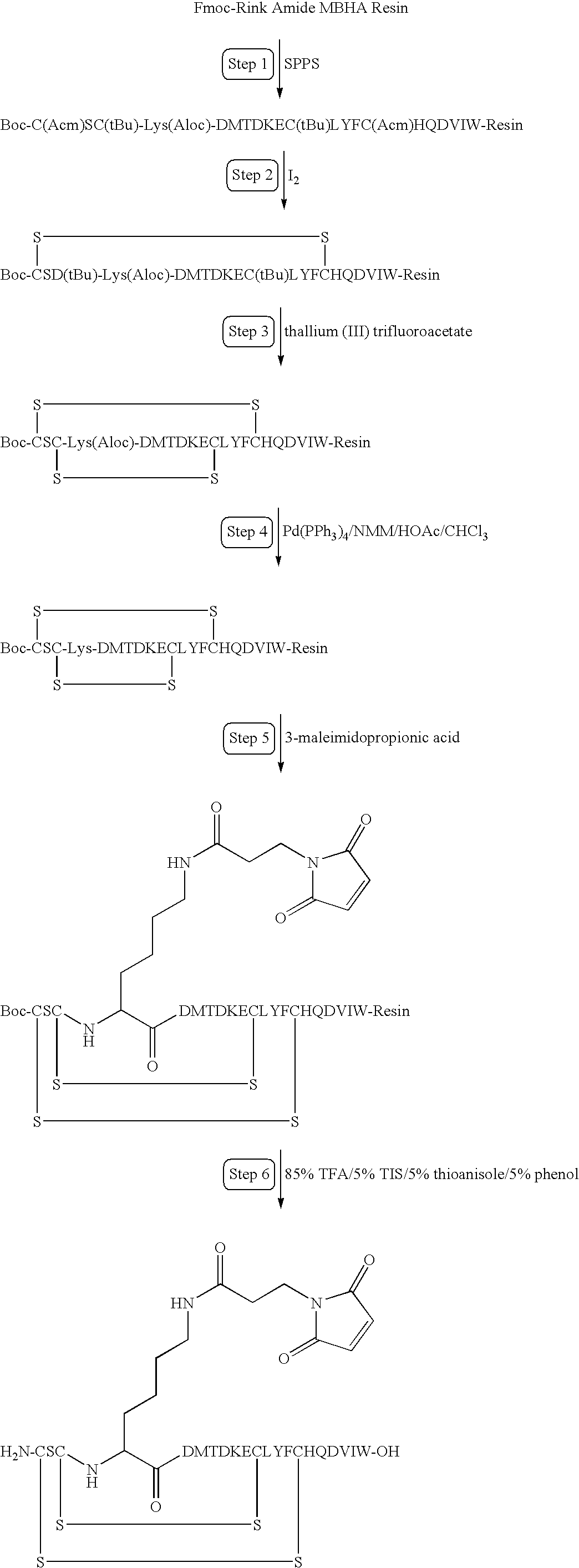
F. Peptide Stability Assays
| Time(Minutes) | % A | % B |
| 0 | 95 | 5 |
| 20 | 70 | 30 |
| 25 | 10 | 90 |
| 30 | 10 | 90 |
| 35 | 95 | 5 |
| 45 | 95 | 5 |
| Time(Minutes) | % A | % B |
| 0 | 95 | 5 |
| 20 | 70 | 30 |
| 25 | 10 | 90 |
| 30 | 10 | 90 |
| 35 | 95 | 5 |
| 45 | 95 | 5 |
| Time | % K5 peptide in Plasma | ||
| 0 | hrs. | 100% | ||
| 4 | hrs | 9% | ||
| 24 | hrs | 0% | ||
| Time (minutes) | % A | % B | Flow (ml/min) | ||
| 0 | 66 | 34 | 0.700 | ||
| 1 | 66 | 34 | 0.700 | ||
| 25 | 58.8 | 41.2 | 0.700 | ||
| 30 | 50 | 50 | 0.70 | ||
| 35 | 5 | 95 | 1.00 | ||
| 41 | 5 | 95 | 1.00 | ||
| 45 | 66 | 34 | 1.00 | ||
| 46 | 66 | 34 | 0.70 | ||
| Time | % capped HSA | % SH-Albumin | % K5-MPA-HSA |
| 0 | hrs. | 61.3 | 16.6 | 22.1 |
| 4 | hrs. | 64.6 | 16.05 | 19.35 |
| 24 | hrs. | 63 | 16.8 | 20.2 |
| TABLE 1 |
| NATURAL AMINO ACIDS AND THEIR ABBREVIATIONS |
| 3-Letter | 1-Letter | Protected Amino | |
| Name | Abbreviation | Abbreviation | Acids |
| Alanine | Ala | A | Fmoc-Ala-OH |
| Arginine | Arg | R | Fmoc-Arg(Pbf)-OH |
| Asparagine | Asn | N | Fmoc-Asn(Trt)-OH |
| Aspartic acid | Asp | D | Asp(tBu)-OH |
| Cysteine | Cys | C | Fmoc-Cys(Trt) |
| Glutamic acid | Glu | E | Fmoc-Glu(tBu)-OH |
| Glutamine | Gln | Q | Fmoc-Gln(Trt)-OH |
| Glycine | Gly | G | Fmoc-Gly-OH |
| Histidine | His | H | Fmoc-His(Trt)-OH |
| Isoleucine | Ile | I | Fmoc-Ile-OH |
| Leucine | Leu | L | Fmoc-Leu-OH |
| Lysine | Lys | K | Fmoc-Lys(Mtt)-OH |
| Methionine | Met | M | Fmoc-Met-OH |
| Phenylalanine | Phe | F | Fmoc-Phe-OH |
| Proline | Pro | P | Fmoc-Pro-OH |
| Serine | Ser | S | Fmoc-Ser(tBu)-OH |
| Threonine | Thr | T | Fmoc-Thr(tBu)-OH |
| Tryptophan | Trp | W | Fmoc-Trp(Boc)-OH |
| Tyrosine | Tyr | Y | Boc-Tyr(tBu)-OH |
| Valine | Val | V | Fmoc-Val-OH |
Claims (9)
Priority Applications (9)
| Application Number | Priority Date | Filing Date | Title |
|---|---|---|---|
| US09/657,276 US6887470B1 (en) | 1999-09-10 | 2000-09-07 | Protection of endogenous therapeutic peptides from peptidase activity through conjugation to blood components |
| US11/040,810 US20050176641A1 (en) | 2000-05-17 | 2005-01-21 | Long lasting natriuretic peptide derivatives |
| US11/067,556 US7601691B2 (en) | 1999-05-17 | 2005-02-25 | Anti-obesity agents |
| US11/066,697 US7256253B2 (en) | 1999-09-10 | 2005-02-25 | Protection of endogenous therapeutic peptides from peptidase activity through conjugation to blood components |
| US11/923,222 US7906482B2 (en) | 1999-05-17 | 2007-10-24 | Anti-obesity agents |
| US11/926,843 US20080199532A1 (en) | 1999-05-17 | 2007-10-29 | Long Lasting Natriuretic Peptide Derivatives |
| US11/982,033 US20090175821A1 (en) | 1999-05-17 | 2007-10-31 | Modified therapeutic peptides with extended half-lives in vivo |
| US12/221,554 US20090093408A1 (en) | 1999-05-17 | 2008-08-01 | Protection of exendin-4 peptides through conjugation |
| US12/253,856 US20090275506A1 (en) | 2000-05-17 | 2008-10-17 | Long lasting natriuretic peptide derivatives |
Applications Claiming Priority (3)
| Application Number | Priority Date | Filing Date | Title |
|---|---|---|---|
| US15340699P | 1999-09-10 | 1999-09-10 | |
| US15978399P | 1999-10-15 | 1999-10-15 | |
| US09/657,276 US6887470B1 (en) | 1999-09-10 | 2000-09-07 | Protection of endogenous therapeutic peptides from peptidase activity through conjugation to blood components |
Related Child Applications (3)
| Application Number | Title | Priority Date | Filing Date |
|---|---|---|---|
| US11/040,810 Continuation-In-Part US20050176641A1 (en) | 1999-05-17 | 2005-01-21 | Long lasting natriuretic peptide derivatives |
| US11/067,556 Continuation-In-Part US7601691B2 (en) | 1999-05-17 | 2005-02-25 | Anti-obesity agents |
| US11/066,697 Continuation US7256253B2 (en) | 1999-09-10 | 2005-02-25 | Protection of endogenous therapeutic peptides from peptidase activity through conjugation to blood components |
Publications (1)
| Publication Number | Publication Date |
|---|---|
| US6887470B1 true US6887470B1 (en) | 2005-05-03 |
Family
ID=34527723
Family Applications (2)
| Application Number | Title | Priority Date | Filing Date |
|---|---|---|---|
| US09/657,276 Expired - Fee Related US6887470B1 (en) | 1999-05-17 | 2000-09-07 | Protection of endogenous therapeutic peptides from peptidase activity through conjugation to blood components |
| US11/066,697 Expired - Fee Related US7256253B2 (en) | 1999-09-10 | 2005-02-25 | Protection of endogenous therapeutic peptides from peptidase activity through conjugation to blood components |
Family Applications After (1)
| Application Number | Title | Priority Date | Filing Date |
|---|---|---|---|
| US11/066,697 Expired - Fee Related US7256253B2 (en) | 1999-09-10 | 2005-02-25 | Protection of endogenous therapeutic peptides from peptidase activity through conjugation to blood components |
Country Status (1)
| Country | Link |
|---|---|
| US (2) | US6887470B1 (en) |
Cited By (105)
| Publication number | Priority date | Publication date | Assignee | Title |
|---|---|---|---|---|
| US20050164950A1 (en) * | 2003-08-11 | 2005-07-28 | The Regents Of The University Of California | Orally administered small peptides synergize statin activity |
| US20050176641A1 (en) * | 2000-05-17 | 2005-08-11 | Conjuchem, Inc. | Long lasting natriuretic peptide derivatives |
| US20050176643A1 (en) * | 1999-05-17 | 2005-08-11 | Conjuchem, Inc. | Anti-obesity agents |
| US20050239628A1 (en) * | 1999-04-06 | 2005-10-27 | Johnson James R | Sinterable structures and method |
| US20050267293A1 (en) * | 2004-04-23 | 2005-12-01 | Conjuchem, Inc. | Method for purification of albumin conjugates |
| US20060009377A1 (en) * | 1999-05-17 | 2006-01-12 | Conjuchem, Inc. | Long lasting synthetic glucagon like peptide (GLP-1) |
| US20060111690A1 (en) * | 2003-01-02 | 2006-05-25 | Medtronic, Inc. | Method and system for treating depressive and anxiety disorders |
| US20060135428A1 (en) * | 1999-05-17 | 2006-06-22 | Bridon Dominique P | Long lasting anti-angiogenic peptides |
| US20060205669A1 (en) * | 2004-09-16 | 2006-09-14 | The Regents Of The University Of California | G-type peptides and other agents to ameliorate atherosclerosis and other pathologies |
| US20060217304A1 (en) * | 2001-02-16 | 2006-09-28 | Bridon Dominique P | Long lasting glucagon-like peptide 2 (GLP-2) for the treatment of gastrointestinal diseases and disorders |
| US20060241019A1 (en) * | 2003-07-25 | 2006-10-26 | Bridon Dominique P | Long lasting insulin derivatives and methods thereof |
| US20070032430A1 (en) * | 2000-08-24 | 2007-02-08 | The Regents Of The University Of California | Peptides and peptide mimetics to treat pathologies characterized by an inflammatory response |
| US20070071764A1 (en) * | 2005-04-22 | 2007-03-29 | Sullivan John K | Toxin peptide therapeutic agents |
| US20070207958A1 (en) * | 2005-11-09 | 2007-09-06 | Bridon Dominique P | Method of treatment of diabetes and/or obesity with reduced nausea side effect |
| US20070212742A1 (en) * | 2003-04-10 | 2007-09-13 | Andreas Bergmann | Determination of a Midregional Proadrenomedullin Partial Peptide in Biological Fluids for Diagnostic Purposes, and Immunoassays for Carrying out Such a Determination |
| US20070254839A1 (en) * | 2000-08-24 | 2007-11-01 | The Regents Of The University Of California | Orally administered peptides synergize statin activity |
| US20070269863A1 (en) * | 2005-12-22 | 2007-11-22 | Bridon Dominique P | Process for the production of preformed conjugates of albumin and a therapeutic agent |
| US20070281887A1 (en) * | 2003-06-20 | 2007-12-06 | Shuchong Pan | Isoforms of brain natriuretic peptide |
| US20080020978A1 (en) * | 2005-10-21 | 2008-01-24 | Gegg Colin V Jr | CGRP peptide antagonists and conjugates |
| WO2008021872A1 (en) * | 2006-08-08 | 2008-02-21 | Mayo Foundation For Medical Education And Research | Diuretic and natriuretic polypeptides |
| US20080171853A1 (en) * | 2001-03-08 | 2008-07-17 | Coy David H | Somatostatin antagonists |
| US20080260840A1 (en) * | 2005-02-03 | 2008-10-23 | Alessi Thomas R | Suspension formulations of insulinotropic peptides and uses thereof |
| US20080280306A1 (en) * | 2005-01-26 | 2008-11-13 | Sphingotec Gmbh | Immunoassay For Determining The Release Of Neurotensin Into The Circulation |
| US20080293639A1 (en) * | 2005-04-29 | 2008-11-27 | The Regents Of The University Of California | Peptides and peptide mimetics to treat pathologies characterized by an inflammatory response |
| US20090093408A1 (en) * | 1999-05-17 | 2009-04-09 | Bridon Dominique P | Protection of exendin-4 peptides through conjugation |
| US20090118181A1 (en) * | 2007-05-22 | 2009-05-07 | Walker Kenneth W | Compositions and methods for producing bioactive fusion proteins |
| WO2009086126A2 (en) * | 2007-12-21 | 2009-07-09 | Mayo Foundation For Medical Education And Research | Natriuretic polypeptides |
| US20090186819A1 (en) * | 2007-12-11 | 2009-07-23 | Marieve Carrier | Formulation of insulinotropic peptide conjugates |
| US7579319B2 (en) | 2004-12-06 | 2009-08-25 | The Regents Of The University Of California | Methods for improving the structure and function of arterioles |
| US20090281028A1 (en) * | 2006-10-25 | 2009-11-12 | Sullivan John K | OSK1 peptide analogs and pharmaceutical compositions |
| US20100227825A1 (en) * | 2005-04-29 | 2010-09-09 | The Regents Of The University Of California | Peptides and peptide mimetics to treat cancer |
| US20100286365A1 (en) * | 2007-09-27 | 2010-11-11 | Amylin Pharmaceuticals, Inc. | Peptide-peptidase-inhibitor conjugates and methods of making and using same |
| US20100292172A1 (en) * | 2006-03-21 | 2010-11-18 | Amylin Pharmaceuticals, Inc. | Peptide-Peptidase Inhibitor Conjugates and Methods of Using Same |
| US20100311660A1 (en) * | 2007-09-15 | 2010-12-09 | Simari Robert D | Natriuretic peptide receptor-c agonists |
| EP2359808A1 (en) | 2006-08-09 | 2011-08-24 | Intarcia Therapeutics, Inc | Osmotic delivery systems and piston assemblies |
| EP2425860A1 (en) | 2005-08-12 | 2012-03-07 | Amgen Inc. | Modified Fc molecules |
| WO2012040518A2 (en) | 2010-09-22 | 2012-03-29 | Amgen Inc. | Carrier immunoglobulins and uses thereof |
| US20120201782A1 (en) * | 2009-09-28 | 2012-08-09 | Staats Herman F | Peptide, adjuvants, vaccines, and methods of use |
| WO2012117336A2 (en) | 2011-02-28 | 2012-09-07 | Istituto Di Ricovero E Cura A Carattere Scientifico Materno-Infantile Burlo Garofolo - Ospedale Di Alta Specializzazione E Di Rilievo Nazionale Per La Salute Della Donna E Del Bambino | Apoptosis-inducing molecules and uses therefor |
| WO2012125973A2 (en) | 2011-03-16 | 2012-09-20 | Amgen Inc. | Potent and selective inhibitors of nav1.3 and nav1.7 |
| US8557767B2 (en) | 2007-08-28 | 2013-10-15 | Uab Research Foundation | Synthetic apolipoprotein E mimicking polypeptides and methods of use |
| US8568766B2 (en) | 2000-08-24 | 2013-10-29 | Gattadahalli M. Anantharamaiah | Peptides and peptide mimetics to treat pathologies associated with eye disease |
| WO2014099984A1 (en) | 2012-12-20 | 2014-06-26 | Amgen Inc. | Apj receptor agonists and uses thereof |
| WO2014165277A2 (en) | 2013-03-12 | 2014-10-09 | Amgen Inc. | POTENT AND SELECTIVE INHIBITORS OF Nav1.7 |
| RU2540012C2 (en) * | 2009-07-29 | 2015-01-27 | Дайити Санкио Компани, Лимитед | Motilin-like peptide compound having imparted transmucosal absorbing ability |
| US9102707B2 (en) | 2011-08-30 | 2015-08-11 | Mayo Foundation For Medical Education And Research | Natriuretic polypeptides |
| US9173950B2 (en) | 2012-05-17 | 2015-11-03 | Extend Biosciences, Inc. | Vitamin D-ghrelin conjugates |
| WO2015191781A2 (en) | 2014-06-10 | 2015-12-17 | Amgen Inc. | Apelin polypeptides |
| US9266939B2 (en) | 2010-12-27 | 2016-02-23 | Alexion Pharmaceuticals, Inc. | Compositions comprising natriuretic peptides and methods of use thereof |
| US9422363B2 (en) | 2007-08-28 | 2016-08-23 | Uab Research Foundation | Synthetic apolipoprotein E mimicking polypeptides and methods of use |
| US9526763B2 (en) | 2005-02-03 | 2016-12-27 | Intarcia Therapeutics Inc. | Solvent/polymer solutions as suspension vehicles |
| US9539200B2 (en) | 2005-02-03 | 2017-01-10 | Intarcia Therapeutics Inc. | Two-piece, internal-channel osmotic delivery system flow modulator |
| US9572889B2 (en) | 2008-02-13 | 2017-02-21 | Intarcia Therapeutics, Inc. | Devices, formulations, and methods for delivery of multiple beneficial agents |
| US9585934B2 (en) | 2014-10-22 | 2017-03-07 | Extend Biosciences, Inc. | Therapeutic vitamin D conjugates |
| US9611305B2 (en) | 2012-01-06 | 2017-04-04 | Mayo Foundation For Medical Education And Research | Treating cardiovascular or renal diseases |
| US9616109B2 (en) | 2014-10-22 | 2017-04-11 | Extend Biosciences, Inc. | Insulin vitamin D conjugates |
| US9616107B2 (en) | 2011-02-25 | 2017-04-11 | Capricor Therapeutics, Inc. | Therapy for kidney disease and/or heart failure |
| US9623085B2 (en) | 2011-09-02 | 2017-04-18 | Capricor Therapeutics, Inc. | Chimeric natriuretic peptide compositions and methods of preparation |
| US9670261B2 (en) | 2012-12-21 | 2017-06-06 | Sanofi | Functionalized exendin-4 derivatives |
| US9694053B2 (en) | 2013-12-13 | 2017-07-04 | Sanofi | Dual GLP-1/glucagon receptor agonists |
| US9724293B2 (en) | 2003-11-17 | 2017-08-08 | Intarcia Therapeutics, Inc. | Methods of manufacturing viscous liquid pharmaceutical formulations |
| US9730921B2 (en) | 2012-03-27 | 2017-08-15 | Duke University | Compositions and methods for the prevention and treatment of mast cell-induced vascular leakage |
| US9751926B2 (en) | 2013-12-13 | 2017-09-05 | Sanofi | Dual GLP-1/GIP receptor agonists |
| US9750788B2 (en) | 2013-12-13 | 2017-09-05 | Sanofi | Non-acylated exendin-4 peptide analogues |
| US9758561B2 (en) | 2014-04-07 | 2017-09-12 | Sanofi | Dual GLP-1/glucagon receptor agonists derived from exendin-4 |
| US9771406B2 (en) | 2014-04-07 | 2017-09-26 | Sanofi | Peptidic dual GLP-1/glucagon receptor agonists derived from exendin-4 |
| US9775904B2 (en) | 2014-04-07 | 2017-10-03 | Sanofi | Exendin-4 derivatives as peptidic dual GLP-1/glucagon receptor agonists |
| US9789165B2 (en) | 2013-12-13 | 2017-10-17 | Sanofi | Exendin-4 peptide analogues as dual GLP-1/GIP receptor agonists |
| US9789197B2 (en) | 2014-10-22 | 2017-10-17 | Extend Biosciences, Inc. | RNAi vitamin D conjugates |
| US9889085B1 (en) | 2014-09-30 | 2018-02-13 | Intarcia Therapeutics, Inc. | Therapeutic methods for the treatment of diabetes and related conditions for patients with high baseline HbA1c |
| US9932381B2 (en) | 2014-06-18 | 2018-04-03 | Sanofi | Exendin-4 derivatives as selective glucagon receptor agonists |
| EP3323423A1 (en) | 2009-09-28 | 2018-05-23 | Intarcia Therapeutics, Inc | Rapid establishment and/or termination of substantial steady-state drug delivery |
| US9982029B2 (en) | 2015-07-10 | 2018-05-29 | Sanofi | Exendin-4 derivatives as selective peptidic dual GLP-1/glucagon receptor agonists |
| WO2018132768A1 (en) | 2017-01-13 | 2018-07-19 | Sanna Pietro P | Methods and compositions for treating hpa hyperactivity |
| US10052366B2 (en) | 2012-05-21 | 2018-08-21 | Alexion Pharmaceuticsl, Inc. | Compositions comprising alkaline phosphatase and/or natriuretic peptide and methods of use thereof |
| EP3385279A1 (en) | 2009-03-20 | 2018-10-10 | Amgen Inc. | Carrier immunoglobulins and uses thereof |
| USD835783S1 (en) | 2016-06-02 | 2018-12-11 | Intarcia Therapeutics, Inc. | Implant placement guide |
| US10159714B2 (en) | 2011-02-16 | 2018-12-25 | Intarcia Therapeutics, Inc. | Compositions, devices and methods of use thereof for the treatment of cancers |
| WO2019028382A1 (en) | 2017-08-04 | 2019-02-07 | Amgen Inc. | Method of conjugation of cys-mabs |
| US10344073B2 (en) | 2014-01-09 | 2019-07-09 | Hadasit Medical Research Services And Development Ltd. | Cell compositions and methods for cancer therapy |
| USD860451S1 (en) | 2016-06-02 | 2019-09-17 | Intarcia Therapeutics, Inc. | Implant removal tool |
| US10449236B2 (en) | 2014-12-05 | 2019-10-22 | Alexion Pharmaceuticals, Inc. | Treating seizure with recombinant alkaline phosphatase |
| US10501517B2 (en) | 2016-05-16 | 2019-12-10 | Intarcia Therapeutics, Inc. | Glucagon-receptor selective polypeptides and methods of use thereof |
| US10603361B2 (en) | 2015-01-28 | 2020-03-31 | Alexion Pharmaceuticals, Inc. | Methods of treating a subject with an alkaline phosphatase deficiency |
| WO2020077129A1 (en) | 2018-10-11 | 2020-04-16 | Intarcia Therapeutics, Inc. | Human amylin analog polypeptides and methods of use |
| US10653747B2 (en) | 2014-07-31 | 2020-05-19 | Uab Research Foundation | ApoE mimetic peptides and higher potency to clear plasma cholesterol |
| US10758592B2 (en) | 2012-10-09 | 2020-09-01 | Sanofi | Exendin-4 derivatives as dual GLP1/glucagon agonists |
| WO2020198075A2 (en) | 2019-03-22 | 2020-10-01 | Reflexion Pharmaceuticals, Inc. | Multivalent d-peptidic compounds for target proteins |
| WO2020198074A1 (en) | 2019-03-22 | 2020-10-01 | Reflexion Pharmaceuticals, Inc. | D-peptidic compounds for vegf |
| US10806797B2 (en) | 2015-06-05 | 2020-10-20 | Sanofi | Prodrugs comprising an GLP-1/glucagon dual agonist linker hyaluronic acid conjugate |
| US10822596B2 (en) | 2014-07-11 | 2020-11-03 | Alexion Pharmaceuticals, Inc. | Compositions and methods for treating craniosynostosis |
| US10835580B2 (en) | 2017-01-03 | 2020-11-17 | Intarcia Therapeutics, Inc. | Methods comprising continuous administration of a GLP-1 receptor agonist and co-administration of a drug |
| US10898549B2 (en) | 2016-04-01 | 2021-01-26 | Alexion Pharmaceuticals, Inc. | Methods for treating hypophosphatasia in adolescents and adults |
| US10925639B2 (en) | 2015-06-03 | 2021-02-23 | Intarcia Therapeutics, Inc. | Implant placement and removal systems |
| US10988744B2 (en) | 2016-06-06 | 2021-04-27 | Alexion Pharmaceuticals, Inc. | Method of producing alkaline phosphatase |
| US11065306B2 (en) | 2016-03-08 | 2021-07-20 | Alexion Pharmaceuticals, Inc. | Methods for treating hypophosphatasia in children |
| US11116821B2 (en) | 2016-08-18 | 2021-09-14 | Alexion Pharmaceuticals, Inc. | Methods for treating tracheobronchomalacia |
| US11186832B2 (en) | 2016-04-01 | 2021-11-30 | Alexion Pharmaceuticals, Inc. | Treating muscle weakness with alkaline phosphatases |
| US11224637B2 (en) | 2017-03-31 | 2022-01-18 | Alexion Pharmaceuticals, Inc. | Methods for treating hypophosphatasia (HPP) in adults and adolescents |
| US11229686B2 (en) | 2015-09-28 | 2022-01-25 | Alexion Pharmaceuticals, Inc. | Reduced frequency dosage regimens for tissue non-specific alkaline phosphatase (TNSALP)-enzyme replacement therapy of hypophosphatasia |
| US11248021B2 (en) | 2004-04-21 | 2022-02-15 | Alexion Pharmaceuticals, Inc. | Bone delivery conjugates and method of using same to target proteins to bone |
| US11246913B2 (en) | 2005-02-03 | 2022-02-15 | Intarcia Therapeutics, Inc. | Suspension formulation comprising an insulinotropic peptide |
| US11352612B2 (en) | 2015-08-17 | 2022-06-07 | Alexion Pharmaceuticals, Inc. | Manufacturing of alkaline phosphatases |
| US11400140B2 (en) | 2015-10-30 | 2022-08-02 | Alexion Pharmaceuticals, Inc. | Methods for treating craniosynostosis in a patient |
| US11913039B2 (en) | 2018-03-30 | 2024-02-27 | Alexion Pharmaceuticals, Inc. | Method for producing recombinant alkaline phosphatase |
Families Citing this family (19)
| Publication number | Priority date | Publication date | Assignee | Title |
|---|---|---|---|---|
| CA2547861A1 (en) * | 2003-12-05 | 2005-06-23 | Ciphergen Biosystems, Inc. | Serum biomarkers for chagas disease |
| US20090099074A1 (en) * | 2007-01-10 | 2009-04-16 | Conjuchem Biotechnologies Inc. | Modulating food intake |
| CA2705603A1 (en) * | 2007-11-21 | 2009-05-28 | Biomarin Pharmaceutical Inc. | Variants of c-type natriuretic peptide |
| US8642558B2 (en) * | 2008-09-19 | 2014-02-04 | Henry Ford Health Systems | Methods, systems, and compositions for calpain inhabition |
| WO2010138343A1 (en) * | 2009-05-27 | 2010-12-02 | Merck Sharp & Dohme Corp. | Neuromedin u receptor agonists |
| RU2017145002A (en) | 2015-05-22 | 2019-06-24 | Цсл Беринг Ленгнау Аг | METHODS OF OBTAINING A MODIFIED VILLE BRAND VON FACTOR |
| WO2016188907A1 (en) | 2015-05-22 | 2016-12-01 | Csl Behring Recombinant Facility Ag | Truncated von willebrand factor polypeptides for treating hemophilia |
| EP3184149A1 (en) | 2015-12-23 | 2017-06-28 | Julius-Maximilians-Universität Würzburg | Soluble glycoprotein v for treating thrombotic diseases |
| CA3008448A1 (en) | 2016-01-07 | 2017-07-13 | Csl Behring Recombinant Facility Ag | Mutated von willebrand factor |
| KR20180094114A (en) | 2016-01-07 | 2018-08-22 | 체에스엘 베링 리컴비넌트 퍼실리티 아게 | Mutated truncated von Willebrand factor |
| JP2020504082A (en) | 2016-11-11 | 2020-02-06 | ツェー・エス・エル・ベーリング・レングナウ・アクチエンゲゼルシャフト | Truncated von Willebrand factor polypeptide for extravascular administration in the treatment or prevention of blood clotting disorders |
| CA3043250A1 (en) | 2016-11-11 | 2018-05-17 | CSL Behring Lengnau AG | Truncated von willebrand factor polypeptides for treating hemophilia |
| WO2018234518A1 (en) | 2017-06-22 | 2018-12-27 | CSL Behring Lengnau AG | Modulation of fviii immunogenicity by truncated vwf |
| US20220211808A1 (en) | 2019-05-17 | 2022-07-07 | Universitaet Zuerich | Haptoglobin for use in treating an adverse secondary neurological outcome following a haemorrhagic stroke |
| JP2022538357A (en) | 2019-07-04 | 2022-09-01 | ツェー・エス・エル・ベーリング・レングナウ・アクチエンゲゼルシャフト | Truncated von Willebrand factor (VWF) to improve the in vitro stability of clotting factor VIII |
| WO2021094344A1 (en) | 2019-11-11 | 2021-05-20 | CSL Behring Lengnau AG | Polypeptides for inducing tolerance to factor viii |
| CA3206884A1 (en) | 2021-02-01 | 2022-08-04 | Kevin AKERET | Method of treating or preventing an adverse secondary neurological outcome following a haemorrhagic stroke |
| CA3217518A1 (en) | 2021-05-07 | 2022-11-10 | Rebecca Butcher | Expression system for producing a recombinant haptoglobin (hp) beta chain |
| WO2024047219A1 (en) | 2022-09-02 | 2024-03-07 | Csl Behring Ag | Haptoglobin for use in treating or preventing exaggerated erectile response or erectile dysfunction |
Citations (23)
| Publication number | Priority date | Publication date | Assignee | Title |
|---|---|---|---|---|
| US4206199A (en) | 1977-07-22 | 1980-06-03 | Takeda Chemical Industries, Ltd. | Novel glucagon fragment and its derivatives |
| US4251631A (en) * | 1978-02-23 | 1981-02-17 | Research Products Rehovot Ltd. | Cross-linked enzyme membrane |
| US4423034A (en) | 1980-10-16 | 1983-12-27 | Toyo Jozo Kabushiki Kaisha | Process for the preparation of antibodies |
| US4462941A (en) | 1982-06-10 | 1984-07-31 | The Regents Of The University Of California | Dynorphin amide analogs |
| US4859604A (en) * | 1987-08-27 | 1989-08-22 | Ampor, Inc. | Composition for stabilization of diagnostic reagents |
| WO1993025217A1 (en) | 1992-06-12 | 1993-12-23 | Des-Tyr Dynorphin Partnership | Des-tyr dynorphin analogues |
| EP0602290A1 (en) | 1992-12-04 | 1994-06-22 | Philippe Pouletty | Cellular drug anchors |
| WO1995010302A1 (en) | 1993-10-15 | 1995-04-20 | Redcell, Inc. | Cellular and serum protein anchors and conjugates |
| US5493007A (en) | 1991-04-05 | 1996-02-20 | Genentech, Inc. | Platelet aggregation inhibitors having high specificity for GPIIBIIIA |
| WO1996006626A1 (en) | 1994-08-26 | 1996-03-07 | Lee Nancy M | Analgesic method with dynorphin analogues truncated at the n-terminus |
| WO1996028544A1 (en) * | 1995-03-09 | 1996-09-19 | Novo Nordisk A/S | A protein-lipid conjugate, consisting of heparin binding protein (hbp) and a ceramide analogue, and its pharmaceutical use in treatment of conditions involving stress injury to cells |
| US5580853A (en) | 1994-03-22 | 1996-12-03 | New England Deaconess Hospital | Modified polypeptides with increased biological activity |
| US5654276A (en) | 1995-06-07 | 1997-08-05 | Affymax Technologies N.V. | Peptides and compounds that bind to the IL-5 receptor |
| US5837247A (en) * | 1995-06-16 | 1998-11-17 | United States Of America As Represented By The Public Health Service National Institutes Of Health | Chemotactic agents for t-cells |
| US5874408A (en) * | 1996-07-12 | 1999-02-23 | Bayer Corporation | Stabilized albumin-free recombinant factor VII preparation having a low sugar content |
| US5877151A (en) * | 1989-07-05 | 1999-03-02 | The Board Of Regents Of The University Of Oklahoma | Method for inhibiting production of tumor necrosis factor |
| WO1999024075A2 (en) | 1997-11-07 | 1999-05-20 | Conjuchem, Inc. | Affinity markers for human serum albumin |
| WO1999024462A2 (en) | 1997-11-07 | 1999-05-20 | Conjuchem, Inc. | Novel conjugates of rgd-containing peptides and endogenous carriers |
| WO1999048536A2 (en) | 1998-03-23 | 1999-09-30 | Conjuchem, Inc. | Delivery of long lasting therapeutic agents by forming covalent attachments in vivo |
| WO2000076551A2 (en) | 1999-06-09 | 2000-12-21 | Ktb Tumorforschungs Gmbh | Method for producing an injectable medicament preparation |
| WO2000076550A2 (en) | 1999-06-10 | 2000-12-21 | Ktb Tumorforschungsgesellschaft Mbh | Carrier-drug conjugate |
| DE19936780A1 (en) * | 1999-08-09 | 2001-02-15 | Basf Ag | New integrin receptor antagonists |
| US6197813B1 (en) * | 1998-02-06 | 2001-03-06 | Oxycal Laboratories, Inc. | Stable liquid mineral ascorbate compositions and methods of manufacture and use |
Family Cites Families (32)
| Publication number | Priority date | Publication date | Assignee | Title |
|---|---|---|---|---|
| US127398A (en) * | 1872-05-28 | Improvement in molders flasks | ||
| US108568A (en) * | 1870-10-25 | Improvement in faucets | ||
| US248782A (en) * | 1881-10-25 | Attachment foe plows | ||
| US138100A (en) * | 1873-04-22 | Improvement in machines for dressing stone | ||
| US158859A (en) * | 1875-01-19 | Improvement in gang-plows | ||
| US105867A (en) * | 1870-07-26 | Improvement in lamps | ||
| US18751A (en) * | 1857-12-01 | Pedal-case fob | ||
| US170250A (en) * | 1875-11-23 | Improvement in cracker-machines | ||
| US73630A (en) * | 1868-01-21 | moore | ||
| US5843440A (en) * | 1990-10-03 | 1998-12-01 | Redcell Canada, Inc. | Cellular and serum protein anchors for modulating pharmacokinetics |
| US5446128A (en) | 1993-06-18 | 1995-08-29 | The Board Of Trustees Of The University Of Illinois | Alpha-helix mimetics and methods relating thereto |
| US20020018751A1 (en) | 1993-10-15 | 2002-02-14 | BRIDON Dominique P. | Cellular and serum protein anchors for diagnostic imaging |
| US6277583B1 (en) * | 1996-02-07 | 2001-08-21 | Conjuchem, Inc. | Affinity labeling libraries and applications thereof |
| US5840733A (en) * | 1996-07-01 | 1998-11-24 | Redcell, Canada, Inc. | Methods and compositions for producing novel conjugates of thrombin inhibitors and endogenous carriers resulting in anti-thrombins with extended lifetimes |
| AU4351897A (en) * | 1996-09-16 | 1998-04-02 | Conjuchem, Inc. | Affinity labeling libraries with tagged leaving groups |
| US6437092B1 (en) * | 1998-11-06 | 2002-08-20 | Conjuchem, Inc. | Conjugates of opioids and endogenous carriers |
| US6107489A (en) * | 1998-03-17 | 2000-08-22 | Conjuchem, Inc. | Extended lifetimes in vivo renin inhibitors |
| US20030170250A1 (en) | 1998-03-23 | 2003-09-11 | Ezrin Alan M. | Local delivery of long lasting therapeutic agents |
| US6202981B1 (en) * | 1998-10-31 | 2001-03-20 | Coltec Industrial Products, Inc. | Fluid control assembly with flange spacer |
| US6440417B1 (en) * | 1998-11-06 | 2002-08-27 | Conjuchem, Inc. | Antibodies to argatroban derivatives and their use in therapeutic and diagnostic treatments |
| AU765753B2 (en) | 1999-05-17 | 2003-09-25 | Conjuchem Biotechnologies Inc. | Protection of endogenous therapeutic peptides from peptidase activity through conjugation to blood components |
| US20040266673A1 (en) | 2002-07-31 | 2004-12-30 | Peter Bakis | Long lasting natriuretic peptide derivatives |
| US6514500B1 (en) | 1999-10-15 | 2003-02-04 | Conjuchem, Inc. | Long lasting synthetic glucagon like peptide {GLP-!} |
| US6849714B1 (en) | 1999-05-17 | 2005-02-01 | Conjuchem, Inc. | Protection of endogenous therapeutic peptides from peptidase activity through conjugation to blood components |
| CN1191273C (en) * | 1999-05-17 | 2005-03-02 | 康久化学公司 | Long lasting insulinotropic peptides |
| ATE443714T1 (en) | 1999-05-17 | 2009-10-15 | Conjuchem Biotechnologies Inc | LONG-ACTING PEPTIDE INHIBITORS OF VIRUS FUSION WITH BODY CELLS IN VIRAL INFECTIONS |
| US6706892B1 (en) | 1999-09-07 | 2004-03-16 | Conjuchem, Inc. | Pulmonary delivery for bioconjugation |
| BR0206919A (en) | 2001-02-02 | 2004-07-06 | Conjuchem Inc | Long-Term Growth Hormone Release Factor Derivatives |
| AU2002233089B2 (en) | 2001-02-16 | 2005-12-22 | Conjuchem Biotechnologies Inc. | Long lasting glucagon-like peptide 2 (GLP-2) for the treatment of gastrointestinal diseases and disorders |
| JP4463545B2 (en) | 2001-05-31 | 2010-05-19 | コンジュケム バイオテクノロジーズ インコーポレイテッド | Long-lasting fusion peptide inhibitors against HIV infection |
| EP1266852B1 (en) * | 2001-06-15 | 2004-09-15 | LTG Mailänder GmbH | Device for feeding and stacking particularly tabular goods and corresponding method |
| US7350098B2 (en) | 2001-11-30 | 2008-03-25 | Oracle International Corporation | Detecting events of interest for managing components on a high availability framework |
-
2000
- 2000-09-07 US US09/657,276 patent/US6887470B1/en not_active Expired - Fee Related
-
2005
- 2005-02-25 US US11/066,697 patent/US7256253B2/en not_active Expired - Fee Related
Patent Citations (25)
| Publication number | Priority date | Publication date | Assignee | Title |
|---|---|---|---|---|
| US4206199A (en) | 1977-07-22 | 1980-06-03 | Takeda Chemical Industries, Ltd. | Novel glucagon fragment and its derivatives |
| US4251631A (en) * | 1978-02-23 | 1981-02-17 | Research Products Rehovot Ltd. | Cross-linked enzyme membrane |
| US4423034A (en) | 1980-10-16 | 1983-12-27 | Toyo Jozo Kabushiki Kaisha | Process for the preparation of antibodies |
| US4462941A (en) | 1982-06-10 | 1984-07-31 | The Regents Of The University Of California | Dynorphin amide analogs |
| US4859604A (en) * | 1987-08-27 | 1989-08-22 | Ampor, Inc. | Composition for stabilization of diagnostic reagents |
| US5877151A (en) * | 1989-07-05 | 1999-03-02 | The Board Of Regents Of The University Of Oklahoma | Method for inhibiting production of tumor necrosis factor |
| US5612034A (en) | 1990-10-03 | 1997-03-18 | Redcell, Inc. | Super-globuling for in vivo extended lifetimes |
| US6103233A (en) * | 1990-10-03 | 2000-08-15 | Conjuchem, Inc. | Cellular and serum protein anchors and conjugates |
| US5493007A (en) | 1991-04-05 | 1996-02-20 | Genentech, Inc. | Platelet aggregation inhibitors having high specificity for GPIIBIIIA |
| WO1993025217A1 (en) | 1992-06-12 | 1993-12-23 | Des-Tyr Dynorphin Partnership | Des-tyr dynorphin analogues |
| EP0602290A1 (en) | 1992-12-04 | 1994-06-22 | Philippe Pouletty | Cellular drug anchors |
| WO1995010302A1 (en) | 1993-10-15 | 1995-04-20 | Redcell, Inc. | Cellular and serum protein anchors and conjugates |
| US5580853A (en) | 1994-03-22 | 1996-12-03 | New England Deaconess Hospital | Modified polypeptides with increased biological activity |
| WO1996006626A1 (en) | 1994-08-26 | 1996-03-07 | Lee Nancy M | Analgesic method with dynorphin analogues truncated at the n-terminus |
| WO1996028544A1 (en) * | 1995-03-09 | 1996-09-19 | Novo Nordisk A/S | A protein-lipid conjugate, consisting of heparin binding protein (hbp) and a ceramide analogue, and its pharmaceutical use in treatment of conditions involving stress injury to cells |
| US5654276A (en) | 1995-06-07 | 1997-08-05 | Affymax Technologies N.V. | Peptides and compounds that bind to the IL-5 receptor |
| US5837247A (en) * | 1995-06-16 | 1998-11-17 | United States Of America As Represented By The Public Health Service National Institutes Of Health | Chemotactic agents for t-cells |
| US5874408A (en) * | 1996-07-12 | 1999-02-23 | Bayer Corporation | Stabilized albumin-free recombinant factor VII preparation having a low sugar content |
| WO1999024075A2 (en) | 1997-11-07 | 1999-05-20 | Conjuchem, Inc. | Affinity markers for human serum albumin |
| WO1999024462A2 (en) | 1997-11-07 | 1999-05-20 | Conjuchem, Inc. | Novel conjugates of rgd-containing peptides and endogenous carriers |
| US6197813B1 (en) * | 1998-02-06 | 2001-03-06 | Oxycal Laboratories, Inc. | Stable liquid mineral ascorbate compositions and methods of manufacture and use |
| WO1999048536A2 (en) | 1998-03-23 | 1999-09-30 | Conjuchem, Inc. | Delivery of long lasting therapeutic agents by forming covalent attachments in vivo |
| WO2000076551A2 (en) | 1999-06-09 | 2000-12-21 | Ktb Tumorforschungs Gmbh | Method for producing an injectable medicament preparation |
| WO2000076550A2 (en) | 1999-06-10 | 2000-12-21 | Ktb Tumorforschungsgesellschaft Mbh | Carrier-drug conjugate |
| DE19936780A1 (en) * | 1999-08-09 | 2001-02-15 | Basf Ag | New integrin receptor antagonists |
Non-Patent Citations (16)
| Title |
|---|
| Ann. Rev. Neurosci., 1984, 7, 223-255. |
| Anti-Cancer Drugs, 1997, 8, 677-685. |
| Biopolymers (Pepetides Science), 1998, 47, 451-463. |
| Biotech Report, 1994/1995, 106-107. |
| Breton J, Pezzi N, Molinari A, Bonomini L, Lansen J, Gonzalez De Buitrago G, Prieto I. Prolonged half-life in the circulaton of a chemical conjugate between a pro-urokinase derivative and human serum albumin.Eur J Biochem. Aug. 1, 1995; 231(3):563-9.* * |
| Chem. Pharm. Bull., 1979, 27 (8), 1942-1944. |
| Endocrinology, 1982, 110 (3), 1049-1051. |
| Int. J. Biochem. Cell. Biol., 1998, 30,1281-1284. |
| J. Biol. Chem., 1995, 270 (43), 25344-25347. |
| J. Dev. Physiol., 1989, 12, 55-62. |
| Proc. Natl. Acad. Sci., 1986, 83, 265-269. |
| Proceedings of the 8<SUP>th </SUP>American Peptide Symposium, 1983, 409-412. |
| TINS, 1993, 16, 403-409. |
| U.S. Appl. No. 09/424,573, Ezrin et al., filed Mar. 7, 2000. |
| U.S. Appl. No. 09/530,891, Blanchard et al., filed Aug. 16, 2000. |
| U.S. Appl. No. 09/623,548, Bridon et al., filed Sep. 5, 2000. |
Cited By (202)
| Publication number | Priority date | Publication date | Assignee | Title |
|---|---|---|---|---|
| US20050239628A1 (en) * | 1999-04-06 | 2005-10-27 | Johnson James R | Sinterable structures and method |
| US20060135428A1 (en) * | 1999-05-17 | 2006-06-22 | Bridon Dominique P | Long lasting anti-angiogenic peptides |
| US20060135426A1 (en) * | 1999-05-17 | 2006-06-22 | Conjuchem, Inc. | Long lasting synthetic glucagon like peptide (GLP-1) |
| US7601691B2 (en) | 1999-05-17 | 2009-10-13 | Conjuchem Biotechnologies Inc. | Anti-obesity agents |
| US8093206B2 (en) | 1999-05-17 | 2012-01-10 | Conjuchem, Llc | Methods involving long lasting synthetic exendin-4 peptide conjugates |
| US20060009377A1 (en) * | 1999-05-17 | 2006-01-12 | Conjuchem, Inc. | Long lasting synthetic glucagon like peptide (GLP-1) |
| US20060058235A1 (en) * | 1999-05-17 | 2006-03-16 | Conjuchem, Inc. | Long lasting synthetic glucagon like peptide (GLP-1) |
| US20050176643A1 (en) * | 1999-05-17 | 2005-08-11 | Conjuchem, Inc. | Anti-obesity agents |
| US20080199532A1 (en) * | 1999-05-17 | 2008-08-21 | Conjuchem Biotechnologies, Inc. | Long Lasting Natriuretic Peptide Derivatives |
| US8080516B2 (en) | 1999-05-17 | 2011-12-20 | Conjuchem, Llc | Long lasting synthetic exendin-4 peptide conjugates |
| US8084414B2 (en) | 1999-05-17 | 2011-12-27 | Conjuchem, Llc | Methods involving long lasting synthetic exendin-4-peptide conjugates |
| US7741286B2 (en) | 1999-05-17 | 2010-06-22 | Conjuchem Biotechnologies Inc. | Long lasting anti-angiogenic peptides |
| US20090093408A1 (en) * | 1999-05-17 | 2009-04-09 | Bridon Dominique P | Protection of exendin-4 peptides through conjugation |
| US20090075890A1 (en) * | 1999-05-17 | 2009-03-19 | Conjuchem Biotechnologies Inc. | Long lasting synthetic exendin-4 peptide conjugates and methods of use thereof |
| US20090275506A1 (en) * | 2000-05-17 | 2009-11-05 | Conjuchem Biotechnologies, Inc. | Long lasting natriuretic peptide derivatives |
| US20050176641A1 (en) * | 2000-05-17 | 2005-08-11 | Conjuchem, Inc. | Long lasting natriuretic peptide derivatives |
| US7820784B2 (en) | 2000-08-24 | 2010-10-26 | The Regents Of The University Of California | Orally administered peptides synergize statin activity |
| US20080095821A1 (en) * | 2000-08-24 | 2008-04-24 | The Regents Of The University Of California | Peptides and peptide mimetics to treat pathologies characterized by an inflammatory response |
| US7994132B2 (en) | 2000-08-24 | 2011-08-09 | The Regents Of The University Of California | Peptides for treatment of scleroderma |
| US20070032430A1 (en) * | 2000-08-24 | 2007-02-08 | The Regents Of The University Of California | Peptides and peptide mimetics to treat pathologies characterized by an inflammatory response |
| US7723303B2 (en) | 2000-08-24 | 2010-05-25 | The Regents Of The University Of California | Peptides and peptide mimetics to treat pathologies characterized by an inflammatory response |
| US8404635B2 (en) | 2000-08-24 | 2013-03-26 | The Regents Of The University Of California | Orally administered peptides synergize statin activity |
| US20080045459A1 (en) * | 2000-08-24 | 2008-02-21 | The Regents Of The University Of California | Orally administered peptides synergize statin activity |
| US20070254839A1 (en) * | 2000-08-24 | 2007-11-01 | The Regents Of The University Of California | Orally administered peptides synergize statin activity |
| US20080096815A1 (en) * | 2000-08-24 | 2008-04-24 | The Regents Of The University Of California | Orally administered peptides synergize statin activity |
| US7531514B2 (en) | 2000-08-24 | 2009-05-12 | The Regents Of The University Of California | Orally administered peptides synergize statin activity |
| US20080096814A1 (en) * | 2000-08-24 | 2008-04-24 | The Regents Of The University Of California | Orally administered peptides synergize statin activity |
| US8568766B2 (en) | 2000-08-24 | 2013-10-29 | Gattadahalli M. Anantharamaiah | Peptides and peptide mimetics to treat pathologies associated with eye disease |
| US7807640B2 (en) | 2000-08-24 | 2010-10-05 | The Regents Of The University Of California | Orally administered peptides synergize statin activity |
| US8048851B2 (en) | 2000-08-24 | 2011-11-01 | The Regents Of The University Of California | Peptides and peptide mimetics to treat pathologies characterized by an inflammatory response |
| US7737251B2 (en) | 2001-02-16 | 2010-06-15 | Conjuchem Biotechnologies Inc. | Long lasting glucagon-like peptide 2 (GLP-2) for the treatment of gastrointestinal diseases and disorders |
| US20060217304A1 (en) * | 2001-02-16 | 2006-09-28 | Bridon Dominique P | Long lasting glucagon-like peptide 2 (GLP-2) for the treatment of gastrointestinal diseases and disorders |
| US20080171853A1 (en) * | 2001-03-08 | 2008-07-17 | Coy David H | Somatostatin antagonists |
| US20060111690A1 (en) * | 2003-01-02 | 2006-05-25 | Medtronic, Inc. | Method and system for treating depressive and anxiety disorders |
| US9885709B2 (en) * | 2003-04-10 | 2018-02-06 | B.R.A.H.M.S. Gmbh | Determination of a midregional proadrenomedullin partial peptide in biological fluids for diagnostic purposes, and immunoassays for carrying out such a determination |
| US20070212742A1 (en) * | 2003-04-10 | 2007-09-13 | Andreas Bergmann | Determination of a Midregional Proadrenomedullin Partial Peptide in Biological Fluids for Diagnostic Purposes, and Immunoassays for Carrying out Such a Determination |
| US9541549B2 (en) | 2003-04-10 | 2017-01-10 | B.R.A.H.M.S. Gmbh | Determination of a midregional proadrenomedullin partial peptide in biological fluids for diagnostic purposes, and immunoassays for carrying out such a determination |
| US8912137B2 (en) | 2003-06-20 | 2014-12-16 | Mayo Foundation For Medical Education And Research | Method for increasing natriuresis or diuresis by isoforms of brain natriuretic peptide |
| US8354496B2 (en) | 2003-06-20 | 2013-01-15 | Mayo Foundation For Medical Education And Research | Isoforms of brain natriuretic peptide |
| US20070281887A1 (en) * | 2003-06-20 | 2007-12-06 | Shuchong Pan | Isoforms of brain natriuretic peptide |
| US20060241019A1 (en) * | 2003-07-25 | 2006-10-26 | Bridon Dominique P | Long lasting insulin derivatives and methods thereof |
| US20050164950A1 (en) * | 2003-08-11 | 2005-07-28 | The Regents Of The University Of California | Orally administered small peptides synergize statin activity |
| US9724293B2 (en) | 2003-11-17 | 2017-08-08 | Intarcia Therapeutics, Inc. | Methods of manufacturing viscous liquid pharmaceutical formulations |
| US11248021B2 (en) | 2004-04-21 | 2022-02-15 | Alexion Pharmaceuticals, Inc. | Bone delivery conjugates and method of using same to target proteins to bone |
| US20080146783A1 (en) * | 2004-04-23 | 2008-06-19 | Conjuchem Biotechnologies Inc. | Method for the purification of albumin conjugates |
| US20050267293A1 (en) * | 2004-04-23 | 2005-12-01 | Conjuchem, Inc. | Method for purification of albumin conjugates |
| US7307148B2 (en) | 2004-04-23 | 2007-12-11 | Conjuchem Biotechnologies Inc. | Method for purification of albumin conjugates |
| US20060205669A1 (en) * | 2004-09-16 | 2006-09-14 | The Regents Of The University Of California | G-type peptides and other agents to ameliorate atherosclerosis and other pathologies |
| US7579319B2 (en) | 2004-12-06 | 2009-08-25 | The Regents Of The University Of California | Methods for improving the structure and function of arterioles |
| US8236754B2 (en) | 2004-12-06 | 2012-08-07 | The Regents Of The University Of California | Methods for improving the structure and function of arterioles |
| US20080280306A1 (en) * | 2005-01-26 | 2008-11-13 | Sphingotec Gmbh | Immunoassay For Determining The Release Of Neurotensin Into The Circulation |
| US8637256B2 (en) | 2005-01-26 | 2014-01-28 | Sphingotec Gmbh | Immunoassay for determining the release of neurotensin into the circulation |
| US20080260840A1 (en) * | 2005-02-03 | 2008-10-23 | Alessi Thomas R | Suspension formulations of insulinotropic peptides and uses thereof |
| US8940316B2 (en) | 2005-02-03 | 2015-01-27 | Intarcia Therapeutics, Inc. | Osmotic delivery comprising an insulinotropic peptide and uses thereof |
| US10363287B2 (en) | 2005-02-03 | 2019-07-30 | Intarcia Therapeutics, Inc. | Method of manufacturing an osmotic delivery device |
| US11246913B2 (en) | 2005-02-03 | 2022-02-15 | Intarcia Therapeutics, Inc. | Suspension formulation comprising an insulinotropic peptide |
| US8299025B2 (en) | 2005-02-03 | 2012-10-30 | Intarcia Therapeutics, Inc. | Suspension formulations of insulinotropic peptides and uses thereof |
| US9526763B2 (en) | 2005-02-03 | 2016-12-27 | Intarcia Therapeutics Inc. | Solvent/polymer solutions as suspension vehicles |
| US9539200B2 (en) | 2005-02-03 | 2017-01-10 | Intarcia Therapeutics Inc. | Two-piece, internal-channel osmotic delivery system flow modulator |
| US9682127B2 (en) | 2005-02-03 | 2017-06-20 | Intarcia Therapeutics, Inc. | Osmotic delivery device comprising an insulinotropic peptide and uses thereof |
| US8907071B2 (en) | 2005-04-22 | 2014-12-09 | Amgen Inc. | Toxin peptide therapeutic agents |
| US7833979B2 (en) | 2005-04-22 | 2010-11-16 | Amgen Inc. | Toxin peptide therapeutic agents |
| US20070071764A1 (en) * | 2005-04-22 | 2007-03-29 | Sullivan John K | Toxin peptide therapeutic agents |
| US20110045587A1 (en) * | 2005-04-22 | 2011-02-24 | Amegen Inc. | Toxin peptide therapeutic agents |
| US20080293639A1 (en) * | 2005-04-29 | 2008-11-27 | The Regents Of The University Of California | Peptides and peptide mimetics to treat pathologies characterized by an inflammatory response |
| US20100227825A1 (en) * | 2005-04-29 | 2010-09-09 | The Regents Of The University Of California | Peptides and peptide mimetics to treat cancer |
| EP2425860A1 (en) | 2005-08-12 | 2012-03-07 | Amgen Inc. | Modified Fc molecules |
| US8168592B2 (en) | 2005-10-21 | 2012-05-01 | Amgen Inc. | CGRP peptide antagonists and conjugates |
| US20080020978A1 (en) * | 2005-10-21 | 2008-01-24 | Gegg Colin V Jr | CGRP peptide antagonists and conjugates |
| US8039432B2 (en) | 2005-11-09 | 2011-10-18 | Conjuchem, Llc | Method of treatment of diabetes and/or obesity with reduced nausea side effect |
| US20070207958A1 (en) * | 2005-11-09 | 2007-09-06 | Bridon Dominique P | Method of treatment of diabetes and/or obesity with reduced nausea side effect |
| US20070269863A1 (en) * | 2005-12-22 | 2007-11-22 | Bridon Dominique P | Process for the production of preformed conjugates of albumin and a therapeutic agent |
| US20100292172A1 (en) * | 2006-03-21 | 2010-11-18 | Amylin Pharmaceuticals, Inc. | Peptide-Peptidase Inhibitor Conjugates and Methods of Using Same |
| AU2007227202B2 (en) * | 2006-03-21 | 2013-08-22 | Amylin Pharmaceuticals, Llc | Peptide-peptidase inhibitor conjugates and methods of using same |
| US8759295B2 (en) | 2006-03-21 | 2014-06-24 | Amylin Pharmaceuticals, Llc | Peptide-peptidase inhibitor conjugates and methods of using same |
| WO2008021872A1 (en) * | 2006-08-08 | 2008-02-21 | Mayo Foundation For Medical Education And Research | Diuretic and natriuretic polypeptides |
| US9439951B2 (en) | 2006-08-08 | 2016-09-13 | Mayo Foundation For Medical Education And Research | Methods of treatment of heart dysfunctions using diuretic and natriuretic polypeptides |
| US8324162B2 (en) | 2006-08-08 | 2012-12-04 | Mayo Foundation For Medical Education And Research | Diuretic and natriuretic polypeptides lacking the blood pressure lowering property |
| US9132166B2 (en) | 2006-08-08 | 2015-09-15 | Mayo Foundation For Medical Education And Research | Method of treatment of renal dysfunctions with chimeric natriuretic peptides |
| US20100204094A1 (en) * | 2006-08-08 | 2010-08-12 | Simari Robert D | Diuretic and natriuretic polypeptides |
| EP2363112A2 (en) | 2006-08-09 | 2011-09-07 | Intarcia Therapeutics, Inc | Osmotic delivery systems and piston assemblies |
| EP3421031A1 (en) | 2006-08-09 | 2019-01-02 | Intarcia Therapeutics, Inc | Osmotic delivery systems and piston assemblies |
| US10527170B2 (en) | 2006-08-09 | 2020-01-07 | Intarcia Therapeutics, Inc. | Osmotic delivery systems and piston assemblies for use therein |
| EP2359808A1 (en) | 2006-08-09 | 2011-08-24 | Intarcia Therapeutics, Inc | Osmotic delivery systems and piston assemblies |
| US20090305399A1 (en) * | 2006-10-25 | 2009-12-10 | Sullivan John K | DNA encoding OSK1 toxin peptide analogs and vectors and cells for combinant expression |
| US7820623B2 (en) | 2006-10-25 | 2010-10-26 | Amgen Inc. | Conjugated toxin peptide therapeutic agents |
| US20090299044A1 (en) * | 2006-10-25 | 2009-12-03 | Sullivan John K | DNA encoding chimeric toxin peptide fusion proteins and vectors and mammalian cells for recombinant expression |
| US7834164B2 (en) | 2006-10-25 | 2010-11-16 | Amgen Inc. | DNA encoding OSK1 toxin peptide analogs and vectors and cells for combinant expression |
| US20090281028A1 (en) * | 2006-10-25 | 2009-11-12 | Sullivan John K | OSK1 peptide analogs and pharmaceutical compositions |
| US7910102B2 (en) | 2006-10-25 | 2011-03-22 | Amgen Inc. | Methods of using conjugated toxin peptide therapeutic agents |
| US8043829B2 (en) | 2006-10-25 | 2011-10-25 | Amgen Inc. | DNA encoding chimeric toxin peptide fusion proteins and vectors and mammalian cells for recombinant expression |
| US7803769B2 (en) | 2006-10-25 | 2010-09-28 | Amgen Inc. | OSK1 peptide analogs and pharmaceutical compositions |
| US7825093B2 (en) | 2006-10-25 | 2010-11-02 | Amgen Inc. | Methods of using OSK1 peptide analogs |
| US20090118181A1 (en) * | 2007-05-22 | 2009-05-07 | Walker Kenneth W | Compositions and methods for producing bioactive fusion proteins |
| EP2738257A1 (en) | 2007-05-22 | 2014-06-04 | Amgen Inc. | Compositions and methods for producing bioactive fusion proteins |
| US8420779B2 (en) | 2007-05-22 | 2013-04-16 | Amgen Inc. | Compositions and methods for producing bioactive fusion proteins |
| US9422363B2 (en) | 2007-08-28 | 2016-08-23 | Uab Research Foundation | Synthetic apolipoprotein E mimicking polypeptides and methods of use |
| US8557767B2 (en) | 2007-08-28 | 2013-10-15 | Uab Research Foundation | Synthetic apolipoprotein E mimicking polypeptides and methods of use |
| US20100311660A1 (en) * | 2007-09-15 | 2010-12-09 | Simari Robert D | Natriuretic peptide receptor-c agonists |
| US8357656B2 (en) | 2007-09-15 | 2013-01-22 | Mayo Foundation For Medical Education And Research | Natriuretic peptide receptor-C agonists |
| US20100286365A1 (en) * | 2007-09-27 | 2010-11-11 | Amylin Pharmaceuticals, Inc. | Peptide-peptidase-inhibitor conjugates and methods of making and using same |
| US8598314B2 (en) | 2007-09-27 | 2013-12-03 | Amylin Pharmaceuticals, Llc | Peptide-peptidase-inhibitor conjugates and methods of making and using same |
| US20090186819A1 (en) * | 2007-12-11 | 2009-07-23 | Marieve Carrier | Formulation of insulinotropic peptide conjugates |
| WO2009086126A2 (en) * | 2007-12-21 | 2009-07-09 | Mayo Foundation For Medical Education And Research | Natriuretic polypeptides |
| WO2009086126A3 (en) * | 2007-12-21 | 2009-09-17 | Mayo Foundation For Medical Education And Research | Natriuretic polypeptides |
| US10441528B2 (en) | 2008-02-13 | 2019-10-15 | Intarcia Therapeutics, Inc. | Devices, formulations, and methods for delivery of multiple beneficial agents |
| US9572889B2 (en) | 2008-02-13 | 2017-02-21 | Intarcia Therapeutics, Inc. | Devices, formulations, and methods for delivery of multiple beneficial agents |
| EP3385279A1 (en) | 2009-03-20 | 2018-10-10 | Amgen Inc. | Carrier immunoglobulins and uses thereof |
| RU2540012C2 (en) * | 2009-07-29 | 2015-01-27 | Дайити Санкио Компани, Лимитед | Motilin-like peptide compound having imparted transmucosal absorbing ability |
| EP3735944A1 (en) | 2009-09-28 | 2020-11-11 | Intarcia Therapeutics, Inc. | Rapid establishment and/or termination of substantial steady-state drug delivery |
| EP3323423A1 (en) | 2009-09-28 | 2018-05-23 | Intarcia Therapeutics, Inc | Rapid establishment and/or termination of substantial steady-state drug delivery |
| US10869830B2 (en) | 2009-09-28 | 2020-12-22 | Intarcia Therapeutics, Inc. | Rapid establishment and/or termination of substantial steady-state drug delivery |
| US10231923B2 (en) | 2009-09-28 | 2019-03-19 | Intarcia Therapeutics, Inc. | Rapid establishment and/or termination of substantial steady-state drug delivery |
| US20120201782A1 (en) * | 2009-09-28 | 2012-08-09 | Staats Herman F | Peptide, adjuvants, vaccines, and methods of use |
| US9301999B2 (en) * | 2009-09-28 | 2016-04-05 | Duke University | Peptide, adjuvants, vaccines, and methods of use |
| WO2012040518A2 (en) | 2010-09-22 | 2012-03-29 | Amgen Inc. | Carrier immunoglobulins and uses thereof |
| US9266939B2 (en) | 2010-12-27 | 2016-02-23 | Alexion Pharmaceuticals, Inc. | Compositions comprising natriuretic peptides and methods of use thereof |
| US10159714B2 (en) | 2011-02-16 | 2018-12-25 | Intarcia Therapeutics, Inc. | Compositions, devices and methods of use thereof for the treatment of cancers |
| US9616107B2 (en) | 2011-02-25 | 2017-04-11 | Capricor Therapeutics, Inc. | Therapy for kidney disease and/or heart failure |
| WO2012117336A2 (en) | 2011-02-28 | 2012-09-07 | Istituto Di Ricovero E Cura A Carattere Scientifico Materno-Infantile Burlo Garofolo - Ospedale Di Alta Specializzazione E Di Rilievo Nazionale Per La Salute Della Donna E Del Bambino | Apoptosis-inducing molecules and uses therefor |
| WO2012125973A2 (en) | 2011-03-16 | 2012-09-20 | Amgen Inc. | Potent and selective inhibitors of nav1.3 and nav1.7 |
| US10344068B2 (en) | 2011-08-30 | 2019-07-09 | Mayo Foundation For Medical Education And Research | Natriuretic polypeptides |
| US9102707B2 (en) | 2011-08-30 | 2015-08-11 | Mayo Foundation For Medical Education And Research | Natriuretic polypeptides |
| US9587004B2 (en) | 2011-08-30 | 2017-03-07 | Mayo Foundation For Medical Education And Research | Natriuretic polypeptides |
| US9441027B2 (en) | 2011-08-30 | 2016-09-13 | Mayo Foundation For Medical Education And Research | Natriuretic polypeptides |
| US9623085B2 (en) | 2011-09-02 | 2017-04-18 | Capricor Therapeutics, Inc. | Chimeric natriuretic peptide compositions and methods of preparation |
| US10092628B2 (en) | 2012-01-06 | 2018-10-09 | Mayo Foundation For Medical Education And Research | Treating cardiovascular or renal diseases |
| US9611305B2 (en) | 2012-01-06 | 2017-04-04 | Mayo Foundation For Medical Education And Research | Treating cardiovascular or renal diseases |
| US9987331B2 (en) | 2012-01-06 | 2018-06-05 | Mayo Foundation For Medical Education And Research | Treating cardiovascular or renal diseases |
| US10668059B2 (en) | 2012-03-27 | 2020-06-02 | Duke University | Compositions and methods for the prevention and treatment of mast cell-induced vascular leakage |
| US9730921B2 (en) | 2012-03-27 | 2017-08-15 | Duke University | Compositions and methods for the prevention and treatment of mast cell-induced vascular leakage |
| US9289507B2 (en) | 2012-05-17 | 2016-03-22 | Extend Biosciences, Inc. | Carriers for improved drug delivery |
| US9173950B2 (en) | 2012-05-17 | 2015-11-03 | Extend Biosciences, Inc. | Vitamin D-ghrelin conjugates |
| US9884124B2 (en) | 2012-05-17 | 2018-02-06 | Extend Biosciences, Inc. | Carriers for improved drug delivery |
| US10052366B2 (en) | 2012-05-21 | 2018-08-21 | Alexion Pharmaceuticsl, Inc. | Compositions comprising alkaline phosphatase and/or natriuretic peptide and methods of use thereof |
| US10758592B2 (en) | 2012-10-09 | 2020-09-01 | Sanofi | Exendin-4 derivatives as dual GLP1/glucagon agonists |
| WO2014099984A1 (en) | 2012-12-20 | 2014-06-26 | Amgen Inc. | Apj receptor agonists and uses thereof |
| EP3907237A1 (en) | 2012-12-20 | 2021-11-10 | Amgen Inc. | Apj receptor agonists and uses thereof |
| US9670261B2 (en) | 2012-12-21 | 2017-06-06 | Sanofi | Functionalized exendin-4 derivatives |
| US10253079B2 (en) | 2012-12-21 | 2019-04-09 | Sanofi | Functionalized Exendin-4 derivatives |
| US9745360B2 (en) | 2012-12-21 | 2017-08-29 | Sanofi | Dual GLP1/GIP or trigonal GLP1/GIP/glucagon agonists |
| WO2014165277A2 (en) | 2013-03-12 | 2014-10-09 | Amgen Inc. | POTENT AND SELECTIVE INHIBITORS OF Nav1.7 |
| US9751926B2 (en) | 2013-12-13 | 2017-09-05 | Sanofi | Dual GLP-1/GIP receptor agonists |
| US9750788B2 (en) | 2013-12-13 | 2017-09-05 | Sanofi | Non-acylated exendin-4 peptide analogues |
| US9694053B2 (en) | 2013-12-13 | 2017-07-04 | Sanofi | Dual GLP-1/glucagon receptor agonists |
| US9789165B2 (en) | 2013-12-13 | 2017-10-17 | Sanofi | Exendin-4 peptide analogues as dual GLP-1/GIP receptor agonists |
| EP4101461A1 (en) | 2014-01-09 | 2022-12-14 | Hadasit Medical Research Services and Development Ltd. | Improved cell compositions and methods for cancer therapy |
| US11530251B2 (en) | 2014-01-09 | 2022-12-20 | Hadasit Medical Research Services And Development Ltd. | Methods for cancer therapy using isolated NTB-A ectodomain polypeptides |
| US10344073B2 (en) | 2014-01-09 | 2019-07-09 | Hadasit Medical Research Services And Development Ltd. | Cell compositions and methods for cancer therapy |
| US9758561B2 (en) | 2014-04-07 | 2017-09-12 | Sanofi | Dual GLP-1/glucagon receptor agonists derived from exendin-4 |
| US9775904B2 (en) | 2014-04-07 | 2017-10-03 | Sanofi | Exendin-4 derivatives as peptidic dual GLP-1/glucagon receptor agonists |
| US9771406B2 (en) | 2014-04-07 | 2017-09-26 | Sanofi | Peptidic dual GLP-1/glucagon receptor agonists derived from exendin-4 |
| WO2015191781A2 (en) | 2014-06-10 | 2015-12-17 | Amgen Inc. | Apelin polypeptides |
| EP4257152A2 (en) | 2014-06-10 | 2023-10-11 | Amgen Inc. | Apelin polypeptides |
| US10941182B2 (en) | 2014-06-10 | 2021-03-09 | Amgen Inc. | Apelin polypeptides |
| US11807693B2 (en) | 2014-06-10 | 2023-11-07 | Amgen Inc. | Apelin polypeptides |
| EP3674314A2 (en) | 2014-06-10 | 2020-07-01 | Amgen, Inc | Apelin polypeptides |
| US9932381B2 (en) | 2014-06-18 | 2018-04-03 | Sanofi | Exendin-4 derivatives as selective glucagon receptor agonists |
| US10822596B2 (en) | 2014-07-11 | 2020-11-03 | Alexion Pharmaceuticals, Inc. | Compositions and methods for treating craniosynostosis |
| US10653747B2 (en) | 2014-07-31 | 2020-05-19 | Uab Research Foundation | ApoE mimetic peptides and higher potency to clear plasma cholesterol |
| US10583080B2 (en) | 2014-09-30 | 2020-03-10 | Intarcia Therapeutics, Inc. | Therapeutic methods for the treatment of diabetes and related conditions for patients with high baseline HbA1c |
| US9889085B1 (en) | 2014-09-30 | 2018-02-13 | Intarcia Therapeutics, Inc. | Therapeutic methods for the treatment of diabetes and related conditions for patients with high baseline HbA1c |
| US9616109B2 (en) | 2014-10-22 | 2017-04-11 | Extend Biosciences, Inc. | Insulin vitamin D conjugates |
| US10406202B2 (en) | 2014-10-22 | 2019-09-10 | Extend Biosciences, Inc. | Therapeutic vitamin D conjugates |
| US9585934B2 (en) | 2014-10-22 | 2017-03-07 | Extend Biosciences, Inc. | Therapeutic vitamin D conjugates |
| US10702574B2 (en) | 2014-10-22 | 2020-07-07 | Extend Biosciences, Inc. | Therapeutic vitamin D conjugates |
| US11116816B2 (en) | 2014-10-22 | 2021-09-14 | Extend Biosciences, Inc. | Therapeutic vitamin d conjugates |
| US10420819B2 (en) | 2014-10-22 | 2019-09-24 | Extend Biosciences, Inc. | Insulin vitamin D conjugates |
| US9789197B2 (en) | 2014-10-22 | 2017-10-17 | Extend Biosciences, Inc. | RNAi vitamin D conjugates |
| US11224638B2 (en) | 2014-12-05 | 2022-01-18 | Alexion Pharmaceuticals, Inc. | Treating seizure with recombinant alkaline phosphatase |
| US10449236B2 (en) | 2014-12-05 | 2019-10-22 | Alexion Pharmaceuticals, Inc. | Treating seizure with recombinant alkaline phosphatase |
| US10603361B2 (en) | 2015-01-28 | 2020-03-31 | Alexion Pharmaceuticals, Inc. | Methods of treating a subject with an alkaline phosphatase deficiency |
| US11564978B2 (en) | 2015-01-28 | 2023-01-31 | Alexion Pharmaceuticals, Inc. | Methods of treating a subject with an alkaline phosphatase deficiency |
| US10925639B2 (en) | 2015-06-03 | 2021-02-23 | Intarcia Therapeutics, Inc. | Implant placement and removal systems |
| US10806797B2 (en) | 2015-06-05 | 2020-10-20 | Sanofi | Prodrugs comprising an GLP-1/glucagon dual agonist linker hyaluronic acid conjugate |
| US9982029B2 (en) | 2015-07-10 | 2018-05-29 | Sanofi | Exendin-4 derivatives as selective peptidic dual GLP-1/glucagon receptor agonists |
| US11352612B2 (en) | 2015-08-17 | 2022-06-07 | Alexion Pharmaceuticals, Inc. | Manufacturing of alkaline phosphatases |
| US11229686B2 (en) | 2015-09-28 | 2022-01-25 | Alexion Pharmaceuticals, Inc. | Reduced frequency dosage regimens for tissue non-specific alkaline phosphatase (TNSALP)-enzyme replacement therapy of hypophosphatasia |
| US11400140B2 (en) | 2015-10-30 | 2022-08-02 | Alexion Pharmaceuticals, Inc. | Methods for treating craniosynostosis in a patient |
| US11065306B2 (en) | 2016-03-08 | 2021-07-20 | Alexion Pharmaceuticals, Inc. | Methods for treating hypophosphatasia in children |
| US10898549B2 (en) | 2016-04-01 | 2021-01-26 | Alexion Pharmaceuticals, Inc. | Methods for treating hypophosphatasia in adolescents and adults |
| US11186832B2 (en) | 2016-04-01 | 2021-11-30 | Alexion Pharmaceuticals, Inc. | Treating muscle weakness with alkaline phosphatases |
| US11840559B2 (en) | 2016-05-16 | 2023-12-12 | I2O Therapeutics, Inc. | Glucagon-receptor selective polypeptides and methods of use thereof |
| US10501517B2 (en) | 2016-05-16 | 2019-12-10 | Intarcia Therapeutics, Inc. | Glucagon-receptor selective polypeptides and methods of use thereof |
| EP3733694A1 (en) | 2016-05-16 | 2020-11-04 | Intarcia Therapeutics, Inc | Glucagon-receptor selective polypeptides and methods of use thereof |
| US11214607B2 (en) | 2016-05-16 | 2022-01-04 | Intarcia Therapeutics Inc. | Glucagon-receptor selective polypeptides and methods of use thereof |
| USD840030S1 (en) | 2016-06-02 | 2019-02-05 | Intarcia Therapeutics, Inc. | Implant placement guide |
| USD860451S1 (en) | 2016-06-02 | 2019-09-17 | Intarcia Therapeutics, Inc. | Implant removal tool |
| USD835783S1 (en) | 2016-06-02 | 2018-12-11 | Intarcia Therapeutics, Inc. | Implant placement guide |
| USD962433S1 (en) | 2016-06-02 | 2022-08-30 | Intarcia Therapeutics, Inc. | Implant placement guide |
| USD912249S1 (en) | 2016-06-02 | 2021-03-02 | Intarcia Therapeutics, Inc. | Implant removal tool |
| US10988744B2 (en) | 2016-06-06 | 2021-04-27 | Alexion Pharmaceuticals, Inc. | Method of producing alkaline phosphatase |
| US11116821B2 (en) | 2016-08-18 | 2021-09-14 | Alexion Pharmaceuticals, Inc. | Methods for treating tracheobronchomalacia |
| US11654183B2 (en) | 2017-01-03 | 2023-05-23 | Intarcia Therapeutics, Inc. | Methods comprising continuous administration of exenatide and co-administration of a drug |
| US10835580B2 (en) | 2017-01-03 | 2020-11-17 | Intarcia Therapeutics, Inc. | Methods comprising continuous administration of a GLP-1 receptor agonist and co-administration of a drug |
| WO2018132768A1 (en) | 2017-01-13 | 2018-07-19 | Sanna Pietro P | Methods and compositions for treating hpa hyperactivity |
| US11224637B2 (en) | 2017-03-31 | 2022-01-18 | Alexion Pharmaceuticals, Inc. | Methods for treating hypophosphatasia (HPP) in adults and adolescents |
| WO2019028382A1 (en) | 2017-08-04 | 2019-02-07 | Amgen Inc. | Method of conjugation of cys-mabs |
| US11913039B2 (en) | 2018-03-30 | 2024-02-27 | Alexion Pharmaceuticals, Inc. | Method for producing recombinant alkaline phosphatase |
| WO2020077129A1 (en) | 2018-10-11 | 2020-04-16 | Intarcia Therapeutics, Inc. | Human amylin analog polypeptides and methods of use |
| WO2020198074A1 (en) | 2019-03-22 | 2020-10-01 | Reflexion Pharmaceuticals, Inc. | D-peptidic compounds for vegf |
| WO2020198075A2 (en) | 2019-03-22 | 2020-10-01 | Reflexion Pharmaceuticals, Inc. | Multivalent d-peptidic compounds for target proteins |
Also Published As
| Publication number | Publication date |
|---|---|
| US20050187159A1 (en) | 2005-08-25 |
| US7256253B2 (en) | 2007-08-14 |
Similar Documents
| Publication | Publication Date | Title |
|---|---|---|
| US6887470B1 (en) | Protection of endogenous therapeutic peptides from peptidase activity through conjugation to blood components | |
| US6849714B1 (en) | Protection of endogenous therapeutic peptides from peptidase activity through conjugation to blood components | |
| EP1105409B1 (en) | Protection of endogenous therapeutic peptides from peptidase activity through conjugation to blood components | |
| US20090175821A1 (en) | Modified therapeutic peptides with extended half-lives in vivo | |
| US20210205415A1 (en) | Highly active polypeptides and methods of making and using the same | |
| US8080516B2 (en) | Long lasting synthetic exendin-4 peptide conjugates | |
| JP4115671B2 (en) | Long-lasting insulinotropic peptides | |
| TWI300414B (en) | Modified peptide yy and conjugates thereof |
Legal Events
| Date | Code | Title | Description |
|---|---|---|---|
| AS | Assignment |
Owner name: CONJUCHEM, INC., CANADA Free format text: ASSIGNMENT OF ASSIGNORS INTEREST;ASSIGNORS:BRIDON, DOMINIQUE P.;EZRIN, ALAN M.;MILNER, PETER G.;AND OTHERS;REEL/FRAME:011083/0096;SIGNING DATES FROM 20000815 TO 20000901 |
|
| AS | Assignment |
Owner name: CONJUCHEM BIOTECHNOLOGIES INC.,CANADA Free format text: ASSIGNMENT OF ASSIGNORS INTEREST;ASSIGNOR:CONJUCHEM INC.;REEL/FRAME:018039/0669 Effective date: 20060523 Owner name: CONJUCHEM BIOTECHNOLOGIES INC., CANADA Free format text: ASSIGNMENT OF ASSIGNORS INTEREST;ASSIGNOR:CONJUCHEM INC.;REEL/FRAME:018039/0669 Effective date: 20060523 |
|
| FEPP | Fee payment procedure |
Free format text: PAYOR NUMBER ASSIGNED (ORIGINAL EVENT CODE: ASPN); ENTITY STATUS OF PATENT OWNER: SMALL ENTITY |
|
| FPAY | Fee payment |
Year of fee payment: 4 |
|
| AS | Assignment |
Owner name: ADVANCED DIAGNOSTICS AND DISCOVERY, CALIFORNIA Free format text: ASSIGNMENT OF ASSIGNORS INTEREST;ASSIGNOR:CONJUCHEM BIOTECHNOLOGIES INC.;REEL/FRAME:025198/0735 Effective date: 20100823 |
|
| AS | Assignment |
Owner name: CONJUCHEM, LLC, CALIFORNIA Free format text: ASSIGNMENT OF ASSIGNORS INTEREST;ASSIGNOR:ADVANCED DIAGNOSTICS AND DISCOVERY;REEL/FRAME:026157/0022 Effective date: 20110218 |
|
| REMI | Maintenance fee reminder mailed | ||
| LAPS | Lapse for failure to pay maintenance fees | ||
| STCH | Information on status: patent discontinuation |
Free format text: PATENT EXPIRED DUE TO NONPAYMENT OF MAINTENANCE FEES UNDER 37 CFR 1.362 |
|
| FP | Lapsed due to failure to pay maintenance fee |
Effective date: 20130503 |