US6649357B2 - Apparatus and method for detecting and identifying infectious agents - Google Patents
Apparatus and method for detecting and identifying infectious agents Download PDFInfo
- Publication number
- US6649357B2 US6649357B2 US10/126,798 US12679802A US6649357B2 US 6649357 B2 US6649357 B2 US 6649357B2 US 12679802 A US12679802 A US 12679802A US 6649357 B2 US6649357 B2 US 6649357B2
- Authority
- US
- United States
- Prior art keywords
- luciferase
- micro
- bioluminescence
- location
- reaction
- Prior art date
- Legal status (The legal status is an assumption and is not a legal conclusion. Google has not performed a legal analysis and makes no representation as to the accuracy of the status listed.)
- Expired - Lifetime
Links
- 238000000034 method Methods 0.000 title claims abstract description 116
- 239000012678 infectious agent Substances 0.000 title claims description 9
- 238000005415 bioluminescence Methods 0.000 claims abstract description 152
- 230000029918 bioluminescence Effects 0.000 claims abstract description 152
- 239000012491 analyte Substances 0.000 claims abstract description 46
- 108060001084 Luciferase Proteins 0.000 claims description 235
- 238000006243 chemical reaction Methods 0.000 claims description 176
- 239000005089 Luciferase Substances 0.000 claims description 170
- IGXWBGJHJZYPQS-SSDOTTSWSA-N D-Luciferin Chemical compound OC(=O)[C@H]1CSC(C=2SC3=CC=C(O)C=C3N=2)=N1 IGXWBGJHJZYPQS-SSDOTTSWSA-N 0.000 claims description 96
- BJGNCJDXODQBOB-UHFFFAOYSA-N Fivefly Luciferin Natural products OC(=O)C1CSC(C=2SC3=CC(O)=CC=C3N=2)=N1 BJGNCJDXODQBOB-UHFFFAOYSA-N 0.000 claims description 93
- 239000000758 substrate Substances 0.000 claims description 93
- CYCGRDQQIOGCKX-UHFFFAOYSA-N Dehydro-luciferin Natural products OC(=O)C1=CSC(C=2SC3=CC(O)=CC=C3N=2)=N1 CYCGRDQQIOGCKX-UHFFFAOYSA-N 0.000 claims description 92
- DDWFXDSYGUXRAY-UHFFFAOYSA-N Luciferin Natural products CCc1c(C)c(CC2NC(=O)C(=C2C=C)C)[nH]c1Cc3[nH]c4C(=C5/NC(CC(=O)O)C(C)C5CC(=O)O)CC(=O)c4c3C DDWFXDSYGUXRAY-UHFFFAOYSA-N 0.000 claims description 92
- 239000000523 sample Substances 0.000 claims description 59
- 230000027455 binding Effects 0.000 claims description 39
- 102000006830 Luminescent Proteins Human genes 0.000 claims description 37
- 108010047357 Luminescent Proteins Proteins 0.000 claims description 37
- 239000012530 fluid Substances 0.000 claims description 28
- 238000004377 microelectronic Methods 0.000 claims description 28
- 241000238584 Vargula Species 0.000 claims description 22
- 230000001580 bacterial effect Effects 0.000 claims description 19
- 229920002521 macromolecule Polymers 0.000 claims description 18
- 241000242739 Renilla Species 0.000 claims description 17
- 108060006184 phycobiliprotein Proteins 0.000 claims description 17
- 241000254158 Lampyridae Species 0.000 claims description 16
- 241000243290 Aequorea Species 0.000 claims description 11
- 239000012472 biological sample Substances 0.000 claims description 11
- 108091005948 blue fluorescent proteins Proteins 0.000 claims description 11
- 108091006047 fluorescent proteins Proteins 0.000 claims description 11
- 102000034287 fluorescent proteins Human genes 0.000 claims description 11
- 108010043121 Green Fluorescent Proteins Proteins 0.000 claims description 10
- 102000004144 Green Fluorescent Proteins Human genes 0.000 claims description 8
- 239000005090 green fluorescent protein Substances 0.000 claims description 8
- 241001331164 Aristostomias Species 0.000 claims description 5
- 108010089433 obelin Proteins 0.000 claims description 5
- 241000191235 Porichthys Species 0.000 claims description 4
- 230000000977 initiatory effect Effects 0.000 claims description 4
- 241000085362 Pachystomias Species 0.000 claims description 3
- INULNSAIIZKOQE-YOSAUDMPSA-N [(3r,4ar,10ar)-6-methoxy-1-methyl-3,4,4a,5,10,10a-hexahydro-2h-benzo[g]quinolin-3-yl]-[4-(4-nitrophenyl)piperazin-1-yl]methanone Chemical compound O=C([C@@H]1C[C@H]2[C@H](N(C1)C)CC=1C=CC=C(C=1C2)OC)N(CC1)CCN1C1=CC=C([N+]([O-])=O)C=C1 INULNSAIIZKOQE-YOSAUDMPSA-N 0.000 claims description 3
- 238000005406 washing Methods 0.000 claims description 3
- 241001533497 Odontosyllis Species 0.000 claims description 2
- 239000011159 matrix material Substances 0.000 abstract description 29
- 229910052710 silicon Inorganic materials 0.000 abstract description 19
- 239000010703 silicon Substances 0.000 abstract description 18
- 238000000151 deposition Methods 0.000 abstract description 11
- 210000000225 synapse Anatomy 0.000 abstract description 10
- 230000033558 biomineral tissue development Effects 0.000 abstract description 5
- 210000001124 body fluid Anatomy 0.000 abstract description 5
- 239000010839 body fluid Substances 0.000 abstract description 5
- 238000010532 solid phase synthesis reaction Methods 0.000 abstract 1
- VYPSYNLAJGMNEJ-UHFFFAOYSA-N Silicium dioxide Chemical compound O=[Si]=O VYPSYNLAJGMNEJ-UHFFFAOYSA-N 0.000 description 103
- 239000010410 layer Substances 0.000 description 80
- 108090000623 proteins and genes Proteins 0.000 description 74
- RLMISHABBKUNFO-WHFBIAKZSA-N Ala-Ala-Gly Chemical compound C[C@H](N)C(=O)N[C@@H](C)C(=O)NCC(O)=O RLMISHABBKUNFO-WHFBIAKZSA-N 0.000 description 70
- 108010000239 Aequorin Proteins 0.000 description 69
- 235000018102 proteins Nutrition 0.000 description 64
- 102000004169 proteins and genes Human genes 0.000 description 64
- 239000003446 ligand Substances 0.000 description 59
- 239000000377 silicon dioxide Substances 0.000 description 51
- 108020004414 DNA Proteins 0.000 description 45
- 102000039446 nucleic acids Human genes 0.000 description 39
- 108020004707 nucleic acids Proteins 0.000 description 39
- 150000007523 nucleic acids Chemical class 0.000 description 39
- 239000000203 mixture Substances 0.000 description 37
- -1 urine Substances 0.000 description 37
- KDXKERNSBIXSRK-UHFFFAOYSA-N Lysine Natural products NCCCCC(N)C(O)=O KDXKERNSBIXSRK-UHFFFAOYSA-N 0.000 description 33
- 102000005962 receptors Human genes 0.000 description 32
- 108020003175 receptors Proteins 0.000 description 32
- 102000004190 Enzymes Human genes 0.000 description 30
- 108090000790 Enzymes Proteins 0.000 description 30
- 210000004027 cell Anatomy 0.000 description 30
- 229940088598 enzyme Drugs 0.000 description 30
- 238000012360 testing method Methods 0.000 description 27
- 125000005647 linker group Chemical group 0.000 description 26
- 108010052090 Renilla Luciferases Proteins 0.000 description 25
- LNCOEGVEEQDKGX-UHFFFAOYSA-N chembl276558 Chemical compound N1=C2C(CC=3C=CC=CC=3)=NC(C=3C=CC(O)=CC=3)=CN2C(O)=C1CC1=CC=C(O)C=C1 LNCOEGVEEQDKGX-UHFFFAOYSA-N 0.000 description 25
- 239000003153 chemical reaction reagent Substances 0.000 description 25
- 230000000694 effects Effects 0.000 description 25
- 238000004519 manufacturing process Methods 0.000 description 25
- 238000012545 processing Methods 0.000 description 25
- 108090000765 processed proteins & peptides Proteins 0.000 description 24
- 241000894007 species Species 0.000 description 24
- BHPQYMZQTOCNFJ-UHFFFAOYSA-N Calcium cation Chemical compound [Ca+2] BHPQYMZQTOCNFJ-UHFFFAOYSA-N 0.000 description 23
- 229910001424 calcium ion Inorganic materials 0.000 description 23
- 150000001875 compounds Chemical class 0.000 description 23
- 238000003556 assay Methods 0.000 description 22
- QVGXLLKOCUKJST-UHFFFAOYSA-N atomic oxygen Chemical compound [O] QVGXLLKOCUKJST-UHFFFAOYSA-N 0.000 description 22
- 239000000463 material Substances 0.000 description 22
- 239000001301 oxygen Substances 0.000 description 22
- 229910052760 oxygen Inorganic materials 0.000 description 22
- YHIPILPTUVMWQT-UHFFFAOYSA-N Coelenterazine Natural products C1=CC(O)=CC=C1CC(C(N1C=C(N2)C=3C=CC(O)=CC=3)=O)=NC1=C2CC1=CC=CC=C1 YHIPILPTUVMWQT-UHFFFAOYSA-N 0.000 description 21
- 108010038633 aspartylglutamate Proteins 0.000 description 21
- 230000009870 specific binding Effects 0.000 description 19
- 239000000126 substance Substances 0.000 description 19
- MTCFGRXMJLQNBG-REOHCLBHSA-N (2S)-2-Amino-3-hydroxypropansäure Chemical compound OC[C@H](N)C(O)=O MTCFGRXMJLQNBG-REOHCLBHSA-N 0.000 description 18
- 239000002299 complementary DNA Substances 0.000 description 18
- 238000007254 oxidation reaction Methods 0.000 description 18
- 102000004196 processed proteins & peptides Human genes 0.000 description 18
- XUIMIQQOPSSXEZ-UHFFFAOYSA-N Silicon Chemical compound [Si] XUIMIQQOPSSXEZ-UHFFFAOYSA-N 0.000 description 17
- 238000001514 detection method Methods 0.000 description 17
- MYMOFIZGZYHOMD-UHFFFAOYSA-N Dioxygen Chemical compound O=O MYMOFIZGZYHOMD-UHFFFAOYSA-N 0.000 description 16
- 229910001882 dioxygen Inorganic materials 0.000 description 16
- 108010083708 leucyl-aspartyl-valine Proteins 0.000 description 16
- 230000003647 oxidation Effects 0.000 description 16
- XLYOFNOQVPJJNP-UHFFFAOYSA-N water Substances O XLYOFNOQVPJJNP-UHFFFAOYSA-N 0.000 description 16
- 241000894006 Bacteria Species 0.000 description 15
- 239000000427 antigen Substances 0.000 description 15
- 229910052751 metal Inorganic materials 0.000 description 15
- 239000002184 metal Substances 0.000 description 15
- 239000000243 solution Substances 0.000 description 15
- 108010083590 Apoproteins Proteins 0.000 description 14
- 102000006410 Apoproteins Human genes 0.000 description 14
- 108090000363 Bacterial Luciferases Proteins 0.000 description 14
- 241000700108 Ctenophora <comb jellyfish phylum> Species 0.000 description 14
- KOSRFJWDECSPRO-UHFFFAOYSA-N alpha-L-glutamyl-L-glutamic acid Natural products OC(=O)CCC(N)C(=O)NC(CCC(O)=O)C(O)=O KOSRFJWDECSPRO-UHFFFAOYSA-N 0.000 description 14
- 108091007433 antigens Proteins 0.000 description 14
- 102000036639 antigens Human genes 0.000 description 14
- 241000035538 Cypridina Species 0.000 description 13
- 108091093037 Peptide nucleic acid Proteins 0.000 description 13
- 239000012190 activator Substances 0.000 description 13
- 108010055341 glutamyl-glutamic acid Proteins 0.000 description 13
- OIPILFWXSMYKGL-UHFFFAOYSA-N acetylcholine Chemical compound CC(=O)OCC[N+](C)(C)C OIPILFWXSMYKGL-UHFFFAOYSA-N 0.000 description 12
- 108020001507 fusion proteins Proteins 0.000 description 12
- 102000037865 fusion proteins Human genes 0.000 description 12
- 229920001184 polypeptide Polymers 0.000 description 12
- ZKHQWZAMYRWXGA-UHFFFAOYSA-N Adenosine triphosphate Natural products C1=NC=2C(N)=NC=NC=2N1C1OC(COP(O)(=O)OP(O)(=O)OP(O)(O)=O)C(O)C1O ZKHQWZAMYRWXGA-UHFFFAOYSA-N 0.000 description 11
- YTNIXZGTHTVJBW-SCRDCRAPSA-L FMNH2(2-) Chemical compound [O-]P(=O)([O-])OC[C@@H](O)[C@@H](O)[C@@H](O)CN1C=2C=C(C)C(C)=CC=2NC2=C1NC(=O)NC2=O YTNIXZGTHTVJBW-SCRDCRAPSA-L 0.000 description 11
- 229960004373 acetylcholine Drugs 0.000 description 11
- 239000003795 chemical substances by application Substances 0.000 description 11
- 239000012634 fragment Substances 0.000 description 11
- 230000010354 integration Effects 0.000 description 11
- 239000000543 intermediate Substances 0.000 description 11
- 238000000746 purification Methods 0.000 description 11
- 239000004065 semiconductor Substances 0.000 description 11
- ZKHQWZAMYRWXGA-KQYNXXCUSA-N Adenosine triphosphate Chemical compound C1=NC=2C(N)=NC=NC=2N1[C@@H]1O[C@H](COP(O)(=O)OP(O)(=O)OP(O)(O)=O)[C@@H](O)[C@H]1O ZKHQWZAMYRWXGA-KQYNXXCUSA-N 0.000 description 10
- 108090000331 Firefly luciferases Proteins 0.000 description 10
- 241000238631 Hexapoda Species 0.000 description 10
- 150000001299 aldehydes Chemical class 0.000 description 10
- 230000015572 biosynthetic process Effects 0.000 description 10
- 230000000875 corresponding effect Effects 0.000 description 10
- 238000001212 derivatisation Methods 0.000 description 10
- 230000006870 function Effects 0.000 description 10
- 229910021645 metal ion Inorganic materials 0.000 description 10
- 230000007704 transition Effects 0.000 description 10
- 241000588724 Escherichia coli Species 0.000 description 9
- NLOAIFSWUUFQFR-CIUDSAMLSA-N Ser-Leu-Asp Chemical compound [H]N[C@@H](CO)C(=O)N[C@@H](CC(C)C)C(=O)N[C@@H](CC(O)=O)C(O)=O NLOAIFSWUUFQFR-CIUDSAMLSA-N 0.000 description 9
- 125000003275 alpha amino acid group Chemical group 0.000 description 9
- 235000001014 amino acid Nutrition 0.000 description 9
- FVTCRASFADXXNN-SCRDCRAPSA-N flavin mononucleotide Chemical compound OP(=O)(O)OC[C@@H](O)[C@@H](O)[C@@H](O)CN1C=2C=C(C)C(C)=CC=2N=C2C1=NC(=O)NC2=O FVTCRASFADXXNN-SCRDCRAPSA-N 0.000 description 9
- 108010049041 glutamylalanine Proteins 0.000 description 9
- 238000004020 luminiscence type Methods 0.000 description 9
- 230000008569 process Effects 0.000 description 9
- WYTZZXDRDKSJID-UHFFFAOYSA-N (3-aminopropyl)triethoxysilane Chemical compound CCO[Si](OCC)(OCC)CCCN WYTZZXDRDKSJID-UHFFFAOYSA-N 0.000 description 8
- 241000254173 Coleoptera Species 0.000 description 8
- 108010065920 Insulin Lispro Proteins 0.000 description 8
- QCSFMCFHVGTLFF-NHCYSSNCSA-N Leu-Asp-Val Chemical compound CC(C)C[C@H](N)C(=O)N[C@@H](CC(O)=O)C(=O)N[C@@H](C(C)C)C(O)=O QCSFMCFHVGTLFF-NHCYSSNCSA-N 0.000 description 8
- ZAVCJRJOQKIOJW-KKUMJFAQSA-N Leu-Phe-Asp Chemical compound CC(C)C[C@H](N)C(=O)N[C@H](C(=O)N[C@@H](CC(O)=O)C(O)=O)CC1=CC=CC=C1 ZAVCJRJOQKIOJW-KKUMJFAQSA-N 0.000 description 8
- 108091034117 Oligonucleotide Proteins 0.000 description 8
- 229910052581 Si3N4 Inorganic materials 0.000 description 8
- 229940024606 amino acid Drugs 0.000 description 8
- 230000000692 anti-sense effect Effects 0.000 description 8
- 108010041089 apoaequorin Proteins 0.000 description 8
- 230000003115 biocidal effect Effects 0.000 description 8
- 239000000872 buffer Substances 0.000 description 8
- 210000002421 cell wall Anatomy 0.000 description 8
- 238000010586 diagram Methods 0.000 description 8
- 239000011768 flavin mononucleotide Substances 0.000 description 8
- 229940013640 flavin mononucleotide Drugs 0.000 description 8
- FVTCRASFADXXNN-UHFFFAOYSA-N flavin mononucleotide Natural products OP(=O)(O)OCC(O)C(O)C(O)CN1C=2C=C(C)C(C)=CC=2N=C2C1=NC(=O)NC2=O FVTCRASFADXXNN-UHFFFAOYSA-N 0.000 description 8
- 108010063718 gamma-glutamylaspartic acid Proteins 0.000 description 8
- 239000000499 gel Substances 0.000 description 8
- 108010084525 phenylalanyl-phenylalanyl-glycine Proteins 0.000 description 8
- 239000000376 reactant Substances 0.000 description 8
- 235000019231 riboflavin-5'-phosphate Nutrition 0.000 description 8
- 230000035945 sensitivity Effects 0.000 description 8
- HQVNEWCFYHHQES-UHFFFAOYSA-N silicon nitride Chemical compound N12[Si]34N5[Si]62N3[Si]51N64 HQVNEWCFYHHQES-UHFFFAOYSA-N 0.000 description 8
- 239000007787 solid Substances 0.000 description 8
- 210000000278 spinal cord Anatomy 0.000 description 8
- 238000006467 substitution reaction Methods 0.000 description 8
- 238000003786 synthesis reaction Methods 0.000 description 8
- KYQNAIMCTRZLNP-QSFUFRPTSA-N Asp-Ile-Val Chemical compound [H]N[C@@H](CC(O)=O)C(=O)N[C@@H]([C@@H](C)CC)C(=O)N[C@@H](C(C)C)C(O)=O KYQNAIMCTRZLNP-QSFUFRPTSA-N 0.000 description 7
- 241000206761 Bacillariophyta Species 0.000 description 7
- JRDYDYXZKFNNRQ-XPUUQOCRSA-N Gly-Ala-Val Chemical compound CC(C)[C@@H](C(O)=O)NC(=O)[C@H](C)NC(=O)CN JRDYDYXZKFNNRQ-XPUUQOCRSA-N 0.000 description 7
- DHMQDGOQFOQNFH-UHFFFAOYSA-N Glycine Chemical compound NCC(O)=O DHMQDGOQFOQNFH-UHFFFAOYSA-N 0.000 description 7
- PMGDADKJMCOXHX-UHFFFAOYSA-N L-Arginyl-L-glutamin-acetat Natural products NC(=N)NCCCC(N)C(=O)NC(CCC(N)=O)C(O)=O PMGDADKJMCOXHX-UHFFFAOYSA-N 0.000 description 7
- OXRLYTYUXAQTHP-YUMQZZPRSA-N Leu-Gly-Ala Chemical compound [H]N[C@@H](CC(C)C)C(=O)NCC(=O)N[C@@H](C)C(O)=O OXRLYTYUXAQTHP-YUMQZZPRSA-N 0.000 description 7
- GWADARYJIJDYRC-XGEHTFHBSA-N Met-Thr-Ser Chemical compound CSCC[C@H](N)C(=O)N[C@@H]([C@@H](C)O)C(=O)N[C@@H](CO)C(O)=O GWADARYJIJDYRC-XGEHTFHBSA-N 0.000 description 7
- MTCFGRXMJLQNBG-UHFFFAOYSA-N Serine Natural products OCC(N)C(O)=O MTCFGRXMJLQNBG-UHFFFAOYSA-N 0.000 description 7
- 241000700605 Viruses Species 0.000 description 7
- 239000002253 acid Substances 0.000 description 7
- 229910052782 aluminium Inorganic materials 0.000 description 7
- XAGFODPZIPBFFR-UHFFFAOYSA-N aluminium Chemical compound [Al] XAGFODPZIPBFFR-UHFFFAOYSA-N 0.000 description 7
- 150000001413 amino acids Chemical class 0.000 description 7
- 230000000890 antigenic effect Effects 0.000 description 7
- 108010008355 arginyl-glutamine Proteins 0.000 description 7
- 108010047857 aspartylglycine Proteins 0.000 description 7
- 125000003178 carboxy group Chemical group [H]OC(*)=O 0.000 description 7
- 239000003638 chemical reducing agent Substances 0.000 description 7
- 230000008021 deposition Effects 0.000 description 7
- 238000013461 design Methods 0.000 description 7
- 230000005281 excited state Effects 0.000 description 7
- VPZXBVLAVMBEQI-UHFFFAOYSA-N glycyl-DL-alpha-alanine Natural products OC(=O)C(C)NC(=O)CN VPZXBVLAVMBEQI-UHFFFAOYSA-N 0.000 description 7
- 108010027668 glycyl-alanyl-valine Proteins 0.000 description 7
- 108010050848 glycylleucine Proteins 0.000 description 7
- 239000002609 medium Substances 0.000 description 7
- 239000012528 membrane Substances 0.000 description 7
- 238000002360 preparation method Methods 0.000 description 7
- 239000000047 product Substances 0.000 description 7
- 125000003396 thiol group Chemical group [H]S* 0.000 description 7
- 239000003981 vehicle Substances 0.000 description 7
- CSCPPACGZOOCGX-UHFFFAOYSA-N Acetone Chemical compound CC(C)=O CSCPPACGZOOCGX-UHFFFAOYSA-N 0.000 description 6
- ODBSSLHUFPJRED-CIUDSAMLSA-N Asn-His-Asn Chemical compound C1=C(NC=N1)C[C@@H](C(=O)N[C@@H](CC(=O)N)C(=O)O)NC(=O)[C@H](CC(=O)N)N ODBSSLHUFPJRED-CIUDSAMLSA-N 0.000 description 6
- HZZIFFOVHLWGCS-KKUMJFAQSA-N Asn-Phe-Leu Chemical compound [H]N[C@@H](CC(N)=O)C(=O)N[C@@H](CC1=CC=CC=C1)C(=O)N[C@@H](CC(C)C)C(O)=O HZZIFFOVHLWGCS-KKUMJFAQSA-N 0.000 description 6
- UWOPETAWXDZUJR-ACZMJKKPSA-N Asp-Cys-Glu Chemical compound [H]N[C@@H](CC(O)=O)C(=O)N[C@@H](CS)C(=O)N[C@@H](CCC(O)=O)C(O)=O UWOPETAWXDZUJR-ACZMJKKPSA-N 0.000 description 6
- OYPRJOBELJOOCE-UHFFFAOYSA-N Calcium Chemical compound [Ca] OYPRJOBELJOOCE-UHFFFAOYSA-N 0.000 description 6
- 108020004635 Complementary DNA Proteins 0.000 description 6
- KCXVZYZYPLLWCC-UHFFFAOYSA-N EDTA Chemical compound OC(=O)CN(CC(O)=O)CCN(CC(O)=O)CC(O)=O KCXVZYZYPLLWCC-UHFFFAOYSA-N 0.000 description 6
- 101710157404 Flavin reductase Proteins 0.000 description 6
- 102100027944 Flavin reductase (NADPH) Human genes 0.000 description 6
- OXEMJGCAJFFREE-FXQIFTODSA-N Glu-Gln-Ala Chemical compound [H]N[C@@H](CCC(O)=O)C(=O)N[C@@H](CCC(N)=O)C(=O)N[C@@H](C)C(O)=O OXEMJGCAJFFREE-FXQIFTODSA-N 0.000 description 6
- HUFCEIHAFNVSNR-IHRRRGAJSA-N Glu-Gln-Tyr Chemical compound OC(=O)CC[C@H](N)C(=O)N[C@@H](CCC(N)=O)C(=O)N[C@H](C(O)=O)CC1=CC=C(O)C=C1 HUFCEIHAFNVSNR-IHRRRGAJSA-N 0.000 description 6
- ILWHFUZZCFYSKT-AVGNSLFASA-N Glu-Lys-Leu Chemical compound [H]N[C@@H](CCC(O)=O)C(=O)N[C@@H](CCCCN)C(=O)N[C@@H](CC(C)C)C(O)=O ILWHFUZZCFYSKT-AVGNSLFASA-N 0.000 description 6
- KQDMENMTYNBWMR-WHFBIAKZSA-N Gly-Asp-Ala Chemical compound [H]NCC(=O)N[C@@H](CC(O)=O)C(=O)N[C@@H](C)C(O)=O KQDMENMTYNBWMR-WHFBIAKZSA-N 0.000 description 6
- LKJCZEPXHOIAIW-HOTGVXAUSA-N Gly-Trp-Lys Chemical compound C1=CC=C2C(=C1)C(=CN2)C[C@@H](C(=O)N[C@@H](CCCCN)C(=O)O)NC(=O)CN LKJCZEPXHOIAIW-HOTGVXAUSA-N 0.000 description 6
- SYOJVRNQCXYEOV-XVKPBYJWSA-N Gly-Val-Glu Chemical compound [H]NCC(=O)N[C@@H](C(C)C)C(=O)N[C@@H](CCC(O)=O)C(O)=O SYOJVRNQCXYEOV-XVKPBYJWSA-N 0.000 description 6
- QEYUCKCWTMIERU-SRVKXCTJSA-N His-Lys-Asp Chemical compound C1=C(NC=N1)C[C@@H](C(=O)N[C@@H](CCCCN)C(=O)N[C@@H](CC(=O)O)C(=O)O)N QEYUCKCWTMIERU-SRVKXCTJSA-N 0.000 description 6
- DEOQGJUXUQGUJN-KKUMJFAQSA-N His-Lys-His Chemical compound C1=C(NC=N1)C[C@@H](C(=O)N[C@@H](CCCCN)C(=O)N[C@@H](CC2=CN=CN2)C(=O)O)N DEOQGJUXUQGUJN-KKUMJFAQSA-N 0.000 description 6
- QYZYJFXHXYUZMZ-UGYAYLCHSA-N Ile-Asn-Asn Chemical compound CC[C@H](C)[C@@H](C(=O)N[C@@H](CC(=O)N)C(=O)N[C@@H](CC(=O)N)C(=O)O)N QYZYJFXHXYUZMZ-UGYAYLCHSA-N 0.000 description 6
- IDAHFEPYTJJZFD-PEFMBERDSA-N Ile-Asp-Glu Chemical compound CC[C@H](C)[C@@H](C(=O)N[C@@H](CC(=O)O)C(=O)N[C@@H](CCC(=O)O)C(=O)O)N IDAHFEPYTJJZFD-PEFMBERDSA-N 0.000 description 6
- NHJKZMDIMMTVCK-QXEWZRGKSA-N Ile-Gly-Arg Chemical compound CC[C@H](C)[C@H](N)C(=O)NCC(=O)N[C@H](C(O)=O)CCCN=C(N)N NHJKZMDIMMTVCK-QXEWZRGKSA-N 0.000 description 6
- RIVKTKFVWXRNSJ-GRLWGSQLSA-N Ile-Ile-Gln Chemical compound CC[C@H](C)[C@@H](C(=O)N[C@@H]([C@@H](C)CC)C(=O)N[C@@H](CCC(=O)N)C(=O)O)N RIVKTKFVWXRNSJ-GRLWGSQLSA-N 0.000 description 6
- KEVYYIMVELOXCT-KBPBESRZSA-N Leu-Gly-Phe Chemical compound CC(C)C[C@H]([NH3+])C(=O)NCC(=O)N[C@H](C([O-])=O)CC1=CC=CC=C1 KEVYYIMVELOXCT-KBPBESRZSA-N 0.000 description 6
- DNDVVILEHVMWIS-LPEHRKFASA-N Met-Asp-Pro Chemical compound CSCC[C@@H](C(=O)N[C@@H](CC(=O)O)C(=O)N1CCC[C@@H]1C(=O)O)N DNDVVILEHVMWIS-LPEHRKFASA-N 0.000 description 6
- KSIPKXNIQOWMIC-RCWTZXSCSA-N Met-Thr-Arg Chemical compound CSCC[C@H](N)C(=O)N[C@@H]([C@@H](C)O)C(=O)N[C@H](C(O)=O)CCCNC(N)=N KSIPKXNIQOWMIC-RCWTZXSCSA-N 0.000 description 6
- SITLTJHOQZFJGG-UHFFFAOYSA-N N-L-alpha-glutamyl-L-valine Natural products CC(C)C(C(O)=O)NC(=O)C(N)CCC(O)=O SITLTJHOQZFJGG-UHFFFAOYSA-N 0.000 description 6
- 108010047562 NGR peptide Proteins 0.000 description 6
- 108090000417 Oxygenases Proteins 0.000 description 6
- 102000004020 Oxygenases Human genes 0.000 description 6
- LJUUGSWZPQOJKD-JYJNAYRXSA-N Phe-Arg-Val Chemical compound CC(C)[C@H](NC(=O)[C@H](CCCNC(N)=N)NC(=O)[C@@H](N)Cc1ccccc1)C(O)=O LJUUGSWZPQOJKD-JYJNAYRXSA-N 0.000 description 6
- 241000242583 Scyphozoa Species 0.000 description 6
- GYDFRTRSSXOZCR-ACZMJKKPSA-N Ser-Ser-Glu Chemical compound OC[C@H](N)C(=O)N[C@@H](CO)C(=O)N[C@H](C(O)=O)CCC(O)=O GYDFRTRSSXOZCR-ACZMJKKPSA-N 0.000 description 6
- LGIMRDKGABDMBN-DCAQKATOSA-N Ser-Val-Lys Chemical compound CC(C)[C@@H](C(=O)N[C@@H](CCCCN)C(=O)O)NC(=O)[C@H](CO)N LGIMRDKGABDMBN-DCAQKATOSA-N 0.000 description 6
- YXFVVABEGXRONW-UHFFFAOYSA-N Toluene Chemical compound CC1=CC=CC=C1 YXFVVABEGXRONW-UHFFFAOYSA-N 0.000 description 6
- YTHWAWACWGWBLE-MNSWYVGCSA-N Trp-Tyr-Thr Chemical compound C([C@@H](C(=O)N[C@@H]([C@H](O)C)C(O)=O)NC(=O)[C@@H](N)CC=1C2=CC=CC=C2NC=1)C1=CC=C(O)C=C1 YTHWAWACWGWBLE-MNSWYVGCSA-N 0.000 description 6
- RLVTVHSDKHBFQP-ULQDDVLXSA-N Val-Tyr-Lys Chemical compound NCCCC[C@@H](C(O)=O)NC(=O)[C@@H](NC(=O)[C@@H](N)C(C)C)CC1=CC=C(O)C=C1 RLVTVHSDKHBFQP-ULQDDVLXSA-N 0.000 description 6
- 241000607618 Vibrio harveyi Species 0.000 description 6
- JLCPHMBAVCMARE-UHFFFAOYSA-N [3-[[3-[[3-[[3-[[3-[[3-[[3-[[3-[[3-[[3-[[3-[[5-(2-amino-6-oxo-1H-purin-9-yl)-3-[[3-[[3-[[3-[[3-[[3-[[5-(2-amino-6-oxo-1H-purin-9-yl)-3-[[5-(2-amino-6-oxo-1H-purin-9-yl)-3-hydroxyoxolan-2-yl]methoxy-hydroxyphosphoryl]oxyoxolan-2-yl]methoxy-hydroxyphosphoryl]oxy-5-(5-methyl-2,4-dioxopyrimidin-1-yl)oxolan-2-yl]methoxy-hydroxyphosphoryl]oxy-5-(6-aminopurin-9-yl)oxolan-2-yl]methoxy-hydroxyphosphoryl]oxy-5-(6-aminopurin-9-yl)oxolan-2-yl]methoxy-hydroxyphosphoryl]oxy-5-(6-aminopurin-9-yl)oxolan-2-yl]methoxy-hydroxyphosphoryl]oxy-5-(6-aminopurin-9-yl)oxolan-2-yl]methoxy-hydroxyphosphoryl]oxyoxolan-2-yl]methoxy-hydroxyphosphoryl]oxy-5-(5-methyl-2,4-dioxopyrimidin-1-yl)oxolan-2-yl]methoxy-hydroxyphosphoryl]oxy-5-(4-amino-2-oxopyrimidin-1-yl)oxolan-2-yl]methoxy-hydroxyphosphoryl]oxy-5-(5-methyl-2,4-dioxopyrimidin-1-yl)oxolan-2-yl]methoxy-hydroxyphosphoryl]oxy-5-(5-methyl-2,4-dioxopyrimidin-1-yl)oxolan-2-yl]methoxy-hydroxyphosphoryl]oxy-5-(6-aminopurin-9-yl)oxolan-2-yl]methoxy-hydroxyphosphoryl]oxy-5-(6-aminopurin-9-yl)oxolan-2-yl]methoxy-hydroxyphosphoryl]oxy-5-(4-amino-2-oxopyrimidin-1-yl)oxolan-2-yl]methoxy-hydroxyphosphoryl]oxy-5-(4-amino-2-oxopyrimidin-1-yl)oxolan-2-yl]methoxy-hydroxyphosphoryl]oxy-5-(4-amino-2-oxopyrimidin-1-yl)oxolan-2-yl]methoxy-hydroxyphosphoryl]oxy-5-(6-aminopurin-9-yl)oxolan-2-yl]methoxy-hydroxyphosphoryl]oxy-5-(4-amino-2-oxopyrimidin-1-yl)oxolan-2-yl]methyl [5-(6-aminopurin-9-yl)-2-(hydroxymethyl)oxolan-3-yl] hydrogen phosphate Polymers Cc1cn(C2CC(OP(O)(=O)OCC3OC(CC3OP(O)(=O)OCC3OC(CC3O)n3cnc4c3nc(N)[nH]c4=O)n3cnc4c3nc(N)[nH]c4=O)C(COP(O)(=O)OC3CC(OC3COP(O)(=O)OC3CC(OC3COP(O)(=O)OC3CC(OC3COP(O)(=O)OC3CC(OC3COP(O)(=O)OC3CC(OC3COP(O)(=O)OC3CC(OC3COP(O)(=O)OC3CC(OC3COP(O)(=O)OC3CC(OC3COP(O)(=O)OC3CC(OC3COP(O)(=O)OC3CC(OC3COP(O)(=O)OC3CC(OC3COP(O)(=O)OC3CC(OC3COP(O)(=O)OC3CC(OC3COP(O)(=O)OC3CC(OC3COP(O)(=O)OC3CC(OC3COP(O)(=O)OC3CC(OC3COP(O)(=O)OC3CC(OC3CO)n3cnc4c(N)ncnc34)n3ccc(N)nc3=O)n3cnc4c(N)ncnc34)n3ccc(N)nc3=O)n3ccc(N)nc3=O)n3ccc(N)nc3=O)n3cnc4c(N)ncnc34)n3cnc4c(N)ncnc34)n3cc(C)c(=O)[nH]c3=O)n3cc(C)c(=O)[nH]c3=O)n3ccc(N)nc3=O)n3cc(C)c(=O)[nH]c3=O)n3cnc4c3nc(N)[nH]c4=O)n3cnc4c(N)ncnc34)n3cnc4c(N)ncnc34)n3cnc4c(N)ncnc34)n3cnc4c(N)ncnc34)O2)c(=O)[nH]c1=O JLCPHMBAVCMARE-UHFFFAOYSA-N 0.000 description 6
- 125000003277 amino group Chemical group 0.000 description 6
- 238000003491 array Methods 0.000 description 6
- 230000004071 biological effect Effects 0.000 description 6
- 239000011575 calcium Substances 0.000 description 6
- 229910052791 calcium Inorganic materials 0.000 description 6
- 239000011248 coating agent Substances 0.000 description 6
- 238000000576 coating method Methods 0.000 description 6
- 239000003086 colorant Substances 0.000 description 6
- 230000000295 complement effect Effects 0.000 description 6
- 239000004020 conductor Substances 0.000 description 6
- 239000003814 drug Substances 0.000 description 6
- 108010051673 leucyl-glycyl-phenylalanine Proteins 0.000 description 6
- 238000005459 micromachining Methods 0.000 description 6
- VLKZOEOYAKHREP-UHFFFAOYSA-N n-Hexane Chemical compound CCCCCC VLKZOEOYAKHREP-UHFFFAOYSA-N 0.000 description 6
- 210000002569 neuron Anatomy 0.000 description 6
- 108010038745 tryptophylglycine Proteins 0.000 description 6
- JAQNUEWEJWBVAY-WBAXXEDZSA-N Ala-Phe-Phe Chemical compound C([C@H](NC(=O)[C@@H](N)C)C(=O)N[C@@H](CC=1C=CC=CC=1)C(O)=O)C1=CC=CC=C1 JAQNUEWEJWBVAY-WBAXXEDZSA-N 0.000 description 5
- YJHKTAMKPGFJCT-NRPADANISA-N Ala-Val-Glu Chemical compound [H]N[C@@H](C)C(=O)N[C@@H](C(C)C)C(=O)N[C@@H](CCC(O)=O)C(O)=O YJHKTAMKPGFJCT-NRPADANISA-N 0.000 description 5
- 241000242757 Anthozoa Species 0.000 description 5
- 229920002799 BoPET Polymers 0.000 description 5
- 108090000371 Esterases Proteins 0.000 description 5
- YMUFWNJHVPQNQD-ZKWXMUAHSA-N Gly-Ala-Ile Chemical compound CC[C@H](C)[C@@H](C(O)=O)NC(=O)[C@H](C)NC(=O)CN YMUFWNJHVPQNQD-ZKWXMUAHSA-N 0.000 description 5
- AGPKZVBTJJNPAG-WHFBIAKZSA-N L-isoleucine Chemical compound CC[C@H](C)[C@H](N)C(O)=O AGPKZVBTJJNPAG-WHFBIAKZSA-N 0.000 description 5
- ILJREDZFPHTUIE-GUBZILKMSA-N Leu-Asp-Glu Chemical compound [H]N[C@@H](CC(C)C)C(=O)N[C@@H](CC(O)=O)C(=O)N[C@@H](CCC(O)=O)C(O)=O ILJREDZFPHTUIE-GUBZILKMSA-N 0.000 description 5
- 239000005041 Mylar™ Substances 0.000 description 5
- 108010002311 N-glycylglutamic acid Proteins 0.000 description 5
- BPQQTUXANYXVAA-UHFFFAOYSA-N Orthosilicate Chemical compound [O-][Si]([O-])([O-])[O-] BPQQTUXANYXVAA-UHFFFAOYSA-N 0.000 description 5
- OFBQJSOFQDEBGM-UHFFFAOYSA-N Pentane Chemical compound CCCCC OFBQJSOFQDEBGM-UHFFFAOYSA-N 0.000 description 5
- 241000688179 Phaeodarea Species 0.000 description 5
- 241000512292 Pholas Species 0.000 description 5
- AUNGANRZJHBGPY-SCRDCRAPSA-N Riboflavin Chemical compound OC[C@@H](O)[C@@H](O)[C@@H](O)CN1C=2C=C(C)C(C)=CC=2N=C2C1=NC(=O)NC2=O AUNGANRZJHBGPY-SCRDCRAPSA-N 0.000 description 5
- SNVIOQXAHVORQM-WDSKDSINSA-N Ser-Gly-Gln Chemical compound [H]N[C@@H](CO)C(=O)NCC(=O)N[C@@H](CCC(N)=O)C(O)=O SNVIOQXAHVORQM-WDSKDSINSA-N 0.000 description 5
- 241000607598 Vibrio Species 0.000 description 5
- 0 [1*]C1=NC2=C(C[2*])N([H])C([3*])=CN2C1=O Chemical compound [1*]C1=NC2=C(C[2*])N([H])C([3*])=CN2C1=O 0.000 description 5
- 108010047495 alanylglycine Proteins 0.000 description 5
- 229910000266 aqualite Inorganic materials 0.000 description 5
- 108010068265 aspartyltyrosine Proteins 0.000 description 5
- 239000000919 ceramic Substances 0.000 description 5
- 230000008878 coupling Effects 0.000 description 5
- 238000010168 coupling process Methods 0.000 description 5
- 238000005859 coupling reaction Methods 0.000 description 5
- 230000001419 dependent effect Effects 0.000 description 5
- 238000011161 development Methods 0.000 description 5
- 230000018109 developmental process Effects 0.000 description 5
- 229940079593 drug Drugs 0.000 description 5
- 230000002255 enzymatic effect Effects 0.000 description 5
- 230000004927 fusion Effects 0.000 description 5
- 239000011521 glass Substances 0.000 description 5
- 108010090037 glycyl-alanyl-isoleucine Proteins 0.000 description 5
- 238000009396 hybridization Methods 0.000 description 5
- 208000015181 infectious disease Diseases 0.000 description 5
- 230000003993 interaction Effects 0.000 description 5
- 238000001393 microlithography Methods 0.000 description 5
- 230000004048 modification Effects 0.000 description 5
- 238000012986 modification Methods 0.000 description 5
- 210000003205 muscle Anatomy 0.000 description 5
- 230000003287 optical effect Effects 0.000 description 5
- 150000003141 primary amines Chemical group 0.000 description 5
- 125000002924 primary amino group Chemical group [H]N([H])* 0.000 description 5
- 230000002829 reductive effect Effects 0.000 description 5
- UHUFTBALEZWWIH-UHFFFAOYSA-N tetradecanal Chemical compound CCCCCCCCCCCCCC=O UHUFTBALEZWWIH-UHFFFAOYSA-N 0.000 description 5
- 239000013598 vector Substances 0.000 description 5
- DGVVWUTYPXICAM-UHFFFAOYSA-N β‐Mercaptoethanol Chemical compound OCCS DGVVWUTYPXICAM-UHFFFAOYSA-N 0.000 description 5
- CWEAKSWWKHGTRJ-BQBZGAKWSA-N Ala-Gly-Met Chemical compound [H]N[C@@H](C)C(=O)NCC(=O)N[C@@H](CCSC)C(O)=O CWEAKSWWKHGTRJ-BQBZGAKWSA-N 0.000 description 4
- MSWSRLGNLKHDEI-ACZMJKKPSA-N Ala-Ser-Glu Chemical compound [H]N[C@@H](C)C(=O)N[C@@H](CO)C(=O)N[C@@H](CCC(O)=O)C(O)=O MSWSRLGNLKHDEI-ACZMJKKPSA-N 0.000 description 4
- OGSQONVYSTZIJB-WDSOQIARSA-N Arg-Leu-Trp Chemical compound CC(C)C[C@H](NC(=O)[C@@H](N)CCCN=C(N)N)C(=O)N[C@@H](Cc1c[nH]c2ccccc12)C(O)=O OGSQONVYSTZIJB-WDSOQIARSA-N 0.000 description 4
- QHUOOCKNNURZSL-IHRRRGAJSA-N Arg-Tyr-Ser Chemical compound [H]N[C@@H](CCCNC(N)=N)C(=O)N[C@@H](CC1=CC=C(O)C=C1)C(=O)N[C@@H](CO)C(O)=O QHUOOCKNNURZSL-IHRRRGAJSA-N 0.000 description 4
- HOBNTSHITVVNBN-ZPFDUUQYSA-N Asp-Ile-Leu Chemical compound CC[C@H](C)[C@@H](C(=O)N[C@@H](CC(C)C)C(=O)O)NC(=O)[C@H](CC(=O)O)N HOBNTSHITVVNBN-ZPFDUUQYSA-N 0.000 description 4
- UDPSLLFHOLGXBY-FXQIFTODSA-N Cys-Glu-Glu Chemical compound [H]N[C@@H](CS)C(=O)N[C@@H](CCC(O)=O)C(=O)N[C@@H](CCC(O)=O)C(O)=O UDPSLLFHOLGXBY-FXQIFTODSA-N 0.000 description 4
- 241000238557 Decapoda Species 0.000 description 4
- 241000199914 Dinophyceae Species 0.000 description 4
- LFQSCWFLJHTTHZ-UHFFFAOYSA-N Ethanol Chemical compound CCO LFQSCWFLJHTTHZ-UHFFFAOYSA-N 0.000 description 4
- ZNTDJIMJKNNSLR-RWRJDSDZSA-N Gln-Ile-Thr Chemical compound CC[C@H](C)[C@@H](C(=O)N[C@@H]([C@@H](C)O)C(=O)O)NC(=O)[C@H](CCC(=O)N)N ZNTDJIMJKNNSLR-RWRJDSDZSA-N 0.000 description 4
- IVGJYOOGJLFKQE-AVGNSLFASA-N Glu-Leu-Lys Chemical compound CC(C)C[C@@H](C(=O)N[C@@H](CCCCN)C(=O)O)NC(=O)[C@H](CCC(=O)O)N IVGJYOOGJLFKQE-AVGNSLFASA-N 0.000 description 4
- YOTHMZZSJKKEHZ-SZMVWBNQSA-N Glu-Trp-Lys Chemical compound C1=CC=C2C(C[C@@H](C(=O)N[C@@H](CCCCN)C(O)=O)NC(=O)[C@@H](N)CCC(O)=O)=CNC2=C1 YOTHMZZSJKKEHZ-SZMVWBNQSA-N 0.000 description 4
- OCQUNKSFDYDXBG-QXEWZRGKSA-N Gly-Arg-Ile Chemical compound CC[C@H](C)[C@@H](C(O)=O)NC(=O)[C@@H](NC(=O)CN)CCCN=C(N)N OCQUNKSFDYDXBG-QXEWZRGKSA-N 0.000 description 4
- LUJVWKKYHSLULQ-ZKWXMUAHSA-N Gly-Ile-Cys Chemical compound CC[C@H](C)[C@@H](C(=O)N[C@@H](CS)C(=O)O)NC(=O)CN LUJVWKKYHSLULQ-ZKWXMUAHSA-N 0.000 description 4
- DLEBSGAVWRPTIX-PEDHHIEDSA-N Ile-Val-Ile Chemical compound CC[C@H](C)[C@H](N)C(=O)N[C@@H](C(C)C)C(=O)N[C@H](C(O)=O)[C@@H](C)CC DLEBSGAVWRPTIX-PEDHHIEDSA-N 0.000 description 4
- WHUUTDBJXJRKMK-VKHMYHEASA-N L-glutamic acid Chemical compound OC(=O)[C@@H](N)CCC(O)=O WHUUTDBJXJRKMK-VKHMYHEASA-N 0.000 description 4
- 241000880493 Leptailurus serval Species 0.000 description 4
- YBAFDPFAUTYYRW-UHFFFAOYSA-N N-L-alpha-glutamyl-L-leucine Natural products CC(C)CC(C(O)=O)NC(=O)C(N)CCC(O)=O YBAFDPFAUTYYRW-UHFFFAOYSA-N 0.000 description 4
- XMBSYZWANAQXEV-UHFFFAOYSA-N N-alpha-L-glutamyl-L-phenylalanine Natural products OC(=O)CCC(N)C(=O)NC(C(O)=O)CC1=CC=CC=C1 XMBSYZWANAQXEV-UHFFFAOYSA-N 0.000 description 4
- 108010079364 N-glycylalanine Proteins 0.000 description 4
- BAWFJGJZGIEFAR-NNYOXOHSSA-N NAD zwitterion Chemical compound NC(=O)C1=CC=C[N+]([C@H]2[C@@H]([C@H](O)[C@@H](COP([O-])(=O)OP(O)(=O)OC[C@@H]3[C@H]([C@@H](O)[C@@H](O3)N3C4=NC=NC(N)=C4N=C3)O)O2)O)=C1 BAWFJGJZGIEFAR-NNYOXOHSSA-N 0.000 description 4
- 206010028980 Neoplasm Diseases 0.000 description 4
- 241001455233 Obelia Species 0.000 description 4
- HXOLCSYHGRNXJJ-IHRRRGAJSA-N Pro-Asp-Phe Chemical compound [H]N1CCC[C@H]1C(=O)N[C@@H](CC(O)=O)C(=O)N[C@@H](CC1=CC=CC=C1)C(O)=O HXOLCSYHGRNXJJ-IHRRRGAJSA-N 0.000 description 4
- QGOZJLYCGRYYRW-KKUMJFAQSA-N Pro-Glu-Tyr Chemical compound [H]N1CCC[C@H]1C(=O)N[C@@H](CCC(O)=O)C(=O)N[C@@H](CC1=CC=C(O)C=C1)C(O)=O QGOZJLYCGRYYRW-KKUMJFAQSA-N 0.000 description 4
- UIMCLYYSUCIUJM-UWVGGRQHSA-N Pro-Gly-Lys Chemical compound NCCCC[C@@H](C(O)=O)NC(=O)CNC(=O)[C@@H]1CCCN1 UIMCLYYSUCIUJM-UWVGGRQHSA-N 0.000 description 4
- IQAGKQWXVHTPOT-FHWLQOOXSA-N Pro-Lys-Trp Chemical compound C1C[C@H](NC1)C(=O)N[C@@H](CCCCN)C(=O)N[C@@H](CC2=CNC3=CC=CC=C32)C(=O)O IQAGKQWXVHTPOT-FHWLQOOXSA-N 0.000 description 4
- ATUOYWHBWRKTHZ-UHFFFAOYSA-N Propane Chemical compound CCC ATUOYWHBWRKTHZ-UHFFFAOYSA-N 0.000 description 4
- VULNJDORNLBPNG-SWRJLBSHSA-N Thr-Glu-Trp Chemical compound C[C@H]([C@@H](C(=O)N[C@@H](CCC(=O)O)C(=O)N[C@@H](CC1=CNC2=CC=CC=C21)C(=O)O)N)O VULNJDORNLBPNG-SWRJLBSHSA-N 0.000 description 4
- XSEPSRUDSPHMPX-KATARQTJSA-N Thr-Lys-Ser Chemical compound [H]N[C@@H]([C@@H](C)O)C(=O)N[C@@H](CCCCN)C(=O)N[C@@H](CO)C(O)=O XSEPSRUDSPHMPX-KATARQTJSA-N 0.000 description 4
- MEBDIIKMUUNBSB-RPTUDFQQSA-N Thr-Phe-Tyr Chemical compound [H]N[C@@H]([C@@H](C)O)C(=O)N[C@@H](CC1=CC=CC=C1)C(=O)N[C@@H](CC1=CC=C(O)C=C1)C(O)=O MEBDIIKMUUNBSB-RPTUDFQQSA-N 0.000 description 4
- NLLARHRWSFNEMH-NUTKFTJISA-N Trp-Lys-Ala Chemical compound C[C@@H](C(=O)O)NC(=O)[C@H](CCCCN)NC(=O)[C@H](CC1=CNC2=CC=CC=C21)N NLLARHRWSFNEMH-NUTKFTJISA-N 0.000 description 4
- 241000238583 Vargula hilgendorfii Species 0.000 description 4
- 108010041407 alanylaspartic acid Proteins 0.000 description 4
- AVKUERGKIZMTKX-NJBDSQKTSA-N ampicillin Chemical compound C1([C@@H](N)C(=O)N[C@H]2[C@H]3SC([C@@H](N3C2=O)C(O)=O)(C)C)=CC=CC=C1 AVKUERGKIZMTKX-NJBDSQKTSA-N 0.000 description 4
- 229960000723 ampicillin Drugs 0.000 description 4
- 238000004458 analytical method Methods 0.000 description 4
- 108010077245 asparaginyl-proline Proteins 0.000 description 4
- 230000000712 assembly Effects 0.000 description 4
- 238000000429 assembly Methods 0.000 description 4
- 230000036978 cell physiology Effects 0.000 description 4
- 230000001413 cellular effect Effects 0.000 description 4
- 230000008859 change Effects 0.000 description 4
- 239000002738 chelating agent Substances 0.000 description 4
- 239000000287 crude extract Substances 0.000 description 4
- GNBHRKFJIUUOQI-UHFFFAOYSA-N fluorescein Chemical class O1C(=O)C2=CC=CC=C2C21C1=CC=C(O)C=C1OC1=CC(O)=CC=C21 GNBHRKFJIUUOQI-UHFFFAOYSA-N 0.000 description 4
- 238000007306 functionalization reaction Methods 0.000 description 4
- 108010089804 glycyl-threonine Proteins 0.000 description 4
- 238000001727 in vivo Methods 0.000 description 4
- 238000003780 insertion Methods 0.000 description 4
- 108010054155 lysyllysine Proteins 0.000 description 4
- 244000005700 microbiome Species 0.000 description 4
- 229950006238 nadide Drugs 0.000 description 4
- 230000001537 neural effect Effects 0.000 description 4
- 210000003463 organelle Anatomy 0.000 description 4
- 125000001997 phenyl group Chemical group [H]C1=C([H])C([H])=C(*)C([H])=C1[H] 0.000 description 4
- 239000013612 plasmid Substances 0.000 description 4
- 239000004033 plastic Substances 0.000 description 4
- 229920003023 plastic Polymers 0.000 description 4
- BASFCYQUMIYNBI-UHFFFAOYSA-N platinum Chemical compound [Pt] BASFCYQUMIYNBI-UHFFFAOYSA-N 0.000 description 4
- 239000011148 porous material Substances 0.000 description 4
- 108010079317 prolyl-tyrosine Proteins 0.000 description 4
- 230000009467 reduction Effects 0.000 description 4
- 230000004044 response Effects 0.000 description 4
- 230000002441 reversible effect Effects 0.000 description 4
- 150000003839 salts Chemical class 0.000 description 4
- 235000000346 sugar Nutrition 0.000 description 4
- 150000008163 sugars Chemical class 0.000 description 4
- 230000002459 sustained effect Effects 0.000 description 4
- 108010061238 threonyl-glycine Proteins 0.000 description 4
- 108010084932 tryptophyl-proline Proteins 0.000 description 4
- AVNJFDTZJJNPKF-ZDUSSCGKSA-N 2-[3-[2-[(2S)-butan-2-yl]-3-hydroxy-6-(1H-indol-3-yl)imidazo[1,2-a]pyrazin-8-yl]propyl]guanidine Chemical compound CC[C@H](C)c1nc2c(CCCNC(N)=[NH2+])nc(cn2c1[O-])-c1c[nH]c2ccccc12 AVNJFDTZJJNPKF-ZDUSSCGKSA-N 0.000 description 3
- GKAZXNDATBWNBI-DCAQKATOSA-N Ala-Met-Lys Chemical compound C[C@@H](C(=O)N[C@@H](CCSC)C(=O)N[C@@H](CCCCN)C(=O)O)N GKAZXNDATBWNBI-DCAQKATOSA-N 0.000 description 3
- 108010011667 Ala-Phe-Ala Proteins 0.000 description 3
- 241000243818 Annelida Species 0.000 description 3
- YTMKMRSYXHBGER-IHRRRGAJSA-N Arg-Phe-Asn Chemical compound C1=CC=C(C=C1)C[C@@H](C(=O)N[C@@H](CC(=O)N)C(=O)O)NC(=O)[C@H](CCCN=C(N)N)N YTMKMRSYXHBGER-IHRRRGAJSA-N 0.000 description 3
- 241001331189 Aristostomias scintillans Species 0.000 description 3
- CTWCFPWFIGRAEP-CIUDSAMLSA-N Asp-Lys-Asp Chemical compound [H]N[C@@H](CC(O)=O)C(=O)N[C@@H](CCCCN)C(=O)N[C@@H](CC(O)=O)C(O)=O CTWCFPWFIGRAEP-CIUDSAMLSA-N 0.000 description 3
- KPSHWSWFPUDEGF-FXQIFTODSA-N Asp-Pro-Ala Chemical compound OC(=O)[C@H](C)NC(=O)[C@@H]1CCCN1C(=O)[C@@H](N)CC(O)=O KPSHWSWFPUDEGF-FXQIFTODSA-N 0.000 description 3
- WAEDSQFVZJUHLI-BYULHYEWSA-N Asp-Val-Asp Chemical compound [H]N[C@@H](CC(O)=O)C(=O)N[C@@H](C(C)C)C(=O)N[C@@H](CC(O)=O)C(O)=O WAEDSQFVZJUHLI-BYULHYEWSA-N 0.000 description 3
- 108090001008 Avidin Proteins 0.000 description 3
- 102000005701 Calcium-Binding Proteins Human genes 0.000 description 3
- 108010045403 Calcium-Binding Proteins Proteins 0.000 description 3
- 241000243807 Chaetopterus Species 0.000 description 3
- 241000238424 Crustacea Species 0.000 description 3
- VAZZOGXDUQSVQF-NUMRIWBASA-N Glu-Asn-Thr Chemical compound C[C@H]([C@@H](C(=O)O)NC(=O)[C@H](CC(=O)N)NC(=O)[C@H](CCC(=O)O)N)O VAZZOGXDUQSVQF-NUMRIWBASA-N 0.000 description 3
- GGJOGFJIPPGNRK-JSGCOSHPSA-N Glu-Gly-Trp Chemical compound C1=CC=C2C(C[C@H](NC(=O)CNC(=O)[C@H](CCC(O)=O)N)C(O)=O)=CNC2=C1 GGJOGFJIPPGNRK-JSGCOSHPSA-N 0.000 description 3
- IRXNJYPKBVERCW-DCAQKATOSA-N Glu-Leu-Glu Chemical compound [H]N[C@@H](CCC(O)=O)C(=O)N[C@@H](CC(C)C)C(=O)N[C@@H](CCC(O)=O)C(O)=O IRXNJYPKBVERCW-DCAQKATOSA-N 0.000 description 3
- QSDKBRMVXSWAQE-BFHQHQDPSA-N Gly-Ala-Thr Chemical compound C[C@@H](O)[C@@H](C(O)=O)NC(=O)[C@H](C)NC(=O)CN QSDKBRMVXSWAQE-BFHQHQDPSA-N 0.000 description 3
- OLPPXYMMIARYAL-QMMMGPOBSA-N Gly-Gly-Val Chemical compound CC(C)[C@@H](C(O)=O)NC(=O)CNC(=O)CN OLPPXYMMIARYAL-QMMMGPOBSA-N 0.000 description 3
- PEDCQBHIVMGVHV-UHFFFAOYSA-N Glycerine Chemical compound OCC(O)CO PEDCQBHIVMGVHV-UHFFFAOYSA-N 0.000 description 3
- HGCNKOLVKRAVHD-UHFFFAOYSA-N L-Met-L-Phe Natural products CSCCC(N)C(=O)NC(C(O)=O)CC1=CC=CC=C1 HGCNKOLVKRAVHD-UHFFFAOYSA-N 0.000 description 3
- DCXYFEDJOCDNAF-REOHCLBHSA-N L-asparagine Chemical compound OC(=O)[C@@H](N)CC(N)=O DCXYFEDJOCDNAF-REOHCLBHSA-N 0.000 description 3
- CKLJMWTZIZZHCS-REOHCLBHSA-N L-aspartic acid Chemical compound OC(=O)[C@@H](N)CC(O)=O CKLJMWTZIZZHCS-REOHCLBHSA-N 0.000 description 3
- OUYCCCASQSFEME-QMMMGPOBSA-N L-tyrosine Chemical compound OC(=O)[C@@H](N)CC1=CC=C(O)C=C1 OUYCCCASQSFEME-QMMMGPOBSA-N 0.000 description 3
- 241000079883 Latia Species 0.000 description 3
- 108090001090 Lectins Proteins 0.000 description 3
- 102000004856 Lectins Human genes 0.000 description 3
- 241000254054 Luciola cruciata Species 0.000 description 3
- 101000622052 Luciola cruciata Luciferin 4-monooxygenase Proteins 0.000 description 3
- YXTKSLRSRXKXNV-IHRRRGAJSA-N Lys-His-Met Chemical compound CSCC[C@@H](C(=O)O)NC(=O)[C@H](CC1=CN=CN1)NC(=O)[C@H](CCCCN)N YXTKSLRSRXKXNV-IHRRRGAJSA-N 0.000 description 3
- FYYHWMGAXLPEAU-UHFFFAOYSA-N Magnesium Chemical compound [Mg] FYYHWMGAXLPEAU-UHFFFAOYSA-N 0.000 description 3
- 241001290159 Malacosteus Species 0.000 description 3
- 241000238590 Ostracoda Species 0.000 description 3
- 102000004316 Oxidoreductases Human genes 0.000 description 3
- 108090000854 Oxidoreductases Proteins 0.000 description 3
- 241000426956 Pelagia Species 0.000 description 3
- IWZRODDWOSIXPZ-IRXDYDNUSA-N Phe-Phe-Gly Chemical compound C([C@H](N)C(=O)N[C@@H](CC=1C=CC=CC=1)C(=O)NCC(O)=O)C1=CC=CC=C1 IWZRODDWOSIXPZ-IRXDYDNUSA-N 0.000 description 3
- 241000360552 Phengodidae Species 0.000 description 3
- 241000254064 Photinus pyralis Species 0.000 description 3
- LHALYDBUDCWMDY-CIUDSAMLSA-N Pro-Glu-Ala Chemical compound C[C@H](NC(=O)[C@H](CCC(O)=O)NC(=O)[C@@H]1CCCN1)C(O)=O LHALYDBUDCWMDY-CIUDSAMLSA-N 0.000 description 3
- 241000242743 Renilla reniformis Species 0.000 description 3
- BNFVPSRLHHPQKS-WHFBIAKZSA-N Ser-Asp-Gly Chemical compound [H]N[C@@H](CO)C(=O)N[C@@H](CC(O)=O)C(=O)NCC(O)=O BNFVPSRLHHPQKS-WHFBIAKZSA-N 0.000 description 3
- PPNPDKGQRFSCAC-CIUDSAMLSA-N Ser-Lys-Asp Chemical compound NCCCC[C@H](NC(=O)[C@@H](N)CO)C(=O)N[C@@H](CC(O)=O)C(O)=O PPNPDKGQRFSCAC-CIUDSAMLSA-N 0.000 description 3
- 208000037065 Subacute sclerosing leukoencephalitis Diseases 0.000 description 3
- 206010042297 Subacute sclerosing panencephalitis Diseases 0.000 description 3
- WRQLCVIALDUQEQ-UNQGMJICSA-N Thr-Phe-Arg Chemical compound [H]N[C@@H]([C@@H](C)O)C(=O)N[C@@H](CC1=CC=CC=C1)C(=O)N[C@@H](CCCNC(N)=N)C(O)=O WRQLCVIALDUQEQ-UNQGMJICSA-N 0.000 description 3
- ZWZOCUWOXSDYFZ-CQDKDKBSSA-N Tyr-Ala-Lys Chemical compound NCCCC[C@@H](C(O)=O)NC(=O)[C@H](C)NC(=O)[C@@H](N)CC1=CC=C(O)C=C1 ZWZOCUWOXSDYFZ-CQDKDKBSSA-N 0.000 description 3
- QHDXUYOYTPWCSK-RCOVLWMOSA-N Val-Asp-Gly Chemical compound CC(C)[C@@H](C(=O)N[C@@H](CC(=O)O)C(=O)NCC(=O)O)N QHDXUYOYTPWCSK-RCOVLWMOSA-N 0.000 description 3
- XGJLNBNZNMVJRS-NRPADANISA-N Val-Glu-Ala Chemical compound CC(C)[C@H](N)C(=O)N[C@@H](CCC(O)=O)C(=O)N[C@@H](C)C(O)=O XGJLNBNZNMVJRS-NRPADANISA-N 0.000 description 3
- ZXAGTABZUOMUDO-GVXVVHGQSA-N Val-Glu-Lys Chemical compound CC(C)[C@@H](C(=O)N[C@@H](CCC(=O)O)C(=O)N[C@@H](CCCCN)C(=O)O)N ZXAGTABZUOMUDO-GVXVVHGQSA-N 0.000 description 3
- LKUDRJSNRWVGMS-QSFUFRPTSA-N Val-Ile-Asp Chemical compound CC[C@H](C)[C@@H](C(=O)N[C@@H](CC(=O)O)C(=O)O)NC(=O)[C@H](C(C)C)N LKUDRJSNRWVGMS-QSFUFRPTSA-N 0.000 description 3
- ZRSZTKTVPNSUNA-IHRRRGAJSA-N Val-Lys-Leu Chemical compound CC(C)C[C@H](NC(=O)[C@H](CCCCN)NC(=O)[C@@H](N)C(C)C)C(O)=O ZRSZTKTVPNSUNA-IHRRRGAJSA-N 0.000 description 3
- 238000010521 absorption reaction Methods 0.000 description 3
- UDMBCSSLTHHNCD-KQYNXXCUSA-N adenosine 5'-monophosphate Chemical compound C1=NC=2C(N)=NC=NC=2N1[C@@H]1O[C@H](COP(O)(O)=O)[C@@H](O)[C@H]1O UDMBCSSLTHHNCD-KQYNXXCUSA-N 0.000 description 3
- 239000003242 anti bacterial agent Substances 0.000 description 3
- 108010062796 arginyllysine Proteins 0.000 description 3
- XKRFYHLGVUSROY-UHFFFAOYSA-N argon Substances [Ar] XKRFYHLGVUSROY-UHFFFAOYSA-N 0.000 description 3
- 108010040443 aspartyl-aspartic acid Proteins 0.000 description 3
- 230000008901 benefit Effects 0.000 description 3
- 150000001615 biotins Chemical class 0.000 description 3
- 210000004369 blood Anatomy 0.000 description 3
- 239000008280 blood Substances 0.000 description 3
- 150000001718 carbodiimides Chemical class 0.000 description 3
- 210000000170 cell membrane Anatomy 0.000 description 3
- 238000005119 centrifugation Methods 0.000 description 3
- 239000002131 composite material Substances 0.000 description 3
- 230000002596 correlated effect Effects 0.000 description 3
- SSJJWVREPZVNBF-DGXVIIAXSA-N dG10 Chemical compound C1=NC(C(NC(N)=N2)=O)=C2N1[C@H](O[C@@H]1COP(O)(=O)O[C@@H]2[C@H](O[C@H](C2)N2C3=C(C(NC(N)=N3)=O)N=C2)COP(O)(=O)O[C@@H]2[C@H](O[C@H](C2)N2C3=C(C(NC(N)=N3)=O)N=C2)COP(O)(=O)O[C@@H]2[C@H](O[C@H](C2)N2C3=C(C(NC(N)=N3)=O)N=C2)COP(O)(=O)O[C@@H]2[C@H](O[C@H](C2)N2C3=C(C(NC(N)=N3)=O)N=C2)COP(O)(=O)O[C@@H]2[C@H](O[C@H](C2)N2C3=C(C(NC(N)=N3)=O)N=C2)COP(O)(=O)O[C@@H]2[C@H](O[C@H](C2)N2C3=C(C(NC(N)=N3)=O)N=C2)COP(O)(=O)O[C@@H]2[C@H](O[C@H](C2)N2C3=C(C(NC(N)=N3)=O)N=C2)COP(O)(=O)O[C@@H]2[C@H](O[C@H](C2)N2C3=C(C(NC(N)=N3)=O)N=C2)CO)C[C@@H]1OP(O)(=O)OC[C@@H](O1)[C@@H](O)C[C@@H]1N1C(N=C(NC2=O)N)=C2N=C1 SSJJWVREPZVNBF-DGXVIIAXSA-N 0.000 description 3
- 238000009792 diffusion process Methods 0.000 description 3
- LOKCTEFSRHRXRJ-UHFFFAOYSA-I dipotassium trisodium dihydrogen phosphate hydrogen phosphate dichloride Chemical compound P(=O)(O)(O)[O-].[K+].P(=O)(O)([O-])[O-].[Na+].[Na+].[Cl-].[K+].[Cl-].[Na+] LOKCTEFSRHRXRJ-UHFFFAOYSA-I 0.000 description 3
- 230000009977 dual effect Effects 0.000 description 3
- 238000005868 electrolysis reaction Methods 0.000 description 3
- 239000000284 extract Substances 0.000 description 3
- 239000000835 fiber Substances 0.000 description 3
- LIYGYAHYXQDGEP-UHFFFAOYSA-N firefly oxyluciferin Natural products Oc1csc(n1)-c1nc2ccc(O)cc2s1 LIYGYAHYXQDGEP-UHFFFAOYSA-N 0.000 description 3
- 229930195712 glutamate Natural products 0.000 description 3
- 108010078326 glycyl-glycyl-valine Proteins 0.000 description 3
- 238000000227 grinding Methods 0.000 description 3
- 108010025306 histidylleucine Proteins 0.000 description 3
- 125000002887 hydroxy group Chemical group [H]O* 0.000 description 3
- 238000011534 incubation Methods 0.000 description 3
- 230000037431 insertion Effects 0.000 description 3
- 230000003834 intracellular effect Effects 0.000 description 3
- 238000002955 isolation Methods 0.000 description 3
- BPHPUYQFMNQIOC-NXRLNHOXSA-N isopropyl beta-D-thiogalactopyranoside Chemical compound CC(C)S[C@@H]1O[C@H](CO)[C@H](O)[C@H](O)[C@H]1O BPHPUYQFMNQIOC-NXRLNHOXSA-N 0.000 description 3
- 239000002523 lectin Substances 0.000 description 3
- 230000003902 lesion Effects 0.000 description 3
- 239000006166 lysate Substances 0.000 description 3
- 108010025153 lysyl-alanyl-alanine Proteins 0.000 description 3
- 108010017391 lysylvaline Proteins 0.000 description 3
- 239000011777 magnesium Substances 0.000 description 3
- 229910052749 magnesium Inorganic materials 0.000 description 3
- 102000006240 membrane receptors Human genes 0.000 description 3
- 108020004084 membrane receptors Proteins 0.000 description 3
- 108010068488 methionylphenylalanine Proteins 0.000 description 3
- 108010023206 mnemiopsin Proteins 0.000 description 3
- 230000035772 mutation Effects 0.000 description 3
- 229930027945 nicotinamide-adenine dinucleotide Natural products 0.000 description 3
- 238000007899 nucleic acid hybridization Methods 0.000 description 3
- 239000002773 nucleotide Substances 0.000 description 3
- JJVOROULKOMTKG-UHFFFAOYSA-N oxidized Photinus luciferin Chemical compound S1C2=CC(O)=CC=C2N=C1C1=NC(=O)CS1 JJVOROULKOMTKG-UHFFFAOYSA-N 0.000 description 3
- 239000008188 pellet Substances 0.000 description 3
- 239000002953 phosphate buffered saline Substances 0.000 description 3
- 230000002186 photoactivation Effects 0.000 description 3
- 238000000623 plasma-assisted chemical vapour deposition Methods 0.000 description 3
- 229920002401 polyacrylamide Polymers 0.000 description 3
- 229920000642 polymer Polymers 0.000 description 3
- 108010069117 seryl-lysyl-aspartic acid Proteins 0.000 description 3
- 150000004756 silanes Chemical class 0.000 description 3
- 239000007790 solid phase Substances 0.000 description 3
- 239000002904 solvent Substances 0.000 description 3
- 238000010561 standard procedure Methods 0.000 description 3
- 230000008685 targeting Effects 0.000 description 3
- 238000013518 transcription Methods 0.000 description 3
- 230000035897 transcription Effects 0.000 description 3
- 238000012546 transfer Methods 0.000 description 3
- 238000011282 treatment Methods 0.000 description 3
- 108010017949 tyrosyl-glycyl-glycine Proteins 0.000 description 3
- 210000002700 urine Anatomy 0.000 description 3
- 108010015385 valyl-prolyl-proline Proteins 0.000 description 3
- 108010073969 valyllysine Proteins 0.000 description 3
- 230000003612 virological effect Effects 0.000 description 3
- YBJHBAHKTGYVGT-ZKWXMUAHSA-N (+)-Biotin Chemical compound N1C(=O)N[C@@H]2[C@H](CCCCC(=O)O)SC[C@@H]21 YBJHBAHKTGYVGT-ZKWXMUAHSA-N 0.000 description 2
- LLXVXPPXELIDGQ-UHFFFAOYSA-N (2,5-dioxopyrrolidin-1-yl) 3-(2,5-dioxopyrrol-1-yl)benzoate Chemical compound C=1C=CC(N2C(C=CC2=O)=O)=CC=1C(=O)ON1C(=O)CCC1=O LLXVXPPXELIDGQ-UHFFFAOYSA-N 0.000 description 2
- JWDFQMWEFLOOED-UHFFFAOYSA-N (2,5-dioxopyrrolidin-1-yl) 3-(pyridin-2-yldisulfanyl)propanoate Chemical compound O=C1CCC(=O)N1OC(=O)CCSSC1=CC=CC=N1 JWDFQMWEFLOOED-UHFFFAOYSA-N 0.000 description 2
- BQWBEDSJTMWJAE-UHFFFAOYSA-N (2,5-dioxopyrrolidin-1-yl) 4-[(2-iodoacetyl)amino]benzoate Chemical compound C1=CC(NC(=O)CI)=CC=C1C(=O)ON1C(=O)CCC1=O BQWBEDSJTMWJAE-UHFFFAOYSA-N 0.000 description 2
- PMJWDPGOWBRILU-UHFFFAOYSA-N (2,5-dioxopyrrolidin-1-yl) 4-[4-(2,5-dioxopyrrol-1-yl)phenyl]butanoate Chemical compound O=C1CCC(=O)N1OC(=O)CCCC(C=C1)=CC=C1N1C(=O)C=CC1=O PMJWDPGOWBRILU-UHFFFAOYSA-N 0.000 description 2
- QYEAAMBIUQLHFQ-UHFFFAOYSA-N (2,5-dioxopyrrolidin-1-yl) 6-[3-(pyridin-2-yldisulfanyl)propanoylamino]hexanoate Chemical compound O=C1CCC(=O)N1OC(=O)CCCCCNC(=O)CCSSC1=CC=CC=N1 QYEAAMBIUQLHFQ-UHFFFAOYSA-N 0.000 description 2
- KDCCOOGTVSRCHX-YYVBKQGDSA-N (4S,10Z,16R)-phycourobilin Chemical compound CCC1=C(C)C(=O)N[C@H]1CC1=C(C)C(CCC(O)=O)=C(\C=C/2C(=C(C)C(C[C@@H]3C(=C(CC)C(=O)N3)C)=N\2)CCC(O)=O)N1 KDCCOOGTVSRCHX-YYVBKQGDSA-N 0.000 description 2
- 125000003837 (C1-C20) alkyl group Chemical group 0.000 description 2
- RYHBNJHYFVUHQT-UHFFFAOYSA-N 1,4-Dioxane Chemical compound C1COCCO1 RYHBNJHYFVUHQT-UHFFFAOYSA-N 0.000 description 2
- DIYPCWKHSODVAP-UHFFFAOYSA-N 1-[3-(2,5-dioxopyrrol-1-yl)benzoyl]oxy-2,5-dioxopyrrolidine-3-sulfonic acid Chemical compound O=C1C(S(=O)(=O)O)CC(=O)N1OC(=O)C1=CC=CC(N2C(C=CC2=O)=O)=C1 DIYPCWKHSODVAP-UHFFFAOYSA-N 0.000 description 2
- FPKVOQKZMBDBKP-UHFFFAOYSA-N 1-[4-[(2,5-dioxopyrrol-1-yl)methyl]cyclohexanecarbonyl]oxy-2,5-dioxopyrrolidine-3-sulfonic acid Chemical compound O=C1C(S(=O)(=O)O)CC(=O)N1OC(=O)C1CCC(CN2C(C=CC2=O)=O)CC1 FPKVOQKZMBDBKP-UHFFFAOYSA-N 0.000 description 2
- VHYRLCJMMJQUBY-UHFFFAOYSA-N 1-[4-[4-(2,5-dioxopyrrol-1-yl)phenyl]butanoyloxy]-2,5-dioxopyrrolidine-3-sulfonic acid Chemical compound O=C1C(S(=O)(=O)O)CC(=O)N1OC(=O)CCCC1=CC=C(N2C(C=CC2=O)=O)C=C1 VHYRLCJMMJQUBY-UHFFFAOYSA-N 0.000 description 2
- ASNTZYQMIUCEBV-UHFFFAOYSA-N 2,5-dioxo-1-[6-[3-(pyridin-2-yldisulfanyl)propanoylamino]hexanoyloxy]pyrrolidine-3-sulfonic acid Chemical compound O=C1C(S(=O)(=O)O)CC(=O)N1OC(=O)CCCCCNC(=O)CCSSC1=CC=CC=N1 ASNTZYQMIUCEBV-UHFFFAOYSA-N 0.000 description 2
- IIRFMIKBXCMIPE-UHFFFAOYSA-N 2,5-dioxo-1-[6-[[2-[1-(pyridin-2-yldisulfanyl)ethyl]benzoyl]amino]hexanoyloxy]pyrrolidine-3-sulfonic acid Chemical compound C=1C=CC=C(C(=O)NCCCCCC(=O)ON2C(C(CC2=O)S(O)(=O)=O)=O)C=1C(C)SSC1=CC=CC=N1 IIRFMIKBXCMIPE-UHFFFAOYSA-N 0.000 description 2
- XTWYTFMLZFPYCI-KQYNXXCUSA-N 5'-adenylphosphoric acid Chemical compound C1=NC=2C(N)=NC=NC=2N1[C@@H]1O[C@H](COP(O)(=O)OP(O)(O)=O)[C@@H](O)[C@H]1O XTWYTFMLZFPYCI-KQYNXXCUSA-N 0.000 description 2
- 241000251468 Actinopterygii Species 0.000 description 2
- XTWYTFMLZFPYCI-UHFFFAOYSA-N Adenosine diphosphate Natural products C1=NC=2C(N)=NC=NC=2N1C1OC(COP(O)(=O)OP(O)(O)=O)C(O)C1O XTWYTFMLZFPYCI-UHFFFAOYSA-N 0.000 description 2
- XCVRVWZTXPCYJT-BIIVOSGPSA-N Ala-Asn-Pro Chemical compound C[C@@H](C(=O)N[C@@H](CC(=O)N)C(=O)N1CCC[C@@H]1C(=O)O)N XCVRVWZTXPCYJT-BIIVOSGPSA-N 0.000 description 2
- DECCMEWNXSNSDO-ZLUOBGJFSA-N Ala-Cys-Ala Chemical compound C[C@H](N)C(=O)N[C@@H](CS)C(=O)N[C@@H](C)C(O)=O DECCMEWNXSNSDO-ZLUOBGJFSA-N 0.000 description 2
- PAIHPOGPJVUFJY-WDSKDSINSA-N Ala-Glu-Gly Chemical compound C[C@H](N)C(=O)N[C@@H](CCC(O)=O)C(=O)NCC(O)=O PAIHPOGPJVUFJY-WDSKDSINSA-N 0.000 description 2
- NIZKGBJVCMRDKO-KWQFWETISA-N Ala-Gly-Tyr Chemical compound C[C@H](N)C(=O)NCC(=O)N[C@H](C(O)=O)CC1=CC=C(O)C=C1 NIZKGBJVCMRDKO-KWQFWETISA-N 0.000 description 2
- GSHKMNKPMLXSQW-KBIXCLLPSA-N Ala-Ile-Gln Chemical compound CC[C@H](C)[C@@H](C(=O)N[C@@H](CCC(=O)N)C(=O)O)NC(=O)[C@H](C)N GSHKMNKPMLXSQW-KBIXCLLPSA-N 0.000 description 2
- LDLSENBXQNDTPB-DCAQKATOSA-N Ala-Lys-Arg Chemical compound NCCCC[C@H](NC(=O)[C@@H](N)C)C(=O)N[C@H](C(O)=O)CCCN=C(N)N LDLSENBXQNDTPB-DCAQKATOSA-N 0.000 description 2
- FQNILRVJOJBFFC-FXQIFTODSA-N Ala-Pro-Asp Chemical compound C[C@@H](C(=O)N1CCC[C@H]1C(=O)N[C@@H](CC(=O)O)C(=O)O)N FQNILRVJOJBFFC-FXQIFTODSA-N 0.000 description 2
- OEVCHROQUIVQFZ-YTLHQDLWSA-N Ala-Thr-Ala Chemical compound C[C@H](N)C(=O)N[C@@H]([C@H](O)C)C(=O)N[C@@H](C)C(O)=O OEVCHROQUIVQFZ-YTLHQDLWSA-N 0.000 description 2
- YNOCMHZSWJMGBB-GCJQMDKQSA-N Ala-Thr-Asp Chemical compound [H]N[C@@H](C)C(=O)N[C@@H]([C@@H](C)O)C(=O)N[C@@H](CC(O)=O)C(O)=O YNOCMHZSWJMGBB-GCJQMDKQSA-N 0.000 description 2
- QKHWNPQNOHEFST-VZFHVOOUSA-N Ala-Thr-Cys Chemical compound C[C@H]([C@@H](C(=O)N[C@@H](CS)C(=O)O)NC(=O)[C@H](C)N)O QKHWNPQNOHEFST-VZFHVOOUSA-N 0.000 description 2
- KLKARCOHVHLAJP-UWJYBYFXSA-N Ala-Tyr-Cys Chemical compound C[C@H](N)C(=O)N[C@@H](Cc1ccc(O)cc1)C(=O)N[C@@H](CS)C(O)=O KLKARCOHVHLAJP-UWJYBYFXSA-N 0.000 description 2
- BOKLLPVAQDSLHC-FXQIFTODSA-N Ala-Val-Cys Chemical compound C[C@@H](C(=O)N[C@@H](C(C)C)C(=O)N[C@@H](CS)C(=O)O)N BOKLLPVAQDSLHC-FXQIFTODSA-N 0.000 description 2
- 241000607620 Aliivibrio fischeri Species 0.000 description 2
- VBFJESQBIWCWRL-DCAQKATOSA-N Arg-Ala-Lys Chemical compound NCCCC[C@@H](C(O)=O)NC(=O)[C@H](C)NC(=O)[C@@H](N)CCCNC(N)=N VBFJESQBIWCWRL-DCAQKATOSA-N 0.000 description 2
- USNSOPDIZILSJP-FXQIFTODSA-N Arg-Asn-Asn Chemical compound NC(N)=NCCC[C@H](N)C(=O)N[C@@H](CC(N)=O)C(=O)N[C@@H](CC(N)=O)C(O)=O USNSOPDIZILSJP-FXQIFTODSA-N 0.000 description 2
- VXXHDZKEQNGXNU-QXEWZRGKSA-N Arg-Asp-Val Chemical compound CC(C)[C@@H](C(O)=O)NC(=O)[C@H](CC(O)=O)NC(=O)[C@@H](N)CCCN=C(N)N VXXHDZKEQNGXNU-QXEWZRGKSA-N 0.000 description 2
- VSPLYCLMFAUZRF-GUBZILKMSA-N Arg-Cys-Met Chemical compound CSCC[C@@H](C(=O)O)NC(=O)[C@H](CS)NC(=O)[C@H](CCCN=C(N)N)N VSPLYCLMFAUZRF-GUBZILKMSA-N 0.000 description 2
- RYRQZJVFDVWURI-SRVKXCTJSA-N Arg-Gln-His Chemical compound C1=C(NC=N1)C[C@@H](C(=O)O)NC(=O)[C@H](CCC(=O)N)NC(=O)[C@H](CCCN=C(N)N)N RYRQZJVFDVWURI-SRVKXCTJSA-N 0.000 description 2
- YNSGXDWWPCGGQS-YUMQZZPRSA-N Arg-Gly-Gln Chemical compound [H]N[C@@H](CCCNC(N)=N)C(=O)NCC(=O)N[C@@H](CCC(N)=O)C(O)=O YNSGXDWWPCGGQS-YUMQZZPRSA-N 0.000 description 2
- UBCPNBUIQNMDNH-NAKRPEOUSA-N Arg-Ile-Ala Chemical compound [H]N[C@@H](CCCNC(N)=N)C(=O)N[C@@H]([C@@H](C)CC)C(=O)N[C@@H](C)C(O)=O UBCPNBUIQNMDNH-NAKRPEOUSA-N 0.000 description 2
- CFGHCPUPFHWMCM-FDARSICLSA-N Arg-Ile-Trp Chemical compound CC[C@H](C)[C@@H](C(=O)N[C@@H](CC1=CNC2=CC=CC=C21)C(=O)O)NC(=O)[C@H](CCCN=C(N)N)N CFGHCPUPFHWMCM-FDARSICLSA-N 0.000 description 2
- OQPAZKMGCWPERI-GUBZILKMSA-N Arg-Ser-Val Chemical compound [H]N[C@@H](CCCNC(N)=N)C(=O)N[C@@H](CO)C(=O)N[C@@H](C(C)C)C(O)=O OQPAZKMGCWPERI-GUBZILKMSA-N 0.000 description 2
- 241000238421 Arthropoda Species 0.000 description 2
- AKEBUSZTMQLNIX-UWJYBYFXSA-N Asn-Ala-Tyr Chemical compound C[C@@H](C(=O)N[C@@H](CC1=CC=C(C=C1)O)C(=O)O)NC(=O)[C@H](CC(=O)N)N AKEBUSZTMQLNIX-UWJYBYFXSA-N 0.000 description 2
- KXEGPPNPXOKKHK-ZLUOBGJFSA-N Asn-Asp-Ala Chemical compound [H]N[C@@H](CC(N)=O)C(=O)N[C@@H](CC(O)=O)C(=O)N[C@@H](C)C(O)=O KXEGPPNPXOKKHK-ZLUOBGJFSA-N 0.000 description 2
- TWVTVZUGEDBAJF-ACZMJKKPSA-N Asn-Cys-Gln Chemical compound C(CC(=O)N)[C@@H](C(=O)O)NC(=O)[C@H](CS)NC(=O)[C@H](CC(=O)N)N TWVTVZUGEDBAJF-ACZMJKKPSA-N 0.000 description 2
- UPALZCBCKAMGIY-PEFMBERDSA-N Asn-Gln-Ile Chemical compound [H]N[C@@H](CC(N)=O)C(=O)N[C@@H](CCC(N)=O)C(=O)N[C@@H]([C@@H](C)CC)C(O)=O UPALZCBCKAMGIY-PEFMBERDSA-N 0.000 description 2
- DDPXDCKYWDGZAL-BQBZGAKWSA-N Asn-Gly-Arg Chemical compound NC(=O)C[C@H](N)C(=O)NCC(=O)N[C@H](C(O)=O)CCCN=C(N)N DDPXDCKYWDGZAL-BQBZGAKWSA-N 0.000 description 2
- FBODFHMLALOPHP-GUBZILKMSA-N Asn-Lys-Glu Chemical compound [H]N[C@@H](CC(N)=O)C(=O)N[C@@H](CCCCN)C(=O)N[C@@H](CCC(O)=O)C(O)=O FBODFHMLALOPHP-GUBZILKMSA-N 0.000 description 2
- RVHGJNGNKGDCPX-KKUMJFAQSA-N Asn-Phe-Lys Chemical compound C1=CC=C(C=C1)C[C@@H](C(=O)N[C@@H](CCCCN)C(=O)O)NC(=O)[C@H](CC(=O)N)N RVHGJNGNKGDCPX-KKUMJFAQSA-N 0.000 description 2
- WSWYMRLTJVKRCE-ZLUOBGJFSA-N Asp-Ala-Asp Chemical compound OC(=O)C[C@H](N)C(=O)N[C@@H](C)C(=O)N[C@@H](CC(O)=O)C(O)=O WSWYMRLTJVKRCE-ZLUOBGJFSA-N 0.000 description 2
- GBAWQWASNGUNQF-ZLUOBGJFSA-N Asp-Ala-Cys Chemical compound C[C@@H](C(=O)N[C@@H](CS)C(=O)O)NC(=O)[C@H](CC(=O)O)N GBAWQWASNGUNQF-ZLUOBGJFSA-N 0.000 description 2
- RGKKALNPOYURGE-ZKWXMUAHSA-N Asp-Ala-Val Chemical compound N[C@@H](CC(=O)O)C(=O)N[C@@H](C)C(=O)N[C@@H](C(C)C)C(=O)O RGKKALNPOYURGE-ZKWXMUAHSA-N 0.000 description 2
- SVFOIXMRMLROHO-SRVKXCTJSA-N Asp-Asp-Phe Chemical compound OC(=O)C[C@H](N)C(=O)N[C@@H](CC(O)=O)C(=O)N[C@H](C(O)=O)CC1=CC=CC=C1 SVFOIXMRMLROHO-SRVKXCTJSA-N 0.000 description 2
- WLKVEEODTPQPLI-ACZMJKKPSA-N Asp-Gln-Asn Chemical compound [H]N[C@@H](CC(O)=O)C(=O)N[C@@H](CCC(N)=O)C(=O)N[C@@H](CC(N)=O)C(O)=O WLKVEEODTPQPLI-ACZMJKKPSA-N 0.000 description 2
- RQHLMGCXCZUOGT-ZPFDUUQYSA-N Asp-Leu-Ile Chemical compound [H]N[C@@H](CC(O)=O)C(=O)N[C@@H](CC(C)C)C(=O)N[C@@H]([C@@H](C)CC)C(O)=O RQHLMGCXCZUOGT-ZPFDUUQYSA-N 0.000 description 2
- XUVTWGPERWIERB-IHRRRGAJSA-N Asp-Pro-Phe Chemical compound N[C@@H](CC(O)=O)C(=O)N1CCC[C@H]1C(=O)N[C@@H](Cc1ccccc1)C(O)=O XUVTWGPERWIERB-IHRRRGAJSA-N 0.000 description 2
- YIDFBWRHIYOYAA-LKXGYXEUSA-N Asp-Ser-Thr Chemical compound [H]N[C@@H](CC(O)=O)C(=O)N[C@@H](CO)C(=O)N[C@@H]([C@@H](C)O)C(O)=O YIDFBWRHIYOYAA-LKXGYXEUSA-N 0.000 description 2
- BOXNGMVEVOGXOJ-UBHSHLNASA-N Asp-Trp-Ser Chemical compound C1=CC=C2C(=C1)C(=CN2)C[C@@H](C(=O)N[C@@H](CO)C(=O)O)NC(=O)[C@H](CC(=O)O)N BOXNGMVEVOGXOJ-UBHSHLNASA-N 0.000 description 2
- IJGRMHOSHXDMSA-UHFFFAOYSA-N Atomic nitrogen Chemical compound N#N IJGRMHOSHXDMSA-UHFFFAOYSA-N 0.000 description 2
- 208000023275 Autoimmune disease Diseases 0.000 description 2
- 241000700668 Beroe ovata Species 0.000 description 2
- 125000003358 C2-C20 alkenyl group Chemical group 0.000 description 2
- 102000014914 Carrier Proteins Human genes 0.000 description 2
- 241000243321 Cnidaria Species 0.000 description 2
- 208000035473 Communicable disease Diseases 0.000 description 2
- 241000195493 Cryptophyta Species 0.000 description 2
- ZOLXQKZHYOHHMD-DLOVCJGASA-N Cys-Ala-Phe Chemical compound C[C@@H](C(=O)N[C@@H](CC1=CC=CC=C1)C(=O)O)NC(=O)[C@H](CS)N ZOLXQKZHYOHHMD-DLOVCJGASA-N 0.000 description 2
- PFAQXUDMZVMADG-AVGNSLFASA-N Cys-Gln-Tyr Chemical compound [H]N[C@@H](CS)C(=O)N[C@@H](CCC(N)=O)C(=O)N[C@@H](CC1=CC=C(O)C=C1)C(O)=O PFAQXUDMZVMADG-AVGNSLFASA-N 0.000 description 2
- YUZPQIQWXLRFBW-ACZMJKKPSA-N Cys-Glu-Ala Chemical compound [H]N[C@@H](CS)C(=O)N[C@@H](CCC(O)=O)C(=O)N[C@@H](C)C(O)=O YUZPQIQWXLRFBW-ACZMJKKPSA-N 0.000 description 2
- MUZAUPFGPMMZSS-GUBZILKMSA-N Cys-Glu-Lys Chemical compound C(CCN)C[C@@H](C(=O)O)NC(=O)[C@H](CCC(=O)O)NC(=O)[C@H](CS)N MUZAUPFGPMMZSS-GUBZILKMSA-N 0.000 description 2
- BNCKELUXXUYRNY-GUBZILKMSA-N Cys-Lys-Glu Chemical compound C(CCN)C[C@@H](C(=O)N[C@@H](CCC(=O)O)C(=O)O)NC(=O)[C@H](CS)N BNCKELUXXUYRNY-GUBZILKMSA-N 0.000 description 2
- JLZCAZJGWNRXCI-XKBZYTNZSA-N Cys-Thr-Glu Chemical compound [H]N[C@@H](CS)C(=O)N[C@@H]([C@@H](C)O)C(=O)N[C@@H](CCC(O)=O)C(O)=O JLZCAZJGWNRXCI-XKBZYTNZSA-N 0.000 description 2
- ZFHXNNXMNLWKJH-HJPIBITLSA-N Cys-Tyr-Ile Chemical compound [H]N[C@@H](CS)C(=O)N[C@@H](CC1=CC=C(O)C=C1)C(=O)N[C@@H]([C@@H](C)CC)C(O)=O ZFHXNNXMNLWKJH-HJPIBITLSA-N 0.000 description 2
- LLUXQOVDMQZMPJ-KKUMJFAQSA-N Cys-Tyr-Lys Chemical compound NCCCC[C@@H](C(O)=O)NC(=O)[C@@H](NC(=O)[C@@H](N)CS)CC1=CC=C(O)C=C1 LLUXQOVDMQZMPJ-KKUMJFAQSA-N 0.000 description 2
- KZZYVYWSXMFYEC-DCAQKATOSA-N Cys-Val-Leu Chemical compound [H]N[C@@H](CS)C(=O)N[C@@H](C(C)C)C(=O)N[C@@H](CC(C)C)C(O)=O KZZYVYWSXMFYEC-DCAQKATOSA-N 0.000 description 2
- 241001468684 Diplocardia Species 0.000 description 2
- 241000255925 Diptera Species 0.000 description 2
- NSORZJXKUQFEKL-JGVFFNPUSA-N Gln-Gly-Pro Chemical compound C1C[C@@H](N(C1)C(=O)CNC(=O)[C@H](CCC(=O)N)N)C(=O)O NSORZJXKUQFEKL-JGVFFNPUSA-N 0.000 description 2
- XFAUJGNLHIGXET-AVGNSLFASA-N Gln-Leu-Leu Chemical compound [H]N[C@@H](CCC(N)=O)C(=O)N[C@@H](CC(C)C)C(=O)N[C@@H](CC(C)C)C(O)=O XFAUJGNLHIGXET-AVGNSLFASA-N 0.000 description 2
- SXGMGNZEHFORAV-IUCAKERBSA-N Gln-Lys-Gly Chemical compound C(CCN)C[C@@H](C(=O)NCC(=O)O)NC(=O)[C@H](CCC(=O)N)N SXGMGNZEHFORAV-IUCAKERBSA-N 0.000 description 2
- JILRMFFFCHUUTJ-ACZMJKKPSA-N Gln-Ser-Ser Chemical compound [H]N[C@@H](CCC(N)=O)C(=O)N[C@@H](CO)C(=O)N[C@@H](CO)C(O)=O JILRMFFFCHUUTJ-ACZMJKKPSA-N 0.000 description 2
- BYKZWDGMJLNFJY-XKBZYTNZSA-N Gln-Ser-Thr Chemical compound C[C@H]([C@@H](C(=O)O)NC(=O)[C@H](CO)NC(=O)[C@H](CCC(=O)N)N)O BYKZWDGMJLNFJY-XKBZYTNZSA-N 0.000 description 2
- ZOXBSICWUDAOHX-GUBZILKMSA-N Glu-Asn-Leu Chemical compound CC(C)C[C@@H](C(O)=O)NC(=O)[C@H](CC(N)=O)NC(=O)[C@@H](N)CCC(O)=O ZOXBSICWUDAOHX-GUBZILKMSA-N 0.000 description 2
- RJONUNZIMUXUOI-GUBZILKMSA-N Glu-Asn-Lys Chemical compound C(CCN)C[C@@H](C(=O)O)NC(=O)[C@H](CC(=O)N)NC(=O)[C@H](CCC(=O)O)N RJONUNZIMUXUOI-GUBZILKMSA-N 0.000 description 2
- LVCHEMOPBORRLB-DCAQKATOSA-N Glu-Gln-Lys Chemical compound NCCCC[C@H](NC(=O)[C@H](CCC(N)=O)NC(=O)[C@@H](N)CCC(O)=O)C(O)=O LVCHEMOPBORRLB-DCAQKATOSA-N 0.000 description 2
- MUSGDMDGNGXULI-DCAQKATOSA-N Glu-Glu-Leu Chemical compound CC(C)C[C@@H](C(O)=O)NC(=O)[C@H](CCC(O)=O)NC(=O)[C@@H](N)CCC(O)=O MUSGDMDGNGXULI-DCAQKATOSA-N 0.000 description 2
- OHWJUIXZHVIXJJ-GUBZILKMSA-N Glu-Lys-Cys Chemical compound C(CCN)C[C@@H](C(=O)N[C@@H](CS)C(=O)O)NC(=O)[C@H](CCC(=O)O)N OHWJUIXZHVIXJJ-GUBZILKMSA-N 0.000 description 2
- YHOJJFFTSMWVGR-HJGDQZAQSA-N Glu-Met-Thr Chemical compound [H]N[C@@H](CCC(O)=O)C(=O)N[C@@H](CCSC)C(=O)N[C@@H]([C@@H](C)O)C(O)=O YHOJJFFTSMWVGR-HJGDQZAQSA-N 0.000 description 2
- ZIYGTCDTJJCDDP-JYJNAYRXSA-N Glu-Phe-Lys Chemical compound C1=CC=C(C=C1)C[C@@H](C(=O)N[C@@H](CCCCN)C(=O)O)NC(=O)[C@H](CCC(=O)O)N ZIYGTCDTJJCDDP-JYJNAYRXSA-N 0.000 description 2
- SWDNPSMMEWRNOH-HJGDQZAQSA-N Glu-Pro-Thr Chemical compound [H]N[C@@H](CCC(O)=O)C(=O)N1CCC[C@H]1C(=O)N[C@@H]([C@@H](C)O)C(O)=O SWDNPSMMEWRNOH-HJGDQZAQSA-N 0.000 description 2
- RFTVTKBHDXCEEX-WDSKDSINSA-N Glu-Ser-Gly Chemical compound [H]N[C@@H](CCC(O)=O)C(=O)N[C@@H](CO)C(=O)NCC(O)=O RFTVTKBHDXCEEX-WDSKDSINSA-N 0.000 description 2
- TWYSSILQABLLME-HJGDQZAQSA-N Glu-Thr-Arg Chemical compound [H]N[C@@H](CCC(O)=O)C(=O)N[C@@H]([C@@H](C)O)C(=O)N[C@@H](CCCNC(N)=N)C(O)=O TWYSSILQABLLME-HJGDQZAQSA-N 0.000 description 2
- QCMVGXDELYMZET-GLLZPBPUSA-N Glu-Thr-Glu Chemical compound [H]N[C@@H](CCC(O)=O)C(=O)N[C@@H]([C@@H](C)O)C(=O)N[C@@H](CCC(O)=O)C(O)=O QCMVGXDELYMZET-GLLZPBPUSA-N 0.000 description 2
- QOOFKCCZZWTCEP-AVGNSLFASA-N Glu-Tyr-Cys Chemical compound C1=CC(=CC=C1C[C@@H](C(=O)N[C@@H](CS)C(=O)O)NC(=O)[C@H](CCC(=O)O)N)O QOOFKCCZZWTCEP-AVGNSLFASA-N 0.000 description 2
- HJTSRYLPAYGEEC-SIUGBPQLSA-N Glu-Tyr-Ile Chemical compound CC[C@H](C)[C@@H](C(=O)O)NC(=O)[C@H](CC1=CC=C(C=C1)O)NC(=O)[C@H](CCC(=O)O)N HJTSRYLPAYGEEC-SIUGBPQLSA-N 0.000 description 2
- SOYWRINXUSUWEQ-DLOVCJGASA-N Glu-Val-Val Chemical compound CC(C)[C@@H](C(O)=O)NC(=O)[C@H](C(C)C)NC(=O)[C@@H](N)CCC(O)=O SOYWRINXUSUWEQ-DLOVCJGASA-N 0.000 description 2
- RLFSBAPJTYKSLG-WHFBIAKZSA-N Gly-Ala-Asp Chemical compound NCC(=O)N[C@@H](C)C(=O)N[C@@H](CC(O)=O)C(O)=O RLFSBAPJTYKSLG-WHFBIAKZSA-N 0.000 description 2
- XRTDOIOIBMAXCT-NKWVEPMBSA-N Gly-Asn-Pro Chemical compound C1C[C@@H](N(C1)C(=O)[C@H](CC(=O)N)NC(=O)CN)C(=O)O XRTDOIOIBMAXCT-NKWVEPMBSA-N 0.000 description 2
- LURCIJSJAKFCRO-QWRGUYRKSA-N Gly-Asn-Tyr Chemical compound [H]NCC(=O)N[C@@H](CC(N)=O)C(=O)N[C@@H](CC1=CC=C(O)C=C1)C(O)=O LURCIJSJAKFCRO-QWRGUYRKSA-N 0.000 description 2
- PMNHJLASAAWELO-FOHZUACHSA-N Gly-Asp-Thr Chemical compound [H]NCC(=O)N[C@@H](CC(O)=O)C(=O)N[C@@H]([C@@H](C)O)C(O)=O PMNHJLASAAWELO-FOHZUACHSA-N 0.000 description 2
- UEGIPZAXNBYCCP-NKWVEPMBSA-N Gly-Cys-Pro Chemical compound C1C[C@@H](N(C1)C(=O)[C@H](CS)NC(=O)CN)C(=O)O UEGIPZAXNBYCCP-NKWVEPMBSA-N 0.000 description 2
- BIRKKBCSAIHDDF-WDSKDSINSA-N Gly-Glu-Cys Chemical compound NCC(=O)N[C@@H](CCC(O)=O)C(=O)N[C@@H](CS)C(O)=O BIRKKBCSAIHDDF-WDSKDSINSA-N 0.000 description 2
- PTIIBFKSLCYQBO-NHCYSSNCSA-N Gly-Lys-Ile Chemical compound CC[C@H](C)[C@@H](C(=O)O)NC(=O)[C@H](CCCCN)NC(=O)CN PTIIBFKSLCYQBO-NHCYSSNCSA-N 0.000 description 2
- 239000004471 Glycine Substances 0.000 description 2
- 241001275863 Gnathophausia Species 0.000 description 2
- GMIWMPUGTFQFHK-KCTSRDHCSA-N His-Ala-Trp Chemical compound C[C@H](NC(=O)[C@@H](N)Cc1cnc[nH]1)C(=O)N[C@@H](Cc1c[nH]c2ccccc12)C(O)=O GMIWMPUGTFQFHK-KCTSRDHCSA-N 0.000 description 2
- MHAJPDPJQMAIIY-UHFFFAOYSA-N Hydrogen peroxide Chemical compound OO MHAJPDPJQMAIIY-UHFFFAOYSA-N 0.000 description 2
- MKWSZEHGHSLNPF-NAKRPEOUSA-N Ile-Ala-Val Chemical compound CC[C@H](C)[C@@H](C(=O)N[C@@H](C)C(=O)N[C@@H](C(C)C)C(=O)O)N MKWSZEHGHSLNPF-NAKRPEOUSA-N 0.000 description 2
- ZZHGKECPZXPXJF-PCBIJLKTSA-N Ile-Asn-Phe Chemical compound CC[C@H](C)[C@H](N)C(=O)N[C@@H](CC(N)=O)C(=O)N[C@H](C(O)=O)CC1=CC=CC=C1 ZZHGKECPZXPXJF-PCBIJLKTSA-N 0.000 description 2
- QSPLUJGYOPZINY-ZPFDUUQYSA-N Ile-Asp-Lys Chemical compound CC[C@H](C)[C@@H](C(=O)N[C@@H](CC(=O)O)C(=O)N[C@@H](CCCCN)C(=O)O)N QSPLUJGYOPZINY-ZPFDUUQYSA-N 0.000 description 2
- OVPYIUNCVSOVNF-ZPFDUUQYSA-N Ile-Gln-Pro Natural products CC[C@H](C)[C@H](N)C(=O)N[C@@H](CCC(N)=O)C(=O)N1CCC[C@H]1C(O)=O OVPYIUNCVSOVNF-ZPFDUUQYSA-N 0.000 description 2
- LPFBXFILACZHIB-LAEOZQHASA-N Ile-Gly-Glu Chemical compound CC[C@H](C)[C@@H](C(=O)NCC(=O)N[C@@H](CCC(=O)O)C(=O)O)N LPFBXFILACZHIB-LAEOZQHASA-N 0.000 description 2
- TWPSALMCEHCIOY-YTFOTSKYSA-N Ile-Ile-Leu Chemical compound CC[C@H](C)[C@@H](C(=O)N[C@@H]([C@@H](C)CC)C(=O)N[C@@H](CC(C)C)C(=O)O)N TWPSALMCEHCIOY-YTFOTSKYSA-N 0.000 description 2
- PMMMQRVUMVURGJ-XUXIUFHCSA-N Ile-Leu-Pro Chemical compound CC[C@H](C)[C@H](N)C(=O)N[C@@H](CC(C)C)C(=O)N1CCC[C@H]1C(O)=O PMMMQRVUMVURGJ-XUXIUFHCSA-N 0.000 description 2
- NAFIFZNBSPWYOO-RWRJDSDZSA-N Ile-Thr-Gln Chemical compound CC[C@H](C)[C@@H](C(=O)N[C@@H]([C@@H](C)O)C(=O)N[C@@H](CCC(=O)N)C(=O)O)N NAFIFZNBSPWYOO-RWRJDSDZSA-N 0.000 description 2
- QHUREMVLLMNUAX-OSUNSFLBSA-N Ile-Thr-Val Chemical compound CC[C@H](C)[C@@H](C(=O)N[C@@H]([C@@H](C)O)C(=O)N[C@@H](C(C)C)C(=O)O)N QHUREMVLLMNUAX-OSUNSFLBSA-N 0.000 description 2
- KZSNJWFQEVHDMF-BYPYZUCNSA-N L-valine Chemical compound CC(C)[C@H](N)C(O)=O KZSNJWFQEVHDMF-BYPYZUCNSA-N 0.000 description 2
- LJHGALIOHLRRQN-DCAQKATOSA-N Leu-Ala-Arg Chemical compound CC(C)C[C@H](N)C(=O)N[C@@H](C)C(=O)N[C@H](C(O)=O)CCCN=C(N)N LJHGALIOHLRRQN-DCAQKATOSA-N 0.000 description 2
- DKEZVKFLETVJFY-CIUDSAMLSA-N Leu-Cys-Asn Chemical compound CC(C)C[C@@H](C(=O)N[C@@H](CS)C(=O)N[C@@H](CC(=O)N)C(=O)O)N DKEZVKFLETVJFY-CIUDSAMLSA-N 0.000 description 2
- PRZVBIAOPFGAQF-SRVKXCTJSA-N Leu-Glu-Met Chemical compound [H]N[C@@H](CC(C)C)C(=O)N[C@@H](CCC(O)=O)C(=O)N[C@@H](CCSC)C(O)=O PRZVBIAOPFGAQF-SRVKXCTJSA-N 0.000 description 2
- HVJVUYQWFYMGJS-GVXVVHGQSA-N Leu-Glu-Val Chemical compound [H]N[C@@H](CC(C)C)C(=O)N[C@@H](CCC(O)=O)C(=O)N[C@@H](C(C)C)C(O)=O HVJVUYQWFYMGJS-GVXVVHGQSA-N 0.000 description 2
- BJWKOATWNQJPSK-SRVKXCTJSA-N Leu-Met-Glu Chemical compound CC(C)C[C@@H](C(=O)N[C@@H](CCSC)C(=O)N[C@@H](CCC(=O)O)C(=O)O)N BJWKOATWNQJPSK-SRVKXCTJSA-N 0.000 description 2
- WFCKERTZVCQXKH-KBPBESRZSA-N Leu-Tyr-Gly Chemical compound [H]N[C@@H](CC(C)C)C(=O)N[C@@H](CC1=CC=C(O)C=C1)C(=O)NCC(O)=O WFCKERTZVCQXKH-KBPBESRZSA-N 0.000 description 2
- 241000254056 Luciola Species 0.000 description 2
- 241000243662 Lumbricus terrestris Species 0.000 description 2
- 239000006137 Luria-Bertani broth Substances 0.000 description 2
- UWKNTTJNVSYXPC-CIUDSAMLSA-N Lys-Ala-Ser Chemical compound OC[C@@H](C(O)=O)NC(=O)[C@H](C)NC(=O)[C@@H](N)CCCCN UWKNTTJNVSYXPC-CIUDSAMLSA-N 0.000 description 2
- GQUDMNDPQTXZRV-DCAQKATOSA-N Lys-Arg-Asp Chemical compound [H]N[C@@H](CCCCN)C(=O)N[C@@H](CCCNC(N)=N)C(=O)N[C@@H](CC(O)=O)C(O)=O GQUDMNDPQTXZRV-DCAQKATOSA-N 0.000 description 2
- NDORZBUHCOJQDO-GVXVVHGQSA-N Lys-Gln-Val Chemical compound [H]N[C@@H](CCCCN)C(=O)N[C@@H](CCC(N)=O)C(=O)N[C@@H](C(C)C)C(O)=O NDORZBUHCOJQDO-GVXVVHGQSA-N 0.000 description 2
- ISHNZELVUVPCHY-ZETCQYMHSA-N Lys-Gly-Gly Chemical compound NCCCC[C@H](N)C(=O)NCC(=O)NCC(O)=O ISHNZELVUVPCHY-ZETCQYMHSA-N 0.000 description 2
- GQFDWEDHOQRNLC-QWRGUYRKSA-N Lys-Gly-Leu Chemical compound CC(C)C[C@@H](C(O)=O)NC(=O)CNC(=O)[C@@H](N)CCCCN GQFDWEDHOQRNLC-QWRGUYRKSA-N 0.000 description 2
- TVOOGUNBIWAURO-KATARQTJSA-N Lys-Thr-Cys Chemical compound C[C@H]([C@@H](C(=O)N[C@@H](CS)C(=O)O)NC(=O)[C@H](CCCCN)N)O TVOOGUNBIWAURO-KATARQTJSA-N 0.000 description 2
- CAVRAQIDHUPECU-UVOCVTCTSA-N Lys-Thr-Thr Chemical compound [H]N[C@@H](CCCCN)C(=O)N[C@@H]([C@@H](C)O)C(=O)N[C@@H]([C@@H](C)O)C(O)=O CAVRAQIDHUPECU-UVOCVTCTSA-N 0.000 description 2
- SUZVLFWOCKHWET-CQDKDKBSSA-N Lys-Tyr-Ala Chemical compound [H]N[C@@H](CCCCN)C(=O)N[C@@H](CC1=CC=C(O)C=C1)C(=O)N[C@@H](C)C(O)=O SUZVLFWOCKHWET-CQDKDKBSSA-N 0.000 description 2
- 239000004472 Lysine Substances 0.000 description 2
- 241001290158 Malacosteus niger Species 0.000 description 2
- YRAWWKUTNBILNT-FXQIFTODSA-N Met-Ala-Ala Chemical compound CSCC[C@H](N)C(=O)N[C@@H](C)C(=O)N[C@@H](C)C(O)=O YRAWWKUTNBILNT-FXQIFTODSA-N 0.000 description 2
- AETNZPKUUYYYEK-CIUDSAMLSA-N Met-Glu-Asn Chemical compound CSCC[C@H](N)C(=O)N[C@@H](CCC(O)=O)C(=O)N[C@@H](CC(N)=O)C(O)=O AETNZPKUUYYYEK-CIUDSAMLSA-N 0.000 description 2
- UZWMJZSOXGOVIN-LURJTMIESA-N Met-Gly-Gly Chemical compound CSCC[C@H](N)C(=O)NCC(=O)NCC(O)=O UZWMJZSOXGOVIN-LURJTMIESA-N 0.000 description 2
- PVSPJQWHEIQTEH-JYJNAYRXSA-N Met-Val-Tyr Chemical compound CSCC[C@H](N)C(=O)N[C@@H](C(C)C)C(=O)N[C@H](C(O)=O)CC1=CC=C(O)C=C1 PVSPJQWHEIQTEH-JYJNAYRXSA-N 0.000 description 2
- 241001126922 Mnemiopsis Species 0.000 description 2
- 241000237852 Mollusca Species 0.000 description 2
- 206010028347 Muscle twitching Diseases 0.000 description 2
- PESQCPHRXOFIPX-UHFFFAOYSA-N N-L-methionyl-L-tyrosine Natural products CSCCC(N)C(=O)NC(C(O)=O)CC1=CC=C(O)C=C1 PESQCPHRXOFIPX-UHFFFAOYSA-N 0.000 description 2
- SEQKRHFRPICQDD-UHFFFAOYSA-N N-tris(hydroxymethyl)methylglycine Chemical compound OCC(CO)(CO)[NH2+]CC([O-])=O SEQKRHFRPICQDD-UHFFFAOYSA-N 0.000 description 2
- 102220500397 Neutral and basic amino acid transport protein rBAT_M41T_mutation Human genes 0.000 description 2
- 102000003840 Opioid Receptors Human genes 0.000 description 2
- 108090000137 Opioid Receptors Proteins 0.000 description 2
- 241001443978 Oplophorus Species 0.000 description 2
- KDLHZDBZIXYQEI-UHFFFAOYSA-N Palladium Chemical compound [Pd] KDLHZDBZIXYQEI-UHFFFAOYSA-N 0.000 description 2
- AWAYOWOUGVZXOB-BZSNNMDCSA-N Phe-Asn-Phe Chemical compound C([C@H](N)C(=O)N[C@@H](CC(N)=O)C(=O)N[C@@H](CC=1C=CC=CC=1)C(O)=O)C1=CC=CC=C1 AWAYOWOUGVZXOB-BZSNNMDCSA-N 0.000 description 2
- WMGVYPPIMZPWPN-SRVKXCTJSA-N Phe-Asp-Asn Chemical compound C1=CC=C(C=C1)C[C@@H](C(=O)N[C@@H](CC(=O)O)C(=O)N[C@@H](CC(=O)N)C(=O)O)N WMGVYPPIMZPWPN-SRVKXCTJSA-N 0.000 description 2
- IUVYJBMTHARMIP-PCBIJLKTSA-N Phe-Asp-Ile Chemical compound [H]N[C@@H](CC1=CC=CC=C1)C(=O)N[C@@H](CC(O)=O)C(=O)N[C@@H]([C@@H](C)CC)C(O)=O IUVYJBMTHARMIP-PCBIJLKTSA-N 0.000 description 2
- WIVCOAKLPICYGY-KKUMJFAQSA-N Phe-Asp-Lys Chemical compound C1=CC=C(C=C1)C[C@@H](C(=O)N[C@@H](CC(=O)O)C(=O)N[C@@H](CCCCN)C(=O)O)N WIVCOAKLPICYGY-KKUMJFAQSA-N 0.000 description 2
- OJUMUUXGSXUZJZ-SRVKXCTJSA-N Phe-Asp-Ser Chemical compound [H]N[C@@H](CC1=CC=CC=C1)C(=O)N[C@@H](CC(O)=O)C(=O)N[C@@H](CO)C(O)=O OJUMUUXGSXUZJZ-SRVKXCTJSA-N 0.000 description 2
- ZBYHVSHBZYHQBW-SRVKXCTJSA-N Phe-Cys-Asp Chemical compound C1=CC=C(C=C1)C[C@@H](C(=O)N[C@@H](CS)C(=O)N[C@@H](CC(=O)O)C(=O)O)N ZBYHVSHBZYHQBW-SRVKXCTJSA-N 0.000 description 2
- UNLYPPYNDXHGDG-IHRRRGAJSA-N Phe-Gln-Glu Chemical compound OC(=O)CC[C@@H](C(O)=O)NC(=O)[C@H](CCC(N)=O)NC(=O)[C@@H](N)CC1=CC=CC=C1 UNLYPPYNDXHGDG-IHRRRGAJSA-N 0.000 description 2
- DOXQMJCSSYZSNM-BZSNNMDCSA-N Phe-Lys-Leu Chemical compound [H]N[C@@H](CC1=CC=CC=C1)C(=O)N[C@@H](CCCCN)C(=O)N[C@@H](CC(C)C)C(O)=O DOXQMJCSSYZSNM-BZSNNMDCSA-N 0.000 description 2
- KAJLHCWRWDSROH-BZSNNMDCSA-N Phe-Phe-Asp Chemical compound C([C@H](N)C(=O)N[C@@H](CC=1C=CC=CC=1)C(=O)N[C@@H](CC(O)=O)C(O)=O)C1=CC=CC=C1 KAJLHCWRWDSROH-BZSNNMDCSA-N 0.000 description 2
- APECKGGXAXNFLL-RNXOBYDBSA-N Phe-Trp-Tyr Chemical compound C([C@H](N)C(=O)N[C@@H](CC=1C2=CC=CC=C2NC=1)C(=O)N[C@@H](CC=1C=CC(O)=CC=1)C(O)=O)C1=CC=CC=C1 APECKGGXAXNFLL-RNXOBYDBSA-N 0.000 description 2
- 241000254058 Photinus Species 0.000 description 2
- 241000607565 Photobacterium phosphoreum Species 0.000 description 2
- 108010039918 Polylysine Proteins 0.000 description 2
- WCUXLLCKKVVCTQ-UHFFFAOYSA-M Potassium chloride Chemical compound [Cl-].[K+] WCUXLLCKKVVCTQ-UHFFFAOYSA-M 0.000 description 2
- LCRSGSIRKLXZMZ-BPNCWPANSA-N Pro-Ala-Tyr Chemical compound [H]N1CCC[C@H]1C(=O)N[C@@H](C)C(=O)N[C@@H](CC1=CC=C(O)C=C1)C(O)=O LCRSGSIRKLXZMZ-BPNCWPANSA-N 0.000 description 2
- NUZHSNLQJDYSRW-BZSNNMDCSA-N Pro-Arg-Trp Chemical compound [H]N1CCC[C@H]1C(=O)N[C@@H](CCCNC(N)=N)C(=O)N[C@@H](CC1=CNC2=C1C=CC=C2)C(O)=O NUZHSNLQJDYSRW-BZSNNMDCSA-N 0.000 description 2
- XWYXZPHPYKRYPA-GMOBBJLQSA-N Pro-Asn-Ile Chemical compound [H]N1CCC[C@H]1C(=O)N[C@@H](CC(N)=O)C(=O)N[C@@H]([C@@H](C)CC)C(O)=O XWYXZPHPYKRYPA-GMOBBJLQSA-N 0.000 description 2
- TXPUNZXZDVJUJQ-LPEHRKFASA-N Pro-Asn-Pro Chemical compound C1C[C@H](NC1)C(=O)N[C@@H](CC(=O)N)C(=O)N2CCC[C@@H]2C(=O)O TXPUNZXZDVJUJQ-LPEHRKFASA-N 0.000 description 2
- VDGTVWFMRXVQCT-GUBZILKMSA-N Pro-Glu-Gln Chemical compound NC(=O)CC[C@@H](C(O)=O)NC(=O)[C@H](CCC(O)=O)NC(=O)[C@@H]1CCCN1 VDGTVWFMRXVQCT-GUBZILKMSA-N 0.000 description 2
- XQSREVQDGCPFRJ-STQMWFEESA-N Pro-Gly-Phe Chemical compound [H]N1CCC[C@H]1C(=O)NCC(=O)N[C@@H](CC1=CC=CC=C1)C(O)=O XQSREVQDGCPFRJ-STQMWFEESA-N 0.000 description 2
- RFWXYTJSVDUBBZ-DCAQKATOSA-N Pro-Pro-Glu Chemical compound OC(=O)CC[C@@H](C(O)=O)NC(=O)[C@@H]1CCCN1C(=O)[C@H]1NCCC1 RFWXYTJSVDUBBZ-DCAQKATOSA-N 0.000 description 2
- RMJZWERKFFNNNS-XGEHTFHBSA-N Pro-Thr-Ser Chemical compound [H]N1CCC[C@H]1C(=O)N[C@@H]([C@@H](C)O)C(=O)N[C@@H](CO)C(O)=O RMJZWERKFFNNNS-XGEHTFHBSA-N 0.000 description 2
- CXGLFEOYCJFKPR-RCWTZXSCSA-N Pro-Thr-Val Chemical compound [H]N1CCC[C@H]1C(=O)N[C@@H]([C@@H](C)O)C(=O)N[C@@H](C(C)C)C(O)=O CXGLFEOYCJFKPR-RCWTZXSCSA-N 0.000 description 2
- IMNVAOPEMFDAQD-NHCYSSNCSA-N Pro-Val-Glu Chemical compound [H]N1CCC[C@H]1C(=O)N[C@@H](C(C)C)C(=O)N[C@@H](CCC(O)=O)C(O)=O IMNVAOPEMFDAQD-NHCYSSNCSA-N 0.000 description 2
- OQSGBXGNAFQGGS-CYDGBPFRSA-N Pro-Val-Ile Chemical compound [H]N1CCC[C@H]1C(=O)N[C@@H](C(C)C)C(=O)N[C@@H]([C@@H](C)CC)C(O)=O OQSGBXGNAFQGGS-CYDGBPFRSA-N 0.000 description 2
- 206010036790 Productive cough Diseases 0.000 description 2
- 108010079005 RDV peptide Proteins 0.000 description 2
- 108020004511 Recombinant DNA Proteins 0.000 description 2
- QPFJSHSJFIYDJZ-GHCJXIJMSA-N Ser-Asp-Ile Chemical compound CC[C@H](C)[C@@H](C(O)=O)NC(=O)[C@H](CC(O)=O)NC(=O)[C@@H](N)CO QPFJSHSJFIYDJZ-GHCJXIJMSA-N 0.000 description 2
- OLIJLNWFEQEFDM-SRVKXCTJSA-N Ser-Asp-Phe Chemical compound OC[C@H](N)C(=O)N[C@@H](CC(O)=O)C(=O)N[C@H](C(O)=O)CC1=CC=CC=C1 OLIJLNWFEQEFDM-SRVKXCTJSA-N 0.000 description 2
- IXCHOHLPHNGFTJ-YUMQZZPRSA-N Ser-Gly-His Chemical compound C1=C(NC=N1)C[C@@H](C(=O)O)NC(=O)CNC(=O)[C@H](CO)N IXCHOHLPHNGFTJ-YUMQZZPRSA-N 0.000 description 2
- WSTIOCFMWXNOCX-YUMQZZPRSA-N Ser-Gly-Lys Chemical compound C(CCN)C[C@@H](C(=O)O)NC(=O)CNC(=O)[C@H](CO)N WSTIOCFMWXNOCX-YUMQZZPRSA-N 0.000 description 2
- BKZYBLLIBOBOOW-GHCJXIJMSA-N Ser-Ile-Asp Chemical compound [H]N[C@@H](CO)C(=O)N[C@@H]([C@@H](C)CC)C(=O)N[C@@H](CC(O)=O)C(O)=O BKZYBLLIBOBOOW-GHCJXIJMSA-N 0.000 description 2
- MOINZPRHJGTCHZ-MMWGEVLESA-N Ser-Ile-Pro Chemical compound CC[C@H](C)[C@@H](C(=O)N1CCC[C@@H]1C(=O)O)NC(=O)[C@H](CO)N MOINZPRHJGTCHZ-MMWGEVLESA-N 0.000 description 2
- 241000927894 Sergestes Species 0.000 description 2
- BLRPTPMANUNPDV-UHFFFAOYSA-N Silane Chemical compound [SiH4] BLRPTPMANUNPDV-UHFFFAOYSA-N 0.000 description 2
- 108010090804 Streptavidin Proteins 0.000 description 2
- 102000005262 Sulfatase Human genes 0.000 description 2
- WYURNTSHIVDZCO-UHFFFAOYSA-N Tetrahydrofuran Chemical compound C1CCOC1 WYURNTSHIVDZCO-UHFFFAOYSA-N 0.000 description 2
- DFTCYYILCSQGIZ-GCJQMDKQSA-N Thr-Ala-Asn Chemical compound [H]N[C@@H]([C@@H](C)O)C(=O)N[C@@H](C)C(=O)N[C@@H](CC(N)=O)C(O)=O DFTCYYILCSQGIZ-GCJQMDKQSA-N 0.000 description 2
- JMQUAZXYFAEOIH-XGEHTFHBSA-N Thr-Arg-Cys Chemical compound C[C@H]([C@@H](C(=O)N[C@@H](CCCN=C(N)N)C(=O)N[C@@H](CS)C(=O)O)N)O JMQUAZXYFAEOIH-XGEHTFHBSA-N 0.000 description 2
- VUKVQVNKIIZBPO-HOUAVDHOSA-N Thr-Asp-Trp Chemical compound C[C@H]([C@@H](C(=O)N[C@@H](CC(=O)O)C(=O)N[C@@H](CC1=CNC2=CC=CC=C21)C(=O)O)N)O VUKVQVNKIIZBPO-HOUAVDHOSA-N 0.000 description 2
- QWMPARMKIDVBLV-VZFHVOOUSA-N Thr-Cys-Ala Chemical compound C[C@@H](O)[C@H](N)C(=O)N[C@@H](CS)C(=O)N[C@@H](C)C(O)=O QWMPARMKIDVBLV-VZFHVOOUSA-N 0.000 description 2
- DGOJNGCGEYOBKN-BWBBJGPYSA-N Thr-Cys-Cys Chemical compound C[C@H]([C@@H](C(=O)N[C@@H](CS)C(=O)N[C@@H](CS)C(=O)O)N)O DGOJNGCGEYOBKN-BWBBJGPYSA-N 0.000 description 2
- RRRRCRYTLZVCEN-HJGDQZAQSA-N Thr-Leu-Asp Chemical compound [H]N[C@@H]([C@@H](C)O)C(=O)N[C@@H](CC(C)C)C(=O)N[C@@H](CC(O)=O)C(O)=O RRRRCRYTLZVCEN-HJGDQZAQSA-N 0.000 description 2
- FLPZMPOZGYPBEN-PPCPHDFISA-N Thr-Leu-Ile Chemical compound [H]N[C@@H]([C@@H](C)O)C(=O)N[C@@H](CC(C)C)C(=O)N[C@@H]([C@@H](C)CC)C(O)=O FLPZMPOZGYPBEN-PPCPHDFISA-N 0.000 description 2
- KRDSCBLRHORMRK-JXUBOQSCSA-N Thr-Lys-Ala Chemical compound [H]N[C@@H]([C@@H](C)O)C(=O)N[C@@H](CCCCN)C(=O)N[C@@H](C)C(O)=O KRDSCBLRHORMRK-JXUBOQSCSA-N 0.000 description 2
- NDXSOKGYKCGYKT-VEVYYDQMSA-N Thr-Pro-Asp Chemical compound C[C@@H](O)[C@H](N)C(=O)N1CCC[C@H]1C(=O)N[C@@H](CC(O)=O)C(O)=O NDXSOKGYKCGYKT-VEVYYDQMSA-N 0.000 description 2
- NDZYTIMDOZMECO-SHGPDSBTSA-N Thr-Thr-Ala Chemical compound [H]N[C@@H]([C@@H](C)O)C(=O)N[C@@H]([C@@H](C)O)C(=O)N[C@@H](C)C(O)=O NDZYTIMDOZMECO-SHGPDSBTSA-N 0.000 description 2
- KHTIUAKJRUIEMA-HOUAVDHOSA-N Thr-Trp-Asp Chemical compound C1=CC=C2C(C[C@H](NC(=O)[C@@H](N)[C@H](O)C)C(=O)N[C@@H](CC(O)=O)C(O)=O)=CNC2=C1 KHTIUAKJRUIEMA-HOUAVDHOSA-N 0.000 description 2
- ILUOMMDDGREELW-OSUNSFLBSA-N Thr-Val-Ile Chemical compound CC[C@H](C)[C@@H](C(O)=O)NC(=O)[C@H](C(C)C)NC(=O)[C@@H](N)[C@@H](C)O ILUOMMDDGREELW-OSUNSFLBSA-N 0.000 description 2
- 239000007983 Tris buffer Substances 0.000 description 2
- BEWOXKJJMBKRQL-AAEUAGOBSA-N Trp-Gly-Asp Chemical compound C1=CC=C2C(=C1)C(=CN2)C[C@@H](C(=O)NCC(=O)N[C@@H](CC(=O)O)C(=O)O)N BEWOXKJJMBKRQL-AAEUAGOBSA-N 0.000 description 2
- KIMOCKLJBXHFIN-YLVFBTJISA-N Trp-Ile-Gly Chemical compound C1=CC=C2C(C[C@H](N)C(=O)N[C@@H]([C@@H](C)CC)C(=O)NCC(O)=O)=CNC2=C1 KIMOCKLJBXHFIN-YLVFBTJISA-N 0.000 description 2
- QOEZFICGUZTRFX-IHRRRGAJSA-N Tyr-Cys-Val Chemical compound [H]N[C@@H](CC1=CC=C(O)C=C1)C(=O)N[C@@H](CS)C(=O)N[C@@H](C(C)C)C(O)=O QOEZFICGUZTRFX-IHRRRGAJSA-N 0.000 description 2
- FXYOYUMPUJONGW-FHWLQOOXSA-N Tyr-Gln-Phe Chemical compound C([C@H](N)C(=O)N[C@@H](CCC(N)=O)C(=O)N[C@@H](CC=1C=CC=CC=1)C(O)=O)C1=CC=C(O)C=C1 FXYOYUMPUJONGW-FHWLQOOXSA-N 0.000 description 2
- NJLQMKZSXYQRTO-FHWLQOOXSA-N Tyr-Glu-Tyr Chemical compound C([C@H](N)C(=O)N[C@@H](CCC(O)=O)C(=O)N[C@@H](CC=1C=CC(O)=CC=1)C(O)=O)C1=CC=C(O)C=C1 NJLQMKZSXYQRTO-FHWLQOOXSA-N 0.000 description 2
- NHOVZGFNTGMYMI-KKUMJFAQSA-N Tyr-Ser-Lys Chemical compound NCCCC[C@@H](C(O)=O)NC(=O)[C@H](CO)NC(=O)[C@@H](N)CC1=CC=C(O)C=C1 NHOVZGFNTGMYMI-KKUMJFAQSA-N 0.000 description 2
- UMSZZGTXGKHTFJ-SRVKXCTJSA-N Tyr-Ser-Ser Chemical compound OC[C@@H](C(O)=O)NC(=O)[C@H](CO)NC(=O)[C@@H](N)CC1=CC=C(O)C=C1 UMSZZGTXGKHTFJ-SRVKXCTJSA-N 0.000 description 2
- LVFZXRQQQDTBQH-IRIUXVKKSA-N Tyr-Thr-Glu Chemical compound [H]N[C@@H](CC1=CC=C(O)C=C1)C(=O)N[C@@H]([C@@H](C)O)C(=O)N[C@@H](CCC(O)=O)C(O)=O LVFZXRQQQDTBQH-IRIUXVKKSA-N 0.000 description 2
- CLEGSEJVGBYZBJ-MEYUZBJRSA-N Tyr-Thr-Lys Chemical compound NCCCC[C@@H](C(O)=O)NC(=O)[C@H]([C@H](O)C)NC(=O)[C@@H](N)CC1=CC=C(O)C=C1 CLEGSEJVGBYZBJ-MEYUZBJRSA-N 0.000 description 2
- YMZYSCDRTXEOKD-IHPCNDPISA-N Tyr-Trp-Asn Chemical compound C1=CC=C2C(=C1)C(=CN2)C[C@@H](C(=O)N[C@@H](CC(=O)N)C(=O)O)NC(=O)[C@H](CC3=CC=C(C=C3)O)N YMZYSCDRTXEOKD-IHPCNDPISA-N 0.000 description 2
- HMPMGPISLMLHSI-JBACZVJFSA-N Tyr-Trp-Gln Chemical compound C1=CC=C2C(=C1)C(=CN2)C[C@@H](C(=O)N[C@@H](CCC(=O)N)C(=O)O)NC(=O)[C@H](CC3=CC=C(C=C3)O)N HMPMGPISLMLHSI-JBACZVJFSA-N 0.000 description 2
- KSGKJSFPWSMJHK-JNPHEJMOSA-N Tyr-Tyr-Thr Chemical compound [H]N[C@@H](CC1=CC=C(O)C=C1)C(=O)N[C@@H](CC1=CC=C(O)C=C1)C(=O)N[C@@H]([C@@H](C)O)C(O)=O KSGKJSFPWSMJHK-JNPHEJMOSA-N 0.000 description 2
- PQPWEALFTLKSEB-DZKIICNBSA-N Tyr-Val-Glu Chemical compound [H]N[C@@H](CC1=CC=C(O)C=C1)C(=O)N[C@@H](C(C)C)C(=O)N[C@@H](CCC(O)=O)C(O)=O PQPWEALFTLKSEB-DZKIICNBSA-N 0.000 description 2
- BWVHQINTNLVWGZ-ZKWXMUAHSA-N Val-Cys-Asp Chemical compound CC(C)[C@@H](C(=O)N[C@@H](CS)C(=O)N[C@@H](CC(=O)O)C(=O)O)N BWVHQINTNLVWGZ-ZKWXMUAHSA-N 0.000 description 2
- VCAWFLIWYNMHQP-UKJIMTQDSA-N Val-Glu-Ile Chemical compound CC[C@H](C)[C@@H](C(=O)O)NC(=O)[C@H](CCC(=O)O)NC(=O)[C@H](C(C)C)N VCAWFLIWYNMHQP-UKJIMTQDSA-N 0.000 description 2
- BZMIYHIJVVJPCK-QSFUFRPTSA-N Val-Ile-Asn Chemical compound CC[C@H](C)[C@@H](C(=O)N[C@@H](CC(=O)N)C(=O)O)NC(=O)[C@H](C(C)C)N BZMIYHIJVVJPCK-QSFUFRPTSA-N 0.000 description 2
- SYSWVVCYSXBVJG-RHYQMDGZSA-N Val-Leu-Thr Chemical compound C[C@H]([C@@H](C(=O)O)NC(=O)[C@H](CC(C)C)NC(=O)[C@H](C(C)C)N)O SYSWVVCYSXBVJG-RHYQMDGZSA-N 0.000 description 2
- VPGCVZRRBYOGCD-AVGNSLFASA-N Val-Lys-Val Chemical compound CC(C)[C@H](N)C(=O)N[C@@H](CCCCN)C(=O)N[C@@H](C(C)C)C(O)=O VPGCVZRRBYOGCD-AVGNSLFASA-N 0.000 description 2
- GIAZPLMMQOERPN-YUMQZZPRSA-N Val-Pro Chemical compound CC(C)[C@H](N)C(=O)N1CCC[C@H]1C(O)=O GIAZPLMMQOERPN-YUMQZZPRSA-N 0.000 description 2
- WUFHZIRMAZZWRS-OSUNSFLBSA-N Val-Thr-Ile Chemical compound CC[C@H](C)[C@@H](C(=O)O)NC(=O)[C@H]([C@@H](C)O)NC(=O)[C@H](C(C)C)N WUFHZIRMAZZWRS-OSUNSFLBSA-N 0.000 description 2
- LCHZBEUVGAVMKS-RHYQMDGZSA-N Val-Thr-Leu Chemical compound CC(C)C[C@H](NC(=O)[C@@H](NC(=O)[C@@H](N)C(C)C)[C@@H](C)O)C(O)=O LCHZBEUVGAVMKS-RHYQMDGZSA-N 0.000 description 2
- SSKKGOWRPNIVDW-AVGNSLFASA-N Val-Val-His Chemical compound CC(C)[C@@H](C(=O)N[C@@H](C(C)C)C(=O)N[C@@H](CC1=CN=CN1)C(=O)O)N SSKKGOWRPNIVDW-AVGNSLFASA-N 0.000 description 2
- AOILQMZPNLUXCM-AVGNSLFASA-N Val-Val-Lys Chemical compound CC(C)[C@H](N)C(=O)N[C@@H](C(C)C)C(=O)N[C@H](C(O)=O)CCCCN AOILQMZPNLUXCM-AVGNSLFASA-N 0.000 description 2
- KZSNJWFQEVHDMF-UHFFFAOYSA-N Valine Natural products CC(C)C(N)C(O)=O KZSNJWFQEVHDMF-UHFFFAOYSA-N 0.000 description 2
- 241000607757 Xenorhabdus Species 0.000 description 2
- 150000007513 acids Chemical class 0.000 description 2
- 230000004913 activation Effects 0.000 description 2
- 108010050025 alpha-glutamyltryptophan Proteins 0.000 description 2
- 150000001412 amines Chemical class 0.000 description 2
- 125000000539 amino acid group Chemical group 0.000 description 2
- 239000007864 aqueous solution Substances 0.000 description 2
- 229910052786 argon Inorganic materials 0.000 description 2
- 125000003118 aryl group Chemical group 0.000 description 2
- 108010069205 aspartyl-phenylalanine Proteins 0.000 description 2
- 108010093581 aspartyl-proline Proteins 0.000 description 2
- IOJUPLGTWVMSFF-UHFFFAOYSA-N benzothiazole Chemical compound C1=CC=C2SC=NC2=C1 IOJUPLGTWVMSFF-UHFFFAOYSA-N 0.000 description 2
- 229920001222 biopolymer Polymers 0.000 description 2
- 210000004556 brain Anatomy 0.000 description 2
- 159000000007 calcium salts Chemical class 0.000 description 2
- 201000011510 cancer Diseases 0.000 description 2
- 210000000234 capsid Anatomy 0.000 description 2
- 125000004432 carbon atom Chemical group C* 0.000 description 2
- 125000002915 carbonyl group Chemical group [*:2]C([*:1])=O 0.000 description 2
- 150000001732 carboxylic acid derivatives Chemical class 0.000 description 2
- 239000003054 catalyst Substances 0.000 description 2
- 230000003197 catalytic effect Effects 0.000 description 2
- 238000004113 cell culture Methods 0.000 description 2
- 229930002875 chlorophyll Natural products 0.000 description 2
- 235000019804 chlorophyll Nutrition 0.000 description 2
- ATNHDLDRLWWWCB-AENOIHSZSA-M chlorophyll a Chemical compound C1([C@@H](C(=O)OC)C(=O)C2=C3C)=C2N2C3=CC(C(CC)=C3C)=[N+]4C3=CC3=C(C=C)C(C)=C5N3[Mg-2]42[N+]2=C1[C@@H](CCC(=O)OC\C=C(/C)CCC[C@H](C)CCC[C@H](C)CCCC(C)C)[C@H](C)C2=C5 ATNHDLDRLWWWCB-AENOIHSZSA-M 0.000 description 2
- 238000004587 chromatography analysis Methods 0.000 description 2
- 238000010367 cloning Methods 0.000 description 2
- 230000021615 conjugation Effects 0.000 description 2
- 239000000470 constituent Substances 0.000 description 2
- 239000007822 coupling agent Substances 0.000 description 2
- 239000002178 crystalline material Substances 0.000 description 2
- 125000000151 cysteine group Chemical group N[C@@H](CS)C(=O)* 0.000 description 2
- 108010016616 cysteinylglycine Proteins 0.000 description 2
- 108010060199 cysteinylproline Proteins 0.000 description 2
- 230000001086 cytosolic effect Effects 0.000 description 2
- 230000006378 damage Effects 0.000 description 2
- 230000007423 decrease Effects 0.000 description 2
- 230000003247 decreasing effect Effects 0.000 description 2
- FSXRLASFHBWESK-UHFFFAOYSA-N dipeptide phenylalanyl-tyrosine Natural products C=1C=C(O)C=CC=1CC(C(O)=O)NC(=O)C(N)CC1=CC=CC=C1 FSXRLASFHBWESK-UHFFFAOYSA-N 0.000 description 2
- 201000010099 disease Diseases 0.000 description 2
- 208000037265 diseases, disorders, signs and symptoms Diseases 0.000 description 2
- 238000005553 drilling Methods 0.000 description 2
- 239000012636 effector Substances 0.000 description 2
- 238000005516 engineering process Methods 0.000 description 2
- 238000006911 enzymatic reaction Methods 0.000 description 2
- 229920006334 epoxy coating Polymers 0.000 description 2
- 238000001997 free-flow electrophoresis Methods 0.000 description 2
- 108010078144 glutaminyl-glycine Proteins 0.000 description 2
- 150000004676 glycans Chemical class 0.000 description 2
- 108010010147 glycylglutamine Proteins 0.000 description 2
- 108010015792 glycyllysine Proteins 0.000 description 2
- 239000005556 hormone Substances 0.000 description 2
- 229940088597 hormone Drugs 0.000 description 2
- 108091008039 hormone receptors Proteins 0.000 description 2
- 230000002209 hydrophobic effect Effects 0.000 description 2
- 238000003018 immunoassay Methods 0.000 description 2
- 230000001976 improved effect Effects 0.000 description 2
- 238000000338 in vitro Methods 0.000 description 2
- 230000002458 infectious effect Effects 0.000 description 2
- 230000002401 inhibitory effect Effects 0.000 description 2
- 238000004255 ion exchange chromatography Methods 0.000 description 2
- 229960000310 isoleucine Drugs 0.000 description 2
- AGPKZVBTJJNPAG-UHFFFAOYSA-N isoleucine Natural products CCC(C)C(N)C(O)=O AGPKZVBTJJNPAG-UHFFFAOYSA-N 0.000 description 2
- 108010044374 isoleucyl-tyrosine Proteins 0.000 description 2
- 108010078274 isoleucylvaline Proteins 0.000 description 2
- 150000002540 isothiocyanates Chemical class 0.000 description 2
- 101150066555 lacZ gene Proteins 0.000 description 2
- 108010034529 leucyl-lysine Proteins 0.000 description 2
- 108010057821 leucylproline Proteins 0.000 description 2
- 239000002502 liposome Substances 0.000 description 2
- 230000004807 localization Effects 0.000 description 2
- 230000005923 long-lasting effect Effects 0.000 description 2
- 101150000296 luxA gene Proteins 0.000 description 2
- 101150065800 luxB gene Proteins 0.000 description 2
- 230000007246 mechanism Effects 0.000 description 2
- 150000002739 metals Chemical class 0.000 description 2
- 239000003068 molecular probe Substances 0.000 description 2
- 239000000178 monomer Substances 0.000 description 2
- BQJCRHHNABKAKU-KBQPJGBKSA-N morphine Chemical compound O([C@H]1[C@H](C=C[C@H]23)O)C4=C5[C@@]12CCN(C)[C@@H]3CC5=CC=C4O BQJCRHHNABKAKU-KBQPJGBKSA-N 0.000 description 2
- 239000002777 nucleoside Substances 0.000 description 2
- 125000003729 nucleotide group Chemical group 0.000 description 2
- 229920001542 oligosaccharide Polymers 0.000 description 2
- 150000002482 oligosaccharides Chemical class 0.000 description 2
- 239000002245 particle Substances 0.000 description 2
- 229920002120 photoresistant polymer Polymers 0.000 description 2
- 108010012711 phycourobilin Proteins 0.000 description 2
- 229910052697 platinum Inorganic materials 0.000 description 2
- 229910021420 polycrystalline silicon Inorganic materials 0.000 description 2
- 229920000139 polyethylene terephthalate Polymers 0.000 description 2
- 239000005020 polyethylene terephthalate Substances 0.000 description 2
- 229920000656 polylysine Polymers 0.000 description 2
- 238000006116 polymerization reaction Methods 0.000 description 2
- 229920001282 polysaccharide Polymers 0.000 description 2
- 239000005017 polysaccharide Substances 0.000 description 2
- 229920005591 polysilicon Polymers 0.000 description 2
- 108010029020 prolylglycine Proteins 0.000 description 2
- 239000001294 propane Substances 0.000 description 2
- 230000012743 protein tagging Effects 0.000 description 2
- 210000001938 protoplast Anatomy 0.000 description 2
- 230000009257 reactivity Effects 0.000 description 2
- 238000003259 recombinant expression Methods 0.000 description 2
- 238000010188 recombinant method Methods 0.000 description 2
- 238000012552 review Methods 0.000 description 2
- PYWVYCXTNDRMGF-UHFFFAOYSA-N rhodamine B Chemical compound [Cl-].C=12C=CC(=[N+](CC)CC)C=C2OC2=CC(N(CC)CC)=CC=C2C=1C1=CC=CC=C1C(O)=O PYWVYCXTNDRMGF-UHFFFAOYSA-N 0.000 description 2
- 239000005060 rubber Substances 0.000 description 2
- 238000000926 separation method Methods 0.000 description 2
- 229910000077 silane Inorganic materials 0.000 description 2
- SCPYDCQAZCOKTP-UHFFFAOYSA-N silanol Chemical compound [SiH3]O SCPYDCQAZCOKTP-UHFFFAOYSA-N 0.000 description 2
- 150000004819 silanols Chemical class 0.000 description 2
- 229960001866 silicon dioxide Drugs 0.000 description 2
- 235000012239 silicon dioxide Nutrition 0.000 description 2
- HHSGWIABCIVPJT-UHFFFAOYSA-M sodium;1-[4-[(2-iodoacetyl)amino]benzoyl]oxy-2,5-dioxopyrrolidine-3-sulfonate Chemical compound [Na+].O=C1C(S(=O)(=O)[O-])CC(=O)N1OC(=O)C1=CC=C(NC(=O)CI)C=C1 HHSGWIABCIVPJT-UHFFFAOYSA-M 0.000 description 2
- 210000003802 sputum Anatomy 0.000 description 2
- 208000024794 sputum Diseases 0.000 description 2
- 238000003860 storage Methods 0.000 description 2
- 108060007951 sulfatase Proteins 0.000 description 2
- 239000006228 supernatant Substances 0.000 description 2
- 239000002344 surface layer Substances 0.000 description 2
- 239000000725 suspension Substances 0.000 description 2
- 239000008399 tap water Substances 0.000 description 2
- 235000020679 tap water Nutrition 0.000 description 2
- MPLHNVLQVRSVEE-UHFFFAOYSA-N texas red Chemical compound [O-]S(=O)(=O)C1=CC(S(Cl)(=O)=O)=CC=C1C(C1=CC=2CCCN3CCCC(C=23)=C1O1)=C2C1=C(CCC1)C3=[N+]1CCCC3=C2 MPLHNVLQVRSVEE-UHFFFAOYSA-N 0.000 description 2
- 238000004809 thin layer chromatography Methods 0.000 description 2
- 108010072986 threonyl-seryl-lysine Proteins 0.000 description 2
- 210000001519 tissue Anatomy 0.000 description 2
- 230000001960 triggered effect Effects 0.000 description 2
- LENZDBCJOHFCAS-UHFFFAOYSA-N tris Chemical compound OCC(N)(CO)CO LENZDBCJOHFCAS-UHFFFAOYSA-N 0.000 description 2
- 108010051110 tyrosyl-lysine Proteins 0.000 description 2
- 239000004474 valine Substances 0.000 description 2
- 238000012800 visualization Methods 0.000 description 2
- VQZYZXLBKBUOHE-UHFFFAOYSA-N (2,5-dioxopyrrolidin-1-yl) 3-(pyridin-2-yldisulfanyl)butanoate Chemical compound C=1C=CC=NC=1SSC(C)CC(=O)ON1C(=O)CCC1=O VQZYZXLBKBUOHE-UHFFFAOYSA-N 0.000 description 1
- VYEKJNGRMIAFMH-UHFFFAOYSA-N (2,5-dioxopyrrolidin-1-yl) 4-(1-pyridin-2-ylsulfanylethyl)benzoate Chemical compound C=1C=C(C(=O)ON2C(CCC2=O)=O)C=CC=1C(C)SC1=CC=CC=N1 VYEKJNGRMIAFMH-UHFFFAOYSA-N 0.000 description 1
- JSHOVKSMJRQOGY-UHFFFAOYSA-N (2,5-dioxopyrrolidin-1-yl) 4-(pyridin-2-yldisulfanyl)butanoate Chemical compound O=C1CCC(=O)N1OC(=O)CCCSSC1=CC=CC=N1 JSHOVKSMJRQOGY-UHFFFAOYSA-N 0.000 description 1
- GKSPIZSKQWTXQG-UHFFFAOYSA-N (2,5-dioxopyrrolidin-1-yl) 4-[1-(pyridin-2-yldisulfanyl)ethyl]benzoate Chemical compound C=1C=C(C(=O)ON2C(CCC2=O)=O)C=CC=1C(C)SSC1=CC=CC=N1 GKSPIZSKQWTXQG-UHFFFAOYSA-N 0.000 description 1
- 108091032973 (ribonucleotides)n+m Proteins 0.000 description 1
- BDAGIHXWWSANSR-UHFFFAOYSA-N *.*.O=CO.[OH3+].[OH3+] Chemical compound *.*.O=CO.[OH3+].[OH3+] BDAGIHXWWSANSR-UHFFFAOYSA-N 0.000 description 1
- BQCIDUSAKPWEOX-UHFFFAOYSA-N 1,1-Difluoroethene Chemical compound FC(F)=C BQCIDUSAKPWEOX-UHFFFAOYSA-N 0.000 description 1
- NWHMETIBESABNA-UHFFFAOYSA-N 1-[2-(7-azido-4-methyl-2-oxochromen-3-yl)acetyl]oxy-2,5-dioxopyrrolidine-3-sulfonic acid Chemical compound O=C1OC=2C=C(N=[N+]=[N-])C=CC=2C(C)=C1CC(=O)ON1C(=O)CC(S(O)(=O)=O)C1=O NWHMETIBESABNA-UHFFFAOYSA-N 0.000 description 1
- ZKAMEFMDQNTDFK-UHFFFAOYSA-N 1h-imidazo[4,5-b]pyrazine Chemical compound C1=CN=C2NC=NC2=N1 ZKAMEFMDQNTDFK-UHFFFAOYSA-N 0.000 description 1
- UYYJHDDNZULZIZ-UHFFFAOYSA-N 2-silylacetic acid Chemical class OC(=O)C[SiH3] UYYJHDDNZULZIZ-UHFFFAOYSA-N 0.000 description 1
- JMUAKWNHKQBPGJ-UHFFFAOYSA-N 3-(pyridin-2-yldisulfanyl)-n-[4-[3-(pyridin-2-yldisulfanyl)propanoylamino]butyl]propanamide Chemical compound C=1C=CC=NC=1SSCCC(=O)NCCCCNC(=O)CCSSC1=CC=CC=N1 JMUAKWNHKQBPGJ-UHFFFAOYSA-N 0.000 description 1
- SJECZPVISLOESU-UHFFFAOYSA-N 3-trimethoxysilylpropan-1-amine Chemical compound CO[Si](OC)(OC)CCCN SJECZPVISLOESU-UHFFFAOYSA-N 0.000 description 1
- VXGRJERITKFWPL-UHFFFAOYSA-N 4',5'-Dihydropsoralen Natural products C1=C2OC(=O)C=CC2=CC2=C1OCC2 VXGRJERITKFWPL-UHFFFAOYSA-N 0.000 description 1
- FWMNVWWHGCHHJJ-SKKKGAJSSA-N 4-amino-1-[(2r)-6-amino-2-[[(2r)-2-[[(2r)-2-[[(2r)-2-amino-3-phenylpropanoyl]amino]-3-phenylpropanoyl]amino]-4-methylpentanoyl]amino]hexanoyl]piperidine-4-carboxylic acid Chemical compound C([C@H](C(=O)N[C@H](CC(C)C)C(=O)N[C@H](CCCCN)C(=O)N1CCC(N)(CC1)C(O)=O)NC(=O)[C@H](N)CC=1C=CC=CC=1)C1=CC=CC=C1 FWMNVWWHGCHHJJ-SKKKGAJSSA-N 0.000 description 1
- SWDDLRSGGCWDPH-UHFFFAOYSA-N 4-triethoxysilylbutan-1-amine Chemical compound CCO[Si](OCC)(OCC)CCCCN SWDDLRSGGCWDPH-UHFFFAOYSA-N 0.000 description 1
- ZKHQWZAMYRWXGA-KQYNXXCUSA-J ATP(4-) Chemical compound C1=NC=2C(N)=NC=NC=2N1[C@@H]1O[C@H](COP([O-])(=O)OP([O-])(=O)OP([O-])([O-])=O)[C@@H](O)[C@H]1O ZKHQWZAMYRWXGA-KQYNXXCUSA-J 0.000 description 1
- 241000006840 Acanthoptilum Species 0.000 description 1
- 244000202285 Acrocomia mexicana Species 0.000 description 1
- 241000242764 Aequorea victoria Species 0.000 description 1
- 235000001674 Agaricus brunnescens Nutrition 0.000 description 1
- 229920000936 Agarose Polymers 0.000 description 1
- 241000059559 Agriotes sordidus Species 0.000 description 1
- HHGYNJRJIINWAK-FXQIFTODSA-N Ala-Ala-Arg Chemical compound C[C@H](N)C(=O)N[C@@H](C)C(=O)N[C@H](C(O)=O)CCCN=C(N)N HHGYNJRJIINWAK-FXQIFTODSA-N 0.000 description 1
- BVSGPHDECMJBDE-HGNGGELXSA-N Ala-Glu-His Chemical compound C[C@@H](C(=O)N[C@@H](CCC(=O)O)C(=O)N[C@@H](CC1=CN=CN1)C(=O)O)N BVSGPHDECMJBDE-HGNGGELXSA-N 0.000 description 1
- WGDNWOMKBUXFHR-BQBZGAKWSA-N Ala-Gly-Arg Chemical compound C[C@H](N)C(=O)NCC(=O)N[C@H](C(O)=O)CCCN=C(N)N WGDNWOMKBUXFHR-BQBZGAKWSA-N 0.000 description 1
- NYDBKUNVSALYPX-NAKRPEOUSA-N Ala-Ile-Arg Chemical compound C[C@H](N)C(=O)N[C@@H]([C@@H](C)CC)C(=O)N[C@H](C(O)=O)CCCN=C(N)N NYDBKUNVSALYPX-NAKRPEOUSA-N 0.000 description 1
- TZDNWXDLYFIFPT-BJDJZHNGSA-N Ala-Ile-Leu Chemical compound [H]N[C@@H](C)C(=O)N[C@@H]([C@@H](C)CC)C(=O)N[C@@H](CC(C)C)C(O)=O TZDNWXDLYFIFPT-BJDJZHNGSA-N 0.000 description 1
- VNYMOTCMNHJGTG-JBDRJPRFSA-N Ala-Ile-Ser Chemical compound [H]N[C@@H](C)C(=O)N[C@@H]([C@@H](C)CC)C(=O)N[C@@H](CO)C(O)=O VNYMOTCMNHJGTG-JBDRJPRFSA-N 0.000 description 1
- LXAARTARZJJCMB-CIQUZCHMSA-N Ala-Ile-Thr Chemical compound [H]N[C@@H](C)C(=O)N[C@@H]([C@@H](C)CC)C(=O)N[C@@H]([C@@H](C)O)C(O)=O LXAARTARZJJCMB-CIQUZCHMSA-N 0.000 description 1
- LNNSWWRRYJLGNI-NAKRPEOUSA-N Ala-Ile-Val Chemical compound C[C@H](N)C(=O)N[C@@H]([C@@H](C)CC)C(=O)N[C@@H](C(C)C)C(O)=O LNNSWWRRYJLGNI-NAKRPEOUSA-N 0.000 description 1
- NOGFDULFCFXBHB-CIUDSAMLSA-N Ala-Leu-Cys Chemical compound C[C@@H](C(=O)N[C@@H](CC(C)C)C(=O)N[C@@H](CS)C(=O)O)N NOGFDULFCFXBHB-CIUDSAMLSA-N 0.000 description 1
- SUMYEVXWCAYLLJ-GUBZILKMSA-N Ala-Leu-Gln Chemical compound [H]N[C@@H](C)C(=O)N[C@@H](CC(C)C)C(=O)N[C@@H](CCC(N)=O)C(O)=O SUMYEVXWCAYLLJ-GUBZILKMSA-N 0.000 description 1
- CCDFBRZVTDDJNM-GUBZILKMSA-N Ala-Leu-Glu Chemical compound [H]N[C@@H](C)C(=O)N[C@@H](CC(C)C)C(=O)N[C@@H](CCC(O)=O)C(O)=O CCDFBRZVTDDJNM-GUBZILKMSA-N 0.000 description 1
- VHVVPYOJIIQCKS-QEJZJMRPSA-N Ala-Leu-Phe Chemical compound C[C@H](N)C(=O)N[C@@H](CC(C)C)C(=O)N[C@H](C(O)=O)CC1=CC=CC=C1 VHVVPYOJIIQCKS-QEJZJMRPSA-N 0.000 description 1
- VCSABYLVNWQYQE-SRVKXCTJSA-N Ala-Lys-Lys Chemical compound NCCCC[C@H](NC(=O)[C@@H](N)C)C(=O)N[C@@H](CCCCN)C(O)=O VCSABYLVNWQYQE-SRVKXCTJSA-N 0.000 description 1
- VCSABYLVNWQYQE-UHFFFAOYSA-N Ala-Lys-Lys Natural products NCCCCC(NC(=O)C(N)C)C(=O)NC(CCCCN)C(O)=O VCSABYLVNWQYQE-UHFFFAOYSA-N 0.000 description 1
- MDNAVFBZPROEHO-UHFFFAOYSA-N Ala-Lys-Val Natural products CC(C)C(C(O)=O)NC(=O)C(NC(=O)C(C)N)CCCCN MDNAVFBZPROEHO-UHFFFAOYSA-N 0.000 description 1
- HYIDEIQUCBKIPL-CQDKDKBSSA-N Ala-Phe-His Chemical compound C[C@@H](C(=O)N[C@@H](CC1=CC=CC=C1)C(=O)N[C@@H](CC2=CN=CN2)C(=O)O)N HYIDEIQUCBKIPL-CQDKDKBSSA-N 0.000 description 1
- WEZNQZHACPSMEF-QEJZJMRPSA-N Ala-Phe-Lys Chemical compound NCCCC[C@@H](C(O)=O)NC(=O)[C@@H](NC(=O)[C@@H](N)C)CC1=CC=CC=C1 WEZNQZHACPSMEF-QEJZJMRPSA-N 0.000 description 1
- XWFWAXPOLRTDFZ-FXQIFTODSA-N Ala-Pro-Ser Chemical compound C[C@H](N)C(=O)N1CCC[C@H]1C(=O)N[C@@H](CO)C(O)=O XWFWAXPOLRTDFZ-FXQIFTODSA-N 0.000 description 1
- NCQMBSJGJMYKCK-ZLUOBGJFSA-N Ala-Ser-Ser Chemical compound [H]N[C@@H](C)C(=O)N[C@@H](CO)C(=O)N[C@@H](CO)C(O)=O NCQMBSJGJMYKCK-ZLUOBGJFSA-N 0.000 description 1
- WZGZDOXCDLLTHE-SYWGBEHUSA-N Ala-Trp-Ile Chemical compound C1=CC=C2C(C[C@@H](C(=O)N[C@@H]([C@@H](C)CC)C(O)=O)NC(=O)[C@H](C)N)=CNC2=C1 WZGZDOXCDLLTHE-SYWGBEHUSA-N 0.000 description 1
- GCTANJIJJROSLH-GVARAGBVSA-N Ala-Tyr-Ile Chemical compound CC[C@H](C)[C@@H](C(=O)O)NC(=O)[C@H](CC1=CC=C(C=C1)O)NC(=O)[C@H](C)N GCTANJIJJROSLH-GVARAGBVSA-N 0.000 description 1
- ZJLORAAXDAJLDC-CQDKDKBSSA-N Ala-Tyr-Leu Chemical compound [H]N[C@@H](C)C(=O)N[C@@H](CC1=CC=C(O)C=C1)C(=O)N[C@@H](CC(C)C)C(O)=O ZJLORAAXDAJLDC-CQDKDKBSSA-N 0.000 description 1
- VHAQSYHSDKERBS-XPUUQOCRSA-N Ala-Val-Gly Chemical compound C[C@H](N)C(=O)N[C@@H](C(C)C)C(=O)NCC(O)=O VHAQSYHSDKERBS-XPUUQOCRSA-N 0.000 description 1
- LYILPUNCKACNGF-NAKRPEOUSA-N Ala-Val-Ile Chemical compound CC[C@H](C)[C@@H](C(O)=O)NC(=O)[C@H](C(C)C)NC(=O)[C@H](C)N LYILPUNCKACNGF-NAKRPEOUSA-N 0.000 description 1
- XCIGOVDXZULBBV-DCAQKATOSA-N Ala-Val-Lys Chemical compound CC(C)[C@H](NC(=O)[C@H](C)N)C(=O)N[C@@H](CCCCN)C(O)=O XCIGOVDXZULBBV-DCAQKATOSA-N 0.000 description 1
- NLYYHIKRBRMAJV-AEJSXWLSSA-N Ala-Val-Pro Chemical compound C[C@@H](C(=O)N[C@@H](C(C)C)C(=O)N1CCC[C@@H]1C(=O)O)N NLYYHIKRBRMAJV-AEJSXWLSSA-N 0.000 description 1
- 241001648766 Apogon <genus> Species 0.000 description 1
- 241000402204 Arachnocampa Species 0.000 description 1
- GIVATXIGCXFQQA-FXQIFTODSA-N Arg-Ala-Ser Chemical compound OC[C@@H](C(O)=O)NC(=O)[C@H](C)NC(=O)[C@@H](N)CCCN=C(N)N GIVATXIGCXFQQA-FXQIFTODSA-N 0.000 description 1
- IASNWHAGGYTEKX-IUCAKERBSA-N Arg-Arg-Gly Chemical compound NC(N)=NCCC[C@H](N)C(=O)N[C@@H](CCCN=C(N)N)C(=O)NCC(O)=O IASNWHAGGYTEKX-IUCAKERBSA-N 0.000 description 1
- OVVUNXXROOFSIM-SDDRHHMPSA-N Arg-Arg-Pro Chemical compound C1C[C@@H](N(C1)C(=O)[C@H](CCCN=C(N)N)NC(=O)[C@H](CCCN=C(N)N)N)C(=O)O OVVUNXXROOFSIM-SDDRHHMPSA-N 0.000 description 1
- ZTKHZAXGTFXUDD-VEVYYDQMSA-N Arg-Asn-Thr Chemical compound [H]N[C@@H](CCCNC(N)=N)C(=O)N[C@@H](CC(N)=O)C(=O)N[C@@H]([C@@H](C)O)C(O)=O ZTKHZAXGTFXUDD-VEVYYDQMSA-N 0.000 description 1
- OTUQSEPIIVBYEM-IHRRRGAJSA-N Arg-Asn-Tyr Chemical compound [H]N[C@@H](CCCNC(N)=N)C(=O)N[C@@H](CC(N)=O)C(=O)N[C@@H](CC1=CC=C(O)C=C1)C(O)=O OTUQSEPIIVBYEM-IHRRRGAJSA-N 0.000 description 1
- OZNSCVPYWZRQPY-CIUDSAMLSA-N Arg-Asp-Glu Chemical compound [H]N[C@@H](CCCNC(N)=N)C(=O)N[C@@H](CC(O)=O)C(=O)N[C@@H](CCC(O)=O)C(O)=O OZNSCVPYWZRQPY-CIUDSAMLSA-N 0.000 description 1
- MFAMTAVAFBPXDC-LPEHRKFASA-N Arg-Asp-Pro Chemical compound C1C[C@@H](N(C1)C(=O)[C@H](CC(=O)O)NC(=O)[C@H](CCCN=C(N)N)N)C(=O)O MFAMTAVAFBPXDC-LPEHRKFASA-N 0.000 description 1
- XLWSGICNBZGYTA-CIUDSAMLSA-N Arg-Glu-Asp Chemical compound NC(N)=NCCC[C@H](N)C(=O)N[C@@H](CCC(O)=O)C(=O)N[C@@H](CC(O)=O)C(O)=O XLWSGICNBZGYTA-CIUDSAMLSA-N 0.000 description 1
- ZZZWQALDSQQBEW-STQMWFEESA-N Arg-Gly-Tyr Chemical compound [H]N[C@@H](CCCNC(N)=N)C(=O)NCC(=O)N[C@@H](CC1=CC=C(O)C=C1)C(O)=O ZZZWQALDSQQBEW-STQMWFEESA-N 0.000 description 1
- LVMUGODRNHFGRA-AVGNSLFASA-N Arg-Leu-Arg Chemical compound NC(N)=NCCC[C@H](N)C(=O)N[C@@H](CC(C)C)C(=O)N[C@@H](CCCN=C(N)N)C(O)=O LVMUGODRNHFGRA-AVGNSLFASA-N 0.000 description 1
- UZGFHWIJWPUPOH-IHRRRGAJSA-N Arg-Leu-Lys Chemical compound CC(C)C[C@@H](C(=O)N[C@@H](CCCCN)C(=O)O)NC(=O)[C@H](CCCN=C(N)N)N UZGFHWIJWPUPOH-IHRRRGAJSA-N 0.000 description 1
- AOHKLEBWKMKITA-IHRRRGAJSA-N Arg-Phe-Ser Chemical compound C1=CC=C(C=C1)C[C@@H](C(=O)N[C@@H](CO)C(=O)O)NC(=O)[C@H](CCCN=C(N)N)N AOHKLEBWKMKITA-IHRRRGAJSA-N 0.000 description 1
- SLQQPJBDBVPVQV-JYJNAYRXSA-N Arg-Phe-Val Chemical compound [H]N[C@@H](CCCNC(N)=N)C(=O)N[C@@H](CC1=CC=CC=C1)C(=O)N[C@@H](C(C)C)C(O)=O SLQQPJBDBVPVQV-JYJNAYRXSA-N 0.000 description 1
- JQHASVQBAKRJKD-GUBZILKMSA-N Arg-Ser-Met Chemical compound CSCC[C@@H](C(=O)O)NC(=O)[C@H](CO)NC(=O)[C@H](CCCN=C(N)N)N JQHASVQBAKRJKD-GUBZILKMSA-N 0.000 description 1
- JPAWCMXVNZPJLO-IHRRRGAJSA-N Arg-Ser-Phe Chemical compound [H]N[C@@H](CCCNC(N)=N)C(=O)N[C@@H](CO)C(=O)N[C@@H](CC1=CC=CC=C1)C(O)=O JPAWCMXVNZPJLO-IHRRRGAJSA-N 0.000 description 1
- POZKLUIXMHIULG-FDARSICLSA-N Arg-Trp-Ile Chemical compound CC[C@H](C)[C@@H](C(=O)O)NC(=O)[C@H](CC1=CNC2=CC=CC=C21)NC(=O)[C@H](CCCN=C(N)N)N POZKLUIXMHIULG-FDARSICLSA-N 0.000 description 1
- UVTGNSWSRSCPLP-UHFFFAOYSA-N Arg-Tyr Natural products NC(CCNC(=N)N)C(=O)NC(Cc1ccc(O)cc1)C(=O)O UVTGNSWSRSCPLP-UHFFFAOYSA-N 0.000 description 1
- FTMRPIVPSDVGCC-GUBZILKMSA-N Arg-Val-Cys Chemical compound CC(C)[C@@H](C(=O)N[C@@H](CS)C(=O)O)NC(=O)[C@H](CCCN=C(N)N)N FTMRPIVPSDVGCC-GUBZILKMSA-N 0.000 description 1
- 241000971686 Argyropelecus Species 0.000 description 1
- 241001518047 Argyropelecus gigas Species 0.000 description 1
- 241000216654 Armillaria Species 0.000 description 1
- DAPLJWATMAXPPZ-CIUDSAMLSA-N Asn-Asn-Leu Chemical compound CC(C)C[C@@H](C(O)=O)NC(=O)[C@H](CC(N)=O)NC(=O)[C@@H](N)CC(N)=O DAPLJWATMAXPPZ-CIUDSAMLSA-N 0.000 description 1
- YQNBILXAUIAUCF-CIUDSAMLSA-N Asn-Cys-Lys Chemical compound C(CCN)C[C@@H](C(=O)O)NC(=O)[C@H](CS)NC(=O)[C@H](CC(=O)N)N YQNBILXAUIAUCF-CIUDSAMLSA-N 0.000 description 1
- SRUUBQBAVNQZGJ-LAEOZQHASA-N Asn-Gln-Val Chemical compound CC(C)[C@@H](C(=O)O)NC(=O)[C@H](CCC(=O)N)NC(=O)[C@H](CC(=O)N)N SRUUBQBAVNQZGJ-LAEOZQHASA-N 0.000 description 1
- IIFDPDVJAHQFSR-WHFBIAKZSA-N Asn-Glu Chemical compound NC(=O)C[C@H](N)C(=O)N[C@H](C(O)=O)CCC(O)=O IIFDPDVJAHQFSR-WHFBIAKZSA-N 0.000 description 1
- QYXNFROWLZPWPC-FXQIFTODSA-N Asn-Glu-Gln Chemical compound [H]N[C@@H](CC(N)=O)C(=O)N[C@@H](CCC(O)=O)C(=O)N[C@@H](CCC(N)=O)C(O)=O QYXNFROWLZPWPC-FXQIFTODSA-N 0.000 description 1
- MSBDSTRUMZFSEU-PEFMBERDSA-N Asn-Glu-Ile Chemical compound [H]N[C@@H](CC(N)=O)C(=O)N[C@@H](CCC(O)=O)C(=O)N[C@@H]([C@@H](C)CC)C(O)=O MSBDSTRUMZFSEU-PEFMBERDSA-N 0.000 description 1
- WONGRTVAMHFGBE-WDSKDSINSA-N Asn-Gly-Gln Chemical compound C(CC(=O)N)[C@@H](C(=O)O)NC(=O)CNC(=O)[C@H](CC(=O)N)N WONGRTVAMHFGBE-WDSKDSINSA-N 0.000 description 1
- FTCGGKNCJZOPNB-WHFBIAKZSA-N Asn-Gly-Ser Chemical compound NC(=O)C[C@H](N)C(=O)NCC(=O)N[C@@H](CO)C(O)=O FTCGGKNCJZOPNB-WHFBIAKZSA-N 0.000 description 1
- SNYCNNPOFYBCEK-ZLUOBGJFSA-N Asn-Ser-Ser Chemical compound [H]N[C@@H](CC(N)=O)C(=O)N[C@@H](CO)C(=O)N[C@@H](CO)C(O)=O SNYCNNPOFYBCEK-ZLUOBGJFSA-N 0.000 description 1
- YSYTWUMRHSFODC-QWRGUYRKSA-N Asn-Tyr-Gly Chemical compound [H]N[C@@H](CC(N)=O)C(=O)N[C@@H](CC1=CC=C(O)C=C1)C(=O)NCC(O)=O YSYTWUMRHSFODC-QWRGUYRKSA-N 0.000 description 1
- HPNDBHLITCHRSO-WHFBIAKZSA-N Asp-Ala-Gly Chemical compound [H]N[C@@H](CC(O)=O)C(=O)N[C@@H](C)C(=O)NCC(O)=O HPNDBHLITCHRSO-WHFBIAKZSA-N 0.000 description 1
- NECWUSYTYSIFNC-DLOVCJGASA-N Asp-Ala-Phe Chemical compound OC(=O)C[C@H](N)C(=O)N[C@@H](C)C(=O)N[C@H](C(O)=O)CC1=CC=CC=C1 NECWUSYTYSIFNC-DLOVCJGASA-N 0.000 description 1
- XJQRWGXKUSDEFI-ACZMJKKPSA-N Asp-Glu-Asn Chemical compound [H]N[C@@H](CC(O)=O)C(=O)N[C@@H](CCC(O)=O)C(=O)N[C@@H](CC(N)=O)C(O)=O XJQRWGXKUSDEFI-ACZMJKKPSA-N 0.000 description 1
- DGKCOYGQLNWNCJ-ACZMJKKPSA-N Asp-Glu-Ser Chemical compound [H]N[C@@H](CC(O)=O)C(=O)N[C@@H](CCC(O)=O)C(=O)N[C@@H](CO)C(O)=O DGKCOYGQLNWNCJ-ACZMJKKPSA-N 0.000 description 1
- JHFNSBBHKSZXKB-VKHMYHEASA-N Asp-Gly Chemical compound OC(=O)C[C@H](N)C(=O)NCC(O)=O JHFNSBBHKSZXKB-VKHMYHEASA-N 0.000 description 1
- KQBVNNAPIURMPD-PEFMBERDSA-N Asp-Ile-Glu Chemical compound [H]N[C@@H](CC(O)=O)C(=O)N[C@@H]([C@@H](C)CC)C(=O)N[C@@H](CCC(O)=O)C(O)=O KQBVNNAPIURMPD-PEFMBERDSA-N 0.000 description 1
- IVPNEDNYYYFAGI-GARJFASQSA-N Asp-Leu-Pro Chemical compound CC(C)C[C@@H](C(=O)N1CCC[C@@H]1C(=O)O)NC(=O)[C@H](CC(=O)O)N IVPNEDNYYYFAGI-GARJFASQSA-N 0.000 description 1
- UZFHNLYQWMGUHU-DCAQKATOSA-N Asp-Lys-Arg Chemical compound OC(=O)C[C@H](N)C(=O)N[C@@H](CCCCN)C(=O)N[C@@H](CCCNC(N)=N)C(O)=O UZFHNLYQWMGUHU-DCAQKATOSA-N 0.000 description 1
- NZWDWXSWUQCNMG-GARJFASQSA-N Asp-Lys-Pro Chemical compound C1C[C@@H](N(C1)C(=O)[C@H](CCCCN)NC(=O)[C@H](CC(=O)O)N)C(=O)O NZWDWXSWUQCNMG-GARJFASQSA-N 0.000 description 1
- WOPJVEMFXYHZEE-SRVKXCTJSA-N Asp-Phe-Asp Chemical compound [H]N[C@@H](CC(O)=O)C(=O)N[C@@H](CC1=CC=CC=C1)C(=O)N[C@@H](CC(O)=O)C(O)=O WOPJVEMFXYHZEE-SRVKXCTJSA-N 0.000 description 1
- UCHSVZYJKJLPHF-BZSNNMDCSA-N Asp-Phe-Phe Chemical compound [H]N[C@@H](CC(O)=O)C(=O)N[C@@H](CC1=CC=CC=C1)C(=O)N[C@@H](CC1=CC=CC=C1)C(O)=O UCHSVZYJKJLPHF-BZSNNMDCSA-N 0.000 description 1
- DINOVZWPTMGSRF-QXEWZRGKSA-N Asp-Pro-Val Chemical compound [H]N[C@@H](CC(O)=O)C(=O)N1CCC[C@H]1C(=O)N[C@@H](C(C)C)C(O)=O DINOVZWPTMGSRF-QXEWZRGKSA-N 0.000 description 1
- GXHDGYOXPNQCKM-XVSYOHENSA-N Asp-Thr-Phe Chemical compound C[C@H]([C@@H](C(=O)N[C@@H](CC1=CC=CC=C1)C(=O)O)NC(=O)[C@H](CC(=O)O)N)O GXHDGYOXPNQCKM-XVSYOHENSA-N 0.000 description 1
- ZVYYMCXVPZEAPU-CWRNSKLLSA-N Asp-Trp-Pro Chemical compound C1C[C@@H](N(C1)C(=O)[C@H](CC2=CNC3=CC=CC=C32)NC(=O)[C@H](CC(=O)O)N)C(=O)O ZVYYMCXVPZEAPU-CWRNSKLLSA-N 0.000 description 1
- CZIVKMOEXPILDK-SRVKXCTJSA-N Asp-Tyr-Ser Chemical compound [H]N[C@@H](CC(O)=O)C(=O)N[C@@H](CC1=CC=C(O)C=C1)C(=O)N[C@@H](CO)C(O)=O CZIVKMOEXPILDK-SRVKXCTJSA-N 0.000 description 1
- UXRVDHVARNBOIO-QSFUFRPTSA-N Asp-Val-Ile Chemical compound CC[C@H](C)[C@@H](C(=O)O)NC(=O)[C@H](C(C)C)NC(=O)[C@H](CC(=O)O)N UXRVDHVARNBOIO-QSFUFRPTSA-N 0.000 description 1
- DCXYFEDJOCDNAF-UHFFFAOYSA-N Asparagine Natural products OC(=O)C(N)CC(N)=O DCXYFEDJOCDNAF-UHFFFAOYSA-N 0.000 description 1
- 241000020089 Atacta Species 0.000 description 1
- 241001668512 Atolla Species 0.000 description 1
- 241000700107 Beroe Species 0.000 description 1
- 102100021277 Beta-secretase 2 Human genes 0.000 description 1
- 101710150190 Beta-secretase 2 Proteins 0.000 description 1
- LSNNMFCWUKXFEE-UHFFFAOYSA-M Bisulfite Chemical compound OS([O-])=O LSNNMFCWUKXFEE-UHFFFAOYSA-M 0.000 description 1
- 101710148099 Blue fluorescence protein Proteins 0.000 description 1
- 241001518045 Bolinichthys indicus Species 0.000 description 1
- 108091003079 Bovine Serum Albumin Proteins 0.000 description 1
- 125000003860 C1-C20 alkoxy group Chemical group 0.000 description 1
- MJURCEOLOMHLAX-ZRDIBKRKSA-N CC1=C(CC/C(C)=C/OC=O)C(C)(C)CCC1 Chemical compound CC1=C(CC/C(C)=C/OC=O)C(C)(C)CCC1 MJURCEOLOMHLAX-ZRDIBKRKSA-N 0.000 description 1
- OKTJSMMVPCPJKN-UHFFFAOYSA-N Carbon Chemical compound [C] OKTJSMMVPCPJKN-UHFFFAOYSA-N 0.000 description 1
- 108010078791 Carrier Proteins Proteins 0.000 description 1
- 241000238366 Cephalopoda Species 0.000 description 1
- 241000881619 Chiroteuthis Species 0.000 description 1
- 229920002101 Chitin Polymers 0.000 description 1
- 241000251556 Chordata Species 0.000 description 1
- 108020004705 Codon Proteins 0.000 description 1
- UDMBCSSLTHHNCD-UHFFFAOYSA-N Coenzym Q(11) Natural products C1=NC=2C(N)=NC=NC=2N1C1OC(COP(O)(O)=O)C(O)C1O UDMBCSSLTHHNCD-UHFFFAOYSA-N 0.000 description 1
- 241000239250 Copepoda Species 0.000 description 1
- RYGMFSIKBFXOCR-UHFFFAOYSA-N Copper Chemical compound [Cu] RYGMFSIKBFXOCR-UHFFFAOYSA-N 0.000 description 1
- 241001467589 Coscinodiscophyceae Species 0.000 description 1
- 239000004971 Cross linker Substances 0.000 description 1
- 241000893690 Cryptopsaras Species 0.000 description 1
- 241000432408 Curculio hilgendorfi Species 0.000 description 1
- 241000192700 Cyanobacteria Species 0.000 description 1
- 241000206743 Cylindrotheca Species 0.000 description 1
- LRZPRGJXAZFXCR-DCAQKATOSA-N Cys-Arg-His Chemical compound C1=C(NC=N1)C[C@@H](C(=O)O)NC(=O)[C@H](CCCN=C(N)N)NC(=O)[C@H](CS)N LRZPRGJXAZFXCR-DCAQKATOSA-N 0.000 description 1
- YZKOXEJTLWZOQL-GUBZILKMSA-N Cys-Gln-Leu Chemical compound CC(C)C[C@@H](C(=O)O)NC(=O)[C@H](CCC(=O)N)NC(=O)[C@H](CS)N YZKOXEJTLWZOQL-GUBZILKMSA-N 0.000 description 1
- PQHYZJPCYRDYNE-QWRGUYRKSA-N Cys-Gly-Phe Chemical compound [H]N[C@@H](CS)C(=O)NCC(=O)N[C@@H](CC1=CC=CC=C1)C(O)=O PQHYZJPCYRDYNE-QWRGUYRKSA-N 0.000 description 1
- CUXIOFHFFXNUGG-HTFCKZLJSA-N Cys-Ile-Ile Chemical compound CC[C@H](C)[C@@H](C(=O)N[C@@H]([C@@H](C)CC)C(=O)O)NC(=O)[C@H](CS)N CUXIOFHFFXNUGG-HTFCKZLJSA-N 0.000 description 1
- VFGADOJXRLWTBU-JBDRJPRFSA-N Cys-Ile-Ser Chemical compound CC[C@H](C)[C@@H](C(=O)N[C@@H](CO)C(=O)O)NC(=O)[C@H](CS)N VFGADOJXRLWTBU-JBDRJPRFSA-N 0.000 description 1
- WTXCNOPZMQRTNN-BWBBJGPYSA-N Cys-Thr-Ser Chemical compound C[C@H]([C@@H](C(=O)N[C@@H](CO)C(=O)O)NC(=O)[C@H](CS)N)O WTXCNOPZMQRTNN-BWBBJGPYSA-N 0.000 description 1
- 102000053602 DNA Human genes 0.000 description 1
- 229920002307 Dextran Polymers 0.000 description 1
- 241000017626 Diaphus Species 0.000 description 1
- 108030000598 Dinoflagellate luciferases Proteins 0.000 description 1
- BVTJGGGYKAMDBN-UHFFFAOYSA-N Dioxetane Chemical class C1COO1 BVTJGGGYKAMDBN-UHFFFAOYSA-N 0.000 description 1
- 241000258955 Echinodermata Species 0.000 description 1
- 241001427543 Elateridae Species 0.000 description 1
- 241000196324 Embryophyta Species 0.000 description 1
- 241000305071 Enterobacterales Species 0.000 description 1
- 241000463219 Epitheca Species 0.000 description 1
- 239000004593 Epoxy Substances 0.000 description 1
- 241000447495 Equulites leuciscus Species 0.000 description 1
- 241000980703 Eucleoteuthis Species 0.000 description 1
- 239000004606 Fillers/Extenders Substances 0.000 description 1
- 241000661303 Florenciella Species 0.000 description 1
- 241000233866 Fungi Species 0.000 description 1
- 229910005540 GaP Inorganic materials 0.000 description 1
- 229910001218 Gallium arsenide Inorganic materials 0.000 description 1
- 241000963438 Gaussia <copepod> Species 0.000 description 1
- 108010010803 Gelatin Proteins 0.000 description 1
- UWZLBXOBVKRUFE-HGNGGELXSA-N Gln-Ala-His Chemical compound C[C@@H](C(=O)N[C@@H](CC1=CN=CN1)C(=O)O)NC(=O)[C@H](CCC(=O)N)N UWZLBXOBVKRUFE-HGNGGELXSA-N 0.000 description 1
- MLZRSFQRBDNJON-GUBZILKMSA-N Gln-Ala-Lys Chemical compound C[C@@H](C(=O)N[C@@H](CCCCN)C(=O)O)NC(=O)[C@H](CCC(=O)N)N MLZRSFQRBDNJON-GUBZILKMSA-N 0.000 description 1
- UVAOVENCIONMJP-GUBZILKMSA-N Gln-Cys-Leu Chemical compound [H]N[C@@H](CCC(N)=O)C(=O)N[C@@H](CS)C(=O)N[C@@H](CC(C)C)C(O)=O UVAOVENCIONMJP-GUBZILKMSA-N 0.000 description 1
- AJDMYLOISOCHHC-YVNDNENWSA-N Gln-Gln-Ile Chemical compound [H]N[C@@H](CCC(N)=O)C(=O)N[C@@H](CCC(N)=O)C(=O)N[C@@H]([C@@H](C)CC)C(O)=O AJDMYLOISOCHHC-YVNDNENWSA-N 0.000 description 1
- YXQCLIVLWCKCRS-RYUDHWBXSA-N Gln-Gly-Tyr Chemical compound C1=CC(=CC=C1C[C@@H](C(=O)O)NC(=O)CNC(=O)[C@H](CCC(=O)N)N)O YXQCLIVLWCKCRS-RYUDHWBXSA-N 0.000 description 1
- XITLYYAIPBBHPX-ZKWXMUAHSA-N Gln-Ile Chemical compound CC[C@H](C)[C@@H](C(O)=O)NC(=O)[C@@H](N)CCC(N)=O XITLYYAIPBBHPX-ZKWXMUAHSA-N 0.000 description 1
- HWEINOMSWQSJDC-SRVKXCTJSA-N Gln-Leu-Arg Chemical compound [H]N[C@@H](CCC(N)=O)C(=O)N[C@@H](CC(C)C)C(=O)N[C@@H](CCCNC(N)=N)C(O)=O HWEINOMSWQSJDC-SRVKXCTJSA-N 0.000 description 1
- XZLLTYBONVKGLO-SDDRHHMPSA-N Gln-Lys-Pro Chemical compound C1C[C@@H](N(C1)C(=O)[C@H](CCCCN)NC(=O)[C@H](CCC(=O)N)N)C(=O)O XZLLTYBONVKGLO-SDDRHHMPSA-N 0.000 description 1
- UBRQJXFDVZNYJP-AVGNSLFASA-N Gln-Tyr-Ser Chemical compound C1=CC(=CC=C1C[C@@H](C(=O)N[C@@H](CO)C(=O)O)NC(=O)[C@H](CCC(=O)N)N)O UBRQJXFDVZNYJP-AVGNSLFASA-N 0.000 description 1
- ZZLDMBMFKZFQMU-NRPADANISA-N Gln-Val-Ala Chemical compound [H]N[C@@H](CCC(N)=O)C(=O)N[C@@H](C(C)C)C(=O)N[C@@H](C)C(O)=O ZZLDMBMFKZFQMU-NRPADANISA-N 0.000 description 1
- NCWOMXABNYEPLY-NRPADANISA-N Glu-Ala-Val Chemical compound [H]N[C@@H](CCC(O)=O)C(=O)N[C@@H](C)C(=O)N[C@@H](C(C)C)C(O)=O NCWOMXABNYEPLY-NRPADANISA-N 0.000 description 1
- GLWXKFRTOHKGIT-ACZMJKKPSA-N Glu-Asn-Asn Chemical compound [H]N[C@@H](CCC(O)=O)C(=O)N[C@@H](CC(N)=O)C(=O)N[C@@H](CC(N)=O)C(O)=O GLWXKFRTOHKGIT-ACZMJKKPSA-N 0.000 description 1
- RDPOETHPAQEGDP-ACZMJKKPSA-N Glu-Asp-Ala Chemical compound [H]N[C@@H](CCC(O)=O)C(=O)N[C@@H](CC(O)=O)C(=O)N[C@@H](C)C(O)=O RDPOETHPAQEGDP-ACZMJKKPSA-N 0.000 description 1
- IESFZVCAVACGPH-PEFMBERDSA-N Glu-Asp-Ile Chemical compound CC[C@H](C)[C@@H](C(O)=O)NC(=O)[C@H](CC(O)=O)NC(=O)[C@@H](N)CCC(O)=O IESFZVCAVACGPH-PEFMBERDSA-N 0.000 description 1
- FLQAKQOBSPFGKG-CIUDSAMLSA-N Glu-Cys-Arg Chemical compound OC(=O)CC[C@H](N)C(=O)N[C@@H](CS)C(=O)N[C@H](C(O)=O)CCCN=C(N)N FLQAKQOBSPFGKG-CIUDSAMLSA-N 0.000 description 1
- GFLQTABMFBXRIY-GUBZILKMSA-N Glu-Gln-Arg Chemical compound [H]N[C@@H](CCC(O)=O)C(=O)N[C@@H](CCC(N)=O)C(=O)N[C@@H](CCCNC(N)=N)C(O)=O GFLQTABMFBXRIY-GUBZILKMSA-N 0.000 description 1
- RFDHKPSHTXZKLL-IHRRRGAJSA-N Glu-Gln-Phe Chemical compound C1=CC=C(C=C1)C[C@@H](C(=O)O)NC(=O)[C@H](CCC(=O)N)NC(=O)[C@H](CCC(=O)O)N RFDHKPSHTXZKLL-IHRRRGAJSA-N 0.000 description 1
- SJPMNHCEWPTRBR-BQBZGAKWSA-N Glu-Glu-Gly Chemical compound OC(=O)CC[C@H](N)C(=O)N[C@@H](CCC(O)=O)C(=O)NCC(O)=O SJPMNHCEWPTRBR-BQBZGAKWSA-N 0.000 description 1
- MTAOBYXRYJZRGQ-WDSKDSINSA-N Glu-Gly-Asp Chemical compound OC(=O)CC[C@H](N)C(=O)NCC(=O)N[C@@H](CC(O)=O)C(O)=O MTAOBYXRYJZRGQ-WDSKDSINSA-N 0.000 description 1
- XOIATPHFYVWFEU-DCAQKATOSA-N Glu-His-Gln Chemical compound C1=C(NC=N1)C[C@@H](C(=O)N[C@@H](CCC(=O)N)C(=O)O)NC(=O)[C@H](CCC(=O)O)N XOIATPHFYVWFEU-DCAQKATOSA-N 0.000 description 1
- VGUYMZGLJUJRBV-YVNDNENWSA-N Glu-Ile-Glu Chemical compound [H]N[C@@H](CCC(O)=O)C(=O)N[C@@H]([C@@H](C)CC)C(=O)N[C@@H](CCC(O)=O)C(O)=O VGUYMZGLJUJRBV-YVNDNENWSA-N 0.000 description 1
- GXMXPCXXKVWOSM-KQXIARHKSA-N Glu-Ile-Pro Chemical compound CC[C@H](C)[C@@H](C(=O)N1CCC[C@@H]1C(=O)O)NC(=O)[C@H](CCC(=O)O)N GXMXPCXXKVWOSM-KQXIARHKSA-N 0.000 description 1
- VGBSZQSKQRMLHD-MNXVOIDGSA-N Glu-Leu-Ile Chemical compound [H]N[C@@H](CCC(O)=O)C(=O)N[C@@H](CC(C)C)C(=O)N[C@@H]([C@@H](C)CC)C(O)=O VGBSZQSKQRMLHD-MNXVOIDGSA-N 0.000 description 1
- UGSVSNXPJJDJKL-SDDRHHMPSA-N Glu-Leu-Pro Chemical compound CC(C)C[C@@H](C(=O)N1CCC[C@@H]1C(=O)O)NC(=O)[C@H](CCC(=O)O)N UGSVSNXPJJDJKL-SDDRHHMPSA-N 0.000 description 1
- UJMNFCAHLYKWOZ-DCAQKATOSA-N Glu-Lys-Gln Chemical compound [H]N[C@@H](CCC(O)=O)C(=O)N[C@@H](CCCCN)C(=O)N[C@@H](CCC(N)=O)C(O)=O UJMNFCAHLYKWOZ-DCAQKATOSA-N 0.000 description 1
- BCYGDJXHAGZNPQ-DCAQKATOSA-N Glu-Lys-Glu Chemical compound OC(=O)CC[C@H](N)C(=O)N[C@@H](CCCCN)C(=O)N[C@@H](CCC(O)=O)C(O)=O BCYGDJXHAGZNPQ-DCAQKATOSA-N 0.000 description 1
- RBXSZQRSEGYDFG-GUBZILKMSA-N Glu-Lys-Ser Chemical compound [H]N[C@@H](CCC(O)=O)C(=O)N[C@@H](CCCCN)C(=O)N[C@@H](CO)C(O)=O RBXSZQRSEGYDFG-GUBZILKMSA-N 0.000 description 1
- SXGAGTVDWKQYCX-BQBZGAKWSA-N Glu-Met Chemical compound CSCC[C@@H](C(O)=O)NC(=O)[C@@H](N)CCC(O)=O SXGAGTVDWKQYCX-BQBZGAKWSA-N 0.000 description 1
- NNQDRRUXFJYCCJ-NHCYSSNCSA-N Glu-Pro-Val Chemical compound [H]N[C@@H](CCC(O)=O)C(=O)N1CCC[C@H]1C(=O)N[C@@H](C(C)C)C(O)=O NNQDRRUXFJYCCJ-NHCYSSNCSA-N 0.000 description 1
- WIKMTDVSCUJIPJ-CIUDSAMLSA-N Glu-Ser-Arg Chemical compound OC(=O)CC[C@H](N)C(=O)N[C@@H](CO)C(=O)N[C@H](C(O)=O)CCCN=C(N)N WIKMTDVSCUJIPJ-CIUDSAMLSA-N 0.000 description 1
- CQGBSALYGOXQPE-HTUGSXCWSA-N Glu-Thr-Phe Chemical compound C[C@H]([C@@H](C(=O)N[C@@H](CC1=CC=CC=C1)C(=O)O)NC(=O)[C@H](CCC(=O)O)N)O CQGBSALYGOXQPE-HTUGSXCWSA-N 0.000 description 1
- KIEICAOUSNYOLM-NRPADANISA-N Glu-Val-Ala Chemical compound [H]N[C@@H](CCC(O)=O)C(=O)N[C@@H](C(C)C)C(=O)N[C@@H](C)C(O)=O KIEICAOUSNYOLM-NRPADANISA-N 0.000 description 1
- KCCNSVHJSMMGFS-NRPADANISA-N Glu-Val-Cys Chemical compound CC(C)[C@@H](C(=O)N[C@@H](CS)C(=O)O)NC(=O)[C@H](CCC(=O)O)N KCCNSVHJSMMGFS-NRPADANISA-N 0.000 description 1
- HQTDNEZTGZUWSY-XVKPBYJWSA-N Glu-Val-Gly Chemical compound CC(C)[C@H](NC(=O)[C@@H](N)CCC(O)=O)C(=O)NCC(O)=O HQTDNEZTGZUWSY-XVKPBYJWSA-N 0.000 description 1
- PYTZFYUXZZHOAD-WHFBIAKZSA-N Gly-Ala-Ala Chemical compound OC(=O)[C@H](C)NC(=O)[C@H](C)NC(=O)CN PYTZFYUXZZHOAD-WHFBIAKZSA-N 0.000 description 1
- JPXNYFOHTHSREU-UWVGGRQHSA-N Gly-Arg-His Chemical compound C1=C(NC=N1)C[C@@H](C(=O)O)NC(=O)[C@H](CCCN=C(N)N)NC(=O)CN JPXNYFOHTHSREU-UWVGGRQHSA-N 0.000 description 1
- UXJHNZODTMHWRD-WHFBIAKZSA-N Gly-Asn-Ala Chemical compound [H]NCC(=O)N[C@@H](CC(N)=O)C(=O)N[C@@H](C)C(O)=O UXJHNZODTMHWRD-WHFBIAKZSA-N 0.000 description 1
- GRIRDMVMJJDZKV-RCOVLWMOSA-N Gly-Asn-Val Chemical compound [H]NCC(=O)N[C@@H](CC(N)=O)C(=O)N[C@@H](C(C)C)C(O)=O GRIRDMVMJJDZKV-RCOVLWMOSA-N 0.000 description 1
- XBWMTPAIUQIWKA-BYULHYEWSA-N Gly-Asp-Ile Chemical compound CC[C@H](C)[C@@H](C(O)=O)NC(=O)[C@H](CC(O)=O)NC(=O)CN XBWMTPAIUQIWKA-BYULHYEWSA-N 0.000 description 1
- LXXANCRPFBSSKS-IUCAKERBSA-N Gly-Gln-Leu Chemical compound [H]NCC(=O)N[C@@H](CCC(N)=O)C(=O)N[C@@H](CC(C)C)C(O)=O LXXANCRPFBSSKS-IUCAKERBSA-N 0.000 description 1
- JLJLBWDKDRYOPA-RYUDHWBXSA-N Gly-Gln-Tyr Chemical compound NCC(=O)N[C@@H](CCC(N)=O)C(=O)N[C@H](C(O)=O)CC1=CC=C(O)C=C1 JLJLBWDKDRYOPA-RYUDHWBXSA-N 0.000 description 1
- QITBQGJOXQYMOA-ZETCQYMHSA-N Gly-Gly-Lys Chemical compound NCCCC[C@@H](C(O)=O)NC(=O)CNC(=O)CN QITBQGJOXQYMOA-ZETCQYMHSA-N 0.000 description 1
- TVUWMSBGMVAHSJ-KBPBESRZSA-N Gly-Leu-Phe Chemical compound NCC(=O)N[C@@H](CC(C)C)C(=O)N[C@H](C(O)=O)CC1=CC=CC=C1 TVUWMSBGMVAHSJ-KBPBESRZSA-N 0.000 description 1
- LHYJCVCQPWRMKZ-WEDXCCLWSA-N Gly-Leu-Thr Chemical compound [H]NCC(=O)N[C@@H](CC(C)C)C(=O)N[C@@H]([C@@H](C)O)C(O)=O LHYJCVCQPWRMKZ-WEDXCCLWSA-N 0.000 description 1
- BXICSAQLIHFDDL-YUMQZZPRSA-N Gly-Lys-Asn Chemical compound [H]NCC(=O)N[C@@H](CCCCN)C(=O)N[C@@H](CC(N)=O)C(O)=O BXICSAQLIHFDDL-YUMQZZPRSA-N 0.000 description 1
- FXGRXIATVXUAHO-WEDXCCLWSA-N Gly-Lys-Thr Chemical compound C[C@@H](O)[C@@H](C(O)=O)NC(=O)[C@@H](NC(=O)CN)CCCCN FXGRXIATVXUAHO-WEDXCCLWSA-N 0.000 description 1
- ZWRDOVYMQAAISL-UWVGGRQHSA-N Gly-Met-Lys Chemical compound CSCC[C@H](NC(=O)CN)C(=O)N[C@H](C(O)=O)CCCCN ZWRDOVYMQAAISL-UWVGGRQHSA-N 0.000 description 1
- YYXJFBMCOUSYSF-RYUDHWBXSA-N Gly-Phe-Gln Chemical compound [H]NCC(=O)N[C@@H](CC1=CC=CC=C1)C(=O)N[C@@H](CCC(N)=O)C(O)=O YYXJFBMCOUSYSF-RYUDHWBXSA-N 0.000 description 1
- FEUPVVCGQLNXNP-IRXDYDNUSA-N Gly-Phe-Phe Chemical compound C([C@H](NC(=O)CN)C(=O)N[C@@H](CC=1C=CC=CC=1)C(O)=O)C1=CC=CC=C1 FEUPVVCGQLNXNP-IRXDYDNUSA-N 0.000 description 1
- NZOAFWHVAFJERA-OALUTQOASA-N Gly-Phe-Trp Chemical compound [H]NCC(=O)N[C@@H](CC1=CC=CC=C1)C(=O)N[C@@H](CC1=CNC2=C1C=CC=C2)C(O)=O NZOAFWHVAFJERA-OALUTQOASA-N 0.000 description 1
- SCJJPCQUJYPHRZ-BQBZGAKWSA-N Gly-Pro-Asn Chemical compound NCC(=O)N1CCC[C@H]1C(=O)N[C@@H](CC(N)=O)C(O)=O SCJJPCQUJYPHRZ-BQBZGAKWSA-N 0.000 description 1
- HJARVELKOSZUEW-YUMQZZPRSA-N Gly-Pro-Gln Chemical compound [H]NCC(=O)N1CCC[C@H]1C(=O)N[C@@H](CCC(N)=O)C(O)=O HJARVELKOSZUEW-YUMQZZPRSA-N 0.000 description 1
- OCPPBNKYGYSLOE-IUCAKERBSA-N Gly-Pro-Met Chemical compound CSCC[C@@H](C(O)=O)NC(=O)[C@@H]1CCCN1C(=O)CN OCPPBNKYGYSLOE-IUCAKERBSA-N 0.000 description 1
- IRJWAYCXIYUHQE-WHFBIAKZSA-N Gly-Ser-Ala Chemical compound OC(=O)[C@H](C)NC(=O)[C@H](CO)NC(=O)CN IRJWAYCXIYUHQE-WHFBIAKZSA-N 0.000 description 1
- FFJQHWKSGAWSTJ-BFHQHQDPSA-N Gly-Thr-Ala Chemical compound [H]NCC(=O)N[C@@H]([C@@H](C)O)C(=O)N[C@@H](C)C(O)=O FFJQHWKSGAWSTJ-BFHQHQDPSA-N 0.000 description 1
- JQFILXICXLDTRR-FBCQKBJTSA-N Gly-Thr-Gly Chemical compound NCC(=O)N[C@@H]([C@H](O)C)C(=O)NCC(O)=O JQFILXICXLDTRR-FBCQKBJTSA-N 0.000 description 1
- UMBDRSMLCUYIRI-DVJZZOLTSA-N Gly-Trp-Thr Chemical compound C[C@H]([C@@H](C(=O)O)NC(=O)[C@H](CC1=CNC2=CC=CC=C21)NC(=O)CN)O UMBDRSMLCUYIRI-DVJZZOLTSA-N 0.000 description 1
- GWCJMBNBFYBQCV-XPUUQOCRSA-N Gly-Val-Ala Chemical compound NCC(=O)N[C@@H](C(C)C)C(=O)N[C@@H](C)C(O)=O GWCJMBNBFYBQCV-XPUUQOCRSA-N 0.000 description 1
- GJHWILMUOANXTG-WPRPVWTQSA-N Gly-Val-Arg Chemical compound [H]NCC(=O)N[C@@H](C(C)C)C(=O)N[C@@H](CCCNC(N)=N)C(O)=O GJHWILMUOANXTG-WPRPVWTQSA-N 0.000 description 1
- NGRPGJGKJMUGDM-XVKPBYJWSA-N Gly-Val-Gln Chemical compound NCC(=O)N[C@@H](C(C)C)C(=O)N[C@@H](CCC(N)=O)C(O)=O NGRPGJGKJMUGDM-XVKPBYJWSA-N 0.000 description 1
- 241000695276 Glyphus Species 0.000 description 1
- JBCLFWXMTIKCCB-UHFFFAOYSA-N H-Gly-Phe-OH Natural products NCC(=O)NC(C(O)=O)CC1=CC=CC=C1 JBCLFWXMTIKCCB-UHFFFAOYSA-N 0.000 description 1
- 241000203968 Harmothoe Species 0.000 description 1
- 241001122740 Heterocarpus Species 0.000 description 1
- 241000556228 Heteroteuthis Species 0.000 description 1
- AFPFGFUGETYOSY-HGNGGELXSA-N His-Ala-Glu Chemical compound [H]N[C@@H](CC1=CNC=N1)C(=O)N[C@@H](C)C(=O)N[C@@H](CCC(O)=O)C(O)=O AFPFGFUGETYOSY-HGNGGELXSA-N 0.000 description 1
- NOQPTNXSGNPJNS-YUMQZZPRSA-N His-Asn-Gly Chemical compound [H]N[C@@H](CC1=CNC=N1)C(=O)N[C@@H](CC(N)=O)C(=O)NCC(O)=O NOQPTNXSGNPJNS-YUMQZZPRSA-N 0.000 description 1
- MAJYPBAJPNUFPV-BQBZGAKWSA-N His-Cys Chemical compound SC[C@@H](C(O)=O)NC(=O)[C@@H](N)CC1=CN=CN1 MAJYPBAJPNUFPV-BQBZGAKWSA-N 0.000 description 1
- HAPWZEVRQYGLSG-IUCAKERBSA-N His-Gly-Glu Chemical compound [H]N[C@@H](CC1=CNC=N1)C(=O)NCC(=O)N[C@@H](CCC(O)=O)C(O)=O HAPWZEVRQYGLSG-IUCAKERBSA-N 0.000 description 1
- RAVLQPXCMRCLKT-KBPBESRZSA-N His-Gly-Phe Chemical compound [H]N[C@@H](CC1=CNC=N1)C(=O)NCC(=O)N[C@@H](CC1=CC=CC=C1)C(O)=O RAVLQPXCMRCLKT-KBPBESRZSA-N 0.000 description 1
- SGLXGEDPYJPGIQ-ACRUOGEOSA-N His-Phe-Phe Chemical compound C1=CC=C(C=C1)C[C@@H](C(=O)N[C@@H](CC2=CC=CC=C2)C(=O)O)NC(=O)[C@H](CC3=CN=CN3)N SGLXGEDPYJPGIQ-ACRUOGEOSA-N 0.000 description 1
- PYNPBMCLAKTHJL-SRVKXCTJSA-N His-Pro-Glu Chemical compound [H]N[C@@H](CC1=CNC=N1)C(=O)N1CCC[C@H]1C(=O)N[C@@H](CCC(O)=O)C(O)=O PYNPBMCLAKTHJL-SRVKXCTJSA-N 0.000 description 1
- DRKZDEFADVYTLU-AVGNSLFASA-N His-Val-Val Chemical compound [H]N[C@@H](CC1=CNC=N1)C(=O)N[C@@H](C(C)C)C(=O)N[C@@H](C(C)C)C(O)=O DRKZDEFADVYTLU-AVGNSLFASA-N 0.000 description 1
- 241000271834 Howella Species 0.000 description 1
- AQCUAZTZSPQJFF-ZKWXMUAHSA-N Ile-Ala-Gly Chemical compound CC[C@H](C)[C@H](N)C(=O)N[C@@H](C)C(=O)NCC(O)=O AQCUAZTZSPQJFF-ZKWXMUAHSA-N 0.000 description 1
- NKRJALPCDNXULF-BYULHYEWSA-N Ile-Asp-Gly Chemical compound [H]N[C@@H]([C@@H](C)CC)C(=O)N[C@@H](CC(O)=O)C(=O)NCC(O)=O NKRJALPCDNXULF-BYULHYEWSA-N 0.000 description 1
- YBJWJQQBWRARLT-KBIXCLLPSA-N Ile-Gln-Ser Chemical compound CC[C@H](C)[C@H](N)C(=O)N[C@@H](CCC(N)=O)C(=O)N[C@@H](CO)C(O)=O YBJWJQQBWRARLT-KBIXCLLPSA-N 0.000 description 1
- PHIXPNQDGGILMP-YVNDNENWSA-N Ile-Glu-Glu Chemical compound CC[C@H](C)[C@@H](C(=O)N[C@@H](CCC(=O)O)C(=O)N[C@@H](CCC(=O)O)C(=O)O)N PHIXPNQDGGILMP-YVNDNENWSA-N 0.000 description 1
- NZOCIWKZUVUNDW-ZKWXMUAHSA-N Ile-Gly-Ala Chemical compound CC[C@H](C)[C@H](N)C(=O)NCC(=O)N[C@@H](C)C(O)=O NZOCIWKZUVUNDW-ZKWXMUAHSA-N 0.000 description 1
- VOBYAKCXGQQFLR-LSJOCFKGSA-N Ile-Gly-Val Chemical compound CC[C@H](C)[C@H](N)C(=O)NCC(=O)N[C@@H](C(C)C)C(O)=O VOBYAKCXGQQFLR-LSJOCFKGSA-N 0.000 description 1
- HPCFRQWLTRDGHT-AJNGGQMLSA-N Ile-Leu-Leu Chemical compound CC[C@H](C)[C@H](N)C(=O)N[C@@H](CC(C)C)C(=O)N[C@@H](CC(C)C)C(O)=O HPCFRQWLTRDGHT-AJNGGQMLSA-N 0.000 description 1
- TVYWVSJGSHQWMT-AJNGGQMLSA-N Ile-Leu-Lys Chemical compound CC[C@H](C)[C@@H](C(=O)N[C@@H](CC(C)C)C(=O)N[C@@H](CCCCN)C(=O)O)N TVYWVSJGSHQWMT-AJNGGQMLSA-N 0.000 description 1
- GVKKVHNRTUFCCE-BJDJZHNGSA-N Ile-Leu-Ser Chemical compound CC[C@H](C)[C@@H](C(=O)N[C@@H](CC(C)C)C(=O)N[C@@H](CO)C(=O)O)N GVKKVHNRTUFCCE-BJDJZHNGSA-N 0.000 description 1
- UIEZQYNXCYHMQS-BJDJZHNGSA-N Ile-Lys-Ala Chemical compound CC[C@H](C)[C@@H](C(=O)N[C@@H](CCCCN)C(=O)N[C@@H](C)C(=O)O)N UIEZQYNXCYHMQS-BJDJZHNGSA-N 0.000 description 1
- CKRFDMPBSWYOBT-PPCPHDFISA-N Ile-Lys-Thr Chemical compound CC[C@H](C)[C@@H](C(=O)N[C@@H](CCCCN)C(=O)N[C@@H]([C@@H](C)O)C(=O)O)N CKRFDMPBSWYOBT-PPCPHDFISA-N 0.000 description 1
- FFJQAEYLAQMGDL-MGHWNKPDSA-N Ile-Lys-Tyr Chemical compound CC[C@H](C)[C@H](N)C(=O)N[C@@H](CCCCN)C(=O)N[C@H](C(O)=O)CC1=CC=C(O)C=C1 FFJQAEYLAQMGDL-MGHWNKPDSA-N 0.000 description 1
- UFRXVQGGPNSJRY-CYDGBPFRSA-N Ile-Met-Arg Chemical compound CC[C@H](C)[C@H](N)C(=O)N[C@@H](CCSC)C(=O)N[C@H](C(O)=O)CCCN=C(N)N UFRXVQGGPNSJRY-CYDGBPFRSA-N 0.000 description 1
- WMDZARSFSMZOQO-DRZSPHRISA-N Ile-Phe Chemical compound CC[C@H](C)[C@H](N)C(=O)N[C@H](C(O)=O)CC1=CC=CC=C1 WMDZARSFSMZOQO-DRZSPHRISA-N 0.000 description 1
- XLXPYSDGMXTTNQ-UHFFFAOYSA-N Ile-Phe-Leu Natural products CCC(C)C(N)C(=O)NC(C(=O)NC(CC(C)C)C(O)=O)CC1=CC=CC=C1 XLXPYSDGMXTTNQ-UHFFFAOYSA-N 0.000 description 1
- MLSUZXHSNRBDCI-CYDGBPFRSA-N Ile-Pro-Val Chemical compound CC[C@H](C)[C@@H](C(=O)N1CCC[C@H]1C(=O)N[C@@H](C(C)C)C(=O)O)N MLSUZXHSNRBDCI-CYDGBPFRSA-N 0.000 description 1
- PELCGFMHLZXWBQ-BJDJZHNGSA-N Ile-Ser-Leu Chemical compound CC[C@H](C)[C@@H](C(=O)N[C@@H](CO)C(=O)N[C@@H](CC(C)C)C(=O)O)N PELCGFMHLZXWBQ-BJDJZHNGSA-N 0.000 description 1
- ANTFEOSJMAUGIB-KNZXXDILSA-N Ile-Thr-Pro Chemical compound CC[C@H](C)[C@@H](C(=O)N[C@@H]([C@@H](C)O)C(=O)N1CCC[C@@H]1C(=O)O)N ANTFEOSJMAUGIB-KNZXXDILSA-N 0.000 description 1
- WKSHBPRUIRGWRZ-KCTSRDHCSA-N Ile-Trp-Gly Chemical compound CC[C@H](C)[C@@H](C(=O)N[C@@H](CC1=CNC2=CC=CC=C21)C(=O)NCC(=O)O)N WKSHBPRUIRGWRZ-KCTSRDHCSA-N 0.000 description 1
- PRTZQMBYUZFSFA-XEGUGMAKSA-N Ile-Tyr-Gly Chemical compound CC[C@H](C)[C@@H](C(=O)N[C@@H](CC1=CC=C(C=C1)O)C(=O)NCC(=O)O)N PRTZQMBYUZFSFA-XEGUGMAKSA-N 0.000 description 1
- BCISUQVFDGYZBO-QSFUFRPTSA-N Ile-Val-Asp Chemical compound CC[C@H](C)[C@H](N)C(=O)N[C@@H](C(C)C)C(=O)N[C@H](C(O)=O)CC(O)=O BCISUQVFDGYZBO-QSFUFRPTSA-N 0.000 description 1
- DGAQECJNVWCQMB-PUAWFVPOSA-M Ilexoside XXIX Chemical compound C[C@@H]1CC[C@@]2(CC[C@@]3(C(=CC[C@H]4[C@]3(CC[C@@H]5[C@@]4(CC[C@@H](C5(C)C)OS(=O)(=O)[O-])C)C)[C@@H]2[C@]1(C)O)C)C(=O)O[C@H]6[C@@H]([C@H]([C@@H]([C@H](O6)CO)O)O)O.[Na+] DGAQECJNVWCQMB-PUAWFVPOSA-M 0.000 description 1
- 102000001706 Immunoglobulin Fab Fragments Human genes 0.000 description 1
- 108010054477 Immunoglobulin Fab Fragments Proteins 0.000 description 1
- 108010021625 Immunoglobulin Fragments Proteins 0.000 description 1
- 102000008394 Immunoglobulin Fragments Human genes 0.000 description 1
- FADYJNXDPBKVCA-UHFFFAOYSA-N L-Phenylalanyl-L-lysin Natural products NCCCCC(C(O)=O)NC(=O)C(N)CC1=CC=CC=C1 FADYJNXDPBKVCA-UHFFFAOYSA-N 0.000 description 1
- QNAYBMKLOCPYGJ-REOHCLBHSA-N L-alanine Chemical compound C[C@H](N)C(O)=O QNAYBMKLOCPYGJ-REOHCLBHSA-N 0.000 description 1
- 150000008575 L-amino acids Chemical group 0.000 description 1
- RCFDOSNHHZGBOY-UHFFFAOYSA-N L-isoleucyl-L-alanine Natural products CCC(C)C(N)C(=O)NC(C)C(O)=O RCFDOSNHHZGBOY-UHFFFAOYSA-N 0.000 description 1
- LHSGPCFBGJHPCY-UHFFFAOYSA-N L-leucine-L-tyrosine Natural products CC(C)CC(N)C(=O)NC(C(O)=O)CC1=CC=C(O)C=C1 LHSGPCFBGJHPCY-UHFFFAOYSA-N 0.000 description 1
- TYYLDKGBCJGJGW-UHFFFAOYSA-N L-tryptophan-L-tyrosine Natural products C=1NC2=CC=CC=C2C=1CC(N)C(=O)NC(C(O)=O)CC1=CC=C(O)C=C1 TYYLDKGBCJGJGW-UHFFFAOYSA-N 0.000 description 1
- 241001674182 Laetmogone Species 0.000 description 1
- 241000079888 Latia neritoides Species 0.000 description 1
- 241001622953 Leiognathus Species 0.000 description 1
- ZRLUISBDKUWAIZ-CIUDSAMLSA-N Leu-Ala-Asp Chemical compound CC(C)C[C@H](N)C(=O)N[C@@H](C)C(=O)N[C@H](C(O)=O)CC(O)=O ZRLUISBDKUWAIZ-CIUDSAMLSA-N 0.000 description 1
- HASRFYOMVPJRPU-SRVKXCTJSA-N Leu-Arg-Glu Chemical compound CC(C)C[C@H](N)C(=O)N[C@@H](CCCN=C(N)N)C(=O)N[C@@H](CCC(O)=O)C(O)=O HASRFYOMVPJRPU-SRVKXCTJSA-N 0.000 description 1
- MDVZJYGNAGLPGJ-KKUMJFAQSA-N Leu-Asn-Phe Chemical compound CC(C)C[C@H](N)C(=O)N[C@@H](CC(N)=O)C(=O)N[C@H](C(O)=O)CC1=CC=CC=C1 MDVZJYGNAGLPGJ-KKUMJFAQSA-N 0.000 description 1
- ZDSNOSQHMJBRQN-SRVKXCTJSA-N Leu-Asp-His Chemical compound CC(C)C[C@@H](C(=O)N[C@@H](CC(=O)O)C(=O)N[C@@H](CC1=CN=CN1)C(=O)O)N ZDSNOSQHMJBRQN-SRVKXCTJSA-N 0.000 description 1
- MYGQXVYRZMKRDB-SRVKXCTJSA-N Leu-Asp-Lys Chemical compound CC(C)C[C@H](N)C(=O)N[C@@H](CC(O)=O)C(=O)N[C@H](C(O)=O)CCCCN MYGQXVYRZMKRDB-SRVKXCTJSA-N 0.000 description 1
- PVMPDMIKUVNOBD-CIUDSAMLSA-N Leu-Asp-Ser Chemical compound CC(C)C[C@H](N)C(=O)N[C@@H](CC(O)=O)C(=O)N[C@@H](CO)C(O)=O PVMPDMIKUVNOBD-CIUDSAMLSA-N 0.000 description 1
- ZYLJULGXQDNXDK-GUBZILKMSA-N Leu-Gln-Asp Chemical compound [H]N[C@@H](CC(C)C)C(=O)N[C@@H](CCC(N)=O)C(=O)N[C@@H](CC(O)=O)C(O)=O ZYLJULGXQDNXDK-GUBZILKMSA-N 0.000 description 1
- HQUXQAMSWFIRET-AVGNSLFASA-N Leu-Glu-Lys Chemical compound CC(C)C[C@H](N)C(=O)N[C@@H](CCC(O)=O)C(=O)N[C@H](C(O)=O)CCCCN HQUXQAMSWFIRET-AVGNSLFASA-N 0.000 description 1
- WQWSMEOYXJTFRU-GUBZILKMSA-N Leu-Glu-Ser Chemical compound [H]N[C@@H](CC(C)C)C(=O)N[C@@H](CCC(O)=O)C(=O)N[C@@H](CO)C(O)=O WQWSMEOYXJTFRU-GUBZILKMSA-N 0.000 description 1
- APFJUBGRZGMQFF-QWRGUYRKSA-N Leu-Gly-Lys Chemical compound CC(C)C[C@H](N)C(=O)NCC(=O)N[C@H](C(O)=O)CCCCN APFJUBGRZGMQFF-QWRGUYRKSA-N 0.000 description 1
- OHZIZVWQXJPBJS-IXOXFDKPSA-N Leu-His-Thr Chemical compound [H]N[C@@H](CC(C)C)C(=O)N[C@@H](CC1=CNC=N1)C(=O)N[C@@H]([C@@H](C)O)C(O)=O OHZIZVWQXJPBJS-IXOXFDKPSA-N 0.000 description 1
- KOSWSHVQIVTVQF-ZPFDUUQYSA-N Leu-Ile-Asp Chemical compound [H]N[C@@H](CC(C)C)C(=O)N[C@@H]([C@@H](C)CC)C(=O)N[C@@H](CC(O)=O)C(O)=O KOSWSHVQIVTVQF-ZPFDUUQYSA-N 0.000 description 1
- QJXHMYMRGDOHRU-NHCYSSNCSA-N Leu-Ile-Gly Chemical compound [H]N[C@@H](CC(C)C)C(=O)N[C@@H]([C@@H](C)CC)C(=O)NCC(O)=O QJXHMYMRGDOHRU-NHCYSSNCSA-N 0.000 description 1
- HNDWYLYAYNBWMP-AJNGGQMLSA-N Leu-Ile-Lys Chemical compound CC[C@H](C)[C@@H](C(=O)N[C@@H](CCCCN)C(=O)O)NC(=O)[C@H](CC(C)C)N HNDWYLYAYNBWMP-AJNGGQMLSA-N 0.000 description 1
- ZALAVHVPPOHAOL-XUXIUFHCSA-N Leu-Ile-Met Chemical compound CC[C@H](C)[C@@H](C(=O)N[C@@H](CCSC)C(=O)O)NC(=O)[C@H](CC(C)C)N ZALAVHVPPOHAOL-XUXIUFHCSA-N 0.000 description 1
- JNDYEOUZBLOVOF-AVGNSLFASA-N Leu-Leu-Gln Chemical compound [H]N[C@@H](CC(C)C)C(=O)N[C@@H](CC(C)C)C(=O)N[C@@H](CCC(N)=O)C(O)=O JNDYEOUZBLOVOF-AVGNSLFASA-N 0.000 description 1
- OVZLLFONXILPDZ-VOAKCMCISA-N Leu-Lys-Thr Chemical compound [H]N[C@@H](CC(C)C)C(=O)N[C@@H](CCCCN)C(=O)N[C@@H]([C@@H](C)O)C(O)=O OVZLLFONXILPDZ-VOAKCMCISA-N 0.000 description 1
- INCJJHQRZGQLFC-KBPBESRZSA-N Leu-Phe-Gly Chemical compound [H]N[C@@H](CC(C)C)C(=O)N[C@@H](CC1=CC=CC=C1)C(=O)NCC(O)=O INCJJHQRZGQLFC-KBPBESRZSA-N 0.000 description 1
- KZZCOWMDDXDKSS-CIUDSAMLSA-N Leu-Ser-Asn Chemical compound [H]N[C@@H](CC(C)C)C(=O)N[C@@H](CO)C(=O)N[C@@H](CC(N)=O)C(O)=O KZZCOWMDDXDKSS-CIUDSAMLSA-N 0.000 description 1
- AMSSKPUHBUQBOQ-SRVKXCTJSA-N Leu-Ser-Lys Chemical compound CC(C)C[C@@H](C(=O)N[C@@H](CO)C(=O)N[C@@H](CCCCN)C(=O)O)N AMSSKPUHBUQBOQ-SRVKXCTJSA-N 0.000 description 1
- HWMQRQIFVGEAPH-XIRDDKMYSA-N Leu-Ser-Trp Chemical compound C1=CC=C2C(C[C@H](NC(=O)[C@H](CO)NC(=O)[C@@H](N)CC(C)C)C(O)=O)=CNC2=C1 HWMQRQIFVGEAPH-XIRDDKMYSA-N 0.000 description 1
- ZJZNLRVCZWUONM-JXUBOQSCSA-N Leu-Thr-Ala Chemical compound CC(C)C[C@H](N)C(=O)N[C@@H]([C@@H](C)O)C(=O)N[C@@H](C)C(O)=O ZJZNLRVCZWUONM-JXUBOQSCSA-N 0.000 description 1
- FGZVGOAAROXFAB-IXOXFDKPSA-N Leu-Thr-His Chemical compound C[C@H]([C@@H](C(=O)N[C@@H](CC1=CN=CN1)C(=O)O)NC(=O)[C@H](CC(C)C)N)O FGZVGOAAROXFAB-IXOXFDKPSA-N 0.000 description 1
- AIQWYVFNBNNOLU-RHYQMDGZSA-N Leu-Thr-Val Chemical compound [H]N[C@@H](CC(C)C)C(=O)N[C@@H]([C@@H](C)O)C(=O)N[C@@H](C(C)C)C(O)=O AIQWYVFNBNNOLU-RHYQMDGZSA-N 0.000 description 1
- SEOXPEFQEOYURL-PMVMPFDFSA-N Leu-Tyr-Trp Chemical compound [H]N[C@@H](CC(C)C)C(=O)N[C@@H](CC1=CC=C(O)C=C1)C(=O)N[C@@H](CC1=CNC2=C1C=CC=C2)C(O)=O SEOXPEFQEOYURL-PMVMPFDFSA-N 0.000 description 1
- AIMGJYMCTAABEN-GVXVVHGQSA-N Leu-Val-Glu Chemical compound [H]N[C@@H](CC(C)C)C(=O)N[C@@H](C(C)C)C(=O)N[C@@H](CCC(O)=O)C(O)=O AIMGJYMCTAABEN-GVXVVHGQSA-N 0.000 description 1
- LMDVGHQPPPLYAR-IHRRRGAJSA-N Leu-Val-His Chemical compound N[C@@H](CC(C)C)C(=O)N[C@@H](C(C)C)C(=O)N[C@@H](CC1=CNC=N1)C(=O)O LMDVGHQPPPLYAR-IHRRRGAJSA-N 0.000 description 1
- YQFZRHYZLARWDY-IHRRRGAJSA-N Leu-Val-Lys Chemical compound CC(C)C[C@H](N)C(=O)N[C@@H](C(C)C)C(=O)N[C@H](C(O)=O)CCCCN YQFZRHYZLARWDY-IHRRRGAJSA-N 0.000 description 1
- FDBTVENULFNTAL-XQQFMLRXSA-N Leu-Val-Pro Chemical compound CC(C)C[C@@H](C(=O)N[C@@H](C(C)C)C(=O)N1CCC[C@@H]1C(=O)O)N FDBTVENULFNTAL-XQQFMLRXSA-N 0.000 description 1
- 241001417045 Lophius litulon Species 0.000 description 1
- 101710178991 Luciferin-binding protein Proteins 0.000 description 1
- 241001124207 Luciola mingrelica Species 0.000 description 1
- 241000013261 Lypoglossa lateralis Species 0.000 description 1
- FZIJIFCXUCZHOL-CIUDSAMLSA-N Lys-Ala-Ala Chemical compound OC(=O)[C@H](C)NC(=O)[C@H](C)NC(=O)[C@@H](N)CCCCN FZIJIFCXUCZHOL-CIUDSAMLSA-N 0.000 description 1
- WSXTWLJHTLRFLW-SRVKXCTJSA-N Lys-Ala-Lys Chemical compound NCCCC[C@H](N)C(=O)N[C@@H](C)C(=O)N[C@@H](CCCCN)C(O)=O WSXTWLJHTLRFLW-SRVKXCTJSA-N 0.000 description 1
- YNNPKXBBRZVIRX-IHRRRGAJSA-N Lys-Arg-Leu Chemical compound [H]N[C@@H](CCCCN)C(=O)N[C@@H](CCCNC(N)=N)C(=O)N[C@@H](CC(C)C)C(O)=O YNNPKXBBRZVIRX-IHRRRGAJSA-N 0.000 description 1
- JPNRPAJITHRXRH-BQBZGAKWSA-N Lys-Asn Chemical compound NCCCC[C@H](N)C(=O)N[C@H](C(O)=O)CC(N)=O JPNRPAJITHRXRH-BQBZGAKWSA-N 0.000 description 1
- HKCCVDWHHTVVPN-CIUDSAMLSA-N Lys-Asp-Ala Chemical compound [H]N[C@@H](CCCCN)C(=O)N[C@@H](CC(O)=O)C(=O)N[C@@H](C)C(O)=O HKCCVDWHHTVVPN-CIUDSAMLSA-N 0.000 description 1
- HIIZIQUUHIXUJY-GUBZILKMSA-N Lys-Asp-Gln Chemical compound [H]N[C@@H](CCCCN)C(=O)N[C@@H](CC(O)=O)C(=O)N[C@@H](CCC(N)=O)C(O)=O HIIZIQUUHIXUJY-GUBZILKMSA-N 0.000 description 1
- CKSBRMUOQDNPKZ-SRVKXCTJSA-N Lys-Gln-Met Chemical compound [H]N[C@@H](CCCCN)C(=O)N[C@@H](CCC(N)=O)C(=O)N[C@@H](CCSC)C(O)=O CKSBRMUOQDNPKZ-SRVKXCTJSA-N 0.000 description 1
- GRADYHMSAUIKPS-DCAQKATOSA-N Lys-Glu-Gln Chemical compound [H]N[C@@H](CCCCN)C(=O)N[C@@H](CCC(O)=O)C(=O)N[C@@H](CCC(N)=O)C(O)=O GRADYHMSAUIKPS-DCAQKATOSA-N 0.000 description 1
- LCMWVZLBCUVDAZ-IUCAKERBSA-N Lys-Gly-Glu Chemical compound [NH3+]CCCC[C@H]([NH3+])C(=O)NCC(=O)N[C@H](C([O-])=O)CCC([O-])=O LCMWVZLBCUVDAZ-IUCAKERBSA-N 0.000 description 1
- NNKLKUUGESXCBS-KBPBESRZSA-N Lys-Gly-Tyr Chemical compound [H]N[C@@H](CCCCN)C(=O)NCC(=O)N[C@@H](CC1=CC=C(O)C=C1)C(O)=O NNKLKUUGESXCBS-KBPBESRZSA-N 0.000 description 1
- KZJQUYFDSCFSCO-DLOVCJGASA-N Lys-His-Ala Chemical compound C[C@@H](C(=O)O)NC(=O)[C@H](CC1=CN=CN1)NC(=O)[C@H](CCCCN)N KZJQUYFDSCFSCO-DLOVCJGASA-N 0.000 description 1
- PRSBSVAVOQOAMI-BJDJZHNGSA-N Lys-Ile-Ser Chemical compound OC[C@@H](C(O)=O)NC(=O)[C@H]([C@@H](C)CC)NC(=O)[C@@H](N)CCCCN PRSBSVAVOQOAMI-BJDJZHNGSA-N 0.000 description 1
- SKRGVGLIRUGANF-AVGNSLFASA-N Lys-Leu-Glu Chemical compound [H]N[C@@H](CCCCN)C(=O)N[C@@H](CC(C)C)C(=O)N[C@@H](CCC(O)=O)C(O)=O SKRGVGLIRUGANF-AVGNSLFASA-N 0.000 description 1
- VUTWYNQUSJWBHO-BZSNNMDCSA-N Lys-Leu-Tyr Chemical compound [H]N[C@@H](CCCCN)C(=O)N[C@@H](CC(C)C)C(=O)N[C@@H](CC1=CC=C(O)C=C1)C(O)=O VUTWYNQUSJWBHO-BZSNNMDCSA-N 0.000 description 1
- YUAXTFMFMOIMAM-QWRGUYRKSA-N Lys-Lys-Gly Chemical compound [H]N[C@@H](CCCCN)C(=O)N[C@@H](CCCCN)C(=O)NCC(O)=O YUAXTFMFMOIMAM-QWRGUYRKSA-N 0.000 description 1
- HVAUKHLDSDDROB-KKUMJFAQSA-N Lys-Lys-Leu Chemical compound [H]N[C@@H](CCCCN)C(=O)N[C@@H](CCCCN)C(=O)N[C@@H](CC(C)C)C(O)=O HVAUKHLDSDDROB-KKUMJFAQSA-N 0.000 description 1
- LOGFVTREOLYCPF-RHYQMDGZSA-N Lys-Pro-Thr Chemical compound C[C@@H](O)[C@@H](C(O)=O)NC(=O)[C@@H]1CCCN1C(=O)[C@@H](N)CCCCN LOGFVTREOLYCPF-RHYQMDGZSA-N 0.000 description 1
- RMKJOQSYLQQRFN-KKUMJFAQSA-N Lys-Tyr-Asp Chemical compound [H]N[C@@H](CCCCN)C(=O)N[C@@H](CC1=CC=C(O)C=C1)C(=O)N[C@@H](CC(O)=O)C(O)=O RMKJOQSYLQQRFN-KKUMJFAQSA-N 0.000 description 1
- RQILLQOQXLZTCK-KBPBESRZSA-N Lys-Tyr-Gly Chemical compound [H]N[C@@H](CCCCN)C(=O)N[C@@H](CC1=CC=C(O)C=C1)C(=O)NCC(O)=O RQILLQOQXLZTCK-KBPBESRZSA-N 0.000 description 1
- IMDJSVBFQKDDEQ-MGHWNKPDSA-N Lys-Tyr-Ile Chemical compound CC[C@H](C)[C@@H](C(=O)O)NC(=O)[C@H](CC1=CC=C(C=C1)O)NC(=O)[C@H](CCCCN)N IMDJSVBFQKDDEQ-MGHWNKPDSA-N 0.000 description 1
- HMZPYMSEAALNAE-ULQDDVLXSA-N Lys-Val-Tyr Chemical compound [H]N[C@@H](CCCCN)C(=O)N[C@@H](C(C)C)C(=O)N[C@@H](CC1=CC=C(O)C=C1)C(O)=O HMZPYMSEAALNAE-ULQDDVLXSA-N 0.000 description 1
- 241000212923 Meganyctiphanes Species 0.000 description 1
- GAELMDJMQDUDLJ-BQBZGAKWSA-N Met-Ala-Gly Chemical compound CSCC[C@H](N)C(=O)N[C@@H](C)C(=O)NCC(O)=O GAELMDJMQDUDLJ-BQBZGAKWSA-N 0.000 description 1
- XMMWDTUFTZMQFD-GMOBBJLQSA-N Met-Asp-Ile Chemical compound CC[C@H](C)[C@@H](C(O)=O)NC(=O)[C@H](CC(O)=O)NC(=O)[C@@H](N)CCSC XMMWDTUFTZMQFD-GMOBBJLQSA-N 0.000 description 1
- KQBJYJXPZBNEIK-DCAQKATOSA-N Met-Glu-Arg Chemical compound CSCC[C@H](N)C(=O)N[C@@H](CCC(O)=O)C(=O)N[C@H](C(O)=O)CCCNC(N)=N KQBJYJXPZBNEIK-DCAQKATOSA-N 0.000 description 1
- JACAKCWAOHKQBV-UWVGGRQHSA-N Met-Gly-Lys Chemical compound CSCC[C@H](N)C(=O)NCC(=O)N[C@H](C(O)=O)CCCCN JACAKCWAOHKQBV-UWVGGRQHSA-N 0.000 description 1
- HWROAFGWPQUPTE-OSUNSFLBSA-N Met-Ile-Thr Chemical compound CC[C@H](C)[C@@H](C(=O)N[C@@H]([C@@H](C)O)C(=O)O)NC(=O)[C@H](CCSC)N HWROAFGWPQUPTE-OSUNSFLBSA-N 0.000 description 1
- AWGBEIYZPAXXSX-RWMBFGLXSA-N Met-Leu-Pro Chemical compound CC(C)C[C@@H](C(=O)N1CCC[C@@H]1C(=O)O)NC(=O)[C@H](CCSC)N AWGBEIYZPAXXSX-RWMBFGLXSA-N 0.000 description 1
- HOZNVKDCKZPRER-XUXIUFHCSA-N Met-Lys-Ile Chemical compound [H]N[C@@H](CCSC)C(=O)N[C@@H](CCCCN)C(=O)N[C@@H]([C@@H](C)CC)C(O)=O HOZNVKDCKZPRER-XUXIUFHCSA-N 0.000 description 1
- HSJIGJRZYUADSS-IHRRRGAJSA-N Met-Lys-Leu Chemical compound [H]N[C@@H](CCSC)C(=O)N[C@@H](CCCCN)C(=O)N[C@@H](CC(C)C)C(O)=O HSJIGJRZYUADSS-IHRRRGAJSA-N 0.000 description 1
- CGUYGMFQZCYJSG-DCAQKATOSA-N Met-Lys-Ser Chemical compound [H]N[C@@H](CCSC)C(=O)N[C@@H](CCCCN)C(=O)N[C@@H](CO)C(O)=O CGUYGMFQZCYJSG-DCAQKATOSA-N 0.000 description 1
- AOFZWWDTTJLHOU-ULQDDVLXSA-N Met-Lys-Tyr Chemical compound CSCC[C@H](N)C(=O)N[C@@H](CCCCN)C(=O)N[C@H](C(O)=O)CC1=CC=C(O)C=C1 AOFZWWDTTJLHOU-ULQDDVLXSA-N 0.000 description 1
- QLESZRANMSYLCZ-CYDGBPFRSA-N Met-Pro-Ile Chemical compound [H]N[C@@H](CCSC)C(=O)N1CCC[C@H]1C(=O)N[C@@H]([C@@H](C)CC)C(O)=O QLESZRANMSYLCZ-CYDGBPFRSA-N 0.000 description 1
- QAVZUKIPOMBLMC-AVGNSLFASA-N Met-Val-Leu Chemical compound CSCC[C@H](N)C(=O)N[C@@H](C(C)C)C(=O)N[C@H](C(O)=O)CC(C)C QAVZUKIPOMBLMC-AVGNSLFASA-N 0.000 description 1
- 241000361919 Metaphire sieboldi Species 0.000 description 1
- 241001465754 Metazoa Species 0.000 description 1
- 102000008109 Mixed Function Oxygenases Human genes 0.000 description 1
- 108010074633 Mixed Function Oxygenases Proteins 0.000 description 1
- 241001590997 Moolgarda engeli Species 0.000 description 1
- 102000016943 Muramidase Human genes 0.000 description 1
- 108010014251 Muramidase Proteins 0.000 description 1
- 108010062010 N-Acetylmuramoyl-L-alanine Amidase Proteins 0.000 description 1
- AUEJLPRZGVVDNU-UHFFFAOYSA-N N-L-tyrosyl-L-leucine Natural products CC(C)CC(C(O)=O)NC(=O)C(N)CC1=CC=C(O)C=C1 AUEJLPRZGVVDNU-UHFFFAOYSA-N 0.000 description 1
- BAWFJGJZGIEFAR-NNYOXOHSSA-O NAD(+) Chemical compound NC(=O)C1=CC=C[N+]([C@H]2[C@@H]([C@H](O)[C@@H](COP(O)(=O)OP(O)(=O)OC[C@@H]3[C@H]([C@@H](O)[C@@H](O3)N3C4=NC=NC(N)=C4N=C3)O)O2)O)=C1 BAWFJGJZGIEFAR-NNYOXOHSSA-O 0.000 description 1
- BQVUABVGYYSDCJ-UHFFFAOYSA-N Nalpha-L-Leucyl-L-tryptophan Natural products C1=CC=C2C(CC(NC(=O)C(N)CC(C)C)C(O)=O)=CNC2=C1 BQVUABVGYYSDCJ-UHFFFAOYSA-N 0.000 description 1
- 241000502321 Navicula Species 0.000 description 1
- 241000018620 Neoscopelus Species 0.000 description 1
- 244000061176 Nicotiana tabacum Species 0.000 description 1
- 235000002637 Nicotiana tabacum Nutrition 0.000 description 1
- 241000206745 Nitzschia alba Species 0.000 description 1
- 241000200174 Noctiluca Species 0.000 description 1
- 108091028043 Nucleic acid sequence Proteins 0.000 description 1
- 108091005461 Nucleic proteins Proteins 0.000 description 1
- 239000004677 Nylon Substances 0.000 description 1
- ZRWSVIVDRUFWIS-UHFFFAOYSA-N O=C=O.[Ca+2].[H]N(C(=O)CC1=CC=C(O)C=C1)C1N=CC(C2=CC=C(O)C=C2)=NC1CC1=CC=CC=C1.[H]N1C(C2=CC=C(O)C=C2)=CN2C(=O)C(CC3=CC=C(O)C=C3)=NC2=C1CC1=CC=CC=C1 Chemical compound O=C=O.[Ca+2].[H]N(C(=O)CC1=CC=C(O)C=C1)C1N=CC(C2=CC=C(O)C=C2)=NC1CC1=CC=CC=C1.[H]N1C(C2=CC=C(O)C=C2)=CN2C(=O)C(CC3=CC=C(O)C=C3)=NC2=C1CC1=CC=CC=C1 ZRWSVIVDRUFWIS-UHFFFAOYSA-N 0.000 description 1
- 241001455930 Obelia longissima Species 0.000 description 1
- 108010038807 Oligopeptides Proteins 0.000 description 1
- 102000015636 Oligopeptides Human genes 0.000 description 1
- 241000882340 Onychoteuthis Species 0.000 description 1
- 241001536563 Panus Species 0.000 description 1
- 241000538436 Parapriacanthus Species 0.000 description 1
- 241000169268 Parazoanthus Species 0.000 description 1
- 241000242751 Pennatulacea Species 0.000 description 1
- 241000657508 Periphylla Species 0.000 description 1
- CDNPIRSCAFMMBE-SRVKXCTJSA-N Phe-Asn-Ser Chemical compound [H]N[C@@H](CC1=CC=CC=C1)C(=O)N[C@@H](CC(N)=O)C(=O)N[C@@H](CO)C(O)=O CDNPIRSCAFMMBE-SRVKXCTJSA-N 0.000 description 1
- WKTSCAXSYITIJJ-PCBIJLKTSA-N Phe-Ile-Asn Chemical compound [H]N[C@@H](CC1=CC=CC=C1)C(=O)N[C@@H]([C@@H](C)CC)C(=O)N[C@@H](CC(N)=O)C(O)=O WKTSCAXSYITIJJ-PCBIJLKTSA-N 0.000 description 1
- KRYSMKKRRRWOCZ-QEWYBTABSA-N Phe-Ile-Glu Chemical compound [H]N[C@@H](CC1=CC=CC=C1)C(=O)N[C@@H]([C@@H](C)CC)C(=O)N[C@@H](CCC(O)=O)C(O)=O KRYSMKKRRRWOCZ-QEWYBTABSA-N 0.000 description 1
- YKUGPVXSDOOANW-KKUMJFAQSA-N Phe-Leu-Asp Chemical compound [H]N[C@@H](CC1=CC=CC=C1)C(=O)N[C@@H](CC(C)C)C(=O)N[C@@H](CC(O)=O)C(O)=O YKUGPVXSDOOANW-KKUMJFAQSA-N 0.000 description 1
- METZZBCMDXHFMK-BZSNNMDCSA-N Phe-Leu-His Chemical compound CC(C)C[C@@H](C(=O)N[C@@H](CC1=CN=CN1)C(=O)O)NC(=O)[C@H](CC2=CC=CC=C2)N METZZBCMDXHFMK-BZSNNMDCSA-N 0.000 description 1
- WLYPRKLMRIYGPP-JYJNAYRXSA-N Phe-Lys-Glu Chemical compound OC(=O)CC[C@@H](C(O)=O)NC(=O)[C@H](CCCCN)NC(=O)[C@@H](N)CC1=CC=CC=C1 WLYPRKLMRIYGPP-JYJNAYRXSA-N 0.000 description 1
- XZQYIJALMGEUJD-OEAJRASXSA-N Phe-Lys-Thr Chemical compound [H]N[C@@H](CC1=CC=CC=C1)C(=O)N[C@@H](CCCCN)C(=O)N[C@@H]([C@@H](C)O)C(O)=O XZQYIJALMGEUJD-OEAJRASXSA-N 0.000 description 1
- GRVMHFCZUIYNKQ-UFYCRDLUSA-N Phe-Phe-Val Chemical compound [H]N[C@@H](CC1=CC=CC=C1)C(=O)N[C@@H](CC1=CC=CC=C1)C(=O)N[C@@H](C(C)C)C(O)=O GRVMHFCZUIYNKQ-UFYCRDLUSA-N 0.000 description 1
- RVEVENLSADZUMS-IHRRRGAJSA-N Phe-Pro-Asn Chemical compound [H]N[C@@H](CC1=CC=CC=C1)C(=O)N1CCC[C@H]1C(=O)N[C@@H](CC(N)=O)C(O)=O RVEVENLSADZUMS-IHRRRGAJSA-N 0.000 description 1
- HBXAOEBRGLCLIW-AVGNSLFASA-N Phe-Ser-Gln Chemical compound C1=CC=C(C=C1)C[C@@H](C(=O)N[C@@H](CO)C(=O)N[C@@H](CCC(=O)N)C(=O)O)N HBXAOEBRGLCLIW-AVGNSLFASA-N 0.000 description 1
- MCIXMYKSPQUMJG-SRVKXCTJSA-N Phe-Ser-Ser Chemical compound [H]N[C@@H](CC1=CC=CC=C1)C(=O)N[C@@H](CO)C(=O)N[C@@H](CO)C(O)=O MCIXMYKSPQUMJG-SRVKXCTJSA-N 0.000 description 1
- XNMYNGDKJNOKHH-BZSNNMDCSA-N Phe-Ser-Tyr Chemical compound [H]N[C@@H](CC1=CC=CC=C1)C(=O)N[C@@H](CO)C(=O)N[C@@H](CC1=CC=C(O)C=C1)C(O)=O XNMYNGDKJNOKHH-BZSNNMDCSA-N 0.000 description 1
- JHSRGEODDALISP-XVSYOHENSA-N Phe-Thr-Asn Chemical compound [H]N[C@@H](CC1=CC=CC=C1)C(=O)N[C@@H]([C@@H](C)O)C(=O)N[C@@H](CC(N)=O)C(O)=O JHSRGEODDALISP-XVSYOHENSA-N 0.000 description 1
- YFXXRYFWJFQAFW-JHYOHUSXSA-N Phe-Thr-Thr Chemical compound C[C@H]([C@@H](C(=O)N[C@@H]([C@@H](C)O)C(=O)O)NC(=O)[C@H](CC1=CC=CC=C1)N)O YFXXRYFWJFQAFW-JHYOHUSXSA-N 0.000 description 1
- SJRQWEDYTKYHHL-SLFFLAALSA-N Phe-Tyr-Pro Chemical compound C1C[C@@H](N(C1)C(=O)[C@H](CC2=CC=C(C=C2)O)NC(=O)[C@H](CC3=CC=CC=C3)N)C(=O)O SJRQWEDYTKYHHL-SLFFLAALSA-N 0.000 description 1
- YUPRIZTWANWWHK-DZKIICNBSA-N Phe-Val-Glu Chemical compound CC(C)[C@@H](C(=O)N[C@@H](CCC(=O)O)C(=O)O)NC(=O)[C@H](CC1=CC=CC=C1)N YUPRIZTWANWWHK-DZKIICNBSA-N 0.000 description 1
- XALFIVXGQUEGKV-JSGCOSHPSA-N Phe-Val-Gly Chemical compound OC(=O)CNC(=O)[C@H](C(C)C)NC(=O)[C@@H](N)CC1=CC=CC=C1 XALFIVXGQUEGKV-JSGCOSHPSA-N 0.000 description 1
- 241000342073 Phengodes Species 0.000 description 1
- 108010030688 Photinus luciferase Proteins 0.000 description 1
- 241000607568 Photobacterium Species 0.000 description 1
- 241000985631 Photoblepharon Species 0.000 description 1
- 241000133753 Photuris Species 0.000 description 1
- 241000360551 Phrixothrix Species 0.000 description 1
- 108010010522 Phycobilisomes Proteins 0.000 description 1
- 108010004729 Phycoerythrin Proteins 0.000 description 1
- 241000222350 Pleurotus Species 0.000 description 1
- 239000004743 Polypropylene Substances 0.000 description 1
- 239000004793 Polystyrene Substances 0.000 description 1
- VJLJGKQAOQJXJG-CIUDSAMLSA-N Pro-Asp-Glu Chemical compound [H]N1CCC[C@H]1C(=O)N[C@@H](CC(O)=O)C(=O)N[C@@H](CCC(O)=O)C(O)=O VJLJGKQAOQJXJG-CIUDSAMLSA-N 0.000 description 1
- FRKBNXCFJBPJOL-GUBZILKMSA-N Pro-Glu-Glu Chemical compound [H]N1CCC[C@H]1C(=O)N[C@@H](CCC(O)=O)C(=O)N[C@@H](CCC(O)=O)C(O)=O FRKBNXCFJBPJOL-GUBZILKMSA-N 0.000 description 1
- BEPSGCXDIVACBU-IUCAKERBSA-N Pro-His Chemical compound C([C@@H](C(=O)O)NC(=O)[C@H]1NCCC1)C1=CN=CN1 BEPSGCXDIVACBU-IUCAKERBSA-N 0.000 description 1
- BFXZQMWKTYWGCF-PYJNHQTQSA-N Pro-His-Ile Chemical compound [H]N1CCC[C@H]1C(=O)N[C@@H](CC1=CNC=N1)C(=O)N[C@@H]([C@@H](C)CC)C(O)=O BFXZQMWKTYWGCF-PYJNHQTQSA-N 0.000 description 1
- FMLRRBDLBJLJIK-DCAQKATOSA-N Pro-Leu-Ala Chemical compound OC(=O)[C@H](C)NC(=O)[C@H](CC(C)C)NC(=O)[C@@H]1CCCN1 FMLRRBDLBJLJIK-DCAQKATOSA-N 0.000 description 1
- DRKAXLDECUGLFE-ULQDDVLXSA-N Pro-Leu-Phe Chemical compound CC(C)C[C@H](NC(=O)[C@@H]1CCCN1)C(=O)N[C@@H](Cc1ccccc1)C(O)=O DRKAXLDECUGLFE-ULQDDVLXSA-N 0.000 description 1
- GNADVDLLGVSXLS-ULQDDVLXSA-N Pro-Phe-His Chemical compound [H]N1CCC[C@H]1C(=O)N[C@@H](CC1=CC=CC=C1)C(=O)N[C@@H](CC1=CNC=N1)C(O)=O GNADVDLLGVSXLS-ULQDDVLXSA-N 0.000 description 1
- KDBHVPXBQADZKY-GUBZILKMSA-N Pro-Pro-Ala Chemical compound OC(=O)[C@H](C)NC(=O)[C@@H]1CCCN1C(=O)[C@H]1NCCC1 KDBHVPXBQADZKY-GUBZILKMSA-N 0.000 description 1
- FDMKYQQYJKYCLV-GUBZILKMSA-N Pro-Pro-Ser Chemical compound OC[C@@H](C(O)=O)NC(=O)[C@@H]1CCCN1C(=O)[C@H]1NCCC1 FDMKYQQYJKYCLV-GUBZILKMSA-N 0.000 description 1
- FDMCIBSQRKFSTJ-RHYQMDGZSA-N Pro-Thr-Leu Chemical compound [H]N1CCC[C@H]1C(=O)N[C@@H]([C@@H](C)O)C(=O)N[C@@H](CC(C)C)C(O)=O FDMCIBSQRKFSTJ-RHYQMDGZSA-N 0.000 description 1
- ONIBWKKTOPOVIA-UHFFFAOYSA-N Proline Natural products OC(=O)C1CCCN1 ONIBWKKTOPOVIA-UHFFFAOYSA-N 0.000 description 1
- 108010029485 Protein Isoforms Proteins 0.000 description 1
- 102000001708 Protein Isoforms Human genes 0.000 description 1
- 108010001267 Protein Subunits Proteins 0.000 description 1
- 102000002067 Protein Subunits Human genes 0.000 description 1
- 241000307291 Protomyctophum crockeri Species 0.000 description 1
- 241001343656 Ptilosarcus Species 0.000 description 1
- 241000935754 Pyrocystis noctiluca Species 0.000 description 1
- 241001427617 Pyrophorus Species 0.000 description 1
- 241001291941 Pyrosoma Species 0.000 description 1
- 108010008281 Recombinant Fusion Proteins Proteins 0.000 description 1
- 102000007056 Recombinant Fusion Proteins Human genes 0.000 description 1
- 108700008625 Reporter Genes Proteins 0.000 description 1
- 241001136903 Rhagoletis pomonella Species 0.000 description 1
- 241000235070 Saccharomyces Species 0.000 description 1
- 241000238371 Sepiidae Species 0.000 description 1
- 241001002152 Sepiolina Species 0.000 description 1
- 238000012300 Sequence Analysis Methods 0.000 description 1
- BTKUIVBNGBFTTP-WHFBIAKZSA-N Ser-Ala-Gly Chemical compound [H]N[C@@H](CO)C(=O)N[C@@H](C)C(=O)NCC(O)=O BTKUIVBNGBFTTP-WHFBIAKZSA-N 0.000 description 1
- RZEQTVHJZCIUBT-WDSKDSINSA-N Ser-Arg Chemical compound OC[C@H](N)C(=O)N[C@H](C(O)=O)CCCNC(N)=N RZEQTVHJZCIUBT-WDSKDSINSA-N 0.000 description 1
- GHPQVUYZQQGEDA-BIIVOSGPSA-N Ser-Asp-Pro Chemical compound C1C[C@@H](N(C1)C(=O)[C@H](CC(=O)O)NC(=O)[C@H](CO)N)C(=O)O GHPQVUYZQQGEDA-BIIVOSGPSA-N 0.000 description 1
- SQBLRDDJTUJDMV-ACZMJKKPSA-N Ser-Glu-Asn Chemical compound [H]N[C@@H](CO)C(=O)N[C@@H](CCC(O)=O)C(=O)N[C@@H](CC(N)=O)C(O)=O SQBLRDDJTUJDMV-ACZMJKKPSA-N 0.000 description 1
- HJEBZBMOTCQYDN-ACZMJKKPSA-N Ser-Glu-Asp Chemical compound [H]N[C@@H](CO)C(=O)N[C@@H](CCC(O)=O)C(=O)N[C@@H](CC(O)=O)C(O)=O HJEBZBMOTCQYDN-ACZMJKKPSA-N 0.000 description 1
- LALNXSXEYFUUDD-GUBZILKMSA-N Ser-Glu-Leu Chemical compound [H]N[C@@H](CO)C(=O)N[C@@H](CCC(O)=O)C(=O)N[C@@H](CC(C)C)C(O)=O LALNXSXEYFUUDD-GUBZILKMSA-N 0.000 description 1
- YMTLKLXDFCSCNX-BYPYZUCNSA-N Ser-Gly-Gly Chemical compound OC[C@H](N)C(=O)NCC(=O)NCC(O)=O YMTLKLXDFCSCNX-BYPYZUCNSA-N 0.000 description 1
- UGHCUDLCCVVIJR-VGDYDELISA-N Ser-His-Ile Chemical compound CC[C@H](C)[C@@H](C(=O)O)NC(=O)[C@H](CC1=CN=CN1)NC(=O)[C@H](CO)N UGHCUDLCCVVIJR-VGDYDELISA-N 0.000 description 1
- SFTZTYBXIXLRGQ-JBDRJPRFSA-N Ser-Ile-Ala Chemical compound [H]N[C@@H](CO)C(=O)N[C@@H]([C@@H](C)CC)C(=O)N[C@@H](C)C(O)=O SFTZTYBXIXLRGQ-JBDRJPRFSA-N 0.000 description 1
- HBTCFCHYALPXME-HTFCKZLJSA-N Ser-Ile-Ile Chemical compound [H]N[C@@H](CO)C(=O)N[C@@H]([C@@H](C)CC)C(=O)N[C@@H]([C@@H](C)CC)C(O)=O HBTCFCHYALPXME-HTFCKZLJSA-N 0.000 description 1
- RIAKPZVSNBBNRE-BJDJZHNGSA-N Ser-Ile-Leu Chemical compound OC[C@H](N)C(=O)N[C@@H]([C@@H](C)CC)C(=O)N[C@@H](CC(C)C)C(O)=O RIAKPZVSNBBNRE-BJDJZHNGSA-N 0.000 description 1
- LWMQRHDTXHQQOV-MXAVVETBSA-N Ser-Ile-Phe Chemical compound [H]N[C@@H](CO)C(=O)N[C@@H]([C@@H](C)CC)C(=O)N[C@@H](CC1=CC=CC=C1)C(O)=O LWMQRHDTXHQQOV-MXAVVETBSA-N 0.000 description 1
- XNCUYZKGQOCOQH-YUMQZZPRSA-N Ser-Leu-Gly Chemical compound [H]N[C@@H](CO)C(=O)N[C@@H](CC(C)C)C(=O)NCC(O)=O XNCUYZKGQOCOQH-YUMQZZPRSA-N 0.000 description 1
- UBRMZSHOOIVJPW-SRVKXCTJSA-N Ser-Leu-Lys Chemical compound OC[C@H](N)C(=O)N[C@@H](CC(C)C)C(=O)N[C@@H](CCCCN)C(O)=O UBRMZSHOOIVJPW-SRVKXCTJSA-N 0.000 description 1
- PMCMLDNPAZUYGI-DCAQKATOSA-N Ser-Lys-Val Chemical compound [H]N[C@@H](CO)C(=O)N[C@@H](CCCCN)C(=O)N[C@@H](C(C)C)C(O)=O PMCMLDNPAZUYGI-DCAQKATOSA-N 0.000 description 1
- CUXJENOFJXOSOZ-BIIVOSGPSA-N Ser-Ser-Pro Chemical compound C1C[C@@H](N(C1)C(=O)[C@H](CO)NC(=O)[C@H](CO)N)C(=O)O CUXJENOFJXOSOZ-BIIVOSGPSA-N 0.000 description 1
- LZLREEUGSYITMX-JQWIXIFHSA-N Ser-Trp Chemical compound C1=CC=C2C(C[C@H](NC(=O)[C@H](CO)N)C(O)=O)=CNC2=C1 LZLREEUGSYITMX-JQWIXIFHSA-N 0.000 description 1
- XPVIVVLLLOFBRH-XIRDDKMYSA-N Ser-Trp-Lys Chemical compound NCCCC[C@H](NC(=O)[C@H](Cc1c[nH]c2ccccc12)NC(=O)[C@@H](N)CO)C(O)=O XPVIVVLLLOFBRH-XIRDDKMYSA-N 0.000 description 1
- PLQWGQUNUPMNOD-KKUMJFAQSA-N Ser-Tyr-Leu Chemical compound [H]N[C@@H](CO)C(=O)N[C@@H](CC1=CC=C(O)C=C1)C(=O)N[C@@H](CC(C)C)C(O)=O PLQWGQUNUPMNOD-KKUMJFAQSA-N 0.000 description 1
- MFQMZDPAZRZAPV-NAKRPEOUSA-N Ser-Val-Ile Chemical compound CC[C@H](C)[C@@H](C(=O)O)NC(=O)[C@H](C(C)C)NC(=O)[C@H](CO)N MFQMZDPAZRZAPV-NAKRPEOUSA-N 0.000 description 1
- HSWXBJCBYSWBPT-GUBZILKMSA-N Ser-Val-Val Chemical compound CC(C)[C@H](NC(=O)[C@@H](NC(=O)[C@@H](N)CO)C(C)C)C(O)=O HSWXBJCBYSWBPT-GUBZILKMSA-N 0.000 description 1
- 241000863431 Shewanella hanedai Species 0.000 description 1
- BQCADISMDOOEFD-UHFFFAOYSA-N Silver Chemical compound [Ag] BQCADISMDOOEFD-UHFFFAOYSA-N 0.000 description 1
- 241000146348 Siphonophorae Species 0.000 description 1
- 241000251774 Squalus Species 0.000 description 1
- 208000010513 Stupor Diseases 0.000 description 1
- 241000006838 Stylatula Species 0.000 description 1
- 241001038242 Syllides Species 0.000 description 1
- UZMAPBJVXOGOFT-UHFFFAOYSA-N Syringetin Natural products COC1=C(O)C(OC)=CC(C2=C(C(=O)C3=C(O)C=C(O)C=C3O2)O)=C1 UZMAPBJVXOGOFT-UHFFFAOYSA-N 0.000 description 1
- 241000307297 Tarletonbeania Species 0.000 description 1
- LHUBVKCLOVALIA-HJGDQZAQSA-N Thr-Arg-Gln Chemical compound [H]N[C@@H]([C@@H](C)O)C(=O)N[C@@H](CCCNC(N)=N)C(=O)N[C@@H](CCC(N)=O)C(O)=O LHUBVKCLOVALIA-HJGDQZAQSA-N 0.000 description 1
- YBXMGKCLOPDEKA-NUMRIWBASA-N Thr-Asp-Glu Chemical compound [H]N[C@@H]([C@@H](C)O)C(=O)N[C@@H](CC(O)=O)C(=O)N[C@@H](CCC(O)=O)C(O)=O YBXMGKCLOPDEKA-NUMRIWBASA-N 0.000 description 1
- OQCXTUQTKQFDCX-HTUGSXCWSA-N Thr-Glu-Phe Chemical compound C[C@H]([C@@H](C(=O)N[C@@H](CCC(=O)O)C(=O)N[C@@H](CC1=CC=CC=C1)C(=O)O)N)O OQCXTUQTKQFDCX-HTUGSXCWSA-N 0.000 description 1
- XPNSAQMEAVSQRD-FBCQKBJTSA-N Thr-Gly-Gly Chemical compound C[C@@H](O)[C@H](N)C(=O)NCC(=O)NCC(O)=O XPNSAQMEAVSQRD-FBCQKBJTSA-N 0.000 description 1
- QQWNRERCGGZOKG-WEDXCCLWSA-N Thr-Gly-Leu Chemical compound [H]N[C@@H]([C@@H](C)O)C(=O)NCC(=O)N[C@@H](CC(C)C)C(O)=O QQWNRERCGGZOKG-WEDXCCLWSA-N 0.000 description 1
- MPUMPERGHHJGRP-WEDXCCLWSA-N Thr-Gly-Lys Chemical compound C[C@H]([C@@H](C(=O)NCC(=O)N[C@@H](CCCCN)C(=O)O)N)O MPUMPERGHHJGRP-WEDXCCLWSA-N 0.000 description 1
- JKGGPMOUIAAJAA-YEPSODPASA-N Thr-Gly-Val Chemical compound [H]N[C@@H]([C@@H](C)O)C(=O)NCC(=O)N[C@@H](C(C)C)C(O)=O JKGGPMOUIAAJAA-YEPSODPASA-N 0.000 description 1
- PRNGXSILMXSWQQ-OEAJRASXSA-N Thr-Leu-Phe Chemical compound [H]N[C@@H]([C@@H](C)O)C(=O)N[C@@H](CC(C)C)C(=O)N[C@@H](CC1=CC=CC=C1)C(O)=O PRNGXSILMXSWQQ-OEAJRASXSA-N 0.000 description 1
- MGJLBZFUXUGMML-VOAKCMCISA-N Thr-Lys-Lys Chemical compound C[C@H]([C@@H](C(=O)N[C@@H](CCCCN)C(=O)N[C@@H](CCCCN)C(=O)O)N)O MGJLBZFUXUGMML-VOAKCMCISA-N 0.000 description 1
- QNCFWHZVRNXAKW-OEAJRASXSA-N Thr-Lys-Phe Chemical compound [H]N[C@@H]([C@@H](C)O)C(=O)N[C@@H](CCCCN)C(=O)N[C@@H](CC1=CC=CC=C1)C(O)=O QNCFWHZVRNXAKW-OEAJRASXSA-N 0.000 description 1
- NWECYMJLJGCBOD-UNQGMJICSA-N Thr-Phe-Val Chemical compound [H]N[C@@H]([C@@H](C)O)C(=O)N[C@@H](CC1=CC=CC=C1)C(=O)N[C@@H](C(C)C)C(O)=O NWECYMJLJGCBOD-UNQGMJICSA-N 0.000 description 1
- FWTFAZKJORVTIR-VZFHVOOUSA-N Thr-Ser-Ala Chemical compound [H]N[C@@H]([C@@H](C)O)C(=O)N[C@@H](CO)C(=O)N[C@@H](C)C(O)=O FWTFAZKJORVTIR-VZFHVOOUSA-N 0.000 description 1
- IQPWNQRRAJHOKV-KATARQTJSA-N Thr-Ser-Lys Chemical compound C[C@@H](O)[C@H](N)C(=O)N[C@@H](CO)C(=O)N[C@H](C(O)=O)CCCCN IQPWNQRRAJHOKV-KATARQTJSA-N 0.000 description 1
- DSGIVWSDDRDJIO-ZXXMMSQZSA-N Thr-Thr Chemical compound C[C@@H](O)[C@H](N)C(=O)N[C@@H]([C@@H](C)O)C(O)=O DSGIVWSDDRDJIO-ZXXMMSQZSA-N 0.000 description 1
- QGVBFDIREUUSHX-IFFSRLJSSA-N Thr-Val-Gln Chemical compound [H]N[C@@H]([C@@H](C)O)C(=O)N[C@@H](C(C)C)C(=O)N[C@@H](CCC(N)=O)C(O)=O QGVBFDIREUUSHX-IFFSRLJSSA-N 0.000 description 1
- VYVBSMCZNHOZGD-RCWTZXSCSA-N Thr-Val-Val Chemical compound [H]N[C@@H]([C@@H](C)O)C(=O)N[C@@H](C(C)C)C(=O)N[C@@H](C(C)C)C(O)=O VYVBSMCZNHOZGD-RCWTZXSCSA-N 0.000 description 1
- ATJFFYVFTNAWJD-UHFFFAOYSA-N Tin Chemical compound [Sn] ATJFFYVFTNAWJD-UHFFFAOYSA-N 0.000 description 1
- 239000007997 Tricine buffer Substances 0.000 description 1
- FKAPNDWDLDWZNF-QEJZJMRPSA-N Trp-Asp-Glu Chemical compound C1=CC=C2C(=C1)C(=CN2)C[C@@H](C(=O)N[C@@H](CC(=O)O)C(=O)N[C@@H](CCC(=O)O)C(=O)O)N FKAPNDWDLDWZNF-QEJZJMRPSA-N 0.000 description 1
- FEZASNVQLJQBHW-CABZTGNLSA-N Trp-Gly-Ala Chemical compound C1=CC=C2C(C[C@H](N)C(=O)NCC(=O)N[C@@H](C)C(O)=O)=CNC2=C1 FEZASNVQLJQBHW-CABZTGNLSA-N 0.000 description 1
- UUIYFDAWNBSWPG-IHPCNDPISA-N Trp-Lys-Lys Chemical compound C1=CC=C2C(=C1)C(=CN2)C[C@@H](C(=O)N[C@@H](CCCCN)C(=O)N[C@@H](CCCCN)C(=O)O)N UUIYFDAWNBSWPG-IHPCNDPISA-N 0.000 description 1
- CUHBVKUVJIXRFK-DVXDUOKCSA-N Trp-Trp-Ala Chemical compound C1=CC=C2C(C[C@H](N)C(=O)N[C@@H](CC=3C4=CC=CC=C4NC=3)C(=O)N[C@@H](C)C(O)=O)=CNC2=C1 CUHBVKUVJIXRFK-DVXDUOKCSA-N 0.000 description 1
- IELISNUVHBKYBX-XDTLVQLUSA-N Tyr-Ala-Glu Chemical compound OC(=O)CC[C@@H](C(O)=O)NC(=O)[C@H](C)NC(=O)[C@@H](N)CC1=CC=C(O)C=C1 IELISNUVHBKYBX-XDTLVQLUSA-N 0.000 description 1
- YGKVNUAKYPGORG-AVGNSLFASA-N Tyr-Asp-Glu Chemical compound [H]N[C@@H](CC1=CC=C(O)C=C1)C(=O)N[C@@H](CC(O)=O)C(=O)N[C@@H](CCC(O)=O)C(O)=O YGKVNUAKYPGORG-AVGNSLFASA-N 0.000 description 1
- ARPONUQDNWLXOZ-KKUMJFAQSA-N Tyr-Gln-Arg Chemical compound [H]N[C@@H](CC1=CC=C(O)C=C1)C(=O)N[C@@H](CCC(N)=O)C(=O)N[C@@H](CCCNC(N)=N)C(O)=O ARPONUQDNWLXOZ-KKUMJFAQSA-N 0.000 description 1
- KEHKBBUYZWAMHL-DZKIICNBSA-N Tyr-Gln-Val Chemical compound [H]N[C@@H](CC1=CC=C(O)C=C1)C(=O)N[C@@H](CCC(N)=O)C(=O)N[C@@H](C(C)C)C(O)=O KEHKBBUYZWAMHL-DZKIICNBSA-N 0.000 description 1
- QHLIUFUEUDFAOT-MGHWNKPDSA-N Tyr-Leu-Ile Chemical compound CC[C@H](C)[C@@H](C(=O)O)NC(=O)[C@H](CC(C)C)NC(=O)[C@H](CC1=CC=C(C=C1)O)N QHLIUFUEUDFAOT-MGHWNKPDSA-N 0.000 description 1
- JAGGEZACYAAMIL-CQDKDKBSSA-N Tyr-Lys-Ala Chemical compound C[C@@H](C(=O)O)NC(=O)[C@H](CCCCN)NC(=O)[C@H](CC1=CC=C(C=C1)O)N JAGGEZACYAAMIL-CQDKDKBSSA-N 0.000 description 1
- CNNVVEPJTFOGHI-ACRUOGEOSA-N Tyr-Lys-Tyr Chemical compound [H]N[C@@H](CC1=CC=C(O)C=C1)C(=O)N[C@@H](CCCCN)C(=O)N[C@@H](CC1=CC=C(O)C=C1)C(O)=O CNNVVEPJTFOGHI-ACRUOGEOSA-N 0.000 description 1
- ZSXJENBJGRHKIG-UWVGGRQHSA-N Tyr-Ser Chemical compound OC[C@@H](C(O)=O)NC(=O)[C@@H](N)CC1=CC=C(O)C=C1 ZSXJENBJGRHKIG-UWVGGRQHSA-N 0.000 description 1
- MDXLPNRXCFOBTL-BZSNNMDCSA-N Tyr-Ser-Tyr Chemical compound [H]N[C@@H](CC1=CC=C(O)C=C1)C(=O)N[C@@H](CO)C(=O)N[C@@H](CC1=CC=C(O)C=C1)C(O)=O MDXLPNRXCFOBTL-BZSNNMDCSA-N 0.000 description 1
- XFEMMSGONWQACR-KJEVXHAQSA-N Tyr-Thr-Met Chemical compound C[C@H]([C@@H](C(=O)N[C@@H](CCSC)C(=O)O)NC(=O)[C@H](CC1=CC=C(C=C1)O)N)O XFEMMSGONWQACR-KJEVXHAQSA-N 0.000 description 1
- JAQGKXUEKGKTKX-HOTGVXAUSA-N Tyr-Tyr Chemical compound C([C@H](N)C(=O)N[C@@H](CC=1C=CC(O)=CC=1)C(O)=O)C1=CC=C(O)C=C1 JAQGKXUEKGKTKX-HOTGVXAUSA-N 0.000 description 1
- OJCISMMNNUNNJA-BZSNNMDCSA-N Tyr-Tyr-Asp Chemical compound C([C@H](N)C(=O)N[C@@H](CC=1C=CC(O)=CC=1)C(=O)N[C@@H](CC(O)=O)C(O)=O)C1=CC=C(O)C=C1 OJCISMMNNUNNJA-BZSNNMDCSA-N 0.000 description 1
- ASQFIHTXXMFENG-XPUUQOCRSA-N Val-Ala-Gly Chemical compound CC(C)[C@H](N)C(=O)N[C@@H](C)C(=O)NCC(O)=O ASQFIHTXXMFENG-XPUUQOCRSA-N 0.000 description 1
- RUCNAYOMFXRIKJ-DCAQKATOSA-N Val-Ala-Lys Chemical compound CC(C)[C@H](N)C(=O)N[C@@H](C)C(=O)N[C@H](C(O)=O)CCCCN RUCNAYOMFXRIKJ-DCAQKATOSA-N 0.000 description 1
- ZLFHAAGHGQBQQN-GUBZILKMSA-N Val-Ala-Pro Natural products CC(C)[C@H](N)C(=O)N[C@@H](C)C(=O)N1CCC[C@H]1C(O)=O ZLFHAAGHGQBQQN-GUBZILKMSA-N 0.000 description 1
- BYOHPUZJVXWHAE-BYULHYEWSA-N Val-Asn-Asn Chemical compound CC(C)[C@@H](C(=O)N[C@@H](CC(=O)N)C(=O)N[C@@H](CC(=O)N)C(=O)O)N BYOHPUZJVXWHAE-BYULHYEWSA-N 0.000 description 1
- XQVRMLRMTAGSFJ-QXEWZRGKSA-N Val-Asp-Arg Chemical compound CC(C)[C@@H](C(=O)N[C@@H](CC(=O)O)C(=O)N[C@@H](CCCN=C(N)N)C(=O)O)N XQVRMLRMTAGSFJ-QXEWZRGKSA-N 0.000 description 1
- XWYUBUYQMOUFRQ-IFFSRLJSSA-N Val-Glu-Thr Chemical compound C[C@H]([C@@H](C(=O)O)NC(=O)[C@H](CCC(=O)O)NC(=O)[C@H](C(C)C)N)O XWYUBUYQMOUFRQ-IFFSRLJSSA-N 0.000 description 1
- YTPLVNUZZOBFFC-SCZZXKLOSA-N Val-Gly-Pro Chemical compound CC(C)[C@H](N)C(=O)NCC(=O)N1CCC[C@@H]1C(O)=O YTPLVNUZZOBFFC-SCZZXKLOSA-N 0.000 description 1
- LYERIXUFCYVFFX-GVXVVHGQSA-N Val-Leu-Glu Chemical compound CC(C)C[C@@H](C(=O)N[C@@H](CCC(=O)O)C(=O)O)NC(=O)[C@H](C(C)C)N LYERIXUFCYVFFX-GVXVVHGQSA-N 0.000 description 1
- AEMPCGRFEZTWIF-IHRRRGAJSA-N Val-Leu-Lys Chemical compound CC(C)[C@H](N)C(=O)N[C@@H](CC(C)C)C(=O)N[C@@H](CCCCN)C(O)=O AEMPCGRFEZTWIF-IHRRRGAJSA-N 0.000 description 1
- JKHXYJKMNSSFFL-IUCAKERBSA-N Val-Lys Chemical compound CC(C)[C@H](N)C(=O)N[C@H](C(O)=O)CCCCN JKHXYJKMNSSFFL-IUCAKERBSA-N 0.000 description 1
- DIOSYUIWOQCXNR-ONGXEEELSA-N Val-Lys-Gly Chemical compound CC(C)[C@H](N)C(=O)N[C@@H](CCCCN)C(=O)NCC(O)=O DIOSYUIWOQCXNR-ONGXEEELSA-N 0.000 description 1
- JAKHAONCJJZVHT-DCAQKATOSA-N Val-Lys-Ser Chemical compound CC(C)[C@@H](C(=O)N[C@@H](CCCCN)C(=O)N[C@@H](CO)C(=O)O)N JAKHAONCJJZVHT-DCAQKATOSA-N 0.000 description 1
- OJOMXGVLFKYDKP-QXEWZRGKSA-N Val-Met-Asp Chemical compound CC(C)[C@@H](C(=O)N[C@@H](CCSC)C(=O)N[C@@H](CC(=O)O)C(=O)O)N OJOMXGVLFKYDKP-QXEWZRGKSA-N 0.000 description 1
- JVGHIFMSFBZDHH-WPRPVWTQSA-N Val-Met-Gly Chemical compound CC(C)[C@@H](C(=O)N[C@@H](CCSC)C(=O)NCC(=O)O)N JVGHIFMSFBZDHH-WPRPVWTQSA-N 0.000 description 1
- BGXVHVMJZCSOCA-AVGNSLFASA-N Val-Pro-Lys Chemical compound CC(C)[C@@H](C(=O)N1CCC[C@H]1C(=O)N[C@@H](CCCCN)C(=O)O)N BGXVHVMJZCSOCA-AVGNSLFASA-N 0.000 description 1
- KSFXWENSJABBFI-ZKWXMUAHSA-N Val-Ser-Asn Chemical compound [H]N[C@@H](C(C)C)C(=O)N[C@@H](CO)C(=O)N[C@@H](CC(N)=O)C(O)=O KSFXWENSJABBFI-ZKWXMUAHSA-N 0.000 description 1
- JAIZPWVHPQRYOU-ZJDVBMNYSA-N Val-Thr-Thr Chemical compound C[C@H]([C@@H](C(=O)N[C@@H]([C@@H](C)O)C(=O)O)NC(=O)[C@H](C(C)C)N)O JAIZPWVHPQRYOU-ZJDVBMNYSA-N 0.000 description 1
- VVIZITNVZUAEMI-DLOVCJGASA-N Val-Val-Gln Chemical compound CC(C)[C@H](N)C(=O)N[C@@H](C(C)C)C(=O)N[C@H](C(O)=O)CCC(N)=O VVIZITNVZUAEMI-DLOVCJGASA-N 0.000 description 1
- ODUHAIXFXFACDY-SRVKXCTJSA-N Val-Val-Met Chemical compound CSCC[C@@H](C(O)=O)NC(=O)[C@H](C(C)C)NC(=O)[C@@H](N)C(C)C ODUHAIXFXFACDY-SRVKXCTJSA-N 0.000 description 1
- 241000556194 Vampyroteuthis infernalis Species 0.000 description 1
- 238000005411 Van der Waals force Methods 0.000 description 1
- 241001406754 Vinciguerria Species 0.000 description 1
- 241000238430 Watasenia Species 0.000 description 1
- ONUQLSPIIWGUKV-UHFFFAOYSA-N [H]C(=O)CCN([H])C(=O)CC(C)C Chemical compound [H]C(=O)CCN([H])C(=O)CC(C)C ONUQLSPIIWGUKV-UHFFFAOYSA-N 0.000 description 1
- JMLFEPWBLWNOFB-UHFFFAOYSA-N [H]N(CCCC1=C2N=C(C(C)CC)C(=O)N2C=C(C2CN([H])C3=C2C=CC=C3)N1[H])C(=N)N Chemical compound [H]N(CCCC1=C2N=C(C(C)CC)C(=O)N2C=C(C2CN([H])C3=C2C=CC=C3)N1[H])C(=N)N JMLFEPWBLWNOFB-UHFFFAOYSA-N 0.000 description 1
- SPXZBDHAODEJPR-ICEULTGLSA-L [H]N1C(/C=C2\C(CC)=C(C)/C(=C\C3C(C=C)=C(C)C(=O)N3[H])N2[H])=C(C)C2=C1C(C1=NC(C(=O)O[Na])C(C)=C1CCC(=O)O[Na])CC2=O Chemical compound [H]N1C(/C=C2\C(CC)=C(C)/C(=C\C3C(C=C)=C(C)C(=O)N3[H])N2[H])=C(C)C2=C1C(C1=NC(C(=O)O[Na])C(C)=C1CCC(=O)O[Na])CC2=O SPXZBDHAODEJPR-ICEULTGLSA-L 0.000 description 1
- KJNGJIPPQOFCSK-UHFFFAOYSA-N [H][Sr][H] Chemical compound [H][Sr][H] KJNGJIPPQOFCSK-UHFFFAOYSA-N 0.000 description 1
- XJLXINKUBYWONI-DQQFMEOOSA-N [[(2r,3r,4r,5r)-5-(6-aminopurin-9-yl)-3-hydroxy-4-phosphonooxyoxolan-2-yl]methoxy-hydroxyphosphoryl] [(2s,3r,4s,5s)-5-(3-carbamoylpyridin-1-ium-1-yl)-3,4-dihydroxyoxolan-2-yl]methyl phosphate Chemical compound NC(=O)C1=CC=C[N+]([C@@H]2[C@H]([C@@H](O)[C@H](COP([O-])(=O)OP(O)(=O)OC[C@@H]3[C@H]([C@@H](OP(O)(O)=O)[C@@H](O3)N3C4=NC=NC(N)=C4N=C3)O)O2)O)=C1 XJLXINKUBYWONI-DQQFMEOOSA-N 0.000 description 1
- 238000002835 absorbance Methods 0.000 description 1
- 239000000370 acceptor Substances 0.000 description 1
- 150000001241 acetals Chemical class 0.000 description 1
- 230000003213 activating effect Effects 0.000 description 1
- 239000012042 active reagent Substances 0.000 description 1
- LNQVTSROQXJCDD-UHFFFAOYSA-N adenosine monophosphate Natural products C1=NC=2C(N)=NC=NC=2N1C1OC(CO)C(OP(O)(O)=O)C1O LNQVTSROQXJCDD-UHFFFAOYSA-N 0.000 description 1
- 239000000853 adhesive Substances 0.000 description 1
- 230000001070 adhesive effect Effects 0.000 description 1
- 230000002411 adverse Effects 0.000 description 1
- 238000001042 affinity chromatography Methods 0.000 description 1
- 239000000556 agonist Substances 0.000 description 1
- 235000004279 alanine Nutrition 0.000 description 1
- 108010076324 alanyl-glycyl-glycine Proteins 0.000 description 1
- 108010045350 alanyl-tyrosyl-alanine Proteins 0.000 description 1
- 108010005233 alanylglutamic acid Proteins 0.000 description 1
- 108010087924 alanylproline Proteins 0.000 description 1
- PPQRONHOSHZGFQ-LMVFSUKVSA-N aldehydo-D-ribose 5-phosphate Chemical group OP(=O)(O)OC[C@@H](O)[C@@H](O)[C@@H](O)C=O PPQRONHOSHZGFQ-LMVFSUKVSA-N 0.000 description 1
- 125000004183 alkoxy alkyl group Chemical group 0.000 description 1
- 125000003545 alkoxy group Chemical group 0.000 description 1
- 230000004075 alteration Effects 0.000 description 1
- 125000004202 aminomethyl group Chemical group [H]N([H])C([H])([H])* 0.000 description 1
- BFNBIHQBYMNNAN-UHFFFAOYSA-N ammonium sulfate Chemical compound N.N.OS(O)(=O)=O BFNBIHQBYMNNAN-UHFFFAOYSA-N 0.000 description 1
- 229910052921 ammonium sulfate Inorganic materials 0.000 description 1
- 235000011130 ammonium sulphate Nutrition 0.000 description 1
- 239000003708 ampul Substances 0.000 description 1
- 239000005557 antagonist Substances 0.000 description 1
- 230000000844 anti-bacterial effect Effects 0.000 description 1
- 229940088710 antibiotic agent Drugs 0.000 description 1
- 238000013459 approach Methods 0.000 description 1
- 108010013835 arginine glutamate Proteins 0.000 description 1
- 108010068380 arginylarginine Proteins 0.000 description 1
- 108010060035 arginylproline Proteins 0.000 description 1
- 206010003246 arthritis Diseases 0.000 description 1
- 229960001230 asparagine Drugs 0.000 description 1
- 235000009582 asparagine Nutrition 0.000 description 1
- 229940009098 aspartate Drugs 0.000 description 1
- 239000012131 assay buffer Substances 0.000 description 1
- 238000002820 assay format Methods 0.000 description 1
- 210000003050 axon Anatomy 0.000 description 1
- 230000001588 bifunctional effect Effects 0.000 description 1
- 230000002146 bilateral effect Effects 0.000 description 1
- 239000003809 bile pigment Substances 0.000 description 1
- 239000011230 binding agent Substances 0.000 description 1
- 108091008324 binding proteins Proteins 0.000 description 1
- 239000013060 biological fluid Substances 0.000 description 1
- 230000005540 biological transmission Effects 0.000 description 1
- 229960002685 biotin Drugs 0.000 description 1
- 235000020958 biotin Nutrition 0.000 description 1
- 239000011616 biotin Substances 0.000 description 1
- 101150049515 bla gene Proteins 0.000 description 1
- 230000036772 blood pressure Effects 0.000 description 1
- 239000007853 buffer solution Substances 0.000 description 1
- 239000001273 butane Substances 0.000 description 1
- 238000010804 cDNA synthesis Methods 0.000 description 1
- 239000003990 capacitor Substances 0.000 description 1
- 229910052799 carbon Inorganic materials 0.000 description 1
- 238000006555 catalytic reaction Methods 0.000 description 1
- 235000019994 cava Nutrition 0.000 description 1
- 230000030833 cell death Effects 0.000 description 1
- 230000032823 cell division Effects 0.000 description 1
- 230000010261 cell growth Effects 0.000 description 1
- 210000001175 cerebrospinal fluid Anatomy 0.000 description 1
- 239000013522 chelant Substances 0.000 description 1
- 238000007385 chemical modification Methods 0.000 description 1
- 239000007806 chemical reaction intermediate Substances 0.000 description 1
- 239000007795 chemical reaction product Substances 0.000 description 1
- 239000008199 coating composition Substances 0.000 description 1
- JIAUJOOGSKJAFH-UHFFFAOYSA-N coelenteramide Chemical compound C1=CC(O)=CC=C1CC(=O)NC(N=CC(N1)=C2C=CC(=O)C=C2)=C1CC1=CC=CC=C1 JIAUJOOGSKJAFH-UHFFFAOYSA-N 0.000 description 1
- CJIIERPDFZUYPI-UHFFFAOYSA-N coelenteramide Natural products C1=CC(O)=CC=C1CC(=O)NC1=NC=C(C=2C=CC(O)=CC=2)N=C1CC1=CC=CC=C1 CJIIERPDFZUYPI-UHFFFAOYSA-N 0.000 description 1
- 238000004891 communication Methods 0.000 description 1
- 239000012141 concentrate Substances 0.000 description 1
- 230000001268 conjugating effect Effects 0.000 description 1
- 238000010276 construction Methods 0.000 description 1
- 238000011109 contamination Methods 0.000 description 1
- 230000001276 controlling effect Effects 0.000 description 1
- 229910052802 copper Inorganic materials 0.000 description 1
- 239000010949 copper Substances 0.000 description 1
- 239000006071 cream Substances 0.000 description 1
- 238000004132 cross linking Methods 0.000 description 1
- 239000003431 cross linking reagent Substances 0.000 description 1
- 125000004122 cyclic group Chemical group 0.000 description 1
- 235000018417 cysteine Nutrition 0.000 description 1
- XUJNEKJLAYXESH-UHFFFAOYSA-N cysteine Natural products SCC(N)C(O)=O XUJNEKJLAYXESH-UHFFFAOYSA-N 0.000 description 1
- 108010004073 cysteinylcysteine Proteins 0.000 description 1
- 230000021953 cytokinesis Effects 0.000 description 1
- 230000009089 cytolysis Effects 0.000 description 1
- 210000000805 cytoplasm Anatomy 0.000 description 1
- 238000007405 data analysis Methods 0.000 description 1
- KSMVZQYAVGTKIV-UHFFFAOYSA-N decanal Chemical compound CCCCCCCCCC=O KSMVZQYAVGTKIV-UHFFFAOYSA-N 0.000 description 1
- 230000003111 delayed effect Effects 0.000 description 1
- 238000012217 deletion Methods 0.000 description 1
- 230000037430 deletion Effects 0.000 description 1
- 238000004925 denaturation Methods 0.000 description 1
- 230000036425 denaturation Effects 0.000 description 1
- 239000000412 dendrimer Substances 0.000 description 1
- 229920000736 dendritic polymer Polymers 0.000 description 1
- 239000000032 diagnostic agent Substances 0.000 description 1
- 229940039227 diagnostic agent Drugs 0.000 description 1
- KCFYHBSOLOXZIF-UHFFFAOYSA-N dihydrochrysin Natural products COC1=C(O)C(OC)=CC(C2OC3=CC(O)=CC(O)=C3C(=O)C2)=C1 KCFYHBSOLOXZIF-UHFFFAOYSA-N 0.000 description 1
- 238000010790 dilution Methods 0.000 description 1
- 239000012895 dilution Substances 0.000 description 1
- QUHVVVWAQMRCSJ-IXXPHHLHSA-N dinoflagellate luciferin Chemical compound N1C(CC2=C(C=3C(=O)CC(/C=3N2)=C/2[C@H]([C@H](C)[C@H](N\2)C(O)=O)CCC(O)=O)C)=C(CC)C(C)=C1CC1NC(=O)C(C)=C1C=C QUHVVVWAQMRCSJ-IXXPHHLHSA-N 0.000 description 1
- 150000002009 diols Chemical class 0.000 description 1
- MKQRDVRSSVPGEB-UHFFFAOYSA-N dioxetan-3-one Chemical compound O=C1COO1 MKQRDVRSSVPGEB-UHFFFAOYSA-N 0.000 description 1
- LRCFXGAMWKDGLA-UHFFFAOYSA-N dioxosilane;hydrate Chemical compound O.O=[Si]=O LRCFXGAMWKDGLA-UHFFFAOYSA-N 0.000 description 1
- 238000007598 dipping method Methods 0.000 description 1
- 150000002016 disaccharides Chemical class 0.000 description 1
- BFMYDTVEBKDAKJ-UHFFFAOYSA-L disodium;(2',7'-dibromo-3',6'-dioxido-3-oxospiro[2-benzofuran-1,9'-xanthene]-4'-yl)mercury;hydrate Chemical compound O.[Na+].[Na+].O1C(=O)C2=CC=CC=C2C21C1=CC(Br)=C([O-])C([Hg])=C1OC1=C2C=C(Br)C([O-])=C1 BFMYDTVEBKDAKJ-UHFFFAOYSA-L 0.000 description 1
- 239000012153 distilled water Substances 0.000 description 1
- 238000012377 drug delivery Methods 0.000 description 1
- 238000001035 drying Methods 0.000 description 1
- 239000000428 dust Substances 0.000 description 1
- 238000001962 electrophoresis Methods 0.000 description 1
- 230000007613 environmental effect Effects 0.000 description 1
- IDGUHHHQCWSQLU-UHFFFAOYSA-N ethanol;hydrate Chemical compound O.CCO IDGUHHHQCWSQLU-UHFFFAOYSA-N 0.000 description 1
- DNJIEGIFACGWOD-UHFFFAOYSA-N ethyl mercaptane Natural products CCS DNJIEGIFACGWOD-UHFFFAOYSA-N 0.000 description 1
- DEFVIWRASFVYLL-UHFFFAOYSA-N ethylene glycol bis(2-aminoethyl)tetraacetic acid Chemical compound OC(=O)CN(CC(O)=O)CCOCCOCCN(CC(O)=O)CC(O)=O DEFVIWRASFVYLL-UHFFFAOYSA-N 0.000 description 1
- 238000011156 evaluation Methods 0.000 description 1
- 230000005284 excitation Effects 0.000 description 1
- 238000000605 extraction Methods 0.000 description 1
- 108010075712 fatty acid reductase Proteins 0.000 description 1
- MHMNJMPURVTYEJ-UHFFFAOYSA-N fluorescein-5-isothiocyanate Chemical compound O1C(=O)C2=CC(N=C=S)=CC=C2C21C1=CC=C(O)C=C1OC1=CC(O)=CC=C21 MHMNJMPURVTYEJ-UHFFFAOYSA-N 0.000 description 1
- 125000000524 functional group Chemical group 0.000 description 1
- ZCCUUQDIBDJBTK-UHFFFAOYSA-N furocoumarin Natural products C1=C2OC(=O)C=CC2=CC2=C1OC=C2 ZCCUUQDIBDJBTK-UHFFFAOYSA-N 0.000 description 1
- 239000007789 gas Substances 0.000 description 1
- 238000001502 gel electrophoresis Methods 0.000 description 1
- 239000008273 gelatin Substances 0.000 description 1
- 229920000159 gelatin Polymers 0.000 description 1
- 235000019322 gelatine Nutrition 0.000 description 1
- 235000011852 gelatine desserts Nutrition 0.000 description 1
- 238000010353 genetic engineering Methods 0.000 description 1
- 229910052732 germanium Inorganic materials 0.000 description 1
- WHUUTDBJXJRKMK-VKHMYHEASA-L glutamate group Chemical group N[C@@H](CCC(=O)[O-])C(=O)[O-] WHUUTDBJXJRKMK-VKHMYHEASA-L 0.000 description 1
- 108010080575 glutamyl-aspartyl-alanine Proteins 0.000 description 1
- 108010079547 glutamylmethionine Proteins 0.000 description 1
- JMANVNJQNLATNU-UHFFFAOYSA-N glycolonitrile Natural products N#CC#N JMANVNJQNLATNU-UHFFFAOYSA-N 0.000 description 1
- 108010066198 glycyl-leucyl-phenylalanine Proteins 0.000 description 1
- 108010082286 glycyl-seryl-alanine Proteins 0.000 description 1
- 108010045126 glycyl-tyrosyl-glycine Proteins 0.000 description 1
- 108010020688 glycylhistidine Proteins 0.000 description 1
- PCHJSUWPFVWCPO-UHFFFAOYSA-N gold Chemical compound [Au] PCHJSUWPFVWCPO-UHFFFAOYSA-N 0.000 description 1
- 229910052737 gold Inorganic materials 0.000 description 1
- 239000010931 gold Substances 0.000 description 1
- 230000005283 ground state Effects 0.000 description 1
- 230000012010 growth Effects 0.000 description 1
- 239000003102 growth factor Substances 0.000 description 1
- 239000000122 growth hormone Substances 0.000 description 1
- 239000001963 growth medium Substances 0.000 description 1
- 238000010438 heat treatment Methods 0.000 description 1
- 150000002373 hemiacetals Chemical class 0.000 description 1
- 150000002391 heterocyclic compounds Chemical class 0.000 description 1
- 238000004128 high performance liquid chromatography Methods 0.000 description 1
- HNDVDQJCIGZPNO-UHFFFAOYSA-N histidine Natural products OC(=O)C(N)CC1=CN=CN1 HNDVDQJCIGZPNO-UHFFFAOYSA-N 0.000 description 1
- 108010040030 histidinoalanine Proteins 0.000 description 1
- 108010045383 histidyl-glycyl-glutamic acid Proteins 0.000 description 1
- 108010036413 histidylglycine Proteins 0.000 description 1
- 108010085325 histidylproline Proteins 0.000 description 1
- 108010018006 histidylserine Proteins 0.000 description 1
- 238000002657 hormone replacement therapy Methods 0.000 description 1
- 239000001257 hydrogen Substances 0.000 description 1
- 229910052739 hydrogen Inorganic materials 0.000 description 1
- 125000004435 hydrogen atom Chemical group [H]* 0.000 description 1
- 230000007062 hydrolysis Effects 0.000 description 1
- 238000006460 hydrolysis reaction Methods 0.000 description 1
- 230000033444 hydroxylation Effects 0.000 description 1
- 238000005805 hydroxylation reaction Methods 0.000 description 1
- 238000005286 illumination Methods 0.000 description 1
- 150000005235 imidazopyrazines Chemical class 0.000 description 1
- 150000002466 imines Chemical class 0.000 description 1
- 230000002163 immunogen Effects 0.000 description 1
- 239000007943 implant Substances 0.000 description 1
- 239000012535 impurity Substances 0.000 description 1
- 238000011065 in-situ storage Methods 0.000 description 1
- 238000010348 incorporation Methods 0.000 description 1
- 230000006698 induction Effects 0.000 description 1
- 239000003112 inhibitor Substances 0.000 description 1
- 230000005764 inhibitory process Effects 0.000 description 1
- 239000011810 insulating material Substances 0.000 description 1
- 238000009413 insulation Methods 0.000 description 1
- 210000005061 intracellular organelle Anatomy 0.000 description 1
- 150000002500 ions Chemical class 0.000 description 1
- 230000002427 irreversible effect Effects 0.000 description 1
- 108010027338 isoleucylcysteine Proteins 0.000 description 1
- 230000002045 lasting effect Effects 0.000 description 1
- 108010012058 leucyltyrosine Proteins 0.000 description 1
- 239000007788 liquid Substances 0.000 description 1
- 239000006210 lotion Substances 0.000 description 1
- 101150005370 luxC gene Proteins 0.000 description 1
- 101150112190 luxD gene Proteins 0.000 description 1
- 101150059295 luxE gene Proteins 0.000 description 1
- 101150040799 luxI gene Proteins 0.000 description 1
- 239000008176 lyophilized powder Substances 0.000 description 1
- 125000003588 lysine group Chemical group [H]N([H])C([H])([H])C([H])([H])C([H])([H])C([H])([H])C([H])(N([H])[H])C(*)=O 0.000 description 1
- 229960000274 lysozyme Drugs 0.000 description 1
- 239000004325 lysozyme Substances 0.000 description 1
- 235000010335 lysozyme Nutrition 0.000 description 1
- 108010045397 lysyl-tyrosyl-lysine Proteins 0.000 description 1
- 108010009298 lysylglutamic acid Proteins 0.000 description 1
- 108010064235 lysylglycine Proteins 0.000 description 1
- 230000013011 mating Effects 0.000 description 1
- 230000001404 mediated effect Effects 0.000 description 1
- MYWUZJCMWCOHBA-VIFPVBQESA-N methamphetamine Chemical compound CN[C@@H](C)CC1=CC=CC=C1 MYWUZJCMWCOHBA-VIFPVBQESA-N 0.000 description 1
- PULFPBAWBRMDDC-MRVPVSSYSA-N methyl (4S)-2-(6-hydroxy-1,3-benzothiazol-2-yl)-4,5-dihydro-1,3-thiazole-4-carboxylate Chemical group COC(=O)[C@H]1CSC(=N1)c2nc3ccc(O)cc3s2 PULFPBAWBRMDDC-MRVPVSSYSA-N 0.000 description 1
- ZTVPSFFFPDOOLA-UHFFFAOYSA-N methyl 11-[chloro(dimethyl)silyl]undecanoate Chemical compound COC(=O)CCCCCCCCCC[Si](C)(C)Cl ZTVPSFFFPDOOLA-UHFFFAOYSA-N 0.000 description 1
- LWCNCHYFFPUDIH-UHFFFAOYSA-N methyl 3-[dichloro(methyl)silyl]propanoate Chemical compound COC(=O)CC[Si](C)(Cl)Cl LWCNCHYFFPUDIH-UHFFFAOYSA-N 0.000 description 1
- GHWAWUSLARFVNH-UHFFFAOYSA-N methyl 3-trichlorosilylpropanoate Chemical compound COC(=O)CC[Si](Cl)(Cl)Cl GHWAWUSLARFVNH-UHFFFAOYSA-N 0.000 description 1
- 238000003801 milling Methods 0.000 description 1
- 230000003278 mimic effect Effects 0.000 description 1
- 238000010369 molecular cloning Methods 0.000 description 1
- 238000012544 monitoring process Methods 0.000 description 1
- 150000002772 monosaccharides Chemical class 0.000 description 1
- 229960005181 morphine Drugs 0.000 description 1
- 238000002703 mutagenesis Methods 0.000 description 1
- 231100000350 mutagenesis Toxicity 0.000 description 1
- HBELKEREKFGFNM-UHFFFAOYSA-N n'-[[4-(2-trimethoxysilylethyl)phenyl]methyl]ethane-1,2-diamine Chemical compound CO[Si](OC)(OC)CCC1=CC=C(CNCCN)C=C1 HBELKEREKFGFNM-UHFFFAOYSA-N 0.000 description 1
- IJDNQMDRQITEOD-UHFFFAOYSA-N n-butane Chemical compound CCCC IJDNQMDRQITEOD-UHFFFAOYSA-N 0.000 description 1
- 210000005036 nerve Anatomy 0.000 description 1
- 210000004126 nerve fiber Anatomy 0.000 description 1
- 230000010004 neural pathway Effects 0.000 description 1
- 230000014511 neuron projection development Effects 0.000 description 1
- 230000007514 neuronal growth Effects 0.000 description 1
- 239000002858 neurotransmitter agent Substances 0.000 description 1
- 230000007935 neutral effect Effects 0.000 description 1
- BOPGDPNILDQYTO-NNYOXOHSSA-N nicotinamide-adenine dinucleotide Chemical compound C1=CCC(C(=O)N)=CN1[C@H]1[C@H](O)[C@H](O)[C@@H](COP(O)(=O)OP(O)(=O)OC[C@@H]2[C@H]([C@@H](O)[C@@H](O2)N2C3=NC=NC(N)=C3N=C2)O)O1 BOPGDPNILDQYTO-NNYOXOHSSA-N 0.000 description 1
- 229910052757 nitrogen Inorganic materials 0.000 description 1
- 231100000252 nontoxic Toxicity 0.000 description 1
- 230000003000 nontoxic effect Effects 0.000 description 1
- 230000006911 nucleation Effects 0.000 description 1
- 238000010899 nucleation Methods 0.000 description 1
- 239000002853 nucleic acid probe Substances 0.000 description 1
- 229920001778 nylon Polymers 0.000 description 1
- QYSGYZVSCZSLHT-UHFFFAOYSA-N octafluoropropane Chemical compound FC(F)(F)C(F)(F)C(F)(F)F QYSGYZVSCZSLHT-UHFFFAOYSA-N 0.000 description 1
- 229940127240 opiate Drugs 0.000 description 1
- 239000011146 organic particle Substances 0.000 description 1
- 229910052763 palladium Inorganic materials 0.000 description 1
- 239000006072 paste Substances 0.000 description 1
- 244000052769 pathogen Species 0.000 description 1
- 230000037361 pathway Effects 0.000 description 1
- 238000010647 peptide synthesis reaction Methods 0.000 description 1
- 150000002978 peroxides Chemical class 0.000 description 1
- 125000000864 peroxy group Chemical group O(O*)* 0.000 description 1
- 239000012071 phase Substances 0.000 description 1
- 108010084572 phenylalanyl-valine Proteins 0.000 description 1
- 108010012581 phenylalanylglutamate Proteins 0.000 description 1
- 108010073101 phenylalanylleucine Proteins 0.000 description 1
- 108010073025 phenylalanylphenylalanine Proteins 0.000 description 1
- 108010051242 phenylalanylserine Proteins 0.000 description 1
- 125000002467 phosphate group Chemical group [H]OP(=O)(O[H])O[*] 0.000 description 1
- SXADIBFZNXBEGI-UHFFFAOYSA-N phosphoramidous acid Chemical compound NP(O)O SXADIBFZNXBEGI-UHFFFAOYSA-N 0.000 description 1
- 230000000886 photobiology Effects 0.000 description 1
- 238000000206 photolithography Methods 0.000 description 1
- 230000000243 photosynthetic effect Effects 0.000 description 1
- 210000002306 phycobilisome Anatomy 0.000 description 1
- 230000000704 physical effect Effects 0.000 description 1
- 230000008288 physiological mechanism Effects 0.000 description 1
- 230000035790 physiological processes and functions Effects 0.000 description 1
- 230000006461 physiological response Effects 0.000 description 1
- 239000000049 pigment Substances 0.000 description 1
- 238000007747 plating Methods 0.000 description 1
- 229920000515 polycarbonate Polymers 0.000 description 1
- 239000004417 polycarbonate Substances 0.000 description 1
- 108091033319 polynucleotide Proteins 0.000 description 1
- 239000002157 polynucleotide Substances 0.000 description 1
- 102000040430 polynucleotide Human genes 0.000 description 1
- 229920001155 polypropylene Polymers 0.000 description 1
- 229920002223 polystyrene Polymers 0.000 description 1
- 229920001343 polytetrafluoroethylene Polymers 0.000 description 1
- 239000004810 polytetrafluoroethylene Substances 0.000 description 1
- 239000005373 porous glass Substances 0.000 description 1
- 239000001103 potassium chloride Substances 0.000 description 1
- 235000011164 potassium chloride Nutrition 0.000 description 1
- 239000000843 powder Substances 0.000 description 1
- 239000002244 precipitate Substances 0.000 description 1
- 239000002243 precursor Substances 0.000 description 1
- 238000007639 printing Methods 0.000 description 1
- 108010077112 prolyl-proline Proteins 0.000 description 1
- 108010070643 prolylglutamic acid Proteins 0.000 description 1
- 108010090894 prolylleucine Proteins 0.000 description 1
- 230000001737 promoting effect Effects 0.000 description 1
- 230000001681 protective effect Effects 0.000 description 1
- 238000000159 protein binding assay Methods 0.000 description 1
- 239000008262 pumice Substances 0.000 description 1
- 238000003908 quality control method Methods 0.000 description 1
- 239000011541 reaction mixture Substances 0.000 description 1
- 239000012048 reactive intermediate Substances 0.000 description 1
- 230000001172 regenerating effect Effects 0.000 description 1
- 238000011160 research Methods 0.000 description 1
- 230000000717 retained effect Effects 0.000 description 1
- 239000004576 sand Substances 0.000 description 1
- 238000009738 saturating Methods 0.000 description 1
- 238000007789 sealing Methods 0.000 description 1
- 210000002966 serum Anatomy 0.000 description 1
- 229960004029 silicic acid Drugs 0.000 description 1
- 150000003376 silicon Chemical class 0.000 description 1
- 229910052709 silver Inorganic materials 0.000 description 1
- 239000004332 silver Substances 0.000 description 1
- 238000002741 site-directed mutagenesis Methods 0.000 description 1
- 235000015424 sodium Nutrition 0.000 description 1
- 239000011780 sodium chloride Substances 0.000 description 1
- FAPWRFPIFSIZLT-UHFFFAOYSA-M sodium chloride Inorganic materials [Na+].[Cl-] FAPWRFPIFSIZLT-UHFFFAOYSA-M 0.000 description 1
- 238000000527 sonication Methods 0.000 description 1
- 238000001179 sorption measurement Methods 0.000 description 1
- 230000003595 spectral effect Effects 0.000 description 1
- 108010005652 splenotritin Proteins 0.000 description 1
- 239000007858 starting material Substances 0.000 description 1
- 150000003431 steroids Chemical class 0.000 description 1
- 229910052712 strontium Inorganic materials 0.000 description 1
- 210000004895 subcellular structure Anatomy 0.000 description 1
- 150000003467 sulfuric acid derivatives Chemical class 0.000 description 1
- 230000004083 survival effect Effects 0.000 description 1
- BFKJFAAPBSQJPD-UHFFFAOYSA-N tetrafluoroethene Chemical group FC(F)=C(F)F BFKJFAAPBSQJPD-UHFFFAOYSA-N 0.000 description 1
- 230000001225 therapeutic effect Effects 0.000 description 1
- 238000002560 therapeutic procedure Methods 0.000 description 1
- 229910052718 tin Inorganic materials 0.000 description 1
- 239000011135 tin Substances 0.000 description 1
- 239000003053 toxin Substances 0.000 description 1
- 231100000765 toxin Toxicity 0.000 description 1
- 108700012359 toxins Proteins 0.000 description 1
- 239000001226 triphosphate Substances 0.000 description 1
- 235000011178 triphosphate Nutrition 0.000 description 1
- 108010058119 tryptophyl-glycyl-glycine Proteins 0.000 description 1
- 108010044292 tryptophyltyrosine Proteins 0.000 description 1
- OUYCCCASQSFEME-UHFFFAOYSA-N tyrosine Natural products OC(=O)C(N)CC1=CC=C(O)C=C1 OUYCCCASQSFEME-UHFFFAOYSA-N 0.000 description 1
- 108010003137 tyrosyltyrosine Proteins 0.000 description 1
- 241001515965 unidentified phage Species 0.000 description 1
- 229960005486 vaccine Drugs 0.000 description 1
- 210000003934 vacuole Anatomy 0.000 description 1
- 238000007738 vacuum evaporation Methods 0.000 description 1
- 231100000611 venom Toxicity 0.000 description 1
- 210000001048 venom Anatomy 0.000 description 1
- 239000002435 venom Substances 0.000 description 1
- 239000003643 water by type Substances 0.000 description 1
Images


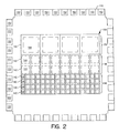


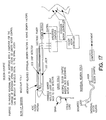


Classifications
-
- G—PHYSICS
- G01—MEASURING; TESTING
- G01N—INVESTIGATING OR ANALYSING MATERIALS BY DETERMINING THEIR CHEMICAL OR PHYSICAL PROPERTIES
- G01N33/00—Investigating or analysing materials by specific methods not covered by groups G01N1/00 - G01N31/00
- G01N33/48—Biological material, e.g. blood, urine; Haemocytometers
- G01N33/50—Chemical analysis of biological material, e.g. blood, urine; Testing involving biospecific ligand binding methods; Immunological testing
- G01N33/58—Chemical analysis of biological material, e.g. blood, urine; Testing involving biospecific ligand binding methods; Immunological testing involving labelled substances
- G01N33/582—Chemical analysis of biological material, e.g. blood, urine; Testing involving biospecific ligand binding methods; Immunological testing involving labelled substances with fluorescent label
-
- G—PHYSICS
- G01—MEASURING; TESTING
- G01N—INVESTIGATING OR ANALYSING MATERIALS BY DETERMINING THEIR CHEMICAL OR PHYSICAL PROPERTIES
- G01N21/00—Investigating or analysing materials by the use of optical means, i.e. using sub-millimetre waves, infrared, visible or ultraviolet light
- G01N21/17—Systems in which incident light is modified in accordance with the properties of the material investigated
- G01N21/25—Colour; Spectral properties, i.e. comparison of effect of material on the light at two or more different wavelengths or wavelength bands
- G01N21/251—Colorimeters; Construction thereof
- G01N21/253—Colorimeters; Construction thereof for batch operation, i.e. multisample apparatus
-
- G—PHYSICS
- G01—MEASURING; TESTING
- G01N—INVESTIGATING OR ANALYSING MATERIALS BY DETERMINING THEIR CHEMICAL OR PHYSICAL PROPERTIES
- G01N21/00—Investigating or analysing materials by the use of optical means, i.e. using sub-millimetre waves, infrared, visible or ultraviolet light
- G01N21/75—Systems in which material is subjected to a chemical reaction, the progress or the result of the reaction being investigated
- G01N21/76—Chemiluminescence; Bioluminescence
- G01N21/763—Bioluminescence
-
- G—PHYSICS
- G01—MEASURING; TESTING
- G01N—INVESTIGATING OR ANALYSING MATERIALS BY DETERMINING THEIR CHEMICAL OR PHYSICAL PROPERTIES
- G01N33/00—Investigating or analysing materials by specific methods not covered by groups G01N1/00 - G01N31/00
- G01N33/48—Biological material, e.g. blood, urine; Haemocytometers
- G01N33/50—Chemical analysis of biological material, e.g. blood, urine; Testing involving biospecific ligand binding methods; Immunological testing
- G01N33/53—Immunoassay; Biospecific binding assay; Materials therefor
- G01N33/543—Immunoassay; Biospecific binding assay; Materials therefor with an insoluble carrier for immobilising immunochemicals
- G01N33/54366—Apparatus specially adapted for solid-phase testing
-
- Y—GENERAL TAGGING OF NEW TECHNOLOGICAL DEVELOPMENTS; GENERAL TAGGING OF CROSS-SECTIONAL TECHNOLOGIES SPANNING OVER SEVERAL SECTIONS OF THE IPC; TECHNICAL SUBJECTS COVERED BY FORMER USPC CROSS-REFERENCE ART COLLECTIONS [XRACs] AND DIGESTS
- Y10—TECHNICAL SUBJECTS COVERED BY FORMER USPC
- Y10S—TECHNICAL SUBJECTS COVERED BY FORMER USPC CROSS-REFERENCE ART COLLECTIONS [XRACs] AND DIGESTS
- Y10S435/00—Chemistry: molecular biology and microbiology
- Y10S435/808—Optical sensing apparatus
-
- Y—GENERAL TAGGING OF NEW TECHNOLOGICAL DEVELOPMENTS; GENERAL TAGGING OF CROSS-SECTIONAL TECHNOLOGIES SPANNING OVER SEVERAL SECTIONS OF THE IPC; TECHNICAL SUBJECTS COVERED BY FORMER USPC CROSS-REFERENCE ART COLLECTIONS [XRACs] AND DIGESTS
- Y10—TECHNICAL SUBJECTS COVERED BY FORMER USPC
- Y10S—TECHNICAL SUBJECTS COVERED BY FORMER USPC CROSS-REFERENCE ART COLLECTIONS [XRACs] AND DIGESTS
- Y10S435/00—Chemistry: molecular biology and microbiology
- Y10S435/973—Simultaneous determination of more than one analyte
-
- Y—GENERAL TAGGING OF NEW TECHNOLOGICAL DEVELOPMENTS; GENERAL TAGGING OF CROSS-SECTIONAL TECHNOLOGIES SPANNING OVER SEVERAL SECTIONS OF THE IPC; TECHNICAL SUBJECTS COVERED BY FORMER USPC CROSS-REFERENCE ART COLLECTIONS [XRACs] AND DIGESTS
- Y10—TECHNICAL SUBJECTS COVERED BY FORMER USPC
- Y10S—TECHNICAL SUBJECTS COVERED BY FORMER USPC CROSS-REFERENCE ART COLLECTIONS [XRACs] AND DIGESTS
- Y10S435/00—Chemistry: molecular biology and microbiology
- Y10S435/975—Kit
-
- Y—GENERAL TAGGING OF NEW TECHNOLOGICAL DEVELOPMENTS; GENERAL TAGGING OF CROSS-SECTIONAL TECHNOLOGIES SPANNING OVER SEVERAL SECTIONS OF THE IPC; TECHNICAL SUBJECTS COVERED BY FORMER USPC CROSS-REFERENCE ART COLLECTIONS [XRACs] AND DIGESTS
- Y10—TECHNICAL SUBJECTS COVERED BY FORMER USPC
- Y10S—TECHNICAL SUBJECTS COVERED BY FORMER USPC CROSS-REFERENCE ART COLLECTIONS [XRACs] AND DIGESTS
- Y10S436/00—Chemistry: analytical and immunological testing
- Y10S436/805—Optical property
-
- Y—GENERAL TAGGING OF NEW TECHNOLOGICAL DEVELOPMENTS; GENERAL TAGGING OF CROSS-SECTIONAL TECHNOLOGIES SPANNING OVER SEVERAL SECTIONS OF THE IPC; TECHNICAL SUBJECTS COVERED BY FORMER USPC CROSS-REFERENCE ART COLLECTIONS [XRACs] AND DIGESTS
- Y10—TECHNICAL SUBJECTS COVERED BY FORMER USPC
- Y10T—TECHNICAL SUBJECTS COVERED BY FORMER US CLASSIFICATION
- Y10T436/00—Chemistry: analytical and immunological testing
- Y10T436/18—Sulfur containing
- Y10T436/186—Sulfur dioxide
Definitions
- the present invention relates to methods for the identification of an analyte in a biological medium using bioluminescence. More particularly, a method is provided for diagnosing diseases employing a solid phase methodology and a luciferase-luciferin bioluminescence generating system. Methods employing biomineralization for depositing silicon on a matrix support are also provided herein.
- Luminescence is a phenomenon in which energy is specifically channeled to a molecule to produce an excited state. Return to a lower energy state is accompanied by release of a photon (hy). Luminescence includes fluorescence, phosphorescence, chemiluminescence and bioluminescence. Bioluminescence is the process by which living organisms emit light that is visible to other organisms. Luminescence may be represented as follows:
- X* is an electronically excited molecule and hy represents light emission upon return of X* to a lower energy state.
- the luminescence is bioluminescence
- creation of the excited state derives from an enzyme catalyzed reaction.
- the color of the emitted light in a bioluminescent (or chemiluminescent or other luminescent) reaction is characteristic of the excited molecule, and is independent from its source of excitation and temperature.
- Luciferases are oxygenases, that act on a substrate, luciferin, in the presence of molecular oxygen and transform the substrate to an excited state. Upon return to a lower energy level, energy is released in the form of light [for reviews see, e.g., McElroy et al. (1966) in Molecular Architecture in Cell Physiology, Hayashi et al., eds., Prentice-Hall, Inc., Englewood Cliffs, N.J., pp.
- Bioluminescence is more common in marine organisms than in terrestrial organisms. Bioluminescence has developed from as many as thirty evolutionarily distinct origins and, thus, is manifested in a variety of ways so that the biochemical and physiological mechanisms responsible for bioluminescence in different organisms are distinct. Bioluminescent species span many genera and include microscopic organisms, such as bacteria [primarily marine bacteria including Vibrio species], fungi, algae and dinoflagellates, to marine organisms, including arthropods, mollusks, echinoderms, and chordates, and terrestrial organism including annelid worms and insects.
- bacteria primarily marine bacteria including Vibrio species
- fungi fungi
- algae and dinoflagellates to marine organisms, including arthropods, mollusks, echinoderms, and chordates
- terrestrial organism including annelid worms and insects.
- Bioluminescence as well as other types of chemiluminescence, is used for quantitative determinations of specific substances in biology and medicine.
- luciferase genes have been cloned and exploited as reporter genes in numerous assays, for many purposes. Since the different luciferase systems have different specific requirements, they may be used to detect and quantify a variety of substances.
- the majority of commercial bioluminescence applications are based on firefly [ Photinus pyralis ] luciferase.
- One of the first and still widely used assays involves the use of firefly luciferase to detect the presence of ATP. It is also used to detect and quantify other substrates or co-factors in the reaction. Any reaction that produces or utilizes NAD(H), NADP(H) or long chain aldehyde, either directly or indirectly, can be coupled to the light-emitting reaction of bacterial luciferase.
- Aequorin system Another luciferase system that has been used commercially for analytical purposes is the Aequorin system.
- the purified jellyfish photoprotein, aequorin is used to detect and quantify intracellular Ca 2+ and its changes under various experimental conditions.
- the Aequorin photoprotein is relatively small [ ⁇ 20 kDa], nontoxic, and can be injected into cells in quantities adequate to detect calcium over a large concentration range [3 ⁇ 10 ⁇ 7 to 10 ⁇ 4 M].
- luciferases and substrates have been studied and well-characterized and are commercially available [e.g., firefly luciferase is available from Sigma, St. Louis, Mo., and Boehringer Mannheim Biochemicals, Indianapolis, Ind.; recombinantly produced firefly luciferase and other reagents based on this gene or for use with this protein are available from Promega Corporation, Madison, Wis.; the aequorin photoprotein luciferase from jellyfish and luciferase from Renilla are commercially available from Sealite Sciences, Bogart, Ga.; coelenterazine, the naturally-occurring substrate for these luciferases, is available from Molecular Probes, Eugene, Oreg.]. These luciferases and related reagents are used as reagents for diagnostics, quality control, environmental testing and other such analyses.
- Microelectronics, chip arrays and other solid phase spacially addressable arrays have been developed for use in diagnostics and other applications. At present, methods for detection of positive results are inadequate or inconvenient. There exists a need for improved, particularly more rapid detection methods.
- a method for diagnosing diseases, particularly infectious diseases, using chip methodology and a luciferase-luciferin bioluminescence generating system is also provided herein.
- the chip includes an integrated photodetector that detects the photons emitted by the bioluminescence generating system.
- the method may be practiced with any suitable chip device, including self-addressable and non-self addressable formats, that is modified as described herein for detection of generated photons by the bioluminescence generating systems.
- the chip device provided herein is adaptable for use in an array format for the detection and identification of infectious agents in biological specimens.
- the chip is fabricated by suitably derivatizing the matrix for linkage of macromolecules, and including linkage of photodiodes, photomultipliers CCD (charge coupled device) or other suitable detector, for measuring light production; attaching an appropriate macromolecule, such as a biological molecule or anti-ligand, e.g., a receptor, such as an antibody, to the chip, preferably to an assigned location thereon.
- an appropriate macromolecule such as a biological molecule or anti-ligand, e.g., a receptor, such as an antibody
- Photodiodes are presently among the preferred detectors, and specified herein. It is understood, however, that other suitable detectors may be substituted therefor.
- the chip is made using an integrated circuit with an array, such as an X-Y array, of photodetectors.
- the surface of circuit is treated to render it inert to conditions of the diagnostic assays for which the chip is intended, and is adapted, such as by derivatization for linking molecules, such as antibodies.
- a selected antibody or panel of antibodies, such as an antibody specific for particularly bacterial antigen, is affixed to the surface of the chip above each photodetector. After contacting the chip with a test sample, the chip is contacted a second antibody linked to a component of a bioluminescence generating system, such as a luciferase or luciferin, specific for the antigen.
- the remaining components of the bioluminescence generating reaction are added, and, if any of the antibodies linked to a component of a bioluminescence generating system are present on the chip, light will be generated and detected by the adjacent photodetector.
- the photodetector is operatively linked to a computer, which is programmed with information identifying the linked antibodies, records the event, and thereby identifies antigens present in the test sample.
- the chip is employed in any desired assay, such as an assay for infectious disease or antibiotic sensitivity, by, for example, linking an antibody or a panel of antibodies, to the surface, contacting the chip with a test sample of a body fluid, such as urine, blood and cerebral spinal fluid (CFS), for a sufficient time, depending upon assay format, such as to bind the a target in the sample; washing the chip and then incubating with a secondary antibody conjugated to a luciferase or an antibody:luciferase fusion protein; initiating the bioluminescent reaction; detecting light emitted at each location bound with a target through the photodiode in the chip; transferring the electronic signal from the chip to a computer for analysis.
- a body fluid such as urine, blood and cerebral spinal fluid (CFS)
- CFS cerebral spinal fluid
- a secondary antibody conjugated to a luciferase or an antibody:luciferase fusion protein initiating the bioluminescent reaction
- the chip is a nonself-addressable, microelectronic device for detecting photons of light emitted by light-emitting chemical reactions.
- the device includes a substrate, an array of loci, herein designated micro-locations, defined thereon, and an independent photodetector optically coupled to each micro-location.
- Each micro-location holds a separate chemical reactant that will emit photons of light when a reaction takes place thereat.
- Each photodetector generates a sensed signal responsive to the photons emitted at the corresponding micro-location when the reaction takes place thereat, and each photodetector is independent from the other photodetectors.
- the device also includes an electronic circuit that reads the sensed signal generated by each photodetector and generates output data signals therefrom. The output data signals are indicative of the light emitted at each micro-location.
- a microelectronic device for detecting and identifying analytes in a fluid sample using light-emitting reactions.
- the device includes a substrate, an array of micro-locations defined thereon for receiving the fluid sample to be analyzed, a separate targeting agent attached to an attachment layer of each micro-location, and an independent photodetector optically coupled to each micro-location.
- Each targeting agent is, preferably, specific for binding a selected analyte that may be present in the received sample.
- Each photodetector generates a sensed signal responsive to photons of light emitted at the corresponding micro-location when the selected analyte bound thereto is exposed to a secondary binding agent also specific for binding the selected analyte or the targeting agent-selected analyte complex and linked to one or more components of a light-emitting reaction.
- the chip is then reacted with the remaining components to emit the photons when the selected analyte is present.
- An electronic circuit reads the sensed signal generated by each photodetector and generates output data signals therefrom that are indicative of the light emitted at each micro-location.
- a microelectronic device for detecting and identifying analytes in a biological sample using luciferase-luciferin bioluminescence.
- the device includes a substrate, an array of micro-locations defined thereon for receiving the sample to be analyzed, a separate anti-ligand, such as a receptor antibody, attached to an attachment layer of each micro-location, and an independent photodetector optically coupled to each micro-location.
- a separate anti-ligand such as a receptor antibody
- Each receptor antibody is specific for binding a selected analyte that may be present in the received sample.
- Each photodetector generates a sensed signal responsive to bioluminescence emitted at the corresponding micro-location when the selected analyte bound to the corresponding receptor antibody is exposed to a secondary antibody also specific to the selected analyte or to the receptor antibody-selected analyte complex and linked to one or more components of a luciferase-luciferin reaction, and is then reacted with the remaining components to generate the bioluminescence when the selected analyte is present.
- An electronic circuit reads the sensed signal from each photodetector and generates output data signals therefrom. The output data signals are indicative of the bioluminescence emitted at each micro-location by the reaction.
- a method of detecting and identifying analytes in a biological sample using luciferase-luciferin bioluminescence includes providing a microelectronic device having a surface with an array of micro-locations defined thereon, derivatizing the surface to permit or enhance the attachment of a receptor antibody or plurality of antibodies thereto at each micro-location, and attaching a specific receptor antibody or plurality thereof to the surface at each micro-location.
- the selected antibody is specific for binding to a selected analyte that may be present in the sample.
- the method also includes applying the sample to the surface such that the selected analytes will bind to the receptor antibody attached to the surface at each micro-location, washing the sample from the surface after waiting a sufficient period of time for the selected analytes to bind with the receptor antibody at each micro-location, exposing the surface to a secondary antibody specific to bind the selected analyte already bound to the receptor antibody at each micro-location when the selected analyte is present, the secondary antibody linked to one of a luciferase and a luciferin, and initiating the reaction by applying the other of the luciferase and luciferin to the surface.
- the method also includes detecting photons of light emitted by the reaction using a photodetector optically coupled to each micro-location, each photodetector generating a sensed signal representative of the bio-luminescent activity thereat, reading the sensed signal from each photodetector and generating output data signals therefrom indicative of the bioluminescence emitted at each micro-location by the reaction.
- a system for detecting and identifying analytes in a biological sample using luciferase-luciferin bioluminescence includes: a microelectronic device including an array of micro-locations for receiving the sample; a separate receptor antibody attached to an attachment layer of each micro-location, each receptor antibody is specific for a selected analyte that may be present in the received sample; a photodetector that generates a sensed signal responsive to bioluminescence emitted at the corresponding micro-location when the selected analyte bound to the corresponding receptor antibody is exposed to a secondary antibody also specific to the selected analyte and linked to one of a luciferase and a luciferin, and is then reacted with the other of the luciferase and luciferin to generate the bioluminescence when the selected analyte is present, and an electronic circuit which reads the sensed signal from each photodetector and generates output data signals there
- the system includes a processing instrument including an input interface circuit for receiving the output data signals indicative of the bioluminescence emitted at each micro-location, a memory circuit for storing a data acquisition array having a location associated with each micro-location, an output device for generating visible indicia in response to an output device signal and a processing circuit.
- the processing circuit reads the output data signals received by the input interface circuit, correlates these signals with the corresponding micro-locations, integrates the correlated output data signals for a desired time period by accumulating them in the data acquisition array, and generates the output device signal which, when applied to the output device, causes the output device to generate visible indicia related to the presence of the selected analytes.
- the chip is self-addressable.
- self-addressable chips in the method, presently preferred are those adaptable to microelectronic self addressable, self-assembling chips and systems, such as those described in International PCT application Nos. WO 95/12808; WO 96/01836 and WO 96/07917 and also in arrays, such as those described in U.S. Pat. No. 5,451,683, which are each herein incorporated by reference.
- the self-addressable chips are such that each individual well may be addressed one at a time in the presence of the rest by changing the charge at a single microlocation and then sending the analytes or reagents via free flow electrophoresis throughout, but assembly occurs only at that location after the chip has been assembled.
- These devices are modified for use in the methods herein by replacing the disclosed detection means with the luciferase/luciferin systems.
- electrodes are located at the bottom and top of each well, respectively, to allow for the delivery of analytes and reagents by free flow electrophoresis.
- the antibodies are attached to each location on a MYLAR (oriented polyethylene terephthalate) layer prior to assembling the chip (using, for example, a dot matrix printer).
- MYLAR oriented polyethylene terephthalate
- it is nonself-addressable in that is has a plurality of individual wells each containing a photodiode incorporated into the semiconductor layer at the bottom of each well.
- specific anti-ligands e.g., antibodies
- a middle reflective support matrix such as heat stable MYLAR
- the sample is contacted with the chip, washed and a plurality of secondary antibody-luciferase conjugates or protein fusions are added.
- the wells are washed and the remaining components of the bioluminescent reaction are added to initiate the reaction.
- Light produced in a well is detected by the photodiode, photomultiplier, CCD (charge coupled device) or other suitable detector in the semiconductor layer and the signal is relayed to a processing unit, typically a computer.
- the processing unit displays the well or wells that are positive. Each well corresponds to a particular ligand, thereby permitting identification of the infectious agents. All steps may be automated.
- the design, fabrication, and uses of nonself-addressable and programmable, self-addressable and self-assembling microelectronic systems and devices which actively carry out controlled multi-step and multiplex reactions in microscopic formats for detecting the electromagnetic emissions of a bioluminescent reaction are provided herein.
- the reactions include, but are not limited to, most molecular biological procedures, such as nucleic acid and protein nucleic acid hybridizations, antibody/antigen reactions, and related clinical diagnostics.
- the resulting chips which includes a silicon matrix and photodiodes or other light detecting means, are provided.
- the silicon may be deposited using enzymatic deposition, similar to the enzymatic deposition by radiolarains and diatoms.
- Such silica has an absorption maxima at about 705 nm, which is the wavelength emitted by Aristostomias bioluminescence generating system.
- Enzymatic methods for depositing silicon on the surface of a matrix are also provided herein.
- a suitable enzyme particularly, acetylcholine esterase is fused to a luciferase, such as by recombinant expression.
- the luciferase is either in an inactive or active conformation. Suitable mutations in either protein may be selected to insure that luciferase can undergo appropriate conformational changes as described herein.
- the resulting fusion is attached to a chip, such as a chip provided herein.
- the linked luciferase is, if previously inactive, is activated by the binding, or if previously active, is inactivated by the binding.
- FIG. 1 is a schematic block diagram of a microelectronic device for detecting and identifying analytes in a biological sample using bioluminescence, the microelectronic device including an array of micro-locations and a photodetector optically coupled to each micro-location for detecting the bioluminescence emitted at the corresponding micro-location;
- FIG. 2 is a top view of the die for the microelectronic device of FIG. 1 showing the photodetector array disposed on a semiconductor substrate;
- FIG. 3 is a perspective view of the microelectronic device of FIG. 1 including the die of FIG. 2 housed in a ceramic dual in-line package (DIP), and FIG. 3A is a magnified view showing the test well formed in the DIP in detail;
- DIP ceramic dual in-line package
- FIG. 4 is a schematic diagram showing a pixel unit cell circuit for detecting the bioluminescence emitted at each micro-location in the array;
- FIG. 5 is a graph showing the voltage levels at three nodes of the pixel unit cell circuit of FIG. 4 as a function of time during operation of the device;
- FIG. 6 is a system block diagram showing the microelectronics device of FIG. 1 mounted on an adaptor circuit board and serially interfaced to a computer programmed to read the serial output data stream, to correlate the output data with the array of micro-locations, to integrate the data correlated with each micro-location for a predetermined time period set using an input device, to identify the analytes present in the biological medium by reference to an analyte map, and to display the results on an output device; and
- FIG. 7 shows the microelectronics device of FIG. 1 received on a circuit board which does not require the user to directly handle the package.
- FIG. 8 is a schematic cross-sectional diagram of a three layer multi-well CCD chip (a chip containing a photodiode/CCD).
- FIG. 9 shows a blown-up schematic diagram of a multi-well CCD chip bottom layer and middle reflective layer and schematic diagram of an individual well.
- FIG. 10 shows a blown-up schematic diagram of specific antibodies attached to the middle reflective layer of the multi-well CCD chip of FIG. 8 .
- FIG. 11 is a cross-section of an individual well indicating the relative positions of the CCD, reflective mirror layer and the cathode and anode. Antibodies attached to the middle reflective layer hang inverted above the photodiode. Bound antigen is detected using an antibody-luciferase fusion protein, and light generated from the bioluminescent reaction is detected by the photodiode and relayed to a processing unit for identification.
- FIG. 12 is the cross-section of three self-addressable micro-locations fabricated using microlithographic techniques [see, international PCT application No. WO 96/01836]. Included are arrows denoting the positioning of photodiodes.
- FIG. 13 is the cross-section of a microlithographically fabricated micro-location; antibodies or other receptors are linked to the attachment layer.
- FIG. 14 is a schematic representation of a self-addressable 64 micro-location chip which was actually fabricated, addressed with oligonucleotides, and tested.
- FIG. 15 shows a blown-up schematic diagram of a micro-machined 96 micro-locations device.
- FIG. 16 is the cross-section of a micro-machined device.
- FIG. 17 shows a schematic representation of an artificial silicon-synapse.
- FIG. 18 shows a detailed schematic view of an acetylcholine esterase-luciferase fusion protein and an acetylcholine esterase-fluorochrome conjugate used in the silicon-synapse.
- FIG. 19 depicts the methodology for the placement of silicon-synapses and electrodes in the human spinal cord to bypass a permenant spinal cord lesion.
- FIG. 20 depicts a scheme for operation of chips described herein in diagnostic assays for detecting infectious microorganisms.
- Insect bioluminescence generating systems including fireflies, click beetles, and other insect systems
- chemiluminescence refers to a chemical reaction in which energy is specifically channeled to a molecule causing it to become electronically excited and subsequently to release a photon thereby emitting visible light. Temperature does not contribute to this channeled energy. Thus, chemiluminescence involves the direct conversion of chemical energy to light energy.
- Bioluminescence refers to the subset of chemiluminescence reactions that involve luciferins and luciferases (or the photoproteins). Bioluminescence does not herein include phosphorescence.
- bioluminescence which is a type of chemiluminescence, refers to the emission of light by biological molecules, particularly proteins.
- the essential condition for bioluminescence is molecular oxygen, either bound or free in the presence of an oxygenase, a luciferase, which acts on a substrate, a luciferin.
- Bioluminescence is generated by an enzyme or other protein [luciferase] that is an oxygenase that acts on a substrate luciferin [a bioluminescence substrate] in the presence of molecular oxygen and transforms the substrate to an excited state, which upon return to a lower energy level releases the energy in the form of light.
- luciferin and luciferase are generically referred to as luciferin and luciferase, respectively.
- each generic term is used with the name of the organism from which it derives, for example, bacterial luciferin or firefly luciferase.
- luciferase refers to oxygenases that catalyze a light emitting reaction.
- bacterial luciferases catalyze the oxidation of flavin mononucleotide [FMN] and aliphatic aldehydes, which reaction produces light.
- Another class of luciferases found among marine arthropods, catalyzes the oxidation of Cypridina [Vargula] luciferin, and another class of luciferases catalyzes the oxidation of Coleoptera luciferin.
- luciferase refers to an enzyme or photoprotein that catalyzes a bioluminescent reaction [a reaction that produces bioluminescence].
- the luciferases such as firefly and Renilla luciferases, that are enzymes which act catalytically and are unchanged during the bioluminescence generating reaction.
- the luciferase photoproteins such as the aequorin and obelin photoproteins to which luciferin is non-covalently bound, are changed, such as by release of the luciferin, during bioluminescence generating reaction.
- the luciferase is a protein that occurs naturally in an organism or a variant or mutant thereof, such as a variant produced by mutagenesis that has one or more properties, such as thermal or pH stability, that differ from the naturally-occurring protein. Luciferases and modified mutant or variant forms thereof are well known.
- Renilla luciferase means an enzyme isolated from member of the genus Renilla or an equivalent molecule obtained from any other source, such as from another Anthozoa, or that has been prepared synthetically.
- the luciferases and luciferin and activators thereof are referred to as bioluminescence generating reagents or components. Typically, a subset of these reagents will be provided during the assay or otherwise immobilized at particular locations on the surface of the chip. Bioluminescence will be produced upon contacting the chip surface with the remaining reagents and the light produced is detected by the photodiodes at those locations of the array where a specific target has been detected by the immobilized anti ligand.
- the component luciferases, luciferins, and other factors, such as O 2 , Mg 2+ , Ca 2+ are also referred to as bioluminescence generating reagents [or agents or components].
- photoprotein acts as a catalyst to promote the oxidation of the substrate, but it is changed in the reaction, since the bound substrate is oxidized and bound molecular oxygen is used in the reaction.
- photoproteins are regenerated by addition of the substrate and molecular oxygen under appropriate conditions known to those of skill in this art.
- bioluminescence substrate refers to the compound that is oxidized in the presence of a luciferase, and any necessary activators, and generates light. These substrates are referred to as luciferins, which are substrates that undergo oxidation in a bioluminescence reaction. These bioluminescence substrates include any luciferin or analog thereof or any synthetic compound with which a luciferase interacts to generate light. Preferred substrates are those that are oxidized in the presence of a luciferase or protein in a light-generating reaction. Bioluminescence substrates, thus, include those compounds that those of skill in the art recognize as luciferins.
- Luciferins for example, include firefly luciferin, Cypridina [also known as Vargula] luciferin [coelenterazine], bacterial luciferin, as well as synthetic analogs of these substrates or other compounds that are oxidized in the presence of a luciferase in a reaction the produces bioluminescence.
- Cypridina also known as Vargula] luciferin [coelenterazine]
- bacterial luciferin as well as synthetic analogs of these substrates or other compounds that are oxidized in the presence of a luciferase in a reaction the produces bioluminescence.
- capable of conversion into a bioluminescence substrate means susceptible to chemical reaction, such as oxidation or reduction, that yields a bioluminescence substrate.
- the luminescence producing reaction of bioluminescent bacteria involves the reduction of a flavin mononucleotide group (FMN) to reduced flavin mononucleotide (FMNH 2 ) by a flavin reductase enzyme.
- the reduced flavin mononucleotide [substrate] then reacts with oxygen [an activator] and bacterial luciferase to form an intermediate peroxy flavin that undergoes further reaction, in the presence of a long-chain aldehyde, to generate light.
- the reduced flavin and the long chain aldehyde are substrates.
- bioluminescence system refers to the set of reagents required for a bioluminescence-producing reaction.
- luciferase, luciferin and other substrates, solvents and other reagents that may be required to complete a bioluminescent reaction form a bioluminescence system.
- a bioluminescence system (or equivalently a bioluminescence generating system) refers to any set of reagents that, under appropriate reaction conditions, yield bioluminescence.
- Appropriate reaction conditions refers to the conditions necessary for a bioluminescence reaction to occur, such as pH, salt concentrations and temperature.
- bioluminescence systems include a bioluminescence substrate (a luciferin), a luciferase, which includes enzymes luciferases and photoproteins, and one or more activators.
- a bioluminescence substrate a luciferin
- a luciferase which includes enzymes luciferases and photoproteins
- activators one or more activators.
- a particular bioluminescence system may be identified by reference to the specific organism from which the luciferase derives; for example, the Vargula [also called Cypridina] bioluminescence system (or Vargula system) includes a Vargula luciferase, such as a luciferase isolated from the ostracod, Vargula or produced using recombinant means or modifications of these luciferases.
- This system would also include the particular activators necessary to complete the bioluminescence reaction, such as oxygen and a substrate with which the luciferase reacts in the presence of the oxygen
- ATP, AMP, NAD+ and NADH refer to adenosine triphosphate, adenosine monophosphate, nicotinamide adenine dinucleotide (oxidized form) and nicotinamide adenine dinucleotide (reduced form), respectively.
- production by recombinant means by using recombinant DNA methods means the use of the well known methods of molecular biology for expressing proteins encoded by cloned DNA.
- substantially identical to a product means sufficiently similar so that the property of interest is sufficiently unchanged so that the substantially identical product can be used in place of the product.
- substantially pure means sufficiently homogeneous to appear free of readily detectable impurities as determined by standard methods of analysis, such as thin layer chromatography (TLC), gel electrophoresis and high performance liquid chromatography (HPLC), used by those of skill in the art to assess such purity, or sufficiently pure such that further purification would not detectably alter the physical and chemical properties, such as enzymatic and biological activities, of the substance.
- TLC thin layer chromatography
- HPLC high performance liquid chromatography
- Methods for purification of the compounds to produce substantially chemically pure compounds are known to those of skill in the art.
- a substantially chemically pure compound may, however, be a mixture of stereoisomers. In such instances, further purification might increase the specific activity of the compound.
- “Complementary,” when referring to two nucleotide sequences, means that the two sequences of nucleotides are capable of hybridizing, preferably with less than 25%, more preferably with less than 15%, even more preferably with less than 5%, most preferably with no mismatches between opposed nucleotides. Preferably the two molecules will hybridize under conditions of high stringency.
- peptide nucleic acid refers to nucleic acid analogs in which the ribose-phosphate backbone is replaced by a backbone held together by amide bonds.
- substantially varies with the context as understood by those skilled in the relevant art and generally means at-least 70%, preferably means at least 80%, more preferably at least 90%, and most preferably at least 95%.
- biological activity refers to the in vivo activities of a compound or physiological responses that result upon administration of a compound, composition or other mixture. Biological activities may be observed in in vitro systems designed to test or use such activities.
- biological activity of a luciferase is its oxygenase activity whereby, upon oxidation of a substrate, light is produced.
- composition refers to a any mixture. It may be a solution, a suspension, liquid, powder, a paste, aqueous, non-aqueous or any combination thereof.
- a combination refers to any association between two or among more items.
- fluid refers to any composition that can flow. Fluids thus encompass compositions that are in the form of semi-solids, pastes, solutions, aqueous mixtures, gels, lotions, creams and other such compositions.
- macromolecules are intended to generically encompass all molecules that would be linked to a solid support for diagnostic assays.
- the macromolecules include, but are not limited to: proteins, organic molecules, nucleics acids, viruses, viral capsids, phage, cells or membranes thereof or portions of viruses, viral capsids, phage, cells or membranes.
- macromolecules that specifically bind to an analyte of interest are those present in body fluids and other biological samples.
- a receptor refers to a molecule that has an affinity for a given ligand.
- Receptors may be naturally-occurring or synthetic molecules.
- Receptors may also be referred to in the art as anti-ligands.
- the receptor and anti-ligand are interchangeable.
- Receptors can be used in their unaltered state or as aggregates with other species.
- Receptors may be attached, covalently or noncovalently, or in physical contact with, to a binding member, either directly or indirectly via a specific binding substance or linker.
- receptors include, but are not limited to: antibodies, cell membrane receptors surface receptors and internalizing receptors, monoclonal antibodies and antisera reactive with specific antigenic determinants [such as on viruses, cells, or other materials], drugs, polynucleotides, nucleic acids, peptides, cofactors, lectins, sugars, polysaccharides, cells, cellular membranes, and organelles.
- receptors and applications using such receptors include but are not restricted to:
- enzymes specific transport proteins or enzymes essential to survival of microorganisms, which could serve as targets for antibiotic [ligand] selection;
- b) antibodies identification of a ligand-binding site on the antibody molecule that combines with the epitope of an antigen of interest may be investigated; determination of a sequence that mimics an antigenic epitope may lead to the development of vaccines of which the immunogen is based on one or more of such sequences or lead to the development of related diagnostic agents or compounds useful in therapeutic treatments such as for auto-immune diseases
- nucleic acids identification of ligand, such as protein or RNA, binding sites;
- catalytic polypeptides polymers, preferably polypeptides, that are capable of promoting a chemical reaction involving the conversion of one or more reactants to one or more products; such polypeptides generally include a binding site specific for at least one reactant or reaction intermediate and an active functionality proximate to the binding site, in which the functionality is capable of chemically modifying the bound reactant [see, e.g., U.S. Pat. No. 5,215,8991;
- hormone receptors determination of the ligands that bind with high affinity to a receptor is useful in the development of hormone replacement therapies; for example, identification of ligands that bind to such receptors may lead to the development of drugs to control blood pressure; and
- opiate receptors determination of ligands that bind to the opiate receptors in the brain is useful in the development of less-addictive replacements for morphine and related drugs.
- antibody includes antibody fragments, such as Fab fragments, which are composed of a light chain and the variable region of a heavy chain.
- complementary refers to the topological compatibility or matching together of interacting surfaces of a ligand molecule and its receptor.
- the receptor and its ligand can be described as complementary, and furthermore, the contact surface characteristics are complementary to each other.
- a ligand-receptor pair or complex formed when two macromolecules have combined through molecular recognition to form a complex.
- an epitope refers to a portion of an antigen molecule that is delineated by the area of interaction with the subclass of receptors known as antibodies.
- a ligand is a molecule that is specifically recognized by a particular receptor.
- ligands include, but are not limited to, agonists and antagonists for cell membrane receptors, toxins and venoms, viral epitopes, hormones [e.g., steroids], hormone receptors, opiates, peptides, enzymes, enzyme substrates, cofactors, drugs, lectins, sugars, oligonucleotides, nucleic acids, oligosaccharides, proteins, and monoclonal antibodies.
- an anti-ligand is a molecule that has a known or unknown affinity for a given ligand and can be immobilized on a predefined region of the surface. Anti-ligands may be naturally-occurring or manmade molecules. Also, they can be employed in their unaltered state or as aggregates with other species. Anti-ligands may be reversibly attached, covalently or noncovalently, to a binding member, either directly or via a specific binding substance. By “reversibly attached” is meant that the binding of the anti-ligand (or specific binding member or ligand) is reversible and has, therefore, a substantially non-zero reverse, or unbinding, rate.
- reversible attachments can arise from noncovalent interactions, such as electrostatic forces, van der Waals forces, hydrophobic (i.e., entropic) forces, and the like. Furthermore, reversible attachments also may arise from certain, but not all covalent bonding reactions. Examples include, but are not limited to, attachment by the formation of hemiacetals, hemiketals, imines, acetals, ketals, and the like (See, Morrison et al., “Organic Chemistry”, 2nd ed., ch. 19 (1966), which is incorporated herein by reference).
- anti-ligands which can be employed in the methods and devices herein include, but are not limited to, cell membrane receptors, monoclonal antibodies and antisera reactive with specific antigenic determinants (such as on viruses, cells or other materials), hormones, drugs, oligonucleotides, peptides, peptide nucleic acids, enzymes, substrates, cofactors, lectins, sugars, oligosaccharides, cells, cellular membranes, and organelles.
- specific antigenic determinants such as on viruses, cells or other materials
- hormones drugs, oligonucleotides, peptides, peptide nucleic acids, enzymes, substrates, cofactors, lectins, sugars, oligosaccharides, cells, cellular membranes, and organelles.
- a substrate refers to any matrix that is used either directly or following suitable derivatization, as a solid support for chemical synthesis, assays and other such processes.
- Preferred substrates herein are silicon substrates or siliconized substrates that are derivatized on the surface intended for linkage of anti-ligands and ligands and other macromolecules, including the fluorescent proteins, phycobiliproteins and other emission shifters.
- a matrix refers to any solid or semisolid or insoluble support on which the molecule of interest, typically a biological molecule, macromolecule, organic molecule or biospecific ligand is linked or contacted.
- a matrix is a substrate material having a rigid or semi-rigid surface.
- at least one surface of the substrate will be substantially flat, although in some embodiments it may be desirable to physically separate synthesis regions for different polymers with, for example, wells, raised regions, etched trenches, or other such topology.
- Matrix materials include any materials that are used as affinity matrices or supports for chemical and biological molecule syntheses and analyses, such as, but are not limited to: polystyrene, polycarbonate, polypropylene, nylon, glass, dextran, chitin, sand, pumice, polytetrafluoroethylene, agarose, polysaccharides, dendrimers, buckyballs, polyacrylamide, Kieselguhr-polyacrlamide non-covalent composite, polystyrene-polyacrylamide covalent composite, polystyrene-PEG polyethyleneglycoll composite, silicon, rubber, and other materials used as supports for solid phase syntheses, affinity separations and purifications, hybridization reactions, immunoassays and other such applications.
- the attachment layer refers the surface of the chip device to which molecules are linked.
- the chip is a semiconductor device, which is coated on a least a portion of the surface to render it suitable for linking molecules and inert to any reactions to which the device is exposed.
- Molecules are linked either directly or indirectly to the surface, linkage may be effected by absorption or adsorption, through covalent bonds, ionic interactions or any other interaction.
- the attachment layer is adapted, such as by derivatization for linking the molecules.
- a bioluminescence generating system refers to the components that are necessary and sufficient to generate bioluminescence. These include a luciferase, luciferin and any necessary co-factors or conditions. Virtually any bioluminescence generating system known to those of skill in the art will be amenable to use in the apparatus, systems, combinations and methods provided herein. Factors for consideration in selecting a bioluminescence generating system, include, but are not limited to: the desired assay and biological fluid used in combination with the bioluminescence; the medium in which the reaction is run; stability of the components, such as temperature or pH sensitivity; shelf life of the components; sustainablity of the light emission, whether constant or intermittent; availability of components; desired light intensity; and other such factors.
- bioluminescence refers to an energy-yielding chemical reaction in which a specific chemical substrate, a luciferin, undergoes oxidation, catalyzed by an enzyme, a luciferase. Bioluminescent reactions are easily maintained, requiring only replenishment of exhausted luciferin or other substrate or cofactor or other protein, in order to continue or revive the reaction. Bioluminescence generating reactions are well known to those of skill in this art and any such reaction may be adapted for use in combination with apparatus, systems and methods described herein.
- bioluminescent orgamisms contemplated for use as sources of bioluminescence generating systems herein include, but are not limited to, Gonadostomias, Gaussia, Halisturia, Vampire squid, Glyphus, Myocotophids (fish), Vinciguerria, Howella, Florenciella, Chaudiodus, Melanocostus, Paracanthus, Atolla, Pelagia, Pitilocarpus, Acanthophyra, Siphonophore, Periphylla and Sea Pens (Stylata).
- a bioluminescence generating system may be isolated from natural sources, such as those in the above Table, or may be produced synthetically.
- the components need only be sufficiently pure so that mixture thereof, under appropriate reaction conditions, produces a glow.
- a crude extract or merely grinding up the organism may be adequate.
- substantially pure components are used, but, where necessary, the precise purity can be determined empirically.
- components may be synthetic components that are not isolated from natural sources.
- DNA encoding luciferases is available [see, e., SEQ ID Nos. 1-13] and has been modified [see, e.g., SEQ ID Nos. 3 and 10-13] and synthetic and alternative substrates have been devised.
- the DNA listed herein is only representative of the DNA encoding luciferases that is available.
- bioluminescence generating system whether synthetic or isolated form natural sources, such as those set forth in Table 1, elsewhere herein or known to those of skill in the art, is intended for use in the chip devices, combinations, systems and methods provided herein.
- Luciferases refer to any compound that, in the presence of any necessary activators, catalyze the oxidation of a bioluminescence substrate [luciferin] in the presence of molecular oxygen, whether free or bound, from a lower energy state to a higher energy state such that the substrate, upon return to the lower energy state, emits light.
- luciferase is broadly used to encompass enzymes that act catalytically to generate light by oxidation of a substrate and also photoproteins, such as aequorin, that act, though not strictly catalytically [since such proteins are exhausted in the reaction], in conjunction with a substrate in the presence of oxygen to generate light.
- luciferases including photoproteins, such as aequorin, are herein also included among the luciferases.
- These reagents include the naturally-occurring luciferases [including photoproteins], proteins produced by recombinant DNA, and mutated or modified variants thereof that retain the ability to generate light in the presence of an appropriate substrate, co-factors and activators or any other such protein that acts as a catalyst to oxidize a substrate, whereby light is produced.
- bioluminescence encompasses light produced by reactions that are catalyzed by [in the case of luciferases that act enzymatically] or initiated by [in the case of the photoproteins, such as aequorin, that are not regenerated in the reaction] a biological protein or analog, derivative or mutant thereof.
- these catalytic proteins are referred to as luciferases and include enzymes such as the luciferases that catalyze the oxidation of luciferin, emitting light and releasing oxyluciferin. Also included among luciferases are photoproteins, which catalyze the oxidation of luciferin to emit light but are changed in the reaction and must be reconstituted to be used again.
- the luciferases may be naturally occurring or may be modified, such as by genetic engineering to improve or alter certain properties. As long as the resulting molecule retains the ability to catalyze the bioluminescent reaction, it is encompassed herein.
- any protein that has luciferase activity [a protein that catalyzes oxidation of a substrate in the presence of molecular oxygen to produce light as defined herein] may be used herein.
- the preferred luciferases are those that are described herein or that have minor sequence variations. Such minor sequence variations include, but are not limited to, minor allelic or species variations and insertions or deletions of residues, particularly cysteine residues. Suitable conservative substitutions of amino acids are known to those of skill in this art and may be made generally without altering the biological activity of the resulting molecule. Those of skill in this art recognize that, in general, single amino acid substitutions in non-essential regions of a polypeptide do not substantially alter biological activity (see, e.g., Watson et al. Molecular Biology of the Gene, 4th Edition, 1987, The Benjamin/Cummings Pub. co., p.224). Such substitutions are preferably made in accordance with those set forth in TABLE 2 as follows:
- the luciferases may be obtained commercially, isolated from natural sources, expressed in host cells using DNA encoding the luciferase, or obtained in any manner known to those of skill in the art. For purposes herein, crude extracts obtained by grinding up selected source organisms may suffice. Since large quantities of the luciferase may be desired, isolation of the luciferase from host cells is preferred. DNA for such purposes is widely available as are modified forms thereof.
- luciferases include, but are not limited to, those isolated from the ctenophores Mnemiopsis (mnemiopsin) and Beroe ovata (berovin), those isolated from the coelenterates Aequorea (aequorin), Obelia (obelin), Pelagia, the Renilla luciferase, the luciferases isolated from the mollusca Pholas (pholasin), the luciferases isolated from the Aristostomias and Porichthys fish and from the ostracods, such as Cypridina (also referred to as Vargula).
- Cypridina also referred to as Vargula
- luciferases for use herein are the Aequorin protein, Renilla luciferase and Cypridina [also called Vargula] luciferase [see, e.g., SEQ ID Nos. 1, 2, and 4-13]. Also, preferred are luciferases which react to produce red and/or near infrared light. These include luciferases found in species of Aristostomias, such as A. scintillans, Pachystomias, Malacosteus, such as M. niger.
- the substrates for the reaction include any molecule(s) with which the luciferase reacts to produce light.
- molecules include the naturally-occurring substrates, modified forms thereof, and synthetic substrates [see, e.g., U.S. Pat. Nos. 5,374,534 and 5,098,828].
- Exemplary luciferins include those described herein, as well as derivatives thereof, analogs thereof, synthetic substrates, such as dioxetanes [see, e.g., U.S. Pat. Nos. 5,004,565 and 5,455,3571, and other compounds that are oxidized by a luciferase in a light-producing reaction [see, e.g., U.S. Pat. Nos. 5,374,534, 5,098,828 and 4,950,588].
- Such substrates also may be identified empirically by selecting compounds that are oxidized in bioluminescent reactions.
- bioluminescence generating systems also require additional components discussed herein and known to those of skill in the art. All bioluminescent reactions require molecular oxygen in the form of dissolved or bound oxygen. Thus, molecular oxygen, dissolved in water or in air or bound to a photoprotein, is the activator for bioluminescence reactions. Depending upon the form of the components, other activators include, but are not limited to, ATP [for firefly luciferase], flavin reductase [bacterial systems] for regenerating FMNH 2 from FMN, and Ca 2+ or other suitable metal ion [aequorin].
- mixtures will also contain any additional salts or buffers or ions that are necessary for each reaction to proceed. Since these reactions are well-characterized, those of skill in the art will be able to determine precise proportions and requisite components. Selection of components will depend upon the chip device and system, the assay to be preformed and the luciferase. Various embodiments are described and exemplified herein; in view of such description, other embodiments will be apparent.
- bioluminescence generating system up to all but one component of a bioluminescence generating system will be bound directly or indirectly to the appropriate locations of the chip or otherwise immobilized at those positions of the array in which the presence of analyte, preferably an infectious agent, is detected.
- analyte preferably an infectious agent
- the remaining component(s) will be added to the surface of the chip and the light produced at those locations of the array is detected by the photodiodes of the chip.
- the precise proportions and amounts of components of the bioluminescence reaction need not be stringently determined or met. They must be sufficient to produce light.
- an amount of luciferin and luciferase sufficient to generate a readily detectable signal or a visible glow is used; this amount can be readily determined empirically and is dependent upon the selected system and selected application.
- such amount is preferably at least the concentrations and proportions used for analytical purposes by those of skill in the such arts. Higher concentrations or longer integration times may be used if the glow is not sufficiently bright to be detected by photodiodes in the chip. Also because the conditions in which the reactions are used are not laboratory conditions and the components are subject to storage, higher concentration may be used to overcome any loss of activity.
- the amounts are 1 mg, preferably 10 mg and more preferably 100 mg, of a luciferase per liter of reaction mixture or 1 mg, preferably 10 mg, more preferably 100 mg.
- Such luciferases may be produced by drying a composition containing at least about 0.01 mg/l, and typically 0.1 mg/l, 1 mg/l, 10 mg/l or more of each component.
- the amount of luciferin is also between about 0.01 and 100 mg/l, preferably between 0.1 and 10 mg/l, additional luciferin can be added to many of the reactions to continue the reaction.
- additional luciferin can be added to many of the reactions to continue the reaction.
- lower amounts of luciferase can be used. In those in which it is changed during the reaction, it also can be replenished; typically higher concentrations will be selected.
- greater amounts of coating compositions containing higher concentrations of the luciferase or luciferin may be used.
- luciferin such as coelenterazine
- 5 mg of luciferin, such as coelenterazine in one liter of water will glow brightly for at least about 10 to 20 minutes, depending on the temperature of the water, when about 10 mgs of luciferase, such as aequorin photoprotein luciferase or luciferase from Renilla, is added thereto.
- luciferase such as aequorin photoprotein luciferase or luciferase from Renilla
- Increasing the concentration of luciferase for example, to 100 mg/l, provides a particularly brilliant display of light.
- the onset of the bioluminescent reaction can be delayed by adding an, an inhibitor, for example magnesium, of the bioluminescence generating reaction.
- an inhibitor for example magnesium
- the concentration of free magnesium may be reduced by addition of a sufficient amount of chelating agent, such as ethylenediaminetetraacetic acid [EDTA].
- EDTA ethylenediaminetetraacetic acid
- the amount of EDTA and also calcium can be empirically determined to appropriately chelate magnesium, without inhibiting or preventing the desired bioluminescence.
- concentrations and amounts to be used depend upon the selected luciferase, the desired bacterial target, the concentration and amount of light absorbed by the immobilized anti ligand, the size of the photodiode array and these may be readily determined empirically. Proportions, particularly those used when commencing an empirical determination, are generally those used for analytical purposes, and amounts or concentrations are at least those used for analytical purposes, but the amounts can be increased, particularly if a sustained and brighter glow is desired.
- Ctenophores such as Mnemiopsis (mnemiopsin) and Beroe ovata (berovin)
- coelenterates such as Aequorea (aequorin), Obelia (obelin) and Pelagia
- Aequorea as Aequorin
- Obelia obelin
- Pelagia produce bioluminescent light using similar chemistries [see, e.g., Stephenson et al. (1981) Biochimica et Biophysica Acta 678:65-75; Hart et al. (1979) Biochemistry 18:2204-2210; International PCT Application No. WO94/18342, which is based on U.S. application Ser. No. 08/017,116, U.S. Pat. No. 5,486,455 and other references and patents cited herein].
- the Aequorin and Renilla systems are representative and are described in detail herein as exemplary and as among the presently preferred systems.
- the Aequorin and Renilla systems can use the same luciferin and produce light using the same chemistry, but each luciferase is different.
- the Aequorin luciferase aequorin is a photoprotein that includes bound oxygen and bound luciferin, requires Ca 2+ [or other suitable metal ion] to trigger the reaction, and must be regenerated for repeated use; whereas, the Renilla luciferase acts as a true enzyme because it is unchanged during the reaction and it requires dissolved molecular oxygen.
- aequorin system is well known [see, e.g., Tsuji et al. (1986) “Site-specific mutagenesis of the calcium-binding photoprotein aequorin,” Proc. Natl. Acad. Sci. USA 83:8107-8111; Prasher et al. (1985) “Cloning and Expression of the cDNA Coding for Aequorin, a Bioluminescent Calcium-Binding Protein,” Biochemical and Biophysical Research Communications 126:1259-1268; Prasher et al. (1986) Methods in Enzymology 133:288-297; Prasher, et al.
- the aequorin photoprotein includes noncovalently bound luciferin and molecular oxygen, it is suitable for storage in this form as a lyophilized powder or encapsulated into a selected delivery vehicle.
- the system can be encapsulated into pellets, such as liposomes or other delivery vehicles, or stored in single chamber dual or other multiple chamber ampules.
- the photoproteins will be conjugated to an anti ligand, bound to the specified positions in the array and contacted with a composition, even tap water, that contains Ca 2+ [or other suitable metal ion], to produce a mixture that glows at that particular location of the array.
- the light is detected by the photodiodes in the chip and the data signals are analyzed by the associated computer processor. This system is preferred for use in numerous embodiments herein.
- the bioluminescence photoprotein aequorin is isolated from a number of species of the jellyfish Aequorea. It is a 22 kilodalton [kD] molecular weight peptide complex [see, e.g., Shimomura et al. (1962) J. Cellular and Comp. Physiol. 59:233-238; Shimomura et al. (1969) Biochemistry 8:3991-3997; Kohama et al. (1971) Biochemistry 10:4149-4152; and Shimomura et al. (1972) Biochemistry 11:1602-1608].
- kD 22 kilodalton
- the native protein contains oxygen and a heterocyclic compound coelenterazine, a luciferin, [see, below] noncovalently bound thereto.
- the protein contains three calcium binding sites.
- Ca 2+ or other suitable metal ion, such as strontium
- it undergoes a conformational change the catalyzes the oxidation of the bound coelenterazine using the protein-bound oxygen. Energy from this oxidation is released as a flash of blue light, centered at 469 nm. Concentrations of calcium ions as low as 10 ⁇ 6 M are sufficient to trigger the oxidation reaction.
- Naturally-occurring apoaequorin is not a single compound but rather is a mixture of microheterogeneous molecular species.
- Aequoria jellyfish extracts contain as many as twelve distinct variants of the protein (see, e.g., Prasher et al. (187) Biochemistry 26:1326-1332; Blinks et al. (1975) Fed. Proc. 34:474].
- DNA encoding numerous forms has been isolated [see, e.g., SEQ ID Nos. 5-9 and 13].
- the photoprotein can be reconstituted [see, e.g., U.S. Pat. No. 5,023,1811 by combining the apoprotein, such as a protein recombinantly produced in E. coli, with a coelenterazine, such as a synthetic coelenterazine, in the presence of oxygen and a reducing agent [see, e.g., Shimomura et al. (1975) Nature 256:236-238; Shimomura et al. (1981) Biochemistry J.
- a coelenterazine such as a synthetic coelenterazine
- the light reaction is triggered by adding Ca 2+ at a concentration sufficient to overcome the effects of the chelator and achieve the 10 ⁇ 6 M concentration. Because such low concentrations of Ca 2+ can trigger the reaction, for use in the methods herein, higher concentrations of chelator may be included in the compositions of photoprotein. Accordingly, higher concentrations of added Ca 2+ in the form of a calcium salt will be required. Precise amounts may be empirically determined. For use herein, it may be sufficient to merely add water to the photoprotein, which is provided in the form of a concentrated composition or in lyophilized or powdered form.
- the apoprotein and other constituents of the photoprotein and bioluminescence generating reaction can be mixed under appropriate conditions to regenerate the photoprotein and concomitantly have the photoprotein produce light.
- Reconstitution requires the presence of a reducing agent, such as mercaptoethanol, except for modified forms, discussed below, that are designed so that a reducing agent is not required [see, e.g., U.S. Pat. No. 5,093,240].
- aequorin is produced using DNA, such as that set forth in SEQ ID Nos. 5-13 and known to those of skill in the art or modified forms thereof.
- the DNA encoding aequorin is expressed in a host cell, such as E. coli, isolated and reconstituted to produce the photoprotein [see, e.g., U.S. Pat. Nos. 5,418,155, 5,292,658, 5,360,728, 5,422,266, 5,162,227].
- apoprotein forms of the apoprotein that have been modified so that the bioluminescent activity is greater than unmodified apoaequorin [see, e.g., U.S. Pat. No. 5,360,728, SEQ ID Nos. 10-12].
- Modified forms that exhibit greater bioluminescent activity than unmodified apoaequorin include proteins having sequences set forth in SEQ ID Nos. 10-12, in which aspartate 124 is changed to serine, glutamate 135 is changed to serine, and glycine 129 is changed to alanine, respectively.
- Other modified forms with increased bioluminescence are also available.
- the apoprotein and other components of the aequorin bioluminescence generating system are packaged or provided as a mixture, which, when desired is subjected to conditions under which the photoprotein reconstitutes from the apoprotein, luciferin and oxygen [see, e.g., U.S. Pat. No. 5,023,181; and U.S. Pat. No. 5,093,240].
- Particularly preferred are forms of the apoprotein that do not require a reducing agent, such as 2-mercaptoethanol, for reconstitution. These forms, described, for example in U.S. Pat. No. 5,093,240 [see, also Tsuji et al. (1986) Proc. Natl. Acad. Sci.
- U.S.A. 83:8107-8111 are modified by replacement of one or more, preferably all three cysteine residues with, for example serine.
- Replacement may be effected by modification of the DNA encoding the aequorin apoprotein, such as that set forth in SEQ ID No. 5, and replacing the cysteine codons with serine.
- the photoproteins and luciferases from related species, such as Obelia are also contemplated for use herein.
- DNA encoding the Ca 2+ -activated photoprotein obelin from the hydroid polyp Obelia longissima is known and available [see, e.g., Illarionov et al. (1995) Gene 153:273-274; and Bondar et al. (1995) Biochim. Biophys. Acta 1231:29-32].
- This photoprotein can also be activated by Mn 2+ [see, e.g., Vysotski et al. (1995) Arch. Bioch. Biophys. 316:92-93, Vysotski et al. (1993) J. Biolumin. Chemilumin. 8:301-305].
- the components of the bioluminescence are packaged or provided so that there is insufficient metal ions to trigger the reaction.
- the trace amounts of triggering metal ion, particularly Ca 2+ is contacted with the other components.
- aequorin can be continuously reconstituted or can be added or can be provided in high excess.
- the aequorin luciferin is coelenterazine and analogs therein, which include molecules having the structure [formula (I)]:
- coelenterazine has the structure in which R 1 is CH 2 C 6 H 5 or CH 3 ; R 2 is C 6 H 5 , and R 3 is p-C 6 H 4 OH or CH 3 or other such analogs that have activity.
- Preferred coelenterazine has the structure in which R 1 is p-CH 2 C 6 H 4 OH, R 2 is C 6 H 5 , and R 3 is p-C 6 H 4 OH, which can be prepared by known methods [see, e.g., Inouye et al. (1975) Jap. Chem. Soc. Chemistry Lttrs. pp 141-144; and Halt et al. (1979) Biochemistry 18:2204-2210].
- the preferred coelenterazine has the structure (formula (II)):
- reaction of coelenterazine when bound to the aequorin photoprotein with bound oxygen and in the presence of Ca 2+ can represented as follows:
- the photoprotein aequorin [which contains apoaequorin bound to a coelenterate luciferin molecule] and Renilla luciferase, discussed below, can use the same coelenterate luciferin.
- the sulfate derivative of the coelenterate luciferin is particularly stable in water, and thus may be used in a coelenterate-like bioluminescence generating system.
- adenosine diphosphate (ADP) and a sulpha-kinase are used to convert the coelenterazine to the sulphated form.
- Sulfatase is then used to reconvert the lauryl-luciferin to the native coelenterazine.
- the more stable lauryl-luciferin is used in the item to be illuminated and the luciferase combined with the sulfatase are added to the luciferin mixture when illumination is desired.
- the bioluminescence generating system of Aequorea is particularly suitable for use in the methods and apparatus herein.
- the particular amounts and the manner in which the components are provided depends upon the selected assay, luciferase and anti ligand.
- This system can be provided in lyophilized form, that will glow upon addition of Ca 2+ . It can be encapsulated, linked to matrices, such as porous glass, or in as a compositions, such as a solution or suspension, preferably in the presence of sufficient chelating agent to prevent triggering the reaction.
- concentration of the aequorin photoprotein will vary and can be determined empirically. Typically concentrations of at least 0.1 mg/l, more preferably at least 1 mg/l and higher, will be selected. In certain embodiments, 1-10 mg luciferin/100 mg of luciferase will be used in selected volumes and at the desired concentrations will be used.
- Renilla also known as sea pansies
- Anthozoa which includes other bioluminescent genera, such as Cavarnularia, Ptilosarcus, Stylatula, Acanthoptilum, and Parazoanthus.
- Bioluminescent members of the Anthozoa genera contain luciferases and luciferins that are similar in structure [see, e.g., Cormier et al. (1973) J. Cell. Physiol. 81:291-298; see, also Ward et al. (1975) Proc. Natl. Acad. Sci. U.S.A. 72:2530-2534).
- the luciferases and luciferins from each of these anthozoans crossreact and produce a characteristic blue luminescence.
- squid such as Chiroteuthis, Eucleoteuthis, Onychoteuthis, Watasenia
- cuttlefish such as Oplophorus, Sergestes, and Gnathophausia
- deep-sea fish such as Argyropelecus, Yarella, Diaphus, and Neoscopelus.
- Renilla luciferase does not, however, have bound oxygen, and thus requires dissolved oxygen in order to produce light in the presence of a suitable luciferin substrate. Since Renilla luciferase acts as a true enzyme [i.e., it does not have to be reconstituted for further use] the resulting luminescence can be long-lasting in the presence of saturating levels of luciferin. Also, Renilla luciferase is relatively stable to heat.
- Renilla luciferase DNA encoding Renilla luciferase, and use of the DNA to produce recombinant luciferase, as well as DNA encoding luciferase from other coelenterates, are well known and available [see, e.g., SEQ ID No. 1, U.S. Pat. Nos. 5,418,155 and 5,292,658; see, also, Prasher et al. (1985) Biochem. Biophys. Res. Commun. 126:1259-1268; Cormier (1981) “Renilla and Aequorea bioluminescence” in Bioluminescence and Chemiluminescence, pp. 225-233; Charbonneau et al.
- the Renilla luciferase can be packaged in lyophilized form, encapsulated in a vehicle, either by itself or in combination with the luciferin substrate.
- an aqueous composition preferably a phosphate buffered saline or other suitable buffer, such a Tris-based buffer [such as 0.1 mm Tris, 0.1 mm EDTA] pH 7-8, preferably about pH 8; dissolved O 2 will activate the reaction.
- Tris-based buffer such as 0.1 mm Tris, 0.1 mm EDTA] pH 7-8, preferably about pH 8; dissolved O 2
- Addition of glycerol [about 1%] increases light intensity.
- Final concentrations of luciferase in the glowing mixture will be on the order of 0.01 to 1 mg/l or more. Concentrations of luciferin will be at least about 10 ⁇ 8 M, but 1 to 100 or more orders of magnitude higher to produce a long lasting bioluminescence.
- about 1 to 10 mg, or preferably 2-5 mg, more preferably about 3 mg of coelenterazine will be used with about 100 mg of Renilla luciferase.
- the precise amounts can be determined empirically, and, also will depend to some extent on the ultimate concentration and application.
- about addition of about 0.25 ml of a crude extract from the bacteria that express Renilla to 100 ml of a suitable assay buffer and about 0.005 ⁇ g was sufficient to produce a visible and lasting glow [see, U.S. Pat. Nos. 5,418,155 and 5,292,658, which describe recombinant production of Renilla luciferase].
- Lyophilized mixtures, and compositions containing the Renilla luciferase are also provided.
- the luciferase or mixtures of the luciferase and luciferin may also be encapsulated into a suitable delivery vehicle, such as a liposome, glass particle, capillary tube, drug delivery vehicle, gelatin, time release coating or other such vehicle. Kits containing these mixtures, compositions, or vehicles and also a chip device and reagents for attaching biological molecules to the surface of the chip, are also provided.
- the luciferase may also be linked to an anti ligand through chemical or recombinant means for use in the methods herein.
- the phagemid pTZ18R (Pharmacia) is a multi-purpose DNA vector designed for in vitro transcriptions and useful for expression of recombinant proteins in bacterial hosts.
- the vector contains the bla gene, which allows for the selection of transformants by resistance to ampicillin, and a polylinker site adjacent to the lacZ′ gene.
- the heterologous gene of interest is inserted in the polylinker and transcribed from the lac promoter by induction, for example, with isopropyl- ⁇ -D-thiogalactopyranoside (IPTG).
- the DNA encoding the Renilla reniformis luciferase has been cloned (e.g., see U.S. Pat. Nos. 5,292,658 and 5,418,155).
- the plasmid pTZRLuc-1 encodes the Renilla luciferase on a 2.2 Kbp EcoRI to SstI DNA fragment inserted in EcoRI and SstI sites of pTZ18R (plasmid construction is described U.S. Pat. Nos. 5,292,658 and 5,418,155; see also Lorenz et al. (1991) Isolation and Expression of a cDNA encoding Renilla reniformis Luciferase, Proc. Natl. Acad. Sci.
- a derivative of pTZRLuc-1, pTZRLuc-3.6 produces approximately 7-fold higher levels of recombinant Renilla luciferase than pTZRLuc-1 when transformed into the same E. coli host.
- Competent E. coli strain XL-1 was transformed using purified pTZRLuc-3.6 according to the instructions provided by the manufacturer (XL-1 Supercompetent cells and protocol; Stratagene, Inc., La Jolla, Calif.). Transfectants were selected by plating on Luria Broth (LB) plates supplemented with 100 ⁇ g/ml ampicillin.
- Cells were harvested by centrifugation at 10,000 ⁇ g and frozen at ⁇ 20° C. The cell pellet was thawed and resuspended at a 1:5 ratio (w/w) in a solution of 10 mM EDTA, pH 8.0, containing 4 mg/ml lysozyme (Sigma Chemical Corp.). The cells were placed in a 25° C. water bath for 30 minutes and then transferred to ice for 1 hour. The cells were lysed by sonication at 0° C. using a 1 minute pulse from an Ultrasonics, Inc. cell disruptor.
- the lysed cellular debris was removed by centrifugation at 30,000 ⁇ g for 3 hours and the supernatant was decanted and retained. The pellet was resuspended at a 1:5 ratio in the above-described solutions, and the subsequent incubations, lysis and centrifugation steps were repeated. The two supernatants were combined and stored at ⁇ 70° C.
- the resulting “clarified lysate” was employed as a source of recombinant luciferase.
- the lysate may be subjected to additional purification steps (e.g., ion exchange chromatography or immunoaffinity chromatography) to further enrich the lysate or provide a homogeneous source of the purified enzyme (see e.g., U.S. Pat. Nos. 5,292,658 and 5,418,155).
- the ostracods such as Vargula serratta, hilgendorfii and noctiluca are small marine crustaceans, sometimes called sea fireflies. These sea fireflies are found in the waters off the coast of Japan and emit light by squirting luciferin and luciferase into the water, where the reaction, which produces a bright blue luminous cloud, occurs.
- the reaction involves only luciferin, luciferase and molecular oxygen, and, thus, is very suitable for application herein.
- the systems such as the Vargula bioluminescence generating systems, are particularly preferred herein because the components are stable at room temperature if dried and powdered and will continue to react even if contaminated. Further, the bioluminescent reaction requires only the luciferin/luciferase components in concentrations as low as 1:40 parts per billion to 1:100 parts per billion, water and molecular oxygen to proceed. An exhausted system can renewed by addition of luciferin.
- Vargula luciferase is a 555-amino acid polypeptide that has been produced by isolation from Vargula and also using recombinant technology by expressing the DNA in suitable bacterial and mammalian host cells [see, e.g., Thompson et al. (1989) Proc. Natl. Acad. Sci. U.S.A. 86:6567-6571; Inouye et al. (1992) Proc. Natl. Acad. Sci. U.S.A. 89:9584-9587; Johnson et al. (1978) Methods in Enzymology LVII: 331-349; Tsuji et al. (1978) Methods Enzymol. 57:364-72; Tsuji (19740 Biochemistry 13:5204-5209; Japanese Patent Application No. JP 3-30678 Osaka; and European Patent Application No. EP 0 387 355 A1].
- Vargula [Cypridina] luciferase Methods for purification of Vargula [Cypridina] luciferase are well known.
- crude extracts containing the active can be readily prepared by grinding up or crushing the Vargula shrimp.
- a preparation of Cypridina hilgendorfi luciferase can be prepared by immersing stored frozen C.
- the luciferin can be isolated from ground dried Vargula by heating the extract, which destroys the luciferase but leaves the luciferin intact [see, e.g., U.S. Pat. No. 4,853,327].
- the luciferase is preferably produced by expression of cloned DNA encoding the luciferase [European Patent Application NO. 0 387 355 A1; International PCT Application No. WO90/01542; see, also SEQ ID No. 5, which sets forth the sequence from Japanese Patent Application No. JP 3-30678 and Thompson et al. (1989) Proc. Natl. Acad. Sci. U.S.A. 86:6567-6571] DNA encoding the luciferase or variants thereof is introduced into E. coli using appropriate vectors and isolated using standard methods.
- Analogs thereof and other compounds that react with the luciferase in a light producing reaction also may be used.
- bioluminescent organisms that have luciferases that can react with the Vargula luciferin include, the genera Apogon, Parapriacanthus and Porichthys.
- the luciferin upon reaction with oxygen forms a dioxetanone intermediate [which includes a cyclic peroxide similar to the firefly cyclic peroxide molecule intermediate].
- the peroxide breaks down to form CO 2 and an excited carbonyl.
- the excited molecule then emits a blue to blue-green light.
- the optimum pH for the reaction is about 7.
- concentrations of reagents are those normally used for analytical reactions or higher [see, e.g., Thompson et al. (1990) Gene 96:257-262].
- concentrations of the luciferase between 0.1 and 10 mg/l, preferably 0.5 to 2.5 mg/l will be used. Similar concentrations or higher concentrations of the luciferin may be used.
- the biochemistry of firefly bioluminescence was the first bioluminescence generating system to be characterized [see, e.g., Wienhausen et al. (1985) Photochemistry and Photobiology 42:609-611; McElroy et al. (1966) in Molecular Architecture in Cell Physiology, Hayashi et al., eds. Prentice Hall, Inc., Englewood Cliffs, N.J., pp. 63-80] and it is commercially available [e.g., from Promega Corporation, Madison, Wis., see, e.g., Leach et al. (1986) Methods in Enzymology 133:51-70, esp. Table 1].
- Luciferases from different species of fireflies are antigenically similar. These species include members of the genera Photinus, Photurins and Luciola. Further, the bioluminescent reaction produces more light at 30° C. than at 20° C., the luciferase is stabilized by small quantities of bovine albumin serum, and the reaction can be buffered by tricine.
- DNA clones encoding luciferases from various insects and the use to produce the encoded luciferase is well known.
- DNA clones that encode luciferase from Photinus pyralis, Luciola cruciata see, e.g., de Wet et al. (1985) Proc. Natl. Acad. Sci. U.S.A. 82:7870-7873; de We et al. (1986) Methods in Enzymology 133:3; U.S. Pat. No. 4,968,613, see, also SEQ ID No. 3] are available.
- the DNA has also been expressed in Saccharomyces [see, e.g., Japanese Application No. JP 63317079, published Dec. 26, 1988, KIKKOMAN CORP] and in tobacco.
- luciferase modified insect luciferases have been prepared.
- heat stable luciferase mutants DNA-encoding the mutants, vectors and transformed cells for producing the luciferases are available.
- a protein with 60% amino acid sequence homology with luciferases from Photinus pyralis, Luciola mingrelica, L. cruciata or L. lateralis and having luciferase activity is available [see, e.g., International PCT Application No. WO95/25798]. It is more stable above 30° C. than naturally-occurring insect luciferases and may also be produced at 37° C. or above, with higher yield.
- Modified luciferases that generate light at different wavelengths [compared with native luciferase], and thus, may be selected for their color-producing characteristics.
- synthetic mutant beetle luciferase(s) and DNA encoding such luciferases that produce bioluminescence at a wavelength different from wild-type luciferase are known (Promega Corp, international PCT Application No. WO95/1 8853, which is based on U.S. application Ser. No. 08/177,081 Jan. 1, 1994].
- the mutant beetle luciferase has an amino acid sequence differing from that of the corresponding wild-type Luciola cruciata [see, e.g., U.S. Pat. Nos.
- the mutant luciferase produces a bioluminescence with a wavelength of peak intensity that differs by at least 1 nm from that produced by wild-type luciferases.
- mutant luciferase have also been produced. Mutant luciferases with the amino acid sequence of wild-type luciferase, but with at least one mutation in which valine is replaced by isoleucine at the amino acid number 233, valine by isoleucine at 239, serine by asparagine at 286, glycine by serine at 326, histidine by tyrosine at 433 or proline by serine at 452 are known [see, e.g., U.S. Pat. Nos. 5,219,737, and 5,330,906]. The luciferases are produced by expressing DNA-encoding each mutant luciferase in E. coli and isolating the protein.
- mutant luciferases produce light with colors that differ from wild-type.
- the mutant luciferases catalyze luciferin to produce red [ ⁇ 609 nm and 612 nm], orange[ ⁇ 595 and 607 nm] or green [ ⁇ 558 nm] light.
- the other physical and chemical properties of mutant luciferase are substantially identical to native wild type-luciferase.
- the mutant luciferase has the amino acid sequence of Luciola cruciata luciferase with an alteration selected from Ser 286 replaced by Asn, Gly 326 replaced by Ser, His 433 replaced by Tyr or Pro 452 replaced by Ser.
- Thermostable luciferases are also available [see, e.g., U.S. Pat.
- mutant luciferases as well as the wild type luciferases are among those preferred herein, particularly in instances when a variety of colors are desired or when stability at higher temperatures is desired. It is also noteworthy that firefly luciferases have alkaline pH optima [7.5-9.5], and, thus, are suitable for use in diagnostic assays performed at alkaline pH.
- the firefly luciferin is a benzothiazole:
- R 1 is hydroxy, amino, linear or branched C 1 -C 20 alkoxy, C 2 -C 20 alkyenyloxy, an L-amino acid radical bond via the ⁇ -amino group, an oligopeptide radical with up to ten L-amino acid units linked via the ⁇ -amino group of the terminal unit;
- R 2 is hydrogen, H 2 PO 2 , HSO 3 , unsubstituted or phenyl substituted linear or branched C 1 -C 20 alkyl or C 2 -C 20 alkenyl, aryl containing 6 to 18 carbon atoms, or R 3 —C(O)—; and
- R 3 is an unsubstituted or phenyl substituted linear or branched C 1 -C 20 alkyl or C 2 -C 20 alkenyl, aryl containing 6 to 18 carbon atoms, a nucleotide radical with 1 to 3 phosphate groups, or a glycosidically attached mono- or disaccharide, except when formula (IV) is a D-luciferin or D-luciferin methyl ester.
- the reaction catalyzed by firefly luciferases and related insect luciferases requires ATP, Mg 2+ as well as molecular oxygen. Luciferin must be added exogenously. Firefly luciferase catalyzes the firefly luciferin activation and the subsequent steps leading to the excited product. The luciferin reacts with ATP to form a luciferyl adenylate intermediate. This intermediate then reacts with oxygen to form a cyclic luciferyl peroxy species, similar to that of the coelenterate intermediate cyclic peroxide, which breaks down to yield CO 2 and an excited state of the carbonyl product. The excited molecule then emits a yellow light; the color, however, is a function of pH. As the pH is lowered the color of the bioluminescence changes from yellow-green to red.
- Luminous bacteria typically emit a continuous light, usually blue-green. When strongly expressed, a single bacterium may emit 10 4 to 10 6 photons per second.
- Bacterial bioluminescence systems include, among others, those systems found in the bioluminescent species of the genera Photobacterium, Vibrio and Xenorhabdus. These systems are well known and well characterized [see, e.g., Baldwin et al. (1984) Biochemistry 23:3663-3667; Nicoli et al. (1974) J. Biol. Chem. 249:2393-2396; Popes et al. (1981) Biochemistry 20:512-517; Engebrecht et al.
- Bacterial luciferase as exemplified by luciferase derived from Vibrio harveyi [EC 1.14.14.3, alkanol reduced-FMN-oxygen oxidoreductase 1-hydroxylating, luminescing], is a mixed function oxidase, formed by the association of two different protein subunits ⁇ and ⁇ .
- the ⁇ -subunit has an apparent molecular weight of approximately 42,000 kD and the ⁇ -subunit has an apparent molecular weight of approximately 37,000 kD [see, e.g., Cohn et al. (1989) Proc. Natl. Acad. Sci. U.S.A. 90:102-123].
- Plasmids for expression of bacterial luciferase include pFIT001 (NRRL B-18080), pPALE001 (NRRL B-18082) and pMR19 (NRRL B-18081)] are known.
- pFIT001 NRRL B-18080
- pPALE001 NRRL B-18082
- pMR19 NRRL B-18081
- This regulon includes luxI gene, which encodes a protein required for autoinducer synthesis [see, e.g., Engebrecht et al. (1984) Proc. Natl. Acad. Sci. U.S.A.
- the luxC, luxD, and luxE genes which encode enzymes that provide the luciferase with an aldehyde substrate
- the luxA and luxB genes which encode the alpha and beta subunits of the luciferase.
- luciferase alpha and beta subunit-encoding DNA is provided and can be used to produce the luciferase.
- DNA encoding the ⁇ [1065 bp] and ⁇ [984 bp] subunits DNA encoding a luciferase gene of 2124 bp, encoding the alpha and beta subunits, a recombinant vector containing DNA encoding both subunits and a transformed E. coli and other bacterial hosts for expression and production of the encoded luciferase are available.
- bacterial luciferases are commercially available.
- Bacterial luciferins include:
- the bacterial systems require, in addition to reduced flavin, five polypeptides to complete the bioluminescent reaction: two subunits, ⁇ and ⁇ , of bacterial luciferin and three units of a fatty acid reductase system complex, which supplies the tetradecanal aldehyde.
- Examples of bacterial bioluminescence generating systems useful in the apparatus and methods provided herein include those derived from Vibrio fisheri and Vibrio harveyi .
- One advantage to this system is its ability to operate at cold temperatures. It will thus be particularly amenable to methods of using the chip for the detection and monitoring of antibiotic sensitivity of psychrophilic organisms. All components of a bacterial system can be frozen into ice or placed in solutions stored below 0° C. After incubation at temperatures near 0° C., the chip can be transferred to warmer temperatures and the reaction will proceed thereby providing a sustained glow.
- the reaction can be initiated by contacting reduced flavin mononucleotide [FMNH 2 ] with a mixture of the bacterial luciferase, oxygen, and a long-chain aldehyde, usually n-decyl aldehyde.
- FMNH 2 reduced flavin mononucleotide
- the excited luciferase-bound hydroxy flavin then emits light and dissociates from the luciferase as the oxidized flavin mononucleotide [FMN] and water. In vivo FMN is reduced again and recycled, and the aldehyde is regenerated from the acid.
- FMN oxidized flavin mononucleotide
- Flavin reductases have been cloned [see, e.g., U.S. Pat. No. 5,484,723; see, SEQ ID No. 14 for a representative sequence from this patent]. These as well as NAD(P)H can be included in the reaction to regenerate FMNH 2 for reaction with the bacterial luciferase and long chain aldehyde.
- the flavin reductase catalyzes the reaction of FMN, which is the luciferase reaction, into FMNH 2 ; thus, if luciferase and the reductase are included in the reaction system, it is possible to maintain the bioluminescent reaction. Namely, since the bacterial luciferase turns over many times, bioluminescence continues as long as a long chain aldehyde is present in the reaction system.
- the color of light produced by bioluminescent bacteria also results from the participation of a blue-florescent protein [BFP] in the bioluminescence reaction.
- BFP blue-florescent protein
- This protein which is well known [see, e.g., Lee et al. (1978) Methods in Enzymology LVII: 226-234], may also be added to bacterial bioluminescence reactions in order to cause a shift in the color.
- the luciferase is a 135 kD single chain protein that is active at pH 6.5, but inactive at pH 8 [see, e.g., Hastings (1981) Bioluminescence and Chemiluminescence, DeLuca et al, eds. Academic Press, NY, pp.343-360].
- Luminescent activity can be obtained in extracts made at pH 8 by shifting the pH from 8 to 6. This occurs in soluble and particulate fractions. Within the intact scintillon, the luminescent flash occurs for ⁇ 100 msec, which is the duration of the flash in vivo. In solution, the kinetics are dependent on dilution, as in any enzymatic reaction.
- the luciferin is bound to a protein [luciferin binding protein] that prevents reaction of the luciferin with the luciferase.
- pH 6 however, the luciferin is released and free to react with the enzyme.
- Molluscs Latia neritoides and species of Pholas are bioluminescent animals.
- the luciferin has the structure:
- reaction has a third component, a “purple protein”.
- the reaction which can be initiated by an exogenous reducing agent is represented by the following scheme:
- XH 2 is a reducing agent.
- reaction will require the purple protein as well as a reducing agent.
- the luciferin has the structure:
- the reaction requires hydrogen peroxide in addition to luciferin and luciferase.
- the luciferase is a photoprotein.
- the luciferase/luciferin system from the glow worms that are found in New Zealand caves, Australia and those found in Great Britain are also intended for use herein.
- Marine polycheate worm bioluminescence generating systems such as Phyxotrix and Chaetopterus, are also contemplated for use herein.
- bioluminescence generating system from the South American railway beetle is also intended for use herein.
- luciferases and bioluminescence generating systems that generate red light. These include luciferases found in species of Aristostomias, such as A. scintillans [see, e.g., O'Day et al. (1974) Vision Res. 14:545-550], Pachystomias, Malacosteus, such as M. niger.
- blue light is produced using the Renilla luciferase or the Aequorea photoprotein in the presence of Ca 2+ and the coelenterazine luciferin or analog thereof.
- This light can be converted into a green light if a green fluorescent protein (GFP) is added to the reaction.
- Green fluorescent proteins which have been purified [see, e.g., Prasher et al. (1992) Gene 111:229-233] and also cloned [see, e.g., International PCT Application No. WO 95/07463, which is based on U.S. application Ser. No. 08/119,678 and U.S. application Ser. No.
- GFPs fluoresce in vivo upon receiving energy from a luciferase-oxyluciferein excited-state complex or a Ca 2+ -activated photoprotein.
- the chromophore is modified amino acid residues within the polypeptide.
- the best characterized GFPs are those of Aequorea and Renilla [see, e.g., Prasher et al. (1992) Gene 111:229-233; Hart, et al. (1979) Biochemistry 18:2204-2210].
- a green fluorescent protein [GFP] from Aequorea victoria contains 238 amino acids, absorbs blue light and emits green light.
- inclusion of this protein in a composition containing the aequorin photoprotein charged with coelenterazine and oxygen can, in the presence of calcium, result in the production of green light.
- GFPs may be included in the bioluminescence generating reactions that employ the aequorin or Renilla luciferases or other suitable luciferase in order to enhance or alter color of the resulting bioluminescence.
- GFPs are activated by blue light to emit green light and thus may be used in the absence of luciferase and in conjunction with an external light source with novelty items, as described herein.
- blue fluorescent proteins such as from Vibrio fischeri, Vibrio harveyi or Photobacterium phosphoreum, may be used in conjunction with an external light source of appropriate wavelength to generate blue light.
- GFPs, and/or BFPs or other such fluorescent proteins may be used in the methods provided herein for the detection of infectious agents by binding an analyte to one or more anti ligand-GFP conjugate(s) at a plurality of locations and illuminating the chip with light of an appropriate wavelength to cause the fluorescent proteins to fluoresce whereby the emitted fluorescence is detected by the photodiodoes in the chip.
- GFPs and/or BFPs or other such fluorescent proteins may be used in conjunction with any of the chips or devices described herein. These fluorescent proteins may also be used alone or in combination with bioluminescence generating systems to produce an array of colors. They may be used in combinations such that the color, for example, of the emitted light may be altered to maximize the amount of light available for detection by the photodiodes of the chip.
- Phycobiliproteins are water soluble fluorescent proteins derived from cyanobacteria and eukaryotic algae [see, e.g., Apt et al. (1995) J. Mol. Biol. 238:79-96; Glazer (1982) Ann. Rev. Microbiol. 36:173-198; and Fairchild et al. (1994) J. of Biol. Chem. 269:8686-8694]. These proteins have been used as fluroescent labels in immmunoassay [see, Kronick (1986) J. of Immunolog. Meth. 92:1-13], the proteins have been isolated and DNA encoding them is also available [see, e.g., Pilot et al. (1984) Proc. Natl.
- the phycobiliproteins are arranged in subcellular structures termed phycobilisomes and function as accessory pigments that participate in photosynthetic reactions by absorbing visible light and transferring the derived energy to chlorophyll via a direct fluorescence energy transfer mechanism.
- phycobiliproteins Two classes of phycobiliproteins are known based on their color: phycoerythrins (red) and phycocyanins (blue), which have reported absorbtion maxima between 490 and 570 nm and between 610 and 665 nm, respectively.
- Phycoerythrins and phycocyanins are heterogenous complexes composed of different ratios of alpha and beta monomers to which one or more class of linear tetrapyrrole chromophores are covalently bound.
- Particular phycobiliproteins may also contain a third ⁇ -subunit which often associated with ( ⁇ ) 6 aggregate proteins.
- All phycobiliproteins contain phycothrombilin or phycoerythobilin chromophores, and may also contain other bilins, such as phycourobilin, cryptoviolin or a 697 nm bilin.
- the ⁇ -subunit is covalently bound with phycourobilin, which results in the 495-500 nm absorbance peak of B- and R-phycoerythrins.
- the spectral characteristics of phycobiliproetins may be influenced by the combination of the different chromophores, the subunit composition of the apo-phycobiliproteins and/or the local enviroment that affects the tertiary and quaternary structure of the phycobiliproteins.
- phycobiliproteins are also activated by visible light of the appropriate wavelength and thus may be used in the absence of luciferase and in conjunction with an external light source to illuminate the phycobiliprotein bound to the chip at locations where analyte has been detected.
- phycobiliproteins may be covalently bound to one or more anti-ligand specific for the targeted analyte and illuminated with light of an appropriate wavelength to cause the fluorescent proteins to fluoresce and the fluorescence is measured by the photodiodes of the chip at that location of the array.
- the data signals are sent to the computer processor and analyzed.
- these proteins may be used in combination with other fluoresent proteins and/or bioluminescence generating systems to produce an array of colors or to provide different colors over time that can be detected by the photodiodes of the chip.
- phycobiliproteins may be coupled to microcarriers coupled to one or more components of the bioluminescent reaction, preferably a luciferase, to convert the wavelength of the light generated from the bioluminescent reaction.
- microcarriers coupled to one or more phycobiliproteins may be used when linked to the anti-ligand or to any of the chips used in the methods herein.
- Chips for use as diagnostic devices are provided herein.
- the chips can be nonself-addressable or self-addressable and are typically in the form of an array, such as a 96-member or higher density array or any of those described herein.
- a nonself-addressable microelectronic device 100 for detecting and identifying analytes in a biological sample using bioluminescence includes an address control circuit 102 , a photodetector array 104 , an analog multiplexer 106 , a comparator 108 , a reference circuit 110 , a feedback control circuit 112 and an output control circuit 114 .
- Address control circuit 102 receives a clock input signal 116 from an external oscillator, and output control circuit 114 generates data output signals 118 .
- Device 100 also includes electrical connections 120 and 122 for receiving electrical power and ground, respectively, from an external power source (e.g., an AC-DC converter). Thus, device 100 requires only four electrical connections: clock input signal 116 ; data output signals 118 ; power 120 ; and ground 122 .
- Address control circuit 102 receives clock input signal 116 and generates address signals on busses 124 - 128 in response thereto which sequentially address each pixel element within array 104 . Each pixel element has a row and a column address that are used to address the pixel. Address control circuit 102 sequentially addresses each row of pixel elements within array 104 using row address signals asserted on bus 124 . For each row, address control circuit 102 generates address signals on bus 126 that are used as select signals by analog multiplexer 106 , and also generates address signals on bus 128 that are used by feedback control circuit 112 to generate feedback signals for the pixel elements as described below.
- Address control circuit 102 generates binary address signals decoded into individual row and column address enable signals by one or more address decode circuits located in address control circuit 102 , array 104 , multiplexer 106 and/or feedback control circuit 112 .
- address control circuit 102 generates binary address signals decoded into individual row and column address enable signals by one or more address decode circuits located in address control circuit 102 , array 104 , multiplexer 106 and/or feedback control circuit 112 .
- the addressing of an array in electronic circuits is well known to those of ordinary skill in the art.
- Array 104 receives row address signals 124 from address control circuit 102 and feedback signals 130 from feedback control circuit 112 .
- Each element in array 104 includes a photodetector that receives photons of light from a chemical reaction optically coupled to the photodetector. Based on these inputs, array 104 generates analog column output signals 132 that are applied to analog multiplexer 106 .
- Array 104 uses row address signals 124 to address each row of pixel elements, uses feedback signals 130 when performing a delta-sigma analog-to-digital (A/D) conversion on each pixel element as described below, and generates column output signals 132 that are also used in the delta-sigma A/D conversion.
- A/D analog-to-digital
- Analog multiplexer 106 uses address signals 126 to multiplex column output signals 132 into multiplexed analog output signals 134 .
- Comparator 108 compares multiplexed output signals 134 to a reference signal 136 (e.g., a reference current) generated by reference circuit 110 and, based upon the results of the comparison, generates quantized output signals 138 .
- Quantized output signals 138 and address signals 128 are used by feedback control circuit 112 to generate feedback signals 130 that are applied to array 104 as described below.
- Quantized output signals 138 that are indicative of the photons of light detected at each element in array 104 , are also used by output control circuit 114 to generate data output signals 118 .
- output control circuit 114 formats quantized output signals 138 into an RS-232 serial data stream indicative of the light detected at each pixel element in array 104 .
- output control circuit 114 transmits the serial data stream in frames separated by a synchronization signal (sync).
- sync synchronization signal
- Each frame contains an output data signal for each pixel element in array 104
- the sync signal is an arbitrary value (e.g., a byte having a value of decimal 255) used as a control signal to identify the start of each data frame.
- the external computer waits for the sync signal before correlating the received frame data to the appropriate pixel elements in array 104 .
- output control circuit 114 could include labels in the serial data stream that identify the pixel elements.
- a parallel data interface can also be used.
- array 104 includes pixel elements located at an array of micro-locations on the surface of the semiconductor substrate used for device 100 .
- Each element includes a photodetector for receiving photons of light emitted by a chemical reaction optically coupled at the respective micro-location and for converting the received photons into an electric charge.
- Each element also includes a pixel unit cell circuit with a capacitance circuit for integrating the electric charge.
- the integrated charge is quantized using delta-sigma A/D conversion techniques, and the digitized signal is multiplexed into a serial data output stream interfaced to an external computer.
- the computer executes a control program to integrate the delta-sigma digital signal for a desired integration period ranging from seconds to hours depending on the desired resolution.
- the delta-sigma A/D conversion is clocked for a 56 Kbaud interface to achieve 12-bit resolution in an integration period of about 10 seconds, and 16-bit resolution in a time period of about 3 minutes.
- device 100 includes a semiconductor substrate or die 140 having array 104 defined on a surface thereof.
- Array 104 includes an array of micro-locations 142 , and an independent photodetector 144 optically coupled to each micro-location. (Only the left-most micro-location 142 and photodetector 144 in each row are labeled in FIG. 2 for clarity.)
- Array 104 includes three sub-arrays 146 , 148 and 150 having three different sizes of micro-locations 142 .
- Sub-array 146 includes a 4 ⁇ 16 array of 50 micron square pixels
- sub-array 148 includes a 2 ⁇ 8 array of 100 micron square pixels
- sub-array 150 includes a 1 ⁇ 4 array of 200 micron square pixels.
- Photodetectors 144 are located on a portion of the surface of die 140 at each micro-location 142 .
- the portion taken up by photodetector 144 includes about 90% of the surface area for larger pixel elements and about 50% for smaller pixel elements.
- photodetectors 144 are silicon photodiodes that convert photons of light impinging on their surfaces into a photocurrent. The quantum efficiency of this conversion is about 40% at a wavelength of 500-800 nm (i.e., a photocurrent of 40 electrons is generated for each 100 photons of received light). Photodiodes 144 can thus convert low photon levels into measurable signals.
- the surface of substrate 140 has a slight depression (e.g., 1 micron) at each micro-location 142 to help contain the fluid sample applied to device 100 .
- Array 104 is formed on a relatively small die 140 (e.g., 2.4 ⁇ 2.4 mm) to allow for low-cost production of device 100 .
- Die 140 also has the electronic circuitry of device 100 formed thereon (not shown in FIG. 2 ), and the outer perimeter of die 140 includes bonding pads 152 that connect to the electronic circuitry via traces formed on the die.
- Pads 152 are bonded by wire bonds or other conductors to external leads or conductors of the microelectronic package for die 140 as shown in FIG. 3 .
- Pads 152 include pads for clock input signal 116 , data output signal 118 , electrical power 120 , ground 122 , and various test signals as desired.
- microelectronic device 100 is an integrated circuit device fabricated using a standard CMOS process well known to those of skill in the art.
- the larger pixels elements (e.g., 200 um) in array 104 have a higher sensitivity to detect lower concentrations of analytes than the smaller pixel elements (e.g., 50 um) since a greater number of receptor antibodies can be bound to their larger surface areas, as explained herein, such that more photons of light will be emitted when a chemical reaction occurs at the respective micro-location.
- the smaller pixels can be used to form a larger matrix on a given die size to allow a greater number of assays to be performed simultaneously.
- the optimum pixel size for detecting a particular analyte may be determined empirically.
- the use of different-sized pixel elements on device 100 has two advantages.
- larger pixel elements can be used to detect analytes requiring larger sensitivities while smaller pixel elements can be used to increase the number of pixel elements in the matrix for analytes having lower sensitivities.
- different sizes can be used to help determine the optimum size for a particular analyte by empirical testing, with the optimum size being used for other embodiments of array 104 .
- array 104 can include sub-arrays of pixel elements having different sizes (as in FIG. 2 ), or an array having only a single pixel size (e.g., a 12 ⁇ 16 array of 50 micron pixels).
- the size of each pixel e.g., 50, 100, 200 microns in FIG. 2 can be modified (e.g., a 400 micron pixel can be used).
- the number of pixels in the array or sub-array e.g., 4 ⁇ 16, 2 ⁇ 8 or 1 ⁇ 16 in FIG. 2 can be changed to include an n ⁇ m array or sub-array having n rows and m columns, n and m being integers.
- die 140 can be modified to accommodate the different arrangements of array 104 , although use of a larger die may increase the cost of device 100 . Also, die 140 may include more or fewer bonding pads 152 , provided there are separate pads for clock input signal 116 , data output signals 118 , electrical power 120 and ground 122 (FIG. 1 ).
- die 140 is packaged within a ceramic dual in-line package (DIP) 154 with 40 pins or leads 156 .
- DIP dual in-line package
- pins 156 are dedicated to clock input signal 116 , data output signals 118 , electrical power 120 , and ground 122 .
- the other pins 156 are used for test signals.
- Other microelectronic packages may also be used, such as plastic packages having more or fewer conductors including the four required conductors.
- Package 154 has a top layer 158 having an upper surface 160 , a middle layer 162 having an upper surface 164 , and a lower layer 166 having an upper surface 168 .
- Layers 158 and 162 are made of a non-conductive dielectric (e.g., ceramic) and lower layer 166 forms a conductive ground plane 170 electrically coupled to the ground pin 156 of package 154 .
- Upper surface 160 of top layer 158 has a first square aperture 172 formed therein.
- a ground conductor trace 174 formed on upper surface 160 surrounds aperture 172 and is electrically coupled to ground by a ground trace 176 also formed on upper surface 160 that passes through a cut 180 formed in the outer perimeter of package 154 , and attaches to ground plane 170 .
- Aperture 172 reveals a square portion of middle layer 162 having a second square aperture 182 formed therein.
- the dimensions of second aperture 182 are smaller than the dimensions of first aperture 172 such that a portion of upper surface 164 of middle layer 162 is visible from the top of package 154 .
- the visible portion of upper surface 164 has conductive pads 184 formed thereon that electrically connect to pins 156 of package 154 via traces (only partially shown) passing between layers 158 and 162 .
- Aperture 182 reveals a square portion of ground plane 170 that has die 140 attached thereto by a suitable adhesive 186 .
- Each bonding pad 152 of die 140 is electrically connected to one conductive pad 184 by a bond wire 188 .
- Bond wires 188 are coated with a material (e.g., epoxy) impervious to the fluid sample to be analyzed.
- the other conductive components of package 154 may also be coated with the material to prevent direct contact with the fluid sample. Pins 156 of package 154 are not coated by the material such that pins 156 will make electrical contact with an external computer or instrument when package 154 is read thereby.
- the fluid sample to be analyzed is applied through apertures 172 and 182 to the surface of die 140 (and micro-locations 142 formed thereon) housed within package 154 .
- the fluid sample may be applied by pipetting the fluid sample into the test well formed by apertures 172 and 182 , or simply by dipping package 154 into a container (now shown) filled with the sample.
- the electrical components of device 100 are protected from the sample by the materials of package 154 itself, or by the epoxy coating.
- the remaining components needed to cause light-emitting reactions optically coupled to micro-locations 142 are also applied to the surface of die 140 through apertures 172 and 182 .
- the resulting light-emitting reactions are then detected by photodetectors 144 as described below in relation to FIG. 4 .
- the photodetector 144 of each pixel element in array 104 includes a pixel unit cell circuit 200 associated therewith.
- Each photodetector 144 is preferably a photodiode that generates sensed signals (i.e., photocurrents) in response to photons of light 202 impinging on its surface.
- Each pixel unit cell circuit 200 integrates this sensed signal and quantizes the integrated signal using delta-sigma A/D conversion techniques.
- Circuit 200 includes five MOSFET transistors T 1 -T 5 designated by numerals 204 - 212 , each having a gate terminal G, a source terminal S (with an arrow pointing in toward the oxide layer), a drain terminal D, and a base terminal (unlabeled).
- Transistor T 1 has its gate G connected to a row enable input signal R en designated 214 , its source S connected to power supply voltage V DD and its drain D connected to source S of T 2 .
- Transistor T 2 has its gate G connected to a feedback enable signal F en designated 216 , its source S connected to drain D of T 1 , and its drain D connected to source S of T 3 at Node 3 .
- Transistor T 3 has its gate G connected to a next row enabled signal R en1 designated 218 , its source S connected to Node 3 , and its drain D connected to gate G of T 4 at Node 1 .
- the cathode of photodiode 144 is also connected to Node 1 , and its anode is connected to ground.
- Transistor T 4 has its gate G connected to Node 1 , its source S connected to V DD , and its drain D connected to source S of T 5 , at Node 2 .
- Transistor T 5 has its gate G connected to R en ( 214 ), its source S connected to Node 2 , and its drain D connected to output terminal 220 .
- the base of each transistor T 1 -T 5 is connected to V DD .
- Transistor T 2 uses a second layer of polysilicon for its gate G to allow for a slightly smaller spacing between transistors, while transistors T 1 and T 3 -T 5 use only a first layer of polysilicon.
- the current flowing through photodiode 144 includes two components.
- the first component is a leakage current flowing through photodiode 144 , that has a constant value.
- the second component is the current flow caused by photons 202 impinging on photodiode 144 due to the light-emitting chemical reaction, if any, optically coupled to the respective micro-location 142 , taking into account the photodiode's quantum efficiency.
- Current I D discharges Node 1 toward ground at a rate depending on the leakage current and the number of photons impinging on photodiode 144 . Node 1 will be discharged relatively quickly when a large amount of light is received, and relatively slowly when little or no light is present.
- the photocurrent component of I D can be separated from the leakage current component by taking dark readings when the light-emitting reactions are not occurring, taking test readings when the reactions are taking place, and correcting the test readings using the dark readings (e.g., by subtracting the dark readings from the test readings).
- the dark readings may be taken either before, after, or both before and after, the actual test takes place.
- the output currents flowing from output terminal 220 are the column output signals 132 that are multiplexed by analog multiplexer 106 to form multiplexed output signals 134 input to comparator 108 .
- Comparator 108 maintains I OUT at a constant voltage since the sensed signal is a current, and generates quantized output signals 138 based upon comparisons between signals 134 and reference current 136 .
- Feedback control circuit 112 generates feedback signals 130 , that form enable signals F en ( 216 ), based upon quantized output signals 138 and address signals 128 .
- F en for each pixel element is generated during the time period when the next pixel element is being addressed.
- address control circuit 102 addresses the next row of pixel elements, causing R en ( 214 ) to be asserted for that row, the next row enabled signal R en1 ( 218 ) is also asserted for the previous row of pixel elements using address decode circuits as are well known in the art.
- pixel unit cell circuit 200 operates as follows. Photons 202 impinging on photodiode 144 generate current I D that discharges Node 1 at a rate depending on the number of photons of light received, the photodiode's quantum efficiency, and the constant leakage current.
- address control circuit 102 i.e., R en activated
- transistors T 1 and T 5 are enabled (i.e., become conductive). With T 5 conducting, and output terminal 220 held at a constant voltage less than V DD by comparator 108 , transistor T 4 produces a current proportional to the difference between V DD ⁇ V T and the voltage at Node 1 .
- V T is the transistor threshold voltage (e.g., about 1 V). This current flows through transistor T 5 as I OUT .
- I OUT is compared to reference current 136 by comparator 108 , which includes a differential current amplifier. Quantized output signal 138 is reset to 0 by comparator 108 when I OUT is less than reference current 136 and is set to 1 when I OUT is greater than reference current 136 .
- the capacitance at Node 1 is again discharged by photodiode 144 at a rate dependent on the magnitude of I D until I OUT again transitions above reference current 136 , at which time Node 1 is again recharged.
- Node 1 is kept at a voltage near the voltage value required for T 4 to produce reference current 136 .
- the number of times that comparator 108 senses reference current 136 exceeded is proportional to the total charge that has flowed through photodiode 144 .
- ID is the sum of the constant leakage current and the current due to sensed photons 202 .
- Quantized output signals 138 are formatted into an RS-232 serial data stream transmitted to an external computer as data output signals 118 .
- the voltages at Nodes 1 , 2 and 3 during operation of device 100 are shown.
- the voltages at Nodes 1 , 2 and 3 are designated by curves 222 , 224 and 226 , respectively.
- the x-axis represents time (msec)
- the ⁇ -axis represents voltage (V).
- Voltages at nodes 1 and 3 are essentially equal during most of the downward sloping portions of curves 222 and 226 , differing as shown in FIG. 5 .
- the voltage at Node 1 is recharged to its initial value when Node 3 is set to VDD.
- Node 1 is discharged by I D at a rate depending on the amount of light detected by photodiode 144 .
- a steep decreasing slope on curve 222 occurs when photodiode 144 receives a relatively large amount of light, while a gradually decreasing slope occurs when photodiode 144 receives relatively little or no light.
- Current I OUT caused by the difference between V DD ⁇ V T and the voltage at Node 1 is compared to reference current 136 whenever the pixel is addressed (approximately every 0.1 msec).
- circuit 200 integrates the sensed signal from photodiode 144 by continuing to discharge Node 1 , and the voltage at Node 1 decreases as shown by curve 222 .
- F en causes the capacitance at Node 3 to be reset to V DD . Then, when the next row is addressed (i.e., causing R en1 to be set), the charge on this capacitance circuit is transferred to Node 1 , thereby raising the voltage at Node 1 .
- the cycle repeats throughout the integration time period.
- the external computer counts the number of comparator positive transitions that occur during the integration time period using data output signal 118 . After integration is complete, the computer corrects for the leakage current using the dark readings. The corrected number of counts is proportional to the concentration of the analyte in the fluid sample.
- a system 300 for detecting and identifying analytes in a fluid sample using light-emitting reactions includes an adaptor circuit board 302 , a computer 304 , an input device 306 , and an output device 308 .
- System 300 forms a test instrument.
- Board includes a zero-insertion force (ZIF) socket (not shown) for receiving device 100 , housed in package 154 , after it has been dipped into the fluid sample to be analyzed and then exposed to the remaining components of the light-emitting chemical reactions.
- Board 302 also includes an oscillator circuit 310 for generating clock input signal 116 , and an AC-DC power supply 312 for receiving AC power from an external AC power supply 314 and for generating DC electrical power signal 120 therefrom.
- An RS-232 serial data cable 316 carries serial data output signals 118 from board 302 to computer 304 .
- Computer 304 includes a processing circuit 318 , a memory circuit 320 , and a serial interface circuit 322 .
- Processing circuit 318 includes a central processing unit such as a microprocessor or microcontroller that receives input signals 324 from input device 306 and transmits output signals 326 to output device 308 via I/O interface circuits (not shown).
- Memory circuit 320 includes three memory areas 328 - 332 including volatile and non-volatile memory.
- Memory area 328 stores the control program executed by processing circuit 318 and the fixed and variable data (e.g., calibration and empirical testing data) needed during execution.
- Optional memory area 330 stores an analyte map used by processing circuit 318 to identify the particular analyte being tested for at each micro-location 142 in array 104 .
- processing circuit 318 may be programmed to identify analytes detected in the fluid sample by correlating the received data output signals 118 to the analytes identified in the map, and to generate output signals 326 to identify the detected analytes on output device 308 .
- Memory area 332 stores a data acquisition array used by processing circuit 318 to accumulate the comparator positive transitions for each pixel element during the integration time period. The number of comparator positive transitions received during this period is indicative of the amount of light received by the photodetector 144 at each pixel element.
- Input device 306 includes, for example, a keyboard, a mouse, a touch screen, or another input device for generating input signals 324 used to control operation of system 300 .
- Input signals 324 from device 306 allow the user to, for example, start and stop operation of system 300 , input analyte map data, input a desired integration time period, and input any other data or commands needed by processing circuit 318 to detect and identify analytes in the fluid sample being analyzed.
- Input signals 324 may also be used to configure computer 304 to read a particular device 100 having a predetermined arrangement of array 104 .
- Output device 308 may include an electronic display for displaying the presence and/or concentration of analytes in the fluid sample being analyzed in response to output signals 326 .
- Output device 308 may also include a printer for displaying such data.
- the user enters a desired integration time period into computer 304 using input device 306 before starting a test. For example, the user may input a period of 10 seconds for 12-bit resolution, or 3 minutes for 16-bit resolution.
- the user then applies the fluid sample to be analyzed to device 100 (e.g., by dunking package 154 into a container holding the sample), adds the remaining components of the light-emitting reaction, and inserts package 154 into the ZIF socket on board 302 .
- Device 100 will then start to transmit frames of data over cable 316 to computer 304 . Each frame includes the quantized delta-sigma A/D conversion data for each pixel element in array 104 .
- Processing circuit 318 waits for the sync byte to determine the start of a data frame.
- processing circuit 318 correlates the data received in each frame with micro-locations 142 in array 104 (based upon the known arrangement of array 104 ), and integrates the output data signals 118 correlated with each micro-location 142 by accumulating the comparator positive transitions in the respective locations in data acquisition array 332 . The transitions are accumulated for the duration of the desired integration time period. After the integration period is complete, processing circuit 318 corrects the integrated data to correct for the leakage current through photodetectors 144 based upon dark readings previously taken (e.g., by inserting package 154 into board 302 before starting the light-emitting reactions).
- the corrected data in each location of array 332 is related to the presence of the analytes in the fluid sample being tested for.
- Processing circuit 318 then generates output signals 326 that, when applied to output device 308 , causes output device 308 to display the corrected data.
- This corrected data is related to the presence and/or concentration of each analyte being tested for by relationships determined empirically using known concentrations of analytes.
- the optional analyte map has been pre-programmed in memory area 330 with the identities of the analytes being tested for at each micro-location 142 in array 104 before the test is started (possibly by using input device 306 ). Then, instead of simply outputting the corrected data for display on output device 308 , processing circuit 318 performs the additional step of correlating the locations in data acquisition array 332 with the analyte map to identify the analytes, and generates output signals 326 to identify that analytes the corrected data represents.
- the data stored in memory area 328 includes threshold data indicative of the presence of each analyte in the fluid sample being analyzed.
- the threshold data for each analyte may have been determined by empirical testing using a fluid sample having a known minimum concentration of the analyte, or may simply be stored as an offset from the dark readings.
- Processing circuit 318 then compares the corrected data to the threshold data (or the uncorrected data to the dark readings when offsets are used) to determine which analytes are present in the fluid sample being analyzed.
- Output signals 326 are then generated so that the analytes present in the fluid sample are displayed or printed.
- This embodiment may also include the use of the analyte map to allow processing circuit 318 to identify the analytes whose presence in the sample fluid is detected.
- the data within memory area 328 includes empirically-determined equations, curves or tables representing relationships between the corrected data and the concentrations of the analytes being tested for. Methods for determining such equations, curves or tables are well known in the art, and can include computer curve-fitting techniques.
- Processing circuit 318 uses the corrected data as input data for the equations, curves or tables to determine the concentration of each analyte in the sample. Output signals 326 are generated so that the concentration of analytes present in the sample are displayed or printed. This embodiment may again include the analyte map to allow processing circuit 318 to identify the analytes whose concentration in the sample fluid was determined.
- System 300 provides a kit useful for evaluating device 100 .
- This system requires the user to directly handle package 154 , which may result in mechanical damage to pins 156 or electrostatic discharge damage to circuit 100 .
- device 100 may be mounted on a disposable test circuit board 400 as shown in FIG. 7 .
- Device 100 may again be packaged in ceramic DIP package 154 , or may alternatively be packaged in another style of microelectronic package 402 (e.g., a leadless chip carrier) mounted on board 400 .
- microelectronic package 402 e.g., a leadless chip carrier
- Varieties of microelectronic packages are well known in the art.
- Package 402 is adhered (e.g., soldered) to board 400 such that the user only needs to handle board 400 , and does not need to handle package 402 directly.
- Package 402 includes leads or pins 156 that are electrically coupled to traces 116 - 122 formed on board 400 for the clock input signal, data output signal, power and ground, respectively. Package 402 may have only these four pins to reduce cost in high-volume applications. Traces 116 - 122 are electrically coupled to a cable 404 that attaches to a connector 406 , which attaches to a mating connector on the test instrument or computer. The conductors of package 402 , traces 116 - 122 and the surface of board 400 are protected from the fluid sample to be analyzed with an epoxy coating 408 . Coating 408 is not applied over apertures 172 and 182 or die 140 to allow the fluid sample and the remaining components of the light-emitting reactions to be applied to device 100 .
- multi-well chips are composed of three layers [see, e.g., FIGS. 8 - 11 ].
- the bottom layer forms the lower section of each well and incorporates a semiconductor layer, a photodiode at the bottom of each well and an anode electrode, i.e., metal wire surrounding each well.
- the middle layer fits into grooves in the bottom layer and is composed of a reflective metal layer, an insulating layer, preferably derivatized plastic or silicon, such as MYLAR (oriented polyethylene terephthalate is commercially available from the E. I. du Pont de Nemours & Co., Inc.) to which the specific antibody or ligand for each well is attached [e.g., antibodies attached to MYLAR; see FIG. 10 ].
- MYLAR oriented polyethylene terephthalate is commercially available from the E. I. du Pont de Nemours & Co., Inc.
- the top cap layer forms the remaining upper portion of each well and also contains the cathode electrode.
- Analytes or reactants may be transported within or among wells by free field electrophoresis by supplying direct current, or by reversing the polarity of the current, through the upper cathode and lower anode [e.g., see FIG. 11 ].
- the chip When used, the chip is contacted with a sample and washed thoroughly. Buffer or other suitable compositions is added to each well, until the level is above the cathode position. The chip is then contacted with a composition containing a luciferin or, preferably a luciferase, conjugated or fused or otherwise linked to an antibody or antibody binding portion thereof or a plurality of such fusions. The antibodies or portions thereof are each specific for the antigens of interest. The remaining components of the bioluminescence generating system are added and the chip is attached to a power source through a wire harness [see, e.g., see FIG. 11, bottom]. Light produced is contained within each well and is detected by the photodiode located at the bottom of each well.
- the reflective surface will enhance the signal.
- the detected signal is relayed to a computer processing unit essentially as described above and the computer identifies the detected well and then displays the specific infectious agent detected on an accompanying monitor or printout [see, e.g., FIG. 20 ].
- the self-addressable chips [see, e.g., FIGS. 12 - 16 ] include a matrix, an insulating a layer, a metal layer to which an attachment layer and a permeation layer are affixed.
- the chip also includes photodiodes that will detect emitted light.
- any matrix or chip may be employed as a substrate for fabricating the devices provided herein.
- the substrate may be biological, nonbiological, organic, inorganic, or a combination of any of these, existing as particles, strands, precipitates, gels, sheets, tubing, spheres, containers, capillaries, pads, slices, films, plates, slides, etc.
- the substrate may have any convenient shape, such as a disc, square, sphere, and a circle.
- the substrate and its surface preferably form a rigid support on which to carry out the reactions described herein.
- the substrate and its surface should also be chosen to provide appropriate light-absorbing characteristics.
- the substrate may be a polymerized Langmuir Blodgett film, functionalized glass, Si, Ge, GaAs, GaP, SiO 2 , SiN 4 , modified silicon, or any one of a wide variety of polymers such as (poly)tetrafluoroethylene, (poly)vinylidenedifluoride, or combinations thereof.
- polymers such as (poly)tetrafluoroethylene, (poly)vinylidenedifluoride, or combinations thereof.
- substrate materials will be readily apparent to those of skill in the art in light of the disclsoure herein. Presently preferred are silica substrates used in the fabrication of microelectric chip devices.
- the 64 micro-location device shown in FIG. 14 of WO 95/12808 that can be fabricated using relatively simple mask design and standard microlithographic techniques.
- the base substrate material would be a 1 to 2 centimeter square silicon wafer or a chip approximately 0.5 millimeter in thickness.
- the silicon chip is first overcoated with a 1 to 2 ⁇ m thick silicon dioxide (SiO 2 ) insulation coat, which is applied by plasma enhanced chemical vapor deposition (PECVD).
- PECVD plasma enhanced chemical vapor deposition
- the chips are preferably designed to contain detector elements, e.g., photodiodes, that are incorporated into the semicondutor layer and coupled through optical paths, such as by waveguides or other means, to the other optical paths of the chip.
- the detector element is comprised of a linear array of photodiodes with an approximate resolution of 1-5 microns, preferably 1-2 microns.
- a 0.2 to 0.5 ⁇ m metal layer (e.g., aluminum) is deposited by vacuum evaporation.
- suitable metals for circuitry include gold, silver, tin, copper, platinum, palladium, carbon, and various metal combinations. Special techniques for ensuring proper adhesion to the insulating substrate materials (SiO 2 ) are used with different metals.
- the chip is next overcoated with a positive photoresist (Shipley, Microposit AZ 1350 J), masked (light field) with the circuitry pattern, exposed and developed.
- the photosolubilized resist is removed, and the exposed aluminum is etched away.
- the resist island is now removed, leaving the aluminum circuitry pattern on the chip. This includes an outside perimeter of metal contact pads, the connective circuitry (wires), and the center array of micro-electrodes that serve as the underlying base for the addressable micro-locations.
- the chip is overcoated first with a 0.2 to 0.4 micron layer of SiO 2 , and then with a 0.1 to 0.2 micron layer of silicon nitride (Si 3 N 4 ).
- the chip is then covered with positive photoresist, masked for the contact pads and micro-electrode locations, exposed, and developed.
- Photosolubilized resist is removed, and the SiO 2 and Si 3 N 4 layers are etched way to expose the aluminum contact pads and micro-electrodes.
- the surrounding island resist is then removed, the connective wiring between the contact pads and the micro-electrodes remains insulated by the SiO 2 and Si 3 N 4 layers.
- the SiO 2 and Si 3 N 4 layers provide important properties for the functioning of the device.
- the second SiO 2 layer has better contact and improved sealing with the aluminum circuitry. It is also possible to use resist materials to insulate and seal. This prevents undermining of the circuitry due to electrolysis effects when the micro-electrodes are operating.
- the final surface layer coating of Si 3 N 4 is used because it has much less reactivity with the subsequent reagents used to modify the micro-electrode surfaces for the attachment of specific binding entities.
- micro-electrode locations on the device are modified with a specialized permeation and attachment layer, which is a crucial element required for the active functioning of the device.
- the objective is to create on the micro-electrode an intermediate permeation layer with selective diffusion properties and an attachment surface layer with optimal binding properties.
- the attachment layer should preferably have from 10 5 to 10 7 functionalized locations per square micron ( ⁇ m 2 ) for the optimal attachment of specific binding entities.
- the attachment of specific binding entities must not overcoat or insulate the surface to percent the underlying micro-electrode from functioning.
- a functional device requires some fraction ( ⁇ 5% to 25%) of the actual metal electro-electrode surface to remain accessible to solvent (H 2 O) molecules, and to allow the diffusion of counter-ions (e.g., Na + and Cl ⁇ ) and electrolysis gases (e.g., O 2 and H 2 ) to occur.
- counter-ions e.g., Na + and Cl ⁇
- electrolysis gases e.g., O 2 and H 2
- the intermediate permeation layer must also allow diffusion to occur. Additionally, the permeation layer should have a pore limit property that inhibits or impedes the larger binding entities, reactants, and analytes from physical contact with the micro-electrode surface. The permeation layer keeps the active micro-electrode surface physically distinct from the binding entity layer of the micro-location.
- this design allows the electrolysis reactions required for electrophoretic transport to occur on micro-electrode surface, but avoids adverse electrochemical effects to the binding entities, reactants, and analytes.
- APS aminopropyltriethoxy silane
- APS reacts readily with the oxide and/or hydroxyl groups on metal and silicon surfaces.
- APS provides a combined permeation layer and attachment layer, with primary amine groups for the subsequent covalent coupling of binding entities.
- APS produces a relatively high level of functionalization (i.e., a large number of primary amine groups) on slightly oxidized aluminum surfaces, an intermediate level of functionalization on SiO 2 surfaces, and very limited functionalization of Si 3 N 4 surfaces, and very limited functionalization of Si 3 N 4 surfaces.
- the APS reaction is carried out by treating the whole device (e.g., a chip) surface for 30 minutes with a 10% solution of APS in toluene at 50° C. The chip is then washed in toluene, ethanol, and then dried for one hour at 50° C.
- the micro-electrode metal surface is functionalized with a large number of primary amine groups (10 5 to 10 6 per square micron). Binding entities can now be covalently bound to the derivatized micro-electrode surface.
- WO 95/12808 and WO 96/07917 describe micro-machining techniques (e.g., drilling, milling, etc.) and non-lithographic techniques to fabricate devices. In general, the resulting devices have relatively larger micro-locations (>100 microns) than those produced by microlithography. These devices could be used for analytical applications, as well as for preparative type applications, as well as for preparative type applications, such as biopolymer synthesis. Large addressable locations could be fabricated in three dimensional formats (e.g., tubes or cylinders) in order to carry a large amount of binding entities.
- three dimensional formats e.g., tubes or cylinders
- Such devices could be fabricated using a variety of materials including, but not limited to, plastic, rubber, silicon, glass (e.g., microchannelled, microcapillary, etc.), or ceramics.
- connective circuitry and large electrode structures can be printed onto materials using standard circuit board printing techniques known to those skilled in the art.
- the chips are preferably designed to contain detector elements, e.g., photodiodes, that are incorporated into the semicondutor layer and coupled through optical paths, such as by waveguides or other means, to the other optical paths of the chip.
- the detector element is comprised of a linear array of photodiodes with an approximate resolution of 1-5 microns, preferably 1-2 microns.
- FIG. 15 of WO 95/12808 shows a schematic of a representative 96 micro-location device.
- This micro-location device is fabricated from a suitable material stock (2 cm ⁇ 4 cm ⁇ 1 cm), by drilling 96 proportionately spaced holes (1 mm in diameter) through the material.
- An electrode circuit board is formed on a thin sheet of plastic material stock, which fit precisely over the top of the micro-location component.
- the underside of the circuit board contains the individual wires (printed circuit) to each micro-location.
- Short platinum electrode structures ( ⁇ 3-34 mm) are designed to extend down into the individual micro-location chambers.
- the printed circuit wiring is coated with a suitable water-proof insulating material.
- the printed circuit wiring converges to a socket, which allows connection to a multiplex switch controller and DC power supply.
- the device is partially immersed and operates in a common buffer reservoir.
- the permeation and attachment layers are formed directly on the underlying metal micro-electrode.
- the permeation and attachment layers are physically separated from their individual metal electrode structure by a buffer solution in the individual chamber of reservoir.
- the permeation and attachment layers can be formed using functionalized hydrophilic gels, membranes, or other suitable porous materials.
- the thickness of the combined permeation and attachment layers ranges from 10 ⁇ m to 10 mm.
- a modified hydrophilic gel of 26% to 35% polyacrylamide (with 0.1% polylysine) can be used to partially fill ( ⁇ 0.5 mm) each of the individual micro-location chambers in the device.
- This concentration of gel forms an ideal permeation layer with a pore limit of from 2 nm to 3 nm.
- the polylysine incoroporated into the gel provides primary amine functional groups for the subsequent attachment of specific binding entities.
- This type of gel permeation layer allows the electrodes to function actively in the DC mode. When the electrode is activated, the gel permeation layer allows small counter-ions to pass through it, but the larger specific binding entity molecules are concentrated on the outer surface. Here they become covalently bonded to the outer layer of primary amines, which effectively becomes the attachment layer.
- the chips and devices described in International patent application Publication Nos. WO 95/12808 and WO 96/07917 are able to electronically self-address each micro-location with a specific binding entity.
- the device itself directly affects or causes the transport and attachment of specific binding entities to specific micro-locations.
- the device self-assembles itself in the sense that no outside process, mechanism, or equipment is needed to physically direct, position, or place a specific binding entity at a specific micro-location. This self-addressing process is rapid and specific, and can be carried out in either a serial or parallel manner.
- a device can be serially addressed with specific binding entities by maintaining the selected micro-location in a DC mode and at the opposite charge (potential) to that of a specific binding entity. All other micro-locations are maintained at the same charge as the specific binding entity. In cases where the binding entity is not in excess of the attachment sites on the micro-location, it is necessary to activate only one other micro-electrode to affect the electrophoretic transport to the specific micro-location.
- the specific binding entity is rapidly transported (in a few seconds, or preferably less than a second) through the solution, and concentrated directly at the specific micro-location where it immediately becomes covalently bonded to the special surface.
- the ability to electronically concentrate reactants or analytes ( 70 ) on a specific micro-location ( 72 ) is shown in FIG.
- FIG. 8 of the patent shows the serial process for addressing specific micro-locations ( 81 , 83 , 85 ) with specific oligonucleotide binding entities ( 82 , 84 , 86 ).
- the parallel process for addressing micro-locations simply involves simultaneously activating a large number (particular group or line) of micro-electrodes so that the same specific binding entity is transported, concentrated, and reacted with more than one specific micro-locations.
- the chip When used, the chip is contacted with a sample, such as a body fluid, particularly urine, sputum or blood.
- a sample such as a body fluid, particularly urine, sputum or blood.
- the chip is then contacted with a composition containing a luciferin or, preferably a luciferase, conjugated or linked or fused to an antibody or antibody binding portion thereof or a plurality of such fusions.
- the antibodies or portions thereof are each specific for the antigens of interest.
- Detection is effected by reacting the chip with a bioluminescence generating system that generates light detected by the photodiodes.
- a layer of free amino groups or carboxyl groups may be introduced using amino- and carboxymethyl silane derivatives, such as 3-aminopropyltriethoxysilane, 3-aminopropyltrimethoxysilane, 4-aminobutyltriethoxysilane, (aminoethylaminomethyl)phenethyltrimethoxysilane, 2-(carbomethoxy)ethyltrichlorosilane, (10-carbomethoxydecyl)dimethylchlorosilane and 2-(carbomethoxy)ethylmethyldichlorosilane (e.g., see Hulls Catalog).
- amino- and carboxymethyl silane derivatives such as 3-aminopropyltriethoxysilane, 3-aminopropyltrimethoxysilane, 4-aminobutyltriethoxysilane, (aminoethylaminomethyl)phenethyltrimethoxys
- the silica surface may also be derivatized to introduce a layer of hydroxyl groups using alkyl- and alkoxyalkyl halogenated silane derivatives.
- the alkoxy groups of trialkoxysilanes are hydrolyzed to their corresponding silanol species, which may occur during the formal preparation of aqueous solutions or the reaction of the silane with the absorbed moisture on the silica substrate.
- the silanols usually condense with themselves or with alkoxysilanes to form siloxanes.
- the silanol-containing species are highly reactive intermediates that react with the hydroxyl groups on the surface of the silica [e.g., see Mohsen et al. (1995) J. Oral Rehabil. 22:213-220].
- a silica matrix may also be activated by treatment with a cyanogen halide under alkaline conditions. The anti-ligand is covalently attached to the surface upon addition to the activated surface.
- an appropriate derivatizing reagent is within the skill of the skilled artisan.
- the selection of the appropriate silane derivative may be accomplished by empirical evaluation of silanes within the predicted categories.
- the entire surface of the substrate may be derivatized with the appropriate silane derivative or the surface can be derivatized at only plurality of locations to form a discrete array.
- the reagents and solutions containing biological molecules may be added to the surface of the silica manually or by using a tamping tool or any other tool known to those of skill in the art for this purpose.
- the attachment of biological molecules to the surface of silica substrates may be effected using procedures and techniques known in the art and described herein.
- the attachment of the biological molecule may also be effected in the absence or presence of a linker moiety (e.g., see Section D1 below). Any linker known to those of skill in the art may be used herein.
- Derivatized silica substrates containing a layer of free amino or carboxyl groups or other suitable group may subsequently be covalently linked to a free carboxyl or amino group on a heterobifunctional linker or a biological molecule, such as a protein, a protein nucleic acid or other anti-ligand, in the presence of a carbodiimide.
- a heterobifunctional linker or a biological molecule such as a protein, a protein nucleic acid or other anti-ligand, in the presence of a carbodiimide.
- carbodiimides e.g., N-ethyl-N′-( ⁇ -dimethylaminopropylcarbodiimide
- Another method for attaching biological molecules involves modification of a silica surface through the successive application of multiple layers of biotin, avidin and extenders [see, e.g., U.S. Pat. No. 4,282,287]; other methods involve photoactivation in which a polypeptide chain is attached to a solid substrate by incorporating a light-sensitive unnatural amino acid group into the polypeptide chain and exposing the product to low-energy ultraviolet light [see, e.g., U.S. Pat. No. 4,762,881].
- Oligonucleotides have also been attached using a photochemically active reagents, such as a psoralen compound, and a coupling agent, which attaches the photoreagent to the substrate [see, e.g., U.S. Pat. No. 4,542,102 and U.S. Pat. No. 4,562,157]. Similar methods are applicable to peptide nucleic acids. Photoactivation of the photoreagent binds a nucleic acid molecule or peptide nucleic acid molecule to the substrate to give a surface-bound probe. In certain embodiments, the photoactivation may occur in situ by selecting an appropriate bioluminescence generating system with an appropriate emission wavelength sufficient to photoactivate and immobilize the nucleic acid.
- U.S. Pat. No. 5,451,683 describes a technique for attaching biochemical ligands to surfaces of matrices by attachment of a photoactivatable biotin derivatives. Photolytic activation of the biotin derivatives for biotin analogs having strong binding affinity for avidin or streptavidin. The biotinylated ligands are immobilized on activated regions previously treated with avidin or streptavidin.
- the attachment of anti-ligands to a matrix material may also be achieved electronically.
- Self-addressable, self-assembling microelectronic systems and devices for electronically controlling the transport and binding of specific binding entities to specific microlocations on a matrix e.g., see International Patent Application Publication Nos. WO 95/12808; WO 96/01836 and WO 96/07917, U.S. Pat. No. 5,632,957, 5,605,662].
- Electronic control of the individual micro-locations may be effected whereby voltage or current is controlled. When one aspect is set, the other may be monitored. For example, when voltage is set, the current may be monitored. Alternatively, when voltage is set, the current may be monitored. The voltage and/or current may be applied in a direct current mode, or may vary with time.
- the spatial addressability afforded by these methods allows the formation of patterned surfaces having preselected reactivities. For example, by using lithographic techniques known in the semiconductor industry, light can be directed to relatively small and precisely known locations on the surface. It is, therefore, possible to activate discrete, predetermined locations on the surface for attachment of anti-ligands.
- the resulting surface will have a variety of uses. For example, direct binding assays can be performed in which ligands can be simultaneously tested for affinity at different anti-ligands attached to the surface.
- the attachment of biological molecules to a silica surface of the non-self addressable chip using alkoxysilanes typically involves pre-hydrolysis of the surface. All of the following operations should be performed in a laminar flow hood/clean environment to avoid contamination with dust, organic particles and other particulates.
- the appropriate alkoxysilane is dissolved in a 3:1 ethanol-water solution at room temperature for 12 hours.
- the chip is treated by flooding a selected area of the chip repeatedly using fresh aliquots of the silane-alcohol solution. After this treatment, the chip is washed using large amounts of absolute ethanol, followed by washes in THF or dioxane, hexane (ultrapure) and finally pentane, which is evaporated under a stream of dry nitrogen.
- the efficiency of the derivatization of the surface of the chip may be determined by coupling an appropriate fluorescent amine (carboxyl derivatized) or fluorescent carboxylic acid (amino derivatized) to the surface of the chip by exciting the fluorescence of the bound molecules using a laser of appropriate wavelength.
- Appropriate compounds for this purpose may be amino, carboxyl or other reactive derivatives of fluorescein, rhodamine or Texas Red, which are known to those of skill in the art and are also commercially available (e.g., see Molecular Probes, Inc.).
- the isothiocyanates of fluorescein, rhodamine, or Texas Red react in an irreversible and covalent manner with any free amino groups on the silica surface.
- a solution of an effective concentration of fluorescein (about 10 mM) isothiocyanate (mixed isomers) in acetone or dioxane is placed on the amine-derivatized silica of the chip for sufficient time, typically about 30 minutes at ambient temperatures. To remove all unreacted material, the chip is washed with hot (i.e., 60° C.) solutions of acetone, hexane and pentane or other suitable solvent.
- a region on the same chip that has not been chemically derivatized is similarly treated with the fluorescein isothiocyanate as a control.
- a small amount of direct covalent reaction with the glass is possible and thus the control should be performed to indicate background levels.
- the fluorescence of the bound fluorescein can be excited using a suitable sources, such as an argon ion laser (e.g., 488 nm), preferably using a 45-degree angle geometry.
- the argon laser can further contain a photomultiplier equipped with a 10 nm bandpass filter for detecting the emitted fluorescence signal at about 520 nm. The amount of fluorescence detected is a function of the extent and efficiency of derivatization.
- a reflective surface e.g., MYLAR
- MYLAR may be derivatized as described above such that an anti-ligand may be immobilized directly to the protective, activated outer surface overlaying the reflective metal layer, such as a derivatized silane layer.
- light generated by the bioluminescence generating system will not be scattered or absorbed by the anti-ligand because the photodiodes are not occuled by bound anti-ligand.
- a luciferase to an anti-ligand e.g., an antibody, oligonucleotide or peptide nucleic acid
- an anti-ligand e.g., an antibody, oligonucleotide or peptide nucleic acid
- Any linker known to those of skill in the art may be used herein.
- Methods for linking a luciferase to an antibody are described in U.S. Pat. Nos. 4,657,853; 5,486,455 and International Patent Application Publication No. WO 96/07100.
- linkers are suitable for incorporation into chemically linked proteins. Such linkers include, but are not limted to: disulfide bonds, thioether bonds, hindered disulfide bonds, and covalent bonds between free reactive groups, such as amine and thiol groups. These bonds are produced using heterobifunctional reagents to produce reactive thiol groups on one or both of the polypeptides and then reacting the thiol groups on one polypeptide with reactive thiol groups or amine groups to which reactive maleimido groups or thiol groups can be attached on the other.
- Other linkers include, acid cleavable linkers, such as bismaleimideothoxy propane; cross linkers that are cleaved upon exposure to UV or visible light. In some embodiments, several linkers may be included in order to take advantage of desired properties of each linker.
- Chemical linkers and peptide linkers may be inserted by covalently coupling the linker to the anti ligand and to the surface of the chip.
- the heterobifunctional agents, described below, may be used to effect such covalent coupling.
- Peptide linkers may also be linked by expressing DNA encoding the linker and the anti ligand, e.g., an antibody, as a fusion protein.
- reagents include, but are not limited to: N-succinimidyl-3-(2-pyridyldithio)-propionate (SPDP; disulfide linker); sulfosuccinimidyl 6-[3-(2-pyridyldithio)propionamido]hexanoate (sulfo-LC-SPDP); succinimidyl-oxycarbonyl- ⁇ -methyl benzyl thiosulfate (SMBT, hindered disulfate linker); succinimidyl 6-[3-(2-pyridyldithio) propionamido]hexanoate (LC-SPDP); sulfosuccinimidyl 4-(N-maleimidomethyl)cyclohexane-1-carboxylate (sulfo-SMCC); succinimidyl 3-(2-pyridyldithio)butyrate (SPDB; hindered disulfide bond link
- Acid cleavable linkers, photocleavable and heat sensitive linkers may also be used, particularly where it may be necessary to cleave the targeted agent to permit it to be more readily accessible to reaction.
- Acid cleavable linkers include, but are not limited to, bismaleimideothoxy propane; and adipic acid dihydrazide linkers (see, e.g., Fattom et al. (1992) Infection & Immun. 60:584-589).
- a recombinant luciferase protein fusion to an anti ligand e.g., an antibody or F(Ab) 2 antigen-binding fragment thereof
- an anti ligand e.g., an antibody or F(Ab) 2 antigen-binding fragment thereof
- the DNA encoding a monoclonal antibody may be ligated to DNA encoding a luciferase or the luciferase may be linked to an antibody [see, e.g., U.S. Pat. No. 4,478,817, which describes antibody/luciferase conjugates and the use thereof].
- the luciferase molecules described herein may also be conjugated to nucleic acids or peptide nucleic acids.
- the coupling may also be effected in the absence or presence of a linker.
- Methods for conjugating nucleic acids, at the 5′ends, 3′ ends and elsewhere, to the amino and carboxyl terminii and other sites in proteins are known to those of skill in the art (for a review see e.g., Goodchild, (1993) In: Perspectives in Bioconjugate Chemistry, Mears, Ed., American Chemical Society, Washington, D.C. pp.77-99.
- proteins have been linked to nucleic acids using ultraviolaet irradiation (Sperling et al. (1978) Nucleic Acids Res.
- carboxyl terminus of a luciferase may be conjugated to one of the free amino groups of peptide nucleic acids [e.g., see Nielsen et al. (1990) Science 254:1497-1500; Peffer et al. (1993) Proc. Natl. Acad. Sci. U.S.A. 90:10648-10652) using standard carbodiimide peptide chemistry.
- Additional sites for conjugation can also be introduced into the nucleic aced molecule by chemical modification of one or more position or by the introduction of a small antigenic determinant covalently coupled to the 5′ or 3′-end of the molecule.
- small antigenic determinants i.e., His Tags, fig antigens, S-Tags, dioxigenin and the like
- Boehringer Mannheim, Indianapolis, Ind.; Novagen, Inc., Madison Wis. are known to those of skill in the art and are also commercially available [e.g., Boehringer Mannheim, Indianapolis, Ind.; Novagen, Inc., Madison Wis.].
- Modified nucleic acids and peptide nucleic acid analogs may also be prepared by direct chemical synthesis using standard phosphoroamidite chemistry and commercially available modified nucleoside triphosphate analogs (e.g., 5′-thiolated nucleoside triphosphates and oligonucleotides). 5′ and 3′ thiolated oligonucleotides are also commercially available [e.g., Operon Technologies, Alameda, Calif.].
- a method of using biomineralization to deposit silica on a matrix material is also provided herein.
- the method uses diatom and radiolaria enzymes and cell wall proteins to effect the polymerization of silicon dioxide along the interface region of the matrix to form a matrix-silicate mesostructure. This method may be used in the semiconductor industry for the preparation of silicate chips that have a variety of end use applications.
- Organisms such as diatoms and radiolaria synthesize elaborate biomineral silica-based cell walls, also termed frustulum/frustles or exoskeletons, which display hierarchichal structures patterned on scales from less that a micrometer to millimeters [e.g., see in general, Anderson (1983) in Radioiaria, Spriner-Verlag, N.Y.; Sullivan (1986) Ciba Found. Symp. 121:59-89].
- the two main principles of the architecture in diatom cell walls are cell walls with radial symmetry (centric diatoms; e.g., Cylindrotheca crypta ) and those with bilateral symmetry (pennate diatoms; e.g., Navicula peliculosa and C. fusiformis ).
- the diatom cell wall includes of two parts, the epitheca and the hypotheca.
- Each theca is composed of a valve and several silica strips, girdle bands, which are composed of amorphous, hydrated silica and other organic components [e.g., see Volcani (1981) in Silicon and Siliceous Structures in Biological Systems, Simpson and Volcani, eds, pp. 157-200, Springer-Verlag; Kroger et al. (1996) EMBO 13:4676-4683].
- the major organic protein constituents of these cell walls is a family of proteins known as frustulins [see, e.g., Kroger et al. (1996) Eur. J. Biochem. 239:259-264].
- silica deposition vesicle In marine diatoms, new valves are produced after cell division and cytokinesis of the mother protoplast. The resulting daughter protoplast produces a new valve in a specialized intracellular organelle, the silica deposition vesicle. Silica is transported into the silicalemma where nucleation and epitaxial growth of Si monomers occurs on a template or more complex polymerization of silica occurs within the vesicles [see, e.g., Pickett-Heaps et al. (1979) Bio. Cell. 35:199-203; Sullivan (1986) Ciba Found. Symp. 121:59-89; Pickett-Heaps et al. (1990) Prog. Phycol. Res. 7:1-186].
- the deposition of the silicate skeleton is associated with a cytoplasmic sheath that encloses, molds and deposits the skeleton termed “cytokalymma”.
- the thickness of the skeleton may be influenced by the physiological state of the organism.
- the cytokalymma may function in an analogous manner to the silicalemma in the silica deposition in diatoms.
- silicate species only accumulate at the surface interface, which would thicken the wall or produce amorphous bulk SiO 2 , does not occur because of the strong electrostatic replusion produced by the high negative charge on the silicate species at the high pHs at which these are formed, e.g., pH 12 and above [e.g., Ilier (1979) in The Chemistry of Silica, p. 182, Wiley, New York].
- the diatom silica-based cell walls are analogous in structure to the the above-described artificial assemblies.
- these silica deposits may have similar morphology and optical properties as the fiber optic sensors to the M41S artificial assemblies useful in biosensors and other molecular biological apparatus [see, e.g., U.S. Pat. No. 5,364,797].
- silica may be deposited on a matrix material that has been linked with a uniform coating of a Si template using a silicalemma isolated from a diatom.
- the deposited silica may be attached to the matrix through another suitable linker, such as sugars or other diol-containing compounds.
- the components of the silicalemma may be further purified using methods known to those of skill in the art in protein chemistry.
- the chips described herein may be used in diagnostic assays.
- the chips are used in an immunosandwich assay for the detection of infectious agents using antibodies directed against infectious microorganisms, e.g., bacteria, viruses, protozoa and other lower eukaryotic organisms [e.g. see FIG. 20 ].
- a plurality of anti-ligands, e g, antibodies, is linked to each location or microlocation on the chip or attached to an appropriate layer of the reflective middle layer of a multi-well chip creating a panel of antibodies raised against a particular microorganisms.
- the antibody-bound chip is placed directly into a sample of body fluid obtained from a patient, e.g., urine, sputum or blood.
- a solution containing a plurality of secondary antibodies directed against a panel of known pathogens conjugated to a luciferase or luciferase fusion protein is added, which may be directed against the same antigen or another antigen present on the targeted species.
- a phage or virus may be employed that has been genetically engineered to contain DNA encoding a luciferase.
- the virus or phage has a broad specificity.
- the chip or individual well is washed, and the remaining components of the bioluminescence generating system, e.g., a luciferin and any necessary activators, are added. If an antigen has been detected, light is emitted from the bound luciferase, which is in turn detected by the photodiodes located within the semiconductor layer in the attached chip.
- the output signal of the bioluminescent reaction is increased by detecting light directly emitted from the reaction as well as light reflected off of the middle layer [e.g., see FIGS. 10 & 11 ].
- the output signal may optionally be amplified and/or multiplexed prior to be sent to a computer processing unit for data analysis.
- the assay may be used quantitatively by adding a known amount of luciferin to the well and by measuring the rate of the utilization of the luciferin (i.e., a reduction in light production over time is proportional to the amount present in the sample as compared to controls).
- the chips described herein may also be used in nucleic acid hybridization assays.
- a desired nucleic acid or peptide nucleic acid probe or a nucleic acid with linked peptide is covalently coupled to the derivatized silica surface of the chip directly or via a linker group.
- the nucleic acids can be coupled to the entire surface or the chip or may be added to one or more microlocations on the chip in an array format.
- the infectious agents present in the biological sample are lysed using chemical, enzymatic or physical means and the nucleic acids, preferably DNA, is isolated from the sample using standard methods known to those of skill in the art [e.g., see Sambrook et al., (1989) Molecular Cloning, 2nd ed., Cold Spring Harbor Laboratory Press, New York]. Alternatively, the sample can be analyzed without purification of the nucleic acid species.
- the nucleic acid is resuspended in hybridization buffer and the sample is added to the surface of the chip and incubated at the desired hybridization temperature. After allowing sufficient time for hybridization, the chip is washed thoroughly and then washed under the appropriate stringency conditions as described herein, i.e., high medium or low.
- the complementary nucleic acid immobilized to the chip is detected by the addition of an anti ligand conjugated to a component of a bioluminescence generating system, preferably a luciferase.
- a component of a bioluminescence generating system preferably a luciferase.
- Presently preferred anti ligands are antibodies, or a F(Ab) 2 fragments thereof, that preferentially recognize double stranded nucleic acids or the associated small antigenic determinant.
- Antibodies that recognize double stranded DNA are associated with a number of autoimmune diseases [e.g., see Tsuzaka et al. (1996) Clin. Exp. Immunol. 106:504-508; Kanda et al (1997) Arthritis Rhem. 40:1703-1711].
- the chip or individual well is washed to remove unbound antibody luciferase conjugate, and the remaining components of the bioluminescence generating system, e.g., a luciferin and any necessary activators, are added. If a complementary nucleic acid or peptide nucleic acid has been detected, light is emitted from the bound luciferase, which is in turn detected by the photodiodes located within the semiconductor layer in the attached chip.
- the assay may also be used quantitatively by adding a known amount of luciferin to the well and by measuring the rate of the utilization of the luciferin (i.e., a reduction in light production over time is proportional to the amount present in the sample as compared to controls).
- an isolate may be grown directly in the well for a short period by the addition of a suitable growth medium [e.g., L-broth or other undefined medium] followed by incubation under appropriate enviromental conditions, such as temperatures of 20° C. to 42° C. under aerobic or anaerobic atmospheres.
- a suitable growth medium e.g., L-broth or other undefined medium
- the growing bacteria are then infected with a bacteriophage, such as lambda or P22 for enterobacteria, that has been genetically engineered to encode firefly luciferase, which requires available ATP as a co-factor [see e.g., Section B.4].
- a bacteriophage such as lambda or P22 for enterobacteria
- firefly luciferase which requires available ATP as a co-factor
- the effectiveness of antibiotic therapy can be monitored directly in this system by incubating the bacteria with an effective concentration of an antibiotic and following subsequent light emission. If the antibiotic results in cell death, intracellular ATP pools will be depleted thereby inhibiting the bioluminescent reaction. The decrease in light is suggestive that the particular antibiotic or compound is effective.
- the bacterial are incubated with test compounds and the antibacterial activity of the test compound is assessed.
- Versions of the chips provided herein may also be used to generate a synthetic neuronal synapse [e.g., see FIGS. 17 - 19 ].
- a suitable enzyme particularly, acetycholine esterase is fused to a luciferase, such as by recombinant expression.
- the luciferase is either in an inactive or active conformation.
- Suitable mutations in either protein may be selected to insure that luciferase can undergo appropriate conformational changes as described herein.
- the resulting fusion is attached to a chip, such as a chip provided herein.
- the neuron or bundle of neurons is kept in close proximity to the fusion protein linked to the chip by providing neuronal growth factors, e.g., EGF or NGF, near the location of the chip through a microport to promote and maintain local neurite outgrowth [see FIG. 17 ].
- neuronal growth factors e.g., EGF or NGF
- the silicon-synapse electrodes may be permenantly implanted in an afflicted patient by insertion into the appropriate stereotaxic locations in the spinal cord by MRI localization [see FIG. 19 ].
- microholes are drilled into the spinal cord using a suitable laser, such as a CO 2 laser, and the electrode is placed into proximity of a known nerve fiber or bundle.
- the placement of the silicon-synapse may be from superficial to deep within the spinal cord along known neuronal pathways. Exact tracing of the appropriate neuron is preferable, though not essential, because the human brain will reprogram itself to send the signal along those neurons that transmit the proper signal.
- neurotransmitters such as acetylcholine
- acetylcholine Upon binding of the ligand to the enzyme, such as the binding of acetylcholine to the esterase, the linked luciferase is, if previously inactive, is activated by the binding, or if previously active, is inactivated by the binding [see FIG. 18 ].
- the enzyme such as the binding of acetylcholine to the esterase
- the linked luciferase is, if previously inactive, is activated by the binding, or if previously active, is inactivated by the binding [see FIG. 18 ].
- light is produced (or is quenched), which change is detected by the photodiodes associated with the chip.
- This detection generates one or more electrical or data signals that is/are sent through one or more wires leading to a computer, such a miniature computer that is attached to a belt, which processes the information.
- the processed information is transmitted by appropriate means, such as a fiber, to one or more electrodes, which are attached to any desired device or effector, particularly a muscle.
- work such as a muscle twitch, occurs and body movements may be initiated.
- the devices will be inserted in a manner that bypasses a lesioned area of the spinal cord [see, e.g., see FIG. 17 ].
- the acetylcholine binding region of acetylcholine esterase may be fused to a fluorochrome or phycobiliprotein and used in conjunction with a laser.
- monochromatic light of a known wavelength is generated by a laser to excite the fluorophore and the emitted fluorescence is directed to the photodiode surface of the chip by a parabolic mirror [see e.g., FIGS. 17 & 18 ], and the emitted light detected and employed as described for the bioluminescence.
- FIG. 17 shows a silicon synapse to provide neuronal input to interface with a computer for the purpose of bypassing spinal cord lesions so limited motor control can be brought to muscles distal to the spinal lesion.
- FIG. 18 shows a detail view of acetylcholinesterase-fluorescent fusion protein.
- the nerve For the neuronal axon to transmit a signal to the silicon synapse the nerve must release acetylcholine in the usual manner. The acetylcholine must be in close proximity to the fusion protein. Keeping the neuron associated may be produced by release of growth hormones slowly into the area via a microport.
- an electrode is nearby also causing the neuron to “feel” the artificial synapse, by the input of weak+small electric currents.
- This synapse There are two versions of this synapse, one “crude” that does not have a laser, only a CCD and a luciferin port.
- the “elegant” uses a laser to excite the fusion protein to fluoresce if acetylcholine is present or the converse.
- the specifics of the fusion protein are: (1) conformational change to become active when ACH is present, (2) non-antigenic, (3) stable protein so it does not have to be changed.
- FIG. 19 shows the placement of silicon synapse electrodes. Placement of the electrodes into the correct stereotaxic locations can be achieved by MRI localization.
- Laser microholes can be drilled into the spinal cord with suitable CO 2 or other laser. Placement would be from superficial to deep along the known pathways and there could be several wires leading to the computer. RT/LT/LAT to MED/VENT to Dorsal at first 2 channels on each side but many more could be placed. Muscle movements could be initiated by insertion of permanent electrodes into various muscle bundles. The patient will control the output by thinking about it and thereby relearning motor skills, such as walking.
- SEQ ID NO. 5 Apoaequorin-encoding gene [U.S. Pat. No. 5,093,240, pAQ440]
- SEQ ID NO. 10 Aequorin mutant with increased bioluminescence activity [U.S. Pat. No. 5,360,728; Asp 124 changed to Ser]
- SEQ ID NO. 11 Aequorin mutant with increased bioluminescence activity [U.S. Pat. No. 5,360,728; Glu 135 changed to Ser]
- SEQ ID NO. 12 Aequorin mutant with increased bioluminescence activity [U.S. Pat. No. 5,360,728 Gly 129 changed to Ala]
- SEQ ID NO. 13 Recombinant apoaequorin [sold by Sealite, Sciences, Bogart, Ga. as AQUALITE®, when reconstituted to form aequorin]
- SEQ ID NO. 14 Vibrio fisheri Flavin reductase [U.S. Pat. No. 5,484,723]
Abstract
Solid phase methods for the identification of an analyte in a biological medium, such as a body fluid, using bioluminescence are provided. A chip designed for performing the method and detecting the bioluminescence is also provided. Methods employing biomineralization for depositing silicon on a matrix support are also provided. A synthetic synapse is also provided.
Description
This application is a divisional of application Ser. No. 08/990,103 filed Dec. 12, 1997 now U.S. Pat. No. 6,458,547. This application claims priority under 35 U.S.C. §119(e) to U.S. Provisional application Serial No. 60/037,675, filed Feb. 11, 1997 and to U.S. Provisional application Serial No. 60/033,745, filed Dec. 12, 1996.
Certain subject matter in this application is related to subject matter in U.S. application Ser. No. 08/757,046, filed Nov. 25, 1996, to Bruce Bryan entitled “BIOLUMINESCENT NOVELTY ITEMS” (B), and to U.S. application Ser. No. 08/597,274, filed Feb. 6, 1996, to Bruce Bryan, entitled “BIOLUMINESCENT NOVELTY ITEMS”. This application is also related to U.S. application Ser. No. 08/908,909, filed Aug. 8, 1997, to Bruce Bryan entitled “DETECTION AND VISUALIZATION OF NEOPLASMS AND OTHER TISSUES” and to U.S. Provisional application Serial No. 60/023,374, filed Aug. 8, 1996, entitled “DETECTION AND VISUALIZATION OF NEOPLASMS AND OTHER TISSUES”, and also to published International PCT application No. WO ______.
The subject matter of each of the above noted U.S. applications, provisional applications and international application is herein incorporated by reference in its entirety.
The present invention relates to methods for the identification of an analyte in a biological medium using bioluminescence. More particularly, a method is provided for diagnosing diseases employing a solid phase methodology and a luciferase-luciferin bioluminescence generating system. Methods employing biomineralization for depositing silicon on a matrix support are also provided herein.
Bioluminescence
Luminescence is a phenomenon in which energy is specifically channeled to a molecule to produce an excited state. Return to a lower energy state is accompanied by release of a photon (hy). Luminescence includes fluorescence, phosphorescence, chemiluminescence and bioluminescence. Bioluminescence is the process by which living organisms emit light that is visible to other organisms. Luminescence may be represented as follows:
where X* is an electronically excited molecule and hy represents light emission upon return of X* to a lower energy state. Where the luminescence is bioluminescence, creation of the excited state derives from an enzyme catalyzed reaction. The color of the emitted light in a bioluminescent (or chemiluminescent or other luminescent) reaction is characteristic of the excited molecule, and is independent from its source of excitation and temperature.
An essential condition for bioluminescence is the use of molecular oxygen, either bound or free in the presence of a luciferase. Luciferases, are oxygenases, that act on a substrate, luciferin, in the presence of molecular oxygen and transform the substrate to an excited state. Upon return to a lower energy level, energy is released in the form of light [for reviews see, e.g., McElroy et al. (1966) in Molecular Architecture in Cell Physiology, Hayashi et al., eds., Prentice-Hall, Inc., Englewood Cliffs, N.J., pp. 63-80; Ward et al, Chapter 7 in Chemi-and Bioluminescence, Burr, ed., Marcel Dekker, Inc. NY, pp.321-358; Hastings, J. W. in (1995) Cell Physiology:Source Book, N. Sperelakis (ed.), Academic Press, pp 665-681; Luminescence, Narcosis and Life in the Deep Sea, Johnson, Vantage Press, NY, see, esp. pp. 50-56].
Though rare overall, bioluminescence is more common in marine organisms than in terrestrial organisms. Bioluminescence has developed from as many as thirty evolutionarily distinct origins and, thus, is manifested in a variety of ways so that the biochemical and physiological mechanisms responsible for bioluminescence in different organisms are distinct. Bioluminescent species span many genera and include microscopic organisms, such as bacteria [primarily marine bacteria including Vibrio species], fungi, algae and dinoflagellates, to marine organisms, including arthropods, mollusks, echinoderms, and chordates, and terrestrial organism including annelid worms and insects.
Bioluminescence, as well as other types of chemiluminescence, is used for quantitative determinations of specific substances in biology and medicine. For example, luciferase genes have been cloned and exploited as reporter genes in numerous assays, for many purposes. Since the different luciferase systems have different specific requirements, they may be used to detect and quantify a variety of substances. The majority of commercial bioluminescence applications are based on firefly [Photinus pyralis] luciferase. One of the first and still widely used assays involves the use of firefly luciferase to detect the presence of ATP. It is also used to detect and quantify other substrates or co-factors in the reaction. Any reaction that produces or utilizes NAD(H), NADP(H) or long chain aldehyde, either directly or indirectly, can be coupled to the light-emitting reaction of bacterial luciferase.
Another luciferase system that has been used commercially for analytical purposes is the Aequorin system. The purified jellyfish photoprotein, aequorin, is used to detect and quantify intracellular Ca2+ and its changes under various experimental conditions. The Aequorin photoprotein is relatively small [˜20 kDa], nontoxic, and can be injected into cells in quantities adequate to detect calcium over a large concentration range [3×10⁻⁷ to 10−4 M].
Because of their analytical utility, many luciferases and substrates have been studied and well-characterized and are commercially available [e.g., firefly luciferase is available from Sigma, St. Louis, Mo., and Boehringer Mannheim Biochemicals, Indianapolis, Ind.; recombinantly produced firefly luciferase and other reagents based on this gene or for use with this protein are available from Promega Corporation, Madison, Wis.; the aequorin photoprotein luciferase from jellyfish and luciferase from Renilla are commercially available from Sealite Sciences, Bogart, Ga.; coelenterazine, the naturally-occurring substrate for these luciferases, is available from Molecular Probes, Eugene, Oreg.]. These luciferases and related reagents are used as reagents for diagnostics, quality control, environmental testing and other such analyses.
Chips, Arrays and Microelectronics
Microelectronics, chip arrays and other solid phase spacially addressable arrays have been been developed for use in diagnostics and other applications. At present, methods for detection of positive results are inadequate or inconvenient. There exists a need for improved, particularly more rapid detection methods.
Therefore, it is an object herein to provide detection means and methods.
A method is provided for diagnosing diseases, particularly infectious diseases, using chip methodology and a luciferase-luciferin bioluminescence generating system. A chip device for practicing the methods is also provided herein. The chip includes an integrated photodetector that detects the photons emitted by the bioluminescence generating system. The method may be practiced with any suitable chip device, including self-addressable and non-self addressable formats, that is modified as described herein for detection of generated photons by the bioluminescence generating systems. The chip device provided herein is adaptable for use in an array format for the detection and identification of infectious agents in biological specimens.
To prepare the chip, a suitable matrix for chip production is selected, the chip is fabricated by suitably derivatizing the matrix for linkage of macromolecules, and including linkage of photodiodes, photomultipliers CCD (charge coupled device) or other suitable detector, for measuring light production; attaching an appropriate macromolecule, such as a biological molecule or anti-ligand, e.g., a receptor, such as an antibody, to the chip, preferably to an assigned location thereon. Photodiodes are presently among the preferred detectors, and specified herein. It is understood, however, that other suitable detectors may be substituted therefor.
In one embodiment, the chip is made using an integrated circuit with an array, such as an X-Y array, of photodetectors. The surface of circuit is treated to render it inert to conditions of the diagnostic assays for which the chip is intended, and is adapted, such as by derivatization for linking molecules, such as antibodies. A selected antibody or panel of antibodies, such as an antibody specific for particularly bacterial antigen, is affixed to the surface of the chip above each photodetector. After contacting the chip with a test sample, the chip is contacted a second antibody linked to a component of a bioluminescence generating system, such as a luciferase or luciferin, specific for the antigen. The remaining components of the bioluminescence generating reaction are added, and, if any of the antibodies linked to a component of a bioluminescence generating system are present on the chip, light will be generated and detected by the adjacent photodetector. The photodetector is operatively linked to a computer, which is programmed with information identifying the linked antibodies, records the event, and thereby identifies antigens present in the test sample.
The chip is employed in any desired assay, such as an assay for infectious disease or antibiotic sensitivity, by, for example, linking an antibody or a panel of antibodies, to the surface, contacting the chip with a test sample of a body fluid, such as urine, blood and cerebral spinal fluid (CFS), for a sufficient time, depending upon assay format, such as to bind the a target in the sample; washing the chip and then incubating with a secondary antibody conjugated to a luciferase or an antibody:luciferase fusion protein; initiating the bioluminescent reaction; detecting light emitted at each location bound with a target through the photodiode in the chip; transferring the electronic signal from the chip to a computer for analysis.
In one embodiment, the chip is a nonself-addressable, microelectronic device for detecting photons of light emitted by light-emitting chemical reactions. The device includes a substrate, an array of loci, herein designated micro-locations, defined thereon, and an independent photodetector optically coupled to each micro-location. Each micro-location holds a separate chemical reactant that will emit photons of light when a reaction takes place thereat. Each photodetector generates a sensed signal responsive to the photons emitted at the corresponding micro-location when the reaction takes place thereat, and each photodetector is independent from the other photodetectors. The device also includes an electronic circuit that reads the sensed signal generated by each photodetector and generates output data signals therefrom. The output data signals are indicative of the light emitted at each micro-location.
In another embodiment, a microelectronic device for detecting and identifying analytes in a fluid sample using light-emitting reactions is provided. The device includes a substrate, an array of micro-locations defined thereon for receiving the fluid sample to be analyzed, a separate targeting agent attached to an attachment layer of each micro-location, and an independent photodetector optically coupled to each micro-location. Each targeting agent is, preferably, specific for binding a selected analyte that may be present in the received sample. Each photodetector generates a sensed signal responsive to photons of light emitted at the corresponding micro-location when the selected analyte bound thereto is exposed to a secondary binding agent also specific for binding the selected analyte or the targeting agent-selected analyte complex and linked to one or more components of a light-emitting reaction. The chip is then reacted with the remaining components to emit the photons when the selected analyte is present. An electronic circuit reads the sensed signal generated by each photodetector and generates output data signals therefrom that are indicative of the light emitted at each micro-location.
In yet another embodiment, a microelectronic device for detecting and identifying analytes in a biological sample using luciferase-luciferin bioluminescence is provided. The device includes a substrate, an array of micro-locations defined thereon for receiving the sample to be analyzed, a separate anti-ligand, such as a receptor antibody, attached to an attachment layer of each micro-location, and an independent photodetector optically coupled to each micro-location. Each receptor antibody is specific for binding a selected analyte that may be present in the received sample. Each photodetector generates a sensed signal responsive to bioluminescence emitted at the corresponding micro-location when the selected analyte bound to the corresponding receptor antibody is exposed to a secondary antibody also specific to the selected analyte or to the receptor antibody-selected analyte complex and linked to one or more components of a luciferase-luciferin reaction, and is then reacted with the remaining components to generate the bioluminescence when the selected analyte is present. An electronic circuit reads the sensed signal from each photodetector and generates output data signals therefrom. The output data signals are indicative of the bioluminescence emitted at each micro-location by the reaction.
In another embodiment, a method of detecting and identifying analytes in a biological sample using luciferase-luciferin bioluminescence is provided. The method includes providing a microelectronic device having a surface with an array of micro-locations defined thereon, derivatizing the surface to permit or enhance the attachment of a receptor antibody or plurality of antibodies thereto at each micro-location, and attaching a specific receptor antibody or plurality thereof to the surface at each micro-location. The selected antibody is specific for binding to a selected analyte that may be present in the sample. The method also includes applying the sample to the surface such that the selected analytes will bind to the receptor antibody attached to the surface at each micro-location, washing the sample from the surface after waiting a sufficient period of time for the selected analytes to bind with the receptor antibody at each micro-location, exposing the surface to a secondary antibody specific to bind the selected analyte already bound to the receptor antibody at each micro-location when the selected analyte is present, the secondary antibody linked to one of a luciferase and a luciferin, and initiating the reaction by applying the other of the luciferase and luciferin to the surface. The method also includes detecting photons of light emitted by the reaction using a photodetector optically coupled to each micro-location, each photodetector generating a sensed signal representative of the bio-luminescent activity thereat, reading the sensed signal from each photodetector and generating output data signals therefrom indicative of the bioluminescence emitted at each micro-location by the reaction.
In a further embodiment, a system for detecting and identifying analytes in a biological sample using luciferase-luciferin bioluminescence is provided. The system includes: a microelectronic device including an array of micro-locations for receiving the sample; a separate receptor antibody attached to an attachment layer of each micro-location, each receptor antibody is specific for a selected analyte that may be present in the received sample; a photodetector that generates a sensed signal responsive to bioluminescence emitted at the corresponding micro-location when the selected analyte bound to the corresponding receptor antibody is exposed to a secondary antibody also specific to the selected analyte and linked to one of a luciferase and a luciferin, and is then reacted with the other of the luciferase and luciferin to generate the bioluminescence when the selected analyte is present, and an electronic circuit which reads the sensed signal from each photodetector and generates output data signals therefrom indicative of the bioluminescence emitted at each micro-location by the reaction. The system includes a processing instrument including an input interface circuit for receiving the output data signals indicative of the bioluminescence emitted at each micro-location, a memory circuit for storing a data acquisition array having a location associated with each micro-location, an output device for generating visible indicia in response to an output device signal and a processing circuit. The processing circuit reads the output data signals received by the input interface circuit, correlates these signals with the corresponding micro-locations, integrates the correlated output data signals for a desired time period by accumulating them in the data acquisition array, and generates the output device signal which, when applied to the output device, causes the output device to generate visible indicia related to the presence of the selected analytes.
In other embodiments, the chip is self-addressable. When using self-addressable chips in the method, presently preferred are those adaptable to microelectronic self addressable, self-assembling chips and systems, such as those described in International PCT application Nos. WO 95/12808; WO 96/01836 and WO 96/07917 and also in arrays, such as those described in U.S. Pat. No. 5,451,683, which are each herein incorporated by reference. The self-addressable chips are such that each individual well may be addressed one at a time in the presence of the rest by changing the charge at a single microlocation and then sending the analytes or reagents via free flow electrophoresis throughout, but assembly occurs only at that location after the chip has been assembled. These devices are modified for use in the methods herein by replacing the disclosed detection means with the luciferase/luciferin systems.
In another embodiment provided herein, electrodes, an anode and cathode, are located at the bottom and top of each well, respectively, to allow for the delivery of analytes and reagents by free flow electrophoresis. The antibodies are attached to each location on a MYLAR (oriented polyethylene terephthalate) layer prior to assembling the chip (using, for example, a dot matrix printer). Thus, it is nonself-addressable in that is has a plurality of individual wells each containing a photodiode incorporated into the semiconductor layer at the bottom of each well.
In practice, for example, specific anti-ligands, e.g., antibodies, may be attached directly to the matrix of the chip or to a middle reflective support matrix, such as heat stable MYLAR, positioned in the center of each well. The sample is contacted with the chip, washed and a plurality of secondary antibody-luciferase conjugates or protein fusions are added. The wells are washed and the remaining components of the bioluminescent reaction are added to initiate the reaction. Light produced in a well is detected by the photodiode, photomultiplier, CCD (charge coupled device) or other suitable detector in the semiconductor layer and the signal is relayed to a processing unit, typically a computer. The processing unit displays the well or wells that are positive. Each well corresponds to a particular ligand, thereby permitting identification of the infectious agents. All steps may be automated.
The design, fabrication, and uses of nonself-addressable and programmable, self-addressable and self-assembling microelectronic systems and devices which actively carry out controlled multi-step and multiplex reactions in microscopic formats for detecting the electromagnetic emissions of a bioluminescent reaction are provided herein. The reactions include, but are not limited to, most molecular biological procedures, such as nucleic acid and protein nucleic acid hybridizations, antibody/antigen reactions, and related clinical diagnostics.
The resulting chips, which includes a silicon matrix and photodiodes or other light detecting means, are provided. The silicon may be deposited using enzymatic deposition, similar to the enzymatic deposition by radiolarains and diatoms. Also provided are chips in which the absorption of silica or derivatives thereof is advantageously employed as a detection means. Such silica has an absorption maxima at about 705 nm, which is the wavelength emitted by Aristostomias bioluminescence generating system. Enzymatic methods for depositing silicon on the surface of a matrix are also provided herein.
Also provided herein is a synthetic synapse. A suitable enzyme, particularly, acetylcholine esterase is fused to a luciferase, such as by recombinant expression. The luciferase is either in an inactive or active conformation. Suitable mutations in either protein may be selected to insure that luciferase can undergo appropriate conformational changes as described herein. The resulting fusion is attached to a chip, such as a chip provided herein. Upon binding of the ligand to the enzyme, such as the binding of acetylcholine to the esterase, the linked luciferase is, if previously inactive, is activated by the binding, or if previously active, is inactivated by the binding. In the presence of the remaining components of a bioluminescence generating system, light is produced (or is quenched), which change is detected by the photodiodes associated with the chip. This detection generates an signal that is processed, such as by a computer, and is transmitted by appropriate means, such as fiber, to an electrode, which is attached to any desired device or effector, particularly a muscle. Upon receipt of the signal, work, such as a muscle twitch, occurs.
FIG. 1 is a schematic block diagram of a microelectronic device for detecting and identifying analytes in a biological sample using bioluminescence, the microelectronic device including an array of micro-locations and a photodetector optically coupled to each micro-location for detecting the bioluminescence emitted at the corresponding micro-location;
FIG. 2 is a top view of the die for the microelectronic device of FIG. 1 showing the photodetector array disposed on a semiconductor substrate;
FIG. 3 is a perspective view of the microelectronic device of FIG. 1 including the die of FIG. 2 housed in a ceramic dual in-line package (DIP), and FIG. 3A is a magnified view showing the test well formed in the DIP in detail;
FIG. 4 is a schematic diagram showing a pixel unit cell circuit for detecting the bioluminescence emitted at each micro-location in the array;
FIG. 5 is a graph showing the voltage levels at three nodes of the pixel unit cell circuit of FIG. 4 as a function of time during operation of the device;
FIG. 6 is a system block diagram showing the microelectronics device of FIG. 1 mounted on an adaptor circuit board and serially interfaced to a computer programmed to read the serial output data stream, to correlate the output data with the array of micro-locations, to integrate the data correlated with each micro-location for a predetermined time period set using an input device, to identify the analytes present in the biological medium by reference to an analyte map, and to display the results on an output device; and
FIG. 7 shows the microelectronics device of FIG. 1 received on a circuit board which does not require the user to directly handle the package.
FIG. 8 is a schematic cross-sectional diagram of a three layer multi-well CCD chip (a chip containing a photodiode/CCD).
FIG. 9 shows a blown-up schematic diagram of a multi-well CCD chip bottom layer and middle reflective layer and schematic diagram of an individual well.
FIG. 10 shows a blown-up schematic diagram of specific antibodies attached to the middle reflective layer of the multi-well CCD chip of FIG. 8.
FIG. 11 is a cross-section of an individual well indicating the relative positions of the CCD, reflective mirror layer and the cathode and anode. Antibodies attached to the middle reflective layer hang inverted above the photodiode. Bound antigen is detected using an antibody-luciferase fusion protein, and light generated from the bioluminescent reaction is detected by the photodiode and relayed to a processing unit for identification.
FIG. 12 is the cross-section of three self-addressable micro-locations fabricated using microlithographic techniques [see, international PCT application No. WO 96/01836]. Included are arrows denoting the positioning of photodiodes.
FIG. 13 is the cross-section of a microlithographically fabricated micro-location; antibodies or other receptors are linked to the attachment layer.
FIG. 14 is a schematic representation of a self-addressable 64 micro-location chip which was actually fabricated, addressed with oligonucleotides, and tested.
FIG. 15 shows a blown-up schematic diagram of a micro-machined 96 micro-locations device.
FIG. 16 is the cross-section of a micro-machined device.
FIG. 17 shows a schematic representation of an artificial silicon-synapse.
FIG. 18 shows a detailed schematic view of an acetylcholine esterase-luciferase fusion protein and an acetylcholine esterase-fluorochrome conjugate used in the silicon-synapse.
FIG. 19 depicts the methodology for the placement of silicon-synapses and electrodes in the human spinal cord to bypass a permenant spinal cord lesion.
FIG. 20 depicts a scheme for operation of chips described herein in diagnostic assays for detecting infectious microorganisms.
A. Definitions
B. Bioluminescence generating systems
1. General description
a. Luciferases
b. Luciferins
c. Activators
d. Reactions
2. Ctenophore and coelenterate systems
a. The aequorin system
(1) Aequorin photoprotein
(2) Luciferin
b. The Renilla system
3. Crustacean, particular Cyrpidina [Vargula], systems
a. Vargula Luciferase
(1) Purification from Cypridina
(2) Preparation by Recombinant Methods
b. Vargula luciferin
c. Reaction
4. Insect bioluminescence generating systems including fireflies, click beetles, and other insect systems
a. Luciferase
b. Luciferin
c. Reaction
5. Bacterial systems
a. Luciferases
b. Luciferins
c. Reactions
6. Other systems
a. Dinoflagellate bioluminescence generating systems
b. Systems from Mmlluscs, such as Latia and Pholas
c. Earthworms and other annelids
d. Glow worms
e. Marine polycheate worm systems
f. South American railway beetle
7. Fluorescent Proteins
a. Green and blue fluorescent proteins
b. Phycobiliproteins
C. Design and Fabrication of Chips
1. Nonself-addressable chips
2. Self-addressable chips
a. Matrix materials
b. Fabrication procedures
i. Microlithography
ii. Micromachining
c. Self addressing of chips
3. Attachment of biological molecules to chips
a. Derivatization of silica substrates
b. Attachment of biological molecules
D. Formation of luciferase conjugates
1. Linkers
2. Luciferase fusion proteins
3. Nucleic acid and peptide nucleic acid conjugates
E. Radiolarians and diatoms for deposition of silicon on matrices
F. Methods employing the chip
A. Definitions
Unless defined otherwise, all technical and scientific terms used herein have the same meaning as is commonly understood by one of skill in the art to which this invention belongs. All patents and publications referred to herein are incorporated by reference herein.
As used herein, chemiluminescence refers to a chemical reaction in which energy is specifically channeled to a molecule causing it to become electronically excited and subsequently to release a photon thereby emitting visible light. Temperature does not contribute to this channeled energy. Thus, chemiluminescence involves the direct conversion of chemical energy to light energy. Bioluminescence refers to the subset of chemiluminescence reactions that involve luciferins and luciferases (or the photoproteins). Bioluminescence does not herein include phosphorescence.
As used herein, bioluminescence, which is a type of chemiluminescence, refers to the emission of light by biological molecules, particularly proteins. The essential condition for bioluminescence is molecular oxygen, either bound or free in the presence of an oxygenase, a luciferase, which acts on a substrate, a luciferin. Bioluminescence is generated by an enzyme or other protein [luciferase] that is an oxygenase that acts on a substrate luciferin [a bioluminescence substrate] in the presence of molecular oxygen and transforms the substrate to an excited state, which upon return to a lower energy level releases the energy in the form of light.
As used herein, the substrates and enzymes for producing bioluminescence are generically referred to as luciferin and luciferase, respectively. When reference is made to a particular species thereof, for clarity, each generic term is used with the name of the organism from which it derives, for example, bacterial luciferin or firefly luciferase.
As used herein, luciferase refers to oxygenases that catalyze a light emitting reaction. For instance, bacterial luciferases catalyze the oxidation of flavin mononucleotide [FMN] and aliphatic aldehydes, which reaction produces light. Another class of luciferases, found among marine arthropods, catalyzes the oxidation of Cypridina [Vargula] luciferin, and another class of luciferases catalyzes the oxidation of Coleoptera luciferin.
Thus, luciferase refers to an enzyme or photoprotein that catalyzes a bioluminescent reaction [a reaction that produces bioluminescence]. The luciferases, such as firefly and Renilla luciferases, that are enzymes which act catalytically and are unchanged during the bioluminescence generating reaction. The luciferase photoproteins, such as the aequorin and obelin photoproteins to which luciferin is non-covalently bound, are changed, such as by release of the luciferin, during bioluminescence generating reaction. The luciferase is a protein that occurs naturally in an organism or a variant or mutant thereof, such as a variant produced by mutagenesis that has one or more properties, such as thermal or pH stability, that differ from the naturally-occurring protein. Luciferases and modified mutant or variant forms thereof are well known.
Thus, reference, for example, to “Renilla luciferase” means an enzyme isolated from member of the genus Renilla or an equivalent molecule obtained from any other source, such as from another Anthozoa, or that has been prepared synthetically.
The luciferases and luciferin and activators thereof are referred to as bioluminescence generating reagents or components. Typically, a subset of these reagents will be provided during the assay or otherwise immobilized at particular locations on the surface of the chip. Bioluminescence will be produced upon contacting the chip surface with the remaining reagents and the light produced is detected by the photodiodes at those locations of the array where a specific target has been detected by the immobilized anti ligand. Thus, as used herein, the component luciferases, luciferins, and other factors, such as O2, Mg2+, Ca2+ are also referred to as bioluminescence generating reagents [or agents or components].
As used herein, “not strictly catalytically” means that the photoprotein acts as a catalyst to promote the oxidation of the substrate, but it is changed in the reaction, since the bound substrate is oxidized and bound molecular oxygen is used in the reaction. Such photoproteins are regenerated by addition of the substrate and molecular oxygen under appropriate conditions known to those of skill in this art.
As used herein, bioluminescence substrate refers to the compound that is oxidized in the presence of a luciferase, and any necessary activators, and generates light. These substrates are referred to as luciferins, which are substrates that undergo oxidation in a bioluminescence reaction. These bioluminescence substrates include any luciferin or analog thereof or any synthetic compound with which a luciferase interacts to generate light. Preferred substrates are those that are oxidized in the presence of a luciferase or protein in a light-generating reaction. Bioluminescence substrates, thus, include those compounds that those of skill in the art recognize as luciferins. Luciferins, for example, include firefly luciferin, Cypridina [also known as Vargula] luciferin [coelenterazine], bacterial luciferin, as well as synthetic analogs of these substrates or other compounds that are oxidized in the presence of a luciferase in a reaction the produces bioluminescence.
As used herein, capable of conversion into a bioluminescence substrate means susceptible to chemical reaction, such as oxidation or reduction, that yields a bioluminescence substrate. For example, the luminescence producing reaction of bioluminescent bacteria involves the reduction of a flavin mononucleotide group (FMN) to reduced flavin mononucleotide (FMNH2) by a flavin reductase enzyme. The reduced flavin mononucleotide [substrate] then reacts with oxygen [an activator] and bacterial luciferase to form an intermediate peroxy flavin that undergoes further reaction, in the presence of a long-chain aldehyde, to generate light. With respect to this reaction, the reduced flavin and the long chain aldehyde are substrates.
As used herein, bioluminescence system [or bioluminescence generating system] refers to the set of reagents required for a bioluminescence-producing reaction. Thus, the particular luciferase, luciferin and other substrates, solvents and other reagents that may be required to complete a bioluminescent reaction form a bioluminescence system. Therefore, a bioluminescence system (or equivalently a bioluminescence generating system) refers to any set of reagents that, under appropriate reaction conditions, yield bioluminescence. Appropriate reaction conditions refers to the conditions necessary for a bioluminescence reaction to occur, such as pH, salt concentrations and temperature. In general, bioluminescence systems include a bioluminescence substrate (a luciferin), a luciferase, which includes enzymes luciferases and photoproteins, and one or more activators. A particular bioluminescence system may be identified by reference to the specific organism from which the luciferase derives; for example, the Vargula [also called Cypridina] bioluminescence system (or Vargula system) includes a Vargula luciferase, such as a luciferase isolated from the ostracod, Vargula or produced using recombinant means or modifications of these luciferases. This system would also include the particular activators necessary to complete the bioluminescence reaction, such as oxygen and a substrate with which the luciferase reacts in the presence of the oxygen to produce light.
As used herein, ATP, AMP, NAD+ and NADH refer to adenosine triphosphate, adenosine monophosphate, nicotinamide adenine dinucleotide (oxidized form) and nicotinamide adenine dinucleotide (reduced form), respectively.
As used herein, production by recombinant means by using recombinant DNA methods means the use of the well known methods of molecular biology for expressing proteins encoded by cloned DNA.
As used herein, substantially identical to a product means sufficiently similar so that the property of interest is sufficiently unchanged so that the substantially identical product can be used in place of the product.
As used herein, substantially pure means sufficiently homogeneous to appear free of readily detectable impurities as determined by standard methods of analysis, such as thin layer chromatography (TLC), gel electrophoresis and high performance liquid chromatography (HPLC), used by those of skill in the art to assess such purity, or sufficiently pure such that further purification would not detectably alter the physical and chemical properties, such as enzymatic and biological activities, of the substance. Methods for purification of the compounds to produce substantially chemically pure compounds are known to those of skill in the art. A substantially chemically pure compound may, however, be a mixture of stereoisomers. In such instances, further purification might increase the specific activity of the compound.
As used herein equivalent, when referring to two sequences of nucleic acids means that the two sequences in question encode the same sequence of amino acids or equivalent proteins. When “equivalent” is used in referring to two proteins or peptides, it means that the two proteins or peptides have substantially the same amino acid sequence with only conservative amino acid substitutions [see, e.g., Table 2, below] that do not substantially alter the activity or function of the protein or peptide. When “equivalent” refers to a property, the property does not need to be present to the same extent [e.g., two peptides can exhibit different rates of the same type of enzymatic activity], but the activities are preferably substantially the same. “Complementary,” when referring to two nucleotide sequences, means that the two sequences of nucleotides are capable of hybridizing, preferably with less than 25%, more preferably with less than 15%, even more preferably with less than 5%, most preferably with no mismatches between opposed nucleotides. Preferably the two molecules will hybridize under conditions of high stringency.
As used herein: stringency of hybridization in determining percentage mismatch is as follows:
1) high stringency: 0.1×SSPE, 0.1% SDS, 65° C.
2) medium stringency: 0.2×SSPE, 0.1% SDS, 50° C.
3) low stringency: 1.0×SSPE, 0.1% SDS, 50° C.
It is understood that equivalent stringencies may be achieved using alternative buffers, salts and temperatures.
As used herein, peptide nucleic acid refers to nucleic acid analogs in which the ribose-phosphate backbone is replaced by a backbone held together by amide bonds.
The term “substantially” varies with the context as understood by those skilled in the relevant art and generally means at-least 70%, preferably means at least 80%, more preferably at least 90%, and most preferably at least 95%.
As used herein, biological activity refers to the in vivo activities of a compound or physiological responses that result upon administration of a compound, composition or other mixture. Biological activities may be observed in in vitro systems designed to test or use such activities. Thus, for purposes herein the biological activity of a luciferase is its oxygenase activity whereby, upon oxidation of a substrate, light is produced.
As used herein, a composition refers to a any mixture. It may be a solution, a suspension, liquid, powder, a paste, aqueous, non-aqueous or any combination thereof.
As used herein, a combination refers to any association between two or among more items.
As used herein, fluid refers to any composition that can flow. Fluids thus encompass compositions that are in the form of semi-solids, pastes, solutions, aqueous mixtures, gels, lotions, creams and other such compositions.
As used herein, macromolecules are intended to generically encompass all molecules that would be linked to a solid support for diagnostic assays. The macromolecules include, but are not limited to: proteins, organic molecules, nucleics acids, viruses, viral capsids, phage, cells or membranes thereof or portions of viruses, viral capsids, phage, cells or membranes. Of particular interest herein, are macromolecules that specifically bind to an analyte of interest. Analytes of interest are those present in body fluids and other biological samples.
As used herein, a receptor refers to a molecule that has an affinity for a given ligand. Receptors may be naturally-occurring or synthetic molecules. Receptors may also be referred to in the art as anti-ligands. As used herein, the receptor and anti-ligand are interchangeable. Receptors can be used in their unaltered state or as aggregates with other species. Receptors may be attached, covalently or noncovalently, or in physical contact with, to a binding member, either directly or indirectly via a specific binding substance or linker. Examples of receptors, include, but are not limited to: antibodies, cell membrane receptors surface receptors and internalizing receptors, monoclonal antibodies and antisera reactive with specific antigenic determinants [such as on viruses, cells, or other materials], drugs, polynucleotides, nucleic acids, peptides, cofactors, lectins, sugars, polysaccharides, cells, cellular membranes, and organelles.
Examples of receptors and applications using such receptors, include but are not restricted to:
a) enzymes: specific transport proteins or enzymes essential to survival of microorganisms, which could serve as targets for antibiotic [ligand] selection;
b) antibodies: identification of a ligand-binding site on the antibody molecule that combines with the epitope of an antigen of interest may be investigated; determination of a sequence that mimics an antigenic epitope may lead to the development of vaccines of which the immunogen is based on one or more of such sequences or lead to the development of related diagnostic agents or compounds useful in therapeutic treatments such as for auto-immune diseases
c) nucleic acids: identification of ligand, such as protein or RNA, binding sites;
d) catalytic polypeptides: polymers, preferably polypeptides, that are capable of promoting a chemical reaction involving the conversion of one or more reactants to one or more products; such polypeptides generally include a binding site specific for at least one reactant or reaction intermediate and an active functionality proximate to the binding site, in which the functionality is capable of chemically modifying the bound reactant [see, e.g., U.S. Pat. No. 5,215,8991;
e) hormone receptors: determination of the ligands that bind with high affinity to a receptor is useful in the development of hormone replacement therapies; for example, identification of ligands that bind to such receptors may lead to the development of drugs to control blood pressure; and
f) opiate receptors: determination of ligands that bind to the opiate receptors in the brain is useful in the development of less-addictive replacements for morphine and related drugs.
As used herein, antibody includes antibody fragments, such as Fab fragments, which are composed of a light chain and the variable region of a heavy chain.
As used herein, complementary refers to the topological compatibility or matching together of interacting surfaces of a ligand molecule and its receptor. Thus, the receptor and its ligand can be described as complementary, and furthermore, the contact surface characteristics are complementary to each other.
As used herein, a ligand-receptor pair or complex formed when two macromolecules have combined through molecular recognition to form a complex.
As used herein, an epitope refers to a portion of an antigen molecule that is delineated by the area of interaction with the subclass of receptors known as antibodies.
As used herein, a ligand is a molecule that is specifically recognized by a particular receptor. Examples of ligands, include, but are not limited to, agonists and antagonists for cell membrane receptors, toxins and venoms, viral epitopes, hormones [e.g., steroids], hormone receptors, opiates, peptides, enzymes, enzyme substrates, cofactors, drugs, lectins, sugars, oligonucleotides, nucleic acids, oligosaccharides, proteins, and monoclonal antibodies.
As used herein, an anti-ligand (AL.sub.i): An anti-ligand is a molecule that has a known or unknown affinity for a given ligand and can be immobilized on a predefined region of the surface. Anti-ligands may be naturally-occurring or manmade molecules. Also, they can be employed in their unaltered state or as aggregates with other species. Anti-ligands may be reversibly attached, covalently or noncovalently, to a binding member, either directly or via a specific binding substance. By “reversibly attached” is meant that the binding of the anti-ligand (or specific binding member or ligand) is reversible and has, therefore, a substantially non-zero reverse, or unbinding, rate. Such reversible attachments can arise from noncovalent interactions, such as electrostatic forces, van der Waals forces, hydrophobic (i.e., entropic) forces, and the like. Furthermore, reversible attachments also may arise from certain, but not all covalent bonding reactions. Examples include, but are not limited to, attachment by the formation of hemiacetals, hemiketals, imines, acetals, ketals, and the like (See, Morrison et al., “Organic Chemistry”, 2nd ed., ch. 19 (1966), which is incorporated herein by reference). Examples of anti-ligands which can be employed in the methods and devices herein include, but are not limited to, cell membrane receptors, monoclonal antibodies and antisera reactive with specific antigenic determinants (such as on viruses, cells or other materials), hormones, drugs, oligonucleotides, peptides, peptide nucleic acids, enzymes, substrates, cofactors, lectins, sugars, oligosaccharides, cells, cellular membranes, and organelles.
As used herein, a substrate refers to any matrix that is used either directly or following suitable derivatization, as a solid support for chemical synthesis, assays and other such processes. Preferred substrates herein, are silicon substrates or siliconized substrates that are derivatized on the surface intended for linkage of anti-ligands and ligands and other macromolecules, including the fluorescent proteins, phycobiliproteins and other emission shifters.
As used herein, a matrix refers to any solid or semisolid or insoluble support on which the molecule of interest, typically a biological molecule, macromolecule, organic molecule or biospecific ligand is linked or contacted. Typically a matrix is a substrate material having a rigid or semi-rigid surface. In many embodiments, at least one surface of the substrate will be substantially flat, although in some embodiments it may be desirable to physically separate synthesis regions for different polymers with, for example, wells, raised regions, etched trenches, or other such topology. Matrix materials include any materials that are used as affinity matrices or supports for chemical and biological molecule syntheses and analyses, such as, but are not limited to: polystyrene, polycarbonate, polypropylene, nylon, glass, dextran, chitin, sand, pumice, polytetrafluoroethylene, agarose, polysaccharides, dendrimers, buckyballs, polyacrylamide, Kieselguhr-polyacrlamide non-covalent composite, polystyrene-polyacrylamide covalent composite, polystyrene-PEG polyethyleneglycoll composite, silicon, rubber, and other materials used as supports for solid phase syntheses, affinity separations and purifications, hybridization reactions, immunoassays and other such applications.
As used herein, the attachment layer refers the surface of the chip device to which molecules are linked. Typically, the chip is a semiconductor device, which is coated on a least a portion of the surface to render it suitable for linking molecules and inert to any reactions to which the device is exposed. Molecules are linked either directly or indirectly to the surface, linkage may be effected by absorption or adsorption, through covalent bonds, ionic interactions or any other interaction. Where necessary the attachment layer is adapted, such as by derivatization for linking the molecules.
B. Bioluminescence Generating Systems
A bioluminescence generating system refers to the components that are necessary and sufficient to generate bioluminescence. These include a luciferase, luciferin and any necessary co-factors or conditions. Virtually any bioluminescence generating system known to those of skill in the art will be amenable to use in the apparatus, systems, combinations and methods provided herein. Factors for consideration in selecting a bioluminescence generating system, include, but are not limited to: the desired assay and biological fluid used in combination with the bioluminescence; the medium in which the reaction is run; stability of the components, such as temperature or pH sensitivity; shelf life of the components; sustainablity of the light emission, whether constant or intermittent; availability of components; desired light intensity; and other such factors.
1. General Description
In general, bioluminescence refers to an energy-yielding chemical reaction in which a specific chemical substrate, a luciferin, undergoes oxidation, catalyzed by an enzyme, a luciferase. Bioluminescent reactions are easily maintained, requiring only replenishment of exhausted luciferin or other substrate or cofactor or other protein, in order to continue or revive the reaction. Bioluminescence generating reactions are well known to those of skill in this art and any such reaction may be adapted for use in combination with apparatus, systems and methods described herein.
There are numerous organisms and sources of bioluminescence generating systems, and some representative genera and species that exhibit bioluminescence are set forth in the following table [reproduced in part from Hastings in (1995) Cell Physiology:Source Book, N. Sperelakis (ed.), Academic Press, pp 665-681]:
| TABLE 1 |
| Representative luminous organism |
| Type of Organism | Representative genera | ||
| Bacteria | Photobacterium | ||
| Vibrio | |||
| Xenorhabdus | |||
| Mushrooms | Panus, Armillaria | ||
| Pleurotus | |||
| Dinoflagellates | Gonyaulax | ||
| Pyrocystis | |||
| Noctiluca | |||
| Cnidaria (coelenterates) | |||
| Jellyfish | Aequorea | ||
| Hydroid | Obelia | ||
| Sea Pansy | Renilla | ||
| Ctenophores | Mnemiopsis | ||
| Beroe | |||
| Annelids | |||
| Earthworms | Diplocardia | ||
| Marine polychaetes | Chaetopterus, Phyxotrix | ||
| Syllid fireworm | Odontosyllis | ||
| Molluscs | |||
| Limpet | Latia | ||
| Clam | Pholas | ||
| Squid | Heteroteuthis | ||
| Heterocarpus | |||
| Crustacea | |||
| Ostracod | Vargula (Cypridina) | ||
| Shrimp (euphausids) | Meganyctiphanes | ||
| Acanthophyra | |||
| Oplophorus | |||
| Gnathophausia | |||
| Decapod | Sergestes | ||
| Copepods | |||
| Insects | |||
| Coleopterids (beetles) | |||
| Firefly | Photinus, Photuris | ||
| Click beetles | Pyrophorus | ||
| Railroad worm | Phengodes, Phrixothrix | ||
| Diptera (flies) | Arachnocampa | ||
| Echinoderms | |||
| Brittle stars | Ophiopsila | ||
| Sea cucumbers | Laetmogone | ||
| Chordates | |||
| Tunicates | Pyrosoma | ||
| Fish | |||
| Cartilaginous | Squalus | ||
| Bony | |||
| Ponyfish | Leiognathus | ||
| Flashlight fish | Photoblepharon | ||
| Angler fish | Cryptopsaras | ||
| Midshipman | Porichthys | ||
| Lantern fish | Benia | ||
| Shiny loosejaw | Aristostomias | ||
| Hatchet fish | Agyropelecus | ||
| and other fish | Pachystomias | ||
| Malacosteus | |||
| Midwater fish | Cyclothone | ||
| Neoscopelus | |||
| Tarletonbeania | |||
Other bioluminescent orgamisms contemplated for use as sources of bioluminescence generating systems herein include, but are not limited to, Gonadostomias, Gaussia, Halisturia, Vampire squid, Glyphus, Myocotophids (fish), Vinciguerria, Howella, Florenciella, Chaudiodus, Melanocostus, Paracanthus, Atolla, Pelagia, Pitilocarpus, Acanthophyra, Siphonophore, Periphylla and Sea Pens (Stylata).
It is understood that a bioluminescence generating system may be isolated from natural sources, such as those in the above Table, or may be produced synthetically. In addition, for uses herein, the components need only be sufficiently pure so that mixture thereof, under appropriate reaction conditions, produces a glow. Thus it has been found, in some embodiments, a crude extract or merely grinding up the organism may be adequate. Generally, however, substantially pure components are used, but, where necessary, the precise purity can be determined empirically. Also, components may be synthetic components that are not isolated from natural sources. DNA encoding luciferases is available [see, e., SEQ ID Nos. 1-13] and has been modified [see, e.g., SEQ ID Nos. 3 and 10-13] and synthetic and alternative substrates have been devised. The DNA listed herein is only representative of the DNA encoding luciferases that is available.
Any bioluminescence generating system, whether synthetic or isolated form natural sources, such as those set forth in Table 1, elsewhere herein or known to those of skill in the art, is intended for use in the chip devices, combinations, systems and methods provided herein. Chemiluminescence systems per se, which do not rely on oxygenases [luciferases] are not encompassed herein.
a. Luciferases
Luciferases refer to any compound that, in the presence of any necessary activators, catalyze the oxidation of a bioluminescence substrate [luciferin] in the presence of molecular oxygen, whether free or bound, from a lower energy state to a higher energy state such that the substrate, upon return to the lower energy state, emits light. For purposes herein, luciferase is broadly used to encompass enzymes that act catalytically to generate light by oxidation of a substrate and also photoproteins, such as aequorin, that act, though not strictly catalytically [since such proteins are exhausted in the reaction], in conjunction with a substrate in the presence of oxygen to generate light. These luciferases, including photoproteins, such as aequorin, are herein also included among the luciferases. These reagents include the naturally-occurring luciferases [including photoproteins], proteins produced by recombinant DNA, and mutated or modified variants thereof that retain the ability to generate light in the presence of an appropriate substrate, co-factors and activators or any other such protein that acts as a catalyst to oxidize a substrate, whereby light is produced.
Generically, the protein that catalyzes or initiates the bioluminescent reaction is referred to as a luciferase, and the oxidizable substrate is referred to as a luciferin. The oxidized reaction product is termed oxyluciferin, and certain luciferin precursors are termed etioluciferin. Thus, for purposes herein bioluminescence encompasses light produced by reactions that are catalyzed by [in the case of luciferases that act enzymatically] or initiated by [in the case of the photoproteins, such as aequorin, that are not regenerated in the reaction] a biological protein or analog, derivative or mutant thereof.
For clarity herein, these catalytic proteins are referred to as luciferases and include enzymes such as the luciferases that catalyze the oxidation of luciferin, emitting light and releasing oxyluciferin. Also included among luciferases are photoproteins, which catalyze the oxidation of luciferin to emit light but are changed in the reaction and must be reconstituted to be used again. The luciferases may be naturally occurring or may be modified, such as by genetic engineering to improve or alter certain properties. As long as the resulting molecule retains the ability to catalyze the bioluminescent reaction, it is encompassed herein.
Any protein that has luciferase activity [a protein that catalyzes oxidation of a substrate in the presence of molecular oxygen to produce light as defined herein] may be used herein. The preferred luciferases are those that are described herein or that have minor sequence variations. Such minor sequence variations include, but are not limited to, minor allelic or species variations and insertions or deletions of residues, particularly cysteine residues. Suitable conservative substitutions of amino acids are known to those of skill in this art and may be made generally without altering the biological activity of the resulting molecule. Those of skill in this art recognize that, in general, single amino acid substitutions in non-essential regions of a polypeptide do not substantially alter biological activity (see, e.g., Watson et al. Molecular Biology of the Gene, 4th Edition, 1987, The Benjamin/Cummings Pub. co., p.224). Such substitutions are preferably made in accordance with those set forth in TABLE 2 as follows:
| TABLE 2 | |||
| Original residue | Conservative substitution | ||
| Ala (A) | Gly, Ser | ||
| Arg (R) | Lys | ||
| Asn (N) | Gln; His | ||
| Cys (C) | Ser; neutral amino acid | ||
| Gln (Q) | Asn | ||
| Glu (E) | Asp | ||
| Gly (G) | Ala; Pro | ||
| His (H) | Asn; Gln | ||
| Ile (I) | Leu; Val | ||
| Leu (L) | Ile; Val | ||
| Lys (K) | Arg; Gln; Glu | ||
| Met (M) | Leu; Tyr; Ile | ||
| Phe (F) | Met; Leu; Tyr | ||
| Ser (S) | Thr | ||
| Thr (T) | Ser | ||
| Trp (W) | Tyr | ||
| Tyr (Y) | Trp; Phe | ||
| Val (V) | Ile; Leu | ||
Other substitutions are also permissible and may be determined empirically or in accord with known conservative substitutions. Any such modification of the polypeptide may be effected by any means known to those of skill in this art.
The luciferases may be obtained commercially, isolated from natural sources, expressed in host cells using DNA encoding the luciferase, or obtained in any manner known to those of skill in the art. For purposes herein, crude extracts obtained by grinding up selected source organisms may suffice. Since large quantities of the luciferase may be desired, isolation of the luciferase from host cells is preferred. DNA for such purposes is widely available as are modified forms thereof.
Examples of luciferases include, but are not limited to, those isolated from the ctenophores Mnemiopsis (mnemiopsin) and Beroe ovata (berovin), those isolated from the coelenterates Aequorea (aequorin), Obelia (obelin), Pelagia, the Renilla luciferase, the luciferases isolated from the mollusca Pholas (pholasin), the luciferases isolated from the Aristostomias and Porichthys fish and from the ostracods, such as Cypridina (also referred to as Vargula). Preferred luciferases for use herein are the Aequorin protein, Renilla luciferase and Cypridina [also called Vargula] luciferase [see, e.g., SEQ ID Nos. 1, 2, and 4-13]. Also, preferred are luciferases which react to produce red and/or near infrared light. These include luciferases found in species of Aristostomias, such as A. scintillans, Pachystomias, Malacosteus, such as M. niger.
b. Luciferins
The substrates for the reaction include any molecule(s) with which the luciferase reacts to produce light. Such molecules include the naturally-occurring substrates, modified forms thereof, and synthetic substrates [see, e.g., U.S. Pat. Nos. 5,374,534 and 5,098,828]. Exemplary luciferins include those described herein, as well as derivatives thereof, analogs thereof, synthetic substrates, such as dioxetanes [see, e.g., U.S. Pat. Nos. 5,004,565 and 5,455,3571, and other compounds that are oxidized by a luciferase in a light-producing reaction [see, e.g., U.S. Pat. Nos. 5,374,534, 5,098,828 and 4,950,588]. Such substrates also may be identified empirically by selecting compounds that are oxidized in bioluminescent reactions.
c. Activators
The bioluminescence generating systems also require additional components discussed herein and known to those of skill in the art. All bioluminescent reactions require molecular oxygen in the form of dissolved or bound oxygen. Thus, molecular oxygen, dissolved in water or in air or bound to a photoprotein, is the activator for bioluminescence reactions. Depending upon the form of the components, other activators include, but are not limited to, ATP [for firefly luciferase], flavin reductase [bacterial systems] for regenerating FMNH2 from FMN, and Ca2+ or other suitable metal ion [aequorin].
Most of the systems provided herein will generate light when the luciferase and luciferin are mixed and exposed to air or water. The systems that use photoproteins that have bound oxygen, such as aequorin, however, will require exposure to Ca2+ [or other suitable metal ion], which can be provided in the form of an aqueous composition of a calcium salt. In these instances, addition of a Ca2+ [or other suitable metal ion] to a mixture of luciferase [aequorin] and luciferin [such as coelenterazine] will result in generation of light. The Renilla system and other Anthozoa systems also require Ca2+ [or other suitable metal ion].
If crude preparations are used, such as ground up Cypridina [shrimp] or ground fireflies, it may be necessary to add only water. In instances in which fireflies [or a firefly or beetle luciferase] are used the reaction may only require addition ATP. The precise components will be apparent, in light of the disclosure herein, to those of skill in this art or may be readily determined empirically.
It is also understood that these mixtures will also contain any additional salts or buffers or ions that are necessary for each reaction to proceed. Since these reactions are well-characterized, those of skill in the art will be able to determine precise proportions and requisite components. Selection of components will depend upon the chip device and system, the assay to be preformed and the luciferase. Various embodiments are described and exemplified herein; in view of such description, other embodiments will be apparent.
d. Reactions
In all embodiments, up to all but one component of a bioluminescence generating system will be bound directly or indirectly to the appropriate locations of the chip or otherwise immobilized at those positions of the array in which the presence of analyte, preferably an infectious agent, is detected. When bioluminescence is desired, the remaining component(s) will be added to the surface of the chip and the light produced at those locations of the array is detected by the photodiodes of the chip.
In general, since the result to be achieved is the production of light that can be detected by the photodiodes of the chip or visible to the naked eye for the purposes herein, the precise proportions and amounts of components of the bioluminescence reaction need not be stringently determined or met. They must be sufficient to produce light. Generally, an amount of luciferin and luciferase sufficient to generate a readily detectable signal or a visible glow is used; this amount can be readily determined empirically and is dependent upon the selected system and selected application.
For purposes herein, such amount is preferably at least the concentrations and proportions used for analytical purposes by those of skill in the such arts. Higher concentrations or longer integration times may be used if the glow is not sufficiently bright to be detected by photodiodes in the chip. Also because the conditions in which the reactions are used are not laboratory conditions and the components are subject to storage, higher concentration may be used to overcome any loss of activity. Typically, the amounts are 1 mg, preferably 10 mg and more preferably 100 mg, of a luciferase per liter of reaction mixture or 1 mg, preferably 10 mg, more preferably 100 mg. Such luciferases may be produced by drying a composition containing at least about 0.01 mg/l, and typically 0.1 mg/l, 1 mg/l, 10 mg/l or more of each component. The amount of luciferin is also between about 0.01 and 100 mg/l, preferably between 0.1 and 10 mg/l, additional luciferin can be added to many of the reactions to continue the reaction. In embodiments in which the luciferase acts catalytically and does not need to be regenerated, lower amounts of luciferase can be used. In those in which it is changed during the reaction, it also can be replenished; typically higher concentrations will be selected. Ranges of concentration per liter [or the amount of coating on substrate the results from contacting with such composition] of each component on the order of 0.1 to 20 mg, preferably 0.1 to 10 mg, more preferably between about 1 and 10 mg of each component will be sufficient. When preparing coated substrates, as described herein, greater amounts of coating compositions containing higher concentrations of the luciferase or luciferin may be used.
Thus, for example, in presence of calcium, 5 mg of luciferin, such as coelenterazine, in one liter of water will glow brightly for at least about 10 to 20 minutes, depending on the temperature of the water, when about 10 mgs of luciferase, such as aequorin photoprotein luciferase or luciferase from Renilla, is added thereto. Increasing the concentration of luciferase, for example, to 100 mg/l, provides a particularly brilliant display of light.
If desired, the onset of the bioluminescent reaction can be delayed by adding an, an inhibitor, for example magnesium, of the bioluminescence generating reaction. Also, where inhibition is not desired, the concentration of free magnesium may be reduced by addition of a sufficient amount of chelating agent, such as ethylenediaminetetraacetic acid [EDTA]. The amount of EDTA and also calcium can be empirically determined to appropriately chelate magnesium, without inhibiting or preventing the desired bioluminescence.
It is understood, that concentrations and amounts to be used depend upon the selected luciferase, the desired bacterial target, the concentration and amount of light absorbed by the immobilized anti ligand, the size of the photodiode array and these may be readily determined empirically. Proportions, particularly those used when commencing an empirical determination, are generally those used for analytical purposes, and amounts or concentrations are at least those used for analytical purposes, but the amounts can be increased, particularly if a sustained and brighter glow is desired.
2. Ctenophore and Coelenterate Systems
Ctenophores, such as Mnemiopsis (mnemiopsin) and Beroe ovata (berovin), and coelenterates, such as Aequorea (aequorin), Obelia (obelin) and Pelagia, produce bioluminescent light using similar chemistries [see, e.g., Stephenson et al. (1981) Biochimica et Biophysica Acta 678:65-75; Hart et al. (1979) Biochemistry 18:2204-2210; International PCT Application No. WO94/18342, which is based on U.S. application Ser. No. 08/017,116, U.S. Pat. No. 5,486,455 and other references and patents cited herein]. The Aequorin and Renilla systems are representative and are described in detail herein as exemplary and as among the presently preferred systems. The Aequorin and Renilla systems can use the same luciferin and produce light using the same chemistry, but each luciferase is different. The Aequorin luciferase aequorin, as well as, for example, the luciferases mnemiopsin and berovin, is a photoprotein that includes bound oxygen and bound luciferin, requires Ca2+ [or other suitable metal ion] to trigger the reaction, and must be regenerated for repeated use; whereas, the Renilla luciferase acts as a true enzyme because it is unchanged during the reaction and it requires dissolved molecular oxygen.
a. The Aequorin System
The aequorin system is well known [see, e.g., Tsuji et al. (1986) “Site-specific mutagenesis of the calcium-binding photoprotein aequorin,” Proc. Natl. Acad. Sci. USA 83:8107-8111; Prasher et al. (1985) “Cloning and Expression of the cDNA Coding for Aequorin, a Bioluminescent Calcium-Binding Protein,” Biochemical and Biophysical Research Communications 126:1259-1268; Prasher et al. (1986) Methods in Enzymology 133:288-297; Prasher, et al. (1987) “Sequence Comparisons of cDNAs Encoding for Aequorin Isotypes,” Biochemistry 26:1326-1332; Charbonneau et al. (1985) “Amino Acid Sequence of the Calcium-Dependent Photoprotein Aequorin,” Biochemistry 24:6762-6771; Shimomura et al. (1981) “Resistivity to denaturation of the apoprotein of aequorin and reconstitution of the luminescent photoprotein from the partially denatured apoprotein,” Biochem. J. 199:825-828; Inouye et al. (1989) J. Biochem. 105:473-477; Inouye et al. (1986) “Expression of Apoaequorin Complementary DNA in Escherichia coli,” Biochemistry 25:8425-8429; Inouye et al. (1985) “Cloning and sequence analysis of cDNA for the luminescent protein aequorin,” Proc. Natl. Acad. Sci. USA 82:3154-3158; Prendergast, et al. (1978) “Chemical and Physical Properties of Aequorin and the Green Fluorescent Protein Isolated from Aequorea forskalea” J. Am. Chem. Soc. 17:3448-3453; European Patent Application 0 540 064 A1; European Patent Application 0 226 979 A2, European Patent Application 0 245 093 A1 and European Patent Specification 0 245 093 B1; U.S. Pat. No. 5,093,240; U.S. Pat. No. 5,360,728; U.S. Pat. No. 5,139,937; U.S. Pat. No. 5,422,266; U.S. Pat. No. 5,023,181; U.S. Pat. No. 5,162,227; and SEQ ID Nos. 5-13, which set forth DNA encoding the apoprotein; and a form, described in U.S. Pat. No. 5,162,227, European Patent Application 0 540 064 A1 and Sealite Sciences Technical Report No. 3 (1994), is commercially available from Sealite, Sciences, Bogart, Ga. as AQUALITE®].
This system is among the preferred systems for use herein. As will be evident, since the aequorin photoprotein includes noncovalently bound luciferin and molecular oxygen, it is suitable for storage in this form as a lyophilized powder or encapsulated into a selected delivery vehicle. The system can be encapsulated into pellets, such as liposomes or other delivery vehicles, or stored in single chamber dual or other multiple chamber ampules. When used, the photoproteins will be conjugated to an anti ligand, bound to the specified positions in the array and contacted with a composition, even tap water, that contains Ca2+ [or other suitable metal ion], to produce a mixture that glows at that particular location of the array. The light is detected by the photodiodes in the chip and the data signals are analyzed by the associated computer processor. This system is preferred for use in numerous embodiments herein.
(1) Aequorin and Related Photoproteins
The photoprotein, aequorin, isolated from the jellyfish, Aequorea, emits light upon the addition of Ca2+ [or other suitable metal ion]. The aequorin photoprotein, which includes bound luciferin and bound oxygen that is released by Ca2+, does not require dissolved oxygen. Luminescence is triggered by calcium, which releases oxygen and the luciferin substrate producing apoaqueorin.
The bioluminescence photoprotein aequorin is isolated from a number of species of the jellyfish Aequorea. It is a 22 kilodalton [kD] molecular weight peptide complex [see, e.g., Shimomura et al. (1962) J. Cellular and Comp. Physiol. 59:233-238; Shimomura et al. (1969) Biochemistry 8:3991-3997; Kohama et al. (1971) Biochemistry 10:4149-4152; and Shimomura et al. (1972) Biochemistry 11:1602-1608]. The native protein contains oxygen and a heterocyclic compound coelenterazine, a luciferin, [see, below] noncovalently bound thereto. The protein contains three calcium binding sites. Upon addition of trace amounts Ca2+ [or other suitable metal ion, such as strontium] to the photoprotein, it undergoes a conformational change the catalyzes the oxidation of the bound coelenterazine using the protein-bound oxygen. Energy from this oxidation is released as a flash of blue light, centered at 469 nm. Concentrations of calcium ions as low as 10−6 M are sufficient to trigger the oxidation reaction.
Naturally-occurring apoaequorin is not a single compound but rather is a mixture of microheterogeneous molecular species. Aequoria jellyfish extracts contain as many as twelve distinct variants of the protein (see, e.g., Prasher et al. (187) Biochemistry 26:1326-1332; Blinks et al. (1975) Fed. Proc. 34:474]. DNA encoding numerous forms has been isolated [see, e.g., SEQ ID Nos. 5-9 and 13].
The photoprotein can be reconstituted [see, e.g., U.S. Pat. No. 5,023,1811 by combining the apoprotein, such as a protein recombinantly produced in E. coli, with a coelenterazine, such as a synthetic coelenterazine, in the presence of oxygen and a reducing agent [see, e.g., Shimomura et al. (1975) Nature 256:236-238; Shimomura et al. (1981) Biochemistry J. 199:825-828], such as 2-mercaptoenthanol, and also EDTA or EGTA [concentrations between about 5 to about 100 mM or higher for applications herein] tie up any Ca2+ to prevent triggering the oxidation reaction until desired. DNA encoding a modified form of the apoprotein that does not require 2-mercaptoethanol for reconstitution is also available [see, e.g., U.S. Pat. No. 5,093,240]. The reconstituted photoprotein is also commercially available [sold, e.g., under the trademark AQUALITE®, which is described in U.S. Pat. No. 5,162,227].
The light reaction is triggered by adding Ca2+ at a concentration sufficient to overcome the effects of the chelator and achieve the 10−6 M concentration. Because such low concentrations of Ca2+ can trigger the reaction, for use in the methods herein, higher concentrations of chelator may be included in the compositions of photoprotein. Accordingly, higher concentrations of added Ca2+ in the form of a calcium salt will be required. Precise amounts may be empirically determined. For use herein, it may be sufficient to merely add water to the photoprotein, which is provided in the form of a concentrated composition or in lyophilized or powdered form. Thus, for purposes herein, addition of small quantities of Ca2+, such as those present in most tap water or in phosphate buffered saline (PBS) or other suitable buffers or possible in the moisture on the skin, should trigger the bioluminescence reaction.
Numerous isoforms of the aequorin apoprotein been identified isolated. DNA encoding these proteins has been cloned, and the proteins and modified forms thereof have been produced using suitable host cells [see, e.g., U.S. Pat. Nos. 5,162,227, 5,360,728, 5,093,240; see, also, Prasher et al. (1985) Biophys. Biochem. Res. Commun. 126:1259-1268; Inouye et al. (1986) Biochemistry 25: 8425-8429]. U.S. Pat. No. 5,093,240; U.S. Pat. No. 5,360,728; U.S. Pat. No. 5,139,937; U.S. Pat. No. 5,288,623; U.S. Pat. No. 5,422,266, U.S. Pat. No. 5,162,227 and SEQ ID Nos. 5-13, which set forth DNA encoding the apoprotein; and a form is commercially available form Sealite, Sciences, Bogart, Ga. as AQUALITE®]. DNA encoding apoaequorin or variants thereof is useful for recombinant production of high quantities of the apoprotein. The photoprotein is reconstituted upon addition of the luciferin, coelenterazine, preferably a sulfated derivative thereof, or an analog thereof, and molecular oxygen [see, e.g., U.S. Pat. No. 5,023,181]. The apoprotein and other constituents of the photoprotein and bioluminescence generating reaction can be mixed under appropriate conditions to regenerate the photoprotein and concomitantly have the photoprotein produce light. Reconstitution requires the presence of a reducing agent, such as mercaptoethanol, except for modified forms, discussed below, that are designed so that a reducing agent is not required [see, e.g., U.S. Pat. No. 5,093,240].
For use herein, it is preferred aequorin is produced using DNA, such as that set forth in SEQ ID Nos. 5-13 and known to those of skill in the art or modified forms thereof. The DNA encoding aequorin is expressed in a host cell, such as E. coli, isolated and reconstituted to produce the photoprotein [see, e.g., U.S. Pat. Nos. 5,418,155, 5,292,658, 5,360,728, 5,422,266, 5,162,227].
Of interest herein, are forms of the apoprotein that have been modified so that the bioluminescent activity is greater than unmodified apoaequorin [see, e.g., U.S. Pat. No. 5,360,728, SEQ ID Nos. 10-12]. Modified forms that exhibit greater bioluminescent activity than unmodified apoaequorin include proteins having sequences set forth in SEQ ID Nos. 10-12, in which aspartate 124 is changed to serine, glutamate 135 is changed to serine, and glycine 129 is changed to alanine, respectively. Other modified forms with increased bioluminescence are also available.
For use in certain embodiments herein, the apoprotein and other components of the aequorin bioluminescence generating system are packaged or provided as a mixture, which, when desired is subjected to conditions under which the photoprotein reconstitutes from the apoprotein, luciferin and oxygen [see, e.g., U.S. Pat. No. 5,023,181; and U.S. Pat. No. 5,093,240]. Particularly preferred are forms of the apoprotein that do not require a reducing agent, such as 2-mercaptoethanol, for reconstitution. These forms, described, for example in U.S. Pat. No. 5,093,240 [see, also Tsuji et al. (1986) Proc. Natl. Acad. Sci. U.S.A. 83:8107-8111], are modified by replacement of one or more, preferably all three cysteine residues with, for example serine. Replacement may be effected by modification of the DNA encoding the aequorin apoprotein, such as that set forth in SEQ ID No. 5, and replacing the cysteine codons with serine.
The photoproteins and luciferases from related species, such as Obelia are also contemplated for use herein. DNA encoding the Ca2+-activated photoprotein obelin from the hydroid polyp Obelia longissima is known and available [see, e.g., Illarionov et al. (1995) Gene 153:273-274; and Bondar et al. (1995) Biochim. Biophys. Acta 1231:29-32]. This photoprotein can also be activated by Mn2+ [see, e.g., Vysotski et al. (1995) Arch. Bioch. Biophys. 316:92-93, Vysotski et al. (1993) J. Biolumin. Chemilumin. 8:301-305].
In general for use herein, the components of the bioluminescence are packaged or provided so that there is insufficient metal ions to trigger the reaction. When used, the trace amounts of triggering metal ion, particularly Ca2+ is contacted with the other components. For a more sustained glow, aequorin can be continuously reconstituted or can be added or can be provided in high excess.
(2) Luciferin
The aequorin luciferin is coelenterazine and analogs therein, which include molecules having the structure [formula (I)]: 
in which R1 is CH2C6H5 or CH3; R2 is C6H5, and R3 is p-C6H4OH or CH3 or other such analogs that have activity. Preferred coelenterazine has the structure in which R1 is p-CH2C6H4OH, R2 is C6H5, and R3 is p-C6H4OH, which can be prepared by known methods [see, e.g., Inouye et al. (1975) Jap. Chem. Soc. Chemistry Lttrs. pp 141-144; and Halt et al. (1979) Biochemistry 18:2204-2210]. The preferred coelenterazine has the structure (formula (II)): 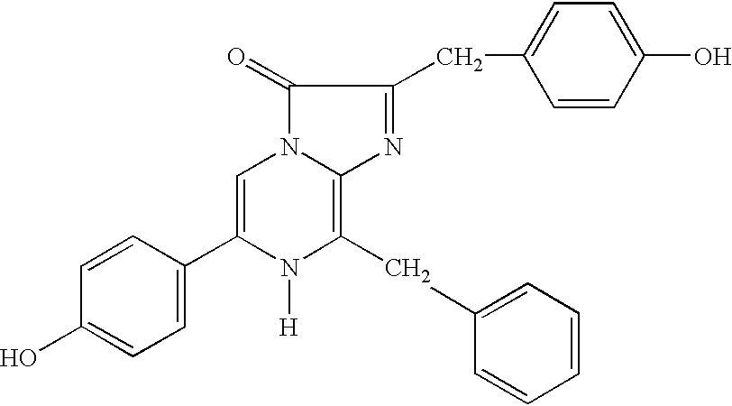
and sulfated derivatives thereof.
The reaction of coelenterazine when bound to the aequorin photoprotein with bound oxygen and in the presence of Ca2+ can represented as follows: 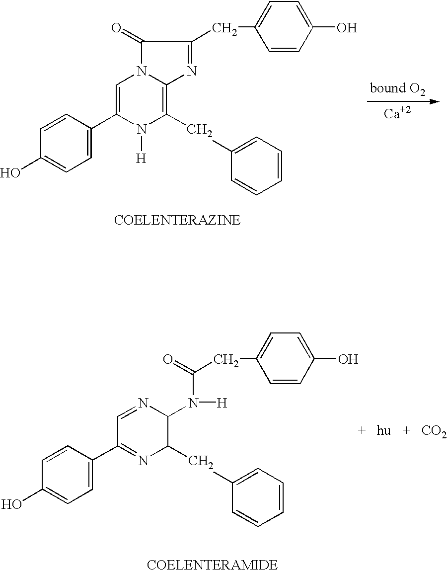
The photoprotein aequorin [which contains apoaequorin bound to a coelenterate luciferin molecule] and Renilla luciferase, discussed below, can use the same coelenterate luciferin. The aequorin photoprotein catalyses the oxidation of coelenterate luciferin [coelenterazine] to oxyluciferin [coelenteramide] with the concomitant production of blue light [lambdamax=469 nm].
Importantly, the sulfate derivative of the coelenterate luciferin [lauryl-luciferin] is particularly stable in water, and thus may be used in a coelenterate-like bioluminescence generating system. In this system, adenosine diphosphate (ADP) and a sulpha-kinase are used to convert the coelenterazine to the sulphated form. Sulfatase is then used to reconvert the lauryl-luciferin to the native coelenterazine. Thus, the more stable lauryl-luciferin is used in the item to be illuminated and the luciferase combined with the sulfatase are added to the luciferin mixture when illumination is desired.
Thus, the bioluminescence generating system of Aequorea is particularly suitable for use in the methods and apparatus herein. The particular amounts and the manner in which the components are provided depends upon the selected assay, luciferase and anti ligand. This system can be provided in lyophilized form, that will glow upon addition of Ca2+. It can be encapsulated, linked to matrices, such as porous glass, or in as a compositions, such as a solution or suspension, preferably in the presence of sufficient chelating agent to prevent triggering the reaction. The concentration of the aequorin photoprotein will vary and can be determined empirically. Typically concentrations of at least 0.1 mg/l, more preferably at least 1 mg/l and higher, will be selected. In certain embodiments, 1-10 mg luciferin/100 mg of luciferase will be used in selected volumes and at the desired concentrations will be used.
b. The Renilla System
Representative of coelenterate systems is the Renilla system. Renilla, also known as sea pansies, are members of the class of coelenterates Anthozoa, which includes other bioluminescent genera, such as Cavarnularia, Ptilosarcus, Stylatula, Acanthoptilum, and Parazoanthus. Bioluminescent members of the Anthozoa genera contain luciferases and luciferins that are similar in structure [see, e.g., Cormier et al. (1973) J. Cell. Physiol. 81:291-298; see, also Ward et al. (1975) Proc. Natl. Acad. Sci. U.S.A. 72:2530-2534). The luciferases and luciferins from each of these anthozoans crossreact and produce a characteristic blue luminescence.
Renilla luciferase and the other coelenterate and ctenophore luciferases, such as the aequorin photoprotein, use imidazopyrazine substrates, particularly the substrates generically called coelenterazine [see, formulae (I) and (II), above]. Other genera that have luciferases that use a coelenterazine include: squid, such as Chiroteuthis, Eucleoteuthis, Onychoteuthis, Watasenia; cuttlefish, Sepiolina; shrimp, such as Oplophorus, Sergestes, and Gnathophausia; deep-sea fish, such as Argyropelecus, Yarella, Diaphus, and Neoscopelus.
Renilla luciferase does not, however, have bound oxygen, and thus requires dissolved oxygen in order to produce light in the presence of a suitable luciferin substrate. Since Renilla luciferase acts as a true enzyme [i.e., it does not have to be reconstituted for further use] the resulting luminescence can be long-lasting in the presence of saturating levels of luciferin. Also, Renilla luciferase is relatively stable to heat.
Renilla luciferase, DNA encoding Renilla luciferase, and use of the DNA to produce recombinant luciferase, as well as DNA encoding luciferase from other coelenterates, are well known and available [see, e.g., SEQ ID No. 1, U.S. Pat. Nos. 5,418,155 and 5,292,658; see, also, Prasher et al. (1985) Biochem. Biophys. Res. Commun. 126:1259-1268; Cormier (1981) “Renilla and Aequorea bioluminescence” in Bioluminescence and Chemiluminescence, pp. 225-233; Charbonneau et al. (1979) J. Biol. Chem. 254:769-780; Ward et al. (1979) J. Biol. Chem. 254:781-788; Lorenz et al. (1981) Proc. Natl. Acad. Sci. U.S.A. 88: 4438-4442; Hori et al. (1977) Proc. Natl. Acad. Sci. U.S.A. 74:4285-4287; Hori et al. (1975) Biochemistry 14:2371-2376; Hori et al. (1977) Proc. Natl. Acad. Sci. U.S.A. 74:4285-4287; Inouye et al. (1975) Jap. Soc. Chem. Lett. 141-144; and Matthews et al. (1979) Biochemistry 16:85-91]. The DNA encoding Renilla luciferase and host cells containing such DNA provide a convenient means for producing large quantities of the enzyme [see, e.g., U.S. Pat. Nos. 5,418,155 and 5,292,658, which describe recombinant production of Renilla luciferase and the use of the DNA to isolate DNA encoding other luciferases, particularly those from related organisms]. A modified version of a method [U.S. Pat. Nos. 5,418,155 and 5,292,658] for the recombinant production of Renilla luciferase that results in a higher level of expression of the recombinant enzyme is presented in the EXAMPLES herein.
When used herein, the Renilla luciferase can be packaged in lyophilized form, encapsulated in a vehicle, either by itself or in combination with the luciferin substrate. Prior to use the mixture is contacted with an aqueous composition, preferably a phosphate buffered saline or other suitable buffer, such a Tris-based buffer [such as 0.1 mm Tris, 0.1 mm EDTA] pH 7-8, preferably about pH 8; dissolved O2 will activate the reaction. Addition of glycerol [about 1%] increases light intensity. Final concentrations of luciferase in the glowing mixture will be on the order of 0.01 to 1 mg/l or more. Concentrations of luciferin will be at least about 10−8 M, but 1 to 100 or more orders of magnitude higher to produce a long lasting bioluminescence.
In certain embodiments herein, about 1 to 10 mg, or preferably 2-5 mg, more preferably about 3 mg of coelenterazine will be used with about 100 mg of Renilla luciferase. The precise amounts, of course can be determined empirically, and, also will depend to some extent on the ultimate concentration and application. In particular, about addition of about 0.25 ml of a crude extract from the bacteria that express Renilla to 100 ml of a suitable assay buffer and about 0.005 μg was sufficient to produce a visible and lasting glow [see, U.S. Pat. Nos. 5,418,155 and 5,292,658, which describe recombinant production of Renilla luciferase].
Lyophilized mixtures, and compositions containing the Renilla luciferase are also provided. The luciferase or mixtures of the luciferase and luciferin may also be encapsulated into a suitable delivery vehicle, such as a liposome, glass particle, capillary tube, drug delivery vehicle, gelatin, time release coating or other such vehicle. Kits containing these mixtures, compositions, or vehicles and also a chip device and reagents for attaching biological molecules to the surface of the chip, are also provided. The luciferase may also be linked to an anti ligand through chemical or recombinant means for use in the methods herein.
Recombinant Production of Renilla Reniformis Luciferase
The phagemid pTZ18R (Pharmacia) is a multi-purpose DNA vector designed for in vitro transcriptions and useful for expression of recombinant proteins in bacterial hosts. The vector contains the bla gene, which allows for the selection of transformants by resistance to ampicillin, and a polylinker site adjacent to the lacZ′ gene. The heterologous gene of interest is inserted in the polylinker and transcribed from the lac promoter by induction, for example, with isopropyl-β-D-thiogalactopyranoside (IPTG).
The DNA encoding the Renilla reniformis luciferase has been cloned (e.g., see U.S. Pat. Nos. 5,292,658 and 5,418,155). The plasmid pTZRLuc-1 encodes the Renilla luciferase on a 2.2 Kbp EcoRI to SstI DNA fragment inserted in EcoRI and SstI sites of pTZ18R (plasmid construction is described U.S. Pat. Nos. 5,292,658 and 5,418,155; see also Lorenz et al. (1991) Isolation and Expression of a cDNA encoding Renilla reniformis Luciferase, Proc. Natl. Acad. Sci. U.S.A. 88, 4438-4442). The initiation of transcription of the Renilla luciferase cDNA is under the control of the lacZ′ promoter. E. coli strains harboring plasmid pTZRLuc-1 express Renilla luciferase that is functional in bioluminescence assays and retains the properties of the native enzyme (see e.g., U.S. Pat. Nos. 5,292,658 and 5,418,155).
A derivative of pTZRLuc-1, pTZRLuc-3.6, produces approximately 7-fold higher levels of recombinant Renilla luciferase than pTZRLuc-1 when transformed into the same E. coli host. Competent E. coli strain XL-1 was transformed using purified pTZRLuc-3.6 according to the instructions provided by the manufacturer (XL-1 Supercompetent cells and protocol; Stratagene, Inc., La Jolla, Calif.). Transfectants were selected by plating on Luria Broth (LB) plates supplemented with 100 μg/ml ampicillin.
Single ampicillin resistant colonies were grown in LB medium supplemented with 100 μg/ml ampicillin at ambient temperature using continuous shaking until cell growth reached mid-log phase (i.e., cell culture reaches an O.D.600 nm=0.6-0.8 units). Transcription from the lac promoter was induced by addition of 1 mM IPTG and cell culture was shaken at ambient temperature for an additional 8 hours.
Cells were harvested by centrifugation at 10,000×g and frozen at −20° C. The cell pellet was thawed and resuspended at a 1:5 ratio (w/w) in a solution of 10 mM EDTA, pH 8.0, containing 4 mg/ml lysozyme (Sigma Chemical Corp.). The cells were placed in a 25° C. water bath for 30 minutes and then transferred to ice for 1 hour. The cells were lysed by sonication at 0° C. using a 1 minute pulse from an Ultrasonics, Inc. cell disruptor.
The lysed cellular debris was removed by centrifugation at 30,000×g for 3 hours and the supernatant was decanted and retained. The pellet was resuspended at a 1:5 ratio in the above-described solutions, and the subsequent incubations, lysis and centrifugation steps were repeated. The two supernatants were combined and stored at −70° C.
The resulting “clarified lysate” was employed as a source of recombinant luciferase. Alternatively, the lysate may be subjected to additional purification steps (e.g., ion exchange chromatography or immunoaffinity chromatography) to further enrich the lysate or provide a homogeneous source of the purified enzyme (see e.g., U.S. Pat. Nos. 5,292,658 and 5,418,155).
3. Crustacean, Particularly Cyrpidina Systems
The ostracods, such as Vargula serratta, hilgendorfii and noctiluca are small marine crustaceans, sometimes called sea fireflies. These sea fireflies are found in the waters off the coast of Japan and emit light by squirting luciferin and luciferase into the water, where the reaction, which produces a bright blue luminous cloud, occurs. The reaction involves only luciferin, luciferase and molecular oxygen, and, thus, is very suitable for application herein.
The systems, such as the Vargula bioluminescence generating systems, are particularly preferred herein because the components are stable at room temperature if dried and powdered and will continue to react even if contaminated. Further, the bioluminescent reaction requires only the luciferin/luciferase components in concentrations as low as 1:40 parts per billion to 1:100 parts per billion, water and molecular oxygen to proceed. An exhausted system can renewed by addition of luciferin.
a. Vargula Luciferase
Vargula luciferase is a 555-amino acid polypeptide that has been produced by isolation from Vargula and also using recombinant technology by expressing the DNA in suitable bacterial and mammalian host cells [see, e.g., Thompson et al. (1989) Proc. Natl. Acad. Sci. U.S.A. 86:6567-6571; Inouye et al. (1992) Proc. Natl. Acad. Sci. U.S.A. 89:9584-9587; Johnson et al. (1978) Methods in Enzymology LVII:331-349; Tsuji et al. (1978) Methods Enzymol. 57:364-72; Tsuji (19740 Biochemistry 13:5204-5209; Japanese Patent Application No. JP 3-30678 Osaka; and European Patent Application No. EP 0 387 355 A1].
(1) Purification from Cypridina
Methods for purification of Vargula [Cypridina] luciferase are well known. For example, crude extracts containing the active can be readily prepared by grinding up or crushing the Vargula shrimp. In other embodiments, a preparation of Cypridina hilgendorfi luciferase can be prepared by immersing stored frozen C. hilgendorfi in distilled water containing, 0.5-5.0 M salt, preferably 0.5-2.0 M sodium or potassium chloride, ammonium sulfate, at 0-30° C., preferably 0-10° C., for 1-48 hr, preferably 10-24 hr, for extraction followed by hydrophobic chromatography and then ion exchange or affinity chromatography [TORAY IND INC, Japanese patent application JP 4258288, published Sep. 14, 1993; see, also, Tsuji et al. (1978) Methods Enzymol. 57:364-72 for other methods].
The luciferin can be isolated from ground dried Vargula by heating the extract, which destroys the luciferase but leaves the luciferin intact [see, e.g., U.S. Pat. No. 4,853,327].
(2) Preparation by Recombinant Methods
The luciferase is preferably produced by expression of cloned DNA encoding the luciferase [European Patent Application NO. 0 387 355 A1; International PCT Application No. WO90/01542; see, also SEQ ID No. 5, which sets forth the sequence from Japanese Patent Application No. JP 3-30678 and Thompson et al. (1989) Proc. Natl. Acad. Sci. U.S.A. 86:6567-6571] DNA encoding the luciferase or variants thereof is introduced into E. coli using appropriate vectors and isolated using standard methods.
b. Vargula Luciferin
The natural luciferin in a substituted imidazopyrazine nucleus, such a compound of formula (III): 
Analogs thereof and other compounds that react with the luciferase in a light producing reaction also may be used.
Other bioluminescent organisms that have luciferases that can react with the Vargula luciferin include, the genera Apogon, Parapriacanthus and Porichthys.
c. Reaction
The luciferin upon reaction with oxygen forms a dioxetanone intermediate [which includes a cyclic peroxide similar to the firefly cyclic peroxide molecule intermediate]. In the final step of the bioluminescent reaction, the peroxide breaks down to form CO2 and an excited carbonyl. The excited molecule then emits a blue to blue-green light.
The optimum pH for the reaction is about 7. For purposes herein, any pH at which the reaction occurs may be used. The concentrations of reagents are those normally used for analytical reactions or higher [see, e.g., Thompson et al. (1990) Gene 96:257-262]. Typically concentrations of the luciferase between 0.1 and 10 mg/l, preferably 0.5 to 2.5 mg/l will be used. Similar concentrations or higher concentrations of the luciferin may be used.
4. Insect Bioluminescence Generating Systems Including Firefly, Click Beetle, and Other Insect Systems
The biochemistry of firefly bioluminescence was the first bioluminescence generating system to be characterized [see, e.g., Wienhausen et al. (1985) Photochemistry and Photobiology 42:609-611; McElroy et al. (1966) in Molecular Architecture in Cell Physiology, Hayashi et al., eds. Prentice Hall, Inc., Englewood Cliffs, N.J., pp. 63-80] and it is commercially available [e.g., from Promega Corporation, Madison, Wis., see, e.g., Leach et al. (1986) Methods in Enzymology 133:51-70, esp. Table 1]. Luciferases from different species of fireflies are antigenically similar. These species include members of the genera Photinus, Photurins and Luciola. Further, the bioluminescent reaction produces more light at 30° C. than at 20° C., the luciferase is stabilized by small quantities of bovine albumin serum, and the reaction can be buffered by tricine.
a. Luciferase
DNA clones encoding luciferases from various insects and the use to produce the encoded luciferase is well known. For example, DNA clones that encode luciferase from Photinus pyralis, Luciola cruciata [see, e.g., de Wet et al. (1985) Proc. Natl. Acad. Sci. U.S.A. 82:7870-7873; de We et al. (1986) Methods in Enzymology 133:3; U.S. Pat. No. 4,968,613, see, also SEQ ID No. 3] are available. The DNA has also been expressed in Saccharomyces [see, e.g., Japanese Application No. JP 63317079, published Dec. 26, 1988, KIKKOMAN CORP] and in tobacco.
In addition to the wild-type luciferase modified insect luciferases have been prepared. For example, heat stable luciferase mutants, DNA-encoding the mutants, vectors and transformed cells for producing the luciferases are available. A protein with 60% amino acid sequence homology with luciferases from Photinus pyralis, Luciola mingrelica, L. cruciata or L. lateralis and having luciferase activity is available [see, e.g., International PCT Application No. WO95/25798]. It is more stable above 30° C. than naturally-occurring insect luciferases and may also be produced at 37° C. or above, with higher yield.
Modified luciferases that generate light at different wavelengths [compared with native luciferase], and thus, may be selected for their color-producing characteristics. For example, synthetic mutant beetle luciferase(s) and DNA encoding such luciferases that produce bioluminescence at a wavelength different from wild-type luciferase are known (Promega Corp, international PCT Application No. WO95/1 8853, which is based on U.S. application Ser. No. 08/177,081 Jan. 1, 1994]. The mutant beetle luciferase has an amino acid sequence differing from that of the corresponding wild-type Luciola cruciata [see, e.g., U.S. Pat. Nos. 5,182,202, 5,219,737, 5,352,598, see, also SEQ ID No. 3] by a substitution(s) at one or two positions. The mutant luciferase produces a bioluminescence with a wavelength of peak intensity that differs by at least 1 nm from that produced by wild-type luciferases.
Other mutant luciferase have also been produced. Mutant luciferases with the amino acid sequence of wild-type luciferase, but with at least one mutation in which valine is replaced by isoleucine at the amino acid number 233, valine by isoleucine at 239, serine by asparagine at 286, glycine by serine at 326, histidine by tyrosine at 433 or proline by serine at 452 are known [see, e.g., U.S. Pat. Nos. 5,219,737, and 5,330,906]. The luciferases are produced by expressing DNA-encoding each mutant luciferase in E. coli and isolating the protein. These luciferases produce light with colors that differ from wild-type. The mutant luciferases catalyze luciferin to produce red [λ609 nm and 612 nm], orange[λ595 and 607 nm] or green [λ558 nm] light. The other physical and chemical properties of mutant luciferase are substantially identical to native wild type-luciferase. The mutant luciferase has the amino acid sequence of Luciola cruciata luciferase with an alteration selected from Ser 286 replaced by Asn, Gly 326 replaced by Ser, His 433 replaced by Tyr or Pro 452 replaced by Ser. Thermostable luciferases are also available [see, e.g., U.S. Pat. No. 5,229,285; see, also International PCT Application No.@) 95/25798, which provides Photinus luciferase in which the glutamate at position 354 is replaced lysine and Luciola luciferase in which the glutamate at 356 is replaced with lysine].
These mutant luciferases as well as the wild type luciferases are among those preferred herein, particularly in instances when a variety of colors are desired or when stability at higher temperatures is desired. It is also noteworthy that firefly luciferases have alkaline pH optima [7.5-9.5], and, thus, are suitable for use in diagnostic assays performed at alkaline pH.
b. Luciferin
The firefly luciferin is a benzothiazole: 
Analogs of this luciferin and synthetic firefly luciferins are also known to those of skill in art [see, e.g., U.S. Pat. No. 5,374,534 and 5,098,828]. These include compounds of formula (IV) [see, U.S. Pat. No. 5,098,828]: 
in which:
R1 is hydroxy, amino, linear or branched C1-C20 alkoxy, C2-C20 alkyenyloxy, an L-amino acid radical bond via the α-amino group, an oligopeptide radical with up to ten L-amino acid units linked via the α-amino group of the terminal unit;
R2 is hydrogen, H2PO2, HSO3, unsubstituted or phenyl substituted linear or branched C1-C20 alkyl or C2-C20alkenyl, aryl containing 6 to 18 carbon atoms, or R3—C(O)—; and
R3 is an unsubstituted or phenyl substituted linear or branched C1-C20 alkyl or C2-C20alkenyl, aryl containing 6 to 18 carbon atoms, a nucleotide radical with 1 to 3 phosphate groups, or a glycosidically attached mono- or disaccharide, except when formula (IV) is a D-luciferin or D-luciferin methyl ester.
c. Reaction
The reaction catalyzed by firefly luciferases and related insect luciferases requires ATP, Mg2+ as well as molecular oxygen. Luciferin must be added exogenously. Firefly luciferase catalyzes the firefly luciferin activation and the subsequent steps leading to the excited product. The luciferin reacts with ATP to form a luciferyl adenylate intermediate. This intermediate then reacts with oxygen to form a cyclic luciferyl peroxy species, similar to that of the coelenterate intermediate cyclic peroxide, which breaks down to yield CO2 and an excited state of the carbonyl product. The excited molecule then emits a yellow light; the color, however, is a function of pH. As the pH is lowered the color of the bioluminescence changes from yellow-green to red.
Different species of fireflies emit different colors of bioluminescence so that the color of the reaction will be dependent upon the species from which the luciferase is obtained. Additionally, the reaction is optimized at pH 7.8.
Addition of ATP and luciferin to a reaction that is exhausted produces additional light emission. Thus, the system, once established, is relatively easily maintained. Therefore, it is highly suitable for use herein in embodiments in which a sustained glow is desired or reuse of the item is contemplated. Thus, the components of a firefly system can be packaged with the chip.
5. Bacterial Systems
Luminous bacteria typically emit a continuous light, usually blue-green. When strongly expressed, a single bacterium may emit 104 to 106 photons per second. Bacterial bioluminescence systems include, among others, those systems found in the bioluminescent species of the genera Photobacterium, Vibrio and Xenorhabdus. These systems are well known and well characterized [see, e.g., Baldwin et al. (1984) Biochemistry 23:3663-3667; Nicoli et al. (1974) J. Biol. Chem. 249:2393-2396; Welches et al. (1981) Biochemistry 20:512-517; Engebrecht et al. (1986) Methods in Enzymology 133:83-99; Frackman et al. (1990) J. of Bacteriology 172:5767-5773; Miyamoto et al. (1986) Methods in Enzymology 133:70; U.S. Pat. No. 4,581,335].
a. Luciferases
Bacterial luciferase, as exemplified by luciferase derived from Vibrio harveyi [EC 1.14.14.3, alkanol reduced-FMN-oxygen oxidoreductase 1-hydroxylating, luminescing], is a mixed function oxidase, formed by the association of two different protein subunits α and β. The α-subunit has an apparent molecular weight of approximately 42,000 kD and the β-subunit has an apparent molecular weight of approximately 37,000 kD [see, e.g., Cohn et al. (1989) Proc. Natl. Acad. Sci. U.S.A. 90:102-123]. These subunits associate to form a 2-chain complex luciferase enzyme, which catalyzes the light emitting reaction of bioluminescent bacteria, such as Vibrio harveyi [U.S. Pat. No. 4,581,335; Belas et al. (1982) Science 218:791-793], Vibrio fischeri [Engebrecht et al. (1983) Cell 32:773-781; Engebrecht et al. (1984) Proc. Natl. Acad. Sci. U.S.A. 81:4154-4158] and other marine bacteria.
Bacterial luciferase genes have been cloned [see, e.g., U.S. Pat. No. 5,221,623; U.S. Pat. No. 4,581,335; European Patent Application No. EP 386 691 A]. Plasmids for expression of bacterial luciferase, such as Vibrio harveyi, include pFIT001 (NRRL B-18080), pPALE001 (NRRL B-18082) and pMR19 (NRRL B-18081)] are known. For example the sequence of the entire lux regulon from Vibiro fisheri has been determined [Baldwin et al. (1984), Biochemistry 23:3663-3667; Baldwin et al. (1981) Biochem. 20: 512-517; Baldwin et al. (1984) Biochem. 233663-3667; see, also, e.g., U.S. Pat. Nos. 5,196,318, 5,221,623, and 4,581,335]. This regulon includes luxI gene, which encodes a protein required for autoinducer synthesis [see, e.g., Engebrecht et al. (1984) Proc. Natl. Acad. Sci. U.S.A. 81:4154-4158], the luxC, luxD, and luxE genes, which encode enzymes that provide the luciferase with an aldehyde substrate, and the luxA and luxB genes, which encode the alpha and beta subunits of the luciferase.
Lux genes from other bacteria have also been cloned and are available [see, e.g., Cohn et al. (1985) J. Biol. Chem. 260:6139-6146; U.S. Pat. No. 5,196,524, which provides a fusion of the luxA and luxB genes from Vibrio harveyi]. Thus, luciferase alpha and beta subunit-encoding DNA is provided and can be used to produce the luciferase. DNA encoding the α [1065 bp] and β [984 bp] subunits, DNA encoding a luciferase gene of 2124 bp, encoding the alpha and beta subunits, a recombinant vector containing DNA encoding both subunits and a transformed E. coli and other bacterial hosts for expression and production of the encoded luciferase are available. In addition, bacterial luciferases are commercially available.
b. Luciferins
Bacterial luciferins include: 
in which the tetradecanal with reduced flavin mononucleotide are considered luciferin since both are oxidized during the light emitting reaction.
c. Reactions
The bacterial systems require, in addition to reduced flavin, five polypeptides to complete the bioluminescent reaction: two subunits, α and β, of bacterial luciferin and three units of a fatty acid reductase system complex, which supplies the tetradecanal aldehyde. Examples of bacterial bioluminescence generating systems useful in the apparatus and methods provided herein include those derived from Vibrio fisheri and Vibrio harveyi. One advantage to this system is its ability to operate at cold temperatures. It will thus be particularly amenable to methods of using the chip for the detection and monitoring of antibiotic sensitivity of psychrophilic organisms. All components of a bacterial system can be frozen into ice or placed in solutions stored below 0° C. After incubation at temperatures near 0° C., the chip can be transferred to warmer temperatures and the reaction will proceed thereby providing a sustained glow.
Bacterial luciferase catalyzes the flavin-mediated hydroxylation of a long-chain aldehyde to yield carboxylic acid and an excited flavin; the flavin decays to ground state with the concomitant emission of blue green light [λmax=490 nm; see, e.g., Legocki et al. (1986) Proc. Natl. Acad. Sci. USA 81:9080; see U.S. Pat. No. 5,196,524]: 
The reaction can be initiated by contacting reduced flavin mononucleotide [FMNH2] with a mixture of the bacterial luciferase, oxygen, and a long-chain aldehyde, usually n-decyl aldehyde.
DNA encoding luciferase from the fluorescent bacterium Alteromonas hanedai is known [CHISSO CORP; see, also, Japanese application JP 7222590, published Aug. 22, 1995]. The reduced flavin mononucleotide [FMNH2; luciferin] reacts with oxygen in the presence of bacterial luciferase to produce an intermediate peroxy flavin. This intermediate reacts with a long-chain aldehyde [tetradecanal] to form the acid and the luciferase-bound hydroxy flavin in its excited state. The excited luciferase-bound hydroxy flavin then emits light and dissociates from the luciferase as the oxidized flavin mononucleotide [FMN] and water. In vivo FMN is reduced again and recycled, and the aldehyde is regenerated from the acid.
Flavin reductases have been cloned [see, e.g., U.S. Pat. No. 5,484,723; see, SEQ ID No. 14 for a representative sequence from this patent]. These as well as NAD(P)H can be included in the reaction to regenerate FMNH2 for reaction with the bacterial luciferase and long chain aldehyde. The flavin reductase catalyzes the reaction of FMN, which is the luciferase reaction, into FMNH2; thus, if luciferase and the reductase are included in the reaction system, it is possible to maintain the bioluminescent reaction. Namely, since the bacterial luciferase turns over many times, bioluminescence continues as long as a long chain aldehyde is present in the reaction system.
The color of light produced by bioluminescent bacteria also results from the participation of a blue-florescent protein [BFP] in the bioluminescence reaction. This protein, which is well known [see, e.g., Lee et al. (1978) Methods in Enzymology LVII:226-234], may also be added to bacterial bioluminescence reactions in order to cause a shift in the color.
6. Other Systems
a. Dinoflagellate Bioluminescence Generating Systems
In dinoflagellates, bioluminescence occurs in organelles termed scintillons. These organelles are outpocketings of the cytoplasm into the cell vacuole. The scintillons contain only dinoflagellate luciferase and luciferin [with its binding protein], other cytoplasmic components being somehow excluded. The dinoflagellate luciferin is a tetrapyrrole related to chlorophyll: 
or an analog thereof.
The luciferase is a 135 kD single chain protein that is active at pH 6.5, but inactive at pH 8 [see, e.g., Hastings (1981) Bioluminescence and Chemiluminescence, DeLuca et al, eds. Academic Press, NY, pp.343-360]. Luminescent activity can be obtained in extracts made at pH 8 by shifting the pH from 8 to 6. This occurs in soluble and particulate fractions. Within the intact scintillon, the luminescent flash occurs for ˜100 msec, which is the duration of the flash in vivo. In solution, the kinetics are dependent on dilution, as in any enzymatic reaction. At pH 8, the luciferin is bound to a protein [luciferin binding protein] that prevents reaction of the luciferin with the luciferase. At pH 6, however, the luciferin is released and free to react with the enzyme.
b. Systems from Molluscs, Such as Latia and Pholas
Molluscs Latia neritoides and species of Pholas are bioluminescent animals. The luciferin has the structure: 
and has been synthesized [see, e.g., Shimomura et al. (1968) Biochemistry 7:1734-1738; Shimomura et al. (1972) Proc. Natl. Acad. Sci. U.S.A. 69:2086-2089]. In addition to a luciferase and luciferin the reaction has a third component, a “purple protein”. The reaction, which can be initiated by an exogenous reducing agent is represented by the following scheme: 
XH2 is a reducing agent.
Thus for practice herein, the reaction will require the purple protein as well as a reducing agent.
c. Earthworms and Other Annelids
Earthworm species, such as Diplocardia longa, Chaetopterus and Harmothoe, exhibit bioluminescence. The luciferin has the structure: 
The reaction requires hydrogen peroxide in addition to luciferin and luciferase. The luciferase is a photoprotein.
d. Glow Worms
The luciferase/luciferin system from the glow worms that are found in New Zealand caves, Australia and those found in Great Britain are also intended for use herein.
e. Marine Polycheate Worm Systems
Marine polycheate worm bioluminescence generating systems, such as Phyxotrix and Chaetopterus, are also contemplated for use herein.
f. South American Railway Beetle
The bioluminescence generating system from the South American railway beetle is also intended for use herein.
g. Fish
Of interest herein, are luciferases and bioluminescence generating systems that generate red light. These include luciferases found in species of Aristostomias, such as A. scintillans [see, e.g., O'Day et al. (1974) Vision Res. 14:545-550], Pachystomias, Malacosteus, such as M. niger.
7. Fluorescent Proteins
a. Green and Blue Fluorescent Proteins
As described herein, blue light is produced using the Renilla luciferase or the Aequorea photoprotein in the presence of Ca2+ and the coelenterazine luciferin or analog thereof. This light can be converted into a green light if a green fluorescent protein (GFP) is added to the reaction. Green fluorescent proteins, which have been purified [see, e.g., Prasher et al. (1992) Gene 111:229-233] and also cloned [see, e.g., International PCT Application No. WO 95/07463, which is based on U.S. application Ser. No. 08/119,678 and U.S. application Ser. No. 08/192,274, which are herein incorporated by reference], are used by cnidarians as energy-transfer acceptors. GFPs fluoresce in vivo upon receiving energy from a luciferase-oxyluciferein excited-state complex or a Ca2+-activated photoprotein. The chromophore is modified amino acid residues within the polypeptide. The best characterized GFPs are those of Aequorea and Renilla [see, e.g., Prasher et al. (1992) Gene 111:229-233; Hart, et al. (1979) Biochemistry 18:2204-2210]. For example, a green fluorescent protein [GFP] from Aequorea victoria contains 238 amino acids, absorbs blue light and emits green light. Thus, inclusion of this protein in a composition containing the aequorin photoprotein charged with coelenterazine and oxygen, can, in the presence of calcium, result in the production of green light. Thus, it is contemplated that GFPs may be included in the bioluminescence generating reactions that employ the aequorin or Renilla luciferases or other suitable luciferase in order to enhance or alter color of the resulting bioluminescence.
GFPs are activated by blue light to emit green light and thus may be used in the absence of luciferase and in conjunction with an external light source with novelty items, as described herein. Similarly, blue fluorescent proteins (BFPs), such as from Vibrio fischeri, Vibrio harveyi or Photobacterium phosphoreum, may be used in conjunction with an external light source of appropriate wavelength to generate blue light. (See for example, Karatani, et al., “A blue fluorescent protein from a yellow-emitting luminous bacterium,” Photochem. Photobiol. 55(2):293-299 (1992); Lee, et al., “Purification of a blue-fluorescent protein from the bioluminescent bacterium Photobacterium phosphoreum” Methods Enzymol. (Biolumin. Chemilumin.) 57:226-234 (1978); and Gast, et al. “Separation of a blue fluorescence protein from bacterial luciferase” Biochem. Biophys. Res. Commun. 80(1):14-21 (1978), each, as all references cited herein, incorporated in its entirety by reference herein.) In particular, GFPs, and/or BFPs or other such fluorescent proteins may be used in the methods provided herein for the detection of infectious agents by binding an analyte to one or more anti ligand-GFP conjugate(s) at a plurality of locations and illuminating the chip with light of an appropriate wavelength to cause the fluorescent proteins to fluoresce whereby the emitted fluorescence is detected by the photodiodoes in the chip.
GFPs and/or BFPs or other such fluorescent proteins may be used in conjunction with any of the chips or devices described herein. These fluorescent proteins may also be used alone or in combination with bioluminescence generating systems to produce an array of colors. They may be used in combinations such that the color, for example, of the emitted light may be altered to maximize the amount of light available for detection by the photodiodes of the chip.
b. Phycobiliproteins
Phycobiliproteins are water soluble fluorescent proteins derived from cyanobacteria and eukaryotic algae [see, e.g., Apt et al. (1995) J. Mol. Biol. 238:79-96; Glazer (1982) Ann. Rev. Microbiol. 36:173-198; and Fairchild et al. (1994) J. of Biol. Chem. 269:8686-8694]. These proteins have been used as fluroescent labels in immmunoassay [see, Kronick (1986) J. of Immunolog. Meth. 92:1-13], the proteins have been isolated and DNA encoding them is also available [see, e.g., Pilot et al. (1984) Proc. Natl. Acad. Sci. U.S.A. 81:6983-6987; Lui et al. (1993) Plant Physiol 103:293-294; and Houmard et al. (1988) J. Bacteriol. 170:5512-5521; the proteins are commercially available from, for example, ProZyme, Inc., San Leandro, Calif.].
In these organisms, the phycobiliproteins are arranged in subcellular structures termed phycobilisomes and function as accessory pigments that participate in photosynthetic reactions by absorbing visible light and transferring the derived energy to chlorophyll via a direct fluorescence energy transfer mechanism.
Two classes of phycobiliproteins are known based on their color: phycoerythrins (red) and phycocyanins (blue), which have reported absorbtion maxima between 490 and 570 nm and between 610 and 665 nm, respectively. Phycoerythrins and phycocyanins are heterogenous complexes composed of different ratios of alpha and beta monomers to which one or more class of linear tetrapyrrole chromophores are covalently bound. Particular phycobiliproteins may also contain a third γ-subunit which often associated with (αβ)6 aggregate proteins.
All phycobiliproteins contain phycothrombilin or phycoerythobilin chromophores, and may also contain other bilins, such as phycourobilin, cryptoviolin or a 697 nm bilin. The γ-subunit is covalently bound with phycourobilin, which results in the 495-500 nm absorbance peak of B- and R-phycoerythrins. Thus, the spectral characteristics of phycobiliproetins may be influenced by the combination of the different chromophores, the subunit composition of the apo-phycobiliproteins and/or the local enviroment that affects the tertiary and quaternary structure of the phycobiliproteins.
As described above for GFPs & BFPs, phycobiliproteins are also activated by visible light of the appropriate wavelength and thus may be used in the absence of luciferase and in conjunction with an external light source to illuminate the phycobiliprotein bound to the chip at locations where analyte has been detected. In particular, phycobiliproteins may be covalently bound to one or more anti-ligand specific for the targeted analyte and illuminated with light of an appropriate wavelength to cause the fluorescent proteins to fluoresce and the fluorescence is measured by the photodiodes of the chip at that location of the array. The data signals are sent to the computer processor and analyzed. As noted above, these proteins may be used in combination with other fluoresent proteins and/or bioluminescence generating systems to produce an array of colors or to provide different colors over time that can be detected by the photodiodes of the chip.
Attachment of phycobiliproteins to solid support matrices is known (e.g., see U.S. Pat. Nos. 4,714,682; 4,767,206; 4,774,189 and 4,867,908). Therefore, phycobiliproteins may be coupled to microcarriers coupled to one or more components of the bioluminescent reaction, preferably a luciferase, to convert the wavelength of the light generated from the bioluminescent reaction. Microcarriers coupled to one or more phycobiliproteins may be used when linked to the anti-ligand or to any of the chips used in the methods herein.
C. Design, Fabrication, and Use of Chips
Chips for use as diagnostic devices are provided herein. The chips can be nonself-addressable or self-addressable and are typically in the form of an array, such as a 96-member or higher density array or any of those described herein.
1. Nonself-Addressable Chips
Referring to FIG. 1, a nonself-addressable microelectronic device 100 for detecting and identifying analytes in a biological sample using bioluminescence includes an address control circuit 102, a photodetector array 104, an analog multiplexer 106, a comparator 108, a reference circuit 110, a feedback control circuit 112 and an output control circuit 114. Address control circuit 102 receives a clock input signal 116 from an external oscillator, and output control circuit 114 generates data output signals 118. Device 100 also includes electrical connections 120 and 122 for receiving electrical power and ground, respectively, from an external power source (e.g., an AC-DC converter). Thus, device 100 requires only four electrical connections: clock input signal 116; data output signals 118; power 120; and ground 122.
In one embodiment, output control circuit 114 formats quantized output signals 138 into an RS-232 serial data stream indicative of the light detected at each pixel element in array 104. To allow an external instrument or computer to correlate the received RS-232 serial data stream with specific pixel elements in array 104, output control circuit 114 transmits the serial data stream in frames separated by a synchronization signal (sync). Each frame contains an output data signal for each pixel element in array 104, and the sync signal is an arbitrary value (e.g., a byte having a value of decimal 255) used as a control signal to identify the start of each data frame. The external computer waits for the sync signal before correlating the received frame data to the appropriate pixel elements in array 104. Alternatively, output control circuit 114 could include labels in the serial data stream that identify the pixel elements. A parallel data interface can also be used.
As will become apparent from the description below, array 104 includes pixel elements located at an array of micro-locations on the surface of the semiconductor substrate used for device 100. Each element includes a photodetector for receiving photons of light emitted by a chemical reaction optically coupled at the respective micro-location and for converting the received photons into an electric charge. Each element also includes a pixel unit cell circuit with a capacitance circuit for integrating the electric charge. The integrated charge is quantized using delta-sigma A/D conversion techniques, and the digitized signal is multiplexed into a serial data output stream interfaced to an external computer. The computer executes a control program to integrate the delta-sigma digital signal for a desired integration period ranging from seconds to hours depending on the desired resolution. In one embodiment, the delta-sigma A/D conversion is clocked for a 56 Kbaud interface to achieve 12-bit resolution in an integration period of about 10 seconds, and 16-bit resolution in a time period of about 3 minutes.
Referring to FIG. 2, device 100 includes a semiconductor substrate or die 140 having array 104 defined on a surface thereof. Array 104 includes an array of micro-locations 142, and an independent photodetector 144 optically coupled to each micro-location. (Only the left-most micro-location 142 and photodetector 144 in each row are labeled in FIG. 2 for clarity.) Array 104 includes three sub-arrays 146, 148 and 150 having three different sizes of micro-locations 142. Sub-array 146 includes a 4×16 array of 50 micron square pixels, sub-array 148 includes a 2×8 array of 100 micron square pixels, and sub-array 150 includes a 1×4 array of 200 micron square pixels. Photodetectors 144 are located on a portion of the surface of die 140 at each micro-location 142. The portion taken up by photodetector 144 includes about 90% of the surface area for larger pixel elements and about 50% for smaller pixel elements. In one embodiment, photodetectors 144 are silicon photodiodes that convert photons of light impinging on their surfaces into a photocurrent. The quantum efficiency of this conversion is about 40% at a wavelength of 500-800 nm (i.e., a photocurrent of 40 electrons is generated for each 100 photons of received light). Photodiodes 144 can thus convert low photon levels into measurable signals. The surface of substrate 140 has a slight depression (e.g., 1 micron) at each micro-location 142 to help contain the fluid sample applied to device 100.
The larger pixels elements (e.g., 200 um) in array 104 have a higher sensitivity to detect lower concentrations of analytes than the smaller pixel elements (e.g., 50 um) since a greater number of receptor antibodies can be bound to their larger surface areas, as explained herein, such that more photons of light will be emitted when a chemical reaction occurs at the respective micro-location. The smaller pixels can be used to form a larger matrix on a given die size to allow a greater number of assays to be performed simultaneously. The optimum pixel size for detecting a particular analyte may be determined empirically. The use of different-sized pixel elements on device 100 has two advantages. First, larger pixel elements can be used to detect analytes requiring larger sensitivities while smaller pixel elements can be used to increase the number of pixel elements in the matrix for analytes having lower sensitivities. Second, different sizes can be used to help determine the optimum size for a particular analyte by empirical testing, with the optimum size being used for other embodiments of array 104.
Alternative arrangements of array 104 will be apparent to a person of ordinary skill in the art. For example, array 104 can include sub-arrays of pixel elements having different sizes (as in FIG. 2), or an array having only a single pixel size (e.g., a 12×16 array of 50 micron pixels). The size of each pixel (e.g., 50, 100, 200 microns in FIG. 2) can be modified (e.g., a 400 micron pixel can be used). Also, the number of pixels in the array or sub-array (e.g., 4×16, 2×8 or 1×16 in FIG. 2) can be changed to include an n×m array or sub-array having n rows and m columns, n and m being integers. Further, shapes other than squares can be used for each pixel element (e.g., rectangles or circles). The size of die 140 can be modified to accommodate the different arrangements of array 104, although use of a larger die may increase the cost of device 100. Also, die 140 may include more or fewer bonding pads 152, provided there are separate pads for clock input signal 116, data output signals 118, electrical power 120 and ground 122 (FIG. 1).
Referring to FIGS. 3 and 3A, die 140 is packaged within a ceramic dual in-line package (DIP) 154 with 40 pins or leads 156. Four pins 156 are dedicated to clock input signal 116, data output signals 118, electrical power 120, and ground 122. The other pins 156 are used for test signals. Other microelectronic packages may also be used, such as plastic packages having more or fewer conductors including the four required conductors.
When performing an assay, the fluid sample to be analyzed is applied through apertures 172 and 182 to the surface of die 140 (and micro-locations 142 formed thereon) housed within package 154. The fluid sample may be applied by pipetting the fluid sample into the test well formed by apertures 172 and 182, or simply by dipping package 154 into a container (now shown) filled with the sample. The electrical components of device 100 are protected from the sample by the materials of package 154 itself, or by the epoxy coating. After the fluid sample is applied, the remaining components needed to cause light-emitting reactions optically coupled to micro-locations 142 are also applied to the surface of die 140 through apertures 172 and 182. The resulting light-emitting reactions are then detected by photodetectors 144 as described below in relation to FIG. 4.
Referring to FIG. 4, the photodetector 144 of each pixel element in array 104 includes a pixel unit cell circuit 200 associated therewith. Each photodetector 144 is preferably a photodiode that generates sensed signals (i.e., photocurrents) in response to photons of light 202 impinging on its surface. Each pixel unit cell circuit 200 integrates this sensed signal and quantizes the integrated signal using delta-sigma A/D conversion techniques. Circuit 200 includes five MOSFET transistors T1-T5 designated by numerals 204-212, each having a gate terminal G, a source terminal S (with an arrow pointing in toward the oxide layer), a drain terminal D, and a base terminal (unlabeled). Transistor T1 has its gate G connected to a row enable input signal Ren designated 214, its source S connected to power supply voltage VDD and its drain D connected to source S of T2. Transistor T2 has its gate G connected to a feedback enable signal Fen designated 216, its source S connected to drain D of T1, and its drain D connected to source S of T3 at Node 3. Transistor T3 has its gate G connected to a next row enabled signal Ren1 designated 218, its source S connected to Node 3, and its drain D connected to gate G of T4 at Node 1. The cathode of photodiode 144 is also connected to Node 1, and its anode is connected to ground. Transistor T4 has its gate G connected to Node 1, its source S connected to VDD, and its drain D connected to source S of T5, at Node 2. Transistor T5 has its gate G connected to Ren (214), its source S connected to Node 2, and its drain D connected to output terminal 220. The base of each transistor T1-T5 is connected to VDD. Transistor T2 uses a second layer of polysilicon for its gate G to allow for a slightly smaller spacing between transistors, while transistors T1 and T3-T5 use only a first layer of polysilicon.
The current flowing through photodiode 144, designated ID, includes two components. The first component is a leakage current flowing through photodiode 144, that has a constant value. The second component is the current flow caused by photons 202 impinging on photodiode 144 due to the light-emitting chemical reaction, if any, optically coupled to the respective micro-location 142, taking into account the photodiode's quantum efficiency. Current ID discharges Node 1 toward ground at a rate depending on the leakage current and the number of photons impinging on photodiode 144. Node 1 will be discharged relatively quickly when a large amount of light is received, and relatively slowly when little or no light is present. Even when no light is present, Node 1 is still discharged due to the leakage component of ID. The photocurrent component of ID can be separated from the leakage current component by taking dark readings when the light-emitting reactions are not occurring, taking test readings when the reactions are taking place, and correcting the test readings using the dark readings (e.g., by subtracting the dark readings from the test readings). The dark readings may be taken either before, after, or both before and after, the actual test takes place.
Referring back to FIG. 1 for a moment, the output currents flowing from output terminal 220, designated IOUT, are the column output signals 132 that are multiplexed by analog multiplexer 106 to form multiplexed output signals 134 input to comparator 108. Comparator 108 maintains IOUT at a constant voltage since the sensed signal is a current, and generates quantized output signals 138 based upon comparisons between signals 134 and reference current 136. Feedback control circuit 112 generates feedback signals 130, that form enable signals Fen (216), based upon quantized output signals 138 and address signals 128. Fen for each pixel element is generated during the time period when the next pixel element is being addressed. When address control circuit 102 addresses the next row of pixel elements, causing Ren (214) to be asserted for that row, the next row enabled signal Ren1 (218) is also asserted for the previous row of pixel elements using address decode circuits as are well known in the art.
Returning to FIG. 4, pixel unit cell circuit 200 operates as follows. Photons 202 impinging on photodiode 144 generate current ID that discharges Node 1 at a rate depending on the number of photons of light received, the photodiode's quantum efficiency, and the constant leakage current. When this pixel element is addressed by address control circuit 102 (i.e., Ren activated), transistors T1 and T5 are enabled (i.e., become conductive). With T5 conducting, and output terminal 220 held at a constant voltage less than VDD by comparator 108, transistor T4 produces a current proportional to the difference between VDD−VT and the voltage at Node 1. VT is the transistor threshold voltage (e.g., about 1 V). This current flows through transistor T5 as IOUT. After passing through multiplexer 106, IOUT is compared to reference current 136 by comparator 108, which includes a differential current amplifier. Quantized output signal 138 is reset to 0 by comparator 108 when IOUT is less than reference current 136 and is set to 1 when IOUT is greater than reference current 136.
When IOUT is less than reference current 136 (i.e., quantized output signal 138=0), feedback control circuit 112 disables feedback signal 130 (i.e., Fen=0) to keep transistor T2 in a non-conducting state. Thus, the voltage at Node 3 is not affected and ID continues to discharge Node 1. When IOUT exceeds reference current 136 (i.e., quantized output signal 138=1), feedback control circuit 112 enables feedback signal 130 (i.e., Fen=1) to turn on T2 while T1 is still enabled by Ren. This sets the capacitance (i.e., the inherent source and drain to bulk capacitance of the MOSFET transistors) at Node 3 to VDD. There will be no appreciable voltage drop across T1 or T2 since these transistors are turned on into their linear regions and no current is flowing. When the next row is addressed (causing Ren1 to be set to 1), the charge on the capacitance circuit is transferred to Node 1 to raise the voltage at Node 1. Thus, the capacitance circuit is reset to an initial charge whenever IOUT transitions above reference current 136. This charge transfer is standard in switched capacitor circuits, and is well known to those of skill in the art. Since ID discharges the capacitance for a period of time before the discharge of Node 1 is sufficient to trip comparator 108, the capacitance effectively integrates ID flowing through photodiode 144.
After being reset to its initial value, the capacitance at Node 1 is again discharged by photodiode 144 at a rate dependent on the magnitude of ID until IOUT again transitions above reference current 136, at which time Node 1 is again recharged. Thus, Node 1 is kept at a voltage near the voltage value required for T4 to produce reference current 136. The number of times that comparator 108 senses reference current 136 exceeded (i.e., “comparator positive transitions”) is proportional to the total charge that has flowed through photodiode 144. As stated previously, ID is the sum of the constant leakage current and the current due to sensed photons 202. Thus, the number of comparator positive transitions over a period of time can be used to acc-emitting reaction, after the number is adjusted for the leakage current flowing through photodiode 144 by subtracting the dark readings. Quantized output signals 138, as stated above, are formatted into an RS-232 serial data stream transmitted to an external computer as data output signals 118.
Referring to FIG. 5, the voltages at Nodes 1, 2 and 3 during operation of device 100 are shown. The voltages at Nodes 1, 2 and 3 are designated by curves 222, 224 and 226, respectively. The x-axis represents time (msec), and the γ-axis represents voltage (V). Voltages at nodes 1 and 3 are essentially equal during most of the downward sloping portions of curves 222 and 226, differing as shown in FIG. 5. At the start of each cycle (i.e., at each comparator positive transition occurring at each large spike in voltage at Node 3), the voltage at Node 1 is recharged to its initial value when Node 3 is set to VDD. Then, Node 1 is discharged by ID at a rate depending on the amount of light detected by photodiode 144. A steep decreasing slope on curve 222 occurs when photodiode 144 receives a relatively large amount of light, while a gradually decreasing slope occurs when photodiode 144 receives relatively little or no light. Current IOUT caused by the difference between VDD−VT and the voltage at Node 1 is compared to reference current 136 whenever the pixel is addressed (approximately every 0.1 msec). When IOUT is less than reference current 136, circuit 200 integrates the sensed signal from photodiode 144 by continuing to discharge Node 1, and the voltage at Node 1 decreases as shown by curve 222. When IOUT exceeds reference current 136, Fen causes the capacitance at Node 3 to be reset to VDD. Then, when the next row is addressed (i.e., causing Ren1 to be set), the charge on this capacitance circuit is transferred to Node 1, thereby raising the voltage at Node 1. The cycle repeats throughout the integration time period. The external computer counts the number of comparator positive transitions that occur during the integration time period using data output signal 118. After integration is complete, the computer corrects for the leakage current using the dark readings. The corrected number of counts is proportional to the concentration of the analyte in the fluid sample.
Referring to FIG. 6, a system 300 for detecting and identifying analytes in a fluid sample using light-emitting reactions includes an adaptor circuit board 302, a computer 304, an input device 306, and an output device 308. System 300 forms a test instrument. Board includes a zero-insertion force (ZIF) socket (not shown) for receiving device 100, housed in package 154, after it has been dipped into the fluid sample to be analyzed and then exposed to the remaining components of the light-emitting chemical reactions. Board 302 also includes an oscillator circuit 310 for generating clock input signal 116, and an AC-DC power supply 312 for receiving AC power from an external AC power supply 314 and for generating DC electrical power signal 120 therefrom. An RS-232 serial data cable 316 carries serial data output signals 118 from board 302 to computer 304.
In one embodiment, the user enters a desired integration time period into computer 304 using input device 306 before starting a test. For example, the user may input a period of 10 seconds for 12-bit resolution, or 3 minutes for 16-bit resolution. The user then applies the fluid sample to be analyzed to device 100 (e.g., by dunking package 154 into a container holding the sample), adds the remaining components of the light-emitting reaction, and inserts package 154 into the ZIF socket on board 302. Device 100 will then start to transmit frames of data over cable 316 to computer 304. Each frame includes the quantized delta-sigma A/D conversion data for each pixel element in array 104. Processing circuit 318 waits for the sync byte to determine the start of a data frame. Once a frame starts, processing circuit 318 correlates the data received in each frame with micro-locations 142 in array 104 (based upon the known arrangement of array 104), and integrates the output data signals 118 correlated with each micro-location 142 by accumulating the comparator positive transitions in the respective locations in data acquisition array 332. The transitions are accumulated for the duration of the desired integration time period. After the integration period is complete, processing circuit 318 corrects the integrated data to correct for the leakage current through photodetectors 144 based upon dark readings previously taken (e.g., by inserting package 154 into board 302 before starting the light-emitting reactions). At this point, the corrected data in each location of array 332 is related to the presence of the analytes in the fluid sample being tested for. Processing circuit 318 then generates output signals 326 that, when applied to output device 308, causes output device 308 to display the corrected data. This corrected data is related to the presence and/or concentration of each analyte being tested for by relationships determined empirically using known concentrations of analytes.
In another embodiment, the optional analyte map has been pre-programmed in memory area 330 with the identities of the analytes being tested for at each micro-location 142 in array 104 before the test is started (possibly by using input device 306). Then, instead of simply outputting the corrected data for display on output device 308, processing circuit 318 performs the additional step of correlating the locations in data acquisition array 332 with the analyte map to identify the analytes, and generates output signals 326 to identify that analytes the corrected data represents.
In yet another embodiment, the data stored in memory area 328 includes threshold data indicative of the presence of each analyte in the fluid sample being analyzed. The threshold data for each analyte may have been determined by empirical testing using a fluid sample having a known minimum concentration of the analyte, or may simply be stored as an offset from the dark readings. Processing circuit 318 then compares the corrected data to the threshold data (or the uncorrected data to the dark readings when offsets are used) to determine which analytes are present in the fluid sample being analyzed. Output signals 326 are then generated so that the analytes present in the fluid sample are displayed or printed. This embodiment may also include the use of the analyte map to allow processing circuit 318 to identify the analytes whose presence in the sample fluid is detected.
In still another embodiment, the data within memory area 328 includes empirically-determined equations, curves or tables representing relationships between the corrected data and the concentrations of the analytes being tested for. Methods for determining such equations, curves or tables are well known in the art, and can include computer curve-fitting techniques. Processing circuit 318 uses the corrected data as input data for the equations, curves or tables to determine the concentration of each analyte in the sample. Output signals 326 are generated so that the concentration of analytes present in the sample are displayed or printed. This embodiment may again include the analyte map to allow processing circuit 318 to identify the analytes whose concentration in the sample fluid was determined.
Alternatively, multi-well chips are composed of three layers [see, e.g., FIGS. 8-11]. The bottom layer forms the lower section of each well and incorporates a semiconductor layer, a photodiode at the bottom of each well and an anode electrode, i.e., metal wire surrounding each well. The middle layer fits into grooves in the bottom layer and is composed of a reflective metal layer, an insulating layer, preferably derivatized plastic or silicon, such as MYLAR (oriented polyethylene terephthalate is commercially available from the E. I. du Pont de Nemours & Co., Inc.) to which the specific antibody or ligand for each well is attached [e.g., antibodies attached to MYLAR; see FIG. 10]. The top cap layer forms the remaining upper portion of each well and also contains the cathode electrode. Analytes or reactants may be transported within or among wells by free field electrophoresis by supplying direct current, or by reversing the polarity of the current, through the upper cathode and lower anode [e.g., see FIG. 11].
When used, the chip is contacted with a sample and washed thoroughly. Buffer or other suitable compositions is added to each well, until the level is above the cathode position. The chip is then contacted with a composition containing a luciferin or, preferably a luciferase, conjugated or fused or otherwise linked to an antibody or antibody binding portion thereof or a plurality of such fusions. The antibodies or portions thereof are each specific for the antigens of interest. The remaining components of the bioluminescence generating system are added and the chip is attached to a power source through a wire harness [see, e.g., see FIG. 11, bottom]. Light produced is contained within each well and is detected by the photodiode located at the bottom of each well. The reflective surface will enhance the signal. The detected signal is relayed to a computer processing unit essentially as described above and the computer identifies the detected well and then displays the specific infectious agent detected on an accompanying monitor or printout [see, e.g., FIG. 20].
2. Self Addressable Chips
The self-addressable chips [see, e.g., FIGS. 12-16] include a matrix, an insulating a layer, a metal layer to which an attachment layer and a permeation layer are affixed. The chip also includes photodiodes that will detect emitted light.
a. Matrix Materials
Any matrix or chip may be employed as a substrate for fabricating the devices provided herein. The substrate may be biological, nonbiological, organic, inorganic, or a combination of any of these, existing as particles, strands, precipitates, gels, sheets, tubing, spheres, containers, capillaries, pads, slices, films, plates, slides, etc. The substrate may have any convenient shape, such as a disc, square, sphere, and a circle. The substrate and its surface preferably form a rigid support on which to carry out the reactions described herein. The substrate and its surface should also be chosen to provide appropriate light-absorbing characteristics. For instance, the substrate may be a polymerized Langmuir Blodgett film, functionalized glass, Si, Ge, GaAs, GaP, SiO2, SiN4, modified silicon, or any one of a wide variety of polymers such as (poly)tetrafluoroethylene, (poly)vinylidenedifluoride, or combinations thereof. Other substrate materials will be readily apparent to those of skill in the art in light of the disclsoure herein. Presently preferred are silica substrates used in the fabrication of microelectric chip devices.
b. Fabrication Procedures
i. Microlithography
International patent application Publication Nos. WO 95112808 and WO 96/07917 describe general microlithographic or photolithographic techniques that can be used for the fabrication of the complex “chip” type device that has a large number of small micro-locations. While the fabrication of devices does not require complex photolithography, the selection of materials and the requirement that an electronic device function actively in aqueous solutions does not require special considerations.
The 64 micro-location device shown in FIG. 14 of WO 95/12808 that can be fabricated using relatively simple mask design and standard microlithographic techniques. Generally, the base substrate material would be a 1 to 2 centimeter square silicon wafer or a chip approximately 0.5 millimeter in thickness. The silicon chip is first overcoated with a 1 to 2 μm thick silicon dioxide (SiO2) insulation coat, which is applied by plasma enhanced chemical vapor deposition (PECVD).
The chips are preferably designed to contain detector elements, e.g., photodiodes, that are incorporated into the semicondutor layer and coupled through optical paths, such as by waveguides or other means, to the other optical paths of the chip. In preferred embodiments, the detector element is comprised of a linear array of photodiodes with an approximate resolution of 1-5 microns, preferably 1-2 microns. Using a detector located with the chip, identification of a target in a test sample may be achieved at the site of the attachment of the biological molecule or anti-ligand.
In the next step, a 0.2 to 0.5 μm metal layer (e.g., aluminum) is deposited by vacuum evaporation. In addition to aluminum, suitable metals for circuitry include gold, silver, tin, copper, platinum, palladium, carbon, and various metal combinations. Special techniques for ensuring proper adhesion to the insulating substrate materials (SiO2) are used with different metals.
The chip is next overcoated with a positive photoresist (Shipley, Microposit AZ 1350 J), masked (light field) with the circuitry pattern, exposed and developed. The photosolubilized resist is removed, and the exposed aluminum is etched away. The resist island is now removed, leaving the aluminum circuitry pattern on the chip. This includes an outside perimeter of metal contact pads, the connective circuitry (wires), and the center array of micro-electrodes that serve as the underlying base for the addressable micro-locations.
Using PECVD, the chip is overcoated first with a 0.2 to 0.4 micron layer of SiO2, and then with a 0.1 to 0.2 micron layer of silicon nitride (Si3N4). The chip is then covered with positive photoresist, masked for the contact pads and micro-electrode locations, exposed, and developed. Photosolubilized resist is removed, and the SiO2 and Si3N4 layers are etched way to expose the aluminum contact pads and micro-electrodes. The surrounding island resist is then removed, the connective wiring between the contact pads and the micro-electrodes remains insulated by the SiO2 and Si3N4 layers.
The SiO2 and Si3N4 layers provide important properties for the functioning of the device. First, the second SiO2 layer has better contact and improved sealing with the aluminum circuitry. It is also possible to use resist materials to insulate and seal. This prevents undermining of the circuitry due to electrolysis effects when the micro-electrodes are operating. The final surface layer coating of Si3N4 is used because it has much less reactivity with the subsequent reagents used to modify the micro-electrode surfaces for the attachment of specific binding entities.
At this point the micro-electrode locations on the device are modified with a specialized permeation and attachment layer, which is a crucial element required for the active functioning of the device. The objective is to create on the micro-electrode an intermediate permeation layer with selective diffusion properties and an attachment surface layer with optimal binding properties. The attachment layer should preferably have from 105 to 107 functionalized locations per square micron (μm2) for the optimal attachment of specific binding entities. The attachment of specific binding entities must not overcoat or insulate the surface to percent the underlying micro-electrode from functioning. A functional device requires some fraction (˜5% to 25%) of the actual metal electro-electrode surface to remain accessible to solvent (H2O) molecules, and to allow the diffusion of counter-ions (e.g., Na+ and Cl−) and electrolysis gases (e.g., O2 and H2) to occur.
The intermediate permeation layer must also allow diffusion to occur. Additionally, the permeation layer should have a pore limit property that inhibits or impedes the larger binding entities, reactants, and analytes from physical contact with the micro-electrode surface. The permeation layer keeps the active micro-electrode surface physically distinct from the binding entity layer of the micro-location.
In terms of the primary device function, this design allows the electrolysis reactions required for electrophoretic transport to occur on micro-electrode surface, but avoids adverse electrochemical effects to the binding entities, reactants, and analytes.
One preferred procedure for the derivatization of the metal micro-electrode surface uses aminopropyltriethoxy silane (APS). APS reacts readily with the oxide and/or hydroxyl groups on metal and silicon surfaces. APS provides a combined permeation layer and attachment layer, with primary amine groups for the subsequent covalent coupling of binding entities. In terms of surface binding sites, APS produces a relatively high level of functionalization (i.e., a large number of primary amine groups) on slightly oxidized aluminum surfaces, an intermediate level of functionalization on SiO2 surfaces, and very limited functionalization of Si3N4 surfaces, and very limited functionalization of Si3N4 surfaces.
The APS reaction is carried out by treating the whole device (e.g., a chip) surface for 30 minutes with a 10% solution of APS in toluene at 50° C. The chip is then washed in toluene, ethanol, and then dried for one hour at 50° C. The micro-electrode metal surface is functionalized with a large number of primary amine groups (105 to 106 per square micron). Binding entities can now be covalently bound to the derivatized micro-electrode surface.
ii. Micromachining
International patent application Publication Nos. WO 95/12808 and WO 96/07917 describe micro-machining techniques (e.g., drilling, milling, etc.) and non-lithographic techniques to fabricate devices. In general, the resulting devices have relatively larger micro-locations (>100 microns) than those produced by microlithography. These devices could be used for analytical applications, as well as for preparative type applications, as well as for preparative type applications, such as biopolymer synthesis. Large addressable locations could be fabricated in three dimensional formats (e.g., tubes or cylinders) in order to carry a large amount of binding entities. Such devices could be fabricated using a variety of materials including, but not limited to, plastic, rubber, silicon, glass (e.g., microchannelled, microcapillary, etc.), or ceramics. In the case of micromachined devices, connective circuitry and large electrode structures can be printed onto materials using standard circuit board printing techniques known to those skilled in the art.
In the instant application, the chips are preferably designed to contain detector elements, e.g., photodiodes, that are incorporated into the semicondutor layer and coupled through optical paths, such as by waveguides or other means, to the other optical paths of the chip. In preferred embodiments, the detector element is comprised of a linear array of photodiodes with an approximate resolution of 1-5 microns, preferably 1-2 microns. Using a detector located with the chip, identification of a target in a test sample may be achieved at the site of the attachment of the biological molecule or anti-ligand.
Addressable micro-location devices can be fabricated relatively easily using micro-machining techniques. FIG. 15 of WO 95/12808 shows a schematic of a representative 96 micro-location device. This micro-location device is fabricated from a suitable material stock (2 cm×4 cm×1 cm), by drilling 96 proportionately spaced holes (1 mm in diameter) through the material. An electrode circuit board is formed on a thin sheet of plastic material stock, which fit precisely over the top of the micro-location component. The underside of the circuit board contains the individual wires (printed circuit) to each micro-location. Short platinum electrode structures (˜3-34 mm) are designed to extend down into the individual micro-location chambers. The printed circuit wiring is coated with a suitable water-proof insulating material. The printed circuit wiring converges to a socket, which allows connection to a multiplex switch controller and DC power supply. The device is partially immersed and operates in a common buffer reservoir.
While the primary function of the micro-locations in devices fabricated by micro-machining and microlithography techniques is the same, their designs are different. In devices fabricated by microlithography, the permeation and attachment layers are formed directly on the underlying metal micro-electrode. In devices fabricated by micro machining techniques, the permeation and attachment layers are physically separated from their individual metal electrode structure by a buffer solution in the individual chamber of reservoir. In micro-machined devices the permeation and attachment layers can be formed using functionalized hydrophilic gels, membranes, or other suitable porous materials.
In general, the thickness of the combined permeation and attachment layers ranges from 10 μm to 10 mm. For example, a modified hydrophilic gel of 26% to 35% polyacrylamide (with 0.1% polylysine), can be used to partially fill (˜0.5 mm) each of the individual micro-location chambers in the device. This concentration of gel forms an ideal permeation layer with a pore limit of from 2 nm to 3 nm. The polylysine incoroporated into the gel provides primary amine functional groups for the subsequent attachment of specific binding entities. This type of gel permeation layer allows the electrodes to function actively in the DC mode. When the electrode is activated, the gel permeation layer allows small counter-ions to pass through it, but the larger specific binding entity molecules are concentrated on the outer surface. Here they become covalently bonded to the outer layer of primary amines, which effectively becomes the attachment layer.
An alternative technique for the formation of the permeation and attachment layers is to incorporate into the base of each micro-location chamber a porous membrane material. The outer surface of the membrane is then derivatized with chemical functional groups to form the attachment layer. Appropriate techniques and materials for carrying out this approach are known to those skilled in the art.
The above description for the design and fabrication of a device should not be considered as a limit to other variations or forms of the basic device. Many variations of the device with larger or smaller numbers of addressable micro-locations are envisioned for different analytical and preparative applications. Variations of the device with larger addressable locations are envisioned for preparative biopolymer synthesis applications. Variations are also contemplated as electronically addressable and controllable reagent dispensers for use with other devices, including those produced by microlithographic techniques.
c. Self-Addressing of Chips
The chips and devices described in International patent application Publication Nos. WO 95/12808 and WO 96/07917 are able to electronically self-address each micro-location with a specific binding entity. The device itself directly affects or causes the transport and attachment of specific binding entities to specific micro-locations. The device self-assembles itself in the sense that no outside process, mechanism, or equipment is needed to physically direct, position, or place a specific binding entity at a specific micro-location. This self-addressing process is rapid and specific, and can be carried out in either a serial or parallel manner.
A device can be serially addressed with specific binding entities by maintaining the selected micro-location in a DC mode and at the opposite charge (potential) to that of a specific binding entity. All other micro-locations are maintained at the same charge as the specific binding entity. In cases where the binding entity is not in excess of the attachment sites on the micro-location, it is necessary to activate only one other micro-electrode to affect the electrophoretic transport to the specific micro-location. The specific binding entity is rapidly transported (in a few seconds, or preferably less than a second) through the solution, and concentrated directly at the specific micro-location where it immediately becomes covalently bonded to the special surface. The ability to electronically concentrate reactants or analytes (70) on a specific micro-location (72) is shown in FIG. 7 of the patent. All other micro-locations remain unaffected by that specific binding entity. Any unreacted binding entity is removed by reversing the polarity of that specific micro-location, and electro-phoresing it to a disposal location. The cycle is repeated until all desired micro-locations are addressed with their specific binding entities. FIG. 8 of the patent shows the serial process for addressing specific micro-locations (81, 83, 85) with specific oligonucleotide binding entities (82, 84, 86).
The parallel process for addressing micro-locations simply involves simultaneously activating a large number (particular group or line) of micro-electrodes so that the same specific binding entity is transported, concentrated, and reacted with more than one specific micro-locations.
When used, the chip is contacted with a sample, such as a body fluid, particularly urine, sputum or blood. The chip is then contacted with a composition containing a luciferin or, preferably a luciferase, conjugated or linked or fused to an antibody or antibody binding portion thereof or a plurality of such fusions. The antibodies or portions thereof are each specific for the antigens of interest. Detection is effected by reacting the chip with a bioluminescence generating system that generates light detected by the photodiodes.
3. Attachment of Biological Molecules to the Surface of Chips
A large variety of methods are known for attaching biological molecules, including proteins, nucleic acids and peptide nucleic acids, to solid supports [see. e.g., Affinity Techniques. Enzyme Purification: Part B. Methods in Enzymol., Vol. 34, ed. W. B. Jakoby, M. Wilchek, Acad. Press, N.Y. (1974) and Immobilized Biochemicals and Affinity Chromatography, Advances in Experimental Medicine and Biology, vol. 42, ed. R. Dunlap, Plenum Press, N.Y. (1974); U.S. Pat. No. 5,451,683, see, also U.S. Pat. Nos. 5,624,711, 5,412,087, 5,679,773, 5,143,854], particularly silicon chips are known.
These methods typically involve derivatization of the solid support to form a uniform layer of reactive groups on the support surface and subsequent attachment of the biological molecule to the derivatized surface via a covalent bond between the reactive group and a reactive moiety present on the biological molecule. Presently preferred methods are those applicable for the derivatization and attachment of biological molecules to silica substrates, particularly methods for derivatizing the silica surface of microelectronic chip devices.
a. Derivatization of Silica Substrates
Numerous methods for derivitizing silica surfaces or for coating surfaces with silica and then derivatizing the surface are known. A number of reagents may be used to derivatize the surface of a silica substrate. For example, U.S. Pat. No. 4,681,870 describes a method for introducing free amino or carboxyl groups onto a silica matrix. Alternatively, a layer of free amino groups or carboxyl groups may be introduced using amino- and carboxymethyl silane derivatives, such as 3-aminopropyltriethoxysilane, 3-aminopropyltrimethoxysilane, 4-aminobutyltriethoxysilane, (aminoethylaminomethyl)phenethyltrimethoxysilane, 2-(carbomethoxy)ethyltrichlorosilane, (10-carbomethoxydecyl)dimethylchlorosilane and 2-(carbomethoxy)ethylmethyldichlorosilane (e.g., see Hulls Catalog).
The silica surface may also be derivatized to introduce a layer of hydroxyl groups using alkyl- and alkoxyalkyl halogenated silane derivatives. The alkoxy groups of trialkoxysilanes are hydrolyzed to their corresponding silanol species, which may occur during the formal preparation of aqueous solutions or the reaction of the silane with the absorbed moisture on the silica substrate. The silanols usually condense with themselves or with alkoxysilanes to form siloxanes. The silanol-containing species are highly reactive intermediates that react with the hydroxyl groups on the surface of the silica [e.g., see Mohsen et al. (1995) J. Oral Rehabil. 22:213-220]. Furthermore, a silica matrix may also be activated by treatment with a cyanogen halide under alkaline conditions. The anti-ligand is covalently attached to the surface upon addition to the activated surface.
The selection and use of an appropriate derivatizing reagent is within the skill of the skilled artisan. For example, the selection of the appropriate silane derivative may be accomplished by empirical evaluation of silanes within the predicted categories. In preparing these silica substrates, the entire surface of the substrate may be derivatized with the appropriate silane derivative or the surface can be derivatized at only plurality of locations to form a discrete array. The reagents and solutions containing biological molecules may be added to the surface of the silica manually or by using a tamping tool or any other tool known to those of skill in the art for this purpose.
b. Attachment of Biological Molecules
The attachment of biological molecules to the surface of silica substrates may be effected using procedures and techniques known in the art and described herein. The attachment of the biological molecule may also be effected in the absence or presence of a linker moiety (e.g., see Section D1 below). Any linker known to those of skill in the art may be used herein.
Derivatized silica substrates containing a layer of free amino or carboxyl groups or other suitable group may subsequently be covalently linked to a free carboxyl or amino group on a heterobifunctional linker or a biological molecule, such as a protein, a protein nucleic acid or other anti-ligand, in the presence of a carbodiimide. The use of carbodiimides [e.g., N-ethyl-N′-(γ-dimethylaminopropylcarbodiimide], as coupling agents is well known to those of skill in the art [see, e.g., Bodansky et al. in “The Practice of Peptide Synthesis,” Springer-Verlag, Berlin (1984)].
Another method for attaching biological molecules involves modification of a silica surface through the successive application of multiple layers of biotin, avidin and extenders [see, e.g., U.S. Pat. No. 4,282,287]; other methods involve photoactivation in which a polypeptide chain is attached to a solid substrate by incorporating a light-sensitive unnatural amino acid group into the polypeptide chain and exposing the product to low-energy ultraviolet light [see, e.g., U.S. Pat. No. 4,762,881]. Oligonucleotides have also been attached using a photochemically active reagents, such as a psoralen compound, and a coupling agent, which attaches the photoreagent to the substrate [see, e.g., U.S. Pat. No. 4,542,102 and U.S. Pat. No. 4,562,157]. Similar methods are applicable to peptide nucleic acids. Photoactivation of the photoreagent binds a nucleic acid molecule or peptide nucleic acid molecule to the substrate to give a surface-bound probe. In certain embodiments, the photoactivation may occur in situ by selecting an appropriate bioluminescence generating system with an appropriate emission wavelength sufficient to photoactivate and immobilize the nucleic acid.
Furthermore, U.S. Pat. No. 5,451,683 describes a technique for attaching biochemical ligands to surfaces of matrices by attachment of a photoactivatable biotin derivatives. Photolytic activation of the biotin derivatives for biotin analogs having strong binding affinity for avidin or streptavidin. The biotinylated ligands are immobilized on activated regions previously treated with avidin or streptavidin.
The attachment of anti-ligands to a matrix material may also be achieved electronically. Self-addressable, self-assembling microelectronic systems and devices for electronically controlling the transport and binding of specific binding entities to specific microlocations on a matrix [e.g., see International Patent Application Publication Nos. WO 95/12808; WO 96/01836 and WO 96/07917, U.S. Pat. No. 5,632,957, 5,605,662]. Electronic control of the individual micro-locations may be effected whereby voltage or current is controlled. When one aspect is set, the other may be monitored. For example, when voltage is set, the current may be monitored. Alternatively, when voltage is set, the current may be monitored. The voltage and/or current may be applied in a direct current mode, or may vary with time.
The spatial addressability afforded by these methods allows the formation of patterned surfaces having preselected reactivities. For example, by using lithographic techniques known in the semiconductor industry, light can be directed to relatively small and precisely known locations on the surface. It is, therefore, possible to activate discrete, predetermined locations on the surface for attachment of anti-ligands. The resulting surface will have a variety of uses. For example, direct binding assays can be performed in which ligands can be simultaneously tested for affinity at different anti-ligands attached to the surface.
For example, the attachment of biological molecules to a silica surface of the non-self addressable chip using alkoxysilanes typically involves pre-hydrolysis of the surface. All of the following operations should be performed in a laminar flow hood/clean environment to avoid contamination with dust, organic particles and other particulates. Typically, the appropriate alkoxysilane is dissolved in a 3:1 ethanol-water solution at room temperature for 12 hours. The chip is treated by flooding a selected area of the chip repeatedly using fresh aliquots of the silane-alcohol solution. After this treatment, the chip is washed using large amounts of absolute ethanol, followed by washes in THF or dioxane, hexane (ultrapure) and finally pentane, which is evaporated under a stream of dry nitrogen.
The efficiency of the derivatization of the surface of the chip may be determined by coupling an appropriate fluorescent amine (carboxyl derivatized) or fluorescent carboxylic acid (amino derivatized) to the surface of the chip by exciting the fluorescence of the bound molecules using a laser of appropriate wavelength. Appropriate compounds for this purpose may be amino, carboxyl or other reactive derivatives of fluorescein, rhodamine or Texas Red, which are known to those of skill in the art and are also commercially available (e.g., see Molecular Probes, Inc.).
The isothiocyanates of fluorescein, rhodamine, or Texas Red, for example, react in an irreversible and covalent manner with any free amino groups on the silica surface. A solution of an effective concentration of fluorescein (about 10 mM) isothiocyanate (mixed isomers) in acetone or dioxane is placed on the amine-derivatized silica of the chip for sufficient time, typically about 30 minutes at ambient temperatures. To remove all unreacted material, the chip is washed with hot (i.e., 60° C.) solutions of acetone, hexane and pentane or other suitable solvent. A region on the same chip that has not been chemically derivatized is similarly treated with the fluorescein isothiocyanate as a control. A small amount of direct covalent reaction with the glass is possible and thus the control should be performed to indicate background levels. The fluorescence of the bound fluorescein can be excited using a suitable sources, such as an argon ion laser (e.g., 488 nm), preferably using a 45-degree angle geometry. The argon laser can further contain a photomultiplier equipped with a 10 nm bandpass filter for detecting the emitted fluorescence signal at about 520 nm. The amount of fluorescence detected is a function of the extent and efficiency of derivatization.
In another embodiment, provided herein, a reflective surface, e.g., MYLAR, may be derivatized as described above such that an anti-ligand may be immobilized directly to the protective, activated outer surface overlaying the reflective metal layer, such as a derivatized silane layer. In this embodiment, light generated by the bioluminescence generating system will not be scattered or absorbed by the anti-ligand because the photodiodes are not occuled by bound anti-ligand.
D. Formation of Luciferase Conjugates
1. Linkers
The conjugation of a luciferase to an anti-ligand, e.g., an antibody, oligonucleotide or peptide nucleic acid, may be achieved in the absence or presence of a linker sequence using methods known to those of skill in the art. Any linker known to those of skill in the art may be used herein. Methods for linking a luciferase to an antibody are described in U.S. Pat. Nos. 4,657,853; 5,486,455 and International Patent Application Publication No. WO 96/07100.
Other linkers are suitable for incorporation into chemically linked proteins. Such linkers include, but are not limted to: disulfide bonds, thioether bonds, hindered disulfide bonds, and covalent bonds between free reactive groups, such as amine and thiol groups. These bonds are produced using heterobifunctional reagents to produce reactive thiol groups on one or both of the polypeptides and then reacting the thiol groups on one polypeptide with reactive thiol groups or amine groups to which reactive maleimido groups or thiol groups can be attached on the other. Other linkers include, acid cleavable linkers, such as bismaleimideothoxy propane; cross linkers that are cleaved upon exposure to UV or visible light. In some embodiments, several linkers may be included in order to take advantage of desired properties of each linker.
Chemical linkers and peptide linkers may be inserted by covalently coupling the linker to the anti ligand and to the surface of the chip. The heterobifunctional agents, described below, may be used to effect such covalent coupling. Peptide linkers may also be linked by expressing DNA encoding the linker and the anti ligand, e.g., an antibody, as a fusion protein.
Numerous heterobifunctional cross-linking reagents that are used to form covalent bonds between amino groups and thiol groups and to introduce thiol groups into proteins, are known to those of skill in this art (see, e.g., the PIERCE CATALOG, ImmunoTechnology Catalog & Handbook, 1992-1993, which describes the preparation of and use of such reagents and provides a commercial source for such reagents; see, also, e.g., Cumber et al. (1992) Bioconjugate Chem. 3:397-401; Thorpe et al. (1987) Cancer Res. 47:5924-5931; Gordon et al. (1987) Proc. Natl. Acad Sci. 84:308-312; Walden et al. (1986) J. Mol. Cell Immunol. 2:191-197; Carlsson et al. (1978) Biochem. J. 173: 723-737; Mahan et al. (1987) Anal. Biochem. 162:163-170; Wawryznaczak et al. (1992) Br. J. Cancer 66:361-366; Fattom et al. (1992) Infection & Immun. 60:584-589). These reagents may be used to form covalent bonds between the anti ligand and the luciferase molcecule. These reagents include, but are not limited to: N-succinimidyl-3-(2-pyridyldithio)-propionate (SPDP; disulfide linker); sulfosuccinimidyl 6-[3-(2-pyridyldithio)propionamido]hexanoate (sulfo-LC-SPDP); succinimidyl-oxycarbonyl-α-methyl benzyl thiosulfate (SMBT, hindered disulfate linker); succinimidyl 6-[3-(2-pyridyldithio) propionamido]hexanoate (LC-SPDP); sulfosuccinimidyl 4-(N-maleimidomethyl)cyclohexane-1-carboxylate (sulfo-SMCC); succinimidyl 3-(2-pyridyldithio)butyrate (SPDB; hindered disulfide bond linker); sulfosuccinimidyl 2-(7-azido-4-methyl-coumarin-3-acetamide) ethyl-1,3′-dithiopropionate (SAED); sulfo-succinimidyl 7-azido-4-methylcoumarin-3-acetate (SAMCA); sulfosuccinimidyl 6-[alpha-methyl-alpha-(2-pyridyldithio)toluamido]hexanoate (sulfo-LC-SMPT); 1,4-di-[3′-(2′-pyridyldithio)propionamido]butane (DPDPB); 4-succinimidyloxycarbonyl-α-methyl-α-(2-pyridylthio)toluene (SMPT, hindered disulfate linker);sulfosuccinimidyl6[α-methyl-α-(2-pyridyldithio)-toluamido]hexanoate (sulfo-LC-SMPT); m-maleimidobenzoyl-N-hydroxysuccinimide ester (MBS); m-maleimidobenzoyl-N-hydroxysulfosuccinimide ester (sulfo-MBS); N-succinimidyl(4-iodoacetyl)aminobenzoate (SIAB; thioether linker); sulfosuccinimidyl(4-iodoacetyl)amino benzoate (sulfo-SIAB); succinimidyl4(p-maleimidophenyl)butyrate (SMPB); sulfosuccinimidyl4-(p-maleimidophenyl)butyrate (sulfo-SMPB); azidobenzoyl hydrazide (ABH).
Acid cleavable linkers, photocleavable and heat sensitive linkers may also be used, particularly where it may be necessary to cleave the targeted agent to permit it to be more readily accessible to reaction. Acid cleavable linkers include, but are not limited to, bismaleimideothoxy propane; and adipic acid dihydrazide linkers (see, e.g., Fattom et al. (1992) Infection & Immun. 60:584-589).
2. Luciferase Fusion Proteins
In addition to antibody-luciferase conjugates, a recombinant luciferase protein fusion to an anti ligand, e.g., an antibody or F(Ab)2 antigen-binding fragment thereof, is also contemplated for use herein. For example, the DNA encoding a monoclonal antibody may be ligated to DNA encoding a luciferase or the luciferase may be linked to an antibody [see, e.g., U.S. Pat. No. 4,478,817, which describes antibody/luciferase conjugates and the use thereof].
3. Nucleic Acid and Peptide Nucleic Acid Conjugates
The luciferase molecules described herein may also be conjugated to nucleic acids or peptide nucleic acids. The coupling may also be effected in the absence or presence of a linker. Methods for conjugating nucleic acids, at the 5′ends, 3′ ends and elsewhere, to the amino and carboxyl terminii and other sites in proteins are known to those of skill in the art (for a review see e.g., Goodchild, (1993) In:Perspectives in Bioconjugate Chemistry, Mears, Ed., American Chemical Society, Washington, D.C. pp.77-99. For example, proteins have been linked to nucleic acids using ultraviolaet irradiation (Sperling et al. (1978) Nucleic Acids Res. 5:2755-2773; Fiser et al. (1975) FEBS Lett. 52:281-283), bifunctional chemicals (Bäumert et al. (1978) Eur. J. Biochem. 89353-359; and Oste et al. (1979) Mol. Gen. Genet. 168::81-86) photochemical cross-linking (Vanin et al. (1981) FEBS Lett. 124:89-92; Rinke et al. (1980) J. Mol. Biol. 137:301-314; Millon et al. (1980) Eur. J. Biochem. 110:485-454).
In addition, the carboxyl terminus of a luciferase may be conjugated to one of the free amino groups of peptide nucleic acids [e.g., see Nielsen et al. (1990) Science 254:1497-1500; Peffer et al. (1993) Proc. Natl. Acad. Sci. U.S.A. 90:10648-10652) using standard carbodiimide peptide chemistry.
Additional sites for conjugation can also be introduced into the nucleic aced molecule by chemical modification of one or more position or by the introduction of a small antigenic determinant covalently coupled to the 5′ or 3′-end of the molecule. A variety of small antigenic determinants (i.e., His Tags, fig antigens, S-Tags, dioxigenin and the like) are known to those of skill in the art and are also commercially available [e.g., Boehringer Mannheim, Indianapolis, Ind.; Novagen, Inc., Madison Wis.]. Modified nucleic acids and peptide nucleic acid analogs may also be prepared by direct chemical synthesis using standard phosphoroamidite chemistry and commercially available modified nucleoside triphosphate analogs (e.g., 5′-thiolated nucleoside triphosphates and oligonucleotides). 5′ and 3′ thiolated oligonucleotides are also commercially available [e.g., Operon Technologies, Alameda, Calif.].
E. Radiolaria and Diatoms for Depositing Silica on Matrices
A method of using biomineralization to deposit silica on a matrix material is also provided herein. The method uses diatom and radiolaria enzymes and cell wall proteins to effect the polymerization of silicon dioxide along the interface region of the matrix to form a matrix-silicate mesostructure. This method may be used in the semiconductor industry for the preparation of silicate chips that have a variety of end use applications.
Organisms such as diatoms and radiolaria synthesize elaborate biomineral silica-based cell walls, also termed frustulum/frustles or exoskeletons, which display hierarchichal structures patterned on scales from less that a micrometer to millimeters [e.g., see in general, Anderson (1983) in Radioiaria, Spriner-Verlag, N.Y.; Sullivan (1986) Ciba Found. Symp. 121:59-89]. The two main principles of the architecture in diatom cell walls are cell walls with radial symmetry (centric diatoms; e.g., Cylindrotheca crypta) and those with bilateral symmetry (pennate diatoms; e.g., Navicula peliculosa and C. fusiformis).
The diatom cell wall includes of two parts, the epitheca and the hypotheca. Each theca is composed of a valve and several silica strips, girdle bands, which are composed of amorphous, hydrated silica and other organic components [e.g., see Volcani (1981) in Silicon and Siliceous Structures in Biological Systems, Simpson and Volcani, eds, pp. 157-200, Springer-Verlag; Kroger et al. (1996) EMBO 13:4676-4683]. The major organic protein constituents of these cell walls is a family of proteins known as frustulins [see, e.g., Kroger et al. (1996) Eur. J. Biochem. 239:259-264]. In marine diatoms, new valves are produced after cell division and cytokinesis of the mother protoplast. The resulting daughter protoplast produces a new valve in a specialized intracellular organelle, the silica deposition vesicle. Silica is transported into the silicalemma where nucleation and epitaxial growth of Si monomers occurs on a template or more complex polymerization of silica occurs within the vesicles [see, e.g., Pickett-Heaps et al. (1979) Bio. Cell. 35:199-203; Sullivan (1986) Ciba Found. Symp. 121:59-89; Pickett-Heaps et al. (1990) Prog. Phycol. Res. 7:1-186].
In radiolaria, the deposition of the silicate skeleton is associated with a cytoplasmic sheath that encloses, molds and deposits the skeleton termed “cytokalymma”. The thickness of the skeleton may be influenced by the physiological state of the organism. The cytokalymma may function in an analogous manner to the silicalemma in the silica deposition in diatoms.
Artifical inorganic assemblies that mimic diatom and radiolaria exoskeletons have been described [e.g., see Oliver et al. (1995) Nature 378:47-50; U.S. Pat. Nos. 5,057,296, 5,108,725 and 5,364,797]. Several morphologies of mesophases may be formed, e.g., lamellar, hexagonal and cubic mesostructures, depending on the selected starting materials and conditions used. These crystalline mesostructures, however, may only be formed at higher temperatures, which may be unsuitable for use with certain matrix materials.
Models have been proposed to explain the biomineralization process and also the formation and morphology of these surfactant-silicate mesostructures [see, e.g., Sullivan (Monnier et al. Science 261:1299-1303]. For example, it is postulated that the control of the silicate wall thickness is related to the double layer potential: silicate species only accumulate at the surface interface, which would thicken the wall or produce amorphous bulk SiO2, does not occur because of the strong electrostatic replusion produced by the high negative charge on the silicate species at the high pHs at which these are formed, e.g., pH 12 and above [e.g., Ilier (1979) in The Chemistry of Silica, p. 182, Wiley, New York].
Artificial assemblies of mesoprous crystalline material containing M41S has been included in sensor sevices, including biosensors [see, e.g., U.S. Pat. No. 5,364,797]. In biosensors, either biological analyte in each pair is affixed to the ultra-large pore crystalline substrate by covalent binding to silanols in the crystalline material [e.g., Harlow et al Antibodies, A Laboratory Manual, Cold Spring Harbor Laboratory (1988)]. The analyte, e.g., an antibody, is attached and the interaction between the affixed analyte and the test sample is monitored.
The diatom silica-based cell walls are analogous in structure to the the above-described artificial assemblies. Thus, these silica deposits may have similar morphology and optical properties as the fiber optic sensors to the M41S artificial assemblies useful in biosensors and other molecular biological apparatus [see, e.g., U.S. Pat. No. 5,364,797].
In a method of using biomineralization using the enzymes and cell wall proteins of the silicalemma and/or silica deposition vesicles of diatoms to deposit silica on the surface of a matrix material of a chip is provided herein, silica may be deposited on a matrix material that has been linked with a uniform coating of a Si template using a silicalemma isolated from a diatom. The deposited silica may be attached to the matrix through another suitable linker, such as sugars or other diol-containing compounds. Alternatively, the components of the silicalemma may be further purified using methods known to those of skill in the art in protein chemistry.
F. Methods of Use
1. Immunoassays
The chips described herein may be used in diagnostic assays. For example, the chips are used in an immunosandwich assay for the detection of infectious agents using antibodies directed against infectious microorganisms, e.g., bacteria, viruses, protozoa and other lower eukaryotic organisms [e.g. see FIG. 20]. A plurality of anti-ligands, e g, antibodies, is linked to each location or microlocation on the chip or attached to an appropriate layer of the reflective middle layer of a multi-well chip creating a panel of antibodies raised against a particular microorganisms. The antibody-bound chip is placed directly into a sample of body fluid obtained from a patient, e.g., urine, sputum or blood.
Sufficient time is allowed to form antibody-antigen complexes and the chip is removed and rinsed thoroughly. A solution containing a plurality of secondary antibodies directed against a panel of known pathogens conjugated to a luciferase or luciferase fusion protein is added, which may be directed against the same antigen or another antigen present on the targeted species. Alternatively, a phage or virus may be employed that has been genetically engineered to contain DNA encoding a luciferase. Preferably, the virus or phage has a broad specificity.
The chip or individual well is washed, and the remaining components of the bioluminescence generating system, e.g., a luciferin and any necessary activators, are added. If an antigen has been detected, light is emitted from the bound luciferase, which is in turn detected by the photodiodes located within the semiconductor layer in the attached chip. In the multi-well chip system, the output signal of the bioluminescent reaction is increased by detecting light directly emitted from the reaction as well as light reflected off of the middle layer [e.g., see FIGS. 10 & 11]. The output signal may optionally be amplified and/or multiplexed prior to be sent to a computer processing unit for data analysis.
The assay may be used quantitatively by adding a known amount of luciferin to the well and by measuring the rate of the utilization of the luciferin (i.e., a reduction in light production over time is proportional to the amount present in the sample as compared to controls).
2. Nucleic Acid Hybridization Assays
The chips described herein may also be used in nucleic acid hybridization assays. For example, a desired nucleic acid or peptide nucleic acid probe or a nucleic acid with linked peptide is covalently coupled to the derivatized silica surface of the chip directly or via a linker group. The nucleic acids can be coupled to the entire surface or the chip or may be added to one or more microlocations on the chip in an array format.
The infectious agents present in the biological sample are lysed using chemical, enzymatic or physical means and the nucleic acids, preferably DNA, is isolated from the sample using standard methods known to those of skill in the art [e.g., see Sambrook et al., (1989) Molecular Cloning, 2nd ed., Cold Spring Harbor Laboratory Press, New York]. Alternatively, the sample can be analyzed without purification of the nucleic acid species.
The nucleic acid is resuspended in hybridization buffer and the sample is added to the surface of the chip and incubated at the desired hybridization temperature. After allowing sufficient time for hybridization, the chip is washed thoroughly and then washed under the appropriate stringency conditions as described herein, i.e., high medium or low. The complementary nucleic acid immobilized to the chip is detected by the addition of an anti ligand conjugated to a component of a bioluminescence generating system, preferably a luciferase. Presently preferred anti ligands are antibodies, or a F(Ab)2 fragments thereof, that preferentially recognize double stranded nucleic acids or the associated small antigenic determinant. Antibodies that recognize double stranded DNA are associated with a number of autoimmune diseases [e.g., see Tsuzaka et al. (1996) Clin. Exp. Immunol. 106:504-508; Kanda et al (1997) Arthritis Rhem. 40:1703-1711].
The chip or individual well is washed to remove unbound antibody luciferase conjugate, and the remaining components of the bioluminescence generating system, e.g., a luciferin and any necessary activators, are added. If a complementary nucleic acid or peptide nucleic acid has been detected, light is emitted from the bound luciferase, which is in turn detected by the photodiodes located within the semiconductor layer in the attached chip.
The assay may also be used quantitatively by adding a known amount of luciferin to the well and by measuring the rate of the utilization of the luciferin (i.e., a reduction in light production over time is proportional to the amount present in the sample as compared to controls).
3. Detection of Antibiotic Sensitivity
Among other uses for the chip is testing the sensitivity of a clinical isolate to known antibiotics or as a device to screen for antibacterial agents. For example, after detecting light emission from a targeted well, an isolate may be grown directly in the well for a short period by the addition of a suitable growth medium [e.g., L-broth or other undefined medium] followed by incubation under appropriate enviromental conditions, such as temperatures of 20° C. to 42° C. under aerobic or anaerobic atmospheres.
The growing bacteria are then infected with a bacteriophage, such as lambda or P22 for enterobacteria, that has been genetically engineered to encode firefly luciferase, which requires available ATP as a co-factor [see e.g., Section B.4]. The expression of intracellular luciferase in these bacteria, in the presence of ATP, results in the production light.
The effectiveness of antibiotic therapy can be monitored directly in this system by incubating the bacteria with an effective concentration of an antibiotic and following subsequent light emission. If the antibiotic results in cell death, intracellular ATP pools will be depleted thereby inhibiting the bioluminescent reaction. The decrease in light is suggestive that the particular antibiotic or compound is effective. In other embodiments, the bacterial are incubated with test compounds and the antibacterial activity of the test compound is assessed.
4. Synthetic Synapse
Versions of the chips provided herein may also be used to generate a synthetic neuronal synapse [e.g., see FIGS. 17-19]. A suitable enzyme, particularly, acetycholine esterase is fused to a luciferase, such as by recombinant expression. The luciferase is either in an inactive or active conformation. Suitable mutations in either protein may be selected to insure that luciferase can undergo appropriate conformational changes as described herein. The resulting fusion is attached to a chip, such as a chip provided herein. The neuron or bundle of neurons is kept in close proximity to the fusion protein linked to the chip by providing neuronal growth factors, e.g., EGF or NGF, near the location of the chip through a microport to promote and maintain local neurite outgrowth [see FIG. 17].
The silicon-synapse electrodes may be permenantly implanted in an afflicted patient by insertion into the appropriate stereotaxic locations in the spinal cord by MRI localization [see FIG. 19]. To implant the electrodes, microholes are drilled into the spinal cord using a suitable laser, such as a CO2 laser, and the electrode is placed into proximity of a known nerve fiber or bundle. The placement of the silicon-synapse may be from superficial to deep within the spinal cord along known neuronal pathways. Exact tracing of the appropriate neuron is preferable, though not essential, because the human brain will reprogram itself to send the signal along those neurons that transmit the proper signal.
The transmission of neuronal impulses involves various neurotransmitters, such as acetylcholine, which are released into the synapse. Upon binding of the ligand to the enzyme, such as the binding of acetylcholine to the esterase, the linked luciferase is, if previously inactive, is activated by the binding, or if previously active, is inactivated by the binding [see FIG. 18]. In the presence of the remaining components of a bioluminescence generating system, light is produced (or is quenched), which change is detected by the photodiodes associated with the chip. This detection generates one or more electrical or data signals that is/are sent through one or more wires leading to a computer, such a miniature computer that is attached to a belt, which processes the information. The processed information is transmitted by appropriate means, such as a fiber, to one or more electrodes, which are attached to any desired device or effector, particularly a muscle. Upon receipt of the signal, work, such as a muscle twitch, occurs and body movements may be initiated. The devices will be inserted in a manner that bypasses a lesioned area of the spinal cord [see, e.g., see FIG. 17].
Alternatively, the acetylcholine binding region of acetylcholine esterase may be fused to a fluorochrome or phycobiliprotein and used in conjunction with a laser. In this embodiment, monochromatic light of a known wavelength is generated by a laser to excite the fluorophore and the emitted fluorescence is directed to the photodiode surface of the chip by a parabolic mirror [see e.g., FIGS. 17 & 18], and the emitted light detected and employed as described for the bioluminescence.
FIG. 17 shows a silicon synapse to provide neuronal input to interface with a computer for the purpose of bypassing spinal cord lesions so limited motor control can be brought to muscles distal to the spinal lesion. FIG. 18 shows a detail view of acetylcholinesterase-fluorescent fusion protein. For the neuronal axon to transmit a signal to the silicon synapse the nerve must release acetylcholine in the usual manner. The acetylcholine must be in close proximity to the fusion protein. Keeping the neuron associated may be produced by release of growth hormones slowly into the area via a microport. Also, an electrode is nearby also causing the neuron to “feel” the artificial synapse, by the input of weak+small electric currents. There are two versions of this synapse, one “crude” that does not have a laser, only a CCD and a luciferin port. The “elegant” uses a laser to excite the fusion protein to fluoresce if acetylcholine is present or the converse. The specifics of the fusion protein are: (1) conformational change to become active when ACH is present, (2) non-antigenic, (3) stable protein so it does not have to be changed. FIG. 19 shows the placement of silicon synapse electrodes. Placement of the electrodes into the correct stereotaxic locations can be achieved by MRI localization. Laser microholes can be drilled into the spinal cord with suitable CO2 or other laser. Placement would be from superficial to deep along the known pathways and there could be several wires leading to the computer. RT/LT/LAT to MED/VENT to Dorsal at first 2 channels on each side but many more could be placed. Muscle movements could be initiated by insertion of permanent electrodes into various muscle bundles. The patient will control the output by thinking about it and thereby relearning motor skills, such as walking.
Since modifications will be apparent to those of skill in this art, it is intended that this invention be limited only by the scope of the appended claims.
Summary of Sequences of Representative Luciferases and the Reductase Set Forth in the Sequence Listing
1. SEQ ID NO. 1 Renilla reinformis Luciferase [U.S. Pat. No. 5,418,155]
2. SEQ ID NO. 2 Cypridina hilgendorfii luciferase [EP 0 387 355]
3. SEQ ID NO. 3 Modified Luciola cruciata Luciferase [firefly; U.S. Pat. No. 4,968,613]
4. SEQ ID NO. 4 Vargula (Cypridina) luciferase [Thompson et al. (1989) Proc. Natl. Acad. Sci. U.S.A. 86:6567-6571 and from JP 3-30678 Osaka
5. SEQ ID NO. 5 Apoaequorin-encoding gene [U.S. Pat. No. 5,093,240, pAQ440]
6. SEQ ID NO. 6 Recombinant Aequorin AEQ1 [Prasher et al. (1987) “Sequence Comparisons of cDNAs Encoding for Aequorin Isotypes,” Biochemistry 26:1326-1332]
7. SEQ ID NO. 7 Recombinant Aequorin AEQ2 [Prasher et al. (1987)]
8. SEQ ID NO. 8 Recombinant Aequorin AEQ3 [Prasher et al. (1987)]
9. SEQ ID NO. 9 Aequorin photoprotein [Charbonneau et al. (1985) “Amino Acid Sequence of the Calcium-Dependent Photoprotein Aequorin,” Biochemistry 24:6762-6771]
10. SEQ ID NO. 10 Aequorin mutant with increased bioluminescence activity [U.S. Pat. No. 5,360,728; Asp 124 changed to Ser]
11. SEQ ID NO. 11 Aequorin mutant with increased bioluminescence activity [U.S. Pat. No. 5,360,728; Glu 135 changed to Ser]
12. SEQ ID NO. 12 Aequorin mutant with increased bioluminescence activity [U.S. Pat. No. 5,360,728 Gly 129 changed to Ala]
13. SEQ ID NO. 13 Recombinant apoaequorin [sold by Sealite, Sciences, Bogart, Ga. as AQUALITE®, when reconstituted to form aequorin]
14. SEQ ID NO. 14 Vibrio fisheri Flavin reductase [U.S. Pat. No. 5,484,723]
| # SEQUENCE LISTING |
| (1) GENERAL INFORMATION: |
| (iii) NUMBER OF SEQUENCES: 14 |
| (2) INFORMATION FOR SEQ ID NO: 1: |
| (i) SEQUENCE CHARACTERISTICS: |
| (A) LENGTH: 1196 base |
| #pairs |
| (B) TYPE: nucleic acid |
| (C) STRANDEDNESS: single |
| (D) TOPOLOGY: linear |
| (ii) MOLECULE TYPE: cDNA |
| (vi) ORIGINAL SOURCE: |
| (ix) FEATURE: |
| (A) NAME/KEY: Coding Se |
| #quence |
| (B) LOCATION: 1...942 |
| (D) OTHER INFORMATION: |
| #Renilla Reinformis Luciferase |
| (x) PUBLICATION INFORMATION: |
| (H) DOCUMENT NUMBER: 5, |
| #418,155 |
| (xi) SEQUENCE DESCRIPTION: SEQ ID NO: |
| #1: |
| AGC TTA AAG ATG ACT TCG AAA GTT TAT GAT CC |
| #A GAA CAA AGG AAA CGG 48 |
| Ser Leu Lys Met Thr Ser Lys Val Tyr Asp Pr |
| #o Glu Gln Arg Lys Arg |
| 1 5 |
| # 10 |
| # 15 |
| ATG ATA ACT GGT CCG CAG TGG TGG GCC AGA TG |
| #T AAA CAA ATG AAT GTT 96 |
| Met Ile Thr Gly Pro Gln Trp Trp Ala Arg Cy |
| #s Lys Gln Met Asn Val |
| 20 |
| # 25 |
| # 30 |
| CTT GAT TCA TTT ATT AAT TAT TAT GAT TCA GA |
| #A AAA CAT GCA GAA AAT 144 |
| Leu Asp Ser Phe Ile Asn Tyr Tyr Asp Ser Gl |
| #u Lys His Ala Glu Asn |
| 35 |
| # 40 |
| # 45 |
| GCT GTT ATT TTT TTA CAT GGT AAC GCG GCC TC |
| #T TCT TAT TTA TGG CGA 192 |
| Ala Val Ile Phe Leu His Gly Asn Ala Ala Se |
| #r Ser Tyr Leu Trp Arg |
| 50 |
| # 55 |
| # 60 |
| CAT GTT GTG CCA CAT ATT GAG CCA GTA GCG CG |
| #G TGT ATT ATA CCA GAT 240 |
| His Val Val Pro His Ile Glu Pro Val Ala Ar |
| #g Cys Ile Ile Pro Asp |
| 65 |
| #70 |
| #75 |
| #80 |
| CTT ATT GGT ATG GGC AAA TCA GGC AAA TCT GG |
| #T AAT GGT TCT TAT AGG 288 |
| Leu Ile Gly Met Gly Lys Ser Gly Lys Ser Gl |
| #y Asn Gly Ser Tyr Arg |
| 85 |
| # 90 |
| # 95 |
| TTA CTT GAT CAT TAC AAA TAT CTT ACT GCA TG |
| #G TTG AAC TTC TTA ATT 336 |
| Leu Leu Asp His Tyr Lys Tyr Leu Thr Ala Tr |
| #p Leu Asn Phe Leu Ile |
| 100 |
| # 105 |
| # 110 |
| TAC CAA AGA AGA TCA TTT TTT GTC GGC CAT GA |
| #T TGG GGT GCT TGT TTG 384 |
| Tyr Gln Arg Arg Ser Phe Phe Val Gly His As |
| #p Trp Gly Ala Cys Leu |
| 115 |
| # 120 |
| # 125 |
| GCA TTT CAT TAT AGC TAT GAG CAT CAA GAT AA |
| #G ATC AAA GCA ATA GTT 432 |
| Ala Phe His Tyr Ser Tyr Glu His Gln Asp Ly |
| #s Ile Lys Ala Ile Val |
| 130 |
| # 135 |
| # 140 |
| CAC GCT GAA AGT GTA GTA GAT GTG ATT GAA TC |
| #A TGG GAT GAA TGG CCT 480 |
| His Ala Glu Ser Val Val Asp Val Ile Glu Se |
| #r Trp Asp Glu Trp Pro |
| 145 1 |
| #50 1 |
| #55 1 |
| #60 |
| GAT ATT GAA GAA GAT ATT GCG TTG ATC AAA TC |
| #T GAA GAA GGA GAA AAA 528 |
| Asp Ile Glu Glu Asp Ile Ala Leu Ile Lys Se |
| #r Glu Glu Gly Glu Lys |
| 165 |
| # 170 |
| # 175 |
| ATG GTT TTG GAG AAT AAC TTC TTC GTG GAA AC |
| #C ATG TTG CCA TCA AAA 576 |
| Met Val Leu Glu Asn Asn Phe Phe Val Glu Th |
| #r Met Leu Pro Ser Lys |
| 180 |
| # 185 |
| # 190 |
| ATC ATG AGA AAG TTA GAA CCA GAA GAA TTT GC |
| #A GCA TAT CTT GAA CCA 624 |
| Ile Met Arg Lys Leu Glu Pro Glu Glu Phe Al |
| #a Ala Tyr Leu Glu Pro |
| 195 |
| # 200 |
| # 205 |
| TTC AAA GAG AAA GGT GAA GTT CGT CGT CCA AC |
| #A TTA TCA TGG CCT CGT 672 |
| Phe Lys Glu Lys Gly Glu Val Arg Arg Pro Th |
| #r Leu Ser Trp Pro Arg |
| 210 |
| # 215 |
| # 220 |
| GAA ATC CCG TTA GTA AAA GGT GGT AAA CCT GA |
| #C GTT GTA CAA ATT GTT 720 |
| Glu Ile Pro Leu Val Lys Gly Gly Lys Pro As |
| #p Val Val Gln Ile Val |
| 225 2 |
| #30 2 |
| #35 2 |
| #40 |
| AGG AAT TAT AAT GCT TAT CTA CGT GCA AGT GA |
| #T GAT TTA CCA AAA ATG 768 |
| Arg Asn Tyr Asn Ala Tyr Leu Arg Ala Ser As |
| #p Asp Leu Pro Lys Met |
| 245 |
| # 250 |
| # 255 |
| TTT ATT GAA TCG GAT CCA GGA TTC TTT TCC AA |
| #T GCT ATT GTT GAA GGC 816 |
| Phe Ile Glu Ser Asp Pro Gly Phe Phe Ser As |
| #n Ala Ile Val Glu Gly |
| 260 |
| # 265 |
| # 270 |
| GCC AAG AAG TTT CCT AAT ACT GAA TTT GTC AA |
| #A GTA AAA GGT CTT CAT 864 |
| Ala Lys Lys Phe Pro Asn Thr Glu Phe Val Ly |
| #s Val Lys Gly Leu His |
| 275 |
| # 280 |
| # 285 |
| TTT TCG CAA GAA GAT GCA CCT GAT GAA ATG GG |
| #A AAA TAT ATC AAA TCG 912 |
| Phe Ser Gln Glu Asp Ala Pro Asp Glu Met Gl |
| #y Lys Tyr Ile Lys Ser |
| 290 |
| # 295 |
| # 300 |
| TTC GTT GAG CGA GTT CTC AAA AAT GAA CAA TA |
| #A TTACTTTGGT TTTTTATTTA 965 |
| Phe Val Glu Arg Val Leu Lys Asn Glu Gln |
| 305 3 |
| #10 |
| CATTTTTCCC GGGTTTAATA ATATAAATGT CATTTTCAAC AATTTTATTT TA |
| #ACTGAATA 1025 |
| TTTCACAGGG AACATTCATA TATGTTGATT AATTTAGCTC GAACTTTACT CT |
| #GTCATATC 1085 |
| ATTTTGGAAT ATTACCTCTT TCAATGAAAC TTTATAAACA GTGGTTCAAT TA |
| #ATTAATAT 1145 |
| ATATTATAAT TACATTTGTT ATGTAATAAA CTCGGTTTTA TTATAAAAAA A |
| # 1196 |
| (2) INFORMATION FOR SEQ ID NO: 2: |
| (i) SEQUENCE CHARACTERISTICS: |
| (A) LENGTH: 1822 base |
| #pairs |
| (B) TYPE: nucleic acid |
| (C) STRANDEDNESS: single |
| (D) TOPOLOGY: linear |
| (ii) MOLECULE TYPE: cDNA |
| (ix) FEATURE: |
| (A) NAME/KEY: Coding Se |
| #quence |
| (B) LOCATION: 1...1665 |
| (D) OTHER INFORMATION: |
| # Cypridina hilgendorfii luciferase |
| (x) PUBLICATION INFORMATION: |
| (H) DOCUMENT NUMBER: |
| #EP 0 387 355 TORAY |
| (xi) SEQUENCE DESCRIPTION: SEQ ID NO: |
| #2: |
| ATG AAG CTA ATA ATT CTG TCT ATT ATA TTG GC |
| #C TAC TGT GTC ACA GTC 48 |
| Met Lys Leu Ile Ile Leu Ser Ile Ile Leu Al |
| #a Tyr Cys Val Thr Val |
| 1 5 |
| # 10 |
| # 15 |
| AAC TGC CAG GAT GCA TGT CCT GTA GAA GCT GA |
| #A GCA CCG TCA AGT ACA 96 |
| Asn Cys Gln Asp Ala Cys Pro Val Glu Ala Gl |
| #u Ala Pro Ser Ser Thr |
| 20 |
| # 25 |
| # 30 |
| CCA ACA GTC CCA ACA TCT TGT GAA GCT AAA GA |
| #A GGA GAA TGT ATC GAT 144 |
| Pro Thr Val Pro Thr Ser Cys Glu Ala Lys Gl |
| #u Gly Glu Cys Ile Asp |
| 35 |
| # 40 |
| # 45 |
| ACC AGA TGC GCA ACA TGT AAA CGA GAC ATA CT |
| #A TCA GAC GGA CTG TGT 192 |
| Thr Arg Cys Ala Thr Cys Lys Arg Asp Ile Le |
| #u Ser Asp Gly Leu Cys |
| 50 |
| # 55 |
| # 60 |
| GAA AAT AAA CCA GGG AAG ACA TGC TGT AGA AT |
| #G TGC CAG TAT GTA ATT 240 |
| Glu Asn Lys Pro Gly Lys Thr Cys Cys Arg Me |
| #t Cys Gln Tyr Val Ile |
| 65 |
| #70 |
| #75 |
| #80 |
| GAA TCC AGA GTA GAA GCT GCT GGA TAT TTT AG |
| #A ACG TTT TAC GCC AAA 288 |
| Glu Ser Arg Val Glu Ala Ala Gly Tyr Phe Ar |
| #g Thr Phe Tyr Ala Lys |
| 85 |
| # 90 |
| # 95 |
| AGA TTT AAT TTT CAG GAA CCT GGT AAA TAT GT |
| #G CTG GCT CGA GGA ACC 336 |
| Arg Phe Asn Phe Gln Glu Pro Gly Lys Tyr Va |
| #l Leu Ala Arg Gly Thr |
| 100 |
| # 105 |
| # 110 |
| AAG GGT GGC GAC TGG TCT GTA ACC CTC ACC AT |
| #G GAG AAT CTA GAT GGA 384 |
| Lys Gly Gly Asp Trp Ser Val Thr Leu Thr Me |
| #t Glu Asn Leu Asp Gly |
| 115 |
| # 120 |
| # 125 |
| CAG AAG GGA GCT GTA CTG ACT AAG ACA ACA CT |
| #G GAG GTA GTA GGA GAC 432 |
| Gln Lys Gly Ala Val Leu Thr Lys Thr Thr Le |
| #u Glu Val Val Gly Asp |
| 130 |
| # 135 |
| # 140 |
| GTA ATA GAC ATT ACT CAA GCT ACT GCA GAT CC |
| #T ATC ACA GTT AAC GGA 480 |
| Val Ile Asp Ile Thr Gln Ala Thr Ala Asp Pr |
| #o Ile Thr Val Asn Gly |
| 145 1 |
| #50 1 |
| #55 1 |
| #60 |
| GGA GCT GAC CCA GTT ATC GCT AAC CCG TTC AC |
| #A ATT GGT GAG GTG ACC 528 |
| Gly Ala Asp Pro Val Ile Ala Asn Pro Phe Th |
| #r Ile Gly Glu Val Thr |
| 165 |
| # 170 |
| # 175 |
| ATT GCT GTT GTC GAA ATA CCC GGC TTC AAT AT |
| #T ACA GTC ATC GAA TTC 576 |
| Ile Ala Val Val Glu Ile Pro Gly Phe Asn Il |
| #e Thr Val Ile Glu Phe |
| 180 |
| # 185 |
| # 190 |
| TTT AAA CTA ATC GTG ATA GAT ATT CTG GGA GG |
| #A AGA TCT GTG AGA ATT 624 |
| Phe Lys Leu Ile Val Ile Asp Ile Leu Gly Gl |
| #y Arg Ser Val Arg Ile |
| 195 |
| # 200 |
| # 205 |
| GCT CCA GAC ACA GCA AAC AAA GGA CTG ATA TC |
| #T GGT ATC TGT GGT AAT 672 |
| Ala Pro Asp Thr Ala Asn Lys Gly Leu Ile Se |
| #r Gly Ile Cys Gly Asn |
| 210 |
| # 215 |
| # 220 |
| CTG GAG ATG AAT GAC GCT GAT GAC TTT ACT AC |
| #A GAC GCA GAT CAG CTG 720 |
| Leu Glu Met Asn Asp Ala Asp Asp Phe Thr Th |
| #r Asp Ala Asp Gln Leu |
| 225 2 |
| #30 2 |
| #35 2 |
| #40 |
| GCG ATC CAA CCC AAC ATA AAC AAA GAG TTC GA |
| #C GGC TGC CCA TTC TAC 768 |
| Ala Ile Gln Pro Asn Ile Asn Lys Glu Phe As |
| #p Gly Cys Pro Phe Tyr |
| 245 |
| # 250 |
| # 255 |
| GGG AAT CCT TCT GAT ATC GAA TAC TGC AAA GG |
| #T CTC ATG GAG CCA TAC 816 |
| Gly Asn Pro Ser Asp Ile Glu Tyr Cys Lys Gl |
| #y Leu Met Glu Pro Tyr |
| 260 |
| # 265 |
| # 270 |
| AGA GCT GTA TGT CGT AAC AAT ATC AAC TTC TA |
| #C TAT TAC ACT CTG TCC 864 |
| Arg Ala Val Cys Arg Asn Asn Ile Asn Phe Ty |
| #r Tyr Tyr Thr Leu Ser |
| 275 |
| # 280 |
| # 285 |
| TGC GCC TTC GCT TAC TGT ATG GGA GGA GAA GA |
| #A AGA GCT AAA CAC GTC 912 |
| Cys Ala Phe Ala Tyr Cys Met Gly Gly Glu Gl |
| #u Arg Ala Lys His Val |
| 290 |
| # 295 |
| # 300 |
| CTT TTC GAC TAT GTT GAG ACA TGC GCT GCA CC |
| #G GAA ACG AGA GGA ACG 960 |
| Leu Phe Asp Tyr Val Glu Thr Cys Ala Ala Pr |
| #o Glu Thr Arg Gly Thr |
| 305 3 |
| #10 3 |
| #15 3 |
| #20 |
| TGT GTT TTA TCA GGA CAT ACT TTC TAT GAC AC |
| #A TTC GAC AAA GCC AGA 1008 |
| Cys Val Leu Ser Gly His Thr Phe Tyr Asp Th |
| #r Phe Asp Lys Ala Arg |
| 325 |
| # 330 |
| # 335 |
| TAT CAA TTC CAG GGC CCA TGC AAA GAG CTT CT |
| #G ATG GCC GCA GAC TGT 1056 |
| Tyr Gln Phe Gln Gly Pro Cys Lys Glu Leu Le |
| #u Met Ala Ala Asp Cys |
| 340 |
| # 345 |
| # 350 |
| TAC TGG AAC ACA TGG GAT GTA AAG GTT TCA CA |
| #T AGA GAT GTT GAG TCA 1104 |
| Tyr Trp Asn Thr Trp Asp Val Lys Val Ser Hi |
| #s Arg Asp Val Glu Ser |
| 355 |
| # 360 |
| # 365 |
| TAC ACT GAG GTA GAG AAA GTA ACA ATC AGG AA |
| #A CAG TCA ACT GTA GTA 1152 |
| Tyr Thr Glu Val Glu Lys Val Thr Ile Arg Ly |
| #s Gln Ser Thr Val Val |
| 370 |
| # 375 |
| # 380 |
| GAT TTG ATT GTG GAT GGC AAG CAG GTC AAG GT |
| #T GGA GGA GTG GAT GTA 1200 |
| Asp Leu Ile Val Asp Gly Lys Gln Val Lys Va |
| #l Gly Gly Val Asp Val |
| 385 3 |
| #90 3 |
| #95 4 |
| #00 |
| TCT ATC CCG TAC AGT TCT GAG AAC ACA TCC AT |
| #A TAC TGG CAG GAT GGA 1248 |
| Ser Ile Pro Tyr Ser Ser Glu Asn Thr Ser Il |
| #e Tyr Trp Gln Asp Gly |
| 405 |
| # 410 |
| # 415 |
| GAC ATC CTG ACG ACG GCC ATC CTA CCT GAA GC |
| #T CTT GTC GTT AAG TTC 1296 |
| Asp Ile Leu Thr Thr Ala Ile Leu Pro Glu Al |
| #a Leu Val Val Lys Phe |
| 420 |
| # 425 |
| # 430 |
| AAC TTT AAG CAG CTC CTT GTA GTT CAT ATC AG |
| #A GAT CCA TTC GAT GGA 1344 |
| Asn Phe Lys Gln Leu Leu Val Val His Ile Ar |
| #g Asp Pro Phe Asp Gly |
| 435 |
| # 440 |
| # 445 |
| AAG ACA TGC GGC ATA TGT GGT AAC TAT AAT CA |
| #A GAT TCA ACT GAT GAT 1392 |
| Lys Thr Cys Gly Ile Cys Gly Asn Tyr Asn Gl |
| #n Asp Ser Thr Asp Asp |
| 450 |
| # 455 |
| # 460 |
| TTC TTT GAC GCA GAA GGA GCA TGC GCT CTG AC |
| #C CCC AAT CCC CCA GGA 1440 |
| Phe Phe Asp Ala Glu Gly Ala Cys Ala Leu Th |
| #r Pro Asn Pro Pro Gly |
| 465 4 |
| #70 4 |
| #75 4 |
| #80 |
| TGT ACA GAG GAG CAG AAA CCA GAA GCT GAG CG |
| #A CTC TGC AAT AGT CTA 1488 |
| Cys Thr Glu Glu Gln Lys Pro Glu Ala Glu Ar |
| #g Leu Cys Asn Ser Leu |
| 485 |
| # 490 |
| # 495 |
| TTT GAT AGT TCT ATC GAC GAG AAA TGT AAT GT |
| #C TGC TAC AAG CCT GAC 1536 |
| Phe Asp Ser Ser Ile Asp Glu Lys Cys Asn Va |
| #l Cys Tyr Lys Pro Asp |
| 500 |
| # 505 |
| # 510 |
| CGT ATT GCA CGA TGT ATG TAC GAG TAT TGC CT |
| #G AGG GGA CAG CAA GGA 1584 |
| Arg Ile Ala Arg Cys Met Tyr Glu Tyr Cys Le |
| #u Arg Gly Gln Gln Gly |
| 515 |
| # 520 |
| # 525 |
| TTC TGT GAC CAT GCT TGG GAG TTC AAA AAA GA |
| #A TGC TAC ATA AAG CAT 1632 |
| Phe Cys Asp His Ala Trp Glu Phe Lys Lys Gl |
| #u Cys Tyr Ile Lys His |
| 530 |
| # 535 |
| # 540 |
| GGA GAC ACT CTA GAA GTA CCA CCT GAA TGC CA |
| #A TAA ATGAACAAAG 1678 |
| Gly Asp Thr Leu Glu Val Pro Pro Glu Cys Gl |
| #n |
| 545 5 |
| #50 5 |
| #55 |
| ATACAGAAGC TAAGACTACT ACAGCAGAAG ATAAAAGAGA AGCTGTAGTT CT |
| #TCAAAAAC 1738 |
| AGTATATTTT GATGTACTCA TTGTTTACTT ACATAAAAAT AAATTGTTAT TA |
| #TCATAACG 1798 |
| TAAAGAAAAA AAAAAAAAAA AAAA |
| # |
| # 1822 |
| (2) INFORMATION FOR SEQ ID NO: 3: |
| (i) SEQUENCE CHARACTERISTICS: |
| (A) LENGTH: 1644 base |
| #pairs |
| (B) TYPE: nucleic acid |
| (C) STRANDEDNESS: single |
| (D) TOPOLOGY: linear |
| (ii) MOLECULE TYPE: cDNA |
| (ix) FEATURE: |
| (A) NAME/KEY: Coding Se |
| #quence |
| (B) LOCATION: 1...1644 |
| (D) OTHER INFORMATION: |
| #Luciola Cruciata Luciferase (Firefly) |
| (x) PUBLICATION INFORMATION: |
| (H) DOCUMENT NUMBER: 4, |
| #968,613 |
| (xi) SEQUENCE DESCRIPTION: SEQ ID NO: |
| # 3: |
| ATG GAA AAC ATG GAA AAC GAT GAA AAT ATT GT |
| #A GTT GGA CCT AAA CCG 48 |
| Met Glu Asn Met Glu Asn Asp Glu Asn Ile Va |
| #l Val Gly Pro Lys Pro |
| 1 5 |
| # 10 |
| # 15 |
| TTT TAC CCT ATC GAA GAG GGA TCT GCT GGA AC |
| #A CAA TTA CGC AAA TAC 96 |
| Phe Tyr Pro Ile Glu Glu Gly Ser Ala Gly Th |
| #r Gln Leu Arg Lys Tyr |
| 20 |
| # 25 |
| # 30 |
| ATG GAG CGA TAT GCA AAA CTT GGC GCA ATT GC |
| #T TTT ACA AAT GCA GTT 144 |
| Met Glu Arg Tyr Ala Lys Leu Gly Ala Ile Al |
| #a Phe Thr Asn Ala Val |
| 35 |
| # 40 |
| # 45 |
| ACT GGT GTT GAT TAT TCT TAC GCC GAA TAC TT |
| #G GAG AAA TCA TGT TGT 192 |
| Thr Gly Val Asp Tyr Ser Tyr Ala Glu Tyr Le |
| #u Glu Lys Ser Cys Cys |
| 50 |
| # 55 |
| # 60 |
| CTA GGA AAA GCT TTG CAA AAT TAT GGT TTG GT |
| #T GTT GAT GGC AGA ATT 240 |
| Leu Gly Lys Ala Leu Gln Asn Tyr Gly Leu Va |
| #l Val Asp Gly Arg Ile |
| 65 |
| #70 |
| #75 |
| #80 |
| GCG TTA TGC AGT GAA AAC TGT GAA GAA TTT TT |
| #T ATT CCT GTA ATA GCC 288 |
| Ala Leu Cys Ser Glu Asn Cys Glu Glu Phe Ph |
| #e Ile Pro Val Ile Ala |
| 85 |
| # 90 |
| # 95 |
| GGA CTG TTT ATA GGT GTA GGT GTT GCA CCC AC |
| #T AAT GAG ATT TAC ACT 336 |
| Gly Leu Phe Ile Gly Val Gly Val Ala Pro Th |
| #r Asn Glu Ile Tyr Thr |
| 100 |
| # 105 |
| # 110 |
| TTA CGT GAA CTG GTT CAC AGT TTA GGT ATC TC |
| #T AAA CCA ACA ATT GTA 384 |
| Leu Arg Glu Leu Val His Ser Leu Gly Ile Se |
| #r Lys Pro Thr Ile Val |
| 115 |
| # 120 |
| # 125 |
| TTT AGT TCT AAA AAA GGC TTA GAT AAA GTT AT |
| #A ACA GTA CAG AAA ACA 432 |
| Phe Ser Ser Lys Lys Gly Leu Asp Lys Val Il |
| #e Thr Val Gln Lys Thr |
| 130 |
| # 135 |
| # 140 |
| GTA ACT ACT ATT AAA ACC ATT GTT ATA CTA GA |
| #T AGC AAA GTT GAT TAT 480 |
| Val Thr Thr Ile Lys Thr Ile Val Ile Leu As |
| #p Ser Lys Val Asp Tyr |
| 145 1 |
| #50 1 |
| #55 1 |
| #60 |
| CGA GGA TAT CAA TGT CTG GAC ACC TTT ATA AA |
| #A AGA AAC ACT CCA CCA 528 |
| Arg Gly Tyr Gln Cys Leu Asp Thr Phe Ile Ly |
| #s Arg Asn Thr Pro Pro |
| 165 |
| # 170 |
| # 175 |
| GGT TTT CAA GCA TCC AGT TTC AAA ACT GTG GA |
| #A GTT GAC CGT AAA GAA 576 |
| Gly Phe Gln Ala Ser Ser Phe Lys Thr Val Gl |
| #u Val Asp Arg Lys Glu |
| 180 |
| # 185 |
| # 190 |
| CAA GTT GCT CTT ATA ATG AAC TCT TCG GGT TC |
| #T ACC GGT TTG CCA AAA 624 |
| Gln Val Ala Leu Ile Met Asn Ser Ser Gly Se |
| #r Thr Gly Leu Pro Lys |
| 195 |
| # 200 |
| # 205 |
| GGC GTA CAA CTT ACT CAC GAA AAT ACA GTC AC |
| #T AGA TTT TCT CAT GCT 672 |
| Gly Val Gln Leu Thr His Glu Asn Thr Val Th |
| #r Arg Phe Ser His Ala |
| 210 |
| # 215 |
| # 220 |
| AGA GAT CCG ATT TAT GGT AAC CAA GTT TCA CC |
| #A GGC ACC GCT GTT TTA 720 |
| Arg Asp Pro Ile Tyr Gly Asn Gln Val Ser Pr |
| #o Gly Thr Ala Val Leu |
| 225 2 |
| #30 2 |
| #35 2 |
| #40 |
| ACT GTC GTT CCA TTC CAT CAT GGT TTT GGT AT |
| #G TTC ACT ACT CTA GGG 768 |
| Thr Val Val Pro Phe His His Gly Phe Gly Me |
| #t Phe Thr Thr Leu Gly |
| 245 |
| # 250 |
| # 255 |
| TAT TTA ATT TGT GGT TTT CGT GTT GTA ATG TT |
| #A ACA AAA TTC GAT GAA 816 |
| Tyr Leu Ile Cys Gly Phe Arg Val Val Met Le |
| #u Thr Lys Phe Asp Glu |
| 260 |
| # 265 |
| # 270 |
| GAA ACA TTT TTA AAA ACT CTA CAA GAT TAT AA |
| #A TGT ACA AGT GTT ATT 864 |
| Glu Thr Phe Leu Lys Thr Leu Gln Asp Tyr Ly |
| #s Cys Thr Ser Val Ile |
| 275 |
| # 280 |
| # 285 |
| CTT GTA CCG ACC TTG TTT GCA ATT CTC AAC AA |
| #A AGT GAA TTA CTC AAT 912 |
| Leu Val Pro Thr Leu Phe Ala Ile Leu Asn Ly |
| #s Ser Glu Leu Leu Asn |
| 290 |
| # 295 |
| # 300 |
| AAA TAC GAT TTG TCA AAT TTA GTT GAG ATT GC |
| #A TCT GGC GGA GCA CCT 960 |
| Lys Tyr Asp Leu Ser Asn Leu Val Glu Ile Al |
| #a Ser Gly Gly Ala Pro |
| 305 3 |
| #10 3 |
| #15 3 |
| #20 |
| TTA TCA AAA GAA GTT GGT GAA GCT GTT GCT AG |
| #A CGC TTT AAT CTT CCC 1008 |
| Leu Ser Lys Glu Val Gly Glu Ala Val Ala Ar |
| #g Arg Phe Asn Leu Pro |
| 325 |
| # 330 |
| # 335 |
| GGT GTT CGT CAA GGT TAT GGT TTA ACA GAA AC |
| #A ACA TCT GCC ATT ATT 1056 |
| Gly Val Arg Gln Gly Tyr Gly Leu Thr Glu Th |
| #r Thr Ser Ala Ile Ile |
| 340 |
| # 345 |
| # 350 |
| ATT ACA CCA GAA GGA GAC GAT AAA CCA GGA GC |
| #T TCT GGA AAA GTC GTG 1104 |
| Ile Thr Pro Glu Gly Asp Asp Lys Pro Gly Al |
| #a Ser Gly Lys Val Val |
| 355 |
| # 360 |
| # 365 |
| CCG TTG TTT AAA GCA AAA GTT ATT GAT CTT GA |
| #T ACC AAA AAA TCT TTA 1152 |
| Pro Leu Phe Lys Ala Lys Val Ile Asp Leu As |
| #p Thr Lys Lys Ser Leu |
| 370 |
| # 375 |
| # 380 |
| GGT CCT AAC AGA CGT GGA GAA GTT TGT GTT AA |
| #A GGA CCT ATG CTT ATG 1200 |
| Gly Pro Asn Arg Arg Gly Glu Val Cys Val Ly |
| #s Gly Pro Met Leu Met |
| 385 3 |
| #90 3 |
| #95 4 |
| #00 |
| AAA GGT TAT GTA AAT AAT CCA GAA GCA ACA AA |
| #A GAA CTT ATT GAC GAA 1248 |
| Lys Gly Tyr Val Asn Asn Pro Glu Ala Thr Ly |
| #s Glu Leu Ile Asp Glu |
| 405 |
| # 410 |
| # 415 |
| GAA GGT TGG CTG CAC ACC GGA GAT ATT GGA TA |
| #T TAT GAT GAA GAA AAA 1296 |
| Glu Gly Trp Leu His Thr Gly Asp Ile Gly Ty |
| #r Tyr Asp Glu Glu Lys |
| 420 |
| # 425 |
| # 430 |
| CAT TTC TTT ATT GTC GAT CGT TTG AAG TCT TT |
| #A ATC AAA TAC AAA GGA 1344 |
| His Phe Phe Ile Val Asp Arg Leu Lys Ser Le |
| #u Ile Lys Tyr Lys Gly |
| 435 |
| # 440 |
| # 445 |
| TAC CAA GTA CCA CCT GCC GAA TTA GAA TCC GT |
| #T CTT TTG CAA CAT CCA 1392 |
| Tyr Gln Val Pro Pro Ala Glu Leu Glu Ser Va |
| #l Leu Leu Gln His Pro |
| 450 |
| # 455 |
| # 460 |
| TCT ATC TTT GAT GCT GGT GTT GCC GGC GTT CC |
| #T GAT CCT GTA GCT GGC 1440 |
| Ser Ile Phe Asp Ala Gly Val Ala Gly Val Pr |
| #o Asp Pro Val Ala Gly |
| 465 4 |
| #70 4 |
| #75 4 |
| #80 |
| GAG CTT CCA GGA GCC GTT GTT GTA CTG GAA AG |
| #C GGA AAA AAT ATG ACC 1488 |
| Glu Leu Pro GLy Ala Val Val Val Leu Glu Se |
| #r Gly Lys Asn Met Thr |
| 485 |
| # 490 |
| # 495 |
| GAA AAA GAA GTA ATG GAT TAT GTT GCA AGT CA |
| #A GTT TCA AAT GCA AAA 1536 |
| Glu Lys Glu Val Met Asp Tyr Val Als Ser Gl |
| #n Val Ser Asn Ala Lys |
| 500 |
| # 505 |
| # 510 |
| CGT TTA CGT GGT GGT GTT CGT TTT GTG GAT GA |
| #A GTA CCT AAA GGT CTT 1584 |
| Arg Leu Arg Gly Gly Val Arg Phe Val Asp Gl |
| #u Val Pro Lys Gly Leu |
| 515 |
| # 520 |
| # 525 |
| ACT GGA AAA ATT GAC GGC AGA GCA ATT AGA GA |
| #A ATC CTT AAG AAA CCA 1632 |
| Thr Gly Lys Ile Asp Gly Arg Ala Ile Arg Gl |
| #u Ile Leu Lys Lys Pro |
| 530 |
| # 535 |
| # 540 |
| GTT GCT AAG ATG |
| # |
| # |
| # 1644 |
| Val Ala Lys Met |
| 545 |
| (2) INFORMATION FOR SEQ ID NO: 4: |
| (i) SEQUENCE CHARACTERISTICS: |
| (A) LENGTH: 1820 base |
| #pairs |
| (B) TYPE: nucleic acid |
| (C) STRANDEDNESS: single |
| (D) TOPOLOGY: linear |
| (ii) MOLECULE TYPE: cDNA |
| (ix) FEATURE: |
| (A) NAME/KEY: Coding Se |
| #quence |
| (B) LOCATION: 1...1664 |
| (D) OTHER INFORMATION: |
| #Vargula (cypridina) luciferase |
| (x) PUBLICATION INFORMATION: |
| (A) AUTHORS: Thompson |
| #et al. |
| (C) JOURNAL: Proc. Natl |
| #. Acad. Sci. U.S.A. |
| (D) VOLUME: 86 |
| (F) PAGES: 1326-1332 |
| (G) DATE: (1989) |
| (H) DOCUMENT NUMBER: JP |
| # 3-30678 Osaka (Tsuji) |
| (xi) SEQUENCE DESCRIPTION: SEQ ID NO: |
| #4: |
| ATG AAG ATA ATA ATT CTG TCT GTT ATA TTG GC |
| #C TAC TGT GTC ACC GAC 48 |
| Met Lys Ile Ile Ile Leu Ser Val Ile Leu Al |
| #a Tyr Cys Val Thr Asp |
| 1 5 |
| # 10 |
| # 15 |
| AAC TGT CAA GAT GCA TGT CCT GTA GAA GCG GA |
| #A CCG CCA TCA AGT ACA 96 |
| Asn Cys Gln Asp Ala Cys Pro Val Glu Ala Gl |
| #u Pro Pro Ser Ser Thr |
| 20 |
| # 25 |
| # 30 |
| CCA ACA GTT CCA ACT TCT TGT GAA GCT AAA GA |
| #A GGA GAA TGT ATA GAT 144 |
| Pro Thr Val Pro Thr Ser Cys Glu Ala Lys Gl |
| #u Gly Glu Cys Ile Asp |
| 35 |
| # 40 |
| # 45 |
| ACC AGA TGC GCA ACA TGT AAA CGA GAT ATA CT |
| #A TCA GAT GGA CTG TGT 192 |
| Thr Arg Cys Ala Thr Cys Lys Arg Asp Ile Le |
| #u Ser Asp Gly Leu Cys |
| 50 |
| # 55 |
| # 60 |
| GAA AAT AAA CCA GGG AAG ACA TGC TGT AGA AT |
| #G TGC CAG TAT GTG ATT 240 |
| Glu Asn Lys Pro Gly Lys Thr Cys Cys Arg Me |
| #t Cys Gln Tyr Val Ile |
| 65 |
| #70 |
| #75 |
| #80 |
| GAA TGC AGA GTA GAA GCA GCT GGT TAT TTT AG |
| #A ACG TTT TAC GGC AAA 288 |
| Glu Cys Arg Val Glu Ala Ala Gly Tyr Phe Ar |
| #g Thr Phe Tyr Gly Lys |
| 85 |
| # 90 |
| # 95 |
| AGA TTT AAT TTT CAG GAA CCT GGT AAA TAT GT |
| #G CTG GCT AGG GGA ACC 336 |
| Arg Phe Asn Phe Gln Glu Pro Gly Lys Tyr Va |
| #l Leu Ala Arg Gly Thr |
| 100 |
| # 105 |
| # 110 |
| AAG GGT GGC GAT TGG TCT GTA ACC CTC ACC AT |
| #G GAG AAT CTA GAT GGA 384 |
| Lys Gly Gly Asp Trp Ser Val Thr Leu Thr Me |
| #t Glu Asn Leu Asp Gly |
| 115 |
| # 120 |
| # 125 |
| CAG AAG GGA GCT GTG CTG ACT AAG ACA ACA CT |
| #G GAG GTT GCA GGA GAC 432 |
| Gln Lys Gly Ala Val Leu Thr Lys Thr Thr Le |
| #u Glu Val Ala Gly Asp |
| 130 |
| # 135 |
| # 140 |
| GTA ATA GAC ATT ACT CAA GCT ACT GCA GAT CC |
| #T ATC ACA GTT AAC GGA 480 |
| Val Ile Asp Ile Thr Gln Ala Thr Ala Asp Pr |
| #o Ile Thr Val Asn Gly |
| 145 1 |
| #50 1 |
| #55 1 |
| #60 |
| GGA GCT GAC CCA GTT ATC GCT AAC CCG TTC AC |
| #A ATT GGT GAG GTG ACC 528 |
| Gly Ala Asp Pro Val Ile Ala Asn Pro Phe Th |
| #r Ile Gly Glu Val Thr |
| 165 |
| # 170 |
| # 175 |
| ATT GCT GTT GTT GAA ATA CCG GGC TTC AAT AT |
| #C ACA GTC ATC GAA TTC 576 |
| Ile Ala Val Val Glu Ile Pro Gly Phe Asn Il |
| #e Thr Val Ile Glu Phe |
| 180 |
| # 185 |
| # 190 |
| TTT AAA CTA ATC GTG ATT GAT ATT CTG GGA GG |
| #A AGA TCT GTC AGA ATT 624 |
| Phe Lys Leu Ile Val Ile Asp Ile Leu Gly Gl |
| #y Arg Ser Val Arg Ile |
| 195 |
| # 200 |
| # 205 |
| GCT CCA GAC ACA GCA AAC AAA GGA CTG ATA TC |
| #T GGT ATC TGT GGT AAT 672 |
| Ala Pro Asp Thr Ala Asn Lys Gly Leu Ile Se |
| #r Gly Ile Cys Gly Asn |
| 210 |
| # 215 |
| # 220 |
| CTG GAG ATG AAT GAC GCT GAT GAC TTT ACT AC |
| #A GAT GCA GAT CAG CTG 720 |
| Leu Glu Met Asn Asp Ala Asp Asp Phe Thr Th |
| #r Asp Ala Asp Gln Leu |
| 225 2 |
| #30 2 |
| #35 2 |
| #40 |
| GCG ATC CAA CCC AAC ATA AAC AAA GAG TTC GA |
| #C GGC TGC CCA TTC TAT 768 |
| Ala Ile Gln Pro Asn Ile Asn Lys Glu Phe As |
| #p Gly Cys Pro Phe Tyr |
| 245 |
| # 250 |
| # 255 |
| GGC AAT CCT TCT GAT ATC GAA TAC TGC AAA GG |
| #T CTG ATG GAG CCA TAC 816 |
| Gly Asn Pro Ser Asp Ile Glu Tyr Cys Lys Gl |
| #y Leu Met Glu Pro Tyr |
| 260 |
| # 265 |
| # 270 |
| AGA GCT GTA TGT CGT AAC AAT ATC AAC TTC TA |
| #C TAT TAC ACT CTA TCC 864 |
| Arg Ala Val Cys Arg Asn Asn Ile Asn Phe Ty |
| #r Tyr Tyr Thr Leu Ser |
| 275 |
| # 280 |
| # 285 |
| TGT GCC TTC GCT TAC TGT ATG GGA GGA GAA GA |
| #A AGA GCT AAA CAC GTC 912 |
| Cys Ala Phe Ala Tyr Cys Met Gly Gly Glu Gl |
| #u Arg Ala Lys His Val |
| 290 |
| # 295 |
| # 300 |
| CTT TTC GAC TAT GTT GAG ACA TGC GCT GCG CC |
| #G GAA ACG AGA GGA ACG 960 |
| Leu Phe Asp Tyr Val Glu Thr Cys Ala Ala Pr |
| #o Glu Thr Arg Gly Thr |
| 305 3 |
| #10 3 |
| #15 3 |
| #20 |
| TGT GTT TTA TCA GGA CAT ACT TTC TAT GAC AC |
| #A TTC GAC AAA GCA AGA 1008 |
| Cys Val Leu Ser Gly His Thr Phe Tyr Asp Th |
| #r Phe Asp Lys Ala Arg |
| 325 |
| # 330 |
| # 335 |
| TAT CAA TTC CAG GGC CCA TGC AAG GAG ATT CT |
| #G ATG GCC GCA GAC TGT 1056 |
| Tyr Gln Phe Gln Gly Pro Cys Lys Glu Ile Le |
| #u Met Ala Ala Asp Cys |
| 340 |
| # 345 |
| # 350 |
| TAC TGG AAC ACA TGG GAT GTA AAG GTT TCA CA |
| #T AGA GAC GTC GAA TCA 1104 |
| Tyr Trp Asn Thr Trp Asp Val Lys Val Ser Hi |
| #s Arg Asp Val Glu Ser |
| 355 |
| # 360 |
| # 365 |
| TAC ACT GAG GTA GAG AAA GTA ACA ATC AGG AA |
| #A CAG TCA ACT GTA GTA 1152 |
| Tyr Thr Glu Val Glu Lys Val Thr Ile Arg Ly |
| #s Gln Ser Thr Val Val |
| 370 |
| # 375 |
| # 380 |
| GAT CTC ATT GTG GAT GGC AAG CAG GTC AAG GT |
| #T GGA GGA GTG GAT GTA 1200 |
| Asp Leu Ile Val Asp Gly Lys Gln Val Lys Va |
| #l Gly Gly Val Asp Val |
| 385 3 |
| #90 3 |
| #95 4 |
| #00 |
| TCT ATC CCG TAC AGC TCT GAG AAC ACT TCC AT |
| #A TAC TGG CAG GAT GGA 1248 |
| Ser Ile Pro Tyr Ser Ser Glu Asn Thr Ser Il |
| #e Tyr Trp Gln Asp Gly |
| 405 |
| # 410 |
| # 415 |
| GAC ATC CTG ACG ACG GCC ATC CTA CCT GAA GC |
| #T CTT GTC GTT AAG TTC 1296 |
| Asp Ile Leu Thr Thr Ala Ile Leu Pro Glu Al |
| #a Leu Val Val Lys Phe |
| 420 |
| # 425 |
| # 430 |
| AAC TTT AAG CAG CTC CTT GTA GTT CAT ATC AG |
| #A GAT CCA TTC GAT GCA 1344 |
| Asn Phe Lys Gln Leu Leu Val Val His Ile Ar |
| #g Asp Pro Phe Asp Ala |
| 435 |
| # 440 |
| # 445 |
| AAG ACA TGC GGC ATA TGT GGT AAC TAT AAT CA |
| #A GAT TCA ACT GAT GAT 1392 |
| Lys Thr Cys Gly Ile Cys Gly Asn Tyr Asn Gl |
| #n Asp Ser Thr Asp Asp |
| 450 |
| # 455 |
| # 460 |
| TTC TTT GAC GCA GAA GGA GCA TGC GCT CTA AC |
| #C CCC AAC CCC CCA GGA 1440 |
| Phe Phe Asp Ala Glu Gly Ala Cys Ala Leu Th |
| #r Pro Asn Pro Pro Gly |
| 465 4 |
| #70 4 |
| #75 4 |
| #80 |
| TGT ACA GAG GAA CAG AAA CCA GAA GCT GAG CG |
| #A CTT TGC AAT AAT CTC 1488 |
| Cys Thr Glu Glu Gln Lys Pro Glu Ala Glu Ar |
| #g Leu Cys Asn Asn Leu |
| 485 |
| # 490 |
| # 495 |
| TTT GAT TCT TCT ATC GAC GAG AAA TGT AAT GT |
| #C TGC TAC AAG CCT GAC 1536 |
| Phe Asp Ser Ser Ile Asp Glu Lys Cys Asn Va |
| #l Cys Tyr Lys Pro Asp |
| 500 |
| # 505 |
| # 510 |
| CGG ATT GCC CGA TGT ATG TAC GAG TAT TGC CT |
| #G AGG GGA CAA CAA GGA 1584 |
| Arg Ile Ala Arg Cys Met Tyr Glu Tyr Cys Le |
| #u Arg Gly Gln Gln Gly |
| 515 |
| # 520 |
| # 525 |
| TTT TGT GAC CAT GCT TGG GAG TTC AAG AAA GA |
| #A TGC TAC ATA AAA CAT 1632 |
| Phe Cys Asp His Ala Trp Glu Phe Lys Lys Gl |
| #u Cys Tyr Ile Lys His |
| 530 |
| # 535 |
| # 540 |
| GGA GAC ACT CTA GAA GTA CCA CCT GAA TGT CA |
| #A TAA ACGTACAAAG 1678 |
| Gly Asp Thr Leu Glu Val Pro Pro Glu Cys Gl |
| #n |
| 545 5 |
| #50 5 |
| #55 |
| ATACAGAAGC TAAGGCTACT ACAGCAGAAG ATAAAAAAGA AACTGTAGTT CC |
| #TTCAAAAA 1738 |
| CCGTGTATTT TATGTACTCA TTGTTTAATT AGAGCAAAAT AAATTGTTAT TA |
| #TCATAACT 1798 |
| TAAACTAAAA AAAAAAAAAA AA |
| # |
| # 1820 |
| (2) INFORMATION FOR SEQ ID NO: 5: |
| (i) SEQUENCE CHARACTERISTICS: |
| (A) LENGTH: 958 base |
| #pairs |
| (B) TYPE: nucleic acid |
| (C) STRANDEDNESS: single |
| (D) TOPOLOGY: linear |
| (ii) MOLECULE TYPE: cDNA |
| (iii) HYPOTHETICAL: NO |
| (iv) ANTI-SENSE: NO |
| (v) FRAGMENT TYPE: <Unknown> |
| (vi) ORIGINAL SOURCE: |
| (ix) FEATURE: |
| (A) NAME/KEY: Coding Se |
| #quence |
| (B) LOCATION: 115...702 |
| (D) OTHER INFORMATION: |
| #apoaequorin-encoding gene |
| (x) PUBLICATION INFORMATION: |
| (A) AUTHORS: Inouye et |
| #al. |
| (C) JOURNAL: Proc. Natl |
| #. Acad. Sci. U.S.A. |
| (D) VOLUME: 82 |
| (F) PAGES: 3154-3158 |
| (G) DATE: (1985) |
| (H) DOCUMENT NUMBER: 5, |
| #093,240 |
| (xi) SEQUENCE DESCRIPTION: SEQ ID NO: |
| #5: |
| GGGGGGGGGG GGGGGGGGGG GGGGGGGGGG GGGAATGCAA TTCATCTTTG CA |
| #TCAAAGAA 60 |
| TTACATCAAA TCTCTAGTTG ATCAACTAAA TTGTCTCGAC AACAACAAGC AA |
| #AC ATG 117 |
| # |
| # |
| # Met |
| # |
| # |
| # 1 |
| ACA AGC AAA CAA TAC TCA GTC AAG CTT ACA TC |
| #A GAC TTC GAC AAC CCA 165 |
| Thr Ser Lys Gln Tyr Ser Val Lys Leu Thr Se |
| #r Asp Phe Asp Asn Pro |
| 5 |
| # 10 |
| # 15 |
| AGA TGG ATT GGA CGA CAC AAG CAT ATG TTC AA |
| #T TTC CTT GAT GTC AAC 213 |
| Arg Trp Ile Gly Arg His Lys His Met Phe As |
| #n Phe Leu Asp Val Asn |
| 20 |
| # 25 |
| # 30 |
| CAC AAT GGA AAA ATC TCT CTT GAC GAG ATG GT |
| #C TAC AAG GCA TCT GAT 261 |
| His Asn Gly Lys Ile Ser Leu Asp Glu Met Va |
| #l Tyr Lys Ala Ser Asp |
| 35 |
| # 40 |
| # 45 |
| ATT GTC ATC AAT AAC CTT GGA GCA ACA CCT GA |
| #G CAA GCC AAA CGA CAC 309 |
| Ile Val Ile Asn Asn Leu Gly Ala Thr Pro Gl |
| #u Gln Ala Lys Arg His |
| 50 |
| #55 |
| #60 |
| #65 |
| AAA GAT GCT GTA GAA GCC TTC TTC GGA GGA GC |
| #T GGA ATG AAA TAT GGT 357 |
| Lys Asp Ala Val Glu Ala Phe Phe Gly Gly Al |
| #a Gly Met Lys Tyr Gly |
| 70 |
| # 75 |
| # 80 |
| GTG GAA ACT GAT TGG CCT GCA TAT ATT GAA GG |
| #A TGG AAA AAA TTG GCT 405 |
| Val Glu Thr Asp Trp Pro Ala Tyr Ile Glu Gl |
| #y Trp Lys Lys Leu Ala |
| 85 |
| # 90 |
| # 95 |
| ACT GAT GAA TTG GAG AAA TAC GCC AAA AAC GA |
| #A CCA ACG CTC ATC CGT 453 |
| Thr Asp Glu Leu Glu Lys Tyr Ala Lys Asn Gl |
| #u Pro Thr Leu Ile Arg |
| 100 |
| # 105 |
| # 110 |
| ATA TGG GGT GAT GCT TTG TTT GAT ATC GTT GA |
| #C AAA GAT CAA AAT GGA 501 |
| Ile Trp Gly Asp Ala Leu Phe Asp Ile Val As |
| #p Lys Asp Gln Asn Gly |
| 115 |
| # 120 |
| # 125 |
| GCC ATT ACA CTG GAT GAA TGG AAA GCA TAC AC |
| #C AAA GCT GCT GGT ATC 549 |
| Ala Ile Thr Leu Asp Glu Trp Lys Ala Tyr Th |
| #r Lys Ala Ala Gly Ile |
| 130 1 |
| #35 1 |
| #40 1 |
| #45 |
| ATC CAA TCA TCA GAA GAT TGC GAG GAA ACA TT |
| #C AGA GTG TGC GAT ATT 597 |
| Ile Gln Ser Ser Glu Asp Cys Glu Glu Thr Ph |
| #e Arg Val Cys Asp Ile |
| 150 |
| # 155 |
| # 160 |
| GAT GAA AGT GGA CAA CTC GAT GTT GAT GAG AT |
| #G ACA AGA CAA CAT TTA 645 |
| Asp Glu Ser Gly Gln Leu Asp Val Asp Glu Me |
| #t Thr Arg Gln His Leu |
| 165 |
| # 170 |
| # 175 |
| GGA TTT TGG TAC ACC ATG GAT CCT GCT TGC GA |
| #A AAG CTC TAC GGT GGA 693 |
| Gly Phe Trp Tyr Thr Met Asp Pro Ala Cys Gl |
| #u Lys Leu Tyr Gly Gly |
| 180 |
| # 185 |
| # 190 |
| GCT GTC CCC TAAGAAGCTC TACGGTGGTG ATGCACCCTA GGAAG |
| #ATGAT GTGATTTTGA 752 |
| Ala Val Pro |
| 195 |
| ATAAAACACT GATGAATTCA ATCAAAATTT TCCAAATTTT TGAACGATTT CA |
| #ATCGTTTG 812 |
| TGTTGATTTT TGTAATTAGG AACAGATTAA ATCGAATGAT TAGTTGTTTT TT |
| #TAATCAAC 872 |
| AGAACTTACA AATCGAAAAA GTAAAAAAAA AAAAAAAAAA AAAAAAAAAA AA |
| #AAAAAAAA 932 |
| AAAAAAAAAA AAAAAAAAAA AAAAAA |
| # |
| # 958 |
| (2) INFORMATION FOR SEQ ID NO: 6: |
| (i) SEQUENCE CHARACTERISTICS: |
| (A) LENGTH: 591 base |
| #pairs |
| (B) TYPE: nucleic acid |
| (C) STRANDEDNESS: Biochemi |
| #stry |
| (D) TOPOLOGY: linear |
| (ii) MOLECULE TYPE: cDNA |
| (iii) HYPOTHETICAL: NO |
| (iv) ANTI-SENSE: NO |
| (v) FRAGMENT TYPE: <Unknown> |
| (vi) ORIGINAL SOURCE: |
| (ix) FEATURE: |
| (A) NAME/KEY: Coding Se |
| #quence |
| (B) LOCATION: 1...588 |
| (D) OTHER INFORMATION: |
| #Recombinant Aequorin AEQ1 |
| (x) PUBLICATION INFORMATION: |
| (A) AUTHORS: Prasher et |
| # al. |
| (B) TITLE: Sequence Com |
| #parisons of Complementary |
| DNAs Enco |
| #ding Aequorin Isotypes |
| (xi) SEQUENCE DESCRIPTION: SEQ ID NO: |
| #6: |
| ATG ACC AGC GAA CAA TAC TCA GTC AAG CTT AC |
| #A CCA GAC TTC GAC AAC 48 |
| Met Thr Ser Glu Gln Tyr Ser Val Lys Leu Th |
| #r Pro Asp Phe Asp Asn |
| 1 5 |
| # 10 |
| # 15 |
| CCA AAA TGG ATT GGA CGA CAC AAG CAC ATG TT |
| #T AAT TTT CTT GAT GTC 96 |
| Pro Lys Trp Ile Gly Arg His Lys His Met Ph |
| #e Asn Phe Leu Asp Val |
| 20 |
| # 25 |
| # 30 |
| AAC CAC AAT GGA AGG ATC TCT CTT GAC GAG AT |
| #G GTC TAC AAG GCG TCC 144 |
| Asn His Asn Gly Arg Ile Ser Leu Asp Glu Me |
| #t Val Tyr Lys Ala Ser |
| 35 |
| # 40 |
| # 45 |
| GAT ATT GTT ATA AAC AAT CTT GGA GCA ACA CC |
| #T GAA CAA GCC AAA CGT 192 |
| Asp Ile Val Ile Asn Asn Leu Gly Ala Thr Pr |
| #o Glu Gln Ala Lys Arg |
| 50 |
| # 55 |
| # 60 |
| CAC AAA GAT GCT GTA GAA GCC TTC TTC GGA GG |
| #A GCT GGA ATG AAA TAT 240 |
| His Lys Asp Ala Val Glu Ala Phe Phe Gly Gl |
| #y Ala Gly Met Lys Tyr |
| 65 |
| #70 |
| #75 |
| #80 |
| GGT GTA GAA ACT GAA TGG CCT GAA TAC ATC GA |
| #A GGA TGG AAA AGA CTG 288 |
| Gly Val Glu Thr Glu Trp Pro Glu Tyr Ile Gl |
| #u Gly Trp Lys Arg Leu |
| 85 |
| # 90 |
| # 95 |
| GCT TCC GAG GAA TTG AAA AGG TAT TCA AAA AA |
| #C CAA ATC ACA CTT ATT 336 |
| Ala Ser Glu Glu Leu Lys Arg Tyr Ser Lys As |
| #n Gln Ile Thr Leu Ile |
| 100 |
| # 105 |
| # 110 |
| CGT TTA TGG GGT GAT GCA TTG TTC GAT ATC AT |
| #T GAC AAA GAC CAA AAT 384 |
| Arg Leu Trp Gly Asp Ala Leu Phe Asp Ile Il |
| #e Asp Lys Asp Gln Asn |
| 115 |
| # 120 |
| # 125 |
| GGA GCT ATT TCA CTG GAT GAA TGG AAA GCA TA |
| #C ACC AAA TCT GAT GGC 432 |
| Gly Ala Ile Ser Leu Asp Glu Trp Lys Ala Ty |
| #r Thr Lys Ser Asp Gly |
| 130 |
| # 135 |
| # 140 |
| ATC ATC CAA TCG TCA GAA GAT TGC GAG GAA AC |
| #A TTC AGA GTG TGC GAT 480 |
| Ile Ile Gln Ser Ser Glu Asp Cys Glu Glu Th |
| #r Phe Arg Val Cys Asp |
| 145 1 |
| #50 1 |
| #55 1 |
| #60 |
| ATT GAT GAA AGT GGA CAG CTC GAT GTT GAT GA |
| #G ATG ACA AGA CAA CAT 528 |
| Ile Asp Glu Ser Gly Gln Leu Asp Val Asp Gl |
| #u Met Thr Arg Gln His |
| 165 |
| # 170 |
| # 175 |
| TTA GGA TTT TGG TAC ACC ATG GAT CCT GCT TG |
| #C GAA AAG CTC TAC GGT 576 |
| Leu Gly Phe Trp Tyr Thr Met Asp Pro Ala Cy |
| #s Glu Lys Leu Tyr Gly |
| 180 |
| # 185 |
| # 190 |
| GGA GCT GTC CCC TAA |
| # |
| # |
| # 591 |
| Gly Ala Val Pro * |
| 195 |
| (2) INFORMATION FOR SEQ ID NO: 7: |
| (i) SEQUENCE CHARACTERISTICS: |
| (A) LENGTH: 591 base |
| #pairs |
| (B) TYPE: nucleic acid |
| (C) STRANDEDNESS: Biochemi |
| #stry |
| (D) TOPOLOGY: linear |
| (ii) MOLECULE TYPE: cDNA |
| (iii) HYPOTHETICAL: NO |
| (iv) ANTI-SENSE: NO |
| (v) FRAGMENT TYPE: <Unknown> |
| (vi) ORIGINAL SOURCE: |
| (ix) FEATURE: |
| (A) NAME/KEY: Coding Se |
| #quence |
| (B) LOCATION: 1...588 |
| (D) OTHER INFORMATION: |
| #Recombinant Aequorin AEQ2 |
| (x) PUBLICATION INFORMATION: |
| (A) AUTHORS: Prasher et |
| # al. |
| (B) TITLE: Sequence Com |
| #parisons of Complementary |
| DNAs Enco |
| #ding Aequorin Isotypes |
| (xi) SEQUENCE DESCRIPTION: SEQ ID NO: |
| #7: |
| ATG ACC AGC GAA CAA TAC TCA GTC AAG CTT AC |
| #A TCA GAC TTC GAC AAC 48 |
| Met Thr Ser Glu Gln Tyr Ser Val Lys Leu Th |
| #r Ser Asp Phe Asp Asn |
| 1 5 |
| # 10 |
| # 15 |
| CCA AGA TGG ATT GGA CGA CAC AAG CAT ATG TT |
| #C AAT TTC CTT GAT GTC 96 |
| Pro Arg Trp Ile Gly Arg His Lys His Met Ph |
| #e Asn Phe Leu Asp Val |
| 20 |
| # 25 |
| # 30 |
| AAC CAC AAT GGA AAA ATC TCT CTT GAC GAG AT |
| #G GTC TAC AAG GCA TCT 144 |
| Asn His Asn Gly Lys Ile Ser Leu Asp Glu Me |
| #t Val Tyr Lys Ala Ser |
| 35 |
| # 40 |
| # 45 |
| GAT ATT GTC ATC AAT AAC CTT GGA GCA ACA CC |
| #T GAG CAA GCC AAA CGA 192 |
| Asp Ile Val Ile Asn Asn Leu Gly Ala Thr Pr |
| #o Glu Gln Ala Lys Arg |
| 50 |
| # 55 |
| # 60 |
| CAC AAA GAT GCT GTA GAA GCC TTC TTC GGA GG |
| #A GCT GGA ATG AAA TAT 240 |
| His Lys Asp Ala Val Glu Ala Phe Phe Gly Gl |
| #y Ala Gly Met Lys Tyr |
| 65 |
| #70 |
| #75 |
| #80 |
| GGT GTG GAA ACT GAT TGG CCT GCA TAT ATT GA |
| #A GGA TGG AAA AAA TTG 288 |
| Gly Val Glu Thr Asp Trp Pro Ala Tyr Ile Gl |
| #u Gly Trp Lys Lys Leu |
| 85 |
| # 90 |
| # 95 |
| GCT ACT GAT GAA TTG GAG AAA TAC GCC AAA AA |
| #C GAA CCA ACG CTC ATC 336 |
| Ala Thr Asp Glu Leu Glu Lys Tyr Ala Lys As |
| #n Glu Pro Thr Leu Ile |
| 100 |
| # 105 |
| # 110 |
| CGT ATA TGG GGT GAT GCT TTG TTC GAT ATC GT |
| #T GAC AAA GAT CAA AAT 384 |
| Arg Ile Trp Gly Asp Ala Leu Phe Asp Ile Va |
| #l Asp Lys Asp Gln Asn |
| 115 |
| # 120 |
| # 125 |
| GGA GCC ATT ACA CTG GAT GAA TGG AAA GCA TA |
| #C ACC AAA GCT GCT GGT 432 |
| Gly Ala Ile Thr Leu Asp Glu Trp Lys Ala Ty |
| #r Thr Lys Ala Ala Gly |
| 130 |
| # 135 |
| # 140 |
| ATC ATC CAA TCA TCA GAA GAT TGC GAG GAA AC |
| #A TTC AGA GTG TGC GAT 480 |
| Ile Ile Gln Ser Ser Glu Asp Cys Glu Glu Th |
| #r Phe Arg Val Cys Asp |
| 145 1 |
| #50 1 |
| #55 1 |
| #60 |
| ATT GAT GAA AGT GGA CAA CTC GAT GTT GAT GA |
| #G ATG ACA AGA CAA CAT 528 |
| Ile Asp Glu Ser Gly Gln Leu Asp Val Asp Gl |
| #u Met Thr Arg Gln His |
| 165 |
| # 170 |
| # 175 |
| TTA GGA TTT TGG TAC ACC ATG GAT CCT GCT TG |
| #C GAA AAG CTC TAC GGT 576 |
| Leu Gly Phe Trp Tyr Thr Met Asp Pro Ala Cy |
| #s Glu Lys Leu Tyr Gly |
| 180 |
| # 185 |
| # 190 |
| GGA GCT GTC CCC TAA |
| # |
| # |
| # 591 |
| Gly Ala Val Pro * |
| 195 |
| (2) INFORMATION FOR SEQ ID NO: 8: |
| (i) SEQUENCE CHARACTERISTICS: |
| (A) LENGTH: 591 base |
| #pairs |
| (B) TYPE: nucleic acid |
| (C) STRANDEDNESS: Biochemi |
| #stry |
| (D) TOPOLOGY: linear |
| (ii) MOLECULE TYPE: cDNA |
| (iii) HYPOTHETICAL: NO |
| (iv) ANTI-SENSE: NO |
| (v) FRAGMENT TYPE: <Unknown> |
| (vi) ORIGINAL SOURCE: |
| (ix) FEATURE: |
| (A) NAME/KEY: Coding Se |
| #quence |
| (B) LOCATION: 1...588 |
| (D) OTHER INFORMATION: |
| #Recombinant Aequorin AEQ3 |
| (x) PUBLICATION INFORMATION: |
| (A) AUTHORS: Prasher et |
| # al. |
| (B) TITLE: Sequence Com |
| #parisons of Complementary |
| DNAs Enco |
| #ding Aequorin Isotypes |
| (xi) SEQUENCE DESCRIPTION: SEQ ID NO: |
| #8: |
| ATG ACC AGC GAA CAA TAC TCA GTC AAG CTT AC |
| #A TCA GAC TTC GAC AAC 48 |
| Met Thr Ser Glu Gln Tyr Ser Val Lys Leu Th |
| #r Ser Asp Phe Asp Asn |
| 1 5 |
| # 10 |
| # 15 |
| CCA AGA TGG ATT GGA CGA CAC AAG CAT ATG TT |
| #C AAT TTC CTT GAT GTC 96 |
| Pro Arg Trp Ile Gly Arg His Lys His Met Ph |
| #e Asn Phe Leu Asp Val |
| 20 |
| # 25 |
| # 30 |
| AAC CAC AAT GGA AAA ATC TCT CTT GAC GAG AT |
| #G GTC TAC AAG GCA TCT 144 |
| Asn His Asn Gly Lys Ile Ser Leu Asp Glu Me |
| #t Val Tyr Lys Ala Ser |
| 35 |
| # 40 |
| # 45 |
| GAT ATT GTC ATC AAT AAC CTT GGA GCA ACA CC |
| #T GAG CAA GCC AAA CGA 192 |
| Asp Ile Val Ile Asn Asn Leu Gly Ala Thr Pr |
| #o Glu Gln Ala Lys Arg |
| 50 |
| # 55 |
| # 60 |
| CAC AAA GAT GCT GTA GGA GAC TTC TTC GGA GG |
| #A GCT GGA ATG AAA TAT 240 |
| His Lys Asp Ala Val Gly Asp Phe Phe Gly Gl |
| #y Ala Gly Met Lys Tyr |
| 65 |
| #70 |
| #75 |
| #80 |
| GGT GTG GAA ACT GAT TGG CCT GCA TAC ATT GA |
| #A GGA TGG AAA AAA TTG 288 |
| Gly Val Glu Thr Asp Trp Pro Ala Tyr Ile Gl |
| #u Gly Trp Lys Lys Leu |
| 85 |
| # 90 |
| # 95 |
| GCT ACT GAT GAA TTG GAG AAA TAC GCC AAA AA |
| #C GAA CCA ACG CTC ATC 336 |
| Ala Thr Asp Glu Leu Glu Lys Tyr Ala Lys As |
| #n Glu Pro Thr Leu Ile |
| 100 |
| # 105 |
| # 110 |
| CGT ATA TGG GGT GAT GCT TTG TTC GAT ATC GT |
| #T GAC AAA GAT CAA AAT 384 |
| Arg Ile Trp Gly Asp Ala Leu Phe Asp Ile Va |
| #l Asp Lys Asp Gln Asn |
| 115 |
| # 120 |
| # 125 |
| GGA GCC ATT ACA CTG GAT GAA TGG AAA GCA TA |
| #C ACC AAA GCT GCT GGT 432 |
| Gly Ala Ile Thr Leu Asp Glu Trp Lys Ala Ty |
| #r Thr Lys Ala Ala Gly |
| 130 |
| # 135 |
| # 140 |
| ATC ATC CAA TCA TCA GAA GAT TGC GAG GAA AC |
| #A TTC AGA GTG TGC GAT 480 |
| Ile Ile Gln Ser Ser Glu Asp Cys Glu Glu Th |
| #r Phe Arg Val Cys Asp |
| 145 1 |
| #50 1 |
| #55 1 |
| #60 |
| ATT GAT GAA AAT GGA CAA CTC GAT GTT GAT GA |
| #G ATG ACA AGA CAA CAT 528 |
| Ile Asp Glu Asn Gly Gln Leu Asp Val Asp Gl |
| #u Met Thr Arg Gln His |
| 165 |
| # 170 |
| # 175 |
| TTA GGA TTT TGG TAC ACC ATG GAT CCT GCT TG |
| #C GAA AAG CTC TAC GGT 576 |
| Leu Gly Phe Trp Tyr Thr Met Asp Pro Ala Cy |
| #s Glu Lys Leu Tyr Gly |
| 180 |
| # 185 |
| # 190 |
| GGA GCT GTC CCC TAA |
| # |
| # |
| # 591 |
| Gly Ala Val Pro * |
| 195 |
| (2) INFORMATION FOR SEQ ID NO: 9: |
| (i) SEQUENCE CHARACTERISTICS: |
| (A) LENGTH: 567 base |
| #pairs |
| (B) TYPE: nucleic acid |
| (C) STRANDEDNESS: single |
| (D) TOPOLOGY: Aequorin |
| #photoprotein |
| (ii) MOLECULE TYPE: cDNA |
| (iii) HYPOTHETICAL: NO |
| (iv) ANTI-SENSE: NO |
| (v) FRAGMENT TYPE: <Unknown> |
| (vi) ORIGINAL SOURCE: |
| (ix) FEATURE: |
| (A) NAME/KEY: Coding Se |
| #quence |
| (B) LOCATION: 1...567 |
| (x) PUBLICATION INFORMATION: |
| (A) AUTHORS: Charbonneau |
| #et al. |
| (B) TITLE: Amino acid |
| #sequence of the calcium-dependent |
| photoprotein |
| # aequorin |
| (C) JOURNAL: Am. Chem. |
| #Soc. |
| (D) VOLUME: 24 |
| (E) ISSUE: 24 |
| (F) PAGES: 6762-6771 |
| (G) DATE: 1985 |
| (xi) SEQUENCE DESCRIPTION: SEQ ID NO: |
| #9: |
| GTC AAG CTT ACA CCA GAC TTC GAC AAC CCA AA |
| #A TGG ATT GGA CGA CAC 48 |
| Val Lys Leu Thr Pro Asp Phe Asp Asn Pro Ly |
| #s Trp Ile Gly Arg His |
| 1 5 |
| # 10 |
| # 15 |
| AAG CAC ATG TTT AAT TTT CTT GAT GTC AAC CA |
| #C AAT GGA AGG ATC TCT 96 |
| Lys His Met Phe Asn Phe Leu Asp Val Asn Hi |
| #s Asn Gly Arg Ile Ser |
| 20 |
| # 25 |
| # 30 |
| CTT GAC GAG ATG GTC TAC AAG GCG TCC GAT AT |
| #T GTT ATA AAC AAT CTT 144 |
| Leu Asp Glu Met Val Tyr Lys Ala Ser Asp Il |
| #e Val Ile Asn Asn Leu |
| 35 |
| # 40 |
| # 45 |
| GGA GCA ACA CCT GAA CAA GCC AAA CGT CAC AA |
| #A GAT GCT GTA GAA GCC 192 |
| Gly Ala Thr Pro Glu Gln Ala Lys Arg His Ly |
| #s Asp Ala Val Glu Ala |
| 50 |
| # 55 |
| # 60 |
| TTC TTC GGA GGA GCT GCA ATG AAA TAT GGT GT |
| #A GAA ACT GAA TGG CCT 240 |
| Phe Phe Gly Gly Ala Ala Met Lys Tyr Gly Va |
| #l Glu Thr Glu Trp Pro |
| 65 |
| #70 |
| #75 |
| #80 |
| GAA TAC ATC GAA GGA TGG AAA AGA CTG GCT TC |
| #C GAG GAA TTG AAA AGG 288 |
| Glu Tyr Ile Glu Gly Trp Lys Arg Leu Ala Se |
| #r Glu Glu Leu Lys Arg |
| 85 |
| # 90 |
| # 95 |
| TAT TCA AAA AAC CAA ATC ACA CTT ATT CGT TT |
| #A TGG GGT GAT GCA TTG 336 |
| Tyr Ser Lys Asn Gln Ile Thr Leu Ile Arg Le |
| #u Trp Gly Asp Ala Leu |
| 100 |
| # 105 |
| # 110 |
| TTC GAT ATC ATT GAC AAA GAC CAA AAT GGA GC |
| #T ATT TCA CTG GAT GAA 384 |
| Phe Asp Ile Ile Asp Lys Asp Gln Asn Gly Al |
| #a Ile Ser Leu Asp Glu |
| 115 |
| # 120 |
| # 125 |
| TGG AAA GCA TAC ACC AAA TCT GCT GGC ATC AT |
| #C CAA TCG TCA GAA GAT 432 |
| Trp Lys Ala Tyr Thr Lys Ser Ala Gly Ile Il |
| #e Gln Ser Ser Glu Asp |
| 130 |
| # 135 |
| # 140 |
| TGC GAG GAA ACA TTC AGA GTG TGC GAT ATT GA |
| #T GAA AGT GGA CAG CTC 480 |
| Cys Glu Glu Thr Phe Arg Val Cys Asp Ile As |
| #p Glu Ser Gly Gln Leu |
| 145 1 |
| #50 1 |
| #55 1 |
| #60 |
| GAT GTT GAT GAG ATG ACA AGA CAA CAT TTA GG |
| #A TTT TGG TAC ACC ATG 528 |
| Asp Val Asp Glu Met Thr Arg Gln His Leu Gl |
| #y Phe Trp Tyr Thr Met |
| 165 |
| # 170 |
| # 175 |
| GAT CCT GCT TGC GAA AAG CTC TAC GGT GGA GC |
| #T GTC CCC |
| # 567 |
| Asp Pro Ala Cys Glu Lys Leu Tyr Gly Gly Al |
| #a Val Pro |
| 180 |
| # 185 |
| (2) INFORMATION FOR SEQ ID NO: 10: |
| (i) SEQUENCE CHARACTERISTICS: |
| (A) LENGTH: 588 base |
| #pairs |
| (B) TYPE: nucleic acid |
| (C) STRANDEDNESS: single |
| (D) TOPOLOGY: linear |
| (ii) MOLECULE TYPE: cDNA |
| (iii) HYPOTHETICAL: NO |
| (iv) ANTI-SENSE: NO |
| (v) FRAGMENT TYPE: <Unknown> |
| (vi) ORIGINAL SOURCE: |
| (ix) FEATURE: |
| (A) NAME/KEY: Coding Se |
| #quence |
| (B) LOCATION: 1...588 |
| (D) OTHER INFORMATION: |
| #Aequorin mutant w/increased |
| bioluminesce |
| #nce activity |
| (x) PUBLICATION INFORMATION: |
| (H) DOCUMENT NUMBER: |
| #5,360,728 |
| (K) RELEVANT RESIDUES I |
| #N SEQ ID NO: 10: Asp 124 changed to Ser |
| (xi) SEQUENCE DESCRIPTION: SEQ ID NO: |
| #10: |
| ATG ACC AGC GAA CAA TAC TCA GTC AAG CTT AC |
| #A CCA GAC TTC GAC AAC 48 |
| Met Thr Ser Glu Gln Tyr Ser Val Lys Leu Th |
| #r Pro Asp Phe Asp Asn |
| 1 5 |
| # 10 |
| # 15 |
| CCA AAA TGG ATT GGA CGA CAC AAG CAC ATG TT |
| #T AAT TTT CTT GAT GTC 96 |
| Pro Lys Trp Ile Gly Arg His Lys His Met Ph |
| #e Asn Phe Leu Asp Val |
| 20 |
| # 25 |
| # 30 |
| AAC CAC AAT GGA AGG ATC TCT CTT GAC GAG AT |
| #G GTC TAC AAG GCG TCC 144 |
| Asn His Asn Gly Arg Ile Ser Leu Asp Glu Me |
| #t Val Tyr Lys Ala Ser |
| 35 |
| # 40 |
| # 45 |
| GAT ATT GTT ATA AAC AAT CTT GGA GCA ACA CC |
| #T GAA CAA GCC AAA CGT 192 |
| Asp Ile Val Ile Asn Asn Leu Gly Ala Thr Pr |
| #o Glu Gln Ala Lys Arg |
| 50 |
| # 55 |
| # 60 |
| CAC AAA GAT GCT GTA GAA GCC TTC TTC GGA GG |
| #A GCT GCA ATG AAA TAT 240 |
| His Lys Asp Ala Val Glu Ala Phe Phe Gly Gl |
| #y Ala Ala Met Lys Tyr |
| 65 |
| #70 |
| #75 |
| #80 |
| GGT GTA GAA ACT GAA TGG CCT GAA TAC ATC GA |
| #A GGA TGG AAA AGA CTG 288 |
| Gly Val Glu Thr Glu Trp Pro Glu Tyr Ile Gl |
| #u Gly Trp Lys Arg Leu |
| 85 |
| # 90 |
| # 95 |
| GCT TCC GAG GAA TTG AAA AGG TAT TCA AAA AA |
| #C CAA ATC ACA CTT ATT 336 |
| Ala Ser Glu Glu Leu Lys Arg Tyr Ser Lys As |
| #n Gln Ile Thr Leu Ile |
| 100 |
| # 105 |
| # 110 |
| CGT TTA TGG GGT GAT GCA TTG TTC GAT ATC AT |
| #T TCC AAA GAC CAA AAT 384 |
| Arg Leu Trp Gly Asp Ala Leu Phe Asp Ile Il |
| #e Ser Lys Asp Gln Asn |
| 115 |
| # 120 |
| # 125 |
| GGA GCT ATT TCA CTG GAT GAA TGG AAA GCA TA |
| #C ACC AAA TCT GCT GGC 432 |
| Gly Ala Ile Ser Leu Asp Glu Trp Lys Ala Ty |
| #r Thr Lys Ser Ala Gly |
| 130 |
| # 135 |
| # 140 |
| ATC ATC CAA TCG TCA GAA GAT TGC GAG GAA AC |
| #A TTC AGA GTG TGC GAT 480 |
| Ile Ile Gln Ser Ser Glu Asp Cys Glu Glu Th |
| #r Phe Arg Val Cys Asp |
| 145 1 |
| #50 1 |
| #55 1 |
| #60 |
| ATT GAT GAA AGT GGA CAG CTC GAT GTT GAT GA |
| #G ATG ACA AGA CAA CAT 528 |
| Ile Asp Glu Ser Gly Gln Leu Asp Val Asp Gl |
| #u Met Thr Arg Gln His |
| 165 |
| # 170 |
| # 175 |
| TTA GGA TTT TGG TAC ACC ATG GAT CCT GCT TG |
| #C GAA AAG CTC TAC GGT 576 |
| Leu Gly Phe Trp Tyr Thr Met Asp Pro Ala Cy |
| #s Glu Lys Leu Tyr Gly |
| 180 |
| # 185 |
| # 190 |
| GGA GCT GTC CCC |
| # |
| # |
| # 588 |
| Gly Ala Val Pro |
| 195 |
| (2) INFORMATION FOR SEQ ID NO: 11: |
| (i) SEQUENCE CHARACTERISTICS: |
| (A) LENGTH: 588 base |
| #pairs |
| (B) TYPE: nucleic acid |
| (C) STRANDEDNESS: single |
| (D) TOPOLOGY: linear |
| (ii) MOLECULE TYPE: cDNA |
| (iii) HYPOTHETICAL: NO |
| (iv) ANTI-SENSE: NO |
| (v) FRAGMENT TYPE: <Unknown> |
| (vi) ORIGINAL SOURCE: |
| (ix) FEATURE: |
| (A) NAME/KEY: Coding Se |
| #quence |
| (B) LOCATION: 1...588 |
| (D) OTHER INFORMATION: |
| #Recombinant site-directed Aequorin |
| mutant w/ |
| #increased biolum. activity |
| (x) PUBLICATION INFORMATION: |
| (H) DOCUMENT NUMBER: |
| #5,360,728 |
| (K) RELEVANT RESIDUES I |
| #N SEQ ID NO: 11: Glu 135 changed to Ser |
| (xi) SEQUENCE DESCRIPTION: SEQ ID NO: |
| #11: |
| ATG ACC AGC GAA CAA TAC TCA GTC AAG CTT AC |
| #A CCA GAC TTC GAC AAC 48 |
| Met Thr Ser Glu Gln Tyr Ser Val Lys Leu Th |
| #r Pro Asp Phe Asp Asn |
| 1 5 |
| # 10 |
| # 15 |
| CCA AAA TGG ATT GGA CGA CAC AAG CAC ATG TT |
| #T AAT TTT CTT GAT GTC 96 |
| Pro Lys Trp Ile Gly Arg His Lys His Met Ph |
| #e Asn Phe Leu Asp Val |
| 20 |
| # 25 |
| # 30 |
| AAC CAC AAT GGA AGG ATC TCT CTT GAC GAG AT |
| #G GTC TAC AAG GCG TCC 144 |
| Asn His Asn Gly Arg Ile Ser Leu Asp Glu Me |
| #t Val Tyr Lys Ala Ser |
| 35 |
| # 40 |
| # 45 |
| GAT ATT GTT ATA AAC AAT CTT GGA GCA ACA CC |
| #T GAA CAA GCC AAA CGT 192 |
| Asp Ile Val Ile Asn Asn Leu Gly Ala Thr Pr |
| #o Glu Gln Ala Lys Arg |
| 50 |
| # 55 |
| # 60 |
| CAC AAA GAT GCT GTA GAA GCC TTC TTC GGA GG |
| #A GCT GCA ATG AAA TAT 240 |
| His Lys Asp Ala Val Glu Ala Phe Phe Gly Gl |
| #y Ala Ala Met Lys Tyr |
| 65 |
| #70 |
| #75 |
| #80 |
| GGT GTA GAA ACT GAA TGG CCT GAA TAC ATC GA |
| #A GGA TGG AAA AGA CTG 288 |
| Gly Val Glu Thr Glu Trp Pro Glu Tyr Ile Gl |
| #u Gly Trp Lys Arg Leu |
| 85 |
| # 90 |
| # 95 |
| GCT TCC GAG GAA TTG AAA AGG TAT TCA AAA AA |
| #C CAA ATC ACA CTT ATT 336 |
| Ala Ser Glu Glu Leu Lys Arg Tyr Ser Lys As |
| #n Gln Ile Thr Leu Ile |
| 100 |
| # 105 |
| # 110 |
| CGT TTA TGG GGT GAT GCA TTG TTC GAT ATC AT |
| #T TCC AAA GAC CAA AAT 384 |
| Arg Leu Trp Gly Asp Ala Leu Phe Asp Ile Il |
| #e Ser Lys Asp Gln Asn |
| 115 |
| # 120 |
| # 125 |
| GGA GCT ATT TCA CTG GAT TCA TGG AAA GCA TA |
| #C ACC AAA TCT GCT GGC 432 |
| Gly Ala Ile Ser Leu Asp Ser Trp Lys Ala Ty |
| #r Thr Lys Ser Ala Gly |
| 130 |
| # 135 |
| # 140 |
| ATC ATC CAA TCG TCA GAA GAT TGC GAG GAA AC |
| #A TTC AGA GTG TGC GAT 480 |
| Ile Ile Gln Ser Ser Glu Asp Cys Glu Glu Th |
| #r Phe Arg Val Cys Asp |
| 145 1 |
| #50 1 |
| #55 1 |
| #60 |
| ATT GAT GAA AGT GGA CAG CTC GAT GTT GAT GA |
| #G ATG ACA AGA CAA CAT 528 |
| Ile Asp Glu Ser Gly Gln Leu Asp Val Asp Gl |
| #u Met Thr Arg Gln His |
| 165 |
| # 170 |
| # 175 |
| TTA GGA TTT TGG TAC ACC ATG GAT CCT GCT TG |
| #C GAA AAG CTC TAC GGT 576 |
| Leu Gly Phe Trp Tyr Thr Met Asp Pro Ala Cy |
| #s Glu Lys Leu Tyr Gly |
| 180 |
| # 185 |
| # 190 |
| GGA GCT GTC CCC |
| # |
| # |
| # 588 |
| Gly Ala Val Pro |
| 195 |
| (2) INFORMATION FOR SEQ ID NO: 12: |
| (i) SEQUENCE CHARACTERISTICS: |
| (A) LENGTH: 588 base |
| #pairs |
| (B) TYPE: nucleic acid |
| (C) STRANDEDNESS: single |
| (D) TOPOLOGY: linear |
| (ii) MOLECULE TYPE: cDNA |
| (iii) HYPOTHETICAL: NO |
| (iv) ANTI-SENSE: NO |
| (v) FRAGMENT TYPE: <Unknown> |
| (vi) ORIGINAL SOURCE: |
| (ix) FEATURE: |
| (A) NAME/KEY: Coding Se |
| #quence |
| (B) LOCATION: 1...588 |
| (D) OTHER INFORMATION: |
| #Recombinant site-directed |
| Aequorin |
| #mutant w/increased biolum. activity |
| (x) PUBLICATION INFORMATION: |
| (H) DOCUMENT NUMBER: |
| #5,360,728 |
| (xi) SEQUENCE DESCRIPTION: SEQ ID NO: |
| #12: |
| ATG ACC AGC GAA CAA TAC TCA GTC AAG CTT AC |
| #A CCA GAC TTC GAC AAC 48 |
| Met Thr Ser Glu Gln Tyr Ser Val Lys Leu Th |
| #r Pro Asp Phe Asp Asn |
| 1 5 |
| # 10 |
| # 15 |
| CCA AAA TGG ATT GGA CGA CAC AAG CAC ATG TT |
| #T AAT TTT CTT GAT GTC 96 |
| Pro Lys Trp Ile Gly Arg His Lys His Met Ph |
| #e Asn Phe Leu Asp Val |
| 20 |
| # 25 |
| # 30 |
| AAC CAC AAT GGA AGG ATC TCT CTT GAC GAG AT |
| #G GTC TAC AAG GCG TCC 144 |
| Asn His Asn Gly Arg Ile Ser Leu Asp Glu Me |
| #t Val Tyr Lys Ala Ser |
| 35 |
| # 40 |
| # 45 |
| GAT ATT GTT ATA AAC AAT CTT GGA GCA ACA CC |
| #T GAA CAA GCC AAA CGT 192 |
| Asp Ile Val Ile Asn Asn Leu Gly Ala Thr Pr |
| #o Glu Gln Ala Lys Arg |
| 50 |
| # 55 |
| # 60 |
| CAC AAA GAT GCT GTA GAA GCC TTC TTC GGA GG |
| #A GCT GCA ATG AAA TAT 240 |
| His Lys Asp Ala Val Glu Ala Phe Phe Gly Gl |
| #y Ala Ala Met Lys Tyr |
| 65 |
| #70 |
| #75 |
| #80 |
| GGT GTA GAA ACT GAA TGG CCT GAA TAC ATC GA |
| #A GGA TGG AAA AGA CTG 288 |
| Gly Val Glu Thr Glu Trp Pro Glu Tyr Ile Gl |
| #u Gly Trp Lys Arg Leu |
| 85 |
| # 90 |
| # 95 |
| GCT TCC GAG GAA TTG AAA AGG TAT TCA AAA AA |
| #C CAA ATC ACA CTT ATT 336 |
| Ala Ser Glu Glu Leu Lys Arg Tyr Ser Lys As |
| #n Gln Ile Thr Leu Ile |
| 100 |
| # 105 |
| # 110 |
| CGT TTA TGG GGT GAT GCA TTG TTC GAT ATC AT |
| #T TCC AAA GAC CAA AAT 384 |
| Arg Leu Trp Gly Asp Ala Leu Phe Asp Ile Il |
| #e Ser Lys Asp Gln Asn |
| 115 |
| # 120 |
| # 125 |
| GCA GCT ATT TCA CTG GAT GAA TGG AAA GCA TA |
| #C ACC AAA TCT GCT GGC 432 |
| Ala Ala Ile Ser Leu Asp Glu Trp Lys Ala Ty |
| #r Thr Lys Ser Ala Gly |
| 130 |
| # 135 |
| # 140 |
| ATC ATC CAA TCG TCA GAA GAT TGC GAG GAA AC |
| #A TTC AGA GTG TGC GAT 480 |
| Ile Ile Gln Ser Ser Glu Asp Cys Glu Glu Th |
| #r Phe Arg Val Cys Asp |
| 145 1 |
| #50 1 |
| #55 1 |
| #60 |
| ATT GAT GAA AGT GGA CAG CTC GAT GTT GAT GA |
| #G ATG ACA AGA CAA CAT 528 |
| Ile Asp Glu Ser Gly Gln Leu Asp Val Asp Gl |
| #u Met Thr Arg Gln His |
| 165 |
| # 170 |
| # 175 |
| TTA GGA TTT TGG TAC ACC ATG GAT CCT GCT TG |
| #C GAA AAG CTC TAC GGT 576 |
| Leu Gly Phe Trp Tyr Thr Met Asp Pro Ala Cy |
| #s Glu Lys Leu Tyr Gly |
| 180 |
| # 185 |
| # 190 |
| GGA GCT GTC CCC |
| # |
| # |
| # 588 |
| Gly Ala Val Pro |
| 195 |
| (2) INFORMATION FOR SEQ ID NO: 13: |
| (i) SEQUENCE CHARACTERISTICS: |
| (A) LENGTH: 567 base |
| #pairs |
| (B) TYPE: nucleic acid |
| (C) STRANDEDNESS: single |
| (D) TOPOLOGY: linear |
| (ii) MOLECULE TYPE: cDNA |
| (ix) FEATURE: |
| (A) NAME/KEY: Coding Se |
| #quence |
| (B) LOCATION: 1...567 |
| (D) OTHER INFORMATION: |
| #Recombinant apoaequorin (AQUALITE ) |
| (xi) SEQUENCE DESCRIPTION: SEQ ID NO: |
| #13: |
| GTC AAG CTT ACA CCA GAC TTC GAC AAC CCA AA |
| #A TGG ATT GGA CGA CAC 48 |
| Val Lys Leu Thr Pro Asp Phe Asp Asn Pro Ly |
| #s Trp Ile Gly Arg His |
| 1 5 |
| # 10 |
| # 15 |
| AAG CAC ATG TTT AAT TTT CTT GAT GTC AAC CA |
| #C AAT GGA AGG ATC TCT 96 |
| Lys His Met Phe Asn Phe Leu Asp Val Asn Hi |
| #s Asn Gly Arg Ile Ser |
| 20 |
| # 25 |
| # 30 |
| CTT GAC GAG ATG GTC TAC AAG GCG TCC GAT AT |
| #T GTT ATA AAC AAT CTT 144 |
| Leu Asp Glu Met Val Tyr Lys Ala Ser Asp Il |
| #e Val Ile Asn Asn Leu |
| 35 |
| # 40 |
| # 45 |
| GGA GCA ACA CCT GAA CAA GCC AAA CGT CAC AA |
| #A GAT GCT GTA GAA GCC 192 |
| Gly Ala Thr Pro Glu Gln Ala Lys Arg His Ly |
| #s Asp Ala Val Glu Ala |
| 50 |
| # 55 |
| # 60 |
| TTC TTC GGA GGA GCT GGA ATG AAA TAT GGT GT |
| #A GAA ACT GAA TGG CCT 240 |
| Phe Phe Gly Gly Ala Gly Met Lys Tyr Gly Va |
| #l Glu Thr Glu Trp Pro |
| 65 |
| #70 |
| #75 |
| #80 |
| GAA TAC ATC GAA GGA TGG AAA AAA CTG GCT TC |
| #C GAG GAA TTG AAA AGG 288 |
| Glu Tyr Ile Glu Gly Trp Lys Lys Leu Ala Se |
| #r Glu Glu Leu Lys Arg |
| 85 |
| # 90 |
| # 95 |
| TAT TCA AAA AAC CAA ATC ACA CTT ATT CGT TT |
| #A TGG GGT GAT GCA TTG 336 |
| Tyr Ser Lys Asn Gln Ile Thr Leu Ile Arg Le |
| #u Trp Gly Asp Ala Leu |
| 100 |
| # 105 |
| # 110 |
| TTC GAT ATC ATT GAC AAA GAC CAA AAT GGA GC |
| #T ATT CTG TCA GAT GAA 384 |
| Phe Asp Ile Ile Asp Lys Asp Gln Asn Gly Al |
| #a Ile Leu Ser Asp Glu |
| 115 |
| # 120 |
| # 125 |
| TGG AAA GCA TAC ACC AAA TCT GAT GGC ATC AT |
| #C CAA TCG TCA GAA GAT 432 |
| Trp Lys Ala Tyr Thr Lys Ser Asp Gly Ile Il |
| #e Gln Ser Ser Glu Asp |
| 130 |
| # 135 |
| # 140 |
| TGC GAG GAA ACA TTC AGA GTG TGC GAT ATT GA |
| #T GAA AGT GGA CAG CTC 480 |
| Cys Glu Glu Thr Phe Arg Val Cys Asp Ile As |
| #p Glu Ser Gly Gln Leu |
| 145 1 |
| #50 1 |
| #55 1 |
| #60 |
| GAT GTT GAT GAG ATG ACA AGA CAA CAT TTA GG |
| #A TTT TGG TAC ACC ATG 528 |
| Asp Val Asp Glu Met Thr Arg Gln His Leu Gl |
| #y Phe Trp Tyr Thr Met |
| 165 |
| # 170 |
| # 175 |
| GAT CCT GCT TGC GAA AAG CTC TAC GGT GGA GC |
| #T GTC CCC |
| # 567 |
| Asp Pro Ala Cys Glu Lys Leu Tyr Gly Gly Al |
| #a Val Pro |
| 180 |
| # 185 |
| (2) INFORMATION FOR SEQ ID NO: 14: |
| (i) SEQUENCE CHARACTERISTICS: |
| (A) LENGTH: 236 amino |
| #acids |
| (B) TYPE: amino acid |
| (D) TOPOLOGY: linear |
| (ii) MOLECULE TYPE: protein |
| (ix) FEATURE: |
| (D) OTHER INFORMATION: |
| #Vibrio fisheri Flavin reductase |
| (x) PUBLICATION INFORMATION: |
| (H) DOCUMENT NUMBER: |
| #5,484,723 |
| (xi) SEQUENCE DESCRIPTION: SEQ ID NO: |
| #14: |
| Met Pro Ile Asn Cys Lys Val Lys Ser Ile Gl |
| #u Pro Leu Ala Cys Asn |
| 1 5 |
| # 10 |
| # 15 |
| Thr Phe Arg Ile Leu Leu His Pro Glu Gln Pr |
| #o Val Ala Phe Lys Ala |
| 20 |
| # 25 |
| # 30 |
| Gly Gln Tyr Leu Thr Val Val Met Gly Glu Ly |
| #s Asp Lys Arg Pro Phe |
| 35 |
| # 40 |
| # 45 |
| Ser Ile Ala Ser Ser Pro Cys Arg His Glu Gl |
| #y Glu Ile Glu Leu His |
| 50 |
| # 55 |
| # 60 |
| Ile Gly Ala Ala Glu His Asn Ala Tyr Ala Gl |
| #y Glu Val Val Glu Ser |
| 65 |
| # 70 |
| # 75 |
| # 80 |
| Met Lys Ser Ala Leu Glu Thr Gly Gly Asp Il |
| #e Leu Ile Asp Ala Pro |
| 85 |
| # 90 |
| # 95 |
| His Gly Glu Ala Trp Ile Arg Glu Asp Ser As |
| #p Arg Ser Met Leu Leu |
| 100 |
| # 105 |
| # 110 |
| Ile Ala Gly Gly Thr Gly Phe Ser Tyr Val Ar |
| #g Ser Ile Leu Asp His |
| 115 |
| # 120 |
| # 125 |
| Cys Ile Ser Gln Gln Ile Gln Lys Pro Ile Ty |
| #r Leu Tyr Trp Gly Gly |
| 130 |
| # 135 |
| # 140 |
| Arg Asp Glu Cys Gln Leu Tyr Ala Lys Ala Gl |
| #u Leu Glu Ser Ile Ala |
| 145 1 |
| #50 1 |
| #55 1 |
| #60 |
| Gln Ala His Ser His Ile Thr Phe Val Pro Va |
| #l Val Glu Lys Ser Glu |
| 165 |
| # 170 |
| # 175 |
| Gly Trp Thr Gly Lys Thr Gly Asn Val Leu Gl |
| #u Ala Val Lys Ala Asp |
| 180 |
| # 185 |
| # 190 |
| Phe Asn Ser Leu Ala Asp Met Asp Ile Tyr Il |
| #e Ala Gly Arg Phe Glu |
| 195 |
| # 200 |
| # 205 |
| Met Ala Gly Ala Ala Arg Glu Gln Phe Thr Th |
| #r Glu Lys Gln Ala Lys |
| 210 |
| # 215 |
| # 220 |
| Lys Glu Gln Leu Phe Gly Asp Ala Phe Ala Ph |
| #e Ile |
| 225 2 |
| #30 2 |
| #35 |
Claims (12)
1. A method of detecting and identifying analytes in a biological sample, comprising the steps of:
(a) providing a microelectronic device comprising:
a substrate;
a plurality of micro-locations defined on the substrate, wherein each micro-location is for linking a macromolecule;
an independent photodetector integrated at each micro-location and optically coupled to each micro-location, each photodetector being configured to generate a sensed signal responsive to the photons of light emitted at the corresponding micro-location when a light-emitting chemical reaction occurs at that micro-location, each photodetector being independent from the photodetectors optically coupled to the other micro-locations; and
an electronic circuit coupled to each photodetector and configured to read the sensed signal generated by each photodetector and to generate output data signals therefrom that are indicative of the light emitted at each micro-location by the light-emitting chemical reactions, whereby the device detects photons of light emitted by light-emitting chemical reactions, wherein each micro-location is defined by a portion of the surface of the device;
(b) attaching a macromolecule or plurality of different macromolecules to the substrate at each micro-location on the device, wherein the macromolecule is specific for binding to a selected analyte present in the biological sample;
(c) contacting the sample with the surface of the microelectronic device, whereby any of the selected analytes that are present in the sample bind to the macromolecule attached to the surface at each micro-location;
(d) exposing the surface of the microelectronic device to a second macromolecule or plurality thereof which bind to the selected analyte already bound to the first macromolecule at each micro-location, wherein the second macromolecule comprises a component of a bioluminescence generating reaction;
(e) initiating the bioluminescence generating reaction by contacting the surface of the device with the remaining components of the bioluminescence generation reaction; and
(f) detecting photons of light emitted by the bio-luminescent reaction using the photodetector optically coupled to each micro-location, each photodetector generating a sensed signal representative of the bioluminescence generation at the respective micro-location.
2. The method of claim 1 , further comprising reading the sensed signal generated by each photodetector and generating output data signals therefrom that are indicative of the bioluminescence emitted at each microlocation due to a luciferase-luciferin reaction.
3. The method of claim 1 , between the contacting and exposing steps, further comprising, washing the sample from the surface of the microelectronic device after waiting a sufficient period of time for the selected analytes present in the sample to bind to the macromolecule attached to the surface at each micro-location.
4. The method of claim 1 , wherein the macromolecules are antibodies.
5. The method of claim 1 , wherein the attaching step includes attaching a plurality of different receptor antibodies to a plurality of different micro-locations, each of the different antibodies being specific to bind a different selected analyte present in the biological sample.
6. The method of claim 1 , wherein the attaching step includes immersing the microelectronic device into a fluid volume of the biological sample.
7. The method of claim 1 , wherein the analytes that are detected or identified are infectious agents.
8. The method of claim 1 , wherein the bioluminescence generating system further comprises a fluorescent protein.
9. The method of claim 8 , wherein the fluorescent protein is selected from the group consisting of green fluorescent protein (GFP), blue fluorescent protein (BFP) and a phycobiliprotein.
10. The method of claim 1 , wherein the component of the bioluminescence generating system is a luciferase or luciferin.
11. The method of claim 10 , wherein the luciferase is a photoprotein.
12. The method of claim 11 , wherein the bioluminescence generating system is selected from the group consisting of the Aequorea, Vargula, Renilla, Obelin, Porichthys, Odontosyllis, Aristostomias, Pachystomias, firefly, and bacterial systems.
Priority Applications (1)
| Application Number | Priority Date | Filing Date | Title |
|---|---|---|---|
| US10/126,798 US6649357B2 (en) | 1996-12-12 | 2002-04-19 | Apparatus and method for detecting and identifying infectious agents |
Applications Claiming Priority (4)
| Application Number | Priority Date | Filing Date | Title |
|---|---|---|---|
| US3374596P | 1996-12-12 | 1996-12-12 | |
| US3767597P | 1997-02-11 | 1997-02-11 | |
| US08/990,103 US6458547B1 (en) | 1996-12-12 | 1997-12-12 | Apparatus and method for detecting and identifying infectious agents |
| US10/126,798 US6649357B2 (en) | 1996-12-12 | 2002-04-19 | Apparatus and method for detecting and identifying infectious agents |
Related Parent Applications (1)
| Application Number | Title | Priority Date | Filing Date |
|---|---|---|---|
| US08/990,103 Division US6458547B1 (en) | 1996-12-12 | 1997-12-12 | Apparatus and method for detecting and identifying infectious agents |
Publications (2)
| Publication Number | Publication Date |
|---|---|
| US20030059798A1 US20030059798A1 (en) | 2003-03-27 |
| US6649357B2 true US6649357B2 (en) | 2003-11-18 |
Family
ID=26710086
Family Applications (4)
| Application Number | Title | Priority Date | Filing Date |
|---|---|---|---|
| US08/990,103 Expired - Lifetime US6458547B1 (en) | 1996-12-12 | 1997-12-12 | Apparatus and method for detecting and identifying infectious agents |
| US10/126,139 Expired - Lifetime US6649356B2 (en) | 1996-12-12 | 2002-04-19 | Apparatus and method for detecting and identifying infectious agents |
| US10/126,777 Expired - Lifetime US6682899B2 (en) | 1996-12-12 | 2002-04-19 | Apparatus and method for detecting and identifying infectious agents |
| US10/126,798 Expired - Lifetime US6649357B2 (en) | 1996-12-12 | 2002-04-19 | Apparatus and method for detecting and identifying infectious agents |
Family Applications Before (3)
| Application Number | Title | Priority Date | Filing Date |
|---|---|---|---|
| US08/990,103 Expired - Lifetime US6458547B1 (en) | 1996-12-12 | 1997-12-12 | Apparatus and method for detecting and identifying infectious agents |
| US10/126,139 Expired - Lifetime US6649356B2 (en) | 1996-12-12 | 2002-04-19 | Apparatus and method for detecting and identifying infectious agents |
| US10/126,777 Expired - Lifetime US6682899B2 (en) | 1996-12-12 | 2002-04-19 | Apparatus and method for detecting and identifying infectious agents |
Country Status (10)
| Country | Link |
|---|---|
| US (4) | US6458547B1 (en) |
| EP (1) | EP1015872B1 (en) |
| JP (2) | JP3795533B2 (en) |
| AT (1) | ATE290205T1 (en) |
| AU (1) | AU741076B2 (en) |
| CA (1) | CA2271717A1 (en) |
| DE (1) | DE69732667T2 (en) |
| IL (1) | IL129767A0 (en) |
| NZ (1) | NZ335453A (en) |
| WO (1) | WO1998026277A2 (en) |
Cited By (14)
| Publication number | Priority date | Publication date | Assignee | Title |
|---|---|---|---|---|
| US20020055187A1 (en) * | 2000-11-03 | 2002-05-09 | Rainer Treptow | Method and apparatus for tempering specimens |
| US20020104757A1 (en) * | 1997-12-17 | 2002-08-08 | Christian Schmidt | Efficient methods for the analysis of ion channel proteins |
| US20030052002A1 (en) * | 1997-12-17 | 2003-03-20 | Horst Vogel | Multiaperture sample positioning and analysis system |
| US20030070923A1 (en) * | 2000-08-04 | 2003-04-17 | Schroeder Kirk S. | High-throughput electrophysiological measurement system |
| US20040120195A1 (en) * | 2002-12-13 | 2004-06-24 | Renesas Technology Corp. | Semiconductor integrated circuit and IC card system |
| EP1544602A1 (en) * | 2003-12-19 | 2005-06-22 | STMicroelectronics Limited | Bio-optical sensors |
| US20050221279A1 (en) * | 2004-04-05 | 2005-10-06 | The Regents Of The University Of California | Method for creating chemical sensors using contact-based microdispensing technology |
| US20070070342A1 (en) * | 2005-09-27 | 2007-03-29 | Treado Patrick J | Ultrasonic spray deposition of analytes for improved molecular chemical imaging detection |
| US20080094632A1 (en) * | 2006-10-19 | 2008-04-24 | Sporian Microsystems, Inc. | Optical Sensor With Chemically Reactive Surface |
| US20080094624A1 (en) * | 2006-10-19 | 2008-04-24 | Sporian Microsystems, Inc. | Optical sensor with biologically reactive surface |
| US20090081129A1 (en) * | 2007-08-29 | 2009-03-26 | Neff Philip T | Bioluminescent Endoscopy Methods And Compounds |
| US20120171774A1 (en) * | 2010-12-30 | 2012-07-05 | Stmicroelectronics Pte Ltd. | Chemical sensor with replaceable sample collection chip |
| US9019688B2 (en) | 2011-12-02 | 2015-04-28 | Stmicroelectronics Pte Ltd. | Capacitance trimming with an integrated heater |
| US9027400B2 (en) | 2011-12-02 | 2015-05-12 | Stmicroelectronics Pte Ltd. | Tunable humidity sensor with integrated heater |
Families Citing this family (242)
| Publication number | Priority date | Publication date | Assignee | Title |
|---|---|---|---|---|
| DE69737883T2 (en) | 1996-04-25 | 2008-03-06 | Bioarray Solutions Ltd. | LIGHT-REGULATED, ELECTROKINETIC COMPOSITION OF PARTICLES TO SURFACES |
| US6117643A (en) | 1997-11-25 | 2000-09-12 | Ut Battelle, Llc | Bioluminescent bioreporter integrated circuit |
| EP1925320A3 (en) | 1998-03-27 | 2008-09-03 | Prolume, Ltd. | Luciferases, fluorescent proteins, nucleic acids encoding the luciferases and fluorescent proteins and the use thereof in diagnostics |
| AU6227900A (en) * | 1999-07-21 | 2001-02-13 | Tropix, Inc. | Luminescence detection workstation |
| AU7730500A (en) * | 1999-09-30 | 2001-04-30 | Nanogen, Inc. | Biomolecular attachment sites on microelectronic arrays and methods thereof |
| US6303082B1 (en) * | 1999-12-15 | 2001-10-16 | Nanogen, Inc. | Permeation layer attachment chemistry and method |
| US6620625B2 (en) * | 2000-01-06 | 2003-09-16 | Caliper Technologies Corp. | Ultra high throughput sampling and analysis systems and methods |
| JP4830063B2 (en) * | 2000-03-15 | 2011-12-07 | プロルーム・リミテッド | Renilla reniformis fluorescent protein, nucleic acid encoding the fluorescent protein, and its use in diagnostics, high-throughput screening and new items |
| US6927851B2 (en) * | 2000-03-31 | 2005-08-09 | Neogen Corporation | Methods and apparatus to improve the sensitivity and reproducibility of bioluminescent analytical methods |
| US9709559B2 (en) | 2000-06-21 | 2017-07-18 | Bioarray Solutions, Ltd. | Multianalyte molecular analysis using application-specific random particle arrays |
| JP4744778B2 (en) | 2000-06-21 | 2011-08-10 | バイオアレイ ソルーションズ リミテッド | Method for analyzing multiple analyte molecules using a specific random particle array |
| GB2368903A (en) * | 2000-11-08 | 2002-05-15 | Proimmune Ltd | Analysis of biological and biochemical assays |
| CA2328684A1 (en) * | 2000-12-15 | 2002-06-15 | Yahia Gawad | Photon-triggered luminescent assay |
| US20050037348A1 (en) * | 2001-02-19 | 2005-02-17 | Akira Tsukada | Charged component detector, its using method and detection panel |
| JP3729251B2 (en) * | 2001-03-12 | 2005-12-21 | オムロン株式会社 | Controller and system |
| US6828545B1 (en) * | 2001-05-15 | 2004-12-07 | Raytheon Company | Hybrid microelectronic array structure having electrically isolated supported islands, and its fabrication |
| US7262063B2 (en) | 2001-06-21 | 2007-08-28 | Bio Array Solutions, Ltd. | Directed assembly of functional heterostructures |
| CA2451789C (en) | 2001-06-29 | 2012-03-27 | Meso Scale Technologies, Llc. | Assay plates, reader systems and methods for luminescence test measurements |
| AU2012200643B2 (en) * | 2001-06-29 | 2014-12-04 | Meso Scale Technologies, Llc | Assay plates reader systems and methods for luminescence test measurements |
| AU2016201009A1 (en) * | 2001-06-29 | 2016-03-03 | Meso Scale Technologies, Llc | Assay plates reader systems and methods for luminescence test measurements |
| US7393656B2 (en) | 2001-07-10 | 2008-07-01 | The Board Of Trustees Of The Leland Stanford Junior University | Methods and compositions for risk stratification |
| US6710877B2 (en) * | 2001-07-23 | 2004-03-23 | Corning Incorporated | Apparatus and methods for determining biomolecular interactions |
| WO2003022028A2 (en) * | 2001-09-10 | 2003-03-20 | Meso Scale Technologies, Llc | Methods, reagents, kits and apparatus for protein function analysis |
| JP4377689B2 (en) | 2001-10-15 | 2009-12-02 | バイオアレイ ソリューションズ リミテッド | Combined analysis of polymorphic loci with simultaneous interrogation and enzyme-mediated detection |
| GB0127913D0 (en) | 2001-11-21 | 2002-01-16 | Elan Vital Uk Ltd | Fluid analyser systems |
| US6960298B2 (en) * | 2001-12-10 | 2005-11-01 | Nanogen, Inc. | Mesoporous permeation layers for use on active electronic matrix devices |
| US7504364B2 (en) * | 2002-03-01 | 2009-03-17 | Receptors Llc | Methods of making arrays and artificial receptors |
| US6849910B2 (en) * | 2002-04-01 | 2005-02-01 | Bruce J Oberhardt | Systems and methods for improving the performance of sensing devices using oscillatory devices |
| US6946286B2 (en) * | 2002-05-09 | 2005-09-20 | Matsushita Electric Industrial Co., Ltd. | Method and apparatus for detecting DNA hybridization |
| US20040005572A1 (en) * | 2002-07-05 | 2004-01-08 | Rosner S. Jeffrey | Electronically readable microarrays |
| JP4092990B2 (en) * | 2002-09-06 | 2008-05-28 | 株式会社日立製作所 | Biological and chemical sample inspection equipment |
| US8524488B2 (en) * | 2002-09-10 | 2013-09-03 | The Regents Of The University Of California | Methods and devices for determining a cell characteristic, and applications employing the same |
| WO2005003326A2 (en) | 2003-03-28 | 2005-01-13 | Receptors Llc. | Artificial receptors including reversibly immobilized building blocks and methods |
| US7595883B1 (en) * | 2002-09-16 | 2009-09-29 | The Board Of Trustees Of The Leland Stanford Junior University | Biological analysis arrangement and approach therefor |
| US20040137481A1 (en) * | 2002-09-16 | 2004-07-15 | Receptors Llc | Artificial receptor building blocks, components, and kits |
| US20060057625A1 (en) * | 2002-09-16 | 2006-03-16 | Carlson Robert E | Scaffold-based artificial receptors and methods |
| US20050037381A1 (en) * | 2002-09-16 | 2005-02-17 | Receptors Llc | Artificial receptors, building blocks, and methods |
| US7469076B2 (en) * | 2003-09-03 | 2008-12-23 | Receptors Llc | Sensors employing combinatorial artificial receptors |
| US20050037429A1 (en) * | 2003-03-28 | 2005-02-17 | Receptors Llc | Artificial receptors including reversibly immobilized building blocks and methods |
| US20050136483A1 (en) * | 2003-09-03 | 2005-06-23 | Receptors Llc | Nanodevices employing combinatorial artificial receptors |
| US9453251B2 (en) | 2002-10-08 | 2016-09-27 | Pfenex Inc. | Expression of mammalian proteins in Pseudomonas fluorescens |
| AU2003298655A1 (en) | 2002-11-15 | 2004-06-15 | Bioarray Solutions, Ltd. | Analysis, secure access to, and transmission of array images |
| MXPA05008370A (en) * | 2003-01-06 | 2006-03-13 | Nutri Check Technologies Llc | Individual need-based system for providing supplements. |
| WO2005029705A2 (en) | 2003-09-18 | 2005-03-31 | Bioarray Solutions, Ltd. | Number coding for identification of subtypes of coded types of solid phase carriers |
| NZ546072A (en) | 2003-09-22 | 2009-08-28 | Bioarray Solutions Ltd | Surface immobilized polyelectrolyte with multiple functional groups capable of covalently bonding to biomolecules |
| US8399618B2 (en) | 2004-10-21 | 2013-03-19 | Xencor, Inc. | Immunoglobulin insertions, deletions, and substitutions |
| US8883147B2 (en) | 2004-10-21 | 2014-11-11 | Xencor, Inc. | Immunoglobulins insertions, deletions, and substitutions |
| WO2005042763A2 (en) | 2003-10-28 | 2005-05-12 | Bioarray Solutions Ltd. | Optimization of gene expression analysis using immobilized capture probes |
| EP1694859B1 (en) | 2003-10-29 | 2015-01-07 | Bioarray Solutions Ltd | Multiplexed nucleic acid analysis by fragmentation of double-stranded dna |
| US7981362B2 (en) | 2003-11-04 | 2011-07-19 | Meso Scale Technologies, Llc | Modular assay plates, reader systems and methods for test measurements |
| DE602004023875D1 (en) * | 2003-12-15 | 2009-12-10 | Geneohm Sciences Inc | CARBON ELECTRODE SURFACE FOR BINDING DNA AND PROTEIN MOLECULARS THEREOF |
| US20050164375A1 (en) * | 2004-01-23 | 2005-07-28 | Sysmex Corporation | Nucleic acid detection apparatus |
| US7481997B1 (en) | 2004-02-12 | 2009-01-27 | Montana State University | Snow mountain virus genome sequence, virus-like particles and methods of use |
| US7501585B2 (en) * | 2004-04-29 | 2009-03-10 | Infineon Technologies Ag | Semiconductor device support element with fluid-tight boundary |
| US7692219B1 (en) | 2004-06-25 | 2010-04-06 | University Of Hawaii | Ultrasensitive biosensors |
| US8206651B2 (en) * | 2004-07-15 | 2012-06-26 | Lockheed Martin Corporation | System for detection of biological agents |
| US8603824B2 (en) | 2004-07-26 | 2013-12-10 | Pfenex, Inc. | Process for improved protein expression by strain engineering |
| US7848889B2 (en) | 2004-08-02 | 2010-12-07 | Bioarray Solutions, Ltd. | Automated analysis of multiplexed probe-target interaction patterns: pattern matching and allele identification |
| WO2006029234A1 (en) | 2004-09-03 | 2006-03-16 | Receptors Llc | Combinatorial artificial receptors including tether building blocks |
| EP1789792A2 (en) * | 2004-09-11 | 2007-05-30 | Receptors LLC | Combinatorial artificial receptors including peptide building blocks |
| DK2325207T3 (en) | 2004-11-12 | 2017-06-06 | Xencor Inc | Fc variants with altered binding to FcRn |
| US8802820B2 (en) | 2004-11-12 | 2014-08-12 | Xencor, Inc. | Fc variants with altered binding to FcRn |
| US7785785B2 (en) | 2004-11-12 | 2010-08-31 | The Board Of Trustees Of The Leland Stanford Junior University | Charge perturbation detection system for DNA and other molecules |
| US20060166285A1 (en) * | 2005-01-26 | 2006-07-27 | Jainamma Krotz | Charged permeation layers for use on active electronic matrix devices |
| US8486629B2 (en) | 2005-06-01 | 2013-07-16 | Bioarray Solutions, Ltd. | Creation of functionalized microparticle libraries by oligonucleotide ligation or elongation |
| US20070053794A1 (en) * | 2005-09-08 | 2007-03-08 | Beckman Coulter, Inc. | Sample identification system having plural readers |
| US7226752B1 (en) * | 2006-01-19 | 2007-06-05 | Avago Technologies General Ip (Singapore) Pte. Ltd. | Methods for detecting an analyte in a sample |
| WO2008008284A2 (en) | 2006-07-14 | 2008-01-17 | The Regents Of The University Of California | Cancer biomarkers and methods of use threof |
| ATE517345T1 (en) * | 2006-07-19 | 2011-08-15 | Koninkl Philips Electronics Nv | METHOD FOR ANALYZING SAMPLES USING A GAS EVOLUTION AGENT |
| US7687103B2 (en) * | 2006-08-31 | 2010-03-30 | Gamida For Life B.V. | Compositions and methods for preserving permeation layers for use on active electronic matrix devices |
| US8680235B2 (en) * | 2006-09-22 | 2014-03-25 | Stowers Institute For Medical Research | Branchiostoma derived fluorescent proteins |
| US7833527B2 (en) | 2006-10-02 | 2010-11-16 | Amgen Inc. | Methods of treating psoriasis using IL-17 Receptor A antibodies |
| US20080081332A1 (en) * | 2006-10-03 | 2008-04-03 | Jun Amano | Methods and devices for conducting diagnostic testing |
| FR2908888B1 (en) * | 2006-11-21 | 2012-08-03 | Centre Nat Rech Scient | DEVICE FOR EXACTLY DETECTING THE EMISSION OF A TARGET PARTICLE |
| US11339430B2 (en) | 2007-07-10 | 2022-05-24 | Life Technologies Corporation | Methods and apparatus for measuring analytes using large scale FET arrays |
| US8349167B2 (en) | 2006-12-14 | 2013-01-08 | Life Technologies Corporation | Methods and apparatus for detecting molecular interactions using FET arrays |
| JP5622392B2 (en) | 2006-12-14 | 2014-11-12 | ライフ テクノロジーズ コーポレーション | Method and apparatus for analyte measurement using large-scale FET arrays |
| US8262900B2 (en) | 2006-12-14 | 2012-09-11 | Life Technologies Corporation | Methods and apparatus for measuring analytes using large scale FET arrays |
| BRPI0810472A2 (en) | 2007-04-16 | 2014-11-11 | Momenta Pharmaceuticals Inc | METHODS RELATED TO CELL SURFACE GLYCOSILATION |
| US9580719B2 (en) | 2007-04-27 | 2017-02-28 | Pfenex, Inc. | Method for rapidly screening microbial hosts to identify certain strains with improved yield and/or quality in the expression of heterologous proteins |
| EP2615172A1 (en) | 2007-04-27 | 2013-07-17 | Pfenex Inc. | Method for rapidly screening microbial hosts to identify certain strains with improved yield and/or quality in the expression of heterologous proteins |
| KR100801448B1 (en) | 2007-05-16 | 2008-02-11 | (주)실리콘화일 | Bio chip |
| KR100822672B1 (en) * | 2007-06-27 | 2008-04-17 | (주)실리콘화일 | Diagnosis device using image sensor and method of manufacturing the diagnosis device |
| EP3330292A1 (en) | 2007-08-21 | 2018-06-06 | Amgen, Inc | Human c-fms antigen binding proteins |
| EP2860247A1 (en) | 2007-08-21 | 2015-04-15 | Nodality, Inc. | Methods for diagnosis, prognosis and methods of treatment |
| TW200918553A (en) | 2007-09-18 | 2009-05-01 | Amgen Inc | Human GM-CSF antigen binding proteins |
| WO2009046227A1 (en) | 2007-10-02 | 2009-04-09 | Theranos, Inc. | Modular point-of-care devices and uses thereof |
| KR100882554B1 (en) * | 2007-11-08 | 2009-02-12 | 삼성전자주식회사 | A method for indentifying a biomolecule |
| EP4098661A1 (en) | 2007-12-26 | 2022-12-07 | Xencor, Inc. | Fc variants with altered binding to fcrn |
| WO2010008480A2 (en) | 2008-06-25 | 2010-01-21 | Ion Torrent Systems Incorporated | Methods and apparatus for measuring analytes using large scale fet arrays |
| GB2464747B (en) * | 2008-10-10 | 2013-05-15 | Hai Kang Life Corp Ltd | Method for detection of analyte in microarray of samples and apparatus for performing such method |
| US20100137143A1 (en) | 2008-10-22 | 2010-06-03 | Ion Torrent Systems Incorporated | Methods and apparatus for measuring analytes |
| US20100301398A1 (en) | 2009-05-29 | 2010-12-02 | Ion Torrent Systems Incorporated | Methods and apparatus for measuring analytes |
| CN103901090B (en) * | 2008-10-22 | 2017-03-22 | 生命技术公司 | Integrated sensor arrays for biological and chemical analysis |
| US8309306B2 (en) | 2008-11-12 | 2012-11-13 | Nodality, Inc. | Detection composition |
| JO3382B1 (en) | 2008-12-23 | 2019-03-13 | Amgen Inc | Human cgrp receptor binding antibodies |
| US8242248B2 (en) | 2009-03-23 | 2012-08-14 | Nodality, Inc. | Kits for multiparametric phospho analysis |
| US8574835B2 (en) | 2009-05-29 | 2013-11-05 | Life Technologies Corporation | Scaffolded nucleic acid polymer particles and methods of making and using |
| US8776573B2 (en) | 2009-05-29 | 2014-07-15 | Life Technologies Corporation | Methods and apparatus for measuring analytes |
| US20120261274A1 (en) | 2009-05-29 | 2012-10-18 | Life Technologies Corporation | Methods and apparatus for measuring analytes |
| US8673627B2 (en) | 2009-05-29 | 2014-03-18 | Life Technologies Corporation | Apparatus and methods for performing electrochemical reactions |
| TW201117824A (en) | 2009-10-12 | 2011-06-01 | Amgen Inc | Use of IL-17 receptor a antigen binding proteins |
| UA109888C2 (en) | 2009-12-07 | 2015-10-26 | ANTIBODY OR ANTIBODILITY ANTIBODY OR ITS BINDING TO THE β-CLOTE, FGF RECEPTORS AND THEIR COMPLEXES | |
| KR101138472B1 (en) | 2009-12-15 | 2012-04-25 | 가천대학교 산학협력단 | Method for Co-culturing Cells in Microenvironment of Biochip |
| US8562160B1 (en) * | 2010-02-28 | 2013-10-22 | Mario Knapp | Chemiluminescent application system |
| EP2558497A2 (en) | 2010-04-15 | 2013-02-20 | Amgen Inc. | Human fgf receptor and beta-klotho binding proteins |
| US20120003639A1 (en) | 2010-04-27 | 2012-01-05 | Prelude, Inc. | Cancer biomarkers and methods of use thereof |
| US20120001646A1 (en) | 2010-06-30 | 2012-01-05 | Life Technologies Corporation | Methods and apparatus for testing isfet arrays |
| JP2013533482A (en) | 2010-06-30 | 2013-08-22 | ライフ テクノロジーズ コーポレーション | Ion-sensitive charge storage circuit and method |
| US9164070B2 (en) | 2010-06-30 | 2015-10-20 | Life Technologies Corporation | Column adc |
| US11307166B2 (en) | 2010-07-01 | 2022-04-19 | Life Technologies Corporation | Column ADC |
| JP5876044B2 (en) | 2010-07-03 | 2016-03-02 | ライフ テクノロジーズ コーポレーション | Chemically sensitive sensor with lightly doped drain |
| WO2012030738A2 (en) | 2010-08-30 | 2012-03-08 | Beckman Coulter, Inc. | Complex phosphoprotein activation profiles |
| EP2617061B1 (en) | 2010-09-15 | 2021-06-30 | Life Technologies Corporation | Methods and apparatus for measuring analytes |
| CN105911126B (en) | 2010-09-24 | 2018-12-18 | 生命科技公司 | Matched transistor pair circuit |
| US8409866B2 (en) * | 2010-10-21 | 2013-04-02 | Nokia Corporation | Apparatus and associated methods |
| CA3097861A1 (en) | 2011-01-21 | 2012-07-26 | Labrador Diagnostics Llc | Systems and methods for sample use maximization |
| ES2726530T3 (en) | 2011-03-16 | 2019-10-07 | Univ Tohoku | Probe to analyze biological tissue and method to use it |
| WO2012155071A1 (en) | 2011-05-12 | 2012-11-15 | Gilead Biologics, Inc. | Methods of identifying lysyl oxidase - like - 2 (loxl2) binding partners |
| US9574002B2 (en) | 2011-06-06 | 2017-02-21 | Amgen Inc. | Human antigen binding proteins that bind to a complex comprising β-Klotho and an FGF receptor |
| WO2013016220A1 (en) | 2011-07-22 | 2013-01-31 | Amgen Inc. | Il-17 receptor a is required for il-17c biology |
| US8704610B2 (en) * | 2011-08-01 | 2014-04-22 | General Electric Company | Apparatus for improving grounding for an electrical component and method of making same |
| US9632102B2 (en) | 2011-09-25 | 2017-04-25 | Theranos, Inc. | Systems and methods for multi-purpose analysis |
| US9619627B2 (en) | 2011-09-25 | 2017-04-11 | Theranos, Inc. | Systems and methods for collecting and transmitting assay results |
| US9664702B2 (en) | 2011-09-25 | 2017-05-30 | Theranos, Inc. | Fluid handling apparatus and configurations |
| US9268915B2 (en) | 2011-09-25 | 2016-02-23 | Theranos, Inc. | Systems and methods for diagnosis or treatment |
| US8435738B2 (en) | 2011-09-25 | 2013-05-07 | Theranos, Inc. | Systems and methods for multi-analysis |
| US8475739B2 (en) | 2011-09-25 | 2013-07-02 | Theranos, Inc. | Systems and methods for fluid handling |
| US20140170735A1 (en) | 2011-09-25 | 2014-06-19 | Elizabeth A. Holmes | Systems and methods for multi-analysis |
| US8840838B2 (en) | 2011-09-25 | 2014-09-23 | Theranos, Inc. | Centrifuge configurations |
| US8987422B2 (en) | 2011-09-22 | 2015-03-24 | Amgen Inc. | CD27L antigen binding proteins |
| US9810704B2 (en) | 2013-02-18 | 2017-11-07 | Theranos, Inc. | Systems and methods for multi-analysis |
| US10012664B2 (en) | 2011-09-25 | 2018-07-03 | Theranos Ip Company, Llc | Systems and methods for fluid and component handling |
| US9250229B2 (en) | 2011-09-25 | 2016-02-02 | Theranos, Inc. | Systems and methods for multi-analysis |
| US9970984B2 (en) | 2011-12-01 | 2018-05-15 | Life Technologies Corporation | Method and apparatus for identifying defects in a chemical sensor array |
| JP6635655B2 (en) | 2011-12-08 | 2020-01-29 | アムジエン・インコーポレーテツド | Human LCAT antigen binding proteins and their use in therapy |
| US8747748B2 (en) | 2012-01-19 | 2014-06-10 | Life Technologies Corporation | Chemical sensor with conductive cup-shaped sensor surface |
| US8821798B2 (en) | 2012-01-19 | 2014-09-02 | Life Technologies Corporation | Titanium nitride as sensing layer for microwell structure |
| US9533302B2 (en) * | 2012-03-09 | 2017-01-03 | Lexmark International, Inc. | Fluid cartridge and system for dispensing fluid |
| EP2836828B1 (en) | 2012-04-09 | 2022-12-14 | Takulapalli, Bharath | Field effect transistor, device including the transistor, and methods of forming and using same |
| US8906320B1 (en) | 2012-04-16 | 2014-12-09 | Illumina, Inc. | Biosensors for biological or chemical analysis and systems and methods for same |
| UY34813A (en) | 2012-05-18 | 2013-11-29 | Amgen Inc | ANTIGEN UNION PROTEINS DIRECTED AGAINST ST2 RECEIVER |
| US8786331B2 (en) | 2012-05-29 | 2014-07-22 | Life Technologies Corporation | System for reducing noise in a chemical sensor array |
| US9645151B2 (en) | 2012-08-17 | 2017-05-09 | California Institute Of Technology | Targeting phosphofructokinase and its glycosylation form for cancer |
| US9624425B2 (en) | 2012-09-01 | 2017-04-18 | Prolume, Ltd. | Substituted benzo[f]imidazo[1,2-a]quinoxalines and imidazo[1,2-a]pyrazines |
| JP5954872B2 (en) * | 2012-09-20 | 2016-07-20 | ルネサスエレクトロニクス株式会社 | Semiconductor integrated circuit |
| US9562253B1 (en) | 2012-11-09 | 2017-02-07 | Point Of Care Diagnostics, Llc | Distinguishing between a bacterial and non-bacterial infection at the point of care |
| TW201425336A (en) | 2012-12-07 | 2014-07-01 | Amgen Inc | BCMA antigen binding proteins |
| US9080968B2 (en) | 2013-01-04 | 2015-07-14 | Life Technologies Corporation | Methods and systems for point of use removal of sacrificial material |
| US9841398B2 (en) | 2013-01-08 | 2017-12-12 | Life Technologies Corporation | Methods for manufacturing well structures for low-noise chemical sensors |
| WO2014114801A1 (en) | 2013-01-25 | 2014-07-31 | Amgen Inc. | Antibodies targeting cdh19 for melanoma |
| JO3519B1 (en) | 2013-01-25 | 2020-07-05 | Amgen Inc | Antibody constructs for CDH19 and CD3 |
| US8962366B2 (en) | 2013-01-28 | 2015-02-24 | Life Technologies Corporation | Self-aligned well structures for low-noise chemical sensors |
| CA2898601C (en) | 2013-02-01 | 2021-05-18 | Tohoku University | Method for separating cell from biological tissue |
| US8963216B2 (en) | 2013-03-13 | 2015-02-24 | Life Technologies Corporation | Chemical sensor with sidewall spacer sensor surface |
| US8841217B1 (en) | 2013-03-13 | 2014-09-23 | Life Technologies Corporation | Chemical sensor with protruded sensor surface |
| WO2014159764A1 (en) | 2013-03-14 | 2014-10-02 | Amgen Inc. | Chrdl-1 antigen binding proteins and methods of treatment |
| CN105431889B (en) * | 2013-03-15 | 2020-08-04 | 巴拉什·塔库拉帕里 | Biomarker sensor arrays and circuits and methods of use and formation thereof |
| US9835585B2 (en) | 2013-03-15 | 2017-12-05 | Life Technologies Corporation | Chemical sensor with protruded sensor surface |
| AU2014228924B2 (en) | 2013-03-15 | 2019-04-18 | Amgen Inc. | Human PAC1 antibodies |
| WO2014149780A1 (en) | 2013-03-15 | 2014-09-25 | Life Technologies Corporation | Chemical sensor with consistent sensor surface areas |
| CN105051525B (en) | 2013-03-15 | 2019-07-26 | 生命科技公司 | Chemical device with thin conducting element |
| WO2014144553A1 (en) | 2013-03-15 | 2014-09-18 | Amgen Inc. | Secreted frizzle-related protein 5 (sfrp5) binding proteins and methods of treatment |
| HUE045859T2 (en) | 2013-03-15 | 2020-01-28 | Amgen Res Munich Gmbh | Single chain binding molecules comprising n-terminal abp |
| US9116117B2 (en) | 2013-03-15 | 2015-08-25 | Life Technologies Corporation | Chemical sensor with sidewall sensor surface |
| WO2014149778A1 (en) | 2013-03-15 | 2014-09-25 | Life Technologies Corporation | Chemical sensors with consistent sensor surface areas |
| US9505849B2 (en) | 2013-03-15 | 2016-11-29 | Amgen Research (Munich) Gmbh | Antibody constructs for influenza M2 and CD3 |
| US20140336063A1 (en) | 2013-05-09 | 2014-11-13 | Life Technologies Corporation | Windowed Sequencing |
| MX351127B (en) | 2013-05-30 | 2017-10-03 | Kiniksa Pharmaceuticals Ltd | Oncostatin m receptor antigen binding proteins. |
| US10458942B2 (en) | 2013-06-10 | 2019-10-29 | Life Technologies Corporation | Chemical sensor array having multiple sensors per well |
| US9770461B2 (en) | 2013-08-02 | 2017-09-26 | California Institute Of Technology | Tailored glycopolymers as anticoagulant heparin mimetics |
| US10227370B2 (en) | 2013-08-02 | 2019-03-12 | California Institute Of Technology | Heparan sulfate/heparin mimetics with anti-chemokine and anti-inflammatory activity |
| US9683937B2 (en) * | 2013-08-23 | 2017-06-20 | Semiconductor Components Industries, Llc | Imaging devices for molecule detection |
| TW201605896A (en) | 2013-08-30 | 2016-02-16 | 安美基股份有限公司 | GITR antigen binding proteins |
| AR097648A1 (en) | 2013-09-13 | 2016-04-06 | Amgen Inc | COMBINATION OF EPIGENETIC FACTORS AND BIESPECTIVE COMPOUNDS THAT HAVE LIKE DIANA CD33 AND CD3 IN THE TREATMENT OF MYELOID LEUKEMIA |
| EP3080585B1 (en) | 2013-12-10 | 2023-12-06 | Illumina, Inc. | Biosensors for biological or chemical analysis and methods of manufacturing the same |
| US9196591B2 (en) | 2014-02-17 | 2015-11-24 | International Business Machines Corporation | Chip with shelf life |
| KR102472816B1 (en) | 2014-02-20 | 2022-11-30 | 알러간, 인코포레이티드 | Complement component c5 antibodies |
| BR112016019825A2 (en) | 2014-02-27 | 2017-10-17 | Allergan Inc | complement factor bb antibodies |
| US9245846B2 (en) | 2014-05-06 | 2016-01-26 | International Business Machines Corporation | Chip with programmable shelf life |
| TW201609812A (en) | 2014-07-31 | 2016-03-16 | 安美基研究(慕尼黑)公司 | Optimized cross-species specific bispecific single chain antibody constructs |
| AR101669A1 (en) | 2014-07-31 | 2017-01-04 | Amgen Res (Munich) Gmbh | ANTIBODY CONSTRUCTS FOR CDH19 AND CD3 |
| AR101400A1 (en) | 2014-07-31 | 2016-12-14 | Amgen Res (Munich) Gmbh | INDIVIDUAL CHAIN INDIVIDUAL CHAIN ANTIBODY CONSTRUCTION WITH IMPROVED FABRIC DISTRIBUTION |
| WO2016040767A2 (en) | 2014-09-12 | 2016-03-17 | Amgen Inc. | Chrdl-1 epitopes and antibodies |
| WO2016090323A1 (en) | 2014-12-05 | 2016-06-09 | Prelude, Inc. | Dcis recurrence and invasive breast cancer |
| KR102593647B1 (en) | 2014-12-18 | 2023-10-26 | 라이프 테크놀로지스 코포레이션 | High data rate integrated circuit with transmitter configuration |
| EP3234575B1 (en) | 2014-12-18 | 2023-01-25 | Life Technologies Corporation | Apparatus for measuring analytes using large scale fet arrays |
| US10077472B2 (en) | 2014-12-18 | 2018-09-18 | Life Technologies Corporation | High data rate integrated circuit with power management |
| CN116063541A (en) | 2015-04-17 | 2023-05-05 | 安进研发(慕尼黑)股份有限公司 | Bispecific antibody constructs for CDH3 and CD3 |
| WO2016189118A1 (en) | 2015-05-28 | 2016-12-01 | INSERM (Institut National de la Santé et de la Recherche Médicale) | Methods of prognosis and treatment of patients suffering from acute myeloid leukemia |
| TWI829617B (en) | 2015-07-31 | 2024-01-21 | 德商安美基研究(慕尼黑)公司 | Antibody constructs for flt3 and cd3 |
| TW202346349A (en) | 2015-07-31 | 2023-12-01 | 德商安美基研究(慕尼黑)公司 | Antibody constructs for dll3 and cd3 |
| TWI744242B (en) | 2015-07-31 | 2021-11-01 | 德商安美基研究(慕尼黑)公司 | Antibody constructs for egfrviii and cd3 |
| TWI796283B (en) | 2015-07-31 | 2023-03-21 | 德商安美基研究(慕尼黑)公司 | Antibody constructs for msln and cd3 |
| TWI717375B (en) | 2015-07-31 | 2021-02-01 | 德商安美基研究(慕尼黑)公司 | Antibody constructs for cd70 and cd3 |
| NZ743474A (en) | 2015-12-23 | 2023-03-31 | Amgen Inc | Method of treating or ameliorating metabolic disorders using binding proteins for gastric inhibitory peptide receptor (gipr) in combination with glp-1 agonists |
| JOP20170017B1 (en) | 2016-01-25 | 2021-08-17 | Amgen Res Munich Gmbh | Pharmaceutical composition comprising bispecific antibody constructs |
| DK3411402T3 (en) | 2016-02-03 | 2022-02-07 | Amgen Res Munich Gmbh | BCMA AND CD3 BISPECIFIC T-CELL INVOLVING ANTIBODY CONSTRUCTIONS |
| WO2017134158A1 (en) | 2016-02-03 | 2017-08-10 | Amgen Research (Munich) Gmbh | Psma and cd3 bispecific t cell engaging antibody constructs |
| EA039859B1 (en) | 2016-02-03 | 2022-03-21 | Эмджен Рисерч (Мюник) Гмбх | Bispecific antibody constructs binding egfrviii and cd3 |
| JOP20170091B1 (en) | 2016-04-19 | 2021-08-17 | Amgen Res Munich Gmbh | Administration of a bispecific construct binding to CD33 and CD3 for use in a method for the treatment of myeloid leukemia |
| CA3029178C (en) | 2016-06-30 | 2022-07-19 | Biois Co.,Ltd | Double-stranded nucleic acid signal probe and method for detecting target molecule using same |
| JOP20190177A1 (en) | 2017-01-17 | 2019-07-16 | Amgen Inc | Method of treating or ameliorating metabolic disorders using glp-1 receptor agonists conjugated to antagonists for gastric inhibitory peptide receptor (gipr) |
| JOP20190189A1 (en) | 2017-02-02 | 2019-08-01 | Amgen Res Munich Gmbh | Low ph pharmaceutical composition comprising t cell engaging antibody constructs |
| BR112019017001A2 (en) | 2017-02-16 | 2020-04-28 | Sonnet Bio Therapeutics | composition, fusion protein, nucleic acid, host cell, and, methods for producing an albumin agglutination domain, for preparing an il-15 variant, for producing a fusion protein and for inhibiting or reducing a tumor in an individual in need of it. |
| UY37726A (en) | 2017-05-05 | 2018-11-30 | Amgen Inc | PHARMACEUTICAL COMPOSITION THAT INCLUDES BISPECTIFIC ANTIBODY CONSTRUCTIONS FOR IMPROVED STORAGE AND ADMINISTRATION |
| JOP20190259A1 (en) | 2017-05-31 | 2019-10-31 | Amgen Inc | Anti-jagged1 antigen binding proteins |
| UY37753A (en) | 2017-06-02 | 2018-11-30 | Amgen Inc | PAC1 PEPTIDE ANTAGONISTS |
| EP3642239A1 (en) | 2017-06-20 | 2020-04-29 | Amgen Inc. | Method of treating or ameliorating metabolic disorders using binding proteins for gastric inhibitory peptide receptor (gipr) in combination with glp-1 agonists |
| MX2020001327A (en) | 2017-08-04 | 2020-03-20 | Amgen Inc | Method of conjugation of cys-mabs. |
| EP3724229A1 (en) | 2017-12-11 | 2020-10-21 | Amgen Inc. | Continuous manufacturing process for bispecific antibody products |
| KR102466365B1 (en) | 2017-12-26 | 2022-11-14 | 일루미나, 인코포레이티드 | Sensor System |
| UY38041A (en) | 2017-12-29 | 2019-06-28 | Amgen Inc | CONSTRUCTION OF BIESPECFIC ANTIBODY DIRECTED TO MUC17 AND CD3 |
| MX2021001221A (en) | 2018-07-30 | 2021-06-23 | Amgen Res Munich Gmbh | Prolonged administration of a bispecific antibody construct binding to cd33 and cd3. |
| KR20210042117A (en) | 2018-08-03 | 2021-04-16 | 암젠 리서치 (뮌헨) 게엠베하 | Antibody constructs against CLDN18.2 and CD3 |
| CA3112792A1 (en) | 2018-09-14 | 2020-03-19 | Prelude Corporation | Method of selection for treatment of subjects at risk of invasive breast cancer |
| US11505614B2 (en) | 2018-09-28 | 2022-11-22 | Amgen Inc. | Antibodies binding to soluble BCMA |
| AU2019356564A1 (en) | 2018-10-11 | 2021-04-29 | Amgen Inc. | Downstream processing of bispecific antibody constructs |
| EP3877413A1 (en) | 2018-11-06 | 2021-09-15 | INSERM (Institut National de la Santé et de la Recherche Médicale) | Methods and pharmaceutical compositions for the treatment of acute myeloid leukemia by eradicating leukemic stem cells |
| WO2020142625A2 (en) | 2019-01-03 | 2020-07-09 | Invetx Inc. | Compositions for increasing half-life of a therapeutic agent in canines and methods of use |
| KR102240669B1 (en) * | 2019-05-08 | 2021-04-16 | (주)플렉솔루션 | Organic electrochemical transistor device and method for preparing the same |
| AU2020290573A1 (en) | 2019-06-13 | 2021-11-04 | Amgen Inc. | Automated biomass-based perfusion control in the manufacturing of biologics |
| MX2022002981A (en) | 2019-09-10 | 2022-04-06 | Amgen Inc | Purification method for bispecific antigen-binding polypeptides with enhanced protein l capture dynamic binding capacity. |
| US11062187B1 (en) * | 2019-09-15 | 2021-07-13 | Gideon Samid | Shapeless—a new language concept and related technology |
| AU2020381536A1 (en) | 2019-11-13 | 2022-04-21 | Amgen Inc. | Method for reduced aggregate formation in downstream processing of bispecific antigen-binding molecules |
| EP4093771A1 (en) | 2020-01-22 | 2022-11-30 | Amgen Research (Munich) GmbH | Combinations of antibody constructs and inhibitors of cytokine release syndrome and uses thereof |
| JP2023518225A (en) | 2020-03-19 | 2023-04-28 | アムジエン・インコーポレーテツド | Antibodies against mucin 17 and uses thereof |
| AU2021270564A1 (en) | 2020-05-11 | 2023-02-02 | Invetx, Inc. | Compositions for increasing half-life of a therapeutic agent in canines and methods of use |
| CA3183756A1 (en) | 2020-05-19 | 2021-11-25 | Amgen Inc. | Mageb2 binding constructs |
| CA3183693A1 (en) | 2020-05-29 | 2021-12-02 | Amgen Inc. | Adverse effects-mitigating administration of a bispecific construct binding to cd33 and cd3 |
| WO2022008488A1 (en) | 2020-07-06 | 2022-01-13 | INSERM (Institut National de la Santé et de la Recherche Médicale) | Use of slamf1 as a biomarker in colorectal cancer |
| CA3188448A1 (en) | 2020-07-10 | 2022-01-13 | William Brondyk | Compositions for increasing half-life of a therapeutic agent in felines and methods of use |
| WO2022013256A1 (en) | 2020-07-15 | 2022-01-20 | INSERM (Institut National de la Santé et de la Recherche Médicale) | Use of cd160 as a biomarker in acute myeloid leukemia |
| EP4240407A1 (en) | 2020-11-06 | 2023-09-13 | Amgen Inc. | Antigen binding domain with reduced clipping rate |
| KR20230098334A (en) | 2020-11-06 | 2023-07-03 | 암젠 리서치 (뮌헨) 게엠베하 | Polypeptide constructs that selectively bind to CLDN6 and CD3 |
| CN116323671A (en) | 2020-11-06 | 2023-06-23 | 安进公司 | Multi-targeting bispecific antigen binding molecules with increased selectivity |
| EP4240767A1 (en) | 2020-11-06 | 2023-09-13 | Amgen Inc. | Polypeptide constructs binding to cd3 |
| WO2022159520A2 (en) | 2021-01-20 | 2022-07-28 | Nautilus Biotechnology, Inc. | Systems and methods for biomolecule quantitation |
| JP2024513376A (en) | 2021-04-02 | 2024-03-25 | アムジエン・インコーポレーテツド | MAGEB2 binding construct |
| IL307672A (en) | 2021-05-06 | 2023-12-01 | Amgen Res Munich Gmbh | Cd20 and cd22 targeting antigen-binding molecules for use in proliferative diseases |
| TW202346368A (en) | 2022-05-12 | 2023-12-01 | 德商安美基研究(慕尼黑)公司 | Multichain multitargeting bispecific antigen-binding molecules of increased selectivity |
| WO2024059675A2 (en) | 2022-09-14 | 2024-03-21 | Amgen Inc. | Bispecific molecule stabilizing composition |
Citations (275)
| Publication number | Priority date | Publication date | Assignee | Title |
|---|---|---|---|---|
| US3509344A (en) | 1966-05-18 | 1970-04-28 | Optische Ind De Oude Delft Nv | Device with a night telescope |
| US3511612A (en) | 1967-03-20 | 1970-05-12 | American Cyanamid Co | Chemiluminescent systems |
| US3539794A (en) | 1967-09-12 | 1970-11-10 | American Cyanamid Co | Self-contained chemiluminescent lighting device |
| US3597877A (en) | 1969-01-23 | 1971-08-10 | Marvin Glass & Associates | Multipurpose novelty item used as a decoration or toy |
| US3843443A (en) | 1973-03-30 | 1974-10-22 | J Fishman | Polypeptide materials bound to fluorocarbon polymers |
| US3859125A (en) | 1972-10-10 | 1975-01-07 | Gilbreth Co | Soluble coated paper |
| US3939123A (en) | 1974-06-18 | 1976-02-17 | Union Carbide Corporation | Lightly cross-linked polyurethane hydrogels based on poly(alkylene ether) polyols |
| US4006117A (en) | 1973-01-24 | 1977-02-01 | Hooker Chemicals & Plastics Corporation | Amine phosphite antioxidants |
| US4016880A (en) | 1976-03-04 | 1977-04-12 | Alza Corporation | Osmotically driven active agent dispenser |
| US4021364A (en) | 1972-12-04 | 1977-05-03 | Prof. Dr. Peter Speiser | Microcapsules in the nanometric range and a method for their production |
| US4044126A (en) | 1972-04-20 | 1977-08-23 | Allen & Hanburys Limited | Steroidal aerosol compositions and process for the preparation thereof |
| FR2292595B1 (en) | 1974-11-28 | 1977-12-02 | Jourdain Jean | |
| US4162355A (en) | 1976-06-30 | 1979-07-24 | Board Of Regents, For And On Behalf Of The University Of Florida | Copolymers of (a) aminimides and (b) vinyl pendant primary halomethy monomers useful for affinity chromatography |
| US4171412A (en) | 1974-04-25 | 1979-10-16 | Ceskoslovenska Akademie Ved | Specific sorbent having D-aminoacid moiety covalently bonded to solid particulate hydrophilic polymeric carrier |
| US4175183A (en) | 1977-03-01 | 1979-11-20 | Development Finance Corporation Of New Zealand | Hydroxyalkylated cross-linked regenerated cellulose and method of preparation thereof |
| US4177038A (en) | 1976-05-18 | 1979-12-04 | Max-Planck-Gesellschaft Zur Forderung Der Wissenschafen E.V. | Method for the production of a vehicle substance which is capable of covalent bonding with biologically active matter |
| US4178439A (en) | 1976-03-04 | 1979-12-11 | Development Finance Corporation Of New Zealand | Sulphated ion exchanger and method for preparation thereof |
| US4179402A (en) | 1978-05-15 | 1979-12-18 | Shell Oil Company | Resin-metal-ligand composition |
| US4180524A (en) | 1978-02-16 | 1979-12-25 | Phillips Petroleum Company | Disproportionation/double-bond isomerization of olefins |
| US4225581A (en) | 1975-03-20 | 1980-09-30 | Jorg Kreuter | Biological materials |
| US4241537A (en) | 1979-05-10 | 1980-12-30 | W. R. Grace & Co. | Plant growth media utilizing polyurethane hydrogel |
| US4244721A (en) | 1979-01-31 | 1981-01-13 | Pedro Buarque De Macedo | Method of making composite borosilicate glass articles |
| EP0025350A2 (en) | 1979-09-05 | 1981-03-18 | Dynatech Ag | Apparatus for detecting luminescent reactions |
| US4281645A (en) | 1977-06-28 | 1981-08-04 | Duke University, Inc. | Method and apparatus for monitoring metabolism in body organs |
| US4282287A (en) | 1980-01-24 | 1981-08-04 | Giese Roger W | Biochemical avidin-biotin multiple-layer system |
| US4322311A (en) | 1978-08-04 | 1982-03-30 | Damon Corporation | Process for producing controlled porosity microcapsules |
| US4324683A (en) | 1975-08-20 | 1982-04-13 | Damon Corporation | Encapsulation of labile biological material |
| US4329332A (en) | 1978-07-19 | 1982-05-11 | Patrick Couvreur | Biodegradable submicroscopic particles containing a biologically active substance and compositions containing them |
| US4364923A (en) | 1972-04-20 | 1982-12-21 | Allen & Hanburs Limited | Chemical compounds |
| US4438869A (en) | 1980-07-10 | 1984-03-27 | Henkel Kommanditgesellschaft Auf Aktien | Dosing device with ball valve and operating method |
| US4439585A (en) | 1980-11-12 | 1984-03-27 | Tyndale Plains-Hunter, Ltd. | Polyurethane diacrylate compositions as carrier for pharmacological agents |
| US4449783A (en) | 1980-03-21 | 1984-05-22 | Siemens Aktiengesellschaft | Optical star coupler with a planar mixer element |
| US4478817A (en) | 1977-11-17 | 1984-10-23 | The Welsh National School Of Medicine | Detecting or quantifying substances using labelling techniques |
| US4485227A (en) | 1983-06-16 | 1984-11-27 | Howmedica, Inc. | Biocompatible poly-(ether-urethane-urea) and process for its preparation |
| US4507230A (en) | 1982-05-12 | 1985-03-26 | Research Corporation | Peptide synthesis reagents and method of use |
| US4512762A (en) | 1982-11-23 | 1985-04-23 | The Beth Israel Hospital Association | Method of treatment of atherosclerosis and a balloon catheter for same |
| US4522811A (en) | 1982-07-08 | 1985-06-11 | Syntex (U.S.A.) Inc. | Serial injection of muramyldipeptides and liposomes enhances the anti-infective activity of muramyldipeptides |
| US4523224A (en) | 1982-12-27 | 1985-06-11 | Welch Allyn Inc. | Color filter wheel synchronizer |
| US4525306A (en) | 1981-09-21 | 1985-06-25 | Asama Chemical Co., Ltd. | Method for prevention of oxidation of oils and fats and soft capsules containing the treated oils and fats |
| US4528180A (en) | 1983-03-01 | 1985-07-09 | Schaeffer Hans A | Dental preparation, article and method for storage and delivery thereof |
| US4534317A (en) | 1984-08-30 | 1985-08-13 | Cape Cod Research | Procedures for monitoring fish food consumption |
| US4542102A (en) | 1983-07-05 | 1985-09-17 | Molecular Diagnostics, Inc. | Coupling of nucleic acids to solid support by photochemical methods |
| US4562157A (en) | 1983-05-25 | 1985-12-31 | National Research Development Corporation | Diagnostic device incorporating a biochemical ligand |
| US4565647A (en) | 1982-04-26 | 1986-01-21 | The Procter & Gamble Company | Foaming surfactant compositions |
| US4569981A (en) | 1980-07-18 | 1986-02-11 | Bayer Aktiengesellschaft | Solid, water-dispersible plastics precursors containing isocyanate groups, a process for the production of aqueous plastics dispersions using such plastics precursors |
| US4581335A (en) | 1982-12-01 | 1986-04-08 | Texas A&M University System | Process for producing a cloned luciferase-synthesizing microorganism |
| US4614712A (en) | 1983-02-25 | 1986-09-30 | The Upjohn Company | Immunoassays with luciferase labeled ligands or receptors |
| US4629295A (en) | 1983-07-07 | 1986-12-16 | Simrad Optronics A/S | Night vision instrument with electronic image converter |
| US4642452A (en) | 1984-10-05 | 1987-02-10 | U.S. Philips Corporation | Semiactive night viewing system |
| US4657853A (en) | 1984-09-14 | 1987-04-14 | E. I. Du Pont De Nemours And Company | Immunoassays utilizing covalent conjugates of polymerized enzyme and antibody |
| US4665022A (en) | 1984-02-17 | 1987-05-12 | The Regents Of The University Of California | Bioluminescent assay reagent and method |
| US4676406A (en) | 1983-12-10 | 1987-06-30 | Upat & Co., Gmbh | Squirt cartridge for mixing and dispensing a two-component mass |
| US4681870A (en) | 1985-01-11 | 1987-07-21 | Imre Corporation | Protein A-silica immunoadsorbent and process for its production |
| US4687663A (en) | 1983-03-01 | 1987-08-18 | Schaeffer Hans A | Dental preparation, article and method for storage and delivery thereof |
| EP0246174A1 (en) | 1986-04-29 | 1987-11-19 | EXTRAITS NOIROT Société Anonyme | Process for obtaining a fluorescence effect and products obtained by this process |
| US4711659A (en) | 1986-08-18 | 1987-12-08 | Moore William P | Attrition resistant controlled release fertilizers |
| US4714682A (en) | 1985-12-11 | 1987-12-22 | Flow Cytometry Standards Corporation | Fluorescent calibration microbeads simulating stained cells |
| EP0226979A3 (en) | 1985-12-14 | 1988-01-27 | Chisso Corporation | Process for producing the protein of an aequorin activity by using a photoprotein aequorin gene |
| US4734939A (en) | 1984-05-17 | 1988-04-05 | The Secretary Of State For Defence In Her Britannic Majesty's Government Of The United Kingdom Of Great Britain And Northern Ireland | Aircrew headgear |
| US4762881A (en) | 1987-01-09 | 1988-08-09 | E. I. Du Pont De Nemours And Company | Photoreactive benzoylphenylalanines and related peptides |
| US4767206A (en) | 1984-12-24 | 1988-08-30 | Flow Cytometry Standards Corporation | Calibration method for flow cytometry using fluorescent microbeads and synthesis thereof |
| US4772942A (en) | 1986-01-11 | 1988-09-20 | Pilkington P.E. Limited | Display system having wide field of view |
| US4774189A (en) | 1984-12-24 | 1988-09-27 | Flow Cytometry Standards Corp. | Fluorescent calibration microbeads simulating stained cells |
| US4789633A (en) | 1984-04-19 | 1988-12-06 | University Of Tennessee Research Corporation | Fused liposome and acid induced method for liposome fusion |
| US4804403A (en) | 1986-08-18 | 1989-02-14 | Melamine Chemicals, Inc. | Attrition-resistant, controlled release fertilizers |
| US4822994A (en) | 1987-09-23 | 1989-04-18 | Itt Electro Optical Products A Division Of Itt Corporation | Small arms sight for use during daylight and nighttime conditions |
| US4849213A (en) | 1983-03-01 | 1989-07-18 | Schaeffer Hans A | Dental preparation, article and method for storage and delivery therof |
| US4853327A (en) | 1985-07-10 | 1989-08-01 | Molecular Diagnostics, Inc. | Enhanced phthalazinedione chemiluminescence |
| US4861876A (en) | 1986-11-26 | 1989-08-29 | Wayne State University | Hematoporphyrin derivative and method of preparation and purification |
| US4867908A (en) | 1986-08-29 | 1989-09-19 | Becton, Dickinson And Company | Method and materials for calibrating flow cytometers and other analysis instruments |
| US4882165A (en) | 1981-11-09 | 1989-11-21 | The Regents Of The University Of California | Light sensitive liposomes |
| US4885250A (en) | 1987-03-02 | 1989-12-05 | E. I. Du Pont De Nemours And Company | Enzyme immobilization and bioaffinity separations with perfluorocarbon polymer-based supports |
| US4891043A (en) | 1987-05-28 | 1990-01-02 | Board Of Trustees Of The University Of Illinois | System for selective release of liposome encapsulated material via laser radiation |
| US4895721A (en) | 1988-01-22 | 1990-01-23 | Carter-Wallace Inc. | Peroxide gel dentifrice compositions |
| US4908405A (en) | 1985-01-04 | 1990-03-13 | Ernst Bayer | Graft copolymers of crosslinked polymers and polyoxyethylene, processes for their production, and their usage |
| US4919140A (en) | 1988-10-14 | 1990-04-24 | Purdue Research Foundation | Method and apparatus for regenerating nerves |
| US4921757A (en) | 1985-04-26 | 1990-05-01 | Massachusetts Institute Of Technology | System for delayed and pulsed release of biologically active substances |
| US4924358A (en) | 1988-09-12 | 1990-05-08 | Inventech Licensing Co. | Safety-sparkler wand w/chemiluminescent or electric-light illumination |
| US4927879A (en) | 1988-02-25 | 1990-05-22 | Purdue Research Foundation | Method for solid phase membrane mimetics |
| US4927916A (en) | 1984-04-23 | 1990-05-22 | The General Hospital Corporation | Method of producing fibrin-specific monoclonal antibodies lacking fibrinogen-cross-reactivity using fibrin-specific peptides |
| US4931498A (en) | 1988-02-25 | 1990-06-05 | Purdue Research Foundation | Immobilized artificial membranes |
| US4948210A (en) | 1988-06-20 | 1990-08-14 | Murasa International | Infrared zoom illuminator |
| US4950588A (en) | 1985-07-10 | 1990-08-21 | Molecular Diagnostics, Inc. | Prolonged enhanced chemiluminescence |
| US4953963A (en) | 1989-08-17 | 1990-09-04 | Miller Alan K | Aviator's night vision system |
| US4954444A (en) | 1987-03-02 | 1990-09-04 | E. I. Du Pont De Nemours And Company | Enzyme immobilization and bioaffinity separations with perfluorocarbon polymer-based supports |
| US4961920A (en) | 1988-12-08 | 1990-10-09 | Luminis Pty, Ltd. | Phototherapeutic monovinyl and divinyl ether-linked dimers |
| US4963368A (en) | 1988-04-18 | 1990-10-16 | Nabisco Brands, Inc. | Oxidoreductase enzyme stabilized highly unsaturated fatty acids and derivatives of such acids |
| US4968613A (en) | 1987-07-29 | 1990-11-06 | Kikkoman Corporation | Luciferase gene and novel recombinant DNA as well as a method of producing luciferase |
| US4999208A (en) | 1989-06-07 | 1991-03-12 | Nabisco Brands, Inc. | Extrusion baking of cookies having liposome encapsulated ingredients |
| US5004565A (en) | 1986-07-17 | 1991-04-02 | The Board Of Governors Of Wayne State University | Method and compositions providing enhanced chemiluminescence from 1,2-dioxetanes |
| US5023181A (en) | 1987-08-19 | 1991-06-11 | Chisso Corporation | Process for preparing a calcium-dependent oxygenase |
| US5023745A (en) | 1989-09-06 | 1991-06-11 | Erico International Corporation | Surge arrestor apparatus and method |
| US5029963A (en) | 1990-02-15 | 1991-07-09 | Itt Corporation | Replacement device for a driver's viewer |
| US5038963A (en) | 1989-03-16 | 1991-08-13 | Chesebrough-Pond's, Inc. | Multi-cavity dispensing container |
| EP0386691A3 (en) | 1989-03-08 | 1991-09-25 | Toyo Boseki Kabushiki Kaisha | Dna probe detection method with bacterial luciferase system |
| US5057296A (en) | 1990-12-10 | 1991-10-15 | Mobil Oil Corp. | Method for synthesizing mesoporous crystalline material |
| US5059417A (en) | 1990-06-26 | 1991-10-22 | Chesebrough-Pond's Usa Co., Division Of Conopco, Inc. | Peroxide gel dentifrice |
| EP0194102B1 (en) | 1985-03-01 | 1991-10-23 | Wayne F. Lisenbee | Luminescence measuring arrangement |
| US5067019A (en) | 1989-03-31 | 1991-11-19 | The United States Of America As Represented By The Administrator Of The National Aeronautics And Space Administration | Programmable remapper for image processing |
| US5068735A (en) | 1989-08-22 | 1991-11-26 | Fuji Photo Optical Co., Ltd. | System for controlling the aiming direction, focus, zooming, and/or position of a television camera |
| US5084780A (en) | 1989-09-12 | 1992-01-28 | Itt Corporation | Telescopic sight for day/night viewing |
| US5085853A (en) | 1991-06-24 | 1992-02-04 | Chesebrough-Pond's U.S.A., Division Of Conopco, Inc. | Flavor for peroxide-bicarbonate oral compositions |
| US5093240A (en) | 1986-10-15 | 1992-03-03 | Chisso Corporation | Variant aequorin genes and process for producing variant aequorin proteins |
| US5092992A (en) | 1989-06-07 | 1992-03-03 | J. T. Baker Inc. | Polyethyleneimine matrixes for affinity chromatography |
| EP0245093B1 (en) | 1986-05-08 | 1992-03-11 | Chisso Corporation | Method for detecting metals |
| US5096807A (en) | 1985-03-06 | 1992-03-17 | Murex Corporation | Imaging immunoassay detection system with background compensation and its use |
| US5098828A (en) | 1985-10-24 | 1992-03-24 | Reinhard Geiger | D-luciferin derivatives, their application and processes for the detection of ligands marked with an enzyme in the determination of biochemical substances |
| US5108725A (en) | 1990-01-25 | 1992-04-28 | Mobil Oil Corp. | Synthesis of mesoporous crystalline material |
| US5113077A (en) | 1989-10-05 | 1992-05-12 | Hitachi Medical Corporation | Radiation detection circuit having a signal integrating capacitor, and a data aquisition system for an x-ray scanner including such circuit |
| US5116868A (en) | 1989-05-03 | 1992-05-26 | The Johns Hopkins University | Effective ophthalmic irrigation solution |
| US5117553A (en) | 1990-06-25 | 1992-06-02 | General Electric Company | Method of assembling rotor magnets |
| US5126276A (en) * | 1984-11-27 | 1992-06-30 | Falk Fish | Method for the determination and measurements of more than one unknown material in a single surface of a multianalytic assay |
| US5132101A (en) | 1990-05-04 | 1992-07-21 | Cytopharm, Inc. | Acetylene-cumulene porphycene compounds for photodynamic therapy |
| EP0302819B1 (en) | 1987-07-30 | 1992-07-29 | HILTI Aktiengesellschaft | Cartridge magazine for free-flowing substances |
| US5139937A (en) | 1987-11-18 | 1992-08-18 | Chisso Corporation | Process for producing apoaequorin |
| US5143854A (en) | 1989-06-07 | 1992-09-01 | Affymax Technologies N.V. | Large scale photolithographic solid phase synthesis of polypeptides and receptor binding screening thereof |
| US5146077A (en) | 1991-03-19 | 1992-09-08 | Itt Corporation | Gated voltage apparatus for high light resolution and bright source protection of image intensifier tube |
| US5148022A (en) | 1989-02-15 | 1992-09-15 | Hitachi, Ltd. | Method for optically inspecting human body and apparatus for the same |
| US5153231A (en) | 1991-03-13 | 1992-10-06 | Dow Corning France S.A. | Silicone foams |
| US5158349A (en) | 1991-07-03 | 1992-10-27 | Lexington & Associates, Inc. | Multi-color chemical lighting device |
| US5162227A (en) | 1984-12-31 | 1992-11-10 | University Of Georgia Research Foundation, Inc. | Recombinant DNA vectors capable of expressing apoaequorin in E. coli |
| US5166065A (en) | 1988-08-04 | 1992-11-24 | Amrad Corporation Limited | In vitro propagation of embryonic stem cells |
| US5179120A (en) | 1991-06-28 | 1993-01-12 | Cytopharm, Inc. | Porphycene compounds for photodynamic therapy |
| US5177812A (en) | 1992-08-10 | 1993-01-12 | Demars Robert A | Illuminated article of wearing apparel |
| US5182202A (en) | 1987-11-30 | 1993-01-26 | Kikkoman Corporation | Purified luciferase from luciola cruciata |
| US5189029A (en) | 1990-04-23 | 1993-02-23 | Bo-Dekk Ventures, Ltd. | Indacene compounds and methods for using the same |
| US5188837A (en) | 1989-11-13 | 1993-02-23 | Nova Pharmaceutical Corporation | Lipsopheres for controlled delivery of substances |
| US5190762A (en) | 1988-07-06 | 1993-03-02 | Applied Genetics, Inc. | Method of administering proteins to living skin cells |
| US5192679A (en) | 1990-05-03 | 1993-03-09 | The United States Of America As Represented By The Secretary Of The Department Of Health And Human Services | Growing ehrlichia species in a continuous cell line |
| US5196318A (en) | 1990-06-26 | 1993-03-23 | The Texas A&M University System | Precisely regulated expression of deleterious genes |
| US5196524A (en) | 1989-01-06 | 1993-03-23 | Eli Lilly And Company | Fusion reporter gene for bacterial luciferase |
| US5206161A (en) | 1991-02-01 | 1993-04-27 | Genentech, Inc. | Human plasma carboxypeptidase B |
| US5215898A (en) | 1991-07-22 | 1993-06-01 | The United States Of America As Represented By The Secretary Of Agriculture | Cytotoxic protein from the yeast Pichia inositovora |
| US5219737A (en) | 1990-03-27 | 1993-06-15 | Kikkoman Corporation | Mutant luciferase of a firefly, mutant luciferase genes, recombinant dnas containing the genes and a method of producing mutant luciferase |
| US5221623A (en) | 1986-07-22 | 1993-06-22 | Boyce Thompson Institute For Plant Research, Inc. | Use of bacterial luciferase structural genes for cloning and monitoring gene expression in microorganisms and for tagging and identification of genetically engineered organisms |
| FR2674223B1 (en) | 1991-03-13 | 1993-06-25 | Dow Corning Sa | DEVICES FOR MIXING AND DISPENSING A MULTI-COMPONENT SILICONE COMPOSITION. |
| US5222797A (en) | 1991-10-31 | 1993-06-29 | Lexington & Associates | Multi-chamber chemiluminescent optical display device |
| US5225212A (en) | 1989-10-20 | 1993-07-06 | Liposome Technology, Inc. | Microreservoir liposome composition and method |
| US5229285A (en) | 1991-06-27 | 1993-07-20 | Kikkoman Corporation | Thermostable luciferase of firefly, thermostable luciferase gene of firefly, novel recombinant dna, and process for the preparation of thermostable luciferase of firefly |
| US5272079A (en) | 1988-07-06 | 1993-12-21 | Applied Genetics, Inc. | Purification and administration of DNA repair enzymes |
| US5277913A (en) | 1991-09-09 | 1994-01-11 | Thompson David H | Liposomal delivery system with photoactivatable triggered release |
| US5283122A (en) | 1984-04-19 | 1994-02-01 | University Of Tennessee Research Corporation | Fused liposome and acid induced method for liposome fusion |
| US5284646A (en) | 1986-07-03 | 1994-02-08 | Advanced Magnetics Inc. | Hepatocyte specific receptor mediated endocytosis type magnetic resonance imaging contrast agents |
| US5283911A (en) | 1992-08-10 | 1994-02-08 | Demars Robert A | Snap-on attachment for wearing apparel |
| US5288623A (en) | 1989-10-26 | 1994-02-22 | Chisso Corporation | Process for secretory production of a calcium-binding protein |
| US5292814A (en) | 1987-04-29 | 1994-03-08 | Ernst Bayer | Process for the preparation of monodispersed polymer beads |
| US5292658A (en) | 1989-12-29 | 1994-03-08 | University Of Georgia Research Foundation, Inc. Boyd Graduate Studies Research Center | Cloning and expressions of Renilla luciferase |
| US5296231A (en) | 1988-07-06 | 1994-03-22 | Applied Genetics, Inc. | Purification and administration of DNA repair enzymes |
| US5311859A (en) | 1992-09-11 | 1994-05-17 | Welch Allyn, Inc. | Add-on video camera arrangement for optical laparoscope |
| US5313264A (en) * | 1988-11-10 | 1994-05-17 | Pharmacia Biosensor Ab | Optical biosensor system |
| US5313306A (en) | 1991-05-13 | 1994-05-17 | Telerobotics International, Inc. | Omniview motionless camera endoscopy system |
| US5315628A (en) | 1992-06-24 | 1994-05-24 | Siemens Aktiengesellschaft | Computer tomograph having means for displaying shadowgraphs |
| US5328603A (en) | 1990-03-20 | 1994-07-12 | The Center For Innovative Technology | Lignocellulosic and cellulosic beads for use in affinity and immunoaffinity chromatography of high molecular weight proteins |
| US5331950A (en) | 1991-10-22 | 1994-07-26 | Welch Allyn, Inc. | Video laparoscope with high-illuminance low-wattage light source |
| US5334640A (en) | 1992-04-08 | 1994-08-02 | Clover Consolidated, Ltd. | Ionically covalently crosslinked and crosslinkable biocompatible encapsulation compositions and methods |
| US5337745A (en) | 1992-03-10 | 1994-08-16 | Benaron David A | Device and method for in vivo qualitative or quantative measurement of blood chromophore concentration using blood pulse spectrophotometry |
| US5342607A (en) | 1986-07-03 | 1994-08-30 | Advanced Magnetics, Inc. | Receptor mediated endocytosis type magnetic resonance imaging contrast agents |
| US5341538A (en) | 1993-03-05 | 1994-08-30 | Sun Stix Incorporated | Sun lotion applicator |
| US5344928A (en) | 1991-04-26 | 1994-09-06 | Takeda Chemical Industries, Ltd. | Phenothiazine derivatives, their production and use |
| US5346455A (en) | 1989-07-17 | 1994-09-13 | Papermasters, Inc. | Methods of making pop-up promotional items |
| US5348392A (en) | 1991-03-13 | 1994-09-20 | Dow Corning France S.A. | Apparatus for mixing and dispensing a multicomponent composition |
| US5352598A (en) | 1988-04-12 | 1994-10-04 | Kikkoman Corporation | Purified luciferase from luciola lateralis |
| US5351931A (en) | 1993-03-23 | 1994-10-04 | Teresa Houben | Paper-making kit with improved basin and web supporting screen |
| US5353378A (en) | 1993-04-16 | 1994-10-04 | Hilco Corporation | Sound and light emitting face apparel |
| US5352448A (en) | 1992-07-20 | 1994-10-04 | Purdue Research Foundatioin | Oral administration of antigens |
| US5352432A (en) | 1986-07-03 | 1994-10-04 | Advanced Magnetics, Inc. | Hepatocyte specific composition and their use as diagnostic imaging agents |
| US5360726A (en) | 1989-09-12 | 1994-11-01 | Board Of Trustees Operating Michigan State University | Polypeptides enabling sorting of proteins to vacuoles in plants |
| US5360728A (en) | 1992-12-01 | 1994-11-01 | Woods Hole Oceanographic Institution (W.H.O.I.) | Modified apoaequorin having increased bioluminescent activity |
| US5360010A (en) | 1990-05-02 | 1994-11-01 | Board Of Regents, The University Of Texas System | Vascular entoptoscope |
| US5362865A (en) | 1993-09-02 | 1994-11-08 | Monsanto Company | Enhanced expression in plants using non-translated leader sequences |
| US5364797A (en) | 1993-05-20 | 1994-11-15 | Mobil Oil Corp. | Sensor device containing mesoporous crystalline material |
| US5363984A (en) | 1993-07-23 | 1994-11-15 | Gldrj Company | Display device having an article dispenser therein |
| US5366881A (en) | 1993-02-23 | 1994-11-22 | The United States Of America As Represented By The Secretary Of The Navy | Polymerizable lipids for preparing vesicles that controllably release an encapsulant |
| US5368518A (en) | 1992-02-14 | 1994-11-29 | Hitchcock; Elise G. | Puppet head |
| US5374805A (en) | 1991-12-13 | 1994-12-20 | Difranco; Domenico | Method for making a novelty item and the novelty item therefrom |
| US5374534A (en) | 1990-07-19 | 1994-12-20 | Charm Sciences, Inc. | Method of preparing D-luciferin derivatives |
| US5381956A (en) | 1993-08-26 | 1995-01-17 | Wet Design | Self activating falling water display |
| US5383100A (en) | 1991-08-02 | 1995-01-17 | Kikos; J. Peter | Multi-channel tubular display package |
| EP0252683B1 (en) | 1986-07-02 | 1995-01-18 | E.I. Du Pont De Nemours And Company | Process and reagents for DNA sequence analysis |
| US5383684A (en) | 1994-03-28 | 1995-01-24 | Smath; Jerome R. | Book |
| US5387526A (en) | 1990-09-11 | 1995-02-07 | General Atomics | Coated capillary tube for the controlled release of reagent |
| US5389033A (en) | 1993-07-23 | 1995-02-14 | Rauch; Blair D. | Toy sword made of foam material |
| US5389449A (en) | 1991-01-04 | 1995-02-14 | Perseptive Biosystems, Inc. | Sulfonamide bonded hydrophilic coatings |
| US5390086A (en) | 1991-10-31 | 1995-02-14 | Lexington & Associates, Inc. | Multi-chamber chemiluminescent optical display device |
| US5393580A (en) | 1992-12-22 | 1995-02-28 | E. I. Du Pont De Nemours And Company | Element containing latent image suited for aqueous wash-out development |
| US5396069A (en) | 1993-07-01 | 1995-03-07 | The United States Of America As Represented By The Secretary Of The Air Force | Portable monocular night vision apparatus |
| US5396408A (en) | 1994-01-03 | 1995-03-07 | Szczech, Iii; John J. | Detachable window decoration |
| US5397014A (en) | 1993-12-22 | 1995-03-14 | Aydt; Robert | Dual aperture retained tab |
| US5397609A (en) | 1993-03-11 | 1995-03-14 | Chapman; Jeffrey A. | Carvable novelty articles and methods |
| US5398972A (en) | 1994-03-14 | 1995-03-21 | Todaro; Frank S. | Fortune telling card |
| US5399122A (en) | 1993-01-07 | 1995-03-21 | Aerobal Corp. | Balloon with accompanying helium supplying cartridge |
| US5400698A (en) | 1992-03-05 | 1995-03-28 | Scoope, Inc. | Apparatus for molding and baking dough and batter |
| US5403713A (en) | 1988-11-14 | 1995-04-04 | Brigham & Women's Hospital | Antibodies specific for ELAM-1 and the use thereof |
| US5403750A (en) | 1991-03-06 | 1995-04-04 | W. R. Grace & Co.-Conn. | Biocompatible, low protein adsorption affinity matrix |
| US5405905A (en) | 1993-11-26 | 1995-04-11 | Hy-Tec Enterprises | Artificial soil and soil-forming composition |
| US5405958A (en) | 1992-12-21 | 1995-04-11 | Transitions Optical, Inc. | Photochromic spiro(indoline)naphthoxazine compounds |
| US5405206A (en) | 1990-07-13 | 1995-04-11 | Bedol; Mark A. | Finger-mounted writing apparatus |
| US5405056A (en) | 1994-04-01 | 1995-04-11 | Mills; Gregory B. | Stereo dispensing container and system |
| US5407691A (en) | 1992-09-16 | 1995-04-18 | The Planning Factory, Inc. | Chocolate candy imprinting process |
| US5407391A (en) | 1993-05-14 | 1995-04-18 | The Walt Disney Company | Negative bust illusion and related method |
| US5409900A (en) | 1991-06-28 | 1995-04-25 | Cytopharm, Inc. | Porphycene compounds for photodynamic therapy |
| US5412087A (en) | 1992-04-24 | 1995-05-02 | Affymax Technologies N.V. | Spatially-addressable immobilization of oligonucleotides and other biological polymers on surfaces |
| US5410962A (en) | 1991-05-16 | 1995-05-02 | Collier; Harry B. | Special effects rotating rubber stamp |
| US5411427A (en) | 1993-04-26 | 1995-05-02 | Premium Balloon Accessories | Balloon weight and latch assembly |
| US5412118A (en) | 1993-10-12 | 1995-05-02 | Lever Brothers Company, Division Of Conopco, Inc. | Thickened foam stable compositions comprising alkyl(alkyl glycosid)uronamides |
| US5411730A (en) | 1993-07-20 | 1995-05-02 | Research Corporation Technologies, Inc. | Magnetic microparticles |
| US5413454A (en) | 1993-07-09 | 1995-05-09 | Movsesian; Peter | Mobile robotic arm |
| US5413332A (en) | 1994-05-26 | 1995-05-09 | Amber Forrest, Inc. | Eggball |
| US5413098A (en) | 1991-12-24 | 1995-05-09 | Sextant Medical Corporation | Path constrained spectrophotometer and method for determination of spatial distribution of light or other radiation scattering and absorbing substances in a radiation scattering medium |
| US5416193A (en) | 1993-04-30 | 1995-05-16 | Pfizer Inc. | Coupling reagent and method |
| US5416017A (en) | 1990-05-18 | 1995-05-16 | The Scripps Research Institute | Cholera toxin gene regulated by tissue-specific promoters |
| US5419558A (en) | 1992-09-04 | 1995-05-30 | Jones; Timothy M. | Puzzle box with hand tool |
| US5421583A (en) | 1993-11-03 | 1995-06-06 | Laservision Productions, Inc. | Print media products with enhanced realism |
| US5422982A (en) | 1991-05-02 | 1995-06-06 | Dow Corning Corporation | Neural networks containing variable resistors as synapses |
| US5422075A (en) | 1990-03-13 | 1995-06-06 | Sankyo Company, Limited | Chemical luminescence-detecting apparatus with multiple sensors |
| US5422266A (en) | 1984-12-31 | 1995-06-06 | University Of Georgia Research Foundation, Inc. | Recombinant DNA vectors capable of expressing apoaequorin |
| US5432623A (en) | 1993-09-27 | 1995-07-11 | Egan; Michael S. | Optical lens incorporating an integral hologram |
| US5433896A (en) | 1994-05-20 | 1995-07-18 | Molecular Probes, Inc. | Dibenzopyrrometheneboron difluoride dyes |
| US5435010A (en) | 1993-10-18 | 1995-07-25 | May; Robert E. | Moisture sensitive article of clothing and method of manufacturing the same |
| US5435787A (en) | 1991-02-02 | 1995-07-25 | Fine Art Developments Plc | Devices |
| US5439797A (en) | 1990-07-02 | 1995-08-08 | The Regents Of The University Of California | Detection of analytes using fluorescent energy transfer |
| US5441867A (en) | 1989-12-04 | 1995-08-15 | Zeneca Limited | Pre-activated proteins for labelling oligonucleotide probes |
| US5446157A (en) | 1990-04-23 | 1995-08-29 | Morgan; Lee R. | Boron difluoride compounds useful in photodynamic therapy and production of laser light |
| US5451683A (en) | 1989-11-13 | 1995-09-19 | Affymax Technologies N.V. | Spatially-addressable immobilization of anti-ligands on surfaces |
| US5455357A (en) | 1992-04-01 | 1995-10-03 | Boehringer Mannheim Gmbh | Substituted thiazoline-dioxetan substrates, process for the production and use |
| GB2288232A (en) | 1994-04-06 | 1995-10-11 | Brf International | Photosensitive derivatives of ATP as assay control standards |
| US5458931A (en) | 1993-09-07 | 1995-10-17 | Leeman Designs Inc. | Decorative medallions providing enhanced display of graphic matter |
| US5460022A (en) | 1993-08-20 | 1995-10-24 | Armament Systems And Procedures, Inc. | Concealed handcuff key |
| US5470881A (en) | 1993-09-09 | 1995-11-28 | West Virginia University Research Corporation | Urea ophthalmic ointment and solution |
| US5478490A (en) | 1992-05-07 | 1995-12-26 | Lonza Inc. | Shampoos containing polyglyceryl esters |
| US5478501A (en) | 1994-04-07 | 1995-12-26 | The Andrew Jergens Company | Bathing composition containing coated cationic polymer |
| US5482719A (en) | 1992-10-30 | 1996-01-09 | Guillet; James E. | Drug delivery systems |
| US5484589A (en) | 1992-04-20 | 1996-01-16 | Rufeld, Inc. | Anti-viral methods using RNAse and DNAse |
| US5484723A (en) | 1993-06-28 | 1996-01-16 | Chisso Corporation | Flavin reductase gene from Vibrio fischeri |
| US5486455A (en) | 1993-02-12 | 1996-01-23 | Sealite Sciences, Inc. | Photoprotein conjugates and methods of use thereof |
| US5486161A (en) | 1993-02-02 | 1996-01-23 | Zomed International | Medical probe device and method |
| US5491084A (en) | 1993-09-10 | 1996-02-13 | The Trustees Of Columbia University In The City Of New York | Uses of green-fluorescent protein |
| US5511564A (en) | 1992-07-29 | 1996-04-30 | Valleylab Inc. | Laparoscopic stretching instrument and associated method |
| US5533082A (en) | 1992-01-28 | 1996-07-02 | Groenemeyer; Dietrich H. W. | Computer tomograph |
| US5535053A (en) | 1994-08-15 | 1996-07-09 | Itt Corporation | Night vision goggle assembly with independant monocular devices |
| US5536382A (en) | 1994-05-23 | 1996-07-16 | Advanced Molecular Systems, Inc. | Capillary electrophoresis assay method useful for the determination of constituents of a clinical sample |
| US5540649A (en) | 1993-10-08 | 1996-07-30 | Leonard Medical, Inc. | Positioner for medical instruments |
| US5547486A (en) | 1993-02-08 | 1996-08-20 | Rlc Technologies, L.L.C. | Machine system and process for producing attrition resistant slow release fertilizers |
| US5553853A (en) | 1995-08-28 | 1996-09-10 | Sackitey; Solomon K. | Game apparatus and method of play for teaching dna related technologies |
| US5569588A (en) | 1995-08-09 | 1996-10-29 | The Regents Of The University Of California | Methods for drug screening |
| EP0387355B1 (en) | 1988-08-09 | 1996-11-06 | Toray Industries, Inc. | Process for preparing luciferase by recombinant expression of a luciferase-coding gene |
| US5575288A (en) | 1993-05-28 | 1996-11-19 | Acuson Corporation | Compact rotationally steerable ultrasound transducer |
| US5604123A (en) | 1988-08-09 | 1997-02-18 | Toray Industries, Inc. | Luciferase, gene encoding the same and production process of the same |
| US5605662A (en) | 1993-11-01 | 1997-02-25 | Nanogen, Inc. | Active programmable electronic devices for molecular biological analysis and diagnostics |
| US5619995A (en) | 1991-11-12 | 1997-04-15 | Lobodzinski; Suave M. | Motion video transformation system and method |
| US5619999A (en) | 1995-12-28 | 1997-04-15 | Siemens Medical Systems, Inc. | Body surface position locator for ultrasound transducer |
| US5622172A (en) | 1995-09-29 | 1997-04-22 | Siemens Medical Systems, Inc. | Acoustic display system and method for ultrasonic imaging |
| US5624711A (en) | 1995-04-27 | 1997-04-29 | Affymax Technologies, N.V. | Derivatization of solid supports and methods for oligomer synthesis |
| US5625048A (en) | 1994-11-10 | 1997-04-29 | The Regents Of The University Of California | Modified green fluorescent proteins |
| US5630426A (en) | 1995-03-03 | 1997-05-20 | Neovision Corporation | Apparatus and method for characterization and treatment of tumors |
| US5632957A (en) | 1993-11-01 | 1997-05-27 | Nanogen | Molecular biological diagnostic systems including electrodes |
| US5637463A (en) | 1995-05-04 | 1997-06-10 | Hoffmann-La Roche Inc. | Method to detect protein-protein interactions |
| US5650135A (en) | 1994-07-01 | 1997-07-22 | The Board Of Trustees Of The Leland Stanford Junior University | Non-invasive localization of a light-emitting conjugate in a mammal |
| EP0713089A3 (en) | 1994-11-15 | 1997-07-30 | Bio Sensor Kenkyusho Kk | Two-dimensional solid phase assay |
| US5671998A (en) | 1990-08-30 | 1997-09-30 | Collet; Marcel Georges | Assembly device combining a container and a chemiluminescent light source |
| US5679773A (en) | 1995-01-17 | 1997-10-21 | Affymax Technologies N.V | Reagants and methods for immobilized polymer synthesis and display |
| US5686578A (en) | 1994-08-05 | 1997-11-11 | Immunomedics, Inc. | Polyspecific immunoconjugates and antibody composites for targeting the multidrug resistant phenotype |
| US5688657A (en) | 1988-03-31 | 1997-11-18 | International Bio-Immune Systems, Inc. | Monoclonal antibodies against human colon carcinoma-associated antigens and uses therefor |
| US5741668A (en) | 1994-02-04 | 1998-04-21 | Rutgers, The State University Of New Jersey | Expression of a gene for a modified green-fluorescent protein |
| US5747338A (en) | 1996-08-15 | 1998-05-05 | Chiron Corporation | Method and construct for screening for inhibitors of transcriptional activation |
| US5770371A (en) | 1996-06-27 | 1998-06-23 | Novo Nordisk Biotech, Inc. | Modification of cryptic splice sites in heterologous genes expressed in fungi |
| US5777079A (en) | 1994-11-10 | 1998-07-07 | The Regents Of The University Of California | Modified green fluorescent proteins |
| US5776681A (en) | 1994-01-17 | 1998-07-07 | Virta; Marko | Method for determining a metal present in a sample |
| US5792456A (en) | 1994-08-04 | 1998-08-11 | Bristol-Myers Squibb Company | Mutant BR96 antibodies reactive with human carcinomas |
| US5795737A (en) | 1994-09-19 | 1998-08-18 | The General Hospital Corporation | High level expression of proteins |
| US5804387A (en) | 1996-02-01 | 1998-09-08 | The Board Of Trustees Of The Leland Stanford Junior University | FACS-optimized mutants of the green fluorescent protein (GFP) |
| US5846708A (en) * | 1991-11-19 | 1998-12-08 | Massachusetts Institiute Of Technology | Optical and electrical methods and apparatus for molecule detection |
| US5849486A (en) * | 1993-11-01 | 1998-12-15 | Nanogen, Inc. | Methods for hybridization analysis utilizing electrically controlled hybridization |
| US5876995A (en) | 1996-02-06 | 1999-03-02 | Bryan; Bruce | Bioluminescent novelty items |
| JP3030678B2 (en) | 1994-06-17 | 2000-04-10 | 崇 近藤 | Auxiliary opening and closing mechanism for car doors |
Family Cites Families (9)
| Publication number | Priority date | Publication date | Assignee | Title |
|---|---|---|---|---|
| FR2289956A1 (en) | 1974-10-29 | 1976-05-28 | Steigerwald Strahltech | BEAM CURRENT REGULATION DEVICE IN AN INDUSTRIAL LOAD CARRIER BEAM APPARATUS |
| GB2098719B (en) | 1981-05-20 | 1984-11-21 | Rolls Royce | Gas turbine engine combustion apparatus |
| US5068830A (en) | 1989-05-09 | 1991-11-26 | Advanced Micro Devices | High speed static ram sensing system |
| US5215899A (en) | 1989-11-09 | 1993-06-01 | Miles Inc. | Nucleic acid amplification employing ligatable hairpin probe and transcription |
| US5002392A (en) | 1989-12-01 | 1991-03-26 | Akzo N.V. | Multichannel optical monitoring system |
| FI931858A0 (en) * | 1993-04-23 | 1993-04-23 | Colin Gerald Potter | EXTENSION OF CHEMICAL PROPERTIES AND PROVISIONS |
| US5625049A (en) | 1993-05-12 | 1997-04-29 | Us Health | Nucleic acids encoding human astrovirus serotype 2 |
| US5653539A (en) | 1994-05-02 | 1997-08-05 | Rosengaus; Eliezer | Method and apparatus for remotely measuring the temperature of a surface |
| US5545531A (en) | 1995-06-07 | 1996-08-13 | Affymax Technologies N.V. | Methods for making a device for concurrently processing multiple biological chip assays |
-
1997
- 1997-12-12 IL IL12976797A patent/IL129767A0/en unknown
- 1997-12-12 US US08/990,103 patent/US6458547B1/en not_active Expired - Lifetime
- 1997-12-12 DE DE69732667T patent/DE69732667T2/en not_active Expired - Lifetime
- 1997-12-12 WO PCT/US1997/023089 patent/WO1998026277A2/en active IP Right Grant
- 1997-12-12 JP JP52703198A patent/JP3795533B2/en not_active Expired - Fee Related
- 1997-12-12 CA CA002271717A patent/CA2271717A1/en not_active Abandoned
- 1997-12-12 NZ NZ335453A patent/NZ335453A/en not_active IP Right Cessation
- 1997-12-12 AT AT97954785T patent/ATE290205T1/en not_active IP Right Cessation
- 1997-12-12 AU AU60128/98A patent/AU741076B2/en not_active Ceased
- 1997-12-12 EP EP97954785A patent/EP1015872B1/en not_active Expired - Lifetime
-
2002
- 2002-04-19 US US10/126,139 patent/US6649356B2/en not_active Expired - Lifetime
- 2002-04-19 US US10/126,777 patent/US6682899B2/en not_active Expired - Lifetime
- 2002-04-19 US US10/126,798 patent/US6649357B2/en not_active Expired - Lifetime
-
2005
- 2005-12-02 JP JP2005349173A patent/JP4287853B2/en not_active Expired - Fee Related
Patent Citations (285)
| Publication number | Priority date | Publication date | Assignee | Title |
|---|---|---|---|---|
| US3509344A (en) | 1966-05-18 | 1970-04-28 | Optische Ind De Oude Delft Nv | Device with a night telescope |
| US3511612A (en) | 1967-03-20 | 1970-05-12 | American Cyanamid Co | Chemiluminescent systems |
| US3539794A (en) | 1967-09-12 | 1970-11-10 | American Cyanamid Co | Self-contained chemiluminescent lighting device |
| US3597877A (en) | 1969-01-23 | 1971-08-10 | Marvin Glass & Associates | Multipurpose novelty item used as a decoration or toy |
| US4044126A (en) | 1972-04-20 | 1977-08-23 | Allen & Hanburys Limited | Steroidal aerosol compositions and process for the preparation thereof |
| US4364923A (en) | 1972-04-20 | 1982-12-21 | Allen & Hanburs Limited | Chemical compounds |
| US4414209A (en) | 1972-04-20 | 1983-11-08 | Allen & Hanburys Limited | Micronized aerosol steroids |
| US3859125A (en) | 1972-10-10 | 1975-01-07 | Gilbreth Co | Soluble coated paper |
| US4021364A (en) | 1972-12-04 | 1977-05-03 | Prof. Dr. Peter Speiser | Microcapsules in the nanometric range and a method for their production |
| US4006117A (en) | 1973-01-24 | 1977-02-01 | Hooker Chemicals & Plastics Corporation | Amine phosphite antioxidants |
| US3843443A (en) | 1973-03-30 | 1974-10-22 | J Fishman | Polypeptide materials bound to fluorocarbon polymers |
| US4171412A (en) | 1974-04-25 | 1979-10-16 | Ceskoslovenska Akademie Ved | Specific sorbent having D-aminoacid moiety covalently bonded to solid particulate hydrophilic polymeric carrier |
| US3939123A (en) | 1974-06-18 | 1976-02-17 | Union Carbide Corporation | Lightly cross-linked polyurethane hydrogels based on poly(alkylene ether) polyols |
| FR2292595B1 (en) | 1974-11-28 | 1977-12-02 | Jourdain Jean | |
| US4269821A (en) | 1975-03-20 | 1981-05-26 | Jorg Kreuter | Biological materials |
| US4225581A (en) | 1975-03-20 | 1980-09-30 | Jorg Kreuter | Biological materials |
| US4324683A (en) | 1975-08-20 | 1982-04-13 | Damon Corporation | Encapsulation of labile biological material |
| US4016880A (en) | 1976-03-04 | 1977-04-12 | Alza Corporation | Osmotically driven active agent dispenser |
| US4016880B1 (en) | 1976-03-04 | 1983-02-01 | ||
| US4178439A (en) | 1976-03-04 | 1979-12-11 | Development Finance Corporation Of New Zealand | Sulphated ion exchanger and method for preparation thereof |
| US4177038A (en) | 1976-05-18 | 1979-12-04 | Max-Planck-Gesellschaft Zur Forderung Der Wissenschafen E.V. | Method for the production of a vehicle substance which is capable of covalent bonding with biologically active matter |
| US4162355A (en) | 1976-06-30 | 1979-07-24 | Board Of Regents, For And On Behalf Of The University Of Florida | Copolymers of (a) aminimides and (b) vinyl pendant primary halomethy monomers useful for affinity chromatography |
| US4175183A (en) | 1977-03-01 | 1979-11-20 | Development Finance Corporation Of New Zealand | Hydroxyalkylated cross-linked regenerated cellulose and method of preparation thereof |
| US4281645A (en) | 1977-06-28 | 1981-08-04 | Duke University, Inc. | Method and apparatus for monitoring metabolism in body organs |
| US4478817A (en) | 1977-11-17 | 1984-10-23 | The Welsh National School Of Medicine | Detecting or quantifying substances using labelling techniques |
| US4180524A (en) | 1978-02-16 | 1979-12-25 | Phillips Petroleum Company | Disproportionation/double-bond isomerization of olefins |
| US4179402A (en) | 1978-05-15 | 1979-12-18 | Shell Oil Company | Resin-metal-ligand composition |
| US4329332A (en) | 1978-07-19 | 1982-05-11 | Patrick Couvreur | Biodegradable submicroscopic particles containing a biologically active substance and compositions containing them |
| US4322311A (en) | 1978-08-04 | 1982-03-30 | Damon Corporation | Process for producing controlled porosity microcapsules |
| US4244721A (en) | 1979-01-31 | 1981-01-13 | Pedro Buarque De Macedo | Method of making composite borosilicate glass articles |
| US4241537A (en) | 1979-05-10 | 1980-12-30 | W. R. Grace & Co. | Plant growth media utilizing polyurethane hydrogel |
| EP0025350A2 (en) | 1979-09-05 | 1981-03-18 | Dynatech Ag | Apparatus for detecting luminescent reactions |
| US4282287A (en) | 1980-01-24 | 1981-08-04 | Giese Roger W | Biochemical avidin-biotin multiple-layer system |
| US4449783A (en) | 1980-03-21 | 1984-05-22 | Siemens Aktiengesellschaft | Optical star coupler with a planar mixer element |
| US4438869A (en) | 1980-07-10 | 1984-03-27 | Henkel Kommanditgesellschaft Auf Aktien | Dosing device with ball valve and operating method |
| US4569981A (en) | 1980-07-18 | 1986-02-11 | Bayer Aktiengesellschaft | Solid, water-dispersible plastics precursors containing isocyanate groups, a process for the production of aqueous plastics dispersions using such plastics precursors |
| US4439585A (en) | 1980-11-12 | 1984-03-27 | Tyndale Plains-Hunter, Ltd. | Polyurethane diacrylate compositions as carrier for pharmacological agents |
| US4525306A (en) | 1981-09-21 | 1985-06-25 | Asama Chemical Co., Ltd. | Method for prevention of oxidation of oils and fats and soft capsules containing the treated oils and fats |
| US4882165A (en) | 1981-11-09 | 1989-11-21 | The Regents Of The University Of California | Light sensitive liposomes |
| US4565647B1 (en) | 1982-04-26 | 1994-04-05 | Procter & Gamble | Foaming surfactant compositions |
| US4565647A (en) | 1982-04-26 | 1986-01-21 | The Procter & Gamble Company | Foaming surfactant compositions |
| US4507230A (en) | 1982-05-12 | 1985-03-26 | Research Corporation | Peptide synthesis reagents and method of use |
| US4522811A (en) | 1982-07-08 | 1985-06-11 | Syntex (U.S.A.) Inc. | Serial injection of muramyldipeptides and liposomes enhances the anti-infective activity of muramyldipeptides |
| US4512762A (en) | 1982-11-23 | 1985-04-23 | The Beth Israel Hospital Association | Method of treatment of atherosclerosis and a balloon catheter for same |
| US4581335A (en) | 1982-12-01 | 1986-04-08 | Texas A&M University System | Process for producing a cloned luciferase-synthesizing microorganism |
| US4523224A (en) | 1982-12-27 | 1985-06-11 | Welch Allyn Inc. | Color filter wheel synchronizer |
| US4614712A (en) | 1983-02-25 | 1986-09-30 | The Upjohn Company | Immunoassays with luciferase labeled ligands or receptors |
| US4528180A (en) | 1983-03-01 | 1985-07-09 | Schaeffer Hans A | Dental preparation, article and method for storage and delivery thereof |
| US4687663B1 (en) | 1983-03-01 | 1997-10-07 | Chesebrough Ponds Usa Co | Dental preparation article and method for storage and delivery thereof |
| US4687663A (en) | 1983-03-01 | 1987-08-18 | Schaeffer Hans A | Dental preparation, article and method for storage and delivery thereof |
| US4849213A (en) | 1983-03-01 | 1989-07-18 | Schaeffer Hans A | Dental preparation, article and method for storage and delivery therof |
| US4562157A (en) | 1983-05-25 | 1985-12-31 | National Research Development Corporation | Diagnostic device incorporating a biochemical ligand |
| US4485227A (en) | 1983-06-16 | 1984-11-27 | Howmedica, Inc. | Biocompatible poly-(ether-urethane-urea) and process for its preparation |
| US4542102A (en) | 1983-07-05 | 1985-09-17 | Molecular Diagnostics, Inc. | Coupling of nucleic acids to solid support by photochemical methods |
| US4629295A (en) | 1983-07-07 | 1986-12-16 | Simrad Optronics A/S | Night vision instrument with electronic image converter |
| US4676406A (en) | 1983-12-10 | 1987-06-30 | Upat & Co., Gmbh | Squirt cartridge for mixing and dispensing a two-component mass |
| US4665022A (en) | 1984-02-17 | 1987-05-12 | The Regents Of The University Of California | Bioluminescent assay reagent and method |
| US4789633A (en) | 1984-04-19 | 1988-12-06 | University Of Tennessee Research Corporation | Fused liposome and acid induced method for liposome fusion |
| US5283122A (en) | 1984-04-19 | 1994-02-01 | University Of Tennessee Research Corporation | Fused liposome and acid induced method for liposome fusion |
| US4927916A (en) | 1984-04-23 | 1990-05-22 | The General Hospital Corporation | Method of producing fibrin-specific monoclonal antibodies lacking fibrinogen-cross-reactivity using fibrin-specific peptides |
| US4734939A (en) | 1984-05-17 | 1988-04-05 | The Secretary Of State For Defence In Her Britannic Majesty's Government Of The United Kingdom Of Great Britain And Northern Ireland | Aircrew headgear |
| US4534317A (en) | 1984-08-30 | 1985-08-13 | Cape Cod Research | Procedures for monitoring fish food consumption |
| US4657853A (en) | 1984-09-14 | 1987-04-14 | E. I. Du Pont De Nemours And Company | Immunoassays utilizing covalent conjugates of polymerized enzyme and antibody |
| US4642452A (en) | 1984-10-05 | 1987-02-10 | U.S. Philips Corporation | Semiactive night viewing system |
| US5126276A (en) * | 1984-11-27 | 1992-06-30 | Falk Fish | Method for the determination and measurements of more than one unknown material in a single surface of a multianalytic assay |
| US4774189A (en) | 1984-12-24 | 1988-09-27 | Flow Cytometry Standards Corp. | Fluorescent calibration microbeads simulating stained cells |
| US4767206A (en) | 1984-12-24 | 1988-08-30 | Flow Cytometry Standards Corporation | Calibration method for flow cytometry using fluorescent microbeads and synthesis thereof |
| EP0540064B1 (en) | 1984-12-31 | 1997-10-15 | The University Of Georgia Research Foundation, Inc. | Apoaequorin peptides, recombinant DNA vectors capable of expressing the same and microorganisms containing said vectors |
| US5422266A (en) | 1984-12-31 | 1995-06-06 | University Of Georgia Research Foundation, Inc. | Recombinant DNA vectors capable of expressing apoaequorin |
| US5162227A (en) | 1984-12-31 | 1992-11-10 | University Of Georgia Research Foundation, Inc. | Recombinant DNA vectors capable of expressing apoaequorin in E. coli |
| US4908405A (en) | 1985-01-04 | 1990-03-13 | Ernst Bayer | Graft copolymers of crosslinked polymers and polyoxyethylene, processes for their production, and their usage |
| US4681870A (en) | 1985-01-11 | 1987-07-21 | Imre Corporation | Protein A-silica immunoadsorbent and process for its production |
| EP0194102B1 (en) | 1985-03-01 | 1991-10-23 | Wayne F. Lisenbee | Luminescence measuring arrangement |
| US5096807A (en) | 1985-03-06 | 1992-03-17 | Murex Corporation | Imaging immunoassay detection system with background compensation and its use |
| US4921757A (en) | 1985-04-26 | 1990-05-01 | Massachusetts Institute Of Technology | System for delayed and pulsed release of biologically active substances |
| US4853327A (en) | 1985-07-10 | 1989-08-01 | Molecular Diagnostics, Inc. | Enhanced phthalazinedione chemiluminescence |
| US4950588A (en) | 1985-07-10 | 1990-08-21 | Molecular Diagnostics, Inc. | Prolonged enhanced chemiluminescence |
| US5098828A (en) | 1985-10-24 | 1992-03-24 | Reinhard Geiger | D-luciferin derivatives, their application and processes for the detection of ligands marked with an enzyme in the determination of biochemical substances |
| US4714682A (en) | 1985-12-11 | 1987-12-22 | Flow Cytometry Standards Corporation | Fluorescent calibration microbeads simulating stained cells |
| EP0226979A3 (en) | 1985-12-14 | 1988-01-27 | Chisso Corporation | Process for producing the protein of an aequorin activity by using a photoprotein aequorin gene |
| US4772942A (en) | 1986-01-11 | 1988-09-20 | Pilkington P.E. Limited | Display system having wide field of view |
| EP0246174A1 (en) | 1986-04-29 | 1987-11-19 | EXTRAITS NOIROT Société Anonyme | Process for obtaining a fluorescence effect and products obtained by this process |
| EP0245093B1 (en) | 1986-05-08 | 1992-03-11 | Chisso Corporation | Method for detecting metals |
| EP0252683B1 (en) | 1986-07-02 | 1995-01-18 | E.I. Du Pont De Nemours And Company | Process and reagents for DNA sequence analysis |
| US5352432A (en) | 1986-07-03 | 1994-10-04 | Advanced Magnetics, Inc. | Hepatocyte specific composition and their use as diagnostic imaging agents |
| US5342607A (en) | 1986-07-03 | 1994-08-30 | Advanced Magnetics, Inc. | Receptor mediated endocytosis type magnetic resonance imaging contrast agents |
| US5284646A (en) | 1986-07-03 | 1994-02-08 | Advanced Magnetics Inc. | Hepatocyte specific receptor mediated endocytosis type magnetic resonance imaging contrast agents |
| US5004565A (en) | 1986-07-17 | 1991-04-02 | The Board Of Governors Of Wayne State University | Method and compositions providing enhanced chemiluminescence from 1,2-dioxetanes |
| US5221623A (en) | 1986-07-22 | 1993-06-22 | Boyce Thompson Institute For Plant Research, Inc. | Use of bacterial luciferase structural genes for cloning and monitoring gene expression in microorganisms and for tagging and identification of genetically engineered organisms |
| US4711659A (en) | 1986-08-18 | 1987-12-08 | Moore William P | Attrition resistant controlled release fertilizers |
| US4804403A (en) | 1986-08-18 | 1989-02-14 | Melamine Chemicals, Inc. | Attrition-resistant, controlled release fertilizers |
| US4867908A (en) | 1986-08-29 | 1989-09-19 | Becton, Dickinson And Company | Method and materials for calibrating flow cytometers and other analysis instruments |
| US5093240A (en) | 1986-10-15 | 1992-03-03 | Chisso Corporation | Variant aequorin genes and process for producing variant aequorin proteins |
| US4861876A (en) | 1986-11-26 | 1989-08-29 | Wayne State University | Hematoporphyrin derivative and method of preparation and purification |
| US4762881A (en) | 1987-01-09 | 1988-08-09 | E. I. Du Pont De Nemours And Company | Photoreactive benzoylphenylalanines and related peptides |
| US4885250A (en) | 1987-03-02 | 1989-12-05 | E. I. Du Pont De Nemours And Company | Enzyme immobilization and bioaffinity separations with perfluorocarbon polymer-based supports |
| US4954444A (en) | 1987-03-02 | 1990-09-04 | E. I. Du Pont De Nemours And Company | Enzyme immobilization and bioaffinity separations with perfluorocarbon polymer-based supports |
| US5292814A (en) | 1987-04-29 | 1994-03-08 | Ernst Bayer | Process for the preparation of monodispersed polymer beads |
| US4891043A (en) | 1987-05-28 | 1990-01-02 | Board Of Trustees Of The University Of Illinois | System for selective release of liposome encapsulated material via laser radiation |
| US4968613A (en) | 1987-07-29 | 1990-11-06 | Kikkoman Corporation | Luciferase gene and novel recombinant DNA as well as a method of producing luciferase |
| EP0302819B1 (en) | 1987-07-30 | 1992-07-29 | HILTI Aktiengesellschaft | Cartridge magazine for free-flowing substances |
| US5023181A (en) | 1987-08-19 | 1991-06-11 | Chisso Corporation | Process for preparing a calcium-dependent oxygenase |
| US4822994A (en) | 1987-09-23 | 1989-04-18 | Itt Electro Optical Products A Division Of Itt Corporation | Small arms sight for use during daylight and nighttime conditions |
| US5139937A (en) | 1987-11-18 | 1992-08-18 | Chisso Corporation | Process for producing apoaequorin |
| US5182202A (en) | 1987-11-30 | 1993-01-26 | Kikkoman Corporation | Purified luciferase from luciola cruciata |
| US4895721A (en) | 1988-01-22 | 1990-01-23 | Carter-Wallace Inc. | Peroxide gel dentifrice compositions |
| US4931498A (en) | 1988-02-25 | 1990-06-05 | Purdue Research Foundation | Immobilized artificial membranes |
| US4927879A (en) | 1988-02-25 | 1990-05-22 | Purdue Research Foundation | Method for solid phase membrane mimetics |
| US5688657A (en) | 1988-03-31 | 1997-11-18 | International Bio-Immune Systems, Inc. | Monoclonal antibodies against human colon carcinoma-associated antigens and uses therefor |
| US5352598A (en) | 1988-04-12 | 1994-10-04 | Kikkoman Corporation | Purified luciferase from luciola lateralis |
| US4963368A (en) | 1988-04-18 | 1990-10-16 | Nabisco Brands, Inc. | Oxidoreductase enzyme stabilized highly unsaturated fatty acids and derivatives of such acids |
| US4948210A (en) | 1988-06-20 | 1990-08-14 | Murasa International | Infrared zoom illuminator |
| US5296231A (en) | 1988-07-06 | 1994-03-22 | Applied Genetics, Inc. | Purification and administration of DNA repair enzymes |
| US5272079A (en) | 1988-07-06 | 1993-12-21 | Applied Genetics, Inc. | Purification and administration of DNA repair enzymes |
| US5190762A (en) | 1988-07-06 | 1993-03-02 | Applied Genetics, Inc. | Method of administering proteins to living skin cells |
| US5166065A (en) | 1988-08-04 | 1992-11-24 | Amrad Corporation Limited | In vitro propagation of embryonic stem cells |
| US5604123A (en) | 1988-08-09 | 1997-02-18 | Toray Industries, Inc. | Luciferase, gene encoding the same and production process of the same |
| EP0387355B1 (en) | 1988-08-09 | 1996-11-06 | Toray Industries, Inc. | Process for preparing luciferase by recombinant expression of a luciferase-coding gene |
| US4924358A (en) | 1988-09-12 | 1990-05-08 | Inventech Licensing Co. | Safety-sparkler wand w/chemiluminescent or electric-light illumination |
| US4919140A (en) | 1988-10-14 | 1990-04-24 | Purdue Research Foundation | Method and apparatus for regenerating nerves |
| US5313264A (en) * | 1988-11-10 | 1994-05-17 | Pharmacia Biosensor Ab | Optical biosensor system |
| US5403713A (en) | 1988-11-14 | 1995-04-04 | Brigham & Women's Hospital | Antibodies specific for ELAM-1 and the use thereof |
| US4961920A (en) | 1988-12-08 | 1990-10-09 | Luminis Pty, Ltd. | Phototherapeutic monovinyl and divinyl ether-linked dimers |
| US5196524A (en) | 1989-01-06 | 1993-03-23 | Eli Lilly And Company | Fusion reporter gene for bacterial luciferase |
| US5148022A (en) | 1989-02-15 | 1992-09-15 | Hitachi, Ltd. | Method for optically inspecting human body and apparatus for the same |
| EP0386691A3 (en) | 1989-03-08 | 1991-09-25 | Toyo Boseki Kabushiki Kaisha | Dna probe detection method with bacterial luciferase system |
| US5038963A (en) | 1989-03-16 | 1991-08-13 | Chesebrough-Pond's, Inc. | Multi-cavity dispensing container |
| US5067019A (en) | 1989-03-31 | 1991-11-19 | The United States Of America As Represented By The Administrator Of The National Aeronautics And Space Administration | Programmable remapper for image processing |
| US5116868A (en) | 1989-05-03 | 1992-05-26 | The Johns Hopkins University | Effective ophthalmic irrigation solution |
| US5143854A (en) | 1989-06-07 | 1992-09-01 | Affymax Technologies N.V. | Large scale photolithographic solid phase synthesis of polypeptides and receptor binding screening thereof |
| US5092992A (en) | 1989-06-07 | 1992-03-03 | J. T. Baker Inc. | Polyethyleneimine matrixes for affinity chromatography |
| US4999208A (en) | 1989-06-07 | 1991-03-12 | Nabisco Brands, Inc. | Extrusion baking of cookies having liposome encapsulated ingredients |
| US5346455A (en) | 1989-07-17 | 1994-09-13 | Papermasters, Inc. | Methods of making pop-up promotional items |
| US4953963A (en) | 1989-08-17 | 1990-09-04 | Miller Alan K | Aviator's night vision system |
| US5068735A (en) | 1989-08-22 | 1991-11-26 | Fuji Photo Optical Co., Ltd. | System for controlling the aiming direction, focus, zooming, and/or position of a television camera |
| US5023745A (en) | 1989-09-06 | 1991-06-11 | Erico International Corporation | Surge arrestor apparatus and method |
| US5084780A (en) | 1989-09-12 | 1992-01-28 | Itt Corporation | Telescopic sight for day/night viewing |
| US5360726A (en) | 1989-09-12 | 1994-11-01 | Board Of Trustees Operating Michigan State University | Polypeptides enabling sorting of proteins to vacuoles in plants |
| US5113077A (en) | 1989-10-05 | 1992-05-12 | Hitachi Medical Corporation | Radiation detection circuit having a signal integrating capacitor, and a data aquisition system for an x-ray scanner including such circuit |
| US5225212A (en) | 1989-10-20 | 1993-07-06 | Liposome Technology, Inc. | Microreservoir liposome composition and method |
| US5288623A (en) | 1989-10-26 | 1994-02-22 | Chisso Corporation | Process for secretory production of a calcium-binding protein |
| US5188837A (en) | 1989-11-13 | 1993-02-23 | Nova Pharmaceutical Corporation | Lipsopheres for controlled delivery of substances |
| US5451683A (en) | 1989-11-13 | 1995-09-19 | Affymax Technologies N.V. | Spatially-addressable immobilization of anti-ligands on surfaces |
| US5441867A (en) | 1989-12-04 | 1995-08-15 | Zeneca Limited | Pre-activated proteins for labelling oligonucleotide probes |
| US5292658A (en) | 1989-12-29 | 1994-03-08 | University Of Georgia Research Foundation, Inc. Boyd Graduate Studies Research Center | Cloning and expressions of Renilla luciferase |
| US5418155A (en) | 1989-12-29 | 1995-05-23 | University Of Georgia Research Foundation, Inc. | Isolated Renilla luciferase and method of use thereof |
| US5108725A (en) | 1990-01-25 | 1992-04-28 | Mobil Oil Corp. | Synthesis of mesoporous crystalline material |
| US5029963A (en) | 1990-02-15 | 1991-07-09 | Itt Corporation | Replacement device for a driver's viewer |
| US5422075A (en) | 1990-03-13 | 1995-06-06 | Sankyo Company, Limited | Chemical luminescence-detecting apparatus with multiple sensors |
| US5328603A (en) | 1990-03-20 | 1994-07-12 | The Center For Innovative Technology | Lignocellulosic and cellulosic beads for use in affinity and immunoaffinity chromatography of high molecular weight proteins |
| US5330906A (en) | 1990-03-27 | 1994-07-19 | Kikkoman Corporation | Mutant luciferase of a firefly |
| US5219737A (en) | 1990-03-27 | 1993-06-15 | Kikkoman Corporation | Mutant luciferase of a firefly, mutant luciferase genes, recombinant dnas containing the genes and a method of producing mutant luciferase |
| US5446157A (en) | 1990-04-23 | 1995-08-29 | Morgan; Lee R. | Boron difluoride compounds useful in photodynamic therapy and production of laser light |
| US5189029A (en) | 1990-04-23 | 1993-02-23 | Bo-Dekk Ventures, Ltd. | Indacene compounds and methods for using the same |
| US5360010A (en) | 1990-05-02 | 1994-11-01 | Board Of Regents, The University Of Texas System | Vascular entoptoscope |
| US5192679A (en) | 1990-05-03 | 1993-03-09 | The United States Of America As Represented By The Secretary Of The Department Of Health And Human Services | Growing ehrlichia species in a continuous cell line |
| US5132101A (en) | 1990-05-04 | 1992-07-21 | Cytopharm, Inc. | Acetylene-cumulene porphycene compounds for photodynamic therapy |
| US5416017A (en) | 1990-05-18 | 1995-05-16 | The Scripps Research Institute | Cholera toxin gene regulated by tissue-specific promoters |
| US5117553A (en) | 1990-06-25 | 1992-06-02 | General Electric Company | Method of assembling rotor magnets |
| US5196318A (en) | 1990-06-26 | 1993-03-23 | The Texas A&M University System | Precisely regulated expression of deleterious genes |
| US5059417A (en) | 1990-06-26 | 1991-10-22 | Chesebrough-Pond's Usa Co., Division Of Conopco, Inc. | Peroxide gel dentifrice |
| US5439797A (en) | 1990-07-02 | 1995-08-08 | The Regents Of The University Of California | Detection of analytes using fluorescent energy transfer |
| US5405206A (en) | 1990-07-13 | 1995-04-11 | Bedol; Mark A. | Finger-mounted writing apparatus |
| US5374534A (en) | 1990-07-19 | 1994-12-20 | Charm Sciences, Inc. | Method of preparing D-luciferin derivatives |
| US5671998A (en) | 1990-08-30 | 1997-09-30 | Collet; Marcel Georges | Assembly device combining a container and a chemiluminescent light source |
| US5387526A (en) | 1990-09-11 | 1995-02-07 | General Atomics | Coated capillary tube for the controlled release of reagent |
| US5057296A (en) | 1990-12-10 | 1991-10-15 | Mobil Oil Corp. | Method for synthesizing mesoporous crystalline material |
| US5389449A (en) | 1991-01-04 | 1995-02-14 | Perseptive Biosystems, Inc. | Sulfonamide bonded hydrophilic coatings |
| US5206161A (en) | 1991-02-01 | 1993-04-27 | Genentech, Inc. | Human plasma carboxypeptidase B |
| US5435787A (en) | 1991-02-02 | 1995-07-25 | Fine Art Developments Plc | Devices |
| US5403750A (en) | 1991-03-06 | 1995-04-04 | W. R. Grace & Co.-Conn. | Biocompatible, low protein adsorption affinity matrix |
| US5153231A (en) | 1991-03-13 | 1992-10-06 | Dow Corning France S.A. | Silicone foams |
| US5348392A (en) | 1991-03-13 | 1994-09-20 | Dow Corning France S.A. | Apparatus for mixing and dispensing a multicomponent composition |
| FR2674223B1 (en) | 1991-03-13 | 1993-06-25 | Dow Corning Sa | DEVICES FOR MIXING AND DISPENSING A MULTI-COMPONENT SILICONE COMPOSITION. |
| US5146077A (en) | 1991-03-19 | 1992-09-08 | Itt Corporation | Gated voltage apparatus for high light resolution and bright source protection of image intensifier tube |
| US5344928A (en) | 1991-04-26 | 1994-09-06 | Takeda Chemical Industries, Ltd. | Phenothiazine derivatives, their production and use |
| US5532171A (en) | 1991-04-26 | 1996-07-02 | Takeda Chemical Industries, Ltd. | Phenothiazine derivatives, their production and use |
| US5422982A (en) | 1991-05-02 | 1995-06-06 | Dow Corning Corporation | Neural networks containing variable resistors as synapses |
| US5313306A (en) | 1991-05-13 | 1994-05-17 | Telerobotics International, Inc. | Omniview motionless camera endoscopy system |
| US5410962A (en) | 1991-05-16 | 1995-05-02 | Collier; Harry B. | Special effects rotating rubber stamp |
| US5085853A (en) | 1991-06-24 | 1992-02-04 | Chesebrough-Pond's U.S.A., Division Of Conopco, Inc. | Flavor for peroxide-bicarbonate oral compositions |
| US5229285A (en) | 1991-06-27 | 1993-07-20 | Kikkoman Corporation | Thermostable luciferase of firefly, thermostable luciferase gene of firefly, novel recombinant dna, and process for the preparation of thermostable luciferase of firefly |
| US5409900A (en) | 1991-06-28 | 1995-04-25 | Cytopharm, Inc. | Porphycene compounds for photodynamic therapy |
| US5179120A (en) | 1991-06-28 | 1993-01-12 | Cytopharm, Inc. | Porphycene compounds for photodynamic therapy |
| US5158349A (en) | 1991-07-03 | 1992-10-27 | Lexington & Associates, Inc. | Multi-color chemical lighting device |
| US5215898A (en) | 1991-07-22 | 1993-06-01 | The United States Of America As Represented By The Secretary Of Agriculture | Cytotoxic protein from the yeast Pichia inositovora |
| US5383100A (en) | 1991-08-02 | 1995-01-17 | Kikos; J. Peter | Multi-channel tubular display package |
| US5277913A (en) | 1991-09-09 | 1994-01-11 | Thompson David H | Liposomal delivery system with photoactivatable triggered release |
| US5331950A (en) | 1991-10-22 | 1994-07-26 | Welch Allyn, Inc. | Video laparoscope with high-illuminance low-wattage light source |
| US5390086A (en) | 1991-10-31 | 1995-02-14 | Lexington & Associates, Inc. | Multi-chamber chemiluminescent optical display device |
| US5222797A (en) | 1991-10-31 | 1993-06-29 | Lexington & Associates | Multi-chamber chemiluminescent optical display device |
| US5619995A (en) | 1991-11-12 | 1997-04-15 | Lobodzinski; Suave M. | Motion video transformation system and method |
| US5846708A (en) * | 1991-11-19 | 1998-12-08 | Massachusetts Institiute Of Technology | Optical and electrical methods and apparatus for molecule detection |
| US5374805A (en) | 1991-12-13 | 1994-12-20 | Difranco; Domenico | Method for making a novelty item and the novelty item therefrom |
| US5413098A (en) | 1991-12-24 | 1995-05-09 | Sextant Medical Corporation | Path constrained spectrophotometer and method for determination of spatial distribution of light or other radiation scattering and absorbing substances in a radiation scattering medium |
| US5533082A (en) | 1992-01-28 | 1996-07-02 | Groenemeyer; Dietrich H. W. | Computer tomograph |
| US5368518A (en) | 1992-02-14 | 1994-11-29 | Hitchcock; Elise G. | Puppet head |
| US5400698A (en) | 1992-03-05 | 1995-03-28 | Scoope, Inc. | Apparatus for molding and baking dough and batter |
| US5337745A (en) | 1992-03-10 | 1994-08-16 | Benaron David A | Device and method for in vivo qualitative or quantative measurement of blood chromophore concentration using blood pulse spectrophotometry |
| US5455357A (en) | 1992-04-01 | 1995-10-03 | Boehringer Mannheim Gmbh | Substituted thiazoline-dioxetan substrates, process for the production and use |
| US5334640A (en) | 1992-04-08 | 1994-08-02 | Clover Consolidated, Ltd. | Ionically covalently crosslinked and crosslinkable biocompatible encapsulation compositions and methods |
| US5484589A (en) | 1992-04-20 | 1996-01-16 | Rufeld, Inc. | Anti-viral methods using RNAse and DNAse |
| US5412087A (en) | 1992-04-24 | 1995-05-02 | Affymax Technologies N.V. | Spatially-addressable immobilization of oligonucleotides and other biological polymers on surfaces |
| US5478490A (en) | 1992-05-07 | 1995-12-26 | Lonza Inc. | Shampoos containing polyglyceryl esters |
| US5315628A (en) | 1992-06-24 | 1994-05-24 | Siemens Aktiengesellschaft | Computer tomograph having means for displaying shadowgraphs |
| US5352448A (en) | 1992-07-20 | 1994-10-04 | Purdue Research Foundatioin | Oral administration of antigens |
| US5511564A (en) | 1992-07-29 | 1996-04-30 | Valleylab Inc. | Laparoscopic stretching instrument and associated method |
| US5283911A (en) | 1992-08-10 | 1994-02-08 | Demars Robert A | Snap-on attachment for wearing apparel |
| US5323492A (en) | 1992-08-10 | 1994-06-28 | Demars Robert A | Illuminated article of wearing apparel with afterglow |
| US5177812A (en) | 1992-08-10 | 1993-01-12 | Demars Robert A | Illuminated article of wearing apparel |
| US5419558A (en) | 1992-09-04 | 1995-05-30 | Jones; Timothy M. | Puzzle box with hand tool |
| US5311859A (en) | 1992-09-11 | 1994-05-17 | Welch Allyn, Inc. | Add-on video camera arrangement for optical laparoscope |
| US5407691A (en) | 1992-09-16 | 1995-04-18 | The Planning Factory, Inc. | Chocolate candy imprinting process |
| US5482719A (en) | 1992-10-30 | 1996-01-09 | Guillet; James E. | Drug delivery systems |
| US5360728A (en) | 1992-12-01 | 1994-11-01 | Woods Hole Oceanographic Institution (W.H.O.I.) | Modified apoaequorin having increased bioluminescent activity |
| US5405958A (en) | 1992-12-21 | 1995-04-11 | Transitions Optical, Inc. | Photochromic spiro(indoline)naphthoxazine compounds |
| US5393580A (en) | 1992-12-22 | 1995-02-28 | E. I. Du Pont De Nemours And Company | Element containing latent image suited for aqueous wash-out development |
| US5399122A (en) | 1993-01-07 | 1995-03-21 | Aerobal Corp. | Balloon with accompanying helium supplying cartridge |
| US5486161A (en) | 1993-02-02 | 1996-01-23 | Zomed International | Medical probe device and method |
| US5547486A (en) | 1993-02-08 | 1996-08-20 | Rlc Technologies, L.L.C. | Machine system and process for producing attrition resistant slow release fertilizers |
| US5486455A (en) | 1993-02-12 | 1996-01-23 | Sealite Sciences, Inc. | Photoprotein conjugates and methods of use thereof |
| US5366881A (en) | 1993-02-23 | 1994-11-22 | The United States Of America As Represented By The Secretary Of The Navy | Polymerizable lipids for preparing vesicles that controllably release an encapsulant |
| US5341538A (en) | 1993-03-05 | 1994-08-30 | Sun Stix Incorporated | Sun lotion applicator |
| US5397609A (en) | 1993-03-11 | 1995-03-14 | Chapman; Jeffrey A. | Carvable novelty articles and methods |
| US5351931A (en) | 1993-03-23 | 1994-10-04 | Teresa Houben | Paper-making kit with improved basin and web supporting screen |
| US5353378A (en) | 1993-04-16 | 1994-10-04 | Hilco Corporation | Sound and light emitting face apparel |
| US5411427A (en) | 1993-04-26 | 1995-05-02 | Premium Balloon Accessories | Balloon weight and latch assembly |
| US5416193A (en) | 1993-04-30 | 1995-05-16 | Pfizer Inc. | Coupling reagent and method |
| US5407391A (en) | 1993-05-14 | 1995-04-18 | The Walt Disney Company | Negative bust illusion and related method |
| US5364797A (en) | 1993-05-20 | 1994-11-15 | Mobil Oil Corp. | Sensor device containing mesoporous crystalline material |
| US5575288A (en) | 1993-05-28 | 1996-11-19 | Acuson Corporation | Compact rotationally steerable ultrasound transducer |
| US5484723A (en) | 1993-06-28 | 1996-01-16 | Chisso Corporation | Flavin reductase gene from Vibrio fischeri |
| US5396069A (en) | 1993-07-01 | 1995-03-07 | The United States Of America As Represented By The Secretary Of The Air Force | Portable monocular night vision apparatus |
| US5413454A (en) | 1993-07-09 | 1995-05-09 | Movsesian; Peter | Mobile robotic arm |
| US5411730A (en) | 1993-07-20 | 1995-05-02 | Research Corporation Technologies, Inc. | Magnetic microparticles |
| US5363984A (en) | 1993-07-23 | 1994-11-15 | Gldrj Company | Display device having an article dispenser therein |
| US5389033A (en) | 1993-07-23 | 1995-02-14 | Rauch; Blair D. | Toy sword made of foam material |
| US5460022A (en) | 1993-08-20 | 1995-10-24 | Armament Systems And Procedures, Inc. | Concealed handcuff key |
| US5381956A (en) | 1993-08-26 | 1995-01-17 | Wet Design | Self activating falling water display |
| US5362865A (en) | 1993-09-02 | 1994-11-08 | Monsanto Company | Enhanced expression in plants using non-translated leader sequences |
| US5458931A (en) | 1993-09-07 | 1995-10-17 | Leeman Designs Inc. | Decorative medallions providing enhanced display of graphic matter |
| US5470881A (en) | 1993-09-09 | 1995-11-28 | West Virginia University Research Corporation | Urea ophthalmic ointment and solution |
| US5491084A (en) | 1993-09-10 | 1996-02-13 | The Trustees Of Columbia University In The City Of New York | Uses of green-fluorescent protein |
| US5432623A (en) | 1993-09-27 | 1995-07-11 | Egan; Michael S. | Optical lens incorporating an integral hologram |
| US5540649A (en) | 1993-10-08 | 1996-07-30 | Leonard Medical, Inc. | Positioner for medical instruments |
| US5412118A (en) | 1993-10-12 | 1995-05-02 | Lever Brothers Company, Division Of Conopco, Inc. | Thickened foam stable compositions comprising alkyl(alkyl glycosid)uronamides |
| US5435010A (en) | 1993-10-18 | 1995-07-25 | May; Robert E. | Moisture sensitive article of clothing and method of manufacturing the same |
| US5632957A (en) | 1993-11-01 | 1997-05-27 | Nanogen | Molecular biological diagnostic systems including electrodes |
| US5849486A (en) * | 1993-11-01 | 1998-12-15 | Nanogen, Inc. | Methods for hybridization analysis utilizing electrically controlled hybridization |
| US5605662A (en) | 1993-11-01 | 1997-02-25 | Nanogen, Inc. | Active programmable electronic devices for molecular biological analysis and diagnostics |
| US5421583A (en) | 1993-11-03 | 1995-06-06 | Laservision Productions, Inc. | Print media products with enhanced realism |
| US5405905A (en) | 1993-11-26 | 1995-04-11 | Hy-Tec Enterprises | Artificial soil and soil-forming composition |
| US5397014A (en) | 1993-12-22 | 1995-03-14 | Aydt; Robert | Dual aperture retained tab |
| US5396408A (en) | 1994-01-03 | 1995-03-07 | Szczech, Iii; John J. | Detachable window decoration |
| US5776681A (en) | 1994-01-17 | 1998-07-07 | Virta; Marko | Method for determining a metal present in a sample |
| US5741668A (en) | 1994-02-04 | 1998-04-21 | Rutgers, The State University Of New Jersey | Expression of a gene for a modified green-fluorescent protein |
| US5398972A (en) | 1994-03-14 | 1995-03-21 | Todaro; Frank S. | Fortune telling card |
| US5383684A (en) | 1994-03-28 | 1995-01-24 | Smath; Jerome R. | Book |
| US5405056A (en) | 1994-04-01 | 1995-04-11 | Mills; Gregory B. | Stereo dispensing container and system |
| GB2288232A (en) | 1994-04-06 | 1995-10-11 | Brf International | Photosensitive derivatives of ATP as assay control standards |
| US5478501A (en) | 1994-04-07 | 1995-12-26 | The Andrew Jergens Company | Bathing composition containing coated cationic polymer |
| US5433896A (en) | 1994-05-20 | 1995-07-18 | Molecular Probes, Inc. | Dibenzopyrrometheneboron difluoride dyes |
| US5536382A (en) | 1994-05-23 | 1996-07-16 | Advanced Molecular Systems, Inc. | Capillary electrophoresis assay method useful for the determination of constituents of a clinical sample |
| US5413332A (en) | 1994-05-26 | 1995-05-09 | Amber Forrest, Inc. | Eggball |
| JP3030678B2 (en) | 1994-06-17 | 2000-04-10 | 崇 近藤 | Auxiliary opening and closing mechanism for car doors |
| US5650135A (en) | 1994-07-01 | 1997-07-22 | The Board Of Trustees Of The Leland Stanford Junior University | Non-invasive localization of a light-emitting conjugate in a mammal |
| US5792456A (en) | 1994-08-04 | 1998-08-11 | Bristol-Myers Squibb Company | Mutant BR96 antibodies reactive with human carcinomas |
| US5686578A (en) | 1994-08-05 | 1997-11-11 | Immunomedics, Inc. | Polyspecific immunoconjugates and antibody composites for targeting the multidrug resistant phenotype |
| US5535053A (en) | 1994-08-15 | 1996-07-09 | Itt Corporation | Night vision goggle assembly with independant monocular devices |
| US5795737A (en) | 1994-09-19 | 1998-08-18 | The General Hospital Corporation | High level expression of proteins |
| US5625048A (en) | 1994-11-10 | 1997-04-29 | The Regents Of The University Of California | Modified green fluorescent proteins |
| US5777079A (en) | 1994-11-10 | 1998-07-07 | The Regents Of The University Of California | Modified green fluorescent proteins |
| EP0713089A3 (en) | 1994-11-15 | 1997-07-30 | Bio Sensor Kenkyusho Kk | Two-dimensional solid phase assay |
| US5679773A (en) | 1995-01-17 | 1997-10-21 | Affymax Technologies N.V | Reagants and methods for immobilized polymer synthesis and display |
| US5630426A (en) | 1995-03-03 | 1997-05-20 | Neovision Corporation | Apparatus and method for characterization and treatment of tumors |
| US5624711A (en) | 1995-04-27 | 1997-04-29 | Affymax Technologies, N.V. | Derivatization of solid supports and methods for oligomer synthesis |
| US5637463A (en) | 1995-05-04 | 1997-06-10 | Hoffmann-La Roche Inc. | Method to detect protein-protein interactions |
| US5569588A (en) | 1995-08-09 | 1996-10-29 | The Regents Of The University Of California | Methods for drug screening |
| US5553853A (en) | 1995-08-28 | 1996-09-10 | Sackitey; Solomon K. | Game apparatus and method of play for teaching dna related technologies |
| US5622172A (en) | 1995-09-29 | 1997-04-22 | Siemens Medical Systems, Inc. | Acoustic display system and method for ultrasonic imaging |
| US5619999A (en) | 1995-12-28 | 1997-04-15 | Siemens Medical Systems, Inc. | Body surface position locator for ultrasound transducer |
| US5804387A (en) | 1996-02-01 | 1998-09-08 | The Board Of Trustees Of The Leland Stanford Junior University | FACS-optimized mutants of the green fluorescent protein (GFP) |
| US5876995A (en) | 1996-02-06 | 1999-03-02 | Bryan; Bruce | Bioluminescent novelty items |
| US5770371A (en) | 1996-06-27 | 1998-06-23 | Novo Nordisk Biotech, Inc. | Modification of cryptic splice sites in heterologous genes expressed in fungi |
| US5747338A (en) | 1996-08-15 | 1998-05-05 | Chiron Corporation | Method and construct for screening for inhibitors of transcriptional activation |
Non-Patent Citations (223)
| Title |
|---|
| "AquaLite(R). A calcium-triggered photoprotein," SeaLite Sciences Technical Report No. 3 (1994). |
| "AquaLite®. A calcium-triggered photoprotein," SeaLite Sciences Technical Report No. 3 (1994). |
| 5th International Conference on Solid-State Sensors and Actuators and Eurosensors III, Montreux, Switzerland, Jun. 25-30, 1989, vol. B1, No. 1-6, ISSN 0925-4005, Sensors and Actuators B (Chemical), Jan. 1990, Switzerland, pp. 580-584. |
| Abe et al. The Monoclonal Antibody Directed to Difucosylated Type 2 Chain (Fucalpha1->2Galbeta1->4[Fucalpha1->3]GlcNAc; Y Determinant) J. Biol. Chem. 258:11793-11797 (1983). |
| Abe et al. The Monoclonal Antibody Directed to Difucosylated Type 2 Chain (Fucα1→2Galβ1→4[Fucα1→3]GlcNAc; Y Determinant) J. Biol. Chem. 258:11793-11797 (1983). |
| Amato, Race quickens for non-stick blood monitoring technology, Science 258:892-893 (1992). |
| Anderson, Radiolaria, Springer-Verlag, New York (1983). |
| Apt et al., Evolution of phycobiliproteins, J. Mol. Biol. 248: 79-96 (1995). |
| Arridge et al. Reconstruction methods for infra-red absorption imaging Proc. SPIE 1431:204-215 (1991). |
| Arridge et al. The Use of Multiple Data Types in Time-resolved Optical Absorption and Scattering Tomography (TOAST) Proc. SPIE 2035:218-229 (1993). |
| Assil et al., Multivesicular lipsomes. Sustained release of the antimetabolite cytarabine in the eye multivesicular liposomes, Arch. Opthalmol. 105: 400-403 (1987). |
| Baldwin et al., "Applications of the cloned bacterial luciferase genes LUXA and LUXB to the study of transcriptional promoters and terminators," Bioluminescence and Chemiluminescence: Basic Chemistry and Analytical Applications, DeLuca and McElroy, Eds., Academic Press (1981). |
| Baldwin et al., Cloning of the luciferase structural genes from Vibro harveyi and expression of bioluminescene in Escherichia coli, Biochemistry 23: 3663-3667 (1984). |
| Batra et al., Insertion of constant region domains of human IgG, Into CD4-PE40 increases its plasma half-lite, Mol. Immunol. 30: 379-386 (1993). |
| Bäumert et al., "RNA-protein neighbourhoods of the ribosome obtained by crosslinking", Eur. J. Biochem. 89353-359 (1978). |
| Bayer and Wichek (1980) The Use of Avidin/Biotin Complex as a Tool in Molecular Biology. Meth. Biochem. Anal. 26, 1-45. |
| Becvar and Wu, "A termodynamic explanation of the kinetic differences observed using different chain length aldehydes in the in vitro bacterial bioluminescent reaction," Bioluminescence and Chemiluminescence: New Perspectives, J. Schölmerich et al., Eds., John Wiley & Sons (1986). |
| Belas et al., Bacterial bioluminescene: Isolation and expression of the luciferase genes from Vibrio harveyi, Science 218: 791-793 (1982). |
| Benaron et al. Optical Time-of-Flight and Absorbance Imaging of Biologic Media Science 259: 1463-1466 (1993). |
| Benaron et al., Non-recursive linear algorithms for optical imaging in diffusive media, Oxygen Transport to Tissue 16:215-222 1994. |
| Benaron et al., Tomographic time-of-flight optical device, Oxygen Transport to Tissue 16:207-214, 1994. |
| Berg et al., Long-chain polystyrene-grafted polyethylene film matrix: a new support for solid phase peptide synthesis, J. Am. Chem. Soc. 111: 8026-8027 (1989). |
| Berg et al., Peptide synthesis on polystyrene-grafted polyethylene sheets, Pept., Proc. Eur. Pept. Symp., 20th, Jung et al. (Eds.), pp. 196-198 (1989). |
| Berg et al., Polystyrene-grafted polyethylene: Design of film and felt matrices for solid-phase peptide synthesis, Innovation Perspect. Solid Phase Synth. Collect. Pap., Int. Symp., 1st, Epton (ed.), pp. 453-459 (1990). |
| Bioluminescence and Chemiluminescence. Basic Chemistry and Analytical Applications, DeLuca et al., eds., Academic Press (1981) Table of Contents. |
| Blinks et al., Multiple forms of the calcium-sensitive bioluminescent protein aequorin, Fed. Proc. 1435: 474 (1975). |
| Bodanszky and Bodanszky, The Practice of Peptide Synthesis, Springer-Verlag, New York, (1984). |
| Bondar et al., Cadmium-induced luminescence of recombinant photoprotein obelin, Biochim. Biophys. Acta 1231: 29-32 (1995). |
| Bunnin et al. The combinatorial synthesis and chemical and biological evaluation of a 1,4-benzodiazephine library, Proc. Natl. Acad. Sci. USA, 91:4708-4712 (1994). |
| Butz et al., Immunization and affinity purification of antibodies using resin-immobilized lysine-branched synthetic peptides, Peptide Res. 7: 20-23 (1994). |
| Carlsson et al. Protein Thiolation and Reversible Protein-Protein Conjugation Biochem. J. 173: 723-737 (1978). |
| Certified Translation of European Patent 0 246 174, Process for obtaining a fluorescent effect and products obtained by means of this process. |
| Chalfie, Green fluorescent protein, Photochemistry and Photobiology, 62(4):651-656 (1995). |
| Charbonneau et al. Ca<2+>-induced Bioluminescence in Renilla reniformis Purification and Characterization of a Calcium-Triggered Luciferin-binding Protein J. Biol. Chem. 254:769-780 (1979). |
| Charbonneau et al. Ca2+-induced Bioluminescence in Renilla reniformis Purification and Characterization of a Calcium-Triggered Luciferin-binding Protein J. Biol. Chem. 254:769-780 (1979). |
| Charbonneau et al., "Amino acid sequence of the calcium-dependent photoprotein aequorin," Biochem. 24:6762-6771 (1985). |
| Chen et al., "Analogues" organic synthesis of small-compound libraries: Validation of combinatorial chemistry in small-molecule synthesis, J. Am. Chem. Soc. 116:2661-2662, 1994. |
| Cohn et al. "Cloning of the Vibrio harveyi luciferase genes: use of a synthetic oligonucleotide probe", Proc. Natl. Acad. Sci. U.S.A. 80(1):120-123 (1983). |
| Cohn et al., Nucleotide sequence of the luxa gene of Vibrio harveyi and the complete amino acid sequence of the alpha subunit of bacterial luciferase, J. Biol. Chem. 260: 6139-6146 (1985). |
| Cohn et al., Nucleotide sequence of the luxa gene of Vibrio harveyi and the complete amino acid sequence of the α subunit of bacterial luciferase, J. Biol. Chem. 260: 6139-6146 (1985). |
| Cormier (1981) "Renilla and Aequorea bioluminescence" in Bioluminescence and Chemiluminescence, pp. 225-233. |
| Cormier et al., Evidence for similar biochemical requirements for bioluminescene among the coelenterates, J. Cell Physiol. 81: 291-298 (1972). |
| Couto et al., "Designing human consensus antibodies with minimal positional templates," Cancer Res 55:5973s-5977s (1995). |
| Cumber et al. Structural Features of the Antibody-A Chain Linkage that Influence the Activity and Stability of Ricin A Chain Immunotoxins Bioconjugate Chem. 3:397-401 (1992). |
| Database Derwent #007775837 (citing European Patent 0302819, Dispenser magazine cartridge for fluid material-has separate container and outlet branch mutually displaceable by coating stop action). |
| Database Derwent #007778737 WPI Acc. No. 89-043849/198906 (citing, Japanese Patent Application No. JP 63317079, published Dec. 26, 1988). |
| Database Derwent #008196500 (citing WO 9001542, Recombinant luciferase, fragments from it, and gene coding for it-the luciferase having increased stability and quantum yield). |
| Database Derwent #008580311 WPI Acc. No. 91-084343/199112 (citing Japanese Patent Application No. JP 3030678, published Feb. 8, 1991). |
| Database Derwent #008987167 (citing WO 9204577, Chemiluminescence prodn. in liq.-contg. vesssel-by placing reagent envelope in liq. or vessel base). |
| Database Derwent #009182471 (citing French Patent 2674223, Mixing and dispensing device for two component cpd.-has plungers sliding in 2 sliding housing so 1st compression expels components into mixing chamber and 2nd compression dispenses cpd. from nozzle). |
| Database Derwent #009227258 WPI Acc. No. 92-354680/199243 (citing, Japanese Patent Application No. JP 4258288, published Sep. 14, 1993). |
| Database Derwent #009443237 WPI Acc. No. 93-136754/199317 (citing Japanese Patent Application No. JP 5064583, published Mar. 19, 1993). |
| Database Derwent #010423635 WPI Acc. No. 95-324955/199542 (citing, Japanese Patent Application No. JP 7222590, published Aug. 22, 1995). |
| Database Derwent #76-7624X/197641 (citing French Patent 2292595, Bubble fountain with means generating pulsating air-and diffusing odour of liq. by bursting of bubbles. |
| Database EMBL Nucleotide and Protein Sequences, AC=AF025844, Co-reporter vector pRL-Null, complete sequence, abstract, (1997). |
| de Wet et al., "Cloning firefly luciferase," Meth. Enzymol. 133:311 (1986). |
| de Wet et al., "Cloning of firefly luciferase cDNA and the expression of active luciferase in Escherichia coli," Proc. Natl. Acad. Sci. USA 82:7870-7873 (1985). |
| Delagrave et al., Red-shifted excitation mutants of the green fluorescent protein, Bio/Technology 13(2):151-154 (1995). |
| DeWitt et al., DIVERSOMER# Technology: solid phase synthesis, automation, and integration for the generation of chemical diversity, Drug Dev Res 33:116-124 (1994). |
| DeWitt et al., Diversomers: an approach to nonpeptide, nonoligomeric chemical diversity, Proc. Natl. Acad. Sci. USA 90: 6909-6913 (1993). |
| Düzgunes et al., Fusion of phospholipid vesicles induced by divalent cations and protons; modulation by phase transitions, free fatty acids, monovalent cations, and polyamines, Cell Fusion, Ch. 11 Divalent Cations and Protons, Sowers, A.E. (ed.) pp. 241-267 (1984). |
| Eggers et al. A microchip for quantitative detection of molecules utilizing luminescent and radioisotope reporter groups. Biotechniques. (1994) vol. 17, No. 3, pp. 516-525.* * |
| Ehrig et al., Green-fluorescent protein mutants with altered fluorence excitationspectra, FEBS Letters 267:163-166 (1995). |
| Eichler et al., Identification of substrate-analog trypsin inhibitors through the screening of synthetic peptide combinatorial libraries, Biochemistry 32: 11035-11041 (1993). |
| Ellens et al., pH-induced Destabilization of phosphatidylethanolamine-containing liposomes: Role of bilayer contact, Biochemistrty, 23: 1532-1538 (1984). |
| Engebrecht et al., "Techniques for cloning and analyzing bioluminescence genes from marine bacteria," Meth. Enzymol. 133:83-99, 234 (1986). |
| Engebrecht et al., Bacterial bioluminescence: Isolation and genetic analysis of functions from Vibrio fischeri, Cell 32: 773-781 (1983). |
| Engebrecht et al., Identification of genes and gene products necessary for bacterial bioluminescene, Proc. Natl. Acad. Sci. USA 81: 4154-4158 (1984). |
| Fairchild et al., (Oligomeric structure, enzyme kinetics, and substrate specificity of the phycocyanin alpha subunit phycocyanobilin lyase, J. Biol. Chem. 269(12): 8686-8694 (1994). |
| Fairchild et al., (Oligomeric structure, enzyme kinetics, and substrate specificity of the phycocyanin α subunit phycocyanobilin lyase, J. Biol. Chem. 269(12): 8686-8694 (1994). |
| Fayer et al., "Determination of humanized anti-Tac in human serum by a sandwich enzyme inked immunosorbent assay," J. of Immunological Methods 186:47-54 (1995). |
| Fiser et al., "Photoaffinity reaction between polyuridylic acid and protein S1 on the Escherichia coli ribosome", FEBS Lett. 52:281-283 (1975). |
| Frackman et al., "Cloning, organization, and expression of the bioluminescence genes of Xenorhabdus luminescens," J. Bacteriol. 127(10):5767-5773 (1990). |
| Fratamico et al., Construction and characterization of Escherichia coli 0157:H7 strains expressing firefly luciferase and green fluorescent protein and their use in survival studies, J of Food Protection 60(10):1167-1173 (1997). |
| Fredman et al., "Characterization of the binding epitope of a monoclonal antibody to sulphatide," Biochem. J. 251:17-22 (1988). |
| Gast et al., Separation of a blue fluorescence protein from bacterial luciferase. Biochem. Biophys. Res. Commun. 80(1): 14-21 (1978). |
| Gesztes et al., Topical anesthesia of the skin by liposome-encapsulated tetracaine, Anesthesia Analg. 67: 1079-1081 (1988). |
| Glazer, Phycobilisomes: structure and dynamics, Ann. Rev. Microbiol. 36: 173-98 (1982). |
| Goldmacher et al., Photoactivation of toxin conjugates, Bioconj. Chem. 3:104-107 (1992). |
| Goodchild, "Conjugates of oligonucleotides and modified oligonucleotides: A review of their synthesis and properties", Perspectives in Bioconjugate Chemistry, Mears, ed., American Chemical Society, Washington, D.C., Ch 6, pp. 77-99 (1993). |
| Gordon et al. Topographical localization of the C-terminal region of the voltage-dependent sodium channel from Electrophorus electricus using antibodies raised against a synthetic peptide Proc. Natl. Acad. Sci. 84:308-312 (1987). |
| Harlow and Lane, Antibodies: A Laboratory Manual, Cold Spring Harbor Laboratory, New York (1988). |
| Hart et al. "Renilla reniformis bioluminescence: Luciferase-catalyzed production of nonradiating excited states from luciferin analogues and elucidation of the excited state species involoved in energy transfer to Renilla green fluorescent protein", (1979) Biochemistry 18:2204-2210 (1979). |
| Hastings, Bioluminescence, in Cell Physiol.: Source Book, Sperelakis, ed., pp. 665-681, Academic Press (1995). |
| Hazum et al., A photocleavable protecting group for the thiol function of cysteine, Pept., Proc. Eur. Pept. Symp., 16th, Brunfeldt, K (Ed), pp. 105-110 (1981). |
| Heim et al., Engineering green fluorescent protein for improved brightness, longer wavelengths and fluorescence resonance energy transfer, Current Biology 6(2):178-182 (1996). |
| Hermanson et al., Immobilized Affinity Ligand Techniques, Chaps. 1 and 2, Academic Press, Inc. (1992). |
| Hori et al., Renilla luciferin as the substrate for calcium induced photoprotein bioluminescence. Assignment of luciferin plutomers in aequorin and mnemiopsin, Biochemistry 14: 2371-2376, (1975). |
| Hori et al., Structure of native Renilla reniformis luciferin, Proc. Natl. Acad. Sci. USA 74: 4285-4287 (1977). |
| Houmard et al., Genes encoding core components of the phycobilisome in cyanobacterium Calothrix sp. strain PCC 7601: occurrence of a multigene family, J. Bacteriol. 170(12): 5512-5321 (1988). |
| Ilier, The Chemistry of Silica, Wiley, New York, p. 182 (1979). |
| Illarionov et al., Sequence of the cDNA encoding the Ca<2+>-activated photoprotein obelin from the hydroid poly Obelia longissima, Gene 153:273-274 (1995). |
| Illarionov et al., Sequence of the cDNA encoding the Ca2+-activated photoprotein obelin from the hydroid poly Obelia longissima, Gene 153:273-274 (1995). |
| Immobilized Biochemicals and Affinity Chromatography, Advances in Experimental Medicine and Biology, vol. 42, ed. R. Dunlap, Plenum Press, N.Y. (1974) Table of Contents. |
| Immobilized Enzyme, Antigens, Antibodies and Peptides. Preparation and Characterization, Marcel Dekker, Inc., N.Y., Howard H. Weetall (ed.) (1975). |
| Inoue et al., "Squid bioluminescence II. Isolation from Watasenia scintillans and synthesis of 2-(p-hydroxybenzyl)-6-(p-hydroxyphenyl)-3,7-dihydroimidazo[1,2-a]pyrazin-3-one", Jap. Soc. Chem. Lett.141-144 (1975). |
| Inoue et al., Electroporation as a new technique for producing transgenic fish, Cell Differ. Devel. 29:123-128 (1990). |
| Inouye et al., "Cloning and sequence analysis of cDNA for the luminescent protein aequorin," Proc. Natl. Acad. Sci. USA 82:3154-3158 (1985). |
| Inouye et al., "Expression of apoaequorin complementary DNA in Escherichia coli," Biochem. 25:8425-8429 (1986). |
| Inouye et al., "Imaging of luciferase secretion from transformed Chinese hamster ovary cells," Proc. Natl. Acad. Sci. USA 89:9584-9587 (1992). |
| Inouye et al., "Overexpression and purification of the recombinant Ca<2+>-binding protein, apoaequorin," J. Biochem. 105(3):473-477 (1989). |
| Inouye et al., "Overexpression and purification of the recombinant Ca2+—binding protein, apoaequorin," J. Biochem. 105(3):473-477 (1989). |
| Johnson et al., "Introduction to the Cypridina system," Methods in Enzymology, Bioluminescence and Chemiluminescence, 57:331-349 (1978). |
| Johnson et la., "Compartmentalization of algal bioluminescence: autofluorescence of bioluminescent particles in the dinoflagellate Gonyoulax as studied with image-intensified video microscopy and flow cytometry", J. Cell. Biol. 100(5):1435-1446 (1985). |
| Johnson, Luminescence, Narcosis, and Life in the Deep Sea, pp. 51-56, Vantage Press. |
| Kanda et al., "Testosterone suppresses anti-DNA antibody production in peripheral blood mononuclear cells from patients with systemic lupus erythematosus", Arthritis Rhem. 40:1703-1711 (1997). |
| Karatani et al., A blue fluorescent protein from a yellow-emitting luminous bacterium, Photochem. Photobiol. 55(2): 293-299 (1992). |
| Kennedy and Cabral, Immobilized Enzymes, in Solid Phase Biochemistry, Analytical and Synthetic Aspects, Scouten, Ed., 7:253-391 (1983). |
| Kent et al., Preparation and properties of tert-butyloxcarbonylaminocayl-4-(oxymethyl)phenylacetamidomethyl-(Kel F-g styrene) resin, an insoluble, noncrosslinked support for solid phase peptide synthesis, Israel J. Chem. 17: 243-247 (1978). |
| Kim et al., Preparation of multivesicular liposomes, Biochim. Biophys. Acta. 728: 339-34 (1983). |
| Kleine et al., Lipopeptide-polyoxyethylene conjugates as mitogens and adjuvants, Immunobiology 190: 53-66 (1994). |
| Koch et al., The oxidative cleavability of protein cross-linking reagents containing organoselenium bridges, Bioconj. Chem. 1: 296-304 (1990). |
| Kohama et al., Molecular weight of the photoprotein aequorin, Biochemistry 10: 4149-4152 (1971). |
| Kröger et al., "A new calcium binding glycoprotein family constitutes a major diatom cell wall component", EMBO 13:4676-4683 (1996). |
| Kröger et al., "Frustulins: domain conservation in a protein family associated with diatom cell walls", Eur. J. Biochem. 239:259-264 (1996). |
| Kronick, The use of phycobiliproteins as fluorescent labels in immunoassay, J. Immunolog. Meth. 92: 1-13 (1986). |
| Kusumi et al., Liposomes that can be disintegrated by photo-irradiation, Chemistry Letters 433-436 (1989). |
| Leach et al., Commerically available firefly luciferase reagents, in Methods in Enzymology, Bioluminescence and Chemiluminescence Part B 133:51-69, Academic Press (1986). |
| Lee et al., "Purification of a blue-fluorescent protein from the bioluminescent bacterium photobacterium phosphoreum," Methods Enzymol. (Biolumin. Chemilumin.), 57:226-234 (1978). |
| Legocki et al., Bioluminescence in soybean root nodules: Demonstration of a general approach to assay gene expression in vivo by using bacterial luciferase, Proc. Natl. Acad. Sci. USA 81: 9080-9084 (1986). |
| Liposome Technology, Targeted Drug Delivery and Biological Interaction, vol. III, G. Gregoriadis (ed.), CRC Press, Inc. (1984) Table of Contents. |
| Liu et al., A cyanidium caldarium Allophycocyanin β subunit gene, Plant Physiol. 103:293-294 (1993). |
| Lorenz et al., Isolation and expression of a cDNA, encoding Renilla reniformis luciferase, Proc. Natl. Acad. Sci. USA 88: 4438-4442 (1991). |
| Mahan et al. Phase Change Enzyme Immunoassay Anal. Biochem 162:163-170 (1987). |
| Matthews et al., Purification and properties of Renilla reniformis luciferase, Biochemistry, 16: 85-91 (1977). |
| McElroy et al., The colors of Bioluminescence: role of enzyme and substrate structure, in Molecular Architecture in Cell Physiology, pp. 63-80, Hayashi et al., eds., Prentice-Hall, Inc., Englewood Cliffs, NJ (1966). |
| Mengeling et al., A microplate assay for analysis of solution-phase glycosyltransferase reactions: Determination of kinetic constants, Anal. Biochem. 199:286-292 (1991). |
| Merrifield, Solid-phase peptide synthesis. III. An improved synthesis of bradykinin, Biochemistry 3(9): 1385-1390 (1964). |
| Mezei et al., Liposomes—A selective drug delivery system for the topical route of administration, Life Sci. 26: 1473-1477 (1980). |
| Mezei et al., Liposomes—A selective drug delivery system for the topical route of administration: Gel dosage form, J. Pham. Pharmacol. 34: 473-474 (1981). |
| Millon et al., "Synthesis of a new reagent, ethyl 4-azidobenzoylaminoacetimidate, and its use for RNA-protein cross-linking within Escherichia coli ribosomal 30-S subunits", Eur. J. Biochem. 110:485-454 (1980). |
| Mitchell et al. A New Synthetic Route to tert-Butyloxycarbonylaminoacyl1-4-(oxymethyl)phenylacetamidomethyl-resin, an Improved Support for Solid-Phase Peptide Synthesis J. Org. Chem. 43: 2845-2852. |
| Mitchell et al., Preparation of aminomethyl-polystyrene resin by direct aminomethylation, Tetra. Lett., 42: 3795-3798 (1976). |
| Mitra et al., Fluorescence resonance energy tranfer between blue-emitting and red-shifted excitation derivatives of the green fluorescent protein, Gene 73(1):13-17 (1996). |
| Miyamoto et al., Cloning and expression of the genes from the bioluminescent system of marine bacteria, Meth. Enzymol. 133:70-81 (1986). |
| Mohsen and Craig, "Hydrolytic stability of silanated zirconia-silica-urethane dimethacrylate composites", J. Oral Rehabil. 22:213-220 (1995). |
| Monnier et al., "Cooperative formation of inorganic-organic interfaces in the synthesis of silicate mesostructures", Science 261:1299-1303 (1993). |
| Morrison, "Organometallic compounds and organometalloids", Organic Chemistry, Ch. 19, pp. 420-435 (1966). |
| Mosbach, AMP and NAD as ‘general ligands’, Affinity Techniques. Enzyme Purification: Part B. Methods in Enzymology, vol. 34, W. B. Jakoby, et al. (eds.), Acad. Press, N.Y. (1974). |
| Müller and Campbell, "The chromophore of pholasin: A highly luminescent protein", J. Biolumin. Chemilum. 5:25-30 (1990). |
| Nataoli et al. A Murine Monoclonal Antibody Detecting N-Acetyl- and N-Glycolyl-GMZ: Characaterization of Cell Surface Reactivity Cancer Res. 46:4116-4120 (1986). |
| Nicoli et al., Bacterial luciferase: The hydrophobic environment of the reactive sulfhydryl, J. Biol. Chem. 249: 2393-2396 (1974). |
| Nielsen et al., "Sequence-selective recognition of DNA by strand displacement with a thymine-substituted polyamide", Science 254:1497-1500 (1990). |
| Nogrady, Medicinal Chemistry A Biological Approach, Oxford University Press, New York pp. 388-392 (1985). |
| O'Day et al., Aristostomias scintillans (Malacostiedae): a deep sea fish with visual pigments apparently adapted to its own bioluminescence, Vision Res. 14:545-550 (1974). |
| Oliver et al., "Lamellar aluminophosphates with surface patterns that mimic diatom and radiolarian microskeletons", Nature 378:47-50 (1995). |
| Oste et al., "The use of sym-triazine trichloride in RNA-protein cross-linking studies with Escherichia coli ribosomal subunits", Mol. Gen. Genet. 168::81-86 (1979). |
| Ozato et al., "Production of transgenic fish: introduction and expression of chicken δ-crystallin gene in medaka embryos," Cell Differentiation 19:237-244 (1986). |
| Padwa et al. Photoelimination of a β-Keto Sulfide with a Low-Lyingπ-π* Triple State J. Org. Chem. 36(23):3550-3552 (1971). |
| Patel, Liposomes as a controlled-release system, Biochem. Soc. Trans. 13: 513-516 (1985). |
| Peffer et al., "Strand-invasion of duplex DNA by peptide nucleic acid oligomers", Proc. Natl. Acad. Sci. U.S.A. 90:10648-10652 (1993). |
| Peterson, I., Glimpses inside a tiny, flashing bubble, Science News 150:214, (1996). |
| Pickett-Heaps et al., "Cell division in the Pennate Diatom Pinnularia. IV—Valve Morphogenesis", Bio. Cell. 35:199-203 (1979). |
| Pickett-Heaps et al., "The cell biology of diatom valve formation", Progress in Phycological Research 7:1-186 (1987). |
| Pidgeon, Solid Phase membrane mimetics: Immobilized artificial membranes, Enzyme Microbiology Technology 12:149-150 (1990). |
| PIERCE Catalog & Handbook, pp. 090-0110, T155-T200 (1994). |
| Pierce Catalog, pp. T123-T154, 1994. |
| Pilot et al., Cloning and sequencing of the genes encoding the α and β subunits of C-phycocyanin from the cyanobacterium Agmenellum quadruplicatum, Proc. Natl. Acad. Sci. USA 81: 6983-6987 (1984). |
| Powers et al., "Protein purification by affinity binding to unilamellar vesicles," Biotech and Bioeng 33:173-182 (1989). |
| Prasher et al., Cloning and expression of the cDNA coding for aequorin, a bioluminescent calcium-binding protein, Biochem. Biophys. Res. Commun. 126(3):1259-1268 (1985). |
| Prasher et al., Isolation and expression of a cDNA coding for aequorin, the Ca2+—activated photoprotein from Aequorea victoria, Meth. Enzymol. 133:288-297 (1986). |
| Prasher et al., Primary structure of the Aequorea victoria green-fluorescent protein, Gene 111:229-233 (1992). |
| Prasher et al., Sequence comparisons of complementary DNAs encoding aequorin isotypes, Biochem. 26:1326-1332 (1987). |
| Prendergast et al., Chemical and physical properties of aequorin and the green fluorescent protein isolated from Aequorea forskålea, Biochemistry 17: 3448-53 (1978). |
| Rinke et al., "the use of azidoarylimidoesters in RNA-protein cross-linking studies with Escherichia coli ribosomes", J.Mol.Biol. 137:301-314 (1980). |
| Rizzuto et al. , Rapid changes of mitochondrial Ca2+ revealed by specifically targeted recombinant aequorin, Nature 358(6384): 325-327 (1992). |
| Romoser et al., Detection in living cells of Ca2+-dependent changes in the fluorescence emission of an indicator composed of two green fluorescent protein variants liked by a calmodulin-binding sequence, J. of Biolog. Chem. 272(20):13270-13274 (1997). |
| Sambrook et al., Molecular Cloning, 2nd ed., Cold Springs Harbor Laboratory press, New York (1989). |
| Sandalova, Some notions about structure of bacterial luciferase, obtained by analysis of amino acid sequence, and study of monoclonal antibodies binding, In Biological Luminescence, Proceedings of International School, 1st, ed., Jezowska-Trzebiatowska et al., World Science (1990). |
| Senter et al., Novel photocleavable protein crosslinking reagents and their use in the preparation of antibody-toxin conjugates, Photochem. Photobiol. 42: 231-237 (1985). |
| Sherf et al., Dual-luciferase reporter assay: an advanced co-reporter technology integrating firefly and Renilla luciferase assays, Promega Notes 57:2-5 (1996). |
| Shimomura et al. Reactions Involved in Bioluminescence of Limpat (Latia neritoides) and Luminous Bacteria Proc. Natl. Acad. Sci. U.S.A. 69:2086-2089 (1972). |
| Shimomura et al. Structure of Ligh-Emitting Moiety of Aequorin Biochemistry 11:1602-1608 (1972). |
| Shimomura et al. The Structure of Latia Luciferin Biochemistry 7:1734-1738. |
| Shimomura et al., Extraction, purification and properties of a aequorin, a bioluminescent protein from the luminous hydromedusan, Aequorea, J. Cell. Comp. Physiol. 59: 233-238 (1962). |
| Shimomura et al., Properties of the bioluminescent protein aequorin, Biochemistry 8:3991-3997 (1969). |
| Shimomura et al., Regeneration of the photoprotein aequorin, Nature 256: 236-238 (1975). |
| Shimomura et al., Resistivity to denaturation of the apoprotein of aequorin and reconstitution of the luminescent photoprotein from the partially denatured apoprotein, Biochem J. 199:825-828 (1981). |
| Shimomura, "Cause of spectral variation in the luminescence of semisynthetic aequorins", Biochem J. 306:537-543 (1995). |
| Smalley et al., "Localization of fluorescent compounds in the firefly light organ", J. Histochem. Cytochem. 28(4):323-329 (1980). |
| Smith et al., Bioluminescent immunoassays using streptavidin and biotin conjugates of recombinant aequorin, reprinted from American Biotechnology Laboratory, Apr. 1995. |
| Smith et al., Kinetically inert Co(III) linkage througn an engineered metal binding site: specific orientation of recombinant human papillomavirus type 16 E7 protein on a solid support, Methods: A Companion to Methods in Enzymology, 4: 73-78, (1992). |
| Sperling and Sperling, "Photochemical cross-linking of histones to DNA in nucleosomes", Nucleic Acids Res. 5:2755-2773 (1978). |
| Spurlok et al., A fine structure study of the anthocodium in Renilla mulleri, J. of Cell Biology 64:15-28 (1975). |
| Stephens et al., "Comprehensive pharmacokinetics of a humanized antibody and analysis of residual anti-idiotypic responses", Immunology 85:668-674 (1995). |
| Stephenson et al., Studies on the luminescent response of the Ca2+-activated photoprotein, obelin, Biochim, Biophys. Acta 678: 65-75 (1981). |
| Stewart and Young, Laboratory techniques in solid phase peptide synthesis, Solid Phase Peptide Synthesis, 2d Ed., Pierce Chemical Co., pp. 53-73 (1984). |
| Straubinger et al., Endocytosis of liposomes and intracellular fate of encapsulated molecules: Encounter with a low pH compartment after internalization in coated vesicles, Cell 32: 1069-1079 (1983). |
| Straubinger et al., pH-sensitive liposomes mediate cytoplasmic delivery of encapsulated macromolecules, FEBS Letters 179: 148-154 (1985). |
| Stults et al. Use of Recombinant Biotinylated Apoaequorin in Microtiter and Membrane-Based Assays: Purification of Recombinant Apoaequorin from Escherichia coli Biochemistry 31:1433-1442 (1992). |
| Sucholeiki, Solid-phase photochemical C-S Bond cleavage of thioethers—A New approach to the solid-phase production of non-peptide molecules, Tetrahedron Lttrs. 35:7307 (1994). |
| Sullivan, "Silicification by diatoms", Silicon biochemistry: Ciba Foundation Symposium 121, A wiley-Interscience Publication, pp. 59-89 (1986). |
| Sung Co et al., "Humanized anti-Lewis Y antibodies: In Vitro properties and pharmacokinetics in rhesus monkeys", Cancer Research 56:1118-1125 (1996). |
| Thompson et al., Cloning and expression of cDNA for the luciferase from the marine ostracod Vargula hilgendorfi xi, Proc. Natl. Acad. Sci. USA 86: 6567-6571 (1989). |
| Thompson et al., Vargula hilgendorfii luciferase: a secreted reporter enzyme for monitoring gene expression in mammalian cells, Gene 96:257-262 (1990). |
| Thorpe et al. New Coupling Agents for the Synthesis of Immunotoxins Containing a Hindered Disulfide Bond with Improved Stability in Vivo Cancer Res. 47:5924-5931 (1987). |
| Travis, J. Following the Inner Light, Glow Genes provide revealing pictures of infections Science News 150:220-221 (1996). |
| Tsuji et al., Site-specific mutagenesis of the calcium-binding photoprotein aequorin, Proc. Natl. Acad. Sci. USA 83:8107-8111 (1986). |
| Tsuji et al., Some properties of luciferase from the bioluminescent crustacean, Cypridina hilgendorfii, Biochem. 13(25):5204-5209 (1974). |
| Tsuji, Cypridina luciferin and luciferase, Meth. Enzymol. 57:364-372 (1978). |
| Tsuzaka et al., "Autoantibodies to double-stranded (ds)DNA immunoprecipitate 18S ribosomal RNA by virtue of their interaction with ribosomal protein S1 and suppress in vitro protein synthesis", Clin. Exp. Immunol. 106:504-508 (1996). |
| US 5,416,927, 5/1995, Spangrud (withdrawn) |
| Vanin et al., "p-Azidophenylglyoxal: A heterobifuntional photosensitive reagent", FEBS Lett. 124:89-92 (1981). |
| Vedejs et al., A method for mild photochemical oxidation: Conversion of phenacyl sulfides into carbonyl compounds, J. Org. Chem. 49 573-575 (1984). |
| Volcani, "Cell Wall formation in diatoms: Morphogenesis and biochemistry", Silicon and Siliceous Structures in Biological Systems, Ch 7, pp. 157-200, (1981). |
| Vysotski et al., Luminescence of Ca2+-activated photoprotein obelin initiated by NaOCI and MnCl2, J. Biolumin. Chemilumin. 8:301-305 (1993). |
| Vysotski et al., Mn2+-activated luminescence of the photoprotein obelin, Arch. Bioch. Biophys. 316:92-93 (1995). |
| Walden et al. Major Histocompatibility Complex-Restricted and Unrestricted Activation of Helper T Cell lines by Liposome-Bound Antigens J. Mol. Cell Immunol. 2:191-197 (1986). |
| Wang, Solid phase synthesis of protected peptides via photolytic cleavage of the α-methylphenacyl ester anchoring linkage, J. Org. Chem. 41: 3258-3261 (1976). |
| Ward et al., An energy transfer protein in coelenterate bioluminescence, J. Biol. Chem. 254: 781-788 (1979). |
| Ward et al., Extraction of Renilla-type luciferin from the calcium-activated photoproteins aequorin, mnemiopsin, and berovin, Proc. Natl. Acad. Sci. USA 72: 2530-2534 (1975). |
| Ward, General Aspects of Bioluminescence, in Chemi- and Bioluminescence, Burr, ed., Marcel Dekker, Inc., New York. |
| Watson et al. Molecular Biology of the Gene 4th Edition, The Benjamin/Cummings Pub. Co., p.224-225 (1987). |
| Wawryznaczak et al. Molecular and biological properties of an abrin A chain immunotoxin designed for therapy of human small cell lung cancer Br. J. Cancer 66:361-366 (1992). |
| Welches et al., Active center studies on bacterial luciferase: Modification of the enzyme with 2,4-dinitrofluorobenzene, Biochemistry 20: 512-517 (1981). |
| Wellhoner et al. Uptake anc Concentration of Bioactive Macromolecules by K562 Cells via the Transferrin Cycle Utilizing an Acid-labile Transferrin Conjugate J. Biol. Chem. 266:4309-4314 (1991). |
| Widder et al., "Far red Bioluminescence from two deep-sea fishes", Science 225:512-514 (1984). |
| Wienhausen et al., Luciferases from different species of fireflies are antigenically similar, Photochem. Photobiol. 42: 609-611 (1985). |
| Wohlrab et al., Penetration Kinetics of liposomal hydrocortisone in human skin, Dermatologica 174: 18-22 (1987). |
| Wong, Conjugation of proteins to solid matrices, Chemistry of Protein Conjugation and Cross Linking, 12:295-317 (1993). |
| Yatvin et al., Temperature- and pH-sensitive liposomes for drug targeting, Meth. Enzymol. 149: 77-87 (1987). |
| Yen et al., Synthesis of water-soluble copolymers containing photocleavable bonds, Makromol. Chemistry 190: 69-82 (1989). |
| Zuckermann et al. Efficient Method for the Preparation of Peptoids [Oligo(N-substituted glycines)] by Submonomer Solid-Phase Synthesis J. Am. Chem. Soc. 114:10646 (1992). |
Cited By (32)
| Publication number | Priority date | Publication date | Assignee | Title |
|---|---|---|---|---|
| US20020104757A1 (en) * | 1997-12-17 | 2002-08-08 | Christian Schmidt | Efficient methods for the analysis of ion channel proteins |
| US20030052002A1 (en) * | 1997-12-17 | 2003-03-20 | Horst Vogel | Multiaperture sample positioning and analysis system |
| US20030070923A1 (en) * | 2000-08-04 | 2003-04-17 | Schroeder Kirk S. | High-throughput electrophysiological measurement system |
| US7118709B2 (en) * | 2000-11-03 | 2006-10-10 | Eppendorf Ag | Method and apparatus for tempering specimens |
| US20020055187A1 (en) * | 2000-11-03 | 2002-05-09 | Rainer Treptow | Method and apparatus for tempering specimens |
| US20040120195A1 (en) * | 2002-12-13 | 2004-06-24 | Renesas Technology Corp. | Semiconductor integrated circuit and IC card system |
| US20090224143A1 (en) * | 2002-12-13 | 2009-09-10 | Renesas Technology Corp. | Semiconductor integrated circuit and ic card system |
| US7535744B2 (en) | 2002-12-13 | 2009-05-19 | Renesas Technology Corp. | Semiconductor integrated circuit and IC card system having internal information protection |
| US20110168875A1 (en) * | 2002-12-13 | 2011-07-14 | Renesas Electronics Corporation | Semiconductor integrated circuit and ic card system |
| US7042752B2 (en) * | 2002-12-13 | 2006-05-09 | Renesas Technology Corp. | IC card system using photo-detectors for protection |
| US8488360B2 (en) | 2002-12-13 | 2013-07-16 | Renesas Electronics Corporation | Semiconductor integrated circuit including a logic circuit module with a plurality of photodetectors |
| US20070189051A1 (en) * | 2002-12-13 | 2007-08-16 | Renesas Technology Corp. | Semiconductor integrated circuit and IC card system |
| US20070189055A1 (en) * | 2002-12-13 | 2007-08-16 | Renesas Technology Corp. | Semiconductor integrated circuit and IC card system |
| US7295455B2 (en) | 2002-12-13 | 2007-11-13 | Renesas Technology Corp. | Semiconductor integrated circuit with photo-detecting elements for reverse-engineering protection |
| US20080031031A1 (en) * | 2002-12-13 | 2008-02-07 | Renesas Technology Corp. | Semiconductor integrated circuit and ic card system |
| EP1544602A1 (en) * | 2003-12-19 | 2005-06-22 | STMicroelectronics Limited | Bio-optical sensors |
| US7447385B2 (en) | 2003-12-19 | 2008-11-04 | Stmicroelectronics Ltd. | Bio-optical sensors |
| US20050141058A1 (en) * | 2003-12-19 | 2005-06-30 | Stmicroelectronics Ltd. | Bio-optical sensors |
| US20050221279A1 (en) * | 2004-04-05 | 2005-10-06 | The Regents Of The University Of California | Method for creating chemical sensors using contact-based microdispensing technology |
| US20070070342A1 (en) * | 2005-09-27 | 2007-03-29 | Treado Patrick J | Ultrasonic spray deposition of analytes for improved molecular chemical imaging detection |
| US20080094624A1 (en) * | 2006-10-19 | 2008-04-24 | Sporian Microsystems, Inc. | Optical sensor with biologically reactive surface |
| US7602496B2 (en) | 2006-10-19 | 2009-10-13 | Sporian Microsystems, Inc. | Optical sensor with biologically reactive surface |
| US7652767B2 (en) | 2006-10-19 | 2010-01-26 | Sporian Microsystems, Inc. | Optical sensor with chemically reactive surface |
| US20080094632A1 (en) * | 2006-10-19 | 2008-04-24 | Sporian Microsystems, Inc. | Optical Sensor With Chemically Reactive Surface |
| US20090081129A1 (en) * | 2007-08-29 | 2009-03-26 | Neff Philip T | Bioluminescent Endoscopy Methods And Compounds |
| US8431698B2 (en) | 2007-08-29 | 2013-04-30 | Biolume Inc. | Bioluminescent endoscopy methods and compounds |
| US20120171774A1 (en) * | 2010-12-30 | 2012-07-05 | Stmicroelectronics Pte Ltd. | Chemical sensor with replaceable sample collection chip |
| US8650953B2 (en) * | 2010-12-30 | 2014-02-18 | Stmicroelectronics Pte Ltd. | Chemical sensor with replaceable sample collection chip |
| US8860152B2 (en) | 2010-12-30 | 2014-10-14 | Stmicroelectronics Pte Ltd. | Integrated chemical sensor |
| US9140683B2 (en) | 2010-12-30 | 2015-09-22 | Stmicroelectronics Pte Ltd. | Single chip having the chemical sensor and electronics on the same die |
| US9019688B2 (en) | 2011-12-02 | 2015-04-28 | Stmicroelectronics Pte Ltd. | Capacitance trimming with an integrated heater |
| US9027400B2 (en) | 2011-12-02 | 2015-05-12 | Stmicroelectronics Pte Ltd. | Tunable humidity sensor with integrated heater |
Also Published As
| Publication number | Publication date |
|---|---|
| US6649356B2 (en) | 2003-11-18 |
| US6458547B1 (en) | 2002-10-01 |
| US20030059798A1 (en) | 2003-03-27 |
| EP1015872B1 (en) | 2005-03-02 |
| AU6012898A (en) | 1998-07-03 |
| ATE290205T1 (en) | 2005-03-15 |
| AU741076B2 (en) | 2001-11-22 |
| DE69732667T2 (en) | 2006-01-19 |
| IL129767A0 (en) | 2000-02-29 |
| NZ335453A (en) | 2001-07-27 |
| JP2001511887A (en) | 2001-08-14 |
| US20030113741A1 (en) | 2003-06-19 |
| JP4287853B2 (en) | 2009-07-01 |
| JP3795533B2 (en) | 2006-07-12 |
| DE69732667D1 (en) | 2005-04-07 |
| WO1998026277A3 (en) | 1999-06-24 |
| US20030013103A1 (en) | 2003-01-16 |
| CA2271717A1 (en) | 1998-06-18 |
| EP1015872A2 (en) | 2000-07-05 |
| US6682899B2 (en) | 2004-01-27 |
| WO1998026277A2 (en) | 1998-06-18 |
| JP2006145545A (en) | 2006-06-08 |
Similar Documents
| Publication | Publication Date | Title |
|---|---|---|
| US6649357B2 (en) | Apparatus and method for detecting and identifying infectious agents | |
| WO1998026277A9 (en) | Apparatus and method for detecting and identifying infectious agents | |
| US7109315B2 (en) | Renilla reniformis fluorescent proteins, nucleic acids encoding the fluorescent proteins and the use thereof in diagnostics, high throughput screening and novelty items | |
| US6232107B1 (en) | Luciferases, fluorescent proteins, nucleic acids encoding the luciferases and fluorescent proteins and the use thereof in diagnostics, high throughput screening and novelty items | |
| EP1265990B1 (en) | Renilla reniformis fluorescent proteins, nucleic acids encoding the fluorescent proteins and the use thereof in diagnostics, high throughput screening and novelty items | |
| AU2001247445A1 (en) | Renilla reniformis fluorescent proteins, nucleic acids encoding the fluorescent proteins and the use thereof in diagnostics, high throughput screening and novelty items | |
| KR20050053783A (en) | Photoprotein with improved bioluminescence | |
| JP2003526364A5 (en) | ||
| EP1925320A2 (en) | Luciferases, fluorescent proteins, nucleic acids encoding the luciferases and fluorescent proteins and the use thereof in diagnostics | |
| Cutler | Characterization and energy transfer mechanism of the green-fluorescent protein from Aequorea victoria |
Legal Events
| Date | Code | Title | Description |
|---|---|---|---|
| STCF | Information on status: patent grant |
Free format text: PATENTED CASE |
|
| FEPP | Fee payment procedure |
Free format text: PAYOR NUMBER ASSIGNED (ORIGINAL EVENT CODE: ASPN); ENTITY STATUS OF PATENT OWNER: SMALL ENTITY |
|
| FPAY | Fee payment |
Year of fee payment: 4 |
|
| FPAY | Fee payment |
Year of fee payment: 8 |
|
| FPAY | Fee payment |
Year of fee payment: 12 |