US6534273B2 - Two-step hybridization and capture of a polynucleotide - Google Patents
Two-step hybridization and capture of a polynucleotide Download PDFInfo
- Publication number
- US6534273B2 US6534273B2 US09/956,412 US95641201A US6534273B2 US 6534273 B2 US6534273 B2 US 6534273B2 US 95641201 A US95641201 A US 95641201A US 6534273 B2 US6534273 B2 US 6534273B2
- Authority
- US
- United States
- Prior art keywords
- target polynucleotide
- hybridization
- capture probe
- probe
- complex
- Prior art date
- Legal status (The legal status is an assumption and is not a legal conclusion. Google has not performed a legal analysis and makes no representation as to the accuracy of the status listed.)
- Expired - Lifetime
Links
- 0 [1*]C1OC(C)C([Y])C1*C1OC(C)C([Y][Y])C1[2*] Chemical compound [1*]C1OC(C)C([Y])C1*C1OC(C)C([Y][Y])C1[2*] 0.000 description 2
Images


Classifications
-
- C—CHEMISTRY; METALLURGY
- C12—BIOCHEMISTRY; BEER; SPIRITS; WINE; VINEGAR; MICROBIOLOGY; ENZYMOLOGY; MUTATION OR GENETIC ENGINEERING
- C12Q—MEASURING OR TESTING PROCESSES INVOLVING ENZYMES, NUCLEIC ACIDS OR MICROORGANISMS; COMPOSITIONS OR TEST PAPERS THEREFOR; PROCESSES OF PREPARING SUCH COMPOSITIONS; CONDITION-RESPONSIVE CONTROL IN MICROBIOLOGICAL OR ENZYMOLOGICAL PROCESSES
- C12Q1/00—Measuring or testing processes involving enzymes, nucleic acids or microorganisms; Compositions therefor; Processes of preparing such compositions
- C12Q1/68—Measuring or testing processes involving enzymes, nucleic acids or microorganisms; Compositions therefor; Processes of preparing such compositions involving nucleic acids
- C12Q1/6813—Hybridisation assays
- C12Q1/6834—Enzymatic or biochemical coupling of nucleic acids to a solid phase
-
- C—CHEMISTRY; METALLURGY
- C12—BIOCHEMISTRY; BEER; SPIRITS; WINE; VINEGAR; MICROBIOLOGY; ENZYMOLOGY; MUTATION OR GENETIC ENGINEERING
- C12Q—MEASURING OR TESTING PROCESSES INVOLVING ENZYMES, NUCLEIC ACIDS OR MICROORGANISMS; COMPOSITIONS OR TEST PAPERS THEREFOR; PROCESSES OF PREPARING SUCH COMPOSITIONS; CONDITION-RESPONSIVE CONTROL IN MICROBIOLOGICAL OR ENZYMOLOGICAL PROCESSES
- C12Q1/00—Measuring or testing processes involving enzymes, nucleic acids or microorganisms; Compositions therefor; Processes of preparing such compositions
- C12Q1/68—Measuring or testing processes involving enzymes, nucleic acids or microorganisms; Compositions therefor; Processes of preparing such compositions involving nucleic acids
- C12Q1/6813—Hybridisation assays
Definitions
- the present invention relates to methods for capturing a polynucleotide which may be present in a sample onto a solid support.
- the invention is particularly useful for separating a target polynucleotide from other material in a sample, and is preferably employed as part of a diagnostic procedure that included nucleic acid amplification to detect the presence of the target polynucleotide.
- a target polynucleotide is a polynucleotide present in a sample that can be purified from one or more sample components and/or whose presence can be detected using different techniques. Such techniques are typically carried out as part of a diagnostic procedure to detect the presence of a target polynucleotide which is indicative of the presence of an infectious agent or pathogenic condition.
- the presence of a target polynucleotide base sequence region, present in a target polynucleotide, can be detected by various methods such as those using nucleic acid probes that hybridize to a target sequence.
- Probes can be designed to detect different target sequences such as those characteristic of microorganisms, viruses, human genes, plant or animal genes, and/or pathogenic conditions.
- a technique for purifying a target polynucleotide which is often used in diagnostic procedures, involves capturing a target polynucleotide onto a solid support.
- the solid support retains the target polynucleotide during one or more washing steps of the target polynucleotide purification procedure.
- Ranki et al. U.S. Pat. No. 4,486,539 describe a hybridization sandwich technique for capturing and for detecting the presence of a target polynucleotide.
- the technique involves the capture of the target polynucleotide by a probe bound to a solid support and hybridization of a detection probe to the captured target polynucleotide. Detection probes not hybridized to the target polynucleotide are readily washed away from the solid support. Thus, remaining label is associated with the target polynucleotide initially present in the sample.
- Englehardt et al. U.S. Pat. Nos. 4,894,324 and 5,288,609, describe a method for detecting a target polynucleotide.
- the method utilizes two single-stranded polynucleotide segments complementary to the same or opposite strands of the target and results in the formation of a double hybrid with the target polynucleotide.
- the hybrid is captured onto a support.
- a method of capturing a target polynucleotide present in a sample includes the steps of incubating a mixture comprising a target polynucleotide, a capture probe, and an immobilized probe in a first hybridization condition that favors formation of a capture probe:target hybridization complex that includes the capture probe and the target polynucleotide, wherein the first hybridization condition disfavors formation of an immobilized probe:capture probe hybridization complex that includes the immobilized probe and the capture probe; and then incubating the mixture in a second hybridization condition that favors formation of the immobilized probe:capture probe hybridization complex, thereby capturing the target polynucleotide in an immobilized probe:capture probe:target hybridization complex that includes the immobilized probe, the capture probe and the target polynucleotide.
- the first incubating step uses a temperature below a T m of the capture probe:target hybridization complex and above a T m of the immobilization probe:capture probe hybridization complex
- the second incubating step uses a temperature below a T m of the immobilization probe:capture probe hybridization complex.
- the second incubating step is achieved by lowering the temperature of the first hybridization condition by at least about 10° C., or by at least about 20° C.
- the first incubating step uses a temperature of about 60° C. and the second incubating step uses a temperature of about 40° C. or lower.
- the first incubating step may use a solution having a chemical stringency that favors formation of the capture probe:target hybridization complex and disfavors formation of the immobilization probe:capture probe hybridization complex, and the second incubating step lowers the chemical stringency of the solution, thereby favoring formation of the immobilization probe:capture probe hybridization complex.
- One embodiment of the method also includes the step of purifying the immobilized probe:capture probe:target hybridization complex.
- Another embodiment includes the step of detecting the target polynucleotide in the purified immobilized probe:capture probe:target hybridization complex.
- the detecting step comprises hybridizing a labeled probe to the target polynucleotide in the purified immobilized probe:capture probe:target hybridization complex.
- the detecting step further includes removing the labeled probe that has not hybridized to the target polynucleotide.
- One embodiment of the method also includes the step of detecting the target polynucleotide in the immobilized probe:capture probe:target hybridization complex, preferably by hybridizing a labeled probe to the target polynucleotide. This embodiment may also include the step of removing the labeled probe that has not hybridized to the target polynucleotide.
- Another embodiment of the method includes the step of amplifying the target polynucleotide to produce an amplified nucleic acid.
- the amplifying step is accomplished by transcription-associated amplification.
- This embodiment may further include the step of detecting the amplified nucleic acid.
- the detecting step includes hybridizing a labeled probe to the amplified nucleic acid that has a sequence complementary to the target polynucleotide sequence, and may also include removing the labeled probe that has not hybridized to the amplified nucleic acid.
- the immobilized probe includes a capture probe-binding region of at least five nucleotide base recognition groups in length
- the capture probe includes an immobilized probe-binding region of at least five nucleotide base recognition groups in length, provided that the capture probe-binding region is complementary to the immobilized probe-binding region.
- the capture probe-binding region of the immobilized probe includes (a) a first backbone containing at least one sugar-phosphodiester linkage, or at least one peptide nucleic acid group, at least one phosphorothioate linkage, or a combination thereof, and (b) at least ten nucleotide base recognition groups joined to the first backbone, wherein each nucleotide base recognition group is capable of hydrogen bonding with adenine, guanine, cytosine, thymine, uracil or inosine; and the immobilized probe-binding region of the capture probe includes (a) a second backbone containing at least one sugar-phosphodiester linkage, or at least one peptide nucleic acid group, at least one phosphorothioate linkage, or a combination thereof, and (b) at least ten nucleotide base recognition groups joined to the second backbone, which are capable of hydrogen bonding to the nucleotide base recognition groups joined to the first back
- the capture probe-binding region of the immobilized probe consists of a repetitious base sequence of at least 10 nucleotide base recognition groups
- the immobilized probe-binding region of the capture probe consists of a repetitious base sequence comprising at least 25 nucleotide base recognition groups, of which at least 10 nucleotide base recognition groups are complementary to the capture probe-binding region.
- the capture probe-binding region includes a sequence of about fourteen contiguous A or T, and the immobilized probe-binding region includes a sequence of 30 bases complementary thereto.
- the capture probe and the immobilized probe may each be made up of deoxynucleotides, ribonucleotides, 2′-methoxy substituted nucleotides, 2′-halo substituted nucleotide components, or combinations thereof.
- Another aspect of the invention is a method for determining the presence of a target polynucleotide in a sample.
- This method includes the steps of: providing a capture probe capable of hybridizing to a target polynucleotide present in a sample; mixing the capture probe with a sample suspected of containing the target polynucleotide at a first incubation temperature that favors hybridization of the capture probe and the target polynucleotide, thereby producing a capture probe:target polynucleotide complex; providing an immobilized probe capable of hybridizing to the capture probe; incubating a mixture comprising the capture probe:target polynucleotide complex and the immobilized probe at a second incubation temperature that favors hybridization of the immobilized probe and the capture probe, thereby producing a captured target polynucleotide comprising an immobilized probe:capture probe:target polynucleotide complex; purifying the captured target polynucleotide, thereby producing a purified
- the detecting step detects an amplified nucleic acid that is complementary to the target polynucleotide or a portion thereof.
- the detecting step comprises using a labeled probe to detect the amplified nucleic acid.
- One aspect of the invention is a method for amplifying a target polynucleotide that is present in a sample that includes the steps of:
- the first hybridization condition uses an incubation temperature below a T m of the capture probe:target polynucleotide hybridization complex and above a T m of the immobilized probe:capture probe hybridization complex
- the second hybridization condition uses an incubation temperature below a T m of the immobilized probe:capture probe hybridization complex.
- Another embodiment includes in the amplifying step hybridizing a second primer oligonucleotide to the target polynucleotide upstream of the first primer oligonucleotide hybridized to the target polynucleotide, and polymerizing a first complementary nucleic acid strand initiating from the first primer oligonucleotide and a second complementary nucleic acid strand initiating from the second primer oligonucleotide, both complementary nucleic acid strands polymerized using the same strand of the target polynucleotide as a template.
- the second complementary nucleic acid strand displaces the first complementary nucleic acid strand from the target polynucleotide.
- the DNA polymerase activity may be provided by a reverse transcriptase and the first primer oligonucleotide may be a promoter primer.
- hybridization of the capture probe to the target polynucleotide and hybridization of the primer oligonucleotide to the target polynucleotide occurs in liquid phase.
- the amplifying step initiates polymerization from the first primer oligonucleotide hybridized to the target polynucleotide without releasing the target polynucleotide from the immobilized probe:capture probe:target polynucleotide:primer oligonucleotide complex.
- Another aspect of the invention is a method for amplifying a target polynucleotide that is present in a sample, that includes the steps of:
- the first hybridization condition uses an incubation temperature below a T m of the capture probe:target polynucleotide hybridization complex and above a T m of the immobilized probe:capture probe hybridization complex
- the second hybridization condition uses an incubation temperature below a T m of the immobilized probe:capture probe hybridization complex.
- the amplifying step also hybridizes a second primer oligonucleotide to the target polynucleotide upstream of the first primer oligonucleotide hybridized to the target polynucleotide, and polymerizes a first complementary nucleic acid strand initiating from the first primer oligonucleotide and a second complementary nucleic acid strand initiating from the second primer oligonucleotide, both complementary nucleic acid strands polymerized using the same strand of the target polynucleotide as a template.
- the second complementary nucleic acid strand can displace the first complementary nucleic acid strand from the target polynucleotide.
- the DNA polymerase activity is provided by a reverse transcriptase.
- the first primer oligonucleotide is a promoter primer.
- the amplifying step initiates polymerization from the first primer oligonucleotide hybridized to the target polynucleotide without releasing the target polynucleotide from the immobilized probe:capture probe:target polynucleotide:primer oligonucleotide complex.
- Another aspect of the invention is a method for detecting the presence of a target polynucleotide in a sample, that includes the following steps:
- amplifying a base sequence in the target polynucleotide by initiating nucleic acid polymerization from the first primer oligonucleotide using the target polynucleotide as a template and a DNA polymerase activity, thereby producing an amplified nucleic acid;
- the detecting step detects an amplified nucleic acid that is complementary to a sequence in the target polynucleotide.
- Another embodiment uses a labeled probe to detect the amplified nucleic acid in a homogeneous detection method.
- the capture probe is an oligonucleotide containing a target polynucleotide-binding region that is substantially complementary to a sequence in the target polynucleotide, and an immobilized probe-binding region that is substantially complementary to the immobilized probe.
- One preferred embodiment, in the amplifying step, uses a transcription-associated amplification in which a reverse transcriptase acts together with the first primer oligonucleotide which is a promoter-primer to form a DNA duplex specific for the target polynucleotide and containing a double-stranded promoter, and then an RNA polymerase recognizes the double-stranded promoter to generate multiple RNA transcripts specific for the target polynucleotide, thereby producing the amplified nucleic acid.
- a reverse transcriptase acts together with the first primer oligonucleotide which is a promoter-primer to form a DNA duplex specific for the target polynucleotide and containing a double-stranded promoter
- an RNA polymerase recognizes the double-stranded promoter to generate multiple RNA transcripts specific for the target polynucleotide, thereby producing the amplified nucleic acid.
- FIGS. 1A and B schematically illustrate the use of two different hybridization conditions to capture a target polynucleotide, labeled “(c)”, using an immobilized probe, labeled “(a)”, attached to a solid support 10 , and a capture probe, labeled “(b)”.
- the first hybridization condition permits hybridization of capture probe (b) and target polynucleotide (c) to form a capture probe:target polynucleotide complex 15 , but does not permit hybridization of capture probe (b) and immobilized probe (a).
- the second hybridization condition permits hybridization of capture probe (b) and immobilized probe (a) to form an immobilized probe:capture probe:target polynucleotide complex 20 .
- FIG. 2 illustrates steps A through F of a method that includes amplification of a captured target polynucleotide sequence (step (A)) using transcription-associated amplification (steps (B) to (F)).
- step (A) an immobilized probe (a) attached to a solid support 10 , capture probe (b), and target polynucleotide sequence (c) are labeled as in FIG. 1.
- a promoter sequence recognized by an RNA polymerase is labeled “P”; “( ⁇ )” indicates the 5′ end of a nucleic acid and “(+)” indicates the 3′ end of a complementary nucleic acid; and a dashed line “(---)” indicates nucleic acid polymerization.
- FIGS. 3A, 3 B and 3 C illustrate the use of two different hybridization conditions to detect the presence of a target polynucleotide (c), using an immobilized probe (a), attached to solid support 10 , a capture probe (b), labeled as in FIG. 1, and labeled probes shown as “(d)”.
- the first hybridization condition allows formation of a capture probe:target polynucleotide:labeled probe complex 30 .
- a second hybridization condition allows formation of an immobilized probe:capture probe:target polynucleotide:labeled probe complex 40 , leaving unbound labeled probes 50 .
- FIG. 3C the unbound labeled probes have been washed away, leaving the purified immobilized probe:capture probe:target polynucleotide:labeled probe complex 40 .
- FIG. 4 illustrates an example of a solid support 10 with attached immobilized probes consisting of T 14 sequences; the upper probe (adjacent to “(a)” at the right side) is in immobilized probe:capture probe complex in which the T 14 immobilized probe is hybridized (shown as “:” between bases) to a capture probe (3′ A 14 TTTGTCAGTAACCTTGATTCATCAAG 5′ (SEQ ID NO:1)) containing a complementary A 14 sequence; the middle probe (adjacent to “(b)” at the right side) is in an immobilized probe:capture probe:target polynucleotide complex which contains the immobilized probe hybridized to the capture probe which is hybridized to a target sequence (5′ CAGTCATTGGAACTAAGTAGTTC 3′ (SEQ ID NO:2)) within a larger sequence (indicated by terminal “NNNN” sequences); and the lower probe (adjacent to “(c)” at the right side) is an unhybridized immobil
- FIG. 5 illustrates a immobilized probe:capture probe:target polynucleotide:primer complex 60 which is a hybridization complex consisting essentially of an immobilized probe oligonucleotide 65 hybridized to a portion of a capture probe oligonucleotide 70 which is hybridized to a portion of a target polynucleotide 75 which is hybridized to a primer oligonucleotide 80 .
- the hybridization complex 60 is attached to a solid support 10 via the immobilized probe 65 .
- the overlapping portions of the lines indicating the oligonucleotides and the target polynucleotide illustrate the portions that hybridize.
- FIG. 6, made up of FIGS. 6A to 6 E, illustrates an embodiment in which nucleic acid amplification of the captured target nucleic acid 75 is initiated by polymerization from a first primer oligonucleotide 80 which is displaced by subsequent polymerization from a second primer oligonucleotide 90 .
- FIG. 6A illustrates an immobilized probe:capture probe:target polynucleotide:primer complex substantially as shown in FIG.
- FIG. 6B shows the hybridization complex of FIG. 6A in which nucleic acid synthesis produces a complementary strand 85 to the target polynucleotide 75 by initiating polymerization from the first primer oligonucleotide 80 .
- FIG. 6C illustrates a step in which a second primer oligonucleotide 90 has hybridized to the target polynucleotide 75 upstream of the first primer oligonucleotide 80 .
- FIG. 6D illustrates continued synthesis of the complementary strand 85 attached to the first primer oligonucleotide 80 and synthesis of a complementary strand 95 initiating from the second primer oligonucleotide 90 .
- 6E illustrates continued synthesis of the complementary strand 95 attached to the second primer oligonucleotide 90 , displacing the first primer oligonucleotide 80 and its attached complementary strand 85 from the target polynucleotide, making it available to hybridize to a third primer olignucleotide 100 at a position downstream from the first primer oligonucleotide 80 .
- the present invention features methods for capturing a target polynucleotide onto a solid support using a capture probe and two different hybridization conditions. Different hybridization conditions are used to control the order of hybridization in a sample containing a target polynucleotide, mixed with a capture probe and an immobilized probe.
- a “hybridization condition” is meant the cumulative environment used for a reaction in which one single-stranded nucleic acid hydrogen bonds to a second single-stranded nucleic acid to produce a hybridization complex (which is sometimes referred to herein as a “complex”).
- the cumulative environment includes, for example, the concentrations and components (e.g., salts, chelating agents and noncompetitive inhibitor nucleic acids) of an aqueous or organic solution containing the single-stranded nucleic acids, and the temperature of the reaction mixture.
- Other factors that may contribute to the cumulative environment include, for example, the amount of time in which hydrogen bonding is allowed to occur, the physical geometry of the chamber holding the reactants, and the use of mixing or agitation during hybridization. All of these environmental conditions are well known in the art (e.g., See Sambrook et al., Molecular Cloning, A Laboratory Manual, 2nd ed. (Cold Spring Harbor Laboratory Press, Cold Spring Harbor, N.Y., 1989)).
- the first hybridization condition facilitates hybridization of the capture probe to the target polynucleotide while essentially precluding hybridization of the capture probe and the immobilized probe, and a second hybridization condition then facilitates hybridization of the capture probe and the immobilized probe.
- An immobilized probe provides means for joining a capture probe to an immobilized support.
- the immobilized probe is a base sequence recognition molecule joined to a solid support which facilitates separation of bound target polynucleotide from unbound material.
- Any known solid support may be used, such as matrices and particles free in solution.
- solid supports may be nitrocellulose, nylon, glass, polyacrylate, mixed polymers, polystyrene, silane polypropylene and, preferably, magnetically attractable particles.
- Particularly preferred supports are magnetic spheres that are monodisperse (i.e., uniform in size ⁇ about 5%), thereby providing consistent results, which is particularly advantageous for use in an automated assay.
- the immobilized probe is joined directly, or indirectly, to a solid support by a linkage or interaction which is stable during the first and second hybridization conditions used in the present invention.
- Direct joining occurs when the immobilized probe molecule is joined to the solid support in the absence of an intermediate group.
- direct joining may be via a covalent linkage, chelation, or ionic interaction.
- Indirect joining occurs when the immobilized probe is joined to the solid support by one or more linkers.
- a “linker” is a means for binding at least two different molecules into a stable complex and contains one or more components of a binding partner set.
- Binding partner sets may be, for example, receptor and ligand, enzyme and substrate, enzyme and cofactor, enzyme and coenzyme, antibody and antigen, sugar and lectin, biotin and streptavidin, ligand and chelating agent, nickel and histidine, substantially complementary nucleotide base recognition molecules, and complementary homopolymeric nucleic acids or homopolymeric portions of polymeric nucleic acids.
- Components of a binding partner set are the regions of the members that participate in binding.
- a linker is directly joined to the solid support, and is joined to the immobilized probe through components of a binding pair set.
- more than one linker is present, where a first linker is directly joined to the solid support and at least one second linker is joined to the immobilized probe through components of a binding pair set. Additional linkers may be used to join the first and second linkers.
- a “base sequence recognition molecule” is a polymer containing nucleotide base recognition groups linked together by a backbone.
- the nucleotide base recognition groups provide sequence information for hybridizing with a complementary molecule.
- An individual nucleotide base recognition group can hydrogen bond to adenine (A), guanine (G), cytosine (C), thymine (T), uracil (U) or derivatives thereof.
- the backbone of a base sequence recognition molecule provides the nucleotide base recognition groups with the proper orientation and spacing for hydrogen bonding during hybridization, particularly to bases of a nucleic acid.
- Base sequence recognition molecules may be, for example, RNA, DNA, peptide nucleic acid, or derivatives thereof.
- a capture probe provides means for stably joining a target polynucleotide and an immobilized probe.
- the capture probe includes one or more base sequence recognition portions: a target polynucleotide-binding region and an immobilized probe-binding region. These two binding regions may be present on one or more base sequence recognition molecules, but preferably are included in a single base sequence recognition molecule containing the two binding regions.
- the capture probe includes a target polynucleotide-binding region and an immobilized probe-binding region which are present on two different base sequence recognition molecules joined together by one or more linkers.
- an immobilized probe-binding region may be present on a first base sequence recognition molecule
- the target polynucleotide-binding region may be present on a second base sequence recognition molecule
- the two different molecules are joined by a linker that is a base sequence recognition molecule that hybridizes to the first and second base sequence recognition molecules.
- the present invention includes a method of capturing a target polynucleotide present in a sample.
- a mixture including the sample, a capture probe, and an immobilized probe is produced and incubated in a first hybridization condition to form a capture probe:target complex made up of the capture probe hybridized to the target polynucleotide.
- the first hybridization condition uses a temperature below the T m of a capture probe:target complex and above the T m of an immobilized probe:capture probe hybrid.
- less than at least 50% of the capture probe is in an immobilized probe:capture probe complex. More preferably, in the first hybridization condition, less than 25%, even more preferably, less than 10%, and most preferably less than 1% of the capture probe is in an immobilized probe:capture probe complex.
- a second hybridization condition is used to form an immobilized probe:capture probe:target polynucleotide complex made up of the immobilized probe hybridized to the capture probe, which is hybridized to the target polynucleotide.
- the temperature of the second hybridization condition is below the T m of the immobilized probe:capture probe hybridization complex, and thus is compatible with the formation of a immobilized probe:capture probe complex.
- the immobilized probe is in excess relative to the capture probe.
- T m refers to the melting temperature at which 50% of hybridization complexes formed between two base sequence recognition molecules are denatured. At a temperature below the T m , hybridization complex formation is favored, whereas at a temperature above the T m , complex formation is not favored.
- T m of a hybridization complex containing a capture probe or immobilized probe includes complexes formed via hybridization with one or more linkers which may be present. If linkers are present, then the T m of a hybridization complex reflects the overall stability of the complex, as can be readily calculated by those skilled in the art. For example, in a capture probe made up of three oligonucleotides there is a first T m of the first oligonucleotide hybridized to the second oligonucleotide, and a second T m of the second oligonucleotide hybridized to a third oligonucleotide. The lower of these first and second T m 's determines the stability of the capture probe and whether the three oligonucleotides making up the capture probe remain hybridized at a particular hybridization condition.
- the capture probe:target polynucleotide hybridization complex may contain additional groups besides the indicated components.
- additional groups include, for example, a labeled probe hybridized to the target polynucleotide or an oligonucleotide hybridized to the target which is useful for amplification of the target polynucleotide. Additional groups not affecting the functioning of the present invention may also be present.
- a labeled probe is a base sequence recognition molecule containing a detectable group.
- Detectable groups such as, e.g., a fluorescent moiety, a chemiluminescent moiety, a radioisotope, biotin, avidin, enzyme, enzyme substrate, or reactive group may be included in a labeled probe.
- the methods of the present invention may further include a purifying step.
- purifying is meant that one or more components making up the sample prior to purification are removed from one or more other components of the sample.
- Sample components include nucleic acids, and may also include materials such as proteins, carbohydrates, lipids and labeled probes.
- the purifying step removes at least about 70%, more preferably at least about 90% and, even more preferably, at least about 95% of the non-target nucleic acid present in the sample.
- the methods of the present invention may also include amplifying a purified target polynucleotide following capture (herein referred to as a “captured target”) to produce an amplified nucleic acid.
- Amplifying a target nucleic acid uses a nucleic acid polymerase to produce multiple copies of the entire target polynucleotide or fragments thereof, or of a nucleic acid complementary to the entire target polynucleotide or fragments thereof.
- Suitable amplification techniques that are well known in the art include, for example, transcription-associated amplification, Polymerase Chain Reaction (PCR), replicase mediated amplification, and Ligase Chain Reaction (LCR).
- Amplification of “fragments thereof” refers to production of an amplified nucleic acid containing less than the complete target polynucleotide or the complement thereof. Such fragments may be produced by amplifying a portion of the target polynucleotide, for example, by using an amplification oligonucleotide which hybridizes to, and initiates polymerization from, an internal position of the target polynucleotide. Preferably, the amplified fragment contains a detectable target sequence. The presence of amplified nucleic acid may be detected using different well known techniques, such as, for example, hybridizing a labeled probe to the amplified nucleic acid. Other techniques well known in the art for detecting nucleic acid include, for example, gel filtration, gel electrophoresis, and High Performance Liquid Chromatography (HPLC).
- HPLC High Performance Liquid Chromatography
- a labeled probe may be used to detect the presence of a purified captured target.
- a purifying step is used to remove unbound labeled probes from bound labeled probes which are hybridized to captured target polynucleotides.
- the purifying step may be used simultaneously with purifying the captured target polynucleotide.
- labeled probes may be added to a purified captured target polynucleotide and unbound labeled probes may be removed in a subsequent purifying step.
- remove in reference to a sample component or hybridization reaction component, (e.g., unbound labeled probe), is meant that at least 70% of the undesirable component is separated from a retained component. More preferably at least 90% and, even more preferably, at least 95% of the undesirable component is removed. Components may be removed, using standard procedures such as, for example, by using a washing procedure.
- the presence of the captured target polynucleotide may be detected using a homogeneous detectable label which may be positioned on the capture probe or on a separate detection probe.
- a “homogeneous detectable label” refers to a label whose presence can be detected in a homogeneous fashion based upon whether the label is on a molecule hybridized to the target polynucleotide. Thus, the homogeneous detectable label can be detected without physically removing hybridized from unhybridized forms of the label. Homogeneous detectable labels have been previously described, including examples in Arnold et al., U.S. Pat. No. 5,283,174; Woodhead et al., U.S. Pat. No. 5,656,207; and Nelson et al., U.S. Pat. No. 5,658,737.
- the immobilized probe may contain one or more repetitious base sequences that are complementary to one or more repetitious base sequences of the capture probe. These complementary repetitious sequences preferably are sequences of at least five bases in length and serve as binding regions (i.e., a capture probe-binding region of the immobilized probe, and an immobilized probe-binding region of the capture probe). The complementary repetitious sequences of the immobilized probe and the capture probe facilitate hybridization between the two probes.
- a “repetitious” sequence refers to a sequence of regularly repeating base sequences, such as those formed, for example, by nucleic acid homopolymers of poly-adenine (A n ), poly-thymine (T n ), poly-cytosine (C n ), and poly-guanine (G n ). Repetitious sequences also include those of nucleic acid mixed polymers, such as AT repeats ([AT] n ).
- the repetitious base sequence of the capture probe is longer than a complementary repetitious base sequence of the immobilized probe.
- the lengths of the complementary repetitious sequences of the two probes determine the T m of the immobilized probe:capture probe complex.
- the longer repetitious base sequence of the capture probe facilitates binding to the repetitious base sequence of the immobilized probe because the additional length provides distance between secondary structure of a target polynucleotide that otherwise interferes with hybridization to the immobilized probe.
- the repetitious base sequence of the immobilized probe is at least about 10 bases, even more preferably about 14 bases in length; and the repetitious base sequence of the complementary capture probe is at least about 10 bases, more preferably is at least about 14 bases, even more preferably is at least about 25 bases in length, and most preferably is about 30 bases in length.
- a method of the present invention is used to determine whether a target polynucleotide is present in a sample.
- the method includes a target capture step that includes two hybridizations, a purifying step, an optional amplifying step and a detecting step.
- a capture probe:target polynucleotide complex is produced, and in the second hybridization an immobilized probe hybridizes with the capture probe.
- the target capture step results in the formation of an immobilized probe:capture probe:target complex comprising an immobilized probe, a capture probe, and a target polynucleotide.
- this immobilized probe:capture probe:target complex is not formed and only the immobilized probe:capture probe complex of the second hybridization is formed.
- the complex formed during the target capture step is purified to produce a purified target polynucleotide for those samples containing the target polynucleotide.
- the purified target polynucleotide may be amplified and then detected, or the purified target polynucleotide may be detected without an amplification step.
- the presence of the purified captured target polynucleotide or amplified nucleic acid is detected using a labeled probe.
- the labeled probe hybridizes to the target polynucleotide to form a labeled probe:target polynucleotide complex having a T m above the temperature of the first hybridization condition. Detection of the labeled probe in the complex indicates the presence of the target polynucleotide in the sample.
- the purified target polynucleotide is amplified to produce an amplified nucleic acid which is then detected using a labeled probe as described above or by any of a variety of know techniques for detecting nucleic acid (e.g., visualization using a fluorescent intercalating agent). Detection of the amplified nucleic acid indicates that the target polynucleotide was initially present in the sample.
- the solid support with an immobilized probe, capture probe, and target nucleic acid may be present during both hybridization conditions.
- the captured target nucleic acid may be purified from other material which is present by washing away the other material using conditions in which the captured target is retained. Then, the purified target polynucleotide may be eluted from the solid support using appropriate conditions, such as by incubating at a temperature above the T m of the capture probe:immobilized probe complex.
- An automated diagnostic assay using the methods of the present invention may be carried out, for example, by: (1) adding a sample suspected of having a target polynucleotide to a container containing a capture probe specific for the target and an immobilized probe which is joined to a solid support; (2) carrying out a two-step hybridization to capture the target polynucleotide onto the solid support by incubating in a first hybridization condition and then lowering the temperature to that of the second hybridization condition; (3) removing sample components not bound to the solid support, for example, by using a washing step; (4) adding to the solid support a solution containing oligonucleotides used in nucleic acid amplification of all or a portion of the target, the appropriate solution components for carrying out amplification, and a probe which hybridizes to amplified nucleic acid and contains a homogeneous detectable label; (5) producing amplified nucleic acid; and (6) detecting the amplified nucleic acid, such as by treating the sample to produce
- FIGS. 1 to 3 provide schematic illustrations of the use of the present invention to capture a target polynucleotide, to optionally amplify a polynucleotide target, and to detect the presence of the polynucleotide target.
- FIGS. 1 to 3 illustrate preferred embodiments, those skilled in the art will appreciate that other variations and equivalents can be used to practice the described invention based on the descriptions provided herein.
- FIG. 4 shows an example of the types of complexes that can be attached to a solid support via an immobilized probe that is a homopolymer.
- FIGS. 1A and 1B illustrate the capture of a target polynucleotide.
- the immobilized probe (a) is bound to a solid support particle 10 , and the capture probe (b) and the target polynucleotide (c) are free in solution.
- a capture probe:target polynucleotide complex 15 is formed in solution but the capture probe (b) is substantially unbound to the immobilized probe (a). Then, the reaction in incubated in the second hybridization condition as shown in FIG. 1 B.
- the capture probe (b) hybridizes to the immobilized probe (a), thereby capturing the target polynucleotide (c) in an immobilized probe:capture probe:target nucleic acid complex 20 .
- the second hybridization condition occurs in the same solution by lowering the temperature from that of the first hybridization condition.
- FIG. 4 A specific example of the complexes that can be attached to a solid support via an immobilized probe is illustrated in FIG. 4, described below.
- FIG. 2 illustrates one embodiment that further includes amplification of a target polynucleotide sequence (c) using a transcription-associated amplification.
- the 5′ and 3′ ends of nucleic acids are indicated by ( ⁇ ) and (+), respectively, on the right-hand side of the figure next to lines representing nucleic acids; using this notation, strands marked “( ⁇ )” indicate antisense strands, and strands marked “(+)” indicate sense strands.
- the dashed line (---) terminating in an arrow ( ⁇ or ⁇ ) indicates nucleic acid polymerization of a complementary strand of nucleic acid.
- step (A) illustrates a captured target polynucleotide (c), as described for FIG. 1B, that has been hybridized to an oligonucleotide containing a promoter sequence “P” to form a nucleic acid hybridization complex 22 .
- a polymerase such as a reverse transcriptase
- a complementary DNA is synthesized (shown by ---).
- step (B) ( ⁇ ) strand is hybridized to a primer 24 and reverse transcriptase is used to form a DNA duplex 23 containing a double-stranded promoter P by polymerization (shown by ---).
- step (B) the complex 23 is shown free in solution, although it need not be released from the solid support 10 .
- Making the ( ⁇ ) strand available for hybridization with a primer 24 may be achieved using different techniques well known in the art, such as, by using a denaturing step, or RNase H activity.
- RNase H activity is used when the target is an RNA.
- steps (C) to (E) illustrate production of amplified nucleic acid from the product of step (B).
- An RNA polymerase such as T7 RNA polymerase, is used to generate multiple sense RNA transcripts (step (C)) to which the primer 24 can bind (step (D)) for primer extension.
- the dashed line in step (D) illustrates primer extension.
- the primer 24 sometimes referred to as a promoter primer, may be added prior to commencing amplification.
- steps (C) to (F) the ( ⁇ ) strands are made available for hybridization to a promoter-primer 24 .
- FIG. 2 steps (D) to (F) illustrate the use of a promoter-primer to form a double-stranded promoter which is recognized by an RNA polymerase and used to produce additional RNA transcripts.
- the produced transcripts may be used in another cycle of amplification (indicated by the arrow at the left of FIG. 2 connecting steps (F) and (C)).
- the steps of amplification are described in greater detail below under “Transcription-Associated Amplification.”
- FIGS. 3A through 3C illustrate the capture and detection of a target polynucleotide (c) with a labeled probe (d).
- the reagent mixture includes the immobilized probe (a) bound to a solid particle 10 , the capture probe (b), the target polynucleotide (c), and the labeled probe (d) free in solution.
- a capture probe:target polynucleotide:labeled probe hybridization complex 30 is formed in solution.
- the capture probe (b) of FIG. 3A consists of a linker 31 joining a first base sequence recognition polynucleotide 33 and a second base sequence recognition polynucleotide 35 .
- the first base sequence recognition polynucleotide 33 includes a target polynucleotide-binding region 32
- the second base sequence recognition polynucleotide 35 includes an immobilized probe-binding region 34 .
- the capture probe:target polynucleotide:labeled probe hybridization complex 30 hybridizes to the immobilized probe (a), thereby producing an immobilized probe:capture probe:target polynucleotide:labeled probe complex 40 (i.e., a bound labeled probe).
- This complex 40 is formed when the immobilized probe (a) hybridizes with the immobilized probe-binding region 34 .
- the second hybridization condition may be achieved by lowering the temperature of the first hybridization condition.
- a washing step may be used to remove unbound labeled probe (not shown) from the bound labeled probe (d) present in the complex 40 attached to the solid support 10 .
- Detection of the bound labeled probe (d) is an indication of the initial presence of the target polynucleotide in the sample.
- FIG. 4 illustrates an example of a solid support 10 to which are attached immobilized probes shown as T 14 sequences.
- the upper immobilized probe (marked (a) at the right) is shown in an immobilized probe:capture probe complex in which the T 14 probe is hybridized (shown as “:” between bases) to a complementary A 14 sequence of a capture probe shown as 3′ A 14 TTTGTCAGTAACCTTGATTCATCAAG 5′ (SEQ ID NO:1).
- the middle immobilized probe (marked (b) at the right) is shown in an immobilized probe:capture probe:target polynucleotide complex which contains the components of the immobilized probe:capture probe complex with the 5′ region of the capture probe hybridized to a complementary target sequence shown as 5′ CAGTCATTGGAACTAAGTAGTTC 3′ (SEQ ID NO:2).
- the target sequence illustrated is an internal sequence as indicated by terminal “NNNNNN” sequences.
- the lowest immobilized probe (marked (c) at the right) is unhybridized.
- the immobilized hybridization complex that contains the target polynucleotide further includes a primer oligonucleotide that is used to initiate polymerization synthesis in a nucleic acid amplification reaction catalyzed by a DNA polymerase activity (e.g., provided by a reverse transcriptase or DNA polymerase enzyme).
- FIG. 5 illustrates such an immobilized hybridization complex 60 .
- the immobilized probe:capture probe:target polynucleotide:primer complex 60 consists essentially of an immobilized probe 65 which is hybridized to a portion of a capture probe 70 which is hybridized to a portion of a target polynucleotide 75 which is hybridized to a primer 80 .
- the hybridization complex is immobilized because it is attached to a solid support 10 via the immobilized probe 65 .
- This hybridization complex can form during the hybridization conditions that are used to capture the target polynucleotide, thus allowing amplification to initiate without first providing an incubation step to hybridize the primer oligonucleotide to the target polynucleotide in an amplification reaction mixture. Because the primer hybridized to the target polynucleotide during the capture step is present in the amplification mixture at the beginning of the reaction (i.e., when amplification reaction reagents are added) it can initiate cDNA synthesis from the template polynucleotide without a separate step to anneal the primer to the template strand.
- amplification reaction more efficient than one which typically requires an initial primer hybridization incubation, usually at elevated temperature and in a hybridization solution, followed by cooling the mixture, diluting the hybridization solution and adding additional reagents needed for amplification, and adding enzyme(s) needed to synthesize the amplified nucleic acid.
- the presence of the primer oligomer already hybridized to the target polynucleotide simplifies adding the amplification reagents (e.g., salts, buffers, NTP and enzyme(s)) because they can be added substantially simultaneously, without the potential for heat inactivating enzyme(s) during a primer annealing step at elevated temperature (e.g., 60° C.).
- the immobilized probe:capture probe:target polynucleotide:primer complex can be washed to remove components that might interfere with the amplification reaction and then the immobilized complex can be added directly to an amplification reaction mixture where the hybridized primer is used to initiate synthesis of a complementary strand, e.g., a cDNA version of the target.
- primer oligonucleotide present in the hybridization reaction may be washed away in the process of washing the immobilized probe:capture probe:target polynucleotide:primer complex (e.g., because hybridization was not 100% efficient), additional primer that is substantially identical to the primer present in the immobilized complex is present in the amplification reaction mixture to ensure sufficient primer oligonucleotide for subsequent polymerization steps.
- Hybridization of the primer oligonucleotide to the target polynucleotide during the target capture step can occur when the primer is added to the mixture of the sample containing the target polynucleotide, the capture probe and the immobilized probe. Then, depending on the T m of the primer:target polynucleotide hybridization complex, the primer hybridizes to the target polynucleotide in the first or second hybridization condition. Whether hybridization of the primer oligonucleotide occurs during the first or second hybridization incubation step, both result in formation of a target polynucleotide:primer oligonucleotide complex that is captured to the solid support via the interaction of the capture probe and the immobilized probe.
- the primer hybridizes to the target polynucleotide in solution in the first hybridization condition in which the capture probe:target polynucleotide complex is formed, thus producing in solution a hybridization complex consisting essentially of capture probe, target polynucleotide and primer oligonucleotide, while the capture probe is substantially unbound to the immobilized probe.
- Incubation in the second hybridization condition, in which the capture probe and the immobilized probe hybridize then results in capture of the complex that includes the primer.
- the complex is made up essentially of immobilized probe, capture probe, target polynucleotide and primer oligonucleotide (i.e., an immobilized probe:capture probe:target polynucleotide:primer complex).
- the primer hybridizes to the target polynucleotide during the second hybridization condition, after the capture probe and target polynucleotide have hybridized and while the immobilized probe and capture probe hybridize to capture the complex that includes the target polynucleotide.
- the captured complex is the immobilized probe:capture probe:target polynucleotide:primer complex.
- the portion of the target polynucleotide that hybridizes specifically to the target-binding portion of the capture probe is different from and does not overlap with the portion of the target polynucleotide that hybridizes specifically with the primer oligonucleotide.
- the captured target polynucleotide and primer may be separated from the complex for use in a nucleic acid amplification reaction. For example, by heating the immobilized complex above the T m of the immobilized probe:capture probe hybridization complex, the immobilized probe and capture probe separate, releasing the capture probe, target polynucleotide, and primer oligonucleotides from the solid support.
- the capture probe:target polynucleotide:primer oligonucleotide complex may remain hybridized or may be melted apart into the individual components.
- the immobilized probe:capture probe:target polynucleotide:primer oligonucleotide complex is maintained intact and the entire complex is put into an amplification reaction mixture where nucleic acid synthesis to amplify the target sequence takes place.
- nucleic acid amplification of the captured target nucleic acid 75 is initiated by polymerization from a first primer oligonucleotide 80 which is hybridized to the target polynucleotide during target capture as described above.
- a second primer oligonucleotide 90 is hybridized upstream from the first primer oligonucleotide and used to displace the first primer oligonucleotide and its attached polymerized nucleic acid strand that is formed during the initial strand polymerization step from the template target polynucleotide.
- FIG. 6A illustrates an immobilized probe:capture probe:target polynucleotide:primer complex substantially as shown in FIG.
- This hybridization complex consists essentially of an immobilized probe oligonucleotide 65 (attached to a solid support 10 ) which is hybridized to a portion of a capture probe oligonucleotide 70 which is hybridized to a portion of a target polynucleotide 75 which is hybridized to a first primer oligonucleotide 80 .
- nucleic acid synthesis initiates to produce a complementary strand 85 to the target polynucleotide 75 by polymerization that starts from the 3′ end of the first primer oligonucleotide 80 .
- a second primer oligonucleotide 90 is present and hybridizes to the target polynucleotide 75 upstream of the first primer oligonucleotide 80 as shown in FIG. 6 C.
- amplification mediated by a DNA polymerase activity proceeds, as shown in FIG. 6D, synthesis of the complementary strand 85 attached to the first primer oligonucleotide 80 continues and synthesis of a complementary strand 95 initiates from the 3′ end of the second primer oligonucleotide 90 .
- 6E illustrates continued synthesis of the complementary strand 95 attached to the second primer oligonucleotide 90 , producing a complementary strand that displaces the first primer oligonucleotide 80 and its attached complementary strand 85 from the target polynucleotide.
- the displaced complementary strand attached to the first primer oligonucleotide thus becomes available for hybridization with third primer olignucleotide 100 at a position downstream from the first primer oligonucleotide 80 .
- the complementary nucleic acid with the first primer end and the hybridized third primer oligonucleotide then is further amplified during the nucleic acid amplification reaction by polymerization of a strand that initiates from the 3′ end of the third primer oligonucleotide, using the complementary strand initiated from the first primer as its template.
- the first primer oligonucleotide is a promoter primer (e.g., a primer that has a 5′ promoter sequence)
- polymerization from the third primer will make a double-stranded functional promoter sequence that can be recognized by the RNA polymerase specific for the promoter sequence.
- RNA transcripts made from the double-stranded DNA sequence will produce amplified copies of a portion of the target polynucleotide that was initially captured in the immobilized probe:capture probe:target polynucleotide:primer complex. If additional first primer oligonucleotides or functionally equivalent oligonucleotides are present in the amplification reaction mixture, the RNA transcripts can hybridize to the oligonucleotides and a cDNA strand is polymerized mediated by the DNA polymerase activity of the reverse transcriptase in the reaction mixture.
- the cDNA in turn, is available to hybridize to the third primer oligonucleotide and polymerization from the third primer makes a double-stranded functional promoter sequence recognized by the RNA polymerase to continue the cycle of producing multiple transcripts to amplify the target sequence present in the target polynucleotide.
- Base sequence recognition molecules contain sequence information that permits hybridization to sufficiently complementary nucleic acids in appropriate reaction conditions.
- “sufficiently complementary” is meant a contiguous nucleic acid base sequence that is capable of hybridizing to a base sequence recognition molecule (e.g., another contiguous nucleic acid base sequence) by hydrogen bonding between complementary bases.
- the complementary base sequence may be complementary at each position in the base sequence recognition molecule using standard base pairing (e.g., G:C, A:T or A:U pairing).
- the complementary base sequence may contain one or more residues that are not complementary using standard hydrogen bonding (including a basic “nucleotides”), but the entire complementary base sequence is capable of specifically hybridizing with the base sequence recognition molecule in appropriate hybridization condition.
- Appropriate hybridization conditions are well known to those skilled in the art, can be predicted readily based on sequence composition, or can be determined empirically by using routine testing (e.g., See Sambrook et al., Molecular Cloning, A Laboratory Manual, 2nd ed. (Cold Spring Harbor Laboratory Press, Cold Spring Harbor, N.Y., 1989) at ⁇ 1.90-1.91, 7.37-7.57, 9.47-9.51 and 11.47-11.57 particularly at ⁇ 9.50-9.51, 11.12-11.13, 11.45-11.47 and 11.55-11.57).
- These base sequence recognition molecules may contain additional groups not providing sequence information. The additional groups, if present, do not prevent hybridization of a base sequence recognition molecule to a sufficiently complementary nucleic acid in the reaction conditions.
- a base sequence recognition molecule comprises nucleotide base recognition groups joined together by a backbone.
- the nucleotide base recognition groups can hydrogen bond with nucleotide nitrogenous bases present in a nucleic acid.
- the backbone provides a proper conformation and spacing to allow the groups to hydrogen bond to nucleotides of a nucleic acid.
- a given nucleotide base recognition group may be complementary to a particular nucleotide (e.g., A, G, C, T, and U), and thus, be able to hydrogen bond with that nucleotide present in a nucleic acid.
- a nucleotide base recognition group may also be able to hydrogen bond with different bases (e.g., a nucleotide base recognition group of inosine (I) can hydrogen bond with U, A, or C).
- Preferred nucleotide base recognition groups are nitrogenous purine or pyrimidine bases, or derivatives thereof, which are able to hydrogen bond with either A, G, C, T, U or I.
- base recognition groups include A, G, C, T, U, or I, and derivatives thereof.
- derivatives include modified purine or pyrimidine bases such as N 4 -methyl deoxygaunosine, deaza- or aza-purines and deaza- or aza-pyrimidines used in place of natural purine and pyrimidine bases, pyrimidine bases having substituent groups at the 5 or 6 position, and purine bases having an altered or a replacement substituent at the 2, 6 or 8 positions.
- the backbone of a base sequence recognition molecule may be made up of different groups or linkages known in the art.
- the backbone contains one or more of: sugar-phosphodiester linkages, peptide nucleic acid backbone groups, phosphorothioate linkages, or combinations thereof.
- Structure I illustrates a sugar-phosphodiester type backbone group where the sugar group is a pentofuranosyl group.
- the sugar groups are joined together by a phosphodiester linkage or other suitable linkage.
- X represents the group joining two sugars.
- Examples of X include, —P(O) 3 —, —NHP(O) 3 —, —OCOO—, OCH 2 CONH—, —OCH 2 COO— and —OCONH—.
- Y 1 and Y 2 are independently selected groups. Examples of Y 1 and Y 2 include H, OH, an alkoxy containing up to 4 carbon atoms, halogen, and C 1 -C 4 alkyl. Preferably, Y 1 and Y 2 are independently either H, OH, F, or OCH 3 .
- Base 1 and Base 2 are independently selected nucleotide base recognition groups.
- Base 1 and Base 2 are independently either A, G, C, T, U or I.
- R 1 and R 2 represent independently selected groups which may include additional sugar-phosphodiester type groups, peptide nucleic acid, and moieties not providing sequence information such as abasic “nucleotides”(containing a phosphodiester backbone, but lacking a nucleotide base recognition group), polymers such as polyethylene glycol, polysaccharides, polypeptides, peptides, and other well-known non-nucleotide linkages (see Arnold et al., U.S. Pat. No. 5,585,481).
- a base sequence recognition molecule may have cyclobutyl moieties connected by linking moieties, where the cyclobutyl moieties have heterocyclic bases attached thereto (e.g., see Cook et al., PCT Int'l Pub. No. WO 94/19023).
- peptide nucleic acid refers to a DNA analogue where the deoxyribose phosphate backbone is replaced by a pseudopeptide backbone as described previously (Hyrup & Nielsen, 1996, Bioorg. & Med. Chem. 4:5-23; Hydig-Hielsen et al., PCT Int'l Pub. No. WO 95/32305).
- the peptide nucleic acid is made up of N-(2-aminoethyl)glycine units as illustrated in Structure II, in which R 1 , R 2 , and Base 1 are as described for Structure I.
- Base sequence recognition molecules can be produced using known methods, such as standard organic synthesis methods for producing oligonucleotides and modified oligonucleotides (for example, see Eckstein, F., Oligonucleotides and Analogues, a Practical Approach , chapters 1-5, 1991; Caruthers et al., Meth. In Enzymol ., vol. 154, p. 287, 1987; Bhatt, U.S. Pat. No. 5,252,723; Klem et al., PCT Int'l Pub. No. WO 92/07864; Cook, PCT Int'l Pub. No. WO 93/13121; Miller et al., PCT Int'l Pub. No.
- Preferred base sequence recognition molecules independently include: (i) a backbone including at least one sugar-phosphodiester type group, at least one peptide nucleic acid group, at least one phosphorothioate group, or a combination thereof, and (ii) independently selected nucleotide base recognition groups able to hydrogen bond to A, G, C, T, U or I, joined to the backbone.
- Base sequence recognition molecules may include components which are deoxynucleotides, ribonucleotides, 2′-methoxy substituted ribonucleotides, or 2′-halo substituted ribonucleotides.
- one or more of the referenced components make up at least about 70%, more preferably at least about 80%, still more preferably at least about 90%, and most preferably about 100% of the base sequence recognition molecule.
- a base sequence recognition molecule may be joined by various known means to different types of supports to form an immobilized probe. Covalent attachment of oligonucleotides synthesized using standard chemical techniques to a solid support has been described previously (Lund, et al., Nuc. Acids Res. 16:10861-10880, 1988; European Pat. App. Pub. No. 0444120). Particularly preferred solid supports are magnetically attractable particles which are useful in a separation step because the particles can be attracted to a reaction container location and held in place while unbound solution components are washed away. Magnetically attractable particles can be produced using standard techniques or obtained from readily available commercial sources.
- the methods of the present invention vary the hybridization conditions to control the timing of the formation of a capture probe:target complex and an immobilized probe:capture probe complex.
- the primer oligonucleotide can hybridize to the target polynucleotide during either of the hybridization conditions to form an immobilized probe:capture probe:target polynucleotide:primer complex.
- the ability of two base sequence recognition molecules to hybridize depends upon the structures and the surrounding reaction environment.
- the reaction environment includes the composition of the solution containing two or more recognition molecules and the solution temperature.
- a hybridization complex is not stable when the assay temperature is above the T m of the complex.
- Solution factors well known in the art such as salt concentration and the presence of denaturing agents, can affect the T m of a given complex.
- Two base sequence recognition molecules making up a hybridization complex can be constructed to have T m characteristics suitable for use in the present invention based on descriptions provided herein and techniques well known in the art (e.g., see Sambrook et al., Molecular Cloning, A Laboratory Manual, 2 nd ed.
- Hybridization complex stability is affected by the length and the degree of complementarity between two base sequence recognition molecules, the nucleotide base recognition groups, and the backbone of the base sequence recognition molecule.
- the T m of a complex formed between two base sequence recognition molecules can be lowered by constructing the molecules to have internal regions of less than 100% complementarity to each other. This may be achieved by including mismatches or linker components such as non-nucleotide linkers and abasic “nucleotides.” Linker components may be positioned opposite a base in an opposing strand or can “bulge” out of a strand, thereby decreasing complex stability.
- the type of nucleotide base recognition groups which are present on opposing strands will also affect stability of a probe hybridization complex. For example, G:C pairing is stronger than A:T pairing, due to more hydrogen bonds in G:C pairs.
- the backbone composition of a base sequence recognition molecule may be adjusted in different ways to affect hybridization complex stability.
- Preferred backbones are peptide linkages such as those present in peptide nucleic acid, and sugar-phosphodiester type linkages such as those present in ribonucleic acids and deoxyribonucleic acids, or derivatives thereof.
- Peptide nucleic acids generally form a more stable complex with RNA than with the corresponding DNA sequence. More preferably, the backbone is made up of sugar-phosphodiester type linkages in which both the sugar group and the linkage joining the group can affect complex stability.
- the sugar effect can be demonstrated with 2′-methoxy substituted RNA groups, in which a hybridization complex formed between 2′-methoxy substituted RNA and the complementary 2′ OH RNA is generally more stable than a corresponding DNA:RNA complex.
- a 2′-fluoro substituted RNA has essentially the same type of effect as 2′-methoxy substituted RNA on complex stability. For this reason, an initiating primer that binds to an RNA target polynucleotide often contains one or more 2′-O-methoxy linkages.
- a linkage joining two sugar groups may affect hybridization complex stability by affecting the overall charge or the charge density, or by affecting steric association between two molecular components. Steric interactions from bulky linkages produce “bulges” that reduce complex stability. Linkages with charged (e.g., phosphorothioates) or neutral (e.g., methylphosphonates) groups can affect complex stability.
- T m can be predicted using standard calculations and measured using routine testing techniques well known in the art. Such methods are described, for example, in Sambrook et al., Molecular Cloning, A Laboratory Manual, 2 nd ed. (Cold Spring Harbor Laboratory Press, Cold Spring Harbor, N.Y., 1989) at ⁇ 1.90-1.91, 7.37-7.57, 9.47-9.51 and 11.47-11.57, particularly at ⁇ 9.50-9.51, 11.12-11.13, 11.45-11.47 and 11.55-11.57), and Hogan et al., U.S. Pat. No. 5,547,842.
- the T m of the capture probe:target polynucleotide complex is greater than the T m of the immobilized probe:capture probe complex by at least about 5° C., preferably by at least about 10° C., more preferably by at least about 20° C., and most preferable by at least about 25° C.
- the T m of the primer:target polynucleotide complex is greater than the T m of the immobilized probe:capture probe complex by at least about 5° C., preferably by at least about 10° C., more preferably by at least about 20° C., and most preferable by at least about 25° C.
- the preferred method of changing the hybridization conditions is by changing the temperature of the hybridization solution containing assay components. Changes in temperature may readily be achieved without adding reagents to the solution and, thus, are more compatible with automation.
- An automated assay is a preferred embodiment of the present invention.
- the second hybridization condition is achieved by lowering the temperature of the first hybridization condition by at least about 10° C., more preferably by at least about 15° C., still more preferably by at least about 20° C., and most preferably by at least about 25° C.
- any known method of lowering the stringency of a hybridization condition such as by increasing the ionic strength of the solution or diluting the solution with denaturants, may be used to achieve the second hybridization condition.
- Amplification is used to increase the number of copies of the target polynucleotide.
- Amplification conditions are compatible with nucleic acid polymerization to produce a nucleic acid strand complementary to a nucleic acid template by using at least one nucleic acid polymerase.
- Amplification conditions include one or more enzymes, amplification oligonucleotides, nucleoside triphosphate substrates, buffer conditions, and incubation at an appropriate temperature. The specific conditions are well known in art and depend upon the type of nucleic acid amplification used.
- Suitable nucleic acid polymerases for carrying out nucleic acid amplification procedures are readily available commercially or can be isolated and purified.
- Such polymerases include, for example, DNA-dependent DNA polymerases such as DNA polymerase I isolated from Escherichia coli, Bacillus stearothermophilus or B. caldotenex , T4 DNA polymerase, and Taq polymerase.
- DNA-dependent DNA polymerases such as DNA polymerase I isolated from Escherichia coli, Bacillus stearothermophilus or B. caldotenex , T4 DNA polymerase, and Taq polymerase.
- Other suitable enzymes are DNA-dependent RNA polymerases such as, for example, T7 RNA polymerase, T3 RNA polymerase, and SP6 RNA polymerase.
- RNA-dependent DNA polymerases such as, for example, avian myeloblastosis virus (AMV) reverse transcriptase and Moloney murine leukemia virus (MMLV) reverse transcriptase.
- Amplification may also be accomplished using replicases (e.g., Q ⁇ -replicase), ligases (e.g., E. coli DNA ligase and T4 DNA ligase) or combinations of enzymes.
- the units of enzyme activity are defined as follows.
- MMLV RT Moloney murine leukemia virus reverse transcriptase
- 1 unit of MMLV-RT incorporates 1 nmol of dTTP in 10 min at 37° C. using 200-400 ⁇ molar oligo dT-primed poly(A) as template.
- T7 RNA polymerase T7 RNAP
- 1 unit of T7 RNAP incorporates 1 nmol of ATP into RNA in 1 hr at 37° C. using a DNA template containing a T7 promoter.
- Amplification oligonucleotides are oligonucleotides which hybridize to a target nucleic acid, or its complement, and participate in an amplification reaction. Examples of amplification oligonucleotides include primers and promoter-primers.
- amplification oligonucleotides depends upon the amplification method used and the nucleic acid being amplified.
- a particular amplification oligonucleotide may be readily designed and synthesized by one skilled in the art depending on the sequence of the desired target nucleic acid and the amplification method chosen by the practitioner of the present invention.
- Examples of commonly used amplification oligonucleotides include those which are analogous or complementary to a nucleotide base sequence and which may optionally contain nucleic acid sequence regions that are not complementary to the target nucleic acid.
- an amplification oligonucleotide may contain a promoter sequence recognized by an RNA polymerase, or a base sequence recognized by a replicase.
- An analogous amplification oligonucleotide includes a region capable of hybridizing to a nucleic acid that is perfectly complementary to a region of the target nucleic acid (e.g., a cDNA when the target nucleic acid is an RNA).
- the analogous oligonucleotide may hybridize to the complementary nucleic acid at a position located near the 3′ end of the complementary target sequence.
- the analogous oligonucleotide may also contain a non-complementary region, such as a promoter sequence region, and/or one or more modifications, such as a modification that inhibits nucleic acid polymerase activity.
- the analogous oligonucleotide contains at least about 10 contiguous bases, and more preferably at least about 12 contiguous bases, which are complementary to the nucleic acid that is perfectly complementary to a region of the target nucleic acid.
- the contiguous bases are preferably at least about 80%, more preferably at least about 90%, and most preferably about 100% complementary to a region of the target nucleic acid sequence.
- the analogous oligonucleotide is preferably about 12 to 60 bases long and may optionally include modified bases.
- a template-complementary amplification oligonucleotide has a region capable of hybridizing to the target nucleic acid at a position located 3′ of the target sequence.
- the template-complementary oligonucleotide may contain a non-complementary region such as a 5′ promoter region.
- the target-complementary oligonucleotide contains at least about 10 contiguous bases, and more preferably at least about 12 contiguous bases, which are complementary to a region of the target nucleic acid sequence.
- the contiguous bases are preferably at least about 80%, more preferably at least about 90%, and most preferably about 100% complementary to a region of the target sequence.
- the template-complementary oligonucleotide is preferably 12 to 60 nucleotide bases in length and optionally may include modified nucleotides.
- a “primer” refers to an optionally modified oligonucleotide which is capable of hybridizing to a template and which has a 3′ end that can be efficiently extended in a known polymerization reaction.
- the 5′ region of the primer may be non-complementary to the target nucleic acid. If the 5′ non-complementary region includes a promoter sequence, it is referred to as a “promoter-primer.”
- a primer or promoter-primer may be analogous or complementary to a target nucleic acid.
- Transcription-associated amplification uses an RNA polymerase to produce multiple RNA transcripts from a nucleic acid template. Transcription-associated amplification generally employs an RNA polymerase, a DNA polymerase, deoxyribonucleoside triphosphates, ribonucleoside triphosphates, and a promoter-template complementary oligonucleotide. Often an analogous oligonucleotide is also used.
- RNA polymerase labeled “P” in FIG. 2
- 3′ sequence region capable of hybridizing to a template nucleic acid at a location 3′ of a target sequence (as shown in FIG. 2, step (A)).
- the double-stranded promoter may be formed by polymerase-mediated primer extension of the promoter-template complementary oligonucleotide to produce a target complementary strand (see FIG. 2, step (A)), followed by hybridization of an analogous oligonucleotide (e.g., primer 24 of FIG. 2, step (B)) and primer extension of the analogous oligonucleotide (see FIG. 2, step (B)).
- an analogous oligonucleotide e.g., primer 24 of FIG. 2, step (B)
- primer extension of the analogous oligonucleotide see FIG. 2, step (B)
- Other known techniques are suitable for forming a double-stranded promoter, such as those involving primer extension of the target nucleic acid.
- Transcription-associated amplification proceeds with the binding of an enzyme having RNA polymerase activity to a promoter region and synthesis of single-stranded RNA transcripts in a 5′ to 3′ direction (see FIG. 2, step (C)).
- Multiple RNA transcripts e.g., about 100 to 3,000, can be produced by transcription-associated amplification using a single template (e.g., by repeating steps (C) to (F)).
- target amplification uses a transcription-associated amplification procedure that uses RNase H activity (e.g., supplied by reverse transcriptase) to generate large amounts of single-stranded nucleic acid in an essentially constant reaction condition.
- RNase H activity e.g., supplied by reverse transcriptase
- Replicase-mediated amplification uses self-replicating RNA molecules, and a replicase such as QB-replicase (e.g., see Kramer et al., U.S. Pat. No. 4,786,600, and PCT Int'l Pub. No. WO 90/14439).
- PCR amplification is well known and uses DNA polymerase, primers and thermal cycling to synthesize multiple copies of the two complementary strands of a DNA or cDNA (e.g., see Mullis et al., U.S. Pat. Nos. 4,683,195, 4,683,202, and 4,800,159; Methods in Enzymology, 1987, Vol. 155:335-350).
- LCR uses at least four separate oligonucleotides to amplify a target and its complementary strand by using multiple cycles of hybridization, ligation, and denaturation (as described in European Pat. App. Pub. No. 0 320 308).
- the presence of a target polynucleotide can be detected using different techniques that use a detection probe and/or detect an amplified nucleic acid.
- the target polynucleotide is detected using a labeled probe which hybridizes to the target polynucleotide or to an amplification product of the target polynucleotide, particularly an amplified nucleic acid that is complementary to the target polynucleotide.
- Labeled probes may contain different types of labels and different techniques may be used to detect the label.
- probe labels include radioisotopes, fluorescent moieties, chemiluminescent moieties, and catalytic groups (e.g., an enzyme which participates in a reaction resulting in a color change). Examples of production and/or use of such labeled probes are described in detail in Sambrook et al., Molecular Cloning, A Laboratory Manual, 2nd ed. (Cold Spring Harbor Laboratory Press, Cold Spring Habor, N.Y., 1989), Chapter 10; Nelson et al., U.S. Pat. No. 5,658,737; Woodhead et al., U.S. Pat. No.
- Labeled probes may be hybridized to a target polynucleotide, an amplified nucleic acid, or an intermediate molecule which is hybridized directly or indirectly to the target polynucleotide or amplified nucleic acid. Indirect hybridization may be accomplished by using multiple intermediate molecules hybridized together.
- Amplified nucleic acid may be detected using a labeled probe or using other well known methods of detecting nucleic acid.
- the amplified nucleic acid may be labeled during amplification by using labeled precursors or after amplificaiton using fluorescent intercalating agents (e.g., ethidium bromide).
- Labeled amplified nucleic acid may be detected using well known procedures, such as, for example, gel filtration, gel electrophoresis, and HPLC.
- One method of adjusting the T m of a hybridization complex is to vary the length of complementarity between two nucleic acids making up the complex. This example illustrates changes in T m due to differences in the lengths of complementarity in a complex.
- Hybrids were formed between a first and second oligonucleotide strands.
- the first strand was a homopolymer of 40 deoxythymidines (dT 40 ), and the second strand was a homopolymer of deoxyadenosines of varying sizes (dA n ).
- dT 40 deoxythymidines
- dA n deoxyadenosines of varying sizes
- Both strands were mixed in a 40 ⁇ L volume containing 20 ⁇ L of 2 ⁇ Hybridization Buffer (200 mM lithium succinate (pH 5.1-5.2), 17% lithium lauryl sulfate (LLS), 3 mM ethylenediaminetetraacetic acid (EDTA), and 3 mM ethylene glycol N, N, N′, N′-tetraacetic acid (EGTA)) and heated at 55° C. for 30 minutes. Then, each complex was diluted to 300 ⁇ l with 1 ⁇ Hybridization Buffer (i.e., half the concentration of 2 ⁇ Hybridization Buffer).
- 2 ⁇ Hybridization Buffer 200 mM lithium succinate (pH 5.1-5.2), 17% lithium lauryl sulfate (LLS), 3 mM ethylenediaminetetraacetic acid (EDTA), and 3 mM ethylene glycol N, N, N′, N′-tetraacetic acid (EGTA)
- T m was determined by detecting a hyperchromatic shift. Briefly, absorbance was measured at 260 nm during incremental changes in the temperature of the reaction mixture and a shift in the absorbance of the solution was detected over the temperature range. The T m is the temperature at the midpoint of the absorbance shift. Table 1 shows the results of hybridization complexes in which the dA n length varied over a range of dA 10 to dA 40 .
- the T m of the complex As seen from these results, as the length of complementarity increased, the T m of the complex also increased.
- the longer hybridization complexes e.g., dA 30 : dT 40
- dA 30 : dT 40 tended to have relatively broad absorbance shifts because there are potentially many types of hybridization complexes in the mixture due to offset pairing (e.g., 30-mer, 29-mer, 28-mer pairing and so forth).
- the combinations of homopolymers that resulted in shorter hybridization complexes e.g., dA 15 :dT 40 which can form up to a 15-mer complex
- the average percentages of the labeled dA 30 probes hybridized to the dT 14 and dT 30 probes were 90% and 88%, respectively, whereas at 60° C. the average percentages of the labeled dA 30 probes hybridized to the dT 14 and dT 30 probes were 61% and 1%, respectively.
- This example illustrates the two-step hybridization method using two hybridization temperatures to achieve target polynucleotide capture.
- the capture probe, target polynucleotide and immobilized probe were concurrently in the admixture throughout both hybridization conditions.
- the immobilized probe was added after hybridization of the capture probe to the target polynucleotide in the first hybridization condition.
- the concurrent admixture of the first assay is advantageous because it requires fewer reagent addition steps, but the separate addition of immobilized probe generally resulted in about a two-fold higher signal than seen for the two-step hybridization performed with the concurrent admixture.
- the capture probe oligonucleotide and acridinium ester (AE)-labeled target polynucleotide were synthesized and detected using standard techniques. Such techniques are known in the art, as described in Sambrook et al., Molecular Cloning, A Laboratory Manual, 2 nd ed. (Cold Spring Harbor Laboratory Press, Cold Spring Harbor, N.Y., 1989), Chapter 10; Nelson et al., U.S. Pat. No. 5,658,737; Woodhead et al., U.S. Pat. No. 5,656,207; Hogan et al., U.S. Pat. No. 5,547,842; and Arnold et al., U.S. Pat. No. 5,283,174.
- Capture probe (1.25 pmol) and AE-labeled target polynucleotide were initially incubated for 30 min at 60° C. in 1 ⁇ Hybridization Buffer (see Example 1) with, or without, 100 ⁇ g of a poly-T immobilized probe attached to Seradyn magnetic beads, and then shifted to room temperature for 30 min.
- this component in 50 ⁇ l 1 ⁇ Hybridization Buffer was added at the start of the second incubation. Volumes of both samples were kept the same by adding 50 ⁇ l 1 ⁇ Hybridization Buffer to the assay that already contained immobilized probe.
- the immobilized probe was a homopolymer of dT 14 or dT 30 attached to the solid support which were magnetic beads.
- the capture probe was a polynucleotide had a base sequence complementary to a sequence in the target polynucleotide (a Neisseria gonorrhoeae 16S rRNA sequence) and a poly-A tail of dA 15 or dA 30 .
- the target sequence was AE-labeled N. gonorrhoeae 16S rRNA.
- an immobilized probe:capture probe:target polynucleotide complex was formed by hybridization of target sequence with the target complementary sequence of the capture probe during the first hybridization at 60° C., and hybridization of the dA region of the capture probe with the dT region of the immobilized probe during the second hybridization at room temperature.
- the captured AE-labeled target polynucleotide was purified by applying a magnetic field to attract the beads with immobilized probe to a location on the reaction container, and then washing twice with Binding Buffer (50 mM lithium succinate, 100 mM LiCl, 1.5% LLS, 1.5 mM EDTA, 1.5 mM EGTA, pH 5.2).
- Binding Buffer 50 mM lithium succinate, 100 mM LiCl, 1.5% LLS, 1.5 mM EDTA, 1.5 mM EGTA, pH 5.2.
- the beads were resuspended and captured target polynucleotide was detected by measuring chemiluminescence produced from the AE label. Chemiluminescence was detected as Relative Light Units (RLU) using techniques substantially as described previously (Arnold et al. Clinical Chemistry 35:1588-1594 (1989); Nelson et al., U.S. Pat. No. 5,658,737.
- the RLU results are
- the results of Table 2 show that a two step hybridization can be used to effectively capture the target polynucleotide using a capture probe and an immobilized probe which is either present in the reaction mixture during both hybridizations or added before the second hybridization. Thus, this method can achieve target capture when all reagents are concurrently present during both hybridizations, thus requiring fewer reagent addition steps.
- the results of Table 2 also show that relatively short complementary sequences (dT 14 and dA 15 ) were effective for target capture in the immobilized probe:capture probe:target polynucleotide complex.
- Use of the 14-mer immobilized probe resulted in substantially the same signal whether the immobilized probe was added at the second hybridization or was present during both hybridizations.
- a shorter immobilized probe homopolymer appeared to be more advantageous than a shorter capture probe homopolymer as seen by the higher signal obtained with the dT 14 :dA 30 combination compared to the dT 30 :dA 15 combination.
- This example shows that the two step hybridization method of the present invention can be used to detect a Mycobacterium tuberculosis target polynucleotide present in a clinical sample.
- the assay is also capable of detecting other Mycobacterium species (i.e., M. bovis, M. simiae and M. africanum , often referred to collectively as the M. tuberculosis complex) but, for the sake of simplicity, will be described herein with reference to M. tuberculosis.
- a lysate of the sample was prepared by adding a clinical sample (e.g., 500 ⁇ l of sediment from sputum, bronchoalveolar lavage or bronchial washings) to an equal volume of a lysis buffer (2.2 M LiCI, 250 mM HEPES buffer, pH 7.5, containing 4% (w/v) LLS) and organisms in the lysate were heat killed (95° C. for 15 min). If M. tuberculosis is present in the clinical sample, a target polynucleotide (e.g., rRNA sequence) derived from M. tuberculosis will be present in the lysate.
- a target polynucleotide e.g., rRNA sequence
- the capture probe was a 58-base oligonucleotide containing a 5′ 15-base sequence complementary to a M. tuberculosis is rRNA sequence, an internal T 3 , and a 3′dA 40 tail.
- the immobilized probe was a poly-dT 14 sequence attached using carbodiimide chemistry (substantially as previously described by Lund, et al. , Nuc. Acids Res.
- the magnetic beads were separated from the solution using a magnetic field substantially as described in Example 2. If M. tuberculosis was present in the sample, these magnetic beads have attached a hybridization complex consisting of immobilized probe:capture probe:target polynucleotide. If M.
- the beads have attached a hybridization complex consisting of immobilized probe and capture probe.
- the beads were washed twice with 1 ml of washing buffer per wash by resuspending the beads in buffer and then repeating the magnetic separation step. Washed beads were then resuspended in 75 ⁇ l of a nucleic acid amplification reagent solution for transcription associated amplification using methods substantially as described in Kacian et al., U.S. Pat. Nos. 5,399,491 and 5,554,516.
- tuberculosis target polynucleotide were incubated in a reaction mixture (40 mM Trizma base, pH 7.5, 17.5 mM KCl, 20 mM MgCl 2 , 5% polyvinylpyrrolidone (PVP), 1 mM each dNTP, 4 mM each rNTP), covered with a layer (200 ⁇ l) of inert oil to prevent evaporation, at 60° C. for 10-15 min, and then at 41.5-42° C. for 5 min.
- a reaction mixture 40 mM Trizma base, pH 7.5, 17.5 mM KCl, 20 mM MgCl 2 , 5% polyvinylpyrrolidone (PVP), 1 mM each dNTP, 4 mM each rNTP
- Reverse transcriptase (about 750 U and about 400 Units of T7 RNA polymerase in 25 ⁇ l) was added per reaction, mixed, and the amplification of the target polynucleotide proceeded at 41.5-42° C. for 2 hr.
- Amplified M. tuberculosis target sequences were detected using an AE-labeled probe which was detected as chemiluminescence and expressed in relative light units (RLU) substantially as described previously (U.S. Pat. No. 5,658,737 at column 25, lines 27-46; Nelson et al., 1996, Biochem. 35:8429-8438 at 8432).
- the two-step hybridization method of the present invention can be used to detect as little as 5 fg of a target polynucleotide indicative of a bacterial infection.
- the target polynucleotide was a rRNA sequence specific to N. gonorrhoeae (Hogan et al., U.S. Pat. No. 5,541,308; Nelson et al., 1996, Biochem. 35:8429-8438).
- the results were reproducible with little variation when the samples were stored over a three-day period at 0-4° C. and assayed on day 1, 2 and 3.
- the 5 fg and 50 fg amounts of N. gonorrhoeae target polynucleotide assayed correspond to rRNA present in 1 cell and 10 cells, respectively.
- the basic protocol used includes the following steps. For each assayed sample, a 20 ⁇ l aqueous solution of either 0 fg (negative control), 5 fg or 50 fg of N. gonorrhoeae target polynucleotide in 400 ⁇ l of water was mixed with 400 ⁇ l of a synthetic urine control (KOVATROLTM; Hycor Biomedical, Inc.) and 200 ⁇ l of a target capture buffer (TCB; consisting of 2.2 M LiCl, 250 mM HEPES buffer, pH 7.5) containing a capture oligonucleotide (6.25 nM) having a sequence complementary to the N.
- a synthetic urine control (KOVATROLTM; Hycor Biomedical, Inc.)
- TAB target capture buffer
- Immobilized dT 14 probe attached to magnetic particles as the solid support were added to 100 ⁇ g/ml and the mixture was incubated successively at two different temperatures (60° C. for 20 min, and 25° C. for 15 min). At the first temperature, the capture probe and the target polynucleotide hybridized because the T m of the capture probe:target polynucleotide complex is greater than 60°, and at the second temperature the immobilized probe and the poly-dA portion of the capture probe hybridized because the T m of the dA:dT complex is less than about 52° C.
- the magnetic beads were separated from the solution using a magnetic field substantially as described in Example 2.
- the magnetic beads For samples containing the N. gonorrhoeae target polynucleotide, the magnetic beads have attached the immobilized probe:capture probe:target polynucleotide complex, whereas for the negative control sample the beads have attached the immobilized probe:capture probe.
- the beads were washed twice substantially as described in Example 3 and the washed beads were resuspended in 75 ⁇ l of a nucleic acid amplification reagent and amplified substantially as described in Example 3 using a primer specific for the N. gonorrhoeae target polynucleotide.
- the amplified target sequences were detected using a AE-labeled which was detected using a chemiluminescent assay as described in Example 3, and the signal was expressed in relative light units (RLU).
- RLU relative light units
- results of the assays are shown in Table 4, including RLU results for each experimental sample, and the mean values of two positive controls and ten negative controls obtained for each day.
- the background (mean of two samples) values were 6.81 ⁇ 10 2 , 1.18 ⁇ 10 3 and 6.61 ⁇ 10 2 RLU for days 1, 2 and 3 respectively.
- the results presented in Table 4 show that the two step hybridization assay is capable of reproducibly detecting as little as 5 fg of an N. gonorrhoeae target polynucleotide, producing chemiluminescence significantly above that of a samples containing no target polynucleotide tested under the same conditions. Moreover, when samples were stored and retested using the method over the course of three days there was good reproducibility of the results over the entire time period. The results also show good sample-to-sample reproducibility over the three-day period.
- the two-step hybridization method of the present invention can be used to detect a target indicative of a bacterial infection using clinical urine samples.
- the target polynucleotide was a sequence specific to Chlamydia trachomatis .
- the samples had been independently tested for C. trachomatis using a PCR-based assay and the results obtained with the two-step hybridization method were compared to those obtained with the PCR-based assay.
- the urine samples were tested in duplicate using the two-step hybridization method, with the samples arranged in a random order relative to their classification (positive or negative) based on their PCR-based assay results.
- each reaction tube contained 200 ⁇ l of TCB (as defined in Example 4) containing 6.25 pmols of a capture oligonucleotide having a sequence complementary to the C.
- trachomatis target polynucleotide and a dT 3 dA 30 sequence into which was added 800 ⁇ l of prepared urine sample (400 ⁇ l urine plus 400 ⁇ l TM buffer (100 mM (NH 4 ) 2 SO 4 , 50 mM Hepes, pH 7.5, 8% LLS)) or 800 ⁇ l of controls (negative controls were 400 ⁇ l KOVATROLTM plus 400 ⁇ l TM buffer; positive controls were 400 ⁇ l KOVATROLTM plus 400 ⁇ l TM buffer containing 5 fg of a C. trachomatis total cellular RNA (i.e., rRNA target polynucleotide).
- the tubes were sealed, mixed, and incubated at 60° C.
- the capture probe and the C. trachomatis target polynucleotide hybridized because the T m of the capture probe:target polynucleotide complex is greater than 60° C., and at the second temperature the immobilized poly-dT probe and the poly-A portion of the capture probe hybridized because the T m of the dA:dT complex is less than about 52° C.
- the target polynucleotide was amplified using a primer specific for the C.
- trachomatis target sequence and reagents for transcription-associated amplification contained in a volume of 75 ⁇ l substantially as described in Example 3 except that amplification was for 1 hr.
- the AE-labeled probe (100 ⁇ l) specific for the C. trachomatis target sequence was added, mixed, and the mixture incubated at 60° C. for 20 min, after which RLU were detected substantially as described in Example 3.
- Table 5 shows the results of the two-step hybridization assays (“RLU” is the mean of the duplicate tests for 30 urine samples, and the means of the five positive and five negative controls) and PCR-based tests (positive or negative for detection of C. trachomatis nucleic acid).
- the two-step hybridization method provided positive results for those samples that had independently tested positive using a PCR-based assay, and in all cases that tested negative using the PCR-based assay, the sample was also negative when assayed by the two-step hybridization method.
- the duplicate assays always included one “0” RLU signal and one relatively high background signal.
- the two-step hybridization assay can be used to detect bacteria present in clinical urine samples, and provide positive or negative results comparable to those obtained with a PCR-based assay.
- This example shows that the two-step hybridization method of the present invention can be used to detect multiple targets indicative of bacterial infections by different species in a single sample.
- the target polynucleotides were sequences specific to Neisseria gonorrhoeae and Chlamydia trachomatis and the assays were performed substantially as described in Examples 3-6. These samples were also spiked with a contaminant (1% to 10% v/v blood).
- the assays were performed substantially as described in Examples 3-6, where the samples consisted of normal urine (no bacterial contamination) that contained either: no target polynucleotide (negative controls); 5 fg of a C. trachomatis target polynucleotide; 5 fg of a N. gonorrhoeae target polynucleotide; or a mixture of 5 fg of a C. trachomatis target polynucleotide and 5 fg of a N. gonorrhoeae target polynucleotide.
- the assay tubes further included either 0%, 1%, 5% or 10% v/v blood.
- the immobilized probes in hybridization complexes were purified using the magnetic field and washing steps, and the purified target sequences were amplified using transcription-associated amplification procedures that included primers specific for the C. trachomatis target polynucleotide and the N. gonorrhoeae target polynucleotide as described in Examples 4 and 5 above.
- the detection reagent was added and simultaneous chemiluminescence was detected from the C. trachomatis -specific labeled probe the N. gonorrhoeae -specific labeled probe.
- For each type of sample assayed four replicates were assayed and the RLU results (mean values for each type of sample) are presented in Table 6.
- Table 6 the signal (RLU) detected for the C. trachomatis -specific labeled probe is indicated by “CT” and the signal detected for the N. gonorrhoeae -specific labeled probe is indicated by “NG.”
- This example demonstrates capture of a target polynucleotide (HIV-1 RNA) using a two-step hybridization procedure in which the mixture includes a magnetic particles with immobilized dT 14 probes, a capture probe having a 3′ dA 30 covalently linked to a sequence specific for the HIV-1 target polynucleotide, and a promoter primer made up of a 5′ T7 promoter sequence covalently linked to an HIV-1 specific target binding sequence.
- the mixture in an aqueous buffer (e.g., 300 mM HEPES, pH 7.5, 2.5 mM EDTA, 7% LLS), includes HIV-1 RNA at 0 to 10 5 copies per reaction (e.g., 10 2 , 10 3 , 10 4, and 10 5 copies per reaction, with 0 copies in the negative control), the capture probe at 1.75 pmol/reaction, the promoter primer at 0.5-1 pmol/reaction and the immobilized probes on particles (substantially as described in Example 3) at 1 ⁇ g/ ⁇ l.
- the mixture is incubated successively at two different hybridization conditions (60-65° C. for 15-20 min, and 25-30° C. for 15 min).
- the magnetic beads are immobilized by applying a magnetic field to the outside of the reaction container and the beads are washed three times at room temperature, substantially as described in Examples 3 and 4.
- the beads with the attached immobilized hybridization complex containing the HIV-1 target RNA and T7 promoter primer are added to an amplification reaction mixture containing an additional 3-5 pmol of the same T7 promoter primer that was included in the target capture hybridization and about 5 pmol of a second primer oligonucleotide that specifically hybridizes to the complementary strand synthesized from the HIV-1 template using the T7 promoter primer to initiate synthesis, at a location about 80 nt downstream from the T7 promoter primer binding site on the HIV-1 target sequence.
- the amplification reaction mixture containing salts and amplification substrates and enzymes, substantially as described in Example 3, is incubated at 42° C. for 2 hr to perform a transcription mediated amplification (substantially as described in Kacian et al., U.S. Pat. Nos. 5,399,491 and 5,554,516).
- the amplified RNA transcripts are detected in a homogeneous detection assay by using an AE-labeled probe that specifically hybridizes to an HIV-1 sequence located between the two primers used in the amplification reaction (i.e., about 56 nt downstream of the T7 promoter primer binding site on the HIV-1 target sequence), using conditions substantially as described in Example 3 to detect the chemiluminescence.
- Amplified sequences are detected by an RLU signal significantly greater than obtained for the negative control for reaction mixtures initially containing at least 10 3 copies of HIV-1 target RNA.
Abstract
Description
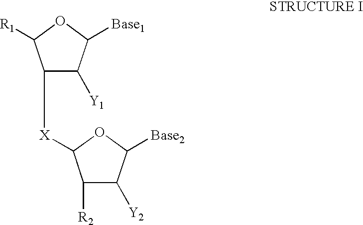

| TABLE 1 | |||
| Hybrid | Tm | ||
| dA40: dT40 | 70.3° C. | ||
| dA30: dT40 | 68.4° C. | ||
| dA25: dT40 | 64.5° C. | ||
| dA20: dT40 | 61.5° C. | ||
| dA15: dT40 | 56.0° C. | ||
| dA10: dT40 | 50.4° C. | ||
| TABLE 2 | |||
| Poly - T of | Poly - A of | Immobilized Probe | Immobilized Probe |
| Immobilized | Capture | Added Before Second | Present During Both |
| Probe | Probe | Hybridization | Hybridizations |
| dT30 | dA30 | 54,8951 | 31,608 |
| dA15 | 44,558 | 30,290 | |
| dT14 | dA30 | 65,3132 | 63,240 |
| dA15 | 43,285 | 37,479 | |
| 1Represents capture of 62% of the labeled target. | |||
| 2Represents capture of 67% of the labeled target. | |||
| TABLE 3 | ||||
| Sample No. | Smear Result | Culture Result | RLU No. 1 | RLU No. 2 |
| 1 | Positive | Unknown | 3.04 × 10⁶ | 2.87 × 10⁶ |
| 2 | Positive | Unknown | 3.09 × 10⁶ | 2.99 × 10⁶ |
| 3 | Positive | Unknown | 3.18 × 10⁶ | 3.00 × 10⁶ |
| 4 | Positive | Unknown | 3.28 × 10⁶ | 3.23 × 10⁶ |
| 5 | Positive | Positive | 2.97 × 10⁶ | 3.04 × 10⁶ |
| 6 | Positive | Positive | 3.16 × 10⁶ | 2.94 × 10⁶ |
| 7 | Unknown | Positive | 2.87 × 10⁶ | 2.98 × 10⁶ |
| 8 | Negative | Positive | 3.46 × 10⁴ | 2.05 × 10⁶ |
| 9 | Negative | Positive | 3.11 × 10⁶ | 2.99 × 10⁶ |
| 10 | Negative | Positive | 3.09 × 10⁶ | 3.09 × 10⁶ |
| 11 | Negative | Positive | 1.35 × 10⁶ | 6.15 × 10⁵ |
| 12 | Negative | Positive | 3.01 × 10⁶ | 3.13 × 10⁶ |
| 13 | Negative | Positive | 2.90 × 10⁶ | 2.88 × 10⁶ |
| 14 | Negative | Positive | 3.12 × 10⁶ | 3.07 × 10⁶ |
| 15 | Negative | Positive | 2.93 × 10⁶ | 3.00 × 10⁶ |
| Positive | Not | Not Applicable | 2.98 × 10⁶ | 2.89 × 10⁶ |
| Controls | Applicable | 3.00 × 10⁶ | ||
| Negative | Not | Not Applicable | 3.45 × 10³ | 5.67 × 10³ |
| Controls | Applicable | 2.05 × 10³ | ||
| TABLE 4 | ||||||
| Sample | Target (fg) | | Day | 2 | Day | 3 |
| 1 | 5 | 7.78 × 10⁵ | 6.46 × 10⁵ | 6.95 × 10⁵ |
| 2 | 5 | 7.73 × 10⁵ | 7.08 × 10⁵ | 7.06 × 10⁵ |
| 3 | 5 | 8.30 × 10⁵ | 7.00 × 10⁵ | 6.57 × 10⁵ |
| 4 | 5 | 7.69 × 10⁵ | 7.34 × 10⁵ | 7.60 × 10⁵ |
| 5 | 5 | 7.73 × 10⁵ | 7.76 × 10⁵ | 7.95 × 10⁵ |
| 6 | 5 | 7.01 × 10⁵ | 6.47 × 10⁵ | 7.14 × 10⁵ |
| 7 | 5 | 7.32 × 10⁵ | 6.70 × 10⁵ | 7.63 × 10⁵ |
| 8 | 5 | 7.84 × 10⁵ | 7.23 × 10⁵ | 7.06 × 10⁵ |
| 9 | 5 | 7.45 × 10⁵ | 7.17 × 10⁵ | 7.18 × 10⁵ |
| 10 | 5 | 7.45 × 10⁵ | 7.11 × 10⁵ | 7.68 × 10⁵ |
| 11 | 50 | 8.58 × 10⁵ | 7.51 × 10⁵ | 7.57 × 10⁵ |
| 12 | 50 | 8.20 × 10⁵ | 4.61 × 10⁵ | 7.77 × 10⁵ |
| 13 | 50 | 7.66 × 10⁵ | 6.99 × 10⁵ | 7.21 × 10⁵ |
| 14 | 50 | 7.85 × 10⁵ | 7.97 × 10⁵ | 7.45 × 10⁵ |
| 15 | 50 | 8.17 × 10⁵ | 7.80 × 10⁵ | 7.76 × 10⁵ |
| 16 | 50 | 7.98 × 10⁵ | 7.33 × 10⁵ | 7.42 × 10⁵ |
| 17 | 50 | 7.51 × 10⁵ | 7.36 × 10⁵ | 7.41 × 10⁵ |
| 18 | 50 | 7.76 × 10⁵ | 8.01 × 10⁵ | 7.80 × 10⁵ |
| 19 | 50 | 7.24 × 10⁵ | 7.27 × 10⁵ | 7.62 × 10⁵ |
| 20 | 50 | 7.74 × 10⁵ | 7.54 × 10⁵ | 7.70 × 10⁵ |
| Negatives (10) | 0 | 201 | −218 | 74 |
| TABLE 5 | ||||||
| Sample No. | PCR Results | RLU | Sample No. | | RLU | |
| 1 | Positive | 4.42 × 10⁵ | 17 | Negative | 0 | |
| 2 | Negative | 1.66 × 10³ | 18 | Positive | 3.59 × 10⁵ | |
| 3 | Positive | 2.95 × 10⁵ | 19 | Positive | 3.64 × 10⁵ | |
| 4 | Negative | 0 | 20 | Positive | 3.51 × 10⁵ | |
| 5 | Positive | 1.62 × 10⁵ | 21 | Positive | 3.75 × 10⁵ | |
| 6 | Positive | 4.15 × 10⁵ | 22 | Negative | 9.15 × 10³ | |
| 7 | Positive | 5.76 × 10⁵ | 23 | Positive | 3.21 × 10⁵ | |
| 8 | Negative | 3.69 × 10³ | 24 | Negative | 0 | |
| 9 | Positive | 4.45 × 10⁵ | 25 | Positive | 4.59 × 10⁵ | |
| 10 | Negative | 0 | 26 | Negative | 8 | |
| 11 | Positive | 5.00 × 10⁵ | 27 | Positive | 3.07 × 10⁵ | |
| 12 | Negative | 0 | 28 | Positive | 3.52 × 10⁵ | |
| 13 | Negative | 0 | 29 | Negative | 0 | |
| 14 | Negative | 0 | 30 | Negative | 0 | |
| 15 | Negative | 0 | Positive | Not Applicable | 3.14 × 10⁵ | |
| Controls | ||||||
| 16 | Negative | 0 | Negative | Not Applicable | 0 | |
| Controls | ||||||
| TABLE 6 | |||||
| C. tracho- | |||||
| matis + | |||||
| C. tracho- | N. gonor- | N. gonor- | |||
| Negative | matis | rhoeae | rhoeae | ||
| Control | Target | Target | Target | ||
| No Blood | CT: 1 | CT: | CT: | CT: |
| NG: 1.86 × 10² | 2.18 × 10⁵ | 4.90 × 10³ | 2.22 × 10⁵ | |
| NG: | NG: | NG: | ||
| 4.05 × 10² | 9.37 × 10⁵ | 9.37 × 10⁵ | ||
| 1% Blood | CT: 52 | CT: | CT: | CT: |
| NG: 4.41 × 10² | 2.73 × 10⁵ | 4.64 × 10³ | 1.73 × 10⁵ | |
| NG: | NG: | NG: | ||
| 1.38 × 10³ | 9.54 × 10⁵ | 9.80 × 10⁵ | ||
| 5% Blood | CT: 27 | CT: | CT: 0 | CT: |
| NG: 4.42 × 10² | 1.91 × 10⁵ | NG: | 1.65 × 10⁵ | |
| NG: | 1.06 × 10⁶ | NG: | ||
| 1.66 × 10³ | 9.19 × 10⁵ | |||
| 10% Blood | CT: 14 | CT: | CT: 0 | CT: |
| NG: 1.36 × 10² | 2.13 × 10⁵ | NG: | 1.35 × 10⁵ | |
| NG: | 1.05 × 10⁶ | NG: | ||
| 1.85 × 10³ | 1.12 × 10⁶ | |||
| # SEQUENCE LISTING |
| <160> NUMBER OF SEQ ID NOS: 2 |
| <210> |
| <211> LENGTH: 40 |
| <212> TYPE: DNA |
| <213> ORGANISM: Artificial Sequence |
| <220> FEATURE: |
| <223> OTHER INFORMATION: Description of Artificial |
| #Sequence: capture |
| probe oligonculeotide |
| <400> SEQUENCE: 1 |
| gaactactta gttccaatga ctgtttaaaa aaaaaaaaaa |
| # |
| # 40 |
| <210> |
| <211> LENGTH: 23 |
| <212> TYPE: DNA |
| <213> ORGANISM: Artificial Sequence |
| <220> FEATURE: |
| <223> OTHER INFORMATION: Description of Artificial |
| #Sequence: target |
| sequence complementary to a portion |
| #of the capture |
| probe oligonucleotide |
| <400> SEQUENCE: 2 |
| cagtcattgg aactaagtag |
| # |
| # |
| 23 |
Claims (20)
Priority Applications (1)
| Application Number | Priority Date | Filing Date | Title |
|---|---|---|---|
| US09/956,412 US6534273B2 (en) | 1997-05-02 | 2001-09-18 | Two-step hybridization and capture of a polynucleotide |
Applications Claiming Priority (5)
| Application Number | Priority Date | Filing Date | Title |
|---|---|---|---|
| US4543097P | 1997-05-02 | 1997-05-02 | |
| US09/070,998 US6110678A (en) | 1997-05-02 | 1998-05-01 | Two-step hybridization and capture of a polynucleotide |
| US09/574,561 US6280952B1 (en) | 1997-05-02 | 2000-05-19 | Two-step hybridization and capture of a polynucleotide |
| US09/910,635 US20020028459A1 (en) | 1997-05-02 | 2001-07-20 | Two-step hybridization and capture of a polynucleotide |
| US09/956,412 US6534273B2 (en) | 1997-05-02 | 2001-09-18 | Two-step hybridization and capture of a polynucleotide |
Related Parent Applications (1)
| Application Number | Title | Priority Date | Filing Date |
|---|---|---|---|
| US09/910,635 Continuation-In-Part US20020028459A1 (en) | 1997-05-02 | 2001-07-20 | Two-step hybridization and capture of a polynucleotide |
Publications (2)
| Publication Number | Publication Date |
|---|---|
| US20020127569A1 US20020127569A1 (en) | 2002-09-12 |
| US6534273B2 true US6534273B2 (en) | 2003-03-18 |
Family
ID=27488985
Family Applications (1)
| Application Number | Title | Priority Date | Filing Date |
|---|---|---|---|
| US09/956,412 Expired - Lifetime US6534273B2 (en) | 1997-05-02 | 2001-09-18 | Two-step hybridization and capture of a polynucleotide |
Country Status (1)
| Country | Link |
|---|---|
| US (1) | US6534273B2 (en) |
Cited By (81)
| Publication number | Priority date | Publication date | Assignee | Title |
|---|---|---|---|---|
| US20040053223A1 (en) * | 1999-07-09 | 2004-03-18 | Bee Gary G. | Detection of HIV-1 by nucleic acid amplification |
| US20040142329A1 (en) * | 2003-01-17 | 2004-07-22 | Ingeneus Corporation | Probe conjugation to increase multiplex binding motif preference |
| US20040232564A1 (en) * | 2001-12-20 | 2004-11-25 | Cher 'khng Victor Tan | Method for fabricating semiconductor package with multi-layer metal bumps |
| US20050282170A1 (en) * | 2003-02-07 | 2005-12-22 | Diagnocure Inc. | Method to detect prostate cancer in a sample |
| WO2005123959A2 (en) | 2004-06-10 | 2005-12-29 | Perkinelmer Las, Inc. | Multiplexing assays for analyte detection |
| WO2006026654A2 (en) * | 2004-08-27 | 2006-03-09 | Exact Sciences Corporation | Method for detecting a recombinant event |
| US20060068417A1 (en) * | 2004-07-01 | 2006-03-30 | Gen-Probe Incorporated | Methods and compositions to detect nucleic acids in a biological sample |
| US20070264658A1 (en) * | 2006-05-12 | 2007-11-15 | Gen-Probe Incorporated | Compositions and methods to detect enterococci nucleic acid |
| WO2008016988A1 (en) | 2006-08-01 | 2008-02-07 | Gen-Probe Incorporated | Methods of nonspecific target capture of nucleic acids |
| EP1909108A2 (en) | 2005-03-10 | 2008-04-09 | Gen-Probe Incorporated | Systems and methods to perform assays for detecting or quantifiying analytes |
| EP1975242A1 (en) | 2004-08-27 | 2008-10-01 | Gen-Probe Incorporated | Single-primer nucleic acid amplification methods |
| US20080241827A1 (en) * | 2004-05-10 | 2008-10-02 | Exact Sciences Corporation | Methods For Detecting A Mutant Nucleic Acid |
| US20080261228A1 (en) * | 1999-09-29 | 2008-10-23 | Ursula Busse | PCA3 Messenger RNA Species in Benign and Malignant Prostate Tissues |
| WO2008157801A2 (en) | 2007-06-21 | 2008-12-24 | Gen-Probe Incorporated | Instrument and receptacles for performing processes |
| EP2017356A2 (en) | 2006-06-06 | 2009-01-21 | Gen-Probe Incorporated | Tagged oliggonucleotides and their use in nucleic acid amplification methods |
| US20090286249A1 (en) * | 2008-05-13 | 2009-11-19 | Gen-Probe Incorporated | Inactivatable target capture oligomers for use in the selective hybridization and capture of target nucleic acid sequences |
| US20090325153A1 (en) * | 2005-04-21 | 2009-12-31 | Exact Sciences Corporation | Analysis of heterogeneous nucleic acid samples |
| US20100021884A1 (en) * | 2004-12-24 | 2010-01-28 | Daphne Hessels | mRNA Ratios in Urinary Sediments and/or Urine as a Prognostic and/or Theranostic Marker for Prostate Cancer |
| US20100047809A1 (en) * | 1997-04-10 | 2010-02-25 | Bussemakers Marion J G | Pca3, pca3 genes, and methods of use |
| US20100093550A1 (en) * | 2006-10-10 | 2010-04-15 | Stuelpnagel John R | Compositions and methods for representational selection of nucleic acids from complex mixtures using hybridization |
| US20100129822A1 (en) * | 2008-11-25 | 2010-05-27 | Gen-Probe Incorporated | Compositions and methods for detecting small rnas, and uses thereof |
| WO2010078374A1 (en) | 2008-12-30 | 2010-07-08 | Gen-Probe Incorporated | Compositions, kits and related methods for the detection and/or monitoring of listeria |
| WO2010088496A1 (en) | 2009-01-30 | 2010-08-05 | Gen-Probe Incorporated | Systems and methods for detecting a signal and applying thermal energy to a signal transmission element |
| US7776524B2 (en) | 2002-02-15 | 2010-08-17 | Genzyme Corporation | Methods for analysis of molecular events |
| WO2010151566A1 (en) | 2009-06-23 | 2010-12-29 | Gen-Probe Incorporated | Compositions and methods for detecting nucleic acid from mollicutes |
| WO2011003020A1 (en) | 2009-07-01 | 2011-01-06 | Gen-Probe Incorporated | Methods and compositions for nucleic acid amplification |
| WO2011026139A1 (en) | 2009-08-31 | 2011-03-03 | Gen-Probe Incorporated | Dengue virus assay |
| EP2322940A1 (en) | 2005-03-10 | 2011-05-18 | Gen-Probe Incorporated | Systems amd methods to perform assays for detecting or quantifing analytes within samples |
| US7993853B2 (en) | 2005-05-06 | 2011-08-09 | Gen-Probe Incorporated | Methods of nucleic acid target capture |
| WO2011103274A1 (en) | 2010-02-17 | 2011-08-25 | Gen-Probe Incorporated | Compositions and methods to detect atopobium vaginae nucleic acid |
| WO2011133811A2 (en) | 2010-04-21 | 2011-10-27 | Gen-Probe Incorporated | Compositions, methods and kits to detect herpes simplex virus nucleic acid |
| US20110269194A1 (en) * | 2010-04-20 | 2011-11-03 | Swift Biosciences, Inc. | Materials and methods for nucleic acid fractionation by solid phase entrapment and enzyme-mediated detachment |
| EP2400037A1 (en) | 2005-10-17 | 2011-12-28 | Gen-Probe Incorporated | Compositions and methods to detect legionella pneumophila nucleic acid |
| WO2012009373A2 (en) | 2010-07-12 | 2012-01-19 | Gen-Probe Incorporated | Compositions and assays to detect swine h1n1 influenza a virus, seasonal h1 influenza a virus and seasonal h3 influenza a virus nucleic acids |
| WO2012016142A1 (en) | 2010-07-29 | 2012-02-02 | Gen-Probe Incorporated | Method, system and apparatus for incorporating capacitive proximity sensing in an automated fluid transfer procedure |
| WO2012046219A2 (en) | 2010-10-04 | 2012-04-12 | Gen-Probe Prodesse, Inc. | Compositions, methods and kits to detect adenovirus nucleic acids |
| EP2471805A2 (en) | 2005-05-06 | 2012-07-04 | Gen-Probe Incorporated | Compositions and assays to specifically detect nucleic acid of influenza virus A or B |
| WO2012149034A1 (en) | 2011-04-25 | 2012-11-01 | Gen-Probe Incorporated | Compositions and methods for detecting bv-associated bacterial nucleic acid |
| WO2012174187A2 (en) | 2011-06-15 | 2012-12-20 | Grifols Therapeutics Inc. | Methods, compositions, and kits for determining human immunodeficiency virus (hiv) |
| WO2013036332A2 (en) | 2011-09-07 | 2013-03-14 | Grifols Therapeutics Inc. | Methods, compositions, and kits for determining hepatitis a virus |
| WO2013036928A1 (en) | 2011-09-08 | 2013-03-14 | Gen-Probe Incorporated | Compositions and methods for detecting bv-associated bacterial nucleic acid |
| WO2013044097A1 (en) | 2011-09-21 | 2013-03-28 | Gen-Probe Incorporated | Methods for amplifying nucleic acid using tag-mediated displacement |
| DE102011120550A1 (en) | 2011-12-05 | 2013-06-06 | Gen-Probe Prodesse, Inc. | Detecting adenovirus target nucleic acid, by contacting sample with amplification oligomer to obtain amplicons, providing conditions sufficient to produce amplicons of adenovirus target nucleic acid, and detecting amplicons |
| WO2013116774A1 (en) | 2012-02-01 | 2013-08-08 | Gen-Probe Incorporated | Asymmetric hairpin target capture oligomers |
| WO2013126793A2 (en) | 2012-02-24 | 2013-08-29 | Gen-Probe Prodesse, Inc. | Detection of shiga toxin genes in bacteria |
| WO2013163188A1 (en) | 2012-04-24 | 2013-10-31 | Gen-Probe Incorporated | Compositions, methods and kits to detect herpes simplex virus nucleic acids |
| WO2013184553A1 (en) | 2012-06-04 | 2013-12-12 | Gen-Probe Incorporated | Compositions and methods for amplifying and characterizing hcv nucleic acid |
| WO2014011892A1 (en) | 2012-07-13 | 2014-01-16 | Gen-Probe Incorporated | Method for detecting a minority genotype |
| WO2014036369A1 (en) | 2012-08-30 | 2014-03-06 | Gen-Probe Incorporated | Multiphase nucleic acid amplification |
| WO2014059260A2 (en) | 2012-10-11 | 2014-04-17 | Gen-Probe Incorporated | Compositions and methods for detecting human papillomavirus nucleic acid |
| US8703421B2 (en) | 2008-05-30 | 2014-04-22 | Gen-Probe Incorporated | Compositions, kits and related methods for the detection and/or monitoring of Salmonella |
| WO2014089508A1 (en) | 2012-12-07 | 2014-06-12 | Gen-Probe Prodesse, Inc. | Compositions and methods for detecting gastrointestinal pathogen nucleic acid |
| WO2014153193A2 (en) | 2013-03-14 | 2014-09-25 | Gen-Probe Incorporated | Diagnostic systems and methods |
| WO2014153116A2 (en) | 2013-03-14 | 2014-09-25 | Gen-Probe Incorporated | Apparatus for indexing and agitating fluid containers |
| US8927703B2 (en) | 2007-04-02 | 2015-01-06 | Gen-Probe Incorporated | Compositions, kits and related methods for the detection and/or monitoring of Pseudomonas aeruginosa |
| US9109256B2 (en) | 2004-10-27 | 2015-08-18 | Esoterix Genetic Laboratories, Llc | Method for monitoring disease progression or recurrence |
| DE102015203606A1 (en) | 2014-02-28 | 2015-09-03 | Gen-Probe Incorporated | A method of isolating nucleic acid from cytological samples in liquid preservatives containing formaldehyde |
| EP3095873A1 (en) | 2006-12-21 | 2016-11-23 | Gen-Probe Incorporated | Methods and compositions for nucleic acid amplification |
| WO2017120216A2 (en) | 2016-01-04 | 2017-07-13 | Gen-Probe Incorporated | Methods and compositions for detecting candida species |
| WO2018053174A1 (en) | 2016-09-15 | 2018-03-22 | Abbott Laboratories | Devices and methods for sample analysis |
| WO2018160979A1 (en) | 2017-03-03 | 2018-09-07 | Gen-Probe Incorporated | Evaporation-limiting inserts for reagent containers and related methods of use |
| WO2018175880A1 (en) | 2017-03-24 | 2018-09-27 | Gen-Probe Incorporated | System for mixing contents of containers and related methods of use |
| US10113206B2 (en) | 2010-02-24 | 2018-10-30 | Grifols Therapeutics Inc. | Methods, compositions, and kits for determining human immunodeficiency virus (HIV) |
| WO2018226798A1 (en) | 2017-06-07 | 2018-12-13 | Gen-Probe Incorporated | Detecting babesia species nucleic acid in a sample |
| WO2019014239A1 (en) | 2017-07-10 | 2019-01-17 | Gen-Probe Incorporated | Analytical systems and methods for nucleic acid amplification using sample assigning parameters |
| WO2019148169A1 (en) | 2018-01-29 | 2019-08-01 | Gen-Probe Incorporated | Analytical systems and methods |
| WO2020014400A1 (en) | 2018-07-10 | 2020-01-16 | Gen-Probe Incorporated | Methods and systems for detecting and quantifying nucleic acids |
| US10563254B2 (en) | 2007-01-23 | 2020-02-18 | Cambridge Enterprise Limited | Nucleic acid amplification and testing |
| WO2020041414A1 (en) | 2018-08-21 | 2020-02-27 | Gen-Probe Incorporated | Compositions and methods for amplifying, detecting or quantifying human cytomegalovirus |
| WO2020041671A1 (en) | 2018-08-24 | 2020-02-27 | Gen-Probe Incorporated | Compositions and methods for detecting bacterial nucleic acid and diagnosing bacterial vaginosis |
| EP3663004A1 (en) | 2009-05-15 | 2020-06-10 | Gen-Probe Incorporated | Method and apparatus for effecting automated movement of a magnet in an instrument for performing a magnetic separation procedure |
| EP3670664A1 (en) | 2014-02-28 | 2020-06-24 | Gen-Probe Incorporated | Method of isolating nucleic acid from specimens in liquid-based cytology preservatives containing formaldehyde |
| WO2020132408A2 (en) | 2018-12-20 | 2020-06-25 | Gen-Probe Incorporated | Compositions and methods for detecting plasmodium species nucleic acid |
| EP3736346A1 (en) | 2015-01-09 | 2020-11-11 | Gen-Probe Incorporated | Methods and compositions for diagnosing bacterial vaginosis |
| WO2020226969A2 (en) | 2019-05-03 | 2020-11-12 | Gen-Probe Incorporated | Receptacle transport system for an analytical system |
| WO2021003331A1 (en) | 2019-07-03 | 2021-01-07 | Gen-Probe Incorporated | Oligonucleotides for use in determining the presence of trichomonas vaginalis in a sample |
| WO2021178644A1 (en) | 2020-03-04 | 2021-09-10 | Gen-Probe Incorporated | Compositions and methods for detecting sars-cov-2 nucleic acid |
| WO2021224873A1 (en) | 2020-05-07 | 2021-11-11 | Grifols Diagnostic Solutions Inc. | Methods and compositions for detecting sars-cov-2 nucleic acid |
| EP3910075A1 (en) | 2015-03-16 | 2021-11-17 | Gen-Probe Incorporated | Kits for detecting bacterial nucleic acid and diagnosing bacterial vaginosis |
| EP3985131A1 (en) | 2016-06-10 | 2022-04-20 | Gen-Probe Incorporated | Compositions and methods for detecting zika virus nucleic acid |
| EP4299767A2 (en) | 2013-08-14 | 2024-01-03 | Gen-Probe Incorporated | Compositions and methods for detecting hev nucleic acid |
Families Citing this family (16)
| Publication number | Priority date | Publication date | Assignee | Title |
|---|---|---|---|---|
| ATE493494T1 (en) * | 2005-02-18 | 2011-01-15 | Gen Probe Inc | SAMPLE PREPARATION METHOD USING AN ALKALINE PUSH |
| DE602006011917D1 (en) * | 2005-08-19 | 2010-03-11 | Bioventures Inc | METHOD AND SUBSTANCES FOR ISOLATING MIRNAS |
| US20070072223A1 (en) * | 2005-09-16 | 2007-03-29 | Primera Biosystems, Inc. | Compositions and methods for purifying nucleic acids |
| JP5481189B2 (en) * | 2006-05-24 | 2014-04-23 | ジェン−プロウブ インコーポレイテッド | Probes and kits for determining the presence of a Mycobacterium tuberculosis complex organism in a test sample, and a method for amplifying Gram positive bacilli or fungi using a capture probe |
| US7833716B2 (en) | 2006-06-06 | 2010-11-16 | Gen-Probe Incorporated | Tagged oligonucleotides and their use in nucleic acid amplification methods |
| EP3733871A1 (en) | 2008-05-27 | 2020-11-04 | Dako Denmark A/S | Compositions and methods for detection of chromosomal aberrations with novel hybridization buffers |
| WO2010097656A1 (en) * | 2009-02-26 | 2010-09-02 | Dako Denmark A/S | Compositions and methods for performing a stringent wash step in hybridization applications |
| DK3133169T3 (en) * | 2009-08-25 | 2019-12-16 | Illumina Inc | PROCEDURES FOR SELECTION AND AMPLIFICATION OF POLYNUCLEOTIDES |
| US9255293B2 (en) | 2010-11-01 | 2016-02-09 | Gen-Probe Incorporated | Integrated capture and amplification of target nucleic acid for sequencing |
| US10662465B2 (en) | 2011-09-30 | 2020-05-26 | Agilent Technologies, Inc. | Hybridization compositions and methods using formamide |
| EP2768974B1 (en) | 2011-10-21 | 2017-07-19 | Dako Denmark A/S | Hybridization compositions and methods |
| US9816130B2 (en) * | 2011-12-22 | 2017-11-14 | Somagenics, Inc. | Methods of constructing small RNA libraries and their use for expression profiling of target RNAs |
| WO2013117595A2 (en) * | 2012-02-07 | 2013-08-15 | Illumina Cambridge Limited | Targeted enrichment and amplification of nucleic acids on a support |
| US9932626B2 (en) * | 2013-01-15 | 2018-04-03 | Quanterix Corporation | Detection of DNA or RNA using single molecule arrays and other techniques |
| US11014957B2 (en) | 2015-12-21 | 2021-05-25 | Realseq Biosciences, Inc. | Methods of library construction for polynucleotide sequencing |
| DE102018103215B3 (en) | 2018-02-13 | 2019-08-14 | Gna Biosolutions Gmbh | Method and device for extracting a nucleic acid from a sample fluid |
Citations (52)
| Publication number | Priority date | Publication date | Assignee | Title |
|---|---|---|---|---|
| US4486539A (en) | 1981-10-16 | 1984-12-04 | Orioon Corporation Ltd. | Detection of microbial nucleic acids by a one-step sandwich hybridization test |
| EP0149565A2 (en) | 1984-01-19 | 1985-07-24 | Kodak Clinical Diagnostics Limited | Assay method |
| WO1985004674A1 (en) | 1984-04-05 | 1985-10-24 | Life Technologies, Inc. | Immobilization of nucleic acids |
| EP0159719A2 (en) | 1984-04-27 | 1985-10-30 | Enzo Biochem, Inc. | Hybridization method for the detection of genetic materials |
| US4554088A (en) | 1983-05-12 | 1985-11-19 | Advanced Magnetics Inc. | Magnetic particles for use in separations |
| US4581333A (en) | 1978-04-13 | 1986-04-08 | Institut Pasteur | Method of detecting and characterizing a nucleic acid or reactant for the application of this method |
| US4683195A (en) | 1986-01-30 | 1987-07-28 | Cetus Corporation | Process for amplifying, detecting, and/or-cloning nucleic acid sequences |
| WO1988001302A1 (en) | 1986-08-11 | 1988-02-25 | Siska Diagnostics, Inc. | Nucleic acid probe assay methods and compositions |
| EP0265244A2 (en) | 1986-10-23 | 1988-04-27 | Amoco Corporation | Target and background capture methods and apparatus for affinity assays |
| US4751177A (en) | 1985-06-13 | 1988-06-14 | Amgen | Methods and kits for performing nucleic acid hybridization assays |
| US4755458A (en) | 1984-08-30 | 1988-07-05 | Enzo Biochem, Inc. | Composition and method for the detection of the presence of a polynucleotide sequence of interest |
| WO1988010315A1 (en) | 1987-06-19 | 1988-12-29 | Siska Diagnostics, Inc. | Transcription-based nucleic acid amplification/detection systems |
| EP0328829A2 (en) | 1987-12-21 | 1989-08-23 | Amoco Corporation | Method for affinity assays using target amplification |
| US4868105A (en) | 1985-12-11 | 1989-09-19 | Chiron Corporation | Solution phase nucleic acid sandwich assay |
| WO1989010979A1 (en) | 1988-05-10 | 1989-11-16 | E.I. Du Pont De Nemours And Company | Process for rapid nucleic acid detection |
| US4882269A (en) | 1985-12-13 | 1989-11-21 | Princeton University | Amplified hybridization assay |
| EP0370694A2 (en) | 1988-11-21 | 1990-05-30 | Eastman Kodak Company | Diagnostic kit and method using a solid phase capture means for detecting nucleic acids |
| WO1990010716A1 (en) | 1989-03-10 | 1990-09-20 | Gene-Trak Systems | Immobilized oligonucleotide probes and uses therefor |
| WO1991008307A1 (en) | 1989-12-04 | 1991-06-13 | Microprobe Corporation | Enhanced capture of target nucleic acid by the use of oligonucleotides covalently attached to polymers |
| EP0435150A2 (en) | 1989-12-29 | 1991-07-03 | Enzo Biochem, Inc. | Amplification capture assay |
| US5043272A (en) | 1989-04-27 | 1991-08-27 | Life Technologies, Incorporated | Amplification of nucleic acid sequences using oligonucleotides of random sequence as primers |
| EP0444120A1 (en) | 1988-11-21 | 1991-09-04 | Dynal As | Detection and quantitative determination of rna and dna. |
| EP0446260A1 (en) | 1988-11-21 | 1991-09-18 | Holmes Michael John | Nucleic acid probes. |
| WO1991014788A1 (en) | 1990-03-23 | 1991-10-03 | Chiron Corporation | Polynucleotide capture assay employing in vitro amplification |
| EP0454461A1 (en) | 1990-04-25 | 1991-10-30 | Gene-Trak Systems Corporation | Selective amplification system using Q beta replicase, a hybridization assay and a method of detecting and assessing concentration of target nucleic acid in liquid samples |
| US5112734A (en) | 1989-05-26 | 1992-05-12 | Gene-Trak Systems | Target-dependent synthesis of an artificial gene for the synthesis of a replicatable rna |
| US5124246A (en) | 1987-10-15 | 1992-06-23 | Chiron Corporation | Nucleic acid multimers and amplified nucleic acid hybridization assays using same |
| US5130238A (en) | 1988-06-24 | 1992-07-14 | Cangene Corporation | Enhanced nucleic acid amplification process |
| US5252723A (en) | 1989-03-06 | 1993-10-12 | Gen-Probe, Inc. | Method and reagent for sulfurization of organophosphorous compounds |
| US5273882A (en) | 1985-06-13 | 1993-12-28 | Amgen | Method and kit for performing nucleic acid hybridization assays |
| US5283174A (en) | 1987-09-21 | 1994-02-01 | Gen-Probe, Incorporated | Homogenous protection assay |
| WO1994003472A1 (en) | 1992-08-04 | 1994-02-17 | Gen-Probe Incorporated | Nucleic acid sequence amplification |
| US5288609A (en) | 1984-04-27 | 1994-02-22 | Enzo Diagnostics, Inc. | Capture sandwich hybridization method and composition |
| WO1994019023A1 (en) | 1993-02-19 | 1994-09-01 | Isis Pharmaceuticals, Inc. | Cyclobutyl antisense oligonucleotides, methods of making and use thereof |
| WO1995003430A1 (en) | 1993-07-23 | 1995-02-02 | Gen-Probe Incorporated | Methods for enhancing nucleic acid amplification |
| US5399491A (en) | 1989-07-11 | 1995-03-21 | Gen-Probe Incorporated | Nucleic acid sequence amplification methods |
| WO1995016055A1 (en) | 1993-12-08 | 1995-06-15 | Chiron Corporation | Solution phase nucleic acid sandwich assays having reduced background noise |
| US5437990A (en) | 1987-07-31 | 1995-08-01 | The Board Of Trustees Of The Leland Stanford Junior University | Selective amplification of target polynucleotide sequences |
| US5468613A (en) | 1986-03-13 | 1995-11-21 | Hoffmann-La Roche Inc. | Process for detecting specific nucleotide variations and genetic polymorphisms present in nucleic acids |
| US5547842A (en) | 1986-11-24 | 1996-08-20 | Gen-Probe Incorporated | Nucleic acid probes for detection and/or quantitation of non-viral organisms |
| US5554516A (en) | 1992-05-06 | 1996-09-10 | Gen-Probe Incorporated | Nucleic acid sequence amplification method, composition and kit |
| US5594123A (en) | 1985-03-28 | 1997-01-14 | Hoffman-La Roche Inc. | Primers and probes for the amplification and detection of aids associated nucleic acids |
| US5639604A (en) | 1987-09-21 | 1997-06-17 | Gen-Probe Incorporated | Homogeneous protection assay |
| US5656207A (en) | 1989-06-24 | 1997-08-12 | Gen Probe Incorporated | Detecting or quantifying multiple analytes using labelling techniques |
| US5658737A (en) | 1994-10-28 | 1997-08-19 | Gen-Probe Incorporated | Compositions and methods for the simultaneous detection and quantification of multiple specific nucleic acid sequences |
| US5702896A (en) | 1991-02-27 | 1997-12-30 | Amoco Corporation | Methods for improving the sensitivity of hybridization assays |
| US5731148A (en) | 1995-06-07 | 1998-03-24 | Gen-Probe Incorporated | Adduct protection assay |
| US5731153A (en) | 1996-08-26 | 1998-03-24 | The Regents Of The University Of California | Identification of random nucleic acid sequence aberrations using dual capture probes which hybridize to different chromosome regions |
| US5750338A (en) | 1986-10-23 | 1998-05-12 | Amoco Corporation | Target and background capture methods with amplification for affinity assays |
| US6060246A (en) | 1996-11-15 | 2000-05-09 | Avi Biopharma, Inc. | Reagent and method for isolation and detection of selected nucleic acid sequences |
| US6110678A (en) | 1997-05-02 | 2000-08-29 | Gen-Probe Incorporated | Two-step hybridization and capture of a polynucleotide |
| US6133436A (en) | 1996-11-06 | 2000-10-17 | Sequenom, Inc. | Beads bound to a solid support and to nucleic acids |
-
2001
- 2001-09-18 US US09/956,412 patent/US6534273B2/en not_active Expired - Lifetime
Patent Citations (58)
| Publication number | Priority date | Publication date | Assignee | Title |
|---|---|---|---|---|
| US4581333A (en) | 1978-04-13 | 1986-04-08 | Institut Pasteur | Method of detecting and characterizing a nucleic acid or reactant for the application of this method |
| US4486539A (en) | 1981-10-16 | 1984-12-04 | Orioon Corporation Ltd. | Detection of microbial nucleic acids by a one-step sandwich hybridization test |
| US4554088A (en) | 1983-05-12 | 1985-11-19 | Advanced Magnetics Inc. | Magnetic particles for use in separations |
| EP0149565A2 (en) | 1984-01-19 | 1985-07-24 | Kodak Clinical Diagnostics Limited | Assay method |
| WO1985004674A1 (en) | 1984-04-05 | 1985-10-24 | Life Technologies, Inc. | Immobilization of nucleic acids |
| EP0526912A2 (en) | 1984-04-27 | 1993-02-10 | Enzo Biochem, Inc. | Methods and compositions for the detection of a single-stranded target genetic material |
| EP0159719A2 (en) | 1984-04-27 | 1985-10-30 | Enzo Biochem, Inc. | Hybridization method for the detection of genetic materials |
| US5288609A (en) | 1984-04-27 | 1994-02-22 | Enzo Diagnostics, Inc. | Capture sandwich hybridization method and composition |
| US4894325A (en) | 1984-04-27 | 1990-01-16 | Enzo Biochem, Inc. | Hybridization method for the detection of genetic material |
| US4755458A (en) | 1984-08-30 | 1988-07-05 | Enzo Biochem, Inc. | Composition and method for the detection of the presence of a polynucleotide sequence of interest |
| US5594123A (en) | 1985-03-28 | 1997-01-14 | Hoffman-La Roche Inc. | Primers and probes for the amplification and detection of aids associated nucleic acids |
| US4751177A (en) | 1985-06-13 | 1988-06-14 | Amgen | Methods and kits for performing nucleic acid hybridization assays |
| US5273882A (en) | 1985-06-13 | 1993-12-28 | Amgen | Method and kit for performing nucleic acid hybridization assays |
| US4868105A (en) | 1985-12-11 | 1989-09-19 | Chiron Corporation | Solution phase nucleic acid sandwich assay |
| US4882269A (en) | 1985-12-13 | 1989-11-21 | Princeton University | Amplified hybridization assay |
| US4683195A (en) | 1986-01-30 | 1987-07-28 | Cetus Corporation | Process for amplifying, detecting, and/or-cloning nucleic acid sequences |
| US4683195B1 (en) | 1986-01-30 | 1990-11-27 | Cetus Corp | |
| US5468613A (en) | 1986-03-13 | 1995-11-21 | Hoffmann-La Roche Inc. | Process for detecting specific nucleotide variations and genetic polymorphisms present in nucleic acids |
| WO1988001302A1 (en) | 1986-08-11 | 1988-02-25 | Siska Diagnostics, Inc. | Nucleic acid probe assay methods and compositions |
| US5750338A (en) | 1986-10-23 | 1998-05-12 | Amoco Corporation | Target and background capture methods with amplification for affinity assays |
| EP0265244A2 (en) | 1986-10-23 | 1988-04-27 | Amoco Corporation | Target and background capture methods and apparatus for affinity assays |
| US5547842A (en) | 1986-11-24 | 1996-08-20 | Gen-Probe Incorporated | Nucleic acid probes for detection and/or quantitation of non-viral organisms |
| WO1988010315A1 (en) | 1987-06-19 | 1988-12-29 | Siska Diagnostics, Inc. | Transcription-based nucleic acid amplification/detection systems |
| US5437990A (en) | 1987-07-31 | 1995-08-01 | The Board Of Trustees Of The Leland Stanford Junior University | Selective amplification of target polynucleotide sequences |
| US5283174A (en) | 1987-09-21 | 1994-02-01 | Gen-Probe, Incorporated | Homogenous protection assay |
| US5639604A (en) | 1987-09-21 | 1997-06-17 | Gen-Probe Incorporated | Homogeneous protection assay |
| US5124246A (en) | 1987-10-15 | 1992-06-23 | Chiron Corporation | Nucleic acid multimers and amplified nucleic acid hybridization assays using same |
| EP0328829A2 (en) | 1987-12-21 | 1989-08-23 | Amoco Corporation | Method for affinity assays using target amplification |
| WO1989010979A1 (en) | 1988-05-10 | 1989-11-16 | E.I. Du Pont De Nemours And Company | Process for rapid nucleic acid detection |
| US5130238A (en) | 1988-06-24 | 1992-07-14 | Cangene Corporation | Enhanced nucleic acid amplification process |
| EP0370694A2 (en) | 1988-11-21 | 1990-05-30 | Eastman Kodak Company | Diagnostic kit and method using a solid phase capture means for detecting nucleic acids |
| EP0444120A1 (en) | 1988-11-21 | 1991-09-04 | Dynal As | Detection and quantitative determination of rna and dna. |
| EP0446260A1 (en) | 1988-11-21 | 1991-09-18 | Holmes Michael John | Nucleic acid probes. |
| US5252723A (en) | 1989-03-06 | 1993-10-12 | Gen-Probe, Inc. | Method and reagent for sulfurization of organophosphorous compounds |
| WO1990010716A1 (en) | 1989-03-10 | 1990-09-20 | Gene-Trak Systems | Immobilized oligonucleotide probes and uses therefor |
| US5043272A (en) | 1989-04-27 | 1991-08-27 | Life Technologies, Incorporated | Amplification of nucleic acid sequences using oligonucleotides of random sequence as primers |
| US5112734A (en) | 1989-05-26 | 1992-05-12 | Gene-Trak Systems | Target-dependent synthesis of an artificial gene for the synthesis of a replicatable rna |
| US5656207A (en) | 1989-06-24 | 1997-08-12 | Gen Probe Incorporated | Detecting or quantifying multiple analytes using labelling techniques |
| US5399491A (en) | 1989-07-11 | 1995-03-21 | Gen-Probe Incorporated | Nucleic acid sequence amplification methods |
| WO1991008307A1 (en) | 1989-12-04 | 1991-06-13 | Microprobe Corporation | Enhanced capture of target nucleic acid by the use of oligonucleotides covalently attached to polymers |
| EP0435150A2 (en) | 1989-12-29 | 1991-07-03 | Enzo Biochem, Inc. | Amplification capture assay |
| WO1991014788A1 (en) | 1990-03-23 | 1991-10-03 | Chiron Corporation | Polynucleotide capture assay employing in vitro amplification |
| US5200314A (en) | 1990-03-23 | 1993-04-06 | Chiron Corporation | Polynucleotide capture assay employing in vitro amplification |
| EP0454461A1 (en) | 1990-04-25 | 1991-10-30 | Gene-Trak Systems Corporation | Selective amplification system using Q beta replicase, a hybridization assay and a method of detecting and assessing concentration of target nucleic acid in liquid samples |
| US5702896A (en) | 1991-02-27 | 1997-12-30 | Amoco Corporation | Methods for improving the sensitivity of hybridization assays |
| US5554516A (en) | 1992-05-06 | 1996-09-10 | Gen-Probe Incorporated | Nucleic acid sequence amplification method, composition and kit |
| WO1994003472A1 (en) | 1992-08-04 | 1994-02-17 | Gen-Probe Incorporated | Nucleic acid sequence amplification |
| WO1994019023A1 (en) | 1993-02-19 | 1994-09-01 | Isis Pharmaceuticals, Inc. | Cyclobutyl antisense oligonucleotides, methods of making and use thereof |
| WO1995003430A1 (en) | 1993-07-23 | 1995-02-02 | Gen-Probe Incorporated | Methods for enhancing nucleic acid amplification |
| WO1995016055A1 (en) | 1993-12-08 | 1995-06-15 | Chiron Corporation | Solution phase nucleic acid sandwich assays having reduced background noise |
| US5635352A (en) | 1993-12-08 | 1997-06-03 | Chiron Corporation | Solution phase nucleic acid sandwich assays having reduced background noise |
| US5658737A (en) | 1994-10-28 | 1997-08-19 | Gen-Probe Incorporated | Compositions and methods for the simultaneous detection and quantification of multiple specific nucleic acid sequences |
| US5731148A (en) | 1995-06-07 | 1998-03-24 | Gen-Probe Incorporated | Adduct protection assay |
| US5731153A (en) | 1996-08-26 | 1998-03-24 | The Regents Of The University Of California | Identification of random nucleic acid sequence aberrations using dual capture probes which hybridize to different chromosome regions |
| US6133436A (en) | 1996-11-06 | 2000-10-17 | Sequenom, Inc. | Beads bound to a solid support and to nucleic acids |
| US6060246A (en) | 1996-11-15 | 2000-05-09 | Avi Biopharma, Inc. | Reagent and method for isolation and detection of selected nucleic acid sequences |
| US6110678A (en) | 1997-05-02 | 2000-08-29 | Gen-Probe Incorporated | Two-step hybridization and capture of a polynucleotide |
| US6280952B1 (en) | 1997-05-02 | 2001-08-28 | Gen-Probe Incorporated | Two-step hybridization and capture of a polynucleotide |
Non-Patent Citations (46)
Cited By (209)
| Publication number | Priority date | Publication date | Assignee | Title |
|---|---|---|---|---|
| US9540696B2 (en) | 1997-04-10 | 2017-01-10 | Stichting Katholieke Universiteit, The University Medical Centre Nijmegen | PCA3 genes |
| US8551699B2 (en) | 1997-04-10 | 2013-10-08 | Stichting Katholieke Universiteit, more particularly The University of Medical Centre Nijmegen | PCA3, PCA3 genes, and methods of use |
| US20100047809A1 (en) * | 1997-04-10 | 2010-02-25 | Bussemakers Marion J G | Pca3, pca3 genes, and methods of use |
| US20040053223A1 (en) * | 1999-07-09 | 2004-03-18 | Bee Gary G. | Detection of HIV-1 by nucleic acid amplification |
| US7097979B2 (en) | 1999-07-09 | 2006-08-29 | Gen-Probe Incorporated | Detection of HIV-1 by nucleic acid amplification |
| US20090053692A1 (en) * | 1999-07-09 | 2009-02-26 | Gen-Probe Incorporated | Detection of hiv-1 by nucleic acid amplification |
| US7723040B2 (en) | 1999-07-09 | 2010-05-25 | Gen-Probe Incorporated | Detection of HIV-1 by nucleic acid amplification |
| US7927806B2 (en) | 1999-09-29 | 2011-04-19 | Diagnocure Inc. | Distinguishing PCA3 messenger RNA species in benign and malignant prostate tissues |
| US20100129824A1 (en) * | 1999-09-29 | 2010-05-27 | Ursula Busse | Distinguishing pca3 messenger rna species in benign and malignant prostate tissues |
| US9909189B2 (en) | 1999-09-29 | 2018-03-06 | Gen-Probe Incorporated | Distinguishing PCA3 messenger RNA species in benign and malignant prostate tissues |
| US20080261228A1 (en) * | 1999-09-29 | 2008-10-23 | Ursula Busse | PCA3 Messenger RNA Species in Benign and Malignant Prostate Tissues |
| US8618276B2 (en) | 1999-09-29 | 2013-12-31 | Diagnocure Inc. | Distinguishing PCA3 messenger RNA species in benign and malignant prostate tissues |
| US7655408B2 (en) | 1999-09-29 | 2010-02-02 | Diagnocure Inc. | PCA3 messenger RNA species in benign and malignant prostate tissues |
| US20110212449A1 (en) * | 1999-09-29 | 2011-09-01 | Ursula Busse | Distinguishing pca3 messenger rna species in benign and malignant prostate tissues |
| US8241848B2 (en) | 1999-09-29 | 2012-08-14 | Diagnocure Inc. | Distinguishing PCA3 messenger RNA species in benign and malignant prostate tissues |
| US20040232564A1 (en) * | 2001-12-20 | 2004-11-25 | Cher 'khng Victor Tan | Method for fabricating semiconductor package with multi-layer metal bumps |
| US8409829B2 (en) | 2002-02-15 | 2013-04-02 | Esoterix Genetic Laboratories, Llc | Methods for analysis of molecular events |
| US7776524B2 (en) | 2002-02-15 | 2010-08-17 | Genzyme Corporation | Methods for analysis of molecular events |
| US20040142329A1 (en) * | 2003-01-17 | 2004-07-22 | Ingeneus Corporation | Probe conjugation to increase multiplex binding motif preference |
| US8546551B2 (en) | 2003-02-07 | 2013-10-01 | Diagnocure Inc. | Method to detect prostate cancer in a sample |
| US10006092B2 (en) | 2003-02-07 | 2018-06-26 | Gen-Probe Incorporated | Method to detect prostate cancer in a sample |
| US8192931B2 (en) | 2003-02-07 | 2012-06-05 | Diagnocure Inc. | Method to detect prostate cancer in a sample |
| US20050282170A1 (en) * | 2003-02-07 | 2005-12-22 | Diagnocure Inc. | Method to detect prostate cancer in a sample |
| US11104958B2 (en) | 2003-02-07 | 2021-08-31 | Gen-Probe Incorporated | Method to detect prostate cancer in a sample |
| US20080241827A1 (en) * | 2004-05-10 | 2008-10-02 | Exact Sciences Corporation | Methods For Detecting A Mutant Nucleic Acid |
| WO2005123959A3 (en) * | 2004-06-10 | 2007-05-24 | Perkinelmer Las Inc | Multiplexing assays for analyte detection |
| US7422855B2 (en) * | 2004-06-10 | 2008-09-09 | Perkinelmer Las, Inc. | Multiplexing assays for analyte detection |
| WO2005123959A2 (en) | 2004-06-10 | 2005-12-29 | Perkinelmer Las, Inc. | Multiplexing assays for analyte detection |
| US20060003366A1 (en) * | 2004-06-10 | 2006-01-05 | Perkinelmer Las, Inc. | Multiplexing assays for analyte detection |
| US20060068417A1 (en) * | 2004-07-01 | 2006-03-30 | Gen-Probe Incorporated | Methods and compositions to detect nucleic acids in a biological sample |
| US8034554B2 (en) | 2004-07-01 | 2011-10-11 | Gen-Probe Incorporated | Methods and compositions to detect nucleic acids in a biological sample |
| US8551766B2 (en) | 2004-07-01 | 2013-10-08 | Gen-Probe Incorporated | Methods and compositions to detect nucleic acids in a biological sample |
| US8460869B2 (en) | 2004-07-01 | 2013-06-11 | Gen-Probe Incorporated | Methods and compositions to detect nucleic acids in a biological sample |
| EP1975242A1 (en) | 2004-08-27 | 2008-10-01 | Gen-Probe Incorporated | Single-primer nucleic acid amplification methods |
| US8389220B2 (en) * | 2004-08-27 | 2013-03-05 | Esoterix Genetic Laboratories, Llc | Method for detecting a recombinant event |
| WO2006026654A3 (en) * | 2004-08-27 | 2006-09-21 | Exact Sciences Corp | Method for detecting a recombinant event |
| US7981607B2 (en) * | 2004-08-27 | 2011-07-19 | Esoterix Genetic Laboratories LLC | Method for detecting recombinant event |
| EP2071031A1 (en) | 2004-08-27 | 2009-06-17 | Gen-Probe Incorporated | Single-primer nucleic acid amplification methods |
| WO2006026654A2 (en) * | 2004-08-27 | 2006-03-09 | Exact Sciences Corporation | Method for detecting a recombinant event |
| US20120015369A1 (en) * | 2004-08-27 | 2012-01-19 | Shuber Anthony P | Method for detecting a recombinant event |
| US20090170077A1 (en) * | 2004-08-27 | 2009-07-02 | Shuber Anthony P | Method for detecting recombinant event |
| US9109256B2 (en) | 2004-10-27 | 2015-08-18 | Esoterix Genetic Laboratories, Llc | Method for monitoring disease progression or recurrence |
| US9951390B2 (en) | 2004-12-24 | 2018-04-24 | Stichting Katholieke Universiteit, The University Medical Centre Nijmegen | Prostate cancer prognostic compositions and kits |
| EP2325334A1 (en) | 2004-12-24 | 2011-05-25 | Stichting Katholieke Universiteit, The University Medical Centre Nijmegen | mRNA ratios in urinary sediments and/or urine as a prognostic and/or theranostic marker for prostate cancer |
| US9096907B2 (en) | 2004-12-24 | 2015-08-04 | Stichting Katholieke Universiteit, The University Medical Centre Nijmegen | Prostate cancer prognostic compositions and kits |
| US10752957B2 (en) | 2004-12-24 | 2020-08-25 | Gen-Probe Incorporated | Prostate cancer prognostic compositions and kits |
| US20100021884A1 (en) * | 2004-12-24 | 2010-01-28 | Daphne Hessels | mRNA Ratios in Urinary Sediments and/or Urine as a Prognostic and/or Theranostic Marker for Prostate Cancer |
| US8257924B2 (en) | 2004-12-24 | 2012-09-04 | Stichting Katholieke Universiteit, The University Medical Centre Nijmegen | mRNA ratios in urinary sediments and/or urine as a prognostic and/or theranostic marker for prostate cancer |
| US7960109B2 (en) | 2004-12-24 | 2011-06-14 | Stichting Katholieke Universiteit, The University Medical Centre Nijmegen | mRNA ratios in urinary sediments and/or urine as a prognostic and/or theranostic marker for prostate cancer |
| EP2348321A2 (en) | 2005-03-10 | 2011-07-27 | Gen-Probe Incorporated | System and methods to perform assays for detecting or quantifying analytes within samples |
| EP1909108A2 (en) | 2005-03-10 | 2008-04-09 | Gen-Probe Incorporated | Systems and methods to perform assays for detecting or quantifiying analytes |
| EP2333561A2 (en) | 2005-03-10 | 2011-06-15 | Gen-Probe Incorporated | System for performing multi-formatted assays |
| EP1921454A2 (en) | 2005-03-10 | 2008-05-14 | Gen-Probe Incorporated | Systems and methods to perform assays for detecting or quantifying analytes |
| EP2322940A1 (en) | 2005-03-10 | 2011-05-18 | Gen-Probe Incorporated | Systems amd methods to perform assays for detecting or quantifing analytes within samples |
| EP2348320A2 (en) | 2005-03-10 | 2011-07-27 | Gen-Probe Incorporated | Systems and methods for detecting multiple optical signals |
| EP1930730A2 (en) | 2005-03-10 | 2008-06-11 | Gen-Probe Incorporated | Systems and methods to perform assays for detecting or quantifying analytes |
| US20090325153A1 (en) * | 2005-04-21 | 2009-12-31 | Exact Sciences Corporation | Analysis of heterogeneous nucleic acid samples |
| US9777314B2 (en) | 2005-04-21 | 2017-10-03 | Esoterix Genetic Laboratories, Llc | Analysis of heterogeneous nucleic acid samples |
| US7993853B2 (en) | 2005-05-06 | 2011-08-09 | Gen-Probe Incorporated | Methods of nucleic acid target capture |
| EP2471805A2 (en) | 2005-05-06 | 2012-07-04 | Gen-Probe Incorporated | Compositions and assays to specifically detect nucleic acid of influenza virus A or B |
| EP2400037A1 (en) | 2005-10-17 | 2011-12-28 | Gen-Probe Incorporated | Compositions and methods to detect legionella pneumophila nucleic acid |
| US20070264658A1 (en) * | 2006-05-12 | 2007-11-15 | Gen-Probe Incorporated | Compositions and methods to detect enterococci nucleic acid |
| US7588897B2 (en) | 2006-05-12 | 2009-09-15 | Gen-Probe Incorporated | Compositions and methods to detect Enterococci nucleic acid |
| EP2345740A1 (en) | 2006-06-06 | 2011-07-20 | Gen-Probe Incorporated | Tagged oligonucleotides and their use in nucleic acid amplification methods |
| EP2017356A2 (en) | 2006-06-06 | 2009-01-21 | Gen-Probe Incorporated | Tagged oliggonucleotides and their use in nucleic acid amplification methods |
| US20080286775A1 (en) * | 2006-08-01 | 2008-11-20 | Gen-Probe Incorporated | Methods of nonspecific target capture of nucleic acids |
| WO2008016988A1 (en) | 2006-08-01 | 2008-02-07 | Gen-Probe Incorporated | Methods of nonspecific target capture of nucleic acids |
| US10900033B2 (en) | 2006-08-01 | 2021-01-26 | Gen-Probe Incorporated | Method of nonspecific target capture of nucleic acids |
| US9051601B2 (en) | 2006-08-01 | 2015-06-09 | Gen-Probe Incorporated | Methods of nonspecific target capture of nucleic acids |
| US9340781B2 (en) | 2006-10-10 | 2016-05-17 | Illumina, Inc. | Compositions and methods for representational selection of nucleic acids from complex mixtures using hybridization |
| US8916350B2 (en) | 2006-10-10 | 2014-12-23 | Illumina, Inc. | Compositions and methods for representational selection of nucleic acids from complex mixtures using hybridization |
| US9139826B2 (en) | 2006-10-10 | 2015-09-22 | Illumina, Inc. | Compositions and methods for representational selection of nucleic acids from complex mixtures using hybridization |
| US8568979B2 (en) | 2006-10-10 | 2013-10-29 | Illumina, Inc. | Compositions and methods for representational selection of nucleic acids from complex mixtures using hybridization |
| US10538759B2 (en) | 2006-10-10 | 2020-01-21 | Illumina, Inc. | Compounds and method for representational selection of nucleic acids from complex mixtures using hybridization |
| US20100093550A1 (en) * | 2006-10-10 | 2010-04-15 | Stuelpnagel John R | Compositions and methods for representational selection of nucleic acids from complex mixtures using hybridization |
| US9587273B2 (en) | 2006-10-10 | 2017-03-07 | Illumina, Inc. | Compositions and methods for representational selection of nucleic acids from complex mixtures using hybridization |
| EP3095873A1 (en) | 2006-12-21 | 2016-11-23 | Gen-Probe Incorporated | Methods and compositions for nucleic acid amplification |
| EP3121286A1 (en) | 2006-12-21 | 2017-01-25 | Gen-Probe Incorporated | Methods and compositions for nucleic acid amplification |
| US11447821B2 (en) | 2007-01-23 | 2022-09-20 | Cambridge Enterprise Limited | Nucleic acid amplification and testing |
| US10563254B2 (en) | 2007-01-23 | 2020-02-18 | Cambridge Enterprise Limited | Nucleic acid amplification and testing |
| US8927703B2 (en) | 2007-04-02 | 2015-01-06 | Gen-Probe Incorporated | Compositions, kits and related methods for the detection and/or monitoring of Pseudomonas aeruginosa |
| US10626466B2 (en) | 2007-04-02 | 2020-04-21 | Gen-Probe Incorporated | Compositions, kits and related methods for the detection and/or monitoring of Pseudomonas aeruginosa |
| US8784745B2 (en) | 2007-06-21 | 2014-07-22 | Gen-Probe Incorporated | Methods for manipulating liquid substances in multi-chambered receptacles |
| US10744469B2 (en) | 2007-06-21 | 2020-08-18 | Gen-Probe Incorporated | Multi-chambered receptacles |
| US8048375B2 (en) | 2007-06-21 | 2011-11-01 | Gen-Probe Incorporated | Gravity-assisted mixing methods |
| EP2191897A1 (en) | 2007-06-21 | 2010-06-02 | Gen-Probe Incorporated | Instrument and receptacles for performing processes |
| US8052929B2 (en) | 2007-06-21 | 2011-11-08 | Gen-Probe Incorporated | Gravity-assisted mixing methods |
| US8480976B2 (en) | 2007-06-21 | 2013-07-09 | Gen-Probe Incorporated | Instruments and methods for mixing the contents of a detection chamber |
| US8491178B2 (en) | 2007-06-21 | 2013-07-23 | Gen-Probe Incorporated | Instruments and methods for mixing the contents of a detection chamber |
| US20090136913A1 (en) * | 2007-06-21 | 2009-05-28 | Gen-Probe Incorporated | Gravity-Assisted Mixing Methods |
| US20110189661A1 (en) * | 2007-06-21 | 2011-08-04 | Gen-Probe Incorporated | Gravity-assisted mixing methods |
| US10688458B2 (en) | 2007-06-21 | 2020-06-23 | Gen-Probe Incorporated | System and method of using multi-chambered receptacles |
| US7767447B2 (en) | 2007-06-21 | 2010-08-03 | Gen-Probe Incorporated | Instruments and methods for exposing a receptacle to multiple thermal zones |
| US20090142745A1 (en) * | 2007-06-21 | 2009-06-04 | Gen-Probe Incorporated | Instruments and methods for exposing a receptacle to multiple thermal zones |
| EP2425894A1 (en) | 2007-06-21 | 2012-03-07 | Gen-Probe Incorporated | Instruments and method for exposing a receptacle to multiple thermal zones |
| US11235295B2 (en) | 2007-06-21 | 2022-02-01 | Gen-Probe Incorporated | System and method of using multi-chambered receptacles |
| US7780336B2 (en) | 2007-06-21 | 2010-08-24 | Gen-Probe Incorporated | Instruments and methods for mixing the contents of a detection chamber |
| EP2465609A1 (en) | 2007-06-21 | 2012-06-20 | Gen-Probe Incorporated | Instruments and method for mixing the contents of a detection chamber |
| US9744506B2 (en) | 2007-06-21 | 2017-08-29 | Gen-Probe Incorporated | Instruments for mixing the contents of a detection chamber |
| US11235294B2 (en) | 2007-06-21 | 2022-02-01 | Gen-Probe Incorporated | System and method of using multi-chambered receptacles |
| US8828654B2 (en) | 2007-06-21 | 2014-09-09 | Gen-Probe Incorporated | Methods for manipulating liquid substances in multi-chambered receptacles |
| US8765367B2 (en) | 2007-06-21 | 2014-07-01 | Gen-Probe Incorporated | Methods and instruments for processing a sample in a multi-chambered receptacle |
| US8735055B2 (en) | 2007-06-21 | 2014-05-27 | Gen-Probe Incorporated | Methods of concentrating an analyte |
| WO2008157801A2 (en) | 2007-06-21 | 2008-12-24 | Gen-Probe Incorporated | Instrument and receptacles for performing processes |
| EP2806037A1 (en) | 2008-05-13 | 2014-11-26 | Gen-Probe Incorporated | Inactivatable target capture oligomers for use in the selective hybridization and capture of target nucleic acid sequences |
| US20090286249A1 (en) * | 2008-05-13 | 2009-11-19 | Gen-Probe Incorporated | Inactivatable target capture oligomers for use in the selective hybridization and capture of target nucleic acid sequences |
| EP2479287A1 (en) | 2008-05-13 | 2012-07-25 | Gen-Probe Incorporated | Inactivatable target capture oligomers for use in the selective hybridization and capture of target nucleic acid sequences |
| US10829801B2 (en) | 2008-05-13 | 2020-11-10 | Gen-Probe Incorporated | Inactivatable target capture oligomers for use in the selective hybridization and capture of target nucleic acid sequences |
| US10316352B2 (en) | 2008-05-13 | 2019-06-11 | Gen-Probe Incorporated | Methods of capturing a target nucleic acid for amplification and detection using an inactivatable target capture oligomer |
| US20140227695A1 (en) * | 2008-05-30 | 2014-08-14 | Gen-Probe Incorporated | Compositions, kits and related methods for the detection and/or monitoring of salmonella |
| US10711295B2 (en) | 2008-05-30 | 2020-07-14 | Gen-Probe Incorporated | Compositions, kits and related methods for the detection and/or monitoring of salmonella |
| US8703421B2 (en) | 2008-05-30 | 2014-04-22 | Gen-Probe Incorporated | Compositions, kits and related methods for the detection and/or monitoring of Salmonella |
| US10240185B2 (en) * | 2008-05-30 | 2019-03-26 | Gen-Probe Incorporated | Compositions, kits and related methods for the detection and/or monitoring of Salmonella |
| US20100129822A1 (en) * | 2008-11-25 | 2010-05-27 | Gen-Probe Incorporated | Compositions and methods for detecting small rnas, and uses thereof |
| US8951730B2 (en) | 2008-11-25 | 2015-02-10 | Gen-Probe Incorporated | Compositions for detecting small RNAs |
| US8399222B2 (en) | 2008-11-25 | 2013-03-19 | Gen-Probe Incorporated | Compositions and methods for detecting small RNAs, and uses thereof |
| EP2910647A1 (en) | 2008-12-30 | 2015-08-26 | Gen-Probe Incorporated | Compositions, kits and related methods for the detection and/or monitoring of listeria |
| WO2010078374A1 (en) | 2008-12-30 | 2010-07-08 | Gen-Probe Incorporated | Compositions, kits and related methods for the detection and/or monitoring of listeria |
| WO2010088496A1 (en) | 2009-01-30 | 2010-08-05 | Gen-Probe Incorporated | Systems and methods for detecting a signal and applying thermal energy to a signal transmission element |
| EP3663004A1 (en) | 2009-05-15 | 2020-06-10 | Gen-Probe Incorporated | Method and apparatus for effecting automated movement of a magnet in an instrument for performing a magnetic separation procedure |
| EP3450573A1 (en) | 2009-06-23 | 2019-03-06 | Gen-Probe Incorporated | Compositions and methods for detecting nucleic acid from mollicutes |
| EP2829616A1 (en) | 2009-06-23 | 2015-01-28 | Gen-Probe Incorporated | Composition and methods for detecting nucleic acid from mollicutes |
| WO2010151566A1 (en) | 2009-06-23 | 2010-12-29 | Gen-Probe Incorporated | Compositions and methods for detecting nucleic acid from mollicutes |
| WO2011003020A1 (en) | 2009-07-01 | 2011-01-06 | Gen-Probe Incorporated | Methods and compositions for nucleic acid amplification |
| WO2011026139A1 (en) | 2009-08-31 | 2011-03-03 | Gen-Probe Incorporated | Dengue virus assay |
| EP3040425A1 (en) | 2010-02-17 | 2016-07-06 | Gen-Probe Incorporated | Compostions and methods to detect atopobium vaginae nucleic acid |
| WO2011103274A1 (en) | 2010-02-17 | 2011-08-25 | Gen-Probe Incorporated | Compositions and methods to detect atopobium vaginae nucleic acid |
| EP3604558A1 (en) | 2010-02-17 | 2020-02-05 | Gen-Probe Incorporated | Compositions and methods to detect atopobium vaginae nucleic acid |
| US10113206B2 (en) | 2010-02-24 | 2018-10-30 | Grifols Therapeutics Inc. | Methods, compositions, and kits for determining human immunodeficiency virus (HIV) |
| US20110269194A1 (en) * | 2010-04-20 | 2011-11-03 | Swift Biosciences, Inc. | Materials and methods for nucleic acid fractionation by solid phase entrapment and enzyme-mediated detachment |
| EP3037555A1 (en) | 2010-04-21 | 2016-06-29 | Gen-Probe Incorporated | Compositions, methods and kits to detect herpes simplex virus nucleic acids |
| WO2011133811A2 (en) | 2010-04-21 | 2011-10-27 | Gen-Probe Incorporated | Compositions, methods and kits to detect herpes simplex virus nucleic acid |
| EP3587593A2 (en) | 2010-04-21 | 2020-01-01 | Gen-Probe Incorporated | Compositions, methods and kits to detect herpes simplex virus nucleic acids |
| WO2012009373A2 (en) | 2010-07-12 | 2012-01-19 | Gen-Probe Incorporated | Compositions and assays to detect swine h1n1 influenza a virus, seasonal h1 influenza a virus and seasonal h3 influenza a virus nucleic acids |
| EP3301195A1 (en) | 2010-07-12 | 2018-04-04 | Gen-Probe Incorporated | Compositions and assays to detect swine h1n1 influenza a virus nucleic acid |
| EP3674423A1 (en) | 2010-07-12 | 2020-07-01 | Gen-Probe Incorporated | Compositions and assays to detect seasonal h3 influenza a virus nucleic acid |
| EP3305920A1 (en) | 2010-07-12 | 2018-04-11 | Gen-Probe Incorporated | Compositions and assays to detect seasonal h3 influenza a virus nucleic acid |
| WO2012016142A1 (en) | 2010-07-29 | 2012-02-02 | Gen-Probe Incorporated | Method, system and apparatus for incorporating capacitive proximity sensing in an automated fluid transfer procedure |
| EP3438289A1 (en) | 2010-10-04 | 2019-02-06 | Gen-Probe Prodesse, Inc. | Compositions, methods and kits to detect adenovirus nucleic acids |
| WO2012046219A2 (en) | 2010-10-04 | 2012-04-12 | Gen-Probe Prodesse, Inc. | Compositions, methods and kits to detect adenovirus nucleic acids |
| EP3604559A1 (en) | 2011-04-25 | 2020-02-05 | Gen-Probe Incorporated | Compositions and methods for detecting bv-associated bacterial nucleic acid |
| EP3272886A1 (en) | 2011-04-25 | 2018-01-24 | Gen-Probe Incorporated | Compositions and methods for detecting bv-associated bacterial nucleic acid |
| WO2012149034A1 (en) | 2011-04-25 | 2012-11-01 | Gen-Probe Incorporated | Compositions and methods for detecting bv-associated bacterial nucleic acid |
| WO2012174187A2 (en) | 2011-06-15 | 2012-12-20 | Grifols Therapeutics Inc. | Methods, compositions, and kits for determining human immunodeficiency virus (hiv) |
| WO2013036332A2 (en) | 2011-09-07 | 2013-03-14 | Grifols Therapeutics Inc. | Methods, compositions, and kits for determining hepatitis a virus |
| WO2013036928A1 (en) | 2011-09-08 | 2013-03-14 | Gen-Probe Incorporated | Compositions and methods for detecting bv-associated bacterial nucleic acid |
| EP3255155A1 (en) | 2011-09-08 | 2017-12-13 | Gen-Probe Incorporated | Compositions and methods for detecting bv-associated bacterial nucleic acid |
| WO2013044097A1 (en) | 2011-09-21 | 2013-03-28 | Gen-Probe Incorporated | Methods for amplifying nucleic acid using tag-mediated displacement |
| DE102011120550A1 (en) | 2011-12-05 | 2013-06-06 | Gen-Probe Prodesse, Inc. | Detecting adenovirus target nucleic acid, by contacting sample with amplification oligomer to obtain amplicons, providing conditions sufficient to produce amplicons of adenovirus target nucleic acid, and detecting amplicons |
| EP3511426A1 (en) | 2012-02-01 | 2019-07-17 | Gen-Probe Incorporated | Asymmetric hairpin target capture oligomers |
| WO2013116774A1 (en) | 2012-02-01 | 2013-08-08 | Gen-Probe Incorporated | Asymmetric hairpin target capture oligomers |
| WO2013126793A2 (en) | 2012-02-24 | 2013-08-29 | Gen-Probe Prodesse, Inc. | Detection of shiga toxin genes in bacteria |
| EP3453770A2 (en) | 2012-02-24 | 2019-03-13 | Gen-Probe Prodesse, Inc. | Detection of shiga toxin genes in bacteria |
| EP3401409A1 (en) | 2012-04-24 | 2018-11-14 | Gen-Probe Incorporated | Compositions, methods and kits to detect herpes simplex virus nucleic acids |
| EP3907302A1 (en) | 2012-04-24 | 2021-11-10 | Gen-Probe Incorporated | Compositions, methods and kits to detect herpes simplex virus nucleic acids |
| EP3783114A1 (en) | 2012-04-24 | 2021-02-24 | Gen-Probe Incorporated | Compositions, methods and kits to detect herpes simplex virus nucleic acids |
| WO2013163188A1 (en) | 2012-04-24 | 2013-10-31 | Gen-Probe Incorporated | Compositions, methods and kits to detect herpes simplex virus nucleic acids |
| WO2013184553A1 (en) | 2012-06-04 | 2013-12-12 | Gen-Probe Incorporated | Compositions and methods for amplifying and characterizing hcv nucleic acid |
| WO2014011892A1 (en) | 2012-07-13 | 2014-01-16 | Gen-Probe Incorporated | Method for detecting a minority genotype |
| EP3339448A1 (en) | 2012-07-13 | 2018-06-27 | Gen-Probe Incorporated | Method for detecting a minority genotype |
| EP3640348A1 (en) | 2012-08-30 | 2020-04-22 | Gen-Probe Incorporated | Multiphase nucleic acid amplification |
| WO2014036369A1 (en) | 2012-08-30 | 2014-03-06 | Gen-Probe Incorporated | Multiphase nucleic acid amplification |
| WO2014059260A2 (en) | 2012-10-11 | 2014-04-17 | Gen-Probe Incorporated | Compositions and methods for detecting human papillomavirus nucleic acid |
| EP3670674A1 (en) | 2012-10-11 | 2020-06-24 | Gen-Probe Incorporated | Compositions and methods for detecting human papillomavirus nucleic acid |
| EP3342876A2 (en) | 2012-12-07 | 2018-07-04 | Gen-Probe Prodesse, Inc. | Methods for detecting gastrointestinal pathogen nucleic acid |
| EP3098323A1 (en) | 2012-12-07 | 2016-11-30 | Gen-Probe Prodesse Inc. | Compositions and methods for detecting gastrointestinal pathogen nucleic acid |
| WO2014089508A1 (en) | 2012-12-07 | 2014-06-12 | Gen-Probe Prodesse, Inc. | Compositions and methods for detecting gastrointestinal pathogen nucleic acid |
| EP3091090A1 (en) | 2012-12-07 | 2016-11-09 | Gen-Probe Prodesse Inc. | Compositions and methods for detecting campylobacter jejuni nucleic acid |
| EP4049757A2 (en) | 2013-03-14 | 2022-08-31 | Gen-Probe Incorporated | Diagnostic systems and methods |
| EP3199229A1 (en) | 2013-03-14 | 2017-08-02 | Gen-Probe Incorporated | Apparatus for indexing and agitating fluid containers |
| US10343127B2 (en) | 2013-03-14 | 2019-07-09 | Gen-Probe Incorporated | Evaporation-controlling container inserts |
| US10449501B2 (en) | 2013-03-14 | 2019-10-22 | Gen-Probe Incorporated | Evaporation-controlling container inserts |
| US11291965B2 (en) | 2013-03-14 | 2022-04-05 | Gen-Probe Incorporated | Evaporation-controlling container inserts |
| WO2014153193A2 (en) | 2013-03-14 | 2014-09-25 | Gen-Probe Incorporated | Diagnostic systems and methods |
| WO2014153116A2 (en) | 2013-03-14 | 2014-09-25 | Gen-Probe Incorporated | Apparatus for indexing and agitating fluid containers |
| EP3444035A1 (en) | 2013-03-14 | 2019-02-20 | Gen-Probe Incorporated | Diagnostic systems and methods |
| US9604185B2 (en) | 2013-03-14 | 2017-03-28 | Gen-Probe Incorporated | Apparatus for indexing and agitating fluid containers |
| EP3181217A1 (en) | 2013-03-14 | 2017-06-21 | Gen-Probe Incorporated | Apparatus for indexing and agitating fluid containers |
| EP4299767A2 (en) | 2013-08-14 | 2024-01-03 | Gen-Probe Incorporated | Compositions and methods for detecting hev nucleic acid |
| EP3670663A1 (en) | 2014-02-28 | 2020-06-24 | Gen-Probe Incorporated | Method of isolating nucleic acid from specimens in liquid-based cytology preservatives containing formaldehyde |
| EP3670664A1 (en) | 2014-02-28 | 2020-06-24 | Gen-Probe Incorporated | Method of isolating nucleic acid from specimens in liquid-based cytology preservatives containing formaldehyde |
| DE102015203606A1 (en) | 2014-02-28 | 2015-09-03 | Gen-Probe Incorporated | A method of isolating nucleic acid from cytological samples in liquid preservatives containing formaldehyde |
| DE102015017080B3 (en) | 2014-02-28 | 2024-01-04 | Gen-Probe Incorporated | Method for isolating nucleic acid from cytological samples in liquid preservatives containing formaldehyde |
| EP3736346A1 (en) | 2015-01-09 | 2020-11-11 | Gen-Probe Incorporated | Methods and compositions for diagnosing bacterial vaginosis |
| EP3910075A1 (en) | 2015-03-16 | 2021-11-17 | Gen-Probe Incorporated | Kits for detecting bacterial nucleic acid and diagnosing bacterial vaginosis |
| WO2017120216A2 (en) | 2016-01-04 | 2017-07-13 | Gen-Probe Incorporated | Methods and compositions for detecting candida species |
| EP4249610A2 (en) | 2016-01-04 | 2023-09-27 | Gen-Probe Incorporated | Methods and compositions for detecting candida species |
| EP4249609A2 (en) | 2016-01-04 | 2023-09-27 | Gen-Probe Incorporated | Methods and compositions for detecting candida species |
| EP3985131A1 (en) | 2016-06-10 | 2022-04-20 | Gen-Probe Incorporated | Compositions and methods for detecting zika virus nucleic acid |
| US11578358B2 (en) | 2016-09-15 | 2023-02-14 | Abbott Laboratories | Devices and methods for sample analysis |
| WO2018053174A1 (en) | 2016-09-15 | 2018-03-22 | Abbott Laboratories | Devices and methods for sample analysis |
| WO2018160979A1 (en) | 2017-03-03 | 2018-09-07 | Gen-Probe Incorporated | Evaporation-limiting inserts for reagent containers and related methods of use |
| US10799870B2 (en) | 2017-03-03 | 2020-10-13 | Gen-Probe Incorporated | Evaporation-limiting inserts for reagent containers and related methods of use |
| WO2018175880A1 (en) | 2017-03-24 | 2018-09-27 | Gen-Probe Incorporated | System for mixing contents of containers and related methods of use |
| WO2018226798A1 (en) | 2017-06-07 | 2018-12-13 | Gen-Probe Incorporated | Detecting babesia species nucleic acid in a sample |
| EP4276201A2 (en) | 2017-06-07 | 2023-11-15 | Gen-Probe Incorporated | Detecting babesia species nucleic acid in a sample |
| EP4282533A2 (en) | 2017-07-10 | 2023-11-29 | Gen-Probe Incorporated | Analytical systems and methods for nucleic acid amplification using sample assigning parameters |
| EP4286055A2 (en) | 2017-07-10 | 2023-12-06 | Gen-Probe Incorporated | Analytical systems and methods for nucleic acid amplification using sample assigning parameters |
| EP4289507A2 (en) | 2017-07-10 | 2023-12-13 | Gen-Probe Incorporated | Analytical systems and methods for nucleic acid amplification using sample assigning parameters |
| WO2019014239A1 (en) | 2017-07-10 | 2019-01-17 | Gen-Probe Incorporated | Analytical systems and methods for nucleic acid amplification using sample assigning parameters |
| WO2019148169A1 (en) | 2018-01-29 | 2019-08-01 | Gen-Probe Incorporated | Analytical systems and methods |
| WO2020014400A1 (en) | 2018-07-10 | 2020-01-16 | Gen-Probe Incorporated | Methods and systems for detecting and quantifying nucleic acids |
| WO2020041414A1 (en) | 2018-08-21 | 2020-02-27 | Gen-Probe Incorporated | Compositions and methods for amplifying, detecting or quantifying human cytomegalovirus |
| WO2020041671A1 (en) | 2018-08-24 | 2020-02-27 | Gen-Probe Incorporated | Compositions and methods for detecting bacterial nucleic acid and diagnosing bacterial vaginosis |
| WO2020132408A2 (en) | 2018-12-20 | 2020-06-25 | Gen-Probe Incorporated | Compositions and methods for detecting plasmodium species nucleic acid |
| WO2020226969A2 (en) | 2019-05-03 | 2020-11-12 | Gen-Probe Incorporated | Receptacle transport system for an analytical system |
| WO2021003331A1 (en) | 2019-07-03 | 2021-01-07 | Gen-Probe Incorporated | Oligonucleotides for use in determining the presence of trichomonas vaginalis in a sample |
| WO2021178644A1 (en) | 2020-03-04 | 2021-09-10 | Gen-Probe Incorporated | Compositions and methods for detecting sars-cov-2 nucleic acid |
| WO2021224873A1 (en) | 2020-05-07 | 2021-11-11 | Grifols Diagnostic Solutions Inc. | Methods and compositions for detecting sars-cov-2 nucleic acid |
Also Published As
| Publication number | Publication date |
|---|---|
| US20020127569A1 (en) | 2002-09-12 |
Similar Documents
| Publication | Publication Date | Title |
|---|---|---|
| US6534273B2 (en) | Two-step hybridization and capture of a polynucleotide | |
| US6280952B1 (en) | Two-step hybridization and capture of a polynucleotide | |
| EP3095873B1 (en) | Methods and compositions for nucleic acid amplification | |
| US20210102201A1 (en) | Method of nonspecific target capture of nucleic acids | |
| CA2766391A1 (en) | Methods and compositions for multiplex nucleic acid amplification | |
| JP4744053B2 (en) | Nucleic acid amplification and detection of Mycobacterium species | |
| US7294489B2 (en) | Nucleic acid amplification and detection of Mycobacterium species | |
| CA2389533C (en) | Methods and compositions for detection of mycobacterium avium complex species |
Legal Events
| Date | Code | Title | Description |
|---|---|---|---|
| AS | Assignment |
Owner name: GEN-PROBE INCORPORATED, CALIFORNIA Free format text: ASSIGNMENT OF ASSIGNORS INTEREST;ASSIGNORS:WEISBURG, WILLIAM G.;SHAW, JAY H.;BECKER, MICHAEL M.;AND OTHERS;REEL/FRAME:012624/0083;SIGNING DATES FROM 20011219 TO 20020124 |
|
| STCF | Information on status: patent grant |
Free format text: PATENTED CASE |
|
| FPAY | Fee payment |
Year of fee payment: 4 |
|
| FPAY | Fee payment |
Year of fee payment: 8 |
|
| AS | Assignment |
Owner name: GOLDMAN SACHS BANK USA, NEW YORK Free format text: SECURITY AGREEMENT;ASSIGNORS:HOLOGIC, INC.;BIOLUCENT, LLC;CYTYC CORPORATION;AND OTHERS;REEL/FRAME:028810/0745 Effective date: 20120801 |
|
| FPAY | Fee payment |
Year of fee payment: 12 |
|
| AS | Assignment |
Owner name: CYTYC SURGICAL PRODUCTS, LIMITED PARTNERSHIP, MASSACHUSETTS Free format text: SECURITY INTEREST RELEASE REEL/FRAME 028810/0745;ASSIGNOR:GOLDMAN SACHS BANK USA, AS COLLATERAL AGENT;REEL/FRAME:035820/0239 Effective date: 20150529 Owner name: CYTYC CORPORATION, MASSACHUSETTS Free format text: SECURITY INTEREST RELEASE REEL/FRAME 028810/0745;ASSIGNOR:GOLDMAN SACHS BANK USA, AS COLLATERAL AGENT;REEL/FRAME:035820/0239 Effective date: 20150529 Owner name: GEN-PROBE INCORPORATED, MASSACHUSETTS Free format text: SECURITY INTEREST RELEASE REEL/FRAME 028810/0745;ASSIGNOR:GOLDMAN SACHS BANK USA, AS COLLATERAL AGENT;REEL/FRAME:035820/0239 Effective date: 20150529 Owner name: BIOLUCENT, LLC, MASSACHUSETTS Free format text: SECURITY INTEREST RELEASE REEL/FRAME 028810/0745;ASSIGNOR:GOLDMAN SACHS BANK USA, AS COLLATERAL AGENT;REEL/FRAME:035820/0239 Effective date: 20150529 Owner name: HOLOGIC, INC., MASSACHUSETTS Free format text: SECURITY INTEREST RELEASE REEL/FRAME 028810/0745;ASSIGNOR:GOLDMAN SACHS BANK USA, AS COLLATERAL AGENT;REEL/FRAME:035820/0239 Effective date: 20150529 Owner name: CYTYC SURGICAL PRODUCTS, LIMITED PARTNERSHIP, MASS Free format text: SECURITY INTEREST RELEASE REEL/FRAME 028810/0745;ASSIGNOR:GOLDMAN SACHS BANK USA, AS COLLATERAL AGENT;REEL/FRAME:035820/0239 Effective date: 20150529 Owner name: SUROS SURGICAL SYSTEMS, INC., MASSACHUSETTS Free format text: SECURITY INTEREST RELEASE REEL/FRAME 028810/0745;ASSIGNOR:GOLDMAN SACHS BANK USA, AS COLLATERAL AGENT;REEL/FRAME:035820/0239 Effective date: 20150529 Owner name: THIRD WAVE TECHNOLOGIES, INC., MASSACHUSETTS Free format text: SECURITY INTEREST RELEASE REEL/FRAME 028810/0745;ASSIGNOR:GOLDMAN SACHS BANK USA, AS COLLATERAL AGENT;REEL/FRAME:035820/0239 Effective date: 20150529 |
|
| AS | Assignment |
Owner name: BANK OF AMERICA, N.A., AS COLLATERAL AGENT, NORTH CAROLINA Free format text: SECURITY AGREEMENT;ASSIGNORS:HOLOGIC, INC.;BIOLUCENT, LLC;CYTYC CORPORATION;AND OTHERS;REEL/FRAME:036307/0199 Effective date: 20150529 Owner name: BANK OF AMERICA, N.A., AS COLLATERAL AGENT, NORTH Free format text: SECURITY AGREEMENT;ASSIGNORS:HOLOGIC, INC.;BIOLUCENT, LLC;CYTYC CORPORATION;AND OTHERS;REEL/FRAME:036307/0199 Effective date: 20150529 |
|
| AS | Assignment |
Owner name: CYTYC SURGICAL PRODUCTS, LIMITED PARTNERSHIP, MASSACHUSETTS Free format text: CORRECTIVE ASSIGNMENT TO CORRECT THE INCORRECT PATENT NO. 8081301 PREVIOUSLY RECORDED AT REEL: 035820 FRAME: 0239. ASSIGNOR(S) HEREBY CONFIRMS THE SECURITY INTEREST RELEASE;ASSIGNOR:GOLDMAN SACHS BANK USA, AS COLLATERAL AGENT;REEL/FRAME:044727/0529 Effective date: 20150529 Owner name: GOLDMAN SACHS BANK USA, NEW YORK Free format text: CORRECTIVE ASSIGNMENT TO CORRECT THE INCORRECT PATENT NO. 8081301 PREVIOUSLY RECORDED AT REEL: 028810 FRAME: 0745. ASSIGNOR(S) HEREBY CONFIRMS THE SECURITY AGREEMENT;ASSIGNORS:HOLOGIC, INC.;BIOLUCENT, LLC;CYTYC CORPORATION;AND OTHERS;REEL/FRAME:044432/0565 Effective date: 20120801 Owner name: GEN-PROBE INCORPORATED, MASSACHUSETTS Free format text: CORRECTIVE ASSIGNMENT TO CORRECT THE INCORRECT PATENT NO. 8081301 PREVIOUSLY RECORDED AT REEL: 035820 FRAME: 0239. ASSIGNOR(S) HEREBY CONFIRMS THE SECURITY INTEREST RELEASE;ASSIGNOR:GOLDMAN SACHS BANK USA, AS COLLATERAL AGENT;REEL/FRAME:044727/0529 Effective date: 20150529 Owner name: BIOLUCENT, LLC, MASSACHUSETTS Free format text: CORRECTIVE ASSIGNMENT TO CORRECT THE INCORRECT PATENT NO. 8081301 PREVIOUSLY RECORDED AT REEL: 035820 FRAME: 0239. ASSIGNOR(S) HEREBY CONFIRMS THE SECURITY INTEREST RELEASE;ASSIGNOR:GOLDMAN SACHS BANK USA, AS COLLATERAL AGENT;REEL/FRAME:044727/0529 Effective date: 20150529 Owner name: THIRD WAVE TECHNOLOGIES, INC., MASSACHUSETTS Free format text: CORRECTIVE ASSIGNMENT TO CORRECT THE INCORRECT PATENT NO. 8081301 PREVIOUSLY RECORDED AT REEL: 035820 FRAME: 0239. ASSIGNOR(S) HEREBY CONFIRMS THE SECURITY INTEREST RELEASE;ASSIGNOR:GOLDMAN SACHS BANK USA, AS COLLATERAL AGENT;REEL/FRAME:044727/0529 Effective date: 20150529 Owner name: SUROS SURGICAL SYSTEMS, INC., MASSACHUSETTS Free format text: CORRECTIVE ASSIGNMENT TO CORRECT THE INCORRECT PATENT NO. 8081301 PREVIOUSLY RECORDED AT REEL: 035820 FRAME: 0239. ASSIGNOR(S) HEREBY CONFIRMS THE SECURITY INTEREST RELEASE;ASSIGNOR:GOLDMAN SACHS BANK USA, AS COLLATERAL AGENT;REEL/FRAME:044727/0529 Effective date: 20150529 Owner name: CYTYC SURGICAL PRODUCTS, LIMITED PARTNERSHIP, MASS Free format text: CORRECTIVE ASSIGNMENT TO CORRECT THE INCORRECT PATENT NO. 8081301 PREVIOUSLY RECORDED AT REEL: 035820 FRAME: 0239. ASSIGNOR(S) HEREBY CONFIRMS THE SECURITY INTEREST RELEASE;ASSIGNOR:GOLDMAN SACHS BANK USA, AS COLLATERAL AGENT;REEL/FRAME:044727/0529 Effective date: 20150529 Owner name: HOLOGIC, INC., MASSACHUSETTS Free format text: CORRECTIVE ASSIGNMENT TO CORRECT THE INCORRECT PATENT NO. 8081301 PREVIOUSLY RECORDED AT REEL: 035820 FRAME: 0239. ASSIGNOR(S) HEREBY CONFIRMS THE SECURITY INTEREST RELEASE;ASSIGNOR:GOLDMAN SACHS BANK USA, AS COLLATERAL AGENT;REEL/FRAME:044727/0529 Effective date: 20150529 Owner name: CYTYC CORPORATION, MASSACHUSETTS Free format text: CORRECTIVE ASSIGNMENT TO CORRECT THE INCORRECT PATENT NO. 8081301 PREVIOUSLY RECORDED AT REEL: 035820 FRAME: 0239. ASSIGNOR(S) HEREBY CONFIRMS THE SECURITY INTEREST RELEASE;ASSIGNOR:GOLDMAN SACHS BANK USA, AS COLLATERAL AGENT;REEL/FRAME:044727/0529 Effective date: 20150529 |