US6406713B1 - Methods of preparing low-toxicity drug-lipid complexes - Google Patents
Methods of preparing low-toxicity drug-lipid complexes Download PDFInfo
- Publication number
- US6406713B1 US6406713B1 US08/430,661 US43066195A US6406713B1 US 6406713 B1 US6406713 B1 US 6406713B1 US 43066195 A US43066195 A US 43066195A US 6406713 B1 US6406713 B1 US 6406713B1
- Authority
- US
- United States
- Prior art keywords
- lipid
- drug
- amphotericin
- liposomes
- hdlcs
- Prior art date
- Legal status (The legal status is an assumption and is not a legal conclusion. Google has not performed a legal analysis and makes no representation as to the accuracy of the status listed.)
- Expired - Lifetime
Links
Images


Classifications
-
- A—HUMAN NECESSITIES
- A61—MEDICAL OR VETERINARY SCIENCE; HYGIENE
- A61K—PREPARATIONS FOR MEDICAL, DENTAL OR TOILETRY PURPOSES
- A61K9/00—Medicinal preparations characterised by special physical form
- A61K9/10—Dispersions; Emulsions
- A61K9/127—Liposomes
-
- A—HUMAN NECESSITIES
- A61—MEDICAL OR VETERINARY SCIENCE; HYGIENE
- A61K—PREPARATIONS FOR MEDICAL, DENTAL OR TOILETRY PURPOSES
- A61K31/00—Medicinal preparations containing organic active ingredients
- A61K31/70—Carbohydrates; Sugars; Derivatives thereof
- A61K31/7042—Compounds having saccharide radicals and heterocyclic rings
- A61K31/7048—Compounds having saccharide radicals and heterocyclic rings having oxygen as a ring hetero atom, e.g. leucoglucosan, hesperidin, erythromycin, nystatin, digitoxin or digoxin
-
- A—HUMAN NECESSITIES
- A61—MEDICAL OR VETERINARY SCIENCE; HYGIENE
- A61K—PREPARATIONS FOR MEDICAL, DENTAL OR TOILETRY PURPOSES
- A61K47/00—Medicinal preparations characterised by the non-active ingredients used, e.g. carriers or inert additives; Targeting or modifying agents chemically bound to the active ingredient
- A61K47/50—Medicinal preparations characterised by the non-active ingredients used, e.g. carriers or inert additives; Targeting or modifying agents chemically bound to the active ingredient the non-active ingredient being chemically bound to the active ingredient, e.g. polymer-drug conjugates
- A61K47/51—Medicinal preparations characterised by the non-active ingredients used, e.g. carriers or inert additives; Targeting or modifying agents chemically bound to the active ingredient the non-active ingredient being chemically bound to the active ingredient, e.g. polymer-drug conjugates the non-active ingredient being a modifying agent
- A61K47/54—Medicinal preparations characterised by the non-active ingredients used, e.g. carriers or inert additives; Targeting or modifying agents chemically bound to the active ingredient the non-active ingredient being chemically bound to the active ingredient, e.g. polymer-drug conjugates the non-active ingredient being a modifying agent the modifying agent being an organic compound
- A61K47/543—Lipids, e.g. triglycerides; Polyamines, e.g. spermine or spermidine
- A61K47/544—Phospholipids
-
- A—HUMAN NECESSITIES
- A61—MEDICAL OR VETERINARY SCIENCE; HYGIENE
- A61K—PREPARATIONS FOR MEDICAL, DENTAL OR TOILETRY PURPOSES
- A61K9/00—Medicinal preparations characterised by special physical form
- A61K9/10—Dispersions; Emulsions
- A61K9/127—Liposomes
- A61K9/1277—Processes for preparing; Proliposomes
-
- A—HUMAN NECESSITIES
- A61—MEDICAL OR VETERINARY SCIENCE; HYGIENE
- A61K—PREPARATIONS FOR MEDICAL, DENTAL OR TOILETRY PURPOSES
- A61K9/00—Medicinal preparations characterised by special physical form
- A61K9/14—Particulate form, e.g. powders, Processes for size reducing of pure drugs or the resulting products, Pure drug nanoparticles
- A61K9/16—Agglomerates; Granulates; Microbeadlets ; Microspheres; Pellets; Solid products obtained by spray drying, spray freeze drying, spray congealing,(multiple) emulsion solvent evaporation or extraction
- A61K9/1605—Excipients; Inactive ingredients
- A61K9/1617—Organic compounds, e.g. phospholipids, fats
-
- G—PHYSICS
- G03—PHOTOGRAPHY; CINEMATOGRAPHY; ANALOGOUS TECHNIQUES USING WAVES OTHER THAN OPTICAL WAVES; ELECTROGRAPHY; HOLOGRAPHY
- G03B—APPARATUS OR ARRANGEMENTS FOR TAKING PHOTOGRAPHS OR FOR PROJECTING OR VIEWING THEM; APPARATUS OR ARRANGEMENTS EMPLOYING ANALOGOUS TECHNIQUES USING WAVES OTHER THAN OPTICAL WAVES; ACCESSORIES THEREFOR
- G03B2227/00—Photographic printing apparatus
- G03B2227/32—Projection printing apparatus, e.g. enlarging apparatus, copying camera
- G03B2227/325—Microcapsule copiers
-
- Y—GENERAL TAGGING OF NEW TECHNOLOGICAL DEVELOPMENTS; GENERAL TAGGING OF CROSS-SECTIONAL TECHNOLOGIES SPANNING OVER SEVERAL SECTIONS OF THE IPC; TECHNICAL SUBJECTS COVERED BY FORMER USPC CROSS-REFERENCE ART COLLECTIONS [XRACs] AND DIGESTS
- Y10—TECHNICAL SUBJECTS COVERED BY FORMER USPC
- Y10T—TECHNICAL SUBJECTS COVERED BY FORMER US CLASSIFICATION
- Y10T428/00—Stock material or miscellaneous articles
- Y10T428/29—Coated or structually defined flake, particle, cell, strand, strand portion, rod, filament, macroscopic fiber or mass thereof
- Y10T428/2982—Particulate matter [e.g., sphere, flake, etc.]
- Y10T428/2984—Microcapsule with fluid core [includes liposome]
Definitions
- the present invention is directed to formulations and methods for making drug-associated lipid complexes at high drug:lipid ratios (high drug:lipid complexes, or HDLCs). Such formulations are generally substantially equivalent or greater in efficacy to the same drug in their free form, yet have lower toxicity. Additionally, methods for the formation of such HDLCs are disclosed. More particularly, the invention is directed to the use of these high drug:lipid complexes with the toxic antifungal polyene antibiotics, specifically, amphotericin B and nystatin.
- HDLCs high drug:lipid complexes
- the high drug:lipid complexes (HDLCs) of the present invention can be made by techniques substantially the same as those for making liposomes.
- the invention includes the use of these HDLC structures in association with bioactive agents such as drugs, specifically the polyene antibiotics such as amphotericin B and nystatin.
- a novel method for forming liposomes (or HDLCs) without the use of organic solvents is disclosed. Entrapment or association of a drug into the liposomes proceeds via an ethanol or an aqueous intermediate.
- Liposomes are completely closed lipid bilayer membranes containing an entrapped aqueous volume. Liposomes may be unilamellar vesicles (possessing a single membrane bilayer) or multilamellar vesicles (onion-like structures characterized by multiple membrane bilayers, each separated from the next by an aqueous layer).
- the bilayer is composed of two lipid monolayers having a hydrophobic “tail” region and a hydrophilic “head” region.
- the structure of the membrane bilayer is such that the hydrophobic (nonpolar) “tails” of the lipid monolayers orient toward the center of the bilayer while the hydrophilic “heads” orient towards the aqueous phase.
- the original liposome preparation of Bangham et al. involves suspending phospholipids in an organic solvent which is then evaporated to dryness leaving a phospholipid film on the reaction vessel. Next, an appropriate amount of aqueous phase is added, the mixture is allowed to “swell,” and the resulting liposomes which consist of multilamellar vesicles (MLVs) are dispersed by mechanical means.
- MLVs multilamellar vesicles
- LUVs large unilamellar vesicles
- a review of these and other methods for producing liposomes may be found in in the text Liposomes , Marc Ostro, ed., Marcel Dekker, Inc., New York, 1983, Chapter 1, the pertinent portions of which are incorporated herein by reference. See also Szoka, Jr. et al., (1980, Ann. Rev. Biophys. Bioeng., 9:467), the pertinent portions of which are also incorporated herein by reference.
- a particularly preferred method for forming LUVs is described in Cullis et al., PCT Publication No.
- Vesicles made by this technique are extruded under pressure through a membrane filter.
- Vesicles may also be made by an extrusion technique through a 200 nm filter; such vesicles are known as VET 200 s. These vesicles may be exposed to at least one freeze and thaw cycle prior to the extrusion technique; this procedure is described in Mayer et al., 1985, Biochem. et. Biophys. Acta., 817:193-196, entitled “Solute Distributions and Trapping Efficiencies Observed in Freeze-Thawed Multilamellar Vesicles,” relevant portions of which are incorporated herein by reference.
- the bioactive agent such as a drug is entrapped in the liposome and then administered to the patient to be treated.
- the bioactive agent such as a drug
- the bioactive agent such as a drug is entrapped in the liposome and then administered to the patient to be treated.
- the bioactive agent such as a drug
- the drug may associate with the lipid bilayer.
- the terms “entrap” or “encapsulate” shall be taken to include both the drug in the aqueous volume of the liposome as well as drug associated with the lipid bilayer.
- amphotericin B is an extremely toxic antifungal polyene antibiotic, but the single most reliability in the treatment of life-threatening fungal infections (Taylor et al., Am. Rev. Respir. Dis., 1982, 125:610-611). Because amphotericin B is a hydrophobic drug, it is insoluble in aqueous solution and is commercially available as a colloidal dispersion in desoxycholate, a detergent used to suspend it which in itself is toxic.
- Amphotericin B methyl ester and amphotericin B have also been shown to be active against the HTLV-III/LAV virus, a lipid-enveloped retrovirus, shown in the etiology of acquired immuno-deficiency syndrome (AIDS) (Schaffner et al., Biochem, Pharmacol., 1986, 35:4110-4113).
- AIDS acquired immuno-deficiency syndrome
- amphotericin B methyl ester ascorbic acid salt (water soluble) and amphotericin B were added to separate cultures of HTLV-III/LAV infected cells and the cells assayed for replication of the virus.
- preparations containing 5 mole percent hydrophobic drug and less are substantially liposomal with some HDLCs.
- the separation of HDLCs from heterogenous populations if necessary, can be performed using any separation technique known in the art, for example, density gradient centrifugation.
- compositions are used to treat infectious diseases such as fungal infections, by administering them to mammals such as humans.
- the HDLC-containing compositions of the present invention include those compositions substantially free of liposomes and compositions substantially free of liposomes entrapping the drug.
- the term “substantially free” shall be taken to mean generally no more than about 10 percent by weight of liposomes, preferably no more than about 5%, and more preferably no more than about 3%.
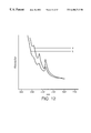
- FIG. 1 is a graph depicting acute toxicity measured by LD 50 as a function of mole percent of amphotericin B in the preparation.
- FIG. 11 are absorbance spectra of 5 mole % amphotericin B (a), 25 mole % amphotericin B (b), 25 mole % amphotericin B after heating (c), and 50 mole % amphotericin B (d), in 7:3 DMPC:DMPG.
- FIG. 12 is an absorbance spectrum of 25 mole % amphotericin B in 7:3 DMPC:DMPG both before (a) and after (b) heating for 10 minutes at 60° C.
- FIG. 17 shows the influence of liposome structure on uptake of amphotericin B into DMPC:DMPG MLVs or LUVs.
- a new liposome-loading method which loads liposomes by entrapping a drug via a solvent intermediate.
- This solvent has the characteristics that it solubilizes the bioactive agent, such as amphotericin B, it does not disrupt the liposome membrane structure, and it is compatible with an aqueous solution such as water.
- a solvent is ethanol.
- This method proceeds without the use of organic solvents.
- HDLCs rather than liposomes will be formed. In the case where the formation of HDLCs are favored, the process loads the HDLCs with drug.
- HDLCs refer to drug-associated lipid in particulate form, such particulates being nonliposomal in nature.
- such HDLCs are characterized, for example, by: (1) freeze-fracture electron micrographs (Deamer et al., Biochim. Biophys. Acta, 1970, 219:47-60), demonstrating nonliposomal complexes; (2) captured volume measurements (Deamer et al., Chem. Phys.
- One aspect of the present invention is a drug:lipid ratio that forms HDLCs which results in substantially equivalent or greater efficacy of the drug while generally decreasing acute toxicity as measured by LD 50 in mice (FIG. 1 ).
- Applicants have found that between about a 6 and 50 mole percent of hydrophobic drug meets such requirements, with a preferred ratio being between about 15 and 50, more preferably between about 25 and 50, most preferably about 25 and 45 mole percent hydrophobic drug.
- drug concentrations exceed about 6 mole percent and approach 25 mole %, mixed populations of liposomes and HDLCs are formed. Within this range, as the mole percent of drug approaches 25, a greater percent of the structures are HDLC rather than liposomes.
- liposomes may be formed by the novel ethanol intermediate process of the present invention.
- HDLCs Characteristic of the above-mentioned HDLCs are various drug-lipid dispersions observed upon density centrifugation. Separations of the drug-lipid (DMPC:DMPG at a 7:3 mole ratio) complex on the isopycnic sucrose density columns showing banding that is dependent on the drug:lipid ratio and the method of preparation. In systems comprising 5 mole percent drug made by the MLV process, two major bands of material are observed; one of liposomes and one where drug is associated with the lipid (HDLCs). Preparations containing 25 and 50 mole percent drug showed a single band wherein most of the drug is associated with the lipid. Surprisingly, these low-lipid/higher mole percent drug systems were less toxic. FIG.
- LD 50s (mg/kg) in mice as a function of the mole percent of the drug amphotericin B in the preparation.
- the LD 50 s increase between 5 and 50 mole percent drug, then drop off at 60 mole percent.
- Also plot is the LD 50 for the commercially available form of this drug; Fungizone, at about 3.5 mg/kg.
- In vitro blood cell lysis (hemolysis) demonstrates the same toxicity phenomenon.
- X-ray data at 5 mole percent shows gel phase lipid at low temperatures, with a transition to liquid crystalline phase at the characteristic temperature of 23° C. At 50 mole percent drug, however, the lipid is in the gel phase at all temperatures; there is no transition due to all the lipid being in tight association with the drug (FIG. 6 ). Similar data for 31 P-NMR studies show the lack of the free lipid signal for these higher (25 and 50) mole percent drug formulations (HDLCs); the low field shoulder/high field peak characteristics of this type of lipid is absent (FIG. 7 ), while it is visible in the 0 and 5 mole percent drug samples (FIG. 8 ).
- liposome-forming procedures may be used in the formation of these lipid complexes. Specifically, these procedures include those that form liposomes known as multilamellar vesicles (MLV). Other processes that may be used are those that form stable plurilamellar vesicles (SPLV), large unilamellar vesicles formed by an extrusion procedure (LUVETS), or other liposome-forming procedures known in the art.
- SPLV plurilamellar vesicles
- LUVETS large unilamellar vesicles formed by an extrusion procedure
- the process for forming SPLVs is disclosed in Lenk et al., U.S. Pat. No. 4,522,803, issue Jun.
- liposomes are formed in aqueous solution to which is added the drug to be entrapped, in acidified ethanol.
- amphotericin B is incorporated into liposomes via an aqueous intermediate.
- amphotericin B is suspended in an aqueous solution, for example distilled water, by sonication.
- the suspended drug is then admixed with a suspension of lipid in aqueous solution, such as distilled water or sodium chloride solution.
- the mixture is incubated at or above the transition temperature of the lipid employed, with the resultant formation of MLVs.
- the lipids which can be (1) employed in making the lipid complexes, and (2) used in the novel liposome formation technique of the present invention are the phospholipids such as phosphatidylcholine (PC), phosphatidylethanolamine (PE), phosphatidylserine (PS), phosphatidylglycerol (PG), phospatidic acid (PA), phospatidylinositol (PI), sphingomyelin (SPM), and the like, alone or in combination.
- Saturated phospholipids such as hydrogenated soy PC may also be used.
- the phospholipids can be synthetic or derived from natural sources such as egg or soy.
- the phospholipids dimyristoylphosphatidylcholine (DMPC) and dimyristoylphosphatidylglycerol (DMPG) are used in combination in any mole ratio, from about 99:1 to about 1:99 DMPC:DMPG, preferably in about a 7:3 mole ratio.
- DMPC and dimyristoylphosphatidylserine (DMPS) may also be used in combination.
- DMPC alone may be used.
- the lipid complexes and liposomes can also contain a steroid component as part of the lipid phase, such steroids may be cholesterol, polyethylene glycol derivatives of cholesterol (PEG-cholesterols), coprostanol, cholestanol, cholestane, organic acid derivatives of sterols such as cholesterol hemisuccinate (CHS), and the like.
- Further lipid complex-forming compositions are fatty acids such as myristic acid, isopropyl myristate, isostearic acid, sucrose distearate, propylene glycol monostearate, and cetylated monoglyceride.
- Other substances that can be employed include lipids such as trimyristin, the fatty alcohols such as cetyl alcohol and myristyl alcohol, and fatty esters such as myristic acid ethyl ester.
- Organic acid derivatives of tocopherols may also be used as complex or liposome-forming ingredients, such as alpha-tocopherol hemisuccinate (THS).
- THS alpha-tocopherol hemisuccinate
- Both CHS- and THS-containing complexes and their tris salt forms may generally be prepared by any method known in the art for preparing liposomes containing these sterols.
- U.S. Pat. No. 4,721,614 issued Jan. 26, 1988, entitled “Steroidal Liposomes”
- Janoff et al. PCT Publication No. 87/02219, published Apr. 23, 1987, entitled “Alpha-Tocopherol Based Vesicles,” filed Sep. 24, 1986, respectively, relevant portions of which are incorporated herein by reference.
- bioactive agents include but are not limited to antibacterial compounds such as the antibacterial compounds such as the aminoglycosides, for example, gentamicin, antiviral compounds such as rifampacin, anti-parasitic compounds such as antimony derivatives, antineoplastic compounds such as vinblastine, vincristine, mitomycin C, doxorubicin, daunorubicin, methotrexate, and cisplatin, among others, proteins such as albumin, toxins such as diptheria toxin, enzymes such as catalase, hormones such as estrogens, neurotransmitters such as acetylcholine, lipoproteins such as alpha-lipoprotein, glycoproteins such as hyaluronic acid., immunoglobulins such as IgG, immunomodulators such as the interferons or the interleukins, dyes such as Arsenazo III, radiolabels such as 14 C, radio-opaque compounds such as 99 Te, fluorescent compounds such
- organic solvents may be used. Suitable organic solvents are those with intermediate polarities and dielectric properties (those having a polarity intermediate to opposing electrical charges), which solubilize lipids, and include but are not limited to chloroform, methanol, dimethylaulfoxide (DMSO), methylene chloride, and solvent mixtures such as chloroform:methanol (70:30) and benzene:methanol (70:30). As a result, solutions, defined as mixtures in which the components are uniformly distributed throughout; containing the lipids are formed. Solvents are preferably chosen on the basis of their biocompatibility, low toxicity, and imflammability.
- DMSO When solubilizing the drug, specifically amphotericin B, DMSO is preferred as it is most soluble in DMSO. Methanol may be substituted for DMSO with concomitant increase in solvent volume.
- methylene chloride is preferably used due to its low toxicity in humans.
- ethanol or aqueous solutions are the preferred solvents.
- aqueous solutions such as distilled water (e.g., USP water for injection), saline, or aqueous buffers may be used.
- Aqueous buffers that may be used include but are not limited to buffered salines such as phosphate buffered saline (“PBS”), tris-(hydroxymethyl)-aminomethane hydrochloride (“tris”) buffers, or glycine buffers at pH of about 7.0 to 7.5, preferably 7.2.
- PBS phosphate buffered saline
- tris tris-(hydroxymethyl)-aminomethane hydrochloride
- a sonication step may be performed. Such a procedure is performed in a bath sonicator for about 15 to about 30 minutes, at 25° C., at about 50-60 Hz.
- the HDLCs or liposomes formed may be sized by extrusion through a filter according to the procedure of Cullis et al., PCT Publication No. 88/00238, published Jan. 16, 1986, relevant portions of which are incorporated herein by reference. Such sizing procedures allow the formation of homogeneous populations of particles, with regard to size.
- the filtering of the HDLCs may be performed through a tortuous path or a straight through membrane filter, such as a polyearbonate filter.
- the HDLCs or liposomes formed by the novel procedures of the present invention may be subject to a lyophilization or dehydration procedure at various stages of formation.
- the lipid film may be lyophilized after removing the solvent and prior to adding the drug.
- the lipid-drug film may be lyophilized prior to hydrating the HDLC or liposome.
- Such dehydration may be carried out by exposing the lipid, HDLC, or liposome to reduced pressure thereby removing all suspending solvent.
- the finally hydrated HDLC or liposome preparation may also be dehydrated by placing it in surrounding medium in liquid nitrogen and freezing it prior to the dehydration step.
- Dehydration with prior freezing may be performed in the presence of one or more protective sugars in the preparation according to the techniques of Bally et al., PCT Publication No. 86/01103 published Feb. 27, 1986, relevant portions of which are hereby incorporated by reference and Schneider et al., U.S. Pat. No. 4,229,360, issued Oct. 21, 1980.
- Such techniques enhance the long-term storage and stability-of the preparations.
- Other suitable methods now known or later discovered, may be used in the dehydration of the-above-disclosed lipid complex preparations.
- One method for formation of the HDLCs of the present invention is an MLV procedure wherein the bioactive agent (e.g., a drug) is dissolved in methanol in a flask to which is then added lipid such as DMPC and DMPG in about a 7:3 mole ratio of DMPC:DMPG, in chloroform. After rotoevaporating the solution at reduced pressure, the dried film is resuspended in PBS and sonicated in a bath sonicator (at about 25° C. for about 30 minutes, about 50-60 Hz) to clarity. At mole ratios of drug:lipid of about 6 and above (to about 60 mole percent), the preparations are nonliposomal HDLC structures substantially free of liposomes.
- the bioactive agent e.g., a drug
- Toxicities of the HDLC preparations are drug:lipid ratio-dependent, preparations of higher drug:lipid ratios (about 16-50 mole percent drug) show less acute toxicity than lower drug:lipid ratio preparations (about 6-15 mole percent drug). As discussed above, preparations containing up to about 5 mole percent drug are substantially liposomal.
- lipid DMPC:DMPG, 2:1 w/w
- an aqueous solution such as buffer or saline
- the bioactive agent previously dissolved in an organic solvent such as dimethyl sulfoxide (DMSO).
- DMSO dimethyl sulfoxide
- This admixture of the lipid with the bioactive agent can be by homogenization wherein the bioactive agent solution is added to the lipid in aliquots. The homogenization is allowed to proceed until the particle size of the resulting liposomes or HDLCs is achieved; i.e., about 1 um to about 10 um, preferably 4 um to about 6 um.
- Another method is a modified MLV procedure whereby the above-produced drug-lipid film is dried in a bell jar under reduced pressure to produce a drug-lipid flake.
- This flake is pulverized to a powder which is homogeneously hydrated when aqueous solution is added.
- This procedure involves solution of the drug in an organic solvent such as DMSO or methanol followed by mixing with a lipid (most preferably a 7:3 mole ratio of DMPC:DMPG) in methylene chloride.
- the mixture is then dried under reduced pressure to a film, which is then dried, for example, in a bell jar under reduced pressure.
- the resulting dried powder is hydrated with an aqueous saline solution and heated to produce a drug-lipid suspension.
- the formation of HDLCs, lippsomes, and mixtures thereof, and concomitant degree of toxicity depends on the drug:lipid ratio.
- HDLCs Other methods may be used in the formation of HDLCs.
- One such method is an SPLV procedure wherein the drug is dissolved in a solvent such as DMSO.
- a lipid such as a 7:3 mole ratio of DMPC:DMPG
- a solvent e.g., chloroform or methylene chloride
- An aliquot of the drug (e.g., amphotericin B) in solution is added to the lipid suspension and the resulting mixture is sonicated to clarity.
- a buffered aqueous solution e.g., buffered saline
- the resulting paste is hydrated further with PBS and sonicated to clarity.
- the resulting preparation will be HDLC or liposomal depending on the mole ratio of drug:lipid employed. LD 50s in mice using this preparation showed lower toxicities with higher drug:lipid ratios.
- Yet another method is a modified SPLV procedure wherein the drug in organic solvent (e.g., DMSO) is mixed with lipid (e.g., DMPC:DMPG) in solution, and a buffered aqueous solution (e.g., PBS) was added, followed by evaporation of the solvent under nitrogen while sonicating. Additional PBS is added and the resulting solution filtered, preferably through a 5 micron filter, then centrifuged at about 10,000 ⁇ g for about 10 minutes. The supernatant solution was removed, discarded, and the pellet resuspended in aqueous solvent (PBS). Following a second “wash” by centrifugation, the resulting pellet is resuspended in PBS.
- organic solvent e.g., DMSO
- lipid e.g., DMPC:DMPG
- PBS buffered aqueous solution
- Yet another SPLV method is a preparation wherein the drug-solvent and lipid solutions are mixed as before, but water for injection, USP, rather than saline is added and the suspension warmed to 37° C. The solvent is again evaporated under nitrogen while sonicating, more water for injection was added, and the suspension held for about 10 minutes at 37° C.
- the preparations are sterile filtered through a 0.1 or 0.15 micron filter using the LUVET method.
- Such method is an extrusion procedure whereby liposomes are produced by filtration at pressures of about 700 psi, and is the subject of Cullis et al., PCT Publication No. 86/99238, published Jan. 16, 1986, relevant portions of which are incorporated herein by reference.
- Such preparations may also be dehydrated according to the methods of Bally et al., as described hereinabove. Acute toxicity tests again demonstrate the ameliorative effects of a high drug:lipid ratio on the drug toxicity.
- a further method involves the dissolution of drug in acidified anhydrous ethanol by sonication.
- a lipid e.g., a 7:3 mole ratio of DMPC:DMPG
- a solvent e.g., benzene:methanol
- PBS aqueous solvent
- the preparation is then lyophilized and rehydrated with aqueous buffer.
- the solvent can be evaporated leaving a lipid-drug film, which can be hydrated with an aqueous solution.
- lower acute toxicities are associated with preparations of higher drug:lipid ratios.
- the process produces mostly liposomes, compared to the use of about 6 mole percent and above, which forms mainly HDLCs.
- Centrifugation of the preparation on a density gradient confirms that all the lipid is associated with the drug.
- the preparation may then be lyophilized, which removes the ethanol, and rehydrated in aqueous solution such as distilled water.
- amphotericin B is incorporated into liposomes via an aqueous intermediate.
- amphotericin B is suspended in an aqueous solution, for example distilled water, by sonication.
- the suspended drug is then admixed with a suspension of lipid in aqueous solution, such as distilled water or sodium chloride solution.
- the mixture is incubated at or above the transition temperature of the lipid employed, with the resultant formation of vesicles.
- dimyristoylphosphatidylglycerol is used alone or in combination to form the liposomes, and when the lipid has been admixed with an aqueous solution having an ionic strength of about 0 mM to about 25 mM salt, and incubated at about the transition temperature (T c ) of the lipid (i.e., at about 22-24° C.), the liposomes spontaneously vesiculate, forming large unilamellar vesicles (LUVS).
- T c transition temperature of the lipid
- LVS large unilamellar vesicles
- DMPG can be used alone or, for example, with other lipids such as with DMPC, e.g., in a 3:7 mole ratio of DMPC:DMPG.
- DMPC lipid-propyl-N-phenyl-N-phenyl-N-phenyl-N-phenyl-N-phenyl-N-phenyl-N-phenyl-N-phenyl-N-phenyl-N
- the high mole ratio amphotericin B-lipid preparations (HDLCs) discussed above are believed to exhibit low toxicity because of enhanced amphotericin B-amphotericin B interactions in the lipid matrix. This can be demonstrated by absorbance spectroscopy.
- the absorbance at 413 nm, for instance, arising from free amphotericin B in deoxycholate is greater than that for unheated 5 mole % amphotericin B in lipid.
- the absorbance of 25 mole % amphotericin B is greater than that exhibited by 50 mole % amphotericin B in lipid (see FIG. 10 ).
- the absorbance spectrum technique is used to determine the toxicity of a drug (e.g. amphotericin B)-lipid complex.
- the absorbance spectrum of a drug is specific for. that drug; the signature of amphotericin B, (appearing in FIG. 12, dissolved in deoxycholate), is between 300 and 500 nm, and has characteristic peaks, the most representative of these peaks the one arising at 413 nm.
- the attenuation of this peak by complexing the drug with a lipid can be used quantitatively as a measure of toxicity of the HDLC. Any of the above-named lipids, when complexed in this way, would be expected to result in the characteristic, albeit attenuated, absorbance discussed here, since the spectra are specific to the drug.
- This attenuation occurs because lipid is phase separated and thereby expelled from the amphotericin B in the heating step, allowing closer association of the amphotericin B with itself and thus a less toxic system.
- the expelled lipid is demonstrated by the reappearance (after heating) of an endotherm viewed in differential scanning calorimetry spectra (see FIG. 12 ), consistent with lipid substantially free of amphotericin B. Further, the expelled lipid can be demonstrated by the narrowing of the 31 P-NMR signal arising from the heated systems (see FIG. 13 ). Again, this narrowing is consistent with lipid substantially reduced in amphotericin B concentration.
- freeze fracture electron microscopy of heated systems reveals the existence of lipid (and liposomes) after heating, not previously observed.
- the unheated sample demonstrates complexes with no free lipid, characteristic of systems exhibiting intramolecular interaction between the lipid and the amphotericin B.
- any type liposome or lipid particle or liposome or lipid particle formation process such as any of those previously enumerated, but in this example the MLV process was used.
- any of the previously named lipids or phospholipids may be used, as may any solvents or aqueous buffers as named hereinabove.
- lipid in any amount known to form liposomes, such as DMPC:DMPG in a 7:3 mole ratio
- suspended in an organic solvent such as chloroform
- Amphotericin B (or any other drug as previously described) is dissolved in any appropriate organic solvent (an appropriate solvent being one that dissolves the drug, specifically, for example, the amphotericin B).
- any mole % of drug (amphotericin B) that forms liposomes or lipid particles may be used. Specifically, in the present invention, from about 5 to about 50 mole % amphotericin B is used. Where liposomes are to be formed, a drug to lipid mole ratio of 5 mole % drug and less is employed. For the formation of HDLCs, about 6 to about 50 mole percent drug is employed. At 25 mole % drug and above, the population is substantially HDLC in nature.
- amphotericin B was dissolved in methanol at 10 mg amphotericin B per 100 ml methanol, or 0.1 mg/ml of amphotericin B.
- the methanol containing dissolved amphotericin B (100.0 ml) was added to the lipid film, and the film resuspended.
- the resulting suspension is then rotoevaporated under reduced pressure, to a thin film.
- the lipid-amphotericin B film is then resuspended in an aliquot of aqueous solution the volume of which is sufficient to form liposomes or lipid particles, such as for example about 4.0 ml.
- the suspension may then be agitated and sonicated for several minutes to about one hour, and heated in a water bath at from about 25° C. to about 60° C., for about 10 to about 400 minutes.
- the HDLCs may be made according to the SPLV process, wherein a drug (e.g., amphotericin B) may be added to an appropriate solvent (such as DMSO) and mixed with a mechanical mixer to dissolve all the drug. If necessary, the solution can then be filtered, removing any undissolved particles of the drug.
- a drug e.g., amphotericin B
- an appropriate solvent such as DMSO
- a 7:3 mole ratio of DMPC:DMPG may then be dissolved in an appropriate solvent (such as methylene chloride), and the lipid then admixed with the drug-DMSO solution.
- aqueous solution such as saline
- organic solvents removed by rotary evaporation at about 35-45° C.
- the resulting drug-lipid mixture is diluted with aqueous solution, such as saline, and the suspension of HDLCs milled using a homogenizer.
- aqueous solution such as saline
- Any homogenization device or colloid mill that will mill particles is acceptable for this procedure, but preferably a Gifford Wood colloid mill is used.
- the particles are milled until an acceptable particle size has been achieved, for example, wherein 90% of the particles are below 10 um in diameter, preferably within a size range of about 4 to about 10 microns, or about 15-30 minutes.
- the particles may passed one or a multiple number of times through the mill, depending on the size and homogeneity desired.
- the HDLC particles may be analyzed for size distribution using the Malvern particle sizer. If necessary, larger and smaller particles may be removed by any methods known in the art for separating particles, such as by filtration. Such a process preferably results in particles between about 0.2 um and 10.0 um in diameter.
- Such a filtration method may be, for example, tangential flow filtration, such as described in copending U.S. patent application Ser. No. (unknown), filed Jul. 28, 1988, Docket No. TLC-139A, entitled “Method for Size Separation of Particles”.
- a heterogeneously sized population of liposomes or particles is passed through the tangential flow device having a filter pore size of about, for example, 5.0 um. Liposomes less than 5.0 um in size pass through the filter into the filtrate, and those greater than 5.0 um are retained in the retentate.
- the filtrate may then be passed through the device through a filter size of about for example, 2.0 um.
- the filtrate contains liposomes or particles of 2.0 um or less
- the retentate contains liposomes or particles of a homogeneous population between the sizes of 2.0 and 5.0 um.
- a homogeneous population of vesicles is one composed of substantially the same size vesicles, and may have a Gaussian distribution.
- the size distribution of the vesicles may be unimodal.
- Such a population is also said to be of a uniform size distribution, and may be unimodal with respect to size.
- the term “unimodal” refers to a population having a narrow polydispersity of particle sizes, and the particles are of a single “mode”.
- a liposomal population is unimodal if, when measured by quasi elastic light scattering methods, the population has a Gaussian distribution, and if a second order polynomial will fit the natural logrithm of the autocorrelation function of a sample (Koppel, 1972, J. Chem. Phys., 57:4814). The closer this fit, the better the measure of unimodality. The closeness of this fit may be determined by how close the chi square (chi 2 ) value of the sample is to unity (1.0). A chi 2 value of 2.0 and less is indicative of a unimodal population.
- the HDLC preparations and liposomes resulting from the novel processes of the present invention can be used therapeutically in animals (including man) in the treatment of a number of infections or conditions which require: (1) repeated administrations; (2) the sustained delivery of a drug in its-bioactive form; or (3) the decreased toxicity with substantially equivalent or greater efficacy of the free drug in question.
- infections or conditions include but are not limited to fungal infections, both topical and systemic such as those that can be treated with antifungal agents such as nystatin and amphotericin B and the viral diseases acquired immunodeficiency syndrome (AIDS) and herpes.
- AIDS acquired immunodeficiency syndrome
- the preparations of the invention are stable in aqueous solution.
- the mode of administration of the preparation may determine the sites and cells in the organism to which the compound will be delivered.
- the HDLCs and liposomes of the present invention can be administered alone but will generally be administered in admixture with a pharmaceutical carrier selected with regard to the intended route of administration and standard pharmaceutical practice.
- delivery to a specific site may be most easily accomplished by topical application (if the infection is external, e.g., on areas such as eyes, skin, in ears, or on afflictions such as wounds or burns).
- topical applications may be in the form of creams, ointments, gels, emulsions, or pastes, for direct application to the afflicted area.
- the preparations may be injected parenterally, for example, intravenously, intramuscularly, or subcutaneously.
- parenteral administration they can be used, for example, in the form of a sterile aqueous solution which may contain other solutes, for example, enough salts or glucose to make the solution isotonic.
- solutes for example, enough salts or glucose to make the solution isotonic.
- Other uses, depending on the particular properties of the preparation, may be envisioned by those skilled in the art.
- compositions of the invention can be used for the treatment of asthma, by instilling a nebulised aqueous suspension of the liposomes or HDLCs into the lungs.
- the liposomes or HDLCs can be suspended in a suitable solvent which can be aerosolized by a pneumatic or ultrasonic nebulizer, or, more conveniently, by a self-contained neblulizer that is driven by gas pressure from a fluorocarbon pellet.
- suitable solvent which can be aerosolized by a pneumatic or ultrasonic nebulizer, or, more conveniently, by a self-contained neblulizer that is driven by gas pressure from a fluorocarbon pellet.
- Other inhalation systems such as those in which the liposomes or HDLCs are delivered in particle form, either as a dry powder or as a suspension in a suitable carrier system are acceptable.
- aerosolization most of the propellant solvent is lost through flash evaporation and replaced by moisture in the respiratory tract, leading to the
- the prescribing physician will ultimately determine the appropriate dosage for a given human subject, and this can be expected to vary according to the age, weight, and response of the individual as well as the nature and severity of the patient's symptoms.
- the dosage of the drug in the HDLC or liposomal form will generally be about that employed for the free drug. In some cases, however, it may be necessary to administer dosages outside these limits.
- Amphotericin B (10 mg; total drug 5 mole percent) was added to 100 ml of methanol in a 500 ml round bottom flask and the mixture sonicated until clear. This sonicating step was performed in a bath sonicator for about 15 minutes at 25° C., at about 50-60 Hz. Dimyristoylphosphatidylcholine (DMPC) was added (100 mg in 1.0 ml of chloroform) as well as 42 mg of dimyristoylphosphatidylglycerol (DMPG) (in 0.42 ml chloroform). The resulting dispersion was dried by rotary evaporation at 60° C. under reduced pressure, to produce a thin film in the flask. The film was resuspended in 4.0 ml of PBS, the solution transferred to a glass tube and sonicated to clarity.
- DMPC dimyristoylphosphatidylcholine
- DMPG dimyristoylphosphatidylgly
- Amphotericin B (140 mg; total drug 5 mole percent) was added to 1.5 ml dimethyl sulfoxide (DMSO).
- DMPC (1400 mg) and 600 mg DMPG were dissolved in 50 ml methylene chloride, and the two solutions were transferred to a flask. The solution was mixed until clear and then dried by rotoevaporation under reduced pressure to produce a film on the flask, then dried 1-4 days in a bell jar. After such drying, the film, which had formed dried flakes, was pulverized to a powder and mixed with 100 ml or 50 ml of 0.9% saline, or 50 ml of USP water for injection. The resulting suspension was heated at 37° C. for one hour.
- Amphotericin B (100 mg; total drug 5 mole percent) was dissolved in 2.0 ml of DMSO.
- DMPC 100 mg in 1.0 ml
- DMPG 42 mg in 0.42 ml
- Methylene chloride (20 ml) was added to the flask followed by the addition of 0.2 ml of the stock amphotericin B solution.
- This suspension was sonicated to clarity (about 1 minute) under the conditions as stated in Example 1.
- PBS (0.3 ml), pH 7.2, was added and the methylene chloride was then removed under a stream of nitrogen while sonicating.
- the resulting paste was resuspended in 10.0 ml PBS and centrifuged at 10,000 ⁇ g for 10 minutes, followed by bath sonication for about 20 minutes.
- Amphotericin B (140 mg, total drug 5 mole percent) was dissolved in 1.5 ml methylene chloride. The two solutions were mixed until clear, transferred to a flask, and mixed with 8.0 ml of PBS. Methylene chloride was evaporated under a nitrogen stream while in a bath sonicator. Sonication proceeded as in Example 1. PBS (32 ml) was added and the solution filtered with a 5 micron pore polytetrafluoroethylene (PTFE) Teflon filter, washed by centrifugation twice, and finally suspended in 100 ml PBS.
- PTFE polytetrafluoroethylene
- Amphotericin B (140 mg; total drug 5 mole percent) was dissolved in 1.5 ml DMSO.
- DMPC (1400 mg) and 600 mg of DMPG were dissolved in 50 ml methylene chloride. The two solutions were mixed until clear, transferred to a flask, and dried to a thin film by rotoevaporation under reduced pressure. The film was then resuspended in 50 ml methylene chloride, and 8.0 ml PBS was added to this solution. Methylene chloride was removed by evaporation under nitrogen while sonicating, according to the procedure of Example 1.
- the resulting paste was further hydrated with 32 ml PBS, and the suspension washed by centrifugation twice, finally resuspended in 100 ml PBS, and passed through a 5 micron pore size teflon (PTFE) filter.
- PTFE teflon
- the above example was also repeated using 5.0, 10.0, 15.0, 20.0 ml of PBS as the volume of hydrating buffer.
- Amphotericin B 140 mg; total drug 5 mole percent was dissolved in 1.5 ml DMSO.
- Egg phosphatidylcholine (EPC) 2.0 gm was dissolved in 50 gm methylene chloride, and the two solutions mixed in a flask.
- PBS 8.0 ml was added and the solvent removed under a nitrogen stream while sonicating, according to the procedure of Example 1.
- PBS 200 ml
- Amphotericin B (140 mg; total drug 5 mole percent) was dissolved in 1.5 ml DMSO.
- DMPC (1400 mg) and 600 mg DMPG were dissolved in a 500 ml round bottom flask with 50 gm methylene chloride.
- the two solutions were mixed in a flask, and 10 ml water for injection, USP, at 37° C., was added.
- the solvent was evaporated using a nitrogen stream while sonicating, according to the procedure of Example 1, and 50 ml of water for injection, USP, was then added, and the solution warmed to 37° C. for 30 minutes.
- Amphotericin B (140 mg; total drug 16.7 mole percent) was mixed with 50 ml methanol and the mixture sonicated 15 minutes to form a solution, then passed through a 0.22 micron tortuous path filter (mixed cellulose and acetate nitrate esters) available from Millex.
- DMPC 350 mg
- 150 mg DMPG were codissolved in 50 gm methylene chloride, and was mixed with the filtrate solution. The solvents were evaporated under a nitrogen stream while sonicating, according to the procedure of Example 1.
- PBS 50 ml
- Half the preparation 25 ml was lyophilized by standard lyophilization procedures.
- Example 8 To 2.0 ml of anhydrous ethanol was added 10 ul of 1N hydrochloric acid. Amphotericin B (20 mg; total drug 5 mole percent) was added to the acidified ethanol and the mixture sonicated to clarity under the conditions as in Example 1 except for the sonication time being 30 seconds to 60 seconds. DMPC (200 mg) and DMPG (42 mg) were placed in a test tube to which was added a benzene:methanol (70:30) solution, and the solution was lyophilized as in Example 8. The dried lipid was mixed with 2.0 ml of PBS and the mixture dispersed by vortical mixing. The amphotericin B solution (0.2 ml) was added to the lipid, and the mixture stirred overnight.
- Amphotericin B 20 mg; total drug 5 mole percent
- the resulting DMPC:DKPG MLVs (200 ul) were layered onto a sucrose density gradient and centrifuged for 24 hours at. 22° C. at 230,000 ⁇ g. Results are shown in FIG. 11; all the amphotericin B was associated with the lipid, min a broad distribution with the top of the gradient containing most of the lipid and the major amphotericin B peak at the bottom of the gradient.
- the resulting DKPC:DMPG MLV s were lyophilized and rehydrated with distilled water (2.0 ml).
- the preparation was centrifuged at 10,000 ⁇ g for 10 minutes, the supernatant decanted, and the pellet resuspended in 1.0 ml of the Hepes/NaCl buffer. The preparation was then sonicated in a bath sonicator for 20 minutes, at 25° C., 50-60 Hz.
- Amphotericin B particles were prepared by drying 15.5 mg of DMPC and 6.5 mg DMPG (7:3 mole ratio) from about 10 ml of chloroform onto the sides of a 100 ml round bottom flask. Amphotericin B (10 mg) was suspended in 100 ml of methanol, and 100.0 ml of the methanol solution was added to the flask and the lipid film suspended, giving 25 mole % of amphotericin B. The mixture was then dried under rotoevaporation at 37° C. to a thin film on the sides of the flask.
- the film was then hydrated with 4.0 ml of 10 mM Hepes, 150 mM NaCl (pH 7.2) using glass beads, and sonicated for 30 minutes, to produce a final suspension.
- a sample of this suspension was then heated to 60° C. for 10 minutes, by immersion in a water bath.
- the resulting suspension was characterized by DSC, NMR, and freeze fracture electron microscopy.
- Example 12 The materials and procedures of Example 12 were repeated using 50 mole % and 5 mole % amphotericin B. These systems were characterized by DSC, ESR, and freeze fracture electron microscopy.
- DSC measurements were carried out on a Micro Cal MC-1 Unit from Micro Cal, Inc., Amherst, Mass. Sample volumes of 0.70 ml containing 5-9 mg of suspension were injected into the sample cell, with the same volume of buffer used in the reference cell. Samples were heated either at about 26° C./hour or about 37° C./hour. Duplicate runs of the same sample with the same history gave onset and completion temperatures reproducible to 0.2° C. In general, samples containing amphotericin B were heated to 60° C., (no higher) and then cooled to 7-4° C. for at least 2 hours in order to insure consistent sample history.
- NMR spectra were obtained at 145.7 MHZ on a Bruker AM360 wide bore NMR spectrometer using 8 K data points for acquisition, a sweep width of 50,000 HZ and a pulse width of 20 usec corresponding to a 45° pulse. Spectra were accumulated from up to 10,000 transients.
- a 0.1-0.3 ul aliquot of the specimen was sandwiched between a pair of Balzers (Nashua, N.H.) copper support plates and rapidly plunged from 23° C. into liquid propane.
- Samples were fractured and replicated on a double replicating device in a Balzers freeze-fracture unit at a vacuum of 2 ⁇ 10 ⁇ 6 mbar or better and at ⁇ 115° C.
- Replicas were floated off in 3N HNO 3 , followed by washing in a graded series of sodium hypochlorite solutions. These were finally cleaned in distilled water and picked up on 300 Hex mesh copper grids (Polysciences, Pa.). Replicas were viewed on a Philips 300 electron microscope at a magnification of 3,000 to 22,000 times.
- Absorbance spectra (as in FIGS. 12 and 13) were made by diluting amphotericin B in Hepes/NaCl buffer to 25 uM liter amphotericin B. A sample was placed in the sample cuvette of a Beckman Spectrophotometer and read at 300 to 500 nm. The sample cuvette was read against a buffer blank.
- Samples for ESR were labeled with a series of positional isomers of doxyl steric acids where the doxyl reporter group was present at different positions along the fatty acid chain. Labelling was effected by incorporating the probe by vortexing and sonication from an ethanolic solution which was dried to a thin film on the side of a test tube. All samples were labeled to one mole percent.
- ESR spectra were recorded on an IBM Instruments ER100D ESR Spectrometer with nitrogen gas flow temperature regulation. An external calibrated thermistor probe (Omega Engineering, Inc., Stamford, Calif.) was used to monitor the temperature of the sample. ESR spectra were recorded at a microwave power of 10 MW and a microwave frequency of 9.11 GHZ with a field sweep of 100 G and a 100 KHZ field modulation amplitude of 0.32 G. The order parameter was calculated from the maximum hyperfine splitting (A max).
- Amphotericin B (337.5 g) was added to 3375 ml of DMSO and the amphotericin B (100 mg/ml) mixed to dissolution (about 3 hours) using a mechanical mixer. The mixture was transferred toga stainless steel pressure vessel (5 L capacity) and filtered through 0.22 um Sartofluor filter and polypropylene depth filter (Pall Profile, Pall, Inc., Glen Cove, N.Y.).
- a 40 L pressure vessel 50.8 kg of methylene chloride was mixed with 225.0 gm of DMPC and 99.0 gm of DMPG, for 3.5 hours to completely dissolve.
- the resulting lipid solution was transferred through a 0.2 M 2 Sartofluor 0.22 micron teflon filter into a stainless steel processing tank.
- the 40 L pressure vessel was washed with an additional 55.2 kg of methylene chloride, and similarly filtered into the processing tank.
- the processing tank was set to rotary mix the lipid solution at 195 rpm, and the amphotericin B DMSO solution was then transferred to the tank.
- Sodium chloride solution (16.2 L, 0.9% USP) was then added to the tank through a 0.22 um Millipak 100 polycarbonate filter.
- lipid-drug complex was diluted with sodium chloride 0.9% USP (7000 ml) transferred to the tank through a Millipak 100 0.22 micron filter
- HDLCs were homogenized by using the Gifford Wood colloid mill and a rotary lobe pump. HDLCs were passed through the colloid mill head at an 0.5 mil gap setting with back pressure of 10 psi, for 3.0 hours, resulting in particles less than 20 microns. Particle size of the HDLCs was analyzed by the Malvern particle analyzer.
- Nystatin 500 mg was dissolved in 1.0 L methanol (0.5 mg/ml) by sonication in a Branson bath sonicator.
- Dissolved nystatin 5.0 ml was added to the round bottom flask containing lipid, and mixed; the solvent was then removed by rotary evaporation at 45° C. for 15 minutes, and the resulting preparations rehydrated in saline (5.0 ml) by vortical mixing with glass beads. Upon light microscopic examination, liposomes were visible.
- Lipid (14.8 umol/ml, 7:3 mol ratio of DMPC:DMPG) was hydrated in distilled water and incubated at 4° C. The resulting MLVs were extruded through two stacked polycarbonate filters ten times using the LUVET procedure.
- Amphotericin B was dispersed in distilled water using a bath sonicator at a concentration of 10.8 umol/ml. The amphotericin B dispersion was added to the lipid suspension to a final lipid and amphotericin B concentration of 13.5 umol/ml and 0.98 umol/ml, respectively. To remove unincorporated amphotericin B, 20 ml of the sample were centrifuged at 15,000 ⁇ g for 30 minutes in a Ti60 or SW 27 rotor (Beckman) at 22° C. in a Beckman L8-60 ultracentrifuge. The supernatant free amphotericin B was removed without disturbing the liposome pellet. The resulting liposomes were measured by quasi-elastic light scattering to be larger than 1.0 um in diameter.
- FIG. 16 shows the uptake of amphotericin B into DMPC:DMPG liposomes under various incubation temperature conditions. Rate of uptake is similar at 23° C. and 45° C. The drug also accumulates when the lipid is in the gel phase i.e., at 15° C.
- Example 21 The materials and procedures of Example 21 were employed, but wherein the lipid suspended in distilled water was incubated with the amphotericin B at 22° C. The resulting liposomes were unilamellar and measured at about 0.1-0.2 um in mean diameter by quasi elastic light scattering. FIG. 19 shows that the rate of amphotericin B uptake is faster in the first two hours with these LUVs than with MLVs.
- Amphotericin B particles were formed according to the following procedure: Amphotericin B (337.5 g) was added to 3375.0 ml of DMSO, and stirred to dissolve for 5.5 hours at 25° C. This solution was sterile filtered into a 5 L pressure can at 25° C.
- Dimyristoylphosphatidylcholine (DMPC) (264.3 g) and 109.9 g of dimyristoylphosphatidylglycerol (DMPG) (a 7:3 mole ratio) were combined with 35.2 L of methylene chloride in a 40 L pressure vessel, and mixed to dissolve completely.
- This solution was sterile filtered through a 0.22 um poly(perfluoroethylene) (Teflon) filter into a 140 L processing tank.
- Methylene chloride (39.1 L) was sterile filtered through a 0.22 um Teflon filter and added to the 140 L tank.
- amphotericin B/DMSO mixture was added to the lipid solution (resulting in a 33 mole % amphotericin B solution), followed by the addition of 16.5 L of 0.9% sterile saline to the tank.
- the suspension was mixed with a marine propeller.
- the methylene chloride was removed by sterile N gas purging.
- the final temperature was less than 40° C. after about 13 hours.
- Sterile saline (7.0 L) was added to the batch for a total volume in the process vessel of about 27 L.
- This product was circulated through a Gifford-Wood colloid mill for about 5 hours to decrease the average size of the lipid particles to about 5.0 um. After milling, the product was circulated through a Romicon 5.0 ceramic tangential flow filter (2 ft 2 ) using an Alfa Laval rotary lobe pump at an average flow rate of 24 gpm, for a total of about 10 hours. Sterile physiological saline (410 L) was added in 30 L aliquots through a top port of the 140 L vessel. The average filtration rate was about 500 ml/min.
- the filtrate was then passed into a reservoir and concentrated by passage through a 1.2 um Romicon 2ft 2 ceramic filter driven by an Alfa Laval rotary lobe pump at a flow rate of about 36 gpm (about 14.5 hours); the filtration rate was about 500-600 ml/min. This filtration removed the particles 1.2 um and less, in the filtrate. The 1.2 um retentate was collected as the final product.
- Lipid (a 2:1 w/w/ ratio of DMPC:DMPG; 20 g DMPC and 10 g DMPG) were admixed with 400 ml of 0.9% sodium chloride using a Tekmar Homogenizer, Ultra Turrex, Model SD45, (Tekmar Co., Cincinnati, Ohio) set at “high” speed, and homogenized for about 1 hour in an ice bath at about 4° C.
- Amphotericin B (20 g) was dissolved in 200 ml DMSO, and slowly added to the lipid, while homogenizing. The lipid and amphotericin B were homogenized for 30 minutes, until the particle size was reduced to about um to about 10 um as measured by Malvern Particle size analysis.
- the resulting lipid particles are size selected according to the methods of tangential flow filtration as in Example 23.
Abstract
Description
Claims (11)
Priority Applications (2)
| Application Number | Priority Date | Filing Date | Title |
|---|---|---|---|
| US08/430,661 US6406713B1 (en) | 1987-03-05 | 1995-04-28 | Methods of preparing low-toxicity drug-lipid complexes |
| US10/132,151 US20020119170A1 (en) | 1987-03-05 | 2002-04-26 | Low toxicity drug-lipid systems |
Applications Claiming Priority (7)
| Application Number | Priority Date | Filing Date | Title |
|---|---|---|---|
| US2215787A | 1987-03-05 | 1987-03-05 | |
| US6990887A | 1987-07-06 | 1987-07-06 | |
| US07/136,267 US4963297A (en) | 1987-12-22 | 1987-12-22 | Spontaneous vesticulation of multilamellar liposomes |
| US16458088A | 1988-03-07 | 1988-03-07 | |
| US23670088A | 1988-08-25 | 1988-08-25 | |
| US87612192A | 1992-04-29 | 1992-04-29 | |
| US08/430,661 US6406713B1 (en) | 1987-03-05 | 1995-04-28 | Methods of preparing low-toxicity drug-lipid complexes |
Related Parent Applications (1)
| Application Number | Title | Priority Date | Filing Date |
|---|---|---|---|
| US87612192A Division | 1987-03-05 | 1992-04-29 |
Related Child Applications (1)
| Application Number | Title | Priority Date | Filing Date |
|---|---|---|---|
| US10/132,151 Continuation US20020119170A1 (en) | 1987-03-05 | 2002-04-26 | Low toxicity drug-lipid systems |
Publications (1)
| Publication Number | Publication Date |
|---|---|
| US6406713B1 true US6406713B1 (en) | 2002-06-18 |
Family
ID=48536645
Family Applications (1)
| Application Number | Title | Priority Date | Filing Date |
|---|---|---|---|
| US08/430,661 Expired - Lifetime US6406713B1 (en) | 1987-03-05 | 1995-04-28 | Methods of preparing low-toxicity drug-lipid complexes |
Country Status (1)
| Country | Link |
|---|---|
| US (1) | US6406713B1 (en) |
Cited By (64)
| Publication number | Priority date | Publication date | Assignee | Title |
|---|---|---|---|---|
| US20020155141A1 (en) * | 1997-08-15 | 2002-10-24 | Borje S. Andersson | Pharmacologically acceptable solvent vehicles |
| US6610664B2 (en) * | 1996-06-27 | 2003-08-26 | Isis Pharmaceuticals, Inc. | Cationic lipids |
| US20040156783A1 (en) * | 2001-07-03 | 2004-08-12 | Mallinckrodt Inc. | Compounds for dual photodiagnosis and therapy |
| US20040213740A1 (en) * | 2000-10-16 | 2004-10-28 | Mallinckrodt Inc. | Light sensitive compounds for instant determination of organ function |
| US20040223913A1 (en) * | 2000-10-16 | 2004-11-11 | Mallinckrodt, Inc. | Hydrophilic light absorbing compositions for determination of physiological function in critically ill patients |
| US20040241095A1 (en) * | 2000-01-18 | 2004-12-02 | Mallinckrodt Inc. | Receptor-avid exogenous optical contrast and therapeutic agents |
| US20040247624A1 (en) * | 2003-06-05 | 2004-12-09 | Unger Evan Charles | Methods of making pharmaceutical formulations for the delivery of drugs having low aqueous solubility |
| US6844004B2 (en) * | 1997-08-15 | 2005-01-18 | Board Of Regents, The University Of Texas System | Topical formulations of natamycin/pimaricin |
| US20050013854A1 (en) * | 2003-04-09 | 2005-01-20 | Mannino Raphael J. | Novel encochleation methods, cochleates and methods of use |
| US20050013855A1 (en) * | 2003-04-09 | 2005-01-20 | Biodelivery Sciences International, Inc. | Cochleate compositions directed against expression of proteins |
| US20050169978A1 (en) * | 2004-01-29 | 2005-08-04 | Shu-Wen Fu | Wet-micro grinding |
| US20050201939A1 (en) * | 2000-01-18 | 2005-09-15 | Mallinckrodt Inc. | Versatile hydrophilic dyes |
| US20060105968A1 (en) * | 2002-10-03 | 2006-05-18 | Cleary John D | Compositions comprising highly purified amphotericin b |
| US20070191272A1 (en) * | 2005-09-27 | 2007-08-16 | Stemmer Willem P | Proteinaceous pharmaceuticals and uses thereof |
| US20070190127A1 (en) * | 2005-12-30 | 2007-08-16 | Mingdong Zhou | Extended release of neuregulin for improved cardiac function |
| US20080039341A1 (en) * | 2005-09-27 | 2008-02-14 | Volker Schellenberger | Unstructured recombinant polymers and uses thereof |
| US20080233050A1 (en) * | 2000-01-18 | 2008-09-25 | Mallinckrodt Inc. | Diagnostic and therapeutic optical agents |
| US20080286808A1 (en) * | 2005-09-27 | 2008-11-20 | Volker Schellenberger | Methods for production of unstructured recombinant polymers and uses thereof |
| US20090036502A1 (en) * | 2006-03-02 | 2009-02-05 | Raghavan Rajagopalan | Thiadiazole Compounds and Uses Thereof |
| US20090035363A1 (en) * | 2006-03-10 | 2009-02-05 | Raghavan Rajagopalan | Photoactive Compounds and Compositions and Uses Thereof |
| US20090092582A1 (en) * | 2007-08-15 | 2009-04-09 | Oren Bogin | Compositions and methods for modifying properties of biologically active polypeptides |
| US20090099031A1 (en) * | 2005-09-27 | 2009-04-16 | Stemmer Willem P | Genetic package and uses thereof |
| US20100022449A1 (en) * | 2006-03-09 | 2010-01-28 | Mallinckrodt Inc. | Receptor-avid exogenous optical contrast and therapeutic agents |
| US20100068251A1 (en) * | 2006-10-10 | 2010-03-18 | Jina Pharmaceuticals, Inc. | Aqueous Systems For The Preparation Of Lipid Based Pharmaceutical Compounds; Compositions, Methods, And Uses Thereof |
| US20100105899A1 (en) * | 2007-03-01 | 2010-04-29 | Neumann William L | Integrated Photoactive Small Molecules and Uses Thereof |
| WO2010078025A1 (en) | 2008-12-17 | 2010-07-08 | Mallinckrodt Inc. | Modified pyrazine derivatives and uses thereof |
| US20100196460A1 (en) * | 2007-07-31 | 2010-08-05 | Neumann William L | Integrated Photoactive Agents for Real-Time Monitoring of Hemostasis |
| US20100222547A1 (en) * | 2007-03-01 | 2010-09-02 | Mallinckrodt Inc. | Integrated Photoactive Peptides and Uses Thereof |
| US20100239554A1 (en) * | 2009-02-03 | 2010-09-23 | Amunix Inc. a Delaware Corporation | Extended recombinant polypeptides and compositions comprising same |
| WO2010144502A2 (en) | 2009-06-08 | 2010-12-16 | Amunix Operating Inc. | Growth hormone polypeptides and methods of making and using same |
| WO2010144508A1 (en) | 2009-06-08 | 2010-12-16 | Amunix Operating Inc. | Glucose-regulating polypeptides and methods of making and using same |
| WO2011009020A2 (en) | 2009-07-16 | 2011-01-20 | Mallinckrodt Inc. | Compounds and compositions for use in phototherapy and in treatment of ocular neovascular disease and cancers |
| WO2011028229A1 (en) | 2009-08-24 | 2011-03-10 | Amunix Operating Inc. | Coagulation factor ix compositions and methods of making and using same |
| WO2011031955A2 (en) | 2009-09-11 | 2011-03-17 | Mallinckrodt Inc. | Optical monitoring of leukemia |
| WO2011060113A1 (en) | 2009-11-11 | 2011-05-19 | Mallinckrodt Inc. | Sulfenamide compounds for phototherapy |
| US20110152211A1 (en) * | 2002-10-03 | 2011-06-23 | University Of Mississippi | Methods and formulations for reducing amphotericin b treatment side effects |
| WO2011084571A2 (en) | 2009-12-16 | 2011-07-14 | Mallinckrodt Inc. | Azide derivatives for phototherapy |
| EP2344198A2 (en) * | 2008-09-27 | 2011-07-20 | Jina Pharmaceuticals Inc. | Lipid based pharmaceutical preparations for oral and topical application; their compositions, methods, and uses thereof |
| US20110177006A1 (en) * | 2008-09-29 | 2011-07-21 | Raghavan Rajagopalan | Dithienofuran Dyes for Imaging and Therapy |
| US20110177007A1 (en) * | 2008-09-29 | 2011-07-21 | Raghavan Rajagopalan | Dithienopyrrole Dyes for Imaging and Therapy |
| US20110196231A1 (en) * | 2008-09-29 | 2011-08-11 | Raghavan Rajagopalan | Fused Ring Thiophene Dyes for Imaging and Therapy |
| WO2011123479A1 (en) | 2010-03-29 | 2011-10-06 | Academia Sinica | Quantitative measurement of nano / micro particle endocytosis with cell mass spectrometry |
| WO2013039851A1 (en) | 2011-09-12 | 2013-03-21 | Mallinckrodt Llc | Optical agents for imaging and visualization of matrix metalloproteinase enzymes |
| US8628751B2 (en) | 2009-04-16 | 2014-01-14 | Medi Beacon Development, LLC | Pyrazine derivatives for optical imaging and therapy |
| US8664392B2 (en) | 2004-12-23 | 2014-03-04 | Medibeacon, LLC | Pyrazine derivatives for bioconjugation |
| US8731655B2 (en) | 2009-05-12 | 2014-05-20 | Mallinckrodt Llc | Compounds containing acyclic N-N bonds for phototherapy |
| EP2774935A1 (en) | 2009-10-30 | 2014-09-10 | NTF Therapeutics, Inc. | Improved neurturin molecules |
| WO2014144712A1 (en) | 2013-03-15 | 2014-09-18 | Biochemics, Inc. | Topical formulations and methods for drug delivery |
| WO2015071841A1 (en) | 2013-11-12 | 2015-05-21 | Druggability Technologies Holdings Limited | Complexes of dabigatran and its derivatives, process for the preparation thereof and pharmaceutical compositions containing them |
| US20150147382A1 (en) * | 2013-09-23 | 2015-05-28 | Exir Nano Sina Company | Topical liposomal compositions for delivering hydrophobic drugs and methods preparing same |
| US9114160B2 (en) | 2004-12-23 | 2015-08-25 | Medibeacon, LLC | Pyrazine derivatives and uses thereof in renal monitoring |
| US9186349B2 (en) | 2009-05-12 | 2015-11-17 | Mallinckrodt Llc | Diaza heterocyclic compounds for phototherapy |
| WO2015195928A1 (en) | 2014-06-19 | 2015-12-23 | Attillaps Holdings | Acetylcholinesterase inhibitors for treatment of dermatological conditions |
| WO2018213071A1 (en) | 2017-05-15 | 2018-11-22 | Biochemics, Inc. | Transdermally-delivered combination drug therapy for pain |
| US10370430B2 (en) | 2012-02-15 | 2019-08-06 | Bioverativ Therapeutics Inc. | Recombinant factor VIII proteins |
| US10421798B2 (en) | 2012-02-15 | 2019-09-24 | Bioverativ Therapeutics Inc. | Factor VIII compositions and methods of making and using same |
| US10434167B2 (en) | 2014-03-25 | 2019-10-08 | The Government Of The United States As Represented By The Secretary Of The Army | Non-toxic adjuvant formulation comprising a monophosphoryl lipid A (MPLA)-containing liposome composition and a saponin |
| US10548953B2 (en) | 2013-08-14 | 2020-02-04 | Bioverativ Therapeutics Inc. | Factor VIII-XTEN fusions and uses thereof |
| US10709135B2 (en) | 2013-07-29 | 2020-07-14 | Attillaps Holdings | Organophosphates for treating afflictions of the skin |
| US10745680B2 (en) | 2015-08-03 | 2020-08-18 | Bioverativ Therapeutics Inc. | Factor IX fusion proteins and methods of making and using same |
| US10906926B2 (en) | 2015-11-06 | 2021-02-02 | Adjuvance Technologies, Inc. | Triterpene saponin analogues |
| US11324821B2 (en) | 2017-10-16 | 2022-05-10 | Adjuvance Technologies, Inc. | Triterpene saponin analogues |
| US11446241B2 (en) | 2013-07-29 | 2022-09-20 | Attillaps Holdings Inc. | Treatment of ophthalmological conditions with acetylcholinesterase inhibitors |
| WO2023175454A1 (en) | 2022-03-14 | 2023-09-21 | Pfizer Inc. | Methods for producing an adjuvant |
Citations (54)
| Publication number | Priority date | Publication date | Assignee | Title |
|---|---|---|---|---|
| US79309A (en) | 1868-06-30 | Thk hohhij fetkm c | ||
| US225327A (en) | 1880-03-09 | Signaling apparatus for tele phon e-li n es | ||
| US360964A (en) | 1887-04-12 | Shutter-fastening | ||
| US498268A (en) | 1893-05-30 | Spinning frames | ||
| US604503A (en) | 1898-05-24 | Bruno richard seifert | ||
| US638809A (en) | 1899-03-27 | 1899-12-12 | Nat Tube Co | Art of manufacturing tube-blanks. |
| US749161A (en) | 1904-01-12 | Carpet-stretcher | ||
| US835832A (en) | 1906-06-30 | 1906-11-13 | Alfred M Rickerd | Mine-drill. |
| US3244590A (en) | 1960-12-23 | 1966-04-05 | Rutgers Res And Educational Fo | Polyenic compounds and procedures related thereto |
| US3993754A (en) | 1974-10-09 | 1976-11-23 | The United States Of America As Represented By The United States Energy Research And Development Administration | Liposome-encapsulated actinomycin for cancer chemotherapy |
| US4145410A (en) | 1976-10-12 | 1979-03-20 | Sears Barry D | Method of preparing a controlled-release pharmaceutical preparation, and resulting composition |
| GB2041871A (en) * | 1979-01-19 | 1980-09-17 | Erba Farmitalia | Suspensions of Drug- containing Liposomes |
| US4224179A (en) | 1977-08-05 | 1980-09-23 | Battelle Memorial Institute | Process for the preparation of liposomes in aqueous solution |
| US4229360A (en) | 1977-08-05 | 1980-10-21 | Battelle Memorial Institute | Process for the dehydration of a colloidal dispersion of lipsomes |
| US4235871A (en) | 1978-02-24 | 1980-11-25 | Papahadjopoulos Demetrios P | Method of encapsulating biologically active materials in lipid vesicles |
| US4310506A (en) | 1979-02-22 | 1982-01-12 | California Institute Of Technology | Means of preparation and applications of liposomes containing high concentrations of entrapped ionic species |
| US4342750A (en) | 1977-03-09 | 1982-08-03 | Schmid Labs., Inc. | Method for treating benign prostatic hypertrophy with N-acyl amphotericin B |
| US4358442A (en) | 1979-12-22 | 1982-11-09 | Nattermann & Cie Gmbh | Rosmarinic acid-phospholipide-complex |
| EP0069307A2 (en) * | 1981-07-02 | 1983-01-12 | F. HOFFMANN-LA ROCHE & CO. Aktiengesellschaft | Process for preparing liposome solutions |
| US4372949A (en) | 1979-03-05 | 1983-02-08 | Toyama Chemical Co., Ltd. | Treatment of cancer with carcinostatic and immunostimulating agent containing lysophospholipid and phospholipid |
| US4419348A (en) | 1981-04-27 | 1983-12-06 | Georgetown University | Anthracycline glycoside compositions, their use and preparation |
| US4436746A (en) | 1982-09-30 | 1984-03-13 | Ciba-Geigy Corporation | Thromboxane synthetase inhibitory N-substituted-2-(1-imidazolyl)indoles |
| US4460577A (en) | 1977-09-30 | 1984-07-17 | Farmitalia Carlo Erba S.P.A. | Pharmaceutical compositions consisting or consisting essentially of liposomes, and processes for making same |
| WO1985000968A1 (en) | 1983-09-06 | 1985-03-14 | Health Research, Inc. | Liposome delivery method for decreasing the toxicity of an antitumor drug |
| US4508703A (en) | 1982-02-17 | 1985-04-02 | Parfums Christian Dior | Production of pulverulent mixtures of lipidic and hydrophobic constituents |
| US4522803A (en) | 1983-02-04 | 1985-06-11 | The Liposome Company, Inc. | Stable plurilamellar vesicles, their preparation and use |
| WO1985004578A1 (en) | 1984-04-12 | 1985-10-24 | The Liposome Company, Inc. | Steroidal liposomes |
| US4551288A (en) | 1982-08-16 | 1985-11-05 | Sandoz, Inc. | Processes for the preparation of liposome drug delivery systems |
| WO1985005030A1 (en) | 1984-05-02 | 1985-11-21 | The Liposome Company, Inc. | Drug preparations of reduced toxicity |
| WO1986000238A1 (en) | 1984-06-20 | 1986-01-16 | The Liposome Company, Inc. | Extrusion techniques for producing liposomes |
| WO1986001103A1 (en) | 1984-08-08 | 1986-02-27 | The Liposome Company, Inc. | Dehydrated liposomes |
| US4588578A (en) | 1983-08-08 | 1986-05-13 | The Liposome Company, Inc. | Lipid vesicles prepared in a monophase |
| US4604376A (en) | 1982-04-21 | 1986-08-05 | Research Corporation | Enteric compounds and complexes |
| US4622219A (en) | 1983-06-17 | 1986-11-11 | Haynes Duncan H | Method of inducing local anesthesia using microdroplets of a general anesthetic |
| EP0202837A1 (en) | 1985-05-17 | 1986-11-26 | Smithkline Beckman Corporation | Polyene antibiotic emulsion formulation |
| WO1987002219A1 (en) | 1985-10-15 | 1987-04-23 | The Liposome Company, Inc. | Alpha tocopherol-based vesicles |
| US4663167A (en) | 1984-04-16 | 1987-05-05 | The Board Of Regents Of The University Of Texas System | Composition and method for treatment of disseminated fungal infections in mammals |
| US4687762A (en) * | 1984-03-31 | 1987-08-18 | Green Cross Corporation | Water soluble drug complex and method for production of same |
| EP0240346A2 (en) * | 1986-04-02 | 1987-10-07 | Takeda Chemical Industries, Ltd. | Method of producing liposome |
| EP0260811A2 (en) | 1986-08-21 | 1988-03-23 | Vestar, Inc. | Phospholipid particles encapsulating polyene antibiotics for the treatment of systemic fungal infections |
| FR2607719A1 (en) | 1986-12-05 | 1988-06-10 | Ire Celltarg Sa | Microcrystals comprising ginkgolides as active substance and pharmaceutical compositions containing them |
| US4766046A (en) | 1985-09-27 | 1988-08-23 | Liposome Technology, Inc. | Stabilized liposome/amphotericin composition and method |
| FR2611138A2 (en) | 1986-12-05 | 1988-08-26 | Ire Celltarg Sa | Microcrystals containing an active substance displaying an affinity for phospholipids |
| EP0296845A1 (en) | 1987-06-23 | 1988-12-28 | The University of Nottingham | Drug emulsion |
| FR2620028A2 (en) | 1986-12-05 | 1989-03-10 | Ire Celltarg Sa | Process for preparing microcrystals containing as active substance, in particular, amphotericin B, microcrystals obtained and pharmaceutical composition comprising them |
| US4812312A (en) | 1987-03-03 | 1989-03-14 | Board Of Regents Of The University Of Texas System | Liposome-incorporated nystatin |
| US4822777A (en) | 1987-02-27 | 1989-04-18 | Liposome Technology, Inc. | Amphotericin B/cholesterol sulfate composition |
| US4897384A (en) | 1983-05-26 | 1990-01-30 | The Liposome Company, Inc. | Drug preparations of reduced toxicity |
| US4963297A (en) | 1987-12-22 | 1990-10-16 | The Liposome Company, Inc. | Spontaneous vesticulation of multilamellar liposomes |
| US4973465A (en) | 1986-12-05 | 1990-11-27 | Ire-Celltarg S.A. | Microcrystals comprising an active substance having an affinity for phospholipids, and at least one phospholipid, process of preparation |
| US5059591A (en) | 1983-05-26 | 1991-10-22 | The Liposome Company, Inc. | Drug preparations of reduced toxicity |
| US5077056A (en) | 1984-08-08 | 1991-12-31 | The Liposome Company, Inc. | Encapsulation of antineoplastic agents in liposomes |
| US5100591A (en) | 1989-09-14 | 1992-03-31 | Medgenix Group S.A. | Process for preparing lipid microparticles |
| US5415867A (en) * | 1988-04-20 | 1995-05-16 | The Liposome Company, Inc. | High ratio active agent: lipid complex |
-
1995
- 1995-04-28 US US08/430,661 patent/US6406713B1/en not_active Expired - Lifetime
Patent Citations (59)
| Publication number | Priority date | Publication date | Assignee | Title |
|---|---|---|---|---|
| US79309A (en) | 1868-06-30 | Thk hohhij fetkm c | ||
| US225327A (en) | 1880-03-09 | Signaling apparatus for tele phon e-li n es | ||
| US360964A (en) | 1887-04-12 | Shutter-fastening | ||
| US498268A (en) | 1893-05-30 | Spinning frames | ||
| US604503A (en) | 1898-05-24 | Bruno richard seifert | ||
| US749161A (en) | 1904-01-12 | Carpet-stretcher | ||
| US638809A (en) | 1899-03-27 | 1899-12-12 | Nat Tube Co | Art of manufacturing tube-blanks. |
| US835832A (en) | 1906-06-30 | 1906-11-13 | Alfred M Rickerd | Mine-drill. |
| US3244590A (en) | 1960-12-23 | 1966-04-05 | Rutgers Res And Educational Fo | Polyenic compounds and procedures related thereto |
| US3993754A (en) | 1974-10-09 | 1976-11-23 | The United States Of America As Represented By The United States Energy Research And Development Administration | Liposome-encapsulated actinomycin for cancer chemotherapy |
| US4145410A (en) | 1976-10-12 | 1979-03-20 | Sears Barry D | Method of preparing a controlled-release pharmaceutical preparation, and resulting composition |
| US4342750A (en) | 1977-03-09 | 1982-08-03 | Schmid Labs., Inc. | Method for treating benign prostatic hypertrophy with N-acyl amphotericin B |
| US4224179A (en) | 1977-08-05 | 1980-09-23 | Battelle Memorial Institute | Process for the preparation of liposomes in aqueous solution |
| US4229360A (en) | 1977-08-05 | 1980-10-21 | Battelle Memorial Institute | Process for the dehydration of a colloidal dispersion of lipsomes |
| US4229360B1 (en) | 1977-08-05 | 1991-11-05 | Liposome Co Inc | |
| US4460577A (en) | 1977-09-30 | 1984-07-17 | Farmitalia Carlo Erba S.P.A. | Pharmaceutical compositions consisting or consisting essentially of liposomes, and processes for making same |
| US4235871A (en) | 1978-02-24 | 1980-11-25 | Papahadjopoulos Demetrios P | Method of encapsulating biologically active materials in lipid vesicles |
| GB2041871A (en) * | 1979-01-19 | 1980-09-17 | Erba Farmitalia | Suspensions of Drug- containing Liposomes |
| US4310506A (en) | 1979-02-22 | 1982-01-12 | California Institute Of Technology | Means of preparation and applications of liposomes containing high concentrations of entrapped ionic species |
| US4372949A (en) | 1979-03-05 | 1983-02-08 | Toyama Chemical Co., Ltd. | Treatment of cancer with carcinostatic and immunostimulating agent containing lysophospholipid and phospholipid |
| US4358442A (en) | 1979-12-22 | 1982-11-09 | Nattermann & Cie Gmbh | Rosmarinic acid-phospholipide-complex |
| US4419348A (en) | 1981-04-27 | 1983-12-06 | Georgetown University | Anthracycline glycoside compositions, their use and preparation |
| EP0069307A2 (en) * | 1981-07-02 | 1983-01-12 | F. HOFFMANN-LA ROCHE & CO. Aktiengesellschaft | Process for preparing liposome solutions |
| US4508703A (en) | 1982-02-17 | 1985-04-02 | Parfums Christian Dior | Production of pulverulent mixtures of lipidic and hydrophobic constituents |
| US4604376A (en) | 1982-04-21 | 1986-08-05 | Research Corporation | Enteric compounds and complexes |
| US4551288A (en) | 1982-08-16 | 1985-11-05 | Sandoz, Inc. | Processes for the preparation of liposome drug delivery systems |
| US4436746A (en) | 1982-09-30 | 1984-03-13 | Ciba-Geigy Corporation | Thromboxane synthetase inhibitory N-substituted-2-(1-imidazolyl)indoles |
| US4522803A (en) | 1983-02-04 | 1985-06-11 | The Liposome Company, Inc. | Stable plurilamellar vesicles, their preparation and use |
| US5059591B1 (en) | 1983-05-26 | 2000-04-25 | Liposome Co Inc | Drug preparations of reduced toxicity |
| US5059591A (en) | 1983-05-26 | 1991-10-22 | The Liposome Company, Inc. | Drug preparations of reduced toxicity |
| US4897384A (en) | 1983-05-26 | 1990-01-30 | The Liposome Company, Inc. | Drug preparations of reduced toxicity |
| US4622219A (en) | 1983-06-17 | 1986-11-11 | Haynes Duncan H | Method of inducing local anesthesia using microdroplets of a general anesthetic |
| US4588578A (en) | 1983-08-08 | 1986-05-13 | The Liposome Company, Inc. | Lipid vesicles prepared in a monophase |
| WO1985000968A1 (en) | 1983-09-06 | 1985-03-14 | Health Research, Inc. | Liposome delivery method for decreasing the toxicity of an antitumor drug |
| US4687762A (en) * | 1984-03-31 | 1987-08-18 | Green Cross Corporation | Water soluble drug complex and method for production of same |
| US4721612A (en) | 1984-04-12 | 1988-01-26 | The Liposome Company, Inc. | Steroidal liposomes |
| WO1985004578A1 (en) | 1984-04-12 | 1985-10-24 | The Liposome Company, Inc. | Steroidal liposomes |
| US4663167A (en) | 1984-04-16 | 1987-05-05 | The Board Of Regents Of The University Of Texas System | Composition and method for treatment of disseminated fungal infections in mammals |
| WO1985005030A1 (en) | 1984-05-02 | 1985-11-21 | The Liposome Company, Inc. | Drug preparations of reduced toxicity |
| WO1986000238A1 (en) | 1984-06-20 | 1986-01-16 | The Liposome Company, Inc. | Extrusion techniques for producing liposomes |
| US4880635A (en) | 1984-08-08 | 1989-11-14 | The Liposome Company, Inc. | Dehydrated liposomes |
| US4880635B1 (en) | 1984-08-08 | 1996-07-02 | Liposome Company | Dehydrated liposomes |
| US5077056A (en) | 1984-08-08 | 1991-12-31 | The Liposome Company, Inc. | Encapsulation of antineoplastic agents in liposomes |
| WO1986001103A1 (en) | 1984-08-08 | 1986-02-27 | The Liposome Company, Inc. | Dehydrated liposomes |
| EP0202837A1 (en) | 1985-05-17 | 1986-11-26 | Smithkline Beckman Corporation | Polyene antibiotic emulsion formulation |
| US4766046A (en) | 1985-09-27 | 1988-08-23 | Liposome Technology, Inc. | Stabilized liposome/amphotericin composition and method |
| WO1987002219A1 (en) | 1985-10-15 | 1987-04-23 | The Liposome Company, Inc. | Alpha tocopherol-based vesicles |
| EP0240346A2 (en) * | 1986-04-02 | 1987-10-07 | Takeda Chemical Industries, Ltd. | Method of producing liposome |
| EP0260811A2 (en) | 1986-08-21 | 1988-03-23 | Vestar, Inc. | Phospholipid particles encapsulating polyene antibiotics for the treatment of systemic fungal infections |
| US4973465A (en) | 1986-12-05 | 1990-11-27 | Ire-Celltarg S.A. | Microcrystals comprising an active substance having an affinity for phospholipids, and at least one phospholipid, process of preparation |
| FR2620028A2 (en) | 1986-12-05 | 1989-03-10 | Ire Celltarg Sa | Process for preparing microcrystals containing as active substance, in particular, amphotericin B, microcrystals obtained and pharmaceutical composition comprising them |
| FR2611138A2 (en) | 1986-12-05 | 1988-08-26 | Ire Celltarg Sa | Microcrystals containing an active substance displaying an affinity for phospholipids |
| FR2607719A1 (en) | 1986-12-05 | 1988-06-10 | Ire Celltarg Sa | Microcrystals comprising ginkgolides as active substance and pharmaceutical compositions containing them |
| US4822777A (en) | 1987-02-27 | 1989-04-18 | Liposome Technology, Inc. | Amphotericin B/cholesterol sulfate composition |
| US4812312A (en) | 1987-03-03 | 1989-03-14 | Board Of Regents Of The University Of Texas System | Liposome-incorporated nystatin |
| EP0296845A1 (en) | 1987-06-23 | 1988-12-28 | The University of Nottingham | Drug emulsion |
| US4963297A (en) | 1987-12-22 | 1990-10-16 | The Liposome Company, Inc. | Spontaneous vesticulation of multilamellar liposomes |
| US5415867A (en) * | 1988-04-20 | 1995-05-16 | The Liposome Company, Inc. | High ratio active agent: lipid complex |
| US5100591A (en) | 1989-09-14 | 1992-03-31 | Medgenix Group S.A. | Process for preparing lipid microparticles |
Non-Patent Citations (31)
| Title |
|---|
| Ahrens, et al., Treatment of experimental murine candidiasis with liposomes-associated amphotericin B:, 1984; S. Journ. Med. Vet. Mycol, 22:163-166. |
| Bangham, et al., "Diffusion of Univalent Iona Across the Lamellae of Swolle Phospholipids", 1965; J. Mol. Biol., 13:238-252. |
| Bartlett, et al., Phosphours Assay in Column Chromatography, 1959; J. Bio. Chem. 234:466-468. |
| Burke, et al., "Ligand self-association at the surface of liposomes: a complication during equilibrium-binding studies", Chem. Abstracts, vol. 102, No. 3. |
| Chapman, "Physicochemical Properties of Phospholipids and Lipid-Water System" in Liposome Technol, 1984; p. 1-18. |
| Cullis, et al., "Structural Properties of Lipids and Their Functional Roles in Biological membranes", 1983; in Membrane Fluidity in Biology, vol. 1, Academic Press. |
| Deamer, et al., "Lamellar and Hexagonal Lipid Phases Visualized by Freeze-Etching", 1970; Biochim. Biophys. Acta., 219:47-60. |
| Deamer, et al., "Permeability of Lipid Bilayers to Water and Ionic Solutes", 1986; Chem. and Phys. of Lipids 40:167-187. |
| DeKruijff, et al., "Polyene Antibiotic-Sterol Interactions in Membranes of Acholephasma Laidlawii Cells and Lecithin Liposomes", BBA 339 (1974) 57-70. |
| Dufourc et al., "Interaction of Amphotericin B. with membrane Lipids as Viewed by H-NMR", 1984, Biohcimica et Biophysica Acta, 778, pp. 435-432. |
| Graybill, et al., "Treatment of Burine Cryptococcosis with Liposome-Associated Amphotericin B", 1982; J. Infec. Dis. 145:748-752. |
| Gruner, et al., "Novel Multilayered Lipid Vesciles: Comparison of Physical Characteristics of Multilamellar Liposomes and Stable Plurilamellar Vesicles", 1985; Biochem.: 24:2833-2842. |
| Herbette, et al., Comparisons of the interaction of propranolol and timotol with model and biological membrane system:, Chem. Abstracts vol. 99, No. 23. |
| Juliano, et al., "Selective Toxicity and Enhanced Therapeutic Index of Liposomal Polyene Antibiotics in Systemic Fungal Infections", 1985; Ann. N. Y. Acad. Sci, 446:390-402. |
| Klimchak, et al., Scale-up of liposome products, Biopharm. Manufacturing, vo., 1, No. 2, Feb. 1988, pp. 18-21. |
| Koppel, "Analysis of Macromolecular Polydispersity in Intersity Correlation Spectroscopy: The Method of Cumulants", 1972; J. Chem. Phys. 4814-4820. |
| Lopez-Berestein, et al., "Liposomal Amphotericin B for the Treatment of Systemic Fungal Infections in Patients with Cancer: A Preliminary Study", 1985; J. Infrect. Dis., 151:704-710. |
| Lopez-Berestein, et al., Prophylaxis of Candida albicans Infection in Neutropenic Mice with Liposome-Encapsulated Amphotericin B:, 1984; Antimicrob. Agents Chemo. 25(3):366-367. |
| Lopez-Berestein, et al., Treatment and Prophylaxis of Disseminated Infection Due to Candida albicans in Mice with Liposome-encapsulated Amphotericin B:, 1983; J. Infec. Dis. 147:939-945. |
| Mayer, et al., "Solute distributions and trapping efficiencies obseved in freeze-thawed multilamellar vescles", 1985; Biochim. Biophys. Acta, 817:193-196. |
| Panosian, et al., Treatment of Experimental Cutaneous Leishmaniasis with Liposome Intercalated Amphotericin B:, 1984; 25:655-656. |
| Papahadjopoulos, et al., "Phospholipid Model Membranes", 1967; Biochim. Biophys. Acta., 135:624-638. |
| Payne, et al., "Characterization of Proliposomes", 1986; J. Pharm. Sci, 75:330-333. |
| Schaffner, et al., "Anti-viral activity of amphotericin B methyl ester: inhibition of HTLV-III replication in cell culture", 1986; Biochem. Pharmacol. 35:4110-4113. |
| Shipley, et al., "Recent X-ray Diffraction Studies of Biological Memdbranes and Membrane Components", in: Biomembranes, 1973:vol. 2:1. |
| Stevens, et al., "In vitro Antiherpetic Activity of Water-soluble Amphotericin V Methyl Ester", 1975; Arc. Virol. 48:391-394. |
| Szoka, et al., Comparative Properties and Methods of Preparation of Lipid Vesciles (Liposomes)1, 1980; An.. Rev. Biophys. Bioeng. 9:467-508. |
| Taylor, et al., "Amphotericin B in Liposomes: A Novel Therapy for Histoplasmosis", 1982; Am. Rev. Respir. Dis 125:610-611. |
| Trembley, et al., "Efficacy of Liposome-Intercalated Amphotericin B in the Treatment of Systemic Candidiasis in Mice", 1984: Antimicrob. Agents Chemo. 26:170-173. |
| Venkataram, et al., "Characteristics of Drug-Phospholipid CoprecipitatesI: Physical Properties and Dissolution Behavior of Griseofulvin-Dimyristoylphosphatidycholine Systems", J. Pharm Sci, 73(6):757-761, 1984. |
| Witzke, et al., "Dissociation Kinetics and Equilibrium Binding Properties of Polyene Antiobiotic Complexes with Phosphatidylcholine-Sterol Vesicles", Biochemistry 23 (1984) 8, 1668-1674. |
Cited By (125)
| Publication number | Priority date | Publication date | Assignee | Title |
|---|---|---|---|---|
| US6610664B2 (en) * | 1996-06-27 | 2003-08-26 | Isis Pharmaceuticals, Inc. | Cationic lipids |
| US6844004B2 (en) * | 1997-08-15 | 2005-01-18 | Board Of Regents, The University Of Texas System | Topical formulations of natamycin/pimaricin |
| US20040037907A1 (en) * | 1997-08-15 | 2004-02-26 | Andersson Borje S. | Parenteral pimaricin as treatment of systemic infections |
| US20020155141A1 (en) * | 1997-08-15 | 2002-10-24 | Borje S. Andersson | Pharmacologically acceptable solvent vehicles |
| US7220726B2 (en) | 1997-08-15 | 2007-05-22 | Board Of Regents, The University Of Texas System | Antifungal pimaricin compositions and therapeutic methods employing same |
| US7153838B2 (en) * | 1997-08-15 | 2006-12-26 | Board Of Regents, The University Of Texas System | Pharmacologically acceptable solvent vehicles |
| US20050201939A1 (en) * | 2000-01-18 | 2005-09-15 | Mallinckrodt Inc. | Versatile hydrophilic dyes |
| US20050271592A1 (en) * | 2000-01-18 | 2005-12-08 | Achilefu Samuel I | Optical diagnostic and therapeutic agents and compositions |
| US20040241095A1 (en) * | 2000-01-18 | 2004-12-02 | Mallinckrodt Inc. | Receptor-avid exogenous optical contrast and therapeutic agents |
| US20080233050A1 (en) * | 2000-01-18 | 2008-09-25 | Mallinckrodt Inc. | Diagnostic and therapeutic optical agents |
| US7790144B2 (en) | 2000-01-18 | 2010-09-07 | Mallinckrodt Inc. | Receptor-avid exogenous optical contrast and therapeutic agents |
| US7566444B2 (en) | 2000-01-18 | 2009-07-28 | Mallinckrodt Inc. | Versatile hydrophilic dyes |
| US7767194B2 (en) | 2000-01-18 | 2010-08-03 | Mallinckrodt Inc. | Optical diagnostic and therapeutic agents and compositions |
| US20040223913A1 (en) * | 2000-10-16 | 2004-11-11 | Mallinckrodt, Inc. | Hydrophilic light absorbing compositions for determination of physiological function in critically ill patients |
| US7468177B2 (en) | 2000-10-16 | 2008-12-23 | Mallinckrodt Inc. | Hydrophilic light absorbing compositions for determination of physiological function in critically ill patients |
| US7175831B2 (en) | 2000-10-16 | 2007-02-13 | Mallinckrodt, Inc. | Light sensitive compounds for instant determination of organ function |
| US20040213740A1 (en) * | 2000-10-16 | 2004-10-28 | Mallinckrodt Inc. | Light sensitive compounds for instant determination of organ function |
| US7608244B2 (en) | 2000-10-16 | 2009-10-27 | Mallinckrodt Inc. | Hydrophilic light absorbing compositions for determination of physiological function in critically ill patients |
| US7297325B2 (en) | 2000-10-16 | 2007-11-20 | Mallinckrodt Inc. | Hydrophilic light absorbing compositions for determination of physiological function |
| US20070269373A1 (en) * | 2000-10-16 | 2007-11-22 | Mallinckrodt, Inc. | Hydrophilic light absorbing compositions for determination of physiological function in critically ill patients |
| US7230088B2 (en) | 2001-07-03 | 2007-06-12 | Mallinckrodt, Inc. | Compounds for dual photodiagnosis and therapy |
| US20040156783A1 (en) * | 2001-07-03 | 2004-08-12 | Mallinckrodt Inc. | Compounds for dual photodiagnosis and therapy |
| US20060105968A1 (en) * | 2002-10-03 | 2006-05-18 | Cleary John D | Compositions comprising highly purified amphotericin b |
| US8648050B2 (en) | 2002-10-03 | 2014-02-11 | University Of Mississippi | Methods and formulations for reducing amphotericin B treatment side effects |
| US20110152211A1 (en) * | 2002-10-03 | 2011-06-23 | University Of Mississippi | Methods and formulations for reducing amphotericin b treatment side effects |
| US7867981B2 (en) | 2002-10-03 | 2011-01-11 | University Of Mississippi | Compositions comprising highly purified amphotericin B |
| US8546555B2 (en) | 2003-04-09 | 2013-10-01 | Biodelivery Sciences International, Inc. | Cochleate compositions directed against expression of proteins |
| US8642073B2 (en) | 2003-04-09 | 2014-02-04 | Biodelivery Sciences International, Inc. | Encochleation methods, cochleates and methods of use |
| US9974745B2 (en) * | 2003-04-09 | 2018-05-22 | Rutgers, The State University Of New Jersey | Encochleation methods, cochleates and methods of use |
| US9259392B2 (en) | 2003-04-09 | 2016-02-16 | Rutgers, The State University Of New Jersey | Cochleate compositions directed against expression of proteins |
| US20140242153A1 (en) * | 2003-04-09 | 2014-08-28 | Rutgers, The State University Of New Jersey | Novel encochleation methods, cochleates and methods of use |
| US20050013855A1 (en) * | 2003-04-09 | 2005-01-20 | Biodelivery Sciences International, Inc. | Cochleate compositions directed against expression of proteins |
| US20050013854A1 (en) * | 2003-04-09 | 2005-01-20 | Mannino Raphael J. | Novel encochleation methods, cochleates and methods of use |
| US20040247624A1 (en) * | 2003-06-05 | 2004-12-09 | Unger Evan Charles | Methods of making pharmaceutical formulations for the delivery of drugs having low aqueous solubility |
| EP2270107A1 (en) | 2003-10-14 | 2011-01-05 | Mallinckrodt Inc. | Compounds for Dual Photodiagnosis and Therapy |
| US20050169978A1 (en) * | 2004-01-29 | 2005-08-04 | Shu-Wen Fu | Wet-micro grinding |
| US9480687B2 (en) | 2004-12-23 | 2016-11-01 | Medibeacon, Inc. | Pyrazine derivatives and uses thereof in renal monitoring |
| USRE47413E1 (en) | 2004-12-23 | 2019-06-04 | Medibeacon, Inc. | Pyrazine derivatives and uses thereof in renal monitoring |
| US8664392B2 (en) | 2004-12-23 | 2014-03-04 | Medibeacon, LLC | Pyrazine derivatives for bioconjugation |
| US9114160B2 (en) | 2004-12-23 | 2015-08-25 | Medibeacon, LLC | Pyrazine derivatives and uses thereof in renal monitoring |
| USRE47255E1 (en) | 2004-12-23 | 2019-02-26 | Medibeacon, Inc. | Pyrazine derivatives and uses thereof in renal monitoring |
| US9938331B2 (en) | 2005-09-27 | 2018-04-10 | Amunix Operating Inc. | Biologically active proteins having increased in vivo and/or in vitro stability |
| US20090099031A1 (en) * | 2005-09-27 | 2009-04-16 | Stemmer Willem P | Genetic package and uses thereof |
| US20070191272A1 (en) * | 2005-09-27 | 2007-08-16 | Stemmer Willem P | Proteinaceous pharmaceuticals and uses thereof |
| US7846445B2 (en) | 2005-09-27 | 2010-12-07 | Amunix Operating, Inc. | Methods for production of unstructured recombinant polymers and uses thereof |
| US20100189682A1 (en) * | 2005-09-27 | 2010-07-29 | Volker Schellenberger | Biologically active proteins having increased In Vivo and/or In Vitro stability |
| US7855279B2 (en) | 2005-09-27 | 2010-12-21 | Amunix Operating, Inc. | Unstructured recombinant polymers and uses thereof |
| US20080039341A1 (en) * | 2005-09-27 | 2008-02-14 | Volker Schellenberger | Unstructured recombinant polymers and uses thereof |
| US20080286808A1 (en) * | 2005-09-27 | 2008-11-20 | Volker Schellenberger | Methods for production of unstructured recombinant polymers and uses thereof |
| US20070190127A1 (en) * | 2005-12-30 | 2007-08-16 | Mingdong Zhou | Extended release of neuregulin for improved cardiac function |
| EP3363455A1 (en) | 2005-12-30 | 2018-08-22 | Zensun (Shanghai) Science & Technology, Co., Ltd. | Extended release of neuregulin for improved cardiac function |
| US11638746B2 (en) | 2005-12-30 | 2023-05-02 | Zensun (Shanghai) Science & Technology, Co., Ltd. | Extended release of neuregulin for improved cardiac function |
| EP2918283A1 (en) | 2005-12-30 | 2015-09-16 | Zensun (Shanghai) Science and Technology Limited | Extended release of neuregulin for improved cardiac function |
| US20110130707A1 (en) * | 2006-03-02 | 2011-06-02 | Raghavan Rajagopalan | Thiadiazole Compounds and Uses Thereof |
| US7888378B2 (en) | 2006-03-02 | 2011-02-15 | Mallinckrodt Inc. | Thiadiazole compounds and uses thereof |
| US20090036502A1 (en) * | 2006-03-02 | 2009-02-05 | Raghavan Rajagopalan | Thiadiazole Compounds and Uses Thereof |
| US20100022449A1 (en) * | 2006-03-09 | 2010-01-28 | Mallinckrodt Inc. | Receptor-avid exogenous optical contrast and therapeutic agents |
| US20090035363A1 (en) * | 2006-03-10 | 2009-02-05 | Raghavan Rajagopalan | Photoactive Compounds and Compositions and Uses Thereof |
| US20100068251A1 (en) * | 2006-10-10 | 2010-03-18 | Jina Pharmaceuticals, Inc. | Aqueous Systems For The Preparation Of Lipid Based Pharmaceutical Compounds; Compositions, Methods, And Uses Thereof |
| US20100105899A1 (en) * | 2007-03-01 | 2010-04-29 | Neumann William L | Integrated Photoactive Small Molecules and Uses Thereof |
| US20100222547A1 (en) * | 2007-03-01 | 2010-09-02 | Mallinckrodt Inc. | Integrated Photoactive Peptides and Uses Thereof |
| US8313729B2 (en) | 2007-03-01 | 2012-11-20 | Medibeacon, LLC | Integrated photoactive small molecules and uses thereof |
| US8350032B2 (en) | 2007-07-31 | 2013-01-08 | Medibeacon Development, Llc | Integrated photoactive agents for real-time monitoring of hemostasis |
| US20100196460A1 (en) * | 2007-07-31 | 2010-08-05 | Neumann William L | Integrated Photoactive Agents for Real-Time Monitoring of Hemostasis |
| US8933197B2 (en) | 2007-08-15 | 2015-01-13 | Amunix Operating Inc. | Compositions comprising modified biologically active polypeptides |
| US20090092582A1 (en) * | 2007-08-15 | 2009-04-09 | Oren Bogin | Compositions and methods for modifying properties of biologically active polypeptides |
| US20100260706A1 (en) * | 2007-08-15 | 2010-10-14 | Oren Bogin | Compositions and methods for improving production of recombinant polypeptides |
| US20110212167A1 (en) * | 2008-09-27 | 2011-09-01 | Jina Pharmaceuticals, Inc. | Lipid based pharmaceutical preparations for oral and topical application; their compositions, methods, and uses thereof |
| EP2344198A2 (en) * | 2008-09-27 | 2011-07-20 | Jina Pharmaceuticals Inc. | Lipid based pharmaceutical preparations for oral and topical application; their compositions, methods, and uses thereof |
| EP2344198A4 (en) * | 2008-09-27 | 2013-01-09 | Jina Pharmaceuticals Inc | Lipid based pharmaceutical preparations for oral and topical application; their compositions, methods, and uses thereof |
| US9750812B2 (en) | 2008-09-27 | 2017-09-05 | Jina Pharmaceuticals, Inc. | Lipid based pharmaceutical preparations for oral and topical application; their compositions, methods, and uses thereof |
| US20110177007A1 (en) * | 2008-09-29 | 2011-07-21 | Raghavan Rajagopalan | Dithienopyrrole Dyes for Imaging and Therapy |
| US20110196231A1 (en) * | 2008-09-29 | 2011-08-11 | Raghavan Rajagopalan | Fused Ring Thiophene Dyes for Imaging and Therapy |
| US20110177006A1 (en) * | 2008-09-29 | 2011-07-21 | Raghavan Rajagopalan | Dithienofuran Dyes for Imaging and Therapy |
| WO2010078025A1 (en) | 2008-12-17 | 2010-07-08 | Mallinckrodt Inc. | Modified pyrazine derivatives and uses thereof |
| US8703100B2 (en) | 2008-12-17 | 2014-04-22 | Medibeacon, LLC | Modified pyrazine derivatives and uses thereof |
| WO2010078028A1 (en) | 2008-12-17 | 2010-07-08 | Mallinckrodt Inc. | Modified pyrazine derivatives and uses thereof |
| US9005581B2 (en) | 2008-12-17 | 2015-04-14 | Medibeacon, LLC | Modified pyrazine derivatives and uses thereof |
| US20100239554A1 (en) * | 2009-02-03 | 2010-09-23 | Amunix Inc. a Delaware Corporation | Extended recombinant polypeptides and compositions comprising same |
| EP3581579A1 (en) | 2009-02-03 | 2019-12-18 | Amunix Pharmaceuticals, Inc. | Extended recombinant polypeptides and compositions comprising same |
| US9926351B2 (en) | 2009-02-03 | 2018-03-27 | Amunix Operating Inc. | Extended recombinant polypeptides and compositions comprising same |
| US10961287B2 (en) | 2009-02-03 | 2021-03-30 | Amunix Pharmaceuticals, Inc | Extended recombinant polypeptides and compositions comprising same |
| US9371369B2 (en) | 2009-02-03 | 2016-06-21 | Amunix Operating Inc. | Extended recombinant polypeptides and compositions comprising same |
| US8673860B2 (en) | 2009-02-03 | 2014-03-18 | Amunix Operating Inc. | Extended recombinant polypeptides and compositions comprising same |
| US8628751B2 (en) | 2009-04-16 | 2014-01-14 | Medi Beacon Development, LLC | Pyrazine derivatives for optical imaging and therapy |
| US8731655B2 (en) | 2009-05-12 | 2014-05-20 | Mallinckrodt Llc | Compounds containing acyclic N-N bonds for phototherapy |
| US9186349B2 (en) | 2009-05-12 | 2015-11-17 | Mallinckrodt Llc | Diaza heterocyclic compounds for phototherapy |
| EP3278813A1 (en) | 2009-06-08 | 2018-02-07 | Amunix Operating Inc. | Growth hormone polypeptides and methods of making and using same |
| WO2010144502A2 (en) | 2009-06-08 | 2010-12-16 | Amunix Operating Inc. | Growth hormone polypeptides and methods of making and using same |
| WO2010144508A1 (en) | 2009-06-08 | 2010-12-16 | Amunix Operating Inc. | Glucose-regulating polypeptides and methods of making and using same |
| US9518062B2 (en) | 2009-07-16 | 2016-12-13 | Mallinckrodt Llc | Compounds and compositions for use in phototherapy and in treatment of ocular neovascular disease and cancers |
| US9527858B2 (en) | 2009-07-16 | 2016-12-27 | Mallinckrodt Llc | Compounds and compositions for use in phototherapy and in treatment of ocular neovascular disease and cancers |
| US8829020B2 (en) | 2009-07-16 | 2014-09-09 | Mallinckrodt Llc | Compounds and compositions for use in phototherapy and in treatment of ocular neovascular disease and cancers |
| WO2011009020A2 (en) | 2009-07-16 | 2011-01-20 | Mallinckrodt Inc. | Compounds and compositions for use in phototherapy and in treatment of ocular neovascular disease and cancers |
| US9376672B2 (en) | 2009-08-24 | 2016-06-28 | Amunix Operating Inc. | Coagulation factor IX compositions and methods of making and using same |
| WO2011028229A1 (en) | 2009-08-24 | 2011-03-10 | Amunix Operating Inc. | Coagulation factor ix compositions and methods of making and using same |
| US9758776B2 (en) | 2009-08-24 | 2017-09-12 | Amunix Operating Inc. | Coagulation factor IX compositions and methods of making and using same |
| EP3222287A1 (en) | 2009-08-24 | 2017-09-27 | Amunix Operating Inc. | Coagulation factor ix compositions and methods of making and using same |
| WO2011031955A2 (en) | 2009-09-11 | 2011-03-17 | Mallinckrodt Inc. | Optical monitoring of leukemia |
| EP2774935A1 (en) | 2009-10-30 | 2014-09-10 | NTF Therapeutics, Inc. | Improved neurturin molecules |
| WO2011060113A1 (en) | 2009-11-11 | 2011-05-19 | Mallinckrodt Inc. | Sulfenamide compounds for phototherapy |
| WO2011084571A2 (en) | 2009-12-16 | 2011-07-14 | Mallinckrodt Inc. | Azide derivatives for phototherapy |
| WO2011123479A1 (en) | 2010-03-29 | 2011-10-06 | Academia Sinica | Quantitative measurement of nano / micro particle endocytosis with cell mass spectrometry |
| WO2013039851A1 (en) | 2011-09-12 | 2013-03-21 | Mallinckrodt Llc | Optical agents for imaging and visualization of matrix metalloproteinase enzymes |
| US11685771B2 (en) | 2012-02-15 | 2023-06-27 | Bioverativ Therapeutics Inc. | Recombinant factor VIII proteins |
| EP4194465A1 (en) | 2012-02-15 | 2023-06-14 | Bioverativ Therapeutics Inc. | Factor viii compositions and methods of making and using same |
| US10370430B2 (en) | 2012-02-15 | 2019-08-06 | Bioverativ Therapeutics Inc. | Recombinant factor VIII proteins |
| US10421798B2 (en) | 2012-02-15 | 2019-09-24 | Bioverativ Therapeutics Inc. | Factor VIII compositions and methods of making and using same |
| EP3564260A1 (en) | 2012-02-15 | 2019-11-06 | Bioverativ Therapeutics Inc. | Factor viii compositions and methods of making and using same |
| WO2014144712A1 (en) | 2013-03-15 | 2014-09-18 | Biochemics, Inc. | Topical formulations and methods for drug delivery |
| US10709135B2 (en) | 2013-07-29 | 2020-07-14 | Attillaps Holdings | Organophosphates for treating afflictions of the skin |
| US11446241B2 (en) | 2013-07-29 | 2022-09-20 | Attillaps Holdings Inc. | Treatment of ophthalmological conditions with acetylcholinesterase inhibitors |
| US10548953B2 (en) | 2013-08-14 | 2020-02-04 | Bioverativ Therapeutics Inc. | Factor VIII-XTEN fusions and uses thereof |
| US20150147382A1 (en) * | 2013-09-23 | 2015-05-28 | Exir Nano Sina Company | Topical liposomal compositions for delivering hydrophobic drugs and methods preparing same |
| WO2015071841A1 (en) | 2013-11-12 | 2015-05-21 | Druggability Technologies Holdings Limited | Complexes of dabigatran and its derivatives, process for the preparation thereof and pharmaceutical compositions containing them |
| US10434167B2 (en) | 2014-03-25 | 2019-10-08 | The Government Of The United States As Represented By The Secretary Of The Army | Non-toxic adjuvant formulation comprising a monophosphoryl lipid A (MPLA)-containing liposome composition and a saponin |
| WO2015195928A1 (en) | 2014-06-19 | 2015-12-23 | Attillaps Holdings | Acetylcholinesterase inhibitors for treatment of dermatological conditions |
| US10500183B2 (en) | 2014-06-19 | 2019-12-10 | Attillaps Holdings | Acetylcholinesterase inhibitors for treatment of dermatological conditions |
| US11045442B2 (en) | 2014-06-19 | 2021-06-29 | Attillaps Holdings | Acetylcholinesterase inhibitors for treatment of dermatological conditions |
| EP4088724A1 (en) | 2014-06-19 | 2022-11-16 | Attillaps Holdings | Acetylcholinesterase inhibitors for treatment of dermatological conditions |
| US10745680B2 (en) | 2015-08-03 | 2020-08-18 | Bioverativ Therapeutics Inc. | Factor IX fusion proteins and methods of making and using same |
| US10906926B2 (en) | 2015-11-06 | 2021-02-02 | Adjuvance Technologies, Inc. | Triterpene saponin analogues |
| WO2018213071A1 (en) | 2017-05-15 | 2018-11-22 | Biochemics, Inc. | Transdermally-delivered combination drug therapy for pain |
| US11324821B2 (en) | 2017-10-16 | 2022-05-10 | Adjuvance Technologies, Inc. | Triterpene saponin analogues |
| WO2023175454A1 (en) | 2022-03-14 | 2023-09-21 | Pfizer Inc. | Methods for producing an adjuvant |
Similar Documents
| Publication | Publication Date | Title |
|---|---|---|
| US6406713B1 (en) | Methods of preparing low-toxicity drug-lipid complexes | |
| US5616334A (en) | Low toxicity drug-lipid systems | |
| EP0282405B2 (en) | Low toxicity drug-lipid systems | |
| Vemuri et al. | Preparation and characterization of liposomes as therapeutic delivery systems: a review | |
| JP2882607B2 (en) | High liposomal antitumor agent: lipid preparation | |
| EP0191824B1 (en) | Encapsulation of antineoplastic agents in liposomes | |
| EP0231261B1 (en) | Multilamellar liposomes having improved trapping efficiencies | |
| US5380531A (en) | Accumulations of amino acids and peptides into liposomes | |
| EP0460720B1 (en) | A method of extruding liposomes | |
| IL97538A (en) | Multilamellar liposomes containing nonphosphate guanindino aminoglycosides and their preparation. | |
| CA1308025C (en) | Phospholipid composition | |
| DE4216644B4 (en) | Liposome-containing drugs | |
| US20060030578A1 (en) | Pharmaceutically active lipid based formulation of irinotecan | |
| Arica et al. | Characterization, in vitro and in vivo studies on primaquine diphosphate liposomes | |
| CA2285985C (en) | A method for preparing aqueous liposomal compositions comprising an active ingredient which is highly insoluble in water | |
| US20020119170A1 (en) | Low toxicity drug-lipid systems | |
| JP2705175B2 (en) | Low toxicity drug-lipid system | |
| WO1989005636A1 (en) | Spontaneous vesiculation of multilamellar liposomes | |
| HU209647B (en) | Process for production of low toxicity composition containing bioactive agent and lipid | |
| CA1329548C (en) | Liposomal preparation and antibiotic | |
| Vemuri | Development and evaluation of liposome process methods and in-vitro drug release rate technique |
Legal Events
| Date | Code | Title | Description |
|---|---|---|---|
| STCF | Information on status: patent grant |
Free format text: PATENTED CASE |
|
| AS | Assignment |
Owner name: ELAN PHARMACEUTICALS, INC., CALIFORNIA Free format text: MERGER;ASSIGNOR:LIPOSOME COMPANY, INC., THE;REEL/FRAME:012958/0601 Effective date: 20011228 |
|
| AS | Assignment |
Owner name: ENZON, INC., NEW JERSEY Free format text: ASSIGNMENT OF ASSIGNORS INTEREST;ASSIGNOR:ELAN PHARMACEUTICALS, INC.;REEL/FRAME:013746/0545 Effective date: 20021107 |
|
| FPAY | Fee payment |
Year of fee payment: 4 |
|
| FPAY | Fee payment |
Year of fee payment: 8 |
|
| AS | Assignment |
Owner name: DEFIANTE FARMACEUTICA, S.A.,PORTUGAL Free format text: ASSIGNMENT OF ASSIGNORS INTEREST;ASSIGNOR:ENZON PHARMACEUTICALS, INC.;REEL/FRAME:023937/0530 Effective date: 20100129 Owner name: ENZON PHARMACEUTICALS, INC.,NEW JERSEY Free format text: CHANGE OF NAME;ASSIGNOR:ENZON, INC.;REEL/FRAME:023937/0543 Effective date: 20060518 Owner name: DEFIANTE FARMACEUTICA, S.A., PORTUGAL Free format text: ASSIGNMENT OF ASSIGNORS INTEREST;ASSIGNOR:ENZON PHARMACEUTICALS, INC.;REEL/FRAME:023937/0530 Effective date: 20100129 |
|
| AS | Assignment |
Owner name: DEFIANTE FARMACEUTICA, S.A.,PORTUGAL Free format text: CORRECTIVE ASSIGNMENT TO CORRECT THE APPLICANT'S ADDRESS MUST BE CORRECTED IN THE ELECTRONIC PATENT ASSIGNMENT SYSTEM PREVIOUSLY RECORDED ON REEL 023937 FRAME 0533. ASSIGNOR(S) HEREBY CONFIRMS THE CORRECT ADDRESS IS RUA DOS FERREIROS 260, FUNCHAL-MADEIRA, PORTUGAL, 9000-082;ASSIGNOR:ENZON PHARMACEUTICALS, INC.;REEL/FRAME:024286/0076 Effective date: 20100129 Owner name: DEFIANTE FARMACEUTICA, S.A., PORTUGAL Free format text: CORRECTIVE ASSIGNMENT TO CORRECT THE APPLICANT'S ADDRESS MUST BE CORRECTED IN THE ELECTRONIC PATENT ASSIGNMENT SYSTEM PREVIOUSLY RECORDED ON REEL 023937 FRAME 0533. ASSIGNOR(S) HEREBY CONFIRMS THE CORRECT ADDRESS IS RUA DOS FERREIROS 260, FUNCHAL-MADEIRA, PORTUGAL, 9000-082;ASSIGNOR:ENZON PHARMACEUTICALS, INC.;REEL/FRAME:024286/0076 Effective date: 20100129 |
|
| FEPP | Fee payment procedure |
Free format text: PAYOR NUMBER ASSIGNED (ORIGINAL EVENT CODE: ASPN); ENTITY STATUS OF PATENT OWNER: LARGE ENTITY |
|
| AS | Assignment |
Owner name: SIGMA-TAU RARE DISEASES, S.A., PORTUGAL Free format text: ASSIGNMENT OF ASSIGNORS INTEREST;ASSIGNOR:DEFIANTE FARMACEUTICA, S.A.;REEL/FRAME:027662/0812 Effective date: 20120203 |
|
| FPAY | Fee payment |
Year of fee payment: 12 |
|
| AS | Assignment |
Owner name: SIGMA-TAU PHARMA LIMITED, UNITED KINGDOM Free format text: MERGER;ASSIGNOR:SIGMA TAU RARE DISEASES S.A.;REEL/FRAME:035551/0219 Effective date: 20131003 |
|
| AS | Assignment |
Owner name: SIGMA-TAU RARE DISEASE LTD, UNITED KINGDOM Free format text: ASSIGNMENT OF ASSIGNORS INTEREST;ASSIGNOR:SIGMA-TAU PHARMA LIMITED;REEL/FRAME:035948/0139 Effective date: 20150622 |
|
| AS | Assignment |
Owner name: LEADIANT BIOSCIENCES LIMITED, UNITED KINGDOM Free format text: CHANGE OF NAME;ASSIGNOR:SIGMA-TAU RARE DISEASE LIMITED;REEL/FRAME:040716/0853 Effective date: 20161205 |