US6342483B1 - Method for detection and treatment of breast cancer - Google Patents
Method for detection and treatment of breast cancer Download PDFInfo
- Publication number
- US6342483B1 US6342483B1 US09/007,678 US767898A US6342483B1 US 6342483 B1 US6342483 B1 US 6342483B1 US 767898 A US767898 A US 767898A US 6342483 B1 US6342483 B1 US 6342483B1
- Authority
- US
- United States
- Prior art keywords
- ser
- glu
- seq
- leu
- lys
- Prior art date
- Legal status (The legal status is an assumption and is not a legal conclusion. Google has not performed a legal analysis and makes no representation as to the accuracy of the status listed.)
- Expired - Fee Related
Links
Images


Classifications
-
- G—PHYSICS
- G01—MEASURING; TESTING
- G01N—INVESTIGATING OR ANALYSING MATERIALS BY DETERMINING THEIR CHEMICAL OR PHYSICAL PROPERTIES
- G01N33/00—Investigating or analysing materials by specific methods not covered by groups G01N1/00 - G01N31/00
- G01N33/48—Biological material, e.g. blood, urine; Haemocytometers
- G01N33/50—Chemical analysis of biological material, e.g. blood, urine; Testing involving biospecific ligand binding methods; Immunological testing
- G01N33/53—Immunoassay; Biospecific binding assay; Materials therefor
- G01N33/574—Immunoassay; Biospecific binding assay; Materials therefor for cancer
- G01N33/57407—Specifically defined cancers
- G01N33/57415—Specifically defined cancers of breast
-
- C—CHEMISTRY; METALLURGY
- C07—ORGANIC CHEMISTRY
- C07K—PEPTIDES
- C07K14/00—Peptides having more than 20 amino acids; Gastrins; Somatostatins; Melanotropins; Derivatives thereof
- C07K14/82—Translation products from oncogenes
-
- C—CHEMISTRY; METALLURGY
- C12—BIOCHEMISTRY; BEER; SPIRITS; WINE; VINEGAR; MICROBIOLOGY; ENZYMOLOGY; MUTATION OR GENETIC ENGINEERING
- C12Q—MEASURING OR TESTING PROCESSES INVOLVING ENZYMES, NUCLEIC ACIDS OR MICROORGANISMS; COMPOSITIONS OR TEST PAPERS THEREFOR; PROCESSES OF PREPARING SUCH COMPOSITIONS; CONDITION-RESPONSIVE CONTROL IN MICROBIOLOGICAL OR ENZYMOLOGICAL PROCESSES
- C12Q1/00—Measuring or testing processes involving enzymes, nucleic acids or microorganisms; Compositions therefor; Processes of preparing such compositions
- C12Q1/68—Measuring or testing processes involving enzymes, nucleic acids or microorganisms; Compositions therefor; Processes of preparing such compositions involving nucleic acids
- C12Q1/6876—Nucleic acid products used in the analysis of nucleic acids, e.g. primers or probes
- C12Q1/6883—Nucleic acid products used in the analysis of nucleic acids, e.g. primers or probes for diseases caused by alterations of genetic material
- C12Q1/6886—Nucleic acid products used in the analysis of nucleic acids, e.g. primers or probes for diseases caused by alterations of genetic material for cancer
-
- C—CHEMISTRY; METALLURGY
- C12—BIOCHEMISTRY; BEER; SPIRITS; WINE; VINEGAR; MICROBIOLOGY; ENZYMOLOGY; MUTATION OR GENETIC ENGINEERING
- C12Q—MEASURING OR TESTING PROCESSES INVOLVING ENZYMES, NUCLEIC ACIDS OR MICROORGANISMS; COMPOSITIONS OR TEST PAPERS THEREFOR; PROCESSES OF PREPARING SUCH COMPOSITIONS; CONDITION-RESPONSIVE CONTROL IN MICROBIOLOGICAL OR ENZYMOLOGICAL PROCESSES
- C12Q2600/00—Oligonucleotides characterized by their use
- C12Q2600/112—Disease subtyping, staging or classification
-
- C—CHEMISTRY; METALLURGY
- C12—BIOCHEMISTRY; BEER; SPIRITS; WINE; VINEGAR; MICROBIOLOGY; ENZYMOLOGY; MUTATION OR GENETIC ENGINEERING
- C12Q—MEASURING OR TESTING PROCESSES INVOLVING ENZYMES, NUCLEIC ACIDS OR MICROORGANISMS; COMPOSITIONS OR TEST PAPERS THEREFOR; PROCESSES OF PREPARING SUCH COMPOSITIONS; CONDITION-RESPONSIVE CONTROL IN MICROBIOLOGICAL OR ENZYMOLOGICAL PROCESSES
- C12Q2600/00—Oligonucleotides characterized by their use
- C12Q2600/136—Screening for pharmacological compounds
-
- C—CHEMISTRY; METALLURGY
- C12—BIOCHEMISTRY; BEER; SPIRITS; WINE; VINEGAR; MICROBIOLOGY; ENZYMOLOGY; MUTATION OR GENETIC ENGINEERING
- C12Q—MEASURING OR TESTING PROCESSES INVOLVING ENZYMES, NUCLEIC ACIDS OR MICROORGANISMS; COMPOSITIONS OR TEST PAPERS THEREFOR; PROCESSES OF PREPARING SUCH COMPOSITIONS; CONDITION-RESPONSIVE CONTROL IN MICROBIOLOGICAL OR ENZYMOLOGICAL PROCESSES
- C12Q2600/00—Oligonucleotides characterized by their use
- C12Q2600/158—Expression markers
Definitions
- differentially expressed genes in pre-invasive breast tissue specifically in non-comedo ductal carcinoma in situ as compared to genes expressed in normal tissue
- Such differentially expressed genes are effective marker genes indicating the significantly increased risk of breast cancer in a patient expressing these differentially expressed marker genes. These marker genes are useful in the detection, early diagnosis, and treatment of breast cancer in humans.
- the discovery of the function of the BRCA 1 gene has broad utility including, in the present invention, development of methods to treat familial and sporadic breast cancers as well as screen for therapeutic drugs through production of important indicator compounds.
- DCIS-1 encodes a gene similar to the M2 subunit of hamster ribonucleotide reductase.
- the M2 subunit of ribonucleotide reductase (RibRed, hereafter) is responsible for regulation of RibRed.
- the differential levels of expression of the marker genes described in this invention (Seq ID No.s 1-7), indicate genetic changes which have been linked to the presence of pre-invasive breast cancer.
- the BRCA1 gene (Seq. ID No. 47) is differentially expressed in invasive breast cancer cells.
- the BRCA1 gene product is a negative regulator of mammary cell proliferation which is expressed at diminished levels in sporadic breast cancer.
- the present invention relates generally to methods of detection and diagnosis of breast cancer and more particularly to a diagnostic method which relies on the identification of marker genes expressed in pre-invasive cancers by microscopically-directed cloning. Furthermore, this invention concerns the prevention, detection, and diagnosis of breast cancer by addressing the molecular events which occur during the earliest alterations in breast tissue.
- the present invention also relates generally to methods of treatment of breast cancer, and more particularly to gene therapy methods and methods for screening compounds that induce expression of the BRCA1 gene product.
- the neu/erbB2 gene appears to be amplified in a subset of DCIS lesions (Allred et al, 1992; Maguire et al, 1992). Histologic analysis of DCIS cases suggests that mutations and altered gene expression events, as well as changes in chromatin and DNA content, occur predominantly in comedo DCIS (Böcker et al, 1992; Killeen et al, 1991; and, Komitowski et al, 1990), which has a rapid rate of local invasion and progression to metastasis. Thus, there are presently no reliable marker genes for non-comedo DCIS (NCDCIS, hereafter).
- Pre-malignant breast disease is also characterized by an apparent morphological progression from atypical hyperplasias, to carcinoma in-situ (pre-invasive cancer) to invasive cancer which ultimately spreads and metastasizes resulting in the death of the patient. Careful histologic examination of breast biopsies has demonstrated intermediate stages which have acquired some of these characteristics but not others.
- DCIS non-comedo carcinoma-in-situ which is associated with a greater than ten-fold increased relative risk of breast cancer compared to control groups.
- DCIS (sometimes called intraductal carcinoma) is a group of lesions in which the cells have grown to completely fill the duct with patterns similar to invasive cancer, but do not invade outside the duct or show metastases at presentation. DCIS occurs in two forms: comedo DCIS and non-comedo DCIS. Comedo DCIS is often a grossly palpable lesion which was probably considered “cancer” in the 19th and early 20th century and progresses to cancer (without definitive therapy) in at least 50% of patients within three years (Ottesen et al, 1992; Page et al, 1982).
- Non-comedo DCIS is detected by microscopic analysis of breast aspirates or biopsies and is associated with a 10 fold increased risk of breast cancer, which corresponds to a 25-30% absolute risk of breast cancer within 15 years (Ottesen et al, 1992; Page et al, 1982; and, Ward et al, 1992).
- NCDCIS now represents the predominant form of DCIS diagnosed in the United States (Ottesen et al, 1992; Page et al, 1982; and Pierce et al, 1992). Both forms of DCIS generally recur as invasive cancer at the same site as the pre-malignant lesion (without definitive therapy).
- the precursor lesions to DCIS are probably atypical ductal hyperplasia and proliferative disease without atypia which are associated with lower rates of breast cancer development, but show further increased risk when associated with a family history of breast cancer (Dupont et al, 1985; Dupont et al, 1989; Dupont et al, 1993; Lawrence, 1990; London et al, 1991; Page et al, 1982; Page et al, 1985; Page et al, 1991; Page et al, 1978; Simpson et al, 1992; Solin et al, 1991; Swain, 1992; Weed et al, 1990).
- the BRCA 1 gene encodes a protein of 1863 amino acids with a predicted zinc finger domain observed in proteins which regulate gene transcription.
- the function was unknown.
- NCDCIS of the breast is associated with a ten-fold increased risk of breast cancer (absolute risk of 25-30%). It seems likely that this pre-invasive lesion is a determinate precursor of breast cancer because the subsequent development of breast cancer is regularly in the same region of the same breast in which the NCDCIS lesion was found.
- Important aspects of the present invention concern isolated DNA segments and those isolated DNA segments inserted into recombinant vectors encoding differentially expressed marker genes in abnormal tissue, specifically in NCDCIS, as compared with those expressed in normal tissue, and the creation and use of recombinant host cells through the application of DNA technology, which express these differentially expressed marker genes (Sambrook et al, 1989).
- NCDCIS is an ideal target for early diagnosis. While these morphologically defined intermediate endpoints have been widely accepted, progress in defining the molecular correlates of these lesions has been hampered by an inability to identify and sample them in a manner which would allow the application of molecular techniques.
- Frozen tissue blocks from breast biopsies were used to construct and screen cDNA libraries prepared from NCDCIS tissue, normal breast tissue, breast cancer tissue, and normal human breast epithelial cells.
- cDNAs which were differentially expressed in human DCIS epithelial cells compared to normal breast epithelial cells were cloned and sequenced.
- One gene which is differentially expressed is the M2 subunit of RibRed which is expressed at low levels in human breast epithelial cells but at higher levels in 4 out of 5 DCIS tissue samples. It is presumed that the altered morphologic appearance and determinant biologic behavior of DCIS results from altered expression of genes (such as RibRed) which is important in the induction of breast cancer in humans.
- This invention therefore, provides a method of detecting and diagnosing pre-invasive breast cancer by analyzing marker genes which are differentially expressed in non-comedo DCIS cells. Histopathologic studies have demonstrated that these morphologic patterns in breast tissue lead to invasive breast cancer in at least 20-30% of patients.
- the present method analyzes gene expression in normal, pre-malignant and malignant breast biopsies; and, it allows simultaneous comparison and cloning of marker genes which are differentially expressed in pre-invasive breast cancer. These marker genes can then be used as probes to develop other diagnostic tests for the early detection of pre-invasive breast cancer.
- the present invention concerns DNA segments, isolatable from both normal and abnormal human breast tissue, which are free from total genomic DNA.
- the isolated DCIS-1 protein product is the regulatory element of the RibRed enzyme.
- This and all other isolatable DNA segments which are differentially expressed in preinvasive breast cancer can be used in the detection, diagnosis and treatment of breast cancer in its earliest and most easily treatable stages.
- abnormal tissue refers to pre-invasive and invasive breast cancer tissue, as exemplified by collected samples of non-comedo or comedo DCIS tissues.
- DNA segment refers to a DNA molecule which has been isolated free of total genomic DNA of a particular species. Therefore, a DNA segment encoding a differentially expressed protein (as measured by the expression of mRNA) in abnormal tissue refers to a DNA segment which contains differentially expressed-coding sequences in abnormal tissue as compared to those expressed in normal tissue, yet is isolated away from, or purified free from, total genomic DNA of Homo sapiens sapiens. Furthermore, a DNA segment encoding a BRCA1 protein refers to a DNA segment which contains BRCA1 coding sequences, yet is isolated away from, or purified free from, total genomic DNA of Homo sapiens sapiens. Included within the term “DNA segment”, are DNA segments and smaller fragments of such segments, and also recombinant vectors, including, for example, plasmids, cosmids, phage, viruses, and the like.
- a DNA segment comprising an isolated or purified differentially expressed gene or comprising an isolated or purified BRCA1 gene refers to a DNA segment including differentially expressed coding sequences or BRCA1 coding sequences isolated substantially away from other naturally occurring genes or protein encoding sequences.
- the term “gene” is used for simplicity to refer to a functional protein, polypeptide or peptide encoding unit. As will be understood by those in the art, this functional term includes both genomic sequences and cDNA sequences.
- isolated substantially away from other coding sequences means that the gene of interest, in this case, any differentially expressed marker gene or the BRCA1 gene, forms the significant part of the coding region of the DNA segment, and that the DNA segment does not contain large portions of naturally-occurring coding DNA, such as large chromosomal fragments or other functional genes or cDNA coding regions. Of course, this refers to the DNA segment as originally isolated, and does not exclude genes or coding regions later added to the segment by the hand of man.
- the invention concerns isolated DNA segments and recombinant vectors incorporating DNA sequences which encode differentially expressed genes in pre-invasive breast cancer, each which includes within its amino acid sequence an amino acid sequence in accordance with SEQ ID NO:1, SEQ ID NO:2, SEQ ID NO:3, SEQ ID NO:4, SEQ ID NO:5, SEQ ID NO:6, or SEQ ID NO:7, all seq id no:s 1-7 are derived from non-comedo DCIS samples from Homo sapiens sapiens.
- the invention concerns isolated DNA segments and recombinant vectors incorporating DNA sequences which encode the M2 subunit of human RibRed that includes within its amino acid sequence the similar amino acid sequence of hamster RibRed corresponding to the M2 subunit of hamster RibRed.
- the invention concerns isolated DNA segments and recombinant vectors which partially or wholly encode a protein or peptide that includes within its amino acid sequence an amino acid sequence essentially as partially or wholly encoded, respectively, by SEQ ID NO:1, SEQ ID NO:2, SEQ ID NO:3, SEQ ID NO:4, SEQ ID NO:5, SEQ ID NO:6, or SEQ ID NO:7.
- DNA segment or vector encodes a full length differentially expressed protein, or is intended for use in expressing the differentially expressed protein
- the most preferred sequences are those which are essentially as set forth in SEQ ID NO:1, SEQ ID NO:2, SEQ ID NO:3, SEQ ID NO:4, SEQ ID NO:5, SEQ ID NO:6, or SEQ ID NO:7 and which encode a protein that exhibits differential expression, e.g., as may be determined by the differential display or differential sequencing assay, as disclosed herein.
- sequence essentially as set forth in SEQ ID NO:1, SEQ ID NO:2, SEQ ID NO:3, SEQ ID NO:4, SEQ ID NO:5, SEQ ID NO:6, or SEQ ID NO:7 means that the sequence substantially corresponds to a portion of SEQ ID NO:1, SEQ ID NO:2, SEQ ID NO:3, SEQ ID NO:4, SEQ ID NO:5, SEQ ID NO:6, or SEQ ID NO:7, respectively, and has relatively few nucleotides which are not identical to, or a biologically functional equivalent of, the nucleotides of the respective SEQ ID NO:1, SEQ ID NO:2, SEQ ID NO:3, SEQ ID NO:4, SEQ ID NO:5, SEQ ID NO:6, or SEQ ID NO:7.
- biologically functional equivalent is well understood in the art and is further defined in detail herein, for example see pages 24 through 25. Accordingly, sequences which have between about 70% and about 80%; or more preferably, between about 81% and about 90%; or even more preferably, between about 91% and about 99%; of amino acids which are identical or functionally equivalent to the amino acids of SEQ ID NO:1, SEQ ID NO:2, SEQ ID NO:3, SEQ ID NO:4, SEQ ID NO:5, SEQ ID NO:6, or SEQ ID NO:7 will be sequences which are “essentially as set forth in SEQ ID NO:1, SEQ ID NO:2, SEQ ID NO:3, SEQ ID NO:4, SEQ ID NO:5, SEQ ID NO:6, or SEQ ID NO:7”, respectively.
- the invention concerns a drug screening method and a gene therapy method that use isolated DNA segments and recombinant vectors incorporating DNA sequences which encode a protein that includes within its amino acid sequence an amino acid sequence in accordance with SEQ ID NO:49, SEQ ID NO:49 derived from breast tissue from Homo sapiens.
- the invention concerns isolated DNA sequences and recombinant DNA vectors incorporating DNA sequences which encode a protein that includes with its amino acid sequence the amino acid sequence of the BRCA1 gene product from human breast tissue.
- the invention concerns methods using isolated DNA segments and recombinant vectors which partially or wholly encode a protein or peptide that includes within its amino acid sequence an amino acid sequence essentially as set forth in SEQ ID NO:49.
- the DNA segment or vector encodes a full length BRCA1 protein, or is intended for use in expressing the BRCA1 protein
- the most preferred sequences are those which are essentially as set forth in SEQ ID NO:47 and which encode a protein that retains activity as a negative growth regulator in human breast cells, as may be determined by antisense assay, as disclosed herein.
- sequence essentially as set forth in SEQ ID NO:1, SEQ ID NO:2, SEQ ID NO:3, SEQ ID NO:4, SEQ ID NO:5, SEQ ID NO:6, or SEQ ID NO:7 means that the sequence substantially corresponds to a portion of SEQ ID NO:1, SEQ ID NO:2, SEQ ID NO:3, SEQ ID NO:4, SEQ ID NO:5, SEQ ID NO:6, or SEQ ID NO:7, respectively, and has relatively few nucleotides which are not identical to, or a biologically functional equivalent of, the nucleotides of the respective SEQ ID NO:1, SEQ ID NO:2, SEQ ID NO:3, SEQ ID NO:4, SEQ ID NO:5, SEQ ID NO:6, or SEQ ID NO:7.
- biologically functional equivalent is well understood in the art and is further defined in detail herein, for example see pages 24 through 25. Accordingly, sequences which have between about 70% and about 80%; or more preferably, between about 81% and about 90%; or even more preferably, between about 91% and about 99%; of amino acids which are identical or functionally equivalent to the amino acids of SEQ ID NO:1, SEQ ID NO:2, SEQ ID NO:3, SEQ ID NO:4, SEQ ID NO:5, SEQ ID NO:6, or SEQ ID NO:7 will be sequences which are “essentially as set forth in SEQ ID NO:1, SEQ ID NO:2, SEQ ID NO:3, SEQ ID NO:4, SEQ ID NO:5, SEQ ID NO:6, or SEQ ID NO:7”, respectively.
- sequence essentially as set forth in SEQ ID NO:49 means that the sequence substantially corresponds to a portion of SEQ ID NO:49 and has relatively few amino acids which are not identical to, or a biologically functional equivalent of, the nucleotides of SEQ ID NO:49.
- biologically functional equivalent is well understood in the art and is further defined in detail herein, for example see pages 24 through 25. Accordingly, sequences which have between about 70% and about 80%; or more preferably, between about 81% and about 90%; or even more preferably, between about 91% and about 99%; of amino acids which are identical or functionally equivalent to the amino acids of SEQ ID NO:49 will be sequences which are “essentially as set forth in SEQ ID NO:49”.
- the invention concerns isolated DNA segments and recombinant vectors that include within their sequence a nucleic acid sequence essentially as set forth in SEQ ID NO:1, SEQ ID NO:2, SEQ ID NO:3, SEQ ID NO:4, SEQ ID NO:5, SEQ ID NO:6, and SEQ ID NO:7.
- SEQ ID NO:1, SEQ ID NO:2, SEQ ID NO:3, SEQ ID NO:4, SEQ ID NO:5, SEQ ID NO:6, and SEQ ID NO:7 is used in the same sense as described above and means that the nucleic acid sequence substantially corresponds to a portion of SEQ ID NO:1, SEQ ID NO:2, SEQ ID NO:3, SEQ ID NO:4, SEQ ID NO:5, SEQ ID NO:6, and SEQ ID NO:7, respectively, and has relatively few codons which are not identical, or functionally equivalent, to the codons of SEQ ID NO:1, SEQ ID NO:2, SEQ ID NO:3, SEQ ID NO:4, SEQ ID NO:5, SEQ ID NO:6, and SEQ ID NO:7, respectively.
- the invention concerns a method for screening drugs and a gene therapy method which involve the use of isolated DNA segments and recombinant vectors that include within their sequence a nucleic acid sequence essentially as set forth in SEQ ID NO:47 and SEQ ID NO:48.
- the term “essentially as set forth in SEQ ID NO:47 and SEQ ID NO:48” is used in the same sense as described above and means that the nucleic acid sequence substantially corresponds to a portion of SEQ ID NO:47 and SEQ ID NO:48 respectively, and has relatively few codons which are not identical, or functionally equivalent, to the codons of SEQ ID NO:47 and SEQ ID NO:48, respectively.
- amino acid and nucleic acid sequences may include additional residues, such as additional N- or C-terminal amino acids or 5′ or 3′ sequences, and yet still be essentially as set forth in one of the sequences disclosed herein, so long as the sequence meets the criteria set forth above, including the maintenance of biological protein activity where protein expression is concerned.
- the addition of terminal sequences particularly applies to nucleic acid sequences which may, for example, include various non-coding sequences flanking either of the 5′ or 3′ portions of the coding region or may include various internal sequences, i.e., introns, which are known to occur within genes.
- sequences which have between about 20% and about 50%; or more preferably, between about 50% and about 70%; or even more preferably, between about 70% and about 99%; of nucleotides which are identical to the nucleotides of SEQ ID NO:1, SEQ ID NO:2, SEQ ID NO:3, SEQ ID NO:4, SEQ ID NO:5, SEQ ID NO:6, and SEQ ID NO:7 will be sequences which are “essentially as set forth in SEQ ID NO:1, SEQ ID NO:2, SEQ ID NO:3, SEQ ID NO:4, SEQ ID NO:5, SEQ ID NO:6, and SEQ ID NO:7”, respectively.
- Sequences which are essentially the same as those set forth in SEQ ID NO:1, SEQ ID NO:2, SEQ ID NO:3, SEQ ID NO:4, SEQ ID NO:5, SEQ ID NO:6, and SEQ ID NO:7 may also be functionally defined as sequences which are capable of hybridizing to a nucleic acid segment containing the complement of SEQ ID NO:1, SEQ ID NO:2, SEQ ID NO:3, SEQ ID NO:4, SEQ ID NO:5, SEQ ID NO:6, and SEQ ID NO:7, respectively, under relatively stringent conditions. Suitable relatively stringent hybridization conditions will be well known to those of skill in the art (Sambrook et al, 1989).
- sequences which have between about 20% and about 50%; or more preferably, between about 50% and about 70%; or even more preferably, between about 70% and about 99%; of nucleotides which are identical to the nucleotides of SEQ ID NO:47 and SEQ ID NO:48 will be sequences which are “essentially as set forth in SEQ ID NO:47 and SEQ ID NO:48”, respectively.
- Sequences which are essentially the same as those set forth in SEQ ID NO:47 and SEQ ID NO:48 may also be functionally defined as sequences which are capable of hybridizing to a nucleic acid segment containing the complement of SEQ ID NO:47 and SEQ ID NO:48, respectively, under relatively stringent conditions. Suitable relatively stringent hybridization conditions will be well known to those of sill in the art (Sambrook et al, 1989).
- breast cancer is a disease that is presumed to involve a series of genetic alterations that confer increasing growth independence and metastatic capability on somatic cells. Identifying the molecular events that lead to the initial development of a neoplasm is therefore critical to understanding the fundamental mechanisms by which tumors arise and to the selection of optimal targets for gene therapy and chemopreventive agents. As intermediate endpoints in neoplastic development, some pre-malignant breast lesions represent important, and possibly rate-limiting steps in the progression of human breast cancer, and careful epidemiological studies have established the relative risk for breast cancer development for specific histologic lesions.
- invasive breast cancer develops in the region of the previous biopsy site in at least 25-30% of patients following diagnosis of non-comedo DCIS providing strong evidence that this pre-malignant lesion is a determinant event in breast cancer progression. While these morphologically defined intermediate endpoints have been widely accepted, progress in defining the molecular correlates of these lesions has been hampered by an inability to identify and sample them in a manner which would allow the application of molecular techniques.
- the present invention includes a comparison of gene expression between multiple breast tissue biopsy samples as a means to identify differentially expressed genes in pre-malignant breast disease compared with normal breast tissue. These genetic markers should be extremely useful reagents for early diagnosis of breast cancer, and for the delineation of molecular events in progression of breast cancer.
- Identification of gene markers which are expressed in the majority of pre-invasive breast cancer tissue samples involves cDNA library preparation from both normal and abnormal tissue. This is followed by either a modified differential display method or a differential screening method to identify differential expression of genes which is subsequently confirmed by RT-PCR, nuclease protection assays and in situ hybridization of DCIS tissue RNA and control tissue RNAs (Sambrook et al, 1989).
- Use of genetic engineering methods can bias the screening to specifically identify genes whose encoded proteins are secreted or are present at the cell surface, in order to find proteins which will be useful markers for diagnostic blood tests (secreted proteins) or for diagnostic imaging studies (cell surface proteins).
- the method of the present invention begins with the collection of at least one tissue sample by a microscopically-directed collection step in which a punch biopsy is obtained exclusively from abnormal tissue which exhibits histological or cytological characteristics of pre-invasive breast cancer.
- the sample site will be an isolatable tissue structure, such as ductal epithelial cells from pre-invasive breast cancer tissue.
- the mRNA is purified from the sample.
- a cDNA library is prepared from the mRNA purified from the abnormal tissue sample (Sambrook et al, 1989).
- a normal tissue sample is then obtained from the patient, using a sample site from an area of tissue which does not exhibit histological or cytological characteristics of pre-invasive cancer.
- a cDNA library is also prepared from this normal tissue sample.
- the abnormal tissue cDNA library can then be compared with the normal tissue cDNA library by differential display or differential screening to determine whether the expression of at least one marker gene in the abnormal tissue sample is different from the expression of the same marker gene in the normal tissue sample.
- Further diagnostic steps can be added to the method by cloning the marker gene using sequence-based amplification to create a cloned marker gene which can then be DNA-sequenced in order to derive the protein sequence.
- the protein sequence is then used to generate antibodies which will recognize these proteins by antibody recognition of the antigen.
- the presence of the antibody-recognized antigen can then be detected by means of conventional medical diagnostic tests.
- This invention also includes methods of screening for compounds and gene therapy methods using the BRCA1 gene.
- BRCA1 mRNA is expressed at 5-10 fold higher levels in normal mammary tissue than in invasive breast cancer samples. Having demonstrated that mRNA expression levels of BRCA1 are higher in normal mammary cells than in cancer cells, antisense methods were used to test the hypothesis that BRCA1 expression inhibits cell growth. These tests showed that diminished expression of BRCA1 increased the proliferative rate of breast cells.
- An object of the present invention is to provide a method of early detection of pre-invasive breast cancer in human tissue.
- TPA Phorbol 12-myristate 13-acetate MCF-7 An immortalized cell line derived from a metastasis of human breast cancer HMEC A primary (non-immortalized) cell line derived from breast epithelial cells obtained during reduction mammoplasty DCIS Ductal Carcinoma-in-situ NCDC Non-Comedo Ductal Carcinoma in situ cDNA Complementary DNA obtained from an RNA template DNA Deoxyribonucleic Acid RT-PCR Reverse Transcriptase-Polymerase Chain Reaction RibRed Ribonucleotide Reductase
- FIG. 1 shows Table I which describes anatomic lesion types in the human breast with pre-malignant implication.
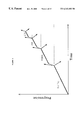
- FIG. 2 shows a model for pre-malignant conditions, highlighting magnitude of risk for progression to clinical malignancy.
- PDWA proliferative disease without alypia
- AH typical hyperplasia
- CIS carcinoma in situ.
- each stage is more likely to proceed to the next (dotted lines), but could also remain stable (horizontal lines, probably fairly frequent), or directly proceeds to development of a clone of cells with malignant behavior (vertical lines, becoming more likely further to the right.
- FIG. 3 presents photographs of DCIS tissue, before (upper left panel) and after microscopically-directed excisional punch biopsy (upper right panel).
- the lower panells show tissue samples of normal breast tissue (lower left panel), and invasive breast cancer (lower right panel).
- FIG. 4 shows expression of collagen III mRNA in tissue mRNA samples, analyzed by RNase protection assay methods.
- One ug of mRNA was hybridized with two labeled RNA probes: a T7 polymerase-generated probe for human glyceraldehycle dehydrogenase (GADP) which protects a 140 bp Sac I-Xba fragment; and a 17 polymerase-generated probe which protects a 208 bp Hinc II-Pst I fragment from the 3′ untranslated region of the human type III procollagen gene (Coll III) obtained by PCR subcloning the published sequence (Ala-Kokko, 1991).
- GDP human glyceraldehycle dehydrogenase
- RNA samples were labeled as follows: NL1 is RNA from cultured human breast epithelial cells (Hammond et al., 1984), NL2 is RNA from normal breast tissue, NL3 is RNA derived from the fibrous stromal fraction of breast tissue as described (Jensen et al., submitted for publication), NL4 is another sample from normal breast tissue. #12, #8, #4, #6, #10 are from patient samples with DCIS. Sample #10CA is RNA obtained from the small focus of microinvasion shown in FIG. 3 in the upper right panel. Con is a control sample using tRNA.
- FIG. 5 shows differential display of cDNAs obtained from patient tissue samples and controls.
- Rescued cDNA library samples were used as templates for low stringency PCR with the primers 5′GATGAGTTCGTGTCCGT ACAACTGG-3′(SEQ ID NO:54) and 5′GGTTATCGAAATCAGCCACAGCGCC (SEQ ID NO:55); 40 cycles were performed with denaturation for 1 minute at 94° C., annealing for 2 minutes at 25° C., and extension for 1 minute at 72° C.
- the samples correspond to those in the legend to FIG.
- Lane 1 is #12; Lanes 2 and 3 are from separate phage rescues of NL1 to show reproducibility of the assay; Lane 4 is #8; Lane 5 is #10; Lane 6 is #10CA; Lane 7 is a control from the rescued phage vector without cDNA inserts.
- Arrows mark cDNAs which are overexpressed in DCIS versus normal and arrowheads mark cDNAs which are differentially expressed in the invasive cancer (note this may reflect contamination from stromal cells as shown in FIG. 3 ).
- the bar marks a cDNA which is expressed in normal breast cells at higher levels than in DCIS or invasive cancer.
- FIG. 6 shows a comparison of the sequence between DCIS-1 (SEQ ID NO:1) and the human (SEQ ID NOs: 56-57) and hamster (SEQ ID NOs: 58-61) genes.
- FIG. 7 shows expression of DCIS-1 mRNA in tissue mRNA samples analyzed by RNase protection assay as described in the legend to FIG. 4 .
- RNA samples were labeled as in the legend to FIG. 4 .
- FIG. 8 is Table ii which displays the genetic code.
- FIG. 9 is a Table which lists differentially expressed marker genes from pre-invasive human breast tissue.
- FIGS. 10A and 10B shows expression of BRCA1 mRNA during breast cancer progression by PCR detection and nuclease protection assay, respectively.
- FIGS. 11A and 11B is a comparison of BRCA1 expression in normal breast and invasive breast cancer using nuclease protection assay of RNA, respectively.
- FIGS. 12A, 12 B, and 12 C show that antisense inhibition of BRCA1 accelerates mammary cell proliferation.
- FIGS. 13A and 13B includes a Northern blot of mRNA and nuclear run on studies that show that ribonucleotide reductase M2 mRNA is cell cycle regulated in MCF-7 cells.
- FIG. 14 includes a nuclease protection assay that shows that antisense inhibition of BRCA1 in human mammary cells decreases BRCA1 mRNA and increases ribonucleotide reductase mRNA.
- Nucleic acid sequences which are “complementary” are those which are capable of base-pairing according to the standard Watson-Crick complementarity rules. That is, that the larger purines will always base pair with the smaller pyrimidines to form only combinations of Guanine paired with Cytosine (G:C) and Adenine paired with either Thymine (A:T) in the case of DNA or Adenine paired with Uracil (A:U) in the case of RNA.
- G:C Guanine paired with Cytosine
- A:T Thymine
- A:U Adenine paired with Uracil
- Hybridization techniques refer to molecular biological techniques which involve the binding or hybridization of a probe to complementary sequences in a polynucleotide. Included among these techniques are northern blot analysis, southern blot analysis, nuclease protection assay, etc.
- Hybridization and “binding” in the context of probes and denatured DNA are used interchangeably. Probes which are hybridized or bound to denatured DNA are aggregated to complementary sequences in the polynucleotide. Whether or not a particular probe remains aggregated with the polynucleotide depends on the degree of complementarity, the length of the probe, and the stringency of the binding conditions. The higher the stringency, the higher must be the degree of complementarity and/or the longer the probe.
- Probe refers to an oligonucleotide or short fragment of DNA designed to be sufficiently complementary to a sequence in a denatured nucleic acid to be probed and to be bound under selected stringency conditions.
- Label refers to a modification to the probe nucleic acid that enables the experimenter to identify the labeled nucleic acid in the presence of unlabeled nucleic acid. Most commonly, this is the replacement of one or more atoms with radioactive isotopes. However, other labels include covalently attached chromophores, fluorescent moeities, enzymes, antigens, groups with specific reactivity, chemiluminescent moeities, and electrochemically detectable moeities, etc.
- Marker gene refers to any gene selected for detection which displays differential expression in abnormal tissue as opposed to normal tissue. It is also referred to as a differentially expressed gene.
- Marker protein refers to any protein encoded by a “marker gene” which protein displays differential expression in abnormal tissue as opposed to normal tissue.
- tissue homogenization probe describes a tissue homogenization probe.
- Abnormal tissue refers to pathologic tissue which displays cytologic, histologic and other defining and derivative features which differ from that of normal tissue. This includes in the case of abnormal breast tissue, among others, pre-invasive and invasive neoplasms.
- Normal tissue refers to tissue which does not display any pathologic traits.
- PCR technique describes a method of gene amplification which involves sequenced-based hybridization of primers to specific genes within a DNA sample (or library) and subsequent amplification involving multiple rounds of annealing, elongation and denaturation using a heat-stable DNA polymerase.
- RT-PCR is an abbreviation for reverse transcriptase-polymerase chain reaction. Subjecting mRNA to the reverse transcriptase enzyme results in the production of cDNA which is complementary to the base sequences of the mRNA. Large amounts of selected cDNA can then be produced by means of the polymerase chain reaction which relies on the action of heat-stable DNA polymerase produced by Thermus aquaticus for its amplification action.
- “Microscopically-directed” refers to the method of tissue sampling by which the tissue sampled is viewed under a microscope during the sampling of that tissue such that the sampling is precisely limited to a given tissue type, as the investigator requires. Specifically, it is a collection step which involves the use of a punch biopsy instrument. This surgical instrument is stereotactically manually-directed to harvest exclusively from abnormal tissue which exhibits histologic or cytologic characteristics of pre-invasive cancer. The harvest is correlated with a companion slide, stained to recognize the target tissue.
- “Differential display” describes a method in which expressed genes are compared between samples using low stringency PCR with random oligonucleotide primers.
- “Differential screening” describes a method in which genes within cDNA libraries are compared between two samples by differential hybridization of cDNAs to probes prepared from each library.
- Nuclease protection assay refers to a method of RNA quantitation which employs strand specific nucleases to identify specific RNAs by detection of duplexes.
- “Differential expression” describes the phenomenon of differential genetic expression seen in abnormal tissue in comparison to that seen in normal tissue.
- Isolatable tissue structure refers to a tissue structure which when visualized microscopically or otherwise is able to be isolated from other different surrounding tissue types.
- RNA In situ hybridization of RNA refers to the use of labeled DNA probes employed in conjunction with histological sections on which RNA is present and with which the labeled probe can hybridize allowing an investigator to visualize the location of the specific RNA within the cell.
- Codo DCIS cells refers to cells comprising an in situ lesion with the combined features of highest grade DCIS.
- Non-comedo DCIS cells refers to cells of DCIS lesions without comedo features.
- “Sequencing” describes the determination of the specific order of nucleic acids in a gene or polynucleotide.
- the present invention provides a method for detecting and diagnosing cancer by analyzing marker genes which are differentially expressed in early, pre-invasive breast cancer, specifically in non-comedo DCIS cells.
- Our histopathologic studies have demonstrated that certain morphologic patterns in breast tissue are pre-malignant, leading to invasive breast cancer in at least 20-30% of patients.
- These marker genes (which appear as differentially expressed genes in pre-invasive breast cancer) can be used as probes to develop diagnostic tests for the early detection of pre-invasive breast cancer (Sambrook, 1989).
- the present invention thus comprises a method of identification of marker genes which are expressed in the majority of pre-invasive breast cancer tissue samples. It involves cDNA library preparation followed by a modified differential display method. Use of genetic engineering methods (Sambrook, 1989) can bias the screening to specifically identify genes whose encoded proteins are secreted or are present at the cell surface, in order to find proteins which will be useful markers for diagnostic blood tests (secreted proteins) or for diagnostic imaging studies (cell surface proteins).
- the present invention also encompasses DNA segments which are complementary, or essentially complementary, to the sequence set forth in SEQ ID NO:1, SEQ ID NO:2, SEQ ID NO:3, SEQ ID NO:4, SEQ ID NO:5, SEQ ID NO:6, SEQ ID NO:7, SEQ ID NO:47 and SEQ ID NO:48.
- Nucleic acid sequences which are “complementary” are those which are capable of base-pairing according to the standard Watson-Crick complementarity rules.
- complementary sequences means nucleic acid sequences which are substantially complementary, as may be assessed by the same nucleotide comparison set forth above, or as defined as being capable of hybridizing to the nucleic acid segment of SEQ ID NO:1, SEQ ID NO:2, SEQ ID NO:3, SEQ ID NO:4, SEQ ID NO:5, SEQ ID NO:6, SEQ ID NO:7, SEQ ID NO:47 and SEQ ID NO:48 under relatively stringent conditions such as those described herein.
- nucleic acid segments of the present invention may be combined with other DNA sequences, such as promoters, polyadenylation signals, additional restriction enzyme sites, multiple cloning sites, other coding segments, and the like, such that their overall length may vary considerably. It is therefore contemplated that a nucleic acid fragment of almost any length may be employed, with the total length preferably being limited by the ease of preparation and use in the intended recombinant DNA protocol.
- nucleic acid fragments may be prepared which include a short stretch complementary to SEQ ID NO:1, SEQ ID NO:2, SEQ ID NO:3, SEQ ID NO:4, SEQ ID NO:5, SEQ ID NO:6, SEQ ID NO:7, SEQ ID NO:47 and SEQ ID NO:48, such as about 10 nucleotides, and which are up to 10,000 or 5,000 base pairs in length, with segments of 500 being preferred in most cases. DNA segments with total lengths of about 1,000, 500, 200, 100 and about 50 base pairs in length are also contemplated to be useful.
- Recombinant vectors and isolated DNA segments may therefore variously include the differentially expressed coding regions or the BRCA1 coding regions themselves, coding regions bearing selected alterations or modifications in the basic coding region, or they may encode larger polypeptides which nevertheless include differentially expressed-coding regions and the BRCA1 coding regions or may encode biologically functional equivalent proteins or peptides which have variant amino acids sequences.
- DNA segments of the present invention encompass biologically functional equivalent differentially expressed proteins and peptides biologically functional equivalent proteins of BRCA1. Such sequences may arise as a consequence of codon redundancy and functional equivalency which are known to occur naturally within nucleic acid sequences and the proteins thus encoded. Alternatively, functionally equivalent proteins or peptides may be created via the application of recombinant DNA technology, in which changes in the protein structure may be engineered, based on considerations of the properties of the amino acids being exchanged.
- Changes designed by man may be introduced through the application of site-directed mutagenesis techniques, e.g., to introduce improvements to the antigenicity of the protein or to test site-directed mutants or others in order to examine carcinogenic activity of the differentially expressed marker genes at the molecular level.
- fusion proteins and peptides e.g., where the differentially expressed marker gene coding regions are aligned within the same expression unit with other proteins or peptides having desired functions, such as for purification or immunodetection purposes (e.g., proteins which may be purified by affinity chromatography and enzyme label coding regions, respectively).
- Recombinant vectors form important further aspects of the present invention.
- Particularly useful vectors are contemplated to be those vectors in which the coding portion of the DNA segment is positioned under the control of a promoter.
- the promoter may be in the form of the promoter which is naturally associated with a RIBRED gene, e.g., in human cells, as may be obtained by isolating the 5′ non-coding sequences located upstream of the coding segment or exon, for example, using recombinant cloning and/or PCR technology, in connection with the compositions disclosed herein.
- a recombinant or heterologous promoter is intended to refer to a promoter that is not normally associated with a differentially expressed marker gene or the BRCA1 gene in its natural environment.
- promoters may include MMTV promoters normally associated with other genes, and/or promoters isolated from any other bacterial, viral, eukaryotic, or mammalian cell.
- promoter and cell type combinations for protein expression is generally known to those of skill in the art of molecular biology, for example, see Sambrook et al. (1989).
- the promoters employed may be constitutive, or inducible, and can be used under the appropriate conditions to direct high level expression of the introduced DNA segment, such as is advantageous in the large-scale production of recombinant proteins or peptides.
- Appropriate promoter systems contemplated for use in high-level expression include, but are not limited to appropriate bacterial promoters.
- differentially expressed marker gene encoded proteins and peptides As mentioned above, in connection with expression embodiments to prepare recombinant differentially expressed marker gene encoded proteins and peptides, it is contemplated that longer DNA segments will most often be used, with DNA segments encoding the entire differentially expressed protein or subunit being most preferred. However, it will be appreciated that the use of shorter DNA segments to direct the expression of differentially expressed peptides or epitopic core regions, such as may be used to generate anti-marker protein antibodies, also falls within the scope of the invention (Harlow et al, 1988).
- DNA segments which encode peptide antigens from about 15 to about 50 amino acids in length, or more preferably, from about 15 to about 30 amino acids in length are contemplated to be particularly useful.
- the C terminus of proteins provide an excellent region for peptide antigen recogition (Harlow et al, 1988).
- DNA segments encoding peptides will generally have a minimum coding length in the order of about 45 to about 147, or to about 90 nucleotides.
- DNA segments encoding partial length peptides may have a minimum coding length in the order of about 50 nucleotides for a polypeptide in accordance with seq id no:3, or about 264 nucleotides for a polypeptide in accordance with SEQ ID NO:1.
- nucleic acid sequences disclosed herein also have a variety of other uses. For example, they also have utility as probes or primers in nucleic acid hybridization embodiments. As such, it is contemplated that oligonucleotide fragments corresponding to the sequences of SEQ ID NO:1, SEQ ID NO:2, SEQ ID NO:3, SEQ ID NO:4, SEQ ID NO:5, SEQ ID NO:6, and SEQ ID NO:7 for stretches of between about 10 to 15 nucleotides and about 20 to 30 nucleotides will find particular utility. Longer complementary sequences, e.g., those of about 40, 50, 100, 200, 500, 1000, and even up to full length sequences of about 2,000 nucleotides in length, will also be of use in certain embodiments.
- nucleic acid probes to specifically hybridize to differentially expressed marker gene sequences will enable them to be of use in detecting the presence of complementary sequences in a given sample.
- sequence information for the preparation of mutant species primers, or primers for use in preparing other genetic constructions.
- Nucleic acid molecules having stretches of 20, 30, 50, or even of 500 nucleotides or so, complementary to SEQ ID NO:1, SEQ ID NO:2, SEQ ID NO:3, SEQ ID NO:4, SEQ ID NO:5, SEQ ID NO:6, and SEQ ID NO:7 are particularly contemplated as hybridization probes for use in, e.g., Southern and Northern blotting. This would allow differentially expressed structural or regulatory genes to be analyzed, both in patients and sample tissue from pre-invasive and invasive breast tissue. The total size of fragment, as well as the size of the complementary stretch(es), will ultimately depend on the intended use or application of the particular nucleic acid segment.
- the length of the complementary region may be varied, such as between about 10 and about 100 nucleotides, but larger complementary stretches of up to about 300 nucleotides may be used, according to the length complementary sequences one wishes to detect.
- hybridization probe of about 10 nucleotides in length allows the formation of a duplex molecule that is both stable and selective.
- Molecules having complementary sequences over stretches greater than 10 bases in length are generally preferred, though, in order to increase stability and selectivity of the hybrid, and thereby improve the quality and degree of specific hybrid molecules obtained.
- Hybridization probes may be selected from any portion of any of the sequences disclosed herein. All that is required is to review the sequences set forth in SEQ ID NO:1, SEQ ID NO:2, SEQ ID NO:3, SEQ ID NO:4, SEQ ID NO:5, SEQ ID NO:6, and SEQ ID NO:7 and to select any continuous portion of one of the sequences, from about 10 nucleotides in length up to and including the full length sequence, that one wishes to utilise as a probe or primer.
- probe and primer sequences may be governed by various factors, such as, by way of example only, one may wish to employ primers from towards the termini of the total sequence, or from the ends of the functional domain-encoding sequences, in order to amplify further DNA; one may employ probes corresponding to the entire DNA, or to the 5′ region, to clone marker-type genes from other species or to clone further marker-like or homologous genes from any species including human; and one may employ randomly selected, wild-type and mutant probes or primers with sequences centered around the RibRed M2 subunit encoding sequence to screen DNA samples for differentially expressed levels of RibRed, such as to identify human subjects which may be expressing differential levels of RibRed and thus may be susceptible to breast cancer.
- nucleic acid segment which includes a sequence from within SEQ ID NO:1, SEQ ID NO:2, SEQ ID NO:3, SEQ ID NO:4, SEQ ID NO:5, SEQ ID NO:6, and SEQ ID NO:7 may alternatively be described as “preparing a nucleic acid fragment”.
- fragments may also be obtained by other techniques such as, e.g., by mechanical shearing or by restriction enzyme digestion. Small nucleic acid segments or fragments may be readily prepared by, for example, directly synthesizing the fragment by chemical means, as is commonly practiced using an automated oligonucleotide synthesizer.
- fragments may be obtained by application of nucleic acid reproduction technology, such as the PCR technology of U.S. Pat. No. 4,603,102 (incorporated herein by reference), by introducing selected sequences into recombinant vectors for recombinant production, and by other recombinant DNA techniques generally known to those of skill in the art of molecular biology.
- the nucleotide sequences of the invention may be used for their ability to selectively form duplex molecules with complementary stretches of differentially expressed marker genes or cDNAs.
- relatively stringent conditions e.g., one will select relatively low salt and/or high temperature conditions, such as provided by 0.02M-0.15M NaCl at temperatures of 50° C. to 70° C.
- Such selective conditions tolerate little, if any, mismatch between the probe and the template or target strand, and would be particularly suitable for isolating specific differentially expressed marker genes.
- nucleic acid sequences of the present invention in combination with an appropriate means, such as a label, for determining hybridization.
- appropriate indicator means include fluorescent, radioactive, enzymatic or other ligands, such as avidin/biotin, which are capable of giving a detectable signal.
- fluorescent label or an enzyme tag such as urease, alkaline phosphatase or peroxidase, instead of radioactive or other environmental undesirable reagents.
- enzyme tags colorimetric indicator substrates are known which can be employed to provide a means visible to the human eye or spectrophotometrically, to identify specific hybridization with complementary nucleic acid-containing samples.
- the hybridization probes described herein will be useful both as reagents in solution hybridization as well as in embodiments employing a solid phase.
- the test DNA or RNA
- the test DNA is adsorbed or otherwise affixed to a selected matrix or surface.
- This fixed, single-stranded nucleic acid is then subjected to specific hybridization with selected probes under desired conditions.
- the selected conditions will depend on the particular circumstances based on the particular criteria required (depending, for example, on the G+C contents, type of target nucleic acid, source of nucleic acid, size of hybridization probe, etc.).
- specific hybridization is detected, or even quantified, by means of the label. (Sambrook et al, 1989).
- certain preliminary procedures are necessary to prepare the sample tissue and the probes before the detection of differential expression of marker genes in abnormal tissue as compared to that in normal tissue can be accomplished.
- RNA which is primarily derived from cells of epithelial origin.
- the particulate material (remaining in the pellet from the 30 second centrifugation) was homogenized with a tissuemizer, washed with PBS, treated with collagenase at 37° C. for 30 minutes, sonicated, extracted with phenol/chloroform and ethanol precipitated.
- cDNA libraries were constructed in lambda phage using polyA-selected mRNA from the following samples; cultured human breast epithelial cells, tissue from three reduction mammoplasty patients, tissue from three DCIS patients, and tissue from one DCIS patient (patient #10) that showed a focus of microinvasion adjacent to an area of DCIS. Multiple punches were needed to obtain sufficient RNA for polyA selection and library construction. 200 ug of total RNA was obtained by pooling 20 punches from normal breast tissue (reduction mammoplasty samples) and 5-8 punches from DCIS lesions, presumably reflecting the greater cellularity of the DCIS samples.
- cDNA libraries were constructed by first and second strand cDNA synthesis followed by the addition of directional synthetic linkers (ZAP-cDNA Synthesis Kit, Stratagene, La Jolla, Calif.). The Xho I-Eco R1 Tinkered cDNA was then ligated into lambda arms, packaged with packaging extracts, and then used to infect XL1-blue bacteria resulting in cDNA libraries.
- the collagen III probe employed for nuclease protection assays was constructed by subcloning the 208 bp Hinc II-Pst I fragment from the 3′ untranslated region of the human type III procollagen gene into pGem4Z. This region of the human procollagen III gene was obtained by PCR amplification of published sequence (Ala-Koldco et al, 1989) followed by restriction with Hinc II and Pst I.
- RNA samples For a control probe to assure equal loading and recovery of RNA, we used a T7 polymerase-generated probe for human glyceraldehyde phosphate dehydrogenase (GADP) which protects a 140 bp Sac I-Xba I fragment; (a generous gift from Janice Nigro, Vanderbilt University). Probe DCIS-1 was generated by linearizing the rescued plasmid with Pvu II, which should generate a 200 bp protected fragment. RNase protection assays were performed with 1 ug of unselected RNA and the above-cited probes using the methods we have reported previously (Holt, 1993).
- GDP human glyceraldehyde phosphate dehydrogenase
- Random 25 bp primers were generated by a computer-based algorithm (Jotte and Holt, unpublished). Samples were denatured for two minutes at 95° C. followed by 40 cycles, each cycle consisting of denaturation for 1 minute at 94° C., annealing for 2 minutes at 25° C., and extension for 1 minute at 72° C. The samples were then run on an 6% non-denaturing polyacrylamide gel, which was dried and autoradiographed. Specific bands were excised then reamplified with the same primers used for their generation.
- pre-invasive breast disease is herewith defined to be any reproducibly defined condition which confers an elevated risk of breast cancer approaching double that of the general population (Komitowski et al, 1990).
- the specifically-defined atypical hyperplasias and lobular carcinoma in situ confer relative risks of four to ten times that of the general population.
- FIG. 2 shows this model for the induction and progression of pre-invasive breast disease based on study of the Vanderbilt cohort (Dupont et al, 1985) of more than 10,000 breast biopsies (follow-up rate 85%; median time of 17 years; 135 women developed breast cancer).
- NCDCIS In order to study differential gene expression in DCIS, we collected cases of NCDCIS. The diagnosis of DCIS is made on histomorphologic grounds based on architectural, cytologic, and occasionally extent criteria. NCDCIS lacks comedo features and consists of microscopic intraductal lesions which fill and extend the duct, contain rigid internal architecture, and often have hyperchromatic and monomorphic nuclei.
- Non-comedo DCIS is the earliest determinant lesion which recurs locally as invasive cancer. Although comedo DCIS may be technically easier to study because the tumors are larger, its aggressiveness and the presence of numerous genetic alterations (such as p53 and erbB2) suggest that it may have advanced beyond the earliest stages of carcinogenesis.
- Control mRNA was obtained from frozen tissue samples obtained from reduction mammoplasties and from cultured human breast epithelial cells. Because non-comedo DCIS is a microscopic lesion, we had to microlocalize regions of DCIS in biopsy samples. To accomplish this we prepared frozen sections in which we located regions of DCIS and then employed a 2 mm punch to obtain an abnormal tissue sample only from those regions that contained DCIS. This selective harvesting was accomplished by carefully aligning the frozen section slide with the frozen tissue block and identifying areas of interest. The harvest of the appropriate area was then confirmed with a repeat frozen section. A similar approach was used to isolate mRNA from lobules of normal breast in samples collected from a reduction mammoplasty.
- FIG. 3 contains color photos of DCIS (abnormal) tissue, before (upper left panel) and after excisional punch biopsy (upper right panel). The lower panels show tissue samples of normal breast tissue lower left panel), and invasive breast cancer lower right panel).
- RNA was isolated, purified, and employed to construct cDNA libraries.
- RNA was isolated following mincing of tissue in 5.6M guanidinium isothiocyanate and 40% phenol, centrifugation to remove particulate matter, viscosity reduction by repeated aspiration through a 22 gauge needle, chloroform extraction and ethanol precipitation. In most samples there was particulate matter resistant to guanidinium-phenol extraction that was white in color and fibrous in appearance and was presumed to represent breast stroma. This stromal material was sparse in DCIS samples but abundant in samples obtained from normal breast tissue derived from reduction mammoplasties.
- stromal material was minced with a tissuemizer, washed with PBS, treated with collagenase at 37° C. for 30 minutes, sonicated, extracted with phenol/chloroform and ethanol precipitated. 200 ug of total RNA was obtained by pooling 20 punches from normal breast tissue (reduction mammoplasty samples) and 5-8 punches from DCIS lesions, presumably reflecting the greater cellularity of the DCIS samples. All libraries had greater than 50% inserts and contained between 2 ⁇ 10 6 and 7 ⁇ 10 7 phage recombinants with an average insert size varying between 500 and 1000 base pairs.
- FIG. 4 compares expression in NL 2 and #10CA with other patient samples and NL1 to determine collagen III expression.
- RNA probes for human glyceraldehyde phosphate dehydrogenase (GADP) which protects a 140 bp Sac I-Xba I fragment; and a T7 polymerase-generated probe which protects a 208 bp Hinc II-Pst I fragment from the 3′ untranslated region of the human type III procollagen gene (Coll III) obtained by PCR subcloning of the published sequence (Ala-Kokko et al, 1991).
- GDP human glyceraldehyde phosphate dehydrogenase
- RNA samples were labeled as follows: NL1 is RNA from cultured human breast epithelial cells (Hammond et al, 1984), NL2 is RNA from normal breast tissue, NL3 is RNA derived from the fibrous stromal fraction of breast tissue as described (Jensen et al, Submitted for publication), NL4 is another sample from normal breast tissue. This is described in greater detail on page 30 of this patent application. #12,#8,#4,#6, and #10 are from patient samples with DCIS. Sample #10CA is RNA obtained from the small focus of microinvasion shown in FIG. 3 . Con is a control sample using tRNA.
- cDNA libraries were constructed in lambda phage using polyA-selected mRNA from the following samples: cultured human breast epithelial cells, tissue from three reduction mammoplasty patients, tissue from three DCIS patients, and tissue from one DCIS patient (patient #10) that showed a small focus of invasion adjacent to an area of DCIS. Multiple punches were needed to obtain sufficient RNA for polyA selection and library construction. Selective handling of tissue was accomplished.
- FIG. 5 shows the results of differential display comparing cDNAs of several patient DCIS samples with cDNA obtained from normal breast epithelial cells and an early invasive cancer. Although few genes shown in this Figure are differentially expressed in the majority of samples with DCIS, the heterogeneity of gene expression in patient samples is seen.
- the differential display method (Liang et al, 1992a and 1992b) allows simultaneous comparison of multiple tissue samples. Initial studies using this method (reverse transcriptase followed by PCR) were unsatisfactory because of unwanted amplification of contaminating DNA in tissue samples and the small size of many of the fragments identified by display. To circumvent some of these problems, we have attempted to combine the advantages of cDNA library screening with the advantages of differential display by:
- FIG. 6 shows a comparison of the sequence between DCIS-1 and the human and hamster genes.
- DCIS patient #10 from an area of invasive cancer adjacent to DCIS
- frozen tissue samples from 7 more DCIS patients, 2 cellular fibroadenoma samples, and samples of “usual hyperplasia” and atypical hyperplasia.
- DCIS clones were identified by cloning methods which include selection and amplification, it was important to confirm by nuclease protection assays that the genes were differentially expressed in the original unselected, unamplified RNA samples (FIG. 7 ).
- differential display method allows comparison of multiple tissue samples of pre-invasive or invasive breast cancer.
- use of this method has successfully demonstrated that the M2 subunit ribonucleotide reductase gene is differentially expressed in 4 out of 5 pre-invasive breast cancer tissue samples. It is significant that the M2 subunit is involved in the regulation of the ribonucleotide reductase gene and is found to be over-expressed in abnormal tissue samples.
- differentially expressed genes may lead to discovery of genes which are potentially useful for breast cancer screening. Of particular interest are genes whose expression is restricted to breast epithelial cells and whose gene products are secreted. Screening for secreted proteins is possible by using the known hydrophobic sequences which encode leader sequences as one primer for differential display. The identification of secreted proteins which are specific for early breast pre-malignancy (or even early invasive cancer) would provide an important tool for early breast cancer screening programs. If a differentially expressed gene has not been cloned previously (or if details of its expression are unknown or uncertain) then nuclease protection assays or Northern blots can be performed on RNA prepared from tissue samples from a variety of tissues to determine if expression of this gene is restricted to breast. If necessary cDNA libraries prepared from other tissues can be added to the differential display screen as a way to identify only those genes which are expressed in early breast cancer and, in addition, are only expressed in breast tissue.
- differentially expressed genes have been initially characterized for expression in pre-malignant and malignant breast disease
- antibodies to the protein products of potentially useful genes can be developed and employed for immunohistochemistry (Harlow et al, 1988). This will provide an additional test to determine whether the expression of this gene is restricted to the breast. Subsequently, these antibodies will be used to detect the presence of this protein present in the blood of patients with pre-invasive and/or invasive cancer. By assaying for serum protein levels in the same patients who exhibited elevated expression of the gene in their tissue samples it will be possible to determine whether a gene product is being secreted into the blood.
- the BRCA 1 gene encodes a protein of 1863 amino acids with a predicted zinc finger domain observed in proteins which regulate gene transcription.
- BRCA 1 gene product is a negative regulator of mammary cell proliferation which is expressed at diminished levels in sporadic breast cancer.
- FIG. 1 The legend of FIG. 1 is as follows.
- tissue samples were used for mRNA isolation: Normal tissue samples: NL1-cultured human breast epithelial cells, NL2-Histologically normal breast tissue from an 11 year old undergoing a reduction mammoplasty, NL4-histologically normal breast tissue from an 14 year old undergoing a reduction mammoplasty. Carcinoma-in-situ samples are #6, #8, #10, #12, #23 (comedo type), #41, #55; and invasive cancer samples #10CA (invasive from the same patient with carcinoma-in-situ), 36CA, 1CA. All of these tissue samples were obtained from patients who had no family history of hereditary breast cancer and RNA preparation was performed as described above.
- Lane 1 human genomic DNA
- lane 2 NL1, lane 3: NL4, lane 4: $8
- lane 5 #12
- lane 6 #10
- lane 7 #10CA
- lane 8 #41, lane 9: #23
- lane 10 36CA
- lane 11 lambda DNA.
- the arrow points to the expected 113 bp band.
- Nuclease protection assays of microdissected mRNA from tissue samples are described in FIG. 10 B.
- One ug of mRNA from each tissue sample was hybridized with 32P-labelled, T7 polymerase-generated RNA probes for BRCA1 and human glyceraldehyde-3-phosphate dehydrogenase (GAPD) which produce expected protected fragments of 113 and 140 respectively as indicated by the lines on the right. Data were quantitated by phosphorimaging.
- the hybridizing intensity of each BRCA1 band was normalized to its respective GAPD band.
- the normalized values of NL1, NL2, and NL4 were intensity in each sample relative to 1.
- Sample 1 employs human leukocyte mRNA; Samples 2-4 are NL1, NL2, and NL4A; Samples 5-9 are #6(2.8), 8(3.7), 10(2.8), 12 (5.9), and 55 (1.4); and 10-12 are #10CA (0.07), 36CA (0.13), and 1CA (0.2).
- FIG. 10 shows that BRCA1 exon 24 mRNA is expressed at 5-10 fold higher levels in normal mammary tissue than in invasive breast cancer samples.
- Initial studies showed detectable levels of BRCA1 cDNA in a cDNA library prepared from a tissue sample with preinvasive carcinoma-in-situ but not in normal breast cancer invasive breast cancer cDNA libraries (FIG. 10 A). Because this method is relatively insensitive we directly quantitated BRCA1 mRNA by nuclease protection assays in RNA samples obtained by our microdissection method described above. These assays indicate that expression of BRCA1 mRNA in micro-dissected normal mammary epithelial tissue (lanes 2-4, FIG.
- FIG. 10B is 5-15 fold higher than that in breast cancer (lanes 10-12, FIG. 10 B).
- the highest levels of BRCA1 are observed in samples from non-comedo ductal carcinoma-in-situ (lanes 5-9, FIG. 10 B), a premalignant breast lesion with a finite, but relatively low rate of progression to invasion (Betsill et at., 1978, Page, D. L. et al., 1982, Page and Dupont, 1990).
- FIG. 11 A compare lanes 2 and 3, 4 and 5, 6 and 7.
- the legend of FIG. 11 is as follows.
- RNA obtained from paired samples of invasive breast cancer and histologically normal breast tissue are shown in FIG. 11 A.
- Samples in lanes 2 and 3 (first patient), 4 and 5 (second patient), 6 and 7 (third patient) are from invasive cancer and normal breast tissue respectively.
- Lane 1 is NL1 mRNA as described in legend to FIG. 10 and lane 8 is human leukocyte mRNA.
- the probes and methods are as described in FIG. 10 except the GAPD probe was of lower specific activity to improve quantitation.
- RNA from a series of invasive breast cancer tissue samples (lanes 2-9 compared with NL1 (lane 1) and leukocyte mRNA (lane 10) are shown in FIG. 11 B.
- Ratios of BRCA1/GAPD for each sample lane 1: 19.1, lane 2: 0.3, lane 3: 1.8, lane 4: 1.6, lane 5: 0.2, lane 6: 0.3, lane 7: 1.9, lane 8: 0, lane 9: 0.6.
- RNA levels were four to eight fold higher in samples derived from normal breast than in samples derived from invasive breast cancer.
- FIG. 11 B lanes 2-7.
- MCF-7 cells were growth arrested by tamoxifen for 48 hours and then stimulated at time zero (0) with 1 uM estradiol (+E) or control vehicle ( ⁇ E). Inhibition of DNA synthesis by tamoxifen and induction of synthesis by estrogen were confirmed by nuclear labelling studies with tritiated thymidine.
- FIG. 13 panels A and B show that transcription of the ribonucleotide reductase M2 gene is cell cycle regulated, inhibited by tamoxifen, and induced by estrogen.
- FIG. 13A is a Northern blot of mRNA from synchronized MCF-7 cells. At the indicated time in hours, total cellular RNA was isolated and Northern blotting performed using the 1.6 Kb Eco RI fragment from our cloned human ribonucleotide reductase cDNA described above. Two mRNA species of 1.6 and 3.4 Kb are observed in these studies.
- FIG. 13B shows nuclear run on studies of synchronized MCF-7 cells were performed by our published methods (Holt et al 1988) employing the 1.6 Kb fragment of ribonucleotide reductase described above (RR); the 1.8 Kb fragment of Topoisomerase II (Topo) described in the Olsen et al. 1993); the 1.0 Kb cyclophilin gene (Thompson et al. 1994) used as a constitutive control; and 18S ribosomal RNA (Thompson et al. 1994).
- Con represents cells which were grown for 48 hours but not treated with tamoxifen.
- Antisense inhibition is a useful strategy for studying gene expression which is dependent on expression of the antisense target gene (Robinson-Benion and Holt, in press, 1995), e.g. genes whose expression is directly or indirectly dependent on BRCA1 levels.
- FIG. 14 demonstrates that antisense inhibition of BRCA1 results in a corresponding increased expression of M2 ribonucleotide reductase mRNA.
- a nuclease protection assay of mRNA derived from primary mammary epithelial cells (lanes 1-4, 9-10) or MCF-7 cells Oanes 5-8, 11-12) cultured for 4 days with antisense or control oligonucleotide was performed under the following conditions: no oligonucleotide (lanes 1 and 5); 40 uM antiBRCA1 (lanes 2,6,10,12); 4 uM antiBRCA1 (lanes 3 and 7); 40 uM sense control (lanes 4,8,9,11).
- Probes for BRCA1 and GAPD are as described for FIG. 10, and the ribonucleotide reductase M2 probe (RR) detects the 200 bp probe is described above.
- Ribonucleotide reductase mRNA levels are highest in samples treated with 40 uM anti-BRCA1 oligonucleotide for both primary mammary epithelial cells and for MCF-7 cells (FIG. 14 ).
- Antisense inhibition of BRCA1 results in a 70-90% inhibition of mRNA levels in anti-BRCA1 treated cells compared with cells treated with the “sense” control oligonucleotide (compare lanes 9 and 10, FIG. 14 ).
- MCF-7 cells have lower levels of BRCA1 than the normal mammary epithelial cells (compare lanes 1 and 5, FIG. 14) anti-BRCA 1 since the antisense inhibition may drop BRCA1 levels below a critical threshold which normally functions to inhibit growth.
- RNA samples Freshly obtained breast biopsy or reduction mammoplasty specimens were frozen and then RNA was obtained following the microdissection method described above. Lesions were selected which were microlocalized and homogenous so that pure lesions could be obtained by 2 mm punches. Samples which had admixed normal epithelial, carcinoma-in-situ, or invasive cancer were not used for this study. Family history was obtained by chart review and/or interview to exclude familial breast cancer cases.
- PCR primers were derived from BRCA1 sequence in GenBank (Accession number U14680); forward 5′CAA TTG GGC AGA TGT GT 3′ (SEQ ID NO:50) and reverse 5′CTG GGG GAT CTG GGG TAT CA 3′ (SEQ ID NO:51) which amplify a 113 bp region from exon 24, corresponding to bases 5587 to 5699 of the human BRCA1. This region was selected because this exon has not been reported to be differentially spliced unlike more 5′ exons.
- the BRCA1 probe was cloned by subcloning this 113 bp band from normal human genomic DNA into PCRscriptSK and screening for correct orientation.
- RNA from each tissue sample was hybridized with 32P-labelled, T7 polymerase-generated RNA probes for BRCA1 and human glyceraldehyde-3-phosphate dehydrogenase (GADP) which would produce expected protected fragments of 113 and 140 respectively.
- GADP human glyceraldehyde-3-phosphate dehydrogenase
- the construction and use of the GADP probe for RNA standardization has been described above.
- the probe for ribonucleotide reductase M2 mRNA is the same as above and detects a 200 bp protected fragment.
- Unmodified deoxyribonucleotide were analyzed by gel electrophoresis and UV shadowing and shown to be homogenous and of appropriate size. These oligonucleotide were purified by multiple lyophilization and solubilized in buffered media as described (Holt et al. 1988). Sequence of the unmodified antiBRCA1 oligonucleotide 5′AAG AGC AGA TAA ATC CAT 3′(SEQ ID NO: 52) and the complementary sense oligonucleotide 5′ATG GAT TTA TCT GCT CTT 3′ (SEQ ID NO:53) correspond to the presumed translation initiation site at bases 12-137 of the GenBank sequence.
- oligonucleotide sequence was searched against GenBank and no significant homologies were identified to genes except BRCA1. Oligonucleotides were used according to our published methods (Holt et al. 1988).
- Primary mammary epithelial cells were cultured in serum-free medium supplemented with epidermal growth factor, insulin, hydrocortisone, ethanolamine, phosphorylethanolamine, and bovine pituitary extract.
- MCF-7 cells were cultured in Minimum Essential Medium Eagle (Modified) with Earle's salts and 2g/L sodium bicarbonate m supplemented with 2 mM L-glutamine, GMS-A (Gibco Cat. #680-1300AD), nonessential amino acids, and 2.5% fetal calf serum.
- Retinal pigmented perithelial cells were cultured in DMEM and 10% calf serum.
- Examples 8 and 9 describe applications of the discovery of the function of the BRCA1 gene.
- Example 8 describes a gene therapy method and example 9 describes a drug screening method.
- the discovery of the diminished expression of the BRCA1 mRNA in breast cancer using the microdissection techniques of this invention provides an important scientific basis for these examples.
- Viral vectors containing a DNA sequence that codes for a protein having an amino acid sequence as essentially set forth in SEQ ID NO:49 can be constructed using techniques that are well known in the art. This sequence includes the BRCA1 gene product. Viral vectors containing a DNA sequence essentially as set forth in SEQ ID NO:47 (the BRCA1 gene) can be also constructed using techniques that are well known in the art. Retroviral vectors, adenoviral vectors, or adeno-associated viral vectors are all useful methods for delivering genes into breast cancer cells. An excellent candidate for use in breast cancer gene therapy is a Moloney-based retroviral vector with a breast selective MMTV promoter which we have reported previously (Wong et al).
- the viral vector is constructed by cloning the DNA sequence essentially as set forth in SEQ ID:47 into a retroviral vector such as a breast selective vector. Most preferably, the full-length (coding region) cDNA for BRCA1 is cloned into the retroviral vector.
- the retroviral vector would then be transfected into virus producing cells in the following manner: Viruses are prepared by transfecting PA317 cells with retroviral vector DNAs which were purified as described in Wong et al. Following transfection, the PA317 cells are split and then treated with G418 until individual clones can be identified and expanded.
- Each clone is then screened for its titer by analyzing its ability to transfer G418 resistance (since the retroviral vector contains a Neomycin resistance gene).
- the clones which have the highest titer are then frozen in numerous aliquots and tested for sterility, presence of replication-competent retrovirus, and presence of mycoplasm.
- the methods generally employed for construction and production of retroviral vectors have been described in Muller, 1990.
- Viral vector expressing BRCA1 is infused into either solid tumors or infused into malignant effusions as a means for altering the growth of the tumor (since it is shown above that the BRCA1 gene product decreases the growth rate of breast cancer cells). Because viral vectors can efficiently transduce a high percentage of cancer cells, the tumors would be growth inhibited.
- the discovery of the function of the BRCA1 gene provides a clear utility in that induction of expression of the gene and the resulting increase in level of protein encoded by the gene in the breast cancer cell should slow the proliferation of the breast cancer cells.
- Induction of expression of the gene can be caused by administering a compound to a patient that stimulates the regulatory regions of this gene, such as the promoter.
- a method for screening compounds that activate the promoter of the BRCA1 gene is designed in the following way.
- a promoter sequence is a DNA segment that upregulates the expression of a gene.
- a sequence essentially as set forth in SEQ ID NO:48 can be ligated into a suitable vector, such as a plasmid, that contains a reporter gene using standard recombinant DNA techniques of restriction enzyme digests, ligation of fragment into vector, and transformation of bacteria.
- SEQ ID NO:48 includes the promoter sequence of the BRCA1 gene.
- a reporter gene is a gene that produces a readily detectable product. Examples of appropriate reporter genes which could be employed for this purpose include Beta-galactosidase or the chloramphenicol acetyltransferase gene.
- the BRCA1 promoter/reporter gene combination can then be cloned into an expression vector or viral vector by standard recombinant DNA methods.
- Breast cancer cells can then be transfected with the expression vector containing the BRCA1 promoter/reporter gene using standard transfection methods which we have reported previously (Holt et al. PNAS 1986).
- a stable transformant with appropriate low level expression will be identified and then characterized to demonstrate proper DNA integration and expression. Methods of establishing and characterizing stable transformants have been described (Holt. MCB, 1994).
- a positive result i.e. induction of the promoter region
- a positive result results in increased levels of the reporter gene resulting in either an increase in color (Beta-galactosidase assay) or specific radioactivity (Chloramphenicol acetyltransferase activity) through a reaction between the protein encoded by the reporter gene and a compound in the reaction medium.
- the compound produced by the reaction between the reporter gene protein and the compound in the reaction medium is the cause of the increase in color or specific radioactivity.
- These compounds can be called indicator compounds in that their presence indicates that the drug or biologial agent activated the BRCA1 promoter.
- Methods for standardizing and performing Beta-galactosidase or chloramphenicol acetyltransferase assays have been reported (Holt et. al. MCB 1994). This method would be useful for initial screening of agents which increase BRCA1 expression. These agents could then be tested in more rigorous assays of breast cancer growth such as nude mouse tumor assays (Arteaga et al). This approach allows mass screening of large numbers of agents, sparing more rigorous animal tests for only promising compounds which score in the reporter gene assay described herein.
Abstract
The present invention provides a method of detecting and diagnosing pre-invasive breast cancer by identifying differentially expressed genes in early, pre-invasive breast cancer tissue. Differentially expressed genes can be used as genetic markers to indicate the presence of pre-invasive cancerous tissues. Microscopically-directed tissue sampling techniques combined with differential display or differential screening of cDNA libraries are used to determine differential expression of genes in the early stages of breast cancer. Differential expression of genes in preinvasive breast cancer tissue is confirmed by RT-PCR, nuclease protection assays and in-situ hybridization of ductal carcinoma in situ tissue RNA and control tissue RNA. The present invention also provides a method of screening for compounds that induce expression of the BRCA1 gene, whose product negatively regulates cell growth in both normal and malignant mammary epithlial cells. The present invention also relates to gene therapy method using this gene.
Description
This is a continuation application Ser. No. 08/373,799, filed Jan. 17, 1995, abandoned, which is a CIP of Ser. No. 08/182,961, filed Jan. 14, 1994, now U.S. Pat. No. 5,677,125.
The owner of this application and the parent application, Vanderbilt University, claims benefit of the Statement Verifying Small Entity Status—Non-Profit Organization filed in the parent application, as such status is still proper.
This invention was made in part from government support under Grant No. FS-00267 from the National Institutes of Health, National Institute of Environmental Health Sciences. The government has certain rights in the invention.
This is a continuation application of U.S. Pat. application Ser. No. 08/373/799, filed Jan. 17, 1995, abandoned.
The detection of differentially expressed genes in pre-invasive breast tissue, specifically in non-comedo ductal carcinoma in situ as compared to genes expressed in normal tissue, is useful in the diagnosis, prognosis and treatment of human breast cancer. Such differentially expressed genes are effective marker genes indicating the significantly increased risk of breast cancer in a patient expressing these differentially expressed marker genes. These marker genes are useful in the detection, early diagnosis, and treatment of breast cancer in humans.
The discovery of the function of the BRCA 1 gene has broad utility including, in the present invention, development of methods to treat familial and sporadic breast cancers as well as screen for therapeutic drugs through production of important indicator compounds.
Of the differentially expressed genes described in this invention, DCIS-1 encodes a gene similar to the M2 subunit of hamster ribonucleotide reductase. The M2 subunit of ribonucleotide reductase (RibRed, hereafter) is responsible for regulation of RibRed. The differential levels of expression of the marker genes described in this invention (Seq ID No.s 1-7), indicate genetic changes which have been linked to the presence of pre-invasive breast cancer.
The BRCA1 gene (Seq. ID No. 47) is differentially expressed in invasive breast cancer cells. The BRCA1 gene product is a negative regulator of mammary cell proliferation which is expressed at diminished levels in sporadic breast cancer.
1. Technical Field
The present invention relates generally to methods of detection and diagnosis of breast cancer and more particularly to a diagnostic method which relies on the identification of marker genes expressed in pre-invasive cancers by microscopically-directed cloning. Furthermore, this invention concerns the prevention, detection, and diagnosis of breast cancer by addressing the molecular events which occur during the earliest alterations in breast tissue.
The present invention also relates generally to methods of treatment of breast cancer, and more particularly to gene therapy methods and methods for screening compounds that induce expression of the BRCA1 gene product.
2. Description of the Prior Art
It will be appreciated by those skilled in the art that there exists a need for a more sensitive and less invasive method of early detection and diagnosis of breast cancer than those methods currently in use. Breast cancer presents inherent difficulties in regard to the ease with which it is detected and diagnosed. This is in contrast to detection of some other common cancers, including skin and cervical cancers, the latter of which is based on cytomorphologic screening techniques.
There have been several attempts to develop improved methods of breast cancer detection and diagnosis. In the attempts to improve methods of detection and diagnosis of breast cancer, numerous studies have searched for oncogene mutations, gene amplification, and loss of heterozygosity in invasive breast cancer (Callahan, et al., 1992; Cheickh, et al., 1992; Chen, et al, 1992; and, Lippman, et al, 1990). However, few studies of breast cancer have analyzed gene mutations and/or altered gene expression in ductal carcinoma in situ (DCIS). Investigators have demonstrated high levels of p53 protein in 13-40% of DCIS lesions employing a monoclonal antibody to p53, and subsequent sequencing demonstrated mutations in several cases (Poller et al, 1992). The neu/erbB2 gene appears to be amplified in a subset of DCIS lesions (Allred et al, 1992; Maguire et al, 1992). Histologic analysis of DCIS cases suggests that mutations and altered gene expression events, as well as changes in chromatin and DNA content, occur predominantly in comedo DCIS (Böcker et al, 1992; Killeen et al, 1991; and, Komitowski et al, 1990), which has a rapid rate of local invasion and progression to metastasis. Thus, there are presently no reliable marker genes for non-comedo DCIS (NCDCIS, hereafter).
Cancer in humans appears to be a multi-step process which involves progression from pre-malignant to malignant to metastatic disease which ultimately kills the patient. Epidemiologic studies in humans have established that certain pathologic conditions are “pre-malignant” because they are associated with increased risk of malignancy. There is precedent for detecting and eliminating pre-invasive lesions as a cancer prevention strategy: dysplasia and carcinoma in-situ of the uterine cervix are examples of pre-malignancies which have been successfully employed in the prevention of cervical cancer by cytologic screening methods. Unfortunately, because the breast cannot be sampled as readily as cervix, the development of screening methods for breast pre-malignancy involves more complex approaches than cytomorphologic screening now currently employed to detect cervical cancer.
Pre-malignant breast disease is also characterized by an apparent morphological progression from atypical hyperplasias, to carcinoma in-situ (pre-invasive cancer) to invasive cancer which ultimately spreads and metastasizes resulting in the death of the patient. Careful histologic examination of breast biopsies has demonstrated intermediate stages which have acquired some of these characteristics but not others. Detailed epidemiological studies have established that different morphologic lesions progress at different rates, varying from atypical hyperplasia (with a low risk) to comedo ductal carcinoma-in-situ which progresses to invasive cancer in a high percentage of patients (London et al, 1991; Page et al, 1982; Page et al, 1985; Page et al, 1991; and Page et al, 1978). Family history is also an important risk factor in the development of breast cancer and increases the relative risk of these pre-malignant lesions (Dupont et al, 1985; Dupont et al, 1993; and, London et al, 1991). Of particular interest is non-comedo carcinoma-in-situ which is associated with a greater than ten-fold increased relative risk of breast cancer compared to control groups (Ottesen et al, 1992; Page et al, 1982). Two other reasons besides an increased relative risk support the concept that DCIS is pre-malignant: 1) When breast cancer occurs in these patients it regularly occurs in the same region of the same breast where the DCIS was found; and 2) DCIS is frequently present in tissue adjacent to invasive breast cancer (Ottesen et al, 1992; Schwartz et al, 1992). For these reasons DCIS very likely represents a rate-limiting step in the development of invasive breast cancer in women.
DCIS (sometimes called intraductal carcinoma) is a group of lesions in which the cells have grown to completely fill the duct with patterns similar to invasive cancer, but do not invade outside the duct or show metastases at presentation. DCIS occurs in two forms: comedo DCIS and non-comedo DCIS. Comedo DCIS is often a grossly palpable lesion which was probably considered “cancer” in the 19th and early 20th century and progresses to cancer (without definitive therapy) in at least 50% of patients within three years (Ottesen et al, 1992; Page et al, 1982). Most of the molecular alterations which have been reported in pre-malignant breast disease have been observed in cases of comedo DCIS (Poller et al, 1993; Radford et al, 1993; and, Tsuda et al, 1993). Non-comedo DCIS is detected by microscopic analysis of breast aspirates or biopsies and is associated with a 10 fold increased risk of breast cancer, which corresponds to a 25-30% absolute risk of breast cancer within 15 years (Ottesen et al, 1992; Page et al, 1982; and, Ward et al, 1992).
Widespread application of mammography has changed the relative incidence of comedo and non-comedo DCIS such that NCDCIS now represents the predominant form of DCIS diagnosed in the United States (Ottesen et al, 1992; Page et al, 1982; and Pierce et al, 1992). Both forms of DCIS generally recur as invasive cancer at the same site as the pre-malignant lesion (without definitive therapy). The precursor lesions to DCIS are probably atypical ductal hyperplasia and proliferative disease without atypia which are associated with lower rates of breast cancer development, but show further increased risk when associated with a family history of breast cancer (Dupont et al, 1985; Dupont et al, 1989; Dupont et al, 1993; Lawrence, 1990; London et al, 1991; Page et al, 1982; Page et al, 1985; Page et al, 1991; Page et al, 1978; Simpson et al, 1992; Solin et al, 1991; Swain, 1992; Weed et al, 1990).
What is needed, then, is a sensitive method of detection and diagnosis of breast cancer when the cancerous cells are still in the pre-invasive stage. To illustrate the usefulness in early breast cancer detection of a marker gene and its encoded protein, consider the dramatic impact that prostate specific antigen has had on early stage prostate cancer. This method of early detection and diagnosis of breast cancer is presently lacking in the prior art.
Breast cancer occurs in hereditary and sporadic forms. Recently the BRCA 1 gene has been cloned and shown to be mutated in kindreds with hereditary breast and ovarian cancer (Hall et al. 1990, Miki, Y. et al. 1994, Friedman et al. 1994, Castilla et al. 1994, Simard et al. 1994). Although 92% of families with two or more cases of early-onset breast cancer and two cases of ovarian cancer have germ-line mutations in BRCA 1 (Narod et al. in press), the gene has not been shown to be mutated in any truly sporadic case to date (Futreal et al. 1994). Despite the surprising paucity of somatically acquired mutations in sporadic breast cancer, it is still a likely tumor suppressor gene with a key role in breast epithelial cell biology. The BRCA 1 gene encodes a protein of 1863 amino acids with a predicted zinc finger domain observed in proteins which regulate gene transcription. Until the discovery of the function of the BRCA1 gene in conjuction with the development of the present invention, the function was unknown.
Epidemiologic studies have established that NCDCIS of the breast is associated with a ten-fold increased risk of breast cancer (absolute risk of 25-30%). It seems likely that this pre-invasive lesion is a determinate precursor of breast cancer because the subsequent development of breast cancer is regularly in the same region of the same breast in which the NCDCIS lesion was found. Important aspects of the present invention concern isolated DNA segments and those isolated DNA segments inserted into recombinant vectors encoding differentially expressed marker genes in abnormal tissue, specifically in NCDCIS, as compared with those expressed in normal tissue, and the creation and use of recombinant host cells through the application of DNA technology, which express these differentially expressed marker genes (Sambrook et al, 1989).
Because there are no cell lines or animal models which clearly display known characteristics of pre-invasive breast disease, human breast tissue samples are essential for studying pre-invasive breast disease. Using human tissue samples, we subsequently have developed a method for cDNA cloning from histologically identified lesions in human breast biopsies. We have used this method to clone genes which are differentially expressed in pre-invasive breast lesions such as NCDCIS lesions as compared to genes expressed in normal tissue. The differentially expressed genes detected in pre-invasive breast cancer are called marker genes. Identification of marker genes for pre-invasive breast disease provides improved methods for detection and diagnosis of pre-invasive breast cancer tissue, and further provides marker genes for studies of the molecular events involved in progression from pre-invasive to malignant breast disease.
Analysis of marker gene expression in NCDCIS presents the advantage that cancerous breast tissue at that stage is non-invasive. Detection and diagnosis of NCDCIS by means of differentially expressed marker genes compared to the same marker genes in normal breast tissue, would allow a greater ability to detect, prevent and treat the disease before it becomes invasive and metastasizes. The stage or intermediate condition of NCDCIS is a particularly good candidate for early intervention because it is 1) prior to any invasion and thus prior to any threat to life; 2) it is followed by invasive carcinoma in over 30% of cases if only treated by biopsy; and, 3) there is a long “window” of opportunity (4-8 years) approximately before invasive neoplasia occurs. Thus, NCDCIS is an ideal target for early diagnosis. While these morphologically defined intermediate endpoints have been widely accepted, progress in defining the molecular correlates of these lesions has been hampered by an inability to identify and sample them in a manner which would allow the application of molecular techniques.
Frozen tissue blocks from breast biopsies were used to construct and screen cDNA libraries prepared from NCDCIS tissue, normal breast tissue, breast cancer tissue, and normal human breast epithelial cells. Several cDNAs which were differentially expressed in human DCIS epithelial cells compared to normal breast epithelial cells were cloned and sequenced. One gene which is differentially expressed is the M2 subunit of RibRed which is expressed at low levels in human breast epithelial cells but at higher levels in 4 out of 5 DCIS tissue samples. It is presumed that the altered morphologic appearance and determinant biologic behavior of DCIS results from altered expression of genes (such as RibRed) which is important in the induction of breast cancer in humans.
This invention, therefore, provides a method of detecting and diagnosing pre-invasive breast cancer by analyzing marker genes which are differentially expressed in non-comedo DCIS cells. Histopathologic studies have demonstrated that these morphologic patterns in breast tissue lead to invasive breast cancer in at least 20-30% of patients. The present method analyzes gene expression in normal, pre-malignant and malignant breast biopsies; and, it allows simultaneous comparison and cloning of marker genes which are differentially expressed in pre-invasive breast cancer. These marker genes can then be used as probes to develop other diagnostic tests for the early detection of pre-invasive breast cancer.
The present invention concerns DNA segments, isolatable from both normal and abnormal human breast tissue, which are free from total genomic DNA. The isolated DCIS-1 protein product is the regulatory element of the RibRed enzyme. This and all other isolatable DNA segments which are differentially expressed in preinvasive breast cancer can be used in the detection, diagnosis and treatment of breast cancer in its earliest and most easily treatable stages. As used herein, the term “abnormal tissue” refers to pre-invasive and invasive breast cancer tissue, as exemplified by collected samples of non-comedo or comedo DCIS tissues.
As used herein, the term “DNA segment” refers to a DNA molecule which has been isolated free of total genomic DNA of a particular species. Therefore, a DNA segment encoding a differentially expressed protein (as measured by the expression of mRNA) in abnormal tissue refers to a DNA segment which contains differentially expressed-coding sequences in abnormal tissue as compared to those expressed in normal tissue, yet is isolated away from, or purified free from, total genomic DNA of Homo sapiens sapiens. Furthermore, a DNA segment encoding a BRCA1 protein refers to a DNA segment which contains BRCA1 coding sequences, yet is isolated away from, or purified free from, total genomic DNA of Homo sapiens sapiens. Included within the term “DNA segment”, are DNA segments and smaller fragments of such segments, and also recombinant vectors, including, for example, plasmids, cosmids, phage, viruses, and the like.
Similarly, a DNA segment comprising an isolated or purified differentially expressed gene or comprising an isolated or purified BRCA1 gene refers to a DNA segment including differentially expressed coding sequences or BRCA1 coding sequences isolated substantially away from other naturally occurring genes or protein encoding sequences. In this respect, the term “gene” is used for simplicity to refer to a functional protein, polypeptide or peptide encoding unit. As will be understood by those in the art, this functional term includes both genomic sequences and cDNA sequences. “Isolated substantially away from other coding sequences” means that the gene of interest, in this case, any differentially expressed marker gene or the BRCA1 gene, forms the significant part of the coding region of the DNA segment, and that the DNA segment does not contain large portions of naturally-occurring coding DNA, such as large chromosomal fragments or other functional genes or cDNA coding regions. Of course, this refers to the DNA segment as originally isolated, and does not exclude genes or coding regions later added to the segment by the hand of man.
In particular embodiments, the invention concerns isolated DNA segments and recombinant vectors incorporating DNA sequences which encode differentially expressed genes in pre-invasive breast cancer, each which includes within its amino acid sequence an amino acid sequence in accordance with SEQ ID NO:1, SEQ ID NO:2, SEQ ID NO:3, SEQ ID NO:4, SEQ ID NO:5, SEQ ID NO:6, or SEQ ID NO:7, all seq id no:s 1-7 are derived from non-comedo DCIS samples from Homo sapiens sapiens. In other particular embodiments, the invention concerns isolated DNA segments and recombinant vectors incorporating DNA sequences which encode the M2 subunit of human RibRed that includes within its amino acid sequence the similar amino acid sequence of hamster RibRed corresponding to the M2 subunit of hamster RibRed.
In certain embodiments, the invention concerns isolated DNA segments and recombinant vectors which partially or wholly encode a protein or peptide that includes within its amino acid sequence an amino acid sequence essentially as partially or wholly encoded, respectively, by SEQ ID NO:1, SEQ ID NO:2, SEQ ID NO:3, SEQ ID NO:4, SEQ ID NO:5, SEQ ID NO:6, or SEQ ID NO:7. Naturally, where the DNA segment or vector encodes a full length differentially expressed protein, or is intended for use in expressing the differentially expressed protein, the most preferred sequences are those which are essentially as set forth in SEQ ID NO:1, SEQ ID NO:2, SEQ ID NO:3, SEQ ID NO:4, SEQ ID NO:5, SEQ ID NO:6, or SEQ ID NO:7 and which encode a protein that exhibits differential expression, e.g., as may be determined by the differential display or differential sequencing assay, as disclosed herein.
The term “a sequence essentially as set forth in SEQ ID NO:1, SEQ ID NO:2, SEQ ID NO:3, SEQ ID NO:4, SEQ ID NO:5, SEQ ID NO:6, or SEQ ID NO:7” means that the sequence substantially corresponds to a portion of SEQ ID NO:1, SEQ ID NO:2, SEQ ID NO:3, SEQ ID NO:4, SEQ ID NO:5, SEQ ID NO:6, or SEQ ID NO:7, respectively, and has relatively few nucleotides which are not identical to, or a biologically functional equivalent of, the nucleotides of the respective SEQ ID NO:1, SEQ ID NO:2, SEQ ID NO:3, SEQ ID NO:4, SEQ ID NO:5, SEQ ID NO:6, or SEQ ID NO:7. The term “biologically functional equivalent” is well understood in the art and is further defined in detail herein, for example see pages 24 through 25. Accordingly, sequences which have between about 70% and about 80%; or more preferably, between about 81% and about 90%; or even more preferably, between about 91% and about 99%; of amino acids which are identical or functionally equivalent to the amino acids of SEQ ID NO:1, SEQ ID NO:2, SEQ ID NO:3, SEQ ID NO:4, SEQ ID NO:5, SEQ ID NO:6, or SEQ ID NO:7 will be sequences which are “essentially as set forth in SEQ ID NO:1, SEQ ID NO:2, SEQ ID NO:3, SEQ ID NO:4, SEQ ID NO:5, SEQ ID NO:6, or SEQ ID NO:7”, respectively.
In particular embodiments, the invention concerns a drug screening method and a gene therapy method that use isolated DNA segments and recombinant vectors incorporating DNA sequences which encode a protein that includes within its amino acid sequence an amino acid sequence in accordance with SEQ ID NO:49, SEQ ID NO:49 derived from breast tissue from Homo sapiens. In other particular embodiments, the invention concerns isolated DNA sequences and recombinant DNA vectors incorporating DNA sequences which encode a protein that includes with its amino acid sequence the amino acid sequence of the BRCA1 gene product from human breast tissue.
In certain embodiments, the invention concerns methods using isolated DNA segments and recombinant vectors which partially or wholly encode a protein or peptide that includes within its amino acid sequence an amino acid sequence essentially as set forth in SEQ ID NO:49. Naturally, where the DNA segment or vector encodes a full length BRCA1 protein, or is intended for use in expressing the BRCA1 protein, the most preferred sequences are those which are essentially as set forth in SEQ ID NO:47 and which encode a protein that retains activity as a negative growth regulator in human breast cells, as may be determined by antisense assay, as disclosed herein.
The term “a sequence essentially as set forth in SEQ ID NO:1, SEQ ID NO:2, SEQ ID NO:3, SEQ ID NO:4, SEQ ID NO:5, SEQ ID NO:6, or SEQ ID NO:7” means that the sequence substantially corresponds to a portion of SEQ ID NO:1, SEQ ID NO:2, SEQ ID NO:3, SEQ ID NO:4, SEQ ID NO:5, SEQ ID NO:6, or SEQ ID NO:7, respectively, and has relatively few nucleotides which are not identical to, or a biologically functional equivalent of, the nucleotides of the respective SEQ ID NO:1, SEQ ID NO:2, SEQ ID NO:3, SEQ ID NO:4, SEQ ID NO:5, SEQ ID NO:6, or SEQ ID NO:7. The term “biologically functional equivalent” is well understood in the art and is further defined in detail herein, for example see pages 24 through 25. Accordingly, sequences which have between about 70% and about 80%; or more preferably, between about 81% and about 90%; or even more preferably, between about 91% and about 99%; of amino acids which are identical or functionally equivalent to the amino acids of SEQ ID NO:1, SEQ ID NO:2, SEQ ID NO:3, SEQ ID NO:4, SEQ ID NO:5, SEQ ID NO:6, or SEQ ID NO:7 will be sequences which are “essentially as set forth in SEQ ID NO:1, SEQ ID NO:2, SEQ ID NO:3, SEQ ID NO:4, SEQ ID NO:5, SEQ ID NO:6, or SEQ ID NO:7”, respectively.
The term “a sequence essentially as set forth in SEQ ID NO:49” means that the sequence substantially corresponds to a portion of SEQ ID NO:49 and has relatively few amino acids which are not identical to, or a biologically functional equivalent of, the nucleotides of SEQ ID NO:49. The term “biologically functional equivalent” is well understood in the art and is further defined in detail herein, for example see pages 24 through 25. Accordingly, sequences which have between about 70% and about 80%; or more preferably, between about 81% and about 90%; or even more preferably, between about 91% and about 99%; of amino acids which are identical or functionally equivalent to the amino acids of SEQ ID NO:49 will be sequences which are “essentially as set forth in SEQ ID NO:49”.
In certain other embodiments, the invention concerns isolated DNA segments and recombinant vectors that include within their sequence a nucleic acid sequence essentially as set forth in SEQ ID NO:1, SEQ ID NO:2, SEQ ID NO:3, SEQ ID NO:4, SEQ ID NO:5, SEQ ID NO:6, and SEQ ID NO:7. The term “essentially as set forth in SEQ ID NO:1, SEQ ID NO:2, SEQ ID NO:3, SEQ ID NO:4, SEQ ID NO:5, SEQ ID NO:6, and SEQ ID NO:7” is used in the same sense as described above and means that the nucleic acid sequence substantially corresponds to a portion of SEQ ID NO:1, SEQ ID NO:2, SEQ ID NO:3, SEQ ID NO:4, SEQ ID NO:5, SEQ ID NO:6, and SEQ ID NO:7, respectively, and has relatively few codons which are not identical, or functionally equivalent, to the codons of SEQ ID NO:1, SEQ ID NO:2, SEQ ID NO:3, SEQ ID NO:4, SEQ ID NO:5, SEQ ID NO:6, and SEQ ID NO:7, respectively. Again, DNA segments which encode proteins exhibiting differential expression will be most preferred. The term “functionally equivalent codon” is used herein to refer to codons that encode the same amino acid, such as the six codons for arginine or serine, and also refers to codons that encode biologically equivalent amino acids (see FIG. 8).
In certain other embodiments, the invention concerns a method for screening drugs and a gene therapy method which involve the use of isolated DNA segments and recombinant vectors that include within their sequence a nucleic acid sequence essentially as set forth in SEQ ID NO:47 and SEQ ID NO:48. The term “essentially as set forth in SEQ ID NO:47 and SEQ ID NO:48” is used in the same sense as described above and means that the nucleic acid sequence substantially corresponds to a portion of SEQ ID NO:47 and SEQ ID NO:48 respectively, and has relatively few codons which are not identical, or functionally equivalent, to the codons of SEQ ID NO:47 and SEQ ID NO:48, respectively. Again, DNA segments which encode proteins exhibiting the negative regulatory activity of the BRCA1 will be most preferred. The term “functionally equivalent codon” is used herein to refer to codons that encode the same amino acid, such as the six codons for arginine or serine, and also refers to codons that encode biologically equivalent amino acids (see FIG. 8).
It will also be understood that amino acid and nucleic acid sequences may include additional residues, such as additional N- or C-terminal amino acids or 5′ or 3′ sequences, and yet still be essentially as set forth in one of the sequences disclosed herein, so long as the sequence meets the criteria set forth above, including the maintenance of biological protein activity where protein expression is concerned. The addition of terminal sequences particularly applies to nucleic acid sequences which may, for example, include various non-coding sequences flanking either of the 5′ or 3′ portions of the coding region or may include various internal sequences, i.e., introns, which are known to occur within genes.
Excepting intronic or flanking regions, and allowing for the degeneracy of the genetic code, sequences which have between about 20% and about 50%; or more preferably, between about 50% and about 70%; or even more preferably, between about 70% and about 99%; of nucleotides which are identical to the nucleotides of SEQ ID NO:1, SEQ ID NO:2, SEQ ID NO:3, SEQ ID NO:4, SEQ ID NO:5, SEQ ID NO:6, and SEQ ID NO:7 will be sequences which are “essentially as set forth in SEQ ID NO:1, SEQ ID NO:2, SEQ ID NO:3, SEQ ID NO:4, SEQ ID NO:5, SEQ ID NO:6, and SEQ ID NO:7”, respectively. Sequences which are essentially the same as those set forth in SEQ ID NO:1, SEQ ID NO:2, SEQ ID NO:3, SEQ ID NO:4, SEQ ID NO:5, SEQ ID NO:6, and SEQ ID NO:7 may also be functionally defined as sequences which are capable of hybridizing to a nucleic acid segment containing the complement of SEQ ID NO:1, SEQ ID NO:2, SEQ ID NO:3, SEQ ID NO:4, SEQ ID NO:5, SEQ ID NO:6, and SEQ ID NO:7, respectively, under relatively stringent conditions. Suitable relatively stringent hybridization conditions will be well known to those of skill in the art (Sambrook et al, 1989).
Excepting intronic or flanking regions, and allowing for the degeneracy of the genetic code, sequences which have between about 20% and about 50%; or more preferably, between about 50% and about 70%; or even more preferably, between about 70% and about 99%; of nucleotides which are identical to the nucleotides of SEQ ID NO:47 and SEQ ID NO:48 will be sequences which are “essentially as set forth in SEQ ID NO:47 and SEQ ID NO:48”, respectively. Sequences which are essentially the same as those set forth in SEQ ID NO:47 and SEQ ID NO:48 may also be functionally defined as sequences which are capable of hybridizing to a nucleic acid segment containing the complement of SEQ ID NO:47 and SEQ ID NO:48, respectively, under relatively stringent conditions. Suitable relatively stringent hybridization conditions will be well known to those of sill in the art (Sambrook et al, 1989).
It is also important to understand the molecular events which lead to progression from pre-invasive to invasive breast cancer. Breast cancer is a disease that is presumed to involve a series of genetic alterations that confer increasing growth independence and metastatic capability on somatic cells. Identifying the molecular events that lead to the initial development of a neoplasm is therefore critical to understanding the fundamental mechanisms by which tumors arise and to the selection of optimal targets for gene therapy and chemopreventive agents. As intermediate endpoints in neoplastic development, some pre-malignant breast lesions represent important, and possibly rate-limiting steps in the progression of human breast cancer, and careful epidemiological studies have established the relative risk for breast cancer development for specific histologic lesions. In particular, invasive breast cancer develops in the region of the previous biopsy site in at least 25-30% of patients following diagnosis of non-comedo DCIS providing strong evidence that this pre-malignant lesion is a determinant event in breast cancer progression. While these morphologically defined intermediate endpoints have been widely accepted, progress in defining the molecular correlates of these lesions has been hampered by an inability to identify and sample them in a manner which would allow the application of molecular techniques.
The present invention includes a comparison of gene expression between multiple breast tissue biopsy samples as a means to identify differentially expressed genes in pre-malignant breast disease compared with normal breast tissue. These genetic markers should be extremely useful reagents for early diagnosis of breast cancer, and for the delineation of molecular events in progression of breast cancer.
Identification of gene markers which are expressed in the majority of pre-invasive breast cancer tissue samples involves cDNA library preparation from both normal and abnormal tissue. This is followed by either a modified differential display method or a differential screening method to identify differential expression of genes which is subsequently confirmed by RT-PCR, nuclease protection assays and in situ hybridization of DCIS tissue RNA and control tissue RNAs (Sambrook et al, 1989). Use of genetic engineering methods can bias the screening to specifically identify genes whose encoded proteins are secreted or are present at the cell surface, in order to find proteins which will be useful markers for diagnostic blood tests (secreted proteins) or for diagnostic imaging studies (cell surface proteins).
Thus, the method of the present invention begins with the collection of at least one tissue sample by a microscopically-directed collection step in which a punch biopsy is obtained exclusively from abnormal tissue which exhibits histological or cytological characteristics of pre-invasive breast cancer. Preferably, the sample site will be an isolatable tissue structure, such as ductal epithelial cells from pre-invasive breast cancer tissue. The mRNA is purified from the sample. Then, a cDNA library is prepared from the mRNA purified from the abnormal tissue sample (Sambrook et al, 1989).
A normal tissue sample is then obtained from the patient, using a sample site from an area of tissue which does not exhibit histological or cytological characteristics of pre-invasive cancer. A cDNA library is also prepared from this normal tissue sample.
The abnormal tissue cDNA library can then be compared with the normal tissue cDNA library by differential display or differential screening to determine whether the expression of at least one marker gene in the abnormal tissue sample is different from the expression of the same marker gene in the normal tissue sample.
Further diagnostic steps can be added to the method by cloning the marker gene using sequence-based amplification to create a cloned marker gene which can then be DNA-sequenced in order to derive the protein sequence. The protein sequence is then used to generate antibodies which will recognize these proteins by antibody recognition of the antigen. The presence of the antibody-recognized antigen can then be detected by means of conventional medical diagnostic tests.
This invention also includes methods of screening for compounds and gene therapy methods using the BRCA1 gene. BRCA1 mRNA is expressed at 5-10 fold higher levels in normal mammary tissue than in invasive breast cancer samples. Having demonstrated that mRNA expression levels of BRCA1 are higher in normal mammary cells than in cancer cells, antisense methods were used to test the hypothesis that BRCA1 expression inhibits cell growth. These tests showed that diminished expression of BRCA1 increased the proliferative rate of breast cells.
An object of the present invention, then, is to provide a method of early detection of pre-invasive breast cancer in human tissue.
It is a further object of this invention to identify early marker genes for pre-invasive breast disease which can be used in screening methods for early pre-invasive breast cancer.
It is also an object of this invention to produce a cDNA library from pre-invasive breast cancer tissue resulting in a permanent genetic sample of that pre-invasive breast cancer tissue.
It is also an object of this invention to provide a drug or biological screening method using the BRCA 1 promoter region and gene therapy method using the BRCA 1 gene.
| List of Abbreviations |
| TPA | Phorbol 12-myristate 13-acetate |
| MCF-7 | An immortalized cell line derived from a metastasis of human |
| breast cancer | |
| HMEC | A primary (non-immortalized) cell line derived from breast |
| epithelial cells obtained during reduction mammoplasty | |
| DCIS | Ductal Carcinoma-in-situ |
| NCDC | Non-Comedo Ductal Carcinoma in situ |
| cDNA | Complementary DNA obtained from an RNA template |
| DNA | Deoxyribonucleic Acid |
| RT-PCR | Reverse Transcriptase-Polymerase Chain Reaction |
| RibRed | Ribonucleotide Reductase |
FIG. 1 shows Table I which describes anatomic lesion types in the human breast with pre-malignant implication.
FIG. 2 shows a model for pre-malignant conditions, highlighting magnitude of risk for progression to clinical malignancy. Terms from human breast neoplasia are used: No Pro=no proliferative disease; PDWA=proliferative disease without alypia; AH=typical hyperplasia; and CIS=carcinoma in situ. As is the proposal of tumor progression, each stage is more likely to proceed to the next (dotted lines), but could also remain stable (horizontal lines, probably fairly frequent), or directly proceeds to development of a clone of cells with malignant behavior (vertical lines, becoming more likely further to the right.
FIG. 3 presents photographs of DCIS tissue, before (upper left panel) and after microscopically-directed excisional punch biopsy (upper right panel). The lower panells show tissue samples of normal breast tissue (lower left panel), and invasive breast cancer (lower right panel).
FIG. 4 shows expression of collagen III mRNA in tissue mRNA samples, analyzed by RNase protection assay methods. One ug of mRNA was hybridized with two labeled RNA probes: a T7 polymerase-generated probe for human glyceraldehycle dehydrogenase (GADP) which protects a 140 bp Sac I-Xba fragment; and a 17 polymerase-generated probe which protects a 208 bp Hinc II-Pst I fragment from the 3′ untranslated region of the human type III procollagen gene (Coll III) obtained by PCR subcloning the published sequence (Ala-Kokko, 1991). RNA samples were labeled as follows: NL1 is RNA from cultured human breast epithelial cells (Hammond et al., 1984), NL2 is RNA from normal breast tissue, NL3 is RNA derived from the fibrous stromal fraction of breast tissue as described (Jensen et al., submitted for publication), NL4 is another sample from normal breast tissue. #12, #8, #4, #6, #10 are from patient samples with DCIS. Sample #10CA is RNA obtained from the small focus of microinvasion shown in FIG. 3 in the upper right panel. Con is a control sample using tRNA.
FIG. 5 shows differential display of cDNAs obtained from patient tissue samples and controls. Rescued cDNA library samples were used as templates for low stringency PCR with the primers 5′GATGAGTTCGTGTCCGT ACAACTGG-3′(SEQ ID NO:54) and 5′GGTTATCGAAATCAGCCACAGCGCC (SEQ ID NO:55); 40 cycles were performed with denaturation for 1 minute at 94° C., annealing for 2 minutes at 25° C., and extension for 1 minute at 72° C. The samples correspond to those in the legend to FIG. 5: Lane 1 is #12; Lanes 2 and 3 are from separate phage rescues of NL1 to show reproducibility of the assay; Lane 4 is #8; Lane 5 is #10; Lane 6 is #10CA; Lane 7 is a control from the rescued phage vector without cDNA inserts. Arrows mark cDNAs which are overexpressed in DCIS versus normal and arrowheads mark cDNAs which are differentially expressed in the invasive cancer (note this may reflect contamination from stromal cells as shown in FIG. 3). The bar marks a cDNA which is expressed in normal breast cells at higher levels than in DCIS or invasive cancer.
FIG. 6 shows a comparison of the sequence between DCIS-1 (SEQ ID NO:1) and the human (SEQ ID NOs: 56-57) and hamster (SEQ ID NOs: 58-61) genes.
FIG. 7 shows expression of DCIS-1 mRNA in tissue mRNA samples analyzed by RNase protection assay as described in the legend to FIG. 4. The GADH probe and a probe for the clone DCIS-1, which was generated by linearizing the rescued plasmid with Pvu II, were used and should generate a 200 bp protected fragment was used. RNA samples were labeled as in the legend to FIG. 4.
FIG. 8 is Table ii which displays the genetic code.
FIG. 9 is a Table which lists differentially expressed marker genes from pre-invasive human breast tissue.
FIGS. 10A and 10B shows expression of BRCA1 mRNA during breast cancer progression by PCR detection and nuclease protection assay, respectively.
FIGS. 11A and 11B is a comparison of BRCA1 expression in normal breast and invasive breast cancer using nuclease protection assay of RNA, respectively.
FIGS. 12A, 12B, and 12C show that antisense inhibition of BRCA1 accelerates mammary cell proliferation.
FIGS. 13A and 13B includes a Northern blot of mRNA and nuclear run on studies that show that ribonucleotide reductase M2 mRNA is cell cycle regulated in MCF-7 cells.
FIG. 14 includes a nuclease protection assay that shows that antisense inhibition of BRCA1 in human mammary cells decreases BRCA1 mRNA and increases ribonucleotide reductase mRNA.
For the purposes of the subsequent description, the following definitions will be used:
Nucleic acid sequences which are “complementary” are those which are capable of base-pairing according to the standard Watson-Crick complementarity rules. That is, that the larger purines will always base pair with the smaller pyrimidines to form only combinations of Guanine paired with Cytosine (G:C) and Adenine paired with either Thymine (A:T) in the case of DNA or Adenine paired with Uracil (A:U) in the case of RNA.
“Hybridization techniques” refer to molecular biological techniques which involve the binding or hybridization of a probe to complementary sequences in a polynucleotide. Included among these techniques are northern blot analysis, southern blot analysis, nuclease protection assay, etc.
“Hybridization” and “binding” in the context of probes and denatured DNA are used interchangeably. Probes which are hybridized or bound to denatured DNA are aggregated to complementary sequences in the polynucleotide. Whether or not a particular probe remains aggregated with the polynucleotide depends on the degree of complementarity, the length of the probe, and the stringency of the binding conditions. The higher the stringency, the higher must be the degree of complementarity and/or the longer the probe.
“Probe” refers to an oligonucleotide or short fragment of DNA designed to be sufficiently complementary to a sequence in a denatured nucleic acid to be probed and to be bound under selected stringency conditions.
“Label” refers to a modification to the probe nucleic acid that enables the experimenter to identify the labeled nucleic acid in the presence of unlabeled nucleic acid. Most commonly, this is the replacement of one or more atoms with radioactive isotopes. However, other labels include covalently attached chromophores, fluorescent moeities, enzymes, antigens, groups with specific reactivity, chemiluminescent moeities, and electrochemically detectable moeities, etc.
“Marker gene” refers to any gene selected for detection which displays differential expression in abnormal tissue as opposed to normal tissue. It is also referred to as a differentially expressed gene.
“Marker protein” refers to any protein encoded by a “marker gene” which protein displays differential expression in abnormal tissue as opposed to normal tissue.
“Tissuemizer” describes a tissue homogenization probe.
“Abnormal tissue” refers to pathologic tissue which displays cytologic, histologic and other defining and derivative features which differ from that of normal tissue. This includes in the case of abnormal breast tissue, among others, pre-invasive and invasive neoplasms.
“Normal tissue” refers to tissue which does not display any pathologic traits.
“PCR technique” describes a method of gene amplification which involves sequenced-based hybridization of primers to specific genes within a DNA sample (or library) and subsequent amplification involving multiple rounds of annealing, elongation and denaturation using a heat-stable DNA polymerase.
“RT-PCR” is an abbreviation for reverse transcriptase-polymerase chain reaction. Subjecting mRNA to the reverse transcriptase enzyme results in the production of cDNA which is complementary to the base sequences of the mRNA. Large amounts of selected cDNA can then be produced by means of the polymerase chain reaction which relies on the action of heat-stable DNA polymerase produced by Thermus aquaticus for its amplification action.
“Microscopically-directed” refers to the method of tissue sampling by which the tissue sampled is viewed under a microscope during the sampling of that tissue such that the sampling is precisely limited to a given tissue type, as the investigator requires. Specifically, it is a collection step which involves the use of a punch biopsy instrument. This surgical instrument is stereotactically manually-directed to harvest exclusively from abnormal tissue which exhibits histologic or cytologic characteristics of pre-invasive cancer. The harvest is correlated with a companion slide, stained to recognize the target tissue.
“Differential display” describes a method in which expressed genes are compared between samples using low stringency PCR with random oligonucleotide primers.
“Differential screening” describes a method in which genes within cDNA libraries are compared between two samples by differential hybridization of cDNAs to probes prepared from each library.
“Nuclease protection assay” refers to a method of RNA quantitation which employs strand specific nucleases to identify specific RNAs by detection of duplexes.
“Differential expression” describes the phenomenon of differential genetic expression seen in abnormal tissue in comparison to that seen in normal tissue.
“Isolatable tissue structure” refers to a tissue structure which when visualized microscopically or otherwise is able to be isolated from other different surrounding tissue types.
“In situ hybridization of RNA” refers to the use of labeled DNA probes employed in conjunction with histological sections on which RNA is present and with which the labeled probe can hybridize allowing an investigator to visualize the location of the specific RNA within the cell.
“Comedo DCIS cells” refers to cells comprising an in situ lesion with the combined features of highest grade DCIS.
“Non-comedo DCIS cells” refers to cells of DCIS lesions without comedo features.
“Cloning” describes separation and isolation of single genes.
“Sequencing” describes the determination of the specific order of nucleic acids in a gene or polynucleotide.
The present invention provides a method for detecting and diagnosing cancer by analyzing marker genes which are differentially expressed in early, pre-invasive breast cancer, specifically in non-comedo DCIS cells. Our histopathologic studies have demonstrated that certain morphologic patterns in breast tissue are pre-malignant, leading to invasive breast cancer in at least 20-30% of patients. We have developed a new method for analyzing gene expression in normal, pre-malignant and malignant breast biopsies which allows simultaneous comparison and cloning of marker genes which are differentially expressed in pre-invasive breast cancer. These marker genes (which appear as differentially expressed genes in pre-invasive breast cancer) can be used as probes to develop diagnostic tests for the early detection of pre-invasive breast cancer (Sambrook, 1989).
The present invention thus comprises a method of identification of marker genes which are expressed in the majority of pre-invasive breast cancer tissue samples. It involves cDNA library preparation followed by a modified differential display method. Use of genetic engineering methods (Sambrook, 1989) can bias the screening to specifically identify genes whose encoded proteins are secreted or are present at the cell surface, in order to find proteins which will be useful markers for diagnostic blood tests (secreted proteins) or for diagnostic imaging studies (cell surface proteins).
Naturally, the present invention also encompasses DNA segments which are complementary, or essentially complementary, to the sequence set forth in SEQ ID NO:1, SEQ ID NO:2, SEQ ID NO:3, SEQ ID NO:4, SEQ ID NO:5, SEQ ID NO:6, SEQ ID NO:7, SEQ ID NO:47 and SEQ ID NO:48. Nucleic acid sequences which are “complementary” are those which are capable of base-pairing according to the standard Watson-Crick complementarity rules. As used herein, the term “complementary sequences” means nucleic acid sequences which are substantially complementary, as may be assessed by the same nucleotide comparison set forth above, or as defined as being capable of hybridizing to the nucleic acid segment of SEQ ID NO:1, SEQ ID NO:2, SEQ ID NO:3, SEQ ID NO:4, SEQ ID NO:5, SEQ ID NO:6, SEQ ID NO:7, SEQ ID NO:47 and SEQ ID NO:48 under relatively stringent conditions such as those described herein.
The nucleic acid segments of the present invention, regardless of the length of the coding sequence itself, may be combined with other DNA sequences, such as promoters, polyadenylation signals, additional restriction enzyme sites, multiple cloning sites, other coding segments, and the like, such that their overall length may vary considerably. It is therefore contemplated that a nucleic acid fragment of almost any length may be employed, with the total length preferably being limited by the ease of preparation and use in the intended recombinant DNA protocol. For example, nucleic acid fragments may be prepared which include a short stretch complementary to SEQ ID NO:1, SEQ ID NO:2, SEQ ID NO:3, SEQ ID NO:4, SEQ ID NO:5, SEQ ID NO:6, SEQ ID NO:7, SEQ ID NO:47 and SEQ ID NO:48, such as about 10 nucleotides, and which are up to 10,000 or 5,000 base pairs in length, with segments of 500 being preferred in most cases. DNA segments with total lengths of about 1,000, 500, 200, 100 and about 50 base pairs in length are also contemplated to be useful.
It will also be understood that this invention is not limited to the particular nucleic acid and amino acid sequences of SEQ ID NO:1, SEQ ID NO:2, SEQ ID NO:3, SEQ ID NO:4, SEQ ID NO:5, SEQ ID NO:6, SEQ ID NO:7, SEQ ID NO:47, SEQ ID NO:48, and SEQ ID NO:49. Recombinant vectors and isolated DNA segments may therefore variously include the differentially expressed coding regions or the BRCA1 coding regions themselves, coding regions bearing selected alterations or modifications in the basic coding region, or they may encode larger polypeptides which nevertheless include differentially expressed-coding regions and the BRCA1 coding regions or may encode biologically functional equivalent proteins or peptides which have variant amino acids sequences.
The DNA segments of the present invention encompass biologically functional equivalent differentially expressed proteins and peptides biologically functional equivalent proteins of BRCA1. Such sequences may arise as a consequence of codon redundancy and functional equivalency which are known to occur naturally within nucleic acid sequences and the proteins thus encoded. Alternatively, functionally equivalent proteins or peptides may be created via the application of recombinant DNA technology, in which changes in the protein structure may be engineered, based on considerations of the properties of the amino acids being exchanged. Changes designed by man may be introduced through the application of site-directed mutagenesis techniques, e.g., to introduce improvements to the antigenicity of the protein or to test site-directed mutants or others in order to examine carcinogenic activity of the differentially expressed marker genes at the molecular level.
If desired, one may also prepare fusion proteins and peptides, e.g., where the differentially expressed marker gene coding regions are aligned within the same expression unit with other proteins or peptides having desired functions, such as for purification or immunodetection purposes (e.g., proteins which may be purified by affinity chromatography and enzyme label coding regions, respectively).
Recombinant vectors form important further aspects of the present invention. Particularly useful vectors are contemplated to be those vectors in which the coding portion of the DNA segment is positioned under the control of a promoter. The promoter may be in the form of the promoter which is naturally associated with a RIBRED gene, e.g., in human cells, as may be obtained by isolating the 5′ non-coding sequences located upstream of the coding segment or exon, for example, using recombinant cloning and/or PCR technology, in connection with the compositions disclosed herein.
In other embodiments, it is contemplated that certain advantages will be gained by positioning the coding DNA segment under the control of a recombinant, or heterologous, promoter. As used herein, a recombinant or heterologous promoter is intended to refer to a promoter that is not normally associated with a differentially expressed marker gene or the BRCA1 gene in its natural environment. Such promoters may include MMTV promoters normally associated with other genes, and/or promoters isolated from any other bacterial, viral, eukaryotic, or mammalian cell. Naturally, it will be important to employ a promoter that effectively directs the expression of the DNA segment in the cell type chosen for expression. The use of promoter and cell type combinations for protein expression is generally known to those of skill in the art of molecular biology, for example, see Sambrook et al. (1989). The promoters employed may be constitutive, or inducible, and can be used under the appropriate conditions to direct high level expression of the introduced DNA segment, such as is advantageous in the large-scale production of recombinant proteins or peptides. Appropriate promoter systems contemplated for use in high-level expression include, but are not limited to appropriate bacterial promoters.
As mentioned above, in connection with expression embodiments to prepare recombinant differentially expressed marker gene encoded proteins and peptides, it is contemplated that longer DNA segments will most often be used, with DNA segments encoding the entire differentially expressed protein or subunit being most preferred. However, it will be appreciated that the use of shorter DNA segments to direct the expression of differentially expressed peptides or epitopic core regions, such as may be used to generate anti-marker protein antibodies, also falls within the scope of the invention (Harlow et al, 1988).
DNA segments which encode peptide antigens from about 15 to about 50 amino acids in length, or more preferably, from about 15 to about 30 amino acids in length are contemplated to be particularly useful. The C terminus of proteins provide an excellent region for peptide antigen recogition (Harlow et al, 1988). DNA segments encoding peptides will generally have a minimum coding length in the order of about 45 to about 147, or to about 90 nucleotides. DNA segments encoding partial length peptides may have a minimum coding length in the order of about 50 nucleotides for a polypeptide in accordance with seq id no:3, or about 264 nucleotides for a polypeptide in accordance with SEQ ID NO:1.
In addition to their use in directing the expression of the differentially expressed marker proteins, the nucleic acid sequences disclosed herein also have a variety of other uses. For example, they also have utility as probes or primers in nucleic acid hybridization embodiments. As such, it is contemplated that oligonucleotide fragments corresponding to the sequences of SEQ ID NO:1, SEQ ID NO:2, SEQ ID NO:3, SEQ ID NO:4, SEQ ID NO:5, SEQ ID NO:6, and SEQ ID NO:7 for stretches of between about 10 to 15 nucleotides and about 20 to 30 nucleotides will find particular utility. Longer complementary sequences, e.g., those of about 40, 50, 100, 200, 500, 1000, and even up to full length sequences of about 2,000 nucleotides in length, will also be of use in certain embodiments.
The ability of such nucleic acid probes to specifically hybridize to differentially expressed marker gene sequences will enable them to be of use in detecting the presence of complementary sequences in a given sample. However, other uses are envisioned, including the use of the sequence information for the preparation of mutant species primers, or primers for use in preparing other genetic constructions.
Nucleic acid molecules having stretches of 20, 30, 50, or even of 500 nucleotides or so, complementary to SEQ ID NO:1, SEQ ID NO:2, SEQ ID NO:3, SEQ ID NO:4, SEQ ID NO:5, SEQ ID NO:6, and SEQ ID NO:7 are particularly contemplated as hybridization probes for use in, e.g., Southern and Northern blotting. This would allow differentially expressed structural or regulatory genes to be analyzed, both in patients and sample tissue from pre-invasive and invasive breast tissue. The total size of fragment, as well as the size of the complementary stretch(es), will ultimately depend on the intended use or application of the particular nucleic acid segment. Smaller fragments will generally find use in hybridization embodiments, wherein the length of the complementary region may be varied, such as between about 10 and about 100 nucleotides, but larger complementary stretches of up to about 300 nucleotides may be used, according to the length complementary sequences one wishes to detect.
Nucleic Acid Hybridization
The use of a hybridization probe of about 10 nucleotides in length allows the formation of a duplex molecule that is both stable and selective. Molecules having complementary sequences over stretches greater than 10 bases in length are generally preferred, though, in order to increase stability and selectivity of the hybrid, and thereby improve the quality and degree of specific hybrid molecules obtained. One will generally prefer to design nucleic acid molecules having gene-complementary stretches of 15 to 20 nucleotides, or even longer where desired.
Hybridization probes may be selected from any portion of any of the sequences disclosed herein. All that is required is to review the sequences set forth in SEQ ID NO:1, SEQ ID NO:2, SEQ ID NO:3, SEQ ID NO:4, SEQ ID NO:5, SEQ ID NO:6, and SEQ ID NO:7 and to select any continuous portion of one of the sequences, from about 10 nucleotides in length up to and including the full length sequence, that one wishes to utilise as a probe or primer. The choice of probe and primer sequences may be governed by various factors, such as, by way of example only, one may wish to employ primers from towards the termini of the total sequence, or from the ends of the functional domain-encoding sequences, in order to amplify further DNA; one may employ probes corresponding to the entire DNA, or to the 5′ region, to clone marker-type genes from other species or to clone further marker-like or homologous genes from any species including human; and one may employ randomly selected, wild-type and mutant probes or primers with sequences centered around the RibRed M2 subunit encoding sequence to screen DNA samples for differentially expressed levels of RibRed, such as to identify human subjects which may be expressing differential levels of RibRed and thus may be susceptible to breast cancer.
The process of selecting and preparing a nucleic acid segment which includes a sequence from within SEQ ID NO:1, SEQ ID NO:2, SEQ ID NO:3, SEQ ID NO:4, SEQ ID NO:5, SEQ ID NO:6, and SEQ ID NO:7 may alternatively be described as “preparing a nucleic acid fragment”. Of course, fragments may also be obtained by other techniques such as, e.g., by mechanical shearing or by restriction enzyme digestion. Small nucleic acid segments or fragments may be readily prepared by, for example, directly synthesizing the fragment by chemical means, as is commonly practiced using an automated oligonucleotide synthesizer. Also, fragments may be obtained by application of nucleic acid reproduction technology, such as the PCR technology of U.S. Pat. No. 4,603,102 (incorporated herein by reference), by introducing selected sequences into recombinant vectors for recombinant production, and by other recombinant DNA techniques generally known to those of skill in the art of molecular biology.
Accordingly, the nucleotide sequences of the invention may be used for their ability to selectively form duplex molecules with complementary stretches of differentially expressed marker genes or cDNAs. Depending on the application envisioned, one will desire to employ varying conditions of hybridization to achieve varying degrees of selectivity of probe towards target sequence. For applications requiring high selectivity, one will typically desire to employ relatively stringent conditions to form the hybrids, e.g., one will select relatively low salt and/or high temperature conditions, such as provided by 0.02M-0.15M NaCl at temperatures of 50° C. to 70° C. Such selective conditions tolerate little, if any, mismatch between the probe and the template or target strand, and would be particularly suitable for isolating specific differentially expressed marker genes.
Of course, for some applications, for example, where one desires to prepare mutants employing a mutant primer strand hybridized to an underlying template or where one seeks to isolate marker gene sequences from related species, functional equivalents, or the like, less stringent hybridization conditions will typically be needed in order to allow formation of the heteroduplex. In these circumstances, one may desire to employ conditions such as 0.15M-0.9M salt, at temperatures ranging from 20° C. to 55° C. Cross-hybridizing species can thereby be readily identified as positively hybridizing signals with respect to control hybridizations. In any case, it is generally appreciated that conditions can be rendered more stringent by the addition of increasing amounts of formamide, which serves to destabilize the hybrid duplex in the same manner as increased temperature. Thus, hybridization conditions can be readily manipulated, and thus will generally be a method of choice depending on the desired results.
In certain embodiments, it will be advantageous to employ nucleic acid sequences of the present invention in combination with an appropriate means, such as a label, for determining hybridization. A wide variety of appropriate indicator means are known in the art, including fluorescent, radioactive, enzymatic or other ligands, such as avidin/biotin, which are capable of giving a detectable signal. In preferred embodiments, one will likely desire to employ a fluorescent label or an enzyme tag, such as urease, alkaline phosphatase or peroxidase, instead of radioactive or other environmental undesirable reagents. In the case of enzyme tags, colorimetric indicator substrates are known which can be employed to provide a means visible to the human eye or spectrophotometrically, to identify specific hybridization with complementary nucleic acid-containing samples.
In general, it is envisioned that the hybridization probes described herein will be useful both as reagents in solution hybridization as well as in embodiments employing a solid phase. In embodiments involving a solid phase, the test DNA (or RNA) is adsorbed or otherwise affixed to a selected matrix or surface. This fixed, single-stranded nucleic acid is then subjected to specific hybridization with selected probes under desired conditions. The selected conditions will depend on the particular circumstances based on the particular criteria required (depending, for example, on the G+C contents, type of target nucleic acid, source of nucleic acid, size of hybridization probe, etc.). Following washing of the hybridized surface so as to remove nonspecifically bound probe molecules, specific hybridization is detected, or even quantified, by means of the label. (Sambrook et al, 1989).
In a preferred embodiment of the method, certain preliminary procedures are necessary to prepare the sample tissue and the probes before the detection of differential expression of marker genes in abnormal tissue as compared to that in normal tissue can be accomplished.
RNA Purification
RNA was isolated from frozen tissue samples by mincing of microdisected frozen tissue fragments with a razor blade and then adding 800 microliter of 5.6M guanidinium to increase mixing, followed by a 30 second microcentrifuge centrifugation at 14,000 rpm to remove particulate matter. The supernatant was then removed and the viscosity was reduced by multiple aspirations through a 22 gauge needle and then 200 ul of chloroform was added and the sample was incubated on ice for 15 minutes (during this time the sample was vortexed multiple times). Following incubation with chloroform, the sample was centrifuged for 15 minutes at 14,000 rpm and the aqueous layer was removed and ethanol precipitated. This extraction method produces RNA which is primarily derived from cells of epithelial origin. In order to obtain RNA samples which presumably includes RNA derived from these stromal cells; the particulate material (remaining in the pellet from the 30 second centrifugation) was homogenized with a tissuemizer, washed with PBS, treated with collagenase at 37° C. for 30 minutes, sonicated, extracted with phenol/chloroform and ethanol precipitated.
cDNA libraries were constructed in lambda phage using polyA-selected mRNA from the following samples; cultured human breast epithelial cells, tissue from three reduction mammoplasty patients, tissue from three DCIS patients, and tissue from one DCIS patient (patient #10) that showed a focus of microinvasion adjacent to an area of DCIS. Multiple punches were needed to obtain sufficient RNA for polyA selection and library construction. 200 ug of total RNA was obtained by pooling 20 punches from normal breast tissue (reduction mammoplasty samples) and 5-8 punches from DCIS lesions, presumably reflecting the greater cellularity of the DCIS samples. cDNA libraries were constructed by first and second strand cDNA synthesis followed by the addition of directional synthetic linkers (ZAP-cDNA Synthesis Kit, Stratagene, La Jolla, Calif.). The Xho I-Eco R1 Tinkered cDNA was then ligated into lambda arms, packaged with packaging extracts, and then used to infect XL1-blue bacteria resulting in cDNA libraries.
The collagen III probe employed for nuclease protection assays was constructed by subcloning the 208 bp Hinc II-Pst I fragment from the 3′ untranslated region of the human type III procollagen gene into pGem4Z. This region of the human procollagen III gene was obtained by PCR amplification of published sequence (Ala-Koldco et al, 1989) followed by restriction with Hinc II and Pst I. For a control probe to assure equal loading and recovery of RNA, we used a T7 polymerase-generated probe for human glyceraldehyde phosphate dehydrogenase (GADP) which protects a 140 bp Sac I-Xba I fragment; (a generous gift from Janice Nigro, Vanderbilt University). Probe DCIS-1 was generated by linearizing the rescued plasmid with Pvu II, which should generate a 200 bp protected fragment. RNase protection assays were performed with 1 ug of unselected RNA and the above-cited probes using the methods we have reported previously (Holt, 1993).
Differential Display-based Cloning of cDNAs
Rescued cDNA library samples were used as templates for low stringency PCR with the either a pair of 25 bp primers or an anchored 14 bp primer paired with a random 25 bp primer. Random 25 bp primers were generated by a computer-based algorithm (Jotte and Holt, unpublished). Samples were denatured for two minutes at 95° C. followed by 40 cycles, each cycle consisting of denaturation for 1 minute at 94° C., annealing for 2 minutes at 25° C., and extension for 1 minute at 72° C. The samples were then run on an 6% non-denaturing polyacrylamide gel, which was dried and autoradiographed. Specific bands were excised then reamplified with the same primers used for their generation. Specificity was confirmed on 6% polyacrylamide gel, and samples were purified by ethanol precipitation of the remainder of the PCR reaction. Fragments were then individually cloned into SrfI cut vectors by standard methods using PCR-Script™SK(+) Cloning Kit (Stratagene, LaJolla, Calif.) and then sequenced.
Since the 1970's, studies of pre-invasive lesions associated with the development of breast cancer have been undertaken in an attempt to refine histologic and cytologic criteria for the hyperplastic lesions analogous to those of the uterine cervix and colon. Because of the availability of tissue from breast biopsies done many years previously, cohorts of women who underwent breast biopsies 15 to 20 years ago, can be studied to determine the risk for development of breast cancer attributable to specific lesions. Many concurrent studies evaluating lesions associated with cancer at time of cancer diagnosis led the way in pointing out lesions of potential interest (Wellings et al, 1975). Hopefully, these intermediate stages in cancer development will serve to provide indicators of breast cancer development sufficiently precise to guide prevention and intervention strategies (Weed et al, 1990; Lippman et al, 1990). Such intermediate elements prior to the development of metastatic capable cancers also provide the opportunity to define the molecular biology of these elements. Studies of the development of pre-invasive breast disease have provided insight into different types of lesions with different implications for breast cancer risk and the process of carcinogenesis (See FIG. 1). Pre-invasive breast disease is herewith defined to be any reproducibly defined condition which confers an elevated risk of breast cancer approaching double that of the general population (Komitowski et al, 1990). The specifically-defined atypical hyperplasias and lobular carcinoma in situ confer relative risks of four to ten times that of the general population. This risk is for carcinoma to develop anywhere in either breast (Page et al, 1985; Page et al, 1991). The statistical significance of these observations have regularly been <0.0001. Thus, absolute risk figures of 10-20% likelihood of developing into invasive carcinoma in 10 to 15 years arise. DCIS is a very special element in this story because the magnitude of risk is as high as any other condition noted (P<0.00005), but remarkably, the developing invasive cancer is in the same site in the same breast. This local recurrence and evolution to invasiveness marks these lesions as determinate precursors of invasive breast cancer (Betsill et al, 1978; Page et al, 1982). These figures are for the type of DCIS which has become detected very commonly since the advent of mammography, the small and NCDCIS variety. It is likely that the comedo DCIS variety indicates a much greater risk, often presenting as larger lesions, and treated regularly by mastectomy in the past 50 years making follow-up studies impossible (FIG. 1).
The precision of histopathologic diagnosis in this area as noted in Table I (shown in FIG. 1) was most convincingly confirmed in a large, prospective study (London et al, 1991). There has also been a recent review of the reproducibility of the assignment of diagnosis by a panel of pathologists (Schnitt et al, 1992). The precision has been fostered by combining histologic pattern criteria with cytologic and extent of lesion criteria. Classic surgical pathology criteria were predominantly derived from histologic pattern only. A further point of relevance to the importance of these histopathologically defined lesions of pre-malignancy in the breast is the relationship to familiarity. A family history of breast cancer in a first degree relatives confers about a doubling of breast cancer risk. However, women with the atypical hyperplasias at biopsy and a family history of breast cancer are at 9-10 times the risk of developing invasive breast cancer as the general population (Dupont et al, 1985; Dupont et al, 1989).
Careful consideration of all of the above-mentioned epidemiologic data has led to the following model for progression from generalized pre-malignant lesions to determinant lesions to invasive cancer. FIG. 2 shows this model for the induction and progression of pre-invasive breast disease based on study of the Vanderbilt cohort (Dupont et al, 1985) of more than 10,000 breast biopsies (follow-up rate 85%; median time of 17 years; 135 women developed breast cancer).
In order to study differential gene expression in DCIS, we collected cases of NCDCIS. The diagnosis of DCIS is made on histomorphologic grounds based on architectural, cytologic, and occasionally extent criteria. NCDCIS lacks comedo features and consists of microscopic intraductal lesions which fill and extend the duct, contain rigid internal architecture, and often have hyperchromatic and monomorphic nuclei.
Study of non-comedo DCIS for differential marker gene expression indicates the diagnostic utility of comparison of marker gene expression in these tissues. Although the morbidity and mortality of breast cancer clearly results from invasion and metastasis, the development of breast cancer is clearly significant in its early stages for two basic reasons:
1) The molecular changes will presumably be simpler in early lesions than in later lesions which may have acquired numerous mutations or “hits”; and
2) Successful prevention strategies may require attacking cancer before it develops the capacity to invade or metastasize.
Non-comedo DCIS is the earliest determinant lesion which recurs locally as invasive cancer. Although comedo DCIS may be technically easier to study because the tumors are larger, its aggressiveness and the presence of numerous genetic alterations (such as p53 and erbB2) suggest that it may have advanced beyond the earliest stages of carcinogenesis.
The commercial utility of a method for prevention of cancer is clear. In order to study differential gene expression in DCIS, breast tissue with extensive microscopic non-comedo DCIS was identified and banked in a frozen state. cDNA libraries were constructed from mRNA isolated from frozen sections of DCIS lesions. Tissue samples from patients with mammographic results consistent with DCIS were cryostat frozen and a definitive diagnosis was made by the histopathologic criteria which we have described (Jensen et al, Submitted for publication; Holt et al, In press).
Control mRNA was obtained from frozen tissue samples obtained from reduction mammoplasties and from cultured human breast epithelial cells. Because non-comedo DCIS is a microscopic lesion, we had to microlocalize regions of DCIS in biopsy samples. To accomplish this we prepared frozen sections in which we located regions of DCIS and then employed a 2 mm punch to obtain an abnormal tissue sample only from those regions that contained DCIS. This selective harvesting was accomplished by carefully aligning the frozen section slide with the frozen tissue block and identifying areas of interest. The harvest of the appropriate area was then confirmed with a repeat frozen section. A similar approach was used to isolate mRNA from lobules of normal breast in samples collected from a reduction mammoplasty. Prior studies have shown that breast lobules are approximately 2.5 mm in diameter, thus the 2 mm punch provided a well-tailored excision. This microlocation and collection step, in which abnormal tissue samples are collected from an isolatable tissue structure, was performed with extreme care and was absolutely crucial to the success of these studies. Contamination by normal breast epithelial cells or by breast stromal cells would clearly negatively skew the differential screening approach. If the punch biopsy did not cleanly excise DCIS without contamination by other cell types or tissues then the sample was not used for mRNA isolation (Jensen et al, Submitted for publication). FIG. 3 contains color photos of DCIS (abnormal) tissue, before (upper left panel) and after excisional punch biopsy (upper right panel). The lower panels show tissue samples of normal breast tissue lower left panel), and invasive breast cancer lower right panel).
Following microlocation punch harvesting of the frozen tissue, RNA was isolated, purified, and employed to construct cDNA libraries. RNA was isolated following mincing of tissue in 5.6M guanidinium isothiocyanate and 40% phenol, centrifugation to remove particulate matter, viscosity reduction by repeated aspiration through a 22 gauge needle, chloroform extraction and ethanol precipitation. In most samples there was particulate matter resistant to guanidinium-phenol extraction that was white in color and fibrous in appearance and was presumed to represent breast stroma. This stromal material was sparse in DCIS samples but abundant in samples obtained from normal breast tissue derived from reduction mammoplasties. The stromal material was minced with a tissuemizer, washed with PBS, treated with collagenase at 37° C. for 30 minutes, sonicated, extracted with phenol/chloroform and ethanol precipitated. 200 ug of total RNA was obtained by pooling 20 punches from normal breast tissue (reduction mammoplasty samples) and 5-8 punches from DCIS lesions, presumably reflecting the greater cellularity of the DCIS samples. All libraries had greater than 50% inserts and contained between 2×10⁶ and 7×10⁷ phage recombinants with an average insert size varying between 500 and 1000 base pairs.
It was necessary that tissue samples not be contaminated by non-epithelial stromal cells. Such contamination would complicate efforts to compare gene expression between samples. In order to test the extent of stromal contamination of the mRNA samples, we determined the level of expression of collagen III mRNA by an RNase protection assay. RNase protection assays were employed in these and subsequent studies because it is a quantitative method and can be performed on small amounts of unselected RNA. Collagen III mRNA was identified in the presumed stromal fraction of the normal breast tissue and to a lesser extent in the microinvasive breast cancer sample, but no expression of collagen III was detected in the DCIS samples which were subsequently employed for cDNA library construction. FIG. 4 compares expression in NL 2 and #10CA with other patient samples and NL1 to determine collagen III expression.
Expression of Collagen III mRNA in tissue mRNA samples was analyzed by RNase protection assay by methods we have reported previously (Holt, 1993). One μg of mRNA was hybridized with two labeled RNA probes: a T7 polymerase-generated probe for human glyceraldehyde phosphate dehydrogenase (GADP) which protects a 140 bp Sac I-Xba I fragment; and a T7 polymerase-generated probe which protects a 208 bp Hinc II-Pst I fragment from the 3′ untranslated region of the human type III procollagen gene (Coll III) obtained by PCR subcloning of the published sequence (Ala-Kokko et al, 1991). RNA samples were labeled as follows: NL1 is RNA from cultured human breast epithelial cells (Hammond et al, 1984), NL2 is RNA from normal breast tissue, NL3 is RNA derived from the fibrous stromal fraction of breast tissue as described (Jensen et al, Submitted for publication), NL4 is another sample from normal breast tissue. This is described in greater detail on page 30 of this patent application. #12,#8,#4,#6, and #10 are from patient samples with DCIS. Sample #10CA is RNA obtained from the small focus of microinvasion shown in FIG. 3. Con is a control sample using tRNA.
Following successful testing which demonstrated that stromal contamination was not a problem, cDNA libraries were constructed in lambda phage using polyA-selected mRNA from the following samples: cultured human breast epithelial cells, tissue from three reduction mammoplasty patients, tissue from three DCIS patients, and tissue from one DCIS patient (patient #10) that showed a small focus of invasion adjacent to an area of DCIS. Multiple punches were needed to obtain sufficient RNA for polyA selection and library construction. Selective handling of tissue was accomplished.
Comparison of gene expression between samples was performed by either differential screening or a modification of differential display (Liang et al, 1992a; Liang et al, 1992b; Saiki et al, 1988; Melton et al, 1984). Plasmid DNA was prepared from the cDNA libraries following helper phage rescue and screened by two independent methods. FIG. 5 below shows the results of differential display comparing cDNAs of several patient DCIS samples with cDNA obtained from normal breast epithelial cells and an early invasive cancer. Although few genes shown in this Figure are differentially expressed in the majority of samples with DCIS, the heterogeneity of gene expression in patient samples is seen.
The differential display method (Liang et al, 1992a and 1992b) allows simultaneous comparison of multiple tissue samples. Initial studies using this method (reverse transcriptase followed by PCR) were unsatisfactory because of unwanted amplification of contaminating DNA in tissue samples and the small size of many of the fragments identified by display. To circumvent some of these problems, we have attempted to combine the advantages of cDNA library screening with the advantages of differential display by:
1) Constructing cDNA libraries from the tissue mRNA samples;
2) Performing differential display on the plasmid DNA prepared from the cDNA libraries;
3) Subcloning the fragments identified by differential display;
4) Using the subcloned fragment as a probe to clone the cDNA from the appropriate library.
Employing these methods, 10 differentially expressed clones were identified and the seven that showed the greatest difference in expression between multiple samples were further characterized by DNA sequencing. Comparison of the sequenced clones with GenBank demonstrated that six of the clones are apparently unique sequences (although further DNA sequencing is necessary); but that one of the clones (here termed DCIS-1 and described in Sequence Listing No. 1) showed 90% homology to the previously cloned hamster gene encoding the M2 subunit of ribonucleotide reductase (Pavloff et al, 1992; Hurta et al, 1991; Hurta et al, 1991). Although human M2 ribonucleotide reductase has been cloned previously, comparison of the hamster cDNA sequence with our clone and with the prior human clone indicates that DCIS-1 is homologous to an alternatively poly-adenylated form of the human ribonucleotide reductase which has not been cloned previously. FIG. 6 shows a comparison of the sequence between DCIS-1 and the human and hamster genes.
Because of our concern that different patients may have differential gene expression which is idiosyncratic (or related to morphological differences in biopsy appearance) and not necessarily related to the induction or progression of DCIS, we simultaneously analyzed gene expression in multiple DCIS samples compared to multiple control samples. We constructed cDNA libraries from the following samples:
1) Cultured HMEC epithelial cells;
2) Reduction mammoplasty: 11 year old with virginal hyperplasia;
3) Reduction mammoplasty: 28 year old patient;
4) Reduction mammoplasty: 35 year old patient;
5) DCIS patient # 12;
6) DCIS patient # 8;
7) DCIS patient # 10;
8) DCIS patient # 10 from an area of invasive cancer adjacent to DCIS;
In addition to the samples we employed to construct cDNA libraries shown above, we also obtained frozen tissue samples from 7 more DCIS patients, 2 cellular fibroadenoma samples, and samples of “usual hyperplasia” and atypical hyperplasia.
Because the DCIS clones were identified by cloning methods which include selection and amplification, it was important to confirm by nuclease protection assays that the genes were differentially expressed in the original unselected, unamplified RNA samples (FIG. 7).
This approach allowed identification of a human gene similar to the hamster RibRed gene (coding for the M2 subunit) and 7 other human genes as genes which are differentially expressed in a majority of cases of DCIS in human breast tissue. The table of differentially expressed genes lists the genes which have been identified as differentially expressed genes in DCIS tissue samples as compared to that in normal tissue (FIG. 9).
One advantage of the differential display method is that it allows comparison of multiple tissue samples of pre-invasive or invasive breast cancer. For example, use of this method has successfully demonstrated that the M2 subunit ribonucleotide reductase gene is differentially expressed in 4 out of 5 pre-invasive breast cancer tissue samples. It is significant that the M2 subunit is involved in the regulation of the ribonucleotide reductase gene and is found to be over-expressed in abnormal tissue samples.
Identification of differentially expressed genes may lead to discovery of genes which are potentially useful for breast cancer screening. Of particular interest are genes whose expression is restricted to breast epithelial cells and whose gene products are secreted. Screening for secreted proteins is possible by using the known hydrophobic sequences which encode leader sequences as one primer for differential display. The identification of secreted proteins which are specific for early breast pre-malignancy (or even early invasive cancer) would provide an important tool for early breast cancer screening programs. If a differentially expressed gene has not been cloned previously (or if details of its expression are unknown or uncertain) then nuclease protection assays or Northern blots can be performed on RNA prepared from tissue samples from a variety of tissues to determine if expression of this gene is restricted to breast. If necessary cDNA libraries prepared from other tissues can be added to the differential display screen as a way to identify only those genes which are expressed in early breast cancer and, in addition, are only expressed in breast tissue.
Once differentially expressed genes have been initially characterized for expression in pre-malignant and malignant breast disease, antibodies to the protein products of potentially useful genes can be developed and employed for immunohistochemistry (Harlow et al, 1988). This will provide an additional test to determine whether the expression of this gene is restricted to the breast. Subsequently, these antibodies will be used to detect the presence of this protein present in the blood of patients with pre-invasive and/or invasive cancer. By assaying for serum protein levels in the same patients who exhibited elevated expression of the gene in their tissue samples it will be possible to determine whether a gene product is being secreted into the blood.
Breast cancer occurs in hereditary and sporadic forms. Recently the BRCA 1 gene has been cloned and shown to be mutated in kindreds with hereditary breast and ovarian cancer (Hall et al. 1990, Miki, Y. et al. 1994, Friedman et al. 1994, Castilla et al. 1994, Simard et al. 1994). Although 92% of families with two or more cases of early-onset breast cancer and two cases of ovarian cancer have germ-line mutations in BRCA 1 (Narod et al. in press), the gene has not been shown to be mutated in any truly sporadic case to date (Futreal et al. 1994). Despite the surprising paucity of somatically acquired mutations in sporadic breast cancer, it is still a likely tumor suppressor gene with a key role in breast epithelial cell biology. The BRCA 1 gene encodes a protein of 1863 amino acids with a predicted zinc finger domain observed in proteins which regulate gene transcription.
As an initial characterization of the regulation and function of the BRCA 1 gene, we analyzed and manipulated expression of BRCA 1 mRNA levels. The results taken together indicate that the BRCA 1 gene product is a negative regulator of mammary cell proliferation which is expressed at diminished levels in sporadic breast cancer.
Expression of BRCA1 mRNA During Breast Cancer Progression
As described above, microscopy-directed cloning has been employed to compare gene expression in normal mammary epithelium, carcinoma in-situ, and invasive breast cancer. This method produces predominantly epithelial mRNA with minimal contamination from stromal elements and we used this approach to obtain mRNA from normal neoplastic tissues from patients without a family history of breast cancer. Expression of BRCA1 exon 24 in human breast tissue samples is shown in FIG. 1. The legend of FIG. 1 is as follows.
The following tissue samples were used for mRNA isolation: Normal tissue samples: NL1-cultured human breast epithelial cells, NL2-Histologically normal breast tissue from an 11 year old undergoing a reduction mammoplasty, NL4-histologically normal breast tissue from an 14 year old undergoing a reduction mammoplasty. Carcinoma-in-situ samples are #6, #8, #10, #12, #23 (comedo type), #41, #55; and invasive cancer samples #10CA (invasive from the same patient with carcinoma-in-situ), 36CA, 1CA. All of these tissue samples were obtained from patients who had no family history of hereditary breast cancer and RNA preparation was performed as described above.
PCR detection of BRCA1 exon 24 in cDNA libraries from the following tissue samples is described in FIG. 10A. Lane 1: human genomic DNA, lane 2: NL1, lane 3: NL4, lane 4: $8, lane 5: #12, lane 6: #10, lane 7: #10CA, lane 8: #41, lane 9: #23, lane 10: 36CA, lane 11: lambda DNA. The arrow points to the expected 113 bp band.
Nuclease protection assays of microdissected mRNA from tissue samples are described in FIG. 10B. One ug of mRNA from each tissue sample was hybridized with 32P-labelled, T7 polymerase-generated RNA probes for BRCA1 and human glyceraldehyde-3-phosphate dehydrogenase (GAPD) which produce expected protected fragments of 113 and 140 respectively as indicated by the lines on the right. Data were quantitated by phosphorimaging. The hybridizing intensity of each BRCA1 band was normalized to its respective GAPD band. The normalized values of NL1, NL2, and NL4 were intensity in each sample relative to 1. Sample 1 employs human leukocyte mRNA; Samples 2-4 are NL1, NL2, and NL4A; Samples 5-9 are #6(2.8), 8(3.7), 10(2.8), 12 (5.9), and 55 (1.4); and 10-12 are #10CA (0.07), 36CA (0.13), and 1CA (0.2).
FIG. 10 shows that BRCA1 exon 24 mRNA is expressed at 5-10 fold higher levels in normal mammary tissue than in invasive breast cancer samples. Initial studies showed detectable levels of BRCA1 cDNA in a cDNA library prepared from a tissue sample with preinvasive carcinoma-in-situ but not in normal breast cancer invasive breast cancer cDNA libraries (FIG. 10A). Because this method is relatively insensitive we directly quantitated BRCA1 mRNA by nuclease protection assays in RNA samples obtained by our microdissection method described above. These assays indicate that expression of BRCA1 mRNA in micro-dissected normal mammary epithelial tissue (lanes 2-4, FIG. 10B) is 5-15 fold higher than that in breast cancer (lanes 10-12, FIG. 10B). The highest levels of BRCA1 are observed in samples from non-comedo ductal carcinoma-in-situ (lanes 5-9, FIG. 10B), a premalignant breast lesion with a finite, but relatively low rate of progression to invasion (Betsill et at., 1978, Page, D. L. et al., 1982, Page and Dupont, 1990).
Because these studies suggested that invasive breast cancer exhibited lower mRNA levels than normal breast epithelial cells, we compared expression of paired samples of normal breast and invasive cancer from the same patient (FIG. 11 A; compare lanes 2 and 3, 4 and 5, 6 and 7). The legend of FIG. 11 is as follows.
Nuclease protection assays of RNA obtained from paired samples of invasive breast cancer and histologically normal breast tissue are shown in FIG. 11A. Samples in lanes 2 and 3 (first patient), 4 and 5 (second patient), 6 and 7 (third patient) are from invasive cancer and normal breast tissue respectively. Lane 1 is NL1 mRNA as described in legend to FIG. 10 and lane 8 is human leukocyte mRNA. Ratios of BRCA1/GAPD for each sample: lane 1: 25.9, lane 2: 1.8, lane 3: 7.6, lane 4: 2.0, lane 5: 12.4, lane 6: 0.7, lane 7: 6.0. The probes and methods are as described in FIG. 10 except the GAPD probe was of lower specific activity to improve quantitation.
Nuclease protection assays of RNA from a series of invasive breast cancer tissue samples (lanes 2-9 compared with NL1 (lane 1) and leukocyte mRNA (lane 10) are shown in FIG. 11B. Ratios of BRCA1/GAPD for each sample: lane 1: 19.1, lane 2: 0.3, lane 3: 1.8, lane 4: 1.6, lane 5: 0.2, lane 6: 0.3, lane 7: 1.9, lane 8: 0, lane 9: 0.6.
Although the samples were paired in FIG. 11A, they were not microdissected so this approach overestimates the relative expression level of invasive samples because they have a greater percentage of epithelial cells. RNA levels were four to eight fold higher in samples derived from normal breast than in samples derived from invasive breast cancer. We next analyzed expression levels in 8 non-hereditary invasive cancer samples (FIG. 11B: lanes 2-7). Although these samples showed some variability in expression level, all had lower levels of BRCA1 mRNA (determined by ratio of BRCA1 to GAPD) than the primary breast epithelial cell line or the normal breast samples shown in FIG. 11A.
Effects of BRCA1 Gene Inhibition on Proliferative Rate and Gene Expression
Having demonstrated that mRNA expression levels of BRCA1 are higher in normal mammary cells than in cancer cells, we used antisense methods to test the hypothesis that BRCA1 expression inhibits cell growth. Unmodified 18 base deoxyribonucleotide complementary to the BRCA1 translation initiation site were synthesized and added to cultures of primary mammary epithelial cells (Stampfer et al. 1980) or MCF-7 breast cancer cells (Soule and McGrath, 1980). FIG. 12 is graph showing growth rate of human primary mammary epithelial cells (A), MCF-7 cells (B), retinal pigmented epithelial cells (C), cultured as described below. Points and bars represent the mean and the 95% confidence interval of triplicate counts of cells incubated with a single bolus of the indicated concentration of antisense or control sense deoxyribonucleotide.
The morphologic appearance of the cell lines was not noticeably changed by addition of antisense oligonucleotide, but the proliferative rate was faster. Incubation of cells with 40 uM anti-BRCA1 oligonucleotide produced accelerated growth of both normal (FIG. 12A) and malignant mammary cells (FIG. 12B), but did not affect the growth of human retinal pigmented epithelial cells (FIG. 12C). An intermediate dose of anti-BRCA1 oligonucleotide produced a less pronounced but significant increase in cell growth rate. This was not a toxic effect of the oligonucleotide since a control “sense” oligomer with the same GC content did not increase the proliferation rate, and because an addition of a 10 fold excess of sense oligomer to the anti-BRCA1 oligomer reversed the growth activation.
In order to critically evaluate the function of BRCA1 gene inhibition on growth stimulation and cell cycle progression it was necessary to identify a gene whose expression is cell cycle regulated in human mammary cells. The gene encoding the M2 subunit of ribonucleotide reductase is amplified in conditions of nucleotide starvation (Hurta and Wright 1992) and as shown above, exhibits elevated levels of expression in premalignant breast disease. Because ribonucleotide reductase constitutes the rate limiting step in DNA synthesis, we reasoned that it might be cell cycle regulated in a synchronous growth model such as MCF-7 cells which can be growth arrested by tamoxifen and then restimulated by estrogen (Aitken et al. 1985, Arteaga et al. 1989). MCF-7 cells were growth arrested by tamoxifen for 48 hours and then stimulated at time zero (0) with 1 uM estradiol (+E) or control vehicle (−E). Inhibition of DNA synthesis by tamoxifen and induction of synthesis by estrogen were confirmed by nuclear labelling studies with tritiated thymidine.
FIG. 13 panels A and B show that transcription of the ribonucleotide reductase M2 gene is cell cycle regulated, inhibited by tamoxifen, and induced by estrogen. FIG. 13A is a Northern blot of mRNA from synchronized MCF-7 cells. At the indicated time in hours, total cellular RNA was isolated and Northern blotting performed using the 1.6 Kb Eco RI fragment from our cloned human ribonucleotide reductase cDNA described above. Two mRNA species of 1.6 and 3.4 Kb are observed in these studies.
FIG. 13B shows nuclear run on studies of synchronized MCF-7 cells were performed by our published methods (Holt et al 1988) employing the 1.6 Kb fragment of ribonucleotide reductase described above (RR); the 1.8 Kb fragment of Topoisomerase II (Topo) described in the Olsen et al. 1993); the 1.0 Kb cyclophilin gene (Thompson et al. 1994) used as a constitutive control; and 18S ribosomal RNA (Thompson et al. 1994). Con represents cells which were grown for 48 hours but not treated with tamoxifen.
Antisense inhibition is a useful strategy for studying gene expression which is dependent on expression of the antisense target gene (Robinson-Benion and Holt, in press, 1995), e.g. genes whose expression is directly or indirectly dependent on BRCA1 levels. FIG. 14 demonstrates that antisense inhibition of BRCA1 results in a corresponding increased expression of M2 ribonucleotide reductase mRNA. A nuclease protection assay of mRNA derived from primary mammary epithelial cells (lanes 1-4, 9-10) or MCF-7 cells Oanes 5-8, 11-12) cultured for 4 days with antisense or control oligonucleotide was performed under the following conditions: no oligonucleotide (lanes 1 and 5); 40 uM antiBRCA1 ( lanes 2,6,10,12); 4 uM antiBRCA1 (lanes 3 and 7); 40 uM sense control ( lanes 4,8,9,11). Probes for BRCA1 and GAPD are as described for FIG. 10, and the ribonucleotide reductase M2 probe (RR) detects the 200 bp probe is described above.
Ribonucleotide reductase mRNA levels are highest in samples treated with 40 uM anti-BRCA1 oligonucleotide for both primary mammary epithelial cells and for MCF-7 cells (FIG. 14). Antisense inhibition of BRCA1 results in a 70-90% inhibition of mRNA levels in anti-BRCA1 treated cells compared with cells treated with the “sense” control oligonucleotide (compare lanes 9 and 10, FIG. 14). Note that MCF-7 cells have lower levels of BRCA1 than the normal mammary epithelial cells (compare lanes 1 and 5, FIG. 14) anti-BRCA 1 since the antisense inhibition may drop BRCA1 levels below a critical threshold which normally functions to inhibit growth.
Methodology
Tissue samples. Freshly obtained breast biopsy or reduction mammoplasty specimens were frozen and then RNA was obtained following the microdissection method described above. Lesions were selected which were microlocalized and homogenous so that pure lesions could be obtained by 2 mm punches. Samples which had admixed normal epithelial, carcinoma-in-situ, or invasive cancer were not used for this study. Family history was obtained by chart review and/or interview to exclude familial breast cancer cases.
Nuclease Protection Assays. PCR primers were derived from BRCA1 sequence in GenBank (Accession number U14680); forward 5′CAA TTG GGC AGA TGT GT 3′ (SEQ ID NO:50) and reverse 5′CTG GGG GAT CTG GGG TAT CA 3′ (SEQ ID NO:51) which amplify a 113 bp region from exon 24, corresponding to bases 5587 to 5699 of the human BRCA1. This region was selected because this exon has not been reported to be differentially spliced unlike more 5′ exons. The BRCA1 probe was cloned by subcloning this 113 bp band from normal human genomic DNA into PCRscriptSK and screening for correct orientation. One ug of mRNA from each tissue sample was hybridized with 32P-labelled, T7 polymerase-generated RNA probes for BRCA1 and human glyceraldehyde-3-phosphate dehydrogenase (GADP) which would produce expected protected fragments of 113 and 140 respectively. The construction and use of the GADP probe for RNA standardization has been described above. The probe for ribonucleotide reductase M2 mRNA is the same as above and detects a 200 bp protected fragment.
Antisense oligonucleotide studies. Unmodified deoxyribonucleotide were analyzed by gel electrophoresis and UV shadowing and shown to be homogenous and of appropriate size. These oligonucleotide were purified by multiple lyophilization and solubilized in buffered media as described (Holt et al. 1988). Sequence of the unmodified antiBRCA1 oligonucleotide 5′AAG AGC AGA TAA ATC CAT 3′(SEQ ID NO: 52) and the complementary sense oligonucleotide 5′ATG GAT TTA TCT GCT CTT 3′ (SEQ ID NO:53) correspond to the presumed translation initiation site at bases 12-137 of the GenBank sequence. The antisense oligonucleotide sequence was searched against GenBank and no significant homologies were identified to genes except BRCA1. Oligonucleotides were used according to our published methods (Holt et al. 1988). Primary mammary epithelial cells were cultured in serum-free medium supplemented with epidermal growth factor, insulin, hydrocortisone, ethanolamine, phosphorylethanolamine, and bovine pituitary extract. MCF-7 cells were cultured in Minimum Essential Medium Eagle (Modified) with Earle's salts and 2g/L sodium bicarbonate m supplemented with 2 mM L-glutamine, GMS-A (Gibco Cat. #680-1300AD), nonessential amino acids, and 2.5% fetal calf serum. Retinal pigmented perithelial cells were cultured in DMEM and 10% calf serum.
Our results indicate that the BRCA1 gene is expressed at higher levels in normal mammary cells than in breast cancer cells and that diminished expression of BRCA1 increased the proliferative rate of breast cells. This correlates well with the recent finding that patients with BRCA1 gene-linked hereditary breast cancer have tumors that grow more rapidly than comparable sporadic tumors (Marcus, J. et al. 1994). The decreased mRNA levels which were observed in sporadic breast cancers are not a consequence of differential splicing of the gene since the RNAs were quantitated with probes from the 3′ end of the mRNA which is not a region where differential splicing is reported to occur (Miki, Y. et al 1994). Invasive sporadic cancers have BRCA1 mRNA levels which vary from 0 (in one case) to 20% of the levels observed in normal human mammary epithelium.
Examples 8 and 9 describe applications of the discovery of the function of the BRCA1 gene. Example 8 describes a gene therapy method and example 9 describes a drug screening method. The discovery of the diminished expression of the BRCA1 mRNA in breast cancer using the microdissection techniques of this invention provides an important scientific basis for these examples.
Viral vectors containing a DNA sequence that codes for a protein having an amino acid sequence as essentially set forth in SEQ ID NO:49 can be constructed using techniques that are well known in the art. This sequence includes the BRCA1 gene product. Viral vectors containing a DNA sequence essentially as set forth in SEQ ID NO:47 (the BRCA1 gene) can be also constructed using techniques that are well known in the art. Retroviral vectors, adenoviral vectors, or adeno-associated viral vectors are all useful methods for delivering genes into breast cancer cells. An excellent candidate for use in breast cancer gene therapy is a Moloney-based retroviral vector with a breast selective MMTV promoter which we have reported previously (Wong et al). The viral vector is constructed by cloning the DNA sequence essentially as set forth in SEQ ID:47 into a retroviral vector such as a breast selective vector. Most preferably, the full-length (coding region) cDNA for BRCA1 is cloned into the retroviral vector. The retroviral vector would then be transfected into virus producing cells in the following manner: Viruses are prepared by transfecting PA317 cells with retroviral vector DNAs which were purified as described in Wong et al. Following transfection, the PA317 cells are split and then treated with G418 until individual clones can be identified and expanded. Each clone is then screened for its titer by analyzing its ability to transfer G418 resistance (since the retroviral vector contains a Neomycin resistance gene). The clones which have the highest titer are then frozen in numerous aliquots and tested for sterility, presence of replication-competent retrovirus, and presence of mycoplasm. The methods generally employed for construction and production of retroviral vectors have been described in Muller, 1990.
Once high titer viral vector producing clones have been identified, then patients with breast cancer can be treated by the following protocol: Viral vector expressing BRCA1 is infused into either solid tumors or infused into malignant effusions as a means for altering the growth of the tumor (since it is shown above that the BRCA1 gene product decreases the growth rate of breast cancer cells). Because viral vectors can efficiently transduce a high percentage of cancer cells, the tumors would be growth inhibited.
The discovery of the function of the BRCA1 gene provides a clear utility in that induction of expression of the gene and the resulting increase in level of protein encoded by the gene in the breast cancer cell should slow the proliferation of the breast cancer cells. Induction of expression of the gene can be caused by administering a compound to a patient that stimulates the regulatory regions of this gene, such as the promoter.
A method for screening compounds that activate the promoter of the BRCA1 gene is designed in the following way. A promoter sequence is a DNA segment that upregulates the expression of a gene. A sequence essentially as set forth in SEQ ID NO:48 can be ligated into a suitable vector, such as a plasmid, that contains a reporter gene using standard recombinant DNA techniques of restriction enzyme digests, ligation of fragment into vector, and transformation of bacteria. SEQ ID NO:48 includes the promoter sequence of the BRCA1 gene. A reporter gene is a gene that produces a readily detectable product. Examples of appropriate reporter genes which could be employed for this purpose include Beta-galactosidase or the chloramphenicol acetyltransferase gene.
The BRCA1 promoter/reporter gene combination can then be cloned into an expression vector or viral vector by standard recombinant DNA methods. Breast cancer cells can then be transfected with the expression vector containing the BRCA1 promoter/reporter gene using standard transfection methods which we have reported previously (Holt et al. PNAS 1986). A stable transformant with appropriate low level expression (breast cancer cells have low level BRCA1 expression as shown above) will be identified and then characterized to demonstrate proper DNA integration and expression. Methods of establishing and characterizing stable transformants have been described (Holt. MCB, 1994). Once an appropriate stable transformant cell line is identified, then we can plate the cell line in a manner than permits screening of hundreds or thousands of drugs or biological agents (for example in multiple 96 well microtiter plates). Level of expression of the reporter gene can be quantitated and agents which activate expression are thus identified. A positive result (i.e. induction of the promoter region) results in increased levels of the reporter gene resulting in either an increase in color (Beta-galactosidase assay) or specific radioactivity (Chloramphenicol acetyltransferase activity) through a reaction between the protein encoded by the reporter gene and a compound in the reaction medium. The compound produced by the reaction between the reporter gene protein and the compound in the reaction medium is the cause of the increase in color or specific radioactivity. These compounds can be called indicator compounds in that their presence indicates that the drug or biologial agent activitated the BRCA1 promoter. Methods for standardizing and performing Beta-galactosidase or chloramphenicol acetyltransferase assays have been reported (Holt et. al. MCB 1994). This method would be useful for initial screening of agents which increase BRCA1 expression. These agents could then be tested in more rigorous assays of breast cancer growth such as nude mouse tumor assays (Arteaga et al). This approach allows mass screening of large numbers of agents, sparing more rigorous animal tests for only promising compounds which score in the reporter gene assay described herein.
Thus, although there have been described particular embodiments of the present invention of a new and useful “Method for Detection and Treatment of Breast Cancer”, it is not intended that such embodiments be construed as limitations upon the scope of this invention except as set forth in the following claims. It will be apparent to those skilled in the art that many changes and modifications may be made without departing from the invention in its broader aspects. For example, the above described techniques may be used in the diagnosis of other diseases and detection of differential genetic expression from microscopically-directed tissue samples of pathologic tissue. The production of a cDNA library produced as a result of the differential expression of genes in pathologic tissue in comparison to normal tissue provides the opportunity for further adiagnostic capabilities. Further, although there have been described certain experimental conditions used in the preferred embodiment, it is not intended that such conditions be construed as limitations upon the scope of this invention except as set forth in the following claims.
| SEQUENCE LISTING |
| <160> NUMBER OF SEQ ID NOS: 61 |
| <210> SEQ ID NO 1 |
| <211> LENGTH: 264 |
| <212> TYPE: DNA |
| <213> ORGANISM: Homo sapiens |
| <400> SEQUENCE: 1 |
| ttgggaattg ggtacgcggg ccccccactg tgccgaattc ctgcatgcgg gggatccact 60 |
| agttcagagc aggccgccac ccgtaggact ccagcttttg ttcgttccct ttagtgaggg 120 |
| ttaattttcg agcttggcgt aatcatggtc atagctgttt cctgtgtgaa attgttatcc 180 |
| gctcacaatt ccacacaaca tacgagccgg aagcataaaa gtgtaaagcc tggggtgcct 240 |
| aatgagtgag ctaactcaca ttaa 264 |
| <210> SEQ ID NO 2 |
| <211> LENGTH: 73 |
| <212> TYPE: DNA |
| <213> ORGANISM: Homo sapiens |
| <400> SEQUENCE: 2 |
| tagcccggtt atcgaaatag ccacagcgcc tcttcactat cagcagtacg ccgcccagtt 60 |
| gtacggacac gga 73 |
| <210> SEQ ID NO 3 |
| <211> LENGTH: 46 |
| <212> TYPE: DNA |
| <213> ORGANISM: Homo sapiens |
| <400> SEQUENCE: 3 |
| tgcccgatgt gtgtcgtaca actggcgctg tggctgattt cgataa 46 |
| <210> SEQ ID NO 4 |
| <211> LENGTH: 72 |
| <212> TYPE: DNA |
| <213> ORGANISM: Homo sapiens |
| <220> FEATURE: |
| <221> NAME/KEY: misc_feature |
| <222> LOCATION: (53)..(57) |
| <223> OTHER INFORMATION: n=a, g, c, or t/u |
| <400> SEQUENCE: 4 |
| tagcccatga gttcgtgtcc gtacaactgg ggcgctgtgg ctgatttcga tannnnnagc 60 |
| atcagcccga cg 72 |
| <210> SEQ ID NO 5 |
| <211> LENGTH: 84 |
| <212> TYPE: DNA |
| <213> ORGANISM: Homo sapiens |
| <400> SEQUENCE: 5 |
| tagcccggtt atcgaaatca gccacagcgc ctaacttctg cagaagcctt tgaccatcac 60 |
| cagttgtacg gacacgaact catc 84 |
| <210> SEQ ID NO 6 |
| <211> LENGTH: 99 |
| <212> TYPE: DNA |
| <213> ORGANISM: Homo sapiens |
| <400> SEQUENCE: 6 |
| gtggtttccg aaattcctgg gaaggggggt gctggcgtgt ggaattgtcg cggcccctgg 60 |
| tctgccgcgg cgttttttgt ctacattcgt cgtagctcg 99 |
| <210> SEQ ID NO 7 |
| <211> LENGTH: 88 |
| <212> TYPE: DNA |
| <213> ORGANISM: Homo sapiens |
| <400> SEQUENCE: 7 |
| atcagcgcgc gacattcggg tacccgcgcc cccccctccg tcggaattcc tcgagccggg 60 |
| atccatagga tgtggagtta gttttgtt 88 |
| <210> SEQ ID NO 8 |
| <211> LENGTH: 25 |
| <212> TYPE: DNA |
| <213> ORGANISM: Artificial Sequence |
| <220> FEATURE: |
| <223> OTHER INFORMATION: Synthesized PCR Primer |
| <400> SEQUENCE: 8 |
| cgcgacggcc gcgcgtctgc caggg 25 |
| <210> SEQ ID NO 9 |
| <211> LENGTH: 25 |
| <212> TYPE: DNA |
| <213> ORGANISM: Artificial Sequence |
| <220> FEATURE: |
| <223> OTHER INFORMATION: Synthesized PCR Primer |
| <400> SEQUENCE: 9 |
| cgcccctgcg ttaccctccc cgccg 25 |
| <210> SEQ ID NO 10 |
| <211> LENGTH: 25 |
| <212> TYPE: DNA |
| <213> ORGANISM: Artificial Sequence |
| <220> FEATURE: |
| <223> OTHER INFORMATION: Synthesized PCR Primer |
| <400> SEQUENCE: 10 |
| ggatggcgtc ctgtaacccg acgct 25 |
| <210> SEQ ID NO 11 |
| <211> LENGTH: 25 |
| <212> TYPE: DNA |
| <213> ORGANISM: Artificial Sequence |
| <220> FEATURE: |
| <223> OTHER INFORMATION: Synthesized PCR Primer |
| <400> SEQUENCE: 11 |
| actgggctgt cctgcggtgg cgggg 25 |
| <210> SEQ ID NO 12 |
| <211> LENGTH: 25 |
| <212> TYPE: DNA |
| <213> ORGANISM: Artificial Sequence |
| <220> FEATURE: |
| <223> OTHER INFORMATION: Synthesized PCR Primer |
| <400> SEQUENCE: 12 |
| ctgagaggta gccgcgcgga ggctg 25 |
| <210> SEQ ID NO 13 |
| <211> LENGTH: 25 |
| <212> TYPE: DNA |
| <213> ORGANISM: Artificial Sequence |
| <220> FEATURE: |
| <223> OTHER INFORMATION: Synthesized PCR Primer |
| <400> SEQUENCE: 13 |
| gcctggccgc gacacggatt accgc 25 |
| <210> SEQ ID NO 14 |
| <211> LENGTH: 25 |
| <212> TYPE: DNA |
| <213> ORGANISM: Artificial Sequence |
| <220> FEATURE: |
| <223> OTHER INFORMATION: Synthesized PCR Primer |
| <400> SEQUENCE: 14 |
| ttagcgcatg gtggacctgg agacg 25 |
| <210> SEQ ID NO 15 |
| <211> LENGTH: 25 |
| <212> TYPE: DNA |
| <213> ORGANISM: Artificial Sequence |
| <220> FEATURE: |
| <223> OTHER INFORMATION: Synthesized PCR Primer |
| <400> SEQUENCE: 15 |
| tgtggttacg tcagcgaagg taata 25 |
| <210> SEQ ID NO 16 |
| <211> LENGTH: 25 |
| <212> TYPE: DNA |
| <213> ORGANISM: Artificial Sequence |
| <220> FEATURE: |
| <223> OTHER INFORMATION: Synthesized PCR Primer |
| <400> SEQUENCE: 16 |
| agtcgcacgc atgtcacgct ccgcc 25 |
| <210> SEQ ID NO 17 |
| <211> LENGTH: 25 |
| <212> TYPE: DNA |
| <213> ORGANISM: Artificial Sequence |
| <220> FEATURE: |
| <223> OTHER INFORMATION: Synthesized PCR Primer |
| <400> SEQUENCE: 17 |
| tatccaagcg gcaggctacg aggcc 25 |
| <210> SEQ ID NO 18 |
| <211> LENGTH: 25 |
| <212> TYPE: DNA |
| <213> ORGANISM: Artificial Sequence |
| <220> FEATURE: |
| <223> OTHER INFORMATION: Synthesized PCR Primer |
| <400> SEQUENCE: 18 |
| ggcgcgcccg acggtctggt atcta 25 |
| <210> SEQ ID NO 19 |
| <211> LENGTH: 25 |
| <212> TYPE: DNA |
| <213> ORGANISM: Artificial Sequence |
| <220> FEATURE: |
| <223> OTHER INFORMATION: Synthesized PCR Primer |
| <400> SEQUENCE: 19 |
| ctccctcccc ggactcgggg ttagt 25 |
| <210> SEQ ID NO 20 |
| <211> LENGTH: 25 |
| <212> TYPE: DNA |
| <213> ORGANISM: Artificial Sequence |
| <220> FEATURE: |
| <223> OTHER INFORMATION: Synthesized PCR Primer |
| <400> SEQUENCE: 20 |
| atgcgggcgg ctcgggcctg gtcgc 25 |
| <210> SEQ ID NO 21 |
| <211> LENGTH: 25 |
| <212> TYPE: DNA |
| <213> ORGANISM: Artificial Sequence |
| <220> FEATURE: |
| <223> OTHER INFORMATION: Synthesized PCR Primer |
| <400> SEQUENCE: 21 |
| cgtgaagcct atgccctccc tcaac 25 |
| <210> SEQ ID NO 22 |
| <211> LENGTH: 25 |
| <212> TYPE: DNA |
| <213> ORGANISM: Artificial Sequence |
| <220> FEATURE: |
| <223> OTHER INFORMATION: Synthesized PCR Primer |
| <400> SEQUENCE: 22 |
| gtgccgtcgt agcccttcag cgatc 25 |
| <210> SEQ ID NO 23 |
| <211> LENGTH: 25 |
| <212> TYPE: DNA |
| <213> ORGANISM: Artificial Sequence |
| <220> FEATURE: |
| <223> OTHER INFORMATION: Synthesized PCR Primer |
| <400> SEQUENCE: 23 |
| gcgacactag gctcccggag gaggg 25 |
| <210> SEQ ID NO 24 |
| <211> LENGTH: 25 |
| <212> TYPE: DNA |
| <213> ORGANISM: Artificial Sequence |
| <220> FEATURE: |
| <223> OTHER INFORMATION: Synthesized PCR Primer |
| <400> SEQUENCE: 24 |
| tgggccaggc ctccgggccc ggtat 25 |
| <210> SEQ ID NO 25 |
| <211> LENGTH: 25 |
| <212> TYPE: DNA |
| <213> ORGANISM: Artificial Sequence |
| <220> FEATURE: |
| <223> OTHER INFORMATION: Synthesized PCR Primer |
| <400> SEQUENCE: 25 |
| ccggaactgc gatagcgtcc gtccc 25 |
| <210> SEQ ID NO 26 |
| <211> LENGTH: 25 |
| <212> TYPE: DNA |
| <213> ORGANISM: Artificial Sequence |
| <220> FEATURE: |
| <223> OTHER INFORMATION: Synthesized PCR Primer |
| <400> SEQUENCE: 26 |
| agcggacacc tgtttcccga gagcc 25 |
| <210> SEQ ID NO 27 |
| <211> LENGTH: 25 |
| <212> TYPE: DNA |
| <213> ORGANISM: Artificial Sequence |
| <220> FEATURE: |
| <223> OTHER INFORMATION: Synthesized PCR Primer |
| <400> SEQUENCE: 27 |
| aacgggtgga catccgcctg ccgcc 25 |
| <210> SEQ ID NO 28 |
| <211> LENGTH: 25 |
| <212> TYPE: DNA |
| <213> ORGANISM: Artificial Sequence |
| <220> FEATURE: |
| <223> OTHER INFORMATION: Synthesized PCR Primer |
| <400> SEQUENCE: 28 |
| tgaaccacga tgtcaatcgt cccga 25 |
| <210> SEQ ID NO 29 |
| <211> LENGTH: 25 |
| <212> TYPE: DNA |
| <213> ORGANISM: Artificial Sequence |
| <220> FEATURE: |
| <223> OTHER INFORMATION: Synthesized PCR Primer |
| <400> SEQUENCE: 29 |
| tcatccccgc cgaaagacgc tcgcc 25 |
| <210> SEQ ID NO 30 |
| <211> LENGTH: 25 |
| <212> TYPE: DNA |
| <213> ORGANISM: Artificial Sequence |
| <220> FEATURE: |
| <223> OTHER INFORMATION: Synthesized PCR Primer |
| <400> SEQUENCE: 30 |
| ataggctgcg gcacgcgctg ggact 25 |
| <210> SEQ ID NO 31 |
| <211> LENGTH: 25 |
| <212> TYPE: DNA |
| <213> ORGANISM: Artificial Sequence |
| <220> FEATURE: |
| <223> OTHER INFORMATION: Synthesized PCR Primer |
| <400> SEQUENCE: 31 |
| gaccaggtgc gcacgagcat gtaca 25 |
| <210> SEQ ID NO 32 |
| <211> LENGTH: 25 |
| <212> TYPE: DNA |
| <213> ORGANISM: Artificial Sequence |
| <220> FEATURE: |
| <223> OTHER INFORMATION: Synthesized PCR Primer |
| <400> SEQUENCE: 32 |
| agcgtagtca tcggccttcg cgccc 25 |
| <210> SEQ ID NO 33 |
| <211> LENGTH: 25 |
| <212> TYPE: DNA |
| <213> ORGANISM: Artificial Sequence |
| <220> FEATURE: |
| <223> OTHER INFORMATION: Synthesized PCR Primer |
| <400> SEQUENCE: 33 |
| ggcccctagc ccagggtgaa gccca 25 |
| <210> SEQ ID NO 34 |
| <211> LENGTH: 25 |
| <212> TYPE: DNA |
| <213> ORGANISM: Artificial Sequence |
| <220> FEATURE: |
| <223> OTHER INFORMATION: Synthesized PCR Primer |
| <400> SEQUENCE: 34 |
| cccagtgcta cgggccgccc caagc 25 |
| <210> SEQ ID NO 35 |
| <211> LENGTH: 25 |
| <212> TYPE: DNA |
| <213> ORGANISM: Artificial Sequence |
| <220> FEATURE: |
| <223> OTHER INFORMATION: Synthesized PCR Primer |
| <400> SEQUENCE: 35 |
| ccttcctggg ttacctgccc tcggg 25 |
| <210> SEQ ID NO 36 |
| <211> LENGTH: 25 |
| <212> TYPE: DNA |
| <213> ORGANISM: Artificial Sequence |
| <220> FEATURE: |
| <223> OTHER INFORMATION: Synthesized PCR Primer |
| <400> SEQUENCE: 36 |
| tccggacagc agccacgcca agggc 25 |
| <210> SEQ ID NO 37 |
| <211> LENGTH: 25 |
| <212> TYPE: DNA |
| <213> ORGANISM: Artificial Sequence |
| <220> FEATURE: |
| <223> OTHER INFORMATION: Synthesized PCR Primer |
| <400> SEQUENCE: 37 |
| acgcgctggt ccaccgaggc ctgat 25 |
| <210> SEQ ID NO 38 |
| <211> LENGTH: 25 |
| <212> TYPE: DNA |
| <213> ORGANISM: Artificial Sequence |
| <220> FEATURE: |
| <223> OTHER INFORMATION: Synthesized PCR Primer |
| <400> SEQUENCE: 38 |
| cgatgcaagg ccagcagcac tcgac 25 |
| <210> SEQ ID NO 39 |
| <211> LENGTH: 25 |
| <212> TYPE: DNA |
| <213> ORGANISM: Artificial Sequence |
| <220> FEATURE: |
| <223> OTHER INFORMATION: Synthesized PCR Primer |
| <400> SEQUENCE: 39 |
| cccccggagc ggaccaccgg acgtg 25 |
| <210> SEQ ID NO 40 |
| <211> LENGTH: 25 |
| <212> TYPE: DNA |
| <213> ORGANISM: Artificial Sequence |
| <220> FEATURE: |
| <223> OTHER INFORMATION: Synthesized PCR Primer |
| <400> SEQUENCE: 40 |
| agcggggagg gatcgggggc caagc 25 |
| <210> SEQ ID NO 41 |
| <211> LENGTH: 25 |
| <212> TYPE: DNA |
| <213> ORGANISM: Artificial Sequence |
| <220> FEATURE: |
| <223> OTHER INFORMATION: Synthesized PCR Primer |
| <400> SEQUENCE: 41 |
| gcctggtgta ggcaggcagc tctta 25 |
| <210> SEQ ID NO 42 |
| <211> LENGTH: 25 |
| <212> TYPE: DNA |
| <213> ORGANISM: Artificial Sequence |
| <220> FEATURE: |
| <223> OTHER INFORMATION: Synthesized PCR Primer |
| <400> SEQUENCE: 42 |
| ccacccctgt agtgcgggct gcgag 25 |
| <210> SEQ ID NO 43 |
| <211> LENGTH: 25 |
| <212> TYPE: DNA |
| <213> ORGANISM: Artificial Sequence |
| <220> FEATURE: |
| <223> OTHER INFORMATION: Synthesized PCR Primer |
| <400> SEQUENCE: 43 |
| ggaacccgac gcccgtccag ggttc 25 |
| <210> SEQ ID NO 44 |
| <211> LENGTH: 25 |
| <212> TYPE: DNA |
| <213> ORGANISM: Artificial Sequence |
| <220> FEATURE: |
| <223> OTHER INFORMATION: Synthesized PCR Primer |
| <400> SEQUENCE: 44 |
| tcgggcagca aggccgggac gctcc 25 |
| <210> SEQ ID NO 45 |
| <211> LENGTH: 25 |
| <212> TYPE: DNA |
| <213> ORGANISM: Artificial Sequence |
| <220> FEATURE: |
| <223> OTHER INFORMATION: Synthesized PCR Primer |
| <400> SEQUENCE: 45 |
| gacgggggac gggctaggtg gctta 25 |
| <210> SEQ ID NO 46 |
| <211> LENGTH: 25 |
| <212> TYPE: DNA |
| <213> ORGANISM: Artificial Sequence |
| <220> FEATURE: |
| <223> OTHER INFORMATION: Synthesized PCR Primer |
| <400> SEQUENCE: 46 |
| cttgttgccg gcggagaggg ctgcc 25 |
| <210> SEQ ID NO 47 |
| <211> LENGTH: 5712 |
| <212> TYPE: DNA |
| <213> ORGANISM: Homo sapiens |
| <220> FEATURE: |
| <221> NAME/KEY: CDS |
| <222> LOCATION: (120)..(5708) |
| <221> NAME/KEY: misc_feature |
| <222> LOCATION: (4532)..(4535) |
| <223> OTHER INFORMATION: Xaa=any amino acid |
| <400> SEQUENCE: 47 |
| agctcgctga gacttcctgg accccgcacc aggctgtggg gtttctcaga taactgggcc 60 |
| cctgcgctca ggaggccttc accctctgct ctgggtaaag ttcattggaa cagaaagaa 119 |
| atg gat tta tct gct ctt cgc gtt gaa gaa gta caa aat gtc att aat 167 |
| Met Asp Leu Ser Ala Leu Arg Val Glu Glu Val Gln Asn Val Ile Asn |
| 1 5 10 15 |
| gct atg cag aaa atc tta gag tgt ccc atc tgt ctg gag ttg atc aag 215 |
| Ala Met Gln Lys Ile Leu Glu Cys Pro Ile Cys Leu Glu Leu Ile Lys |
| 20 25 30 |
| gaa cct gtc tcc aca aag tgt gac cac ata ttt tgc aaa ttt tgc atg 263 |
| Glu Pro Val Ser Thr Lys Cys Asp His Ile Phe Cys Lys Phe Cys Met |
| 35 40 45 |
| ctg aaa ctt ctc aac cag aag aaa ggg cct tca cag tgt cct tta tgt 311 |
| Leu Lys Leu Leu Asn Gln Lys Lys Gly Pro Ser Gln Cys Pro Leu Cys |
| 50 55 60 |
| aag aat gat ata acc aaa agg agc cta caa gaa agt acg aga ttt agt 359 |
| Lys Asn Asp Ile Thr Lys Arg Ser Leu Gln Glu Ser Thr Arg Phe Ser |
| 65 70 75 80 |
| caa ctt gtt gaa gag cta ttg aaa atc att tgt gct ttt cag ctt gac 407 |
| Gln Leu Val Glu Glu Leu Leu Lys Ile Ile Cys Ala Phe Gln Leu Asp |
| 85 90 95 |
| aca ggt ttg gag tat gca aac agc tat aat ttt gca aaa aag gaa aat 455 |
| Thr Gly Leu Glu Tyr Ala Asn Ser Tyr Asn Phe Ala Lys Lys Glu Asn |
| 100 105 110 |
| aac tct cct gaa cat cta aaa gat gaa gtt tct atc atc caa agt atg 503 |
| Asn Ser Pro Glu His Leu Lys Asp Glu Val Ser Ile Ile Gln Ser Met |
| 115 120 125 |
| ggc tac aga aac cgt gcc aaa aga ctt cta cag agt gaa ccc gaa aat 551 |
| Gly Tyr Arg Asn Arg Ala Lys Arg Leu Leu Gln Ser Glu Pro Glu Asn |
| 130 135 140 |
| cct tcc ttg cag gaa acc agt ctc agt gtc caa ctc tct aac ctt gga 599 |
| Pro Ser Leu Gln Glu Thr Ser Leu Ser Val Gln Leu Ser Asn Leu Gly |
| 145 150 155 160 |
| act gtg aga act ctg agg aca aag cag cgg ata caa cct caa aag acg 647 |
| Thr Val Arg Thr Leu Arg Thr Lys Gln Arg Ile Gln Pro Gln Lys Thr |
| 165 170 175 |
| tct gtc tac att gaa ttg gga tct gat tct tct gaa gat acc gtt aat 695 |
| Ser Val Tyr Ile Glu Leu Gly Ser Asp Ser Ser Glu Asp Thr Val Asn |
| 180 185 190 |
| aag gca act tat tgc agt gtg gga gat caa gaa ttg tta caa atc acc 743 |
| Lys Ala Thr Tyr Cys Ser Val Gly Asp Gln Glu Leu Leu Gln Ile Thr |
| 195 200 205 |
| cct caa gga acc agg gat gaa atc agt ttg gat tct gca aaa aag gct 791 |
| Pro Gln Gly Thr Arg Asp Glu Ile Ser Leu Asp Ser Ala Lys Lys Ala |
| 210 215 220 |
| gct tgt gaa ttt tct gag acg gat gta aca aat act gaa cat cat caa 839 |
| Ala Cys Glu Phe Ser Glu Thr Asp Val Thr Asn Thr Glu His His Gln |
| 225 230 235 240 |
| ccc agt aat aat gat ttg aac acc act gag aag cgt gca gct gag agg 887 |
| Pro Ser Asn Asn Asp Leu Asn Thr Thr Glu Lys Arg Ala Ala Glu Arg |
| 245 250 255 |
| cat cca gaa aag tat cag ggt agt tct gtt tca aac ttg cat gtg gag 935 |
| His Pro Glu Lys Tyr Gln Gly Ser Ser Val Ser Asn Leu His Val Glu |
| 260 265 270 |
| cca tgt ggc aca aat act cat gcc agc tca tta cag cat gag aac agc 983 |
| Pro Cys Gly Thr Asn Thr His Ala Ser Ser Leu Gln His Glu Asn Ser |
| 275 280 285 |
| agt tta tta ctc act aaa gac aga atg aat gta gaa aag gct gaa ttc 1031 |
| Ser Leu Leu Leu Thr Lys Asp Arg Met Asn Val Glu Lys Ala Glu Phe |
| 290 295 300 |
| tgt aat aaa agc aaa cag cct ggc tta gca agg agc caa cat aac aga 1079 |
| Cys Asn Lys Ser Lys Gln Pro Gly Leu Ala Arg Ser Gln His Asn Arg |
| 305 310 315 320 |
| tgg gct gga agt aag gaa aca tgt aat gat agg cgg act ccc agc aca 1127 |
| Trp Ala Gly Ser Lys Glu Thr Cys Asn Asp Arg Arg Thr Pro Ser Thr |
| 325 330 335 |
| gaa aaa aag gta gat ctg aat gct gat ccc ctg tgt gag aga aaa gaa 1175 |
| Glu Lys Lys Val Asp Leu Asn Ala Asp Pro Leu Cys Glu Arg Lys Glu |
| 340 345 350 |
| tgg aat aag cag aaa ctg cca tgc tca gag aat cct aga gat act gaa 1223 |
| Trp Asn Lys Gln Lys Leu Pro Cys Ser Glu Asn Pro Arg Asp Thr Glu |
| 355 360 365 |
| gat gtt cct tgg ata aca cta aat agc agc att cag aaa gtt aat gag 1271 |
| Asp Val Pro Trp Ile Thr Leu Asn Ser Ser Ile Gln Lys Val Asn Glu |
| 370 375 380 |
| tgg ttt tcc aga agt gat gaa ctg tta ggt tct gat gac tca cat gat 1319 |
| Trp Phe Ser Arg Ser Asp Glu Leu Leu Gly Ser Asp Asp Ser His Asp |
| 385 390 395 400 |
| ggg gag tct gaa tca aat gcc aaa gta gct gat gta ttg gac gtt cta 1367 |
| Gly Glu Ser Glu Ser Asn Ala Lys Val Ala Asp Val Leu Asp Val Leu |
| 405 410 415 |
| aat gag gta gat gaa tat tct ggt tct tca gag aaa ata gac tta ctg 1415 |
| Asn Glu Val Asp Glu Tyr Ser Gly Ser Ser Glu Lys Ile Asp Leu Leu |
| 420 425 430 |
| gcc agt gat cct cat gag gct tta ata tgt aaa agt gaa aga gtt cac 1463 |
| Ala Ser Asp Pro His Glu Ala Leu Ile Cys Lys Ser Asp Arg Val His |
| 435 440 445 |
| tcc aaa tca gta gag agt aat att gaa gac aaa ata ttt ggg aaa acc 1511 |
| Ser Lys Ser Val Glu Ser Asp Ile Glu Asp Lys Ile Phe Gly Lys Thr |
| 450 455 460 |
| tat cgg aag aag gca agc ctc ccc aac tta agc cat gta act gaa aat 1559 |
| Tyr Arg Lys Lys Ala Ser Leu Pro Asn Leu Ser His Val Thr Glu Asn |
| 465 470 475 480 |
| cta att ata gga gca ttt gtt act gag cca cag ata ata caa gag cgt 1607 |
| Leu Ile Ile Gly Ala Phe Val Ser Glu Pro Gln Ile Ile Gln Glu Arg |
| 485 490 495 |
| ccc ctc aca aat aaa tta aag cgt aaa agg aga cct aca tca ggc ctt 1655 |
| Pro Leu Thr Asn Lys Leu Lys Arg Lys Arg Arg Pro Thr Ser Gly Leu |
| 500 505 510 |
| cat cct gag gat ttt atc aag aaa gca gat ttg gca gtt caa aag act 1703 |
| His Pro Glu Asp Phe Ile Lys Lys Ala Asp Leu Ala Val Gln Lys Thr |
| 515 520 525 |
| cct gaa atg ata aat cag gga act aac caa acg gag cag aat ggt caa 1751 |
| Pro Glu Met Ile Asn Gln Gly Thr Asn Gln Thr Glu Gln Asn Gly Gln |
| 530 535 540 |
| gtg atg aat att act aat agt ggt cat gag aat aaa aca aaa ggt gat 1799 |
| Val Met Asn Ile Thr Asn Ser Gly His Glu Asn Lys Thr Lys Gly Asp |
| 545 550 555 560 |
| tct att cag aat gag aaa aat cct aac cca ata gaa tca ctc gaa aaa 1847 |
| Ser Ile Gln Asn Glu Lys Asn Pro Asn Pro Ile Glu Ser Leu Glu Lys |
| 565 570 575 |
| gaa tct gct ttc aaa acg aaa gct gaa cct ata agc agc agt ata agc 1895 |
| Glu Ser Ala Phe Lys Thr Lys Ala Glu Pro Ile Ser Ser Ser Ile Ser |
| 580 585 590 |
| aat atg gaa ctc gaa tta aat atc cac aat tca aaa gca cct aaa aag 1943 |
| Asn Glu Leu Glu Leu Asn Ile Met His Asn Ser Lys Ala Pro Lys Lys |
| 595 600 605 |
| aat agg ctg agg agg aag tct tct acc agg cat att cat gcg ctt gaa 1991 |
| Asn Arg Leu Arg Arg Lys Ser Ser Thr Arg His Ile His Ala Leu Glu |
| 610 615 620 |
| cta gta gtc agt aga aat cta agc cca cct aat tgt act gaa ttg caa 2039 |
| Leu Val Val Ser Arg Asn Leu Ser Pro Pro Asn Cys Thr Glu Leu Gln |
| 625 630 635 640 |
| att gat agt tgt tct agc agt gaa gag ata aag aaa aaa aag tac aac 2087 |
| Ile Asp Ser Cys Ser Ser Ser Glu Glu Ile Lys Lys Lys Lys Tyr Asn |
| 645 650 655 |
| caa atg cca gtc agg cac agc aga aac cta caa ctc atg gaa ggt aaa 2135 |
| Gln Met Pro Val Arg His Ser Arg Asn Leu Gln Leu Met Glu Gly Lys |
| 660 665 670 |
| gaa cct gca act gga gcc aag aag agt aac aag cca aat gaa cag aca 2183 |
| Glu Pro Ala Thr Gly Ala Lys Lys Ser Asn Lys Pro Asn Glu Gln Thr |
| 675 680 685 |
| agt aaa aga cat gac agc gat act ttc cca gag ctg aag tta aca aat 2231 |
| Ser Lys Arg His Asp Ser Asp Thr Phe Pro Glu Leu Lys Leu Thr Asn |
| 690 695 700 |
| gca cct ggt tct ttt act aag tgt tca aat acc agt gaa ctt aaa gaa 2279 |
| Ala Pro Gly Ser Phe Thr Lys Cys Ser Asn Thr Ser Glu Leu Lys Glu |
| 705 710 715 720 |
| ttt gtc aat cct agc ctt cca aga gaa gaa aaa gaa gag aaa cta gaa 2327 |
| Phe Val Asn Pro Ser Leu Pro Arg Glu Glu Lys Glu Glu Lys Leu Glu |
| 725 730 735 |
| aca gtt aaa gtg tct aat aat gct gaa gac ccc aaa gat ctc atg tta 2375 |
| Thr Val Lys Val Ser Asn Asn Ala Glu Asp Pro Lys Asp Leu Met Leu |
| 740 745 750 |
| agt gga gaa agg gtt ttg caa act gaa aga tct gta gag agt agc agt 2423 |
| Ser Gly Glu Arg Val Leu Gln Thr Glu Arg Ser Val Glu Ser Ser Ser |
| 755 760 765 |
| att tca ttg gta cct ggt act gat tat ggc act cag gaa agt atc tcg 2471 |
| Ile Ser Leu Val Pro Gly Thr Asp Tyr Gly Thr Gln Glu Ser Ile Ser |
| 770 775 780 |
| tta ctg gaa gtt agc act cta ggg aag gca aaa aca gaa cca aat aaa 2519 |
| Leu Leu Glu Val Ser Thr Leu Gly Lys Ala Lys Thr Glu Pro Asn Lys |
| 785 790 795 800 |
| tgt gtg agt cag tgt gca gca ttt gaa aac ccc aag gga cta att cat 2567 |
| Cys Val Ser Gln Cys Ala Ala Phe Glu Asn Pro Lys Gly Leu Ile His |
| 805 810 815 |
| ggt tgt tcc aaa gat aat aga aat gac aca gaa ggc ttt aag tat cca 2615 |
| Gly Cys Ser Lys Asp Asn Arg Asn Asp Thr Glu Gly Phe Lys Tyr Pro |
| 820 825 830 |
| ttg gga cat gaa gtt aac cac agt cgg gaa aca agc ata gaa atg gaa 2663 |
| Leu Gly His Glu Val Asn His Ser Arg Glu Thr Ser Ile Glu Met Glu |
| 835 840 845 |
| gaa agt gaa ctt gat gct cag tat ttg cag aat aca ttc aag gtt tca 2711 |
| Glu Ser Glu Leu Asp Ala Gln Tyr Leu Gln Asn Thr Phe Lys Val Ser |
| 850 855 860 |
| aag cgc cag tca ttt gct ccg ttt tca aat cca gga aat gca gaa gag 2759 |
| Lys Arg Gln Ser Phe Ala Pro Phe Ser Asn Pro Gly Asn Ala Glu Glu |
| 865 870 875 880 |
| gaa tgt gca aca ttc tct gcc cac tct ggg tcc tta aag aaa caa agt 2807 |
| Glu Cys Ala Thr Phe Ser Ala His Ser Gly Ser Leu Lys Lys Gln Ser |
| 885 890 895 |
| cca aaa gtc act ttt gaa tgt gaa caa aag gaa gaa aat caa gga aag 2855 |
| Pro Lys Val Thr Phe Glu Cys Glu Gln Lys Glu Glu Asn Gln Gly Lys |
| 900 905 910 |
| aat gag tct aat atc aag cct gta cag aca gtt aat atc act gca ggc 2903 |
| Asn Glu Ser Asn Ile Lys Pro Val Gln Thr Val Asn Ile Thr Ala Gly |
| 915 920 925 |
| ttt cct gtg gtt ggt cag aaa gat aag cca gtt gat aat gcc aaa tgt 2951 |
| Phe Pro Val Val Gly Gln Lys Asp Lys Pro Val Asp Asn Ala Lys Cys |
| 930 935 940 |
| agt atc aaa gga ggc tct agg ttt tgt cta tca tct cag ttc aga ggc 2999 |
| Ser Ile Lys Gly Gly Ser Arg Phe Cys Leu Ser Ser Gln Phe Arg Gly |
| 945 950 955 960 |
| aac gaa act gga ctc att act cca aat aaa cat gga ctt tta caa aac 3047 |
| Asn Glu Thr Gly Leu Ile Thr Pro Asn Lys His Gly Leu Leu Gln Asn |
| 965 970 975 |
| cca tat cgt ata cca cca ctt ttt ccc atc aag tca ttt gtt aaa act 3095 |
| Pro Tyr Arg Ile Pro Pro Leu Phe Pro Ile Lys Ser Phe Val Lys Thr |
| 980 985 990 |
| aaa tgt aag aaa aat ctg cta gag gaa aac ttt gag gaa cat tca atg 3143 |
| Lys Cys Lys Lys Asn Leu Leu Glu Glu Asn Phe Glu Glu His Ser Met |
| 995 1000 1005 |
| tca cct gaa aga gaa atg gga aat gag aac att cca agt aca gtg agc 3191 |
| Ser Pro Glu Arg Glu Met Gly Asn Glu Asn Ile Pro Ser Thr Val Ser |
| 1010 1015 1020 |
| aca att agc cgt aat aac att aga gaa aat gtt ttt aaa gaa gcc agc 3239 |
| Thr Ile Ser Arg Asn Asn Ile Arg Glu Asn Val Phe Lys Glu Ala Ser |
| 1025 1030 1035 1040 |
| tca agc aat att aat gaa gta ggt tcc agt act aat gaa gtg ggc tcc 3287 |
| Ser Ser Asn Ile Asn Glu Val Gly Ser Ser Thr Asn Glu Val Gly Ser |
| 1045 1050 1055 |
| agt att aat gaa ata ggt tcc agt gat gaa aac att caa gca gaa cta 3335 |
| Ser Ile Asn Glu Ile Gly Ser Ser Asp Glu Asn Ile Gln Ala Glu Leu |
| 1060 1065 1070 |
| ggt aga aac aga ggg cca aaa ttg aat gct atg ctt aga tta ggg gtt 3383 |
| Gly Arg Asn Arg Gly Pro Lys Leu Asn Ala Met Leu Arg Leu Gly Val |
| 1075 1080 1085 |
| ttg caa cct gag gtc tat aaa caa agt ctt cct gga agt aat tgt aag 3431 |
| Leu Gln Pro Glu Val Tyr Lys Gln Ser Leu Pro Gly Ser Asn Cys Lys |
| 1090 1095 1100 |
| cat cct gaa ata aaa aag caa gaa tat gaa gaa gta gtt cag act gtt 3479 |
| His Pro Glu Ile Lys Lys Gln Glu Tyr Glu Glu Val Val Gln Thr Val |
| 1105 1110 1115 1120 |
| aat aca gat ttc tct cca tat ctg att tca gat aac tta gaa cag cct 3527 |
| Asn Thr Asp Phe Ser Pro Tyr Leu Ile Ser Asp Asn Leu Glu Gln Pro |
| 1125 1130 1135 |
| atg gga agt agt cat gca tct cag gtt tgt tct gag aca cct gat gac 3575 |
| Met Gly Ser Ser His Ala Ser Gln Val Cys Ser Glu Thr Pro Asp Asp |
| 1140 1145 1150 |
| ctg tta gat gat ggt gaa ata aag gaa gat act agt ttt gct gaa aat 3623 |
| Leu Leu Asp Asp Gly Glu Ile Lys Glu Asp Thr Ser Phe Ala Glu Asn |
| 1155 1160 1165 |
| gac att aag gaa agt tct gct gtt ttt agc aaa agc gtc cag aaa gga 3671 |
| Asp Ile Lys Glu Ser Ser Ala Val Phe Ser Lys Ser Val Gln Lys Gly |
| 1170 1175 1180 |
| gag ctt agc agg agt cct agc cct ttc acc cat aca cat ttg gct cag 3719 |
| Glu Leu Ser Arg Ser Pro Ser Pro Phe Thr His Thr His Leu Ala Gln |
| 1185 1190 1195 1200 |
| ggt tac cga aga ggg gcc aag aaa tta gag tcc tca gaa gag aac tta 3767 |
| Gly Tyr Arg Arg Gly Ala Lys Lys Leu Glu Ser Ser Glu Glu Asn Leu |
| 1205 1210 1215 |
| tct agt gag gat gaa gag ctt ccc tgc ttc caa cac ttg tta ttt ggt 3815 |
| Ser Ser Glu Asp Glu Glu Leu Pro Cys Phe Gln His Leu Leu Phe Gly |
| 1220 1225 1230 |
| aaa gta aac aat ata cct tct cag tct act agg cat agc acc gtt gct 3863 |
| Lys Val Asn Asn Ile Pro Ser Gln Ser Thr Arg His Ser Thr Val Ala |
| 1235 1240 1245 |
| acc gag tgt ctg tct aag aac aca gag gag aat tta tta tca ttg aag 3911 |
| Thr Glu Cys Leu Ser Lys Asn Thr Glu Glu Asn Leu Leu Ser Leu Lys |
| 1250 1255 1260 |
| aat agc tta aat gac tgc agt aac cag gta ata ttg gca aag gca tct 3959 |
| Asn Ser Leu Asn Asp Cys Ser Asn Gln Val Ile Leu Ala Lys Ala Ser |
| 1265 1270 1275 1280 |
| cag gaa cat cac ctt agt gag gaa aca aaa tgt tct gct agc ttg ttt 4007 |
| Gln Glu His His Leu Ser Glu Glu Thr Lys Cys Ser Ala Ser Leu Phe |
| 1285 1290 1295 |
| tct tca cag tgc agt gaa ttg gaa gac ttg act gca aat aca aac acc 4055 |
| Ser Ser Gln Cys Ser Glu Leu Glu Asp Leu Thr Ala Asn Thr Asn Thr |
| 1300 1305 1310 |
| cag gat cct ttc ttg att ggt tct tcc aaa caa atg agg cat cag tct 4103 |
| Gln Asp Pro Phe Leu Ile Gly Ser Ser Lys Gln Met Arg His Gln Ser |
| 1315 1320 1325 |
| gaa agc cag gga gtt ggt ctg agt gac aag gaa ttg gtt tca gat gat 4151 |
| Glu Ser Gln Gly Val Gly Leu Ser Asp Lys Glu Leu Val Ser Asp Asp |
| 1330 1335 1340 |
| gaa gaa aga gga acg ggc ttg gaa gaa aat aat caa gaa gag caa agc 4199 |
| Glu Glu Arg Gly Thr Gly Leu Glu Glu Asn Asn Gln Glu Glu Gln Ser |
| 1345 1350 1355 1360 |
| atg gat tca aac tta ggt gaa gca gca tct ggg tgt gag agt gaa aca 4247 |
| Met Asp Ser Asn Leu Gly Glu Ala Ala Ser Gly Cys Glu Ser Glu Thr |
| 1365 1370 1375 |
| agc gtc tct gaa gac tgc tca ggg cta tcc tct cag agt gac att tta 4295 |
| Ser Val Ser Glu Asp Cys Ser Gly Leu Ser Ser Gln Ser Asp Ile Leu |
| 1380 1385 1390 |
| acc act cag cag agg gat acc atg caa cat aac ctg ata aag ctc cag 4343 |
| Thr Thr Gln Gln Arg Asp Thr Met Gln His Asn Leu Ile Lys Leu Gln |
| 1395 1400 1405 |
| cag gaa atg gct gaa cta gaa gct gtg tta gaa cag cat ggg agc cag 4391 |
| Gln Glu Met Ala Glu Leu Glu Ala Val Leu Glu Gln His Gly Ser Gln |
| 1410 1415 1420 |
| cct tct aac agc tac cct tcc atc ata agt gac tct tct gcc ctt gag 4439 |
| Pro Ser Asn Ser Tyr Pro Ser Ile Ile Ser Asp Ser Ser Ala Leu Glu |
| 1425 1430 1435 1440 |
| gac ctg cga aat cca gaa caa agc aca tca gaa aaa gca gta tta act 4487 |
| Asp Leu Arg Asn Pro Glu Gln Ser Thr Ser Glu Lys Val Leu Gln Thr |
| 1445 1450 1455 |
| tca cag aaa agt agt gaa tac cct ata agc cag aat cca gaa ggc ctt 4535 |
| Ser Gln Lys Ser Ser Glu Tyr Pro Ile Ser Gln Asn Pro Glu Gly Xaa |
| 1460 1465 1470 |
| tct gct gac aag ttt gag gtg tct gca gat agt tct acc agt aaa aat 4583 |
| Ser Ala Asp Lys Phe Glu Val Ser Ala Asp Ser Ser Thr Ser Lys Asn |
| 1475 1480 1485 |
| aaa gaa cca gga gtg gaa agg tca tcc cct tct aaa tgc cca tca tta 4631 |
| Lys Glu Pro Gly Val Glu Arg Ser Ser Pro Ser Lys Cys Pro Ser Leu |
| 1490 1495 1500 |
| gat gat agg tgg tac atg cac agt tgc tct ggg agt ctt cag aat aga 4679 |
| Asp Asp Arg Trp Tyr Met His Ser Cys Ser Gly Ser Leu Gln Asn Arg |
| 1505 1510 1515 1520 |
| aac tac cca tct caa gag gag ctc att aag gtt gtt gat gtg gag gag 4727 |
| Asn Tyr Pro Pro Gln Glu Glu Leu Ile Lys Val Val Asp Val Glu Glu |
| 1525 1530 1535 |
| caa cag ctg gaa gag tct ggg cca cac gat ttg acg gaa aca tct tac 4775 |
| Gln Gln Leu Glu Glu Ser Gly Pro His Asp Leu Thr Glu Thr Ser Tyr |
| 1540 1545 1550 |
| ttg cca agg caa gat cta gag gga acc cct tac ctg gaa tct gga atc 4823 |
| Leu Pro Arg Gln Asp Leu Glu Gly Thr Pro Tyr Leu Glu Ser Gly Ile |
| 1555 1560 1565 |
| agc ctc ttc tct gat gac cct gaa tct gat cct tct gaa gac aga gcc 4871 |
| Ser Leu Phe Ser Asp Asp Pro Glu Ser Asp Pro Ser Glu Asp Arg Ala |
| 1570 1575 1580 |
| cca gag tca gct cgt gtt ggc aac ata cca tct tca acc tct gca ttg 4919 |
| Pro Glu Ser Ala Arg Val Gly Asn Ile Pro Ser Ser Thr Ser Ala Leu |
| 1585 1590 1595 1600 |
| aaa gtt ccc caa ttg aaa gtt gca gaa tct gcc cag agt cca gct gct 4967 |
| Lys Val Pro Gln Leu Lys Val Ala Glu Ser Ala Gln Ser Pro Ala Ala |
| 1605 1610 1615 |
| gct cat act act gat act gct ggg tat aat gca atg gaa gaa agt gtg 5015 |
| Ala His Thr Thr Asp Thr Ala Gly Tyr Asn Ala Met Glu Glu Ser Val |
| 1620 1625 1630 |
| agc agg gag aag cca gaa ttg aca gct tca aca gaa agg gtc aac aaa 5063 |
| Ser Arg Glu Lys Pro Glu Leu Thr Ala Ser Thr Glu Arg Val Asn Lys |
| 1635 1640 1645 |
| aga atg tcc atg gtg gtg tct ggc ctg acc cca gaa gaa ttt atg ctc 5111 |
| Arg Met Ser Met Val Val Ser Gly Leu Thr Pro Glu Glu Phe Met Leu |
| 1650 1655 1660 |
| gtg tac aag ttt gcc aga aaa cac cac atc act tta act aat cta att 5159 |
| Val Tyr Lys Phe Ala Arg Lys His His Ile Thr Leu Thr Asn Leu Ile |
| 1665 1670 1675 1680 |
| act gaa gag act act cat gtt gtt atg aaa aca gat gct gag ttt gtg 5207 |
| Thr Glu Glu Thr Thr His Val Val Met Lys Thr Asp Ala Glu Phe Val |
| 1685 1690 1695 |
| tgt gaa cgg aca ctg aaa tat ttt cta gga att gcg gga gga aaa tgg 5255 |
| Cys Glu Arg Thr Leu Lys Tyr Phe Leu Gly Ile Ala Gly Gly Lys Trp |
| 1700 1705 1710 |
| gta gtt agc tat ttc tgg gtg acc cag tct att aaa gaa aga aaa atg 5303 |
| Val Val Ser Tyr Phe Trp Val Thr Gln Ser Ile Lys Glu Arg Lys Met |
| 1715 1720 1725 |
| ctg aat gag cat gat ttt gaa gtc aga gga gat gtg gtc aat gga aga 5351 |
| Leu Asn Glu His Asp Phe Glu Val Arg Gly Asp Val Val Asn Gly Arg |
| 1730 1735 1740 |
| aac cac caa ggt cca aag cga gca aga gaa tcc cag gac aga aag atc 5399 |
| Asn His Gln Gly Pro Lys Arg Ala Arg Glu Ser Gln Asp Arg Lys Ile |
| 1745 1750 1755 1760 |
| ttc agg ggg cta gaa atc tgt tgc tat ggg ccc ttc acc aac atg ccc 5447 |
| Phe Arg Gly Leu Glu Ile Cys Cys Tyr Gly Pro Phe Thr Asn Met Pro |
| 1765 1770 1775 |
| aca gat caa ctg gaa tgg atg gta cag ctg tgt ggt gct tct gtg gtg 5495 |
| Thr Asp Gln Leu Glu Trp Met Val Gln Leu Cys Gly Ala Ser Val Val |
| 1780 1785 1790 |
| aag gag ctt tca tca ttc acc ctt ggc aca ggt gtc cac cca att gtg 5543 |
| Lys Glu Leu Ser Ser Phe Thr Leu Gly Thr Gly Val His Pro Ile Val |
| 1795 1800 1805 |
| gtt gtg cag cca gat gcc tgg aca gag gac aat ggc ttc cat gca att 5591 |
| Val Val Gln Pro Asp Ala Trp Thr Glu Asp Asn Gly Phe His Ala Ile |
| 1810 1815 1820 |
| ggg cag atg tgt gag gca cct gtg gtg acc cga gag tgg gtg ttg gac 5639 |
| Gly Gln Met Cys Glu Ala Pro Val Val Thr Arg Glu Trp Val Leu Asp |
| 1825 1830 1835 1840 |
| agt gta gca ctc tac cag tgc cag gag ctg gac acc tac ctg ata ccc 5687 |
| Ser Val Ala Leu Tyr Gln Cys Gln Glu Leu Asp Thr Tyr Leu Ile Pro |
| 1845 1850 1855 |
| cag atc ccc cac agc cac tac tgat 5712 |
| Gln Ile Pro His Ser His Tyr |
| 1860 |
| <210> SEQ ID NO 48 |
| <211> LENGTH: 1237 |
| <212> TYPE: DNA |
| <213> ORGANISM: Homo sapiens |
| <400> SEQUENCE: 48 |
| ttccgggact ctactacctt tacccagacg agagggtgaa ggcctcctga tcgcaggggc 60 |
| ccagttatct gagaaacccc acagcctggt gcggggtcca ggaagtctca gcgagctcac 120 |
| gccgcgcagt cgcagtttta atttatctgt aattcccgcg cttttccgtt gccacggaaa 180 |
| ccaaggggct accgctaagc agcagcctct cagaatacga aatcaaggta caatcagagg 240 |
| aagggaggga cagaaagagc caagcgtctc tcggggctct ggattggcca cccagtctgc 300 |
| ccccggatga cgtaaaagga aagagacgga agaggaagaa ttctacctga gttcgccgta 360 |
| aagcgcccgc cctctcgcct ctacgcttcc agttgcggct tattacgtca cagtaattgc 420 |
| tgtaccaagg tcagaatcgc cacctgaggc ctgaatatca gcgtaagata gtgtccaaag 480 |
| cagtcttaag aagaggtccc attaccccac tctttccgcc ctaatggagt cctccagttt 540 |
| aggtaaataa aaggattgtt gggaggtgga gggaaagaac tactatttcc aacatgcatt 600 |
| gcggaacgaa aggccttggc cacactgttc cttggaaact gtagtcttat ggagaggaac 660 |
| atccaatacc aaagcgggca caattctcac ggaaatccag tggatagatt ggagacctcc 720 |
| gcgggcttat acatgtcaac agtaatattg ggttgttatg ttctcctatc ttgagagcag 780 |
| agactaggcc aaaaaaagat ataggaagac tacgattccc atccagcccc acgagtctcg 840 |
| ggcaagtagt cctctaaggt cagtggcctg cggggacgca gtgggcgccg aatttgcctg 900 |
| gggaagggga aatccctctc tggtcacatc tgcgcactcc tagttccgcc cctcagcatc 960 |
| aatgtttgtt attgttgttc gggttcaggt tgcttctgcc ccgccccatc gacgcaatct 1020 |
| ccaccaatca atggcgtggt cgttttgagg gacaagtggt gagagccaat catcttggcg 1080 |
| aacactcgga gaaacagggg actagttact gtctttatcc gccatgttag attcacccca 1140 |
| cagggatagc ggcagagccg gtagcggacg gtccttgcat tggcctccgg caggcgcccc 1200 |
| ccgggggcgg gaagctggta aggaagcagc tgcggtt 1237 |
| <210> SEQ ID NO 49 |
| <211> LENGTH: 1863 |
| <212> TYPE: PRT |
| <213> ORGANISM: Homo sapiens |
| <220> FEATURE: |
| <221> NAME/KEY: misc_feature |
| <222> LOCATION: (1472) |
| <223> OTHER INFORMATION: Xaa=any amino acid |
| <400> SEQUENCE: 49 |
| Met Asp Leu Ser Ala Leu Arg Val Glu Glu Val Gln Asn Val Ile Asn |
| 1 5 10 15 |
| Ala Met Gln Lys Ile Leu Glu Cys Pro Ile Cys Leu Glu Leu Ile Lys |
| 20 25 30 |
| Glu Pro Val Ser Thr Lys Cys Asp His Ile Phe Cys Lys Phe Cys Met |
| 35 40 45 |
| Leu Lys Leu Leu Asn Gln Lys Lys Gly Pro Ser Gln Cys Pro Leu Cys |
| 50 55 60 |
| Lys Asn Asp Ile Thr Lys Arg Ser Leu Gln Glu Ser Thr Arg Phe Ser |
| 65 70 75 80 |
| Gln Leu Val Glu Glu Leu Leu Lys Ile Ile Cys Ala Phe Gln Leu Asp |
| 85 90 95 |
| Thr Gly Leu Glu Tyr Ala Asn Ser Tyr Asn Phe Ala Lys Lys Glu Asn |
| 100 105 110 |
| Asn Ser Pro Glu His Leu Lys Asp Glu Val Ser Ile Ile Gln Ser Met |
| 115 120 125 |
| Gly Tyr Arg Asn Arg Ala Lys Arg Leu Leu Gln Ser Glu Pro Glu Asn |
| 130 135 140 |
| Pro Ser Leu Gln Glu Thr Ser Leu Ser Val Gln Leu Ser Asn Leu Gly |
| 145 150 155 160 |
| Thr Val Arg Thr Leu Arg Thr Lys Gln Arg Ile Gln Pro Gln Lys Thr |
| 165 170 175 |
| Ser Val Tyr Ile Glu Leu Gly Ser Asp Ser Ser Glu Asp Thr Val Asn |
| 180 185 190 |
| Lys Ala Thr Tyr Cys Ser Val Gly Asp Gln Glu Leu Leu Gln Ile Thr |
| 195 200 205 |
| Pro Gln Gly Thr Arg Asp Glu Ile Ser Leu Asp Ser Ala Lys Lys Ala |
| 210 215 220 |
| Ala Cys Glu Phe Ser Glu Thr Asp Val Thr Asn Thr Glu His His Gln |
| 225 230 235 240 |
| Pro Ser Asn Asn Asp Leu Asn Thr Thr Glu Lys Arg Ala Ala Glu Arg |
| 245 250 255 |
| His Pro Glu Lys Tyr Gln Gly Ser Ser Val Ser Asn Leu His Val Glu |
| 260 265 270 |
| Pro Cys Gly Thr Asn Thr His Ala Ser Ser Leu Gln His Glu Asn Ser |
| 275 280 285 |
| Ser Leu Leu Leu Thr Lys Asp Arg Met Asn Val Glu Lys Ala Glu Phe |
| 290 295 300 |
| Cys Asn Lys Ser Lys Gln Pro Gly Leu Ala Arg Ser Gln His Asn Arg |
| 305 310 315 320 |
| Trp Ala Gly Ser Lys Glu Thr Cys Asn Asp Arg Arg Thr Pro Ser Thr |
| 325 330 335 |
| Glu Lys Lys Val Asp Leu Asn Ala Asp Pro Leu Cys Glu Arg Lys Glu |
| 340 345 350 |
| Trp Asn Lys Gln Lys Leu Pro Cys Ser Glu Asn Pro Arg Asp Thr Glu |
| 355 360 365 |
| Asp Val Pro Trp Ile Thr Leu Asn Ser Ser Ile Gln Lys Val Asn Glu |
| 370 375 380 |
| Trp Phe Ser Arg Ser Asp Glu Leu Leu Gly Ser Asp Asp Ser His Asp |
| 385 390 395 400 |
| Gly Glu Ser Glu Ser Asn Ala Lys Val Ala Asp Val Leu Asp Val Leu |
| 405 410 415 |
| Asn Glu Val Asp Glu Tyr Ser Gly Ser Ser Glu Lys Ile Asp Leu Leu |
| 420 425 430 |
| Ala Ser Asp Pro His Glu Ala Leu Ile Cys Lys Ser Asp Arg Val His |
| 435 440 445 |
| Ser Lys Ser Val Glu Ser Asp Ile Glu Asp Lys Ile Phe Gly Lys Thr |
| 450 455 460 |
| Tyr Arg Lys Lys Ala Ser Leu Pro Asn Leu Ser His Val Thr Glu Asn |
| 465 470 475 480 |
| Leu Ile Ile Gly Ala Phe Val Ser Glu Pro Gln Ile Ile Gln Glu Arg |
| 485 490 495 |
| Pro Leu Thr Asn Lys Leu Lys Arg Lys Arg Arg Pro Thr Ser Gly Leu |
| 500 505 510 |
| His Pro Glu Asp Phe Ile Lys Lys Ala Asp Leu Ala Val Gln Lys Thr |
| 515 520 525 |
| Pro Glu Met Ile Asn Gln Gly Thr Asn Gln Thr Glu Gln Asn Gly Gln |
| 530 535 540 |
| Val Met Asn Ile Thr Asn Ser Gly His Glu Asn Lys Thr Lys Gly Asp |
| 545 550 555 560 |
| Ser Ile Gln Asn Glu Lys Asn Pro Asn Pro Ile Glu Ser Leu Glu Lys |
| 565 570 575 |
| Glu Ser Ala Phe Lys Thr Lys Ala Glu Pro Ile Ser Ser Ser Ile Ser |
| 580 585 590 |
| Asn Glu Leu Glu Leu Asn Ile Met His Asn Ser Lys Ala Pro Lys Lys |
| 595 600 605 |
| Asn Arg Leu Arg Arg Lys Ser Ser Thr Arg His Ile His Ala Leu Glu |
| 610 615 620 |
| Leu Val Val Ser Arg Asn Leu Ser Pro Pro Asn Cys Thr Glu Leu Gln |
| 625 630 635 640 |
| Ile Asp Ser Cys Ser Ser Ser Glu Glu Ile Lys Lys Lys Lys Tyr Asn |
| 645 650 655 |
| Gln Met Pro Val Arg His Ser Arg Asn Leu Gln Leu Met Glu Gly Lys |
| 660 665 670 |
| Glu Pro Ala Thr Gly Ala Lys Lys Ser Asn Lys Pro Asn Glu Gln Thr |
| 675 680 685 |
| Ser Lys Arg His Asp Ser Asp Thr Phe Pro Glu Leu Lys Leu Thr Asn |
| 690 695 700 |
| Ala Pro Gly Ser Phe Thr Lys Cys Ser Asn Thr Ser Glu Leu Lys Glu |
| 705 710 715 720 |
| Phe Val Asn Pro Ser Leu Pro Arg Glu Glu Lys Glu Glu Lys Leu Glu |
| 725 730 735 |
| Thr Val Lys Val Ser Asn Asn Ala Glu Asp Pro Lys Asp Leu Met Leu |
| 740 745 750 |
| Ser Gly Glu Arg Val Leu Gln Thr Glu Arg Ser Val Glu Ser Ser Ser |
| 755 760 765 |
| Ile Ser Leu Val Pro Gly Thr Asp Tyr Gly Thr Gln Glu Ser Ile Ser |
| 770 775 780 |
| Leu Leu Glu Val Ser Thr Leu Gly Lys Ala Lys Thr Glu Pro Asn Lys |
| 785 790 795 800 |
| Cys Val Ser Gln Cys Ala Ala Phe Glu Asn Pro Lys Gly Leu Ile His |
| 805 810 815 |
| Gly Cys Ser Lys Asp Asn Arg Asn Asp Thr Glu Gly Phe Lys Tyr Pro |
| 820 825 830 |
| Leu Gly His Glu Val Asn His Ser Arg Glu Thr Ser Ile Glu Met Glu |
| 835 840 845 |
| Glu Ser Glu Leu Asp Ala Gln Tyr Leu Gln Asn Thr Phe Lys Val Ser |
| 850 855 860 |
| Lys Arg Gln Ser Phe Ala Pro Phe Ser Asn Pro Gly Asn Ala Glu Glu |
| 865 870 875 880 |
| Glu Cys Ala Thr Phe Ser Ala His Ser Gly Ser Leu Lys Lys Gln Ser |
| 885 890 895 |
| Pro Lys Val Thr Phe Glu Cys Glu Gln Lys Glu Glu Asn Gln Gly Lys |
| 900 905 910 |
| Asn Glu Ser Asn Ile Lys Pro Val Gln Thr Val Asn Ile Thr Ala Gly |
| 915 920 925 |
| Phe Pro Val Val Gly Gln Lys Asp Lys Pro Val Asp Asn Ala Lys Cys |
| 930 935 940 |
| Ser Ile Lys Gly Gly Ser Arg Phe Cys Leu Ser Ser Gln Phe Arg Gly |
| 945 950 955 960 |
| Asn Glu Thr Gly Leu Ile Thr Pro Asn Lys His Gly Leu Leu Gln Asn |
| 965 970 975 |
| Pro Tyr Arg Ile Pro Pro Leu Phe Pro Ile Lys Ser Phe Val Lys Thr |
| 980 985 990 |
| Lys Cys Lys Lys Asn Leu Leu Glu Glu Asn Phe Glu Glu His Ser Met |
| 995 1000 1005 |
| Ser Pro Glu Arg Glu Met Gly Asn Glu Asn Ile Pro Ser Thr Val Ser |
| 1010 1015 1020 |
| Thr Ile Ser Arg Asn Asn Ile Arg Glu Asn Val Phe Lys Glu Ala Ser |
| 1025 1030 1035 1040 |
| Ser Ser Asn Ile Asn Glu Val Gly Ser Ser Thr Asn Glu Val Gly Ser |
| 1045 1050 1055 |
| Ser Ile Asn Glu Ile Gly Ser Ser Asp Glu Asn Ile Gln Ala Glu Leu |
| 1060 1065 1070 |
| Gly Arg Asn Arg Gly Pro Lys Leu Asn Ala Met Leu Arg Leu Gly Val |
| 1075 1080 1085 |
| Leu Gln Pro Glu Val Tyr Lys Gln Ser Leu Pro Gly Ser Asn Cys Lys |
| 1090 1095 1100 |
| His Pro Glu Ile Lys Lys Gln Glu Tyr Glu Glu Val Val Gln Thr Val |
| 1105 1110 1115 1120 |
| Asn Thr Asp Phe Ser Pro Tyr Leu Ile Ser Asp Asn Leu Glu Gln Pro |
| 1125 1130 1135 |
| Met Gly Ser Ser His Ala Ser Gln Val Cys Ser Glu Thr Pro Asp Asp |
| 1140 1145 1150 |
| Leu Leu Asp Asp Gly Glu Ile Lys Glu Asp Thr Ser Phe Ala Glu Asn |
| 1155 1160 1165 |
| Asp Ile Lys Glu Ser Ser Ala Val Phe Ser Lys Ser Val Gln Lys Gly |
| 1170 1175 1180 |
| Glu Leu Ser Arg Ser Pro Ser Pro Phe Thr His Thr His Leu Ala Gln |
| 1185 1190 1195 1200 |
| Gly Tyr Arg Arg Gly Ala Lys Lys Leu Glu Ser Ser Glu Glu Asn Leu |
| 1205 1210 1215 |
| Ser Ser Glu Asp Glu Glu Leu Pro Cys Phe Gln His Leu Leu Phe Gly |
| 1220 1225 1230 |
| Lys Val Asn Asn Ile Pro Ser Gln Ser Thr Arg His Ser Thr Val Ala |
| 1235 1240 1245 |
| Thr Glu Cys Leu Ser Lys Asn Thr Glu Glu Asn Leu Leu Ser Leu Lys |
| 1250 1255 1260 |
| Asn Ser Leu Asn Asp Cys Ser Asn Gln Val Ile Leu Ala Lys Ala Ser |
| 1265 1270 1275 1280 |
| Gln Glu His His Leu Ser Glu Glu Thr Lys Cys Ser Ala Ser Leu Phe |
| 1285 1290 1295 |
| Ser Ser Gln Cys Ser Glu Leu Glu Asp Leu Thr Ala Asn Thr Asn Thr |
| 1300 1305 1310 |
| Gln Asp Pro Phe Leu Ile Gly Ser Ser Lys Gln Met Arg His Gln Ser |
| 1315 1320 1325 |
| Glu Ser Gln Gly Val Gly Leu Ser Asp Lys Glu Leu Val Ser Asp Asp |
| 1330 1335 1340 |
| Glu Glu Arg Gly Thr Gly Leu Glu Glu Asn Asn Gln Glu Glu Gln Ser |
| 1345 1350 1355 1360 |
| Met Asp Ser Asn Leu Gly Glu Ala Ala Ser Gly Cys Glu Ser Glu Thr |
| 1365 1370 1375 |
| Ser Val Ser Glu Asp Cys Ser Gly Leu Ser Ser Gln Ser Asp Ile Leu |
| 1380 1385 1390 |
| Thr Thr Gln Gln Arg Asp Thr Met Gln His Asn Leu Ile Lys Leu Gln |
| 1395 1400 1405 |
| Gln Glu Met Ala Glu Leu Glu Ala Val Leu Glu Gln His Gly Ser Gln |
| 1410 1415 1420 |
| Pro Ser Asn Ser Tyr Pro Ser Ile Ile Ser Asp Ser Ser Ala Leu Glu |
| 1425 1430 1435 1440 |
| Asp Leu Arg Asn Pro Glu Gln Ser Thr Ser Glu Lys Val Leu Gln Thr |
| 1445 1450 1455 |
| Ser Gln Lys Ser Ser Glu Tyr Pro Ile Ser Gln Asn Pro Glu Gly Xaa |
| 1460 1465 1470 |
| Ser Ala Asp Lys Phe Glu Val Ser Ala Asp Ser Ser Thr Ser Lys Asn |
| 1475 1480 1485 |
| Lys Glu Pro Gly Val Glu Arg Ser Ser Pro Ser Lys Cys Pro Ser Leu |
| 1490 1495 1500 |
| Asp Asp Arg Trp Tyr Met His Ser Cys Ser Gly Ser Leu Gln Asn Arg |
| 1505 1510 1515 1520 |
| Asn Tyr Pro Pro Gln Glu Glu Leu Ile Lys Val Val Asp Val Glu Glu |
| 1525 1530 1535 |
| Gln Gln Leu Glu Glu Ser Gly Pro His Asp Leu Thr Glu Thr Ser Tyr |
| 1540 1545 1550 |
| Leu Pro Arg Gln Asp Leu Glu Gly Thr Pro Tyr Leu Glu Ser Gly Ile |
| 1555 1560 1565 |
| Ser Leu Phe Ser Asp Asp Pro Glu Ser Asp Pro Ser Glu Asp Arg Ala |
| 1570 1575 1580 |
| Pro Glu Ser Ala Arg Val Gly Asn Ile Pro Ser Ser Thr Ser Ala Leu |
| 1585 1590 1595 1600 |
| Lys Val Pro Gln Leu Lys Val Ala Glu Ser Ala Gln Ser Pro Ala Ala |
| 1605 1610 1615 |
| Ala His Thr Thr Asp Thr Ala Gly Tyr Asn Ala Met Glu Glu Ser Val |
| 1620 1625 1630 |
| Ser Arg Glu Lys Pro Glu Leu Thr Ala Ser Thr Glu Arg Val Asn Lys |
| 1635 1640 1645 |
| Arg Met Ser Met Val Val Ser Gly Leu Thr Pro Glu Glu Phe Met Leu |
| 1650 1655 1660 |
| Val Tyr Lys Phe Ala Arg Lys His His Ile Thr Leu Thr Asn Leu Ile |
| 1665 1670 1675 1680 |
| Thr Glu Glu Thr Thr His Val Val Met Lys Thr Asp Ala Glu Phe Val |
| 1685 1690 1695 |
| Cys Glu Arg Thr Leu Lys Tyr Phe Leu Gly Ile Ala Gly Gly Lys Trp |
| 1700 1705 1710 |
| Val Val Ser Tyr Phe Trp Val Thr Gln Ser Ile Lys Glu Arg Lys Met |
| 1715 1720 1725 |
| Leu Asn Glu His Asp Phe Glu Val Arg Gly Asp Val Val Asn Gly Arg |
| 1730 1735 1740 |
| Asn His Gln Gly Pro Lys Arg Ala Arg Glu Ser Gln Asp Arg Lys Ile |
| 1745 1750 1755 1760 |
| Phe Arg Gly Leu Glu Ile Cys Cys Tyr Gly Pro Phe Thr Asn Met Pro |
| 1765 1770 1775 |
| Thr Asp Gln Leu Glu Trp Met Val Gln Leu Cys Gly Ala Ser Val Val |
| 1780 1785 1790 |
| Lys Glu Leu Ser Ser Phe Thr Leu Gly Thr Gly Val His Pro Ile Val |
| 1795 1800 1805 |
| Val Val Gln Pro Asp Ala Trp Thr Glu Asp Asn Gly Phe His Ala Ile |
| 1810 1815 1820 |
| Gly Gln Met Cys Glu Ala Pro Val Val Thr Arg Glu Trp Val Leu Asp |
| 1825 1830 1835 1840 |
| Ser Val Ala Leu Tyr Gln Cys Gln Glu Leu Asp Thr Tyr Leu Ile Pro |
| 1845 1850 1855 |
| Gln Ile Pro His Ser His Tyr |
| 1860 |
| <210> SEQ ID NO 50 |
| <211> LENGTH: 17 |
| <212> TYPE: DNA |
| <213> ORGANISM: Artificial Sequence |
| <220> FEATURE: |
| <223> OTHER INFORMATION: Synthesized PCR Primer |
| <400> SEQUENCE: 50 |
| caattgggca gatgtgt 17 |
| <210> SEQ ID NO 51 |
| <211> LENGTH: 20 |
| <212> TYPE: DNA |
| <213> ORGANISM: Artificial Sequence |
| <220> FEATURE: |
| <223> OTHER INFORMATION: Synthesized PCR Primer |
| <400> SEQUENCE: 51 |
| ctgggggatc tggggtatca 20 |
| <210> SEQ ID NO 52 |
| <211> LENGTH: 18 |
| <212> TYPE: DNA |
| <213> ORGANISM: Artificial Sequence |
| <220> FEATURE: |
| <223> OTHER INFORMATION: Synthesized PCR Primer |
| <400> SEQUENCE: 52 |
| aagagcagat aaatccat 18 |
| <210> SEQ ID NO 53 |
| <211> LENGTH: 18 |
| <212> TYPE: DNA |
| <213> ORGANISM: Artificial Sequence |
| <220> FEATURE: |
| <223> OTHER INFORMATION: Synthesized PCR Primer |
| <400> SEQUENCE: 53 |
| atggatttat ctgctctt 18 |
| <210> SEQ ID NO 54 |
| <211> LENGTH: 25 |
| <212> TYPE: DNA |
| <213> ORGANISM: Artificial Sequence |
| <220> FEATURE: |
| <223> OTHER INFORMATION: Synthesized PCR Primer |
| <400> SEQUENCE: 54 |
| gatgagttcg tgtccgtaca actgg 25 |
| <210> SEQ ID NO 55 |
| <211> LENGTH: 25 |
| <212> TYPE: DNA |
| <213> ORGANISM: Artificial Sequence |
| <220> FEATURE: |
| <223> OTHER INFORMATION: Synthesized PCR Primer |
| <400> SEQUENCE: 55 |
| ggttatcgaa atcagccaca gcgcc 25 |
| <210> SEQ ID NO 56 |
| <211> LENGTH: 38 |
| <212> TYPE: DNA |
| <213> ORGANISM: Homo sapiens |
| <400> SEQUENCE: 56 |
| ttctcctgac cactaatggg agccaattca caattcac 38 |
| <210> SEQ ID NO 57 |
| <211> LENGTH: 40 |
| <212> TYPE: DNA |
| <213> ORGANISM: Homo sapiens |
| <400> SEQUENCE: 57 |
| taagtgacta aagtaagtta aacttgtgta gactaagcat 40 |
| <210> SEQ ID NO 58 |
| <211> LENGTH: 35 |
| <212> TYPE: DNA |
| <213> ORGANISM: Mesocricetus auratus |
| <400> SEQUENCE: 58 |
| ttctgttcac cactgatggc agctaatgaa aatgc 35 |
| <210> SEQ ID NO 59 |
| <211> LENGTH: 27 |
| <212> TYPE: DNA |
| <213> ORGANISM: Mesocricetus auratus |
| <400> SEQUENCE: 59 |
| aagtgactca gaagttagtg ttagcat 27 |
| <210> SEQ ID NO 60 |
| <211> LENGTH: 34 |
| <212> TYPE: DNA |
| <213> ORGANISM: Mesocricetus auratus |
| <400> SEQUENCE: 60 |
| gggggatcca tagttctaga gcggccgcca ccgc 34 |
| <210> SEQ ID NO 61 |
| <211> LENGTH: 37 |
| <212> TYPE: DNA |
| <213> ORGANISM: Mesocricetus auratus |
| <400> SEQUENCE: 61 |
| tggagctcca gcttttgttc cctttagtga gggttaa 37 |
Claims (4)
1. A method for suppressing the growth of a sporadic epithelial breast tumor in a mammal, the method comprising infusing directly to said sporadic tumor a vector comprising a BRCA1 nucleic acid sequence encoding a BRCA1 protein having tumor suppressor activity, the nucleic acid sequence operatively linked to a promoter, wherein production of the BRCA1 protein results in a decrease in the growth rate of said epithelial breast tumor.
2. A method for reducing the growth of a sporadic epithelial breast tumor in a mammal, comprising: infusing directly to said sporadic epithelial breast tumor a retroviral vector comprising BRCA1 DNA encoding a functionally active BRCA1 polypeptide operably linked to a promoter, wherein said BRCA1 polypeptide is expressed in said sporadic epithelial breast tumor at a level and for a period of time sufficient to reduce the growth of said sporadic epithelial breast tumor.
3. A method for reducing the growth of a sporadic epithelial breast tumor in a mammal, comprising: infusing directly to said sporadic epithelial breast tumor a viral vector comprising BRCA1 DNA encoding a functionally active BRCA1 polypeptide operably linked to a promoter, wherein said BRCA1 polypeptide is expressed in said sporadic epithelial breast tumor at a level and for a period of time sufficient to reduce the growth of said sporadic epithelial breast tumor.
4. The method of claim 3 , wherein the viral vector is an adenoviral vector.
Priority Applications (1)
| Application Number | Priority Date | Filing Date | Title |
|---|---|---|---|
| US09/007,678 US6342483B1 (en) | 1994-01-14 | 1998-01-15 | Method for detection and treatment of breast cancer |
Applications Claiming Priority (3)
| Application Number | Priority Date | Filing Date | Title |
|---|---|---|---|
| US08/182,961 US5677125A (en) | 1994-01-14 | 1994-01-14 | Method of detection and diagnosis of pre-invasive cancer |
| US37379995A | 1995-01-17 | 1995-01-17 | |
| US09/007,678 US6342483B1 (en) | 1994-01-14 | 1998-01-15 | Method for detection and treatment of breast cancer |
Related Parent Applications (1)
| Application Number | Title | Priority Date | Filing Date |
|---|---|---|---|
| US37379995A Continuation | 1994-01-14 | 1995-01-17 |
Publications (1)
| Publication Number | Publication Date |
|---|---|
| US6342483B1 true US6342483B1 (en) | 2002-01-29 |
Family
ID=26878596
Family Applications (1)
| Application Number | Title | Priority Date | Filing Date |
|---|---|---|---|
| US09/007,678 Expired - Fee Related US6342483B1 (en) | 1994-01-14 | 1998-01-15 | Method for detection and treatment of breast cancer |
Country Status (2)
| Country | Link |
|---|---|
| US (1) | US6342483B1 (en) |
| WO (1) | WO1995019369A1 (en) |
Cited By (13)
| Publication number | Priority date | Publication date | Assignee | Title |
|---|---|---|---|---|
| WO2002103320A3 (en) * | 2001-06-18 | 2003-07-31 | Rosetta Inpharmatics Inc | Diagnosis and prognosis of breast cancer patients |
| WO2004018625A2 (en) * | 2002-08-20 | 2004-03-04 | Central Iowa Health System | Ovarian cancer-specific promoter |
| US20040058340A1 (en) * | 2001-06-18 | 2004-03-25 | Hongyue Dai | Diagnosis and prognosis of breast cancer patients |
| US20040225531A1 (en) * | 2003-05-07 | 2004-11-11 | Serrano Laura Kathleen | Computerized system and method for automated correlation of mammography report with pathology specimen result |
| US20050112622A1 (en) * | 2003-08-11 | 2005-05-26 | Ring Brian Z. | Reagents and methods for use in cancer diagnosis, classification and therapy |
| US20060003391A1 (en) * | 2003-08-11 | 2006-01-05 | Ring Brian Z | Reagents and methods for use in cancer diagnosis, classification and therapy |
| US20060188889A1 (en) * | 2003-11-04 | 2006-08-24 | Christopher Burgess | Use of differentially expressed nucleic acid sequences as biomarkers for cancer |
| US7223849B1 (en) * | 1996-07-01 | 2007-05-29 | Genesense Technologies Inc. | Oligonucleotides from the untranslated regions of housekeeping genes and methods of using same to modulate cell growth |
| US20080131916A1 (en) * | 2004-08-10 | 2008-06-05 | Ring Brian Z | Reagents and Methods For Use In Cancer Diagnosis, Classification and Therapy |
| WO2009126965A2 (en) * | 2008-04-11 | 2009-10-15 | The Regents Of The University Of Colorado | Compositions, methods and uses for modulations of brca1 |
| US20100160275A1 (en) * | 2006-09-26 | 2010-06-24 | Lee Eva Y H P | Methods and compositions for cancer prevention and treatment |
| EP2214664A2 (en) * | 2007-11-14 | 2010-08-11 | University Of Kansas | Brca1-based breast or ovarian cancer prevention agents and methods of use |
| US9517240B2 (en) | 2006-09-26 | 2016-12-13 | The Regents Of The University Of California | Methods and compositions for cancer prevention and treatment |
Families Citing this family (51)
| Publication number | Priority date | Publication date | Assignee | Title |
|---|---|---|---|---|
| US5622829A (en) * | 1993-12-08 | 1997-04-22 | The Regents Of The University Of California | Genetic markers for breast, ovarian, and prostatic cancer |
| US6403303B1 (en) * | 1996-05-14 | 2002-06-11 | Visible Genetics Inc. | Method and reagents for testing for mutations in the BRCA1 gene |
| US5693473A (en) * | 1994-08-12 | 1997-12-02 | Myriad Genetics, Inc. | Linked breast and ovarian cancer susceptibility gene |
| US5747282A (en) * | 1994-08-12 | 1998-05-05 | Myraid Genetics, Inc. | 17Q-linked breast and ovarian cancer susceptibility gene |
| US5753441A (en) * | 1994-08-12 | 1998-05-19 | Myriad Genetics, Inc. | 170-linked breast and ovarian cancer susceptibility gene |
| CA2196797C (en) * | 1994-08-12 | 2000-10-10 | Donna M. Shattuck-Eidens | In vivo mutations and polymorphisms in the 17q-linked breast and ovarian cancer susceptibility gene |
| US5709999A (en) * | 1994-08-12 | 1998-01-20 | Myriad Genetics Inc. | Linked breast and ovarian cancer susceptibility gene |
| US5710001A (en) * | 1994-08-12 | 1998-01-20 | Myriad Genetics, Inc. | 17q-linked breast and ovarian cancer susceptibility gene |
| DK0705902T3 (en) * | 1994-08-12 | 2002-03-11 | Univ Utah Res Found | 17q-linked breast and ovarian cancer predisposition gene |
| EP0854923A2 (en) * | 1995-08-09 | 1998-07-29 | Institut National De La Sante Et De La Recherche Medicale (Inserm) | Isolated nucleic acid molecules useful as leukemia markers and in breast cancer prognosis |
| US5756294A (en) * | 1995-09-25 | 1998-05-26 | Oncormed, Inc. | Susceptibility mutation for breast and ovarian cancer |
| US5783182A (en) * | 1996-01-30 | 1998-07-21 | Baylor College Of Medicine | Method for identifying metastatic sequences |
| USRE38490E1 (en) | 1995-11-16 | 2004-04-06 | Baylor College Of Medicine | Method for identifying metastatic sequences |
| DE69630142D1 (en) * | 1995-11-16 | 2003-10-30 | Baylor College Medicine | METHOD FOR IDENTIFYING METASTATIC SEQUENCES |
| DE69631540T2 (en) * | 1995-11-23 | 2005-01-27 | Cancer Research Technology Ltd. | MATERIALS AND METHODS RELATING TO THE IDENTIFICATION AND SEQUENCING OF THE CANCER SUSCEPTIBILITY BRCA2 AND ITS APPLICATIONS |
| DE69625678T3 (en) * | 1995-12-18 | 2006-11-09 | The University Of Utah Research Foundation, Salt Lake City | Chromosome 13 associated breast cancer susceptibility gene BRCA2 |
| US5837492A (en) * | 1995-12-18 | 1998-11-17 | Myriad Genetics, Inc. | Chromosome 13-linked breast cancer susceptibility gene |
| US6656480B2 (en) | 1996-01-11 | 2003-12-02 | Corixa Corporation | Compositions and methods for the treatment and diagnosis of breast cancer |
| US6828431B1 (en) | 1999-04-09 | 2004-12-07 | Corixa Corporation | Compositions and methods for the therapy and diagnosis of breast cancer |
| NZ330887A (en) * | 1996-01-11 | 2001-01-26 | Corixa Corp | Monoclonals and polyclonals antibodies for the diagnosis of breast cancer |
| US6586570B1 (en) | 1996-01-11 | 2003-07-01 | Corixa Corporation | Compositions and methods for the treatment and diagnosis of breast cancer |
| US6423496B1 (en) | 1996-01-11 | 2002-07-23 | Corixa Corporation | Compositions and methods for the treatment and diagnosis of breast cancer |
| US6861506B1 (en) | 1996-01-11 | 2005-03-01 | Corixa Corporation | Compositions and methods for the treatment and diagnosis of breast cancer |
| US6225054B1 (en) | 1996-01-11 | 2001-05-01 | Corixa Corporation | Compositions and methods for the treatment and diagnosis of breast cancer |
| US7241876B2 (en) | 1996-01-11 | 2007-07-10 | Corixa Corporation | Compositions and methods for the therapy and diagnosis of breast cancer |
| US6344550B1 (en) | 1996-01-11 | 2002-02-05 | Corixa Corporation | Compositions and methods for the treatment and diagnosis of breast cancer |
| USRE38392E1 (en) | 1996-01-30 | 2004-01-20 | Baylor College Of Medicine | Method for identifying metastatic sequences |
| US6951721B2 (en) | 1996-02-12 | 2005-10-04 | Gene Logic Inc. | Method for determining the haplotype of a human BRCA1 gene |
| US6130322A (en) * | 1996-02-12 | 2000-10-10 | Gene Logic, Inc. | Coding sequences of the human BRCA1 gene |
| US5654155A (en) * | 1996-02-12 | 1997-08-05 | Oncormed, Inc. | Consensus sequence of the human BRCA1 gene |
| US5891857A (en) * | 1996-02-20 | 1999-04-06 | Vanderbilt University | Characterized BRCA1 and BRCA2 proteins and screening and therapeutic methods based on characterized BRCA1 and BRCA2 proteins |
| WO1998001460A1 (en) * | 1996-07-08 | 1998-01-15 | Board Of Regents, The University Of Texas System | Brca1 compositions and methods for the diagnosis and treatment of breast cancer |
| DE19627932A1 (en) * | 1996-07-11 | 1998-01-15 | Boehringer Mannheim Gmbh | Sensitive Epstein-Barr virus DNA detection |
| EP0973939A4 (en) * | 1997-01-13 | 2002-07-10 | David H Mack | Expression monitoring for gene function identification |
| ZA982968B (en) * | 1997-04-09 | 1998-10-27 | Corixa Corp | Compositions and methods for the treatment and diagnosis of breast cancer |
| WO1998051824A1 (en) * | 1997-05-15 | 1998-11-19 | Abbott Laboratories | Reagents and methods useful for detecting disease of the urinary tract |
| US6890749B2 (en) | 1997-05-15 | 2005-05-10 | Abbott Laboratories | Reagents and methods useful for detecting diseases of the prostate |
| US6333152B1 (en) | 1997-05-21 | 2001-12-25 | The Johnshopkins University | Gene expression profiles in normal and cancer cells |
| WO1998055656A1 (en) * | 1997-06-05 | 1998-12-10 | Abbott Laboratories | Reagents and methods useful for detecting diseases of the urinary tract |
| US6492109B1 (en) | 1997-09-23 | 2002-12-10 | Gene Logic, Inc. | Susceptibility mutation 6495delGC of BRCA2 |
| WO1999021964A1 (en) * | 1997-10-24 | 1999-05-06 | Georgetown University | Evaluation of changes in breast cells using nipple aspirate fluid |
| EP1029048A2 (en) | 1997-11-05 | 2000-08-23 | Baylor College Of Medicine | Sequences for targeting metastatic cells |
| US6177410B1 (en) | 1997-12-05 | 2001-01-23 | Vanderbilt University | Therapeutic methods for prostate cancer |
| US6545139B1 (en) | 1998-03-13 | 2003-04-08 | Baylor College Of Medicine | DNA sequence encoding the p99 gene and kits for the detection of neoplasia |
| US6020135A (en) | 1998-03-27 | 2000-02-01 | Affymetrix, Inc. | P53-regulated genes |
| US6686163B2 (en) | 1998-05-06 | 2004-02-03 | Gene Logic Inc. | Coding sequence haplotype of the human BRCA1 gene |
| US6258536B1 (en) * | 1998-12-01 | 2001-07-10 | Jonathan Oliner | Expression monitoring of downstream genes in the BRCA1 pathway |
| WO2001094629A2 (en) * | 2000-06-05 | 2001-12-13 | Avalon Pharmaceuticals | Cancer gene determination and therapeutic screening using signature gene sets |
| US7462491B2 (en) | 2002-01-31 | 2008-12-09 | Baylor College Of Medicine | Methods and compositions for diagnosis and monitoring of prostate cancer progression by detection of serum caveolin |
| WO2008123867A1 (en) * | 2007-04-05 | 2008-10-16 | Source Precision Medicine, Inc. | Gene expression profiling for identification, monitoring, and treatment of breast cancer |
| EP2215267A1 (en) * | 2007-11-06 | 2010-08-11 | Source Precision Medicine, Inc. | Gene expression profiling for identification of cancer |
Citations (9)
| Publication number | Priority date | Publication date | Assignee | Title |
|---|---|---|---|---|
| WO1995025429A1 (en) | 1994-03-18 | 1995-09-28 | Myriad Genetics, Inc. | Mts gene, mutations therein, and methods for diagnosing cancer using mts gene sequence |
| WO1995025813A1 (en) | 1994-03-18 | 1995-09-28 | University Of Utah Research Foundation | Germline mutations in the mts gene and method for detecting predisposition to cancer at the mts gene |
| WO1996005306A2 (en) | 1994-08-12 | 1996-02-22 | Myriad Genetics, Inc. | IN VIVO MUTATIONS AND POLYMORPHISMS IN THE 17q-LINKED BREAST AND OVARIAN CANCER SUSCEPTIBILITY GENE |
| WO1996005307A2 (en) | 1994-08-12 | 1996-02-22 | Myriad Genetics, Inc. | 17q-LINKED BREAST AND OVARIAN CANCER SUSCEPTIBILITY GENE |
| US5693473A (en) * | 1994-08-12 | 1997-12-02 | Myriad Genetics, Inc. | Linked breast and ovarian cancer susceptibility gene |
| US5709999A (en) * | 1994-08-12 | 1998-01-20 | Myriad Genetics Inc. | Linked breast and ovarian cancer susceptibility gene |
| US5710001A (en) * | 1994-08-12 | 1998-01-20 | Myriad Genetics, Inc. | 17q-linked breast and ovarian cancer susceptibility gene |
| US5747282A (en) * | 1994-08-12 | 1998-05-05 | Myraid Genetics, Inc. | 17Q-linked breast and ovarian cancer susceptibility gene |
| US5753441A (en) * | 1994-08-12 | 1998-05-19 | Myriad Genetics, Inc. | 170-linked breast and ovarian cancer susceptibility gene |
Family Cites Families (1)
| Publication number | Priority date | Publication date | Assignee | Title |
|---|---|---|---|---|
| US5399346A (en) * | 1989-06-14 | 1995-03-21 | The United States Of America As Represented By The Department Of Health And Human Services | Gene therapy |
-
1995
- 1995-01-17 WO PCT/US1995/000608 patent/WO1995019369A1/en not_active Application Discontinuation
-
1998
- 1998-01-15 US US09/007,678 patent/US6342483B1/en not_active Expired - Fee Related
Patent Citations (13)
| Publication number | Priority date | Publication date | Assignee | Title |
|---|---|---|---|---|
| WO1995025429A1 (en) | 1994-03-18 | 1995-09-28 | Myriad Genetics, Inc. | Mts gene, mutations therein, and methods for diagnosing cancer using mts gene sequence |
| WO1995025813A1 (en) | 1994-03-18 | 1995-09-28 | University Of Utah Research Foundation | Germline mutations in the mts gene and method for detecting predisposition to cancer at the mts gene |
| WO1996005306A2 (en) | 1994-08-12 | 1996-02-22 | Myriad Genetics, Inc. | IN VIVO MUTATIONS AND POLYMORPHISMS IN THE 17q-LINKED BREAST AND OVARIAN CANCER SUSCEPTIBILITY GENE |
| WO1996005307A2 (en) | 1994-08-12 | 1996-02-22 | Myriad Genetics, Inc. | 17q-LINKED BREAST AND OVARIAN CANCER SUSCEPTIBILITY GENE |
| WO1996005308A1 (en) | 1994-08-12 | 1996-02-22 | Myriad Genetics, Inc. | Method for diagnosing a predisposition for breast and ovarian cancer |
| EP0699754A1 (en) | 1994-08-12 | 1996-03-06 | Myriad Genetics, Inc. | Method for diagnosing a predisposition for breast and ovarian cancer |
| EP0705902A1 (en) | 1994-08-12 | 1996-04-10 | Myriad Genetics, Inc. | 17q-Linked breast and ovarian cancer susceptibility gene |
| EP0705903A1 (en) | 1994-08-12 | 1996-04-10 | Myriad Genetics, Inc. | In vivo mutations and polymorphisms in the 17q-linked breast and ovarian cancer susceptibility gene |
| US5693473A (en) * | 1994-08-12 | 1997-12-02 | Myriad Genetics, Inc. | Linked breast and ovarian cancer susceptibility gene |
| US5709999A (en) * | 1994-08-12 | 1998-01-20 | Myriad Genetics Inc. | Linked breast and ovarian cancer susceptibility gene |
| US5710001A (en) * | 1994-08-12 | 1998-01-20 | Myriad Genetics, Inc. | 17q-linked breast and ovarian cancer susceptibility gene |
| US5747282A (en) * | 1994-08-12 | 1998-05-05 | Myraid Genetics, Inc. | 17Q-linked breast and ovarian cancer susceptibility gene |
| US5753441A (en) * | 1994-08-12 | 1998-05-19 | Myriad Genetics, Inc. | 170-linked breast and ovarian cancer susceptibility gene |
Non-Patent Citations (41)
| Title |
|---|
| Arteaga et al., "Tissue-Targeted Antisense c-fos Retroviral Vector Inhibits Established Breast Cancer Xenografts in Nude Mice," Cancer Research, vol. 56 (Nov. 5), pp. 1098-1103, (Mar. 1, 1996). |
| Brown et al., "Regulation of BRCA1," Nature 372: 733 (1994). |
| Burtness, B.A., "Oncology and Hematology," JAMA 273:1702-1703 (1995). |
| Campbell et al., "A Novel Gene Encoding a B-Box Protein Within the BRCA1 Region at 17q21.1," Human Molecular Genetics 3, No. 4: 589-594 (1994). |
| Castilla et al., "Mutations in the BRCA1 Gene in Families with Early-Onset Breast and Ovarian Cancer," Nature Genetics 8: 387-391 (1994). |
| Cavazzana-Calvo et al., "Gene Therapy of Human Severe Combined Immunodeficiency (SCID)-XI Disease," Science, vol. 288 (No. 546), pp. 669-672, (Apr. 28, 2000). |
| Dang, C.V. et al. Gene therapy and translational cancer reserarch. Clin. Cancer Res. 5:471-474, 1999.* |
| Easton et al., "Inherited Susceptability to Breast Cancer," Cancer Surveys 18:95-113 (1993). |
| Eck, S. L. & Wilson, J. M. Gene-based therapy in Goodman & Gilman's The pharmacological basis of therapeutics, Ninth edition, pp. 77-101, 1996.* |
| Friedman et al., "Confirmation of BRCA1 by Analysis of Germline Mutations Linked to Breast and Ovarian Cancer in Ten Families," Nature Genetics pp. 1-6. |
| Futreal et al., "BRCA1 Mutations in Primary Breast and Ovarian Carcinomas," Science 266:120-122 (1994). |
| Ghebranious, N. & Donehower, L.A. Mouse models in tumor suppression. Oncogene 17:3385-3400, 1998.* |
| Hall et al., "Linkage of Early Onset-Familial Breast Cancer to Chromosome 17q21," Science 250: 1684-89 (1990). |
| Helzouer et al., "Epidemiology, prevention, and early detection of breast cancer," Current Opinion in Oncology 7: 489-95 (1995). |
| Hopkin, K., "MTSI, Telomerase May Be New Target For Cancer Therapy," The Journal of NIH Research 6:38-42 (1994). |
| Khuri et al., "A Controlled Trial of Intratumoral ONYX-015, a Selectively-Replicating Adenovirus, in Combination with Cisplatin and 5-Fluorouracil in Patients with Recurrent Head and Neck Cancer," Nature Medicine, vol. 6 (No. 8), pp. 879-885, (Aug. 2000). |
| Lawrence H. Arp, "Tumor Models: Assessing Toxicity in Efficacy Studies," Toxicologic Pathology, vol. 27 (No. 1), pp. 121-122, (Jan.-Feb. 1999). |
| Lazennec, G et al., Expression of human estrogen receptor using an efficient adenoviral gene delivery system is able to restore hormone-dependent features to estrogen receptor-negative breast carcinoma cells. Mol. Cel. Endocrinol. 149:93-105, 1999. * |
| Lemoine, N.R., "Molecular biology of Breast Cancer," Annals of Oncology 5 (Supp. 4):S31-S37 (1994). |
| Lopez et al., "A Model-Based Approach for Assessing in Vivo Combination Therapy Interactions," PNAS, vol. 96 (No. 23), pp. 13023-1302, (Nov. 9, 1999). |
| Marcus et al., "Pathology and Heredity of Breast Cancer in Younger Women," Journal of the National Cancer Institute Monographs 16:23-33 (1994). |
| Mc Bibby, "Making the Most of Rodent Tumour Systems in Cancer Drug Discovery," British Journal of Cancer, vol. 79 (No. 11-12), pp. 1633-1640, (1999). |
| Merlo et al., "Evidence for a Second Tumor Suppressor Gene on 17p Linked to High S-Phase Index in Primary Human Breast Carcinomas," Cancer Genet Cytogenet 76: 106-111 (1994). |
| Miki et al., "A Strong Candidate for the Breast and Ovarian Cancer Susceptibility Gene BRCA1," Science 266:66-71 (Oct. 7, 1994). |
| Narod et al., "An Evaluation of Genetic Heterogeneity in 145 Breast-Ovarian Cancer Families," Am. J. Hum. Genet. 56:254-264 (1995). |
| Narod, S. A., "Genetics of breast and ovarian cancer," British Medical Bulletin 50:656-76 (1994). |
| Neuhausen et al., "Loss of Heterozygosity in Familial Tumors from Three BRCA1-linked Kindreds," Cancer Research 54:6069-6072 (1994). |
| Norris et al., "Identification of a New Subclass of Ala DNA Repeats Which Can Function as Estrogen Receptor-dependent Transcriptional Enhancers," Journal of Biological Chemistry 39:22, 777-82 (1995). |
| Porter et al., "Breast Cancer Incidence, Penetrance and Survival in Probable Carriers of BRCA1 Gene Mutation in Families Linked to BRCA1 on Chromosome 17q12-21," British Journal of Surgery 81: 1512-1515 (1994). |
| Rebecca H. Buckley, "Gene Therapy for Human SCID: Dreams become Reality," Nature Medicine, vol. 6 (No. 6), pp. 623-624, (Jun. 2000). |
| Roger Dobson, "Gene Therapy Saves Immune Deficient Babies in France," BMJ, vol. 320, p. 1225, (May. 6, 2000) |
| Simard et al., "Common Origins of BRCA1 Mutations in Canadian Breast and Ovarian Cancer Families," Nature Genetics 8: 392-398 (1994). |
| Steeg, P., "Granin expectations in breast cancer?," Nature Genetics 12:223-225 (1996). |
| Steele et al., "Preclinical Drug Development Paradigms for Chemopreventives," Hematology/Oncology Clinics of North America, vol. 12 (no. 5), pp. 943-961, (Oct. 1998). |
| Szabo et al., "Inherited breast and ovarian cancer," Human Molecular Genetics 4:1811-17 (1995). |
| Tait et al. Ovarian cancer BRCA1 gene therapy: Phase I and II trial differences in immune response and vector stability. Clin. Cancer Res. 5:1708-1714, 1999.* |
| Tait et al., "A Phase I Trial of Retroviral BRCA1sv Gene Therapy in Ovarian Cancer," Clinical Cancer Research, vol. 3 (No. 11), pp. 1959-1968 (Nov. 1997). |
| Takahashi, H., "Mutation analysis of the BRCA1 gene in ovarian cancers," Cancer Res. 55:2998-3002 (1995). |
| Vieweg, J. & Gilboa, E. Considerations for the use of cytokine-secreting tumor cell preparations for cancer treatment. Cancer Invest. 13:193-201, 1995.* |
| Weber et al., "Familial Breast Cancer-Approaching the Isolation of Susceptability Gene," Cancer (Supp.) 74: 1013-20 (1994). |
| Yue et al., "A New Nude Mouse Model for Postmenopausal Breast Cancer using MCF-7 Cells Transfected with the Human Aromatase Gene," Cancer Research, vol. 54 (No. 19), pp. 5092-5095, (Oct. 1, 1994). |
Cited By (27)
| Publication number | Priority date | Publication date | Assignee | Title |
|---|---|---|---|---|
| US7223849B1 (en) * | 1996-07-01 | 2007-05-29 | Genesense Technologies Inc. | Oligonucleotides from the untranslated regions of housekeeping genes and methods of using same to modulate cell growth |
| WO2002103320A3 (en) * | 2001-06-18 | 2003-07-31 | Rosetta Inpharmatics Inc | Diagnosis and prognosis of breast cancer patients |
| US20030224374A1 (en) * | 2001-06-18 | 2003-12-04 | Hongyue Dai | Diagnosis and prognosis of breast cancer patients |
| US20040058340A1 (en) * | 2001-06-18 | 2004-03-25 | Hongyue Dai | Diagnosis and prognosis of breast cancer patients |
| US9909185B2 (en) | 2001-06-18 | 2018-03-06 | The Netherlands Cancer Institute | Diagnosis and prognosis of breast cancer patients |
| US7863001B2 (en) | 2001-06-18 | 2011-01-04 | The Netherlands Cancer Institute | Diagnosis and prognosis of breast cancer patients |
| US20090157326A1 (en) * | 2001-06-18 | 2009-06-18 | Rosetta Inpharmatics Llc | Diagnosis and prognosis of breast cancer patients |
| US7171311B2 (en) | 2001-06-18 | 2007-01-30 | Rosetta Inpharmatics Llc | Methods of assigning treatment to breast cancer patients |
| US7514209B2 (en) | 2001-06-18 | 2009-04-07 | Rosetta Inpharmatics Llc | Diagnosis and prognosis of breast cancer patients |
| WO2004018625A2 (en) * | 2002-08-20 | 2004-03-04 | Central Iowa Health System | Ovarian cancer-specific promoter |
| WO2004018625A3 (en) * | 2002-08-20 | 2004-07-01 | Central Iowa Health System | Ovarian cancer-specific promoter |
| US20040225531A1 (en) * | 2003-05-07 | 2004-11-11 | Serrano Laura Kathleen | Computerized system and method for automated correlation of mammography report with pathology specimen result |
| US20080199891A1 (en) * | 2003-08-11 | 2008-08-21 | Ring Brian Z | Reagents and Methods For Use In Cancer Diagnosis, Classification and Therapy |
| US8399622B2 (en) | 2003-08-11 | 2013-03-19 | Clarient Diagnostic Services, Inc. | Reagents and methods for use in cancer diagnosis, classification and therapy |
| US20050112622A1 (en) * | 2003-08-11 | 2005-05-26 | Ring Brian Z. | Reagents and methods for use in cancer diagnosis, classification and therapy |
| US8440410B2 (en) | 2003-08-11 | 2013-05-14 | Clarient Diagnostic Services, Inc. | Reagents and methods for use in cancer diagnosis, classification and therapy |
| US7811774B2 (en) | 2003-08-11 | 2010-10-12 | Applied Genomics, Inc. | Reagents and methods for use in cancer diagnosis, classification and therapy |
| US20060003391A1 (en) * | 2003-08-11 | 2006-01-05 | Ring Brian Z | Reagents and methods for use in cancer diagnosis, classification and therapy |
| US20110003709A1 (en) * | 2003-08-11 | 2011-01-06 | Ring Brian Z | Reagents and methods for use in cancer diagnosis, classification and therapy |
| US20060188889A1 (en) * | 2003-11-04 | 2006-08-24 | Christopher Burgess | Use of differentially expressed nucleic acid sequences as biomarkers for cancer |
| US20080131916A1 (en) * | 2004-08-10 | 2008-06-05 | Ring Brian Z | Reagents and Methods For Use In Cancer Diagnosis, Classification and Therapy |
| US9517240B2 (en) | 2006-09-26 | 2016-12-13 | The Regents Of The University Of California | Methods and compositions for cancer prevention and treatment |
| US20100160275A1 (en) * | 2006-09-26 | 2010-06-24 | Lee Eva Y H P | Methods and compositions for cancer prevention and treatment |
| EP2214664A4 (en) * | 2007-11-14 | 2013-01-09 | Univ Kansas | Brca1-based breast or ovarian cancer prevention agents and methods of use |
| EP2214664A2 (en) * | 2007-11-14 | 2010-08-11 | University Of Kansas | Brca1-based breast or ovarian cancer prevention agents and methods of use |
| WO2009126965A3 (en) * | 2008-04-11 | 2009-12-30 | The Regents Of The University Of Colorado | Compositions, methods and uses for modulations of brca1 |
| WO2009126965A2 (en) * | 2008-04-11 | 2009-10-15 | The Regents Of The University Of Colorado | Compositions, methods and uses for modulations of brca1 |
Also Published As
| Publication number | Publication date |
|---|---|
| WO1995019369A1 (en) | 1995-07-20 |
Similar Documents
| Publication | Publication Date | Title |
|---|---|---|
| US6342483B1 (en) | Method for detection and treatment of breast cancer | |
| US6251586B1 (en) | Epithelial protein and DNA thereof for use in early cancer detection | |
| US5677125A (en) | Method of detection and diagnosis of pre-invasive cancer | |
| CN107326066B (en) | Urine markers for detection of bladder cancer | |
| US7807373B2 (en) | Prostate specific gene, PCGEM1, and methods of using PCEGM1 to detect, treat, and prevent prostate cancer | |
| EP1365034A2 (en) | Methods and compositions for the prediction, diagnosis, prognosis, prevention and treatment of malignant neoplasia | |
| JP2007512804A (en) | Methods and kits for cancer research | |
| KR20070057132A (en) | The importance of the gene hoxb13 for cancer | |
| CA2428112A1 (en) | Methods and compositions for the prediction, diagnosis, prognosis, prevention and treatment of malignant neoplasia | |
| US20060035292A1 (en) | Differential diagnosis of cancer and other conditions based on expression of p63 | |
| US20020076735A1 (en) | Diagnostic and therapeutic methods using molecules differentially expressed in cancer cells | |
| JP2008502326A (en) | Methods for predicting and monitoring response to cancer treatment | |
| KR101618950B1 (en) | Composition for screening skin irritant and method for screening skin irritant using the same | |
| JP2007512807A (en) | Methods and compositions for predicting response to treatment of malignant tumors | |
| Jensen et al. | Identification of genes expressed in premalignant breast disease by microscopy-directed cloning. | |
| JP4942248B2 (en) | Prostate specific gene, PCGEM1, and methods of using PCGEM1 for detecting, treating and preventing prostate cancer | |
| EP1959022B1 (en) | Detection of uterine leiomyosarcoma using lmp2 | |
| JP2003510075A (en) | PCA3 messenger RNA species in benign and malignant prostate tissue | |
| US20040038220A1 (en) | Methods for categorizing patients | |
| JP2800850B2 (en) | Methods for detecting neoplasia | |
| US7776519B2 (en) | Use of the slug gene, or of the transcription or expression products thereof in the detection and/or treatment of cancerous cells | |
| AU707348B2 (en) | Method for detection and treatment of breast cancer | |
| JP2003528630A (en) | Human genes and expression products | |
| US20040235073A1 (en) | Kruppel-like transcriptional factor KLF4/GKLF and uses thereof | |
| WO1998014469A2 (en) | An epithelial protein and dna thereof for use in early cancer detection |
Legal Events
| Date | Code | Title | Description |
|---|---|---|---|
| FPAY | Fee payment |
Year of fee payment: 4 |
|
| REMI | Maintenance fee reminder mailed | ||
| LAPS | Lapse for failure to pay maintenance fees | ||
| STCH | Information on status: patent discontinuation |
Free format text: PATENT EXPIRED DUE TO NONPAYMENT OF MAINTENANCE FEES UNDER 37 CFR 1.362 |
|
| FP | Lapsed due to failure to pay maintenance fee |
Effective date: 20100129 |