US6316635B1 - 2-indolinone derivatives as modulators of protein kinase activity - Google Patents
2-indolinone derivatives as modulators of protein kinase activity Download PDFInfo
- Publication number
- US6316635B1 US6316635B1 US09/293,518 US29351899A US6316635B1 US 6316635 B1 US6316635 B1 US 6316635B1 US 29351899 A US29351899 A US 29351899A US 6316635 B1 US6316635 B1 US 6316635B1
- Authority
- US
- United States
- Prior art keywords
- group
- indolinone
- compound
- well
- cells
- Prior art date
- Legal status (The legal status is an assumption and is not a legal conclusion. Google has not performed a legal analysis and makes no representation as to the accuracy of the status listed.)
- Expired - Fee Related
Links
- 0 C*1:*(C)c(CO)n(C)c1C Chemical compound C*1:*(C)c(CO)n(C)c1C 0.000 description 2
- CNXKKISNRYYAJF-UHFFFAOYSA-N C.C.ClCCl.PC1=CC=C(C(Cl)(C2=CC=CC=C2)C2=CC=CC=C2Cl)C=C1.[H]C(=O)C1=CN(C(C2=CC=CC=C2)(C2=CC=C(P)C=C2)C2=CC=CC=C2Cl)C(CC)=N1.[H]C(=O)C1=CNC(CC)=N1 Chemical compound C.C.ClCCl.PC1=CC=C(C(Cl)(C2=CC=CC=C2)C2=CC=CC=C2Cl)C=C1.[H]C(=O)C1=CN(C(C2=CC=CC=C2)(C2=CC=C(P)C=C2)C2=CC=CC=C2Cl)C(CC)=N1.[H]C(=O)C1=CNC(CC)=N1 CNXKKISNRYYAJF-UHFFFAOYSA-N 0.000 description 1
- MXWMHNWAIUUCGT-JCUXNYRXSA-N C.O=C1CC2=CC=CC=C2N1.[H]/C(C1=CN(C(C2=CC=CC=C2)(C2=CC=C(P)C=C2)C2=CC=CC=C2Cl)C(CC)=N1)=C1\C(=O)NC2=CC=CC=C21.[H]C(=O)C1=CN(C(C2=CC=CC=C2)(C2=CC=C(P)C=C2)C2=CC=CC=C2Cl)C(CC)=N1 Chemical compound C.O=C1CC2=CC=CC=C2N1.[H]/C(C1=CN(C(C2=CC=CC=C2)(C2=CC=C(P)C=C2)C2=CC=CC=C2Cl)C(CC)=N1)=C1\C(=O)NC2=CC=CC=C21.[H]C(=O)C1=CN(C(C2=CC=CC=C2)(C2=CC=C(P)C=C2)C2=CC=CC=C2Cl)C(CC)=N1 MXWMHNWAIUUCGT-JCUXNYRXSA-N 0.000 description 1
- LGKHHTADSZJBPB-VZUCSPMQSA-N CC1=CC2=C(C(C)=C1)/C(=C\C1=CNC=N1)C(=O)N2 Chemical compound CC1=CC2=C(C(C)=C1)/C(=C\C1=CNC=N1)C(=O)N2 LGKHHTADSZJBPB-VZUCSPMQSA-N 0.000 description 1
- GCUUFUXBAFMIFX-BJMVGYQFSA-N CC1=CC=CC2=C1/C(=C\C1=CNC=N1)C(=O)N2 Chemical compound CC1=CC=CC2=C1/C(=C\C1=CNC=N1)C(=O)N2 GCUUFUXBAFMIFX-BJMVGYQFSA-N 0.000 description 1
- OJHDAMCSJCRWSK-XCVCLJGOSA-N COC1=CC2=C(C=C1OC)/C(=C\C1=CNC=N1)C(=O)N2 Chemical compound COC1=CC2=C(C=C1OC)/C(=C\C1=CNC=N1)C(=O)N2 OJHDAMCSJCRWSK-XCVCLJGOSA-N 0.000 description 1
- QCJJOYVZLXIVQH-ONNFQVAWSA-N NC1=CC2=C(C=C1)NC(=O)/C2=C/C1=CNC=N1 Chemical compound NC1=CC2=C(C=C1)NC(=O)/C2=C/C1=CNC=N1 QCJJOYVZLXIVQH-ONNFQVAWSA-N 0.000 description 1
- MHIITRULZNNEQI-ONNFQVAWSA-N O=C1NC2=C(C=C(Cl)C=C2)/C1=C\C1=CNC=N1 Chemical compound O=C1NC2=C(C=C(Cl)C=C2)/C1=C\C1=CNC=N1 MHIITRULZNNEQI-ONNFQVAWSA-N 0.000 description 1
- NJVAWMLRYRWOLZ-YCRREMRBSA-N O=C1NC2=C(C=C(Cl)C=C2Br)/C1=C\C1=CNC=N1 Chemical compound O=C1NC2=C(C=C(Cl)C=C2Br)/C1=C\C1=CNC=N1 NJVAWMLRYRWOLZ-YCRREMRBSA-N 0.000 description 1
- SFKWQLATWQSMKG-ONNFQVAWSA-N O=C1NC2=C(C=C(F)C=C2)/C1=C\C1=CNC=N1 Chemical compound O=C1NC2=C(C=C(F)C=C2)/C1=C\C1=CNC=N1 SFKWQLATWQSMKG-ONNFQVAWSA-N 0.000 description 1
- JOOGXTCAIOZLLY-XCVCLJGOSA-N O=C1NC2=C(C=C(N(O)O)C=C2)/C1=C\C1=CNC=N1 Chemical compound O=C1NC2=C(C=C(N(O)O)C=C2)/C1=C\C1=CNC=N1 JOOGXTCAIOZLLY-XCVCLJGOSA-N 0.000 description 1
- VEEGZPWAAPPXRB-BJMVGYQFSA-N O=C1NC2=C(C=CC=C2)/C1=C\C1=CNC=N1 Chemical compound O=C1NC2=C(C=CC=C2)/C1=C\C1=CNC=N1 VEEGZPWAAPPXRB-BJMVGYQFSA-N 0.000 description 1
Classifications
-
- C—CHEMISTRY; METALLURGY
- C07—ORGANIC CHEMISTRY
- C07D—HETEROCYCLIC COMPOUNDS
- C07D403/00—Heterocyclic compounds containing two or more hetero rings, having nitrogen atoms as the only ring hetero atoms, not provided for by group C07D401/00
- C07D403/02—Heterocyclic compounds containing two or more hetero rings, having nitrogen atoms as the only ring hetero atoms, not provided for by group C07D401/00 containing two hetero rings
- C07D403/06—Heterocyclic compounds containing two or more hetero rings, having nitrogen atoms as the only ring hetero atoms, not provided for by group C07D401/00 containing two hetero rings linked by a carbon chain containing only aliphatic carbon atoms
-
- A—HUMAN NECESSITIES
- A61—MEDICAL OR VETERINARY SCIENCE; HYGIENE
- A61K—PREPARATIONS FOR MEDICAL, DENTAL OR TOILETRY PURPOSES
- A61K31/00—Medicinal preparations containing organic active ingredients
- A61K31/33—Heterocyclic compounds
- A61K31/395—Heterocyclic compounds having nitrogen as a ring hetero atom, e.g. guanethidine or rifamycins
- A61K31/40—Heterocyclic compounds having nitrogen as a ring hetero atom, e.g. guanethidine or rifamycins having five-membered rings with one nitrogen as the only ring hetero atom, e.g. sulpiride, succinimide, tolmetin, buflomedil
- A61K31/403—Heterocyclic compounds having nitrogen as a ring hetero atom, e.g. guanethidine or rifamycins having five-membered rings with one nitrogen as the only ring hetero atom, e.g. sulpiride, succinimide, tolmetin, buflomedil condensed with carbocyclic rings, e.g. carbazole
- A61K31/404—Indoles, e.g. pindolol
-
- C—CHEMISTRY; METALLURGY
- C07—ORGANIC CHEMISTRY
- C07D—HETEROCYCLIC COMPOUNDS
- C07D209/00—Heterocyclic compounds containing five-membered rings, condensed with other rings, with one nitrogen atom as the only ring hetero atom
- C07D209/02—Heterocyclic compounds containing five-membered rings, condensed with other rings, with one nitrogen atom as the only ring hetero atom condensed with one carbocyclic ring
- C07D209/04—Indoles; Hydrogenated indoles
- C07D209/30—Indoles; Hydrogenated indoles with hetero atoms or with carbon atoms having three bonds to hetero atoms with at the most one bond to halogen, directly attached to carbon atoms of the hetero ring
- C07D209/32—Oxygen atoms
- C07D209/34—Oxygen atoms in position 2
-
- C—CHEMISTRY; METALLURGY
- C07—ORGANIC CHEMISTRY
- C07D—HETEROCYCLIC COMPOUNDS
- C07D209/00—Heterocyclic compounds containing five-membered rings, condensed with other rings, with one nitrogen atom as the only ring hetero atom
- C07D209/56—Ring systems containing three or more rings
- C07D209/80—[b, c]- or [b, d]-condensed
- C07D209/82—Carbazoles; Hydrogenated carbazoles
- C07D209/86—Carbazoles; Hydrogenated carbazoles with only hydrogen atoms, hydrocarbon or substituted hydrocarbon radicals, directly attached to carbon atoms of the ring system
-
- C—CHEMISTRY; METALLURGY
- C07—ORGANIC CHEMISTRY
- C07D—HETEROCYCLIC COMPOUNDS
- C07D409/00—Heterocyclic compounds containing two or more hetero rings, at least one ring having sulfur atoms as the only ring hetero atoms
- C07D409/02—Heterocyclic compounds containing two or more hetero rings, at least one ring having sulfur atoms as the only ring hetero atoms containing two hetero rings
- C07D409/06—Heterocyclic compounds containing two or more hetero rings, at least one ring having sulfur atoms as the only ring hetero atoms containing two hetero rings linked by a carbon chain containing only aliphatic carbon atoms
-
- C—CHEMISTRY; METALLURGY
- C07—ORGANIC CHEMISTRY
- C07D—HETEROCYCLIC COMPOUNDS
- C07D413/00—Heterocyclic compounds containing two or more hetero rings, at least one ring having nitrogen and oxygen atoms as the only ring hetero atoms
- C07D413/02—Heterocyclic compounds containing two or more hetero rings, at least one ring having nitrogen and oxygen atoms as the only ring hetero atoms containing two hetero rings
- C07D413/06—Heterocyclic compounds containing two or more hetero rings, at least one ring having nitrogen and oxygen atoms as the only ring hetero atoms containing two hetero rings linked by a carbon chain containing only aliphatic carbon atoms
-
- C—CHEMISTRY; METALLURGY
- C07—ORGANIC CHEMISTRY
- C07D—HETEROCYCLIC COMPOUNDS
- C07D417/00—Heterocyclic compounds containing two or more hetero rings, at least one ring having nitrogen and sulfur atoms as the only ring hetero atoms, not provided for by group C07D415/00
- C07D417/02—Heterocyclic compounds containing two or more hetero rings, at least one ring having nitrogen and sulfur atoms as the only ring hetero atoms, not provided for by group C07D415/00 containing two hetero rings
- C07D417/06—Heterocyclic compounds containing two or more hetero rings, at least one ring having nitrogen and sulfur atoms as the only ring hetero atoms, not provided for by group C07D415/00 containing two hetero rings linked by a carbon chain containing only aliphatic carbon atoms
Definitions
- the present invention relates generally to organic chemistry, biochemistry, pharmacology and medicine. More particularly, it relates to novel imidazoyl 2-indolinone derivatives and their physiologically acceptable salts and prodrugs which modulate the activity of protein kinases (“PKs”) and, therefore, are expected to exhibit a salutary effect against disorders related to abnormal PK activity.
- PKs protein kinases
- PKs are enzymes that catalyze the phosphorylation of hydroxy groups on tyrosine, serine and threonine residues of proteins.
- the consequences of this seemingly simple activity are staggering; cell growth, differentiation and proliferation; i.e., virtually all aspects of cell life in one way or another depend on PK activity.
- abnormal PK activity has been related to a host of disorders, ranging from relatively non-life threatening diseases such as psorisasis to extremely virulent diseases such as glioblastoma (brain cancer).
- the PKs can conveniently be broken down into two classes, the protein tyrosine kinases (PTKs) and the serine-threonine kinases (STKs).
- PTKs protein tyrosine kinases
- STKs serine-threonine kinases
- growth factor receptors are cell-surface proteins. When bound by a growth factor ligand, growth factor receptors are converted to an active form which interacts with proteins on the inner surface of a cell membrane. This leads to phosphorylation on tyrosine residues of the receptor and other proteins and to the formation inside the cell of complexes with a variety of cytoplasmic signaling molecules that, in turn, affect numerous cellular responses such as cell division (proliferation), cell differentiation, cell growth, expression of metabolic effects to the extracellular microenvironment, etc.
- cytoplasmic signaling molecules that, in turn, affect numerous cellular responses such as cell division (proliferation), cell differentiation, cell growth, expression of metabolic effects to the extracellular microenvironment, etc.
- RTKs receptor tyrosine kinases
- RTK subfamily consists of insulin receptor (IR), insulin-like growth factor I receptor (IGF-1R) and the insulin receptor related receptor (IRR).
- IR and IGF-1R interact with insulin, IGF-I and IGF-II to form a heterotetramer of two entirely extracellular glycosylated ⁇ subunits and two ⁇ subunits which cross the cell membrane and which contain the tyrosine kinase domain.
- a third RTK subfamily is referred to as the platelet derived growth factor receptor (“PDGFR”) group, which includes PDGFR ⁇ , PDGFR ⁇ , CSFIR, c-kit and c-fms. These receptors consist of glycosylated extracellular domains composed of variable numbers of immunoglobin-like loops and an intracellular domain wherein the tyrosine kinase domains is interrupted by unrelated amino acid sequences.
- PDGFR platelet derived growth factor receptor
- flk fetus liver kinase
- KDR/FLK-1 kinase insert domain-receptor fetal liver kinase-1
- flk-1R fetal liver kinase-1 receptor
- flk-4 fms-like tyrosine kinase 1 (fit-1).
- FGF fibroblast growth factor
- This groups consists of four receptors, FGFR1-4, and seven ligands, FGF1-7. While not yet well defined, it appears that the receptors consist of a glycosylated extracellular domain containing a variable number of immunoglobin-like loops and an intracellular domain in which the PTK sequence is interrupted by regions of unrelated amino acid sequences.
- CTK non-receptor tyrosine kinases
- cellular tyrosine kinases cellular tyrosine kinases
- the Src subfamily appear so far to be the largest group of CTKs and includes Src, Yes, Fyn, Lyn, Lck, Blk, Hck, Fgr and Yrk.
- Src Yes, Fyn, Lyn, Lck, Blk, Hck, Fgr and Yrk.
- the serine-threonine kinases or STKs are predominantly intracellular although there are a few receptor kinases of the STK type.
- STKs are the most common of the cytosolic kinases; i.e., kinases which perform their function in that part of the cytoplasm other than the cytoplasmic organelles and cytoskelton.
- the cytosol is the region within the cell where much of the cell's intermediary metabolic and biosynthetic activity occurs; e.g., it is in the cytosol that proteins are synthesized on ribosomes.
- RTKs, CTKs and STKs have all been implicated in a host of pathogenic conditions including, significantly, large number of diverse cancers.
- Others pathogenic conditions which have been associated with PTKs include, without limitation, psoriasis, hepatic cirrhosis, diabetes, atherosclerosis, angiogenesis, restinosis, ocular diseases, rheumatoid arthritis and other inflammatory disorders, autoimmune disease and a variety of renal disorders.
- PK regulated functions known to be PK regulated. That is, it has been suggested that malignant cell growth results from a breakdown in the mechanisms that control cell division and/or differentiation. It has been shown that the protein products of a number of proto-oncogenes are involved in the signal transduction pathways that regulate cell growth and differentiation. These protein products of proto-oncogenes include the extracellular growth factors, transmembrane growth factor PTK receptors (RTKs), cytoplasmic PTKs (CTKs) and cytosolic STKs, discussed above.
- RTKs transmembrane growth factor PTK receptors
- CTKs cytoplasmic PTKs
- STKs cytosolic STKs
- RNA ligands (Jelinek, et al., Biochemistry, 33:10450-56); Takano, et al., Mol. Bio. Cell 4:358A (1993); Kinsella, et al., Exp. Cell Res. 199:56-62 (1992); Wright, et al., J. Cellular Phys., 152:448-57) and tyrosine kinase inhibitors (WO 94/03427; WO 92/21660; WO 91/15495; WO 94/14808; U.S. Pat. No. 5,330,992; Mariani, et al., Proc. Am. Assoc. Cancer Res., 35:2268 (1994)).
- the present invention relates generally to novel imidazoyl 2-indolinone derivatives which modulate the activity of receptor protein tyrosine kinase (RTK), non-receptor (CTK) protein tyrosine kinases (PTKs) and serine/threonine (STK) protein kinases.
- RTK receptor protein tyrosine kinase
- CTK non-receptor protein tyrosine kinases
- STK serine/threonine
- the present invention relates to the preparation and use of pharmacological compositions of the disclosed compounds and their physiologically acceptable salts and prodrugs in the treatment or prevention of PK-related disorders such as, by way of example and not limitation, cancer, diabetes, hepatic cirrhosis, atherosclerosis, angiogenesis, immune disorders, cardiovascular disease, and renal disease.
- imidazolyl 2-indolinone refers to a chemical having the general structure shown in Formula 1.
- a “pharmacological composition” refers to a mixture of one or more of the compounds described herein, or physiologically acceptable salts thereof, with other chemical components, such as physiologically acceptable carriers and/or excipients.
- the purpose of a pharmacological composition is to facilitate administration of a compound to an organism.
- a “physiologically acceptable carrier” refers to a carrier or diluent that does not cause significant irritation to an organism and does not abrogate the biological activity and properties of the administered compound.
- excipient refers to an inert substance added to a pharmacological composition to further facilitate administration of a compound.
- excipients include calcium carbonate, calcium phosphate, various sugars and types of starch, cellulose derivatives, gelatin, vegetable oils and polyethylene glycols.
- a “prodrug” refers to an agent which is converted into the parent drug in vivo. Prodrugs are often useful because, in some situations, they may be easier to administer than the parent drug. They may, for instance, be bioavailable by oral administration whereas the parent drug is not. The prodrug may also have improved solubility in pharmacological compositions over the parent drug.
- An example, without limitation, of a prodrug would be a compound of the present invention which is administered as an ester (the “prodrug”) to facilitate transmittal across a cell membrane where water solubility is detrimental to mobility but then is metabolically hydrolyzed to the carboxylic acid, the active entity, once inside the cell where water solubility is beneficial.
- a further example of a prodrug might be a short polypeptide, for example, without limitation, a 2-10 amino acid polypeptide, bonded through a terminal amino group to a carboxy group of a compound of this invention wherein the polypeptide is hydrolyzed or metabolized in vivo to release the active molecule.
- the compounds of the present invention relate to imidazoyl 2-indolinone derivatives having the following chemical structure:
- A, B, D and E are independently selected from the group consisting of carbon and nitrogen wherein it is understood that, when A, B, D or E is nitrogen, R 6 , R 7 , R 8 or R 9 , respectively, does not exist and there is no bond.
- G and J are selected from the group consisting of nitrogen and carbon such that, when G is nitrogen, J is carbon and when J is nitrogen, G is carbon. When either G or J is nitrogen then R 5 or R 5′ , respectively, does not exist.
- R 2 and the imidazolyl ring may exchange places on the double bond; i.e., compound 1 may exist in the E or Z configuration about the double bond at the 3-position of the 2-indolinone.
- R 1 and R 3 are independently selected from the group consisting of hydrogen, alkyl, cycloalkyl, aryl, hydroxy, alkoxy, C-carboxy, O-carboxy, C-amido, C-thioamido, sulfonyl and trihalomethylsulfonyl.
- R 2 is selected from the group consisting of hydrogen, alkyl, cycloalkyl, aryl, heteroaryl and halo.
- R 4 , R 5 and R 5′ are independently selected from the group consisting of hydrogen, alkyl, cycloalkyl, alkenyl, alkynyl, aryl, heteroaryl, heteroalicyclic, halo, trihalomethyl, hydroxy, alkoxy, aryloxy, C-carboxy, O-carboxy, carbonyl, nitro, cyano, S-sulfonamido, amino and —NR 10 R 11 .
- R 10 and R 11 are independently selected from the group consisting of alkyl, cycloalkyl, aryl, carbonyl, sulfonyl, trihalomethanesulfonyl and, combined, a five-member or a six-member heteroalicyclic ring.
- R 6 , R 7 , R 8 and R 9 are independently selected from the group consisting of hydrogen, alkyl, trihaloalkyl, cycloalkyl, alkenyl, alkynyl, aryl, heteroaryl, heteroalicyclic, hydroxy, alkoxy, aryloxy, thiohydroxy, thioalkoxy, thioaryloxy, sulfinyl, sulfonyl, S-sulfonamido, N-sulfonamido, N-trihalomethanesulfonamido, carbonyl, C-carboxy, O-carboxy, cyano, nitro, halo, cyanato, isocyanato, thiocyanato, isothiocyanato, O-carbamyl, N-carbamyl, O-thiocarbamyl, C-amido, N-amido, amino and -NR 10 R 11 .
- R 6 and R 7 or R 7 and R 8 or R 8 and R 9 may form a five or six-membered aromatic, heteroaromatic, alicyclic or heteroalicyclic ring such as, by way of example and not limitation, a methylenedioxy or an ethylenedioxy ring.
- alkyl refers to a saturated aliphatic hydrocarbon including straight chain and branched chain groups.
- the alkyl group has 1 to 20 carbon atoms (whenever numerical range; e.g. “1-20” is used herein, it means that the group referred to, in this case the alkyl group, may consist of 1 carbon atom, 2 carbon atoms, 3 carbon atoms, etc. up to and including 20 carbon atoms). More preferably, it is a medium size alkyl having 1 to 10 carbon atoms. Most preferably, it is a lower alkyl having 1 to 4 carbon atoms.
- the alkyl group may be substituted or unsubstituted.
- the substituent group(s) is preferably one or more individually selected from cycloalkyl, aryl, heteroaryl, heteroalicyclic, hydroxy, alkoxy, aryloxy, thiohydroxy, thioalkoxy, thioaryloxy, cyano, halo, carbonyl, thiocarbonyl, O-carbamyl, N-carbamyl, O-thiocarbamyl, N-thiocarbamyl, C-amido, N-amido, C-carboxy, O-carboxy, cyanato, isocyanato, thiocyanato, isothiocyanato,nitro, silyl, amino and —NR 10 R 11 wherein R 10 and R 11 are independently selected from the group consisting of hydrogen, alkyl, cycloalkyl, aryl, carbonyl, sulfonyl, trihalomethanesulfonyl and, combined, a five- or six-member hetero
- a “cycloalkyl” group refers to an all-carbon monocyclic or fused ring (i.e., rings which share an adjacent pair of carbon atoms) group wherein one of more of the rings does not have a completely conjugated pi-electron system.
- examples, without limitation, of cycloalkyl groups are cyclopropane, cyclobutane, cyclopentane, cyclopentene, cyclohexane, cyclohexadiene, cycloheptane and, cycloheptatriene.
- a cycloalkyl group may be substituted or unsubstituted.
- the substituent group(s) is preferably one or more individually selected from alkyl, aryl, heteroaryl, heteroalycyclic, hydroxy, alkoxy, aryloxy, thiohydroxy, thioalkoxy, thioaryloxy, cyano, halo, carbonyl, thiocarbonyl, carboxy, O-carbamyl, N-carbamyl, C-amido, N-amido, nitro, amino and NR 10 R 11 with R 10 and R 11 as previously defined herein.
- alkenyl refers to an alkyl group, as defined herein, consisting of at least two carbon atoms and at least one carbon-carbon double bond.
- alkynyl group refers to an alkyl group, as defined herein, consisting of at least two carbon atoms and at least one carbon-carbon triple bond.
- aryl group refers to an all-carbon monocyclic or fused-ring polycyclic (i.e., rings which share adjacent pairs of carbon atoms) groups having a completely conjugated pi-electron system. Examples, without limitation, of aryl groups are phenyl, naphthalenyl and anthracenyl. The aryl group may be substituted or unsubstituted.
- the substituted group(s) is preferably one or more selected from halo, trihalomethyl, alkyl, hydroxy, alkoxy, aryloxy, thiohydroxy, thioalkoxy, thioaryloxy, cyano, nitro, carbonyl, thiocarbonyl, C-carboxy, O-carboxy, O-carbamyl, N-carbamyl, O-thiocarbamyl, N-thiocarbamyl, C-amido, N-amido, sulfinyl, sulfonyl, S-sulfonamido, N-sulfonamido, trihalomethanesulfonamido, amino and —NR 10 R 11 with R 10 and R 11 as previously defined herein.
- heteroaryl group refers to a monocyclic or fused ring (i.e., rings which share an adjacent pair of atoms) group having in the ring(s) one or more atoms selected from the group consisting of nitrogen, oxygen and sulfur and, in addition, having a completely conjugated pi-electron system.
- heteroaryl groups are pyrrole, furan, thiophene, imidazole, oxazole, thiazole, pyrazole, pyridine, pyrimidine, quinoline, isoquinoline, purine and carbazole.
- the heteroaryl group may be substituted or unsubstituted.
- the substituted group(s) is preferably one or more selected from alkyl, cycloalkyl, halo, trihalomethyl, hydroxy, alkoxy, aryloxy, thiohydroxy, thioalkoxy, thioaryloxy, cyano, nitro, carbonyl, thiocarbonyl, sulfonamido, carboxy, sulfinyl, sulfonyl, O-carbamyl, N-carbamyl, O-thiocarbamyl, N-thiocarbamyl, C-amido, N-amido, amino and —NR 10 R 11 with R 10 and R 11 as previously defined herein.
- a “heteroalicyclic” group refers to a monocyclic or fused ring group having in the ring(s) one or more atoms selected from the group consisting of nitrogen, oxygen and sulfur.
- the rings may also have one or more double bonds. However, the rings do not have a completely conjugated pi-electron system.
- the heteroalicyclic ring may be substituted or unsubstituted.
- the substituted group(s) is preferably one or more selected from alkyl, cycloaklyl, halo, trihalomethyl, hydroxy, alkoxy, aryloxy, thiohydroxy, thioalkoxy, thioaryloxy, cyano, nitro, carbonyl, thiocarbonyl, carboxy, O-carbamyl, N-carbamyl, O-thiocarbamyl, N-thiocarbamyl, sulfinyl, sulfonyl, C-amido, N-amido, amino and —NR 10 R 11 with R 10 and R 11 as previously defined herein.
- a “hydroxy” group refers to an —OH group.
- alkoxy refers to both an —O-alkyl and an —O-cycloalkyl group, as defined herein.
- aryloxy refers to both an —O-aryl and an —O-heteroaryl group, as defined herein.
- a “thiohydroxy” group refers to an —SH group.
- a “thioalkoxy” group refers to both an S-alkyl and an —S-cycloalkyl group, as defined herein.
- a “thioaryloxy” group refers to both an —S-aryl and an —S-heteroaryl group, as defined herein.
- a “carbonyl” group refers to a —C( ⁇ O)—R′′ group, where R′′ is selected from the group consisting of hydrogen, alkyl, cycloalkyl, aryl, heteroaryl (bonded through a ring carbon) and heteroalicyclic (bonded through a ring carbon), as defined herein.
- aldehyde refers to a carbonyl group where R′′ is hydrogen.
- a “thiocarbonyl” group refers to a —C( ⁇ S)—R′′ group, with R′′ as defined herein.
- O-carboxy refers to a R′′C( ⁇ O)O— group, with R′′ as defined herein.
- C-carboxy referes to a —C( ⁇ O)OR′′ groups with R′′ as defined herein.
- a “carboxylic acid” group refers to a C-carboxy group in which R′′ is hydrogen.
- esters refers to a C-carboxy group in which R′′ is a group other than hydrogen.
- halo refers to fluorine, chlorine, bromine or iodine.
- a “trihalomethyl” group refers to a —CX 3 group wherein X is a halo group as defined herein.
- a “trihalomethanesulfonyl” group refers to a X 3 CS( ⁇ O) 2 — groups with X as defined above.
- a “cyano” group refers to a —C ⁇ N group.
- An “isocyanato” group refers to a —NCO group.
- a “thiocyanato” group refers to a —CNS group.
- An “isothiocyanato” group refers to a —NCS group.
- a “sulfinyl” group refers to a —S( ⁇ O)—R′′ group where R′′, in addition to being as previously defined may also be a hydroxy group.
- a “sulfonyl” group refers to a —S( ⁇ O) 2 R′′ group where R′′, in addition to being as previously defined may also be a hydroxy group.
- a “sulfonamido” group refers to a —S( ⁇ O) 2 NR 10 R 11 , with R 10 and R 11 as defined herein.
- a “trihalomethanesulfonamido” group refers to a X 3 CS ( ⁇ O) 2 NR 10 — group with X and R 10 as defined herein.
- An “O-carbamyl” group refers to a —OC( ⁇ O)NR 10 R 11 group with R 10 and R 11 as defined herein.
- N-carbamyl refers to a R 10 OC( ⁇ O)NR 11 — group, with R 10 and R 11 as defined herein.
- An “O-thiocarbamyl” group refers to a —OC( ⁇ S)NR 10 R 11 group with R 10 and R 11 as defined herein.
- N-thiocarbamyl refers to a R 11 OC( ⁇ S)NR 10 — group, with R 10 and R 11 as defined herein.
- amino group refers to an —NR 10 R 11 group, with R 10 and R 11 are hydrogen.
- a “C-amido” group refers to a —C( ⁇ O)NR 10 R 11 group with R 10 and R 11 as defined herein.
- N-amido refers to a R 11 C( ⁇ O)NR 10 — group, with R 10 and R 11 as defined herein.
- a “nitro” group refers to a —NO 2 group.
- a “methylenedioxy” group refers to a —OCH 2 O— group wherein the oxygen atoms are bonded to adjacent ring carbon atoms.
- An “ethylenedioxy” group refers to a —OCH 2 CH 2 O— group wherein the oxygen atoms are bonded to adjacent ring carbon atoms.
- R 1 is hydrogen
- A, B, D, and E are carbon in preferred compounds of this invention.
- R 2 is also hydrogen in preferred compounds of this invention.
- R 3 is hydrogen in preferred compounds of this invention.
- R 1 , R 2 and R 3 are hydrogen and A, B, D and E are carbon.
- R 6 , R 7 , R 8 and R 9 are selected from the group consisting of hydrogen, unsubstituted lower alkyl, lower alkyl substituted with a group selected from the group consisting of halo, C-carboxy and —NR 10 R 11 ; unsubstituted lower alkoxy, lower alkoxy substituted with a group selected from the group consisting of halo, C-carboxy and —NR 10 R 11 ; trihalomethyl, unsubstituted alkenyl, unsubstituted alkynyl, unsubstituted aryl, aryl substituted with one or more groups independently selected from the group consisting of unsubstituted lower alkyl, lower alkyl substituted with one or more halo groups, halo, C-carboxy, unsubstituted alkoxy, amino, S-sulfonamido or —NR 10 R ll ; unsubstituted heteroali
- R 10 and R 11 in preferred embodiments of this invention, one of them is hydrogen while the other is an unsubstituted lower alkyl group.
- R 4 , R 5 and R 5′ they are preferrably independently selected from the group consisting of hydrogen, unsubstituted lower alkyl, trihalomethyl, lower alkyl substituted on the carbon furthest from the point of attachment to the ring with a C-carboxy group, halo, hydroxy, unsubstituted alkoxy, O-carboxy, C-carboxy, amino, C-amido, N-amido, S-sulfonamido, nitro, amino and —NR 10 R 11 .
- the chemical formulae referred to herein may exhibit the phenomena of tautomerism and structural isomerism.
- the compounds described herein may adopt and E or a Z configuration about the double bond connecting the 2-indolinone moiety to the imidazoyl moiety or they may be a mixture of E and Z.
- the formulae shown herein are drawn with wavy lines on the imidazoyl side of the double bond to signify that the positions of R 2 and the imidazoyl group are interchangeable.
- This invention encompasses any tautomeric or structural isomeric form and mixtures thereof which possess the ability to modulate RTK, CTK and/or STK activity and is not limited to any one tautomeric or structural isomeric form.
- this invention relates to a method for the modulation of the catalytic activity of PKs by contacting a PK with a compound of this invention or a physiologically acceptable salt or prodrug thereof.
- PK RTKs, CTKs and STKs; i.e., the modulation of RTK, CTK and STK catalyzed signaling processes are contemplated by this invention.
- method refes to manners, means, techniques and procedures for accomplishing a given task including, but not limited to, those manners, means, techniques and procedures either known to, or readily developed from known manners, means, techniques and procedures by, practitioners of the chemical, pharmacological, biological, biochemical and medical arts.
- modulation refers to the alteration of the catalytic activity of RTKs, CTKs and STKs.
- modulating refers to the activation of the catalytic activity of RTKs, CTKs and STKs, preferably the activation or inhibition of the catalytic activity of RTKs, CTKs and STKs, depending on the concentration of the compound or salt to which the RTK, CTK or STK is exposed or, more preferably, the inhibition of the catalytic activity of RTKs, CTKs and STKs.
- catalytic activity refers to the rate of phosphorylation of tyrosine under the influence, direct or indirect of RTKs and/or CTKs or the phosphorylation of serine and threonine under the influence, direct or indirect, of STKs.
- contacting refers to bringing a compound of this invention and a target PK together in such a manner that the compound can affect the catalytic activity of the PK, either directly; i.e., by interacting with the kinase itself, or indirectly; i.e., by interacting with another molecule on which the catalytic activity of the kinase is dependent.
- Such “contacting” can be accomplished in a test tube, a petri dish or the like. In a test tube, contacting may involve only a compound and a PK of interest or it may involve whole cells. Cell may also be maintained or grown in cell culture dishes and contacted with the compound in that environment.
- the ability of a particular compound to affect a PK related disorder i.e., the IC 50 of the compound, defined below, can be determined before the compounds are used in vivo with more complex living organisms is attempted.
- IC 50 of the compound defined below
- cells outside the organism multiple methods exist, and are well-known to those skilled in the arts, to get the PKs in contact with the compounds including, but not limited to, direct cell microinjection and numerous transmembrane carrier techniques.
- RTK mediated signal transduction is initiated by extracellular interaction with a specific growth factor (ligand), followed by receptor dimerization, transient stimulation of the intrinsic protein tyrosine kinase activity and phosphorylation. Binding sites are thereby created for intracellular signal transduction molecules and lead to the formation of complexes with a spectrum of cytoplasmic signaling molecules that facilitate the appropriate cellular response (e.g., cell division, metabolic effects to the extracellular microenvironment). See, Schlessinger and Ullrich, 1992, Neuron 9:303-391.
- tyrosine phosphorylation sites in growth factor receptors function as high-affinity binding sites for SH2 (src homology) domains of signaling molecules. Fantl et al., 1992, Cell 69:413-423; Songyang et al., 1994, Mol. Cell. Biol. 14:2777-2785); Songyang et al., 1993, Cell 72:767-778; and Koch et al., 1991, Science 252:668-678.
- Several intracellular substrate proteins that associate with RTKs have been identified. They may be divided into two principal groups: (1) substrates which have a catalytic domain; and (2) substrates which lack such domain but serve as adapters and associate with catalytically active molecules.
- STKs being primarily cytosolic, affect the internal biochemistry of the cell often as a down-line response to a PTK event. STKs have been implicated in the signaling process which initiates DNA synthesis and subsequent mitosis leading to cell proliferation.
- PK signal transduction results in, among other responses, cell proliferation, differentiation, growth and metabolism.
- Abnormal cell proliferation may result in a wide array of disorders and diseases, including the development of neoplasia such as carcinoma, sarcoma, leukemia, glioblastoma, hemangioma, psoriasis, arteriosclerosis, arthritis and diabetic retinopathy (or other disorders related to uncontrolled angiogenesis and/or vasculogenesis).
- PKs typically possess a bi-lobate structure wherein ATP appears to bind in the cleft between the two lobes in a region where the amino acids are conserved among PKs.
- Inhibitors of PKs are believed to bind by non-covalent interactions such as hydrogen bonding, van der Waals forces and ionic interactions in the same general region where the aforesaid ATP binds to the PKs.
- the 2-indolinone component of the compounds of this invention binds in the general space normally occupied by the adenine ring of ATP.
- Specificity of a particular molecule for a particular PK could arise as the result of additional interactions between the various substituents on the 2-indolinone core with amino acid domains specific to particular PKs.
- different indolinone substituents may contribute to preferential binding to particular PKs.
- the ability to select those compounds active at different ATP (or other nucleotide) binding sites makes the compounds useful for targeting any protein with such a site; i.e., not only PKs but protein phosphatases as well.
- the compounds disclosed herein thus have utility for in vitro assays on such proteins and for in vivo therapeutic effects through such proteins.
- the protein kinase is a protein tyrosine kinase, more particularly, a receptor protein tyrosine kinase.
- receptor protein tyrosine kinases whose catalytic activity can be modulated with a compound of this invention, or salt thereof, are, without limitation, EGF, HER2, HER3, HER4, IR, IGF-1R, IRR, PDGFR ⁇ , PDGFR ⁇ , CSFIR, C-Kit, C-fms, Flk-1R, Flk4, KDR/Flk-1, Flt-1, FGFR-1R, FGFR-2R, FGFR-3R and FGFR-4R.
- the protein tyrosine kinase whose catalytic activity is modulated by contact with a compound of this invention, or a salt or a prodrug thereof, can also be a non-receptor or cellular protein tyrosine kinase (CTK).
- CTKs such as, without limitation, Src, Frk, Btk, Csk, Abl, ZAP70, Fes, Fps, Fak, Jak, Ack, Yes, Fyn, Lyn, Lck, Blk, Hck, Fgr and Yrk, may be modulated by contact with a compound or salt of this invention.
- Still another group of PKs which may have their catalytic activity modulated by contact with a compound of this invention are the serine-threonine protein kinases such as, without limitation, CDK2 and Raf.
- this invention relates to a method for treating or preventing a PK related disorder by administering a therapeutically effective amount of a pharmacological composition of this compound of this invention or a salt or a prodrug thereof to an organism.
- PK related disorder As used herein, “PK related disorder,” “PK driven disorder,” and “abnormal PK activity” all refer to a condition characterized by inappropriate; i.e., under or, more commonly, over, PK catalytic activity, where the particular PK be an RTK, a CTK or an STK Inappropriate catalytic activity can arise as the result of either: (1) PK expression in cells which normally do not express PKs; (2) increased PK expression leading to unwanted cell proliferation, differentiation and/or growth; or, (3) decreased PK expression leading to unwanted reductions in cell proliferation, differentiation and/or growth.
- Over-activity of PKs refers to either amplification of the gene encoding a particular PK or production of a level of PK activity which can correlate with a cell proliferation, differentiation and/or growth disorder (that is, as the level of the PK increases, the severity of one or more of the symptoms of the cellular disorder increases). Underactivity is, of course, the converse, wherein the severity of one or more symptoms of a cellular disorder increase as the level of the PK decreases.
- the terms “prevent”, “preventing” and “prevention” refer to a method for barring an organism from in the first place acquiring an PK mediated cellular disorder.
- the terms “treat”, “treating” and “treatment” refer to a method of alleviating or abrogating the PK mediated cellular disorder and/or its attendant symptoms. With regard particularly to cancer, these terms simply mean that the life expectancy of an individual affected with a cancer will be increased or that one or more of the symptoms of the disease will be reduced.
- organism refers to any living entity comprised of at least one cell.
- a living organism can be as simple as, for example, a single eukariotic cell or as complex as a mammal, including a human being.
- a therapeutically effective amount refers to that amount of the compound being administered which will relieve to some extent one or more of the symptoms of the disorder being treated.
- a therapeutically effective amount refers to that amount which has the effect of (1) reducing the size of the tumor; (2) inhibiting (that is, slowing to some extent, preferably stopping) tumor metastasis; (3) inhibiting to some extent (that is slowing to some extent, preferably stopping) tumor growth; and/or, (4) relieving to some extent (or preferably eliminating) one or more symptoms associated with the cancer.
- This invention is therefore directed to compounds which modulate PK signal transduction by affecting the enzymatic activity of the RTKs, CTKs and/or STKs and thereby interfering with the signal transduced by such proteins. More particularly, the present invention is directed to compounds which modulate the RTK, CTK and/or STK mediated signal transduction pathways as a therapeutic approach to cure many kinds of solid tumors, including but not limited to carcinoma, sarcomas including Kaposi's sarcoma, leukemia, erythroblastoma, glioblastoma, meningioma, astrocytoma, melanoma and myoblastoma. Indications may include, but are not limited to brain cancers, bladder cancers, ovarian cancers, gastric cancers, pancreas cancers, colon cancers, blood cancers, lung cancers, bone cancers and leukemias.
- disorders related to unregulated PK activity are cell proliferative disorders, fibrotic disorders and metabolic disorders.
- Cell proliferative disorders which may be prevented, treated or further studied by the present invention include cancers, blood vessel proliferative disorders and mesangial cell proliferative disorders.
- Blood vessel proliferative disorders refer to angiogenic and vasculogenic disorders generally resulting in abnormal proliferation of blood vessels.
- Other examples of blood vessel proliferation disorders include arthritis, where new capillary blood vessels invade the joint and destroy cartilage, and ocular diseases, like diabetic retinopathy, where new capillaries in the retina invade the vitreous, bleed and cause blindness.
- disorders related to the shrinkage, contraction or closing of blood essels, such as restenosis are also implicated.
- Fibrotic disorders refer to the abnormal formation of extracellular matrices.
- fibrotic disorders include hepatic cirrhosis and mesangial cell proliferative disorders.
- Hepatic cirrhosis is characterized by the increase in extracellular matrix constituents resulting in the formation of a hepatic scar.
- Hepatic cirrhosis can cause diseases such as cirrhosis of the liver.
- An increased extracellular matrix resulting in a hepatic scar can also be caused by viral infection such as hepatitis.
- Lipocytes appear to play a major role in hepatic cirrhosis.
- Other fibrotic disorders implicated include atherosclerosis.
- Mesangial cell proliferative disorders refer to disorders brought about by abnormal proliferation of mesangial cells.
- Mesangial proliferative disorders include various human renal diseases, such as glomerulonephritis, diabetic nephropathy, malignant nephrosclerosis, thrombotic microangiopathy syndromes, transplant rejection, and glomerulopathies.
- the PDGF-R has been implicated in the maintenance of mesangial cell proliferation. Floege et al., 1993, Kidney International 43:47S-54S.
- PKs have been associated with such cell proliferative disorders.
- some members of the RTK family have been associated with the development of cancer.
- Some of these receptors like the EGFR (Tuzi et al., 1991, Br. J. Cancer 63:227-233; Torp et al., 1992, APMIS 100:713-719)
- HER2/neu Slamon et al., 1989, Science 244:707-712
- the PDGF-R Kerabe et al., 1992, Oncogene, 7:627-633
- the EGFR receptor has been associated with squamous cell carcinoma, astrocytoma, glioblastoma, head and neck cancer, lung cancer and bladder cancer.
- HER2 has been associated with breast, ovarian, gastric, lung, pancreas and bladder cancer.
- PDGFR has been associated with glioblastoma, lung, ovarian, melanoma and prostate.
- the RTK c-met has been generally associated with hepatocarcinogenesis and thus hepatocellular carcinoma.
- C-met has been linked to malignant tumor formation. More specifically, the RTK c-met has been associated with, among other cancers, colorectal, thyroid, pancreatic and gastric carcinoma, leukemia and lymphoma. Additionally, over-expression of the c-met gene has been detected in patients with Hodgkins disease, Burkitts disease, and the lymphoma cell line.
- Flk has been associated with a broad spectrum of tumors including without limitation mammary, ovarian and lung tumors as well as gliomas such as glioblastoma.
- IGF-IR in addition to being implicated in nutritional support and in type-II diabetes, has also been associated with several types of cancers.
- IGF-I has been implicated as an autocrine growth stimulator for several tumor types, e.g. human breast cancer carcinoma cells (Arteaga et al., 1989, J. Clin. Invest. 84:1418-1423) and small lung tumor cells (Macauley et al., 1990, Cancer Res., 50:2511-2517).
- IGF-I integrally involved in the normal growth and differentiation of the nervous system, appears to be an autocrine stimulator of human gliomas. Sandberg-Nordqvist et al., 1993, Cancer Res. 53:2475-2478.
- IGF-IR insulin growth factor-associated fibroblasts
- fibroblasts epithelial cells, smooth muscle cells, T-lymphocytes, myeloid cells, chondrocytes, osteoblasts, the stem cells of the bone marrow
- IGF-I insulin growth factor-associated fibroblasts
- Baserga even suggests that IGF-IR plays a central role in the mechanisms of transformation and, as such, could be a preferred target for therapeutic interventions for a broad spectrum of human malignancies. Baserga, 1995, Cancer Res., 55:249-252; Baserga, 1994, Cell 79:927-930; Coppola et al., 1994, Mol. Cell. Biol., 14:4588-4595.
- STKs have been implicated in many types of cancer including notably breast cancer (Cance, et al., Int. J. Cancer, 54:571-77 (1993)).
- RTKs have been associated with diseases such as psoriasis, diabetes mellitus, endometriosis, angiogenesis, atheromatous plaque development, Alzheimer's disease, epidermal hyperproliferation and neurodegenerative diseases age-related macular degeneration, hemangiomas.
- diseases such as psoriasis, diabetes mellitus, endometriosis, angiogenesis, atheromatous plaque development, Alzheimer's disease, epidermal hyperproliferation and neurodegenerative diseases age-related macular degeneration, hemangiomas.
- the EGF-R is indicated in corneal and dermal wound healing. Defects in the Insulin-R and the IGF-1R are indicated in type-II diabetes mellitus.
- a more complete correlation between specific RTKs and their therapeutic indications is set forth in Plowman et al., 1994, DN&P 7:334-339.
- RTKs As noted previously, not only RTKs but CTKs as well including, but not limited to, src, abl, fps, yes, fyn, lyn, lck, blk, hck, fgr and yrk (reviewed by Bolen et al., 1992, FASEB J., 6:3403-3409) are involved in the proliferative and metabolic signal transduction pathway and thus were expected, and have been shown, to be involved in many PTK-mediated disorders to which the present invention is directed. For example, mutated src (v-src) has been demonstrated as an oncoprotein (pp60 v-src ) in chicken.
- v-src mutated src
- pp60 c-src transmits oncogenic signals of many receptors.
- over-expression of EGFR or HER2/neu in tumors leads to the constitutive activation of pp60 cXsrc , which is characteristic for the malignant cell but absent from the normal cell.
- mice deficient in the expression of c-src exhibit an osteopetrotic phenotype, indicating a key participation of c-src in osteoclast function and a possible involvement in related disorders.
- Zap70 is implicated in T-cell signaling.
- STKs have been associated with inflamation, autoimmune disease, immunoresponses, and hyperproliferation disorders such as restinosis, fibrosis, psoriasis, osteoarthritis and rheumatoid arthritis.
- PKs have also been implicated in embryo implantation and the compounds of this invention may provide an effective method of preventing embryo implantation.
- RTKs, CTKs and STKs are currently suspected as being involved in immune disorders' including, without limitation, hyperimmune disorders, and in cardiovascular disease.
- the compounds of this invention may inhibit the activity of protein phosphatases, which are enzymes that remove phosphate groups from phosphorylated proteins.
- protein phosphatases which are enzymes that remove phosphate groups from phosphorylated proteins.
- the compounds disclosed herein may also represent a new generation of therapeutic compounds for diseases and disorders associated with abnormal phosphatase activity (such as, without limitation, diabetes, cell proliferation disorders and inflammatory disorders).
- diseases and disorders associated with abnormal phosphatase activity such as, without limitation, diabetes, cell proliferation disorders and inflammatory disorders.
- the terms defined herein with respect to PKs would be understood by one skilled in the art to have the same or similar meaning with regard to phosphastases.
- a compound of the present invention, a prodrug thereof or its physiologically acceptable salt of either the salt or prodrug can be administered as such to a human patient or in pharmacological compositions where these are mixed with suitable carriers or excipient(s).
- administer refers to the delivery of a compound, salt or prodrug of the present invention or of a pharamacological composition containing a compound, salt or prodrug of this invention to an organism for the purpose of prevention or treatment of a PK-related disorder.
- Suitable routes of administration may include, without limitation, oral, rectal, transmucosal or intestinal administration; or, intramuscular, subcutaneous, intramedullary, intrathecal, direct intraventricular, intravenous, intraperitoneal, intranasal, or intraocular injections.
- the liposomes will be targeted to and taken up selectively by the tumor.
- Pharmacological compositions of the present invention may be manufactured by processes well known in the art; e.g., by means of conventional mixing, dissolving, granulating, dragee-making, levigating, emulsifying, encapsulating, entrapping or lyophilizing processes.
- Pharmacological compositions for use in accordance with the present invention thus may be formulated in conventional manner using one or more physiologically acceptable carriers comprising excipients and auxiliaries which facilitate processing of the active compounds into preparations which can be used pharmaceutically. Proper formulation is dependent upon the route of administration chosen.
- the compounds of the invention may be formulated in aqueous solutions, preferably in physiologically compatible buffers such as Hanks's solution, Ringer's solution, or physiological saline buffer.
- physiologically compatible buffers such as Hanks's solution, Ringer's solution, or physiological saline buffer.
- penetrants appropriate to the barrier to be permeated are used in the formulation. Such penetrants are generally known in the art.
- the compounds can be formulated readily by combining the active compounds with pharmaceutically acceptable carriers well known in the art.
- Such carriers enable the compounds of the invention to be formulated as tablets, pills, dragees, capsules, liquids, gels, syrups, slurries, suspensions and the like, for oral ingestion by a patient to be treated.
- Pharmacological preparations for oral use can be made with the use of a solid excipient, optionally grinding the resulting mixture, and processing the mixture of granules, after adding suitable auxiliaries, if desired, to obtain tablets or dragee cores.
- Suitable excipients are, in particular, fillers such as sugars, including lactose, sucrose, mannitol, or sorbitol; cellulose preparations such as, for example, maize starch, wheat starch, rice starch, potato starch, gelatin, gum tragacanth, methyl cellulose, hydroxypropylmethyl-cellulose, sodium carboxymethylcellulose, and/or polyvinylpyrrolidone (PVP).
- disintegrating agents may be added, such as the cross-linked polyvinyl pyrrolidone, agar, or alginic acid or a salt thereof such as sodium alginate.
- Dragee cores are provided with suitable coatings.
- suitable coatings For this purpose, concentrated sugar solutions may be used, which may optionally contain gum arabic, talc, polyvinyl pyrrolidone, carbopol gel, polyethylene glycol, and/or titanium dioxide, lacquer solutions, and suitable organic solvents or solvent mixtures.
- Dyestuffs or pigments may be added to the tablets or dragee coatings for identification or to characterize different combinations of active compound doses.
- Pharmacological compositions which can be used orally include push-fit capsules made of gelatin, as well as soft, sealed capsules made of gelatin and a plasticizer, such as glycerol or sorbitol.
- the push-fit capsules can contain the active ingredients in admixture with filler such as lactose, binders such as starches, and/or lubricants such as talc or magnesium stearate and, optionally, stabilizers.
- the active compounds may be dissolved or suspended in suitable liquids, such as fatty oils, liquid paraffin, or liquid polyethylene glycols.
- stabilizers may be added. All formulations for oral administration should be in dosages suitable for such administration.
- compositions may take the form of tablets or lozenges formulated in conventional manner.
- the compounds for use according to the present invention are conveniently delivered in the form of an aerosol spray presentation from pressurized packs or a nebulizer, with the use of a suitable propellant, e.g., dichlorodifluoromethane, trichlorofluoromethane, dichlorotetrafluoroethane, carbon dioxide or other suitable gas.
- a suitable propellant e.g., dichlorodifluoromethane, trichlorofluoromethane, dichlorotetrafluoroethane, carbon dioxide or other suitable gas.
- a suitable propellant e.g., dichlorodifluoromethane, trichlorofluoromethane, dichlorotetrafluoroethane, carbon dioxide or other suitable gas.
- a suitable propellant e.g., dichlorodifluoromethane, trichlorofluoromethane, dichlorotetrafluoroethane, carbon dioxide or
- the compounds may be formulated for parenteral administration by injection, e.g., by bolus injection or continuous infusion.
- Formulations for injection may be presented in unit dosage form, e.g., in ampoules or in multi-dose containers, with an added preservative.
- the compositions may take such forms as suspensions, solutions or emulsions in oily or aqueous vehicles, and may contain formulatory agents such as suspending, stabilizing and/or dispersing agents.
- Pharmacological compositions for parenteral administration include aqueous solutions of the active compounds in water soluable form. Additionally, suspensions of the active compounds may be prepared as appropriate oily injection suspensions. Suitable lipophilic solvents or vehicles include fatty oils such as sesame oil, or synthetic fatty acid esters, such as ethyl oleate or triglycerides, or liposomes. Aqueous injection suspensions may contain substances that increase the viscosity of the suspension, such as sodium carboxymethyl cellulose, sorbitol, or dextran. Optionally, the suspension may also contain suitable stabilizers or agents that increase the solubility of the compounds to allow for the preparation of highly concentrated solutions.
- the active ingredient may be in powder form for constitution with a suitable vehicle, e.g., sterile pyrogen-free water, before use.
- a suitable vehicle e.g., sterile pyrogen-free water
- the compounds may also be formulated in rectal compositions such as suppositories or retention enemas, e.g., containing conventional suppository bases such as cocoa butter or other glycerides.
- the compounds may also be formulated as a depot preparation. Such long acting formulations may be administered by implantation (for example subcutaneously or intramuscularly) or by intramuscular injection.
- the compounds may be formulated with suitable polymeric or hydrophobic materials (for example as an emulsion in an acceptable oil) or ion exchange resins, or as sparingly soluble derivatives, for example, as a sparingly soluble salt.
- the pharmacological compositions herein also may comprise suitable solid or gel phase carriers or excipients.
- suitable solid or gel phase carriers or excipients include but are not limited to calcium carbonate, calcium phosphate, various sugars, starches, cellulose derivatives, gelatin, and polymers such as polyethylene glycols.
- PK modulating compounds of the invention may be provided as physiologically acceptable salts wherein the claimed compound may form the negatively or the positively charged species.
- salts in which the compound forms the positively charged moiety include, without limitation, quaternary ammonium (defined elsewhere herein), salts such as the hydrochloride, sulfate, carbonate, lactate, tartrate, maleate, succinate, etc. formed by the reaction of an amino group with the appropriate acid.
- Salts in which the compound forms the negatively charged species include, without limitation, the sodium, potassium, calcium and magnesium salts formed by the reaction of a carboxylic acid group in the molecule with the appropriate base (e.g. sodium hydroxide (NaOH), potassium hydroxide (KOH), Calcium hydroxide (Ca(OH) 2 ), etc.).
- compositions suitable for use in the present invention include compositions wherein the active ingredients are contained in an amount effective to achieve its intended purpose.
- a therapeutically effective amount means an amount of compound effective to prevent, alleviate or ameliorate symptoms of disease or prolong the survival of the subject being treated.
- the therapeutically effective amount or dose can be estimated initially from cell culture assays. Then, the dosage can be formulated for use in animal models so as to achieve a circulating concentration range that includes the IC 50 as determined in cell culture (i.e., the concentration of the test compound that achieves a half-maximal inhibition of the PK activity). Such information can then be used to more accurately determine useful doses in humans.
- Toxicity and therapeutic efficacy of the compounds described herein can be determined by standard pharmaceutical procedures in cell cultures or experimental animals, e.g., for determining the LD 50 (the dose lethal to 50% of the population) and the ED 50 (the dose therapeutically effective in 50% of the population).
- the dose ratio between toxic and therapeutic effects is the therapeutic index and it can be expressed as the ratio between LD 50 and ED 50 .
- Compounds which exhibit high therapeutic indices are preferred.
- the data obtained from these cell culture assays and animal studies can be used in formulating a range of dosage for use in human.
- the dosage of such compounds lies preferably within a range of circulating concentrations that include the ED 50 with little or no toxicity.
- the dosage may vary within this range depending upon the dosage form employed and the route of administration utilized.
- the exact formulation, route of administration and dosage can be chosen by the individual physician in view of the patient's condition. (See e.g., Fingl, et al., 1975, in “The Pharmacological
- Dosage amount and interval may be adjusted individually to provide plasma levels of the active moiety which are sufficient to maintain the kinase modulating effects, or minimal effective concentration (MEC).
- MEC minimal effective concentration
- the MEC will vary for each compound but can be estimated from in vitro data; e.g., the concentration necessary to achieve 50-90% inhibition of the kinase using the assays described herein. Dosages necessary to achieve the MEC will depend on individual characteristics and route of administration. However, HPLC assays or bioassays can be used to determine plasma concentrations.
- Dosage intervals can also be determined using MEC value.
- Compounds should be administered using a regimen which maintains plasma levels above the MEC for 10-90% of the time, preferably between 30-90% and most preferably between 50-90%.
- the effective local concentration of the drug may not be related to plasma concentration.
- composition administered will, of course, be dependent on the subject being treated, on the subject's weight, the severity of the affliction, the manner of administration and the judgment of the prescribing physician.
- compositions may, if desired, be presented in a pack or dispenser device, such as an FDA approved kit, which may contain one or more unit dosage forms containing the active ingredient.
- the pack may for example comprise metal or plastic foil, such as a blister pack.
- the pack or dispenser device may be accompanied by instructions for administration.
- the pack or dispenser may also be accompanied by a notice associated with the container in a form prescribed by a governmental agency regulating the manufacture, use or sale of pharmaceuticals, which notice is reflective of approval by the agency of the form of the compositions or human or veterinary administration.
- Such notice for example, may be of the labeling approved by the U.S. Food and Drug Administration for prescription drugs or of an approved product insert.
- compositions comprising a compound of the invention formulated in a compatible pharmaceutical carrier may also be prepared, placed in an appropriate container, and labeled for treatment of an indicated condition.
- Suitable conditions indicated on the label may include treatment of a tumor, inhibition of angiogenesis, treatment of fibrosis, diabetes, and the like.
- the appropriately substituted 2-indolinone (1 equiv.), the appropriately substituted carbonylimidazole (1.2 equiv.) and piperidine (0.1 equiv.) are mixed with ethanol (1-2 ml/mmol 2-indolinone) and the mixture is then heated at 90° C. for 3 to 5 hours. After cooling, the precipitate is filtered, washed with cold ethanol and dried to yield the target compound.
- the 2-indolinone (6.5 g) was dissolved in 25 mL of concentrated sulfuric acid and the mixture maintained at ⁇ 10-15° C. while 2.1 mL of fuming nitric acid was added dropwise. After the addition of the nitric acid the reaction mixture was stirred at 0° C. for 0.5 hr and poured into ice-water. The precipitate was collected by filtration,. washed with water and crystallized from 50% of acetic acid. The final crystal was then filtered, washed with water and dried under vacuum to give 6.3 g (70%) of 5-nitro-2-indolinone.
- Step 1 Synthesis of 5-Chloroacetyl-2-indolinone
- Aluminum chloride (30.8 g) and 2-oxindole (5.0 g) were added to 200 ml of carbon disulfide at room temperature and the mixture stirred. Choloroacetyl chloride (3.8 mL) was added and the stirring continued for 1 hour. The mixture was heated to reflux for 3 hours, cooled and the solvent decanted. The residue was stirred in ice water until it became a solid suspension. The solid was collected by vacuum filtration, washed with water, and dried to give 7.0 g (90% yield) of the title compound.
- Step 2 Synthesis of 5-Chloroethyl-2-indolinone (indolinone-041) 5-Chloroacetyl-2-indolinone (7.0 g) was added to 25 mL of trifluoroacetic acid and the mixture cooled in an ice bath with stirring. Triethylsilane (12.3 mL) was added drop-wise over 2 minutes. The reaction was then stirred at room temperature for 4 hours and poured into ice water. Hexane was added, the mixture stirred vigorously, and the solid collected by vacuum filtration and washed with hexane to give 5.9 g (91% yield) of the product as a white solid.
- Step 3 Synthesis of 5-Cyanoethyl-2-indolinone Potassium cyanide (2.02 g) was added to 15 mL of dimethylsulfoxide and heated to 90° C.
- 5-Chloroethyl-2-indolinone (3.0 g) dissolved in 5 mL of dimethyl sulfoxide was added slowly with stirring, and the reaction heated to 150° C. for 2 hours.
- the mixture was cooled, poured into ice water and the precipitate collected by vacuum filtration, washed with water, and dried to give crude product.
- the crude material was chromatographed on silica gel in 5% methanol in chloroform to give 1.2 g (42% yield) of the title compound.
- Step 4 Synthesis of 5-Carboxyethyl-2-indolinone 5-Cyaoethyl-2-indolinone (4.02 g) in 10 mL of water containing 25 mL of concentrated hydrochloric acid was refluxed for 4 hours. The mixture was cooled, water added and the resulting solid collected by vacuum filtration, washed with water and dried to give 1.9 g (44% yield) of the title compound as a yellow solid.
- Step 1 Synthesis of 5-chlorosulfonyl-2-indolinone To the 100 mL of flask charged with 27 ml of chlorosulphonic acid was added 2-indolinone slowly. The reaction mixture was maintained between 25-30° C. After addition of 2-indolinone, the reaction mixture was stirred at room temperature for 1.5 hr, then heated to 69° C. for 1 hr, cooled, and poured into ice water. The precipitate was filtered, washed with water and dried in a vacuum oven to give 11.0 g (50%) of 5-chlorosulfonyl-2-indolinone.
- Step 2 Synthesis of 5-Aminosulfonyl-2-indolinone.
- 5-Cholorosulfonyl-2-indolinone (2.1 g) was added to 10 mL of ammonium hydroxide in 10 mL of ethanol and stirred at room temperature overnight. The mixture was concentrated and the solid collected by vacuum filtration to give 0.4 g (20% yield) of the title compound as an off-white solid.
- this invention relates to novel imidazoyl 2-indolinones demonstrating the ability to modulate RTK, CTK, and STK activity.
- the following assays are employed to select those compounds demonstrating the optimal degree of the desired activity.
- Table 1 shows the results of biological assays of representative imidazoyl 2-indolinones of this invention.
- IC 50 generally refers to that amount of a compound needed to effect a 50% change in the activity of a PTK with respect to a control in which no compound is present.
- the 50% change being evaluated is a 50% inhibition of PTK activity by a compound of this invention over the activity of a control in the absence of any compound.
- the CDK2 assay is described in detail below.
- in vitro assays may be used to determine the level of activity and effect of the different compounds of the present invention on one or more of the PKs. Similar assays can be designed along the same lines for any PK using techniques well known in the art.
- the cellular/catalytic assays described herein are performed in an ELISA format.
- the general procedure is a follows: a compound is introduced to cells expressing the test kinase, either naturally or recombinantly, for some period of time after which, if the test kinase is a receptor, a ligand known to activate the receptor is added. The cells are lysed and the lysate is transferred to the wells of an ELISA plate previously coated with a specific antibody recognizing the substrate of the enzymatic phosphorylation reaction. Non-substrate components of the cell lysate are washed away and the amount of phosphorylation on the substrate is detected with an antibody specifically recognizing phosphotyrosine compared with control cells that were not contacted with a test compound.
- the cellular/biologic assays described herein measure the amount of DNA made in response to activation of a test kinase, which is a general measure of a proliferative response.
- the general procedure for this assay is as follows: a compound is introduced to cells expressing the test kinase, either naturally or recombinantly, for some period of time after which, if the test kinase is a receptor, a ligand known to activate the receptor is added. After incubation at least overnight, a DNA labeling reagent such as Bromodeoxy-uridine (BrdU) or 3H-thymidine is added. The amount of labeled DNA is detected with either an anti-BrdU antibody or by measuring radioactivity and is compared to control cells not contacted with a test compound.
- a DNA labeling reagent such as Bromodeoxy-uridine (BrdU) or 3H-thymidine is added.
- Enzyme linked immunosorbent assays may be used to detect and measure the presence of PK activity.
- the ELISA may be conducted according to known protocols which are described in, for example, Voller, et al., 1980, “Enzyme-Linked Immunosorbent Assay,” In: Manual of Clinical Immunology, 2d ed., edited by Rose and Friedman, pp 359-371 Am. Soc. Of Microbiology, Washington, D.C.
- the disclosed protocol may be adapted for determining activity with respect to a specific PK.
- the preferred protocols for conducting the ELISA experiments for specific PKs is provided below. Adaptation of these protocols for determining a compound's activity for other members of the RTK family, as well as for CTKs and STKs, is well within the scope of knowledge of those skilled in the art.
- An ELISA assay was conducted to measure the kinase activity of the FLK-1 receptor and more specifically, the inhibition or activation of TK activity on the FLK-1 receptor. Specifically, the following assay was conducted to measure kinase activity of the FLK-1 receptor in cells genetically engineered to express Flk-1.
- TBSW Buffer 50 mM Tris (pH 7.2), 150 mM NaCl and 0.1% Tween-20);
- Ethanolamine stock (10% ethanolamine (pH 7.0), stored at 4° C.);
- HNTG buffer (20 mM HEPES buffer (pH 7.5), 150 mM NaCl, 0.2% Triton X-100, and 10% glycerol);
- VEGF vascular endothelial growth factor
- PeproTech, Inc. catalog no. 100-20
- ABTS 2,2-azino-bis(3-ethylbenz-thiazoline-6-sulfonic acid
- media should be removed from the cells and washed one time with 200 ⁇ l/well PBS.
- HNTG formulation includes sodium ortho vanadate, sodium pyrophosphate and EDTA.
- Assay 1 EGF Receptor-HER2 Chimeric Receptor Assay In Whole Cells.
- HER2 kinase activity in whole EGFR-NIH3T3 cells was measured as described below:
- EGF stock concentration: 16.5 ILM; EGF 201, TOYOBO, Co., Ltd. Japan.
- UBI a monoclonal antibody recognizing an EGFR extracellular domain
- Anti-phosphotyrosine antibody (anti-Ptyr) (polyclonal) (see, Fendley, et al., supra).
- Detection antibody Goat anti-rabbit lgG horse radish peroxidase conjugate, TAGO, Inc., Burlingame, Calif.
- TBST buffer Tris-HCl, pH 7.2 50 mM NaCl 150 mM Triton X-100 0.1 f. HNTG 5X stock: HEPES 0.1M NaCl 0.75M Glycerol 50% Triton X-100 1.0% g. ABTS stock: Citric Acid 100 mM Na 2 HPO 4 250 mM HCl, conc. 0.5 pM ABTS* 0.5 mg/ml *(2,2′-azinobis(3-ethylbenzthiazolinesulfonic acid)). Keep solution in dark at 4° C. until use.
- Coat ELISA plates (Corning, 96 well, Cat. #25805-96) with 05-101 antibody at 0.5 g per well in PBS, 100 ⁇ l final volume/well, and store overnight at 4° C. Coated plates are good for up to 10 days when stored at 4° C.
- An NIH3T3 cell line overexpressing a chimeric receptor containing the EGFR extracellular domain and intracellular HER2 kinase domain can be used for this assay.
- DMEM seeding medium
- seeding medium 0.5% bovine serum
- seed cells in DMEM medium (0.5% bovine serum) at a density of 10,000 cells per well, 100 ⁇ l per well, in a 96 well microtiter plate.
- EGF ligand dilute stock EGF in DMEM so that upon transfer of 10 ⁇ l dilute EGF (1:12 dilution), 100 nM final concentration is attained.
- HNTG* (10 ml): HNTG stock 2.0 ml milli-Q H 2 O 7.3 ml EDTA, 100 mM, pH 7.0 0.5 ml Na 3 VO 4 , 0.5M 0.1 ml Na 4 (P 2 O 7 ), 0.2M 0.1 ml
- the maximal phosphotyrosine signal is determined by subtracting the value of the negative controls from the positive controls. The percent inhibition of phosphotyrosine content for extract-containing wells is then calculated, after subtraction of the negative controls.
- All cell culture media, glutamine, and fetal bovine serum were purchased from Gibco Life Technologies (Grand Island, N.Y.) unless otherwise specified. All cells were grown in a humid atmosphere of 90-95% air and 5-10% CO 2 at 37° C. All cell lines were routinely subcultured twice a week and were negative for mycoplasma as determined by the Mycotect method (Gibco).
- cells (U1242, obtained from Joseph Schlessinger, NYU) were grown to 80-90% confluency in growth medium (MEM with 10% FBS, NEAA, 1 mM NaPyr and 2 mM GLN) and seeded in 96-well tissue culture plates in 0.5% serum at 25,000 to 30,000 cells per well. After overnight incubation in 0.5% serum-containing medium, cells were changed to serum-free medium and treated with test compound for 2 hr in a 5% CO 2 , 37° C. incubator.
- Cells were then stimulated with ligand for 5-10 minute followed by lysis with HNTG (20 mM Hepes, 150 mM NaCl, 10% glycerol, 5 mM EDTA, 5 mM Na 3 VO 4 , 0.2% Triton X-100, and 2 mM NaPyr).
- HNTG 20 mM Hepes, 150 mM NaCl, 10% glycerol, 5 mM EDTA, 5 mM Na 3 VO 4 , 0.2% Triton X-100, and 2 mM NaPyr.
- Cell lysates (0.5 mg/well in PBS) were transferred to ELISA plates previously coated with receptor-specific antibody and which had been blocked with 5% milk in TBST (50 mM Tris-HCl pH 7.2, 150 mM NaCl and 0.1% Triton X-100) at room temperature for 30 min. Lysates were incubated with shaking for 1 hour at room temperature.
- the plates were washed with TBST four times and then incubated with polyclonal anti-phosphotyrosine antibody at room temperature for 30 minutes. Excess anti-phosphotyrosine antibody was removed by rinsing the plate with TBST four times. Goat anti-rabbit IgG antibody was added to the ELISA plate for 30 min at room temperature followed by rinsing with TBST four more times.
- ABTS 100 mM citric acid, 250 mM Na 2 HPO 4 and 0.5 mg/mL 2,2′-azino-bis(3-ethylbenzthiazoline-6-sulfonic acid) plus H 2 O 2 (1.2 mL 30% H 2 O 2 to 10 ml ABTS) was added to the ELISA plates to start color development. Absorbance at 410 nm with a reference wavelength of 630 nm was recorded about 15 to 30 min after ABTS addition.
- the following protocol may be used to measure phosphotyrosine level on IGF-I receptor, which indicates IGF-I receptor tyrosine kinase activity.
- the cell line used in this assay is 3T3/IGF-1R, a cell line genetically engineered to overexpresses IGF-1 receptor.
- NIH3T3/IGF-1R is grown in an incubator with 5% CO 2 at 37° C.
- the growth media is DMEM+10% FBS (heat inactivated)+2 mM L-glutamine.
- Blocking Buffer TBST plus 5% Milk (Carnation Instant Non-Fat Dry Milk).
- TBS (10X) Stock solution of TBS (10X) is prepared, and Triton X-100 is added to the buffer during dilution.
- Insulin-like growth factor-1 from Promega (Cat# G5111).
- ABTS solution should be kept in dark and 4° C. The solution should be discarded when it turns green.
- the cells grown in tissue culture dish (Corning 25020-100) to 80-90% confluence, are harvested with Trypsin-EDTA (0.25%, 0.5 ml/D-100, GIBCO).
- the drugs are tested in serum-free condition.
- EGF Receptor kinase activity in cells genetically engineered to express human EGF-R was measured as described below:
- UBI a monoclonal antibody recognizing an EGFR extracellular domain
- Anti-phosphotyosine antibody (anti-Ptyr) (polyclonal).
- Detection antibody Goat anti-rabbit lgG horse radish peroxidase conjugate, TAGO, Inc., Burlingame, Calif.
- TBST buffer Tris-HCl, pH 7 50 mM NaCl 150 mM Triton X-100 0.1 f. HNTG 5X stock: HEPES 0.1M NaCl 0.75M Glycerol 50 Triton X-100 1.0% g. ABTS stock: Citric Acid 100 mM Na 2 HPO 4 250 mM HCl, conc. 4.0 pH ABTS* 0.5 mg/ml
- Coat ELISA plates (Corning, 96 well, Cat. #25805-96) with 05-101 antibody at 0.5 ⁇ g per well in PBS, 150 ⁇ l final volume/well, and store overnight at 4° C. Coated plates are good for up to 10 days when stored at 4° C.
- NIH 3T3/C7 cell line (Honegger, et al., Cell 51:199-209, 1987) can be use for this assay.
- DMEM seeding medium
- seeding medium 0.5% bovine serum
- seed cells in DMEM medium (0.5% bovine serum) at a density of 10,000 cells per well, 100 ⁇ l per well, in a 96 well microtiter plate.
- EGF ligand dilute stock EGF in DMEM so that upon transfer of 10 ⁇ l dilute EGF (1:12 dilution), 25 nM final concentration is attained.
- HNTG* comprises: HNTG stock (2.0 ml), milli-Q H 2 O (7.3 ml), EDTA, 100 mM, pH 7.0 (0.5 ml), Na 3 VO 4 0.5 M (0.1 ml) and Na 4 (P 2 O 7 ), 0.2 M (0.1 ml).
- the maximal phosphotyrosine signal is determined by subtracting the value of the negative controls from the positive controls. The percent inhibition of phosphotyrosine content for extract-containing wells is then calculated, after subtraction of the negative controls.
- This assay determines Met tyrosine kinase activity by analyzing Met protein tyrosine kinase levels on the Met receptor.
- HNTG 5X stock solution: Dissolve 23.83 g HEPES and 43.83 g NaCl in about 350 ml dH 2 O. Adjust pH to 7.2 with HCl or NaOH, add 500 ml glycerol and 10 ml Triton X-100, mix, add dH 2 O to 1 L total volume. To make 1 L of 1X working solution add 200 ml 5X stock solution to 800 ml dH 2 O, check and adjust pH as necessary, store at 4° C.
- Blocking Buffer in 500 ml dH 2 O place 100 g BSA, 12.1 g Tris-pH7.5, 58.44 g NaCl and 10 ml Tween-20, dilute to 1 L total volume.
- q. TBST Buffer to approx. 900 ml dH 2 O in a 1 L graduated cylinder add 6.057 g TRIS and 8.766 g NaCl, when dissolved, adjust pH to 7.2 with HCl, add 1.0 ml Triton X-100 and bring to 1 L total volume with dH 2 O.
- EMR Transiently Transfected EGFR/Met chimeric cells
- This procedure can be performed the night before or immediately prior to the start of receptor capture.
- This assay is used to determine src protein kinase activity measuring phosphorylation of a biotinylated peptide as the readout.
- Cell lysates Yeast cells expressing src are pelleted, washed once with water, re-pelleted and stored at ⁇ 80° C. until use.
- Vecastain ELITE ABC reagent Vector, Burlingame, Calif.
- PBS Dulbecco's Phosphate-Buffered Saline
- GIBCO PBS GIBCO Cat. # 450-1300EB.
- Blocking Buffer 5% Non-fat milk (Carnation) in PBS.
- Carbonate Buffer Na 2 CO 4 from Fischer, Cat. # S495, make up 100 mM stock solution.
- e. Lysis Buffer 5.0 HEPES (from 1M stock solution.); 2.74 ml NaCl (from 5M stock solution); 10 ml glycerol; 1.0 ml TX-100; 0.4 ml EDTA (from a 100 mM stock solution); 1.0 ml PMSF (from a 100 mM stock solution); 0.1 ml Na 3 VO 4 (from a 0.1 M stock solution); bring to 100 ml total volume with MilliQ H 2 O.
- TRIS-HCl Fischer Cat. # BP 152-5, to 600 ml MilliQ H 2 O add 121.14 g material, adjust pH to 7.5 with HCl, bring to 1 L total volume with MilliQ H 2 O.
- HEPES Fischer Cat. # BP 310-500; to 200 ml MilliQ H 2 O, add 59.6 g material, adjust pH to 7.5, bring to 250 ml total volume with MilliQ H 2 O, sterile filter (1M stock solution).
- TBST Buffer To 900 ml dH 2 O add 6.057 g TRIS and 8.766 g NaCl; adjust pH to 7.2 with HCl, add 1.0 ml Triton-X100 ; bring to 1 L total volume with dH 2 O.
- TBS TriS Buffered Saline
- Kinase Reaction Mixture Amount per assay plate (100 wells): 1.0 ml Kinase Buffer, 200 ⁇ g GST- ⁇ , bring to final volume of 8.0 ml with MilliQ H 2 O.
- Biotin labeled EEEYEEYEEEYEEEYEEEYEEEY Make peptide stock solution (1 mM, 2.98 mg/ml) in water fresh just before use.
- Vectastain ELITE ABC reagent To prepare 14 ml of working reagent, add 1 drop of reagent A to 15 ml TBST and invert tube several times to mix. Then add 1 drop of reagent B. Put tube on orbital shaker at room temperature and mix for 30 minutes.
- 4G10 plate coat 0.5 ⁇ g/well 4G10 in 100 ⁇ l PBS overnight at 4° C. and block with 150 ⁇ l of 5% milk in PBS for 30 minutes at room temperature.
- This assay is used to determine lck protein kinase activities measuring phosphorylation of GST- ⁇ as the readout.
- Cell lysates Yeast cells expressing lck are pelleted, washed once in water, re-pelleted and stored frozen at ⁇ 80° C. until use.
- GST- ⁇ DNA encoding for GST- ⁇ fusion protein for expression in bacteria obtained from Arthur Weiss of the Howard Hughes Medical Institute at the University of California, San Francisco. Transformed bacteria were grown overnight while shaking at 25° C. GST- ⁇ was purified by glutathione affinity chromatography, Pharmacia, Alameda, Calif.
- 96-Well ELISA plate Corning 96 Well Easy Wash, Modified Flat Bottom Plate, Corning Cat. #25805-96.
- PBS Dulbecco's Phosphate-Buffered Saline 1X solution: GIBCO PBS, GIBCO Cat. # 450-1300EB.
- Blocking Buffer 100 g. BSA, 12.1 g. TRIS-pH7.5, 58.44 g NaCl, 10 ml Tween-20, bring up to 1 L total volume with MilliQ H 2 O.
- Carbonate Buffer Na 2 CO 4 from Fischer, Cat. # S495; make up 100 mM solution with MilliQ H 2 O.
- e. Lysis Buffer 5.0 HEPES (from 1M stock solution.); 2.74 ml NaCl (from 5M stock solution); 10 ml glycerol; 1.0 ml TX-100; 0.4 ml EDTA (from a 100 mM stock solution); 1.0 ml PMSF (from a 100 mM stock solution); 0.1 ml Na 3 VO 4 (from a 0.1 M stock solution); bring to 100 ml total volume with MilliQ H 2 O.
- TRIS-HCl Fischer Cat. # BP 152-5, to 600 ml MilliQ H 2 O add 121.14 g material, adjust pH to 7.5 with HCl, bring to 1 L total volume with MilliQ H 2 O.
- HEPES Fischer Cat. # BP 310-500; to 200 ml MilliQ H 2 O, add 59.6 g material, adjust pH to 7.5, bring to 250 ml total volume with MilliQ H 2 O, sterile filter (1M stock solution).
- m. TBST Buffer To 900 ml dH 2 O add 6.057 g TRIS and 8.766 g NaCl; adjust pH to 7.2 with HCl, add 1.0 ml Triton-X100; bring to 1 L total volume with dH 2 O.
- MnCl 2 Fischer Cat. # M87-100, make up 1M stock solution with MilliQ H 2 O.
- TBS TriS Buffered Saline
- q Kinase Reaction Mixture Amount per assay plate (100 wells): 1.0 ml Kinase Buffer, 200 ⁇ g GST- ⁇ , bring to final volume of 8.0 ml with MilliQ H 2 O.
- UB40 plate 1.0 ⁇ g/well UB40 in 100 ⁇ l of PBS overnight at 4° C. and block with 150 ⁇ l of Blocking Buffer for at least 1 hour.
- the following assay reports the amount of RAF-catalyzed phosphorylation of its target protein MEK as well as MEK's target MAPK.
- the RAF gene sequence is described in Bonner et al., 1985, Molec. Cell. Biol. 5: 1400-1407, and is readily accessible in multiple gene sequence data banks. Construction of the nucleic acid vector and cell lines utilized for this portion of the invention are fully described in Morrison et al., 1988, Proc. Natl. Acad. Sci. USA 85: 8855-8859.
- Sf9 Spodoptera frugiperda
- GIBCO-BRL Gaithersburg, Md.
- RIPA buffer 20 mM Tris/HCl pH 7.4, 137 mM NaCl, 10% glycerol, 1 mM PMSF, 5 mg/L Aprotenin, 0.5% Triton X-100;
- T-MEK Thioredoxin-MEK fusion protein
- His-MAPK (ERK 2); His-tagged MAPK was expressed in XL1 Blue cells transformed with pUC18 vector encoding His-MAPK. His-MAPK was purified by Ni-affinity chromatography. Cat# 27-4949-01, Pharmacia, Alameda, Calif., as described herein.
- RAF-1 protein kinase specific antibody URP2653 from UBI.
- Coating buffer PBS; phosphate buffered saline, GIBCO-BRL, Gaithersburg, Md.
- wash buffer 50 mM Tris/HCL pH 7.2, 150 mM NaCl, 0.1% Triton X-100
- Block buffer TBST, 0.1% ethanolamine pH 7.4
- Kinase buffer 20 mM HEPES/HCl pH 7.2, 150 mM NaCl, 0.1% Triton X-100, 1 mM PMSF, 5 mg/L Aprotenin, 75 mM sodium ortho vanadate, 0.5 MM DTT and 10 mM MgCl 2 .
- ATP mix 100 mM MgCl 2 , 300 mM ATP, 10 mCi 33 P ATP (Dupont-NEN)/mL.
- Stop solution 1% phosphoric acid; Fisher, Pittsburgh, Pa.
- Filter wash solution 1% phosphoric acid, Fisher, Pittsburgh, Pa.
- ELISA plate coating ELISA wells are coated with 100 ml of Sheep anti mouse affinity purified antiserum (1 mg/100 mL coating buffer) over night at 4° C. ELISA plates can be used for two weeks when stored at 4° C.
- Lysates from RAS/RAF infected Sf9 insect cells are prepared after cells are infected with recombinant baculoviruses at a MOI of 5 for each virus, and harvested 48 hours later. The cells are washed once with PBS and lysed in RIPA buffer. Insoluble material is removed by centrifugation (5 min at 10 000 ⁇ g). Aliquots of lysates are frozen in dry ice/ethanol and stored at ⁇ 80° C. until use.
- Pre-dilute compounds stock solution 10 mg/mL DMSO
- extracts 20 fold in TBST plus 1% DMSO.
- 40 mL aliquots from individual wells of the assay plate can be transferred to the corresponding positions on the phosphocellulose filter mat.
- After air drying the filters put the filters in a tray. Gently rock the tray, changing the wash solution at 15 min intervals for 1 hour. Air-dry the filter mats. Seal the filter mats and place them in a holder suitable for measuring the radioactive phosphorous in the samples. Insert the holder into a detection device and quantify the radioactive phosphorous on the filter mats.
- This assay analyzes the protein kinase activity of CDK2 in exogenous substrate.
- ATP solution 60 ⁇ M ATP, 300 ⁇ g/ml BSA, 3 mM DTT: 120 ⁇ l 10 mM ATP, 600 ⁇ l 10 mg/ml BSA to 20 ml, stored in 1 ml aliquots at ⁇ 80° C.
- CDK2 solution cdk2/cyclin A in 10 mM HEPES pH 7.2, 25 mM NaCl, o.5 mM DTT, 10% glycerol, stored in 9 ⁇ l aliquots at ⁇ 80° C.
- PDGF human PDGF B/B; 1276-956, Boehringer Mannheim, Germany
- FixDenat fixation solution (ready to use), Cat. No. 1 647 229, Boehringer Mannheim, Germany.
- Anti-BrdU-POD mouse monoclonal antibody conjugated with peroxidase, Cat. No. 1 647 229, Boehringer Mannheim, Germany.
- TMB Substrate Solution tetramethylbenzidine (TMB), ready to use, Cat. No. 1 647 229, Boehringer Mannheim, Germany.
- PBS Washing Solution 1X PBS, pH 7.4, made in house (Sugen, Inc., Redwood City, Calif.).
- 3T3 cell line genetically engineered to express human PDGF-R.
- Cells are seeded at 8000 cells/well in DMEM, 10% CS, 2 mM Gln in a 96 well plate. Cells are incubated overnight at 37° C. in 5% CO 2 .
- the cells are washed with PBS, and then are serum starved in serum free medium (0% CS DMEM with 0.1% BSA) for 24 hours.
- ligand PDGF, 3.8 nM, prepared in DMEM with 0.1% BSA
- test compounds are added to the cells simultaneously.
- the negative control wells receive serum free DMEM with 0.1% BSA only; the positive control cells receive the ligand (PDGF) but no test compound.
- Test compounds are prepared in serum free DMEM with ligand in a 96 well plate, and serially diluted for 7 test concentrations.
- FixDenat solution is thoroughly removed by decanting and tapping the inverted plate on a paper towel. Milk is added (5% dehydrated milk in PBS, 200 ⁇ l/well) as a blocking solution and the plate is incubated for 30 minutes at room temperature on a plate shaker.
- the antibody conjugate is thoroughly removed by decanting and rinsing the wells 5 times with PBS, and the plate is dried by inverting and tapping on a paper towel.
- TMB substrate solution is added (100 ⁇ l/well) and incubated for 20 minutes at room temperature on a plate shaker until color development is sufficient for photometric detection.
- the absorbance of the samples are measured at 410 nm (in “dual wavelength” mode with a filter reading at 490 nm, as a reference wavelength) on a Dynatech ELISA plate reader.
- EGF mouse EGF, 201; Toyobo,Co., Ltd. Japan
- FixDenat fixation solution (ready to use), Cat. No. 1 647 229, Boehringer Mannheim, Germany.
- Anti-BrdU-POD mouse monoclonal antibody conjugated with peroxidase, Cat. No. 1 647 229, Boehringer Mannheim, Germany.
- TMB Substrate Solution tetramethylbenzidine (TMB), ready to use, Cat. No. 1 647 229, Boehringer Mannheim, Germany.
- PBS Washing Solution 1X PBS, pH 7.4, made in house (Sugen, Inc., Redwood City, Calif.).
- 3T3 cell line genetically engineered to express human EGF-R.
- Cells are seeded at 8000 cells/well in 10% CS, 2 mM Gln in DMEM, in a 96 well plate. Cells are incubated overnight at 37° C. in 5% CO 2 .
- the cells are washed with PBS, and then are serum starved in serum free medium (0% CS DMEM with 0.1% BSA) for 24 hours.
- ligand EGF, 2 nM, prepared in DMEM with 0.1% BSA
- test compounds are added to the cells simultaneously.
- the negative control wells receive serum free DMEM with 0.1% BSA only; the positive control cells receive the ligand (EGF) but no test compound.
- Test compounds are prepared in serum free DMEM with ligand in a 96 well plate, and serially diluted for 7 test concentrations.
- FixDenat solution is thoroughly removed by decanting and tapping the inverted plate on a paper towel. Milk is added (5% dehydrated milk in PBS, 200 ⁇ l/well) as a blocking solution and the plate is incubated for 30 minutes at room temperature on a plate shaker.
- the antibody conjugate is thoroughly removed by decanting and rinsing the wells 5 times with PBS, and the plate is dried by inverting and tapping on a paper towel.
- TMB substrate solution is added (100 ⁇ l/well) and incubated for 20 minutes at room temperature on a plate shaker until color development is sufficient for photometric detection.
- the absorbance of the samples are measured at 410 nm (in “dual wavelength” mode with a filter reading at 490 nm, as a reference wavelength) on a Dynatech ELISA plate reader.
- EGF mouse EGF, 201; Toyobo,Co., Ltd. Japan
- FixDenat fixation solution (ready to use), Cat. No. 1 647 229, Boehringer Mannheim, Germany.
- Anti-BrdU-POD mouse monoclonal antibody conjugated with peroxidase, Cat. No. 1 647 229, Boehringer Mannheim, Germany.
- TMB Substrate Solution tetramethylbenzidine (TMB), ready to use, Cat. No. 1 647 229, Boehringer Mannheim, Germany.
- 3T3 cell line engineered to express a chimeric receptor having the extra-cellular domain of EGF-R and the intra-cellular domain of Her2.
- Cells are seeded at 8000 cells/well in DMEM, 10% CS, 2 mM Gln in a 96- well plate. Cells are incubated overnight at 37° in 5% CO 2 .
- the cells are washed with PBS, and then are serum starved in serum free medium (0% CS DMEM with 0.1% BSA) for 24 hours.
- the negative control wells receive serum free DMEM with 0.1% BSA only; the positive control cells receive the ligand (EGF) but no test compound.
- Test compounds are prepared in serum free DMEM with ligand in a 96 well plate, and serially diluted for 7 test concentrations.
- FixDenat solution is thoroughly removed by decanting and tapping the inverted plate on a paper towel. Milk is added (5% dehydrated milk in PBS, 200 ⁇ l/well) as a blocking solution and the plate is incubated for 30 minutes at room temperature on a plate shaker.
- the antibody conjugate is thoroughly removed by decanting and rinsing the wells 5 times with PBS, and the plate is dried by inverting and tapping on a paper towel.
- TMB substrate solution is added (100 ⁇ l/well) and incubated for 20 minutes at room temperature on a plate shaker until color development is sufficient for photometric detection.
- the absorbance of the samples are measured at 410 nm (in “dual wavelength” mode with a filter reading at 490 nm, as a reference wavelength) on a Dynatech ELISA plate reader.
- IGF1 Ligand human, recombinant; G511, Promega Corp, USA.
- FixDenat fixation solution (ready to use), Cat. No. 1 647 229, Boehringer Mannheim, Germany.
- Anti-BrdU-POD mouse monoclonal antibody conjugated with peroxidase, Cat. No. 1 647 229, Boehringer Mannheim, Germany.
- TMB Substrate Solution tetramethylbenzidine (TMB), ready to use, Cat. No. 1 647 229, Boehringer Mannheim, Germany.
- PBS Washing Solution 1X PBS, pH 7.4, made in house (Sugen, Inc., Redwood City, Calif.).
- 3T3 cell line genetically engineered to express human IGF-1 receptor.
- Cells are seeded at 8000 cells/well in DMEM, 10% CS, 2 mM Gln in a 96- well plate. Cells are incubated overnight at 37° C. in 5% CO 2 .
- the cells are washed with PBS, and then are serum starved in serum free medium (0% CS DMEM with 0.1 BSA) for 24 hours.
- test compounds are added to the cells simultaneously.
- the negative control wells receive serum free DMEM with 0.1% BSA only; the positive control cells receive the ligand (IGF1) but no test compound.
- Test compounds are prepared in serum free DMEM with ligand in a 96 well plate, and serially diluted for 7 test concentrations.
- FixDenat solution is thoroughly removed by decanting and tapping the inverted plate on a paper towel. Milk is added (5% dehydrated milk in PBS, 200 ⁇ l/well) as a blocking solution and the plate is incubated for 30 minutes at room temperature on a plate shaker.
- the antibody conjugate is thoroughly removed by decanting and rinsing the wells 5 times with PBS, and the plate is dried by inverting and tapping on a paper towel.
- TMB substrate solution is added (100 ⁇ l/well) and incubated for 20 minutes at room temperature on a plate shaker until color development is sufficient for photometric detection.
- the absorbance of the samples are measured at 410 nm (in “dual wavelength” mode with a filter reading at 490 nm, as a reference wavelength) on a Dynatech ELISA plate reader.
- the following protocol may also be used to measure a compound's activity against PDGF-R, FGF-R, VEGF, aFGF or Flk-1/KDR, all of which are naturally expressed by HUV-EC cells.
- HUV-EC-C cells human umbilical vein endothelial cells, (American Type Culture Collection; catalogue no. 1730 CRL). Wash with Dulbecco's phosphate-buffered saline (D-PBS; obtained from Gibco BRL; catalogue no. 14190-029) 2 times at about 1 ml/10 cm 2 of tissue culture flask. Trypsinize with 0.05% trypsin-EDTA in non-enzymatic cell dissociation solution (Sigma Chemical Company; catalogue no. C-1544). The 0.05% trypsin was made by diluting 0.25% trypsin/1 mM EDTA (Gibco; catalogue no. 25200-049) in the cell dissociation solution.
- the 200 ⁇ M drug concentration contains 2% DMSO.
- diluent made up to 2% DMSO in assay medium (F12K+0.5% fetal bovine serum) is used as diluent for the drug titrations in order to dilute the drug but keep the DMSO concentration constant.
- VEGF vascular endothelial cell growth factor
- aFGF acidic fibroblast growth factor
- human tumors to grow as xenografts in athymic mice (e.g., Balb/c, nu/nu) provides a useful in vivo model for studying the biological response to therapies for human tumors. Since the first successful xenotransplantation of human tumors into athymic mice, (Rygaard and Povlsen, 1969, Acta Pathol. Microbial. Scand. 77:758-760), many different human tumor cell lines (e.g., mammary, lung, genitourinary, gastro-intestinal, head and neck, glioblastoma, bone, and malignant melanomas) have been transplanted and successfully grown in nude mice.
- human tumor cell lines e.g., mammary, lung, genitourinary, gastro-intestinal, head and neck, glioblastoma, bone, and malignant melanomas
- the following assays may be used to determine the level of activity, specificity and effect of the different compounds of the present invention.
- Three general types of assays are useful for evaluating compounds: cellular/catalytic, cellular/biological and in vivo.
- the object of the cellular/catalytic assays is to determine the effect of a compound on the ability of a TK to phosphorylate tyrosines on a known substrate in a cell.
- the object of the cellular/biological assays is to determine the effect of a compound on the biological response stimulated by a TK in a cell.
- the object of the in vivo assays is to determine the effect of a compound in an animal model of a particular disorder such as cancer.
- Suitable cell lines for subcutaneous xenograft experiments include C6 cells (glioma, ATCC # CCL 107), A375 cells (melanoma, ATCC # CRL 1619), A431 cells (epidermoid carcinoma, ATCC # CRL 1555), Calu 6 cells (lung, ATCC # HTB 56), PC3 cells (prostate, ATCC # CRL 1435) and NIH 3T3 fibroblasts genetically engineered to overexpress EGFR, PDGFR, IGF-1R or any other test kinase.
- the following protocol can be used to perform xenograft experiments:
- mice Female athymic mice (BALB/c, nu/nu) are obtained from Simonsen Laboratories (Gilroy, Calif.). All animals are maintained under clean-room conditions in Micro-isolator cages with Alpha-dri bedding. They receive sterile rodent chow and water ad libitum.
- Cell lines are grown in appropriate medium (for example, MEM, DMEM, Ham's F10, or Ham's F12 plus 5%-10% fetal bovine serum (FBS) and 2 mM glutamine (GLN)). All cell culture media, glutamine, and fetal bovine serum are purchased from Gibco Life Technologies (Grand Island, N.Y.) unless otherwise specified. All cells are grown in a humid atmosphere of 90-95% air and 5-10% CO 2 at 37° C. All cell lines are routinely subcultured twice a week and are negative for mycoplasma as determined by the Mycotect method (Gibco).
- appropriate medium for example, MEM, DMEM, Ham's F10, or Ham's F12 plus 5%-10% fetal bovine serum (FBS) and 2 mM glutamine (GLN)
- All cell culture media, glutamine, and fetal bovine serum are purchased from Gibco Life Technologies (Grand Island, N.Y.) unless otherwise specified. All cells are grown in a humid
- Cells are harvested at or near confluency with 0.05% Trypsin-EDTA and pelleted at 450 ⁇ g for 10 min. Pellets are resuspended in sterile PBS or media (without FBS) to a particular concentration and the cells are implanted into the hindflank of the mice (8-10 mice per group, 2-10 ⁇ 10 6 cells/animal). Tumor growth is measured over 3 to 6 weeks using venier calipers. Tumor volumes are calculated as a product of length x width x height unless otherwise indicated. P values are calculated using the Students t-test. Test compounds in 50-100 ⁇ L excipient (DMSO, or VPD:D5W) was delivered by IP injection at different concentrations generally starting at day one after implantation.
- the following tumor invasion model has been developed and maybe used for the evaluation of therapeutic value and efficacy of the compounds identified to selectively inhibit KDR/FLK-1 receptor.
- mice 8 week old nude mice (female) (Simonsen Inc.) were used as experimental animals. Implantation of tumor cells was performed in a laminar flow hood. For anesthesia, Xylazine/Ketamine Cocktail (100 mg/kg ketamine and 5 mg/kg Xylazine) are administered intraperitoneally. A midline incision is done to expose the abdominal cavity (approximately 1.5 cm in length) to inject 10 7 tumor cells in a volume of 100 ⁇ l medium. The cells are injected either into the duodenal lobe of the pancreas or under the serosa of the colon. The peritoneum and muscles are closed with a 6-0 silk continuous suture and the skin was closed by using wound clips. Animals were observed daily.
- mice After 2-6 weeks, depending on gross observations of the animals, the mice are sacrificed, and the local tumor metastases, to various organs (lung, liver, brain, stomach, spleen, heart, muscle) are excised and analyzed (measurements of tumor size, grade of invasion, immunochemistry, and in situ hybridization).
- Therapeutic compounds should be more potent in inhibiting protein kinase activity than in exerting a cytotoxic effect.
- a measure of the effectiveness and cell toxicity of a compound can be obtained by determining the therapeutic index: IC 50 /LD 50 .
- IC 50 the dose required to achieve 50% inhibition, can be measured using standard techniques such as those described herein.
- LD 50 the dosage which results in 50% toxicity, can also be measured by standard techniques (Mossman, 1983, J. Immunol. Methods, 65:55-63), by measuring the amount of LDH released (Korzeniewski and Callewaert, 1983, J. Immunol. Methods, 64:313; Decker and Lohmann-Matthes, 1988, J. Immunol. Methods, 115:61), or by measuring the lethal dose in animal models.
- Compounds with a large therapeutic index are preferred.
- the therapeutic index should be greater than 2, preferably at least 10, more preferably at least 50.
- the compounds, methods and pharmacological compositions of the present invention are effective in modulating PK activity and therefore are expected to be effective as therapeutic agents against RTK-, CTK-, and STK-related disorders.
Abstract
The present invention relates to novel imidazoly 2-indolinones and physiologically acceptable salts and prodrugs thereof which modulate the activity of protein kinases and therefore are expected to be useful in the prevention and treatment of protein kinase related cellular disorders such as cancer.
Description
This application is a continuation-in-part of U.S. application Ser. No.09/212,494, filed on Dec. 15, 1998 (pending), which is a continuation of U.S. application Ser. No. 08/659,191, filed Jun. 6, 1996, issued as U.S. Pat. No. 5,883,113 on Mar. 16, 1999, which is a continuation-in-part of U.S. application Ser. No. 08/485,323, filed Jun. 7, 1995, issued as U.S. Pat. No. 5,880,141 on Mar. 9, 1999; and a continuation-in-part of U.S. application Ser. No. 08/655,223, filed Jun. 5, 1996, issued as U.S. Pat. No. 5,792,783, which is a continuation-in-part of U.S. application Ser. No. 08/485,323, filed Jun. 7, 1995, issued as U.S. Pat. No. 5,880,141 on Mar. 9, 1999; and claims the benefit of U.S. Provisional Application No. 60/082,056, filed Apr. 16, 1998 (abandoned), each of which is hereby incorporated in its entirety, including all tables, figures, and claims.
The present invention relates generally to organic chemistry, biochemistry, pharmacology and medicine. More particularly, it relates to novel imidazoyl 2-indolinone derivatives and their physiologically acceptable salts and prodrugs which modulate the activity of protein kinases (“PKs”) and, therefore, are expected to exhibit a salutary effect against disorders related to abnormal PK activity.
The following is offered as background information only and is not admitted to be prior art to the present invention.
PKs are enzymes that catalyze the phosphorylation of hydroxy groups on tyrosine, serine and threonine residues of proteins. The consequences of this seemingly simple activity are staggering; cell growth, differentiation and proliferation; i.e., virtually all aspects of cell life in one way or another depend on PK activity. Furthermore, abnormal PK activity has been related to a host of disorders, ranging from relatively non-life threatening diseases such as psorisasis to extremely virulent diseases such as glioblastoma (brain cancer).
The PKs can conveniently be broken down into two classes, the protein tyrosine kinases (PTKs) and the serine-threonine kinases (STKs).
One of the prime aspects of PTK activity is its involvement with growth factor receptors. Growth factor receptors are cell-surface proteins. When bound by a growth factor ligand, growth factor receptors are converted to an active form which interacts with proteins on the inner surface of a cell membrane. This leads to phosphorylation on tyrosine residues of the receptor and other proteins and to the formation inside the cell of complexes with a variety of cytoplasmic signaling molecules that, in turn, affect numerous cellular responses such as cell division (proliferation), cell differentiation, cell growth, expression of metabolic effects to the extracellular microenvironment, etc. For a more complete discussion, see Schlessinger and Ullrich, Neuron, 9:303-391 (1992) which is incorporated by reference, including any drawings, as if fully set forth herein.
Growth factor receptors with PK activity are known as receptor tyrosine kinases (“RTKs”). They comprise a large family of transmembrane receptors with diverse biological activity. At present, at least nineteen (19) distinct subfamilies of RTKs have been identified. An example of these is the subfamily designated the “HER” RTKs, which include EGFR (epithelial growth factor receptor), HER2, HER3 and HER4. These RTKs consist of an extracellular glycosylated ligand binding domain, a transmembrane domain and an intracellular cytoplasmic catalytic domain that can phosphorylate tyrosine residues on proteins.
Another RTK subfamily consists of insulin receptor (IR), insulin-like growth factor I receptor (IGF-1R) and the insulin receptor related receptor (IRR). IR and IGF-1R interact with insulin, IGF-I and IGF-II to form a heterotetramer of two entirely extracellular glycosylated α subunits and two β subunits which cross the cell membrane and which contain the tyrosine kinase domain.
A third RTK subfamily is referred to as the platelet derived growth factor receptor (“PDGFR”) group, which includes PDGFRα, PDGFRβ, CSFIR, c-kit and c-fms. These receptors consist of glycosylated extracellular domains composed of variable numbers of immunoglobin-like loops and an intracellular domain wherein the tyrosine kinase domains is interrupted by unrelated amino acid sequences.
Another group which, because of its similarity to the PDGFR subfamily, is sometimes subsumed in the later group is the fetus liver kinase (“flk”) receptor subfamily. This group is believed to be made of up of kinase insert domain-receptor fetal liver kinase-1 (KDR/FLK-1), flk-1R, flk-4 and fms-like tyrosine kinase 1 (fit-1).
One further member of the tyrosine kinase growth factor receptor family is the fibroblast growth factor (“FGF”) receptor group. This groups consists of four receptors, FGFR1-4, and seven ligands, FGF1-7. While not yet well defined, it appears that the receptors consist of a glycosylated extracellular domain containing a variable number of immunoglobin-like loops and an intracellular domain in which the PTK sequence is interrupted by regions of unrelated amino acid sequences.
A more complete listing of the known RTK subfamilies is described in Plowman et al., DN&P, 7(6):334-339 (1994) which is incorporated by reference, including any drawings, as if fully set forth herein.
In addition to the RTKs, there also exists a family of entirely intracellular PTKs called “non-receptor tyrosine kinases” or “cellular tyrosine kinases”. This latter designation, abbreviated “CTK”, will be used herein. CTKs do not contain extracellular and transmembrane domains. At present, over 24 CTKs in 11 subfamilies (Src, Frk, Btk, Csk, Abl, Zap70, Fes/Fps, Fak, Jak and Ack) have been identified. The Src subfamily appear so far to be the largest group of CTKs and includes Src, Yes, Fyn, Lyn, Lck, Blk, Hck, Fgr and Yrk. For a more detailed discussion of CTKs, see Bolen, Oncogene, 8:2025-2031 (1993), which is incorporated by reference, including any drawings, as if fully set forth herein.
The serine-threonine kinases or STKs, like the CTKs, are predominantly intracellular although there are a few receptor kinases of the STK type. STKs are the most common of the cytosolic kinases; i.e., kinases which perform their function in that part of the cytoplasm other than the cytoplasmic organelles and cytoskelton. The cytosol is the region within the cell where much of the cell's intermediary metabolic and biosynthetic activity occurs; e.g., it is in the cytosol that proteins are synthesized on ribosomes.
RTKs, CTKs and STKs have all been implicated in a host of pathogenic conditions including, significantly, large number of diverse cancers. Others pathogenic conditions which have been associated with PTKs include, without limitation, psoriasis, hepatic cirrhosis, diabetes, atherosclerosis, angiogenesis, restinosis, ocular diseases, rheumatoid arthritis and other inflammatory disorders, autoimmune disease and a variety of renal disorders.
With regard to cancer, two of the major hypotheses advanced to explain the excessive cellular proliferation that drives tumor development relate to functions known to be PK regulated. That is, it has been suggested that malignant cell growth results from a breakdown in the mechanisms that control cell division and/or differentiation. It has been shown that the protein products of a number of proto-oncogenes are involved in the signal transduction pathways that regulate cell growth and differentiation. These protein products of proto-oncogenes include the extracellular growth factors, transmembrane growth factor PTK receptors (RTKs), cytoplasmic PTKs (CTKs) and cytosolic STKs, discussed above.
In view of the apparent link between PK-related cellular activities and a number of human disorders, it is no surprise that a great deal of effort is being expended in an attempt to identify ways to modulate PK activity. Some of these have involved biomimetic approaches using large molecules patterned on those involved in the actual cellular processes (e.g., mutant ligands (U.S. app. Ser. No. 4,966,849); soluble receptors and antibodies (App. No. WO 94/10202, Kendall and Thomas, Proc. Nat'l Acad. Sci., 90:10705-09 (1994), Kim, et al., Nature, 362:841-844 (1993)); RNA ligands (Jelinek, et al., Biochemistry, 33:10450-56); Takano, et al., Mol. Bio. Cell 4:358A (1993); Kinsella, et al., Exp. Cell Res. 199:56-62 (1992); Wright, et al., J. Cellular Phys., 152:448-57) and tyrosine kinase inhibitors (WO 94/03427; WO 92/21660; WO 91/15495; WO 94/14808; U.S. Pat. No. 5,330,992; Mariani, et al., Proc. Am. Assoc. Cancer Res., 35:2268 (1994)).
More recently, attempts have been made to identify small molecules which act as PK inhibitors. For example, bis-monocylic, bicyclic and heterocyclic aryl compounds (PCT WO 92/20642), vinylene-azaindole derivatives (PCT WO 94/14808) and 1-cyclopropyl-4-pyridylquinolones (U.S. Pat. No. 5,330,992) have been described as PTK inhibitors. Styryl compounds (U.S. Pat. No. 5,217,999), styryl-substituted pyridyl compounds (U.S. Pat. No. 5,302,606), quinazoline derivatives (EP App. No. 0 566 266 A1), selenaindoles and selenides (PCT WO 94/03427), tricyclic polyhydroxylic compounds (PCT WO 92/21660) and benzylphosphonic acid compounds (PCT WO 91/15495) have all been described as PTK inhibitors useful in the treatment of cancer.
Our own efforts to identify small organic molecules which modulate PK activity and which, therefore, should be useful in the treatment and prevention of disorders related to abnormal PK activity, has led us to the discovery of a family of novel imidazoyl 2-indolinone derivatives which exhibit excellent PK modulating ability and which, along with a novel method for their synthesis, are the subject of this invention.
The present invention relates generally to novel imidazoyl 2-indolinone derivatives which modulate the activity of receptor protein tyrosine kinase (RTK), non-receptor (CTK) protein tyrosine kinases (PTKs) and serine/threonine (STK) protein kinases. In addition, the present invention relates to the preparation and use of pharmacological compositions of the disclosed compounds and their physiologically acceptable salts and prodrugs in the treatment or prevention of PK-related disorders such as, by way of example and not limitation, cancer, diabetes, hepatic cirrhosis, atherosclerosis, angiogenesis, immune disorders, cardiovascular disease, and renal disease.
An “imidazolyl 2-indolinone” refers to a chemical having the general structure shown in Formula 1.
A “pharmacological composition” refers to a mixture of one or more of the compounds described herein, or physiologically acceptable salts thereof, with other chemical components, such as physiologically acceptable carriers and/or excipients. The purpose of a pharmacological composition is to facilitate administration of a compound to an organism.
As used herein, a “physiologically acceptable carrier” refers to a carrier or diluent that does not cause significant irritation to an organism and does not abrogate the biological activity and properties of the administered compound.
An “excipient” refers to an inert substance added to a pharmacological composition to further facilitate administration of a compound. Examples, without limitation, of excipients include calcium carbonate, calcium phosphate, various sugars and types of starch, cellulose derivatives, gelatin, vegetable oils and polyethylene glycols.
A “prodrug” refers to an agent which is converted into the parent drug in vivo. Prodrugs are often useful because, in some situations, they may be easier to administer than the parent drug. They may, for instance, be bioavailable by oral administration whereas the parent drug is not. The prodrug may also have improved solubility in pharmacological compositions over the parent drug. An example, without limitation, of a prodrug would be a compound of the present invention which is administered as an ester (the “prodrug”) to facilitate transmittal across a cell membrane where water solubility is detrimental to mobility but then is metabolically hydrolyzed to the carboxylic acid, the active entity, once inside the cell where water solubility is beneficial.
A further example of a prodrug might be a short polypeptide, for example, without limitation, a 2-10 amino acid polypeptide, bonded through a terminal amino group to a carboxy group of a compound of this invention wherein the polypeptide is hydrolyzed or metabolized in vivo to release the active molecule.
A. General Structural Features.
In one aspect, the compounds of the present invention relate to imidazoyl 2-indolinone derivatives having the following chemical structure: 
The scope of this invention includes physiologically acceptable salts and prodrugs of the compounds claimed herein.
A, B, D and E are independently selected from the group consisting of carbon and nitrogen wherein it is understood that, when A, B, D or E is nitrogen, R6, R7, R8 or R9, respectively, does not exist and there is no bond.
G and J are selected from the group consisting of nitrogen and carbon such that, when G is nitrogen, J is carbon and when J is nitrogen, G is carbon. When either G or J is nitrogen then R5 or R5′, respectively, does not exist.
R2 and the imidazolyl ring may exchange places on the double bond; i.e., compound 1 may exist in the E or Z configuration about the double bond at the 3-position of the 2-indolinone.
R1 and R3 are independently selected from the group consisting of hydrogen, alkyl, cycloalkyl, aryl, hydroxy, alkoxy, C-carboxy, O-carboxy, C-amido, C-thioamido, sulfonyl and trihalomethylsulfonyl.
R2 is selected from the group consisting of hydrogen, alkyl, cycloalkyl, aryl, heteroaryl and halo.
R4, R5and R5′ are independently selected from the group consisting of hydrogen, alkyl, cycloalkyl, alkenyl, alkynyl, aryl, heteroaryl, heteroalicyclic, halo, trihalomethyl, hydroxy, alkoxy, aryloxy, C-carboxy, O-carboxy, carbonyl, nitro, cyano, S-sulfonamido, amino and —NR10R11.
R10 and R11 are independently selected from the group consisting of alkyl, cycloalkyl, aryl, carbonyl, sulfonyl, trihalomethanesulfonyl and, combined, a five-member or a six-member heteroalicyclic ring.
R6, R7, R8 and R9 are independently selected from the group consisting of hydrogen, alkyl, trihaloalkyl, cycloalkyl, alkenyl, alkynyl, aryl, heteroaryl, heteroalicyclic, hydroxy, alkoxy, aryloxy, thiohydroxy, thioalkoxy, thioaryloxy, sulfinyl, sulfonyl, S-sulfonamido, N-sulfonamido, N-trihalomethanesulfonamido, carbonyl, C-carboxy, O-carboxy, cyano, nitro, halo, cyanato, isocyanato, thiocyanato, isothiocyanato, O-carbamyl, N-carbamyl, O-thiocarbamyl, N-thiocarbamyl, C-amido, N-amido, amino and -NR10R11.
R6 and R7 or R7 and R8 or R8 and R9, combined, may form a five or six-membered aromatic, heteroaromatic, alicyclic or heteroalicyclic ring such as, by way of example and not limitation, a methylenedioxy or an ethylenedioxy ring.
As used herein, the term “alkyl” refers to a saturated aliphatic hydrocarbon including straight chain and branched chain groups. Preferably, the alkyl group has 1 to 20 carbon atoms (whenever numerical range; e.g. “1-20” is used herein, it means that the group referred to, in this case the alkyl group, may consist of 1 carbon atom, 2 carbon atoms, 3 carbon atoms, etc. up to and including 20 carbon atoms). More preferably, it is a medium size alkyl having 1 to 10 carbon atoms. Most preferably, it is a lower alkyl having 1 to 4 carbon atoms. The alkyl group may be substituted or unsubstituted. When substituted, the substituent group(s) is preferably one or more individually selected from cycloalkyl, aryl, heteroaryl, heteroalicyclic, hydroxy, alkoxy, aryloxy, thiohydroxy, thioalkoxy, thioaryloxy, cyano, halo, carbonyl, thiocarbonyl, O-carbamyl, N-carbamyl, O-thiocarbamyl, N-thiocarbamyl, C-amido, N-amido, C-carboxy, O-carboxy, cyanato, isocyanato, thiocyanato, isothiocyanato,nitro, silyl, amino and —NR10R11 wherein R10 and R11 are independently selected from the group consisting of hydrogen, alkyl, cycloalkyl, aryl, carbonyl, sulfonyl, trihalomethanesulfonyl and, combined, a five- or six-member heteroalicyclic ring.
A “cycloalkyl” group refers to an all-carbon monocyclic or fused ring (i.e., rings which share an adjacent pair of carbon atoms) group wherein one of more of the rings does not have a completely conjugated pi-electron system. Examples, without limitation, of cycloalkyl groups are cyclopropane, cyclobutane, cyclopentane, cyclopentene, cyclohexane, cyclohexadiene, cycloheptane and, cycloheptatriene. A cycloalkyl group may be substituted or unsubstituted. When substituted, the substituent group(s) is preferably one or more individually selected from alkyl, aryl, heteroaryl, heteroalycyclic, hydroxy, alkoxy, aryloxy, thiohydroxy, thioalkoxy, thioaryloxy, cyano, halo, carbonyl, thiocarbonyl, carboxy, O-carbamyl, N-carbamyl, C-amido, N-amido, nitro, amino and NR10R11 with R10 and R11 as previously defined herein.
An “alkenyl” group refers to an alkyl group, as defined herein, consisting of at least two carbon atoms and at least one carbon-carbon double bond.
An “alkynyl” group refers to an alkyl group, as defined herein, consisting of at least two carbon atoms and at least one carbon-carbon triple bond.
An “aryl” group refers to an all-carbon monocyclic or fused-ring polycyclic (i.e., rings which share adjacent pairs of carbon atoms) groups having a completely conjugated pi-electron system. Examples, without limitation, of aryl groups are phenyl, naphthalenyl and anthracenyl. The aryl group may be substituted or unsubstituted. When substituted, the substituted group(s) is preferably one or more selected from halo, trihalomethyl, alkyl, hydroxy, alkoxy, aryloxy, thiohydroxy, thioalkoxy, thioaryloxy, cyano, nitro, carbonyl, thiocarbonyl, C-carboxy, O-carboxy, O-carbamyl, N-carbamyl, O-thiocarbamyl, N-thiocarbamyl, C-amido, N-amido, sulfinyl, sulfonyl, S-sulfonamido, N-sulfonamido, trihalomethanesulfonamido, amino and —NR10R11 with R10 and R11 as previously defined herein.
As used herein, a “heteroaryl” group refers to a monocyclic or fused ring (i.e., rings which share an adjacent pair of atoms) group having in the ring(s) one or more atoms selected from the group consisting of nitrogen, oxygen and sulfur and, in addition, having a completely conjugated pi-electron system. Examples, without limitation, of heteroaryl groups are pyrrole, furan, thiophene, imidazole, oxazole, thiazole, pyrazole, pyridine, pyrimidine, quinoline, isoquinoline, purine and carbazole. The heteroaryl group may be substituted or unsubstituted. When substituted, the substituted group(s) is preferably one or more selected from alkyl, cycloalkyl, halo, trihalomethyl, hydroxy, alkoxy, aryloxy, thiohydroxy, thioalkoxy, thioaryloxy, cyano, nitro, carbonyl, thiocarbonyl, sulfonamido, carboxy, sulfinyl, sulfonyl, O-carbamyl, N-carbamyl, O-thiocarbamyl, N-thiocarbamyl, C-amido, N-amido, amino and —NR10R11 with R10 and R11 as previously defined herein.
A “heteroalicyclic” group refers to a monocyclic or fused ring group having in the ring(s) one or more atoms selected from the group consisting of nitrogen, oxygen and sulfur. The rings may also have one or more double bonds. However, the rings do not have a completely conjugated pi-electron system. The heteroalicyclic ring may be substituted or unsubstituted. When substituted, the substituted group(s) is preferably one or more selected from alkyl, cycloaklyl, halo, trihalomethyl, hydroxy, alkoxy, aryloxy, thiohydroxy, thioalkoxy, thioaryloxy, cyano, nitro, carbonyl, thiocarbonyl, carboxy, O-carbamyl, N-carbamyl, O-thiocarbamyl, N-thiocarbamyl, sulfinyl, sulfonyl, C-amido, N-amido, amino and —NR10R11 with R10 and R11 as previously defined herein.
A “hydroxy” group refers to an —OH group.
An “alkoxy” group refers to both an —O-alkyl and an —O-cycloalkyl group, as defined herein.
An “aryloxy” group refers to both an —O-aryl and an —O-heteroaryl group, as defined herein.
A “thiohydroxy” group refers to an —SH group.
A “thioalkoxy” group refers to both an S-alkyl and an —S-cycloalkyl group, as defined herein.
A “thioaryloxy” group refers to both an —S-aryl and an —S-heteroaryl group, as defined herein.
A “carbonyl” group refers to a —C(═O)—R″ group, where R″ is selected from the group consisting of hydrogen, alkyl, cycloalkyl, aryl, heteroaryl (bonded through a ring carbon) and heteroalicyclic (bonded through a ring carbon), as defined herein.
An “aldehyde” group refers to a carbonyl group where R″ is hydrogen.
A “thiocarbonyl” group refers to a —C(═S)—R″ group, with R″ as defined herein.
An “O-carboxy” group refers to a R″C(═O)O— group, with R″ as defined herein.
A “C-carboxy” group referes to a —C(═O)OR″ groups with R″ as defined herein.
A “carboxylic acid” group refers to a C-carboxy group in which R″ is hydrogen.
An “ester” refers to a C-carboxy group in which R″ is a group other than hydrogen.
A “halo” group refers to fluorine, chlorine, bromine or iodine.
A “trihalomethyl” group refers to a —CX3 group wherein X is a halo group as defined herein.
A “trihalomethanesulfonyl” group refers to a X3CS(═O)2— groups with X as defined above.
A “cyano” group refers to a —C≡N group.
An “isocyanato” group refers to a —NCO group.
A “thiocyanato” group refers to a —CNS group.
An “isothiocyanato” group refers to a —NCS group.
A “sulfinyl” group refers to a —S(═O)—R″ group where R″, in addition to being as previously defined may also be a hydroxy group.
A “sulfonyl” group refers to a —S(═O)2R″ group where R″, in addition to being as previously defined may also be a hydroxy group.
A “sulfonamido” group refers to a —S(═O)2NR10R11, with R10 and R11 as defined herein.
A “trihalomethanesulfonamido” group refers to a X3CS (═O)2NR10— group with X and R10 as defined herein.
An “O-carbamyl” group refers to a —OC(═O)NR10R11 group with R10 and R11 as defined herein.
An “N-carbamyl” group refers to a R10OC(═O)NR11— group, with R10 and R11 as defined herein.
An “O-thiocarbamyl” group refers to a —OC(═S)NR10R11 group with R10 and R11 as defined herein.
An “N-thiocarbamyl” group refers to a R11OC(═S)NR10— group, with R10 and R11 as defined herein.
An “amino” group refers to an —NR10R11 group, with R10 and R11 are hydrogen.
A “C-amido” group refers to a —C(═O)NR10R11 group with R10 and R11 as defined herein.
An “N-amido” group refers to a R11C(═O)NR10— group, with R10 and R11 as defined herein.
A “nitro” group refers to a —NO2 group.
A “methylenedioxy” group refers to a —OCH2O— group wherein the oxygen atoms are bonded to adjacent ring carbon atoms.
An “ethylenedioxy” group refers to a —OCH2CH2O— group wherein the oxygen atoms are bonded to adjacent ring carbon atoms.
B. Preferred Structural Features.
A preferred structural feature of this invention is that R1 is hydrogen.
A, B, D, and E are carbon in preferred compounds of this invention.
R2 is also hydrogen in preferred compounds of this invention.
Likewise, R3 is hydrogen in preferred compounds of this invention.
It is also a preferred embodiment of this invention wherein all four of the above limitations are contained in the same molecule; that is, R1, R2 and R3 are hydrogen and A, B, D and E are carbon.
In further preferred embodiments of this invention, R6, R7, R8 and R9 are selected from the group consisting of hydrogen, unsubstituted lower alkyl, lower alkyl substituted with a group selected from the group consisting of halo, C-carboxy and —NR10R11; unsubstituted lower alkoxy, lower alkoxy substituted with a group selected from the group consisting of halo, C-carboxy and —NR10R11; trihalomethyl, unsubstituted alkenyl, unsubstituted alkynyl, unsubstituted aryl, aryl substituted with one or more groups independently selected from the group consisting of unsubstituted lower alkyl, lower alkyl substituted with one or more halo groups, halo, C-carboxy, unsubstituted alkoxy, amino, S-sulfonamido or —NR10Rll; unsubstituted heteroalicyclic, heteroalicyclic substituted with one or more groups independently selected from the group consisting of unsubstituted lower alkyl, lower alkyl substituted with one or more halo grops, aldehyde, unsubstituted lower alkyl carbonyl, hydroxy, unsubstituted alkoxy, alkoxy substituted with one or more halo groups, C-carboxy, amino, S-sulfonamido or —NR10R11; unsubstituted aryloxy, aryloxy substituted with one or more groups independently selected from the group consisting of unsubstituted lower alkyl, trihalomethyl, halo, hydroxy, amino, sulfonamido or NR10R11; thiohydroxy, unsubstituted thioalkoxy, unsubstituted thioaryloxy, thioaryloxy substituted with one or more groups independently selected from the group consisting of halo, hydroxy, amino, S-sulfonamido or NR10R11, S-sulfonamido, C-carboxy, O-carboxy, hydroxy, cyano, nitro, halo, C-amido, N-amido, amino and —NR10R11.
With regard to R10 and R11, in preferred embodiments of this invention, one of them is hydrogen while the other is an unsubstituted lower alkyl group.
Finally, with regard to R4, R5 and R5′, they are preferrably independently selected from the group consisting of hydrogen, unsubstituted lower alkyl, trihalomethyl, lower alkyl substituted on the carbon furthest from the point of attachment to the ring with a C-carboxy group, halo, hydroxy, unsubstituted alkoxy, O-carboxy, C-carboxy, amino, C-amido, N-amido, S-sulfonamido, nitro, amino and —NR10R11.
The chemical formulae referred to herein may exhibit the phenomena of tautomerism and structural isomerism. For example, the compounds described herein may adopt and E or a Z configuration about the double bond connecting the 2-indolinone moiety to the imidazoyl moiety or they may be a mixture of E and Z. The formulae shown herein are drawn with wavy lines on the imidazoyl side of the double bond to signify that the positions of R2 and the imidazoyl group are interchangeable. This invention encompasses any tautomeric or structural isomeric form and mixtures thereof which possess the ability to modulate RTK, CTK and/or STK activity and is not limited to any one tautomeric or structural isomeric form.
In yet another embodiment, this invention relates to a method for the modulation of the catalytic activity of PKs by contacting a PK with a compound of this invention or a physiologically acceptable salt or prodrug thereof.
By “PK” is meant RTKs, CTKs and STKs; i.e., the modulation of RTK, CTK and STK catalyzed signaling processes are contemplated by this invention.
The term “method” refes to manners, means, techniques and procedures for accomplishing a given task including, but not limited to, those manners, means, techniques and procedures either known to, or readily developed from known manners, means, techniques and procedures by, practitioners of the chemical, pharmacological, biological, biochemical and medical arts.
As used herein, the term “modulation” or “modulating” refers to the alteration of the catalytic activity of RTKs, CTKs and STKs. In particular, modulating refers to the activation of the catalytic activity of RTKs, CTKs and STKs, preferably the activation or inhibition of the catalytic activity of RTKs, CTKs and STKs, depending on the concentration of the compound or salt to which the RTK, CTK or STK is exposed or, more preferably, the inhibition of the catalytic activity of RTKs, CTKs and STKs.
The term “catalytic activity” as used herein refers to the rate of phosphorylation of tyrosine under the influence, direct or indirect of RTKs and/or CTKs or the phosphorylation of serine and threonine under the influence, direct or indirect, of STKs.
The term “contacting” as used herein refers to bringing a compound of this invention and a target PK together in such a manner that the compound can affect the catalytic activity of the PK, either directly; i.e., by interacting with the kinase itself, or indirectly; i.e., by interacting with another molecule on which the catalytic activity of the kinase is dependent. Such “contacting” can be accomplished in a test tube, a petri dish or the like. In a test tube, contacting may involve only a compound and a PK of interest or it may involve whole cells. Cell may also be maintained or grown in cell culture dishes and contacted with the compound in that environment. In this context, the ability of a particular compound to affect a PK related disorder; i.e., the IC50 of the compound, defined below, can be determined before the compounds are used in vivo with more complex living organisms is attempted. For cells outside the organism, multiple methods exist, and are well-known to those skilled in the arts, to get the PKs in contact with the compounds including, but not limited to, direct cell microinjection and numerous transmembrane carrier techniques.
RTK mediated signal transduction is initiated by extracellular interaction with a specific growth factor (ligand), followed by receptor dimerization, transient stimulation of the intrinsic protein tyrosine kinase activity and phosphorylation. Binding sites are thereby created for intracellular signal transduction molecules and lead to the formation of complexes with a spectrum of cytoplasmic signaling molecules that facilitate the appropriate cellular response (e.g., cell division, metabolic effects to the extracellular microenvironment). See, Schlessinger and Ullrich, 1992, Neuron 9:303-391.
It has been shown that tyrosine phosphorylation sites in growth factor receptors function as high-affinity binding sites for SH2 (src homology) domains of signaling molecules. Fantl et al., 1992, Cell 69:413-423; Songyang et al., 1994, Mol. Cell. Biol. 14:2777-2785); Songyang et al., 1993, Cell 72:767-778; and Koch et al., 1991, Science 252:668-678. Several intracellular substrate proteins that associate with RTKs have been identified. They may be divided into two principal groups: (1) substrates which have a catalytic domain; and (2) substrates which lack such domain but serve as adapters and associate with catalytically active molecules. Songyang et al., 1993, Cell 72:767-778. The specificity of the interactions between receptors and SH2 domains of their substrates is determined by the amino acid residues immediately surrounding the phosphorylated tyrosine residue. Differences in the binding affinities between SH2 domains and the amino acid sequences surrounding the phosphotyrosine residues on particular receptors are consistent with the observed differences in their substrate phosphorylation profiles. Songyang et al., 1993, Cell 72:767-778. These observations suggest that the function of each RTK is determined not only by its pattern of expression and ligand availability but also by the array of downstream signal transduction pathways that are activated by a particular receptor. Thus, phosphorylation provides an important regulatory step which determines the selectivity of signaling pathways recruited by specific growth factor receptors, as well as differentiation factor receptors.
STKs, being primarily cytosolic, affect the internal biochemistry of the cell often as a down-line response to a PTK event. STKs have been implicated in the signaling process which initiates DNA synthesis and subsequent mitosis leading to cell proliferation.
Thus, PK signal transduction results in, among other responses, cell proliferation, differentiation, growth and metabolism. Abnormal cell proliferation may result in a wide array of disorders and diseases, including the development of neoplasia such as carcinoma, sarcoma, leukemia, glioblastoma, hemangioma, psoriasis, arteriosclerosis, arthritis and diabetic retinopathy (or other disorders related to uncontrolled angiogenesis and/or vasculogenesis).
A precise understanding of the mechanism by which the compounds of this invention inhibit PKs is not required in order to practice the present invention. However, while not hereby being bound to any particular mechanism or theory, it is believed that the compounds interact with the amino acids of the catalytic region of PKs. PKs typically possess a bi-lobate structure wherein ATP appears to bind in the cleft between the two lobes in a region where the amino acids are conserved among PKs. Inhibitors of PKs are believed to bind by non-covalent interactions such as hydrogen bonding, van der Waals forces and ionic interactions in the same general region where the aforesaid ATP binds to the PKs. More specifically, it is thought that the 2-indolinone component of the compounds of this invention binds in the general space normally occupied by the adenine ring of ATP. Specificity of a particular molecule for a particular PK could arise as the result of additional interactions between the various substituents on the 2-indolinone core with amino acid domains specific to particular PKs. Thus, different indolinone substituents may contribute to preferential binding to particular PKs. The ability to select those compounds active at different ATP (or other nucleotide) binding sites makes the compounds useful for targeting any protein with such a site; i.e., not only PKs but protein phosphatases as well. The compounds disclosed herein thus have utility for in vitro assays on such proteins and for in vivo therapeutic effects through such proteins.
In another aspect, the protein kinase, the catalytic activity of which is modulated by contact with a compound of this invention, is a protein tyrosine kinase, more particularly, a receptor protein tyrosine kinase. Among the receptor protein tyrosine kinases whose catalytic activity can be modulated with a compound of this invention, or salt thereof, are, without limitation, EGF, HER2, HER3, HER4, IR, IGF-1R, IRR, PDGFRα, PDGFRβ, CSFIR, C-Kit, C-fms, Flk-1R, Flk4, KDR/Flk-1, Flt-1, FGFR-1R, FGFR-2R, FGFR-3R and FGFR-4R.
The protein tyrosine kinase whose catalytic activity is modulated by contact with a compound of this invention, or a salt or a prodrug thereof, can also be a non-receptor or cellular protein tyrosine kinase (CTK). Thus, the catalytic activity of CTKs such as, without limitation, Src, Frk, Btk, Csk, Abl, ZAP70, Fes, Fps, Fak, Jak, Ack, Yes, Fyn, Lyn, Lck, Blk, Hck, Fgr and Yrk, may be modulated by contact with a compound or salt of this invention.
Still another group of PKs which may have their catalytic activity modulated by contact with a compound of this invention are the serine-threonine protein kinases such as, without limitation, CDK2 and Raf.
In another aspect, this invention relates to a method for treating or preventing a PK related disorder by administering a therapeutically effective amount of a pharmacological composition of this compound of this invention or a salt or a prodrug thereof to an organism.
As used herein, “PK related disorder,” “PK driven disorder,” and “abnormal PK activity” all refer to a condition characterized by inappropriate; i.e., under or, more commonly, over, PK catalytic activity, where the particular PK be an RTK, a CTK or an STK Inappropriate catalytic activity can arise as the result of either: (1) PK expression in cells which normally do not express PKs; (2) increased PK expression leading to unwanted cell proliferation, differentiation and/or growth; or, (3) decreased PK expression leading to unwanted reductions in cell proliferation, differentiation and/or growth. Over-activity of PKs refers to either amplification of the gene encoding a particular PK or production of a level of PK activity which can correlate with a cell proliferation, differentiation and/or growth disorder (that is, as the level of the PK increases, the severity of one or more of the symptoms of the cellular disorder increases). Underactivity is, of course, the converse, wherein the severity of one or more symptoms of a cellular disorder increase as the level of the PK decreases.
As used herein, the terms “prevent”, “preventing” and “prevention” refer to a method for barring an organism from in the first place acquiring an PK mediated cellular disorder.
As used herein, the terms “treat”, “treating” and “treatment” refer to a method of alleviating or abrogating the PK mediated cellular disorder and/or its attendant symptoms. With regard particularly to cancer, these terms simply mean that the life expectancy of an individual affected with a cancer will be increased or that one or more of the symptoms of the disease will be reduced.
The term “organism” refers to any living entity comprised of at least one cell. A living organism can be as simple as, for example, a single eukariotic cell or as complex as a mammal, including a human being.
The term “therapeutically effective amount” as used herein refers to that amount of the compound being administered which will relieve to some extent one or more of the symptoms of the disorder being treated. In reference to the treatment of cancer, a therapeutically effective amount refers to that amount which has the effect of (1) reducing the size of the tumor; (2) inhibiting (that is, slowing to some extent, preferably stopping) tumor metastasis; (3) inhibiting to some extent (that is slowing to some extent, preferably stopping) tumor growth; and/or, (4) relieving to some extent (or preferably eliminating) one or more symptoms associated with the cancer.
This invention is therefore directed to compounds which modulate PK signal transduction by affecting the enzymatic activity of the RTKs, CTKs and/or STKs and thereby interfering with the signal transduced by such proteins. More particularly, the present invention is directed to compounds which modulate the RTK, CTK and/or STK mediated signal transduction pathways as a therapeutic approach to cure many kinds of solid tumors, including but not limited to carcinoma, sarcomas including Kaposi's sarcoma, leukemia, erythroblastoma, glioblastoma, meningioma, astrocytoma, melanoma and myoblastoma. Indications may include, but are not limited to brain cancers, bladder cancers, ovarian cancers, gastric cancers, pancreas cancers, colon cancers, blood cancers, lung cancers, bone cancers and leukemias.
Further examples, without limitation, of the types of disorders related to unregulated PK activity that the compounds described herein may be useful in preventing, treating and studying, are cell proliferative disorders, fibrotic disorders and metabolic disorders.
Cell proliferative disorders, which may be prevented, treated or further studied by the present invention include cancers, blood vessel proliferative disorders and mesangial cell proliferative disorders.
Blood vessel proliferative disorders refer to angiogenic and vasculogenic disorders generally resulting in abnormal proliferation of blood vessels. The formation and spreading of blood vessels, or vasculogenesis and angiogenesis, respectively, play important roles in a variety of physiological processes such as embryonic development, corpus luteum formation, wound healing and organ regeneration. They also play a pivotal role in cancer development. Other examples of blood vessel proliferation disorders include arthritis, where new capillary blood vessels invade the joint and destroy cartilage, and ocular diseases, like diabetic retinopathy, where new capillaries in the retina invade the vitreous, bleed and cause blindness. Conversely, disorders related to the shrinkage, contraction or closing of blood essels, such as restenosis, are also implicated.
Fibrotic disorders refer to the abnormal formation of extracellular matrices. Examples of fibrotic disorders include hepatic cirrhosis and mesangial cell proliferative disorders. Hepatic cirrhosis is characterized by the increase in extracellular matrix constituents resulting in the formation of a hepatic scar. Hepatic cirrhosis can cause diseases such as cirrhosis of the liver. An increased extracellular matrix resulting in a hepatic scar can also be caused by viral infection such as hepatitis. Lipocytes appear to play a major role in hepatic cirrhosis. Other fibrotic disorders implicated include atherosclerosis.
Mesangial cell proliferative disorders refer to disorders brought about by abnormal proliferation of mesangial cells. Mesangial proliferative disorders include various human renal diseases, such as glomerulonephritis, diabetic nephropathy, malignant nephrosclerosis, thrombotic microangiopathy syndromes, transplant rejection, and glomerulopathies. The PDGF-R has been implicated in the maintenance of mesangial cell proliferation. Floege et al., 1993, Kidney International 43:47S-54S.
As noted previously, PKs have been associated with such cell proliferative disorders. For example, some members of the RTK family have been associated with the development of cancer. Some of these receptors, like the EGFR (Tuzi et al., 1991, Br. J. Cancer 63:227-233; Torp et al., 1992, APMIS 100:713-719) HER2/neu (Slamon et al., 1989, Science 244:707-712) and the PDGF-R (Kumabe et al., 1992, Oncogene, 7:627-633) are over-expressed in many tumors and/or persistently activated by autocrine loops. In fact, in the most common and severe cancers these receptor over-expressions (Akbasak and Suner-Akbasak et al., 1992, J. Neurol. Sci., 111:119-133; Dickson et al., 1992, Cancer Treatment Res. 61:249-273; Korc et al., 1992, J. Clin. Invest. 90:1352-1360) and autocrine loops (Lee and Donoghue, 1992, J. Cell. Biol., 118:1057-1070; Korc et al., supra; Akbasak and Suner-Akbasak et al., supra) have been demonstrated. For example, the EGFR receptor has been associated with squamous cell carcinoma, astrocytoma, glioblastoma, head and neck cancer, lung cancer and bladder cancer. HER2 has been associated with breast, ovarian, gastric, lung, pancreas and bladder cancer. PDGFR has been associated with glioblastoma, lung, ovarian, melanoma and prostate. The RTK c-met has been generally associated with hepatocarcinogenesis and thus hepatocellular carcinoma. C-met has been linked to malignant tumor formation. More specifically, the RTK c-met has been associated with, among other cancers, colorectal, thyroid, pancreatic and gastric carcinoma, leukemia and lymphoma. Additionally, over-expression of the c-met gene has been detected in patients with Hodgkins disease, Burkitts disease, and the lymphoma cell line.
Flk has been associated with a broad spectrum of tumors including without limitation mammary, ovarian and lung tumors as well as gliomas such as glioblastoma.
IGF-IR, in addition to being implicated in nutritional support and in type-II diabetes, has also been associated with several types of cancers. For example, IGF-I has been implicated as an autocrine growth stimulator for several tumor types, e.g. human breast cancer carcinoma cells (Arteaga et al., 1989, J. Clin. Invest. 84:1418-1423) and small lung tumor cells (Macauley et al., 1990, Cancer Res., 50:2511-2517). In addition, IGF-I, integrally involved in the normal growth and differentiation of the nervous system, appears to be an autocrine stimulator of human gliomas. Sandberg-Nordqvist et al., 1993, Cancer Res. 53:2475-2478. The importance of the IGF-IR and its ligands in cell proliferation is further supported by the fact that many cell types in culture (fibroblasts, epithelial cells, smooth muscle cells, T-lymphocytes, myeloid cells, chondrocytes, osteoblasts, the stem cells of the bone marrow) are stimulated to grow by IGF-I. Goldring and Goldring, 1991, Eukaryotic Gene Expression,1:301-326. In a series of recent publications, Baserga even suggests that IGF-IR plays a central role in the mechanisms of transformation and, as such, could be a preferred target for therapeutic interventions for a broad spectrum of human malignancies. Baserga, 1995, Cancer Res., 55:249-252; Baserga, 1994, Cell 79:927-930; Coppola et al., 1994, Mol. Cell. Biol., 14:4588-4595.
STKs have been implicated in many types of cancer including notably breast cancer (Cance, et al., Int. J. Cancer, 54:571-77 (1993)).
The association between abnormal PK activity and disease are not restricted to cancer, however. For example, RTKs have been associated with diseases such as psoriasis, diabetes mellitus, endometriosis, angiogenesis, atheromatous plaque development, Alzheimer's disease, epidermal hyperproliferation and neurodegenerative diseases age-related macular degeneration, hemangiomas. For example, the EGF-R is indicated in corneal and dermal wound healing. Defects in the Insulin-R and the IGF-1R are indicated in type-II diabetes mellitus. A more complete correlation between specific RTKs and their therapeutic indications is set forth in Plowman et al., 1994, DN&P 7:334-339.
As noted previously, not only RTKs but CTKs as well including, but not limited to, src, abl, fps, yes, fyn, lyn, lck, blk, hck, fgr and yrk (reviewed by Bolen et al., 1992, FASEB J., 6:3403-3409) are involved in the proliferative and metabolic signal transduction pathway and thus were expected, and have been shown, to be involved in many PTK-mediated disorders to which the present invention is directed. For example, mutated src (v-src) has been demonstrated as an oncoprotein (pp60v-src) in chicken. Moreover, its cellular homolog, the proto-oncogene pp60c-src transmits oncogenic signals of many receptors. For example, over-expression of EGFR or HER2/neu in tumors leads to the constitutive activation of pp60cXsrc, which is characteristic for the malignant cell but absent from the normal cell. On the other hand, mice deficient in the expression of c-src exhibit an osteopetrotic phenotype, indicating a key participation of c-src in osteoclast function and a possible involvement in related disorders. Similarly, Zap70 is implicated in T-cell signaling.
STKs have been associated with inflamation, autoimmune disease, immunoresponses, and hyperproliferation disorders such as restinosis, fibrosis, psoriasis, osteoarthritis and rheumatoid arthritis.
PKs have also been implicated in embryo implantation and the compounds of this invention may provide an effective method of preventing embryo implantation.
RTKs, CTKs and STKs are currently suspected as being involved in immune disorders' including, without limitation, hyperimmune disorders, and in cardiovascular disease.
Finally, in addition to modulating PK activity, the compounds of this invention may inhibit the activity of protein phosphatases, which are enzymes that remove phosphate groups from phosphorylated proteins. Thus the compounds disclosed herein may also represent a new generation of therapeutic compounds for diseases and disorders associated with abnormal phosphatase activity (such as, without limitation, diabetes, cell proliferation disorders and inflammatory disorders). The terms defined herein with respect to PKs would be understood by one skilled in the art to have the same or similar meaning with regard to phosphastases.
A compound of the present invention, a prodrug thereof or its physiologically acceptable salt of either the salt or prodrug can be administered as such to a human patient or in pharmacological compositions where these are mixed with suitable carriers or excipient(s). Techniques for formulation and administration of drugs may be found in “Remington's Pharmaceutical Sciences,” Mack Publishing Co., Easton, Pa., latest edition.
A. Routes of Administration
As used herein, “administer” or “administration” refers to the delivery of a compound, salt or prodrug of the present invention or of a pharamacological composition containing a compound, salt or prodrug of this invention to an organism for the purpose of prevention or treatment of a PK-related disorder.
Suitable routes of administration may include, without limitation, oral, rectal, transmucosal or intestinal administration; or, intramuscular, subcutaneous, intramedullary, intrathecal, direct intraventricular, intravenous, intraperitoneal, intranasal, or intraocular injections.
Alternately, one may administer the compound in a local rather than systemic manner, for example, via injection of the compound directly into a solid tumor, often in a depot or sustained release formulation.
Furthermore, one may administer the drug in a targeted drug delivery system, for example, in a liposome coated with tumor-specific antibody. The liposomes will be targeted to and taken up selectively by the tumor.
B. Composition/Formulation.
Pharmacological compositions of the present invention may be manufactured by processes well known in the art; e.g., by means of conventional mixing, dissolving, granulating, dragee-making, levigating, emulsifying, encapsulating, entrapping or lyophilizing processes.
Pharmacological compositions for use in accordance with the present invention thus may be formulated in conventional manner using one or more physiologically acceptable carriers comprising excipients and auxiliaries which facilitate processing of the active compounds into preparations which can be used pharmaceutically. Proper formulation is dependent upon the route of administration chosen.
For injection, the compounds of the invention may be formulated in aqueous solutions, preferably in physiologically compatible buffers such as Hanks's solution, Ringer's solution, or physiological saline buffer. For transmucosal administration, penetrants appropriate to the barrier to be permeated are used in the formulation. Such penetrants are generally known in the art.
For oral administration, the compounds can be formulated readily by combining the active compounds with pharmaceutically acceptable carriers well known in the art. Such carriers enable the compounds of the invention to be formulated as tablets, pills, dragees, capsules, liquids, gels, syrups, slurries, suspensions and the like, for oral ingestion by a patient to be treated. Pharmacological preparations for oral use can be made with the use of a solid excipient, optionally grinding the resulting mixture, and processing the mixture of granules, after adding suitable auxiliaries, if desired, to obtain tablets or dragee cores. Suitable excipients are, in particular, fillers such as sugars, including lactose, sucrose, mannitol, or sorbitol; cellulose preparations such as, for example, maize starch, wheat starch, rice starch, potato starch, gelatin, gum tragacanth, methyl cellulose, hydroxypropylmethyl-cellulose, sodium carboxymethylcellulose, and/or polyvinylpyrrolidone (PVP). If desired, disintegrating agents may be added, such as the cross-linked polyvinyl pyrrolidone, agar, or alginic acid or a salt thereof such as sodium alginate.
Dragee cores are provided with suitable coatings. For this purpose, concentrated sugar solutions may be used, which may optionally contain gum arabic, talc, polyvinyl pyrrolidone, carbopol gel, polyethylene glycol, and/or titanium dioxide, lacquer solutions, and suitable organic solvents or solvent mixtures. Dyestuffs or pigments may be added to the tablets or dragee coatings for identification or to characterize different combinations of active compound doses.
Pharmacological compositions which can be used orally include push-fit capsules made of gelatin, as well as soft, sealed capsules made of gelatin and a plasticizer, such as glycerol or sorbitol. The push-fit capsules can contain the active ingredients in admixture with filler such as lactose, binders such as starches, and/or lubricants such as talc or magnesium stearate and, optionally, stabilizers. In soft capsules, the active compounds may be dissolved or suspended in suitable liquids, such as fatty oils, liquid paraffin, or liquid polyethylene glycols. In addition, stabilizers may be added. All formulations for oral administration should be in dosages suitable for such administration.
For buccal administration, the compositions may take the form of tablets or lozenges formulated in conventional manner.
For administration by inhalation, the compounds for use according to the present invention are conveniently delivered in the form of an aerosol spray presentation from pressurized packs or a nebulizer, with the use of a suitable propellant, e.g., dichlorodifluoromethane, trichlorofluoromethane, dichlorotetrafluoroethane, carbon dioxide or other suitable gas. In the case of a pressurized aerosol the dosage unit may be determined by providing a valve to deliver a metered amount. Capsules and cartridges of, for example, gelatin for use in an inhaler or insufflator may be formulated containing a powder mix of the a compound and a suitable powder base such as lactose or starch.
The compounds may be formulated for parenteral administration by injection, e.g., by bolus injection or continuous infusion. Formulations for injection may be presented in unit dosage form, e.g., in ampoules or in multi-dose containers, with an added preservative. The compositions may take such forms as suspensions, solutions or emulsions in oily or aqueous vehicles, and may contain formulatory agents such as suspending, stabilizing and/or dispersing agents.
Pharmacological compositions for parenteral administration include aqueous solutions of the active compounds in water soluable form. Additionally, suspensions of the active compounds may be prepared as appropriate oily injection suspensions. Suitable lipophilic solvents or vehicles include fatty oils such as sesame oil, or synthetic fatty acid esters, such as ethyl oleate or triglycerides, or liposomes. Aqueous injection suspensions may contain substances that increase the viscosity of the suspension, such as sodium carboxymethyl cellulose, sorbitol, or dextran. Optionally, the suspension may also contain suitable stabilizers or agents that increase the solubility of the compounds to allow for the preparation of highly concentrated solutions.
Alternatively, the active ingredient may be in powder form for constitution with a suitable vehicle, e.g., sterile pyrogen-free water, before use.
The compounds may also be formulated in rectal compositions such as suppositories or retention enemas, e.g., containing conventional suppository bases such as cocoa butter or other glycerides.
In addition to the formulations described previously, the compounds may also be formulated as a depot preparation. Such long acting formulations may be administered by implantation (for example subcutaneously or intramuscularly) or by intramuscular injection. Thus, for example, the compounds may be formulated with suitable polymeric or hydrophobic materials (for example as an emulsion in an acceptable oil) or ion exchange resins, or as sparingly soluble derivatives, for example, as a sparingly soluble salt.
The pharmacological compositions herein also may comprise suitable solid or gel phase carriers or excipients. Examples of such carriers or excipients include but are not limited to calcium carbonate, calcium phosphate, various sugars, starches, cellulose derivatives, gelatin, and polymers such as polyethylene glycols.
Many of the PK modulating compounds of the invention may be provided as physiologically acceptable salts wherein the claimed compound may form the negatively or the positively charged species. Examples of salts in which the compound forms the positively charged moiety include, without limitation, quaternary ammonium (defined elsewhere herein), salts such as the hydrochloride, sulfate, carbonate, lactate, tartrate, maleate, succinate, etc. formed by the reaction of an amino group with the appropriate acid. Salts in which the compound forms the negatively charged species include, without limitation, the sodium, potassium, calcium and magnesium salts formed by the reaction of a carboxylic acid group in the molecule with the appropriate base (e.g. sodium hydroxide (NaOH), potassium hydroxide (KOH), Calcium hydroxide (Ca(OH)2), etc.).
C. Dosage.
Pharmacological compositions suitable for use in the present invention include compositions wherein the active ingredients are contained in an amount effective to achieve its intended purpose.
More specifically, a therapeutically effective amount means an amount of compound effective to prevent, alleviate or ameliorate symptoms of disease or prolong the survival of the subject being treated.
Determination of a therapeutically effective amount is well within the capability of those skilled in the art, especially in light of the detailed disclosure provided herein.
For any compound used in the methods of the invention, the therapeutically effective amount or dose can be estimated initially from cell culture assays. Then, the dosage can be formulated for use in animal models so as to achieve a circulating concentration range that includes the IC50 as determined in cell culture (i.e., the concentration of the test compound that achieves a half-maximal inhibition of the PK activity). Such information can then be used to more accurately determine useful doses in humans.
Toxicity and therapeutic efficacy of the compounds described herein can be determined by standard pharmaceutical procedures in cell cultures or experimental animals, e.g., for determining the LD50 (the dose lethal to 50% of the population) and the ED50 (the dose therapeutically effective in 50% of the population). The dose ratio between toxic and therapeutic effects is the therapeutic index and it can be expressed as the ratio between LD50 and ED50. Compounds which exhibit high therapeutic indices are preferred. The data obtained from these cell culture assays and animal studies can be used in formulating a range of dosage for use in human. The dosage of such compounds lies preferably within a range of circulating concentrations that include the ED50 with little or no toxicity. The dosage may vary within this range depending upon the dosage form employed and the route of administration utilized. The exact formulation, route of administration and dosage can be chosen by the individual physician in view of the patient's condition. (See e.g., Fingl, et al., 1975, in “The Pharmacological Basis of Therapeutics”, Ch. 1 p.1).
Dosage amount and interval may be adjusted individually to provide plasma levels of the active moiety which are sufficient to maintain the kinase modulating effects, or minimal effective concentration (MEC). The MEC will vary for each compound but can be estimated from in vitro data; e.g., the concentration necessary to achieve 50-90% inhibition of the kinase using the assays described herein. Dosages necessary to achieve the MEC will depend on individual characteristics and route of administration. However, HPLC assays or bioassays can be used to determine plasma concentrations.
Dosage intervals can also be determined using MEC value. Compounds should be administered using a regimen which maintains plasma levels above the MEC for 10-90% of the time, preferably between 30-90% and most preferably between 50-90%.
In cases of local administration or selective uptake, the effective local concentration of the drug may not be related to plasma concentration.
The amount of composition administered will, of course, be dependent on the subject being treated, on the subject's weight, the severity of the affliction, the manner of administration and the judgment of the prescribing physician.
D. Packaging.
The compositions may, if desired, be presented in a pack or dispenser device, such as an FDA approved kit, which may contain one or more unit dosage forms containing the active ingredient. The pack may for example comprise metal or plastic foil, such as a blister pack. The pack or dispenser device may be accompanied by instructions for administration. The pack or dispenser may also be accompanied by a notice associated with the container in a form prescribed by a governmental agency regulating the manufacture, use or sale of pharmaceuticals, which notice is reflective of approval by the agency of the form of the compositions or human or veterinary administration. Such notice, for example, may be of the labeling approved by the U.S. Food and Drug Administration for prescription drugs or of an approved product insert. Compositions comprising a compound of the invention formulated in a compatible pharmaceutical carrier may also be prepared, placed in an appropriate container, and labeled for treatment of an indicated condition. Suitable conditions indicated on the label may include treatment of a tumor, inhibition of angiogenesis, treatment of fibrosis, diabetes, and the like.
The compounds of this invention, as well as the precursor 2-indolinones and carbonylimidazoles, wherein a “carbonylimidazole” refers to a compound having the structure: 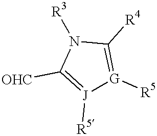
may be readily synthesized using techniques well known in the chemical arts. It will be appreciated by those skilled in the art that other synthetic pathways for forming the compounds of the invention are available and that the following is offered by way of example and not limitation.
A. General synthetic procedure.
The following general methodology may be employed to prepare the compounds of this invention:
The appropriately substituted 2-indolinone (1 equiv.), the appropriately substituted carbonylimidazole (1.2 equiv.) and piperidine (0.1 equiv.) are mixed with ethanol (1-2 ml/mmol 2-indolinone) and the mixture is then heated at 90° C. for 3 to 5 hours. After cooling, the precipitate is filtered, washed with cold ethanol and dried to yield the target compound.
B. Specific syntheses.
The following examples show representative syntheses of several of the 2-indolinones of this invention. These 2-indolinones, as well as the others claimed, will form the claimed compounds by reaction with an appropriately substituted carbonylimidazole using the above procedure.
Preparation of 5-Bromo-2-indolinone
2-indolinone (1.3 g) in 20 mL of acetonitrile was collected to −10° C. and 2.0 g of N-bromosuccinimide was slowly added with stirring. The reaction was stirred for 1 hour at 10° C. and 2 hours at 0°. The precipitate was collected, washed with water and dried to give 1.9 g (90% yield) of the title compound.
Preparation of 5-Nitro-2-indolinone
The 2-indolinone (6.5 g) was dissolved in 25 mL of concentrated sulfuric acid and the mixture maintained at −10-15° C. while 2.1 mL of fuming nitric acid was added dropwise. After the addition of the nitric acid the reaction mixture was stirred at 0° C. for 0.5 hr and poured into ice-water. The precipitate was collected by filtration,. washed with water and crystallized from 50% of acetic acid. The final crystal was then filtered, washed with water and dried under vacuum to give 6.3 g (70%) of 5-nitro-2-indolinone.
Preparation of 5-Carboxyethyl-2-indolinone
Step 1: Synthesis of 5-Chloroacetyl-2-indolinone Aluminum chloride (30.8 g) and 2-oxindole (5.0 g) were added to 200 ml of carbon disulfide at room temperature and the mixture stirred. Choloroacetyl chloride (3.8 mL) was added and the stirring continued for 1 hour. The mixture was heated to reflux for 3 hours, cooled and the solvent decanted. The residue was stirred in ice water until it became a solid suspension. The solid was collected by vacuum filtration, washed with water, and dried to give 7.0 g (90% yield) of the title compound.
Step 2: Synthesis of 5-Chloroethyl-2-indolinone (indolinone-041) 5-Chloroacetyl-2-indolinone (7.0 g) was added to 25 mL of trifluoroacetic acid and the mixture cooled in an ice bath with stirring. Triethylsilane (12.3 mL) was added drop-wise over 2 minutes. The reaction was then stirred at room temperature for 4 hours and poured into ice water. Hexane was added, the mixture stirred vigorously, and the solid collected by vacuum filtration and washed with hexane to give 5.9 g (91% yield) of the product as a white solid.
Step 3: Synthesis of 5-Cyanoethyl-2-indolinone Potassium cyanide (2.02 g) was added to 15 mL of dimethylsulfoxide and heated to 90° C. 5-Chloroethyl-2-indolinone (3.0 g) dissolved in 5 mL of dimethyl sulfoxide was added slowly with stirring, and the reaction heated to 150° C. for 2 hours. The mixture was cooled, poured into ice water and the precipitate collected by vacuum filtration, washed with water, and dried to give crude product. The crude material was chromatographed on silica gel in 5% methanol in chloroform to give 1.2 g (42% yield) of the title compound.
Step 4: Synthesis of 5-Carboxyethyl-2-indolinone 5-Cyaoethyl-2-indolinone (4.02 g) in 10 mL of water containing 25 mL of concentrated hydrochloric acid was refluxed for 4 hours. The mixture was cooled, water added and the resulting solid collected by vacuum filtration, washed with water and dried to give 1.9 g (44% yield) of the title compound as a yellow solid.
Preparation of 5-Aminosulfonyl-2-indolinone
Step 1: Synthesis of 5-chlorosulfonyl-2-indolinone To the 100 mL of flask charged with 27 ml of chlorosulphonic acid was added 2-indolinone slowly. The reaction mixture was maintained between 25-30° C. After addition of 2-indolinone, the reaction mixture was stirred at room temperature for 1.5 hr, then heated to 69° C. for 1 hr, cooled, and poured into ice water. The precipitate was filtered, washed with water and dried in a vacuum oven to give 11.0 g (50%) of 5-chlorosulfonyl-2-indolinone.
Step 2: Synthesis of 5-Aminosulfonyl-2-indolinone. 5-Cholorosulfonyl-2-indolinone (2.1 g) was added to 10 mL of ammonium hydroxide in 10 mL of ethanol and stirred at room temperature overnight. The mixture was concentrated and the solid collected by vacuum filtration to give 0.4 g (20% yield) of the title compound as an off-white solid.
Preparation of 5-(Morpholin-4-yl)sulfonyl)-2-indolinone
To a 100 mL flask charged with 27 mL of chlorosulfonic acid was added slowly 13.3 g of 2-indolinone. The reaction temperature was maintained below 30° C. during the addition of oxindole. After addition of 2-indolinone, the reaction mixture was stirred at room temperature for 1.5 hr, heated to 68° C. for 1 hr, cooled, and poured into water. The precipitate was washed with water and dried in vacuum oven.
5-Chlorosulfonyl-2-indolinone (2.3 g) and 2.2 g morpholine in 50 mL of dichloromethane was stirred for 3 hours at room temperature. The precipitate was collected by vacuum filtration, washed with ethyl acetate and hexane and dried to give 2.09 g (74%) of the title compound as a white solid.
PREPARATION of 3- (2-ETHYLIEDAZOL-4-YL) -2-INDOLINONE by IMMOBILIZED PHASE SYNTHESIS
In addition to the above synthetic approach to the compounds of this invention, the following novel procedure wherein a carbonylimidazole is (1) reversibly bound to a solid substrate which is insoluble in the solvents used in the reaction and is of sufficient size to be manipulated by, without limitation, filtration and other physical procedures, (2) reacted with a 2-indolinone, (3) freed of residual starting materials and non-immobilized byproducts and (4) released from the solid substrate and isolated. The following is an example of this procedure which may be employed to prepare any of the compounds of this invention.
a. Attachment of 2-Ethyl-4-formylimidazole to 2-Chlorotritylchloride Resin
A mixture of 2-chlorotritylchloride resin (2, 100 mg, 0.67 mmol/g loading capacity), N,N-diisopropylethylamine (87 mg, 0.67 mmol), and dichloromethane (1 ml) was gently stirred for 45 min. at ambient temperature. To the stirred mixture was then added 2-ethyl-4-formylimidazole (3, 25 mg, 0.201 mmol). After stirring for 21 h, the resin was filtered and washed with 30 ml each of acetone, water, dichloromethane, methanol, and another portion of acetone. The resin was air dried for 15 min. 
b. Synthesis of Resin-attached Imidazolyl Indolinone
A mixture of 2-ethyl-4-formylimidazole attached to 2-chlorotrityl resin (4, 78 mg, 0.052 mmol based on 0.67 mmol/g loading), DMF (0.9 ml) and piperidine (0.1 ml) was gently stirred for 45 min. at ambient temperature, afterwhich was added 2-indolinone (5, 70 mg, 0.523 mmol). The resultant mixture was gently stirred and heated at 80° C. for 20 h. The resin was filtered and washed with 30 ml each of acetone, water, dichloromethane, methanol, and another portion of acetone. Finally, the resin was air dried for 20 min. 
c. Cleavage of Resin-attached Imidazolyl Indolinone
A mixture of resin-attached imidazolyl indolinone (6, 64 mg, 0.043 mmol based on 0.67 mmol/g loading), dichloromethane (0.9 ml), and trifluoroacetic acid (0.1 ml) was vortexed every 15 min for 2 h at ambient temperature. The mixture was filtered, and the resin was washed with MeOH (1 ml). Concentration of the filtrate provided the TFA salt of the imidazolyl indolinone (7, 12 mg, 0.034 mmol, 79%) as a 10:1 E/Z isomeric mixture. Treatment of the mixture with piperidine (0.1 ml) and EtOH (0.9 ml) at 70° C. for 6 h afforded a 2:1 Z/E isomeric mixture of imidazolyl indolinones (8). 
It will be appreciated that, in any given series of compounds, a spectrum of biological activities will be obtained. In its preferred embodiments, this invention relates to novel imidazoyl 2-indolinones demonstrating the ability to modulate RTK, CTK, and STK activity. The following assays are employed to select those compounds demonstrating the optimal degree of the desired activity.
A. Description of the tables.
Table 1 shows the results of biological assays of representative imidazoyl 2-indolinones of this invention. IC50 generally refers to that amount of a compound needed to effect a 50% change in the activity of a PTK with respect to a control in which no compound is present. With regard to the tests in the table, the 50% change being evaluated is a 50% inhibition of PTK activity by a compound of this invention over the activity of a control in the absence of any compound. The CDK2 assay is described in detail below.
| TABLE 1 | |||
| Corresponding | |||
| Kinase Inhibitory | |||
| Compounds | Activity IC50 (:9) | ||
| 1 | <0.78 | ||
| 2 | <0.78 | ||
| 3 | <0.78 | ||
| 4 | <0.78 | ||
| 5 | <0.78 | ||
| 6 | 1.1 | ||
| 7 | 2.4 | ||
| 8 | 7.7 | ||
| 9 | 9.3 | ||
 |
 |
 |
 |
 |
 |
 |
 |
 |
B. Assay Procedures.
The following in vitro assays may be used to determine the level of activity and effect of the different compounds of the present invention on one or more of the PKs. Similar assays can be designed along the same lines for any PK using techniques well known in the art.
The cellular/catalytic assays described herein are performed in an ELISA format. The general procedure is a follows: a compound is introduced to cells expressing the test kinase, either naturally or recombinantly, for some period of time after which, if the test kinase is a receptor, a ligand known to activate the receptor is added. The cells are lysed and the lysate is transferred to the wells of an ELISA plate previously coated with a specific antibody recognizing the substrate of the enzymatic phosphorylation reaction. Non-substrate components of the cell lysate are washed away and the amount of phosphorylation on the substrate is detected with an antibody specifically recognizing phosphotyrosine compared with control cells that were not contacted with a test compound.
The cellular/biologic assays described herein measure the amount of DNA made in response to activation of a test kinase, which is a general measure of a proliferative response. The general procedure for this assay is as follows: a compound is introduced to cells expressing the test kinase, either naturally or recombinantly, for some period of time after which, if the test kinase is a receptor, a ligand known to activate the receptor is added. After incubation at least overnight, a DNA labeling reagent such as Bromodeoxy-uridine (BrdU) or 3H-thymidine is added. The amount of labeled DNA is detected with either an anti-BrdU antibody or by measuring radioactivity and is compared to control cells not contacted with a test compound.
Enzyme linked immunosorbent assays (ELISA) may be used to detect and measure the presence of PK activity. The ELISA may be conducted according to known protocols which are described in, for example, Voller, et al., 1980, “Enzyme-Linked Immunosorbent Assay,” In: Manual of Clinical Immunology, 2d ed., edited by Rose and Friedman, pp 359-371 Am. Soc. Of Microbiology, Washington, D.C.
The disclosed protocol may be adapted for determining activity with respect to a specific PK. For example, the preferred protocols for conducting the ELISA experiments for specific PKs is provided below. Adaptation of these protocols for determining a compound's activity for other members of the RTK family, as well as for CTKs and STKs, is well within the scope of knowledge of those skilled in the art.
a. FLK-1
An ELISA assay was conducted to measure the kinase activity of the FLK-1 receptor and more specifically, the inhibition or activation of TK activity on the FLK-1 receptor. Specifically, the following assay was conducted to measure kinase activity of the FLK-1 receptor in cells genetically engineered to express Flk-1.
Materials and Methods.
Materials. The following reagents and supplies were used:
a. Corning 96-well ELISA plates (Corning Catalog No. 25805-96);
b. Cappel goat anti-rabbit IgG (catalog no. 55641);
c. PBS (Gibco Catalog No. 450-1300EB);
d. TBSW Buffer (50 mM Tris (pH 7.2), 150 mM NaCl and 0.1% Tween-20);
e. Ethanolamine stock (10% ethanolamine (pH 7.0), stored at 4° C.);
f. HNTG buffer (20 mM HEPES buffer (pH 7.5), 150 mM NaCl, 0.2% Triton X-100, and 10% glycerol);
g. EDTA (0.5 M (pH 7.0) as a 100X stock);
h. Sodium orthovanadate (0.5 M as a 100X stock);
i. Sodium pyrophosphate (0.2 M as a 100X stock);
j. NUNC 96 well V bottom polypropylene plates (Applied Scientific Catalog No. AS-72092);
k. NIH3T3 C7#3 Cells (FLK-1 expressing cells);
l. DMEM with 1X high glucose L-Glutamine (catalog No. 11965-050);
m. FBS, Gibco (catalog no. 16000-028);
n. L-glutamine, Gibco (catalog no. 25030-016);
o. VEGF, PeproTech, Inc. (catalog no. 100-20)(kept as 1 μg/100 μl stock in Milli-Q dH2O and stored at −20° C.;
p. Affinity purified anti-FLK-1 antiserum;
q. UB40 monoclonal antibody specific for phosphotyrosine (see, Fendley, et al., 1990, Cancer Research 50:1550-1558);
r. EIA grade Goat anti-mouse IgG-POD (BioRad catalog no. 172-1011);
s. 2,2-azino-bis(3-ethylbenz-thiazoline-6-sulfonic acid (ABTS) solution (100 mM citric acid (anhydrous), 250 mM Na2HPO4 (pH 4.0), 0.5 mg/ml ABTS (Sigma catalog no. A-1888)), solution should be stored in dark at 4° C. until ready for use;
t. H2O2 (30% solution) (Fisher catalog no. H325);
u. ABTS/H2O2 (15 ml ABTS solution, 2 μl H2O2) prepared 5 minutes before use and left at room temperature;
v. 0.2 M HCl stock in H2O;
w. dimethylsulfoxide (100%)(Sigma Catalog No. D-8418); and
y. Trypsin-EDTA (Gibco BRL Catalog No. 25200-049).
Protocol. The following protocol was used for conducting the assay:
1. Coat Corning 96-well ELISA plates with 1.0 μg per well Cappel Anti-rabbit IgG antibody in 0.1 M Na2C03 pH 9.6. Bring final volume to 150 μl per well. Coat plates overnight at 4° C. Plates can be kept up to two weeks when stored at 4° C.
2. Grow cells in Growth media(DMEM, supplemented with 2.0 mM L-Glutamine, 10% FBS) in suitable culture dishes until confluent at 37° C., 5% CO2.
3. Harvest cells by trypsinization and seed in Corning 25850 polystyrene 96-well round bottom cell plates, 25.000 cells/well in 200 μl of growth media.
4. Grow cells at least one day at 37° C., 5% CO2.
5. Wash cells with D-PBS 1X.
6. Add 200 μl/well of starvation media (DMEM, 2.0 mM 1-Glutamine, 0.1% FBS). Incubate overnight at 37° C., 5% CO2.
7. Dilute Compounds 1:20 in polypropylene 96 well plates using starvation media. Dilute dimethylsulfoxide 1:20 for use in control wells.
8. Remove starvation media from 96 well cell culture plates and add 162 μl of fresh starvation media to each well.
9. Add 18 μl of 1:20 diluted Compound dilution (from step 7) to each well plus the 1:20 dimethylsulfoxide dilution to the control wells (± VEGF), for a final dilution of 1:200 after cell stimulation. Final dimethylsulfoxide is 0.5%. Incubate the plate at 37° C., 5% CO2 for two hours.
10. Remove unbound antibody from ELISA plates by inverting plate to remove liquid. Wash 3 times with TBSW+0.5% ethanolamine, pH 7.0. Pat the plate on a paper towel to remove excess liquid and bubbles.
11. Block plates with TBSW+0.5% Ethanolamine, pH 7.0, 150 μl per well. Incubate plate thirty minutes while shaking on a microtiter plate shaker.
12. Wash plate 3 times as described in step 10.
13. Add 0.5 μg/well affinity purified anti-FLU-1 polyclonal rabbit antiserum. Bring final volume to 150 μl/well with TBSW+0.5% ethanolamine pH 7.0. Incubate plate for thirty minutes while shaking.
14. Add 180 μl starvation medium to the cells and stimulate cells with 20 μl/well 10.0 mM sodium ortho vanadate and 500 ng/ml VEGF (resulting in a final concentration of 1.0 mM sodium ortho vanadate and 50 ng/ml VEGF per well) for eight minutes at 37° C., 5% CO2. Negative control wells receive only starvation medium.
15. After eight minutes, media should be removed from the cells and washed one time with 200 μl/well PBS.
16. Lyse cells in 150 μl/well HNTG while shaking at room temperature for five minutes. HNTG formulation includes sodium ortho vanadate, sodium pyrophosphate and EDTA.
17. Wash ELISA plate three times as described in step 10.
18. Transfer cell lysates from the cell plate to ELISA plate and incubate while shaking for two hours. To transfer cell lysate pipette up and down while scrapping the wells.
19. Wash plate three times as described in step 10.
20. Incubate ELISA plate with 0.02 μg/well UB40 in TBSW+05% ethanolamine. Bring final volume to 150 μl/well. Incubate while shaking for 30 minutes.
21. Wash plate three times as described in step 10.
22. Incubate ELISA plate with 1:10,000 diluted EIA grade goat anti-mouse IgG conjugated horseradish peroxidase in TBSW+0.5% ethanolamine, pH 7.0. Bring final volume to 150 μl/well. Incubate while shaking for thirty minutes.
23. Wash plate as described in step 10.
24. Add 100 μl of ABTS/H2O2 solution to well. Incubate ten minutes while shaking.
25. Add 100 μl of 0.2 M HCl for 0.1 M HCl final to stop the color development reaction. Shake 1 minute at room temperature. Remove bubbles with slow stream of air and read the ELISA plate in an ELISA plate reader at 410 nm.
b. HER-2 ELISA
Assay 1: EGF Receptor-HER2 Chimeric Receptor Assay In Whole Cells.
HER2 kinase activity in whole EGFR-NIH3T3 cells was measured as described below:
Materials and Reagents. The following materials and reagents were used to conduct the assay:
a. EGF: stock concentration: 16.5 ILM; EGF 201, TOYOBO, Co., Ltd. Japan.
b. 05-101 (UBI) (a monoclonal antibody recognizing an EGFR extracellular domain).
c. Anti-phosphotyrosine antibody (anti-Ptyr) (polyclonal) (see, Fendley, et al., supra).
d. Detection antibody: Goat anti-rabbit lgG horse radish peroxidase conjugate, TAGO, Inc., Burlingame, Calif.
| e. | TBST buffer: | ||||
| Tris-HCl, pH 7.2 | 50 | mM | |||
| NaCl | 150 | mM | |||
| Triton X-100 | 0.1 | ||||
| f. | HNTG 5X stock: | ||||
| HEPES | 0.1M | ||||
| NaCl | 0.75M | ||||
| Glycerol | 50% | ||||
| Triton X-100 | 1.0% | ||||
| g. | ABTS stock: | ||||
| Citric Acid | 100 | mM | |||
| Na2HPO4 | 250 | mM | |||
| HCl, conc. | 0.5 | pM | |||
| ABTS* | 0.5 | mg/ml | |||
| *(2,2′-azinobis(3-ethylbenzthiazolinesulfonic acid)). Keep solution in dark at 4° C. until use. | |||||
h. Stock reagents of:
EDTA 100 mM pH 7.0
Na3VO4 0.5 M
Na4 (P2O7) 0.2 M
Procedure. The following protocol was used:
A. Pre-coat ELISA Plate
1. Coat ELISA plates (Corning, 96 well, Cat. #25805-96) with 05-101 antibody at 0.5 g per well in PBS, 100 μl final volume/well, and store overnight at 4° C. Coated plates are good for up to 10 days when stored at 4° C.
2. On day of use, remove coating buffer and replace with 100 μl blocking buffer (5% Carnation Instant Non-Fat Dry Milk in PBS). Incubate the plate, shaking, at room temperature (about 23° C. to 25° C.) for 30 minutes. Just prior to use, remove blocking buffer and wash plate 4 times with TBST buffer.
B. Seeding Cells
1. An NIH3T3 cell line overexpressing a chimeric receptor containing the EGFR extracellular domain and intracellular HER2 kinase domain can be used for this assay.
2. Choose dishes having 80-90% confluence for the experiment. Trypsinize cells and stop reaction by adding 10% fetal bovine serum. Suspend cells in DMEM medium (10% CS DMEM medium) and centrifuge once at 1500 rpm, at room temperature for 5 minutes.
3. Resuspend cells in seeding medium (DMEM, 0.5% bovine serum), and count the cells using trypan blue. Viability above 90% is acceptable. Seed cells in DMEM medium (0.5% bovine serum) at a density of 10,000 cells per well, 100 μl per well, in a 96 well microtiter plate. Incubate seeded cells in 5% CO2 at 37° C. for about 40 hours.
C. Assay Procedures
1. Check seeded cells for contamination using an inverted microscope. Dilute drug stock (10 mg/ml in DMSO) 1:10 in DMEM medium, then transfer 5 μl to a TBST well for a final drug dilution of 1:200 and a final DMSO concentration of 1%. Control wells receive DMSO alone. Incubate in 5% CO2 at 37° C. for two hours.
2. Prepare EGF ligand: dilute stock EGF in DMEM so that upon transfer of 10 μl dilute EGF (1:12 dilution), 100 nM final concentration is attained.
3. Prepare fresh HNTG* sufficient for 100 μl per well; and place on ice.
| HNTG* (10 ml): | |||
| HNTG stock | 2.0 ml | ||
| milli-Q H2O | 7.3 ml | ||
| EDTA, 100 mM, pH 7.0 | 0.5 ml | ||
| Na3VO4, 0.5M | 0.1 ml | ||
| Na4(P2O7), 0.2M | 0.1 ml | ||
4. After 120 minutes incubation with drug, add prepared SGF ligand to cells, 10 μl per well, to a final concentration of 100 nM. Control wells receive DMEM alone. Incubate, shaking, at room temperature, for 5 minutes.
5. Remove drug, EGF, and DMEM. Wash cells twice with PBS. Transfer HNTG* to cells, 100 μl per well. Place on ice for 5 minutes. Meanwhile, remove blocking buffer from other ELISA plate and wash with TBST as described above.
6. With a pipette tip securely fitted to a micropipettor, scrape cells from plate and homogenize cell material by repeatedly aspirating and dispensing the HNTG* lysis buffer. Transfer lysate to a coated, blocked, and washed ELISA plate. Incubate shaking at room temperature for one hour.
7. Remove lysate and wash 4 times with TBST. Transfer freshly diluted anti-Ptyr antibody to ELISA plate at 100 μl per well. Incubate shaking at room temperature for 30 minutes in the presence of the anti-Ptyr antiserum (1:3000 dilution in TBST).
8. Remove the anti-Ptyr antibody and wash 4 times with TBST. Transfer the freshly diluted TAGO anti-rabbit IgG antibody to the ELISA plate at 100 μl per well. Incubate shaking at room temperature for 30 minutes (anti-rabbit IgG antibody: 1:3000 dilution in TBST).
9. Remove TAGO detection antibody and wash 4 times with TBST. Transfer freshly prepared ABTS/H2O2 solution to ELISA plate, 100 μl per well. Incubate shaking at room temperature for 20 minutes. (ABTS/H2O2 solution: 1.0 μl 30% H2O2 in 10 ml ABTS stock).
10. Stop reaction by adding 50 μl 5N H2SO4 (optional), and determine O.D. at 410 nm.
11. The maximal phosphotyrosine signal is determined by subtracting the value of the negative controls from the positive controls. The percent inhibition of phosphotyrosine content for extract-containing wells is then calculated, after subtraction of the negative controls.
c. PDGF-R ELISA
All cell culture media, glutamine, and fetal bovine serum were purchased from Gibco Life Technologies (Grand Island, N.Y.) unless otherwise specified. All cells were grown in a humid atmosphere of 90-95% air and 5-10% CO2 at 37° C. All cell lines were routinely subcultured twice a week and were negative for mycoplasma as determined by the Mycotect method (Gibco).
For ELISA assays, cells (U1242, obtained from Joseph Schlessinger, NYU) were grown to 80-90% confluency in growth medium (MEM with 10% FBS, NEAA, 1 mM NaPyr and 2 mM GLN) and seeded in 96-well tissue culture plates in 0.5% serum at 25,000 to 30,000 cells per well. After overnight incubation in 0.5% serum-containing medium, cells were changed to serum-free medium and treated with test compound for 2 hr in a 5% CO2, 37° C. incubator. Cells were then stimulated with ligand for 5-10 minute followed by lysis with HNTG (20 mM Hepes, 150 mM NaCl, 10% glycerol, 5 mM EDTA, 5 mM Na3VO4, 0.2% Triton X-100, and 2 mM NaPyr). Cell lysates (0.5 mg/well in PBS) were transferred to ELISA plates previously coated with receptor-specific antibody and which had been blocked with 5% milk in TBST (50 mM Tris-HCl pH 7.2, 150 mM NaCl and 0.1% Triton X-100) at room temperature for 30 min. Lysates were incubated with shaking for 1 hour at room temperature. The plates were washed with TBST four times and then incubated with polyclonal anti-phosphotyrosine antibody at room temperature for 30 minutes. Excess anti-phosphotyrosine antibody was removed by rinsing the plate with TBST four times. Goat anti-rabbit IgG antibody was added to the ELISA plate for 30 min at room temperature followed by rinsing with TBST four more times. ABTS (100 mM citric acid, 250 mM Na2HPO4 and 0.5 mg/mL 2,2′-azino-bis(3-ethylbenzthiazoline-6-sulfonic acid)) plus H2O2 (1.2 mL 30% H2O2 to 10 ml ABTS) was added to the ELISA plates to start color development. Absorbance at 410 nm with a reference wavelength of 630 nm was recorded about 15 to 30 min after ABTS addition.
d. IGF-I RECEPTOR ELISA
The following protocol may be used to measure phosphotyrosine level on IGF-I receptor, which indicates IGF-I receptor tyrosine kinase activity.
Materials and Reagents. The following materials and reagents were used:
a. The cell line used in this assay is 3T3/IGF-1R, a cell line genetically engineered to overexpresses IGF-1 receptor.
b. NIH3T3/IGF-1R is grown in an incubator with 5% CO2 at 37° C. The growth media is DMEM+10% FBS (heat inactivated)+2 mM L-glutamine.
c. Affinity purified anti-IGF-1R antibody 17-69.
d. D-PBS:
| KH2PO4 | 0.20 g/l | ||
| K2HPO4 | 2.16 g/l | ||
| KCl | 0.20 g/l | ||
| NaCl | 8.00 g/l (pH 7.2) | ||
e. Blocking Buffer: TBST plus 5% Milk (Carnation Instant Non-Fat Dry Milk).
f. TBST buffer:
Tris-HCl 50 mM
NaCl 150 mM (pH 7.2/HCl 10N)
Triton X-100 0.1%
Stock solution of TBS (10X) is prepared, and Triton X-100 is added to the buffer during dilution.
g. HNTG buffer:
| HEPES | 20 mM | ||
| NaCl | 150 mM (pH 7.2/HCl 1N) | ||
| Glycerol | 10% | ||
| Triton X-100 | 0.2% | ||
Stock solution (5X) is prepared and kept at 4° C.
h. EDTA/HCl: 0.5 M pH 7.0 (NaOH) as 100X stock.
i. Na3VO4: 0.5 M as 100X stock and aliquots are kept in −80° C.
j. Na4P2O7: 0.2 M as 100X stock.
k. Insulin-like growth factor-1 from Promega (Cat# G5111).
l. Rabbit polyclonal anti-phosphotyrosine antiserum.
m. Goat anti-rabbit IgG, POD conjugate (detection antibody), Tago (Cat. No. 4520, Lot No. 1802): Tago, Inc., Burlingame, Calif.
n. ABTS (2,2′-azinobis(3-ethylbenzthiazolinesulfonic acid)) solution:
| Citric acid | 100 mM | ||
| Na2HPO4 | 250 mM (pH 4.0/1 N HCl) | ||
| ABTS | 0.5 mg/ml | ||
ABTS solution should be kept in dark and 4° C. The solution should be discarded when it turns green.
o. Hydrogen Peroxide: 30% solution is kept in the dark and at 4° C.
Procedure. All the following steps are conducted at room temperature unless it is specifically indicated. All ELISA plate washings are performed by rinsing the plate with tap water three times, followed by one TBST rinse. Pat plate dry with paper towels.
A. Cell Seeding:
1. The cells, grown in tissue culture dish (Corning 25020-100) to 80-90% confluence, are harvested with Trypsin-EDTA (0.25%, 0.5 ml/D-100, GIBCO).
2. Resuspend the cells in fresh DMEM+10% FBS+2mM L-Glutamine, and transfer to 96-well tissue culture plate (Corning, 25806-96) at 20,000 cells/well (100 μl/well). Incubate for 1 day then replace medium to serum-free medium (90/μl) and incubate in 5% CO2 and 37° C. overnight.
B. ELISA Plate Coating and Blocking:
1. Coat the ELISA plate (Corning 25805-96) with Anti-IGF-1R Antibody at 0.5 μg/well in 100 μl PBS at least 2 hours.
2. Remove the coating solution, and replace with 100 μl Blocking Buffer, and shake for 30 minutes. Remove the blocking buffer and wash the plate just before adding lysate.
C. Assay Procedures:
1. The drugs are tested in serum-free condition.
2. Dilute drug stock (in 100% DMSO) 1:10 with DMEM in 96-well poly-propylene plate, and transfer 10 μl/well of this solution to the cells to achieve final drug dilution 1:100, and final DMSO concentration of 1.0%. Incubate the cells in 5% CO2 at 37° C. for 2 hours.
3. Prepare fresh cell lysis buffer (HNTG*)
| HNTG | 2 | ml | ||
| EDTA | 0.1 | ml | ||
| Na3VO4 | 0.1 | ml | ||
| Na4(P2O7) | 0.1 | ml | ||
| H2O | 7.3 | ml | ||
4. After drug incubation for two hours, transfer 10 μl/well of 200 nM IGF-1 Ligand in PBS to the cells (Final Conc.=20 nM), and incubate at 5% CO2 at 37° C. for 10 minutes.
5. Remove media and add 100 μl/well HNTG* and shake for 10 minutes. Look at cells under microscope to see if they are adequately lysed.
6. Use a 12-channel pipette to scrape the cells from the plate, and homogenize the lysate by repeated aspiration and dispensing. Transfer all the lysate to the antibody coated ELISA plate, and shake for 1 hour.
7. Remove the lysate, wash the plate, transfer anti-pTyr (1:3,000 with TBST) 100 μl/well, and shake for 30 minutes.
8. Remove anti-pTyr, wash the plate, transfer TAGO (1:3,000 with TBST) 100 μl/well, and shake for 30 minutes.
9. Remove detection antibody, wash the plate, and transfer fresh ABTS/H2O2 (1.2 μl H2O2 to 10 ml ABTS) 100 μl/well to the plate to start color development.
10. Measure OD at 410 nm with a reference wavelength of 630 nm in Dynatec MR5000.
e. EGF Receptor ELISA
EGF Receptor kinase activity in cells genetically engineered to express human EGF-R was measured as described below:
Materials and Reagents. The following materials and reagents were used
a. EGF Ligand: stock concentration=16.5 μM; EGF 201, TOYOBO, Co., Ltd. Japan.
b. 05-101 (UBI) (a monoclonal antibody recognizing an EGFR extracellular domain).
c. Anti-phosphotyosine antibody (anti-Ptyr) (polyclonal).
d. Detection antibody: Goat anti-rabbit lgG horse radish peroxidase conjugate, TAGO, Inc., Burlingame, Calif.
| e. | TBST buffer: | ||||
| Tris-HCl, pH 7 | 50 | mM | |||
| NaCl | 150 | mM | |||
| Triton X-100 | 0.1 | ||||
| f. | HNTG 5X stock: | ||||
| HEPES | 0.1M | ||||
| NaCl | 0.75M | ||||
| Glycerol | 50 | ||||
| Triton X-100 | 1.0% | ||||
| g. | ABTS stock: | ||||
| Citric Acid | 100 | mM | |||
| Na2HPO4 | 250 | mM | |||
| HCl, conc. | 4.0 | pH | |||
| ABTS* | 0.5 | mg/ml | |||
Keep solution in dark at 4° C. until used.
h. Stock reagents of:
EDTA 100 mM pH 7.0
Na3VO4 0.5 M
Na4 (P207) 0.2 M
Procedure. The following protocol was used:
A. Pre-coat ELISA Plate
1. Coat ELISA plates (Corning, 96 well, Cat. #25805-96) with 05-101 antibody at 0.5 μg per well in PBS, 150 μl final volume/well, and store overnight at 4° C. Coated plates are good for up to 10 days when stored at 4° C.
2. On day of use, remove coating buffer and replace with blocking buffer (5% Carnation Instant NonFat Dry Milk in PBS). Incubate the plate, shaking, at room temperature (about 23° C. to 25° C.) for 30 minutes. Just prior to use, remove blocking buffer and wash plate 4 times with TBST buffer.
B. Seeding Cells
1. NIH 3T3/C7 cell line (Honegger, et al., Cell 51:199-209, 1987) can be use for this assay.
2. Choose dishes having 80-90% confluence for the experiment. Trypsinize cells and stop reaction by adding 10% CS DMEM medium. Suspend cells in DMEM medium (10% CS DMEM medium) and centrifuge once at 1000 rpm at room temperature for 5 minutes.
3. Resuspend cells in seeding medium (DMEM, 0.5% bovine serum), and count the cells using trypan blue. Viability above 90% is acceptable. Seed cells in DMEM medium (0.5% bovine serum) at a density of 10,000 cells per well, 100 μl per well, in a 96 well microtiter plate. Incubate seeded cells in 5% CO2 at 37° C. for about 40 hours.
C. Assay Procedures.
1. Check seeded cells for contamination using an inverted microscope. Dilute drug stock (10 mg/ml in DMSO) 1:10 in DMEM medium, then transfer 5 μl to a test well for a final drug dilution of 1:200 and a final DMSO concentration of 1%. Control wells receive DMSO alone. Incubate in 5% CO2 at 37° C. for one hour.
2. Prepare EGF ligand: dilute stock EGF in DMEM so that upon transfer of 10 μl dilute EGF (1:12 dilution), 25 nM final concentration is attained.
3. Prepare fresh 10 ml HNTG* sufficient for 100 μl per well wherein HNTG* comprises: HNTG stock (2.0 ml), milli-Q H2O (7.3 ml), EDTA, 100 mM, pH 7.0 (0.5 ml), Na3VO4 0.5 M (0.1 ml) and Na4 (P2O7), 0.2 M (0.1 ml).
4. Place on ice.
5. After two hours incubation with drug, add prepared EGF ligand to cells, 10 μl per well, to yield a final concentration of 25 nM. Control wells receive DMEM alone. Incubate, shaking, at room temperature, for 5 minutes.
6. Remove drug, EGF, and DMEM. Wash cells twice with PBS. Transfer HNTG* to cells, 100 μl per well. Place on ice for 5 minutes. Meanwhile, remove blocking buffer from other ELISA plate and wash with TBST as described above.
7. With a pipette tip securely fitted to a micropipettor, scrape cells from plate and homogenize cell material by repeatedly aspirating and dispensing the HNTG* lysis buffer. Transfer lysate to a coated, blocked, and washed ELISA plate. Incubate shaking at room temperature for one hour.
8. Remove lysate and wash 4 times with TBST. Transfer freshly diluted anti-Ptyr antibody to ELISA plate at 100 μl per well. Incubate shaking at room temperature for 30 minutes in the presence of the anti-Ptyr antiserum (1:3000 dilution in TBST).
9. Remove the anti-Ptyr antibody and wash 4 times with TBST. Transfer the freshly diluted TAGO 30 anti-rabbit IgG antibody to the ELISA plate at 100 μl per well. Incubate shaking at room temperature for 30 minutes (anti-rabbit IgG antibody: 1:3000 dilution in TBST).
10. Remove detection antibody and wash 4 times with TBST. Transfer freshly prepared ABTS/H2O2 solution to ELISA plate, 100 μl per well. Incubate at room temperature for 20 minutes. ABTS/H2O2 solution: 1.2 μl 30% H2O2 in 10 ml ABTS stock.
11. Stop reaction by adding 50 μl 5N H2S04 (optional), and determine O.D. at 410 nm.
12. The maximal phosphotyrosine signal is determined by subtracting the value of the negative controls from the positive controls. The percent inhibition of phosphotyrosine content for extract-containing wells is then calculated, after subtraction of the negative controls.
f. Met Autophosphorylation Assay—ELISA
This assay determines Met tyrosine kinase activity by analyzing Met protein tyrosine kinase levels on the Met receptor.
Materials and Reagents:
a. HNTG (5X stock solution): Dissolve 23.83 g HEPES and 43.83 g NaCl in about 350 ml dH2O. Adjust pH to 7.2 with HCl or NaOH, add 500 ml glycerol and 10 ml Triton X-100, mix, add dH2O to 1 L total volume. To make 1 L of 1X working solution add 200 ml 5X stock solution to 800 ml dH2O, check and adjust pH as necessary, store at 4° C.
b. PBS (Dulbecco's Phosphate-Buffered Saline), Gibco Cat. # 450-1300EB (1X solution).
c. Blocking Buffer: in 500 ml dH2O place 100 g BSA, 12.1 g Tris-pH7.5, 58.44 g NaCl and 10 ml Tween-20, dilute to 1 L total volume.
d. Kinase Buffer: To 500 ml dH2O add 12.1 g TRIS pH7.2, 58.4 g NaCl, 40.7 g MgCl2 and 1.9 g EGTA; bring to 1 L total volume with dH2O.
e. PMSF (Phenylmethylsulfonyl fluoride), Sigma Cat. # P-7626, to 435.5 mg, add 100% ethanol to 25 ml total volume, vortex.
f. ATP (Bacterial Source), Sigma Cat. # A-7699, store powder at −20° C.; to make up solution for use, dissolve 3.31 mg in 1 ml dH2O.
g. RC-20H HRPO Conjugated Anti-Phosphotyrosine, Transduction Laboratories Cat. # E120H.
h. Pierce 1-Step (TM) Turbo TMB-ELISA (3,3′,5,5′-tetramethylbenzidine, Pierce Cat. # 34022.
i. H2SO4, add 1 ml conc. (18N) to 35 ml dH2O.
j. TRIS HCL, Fischer Cat. # BP152-5; to 121.14 g of material, add 600 ml MilliQ H2O, adjust pH to 7.5 (or 7.2) with HCl, bring volume to 1 L with MilliQ H2O.
k. NaCl, Fischer Cat. # S271-10, make up 5M solution.
l. Tween-20, Fischer Cat. # S337-500.
m. Na3VO4, Fischer Cat. # S454-50, to 1.8 g material add 80 ml MilliQ H2O, adjust pH to 10.0 with HCl or NaOH, boil in microwave, cool, check pH, repeat procedure until pH stable at 10.0, add MilliQ H2O to 100 ml total volume, make 1 ml aliquots and store at −80° C.
n. MgCl2, Fischer Cat. # M33-500, make up 1M solution.
o. HEPES, Fischer Cat. # BP310-500, to 200 ml MilliQ H2O, add 59.6 g material, adjust pH to 7.5, bring volume to 250 ml total, sterile filter.
p. Albumin, Bovine (BSA), Sigma Cat. # A-4503, to 30 grams material add sterile distilled water to make total volume of 300 ml, store at 4° C.
q. TBST Buffer: to approx. 900 ml dH2O in a 1 L graduated cylinder add 6.057 g TRIS and 8.766 g NaCl, when dissolved, adjust pH to 7.2 with HCl, add 1.0 ml Triton X-100 and bring to 1 L total volume with dH2O.
r. Goat Affinity purified antibody Rabbit IgG (whole molecule), Cappel Cat. # 55641.
s. Anti h-Met (C-28) rabbit polyclonal IgG antibody, Santa Cruz Chemical Cat. # SC-161.
t. Transiently Transfected EGFR/Met chimeric cells (EMR) (Komada, et al., Oncogene, 8:2381-2390 (1993).
u. Sodium Carbonate Buffer, (Na2CO4, Fischer Cat. # S495): to 10.6 g material add 800 ml MilliQ H2O, when dissolved adjust pH to 9.6 with NaOH, bring up to 1 L total volume with MilliQ H2O, filter, store at 4° C.
Procedure:
All of the following steps are conducted at room temperature unless it is specifically indicated otherwise. All ELISA plate washing is by rinsing 4X with TBST.
A. EMR Lysis
This procedure can be performed the night before or immediately prior to the start of receptor capture.
1. Quick thaw lysates in a 37° C. waterbath with a swirling motion until the last crystals disappear.
2. Lyse cell pellet with 1X HNTG containing 1 mM PMSF. Use 3 ml of HNTG per 15 cm dish of cells. Add ½ the calculated HNTG volume, vortex the tube for 1 min., add the remaining amount of HNTG, vortex for another min.
3. Balance tubes, centrifuge at 10,000×g for 10 min at 4° C.
4. Pool supernatants, remove an aliquot for protein determination.
5. Quick freeze pooled sample in dry ice/ethanol bath. This step is performed regardless of whether lysate will be stored overnight or used immediately following protein determination.
6. Perform protein determination using standard bicinchoninic acid (BCA) method (BCA Assay Reagent Kit from Pierce Chemical Cat. # 23225).
B. ELISA Procedure
1. Coat Corning 96 well ELISA plates with 5 μg per well Goat anti-Rabbit antibody in Carbonate Buffer for a total well volume of 50 μl. Store overnight at 4° C.
2. Remove unbound Goat anti-rabbit antibody by inverting plate to remove liquid.
3. Add 150 μl of Blocking Buffer to each well. Incubate for 30 min. at room temperature with shaking.
4. Wash 4X with TBST. Pat plate on a paper towel to remove excess liquid and bubbles.
5. Add 1 μg per well of Rabbit anti-Met antibody diluted in TBST for a total well volume of 100 μl.
6. Dilute lysate in HNTG (90 μg lysate/100 μl)
7. Add 100 μl of diluted lysate to each well. Shake at room temperature for 60 min.
8. Wash 4X with TBST. Pat on paper towel to remove excess liquid and bubbles.
9. Add 50 μl of 1X lysate buffer per well.
10. Dilute compounds/extracts 1:10 in 1X Kinase Buffer in a polypropylene 96 well plate.
11. Transfer 5.5 μl of diluted drug to ELISA plate wells. Incubate at room temperature with shaking for 20 min.
12. Add 5.5 μl of 60 μM ATP solution per well. Negative controls do not receive any ATP. Incubate at room temperature for 90 min., with shaking.
13. Wash 4X with TBST. Pat plate on paper towel to remove excess liquid and bubbles.
14. Add 100 μl per well of RC20 (1:3000 dilution in Blocking Buffer). Incubate 30 min. at room temperature with shaking.
15. Wash 4X with TBST. Pat plate on paper towel to remove excess liquid and bubbles.
16. Add 100 μl per well of Turbo-TMB. Incubate with shaking for 30-60 min.
17. Add 100 μl per well of 1M H2SO4 to stop reaction.
18. Read assay on Dynatech MR7000 ELISA reader. Test Filter=450 nm, reference filter=410 nm.
g. Biochemical arc assay—ELISA
This assay is used to determine src protein kinase activity measuring phosphorylation of a biotinylated peptide as the readout.
Materials and Reagents:
a. Yeast transformed with src from Courtneidge Laboratory (Sugen, Inc., Redwood City, Calif.).
b. Cell lysates: Yeast cells expressing src are pelleted, washed once with water, re-pelleted and stored at −80° C. until use.
c. N-terminus biotinylated EEEYEEYEEEYEEEYEEEY is prepared by standard procedures well known to those skilled in the art.
d. DMSO: Sigma, St. Louis, Mo.
e. 96 Well ELISA Plate: Corning 96 Well Easy Wash, Modified flat Bottom Plate, Corning Cat. #25805-96.
f. NUNC 96-well V-bottom polypropylene plates for dilution of compounds: Applied Scientific Cat. # A-72092.
g. Vecastain ELITE ABC reagent: Vector, Burlingame, Calif.
h. Anti-src (327) mab: Schizosaccharomyces Pombe was used to express recombinant Src (Superti-Furga, et al., EMBO J., 25 12:2625-2634; Superti-Furga, et al., Nature Biochem., 14:600-605). S. Pombe strain SP200 (h-s leul.32 ura4 ade210) was grown as described and transformations were pRSP expression plasmids were done by the lithium acetate method (Superti-Furga, supra). Cells were grown in the presence of 1 μM thiamine to repress expression from the nmtl promoter or in the absence of thiamine to induce expression.
i. Monoclonal anti-phosphotyrosine, UBI 05-321 (UB40 may be used instead).
j. Turbo TMB-ELISA peroxidase substrate: Pierce Chemical.
Buffer Solutions:
a. PBS (Dulbecco's Phosphate-Buffered Saline): GIBCO PBS, GIBCO Cat. # 450-1300EB.
b. Blocking Buffer: 5% Non-fat milk (Carnation) in PBS.
c. Carbonate Buffer: Na2CO4 from Fischer, Cat. # S495, make up 100 mM stock solution.
d. Kinase Buffer: 1.0 ml (from 1M stock solution) MgCl2; 0.2 ml (from a 1M stock solution) MnCl2; 0.2 ml (from a 1M stock solution) DTT; 5.0 ml (from a 1M stock solution) HEPES; 0.1 ml TX-100; bring to 10 ml total volume with MilliQ H2O.
e. Lysis Buffer: 5.0 HEPES (from 1M stock solution.); 2.74 ml NaCl (from 5M stock solution); 10 ml glycerol; 1.0 ml TX-100; 0.4 ml EDTA (from a 100 mM stock solution); 1.0 ml PMSF (from a 100 mM stock solution); 0.1 ml Na3VO4 (from a 0.1 M stock solution); bring to 100 ml total volume with MilliQ H2O.
f. ATP: Sigma Cat. # A-7699, make up 10 mM stock solution (5.51 mg/ml).
g. TRIS-HCl: Fischer Cat. # BP 152-5, to 600 ml MilliQ H2O add 121.14 g material, adjust pH to 7.5 with HCl, bring to 1 L total volume with MilliQ H2O.
h. NaCl: Fischer Cat. # S271-10, Make up 5M stock solution with MilliQ H2O.
i. Na3VO4: Fischer Cat. # S454-50; to 80 ml MilliQ H2O, add 1.8 g material; adjust pH to 10.0 with HCl or NaOH; boil in a microwave; cool; check pH, repeat pH adjustment until pH remains stable after heating/cooling cycle; bring to 100 ml total volume with MilliQ H2O; make 1 ml aliquots and store at −80° C.
j. MgCI2: Fischer Cat. # M33-500, make up 1M stock solution with MilliQ H2O.
k. HEPES: Fischer Cat. # BP 310-500; to 200 ml MilliQ H2O, add 59.6 g material, adjust pH to 7.5, bring to 250 ml total volume with MilliQ H2O, sterile filter (1M stock solution).
l. TBST Buffer: TBST Buffer: To 900 ml dH2O add 6.057 g TRIS and 8.766 g NaCl; adjust pH to 7.2 with HCl, add 1.0 ml Triton-X100 ; bring to 1 L total volume with dH2O.
m. MnCl2: Fischer Cat. # M87-100, make up 1M stock solution with MilliQ H2O.
n. DTT; Fischer Cat. # BP172-5.
o. TBS (TRIS Buffered Saline): to 900 ml MilliQ H2O add 6.057 g TRIS and 8.777 g NaCl; bring to 1 L total volume with MilliQ H2O.
p. Kinase Reaction Mixture: Amount per assay plate (100 wells): 1.0 ml Kinase Buffer, 200 μg GST-ζ, bring to final volume of 8.0 ml with MilliQ H2O.
q. Biotin labeled EEEYEEYEEEYEEEYEEEY: Make peptide stock solution (1 mM, 2.98 mg/ml) in water fresh just before use.
r. Vectastain ELITE ABC reagent: To prepare 14 ml of working reagent, add 1 drop of reagent A to 15 ml TBST and invert tube several times to mix. Then add 1 drop of reagent B. Put tube on orbital shaker at room temperature and mix for 30 minutes.
Procedures:
a. Preparation of src coated ELISA plate.
1. Coat ELISA plate with 0.5 μg/well anti-src mab in 100 μl of pH 9.6 sodium carbonate buffer at 4° C. overnight.
2. Wash wells once with PBS.
3. Block plate with 0.15 ml 5% milk in PBS for 30 min. at room temperature.
4. Wash plate 5X with PBS.
5. Add 10 μg/well of src transformed yeast lysates diluted in Lysis Buffer (0.1 ml total volume per well). (Amount of lysate may vary between batches.) Shake plate for 20 minutes at room temperature.
b. Preparation of phosphotyrosine antibody-coated ELISA plate.
1. 4G10 plate: coat 0.5 μg/well 4G10 in 100 μl PBS overnight at 4° C. and block with 150 μl of 5% milk in PBS for 30 minutes at room temperature.
c. Kinase assay procedure.
1. Remove unbound proteins from step 1-7, above, and wash plates 5X with PBS.
2. Add 0.08 ml Kinase Reaction Mixture per well (containing 10 μl of 10X Kinase Buffer and 10 μM (final concentration) biotin-EEEYEEYEEEYEEEYEEEY per well diluted in water.
3. Add 10 μl of compound diluted in water containing 10% DMSO and pre-incubate for 15 minutes at room temperature.
4. Start kinase reaction by adding 10 μl/well of 0.05 mM ATP in water (5 μM ATP final).
5. Shake ELISA plate for 15 min. at room temperature.
6. Stop kinase reaction by adding 10 μl of 0.5 M EDTA per well.
7. Transfer 90 μl supernatant to a blocked 4G10 coated ELISA plate from section B, above.
8. Incubate for 30 min. while shaking at room temperature.
9. Wash plate 5X with TBST.
10. Incubate with Vectastain ELITE ABC reagent (100 μl/well) for 30 min. at room temperature.
11. Wash the wells 5X with TBST.
12. Develop with Turbo TMB.
h. Biochemical lck Assay—ELISA
This assay is used to determine lck protein kinase activities measuring phosphorylation of GST-ζ as the readout.
Materials and Reagents:
a. Yeast transformed with lck. Schizosaccharomyces Pombe was used to express recombinant Lck (Superti-Furga, et al., EMBO J. 12:2625-2634; Superti-Furga, et al., Nature Biotech., 14:600-605). S. Pombe strain SP200 (h-s leul.32 ura4 ade210 ) was grown as described and transformations with pRSP expression plasmids were done by the lithium acetate method (Superti-Furga, supra). Cells were grown in the presence of 1 μM thiamine to induce expression.
b. Cell lysates: Yeast cells expressing lck are pelleted, washed once in water, re-pelleted and stored frozen at −80° C. until use.
c. GST-ζ: DNA encoding for GST-ζ fusion protein for expression in bacteria obtained from Arthur Weiss of the Howard Hughes Medical Institute at the University of California, San Francisco. Transformed bacteria were grown overnight while shaking at 25° C. GST-ζ was purified by glutathione affinity chromatography, Pharmacia, Alameda, Calif.
d. DMSO: Sigma, St. Louis, Mo.
e. 96-Well ELISA plate: Corning 96 Well Easy Wash, Modified Flat Bottom Plate, Corning Cat. #25805-96.
f. NUNC 96-well V-bottom polypropylene plates for dilution of compounds: Applied Scientific Cat. # AS-72092.
g. Purified Rabbit anti-GST antiserum: Amrad Corporation (Australia) Cat. #90001605.
h. Goat anti-Rabbit-IgG-HRP: Amersham Cat. # V010301
i. Sheep ant-mouse IgG (H+L): Jackson Labs Cat. # 5215-005-003.
j. Anti-Lck (3A5) mab: Santa Cruz Biotechnology Cat # sc-433.
k. Monoclonal anti-phosphotyrosine UBI 05-321 (UB40 may be used instead).
Buffer solutions:
a. PBS (Dulbecco's Phosphate-Buffered Saline) 1X solution: GIBCO PBS, GIBCO Cat. # 450-1300EB.
b. Blocking Buffer: 100 g. BSA, 12.1 g. TRIS-pH7.5, 58.44 g NaCl, 10 ml Tween-20, bring up to 1 L total volume with MilliQ H2O.
c. Carbonate Buffer: Na2CO4 from Fischer, Cat. # S495; make up 100 mM solution with MilliQ H2O.
d. Kinase Buffer: 1.0 ml (from 1M stock solution) MgCl2; 0.2 ml (from a 1M stock solution) MnCl2; 0.2 ml (from a 1M stock solution) DTT; 5.0 ml (from a 1M stock solution) HEPES; 0.1 ml TX-100; bring to 10 ml total volume with MilliQ H2O.
e. Lysis Buffer: 5.0 HEPES (from 1M stock solution.); 2.74 ml NaCl (from 5M stock solution); 10 ml glycerol; 1.0 ml TX-100; 0.4 ml EDTA (from a 100 mM stock solution); 1.0 ml PMSF (from a 100 mM stock solution); 0.1 ml Na3VO4 (from a 0.1 M stock solution); bring to 100 ml total volume with MilliQ H2O.
f. ATP: Sigma Cat. # A-7699, make up 10 mM stock solution (5.51 mg/ml).
g TRIS-HCl: Fischer Cat. # BP 152-5, to 600 ml MilliQ H2O add 121.14 g material, adjust pH to 7.5 with HCl, bring to 1 L total volume with MilliQ H2O.
h. NaCl: Fischer Cat. # S271-10, Make up 5M stock solution with MilliQ H2O.
i. Na3VO4: Fischer Cat. # S454-50; to 80 ml MilliQ H2O, add 1.8 g material; adjust pH to 10.0 with HCl or NaOH; boil in a microwave; cool; check pH, repeat pH adjustment until pH remains stable after heating/cooling cycle; bring to 100 ml total volume with MilliQ H2O; make 1 ml aliquots and store at −80° C.
j. MgCl2: Fischer Cat. # M33-500, make up 1M stock solution with MilliQ H2O.
k. HEPES: Fischer Cat. # BP 310-500; to 200 ml MilliQ H2O, add 59.6 g material, adjust pH to 7.5, bring to 250 ml total volume with MilliQ H2O, sterile filter (1M stock solution).
l. Albumin, Bovine (BSA), Sigma Cat. # A4503; to 150 ml MilliQ H2O add 30 g material, bring 300 ml total volume with MilliQ H2O, filter through 0.22 μm filter, store at 4° C.
m. TBST Buffer: To 900 ml dH2O add 6.057 g TRIS and 8.766 g NaCl; adjust pH to 7.2 with HCl, add 1.0 ml Triton-X100; bring to 1 L total volume with dH2O.
n. MnCl2: Fischer Cat. # M87-100, make up 1M stock solution with MilliQ H2O.
o. DTT; Fischer Cat. # BP172-5.
p. TBS (TRIS Buffered Saline): to 900 ml MilliQ H2O add 6.057 g TRIS and 8.777 g NaCl; bring to 1 L total volume with MilliQ H2O.
q Kinase Reaction Mixture: Amount per assay plate (100 wells): 1.0 ml Kinase Buffer, 200 μg GST-ζ, bring to final volume of 8.0 ml with MilliQ H2O.
Procedures:
a. Preparation of Lck coated ELISA plate.
1. Coat 2.0 μg/well Sheep anti-mouse IgG in 100 μl of pH 9.6 sodium carbonate buffer at 4° C. overnight.
2. Wash well once with PBS.
3. Block plate with 0.15 ml of blocking Buffer for 30 min. at room temp.
4. Wash plate 5X with PBS.
5. Add 0.5 μg/well of anti-lck (mab 3A5) in 0.1 ml PBS at room temperature for 1-2 hours.
6. Wash plate 5X with PBS.
7. Add 20 μg/well of lck transformed yeast lysates diluted in Lysis Buffer (0.1 ml total volume per well). (Amount of lysate may vary between batches) Shake plate at 4° C. overnight to prevent loss of activity.
b. Preparation of phosphotyrosine antibody-coated ELISA plate.
1. UB40 plate: 1.0 μg/well UB40 in 100 μl of PBS overnight at 4° C. and block with 150 μl of Blocking Buffer for at least 1 hour.
c. Kinase assay procedure.
1. Remove unbound proteins from step 1-7, above, and wash plates 5X with PBS.
2. Add 0.08 ml Kinase Reaction Mixture per well (containing 10 μl of 10X Kinase Buffer and 2 μg GST-ζ per well diluted with water).
3. Add 10 μl of compound diluted in water containing 10% DMSO and pre-incubate for 15 minutes at room temperature.
4. Start kinase reaction by adding 10 μl/well of 0.1 mM ATP in water (10 μM ATP final).
5. Shake ELISA plate for 60 min. at room temperature.
6. Stop kinase reaction by adding 10 μl of 0.5 M EDTA per well.
7. Transfer 90 μl supernatant to a blocked 4G10 coated ELISA plate from section B, above.
8. Incubate while shaking for 30 min. at room temperature.
9. Wash plate 5X with TBST.
10. Incubate with Rabbit anti-GST antibody at 1:5000 dilution in 100 μl TBST for 30 min. at room temperature.
11. Wash the wells 5X with TBST.
12. Incubate with Goat anti-Rabbit-IgG-HRP at 1:20,000 dilution in 100 μl of TBST for 30 min. at room temperature.
13. Wash the wells 5X with TBST.
14. Develop with Turbo TMB.
i. ASSAY MEASURING PHOSPEORYLATING FUNCTION OF RAF
The following assay reports the amount of RAF-catalyzed phosphorylation of its target protein MEK as well as MEK's target MAPK. The RAF gene sequence is described in Bonner et al., 1985, Molec. Cell. Biol. 5: 1400-1407, and is readily accessible in multiple gene sequence data banks. Construction of the nucleic acid vector and cell lines utilized for this portion of the invention are fully described in Morrison et al., 1988, Proc. Natl. Acad. Sci. USA 85: 8855-8859.
Materials and Reagents
1. Sf9 (Spodoptera frugiperda) cells; GIBCO-BRL, Gaithersburg, Md.
2. RIPA buffer: 20 mM Tris/HCl pH 7.4, 137 mM NaCl, 10% glycerol, 1 mM PMSF, 5 mg/L Aprotenin, 0.5% Triton X-100;
3. Thioredoxin-MEK fusion protein (T-MEK): T-MEK expression and purification by affinity chromatography were performed according to the manufacturer's procedures. Catalog# K 350-01 and R 350-40, Invitrogen Corp., San Diego, Calif.
4. His-MAPK (ERK 2); His-tagged MAPK was expressed in XL1 Blue cells transformed with pUC18 vector encoding His-MAPK. His-MAPK was purified by Ni-affinity chromatography. Cat# 27-4949-01, Pharmacia, Alameda, Calif., as described herein.
5. Sheep anti mouse IgG: Jackson laboratories, West Grove, Pa. Catalog, # 515-006-008, Lot# 28563
6. RAF-1 protein kinase specific antibody: URP2653 from UBI.
7. Coating buffer: PBS; phosphate buffered saline, GIBCO-BRL, Gaithersburg, Md.
8. Wash buffer: TBST - 50 mM Tris/HCL pH 7.2, 150 mM NaCl, 0.1% Triton X-100
9. Block buffer: TBST, 0.1% ethanolamine pH 7.4
10. DMSO, Sigma, St. Louis, Mo.
11. Kinase buffer (KB): 20 mM HEPES/HCl pH 7.2, 150 mM NaCl, 0.1% Triton X-100, 1 mM PMSF, 5 mg/L Aprotenin, 75 mM sodium ortho vanadate, 0.5 MM DTT and 10 mM MgCl2.
12. ATP mix: 100 mM MgCl2, 300 mM ATP, 10 mCi 33P ATP (Dupont-NEN)/mL.
13. Stop solution: 1% phosphoric acid; Fisher, Pittsburgh, Pa.
14. Wallac Cellulose Phosphate Filter mats; Wallac, Turku, Finnland.
15. Filter wash solution: 1% phosphoric acid, Fisher, Pittsburgh, Pa.
16. Tomtec plate harvester, Wallac, Turku, Finnland.
17. Wallac beta plate reader # 1205, Wallac, Turku, Finnland.
18. NUNC 96-well V bottom polypropylene plates for compounds Applied Scientific Catalog # AS-72092.
Procedure
All of the following steps were conducted at room temperature unless specifically indicated.
1. ELISA plate coating: ELISA wells are coated with 100 ml of Sheep anti mouse affinity purified antiserum (1 mg/100 mL coating buffer) over night at 4° C. ELISA plates can be used for two weeks when stored at 4° C.
2. Invert the plate and remove liquid. Add 100 mL of blocking solution and incubate for 30 min.
3. Remove blocking solution and wash four times with wash buffer. Pat the plate on a paper towel to remove excess liquid.
4. Add 1 mg of antibody specific for RAF-1 to each well and incubate for 1 hour. Wash as described in step 3.
5. Thaw lysates from RAS/RAF infected Sf9 cells and dilute with TBST to 10 mg/100 mL. Add 10 mg of diluted lysate to the wells and incubate for 1 hour. Shake the plate during incubation. Negative controls receive no lysate. Lysates from RAS/RAF infected Sf9 insect cells are prepared after cells are infected with recombinant baculoviruses at a MOI of 5 for each virus, and harvested 48 hours later. The cells are washed once with PBS and lysed in RIPA buffer. Insoluble material is removed by centrifugation (5 min at 10 000×g). Aliquots of lysates are frozen in dry ice/ethanol and stored at −80° C. until use.
6. Remove non-bound material and wash as outlined above (step 3).
7. Add 2 mg of T-MEK and 2 mg of His-MAEPK per well and adjust the volume to 40 mL with kinase buffer. Methods for purifying T-MEK and MAPK from cell extracts are provided herein by example.
8. Pre-dilute compounds (stock solution 10 mg/mL DMSO) or extracts 20 fold in TBST plus 1% DMSO. Add 5 mL of the pre-diluted compounds/extracts to the wells described in step 6. Incubate for 20 min. Controls receive no drug.
9. Start the kinase reaction by addition of 5 mL ATP mix; Shake the plates on an ELISA plate shaker during incubation.
10. Stop the kinase reaction after 60 min by addition of 30 mL stop solution to each well.
11. Place the phosphocellulose mat and the ELISA plate in the Tomtec plate harvester. Harvest and wash the filter with the filter wash solution according to the manufacturers recommendation. Dry the filter mats. Seal the filter mats and place them in the holder. Insert the holder into radioactive detection apparatus and quantify the radioactive phosphorous on the filter mats.
Alternatively, 40 mL aliquots from individual wells of the assay plate can be transferred to the corresponding positions on the phosphocellulose filter mat. After air drying the filters, put the filters in a tray. Gently rock the tray, changing the wash solution at 15 min intervals for 1 hour. Air-dry the filter mats. Seal the filter mats and place them in a holder suitable for measuring the radioactive phosphorous in the samples. Insert the holder into a detection device and quantify the radioactive phosphorous on the filter mats.
j. CDK2/Cyclin A—Inhibition Assay
This assay analyzes the protein kinase activity of CDK2 in exogenous substrate.
Materials and Reagents:
A. Buffer A (80 mM Tris (pH 7.2), 40 mM MgCl2): 4.84 G. Tris (F.W.=121.1 g/mol), 4.07 g. MgCl2 (F.W.=203.31 g/mol) dissolved in 500 ml H2O. Adjust pH to 7.2 with HCl.
B. Histone H1 solution (0.45 mg/ml Histone H1 and 20 mM HEPES pH 7.2 (pH 7.4 is OK): 5 mg Histone H1 (Boehinger Mannheim) in 11.111 ml 20 mM HEPES pH 7.2 (477 mg HEPES (F.W.=238.3 g/mol) dissolved in 100 ml ddH2O, stored in 1 ml aliquots at −80° C.
C. ATP solution (60 μM ATP, 300 μg/ml BSA, 3 mM DTT): 120 μl 10 mM ATP, 600 μl 10 mg/ml BSA to 20 ml, stored in 1 ml aliquots at −80° C.
D. CDK2 solution: cdk2/cyclin A in 10 mM HEPES pH 7.2, 25 mM NaCl, o.5 mM DTT, 10% glycerol, stored in 9 μl aliquots at −80° C.
Description of Assay:
1. Prepare solutions of inhibitors at three times the desired final assay concentration in ddH2O/15% DMSO by volume.
2. Dispense 20 μl of inhibitors to wells of polypropylene 96-well plates (or 20 μl 15% DMSO for positive and negative controls).
3. Thaw Histone H1 solution (1 ml/plate), ATP solution (1 ml/plate plus 1 aliquot for negative control), and CDK2 solution (9 μl/plate). Keep CDK2 on ice until use. Aliquot CDK2 solution appropriately to avoid repeated freeze-thaw cycles.
4. Dilute 9 μl CDK2 solution into 2.1 ml Buffer A (per plate). Mix. Dispense 20 μl into each well.
5. Mix 1 ml Histone H1 solution with 1 ml ATP solution (per plate) into a 10 ml screw cap tube. Add γ33P ATP to a concentration of 0.15 μCi/20μl (0.15 μCi/well in assay). Mix carefully to avoid BSA frothing. Add 20 μl to appropriate wells. Mix plates on plate shaker. For negative control, mix ATP solution with an equal amount of 20 mM HEPES pH 7.2 and add γ33P ATP to a concentration of 0.15 μCi/20 μl solution. Add 20 μl to appropriate wells.
6. Let reactions proceed for 60 minutes.
7. Add 35 μl 10% TCA to each well. Mix plates on plate shaker.
8. Spot 40 μl of each sample onto P30 filter mat squares. Allow mats to dry (approx. 10-20 minutes).
9. Wash filter mats 4 X 10 minutes with 250 ml 1% phosphoric acid (10 ml phosphoric acid per liter dd2O).
10. Count filter mats with beta plate reader.
2. Cellular/Biologic Assays
Assay 1: PDGF-Induced BrdU Incorporation Assay
Materials and Reagents:
(1) PDGF: human PDGF B/B; 1276-956, Boehringer Mannheim, Germany
(2) BrdU Labeling Reagent: 10 mM, in PBS (pH7.4), Cat. No. 1 647 229, Boehringer Mannheim, Germany.
(3) FixDenat: fixation solution (ready to use), Cat. No. 1 647 229, Boehringer Mannheim, Germany.
(4) Anti-BrdU-POD: mouse monoclonal antibody conjugated with peroxidase, Cat. No. 1 647 229, Boehringer Mannheim, Germany.
(5) TMB Substrate Solution: tetramethylbenzidine (TMB), ready to use, Cat. No. 1 647 229, Boehringer Mannheim, Germany.
(6) PBS Washing Solution : 1X PBS, pH 7.4, made in house (Sugen, Inc., Redwood City, Calif.).
(7) Albumin, Bovine (BSA): fraction V powder; A-8551, Sigma Chemical Co., USA.
(8) 3T3 cell line genetically engineered to express human PDGF-R.
Protocol
(1) Cells are seeded at 8000 cells/well in DMEM, 10% CS, 2 mM Gln in a 96 well plate. Cells are incubated overnight at 37° C. in 5% CO2.
(2) After 24 hours, the cells are washed with PBS, and then are serum starved in serum free medium (0% CS DMEM with 0.1% BSA) for 24 hours.
(3) On day 3, ligand (PDGF, 3.8 nM, prepared in DMEM with 0.1% BSA) and test compounds are added to the cells simultaneously. The negative control wells receive serum free DMEM with 0.1% BSA only; the positive control cells receive the ligand (PDGF) but no test compound. Test compounds are prepared in serum free DMEM with ligand in a 96 well plate, and serially diluted for 7 test concentrations.
(4) After 20 hours of ligand activation, diluted BrdU labeling reagent (1:100 in DMEM, 0.1% BSA) is added and the cells are incubated with BrdU (final concentration=10 μM) for 1.5 hours.
(5) After incubation with labeling reagent, the medium is removed by decanting and tapping the inverted plate on a paper towel. FixDenat solution is added (50 μl/well) and the plates are incubated at room temperature for 45 minutes on a plate shaker.
(6) The FixDenat solution is thoroughly removed by decanting and tapping the inverted plate on a paper towel. Milk is added (5% dehydrated milk in PBS, 200 μl/well) as a blocking solution and the plate is incubated for 30 minutes at room temperature on a plate shaker.
(7) The blocking solution is removed by decanting and the wells are washed once with PBS. Anti-BrdU-POD solution (1:100 dilution in PBS, 1% BSA) is added (100 μl/well) and the plate is incubated for 90 minutes at room temperature on a plate shaker.
(8) The antibody conjugate is thoroughly removed by decanting and rinsing the wells 5 times with PBS, and the plate is dried by inverting and tapping on a paper towel.
(9) TMB substrate solution is added (100 μl/well) and incubated for 20 minutes at room temperature on a plate shaker until color development is sufficient for photometric detection.
(10) The absorbance of the samples are measured at 410 nm (in “dual wavelength” mode with a filter reading at 490 nm, as a reference wavelength) on a Dynatech ELISA plate reader.
Assay 2: EGF-Induced BrdU Incorporation Assay
Materials and Reagents
(1) EGF: mouse EGF, 201; Toyobo,Co., Ltd. Japan
(2) BrdU Labeling Reagent: 10 mM, in PBS (pH7.4), Cat. No. 1 647 229, Boehringer Mannheim, Germany.
(3) FixDenat: fixation solution (ready to use), Cat. No. 1 647 229, Boehringer Mannheim, Germany.
(4) Anti-BrdU-POD: mouse monoclonal antibody conjugated with peroxidase, Cat. No. 1 647 229, Boehringer Mannheim, Germany.
(5) TMB Substrate Solution: tetramethylbenzidine (TMB), ready to use, Cat. No. 1 647 229, Boehringer Mannheim, Germany.
(6) PBS Washing Solution : 1X PBS, pH 7.4, made in house (Sugen, Inc., Redwood City, Calif.).
(7) Albumin, Bovine (BSA): fraction V powder; A-8551, Sigma Chemical Co., USA.
(8) 3T3 cell line genetically engineered to express human EGF-R.
Protocol
(1) Cells are seeded at 8000 cells/well in 10% CS, 2 mM Gln in DMEM, in a 96 well plate. Cells are incubated overnight at 37° C. in 5% CO2.
(2) After 24 hours, the cells are washed with PBS, and then are serum starved in serum free medium (0% CS DMEM with 0.1% BSA) for 24 hours.
(3) On day 3, ligand (EGF, 2 nM, prepared in DMEM with 0.1% BSA) and test compounds are added to the cells simultaneously. The negative control wells receive serum free DMEM with 0.1% BSA only; the positive control cells receive the ligand (EGF) but no test compound. Test compounds are prepared in serum free DMEM with ligand in a 96 well plate, and serially diluted for 7 test concentrations.
(4) After 20 hours of ligand activation, diluted BrdU labeling reagent (1:100 in DMEM, 0.1% BSA) is added and the cells are incubated with BrdU (final concentration=10 μM) for 1.5 hours.
(5) After incubation with labeling reagent, the medium is removed by decanting and tapping the inverted plate on a paper towel. FixDenat solution is added (50 μl/well) and the plates are incubated at room temperature for 45 minutes on a plate shaker.
(6) The FixDenat solution is thoroughly removed by decanting and tapping the inverted plate on a paper towel. Milk is added (5% dehydrated milk in PBS, 200 μl/well) as a blocking solution and the plate is incubated for 30 minutes at room temperature on a plate shaker.
(7) The blocking solution is removed by decanting and the wells are washed once with PBS. Anti-BrdU-POD solution (1:100 dilution in PBS, 1% BSA) is added (100 μl/well) and the plate is incubated for 90 minutes at room temperature on a plate shaker.
(8) The antibody conjugate is thoroughly removed by decanting and rinsing the wells 5 times with PBS, and the plate is dried by inverting and tapping on a paper towel.
(9) TMB substrate solution is added (100 μl/well) and incubated for 20 minutes at room temperature on a plate shaker until color development is sufficient for photometric detection.
(10) The absorbance of the samples are measured at 410 nm (in “dual wavelength” mode with a filter reading at 490 nm, as a reference wavelength) on a Dynatech ELISA plate reader.
Assay 3: EGF-Induced Her2-Driven BrdU Incorporation
Materials and Reagents:
(1) EGF: mouse EGF, 201; Toyobo,Co., Ltd. Japan
(2) BrdU Labeling Reagent: 10 mM, in PBS (pH7.4), Cat. No. 1 647 229, Boehringer Mannheim, Germany.
(3) FixDenat: fixation solution (ready to use), Cat. No. 1 647 229, Boehringer Mannheim, Germany.
(4) Anti-BrdU-POD: mouse monoclonal antibody conjugated with peroxidase, Cat. No. 1 647 229, Boehringer Mannheim, Germany.
(5) TMB Substrate Solution: tetramethylbenzidine (TMB), ready to use, Cat. No. 1 647 229, Boehringer Mannheim, Germany.
(6) PBS Washing Solution: 1X PBS, pH 7.4, made in house.
(7) Albumin, Bovine (BSA): fraction V powder; A-8551, Sigma Chemical Co., USA.
(8) 3T3 cell line engineered to express a chimeric receptor having the extra-cellular domain of EGF-R and the intra-cellular domain of Her2.
Protocol:
(1) Cells are seeded at 8000 cells/well in DMEM, 10% CS, 2 mM Gln in a 96- well plate. Cells are incubated overnight at 37° in 5% CO2.
(2) After 24 hours, the cells are washed with PBS, and then are serum starved in serum free medium (0% CS DMEM with 0.1% BSA) for 24 hours.
(3) On day 3, ligand (EGF=2 nM, prepared in DMEM with 0.1% BSA) and test compounds are added to the cells simultaneously. The negative control wells receive serum free DMEM with 0.1% BSA only; the positive control cells receive the ligand (EGF) but no test compound. Test compounds are prepared in serum free DMEM with ligand in a 96 well plate, and serially diluted for 7 test concentrations.
(4) After 20 hours of ligand activation, diluted BrdU labeling reagent (1:100 in DMEM, 0.1% BSA) is added and the cells are incubated with BrdU (final concentration=10 μM) for 1.5 hours.
(5) After incubation with labeling reagent, the medium is removed by decanting and tapping the inverted plate on a paper towel. FixDenat solution is added (50 μl/well) and the plates are incubated at room temperature for 45 minutes on a plate shaker.
(6) The FixDenat solution is thoroughly removed by decanting and tapping the inverted plate on a paper towel. Milk is added (5% dehydrated milk in PBS, 200 μl/well) as a blocking solution and the plate is incubated for 30 minutes at room temperature on a plate shaker.
(7) The blocking solution is removed by decanting and the wells are washed once with PBS. Anti-BrdU-POD solution (1:100 dilution in PBS, 1% BSA) is added (100 μl/well) and the plate is incubated for 90 minutes at room temperature on a plate shaker.
(8) The antibody conjugate is thoroughly removed by decanting and rinsing the wells 5 times with PBS, and the plate is dried by inverting and tapping on a paper towel.
(9) TMB substrate solution is added (100 μl/well) and incubated for 20 minutes at room temperature on a plate shaker until color development is sufficient for photometric detection.
(10) The absorbance of the samples are measured at 410 nm (in “dual wavelength” mode with a filter reading at 490 nm, as a reference wavelength) on a Dynatech ELISA plate reader.
Assay 4: IGF1-Induced BrdU Incorporation Assay
Materials and Reagents:
(1) IGF1 Ligand: human, recombinant; G511, Promega Corp, USA.
(2) BrdU Labeling Reagent: 10 mM, in PBS (pH7.4), Cat. No. 1 647 229, Boehringer Mannheim, Germany.
(3) FixDenat: fixation solution (ready to use), Cat. No. 1 647 229, Boehringer Mannheim, Germany.
(4) Anti-BrdU-POD: mouse monoclonal antibody conjugated with peroxidase, Cat. No. 1 647 229, Boehringer Mannheim, Germany.
(5) TMB Substrate Solution: tetramethylbenzidine (TMB), ready to use, Cat. No. 1 647 229, Boehringer Mannheim, Germany.
(6) PBS Washing Solution: 1X PBS, pH 7.4, made in house (Sugen, Inc., Redwood City, Calif.).
(7) Albumin, Bovine (BSA): fraction V powder; A-8551, Sigma Chemical Co., USA.
(8) 3T3 cell line genetically engineered to express human IGF-1 receptor.
Protocol:
(1) Cells are seeded at 8000 cells/well in DMEM, 10% CS, 2 mM Gln in a 96- well plate. Cells are incubated overnight at 37° C. in 5% CO2.
(2) After 24 hours, the cells are washed with PBS, and then are serum starved in serum free medium (0% CS DMEM with 0.1 BSA) for 24 hours.
(3) On day 3, ligand (IGF1=3.3 nM, prepared in DMEM with 0.1% BSA) and test compounds are added to the cells simultaneously. The negative control wells receive serum free DMEM with 0.1% BSA only; the positive control cells receive the ligand (IGF1) but no test compound. Test compounds are prepared in serum free DMEM with ligand in a 96 well plate, and serially diluted for 7 test concentrations.
(4) After 16 hours of ligand activation, diluted BrdU labeling reagent (1:100 in DMEM, 0.1% BSA) is added and the cells are incubated with BrdU (final concentration=10 μM) for 1.5 hours.
(5) After incubation with labeling reagent, the medium is removed by decanting and tapping the inverted plate on a paper towel. FixDenat solution is added (50 μl/well) and the plates are incubated at room temperature for 45 minutes on a plate shaker.
(6) The FixDenat solution is thoroughly removed by decanting and tapping the inverted plate on a paper towel. Milk is added (5% dehydrated milk in PBS, 200 μl/well) as a blocking solution and the plate is incubated for 30 minutes at room temperature on a plate shaker.
(7) The blocking solution is removed by decanting and the wells are washed once with PBS. Anti-BrdU-POD solution (1:100 dilution in PBS, 1% BSA) is added (100 μl/well) and the plate is incubated for 90 minutes at room temperature on a plate shaker.
(8) The antibody conjugate is thoroughly removed by decanting and rinsing the wells 5 times with PBS, and the plate is dried by inverting and tapping on a paper towel.
(9) TMB substrate solution is added (100 μl/well) and incubated for 20 minutes at room temperature on a plate shaker until color development is sufficient for photometric detection.
(10) The absorbance of the samples are measured at 410 nm (in “dual wavelength” mode with a filter reading at 490 nm, as a reference wavelength) on a Dynatech ELISA plate reader.
g. HUV-EC-C Assay
The following protocol may also be used to measure a compound's activity against PDGF-R, FGF-R, VEGF, aFGF or Flk-1/KDR, all of which are naturally expressed by HUV-EC cells.
DAY 0
1. Wash and trypsinize HUV-EC-C cells (human umbilical vein endothelial cells, (American Type Culture Collection; catalogue no. 1730 CRL). Wash with Dulbecco's phosphate-buffered saline (D-PBS; obtained from Gibco BRL; catalogue no. 14190-029) 2 times at about 1 ml/10 cm2 of tissue culture flask. Trypsinize with 0.05% trypsin-EDTA in non-enzymatic cell dissociation solution (Sigma Chemical Company; catalogue no. C-1544). The 0.05% trypsin was made by diluting 0.25% trypsin/1 mM EDTA (Gibco; catalogue no. 25200-049) in the cell dissociation solution. Trypsinize with about 1 ml/25-30 cm2 of tissue culture flask for about 5 minutes at 37° C. After cells have detached from the flask, add an equal volume of assay medium and transfer to a 50 ml sterile centrifuge tube (Fisher Scientific; catalogue no. 05-539-6).
2. Wash the cells with about 35 ml assay medium in the 50 ml sterile centrifuge tube by adding the assay medium, centrifuge for 10 minutes at approximately 200 g, aspirate the supernatant, and resuspend with 35 ml D-PBS. Repeat the wash two more times with D-PBS, resuspend the cells in about 1 ml assay medium/15 cm2 of tissue culture flask. Assay medium consists of F12K medium (Gibco BRL; catalogue no. 21127-014)+0.5% heat-inactivated fetal bovine serum. Count the cells with a Coulter Counter, Coulter Electronics, Inc.) and add assay medium to the cells to obtain a concentration of 0.8-1.0×10⁵ cells/ml.
3. Add cells to 96-well flat-bottom plates at 100 μl/well or 0.8-1.0×10⁴ cells/well; incubate ˜24 h at 37° C., 5% CO2.
DAY 1
1. Make up two-fold drug titrations in separate 96-well plates, generally 50 μM on down to 0 μM. Use the same assay medium as mentioned in day 0, step 2 above. Titrations are made by adding 90 μl/well of drug at 200 μM (4X the final well concentration) to the top well of a particular plate column.
Since the stock drug concentration is usually 20 mM in DMSO, the 200 μM drug concentration contains 2% DMSO.
Therefore, diluent made up to 2% DMSO in assay medium (F12K+0.5% fetal bovine serum) is used as diluent for the drug titrations in order to dilute the drug but keep the DMSO concentration constant. Add this diluent to the remaining wells in the column at 60 μl/well. Take 60 μl from the 120 μl of 200 μM drug dilution in the top well of the column and mix with the 60 μl in the second well of the column. Take 60 μl from this well and mix with the 60 μl in the third well of the column, and so on until two-fold titrations are completed. When the next-to-the-last well is mixed, take 60 μl of the 120 μl in this well and discard it. Leave the last well with 60 μl of DMSO/media diluent as a non-drug-containing control. Make 9 columns of titrated drug, enough for triplicate wells each for 1) VEGF (obtained from Pepro Tech Inc., catalogue no. 100-200, 2) endothelial cell growth factor (ECGF) (also known as acidic fibroblast growth factor, or aFGF) (obtained from Boehringer Mannheim Biochemica, catalogue no. 1439 600); or, 3) human PDGF B/B (1276-956, Boehringer Mannheim, Germany) and assay media control. ECGF comes as a preparation with sodium heparin.
2. Transfer 50 μl/well of the drug dilutions to the 96-well assay plates containing the 0.8-1.0×10⁴ cells/100 μl/well of the HUV-EC-C cells from day 0 and incubate ˜2 h at 37° C., 5% Co2.
3. In triplicate, add 50 μl/well of 80 μg/ml VEGF, 20 ng/ml ECGF, or media control to each drug condition. As with the drugs, the growth factor concentrations are 4X the desired final concentration. Use the assay media from day 0 step 2 to make the concentrations of growth factors. Incubate approximately 24 hours at 37° C., 5% CO2. Each well will have 50 μl drug dilution, 50 μl growth factor or media, and 100 ul cells,=200 ul/well total. Thus the 4X concentrations of drugs and growth factors become 1X once everything has been added to the wells.
DAY 2
1. Add 3H-thymidine (Amersham; catalogue no. TRK-686) at 1 μCi/well (10 μl/well of 100 μCi/ml solution made up in RPMI media+10% heat-inactivated fetal bovine serum) and incubate ˜24 h at 37° C., 5% CO2. Note: 3H-thymidine is made up in RPMI media because all of the other applications for which we use the 3H-thymidine involve experiments done in RPMI. The media difference at this step is probably not significant. RPMI was obtained from Gibco BRL, catalogue no. 11875-051.
DAY 3
1. Freeze plates overnight at −20° C.
DAY 4
1. Thaw plates and harvest with a 96-well plate harvester (Tomtec Harvester 96(®)) onto filter mats (Wallac; catalogue no. 1205-401); read counts on a Wallac Betaplate(™) liquid scintillation counter.
C. In Vivo Animal Models
1. Xenograft Animal Models
The ability of human tumors to grow as xenografts in athymic mice (e.g., Balb/c, nu/nu) provides a useful in vivo model for studying the biological response to therapies for human tumors. Since the first successful xenotransplantation of human tumors into athymic mice, (Rygaard and Povlsen, 1969, Acta Pathol. Microbial. Scand. 77:758-760), many different human tumor cell lines (e.g., mammary, lung, genitourinary, gastro-intestinal, head and neck, glioblastoma, bone, and malignant melanomas) have been transplanted and successfully grown in nude mice. The following assays may be used to determine the level of activity, specificity and effect of the different compounds of the present invention. Three general types of assays are useful for evaluating compounds: cellular/catalytic, cellular/biological and in vivo. The object of the cellular/catalytic assays is to determine the effect of a compound on the ability of a TK to phosphorylate tyrosines on a known substrate in a cell. The object of the cellular/biological assays is to determine the effect of a compound on the biological response stimulated by a TK in a cell. The object of the in vivo assays is to determine the effect of a compound in an animal model of a particular disorder such as cancer.
Suitable cell lines for subcutaneous xenograft experiments include C6 cells (glioma, ATCC # CCL 107), A375 cells (melanoma, ATCC # CRL 1619), A431 cells (epidermoid carcinoma, ATCC # CRL 1555), Calu 6 cells (lung, ATCC # HTB 56), PC3 cells (prostate, ATCC # CRL 1435) and NIH 3T3 fibroblasts genetically engineered to overexpress EGFR, PDGFR, IGF-1R or any other test kinase. The following protocol can be used to perform xenograft experiments:
Female athymic mice (BALB/c, nu/nu) are obtained from Simonsen Laboratories (Gilroy, Calif.). All animals are maintained under clean-room conditions in Micro-isolator cages with Alpha-dri bedding. They receive sterile rodent chow and water ad libitum.
Cell lines are grown in appropriate medium (for example, MEM, DMEM, Ham's F10, or Ham's F12 plus 5%-10% fetal bovine serum (FBS) and 2 mM glutamine (GLN)). All cell culture media, glutamine, and fetal bovine serum are purchased from Gibco Life Technologies (Grand Island, N.Y.) unless otherwise specified. All cells are grown in a humid atmosphere of 90-95% air and 5-10% CO2 at 37° C. All cell lines are routinely subcultured twice a week and are negative for mycoplasma as determined by the Mycotect method (Gibco).
Cells are harvested at or near confluency with 0.05% Trypsin-EDTA and pelleted at 450×g for 10 min. Pellets are resuspended in sterile PBS or media (without FBS) to a particular concentration and the cells are implanted into the hindflank of the mice (8-10 mice per group, 2-10×106 cells/animal). Tumor growth is measured over 3 to 6 weeks using venier calipers. Tumor volumes are calculated as a product of length x width x height unless otherwise indicated. P values are calculated using the Students t-test. Test compounds in 50-100 μL excipient (DMSO, or VPD:D5W) was delivered by IP injection at different concentrations generally starting at day one after implantation.
2. Tumor Invasion Model
The following tumor invasion model has been developed and maybe used for the evaluation of therapeutic value and efficacy of the compounds identified to selectively inhibit KDR/FLK-1 receptor.
Procedure
8 week old nude mice (female) (Simonsen Inc.) were used as experimental animals. Implantation of tumor cells was performed in a laminar flow hood. For anesthesia, Xylazine/Ketamine Cocktail (100 mg/kg ketamine and 5 mg/kg Xylazine) are administered intraperitoneally. A midline incision is done to expose the abdominal cavity (approximately 1.5 cm in length) to inject 107 tumor cells in a volume of 100 μl medium. The cells are injected either into the duodenal lobe of the pancreas or under the serosa of the colon. The peritoneum and muscles are closed with a 6-0 silk continuous suture and the skin was closed by using wound clips. Animals were observed daily.
Analysis
After 2-6 weeks, depending on gross observations of the animals, the mice are sacrificed, and the local tumor metastases, to various organs (lung, liver, brain, stomach, spleen, heart, muscle) are excised and analyzed (measurements of tumor size, grade of invasion, immunochemistry, and in situ hybridization).
D. Measurement of Cell Toxicity
Therapeutic compounds should be more potent in inhibiting protein kinase activity than in exerting a cytotoxic effect. A measure of the effectiveness and cell toxicity of a compound can be obtained by determining the therapeutic index: IC50/LD50. IC50, the dose required to achieve 50% inhibition, can be measured using standard techniques such as those described herein. LD50, the dosage which results in 50% toxicity, can also be measured by standard techniques (Mossman, 1983, J. Immunol. Methods, 65:55-63), by measuring the amount of LDH released (Korzeniewski and Callewaert, 1983, J. Immunol. Methods, 64:313; Decker and Lohmann-Matthes, 1988, J. Immunol. Methods, 115:61), or by measuring the lethal dose in animal models. Compounds with a large therapeutic index are preferred. The therapeutic index should be greater than 2, preferably at least 10, more preferably at least 50.
Thus, it will be appreciated that the compounds, methods and pharmacological compositions of the present invention are effective in modulating PK activity and therefore are expected to be effective as therapeutic agents against RTK-, CTK-, and STK-related disorders.
Although certain embodiments and examples have been used to describe the present invention, it will be apparent to those skilled in the art that changes to the embodiments and examples shown may be made without departing from the scope and spirit of the invention.
Other embodiments are within the following claims.
Claims (29)
1. A 2-indolinone having the chemical structure: 
or a physiologically acceptable salt thereof, wherein:
G and J are selected from the group consisting of nitrogen and carbon such that, when G is nitrogen, J is carbon and when J is nitrogen, G is carbon, it being recognized that, when G or J is nitrogen, R5 or R5′, respectively, does not exist;
R1, R2 and R3 are each hydrogen;
R4, R5 and R5′ are independently selected from the group consisting of hydrogen, alkyl, alkenyl, alkynyl, cycloalkyl, aryl, heteroawyl, heteroalicylic, halo, hydroxy, nitro, cyano, alkoxy, aryloxy, —C(O)OR″, —OC(O)R″, —C(O)R″, S-sulfonamido, amino and NR10R11 wherein
R10 and R11 are independently selected from the group consisting of alkyl, cycloalkyl, aryl, —C(O)R″, —S(O)2R″, tri-halomethanesulfonyl and, combined, a five-member or a six-member heteroalicyclic ring;
R6, R7, R8 and R9 are independently selected from the group consisting of hydrogen, alkyl, trihaloalkyl, cycloalkyl, alkenyl, alkynyl, aryl, heteroaryl, heteroalicyclic, hydroxy, alkoxy, aryloxy, thiohydroxy, thioalkoxy, thioaryloxy, —S(O)R″, —S(O)2R″, S-sulfonamido, N-sulfonamido, N-trihalomethanesulfonamido, —C(O))OR″, —OC(O)R″, —C(O)R″, cyano, halo, cyanato, isocyanato, thiocyanato, isothiocyanato, O-carbamyl, N-carbamyl, O-thiocarbamyl, N-thiocarbamyl, C-amido, N-amido, ainio and —NR10R11 with R10 and R11 as defined above;
R6 and R7 or R7 and R8 or R8 and R9, combined, may form a five- or six-member aromatic, heteroaromatic, alicyclic or heteroalicyclic ring group; and
R″ is selected from the group consisting of hydrogen, alky, cycloalkyl and heteroaryl bonded through a ring carbon;
with the proviso that the following compounds are excluded:
3[(1-methyl-5-nitro-imidazol-2-yl)methlene]-2-indolinone;
1H-indole-7-acetic acid 3-[(2-butyl-1H-imidazol-4-yl)methylene]-2.3-dihydro-2-oxo ethyl ester; and
1H-indole-7-acetic acid 3-[(2-butyl-1-[1,1-dimethylethoxy)carbonyl]-1H-imidazol-4-yl) methylene]-2,3-dihydro-2-oxo ethyl ester.
2. The compound or salt of claim 1, wherein:
R6, R7, R8 and R9 are independently selected from the group consisting of:
hydrogen;
unsubstituted lower alkyl;
lower alkyl substituted with a group selected from the group consisting of halo, —C(O)OR″ and —NR10R11;
unsubstituted lower alkoxy;
lower alkoxy substituted with a group selected from the group consisting of halo, —C(O)OR″ and NR10R11;
trihalomethyl;
unsubstituted alkenyl;
unsubstituted alkynyl;
unsubstituted aryl;
aryl substituted with one or more groups independently selected from the group consisting of unsubstituted lower alkyl, lower alkyl substituted with one or more halo groups, halo, unsubstituted lower alkoxy, —C(O)OR″, amino, S-sulfonamido or —NR10R11;
unsubstituted heteroalicyclic;
heteroalicyclic substituted with one or more groups independently selected from the group consisting of unsubstituted lower alkyl, lower alkyl substituted with one or more halo groups, aldehyde, unsubstituted lower alkyl carbonyl, hydroxy, unsubstituted alkoxy, alkoxy substituted with one or more halo groups, —C(O)OR″, amino, S-sulfonamido or —NR10R11;
unsubstituted aryloxy;
aryloxy substituted with one or more groups independently selected from the group consisting of unsubstituted lower alkyl, trihalomethyl, halo, hydroxy, amino, S-sulfonamido or —NR10R11;
thiohydroxy;
unsubstituted thioalkoxy;
unsubstituted thioaryloxy;
thioaryloxy substituted with one or more groups independently selected from the group consisting of halo, hydroxy, amino, S-sulfonamido or —NR10R11;
S-sulfonamido;
—C(O)OR″;
—OC(O)R″;
hydroxy;
cyano;
nitro;
halo;
C-amido;
N-amido;
amino; and,
—NR10R11.
3. The compound or salt of claim 1, wherein:
R10 is hydrogen; and
R11 is unsubstituted lower alkyl.
4. The compound or salt of claim 1, wherein:
R4, R5 and R5′ are independently selected from the group consisting of hydrogen, unsubstituted lower alkyl, trihalomethyl, lower alkyl susbstituted on the carbon furthest from the point of attachment to the ring with a —C(O)OR″ group, halo, hydroxy, unsubstituted lower alkoxy, —C(O)OR″, —OC(O)R″, amino, C-amido, N-amido, nitro, amino, S-sulfonamido and —NR10R11.
5. The compound or salt of claim 1, wherein:
G is nitrogen;
J is carbon; and,
R4 and R5′ are hydrogen.
6. The compound or salt of claim 1, wherein:
G is carbon;
J is nitrogen; and,
R4 and R5 are hydrogen.
7. The compound or salt of claim 1, wherein:
R6, R8 and R9 are hydrogen; and,
R7 is S-sulfonamido.
8. The compound or salt of claim 7, wherein:
G is nitrogen; and,
J is carbon.
9. The compound or salt of claim 8, wherein R5′ is selected from the group consisting of unsubstituted lower alkyl and halo.
10. The compound or salt of claim 1, wherein:
J is carbon;
G is nitrogen; and,
R4, R6, R8 and R9 are hydrogen;
R7 is S-sulfonamido; and,
R5′ is selected from the group consisting of aryl and heteroaryl.
11. The compound or salt of claim 1, wherein said compound is selected from the group consisting of 3-[(imidazol4-yl)methylene]-2-indolinone, 3-[(imidazol4-yl)methylene]-5-nitro-2-indolinone, 3-[(imidazol-4-yl)methylene]-5chloro-2-indolinone, 3-[(imidazol-4-yl)methylene]-5-amino-2-indolinone, 3-[(imidazol-4-yl)methylene]-4,6-dimethyl-2-indolinone, 3-[(imidazol-4-yl)methylene]-4-methyl-2-indolinone, 3-[(imidazol-4-yl)methylene]-5,6-dimethoxy-2-indolinone, 3-(imidazol-4-yl)methylene]-5-fluoro-2-indolinone and 3-[(imidazol-4-yl)methylene]-5chloro-7-bromo-2-indolinone.
12. A pharmacological composition comprising:
a compound or salt of any one of claims 1, 7, 8, 9, 10 or 11; and
a physiologically acceptable carrier or excipient.
13. A method for the modulation of the catalytic activity of a protein kinase, comprising contacting said protein kinase with a compound or salt of claim 1.
14. The method of claim 13, wherein said protein kinase comprises a receptor protein tyrosine kinase.
15. The method of claim 14, wherein said receptor protein tyrosine kinase is selected from the group consisting of EGF, HER2, HER3, HER4, IR, IGF-1R, IRR, PDGFRα, PDGFRβ, CSFIR, C-Kit, C-fms, Flk-1R, Flk4, KDR/Flk-1, Flt-1, FGFR-1R, FGFR-2R, FGFR-3R and FGFR-4R.
16. The method of claim 13, wherein said protein kinase comprises a non-receptor protein tyrosine kinase.
17. The method of claim 16, wherein said non-receptor protein tyrosine kinase is selected from the group consisting of Src, Frk, Btk, Csk, Abl, ZAP70, Fes/Fps, Fak, Jak, Ack, Yes, Fyn, Lyn, Lck, Blk, Hck, Fgr and Yrk.
18. The method of claim 13, wherein said protein kinase comprises a serine-threonine protein kinase.
19. The method of claim 18, wherein said serine-threonine protein kinase is selected from the group consisting of CDK2 and Raf.
20. A method for treating a protein kinase related disorder in an organism comprising administering a therapeutically effective amount of a compound or salt of any one of claims 1, 7, 8, 9, 10 or 11 to said organism.
21. The method of claim 20 wherein said protein kinase related disorder is selected from the group consisting of a receptor tyrosine kinase related disorder, a cellular tyrosine kinase related disorder and a serine-threonine kinase related disorder.
22. The method of claim 20 wherein said protein kinase related disorder is selected from the group consisting of an EGFR related disorder, a PDGFR related disorder, an IGFR related disorder, and a flk related disorder.
23. The method of claim 20 wherein said protein kinase related disorder is selected from the group consisting of squamous cell carcinoma, astrocytoma, glioblastoma, lung cancer, bladder cancer, head and neck cancer, melanoma, ovarian cancer, prostate cancer, breast cancer and small-cell lung cancer.
24. The method of claim 20 wherein said protein kinase related disorder is selected from the group consisting of diabetes, autoimmune disorder, hyperproliferation disorder, inflammatory disorder, angiogenesis, restenosis, fibrosis, psoriasis, osteoarthritis and rheumatiod arthritis.
25. The method of claim 20 wherein said organism is a human.
26. A method for synthesizing an imidazoyl-2-indolinone comprising the steps of:
binding a carbonylimidazole to a solid substrate;
reacting said solid substrate-bound carbonylimidazole with a 2-indolinone to form a solid substrate-bound imidazoyl-2-indolinone; and,
releasing said imidazoly-2-indolinone from said solid substrate.
27. The method of claim 26 comprising using a resin as said solid substrate.
28. The method of claim 26 comprising using a 2-chlorotrityl resin as said solid substrate.
29. The method of claim 26 comprising washing said solid substrate-bound imidazoyl-2-indolinone with acetone, water, dichloromethane and methanol.
Priority Applications (3)
| Application Number | Priority Date | Filing Date | Title |
|---|---|---|---|
| US09/293,518 US6316635B1 (en) | 1995-06-07 | 1999-04-15 | 2-indolinone derivatives as modulators of protein kinase activity |
| US09/899,550 US6569868B2 (en) | 1998-04-16 | 2001-07-06 | 2-indolinone derivatives as modulators of protein kinase activity |
| US10/372,341 US20030191128A1 (en) | 1998-04-16 | 2003-02-25 | 2-indolinone derivatives as modulators of protein kinase activity |
Applications Claiming Priority (6)
| Application Number | Priority Date | Filing Date | Title |
|---|---|---|---|
| US08/485,323 US5880141A (en) | 1995-06-07 | 1995-06-07 | Benzylidene-Z-indoline compounds for the treatment of disease |
| US08/655,223 US5792783A (en) | 1995-06-07 | 1996-06-05 | 3-heteroaryl-2-indolinone compounds for the treatment of disease |
| US08/659,191 US5883113A (en) | 1995-06-07 | 1996-06-05 | 3-(4'-Bromobenzylindenyl)-2-indolinone and analogues thereof for the treatment of disease |
| US8205698P | 1998-04-16 | 1998-04-16 | |
| US09/212,494 US6225335B1 (en) | 1995-06-07 | 1998-12-15 | 3-(4′-bromobenzylindenyl)-2-indolinone and analogues thereof for the treatment of disease |
| US09/293,518 US6316635B1 (en) | 1995-06-07 | 1999-04-15 | 2-indolinone derivatives as modulators of protein kinase activity |
Related Parent Applications (1)
| Application Number | Title | Priority Date | Filing Date |
|---|---|---|---|
| US09/212,494 Continuation-In-Part US6225335B1 (en) | 1995-06-07 | 1998-12-15 | 3-(4′-bromobenzylindenyl)-2-indolinone and analogues thereof for the treatment of disease |
Related Child Applications (1)
| Application Number | Title | Priority Date | Filing Date |
|---|---|---|---|
| US09/899,550 Continuation US6569868B2 (en) | 1998-04-16 | 2001-07-06 | 2-indolinone derivatives as modulators of protein kinase activity |
Publications (1)
| Publication Number | Publication Date |
|---|---|
| US6316635B1 true US6316635B1 (en) | 2001-11-13 |
Family
ID=27536286
Family Applications (1)
| Application Number | Title | Priority Date | Filing Date |
|---|---|---|---|
| US09/293,518 Expired - Fee Related US6316635B1 (en) | 1995-06-07 | 1999-04-15 | 2-indolinone derivatives as modulators of protein kinase activity |
Country Status (1)
| Country | Link |
|---|---|
| US (1) | US6316635B1 (en) |
Cited By (29)
| Publication number | Priority date | Publication date | Assignee | Title |
|---|---|---|---|---|
| US6559173B1 (en) | 2001-09-27 | 2003-05-06 | Allergan, Inc. | 3-(heteroarylamino)methylene-1,3-dihydro-2H-indol-2-ones as kinase inhibitors |
| US6569868B2 (en) * | 1998-04-16 | 2003-05-27 | Sugen, Inc. | 2-indolinone derivatives as modulators of protein kinase activity |
| US20030199478A1 (en) * | 2001-09-27 | 2003-10-23 | Andrews Steven W. | 3-(Arylamino)methylene-1, 3-dihydro-2H-indol-2-ones as kinase inhibitors |
| US20030216462A1 (en) * | 2000-05-02 | 2003-11-20 | Sugen, Inc. | (2-Oxindol-3-ylidenyl) acetic acid derivatives and their use as protein kinase inhibitors |
| US20030229229A1 (en) * | 2002-02-15 | 2003-12-11 | Qingwu Jin | Process for preparing indolinone derivatives |
| US6699863B1 (en) | 2002-11-27 | 2004-03-02 | Allergan, Inc. | Kinase inhibitors for the treatment of disease |
| US20040082645A1 (en) * | 2001-03-13 | 2004-04-29 | Cecile Combeau | Compounds derived from oxindoles and their therapeutic application in cancer treatment |
| US20040102509A1 (en) * | 2002-11-27 | 2004-05-27 | Allergan, Inc. | Kinase inhibitors for the treatment of disease |
| US20040106618A1 (en) * | 1997-05-07 | 2004-06-03 | Sugen, Inc. | Bicyclic protein kinase inhibitors |
| US6797825B2 (en) | 2001-12-13 | 2004-09-28 | Abbott Laboratories | Protein kinase inhibitors |
| US20050032871A1 (en) * | 2002-09-03 | 2005-02-10 | Sugen, Inc. | Sulfonylated pyrrole-2-indolinone derivatives as kinase inhibitors |
| US20050090498A1 (en) * | 2001-05-24 | 2005-04-28 | Kiyohiro Samizu | 3-Quinolin-2(1h)-ylideneindolin-2-one derivative |
| US20050234049A1 (en) * | 2003-07-30 | 2005-10-20 | Rajinder Singh | Methods of treating or preventing autoimmune diseases with 2, 4-pyrimidinediamine compounds |
| US6960572B2 (en) | 2001-06-21 | 2005-11-01 | Ariad Pharmaceuticals, Inc. | Indolinones and uses thereof |
| US20060122171A1 (en) * | 2002-11-15 | 2006-06-08 | Wei Xu | Kinase modulators |
| US20060293311A1 (en) * | 2005-06-08 | 2006-12-28 | Rigel Pharmaceuticals, Inc. | Compositions and methods for inhibition of the jak pathway |
| US20070203162A1 (en) * | 2006-02-24 | 2007-08-30 | Rigel Pharmaceuticals, Inc. | Compositions and methods for inhibition of the jak pathway |
| US20070203161A1 (en) * | 2006-02-24 | 2007-08-30 | Rigel Pharmaceuticals, Inc. | Compositions and methods for inhibition of the jak pathway |
| US7435814B2 (en) | 2002-02-01 | 2008-10-14 | Rigel Pharmaceuticals, Inc. | 2,4-Pyrimidinediamine compounds and their uses |
| US20090258864A1 (en) * | 2008-02-15 | 2009-10-15 | Rigel Pharmaceuticals, Inc. | Pyrimidine-2-amine compounds and their use as inhibitors of jak kinases |
| US20090298823A1 (en) * | 2008-04-22 | 2009-12-03 | Portola Pharmaceuticals, Inc. | Inhibitors of protein kinases |
| US20090318407A1 (en) * | 2008-04-16 | 2009-12-24 | Portola Pharmaceuticals, Inc. | Inhibitors of protein kinases |
| US20110028503A1 (en) * | 2009-07-28 | 2011-02-03 | Rigel Pharmaceuticals, Inc. | Compositions and methods for inhibition of the jak pathway |
| WO2012061428A2 (en) | 2010-11-01 | 2012-05-10 | Portola Pharmaceuticals, Inc. | Nicotinamides as jak kinase modulators |
| US20140205537A1 (en) * | 2011-06-24 | 2014-07-24 | Zenobia Therapeutics, Inc. | Lrrk2 inhibitors |
| US8952027B2 (en) | 2008-04-16 | 2015-02-10 | Portola Pharmaceuticals, Inc. | Inhibitors of syk and JAK protein kinases |
| WO2015057736A1 (en) * | 2013-10-14 | 2015-04-23 | Indiana University Research And Technology Corporation | Use of acamprosate to modulate erk 1-2 activation in animal models for fxs and asd and individuals diagnosed with fxs and asd |
| US9359308B2 (en) | 2011-11-23 | 2016-06-07 | Portola Pharmaceuticals, Inc. | Pyrazine kinase inhibitors |
| US9676756B2 (en) | 2012-10-08 | 2017-06-13 | Portola Pharmaceuticals, Inc. | Substituted pyrimidinyl kinase inhibitors |
Citations (29)
| Publication number | Priority date | Publication date | Assignee | Title |
|---|---|---|---|---|
| US4002749A (en) | 1975-08-12 | 1977-01-11 | E. R. Squibb & Sons, Inc. | Substituted indolinones |
| US4053613A (en) | 1975-09-17 | 1977-10-11 | E. R. Squibb & Sons, Inc. | 1,3,thiazolinyl and 1,3 thiazinyl substituted indolinones |
| US4966849A (en) | 1985-09-20 | 1990-10-30 | President And Fellows Of Harvard College | CDNA and genes for human angiogenin (angiogenesis factor) and methods of expression |
| WO1991013055A2 (en) | 1990-02-28 | 1991-09-05 | Farmitalia Carlo Erba S.R.L. | New aryl- and heteroarylethenylene derivatives and process for their preparation |
| WO1991015495A1 (en) | 1990-04-02 | 1991-10-17 | Pfizer Inc. | Benzylphosphonic acid tyrosine kinase inhibitors |
| WO1992007830A2 (en) | 1990-10-29 | 1992-05-14 | Pfizer Inc. | Oxindole peptide antagonists |
| WO1992020642A1 (en) | 1991-05-10 | 1992-11-26 | Rhone-Poulenc Rorer International (Holdings) Inc. | Bis mono-and bicyclic aryl and heteroaryl compounds which inhibit egf and/or pdgf receptor tyrosine kinase |
| WO1992021660A1 (en) | 1991-05-29 | 1992-12-10 | Pfizer, Inc. | Tricyclic polyhydroxylic tyrosine kinase inhibitors |
| US5217999A (en) | 1987-12-24 | 1993-06-08 | Yissum Research Development Company Of The Hebrew University Of Jerusalem | Styryl compounds which inhibit EGF receptor protein tyrosine kinase |
| EP0566226A1 (en) | 1992-01-20 | 1993-10-20 | Zeneca Limited | Quinazoline derivatives |
| WO1994003427A1 (en) | 1992-08-06 | 1994-02-17 | Warner-Lambert Company | 2-thioindoles (selenoindoles) and related disulfides (selenides) which inhibit protein tyrosine kinases and which have antitumor properties |
| US5302606A (en) | 1990-04-16 | 1994-04-12 | Rhone-Poulenc Rorer Pharmaceuticals Inc. | Styryl-substituted pyridyl compounds which inhibit EGF receptor tyrosine kinase |
| WO1994010202A1 (en) | 1992-10-28 | 1994-05-11 | Genentech, Inc. | Vascular endothelial cell growth factor antagonists |
| US5322950A (en) * | 1991-12-05 | 1994-06-21 | Warner-Lambert Company | Imidazole with angiotensin II antagonist properties |
| WO1994014808A1 (en) | 1992-12-23 | 1994-07-07 | Farmitalia Carlo Erba Srl | Vinylene-azaindole derivatives and process for their preparation |
| US5330992A (en) | 1992-10-23 | 1994-07-19 | Sterling Winthrop Inc. | 1-cyclopropyl-4-pyridyl-quinolinones |
| US5389661A (en) * | 1991-12-05 | 1995-02-14 | Warner-Lambert Company | Imidazole and 1,2,4-triazole derivatives with angiotensin II antagonist properties |
| WO1995014667A1 (en) | 1993-11-26 | 1995-06-01 | Pfizer Inc. | Substituted indoles as phosphodiesterase type iv inhibitors |
| WO1995024190A2 (en) | 1994-03-07 | 1995-09-14 | Sugen, Inc. | Receptor tyrosine kinase inhibitors for inhibiting cell proliferative disorders and compositions thereof |
| WO1996000226A1 (en) | 1994-06-24 | 1996-01-04 | Pharmacia S.P.A. | Substituted azaindolylidene compounds and process for their preparation |
| WO1996016964A1 (en) | 1994-11-28 | 1996-06-06 | Pharmacia & Upjohn S.P.A | Substituted 3-arylidene-7-azaoxindole compounds and process for their preparation |
| WO1996022976A1 (en) | 1995-01-26 | 1996-08-01 | Pharmacia S.P.A. | Hydrosoluble 3-arylidene-2-oxindole derivatives as tyrosine kinase inhibitors |
| WO1996040116A1 (en) | 1995-06-07 | 1996-12-19 | Sugen, Inc. | Indolinone compounds for the treatment of disease |
| WO1998007695A1 (en) | 1996-08-23 | 1998-02-26 | Sugen, Inc. | Indolinone combinatorial libraries and related products and methods for the treatment of disease |
| WO1998045708A1 (en) | 1997-04-08 | 1998-10-15 | Sugen, Inc. | Study and treatment of diseases related to specific cellular functions of receptor protein tyrosine kinases |
| WO1998050356A1 (en) | 1997-05-07 | 1998-11-12 | Sugen, Inc. | 2-indolinone derivatives as modulators of protein kinase activity |
| WO1998056376A1 (en) | 1997-06-13 | 1998-12-17 | Sugen, Inc. | Novel heteroaryl compounds for the modulation of protein tyrosine enzyme related cellular signal transduction |
| WO1999010325A1 (en) | 1997-08-06 | 1999-03-04 | Glaxo Group Limited | Benzylidene-1,3-dihydro-indol-2-one derivatives as receptor tyrosine kinase inhibitors, particularly of raf kinases |
| USRE36256E (en) | 1991-05-10 | 1999-07-20 | Rhone-Poulenc Rorer Pharmaceuticals, Inc. | Bis mono- and bicyclic aryl and heteroaryl compounds which inhibit EGF and/or PDGF receptor tyrosine kinase |
-
1999
- 1999-04-15 US US09/293,518 patent/US6316635B1/en not_active Expired - Fee Related
Patent Citations (33)
| Publication number | Priority date | Publication date | Assignee | Title |
|---|---|---|---|---|
| US4002749A (en) | 1975-08-12 | 1977-01-11 | E. R. Squibb & Sons, Inc. | Substituted indolinones |
| US4053613A (en) | 1975-09-17 | 1977-10-11 | E. R. Squibb & Sons, Inc. | 1,3,thiazolinyl and 1,3 thiazinyl substituted indolinones |
| US4966849A (en) | 1985-09-20 | 1990-10-30 | President And Fellows Of Harvard College | CDNA and genes for human angiogenin (angiogenesis factor) and methods of expression |
| US5217999A (en) | 1987-12-24 | 1993-06-08 | Yissum Research Development Company Of The Hebrew University Of Jerusalem | Styryl compounds which inhibit EGF receptor protein tyrosine kinase |
| WO1991013055A2 (en) | 1990-02-28 | 1991-09-05 | Farmitalia Carlo Erba S.R.L. | New aryl- and heteroarylethenylene derivatives and process for their preparation |
| WO1991015495A1 (en) | 1990-04-02 | 1991-10-17 | Pfizer Inc. | Benzylphosphonic acid tyrosine kinase inhibitors |
| US5302606A (en) | 1990-04-16 | 1994-04-12 | Rhone-Poulenc Rorer Pharmaceuticals Inc. | Styryl-substituted pyridyl compounds which inhibit EGF receptor tyrosine kinase |
| WO1992007830A2 (en) | 1990-10-29 | 1992-05-14 | Pfizer Inc. | Oxindole peptide antagonists |
| WO1992020642A1 (en) | 1991-05-10 | 1992-11-26 | Rhone-Poulenc Rorer International (Holdings) Inc. | Bis mono-and bicyclic aryl and heteroaryl compounds which inhibit egf and/or pdgf receptor tyrosine kinase |
| USRE36256E (en) | 1991-05-10 | 1999-07-20 | Rhone-Poulenc Rorer Pharmaceuticals, Inc. | Bis mono- and bicyclic aryl and heteroaryl compounds which inhibit EGF and/or PDGF receptor tyrosine kinase |
| WO1992021660A1 (en) | 1991-05-29 | 1992-12-10 | Pfizer, Inc. | Tricyclic polyhydroxylic tyrosine kinase inhibitors |
| US5389661A (en) * | 1991-12-05 | 1995-02-14 | Warner-Lambert Company | Imidazole and 1,2,4-triazole derivatives with angiotensin II antagonist properties |
| US5322950A (en) * | 1991-12-05 | 1994-06-21 | Warner-Lambert Company | Imidazole with angiotensin II antagonist properties |
| EP0566226A1 (en) | 1992-01-20 | 1993-10-20 | Zeneca Limited | Quinazoline derivatives |
| WO1994003427A1 (en) | 1992-08-06 | 1994-02-17 | Warner-Lambert Company | 2-thioindoles (selenoindoles) and related disulfides (selenides) which inhibit protein tyrosine kinases and which have antitumor properties |
| US5330992A (en) | 1992-10-23 | 1994-07-19 | Sterling Winthrop Inc. | 1-cyclopropyl-4-pyridyl-quinolinones |
| WO1994010202A1 (en) | 1992-10-28 | 1994-05-11 | Genentech, Inc. | Vascular endothelial cell growth factor antagonists |
| WO1994014808A1 (en) | 1992-12-23 | 1994-07-07 | Farmitalia Carlo Erba Srl | Vinylene-azaindole derivatives and process for their preparation |
| WO1995014667A1 (en) | 1993-11-26 | 1995-06-01 | Pfizer Inc. | Substituted indoles as phosphodiesterase type iv inhibitors |
| WO1995024190A2 (en) | 1994-03-07 | 1995-09-14 | Sugen, Inc. | Receptor tyrosine kinase inhibitors for inhibiting cell proliferative disorders and compositions thereof |
| WO1996000226A1 (en) | 1994-06-24 | 1996-01-04 | Pharmacia S.P.A. | Substituted azaindolylidene compounds and process for their preparation |
| WO1996016964A1 (en) | 1994-11-28 | 1996-06-06 | Pharmacia & Upjohn S.P.A | Substituted 3-arylidene-7-azaoxindole compounds and process for their preparation |
| WO1996022976A1 (en) | 1995-01-26 | 1996-08-01 | Pharmacia S.P.A. | Hydrosoluble 3-arylidene-2-oxindole derivatives as tyrosine kinase inhibitors |
| WO1996040116A1 (en) | 1995-06-07 | 1996-12-19 | Sugen, Inc. | Indolinone compounds for the treatment of disease |
| US5792783A (en) * | 1995-06-07 | 1998-08-11 | Sugen, Inc. | 3-heteroaryl-2-indolinone compounds for the treatment of disease |
| US5834504A (en) * | 1995-06-07 | 1998-11-10 | Sugen, Inc. | 3-(2'-halobenzylidenyl)-2-indolinone compounds for the treatment of disease |
| US5880141A (en) * | 1995-06-07 | 1999-03-09 | Sugen, Inc. | Benzylidene-Z-indoline compounds for the treatment of disease |
| US5883113A (en) * | 1995-06-07 | 1999-03-16 | Sugen, Inc. | 3-(4'-Bromobenzylindenyl)-2-indolinone and analogues thereof for the treatment of disease |
| WO1998007695A1 (en) | 1996-08-23 | 1998-02-26 | Sugen, Inc. | Indolinone combinatorial libraries and related products and methods for the treatment of disease |
| WO1998045708A1 (en) | 1997-04-08 | 1998-10-15 | Sugen, Inc. | Study and treatment of diseases related to specific cellular functions of receptor protein tyrosine kinases |
| WO1998050356A1 (en) | 1997-05-07 | 1998-11-12 | Sugen, Inc. | 2-indolinone derivatives as modulators of protein kinase activity |
| WO1998056376A1 (en) | 1997-06-13 | 1998-12-17 | Sugen, Inc. | Novel heteroaryl compounds for the modulation of protein tyrosine enzyme related cellular signal transduction |
| WO1999010325A1 (en) | 1997-08-06 | 1999-03-04 | Glaxo Group Limited | Benzylidene-1,3-dihydro-indol-2-one derivatives as receptor tyrosine kinase inhibitors, particularly of raf kinases |
Non-Patent Citations (53)
Cited By (87)
| Publication number | Priority date | Publication date | Assignee | Title |
|---|---|---|---|---|
| US20040106630A1 (en) * | 1997-05-07 | 2004-06-03 | Sugen, Inc. | Bicyclic protein kinase inhibitors |
| US7189721B2 (en) | 1997-05-07 | 2007-03-13 | Sugen Inc. | Bicyclic protein kinase inhibitors |
| US20040106618A1 (en) * | 1997-05-07 | 2004-06-03 | Sugen, Inc. | Bicyclic protein kinase inhibitors |
| US6569868B2 (en) * | 1998-04-16 | 2003-05-27 | Sugen, Inc. | 2-indolinone derivatives as modulators of protein kinase activity |
| US20030216462A1 (en) * | 2000-05-02 | 2003-11-20 | Sugen, Inc. | (2-Oxindol-3-ylidenyl) acetic acid derivatives and their use as protein kinase inhibitors |
| US20040039196A1 (en) * | 2000-05-02 | 2004-02-26 | Sugen, Inc. | (2-Oxindol-3-ylidenyl) acetic acid derivatives and their use as protein kinase inhibitors |
| US7214700B2 (en) | 2000-05-02 | 2007-05-08 | Sugen Inc. | (2-Oxindol-3-ylidenyl) acetic acid derivatives and their use as protein kinase inhibitors |
| US6930124B2 (en) * | 2001-03-13 | 2005-08-16 | Aventis Pharma S. A. | Compounds derived from oxindoles and their therapeutic application in cancer treatment |
| US20040082645A1 (en) * | 2001-03-13 | 2004-04-29 | Cecile Combeau | Compounds derived from oxindoles and their therapeutic application in cancer treatment |
| US20050090498A1 (en) * | 2001-05-24 | 2005-04-28 | Kiyohiro Samizu | 3-Quinolin-2(1h)-ylideneindolin-2-one derivative |
| US6960572B2 (en) | 2001-06-21 | 2005-11-01 | Ariad Pharmaceuticals, Inc. | Indolinones and uses thereof |
| US7098236B2 (en) | 2001-09-27 | 2006-08-29 | Allergan, Inc. | 3-(arylamino)methylene-1,3-dihydro-2H-indol-2-ones as kinase inhibitors |
| US7393870B2 (en) | 2001-09-27 | 2008-07-01 | Allergan, Inc. | 3-(heteroarylamino)methylene-1, 3-dihydro-2H-indol-2-ones as kinase inhibitors |
| US6765012B2 (en) | 2001-09-27 | 2004-07-20 | Allergan, Inc. | 3-(Arylamino)methylene-1,3-dihydro-2H-indol-2-ones as kinase inhibitors |
| US20060194863A1 (en) * | 2001-09-27 | 2006-08-31 | Allergan, Inc. | 3-(Arylamino)methylene-1, 3-dihydro-2H-indol-2-ones as kinase inhibitors |
| US20040198720A1 (en) * | 2001-09-27 | 2004-10-07 | Andrews Steven W. | 3-(Arylamino)methylene-1, 3-dihydro-2H-indol-2-ones as kinase inhibitors |
| US20060063940A1 (en) * | 2001-09-27 | 2006-03-23 | Allergan, Inc. | 3-(arylamino)methylene-1, 3-dihydro-2H-indol-2-ones as kinase inhibitors |
| US7414054B2 (en) | 2001-09-27 | 2008-08-19 | Allergan, Inc. | 3-(arylamino)methylene-1, 3-dihydro-2H-indol-2-ones as kinase inhibitors |
| US7015220B2 (en) | 2001-09-27 | 2006-03-21 | Allergan, Inc. | 3-(arylamino)methylene-1, 3-dihydro-2H-indol-2-ones as kinase inhibitors |
| US20030225152A1 (en) * | 2001-09-27 | 2003-12-04 | Andrews Steven W. | 3-(Arylamino)methylene-1, 3-dihydro-2h-indol-2-ones as kinase inhibitors |
| US20050228036A1 (en) * | 2001-09-27 | 2005-10-13 | Allergan, Inc. | 3-(arylamino)methylene-1,3-dihydro-2H-indol-2-ones as kinase inhibitors |
| US6559173B1 (en) | 2001-09-27 | 2003-05-06 | Allergan, Inc. | 3-(heteroarylamino)methylene-1,3-dihydro-2H-indol-2-ones as kinase inhibitors |
| US20030199478A1 (en) * | 2001-09-27 | 2003-10-23 | Andrews Steven W. | 3-(Arylamino)methylene-1, 3-dihydro-2H-indol-2-ones as kinase inhibitors |
| US20060025413A1 (en) * | 2001-09-27 | 2006-02-02 | Allergan, Inc. | 3-(arylamino)methylene-1,3-dihydro-2H-indol-2-ones as kinase inhibitors |
| US7005444B2 (en) | 2001-09-27 | 2006-02-28 | Allergan, Inc. | 3-(heteroarylamino)methylene-1, 3-dihydro-2H-indol-2-ones as kinase inhibitors |
| US6797825B2 (en) | 2001-12-13 | 2004-09-28 | Abbott Laboratories | Protein kinase inhibitors |
| US7435814B2 (en) | 2002-02-01 | 2008-10-14 | Rigel Pharmaceuticals, Inc. | 2,4-Pyrimidinediamine compounds and their uses |
| US7557210B2 (en) | 2002-02-01 | 2009-07-07 | Rigel Pharmaceuticals, Inc. | 2,4-pyrimidinediamine compounds and their uses |
| US7655797B2 (en) | 2002-02-01 | 2010-02-02 | Rigel Pharmaceuticals, Inc. | Intermediates for making 2,4-pyrimidinediamine compounds |
| US7803939B2 (en) | 2002-02-01 | 2010-09-28 | Rigel Pharmaceuticals, Inc. | 2,4-pyrimidinediamine compounds and their uses |
| US8334296B2 (en) | 2002-02-01 | 2012-12-18 | Rigel Pharmaceuticals, Inc. | 2,4-pyrimidinediamine compounds and their uses |
| US20050171357A1 (en) * | 2002-02-15 | 2005-08-04 | Pharmacia & Upjohn Company | Process for preparing indolinone derivatives |
| US7119209B2 (en) | 2002-02-15 | 2006-10-10 | Pharmacia & Upjohn Company | Process for preparing indolinone derivatives |
| US20030229229A1 (en) * | 2002-02-15 | 2003-12-11 | Qingwu Jin | Process for preparing indolinone derivatives |
| US20050032871A1 (en) * | 2002-09-03 | 2005-02-10 | Sugen, Inc. | Sulfonylated pyrrole-2-indolinone derivatives as kinase inhibitors |
| US20060122171A1 (en) * | 2002-11-15 | 2006-06-08 | Wei Xu | Kinase modulators |
| US7626031B2 (en) | 2002-11-15 | 2009-12-01 | Symphony Evolution, Inc. | Substituted 3-(diarylmethylene)indolin-2-ones and methods of their use |
| US7531665B2 (en) | 2002-11-27 | 2009-05-12 | Allergan, Inc. | Kinase inhibitors for the treatment of disease |
| US6699863B1 (en) | 2002-11-27 | 2004-03-02 | Allergan, Inc. | Kinase inhibitors for the treatment of disease |
| US20040102509A1 (en) * | 2002-11-27 | 2004-05-27 | Allergan, Inc. | Kinase inhibitors for the treatment of disease |
| US6747025B1 (en) | 2002-11-27 | 2004-06-08 | Allergan, Inc. | Kinase inhibitors for the treatment of disease |
| US20050234049A1 (en) * | 2003-07-30 | 2005-10-20 | Rajinder Singh | Methods of treating or preventing autoimmune diseases with 2, 4-pyrimidinediamine compounds |
| US7452879B2 (en) | 2003-07-30 | 2008-11-18 | Rigel Pharmaceuticals, Inc. | Methods of treating or preventing autoimmune diseases with 2,4-pyrimidinediamine compounds |
| US8178671B2 (en) | 2003-07-30 | 2012-05-15 | Rigel Pharmaceuticals, Inc. | Methods of treating or preventing autoimmune diseases with 2, 4-pyrimidinediamine compounds |
| US7582648B2 (en) | 2003-07-30 | 2009-09-01 | Rigel Pharmaceuticals, Inc. | Methods of treating or preventing autoimmune diseases with 2,4-pyrimidinediamine compounds |
| US9593082B2 (en) | 2005-06-08 | 2017-03-14 | Rigel Pharmaceuticals, Inc. | Compositions and methods for inhibition of the JAK pathway |
| US10421752B2 (en) | 2005-06-08 | 2019-09-24 | Rigel Pharmaceuticals, Inc. | Compositions and methods for inhibition of the JAK pathway |
| US11827628B2 (en) | 2005-06-08 | 2023-11-28 | Rigel Pharmaceuticals, Inc. | Compositions and methods for inhibition of the JAK pathway |
| US8415365B2 (en) | 2005-06-08 | 2013-04-09 | Rigel Pharmaceuticals, Inc. | Compositions and methods for inhibition of the JAK pathway |
| US20060293311A1 (en) * | 2005-06-08 | 2006-12-28 | Rigel Pharmaceuticals, Inc. | Compositions and methods for inhibition of the jak pathway |
| US8399472B2 (en) | 2005-06-08 | 2013-03-19 | Rigel Pharmaceuticals, Inc. | Compositions and methods for inhibition of the JAK pathway |
| US11198689B2 (en) | 2005-06-08 | 2021-12-14 | Rigel Pharmaceuticals, Inc. | Compositions and methods for inhibition of the JAK pathway |
| US8815848B2 (en) | 2005-06-08 | 2014-08-26 | Rigel Pharmaceuticals, Inc. | Compositions and methods for inhibition of the JAK pathway |
| US9732073B2 (en) | 2005-06-08 | 2017-08-15 | Rigel Pharmaceuticals, Inc. | Compositions and methods for inhibition of the JAK pathway |
| US7491732B2 (en) | 2005-06-08 | 2009-02-17 | Rigel Pharmaceuticals, Inc. | Compositions and methods for inhibition of the JAK pathway |
| US9248190B2 (en) | 2005-06-08 | 2016-02-02 | Rigel Pharmaceuticals, Inc. | Compositions and methods for inhibition of the JAK pathway |
| US20070203161A1 (en) * | 2006-02-24 | 2007-08-30 | Rigel Pharmaceuticals, Inc. | Compositions and methods for inhibition of the jak pathway |
| WO2007098507A2 (en) | 2006-02-24 | 2007-08-30 | Rigel Pharmaceuticals, Inc. | Compositions and methods for inhibition of the jak pathway |
| US20070203162A1 (en) * | 2006-02-24 | 2007-08-30 | Rigel Pharmaceuticals, Inc. | Compositions and methods for inhibition of the jak pathway |
| US11667611B2 (en) | 2006-02-24 | 2023-06-06 | Rigel Pharmaceuticals, Inc. | Compositions and methods for inhibition of the JAK pathway |
| US8962643B2 (en) | 2006-02-24 | 2015-02-24 | Rigel Pharmaceuticals, Inc. | Compositions and methods for inhibition of the JAK pathway |
| US8309566B2 (en) | 2008-02-15 | 2012-11-13 | Rigel Pharmaceuticals, Inc. | Pyrimidine-2-amine compounds and their use as inhibitors of JAK kinases |
| US9624229B2 (en) | 2008-02-15 | 2017-04-18 | Rigel Pharmaceuticals, Inc. | Pyrimidine-2-amine compounds and their use as inhibitors of JAK kinases |
| US8735418B2 (en) | 2008-02-15 | 2014-05-27 | Rigel Pharmaceuticals, Inc. | Pyrimidine-2-amine compounds and their use as inhibitors of JAK kinases |
| US20090258864A1 (en) * | 2008-02-15 | 2009-10-15 | Rigel Pharmaceuticals, Inc. | Pyrimidine-2-amine compounds and their use as inhibitors of jak kinases |
| US8937070B2 (en) | 2008-04-16 | 2015-01-20 | Portola Pharmaceuticals, Inc. | Inhibitors of protein kinases |
| US8952027B2 (en) | 2008-04-16 | 2015-02-10 | Portola Pharmaceuticals, Inc. | Inhibitors of syk and JAK protein kinases |
| US8501944B2 (en) | 2008-04-16 | 2013-08-06 | Portola Pharmaceuticals, Inc. | Inhibitors of protein kinases |
| US20090318407A1 (en) * | 2008-04-16 | 2009-12-24 | Portola Pharmaceuticals, Inc. | Inhibitors of protein kinases |
| US11414410B2 (en) | 2008-04-16 | 2022-08-16 | Alexion Pharmaceuticals, Inc. | Inhibitors of protein kinases |
| US10533001B2 (en) | 2008-04-16 | 2020-01-14 | Portola Pharmaceuticals, Inc. | Inhibitors of protein kinases |
| US8138339B2 (en) | 2008-04-16 | 2012-03-20 | Portola Pharmaceuticals, Inc. | Inhibitors of protein kinases |
| US9868729B2 (en) | 2008-04-16 | 2018-01-16 | Portola Pharmaceuticals, Inc. | Inhibitors of protein kinases |
| US9579320B2 (en) | 2008-04-16 | 2017-02-28 | Portola Pharmaceuticals, Inc. | Inhibitors of syk and JAK protein kinases |
| US8258144B2 (en) | 2008-04-22 | 2012-09-04 | Portola Pharmaceuticals, Inc. | Inhibitors of protein kinases |
| US9139581B2 (en) | 2008-04-22 | 2015-09-22 | Portola Pharmaceuticals, Inc. | Inhibitors of protein kinases |
| US20090298823A1 (en) * | 2008-04-22 | 2009-12-03 | Portola Pharmaceuticals, Inc. | Inhibitors of protein kinases |
| US20110028503A1 (en) * | 2009-07-28 | 2011-02-03 | Rigel Pharmaceuticals, Inc. | Compositions and methods for inhibition of the jak pathway |
| WO2012061428A2 (en) | 2010-11-01 | 2012-05-10 | Portola Pharmaceuticals, Inc. | Nicotinamides as jak kinase modulators |
| EP2975027A1 (en) | 2010-11-01 | 2016-01-20 | Portola Pharmaceuticals, Inc. | Nicotinamides as jak kinase modulators |
| US9102625B2 (en) | 2010-11-01 | 2015-08-11 | Portola Pharmaceuticals, Inc. | Nicotinamides as JAK kinase modulators |
| US20140205537A1 (en) * | 2011-06-24 | 2014-07-24 | Zenobia Therapeutics, Inc. | Lrrk2 inhibitors |
| US9359308B2 (en) | 2011-11-23 | 2016-06-07 | Portola Pharmaceuticals, Inc. | Pyrazine kinase inhibitors |
| US9676756B2 (en) | 2012-10-08 | 2017-06-13 | Portola Pharmaceuticals, Inc. | Substituted pyrimidinyl kinase inhibitors |
| US10634677B2 (en) | 2013-10-14 | 2020-04-28 | Indiana University Research And Technology Corporation | Use of acamprosate to modulate ERK1/2 activation in animal models for FXS and ASD and individuals diagnosed with FXS and ASD |
| WO2015057736A1 (en) * | 2013-10-14 | 2015-04-23 | Indiana University Research And Technology Corporation | Use of acamprosate to modulate erk 1-2 activation in animal models for fxs and asd and individuals diagnosed with fxs and asd |
| US11733243B2 (en) | 2013-10-14 | 2023-08-22 | Indiana University Research And Technology Corporation | Use of acamprosate to modulate ERK1/2 activation in animal models for FXS and ASD and individuals diagnosed with FXS and ASD |
Similar Documents
| Publication | Publication Date | Title |
|---|---|---|
| US6316635B1 (en) | 2-indolinone derivatives as modulators of protein kinase activity | |
| US6683082B2 (en) | Bicyclic protein kinase inhibitors | |
| US6656940B2 (en) | Tricyclic quinoxaline derivatives as protein tyrosine kinase inhibitors | |
| US6642251B1 (en) | Geometrically restricted 2-indolinone derivatives as modulators of protein kinase activity | |
| EP0984930B1 (en) | 2-indolinone derivatives as modulators of protein kinase activity | |
| US6133305A (en) | 3-(substituted)-2-indolinones compounds and use thereof as inhibitors of protein kinase activity | |
| US6395734B1 (en) | Pyrrole substituted 2-indolinone protein kinase inhibitors | |
| US6486185B1 (en) | 3-heteroarylidene-2-indolinone protein kinase inhibitors | |
| US6696448B2 (en) | 3-(piperazinylbenzylidenyl)-2-indolinone compounds and derivatives as protein tyrosine kinase inhibitors | |
| US6114371A (en) | 3-(cyclohexanoheteroarylidenyl)-2-indolinone protein tyrosine kinase inhibitors | |
| US6987113B2 (en) | Tyrosine kinase inhibitors | |
| US6579897B2 (en) | 3-(cycloalkanoheteroarylidenyl)-2-indolinone protein tyrosine kinase inhibitors | |
| US6689806B1 (en) | Indolinone compounds as kinase inhibitors | |
| US6130238A (en) | 3-(cyclohexanoheteroarylidenyl)-2-indolinone protein tyrosine kinase inhibitors | |
| US6569868B2 (en) | 2-indolinone derivatives as modulators of protein kinase activity | |
| US6313158B1 (en) | Bioavailability of 3-heteroarylidenyl-2-indolinones active as protein tyrosine kinase inhibitors |
Legal Events
| Date | Code | Title | Description |
|---|---|---|---|
| AS | Assignment |
Owner name: SUGEN, INC., CALIFORNIA Free format text: ASSIGNMENT OF ASSIGNORS INTEREST;ASSIGNORS:TANG, PENG CHO;SUN, LI;MCMAHON, GERALD;AND OTHERS;REEL/FRAME:009971/0873 Effective date: 19990423 |
|
| FPAY | Fee payment |
Year of fee payment: 4 |
|
| REMI | Maintenance fee reminder mailed | ||
| LAPS | Lapse for failure to pay maintenance fees | ||
| STCH | Information on status: patent discontinuation |
Free format text: PATENT EXPIRED DUE TO NONPAYMENT OF MAINTENANCE FEES UNDER 37 CFR 1.362 |
|
| FP | Lapsed due to failure to pay maintenance fee |
Effective date: 20091113 |