US6180348B1 - Method of isolating target specific oligonucleotide ligands - Google Patents
Method of isolating target specific oligonucleotide ligands Download PDFInfo
- Publication number
- US6180348B1 US6180348B1 US09/287,936 US28793699A US6180348B1 US 6180348 B1 US6180348 B1 US 6180348B1 US 28793699 A US28793699 A US 28793699A US 6180348 B1 US6180348 B1 US 6180348B1
- Authority
- US
- United States
- Prior art keywords
- oligonucleotides
- solid supports
- target molecule
- attached
- oligonucleotide
- Prior art date
- Legal status (The legal status is an assumption and is not a legal conclusion. Google has not performed a legal analysis and makes no representation as to the accuracy of the status listed.)
- Expired - Fee Related
Links
- 108091034117 Oligonucleotide Proteins 0.000 title claims abstract description 137
- 238000000034 method Methods 0.000 title claims abstract description 63
- 239000003446 ligand Substances 0.000 title claims description 35
- 108091023037 Aptamer Proteins 0.000 claims abstract description 60
- JLCPHMBAVCMARE-UHFFFAOYSA-N [3-[[3-[[3-[[3-[[3-[[3-[[3-[[3-[[3-[[3-[[3-[[5-(2-amino-6-oxo-1H-purin-9-yl)-3-[[3-[[3-[[3-[[3-[[3-[[5-(2-amino-6-oxo-1H-purin-9-yl)-3-[[5-(2-amino-6-oxo-1H-purin-9-yl)-3-hydroxyoxolan-2-yl]methoxy-hydroxyphosphoryl]oxyoxolan-2-yl]methoxy-hydroxyphosphoryl]oxy-5-(5-methyl-2,4-dioxopyrimidin-1-yl)oxolan-2-yl]methoxy-hydroxyphosphoryl]oxy-5-(6-aminopurin-9-yl)oxolan-2-yl]methoxy-hydroxyphosphoryl]oxy-5-(6-aminopurin-9-yl)oxolan-2-yl]methoxy-hydroxyphosphoryl]oxy-5-(6-aminopurin-9-yl)oxolan-2-yl]methoxy-hydroxyphosphoryl]oxy-5-(6-aminopurin-9-yl)oxolan-2-yl]methoxy-hydroxyphosphoryl]oxyoxolan-2-yl]methoxy-hydroxyphosphoryl]oxy-5-(5-methyl-2,4-dioxopyrimidin-1-yl)oxolan-2-yl]methoxy-hydroxyphosphoryl]oxy-5-(4-amino-2-oxopyrimidin-1-yl)oxolan-2-yl]methoxy-hydroxyphosphoryl]oxy-5-(5-methyl-2,4-dioxopyrimidin-1-yl)oxolan-2-yl]methoxy-hydroxyphosphoryl]oxy-5-(5-methyl-2,4-dioxopyrimidin-1-yl)oxolan-2-yl]methoxy-hydroxyphosphoryl]oxy-5-(6-aminopurin-9-yl)oxolan-2-yl]methoxy-hydroxyphosphoryl]oxy-5-(6-aminopurin-9-yl)oxolan-2-yl]methoxy-hydroxyphosphoryl]oxy-5-(4-amino-2-oxopyrimidin-1-yl)oxolan-2-yl]methoxy-hydroxyphosphoryl]oxy-5-(4-amino-2-oxopyrimidin-1-yl)oxolan-2-yl]methoxy-hydroxyphosphoryl]oxy-5-(4-amino-2-oxopyrimidin-1-yl)oxolan-2-yl]methoxy-hydroxyphosphoryl]oxy-5-(6-aminopurin-9-yl)oxolan-2-yl]methoxy-hydroxyphosphoryl]oxy-5-(4-amino-2-oxopyrimidin-1-yl)oxolan-2-yl]methyl [5-(6-aminopurin-9-yl)-2-(hydroxymethyl)oxolan-3-yl] hydrogen phosphate Polymers Cc1cn(C2CC(OP(O)(=O)OCC3OC(CC3OP(O)(=O)OCC3OC(CC3O)n3cnc4c3nc(N)[nH]c4=O)n3cnc4c3nc(N)[nH]c4=O)C(COP(O)(=O)OC3CC(OC3COP(O)(=O)OC3CC(OC3COP(O)(=O)OC3CC(OC3COP(O)(=O)OC3CC(OC3COP(O)(=O)OC3CC(OC3COP(O)(=O)OC3CC(OC3COP(O)(=O)OC3CC(OC3COP(O)(=O)OC3CC(OC3COP(O)(=O)OC3CC(OC3COP(O)(=O)OC3CC(OC3COP(O)(=O)OC3CC(OC3COP(O)(=O)OC3CC(OC3COP(O)(=O)OC3CC(OC3COP(O)(=O)OC3CC(OC3COP(O)(=O)OC3CC(OC3COP(O)(=O)OC3CC(OC3COP(O)(=O)OC3CC(OC3CO)n3cnc4c(N)ncnc34)n3ccc(N)nc3=O)n3cnc4c(N)ncnc34)n3ccc(N)nc3=O)n3ccc(N)nc3=O)n3ccc(N)nc3=O)n3cnc4c(N)ncnc34)n3cnc4c(N)ncnc34)n3cc(C)c(=O)[nH]c3=O)n3cc(C)c(=O)[nH]c3=O)n3ccc(N)nc3=O)n3cc(C)c(=O)[nH]c3=O)n3cnc4c3nc(N)[nH]c4=O)n3cnc4c(N)ncnc34)n3cnc4c(N)ncnc34)n3cnc4c(N)ncnc34)n3cnc4c(N)ncnc34)O2)c(=O)[nH]c1=O JLCPHMBAVCMARE-UHFFFAOYSA-N 0.000 claims description 59
- 239000007787 solid Substances 0.000 claims description 56
- 239000011324 bead Substances 0.000 claims description 46
- 239000012508 resin bead Substances 0.000 claims description 16
- 239000007850 fluorescent dye Substances 0.000 claims description 14
- 108020004707 nucleic acids Proteins 0.000 claims description 9
- 102000039446 nucleic acids Human genes 0.000 claims description 9
- 150000007523 nucleic acids Chemical class 0.000 claims description 9
- 108091028043 Nucleic acid sequence Proteins 0.000 claims description 6
- 239000002773 nucleotide Substances 0.000 claims description 4
- 125000003729 nucleotide group Chemical group 0.000 claims description 4
- OXCMYAYHXIHQOA-UHFFFAOYSA-N potassium;[2-butyl-5-chloro-3-[[4-[2-(1,2,4-triaza-3-azanidacyclopenta-1,4-dien-5-yl)phenyl]phenyl]methyl]imidazol-4-yl]methanol Chemical compound [K+].CCCCC1=NC(Cl)=C(CO)N1CC1=CC=C(C=2C(=CC=CC=2)C2=N[N-]N=N2)C=C1 OXCMYAYHXIHQOA-UHFFFAOYSA-N 0.000 abstract description 23
- 102000003951 Erythropoietin Human genes 0.000 abstract description 20
- 108090000394 Erythropoietin Proteins 0.000 abstract description 20
- 229940105423 erythropoietin Drugs 0.000 abstract description 20
- 238000000926 separation method Methods 0.000 abstract description 3
- 108020004414 DNA Proteins 0.000 description 24
- 230000027455 binding Effects 0.000 description 20
- 230000008569 process Effects 0.000 description 13
- 108091035707 Consensus sequence Proteins 0.000 description 12
- 101000987586 Homo sapiens Eosinophil peroxidase Proteins 0.000 description 9
- 101000920686 Homo sapiens Erythropoietin Proteins 0.000 description 9
- 102000044890 human EPO Human genes 0.000 description 9
- 238000003752 polymerase chain reaction Methods 0.000 description 9
- 108010094028 Prothrombin Proteins 0.000 description 8
- 102100027378 Prothrombin Human genes 0.000 description 8
- 239000002953 phosphate buffered saline Substances 0.000 description 8
- 229940039716 prothrombin Drugs 0.000 description 8
- 230000015572 biosynthetic process Effects 0.000 description 7
- 238000007885 magnetic separation Methods 0.000 description 7
- 239000000523 sample Substances 0.000 description 7
- 238000012163 sequencing technique Methods 0.000 description 7
- 238000003786 synthesis reaction Methods 0.000 description 7
- 108091032973 (ribonucleotides)n+m Proteins 0.000 description 6
- FAPWRFPIFSIZLT-UHFFFAOYSA-M Sodium chloride Chemical compound [Na+].[Cl-] FAPWRFPIFSIZLT-UHFFFAOYSA-M 0.000 description 6
- YBJHBAHKTGYVGT-ZKWXMUAHSA-N biotin Natural products N1C(=O)N[C@@H]2[C@H](CCCCC(=O)O)SC[C@@H]21 YBJHBAHKTGYVGT-ZKWXMUAHSA-N 0.000 description 6
- 230000003321 amplification Effects 0.000 description 5
- 239000011159 matrix material Substances 0.000 description 5
- 238000003199 nucleic acid amplification method Methods 0.000 description 5
- 102000004169 proteins and genes Human genes 0.000 description 5
- 108090000623 proteins and genes Proteins 0.000 description 5
- 238000005406 washing Methods 0.000 description 5
- 239000004793 Polystyrene Substances 0.000 description 4
- 108010090804 Streptavidin Proteins 0.000 description 4
- 238000010586 diagram Methods 0.000 description 4
- PCHJSUWPFVWCPO-UHFFFAOYSA-N gold Chemical compound [Au] PCHJSUWPFVWCPO-UHFFFAOYSA-N 0.000 description 4
- 239000010931 gold Substances 0.000 description 4
- 229910052737 gold Inorganic materials 0.000 description 4
- 239000000203 mixture Substances 0.000 description 4
- 229920002223 polystyrene Polymers 0.000 description 4
- 108090000790 Enzymes Proteins 0.000 description 3
- 102000004190 Enzymes Human genes 0.000 description 3
- 108010092408 Eosinophil Peroxidase Proteins 0.000 description 3
- 102100031939 Erythropoietin Human genes 0.000 description 3
- 239000000872 buffer Substances 0.000 description 3
- 238000005516 engineering process Methods 0.000 description 3
- 229940088598 enzyme Drugs 0.000 description 3
- 238000000684 flow cytometry Methods 0.000 description 3
- GNBHRKFJIUUOQI-UHFFFAOYSA-N fluorescein Chemical compound O1C(=O)C2=CC=CC=C2C21C1=CC=C(O)C=C1OC1=CC(O)=CC=C21 GNBHRKFJIUUOQI-UHFFFAOYSA-N 0.000 description 3
- 239000012634 fragment Substances 0.000 description 3
- 238000002955 isolation Methods 0.000 description 3
- 229920001223 polyethylene glycol Polymers 0.000 description 3
- 238000000746 purification Methods 0.000 description 3
- 239000011780 sodium chloride Substances 0.000 description 3
- YMXHPSHLTSZXKH-RVBZMBCESA-N (2,5-dioxopyrrolidin-1-yl) 5-[(3as,4s,6ar)-2-oxo-1,3,3a,4,6,6a-hexahydrothieno[3,4-d]imidazol-4-yl]pentanoate Chemical compound C([C@H]1[C@H]2NC(=O)N[C@H]2CS1)CCCC(=O)ON1C(=O)CCC1=O YMXHPSHLTSZXKH-RVBZMBCESA-N 0.000 description 2
- 108091003079 Bovine Serum Albumin Proteins 0.000 description 2
- 102000053602 DNA Human genes 0.000 description 2
- KCXVZYZYPLLWCC-UHFFFAOYSA-N EDTA Chemical compound OC(=O)CN(CC(O)=O)CCN(CC(O)=O)CC(O)=O KCXVZYZYPLLWCC-UHFFFAOYSA-N 0.000 description 2
- LFQSCWFLJHTTHZ-UHFFFAOYSA-N Ethanol Chemical compound CCO LFQSCWFLJHTTHZ-UHFFFAOYSA-N 0.000 description 2
- 108010051696 Growth Hormone Proteins 0.000 description 2
- 241000124008 Mammalia Species 0.000 description 2
- 206010028980 Neoplasm Diseases 0.000 description 2
- 108091000080 Phosphotransferase Proteins 0.000 description 2
- 239000002202 Polyethylene glycol Substances 0.000 description 2
- UIIMBOGNXHQVGW-UHFFFAOYSA-M Sodium bicarbonate Chemical compound [Na+].OC([O-])=O UIIMBOGNXHQVGW-UHFFFAOYSA-M 0.000 description 2
- 102100038803 Somatotropin Human genes 0.000 description 2
- 108090000190 Thrombin Proteins 0.000 description 2
- 239000007983 Tris buffer Substances 0.000 description 2
- 230000000692 anti-sense effect Effects 0.000 description 2
- 230000008901 benefit Effects 0.000 description 2
- 229960002685 biotin Drugs 0.000 description 2
- 235000020958 biotin Nutrition 0.000 description 2
- 239000011616 biotin Substances 0.000 description 2
- 229940098773 bovine serum albumin Drugs 0.000 description 2
- 201000011510 cancer Diseases 0.000 description 2
- 239000001913 cellulose Substances 0.000 description 2
- 229920002678 cellulose Polymers 0.000 description 2
- 238000006243 chemical reaction Methods 0.000 description 2
- 238000010367 cloning Methods 0.000 description 2
- 150000001875 compounds Chemical class 0.000 description 2
- 230000008878 coupling Effects 0.000 description 2
- 238000010168 coupling process Methods 0.000 description 2
- 238000005859 coupling reaction Methods 0.000 description 2
- 238000011161 development Methods 0.000 description 2
- LOKCTEFSRHRXRJ-UHFFFAOYSA-I dipotassium trisodium dihydrogen phosphate hydrogen phosphate dichloride Chemical compound P(=O)(O)(O)[O-].[K+].P(=O)(O)([O-])[O-].[Na+].[Na+].[Cl-].[K+].[Cl-].[Na+] LOKCTEFSRHRXRJ-UHFFFAOYSA-I 0.000 description 2
- 239000003814 drug Substances 0.000 description 2
- 238000006911 enzymatic reaction Methods 0.000 description 2
- 239000000122 growth hormone Substances 0.000 description 2
- 229940088597 hormone Drugs 0.000 description 2
- 239000005556 hormone Substances 0.000 description 2
- 238000000338 in vitro Methods 0.000 description 2
- 239000003550 marker Substances 0.000 description 2
- 230000003287 optical effect Effects 0.000 description 2
- 239000002245 particle Substances 0.000 description 2
- 102000020233 phosphotransferase Human genes 0.000 description 2
- 239000013612 plasmid Substances 0.000 description 2
- 229920002401 polyacrylamide Polymers 0.000 description 2
- 229920000642 polymer Polymers 0.000 description 2
- 238000011160 research Methods 0.000 description 2
- 230000000717 retained effect Effects 0.000 description 2
- 238000002415 sodium dodecyl sulfate polyacrylamide gel electrophoresis Methods 0.000 description 2
- 239000000243 solution Substances 0.000 description 2
- 125000006850 spacer group Chemical group 0.000 description 2
- 238000010561 standard procedure Methods 0.000 description 2
- 239000000126 substance Substances 0.000 description 2
- 230000001225 therapeutic effect Effects 0.000 description 2
- 229960004072 thrombin Drugs 0.000 description 2
- LENZDBCJOHFCAS-UHFFFAOYSA-N tris Chemical compound OCC(N)(CO)CO LENZDBCJOHFCAS-UHFFFAOYSA-N 0.000 description 2
- 229960005486 vaccine Drugs 0.000 description 2
- 238000001262 western blot Methods 0.000 description 2
- 102000040650 (ribonucleotides)n+m Human genes 0.000 description 1
- JBWYRBLDOOOJEU-UHFFFAOYSA-N 1-[chloro-(4-methoxyphenyl)-phenylmethyl]-4-methoxybenzene Chemical compound C1=CC(OC)=CC=C1C(Cl)(C=1C=CC(OC)=CC=1)C1=CC=CC=C1 JBWYRBLDOOOJEU-UHFFFAOYSA-N 0.000 description 1
- ASJSAQIRZKANQN-CRCLSJGQSA-N 2-deoxy-D-ribose Chemical compound OC[C@@H](O)[C@@H](O)CC=O ASJSAQIRZKANQN-CRCLSJGQSA-N 0.000 description 1
- CNNSWSHYGANWBM-UHFFFAOYSA-N 6-chloro-2,3-dimethylquinoxaline Chemical compound C1=C(Cl)C=C2N=C(C)C(C)=NC2=C1 CNNSWSHYGANWBM-UHFFFAOYSA-N 0.000 description 1
- 229920000936 Agarose Polymers 0.000 description 1
- 108010088751 Albumins Proteins 0.000 description 1
- 102000009027 Albumins Human genes 0.000 description 1
- 239000004382 Amylase Substances 0.000 description 1
- 102000013142 Amylases Human genes 0.000 description 1
- 108010065511 Amylases Proteins 0.000 description 1
- 102000004411 Antithrombin III Human genes 0.000 description 1
- 108090000935 Antithrombin III Proteins 0.000 description 1
- 241000894006 Bacteria Species 0.000 description 1
- 102000004506 Blood Proteins Human genes 0.000 description 1
- 108010017384 Blood Proteins Proteins 0.000 description 1
- 102000019034 Chemokines Human genes 0.000 description 1
- 108010012236 Chemokines Proteins 0.000 description 1
- 102000007644 Colony-Stimulating Factors Human genes 0.000 description 1
- 108010071942 Colony-Stimulating Factors Proteins 0.000 description 1
- HMFHBZSHGGEWLO-SOOFDHNKSA-N D-ribofuranose Chemical compound OC[C@H]1OC(O)[C@H](O)[C@@H]1O HMFHBZSHGGEWLO-SOOFDHNKSA-N 0.000 description 1
- 102400001047 Endostatin Human genes 0.000 description 1
- 108010079505 Endostatins Proteins 0.000 description 1
- 108010054218 Factor VIII Proteins 0.000 description 1
- 102000001690 Factor VIII Human genes 0.000 description 1
- 102000003886 Glycoproteins Human genes 0.000 description 1
- 108090000288 Glycoproteins Proteins 0.000 description 1
- 102000014150 Interferons Human genes 0.000 description 1
- 108010050904 Interferons Proteins 0.000 description 1
- 102000015696 Interleukins Human genes 0.000 description 1
- 108010063738 Interleukins Proteins 0.000 description 1
- ISWSIDIOOBJBQZ-UHFFFAOYSA-N Phenol Chemical compound OC1=CC=CC=C1 ISWSIDIOOBJBQZ-UHFFFAOYSA-N 0.000 description 1
- 102000013566 Plasminogen Human genes 0.000 description 1
- 108010051456 Plasminogen Proteins 0.000 description 1
- 101800004937 Protein C Proteins 0.000 description 1
- 102000017975 Protein C Human genes 0.000 description 1
- PYMYPHUHKUWMLA-LMVFSUKVSA-N Ribose Natural products OC[C@@H](O)[C@@H](O)[C@@H](O)C=O PYMYPHUHKUWMLA-LMVFSUKVSA-N 0.000 description 1
- 101800001700 Saposin-D Proteins 0.000 description 1
- 108091081021 Sense strand Proteins 0.000 description 1
- 229920005654 Sephadex Polymers 0.000 description 1
- 239000012507 Sephadex™ Substances 0.000 description 1
- 108020004682 Single-Stranded DNA Proteins 0.000 description 1
- 108010006785 Taq Polymerase Proteins 0.000 description 1
- 239000004809 Teflon Substances 0.000 description 1
- 229920006362 Teflon® Polymers 0.000 description 1
- 102000003978 Tissue Plasminogen Activator Human genes 0.000 description 1
- 108090000373 Tissue Plasminogen Activator Proteins 0.000 description 1
- 241000700605 Viruses Species 0.000 description 1
- 238000001261 affinity purification Methods 0.000 description 1
- 125000003275 alpha amino acid group Chemical group 0.000 description 1
- HMFHBZSHGGEWLO-UHFFFAOYSA-N alpha-D-Furanose-Ribose Natural products OCC1OC(O)C(O)C1O HMFHBZSHGGEWLO-UHFFFAOYSA-N 0.000 description 1
- 235000019418 amylase Nutrition 0.000 description 1
- 238000004458 analytical method Methods 0.000 description 1
- 238000000137 annealing Methods 0.000 description 1
- 229960005348 antithrombin iii Drugs 0.000 description 1
- 238000013459 approach Methods 0.000 description 1
- 238000003556 assay Methods 0.000 description 1
- 238000000211 autoradiogram Methods 0.000 description 1
- 230000001580 bacterial effect Effects 0.000 description 1
- 210000004369 blood Anatomy 0.000 description 1
- 239000008280 blood Substances 0.000 description 1
- 238000009835 boiling Methods 0.000 description 1
- -1 but not limited to Proteins 0.000 description 1
- 238000004113 cell culture Methods 0.000 description 1
- 238000013375 chromatographic separation Methods 0.000 description 1
- 230000009918 complex formation Effects 0.000 description 1
- 230000000536 complexating effect Effects 0.000 description 1
- 239000000356 contaminant Substances 0.000 description 1
- 239000005289 controlled pore glass Substances 0.000 description 1
- 238000007796 conventional method Methods 0.000 description 1
- 230000002596 correlated effect Effects 0.000 description 1
- 239000000287 crude extract Substances 0.000 description 1
- 230000029087 digestion Effects 0.000 description 1
- 229940079593 drug Drugs 0.000 description 1
- 239000000975 dye Substances 0.000 description 1
- 230000000694 effects Effects 0.000 description 1
- 238000001962 electrophoresis Methods 0.000 description 1
- 239000000284 extract Substances 0.000 description 1
- 229960000301 factor viii Drugs 0.000 description 1
- 238000000799 fluorescence microscopy Methods 0.000 description 1
- 238000001943 fluorescence-activated cell sorting Methods 0.000 description 1
- 150000004676 glycans Chemical class 0.000 description 1
- 125000002887 hydroxy group Chemical group [H]O* 0.000 description 1
- 230000003100 immobilizing effect Effects 0.000 description 1
- 238000003018 immunoassay Methods 0.000 description 1
- 238000001727 in vivo Methods 0.000 description 1
- 238000011534 incubation Methods 0.000 description 1
- 230000003993 interaction Effects 0.000 description 1
- 229940047124 interferons Drugs 0.000 description 1
- 229940047122 interleukins Drugs 0.000 description 1
- 150000002500 ions Chemical class 0.000 description 1
- 239000007788 liquid Substances 0.000 description 1
- 238000011068 loading method Methods 0.000 description 1
- 229920002521 macromolecule Polymers 0.000 description 1
- 238000004519 manufacturing process Methods 0.000 description 1
- 239000012528 membrane Substances 0.000 description 1
- 239000002207 metabolite Substances 0.000 description 1
- 230000009149 molecular binding Effects 0.000 description 1
- 238000010369 molecular cloning Methods 0.000 description 1
- 238000001426 native polyacrylamide gel electrophoresis Methods 0.000 description 1
- 239000013642 negative control Substances 0.000 description 1
- 150000002894 organic compounds Chemical class 0.000 description 1
- 239000008177 pharmaceutical agent Substances 0.000 description 1
- 230000000144 pharmacologic effect Effects 0.000 description 1
- 229920001282 polysaccharide Polymers 0.000 description 1
- 239000005017 polysaccharide Substances 0.000 description 1
- 229920003053 polystyrene-divinylbenzene Polymers 0.000 description 1
- 102000004196 processed proteins & peptides Human genes 0.000 description 1
- 108090000765 processed proteins & peptides Proteins 0.000 description 1
- 238000000159 protein binding assay Methods 0.000 description 1
- 229960000856 protein c Drugs 0.000 description 1
- 150000003212 purines Chemical class 0.000 description 1
- 150000003230 pyrimidines Chemical class 0.000 description 1
- 230000002285 radioactive effect Effects 0.000 description 1
- 239000011347 resin Substances 0.000 description 1
- 229920005989 resin Polymers 0.000 description 1
- 108091008146 restriction endonucleases Proteins 0.000 description 1
- 238000012552 review Methods 0.000 description 1
- 150000003839 salts Chemical class 0.000 description 1
- 150000003384 small molecules Chemical class 0.000 description 1
- 235000017557 sodium bicarbonate Nutrition 0.000 description 1
- 229910000030 sodium bicarbonate Inorganic materials 0.000 description 1
- 230000009870 specific binding Effects 0.000 description 1
- 108010051423 streptavidin-agarose Proteins 0.000 description 1
- 230000002194 synthesizing effect Effects 0.000 description 1
- 230000009897 systematic effect Effects 0.000 description 1
- 239000013076 target substance Substances 0.000 description 1
- 238000012360 testing method Methods 0.000 description 1
- 229960000187 tissue plasminogen activator Drugs 0.000 description 1
- 239000003053 toxin Substances 0.000 description 1
- 231100000765 toxin Toxicity 0.000 description 1
- 108700012359 toxins Proteins 0.000 description 1
- 230000007704 transition Effects 0.000 description 1
- 125000002221 trityl group Chemical group [H]C1=C([H])C([H])=C([H])C([H])=C1C([*])(C1=C(C(=C(C(=C1[H])[H])[H])[H])[H])C1=C([H])C([H])=C([H])C([H])=C1[H] 0.000 description 1
Images


Classifications
-
- C—CHEMISTRY; METALLURGY
- C12—BIOCHEMISTRY; BEER; SPIRITS; WINE; VINEGAR; MICROBIOLOGY; ENZYMOLOGY; MUTATION OR GENETIC ENGINEERING
- C12Q—MEASURING OR TESTING PROCESSES INVOLVING ENZYMES, NUCLEIC ACIDS OR MICROORGANISMS; COMPOSITIONS OR TEST PAPERS THEREFOR; PROCESSES OF PREPARING SUCH COMPOSITIONS; CONDITION-RESPONSIVE CONTROL IN MICROBIOLOGICAL OR ENZYMOLOGICAL PROCESSES
- C12Q1/00—Measuring or testing processes involving enzymes, nucleic acids or microorganisms; Compositions therefor; Processes of preparing such compositions
- C12Q1/68—Measuring or testing processes involving enzymes, nucleic acids or microorganisms; Compositions therefor; Processes of preparing such compositions involving nucleic acids
- C12Q1/6811—Selection methods for production or design of target specific oligonucleotides or binding molecules
Definitions
- the present invention relates to the synthesis and identification of oligonucleotide ligands of target molecules and uses thereof.
- the invention also relates to Fluorescence Activated Cell Sorter analysis, i.e., FACScan flow cytometetry, and other convenient separation means.
- SELEX i.e., Systematic Evolution of Ligands by Exponential Enrichment
- SELEX is a process of isolating oligonucleotide ligands of a chosen target molecule (see Tuerk and Gold, Science 249:505-510, (1990), U.S. Pat. Nos. 5,475,096, 5,595,877, and 5,660,985).
- SELEX as described in Tuerk and Gold involves admixing the target molecule with a pool of oligonucleotides (e.g., RNA) of diverse sequences; retaining complexes formed between the target and oligonucleotides; recovering the oligonucleotides bound to the target; reverse-transcribing the RNA into DNA; amplifying the DNA with polymerase chain reactions (PCR); transcribing the amplified DNA into RNA; and repeating the cycle with ever increasing binding stringency.
- PCR polymerase chain reactions
- Three enzymatic reactions are required for each cycle. It usually takes many cycles (e.g., between 12-15 cycles) to isolate aptamers of high affinity and specificity to the target.
- An aptamer is an oligonucleotide that is capable of binding to an intended target substance but not other molecules under the same conditions.
- Bock et al. (1990) Nature 355:564-566, describes another approach of isolating aptamers. Bock's process is different from that of Tuerk and Gold in that only one enzymatic reaction is required for each cycle (i.e., PCR) because the nucleic acid library in Bock's method is comprised of DNA instead of RNA.
- PCR enzymatic reaction
- the identification and isolation of aptamers of high specificity and affinity with the method of Bock et al. still requires repeated cycles in a chromatographic column.
- the present invention features a simple, speedy and cost efficient method of identifying aptamers to specific target molecules. It allows for the identification and isolation of aptamers without numerous cycles of selection and amplification.
- Applicant successfully identified and isolated synthetic single-stranded oligonucleotides of high affinity to a target molecule of interest, erythropoietin.
- Applicant successfully used a synthetic single-stranded oligonucleotide in an affinity matrix to purify another molecule of interest, prothrombin.
- the Applicant's success in these endeavors suggests a broad-spectrum applicability of the invention that is not limited to the specific biologic molecules studied herein. Those of skill in the art will appreciate that such can be applied to any molecule or compound of interest, biologic or otherwise, that has an affinity for oligonucleotide ligand sequences of the type described herein.
- this invention features a method that makes use of magnetic separation to identify an aptamer which specifically binds to a target molecule of interest by magnetic separation.
- the target molecule is conjugated to a magnetic substance.
- magnetic force is applied to separate aptamer(s) from the rest of the candidate oligonucleotides that have little or no affinity for the target molecule.
- this method contains the following steps:
- a collection of candidate oligonucleotides is generated using conventional synthesis techniques.
- each oligonucleotide in the collection contains both a randomized sequence as well as at least one adjacent primer sequence for amplification and/or sequencing.
- Candidate oligonucleotides include single-stranded and double-stranded RNA or DNA of any length.
- a candidate oligonucleotide may contain modified or derivatized groups known in the art, especially those identified in U.S. Pat. Nos. 5,582,981 and 5,660,985, such as analogous forms of purines and pyrimidines and analogous forms of ribose and deoxyribose.
- the oligonucleotides shown correspond to the sequence listing as follows: F18 is SEQ ID NO. 8; F15 is SEQ ID NO. 9; F21 is SEQ ID NO. 10; F22 is SEQ ID NO. 11; F17 is SEQ ID NO. 12; F8 is SEQ ID NO. 13; F13 is SEQ ID NO. 14; and F19 is SEQ ID NO. 15.
- a target molecule can be any molecule capable of forming a complex with an oligonucleotide, including, but not limited to, peptides, proteins, enzymes, antibodies, hormones, glycoproteins, polymers, polysaccharides, nucleic acids, small organic compounds such as drugs, dyes, metabolites, cofactors, transition state analogs and toxins.
- Specific target molecules of interest include molecules of biological and physiological relevance in both prokaryotic and eukaryotic organisms, particularly mammals.
- biologically significant molecules in mammals include, but are not limited to, erythropoietin, tissue plasminogen activator, granular colony stimulating factor (G-CSF), growth hormone (GH), endostatin (O'Reilly et al., (1997) Cell 88:277-285), interferons, interleukins, chemokines (Shi et al., (1997) FASEB J .
- the solid support can be anything suitable for attaching oligonucleotides, including, but not limited to, resin beads, controlled pore glass (e.g., Maskos and Southern, (1992) Nucleic Acid Res . 20:1679-1684), polystyrene (e.g., McCollum and Andrus, (1991) Tetrahedron Lett . 32:4069-4072), PEG-polystyrene (e.g., Gao et al., (1991) Tetrahedron Lett. 32:5477-5480), Teflon (e.g., Arnold, U.S. Pat. No. 5,362,866), cellulose (e.g., Crea and Horn, (1980) Nucleic Acid Res.
- resin beads controlled pore glass
- controlled pore glass e.g., Maskos and Southern, (1992) Nucleic Acid Res . 20:1679-1684
- polystyrene e.g., McCollum and Andrus, (1991) Te
- the oligonucleotides of diverse sequences are synthesized on the solid supports (e.g., Pon, (1993) Methods Mol. Biol . 20:465-496).
- the solid support is of a size observable under an optical microscope. Such a size facilitates the selection of individual solid supports following magnetic separation. Oligonucleotides attached to individual solid supports can be sequenced directly to obtain the identity, consensus sequence and/or structure information of the aptamers.
- the solid support has a diameter of about 50 um to about 500 um, more preferably about 70 um to about 150 um, and even more preferably about 90 um to about 120 um.
- the target molecule is optionally labeled with a fluorescent label to facilitate the identificcation and selection of solid supports which form complexes with the target molecule through aptamer/target molecule interaction.
- the magnetic beads covered with the target molecule are preferably much smaller than the solid supports attached with oligonucleotides.
- the magnetic beads may have a diameter of about 1 um to about 10 um, or about 2 um to about 5 um.
- this invention features a method of identifying an aptamer which specifically binds to a target molecule by fluorescence activated sorting.
- the target molecule contains or is attached with a fluorescent label.
- fluorescence activated sorting is applied to separate aptamer(s) complexed with the fluorescent target molecule from the rest of the candidate oligonucleotides.
- this method contains the following steps:
- the solid supports attached with the fluorescence labeled target molecules are isolated by fluorescence activated sorting. Because FACscan can sort particles into different fractions according to their fluorescence intensity, this method allows a series of oligonucleotides to be sorted according to relative affinity strength to target molecule, i.e., solid supports bearing higher affinity oligonucleotides attract more fluorescence labeled target molecules.
- the fluorescence labeled target molecules are linked to magnetic beads and magnetic separation is applied before the florescence activated sorting. This step increases the speed of FACscan by reducing the number of particles to be sorted.
- this invention features a method of identifying an aptamer which specifically binds to a target molecule. This method contains the following steps:
- the processes of this invention allow the isolation and identification of an aptamer in a single step without repeated cycles.
- the processes described above for isolating aptamers may be used in lieu of or in combination with conventional methods in the art, including, but not limited to, those described in U.S. Pat. Nos. 5,475,096 and 5,582,981.
- nucleic acid ligands can be further modified or derivatized using methods known in the art, including, but not limited to, those described in U.S. Pat. No. 5,660,985.
- oligonucleotides that specifically bind to erythropoietin were isolated and identified with a method of this invention.
- the present invention provides single stranded DNA ligands to erythropoietin according to the method described above. The sequences of these ligands are listed in FIG. 4 . Also included is consensus DNA sequence identified by sequence homology and binding ability to erythropoietin.
- this invention features an oligonucleotide aptamer binding to erythropoietin with an affinity of no less than 10 ⁇ 4 M.
- the affinity ranges from 10 ⁇ 3 to 10 ⁇ 8 M, preferably 10 ⁇ 6 to 10 ⁇ 8 M.
- the oligonucleotide aptamer is substantially homologous, preferably no less than 70%, more preferably no less than 80%, and even more preferably no less than 90%, to the sequences of FIG. 4 .
- Homology is defined as identical bases at the same position within the range of sequence nucleotide positions that is compared.
- the homology is from about 75% to about 100% within the 8 base consensus region in FIG. 4, Group I.
- such an oligonucleotide aptamer to erythropoietin contains a sequence as shown in FIG. 4 .
- Oligonucleotides designed to closely resemble or match the consensus sequence would have high affinity for the target molecule.
- an oligonucleotide containing multiple consensus sequences separated by spacers would have high affinity for the target molecule.
- an oligonucleotide containing two consensus regions linked by a 6 base spacer binds to erythropoietin as strong as F15.
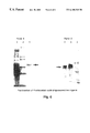
- FIG. 1 is a schematic diagram showing a method of identifying oligonucleotide aptamers that uses magnetic separation.
- FIG. 2 is a schematic diagram showing a method of identifying oligonucleotide aptamers that uses fluorescence activated sorting.
- FIG. 3 is a schematic diagram showing another method of identifying oligonucleotide aptamers that uses fluorescence activated sorting.
- FIG. 4 is a diagram showing the sequences of aptamers to erythropoietin which are isolated from an oligonucleotide library.
- a consensus sequence is identified in the aptamers of Group I.
- Group II is comprised of oligonucleotides not containing the consensus sequence.
- FIG. 5 is an autoradiogram showing the specific binding of an isolated oligonucleotide, F15 (Seq ID No: 9), to erythropoietin in native polyacrylamide gel electrophoresis. It also shows that oligonucleotide F15 does not bind to the negative control, i.e., an equal amount of bovine serum albumin.
- FIGS. 6A and 6B comprises two radiograms verifying the purification of prothrombin with an oligonucleotide ligand matrix.
- FIG. 1 illustrates a process of selecting aptamers by magnetic separation.
- an oligonucleotide library is synthesized on TentaGel beads (polyethylene glycol grafted polystyrene-divinylbenzene polymer beads), each covered with a plurality of unique and identical oligonucleotides.
- the target molecule is linked (conjugated) to magnetic beads (Strepavidine-coated Dynal spheres M280).
- the diameter of resin beads for oligonucleotides is substantially larger than that of the magnetic beads (from about 10 to about 100 times larger).
- the resin beads may have a diameter of about 90 m and are observable under an optical microscope, while the magnetic beads have a diameter of about 2.8 m.
- the oligonucleotide library is brought into contact with the target molecule at a concentration preferably below the desired affinity. For example, if affinity of 10 ⁇ 6 M or higher is desired, the concentration of the target molecule is preferably below 10 ⁇ 6 M.
- the selection stringency can be adjusted by choosing the appropriate binding and washing conditions, and picking oligonucleotide beads above a defined fluorescence intensity.
- the selection stringency can be manipulated with salt concentrations between 50 mM and 250 mM NaCl, preferably between 100 mM and 200 mM NaCl.
- oligonucleotide resin beads After allowing the oligonucleotide library to interact with the target molecule-magnetic conjugate, magnetic force is applied to the mixture to separate complexed oligonucleotide resin beads from uncomplexed resin beads.
- the mixture in a microfuge tube is subject to a magnetic field and magnetic beads are retained on the side of the tube while non-magnetic beads can be washed away with buffers. The washing procedure is repeated several times and more stringent conditions can be applied to select for high affinity beads.
- the retained beads are examined, e.g., using fluorescence microscopy when the target molecules are fluor- labeled. Individual oligonucleotide resin beads can then be singled out and subject to sequencing and/or amplification.
- FIG. 2 illustrates a process of selecting aptamers by fluorescence-activated sorting.
- an oligonucleotide library is synthesized on resin beads.
- the target molecule is conjugated with a fluorescent label.
- the oligonucleotide library is brought into contact with the target molecule under conditions suitable for binding.
- the selection stringency can be adjusted by choosing the appropriate binding and washing conditions.
- the mixture is supplied to a FACS machine to select oligonucleotide resin beads bearing fluorescent label.
- oligonucleotide resin beads of varying fluorescence intensity can be separated automatically by programming the FACS machine to collect beads of specific fluorescence intensity in individual sample containers.
- the target molecule:ligand complex is therefore marked and identified using both a fluorescent label and a magnetic label.
- oligonucleotide aptamers for the target molecule are selected by magnetic force and sorted using fluorescence activated sorting.
- FIG. 3 illustrates a second process of selecting aptamers by fluorescence activated sorting.
- a soluble oligonucleotide library is synthesized and each oligonucleotide is marked with a fluorescent label.
- the target molecule is linked to a solid support such as a bead.
- the oligonucleotide library is brought into contact with the target molecule under conditions suitable for binding.
- the selection stringency can be adjusted by choosing the appropriate binding and washing conditions.
- the mixture After allowing the oligonucleotide library to interact with the target molecule, the mixture is supplied to a FACS machine to select target molecule beads covered with fluorescent label.
- An oligonucleotide in a candidate library of this invention contains a variable region flanked by one or two constant regions.
- the variable region contains randomized sequences while the constant region(s) contains primer binding sites for amplification and/or sequencing of the oligonucleotide aptamer, and/or restriction sites for cloning.
- the variable region is designed to contain molecular diversity from which specific ligands can be selected.
- an oligonucleotide library is prepared on solid support such as PEG resin beads. Each oligonucleotide is covalently linked to the resin.
- the molecular diversity is generated by split-pool strategy commonly practiced in combinatorial chemistry processes (Gallo et al., (1994) J. Medicinal Chem . 37:1233-1251).
- Oligonucleotides on each individual bead are preferably unique and identical.
- the oligonucleotide library therefore comprises a plurality of beads each representing a unique oligonucleotide construct.
- the size of a library is correlated to the complexity of oligonucleotide ligands. For example, 4 13 (i.e., 67 million) individual beads are required for a 13 mer library which is completely randomized and representative. 4 15 (i.e., 1 billion) beads are required for a 15 mer library which is completely randomized and representative.
- the oligonucleotide can be DNA, RNA, their derivatives, mimetics, or a combination thereof.
- the oligonucleotide or the solid support thereof is tagged with a marker, e.g., a fluorescent label which allows selection, e.g., by fluorescence activated sorting (FACS).
- a marker e.g., a fluorescent label which allows selection, e.g., by fluorescence activated sorting (FACS).
- a soluble oligonucleotide library is synthesized with each oligonucleotide marked with a fluorescent label.
- a target molecule can be linked to a solid support such as a magnetic bead, either directly by covalent bonding or indirectly through a linker or precoated molecule such as a strepavidin coated bead.
- erythropoietin molecules can be biotinylated with NHS-biotin and linked to strepavidin coated magnetic beads.
- the target molecule can be labeled with a fluorescent probe such as fluorescein to allow selection by fluorescence activated sorting (FACS).
- a fluorescent probe such as fluorescein to allow selection by fluorescence activated sorting (FACS).
- the selected oligonucleotides can be amplified with polymerase chain reactions utilizing primers directed to the constant regions of the oligonucleotides.
- the PCR products can be cloned and sequenced subsequently.
- individual beads are sequenced directly with labeled primers, either manually or automatically.
- the ligands isolated by the method of this invention can be used as affinity ligands to separate and purify target molecules, as probes to trace, monitor, detect and quantitate target molecules, or to block, activate or catalyze reactions that are physiologically relevant to achieve therapeutic effect (see review by Gold (1995) J. Biol. Chem . 270:13581-4).
- Oligonucleotide ligands so isolated have utilities similar to antibodies. They can act as a pharmaceutical agent. They can bind to a specific target and direct specific molecules to a desired site, and/or they can inhibit or promote a physiologically relevant reaction to achieve a desired therapeutic effect. Various in vivo, ex vivo, and in vitro methods can employ the principles taught herein, as will appreciated by one of skill in the art.
- oligonucleotide aptamers can be used in affinity purification matrixes to purify target molecules.
- oligonucleotide aptamers are ideal for chromatographic separations of target molecules from contaminants.
- oligonucleotide aptamers are particularly useful for purifying target protein molecules from cell cultures or cell extracts.
- prothrombin from crude plasma.
- the immediate application of this technology is to purify antibodies, enzymes, hormones, receptors, and factors that are used in research, development, diagnostic, pharmaceutical, industry applications.
- Oligonucleotides of specific chiral properties can be used to separate chiral compounds and obtain optically pure chemicals.
- Oligonucleotide ligands can also be used in place of antibodies in various research, development and diagnostic applications such as blotting techniques, flow cytometry, immunoassays, strip assays, immunohistological techniques, affinity sensors, etc. Oligonucleotide can further be used to monitor, trace, detect and quantitative desired target such as proteins, antibodies, microbes, virus, bacteria, macromolecules, and small molecules. Oligonucleotide ligands will also be valuable tools for proteomics studies of protein and their function.
- Deoxyoligonucleotides each containing 40 bases of random sequence flanked by two 18 base primer binding sites and a 5′ fluorescein label were synthesized using standard techniques (Genosys).
- the 76 mer library has the following formula: 5′-F-label-CATGA ACTAG TGGAT CCG-40nt-TTGGT ACCCA ATTCG CCC-3′ (seq ID NO: 1).
- the 5′ end constant region is comprised of 5′-CATGA ACTAG TGGAT CCG-3′ (seq ID NO: 2), while the 3′ end constant region is comprised of 5′-GGGCG AATTG GGTAC CAA-3′ (seq ID NO: 3).
- Restriction sites BamHI and Kpn I are designed into the constant regions to facilitate cloning and the production of probes.
- erythropoietin in 100 mM sodium bicarbonate buffer (pH 8.0) was biotinylated with NHS-biotin (molar ratio biotin:target 35:1, Calbiochem) for four hours at room temperature. Free biotin was removed by dialyzing against phosphate buffered saline. Biotinylated erythropoietin was linked to streptavidin beads (M280, Dynal) by incubating at 4° C. overnight.
- NHS-biotin molar ratio biotin:target 35:1, Calbiochem
- the fluorescein tagged oligonucleotide library (119 nmol, contains 7 ⁇ 10 16 molecular diversity) was dissolved in PBS and incubated with erythropoietin immobilized on Dynal beads at room temperature for 15 minutes. The beads were washed with PBS five times and subject to fluorescence activated cell sorting (Becton-Dickson, FACStar Plus). Beads with the highest fluorescent intensity (0.1% of the total 600,000 beads scanned) were collected in PBS and subject to polymerase chain reaction (PCR) with Taq polymerase (Life Technologies). A total of twenty-five to thirty cycles were performed on a Perkin-Elmer DNA Thermal cycler (denaturing at 94° C.
- PCR products were verified on 2% agarose TBE electrophoresis and digested with restriction enzymes Kpn 1 and BamH1 (New England Biolabs). The digested fragments were ligated and cloned into vector pBlueScript KS (Stratagene) and transformed into bacterial host DH5 ⁇ . Individual colonies were identified.
- Plasmid DNA was prepared using boiling miniprep procedures according to Sambrook, Fritsch and Maniatis, Molecular Cloning: A Laboratory Manual , Second Edition (1989). Plasmids were digested with Kpn I and BamHI, and the fragments were end-labeled using 32 P-ATP and kinase. After the restriction digestion, the sense strand contains protruding ends 8 bases longer than the antisense strand. The labeled fragment were then denatured by sequencing stop buffer and heated at 100° C. for 2 min, and electrophoretically separated on a 15% urea-TBE polyacrylamide gel.
- Single stranded radioactive 32 p labeled probes were excised from the gel and eluted in 0.5 M NaAc, 20 mM Tris, pH 7.5, 10 mM EDTA, 0.1% SDS at 65° C. for 30 min. The probes were then extracted with phenol, precipitated with tRNA and ethanol, and air dried.
- the probes were dissolved in PBS. Binding assays were performed in PBS at room temperature for 15 min. The formed complexes were electrophoretically resolved in a native 0.5 ⁇ TBE 4% polyacrylamide gel, and then exposed on X-ray film (Kodak).
- Erythropoietin-bound aptamers were identified thereby and sequenced using the following primers:
- Sense primer (Seq ID NO: 4) 5′-AGCGGATAACAATTTCACACAGGA-3′
- Antisense primer (Seq ID NO: 5) 5′-CGCCAGGGTTTTCCCAGTCACGAC-3′
- the minimal consensus sequence and constructs bearing several copies of consensus demonstrated more or less binding.
- the spaced region between the two consensus sequences was found to affect binding ability. It was thus determined that oligonucleotides with more or less affinity for the target molecule of interest could be prepared and identified based on the consensus sequence.
- oligonucleotide ligand to EPO is demonstrated in FIG. 2 by its ability to bind to EPO and not an equi-molar amount of bovine serum albumin.
- Oligonucleotide ligand against thrombin has been isolated by Bock et al., Nature 355:584-566, 1992.
- the converse utility of an oligonucleotide ligand as affinity matrix to purify protein from crude extracts is demonstrated by the following.
- Biotinylated oligonucleotide ligand against thrombin is produced according to known procedures (e.g., Genosys).
- the sequence (Seq ID NO: 6) of the ligand is as follows:
- the ligand (99 nmol) is labeled with T4 kinase with 32 P-ATP (New England Biolabs). Free radioactivity is removed by passage through a sephadex G25 spin column. The labeled ligands were incubated with 0.5 ml streptavidin-agarose (Life Technologies) at 4° C. overnight. Coupling efficiency is monitored by reduced radioactivity in the solution before and after incubation. Coupling efficiency was found to be 71%.
- the matrix bound ligand is packed in a small column (BioRad) and washed with phosphate buffered saline. Human plasma (Calbiochem, 10 ml) is loaded on the column. After washing with PBS, the bound prothrombin is eluted with 1 M NaCl, 5 mM EDTA, 25 mM Tris, pH 7.4.
- the collected sample is analyzed by 7.5% SDS-PAGE, shown in FIG. 3 A and identity of prothrombin verified by western blotting as shown in FIG. 3 B.
- the ligand can be synthesized directly on a solid support, preferably polyethylene glycol grafted polystyrene beads or cellulose based membrane.
- PEG-grafted polystyrene beads (TentaGel S) are purchased from Fluka. Loading is controlled by reacting the beads with defined amount of DMT-Cl and capping the remaining OH groups on the support.
- the oligonucleotides are synthesized on an ABI 394 DNA synthesizer using standard procedures. One gram of solid support is divided into four equal parts and synthesis performed in four columns. After the primer synthesis, the supports are mixed together and divided into four equal parts again and an additional base synthesized. The mix-split process is repeated for synthesis of a total of 20 bases, each column having a defined base for each of the 20 rounds. After the mix-split, the second primer is synthesized.
- Synthesis is monitored by trityl efficiency and the polymerase chain reaction.
Abstract
This invention relates to methods of preparing oligonucleotide libraries, isolating oligonucleotide aptamers to target molecules from the libraries, and using the aptamers to purify target molecules by affinity separation. Specific oligonucleotide aptamers to erythropoietin were isolated accordingly.
Description
This application claims the benefit of priority under 35 U.S.C. 119(e) to U.S. provisional application U.S. Ser. No. 60/082,405, filed Apr. 20, 1998, the contents of which, including drawings and sequence, are hereby incorporated by reference into the present application.
The present invention relates to the synthesis and identification of oligonucleotide ligands of target molecules and uses thereof. The invention also relates to Fluorescence Activated Cell Sorter analysis, i.e., FACScan flow cytometetry, and other convenient separation means.
SELEX, i.e., Systematic Evolution of Ligands by Exponential Enrichment, is a process of isolating oligonucleotide ligands of a chosen target molecule (see Tuerk and Gold, Science 249:505-510, (1990), U.S. Pat. Nos. 5,475,096, 5,595,877, and 5,660,985). SELEX as described in Tuerk and Gold involves admixing the target molecule with a pool of oligonucleotides (e.g., RNA) of diverse sequences; retaining complexes formed between the target and oligonucleotides; recovering the oligonucleotides bound to the target; reverse-transcribing the RNA into DNA; amplifying the DNA with polymerase chain reactions (PCR); transcribing the amplified DNA into RNA; and repeating the cycle with ever increasing binding stringency. Three enzymatic reactions are required for each cycle. It usually takes many cycles (e.g., between 12-15 cycles) to isolate aptamers of high affinity and specificity to the target. An aptamer is an oligonucleotide that is capable of binding to an intended target substance but not other molecules under the same conditions.
Bock et al., (1990) Nature 355:564-566, describes another approach of isolating aptamers. Bock's process is different from that of Tuerk and Gold in that only one enzymatic reaction is required for each cycle (i.e., PCR) because the nucleic acid library in Bock's method is comprised of DNA instead of RNA. The identification and isolation of aptamers of high specificity and affinity with the method of Bock et al. still requires repeated cycles in a chromatographic column.
Conrad et al., (1996) Methods in Enzymol. 267:336-367, describes a variety of methods for isolating aptamers, all of which employ repeated cycles to enrich target-bound ligands and require a large amount of purified target molecules.
The present invention features a simple, speedy and cost efficient method of identifying aptamers to specific target molecules. It allows for the identification and isolation of aptamers without numerous cycles of selection and amplification.
Within the scope of this invention, Applicant successfully identified and isolated synthetic single-stranded oligonucleotides of high affinity to a target molecule of interest, erythropoietin. In addition, Applicant successfully used a synthetic single-stranded oligonucleotide in an affinity matrix to purify another molecule of interest, prothrombin. The Applicant's success in these endeavors suggests a broad-spectrum applicability of the invention that is not limited to the specific biologic molecules studied herein. Those of skill in the art will appreciate that such can be applied to any molecule or compound of interest, biologic or otherwise, that has an affinity for oligonucleotide ligand sequences of the type described herein.
In a first aspect, this invention features a method that makes use of magnetic separation to identify an aptamer which specifically binds to a target molecule of interest by magnetic separation. In this method, the target molecule is conjugated to a magnetic substance. After allowing the target molecule to mix with a collection of candidate oligonucleotides under conditions suitable for complex formation, i.e., binding of target molecule with aptamer(s), magnetic force is applied to separate aptamer(s) from the rest of the candidate oligonucleotides that have little or no affinity for the target molecule. Preferably, this method contains the following steps:
a) providing a collection of candidate oligonucleotides attached to a plurality of solid supports, wherein candidate oligonucleotides of different nucleotide sequences are attached to different solid supports and all candidate oligonucleotides attached to a single solid support are of the same nucleotide sequence;
b) providing a plurality of magnetic beads covered with the target molecule;
c) admixing the collection of candidate oligonucleotides on solid supports with the magnetic beads covered with the target molecule under conditions suitable for the binding of oligonucleotide ligands to the target molecule, wherein magnetic bead conjugates complex with one or more solid supports bearing aptamers to the target molecule of interest;
d) applying magnetic force to isolate one or more solid supports attached with magnetic beads; and
e) identifying oligonucleotides present on the isolated solid supports.
A collection of candidate oligonucleotides is generated using conventional synthesis techniques. Preferably, each oligonucleotide in the collection contains both a randomized sequence as well as at least one adjacent primer sequence for amplification and/or sequencing. Candidate oligonucleotides include single-stranded and double-stranded RNA or DNA of any length. A candidate oligonucleotide may contain modified or derivatized groups known in the art, especially those identified in U.S. Pat. Nos. 5,582,981 and 5,660,985, such as analogous forms of purines and pyrimidines and analogous forms of ribose and deoxyribose.
In FIG. 4, the oligonucleotides shown correspond to the sequence listing as follows: F18 is SEQ ID NO. 8; F15 is SEQ ID NO. 9; F21 is SEQ ID NO. 10; F22 is SEQ ID NO. 11; F17 is SEQ ID NO. 12; F8 is SEQ ID NO. 13; F13 is SEQ ID NO. 14; and F19 is SEQ ID NO. 15.
A target molecule can be any molecule capable of forming a complex with an oligonucleotide, including, but not limited to, peptides, proteins, enzymes, antibodies, hormones, glycoproteins, polymers, polysaccharides, nucleic acids, small organic compounds such as drugs, dyes, metabolites, cofactors, transition state analogs and toxins.
Specific target molecules of interest include molecules of biological and physiological relevance in both prokaryotic and eukaryotic organisms, particularly mammals. Examples of such biologically significant molecules in mammals include, but are not limited to, erythropoietin, tissue plasminogen activator, granular colony stimulating factor (G-CSF), growth hormone (GH), endostatin (O'Reilly et al., (1997) Cell 88:277-285), interferons, interleukins, chemokines (Shi et al., (1997) FASEB J. 11:1330; Bubrovsky et al., (1996) PNAS, USA 92:700-709), enzymes such as SOD (Yoshikai et al., (1995) Cancer Res. 55(8) 1617-1620) and amylase, antibodies (particularly the constant “Fc” regions thereof), OKT3 (Ho et al., (1998) Science 280:1866-1867), serum proteins (e.g., Factor VIII (Papadopulos-Eleopulos et al., (1990) Genetica 95:35-50), Factor VIX, plasminogen, antithrombin III (Jones et al., (1992) Br. J. Cancer 66:744-747), albumin, protein C (Griffin et al., (1993) Blood 82:1989-93), etc.), and vaccines (e.q., HbsAg (Davis et al., (1994) Vaccine 12:1503-1509), etc.). The physiological significance of most of these, and many other molecules, may similarly be found in Goodman and Gilman's The Pharmacological Basis of Therapeutics, 8th ed., (1990) Pergamon Press, Elmsford, N.Y. Those of skill in the art will appreciate that a virtually unlimited number of other target molecules may also be used with the claimed methods.
The solid support can be anything suitable for attaching oligonucleotides, including, but not limited to, resin beads, controlled pore glass (e.g., Maskos and Southern, (1992) Nucleic Acid Res. 20:1679-1684), polystyrene (e.g., McCollum and Andrus, (1991) Tetrahedron Lett. 32:4069-4072), PEG-polystyrene (e.g., Gao et al., (1991) Tetrahedron Lett. 32:5477-5480), Teflon (e.g., Arnold, U.S. Pat. No. 5,362,866), cellulose (e.g., Crea and Horn, (1980) Nucleic Acid Res. 8:2331-2348). In a preferred embodiment, the oligonucleotides of diverse sequences are synthesized on the solid supports (e.g., Pon, (1993) Methods Mol. Biol. 20:465-496). In another preferred embodiment, the solid support is of a size observable under an optical microscope. Such a size facilitates the selection of individual solid supports following magnetic separation. Oligonucleotides attached to individual solid supports can be sequenced directly to obtain the identity, consensus sequence and/or structure information of the aptamers. Preferably, the solid support has a diameter of about 50 um to about 500 um, more preferably about 70 um to about 150 um, and even more preferably about 90 um to about 120 um. The target molecule is optionally labeled with a fluorescent label to facilitate the identificcation and selection of solid supports which form complexes with the target molecule through aptamer/target molecule interaction.
The magnetic beads covered with the target molecule are preferably much smaller than the solid supports attached with oligonucleotides. For example, the magnetic beads may have a diameter of about 1 um to about 10 um, or about 2 um to about 5 um.
If too many solid supports are selected by the above described process, one can increase the binding stringency and isolate a more limited set of solid supports attached with oligonucleotides of higher affinity to the target molecule. If none or too few solid supports are selected by the process, one can lower the binding stringency to isolate more solid supports. The phenomenon of stringency is known by those of skill in the art to be routinely affected and modulated by parameters such as temperature and ion strength of solution.
In a second aspect, this invention features a method of identifying an aptamer which specifically binds to a target molecule by fluorescence activated sorting. In this method, the target molecule contains or is attached with a fluorescent label. After allowing the target molecule to mix with a collection of candidate oligonucleotides under conditions suitable for aptamer binding and complexing, fluorescence activated sorting is applied to separate aptamer(s) complexed with the fluorescent target molecule from the rest of the candidate oligonucleotides. Preferably, this method contains the following steps:
a) providing a collection of candidate oligonucleotides attached to a plurality of solid supports, wherein candidate oligonucleotides of different nucleotide sequences are attached to different solid supports, each support having identical oligonucleotide sequences bound thereto;
b) providing fluorescence labeled target molecules;
c) admixing the candidate oligonucleotides on solid supports with the target molecules under conditions suitable for oligonucleotide/target molecule binding;
d) isolating one or more solid supports attached with the fluorescence labeled target molecules; and
e) identifying oligonucleotides attached to the isolated solid supports.
In a preferred embodiment, the solid supports attached with the fluorescence labeled target molecules are isolated by fluorescence activated sorting. Because FACscan can sort particles into different fractions according to their fluorescence intensity, this method allows a series of oligonucleotides to be sorted according to relative affinity strength to target molecule, i.e., solid supports bearing higher affinity oligonucleotides attract more fluorescence labeled target molecules.
In another preferred embodiment, the fluorescence labeled target molecules are linked to magnetic beads and magnetic separation is applied before the florescence activated sorting. This step increases the speed of FACscan by reducing the number of particles to be sorted.
In a third aspect, this invention features a method of identifying an aptamer which specifically binds to a target molecule. This method contains the following steps:
a) providing a collection of oligonucleotides of diverse sequences in a liquid, wherein each of said oligonucleotides is linked with a detectable label such as a fluorescent label;
b) attaching a plurality of solid supports, e.g., resin beads, to target molecules, said target molecules exposed on the support surfaces and accessible to olignonucleotides;
c) admixing the collection of oligonucleotides with the solid supports covered with the target molecule under conditions suitable for oligonucleotide binding to the target molecule;
d) selecting solid supports linked to fluorescence labeled oligonucleotides by fluorescence activated sorting; and
e) identifying the oligonucleotides linked to the selected solid supports, e.g., by amplification and sequencing.
By utilizing magnetic separation and/or fluorescence activated sorting, the processes of this invention allow the isolation and identification of an aptamer in a single step without repeated cycles. The processes described above for isolating aptamers may be used in lieu of or in combination with conventional methods in the art, including, but not limited to, those described in U.S. Pat. Nos. 5,475,096 and 5,582,981.
Following the identification of aptamers with the processes described in this application, one can further delineate consensus secondary structures and primary sequences, tertiary structures, motifs, and activity boundaries using methods known in the art, including, but not limited to, those described in U.S. Pat. No. 5,595,877. Nucleic acid ligands, once identified, can be further modified or derivatized using methods known in the art, including, but not limited to, those described in U.S. Pat. No. 5,660,985.
In an example, oligonucleotides that specifically bind to erythropoietin were isolated and identified with a method of this invention. Specifically, the present invention provides single stranded DNA ligands to erythropoietin according to the method described above. The sequences of these ligands are listed in FIG. 4. Also included is consensus DNA sequence identified by sequence homology and binding ability to erythropoietin. Thus, in another aspect, this invention features an oligonucleotide aptamer binding to erythropoietin with an affinity of no less than 10−4 M. The affinity ranges from 10−3 to 10−8 M, preferably 10−6 to 10−8 M.
In a preferred embodiment, the oligonucleotide aptamer is substantially homologous, preferably no less than 70%, more preferably no less than 80%, and even more preferably no less than 90%, to the sequences of FIG. 4. Homology is defined as identical bases at the same position within the range of sequence nucleotide positions that is compared. Preferably, the homology is from about 75% to about 100% within the 8 base consensus region in FIG. 4, Group I. In another preferred embodiment, such an oligonucleotide aptamer to erythropoietin contains a sequence as shown in FIG. 4.
Oligonucleotides designed to closely resemble or match the consensus sequence would have high affinity for the target molecule. For example, an oligonucleotide containing multiple consensus sequences separated by spacers would have high affinity for the target molecule. Specifically, an oligonucleotide containing two consensus regions linked by a 6 base spacer binds to erythropoietin as strong as F15.
Other features and advantages of the invention will be apparent from the following drawings and detailed description of the invention and from the claims.
FIG. 1 is a schematic diagram showing a method of identifying oligonucleotide aptamers that uses magnetic separation.
FIG. 2 is a schematic diagram showing a method of identifying oligonucleotide aptamers that uses fluorescence activated sorting.
FIG. 3 is a schematic diagram showing another method of identifying oligonucleotide aptamers that uses fluorescence activated sorting.
FIG. 4 is a diagram showing the sequences of aptamers to erythropoietin which are isolated from an oligonucleotide library. A consensus sequence is identified in the aptamers of Group I. Group II is comprised of oligonucleotides not containing the consensus sequence.
FIG. 5 is an autoradiogram showing the specific binding of an isolated oligonucleotide, F15 (Seq ID No: 9), to erythropoietin in native polyacrylamide gel electrophoresis. It also shows that oligonucleotide F15 does not bind to the negative control, i.e., an equal amount of bovine serum albumin.
FIGS. 6A and 6B comprises two radiograms verifying the purification of prothrombin with an oligonucleotide ligand matrix.
(A): Collected fractions on 7.5% SDS-PAGE of. Lane 1, crude human plasma; lane 2, molecular weight marker (BioRad, low range); lane 3, wash fraction; lane 4, eluted prothrombin fraction.
(B): Western blot analysis of collected fractions. Molecular weight and prothrombin positions are marked by arrows.
1. Selecting Aptamers by Magnetic Bead Separation
FIG. 1 illustrates a process of selecting aptamers by magnetic separation. In this method, an oligonucleotide library is synthesized on TentaGel beads (polyethylene glycol grafted polystyrene-divinylbenzene polymer beads), each covered with a plurality of unique and identical oligonucleotides. The target molecule is linked (conjugated) to magnetic beads (Strepavidine-coated Dynal spheres M280). The diameter of resin beads for oligonucleotides is substantially larger than that of the magnetic beads (from about 10 to about 100 times larger). For example, the resin beads may have a diameter of about 90 m and are observable under an optical microscope, while the magnetic beads have a diameter of about 2.8 m.
The oligonucleotide library is brought into contact with the target molecule at a concentration preferably below the desired affinity. For example, if affinity of 10−6 M or higher is desired, the concentration of the target molecule is preferably below 10−6 M. The selection stringency can be adjusted by choosing the appropriate binding and washing conditions, and picking oligonucleotide beads above a defined fluorescence intensity. The selection stringency can be manipulated with salt concentrations between 50 mM and 250 mM NaCl, preferably between 100 mM and 200 mM NaCl.
After allowing the oligonucleotide library to interact with the target molecule-magnetic conjugate, magnetic force is applied to the mixture to separate complexed oligonucleotide resin beads from uncomplexed resin beads. The mixture in a microfuge tube is subject to a magnetic field and magnetic beads are retained on the side of the tube while non-magnetic beads can be washed away with buffers. The washing procedure is repeated several times and more stringent conditions can be applied to select for high affinity beads. The retained beads are examined, e.g., using fluorescence microscopy when the target molecules are fluor- labeled. Individual oligonucleotide resin beads can then be singled out and subject to sequencing and/or amplification.
2. Selecting Aptamers Using Flow Cytometry (1)
FIG. 2 illustrates a process of selecting aptamers by fluorescence-activated sorting. In this method, an oligonucleotide library is synthesized on resin beads. The target molecule is conjugated with a fluorescent label.
The oligonucleotide library is brought into contact with the target molecule under conditions suitable for binding. The selection stringency can be adjusted by choosing the appropriate binding and washing conditions.
After allowing the oligonucleotide library to interact with the target molecule, the mixture is supplied to a FACS machine to select oligonucleotide resin beads bearing fluorescent label. oligonucleotide resin beads of varying fluorescence intensity can be separated automatically by programming the FACS machine to collect beads of specific fluorescence intensity in individual sample containers.
In a preferred embodiment, the target molecule:ligand complex is therefore marked and identified using both a fluorescent label and a magnetic label. After allowing the oligonucleotide library to interact with the target molecule, oligonucleotide aptamers for the target molecule are selected by magnetic force and sorted using fluorescence activated sorting.
3. Selecting Aptamers by Flow Cytometry (2)
FIG. 3 illustrates a second process of selecting aptamers by fluorescence activated sorting. In this method, a soluble oligonucleotide library is synthesized and each oligonucleotide is marked with a fluorescent label. The target molecule is linked to a solid support such as a bead.
The oligonucleotide library is brought into contact with the target molecule under conditions suitable for binding. The selection stringency can be adjusted by choosing the appropriate binding and washing conditions.
After allowing the oligonucleotide library to interact with the target molecule, the mixture is supplied to a FACS machine to select target molecule beads covered with fluorescent label.
4. Preparing a Library of Oligonucleotide Ligands
An oligonucleotide in a candidate library of this invention contains a variable region flanked by one or two constant regions. The variable region contains randomized sequences while the constant region(s) contains primer binding sites for amplification and/or sequencing of the oligonucleotide aptamer, and/or restriction sites for cloning. The variable region is designed to contain molecular diversity from which specific ligands can be selected.
In a preferred embodiment, an oligonucleotide library is prepared on solid support such as PEG resin beads. Each oligonucleotide is covalently linked to the resin. The molecular diversity is generated by split-pool strategy commonly practiced in combinatorial chemistry processes (Gallo et al., (1994) J. Medicinal Chem. 37:1233-1251). Oligonucleotides on each individual bead are preferably unique and identical. The oligonucleotide library therefore comprises a plurality of beads each representing a unique oligonucleotide construct. The size of a library is correlated to the complexity of oligonucleotide ligands. For example, 413 (i.e., 67 million) individual beads are required for a 13 mer library which is completely randomized and representative. 415 (i.e., 1 billion) beads are required for a 15 mer library which is completely randomized and representative.
The oligonucleotide can be DNA, RNA, their derivatives, mimetics, or a combination thereof.
In another preferred embodiment, the oligonucleotide or the solid support thereof is tagged with a marker, e.g., a fluorescent label which allows selection, e.g., by fluorescence activated sorting (FACS).
In yet another preferred embodiment, a soluble oligonucleotide library is synthesized with each oligonucleotide marked with a fluorescent label.
5. Preparing the Target Molecule for the Selection Process
A target molecule can be linked to a solid support such as a magnetic bead, either directly by covalent bonding or indirectly through a linker or precoated molecule such as a strepavidin coated bead. For example, erythropoietin molecules can be biotinylated with NHS-biotin and linked to strepavidin coated magnetic beads.
Alternatively, the target molecule can be labeled with a fluorescent probe such as fluorescein to allow selection by fluorescence activated sorting (FACS).
6. Amplifying and Sequencing the Selected Ligands
The selected oligonucleotides can be amplified with polymerase chain reactions utilizing primers directed to the constant regions of the oligonucleotides. The PCR products can be cloned and sequenced subsequently. Preferably, individual beads are sequenced directly with labeled primers, either manually or automatically.
Utility
The ligands isolated by the method of this invention can be used as affinity ligands to separate and purify target molecules, as probes to trace, monitor, detect and quantitate target molecules, or to block, activate or catalyze reactions that are physiologically relevant to achieve therapeutic effect (see review by Gold (1995) J. Biol. Chem. 270:13581-4).
Oligonucleotide ligands so isolated have utilities similar to antibodies. They can act as a pharmaceutical agent. They can bind to a specific target and direct specific molecules to a desired site, and/or they can inhibit or promote a physiologically relevant reaction to achieve a desired therapeutic effect. Various in vivo, ex vivo, and in vitro methods can employ the principles taught herein, as will appreciated by one of skill in the art.
Within respect to in vitro procedures, the applicant has found that isolated oligonucleotide aptamers can be used in affinity purification matrixes to purify target molecules. oligonucleotide aptamers are ideal for chromatographic separations of target molecules from contaminants. In a preferred embodiment, applicant has found that oligonucleotide aptamers are particularly useful for purifying target protein molecules from cell cultures or cell extracts.
Specifically, applicant has demonstrated a one step purification of prothrombin from crude plasma. The immediate application of this technology is to purify antibodies, enzymes, hormones, receptors, and factors that are used in research, development, diagnostic, pharmaceutical, industry applications.
Oligonucleotides of specific chiral properties can be used to separate chiral compounds and obtain optically pure chemicals.
Oligonucleotide ligands can also be used in place of antibodies in various research, development and diagnostic applications such as blotting techniques, flow cytometry, immunoassays, strip assays, immunohistological techniques, affinity sensors, etc. Oligonucleotide can further be used to monitor, trace, detect and quantitative desired target such as proteins, antibodies, microbes, virus, bacteria, macromolecules, and small molecules. Oligonucleotide ligands will also be valuable tools for proteomics studies of protein and their function.
Synthesizing a Soluble Oligonucleotide Library
Deoxyoligonucleotides each containing 40 bases of random sequence flanked by two 18 base primer binding sites and a 5′ fluorescein label were synthesized using standard techniques (Genosys). The 76 mer library has the following formula: 5′-F-label-CATGA ACTAG TGGAT CCG-40nt-TTGGT ACCCA ATTCG CCC-3′ (seq ID NO: 1).
The 5′ end constant region is comprised of 5′-CATGA ACTAG TGGAT CCG-3′ (seq ID NO: 2), while the 3′ end constant region is comprised of 5′-GGGCG AATTG GGTAC CAA-3′ (seq ID NO: 3).
Restriction sites BamHI and Kpn I are designed into the constant regions to facilitate cloning and the production of probes.
Immobilizing EPO to Streptavidin Beads
Purified erythropoietin in 100 mM sodium bicarbonate buffer (pH 8.0) was biotinylated with NHS-biotin (molar ratio biotin:target 35:1, Calbiochem) for four hours at room temperature. Free biotin was removed by dialyzing against phosphate buffered saline. Biotinylated erythropoietin was linked to streptavidin beads (M280, Dynal) by incubating at 4° C. overnight.
Isolating Erythropoietin Binding Oligonucleotide Aptamers
The fluorescein tagged oligonucleotide library (119 nmol, contains 7×10¹⁶ molecular diversity) was dissolved in PBS and incubated with erythropoietin immobilized on Dynal beads at room temperature for 15 minutes. The beads were washed with PBS five times and subject to fluorescence activated cell sorting (Becton-Dickson, FACStar Plus). Beads with the highest fluorescent intensity (0.1% of the total 600,000 beads scanned) were collected in PBS and subject to polymerase chain reaction (PCR) with Taq polymerase (Life Technologies). A total of twenty-five to thirty cycles were performed on a Perkin-Elmer DNA Thermal cycler (denaturing at 94° C. for 45 sec, annealing at 60° C. for 45 sec, and chain extension at 72° C. for 1 minute) The PCR products were verified on 2% agarose TBE electrophoresis and digested with restriction enzymes Kpn 1 and BamH1 (New England Biolabs). The digested fragments were ligated and cloned into vector pBlueScript KS (Stratagene) and transformed into bacterial host DH5α. Individual colonies were identified.
Testing the Affinity of an Aptamer to Erythropoietin
Plasmid DNA was prepared using boiling miniprep procedures according to Sambrook, Fritsch and Maniatis, Molecular Cloning: A Laboratory Manual, Second Edition (1989). Plasmids were digested with Kpn I and BamHI, and the fragments were end-labeled using 32P-ATP and kinase. After the restriction digestion, the sense strand contains protruding ends 8 bases longer than the antisense strand. The labeled fragment were then denatured by sequencing stop buffer and heated at 100° C. for 2 min, and electrophoretically separated on a 15% urea-TBE polyacrylamide gel.
Single stranded radioactive 32p labeled probes were excised from the gel and eluted in 0.5 M NaAc, 20 mM Tris, pH 7.5, 10 mM EDTA, 0.1% SDS at 65° C. for 30 min. The probes were then extracted with phenol, precipitated with tRNA and ethanol, and air dried.
The probes were dissolved in PBS. Binding assays were performed in PBS at room temperature for 15 min. The formed complexes were electrophoretically resolved in a native 0.5× TBE 4% polyacrylamide gel, and then exposed on X-ray film (Kodak).
Erythropoietin-bound aptamers were identified thereby and sequenced using the following primers:
Sense primer (Seq ID NO: 4) 5′-AGCGGATAACAATTTCACACAGGA-3′
Antisense primer (Seq ID NO: 5) 5′-CGCCAGGGTTTTCCCAGTCACGAC-3′
Manual sequencing was performed according to the manufacturer's instructions (Amersham USB).
A consensus sequence emerged from five sequenced aptamers as shown in FIG. 4, Group I. This consensus sequence was found to bind erythropoietin, albeit with slightly lower affinity than the five aptamers identified. Three other aptamers were found that shared no homology with the consensus sequence (FIG. 4, Group II).
The minimal consensus sequence and constructs bearing several copies of consensus demonstrated more or less binding. The spaced region between the two consensus sequences was found to affect binding ability. It was thus determined that oligonucleotides with more or less affinity for the target molecule of interest could be prepared and identified based on the consensus sequence.
The specificity of oligonucleotide ligand to EPO is demonstrated in FIG. 2 by its ability to bind to EPO and not an equi-molar amount of bovine serum albumin.
Oligonucleotide ligand against thrombin has been isolated by Bock et al., Nature 355:584-566, 1992. The converse utility of an oligonucleotide ligand as affinity matrix to purify protein from crude extracts is demonstrated by the following.
Biotinylated oligonucleotide ligand against thrombin is produced according to known procedures (e.g., Genosys). The sequence (Seq ID NO: 6) of the ligand is as follows:
5′-GGTTGGTGTG GTTGGATGAT GGTTGGTGTG GTTGGTAAGT-3′-biotin
The ligand (99 nmol) is labeled with T4 kinase with 32P-ATP (New England Biolabs). Free radioactivity is removed by passage through a sephadex G25 spin column. The labeled ligands were incubated with 0.5 ml streptavidin-agarose (Life Technologies) at 4° C. overnight. Coupling efficiency is monitored by reduced radioactivity in the solution before and after incubation. Coupling efficiency was found to be 71%.
The matrix bound ligand is packed in a small column (BioRad) and washed with phosphate buffered saline. Human plasma (Calbiochem, 10 ml) is loaded on the column. After washing with PBS, the bound prothrombin is eluted with 1 M NaCl, 5 mM EDTA, 25 mM Tris, pH 7.4.
The collected sample is analyzed by 7.5% SDS-PAGE, shown in FIG. 3A and identity of prothrombin verified by western blotting as shown in FIG. 3B.
Alternatively, the ligand can be synthesized directly on a solid support, preferably polyethylene glycol grafted polystyrene beads or cellulose based membrane.
PEG-grafted polystyrene beads (TentaGel S) are purchased from Fluka. Loading is controlled by reacting the beads with defined amount of DMT-Cl and capping the remaining OH groups on the support. The oligonucleotides are synthesized on an ABI 394 DNA synthesizer using standard procedures. One gram of solid support is divided into four equal parts and synthesis performed in four columns. After the primer synthesis, the supports are mixed together and divided into four equal parts again and an additional base synthesized. The mix-split process is repeated for synthesis of a total of 20 bases, each column having a defined base for each of the 20 rounds. After the mix-split, the second primer is synthesized.
Synthesis is monitored by trityl efficiency and the polymerase chain reaction.
All publications noted, none of which is admitted prior art, are incorporated herein by reference, including nucleic acid and amino acid sequence listings.
One of skill in the art will appreciate that other aspects and embodiments are possible within the true scope and spirit of the imvention. The claims below are further illustrative.
Claims (13)
1. A method for identifying an aptamer which specifically binds to a target molecule other than a nucleic acid, comprising the steps of:
a) providing a collection of oligonucleotides attached to a plurality of solid supports, wherein oligonucleotides of different nucleotide sequences are attached to different solid supports and all oligonucleotides attached to a single solid support are of the same nucleotide sequence;
b) providing a plurality of magnetic beads having said target molecule immobilized on their surface;
c) admixing said collection of oligonucleotides with said plurality of magnetic beads under conditions wherein the oligonucleotide ligands become bound to the target molecule;
d) applying magnetic force to isolate one or more solid supports bound to magnetic beads; and
e) identifying oligonucleotides attached to said one or more isolated solid supports.
2. The method of claim 1, wherein said solid supports are resin beads and said step d) comprises selecting resin beads bound to magnetic beads.
3. The method of claim 2, wherein said resin beads have a diameter of about 50 um to about 500 um.
4. The method of claim 2, wherein said resin beads have a diameter of about 70 um to about 150 um.
5. The method of claim 2, wherein said resin beads have a diameter of about 90 um to about 120 um.
6. The method of claim 1, wherein said magnetic beads have a diameter of about 1 um to about 10 um.
7. The method of claim 1, wherein said magnetic beads have a diameter of about 2 um to about 5 um.
8. The method of claim 1, wherein said target molecule is labeled with a fluorescent probe.
9. The method of claim 1, wherein said collection of oligonucleotides are synthesized on said plurality of solid supports.
10. A method for identifying an aptamer which specifically binds to a target molecule other than a nucleic acid, comprising the steps of:
a) providing a collection of oligonucleotides attached to a plurality of solid supports, wherein oligonucleotides of different nucleotide sequences are attached to different solid supports and all oligonucleotides attached to a single solid support are of the same nucleotide sequence;
b) providing a plurality of said target molecules labeled with fluorescent probe and linked to a plurality of solid supports;
c) admixing said collection of oligonucleotides with said plurality of said target molecules under conditions wherein the oligonucleotide ligands become bound to target molecule;
d) isolating one or more solid supports attached with fluorescent probe; and
e) identifying oligonucleotides attached to said one or more isolated solid supports.
11. The method of claim 10, wherein said fluorescence labeled target molecules are linked to magetic beads.
12. The method of claim 11, further comprising the step of applying magnetic force to isolate solid supports attached to oligonucleotides bound to said target molecules linked to magnetic beads after step c) and before step d).
13. A method for identifying an aptamer which specifically binds to a target molecule other than a nucleic acid, comprising the steps of:
a) providing a collection of fluorescence labeled oligonucleotides of different nucleotide sequences attached to a plurality of solid supports;
b) providing a plurality of solid supports having said target molecule immobilized on their surface;
c) admixing said collection of oligonucleotides with said plurality of solid supports under conditions wherein the oligonucleotide ligands become bound to the target molecule;
d) isolating solid supports attached with fluorescence labeled oligonucleotides; and
e) identifying oligonucleotides attached to said isolated solid supports.
Priority Applications (1)
| Application Number | Priority Date | Filing Date | Title |
|---|---|---|---|
| US09/287,936 US6180348B1 (en) | 1998-04-20 | 1999-04-07 | Method of isolating target specific oligonucleotide ligands |
Applications Claiming Priority (2)
| Application Number | Priority Date | Filing Date | Title |
|---|---|---|---|
| US8240598P | 1998-04-20 | 1998-04-20 | |
| US09/287,936 US6180348B1 (en) | 1998-04-20 | 1999-04-07 | Method of isolating target specific oligonucleotide ligands |
Publications (1)
| Publication Number | Publication Date |
|---|---|
| US6180348B1 true US6180348B1 (en) | 2001-01-30 |
Family
ID=22170997
Family Applications (1)
| Application Number | Title | Priority Date | Filing Date |
|---|---|---|---|
| US09/287,936 Expired - Fee Related US6180348B1 (en) | 1998-04-20 | 1999-04-07 | Method of isolating target specific oligonucleotide ligands |
Country Status (3)
| Country | Link |
|---|---|
| US (1) | US6180348B1 (en) |
| AU (1) | AU3653399A (en) |
| WO (1) | WO1999054506A1 (en) |
Cited By (53)
| Publication number | Priority date | Publication date | Assignee | Title |
|---|---|---|---|---|
| US20020037506A1 (en) * | 2000-04-18 | 2002-03-28 | Yun Lin | Aptamer based two-site binding assay |
| US20020081617A1 (en) * | 2000-11-08 | 2002-06-27 | Tione Buranda | Fluorescence and FRET based assays for biomolecules on beads |
| US20020150996A1 (en) * | 2001-03-15 | 2002-10-17 | Marit Nilsen-Hamilton | Functional nucleic acid probes and uses thereof |
| US20020155493A1 (en) * | 2000-01-24 | 2002-10-24 | Yingjian Wang | Methods and arrays for detecting biological molecules |
| WO2003029490A1 (en) * | 2001-10-01 | 2003-04-10 | Yingjian Wang | Methods and arrays for detecting biological molecules |
| US20030083294A1 (en) * | 2001-05-25 | 2003-05-01 | Sullenger Bruce A. | Modulators of pharmacological agents |
| US20030099927A1 (en) * | 2000-01-24 | 2003-05-29 | Yingjian Wang | Arrays for bringing two or more reagents in contact with one or more biological targets and methods for making and using the arrays |
| US20030162190A1 (en) * | 2001-11-15 | 2003-08-28 | Gorenstein David G. | Phosphoromonothioate and phosphorodithioate oligonucleotide aptamer chip for functional proteomics |
| US20030185870A1 (en) * | 2001-11-20 | 2003-10-02 | Grinstaff Mark W. | Interfacial biomaterials |
| DE10220804A1 (en) * | 2002-05-10 | 2003-11-27 | Proteome Factory Ag | Analyzing complex mixtures of polypeptides, useful e.g. for proteome analysis, by fragmentation, immobilization, selective release, labeling and characterization |
| DE10247896A1 (en) * | 2002-10-14 | 2004-05-06 | Bruker Daltonik Gmbh | Multi-dimensional separation of mixtures of bio-substances, e.g. for use in biochemistry, involves forming fraction bonds with selected solutions, using pipette robot in micro-titration plate |
| US6762025B2 (en) | 1998-05-20 | 2004-07-13 | Molecular Machines, Inc. | Single-molecule selection methods and compositions therefrom |
| US20040242521A1 (en) * | 1999-10-25 | 2004-12-02 | Board Of Regents, The University Of Texas System | Thio-siRNA aptamers |
| US20040265912A1 (en) * | 2003-05-23 | 2004-12-30 | Board Of Regents, The University Of Texas System | Structure based and combinatorially selected oligonucleoside phosphorothioate and phosphorodithioate aptamer targeting AP-1 transcription factors |
| WO2005003291A2 (en) * | 2002-10-16 | 2005-01-13 | Board Of Regents Of The University Of Texas System | Bead bound combinatorial oligonucleoside phosphorothioate and phosphorodithioate aptamer libraries |
| WO2005037053A2 (en) * | 2003-05-23 | 2005-04-28 | Board Of Regents - The University Of Texas System | High throughput screening of aptamer libraries for specific binding to proteins on viruses and other pathogens |
| US20050118611A1 (en) * | 2003-07-24 | 2005-06-02 | Board Of Regents, The University Of Texas System | Thioaptamers enable discovery of physiological pathways and new therapeutic strategies |
| US20050142582A1 (en) * | 2003-09-04 | 2005-06-30 | The Regents Of The University Of California | Aptamers and methods for their in vitro selection and uses thereof |
| US20050214772A1 (en) * | 1998-10-26 | 2005-09-29 | Board Of Regents, The University Of Texas System | Thio modified aptamer synthetic methods and compositions |
| US20050233317A1 (en) * | 1999-10-08 | 2005-10-20 | National Institute of Advanced Industrial Science and Technology, a Japanese corporation | Modulate aptamer and method of detecting target protein by using the same |
| US20060040881A1 (en) * | 2004-04-22 | 2006-02-23 | Christopher Rusconi | Modulators of coagulation factors |
| US7074586B1 (en) * | 1999-06-17 | 2006-07-11 | Source Precision Medicine, Inc. | Quantitative assay for low abundance molecules |
| US20060172925A1 (en) * | 1998-10-26 | 2006-08-03 | Board Of Regents, The University Of Texas System | Thio-siRNA aptamers |
| US20060281702A1 (en) * | 2005-05-18 | 2006-12-14 | Board Of Regents, The University Of Texas System | Combinatorial selection of phosphorothioate aptamers for RNases |
| DE102005022057B3 (en) * | 2005-05-09 | 2007-02-08 | Analyticon Biotechnologies Ag | Process for the preparation of ligands, ligands and test kit |
| US20070072179A1 (en) * | 2002-05-10 | 2007-03-29 | Frank Essmann | Solid-phase assisted spectroscopic and spectrometric analysis of complex biopolymer mixtures |
| US20070117099A1 (en) * | 2005-11-18 | 2007-05-24 | Mei Technologies, Inc. | Process and apparatus for combinatorial synthesis |
| US20070186296A1 (en) * | 2006-02-02 | 2007-08-09 | Wyeth | Cloning, characterization, and application of tnfrsf19 in neurological disorders |
| US7312325B2 (en) | 2000-09-26 | 2007-12-25 | Duke University | RNA aptamers and methods for identifying the same |
| US7393529B2 (en) | 1998-04-09 | 2008-07-01 | Idexx Laboratories, Inc. | Methods and compositions for inhibiting binding of IgE to a high affinity receptor |
| US20080268423A1 (en) * | 2002-08-16 | 2008-10-30 | Alan Barrett | Compositions and Methods Related to Flavivirus Envelope Protein Domain III Antigens |
| US20080318888A1 (en) * | 1999-04-08 | 2008-12-25 | Antisoma Research Limited | Antiproliferative activity of g-rich oligonucleotides and method of using same to bind to nucleolin |
| US20080318890A1 (en) * | 1999-04-08 | 2008-12-25 | Antisoma Research Limited | Antiproliferative activity of G-rich oligonucleotides and method of using same to bind to nucleolin |
| US20080318887A1 (en) * | 1999-04-08 | 2008-12-25 | Antisoma Research Limited | Antiproliferative activity of G-rich oligonucleotides and method of using same to bind to nucleolin |
| US20080318889A1 (en) * | 1999-04-08 | 2008-12-25 | Antisoma Research Limited | Antiproliferative activity of G-rich oligonucleotides and method of using same to bind to nucleolin |
| US20090131351A1 (en) * | 2007-11-16 | 2009-05-21 | Antisoma Research Limited | Methods, compositions, and kits for modulating tumor cell proliferation |
| US20090252745A1 (en) * | 2006-09-15 | 2009-10-08 | Duke University | Amino acids in the HCV core polypeptide domain 3 and correlation with steatosis |
| US20090326047A1 (en) * | 1999-04-08 | 2009-12-31 | Miller Donald M | Antiproliferative activity of G-rich oligonucleotides and method of using same to bind to nucleolin |
| WO2010036866A1 (en) | 2008-09-25 | 2010-04-01 | University Of Guelph | Nitrogen responsive early nodulin gene |
| WO2010076766A1 (en) | 2008-12-30 | 2010-07-08 | Institute Of Genetics And Developmental Biology | Genes associated with plant tiller number and uses thereof |
| WO2010097746A1 (en) | 2009-02-26 | 2010-09-02 | Institute Of Genetics And Developmental Biology, Chinese Academy Of Sciences | Metallothionein gene conferring abiotic stress tolerance in plants and uses thereof |
| EP2426140A1 (en) | 2003-08-15 | 2012-03-07 | University of Florida Research Foundation, Inc. | Identification of porphyromonas gingivalis virulence polynucleotides in the diagnosis, treatment, and monitoring periodontal diseases |
| US20120289411A1 (en) * | 2010-02-26 | 2012-11-15 | Canon Kabushiki Kaisha | Method for screening nucleic acid ligand |
| EP3050574A1 (en) | 2015-01-28 | 2016-08-03 | Universite De Bordeaux | New compositions and methods of treating and/or preventing chronic obstructive pulmonary disease |
| WO2017176265A1 (en) | 2016-04-06 | 2017-10-12 | Mayo Foundation For Medical Education And Research | Carrier-binding agent compositions and methods of making and using the same |
| US9874554B1 (en) | 2014-07-16 | 2018-01-23 | Verily Life Sciences Llc | Aptamer-based in vivo diagnostic system |
| WO2018045238A1 (en) | 2016-09-01 | 2018-03-08 | Mayo Foundation For Medical Education And Research | Methods and compositions for targeting t-cell cancers |
| WO2018045239A1 (en) | 2016-09-01 | 2018-03-08 | Mayo Foundation For Medical Education And Research | Carrier-pd-l1 binding agent compositions for treating cancers |
| US9939443B2 (en) | 2012-12-19 | 2018-04-10 | Caris Life Sciences Switzerland Holdings Gmbh | Compositions and methods for aptamer screening |
| US9958448B2 (en) | 2012-10-23 | 2018-05-01 | Caris Life Sciences Switzerland Holdings Gmbh | Aptamers and uses thereof |
| US10942184B2 (en) | 2012-10-23 | 2021-03-09 | Caris Science, Inc. | Aptamers and uses thereof |
| US11268135B2 (en) | 2016-05-10 | 2022-03-08 | Pattern Bioscience, Inc. | Compositions and methods for identifying, quantifying, and/or characterizing an analyte |
| WO2022051414A1 (en) | 2020-09-02 | 2022-03-10 | Mayo Foundation For Medical Education And Research | Antibody-nanoparticle complexes and methods for making and using the same |
Families Citing this family (15)
| Publication number | Priority date | Publication date | Assignee | Title |
|---|---|---|---|---|
| US20040175754A1 (en) | 2002-10-09 | 2004-09-09 | David Bar-Or | Diagnosis and monitoring of inflammation, ischemia and appendicitis |
| JP2008507277A (en) * | 2004-07-23 | 2008-03-13 | (オーエスアイ)アイテツク・インコーポレーテツド | Sequencing of modified nucleic acid molecules |
| ATE514776T1 (en) | 2004-10-05 | 2011-07-15 | California Inst Of Techn | APTAMER-REGULATED NUCLEIC ACIDS AND USES THEREOF |
| WO2009012263A2 (en) | 2007-07-18 | 2009-01-22 | The Trustees Of Columbia University In The City Of New York | Tissue-specific micrornas and compositions and uses thereof |
| US20120165387A1 (en) | 2007-08-28 | 2012-06-28 | Smolke Christina D | General composition framework for ligand-controlled RNA regulatory systems |
| US8865667B2 (en) | 2007-09-12 | 2014-10-21 | California Institute Of Technology | Higher-order cellular information processing devices |
| US9029524B2 (en) | 2007-12-10 | 2015-05-12 | California Institute Of Technology | Signal activated RNA interference |
| US9599591B2 (en) | 2009-03-06 | 2017-03-21 | California Institute Of Technology | Low cost, portable sensor for molecular assays |
| US9145555B2 (en) | 2009-04-02 | 2015-09-29 | California Institute Of Technology | Integrated—ligand-responsive microRNAs |
| CN102191250B (en) * | 2010-03-19 | 2013-09-18 | 中国人民解放军军事医学科学院毒物药物研究所 | DNA (Desoxyribonucleic Acid) ligand for specific binding with EPO (erythropoietin), preparation method thereof and application thereof |
| CA2886120A1 (en) | 2012-10-08 | 2014-04-17 | St. Jude Children's Research Hospital | Therapies based on control of regulatory t cell stability and function via a neuropilin-1:semaphorin axis |
| US20150119285A1 (en) * | 2013-10-28 | 2015-04-30 | Chin-Yih Hong | Magnetic-assisted rapid aptamer selection method for generating high affinity dna aptamer |
| SG10202002486QA (en) | 2014-06-16 | 2020-04-29 | Univ Johns Hopkins | Compositions and methods for the expression of crispr guide rnas using the h1 promoter |
| WO2016176617A2 (en) | 2015-04-29 | 2016-11-03 | New York University | Method for treating high-grade gliomas |
| AU2021205929A1 (en) | 2020-01-08 | 2022-09-01 | Regeneron Pharmaceuticals, Inc. | Treatment of fibrodysplasia ossificans progressiva |
Citations (9)
| Publication number | Priority date | Publication date | Assignee | Title |
|---|---|---|---|---|
| WO1994002515A1 (en) | 1992-07-21 | 1994-02-03 | Bunsen Rush Laboratories Inc. | Oligomer library formats and methods relating thereto |
| WO1994014843A1 (en) | 1992-12-21 | 1994-07-07 | Warner-Lambert Company | Endothelin antagonists |
| US5362866A (en) | 1983-09-02 | 1994-11-08 | Molecular Biosystems, Inc. | Oligonucleotide polymeric support system with an oxidation cleavable link |
| US5475096A (en) | 1990-06-11 | 1995-12-12 | University Research Corporation | Nucleic acid ligands |
| US5487973A (en) * | 1986-09-10 | 1996-01-30 | Polyprobe, Inc. | Methods for detecting and assaying nucleic acid sequences |
| US5582981A (en) | 1991-08-14 | 1996-12-10 | Gilead Sciences, Inc. | Method for identifying an oligonucleotide aptamer specific for a target |
| US5595877A (en) | 1990-06-11 | 1997-01-21 | Nexstar Pharmaceuticals, Inc. | Methods of producing nucleic acid ligands |
| US5660985A (en) | 1990-06-11 | 1997-08-26 | Nexstar Pharmaceuticals, Inc. | High affinity nucleic acid ligands containing modified nucleotides |
| US5756291A (en) * | 1992-08-21 | 1998-05-26 | Gilead Sciences, Inc. | Aptamers specific for biomolecules and methods of making |
Family Cites Families (2)
| Publication number | Priority date | Publication date | Assignee | Title |
|---|---|---|---|---|
| GB8827160D0 (en) * | 1988-11-21 | 1988-12-29 | Apothekernes Lab | Detection & quantitative determination of rna & dna |
| WO1992014843A1 (en) * | 1991-02-21 | 1992-09-03 | Gilead Sciences, Inc. | Aptamer specific for biomolecules and method of making |
-
1999
- 1999-04-07 US US09/287,936 patent/US6180348B1/en not_active Expired - Fee Related
- 1999-04-19 WO PCT/US1999/008561 patent/WO1999054506A1/en active Application Filing
- 1999-04-19 AU AU36533/99A patent/AU3653399A/en not_active Abandoned
Patent Citations (9)
| Publication number | Priority date | Publication date | Assignee | Title |
|---|---|---|---|---|
| US5362866A (en) | 1983-09-02 | 1994-11-08 | Molecular Biosystems, Inc. | Oligonucleotide polymeric support system with an oxidation cleavable link |
| US5487973A (en) * | 1986-09-10 | 1996-01-30 | Polyprobe, Inc. | Methods for detecting and assaying nucleic acid sequences |
| US5475096A (en) | 1990-06-11 | 1995-12-12 | University Research Corporation | Nucleic acid ligands |
| US5595877A (en) | 1990-06-11 | 1997-01-21 | Nexstar Pharmaceuticals, Inc. | Methods of producing nucleic acid ligands |
| US5660985A (en) | 1990-06-11 | 1997-08-26 | Nexstar Pharmaceuticals, Inc. | High affinity nucleic acid ligands containing modified nucleotides |
| US5582981A (en) | 1991-08-14 | 1996-12-10 | Gilead Sciences, Inc. | Method for identifying an oligonucleotide aptamer specific for a target |
| WO1994002515A1 (en) | 1992-07-21 | 1994-02-03 | Bunsen Rush Laboratories Inc. | Oligomer library formats and methods relating thereto |
| US5756291A (en) * | 1992-08-21 | 1998-05-26 | Gilead Sciences, Inc. | Aptamers specific for biomolecules and methods of making |
| WO1994014843A1 (en) | 1992-12-21 | 1994-07-07 | Warner-Lambert Company | Endothelin antagonists |
Non-Patent Citations (26)
Cited By (109)
| Publication number | Priority date | Publication date | Assignee | Title |
|---|---|---|---|---|
| US8252907B2 (en) | 1998-04-09 | 2012-08-28 | Idexx Laboratories, Inc. | Methods and compositions for inhibiting binding of IgE to a high affinity receptor |
| US20110158981A1 (en) * | 1998-04-09 | 2011-06-30 | Idexx Laboratories, Inc. | Methods and Compositions for Inhibiting Binding of IgE to a High Affinity Receptor |
| US7393529B2 (en) | 1998-04-09 | 2008-07-01 | Idexx Laboratories, Inc. | Methods and compositions for inhibiting binding of IgE to a high affinity receptor |
| US7931898B2 (en) | 1998-04-09 | 2011-04-26 | Idexx Laboratories, Inc. | Methods and compositions for inhibiting binding of IgE to a high affinity receptor |
| US20100137219A1 (en) * | 1998-04-09 | 2010-06-03 | Idexx Laboratories, Inc. | Methods and Compositions for Inhibiting Binding IgE to a High Affinity Receptor |
| US6762025B2 (en) | 1998-05-20 | 2004-07-13 | Molecular Machines, Inc. | Single-molecule selection methods and compositions therefrom |
| US20050089890A1 (en) * | 1998-05-20 | 2005-04-28 | Cubicciotti Roger S. | Multimolecular devices and drug delivery systems |
| US20050214772A1 (en) * | 1998-10-26 | 2005-09-29 | Board Of Regents, The University Of Texas System | Thio modified aptamer synthetic methods and compositions |
| US20060172925A1 (en) * | 1998-10-26 | 2006-08-03 | Board Of Regents, The University Of Texas System | Thio-siRNA aptamers |
| US20090326047A1 (en) * | 1999-04-08 | 2009-12-31 | Miller Donald M | Antiproliferative activity of G-rich oligonucleotides and method of using same to bind to nucleolin |
| US20080318887A1 (en) * | 1999-04-08 | 2008-12-25 | Antisoma Research Limited | Antiproliferative activity of G-rich oligonucleotides and method of using same to bind to nucleolin |
| US20080318889A1 (en) * | 1999-04-08 | 2008-12-25 | Antisoma Research Limited | Antiproliferative activity of G-rich oligonucleotides and method of using same to bind to nucleolin |
| US20080318890A1 (en) * | 1999-04-08 | 2008-12-25 | Antisoma Research Limited | Antiproliferative activity of G-rich oligonucleotides and method of using same to bind to nucleolin |
| US20080318888A1 (en) * | 1999-04-08 | 2008-12-25 | Antisoma Research Limited | Antiproliferative activity of g-rich oligonucleotides and method of using same to bind to nucleolin |
| US7960540B2 (en) | 1999-04-08 | 2011-06-14 | Advanced Cancer Therapeutics, Llc | Antiproliferative activity of G-rich oligonucleotides and method of using same to bind to nucleolin |
| US8114850B2 (en) | 1999-04-08 | 2012-02-14 | Advanced Cancer Therapeutics, Llc | Antiproliferative activity of G-rich oligonucleotides and method of using same to bind to nucleolin |
| US8648051B2 (en) | 1999-04-08 | 2014-02-11 | Advanced Cancer Therapeutics, Llc | Antiproliferative activity of G-rich oligonucleotides and method of using same to bind to nucleolin |
| US7074586B1 (en) * | 1999-06-17 | 2006-07-11 | Source Precision Medicine, Inc. | Quantitative assay for low abundance molecules |
| US20050233317A1 (en) * | 1999-10-08 | 2005-10-20 | National Institute of Advanced Industrial Science and Technology, a Japanese corporation | Modulate aptamer and method of detecting target protein by using the same |
| US20040242521A1 (en) * | 1999-10-25 | 2004-12-02 | Board Of Regents, The University Of Texas System | Thio-siRNA aptamers |
| US7429466B2 (en) | 2000-01-24 | 2008-09-30 | Hypromatrix, Inc | Methods and arrays for detecting biological molecules |
| US20030099927A1 (en) * | 2000-01-24 | 2003-05-29 | Yingjian Wang | Arrays for bringing two or more reagents in contact with one or more biological targets and methods for making and using the arrays |
| US8143195B2 (en) | 2000-01-24 | 2012-03-27 | Yingjian Wang | Arrays for bringing two or more reagents in contact with one or more biological targets and methods for making and using the arrays |
| US20020155493A1 (en) * | 2000-01-24 | 2002-10-24 | Yingjian Wang | Methods and arrays for detecting biological molecules |
| US20020037506A1 (en) * | 2000-04-18 | 2002-03-28 | Yun Lin | Aptamer based two-site binding assay |
| US7858591B2 (en) | 2000-09-26 | 2010-12-28 | Duke University | RNA aptamers and methods for identifying the same |
| US20110160443A1 (en) * | 2000-09-26 | 2011-06-30 | Sullenger Bruce A | Rna aptamers and methods for identifying the same |
| US8143233B2 (en) | 2000-09-26 | 2012-03-27 | Duke University | RNA aptamers and methods for identifying the same |
| US7312325B2 (en) | 2000-09-26 | 2007-12-25 | Duke University | RNA aptamers and methods for identifying the same |
| US7812001B2 (en) | 2000-09-26 | 2010-10-12 | Duke University | RNA aptamers and methods for identifying the same |
| US7776837B2 (en) | 2000-09-26 | 2010-08-17 | Duke University | RNA aptamers and methods for identifying the same |
| US7776836B2 (en) | 2000-09-26 | 2010-08-17 | Duke University | RNA aptamers and methods for identifying the same |
| US7741307B2 (en) | 2000-09-26 | 2010-06-22 | Duke University | RNA aptamers and methods for identifying the same |
| US20090163429A1 (en) * | 2000-09-26 | 2009-06-25 | Sullenger Bruce A | Rna aptamers and methods for identifying the same |
| US20080207546A1 (en) * | 2000-09-26 | 2008-08-28 | Sullenger Bruce A | Rna aptamers and methods for identifying the same |
| US20020081617A1 (en) * | 2000-11-08 | 2002-06-27 | Tione Buranda | Fluorescence and FRET based assays for biomolecules on beads |
| US7115369B2 (en) | 2001-03-15 | 2006-10-03 | Iowa State University Research Foundation, Inc. | Functional nucleic acid probes and uses thereof |
| US20020150996A1 (en) * | 2001-03-15 | 2002-10-17 | Marit Nilsen-Hamilton | Functional nucleic acid probes and uses thereof |
| EP2364990A1 (en) | 2001-05-25 | 2011-09-14 | Duke University | Modulators of pharmacological agents |
| EP2070939A1 (en) | 2001-05-25 | 2009-06-17 | Duke University | Modulators of pharmacological agents |
| US20100249217A1 (en) * | 2001-05-25 | 2010-09-30 | Sullenger Bruce A | Modulators of pharmacological agents |
| US20080051339A1 (en) * | 2001-05-25 | 2008-02-28 | Duke University | Modulators of pharmacological agents |
| US20030083294A1 (en) * | 2001-05-25 | 2003-05-01 | Sullenger Bruce A. | Modulators of pharmacological agents |
| US7300922B2 (en) | 2001-05-25 | 2007-11-27 | Duke University | Modulators of pharmacological agents |
| US8586524B2 (en) | 2001-05-25 | 2013-11-19 | Duke University | Modulators of pharmacological agents |
| US8283330B2 (en) | 2001-05-25 | 2012-10-09 | Duke University | Modulators of pharmacological agents |
| WO2003029490A1 (en) * | 2001-10-01 | 2003-04-10 | Yingjian Wang | Methods and arrays for detecting biological molecules |
| US20030162190A1 (en) * | 2001-11-15 | 2003-08-28 | Gorenstein David G. | Phosphoromonothioate and phosphorodithioate oligonucleotide aptamer chip for functional proteomics |
| US20080200340A1 (en) * | 2001-11-15 | 2008-08-21 | Board Of Regents, The University Of Texas System | Bead Bound Combinatorial Oligonucleoside Phosphorothioate And Phosphorodithioate Aptamer Libraries |
| US20080255005A1 (en) * | 2001-11-15 | 2008-10-16 | Board Of Regents, The University Of Texas System | Bead Bound Combinatorial Oligonucleoside Phosphorothioate And Phosphorodithioate Aptamer Libraries |
| US20030185870A1 (en) * | 2001-11-20 | 2003-10-02 | Grinstaff Mark W. | Interfacial biomaterials |
| DE10220804A1 (en) * | 2002-05-10 | 2003-11-27 | Proteome Factory Ag | Analyzing complex mixtures of polypeptides, useful e.g. for proteome analysis, by fragmentation, immobilization, selective release, labeling and characterization |
| US20070072179A1 (en) * | 2002-05-10 | 2007-03-29 | Frank Essmann | Solid-phase assisted spectroscopic and spectrometric analysis of complex biopolymer mixtures |
| US7785799B2 (en) | 2002-08-16 | 2010-08-31 | The Board Of Regents Of The University Of Texas System | Compositions and methods related to flavivirus envelope protein domain III antigens |
| US20080268423A1 (en) * | 2002-08-16 | 2008-10-30 | Alan Barrett | Compositions and Methods Related to Flavivirus Envelope Protein Domain III Antigens |
| US20040147040A1 (en) * | 2002-10-14 | 2004-07-29 | Bruker Daltonik Gmbh | Multidimensional separation of biosubstance mixtures for mass spectrometric analysis |
| DE10247896A1 (en) * | 2002-10-14 | 2004-05-06 | Bruker Daltonik Gmbh | Multi-dimensional separation of mixtures of bio-substances, e.g. for use in biochemistry, involves forming fraction bonds with selected solutions, using pipette robot in micro-titration plate |
| DE10247896B4 (en) * | 2002-10-14 | 2007-01-18 | Bruker Daltonik Gmbh | Multidimensional separation of biosubstance mixtures for mass spectrometric analysis |
| US20090123922A1 (en) * | 2002-10-16 | 2009-05-14 | Board Of Regents, The University Of Texas System | Bead Bound Combinatorial Oligonucleoside Phosphorothioate And Phosphorodithioate Aptamer Libraries |
| US7338762B2 (en) | 2002-10-16 | 2008-03-04 | Board Of Regents, The University Of Texas System | Bead bound combinatorial oligonucleoside phosphorothioate and phosphorodithioate aptamer libraries |
| WO2005003291A2 (en) * | 2002-10-16 | 2005-01-13 | Board Of Regents Of The University Of Texas System | Bead bound combinatorial oligonucleoside phosphorothioate and phosphorodithioate aptamer libraries |
| WO2005003291A3 (en) * | 2002-10-16 | 2005-08-18 | Univ Texas | Bead bound combinatorial oligonucleoside phosphorothioate and phosphorodithioate aptamer libraries |
| US20050123939A1 (en) * | 2002-10-16 | 2005-06-09 | Board Of Regents, The University Of Texas System | Bead bound combinatorial oligonucleoside phosphorothioate and phosphorodithioate aptamer libraries |
| US20060121489A1 (en) * | 2003-05-23 | 2006-06-08 | Board Of Regents, The University Of Texas System | High throughput screening of aptamer libraries for specific binding to proteins on viruses and other pathogens |
| US20040265912A1 (en) * | 2003-05-23 | 2004-12-30 | Board Of Regents, The University Of Texas System | Structure based and combinatorially selected oligonucleoside phosphorothioate and phosphorodithioate aptamer targeting AP-1 transcription factors |
| US20110212843A1 (en) * | 2003-05-23 | 2011-09-01 | Board Of Regents, The University Of Texas System | Structure based and combinatorially selected oligonucleoside phosphorothioate and phosphorodithioate aptamer targeting ap-1 transcription factors |
| US7910523B2 (en) | 2003-05-23 | 2011-03-22 | Board Of Regents, The University Of Texas System | Structure based and combinatorially selected oligonucleoside phosphorothioate and phosphorodithioate aptamer targeting AP-1 transcription factors |
| WO2005037053A3 (en) * | 2003-05-23 | 2006-09-21 | Univ Texas | High throughput screening of aptamer libraries for specific binding to proteins on viruses and other pathogens |
| US9567579B2 (en) | 2003-05-23 | 2017-02-14 | Board Of Regents, The University Of Texas System | Structure based and combinatorially selected oligonucleoside phosphorothioate and phosphorodithioate aptamer targeting AP-1 transcription factors |
| WO2005037053A2 (en) * | 2003-05-23 | 2005-04-28 | Board Of Regents - The University Of Texas System | High throughput screening of aptamer libraries for specific binding to proteins on viruses and other pathogens |
| US20050118611A1 (en) * | 2003-07-24 | 2005-06-02 | Board Of Regents, The University Of Texas System | Thioaptamers enable discovery of physiological pathways and new therapeutic strategies |
| EP2426140A1 (en) | 2003-08-15 | 2012-03-07 | University of Florida Research Foundation, Inc. | Identification of porphyromonas gingivalis virulence polynucleotides in the diagnosis, treatment, and monitoring periodontal diseases |
| US20090075834A1 (en) * | 2003-09-04 | 2009-03-19 | The Regents Of The University Of California | Aptamers and methods for their in vitro selection and uses thereof |
| US7329742B2 (en) | 2003-09-04 | 2008-02-12 | The Regents Of The University Of California | Aptamers and methods for their in vitro selection and uses thereof |
| US8105982B2 (en) | 2003-09-04 | 2012-01-31 | The Regents Of The University Of California | Aptamers and methods for their in vitro selection and uses thereof |
| US20050142582A1 (en) * | 2003-09-04 | 2005-06-30 | The Regents Of The University Of California | Aptamers and methods for their in vitro selection and uses thereof |
| US20080153769A1 (en) * | 2004-04-22 | 2008-06-26 | Christopher Rusconi | Modulators of coagulation factors |
| US8859518B2 (en) | 2004-04-22 | 2014-10-14 | Regado Biosciences, Inc. | Modulators of coagulation factors |
| US7723315B2 (en) | 2004-04-22 | 2010-05-25 | Regado Biosciences, Inc. | Modulators of coagulation factors |
| US20100197900A1 (en) * | 2004-04-22 | 2010-08-05 | Regado Biosciences, Inc. | Modulators of coagulation factors |
| US8389489B2 (en) | 2004-04-22 | 2013-03-05 | Regado Biosciences, Inc. | Modulators of coagulation factors |
| US20070105809A1 (en) * | 2004-04-22 | 2007-05-10 | Rusconi Christopher P | Modulators of coagulation factors with enhanced stability |
| US20060040881A1 (en) * | 2004-04-22 | 2006-02-23 | Christopher Rusconi | Modulators of coagulation factors |
| US7531524B2 (en) | 2004-04-22 | 2009-05-12 | Regado Biosciences, Inc. | Modulators of coagulation factors with enhanced stability |
| US7304041B2 (en) | 2004-04-22 | 2007-12-04 | Regado Biosciences, Inc. | Modulators of coagulation factors |
| US20080233577A1 (en) * | 2005-05-09 | 2008-09-25 | Analyticon Biotechnologies Ag | Method for producing ligands, ligands and test kit |
| DE102005022057B3 (en) * | 2005-05-09 | 2007-02-08 | Analyticon Biotechnologies Ag | Process for the preparation of ligands, ligands and test kit |
| US20060281702A1 (en) * | 2005-05-18 | 2006-12-14 | Board Of Regents, The University Of Texas System | Combinatorial selection of phosphorothioate aptamers for RNases |
| US20070117099A1 (en) * | 2005-11-18 | 2007-05-24 | Mei Technologies, Inc. | Process and apparatus for combinatorial synthesis |
| US7576037B2 (en) | 2005-11-18 | 2009-08-18 | Mei Technologies, Inc. | Process and apparatus for combinatorial synthesis |
| US20070186296A1 (en) * | 2006-02-02 | 2007-08-09 | Wyeth | Cloning, characterization, and application of tnfrsf19 in neurological disorders |
| US20090252745A1 (en) * | 2006-09-15 | 2009-10-08 | Duke University | Amino acids in the HCV core polypeptide domain 3 and correlation with steatosis |
| US20090131351A1 (en) * | 2007-11-16 | 2009-05-21 | Antisoma Research Limited | Methods, compositions, and kits for modulating tumor cell proliferation |
| WO2010036866A1 (en) | 2008-09-25 | 2010-04-01 | University Of Guelph | Nitrogen responsive early nodulin gene |
| WO2010076766A1 (en) | 2008-12-30 | 2010-07-08 | Institute Of Genetics And Developmental Biology | Genes associated with plant tiller number and uses thereof |
| WO2010097746A1 (en) | 2009-02-26 | 2010-09-02 | Institute Of Genetics And Developmental Biology, Chinese Academy Of Sciences | Metallothionein gene conferring abiotic stress tolerance in plants and uses thereof |
| US20120289411A1 (en) * | 2010-02-26 | 2012-11-15 | Canon Kabushiki Kaisha | Method for screening nucleic acid ligand |
| US9958448B2 (en) | 2012-10-23 | 2018-05-01 | Caris Life Sciences Switzerland Holdings Gmbh | Aptamers and uses thereof |
| US10942184B2 (en) | 2012-10-23 | 2021-03-09 | Caris Science, Inc. | Aptamers and uses thereof |
| US9939443B2 (en) | 2012-12-19 | 2018-04-10 | Caris Life Sciences Switzerland Holdings Gmbh | Compositions and methods for aptamer screening |
| US9874554B1 (en) | 2014-07-16 | 2018-01-23 | Verily Life Sciences Llc | Aptamer-based in vivo diagnostic system |
| EP3050574A1 (en) | 2015-01-28 | 2016-08-03 | Universite De Bordeaux | New compositions and methods of treating and/or preventing chronic obstructive pulmonary disease |
| EP3613435A1 (en) | 2015-01-28 | 2020-02-26 | Universite De Bordeaux | Chemokine receptor cxcr4 inhibitors for treating and/or preventing chronic obstructive pulmonary disease |
| WO2017176265A1 (en) | 2016-04-06 | 2017-10-12 | Mayo Foundation For Medical Education And Research | Carrier-binding agent compositions and methods of making and using the same |
| US11268135B2 (en) | 2016-05-10 | 2022-03-08 | Pattern Bioscience, Inc. | Compositions and methods for identifying, quantifying, and/or characterizing an analyte |
| WO2018045238A1 (en) | 2016-09-01 | 2018-03-08 | Mayo Foundation For Medical Education And Research | Methods and compositions for targeting t-cell cancers |
| WO2018045239A1 (en) | 2016-09-01 | 2018-03-08 | Mayo Foundation For Medical Education And Research | Carrier-pd-l1 binding agent compositions for treating cancers |
| EP4177271A1 (en) | 2016-09-01 | 2023-05-10 | Mayo Foundation for Medical Education and Research | Carrier-pd-l1 binding agent compositions for treating cancers |
| WO2022051414A1 (en) | 2020-09-02 | 2022-03-10 | Mayo Foundation For Medical Education And Research | Antibody-nanoparticle complexes and methods for making and using the same |
Also Published As
| Publication number | Publication date |
|---|---|
| WO1999054506A1 (en) | 1999-10-28 |
| AU3653399A (en) | 1999-11-08 |
Similar Documents
| Publication | Publication Date | Title |
|---|---|---|
| US6180348B1 (en) | Method of isolating target specific oligonucleotide ligands | |
| US10316321B2 (en) | Method for generating aptamers with improved off-rates | |
| US6423493B1 (en) | Combinatorial selection of oligonucleotide aptamers | |
| AU2008275915B2 (en) | Method for generating aptamers with improved off-rates | |
| AU783689B2 (en) | Sensitive, multiplexed diagnostic assays for protein analysis | |
| US6013443A (en) | Systematic evolution of ligands by exponential enrichment: tissue SELEX | |
| US9951376B2 (en) | Methods for the selection of aptamers | |
| AU2775402A (en) | Nucleic acid ligands to complex targets | |
| WO1993005182A1 (en) | Determination of oligonucleotides for therapeutics, diagnostics and research reagents | |
| JP2002520074A (en) | Cis-acting nucleic acid elements and methods of use | |
| AU754786B2 (en) | Morphatides: novel shape and structure libraries | |
| US20040197779A1 (en) | Methods for analyzing mixtures of proteins | |
| JP2001525180A (en) | Selective methods for rapid identification of proteins and genes and uses thereof | |
| US20230132627A1 (en) | Oligonucleotides and methods for preparing | |
| AU2015249082B2 (en) | Method for generating aptamers with improved off-rates | |
| CN116144666A (en) | DNA aptamer capable of specifically recognizing mCRP and application thereof | |
| CN117660460A (en) | Nucleic acid aptamer combined with human REST co-repressor 3 protein and application thereof | |
| JP2016171795A (en) | Method for selecting aptamer | |
| CN117143878A (en) | Nucleic acid aptamer for specifically targeting SARS-COV-2N protein and application thereof | |
| JP2880996B2 (en) | Hybridization support and method for preparing the same |
Legal Events
| Date | Code | Title | Description |
|---|---|---|---|
| AS | Assignment |
Owner name: BIOAGE PHARMACEUTICALS, INC., CALIFORNIA Free format text: ASSIGNMENT OF ASSIGNORS INTEREST;ASSIGNOR:LI, WEIHUA;REEL/FRAME:011471/0384 Effective date: 20010103 |
|
| REMI | Maintenance fee reminder mailed | ||
| LAPS | Lapse for failure to pay maintenance fees | ||
| STCH | Information on status: patent discontinuation |
Free format text: PATENT EXPIRED DUE TO NONPAYMENT OF MAINTENANCE FEES UNDER 37 CFR 1.362 |
|
| FP | Lapsed due to failure to pay maintenance fee |
Effective date: 20050130 |