US6066176A - Orthopedic implant system - Google Patents
Orthopedic implant system Download PDFInfo
- Publication number
- US6066176A US6066176A US08/678,676 US67867696A US6066176A US 6066176 A US6066176 A US 6066176A US 67867696 A US67867696 A US 67867696A US 6066176 A US6066176 A US 6066176A
- Authority
- US
- United States
- Prior art keywords
- vol
- metal
- pmma
- bone
- sheet
- Prior art date
- Legal status (The legal status is an assumption and is not a legal conclusion. Google has not performed a legal analysis and makes no representation as to the accuracy of the status listed.)
- Expired - Fee Related
Links
Images


Classifications
-
- A—HUMAN NECESSITIES
- A61—MEDICAL OR VETERINARY SCIENCE; HYGIENE
- A61F—FILTERS IMPLANTABLE INTO BLOOD VESSELS; PROSTHESES; DEVICES PROVIDING PATENCY TO, OR PREVENTING COLLAPSING OF, TUBULAR STRUCTURES OF THE BODY, e.g. STENTS; ORTHOPAEDIC, NURSING OR CONTRACEPTIVE DEVICES; FOMENTATION; TREATMENT OR PROTECTION OF EYES OR EARS; BANDAGES, DRESSINGS OR ABSORBENT PADS; FIRST-AID KITS
- A61F2/00—Filters implantable into blood vessels; Prostheses, i.e. artificial substitutes or replacements for parts of the body; Appliances for connecting them with the body; Devices providing patency to, or preventing collapsing of, tubular structures of the body, e.g. stents
- A61F2/02—Prostheses implantable into the body
- A61F2/30—Joints
- A61F2/30767—Special external or bone-contacting surface, e.g. coating for improving bone ingrowth
- A61F2/30907—Nets or sleeves applied to surface of prostheses or in cement
-
- A—HUMAN NECESSITIES
- A61—MEDICAL OR VETERINARY SCIENCE; HYGIENE
- A61L—METHODS OR APPARATUS FOR STERILISING MATERIALS OR OBJECTS IN GENERAL; DISINFECTION, STERILISATION OR DEODORISATION OF AIR; CHEMICAL ASPECTS OF BANDAGES, DRESSINGS, ABSORBENT PADS OR SURGICAL ARTICLES; MATERIALS FOR BANDAGES, DRESSINGS, ABSORBENT PADS OR SURGICAL ARTICLES
- A61L24/00—Surgical adhesives or cements; Adhesives for colostomy devices
- A61L24/0047—Composite materials, i.e. containing one material dispersed in a matrix of the same or different material
- A61L24/0073—Composite materials, i.e. containing one material dispersed in a matrix of the same or different material with a macromolecular matrix
- A61L24/0089—Composite materials, i.e. containing one material dispersed in a matrix of the same or different material with a macromolecular matrix containing inorganic fillers not covered by groups A61L24/0078 or A61L24/0084
-
- A—HUMAN NECESSITIES
- A61—MEDICAL OR VETERINARY SCIENCE; HYGIENE
- A61F—FILTERS IMPLANTABLE INTO BLOOD VESSELS; PROSTHESES; DEVICES PROVIDING PATENCY TO, OR PREVENTING COLLAPSING OF, TUBULAR STRUCTURES OF THE BODY, e.g. STENTS; ORTHOPAEDIC, NURSING OR CONTRACEPTIVE DEVICES; FOMENTATION; TREATMENT OR PROTECTION OF EYES OR EARS; BANDAGES, DRESSINGS OR ABSORBENT PADS; FIRST-AID KITS
- A61F2/00—Filters implantable into blood vessels; Prostheses, i.e. artificial substitutes or replacements for parts of the body; Appliances for connecting them with the body; Devices providing patency to, or preventing collapsing of, tubular structures of the body, e.g. stents
- A61F2/02—Prostheses implantable into the body
- A61F2/30—Joints
- A61F2/32—Joints for the hip
- A61F2/34—Acetabular cups
-
- A—HUMAN NECESSITIES
- A61—MEDICAL OR VETERINARY SCIENCE; HYGIENE
- A61F—FILTERS IMPLANTABLE INTO BLOOD VESSELS; PROSTHESES; DEVICES PROVIDING PATENCY TO, OR PREVENTING COLLAPSING OF, TUBULAR STRUCTURES OF THE BODY, e.g. STENTS; ORTHOPAEDIC, NURSING OR CONTRACEPTIVE DEVICES; FOMENTATION; TREATMENT OR PROTECTION OF EYES OR EARS; BANDAGES, DRESSINGS OR ABSORBENT PADS; FIRST-AID KITS
- A61F2/00—Filters implantable into blood vessels; Prostheses, i.e. artificial substitutes or replacements for parts of the body; Appliances for connecting them with the body; Devices providing patency to, or preventing collapsing of, tubular structures of the body, e.g. stents
- A61F2/02—Prostheses implantable into the body
- A61F2/30—Joints
- A61F2/32—Joints for the hip
- A61F2/36—Femoral heads ; Femoral endoprostheses
- A61F2/3662—Femoral shafts
- A61F2/367—Proximal or metaphyseal parts of shafts
-
- A—HUMAN NECESSITIES
- A61—MEDICAL OR VETERINARY SCIENCE; HYGIENE
- A61F—FILTERS IMPLANTABLE INTO BLOOD VESSELS; PROSTHESES; DEVICES PROVIDING PATENCY TO, OR PREVENTING COLLAPSING OF, TUBULAR STRUCTURES OF THE BODY, e.g. STENTS; ORTHOPAEDIC, NURSING OR CONTRACEPTIVE DEVICES; FOMENTATION; TREATMENT OR PROTECTION OF EYES OR EARS; BANDAGES, DRESSINGS OR ABSORBENT PADS; FIRST-AID KITS
- A61F2/00—Filters implantable into blood vessels; Prostheses, i.e. artificial substitutes or replacements for parts of the body; Appliances for connecting them with the body; Devices providing patency to, or preventing collapsing of, tubular structures of the body, e.g. stents
- A61F2/02—Prostheses implantable into the body
- A61F2/30—Joints
- A61F2002/30001—Additional features of subject-matter classified in A61F2/28, A61F2/30 and subgroups thereof
- A61F2002/30316—The prosthesis having different structural features at different locations within the same prosthesis; Connections between prosthetic parts; Special structural features of bone or joint prostheses not otherwise provided for
- A61F2002/30329—Connections or couplings between prosthetic parts, e.g. between modular parts; Connecting elements
- A61F2002/30451—Connections or couplings between prosthetic parts, e.g. between modular parts; Connecting elements soldered or brazed or welded
-
- A—HUMAN NECESSITIES
- A61—MEDICAL OR VETERINARY SCIENCE; HYGIENE
- A61F—FILTERS IMPLANTABLE INTO BLOOD VESSELS; PROSTHESES; DEVICES PROVIDING PATENCY TO, OR PREVENTING COLLAPSING OF, TUBULAR STRUCTURES OF THE BODY, e.g. STENTS; ORTHOPAEDIC, NURSING OR CONTRACEPTIVE DEVICES; FOMENTATION; TREATMENT OR PROTECTION OF EYES OR EARS; BANDAGES, DRESSINGS OR ABSORBENT PADS; FIRST-AID KITS
- A61F2/00—Filters implantable into blood vessels; Prostheses, i.e. artificial substitutes or replacements for parts of the body; Appliances for connecting them with the body; Devices providing patency to, or preventing collapsing of, tubular structures of the body, e.g. stents
- A61F2/02—Prostheses implantable into the body
- A61F2/30—Joints
- A61F2/30767—Special external or bone-contacting surface, e.g. coating for improving bone ingrowth
- A61F2002/3092—Special external or bone-contacting surface, e.g. coating for improving bone ingrowth having an open-celled or open-pored structure
-
- A—HUMAN NECESSITIES
- A61—MEDICAL OR VETERINARY SCIENCE; HYGIENE
- A61F—FILTERS IMPLANTABLE INTO BLOOD VESSELS; PROSTHESES; DEVICES PROVIDING PATENCY TO, OR PREVENTING COLLAPSING OF, TUBULAR STRUCTURES OF THE BODY, e.g. STENTS; ORTHOPAEDIC, NURSING OR CONTRACEPTIVE DEVICES; FOMENTATION; TREATMENT OR PROTECTION OF EYES OR EARS; BANDAGES, DRESSINGS OR ABSORBENT PADS; FIRST-AID KITS
- A61F2/00—Filters implantable into blood vessels; Prostheses, i.e. artificial substitutes or replacements for parts of the body; Appliances for connecting them with the body; Devices providing patency to, or preventing collapsing of, tubular structures of the body, e.g. stents
- A61F2/02—Prostheses implantable into the body
- A61F2/30—Joints
- A61F2/3094—Designing or manufacturing processes
- A61F2/30942—Designing or manufacturing processes for designing or making customized prostheses, e.g. using templates, CT or NMR scans, finite-element analysis or CAD-CAM techniques
- A61F2002/30957—Designing or manufacturing processes for designing or making customized prostheses, e.g. using templates, CT or NMR scans, finite-element analysis or CAD-CAM techniques using a positive or a negative model, e.g. moulds
-
- A—HUMAN NECESSITIES
- A61—MEDICAL OR VETERINARY SCIENCE; HYGIENE
- A61F—FILTERS IMPLANTABLE INTO BLOOD VESSELS; PROSTHESES; DEVICES PROVIDING PATENCY TO, OR PREVENTING COLLAPSING OF, TUBULAR STRUCTURES OF THE BODY, e.g. STENTS; ORTHOPAEDIC, NURSING OR CONTRACEPTIVE DEVICES; FOMENTATION; TREATMENT OR PROTECTION OF EYES OR EARS; BANDAGES, DRESSINGS OR ABSORBENT PADS; FIRST-AID KITS
- A61F2/00—Filters implantable into blood vessels; Prostheses, i.e. artificial substitutes or replacements for parts of the body; Appliances for connecting them with the body; Devices providing patency to, or preventing collapsing of, tubular structures of the body, e.g. stents
- A61F2/02—Prostheses implantable into the body
- A61F2/30—Joints
- A61F2/3094—Designing or manufacturing processes
- A61F2002/30967—Diffusion bonding
-
- A—HUMAN NECESSITIES
- A61—MEDICAL OR VETERINARY SCIENCE; HYGIENE
- A61F—FILTERS IMPLANTABLE INTO BLOOD VESSELS; PROSTHESES; DEVICES PROVIDING PATENCY TO, OR PREVENTING COLLAPSING OF, TUBULAR STRUCTURES OF THE BODY, e.g. STENTS; ORTHOPAEDIC, NURSING OR CONTRACEPTIVE DEVICES; FOMENTATION; TREATMENT OR PROTECTION OF EYES OR EARS; BANDAGES, DRESSINGS OR ABSORBENT PADS; FIRST-AID KITS
- A61F2/00—Filters implantable into blood vessels; Prostheses, i.e. artificial substitutes or replacements for parts of the body; Appliances for connecting them with the body; Devices providing patency to, or preventing collapsing of, tubular structures of the body, e.g. stents
- A61F2/02—Prostheses implantable into the body
- A61F2/30—Joints
- A61F2/32—Joints for the hip
- A61F2/36—Femoral heads ; Femoral endoprostheses
- A61F2/3609—Femoral heads or necks; Connections of endoprosthetic heads or necks to endoprosthetic femoral shafts
- A61F2002/3611—Heads or epiphyseal parts of femur
-
- A—HUMAN NECESSITIES
- A61—MEDICAL OR VETERINARY SCIENCE; HYGIENE
- A61F—FILTERS IMPLANTABLE INTO BLOOD VESSELS; PROSTHESES; DEVICES PROVIDING PATENCY TO, OR PREVENTING COLLAPSING OF, TUBULAR STRUCTURES OF THE BODY, e.g. STENTS; ORTHOPAEDIC, NURSING OR CONTRACEPTIVE DEVICES; FOMENTATION; TREATMENT OR PROTECTION OF EYES OR EARS; BANDAGES, DRESSINGS OR ABSORBENT PADS; FIRST-AID KITS
- A61F2/00—Filters implantable into blood vessels; Prostheses, i.e. artificial substitutes or replacements for parts of the body; Appliances for connecting them with the body; Devices providing patency to, or preventing collapsing of, tubular structures of the body, e.g. stents
- A61F2/02—Prostheses implantable into the body
- A61F2/30—Joints
- A61F2/32—Joints for the hip
- A61F2/36—Femoral heads ; Femoral endoprostheses
- A61F2/3609—Femoral heads or necks; Connections of endoprosthetic heads or necks to endoprosthetic femoral shafts
- A61F2002/3625—Necks
- A61F2002/3631—Necks with an integral complete or partial peripheral collar or bearing shoulder at its base
-
- A—HUMAN NECESSITIES
- A61—MEDICAL OR VETERINARY SCIENCE; HYGIENE
- A61F—FILTERS IMPLANTABLE INTO BLOOD VESSELS; PROSTHESES; DEVICES PROVIDING PATENCY TO, OR PREVENTING COLLAPSING OF, TUBULAR STRUCTURES OF THE BODY, e.g. STENTS; ORTHOPAEDIC, NURSING OR CONTRACEPTIVE DEVICES; FOMENTATION; TREATMENT OR PROTECTION OF EYES OR EARS; BANDAGES, DRESSINGS OR ABSORBENT PADS; FIRST-AID KITS
- A61F2220/00—Fixations or connections for prostheses classified in groups A61F2/00 - A61F2/26 or A61F2/82 or A61F9/00 or A61F11/00 or subgroups thereof
- A61F2220/0025—Connections or couplings between prosthetic parts, e.g. between modular parts; Connecting elements
- A61F2220/0058—Connections or couplings between prosthetic parts, e.g. between modular parts; Connecting elements soldered or brazed or welded
-
- A—HUMAN NECESSITIES
- A61—MEDICAL OR VETERINARY SCIENCE; HYGIENE
- A61F—FILTERS IMPLANTABLE INTO BLOOD VESSELS; PROSTHESES; DEVICES PROVIDING PATENCY TO, OR PREVENTING COLLAPSING OF, TUBULAR STRUCTURES OF THE BODY, e.g. STENTS; ORTHOPAEDIC, NURSING OR CONTRACEPTIVE DEVICES; FOMENTATION; TREATMENT OR PROTECTION OF EYES OR EARS; BANDAGES, DRESSINGS OR ABSORBENT PADS; FIRST-AID KITS
- A61F2310/00—Prostheses classified in A61F2/28 or A61F2/30 - A61F2/44 being constructed from or coated with a particular material
- A61F2310/00389—The prosthesis being coated or covered with a particular material
- A61F2310/00395—Coating or prosthesis-covering structure made of metals or of alloys
- A61F2310/00407—Coating made of titanium or of Ti-based alloys
-
- A—HUMAN NECESSITIES
- A61—MEDICAL OR VETERINARY SCIENCE; HYGIENE
- A61F—FILTERS IMPLANTABLE INTO BLOOD VESSELS; PROSTHESES; DEVICES PROVIDING PATENCY TO, OR PREVENTING COLLAPSING OF, TUBULAR STRUCTURES OF THE BODY, e.g. STENTS; ORTHOPAEDIC, NURSING OR CONTRACEPTIVE DEVICES; FOMENTATION; TREATMENT OR PROTECTION OF EYES OR EARS; BANDAGES, DRESSINGS OR ABSORBENT PADS; FIRST-AID KITS
- A61F2310/00—Prostheses classified in A61F2/28 or A61F2/30 - A61F2/44 being constructed from or coated with a particular material
- A61F2310/00389—The prosthesis being coated or covered with a particular material
- A61F2310/00952—Coating, pre-coating or prosthesis-covering structure made of bone cement, e.g. pre-applied PMMA cement mantle
-
- A—HUMAN NECESSITIES
- A61—MEDICAL OR VETERINARY SCIENCE; HYGIENE
- A61L—METHODS OR APPARATUS FOR STERILISING MATERIALS OR OBJECTS IN GENERAL; DISINFECTION, STERILISATION OR DEODORISATION OF AIR; CHEMICAL ASPECTS OF BANDAGES, DRESSINGS, ABSORBENT PADS OR SURGICAL ARTICLES; MATERIALS FOR BANDAGES, DRESSINGS, ABSORBENT PADS OR SURGICAL ARTICLES
- A61L2430/00—Materials or treatment for tissue regeneration
- A61L2430/02—Materials or treatment for tissue regeneration for reconstruction of bones; weight-bearing implants
Definitions
- the present invention relates to an orthopedic implant system. More specifically, the present invention relates directly to a surface modification of the total hip prothesis system to improve the bony ingrowth for a better biological fixation, and to modify the bone cement in terms of both mechanical and physical properties.
- a loose (or unstable) implant may function less efficiently or cease functioning completely or it may induce an excessive tissue response. In either case it may cause the patient discomfort and pain and in several situations a loose implant is deemed to have failed and has to be surgically removed.
- Total hip replacement protheses have focused attention onto this stability question of the propensity for the load-transmitting protheses to become over a period of time.
- the initial stability and the minimization of differentiation processes around the prothesis may allow new bone to grow right up to the prothesis surface to give a long-term stability.
- Using high density alumina ceramic appears to be a very bio-inert material and is a suitable candidate for these protheses.
- Surface macro-profiles on the prothesis may improve the stability but even here the limitations of this primary mechanical interlocking are shown.
- the tissues do the work rather than the prothesis or a cement for this relies on tissue ingrowth into a micro-porous structure.
- the important point is the extent of the fibrous tissue layer, if any, that forms between the material and the bone. It can be explained that it may need a biodegradable cement or temporary fixation to give initial stability while the necessary bone remodelling takes place to achieve a fibrous-tissue-free interface.
- porous implant was based on three key needs; biocompatibility through the use of ingredients (polytetrafluoroethylene and carbon fibers) that are stable in the body milieu and induce minimal biological disturbance through surface effects; biofunctionality through the selection of a modulus close to that of soft tissue and spongy bone (4 MPa); and maximum porosity (80 per cent void, 100 ⁇ 500 ⁇ m diameter) and inter-pore connections (100 ⁇ 200 ⁇ m) for tissue ingrowth consistent with appropriate bulk mechanical properties (ultimate tensile strength 1 MPa).
- typical type of an orthopedic implant particularly the total hip prothesis system, consists of a femoral prothesis 1 which has a long stem extending from a proximal portion with a ball-head portion to a distal portion and is inserted into a femur, and an acetabular socket-type cup 2 with an interlocking ring and insert which is implanted into an acetabulum.
- FIG. 3 shows an example of the total hip prothesis stem in which said femoral prothesis stem is interlocked into an acetabular cup (not shown).
- any type of implants should possess a biological compatibility against an implant-receiving surrounding hard and soft tissues. Accordingly, the materials for implants are limited to a certain type of materials, including a commercially pure titanium (CPT), T-6Al-4V alloy, Co--Cr or Co--Cr--Mo alloy, Fe--Cr--Mn--Ni--Mo--Nb--Si alloy, AISI Type 316L stainless steel, and some ceramic materials including pure alumina and the synthetic hydroxyapatite.
- CPT commercially pure titanium
- T-6Al-4V alloy Co--Cr or Co--Cr--Mo alloy
- Fe--Cr--Mn---Ni--Mo--Nb--Si alloy Fe--Cr--Mn--Ni--Mo--Nb--Si alloy
- AISI Type 316L stainless steel AISI Type 316L stainless steel
- UHWMPE ultra high molecular weight polyethylene
- CPT is coated onto a proximal portion of the Ti-6Al-4V stem material by the inert gas shielded arc spray to generate a coarse surface.
- CPT beads are also coated on a proximal portion of the femoral stem to ensure a complete contact with the relatively weak cancellous bone structure.
- Co--Cr beads (approximately 150 ⁇ 300 ⁇ m diameter) are sprayed or sintered to Co--Cr femoral stem material to generate an average pore size of 275 ⁇ m.
- PMMA resin studs are bonded as a pre-coating on an entire stem portion to enhance the bonding strength with the bone cement when the femoral stem is implanted into the cement mantle.
- synthetic hydroxyapatite is plasma-coated on the proximal portion of the stem to have an average particle size of 50 ⁇ m.
- the proximal portion is subjected to a blasting to form a micro-textured structure.
- CPT beads are plasma-coated onto Co--Cr alloy to reduce the occurrence of the loosening.
- CPT beads are bonded on the outer shell of the socket to enhance the mechanical retention.
- MULTILOCK® system a plurality of PMMA studs are adhered on the outer surface of the acetabular head to promote a chemical bond with PMMA in the cement mantle.
- a plurality of multi-hole, in-hole or cluster-hole are formed to promote the bony ingrowth activity.
- Some of the aforementioned surface modification, particularly hydroxyapatite coating, are related to the mechanical compatibility (which is the second compatibility required for the successful implant system). As discussed previously, the protheses, particularly surface zone thereof, should respond to the load-transmitting function.
- an implant made of Ti-6Al-4V alloy is used. It is reported that strength and modulus of elasticity (MOE) of Ti-6Al-4V alloy are in ranges of 900 ⁇ 1200 MPa and 200 ⁇ 300 GPa, respectively. On the other hand, the strength and MOE of an implant-receiving bone has ranges of 100 ⁇ 200 MPa and 9 ⁇ 12 GPa, respectively. There are great differences in mechanical properties between these materials. With respect to the load-transmitting function, the strain field should be continuous. If the strain field in a metallic implant and bone system is not continuous, then the interface is not adhered and is easily debonded. However, even the strain field is continuous, the stress field has always a discontinuity due to the differences in MOE values between two dissimilar materials. If the discontinuous stress at an interface exceeds the interfacial bonding strength, then the bonding is failed.
- MOE modulus of elasticity
- the synthetic hydroxyapatite is plasma-coated onto the metallic implant, since the hydroxyapatite has not only an excellent biocompatibility, but it also exhibits the strength (400 ⁇ 800 MPa) and MOE (40 ⁇ 120 GPa) which are just between those values for Ti-6Al-4V and the implant-receiving bone structure.
- This is what is claimed as the second compatibility; namely, a mechanical compatibility to form a continuous gradient functional (in terms of mechanical property) structure.
- the third compatibility should be included to the aforementioned currently accepted compatibilities; that is a morphological compatibility.
- surface morphology of successful implants has a limitation of the average roughness (1 ⁇ 500 ⁇ m) and average pore size (10 ⁇ 500 ⁇ m), regardless of types of implant materials including metallic materials, ceramic materials and polymeric materials.
- the pore size is smaller than 10 ⁇ m, the surfaces will be more toxic to fibroblastic cells and has an adverse effect on cells due to their physical presence independent of any chemical toxic effects. If the pore size is larger than 500 ⁇ m, the surface does not exhibit any sufficient structural integrity.
- a mesh pad made of titanium beads is coated on the proximal portion of the femoral stem to promote the bony ingrowth.
- the opening space is not morphologically compatible to surrounding bone, particularly the cancellous bone. Any one of the beads (either made of Ti, hydroxyapatite, or PMMA) does not create sufficient opening space for the bony ingrowth and, then, is not morphologically compatible.
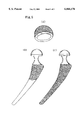
- a ring-shaped cement riser made of PMMA is installed at a collar portion of the proximal part 4 in FIG. 3 and at a distal portion thereof 5 in FIG. 3.
- cement mantle should be fabricated.
- the bone cement or bone paste consists of a mixture of PMMA resin and crushed bone autograft.
- the kneaded cement is used under a relative low viscosity.
- the cement is cooled to prolong the wetting time until 90 seconds, which is advantageous, but the strength of the cement is slightly reduced.
- the cement is mixed in vacuum to control the porosity.
- the temperature rise is a function of the rate of polymer formation, or curing rate, rather than the rate of polymerization of a single polymer. Furthermore, the temperature rise is dependent of the rate of heat evolution and not necessarily upon the total heat evolved. Depending on the polymer formation rate, the temperature rise is reported in a range of 70 to 120° C., which may irritate the surrounding hard and soft tissue and cause unexpected damage on them.
- the polymerization shrinkage it is reported that when methyl methacrylate monomer is polymerized, the density changes from 0.94 gm/cm 3 to 1.19 gm/cm 3 . This change in density results in a volumetric shrinkage of 21 per cent, usually called the polymerization shrinkage. However, about one third in weight of the resin is monomer, then the volumetric shrinkage will be about 7% (that is approximately 2% of linear shrinkage). This amount of shrinkage is still too large and causes a dimensional accuracy and stability.
- the compressive strength of PMMA is about 50 ⁇ 70 MPa; while the bending strength is about 60 ⁇ 65 MPa.
- the modulus of elasticity in compression is 1.8 ⁇ 2.0 GPa; while MOE value under the bending mode is about 1.3 ⁇ 1.5 GPa.
- an object of the present invention to provide an orthopedic implant system which meets all three compatibilities (namely, biological, mechanical, and morphological), so that a strong interlocking fixation due to the bony ingrowth can be achieved.
- the foam-shaped materials exhibit a continuously connected ligamented, reticulated open-cell geometry having a duodecahedronal cell shape, as seen in FIG. 4.
- the foam-shaped material is presently available in a density ranging from 1 to 20 % theoretical and a cell density of 10 ⁇ 50 cells per linear inch, with material density and cell density independently variable.
- Foamed material is presently produced in a wide range of plastics, metals and composites having either solid or tubular ligaments.
- the metals include aluminum, nickel, copper, silver, zinc, lead, tin, magnesium, and stainless steel.
- the example shown in FIG. 4 is a product by ERG, Inc., using Al-alloy (6101-T6).
- This aluminum foam metal has a modulus of elasticity in tension is 48 ⁇ 83 MPa (for 6 ⁇ 10% nominal density) and an MOE in compression of 69 ⁇ 114 MPa (for 6 ⁇ 10% nominal density).
- the procedure for producing a foamed aluminum body comprises adding a foaming agent to a pool of molten aluminum or aluminum alloy, mixing thoroughly, and pouring into a mold.
- the gas-separating additive must be perfectly wetted with the base metal. When necessary, this must be achieved by grinding the metal hydride in a preliminary melt of the base metal. If the metal hydride decomposes at temperatures below the fusion temperature of the base metal, grinding has to be performed in pressurized atmosphere. The premix is allowed to solidify and is reduced to power before being added to the bulk quantity of the powdered or granulated base metal. If titanium hydride is used, the amount to be added should be about 10%.
- the mixture when ready contains a gas-separating additive which decomposes at temperatures below the fusion temperature of the base metal, it is placed on a conveyer belt of a pressurized installation, passed through a heating zone for melting--while suppressing the decomposition of the additive--and then retransferred into normal atmosphere.
- the foamed metal which forms instantaneously is then solidified by immediate quenching in water.
- foamed aluminum plates can be made in thicknesses of up to 200 mm. Because of the nature of the material, these plates will not solidify with a uniform density, the gas bubbles ranging from ⁇ 1 mm in diameter at the surface to about 6 mm in diameter in the core of the plate.
- the density of the material can be adjusted to values of from 0.2 to 0.5 gm/cm 3 .
- the spherical pores provide the foamed metal with isotropical properties in all directions.
- the strength (50 ⁇ 70 MPa) of foam Al alloy is about 30% of strength of solid Al alloy
- MOE values (1.3 ⁇ 2 GPa) of foam Al alloy is about 3% of the solid Al alloy.
- the strength of CP titanium foam will be about 90 MPa (which is 30% of strength of solid CP titanium)
- the MOE of CPT will be 3.06 GPa (which is about 3% of the solid CP titanium).
- 285 MPa will be expected for the strength of foam Ti-6Al-4V alloy and 3.42 GPa will be speculated for the MOE value of the foam Ti-6Al-4V alloy.
- the present invention provides a new orthopedic implant system which comprises of a solid stem and head, foamed sheet made of biocompatible materials (mainly CPT or Ti-based alloy) being covered over partial or entire surfaces of said stem and head, and reinforced PMMA resin serving as a bone cement partially being impregnated into open-cells of said foamed metallic sheet.
- the presently invented implant system has a uniquely divided but structurally continuous three layers; (i) a solid metallic sten or head, (ii) coarse foamed metallic sheet which is impregnated by the reinforced PMMA resin (which will be described later in a great detail), and (iii) a layer of the reinforced PMMA resin.
- the foamed CPT or Ti-6Al-4V sheet will possess mechanical properties somewhat between those values of solid COT ot Ti-6Al-4V and those values of the vital bone structure. But the estimated strength and MOE values of foamed Ti sheet are not good enough to find themselves exactly between those values; particularly the estimated MOE value (3 ⁇ 3.4 GPa) of foamed Ti metal sheet is lower than the MOE value range (9 ⁇ 12 GPa) for bone.
- foam metal sheet (of Al alloy) was impregnated with PMMA resin which is reinforced with said metallic oxides.
- a certain type of metallic oxides such as Al 2 O 3 , MgO, ZrO 2
- PMMA is mixed with these metallic oxides and embedded into a metallic foam network, it was found that this modification provided us the best results of all in terms of mechanical properties (strength and MOE values) and physical properties (temperature rise and polymerization shrinkage).
- FIG. 1 shows a general view to show how the stem with a ball head prothesis 1 is implanted into a femur and how the socket-type cup 2 with insert potion is implanted into an acetabulum.
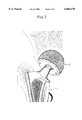
- FIG. 2 shows an enlarged view showing a jointing manner of said ball head portion of the femoral prothesis 1 into an inner insert portion of the acetabular cup 2.
- FIG. 3 shows a complete view of an orthopedic implant system, wherein there is a femoral prothesis 1, an acetabular cup 2, a stem proximal portion 3, a collar portion 4, and a distal portion 5.
- FIG. 4 depicts a microstructure of Al alloy foam network, taken under a magnification of X10.
- FIG. 5(A) shows the bonding manner of titanium or titanium alloy foam network sheet to the outer peripheral surface of the acetabular cup, according to the present invention.
- FIG. 5(B) and (C) show the different ways to bond the titanium or titanium alloy foam network sheet onto the proximal portion (B) or entire surface of the stem portion (C) of the femoral prothesis, according to the present invention.
- Titanium or titanium ally (for example, Ti-6Al-4V) is formed into a continuous network of foam metal sheet, having an open-cell size of 0.5 mm ⁇ 1.5 mm.
- an average size of the open-cell is 1.0 mm which is close to open size of the cancellous bone structure.
- the thickness of the sheet will be from 1.5 mm to about 3 mm, depending on the open-cell size, so that at least three (3) layers of the open-cell structure will be included within the foam-shaped network sheet.
- FIG. 5 (A) shows an example of a case when the entire outer spherical surface of the cup is covered with the foam-shaped network sheet.
- the application of said sheet is not limited to this manner, the sheet can also be applied to cover a portion of the cup.
- the thickness is less than 1.5 mm, many unconnected cell skeletons are revealed in the sheet, causing the mechanical weakness.
- the thickness of the sheet exceeds 3 mm, there are two choices, one is the reduction of thickness of metallic acetabular cup or the other alternative is increasing the amount of bone to be removed to accommodate the thickened acetabular cup portion. Either one of these choices is not preferred.
- the network sheet which is cut in an appropriate dimension is bonded to the solid metallic substrates of the acetabular cup and femoral stem.
- the bonding can be achieved by the solid state or liquid-phase involved joining techniques.
- the latter technique may include the laser welding, soldering or fusion welding.
- it is preferable to utilize the solid-state diffusion bonding since titanium is one of the typical materials which can be jointed successfully by the solid-state diffusion bonding technique.
- the casting technique can be used to fabricate te cup and stem with entirely or partially covered with the foam-shaped network using a cast-in-cast method or the lost wax technique.
- Electroplating method can also be applied to produce foamed titanium sheet.
- Untreated reticulated urethane foam substrate can be electroless-plated.
- the solid protheses of an acetabular cup and a femoral stem are covered entirely or partially with the titanium or titanium-based alloy foam-shaped network sheet with an average open-cell size of 1 mm and an average thickness ranging from 1.5 mm to 3 mm.
- the open-cells of said network are then impregnated with the reinforced bone cement which is described in the following.
- both break strength and modulus of elasticity were significantly improved. More specifically, the said improvement can be more intensified in a range of 10 to 15 vol. %.
- alumina, magnesia, and zirconia exhibit beneficial effects on improving mechanical properties.
- these metallic oxides are also admixed.
- MP II indicates the metal primer II (a product from G corporation).
- metal oxides is individually admixed to the polymer powders in the above tests, a combination of alumina, magnesia, and zirconia can further improve the mechanical properties.
- the selected samples are; (1) untreated PMMA, serving as a control sample, (2) 15 vol. % Al 2 O 3 added PMMA (from Table 1), (3) 15 vol. % MgO added PMMA (from Table 5), (4) 15 vol. % ZrO 2 added PMMA (from Table 6), (5) Al network sheet impregnated by un-reinforced PMMA (from Table 7), (6) Al network sheet +MP II+15 vol. % alumina added PMMA (from Table 7), (7) Al network sheet +MP II+15 vol. % magnesia added PMMA (from Table 7), and (8) Al network sheet +MP II+15 vol. % zirconia added PMMA (from Table 7). The obtained results are shown in Table 8.
- zirconia is a very effective oxide to suppress the temperature rise and polymerization shrinkage, if the oxide alone is added to PMMA powder resin, (2) aluminum network sheet exhibits another effective function to improve the above mentioned physical properties, and (3) furthermore, a combination of the aluminum network sheet, zirconia, and metal primer provides the best result.
- an improved orthopedic implant system possesses biological, mechanical and morphological compatibilities. All materials used for said systems is biologically safe and compatible to receiving vital hard and soft tissues.
- mechanical compatibility as discussed previously in details, the foam metal network being impregnated with reinforced bone cement (mainly consisted of polymethyl methacrylate resin and metallic oxides) exhibits the intermediate values in mechanical strength and modulus of elasticity between those for the solid CPT or Ti-based alloys cup and stem and those for reinforced PMMA bone cement. Hence, even under the discontinuous stress field, the load can be smoothly transmitted and the mechanical compatibility can be achieved.
- a continuously connected ligamented, reticulated open-cell geometry of the foam metal network having a duodecahedronal cell shape is morphologically compatible to receiving bone, particularly the cancellous (or spongy) bone structure.
- a production trial was carried out to fabricate a titanium foam metal sheet.
- a commercially available aluminum foam block which has an average open-cell size of about 1 mm diameter was employed. This block is a product from ERG, Inc., as described previously.
- a basic idea was to replicate the aluminum foam structure to be replaced by titanium metal.
- a molding cement was prepared and fed into open cells of the aluminum foam structure. After hardening the molding cement, the mold was heated in a furnace at 670° C. for about 1 hour to let all aluminum melt and flow out the mold. Then the mold with open-cells was placed into a vacuum-controlled casting machine for titanium. After the all titanium was solidified, the mold was removed from the casting machine and the molding cement was removed by using the air-borne particle blasting machine with fine particle of aluminum oxide. The surface topology of titanium foam sheet was observed to be identical to original aluminum open-cells.
- the presently invented orthotopic implant system can be applied as a total hip replacement and also applied to knee, elbow, shoulder, ankle, or finger(s).
- the reinforced PMMA can be used not only for a bone cement to form a cement mantle, but also can be employed solely to fabricate provisional crowns and bridges in dentistry.
Abstract
It is an object of the present invention to provide an improved orthopedic implant system with satisfied biological, mechanical and morphological compatibilities.
Solid metal femoral stem (entirely or partially) and solid metal acetabular head (entirely or partially) are covered with diffusion-bonded foamed-shaped sheet made of commercially pure titanium or titanium alloy(s). The open-cells in said foamed metal sheet are impregnated with biocompatible polymethyl methacrylate resin cement, which is reinforced with selected oxides (e.g., alumina, magnesia, zirconia, or a combination of these oxides) along with an application of a small amount of a metal primer agent.
Description
The present invention relates to an orthopedic implant system. More specifically, the present invention relates directly to a surface modification of the total hip prothesis system to improve the bony ingrowth for a better biological fixation, and to modify the bone cement in terms of both mechanical and physical properties.
Total hip replacement protheses have now been in use clinically for over 35 years. Most experience has been gained in the hip joint, where naturally, the prothesis is of the ball-head of the femoral stem and socket-type cup of the acetabular component. More recently reconstruction of the knee, elbow, shoulder and ankle have been attempted using protheses of more varied mechanisms and design, although the basic concept for designing these protheses remains unchanged.
One of the many universal requirements of implants, wherever they are used in the body, is the ability to form a suitably stable mechanical unit with the neighboring tissues. A loose (or unstable) implant may function less efficiently or cease functioning completely or it may induce an excessive tissue response. In either case it may cause the patient discomfort and pain and in several situations a loose implant is deemed to have failed and has to be surgically removed. Total hip replacement protheses have focused attention onto this stability question of the propensity for the load-transmitting protheses to become over a period of time.
There are several ways in which the stability problem could be approached. One could consider a chemical approach in which adhesives were used to bond protheses to tissues. Secondly, there is the mechanical approach and at least three different methods have been tried. One is now in routine clinical use and involves the interposition of a mass of acrylic "cement" into the space between prothesis and bone. There is no adhesive action as such, but rather a grouting effect which gives a mechanically stable unit and provides uniform load transfer.
Alternatively, one may choose an exceptionally inert material and prepare an accurate, well designed bed in the bone for the prothesis. The initial stability and the minimization of differentiation processes around the prothesis may allow new bone to grow right up to the prothesis surface to give a long-term stability. Using high density alumina ceramic appears to be a very bio-inert material and is a suitable candidate for these protheses. However, as one might expect, the situation is not quite this simple and loosening can occur, even with very accurate bone-bed preparation. Surface macro-profiles on the prothesis may improve the stability but even here the limitations of this primary mechanical interlocking are shown.
In a third mechanical approach, the tissues do the work rather than the prothesis or a cement for this relies on tissue ingrowth into a micro-porous structure.
The important point is the extent of the fibrous tissue layer, if any, that forms between the material and the bone. It can be explained that it may need a biodegradable cement or temporary fixation to give initial stability while the necessary bone remodelling takes place to achieve a fibrous-tissue-free interface.
The development of porous implant was based on three key needs; biocompatibility through the use of ingredients (polytetrafluoroethylene and carbon fibers) that are stable in the body milieu and induce minimal biological disturbance through surface effects; biofunctionality through the selection of a modulus close to that of soft tissue and spongy bone (4 MPa); and maximum porosity (80 per cent void, 100˜500 μm diameter) and inter-pore connections (100˜200 μm) for tissue ingrowth consistent with appropriate bulk mechanical properties (ultimate tensile strength 1 MPa).
Referring to FIGS. 1 and 2, typical type of an orthopedic implant, particularly the total hip prothesis system, consists of a femoral prothesis 1 which has a long stem extending from a proximal portion with a ball-head portion to a distal portion and is inserted into a femur, and an acetabular socket-type cup 2 with an interlocking ring and insert which is implanted into an acetabulum. FIG. 3 shows an example of the total hip prothesis stem in which said femoral prothesis stem is interlocked into an acetabular cup (not shown).
It has been, for a long time, recognized that any type of implants (whether it is a dental implant or an orthopedic implant), should possess a biological compatibility against an implant-receiving surrounding hard and soft tissues. Accordingly, the materials for implants are limited to a certain type of materials, including a commercially pure titanium (CPT), T-6Al-4V alloy, Co--Cr or Co--Cr--Mo alloy, Fe--Cr--Mn--Ni--Mo--Nb--Si alloy, AISI Type 316L stainless steel, and some ceramic materials including pure alumina and the synthetic hydroxyapatite.
Similar type of materials used for the femoral protheses can be found for the acetabular cups. One exception is that the ultra high molecular weight polyethylene (UHWMPE; average molecular weight is about more than 5,500,000) is a mainly selected material for the acetabular socket-type cup.
For these femoral protheses and acetabular cups, several surface modifications have been proposed and used for enhancing the mechanical retention and promotion of the bony ingrowth.
In the ORTHOMET® system, very small size of commercially pure (CP) titanium beads are plasma-coated onto Ti-6Al-4V femoral stem to produce a porosity of about 50%. The CPT beads are coated on a proximal portion 3 in FIG. 3.
In the KYOCERA® system, CPT is coated onto a proximal portion of the Ti-6Al-4V stem material by the inert gas shielded arc spray to generate a coarse surface.
In the DePuy® system, very fine size of CPT beads are also coated on a proximal portion of the femoral stem to ensure a complete contact with the relatively weak cancellous bone structure.
In the development history of the bony ingrowth type artificial joint protheses, there are two major design concepts available; namely, (1) a micro-fit type (or bone ongrowth type) in which grooves are formed on the surface of the implants, and (2) a micro-interlock type (or bone ingrowth type) in which the surface of implants is formed to be porous structure. In MULTILOCK® system, the mesh pad made of titanium fibers is bonded onto the proximal portion of the femoral prothesis through a solid state diffusion bonding technique.
In DePuy® system and STRAYKER® system, Co--Cr beads (approximately 150˜300 μm diameter) are sprayed or sintered to Co--Cr femoral stem material to generate an average pore size of 275 μm.
In MATRIX HIP® system, the proximal portion of Co--Cr femoral stem was machined to create the macro- and micro-texturing for a reduced interfacial shear stress.
In ORTHOMET® system, polymethyl methacrylate (PMMA) resin studs are bonded as a pre-coating on an entire stem portion to enhance the bonding strength with the bone cement when the femoral stem is implanted into the cement mantle.
Moreover, in KYUCERA® system, synthetic hydroxyapatite is plasma-coated on the proximal portion of the stem to have an average particle size of 50 μm. Alternatively, the proximal portion is subjected to a blasting to form a micro-textured structure.
Moving to the acetabular socket-type cups, in ORTHOMET® system, CPT beads are plasma-coated onto Co--Cr alloy to reduce the occurrence of the loosening.
In a similar manner, in STRYKER® system, CPT beads are bonded on the outer shell of the socket to enhance the mechanical retention.
In MULTILOCK® system, a plurality of PMMA studs are adhered on the outer surface of the acetabular head to promote a chemical bond with PMMA in the cement mantle.
In AcSysHA® system, synthetic hydroxyapatite is coated on an entire outer surface of the acetabular cup.
In the most outer shell of the acetabular head, a plurality of multi-hole, in-hole or cluster-hole are formed to promote the bony ingrowth activity.
Some of the aforementioned surface modification, particularly hydroxyapatite coating, are related to the mechanical compatibility (which is the second compatibility required for the successful implant system). As discussed previously, the protheses, particularly surface zone thereof, should respond to the load-transmitting function.
Suppose that an implant made of Ti-6Al-4V alloy is used. It is reported that strength and modulus of elasticity (MOE) of Ti-6Al-4V alloy are in ranges of 900˜1200 MPa and 200˜300 GPa, respectively. On the other hand, the strength and MOE of an implant-receiving bone has ranges of 100˜200 MPa and 9˜12 GPa, respectively. There are great differences in mechanical properties between these materials. With respect to the load-transmitting function, the strain field should be continuous. If the strain field in a metallic implant and bone system is not continuous, then the interface is not adhered and is easily debonded. However, even the strain field is continuous, the stress field has always a discontinuity due to the differences in MOE values between two dissimilar materials. If the discontinuous stress at an interface exceeds the interfacial bonding strength, then the bonding is failed.
Based on the above discussion, it is easily understood that the synthetic hydroxyapatite is plasma-coated onto the metallic implant, since the hydroxyapatite has not only an excellent biocompatibility, but it also exhibits the strength (400˜800 MPa) and MOE (40˜120 GPa) which are just between those values for Ti-6Al-4V and the implant-receiving bone structure. This is what is claimed as the second compatibility; namely, a mechanical compatibility to form a continuous gradient functional (in terms of mechanical property) structure.
Furthermore, the third compatibility should be included to the aforementioned currently accepted compatibilities; that is a morphological compatibility. In a scientific article published by this inventor (entitled "Fractal Dimension Analysis of Mandibular Bones: Toward a Morphological Compatibility of Implants" in Bio-Medical Materials and Engineering, Vol. 4 No. 5, pp.397/407, 1994), surface morphology of successful implants has a limitation of the average roughness (1˜500 μm) and average pore size (10˜500 μm), regardless of types of implant materials including metallic materials, ceramic materials and polymeric materials. If the pore size is smaller than 10 μm, the surfaces will be more toxic to fibroblastic cells and has an adverse effect on cells due to their physical presence independent of any chemical toxic effects. If the pore size is larger than 500 μm, the surface does not exhibit any sufficient structural integrity.
Moreover, as discussed previously, a mesh pad made of titanium beads is coated on the proximal portion of the femoral stem to promote the bony ingrowth. However, the opening space is not morphologically compatible to surrounding bone, particularly the cancellous bone. Any one of the beads (either made of Ti, hydroxyapatite, or PMMA) does not create sufficient opening space for the bony ingrowth and, then, is not morphologically compatible.
Successful implant system is relied not only on these three compatibilities, but also on cementation of the space gap between the prothesis and the vital bone.
With a certain type of the femoral prothesis, a ring-shaped cement riser made of PMMA is installed at a collar portion of the proximal part 4 in FIG. 3 and at a distal portion thereof 5 in FIG. 3. However, in the most femoral protheses and acetabular cups, there is no pre-formed cement riser. Hence, during a surgical operation, cement mantle should be fabricated. Normally, the bone cement or bone paste consists of a mixture of PMMA resin and crushed bone autograft.
For a normal practice, the kneaded cement is used under a relative low viscosity. In order to keep the low viscous cement, the cement is cooled to prolong the wetting time until 90 seconds, which is advantageous, but the strength of the cement is slightly reduced. In order to compensate the reduced mechanical strength, the cement is mixed in vacuum to control the porosity.
Even with the aforementioned efforts, there are several important factors unsolved. These include (1) the heat generated during the polymerization of methyl methacrylate monomer, (2) the polymerization shrinkage, and (3) insufficient break strength and the modulus of elasticity.
Regarding the heat generated, the temperature rise is a function of the rate of polymer formation, or curing rate, rather than the rate of polymerization of a single polymer. Furthermore, the temperature rise is dependent of the rate of heat evolution and not necessarily upon the total heat evolved. Depending on the polymer formation rate, the temperature rise is reported in a range of 70 to 120° C., which may irritate the surrounding hard and soft tissue and cause unexpected damage on them.
With regard to the polymerization shrinkage, it is reported that when methyl methacrylate monomer is polymerized, the density changes from 0.94 gm/cm3 to 1.19 gm/cm3. This change in density results in a volumetric shrinkage of 21 per cent, usually called the polymerization shrinkage. However, about one third in weight of the resin is monomer, then the volumetric shrinkage will be about 7% (that is approximately 2% of linear shrinkage). This amount of shrinkage is still too large and causes a dimensional accuracy and stability.
Regarding the mechanical properties, it is reported that the compressive strength of PMMA is about 50˜70 MPa; while the bending strength is about 60˜65 MPa. The modulus of elasticity in compression is 1.8˜2.0 GPa; while MOE value under the bending mode is about 1.3˜1.5 GPa. These are lower than those values for bone structure.
It is, therefore, an object of the present invention to provide an orthopedic implant system which meets all three compatibilities (namely, biological, mechanical, and morphological), so that a strong interlocking fixation due to the bony ingrowth can be achieved.
It is another object of the present invention to provide the modified and reinforced polymethyl methacrylate resin as a bone cement as well as a material which can be used as a provisional crowns and bridges in dentistry, having a less temperature rise and shrinkage during the polymerization process.
In order to achieve the first object, it was found that the foam-shaped materials exhibit a continuously connected ligamented, reticulated open-cell geometry having a duodecahedronal cell shape, as seen in FIG. 4. The foam-shaped material is presently available in a density ranging from 1 to 20 % theoretical and a cell density of 10˜50 cells per linear inch, with material density and cell density independently variable.
Foamed material is presently produced in a wide range of plastics, metals and composites having either solid or tubular ligaments. The metals include aluminum, nickel, copper, silver, zinc, lead, tin, magnesium, and stainless steel. The example shown in FIG. 4 is a product by ERG, Inc., using Al-alloy (6101-T6). This aluminum foam metal has a modulus of elasticity in tension is 48˜83 MPa (for 6˜10% nominal density) and an MOE in compression of 69˜114 MPa (for 6˜10% nominal density).
The patent literature (U.S. Pat. No. 2,434,775 January 1948, and U.S. Pat. No. 2,553,016, May 1951) describes a foam-like metal made of heating under pressure a mixture or composite of a metal and gas-forming material to a temperature above the melting point of the base metal and then releasing the pressure to permit the gas-forming material to decompose. On solidifying, the metal assumes a foamed structure.
Basically, the procedure for producing a foamed aluminum body comprises adding a foaming agent to a pool of molten aluminum or aluminum alloy, mixing thoroughly, and pouring into a mold.
An initial proposal by B. Sosnik in 1948 (U.S. Pat. Nos. 2,434,775 and 2,553,016) was concerned with the making of foamed metals with the aid of an alloying constituent to form a vapor phase at metal fusion temperatures. According to Sosnik, this can be achieved by using a metal mixture made up, e.g., of 10% Hg and 90% Al. This is fused in a pressurized chamber, then the pressure is reduced to atmospheric so that Hg-vapor filled cavities may form, which gives rise to a foamed structure when the metal is allowed to cool. The procedure developed by Bjorksten Research Laboratory Inc., Madison Wis., in 1956 is based on a similar idea, i.e., the decomposition of metal hydrides, for example titanium or zirconium hydrides, or of carbonates etc., in melts of metal alloys with a wide solidification range, preferably Al:Mg melts.
To ensure that the process operates under optimum conditions, the gas-separating additive must be perfectly wetted with the base metal. When necessary, this must be achieved by grinding the metal hydride in a preliminary melt of the base metal. If the metal hydride decomposes at temperatures below the fusion temperature of the base metal, grinding has to be performed in pressurized atmosphere. The premix is allowed to solidify and is reduced to power before being added to the bulk quantity of the powdered or granulated base metal. If titanium hydride is used, the amount to be added should be about 10%. If the mixture when ready contains a gas-separating additive which decomposes at temperatures below the fusion temperature of the base metal, it is placed on a conveyer belt of a pressurized installation, passed through a heating zone for melting--while suppressing the decomposition of the additive--and then retransferred into normal atmosphere. The foamed metal which forms instantaneously is then solidified by immediate quenching in water. In this way, foamed aluminum plates can be made in thicknesses of up to 200 mm. Because of the nature of the material, these plates will not solidify with a uniform density, the gas bubbles ranging from <1 mm in diameter at the surface to about 6 mm in diameter in the core of the plate. The density of the material can be adjusted to values of from 0.2 to 0.5 gm/cm3. Unlike the honeycomb material, the spherical pores provide the foamed metal with isotropical properties in all directions.
Although titanium foamed metal is not available currently, we are furnished with a technology to fabricate titanium foamed metal. According to the aluminum foam metal data as described before, the strength (50˜70 MPa) of foam Al alloy is about 30% of strength of solid Al alloy, and MOE values (1.3˜2 GPa) of foam Al alloy is about 3% of the solid Al alloy. Hence, it is expected that the strength of CP titanium foam will be about 90 MPa (which is 30% of strength of solid CP titanium) and the MOE of CPT will be 3.06 GPa (which is about 3% of the solid CP titanium). By the same token, 285 MPa will be expected for the strength of foam Ti-6Al-4V alloy and 3.42 GPa will be speculated for the MOE value of the foam Ti-6Al-4V alloy.
Hence, these values of strength and MOE values are cloase to somewhat between the solid metallic materials (Ti-6Al-4V, CPT or the others) and bone structure as mentioned previously. Hence the biological and mechanical compatibilities will be satisfied by using Ti foam metal at the interface of the prothesis and receiving vital bone structure.
Surprisingly, it is observed that the continuously connected ligamented, reticulated open-cell geometry of the foam metal (as seen in FIG. 4) bears a very resemblance to the cancellous (or spongy) bone. Hence the third morphological compatibility is now satisfied.
The present invention provides a new orthopedic implant system which comprises of a solid stem and head, foamed sheet made of biocompatible materials (mainly CPT or Ti-based alloy) being covered over partial or entire surfaces of said stem and head, and reinforced PMMA resin serving as a bone cement partially being impregnated into open-cells of said foamed metallic sheet. Hence the presently invented implant system has a uniquely divided but structurally continuous three layers; (i) a solid metallic sten or head, (ii) coarse foamed metallic sheet which is impregnated by the reinforced PMMA resin (which will be described later in a great detail), and (iii) a layer of the reinforced PMMA resin.
As described briefly in the above, it is speculated that the foamed CPT or Ti-6Al-4V sheet will possess mechanical properties somewhat between those values of solid COT ot Ti-6Al-4V and those values of the vital bone structure. But the estimated strength and MOE values of foamed Ti sheet are not good enough to find themselves exactly between those values; particularly the estimated MOE value (3˜3.4 GPa) of foamed Ti metal sheet is lower than the MOE value range (9˜12 GPa) for bone.
Hence it is needed to modify the mechanical properties of PMMA resin which is impregnated into open-cells of said foamed sheet. In order to reinforce PMMA (polymethyl methacrylate) resin, it was proposed to mix with a synthetic fluormica or plasma-treated woven polyethylene fiber as a reinforcement element as a prior art. These modifications have been proposed only for a purpose when PMMA resin is used for fabricating provisional crowns and bridges. These reinforcing filaments are relatively expensive and alignment of filaments is troublesome and time-consuming. In the present invention, therefore, the dispersive strengthening concept, which is normally employed in the reinforcing the composite materials, is used. For reinforcing dispersive particles, metallic oxides and synthetic hydroxyapatite are used in the present invention.
Moreover, foam metal sheet (of Al alloy) was impregnated with PMMA resin which is reinforced with said metallic oxides. It was found that a certain type of metallic oxides (such as Al2 O3, MgO, ZrO2) improved the mechanical strengths as well as the modulus of elasticity of PMMA matrix, and reduced the temperature rise during the polymerization process and the extent of the polymerization shrinkage. Furthermore, if PMMA is mixed with these metallic oxides and embedded into a metallic foam network, it was found that this modification provided us the best results of all in terms of mechanical properties (strength and MOE values) and physical properties (temperature rise and polymerization shrinkage).
The above and many other objectives, features and advantages of the present invention will be more fully understood from the ensuing detailed description of the preferred embodiment of the invention, which description should be read in conjunction with the accompanying drawings.
FIG. 1 shows a general view to show how the stem with a ball head prothesis 1 is implanted into a femur and how the socket-type cup 2 with insert potion is implanted into an acetabulum.
FIG. 2 shows an enlarged view showing a jointing manner of said ball head portion of the femoral prothesis 1 into an inner insert portion of the acetabular cup 2.
FIG. 3 shows a complete view of an orthopedic implant system, wherein there is a femoral prothesis 1, an acetabular cup 2, a stem proximal portion 3, a collar portion 4, and a distal portion 5.
FIG. 4 depicts a microstructure of Al alloy foam network, taken under a magnification of X10.
FIG. 5(A) shows the bonding manner of titanium or titanium alloy foam network sheet to the outer peripheral surface of the acetabular cup, according to the present invention.
FIG. 5(B) and (C) show the different ways to bond the titanium or titanium alloy foam network sheet onto the proximal portion (B) or entire surface of the stem portion (C) of the femoral prothesis, according to the present invention.
Titanium or titanium ally (for example, Ti-6Al-4V) is formed into a continuous network of foam metal sheet, having an open-cell size of 0.5 mm ˜1.5 mm. Preferably an average size of the open-cell is 1.0 mm which is close to open size of the cancellous bone structure. The thickness of the sheet will be from 1.5 mm to about 3 mm, depending on the open-cell size, so that at least three (3) layers of the open-cell structure will be included within the foam-shaped network sheet. FIG. 5 (A) shows an example of a case when the entire outer spherical surface of the cup is covered with the foam-shaped network sheet. However, the application of said sheet is not limited to this manner, the sheet can also be applied to cover a portion of the cup. If the thickness is less than 1.5 mm, many unconnected cell skeletons are revealed in the sheet, causing the mechanical weakness. On the other hand, if the thickness of the sheet exceeds 3 mm, there are two choices, one is the reduction of thickness of metallic acetabular cup or the other alternative is increasing the amount of bone to be removed to accommodate the thickened acetabular cup portion. Either one of these choices is not preferred.
The same argument can be applied to the femoral prothesis. Although it is not necessary to cover the whole stem portion (that is from the proximal portion right under the collar part of the stem to the distal end portion) with the Ti or T-based alloy foam-shaped network sheet, said foam sheet can be adhered to only the proximal portion (see FIG. 5 (B)), or the entire stem portion (see FIG. 5 (C)).
The network sheet which is cut in an appropriate dimension is bonded to the solid metallic substrates of the acetabular cup and femoral stem. The bonding can be achieved by the solid state or liquid-phase involved joining techniques. The latter technique may include the laser welding, soldering or fusion welding. However, it is preferable to utilize the solid-state diffusion bonding, since titanium is one of the typical materials which can be jointed successfully by the solid-state diffusion bonding technique.
Alternatively, the casting technique can be used to fabricate te cup and stem with entirely or partially covered with the foam-shaped network using a cast-in-cast method or the lost wax technique.
Electroplating method can also be applied to produce foamed titanium sheet. Untreated reticulated urethane foam substrate can be electroless-plated.
Accordingly, the solid protheses of an acetabular cup and a femoral stem are covered entirely or partially with the titanium or titanium-based alloy foam-shaped network sheet with an average open-cell size of 1 mm and an average thickness ranging from 1.5 mm to 3 mm. The open-cells of said network are then impregnated with the reinforced bone cement which is described in the following.
In our experiments, selected oxides are mixed with polymethyl methacrylate powder with a certain volume per cent and polymerized into a stainless steel mold (25×2×2 mm). After curing, the sample was subjected to a transverse rupture strength and modulus of elasticity measurements under a cross head speed of 1 mm/cm. For each group, five samples were prepared. The results are averaged out for five individual data points.
The following tables show the results.
              TABLE 1
______________________________________
Effect of aluminum oxide addition
( ): standard deviation
              break strength
                         modulus of
sample        (MPa)      elasticity (GPa)
______________________________________
PMMA (control)
              58.23 (1.54)
                         1.5 (0.14)
Al.sub.2 O.sub.3 5 vol. %
              58.39 (1.22)
                         1.6 (0.23)
10 vol. %     60.79 (2.10)
                         1.9 (0.33)
15 vol. %     63.76 (1.92)
                         1.8 (0.31)
20 vol. %     59.15 (2.08)
                         1.5 (0.21)
25 vol. %     50.43 (1.87)
                         1.5 (0.23)
30 vol. %     50.37 (1.22)
                         1.3 (0.17)
______________________________________
By admixing aluminum oxide particles to polymethyl methacrylate resin did not show a significant improvement in terms of mechanical properties, as seen in this table, although a slight increasing the mechanical properties is noticed when 5˜20 vol. % of alumina was added.
              TABLE 2
______________________________________
Effect of titanium oxide addition
( ): standard deviation
              break strength
                         modulus of
sample        (MPa)      elasticity (GPa)
______________________________________
PMMA (control)
              58.23 (1.54)
                         1.5 (0.14)
TiO.sub.2 5 vol. %
              54.75 (0.98)
                         1.4 (0.12)
10 vol. %     48.71 (1.43)
                         1.4 (0.22)
15 vol. %     43.73 (1.54)
                         1.5 (0.27)
20 vol. %     42.77 (1.96)
                         1.3 (0.19)
25 vol. %     40.56 (1.57)
                         1.3 (0.14)
30 vol. %     40.33 (1.52)
                         1.2 (0.13)
______________________________________
The similar conclusions as for the aluminum oxide addition can be found for titania addition. Again a slight improvement can be observed when 5 to 20 vol. % of titanium oxide particles are admixed.
              TABLE 3
______________________________________
Effect of calcium oxide addition
( ): standard deviation
              break strength
                         modulus of
sample        (MPa)      elasticity (GPa)
______________________________________
PMMA (control)
              58.23 (1.54)
                         1.5 (0.14)
CaO 5 vol. %  48.42 (1.34)
                         1.4 (0.16)
10 vol. %     39.99 (1.29)
                         1.3 (0.15)
15 vol. %     35.38 (1.28)
                         1.3 (0.23)
20 vol. %     33.24 (2.01)
                         1.2 (0.21)
25 vol. %     30.56 (1.38)
                         1.3 (0.15)
30 vol. %     30.11 (1.71)
                         1.2 (0.12)
______________________________________
There is no indication of improvements in mechanical properties when the calcium oxide particles are mixed to polymethyl methacrylate resin powders.
              TABLE 4
______________________________________
Effect of hydroxyapatite addition
( ): standard deviation
              break strength
                         modulus of
sample        (MPa)      elasticity (GPa)
______________________________________
PMMA (control)
              58.23 (1.54)
                          1.5 (0.14)
HA 5 vol. %   64.57 (2.11)
                         0.96 (0.12)
10 vol. %     60.56 (1.87)
                         0.89 (0.13)
15 vol. %     47.97 (1.55)
                         0.87 (0.17)
20 vol. %     48.09 (1.15)
                         0.79 (0.14)
25 vol. %     49.29 (1.76)
                         0.78 (0.21)
30 vol. %     50.21 (1.44)
                         0.72 (0.17)
______________________________________
Within a limited addition ranging from 5 to 10 vol. % of synthetic hydroxyapatite, a slight increasing in break strengths was noted. However, surprisingly no improvements in modulus of elasticity was indicated in all ranges of additive per cent.
              TABLE 5
______________________________________
Effect of magnesium oxide addition
( ): standard deviation
              break strength
                         modulus of
sample        (MPa)      elasticity (GPa)
______________________________________
PMMA (control)
              58.23 (1.54)
                         1.5 (0.14)
MgO 5 vol. %  65.79 (1.45)
                         1.9 (0.23)
10 vol. %     70.34 (1.33)
                         2.1 (0.33)
15 vol. %     72.45 (1.90)
                         2.2 (0.20)
20 vol. %     71.66 (1.33)
                         2.0 (0.32)
25 vol. %     67.43 (1.21)
                         1.8 (0.09)
30 vol. %     65.46 (1.82)
                         1.7 (0.13)
______________________________________
For all ranges of volume % addition of magnesium oxide particles, both break strength and modulus of elasticity were significantly improved. More specifically, the said improvement can be more intensified in a range of 10 to 15 vol. %.
              TABLE 6
______________________________________
Effect of zirconium oxide addition
( ): standard deviation
              break strength
                         modulus of
sample        (MPa)      elasticity (GPa)
______________________________________
PMMA (control)
              58.23 (1.54)
                         1.5 (0.14)
ZrO.sub.2 5 vol. %
              59.75 (1.92)
                         2.6 (0.11)
10 vol. %     62.35 (1.60)
                         2.7 (0.30)
15 vol. %     64.40 (1.19)
                         2.6 (0.47)
20 vol. %     62.21 (1.51)
                         2.4 (0.24)
25 vol. %     57.36 (1.70)
                         2.1 (0.22)
30 vol. %     56.77 (2.09)
                         2.2 (0.31)
______________________________________
Similar findings can be applied to the case when zirconium oxide particles are added, specifically the additive effect appears significant in a range of 10 to 20 vol. %.
In the above, alumina, magnesia, and zirconia exhibit beneficial effects on improving mechanical properties. Hence, in addition to applying aluminum foam-shaped network sheet, these metallic oxides are also admixed. In the following table, MP II indicates the metal primer II (a product from G corporation).
              TABLE 7
______________________________________
Effect of inserting aluminum network sheet
and several oxide particles addition
( ): standard deviation
                break strength
                           modulus of
sample          (MPa)      elasticity (GPa)
______________________________________
PMMA (control)  58.23 (1.54)
                           1.5 (0.14)
Al network      69.61 (2.01)
                           2.7 (0.12)
Al network + MP II
                72.74 (3.02)
                           3.2 (0.33)
Al network + Al.sub.2 O.sub.3
                73.75 (2.90)
                           2.8 (0.19)
5 vol. %
10 vol. %       75.95 (3.11)
                           3.1 (0.24)
15 vol. %       74.35 (1.99)
                           3.0 (0.18)
Al network + MP II
                80.45 (2.18)
                           3.3 (0.67)
+ Al.sub.2 O.sub.3 15 vol. %
Al network + MP II
                85.20 (2.29)
                           3.7 (0.11)
+ MgO 15 vol. %
Al network + MP II
                79.37 (1.94)
                           3.9 (0.34)
+ ZrO.sub.2 15 vol. %
______________________________________
Several important conclusions can be drawn from these data;
1) By impregnating metallic (in this study, aluminum foam metal is used) network sheet by un-reinforced polymethyl methacrylate, 20% improvement in strength and 80% improvement in MOE were noticed.
2) Furthermore, if MP II droplet is applied when metallic sheet is impregnated by the PMMA, 30% of strength and 100% (in other words, doubled) of MOE were improved.
3) Moreover, if a certain amount of oxide (for example, alumina) is admixed with polymeric resin powder (PMMA) prior to impregnating the metallic network sheet, about same extent of improvements as the previous case can be found.
4) As indicated in last three columns in Table 7, if 15 vol. % of alumina, magnesia, or zirconia was admixed to the polymer powder along with a droplet of MP II prior to impregnate the aluminum network sheet, remarkable improvements in both strengths and MOE are noticed.
5) Since for the case of using Al foamed sheet, the MOE value was improved as much as about 2.7 times as compared to that of PMMA control, hence it can be expected that if Ti foamed sheet is used, the resultant improved MOE value will be more than 10 GPa which is higher than those of the bone structure.
Although metal oxides is individually admixed to the polymer powders in the above tests, a combination of alumina, magnesia, and zirconia can further improve the mechanical properties.
Regarding the physical properties, several selected samples are subjected to measurements of temperature rise during the polymerization and a linear polymerization shrinkage. The selected samples are; (1) untreated PMMA, serving as a control sample, (2) 15 vol. % Al2 O3 added PMMA (from Table 1), (3) 15 vol. % MgO added PMMA (from Table 5), (4) 15 vol. % ZrO2 added PMMA (from Table 6), (5) Al network sheet impregnated by un-reinforced PMMA (from Table 7), (6) Al network sheet +MP II+15 vol. % alumina added PMMA (from Table 7), (7) Al network sheet +MP II+15 vol. % magnesia added PMMA (from Table 7), and (8) Al network sheet +MP II+15 vol. % zirconia added PMMA (from Table 7). The obtained results are shown in Table 8.
              TABLE 8
______________________________________
Temperature rise and polymerization shrinkage
( ): standard deviation
             temperature rise
                         polymerization
sample No.   (°C.)
                         shrinkage (%)
______________________________________
1            74.4 (0.4)  2.3 (0.09)
2            65.4 (0.4)  1.5 (0.09)
3            58.3 (0.2)  1.3 (0.11)
4            50.2 (0.4)  1.5 (0.10)
5            49.3 (0.2)  1.3 (0.11)
6            48.9 (0.3)  1.3 (0.13)
7            47.9 (0.2)  1.3 (0.11)
8            45.9 (0.3)  1.1 (0.08)
______________________________________
From Table 8, it was found that (1) zirconia is a very effective oxide to suppress the temperature rise and polymerization shrinkage, if the oxide alone is added to PMMA powder resin, (2) aluminum network sheet exhibits another effective function to improve the above mentioned physical properties, and (3) furthermore, a combination of the aluminum network sheet, zirconia, and metal primer provides the best result.
As a result, according to the present invention, an improved orthopedic implant system possesses biological, mechanical and morphological compatibilities. All materials used for said systems is biologically safe and compatible to receiving vital hard and soft tissues. With respect to mechanical compatibility, as discussed previously in details, the foam metal network being impregnated with reinforced bone cement (mainly consisted of polymethyl methacrylate resin and metallic oxides) exhibits the intermediate values in mechanical strength and modulus of elasticity between those for the solid CPT or Ti-based alloys cup and stem and those for reinforced PMMA bone cement. Hence, even under the discontinuous stress field, the load can be smoothly transmitted and the mechanical compatibility can be achieved.
Finally, a continuously connected ligamented, reticulated open-cell geometry of the foam metal network having a duodecahedronal cell shape is morphologically compatible to receiving bone, particularly the cancellous (or spongy) bone structure.
Embodiment
A production trial was carried out to fabricate a titanium foam metal sheet. For the present trial, a commercially available aluminum foam block which has an average open-cell size of about 1 mm diameter was employed. This block is a product from ERG, Inc., as described previously. A basic idea was to replicate the aluminum foam structure to be replaced by titanium metal. A molding cement was prepared and fed into open cells of the aluminum foam structure. After hardening the molding cement, the mold was heated in a furnace at 670° C. for about 1 hour to let all aluminum melt and flow out the mold. Then the mold with open-cells was placed into a vacuum-controlled casting machine for titanium. After the all titanium was solidified, the mold was removed from the casting machine and the molding cement was removed by using the air-borne particle blasting machine with fine particle of aluminum oxide. The surface topology of titanium foam sheet was observed to be identical to original aluminum open-cells.
Medical Applications
The presently invented orthotopic implant system can be applied as a total hip replacement and also applied to knee, elbow, shoulder, ankle, or finger(s). The reinforced PMMA can be used not only for a bone cement to form a cement mantle, but also can be employed solely to fabricate provisional crowns and bridges in dentistry.
While this invention has been described in details with respect to preferred embodiment and examples, it should be understood that the invention is not limited to that precise embodiments; rather many modifications, and variations would present themselves to those of skill in the art without departing from the scope and spirit of this invention, as defined in the appended claims.
Claims (3)
1. An orthopedic implant system fixative comprising bone cement containing in admixture a biocompatible polymethyl methacrylate resin and metallic oxide particles selected from the group consisting of aluminum oxide, magnesium oxide, zirconium oxide, and mixtures thereof, wherein said metallic oxide particles have a particle size ranging from 1-5 μm, said metallic oxide particles serving to reinforce said resin.
2. The orthopedic implant system fixative as claimed in claim 1, wherein said metallic oxide particles have a loading percentage ranging form 5-20% by volume.
3. The orthopedic implant system fixative as claimed in claim 1, wherein said bone cement reinforced with metallic oxide particles is admixed with a metal primer including thiophosphoric acid methacrylate to enhance bonding between the polymethyl methacrylate resin, said metallic oxide particles, and metal foam network skeletons with which said bone cement is utilized.
Priority Applications (3)
| Application Number | Priority Date | Filing Date | Title |
|---|---|---|---|
| US08/678,676 US6066176A (en) | 1996-07-11 | 1996-07-11 | Orthopedic implant system |
| JP8244198A JPH11341A (en) | 1996-07-11 | 1996-08-27 | Orthopedic implant |
| US09/421,390 US6312473B1 (en) | 1996-07-11 | 1999-10-20 | Orthopedic implant system |
Applications Claiming Priority (1)
| Application Number | Priority Date | Filing Date | Title |
|---|---|---|---|
| US08/678,676 US6066176A (en) | 1996-07-11 | 1996-07-11 | Orthopedic implant system |
Related Child Applications (1)
| Application Number | Title | Priority Date | Filing Date |
|---|---|---|---|
| US09/421,390 Continuation US6312473B1 (en) | 1996-07-11 | 1999-10-20 | Orthopedic implant system |
Publications (1)
| Publication Number | Publication Date |
|---|---|
| US6066176A true US6066176A (en) | 2000-05-23 |
Family
ID=24723811
Family Applications (2)
| Application Number | Title | Priority Date | Filing Date |
|---|---|---|---|
| US08/678,676 Expired - Fee Related US6066176A (en) | 1996-07-11 | 1996-07-11 | Orthopedic implant system |
| US09/421,390 Expired - Fee Related US6312473B1 (en) | 1996-07-11 | 1999-10-20 | Orthopedic implant system |
Family Applications After (1)
| Application Number | Title | Priority Date | Filing Date |
|---|---|---|---|
| US09/421,390 Expired - Fee Related US6312473B1 (en) | 1996-07-11 | 1999-10-20 | Orthopedic implant system |
Country Status (2)
| Country | Link |
|---|---|
| US (2) | US6066176A (en) |
| JP (1) | JPH11341A (en) |
Cited By (57)
| Publication number | Priority date | Publication date | Assignee | Title |
|---|---|---|---|---|
| US6319285B1 (en) * | 1997-03-13 | 2001-11-20 | Ceramtec Ag Innovative Ceramic Engineering | Ceramic acetabular cup with metal coating |
| WO2002013730A2 (en) * | 2000-08-15 | 2002-02-21 | Sulzer Orthopedics Inc. | Two piece fused femoral hip stem |
| US6387132B1 (en) * | 1997-11-28 | 2002-05-14 | Ceramtec Ag Innovative Ceramic Engineering | Artificial joint of a prosthesis |
| US20030153981A1 (en) * | 2002-02-08 | 2003-08-14 | Wang Kathy K. | Porous metallic scaffold for tissue ingrowth |
| US20040243246A1 (en) * | 2003-05-27 | 2004-12-02 | Lyren Philip S. | Hip implant with porous body |
| US20050112397A1 (en) * | 2003-07-24 | 2005-05-26 | Rolfe Jonathan L. | Assembled non-random foams |
| US20050267585A1 (en) * | 2004-05-26 | 2005-12-01 | Sidebotham Christopher G | Canine acetabular cup |
| US20060108033A1 (en) * | 2002-08-05 | 2006-05-25 | Atakan Peker | Metallic dental prostheses made of bulk-solidifying amorphous alloys and method of making such articles |
| US20060149391A1 (en) * | 2002-08-19 | 2006-07-06 | David Opie | Medical implants |
| US20070118218A1 (en) * | 2005-11-22 | 2007-05-24 | Hooper David M | Facet joint implant and procedure |
| FR2896166A1 (en) * | 2006-01-18 | 2007-07-20 | Didier Pierre Rene Dages | Medical prosthesis e.g. hip prosthesis, forming method for e.g. bone repair surgery, involves coating chromium cobalt metallic layer of variable thickness on surface of prosthesis head, and then coating head with biocompatible ceramic layer |
| EP1813688A1 (en) * | 2004-11-15 | 2007-08-01 | Mitsubishi Materials Corporation | Titanium or titanium alloy sintered article of a sponge form excellent in compression strength |
| US20080269897A1 (en) * | 2007-04-26 | 2008-10-30 | Abhijeet Joshi | Implantable device and methods for repairing articulating joints for using the same |
| US20080268056A1 (en) * | 2007-04-26 | 2008-10-30 | Abhijeet Joshi | Injectable copolymer hydrogel useful for repairing vertebral compression fractures |
| US20080300682A1 (en) * | 2007-05-31 | 2008-12-04 | Depuy Products, Inc. | Sintered Coatings For Implantable Prostheses |
| US20090192609A1 (en) * | 2008-01-29 | 2009-07-30 | Zimmer, Inc. | Implant device for use in an implant system |
| US20090297603A1 (en) * | 2008-05-29 | 2009-12-03 | Abhijeet Joshi | Interspinous dynamic stabilization system with anisotropic hydrogels |
| US20090306673A1 (en) * | 2006-11-10 | 2009-12-10 | Fondel Finance B.V. | Kit and method for fixating a prosthesis or part thereof and/or filling osseous defects |
| US20100003155A1 (en) * | 2006-02-17 | 2010-01-07 | Biomet Manufacturing Corp. | Method and apparatus for forming porous metal implants |
| US7811291B2 (en) | 2007-11-16 | 2010-10-12 | Osseon Therapeutics, Inc. | Closed vertebroplasty bone cement injection system |
| US20100268347A1 (en) * | 2009-04-15 | 2010-10-21 | Weidong Tong | Micro and nano scale surface textured titanium-containing articles and methods of producing same |
| US20100268330A1 (en) * | 2009-04-15 | 2010-10-21 | Depuy Products, Inc. | Methods and Devices for Implants with Calcium Phosphate |
| US20110190902A1 (en) * | 2010-01-29 | 2011-08-04 | Depuy Products, Inc. | Methods and devices for implants with improved cement adhesion |
| US8021432B2 (en) | 2005-12-05 | 2011-09-20 | Biomet Manufacturing Corp. | Apparatus for use of porous implants |
| US8066778B2 (en) | 2005-04-21 | 2011-11-29 | Biomet Manufacturing Corp. | Porous metal cup with cobalt bearing surface |
| US8123814B2 (en) | 2001-02-23 | 2012-02-28 | Biomet Manufacturing Corp. | Method and appartus for acetabular reconstruction |
| US8157869B2 (en) | 2007-01-10 | 2012-04-17 | Biomet Manufacturing Corp. | Knee joint prosthesis system and method for implantation |
| US8163028B2 (en) | 2007-01-10 | 2012-04-24 | Biomet Manufacturing Corp. | Knee joint prosthesis system and method for implantation |
| US8187280B2 (en) | 2007-10-10 | 2012-05-29 | Biomet Manufacturing Corp. | Knee joint prosthesis system and method for implantation |
| US8197550B2 (en) | 2005-04-21 | 2012-06-12 | Biomet Manufacturing Corp. | Method and apparatus for use of porous implants |
| US20120221115A1 (en) * | 2011-02-24 | 2012-08-30 | Komistek Richard D | Maintaining proper mechanics tha |
| US8266780B2 (en) | 2005-04-21 | 2012-09-18 | Biomet Manufacturing Corp. | Method and apparatus for use of porous implants |
| US8292967B2 (en) | 2005-04-21 | 2012-10-23 | Biomet Manufacturing Corp. | Method and apparatus for use of porous implants |
| US8328873B2 (en) | 2007-01-10 | 2012-12-11 | Biomet Manufacturing Corp. | Knee joint prosthesis system and method for implantation |
| US8383033B2 (en) | 2009-10-08 | 2013-02-26 | Biomet Manufacturing Corp. | Method of bonding porous metal to metal substrates |
| US8562616B2 (en) | 2007-10-10 | 2013-10-22 | Biomet Manufacturing, Llc | Knee joint prosthesis system and method for implantation |
| US20140172113A1 (en) * | 2011-12-07 | 2014-06-19 | Smith & Nephew, Inc. | Orthopedic augments having recessed pockets |
| US8827981B2 (en) | 2007-11-16 | 2014-09-09 | Osseon Llc | Steerable vertebroplasty system with cavity creation element |
| US9192459B2 (en) | 2000-01-14 | 2015-11-24 | Bonutti Skeletal Innovations Llc | Method of performing total knee arthroplasty |
| US9510885B2 (en) | 2007-11-16 | 2016-12-06 | Osseon Llc | Steerable and curvable cavity creation system |
| US9539095B2 (en) | 2012-01-09 | 2017-01-10 | Zimmer, Inc. | Porous metal implants with bone cement |
| US9668745B2 (en) | 2011-12-19 | 2017-06-06 | Depuy Ireland Unlimited Company | Anatomical concentric spheres THA |
| US9700416B2 (en) | 2012-06-21 | 2017-07-11 | DePuy Synthes Products, Inc. | Constrained mobile bearing hip assembly |
| EP2164428B1 (en) * | 2007-05-29 | 2017-08-09 | Limacorporate SPA | Prosthetic element and relative method to make it |
| US10045801B2 (en) | 2012-07-30 | 2018-08-14 | The Governing Council Of The University Of Toronto | Bone stabilization device and method of production |
| US10213310B2 (en) * | 2012-06-18 | 2019-02-26 | DePuy Synthes Products, Inc. | Dual modulus hip stem and method of making the same |
| US10213314B2 (en) | 2013-03-14 | 2019-02-26 | DePuy Synthes Products, Inc. | Femoral component for an implantable hip prosthesis |
| US10463380B2 (en) | 2016-12-09 | 2019-11-05 | Dfine, Inc. | Medical devices for treating hard tissues and related methods |
| US10478241B2 (en) | 2016-10-27 | 2019-11-19 | Merit Medical Systems, Inc. | Articulating osteotome with cement delivery channel |
| US10624652B2 (en) | 2010-04-29 | 2020-04-21 | Dfine, Inc. | System for use in treatment of vertebral fractures |
| US10660656B2 (en) | 2017-01-06 | 2020-05-26 | Dfine, Inc. | Osteotome with a distal portion for simultaneous advancement and articulation |
| US20200354521A1 (en) * | 2017-05-17 | 2020-11-12 | Kyushu University, National University Corporation | Medical polymer material having excellent bone-bonding property |
| US11026744B2 (en) | 2016-11-28 | 2021-06-08 | Dfine, Inc. | Tumor ablation devices and related methods |
| US11197681B2 (en) | 2009-05-20 | 2021-12-14 | Merit Medical Systems, Inc. | Steerable curvable vertebroplasty drill |
| US20220168110A1 (en) * | 2020-11-12 | 2022-06-02 | Common Sense Engineering And Consult B.V. | Acetabulair implantaat en werkwijze voor het vervormen van dit implantaat |
| US11384260B1 (en) | 2021-05-28 | 2022-07-12 | Cohesys Inc. | Adhesive devices and uses thereof |
| US11510723B2 (en) | 2018-11-08 | 2022-11-29 | Dfine, Inc. | Tumor ablation device and related systems and methods |
Families Citing this family (119)
| Publication number | Priority date | Publication date | Assignee | Title |
|---|---|---|---|---|
| DE19813074A1 (en) * | 1998-03-25 | 1999-09-30 | Ceram Tec Ag Innovative Cerami | Clamp-fit connection between prosthetic components of joint prostheses |
| SE515227C2 (en) * | 1999-04-28 | 2001-07-02 | Bruce Medical Ab | Body for providing and growing bone and / or connective tissue and methods for making the body |
| EP1362129A1 (en) * | 2001-02-19 | 2003-11-19 | IsoTis N.V. | Porous metals and metal coatings for implants |
| EP1399098A1 (en) * | 2001-06-13 | 2004-03-24 | Bomet Merck GmbH | Hip endoprosthesis |
| DE10129842C1 (en) * | 2001-06-15 | 2003-04-24 | Bam Bundesanstalt Matforschung | Process for the production of a bioactive bone cement and bone cement kit |
| GB0207170D0 (en) * | 2002-03-26 | 2002-05-08 | Mcminn Derek J W | Hip joint prosthesis |
| US20030220696A1 (en) * | 2002-05-23 | 2003-11-27 | Levine David Jerome | Implantable porous metal |
| JP2004067547A (en) | 2002-08-02 | 2004-03-04 | Yoshinori Kuboki | Titanium fiber medical material |
| US6860932B2 (en) | 2002-09-12 | 2005-03-01 | Yoshiki Oshida | Dental and medical cement |
| US6887278B2 (en) * | 2002-11-04 | 2005-05-03 | Mayo Foundation For Medical Education And Research | Prosthetic implant having segmented flexible stem |
| US7575599B2 (en) * | 2004-07-30 | 2009-08-18 | Spinalmotion, Inc. | Intervertebral prosthetic disc with metallic core |
| US7270679B2 (en) * | 2003-05-30 | 2007-09-18 | Warsaw Orthopedic, Inc. | Implants based on engineered metal matrix composite materials having enhanced imaging and wear resistance |
| US6974625B2 (en) * | 2003-12-16 | 2005-12-13 | Smith & Nephew, Inc. | Oxidized zirconium on a porous structure for bone implant use |
| GB0422666D0 (en) * | 2004-10-12 | 2004-11-10 | Benoist Girard Sas | Prosthetic acetabular cups |
| US9907659B2 (en) | 2007-04-17 | 2018-03-06 | Biomet Manufacturing, Llc | Method and apparatus for manufacturing an implant |
| US20150335438A1 (en) | 2006-02-27 | 2015-11-26 | Biomet Manufacturing, Llc. | Patient-specific augments |
| US8608749B2 (en) | 2006-02-27 | 2013-12-17 | Biomet Manufacturing, Llc | Patient-specific acetabular guides and associated instruments |
| US8407067B2 (en) | 2007-04-17 | 2013-03-26 | Biomet Manufacturing Corp. | Method and apparatus for manufacturing an implant |
| US8858561B2 (en) | 2006-06-09 | 2014-10-14 | Blomet Manufacturing, LLC | Patient-specific alignment guide |
| US8535387B2 (en) | 2006-02-27 | 2013-09-17 | Biomet Manufacturing, Llc | Patient-specific tools and implants |
| US8473305B2 (en) | 2007-04-17 | 2013-06-25 | Biomet Manufacturing Corp. | Method and apparatus for manufacturing an implant |
| US9289253B2 (en) | 2006-02-27 | 2016-03-22 | Biomet Manufacturing, Llc | Patient-specific shoulder guide |
| US9113971B2 (en) | 2006-02-27 | 2015-08-25 | Biomet Manufacturing, Llc | Femoral acetabular impingement guide |
| US10278711B2 (en) | 2006-02-27 | 2019-05-07 | Biomet Manufacturing, Llc | Patient-specific femoral guide |
| US8070752B2 (en) | 2006-02-27 | 2011-12-06 | Biomet Manufacturing Corp. | Patient specific alignment guide and inter-operative adjustment |
| US8133234B2 (en) * | 2006-02-27 | 2012-03-13 | Biomet Manufacturing Corp. | Patient specific acetabular guide and method |
| US7967868B2 (en) | 2007-04-17 | 2011-06-28 | Biomet Manufacturing Corp. | Patient-modified implant and associated method |
| US8568487B2 (en) | 2006-02-27 | 2013-10-29 | Biomet Manufacturing, Llc | Patient-specific hip joint devices |
| US8298237B2 (en) | 2006-06-09 | 2012-10-30 | Biomet Manufacturing Corp. | Patient-specific alignment guide for multiple incisions |
| US9345548B2 (en) | 2006-02-27 | 2016-05-24 | Biomet Manufacturing, Llc | Patient-specific pre-operative planning |
| US8377066B2 (en) | 2006-02-27 | 2013-02-19 | Biomet Manufacturing Corp. | Patient-specific elbow guides and associated methods |
| US8241293B2 (en) | 2006-02-27 | 2012-08-14 | Biomet Manufacturing Corp. | Patient specific high tibia osteotomy |
| US8608748B2 (en) | 2006-02-27 | 2013-12-17 | Biomet Manufacturing, Llc | Patient specific guides |
| US9339278B2 (en) | 2006-02-27 | 2016-05-17 | Biomet Manufacturing, Llc | Patient-specific acetabular guides and associated instruments |
| US8092465B2 (en) | 2006-06-09 | 2012-01-10 | Biomet Manufacturing Corp. | Patient specific knee alignment guide and associated method |
| US8864769B2 (en) | 2006-02-27 | 2014-10-21 | Biomet Manufacturing, Llc | Alignment guides with patient-specific anchoring elements |
| US9173661B2 (en) | 2006-02-27 | 2015-11-03 | Biomet Manufacturing, Llc | Patient specific alignment guide with cutting surface and laser indicator |
| US9918740B2 (en) | 2006-02-27 | 2018-03-20 | Biomet Manufacturing, Llc | Backup surgical instrument system and method |
| US8591516B2 (en) | 2006-02-27 | 2013-11-26 | Biomet Manufacturing, Llc | Patient-specific orthopedic instruments |
| US8282646B2 (en) | 2006-02-27 | 2012-10-09 | Biomet Manufacturing Corp. | Patient specific knee alignment guide and associated method |
| US8603180B2 (en) | 2006-02-27 | 2013-12-10 | Biomet Manufacturing, Llc | Patient-specific acetabular alignment guides |
| US9795399B2 (en) | 2006-06-09 | 2017-10-24 | Biomet Manufacturing, Llc | Patient-specific knee alignment guide and associated method |
| DE102007031669A1 (en) * | 2006-08-04 | 2008-09-11 | Ceramtec Ag Innovative Ceramic Engineering | Asymmetrical design of acetabular cups to reduce cup deformations |
| JP2009066387A (en) * | 2007-03-15 | 2009-04-02 | Osaka Titanium Technologies Co Ltd | Composite material for living body and its manufacturing method |
| US8057914B2 (en) * | 2007-03-26 | 2011-11-15 | Howmedica Osteonics Corp. | Method for fabricating a medical component from a material having a high carbide phase and such medical component |
| US8920534B2 (en) | 2007-03-26 | 2014-12-30 | Howmedica Osteonics Corp. | Method for fabricating a biocompatible material having a high carbide phase and such material |
| US7771775B2 (en) * | 2007-03-26 | 2010-08-10 | Howmedica Osteonics Corp. | Method for fabricating a medical component from a material having a high carbide phase |
| US9370427B2 (en) * | 2007-04-13 | 2016-06-21 | Christopher G. Sidebotham | Bone-compliant femoral stem |
| CN101772357A (en) * | 2007-06-07 | 2010-07-07 | 史密夫和内修有限公司 | Reticulated particle porous coating for medical implant use |
| US8715359B2 (en) * | 2009-10-30 | 2014-05-06 | Depuy (Ireland) | Prosthesis for cemented fixation and method for making the prosthesis |
| US8632600B2 (en) | 2007-09-25 | 2014-01-21 | Depuy (Ireland) | Prosthesis with modular extensions |
| US9204967B2 (en) | 2007-09-28 | 2015-12-08 | Depuy (Ireland) | Fixed-bearing knee prosthesis having interchangeable components |
| US20090299485A1 (en) * | 2008-05-28 | 2009-12-03 | Emmanuel Michelinakis | Implant |
| GB0809721D0 (en) * | 2008-05-28 | 2008-07-02 | Univ Bath | Improvements in or relating to joints and/or implants |
| ES2544528T3 (en) * | 2008-06-03 | 2015-09-01 | Depuy (Ireland) | Porous Titanium Femoral Caps |
| EP2130516B1 (en) * | 2008-06-03 | 2014-03-05 | DePuy (Ireland) | Porous titanium tibial sleeves |
| JP4900848B2 (en) * | 2009-01-26 | 2012-03-21 | 宏美 林 | Artificial joint |
| US8170641B2 (en) | 2009-02-20 | 2012-05-01 | Biomet Manufacturing Corp. | Method of imaging an extremity of a patient |
| DE102009028503B4 (en) | 2009-08-13 | 2013-11-14 | Biomet Manufacturing Corp. | Resection template for the resection of bones, method for producing such a resection template and operation set for performing knee joint surgery |
| GB0918484D0 (en) * | 2009-10-22 | 2009-12-09 | Depuy Int Ltd | A medical implant device |
| DK2316382T3 (en) * | 2009-10-30 | 2014-05-12 | Depuy Ireland | Prosthesis for uncemented fixation |
| IT1398443B1 (en) * | 2010-02-26 | 2013-02-22 | Lima Lto S P A Ora Limacorporate Spa | INTEGRATED PROSTHETIC ELEMENT |
| US8632547B2 (en) | 2010-02-26 | 2014-01-21 | Biomet Sports Medicine, Llc | Patient-specific osteotomy devices and methods |
| US9066727B2 (en) | 2010-03-04 | 2015-06-30 | Materialise Nv | Patient-specific computed tomography guides |
| KR101785947B1 (en) * | 2010-04-09 | 2017-10-17 | 엑세라 오르토피딕스, 인크. | Hip implant |
| US8727203B2 (en) | 2010-09-16 | 2014-05-20 | Howmedica Osteonics Corp. | Methods for manufacturing porous orthopaedic implants |
| US9271744B2 (en) | 2010-09-29 | 2016-03-01 | Biomet Manufacturing, Llc | Patient-specific guide for partial acetabular socket replacement |
| US9968376B2 (en) | 2010-11-29 | 2018-05-15 | Biomet Manufacturing, Llc | Patient-specific orthopedic instruments |
| US9034048B2 (en) * | 2011-01-26 | 2015-05-19 | John A. Choren | Orthopaedic implants and methods of forming implant structures |
| US9241745B2 (en) | 2011-03-07 | 2016-01-26 | Biomet Manufacturing, Llc | Patient-specific femoral version guide |
| JP4995978B2 (en) * | 2011-03-15 | 2012-08-08 | 宏美 林 | Treatment method of living body excluding human body |
| US8715289B2 (en) | 2011-04-15 | 2014-05-06 | Biomet Manufacturing, Llc | Patient-specific numerically controlled instrument |
| US9675400B2 (en) | 2011-04-19 | 2017-06-13 | Biomet Manufacturing, Llc | Patient-specific fracture fixation instrumentation and method |
| US8956364B2 (en) | 2011-04-29 | 2015-02-17 | Biomet Manufacturing, Llc | Patient-specific partial knee guides and other instruments |
| US8668700B2 (en) | 2011-04-29 | 2014-03-11 | Biomet Manufacturing, Llc | Patient-specific convertible guides |
| US8532807B2 (en) | 2011-06-06 | 2013-09-10 | Biomet Manufacturing, Llc | Pre-operative planning and manufacturing method for orthopedic procedure |
| US9084618B2 (en) | 2011-06-13 | 2015-07-21 | Biomet Manufacturing, Llc | Drill guides for confirming alignment of patient-specific alignment guides |
| US20130001121A1 (en) | 2011-07-01 | 2013-01-03 | Biomet Manufacturing Corp. | Backup kit for a patient-specific arthroplasty kit assembly |
| US8764760B2 (en) | 2011-07-01 | 2014-07-01 | Biomet Manufacturing, Llc | Patient-specific bone-cutting guidance instruments and methods |
| US8585770B2 (en) | 2011-07-12 | 2013-11-19 | Biomet Manufacturing, Llc | Implant sleeve for cement hip stems |
| US8597365B2 (en) | 2011-08-04 | 2013-12-03 | Biomet Manufacturing, Llc | Patient-specific pelvic implants for acetabular reconstruction |
| US8986392B2 (en) * | 2011-08-25 | 2015-03-24 | S. Gary Brown | Femoral implant |
| US9295497B2 (en) | 2011-08-31 | 2016-03-29 | Biomet Manufacturing, Llc | Patient-specific sacroiliac and pedicle guides |
| US9066734B2 (en) | 2011-08-31 | 2015-06-30 | Biomet Manufacturing, Llc | Patient-specific sacroiliac guides and associated methods |
| US9386993B2 (en) | 2011-09-29 | 2016-07-12 | Biomet Manufacturing, Llc | Patient-specific femoroacetabular impingement instruments and methods |
| US9301812B2 (en) | 2011-10-27 | 2016-04-05 | Biomet Manufacturing, Llc | Methods for patient-specific shoulder arthroplasty |
| US9554910B2 (en) | 2011-10-27 | 2017-01-31 | Biomet Manufacturing, Llc | Patient-specific glenoid guide and implants |
| US9451973B2 (en) | 2011-10-27 | 2016-09-27 | Biomet Manufacturing, Llc | Patient specific glenoid guide |
| KR20130046337A (en) | 2011-10-27 | 2013-05-07 | 삼성전자주식회사 | Multi-view device and contol method thereof, display apparatus and contol method thereof, and display system |
| EP3384858A1 (en) | 2011-10-27 | 2018-10-10 | Biomet Manufacturing, LLC | Patient-specific glenoid guides |
| US9237950B2 (en) | 2012-02-02 | 2016-01-19 | Biomet Manufacturing, Llc | Implant with patient-specific porous structure |
| US9204977B2 (en) | 2012-12-11 | 2015-12-08 | Biomet Manufacturing, Llc | Patient-specific acetabular guide for anterior approach |
| US9060788B2 (en) | 2012-12-11 | 2015-06-23 | Biomet Manufacturing, Llc | Patient-specific acetabular guide for anterior approach |
| US9839438B2 (en) | 2013-03-11 | 2017-12-12 | Biomet Manufacturing, Llc | Patient-specific glenoid guide with a reusable guide holder |
| US9579107B2 (en) | 2013-03-12 | 2017-02-28 | Biomet Manufacturing, Llc | Multi-point fit for patient specific guide |
| US9498233B2 (en) | 2013-03-13 | 2016-11-22 | Biomet Manufacturing, Llc. | Universal acetabular guide and associated hardware |
| US9826981B2 (en) | 2013-03-13 | 2017-11-28 | Biomet Manufacturing, Llc | Tangential fit of patient-specific guides |
| US9517145B2 (en) | 2013-03-15 | 2016-12-13 | Biomet Manufacturing, Llc | Guide alignment system and method |
| US20150112349A1 (en) | 2013-10-21 | 2015-04-23 | Biomet Manufacturing, Llc | Ligament Guide Registration |
| ITMI20132154A1 (en) * | 2013-12-20 | 2015-06-21 | Adler Ortho S R L | FEMORAL COMPONENT FOR KNEE PROSTHESIS. |
| US9610164B2 (en) * | 2014-02-03 | 2017-04-04 | Biomet Manufacturing, Llc | Stiffening structure in a prosthetic member |
| US10282488B2 (en) | 2014-04-25 | 2019-05-07 | Biomet Manufacturing, Llc | HTO guide with optional guided ACL/PCL tunnels |
| US9408616B2 (en) | 2014-05-12 | 2016-08-09 | Biomet Manufacturing, Llc | Humeral cut guide |
| US9561040B2 (en) | 2014-06-03 | 2017-02-07 | Biomet Manufacturing, Llc | Patient-specific glenoid depth control |
| US9839436B2 (en) | 2014-06-03 | 2017-12-12 | Biomet Manufacturing, Llc | Patient-specific glenoid depth control |
| US10932910B2 (en) | 2014-08-18 | 2021-03-02 | University of Central Oklahoma | Nanofiber coating to improve biological and mechanical performance of joint prosthesis |
| US11058521B2 (en) | 2014-08-18 | 2021-07-13 | University of Central Oklahoma | Method and apparatus for improving osseointegration, functional load, and overall strength of intraosseous implants |
| US9833245B2 (en) | 2014-09-29 | 2017-12-05 | Biomet Sports Medicine, Llc | Tibial tubercule osteotomy |
| US9826994B2 (en) | 2014-09-29 | 2017-11-28 | Biomet Manufacturing, Llc | Adjustable glenoid pin insertion guide |
| US9820868B2 (en) | 2015-03-30 | 2017-11-21 | Biomet Manufacturing, Llc | Method and apparatus for a pin apparatus |
| US10568647B2 (en) | 2015-06-25 | 2020-02-25 | Biomet Manufacturing, Llc | Patient-specific humeral guide designs |
| US10226262B2 (en) | 2015-06-25 | 2019-03-12 | Biomet Manufacturing, Llc | Patient-specific humeral guide designs |
| US10596660B2 (en) | 2015-12-15 | 2020-03-24 | Howmedica Osteonics Corp. | Porous structures produced by additive layer manufacturing |
| CA3055171C (en) | 2016-03-23 | 2021-07-27 | University of Central Oklahoma | Method and apparatus to coat a metal implant with electrospun nanofiber matrix |
| US10722310B2 (en) | 2017-03-13 | 2020-07-28 | Zimmer Biomet CMF and Thoracic, LLC | Virtual surgery planning system and method |
| EP3384876A1 (en) * | 2017-04-07 | 2018-10-10 | Kyon AG | Total shoulder prosthesis |
| US11628517B2 (en) | 2017-06-15 | 2023-04-18 | Howmedica Osteonics Corp. | Porous structures produced by additive layer manufacturing |
| EP3479798B1 (en) | 2017-11-03 | 2023-06-21 | Howmedica Osteonics Corp. | Flexible construct for femoral reconstruction |
| US20230008452A1 (en) * | 2021-07-07 | 2023-01-12 | Joon Bu Park | Medical implants including negative poisson`s ratio materials |
Citations (8)
| Publication number | Priority date | Publication date | Assignee | Title |
|---|---|---|---|---|
| US4500658A (en) * | 1983-06-06 | 1985-02-19 | Austenal International, Inc. | Radiopaque acrylic resin |
| US4693721A (en) * | 1984-10-17 | 1987-09-15 | Paul Ducheyne | Porous flexible metal fiber material for surgical implantation |
| US4735625A (en) * | 1985-09-11 | 1988-04-05 | Richards Medical Company | Bone cement reinforcement and method |
| US4791150A (en) * | 1985-10-01 | 1988-12-13 | Bonar Cole Polymers Limited | Composition for use in making bone cement |
| US5236457A (en) * | 1992-02-27 | 1993-08-17 | Zimmer, Inc. | Method of making an implant having a metallic porous surface |
| US5264513A (en) * | 1990-02-15 | 1993-11-23 | Kabushiki Kaisha Shofu | Primer composition |
| US5276070A (en) * | 1990-01-25 | 1994-01-04 | Pfizer Hospital Products Group, Inc. | Bone cement |
| US5282861A (en) * | 1992-03-11 | 1994-02-01 | Ultramet | Open cell tantalum structures for cancellous bone implants and cell and tissue receptors |
Family Cites Families (3)
| Publication number | Priority date | Publication date | Assignee | Title |
|---|---|---|---|---|
| DE3421157A1 (en) * | 1984-06-07 | 1985-12-12 | Ernst Leitz Wetzlar Gmbh, 6330 Wetzlar | PLASTIC-BASED COMPOSITE FOR PROSTHETIC PURPOSES |
| US5035713A (en) * | 1990-02-12 | 1991-07-30 | Orthopaedic Research Institute, Inc. | Surgical implants incorporating re-entrant material |
| US5171288A (en) * | 1990-09-04 | 1992-12-15 | Mikhail Michael W E | Femoral stem prosthesis with preapplied cement mantle |
-
1996
- 1996-07-11 US US08/678,676 patent/US6066176A/en not_active Expired - Fee Related
- 1996-08-27 JP JP8244198A patent/JPH11341A/en active Pending
-
1999
- 1999-10-20 US US09/421,390 patent/US6312473B1/en not_active Expired - Fee Related
Patent Citations (9)
| Publication number | Priority date | Publication date | Assignee | Title |
|---|---|---|---|---|
| US4500658A (en) * | 1983-06-06 | 1985-02-19 | Austenal International, Inc. | Radiopaque acrylic resin |
| US4693721A (en) * | 1984-10-17 | 1987-09-15 | Paul Ducheyne | Porous flexible metal fiber material for surgical implantation |
| US4735625A (en) * | 1985-09-11 | 1988-04-05 | Richards Medical Company | Bone cement reinforcement and method |
| US4791150A (en) * | 1985-10-01 | 1988-12-13 | Bonar Cole Polymers Limited | Composition for use in making bone cement |
| US5276070A (en) * | 1990-01-25 | 1994-01-04 | Pfizer Hospital Products Group, Inc. | Bone cement |
| US5264513A (en) * | 1990-02-15 | 1993-11-23 | Kabushiki Kaisha Shofu | Primer composition |
| US5236457A (en) * | 1992-02-27 | 1993-08-17 | Zimmer, Inc. | Method of making an implant having a metallic porous surface |
| US5571187A (en) * | 1992-02-27 | 1996-11-05 | Zimmer, Inc. | Implant having a metallic porous surface |
| US5282861A (en) * | 1992-03-11 | 1994-02-01 | Ultramet | Open cell tantalum structures for cancellous bone implants and cell and tissue receptors |
Cited By (107)
| Publication number | Priority date | Publication date | Assignee | Title |
|---|---|---|---|---|
| US6319285B1 (en) * | 1997-03-13 | 2001-11-20 | Ceramtec Ag Innovative Ceramic Engineering | Ceramic acetabular cup with metal coating |
| US6387132B1 (en) * | 1997-11-28 | 2002-05-14 | Ceramtec Ag Innovative Ceramic Engineering | Artificial joint of a prosthesis |
| US9192459B2 (en) | 2000-01-14 | 2015-11-24 | Bonutti Skeletal Innovations Llc | Method of performing total knee arthroplasty |
| WO2002013730A2 (en) * | 2000-08-15 | 2002-02-21 | Sulzer Orthopedics Inc. | Two piece fused femoral hip stem |
| WO2002013730A3 (en) * | 2000-08-15 | 2002-08-01 | Sulzer Orthopedics Inc | Two piece fused femoral hip stem |
| US8123814B2 (en) | 2001-02-23 | 2012-02-28 | Biomet Manufacturing Corp. | Method and appartus for acetabular reconstruction |
| US9375316B2 (en) | 2001-02-23 | 2016-06-28 | Biomet Manufacturing, Llc. | Method and apparatus for acetabular reconstruction |
| US8551181B2 (en) | 2001-02-23 | 2013-10-08 | Biomet Manufacturing, Llc | Method and apparatus for acetabular reconstruction |
| US20030153981A1 (en) * | 2002-02-08 | 2003-08-14 | Wang Kathy K. | Porous metallic scaffold for tissue ingrowth |
| US7458991B2 (en) | 2002-02-08 | 2008-12-02 | Howmedica Osteonics Corp. | Porous metallic scaffold for tissue ingrowth |
| US20060003179A1 (en) * | 2002-02-08 | 2006-01-05 | Howmedica Osteonics Corp. | Porous metallic scaffold for tissue ingrowth |
| US7740795B2 (en) * | 2002-02-08 | 2010-06-22 | Howmedica Osteonics Corp. | Porous metallic scaffold for tissue ingrowth |
| US8002911B2 (en) * | 2002-08-05 | 2011-08-23 | Crucible Intellectual Property, Llc | Metallic dental prostheses and objects made of bulk-solidifying amorphhous alloys and method of making such articles |
| US20060108033A1 (en) * | 2002-08-05 | 2006-05-25 | Atakan Peker | Metallic dental prostheses made of bulk-solidifying amorphous alloys and method of making such articles |
| US9782242B2 (en) | 2002-08-05 | 2017-10-10 | Crucible Intellectual Propery, LLC | Objects made of bulk-solidifying amorphous alloys and method of making same |
| US20060149391A1 (en) * | 2002-08-19 | 2006-07-06 | David Opie | Medical implants |
| US9724450B2 (en) | 2002-08-19 | 2017-08-08 | Crucible Intellectual Property, Llc | Medical implants |
| US9795712B2 (en) * | 2002-08-19 | 2017-10-24 | Crucible Intellectual Property, Llc | Medical implants |
| US20040243246A1 (en) * | 2003-05-27 | 2004-12-02 | Lyren Philip S. | Hip implant with porous body |
| US8506642B1 (en) * | 2003-05-27 | 2013-08-13 | Philip Scott Lyren | Hip implant with porous body |
| US20050112397A1 (en) * | 2003-07-24 | 2005-05-26 | Rolfe Jonathan L. | Assembled non-random foams |
| US20050267585A1 (en) * | 2004-05-26 | 2005-12-01 | Sidebotham Christopher G | Canine acetabular cup |
| US7169185B2 (en) * | 2004-05-26 | 2007-01-30 | Impact Science And Technology, Inc. | Canine acetabular cup |
| US7771506B2 (en) | 2004-11-15 | 2010-08-10 | Mitsubishi Materials Corporation | Spongy sintered article of titanium or titanium alloy exhibiting excellent compression strength |
| EP1813688A1 (en) * | 2004-11-15 | 2007-08-01 | Mitsubishi Materials Corporation | Titanium or titanium alloy sintered article of a sponge form excellent in compression strength |
| EP1813688A4 (en) * | 2004-11-15 | 2009-05-13 | Mitsubishi Materials Corp | Titanium or titanium alloy sintered article of a sponge form excellent in compression strength |
| US8197550B2 (en) | 2005-04-21 | 2012-06-12 | Biomet Manufacturing Corp. | Method and apparatus for use of porous implants |
| US8266780B2 (en) | 2005-04-21 | 2012-09-18 | Biomet Manufacturing Corp. | Method and apparatus for use of porous implants |
| US8292967B2 (en) | 2005-04-21 | 2012-10-23 | Biomet Manufacturing Corp. | Method and apparatus for use of porous implants |
| US8066778B2 (en) | 2005-04-21 | 2011-11-29 | Biomet Manufacturing Corp. | Porous metal cup with cobalt bearing surface |
| US20070118218A1 (en) * | 2005-11-22 | 2007-05-24 | Hooper David M | Facet joint implant and procedure |
| US8021432B2 (en) | 2005-12-05 | 2011-09-20 | Biomet Manufacturing Corp. | Apparatus for use of porous implants |
| FR2896166A1 (en) * | 2006-01-18 | 2007-07-20 | Didier Pierre Rene Dages | Medical prosthesis e.g. hip prosthesis, forming method for e.g. bone repair surgery, involves coating chromium cobalt metallic layer of variable thickness on surface of prosthesis head, and then coating head with biocompatible ceramic layer |
| EP1810701A1 (en) * | 2006-01-18 | 2007-07-25 | Didier Dages | Lightened metal prostheses, with biocompatible covering, for orthopaedic surgery |
| US8361380B2 (en) | 2006-02-17 | 2013-01-29 | Biomet Manufacturing Corp. | Method for forming porous metal implants |
| US8814978B2 (en) | 2006-02-17 | 2014-08-26 | Biomet Manufacturing, Llc | Method and apparatus for forming porous metal implants |
| US20110123382A1 (en) * | 2006-02-17 | 2011-05-26 | Biomet Manufacturing Corp. | Method and apparatus for forming porous metal implants |
| US7883661B2 (en) | 2006-02-17 | 2011-02-08 | Biomet Manufacturing Corp. | Method for forming porous metal implants |
| US20100003155A1 (en) * | 2006-02-17 | 2010-01-07 | Biomet Manufacturing Corp. | Method and apparatus for forming porous metal implants |
| US8361161B2 (en) * | 2006-11-10 | 2013-01-29 | Fondel Finance B.V. | Kit and method for fixating a prosthesis or part thereof and/or filling osseous defects |
| US20090306673A1 (en) * | 2006-11-10 | 2009-12-10 | Fondel Finance B.V. | Kit and method for fixating a prosthesis or part thereof and/or filling osseous defects |
| US8480751B2 (en) | 2007-01-10 | 2013-07-09 | Biomet Manufacturing, Llc | Knee joint prosthesis system and method for implantation |
| US8328873B2 (en) | 2007-01-10 | 2012-12-11 | Biomet Manufacturing Corp. | Knee joint prosthesis system and method for implantation |
| US8936648B2 (en) | 2007-01-10 | 2015-01-20 | Biomet Manufacturing, Llc | Knee joint prosthesis system and method for implantation |
| US8163028B2 (en) | 2007-01-10 | 2012-04-24 | Biomet Manufacturing Corp. | Knee joint prosthesis system and method for implantation |
| US8157869B2 (en) | 2007-01-10 | 2012-04-17 | Biomet Manufacturing Corp. | Knee joint prosthesis system and method for implantation |
| US20080268056A1 (en) * | 2007-04-26 | 2008-10-30 | Abhijeet Joshi | Injectable copolymer hydrogel useful for repairing vertebral compression fractures |
| US20080269897A1 (en) * | 2007-04-26 | 2008-10-30 | Abhijeet Joshi | Implantable device and methods for repairing articulating joints for using the same |
| EP3495003A1 (en) * | 2007-05-29 | 2019-06-12 | Limacorporate SPA | Prosthetic element and relative method to make it |
| EP3263143A1 (en) * | 2007-05-29 | 2018-01-03 | Limacorporate SPA | Prosthetic element and relative method to make it |
| EP2164428B1 (en) * | 2007-05-29 | 2017-08-09 | Limacorporate SPA | Prosthetic element and relative method to make it |
| US8066770B2 (en) * | 2007-05-31 | 2011-11-29 | Depuy Products, Inc. | Sintered coatings for implantable prostheses |
| US20080300682A1 (en) * | 2007-05-31 | 2008-12-04 | Depuy Products, Inc. | Sintered Coatings For Implantable Prostheses |
| US8187280B2 (en) | 2007-10-10 | 2012-05-29 | Biomet Manufacturing Corp. | Knee joint prosthesis system and method for implantation |
| US9763793B2 (en) | 2007-10-10 | 2017-09-19 | Biomet Manufacturing, Llc | Knee joint prosthesis system and method for implantation |
| US8562616B2 (en) | 2007-10-10 | 2013-10-22 | Biomet Manufacturing, Llc | Knee joint prosthesis system and method for implantation |
| US10736747B2 (en) | 2007-10-10 | 2020-08-11 | Biomet Manufacturing, Llc | Knee joint prosthesis system and method for implantation |
| US8827981B2 (en) | 2007-11-16 | 2014-09-09 | Osseon Llc | Steerable vertebroplasty system with cavity creation element |
| US7842041B2 (en) | 2007-11-16 | 2010-11-30 | Osseon Therapeutics, Inc. | Steerable vertebroplasty system |
| US7811291B2 (en) | 2007-11-16 | 2010-10-12 | Osseon Therapeutics, Inc. | Closed vertebroplasty bone cement injection system |
| US9510885B2 (en) | 2007-11-16 | 2016-12-06 | Osseon Llc | Steerable and curvable cavity creation system |
| US20090192609A1 (en) * | 2008-01-29 | 2009-07-30 | Zimmer, Inc. | Implant device for use in an implant system |
| US20090297603A1 (en) * | 2008-05-29 | 2009-12-03 | Abhijeet Joshi | Interspinous dynamic stabilization system with anisotropic hydrogels |
| US20100268347A1 (en) * | 2009-04-15 | 2010-10-21 | Weidong Tong | Micro and nano scale surface textured titanium-containing articles and methods of producing same |
| US8303830B2 (en) * | 2009-04-15 | 2012-11-06 | Depuy Products, Inc. | Micro and NANO scale surface textured titanium-containing articles and methods of producing same |
| US8696759B2 (en) | 2009-04-15 | 2014-04-15 | DePuy Synthes Products, LLC | Methods and devices for implants with calcium phosphate |
| US20100268330A1 (en) * | 2009-04-15 | 2010-10-21 | Depuy Products, Inc. | Methods and Devices for Implants with Calcium Phosphate |
| US11197681B2 (en) | 2009-05-20 | 2021-12-14 | Merit Medical Systems, Inc. | Steerable curvable vertebroplasty drill |
| US8951465B2 (en) | 2009-10-08 | 2015-02-10 | Biomet Manufacturing, Llc | Method of bonding porous metal to metal substrates |
| US8383033B2 (en) | 2009-10-08 | 2013-02-26 | Biomet Manufacturing Corp. | Method of bonding porous metal to metal substrates |
| US20110190902A1 (en) * | 2010-01-29 | 2011-08-04 | Depuy Products, Inc. | Methods and devices for implants with improved cement adhesion |
| US8475536B2 (en) | 2010-01-29 | 2013-07-02 | DePuy Synthes Products, LLC | Methods and devices for implants with improved cement adhesion |
| US10624652B2 (en) | 2010-04-29 | 2020-04-21 | Dfine, Inc. | System for use in treatment of vertebral fractures |
| US20120221115A1 (en) * | 2011-02-24 | 2012-08-30 | Komistek Richard D | Maintaining proper mechanics tha |
| US9023112B2 (en) * | 2011-02-24 | 2015-05-05 | Depuy (Ireland) | Maintaining proper mechanics THA |
| US10064729B2 (en) | 2011-02-24 | 2018-09-04 | Depuy Ireland Unlimited Company | Methods for maintaining proper mechanics THA |
| US10265178B2 (en) | 2011-12-07 | 2019-04-23 | Smith & Nephew, Inc. | Orthopedic augments having recessed pockets |
| US9848989B2 (en) | 2011-12-07 | 2017-12-26 | Smith & Nephew, Inc. | Orthopedic augments having recessed pockets |
| US9345576B2 (en) * | 2011-12-07 | 2016-05-24 | Smith & Nephew, Inc. | Orthopedic augments having recessed pockets |
| US20140172113A1 (en) * | 2011-12-07 | 2014-06-19 | Smith & Nephew, Inc. | Orthopedic augments having recessed pockets |
| US9668745B2 (en) | 2011-12-19 | 2017-06-06 | Depuy Ireland Unlimited Company | Anatomical concentric spheres THA |
| US10136901B2 (en) | 2011-12-19 | 2018-11-27 | Depuy Ireland Unlimited Company | Anatomical concentric spheres THA |
| US9539095B2 (en) | 2012-01-09 | 2017-01-10 | Zimmer, Inc. | Porous metal implants with bone cement |
| US10213310B2 (en) * | 2012-06-18 | 2019-02-26 | DePuy Synthes Products, Inc. | Dual modulus hip stem and method of making the same |
| US10314711B2 (en) | 2012-06-21 | 2019-06-11 | DePuy Synthes Products, Inc. | Constrained mobile bearing hip assembly and method |
| US9700416B2 (en) | 2012-06-21 | 2017-07-11 | DePuy Synthes Products, Inc. | Constrained mobile bearing hip assembly |
| US11076960B2 (en) | 2012-06-21 | 2021-08-03 | DePuy Synthes Products, Inc. | Constrained mobile bearing hip assembly and method |
| US10045801B2 (en) | 2012-07-30 | 2018-08-14 | The Governing Council Of The University Of Toronto | Bone stabilization device and method of production |
| US10213314B2 (en) | 2013-03-14 | 2019-02-26 | DePuy Synthes Products, Inc. | Femoral component for an implantable hip prosthesis |
| US11058543B2 (en) | 2013-03-14 | 2021-07-13 | DePuy Synthes Products, Inc. | Femoral component for an implantable hip prosthesis and method of making same |
| US11344350B2 (en) | 2016-10-27 | 2022-05-31 | Dfine, Inc. | Articulating osteotome with cement delivery channel and method of use |
| US10478241B2 (en) | 2016-10-27 | 2019-11-19 | Merit Medical Systems, Inc. | Articulating osteotome with cement delivery channel |
| US11026744B2 (en) | 2016-11-28 | 2021-06-08 | Dfine, Inc. | Tumor ablation devices and related methods |
| US11116570B2 (en) | 2016-11-28 | 2021-09-14 | Dfine, Inc. | Tumor ablation devices and related methods |
| US11540842B2 (en) | 2016-12-09 | 2023-01-03 | Dfine, Inc. | Medical devices for treating hard tissues and related methods |
| US10470781B2 (en) | 2016-12-09 | 2019-11-12 | Dfine, Inc. | Medical devices for treating hard tissues and related methods |
| US10463380B2 (en) | 2016-12-09 | 2019-11-05 | Dfine, Inc. | Medical devices for treating hard tissues and related methods |
| US11607230B2 (en) | 2017-01-06 | 2023-03-21 | Dfine, Inc. | Osteotome with a distal portion for simultaneous advancement and articulation |
| US10660656B2 (en) | 2017-01-06 | 2020-05-26 | Dfine, Inc. | Osteotome with a distal portion for simultaneous advancement and articulation |
| US11542367B2 (en) * | 2017-05-17 | 2023-01-03 | Kyushu University, National University Corporation | Medical polymer material having excellent bone-bonding property |
| US20200354521A1 (en) * | 2017-05-17 | 2020-11-12 | Kyushu University, National University Corporation | Medical polymer material having excellent bone-bonding property |
| EP3626275A4 (en) * | 2017-05-17 | 2021-02-24 | Kyushu University, National University Corporation | Medical polymer material having excellent bone binding property |
| US11510723B2 (en) | 2018-11-08 | 2022-11-29 | Dfine, Inc. | Tumor ablation device and related systems and methods |
| US11937864B2 (en) | 2018-11-08 | 2024-03-26 | Dfine, Inc. | Ablation systems with parameter-based modulation and related devices and methods |
| US20220168110A1 (en) * | 2020-11-12 | 2022-06-02 | Common Sense Engineering And Consult B.V. | Acetabulair implantaat en werkwijze voor het vervormen van dit implantaat |
| US11384260B1 (en) | 2021-05-28 | 2022-07-12 | Cohesys Inc. | Adhesive devices and uses thereof |
| US11643574B2 (en) | 2021-05-28 | 2023-05-09 | Cohesys Inc. | Adhesive devices and uses thereof |
Also Published As
| Publication number | Publication date |
|---|---|
| US6312473B1 (en) | 2001-11-06 |
| JPH11341A (en) | 1999-01-06 |
Similar Documents
| Publication | Publication Date | Title |
|---|---|---|
| US6066176A (en) | Orthopedic implant system | |
| US4309488A (en) | Implantable bone replacement materials based on calcium phosphate ceramic material in a matrix and process for the production thereof | |
| CA2548441C (en) | Laser based metal deposition of implant structures | |
| US4351069A (en) | Prosthetic devices having sintered thermoplastic coatings with a porosity gradient | |
| Levine | A new era in porous metals: applications in orthopaedics | |
| US7666522B2 (en) | Laser based metal deposition (LBMD) of implant structures | |
| Pilliar et al. | Bone ingrowth and stress shielding with a porous surface coated fracture fixation plate | |
| US6087553A (en) | Implantable metallic open-celled lattice/polyethylene composite material and devices | |
| US8191760B2 (en) | Method for attaching porous metal layer to a metal substrate | |
| US5030233A (en) | Porous flexible metal fiber material for surgical implantation | |
| AU754630B2 (en) | Bone substitutes | |
| EP0178650B1 (en) | Porous flexible metal fiber material for surgical implantation | |
| EP0560279B1 (en) | Open cell tantalum structures for cancellous bone implants and cell and tissue receptors | |
| KR101819885B1 (en) | Prosthesis | |
| US20100174377A1 (en) | Reticulated particle porous coating for medical implant use | |
| US11419725B2 (en) | Implants including a monolithic layer of biocompatible metallic material | |
| Mani et al. | Is there a future for additive manufactured titanium bioglass composites in biomedical application? A perspective | |
| Fabi et al. | Porous coatings on metallic implant materials | |
| Oonishi | Interfacial reactions to bioactive and non-bioactive biomaterials | |
| Smith | Variable Density Ceramics Produced by Three Dimensional Printing | |
| JP2005278875A (en) | Artificial bone and artificial joint | |
| Katz | Bioengineering and Implant Surgery: Some New Directions | |
| SZIVEK | Faculty zyxwvutsrqponm |
Legal Events
| Date | Code | Title | Description |
|---|---|---|---|
| FPAY | Fee payment |
Year of fee payment: 4 |
|
| FPAY | Fee payment |
Year of fee payment: 8 |
|
| REMI | Maintenance fee reminder mailed | ||
| LAPS | Lapse for failure to pay maintenance fees | ||
| STCH | Information on status: patent discontinuation |
Free format text: PATENT EXPIRED DUE TO NONPAYMENT OF MAINTENANCE FEES UNDER 37 CFR 1.362 |
|
| FP | Lapsed due to failure to pay maintenance fee |
Effective date: 20120523 |