US6048528A - Monoclonal antibody for diagnostic and therapeutic use in ovarian cancer - Google Patents
Monoclonal antibody for diagnostic and therapeutic use in ovarian cancer Download PDFInfo
- Publication number
- US6048528A US6048528A US08/977,373 US97737397A US6048528A US 6048528 A US6048528 A US 6048528A US 97737397 A US97737397 A US 97737397A US 6048528 A US6048528 A US 6048528A
- Authority
- US
- United States
- Prior art keywords
- ovarian
- ose
- antibody
- ovarian cancer
- tumor
- Prior art date
- Legal status (The legal status is an assumption and is not a legal conclusion. Google has not performed a legal analysis and makes no representation as to the accuracy of the status listed.)
- Expired - Fee Related
Links
- 206010061535 Ovarian neoplasm Diseases 0.000 title claims abstract description 77
- 206010033128 Ovarian cancer Diseases 0.000 title claims abstract description 51
- 230000001225 therapeutic effect Effects 0.000 title description 5
- 238000000034 method Methods 0.000 claims abstract description 27
- 239000003814 drug Substances 0.000 claims abstract description 11
- 229940124597 therapeutic agent Drugs 0.000 claims abstract description 11
- 210000004408 hybridoma Anatomy 0.000 claims description 3
- 230000001268 conjugating effect Effects 0.000 claims 1
- 239000000427 antigen Substances 0.000 abstract description 45
- 108091007433 antigens Proteins 0.000 abstract description 45
- 102000036639 antigens Human genes 0.000 abstract description 45
- 210000001124 body fluid Anatomy 0.000 abstract description 9
- 239000010839 body fluid Substances 0.000 abstract description 9
- 238000011282 treatment Methods 0.000 abstract description 7
- 238000003745 diagnosis Methods 0.000 abstract description 5
- 210000001519 tissue Anatomy 0.000 description 48
- 230000002611 ovarian Effects 0.000 description 34
- 206010028980 Neoplasm Diseases 0.000 description 30
- 239000000523 sample Substances 0.000 description 28
- 210000004027 cell Anatomy 0.000 description 26
- 210000001672 ovary Anatomy 0.000 description 23
- LFQSCWFLJHTTHZ-UHFFFAOYSA-N Ethanol Chemical compound CCO LFQSCWFLJHTTHZ-UHFFFAOYSA-N 0.000 description 21
- 210000000981 epithelium Anatomy 0.000 description 18
- 241000282414 Homo sapiens Species 0.000 description 17
- 108090000623 proteins and genes Proteins 0.000 description 17
- 102000004169 proteins and genes Human genes 0.000 description 16
- 241000283973 Oryctolagus cuniculus Species 0.000 description 12
- 238000001514 detection method Methods 0.000 description 10
- 239000000243 solution Substances 0.000 description 10
- 239000002609 medium Substances 0.000 description 9
- 230000004807 localization Effects 0.000 description 8
- 206010061289 metastatic neoplasm Diseases 0.000 description 8
- 230000001394 metastastic effect Effects 0.000 description 7
- OKKJLVBELUTLKV-UHFFFAOYSA-N Methanol Chemical compound OC OKKJLVBELUTLKV-UHFFFAOYSA-N 0.000 description 6
- 238000003556 assay Methods 0.000 description 6
- 230000027455 binding Effects 0.000 description 6
- 201000011510 cancer Diseases 0.000 description 6
- 238000011534 incubation Methods 0.000 description 6
- 239000006228 supernatant Substances 0.000 description 6
- 239000003656 tris buffered saline Substances 0.000 description 6
- 210000004369 blood Anatomy 0.000 description 5
- 239000008280 blood Substances 0.000 description 5
- 238000003018 immunoassay Methods 0.000 description 5
- 230000002055 immunohistochemical effect Effects 0.000 description 5
- 238000012544 monitoring process Methods 0.000 description 5
- 239000012188 paraffin wax Substances 0.000 description 5
- 229920003023 plastic Polymers 0.000 description 5
- 239000012981 Hank's balanced salt solution Substances 0.000 description 4
- MHAJPDPJQMAIIY-UHFFFAOYSA-N Hydrogen peroxide Chemical compound OO MHAJPDPJQMAIIY-UHFFFAOYSA-N 0.000 description 4
- 231100000599 cytotoxic agent Toxicity 0.000 description 4
- 239000012530 fluid Substances 0.000 description 4
- 230000001900 immune effect Effects 0.000 description 4
- 239000000203 mixture Substances 0.000 description 4
- 239000008188 pellet Substances 0.000 description 4
- 230000003248 secreting effect Effects 0.000 description 4
- 238000000539 two dimensional gel electrophoresis Methods 0.000 description 4
- QTBSBXVTEAMEQO-UHFFFAOYSA-N Acetic acid Chemical compound CC(O)=O QTBSBXVTEAMEQO-UHFFFAOYSA-N 0.000 description 3
- 241001156002 Anthonomus pomorum Species 0.000 description 3
- 238000002965 ELISA Methods 0.000 description 3
- PEDCQBHIVMGVHV-UHFFFAOYSA-N Glycerine Chemical compound OCC(O)CO PEDCQBHIVMGVHV-UHFFFAOYSA-N 0.000 description 3
- 241000282567 Macaca fascicularis Species 0.000 description 3
- 206010035226 Plasma cell myeloma Diseases 0.000 description 3
- 208000009956 adenocarcinoma Diseases 0.000 description 3
- 150000001720 carbohydrates Chemical group 0.000 description 3
- 239000003153 chemical reaction reagent Substances 0.000 description 3
- 239000003795 chemical substances by application Substances 0.000 description 3
- 229940127089 cytotoxic agent Drugs 0.000 description 3
- 239000002254 cytotoxic agent Substances 0.000 description 3
- 210000004696 endometrium Anatomy 0.000 description 3
- 238000004519 manufacturing process Methods 0.000 description 3
- 239000003550 marker Substances 0.000 description 3
- 201000000050 myeloid neoplasm Diseases 0.000 description 3
- 210000002747 omentum Anatomy 0.000 description 3
- 210000000287 oocyte Anatomy 0.000 description 3
- 229920002401 polyacrylamide Polymers 0.000 description 3
- 238000002264 polyacrylamide gel electrophoresis Methods 0.000 description 3
- XLYOFNOQVPJJNP-UHFFFAOYSA-N water Chemical compound O XLYOFNOQVPJJNP-UHFFFAOYSA-N 0.000 description 3
- QFVHZQCOUORWEI-UHFFFAOYSA-N 4-[(4-anilino-5-sulfonaphthalen-1-yl)diazenyl]-5-hydroxynaphthalene-2,7-disulfonic acid Chemical compound C=12C(O)=CC(S(O)(=O)=O)=CC2=CC(S(O)(=O)=O)=CC=1N=NC(C1=CC=CC(=C11)S(O)(=O)=O)=CC=C1NC1=CC=CC=C1 QFVHZQCOUORWEI-UHFFFAOYSA-N 0.000 description 2
- 102000010834 Extracellular Matrix Proteins Human genes 0.000 description 2
- 108010037362 Extracellular Matrix Proteins Proteins 0.000 description 2
- 241000282326 Felis catus Species 0.000 description 2
- SXRSQZLOMIGNAQ-UHFFFAOYSA-N Glutaraldehyde Chemical compound O=CCCCC=O SXRSQZLOMIGNAQ-UHFFFAOYSA-N 0.000 description 2
- 108090000288 Glycoproteins Proteins 0.000 description 2
- 102000003886 Glycoproteins Human genes 0.000 description 2
- -1 OCT compound Chemical class 0.000 description 2
- 108010091841 PS1 antigen Proteins 0.000 description 2
- 241001504519 Papio ursinus Species 0.000 description 2
- 102000003992 Peroxidases Human genes 0.000 description 2
- 108010039491 Ricin Proteins 0.000 description 2
- 229920002684 Sepharose Polymers 0.000 description 2
- 210000003567 ascitic fluid Anatomy 0.000 description 2
- 230000000903 blocking effect Effects 0.000 description 2
- 239000000872 buffer Substances 0.000 description 2
- 230000001413 cellular effect Effects 0.000 description 2
- 239000003636 conditioned culture medium Substances 0.000 description 2
- 239000008367 deionised water Substances 0.000 description 2
- 229910021641 deionized water Inorganic materials 0.000 description 2
- 238000010494 dissociation reaction Methods 0.000 description 2
- 230000005593 dissociations Effects 0.000 description 2
- 210000002744 extracellular matrix Anatomy 0.000 description 2
- 238000009169 immunotherapy Methods 0.000 description 2
- 238000000338 in vitro Methods 0.000 description 2
- 230000036210 malignancy Effects 0.000 description 2
- 210000004379 membrane Anatomy 0.000 description 2
- 239000012528 membrane Substances 0.000 description 2
- 208000013371 ovarian adenocarcinoma Diseases 0.000 description 2
- 230000007170 pathology Effects 0.000 description 2
- 108040007629 peroxidase activity proteins Proteins 0.000 description 2
- 238000003127 radioimmunoassay Methods 0.000 description 2
- 230000028327 secretion Effects 0.000 description 2
- 210000002966 serum Anatomy 0.000 description 2
- UCSJYZPVAKXKNQ-HZYVHMACSA-N streptomycin Chemical compound CN[C@H]1[C@H](O)[C@@H](O)[C@H](CO)O[C@H]1O[C@@H]1[C@](C=O)(O)[C@H](C)O[C@H]1O[C@@H]1[C@@H](NC(N)=N)[C@H](O)[C@@H](NC(N)=N)[C@H](O)[C@H]1O UCSJYZPVAKXKNQ-HZYVHMACSA-N 0.000 description 2
- 239000000725 suspension Substances 0.000 description 2
- 238000011287 therapeutic dose Methods 0.000 description 2
- 239000000439 tumor marker Substances 0.000 description 2
- 208000025421 tumor of uterus Diseases 0.000 description 2
- 206010046766 uterine cancer Diseases 0.000 description 2
- 238000005406 washing Methods 0.000 description 2
- 210000004340 zona pellucida Anatomy 0.000 description 2
- FJQZXCPWAGYPSD-UHFFFAOYSA-N 1,3,4,6-tetrachloro-3a,6a-diphenylimidazo[4,5-d]imidazole-2,5-dione Chemical compound ClN1C(=O)N(Cl)C2(C=3C=CC=CC=3)N(Cl)C(=O)N(Cl)C12C1=CC=CC=C1 FJQZXCPWAGYPSD-UHFFFAOYSA-N 0.000 description 1
- IEQAICDLOKRSRL-UHFFFAOYSA-N 2-[2-[2-[2-[2-[2-[2-[2-[2-[2-[2-[2-[2-[2-[2-[2-[2-[2-[2-[2-[2-[2-(2-dodecoxyethoxy)ethoxy]ethoxy]ethoxy]ethoxy]ethoxy]ethoxy]ethoxy]ethoxy]ethoxy]ethoxy]ethoxy]ethoxy]ethoxy]ethoxy]ethoxy]ethoxy]ethoxy]ethoxy]ethoxy]ethoxy]ethoxy]ethanol Chemical compound CCCCCCCCCCCCOCCOCCOCCOCCOCCOCCOCCOCCOCCOCCOCCOCCOCCOCCOCCOCCOCCOCCOCCOCCOCCOCCOCCO IEQAICDLOKRSRL-UHFFFAOYSA-N 0.000 description 1
- HSTOKWSFWGCZMH-UHFFFAOYSA-N 3,3'-diaminobenzidine Chemical compound C1=C(N)C(N)=CC=C1C1=CC=C(N)C(N)=C1 HSTOKWSFWGCZMH-UHFFFAOYSA-N 0.000 description 1
- 208000036832 Adenocarcinoma of ovary Diseases 0.000 description 1
- 240000003291 Armoracia rusticana Species 0.000 description 1
- 235000011330 Armoracia rusticana Nutrition 0.000 description 1
- 206010003445 Ascites Diseases 0.000 description 1
- 108090001008 Avidin Proteins 0.000 description 1
- 208000007690 Brenner tumor Diseases 0.000 description 1
- 206010073258 Brenner tumour Diseases 0.000 description 1
- 201000009030 Carcinoma Diseases 0.000 description 1
- 241000282693 Cercopithecidae Species 0.000 description 1
- 102000029816 Collagenase Human genes 0.000 description 1
- 108060005980 Collagenase Proteins 0.000 description 1
- 206010011732 Cyst Diseases 0.000 description 1
- 208000001154 Dermoid Cyst Diseases 0.000 description 1
- 240000006497 Dianthus caryophyllus Species 0.000 description 1
- 235000009355 Dianthus caryophyllus Nutrition 0.000 description 1
- 108010053187 Diphtheria Toxin Proteins 0.000 description 1
- 102000016607 Diphtheria Toxin Human genes 0.000 description 1
- 108090000790 Enzymes Proteins 0.000 description 1
- 102000004190 Enzymes Human genes 0.000 description 1
- 101000623901 Homo sapiens Mucin-16 Proteins 0.000 description 1
- 239000005909 Kieselgur Substances 0.000 description 1
- FFEARJCKVFRZRR-BYPYZUCNSA-N L-methionine Chemical compound CSCC[C@H](N)C(O)=O FFEARJCKVFRZRR-BYPYZUCNSA-N 0.000 description 1
- 241000124008 Mammalia Species 0.000 description 1
- 241001465754 Metazoa Species 0.000 description 1
- 102100023123 Mucin-16 Human genes 0.000 description 1
- 108010063954 Mucins Proteins 0.000 description 1
- 102000015728 Mucins Human genes 0.000 description 1
- 241000699670 Mus sp. Species 0.000 description 1
- 101100386050 Neurospora crassa (strain ATCC 24698 / 74-OR23-1A / CBS 708.71 / DSM 1257 / FGSC 987) cys-14 gene Proteins 0.000 description 1
- CTQNGGLPUBDAKN-UHFFFAOYSA-N O-Xylene Chemical compound CC1=CC=CC=C1C CTQNGGLPUBDAKN-UHFFFAOYSA-N 0.000 description 1
- 241000282520 Papio Species 0.000 description 1
- 229930182555 Penicillin Natural products 0.000 description 1
- JGSARLDLIJGVTE-MBNYWOFBSA-N Penicillin G Chemical compound N([C@H]1[C@H]2SC([C@@H](N2C1=O)C(O)=O)(C)C)C(=O)CC1=CC=CC=C1 JGSARLDLIJGVTE-MBNYWOFBSA-N 0.000 description 1
- 108091058545 Secretory proteins Proteins 0.000 description 1
- 102000040739 Secretory proteins Human genes 0.000 description 1
- VYPSYNLAJGMNEJ-UHFFFAOYSA-N Silicium dioxide Chemical compound O=[Si]=O VYPSYNLAJGMNEJ-UHFFFAOYSA-N 0.000 description 1
- BQCADISMDOOEFD-UHFFFAOYSA-N Silver Chemical compound [Ag] BQCADISMDOOEFD-UHFFFAOYSA-N 0.000 description 1
- 229930006000 Sucrose Natural products 0.000 description 1
- CZMRCDWAGMRECN-UGDNZRGBSA-N Sucrose Chemical compound O[C@H]1[C@H](O)[C@@H](CO)O[C@@]1(CO)O[C@@H]1[C@H](O)[C@@H](O)[C@H](O)[C@@H](CO)O1 CZMRCDWAGMRECN-UGDNZRGBSA-N 0.000 description 1
- 206010043276 Teratoma Diseases 0.000 description 1
- 239000007983 Tris buffer Substances 0.000 description 1
- 102000004142 Trypsin Human genes 0.000 description 1
- 108090000631 Trypsin Proteins 0.000 description 1
- 208000004354 Vulvar Neoplasms Diseases 0.000 description 1
- 238000013019 agitation Methods 0.000 description 1
- 150000001413 amino acids Chemical class 0.000 description 1
- 238000004458 analytical method Methods 0.000 description 1
- 230000001446 anti-myeloma Effects 0.000 description 1
- 230000000890 antigenic effect Effects 0.000 description 1
- 238000001574 biopsy Methods 0.000 description 1
- 239000012503 blood component Substances 0.000 description 1
- 238000009835 boiling Methods 0.000 description 1
- 150000001718 carbodiimides Chemical class 0.000 description 1
- 231100000504 carcinogenesis Toxicity 0.000 description 1
- 210000000170 cell membrane Anatomy 0.000 description 1
- 239000002738 chelating agent Substances 0.000 description 1
- 238000006243 chemical reaction Methods 0.000 description 1
- 229960002424 collagenase Drugs 0.000 description 1
- 230000002860 competitive effect Effects 0.000 description 1
- 239000000470 constituent Substances 0.000 description 1
- 208000031513 cyst Diseases 0.000 description 1
- 210000000805 cytoplasm Anatomy 0.000 description 1
- 239000002619 cytotoxin Substances 0.000 description 1
- 230000034994 death Effects 0.000 description 1
- 231100000517 death Toxicity 0.000 description 1
- 230000018044 dehydration Effects 0.000 description 1
- 238000006297 dehydration reaction Methods 0.000 description 1
- 238000012631 diagnostic technique Methods 0.000 description 1
- 201000010099 disease Diseases 0.000 description 1
- 208000037265 diseases, disorders, signs and symptoms Diseases 0.000 description 1
- 239000003937 drug carrier Substances 0.000 description 1
- 239000000975 dye Substances 0.000 description 1
- 238000001962 electrophoresis Methods 0.000 description 1
- 230000001159 endocytotic effect Effects 0.000 description 1
- 229940088598 enzyme Drugs 0.000 description 1
- 210000002919 epithelial cell Anatomy 0.000 description 1
- 235000013861 fat-free Nutrition 0.000 description 1
- 239000007850 fluorescent dye Substances 0.000 description 1
- 239000012458 free base Substances 0.000 description 1
- 239000011521 glass Substances 0.000 description 1
- 210000002503 granulosa cell Anatomy 0.000 description 1
- 239000001963 growth medium Substances 0.000 description 1
- 229910001385 heavy metal Inorganic materials 0.000 description 1
- 230000001744 histochemical effect Effects 0.000 description 1
- 238000012151 immunohistochemical method Methods 0.000 description 1
- 238000010324 immunological assay Methods 0.000 description 1
- 239000012133 immunoprecipitate Substances 0.000 description 1
- 238000001114 immunoprecipitation Methods 0.000 description 1
- 238000000099 in vitro assay Methods 0.000 description 1
- 238000002347 injection Methods 0.000 description 1
- 239000007924 injection Substances 0.000 description 1
- 238000007912 intraperitoneal administration Methods 0.000 description 1
- 238000002372 labelling Methods 0.000 description 1
- 231100000518 lethal Toxicity 0.000 description 1
- 230000001665 lethal effect Effects 0.000 description 1
- 210000005229 liver cell Anatomy 0.000 description 1
- 230000003211 malignant effect Effects 0.000 description 1
- 229910052751 metal Inorganic materials 0.000 description 1
- 239000002184 metal Substances 0.000 description 1
- 229930182817 methionine Natural products 0.000 description 1
- 238000000386 microscopy Methods 0.000 description 1
- 230000000921 morphogenic effect Effects 0.000 description 1
- 230000000877 morphologic effect Effects 0.000 description 1
- 229940051875 mucins Drugs 0.000 description 1
- 238000011587 new zealand white rabbit Methods 0.000 description 1
- 231100001223 noncarcinogenic Toxicity 0.000 description 1
- 230000009871 nonspecific binding Effects 0.000 description 1
- 238000011275 oncology therapy Methods 0.000 description 1
- 210000000056 organ Anatomy 0.000 description 1
- 201000006588 ovary adenocarcinoma Diseases 0.000 description 1
- 230000016087 ovulation Effects 0.000 description 1
- 230000000624 ovulatory effect Effects 0.000 description 1
- 210000004681 ovum Anatomy 0.000 description 1
- 229940049954 penicillin Drugs 0.000 description 1
- RLZZZVKAURTHCP-UHFFFAOYSA-N phenanthrene-3,4-diol Chemical compound C1=CC=C2C3=C(O)C(O)=CC=C3C=CC2=C1 RLZZZVKAURTHCP-UHFFFAOYSA-N 0.000 description 1
- 235000008476 powdered milk Nutrition 0.000 description 1
- 239000000047 product Substances 0.000 description 1
- 230000002062 proliferating effect Effects 0.000 description 1
- 230000035755 proliferation Effects 0.000 description 1
- 230000005855 radiation Effects 0.000 description 1
- 230000002285 radioactive effect Effects 0.000 description 1
- 230000009257 reactivity Effects 0.000 description 1
- 230000001850 reproductive effect Effects 0.000 description 1
- 208000004548 serous cystadenocarcinoma Diseases 0.000 description 1
- 229910052709 silver Inorganic materials 0.000 description 1
- 239000004332 silver Substances 0.000 description 1
- 238000002415 sodium dodecyl sulfate polyacrylamide gel electrophoresis Methods 0.000 description 1
- QDRKDTQENPPHOJ-UHFFFAOYSA-N sodium ethoxide Chemical compound [Na+].CC[O-] QDRKDTQENPPHOJ-UHFFFAOYSA-N 0.000 description 1
- 239000011537 solubilization buffer Substances 0.000 description 1
- 238000001179 sorption measurement Methods 0.000 description 1
- 230000009870 specific binding Effects 0.000 description 1
- 238000011895 specific detection Methods 0.000 description 1
- 238000010186 staining Methods 0.000 description 1
- 229960005322 streptomycin Drugs 0.000 description 1
- 239000000126 substance Substances 0.000 description 1
- 239000005720 sucrose Substances 0.000 description 1
- 238000001356 surgical procedure Methods 0.000 description 1
- 230000009885 systemic effect Effects 0.000 description 1
- 230000008685 targeting Effects 0.000 description 1
- 239000003053 toxin Substances 0.000 description 1
- 231100000765 toxin Toxicity 0.000 description 1
- 108700012359 toxins Proteins 0.000 description 1
- 230000032258 transport Effects 0.000 description 1
- LENZDBCJOHFCAS-UHFFFAOYSA-N tris Chemical compound OCC(N)(CO)CO LENZDBCJOHFCAS-UHFFFAOYSA-N 0.000 description 1
- 239000012588 trypsin Substances 0.000 description 1
- 210000004881 tumor cell Anatomy 0.000 description 1
- 239000008096 xylene Substances 0.000 description 1
- DGVVWUTYPXICAM-UHFFFAOYSA-N β‐Mercaptoethanol Chemical compound OCCS DGVVWUTYPXICAM-UHFFFAOYSA-N 0.000 description 1
Images


Classifications
-
- C—CHEMISTRY; METALLURGY
- C07—ORGANIC CHEMISTRY
- C07K—PEPTIDES
- C07K16/00—Immunoglobulins [IGs], e.g. monoclonal or polyclonal antibodies
- C07K16/18—Immunoglobulins [IGs], e.g. monoclonal or polyclonal antibodies against material from animals or humans
- C07K16/28—Immunoglobulins [IGs], e.g. monoclonal or polyclonal antibodies against material from animals or humans against receptors, cell surface antigens or cell surface determinants
- C07K16/30—Immunoglobulins [IGs], e.g. monoclonal or polyclonal antibodies against material from animals or humans against receptors, cell surface antigens or cell surface determinants from tumour cells
- C07K16/3069—Reproductive system, e.g. ovaria, uterus, testes, prostate
-
- A—HUMAN NECESSITIES
- A61—MEDICAL OR VETERINARY SCIENCE; HYGIENE
- A61K—PREPARATIONS FOR MEDICAL, DENTAL OR TOILETRY PURPOSES
- A61K47/00—Medicinal preparations characterised by the non-active ingredients used, e.g. carriers or inert additives; Targeting or modifying agents chemically bound to the active ingredient
- A61K47/50—Medicinal preparations characterised by the non-active ingredients used, e.g. carriers or inert additives; Targeting or modifying agents chemically bound to the active ingredient the non-active ingredient being chemically bound to the active ingredient, e.g. polymer-drug conjugates
- A61K47/51—Medicinal preparations characterised by the non-active ingredients used, e.g. carriers or inert additives; Targeting or modifying agents chemically bound to the active ingredient the non-active ingredient being chemically bound to the active ingredient, e.g. polymer-drug conjugates the non-active ingredient being a modifying agent
- A61K47/68—Medicinal preparations characterised by the non-active ingredients used, e.g. carriers or inert additives; Targeting or modifying agents chemically bound to the active ingredient the non-active ingredient being chemically bound to the active ingredient, e.g. polymer-drug conjugates the non-active ingredient being a modifying agent the modifying agent being an antibody, an immunoglobulin or a fragment thereof, e.g. an Fc-fragment
- A61K47/6835—Medicinal preparations characterised by the non-active ingredients used, e.g. carriers or inert additives; Targeting or modifying agents chemically bound to the active ingredient the non-active ingredient being chemically bound to the active ingredient, e.g. polymer-drug conjugates the non-active ingredient being a modifying agent the modifying agent being an antibody, an immunoglobulin or a fragment thereof, e.g. an Fc-fragment the modifying agent being an antibody or an immunoglobulin bearing at least one antigen-binding site
- A61K47/6851—Medicinal preparations characterised by the non-active ingredients used, e.g. carriers or inert additives; Targeting or modifying agents chemically bound to the active ingredient the non-active ingredient being chemically bound to the active ingredient, e.g. polymer-drug conjugates the non-active ingredient being a modifying agent the modifying agent being an antibody, an immunoglobulin or a fragment thereof, e.g. an Fc-fragment the modifying agent being an antibody or an immunoglobulin bearing at least one antigen-binding site the antibody targeting a determinant of a tumour cell
- A61K47/6869—Medicinal preparations characterised by the non-active ingredients used, e.g. carriers or inert additives; Targeting or modifying agents chemically bound to the active ingredient the non-active ingredient being chemically bound to the active ingredient, e.g. polymer-drug conjugates the non-active ingredient being a modifying agent the modifying agent being an antibody, an immunoglobulin or a fragment thereof, e.g. an Fc-fragment the modifying agent being an antibody or an immunoglobulin bearing at least one antigen-binding site the antibody targeting a determinant of a tumour cell the tumour determinant being from a cell of the reproductive system: ovaria, uterus, testes, prostate
-
- A—HUMAN NECESSITIES
- A61—MEDICAL OR VETERINARY SCIENCE; HYGIENE
- A61P—SPECIFIC THERAPEUTIC ACTIVITY OF CHEMICAL COMPOUNDS OR MEDICINAL PREPARATIONS
- A61P35/00—Antineoplastic agents
-
- G—PHYSICS
- G01—MEASURING; TESTING
- G01N—INVESTIGATING OR ANALYSING MATERIALS BY DETERMINING THEIR CHEMICAL OR PHYSICAL PROPERTIES
- G01N33/00—Investigating or analysing materials by specific methods not covered by groups G01N1/00 - G01N31/00
- G01N33/48—Biological material, e.g. blood, urine; Haemocytometers
- G01N33/50—Chemical analysis of biological material, e.g. blood, urine; Testing involving biospecific ligand binding methods; Immunological testing
- G01N33/53—Immunoassay; Biospecific binding assay; Materials therefor
- G01N33/574—Immunoassay; Biospecific binding assay; Materials therefor for cancer
- G01N33/57407—Specifically defined cancers
- G01N33/57449—Specifically defined cancers of ovaries
-
- A—HUMAN NECESSITIES
- A61—MEDICAL OR VETERINARY SCIENCE; HYGIENE
- A61K—PREPARATIONS FOR MEDICAL, DENTAL OR TOILETRY PURPOSES
- A61K39/00—Medicinal preparations containing antigens or antibodies
- A61K2039/505—Medicinal preparations containing antigens or antibodies comprising antibodies
Definitions
- the present invention relates to a method for the detection, monitoring, and treatment of ovarian cancer. More specifically, this invention relates to the use of a monoclonal antibody which specifically recognizes an ovarian antigen secreted by the ovarian surface epithelium.
- Cancer of the ovary is the second most common cancer of the female reproductive organs and the fourth most common cause of cancer deaths among American women. Because ovarian cancers are not readily detectable by diagnostic techniques (Siemens and Auersperg, 1988, J. Cellular Physiol., 134:347-356). Because diagnosis of carcinoma of the ovary is generally only possible when the disease has progressed to a late stage of development, it is one of the most lethal of the gynecological malignancies.
- OSE epithelium
- the majority, e.g., 80-90%, of ovarian cancers are thought to arise from the ovarian surface of epithelium (OSE).
- OSE epithelium
- the OSE also has significant proliferative properties, as it it must proliferate rapidly to cover the ovulatory site after ovulation of the ova.
- Morphological and histochemical studies suggest that the OSE has secretory, endocytotic and transport functions which are hormonally controlled (Blaustein and Lee, 1979, Oncol. 8:34-43; Nicosia and Johnson, 1983, Int. J. Gynecol. Pathol., 3:249-260; Papadaki and Beilby, 1971, J. Cell Sci., 8:445-464; Anderson et al., 1976, J. Morphol., 150:135-164).
- an ovarian cancer marker which can specifically detect-ovarian cancer, which marker can be used to diagnose, monitor and identify ovarian cancers and which may be used in specific immunotherapy.
- the present invention provides a method for the specific detection, monitoring, and/or treatment of ovarian cancer.
- an in vitro assay employs antibodies which specifically recognize an antigen of the ovarian surface epithelium (OSE) to determine the presence and amounts of OSE antigen in tissues or body fluids of patients.
- OSE ovarian surface epithelium
- Such assay permits detection of ovarian cancer, monitoring of ovarian cancers and specific identification of ovarian cancer type.
- Anti-OSE antibodies are also used to specifically target therapeutic agents to ovarian cancer cells.
- ovarian cancer tissues are probed with an anti-OSE antibody, e.g., by immunohistochemical methods.
- the anti-OSE antibodies are preferably used in immunoassays to detect a secreted OSE antigen.
- anti-OSE antibodies conjugated to cytotoxic agents are administered for targeted delivery to OSE antigens present in the ovarian cancer.
- FIG. 1 shows localization of PS1 antigen in OSE of cynamologous monkeys (FIG. 1A), baboons (FIG. 1B), rabbit (FIG. 1C) and human (FIG. 1D).
- FIG. 1E shows a section of human ovary probed with control, anti-myeloma antibody. Arrows indicate ovarian surface epithelium.
- FIG. 2 shows histological sections of paraffin embedded human ovarian tumors.
- FIG. 2A shows sample 13022 stained with PAS.
- FIG. 2B shows sample 13022 reacted with PS1 antibody.
- FIG. 2C shows sample 9010 stained with PAS.
- FIG. 2D shows sample 9010 reacted with PS1 antibody.
- FIG. 2E shows sample 10035 stained with PAS.
- FIG. 2F shows sample 10035 reacted with PS1 antibody.
- FIG. 2G shows sample 13017 stained with PAS.
- FIG. 2H shows sample 13017 reacted with PS1 antibody.
- FIG. 2I shows sample 9160 stained with PAS.
- FIG. 2J shows sample 9160 stained with PS1 antibody.
- FIG. 2K shows sample 10880 stained with PAS.
- FIG. 2L shows sample 10880 reacted with PS1 antibody.
- FIG. 2M shows sample 9219 stained with PAS.
- FIG. 2N shows sample 9219 reacted with PS1 antibody.
- FIG. 3 shows histological sections of paraffin embedded normal human ovaries.
- FIG. 3A shows sample 13053 stained with PAS.
- FIG. 3B shows sample 13053 reacted with PS1 antibody.
- FIG. 3C shows sample 13012 stained with PAS.
- FIG. 3D shows sample 13012 reacted with PS1 antibody.
- FIG. 4 shows histological sections of a paraffin embedded Brenners tumor.
- FIG. 4A shows sample 13044 stained with PAS.
- FIG. 4B shows sample 13044 reacted with PS1 antibody.
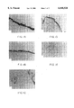
- FIG. 5 shows an autoradiograph of proteins radiolabeled with 35 S-Translabel, secreted by rabbit OSE in vitro, immunoprecipitated with anti-PS1 antibody, and electrophoresed in 7.5% polyacrylamide. Lanes 1-3 contain OSE secretory proteins.
- antibodies which specifically recognize antigens of the ovarian surface epithelium are used as diagnostic or therapeutic tools to determine the presence and amounts of OSE antigen in the tissues or blood of patients to diagnose, identify, and monitor ovarian cancer.
- anti-OSE antibodies are used to specifically target therapeutic agents to tissues expressing OSE antigens, e.g., ovarian cancer tissues.
- the antibodies used in the method invention specifically bind antigens of the ovarian surface epithelium, and also recognize antigens present in the cell surface membranes and preferably for diagnosis, antigens secreted by these epithelial cells.
- the anti-OSE antibodies preferably bind ovarian cancer tissues.
- the anti-OSE antibodies For detection of ovarian cancer in body fluids such as blood, peritoneal fluid, tissue fluid, and the like, the anti-OSE antibodies preferably bind antigens secreted by the OSE and by the ovarian cancer cells.
- the anti-OSE antibodies preferably bind antigens associated with the cell surface membranes of the ovarian cancer, and thereby deliver therapeutic agents to the tumor.
- Antibodies useful in the present invention may be polyclonal, monoclonal, or recombinant antibodies, and are prepared by methods generally known in the art. See, for example, Dunbar, 1987, Two Dimensional Gel Electrophoresis and Immunological Techniques, Plenum Press, New York, and Larrick and Fry, 1991, Human Antibodies and Hybridomas, 2:172-189.
- Antigens which may be used to produce antibodies useful in the method invention are those present in the ovarian surface epithelium (OSE) and those antigens secreted by the OSE.
- the antigens may be, for example, a protein, a glycoprotein, or a carbohydrate moiety.
- Antibodies to be used in the method invention are those which specifically bind normal OSE and those which specifically bind an antigen secreted by normal OSE, and also specifically bind ovarian cancer cells or antigens secreted by ovarian cancer cells.
- the monoclonal antibody PS1 described by Tivnons and Dunbar (1988, in Mathur and Fredericks, eds., Perspectives in Immunoreproduction: Conception and Contraception, Hemisphere Press, New York, p. 242) has surprisingly been found to recognize an antigen present in the ovarian surface epithelium of cynomolgus monkey, baboon, rabbit, and human ovarian tissues.
- PS1 also binds an antigen secreted by ovarian cell surface epithelium, in vitro, and specifically recognizes an ovarian cancer antigen.
- the PS1 antibody has been previously described as recognizing a ovarian zona pellucida antigen, and has been characterized as recognizing a carbohydrate constituent of this antigen (Timmons et al., 1987, Biol. Reprod., 36:1275).
- The-PS1 monoclonal antibody has been found to localize not only to the oocyte and its surrounding extracellular matrix but also in the secretory, apical portion of the ovarian surface epithelium.
- PS1 specifically binds ovarian tumor tissue and can be used to distinguish ovarian cancer cells from non-carcinogenic tissue for diagnostic purposes and for the classification of tumor types based upon histological sections and immunohistochemical assays.
- the antibody PS1 When the antibody PS1 is used in immunohistochemical assays of human cancer tissues, the antibody specifically binds throughout the ovarian cancer tissue while showing no significant binding to non-ovarian cancer types. Normal ovaries show binding of the antibody in the remnants of the OSE.
- the hybridoma cell line which produces monoclonal antibody PS1 was deposited under the provisions of the Budapest Treaty and accepted by the American Type Culture Collection (ATCC), 10801 University Boulevard, Manassas, Va. 20110-2209, on Oct. 8, 1987, and given the accession number HB9565.
- Metastatic ovarian cancer present in non-ovarian tissues may be identified using the method of the present invention.
- antibodies which specifically recognize antigens of the OSE are used to identify ovarian cancer tissue.
- the identified ovarian cancer tissue may be metastatic ovarian cancer cells present in non-ovarian tissues.
- the tissue to be assayed will be obtained by surgical procedures, e.g., biopsy.
- the excised tissue will be assayed for the presence of an antigen which recognizes an anti-OSE antibody as described above, e.g., PS1, by methods generally known in the art, e.g., imunohistochemistry.
- the tissue may be fixed or frozen to permit histological sectioning, and may be stained prior to incubation with the antibody.
- the antibody may be labeled, for example with a dye or fluorescent label, chemical, heavy metal or radioactive marker to permit the detection and localization of the antibody in the assayed tissue.
- other immunological assays known to those of skill in the art may be employed which permit detection of antibody binding to the excised tissue.
- the PS1 antigen is a carbohydrate moiety, and has now been found to be a secretory antigen in the oocyte
- secretion of the PS1-recognized antigen by OSE provides a specific ovarian cancer marker for detection in body fluids of patients presenting with proliferation of this cell type, e.g., ovarian cancer.
- Additional antigens secreted by the OSE may be used to prepare antibodies which, like PS1, may be used to diagnose ovarian cancer by immunoassay of tissue and body fluids for the presence of an antigen present in or secreted by ovarian cancer cells and recognized by the anti-OSE antibody.
- Body fluids such as blood and fluid blood components, peritoneal fluid, tissue and the like may be assayed for the presence and amount of antigen which binds anti-OSE antibodies.
- the assay may be performed by methods routinely used by those of skill in the art. Generally, a sample of body fluid is added to an assay mixture containing the antibody and a marker system for detection of antigen-bound antibody. Examples of such immunoassay systems are radioimmunoassays (RIA), enzyme-linked immunoassays (ELISA), immobilized immunoassays, and the like.
- RIA radioimmunoassays
- ELISA enzyme-linked immunoassays
- immobilized immunoassays and the like.
- Therapeutic agents useful in the treatment of ovarian cancer may be conjugated to the anti-OSE antibodies by methods known to those of skill in the art.
- therapeutic agents include radioisotopes such as 131 I or 67 Cu and cytotoxic agents such as ricin or diphtheria toxin.
- 131 I can be covalently bound to antibody molecules by methods known in the art, e.g., Iodogen® reactions.
- 67 Cu may be attached by way of an appropriate metal chelator bound to the antibody.
- the toxins may be conjugated to antibodies by agents such as glutaraldehyde or carbodiimide.
- conjugated therapeutic agent-antibody may be by various means known in the art, but generally will be by injection, systemic or intraperitoneal and in a pharmaceutically acceptable carrier.
- a useful therapeutic dose will vary with the particular therapeutic agent used, the particular cancer type and history, and the specific considerations of the patient to be treated.
- a physician administering the agent will know to calculate the effective therapeutic dose, which dose will be effective in reducing or eliminating the tumor tissue without compromising significantly normal tissues of the patient.
- PS1 antibody was prepared as described in Timmons and Dunbar, 1988, in Mathur and Fredericks, eds., Perspectives in Immunoreproduction: Conception and Contraception, Hemisphere Press, New York, p. 242. Control sections were treated with IgG isolated from myeloma cell acetous fluid. The incubation was followed by rinsing in TBS/Brij and application of secondary antibody under the same conditions.
- the secondary antibody, peroxidase-labeled rabbit anti-mouse IgG (Dako, Santa Barbara, Calif.) was diluted 1:50 in blocking solution before application and was followed by washing in TBS and subsequent treatment with a solution of 3,3'-diaminobenzidine (Fisher Scientific, Fairlawn, N.J.) consisting of 50 mg freebase/100 ml TBS containing 0.0031% hydrogen peroxide at room temperature.
- Rabbit and cynomolgus monkey ovarian tissue was similarly prepared and treated. Morphology was improved by finely mincing the rabbit tissues, fixing in 1% glutaraldehyde for one hour at room temperature, washing several times in chilled PBS, dehydration, and imbedding in Epon according to the manufacturer's instructions. Plastic embedment was followed by sectioning at 1-3 ⁇ m on a Mt-2 ultra microtome (Sorvall). Mounted sections were briefly etched (15 minutes) in sodium ethoxide/ethanol (1:1) and light trypsination (2.5% sucrose treatment followed by incubation in 0.0001% trypsin for 30 minutes at 37° C.). The blocking, antibody treatment, and signal development were performed as described above for human and cat ovarian tissue.
- the PS1 antigenic determinant was localized in the ovarian surface epithelium of cynomolgus monkey (FIG. 11A), baboon (FIG. 1B), rabbit (FIG. 1C), and human (FIG. 1D) with intense localization on the apical portion of OSE (arrows).
- the control myeloma antibody did not recognize human surface epithelium (FIG. 1E).
- Paraffin was first removed and the sections rehydrated by six (6) minute washes, in each of Xylene (twice), 100% ethanol (twice), 95% ethanol, 85% ethanol, 70% ethanol, 50% ethanol, and distilled, deionized water. The sections were next incubated 15-20 minutes in 3% hydrogen peroxide in methanol and washed 3 times in PBS containing 0.1% BSA, 5 minutes per wash. The washed sections were next blocked by incubating 20 minutes with normal rabbit serum (diluted 1:500) in a humidified chamber at room temperature, and washed 3 times in PBS containing 0,1% BSA, 5 minutes per wash.
- FIGS. 2, 3 and 4 the monoclonal antibody PS1 specifically recognized an antigen present in human ovarian tumors (FIGS. 2A-2N). In contrast, antibody specific antigen was identified only in the remnant ovarian cell surface epithelium of normal, adult ovarian tissues (FIGS. 3A-3D).
- FIGS. 4A and B show histological sections of a Brenner's tumor. This tumor is generally considered to be non-malignant and does not react with the PS1 antibody, which specifically identifies ovarian malignancies derived from OSE. Histological data for these and additional human tissue samples is summarized in Table 1.
- Tumors were obtained and analyzed as described for Example 2 for binding of the anti-OSE antibody, PS1. As shown in Table 2, non-ovarian tumor tissue did not react positively with the PS1 antibody. In contrast, an omentum tumor taken from a patient diagnosed with metastic adenocarcinona of the ovary was highly reactive with the PS1 antibody.
- Both ovaries were removed from a mature (>14 weeks) New Zealand White rabbit, trimmed, and placed into warm PBS, pH 7.2.
- the ovaries were rinsed three times in Hank's Balanced Salt Solution (HBSS) (GIBCO) and placed into a 25 ml Erlenmeyer flask containing 5 ml Dulbecco's Modified Eagle Media (DMEM) without methionine (Met-free medium) (GIBCO), 37° C.
- HBSS Hank's Balanced Salt Solution
- DMEM Dulbecco's Modified Eagle Media
- Method-free medium Gibco-free medium
- the ovaries were next removed to a second Erlenmeyer flask containing medium as described above but also containing 100 uCi of 35 S-Translabel (Met:Cys 14:3) (ICN, Irvine, Calif.).
- the 15 minute gassing, 45 minute swirling incubations described above were repeated four times.
- the medium was removed and concentrated five times using a Centricon microconcentrator having a 10,000 MW cut-off and centrifuging at 5000-6000 rpm at 4° C.
- the resulting concentrated medium was assayed by immunoprecipitation with the monoclonal antibody, PS1.
- PS1 monoclonal antibody
- 25 ul of normal rabbit serum was added to 200 ul of the concentrated medium, and the samples were agitated on a platform rocker for one hour at room temperature.
- the samples were rocked for two hours at room temperature, or overnight at 4° C., then centrifuged at 12,000-13,000 rpm for ten minutes, and the supernatant removed.
- SDS solubilization buffer (0.0625 M Trizma base containing 10% glycerol and 2% SDS to which 2-5% beta-mercaptoethanol was added immediately prior to use).
- SDS solubilization buffer (0.0625 M Trizma base containing 10% glycerol and 2% SDS to which 2-5% beta-mercaptoethanol was added immediately prior to use).
- the mixture was heated in a boiling water bath for ten minutes, cooled to room temperature, centrifuged at 12,000-13,000 rpm for ten minutes and the supernatant removed for electrophoresis.
- One dimensional PAGE was performed using conventional procedures. A 10% polyacrylamide gel was prepared, and samples added to lanes and electrophoresed. The resulting gel was stained with Coomassie blue stain, destained in ethanol/acetic acid, and treated with 1X EN 3 HANCE reagent (Dupont) for one hour with gentle agitation. The reagent was discarded and the gel was washed with deionized water for 30 minutes. The washed gel was dried and placed against X-ray film for the production of autoradiographs. Development of the film overnight at -70° C. was generally sufficient for the production of a strong signal.
- Lanes 1-3 contained proteins immunoprecipitated from the conditioned medium of the ovaries incubated as described above. These proteins may be presumed to be secreted predominantly by the ovarian surface epithelium cells and not other ovarian cell types because of the short incubation period. Only cells at the surface of the ovary are expected to take up the labeled amino acids, incorporate label into protein, and release labeled protein into the culture medium in such a short time. The OSE cells are those available to the label on the surface of the ovary. Lane 4 contained molecular weight markers. Lane 5 contained cytoplasm of isolated OSE cells and Lane 6 plasma membranes isolated from OSE after labeling as described above.
- Antigens secreted by the OSE are useful for the production of antibodies for the detection of tissue and secreted antigens conserved in ovarian tumor cells. Such antigens may be prepared and screened, for example, by the following procedure.
- OSE is isolated from the ovaries of a mammal, e.g, rabbit, as adapted from Nicosia et.al, International Journal of Gyn. Path., 1984, 3:348-360. Ovaries are rinsed three times briefly in HBSS at 37° C. and placed into a 50 ml plastic centrifuge tube containing 10 ml dissociation solution (10 ml Medium 199 containing 3000 units of collagenase, 25 units/ml penicillin and 50 ug/ml streptomycin, the final mixture filtered through a 0.22 u filter before use and at 37° C.). The sample is gassed for 15 minutes at 5% CO 2 and 37° C., then stoppered and moved to a 37° C. shaker-incubator and swirled gently (70-80 rpm) for 45 minutes.
- the contents of the tube are poured into a plastic petri dish and the ovaries gently lifted into a 15 ml plastic centrifuge tube containing 10 ml Medium 199 at 37° C.
- the solution is aspirated gently five times with a Pasteur pipette, and then vortexed.
- the solution is then gently aspirated again five times, and the contents poured onto a watch glass.
- the ovary is immobilized with a needle placed through its long axis and the surface of the ovary is gently scraped with a scalpel blade, releasing the OSE into the solution.
- the ovaries are then removed and the solution containing the OSE is placed into a 15 ml plastic centrifuge tube and centrifuged at 1400 rpm for five minutes. The supernatant is discarded and the pellet resuspended in 3 ml Medium 199 at 37° C. The solution is then layered onto a 10 ml "unit gradient" (5% BSA in Medium 199, pH 7.2, sterile filtered) and allowed to sit undisturbed for twenty minutes.
- the OSE cells are then cultured by means known in the art, for example, using conditions as described in Maresh et al, 1990, Biol. Reprod., 43:965-976.
- the conditioned medium is then collected from the OSE cultures and proteins concentrated, for example, using the diatomaceous earth procedure described by Dunbar, 1987, in Two Dimensional Electrophoresis and Immunological and Immunological Techniques, p. 223-225.
- the OSE secreted proteins are analyzed by two-dimensional gel electrophoresis, by methods known in the art. See, for example, Dunbar, 1987, Two Dimensional Electrophoresis and Immunological Techniques, Plenum Press, New York. Proteins detected by staining, i.e., Coomassie blue or silver stain, are isolated from the polyacrylamide gel using procedures known in the art (see Dunbar, 1987) and used to inject animals to prepare polyclonal and monoclonal antibodies (see Dunbar, 1987).
- the PS1 antibody can be used to immunoprecipitate secreted OSE proteins, as described in Example 3 above. Proteins identified using this procedure may be separated by two-dimensional PAGE and purified for use as antigens to prepare anti-OSE antibodies.
- Efficacy of anti-OSE antibodies prepared as described herein for utility as ovarian cancer markers can be tested by the antibody's recognition of ovarian tumor tissue using the immunohistochemical procedures described in Example 2, as well as identification of antigens secreted by human ovarian cancer cell lines. Secretion of antigen into the body fluids and or circulation in the blood may be evaluated using conventional immunoassay techniques, e.g., competitive ELISA procedures.
- Therapeutic agents may be conjugated to anti-OSE antibodies for targeted delivery to ovarian cancer tissue.
- cytotoxic agents including radioisotopes and cytotoxins such as ricin may be conjugated by means known in the art to anti-OSE antibodies.
- the specific binding of the anti-OSE antibody to ovarian cancer cells enables specific delivery of the agent to the cancer tissue.
Abstract
A method for the diagnosis and treatment of ovarian cancer. Tissue and body fluid samples are assayed for an antigen which specifically binds an anti-OSE antigen to diagnose ovarian cancer. Therapeutic agents coupled to anti-OSE antibodies, such as PS1 are specifically directed to ovarian cancer tissues.
Description
This is a Continuation of Parent application Ser. No. 08/520,584 now abandoned, filed on Aug. 29, 1995; which in turn is a Divisional of application Ser. No. 08/326,053, filed Oct. 19, 1994 (now U.S. Pat. No. 5,484,704); which in turn is a Continuation of application Ser. No. 07/975,093, filed Nov. 12, 1992 (now abandoned).
The present invention relates to a method for the detection, monitoring, and treatment of ovarian cancer. More specifically, this invention relates to the use of a monoclonal antibody which specifically recognizes an ovarian antigen secreted by the ovarian surface epithelium.
Cancer of the ovary is the second most common cancer of the female reproductive organs and the fourth most common cause of cancer deaths among American women. Because ovarian cancers are not readily detectable by diagnostic techniques (Siemens and Auersperg, 1988, J. Cellular Physiol., 134:347-356). Because diagnosis of carcinoma of the ovary is generally only possible when the disease has progressed to a late stage of development, it is one of the most lethal of the gynecological malignancies.
There is a vital need for the identification of tumor markers which can be used in the early detection of ovarian cancer, the monitoring of cancer therapies, the immunodetection of ovarian tumors, and the development of probes for potential use in immunotherapy (Cantarow et al., 1981, Int. J. Radiation Oncol. Biol. Phys., 7:1095-1098). Although a number of potential tumor markers have been evaluated including the cancer antigen 125 (Ca-125) nonspecificity of the antigens diminish their value as markers for primary ovarian cancer (Kudlacek et al., 1989, Gyn. Onc., 35:323-329; Rustin et al., 1989, J. Clin. Onc., 7:1667-1671; Sevelda et al., 1989, Am. J. Obstet. Gynecol., 161:1213-1216; Omar et al., 1989, Tumor Biol., 10:316-323).
Several monoclonal antibodies have recently been shown to react with ovarian tumor associated antigens (Kenemans et al., 1989, Eur. J. Obstet. Gynecol. Repod. Biol., 29:207-218). These antibodies do not appear to be specific to a specific type of cancer (McDuffy, 1989, Ann. Clin. Biochen., 26:379-387). Many of these antibodies recognize determinants associated with high molecular weight glycoproteins related to mucins. Additional markers such as enzymes or activated cellular oncagenes or products of these genes have also been suggested for use as ovarian cancer markers. These potential probes, however, have not been detected in the blood and hence their use is confined to analysis of tumor tissues.
The majority, e.g., 80-90%, of ovarian cancers are thought to arise from the ovarian surface of epithelium (OSE). To date, little is known about the structure and function of the cells of the ovarian surface epithelium. It is known that the surface epithelium is a highly dynamic tissue which undergoes morphogenic changes. The OSE also has significant proliferative properties, as it it must proliferate rapidly to cover the ovulatory site after ovulation of the ova. Morphological and histochemical studies suggest that the OSE has secretory, endocytotic and transport functions which are hormonally controlled (Blaustein and Lee, 1979, Oncol. 8:34-43; Nicosia and Johnson, 1983, Int. J. Gynecol. Pathol., 3:249-260; Papadaki and Beilby, 1971, J. Cell Sci., 8:445-464; Anderson et al., 1976, J. Morphol., 150:135-164).
There is a vital need for an ovarian cancer marker which can specifically detect-ovarian cancer, which marker can be used to diagnose, monitor and identify ovarian cancers and which may be used in specific immunotherapy.
The present invention provides a method for the specific detection, monitoring, and/or treatment of ovarian cancer. In the method of the present invention, an in vitro assay employs antibodies which specifically recognize an antigen of the ovarian surface epithelium (OSE) to determine the presence and amounts of OSE antigen in tissues or body fluids of patients. Such assay permits detection of ovarian cancer, monitoring of ovarian cancers and specific identification of ovarian cancer type. Anti-OSE antibodies are also used to specifically target therapeutic agents to ovarian cancer cells.
For diagnosing and classifying ovarian cancer types, ovarian cancer tissues are probed with an anti-OSE antibody, e.g., by immunohistochemical methods. For detection and monitoring of ovarian cancer in body fluids, the anti-OSE antibodies are preferably used in immunoassays to detect a secreted OSE antigen. For therapeutic purposes, anti-OSE antibodies conjugated to cytotoxic agents are administered for targeted delivery to OSE antigens present in the ovarian cancer.
FIG. 1 shows localization of PS1 antigen in OSE of cynamologous monkeys (FIG. 1A), baboons (FIG. 1B), rabbit (FIG. 1C) and human (FIG. 1D). FIG. 1E shows a section of human ovary probed with control, anti-myeloma antibody. Arrows indicate ovarian surface epithelium.
FIG. 2 shows histological sections of paraffin embedded human ovarian tumors.
FIG. 2A shows sample 13022 stained with PAS.
FIG. 2B shows sample 13022 reacted with PS1 antibody.
FIG. 2C shows sample 9010 stained with PAS.
FIG. 2D shows sample 9010 reacted with PS1 antibody.
FIG. 2E shows sample 10035 stained with PAS.
FIG. 2F shows sample 10035 reacted with PS1 antibody.
FIG. 2G shows sample 13017 stained with PAS.
FIG. 2H shows sample 13017 reacted with PS1 antibody.
FIG. 2I shows sample 9160 stained with PAS.
FIG. 2J shows sample 9160 stained with PS1 antibody.
FIG. 2K shows sample 10880 stained with PAS.
FIG. 2L shows sample 10880 reacted with PS1 antibody.
FIG. 2M shows sample 9219 stained with PAS.
FIG. 2N shows sample 9219 reacted with PS1 antibody.
FIG. 3 shows histological sections of paraffin embedded normal human ovaries.
FIG. 3A shows sample 13053 stained with PAS.
FIG. 3B shows sample 13053 reacted with PS1 antibody.
FIG. 3C shows sample 13012 stained with PAS.
FIG. 3D shows sample 13012 reacted with PS1 antibody.
FIG. 4 shows histological sections of a paraffin embedded Brenners tumor.
FIG. 4A shows sample 13044 stained with PAS.
FIG. 4B shows sample 13044 reacted with PS1 antibody.
FIG. 5 shows an autoradiograph of proteins radiolabeled with 35 S-Translabel, secreted by rabbit OSE in vitro, immunoprecipitated with anti-PS1 antibody, and electrophoresed in 7.5% polyacrylamide. Lanes 1-3 contain OSE secretory proteins.
In the method of the present invention, antibodies which specifically recognize antigens of the ovarian surface epithelium are used as diagnostic or therapeutic tools to determine the presence and amounts of OSE antigen in the tissues or blood of patients to diagnose, identify, and monitor ovarian cancer. In an alternative embodiment anti-OSE antibodies are used to specifically target therapeutic agents to tissues expressing OSE antigens, e.g., ovarian cancer tissues.
The antibodies used in the method invention specifically bind antigens of the ovarian surface epithelium, and also recognize antigens present in the cell surface membranes and preferably for diagnosis, antigens secreted by these epithelial cells.
For diagnosing and classifying ovarian cancer types, the anti-OSE antibodies preferably bind ovarian cancer tissues. For detection of ovarian cancer in body fluids such as blood, peritoneal fluid, tissue fluid, and the like, the anti-OSE antibodies preferably bind antigens secreted by the OSE and by the ovarian cancer cells. For therapeutic purposes, the anti-OSE antibodies preferably bind antigens associated with the cell surface membranes of the ovarian cancer, and thereby deliver therapeutic agents to the tumor.
Antibodies useful in the present invention may be polyclonal, monoclonal, or recombinant antibodies, and are prepared by methods generally known in the art. See, for example, Dunbar, 1987, Two Dimensional Gel Electrophoresis and Immunological Techniques, Plenum Press, New York, and Larrick and Fry, 1991, Human Antibodies and Hybridomas, 2:172-189.
Antigens which may be used to produce antibodies useful in the method invention are those present in the ovarian surface epithelium (OSE) and those antigens secreted by the OSE. The antigens may be, for example, a protein, a glycoprotein, or a carbohydrate moiety.
Antibodies to be used in the method invention are those which specifically bind normal OSE and those which specifically bind an antigen secreted by normal OSE, and also specifically bind ovarian cancer cells or antigens secreted by ovarian cancer cells.
The phrases "specifically binds" and "having specificity for" are meant to include binding of the anti-OSE antibody to ovarian tissues and to ovarian cancer cells with little or no binding to non-ovarian tissues or cancers of non-ovarian origin.
For example, the monoclonal antibody PS1 described by Tivnons and Dunbar (1988, in Mathur and Fredericks, eds., Perspectives in Immunoreproduction: Conception and Contraception, Hemisphere Press, New York, p. 242) has surprisingly been found to recognize an antigen present in the ovarian surface epithelium of cynomolgus monkey, baboon, rabbit, and human ovarian tissues. PS1 also binds an antigen secreted by ovarian cell surface epithelium, in vitro, and specifically recognizes an ovarian cancer antigen.
The PS1 antibody has been previously described as recognizing a ovarian zona pellucida antigen, and has been characterized as recognizing a carbohydrate constituent of this antigen (Timmons et al., 1987, Biol. Reprod., 36:1275). The-PS1 monoclonal antibody has been found to localize not only to the oocyte and its surrounding extracellular matrix but also in the secretory, apical portion of the ovarian surface epithelium.
PS1 specifically binds ovarian tumor tissue and can be used to distinguish ovarian cancer cells from non-carcinogenic tissue for diagnostic purposes and for the classification of tumor types based upon histological sections and immunohistochemical assays.
When the antibody PS1 is used in immunohistochemical assays of human cancer tissues, the antibody specifically binds throughout the ovarian cancer tissue while showing no significant binding to non-ovarian cancer types. Normal ovaries show binding of the antibody in the remnants of the OSE.
The hybridoma cell line which produces monoclonal antibody PS1 was deposited under the provisions of the Budapest Treaty and accepted by the American Type Culture Collection (ATCC), 10801 University Boulevard, Manassas, Va. 20110-2209, on Oct. 8, 1987, and given the accession number HB9565.
Metastatic ovarian cancer present in non-ovarian tissues may be identified using the method of the present invention. Thus, antibodies which specifically recognize antigens of the OSE are used to identify ovarian cancer tissue. The identified ovarian cancer tissue may be metastatic ovarian cancer cells present in non-ovarian tissues.
Preferably, the tissue to be assayed will be obtained by surgical procedures, e.g., biopsy. The excised tissue will be assayed for the presence of an antigen which recognizes an anti-OSE antibody as described above, e.g., PS1, by methods generally known in the art, e.g., imunohistochemistry. The tissue may be fixed or frozen to permit histological sectioning, and may be stained prior to incubation with the antibody. The antibody may be labeled, for example with a dye or fluorescent label, chemical, heavy metal or radioactive marker to permit the detection and localization of the antibody in the assayed tissue. Alternatively, other immunological assays known to those of skill in the art may be employed which permit detection of antibody binding to the excised tissue.
Because the PS1 antigen is a carbohydrate moiety, and has now been found to be a secretory antigen in the oocyte, secretion of the PS1-recognized antigen by OSE provides a specific ovarian cancer marker for detection in body fluids of patients presenting with proliferation of this cell type, e.g., ovarian cancer. Additional antigens secreted by the OSE may be used to prepare antibodies which, like PS1, may be used to diagnose ovarian cancer by immunoassay of tissue and body fluids for the presence of an antigen present in or secreted by ovarian cancer cells and recognized by the anti-OSE antibody.
Body fluids such as blood and fluid blood components, peritoneal fluid, tissue and the like may be assayed for the presence and amount of antigen which binds anti-OSE antibodies. The assay may be performed by methods routinely used by those of skill in the art. Generally, a sample of body fluid is added to an assay mixture containing the antibody and a marker system for detection of antigen-bound antibody. Examples of such immunoassay systems are radioimmunoassays (RIA), enzyme-linked immunoassays (ELISA), immobilized immunoassays, and the like.
Therapeutic agents useful in the treatment of ovarian cancer may be conjugated to the anti-OSE antibodies by methods known to those of skill in the art. Examples of such therapeutic agents include radioisotopes such as 131 I or 67 Cu and cytotoxic agents such as ricin or diphtheria toxin. 131 I can be covalently bound to antibody molecules by methods known in the art, e.g., Iodogen® reactions. 67 Cu may be attached by way of an appropriate metal chelator bound to the antibody. The toxins may be conjugated to antibodies by agents such as glutaraldehyde or carbodiimide.
Administration of the conjugated therapeutic agent-antibody may be by various means known in the art, but generally will be by injection, systemic or intraperitoneal and in a pharmaceutically acceptable carrier. A useful therapeutic dose will vary with the particular therapeutic agent used, the particular cancer type and history, and the specific considerations of the patient to be treated. A physician administering the agent will know to calculate the effective therapeutic dose, which dose will be effective in reducing or eliminating the tumor tissue without compromising significantly normal tissues of the patient.
The nature of the invention can be further understood by reference to the following examples.
Initial immunohistochemical localization studies demonstrated that the PS1 monoclonal antibody localized not only to the oocyte and surrounding extracellular matrix of the zona pellucida but also localized intensively in the secretory, apical portion of the ovarian surface epithelium.
Human and cat ovarian tissue was embedded and immediately frozen in OCT compound (Miles, Elkhart, Ind.). Cryostat sections, 4-8 μm were prepared using a Minotome/microtome cryostat, and the sections were fixed in 100% methanol for 10 minutes, blocked in TRIS-buffered saline (TBS), pH 7.5, containing 2.5% w/v Brij-35 (SIGMA, St. Louis, Mo.) and 6% w/v Carnation non-fat dried milk. Blocking-proceeded for thirty minutes at 37° C. in a moist chamber and was followed by brief rinsing in TBS/Brij.
The blocked sections were next incubated with the monoclonal antibody PS1 in a moist chamber for 30 minutes at 37° C. PS1 antibody was prepared as described in Timmons and Dunbar, 1988, in Mathur and Fredericks, eds., Perspectives in Immunoreproduction: Conception and Contraception, Hemisphere Press, New York, p. 242. Control sections were treated with IgG isolated from myeloma cell acetous fluid. The incubation was followed by rinsing in TBS/Brij and application of secondary antibody under the same conditions. The secondary antibody, peroxidase-labeled rabbit anti-mouse IgG (Dako, Santa Barbara, Calif.) was diluted 1:50 in blocking solution before application and was followed by washing in TBS and subsequent treatment with a solution of 3,3'-diaminobenzidine (Fisher Scientific, Fairlawn, N.J.) consisting of 50 mg freebase/100 ml TBS containing 0.0031% hydrogen peroxide at room temperature.
Rabbit and cynomolgus monkey ovarian tissue was similarly prepared and treated. Morphology was improved by finely mincing the rabbit tissues, fixing in 1% glutaraldehyde for one hour at room temperature, washing several times in chilled PBS, dehydration, and imbedding in Epon according to the manufacturer's instructions. Plastic embedment was followed by sectioning at 1-3 μm on a Mt-2 ultra microtome (Sorvall). Mounted sections were briefly etched (15 minutes) in sodium ethoxide/ethanol (1:1) and light trypsination (2.5% sucrose treatment followed by incubation in 0.0001% trypsin for 30 minutes at 37° C.). The blocking, antibody treatment, and signal development were performed as described above for human and cat ovarian tissue.
As shown in FIGS. 1A-1E, the PS1 antigenic determinant was localized in the ovarian surface epithelium of cynomolgus monkey (FIG. 11A), baboon (FIG. 1B), rabbit (FIG. 1C), and human (FIG. 1D) with intense localization on the apical portion of OSE (arrows). In contrast, the control myeloma antibody did not recognize human surface epithelium (FIG. 1E).
Human ovarian tissue, both normal controls and tumor tissues were obtained as fixed sections embedded in paraffin from the Department of Pathology, M.D. Anderson Hospital, Houston, Tex. Immunohistochemical assay of the sections was performed as follows:
Paraffin was first removed and the sections rehydrated by six (6) minute washes, in each of Xylene (twice), 100% ethanol (twice), 95% ethanol, 85% ethanol, 70% ethanol, 50% ethanol, and distilled, deionized water. The sections were next incubated 15-20 minutes in 3% hydrogen peroxide in methanol and washed 3 times in PBS containing 0.1% BSA, 5 minutes per wash. The washed sections were next blocked by incubating 20 minutes with normal rabbit serum (diluted 1:500) in a humidified chamber at room temperature, and washed 3 times in PBS containing 0,1% BSA, 5 minutes per wash. The blocked sections were then incubated with PS1 IgG (1:500) or control antibody, IgG derived from ascites fluid of mice injected with p3U1 myeloma cells (Drell & Dunbar (1984) Diolopy of Reproduction, 30:445-457) (1:500) for two hours at room temperature, or overnight at 4° C., followed by three PBS washes as described above. Second antibody was applied by incubating with biotinylated rabbit anti-mouse IgG (Vectastain, Vector Laboratories, Burlingame, Calif.) for thirty (30) minutes at room temperature, followed by three PBS washes as described above. The sections were next incubated 30 minutes at room temperature with the avidin biotinylated horseradish/peroxidase reagent (ABC) (Vectastain) and washed three times in PBS as described above.
To stain the sections, 2 drops of 3% H2 O2 was added to diaminobenzldine tetrahydrochloride DAB solution (3-5 mg/10 ml 0.1 M TRIS) iimediately before use. The sections were incubated with the prepared DAB solution for 5-10 minutes, washed twice in dd H2 O, and coverslips applied.
The assayed sections were then viewed by light microscopy. Sections of "normal" human ovarian tissue (ovarian tissue having no gross pathology as evaluated by M.D. Anderson pathologists) was compared with sections of tissue surgically removed from cancer patients.
As shown in FIGS. 2, 3 and 4, the monoclonal antibody PS1 specifically recognized an antigen present in human ovarian tumors (FIGS. 2A-2N). In contrast, antibody specific antigen was identified only in the remnant ovarian cell surface epithelium of normal, adult ovarian tissues (FIGS. 3A-3D). FIGS. 4A and B show histological sections of a Brenner's tumor. This tumor is generally considered to be non-malignant and does not react with the PS1 antibody, which specifically identifies ovarian malignancies derived from OSE. Histological data for these and additional human tissue samples is summarized in Table 1.
                                  TABLE 1
__________________________________________________________________________
SAMPLE                      PS1 PRESENCE.sup.b
                             NO. TISSUE DIAGNOSIS (Localization)
__________________________________________________________________________
13012
     OVARY     NORMAL       RESIDUAL OSE
     ONLY (IN CRYPTS)
  13057 OVARY NORMAL + Residual OSE
  13053 OVARY NORMAL -
  13018 OVARY NORMAL -
  13004 OVARY NORMAL -
  13065 OVARY NORMAL -
  9213 OVARY NORMAL -
  13017 OVARY ADENOCARCINOMA OF +
    ENDOMETRIUM
  13078 OVARIAN TUMOR PAPILLARY SEROUS ++
    CARCINOMA
  9129 OVARIAN TUMOR BRENNER'S MUCINOUS + (FEW
    TUMOR METASTATIC ISOLATED
    ADENOCARCINOMA OF EPITHELIAL
    LIVER CELLS)
  13022 OVARIAN TUMOR PAPILLARY SEROUS +++
  9208 OVARIAN TUMOR TERATOMA TUMOR ++
  13027 OVARIAN TUMOR SEROUS CYST +
  9010 OVARIAN TUMOR SEROUS METASTATIC ++
  13044 OVARIAN TUMOR BRENNER'S TUMOR -
  13028 OVARIAN TUMOR GRANULOSA CELL +++
    TUMOR (RESEMBLES
    PAPILLARY SEROUS)
  10880 OVARIAN TUMOR SEROUS CARCINOMA +++
  1068 OVARIAN TUMOR METASTATIC +/- (SMALL
    TRANSITIONAL POCKETS)
  9160 OVARIAN TUMOR ADENOCARCINOMA OF +++
    ENDOMETRIUM
  13023 OVARIAN TUMOR MATURE CYSTIC -
    TERATOMA
  9219 OVARIAN TUMOR ENDOMETRIUM ++
    CARCINOMA OF OVARY
    WITH METASTATIC
    ADENOCARCINOMA
  10890 OVARIAN TUMOR MUCINOUS CYSTIC ++
  4703 OVARIAN TUMOR PAPILLARY SEROUS ++
  10035 OVARIAN TUMOR PAPILLARY SEROUS ++
__________________________________________________________________________
 .sup.a Classification and tumor diagnosis by pathologists from M.D.
 Anderson Cancer Institute (Houston, TX)
 .sup.b Monoclonal antibody PS1 localization: + = few cells but distinct;
 ++ = more general localization; +++ = predominant localization.
Tumors were obtained and analyzed as described for Example 2 for binding of the anti-OSE antibody, PS1. As shown in Table 2, non-ovarian tumor tissue did not react positively with the PS1 antibody. In contrast, an omentum tumor taken from a patient diagnosed with metastic adenocarcinona of the ovary was highly reactive with the PS1 antibody. These results demonstrate the use of anti-OSE antibodies to characterize metastatic tumors as having an ovarian origin.
              TABLE 2
______________________________________
                            REACTIVITY
  SAMPLE NO. TUMOR DESCRIPTION WITH ANTI-PS1
______________________________________
UB 2102     Uterine tumor   Negative
  PN 2376 Uterine tumor Negative
  SB 0379 Perivaginal tumor Negative
  SS 0688 Vulva tumor Negative
  VM 9758 Omentum* tumor +++
______________________________________
 *Omentum tumor from patient having metastatic adenocarcinoma of ovary
Both ovaries were removed from a mature (>14 weeks) New Zealand White rabbit, trimmed, and placed into warm PBS, pH 7.2. The ovaries were rinsed three times in Hank's Balanced Salt Solution (HBSS) (GIBCO) and placed into a 25 ml Erlenmeyer flask containing 5 ml Dulbecco's Modified Eagle Media (DMEM) without methionine (Met-free medium) (GIBCO), 37° C. The flask was gassed for 15 minutes in a 37° C. incubator at 5% CO2, then stoppered and moved to a 37° C. shaker-incubator and swirled gently at 70-80 rpm for 45 minutes.
The ovaries were next removed to a second Erlenmeyer flask containing medium as described above but also containing 100 uCi of 35 S-Translabel (Met:Cys 14:3) (ICN, Irvine, Calif.). The 15 minute gassing, 45 minute swirling incubations described above were repeated four times. The medium was removed and concentrated five times using a Centricon microconcentrator having a 10,000 MW cut-off and centrifuging at 5000-6000 rpm at 4° C.
The resulting concentrated medium was assayed by immunoprecipitation with the monoclonal antibody, PS1. To eliminate non-specific binding, 25 ul of normal rabbit serum was added to 200 ul of the concentrated medium, and the samples were agitated on a platform rocker for one hour at room temperature. To this mixture was added 50 ul of a 10% suspension of Protein A Sepharose CL-4B (Pharmacia) suspended in NP-NET buffer. The samples were rocked for two hours at room temperature, or overnight at 4° C., then centrifuged at 12,000-13,000 rpm for ten minutes, and the supernatant removed.
To the adsorption-treated supernatant was added 100 ul of PS1 antibody (1.6 mg/ml protein) and the samples were rocked for two hours at room temperature (or overnight at 4° C.). After incubation with PS1, 50 ul of affinity purified rabbit anti-mouse IgG (1 mg/ml protein) was added and the samples were again rocked for one hour at room temperature. A volume of 100 ul of the 10% suspension of Protein A-Sepharose in NP-NET described above was added to each sample which was then vortexed briefly and rocked for two hours at room temperature or overnight at 4° C. The samples were then centrifuged at 12,000-13,000 rpm for ten minutes and the supernatant removed. The pellet containing PS1-bound 35 S-labeled proteins was washed twice in NP-NET buffer and analyzed by SDS-polyacrylamide gel electrophoresis (PAGE).
To each sample pellet was added freshly made SDS solubilization buffer (0.0625 M Trizma base containing 10% glycerol and 2% SDS to which 2-5% beta-mercaptoethanol was added immediately prior to use). The mixture was heated in a boiling water bath for ten minutes, cooled to room temperature, centrifuged at 12,000-13,000 rpm for ten minutes and the supernatant removed for electrophoresis.
One dimensional PAGE was performed using conventional procedures. A 10% polyacrylamide gel was prepared, and samples added to lanes and electrophoresed. The resulting gel was stained with Coomassie blue stain, destained in ethanol/acetic acid, and treated with 1X EN3 HANCE reagent (Dupont) for one hour with gentle agitation. The reagent was discarded and the gel was washed with deionized water for 30 minutes. The washed gel was dried and placed against X-ray film for the production of autoradiographs. Development of the film overnight at -70° C. was generally sufficient for the production of a strong signal.
The resultant autoradiograph is shown in FIG. 5. Lanes 1-3 contained proteins immunoprecipitated from the conditioned medium of the ovaries incubated as described above. These proteins may be presumed to be secreted predominantly by the ovarian surface epithelium cells and not other ovarian cell types because of the short incubation period. Only cells at the surface of the ovary are expected to take up the labeled amino acids, incorporate label into protein, and release labeled protein into the culture medium in such a short time. The OSE cells are those available to the label on the surface of the ovary. Lane 4 contained molecular weight markers. Lane 5 contained cytoplasm of isolated OSE cells and Lane 6 plasma membranes isolated from OSE after labeling as described above.
Antigens secreted by the OSE are useful for the production of antibodies for the detection of tissue and secreted antigens conserved in ovarian tumor cells. Such antigens may be prepared and screened, for example, by the following procedure.
OSE is isolated from the ovaries of a mammal, e.g, rabbit, as adapted from Nicosia et.al, International Journal of Gyn. Path., 1984, 3:348-360. Ovaries are rinsed three times briefly in HBSS at 37° C. and placed into a 50 ml plastic centrifuge tube containing 10 ml dissociation solution (10 ml Medium 199 containing 3000 units of collagenase, 25 units/ml penicillin and 50 ug/ml streptomycin, the final mixture filtered through a 0.22 u filter before use and at 37° C.). The sample is gassed for 15 minutes at 5% CO2 and 37° C., then stoppered and moved to a 37° C. shaker-incubator and swirled gently (70-80 rpm) for 45 minutes.
After dissociation, the contents of the tube are poured into a plastic petri dish and the ovaries gently lifted into a 15 ml plastic centrifuge tube containing 10 ml Medium 199 at 37° C. The solution is aspirated gently five times with a Pasteur pipette, and then vortexed. The solution is then gently aspirated again five times, and the contents poured onto a watch glass. The ovary is immobilized with a needle placed through its long axis and the surface of the ovary is gently scraped with a scalpel blade, releasing the OSE into the solution.
The ovaries are then removed and the solution containing the OSE is placed into a 15 ml plastic centrifuge tube and centrifuged at 1400 rpm for five minutes. The supernatant is discarded and the pellet resuspended in 3 ml Medium 199 at 37° C. The solution is then layered onto a 10 ml "unit gradient" (5% BSA in Medium 199, pH 7.2, sterile filtered) and allowed to sit undisturbed for twenty minutes.
All but the lower 3 ml of the "gradient" is pipetted off and discarded. The remaining 3 ml is centrifuged at 1400 rpm for 5 minutes, the resulting supernatant is discarded, and the pellet washed twice with 10 ml HBSS at room temperature, and again centrifuged.
The OSE cells are then cultured by means known in the art, for example, using conditions as described in Maresh et al, 1990, Biol. Reprod., 43:965-976. The conditioned medium is then collected from the OSE cultures and proteins concentrated, for example, using the diatomaceous earth procedure described by Dunbar, 1987, in Two Dimensional Electrophoresis and Immunological and Immunological Techniques, p. 223-225.
The OSE secreted proteins are analyzed by two-dimensional gel electrophoresis, by methods known in the art. See, for example, Dunbar, 1987, Two Dimensional Electrophoresis and Immunological Techniques, Plenum Press, New York. Proteins detected by staining, i.e., Coomassie blue or silver stain, are isolated from the polyacrylamide gel using procedures known in the art (see Dunbar, 1987) and used to inject animals to prepare polyclonal and monoclonal antibodies (see Dunbar, 1987).
The PS1 antibody can be used to immunoprecipitate secreted OSE proteins, as described in Example 3 above. Proteins identified using this procedure may be separated by two-dimensional PAGE and purified for use as antigens to prepare anti-OSE antibodies.
Efficacy of anti-OSE antibodies prepared as described herein for utility as ovarian cancer markers can be tested by the antibody's recognition of ovarian tumor tissue using the immunohistochemical procedures described in Example 2, as well as identification of antigens secreted by human ovarian cancer cell lines. Secretion of antigen into the body fluids and or circulation in the blood may be evaluated using conventional immunoassay techniques, e.g., competitive ELISA procedures.
Therapeutic agents may be conjugated to anti-OSE antibodies for targeted delivery to ovarian cancer tissue. For example, cytotoxic agents including radioisotopes and cytotoxins such as ricin may be conjugated by means known in the art to anti-OSE antibodies.
The specific binding of the anti-OSE antibody to ovarian cancer cells enables specific delivery of the agent to the cancer tissue.
Claims (1)
1. A method of targeted delivery of a therapeutic agent to ovarian cancer tissue comprising the steps of:
conjugating a therapeutic agent to PS1 antibody secreted by hybridoma cells having ATCC Accession number HB9566; and
administering the conjugated antibody to a patient.
Priority Applications (1)
| Application Number | Priority Date | Filing Date | Title |
|---|---|---|---|
| US08/977,373 US6048528A (en) | 1992-11-12 | 1997-11-24 | Monoclonal antibody for diagnostic and therapeutic use in ovarian cancer |
Applications Claiming Priority (4)
| Application Number | Priority Date | Filing Date | Title |
|---|---|---|---|
| US97509392A | 1992-11-12 | 1992-11-12 | |
| US08/326,053 US5484704A (en) | 1992-11-12 | 1994-10-19 | Monoclonal antibody for diagnostic use in ovarian cancer |
| US52058495A | 1995-08-29 | 1995-08-29 | |
| US08/977,373 US6048528A (en) | 1992-11-12 | 1997-11-24 | Monoclonal antibody for diagnostic and therapeutic use in ovarian cancer |
Related Parent Applications (1)
| Application Number | Title | Priority Date | Filing Date |
|---|---|---|---|
| US52058495A Continuation | 1992-11-12 | 1995-08-29 |
Publications (1)
| Publication Number | Publication Date |
|---|---|
| US6048528A true US6048528A (en) | 2000-04-11 |
Family
ID=25522695
Family Applications (2)
| Application Number | Title | Priority Date | Filing Date |
|---|---|---|---|
| US08/326,053 Expired - Fee Related US5484704A (en) | 1992-11-12 | 1994-10-19 | Monoclonal antibody for diagnostic use in ovarian cancer |
| US08/977,373 Expired - Fee Related US6048528A (en) | 1992-11-12 | 1997-11-24 | Monoclonal antibody for diagnostic and therapeutic use in ovarian cancer |
Family Applications Before (1)
| Application Number | Title | Priority Date | Filing Date |
|---|---|---|---|
| US08/326,053 Expired - Fee Related US5484704A (en) | 1992-11-12 | 1994-10-19 | Monoclonal antibody for diagnostic use in ovarian cancer |
Country Status (1)
| Country | Link |
|---|---|
| US (2) | US5484704A (en) |
Cited By (6)
| Publication number | Priority date | Publication date | Assignee | Title |
|---|---|---|---|---|
| US20050202448A1 (en) * | 2004-03-15 | 2005-09-15 | Digigenomics Co., Ltd. | Methods for identification, assessment, prevention, and therapy of cancer |
| WO2006102095A2 (en) | 2005-03-18 | 2006-09-28 | Medimmune, Inc. | Framework-shuffling of antibodies |
| US20060275330A1 (en) * | 2003-04-17 | 2006-12-07 | Kenneth Kao Et Al | Pygopus in diagnosis and treatment of cancer |
| WO2007002543A2 (en) | 2005-06-23 | 2007-01-04 | Medimmune, Inc. | Antibody formulations having optimized aggregation and fragmentation profiles |
| US20100216975A1 (en) * | 2003-08-18 | 2010-08-26 | Medimmune, Llc | Framework-Shuffling Of Antibodies |
| EP2272566A2 (en) | 2003-08-18 | 2011-01-12 | MedImmune, LLC | Humanisation of antibodies |
Families Citing this family (3)
| Publication number | Priority date | Publication date | Assignee | Title |
|---|---|---|---|---|
| US6576239B1 (en) * | 1996-09-10 | 2003-06-10 | The Burnham Institute | Angiogenic homing molecules and conjugates derived therefrom |
| AU2001250828A1 (en) * | 2000-03-14 | 2001-09-24 | The Johns Hopkins University School Of Medicine | Immunogenic ovarian cancer genes |
| WO2006133408A2 (en) * | 2005-06-08 | 2006-12-14 | Iowa State University Research Foundation, Inc. | Breast cancer and prostate cancer assessment |
Citations (10)
| Publication number | Priority date | Publication date | Assignee | Title |
|---|---|---|---|---|
| US4650756A (en) * | 1981-08-31 | 1987-03-17 | Sloan Kettering Institute For Cancer Research | Monoclonal antibodies to cell surface antigens of human renal cancer |
| US4666845A (en) * | 1983-12-16 | 1987-05-19 | Sloan-Kettering Institute | Monoclonal antibodies to ovarian, cervical and uterine human cancers and method of diagnosis |
| US4695538A (en) * | 1984-06-01 | 1987-09-22 | Sloan-Kettering Institute For Cancer Research | Human monoclonal antibodies to cell surface antigens |
| US4713351A (en) * | 1983-01-28 | 1987-12-15 | The University Of Rochester | Monoclonal antibody directed to an antigen derived from human ovarian tumors and a radioimmunoassay using the antibody |
| US4725538A (en) * | 1985-10-25 | 1988-02-16 | The Beth Israel Hospital Association | Method of assaying the presence of cancer cells |
| US4806494A (en) * | 1986-07-24 | 1989-02-21 | The United States Of America As Represented By The Department Of Health & Human Services | Monoclonal antibody against ovarian cancer cells (OVB-3) |
| US4814275A (en) * | 1985-05-13 | 1989-03-21 | E.I. Dupont De Nemours And Company | Monoclonal hybridoma antibody specific for a human epithelial antigen |
| US4857452A (en) * | 1986-12-04 | 1989-08-15 | E. I. Du Pont De Nemours And Company | Assay for carcinoma of breast, colon and ovary |
| US4921790A (en) * | 1987-04-24 | 1990-05-01 | Research Corporation | Tumor specific assay for CA125 ovarian cancer antigen |
| US5094942A (en) * | 1988-05-13 | 1992-03-10 | Morinaga & Co., Ltd. | Detection of antibodies binding carboxypeptidase in serum of cancer patients |
-
1994
- 1994-10-19 US US08/326,053 patent/US5484704A/en not_active Expired - Fee Related
-
1997
- 1997-11-24 US US08/977,373 patent/US6048528A/en not_active Expired - Fee Related
Patent Citations (10)
| Publication number | Priority date | Publication date | Assignee | Title |
|---|---|---|---|---|
| US4650756A (en) * | 1981-08-31 | 1987-03-17 | Sloan Kettering Institute For Cancer Research | Monoclonal antibodies to cell surface antigens of human renal cancer |
| US4713351A (en) * | 1983-01-28 | 1987-12-15 | The University Of Rochester | Monoclonal antibody directed to an antigen derived from human ovarian tumors and a radioimmunoassay using the antibody |
| US4666845A (en) * | 1983-12-16 | 1987-05-19 | Sloan-Kettering Institute | Monoclonal antibodies to ovarian, cervical and uterine human cancers and method of diagnosis |
| US4695538A (en) * | 1984-06-01 | 1987-09-22 | Sloan-Kettering Institute For Cancer Research | Human monoclonal antibodies to cell surface antigens |
| US4814275A (en) * | 1985-05-13 | 1989-03-21 | E.I. Dupont De Nemours And Company | Monoclonal hybridoma antibody specific for a human epithelial antigen |
| US4725538A (en) * | 1985-10-25 | 1988-02-16 | The Beth Israel Hospital Association | Method of assaying the presence of cancer cells |
| US4806494A (en) * | 1986-07-24 | 1989-02-21 | The United States Of America As Represented By The Department Of Health & Human Services | Monoclonal antibody against ovarian cancer cells (OVB-3) |
| US4857452A (en) * | 1986-12-04 | 1989-08-15 | E. I. Du Pont De Nemours And Company | Assay for carcinoma of breast, colon and ovary |
| US4921790A (en) * | 1987-04-24 | 1990-05-01 | Research Corporation | Tumor specific assay for CA125 ovarian cancer antigen |
| US5094942A (en) * | 1988-05-13 | 1992-03-10 | Morinaga & Co., Ltd. | Detection of antibodies binding carboxypeptidase in serum of cancer patients |
Non-Patent Citations (48)
| Title |
|---|
| Anderson, J. Morphol., 150:135 164 (1976). * |
| Anderson, J. Morphol., 150:135-164 (1976). |
| Blaustein, Gynecol., 8:34 43 (1979). * |
| Blaustein, Gynecol., 8:34-43 (1979). |
| Cantarow, Int. J. Radiation Oncol. Biol. Phys., 7:1095 1098 (1981). * |
| Cantarow, Int. J. Radiation Oncol. Biol. Phys., 7:1095-1098 (1981). |
| Duffy, Ann. Clin. Biochem., 26:379 387 (1989). * |
| Duffy, Ann. Clin. Biochem., 26:379-387 (1989). |
| Dunbar, In: Proceedings Int l. Conference on Reprod. Immuno., T. Gill and T. Wegman, eds., Oxford University Press, pp. 505 535. * |
| Dunbar, In: Proceedings Int'l. Conference on Reprod. Immuno., T. Gill and T. Wegman, eds., Oxford University Press, pp. 505-535. |
| Dunbar, In: Two Dimensional Gel Electrophoreses and Immunological Techniques, Plenum Press, New York, pp. 223 225 (1987). * |
| Dunbar, In: Two Dimensional Gel Electrophoreses and Immunological Techniques, Plenum Press, New York, pp. 223-225 (1987). |
| East, J. Cell. Biol., 98:795 800 (1984). * |
| East, J. Cell. Biol., 98:795-800 (1984). |
| Harris et al, TIBTECH, 11:42 44 (1993). * |
| Harris et al, TIBTECH, 11:42-44 (1993). |
| Kenemans, Eur. J. Obst. Gynecol. Reprod. Biol., 29:207 218 (1989). * |
| Kenemans, Eur. J. Obst. Gynecol. Reprod. Biol., 29:207-218 (1989). |
| Kudlacek, Gyn. Onc., 35:323 329 (1989). * |
| Kudlacek, Gyn. Onc., 35:323-329 (1989). |
| Larrick et al, Human Antibodies and Hybridomas, 2:172 189 (1991). * |
| Larrick et al, Human Antibodies and Hybridomas, 2:172-189 (1991). |
| Lidor et al. "Serotherapy of Ovarian Cancer" Natural Immunity and Cell Growth Regulation. vol. 7 No. 4, pp. 193-215, 1988. |
| Lidor et al. Serotherapy of Ovarian Cancer Natural Immunity and Cell Growth Regulation. vol. 7 No. 4, pp. 193 215, 1988. * |
| Maresch et al, Biol. Reprod., 43:965 976 (1990). * |
| Maresch et al, Biol. Reprod., 43:965-976 (1990). |
| McGuckin et al, J. Tumor Marker Oncol., 5(1):87 94 (1990). * |
| McGuckin et al, J. Tumor Marker Oncol., 5(1):87-94 (1990). |
| Nicosia, Int. J. Gynecol. Pathol., 3:249 260 (1983). * |
| Nicosia, Int. J. Gynecol. Pathol., 3:249-260 (1983). |
| Nicosia, Int. J. Gynecol. Pathol., 3:348 360 (1983). * |
| Nicosia, Int. J. Gynecol. Pathol., 3:348-360 (1983). |
| Omar et al, Tumor Biol., 10:316 323 (1989). * |
| Omar et al, Tumor Biol., 10:316-323 (1989). |
| Papadaki, J. Cell Science, 8:445 464 (1971). * |
| Papadaki, J. Cell Science, 8:445-464 (1971). |
| Rustin, J. Clin. Onc., 7:1667 1671 (1989). * |
| Rustin, J. Clin. Onc., 7:1667-1671 (1989). |
| Sevelda, Am. J. Obstet. Gynec., 161:1213 1216 (1989). * |
| Sevelda, Am. J. Obstet. Gynec., 161:1213-1216 (1989). |
| Siemens, J. Cellular Physiol., 134:347 356 (1988). * |
| Siemens, J. Cellular Physiol., 134:347-356 (1988). |
| Skinner, J. Histochem. Cytochem., 40:1031 1036 (1992). * |
| Skinner, J. Histochem. Cytochem., 40:1031-1036 (1992). |
| Stedmans Medical Dictionary, Waverly Press, 24th Edition, pp. 546 and 1008 (1984). * |
| Stedmans'Medical Dictionary, Waverly Press, 24th Edition, pp. 546 and 1008 (1984). |
| Timmons, Biol. Reprod., 36:1275 (1987). * |
| Timmons, In: Perspectives in Immunoreproduction: Conception and Contraception, Mather and Fredericks, eds, Hemisphere Press, New York, p. 242 (1988). * |
Cited By (8)
| Publication number | Priority date | Publication date | Assignee | Title |
|---|---|---|---|---|
| US20060275330A1 (en) * | 2003-04-17 | 2006-12-07 | Kenneth Kao Et Al | Pygopus in diagnosis and treatment of cancer |
| US7635560B2 (en) | 2003-04-17 | 2009-12-22 | Genesis Group Inc. | Pygopus in diagnosis and treatment of cancer |
| US20100216975A1 (en) * | 2003-08-18 | 2010-08-26 | Medimmune, Llc | Framework-Shuffling Of Antibodies |
| EP2272566A2 (en) | 2003-08-18 | 2011-01-12 | MedImmune, LLC | Humanisation of antibodies |
| US20050202448A1 (en) * | 2004-03-15 | 2005-09-15 | Digigenomics Co., Ltd. | Methods for identification, assessment, prevention, and therapy of cancer |
| US7192709B2 (en) | 2004-03-15 | 2007-03-20 | Digigenomics Co., Ltd. | Methods for identification, assessment, prevention, and therapy of cancer |
| WO2006102095A2 (en) | 2005-03-18 | 2006-09-28 | Medimmune, Inc. | Framework-shuffling of antibodies |
| WO2007002543A2 (en) | 2005-06-23 | 2007-01-04 | Medimmune, Inc. | Antibody formulations having optimized aggregation and fragmentation profiles |
Also Published As
| Publication number | Publication date |
|---|---|
| US5484704A (en) | 1996-01-16 |
Similar Documents
| Publication | Publication Date | Title |
|---|---|---|
| Southall et al. | Immunohistological distribution of 5T4 antigen in normal and malignant tissues | |
| Nagle et al. | The use of antikeratin antibodies in the diagnosis of human neoplasms | |
| Fox et al. | Immunohistochemical localization of the mouse stage-specific embryonic antigen 1 in human tissues and tumors | |
| Pinkus et al. | Profile of intracytoplasmic lysozyme in normal tissues, myeloproliferative disorders, hairy cell leukemia, and other pathologic processes. An immunoperoxidase study of paraffin sections and smears. | |
| Natali et al. | Structural properties and tissue distribution of the antigen recognized by the monoclonal antibody 653.40 S to human melanoma cells | |
| US6410247B1 (en) | Nuclear matrix protein fluid assay | |
| Sinha et al. | Cathepsin B in angiogenesis of human prostate: an immunohistochemical and immunoelectron microscopic analysis | |
| JP2002513378A (en) | Cancer treatment and diagnosis | |
| Didierjean et al. | Localization and characterization of the interleukin 1 immunoreactive pool (IL-1 alpha and beta forms) in normal human epidermis | |
| Klein et al. | The presence and significance of the Thomsen-Friedenreich antigen in mammary gland: II. Its topochemistry in normal, hyperplastic and carcinoma tissue of the breast | |
| JPS60500974A (en) | Binding analysis method | |
| Howarth et al. | Immunohistochemical localization of prostate-specific antigen in benign and malignant breast tissues | |
| US20080118935A1 (en) | Methods and compositions for diagnosing neoplastic disease | |
| US6048528A (en) | Monoclonal antibody for diagnostic and therapeutic use in ovarian cancer | |
| Sasagawa et al. | Immunohistochemical localization of HLA antigens and placental proteins (αhCG, βhCG CTP, hPL and SP1 in villous and extravillous trophoblast in normal human pregnancy: a distinctive pathway of differentiation of extravillous trophoblast | |
| US6379669B1 (en) | Targeting of organs by immunoconjugates | |
| Allen et al. | Localization of intracellular immunoglobulin A in procine intestinal mucosa using enzyme-labelled antibody: An ultrastructural study | |
| Groos et al. | Simultaneous apocrine and merocrine secretion in the rat coagulating gland | |
| JPS60231700A (en) | Human basal cell surface antigen idiosyncratic monoclonal antibody | |
| US20030073146A1 (en) | Methods for diagnosis of low grade astrocytoma | |
| Kluskens et al. | Lectin binding in endometrial adenocarcinoma | |
| Mottolese et al. | Use of selected combinations of monoclonal antibodies to tumor associated antigens in the diagnosis of neoplastic effusions of unknown origin | |
| Shishi et al. | Immunohistochemical localization of CA 125 antigen in formalin-fixed paraffin sections of ovarian tumors with the use of pronase® | |
| CA1320689C (en) | Monoclonal antibody specific for human basal cell surface antigen | |
| Mori et al. | Immunocytochemical demonstration of prostatic acid phosphatase: different secretion kinetics between normal, hyperplastic and neoplastic prostates |
Legal Events
| Date | Code | Title | Description |
|---|---|---|---|
| REMI | Maintenance fee reminder mailed | ||
| LAPS | Lapse for failure to pay maintenance fees | ||
| FP | Lapsed due to failure to pay maintenance fee |
Effective date: 20040411 |
|
| STCH | Information on status: patent discontinuation |
Free format text: PATENT EXPIRED DUE TO NONPAYMENT OF MAINTENANCE FEES UNDER 37 CFR 1.362 |