US5964217A - Method and apparatus for ventilation/oxygenation during guided insertion of an endotracheal tube - Google Patents
Method and apparatus for ventilation/oxygenation during guided insertion of an endotracheal tube Download PDFInfo
- Publication number
- US5964217A US5964217A US08/974,864 US97486497A US5964217A US 5964217 A US5964217 A US 5964217A US 97486497 A US97486497 A US 97486497A US 5964217 A US5964217 A US 5964217A
- Authority
- US
- United States
- Prior art keywords
- endotracheal tube
- guide
- patient
- mask
- face mask
- Prior art date
- Legal status (The legal status is an assumption and is not a legal conclusion. Google has not performed a legal analysis and makes no representation as to the accuracy of the status listed.)
- Expired - Fee Related
Links
- 238000003780 insertion Methods 0.000 title claims description 28
- 230000037431 insertion Effects 0.000 title claims description 28
- 238000006213 oxygenation reaction Methods 0.000 title abstract description 6
- 238000009423 ventilation Methods 0.000 title abstract description 6
- 238000000034 method Methods 0.000 title description 19
- 210000003437 trachea Anatomy 0.000 claims abstract description 20
- 239000001301 oxygen Substances 0.000 claims abstract description 15
- 229910052760 oxygen Inorganic materials 0.000 claims abstract description 15
- 210000003026 hypopharynx Anatomy 0.000 claims abstract description 8
- MYMOFIZGZYHOMD-UHFFFAOYSA-N Dioxygen Chemical compound O=O MYMOFIZGZYHOMD-UHFFFAOYSA-N 0.000 claims abstract 4
- 239000012528 membrane Substances 0.000 claims description 6
- 239000000835 fiber Substances 0.000 abstract description 36
- 239000000523 sample Substances 0.000 abstract description 33
- 210000000867 larynx Anatomy 0.000 abstract description 8
- QVGXLLKOCUKJST-UHFFFAOYSA-N atomic oxygen Chemical compound [O] QVGXLLKOCUKJST-UHFFFAOYSA-N 0.000 description 11
- 230000029058 respiratory gaseous exchange Effects 0.000 description 9
- 230000008569 process Effects 0.000 description 7
- 208000014674 injury Diseases 0.000 description 6
- 208000027418 Wounds and injury Diseases 0.000 description 5
- 230000006378 damage Effects 0.000 description 5
- 238000002627 tracheal intubation Methods 0.000 description 5
- 230000007704 transition Effects 0.000 description 5
- 230000003444 anaesthetic effect Effects 0.000 description 4
- 230000028327 secretion Effects 0.000 description 4
- 238000002680 cardiopulmonary resuscitation Methods 0.000 description 3
- 239000007789 gas Substances 0.000 description 3
- NNJVILVZKWQKPM-UHFFFAOYSA-N Lidocaine Chemical compound CCN(CC)CC(=O)NC1=C(C)C=CC=C1C NNJVILVZKWQKPM-UHFFFAOYSA-N 0.000 description 2
- 230000036772 blood pressure Effects 0.000 description 2
- 239000011248 coating agent Substances 0.000 description 2
- 238000000576 coating method Methods 0.000 description 2
- 239000003589 local anesthetic agent Substances 0.000 description 2
- 210000004072 lung Anatomy 0.000 description 2
- 239000000463 material Substances 0.000 description 2
- 230000007246 mechanism Effects 0.000 description 2
- 229920003023 plastic Polymers 0.000 description 2
- 239000004033 plastic Substances 0.000 description 2
- 229920001296 polysiloxane Polymers 0.000 description 2
- 230000004044 response Effects 0.000 description 2
- 210000002784 stomach Anatomy 0.000 description 2
- 238000012800 visualization Methods 0.000 description 2
- 206010002091 Anaesthesia Diseases 0.000 description 1
- 206010012373 Depressed level of consciousness Diseases 0.000 description 1
- 208000032974 Gagging Diseases 0.000 description 1
- 208000004756 Respiratory Insufficiency Diseases 0.000 description 1
- 206010038776 Retching Diseases 0.000 description 1
- 230000037005 anaesthesia Effects 0.000 description 1
- 230000000903 blocking effect Effects 0.000 description 1
- 239000008280 blood Substances 0.000 description 1
- 210000004369 blood Anatomy 0.000 description 1
- 230000002612 cardiopulmonary effect Effects 0.000 description 1
- 230000001010 compromised effect Effects 0.000 description 1
- 230000001143 conditioned effect Effects 0.000 description 1
- 230000000994 depressogenic effect Effects 0.000 description 1
- 229940079593 drug Drugs 0.000 description 1
- 239000003814 drug Substances 0.000 description 1
- 230000000694 effects Effects 0.000 description 1
- 239000013013 elastic material Substances 0.000 description 1
- 238000005516 engineering process Methods 0.000 description 1
- 229920002457 flexible plastic Polymers 0.000 description 1
- 239000012530 fluid Substances 0.000 description 1
- 210000003736 gastrointestinal content Anatomy 0.000 description 1
- 230000001788 irregular Effects 0.000 description 1
- 230000007794 irritation Effects 0.000 description 1
- 229960004194 lidocaine Drugs 0.000 description 1
- 210000003800 pharynx Anatomy 0.000 description 1
- 230000003014 reinforcing effect Effects 0.000 description 1
- 230000000241 respiratory effect Effects 0.000 description 1
- 201000004193 respiratory failure Diseases 0.000 description 1
- 238000009987 spinning Methods 0.000 description 1
- 230000000087 stabilizing effect Effects 0.000 description 1
- 239000003204 tranquilizing agent Substances 0.000 description 1
- 230000002936 tranquilizing effect Effects 0.000 description 1
- 230000008733 trauma Effects 0.000 description 1
- 210000001260 vocal cord Anatomy 0.000 description 1
- 229940072358 xylocaine Drugs 0.000 description 1
Images


Classifications
-
- A—HUMAN NECESSITIES
- A61—MEDICAL OR VETERINARY SCIENCE; HYGIENE
- A61M—DEVICES FOR INTRODUCING MEDIA INTO, OR ONTO, THE BODY; DEVICES FOR TRANSDUCING BODY MEDIA OR FOR TAKING MEDIA FROM THE BODY; DEVICES FOR PRODUCING OR ENDING SLEEP OR STUPOR
- A61M16/00—Devices for influencing the respiratory system of patients by gas treatment, e.g. mouth-to-mouth respiration; Tracheal tubes
- A61M16/04—Tracheal tubes
- A61M16/0488—Mouthpieces; Means for guiding, securing or introducing the tubes
-
- A—HUMAN NECESSITIES
- A61—MEDICAL OR VETERINARY SCIENCE; HYGIENE
- A61M—DEVICES FOR INTRODUCING MEDIA INTO, OR ONTO, THE BODY; DEVICES FOR TRANSDUCING BODY MEDIA OR FOR TAKING MEDIA FROM THE BODY; DEVICES FOR PRODUCING OR ENDING SLEEP OR STUPOR
- A61M16/00—Devices for influencing the respiratory system of patients by gas treatment, e.g. mouth-to-mouth respiration; Tracheal tubes
- A61M16/0057—Pumps therefor
- A61M16/0084—Pumps therefor self-reinflatable by elasticity, e.g. resuscitation squeeze bags
-
- A—HUMAN NECESSITIES
- A61—MEDICAL OR VETERINARY SCIENCE; HYGIENE
- A61M—DEVICES FOR INTRODUCING MEDIA INTO, OR ONTO, THE BODY; DEVICES FOR TRANSDUCING BODY MEDIA OR FOR TAKING MEDIA FROM THE BODY; DEVICES FOR PRODUCING OR ENDING SLEEP OR STUPOR
- A61M16/00—Devices for influencing the respiratory system of patients by gas treatment, e.g. mouth-to-mouth respiration; Tracheal tubes
- A61M16/04—Tracheal tubes
- A61M16/0402—Special features for tracheal tubes not otherwise provided for
- A61M16/0409—Special features for tracheal tubes not otherwise provided for with mean for closing the oesophagus
-
- A—HUMAN NECESSITIES
- A61—MEDICAL OR VETERINARY SCIENCE; HYGIENE
- A61M—DEVICES FOR INTRODUCING MEDIA INTO, OR ONTO, THE BODY; DEVICES FOR TRANSDUCING BODY MEDIA OR FOR TAKING MEDIA FROM THE BODY; DEVICES FOR PRODUCING OR ENDING SLEEP OR STUPOR
- A61M16/00—Devices for influencing the respiratory system of patients by gas treatment, e.g. mouth-to-mouth respiration; Tracheal tubes
- A61M16/04—Tracheal tubes
- A61M16/0488—Mouthpieces; Means for guiding, securing or introducing the tubes
- A61M16/049—Mouthpieces
- A61M16/0495—Mouthpieces with tongue depressors
-
- A—HUMAN NECESSITIES
- A61—MEDICAL OR VETERINARY SCIENCE; HYGIENE
- A61M—DEVICES FOR INTRODUCING MEDIA INTO, OR ONTO, THE BODY; DEVICES FOR TRANSDUCING BODY MEDIA OR FOR TAKING MEDIA FROM THE BODY; DEVICES FOR PRODUCING OR ENDING SLEEP OR STUPOR
- A61M16/00—Devices for influencing the respiratory system of patients by gas treatment, e.g. mouth-to-mouth respiration; Tracheal tubes
- A61M16/06—Respiratory or anaesthetic masks
-
- A—HUMAN NECESSITIES
- A61—MEDICAL OR VETERINARY SCIENCE; HYGIENE
- A61M—DEVICES FOR INTRODUCING MEDIA INTO, OR ONTO, THE BODY; DEVICES FOR TRANSDUCING BODY MEDIA OR FOR TAKING MEDIA FROM THE BODY; DEVICES FOR PRODUCING OR ENDING SLEEP OR STUPOR
- A61M16/00—Devices for influencing the respiratory system of patients by gas treatment, e.g. mouth-to-mouth respiration; Tracheal tubes
- A61M16/08—Bellows; Connecting tubes ; Water traps; Patient circuits
-
- A—HUMAN NECESSITIES
- A61—MEDICAL OR VETERINARY SCIENCE; HYGIENE
- A61M—DEVICES FOR INTRODUCING MEDIA INTO, OR ONTO, THE BODY; DEVICES FOR TRANSDUCING BODY MEDIA OR FOR TAKING MEDIA FROM THE BODY; DEVICES FOR PRODUCING OR ENDING SLEEP OR STUPOR
- A61M2209/00—Ancillary equipment
- A61M2209/06—Packaging for specific medical equipment
-
- Y—GENERAL TAGGING OF NEW TECHNOLOGICAL DEVELOPMENTS; GENERAL TAGGING OF CROSS-SECTIONAL TECHNOLOGIES SPANNING OVER SEVERAL SECTIONS OF THE IPC; TECHNICAL SUBJECTS COVERED BY FORMER USPC CROSS-REFERENCE ART COLLECTIONS [XRACs] AND DIGESTS
- Y10—TECHNICAL SUBJECTS COVERED BY FORMER USPC
- Y10S—TECHNICAL SUBJECTS COVERED BY FORMER USPC CROSS-REFERENCE ART COLLECTIONS [XRACs] AND DIGESTS
- Y10S128/00—Surgery
- Y10S128/911—Unilimb inhalation-exhalation breathing tubes
-
- Y—GENERAL TAGGING OF NEW TECHNOLOGICAL DEVELOPMENTS; GENERAL TAGGING OF CROSS-SECTIONAL TECHNOLOGIES SPANNING OVER SEVERAL SECTIONS OF THE IPC; TECHNICAL SUBJECTS COVERED BY FORMER USPC CROSS-REFERENCE ART COLLECTIONS [XRACs] AND DIGESTS
- Y10—TECHNICAL SUBJECTS COVERED BY FORMER USPC
- Y10S—TECHNICAL SUBJECTS COVERED BY FORMER USPC CROSS-REFERENCE ART COLLECTIONS [XRACs] AND DIGESTS
- Y10S128/00—Surgery
- Y10S128/912—Connections and closures for tubes delivering fluids to or from the body
Definitions
- the present invention relates generally to the field of respiratory devices and methods. More specifically, the present invention discloses a method and apparatus for guiding insertion of an endotracheal tube while the patient continues to receive cardiopulmonary resuscitation.
- an oral airway is first inserted into the patient's mouth.
- a face mask is then placed over the patient's mouth and nose.
- the face mask is connected to an inflatable bag to maintain at least minimal oxygen flow to the lungs in the short term. This process is sometimes referred to as "bagging" the patient. It is suitable for initially stabilizing the patient.
- an endotracheal tube (or ET tube) is placed into the trachea. Longer-term care usually requires attaching the patient to a ventilator (e.g., by means of the endotracheal tube).
- the transition from face mask to breathing through the endotracheal tube can be dangerous if insertion of the endotracheal tube takes too long, because the mask and oral airway must be removed and the flow of air/oxygen is interrupted while the endotracheal tube is inserted through the patient's mouth.
- the typical conventional approach to making this transition involves discontinuing resuscitation and completely removing the mask and oral airway to expose the mouth.
- the physician inserts a rigid laryngoscope blade into the patient's mouth to ensure that the patient's airway is open, and then attempts to insert the endotracheal tube through the patient's mouth and into the trachea in the conventional manner.
- This may require a significant amount of time, particularly if the patient is less than completely cooperative and relaxed, or if the patient's airway has suffered trauma, or the tongue has fallen back to close the airway.
- the patient may not be breathing during this time, or may not be breathing sufficiently to maintain adequate blood oxygen levels.
- transition process takes more than a few seconds, the physician must temporarily abandon the effort and return to resuscitation by reinserting the oral airway and replacing the face mask.
- the transition process may have to be repeated several times before the endotracheal tube is successful installed.
- speed with which the transition process must be completed increases the chances of a mistake being made or unnecessary injury to the patient during the intubation procedure.
- Endotracheal tubes are also used in semi-emergency situations to ventilate patients with respiratory failure who may be conscious or semi-conscious.
- the conventional approach requires the patient to lie still while the physician inserts a rigid laryngoscope blade into the patient's mouth and trachea. Delivery of ventilation and/or oxygen is also interrupted during this period.
- the endotracheal tube is then inserted into place while the laryngoscope blade keeps the patient's airway open.
- Successful intubation depends on the patient being cooperative and completely relaxed, which unfortunately is often not the case. Even with a cooperative patient, intubation is very uncomfortable and can cause the patient to panic due to the difficulty in breathing during the procedure.
- Jeshuran shows an anesthesia mask 28 that is initially placed over the patient's mouth and nose as shown in FIG. 7 of the Jeshuran patent.
- a fiber optic 40 is inserted through an endotracheal tube, and then through an opening in a two-piece core 84, 86, as shown in FIG. 9.
- the fiber optic 40 is advanced into the trachea.
- the head 96 is then unscrewed and the core segments 84, 86 are disassembled to allow the endotracheal tube to be inserted through the mask, as shown in FIG. 2.
- the fiber optic 40 serves as a guide for insertion of the endotracheal tube 46.
- the fiber optic 40 is then withdrawn and the endotracheal tube cuff 136 is inflated, as shown in FIG. 8.
- Jeshuran does not show a curved guide to direct insertion of the fiber optic probe. The physician is faced with the problem of navigating the fiber optic probe past the patient's tongue and along the patient's airway.
- Kondur discloses another example of a adapter 10 that allows insertion of an endotracheal tube 40 through the face mask 50 and nose of the patient. Here again, no curved guide is provided.
- Donmichael discloses an esophageal obturator for blocking aspiration of stomach fluids while the face mask is being used for ventilating the lungs.
- Dryden discloses a two-tube resuscitation system. One tube is used to supply air to the trachea, while the other tube is used for aspiration or administering medication.
- Buttaravoli discloses a resuscitator having a face mask 11 with a curved tube 15 for supplying air to the patient's airway.
- the Berman patents show an intubating pharyngeal airway having a side access for passage of a tube.
- the side opening can be expanded or closed by means of either a hinge on the opposite side wall of the tube or by a cap.
- This system allows the endotracheal tube to be inserted and connected to a ventilator without interrupting resuscitation or oxygenation of the patient via the face mask.
- the curved guide greatly simplifies insertion of the fiber optic probe and endotracheal tube by providing direction and maintaining an open passageway past the patient's tongue and into the hypopharynx.
- the flow of air/oxygen supplied by the resuscitation bag tends to inflate the patient's mouth and airway, and thus also helps to maintain a passageway and visualization for the fiber optic probe and endotracheal tube.
- This invention provides a method and apparatus for guiding insertion of an endotracheal tube into a patient's trachea during resuscitation by using a face mask and a curved guide.
- the guide is inserted through a flexible port in the face mask and has a curved distal portion that extends into the patient's mouth and hypopharynx.
- the patient is initially resuscitated by supplying a flow of air/oxygen through the mask.
- An endotracheal tube is inserted over the distal end of a fiber optic probe.
- Resuscitation, oxygenation, or artificial ventilation continue without interruption while the fiber optic probe and endotracheal tube are inserted through a flexible port at the proximal end of the guide and then advanced along the guide into the patient's airway.
- the direction of the distal tip of the fiber optic probe can be controlled by the physician. This allows the physician to carefully guide the fiber optic probe and endotracheal tube to a position past the larynx while resuscitation continues.
- the fiber optic probe is then removed from within the endotracheal tube and the mask is removed while leaving the endotracheal tube in place within the trachea.
- the cuff on the endotracheal tube is inflated and a ventilator is connected to the proximal end of the endotracheal tube to ventilate the patient.
- a ventilator is connected to the proximal end of the endotracheal tube to ventilate the patient.
- the patient can be manually ventilated by connecting a resuscitation bag to the proximal end of the endotracheal tube.
- a primary object of the present invention is to provide a method and apparatus for guiding insertion of an endotracheal tube that does not require interruption of the resuscitation process.
- Another object of the present invention is to provide a method and apparatus for improving insertion of an endotracheal tube by helping to keep the patient's airway open, and also allowing the physician to guide the insertion process via the fiber optic probe.
- Another object of the present invention is to provide a method and apparatus for instilling local anesthetic into the patient's airway and suctioning excess secretions prior to insertion of the endotracheal tube.
- Another object of the present invention is to provide a method and apparatus for guiding insertion of an endotracheal tube that lessens the risk of injury and reduces patient discomfort.
- Yet another object of the present invention is to provide a device that enables the physician to instill anesthetic and/or suction secretions from the patient's mouth and airway as the device is inserted.
- FIG. 1 is a front perspective view of the face mask assembly, including the port 23 and curved guide 25.
- FIG. 2 is a rear perspective view of the mask assembly corresponding to FIG. 1.
- FIG. 3 is a cross-sectional view of the mask assembly corresponding to FIG. 1.
- FIG. 4 is a front view of the face mask port 23 showing the elastic slot 28 closed.
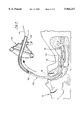
- FIG. 5 is a cross-sectional view of the mouth and airway of a patient after the mask 20 has been initially placed over the patient's mouth and nose with the curved guide 25 extending into the mouth, over the tongue 14, and into the hypopharynx 15.
- FIG. 6 is a cross-sectional view of the mouth and airway of the patient corresponding to FIG. 5 after the fiber optic probe 30 and endotracheal tube 40 have been inserted through the face mask port 23 and advanced along the curved guide 25 to a position below the larynx 18.
- FIG. 7 is a front view of the mask port 23 corresponding to FIG. 6 showing the fiber optic probe 30 and endotracheal tube 40 in cross-section.
- FIG. 8 is a cross-sectional view of the mouth and airway of the patient corresponding to FIG. 5 after the fiber optic probe 30 has been removed from within the endotracheal tube 40.
- FIG. 9 is a cross-sectional view of the mouth and airway of the patient corresponding to FIG. 5 showing the face mask 20 being removed while the endotracheal tube 40 remains in place.
- FIG. 10 is a cross-sectional view of the mouth and airway of the patient corresponding to FIG. 5 after the mask 20 has been removed, the endotracheal tube cuff 44 has been inflated, and a ventilator 50 has been connected to the endotracheal tube 40.
- FIG. 11 is a cross-sectional view of the face mask 20 and guide 25 in an alternative embodiment in which the curved guide 25 is configured as a oral airway that engages the posterior surface of the mask 20 surrounding the face mask port 23.
- FIG. 12 is a rear detail view of locking mechanism 21 used to engage the curved guide 25 to the posterior surface of the mask 20.
- FIGS. 1 and 2 front and rear perspective views of the present invention are illustrated.
- a corresponding cross-sectional view is shown in FIG. 3.
- the face mask 20 is adapted to fit over the patient's mouth and nose for resuscitation of the patient 10 as shown in FIG. 5.
- the mask 20 has a low profile and is made of an elastic material, such as rubber or flexible plastic, to allow the mask to conform to the contours of the patient's face and create a more air-tight seal around the mouth and nose.
- the face mask 20 includes a sealable port 23.
- the face mask port 23 consists of a flexible, elastic membrane having a stretchable opening 24 with dimensions large enough to allow a curved guide 25 to pass through the face mask port 23.
- this elastic membrane can be made of rubber with slot or hole forming an opening 24, as shown in FIG. 4.
- the curved guide 25 can be readily inserted through the face mask port 23 while maintaining a substantially air-tight seal around the guide 25 to prevent gas from escaping from within the face mask 20.
- the guide 25 is generally tubular and includes a sealable port 27 at its proximal end.
- the guide port 27 can be made of a flexible, elastic membrane having a stretchable slot or opening 28 with dimensions large enough to allow an endotracheal tube to pass through the guide port 27.
- the guide 25 extends posteriorly through the face mask 20 and has a curved distal portion that is inserted into the patient's mouth and hypopharynx 15 as the face mask 20 is placed over the patient's mouth.
- the distal portion of the curved guide 25 generally follows the profile of a typical patient's airway through the mouth, over the tongue 14, and into the hypopharynx 15 just above the opening to the trachea 16.
- the guide 25 is shaped to prevent the patient's tongue 14 and collapsible pharynx from obstructing access to the trachea 16, while also defining a channel for later insertion of an endotracheal tube.
- the guide 25 is typically made of plastic with sufficient strength and rigidity to keep the patient's teeth apart and prevent the patient from biting down on the endotracheal tube.
- the face mask port 23 allows the guide 25 to slide relative to the face mask 20, and also allows a limited range of rotation of the guide 25. This flexibility allows the guide 25 to accommodate a wide range of patient sizes and conditions.
- the guide 25 is equipped with small tube 29 bonded to the exterior of the guide 25 that extends along the length of the guide 25 to its distal end.
- This tube 29 can be used to suction secretions from the patient's mouth and airway as the guide 25 is advanced.
- a syringe 55 containing a local anesthetic e.g., lidocaine or xylocaine
- a local anesthetic e.g., lidocaine or xylocaine
- the anesthetic can be carried as far as the larynx 18 to deaden any discomfort associated with insertion of the endotracheal tube 40.
- the physician can squirt anesthetic directly down the main passageway of the guide 25.
- the main passageway can also be used for suctioning secretions from the patient's mouth and airway.
- the patient is initially resuscitated by supplying a flow of air/oxygen through the mask.
- the flow of air can be supplied by a resuscitation bag 22 attached to the mask 20 that is manually squeezed periodically to simulate natural breathing.
- other conventional air/oxygen supplies for resuscitation could be substituted at the connector 23 for the face mask 20.
- the flow of oxygen/air from the resuscitation bag 22 is directed around the exterior of the curved guide 25. This tends to inflate the patient's mouth and airway, which distends the collapsible tissues, and thereby makes visualization and insertion of the endotracheal tube 40 easier.
- an endotracheal tube 40 is inserted over a fiber optic probe 30.
- the fiber optic probe 30 and endotracheal tube 40 are then inserted through the guide port 27 and along the guide 25 to a position within the trachea 16 past the larynx 18 while resuscitation continues, as illustrated in FIG. 6.
- the opening 28 in the flexible membrane stretches to allow the endotracheal tube 40 and fiber optic probe 30 to pass through the guide port 27, but maintains a sufficiently tight fit around the endotracheal tube 40 to prevent the escape of gas from within the mask 20, as shown in the front view of the face mask provided in FIG. 7.
- the fiber optic probe 30 allows the physician to view within the patient's mouth and trachea 16 during insertion.
- the physician can also remotely manipulate the direction of the probe tip 32 to control the direction of the fiber optic probe 30. This minimizes patient discomfort and risk of injury to the patient.
- the small size of the fiber optic probe 30 also allows the physician to thread the fiber optic probe 30 through relatively constricted areas within the airway, such as the larynx 18. Most importantly, the fiber optic probe 30 and endotracheal tube 40 do not interfere with ongoing resuscitation of the patient.
- the distal end 46 of the endotracheal tube 40 can beveled as illustrated most clearly in FIG. 6.
- injury to the larynx 18 can be reduced by spinning the endotracheal tube 40 as it is advanced.
- the beveled end tends to keep the endotracheal tube 40 centered as it is passes through the vocal cords.
- Injury to the lining of the mouth and trachea can be reduced by using an endotracheal tube 40 made of a material having a low coefficient of friction, such as silicone.
- Bivona Medical Technologies of Gary, Ind. markets a line of endotracheal tubes made of silicone with a helical reinforcing wire.
- the fiber optic probe 30 is removed from within the endotracheal tube 40 through the proximal end of the endotracheal tube 40, as depicted in FIG. 8.
- the face mask 20 and guide 25 can then be removed while leaving the endotracheal tube 40 in place within the trachea 16, as shown in FIG. 9.
- the opening 28 in the flexible port 27 allows the face mask 20 and guide 25 to be withdrawn over the connector 42 at the proximal end of the endotracheal tube 40 with minimal effort and dislocation of the endotracheal tube 40.
- the position of the endotracheal tube 40 can be stabilized while the mask 20 is removed by manually gripping the proximal end of the endotracheal tube 40 and gradually urging it through the port 27 as the mask 20 is lifted from the patient's face. The physician can then reach under the face mask 20 to grip the endotracheal tube 40 after the mask 20 has been lifted sufficiently to allow access.
- the face mask 20 can be removed while leaving the guide 25 in place to serve as an oral airway and to protect the endotracheal tube 40 from being bitten by the patient's teeth.
- the endotracheal tube is taped to the patient's face, or held in place by some other suitable means for attachment.
- the cuff 44 at the distal end 46 of the endotracheal tube 40 is then inflated through the port valve 45 to block the trachea 16.
- An external ventilator 50 can be attached to the connector 42 at the proximal end of the endotracheal tube 40, as shown in FIG. 10.
- the patient can then be mechanically ventilated in the conventional manner via the endotracheal tube 40.
- the patient can be manually ventilated by attaching a resuscitation bag to the connector 42 at the proximal end of the endotracheal tube.
- the guide 25 and mask 20 can have any number of possible embodiments.
- the embodiment shown in the drawings uses a guide 25 that extends through an elastic port 23 in the face mask 20. This allows a limited range of motion between the guide 25 and mask 20 to make insertion of the guide easier, but requires two elastic ports 23 and 28.
- the guide 25 and mask 20 could be fabricated as two separate pieces that engage one another, as illustrated in FIG. 11. This eliminates the need for the guide port 27.
- the guide 25 is separately inserted into the mouth, similar to a conventional oral airway.
- FIG. 12 provides a rear detail view of the locking mechanism 21 used to engage the guide 25 to the posterior face of the mask 20.
- the guide 25 can be readily disengaged by rotating it slightly relative to the face mask 20. After the endotracheal tube 40 has been inserted, the mask 20 is removed while leaving the guide 25 in place within the patient's mouth. The guide 25 remains around the endotracheal tube 40 and protects it from being bitten or crimped by the patient's teeth.
- the guide 25 can consist of a tubular member as shown in the drawings. Alternatively, the distal portion of the guide 25 can have a U-shaped cross-section.
- the guide 25 can be molded from a suitable plastic material having a relatively low coefficient of friction to minimize irritation to the lining of mouth and trachea and to minimize resistance to insertion of the endotracheal tube 40 along the guide. Friction can be further reduced by applying a slippery coating to both the exterior and interior surfaces of the guide 25. A slippery coating can also be applied to the endotracheal tube to minimize friction between the endotracheal tube and the guide.
- kits for use in emergency rooms and intensive care units can be readily packaged as a kit for use in emergency rooms and intensive care units.
- the kit is sufficiently compact and inexpensive that it can be stocked on resuscitation carts; widely used in hospitals, and carried in ambulances for use by emergency medical technicians in the field.
- the fiber optic probe can be operated using a battery-powered light source.
- the oxygen supply for the hospital or ambulance can be connected to the face mask 20 for resuscitation or to provide a flow of gas to the ventilator 50.
- the tube 29 extending along the guide 29 can also be connected to the suction system provided by the hospital or ambulance, if necessary.
Abstract
An endotracheal tube can be inserted into a patient's trachea during resuscitation by using a face mask and a curved guide. The guide is inserted through a flexible port in the face mask and has a curved distal portion that extends into the patient's mouth and hypopharynx. The patient is initially resuscitated by supplying a flow of air/oxygen through the mask. An endotracheal tube is inserted over the distal end of a fiber optic probe. Resuscitation, oxygenation, or artificial ventilation continue without interruption while the fiber optic probe and endotracheal tube are inserted through a flexible port at the proximal end of the curve guide and then advanced along the guide into the patient's airway. The direction of the distal tip of the fiber optic probe can be controlled by the physician. This allows the physician to carefully guide the fiber optic probe and endotracheal tube to a position past the larynx while resuscitation continues. The fiber optic probe is then removed from within the endotracheal tube and the mask is removed while leaving the endotracheal tube in place within the trachea. The cuff on the endotracheal tube is inflated and a ventilator is connected to the proximal end of the endotracheal tube to ventilate the patient. Alternatively, the patient can be manually ventilated by connecting a resuscitation bag to the proximal end of the endotracheal tube.
Description
This application is a continuation of the Applicant's U.S. patent application Ser. No. 08/607,332 filed on Feb. 26, 1996, now U.S. Pat. No. 5,694,929 entitled "Method and Apparatus For Ventilation/Oxygenation During Guided Insertion Of An Endotracheal Tube".
1. Field of the Invention
The present invention relates generally to the field of respiratory devices and methods. More specifically, the present invention discloses a method and apparatus for guiding insertion of an endotracheal tube while the patient continues to receive cardiopulmonary resuscitation.
2. Statement of the Problem
In emergency situations involving cardiopulmonary patients or other patients with compromised or arrested breathing, an oral airway is first inserted into the patient's mouth. A face mask is then placed over the patient's mouth and nose. The face mask is connected to an inflatable bag to maintain at least minimal oxygen flow to the lungs in the short term. This process is sometimes referred to as "bagging" the patient. It is suitable for initially stabilizing the patient. In order to breathe more effectively for the patient during cardiopulmonary resuscitation, and to prevent aspiration of stomach contents, an endotracheal tube (or ET tube) is placed into the trachea. Longer-term care usually requires attaching the patient to a ventilator (e.g., by means of the endotracheal tube). The transition from face mask to breathing through the endotracheal tube can be dangerous if insertion of the endotracheal tube takes too long, because the mask and oral airway must be removed and the flow of air/oxygen is interrupted while the endotracheal tube is inserted through the patient's mouth.
The typical conventional approach to making this transition involves discontinuing resuscitation and completely removing the mask and oral airway to expose the mouth. The physician inserts a rigid laryngoscope blade into the patient's mouth to ensure that the patient's airway is open, and then attempts to insert the endotracheal tube through the patient's mouth and into the trachea in the conventional manner. This may require a significant amount of time, particularly if the patient is less than completely cooperative and relaxed, or if the patient's airway has suffered trauma, or the tongue has fallen back to close the airway. The patient may not be breathing during this time, or may not be breathing sufficiently to maintain adequate blood oxygen levels. If the transition process takes more than a few seconds, the physician must temporarily abandon the effort and return to resuscitation by reinserting the oral airway and replacing the face mask. The transition process may have to be repeated several times before the endotracheal tube is successful installed. In addition, the speed with which the transition process must be completed increases the chances of a mistake being made or unnecessary injury to the patient during the intubation procedure.
Endotracheal tubes are also used in semi-emergency situations to ventilate patients with respiratory failure who may be conscious or semi-conscious. The conventional approach requires the patient to lie still while the physician inserts a rigid laryngoscope blade into the patient's mouth and trachea. Delivery of ventilation and/or oxygen is also interrupted during this period. The endotracheal tube is then inserted into place while the laryngoscope blade keeps the patient's airway open. Successful intubation depends on the patient being cooperative and completely relaxed, which unfortunately is often not the case. Even with a cooperative patient, intubation is very uncomfortable and can cause the patient to panic due to the difficulty in breathing during the procedure. This procedure can also result in a choking or gagging response that can cause the patient to regurgitate and aspirate contents from the stomach. One conventional response to these shortcomings has been to sedate the patient during intubation. Tranquilizers make the patient more cooperative and less likely to choke during intubation, but also tend to suppress the patient's breathing and blood pressure. These side effects may be unacceptable when dealing with a patient who already suffers from shallow or irregular breathing or depressed blood pressure. Therefore, a need exists for an improved device to guide insertion of an endotracheal tube and ensure that the patient's airway is open, and that also allows the patient to continue to receive air/oxygen during the insertion process.
A wide variety of devices that combine face masks with tubes for ventilation (e.g., endotracheal tubes) have been used in the past, including the following:
______________________________________
Inventor   Patent No.      Issue Date
______________________________________
Jeshuran   5,197,463       Mar. 20, 1993
Kondur     4,580,556       Apr. 8, 1986
Donmichael 4,497,318       Feb. 5, 1985
Dryden     4,256,099       Mar. 17, 1981
Buttaravoli
           3,809,079       May 7, 1974
______________________________________
Jeshuran shows an anesthesia mask 28 that is initially placed over the patient's mouth and nose as shown in FIG. 7 of the Jeshuran patent. A fiber optic 40 is inserted through an endotracheal tube, and then through an opening in a two-piece core 84, 86, as shown in FIG. 9. The fiber optic 40 is advanced into the trachea. The head 96 is then unscrewed and the core segments 84, 86 are disassembled to allow the endotracheal tube to be inserted through the mask, as shown in FIG. 2. The fiber optic 40 serves as a guide for insertion of the endotracheal tube 46. The fiber optic 40 is then withdrawn and the endotracheal tube cuff 136 is inflated, as shown in FIG. 8. However, Jeshuran does not show a curved guide to direct insertion of the fiber optic probe. The physician is faced with the problem of navigating the fiber optic probe past the patient's tongue and along the patient's airway.
Kondur discloses another example of a adapter 10 that allows insertion of an endotracheal tube 40 through the face mask 50 and nose of the patient. Here again, no curved guide is provided.
Donmichael discloses an esophageal obturator for blocking aspiration of stomach fluids while the face mask is being used for ventilating the lungs.
Dryden discloses a two-tube resuscitation system. One tube is used to supply air to the trachea, while the other tube is used for aspiration or administering medication.
Buttaravoli discloses a resuscitator having a face mask 11 with a curved tube 15 for supplying air to the patient's airway.
In addition, the prior art includes several references involving intubating pharyngeal airways that have a curved central tubular member, including the following:
______________________________________
Inventor Patent No.       Issue Date
______________________________________
Berman   4,054,135        Oct. 18, 1977
Berman   4,067,331        Jan. 10, 1978
Berman   4,068,658        Jan. 17, 1978
Berman   4,069,820        Jan. 24, 1978
______________________________________
The Berman patents show an intubating pharyngeal airway having a side access for passage of a tube. The side opening can be expanded or closed by means of either a hinge on the opposite side wall of the tube or by a cap.
3. Solution to the Problem
None of the prior art references uncovered in the search show a face mask that incorporates a port and a curved guide for directing a fiber optic probe and endotracheal tube along the patient's airway while resuscitation, cardiopulmonary resuscitation, artificial mask breathing, or oxygenation continues. After the distal end of the fiber optic probe has guided the endotracheal tube past the larynx into the trachea, the fiber optic probe is withdrawn and the endotracheal tube can be used to more effectively "bag" the patient, or the patient can be connected to an external ventilator.
This system allows the endotracheal tube to be inserted and connected to a ventilator without interrupting resuscitation or oxygenation of the patient via the face mask. In addition, the curved guide greatly simplifies insertion of the fiber optic probe and endotracheal tube by providing direction and maintaining an open passageway past the patient's tongue and into the hypopharynx. The flow of air/oxygen supplied by the resuscitation bag tends to inflate the patient's mouth and airway, and thus also helps to maintain a passageway and visualization for the fiber optic probe and endotracheal tube.
This invention provides a method and apparatus for guiding insertion of an endotracheal tube into a patient's trachea during resuscitation by using a face mask and a curved guide. The guide is inserted through a flexible port in the face mask and has a curved distal portion that extends into the patient's mouth and hypopharynx. The patient is initially resuscitated by supplying a flow of air/oxygen through the mask. An endotracheal tube is inserted over the distal end of a fiber optic probe. Resuscitation, oxygenation, or artificial ventilation continue without interruption while the fiber optic probe and endotracheal tube are inserted through a flexible port at the proximal end of the guide and then advanced along the guide into the patient's airway. The direction of the distal tip of the fiber optic probe can be controlled by the physician. This allows the physician to carefully guide the fiber optic probe and endotracheal tube to a position past the larynx while resuscitation continues. The fiber optic probe is then removed from within the endotracheal tube and the mask is removed while leaving the endotracheal tube in place within the trachea. The cuff on the endotracheal tube is inflated and a ventilator is connected to the proximal end of the endotracheal tube to ventilate the patient. Alternatively, the patient can be manually ventilated by connecting a resuscitation bag to the proximal end of the endotracheal tube.
A primary object of the present invention is to provide a method and apparatus for guiding insertion of an endotracheal tube that does not require interruption of the resuscitation process.
Another object of the present invention is to provide a method and apparatus for improving insertion of an endotracheal tube by helping to keep the patient's airway open, and also allowing the physician to guide the insertion process via the fiber optic probe.
Another object of the present invention is to provide a method and apparatus for instilling local anesthetic into the patient's airway and suctioning excess secretions prior to insertion of the endotracheal tube.
Another object of the present invention is to provide a method and apparatus for guiding insertion of an endotracheal tube that lessens the risk of injury and reduces patient discomfort.
Yet another object of the present invention is to provide a device that enables the physician to instill anesthetic and/or suction secretions from the patient's mouth and airway as the device is inserted.
These and other advantages, features, and objects of the present invention will be more readily understood in view of the following detailed description and the drawings.
The present invention can be more readily understood in conjunction with the accompanying drawings, in which:
FIG. 1 is a front perspective view of the face mask assembly, including the port 23 and curved guide 25.
FIG. 2 is a rear perspective view of the mask assembly corresponding to FIG. 1.
FIG. 3 is a cross-sectional view of the mask assembly corresponding to FIG. 1.
FIG. 4 is a front view of the face mask port 23 showing the elastic slot 28 closed.
FIG. 5 is a cross-sectional view of the mouth and airway of a patient after the mask 20 has been initially placed over the patient's mouth and nose with the curved guide 25 extending into the mouth, over the tongue 14, and into the hypopharynx 15.
FIG. 6 is a cross-sectional view of the mouth and airway of the patient corresponding to FIG. 5 after the fiber optic probe 30 and endotracheal tube 40 have been inserted through the face mask port 23 and advanced along the curved guide 25 to a position below the larynx 18.
FIG. 7 is a front view of the mask port 23 corresponding to FIG. 6 showing the fiber optic probe 30 and endotracheal tube 40 in cross-section.
FIG. 8 is a cross-sectional view of the mouth and airway of the patient corresponding to FIG. 5 after the fiber optic probe 30 has been removed from within the endotracheal tube 40.
FIG. 9 is a cross-sectional view of the mouth and airway of the patient corresponding to FIG. 5 showing the face mask 20 being removed while the endotracheal tube 40 remains in place.
FIG. 10 is a cross-sectional view of the mouth and airway of the patient corresponding to FIG. 5 after the mask 20 has been removed, the endotracheal tube cuff 44 has been inflated, and a ventilator 50 has been connected to the endotracheal tube 40.
FIG. 11 is a cross-sectional view of the face mask 20 and guide 25 in an alternative embodiment in which the curved guide 25 is configured as a oral airway that engages the posterior surface of the mask 20 surrounding the face mask port 23.
FIG. 12 is a rear detail view of locking mechanism 21 used to engage the curved guide 25 to the posterior surface of the mask 20.
Turning to FIGS. 1 and 2, front and rear perspective views of the present invention are illustrated. A corresponding cross-sectional view is shown in FIG. 3. The face mask 20 is adapted to fit over the patient's mouth and nose for resuscitation of the patient 10 as shown in FIG. 5. The mask 20 has a low profile and is made of an elastic material, such as rubber or flexible plastic, to allow the mask to conform to the contours of the patient's face and create a more air-tight seal around the mouth and nose.
The face mask 20 includes a sealable port 23. In the preferred embodiment, the face mask port 23 consists of a flexible, elastic membrane having a stretchable opening 24 with dimensions large enough to allow a curved guide 25 to pass through the face mask port 23. For example, this elastic membrane can be made of rubber with slot or hole forming an opening 24, as shown in FIG. 4.
As depicted in FIG. 5, the curved guide 25 can be readily inserted through the face mask port 23 while maintaining a substantially air-tight seal around the guide 25 to prevent gas from escaping from within the face mask 20. The guide 25 is generally tubular and includes a sealable port 27 at its proximal end. For example, the guide port 27 can be made of a flexible, elastic membrane having a stretchable slot or opening 28 with dimensions large enough to allow an endotracheal tube to pass through the guide port 27. The guide 25 extends posteriorly through the face mask 20 and has a curved distal portion that is inserted into the patient's mouth and hypopharynx 15 as the face mask 20 is placed over the patient's mouth. The distal portion of the curved guide 25 generally follows the profile of a typical patient's airway through the mouth, over the tongue 14, and into the hypopharynx 15 just above the opening to the trachea 16. The guide 25 is shaped to prevent the patient's tongue 14 and collapsible pharynx from obstructing access to the trachea 16, while also defining a channel for later insertion of an endotracheal tube. The guide 25 is typically made of plastic with sufficient strength and rigidity to keep the patient's teeth apart and prevent the patient from biting down on the endotracheal tube. The face mask port 23 allows the guide 25 to slide relative to the face mask 20, and also allows a limited range of rotation of the guide 25. This flexibility allows the guide 25 to accommodate a wide range of patient sizes and conditions.
In the preferred embodiment, the guide 25 is equipped with small tube 29 bonded to the exterior of the guide 25 that extends along the length of the guide 25 to its distal end. This tube 29 can be used to suction secretions from the patient's mouth and airway as the guide 25 is advanced. Alternatively a syringe 55 containing a local anesthetic (e.g., lidocaine or xylocaine) can be connected to the proximal end of the tube 29 to squirt anesthetic as the guide 25 is insert through the patient's mouth and into the hypopharynx 15, as illustrated in FIG. 5. If squirted with sufficient force, the anesthetic can be carried as far as the larynx 18 to deaden any discomfort associated with insertion of the endotracheal tube 40. Alternatively, the physician can squirt anesthetic directly down the main passageway of the guide 25. The main passageway can also be used for suctioning secretions from the patient's mouth and airway.
The patient is initially resuscitated by supplying a flow of air/oxygen through the mask. For example, the flow of air can be supplied by a resuscitation bag 22 attached to the mask 20 that is manually squeezed periodically to simulate natural breathing. However, other conventional air/oxygen supplies for resuscitation could be substituted at the connector 23 for the face mask 20. In the preferred embodiment, the flow of oxygen/air from the resuscitation bag 22 is directed around the exterior of the curved guide 25. This tends to inflate the patient's mouth and airway, which distends the collapsible tissues, and thereby makes visualization and insertion of the endotracheal tube 40 easier.
After the patient's conditioned has been stabilized to some degree during initial resuscitation, an endotracheal tube 40 is inserted over a fiber optic probe 30. The fiber optic probe 30 and endotracheal tube 40 are then inserted through the guide port 27 and along the guide 25 to a position within the trachea 16 past the larynx 18 while resuscitation continues, as illustrated in FIG. 6. The opening 28 in the flexible membrane stretches to allow the endotracheal tube 40 and fiber optic probe 30 to pass through the guide port 27, but maintains a sufficiently tight fit around the endotracheal tube 40 to prevent the escape of gas from within the mask 20, as shown in the front view of the face mask provided in FIG. 7.
The fiber optic probe 30 allows the physician to view within the patient's mouth and trachea 16 during insertion. The physician can also remotely manipulate the direction of the probe tip 32 to control the direction of the fiber optic probe 30. This minimizes patient discomfort and risk of injury to the patient. The small size of the fiber optic probe 30 also allows the physician to thread the fiber optic probe 30 through relatively constricted areas within the airway, such as the larynx 18. Most importantly, the fiber optic probe 30 and endotracheal tube 40 do not interfere with ongoing resuscitation of the patient.
The distal end 46 of the endotracheal tube 40 can beveled as illustrated most clearly in FIG. 6. Experience has shown that injury to the larynx 18 can be reduced by spinning the endotracheal tube 40 as it is advanced. The beveled end tends to keep the endotracheal tube 40 centered as it is passes through the vocal cords. Injury to the lining of the mouth and trachea can be reduced by using an endotracheal tube 40 made of a material having a low coefficient of friction, such as silicone. Bivona Medical Technologies of Gary, Ind., markets a line of endotracheal tubes made of silicone with a helical reinforcing wire.
After the endotracheal tube 40 has been inserted, the fiber optic probe 30 is removed from within the endotracheal tube 40 through the proximal end of the endotracheal tube 40, as depicted in FIG. 8. The face mask 20 and guide 25 can then be removed while leaving the endotracheal tube 40 in place within the trachea 16, as shown in FIG. 9. The opening 28 in the flexible port 27 allows the face mask 20 and guide 25 to be withdrawn over the connector 42 at the proximal end of the endotracheal tube 40 with minimal effort and dislocation of the endotracheal tube 40. The position of the endotracheal tube 40 can be stabilized while the mask 20 is removed by manually gripping the proximal end of the endotracheal tube 40 and gradually urging it through the port 27 as the mask 20 is lifted from the patient's face. The physician can then reach under the face mask 20 to grip the endotracheal tube 40 after the mask 20 has been lifted sufficiently to allow access.
Alternatively, the face mask 20 can be removed while leaving the guide 25 in place to serve as an oral airway and to protect the endotracheal tube 40 from being bitten by the patient's teeth. After the face mask 20 has been removed, the endotracheal tube is taped to the patient's face, or held in place by some other suitable means for attachment.
The cuff 44 at the distal end 46 of the endotracheal tube 40 is then inflated through the port valve 45 to block the trachea 16. An external ventilator 50 can be attached to the connector 42 at the proximal end of the endotracheal tube 40, as shown in FIG. 10. The patient can then be mechanically ventilated in the conventional manner via the endotracheal tube 40. Alternatively, the patient can be manually ventilated by attaching a resuscitation bag to the connector 42 at the proximal end of the endotracheal tube.
It should be understood that the guide 25 and mask 20 can have any number of possible embodiments. The embodiment shown in the drawings uses a guide 25 that extends through an elastic port 23 in the face mask 20. This allows a limited range of motion between the guide 25 and mask 20 to make insertion of the guide easier, but requires two elastic ports 23 and 28. Alternatively, the guide 25 and mask 20 could be fabricated as two separate pieces that engage one another, as illustrated in FIG. 11. This eliminates the need for the guide port 27. In this embodiment, the guide 25 is separately inserted into the mouth, similar to a conventional oral airway. The mask 20 is then placed over the patient's mouth and nose so that the proximal end of the guide 25 engages a corresponding opening in the posterior face of the mask 20 to provide a relatively continuous passageway for insertion of the fiber optic probe 30 and endotracheal tube 40 through the face mask port 23 and along the guide 25. FIG. 12 provides a rear detail view of the locking mechanism 21 used to engage the guide 25 to the posterior face of the mask 20. The guide 25 can be readily disengaged by rotating it slightly relative to the face mask 20. After the endotracheal tube 40 has been inserted, the mask 20 is removed while leaving the guide 25 in place within the patient's mouth. The guide 25 remains around the endotracheal tube 40 and protects it from being bitten or crimped by the patient's teeth.
The guide 25 can consist of a tubular member as shown in the drawings. Alternatively, the distal portion of the guide 25 can have a U-shaped cross-section. The guide 25 can be molded from a suitable plastic material having a relatively low coefficient of friction to minimize irritation to the lining of mouth and trachea and to minimize resistance to insertion of the endotracheal tube 40 along the guide. Friction can be further reduced by applying a slippery coating to both the exterior and interior surfaces of the guide 25. A slippery coating can also be applied to the endotracheal tube to minimize friction between the endotracheal tube and the guide.
All of the components necessary to practice the present invention can be readily packaged as a kit for use in emergency rooms and intensive care units. The kit is sufficiently compact and inexpensive that it can be stocked on resuscitation carts; widely used in hospitals, and carried in ambulances for use by emergency medical technicians in the field. The fiber optic probe can be operated using a battery-powered light source. The oxygen supply for the hospital or ambulance can be connected to the face mask 20 for resuscitation or to provide a flow of gas to the ventilator 50. The tube 29 extending along the guide 29 can also be connected to the suction system provided by the hospital or ambulance, if necessary.
The above disclosure sets forth a number of embodiments of the present invention. Other arrangements or embodiments, not precisely set forth, could be practiced under the teachings of the present invention and as set forth in the following claims.
Claims (7)
1. An apparatus for guiding insertion of an endotracheal tube into the trachea of a patient, said apparatus comprising:
a face mask for covering a patient's mouth and nose;
means for supplying a flow of air/oxygen into said mask to resuscitate a patient;
a resealable face mask port; and
a guide removably extending through said face mask port having:
(a) a curved distal portion for insertion into a patient's mouth and hypopharynx; said guide directing insertion of an endotracheal tube into a patient's trachea; and
(b) a guide port allowing removable insertion of the endotracheal tube along said guide;
wherein said face mask port seals about said guide during insertion of said guide, and reseals to substantially close said face mask port after removal of said guide.
2. The apparatus of claim 1 wherein said guide comprises a tube having a curved distal portion.
3. The apparatus of claim 1 wherein said means for supplying a flow of air/oxygen to said mask comprises a resuscitation bag attached to said mask.
4. The apparatus of claim 1 wherein said flow of air/oxygen is directed around the exterior of said guide.
5. The apparatus of claim 1 wherein said guide port further comprises a flexible membrane having a stretchable opening for receiving said endotracheal tube.
6. The apparatus of claim 1 wherein said face mask port comprises a flexible membrane having a stretchable opening for receiving said guide.
7. The apparatus of claim 1 wherein said guide has a substantially U-shaped cross-section.
Priority Applications (11)
| Application Number | Priority Date | Filing Date | Title |
|---|---|---|---|
| US08/974,864 US5964217A (en) | 1996-02-26 | 1997-11-20 | Method and apparatus for ventilation/oxygenation during guided insertion of an endotracheal tube |
| US09/411,610 US6405725B1 (en) | 1996-02-26 | 1999-10-01 | Method and apparatus for ventilation/oxygenation during guided insertion of an endotracheal tube |
| US09/525,270 US6631713B1 (en) | 1996-02-26 | 2000-03-14 | Method and apparatus for ventilation/oxygenation during guided insertion of an endotracheal tube |
| US09/707,350 US6543446B1 (en) | 1996-02-26 | 2000-11-06 | Method and apparatus for ventilation/oxygenation during guided insertion of an endotracheal tube |
| US09/767,272 US6568388B2 (en) | 1996-02-26 | 2001-01-22 | Method and apparatus for ventilation / oxygenation during guided insertion of an endotracheal tube |
| US09/840,194 US6634354B2 (en) | 1996-02-26 | 2001-04-23 | Laryngeal mask airway |
| US09/908,380 US6668821B2 (en) | 1996-02-26 | 2001-07-18 | Laryngeal mask airway |
| US10/115,224 US6860264B2 (en) | 1996-02-26 | 2002-04-02 | Method and apparatus for endotracheal intubation using a light wand and curved guide |
| US10/405,511 US20030188750A1 (en) | 1996-02-26 | 2003-04-02 | Method and apparatus for ventilation / oxygenation during guided insertion of an endotracheal tube |
| US10/674,136 US6895966B2 (en) | 1996-02-26 | 2003-09-29 | Laryngeal mask airway |
| US11/067,860 US20050139220A1 (en) | 1996-02-26 | 2005-02-28 | Method and apparatus for ventilation / oxygenation during guided insertion of an endotracheal tube |
Applications Claiming Priority (2)
| Application Number | Priority Date | Filing Date | Title |
|---|---|---|---|
| US08/607,332 US5694929A (en) | 1996-02-26 | 1996-02-26 | Method and apparatus for ventilation/oxygenation during guided insertion of an endotracheal tube |
| US08/974,864 US5964217A (en) | 1996-02-26 | 1997-11-20 | Method and apparatus for ventilation/oxygenation during guided insertion of an endotracheal tube |
Related Parent Applications (1)
| Application Number | Title | Priority Date | Filing Date |
|---|---|---|---|
| US08/607,332 Continuation US5694929A (en) | 1996-02-26 | 1996-02-26 | Method and apparatus for ventilation/oxygenation during guided insertion of an endotracheal tube |
Related Child Applications (1)
| Application Number | Title | Priority Date | Filing Date |
|---|---|---|---|
| US09/411,610 Continuation-In-Part US6405725B1 (en) | 1996-02-26 | 1999-10-01 | Method and apparatus for ventilation/oxygenation during guided insertion of an endotracheal tube |
Publications (1)
| Publication Number | Publication Date |
|---|---|
| US5964217A true US5964217A (en) | 1999-10-12 |
Family
ID=24431831
Family Applications (2)
| Application Number | Title | Priority Date | Filing Date |
|---|---|---|---|
| US08/607,332 Expired - Fee Related US5694929A (en) | 1996-02-26 | 1996-02-26 | Method and apparatus for ventilation/oxygenation during guided insertion of an endotracheal tube |
| US08/974,864 Expired - Fee Related US5964217A (en) | 1996-02-26 | 1997-11-20 | Method and apparatus for ventilation/oxygenation during guided insertion of an endotracheal tube |
Family Applications Before (1)
| Application Number | Title | Priority Date | Filing Date |
|---|---|---|---|
| US08/607,332 Expired - Fee Related US5694929A (en) | 1996-02-26 | 1996-02-26 | Method and apparatus for ventilation/oxygenation during guided insertion of an endotracheal tube |
Country Status (1)
| Country | Link |
|---|---|
| US (2) | US5694929A (en) |
Cited By (50)
| Publication number | Priority date | Publication date | Assignee | Title |
|---|---|---|---|---|
| WO2001024859A1 (en) * | 1999-10-01 | 2001-04-12 | Evergreen Medical, Inc. | Ventilation system during guided insertion of an endotracheal tube |
| WO2002040079A2 (en) * | 2000-11-20 | 2002-05-23 | Evergreen Medical, Inc. | Laryngeal mask airway |
| US6405725B1 (en) * | 1996-02-26 | 2002-06-18 | Evergreen Medical, Inc. | Method and apparatus for ventilation/oxygenation during guided insertion of an endotracheal tube |
| WO2002056936A2 (en) * | 2001-01-22 | 2002-07-25 | Evergreen Medical, Inc. | Ventilation system during guided insertion of an endotracheal tube |
| US20020108610A1 (en) * | 1996-02-26 | 2002-08-15 | Christopher Kent L. | Method and apparatus for endotracheal intubation using a light wand and curved guide |
| US6460540B1 (en) * | 1999-04-05 | 2002-10-08 | Mark S. Klepper | Endotracheal tube sump |
| US6543446B1 (en) | 1996-02-26 | 2003-04-08 | Evergreen Medical Incorporated | Method and apparatus for ventilation/oxygenation during guided insertion of an endotracheal tube |
| EP1318850A2 (en) * | 2000-05-22 | 2003-06-18 | Sleepup Ltd. | Device, system and method for preventing collapse of the upper airway |
| US6668821B2 (en) * | 1996-02-26 | 2003-12-30 | Evergreen Medical Incorporated | Laryngeal mask airway |
| US6725862B2 (en) | 2001-08-24 | 2004-04-27 | Naum Klinberg | Tracheostomy tube apparatus for noninvasive suctioning |
| US20040079364A1 (en) * | 1996-02-26 | 2004-04-29 | Christopher Kent L. | Laryngeal mask airway |
| US6763831B2 (en) * | 2001-08-02 | 2004-07-20 | Joseph A. Sniadach | Adjustable ventilation mask for a patient |
| US6792943B2 (en) | 2001-09-05 | 2004-09-21 | Minnesota High-Tech Resources, Llc | Intubating ventilatory face mask |
| US20050081861A1 (en) * | 2002-08-14 | 2005-04-21 | Nasir Muhammed A. | Airway device |
| US20050139220A1 (en) * | 1996-02-26 | 2005-06-30 | Evergreen Medical Incorporated | Method and apparatus for ventilation / oxygenation during guided insertion of an endotracheal tube |
| US20050268917A1 (en) * | 2004-06-02 | 2005-12-08 | Benje Boedeker | Device for insertion of endotracheal tube |
| US20060102186A1 (en) * | 2004-11-18 | 2006-05-18 | Mark Adler | Intra-bronchial apparatus for aspiration and insufflation of lung regions distal to placement or cross communication and deployment and placement system therefor |
| US20060207601A1 (en) * | 2003-08-14 | 2006-09-21 | Nasir Muhammed Aslam | Airway device |
| US20060242946A1 (en) * | 2005-04-29 | 2006-11-02 | Arvin Technologies, Inc. | Method and apparatus for supplying air to emission abatement device by use of turbocharger |
| US20090125002A1 (en) * | 2005-07-25 | 2009-05-14 | Km Technologies | Device and method for placing within a patient an enteral tube after endotracheal intubation |
| US20100113916A1 (en) * | 2008-10-30 | 2010-05-06 | Avinash B. Kumar | Systems and Methods for Endotracheal Tube Positioning |
| US20100180737A1 (en) * | 2009-01-21 | 2010-07-22 | Klepper Mark S | Method to manufacture a tube sump with integral clips |
| US20100288289A1 (en) * | 2007-09-29 | 2010-11-18 | Muhammed Aslam Nasir | Airway device |
| US7921847B2 (en) | 2005-07-25 | 2011-04-12 | Intubix, Llc | Device and method for placing within a patient an enteral tube after endotracheal intubation |
| US20110162650A1 (en) * | 2008-06-04 | 2011-07-07 | Cosmeplast Ets | respiratory interface devices |
| US8197402B1 (en) | 2009-06-17 | 2012-06-12 | Douglas Alexis Cedeno | Free-hand laryngoscope gaper |
| USD665254S1 (en) | 2011-06-08 | 2012-08-14 | Intersurgical Ag | Airway device packaging |
| USD665495S1 (en) | 2009-07-14 | 2012-08-14 | Muhammed Aslam Nasir | Medical device |
| USD688787S1 (en) | 2011-06-08 | 2013-08-27 | Intersurgical Ag | Airway device cap and strap holder |
| USD693920S1 (en) | 2011-06-08 | 2013-11-19 | Intersurgical Ag | Airway device |
| US20140060534A1 (en) * | 2004-01-22 | 2014-03-06 | Thermocure, Inc. | Respiratory system for inducing therapeutic hypothermia |
| RU2523151C2 (en) * | 2008-12-12 | 2014-07-20 | Кимберли-Кларк Ворлдвайд, Инк. | Airway port unit blocked with press button and method for using it |
| USD712244S1 (en) | 2011-09-23 | 2014-09-02 | Intersurgical Ag | Medical device package |
| US8887730B2 (en) | 2011-05-26 | 2014-11-18 | Covidien Lp | Dual-lumen tracheal tube with assembly portion |
| US8978657B2 (en) | 2010-07-29 | 2015-03-17 | Covidien Lp | Dual-lumen tracheal tube with shaped lumen divider |
| US8998798B2 (en) | 2010-12-29 | 2015-04-07 | Covidien Lp | Multi-lumen tracheal tube with visualization device |
| US9155854B2 (en) | 2011-08-31 | 2015-10-13 | Covidien Lp | Tracheal tube with visualization device and integrated flushing system |
| US9211060B2 (en) | 2011-04-05 | 2015-12-15 | Covidien Lp | Visualization device and holder for use with a tracheal tube |
| US9265905B2 (en) | 2010-06-24 | 2016-02-23 | Ashkal Developments Limited | Stopper device |
| US20160107006A1 (en) * | 2013-06-13 | 2016-04-21 | The Board Of Trustees Of The University Of Illinois | Helmet for anesthesia |
| USD761952S1 (en) | 2012-07-27 | 2016-07-19 | Docsinnovent Limited | Airway device |
| EP3150245A2 (en) | 2015-09-30 | 2017-04-05 | Sajith Chakithandy | Ventilation with a view mask |
| US9788755B2 (en) | 2011-05-26 | 2017-10-17 | Covidien Lp | Illumination systems and devices for tracheal tubes |
| US9937311B2 (en) | 2012-01-27 | 2018-04-10 | Ashkal Developments Limited | Stopper device |
| USD842456S1 (en) | 2015-12-15 | 2019-03-05 | Intersurgical Ag | Airway device |
| US10238831B2 (en) | 2013-09-08 | 2019-03-26 | Qool Therapeutics, Inc. | Temperature measurement and feedback for therapeutic hypothermia |
| US10625037B2 (en) | 2013-12-17 | 2020-04-21 | Intersurgical Ag | Intubating airway device |
| US11020269B2 (en) | 2015-02-23 | 2021-06-01 | Qool Therapeutics, Inc. | Systems and methods for endotracheal delivery of frozen particles |
| US11701484B2 (en) | 2017-12-13 | 2023-07-18 | Ashkal Developments Limited | Airway device |
| USD1025348S1 (en) | 2020-04-16 | 2024-04-30 | Intersurgical Ag | Airway device |
Families Citing this family (48)
| Publication number | Priority date | Publication date | Assignee | Title |
|---|---|---|---|---|
| US5694929A (en) * | 1996-02-26 | 1997-12-09 | Christopher; Kent L. | Method and apparatus for ventilation/oxygenation during guided insertion of an endotracheal tube |
| US5878745A (en) | 1996-03-01 | 1999-03-09 | Brain; Archibald I.J. | Gastro-laryngeal mask |
| GB9622880D0 (en) * | 1996-11-02 | 1997-01-08 | Smiths Industries Plc | Laryngeal mask airways and thier manufacture |
| US6109259A (en) | 1997-12-10 | 2000-08-29 | Spirit Medical Systems, Inc. | Gas supplying and substance suctioning relative to a patients trachea |
| US7331346B2 (en) | 1997-12-24 | 2008-02-19 | Indian Ocean Medical, Inc. | Monitoring and control for a laryngeal mask airway device |
| GB9727367D0 (en) | 1997-12-24 | 1998-02-25 | Brain Archibald Ian Jeremy | Improvements in laryngeal mask airway devices |
| GB9817537D0 (en) * | 1998-08-13 | 1998-10-07 | Brain Archibald Ian Jeremy | A laryngear mask airway with mutually independant laterally-placed ultra-flexible eastric access/discharge and airway tubes |
| GB9821771D0 (en) | 1998-10-06 | 1998-12-02 | Brain Archibald Ian Jeremy | Improvements relating to laryngeal mask airway devices |
| US6202646B1 (en) | 1998-12-23 | 2001-03-20 | Para Products Incorporated | Detection device for verifying the proper intubation of an endotracheal tube |
| US6257236B1 (en) * | 1999-02-23 | 2001-07-10 | Edward P Dutkiewicz | Intubation device |
| US6705318B1 (en) * | 1999-04-09 | 2004-03-16 | Archibald I. J. Brain | Disposable LMA |
| US6254591B1 (en) * | 1999-06-01 | 2001-07-03 | Children's Medical Center Corporation | Scavenger suction device |
| US6183493B1 (en) | 1999-08-24 | 2001-02-06 | Pharmasys International, Llc | Method and apparatus for the treatment of sleep apnea and related breathing disorders |
| US6511676B1 (en) * | 1999-11-05 | 2003-01-28 | Teni Boulikas | Therapy for human cancers using cisplatin and other drugs or genes encapsulated into liposomes |
| US6378523B1 (en) | 2000-03-15 | 2002-04-30 | Evergreen Medical Incorporated | Endotracheal tube having a beveled tip and orientation indicator |
| US6575944B1 (en) * | 2000-06-19 | 2003-06-10 | Portex, Inc. | Adapter for localized treatment through a tracheal tube and method for use thereof |
| US6820614B2 (en) * | 2000-12-02 | 2004-11-23 | The Bonutti 2003 Trust -A | Tracheal intubination |
| US6626169B2 (en) | 2001-05-17 | 2003-09-30 | Elisha Medical Technologies Ltd. | Anatomical airway ventilation intubating and resuscitation device |
| US6718970B2 (en) | 2001-07-25 | 2004-04-13 | Joseph A. Sniadach | Intubation system and methods of use thereof |
| US7159589B2 (en) * | 2001-08-23 | 2007-01-09 | Indian Ocean Medical Inc. | Disposable laryngeal mask airway device |
| US6792948B2 (en) * | 2003-01-22 | 2004-09-21 | Archibald I. J. Brain | Laryngeal mask airway device with airway tube having flattened outer circumference and elliptical inner airway passage |
| US7128071B2 (en) * | 2003-09-10 | 2006-10-31 | Indian Ocean Medical Inc. | Intubating laryngeal mask airway device with fiber optic assembly |
| GB0510951D0 (en) | 2005-05-27 | 2005-07-06 | Laryngeal Mask Company The Ltd | Laryngeal mask airway device |
| FR2899483B1 (en) * | 2006-04-06 | 2008-05-30 | Georges Boussignac | ARTIFICIAL BREATHING DEVICE FOR PATIENTS WITH HYPOXEMIA OR ANOXEMIA |
| US20080230053A1 (en) * | 2006-09-15 | 2008-09-25 | Board Of Regents, The University Of Texas System | Pulse drug nebulization systems, formulations therefore, and methods of use |
| GB0903654D0 (en) | 2009-03-03 | 2009-04-15 | Laryngeal Mask Company The Ltd | Artificial airway device |
| US8365734B1 (en) * | 2009-04-29 | 2013-02-05 | Edward Lehman | Multi-port, intubation-permitting, oxygen mask |
| US8960195B2 (en) | 2009-04-29 | 2015-02-24 | Edward Lehman | Intubation-facilitating oxygen mask |
| DE202010018619U1 (en) | 2009-07-06 | 2018-11-07 | Teleflex Life Sciences Unlimited Company | Artificial airway |
| US8973574B2 (en) * | 2009-07-09 | 2015-03-10 | Inovytec Medical Solutions Ltd | System for respiratory emergencies |
| WO2011017756A1 (en) | 2009-08-13 | 2011-02-17 | Ultimate Medical Pty. Ltd. | Pressure indicator |
| EP2515983A2 (en) * | 2009-12-24 | 2012-10-31 | Giovanni Guglielmo Landoni | Non-invasive ventilation mask and use thereof |
| CA2786747C (en) * | 2010-01-13 | 2017-05-16 | Thomas Julius Borody | A mask for use with a patient undergoing a sedated endoscopic procedure |
| US9901695B2 (en) * | 2010-06-22 | 2018-02-27 | Koninklijke Philips N.V. | Respiratory interface apparatus |
| GB201016562D0 (en) | 2010-10-01 | 2010-11-17 | Laryngeal Mask Company The Ltd | Artificial airway device |
| US9675772B2 (en) | 2010-10-15 | 2017-06-13 | The Laryngeal Mask Company Limited | Artificial airway device |
| US20130296653A1 (en) | 2011-01-22 | 2013-11-07 | Intermountain Invention Management, Llc | Apparatus, systems, and methods for accessing the airway with medical instruments without interruption of assisted respiration |
| CN109200416A (en) | 2011-02-02 | 2019-01-15 | 梅田有限公司 | improved artificial airway |
| GB201120628D0 (en) | 2011-11-30 | 2012-01-11 | Laryngeal Mask Company The Ltd | Endoscopy device |
| CN111603643B (en) * | 2015-04-02 | 2023-05-23 | 希尔-罗姆服务私人有限公司 | Pressure control of breathing apparatus |
| EP3313489B1 (en) * | 2015-06-23 | 2022-02-09 | ReddyPort, Inc. | Positive pressure mask and related adapters and applicances |
| US11400248B2 (en) | 2015-06-23 | 2022-08-02 | Simplicity Airway, Inc. | Positive pressure ventilation elbow and related masks, systems, and methods |
| CN107847704B (en) * | 2015-07-22 | 2021-01-12 | 皇家飞利浦有限公司 | Noninvasive ventilated bronchoscopy scope guide and method |
| US10369321B2 (en) * | 2016-08-19 | 2019-08-06 | Doctor Vox Saglik Hizmetleri Ve Medikal Cihazlar Limited Sirketi | Voice therapy and vocal training device |
| US10596339B2 (en) * | 2018-05-21 | 2020-03-24 | Sridhar R. Musuku | Intubation devices and methods of use |
| WO2020000031A1 (en) * | 2018-06-25 | 2020-01-02 | Airway Medical Innovations Pty Ltd | Ventilated intubation method and apparatus |
| KR102307733B1 (en) * | 2019-11-13 | 2021-10-01 | 국방과학연구소 | Endotracheal tube for avian anesthesia |
| US20220040429A1 (en) * | 2020-08-07 | 2022-02-10 | Washington University | Airway management system with selectively pressurized valve |
Citations (10)
| Publication number | Priority date | Publication date | Assignee | Title |
|---|---|---|---|---|
| US3809079A (en) * | 1972-09-14 | 1974-05-07 | E Med Corp | Resuscitator |
| US4054135A (en) * | 1976-07-23 | 1977-10-18 | Berman Robert A | Intubating pharyngeal airway |
| US4067331A (en) * | 1976-07-23 | 1978-01-10 | Berman Robert A | Intubating pharyngeal airway |
| US4256099A (en) * | 1979-03-21 | 1981-03-17 | Dryden Gale E | Two-tube resuscitation system |
| US4497318A (en) * | 1982-04-09 | 1985-02-05 | Donmichael T A | Esophageal obturator airway |
| US4580556A (en) * | 1984-04-13 | 1986-04-08 | Kondur Prabhakar R | Adaptor for endotracheal intubation |
| US4848331A (en) * | 1986-11-14 | 1989-07-18 | Northway Meyer Robert | Apparatus and method for pulmonary ventilation of a patient concurrent with fiberoptic respiratory tract examination and tracheal intubation |
| US5197463A (en) * | 1991-12-10 | 1993-03-30 | Jeshuran Winston R | Single stage seal adaptor for endotracheal intubation and removal of anesthesia mask |
| US5339808A (en) * | 1991-04-02 | 1994-08-23 | Don Michael T Anthony | Endotracheal-esophageal intubation devices |
| US5694929A (en) * | 1996-02-26 | 1997-12-09 | Christopher; Kent L. | Method and apparatus for ventilation/oxygenation during guided insertion of an endotracheal tube |
Family Cites Families (4)
| Publication number | Priority date | Publication date | Assignee | Title |
|---|---|---|---|---|
| US3683908A (en) * | 1969-10-20 | 1972-08-15 | Tantrimudalige Anthony Don Mic | Apparatus for sealing the oesophagus and providing artificial respiration |
| US5203320A (en) * | 1987-03-24 | 1993-04-20 | Augustine Medical, Inc. | Tracheal intubation guide |
| US5174283A (en) * | 1989-11-08 | 1992-12-29 | Parker Jeffrey D | Blind orolaryngeal and oroesophageal guiding and aiming device |
| US5348000A (en) * | 1993-07-19 | 1994-09-20 | Teves Leonides Y | Apparatus and method for dispensing oxygen and anesthesia via interchangeable facemask and nasal catheter |
-
1996
- 1996-02-26 US US08/607,332 patent/US5694929A/en not_active Expired - Fee Related
-
1997
- 1997-11-20 US US08/974,864 patent/US5964217A/en not_active Expired - Fee Related
Patent Citations (12)
| Publication number | Priority date | Publication date | Assignee | Title |
|---|---|---|---|---|
| US3809079A (en) * | 1972-09-14 | 1974-05-07 | E Med Corp | Resuscitator |
| US4054135A (en) * | 1976-07-23 | 1977-10-18 | Berman Robert A | Intubating pharyngeal airway |
| US4067331A (en) * | 1976-07-23 | 1978-01-10 | Berman Robert A | Intubating pharyngeal airway |
| US4068658A (en) * | 1976-07-23 | 1978-01-17 | Berman Robert A | Intubating pharyngeal airway |
| US4069820A (en) * | 1976-07-23 | 1978-01-24 | Berman Robert A | Intubating pharyngeal airway |
| US4256099A (en) * | 1979-03-21 | 1981-03-17 | Dryden Gale E | Two-tube resuscitation system |
| US4497318A (en) * | 1982-04-09 | 1985-02-05 | Donmichael T A | Esophageal obturator airway |
| US4580556A (en) * | 1984-04-13 | 1986-04-08 | Kondur Prabhakar R | Adaptor for endotracheal intubation |
| US4848331A (en) * | 1986-11-14 | 1989-07-18 | Northway Meyer Robert | Apparatus and method for pulmonary ventilation of a patient concurrent with fiberoptic respiratory tract examination and tracheal intubation |
| US5339808A (en) * | 1991-04-02 | 1994-08-23 | Don Michael T Anthony | Endotracheal-esophageal intubation devices |
| US5197463A (en) * | 1991-12-10 | 1993-03-30 | Jeshuran Winston R | Single stage seal adaptor for endotracheal intubation and removal of anesthesia mask |
| US5694929A (en) * | 1996-02-26 | 1997-12-09 | Christopher; Kent L. | Method and apparatus for ventilation/oxygenation during guided insertion of an endotracheal tube |
Cited By (81)
| Publication number | Priority date | Publication date | Assignee | Title |
|---|---|---|---|---|
| US6668821B2 (en) * | 1996-02-26 | 2003-12-30 | Evergreen Medical Incorporated | Laryngeal mask airway |
| US6895966B2 (en) * | 1996-02-26 | 2005-05-24 | Evergreen Medical Incorporated | Laryngeal mask airway |
| US6405725B1 (en) * | 1996-02-26 | 2002-06-18 | Evergreen Medical, Inc. | Method and apparatus for ventilation/oxygenation during guided insertion of an endotracheal tube |
| US20020108610A1 (en) * | 1996-02-26 | 2002-08-15 | Christopher Kent L. | Method and apparatus for endotracheal intubation using a light wand and curved guide |
| US6860264B2 (en) * | 1996-02-26 | 2005-03-01 | Evergreen Medical Incorporated | Method and apparatus for endotracheal intubation using a light wand and curved guide |
| US20050139220A1 (en) * | 1996-02-26 | 2005-06-30 | Evergreen Medical Incorporated | Method and apparatus for ventilation / oxygenation during guided insertion of an endotracheal tube |
| US6543446B1 (en) | 1996-02-26 | 2003-04-08 | Evergreen Medical Incorporated | Method and apparatus for ventilation/oxygenation during guided insertion of an endotracheal tube |
| US6568388B2 (en) | 1996-02-26 | 2003-05-27 | Evergreen Medical Incorporated | Method and apparatus for ventilation / oxygenation during guided insertion of an endotracheal tube |
| US20040079364A1 (en) * | 1996-02-26 | 2004-04-29 | Christopher Kent L. | Laryngeal mask airway |
| US6631713B1 (en) * | 1996-02-26 | 2003-10-14 | Evergreen Medical Incorporated | Method and apparatus for ventilation/oxygenation during guided insertion of an endotracheal tube |
| US6460540B1 (en) * | 1999-04-05 | 2002-10-08 | Mark S. Klepper | Endotracheal tube sump |
| WO2001024859A1 (en) * | 1999-10-01 | 2001-04-12 | Evergreen Medical, Inc. | Ventilation system during guided insertion of an endotracheal tube |
| EP1318850A4 (en) * | 2000-05-22 | 2004-06-30 | Sleepup Ltd | Device, system and method for preventing collapse of the upper airway |
| EP1318850A2 (en) * | 2000-05-22 | 2003-06-18 | Sleepup Ltd. | Device, system and method for preventing collapse of the upper airway |
| WO2002040079A3 (en) * | 2000-11-20 | 2002-08-29 | Evergreen Medical Inc | Laryngeal mask airway |
| WO2002040079A2 (en) * | 2000-11-20 | 2002-05-23 | Evergreen Medical, Inc. | Laryngeal mask airway |
| WO2002056936A3 (en) * | 2001-01-22 | 2002-10-10 | Evergreen Medical Inc | Ventilation system during guided insertion of an endotracheal tube |
| WO2002056936A2 (en) * | 2001-01-22 | 2002-07-25 | Evergreen Medical, Inc. | Ventilation system during guided insertion of an endotracheal tube |
| US6763831B2 (en) * | 2001-08-02 | 2004-07-20 | Joseph A. Sniadach | Adjustable ventilation mask for a patient |
| US6725862B2 (en) | 2001-08-24 | 2004-04-27 | Naum Klinberg | Tracheostomy tube apparatus for noninvasive suctioning |
| US6792943B2 (en) | 2001-09-05 | 2004-09-21 | Minnesota High-Tech Resources, Llc | Intubating ventilatory face mask |
| US8215307B2 (en) | 2002-08-14 | 2012-07-10 | Intersurgical Uab | Airway device |
| US9266268B2 (en) | 2002-08-14 | 2016-02-23 | Muhammed Aslam Nasir | Method of manufacturing an airway device |
| US20050081861A1 (en) * | 2002-08-14 | 2005-04-21 | Nasir Muhammed A. | Airway device |
| US8778248B2 (en) | 2002-08-14 | 2014-07-15 | Muhammed Aslam Nasir | Method of manufacturing an airway device |
| US10040231B2 (en) | 2002-08-14 | 2018-08-07 | Muhammed Aslam Nasir | Airway device |
| US9475223B2 (en) | 2002-08-14 | 2016-10-25 | Muhammed Aslam Nasir | Method of manufacturing an airway device |
| US20100126512A1 (en) * | 2002-08-14 | 2010-05-27 | Muhammed Aslam Nasir | Airway device |
| US20100319704A1 (en) * | 2003-08-14 | 2010-12-23 | Nasir Muhammed Aslam | Airway device |
| US8485188B2 (en) | 2003-08-14 | 2013-07-16 | Muhammed Aslam Nasir | Airway device |
| US7806119B2 (en) | 2003-08-14 | 2010-10-05 | Intersurgical Uab | Airway device |
| US20060207601A1 (en) * | 2003-08-14 | 2006-09-21 | Nasir Muhammed Aslam | Airway device |
| US20140060534A1 (en) * | 2004-01-22 | 2014-03-06 | Thermocure, Inc. | Respiratory system for inducing therapeutic hypothermia |
| US9004066B2 (en) * | 2004-01-22 | 2015-04-14 | Qool Therapeutics, Inc. | Respiratory system for inducing therapeutic hypothermia |
| US9757272B2 (en) | 2004-01-22 | 2017-09-12 | Qool Therapeutics, Inc. | Respiratory system for inducing therapeutic hypothermia |
| US10893976B2 (en) | 2004-01-22 | 2021-01-19 | Qool Therapeutics, Inc. | Respiratory system for inducing therapeutic hypothermia |
| US7174889B2 (en) | 2004-06-02 | 2007-02-13 | Board of Regents of the University of Nebraska by and on behalf of the University of Nebraska Medical Center | Device for insertion of endotracheal tube |
| US20050268917A1 (en) * | 2004-06-02 | 2005-12-08 | Benje Boedeker | Device for insertion of endotracheal tube |
| US7451765B2 (en) * | 2004-11-18 | 2008-11-18 | Mark Adler | Intra-bronchial apparatus for aspiration and insufflation of lung regions distal to placement or cross communication and deployment and placement system therefor |
| US20060102186A1 (en) * | 2004-11-18 | 2006-05-18 | Mark Adler | Intra-bronchial apparatus for aspiration and insufflation of lung regions distal to placement or cross communication and deployment and placement system therefor |
| US20060242946A1 (en) * | 2005-04-29 | 2006-11-02 | Arvin Technologies, Inc. | Method and apparatus for supplying air to emission abatement device by use of turbocharger |
| US8863746B2 (en) | 2005-07-25 | 2014-10-21 | Kim Technology Partners, LP | Device and method for placing within a patient an enteral tube after endotracheal intubation |
| US7921847B2 (en) | 2005-07-25 | 2011-04-12 | Intubix, Llc | Device and method for placing within a patient an enteral tube after endotracheal intubation |
| US20090125002A1 (en) * | 2005-07-25 | 2009-05-14 | Km Technologies | Device and method for placing within a patient an enteral tube after endotracheal intubation |
| US20100288289A1 (en) * | 2007-09-29 | 2010-11-18 | Muhammed Aslam Nasir | Airway device |
| US20110162650A1 (en) * | 2008-06-04 | 2011-07-07 | Cosmeplast Ets | respiratory interface devices |
| US9592358B2 (en) | 2008-06-04 | 2017-03-14 | Intersurgical Ag | Respiratory interface devices |
| US20100113916A1 (en) * | 2008-10-30 | 2010-05-06 | Avinash B. Kumar | Systems and Methods for Endotracheal Tube Positioning |
| RU2523151C2 (en) * | 2008-12-12 | 2014-07-20 | Кимберли-Кларк Ворлдвайд, Инк. | Airway port unit blocked with press button and method for using it |
| US8479625B2 (en) | 2009-01-21 | 2013-07-09 | Mark S. Klepper | Method to manufacture a tube sump with integral clips |
| US20100180737A1 (en) * | 2009-01-21 | 2010-07-22 | Klepper Mark S | Method to manufacture a tube sump with integral clips |
| US8197402B1 (en) | 2009-06-17 | 2012-06-12 | Douglas Alexis Cedeno | Free-hand laryngoscope gaper |
| USD665495S1 (en) | 2009-07-14 | 2012-08-14 | Muhammed Aslam Nasir | Medical device |
| US9265905B2 (en) | 2010-06-24 | 2016-02-23 | Ashkal Developments Limited | Stopper device |
| US8978657B2 (en) | 2010-07-29 | 2015-03-17 | Covidien Lp | Dual-lumen tracheal tube with shaped lumen divider |
| US8998798B2 (en) | 2010-12-29 | 2015-04-07 | Covidien Lp | Multi-lumen tracheal tube with visualization device |
| US9211060B2 (en) | 2011-04-05 | 2015-12-15 | Covidien Lp | Visualization device and holder for use with a tracheal tube |
| US10806371B2 (en) | 2011-05-26 | 2020-10-20 | Covidien Lp | Illumination systems and devices for tracheal tubes |
| US8887730B2 (en) | 2011-05-26 | 2014-11-18 | Covidien Lp | Dual-lumen tracheal tube with assembly portion |
| US9788755B2 (en) | 2011-05-26 | 2017-10-17 | Covidien Lp | Illumination systems and devices for tracheal tubes |
| USD665254S1 (en) | 2011-06-08 | 2012-08-14 | Intersurgical Ag | Airway device packaging |
| USD688787S1 (en) | 2011-06-08 | 2013-08-27 | Intersurgical Ag | Airway device cap and strap holder |
| USD693920S1 (en) | 2011-06-08 | 2013-11-19 | Intersurgical Ag | Airway device |
| US9155854B2 (en) | 2011-08-31 | 2015-10-13 | Covidien Lp | Tracheal tube with visualization device and integrated flushing system |
| USD734662S1 (en) | 2011-09-23 | 2015-07-21 | Intersurgical Ag | Medical device package |
| USD712244S1 (en) | 2011-09-23 | 2014-09-02 | Intersurgical Ag | Medical device package |
| USD768846S1 (en) | 2012-01-27 | 2016-10-11 | Ashkal Developments Limited | Airway device |
| USD769442S1 (en) | 2012-01-27 | 2016-10-18 | Ashkal Developments Limited | Airway device |
| US9937311B2 (en) | 2012-01-27 | 2018-04-10 | Ashkal Developments Limited | Stopper device |
| USD761952S1 (en) | 2012-07-27 | 2016-07-19 | Docsinnovent Limited | Airway device |
| US10799727B2 (en) * | 2013-06-13 | 2020-10-13 | The Board Of Trustees Of The University Of Illinois | Helmet for anesthesia |
| US20160107006A1 (en) * | 2013-06-13 | 2016-04-21 | The Board Of Trustees Of The University Of Illinois | Helmet for anesthesia |
| US10238831B2 (en) | 2013-09-08 | 2019-03-26 | Qool Therapeutics, Inc. | Temperature measurement and feedback for therapeutic hypothermia |
| US11357949B2 (en) | 2013-09-08 | 2022-06-14 | Pagonia Medical, Inc. | Temperature measurement and feedback for therapeutic hypothermia |
| US10625037B2 (en) | 2013-12-17 | 2020-04-21 | Intersurgical Ag | Intubating airway device |
| US11020269B2 (en) | 2015-02-23 | 2021-06-01 | Qool Therapeutics, Inc. | Systems and methods for endotracheal delivery of frozen particles |
| EP3150245A2 (en) | 2015-09-30 | 2017-04-05 | Sajith Chakithandy | Ventilation with a view mask |
| USD842456S1 (en) | 2015-12-15 | 2019-03-05 | Intersurgical Ag | Airway device |
| USD877888S1 (en) | 2015-12-15 | 2020-03-10 | Intersurgical Ag | Airway device |
| US11701484B2 (en) | 2017-12-13 | 2023-07-18 | Ashkal Developments Limited | Airway device |
| USD1025348S1 (en) | 2020-04-16 | 2024-04-30 | Intersurgical Ag | Airway device |
Also Published As
| Publication number | Publication date |
|---|---|
| US5694929A (en) | 1997-12-09 |
Similar Documents
| Publication | Publication Date | Title |
|---|---|---|
| US5964217A (en) | Method and apparatus for ventilation/oxygenation during guided insertion of an endotracheal tube | |
| US6631713B1 (en) | Method and apparatus for ventilation/oxygenation during guided insertion of an endotracheal tube | |
| US6543446B1 (en) | Method and apparatus for ventilation/oxygenation during guided insertion of an endotracheal tube | |
| US6568388B2 (en) | Method and apparatus for ventilation / oxygenation during guided insertion of an endotracheal tube | |
| US6626169B2 (en) | Anatomical airway ventilation intubating and resuscitation device | |
| US6634354B2 (en) | Laryngeal mask airway | |
| US6668821B2 (en) | Laryngeal mask airway | |
| US6860264B2 (en) | Method and apparatus for endotracheal intubation using a light wand and curved guide | |
| US20050139220A1 (en) | Method and apparatus for ventilation / oxygenation during guided insertion of an endotracheal tube | |
| US4848331A (en) | Apparatus and method for pulmonary ventilation of a patient concurrent with fiberoptic respiratory tract examination and tracheal intubation | |
| US5623921A (en) | Laryngeal mask airway and method for its use | |
| US4338930A (en) | Airway intubator | |
| CA2101939C (en) | Artificial airway device | |
| US6792943B2 (en) | Intubating ventilatory face mask | |
| AU647437B2 (en) | Blind orolaryngeal and oroesophageal guiding and aiming device | |
| US6196224B1 (en) | Perilaryngeal oral airway | |
| US7174889B2 (en) | Device for insertion of endotracheal tube | |
| US10709858B2 (en) | Sealing mechanism for anaesthetic airway devices | |
| US6405725B1 (en) | Method and apparatus for ventilation/oxygenation during guided insertion of an endotracheal tube | |
| EP3150245B1 (en) | Breathing mask comprising an intubation system | |
| WO2002040079A2 (en) | Laryngeal mask airway |
Legal Events
| Date | Code | Title | Description |
|---|---|---|---|
| AS | Assignment |
Owner name: EVERGREEN MEDICAL INCORPORATED, COLORADO Free format text: ASSIGNMENT OF ASSIGNORS INTEREST;ASSIGNOR:CHRISTOPHER, KENT L.;REEL/FRAME:010247/0238 Effective date: 19990915 |
|
| FPAY | Fee payment |
Year of fee payment: 4 |
|
| FPAY | Fee payment |
Year of fee payment: 8 |
|
| REMI | Maintenance fee reminder mailed | ||
| LAPS | Lapse for failure to pay maintenance fees | ||
| STCH | Information on status: patent discontinuation |
Free format text: PATENT EXPIRED DUE TO NONPAYMENT OF MAINTENANCE FEES UNDER 37 CFR 1.362 |
|
| FP | Lapsed due to failure to pay maintenance fee |
Effective date: 20111012 |