US5885577A - Antigen binding peptides (abtides) from peptide libraries - Google Patents
Antigen binding peptides (abtides) from peptide libraries Download PDFInfo
- Publication number
- US5885577A US5885577A US08/488,161 US48816195A US5885577A US 5885577 A US5885577 A US 5885577A US 48816195 A US48816195 A US 48816195A US 5885577 A US5885577 A US 5885577A
- Authority
- US
- United States
- Prior art keywords
- seq
- peptide
- abtides
- binding
- amino acid
- Prior art date
- Legal status (The legal status is an assumption and is not a legal conclusion. Google has not performed a legal analysis and makes no representation as to the accuracy of the status listed.)
- Expired - Fee Related
Links
Images


Classifications
-
- C—CHEMISTRY; METALLURGY
- C40—COMBINATORIAL TECHNOLOGY
- C40B—COMBINATORIAL CHEMISTRY; LIBRARIES, e.g. CHEMICAL LIBRARIES
- C40B30/00—Methods of screening libraries
- C40B30/04—Methods of screening libraries by measuring the ability to specifically bind a target molecule, e.g. antibody-antigen binding, receptor-ligand binding
-
- C—CHEMISTRY; METALLURGY
- C07—ORGANIC CHEMISTRY
- C07K—PEPTIDES
- C07K1/00—General methods for the preparation of peptides, i.e. processes for the organic chemical preparation of peptides or proteins of any length
- C07K1/04—General methods for the preparation of peptides, i.e. processes for the organic chemical preparation of peptides or proteins of any length on carriers
- C07K1/047—Simultaneous synthesis of different peptide species; Peptide libraries
-
- C—CHEMISTRY; METALLURGY
- C07—ORGANIC CHEMISTRY
- C07K—PEPTIDES
- C07K14/00—Peptides having more than 20 amino acids; Gastrins; Somatostatins; Melanotropins; Derivatives thereof
- C07K14/001—Peptides having more than 20 amino acids; Gastrins; Somatostatins; Melanotropins; Derivatives thereof by chemical synthesis
-
- C—CHEMISTRY; METALLURGY
- C07—ORGANIC CHEMISTRY
- C07K—PEPTIDES
- C07K14/00—Peptides having more than 20 amino acids; Gastrins; Somatostatins; Melanotropins; Derivatives thereof
- C07K14/435—Peptides having more than 20 amino acids; Gastrins; Somatostatins; Melanotropins; Derivatives thereof from animals; from humans
- C07K14/46—Peptides having more than 20 amino acids; Gastrins; Somatostatins; Melanotropins; Derivatives thereof from animals; from humans from vertebrates
- C07K14/47—Peptides having more than 20 amino acids; Gastrins; Somatostatins; Melanotropins; Derivatives thereof from animals; from humans from vertebrates from mammals
- C07K14/4701—Peptides having more than 20 amino acids; Gastrins; Somatostatins; Melanotropins; Derivatives thereof from animals; from humans from vertebrates from mammals not used
- C07K14/4727—Mucins, e.g. human intestinal mucin
-
- C—CHEMISTRY; METALLURGY
- C12—BIOCHEMISTRY; BEER; SPIRITS; WINE; VINEGAR; MICROBIOLOGY; ENZYMOLOGY; MUTATION OR GENETIC ENGINEERING
- C12N—MICROORGANISMS OR ENZYMES; COMPOSITIONS THEREOF; PROPAGATING, PRESERVING, OR MAINTAINING MICROORGANISMS; MUTATION OR GENETIC ENGINEERING; CULTURE MEDIA
- C12N15/00—Mutation or genetic engineering; DNA or RNA concerning genetic engineering, vectors, e.g. plasmids, or their isolation, preparation or purification; Use of hosts therefor
- C12N15/09—Recombinant DNA-technology
- C12N15/10—Processes for the isolation, preparation or purification of DNA or RNA
- C12N15/1034—Isolating an individual clone by screening libraries
- C12N15/1037—Screening libraries presented on the surface of microorganisms, e.g. phage display, E. coli display
-
- C—CHEMISTRY; METALLURGY
- C40—COMBINATORIAL TECHNOLOGY
- C40B—COMBINATORIAL CHEMISTRY; LIBRARIES, e.g. CHEMICAL LIBRARIES
- C40B40/00—Libraries per se, e.g. arrays, mixtures
- C40B40/02—Libraries contained in or displayed by microorganisms, e.g. bacteria or animal cells; Libraries contained in or displayed by vectors, e.g. plasmids; Libraries containing only microorganisms or vectors
-
- G—PHYSICS
- G01—MEASURING; TESTING
- G01N—INVESTIGATING OR ANALYSING MATERIALS BY DETERMINING THEIR CHEMICAL OR PHYSICAL PROPERTIES
- G01N33/00—Investigating or analysing materials by specific methods not covered by groups G01N1/00 - G01N31/00
- G01N33/48—Biological material, e.g. blood, urine; Haemocytometers
- G01N33/50—Chemical analysis of biological material, e.g. blood, urine; Testing involving biospecific ligand binding methods; Immunological testing
- G01N33/68—Chemical analysis of biological material, e.g. blood, urine; Testing involving biospecific ligand binding methods; Immunological testing involving proteins, peptides or amino acids
- G01N33/6803—General methods of protein analysis not limited to specific proteins or families of proteins
- G01N33/6845—Methods of identifying protein-protein interactions in protein mixtures
-
- G—PHYSICS
- G01—MEASURING; TESTING
- G01N—INVESTIGATING OR ANALYSING MATERIALS BY DETERMINING THEIR CHEMICAL OR PHYSICAL PROPERTIES
- G01N33/00—Investigating or analysing materials by specific methods not covered by groups G01N1/00 - G01N31/00
- G01N33/48—Biological material, e.g. blood, urine; Haemocytometers
- G01N33/50—Chemical analysis of biological material, e.g. blood, urine; Testing involving biospecific ligand binding methods; Immunological testing
- G01N33/68—Chemical analysis of biological material, e.g. blood, urine; Testing involving biospecific ligand binding methods; Immunological testing involving proteins, peptides or amino acids
- G01N33/6854—Immunoglobulins
-
- A—HUMAN NECESSITIES
- A61—MEDICAL OR VETERINARY SCIENCE; HYGIENE
- A61K—PREPARATIONS FOR MEDICAL, DENTAL OR TOILETRY PURPOSES
- A61K38/00—Medicinal preparations containing peptides
-
- C—CHEMISTRY; METALLURGY
- C07—ORGANIC CHEMISTRY
- C07K—PEPTIDES
- C07K2319/00—Fusion polypeptide
Definitions
- the present invention relates generally to peptides capable of specific binding to ligands of interest.
- the present invention also relates to peptides capable of mimicking the specific binding of a receptor to its ligand, an antibody to its antigen, and the like.
- Such peptides are known as "abtides.”
- Abtides are identified by first and second screening steps of peptide libraries. The first screening step uses an antibody or receptor as a first target ligand and identifies peptide sequences ("mimetopes") which specifically bind to the antibody or receptor. The mimetopes are then incorporated into a second target ligand in a second screening step to identify abtides that bind the mimetope.
- Abtides mimic the binding specificity of the antibody (to its antigen) or the receptor (to its ligand) that was used as the first target ligand in the first screening step.
- the invention further relates to the use of abtides in the place of antibodies in assays.
- the invention also provides abtide compositions for use in therapy and diagnosis of disease.
- peptide libraries have generally been constructed by one of two approaches. According to one approach, peptides have been chemically synthesized in vitro in several formats. For example, Fodor et al., 1991, Science 251: 767-773, describes use of complex instrumentation, photochemistry and computerized inventory control to synthesize a known array of short peptides on an individual microscopic slide. Houghten et al., 1991, Nature 354: 84-86, describes mixtures of free hexapeptides in which the first and second residues in each peptide were individually and specifically defined.
- Lam et al., 1991, Nature 354: 82-84 describes a "one bead, one peptide" approach in which a solid phase split synthesis scheme produced a library of peptides in which each bead in the collection had immobilized thereon a single, random sequence of amino acid residues.
- the chemical synthetic systems have been directed to generation of arrays of short length peptides, generally fewer than about 10 amino acids or so, more particularly about 6-8 amino acids. Direct amino acid sequencing, alone or in combination with complex record keeping of the peptide synthesis schemes, is required to use these libraries.
- peptides have been expressed in biological systems as either soluble fusion proteins or viral capsid fusion proteins.
- M13 is a filamentous bacteriophage that has been a workhorse in molecular biology laboratories for the past 20 years.
- M13 viral particles consist of six different capsid proteins and one copy of the viral genome, as a single-stranded circular DNA molecule. Once the M13 DNA has been introduced into a host cell such as E. coli, it is converted into double-stranded, circular DNA. The viral DNA carries a second origin of replication that is used to generate the single-stranded DNA found in the viral particles.
- the M13 virus is neither lysogenic nor lytic like other bacteriophage (e.g., ⁇ ); cells, once infected, chronically release virus. This feature leads to high titers of virus in infected cultures, i.e., 10 12 pfu/ml.
- the genome of the M13 phage is ⁇ 8000 nucleotides in length and has been completely sequenced.
- the viral capsid protein, protein III (pIII) is responsible for infection of bacteria.
- pIII protein III
- the pillin protein encoded by the F factor interacts with pIII protein and is responsible for phage uptake.
- all E. coli hosts for M13 virus are considered male because they carry the F factor.
- Several investigators have determined from mutational analysis that the 406 amino acid long pIII capsid protein has two domains. The C-terminus anchors the protein to the viral coat, while portions of the N-terminus of pIII are essential for interaction with the E.
- biopanning in which mixtures of recombinant phage were incubated with biotinylated monoclonal antibodies, and phage-antibody complexes could be specifically recovered with streptavidin-coated plastic plates.
- the starting DNA was generated by means of an oligonucleotide comprising the degenerate codons NN(G/T)! 10 with a self-complementary 3' terminus.
- This sequence in forming a hairpin, creates a self-priming replication site which could be used by T4 DNA polymerase to generate the complementary strand.
- the double-stranded DNA was cleaved at the Sfi I sites at the 5' terminus and hairpin for cloning into the fUSE5 vector described by Scott and Smith, supra.
- the protein pVIII is a major M13 viral capsid protein and interacts with the single stranded DNA of M13 viral particles at its C-terminus. It is 50 amino acids long and exists in approximately 2,700 copies per particle. The N-terminus of the protein is exposed and will tolerate insertions, although large inserts have been reported to disrupt the assembly of pVIII fusion proteins into viral particles (Cesareni, 1992, FEBS Lett. 307:66-70). To minimize the negative effect of pVIII fusion proteins, a phagemid system has been utilized.
- Bacterial cells carrying the phagemid are infected with helper phage and secrete viral particles that have a mixture of both wild-type and pVIII fusion capsid molecules.
- pVIII has also served as a site for expressing peptides on the surface of M13 viral particles.
- Four and six amino acid sequences corresponding to different segments of the Plasmodium falciparum major surface antigen have been cloned and expressed in the comparable gene of the filamentous bacteriophage fd (Greenwood et al., 1991, J. Mol. Biol. 220:821-827).
- peptide libraries are excellent sources for identifying epitopes or epitope-like molecules of that antibody (Yayon et al., 1993, Proc. Natl. Acad. Sci. USA 90:10643-10647).
- McCafferty et al., 1990, Nature 348:552-554 used PCR to amplify immunoglobulin variable (V) region genes and cloned those genes into phage expression vectors.
- V immunoglobulin variable
- D diversity
- J joining
- V immunoglobulin variable
- the heavy and light chain V-C regions were engineered to combine in the periplasm to produce an antibody-like molecule with a functional antigen binding site.
- Infection of cells harboring this phagemid with helper phage resulted in the incorporation of the antibody-like molecule on the surface of phage that carried the phagemid DNA. This allowed for identification and enrichment of these phage by screening with the antigen. It was suggested that the enriched phage could be subject to mutation and further rounds of screening, leading to the isolation of antibody-like molecules that were capable of even stronger binding to the antigen.
- peptide libraries have been screened with receptors to identify receptor ligand-like peptides, but peptide libraries have not been considered useful for identifying such ligand-binding peptides as those that mimic receptors.
- Balass et al., 1993, Proc. Natl. Acad. Sci. USA 90:10638-10642 used a phage display library to isolate linear peptides that mimicked a conformationally dependent epitope of the nicotinic acetylcholine receptor. This was done by screening the library with a monoclonal antibody specific for the conformationally dependent epitope. The monoclonal antibody used was thought to be specific to the acetylcholine receptor's binding site for its natural ligand, acetylcholine.
- the present invention relates to abtides.
- abtides refers to peptides that mimic the binding specificity of a larger molecule such as an antibody or receptor. Abtides specifically bind to a ligand of interest, in which the ligand is a specific binding partner of the larger molecule (e.g. antibody or receptor).
- a ligand of interest in which the ligand is a specific binding partner of the larger molecule (e.g. antibody or receptor).
- peptide libraries are screened in a two-step process.
- the first screening step uses an antibody (or antigen-binding derivative thereof) or receptor (or ligand-binding derivative thereof) as a first target ligand.
- This step identifies peptide sequences termed "epitopes" or “mimetopes” which specifically bind the first target ligand.
- epitopes or “mimetopes” which specifically bind the first target ligand.
- a mimetope will often resemble, either functionally in terms of its binding capability and/or structurally in terms of its amino acid sequence, the epitope recognized by the antibody used as the first ligand.
- An epitope or mimetope is then used as a second target ligand in a second screening step to identify a peptide sequence that specifically binds the epitope or mimetope.
- abtides Such peptides are known as "abtides.” Surprisingly, it was found by the current inventor and is demonstrated herein that abtides possess binding specificities strikingly similar to those possessed by the first target ligands (usually antibodies or receptors) described above.
- Abtides are useful since they mimic the binding specificities of antibodies or receptors. Thus, they may be used in many instances where antibodies or receptors may be used.
- the present invention further relates to the use of abtides in the place of antibodies in assays such as the many types of immunoassays known in the art. Abtides may take the place of antibodies in such assays as, for example, enzyme-linked immunosorbent assays (ELISAs) or sandwich immunoassays.
- ELISAs enzyme-linked immunosorbent assays
- sandwich immunoassays sandwich immunoassays.
- the invention also provides abtide compositions for use in therapy and diagnosis. In a specific example, abtides have been discovered and demonstrated to be useful in place of antibodies in enzyme-linked immunosorbent assays and in in vivo localization to prostate carcinoma in a xenograft model.
- abtides has many potential advantages over the use of antibodies or receptors: the smaller size of abtides allows their easier production at lower cost, reduced immunogenicity, and may facilitate their in vivo delivery if such is desired; biological reactions and functions mediated by constant domains of antibodies, and cross-linking of antibodies/receptors and resulting biological effects can be avoided if desired.
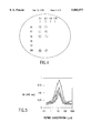
- FIG. 1 shows in schematic diagram a general method for identifying an abtide by a two-step screening process. See Section 5.3 for a discussion of this method.
- FIG. 2 shows the binding of biotinylated monoclonal antibody 7E11-C5 to immobilized mimetope peptide 7E11-9.5. See Section 6.1.2.2 for details.
- FIG. 3 shows similarities in the amino acid sequences of the CDR2L and CDR3L regions of monoclonal antibody 7E11-C5 and the 7E11-C5 abtides of Table 2.
- the number of amino acids in the abtides that are similar to the CDRs is indicated in parentheses, along with the percent homology. Dashes indicate gaps which have been added to improve the homology. In the case of clones 13 and 16, the homology with CDR2L was greatest if the sequence of CDR2L was reversed.
- the sequence shown for clone 14 is SEQ ID NO: 1; the sequence shown for clone 17 is SEQ ID NO: 2; The sequence shown for clone 15 is SEQ ID NO: 3; the sequence shown for clone 13 is SEQ ID NO: 4; the sequence shown for clone 16 is SEQ ID NO: 5; the sequence shown for CDR3L is SEQ ID NO: 6; the sequence shown for CDR2L is SEQ ID NO: 7;the sequence shown for CDR2L(rev) is SEQ ID NO: 8.
- FIG. 4 shows binding of abtides to the 7E11-9.5 mimetope peptide in a dot blot assay as described in Section 6.1.2.1.
- Numbers along the left side of the figure refer to the 7E11-C5 abtide that was spotted in the indicated position.
- the number 351 refers to the monoclonal antibody 7E11-C5, used as a positive control.
- the numbers along the top of the figure refer to the various dilutions of the abtide or the monoclonal antibody that were used.
- FIG. 5 shows the binding of biotinylated mimetopes to immobilized abtides.
- ⁇ represents binding of mimetope peptide Biotin-LYANPGMYSRLHSPA-NH 2 (SEQ ID NO: 20) to 7E11-C5 abtide clone 14;
- ⁇ represents binding of mimetope peptide Biotin-LYANPGMYSRLHSPA-NH 2 (SEQ ID NO: 20) to 7E11-C5 abtide clone 17;
- ⁇ represents binding of mimetope peptide Biotin-GMYSRLH-NH 2 (a portional SEQ ID NO: 20) to 7E11-C5 abtide clone 14;
- ⁇ represents binding of mimetope peptide Biotin-GMYSRLH-NH 2 (a portional SEQ ID NO: 20) to 7E11-C5 abtide clone 17. See Section 6.1.2.2 for details.
- FIG. 6 shows the capture of an antigen from a lysate of LNCaP tumor cells by the monoclonal antibody 7E11-C5 and the 7E11-C5 abtide clone 14. See Section 6.1.3 for details.
- FIG. 7 shows the biodistribution of abtide clone 14-DPTA- 111 In in SCID mice bearing human prostate carcinoma LNCaP xenograft tumors 2 hours , bar on the left for each pair of bars) or 4 hours , bar on the right for each pair of bars) post-injection of 2 ⁇ g of peptide, specific activity 32 ⁇ Ci/ ⁇ g. See Section 6.1.4 for details.
- FIG. 8 shows the biodistribution of abtide clone 17-DPTA- 111 -In in four SCID mice bearing human prostate LNCaP carcinoma xenograft tumors 2 hours , leftmost bar for each group of four bars, mouse 1; , second bar from left for each group of four bars, mouse 6) or five hours , third bar from left for each group of four bars, mouse 2; , rightmost bar for each group of four bars, mouse 4) post-injection of 0.02 ⁇ g of peptide, specific activity 2.4 ⁇ Ci/ng. See Section 6.1.4 for details.
- FIG. 9 shows the biodistribution of 111 -In labeled control irrelevant peptide in SCID mice bearing human prostate carcinoma LNCaP xenograft tumors 2 hours , leftmost bar for each group of five bars; , second bar from left for each group of five bars) or 5 hours , third bar from left for each group of five bars; , fourth bar from left for each group of five bars; , rightmost bar for each group of five bars) post-injection of 1.5 ⁇ g of peptide, specific activity 30 ⁇ Ci/ ⁇ g. See Section 6.1.4 for details.
- FIG. 10 schematically illustrates the construction of the R26 TSAR library.
- the R26 expression library was constructed essentially as described for the TSAR-9 library that is described in PCT publication WO 94/18318, dated Aug. 18, 1994, except for the modifications depicted in FIG. 10.
- the oligonucleotide assembly process depicted in FIG. 10 results in expression of peptides with the following amino acid sequence:
- FIG. 11 schematically illustrates the construction of the D38 TSAR library.
- the D38 expression library was constructed essentially as described for the TSAR-9 library that is described in PCT publication WO 94/18318, dated Aug. 18, 1994, except for the modifications depicted in FIG. 11.
- GTGTGTCTCGAGN(NNB) 20 NACGCCAN is SEQ ID NO: 92; GTTGTGTCTAGA(VNN) 15 VNTGGCGTN is SEQ ID NO: 93; TCGAGN(NNB) 20 NACGCCAN is SEQ ID NO: 94; CTAGA(VNN) 15 VNTGGCGTN is SEQ ID NO: 95; HSS(S/R)X 20 (Y/H/N/D)A(I/M/T/N/K/S/R)X 15 SR is SEQ ID NO: 96.
- FIG. 12 schematically illustrates the construction of the DC43 TSAR library.
- the DC43 expression library was constructed essentially as described for the TSAR-9 library that is described in PCT publication WO 94/18318, dated Aug. 18, 1994, except for the modifications depicted in FIG. 12.
- GTGTGTCTCGAGN(NNB) 20 GGTTGTGGT is SEQ ID NO: 97; GTTGTGTCTAGA(VNN) 20 ACCACAACC is SEQ ID NO: 98; TCGAGN(NNB) 20 GGTTGTGGT is SEQ ID NO: 99; CTAGA(VNN) 20 ACCACAACC is SEQ ID NO: 100; HSS(S/R)X 20 GCGX 20 SR is SEQ ID NO: 101.
- FIG. 13 schematically illustrates the oligonucleotides used to construct the polymorphic epithelial mucin (PEM) abtide saturation mutagenesis TSAR library (See Section 6.2.2).
- GAPVWRGNPRWRGPGGFKWPGCGNGPMCNTFTPARGGSRNNGP is SEQ ID NO: 51;
- ggsgcsccsgtstgsagsggsaasccscgstgsagsggsccsggsggsttsaastgsccsGGCTGCGGG is SEQ ID NO: 102;
- sggsccsttsttscgsgasccccscgsgcsggsgtsaasgtsttscasatsggsccsttCCCGCAGCC is SEQ ID NO: 103.
- the present invention relates generally to abtides.
- abtides refers to peptides that mimic the binding specificity of a larger molecule such as an antibody or receptor. Abtides specifically bind to a ligand of interest, in which the ligand is a specific binding partner of the mimicked larger molecule (e.g. antibody or receptor).
- peptide libraries are typically screened in a two-step process (see FIG. 1). The first screening step uses an antibody (or antigen-binding derivative thereof) or receptor (or ligand-binding derivative thereof) as a first target ligand.
- This step identifies peptide sequences termed "epitopes" or “mimetopes” which specifically bind the first target ligand. If the first screening step uses an antibody and the peptide identified contains the amino acid sequence of the natural antigen that is responsible for the specific binding of the antigen to the antibody, then the identified peptide is said to be an epitope; if the identified peptide does not contain the sequence of the natural antigen, then the identified peptide is said to be a mimetope.
- a mimetope will often resemble, either functionally in terms of its binding capability and/or structurally in terms of its amino acid sequence, the epitope recognized by the antibody used as the first ligand.
- a mimetope is then used as a second target ligand in a second screening step to identify a peptide sequence that specifically binds the epitope or mimetope.
- Such peptides are known as "abtides.”
- Abtides possess binding specificities similar to those possessed by the first target ligands (usually antibodies or receptors) described above.
- the antibody or derivative thereof used in the first screening step recognizes a tumor antigen, preferably a human tumor antigen, most preferably of a malignant tumor.
- the present invention provides a method to successfully screen against very small peptide or protein targets, e.g. 5 to 40 amino acids, preferably 10 to 20 amino acids. To date, screening against such targets has not been successful.
- the methods of the present invention increase the likelihood that the abtide obtained will bind its target in a complex or structurally dependent fashion.
- Abtides are useful since they mimic the binding specificities of antibodies or receptors. Thus, they may be used in many instances where antibodies or receptors may be used.
- the present invention further relates to the use of abtides in the place of antibodies in assays such as the many types of immunoassays known in the art. Abtides may take the place of antibodies in such assays as, for example, enzyme-linked immunosorbent assays (ELISAs) or sandwich immunoassays.
- ELISAs enzyme-linked immunosorbent assays
- the invention also provides abtide compositions for use in therapy and diagnosis of disease. In a specific example, abtides have been produced and demonstrated to be useful in place of antibodies in enzyme-linked immunosorbent assays and in in vivo localization to prostate carcinoma in xenograft models.
- abtides has many potential advantages over the use of antibodies or receptors: the smaller size of abtides allows their easier production at lower cost, reduced immunogenicity, and facilitates their in vivo delivery if such is desired; biological reactions and functions mediated by constant domains of antibodies, and cross-linking of antibodies/receptors and resulting biological effects can be avoided if desired.
- the abtides of the present invention can be identified from a chemically synthesized peptide library or a biologically expressed peptide library. If a biological peptide expression library is used, the nucleic acid which encodes the peptide which binds to the ligand of choice can be recovered, and then sequenced to determine its nucleotide sequence and hence deduce the amino acid sequence that mediates binding. Alternatively, the amino acid sequence of an appropriate binding domain can be determined by direct determination of the amino acid sequence of a peptide selected from a peptide library containing chemically synthesized peptides. In a less preferred aspect, direct amino acid sequencing of a binding peptide selected from a biological peptide expression library can also be performed.
- the abtides are advantageously identified from random peptide libraries.
- random peptide libraries will be encoded by synthetic oligonucleotides with a plurality of variant nucleotide positions having the potential to encode all 20 naturally occurring amino acids.
- the sequence of amino acids encoded by the variant nucleotides is unpredictable and substantially random.
- the terms "unpredicted”, “unpredictable” and “substantially random” are used interchangeably with respect to the amino acids encoded and are intended to mean that the variant nucleotides at any given position are such that it cannot be predicted which of the 20 naturally occurring amino acids will appear at that position.
- These variant nucleotides are the product of random chemical synthesis.
- the biological random peptide libraries envisioned for use include those in which a bias has been introduced into the random sequence, e.g., to disfavor stop codon usage.
- the peptide libraries used in the present invention may be libraries that are chemically synthesized in vitro. Examples of such libraries are given in Fodor et al., 1991, Science 251:767-773, which describes the synthesis of a known array of short peptides on an individual microscopic slide; Houghten et al., 1991, Nature 354:84-86, which describes mixtures of free hexapeptides in which the first and second residues in each peptide were individually and specifically defined. Lam et al., 1991, Nature 354:82-84, which describes a split synthesis scheme; Medynski, 1994, Bio/Technology 12:709-710, describes split synthesis and T-bag synthesis methods as well. See also Gallop et al., 1994, J. Medicinal Chemistry 37:1233-1251.
- PCT publication WO 91/05058, dated Apr. 18, 1991 is directed to random libraries containing semi-random nucleotide sequences.
- the semi-random nucleotide sequences are transcribed in vitro under conditions such that polysomes are produced.
- the polysomes are screened for binding to a substance of interest. Those polysomes that bind to the substance of interest are recovered.
- the RNA from those polysomes is used to construct cDNA, which is expressed to produce polypeptides.
- the total number of unpredictable amino acids in the peptides of the chemical library used for screening is greater than or equal to 5 and less than or equal to 25; in other embodiments the total is in the range of 5-15 or 5-10 amino acids, preferably contiguous amino acids.
- binding domain can be identified from chemically synthesized peptide libraries, such a domain would be small (i.e. less than 10 amino acids, and most probably 5-6 amino acids, in length). Therefore, use of a chemically synthesized peptide library is less preferred for the second screening step involved in isolating abtides than is use in the second screening step of biological peptide libraries containing unpredictable sequences of greater length, described below.
- biological peptide libraries are used to identify abtides.
- Many suitable biological peptide libraries are known in the art and can be used.
- peptides have been expressed in biological systems as either soluble fusion proteins or viral capsid fusion proteins.
- peptide libraries according to this approach have used the M13 phage. Although the N-terminus of the viral capsid protein, protein III (pIII), has been shown to be necessary for viral infection, the extreme N-terminus of the mature protein does tolerate alterations such as insertions. Accordingly, various peptide libraries, in which the diverse peptides are expressed as pIII fusion proteins, are known in the art; these libraries can be used to identify abtides. Examples of such libraries are described below.
- the starting DNA was generated by means of an oligonucleotide comprising the degenerate codons NN(G/T)! 10 with a self-complementary 3' terminus.
- This sequence in forming a hairpin, creates a self-priming replication site which could be used by T4 DNA polymerase to generate the complementary strand.
- the double-stranded DNA was cleaved at the SfiI sites at the 5° terminus and hairpin for cloning into the fUSES vector described by Scott and Smith, supra.
- the protein pVIII is a major M13 viral capsid protein, which can also serve as a site for expressing peptides on the surface of M13 viral particles, in the construction of random peptide libraries.
- the average functional domain within a natural protein is considered to be about 40 amino acids.
- the random peptide libraries from which the abtides of the present invention are preferably identified encode peptides having in the range of 5 to 200 total variant amino acids.
- biologically expressed random peptide libraries displaying short random inserts i.e. less than 20 amino acids in length
- the most preferred biologically expressed random peptide libraries for use in the invention are those in which the displayed peptide has 20 or greater unpredictable amino acids i.e.
- the invention preferably uses libraries of greater complexity than are commonly employed in the art.
- the conventional teaching in the random peptide library art is that the length of inserted oligonucleotides should be kept short, encoding preferably fewer than 15 and most preferably about 6-8 amino acids.
- libraries encoding more than about 20 amino acids can be constructed, but such libraries can be advantageously screened to identify peptides having binding specificity for a variety of ligands.
- Such libraries with longer length inserts are exemplified by the TSAR libraries, described in PCT publication WO 91/12328, dated Aug. 22, 1991, and PCT publication WO 94/18318, dated Aug. 18, 1994.
- Libraries composed of longer length oligonucleotides afford the ability to identify peptides in which a short sequence of amino acids is common to or shared by a number of peptides binding a given ligand, i.e., library members having shared binding motifs.
- the use of longer length libraries also affords the ability to identify peptides which do not have any shared sequences with other peptides but which nevertheless have binding specificity for the same ligand.
- libraries having large inserted oligonucleotide sequences provide the opportunity to identify or map binding sites which encompass not only a few contiguous amino acid residues, i.e., simple binding sites, but also those which encompass discontinuous amino acids, i.e., complex binding sites, and may afford the complex binding characteristic of antibodies and receptor-like molecules.
- the large size of the inserted synthesized oligonucleotides of certain libraries provides the opportunity for the development of secondary and/or tertiary structure in the potential binding peptides and in sequences flanking the actual binding site in the binding domain.
- Secondary and tertiary structure often significantly affect the ability of a sequence to mediate binding, as well as the strength and specificity of any binding which occurs. Such complex structural effects are not possible when only small length oligonucleotides are used in libraries. It may be that secondary and tertiary structures are especially important in the identification of abtides since abtides mimic the binding of large molecules such as antibodies. It is well known that the antigen binding properties of antibodies depend in most instances upon several different regions of the heavy chain (complementarity determining regions) and upon regions contributed by the light chain as well.
- the most preferred binding domains for identifying the abtides of the present invention will be those from biologically expressed random peptide libraries in which the displayed peptide is 20 or greater amino acids in length.
- random peptide libraries are the TSAR libraries, described in in PCT publication WO 91/12328, dated Aug. 22, 1991, and PCT publication WO 94/18318, dated Aug. 18, 1994.
- the library utilized in the present invention is a linear, non-constrained library.
- “constrained”, “structured” or “semi-rigid” random peptide libraries could also be used in the present methods to identify abtides.
- these libraries express peptides that are substantially random but contain a small percentage of fixed residues within or flanking the random sequences that have the result of conferring structure or some degree of conformational rigidity to the peptide.
- the plurality of synthetic oligonucleotides express peptides that are each able to adopt only one or a small number of different conformations that are constrained by the positioning of codons encoding certain structure conferring amino acids in or flanking the synthesized variant or unpredicted oligonucleotides.
- the plurality of proteins expressed can adopt only a single or a small number of conformations.
- Such libraries are exemplified by the TSAR-13 and TSAR-14 libraries described in PCT publication WO 94/18318, dated Aug.
- a biological peptide library that is a random peptide "TSAR" library is screened to identify an abtide.
- TSARs is an acronym for "Totally Synthetic Affinity Reagents" as described in PCT publication WO 91/12328, dated Aug. 22, 1991, and PCT publication WO 94/18318, dated Aug. 18, 1994.
- TSAR libraries, their construction and use, and specific examples of TSAR libraries, are described in detail in those PCT publications. Nucleic acids encoding TSARs or a TSAR portion which mediates binding to the ligand used for screening can be used to identify the abtides of the present invention.
- a TSAR may be a heterofunctional fusion protein, said fusion protein comprising (a) a binding domain encoded by an oligonucleotide comprising unpredictable nucleotides in which the unpredictable nucleotides are arranged in one or more contiguous sequences, wherein the total number of unpredictable nucleotides is greater than or equal to about 60 and less than or equal to about 600, and optionally, (b) an effector domain encoded by an oligonucleotide sequence which is a protein or peptide that enhances expression or detection of the binding domain.
- a TSAR may be a heterofunctional fusion protein as described above but in which the contiguous sequences are flanked by invariant residues designed to encode amino acids that confer a desired structure to the binding domain of the expressed heterofunctional fusion protein.
- libraries for use in the present invention may be those wherein the library is a library of recombinant vectors that express a plurality of heterofunctional fusion proteins, said fusion proteins comprising a binding domain encoded by an oligonucleotide comprising unpredictable nucleotides in which the unpredictable nucleotides are arranged in one or more contiguous sequences, wherein the total number of unpredictable nucleotides is greater than or equal to about 15 and less than or equal to about 600.
- An abtide is typically a peptide that mimics, with respect to binding specificity, and possibly other characteristics (e.g., binding affinity, sequence, etc.) a large molecule such as an antibody or receptor. However, an abtide is generally much smaller than an antibody or receptor.
- An abtide is generally a peptide of about 5 to 200 amino acids. Preferably, an abtide is a peptide of about 10 to 100 amino acids. Most preferably, an abtide is a peptide of about 20 to 50 amino acids.
- an abtide may be linked to additional amino acid sequences or additional non-amino acid sequences. Such additional sequences may aid in the identification or isolation of the abtide. Or, they may aid in the biodistribution, stability, or diagnostic or therapeutic effectiveness of the abtide when the abtide is used diagnostically or therapeutically.
- the abtides may be linked to a variety of non-peptide moieties.
- moieties might include toxins; drugs; polysaccharides; nucleotides; oligonucleotides; labels such as radioactive substances (e.g. 111 In, 125 I, 131 I, 99m Tc, 212 B, 90 Y, 186 Rh); biotin; fluorescent tags; imaging reagents (e.g. those described in U.S. Pat. No. 4,741,900 and U.S. Pat. No.
- hydrocarbon linkers e.g., an alkyl group or derivative thereof conjugated to a moiety providing for attachment to a solid substratum, or to a moiety providing for easy separation (e.g., a hapten recognized by an antibody bound to a magnetic bead), etc.
- Linkage of the peptide to the non-peptide moiety may be by any of several well-known methods in the art.
- an abtide has a free amino- or carboxy- terminus
- such termini can be modified by known methods, e.g., to provide greater resistance to degradation, greater cell permeability, greater solubility, etc., e.g., by acetylation, biotinylation, fatty acylation, etc. at the amino-terminus; amidation at the carboxy-terminus; or the abtide can be stabilized by inclusion of D amino acids, nonnatural amino acids or glycosyl amino acids, etc.
- the abtides of the invention are preferably made by commonly known methods of chemical synthesis, e.g., as described by way of example in Section 5.6 and its subsections.
- abtides (or portions thereof) can be obtained by recombinant expression of a nucleic acid encoding the desired abtide in an appropriate host, by well-known methods.
- the process of identifying abtides from a peptide library comprises two distinct screening steps.
- the first step is designed to identify epitopes or ligands with binding specificity for the larger molecule of interest, e.g. antigen mimics, receptor-ligand mimics, and the like.
- a peptide library is screened with a ligand that possesses a specific, often complex, binding site of interest.
- Those peptides in the library that are specific binding partners of the ligand bind to the ligand and are readily recoverable because of this specific binding.
- An example of a ligand suitable for use in this first screening step would be an antibody, inherently possessing an antigen binding site, or an antigen-binding derivative thereof.
- Another example would be a receptor e.g. the epidermal growth factor receptor or the platelet derived growth factor receptor. These particular receptors possess specific binding sites for epidermal growth factor and platelet derived growth factor, respectively.
- Other receptors are known in the art and are also potential ligands, for example, the estrogen receptor, the various acetylcholine receptors, the human growth hormone receptor, etc.
- Molecules comprising those peptide sequences in the library that are identified by the first screening step will be referred to herein as epitopes or mimetopes.
- epitopes or mimetopes the peptides in the library that are identified as specific binding partners of the antibody would be known as antigen epitopes or mimetopes.
- the peptides that are isolated in the first screening step are then preferably analyzed, as, for example, by DNA sequencing of the binding domain of the phage that encode the peptides if the library used was a phage library.
- the DNA sequence of the binding domain encodes the amino acid sequence of the epitope or mimetope peptide. Due to the known relationship between DNA sequences and their encoded amino acid sequences, obtaining the DNA sequence of the epitope or mimetope allows the determination of the amino acid sequence of the epitope or mimetope.
- the library used was a chemical library
- direct amino acid sequencing of the peptide epitope or mimetope can be carried out by well known methods in the art.
- sequences of different peptide mimetopes identified in the first screening step can be compared to determine a consensus mimetope sequence.
- a molecule preferably a peptide, is produced comprising that amino acid sequence which mediates binding.
- This peptide may be synthesized chemically, or, alternatively, may be produced by methods involving recombinant DNA.
- This peptide may contain only the amino acids of the epitope or mimetope or, preferably, it may contain additional amino acids or non-amino acid moieties to aid in identifying or recovering the epitope or mimetope peptide and any new peptide binders found in the subsequent screening step discussed below.
- the epitope or mimetope that was identified in the first screening step is used as a ligand for the second screening step.
- the second screening step identifies peptides with binding specificity for the epitope or mimetope and that surprisingly mimic the binding specificity of the antibody or receptor that was used as ligand in the first screening step.
- the second screening step yields peptides with antibody or receptor-like binding activity for antigens or receptor ligands that are known as abtides.
- FIG. 1 is a schematic representation of an exemplary two-step screening process used to identify abtides.
- the epitope to which an antibody specifically binds is known.
- the monoclonal antibody SM-3 that specifically binds the polymorphic epithelial mucin (PEM) found on human breast carcinoma cells has been shown to be specific for the epitope defined by the amino acid sequence VTSAPDTRPAPGSTAPPAHGVTSAPDTR (SEQ ID NO: 9) (Burchell et al., 1989, Int. J. Cancer 44:691-696).
- the first screening step described above may be dispensed with.
- a peptide comprising the sequence of the epitope for which the antibody is specific can be synthesized and used in the second screening step described above in order to identify abtides of the antibody.
- a receptor-ligand i.e., a ligand which specifically binds to a receptor
- a receptor specifically binds
- GM-CSF granulocyte/macrophage colony stimulating factor
- a molecule comprises a peptide or a binding portion thereof which binds to a ligand of interest, which peptide is identified by a method comprising: screening a random peptide library with a ligand of interest, said ligand of interest being a peptide having a length of between 5 and 40 amino acids, to identify a peptide that specifically binds to the ligand of interest, in which the ligand of interest is also specifically bound by an antibody or a receptor.
- a molecule comprises a peptide which binds to a substance of interest, which peptide is identified by a method comprising: screening a random peptide library with a ligand, said ligand being a peptide of 36 amino acids or fewer, in which the ligand is an epitope of an antigen that is specifically bound by an antibody or in which the ligand represents the portion of a receptor-ligand that is responsible for the specific binding of the receptor to the receptor-ligand.
- a ligand is a substance for which it is desired to isolate a specific binding partner from a peptide library.
- a ligand can function as a lock, i.e., a large polypeptide or protein analogous to a lock into which a smaller specific binding partner fits as a key; or a ligand can function as a key which fits into and specifically binds a larger binding partner or lock.
- an epitope or mimetope is typically a peptide that acts as a key; it is identified by screening a peptide library for peptides that fit into and bind the specific binding site of a larger molecule which acts as a lock, e.g. antibody or receptor.
- the larger molecule is an antibody and the peptide identified contains the portion of the amino acid sequence of the natural antigen that is responsible for the specific binding of the antigen to the antibody, then the identified peptide is said to be an epitope; if the identified peptide does not contain the sequence of the natural antigen, then the identified peptide is said to be a mimetope.
- a mimetope is identified by screening a peptide library with an antibody or antibody fragment. Mimetopes thus identified functionally mimic the antigen to which the antibody binds in that the mimetopes also specifically bind with the antibody. In some cases, if the antigen is a protein or polypeptide, the mimetopes may share amino acid sequence motifs with the antigen. In another embodiment, a mimetope is identified by screening a peptide library with a receptor or receptor fragment. Mimetopes thus identified functionally mimic the natural ligand of the receptor.
- the peptide libraries that are used in the first and second screening steps may be the same or different.
- a peptide library containing small random inserts (from about 6 to about 15 amino acids) is used in the first screening step.
- Such larger libraries preferably express peptides of about 20 to 200 random amino acids. Examples of such larger libraries are the TSAR libraries described in PCT publication WO 91/12328, dated Aug. 22, 1991, and in PCT Publication WO 94/18318, dated Aug. 18, 1994.
- Biological or chemically synthesized peptide libraries can be used in either the first or second screenings.
- the peptide libraries used in the present invention may have a plurality of residues that are random, i.e. residues for which the amino acid occupying that residue cannot be predicted in advance. Such libraries are said to be random peptide libraries.
- a preferred method for identifying abtides comprises screening a library of recombinant vectors that express a plurality of heterofunctional fusion proteins, said fusion proteins comprising (a) a binding domain encoded by an oligonucleotide comprising unpredictable nucleotides in which the unpredictable nucleotides are arranged in one or more contiguous sequences, wherein the total number of unpredictable nucleotides is greater than or equal to about 15 and less than or equal to about 600, and optionally, (b) an effector domain encoded by an oligonucleotide sequence which is a protein or peptide that enhances expression or detection of the binding domain.
- Screening is done by contacting the plurality of heterofunctional fusion proteins with a ligand under conditions conducive to ligand binding and then isolating the fusion proteins which bind to the ligand.
- the methods of the invention further preferably comprise determining the nucleotide sequence encoding the binding domain of the heterofunctional fusion protein identified to determine the DNA sequence that encodes the binding domain and simultaneously to deduce the amino acid sequence of the mimetope used in the second screen. Nucleotide sequence analysis can be carried out by any method known in the art, including but not limited to the method of Maxam and Gilbert (1980, Meth. Enzymol. 65:499-560), the Sanger dideoxy method (Sanger et al., 1977, Proc. Natl, Acad.
- the libraries used to screen for mimetopes and abtides of the invention have unpredictable nucleotides arranged in one or more contiguous sequences that are flanked by invariant residues designed to encode amino acids that confer a desired structure to the binding domain of the expressed heterofunctional fusion protein.
- a suitable peptide library Once a suitable peptide library has been constructed (or otherwise obtained), the library is screened to identify peptides having binding affinity for a ligand of choice. Screening the libraries can be accomplished by any of a variety of methods known to those of skill in the art. See, e.g., the following references, which disclose screening of peptide libraries: Parmley and Smith, 1989, Adv. Exp. Med. Biol. 251:215-218; Scott and Smith, 1990, Science 249:386-390; Fowlkes et al., 1992; BioTechniques 13:422-427; Oldenburg et al., 1992, Proc. Natl. Acad. Sci.
- the libraries are expressed as fusion proteins with a cell surface molecule, then screening is advantageously achieved by contacting the vectors with an immobilized target ligand and harvesting those vectors that bind to said ligand.
- Such useful screening methods designated “panning” techniques are described in Fowlkes et al., 1992, BioTechniques 13:422-427.
- the target ligand can be immobilized on plates, beads, such as magnetic beads, sepharose, etc., or on beads used in columns.
- the immobilized target ligand can be "tagged", e.g., using such as biotin, 2-fluorochrome, e.g. for FACS sorting.
- screening a library of phage expressing random peptides on phage and phagemid vectors can be achieved by using magnetic beads as described in PCT publication WO 94/18318, dated Aug. 18, 1994.
- screening a library of phage expressing random peptides can be achieved by panning using microtiter plates.
- a pH change is used.
- the libraries expressing random peptides as a surface protein of either a vector or a host cell can be screened by passing a solution of the library over a column of a ligand immobilized to a solid matrix, such as sepharose, silica, etc., and recovering those phage that bind to the column after extensive washing and elution.
- a host cell e.g., phage or bacterial cell
- weak binding library members can be isolated based on retarded chromatographic properties.
- fractions are collected as they come off the column, saving the trailing fractions (i.e., those members that are retarded in mobility relative to the peak fraction are saved). These members are then concentrated and passed over the column a second time, again saving the retarded fractions.
- the trailing fractions i.e., those members that are retarded in mobility relative to the peak fraction are saved.
- These members are then concentrated and passed over the column a second time, again saving the retarded fractions.
- Through successive rounds of chromatography it is possible to isolate those that have some affinity, albeit weak, to the immobilized ligand.
- These library members are retarded in their mobility because of the millions of possible ligand interactions as the member passes down the column.
- this methodology selects those members that have modest affinity to the target, and which also have a rapid dissociation time.
- the oligonucleotides encoding the binding domain selected in this manner can be mutagenized, expressed and rechromatographed (or screened by another method) to discover improved binding activity.
- saturation mutagenesis can be carried out using synthetic oligonucleotides synthesized from "doped" nucleotide reservoirs. The doping is carried out such that the original peptide sequence is represented only once in 10 6 unique clones of the mutagenized oligonucleotide.
- the assembled oligonucleotides are cloned into a parental TSAR vector.
- the vector is m663 (Fowlkes et al., 1992, BioTechniques 13:422-427).
- m663 is able to make blue plaques when grown in E. coli stain JM101 or DH5 ⁇ F'.
- a library of greater than 10 6 is preferred; however a library of 10 5 is sufficient to isolate TSAR phage displaying peptide domains with increased selectivity for binding to the target ligand.
- screening a library of can be achieved using a method comprising an "enrichment” step and a filter lift step as follows.
- Random peptides from an expressed library capable of binding to a given ligand are initially enriched by one or two cycles of panning or affinity chromatography, as described above. The goal is to enrich the positives to a frequency of about >1/10 5 .
- a filter lift assay is conducted. For example, approximately 1-2 ⁇ 10 5 phage, enriched for binders, are added to 500 ⁇ l of log phase E. coli and plated on a large LB-agarose plate with 0.7% agarose in broth. The agarose is allowed to solidify, and a nitrocellulose filter (e.g., 0.45 ⁇ ) is placed on the agarose surface.
- a series of registration marks is made with a sterile needle to allow re-alignment of the filter and plate following development as described below. Phage plaques are allowed to develop by overnight incubation at 37° C. (the presence of the filter does not inhibit this process). The filter is then removed from the plate with phage from each individual plaque adhered in situ. The filter is then exposed to a solution of BSA or other blocking agent for 1-2 hours to prevent non-specific binding of the ligand (or "probe").
- the probe itself is labeled, for example, either by biotinylation (using commercial NHS-biotin) or direct enzyme labeling, e.g., with horse radish peroxidase (HRP) or alkaline phosphatase. Probes labeled in this manner are indefinitely stable and can be re-used several times.
- the blocked filter is exposed to a solution of probe for several hours to allow the probe to bind in situ to any phage on the filter displaying a peptide with significant affinity to the probe.
- the filter is then washed to remove unbound probe, and then developed by exposure to enzyme substrate solution (in the case of directly labeled probe) or further exposed to a solution of enzyme-labeled avidin (in the case of biotinylated probe).
- an HRP-labeled probe is detected by ECL western blotting methods (Amersham, Arlington Heights, Ill.), which involves using luminol in the presence of phenol to yield enhanced chemiluminescence detectable by brief exposure of film by autoradiography, in which the exposed areas of film correspond to positive plaques on the original plate.
- ECL western blotting methods Amersham, Arlington Heights, Ill.
- positive phage plaques are identified by localized deposition of colored enzymatic cleavage product on the filter which corresponds to plaques on the original plate.
- the developed filter or film is simply realigned with the plate using the registration marks, and the "positive" plaques are cored from the agarose to recover the phage.
- phage recovered from the initial core are re-plated at low density and the process is repeated to allow isolation of individual plaques and hence single clones of phage.
- Successful screening experiments are optimally conducted using 3 rounds of serial screening.
- the recovered cells are then plated at a low density to yield isolated colonies for individual analysis.
- the individual colonies are selected and used to inoculate LB culture medium containing ampicillin. After overnight culture at 37° C., the cultures are then spun down by centrifugation. Individual cell aliquots are then retested for binding to the target ligand attached to the beads. Binding to other beads, having attached thereto a non-relevant ligand, can be used as a negative control.
- elution conditions including but not limited to pH 2-3, pH 12-13, excess target in competition, detergents, mild protein denaturants, urea, varying temperature, light, presence or absence of metal ions, chelators, etc.
- Some of these elution conditions may be incompatible with phage infection because they are bactericidal and will need to be removed by dialysis (i.e., dialysis bag, Centricon/Amicon microconcentrators).
- flanking sequences may also be denatured in combination with the actual binding sequence; these flanking regions may also change their secondary or tertiary structure in response to exposure to the elution conditions (i.e., pH 2-3, pH 12-13, excess target in competition, detergents, mild protein denaturants, urea, heat, cold, light, metal ions, chelators, etc.) which in turn leads to the conformational deformation of the peptide responsible for binding to the target.
- TSAR libraries can be prepared and screened by: (1) engineering a vector, preferably a phage vector, so that a DNA sequence encodes a segment of Factor Xa (or Factor Xa protease cleavable peptide) and is present adjacent to the gene encoding the effector domain, e.g., the pIII coat protein gene; (2) construct and assemble the double stranded synthetic oligonucleotides as described above and insert into the engineered vector; (3) express the plurality of vectors in a suitable host to form a library of vectors; (4) screen for binding to an immobilized ligand; (5) wash away excess phage; and (6) treat the immobilized phage with Factor Xa protease.
- a vector preferably a phage vector
- the particle will be uncoupled from the peptide-ligand complex and can then be used to infect bacteria to regenerate the particle with its full-length pIII molecule for additional rounds of screening.
- This alternative embodiment advantageously allows the use of universally effective elution conditions and thus allows identification of phage expressing TSARs that otherwise might not be recovered using other known methods for elution. To illustrate, using this embodiment, exceptionally tight binding TSARs could be recovered.
- the oligonucleotides encoding the binding domain selected in this manner can be mutagenized, expressed and rechromatographed (or screened by another method) to discover improved binding activity.
- saturation mutagenesis can be carried out using synthetic oligonucleotides synthesized from "doped" nucleotide reservoirs. The doping is carried out such that the original peptide sequence is represented only once in 10 6 unique clones of the mutagenized oligonucleotide.
- the assembled oligonucleotides are cloned into a parental TSAR vector.
- the vector is m663 (Fowlkes et al., 1992, BioTechniques 13:422-427). m663 is able to make blue plaques when grown in E. coli stain JM101 or DH5 ⁇ F'.
- a library of greater than 10 6 is preferred; however a library of 10 5 is sufficient to isolate TSAR phage displaying peptide domains with increased selectivity for binding to the target ligand.
- a TSAR library is utilized; however, to those skilled in the art, it will be apparent that other peptide libraries may be used.
- An example of a TSAR library is the TSAR-9 library disclosed in Kay et al., 1993, Gene 128:59-65.
- TSAR-9 constructs display a peptide of about 38 amino acids in length having 36 totally random positions.
- Antibodies can be produced which recognize an antigen of interest. Such antibodies can be polyclonal or monoclonal. Such antibodies may be used as ligands in the first screening step of the present invention.
- adjuvants may be used to increase the immunological response, depending on the host species, and including but not limited to Freund's (complete and incomplete), mineral gels such as aluminum hydroxide, surface active substances such as lysolecithin, pluronic polyols, polyanions, peptides, oil emulsions, keyhole limpet hemocyanins, dinitrophenol, and potentially useful human adjuvants such as BCG (bacille Calmette-Guerin) and corynebacterium parvum.
- BCG Bacille Calmette-Guerin
- a monoclonal antibody to an antigen of interest can be prepared by using any technique which provides for the production of antibody molecules by continuous cell lines in culture. These include but are not limited to the hybridoma technique originally described by Kohler and Milstein (1975, Nature 256:495-497), and the more recent human B cell hybridoma technique (Kozbor et al., 1983, Immunology Today 4:72) and EBV-hybridoma technique (Cole et al., 1985, Monoclonal Antibodies and Cancer Therapy, Alan R. Liss, Inc., pp. 77-96).
- the monoclonal antibodies may be human monoclonal antibodies or chimeric human-mouse (or other species) monoclonal antibodies.
- Human monoclonal antibodies may be made by any of numerous techniques known in the art (e.g., Teng et al., 1983, Proc. Natl. Acad. Sci. U.S.A. 80:7308-7312; Kozbor et al., 1983, Immunology Today 4:72-79; Olsson et al., 1982, Meth. Enzymol. 92:3-16).
- Chimeric antibody molecules may be prepared containing a mouse antigen-binding domain with human constant regions (Morrison et al., 1984, Proc. Natl. Acad. Sci. U.S.A. 81:6851, Takeda et al., 1985, Nature 314:452).
- a molecular clone of an antibody to an antigen of interest can be prepared by known techniques. Recombinant DNA methodology (see e.g., Maniatis et al., 1982, Molecular Cloning, A Laboratory Manual, Cold Spring Harbor Laboratory, Cold Spring Harbor, N.Y.) may be used to construct nucleic acid sequences which encode a monoclonal antibody molecule, or antigen binding region thereof.
- Antibody molecules may be purified by known techniques, e.g., immunoabsorption or immunoaffinity chromatography, chromatographic methods such as HPLC (high performance liquid chromatography), or a combination thereof, etc.
- Antibody fragments which contain the idiotype or antigen binding region of the molecule can be generated by known techniques.
- fragments include but are not limited to: the F(ab')2 fragment which can be produced by pepsin digestion of the antibody molecule; the Fab' fragments which can be generated by reducing the disulfide bridges of the F(ab')2 fragment, and the 2 Fab or Fab fragments which can be generated by treating the antibody molecule with papain and a reducing agent.
- a peptide from a peptide library has been identified as an abtide or mimetope for a particular target ligand of interest according to the method of the invention (in either the first or second screening step), it may be useful to determine what region(s) of the expressed peptide sequence is (are) responsible for binding to the target ligand.
- Such analysis can be conducted at two different levels, i.e., the nucleotide sequence and amino acid sequence levels.
- the inserted oligonucleotides can be cleaved using appropriate restriction enzymes and religated into the original expression vector and the expression product of such vector screened for ligand binding to identify the oligonucleotides that encode the binding region of the abtide or mimetope.
- the oligonucleotides can be transferred into another vector, e.g., from phage to phagemid. The newly expressed fusion proteins should acquire the same binding activity if the domain is necessary and sufficient for binding to the ligand.
- the oligonucleotides can be synthesized, based on the nucleotide sequence determined for the phage in the library that encodes the binding peptide, amplified by cloning or PCR amplification using internal and flanking primers, cleaved into two pieces and cloned as two half-binding domain fragments. In the foregoing manner, the inserted oligonucleotides are subdivided into two equal halves. If the peptide domain important for binding is small, then one recombinant clone would demonstrate binding and the other would not. If neither have binding, then either both are important or the essential portion of the domain spans the middle (which can be tested by expressing just the central region).
- the binding domains can be analyzed.
- the entire peptide should be synthesized and assessed for binding to the target ligand to verify that the peptide is necessary and sufficient for binding.
- short peptide fragments for example, overlapping 10-mers, can be synthesized, based on the amino acid sequence of the random peptide binding domain, and tested to identify those binding the ligand.
- linear motifs may become apparent after comparing the primary structures of different binding peptides from the library having binding affinity for a target ligand. The contribution of these motifs to binding can be verified with synthesized peptides in competition experiments (i.e., determine the concentration of peptide capable of inhibiting 50% of the binding of the phage to its target; IC 50 ). Conversely, the motif or any region suspected to be important for binding can be removed from or mutated within the DNA encoding the random peptide insert and the altered displayed peptide can be retested for binding.
- the binding characteristics of that peptide can be modified by varying the binding domain sequence to produce a related family of peptides with differing properties for a specific ligand.
- the identified peptides can be improved by additional rounds of random mutagenesis, selection, and amplification of the nucleotide sequences encoding the binding domains.
- Mutagenesis can be accomplished by creating and cloning a new set of oligonucleotides that differ slightly from the parent sequence, e.g., by 1-10%. Selection and amplification are achieved as described above.
- mutants and the parent phage, differing in their lacZ expression can be processed together during the screening experiments. Alteration of the original blue-white color ratios during the course of the screening experiment will serve as a visual means to assess the successful selection of enhanced binders. This process can go through numerous cycles.
- the abtides of the present invention possess binding specificities that are similar to those of the ligands (e.g. antibodies, receptors) that are used in the first screening step of the process by which the abtides are identified. Consequently, the abtides may be used in many of the same instances where the ligand of the first screening step might be used. For example, if the ligand used in the first screening step is an antibody, the abtide that is identified after the second screening step will bind specifically to the same antigen to which the antibody specifically binds. Therefore, the abtide may be used as a substitute for the antibody in many of the reactions or assays that the antibody could be used in.
- the ligands e.g. antibodies, receptors
- the abtide could be used in immunoassays known in the art, e.g., those designed to detect or measure the amount of the antigen.
- immunoassays may have to be suitably modified.
- many immunoassays make use of a step in which a second antibody, labeled with a radioactive moiety or an enzyme such as alkaline phosphatase, specifically binds to the first antibody.
- a second antibody would not be expected to specifically bind to the abtide.
- the immunoassays which can be used include but are not limited to competitive and non-competitive assay systems using techniques such as western blots, radioimmunoassays, ELISA (enzyme linked immunosorbent assays), "sandwich” immunoassays, dot immunoblot assays, immunoprecipitation assays, precipitin reactions, gel diffusion precipitin reactions, immunodiffusion assays, agglutination assays, complement-fixation assays, immunoradiometric assays, fluorescent immunoassays, protein A immunoassays, immunoaffinity chromatography, and flow dipstick assays to name but a few.
- the samples to be assayed in the immunoassays can be any sample that may contain the antigen or ligand desired to be assayed.
- these samples can be body fluids such as plasma, blood, serum, saliva, cerebrospinal fluid, synovial fluid, etc.
- the detectable label to be used in the immunoassays can be any detectable label known in the art.
- Such labels include radioisotopes, fluorescent dyes, enzymes (for example horseradish peroxidase or alkaline phosphatase), chemiluminescent molecules, metal atoms, or phosphorescent dyes, colored particles, metal and dye colloids.
- the abtides can be used in a sandwich enzyme immunoassay.
- a molecule comprising an abtide is affixed to a solid substratum.
- the molecule comprising the abtide may be linked to a substance that will provide for greater attachment of the molecule to the substratum.
- the sample to be assayed is contacted with the molecule comprising the abtide that is bound to the substratum.
- the substances in the sample that are specific binding partners of the abtide (analyte) will bind to the abtide, and non-binding sample components are removed by washing.
- An enzyme-conjugated monoclonal antibody directed against the analyte is added.
- This enzyme-conjugated monoclonal antibody binds to the part of the analyte it is specific for and completes the sandwich.
- a substrate solution is added to the wells.
- a colored product is formed in proportion to the amount of analyte present in the sample.
- the reaction may be terminated by addition of stop solution and absorbance is measured spectrophotometrically. The following illustrates these steps in more detail.
- ABTSTM stock solution is prepared at 15 mg/ml in dH 2 O.
- 200 ⁇ l of this ABTSTM stock is diluted into 10 ml of citrate phosphate buffer (17 mm citric acid, 65 mm dibasic sodium phosphate) and 10 ⁇ l 30% H 2 O 2 .
- the invention provides methods of treatment by administration to a subject of an effective amount of a pharmaceutical (therapeutic or diagnostic) composition comprising an abtide.
- a pharmaceutical therapeutic or diagnostic
- Such an abtide envisioned for therapeutic or diagnostic use is referred to hereinafter as a "Therapeutic” or “Therapeutic of the invention.”
- Such therapeutics are abtides that specifically bind to a molecule in vivo, to exert a therapeutic or diagnostic effect.
- the Therapeutic is substantially purified.
- the subject is preferably an animal, including but not limited to animals such as cows, pigs, horses, chickens, cats, dogs, etc., and is preferably a mammal, and most preferably human.
- Formulations and methods of administration that can be employed are known in the art and can be selected from among those described hereinbelow.
- a Therapeutic of the invention e.g., encapsulation in liposomes, microparticles, microcapsules, recombinant cells containing the Therapeutic, receptor-mediated endocytosis (see, e.g., Wu and Wu, 1987, J. Biol. Chem. 262:4429-4432), etc.
- Methods of introduction include but are not limited to intradermal, intramuscular, intraperitoneal, intravenous, subcutaneous, intranasal, epidural, and oral routes as well as transdermal and subcutaneous time-release implants.
- the Therapeutics may be administered by any convenient route, for example by infusion or bolus injection, by absorption through epithelial or mucocutaneous linings (e.g., oral mucosa, rectal and intestinal mucosa, etc.) and may be administered together with other biologically active agents. Administration can be systemic or local.
- the Therapeutics of the invention may be desirable to administer the Therapeutics of the invention locally to the area in need of treatment; this may be achieved by, for example, and not by way of limitation, local infusion during surgery, topical application, e.g., in conjunction with a wound dressing after surgery, by injection, by means of a catheter, by means of a suppository, or by means of an implant, said implant being of a porous, non-porous, or gelatinous material, including membranes, such as sialastic membranes, or fibers.
- compositions comprise a therapeutically effective amount of a Therapeutic, and a pharmaceutically acceptable carrier or excipient.
- a carrier includes but is not limited to saline, buffered saline, dextrose, water, glycerol, ethanol, and combinations thereof.
- the carrier and composition can be sterile.
- the formulation should suit the mode of administration.
- the composition can also contain minor amounts of wetting or emulsifying agents, or pH buffering agents.
- the composition can be a liquid solution, suspension, emulsion, tablet, pill, capsule, sustained release formulation, or powder.
- the composition can be formulated as a suppository, with traditional binders and carriers such as triglycerides.
- Oral formulation can include standard carriers such as pharmaceutical grades of mannitol, lactose, starch, magnesium stearate, sodium saccharine, cellulose, magnesium carbonate, etc.
- the composition is formulated in accordance with routine procedures as a pharmaceutical composition adapted for intravenous administration to human beings.
- compositions for intravenous administration are solutions in sterile isotonic aqueous buffer.
- the composition may also include a solubilizing agent and a local anesthetic such as lignocaine to ease pain at the site of the injection.
- the ingredients are supplied either separately or mixed together in unit dosage form, for example, as a dry lyophilized powder or water free concentrate in a hermetically sealed container such as an ampoule or sachette indicating the quantity of active agent.
- composition is to be administered by infusion, it can be dispensed with an infusion bottle containing sterile pharmaceutical grade water or saline.
- an ampoule of sterile water for injection or saline can be provided so that the ingredients may be mixed prior to administration.
- the Therapeutics of the invention can be formulated as neutral or salt forms.
- Pharmaceutically acceptable salts include those formed with free amino groups such as those derived from hydrochloric, phosphoric, acetic, oxalic, tartaric acids, etc., and those formed with free carboxyl groups such as those derived from sodium, potassium, ammonium, calcium, ferric hydroxides, isopropylamine, triethylamine, 2-ethylamino ethanol, histidine, procaine, etc.
- the amount of the Therapeutic of the invention which will be effective in the treatment of a particular disorder or condition will depend on the nature of the disorder or condition, and can be determined by standard clinical techniques. In addition, in vitro assays may optionally be employed to help identify optimal dosage ranges. The precise dose to be employed in the formulation will also depend on the route of administration, and the seriousness of the disease or disorder, and should be decided according to the judgment of the practitioner and each patient's circumstances.
- the invention also provides a pharmaceutical pack or kit comprising one or more containers filled with one or more of the ingredients of the pharmaceutical compositions of the invention.
- a pharmaceutical pack or kit comprising one or more containers filled with one or more of the ingredients of the pharmaceutical compositions of the invention.
- Optionally associated with such container(s) can be a notice in the form prescribed by a governmental agency regulating the manufacture, use or sale of pharmaceuticals or biological products, which notice reflects approval by the agency of manufacture, use or sale for human administration.
- abtides can be used in place of antibodies in the imaging, detection, or treatment of disease.
- Current diagnostic and therapeutic methods make use of antibodies to target imaging agents or therapeutic substances, e.g. to tumors. Since abtides possess the same specificity of binding as antibodies, abtides can be used in place of antibodies in such diagnostic and therapeutic methods.
- Abtides. may be linked to chelators such as those described in U.S. Pat. No. 4,741,900 or U.S. Pat. No. 5,326,856.
- the abtide-chelator complex may then be radiolabeled to provide an imaging agent for diagnosis or treatment of disease.
- the abtide may also be used in the methods that are disclosed in co-pending U.S. patent application Ser. No. 08/127,351, now U.S. Pat. No. 5,449,761, for creating a radiolabeled peptide for use in imaging or radiotherapy. This application contains a review of methods of using peptides in imaging agents.
- metal ions suitable for in vivo tissue imaging have been tested and utilized clinically.
- the following characteristics are generally desirable: (a) low radiation dose to the patient; (b) high photon yield which permits a nuclear medicine procedure to be performed in a short time period; (c) ability to be produced in sufficient quantities; (d) acceptable cost; (e) simple preparation for administration; and (f) no requirement that the patient be sequestered subsequently.
- the radiation exposure to the most critical organ is less than 5 rad;
- a single image can be obtained within several hours after infusion;
- the radioisotope does not decay by emission of a particle;
- the isotope can be readily detected; and
- the half-life is less than four days (Lamb and Kramer, "Commercial Production of Radioisotopes for Nuclear Medicine", In Radiotracers For Medical Applications, Vol. 1, Rayudu (Ed.), CRC Press, Inc., Boca Raton, pp. 17-62).
- the metal is technetium-99m.
- the targets that one may image include any solid neoplasm, certain organs such a lymph nodes, parathyroids, spleen and kidney, sites of inflammation or infection (e.g., macrophages at such sites), myocardial infarction or thromboses (neoantigenic determinants on fibrin or platelets), and the like evident to one of ordinary skill in the art.
- the neoplastic tissue may be present in bone, internal organs, connective tissue, or skin.
- the present invention in in vivo therapeutics (e.g., using radiotherapeutic metal complexes), especially after having diagnosed a diseased condition via the in vivo diagnostic method described above, or in in vitro diagnostic application (e.g., using a radiometal or a fluorescent metal complex).
- in vivo therapeutics e.g., using radiotherapeutic metal complexes
- in vitro diagnostic application e.g., using a radiometal or a fluorescent metal complex
- a method of obtaining an image of an internal region of a subject comprises administering to a subject an effective amount of an abtide composition containing a metal in which the metal is radioactive, and recording the scintigraphic image obtained from the decay of the radioactive metal.
- a method is contemplated of enhancing an MR image of an internal region of a subject which comprises administering to a subject an effective amount of an abtide composition containing a metal in which the metal is paramagnetic, and recording the MR image of an internal region of the subject.
- Other methods include a method of enhancing a sonographic image of an internal region of a subject comprising administering to a subject an effective amount of an abtide composition containing a metal and recording the sonographic image of an internal region of the subject.
- the metal is preferably any non-toxic heavy metal ion.
- a method of enhancing an X-ray image of an internal region of a subject is also provided which comprises administering to a subject an abtide composition containing a metal, and recording the X-ray image of an internal region of the subject.
- a radioactive, non-toxic heavy metal ion is preferred.
- abtides in place of antibodies in such methods has certain advantages. Because abtides are peptides rather than large proteins such as antibodies, the kinetics of their distribution in the body and clearance from the bloodstream differ from that of large proteins such as antibodies. For example, as demonstrated in Section 6.1.4, abtides can be used for in vivo imaging of disease states in about 2 to 5 hours. Current methods of tumor imaging using antibodies require approximately 24 to 48 hours.
- abtides are peptides, they are cleared from the blood faster than antibodies. This means that there will be less background signal in the bloodstream when using abtides to image disease states than there is when using antibodies.
- Peptides most likely will provoke less of an immune response in patients than do large proteins such as antibodies. This consideration is especially important when diagnosis or treatment is required to be done repeatedly or over a long period of time.
- Abtides again because they are generally peptides, can be produced synthetically or by recombinant methods and therefore may be less costly to produce than antibodies.
- Abtides may be used individually. Alternatively, abtides may be used as compositions of abtides in which the peptide sequences of the abtides differ.
- Abtide or mimetope peptides may be prepared by methods that are known in the art.
- solid phase peptide synthesis consists of coupling the carboxyl group of the C-terminal amino acid to a resin and successively adding N-alpha protected amino acids.
- the protecting groups may be any known in the art. Before each new amino acid is added to the growing chain, the protecting group of the previous amino acid added to the chain is removed.
- the coupling of amino acids to appropriate resins is described by Rivier et al., U.S. Pat. No. 4,244,946.
- Such solid phase syntheses have been described, for example, by Merrifield, 1964, J. Am. Chem. Soc.
- peptides can be synthesized on an Applied Biosystems Inc. ("ABI") model 431A automated peptide synthesizer using the "Fastmoc” synthesis protocol supplied by ABI, which uses 2-(1H-Benzotriazol-1-yl)-1,1,3,3,-tetramethyluronium hexafluorophosphate (“HBTU”) (R. Knorr et al., 1989, Tet. Lett., 30:1927) as coupling agent.
- ABSI Applied Biosystems Inc.
- HBTU 2-(1H-Benzotriazol-1-yl)-1,1,3,3,-tetramethyluronium hexafluorophosphate
- Syntheses can be carried out on 0.25 mmol of commercially available 4-(2',4'-dimethoxyphenyl-(9-fluorenylmethoxycarbonyl)-aminomethyl)-phenoxy polystyrene resin ("Rink resin” from Advanced ChemTech) (H. Rink, 1987, Tet. Lett. 28:3787). Fmoc amino acids (1 mmol) are coupled according to the "FASTMOCTM" protocol.
- FmocArg(Pmc)OH FmocAsn (Mbh) OH; FmocAsp ( t Bu) OH; FmocCys (Acm) OH; FmocGlu ( t Bu) OH; FmocGln (Mbh) OH; FmocHis (Tr) OH; FmocLys (Boc) OH; FmocSer ( t Bu) OH; FmocThr ( t Bu) OH; FmocTyr ( t Bu) OH.
- NMP N-methylpyrrolidone
- DMF N,N-dimethylformamide
- Deprotection of the Fmoc group is effected using ca. 20% piperidine in NMP.
- the amount of peptide present is assayed by ultraviolet spectroscopy.
- a sample of dry peptide resin (ca. 3-10 mg) is weighed, then 20% piperidine in DMA (10 mL) is added. After 30 min sonication, the UV (ultraviolet) absorbance of the dibenzofulvene-piperidine adduct (formed by cleavage of the N-terminal Fmoc group) is recorded at 301 nm.
- Peptide substitution (in mmol g -1 ) can be calculated according to the equation: ##EQU1## where A is the absorbance at 301 nm, v is the volume of 20% piperidine in DMA (in mL), 7800 is the extinction coefficient (in mol -1 dm 3 cm -1 ) of the dibenzofulvene-piperidine adduct, and w is the weight of the peptide-resin sample (in mg).
- N-terminal Fmoc group is cleaved using 20% piperidine in DMA, then acetylated using acetic anhydride and pyridine in DMA.
- the peptide resin is thoroughly washed with DMA, CH 2 Cl 2 and finally diethyl ether.
- cleavage and deprotection can be carried out as follows:
- the air-dried peptide resin is treated with ethylmethyl-sulfide (EtSMe), ethanedithiol (EDT), and thioanisole (PhSMe) for approximately 20 min. prior to addition of 95% aqueous trifluoracetic acid (TFA).
- EtSMe ethylmethyl-sulfide
- EDT ethanedithiol
- PhSMe thioanisole
- Purification of the synthesized peptides can be carried out by standard methods including chromatography (e.g., ion exchange, affinity, and sizing column chromatography, high performance liquid chromatography (HPLC)), centrifugation, differential solubility, or by any other standard technique.
- chromatography e.g., ion exchange, affinity, and sizing column chromatography, high performance liquid chromatography (HPLC)
- HPLC high performance liquid chromatography
- the abtides of the present invention may be linked to other molecules (e.g., a detectable label, a molecule facilitating adsorption to a solid substratum, or a toxin, according to various embodiments of the invention) by methods that are well known in the art. Such methods include the use of homobifunctional and heterobifunctional cross-linking molecules.
- the homobifunctional molecules have at least two reactive functional groups, which are the same.
- the reactive functional groups on a homobifunctional molecule include, for example, aldehyde groups and active ester groups.
- Homobifunctional molecules having aldehyde groups include, for example, glutaraldehyde and subaraldehyde. The use of glutaraldehyde as a cross-linking agent was disclosed by Poznansky et al., 1984, Science 223:1304-1306.
- Homobifunctional molecules having at least two active ester units include esters of dicarboxylic acids and N-hydroxysuccinimide.
- N-succinimidyl esters include disuccinimidyl suberate and dithio-bis-(succinimidyl propionate), and their soluble bis-sulfonic acid and bis-sulfonate salts such as their sodium and potassium salts.
- heterobifunctional molecules have at least two different reactive groups.
- Some examples of heterobifunctional reagents containing reactive disulfide bonds include N-succinimidyl 3-(2-pyridyl-dithio)propionate (Carlsson et al., 1978, Biochem J. 173:723-737), sodium S-4-succinimidyloxycarbonyl-alpha-methylbenzylthiosulfate, and 4-succinimidyloxycarbonyl-alpha-methyl-(2-pyridyldithio)toluene.
- N-succinimidyl 3-(2-pyridyldithio)propionate is preferred.
- heterobifunctional reagents comprising reactive groups having a double bond that reacts with a thiol group include succinimidyl 4-(N-maleimidomethyl)cyclohexahe-1-carboxylate and succinimidyl m-maleimidobenzoate.
- heterobifunctional molecules include succinimidyl 3-(maleimido)propionate, sulfosuccinimidyl 4-(p-maleimido-phenyl)butyrate, sulfosuccinimidyl 4-(N-maleimidomethyl-cyclohexane)-1-carboxylate, maleimidobenzoyl-N-hydroxy-succinimide ester.
- the sodium sulfonate salt of succinimidyl m-maleimidobenzoate is preferred.
- Many of the above-mentioned heterobifunctional reagents and their sulfonate salts are available from Pierce.
- 7E11-C5 is a murine IgG1 monoclonal antibody specific for an antigen of a human prostate carcinoma, LNCaP. 7E11-C5 binds strongly to malignant prostatic epithelium but only weakly to normal prostatic epithelium. It does not bind to non-prostatic tumors or to most normal organs. See Horoszewicz et al., 1987, Anticancer Res. 7:927-936. 7E11-C5 is also described in U.S. Pat. No. 5,162,504 issued Nov. 10, 1992.
- Hybridomas producing monoclonal antibody 7E11-C5 were grown as ascites in mice and 7E11-C5 was purified from ascites fluid by Protein A affinity chromatography to over 90% purity as judged by sodium dodecylsulfate polyacrylamide gel electrophoresis.
- monoclonal antibody 7E11-C5 was used as the target ligand in a first screening of the TSAR-9 library (see Kay et al., 1993, Gene 128:59-65 and PCT publication WO 94/18318, dated Aug. 18, 1994). The following screening procedure was used. First, 7E11-C5 was bound to a well of a microtiter plate. 7E11-C5 at a concentration of 11.2 mg/mL in phosphate buffered saline (PBS), pH 6.0, was diluted to 100 ⁇ g per mL in 0.1 ⁇ PBS pH 7.2.
- PBS phosphate buffered saline
- One hundred microliters (100 ⁇ L) of this dilution was added to one well of a microtiter plate, and allowed to incubate for 1-6 hours at room temperature or overnight at 4° C. After incubation, the well was washed at least 4 times with a blocking buffer which consisted of either 1% bovine serum albumin (BSA) in PBS, 1% non-fat dry milk (NFDM) in PBS, or 0.1% "TWEENTM” in either 1% BSA in PBS or 1% NFDM in PBS. Two hundred microliters of the blocking buffer was then added to the well and allowed to incubate for at least an hour at room temperature.
- BSA bovine serum albumin
- NFDM non-fat dry milk
- TSAR-9 library was added to the well containing bound 7E11-C5.
- An aliquot of the library containing 10 10 phage particles was added to the well and allowed to incubate for at least 1 hour at room temperature. This resulted in the binding to the plate of those phage containing binding domains that bind to 7E11-C5.
- the well was washed extensively with either 1% bovine serum albumin (BSA) in PBS, 1% non-fat dry milk (NFDM) in PBS, or 0.1% "TWEENTM” in either 1% BSA in PBS or 1% NFDM in PBS.
- BSA bovine serum albumin
- NFDM non-fat dry milk
- phage bound to the 7E11-C5 antibody in the well were eluted by adding 100 ⁇ L of an acid solution of 0.2M glycine-HCl, pH 2.0. After incubation from 15 minutes to 1 hour, the acid solution containing eluted phage was transferred to a 1.5 mL microfuge tube, and an equal volume of 0.2M Tris-HCl , pH 7.5 added to neutralize the acid solution. In some cases, the neutralized phage solution was immediately added to a second well containing bound 7E11-C5 antibody, and the binding and elution procedure repeated.
- phage that does not bind 7E11-C5 but that expresses the ⁇ -galactosidase gene was added to the aliquot from the TSAR-9 library. This phage gives rise to blue plaques when plated in the presence of X-Gal and IPTG. Following a screening step, the eluted phage were plated in X-gal and IPTG. An aliquot of unscreened phage were plated as well. The ratio of white to blue plaques was measured for both phage samples.
- screening was done against an irrelevant target (either BSA, mouse IgG, or plastic) in addition to being done against 7E11-C5.
- irrelevant target either BSA, mouse IgG, or plastic
- the phage were amplified by adding an aliquot of the eluted phage to a solution containing LB broth and competent DH5 ⁇ F' E. coli cells (GIBCO BRL, Gaithersburg, Md.). Typically a 2-5 ⁇ L aliquot of the phage solution was added to 125 ⁇ L of LB broth containing a 1:50 dilution of DH5 ⁇ F' E.Coli cells (about 1 ⁇ 10 9 cells/ml). 3.3 mL of top agar was added to this solution, and the mixture was plated out onto a Petri dish containing agar.
- the phage were titered by making several serial 1:10 dilutions, and plating out the dilution as described above. After incubation overnight at 37° C., the plates were evaluated for growth of plaques, and counted if desired. The plates were eluted by adding 3-5 mL of 100 mM NaCl, 10 mM MgCl 2 , 10 mM Tris-HCl, pH 7.5 (SM buffer) to each plate and incubating for 1-5 hours with gentle rocking. The solution was then removed from the plate, centrifuged, and either stored, amplified further, or analyzed.
- SM buffer 10 mM Tris-HCl
- the phage were amplified in solution by adding 1-5 ⁇ L of the phage solution to 1-5 mL of LB broth containing a 1:50 or 1:100 dilution of competent DH5 ⁇ F' E. coli cells (about 1 ⁇ 10 9 cells/ml. After incubation for either 6 hours or overnight, the solution was centrifuged and the supernatant collected. In some cases, the phage particles were precipitated with polyethylene glycol (PEG) by adding a 1/5 volume of PEG to the clarified phage solution, and incubating for 1 hour on ice. After centrifugation, the phage were usually reconstituted with 100 ⁇ L of SM buffer.
- PEG polyethylene glycol
- peptides corresponding to the mimetope sequences were synthesized, and then dissolved in either water or PBS to give a final concentration of 5 ⁇ g/mL. Specifically, a peptide called 7E11-9.5, with the sequence LYANPGMYSRLHSPA (SEQ ID NO: 20) and a peptide with the sequence GMYSRLHSPA (SEQ ID NO: 21) were synthesized.
- the mimetope peptide 7E11-9.5 was tested for its ability to bind to the monoclonal antibody 7E11-C5.
- Ninety-six well plates (Immunlon 4, Dynatech, Alexandria, Va.) were coated with 50 ⁇ L of a 5 ⁇ g/mL solution of the mimetope peptide 7E11-9.5 and incubated overnight at room temperature. Following incubation, the wells were washed 4 times with 1% BSA in PBS.
- Biotinylated monoclonal antibody 7E11-C5 was serially diluted with PBS beginning with a concentration of 29 ⁇ g/mL and various amounts of the monoclonal antibody were added to the wells that had been coated with the mimetope peptide.
- FIG. 2 shows the results. It can be seen from FIG. 2 that monoclonal antibody 7E11-C5 binds to the synthesized mimetope 7E11-9.5 in a concentration dependent manner.
- the mimetope peptide was used to isolate abtides from a peptide library. Fifty to one hundred microliters of the solution of this peptide was used to coat the wells of a 96-well plate, the wells were blocked with 0.5% BSA in PBS, and the wells were used for screening. An aliquot of the TSAR-9 random phage library (containing approximately 3 ⁇ 10 10 phage particles) was used as in the initial screening, and 4 rounds of screening were performed. After the first two rounds, the phage were amplified. Two more rounds of screening were then performed. By this procedure, phage from the TSAR-9 library that expressed peptides capable of binding to the 7E11-9.5 mimetope peptide were identified and isolated. The peptides containing the binding domains of these phage are abtides and were discovered to mimic the binding specificities of monoclonal antibody 7E11-C5. These abtides are termed "7E11-C5 abtides.”
- Phage encoding the 7E11-C5 abtides were subjected to DNA sequencing of the nucleotide sequences encoding their binding peptides in order to obtain the DNA and amino acid sequences of the 7E11-C5 abtides.
- Table 2 shows the amino acid sequence of five of the 7E11-C5 abtides that had relatively high affinity for the mimetope.
- the amino acid sequence shown in Table 2 for clone 14 is SEQ ID NO: 1.
- the amino acid sequence shown in Table 2 for clone 17 is SEQ ID NO: 2.
- the amino acid sequence shown in Table 2 for clone 15 is SEQ ID NO: 3.
- the amino acid sequence shown in Table 2 for clone 13 is SEQ ID NO: 4.
- the amino acid sequence shown in Table 2 for clone 16 is SEQ ID NO: 5.
- FIG. 3 presents a comparison of the amino acid sequences of the abtides of Table 2 and portions of the amino acid sequences of some of the CDRs of monoclonal antibody 7E11-C5. Surprisingly, it can be seen that there are similarities in the sequences of these abtides and the sequences of the CDRs of the monoclonal antibody.
- 7E11-C5 abtides were tested for their ability to bind to the 7E11-C5 mimetopes that were used as target ligands in the second screening step above.
- the DNA sequences of the regions of the phage DNA encoding the abtides were determined. This allowed the determination of the amino acid sequences of the abtides. Based upon these determined amino acid sequences, synthetic peptides corresponding to these sequences were made. These synthetic peptides (7E11-C5 abtides) were 38 amino acids in length.
- these abtides were used in a dot blot experiment.
- 1 ⁇ L of a 1 mg/mL solution of the 38-residue abtides was spotted onto nitrocellulose (0.2 ⁇ m or 0.45 ⁇ m, Schleicher & Schuell, Keene, N.H.) strips or circles. After drying (about 1/2 hour), the nitrocellulose was blocked for 1 hour in a solution of 1% BSA in PBS. The nitrocellulose was then allowed to incubate in approximately 5 mL of a solution of 0.1 mg/mL of a biotinylated 7E11-9.5 mimetope peptide (biotin-LYANPGMYSRLHSPA) (SEQ ID NO: 20).
- biotinylated 7E11-9.5 mimetope peptide biotin-LYANPGMYSRLHSPA
- This mimetope peptide was one of those described in Section 6.1.1 above that were synthesized based upon the nine peptides that were identified in the screening of Section 6.1.1 above.
- the nitrocellulose was washed approximately 5 times with a solution of 1 BSA in PBS.
- a 1:2000 dilution of Extravidin-Alkaline Phosphatase (4,250 units/mL) (Sigma, St. Louis, Mo.) in PBS was then added and allowed to incubate for 1 hour, after which the nitrocellulose was again washed extensively.
- FIG. 4 An example of such a dot blot assay is shown in FIG. 4.
- the 7E11-C5 abtides known as clone 14, clone 17, clone 15, clone 16, and clone 13 were tested for their ability to bind the biotinylated 7E11-9.5 mimetope peptide.
- 7E11-C5 was spotted onto the region marked 351 in FIG. 4. Inspection of FIG. 4 shows that at least three of the abtides (clone 14, clone 17, and clone 15) bound the mimetope. This shows that these abtides are capable of mimicking the specific binding exhibited by the monoclonal antibody 7E11-C5.
- the 7E11-C5 abtides clone 14 and clone 17 were each dissolved in 0.1 ⁇ PBS to give a solution of 5 ⁇ g/mL. Fifty microliters of each of these solutions was used to coat the wells of a 96-well microtiter plate (Immulon 4, Dynatech, Alexandria, Va.) by overnight incubation at 4° C. Following this incubation, the abtide solutions were removed and the wells were blocked with 200 ⁇ L ⁇ l of a solution of 1% BSA in PBS.
- Mimetope peptides 7E11-9.5 (LYANPGMYSRLHSPA SEQ IN NO: 20!) and GMYSRLHSPA (SEQ ID NO: 21) were biotinylated as described in Section 5.6.4.1 and dissolved in H 2 O to give 1 mg/mL solutions. Serial 1:2 dilutions were made of these solutions and these dilutions were added to the wells of the microtiter plate containing the bound abtides. After incubation for 1 hour at room temperature, the wells were washed four times with 1% BSA in PBS. Then a 1:2000 dilution of Extravidin-Alkaline Phosphatase (4,250 units/mL) (Sigma, St.
- FIG. 5 shows that, except for the non-linear effect at high concentrations of mimetope, there is a good correlation between the amount of mimetope added to the wells and the absorbance at 405 nm.
- assays such as enzyme-linked immunosorbent assays (ELISAs) to measure the concentration of a substance
- ELISAs enzyme-linked immunosorbent assays
- the ability of antibodies to specifically bind their antigens is crucial to the success of such assays. Since abtides also specifically bind to antigens, it was of interest to determine if abtides can be used in place of antibodies in immunoassays such as ELISA-like assays to measure the concentration of a substance.
- the results of FIG. 5 show that the abtides of the present invention can be used in assays in much the same way that antibodies can be used in immunoassays such as ELISAs.
- the monoclonal antibody 7E11-C5 recognizes a prostate specific mucin antigen of the human prostate cancer cell line LNCaP (Horoszewicz et al., 1987, Anticancer Res. 7:927-936). To determine if 7E11-C5 abtides recognize and bind the native antigen, the following experiment was done.
- a sandwich assay using the LNCaP antigen was performed.
- the wells of a microtiter plate were coated with either monoclonal antibody 7E11-C5, the clone 14 7E11-C5 abtide, or, as a negative control, BSA. Coating and washing was as for the assay described in Section 6.1.2.2.
- One hundred microliters of a lysate of LNCaP cells was added to the wells.
- the LNCaP lysate was prepared as described in PCT publication WO 94/18318, dated Aug. 18, 1994.
- DEA diethanol amine
- FIG. 6 shows that the 7E11-C5 abtide is capable of recognizing the native 7E11-C5 antigen in LNCaP lysates.
- mice Male SCID mice (C.B-17/Icr Tac--SCID mice) were purchased from Fox Chase (Philadelphia, Pa.) or Taconic Farms (Germantown, N.Y.), were housed in sterilized cages with filter bonnets, and were given autoclaved laboratory rodent chow (Purina, St. Louis, Mo.), and filtered tap water ad libitum .
- mice 1 ⁇ 10 7 cells of the human prostate tumor line LNCaP (Horoszewicz et al., 1987, Anticancer Res. 7:927-936) were injected subcutaneously (s.c.) into the left rear flank of the mice. The cells were growing in exponential phase before harvesting and had been resuspended in 0.2 mL of sterile saline. Tumors were grown in the mice for 2-3 months before abtides were injected into the mice.
- LNCaP Heroszewicz et al., 1987, Anticancer Res. 7:927-936
- DTPA-A diethylene-triamine-pentaacetic acid anhydride
- DMSO dimethylsulfoxide
- Abtides modified with DTPA were labeled with 111 InCl 2 as follows. 0.1 to 0.5 mCi of 111 InCl 2 (Amersham, Chicago, Ill.) were first neutralized by adding an equal volume of 0.1M NaOAc, and then added to 100 to 200 ⁇ g of the DTPA-A-modified abtide. After incubation for one half hour, the labeled peptide was purified using a Superose-12 FPLC column (Pharmacia, Piscataway, N.J.) with PBS as the running buffer. Labeled fractions were collected in a fraction collector. Tubes containing the labeled peptide were pooled and used to prepare syringes for injection into mice.
- abtide clone 14-DPTA- 111 In was injected intravenously (i.v.) into two groups of mice bearing measurable LNCaP xenografts. About 0.2 mL of a 10 ⁇ g/mL solution of the radioactively labeled abtide in sterile saline was used. The specific activity of the abtide was about 32 ⁇ Ci/ ⁇ g. Thus, the total injected dose of radioactivity was about 120-140 ⁇ 10 6 cpm.
- mice The first group of mice was sacrificed 2 hours after injection of the abtide and tissues were dissected for analysis. The second group was sacrificed 4 hours after injection. Dissected tissues were weighed and the amount of 111 In in them was determined by gamma counting. The cpm per gram of each tissue was calculated by dividing the cpm of 111 In found in the tissue by the weight in grams of the tissue. The data are presented as the ratio of the cpm/g in each organ to the cpm/g in blood (organ to blood ratio). This gave the ratios that are shown in Table 3 and FIG. 7.
- Table 3 and FIG. 7 show that, with the exception of kidney, the highest organ to blood ratio is found in the tumor, both at 2 hours and at 4 hours post-injection of abtide. This result shows that abtides with the binding specificity of antibodies, e.g. that are specific for tumor antigens, can be used to localize to those tumors.
- kidney No unusual localization was seen to any non-tumor tissue or organ except kidney.
- the ratio for kidney is extremely high due to the well known tendency of injected peptides to localize to the kidneys prior to being cleared from the body.
- abtide clone 17-DPTA- 111 In was injected intravenously into four SCID mice bearing measurable LNCaP xenografts.
- Administration of xenografts was as above.
- About 0.2 mL of a 0.1 ⁇ g/mL solution of the radioactively labeled clone 17 abtide in sterile saline was injected.
- the specific activity of the abtide was about 2.4 ⁇ Ci/ng.
- the total injected dose of radioactivity was about 100-110 ⁇ 10 6 cpm.
- mice were sacrificed at either 2 or 5 hours post-injection with labeled abtide. Again, as above, the data are presented as organ to blood ratios. As shown in Table 4 and FIG. 8, abtide clone 17-DPTA- 111 In localized to LNCaP xenograft tumors in mice.
- Table 4 and FIG. 8, like Table 3 and FIG. 7, show that the injected abtide localized to the tumor. This indicates that abtides can be useful in the localization of tumors.
- the organ to blood ratios are presented in Table 5 and FIG. 9. As shown, the control peptide did not selectively localize to the tumor. While the tumor to blood ratio in one mouse was 3.26, the control peptide distributed equally well to other organs (e.g. lung 3.52, spleen 3.27, liver 21.70, etc.).
- the monoclonal antibody SM-3 that specifically binds the polymorphic epithelial mucin (PEM) tumor antigen found on human breast cancer cells has been shown to be specific for the epitope defined by the amino acid sequence VTSAPDTRPAPGSTAPPAHGVTSAPDTR (SEQ ID NO: 9) (Bruchell et al., 1989, Int. J. Cancer 44:691-696).
- a peptide comprising this sequence was synthesized and used to isolate abtides from TSAR peptide libraries by methods analogous to those described above.
- the specific TSAR libraries used were R26, D38 and DC43. See FIGS. 10-12 for description of these libraries.
- Phage bound to PEM were eluted by either standard acid elution methods, stringent acid elution methods where phage were incubated with the PEM peptide for only 10 minutes prior to washing and elution, or were eluted using excess PEM peptide. Phage from each library were isolated that express peptides capable of binding to PEM. The amino acid sequences of PEM binding phage are shown in Table 6.
- the resulting library was screened to identify phage expressing abtides capable of binding to PEM.
- the binding of isolated phage to PEM was confirmed by an ELISA assay. Phage that were shown to bind to PEM as well as phage that did not bind to PEM were sequenced to determine the amino acid sequences of the expressed abtides. Table 7 shows the amino acid sequences of these positive and negative binding phage.
- R 2 A, S, P, or L, preferably A;
- R 3 P, T, H, or L, preferably P;
- R 4 L, M, Q, G, A, or S;
- R 5 W or Y, preferably W;
- R 6 S, C, K or T, preferably S;
- R 7 E, S, C, D, V, or R;
- R 8 N, H, K, S, or E;
- R 9 L, H, R, N, Q, T, or G;
- R 10 W, P, R, T, or D, preferably W;
- R 11 W, C, V, L, or G, preferably W;
- R 12 S, T, M, or H, preferably S or T;
- R 14 S, A, G, N, Q, or H, preferably S;
- R 15 W, H, G, A, or R;
- R 16 G, T, E, P, V, or W, preferably G;
- R 17 V, F, W, K, or A
- R 18 K, Q, D, E, R, or L, preferably K;
- R 19 R, F, or S, preferably R;
- R 20 P, S, I or H, preferably P;
- R 24 D, S, T, N, or H
- R 25 G, D, L, or R;
- R 26 P or S, preferably P;
- R 27 M, S, D, I, L, or R;
- R 28 G, W, C, L, F, Y, or T, preferably G or W;
- R 29 S, N, V, F, H, or R;
- R 30 N, A, S, M, or R, preferably N;
- R 31 F, Q, P, or V, preferably F;
- R 32 S. V, I, K, A, or S;
- R 33 P, A, N, or Y, preferably P;
- R 34 G, N, or L
- R 35 K, R, C, Q or L, preferably K or R;
- R 36 V, K, R, or A
- R 37 G, D, A, or E, preferably G;
- R 38 S, T, P, Y or W; preferably S;
- R 39 R, I, L, P, A or S;
- R 40 N, K, or M, preferably N or K;
- R 41 S, R, T, E, Q, P, Y or H;
- R 42 G, A, S, D, N, P, Y, or K, preferably G;
- R 43 P, H or A.
Abstract
Description
                                  TABLE 1
__________________________________________________________________________
SCVSHMLDTSRVYTAYANPG
                 MYSRLH
                       SPAVRPLTQSSA      (SEQ ID No.: 11)
SVQFKSISSRSMDDVVKDPGPKPA
                 MYNRLH
                       SKNPFTLS          (SEQ ID No.: 12)
YFDHTYSGPVVKNGGLVSPGVLS
                 MYNRLH
                       SDGGPSLAS         (SEQ ID No.: 13)
TVAT             MHDRLH
                       SAPGSGNLPGSYDIKPIFKAQSGALHS
                                         (SEQ ID No.: 14)
                       T
IDMPQTAST        MYNMLH
                       RNEPGGRKLSPPANDMPPALLKR
                                         (SEQ ID No.: 15)
RLGNHVWREGGG     MYQQLH
                       HNFP              (SEQ ID No.: 16)
RDSAVENPSVGGEIP  MYRYLH
                       QR                (SEQ ID No.: 17)
PVQKEYGFGMSGAS   MIRLIR
                       ETP               (SEQ ID No.: 18)
QKGGPGLLLYGGDS   MYITLH
                       EPG               (SEQ ID No.: 19)
                 MYxxLH                  (SEQ ID No.: 10)
              LYANPGMYSRLHSPA            (SEQ ID No.: 20)
__________________________________________________________________________
              TABLE 2
______________________________________
Clone
     Sequence
______________________________________
14   GIINANDPLPFWFMSPYTPGPAPIDINASRALVSNESG
17   DLSRNLDPGRFLLYNAYVPGFTPTFISLTAEHLSSPKG
15   CGRAYCLSGNYNIFGALFPGVSTPYADVGHDDAQSWRR
13   RCSPIWGISYPFGLLSSNPGVCHSSDAETNIRNDILTT
16   GHSNYCFVSTLGMPIVGFPSINARGLIHYGGSDPRLAA
______________________________________
              TABLE 3
______________________________________
BIODISTRIBUTION OF ABTIDE CLONE 14-DPTA-.sup.111 In
IN LNCaP XENOGRAFT BEARING MICE
        Group 1.sup.a
                     Group 2.sup.b
Tissue.sup.c
          AVG    s.e.m.      AVG   s.e.m.
______________________________________
Blood     1.00   0.00        1.00  0.00
Lung      0.81   0.04        1.06  0.26
Spleen    0.83   0.16        1.42  0.74
Liver     0.95   0.04        2.12  0.69
Kidney-R  55.85  10.22       171.73
                                   77.97
Kidney-L  53.03  11.97       182.79
                                   86.38
Tumor     1.85   0.80        3.91  2.85
Muscle    0.33   0.03        0.42  0.01
Testes-R  0.54   0.02        0.94  0.25
Testes-L  0.68   0.24        0.88  0.24
______________________________________
 .sup.a Group 1: sacrificed at 2 hours; n = 2.
 .sup.b Group 2: sacrificed at 4 hours; n =2.
 .sup.c Value shown is the organ to blood ratio.
TABLE 4 ______________________________________ BIODISTRIBUTION OF ABTIDE CLONE 17-DPTA-.sup.111 In IN LNCαP XENOGRAFTBEARING MICE GROUP 1GROUP 2 MOUSE # MOUSE # TISSUE.sup.a 1 6 2 3 ______________________________________ BLOOD 1.00 1.00 1.00 1.00 LUNG 2.52 2.57 4.33 2.47 SPLEEN 5.82 3.00 5.53 3.70 LIVER-S 5.42 5.58 8.37 4.03 KIDNEY-R 235.63 234.88 321.09 106.74 KIDNEY-L 563.71 220.69 424.64 104.09 TUMOR-S 10.60 15.01 8.36 2.90 MUSCLE 0.93 2.96 1.16 3.04 TESTES-R 2.71 2.19 2.31 3.15 TESTES-L 1.64 1.14 5.36 3.41 ______________________________________ .sup.a Value shown is the organ to blood ratio.
TABLE 5 ______________________________________ BIODISTRIBUTION OF GYK-DPTA-.sup.111 In IN LNCaP XENOGRAFTBEARING MICE GROUP 1GROUP 2 MOUSE # MOUSE # ORGAN/BLOOD 1 2 3 4 5 ______________________________________ BLOOD 1.00 1.00 1.00 1.00 1.00 LUNG 2.96 3.52 0.95 2.84 2.39 SPLEEN 1.74 3.27 0.97 2.21 4.04 LIVER-S 31.79 21.70 21.56 25.38 17.78 KIDNEY-R 563.87 406.27 273.82 509.53 269.30 KIDNEY-L 584.69 417.98 297.65 433.97 280.50 TUMOR-S 1.14 3.26 0.86 1.68 1.87 MUSCLE 1.04 1.02 0.29 4.34 2.51 TESTES-R 603.01 58.49 0.82 2.05 2.25 TESTES-L 44.65 2.08 0.93 2.11 2.32 ______________________________________
                                  TABLE 6
__________________________________________________________________________
Sequences of PEM Binding Phage
__________________________________________________________________________
Acid Eluted
R26 Library
A15 SFMDYFFHTPEPKPAGYPNAYTDPKHPA (SEQ ID NO: 26)
A54 SSSIFDYAPFSWGSAGLSNSSINVFERS (SEQ ID NO: 27)
A5  SASLWDALGGWTTSAVPSYPRPHQTPGR (SEQ ID NO: 28)
A39 SLGLPWIDVFGRSSAEPWPFGRTNLPRS (SEQ ID NO: 29)
A16 SVHGAFLDSFFPWAADGPHGRGRL-TSF (SEQ ID NO:30)
DC43 Library
MA-8
    EEKQGGRWSTMMPRPWCHEGGCGFLYYDAMTKPKTPPIMRTAA (SEQ ID
    NO: 31)
MA-21
    LPRPFDDASWKLRAVKESPDGCGFGSPLLFPPYSGLPTFSSCD (SEQ ID
    NO: 32)
V22 GSFESARGVTCIGNHSIGAHGCGPLRSYASFNRGSGRRH (SEQ ID
    NO: 33)
D38 Library
MA-32
    DQIGSRPQTTSRSISGSWWENAKTLWQQDYAFSAPNAA (SEQ ID
    NO: 34)
V23 LSDAWGNFTTSYRDSAGFPSHAMTTSQGGKRNHASRFP (SEQ ID
    NO: 35)
V21 VQLDDTSPRASGQETSQSEYDARPLLSKFAIPRPWSR (SEQ ID NO: 36)
V1  IDSSKNRISGTGYLSFPHIRHANRRHMADDSNLAPGPS (SEQ ID
    NO: 37)
Acid Eluted: "Stringent"
DC43 Library
V44 WSIGTHTGPEGKFRIPCDRSGCGGTTLTHGGLNSSPTGQHERP (SEQ ID
    NO:38)
V39 DPCEDGYWLSSVGRAGASIRGCGAIRRSSRTLTAEYSTRASNH (SEQ ID
    NO: 39)
V10 GSKRSCWGTTISNYFRPVPEGCGSASSINPNTNTGRLPSLHRQ (SEQ ID
    NO: 40)
V7  SSASSGCLGRAEHLDLDSVWGCGSQADMSRRYSPWYGRPRTGV (SEQ ID
    NO: 41)
V4  NVMWSSSKAGIRDCSQVPPGGCGPVNRHRASPPLTPFRHGSIR (SEQ ID
    NO: 42)
D38 Library
V45 PLTSGSSSEYRNRDDCPVYKYATNCPRLNFSPSRYSPF (SEQ ID
    NO: 43)
V32 GDAYGGIFSRPRQGLADSYIHASYTGKHFFRGPRPPPTR (SEQ ID
    NO: 44)
V27 STCIGAEGEWKSFHNFLQCRDATSTSSSTLDPTALRFG (SEQ ID
    NO: 45)
V40 YSATLWDQFGSRQVELWSNRHASSALPFASRASVLGSR (SEQ ID
    NO: 46)
Peptide Eluted
R26 Library
P24 ILGWPFLTGLGDSTVHPRGRKGTDPS (SEQ ID NO: 47)
P49 SIPSFSMWLNQLGSAALPSKGNSQDRSD (SEQ ID NO: 48)
P26 SRDDIFTGGPLVLFRGSKTSNHDVHSMR (SEQ ID NO: 49)
P6  RAELVNWYEWFHVTAEAETPVINSHNMT (SEQ ID NO: 50)
DC43 Library
MP-1
    GAPVWRGNPRWRGPGGFKWPGCGNGPMCNTFTPARGGSRNNGP (SEQ ID
    NO: 51)
MP-2
    GSASSCFPNFTARGVTVGFFGCGSPAHPAAPRVLNPATDFPAP (SEQ ID
    NO: 52)
MP-22
    VFRRTARSSRPIGATVFPWYGCGNSNDETLPHHDSPPSFFLGA )SEQ ID
    NO: 53)
MA-13
    NTCWTDLFWHGLPGGDLPRDGCGLPSELTTHSRERRDASEN (SEQ ID
    NO: 54)
D38 Library
MP-20
    IDWNWLERGQHNRGYLHSFPDAKSQPTRGPRVAPNGND (SEQ ID
    NO: 55)
MP-30
    GRGSDMREHWPWSMPLILDQHANDPSPRAQSHYYSHPF (SEQ ID
    NO: 56)
__________________________________________________________________________
                                  TABLE 7
__________________________________________________________________________
Sequence Comparison: MP-1 Binding Motif
__________________________________________________________________________
Positive Binding Sequences
MP1GAPVWRGNPRWRGPGGFKWPGCGNGPMCNTFTPARGGSRNNGP(SEQ ID NO: 51)
E4VSTGWSGTPRWCAPGGKQGSGCGNGPRWTTLTPDLGGTRKYGP(SEQ ID NO: 57)
E7GAPLWCEKLSGTGSGGFKWPGCGSGPTYNTFTPARVGSDNKWP(SEQ ID NO: 58)
E16GPPVWSAKSRWTGTGVLNWPGCGKVPSCSTYTPSRDRSRKSDP(SEQ ID NO: 59)
E21GSALLTSKGCVRGPGGLMRPGCGNDRLGKSSTYAHGGWIKTGP(SEQ ID NO: 60)
E33GSPVWSGDNRWRGSSPLKRPGCGNGAKCNTLKDNRKDSRKTKH(SEQ ID NO: 61)
E44GPLLPGEAAVHGARGLMRSGCGNGPTWNRLTAACRDSRNKGP(SEQ ID NO: 62)
E65GSPVWMGSTRWTGHGWFRSQGCGNVPRTNSCAPAGKDSQNKGP(SEQ ID NO: 63)
E73GAPVWRGNRWCSDNGELERPGCGYGPRFNILPPGRGNSRKPSP(SEQ ID NO: 64)
E84GSSGWKVKHRCGGPGTLQRPGCGNLPLGHTFPPTRGGSHMEGA(SEQ ID NO: 65)
E85GPRSWMGQPRGSDAGSCKWAGCGDAPMWRASTPGHGGPPNRGS(SEQ ID NO: 66)
E88EALVCRGKPPWSGPAGLLWQGCGTGPVSRTFTSAQGRSRNKTS(SEQ ID NO: 67)
E90GAPVVGDILWCSGARGAKWPGCGKGPTNKTFSHSRGGTQKSGL(SEQ ID NO: 68)
E22GAPVSRCKPACGGFWGVNWPGCGNASMCKTFTNGHGVSSDNGH(SEQ ID NO: 69)
E29GAHGYKNGSTCTGLGGWRCRGCGKGAMCNNPSPAGGAYHNQGP(SEQ ID NO: 70)
E62GPQGSEHQCCSGHWGLKFPGCGNGPICNNFTALRGASRKNGP(SEQ ID NO: 71)
E64GEPVWCRHSGGRVQGGLDWLGCGDGPLRYTVTPARGGPSKHGP(SEQ ID NO: 72)
E66GLSLVRGDSWGSGAGGWKRHGCGHGPMYNPQTPARGGSCTRNT(SEQ ID NO: 73)
E67VSRAWSGKPRLMGSHGLNCPGCGKGHSGIMFIPDPAGSANTPP(SEQ ID NO: 74)
E68CAPMWSGKPPWCVGGGVKFRGCGNRPDCNIITPRLVESRDKAL(SEQ ID NO: 75)
E70ADPVCSRKPDGGGLRGLRWPGCGKGPILYNVTAARGGSRNNGP(SEQ ID NO: 76)
Negative Binding Sequences
MPIGAPVWRGNPRWRGPGGFKWPGCGNGPMCNTFTPARGGSRNNGP(SEQ ID NO: 51)
E3GTRVPPGFALRGGRDGLSWAGCGKAPISKTYTSARGRSRKKGS(SEQ ID NO: 77)
E15RSAVSEGKPREIVPGGCMWPGCGNGRKSNTLTHGPEQFQEIEP(SEQ ID NO: 78)
E24SSGVGNGKPRSWAPDALNGGCGNIQFANTITPDRGGSCNQTL(SEQ ID NO: 79)
E27GSSVCGGQPSGRGFGGLPGPGCGNGPTSNTLTSARGGFPNKGL(SEQ ID NO: 80)
E37GAPLWQGDPADEVLGGSMIPGCGIGALSQTFTPTPGGSRKNVT(SEQ ID NO: 81)
E43AGRELRQDEGEGGAGADVARLREGPICSTFTPARGGSCPSGL(SEQ ID NO: 82)
E49QARVSMAISCRSGPSDLMHQGCGYGPRCNPDTTDSGGSHTNTP(SEQ ID NO: 83)
E60GDPECRGKPRGRWTGSLACTGCGNGPNSKICTRARGVSRNKGP(SEQ ID NO: 84)
E72STPGCSGYSGSGDPRCLTCTACGNGHTRKTLTPAHGRSTHKEP(SEQ ID NO: 85)
E34GQPECRITSGCCGTDGNKWLGCGKVDMCNTLNPAVGCHGTNGS(SEQ ID NO: 86)
E83REPVVGGKPWCRGPGGLRWRGCGKSQFDKIITLSRDNRRDKRP(SEQ ID NO:
__________________________________________________________________________
23)
__________________________________________________________________________
SEQUENCE LISTING
(1) GENERAL INFORMATION:
(iii) NUMBER OF SEQUENCES: 103
(2) INFORMATION FOR SEQ ID NO:1:
(i) SEQUENCE CHARACTERISTICS:
(A) LENGTH: 38 amino acids
(B) TYPE: amino acid
(C) STRANDEDNESS: single
(D) TOPOLOGY: linear
(ii) MOLECULE TYPE: peptide
(xi) SEQUENCE DESCRIPTION: SEQ ID NO:1:
GlyIleIleAsnAlaAsnAspProLeuProPheTrpPheMetSerPro
151015
TyrThrProGlyProAlaProIleAspIleAsnAlaSerArgAlaLeu
202530
ValSerAsnGluSerGly
35
(2) INFORMATION FOR SEQ ID NO:2:
(i) SEQUENCE CHARACTERISTICS:
(A) LENGTH: 38 amino acids
(B) TYPE: amino acid
(C) STRANDEDNESS: single
(D) TOPOLOGY: linear
(ii) MOLECULE TYPE: peptide
(xi) SEQUENCE DESCRIPTION: SEQ ID NO:2:
AspLeuSerArgAsnLeuAspPheGlyArgPheLeuLeuTyrAsnAla
151015
TyrValProGlyPheThrProThrPheIleSerLeuThrAlaGluHis
202530
LeuSerSerProLysGly
35
(2) INFORMATION FOR SEQ ID NO:3:
(i) SEQUENCE CHARACTERISTICS:
(A) LENGTH: 38 amino acids
(B) TYPE: amino acid
(C) STRANDEDNESS: single
(D) TOPOLOGY: linear
(ii) MOLECULE TYPE: peptide
(xi) SEQUENCE DESCRIPTION: SEQ ID NO:3:
CysGlyArgAlaTyrCysLeuSerGlyAsnTyrAsnIlePheGlyAla
151015
LeuPheProGlyValSerThrProTyrAlaAspValGlyHisAspAsp
202530
AlaGlnSerTrpArgArg
35
(2) INFORMATION FOR SEQ ID NO:4:
(i) SEQUENCE CHARACTERISTICS:
(A) LENGTH: 38 amino acids
(B) TYPE: amino acid
(C) STRANDEDNESS: single
(D) TOPOLOGY: linear
(ii) MOLECULE TYPE: peptide
(xi) SEQUENCE DESCRIPTION: SEQ ID NO:4:
ArgCysSerProIleTrpGlyIleSerTyrProPheGlyLeuLeuSer
151015
SerAsnProGlyValCysHisSerSerAspAlaGluThrAsnIleArg
202530
AsnAspIleLeuThrThr
35
(2) INFORMATION FOR SEQ ID NO:5:
(i) SEQUENCE CHARACTERISTICS:
(A) LENGTH: 38 amino acids
(B) TYPE: amino acid
(C) STRANDEDNESS: single
(D) TOPOLOGY: linear
(ii) MOLECULE TYPE: peptide
(xi) SEQUENCE DESCRIPTION: SEQ ID NO:5:
GlyHisSerAsnTyrCysPheValSerThrLeuGlyMetProIleVal
151015
GlyPheProSerIleAsnAlaArgGlyLeuIleHisTyrGlyGlySer
202530
AspProArgLeuAlaAla
35
(2) INFORMATION FOR SEQ ID NO:6:
(i) SEQUENCE CHARACTERISTICS:
(A) LENGTH: 9 amino acids
(B) TYPE: amino acid
(C) STRANDEDNESS: single
(D) TOPOLOGY: linear
(ii) MOLECULE TYPE: peptide
(xi) SEQUENCE DESCRIPTION: SEQ ID NO:6:
TrpGlnGlyThrHisPheProTyrThr
15
(2) INFORMATION FOR SEQ ID NO:7:
(i) SEQUENCE CHARACTERISTICS:
(A) LENGTH: 8 amino acids
(B) TYPE: amino acid
(C) STRANDEDNESS: single
(D) TOPOLOGY: linear
(ii) MOLECULE TYPE: peptide
(xi) SEQUENCE DESCRIPTION: SEQ ID NO:7:
LeuValSerLysAsnAspSerGly
15
(2) INFORMATION FOR SEQ ID NO:8:
(i) SEQUENCE CHARACTERISTICS:
(A) LENGTH: 8 amino acids
(B) TYPE: amino acid
(C) STRANDEDNESS: single
(D) TOPOLOGY: linear
(ii) MOLECULE TYPE: peptide
(xi) SEQUENCE DESCRIPTION: SEQ ID NO:8:
GlySerAspAsnLysSerValLeu
15
(2) INFORMATION FOR SEQ ID NO:9:
(i) SEQUENCE CHARACTERISTICS:
(A) LENGTH: 28 amino acids
(B) TYPE: amino acid
(C) STRANDEDNESS: single
(D) TOPOLOGY: linear
(ii) MOLECULE TYPE: peptide
(xi) SEQUENCE DESCRIPTION: SEQ ID NO:9:
ValThrSerAlaProAspThrArgProAlaProGlySerThrAlaPro
151015
ProAlaHisGlyValThrSerAlaProAspThrArg
2025
(2) INFORMATION FOR SEQ ID NO:10:
(i) SEQUENCE CHARACTERISTICS:
(A) LENGTH: 6 amino acids
(B) TYPE: amino acid
(C) STRANDEDNESS: single
(D) TOPOLOGY: linear
(ii) MOLECULE TYPE: peptide
(xi) SEQUENCE DESCRIPTION: SEQ ID NO:10:
MetTyrXaaXaaLeuHis
15
(2) INFORMATION FOR SEQ ID NO:11:
(i) SEQUENCE CHARACTERISTICS:
(A) LENGTH: 38 amino acids
(B) TYPE: amino acid
(C) STRANDEDNESS: single
(D) TOPOLOGY: linear
(ii) MOLECULE TYPE: peptide
(xi) SEQUENCE DESCRIPTION: SEQ ID NO:11:
SerCysValSerHisMetLeuAspThrSerArgValTyrThrAlaTyr
151015
AlaAsnProGlyMetTyrSerArgLeuHisSerProAlaValArgPro
202530
LeuThrGlnSerSerAla
35
(2) INFORMATION FOR SEQ ID NO:12:
(i) SEQUENCE CHARACTERISTICS:
(A) LENGTH: 38 amino acids
(B) TYPE: amino acid
(C) STRANDEDNESS: single
(D) TOPOLOGY: linear
(ii) MOLECULE TYPE: peptide
(xi) SEQUENCE DESCRIPTION: SEQ ID NO:12:
SerValGlnPheLysSerIleSerSerArgSerMetAspAspValVal
151015
LysAspProGlyProLysProAlaMetTyrAsnArgLeuHisSerLys
202530
AsnProPheThrLeuSer
35
(2) INFORMATION FOR SEQ ID NO:13:
(i) SEQUENCE CHARACTERISTICS:
(A) LENGTH: 38 amino acids
(B) TYPE: amino acid
(C) STRANDEDNESS: single
(D) TOPOLOGY: linear
(ii) MOLECULE TYPE: peptide
(xi) SEQUENCE DESCRIPTION: SEQ ID NO:13:
TyrPheAspHisThrTyrSerGlyProValValLysAsnGlyGlyLeu
151015
ValSerProGlyValLeuSerMetTyrAsnArgLeuHisSerAspGly
202530
GlyProSerLeuAlaSer
35
(2) INFORMATION FOR SEQ ID NO:14:
(i) SEQUENCE CHARACTERISTICS:
(A) LENGTH: 38 amino acids
(B) TYPE: amino acid
(C) STRANDEDNESS: single
(D) TOPOLOGY: linear
(ii) MOLECULE TYPE: peptide
(xi) SEQUENCE DESCRIPTION: SEQ ID NO:14:
ThrValAlaThrMetHisAspArgLeuHisSerAlaProGlySerGly
151015
AsnLeuProGlySerTyrAspIleLysProIlePheLysAlaGlnSer
202530
GlyAlaLeuHisSerThr
35
(2) INFORMATION FOR SEQ ID NO:15:
(i) SEQUENCE CHARACTERISTICS:
(A) LENGTH: 38 amino acids
(B) TYPE: amino acid
(C) STRANDEDNESS: single
(D) TOPOLOGY: linear
(ii) MOLECULE TYPE: peptide
(xi) SEQUENCE DESCRIPTION: SEQ ID NO:15:
IleAspMetProGlnThrAlaSerThrMetTyrAsnMetLeuHisArg
151015
AsnGluProGlyGlyArgLysLeuSerProProAlaAsnAspMetPro
202530
ProAlaLeuLeuLysArg
35
(2) INFORMATION FOR SEQ ID NO:16:
(i) SEQUENCE CHARACTERISTICS:
(A) LENGTH: 22 amino acids
(B) TYPE: amino acid
(C) STRANDEDNESS: single
(D) TOPOLOGY: linear
(ii) MOLECULE TYPE: peptide
(xi) SEQUENCE DESCRIPTION: SEQ ID NO:16:
ArgLeuGlyAsnHisValTrpArgGluGlyGlyGlyMetTyrGlnGln
151015
LeuHisHisAsnPhePro
20
(2) INFORMATION FOR SEQ ID NO:17:
(i) SEQUENCE CHARACTERISTICS:
(A) LENGTH: 23 amino acids
(B) TYPE: amino acid
(C) STRANDEDNESS: single
(D) TOPOLOGY: linear
(ii) MOLECULE TYPE: peptide
(xi) SEQUENCE DESCRIPTION: SEQ ID NO:17:
ArgAspSerAlaValGluAsnProSerValGlyGlyGluIleProMet
151015
TyrArgTyrLeuHisGlnArg
20
(2) INFORMATION FOR SEQ ID NO:18:
(i) SEQUENCE CHARACTERISTICS:
(A) LENGTH: 23 amino acids
(B) TYPE: amino acid
(C) STRANDEDNESS: single
(D) TOPOLOGY: linear
(ii) MOLECULE TYPE: peptide
(xi) SEQUENCE DESCRIPTION: SEQ ID NO:18:
ProValGlnLysGluTyrGlyPheGlyMetSerGlyAlaSerMetIle
151015
ArgLeuLeuArgGluThrPro
20
(2) INFORMATION FOR SEQ ID NO:19:
(i) SEQUENCE CHARACTERISTICS:
(A) LENGTH: 23 amino acids
(B) TYPE: amino acid
(C) STRANDEDNESS: single
(D) TOPOLOGY: linear
(ii) MOLECULE TYPE: peptide
(xi) SEQUENCE DESCRIPTION: SEQ ID NO:19:
GlnLysGlyGlyProGlyLeuLeuLeuTyrGlyGlyAspSerMetTyr
151015
IleThrLeuHisGluProGly
20
(2) INFORMATION FOR SEQ ID NO:20:
(i) SEQUENCE CHARACTERISTICS:
(A) LENGTH: 15 amino acids
(B) TYPE: amino acid
(C) STRANDEDNESS: single
(D) TOPOLOGY: linear
(ii) MOLECULE TYPE: peptide
(xi) SEQUENCE DESCRIPTION: SEQ ID NO:20:
LeuTyrAlaAsnProGlyMetTyrSerArgLeuHisSerProAla
151015
(2) INFORMATION FOR SEQ ID NO:21:
(i) SEQUENCE CHARACTERISTICS:
(A) LENGTH: 10 amino acids
(B) TYPE: amino acid
(C) STRANDEDNESS: single
(D) TOPOLOGY: linear
(ii) MOLECULE TYPE: peptide
(xi) SEQUENCE DESCRIPTION: SEQ ID NO:21:
GlyMetTyrSerArgLeuHisSerProAla
1510
(2) INFORMATION FOR SEQ ID NO:22:
(i) SEQUENCE CHARACTERISTICS:
(A) LENGTH: 35 amino acids
(B) TYPE: amino acid
(C) STRANDEDNESS: single
(D) TOPOLOGY: linear
(ii) MOLECULE TYPE: peptide
(xi) SEQUENCE DESCRIPTION: SEQ ID NO:22:
HisCysProProThrProGluThrSerCysAlaThrGlnThrIleThr
151015
PheGluSerPheLysGluAsnLeuLysAspPheLeuLeuValIlePro
202530
PheAspCys
35
(2) INFORMATION FOR SEQ ID NO:23:
(i) SEQUENCE CHARACTERISTICS:
(A) LENGTH: 43 amino acids
(B) TYPE: amino acid
(C) STRANDEDNESS: single
(D) TOPOLOGY: linear
(ii) MOLECULE TYPE: peptide
(xi) SEQUENCE DESCRIPTION: SEQ ID NO:23:
ArgGluProValValGlyGlyLysProTrpCysArgGlyProGlyGly
151015
LeuArgTrpArgGlyCysGlyLysSerGlnPheAspLysIleIleThr
202530
LeuSerArgAspAsnArgArgAspLysArgPro
3540
(2) INFORMATION FOR SEQ ID NO:24:
(i) SEQUENCE CHARACTERISTICS:
(A) LENGTH: 43 amino acids
(B) TYPE: amino acid
(C) STRANDEDNESS: single
(D) TOPOLOGY: linear
(ii) MOLECULE TYPE: peptide
(ix) FEATURE:
(A) NAME/KEY: Modified-site
(B) LOCATION: 1
(D) OTHER INFORMATION: /label=Xaa
/note= "Xaa = Gly, Cys, Glu, or Val"
(ix) FEATURE:
(A) NAME/KEY: Modified-site
(B) LOCATION: 2
(D) OTHER INFORMATION: /label=Xaa
/note= "Xaa = Ala, Ser, Pro or Leu"
(ix) FEATURE:
(A) NAME/KEY: Modified-site
(B) LOCATION: 3
(D) OTHER INFORMATION: /label=Xaa
/note= "Xaa = Pro, Thr, His or Leu"
(ix) FEATURE:
(A) NAME/KEY: Modified-site
(B) LOCATION: 4
(D) OTHER INFORMATION: /label=Xaa
/note= "Xaa = Leu, Met, Gln, Gly, Ala, or Ser"
(ix) FEATURE:
(A) NAME/KEY: Modified-site
(B) LOCATION: 5
(D) OTHER INFORMATION: /label=Xaa
/note= "Xaa = Trp or Tyr"
(ix) FEATURE:
(A) NAME/KEY: Modified-site
(B) LOCATION: 6
(D) OTHER INFORMATION: /label=Xaa
/note= "Xaa = Ser, Cys, Lys or Thr"
(ix) FEATURE:
(A) NAME/KEY: Modified-site
(B) LOCATION: 7
(D) OTHER INFORMATION: /label=Xaa
/note= "Xaa = Glu, Ser, Cys, Asp, Val, or Arg"
(ix) FEATURE:
(A) NAME/KEY: Modified-site
(B) LOCATION: 8
(D) OTHER INFORMATION: /label=Xaa
/note= "Xaa = Asn, His, Lys, Ser or Glu"
(ix) FEATURE:
(A) NAME/KEY: Modified-site
(B) LOCATION: 9
(D) OTHER INFORMATION: /label=Xaa
/note= "Xaa = Leu, His, Arg, Asn, Gln, Thr or Gly"
(ix) FEATURE:
(A) NAME/KEY: Modified-site
(B) LOCATION: 10
(D) OTHER INFORMATION: /label=Xaa
/note= "Xaa = Trp, Pro, Arg, Thr, or Asp"
(ix) FEATURE:
(A) NAME/KEY: Modified-site
(B) LOCATION: 11
(D) OTHER INFORMATION: /label=Xaa
/note= "Xaa = Trp, Cys, Val, Leu, or Gly"
(ix) FEATURE:
(A) NAME/KEY: Modified-site
(B) LOCATION: 12
(D) OTHER INFORMATION: /label=Xaa
/note= "Xaa = Ser, Thr, Met, or His"
(ix) FEATURE:
(A) NAME/KEY: Modified-site
(B) LOCATION: 13
(D) OTHER INFORMATION: /label=Xaa
/note= "Xaa = Gly"
(ix) FEATURE:
(A) NAME/KEY: Modified-site
(B) LOCATION: 14
(D) OTHER INFORMATION: /label=Xaa
/note= "Xaa = Ser, Ala, Gly, Asn, Gln, or His"
(ix) FEATURE:
(A) NAME/KEY: Modified-site
(B) LOCATION: 15
(D) OTHER INFORMATION: /label=Xaa
/note= "Xaa = Trp, His, Gly, Ala, or Arg"
(ix) FEATURE:
(A) NAME/KEY: Modified-site
(B) LOCATION: 16
(D) OTHER INFORMATION: /label=Xaa
/note= "Xaa = Gly, Thr, Glu, Pro, Val, or Trp"
(ix) FEATURE:
(A) NAME/KEY: Modified-site
(B) LOCATION: 17
(D) OTHER INFORMATION: /label=Xaa
/note= "Xaa = Val, Phe, Trp, Lys, or Ala"
(ix) FEATURE:
(A) NAME/KEY: Modified-site
(B) LOCATION: 18
(D) OTHER INFORMATION: /label=Xaa
/note= "Xaa = Lys, Gln, Asp, Glu, Arg, and Leu"
(ix) FEATURE:
(A) NAME/KEY: Modified-site
(B) LOCATION: 19
(D) OTHER INFORMATION: /label=Xaa
/note= "Xaa = Arg, Phe, or Ser"
(ix) FEATURE:
(A) NAME/KEY: Modified-site
(B) LOCATION: 20
(D) OTHER INFORMATION: /label=Xaa
/note= "Xaa = Pro, Ser, Ile, or His"
(ix) FEATURE:
(A) NAME/KEY: Modified-site
(B) LOCATION: 21
(D) OTHER INFORMATION: /label=Xaa
/note= "Xaa = Gly"
(ix) FEATURE:
(A) NAME/KEY: Modified-site
(B) LOCATION: 22
(D) OTHER INFORMATION: /label=Xaa
/note= "Xaa = Cys"
(ix) FEATURE:
(A) NAME/KEY: Modified-site
(B) LOCATION: 23
(D) OTHER INFORMATION: /label=Xaa
/note= "Xaa = Gly"
(ix) FEATURE:
(A) NAME/KEY: Modified-site
(B) LOCATION: 24
(D) OTHER INFORMATION: /label=Xaa
/note= "Xaa = Asp, Ser, Thr, or Asn"
(ix) FEATURE:
(A) NAME/KEY: Modified-site
(B) LOCATION: 25
(D) OTHER INFORMATION: /label=Xaa
/note= "Xaa = Gly, Asp, or Leu"
(ix) FEATURE:
(A) NAME/KEY: Modified-site
(B) LOCATION: 26
(D) OTHER INFORMATION: /label=Xaa
/note= "Xaa = Pro or Ser"
(ix) FEATURE:
(A) NAME/KEY: Modified-site
(B) LOCATION: 27
(D) OTHER INFORMATION: /label=Xaa
/note= "Xaa = Met, Ser, Asp, Ile, Leu, or Arg"
(ix) FEATURE:
(A) NAME/KEY: Modified-site
(B) LOCATION: 28
(D) OTHER INFORMATION: /label=Xaa
/note= "Xaa = Gly, Trp, Cys, Leu, Phe, Tyr, or Thr"
(ix) FEATURE:
(A) NAME/KEY: Modified-site
(B) LOCATION: 29
(D) OTHER INFORMATION: /label=Xaa
/note= "Xaa = Ser, Asn, Val, Phe, His or Arg"
(ix) FEATURE:
(A) NAME/KEY: Modified-site
(B) LOCATION: 30
(D) OTHER INFORMATION: /label=Xaa
/note= "Xaa = Asn, Arg, Ser, Met, or Arg"
(ix) FEATURE:
(A) NAME/KEY: Modified-site
(B) LOCATION: 31
(D) OTHER INFORMATION: /label=Xaa
/note= "Xaa = Phe, Gln, Pro, or Val"
(ix) FEATURE:
(A) NAME/KEY: Modified-site
(B) LOCATION: 32
(D) OTHER INFORMATION: /label=Xaa
/note= "Xaa = Ser, Val, Ile, Lys, Ala or Ser"
(ix) FEATURE:
(A) NAME/KEY: Modified-site
(B) LOCATION: 33
(D) OTHER INFORMATION: /label=Xaa
/note= "Xaa = Pro, Ala, Asn, or Tyr"
(ix) FEATURE:
(A) NAME/KEY: Modified-site
(B) LOCATION: 34
(D) OTHER INFORMATION: /label=Xaa
/note= "Xaa = Gly, Asn, or Leu"
(ix) FEATURE:
(A) NAME/KEY: Modified-site
(B) LOCATION: 35
(D) OTHER INFORMATION: /label=Xaa
/note= "Xaa = Lys, Arg, Cys, Gln, or Leu"
(ix) FEATURE:
(A) NAME/KEY: Modified-site
(B) LOCATION: 36
(D) OTHER INFORMATION: /label=Xaa
/note= "Xaa = Val, Lys, Arg, or Ala"
(ix) FEATURE:
(A) NAME/KEY: Modified-site
(B) LOCATION: 37
(D) OTHER INFORMATION: /label=Xaa
/note= "Xaa = Gly, Asp, Ala, or Glu"
(ix) FEATURE:
(A) NAME/KEY: Modified-site
(B) LOCATION: 38
(D) OTHER INFORMATION: /label=Xaa
/note= "Xaa = Ser, Thr, Pro, Tyr, or Trp"
(ix) FEATURE:
(A) NAME/KEY: Modified-site
(B) LOCATION: 39
(D) OTHER INFORMATION: /label=Xaa
/note= "Xaa = Arg, Ile, Leu, Pro, Ala, or Ser"
(ix) FEATURE:
(A) NAME/KEY: Modified-site
(B) LOCATION: 40
(D) OTHER INFORMATION: /label=Xaa
/note= "Xaa = Asn, Lys, or Met"
(ix) FEATURE:
(A) NAME/KEY: Modified-site
(B) LOCATION: 41
(D) OTHER INFORMATION: /label=Xaa
/note= "Xaa = Ser, Arg, Thr, Glu, Gln, Pro, Tyr, or His"
(ix) FEATURE:
(A) NAME/KEY: Modified-site
(B) LOCATION: 42
(D) OTHER INFORMATION: /label=Xaa
/note= "Xaa = Gly, Ala, Ser, Asp, Asn, Pro, Tyr, or Lys"
(ix) FEATURE:
(A) NAME/KEY: Modified-site
(B) LOCATION: 43
(D) OTHER INFORMATION: /label=Xaa
/note= "Xaa = Pro, His, or Ala"
(xi) SEQUENCE DESCRIPTION: SEQ ID NO:24:
XaaXaaXaaXaaXaaXaaXaaXaaXaaXaaXaaXaaXaaXaaXaaXaa
151015
XaaXaaXaaXaaXaaXaaXaaXaaXaaXaaXaaXaaXaaXaaXaaXaa
202530
XaaXaaXaaXaaXaaXaaXaaXaaXaaXaaXaa
3540
(2) INFORMATION FOR SEQ ID NO:25:
(i) SEQUENCE CHARACTERISTICS:
(A) LENGTH: 31 amino acids
(B) TYPE: amino acid
(C) STRANDEDNESS: single
(D) TOPOLOGY: linear
(ii) MOLECULE TYPE: peptide
(ix) FEATURE:
(A) NAME/KEY: Modified-site
(B) LOCATION: 2
(D) OTHER INFORMATION: /label=Xaa
/note= "Xaa = Ser or Arg"
(ix) FEATURE:
(A) NAME/KEY: Modified-site
(B) LOCATION: 15
(D) OTHER INFORMATION: /label=Xaa
/note= "Xaa = Ser, Pro, Thr, or Ala"
(ix) FEATURE:
(A) NAME/KEY: Modified-site
(B) LOCATION: 17
(D) OTHER INFORMATION: /label=Xaa
/note= "Xaa = Val, Ala, Asp, Glu, or Gly"
(xi) SEQUENCE DESCRIPTION: SEQ ID NO:25:
SerXaaXaaXaaXaaXaaXaaXaaXaaXaaXaaXaaXaaXaaXaaAla
151015
XaaXaaXaaXaaXaaXaaXaaXaaXaaXaaXaaXaaXaaSerArg
202530
(2) INFORMATION FOR SEQ ID NO:26:
(i) SEQUENCE CHARACTERISTICS:
(A) LENGTH: 28 amino acids
(B) TYPE: amino acid
(C) STRANDEDNESS: single
(D) TOPOLOGY: linear
(ii) MOLECULE TYPE: peptide
(xi) SEQUENCE DESCRIPTION: SEQ ID NO:26:
SerPheMetAspTyrPhePheHisThrProGluProLysProAlaGly
151015
TyrProAsnAlaTyrThrAspProLysHisProAla
2025
(2) INFORMATION FOR SEQ ID NO:27:
(i) SEQUENCE CHARACTERISTICS:
(A) LENGTH: 28 amino acids
(B) TYPE: amino acid
(C) STRANDEDNESS: single
(D) TOPOLOGY: linear
(ii) MOLECULE TYPE: peptide
(xi) SEQUENCE DESCRIPTION: SEQ ID NO:27:
SerSerSerIlePheAspTyrAlaProPheSerTrpGlySerAlaGly
151015
LeuSerAsnSerSerIleAsnValPheGluArgSer
2025
(2) INFORMATION FOR SEQ ID NO:28:
(i) SEQUENCE CHARACTERISTICS:
(A) LENGTH: 28 amino acids
(B) TYPE: amino acid
(C) STRANDEDNESS: single
(D) TOPOLOGY: linear
(ii) MOLECULE TYPE: peptide
(xi) SEQUENCE DESCRIPTION: SEQ ID NO:28:
SerAlaSerLeuTrpAspAlaLeuGlyGlyTrpThrThrSerAlaVal
151015
ProSerTyrProArgProHisGlnThrProGlyArg
2025
(2) INFORMATION FOR SEQ ID NO:29:
(i) SEQUENCE CHARACTERISTICS:
(A) LENGTH: 28 amino acids
(B) TYPE: amino acid
(C) STRANDEDNESS: single
(D) TOPOLOGY: linear
(ii) MOLECULE TYPE: peptide
(xi) SEQUENCE DESCRIPTION: SEQ ID NO:29:
SerLeuGlyLeuProTrpIleAspValPheGlyArgSerSerAlaGlu
151015
ProTrpProPheGlyArgThrAsnLeuProArgSer
2025
(2) INFORMATION FOR SEQ ID NO:30:
(i) SEQUENCE CHARACTERISTICS:
(A) LENGTH: 27 amino acids
(B) TYPE: amino acid
(C) STRANDEDNESS: single
(D) TOPOLOGY: linear
(ii) MOLECULE TYPE: peptide
(xi) SEQUENCE DESCRIPTION: SEQ ID NO:30:
SerValHisGlyAlaPheLeuAspSerPhePheProTrpAlaAlaAsp
151015
GlyProHisGlyArgGlyArgLeuThrSerPhe
2025
(2) INFORMATION FOR SEQ ID NO:31:
(i) SEQUENCE CHARACTERISTICS:
(A) LENGTH: 43 amino acids
(B) TYPE: amino acid
(C) STRANDEDNESS: single
(D) TOPOLOGY: linear
(ii) MOLECULE TYPE: peptide
(xi) SEQUENCE DESCRIPTION: SEQ ID NO:31:
GluGluLysGlnGlyGlyArgTrpSerThrMetMetProArgProTrp
151015
CysHisGluGlyGlyCysGlyPheLeuTyrTyrAspAlaMetThrLys
202530
ProLysThrProProIleMetArgThrAlaAla
3540
(2) INFORMATION FOR SEQ ID NO:32:
(i) SEQUENCE CHARACTERISTICS:
(A) LENGTH: 43 amino acids
(B) TYPE: amino acid
(C) STRANDEDNESS: single
(D) TOPOLOGY: linear
(ii) MOLECULE TYPE: peptide
(xi) SEQUENCE DESCRIPTION: SEQ ID NO:32:
LeuProArgProPheAspAspAlaSerTrpLysLeuArgAlaValLys
151015
GluSerProAspGlyCysGlyPheGlySerProLeuLeuPheProPro
202530
TyrSerGlyLeuProThrPheSerSerCysAsp
3540
(2) INFORMATION FOR SEQ ID NO:33:
(i) SEQUENCE CHARACTERISTICS:
(A) LENGTH: 39 amino acids
(B) TYPE: amino acid
(C) STRANDEDNESS: single
(D) TOPOLOGY: linear
(ii) MOLECULE TYPE: peptide
(xi) SEQUENCE DESCRIPTION: SEQ ID NO:33:
GlySerPheGluSerAlaArgGlyValThrCysIleGlyAsnHisSer
151015
IleGlyAlaHisGlyCysGlyProLeuArgSerTyrAlaSerPheAsn
202530
ArgGlySerGlyArgArgHis
35
(2) INFORMATION FOR SEQ ID NO:34:
(i) SEQUENCE CHARACTERISTICS:
(A) LENGTH: 38 amino acids
(B) TYPE: amino acid
(C) STRANDEDNESS: single
(D) TOPOLOGY: linear
(ii) MOLECULE TYPE: peptide
(xi) SEQUENCE DESCRIPTION: SEQ ID NO:34:
AspGlnIleGlySerArgProGlnThrThrSerArgSerIleSerGly
151015
SerTrpTrpGluAsnAlaLysThrLeuTrpGlnGlnAspTyrAlaPhe
202530
SerAlaProAsnAlaAla
35
(2) INFORMATION FOR SEQ ID NO:35:
(i) SEQUENCE CHARACTERISTICS:
(A) LENGTH: 38 amino acids
(B) TYPE: amino acid
(C) STRANDEDNESS: single
(D) TOPOLOGY: linear
(ii) MOLECULE TYPE: peptide
(xi) SEQUENCE DESCRIPTION: SEQ ID NO:35:
LeuSerAspAlaTrpGlyAsnPheThrThrSerTyrArgAspSerAla
151015
GlyPheProSerHisAlaMetThrThrSerGlnGlyGlyLysArgAsn
202530
HisAlaSerArgPhePro
35
(2) INFORMATION FOR SEQ ID NO:36:
(i) SEQUENCE CHARACTERISTICS:
(A) LENGTH: 37 amino acids
(B) TYPE: amino acid
(C) STRANDEDNESS: single
(D) TOPOLOGY: linear
(ii) MOLECULE TYPE: peptide
(xi) SEQUENCE DESCRIPTION: SEQ ID NO:36:
ValGlnLeuAspAspThrSerProArgAlaSerGlyGlnGluThrSer
151015
GlnSerGluTyrAspAlaArgProLeuLeuSerLysPheAlaIlePro
202530
ArgProTrpSerArg
35
(2) INFORMATION FOR SEQ ID NO:37:
(i) SEQUENCE CHARACTERISTICS:
(A) LENGTH: 38 amino acids
(B) TYPE: amino acid
(C) STRANDEDNESS: single
(D) TOPOLOGY: linear
(ii) MOLECULE TYPE: peptide
(xi) SEQUENCE DESCRIPTION: SEQ ID NO:37:
IleAspSerSerLysAsnArgIleSerGlyThrGlyTyrLeuSerPhe
151015
ProHisIleArgHisAlaAsnArgArgHisMetAlaAspAspSerAsn
202530
LeuAlaProGlyProSer
35
(2) INFORMATION FOR SEQ ID NO:38:
(i) SEQUENCE CHARACTERISTICS:
(A) LENGTH: 43 amino acids
(B) TYPE: amino acid
(C) STRANDEDNESS: single
(D) TOPOLOGY: linear
(ii) MOLECULE TYPE: peptide
(xi) SEQUENCE DESCRIPTION: SEQ ID NO:38:
TrpSerIleGlyThrHisThrGlyProGluGlyLysPheArgIlePro
151015
CysAspArgSerGlyCysGlyGlyThrThrLeuThrHisGlyGlyLeu
202530
AsnSerSerProThrGlyGlnHisGluArgPro
3540
(2) INFORMATION FOR SEQ ID NO:39:
(i) SEQUENCE CHARACTERISTICS:
(A) LENGTH: 43 amino acids
(B) TYPE: amino acid
(C) STRANDEDNESS: single
(D) TOPOLOGY: linear
(ii) MOLECULE TYPE: peptide
(xi) SEQUENCE DESCRIPTION: SEQ ID NO:39:
AspProCysGluAspGlyTyrTrpLeuSerSerValGlyArgAlaGly
151015
AlaSerIleArgGlyCysGlyAlaIleArgArgSerSerArgThrLeu
202530
ThrAlaGluTyrSerThrArgAlaSerAsnHis
3540
(2) INFORMATION FOR SEQ ID NO:40:
(i) SEQUENCE CHARACTERISTICS:
(A) LENGTH: 43 amino acids
(B) TYPE: amino acid
(C) STRANDEDNESS: single
(D) TOPOLOGY: linear
(ii) MOLECULE TYPE: peptide
(xi) SEQUENCE DESCRIPTION: SEQ ID NO:40:
GlySerLysArgSerCysTrpGlyThrThrIleSerAsnTyrPheArg
151015
ProValProGluGlyCysGlySerAlaSerSerIleAsnProAsnThr
202530
AsnThrGlyArgLeuProSerLeuHisArgGln
3540
(2) INFORMATION FOR SEQ ID NO:41:
(i) SEQUENCE CHARACTERISTICS:
(A) LENGTH: 43 amino acids
(B) TYPE: amino acid
(C) STRANDEDNESS: single
(D) TOPOLOGY: linear
(ii) MOLECULE TYPE: peptide
(xi) SEQUENCE DESCRIPTION: SEQ ID NO:41:
SerSerAlaSerSerGlyCysLeuGlyArgAlaGluHisLeuAspLeu
151015
AspSerValTrpGlyCysGlySerGlnAlaAspMetSerArgArgTyr
202530
SerProTrpTyrGlyArgProArgThrGlyVal
3540
(2) INFORMATION FOR SEQ ID NO:42:
(i) SEQUENCE CHARACTERISTICS:
(A) LENGTH: 43 amino acids
(B) TYPE: amino acid
(C) STRANDEDNESS: single
(D) TOPOLOGY: linear
(ii) MOLECULE TYPE: peptide
(xi) SEQUENCE DESCRIPTION: SEQ ID NO:42:
AsnValMetTrpSerSerSerLysAlaGlyIleArgAspCysSerGln
151015
ValProProGlyGlyCysGlyProValAsnArgHisArgAlaSerPro
202530
ProLeuThrProPheArgHisGlySerIleArg
3540
(2) INFORMATION FOR SEQ ID NO:43:
(i) SEQUENCE CHARACTERISTICS:
(A) LENGTH: 38 amino acids
(B) TYPE: amino acid
(C) STRANDEDNESS: single
(D) TOPOLOGY: linear
(ii) MOLECULE TYPE: peptide
(xi) SEQUENCE DESCRIPTION: SEQ ID NO:43:
ProLeuThrSerGlySerSerSerGluTyrArgAsnArgAspAspCys
151015
ProValTyrLysTyrAlaThrAsnCysProArgLeuAsnPheSerPro
202530
SerArgTyrSerProPhe
35
(2) INFORMATION FOR SEQ ID NO:44:
(i) SEQUENCE CHARACTERISTICS:
(A) LENGTH: 38 amino acids
(B) TYPE: amino acid
(C) STRANDEDNESS: single
(D) TOPOLOGY: linear
(ii) MOLECULE TYPE: peptide
(xi) SEQUENCE DESCRIPTION: SEQ ID NO:44:
GlyAspAlaTyrGlyGlyIlePheSerArgProArgGlnGlyLeuAla
151015
AspSerTyrIleHisAlaSerTyrThrGlyLysHisPhePheArgGly
202530
ProArgProProThrArg
35
(2) INFORMATION FOR SEQ ID NO:45:
(i) SEQUENCE CHARACTERISTICS:
(A) LENGTH: 38 amino acids
(B) TYPE: amino acid
(C) STRANDEDNESS: single
(D) TOPOLOGY: linear
(ii) MOLECULE TYPE: peptide
(xi) SEQUENCE DESCRIPTION: SEQ ID NO:45:
SerThrCysIleGlyAlaGluGlyGluTrpLysSerPheHisAsnPhe
151015
LeuGlnCysArgAspAlaThrSerThrSerSerSerThrLeuAspPro
202530
ThrAlaLeuArgPheGly
35
(2) INFORMATION FOR SEQ ID NO:46:
(i) SEQUENCE CHARACTERISTICS:
(A) LENGTH: 38 amino acids
(B) TYPE: amino acid
(C) STRANDEDNESS: single
(D) TOPOLOGY: linear
(ii) MOLECULE TYPE: peptide
(xi) SEQUENCE DESCRIPTION: SEQ ID NO:46:
TyrSerAlaThrLeuTrpAspGlnPheGlySerArgGlnValGluLeu
151015
TrpSerAsnArgHisAlaSerSerAlaLeuProPheAlaSerArgAla
202530
SerValLeuGlySerArg
35
(2) INFORMATION FOR SEQ ID NO:47:
(i) SEQUENCE CHARACTERISTICS:
(A) LENGTH: 26 amino acids
(B) TYPE: amino acid
(C) STRANDEDNESS: single
(D) TOPOLOGY: linear
(ii) MOLECULE TYPE: peptide
(xi) SEQUENCE DESCRIPTION: SEQ ID NO:47:
IleLeuGlyTrpProPheLeuThrGlyLeuGlyAspSerThrValHis
151015
ProArgGlyArgLysGlyThrAspProSer
2025
(2) INFORMATION FOR SEQ ID NO:48:
(i) SEQUENCE CHARACTERISTICS:
(A) LENGTH: 28 amino acids
(B) TYPE: amino acid
(C) STRANDEDNESS: single
(D) TOPOLOGY: linear
(ii) MOLECULE TYPE: peptide
(xi) SEQUENCE DESCRIPTION: SEQ ID NO:48:
SerIleProSerPheSerMetTrpLeuAsnGlnLeuGlySerAlaAla
151015
LeuProSerLysGlyAsnSerGlnAspArgSerAsp
2025
(2) INFORMATION FOR SEQ ID NO:49:
(i) SEQUENCE CHARACTERISTICS:
(A) LENGTH: 28 amino acids
(B) TYPE: amino acid
(C) STRANDEDNESS: single
(D) TOPOLOGY: linear
(ii) MOLECULE TYPE: peptide
(xi) SEQUENCE DESCRIPTION: SEQ ID NO:49:
SerArgAspAspIlePheThrGlyGlyProLeuValLeuPheArgGly
151015
SerLysThrSerAsnHisAspValHisSerMetArg
2025
(2) INFORMATION FOR SEQ ID NO:50:
(i) SEQUENCE CHARACTERISTICS:
(A) LENGTH: 28 amino acids
(B) TYPE: amino acid
(C) STRANDEDNESS: single
(D) TOPOLOGY: linear
(ii) MOLECULE TYPE: peptide
(xi) SEQUENCE DESCRIPTION: SEQ ID NO:50:
ArgAlaGluLeuValAsnTrpTyrGluTrpPheHisValThrAlaGlu
151015
AlaGluThrProValIleAsnSerHisAsnMetThr
2025
(2) INFORMATION FOR SEQ ID NO:51:
(i) SEQUENCE CHARACTERISTICS:
(A) LENGTH: 43 amino acids
(B) TYPE: amino acid
(C) STRANDEDNESS: single
(D) TOPOLOGY: linear
(ii) MOLECULE TYPE: peptide
(xi) SEQUENCE DESCRIPTION: SEQ ID NO:51:
GlyAlaProValTrpArgGlyAsnProArgTrpArgGlyProGlyGly
151015
PheLysTrpProGlyCysGlyAsnGlyProMetCysAsnThrPheThr
202530
ProAlaArgGlyGlySerArgAsnAsnGlyPro
3540
(2) INFORMATION FOR SEQ ID NO:52:
(i) SEQUENCE CHARACTERISTICS:
(A) LENGTH: 43 amino acids
(B) TYPE: amino acid
(C) STRANDEDNESS: single
(D) TOPOLOGY: linear
(ii) MOLECULE TYPE: peptide
(xi) SEQUENCE DESCRIPTION: SEQ ID NO:52:
GlySerAlaSerSerCysPheProAsnPheThrAlaArgGlyValThr
151015
ValGlyPhePheGlyCysGlySerProAlaHisProAlaAlaProArg
202530
ValLeuAsnProAlaThrAspPheProAlaPro
3540
(2) INFORMATION FOR SEQ ID NO:53:
(i) SEQUENCE CHARACTERISTICS:
(A) LENGTH: 43 amino acids
(B) TYPE: amino acid
(C) STRANDEDNESS: single
(D) TOPOLOGY: linear
(ii) MOLECULE TYPE: peptide
(xi) SEQUENCE DESCRIPTION: SEQ ID NO:53:
ValPheArgArgThrAlaArgSerSerArgProIleGlyAlaThrVal
151015
PheProTrpTyrGlyCysGlyAsnSerAsnAspGluThrLeuProHis
202530
HisAspSerProProSerPhePheLeuGlyAla
3540
(2) INFORMATION FOR SEQ ID NO:54:
(i) SEQUENCE CHARACTERISTICS:
(A) LENGTH: 42 amino acids
(B) TYPE: amino acid
(C) STRANDEDNESS: single
(D) TOPOLOGY: linear
(ii) MOLECULE TYPE: peptide
(xi) SEQUENCE DESCRIPTION: SEQ ID NO:54:
AsnThrCysTrpThrAspLeuPheTrpHisGlyLeuProGlyGlyAsp
151015
LeuProArgAspGlyCysGlyLeuProSerGluLeuThrThrHisPro
202530
SerArgGluArgArgAspAlaSerGluAsn
3540
(2) INFORMATION FOR SEQ ID NO:55:
(i) SEQUENCE CHARACTERISTICS:
(A) LENGTH: 38 amino acids
(B) TYPE: amino acid
(C) STRANDEDNESS: single
(D) TOPOLOGY: linear
(ii) MOLECULE TYPE: peptide
(xi) SEQUENCE DESCRIPTION: SEQ ID NO:55:
IleAspTrpAsnTrpLeuGluArgGlyGlnHisAsnArgGlyTyrLeu
151015
HisSerPheProAspAlaLysSerGlnProThrArgGlyProArgVal
202530
AlaProAsnGlyAsnAsp
35
(2) INFORMATION FOR SEQ ID NO:56:
(i) SEQUENCE CHARACTERISTICS:
(A) LENGTH: 38 amino acids
(B) TYPE: amino acid
(C) STRANDEDNESS: single
(D) TOPOLOGY: linear
(ii) MOLECULE TYPE: peptide
(xi) SEQUENCE DESCRIPTION: SEQ ID NO:56:
GlyArgGlySerAspMetArgGluHisTrpProTrpSerMetProLeu
151015
IleLeuAspGlnHisAlaAsnAspProSerProArgAlaGlnSerHis
202530
TyrTyrSerHisProPhe
35
(2) INFORMATION FOR SEQ ID NO:57:
(i) SEQUENCE CHARACTERISTICS:
(A) LENGTH: 43 amino acids
(B) TYPE: amino acid
(C) STRANDEDNESS: single
(D) TOPOLOGY: linear
(ii) MOLECULE TYPE: peptide
(xi) SEQUENCE DESCRIPTION: SEQ ID NO:57:
ValSerThrGlyTrpSerGlyThrProArgTrpCysAlaProGlyGly
151015
LysGlnGlySerGlyCysGlyAsnGlyProArgTrpThrThrLeuThr
202530
ProAspLeuGlyGlyThrArgLysTyrGlyPro
3540
(2) INFORMATION FOR SEQ ID NO:58:
(i) SEQUENCE CHARACTERISTICS:
(A) LENGTH: 43 amino acids
(B) TYPE: amino acid
(C) STRANDEDNESS: single
(D) TOPOLOGY: linear
(ii) MOLECULE TYPE: peptide
(xi) SEQUENCE DESCRIPTION: SEQ ID NO:58:
GlyAlaProLeuTrpCysGluLysLeuSerGlyThrGlySerGlyGly
151015
PheLysTrpProGlyCysGlySerGlyProThrTyrAsnThrPheThr
202530
ProAlaArgValGlySerAspAsnLysTrpPro
3540
(2) INFORMATION FOR SEQ ID NO:59:
(i) SEQUENCE CHARACTERISTICS:
(A) LENGTH: 43 amino acids
(B) TYPE: amino acid
(C) STRANDEDNESS: single
(D) TOPOLOGY: linear
(ii) MOLECULE TYPE: peptide
(xi) SEQUENCE DESCRIPTION: SEQ ID NO:59:
GlyProProValTrpSerAlaLysSerArgTrpThrGlyThrGlyVal
151015
LeuAsnTrpProGlyCysGlyLysValProSerCysSerThrTyrThr
202530
ProSerArgAspArgSerArgLysSerAspPro
3540
(2) INFORMATION FOR SEQ ID NO:60:
(i) SEQUENCE CHARACTERISTICS:
(A) LENGTH: 43 amino acids
(B) TYPE: amino acid
(C) STRANDEDNESS: single
(D) TOPOLOGY: linear
(ii) MOLECULE TYPE: peptide
(xi) SEQUENCE DESCRIPTION: SEQ ID NO:60:
GlySerAlaLeuLeuThrSerLysGlyCysValArgGlyProGlyGly
151015
LeuMetArgProGlyCysGlyAsnAspArgLeuGlyLysSerSerThr
202530
TyrAlaHisGlyGlyTrpIleLysThrGlyPro
3540
(2) INFORMATION FOR SEQ ID NO:61:
(i) SEQUENCE CHARACTERISTICS:
(A) LENGTH: 43 amino acids
(B) TYPE: amino acid
(C) STRANDEDNESS: single
(D) TOPOLOGY: linear
(ii) MOLECULE TYPE: peptide
(xi) SEQUENCE DESCRIPTION: SEQ ID NO:61:
GlySerProValTrpSerGlyAspAsnArgTrpArgGlySerSerPro
151015
LeuLysArgProGlyCysGlyAsnGlyAlaLysCysAsnThrLeuLys
202530
AspAsnArgLysAspSerArgLysThrLysHis
3540
(2) INFORMATION FOR SEQ ID NO:62:
(i) SEQUENCE CHARACTERISTICS:
(A) LENGTH: 42 amino acids
(B) TYPE: amino acid
(C) STRANDEDNESS: single
(D) TOPOLOGY: linear
(ii) MOLECULE TYPE: peptide
(xi) SEQUENCE DESCRIPTION: SEQ ID NO:62:
GlyProLeuLeuProGlyGluAlaAlaValHisGlyAlaArgGlyLeu
151015
MetArgSerGlyCysGlyAsnGlyProThrTrpAsnArgLeuThrAla
202530
AlaCysArgAspSerArgAsnLysGlyPro
3540
(2) INFORMATION FOR SEQ ID NO:63:
(i) SEQUENCE CHARACTERISTICS:
(A) LENGTH: 43 amino acids
(B) TYPE: amino acid
(C) STRANDEDNESS: single
(D) TOPOLOGY: linear
(ii) MOLECULE TYPE: peptide
(xi) SEQUENCE DESCRIPTION: SEQ ID NO:63:
GlySerProValTrpMetGlySerThrArgTrpThrGlyHisGlyTrp
151015
PheArgSerGlnGlyCysGlyAsnValProArgThrAsnSerCysAla
202530
ProAlaGlyLysAspSerGlnAsnLysGlyPro
3540
(2) INFORMATION FOR SEQ ID NO:64:
(i) SEQUENCE CHARACTERISTICS:
(A) LENGTH: 43 amino acids
(B) TYPE: amino acid
(C) STRANDEDNESS: single
(D) TOPOLOGY: linear
(ii) MOLECULE TYPE: peptide
(xi) SEQUENCE DESCRIPTION: SEQ ID NO:64:
GlyAlaProValTrpArgGlyAsnArgTrpCysSerAspAsnGlyGlu
151015
LeuGluArgProGlyCysGlyTyrGlyProArgPheAsnIleLeuPro
202530
ProGlyArgGlyAsnSerArgLysProSerPro
3540
(2) INFORMATION FOR SEQ ID NO:65:
(i) SEQUENCE CHARACTERISTICS:
(A) LENGTH: 43 amino acids
(B) TYPE: amino acid
(C) STRANDEDNESS: single
(D) TOPOLOGY: linear
(ii) MOLECULE TYPE: peptide
(xi) SEQUENCE DESCRIPTION: SEQ ID NO:65:
GlySerSerGlyTrpLysValLysHisArgCysGlyGlyProGlyThr
151015
LeuGlnArgProGlyCysGlyAsnLeuProLeuGlyHisThrPhePro
202530
ProThrArgGlyGlySerHisMetGluGlyAla
3540
(2) INFORMATION FOR SEQ ID NO:66:
(i) SEQUENCE CHARACTERISTICS:
(A) LENGTH: 43 amino acids
(B) TYPE: amino acid
(C) STRANDEDNESS: single
(D) TOPOLOGY: linear
(ii) MOLECULE TYPE: peptide
(xi) SEQUENCE DESCRIPTION: SEQ ID NO:66:
GlyProArgSerTrpMetGlyGlnProArgGlySerAspAlaGlySer
151015
CysLysTrpAlaGlyCysGlyAspAlaProMetTrpArgAlaSerThr
202530
ProGlyHisGlyGlyProProAsnArgGlySer
3540
(2) INFORMATION FOR SEQ ID NO:67:
(i) SEQUENCE CHARACTERISTICS:
(A) LENGTH: 43 amino acids
(B) TYPE: amino acid
(C) STRANDEDNESS: single
(D) TOPOLOGY: linear
(ii) MOLECULE TYPE: peptide
(xi) SEQUENCE DESCRIPTION: SEQ ID NO:67:
GluAlaLeuValCysArgGlyLysProProTrpSerGlyProAlaGly
151015
LeuLeuTrpGlnGlyCysGlyThrGlyProValSerArgThrPheThr
202530
SerAlaGlnGlyArgSerArgAsnLysThrSer
3540
(2) INFORMATION FOR SEQ ID NO:68:
(i) SEQUENCE CHARACTERISTICS:
(A) LENGTH: 43 amino acids
(B) TYPE: amino acid
(C) STRANDEDNESS: single
(D) TOPOLOGY: linear
(ii) MOLECULE TYPE: peptide
(xi) SEQUENCE DESCRIPTION: SEQ ID NO:68:
GlyAlaProValValGlyAspIleLeuTrpCysSerGlyAlaArgGly
151015
AlaLysTrpProGlyCysGlyLysGlyProThrAsnLysThrPheSer
202530
HisSerArgGlyGlyThrGlnLysSerGlyLeu
3540
(2) INFORMATION FOR SEQ ID NO:69:
(i) SEQUENCE CHARACTERISTICS:
(A) LENGTH: 43 amino acids
(B) TYPE: amino acid
(C) STRANDEDNESS: single
(D) TOPOLOGY: linear
(ii) MOLECULE TYPE: peptide
(xi) SEQUENCE DESCRIPTION: SEQ ID NO:69:
GlyAlaProValSerArgCysLysProAlaCysGlyGlyPheTrpGly
151015
ValAsnTrpProGlyCysGlyAsnAlaSerMetCysLysThrPheThr
202530
AsnGlyHisGlyValSerSerAspAsnGlyHis
3540
(2) INFORMATION FOR SEQ ID NO:70:
(i) SEQUENCE CHARACTERISTICS:
(A) LENGTH: 43 amino acids
(B) TYPE: amino acid
(C) STRANDEDNESS: single
(D) TOPOLOGY: linear
(ii) MOLECULE TYPE: peptide
(xi) SEQUENCE DESCRIPTION: SEQ ID NO:70:
GlyAlaHisGlyTyrLysAsnGlySerThrCysThrGlyLeuGlyGly
151015
TrpArgCysArgGlyCysGlyLysGlyAlaMetCysAsnAsnProSer
202530
ProAlaGlyGlyAlaTyrHisAsnGlnGlyPro
3540
(2) INFORMATION FOR SEQ ID NO:71:
(i) SEQUENCE CHARACTERISTICS:
(A) LENGTH: 42 amino acids
(B) TYPE: amino acid
(C) STRANDEDNESS: single
(D) TOPOLOGY: linear
(ii) MOLECULE TYPE: peptide
(xi) SEQUENCE DESCRIPTION: SEQ ID NO:71:
GlyProGlnGlySerGluHisGlnCysCysSerGlyHisTrpGlyLeu
151015
LysPheProGlyCysGlyAsnGlyProIleCysAsnAsnPheThrAla
202530
LeuArgGlyAlaSerArgLysAsnGlyPro
3540
(2) INFORMATION FOR SEQ ID NO:72:
(i) SEQUENCE CHARACTERISTICS:
(A) LENGTH: 43 amino acids
(B) TYPE: amino acid
(C) STRANDEDNESS: single
(D) TOPOLOGY: linear
(ii) MOLECULE TYPE: peptide
(xi) SEQUENCE DESCRIPTION: SEQ ID NO:72:
GlyGluProValTrpCysArgHisSerGlyGlyArgValGlnGlyGly
151015
LeuAspTrpLeuGlyCysGlyAspGlyProLeuArgTyrThrValThr
202530
ProAlaArgGlyGlyProSerLysHisGlyPro
3540
(2) INFORMATION FOR SEQ ID NO:73:
(i) SEQUENCE CHARACTERISTICS:
(A) LENGTH: 43 amino acids
(B) TYPE: amino acid
(C) STRANDEDNESS: single
(D) TOPOLOGY: linear
(ii) MOLECULE TYPE: peptide
(xi) SEQUENCE DESCRIPTION: SEQ ID NO:73:
GlyLeuSerLeuValArgGlyAspSerTrpGlySerGlyAlaGlyGly
151015
TrpLysArgHisGlyCysGlyHisGlyProMetTyrAsnProGlnThr
202530
ProAlaArgGlyGlySerCysThrArgAsnThr
3540
(2) INFORMATION FOR SEQ ID NO:74:
(i) SEQUENCE CHARACTERISTICS:
(A) LENGTH: 43 amino acids
(B) TYPE: amino acid
(C) STRANDEDNESS: single
(D) TOPOLOGY: linear
(ii) MOLECULE TYPE: peptide
(xi) SEQUENCE DESCRIPTION: SEQ ID NO:74:
ValSerArgAlaTrpSerGlyLysProArgLeuMetGlySerHisGly
151015
LeuAsnCysProGlyCysGlyLysGlyHisSerGlyIleMetPheIle
202530
ProAspProAlaGlySerAlaAsnThrProPro
3540
(2) INFORMATION FOR SEQ ID NO:75:
(i) SEQUENCE CHARACTERISTICS:
(A) LENGTH: 43 amino acids
(B) TYPE: amino acid
(C) STRANDEDNESS: single
(D) TOPOLOGY: linear
(ii) MOLECULE TYPE: peptide
(xi) SEQUENCE DESCRIPTION: SEQ ID NO:75:
CysAlaProMetTrpSerGlyLysProProTrpCysValGlyGlyGly
151015
ValLysPheArgGlyCysGlyAsnArgProAspCysAsnIleIleThr
202530
ProArgLeuValGluSerArgAspLysAlaLeu
3540
(2) INFORMATION FOR SEQ ID NO:76:
(i) SEQUENCE CHARACTERISTICS:
(A) LENGTH: 43 amino acids
(B) TYPE: amino acid
(C) STRANDEDNESS: single
(D) TOPOLOGY: linear
(ii) MOLECULE TYPE: peptide
(xi) SEQUENCE DESCRIPTION: SEQ ID NO:76:
AlaAspProValCysSerArgLysProAspGlyGlyGlyLeuArgGly
151015
LeuArgTrpProGlyCysGlyLysGlyProIleLeuTyrAsnValThr
202530
AlaAlaArgGlyGlySerArgAsnAsnGlyPro
3540
(2) INFORMATION FOR SEQ ID NO:77:
(i) SEQUENCE CHARACTERISTICS:
(A) LENGTH: 43 amino acids
(B) TYPE: amino acid
(C) STRANDEDNESS: single
(D) TOPOLOGY: linear
(ii) MOLECULE TYPE: peptide
(xi) SEQUENCE DESCRIPTION: SEQ ID NO:77:
GlyThrArgValProProGlyPheAlaLeuArgGlyGlyArgAspGly
151015
LeuSerTrpAlaGlyCysGlyLysAlaProIleSerLysThrTyrThr
202530
SerAlaArgGlyArgSerArgLysLysGlySer
3540
(2) INFORMATION FOR SEQ ID NO:78:
(i) SEQUENCE CHARACTERISTICS:
(A) LENGTH: 43 amino acids
(B) TYPE: amino acid
(C) STRANDEDNESS: single
(D) TOPOLOGY: linear
(ii) MOLECULE TYPE: peptide
(xi) SEQUENCE DESCRIPTION: SEQ ID NO:78:
ArgSerAlaValSerGluGlyLysProArgGluIleValProGlyGly
151015
CysMetTrpProGlyCysGlyAsnGlyArgLysSerAsnThrLeuThr
202530
HisGlyProGluGlnPheGlnGluIleGluPro
3540
(2) INFORMATION FOR SEQ ID NO:79:
(i) SEQUENCE CHARACTERISTICS:
(A) LENGTH: 42 amino acids
(B) TYPE: amino acid
(C) STRANDEDNESS: single
(D) TOPOLOGY: linear
(ii) MOLECULE TYPE: peptide
(xi) SEQUENCE DESCRIPTION: SEQ ID NO:79:
SerSerGlyValGlyAsnGlyLysProArgSerTrpAlaProAspAla
151015
LeuAsnGlyGlyCysGlyAsnIleGlnPheAlaAsnThrIleThrPro
202530
AspArgGlyGlySerCysAsnGlnThrLeu
3540
(2) INFORMATION FOR SEQ ID NO:80:
(i) SEQUENCE CHARACTERISTICS:
(A) LENGTH: 43 amino acids
(B) TYPE: amino acid
(C) STRANDEDNESS: single
(D) TOPOLOGY: linear
(ii) MOLECULE TYPE: peptide
(xi) SEQUENCE DESCRIPTION: SEQ ID NO:80:
GlySerSerValCysGlyGlyGlnProSerGlyArgGlyPheGlyGly
151015
LeuProGlyProGlyCysGlyAsnGlyProThrSerAsnThrLeuThr
202530
SerAlaArgGlyGlyPheProAsnLysGlyLeu
3540
(2) INFORMATION FOR SEQ ID NO:81:
(i) SEQUENCE CHARACTERISTICS:
(A) LENGTH: 43 amino acids
(B) TYPE: amino acid
(C) STRANDEDNESS: single
(D) TOPOLOGY: linear
(ii) MOLECULE TYPE: peptide
(xi) SEQUENCE DESCRIPTION: SEQ ID NO:81:
GlyAlaProLeuTrpGlnGlyAspProAlaAspGluValLeuGlyGly
151015
SerMetIleProGlyCysGlyIleGlyAlaLeuSerGlnThrPheThr
202530
ProThrProGlyGlySerArgLysAsnValThr
3540
(2) INFORMATION FOR SEQ ID NO:82:
(i) SEQUENCE CHARACTERISTICS:
(A) LENGTH: 42 amino acids
(B) TYPE: amino acid
(C) STRANDEDNESS: single
(D) TOPOLOGY: linear
(ii) MOLECULE TYPE: peptide
(xi) SEQUENCE DESCRIPTION: SEQ ID NO:82:
AlaGlyArgGluLeuArgGlnAspGluGlyGluGlyGlyAlaGlyAla
151015
AspValAlaArgLeuArgGluGlyProIleCysSerThrPheThrPro
202530
AlaArgGlyGlySerCysProSerGlyLeu
3540
(2) INFORMATION FOR SEQ ID NO:83:
(i) SEQUENCE CHARACTERISTICS:
(A) LENGTH: 43 amino acids
(B) TYPE: amino acid
(C) STRANDEDNESS: single
(D) TOPOLOGY: linear
(ii) MOLECULE TYPE: peptide
(xi) SEQUENCE DESCRIPTION: SEQ ID NO:83:
GlnAlaArgValSerMetAlaIleSerCysArgSerGlyProSerAsp
151015
LeuMetHisGlnGlyCysGlyTyrGlyProArgCysAsnProAspThr
202530
ThrAspSerGlyGlySerHisThrAsnThrPro
3540
(2) INFORMATION FOR SEQ ID NO:84:
(i) SEQUENCE CHARACTERISTICS:
(A) LENGTH: 43 amino acids
(B) TYPE: amino acid
(C) STRANDEDNESS: single
(D) TOPOLOGY: linear
(ii) MOLECULE TYPE: peptide
(xi) SEQUENCE DESCRIPTION: SEQ ID NO:84:
GlyAspProGluCysArgGlyLysProArgGlyArgTrpThrGlySer
151015
LeuAlaCysThrGlyCysGlyAsnGlyProAsnSerLysIleCysThr
202530
ArgAlaArgGlyValSerArgAsnLysGlyPro
3540
(2) INFORMATION FOR SEQ ID NO:85:
(i) SEQUENCE CHARACTERISTICS:
(A) LENGTH: 43 amino acids
(B) TYPE: amino acid
(C) STRANDEDNESS: single
(D) TOPOLOGY: linear
(ii) MOLECULE TYPE: peptide
(xi) SEQUENCE DESCRIPTION: SEQ ID NO:85:
SerThrProGlyCysSerGlyTyrSerGlySerGlyAspProArgCys
151015
LeuThrCysThrAlaCysGlyAsnGlyHisThrArgLysThrLeuThr
202530
ProAlaHisGlyArgSerThrHisLysGluPro
3540
(2) INFORMATION FOR SEQ ID NO:86:
(i) SEQUENCE CHARACTERISTICS:
(A) LENGTH: 43 amino acids
(B) TYPE: amino acid
(C) STRANDEDNESS: single
(D) TOPOLOGY: linear
(ii) MOLECULE TYPE: peptide
(xi) SEQUENCE DESCRIPTION: SEQ ID NO:86:
GlyGlnProGluCysArgIleThrSerGlyCysCysGlyThrAspGly
151015
AsnLysTrpLeuGlyCysGlyLysValAspMetCysAsnThrLeuAsn
202530
ProAlaValGlyCysHisGlyThrAsnGlySer
3540
(2) INFORMATION FOR SEQ ID NO:87:
(i) SEQUENCE CHARACTERISTICS:
(A) LENGTH: 56 base pairs
(B) TYPE: nucleic acid
(C) STRANDEDNESS: single
(D) TOPOLOGY: linear
(ii) MOLECULE TYPE: DNA
(xi) SEQUENCE DESCRIPTION: SEQ ID NO:87:
CTGTGCCTCGAGBNNBNNBNNBNNBNNBNNBNNBNNBNNBNNBNNBNNBNCCGCGG56
(2) INFORMATION FOR SEQ ID NO:88:
(i) SEQUENCE CHARACTERISTICS:
(A) LENGTH: 56 base pairs
(B) TYPE: nucleic acid
(C) STRANDEDNESS: single
(D) TOPOLOGY: linear
(ii) MOLECULE TYPE: DNA
(xi) SEQUENCE DESCRIPTION: SEQ ID NO:88:
CTGTGCTCTAGAVNNVNNVNNVNNVNNVNNVNNVNNVNNVNNVNNVNNVNCCGCGG56
(2) INFORMATION FOR SEQ ID NO:89:
(i) SEQUENCE CHARACTERISTICS:
(A) LENGTH: 49 base pairs
(B) TYPE: nucleic acid
(C) STRANDEDNESS: single
(D) TOPOLOGY: linear
(ii) MOLECULE TYPE: DNA
(xi) SEQUENCE DESCRIPTION: SEQ ID NO:89:
TCGAGBNNBNNBNNBNNBNNBNNBNNBNNBNNBNNBNNBNNBNCCGCGG49
(2) INFORMATION FOR SEQ ID NO:90:
(i) SEQUENCE CHARACTERISTICS:
(A) LENGTH: 49 base pairs
(B) TYPE: nucleic acid
(C) STRANDEDNESS: single
(D) TOPOLOGY: linear
(ii) MOLECULE TYPE: DNA
(xi) SEQUENCE DESCRIPTION: SEQ ID NO:90:
CTAGTVNNVNNVNNVNNVNNVNNVNNVNNVNNVNNVNNVNNVNCCGCGG49
(2) INFORMATION FOR SEQ ID NO:91:
(i) SEQUENCE CHARACTERISTICS:
(A) LENGTH: 38 amino acids
(B) TYPE: amino acid
(C) STRANDEDNESS: single
(D) TOPOLOGY: linear
(ii) MOLECULE TYPE: peptide
(ix) FEATURE:
(A) NAME/KEY: Modified-site
(B) LOCATION: 5
(D) OTHER INFORMATION: /label=Xaa
/note= "Xaa = Ser or Arg"
(ix) FEATURE:
(A) NAME/KEY: Modified-site
(B) LOCATION: 18
(D) OTHER INFORMATION: /label=Xaa
/note= "Xaa = Ser, Pro, Thr, or Ala"
(ix) FEATURE:
(A) NAME/KEY: Modified-site
(B) LOCATION: 20
(D) OTHER INFORMATION: /label=Xaa
/note= "Xaa = Val, Ala, Asp, Glu, or Gly"
(xi) SEQUENCE DESCRIPTION: SEQ ID NO:91:
SerHisSerSerXaaXaaXaaXaaXaaXaaXaaXaaXaaXaaXaaXaa
151015
XaaXaaAlaXaaXaaXaaXaaXaaXaaXaaXaaXaaXaaXaaXaaXaa
202530
SerArgProSerArgThr
35
(2) INFORMATION FOR SEQ ID NO:92:
(i) SEQUENCE CHARACTERISTICS:
(A) LENGTH: 81 base pairs
(B) TYPE: nucleic acid
(C) STRANDEDNESS: single
(D) TOPOLOGY: linear
(ii) MOLECULE TYPE: DNA
(xi) SEQUENCE DESCRIPTION: SEQ ID NO:92:
GTGTGTCTCGAGNNNBNNBNNBNNBNNBNNBNNBNNBNNBNNBNNBNNBNNBNNBNNBNN60
BNNBNNBNNBNNBNACGCCAN81
(2) INFORMATION FOR SEQ ID NO:93:
(i) SEQUENCE CHARACTERISTICS:
(A) LENGTH: 66 base pairs
(B) TYPE: nucleic acid
(C) STRANDEDNESS: single
(D) TOPOLOGY: linear
(ii) MOLECULE TYPE: DNA
(xi) SEQUENCE DESCRIPTION: SEQ ID NO:93:
GTTGTGTCTAGAVNNVNNVNNVNNVNNVNNVNNVNNVNNVNNVNNVNNVNNVNNVNNVNT60
GGCGTN66
(2) INFORMATION FOR SEQ ID NO:94:
(i) SEQUENCE CHARACTERISTICS:
(A) LENGTH: 74 base pairs
(B) TYPE: nucleic acid
(C) STRANDEDNESS: single
(D) TOPOLOGY: linear
(ii) MOLECULE TYPE: DNA
(xi) SEQUENCE DESCRIPTION: SEQ ID NO:94:
TCGAGNNNBNNBNNBNNBNNBNNBNNBNNBNNBNNBNNBNNBNNBNNBNNBNNBNNBNNB60
NNBNNBNACGCCAN74
(2) INFORMATION FOR SEQ ID NO:95:
(i) SEQUENCE CHARACTERISTICS:
(A) LENGTH: 59 base pairs
(B) TYPE: nucleic acid
(C) STRANDEDNESS: single
(D) TOPOLOGY: linear
(ii) MOLECULE TYPE: DNA
(xi) SEQUENCE DESCRIPTION: SEQ ID NO:95:
CTAGAVNNVNNVNNVNNVNNVNNVNNVNNVNNVNNVNNVNNVNNVNNVNNVNTGGCGTN59
(2) INFORMATION FOR SEQ ID NO:96:
(i) SEQUENCE CHARACTERISTICS:
(A) LENGTH: 44 amino acids
(B) TYPE: amino acid
(C) STRANDEDNESS: single
(D) TOPOLOGY: linear
(ii) MOLECULE TYPE: peptide
(ix) FEATURE:
(A) NAME/KEY: Modified-site
(B) LOCATION: 4
(D) OTHER INFORMATION: /label=Xaa
/note= "Xaa = Ser or Arg"
(ix) FEATURE:
(A) NAME/KEY: Modified-site
(B) LOCATION: 25
(D) OTHER INFORMATION: /label=Xaa
/note= "Xaa = Tyr, His, Asn or Asp"
(ix) FEATURE:
(A) NAME/KEY: Modified-site
(B) LOCATION: 27
(D) OTHER INFORMATION: /label=Xaa
/note= "Xaa = Ile, Met, Thr, Asn, Lys, Ser, or Arg"
(xi) SEQUENCE DESCRIPTION: SEQ ID NO:96:
HisSerSerXaaXaaXaaXaaXaaXaaXaaXaaXaaXaaXaaXaaXaa
151015
XaaXaaXaaXaaXaaXaaXaaXaaXaaAlaXaaXaaXaaXaaXaaXaa
202530
XaaXaaXaaXaaXaaXaaXaaXaaXaaXaaSerArg
3540
(2) INFORMATION FOR SEQ ID NO:97:
(i) SEQUENCE CHARACTERISTICS:
(A) LENGTH: 82 base pairs
(B) TYPE: nucleic acid
(C) STRANDEDNESS: single
(D) TOPOLOGY: linear
(ii) MOLECULE TYPE: DNA
(xi) SEQUENCE DESCRIPTION: SEQ ID NO:97:
GTGTGTCTCGAGNNNBNNBNNBNNBNNBNNBNNBNNBNNBNNBNNBNNBNNBNNBNNBNN60
BNNBNNBNNBNNBGGTTGTGGT82
(2) INFORMATION FOR SEQ ID NO:98:
(i) SEQUENCE CHARACTERISTICS:
(A) LENGTH: 81 base pairs
(B) TYPE: nucleic acid
(C) STRANDEDNESS: single
(D) TOPOLOGY: linear
(ii) MOLECULE TYPE: DNA
(xi) SEQUENCE DESCRIPTION: SEQ ID NO:98:
GTTGTGTCTAGAVNNVNNVNNVNNVNNVNNVNNVNNVNNVNNVNNVNNVNNVNNVNNVNN60
VNNVNNVNNVNNACCACAACC81
(2) INFORMATION FOR SEQ ID NO:99:
(i) SEQUENCE CHARACTERISTICS:
(A) LENGTH: 75 base pairs
(B) TYPE: nucleic acid
(C) STRANDEDNESS: single
(D) TOPOLOGY: linear
(ii) MOLECULE TYPE: DNA
(xi) SEQUENCE DESCRIPTION: SEQ ID NO:99:
TCGAGNNNBNNBNNBNNBNNBNNBNNBNNBNNBNNBNNBNNBNNBNNBNNBNNBNNBNNB60
NNBNNBGGTTGTGGT75
(2) INFORMATION FOR SEQ ID NO:100:
(i) SEQUENCE CHARACTERISTICS:
(A) LENGTH: 74 base pairs
(B) TYPE: nucleic acid
(C) STRANDEDNESS: single
(D) TOPOLOGY: linear
(ii) MOLECULE TYPE: DNA
(xi) SEQUENCE DESCRIPTION: SEQ ID NO:100:
CTAGAVNNVNNVNNVNNVNNVNNVNNVNNVNNVNNVNNVNNVNNVNNVNNVNNVNNVNNV60
NNVNNACCACAACC74
(2) INFORMATION FOR SEQ ID NO:101:
(i) SEQUENCE CHARACTERISTICS:
(A) LENGTH: 49 amino acids
(B) TYPE: amino acid
(C) STRANDEDNESS: single
(D) TOPOLOGY: linear
(ii) MOLECULE TYPE: peptide
(ix) FEATURE:
(A) NAME/KEY: Modified-site
(B) LOCATION: 4
(D) OTHER INFORMATION: /label=Xaa
/note= "Xaa = Ser or Arg"
(xi) SEQUENCE DESCRIPTION: SEQ ID NO:101:
HisSerSerXaaXaaXaaXaaXaaXaaXaaXaaXaaXaaXaaXaaXaa
151015
XaaXaaXaaXaaXaaXaaXaaXaaGlyCysGlyXaaXaaXaaXaaXaa
202530
XaaXaaXaaXaaXaaXaaXaaXaaXaaXaaXaaXaaXaaXaaXaaSer
354045
Arg
(2) INFORMATION FOR SEQ ID NO:102:
(i) SEQUENCE CHARACTERISTICS:
(A) LENGTH: 69 base pairs
(B) TYPE: nucleic acid
(C) STRANDEDNESS: single
(D) TOPOLOGY: linear
(ii) MOLECULE TYPE: DNA
(xi) SEQUENCE DESCRIPTION: SEQ ID NO:102:
GGSGCSCCSGTSTGSAGSGGSAASCCSCGSTGSAGSGGSCCSGGSGGSTTSAASTGSCCS60
GGCTGCGGG69
(2) INFORMATION FOR SEQ ID NO:103:
(i) SEQUENCE CHARACTERISTICS:
(A) LENGTH: 69 base pairs
(B) TYPE: nucleic acid
(C) STRANDEDNESS: single
(D) TOPOLOGY: linear
(ii) MOLECULE TYPE: DNA
(xi) SEQUENCE DESCRIPTION: SEQ ID NO:103:
SGGSCCSTTSTTSCGSGASCCSCCSCGSGCSGGSGTSAASGTSTTSCASATSGGSCCSTT60
CCCGCAGCC69
__________________________________________________________________________
Claims (2)
Priority Applications (12)
| Application Number | Priority Date | Filing Date | Title |
|---|---|---|---|
| US08/488,161 US5885577A (en) | 1994-09-21 | 1995-06-07 | Antigen binding peptides (abtides) from peptide libraries |
| DE69526665T DE69526665T2 (en) | 1994-09-21 | 1995-09-20 | ANTI-BINDING PEPTIDES (ABTIDES) FROM PEPTIDE LIBRARIES |
| AT95935014T ATE217344T1 (en) | 1994-09-21 | 1995-09-20 | ANTIGEN BINDING PEPTIDES (ABTIDES) FROM PEPTIDE LIBRARIES |
| AU37193/95A AU3719395A (en) | 1994-09-21 | 1995-09-20 | Antigen binding peptides (abtides) from peptide libraries |
| EP95935014A EP0789783B1 (en) | 1994-09-21 | 1995-09-20 | Antigen binding peptides (abtides) from peptide libraries |
| JP8511046A JPH10506463A (en) | 1994-09-21 | 1995-09-20 | Antigen-binding peptide (abtide) from peptide library |
| CZ97855A CZ85597A3 (en) | 1994-09-21 | 1995-09-20 | Peptides |
| PCT/US1995/011934 WO1996009411A1 (en) | 1994-09-21 | 1995-09-20 | Antigen binding peptides (abtides) from peptide libraries |
| CA002200684A CA2200684A1 (en) | 1994-09-21 | 1995-09-20 | Antigen binding peptides (abtides) from peptide libraries |
| IL11537395A IL115373A0 (en) | 1994-09-21 | 1995-09-21 | Antigen binding peptides (abtides) from peptide libraries |
| US09/273,685 US6015561A (en) | 1994-09-21 | 1999-03-22 | Antigen binding peptides (abtides) from peptide libraries |
| US10/835,392 US20040229289A1 (en) | 1994-09-21 | 2004-04-28 | Antigen binding peptides (abtides) from peptide libraries |
Applications Claiming Priority (2)
| Application Number | Priority Date | Filing Date | Title |
|---|---|---|---|
| US31019294A | 1994-09-21 | 1994-09-21 | |
| US08/488,161 US5885577A (en) | 1994-09-21 | 1995-06-07 | Antigen binding peptides (abtides) from peptide libraries |
Related Parent Applications (1)
| Application Number | Title | Priority Date | Filing Date |
|---|---|---|---|
| US31019294A Continuation-In-Part | 1994-09-21 | 1994-09-21 |
Related Child Applications (1)
| Application Number | Title | Priority Date | Filing Date |
|---|---|---|---|
| US09/273,685 Division US6015561A (en) | 1994-09-21 | 1999-03-22 | Antigen binding peptides (abtides) from peptide libraries |
Publications (1)
| Publication Number | Publication Date |
|---|---|
| US5885577A true US5885577A (en) | 1999-03-23 |
Family
ID=23201382
Family Applications (2)
| Application Number | Title | Priority Date | Filing Date |
|---|---|---|---|
| US08/488,161 Expired - Fee Related US5885577A (en) | 1994-09-21 | 1995-06-07 | Antigen binding peptides (abtides) from peptide libraries |
| US10/835,392 Abandoned US20040229289A1 (en) | 1994-09-21 | 2004-04-28 | Antigen binding peptides (abtides) from peptide libraries |
Family Applications After (1)
| Application Number | Title | Priority Date | Filing Date |
|---|---|---|---|
| US10/835,392 Abandoned US20040229289A1 (en) | 1994-09-21 | 2004-04-28 | Antigen binding peptides (abtides) from peptide libraries |
Country Status (2)
| Country | Link |
|---|---|
| US (2) | US5885577A (en) |
| ZA (1) | ZA957989B (en) |
Cited By (38)
| Publication number | Priority date | Publication date | Assignee | Title |
|---|---|---|---|---|
| US6171820B1 (en) | 1995-12-07 | 2001-01-09 | Diversa Corporation | Saturation mutagenesis in directed evolution |
| US6238884B1 (en) | 1995-12-07 | 2001-05-29 | Diversa Corporation | End selection in directed evolution |
| US6310043B1 (en) | 1998-08-07 | 2001-10-30 | Governors Of The University Of Alberta | Treatment of bacterial infections |
| US6352842B1 (en) | 1995-12-07 | 2002-03-05 | Diversa Corporation | Exonucease-mediated gene assembly in directed evolution |
| US6358709B1 (en) | 1995-12-07 | 2002-03-19 | Diversa Corporation | End selection in directed evolution |
| US6361974B1 (en) | 1995-12-07 | 2002-03-26 | Diversa Corporation | Exonuclease-mediated nucleic acid reassembly in directed evolution |
| US20020159996A1 (en) * | 2001-01-31 | 2002-10-31 | Kandasamy Hariharan | Use of CD23 antagonists for the treatment of neoplastic disorders |
| US20020164580A1 (en) * | 1995-12-07 | 2002-11-07 | Diversa Corporation | Combinatorial screening of mixed populations of organisms |
| US20020193563A1 (en) * | 1997-08-28 | 2002-12-19 | Koganty R. Rao | Randomly generated glycopeptide combinatorial libraries |
| US6537776B1 (en) | 1999-06-14 | 2003-03-25 | Diversa Corporation | Synthetic ligation reassembly in directed evolution |
| US6562594B1 (en) | 1999-09-29 | 2003-05-13 | Diversa Corporation | Saturation mutagenesis in directed evolution |
| US20030103971A1 (en) * | 2001-11-09 | 2003-06-05 | Kandasamy Hariharan | Immunoregulatory antibodies and uses thereof |
| US20030180292A1 (en) * | 2002-03-14 | 2003-09-25 | Idec Pharmaceuticals | Treatment of B cell malignancies using anti-CD40L antibodies in combination with anti-CD20 antibodies and/or chemotherapeutics and radiotherapy |
| US20030211107A1 (en) * | 2002-01-31 | 2003-11-13 | Kandasamy Hariharan | Use of CD23 antagonists for the treatment of neoplastic disorders |
| US20030215883A1 (en) * | 1995-12-07 | 2003-11-20 | Short Jay M. | Altered thermostability of enzymes |
| US20030219752A1 (en) * | 1995-12-07 | 2003-11-27 | Diversa Corporation | Novel antigen binding molecules for therapeutic, diagnostic, prophylactic, enzymatic, industrial, and agricultural applications, and methods for generating and screening thereof |
| US20030228605A1 (en) * | 2000-10-11 | 2003-12-11 | Slootstra Jelle Wouter | Identification of protein binding sites |
| US20030236292A1 (en) * | 2001-12-03 | 2003-12-25 | Kodra Janos Tibor | Novel glucagon antagonists |
| US20040009498A1 (en) * | 2002-01-14 | 2004-01-15 | Diversa Corporation | Chimeric antigen binding molecules and methods for making and using them |
| US20040023327A1 (en) * | 1995-12-07 | 2004-02-05 | Short Jay M. | End selection in directed evolution |
| US20040023204A1 (en) * | 1996-11-08 | 2004-02-05 | O'mahony Daniel Joseph | Peptides which enhance transport across tissues and methods of identifying and using the same |
| US6713281B2 (en) | 1995-12-07 | 2004-03-30 | Diversa Corporation | Directed evolution of thermophilic enzymes |
| US6713279B1 (en) | 1995-12-07 | 2004-03-30 | Diversa Corporation | Non-stochastic generation of genetic vaccines and enzymes |
| US6740506B2 (en) | 1995-12-07 | 2004-05-25 | Diversa Corporation | End selection in directed evolution |
| US20050062114A1 (en) * | 2003-08-28 | 2005-03-24 | Zhongze Wang | Method for reducing the effective thickness of gate oxides by nitrogen implantation and anneal |
| US20050100985A1 (en) * | 1995-12-07 | 2005-05-12 | Short Jay M. | Saturation mutagenesis in directed evolution |
| US20050123540A1 (en) * | 1999-11-08 | 2005-06-09 | Biogen Idec Inc. | Treatment of B cell malignancies using combination of B cell depleting antibody and immune modulating antibody related applications |
| US20050130926A1 (en) * | 1998-11-04 | 2005-06-16 | Chiron Corporation | Gene products differentially expressed in cancerous cells and their methods of use V |
| US6939689B2 (en) | 1995-12-07 | 2005-09-06 | Diversa Corporation | Exonuclease-mediated nucleic acid reassembly in directed evolution |
| US20060073518A1 (en) * | 2003-02-27 | 2006-04-06 | Pepscan Systems B. V. | Method for selecting a candidate drug compound |
| US20060275823A1 (en) * | 2000-02-11 | 2006-12-07 | Kodadek Thomas J | Selection of peptides with antibody-like properties |
| US20070065436A1 (en) * | 2001-01-31 | 2007-03-22 | Biogen Idec Inc. | Anti-cd80 antibody having adcc activity for adcc mediated killing of b cell lymphoma cells alone or in combination with other therapies |
| US20080139407A1 (en) * | 2000-02-16 | 2008-06-12 | Pepscan Systems B. V. | Segment synthesis |
| US20110124020A1 (en) * | 2008-03-27 | 2011-05-26 | Kipps Thomas J | Methods for Detecting Antibodies |
| US20110152500A1 (en) * | 2008-10-20 | 2011-06-23 | Gwanju Institute Of Science And Technology | Bipodal-Peptide Binder |
| USD800912S1 (en) | 2016-06-17 | 2017-10-24 | Abreos Biosciences, Inc. | Point-of-care diagnostic device |
| WO2019074940A1 (en) * | 2017-10-09 | 2019-04-18 | Healthtell Inc. | Integrated platform for target and specificity information-derived binding partner selection |
| US11680948B2 (en) | 2016-08-12 | 2023-06-20 | Abreos Biosciences, Inc. | Detection and quantification of natalizumab |
Citations (19)
| Publication number | Priority date | Publication date | Assignee | Title |
|---|---|---|---|---|
| WO1986005806A1 (en) * | 1985-03-29 | 1986-10-09 | National Research Development Corporation | Infectious bronchitis virus spike protein |
| GB2183661A (en) * | 1985-03-30 | 1987-06-10 | Marc Ballivet | Method for obtaining dna, rna, peptides, polypeptides or proteins by means of a dna recombinant technique |
| WO1991005058A1 (en) * | 1989-10-05 | 1991-04-18 | Glenn Kawasaki | Cell-free synthesis and isolation of novel genes and polypeptides |
| WO1991012328A1 (en) * | 1990-02-15 | 1991-08-22 | Fowlkes Dana M | Totally synthetic affinity reagents |
| WO1991018980A1 (en) * | 1990-06-01 | 1991-12-12 | Cetus Corporation | Compositions and methods for identifying biologically active molecules |
| WO1991019818A1 (en) * | 1990-06-20 | 1991-12-26 | Affymax Technologies N.V. | Peptide library and screening systems |
| US5096815A (en) * | 1989-01-06 | 1992-03-17 | Protein Engineering Corporation | Generation and selection of novel dna-binding proteins and polypeptides |
| WO1992006191A1 (en) * | 1990-09-28 | 1992-04-16 | Protein Engineering Corporation | Proteinaceous anti-dental plaque agents |
| WO1992015679A1 (en) * | 1991-03-01 | 1992-09-17 | Protein Engineering Corporation | Improved epitode displaying phage |
| WO1992015605A2 (en) * | 1991-03-01 | 1992-09-17 | Protein Engineering Corporation | Inhibitors of human neutrophil elastase and human cathepsin g |
| US5162504A (en) * | 1988-06-03 | 1992-11-10 | Cytogen Corporation | Monoclonal antibodies to a new antigenic marker in epithelial prostatic cells and serum of prostatic cancer patients |
| US5198346A (en) * | 1989-01-06 | 1993-03-30 | Protein Engineering Corp. | Generation and selection of novel DNA-binding proteins and polypeptides |
| US5223409A (en) * | 1988-09-02 | 1993-06-29 | Protein Engineering Corp. | Directed evolution of novel binding proteins |
| US5270170A (en) * | 1991-10-16 | 1993-12-14 | Affymax Technologies N.V. | Peptide library and screening method |
| WO1994011496A1 (en) * | 1992-11-10 | 1994-05-26 | Ixsys, Inc. | Soluble peptides having constrained, secondary conformation in solution and method of making same |
| WO1994018318A1 (en) * | 1993-02-01 | 1994-08-18 | The University Of North Carolina At Chapel Hill | Totally synthetic affinity reagents |
| US5458538A (en) * | 1993-01-28 | 1995-10-17 | Mid-America Building Products Corporation | Roof vent |
| WO1996009411A1 (en) * | 1994-09-21 | 1996-03-28 | Cytogen Corporation | Antigen binding peptides (abtides) from peptide libraries |
| US5625033A (en) * | 1990-02-15 | 1997-04-29 | The University Of North Carolina At Chapel Hill | Totally synthetic affinity reagents |
Family Cites Families (1)
| Publication number | Priority date | Publication date | Assignee | Title |
|---|---|---|---|---|
| US5409611A (en) * | 1988-03-24 | 1995-04-25 | Terrapin Technoogies, Inc. | Method to identify analyte-binding ligands |
-
1995
- 1995-06-07 US US08/488,161 patent/US5885577A/en not_active Expired - Fee Related
- 1995-09-21 ZA ZA957989A patent/ZA957989B/en unknown
-
2004
- 2004-04-28 US US10/835,392 patent/US20040229289A1/en not_active Abandoned
Patent Citations (21)
| Publication number | Priority date | Publication date | Assignee | Title |
|---|---|---|---|---|
| WO1986005806A1 (en) * | 1985-03-29 | 1986-10-09 | National Research Development Corporation | Infectious bronchitis virus spike protein |
| GB2183661A (en) * | 1985-03-30 | 1987-06-10 | Marc Ballivet | Method for obtaining dna, rna, peptides, polypeptides or proteins by means of a dna recombinant technique |
| EP0590689A2 (en) * | 1985-03-30 | 1994-04-06 | BALLIVET, Marc | Method for obtaining DNA, RNA, peptides, polypeptides or proteins by means of a DNA-recombinant technique |
| US5162504A (en) * | 1988-06-03 | 1992-11-10 | Cytogen Corporation | Monoclonal antibodies to a new antigenic marker in epithelial prostatic cells and serum of prostatic cancer patients |
| US5223409A (en) * | 1988-09-02 | 1993-06-29 | Protein Engineering Corp. | Directed evolution of novel binding proteins |
| US5096815A (en) * | 1989-01-06 | 1992-03-17 | Protein Engineering Corporation | Generation and selection of novel dna-binding proteins and polypeptides |
| US5198346A (en) * | 1989-01-06 | 1993-03-30 | Protein Engineering Corp. | Generation and selection of novel DNA-binding proteins and polypeptides |
| WO1991005058A1 (en) * | 1989-10-05 | 1991-04-18 | Glenn Kawasaki | Cell-free synthesis and isolation of novel genes and polypeptides |
| WO1991012328A1 (en) * | 1990-02-15 | 1991-08-22 | Fowlkes Dana M | Totally synthetic affinity reagents |
| US5625033A (en) * | 1990-02-15 | 1997-04-29 | The University Of North Carolina At Chapel Hill | Totally synthetic affinity reagents |
| WO1991018980A1 (en) * | 1990-06-01 | 1991-12-12 | Cetus Corporation | Compositions and methods for identifying biologically active molecules |
| WO1991019818A1 (en) * | 1990-06-20 | 1991-12-26 | Affymax Technologies N.V. | Peptide library and screening systems |
| WO1992006191A1 (en) * | 1990-09-28 | 1992-04-16 | Protein Engineering Corporation | Proteinaceous anti-dental plaque agents |
| WO1992015677A1 (en) * | 1991-03-01 | 1992-09-17 | Protein Engineering Corporation | Process for the development of binding mini-proteins |
| WO1992015605A2 (en) * | 1991-03-01 | 1992-09-17 | Protein Engineering Corporation | Inhibitors of human neutrophil elastase and human cathepsin g |
| WO1992015679A1 (en) * | 1991-03-01 | 1992-09-17 | Protein Engineering Corporation | Improved epitode displaying phage |
| US5270170A (en) * | 1991-10-16 | 1993-12-14 | Affymax Technologies N.V. | Peptide library and screening method |
| WO1994011496A1 (en) * | 1992-11-10 | 1994-05-26 | Ixsys, Inc. | Soluble peptides having constrained, secondary conformation in solution and method of making same |
| US5458538A (en) * | 1993-01-28 | 1995-10-17 | Mid-America Building Products Corporation | Roof vent |
| WO1994018318A1 (en) * | 1993-02-01 | 1994-08-18 | The University Of North Carolina At Chapel Hill | Totally synthetic affinity reagents |
| WO1996009411A1 (en) * | 1994-09-21 | 1996-03-28 | Cytogen Corporation | Antigen binding peptides (abtides) from peptide libraries |
Non-Patent Citations (74)
| Title |
|---|
| Balass et al., 1993, "Identification of a hexapeptide that mimics a conformation-dependant binding site of acetylcholine receptor by use of a phage-epitope library", Proc Natl Acad Sci 90:10638-10642. |
| Balass et al., 1993, Identification of a hexapeptide that mimics a conformation dependant binding site of acetylcholine receptor by use of a phage epitope library , Proc Natl Acad Sci 90:10638 10642. * |
| Bass et al., 1990, "Hormone phage: An enrichment method for variant proteins with altered binding properties", Proteins: Struct Func Genet 8:309-314. |
| Bass et al., 1990, Hormone phage: An enrichment method for variant proteins with altered binding properties , Proteins: Struct Func Genet 8:309 314. * |
| Bock et al., 1992, "Selection of single-stranded DNA molecules that bind and inhibit human thrombin", Nature 355:564-566. |
| Bock et al., 1992, Selection of single stranded DNA molecules that bind and inhibit human thrombin , Nature 355:564 566. * |
| Bruchell et al., 1989, "A short sequence, within the amino acid tandem repeat of a cancer-associated mucin, contains immunodominant epitopes", Int. J. Cancer 44:691-696. |
| Bruchell et al., 1989, A short sequence, within the amino acid tandem repeat of a cancer associated mucin, contains immunodominant epitopes , Int. J. Cancer 44:691 696. * |
| Caesareni, 1992, "Peptide display on filamentous phage capsids: A new powerful tool to study protein-ligand interaction", FEBS 307(1):66-70. |
| Caesareni, 1992, Peptide display on filamentous phage capsids: A new powerful tool to study protein ligand interaction , FEBS 307(1):66 70. * |
| Christian et al., 1992, "Simplified methods for construction, assessment and rapid screening of peptide libraries in bacteriophage", J Mol Biol 227:711-718. |
| Christian et al., 1992, Simplified methods for construction, assessment and rapid screening of peptide libraries in bacteriophage , J Mol Biol 227:711 718. * |
| Cull et al., 1992, "Screening for receptor ligands using large libraries of peptides linked to the C terminus of the lac repressor", Proc Natl Acad Sci 89:1865-1869. |
| Cull et al., 1992, Screening for receptor ligands using large libraries of peptides linked to the C terminus of the lac repressor , Proc Natl Acad Sci 89:1865 1869. * |
| Cwirla et al., 1990, "Peptides on phage: A vast library of peptides for identifying ligands", Proc Natl Acad Sci 87:6378-6382. |
| Cwirla et al., 1990, Peptides on phage: A vast library of peptides for identifying ligands , Proc Natl Acad Sci 87:6378 6382. * |
| de la Cruz et al., 1988, "Immunogenicity and epitope mapping of foreign sequences via genetically engineered filamentous phage", J Biol Chem 263(9):4318-4322. |
| de la Cruz et al., 1988, Immunogenicity and epitope mapping of foreign sequences via genetically engineered filamentous phage , J Biol Chem 263(9):4318 4322. * |
| Devlin et al., 1990, "Random peptide libraries: A source of specific protein binding molecules", Science 249:404-406. |
| Devlin et al., 1990, Random peptide libraries: A source of specific protein binding molecules , Science 249:404 406. * |
| Ellington and Szostak, 1992, "Selection in vitro of single-stranded DNA molecules that fold into specific ligand-binding structures", Nature 355:850-852. |
| Ellington and Szostak, 1992, Selection in vitro of single stranded DNA molecules that fold into specific ligand binding structures , Nature 355:850 852. * |
| Fodor et al., 1991, "Light-directed, spatially addressable parallel chemical synthesis", Science 251:767-773. |
| Fodor et al., 1991, Light directed, spatially addressable parallel chemical synthesis , Science 251:767 773. * |
| Fowlkes et al., 1992, "Multipurpose vectors for peptide expression on the M13 viral surface", BioTechniques 13(3):422-427. |
| Fowlkes et al., 1992, Multipurpose vectors for peptide expression on the M13 viral surface , BioTechniques 13(3):422 427. * |
| Gallop et al., 1994, "Applications of combinatorial technologies to drug discoveries. 1. Background and peptide combinatorial libraries", J Med Chem 37(9):1233-1251. |
| Gallop et al., 1994, Applications of combinatorial technologies to drug discoveries. 1. Background and peptide combinatorial libraries , J Med Chem 37(9):1233 1251. * |
| Greenwood et al., 1991, "Multiple display of foreign peptides on a filamentous bacteriophage", J Mol Biol 220:821-827. |
| Greenwood et al., 1991, Multiple display of foreign peptides on a filamentous bacteriophage , J Mol Biol 220:821 827. * |
| Hoogenboom et al., 1991, "Multi-subunit proteins on the surface of filamentous phage: Methodologies for displaying antibody (Fab) heavy and light chains", Nucleic Acid Res 19(15):4133-4137. |
| Hoogenboom et al., 1991, Multi subunit proteins on the surface of filamentous phage: Methodologies for displaying antibody (Fab) heavy and light chains , Nucleic Acid Res 19(15):4133 4137. * |
| Horoszewicz et al., 1987, "Monoclonal antibodies to a new antigenic marker in epithelial prostatic cells and serum of prostatic cancer patients", Anticancer Res 7:927-936. |
| Horoszewicz et al., 1987, Monoclonal antibodies to a new antigenic marker in epithelial prostatic cells and serum of prostatic cancer patients , Anticancer Res 7:927 936. * |
| Houghten et al., 1991, "Generation and use of synthetic peptide combinatorial libraries for basic research and drug discoveries", Nature 354:84-86. |
| Houghten et al., 1991, Generation and use of synthetic peptide combinatorial libraries for basic research and drug discoveries , Nature 354:84 86. * |
| Kang et al., 1991, "Linkage of recognition and replication functions by assembling combinatorial antibody Fab libraries along phage surfaces", Proc Natl Acad Sci 88:4363-4366. |
| Kang et al., 1991, Linkage of recognition and replication functions by assembling combinatorial antibody Fab libraries along phage surfaces , Proc Natl Acad Sci 88:4363 4366. * |
| Lam et al., 1991, "A new type of synthetic peptide library for identifying ligand-binding activity", Nature 354:82-84. |
| Lam et al., 1991, A new type of synthetic peptide library for identifying ligand binding activity , Nature 354:82 84. * |
| Lenstra et al., 1992, "Isolation of sequences from a random-sequence expression library that mimic viral eptitopes", J Immunol Methods 152:149-157. |
| Lenstra et al., 1992, Isolation of sequences from a random sequence expression library that mimic viral eptitopes , J Immunol Methods 152:149 157. * |
| Lowman et al., 1991, "Selecting high-affinity binding proteins by monovalent phage display", Biochemistry 30:10832-10838. |
| Lowman et al., 1991, Selecting high affinity binding proteins by monovalent phage display , Biochemistry 30:10832 10838. * |
| Marks et al., 1991, "By-passing immunization: Human antibodies from V-gene libraries displayed on phage", J Mol Biol 222:581-597. |
| Marks et al., 1991, By passing immunization: Human antibodies from V gene libraries displayed on phage , J Mol Biol 222:581 597. * |
| McCafferty et al., 1990, "Phage antibodies: Filamentous phage displaying antibody variable domains", Nature 348:552-554. |
| McCafferty et al., 1990, Phage antibodies: Filamentous phage displaying antibody variable domains , Nature 348:552 554. * |
| Medynski et al., 1994, "Synthetic peptide combinatorial libraries", Bio/Technology 12:709-710. |
| Medynski et al., 1994, Synthetic peptide combinatorial libraries , Bio/Technology 12:709 710. * |
| Nishimori et al., 1994, "N-acetylgalactosamine glycosylation of MUC1 tandem repeat peptides by pancreatic tumor cell extracts," Cancer Res. 54:3738-3744. |
| Nishimori et al., 1994, N acetylgalactosamine glycosylation of MUC1 tandem repeat peptides by pancreatic tumor cell extracts, Cancer Res. 54:3738 3744. * |
| O Neil et al., 1992, Identification of novel peptide antagonists for GPIIb/IIIa from a confromationally constrained phage peptide library , Proteins: Struct Func Genet 14:509 515. * |
| Oldenberg et al., 1992, "Peptide libraries for a sugar-binding protein isolated from a random peptide library", Proc Natl Acad Sci 89:5393-5397. |
| Oldenberg et al., 1992, Peptide libraries for a sugar binding protein isolated from a random peptide library , Proc Natl Acad Sci 89:5393 5397. * |
| O'Neil et al., 1992, "Identification of novel peptide antagonists for GPIIb/IIIa from a confromationally constrained phage peptide library", Proteins: Struct Func Genet 14:509-515. |
| Parmley and Smith, 1988, "Antibody-selectable filamentous fd phage vectors: Affinity purification of target genes", Gene 73:305-318. |
| Parmley and Smith, 1988, Antibody selectable filamentous fd phage vectors: Affinity purification of target genes , Gene 73:305 318. * |
| Parmley and Smith, 1989, "Filamentous fusion phage cloning vectors for the study of eptiopes and design of vaccines", Adv Exp Med Biol 251:215-218. |
| Parmley and Smith, 1989, Filamentous fusion phage cloning vectors for the study of eptiopes and design of vaccines , Adv Exp Med Biol 251:215 218. * |
| Rebar and Pabo, 1994, "Zinc finger phage: Affinity selection of fingers with new DNA-binding specificities", Science 263:671-673. |
| Rebar and Pabo, 1994, Zinc finger phage: Affinity selection of fingers with new DNA binding specificities , Science 263:671 673. * |
| Scott and Smith, 1990, "Search for peptides ligands with an epitope library", Science 249:386-390. |
| Scott and Smith, 1990, Search for peptides ligands with an epitope library , Science 249:386 390. * |
| Smith, 1985, "Filamentous fusion phage: Novel expression vectors that display cloned antigens on the virion surface", Science 228:1315-1317. |
| Smith, 1985, Filamentous fusion phage: Novel expression vectors that display cloned antigens on the virion surface , Science 228:1315 1317. * |
| Staudt et al., 1988, "Cloning of a lymphoid-specific cDNA encoding a protein binding the regulatory octamer DNA motif", Science 241:577-580. |
| Staudt et al., 1988, Cloning of a lymphoid specific cDNA encoding a protein binding the regulatory octamer DNA motif , Science 241:577 580. * |
| Tuerk et al., 1992, "RNA pseudoknots that inhibit human immunodeficiency virus type 1 reverse transciptase", Proc Natl Acad Sci 89:6988-6992. |
| Tuerk et al., 1992, RNA pseudoknots that inhibit human immunodeficiency virus type 1 reverse transciptase , Proc Natl Acad Sci 89:6988 6992. * |
| Yayon et al., 1993, "Isolation of peptides that inhibit binding of basic fibroblast growth factor to its receptor from a random phage-epitope library", Proc Natl Acad Sci 90:10643-10647. |
| Yayon et al., 1993, Isolation of peptides that inhibit binding of basic fibroblast growth factor to its receptor from a random phage epitope library , Proc Natl Acad Sci 90:10643 10647. * |
| Yu et al., 1994, "Structural basis for the binding of proline-rich peptides to SH3 domains", Cell 76:933-945. |
| Yu et al., 1994, Structural basis for the binding of proline rich peptides to SH3 domains , Cell 76:933 945. * |
Cited By (63)
| Publication number | Priority date | Publication date | Assignee | Title |
|---|---|---|---|---|
| US20040248143A1 (en) * | 1995-12-07 | 2004-12-09 | Short Jay M. | Exonuclease-mediated nucleic acid reassembly in directed evolution |
| US6635449B2 (en) | 1995-12-07 | 2003-10-21 | Diversa Corporation | Exonuclease-mediated nucleic acid reassembly in directed evolution |
| US6171820B1 (en) | 1995-12-07 | 2001-01-09 | Diversa Corporation | Saturation mutagenesis in directed evolution |
| US6352842B1 (en) | 1995-12-07 | 2002-03-05 | Diversa Corporation | Exonucease-mediated gene assembly in directed evolution |
| US6358709B1 (en) | 1995-12-07 | 2002-03-19 | Diversa Corporation | End selection in directed evolution |
| US6361974B1 (en) | 1995-12-07 | 2002-03-26 | Diversa Corporation | Exonuclease-mediated nucleic acid reassembly in directed evolution |
| US7018793B1 (en) | 1995-12-07 | 2006-03-28 | Diversa Corporation | Combinatorial screening of mixed populations of organisms |
| US20020164580A1 (en) * | 1995-12-07 | 2002-11-07 | Diversa Corporation | Combinatorial screening of mixed populations of organisms |
| US6939689B2 (en) | 1995-12-07 | 2005-09-06 | Diversa Corporation | Exonuclease-mediated nucleic acid reassembly in directed evolution |
| US20050100985A1 (en) * | 1995-12-07 | 2005-05-12 | Short Jay M. | Saturation mutagenesis in directed evolution |
| US6238884B1 (en) | 1995-12-07 | 2001-05-29 | Diversa Corporation | End selection in directed evolution |
| US6773900B2 (en) | 1995-12-07 | 2004-08-10 | Diversa Corporation | End selection in directed evolution |
| US6740506B2 (en) | 1995-12-07 | 2004-05-25 | Diversa Corporation | End selection in directed evolution |
| US6713282B2 (en) | 1995-12-07 | 2004-03-30 | Diversa Corporation | End selection in directed evolution |
| US6713279B1 (en) | 1995-12-07 | 2004-03-30 | Diversa Corporation | Non-stochastic generation of genetic vaccines and enzymes |
| US20030215883A1 (en) * | 1995-12-07 | 2003-11-20 | Short Jay M. | Altered thermostability of enzymes |
| US20030219752A1 (en) * | 1995-12-07 | 2003-11-27 | Diversa Corporation | Novel antigen binding molecules for therapeutic, diagnostic, prophylactic, enzymatic, industrial, and agricultural applications, and methods for generating and screening thereof |
| US6713281B2 (en) | 1995-12-07 | 2004-03-30 | Diversa Corporation | Directed evolution of thermophilic enzymes |
| US6709841B2 (en) | 1995-12-07 | 2004-03-23 | Diversa Corporation | Exonuclease-mediated gene assembly in directed evolution |
| US6696275B2 (en) | 1995-12-07 | 2004-02-24 | Diversa Corporation | End selection in directed evolution |
| US20040023327A1 (en) * | 1995-12-07 | 2004-02-05 | Short Jay M. | End selection in directed evolution |
| US20040023204A1 (en) * | 1996-11-08 | 2004-02-05 | O'mahony Daniel Joseph | Peptides which enhance transport across tissues and methods of identifying and using the same |
| US7566766B2 (en) | 1996-11-08 | 2009-07-28 | EUSA Pharma Ltd. | Peptides which enhance transport across tissues and methods of identifying and using the same |
| US20020193563A1 (en) * | 1997-08-28 | 2002-12-19 | Koganty R. Rao | Randomly generated glycopeptide combinatorial libraries |
| US6310043B1 (en) | 1998-08-07 | 2001-10-30 | Governors Of The University Of Alberta | Treatment of bacterial infections |
| US7867731B2 (en) * | 1998-11-04 | 2011-01-11 | Novartis Vaccines And Diagnostics, Inc. | HX2004-6 polypeptide expressed in cancerous cells |
| US20050130926A1 (en) * | 1998-11-04 | 2005-06-16 | Chiron Corporation | Gene products differentially expressed in cancerous cells and their methods of use V |
| US20040002103A1 (en) * | 1999-06-14 | 2004-01-01 | Short Jay M. | Synthetic ligation reassembly in directed evolution |
| US6537776B1 (en) | 1999-06-14 | 2003-03-25 | Diversa Corporation | Synthetic ligation reassembly in directed evolution |
| US6562594B1 (en) | 1999-09-29 | 2003-05-13 | Diversa Corporation | Saturation mutagenesis in directed evolution |
| US20050123540A1 (en) * | 1999-11-08 | 2005-06-09 | Biogen Idec Inc. | Treatment of B cell malignancies using combination of B cell depleting antibody and immune modulating antibody related applications |
| US20060275823A1 (en) * | 2000-02-11 | 2006-12-07 | Kodadek Thomas J | Selection of peptides with antibody-like properties |
| US20080139407A1 (en) * | 2000-02-16 | 2008-06-12 | Pepscan Systems B. V. | Segment synthesis |
| US20030228605A1 (en) * | 2000-10-11 | 2003-12-11 | Slootstra Jelle Wouter | Identification of protein binding sites |
| US7972993B2 (en) | 2000-10-11 | 2011-07-05 | Pepscan Systems B.V. | Identification of protein binding sites |
| US20080213167A1 (en) * | 2001-01-31 | 2008-09-04 | Biogen Idec Inc. | Use of cd23 antagonists for the treatment of neoplastic disorders |
| US20060286101A1 (en) * | 2001-01-31 | 2006-12-21 | Biogen Idec Inc. | Use of CD23 Antagonists for the Treatment of Neoplastic Disorders |
| US20020159996A1 (en) * | 2001-01-31 | 2002-10-31 | Kandasamy Hariharan | Use of CD23 antagonists for the treatment of neoplastic disorders |
| US20060286100A1 (en) * | 2001-01-31 | 2006-12-21 | Biogen Idec Inc. | Use of CD23 Antagonists for the Treatment of Neoplastic Disorders |
| US20060171950A1 (en) * | 2001-01-31 | 2006-08-03 | Biogen Idec Inc. | Use of cd23 antagonists for the treatment of neoplastic disorders |
| US20070065436A1 (en) * | 2001-01-31 | 2007-03-22 | Biogen Idec Inc. | Anti-cd80 antibody having adcc activity for adcc mediated killing of b cell lymphoma cells alone or in combination with other therapies |
| EP2314318A1 (en) | 2001-01-31 | 2011-04-27 | Biogen Idec Inc. | CD80 antibody for use in combination with chemotherapeutics to treat B cell malignancies |
| US20080213168A1 (en) * | 2001-01-31 | 2008-09-04 | Biogen Idec Inc. | Use of cd23 antagonists for the treatment of neoplastic disorders |
| US20030103971A1 (en) * | 2001-11-09 | 2003-06-05 | Kandasamy Hariharan | Immunoregulatory antibodies and uses thereof |
| US6762318B2 (en) * | 2001-12-03 | 2004-07-13 | Novo Nordisk A/S | Glucagon antagonists |
| US20030236292A1 (en) * | 2001-12-03 | 2003-12-25 | Kodra Janos Tibor | Novel glucagon antagonists |
| US20040009498A1 (en) * | 2002-01-14 | 2004-01-15 | Diversa Corporation | Chimeric antigen binding molecules and methods for making and using them |
| US20030211107A1 (en) * | 2002-01-31 | 2003-11-13 | Kandasamy Hariharan | Use of CD23 antagonists for the treatment of neoplastic disorders |
| US20030180292A1 (en) * | 2002-03-14 | 2003-09-25 | Idec Pharmaceuticals | Treatment of B cell malignancies using anti-CD40L antibodies in combination with anti-CD20 antibodies and/or chemotherapeutics and radiotherapy |
| US8748105B2 (en) | 2003-02-27 | 2014-06-10 | Pepscan Systems B.V. | Method for selecting a candidate drug compound |
| US9176127B2 (en) | 2003-02-27 | 2015-11-03 | Pepscan Systems B.V. | Method for selecting a candidate drug compound |
| US20060073518A1 (en) * | 2003-02-27 | 2006-04-06 | Pepscan Systems B. V. | Method for selecting a candidate drug compound |
| US8742070B2 (en) | 2003-02-27 | 2014-06-03 | Pepscan Systems B.V. | Method for selecting a candidate drug compound |
| US20050062114A1 (en) * | 2003-08-28 | 2005-03-24 | Zhongze Wang | Method for reducing the effective thickness of gate oxides by nitrogen implantation and anneal |
| US10359432B2 (en) | 2008-03-27 | 2019-07-23 | The Regents Of The University Of California | Methods for detecting antibodies |
| US9250233B2 (en) * | 2008-03-27 | 2016-02-02 | The Regents Of The University Of California | Methods for detecting antibodies |
| US20110124020A1 (en) * | 2008-03-27 | 2011-05-26 | Kipps Thomas J | Methods for Detecting Antibodies |
| US10921329B2 (en) | 2008-03-27 | 2021-02-16 | The Regents Of The University Of California | Methods for detecting antibodies |
| US20110152500A1 (en) * | 2008-10-20 | 2011-06-23 | Gwanju Institute Of Science And Technology | Bipodal-Peptide Binder |
| US10131693B2 (en) * | 2008-10-20 | 2018-11-20 | Gwangju Institute Of Science And Technology | Bipodal-peptide binder |
| USD800912S1 (en) | 2016-06-17 | 2017-10-24 | Abreos Biosciences, Inc. | Point-of-care diagnostic device |
| US11680948B2 (en) | 2016-08-12 | 2023-06-20 | Abreos Biosciences, Inc. | Detection and quantification of natalizumab |
| WO2019074940A1 (en) * | 2017-10-09 | 2019-04-18 | Healthtell Inc. | Integrated platform for target and specificity information-derived binding partner selection |
Also Published As
| Publication number | Publication date |
|---|---|
| ZA957989B (en) | 1996-04-18 |
| US20040229289A1 (en) | 2004-11-18 |
Similar Documents
| Publication | Publication Date | Title |
|---|---|---|
| US5885577A (en) | Antigen binding peptides (abtides) from peptide libraries | |
| EP0789783B1 (en) | Antigen binding peptides (abtides) from peptide libraries | |
| US5747334A (en) | Random peptide library | |
| US5498538A (en) | Totally synthetic affinity reagents | |
| US6472146B1 (en) | Methods for identification on internalizing ligands and identification of known and putative ligands | |
| ES2219638T3 (en) | FAGO PRESENTING IMPROVED EPITHOPES. | |
| Breitling et al. | A surface expression vector for antibody screening | |
| JP5591209B2 (en) | Human and mouse targeting peptides identified by phage display | |
| US6730483B2 (en) | Phagemid for antibody screening | |
| US6451527B1 (en) | Methods using genetic package display for selecting internalizing ligands for gene delivery | |
| US20080003200A1 (en) | GRP78 targeting peptides and methods employing same | |
| US7914780B1 (en) | Aminopeptidase A (APA) targeting peptides for the treatment of cancer | |
| US6723512B2 (en) | Methods using genetic package display for detecting and identifying protein-protein interactions that facilitate internalization and transgene expression and cells or tissues competent for the same and methods for evolving gene delivery vectors | |
| US20120270808A1 (en) | Compositions and methods related to synchronous selection of homing peptides for multiple tissues by in vivo phage display | |
| AU2010249304A1 (en) | Human and mouse targeting peptides identified by phage display | |
| Martin et al. | Coupling Protein Design andin VitroSelection Strategies: Improving Specificity and Affinity of a Designed β-protein IL-6 Antagonist | |
| JPH10504718A (en) | Peptide libraries as syngene sources | |
| JPH06508503A (en) | HTLV-1 and HTLV-2 peptide antigens and methods | |
| US6589730B1 (en) | Methods for identifying protein-protein interactions by selective transduction | |
| WO1995020601A1 (en) | Reagents binding vinculin, dynein, and glutathione s-transferase from peptide libraries | |
| US9217145B2 (en) | T7 phage peptide display system and uses thereof | |
| WO1999045020A1 (en) | Isolation of tissue specific peptide ligands and their use for targeting pharmaceuticals to organs |
Legal Events
| Date | Code | Title | Description |
|---|---|---|---|
| AS | Assignment |
Owner name: CYTOGEN CORPORATION, NEW JERSEY Free format text: ASSIGNMENT OF ASSIGNORS INTEREST;ASSIGNOR:ALVAREZ, VERNON L.;REEL/FRAME:007657/0862 Effective date: 19950809 |
|
| FPAY | Fee payment |
Year of fee payment: 4 |
|
| FEPP | Fee payment procedure |
Free format text: PAYOR NUMBER ASSIGNED (ORIGINAL EVENT CODE: ASPN); ENTITY STATUS OF PATENT OWNER: SMALL ENTITY |
|
| FPAY | Fee payment |
Year of fee payment: 8 |
|
| AS | Assignment |
Owner name: GE BUSINESS FINANCIAL SERVICES INC., CONNECTICUT Free format text: SECURITY INTEREST;ASSIGNOR:EUSA PHARMA (USA), INC.;REEL/FRAME:022390/0822 Effective date: 20090224 |
|
| AS | Assignment |
Owner name: EUSA PHARMA (USA), INC.,UNITED KINGDOM Free format text: RELEASE OF SECURITY (R/F 022390/0822);ASSIGNOR:GE BUSINESS FINANCIAL SERVICES INC. (FORMERLY KNOWN AS MERRILL LYNCH BUSINESS FINANCIAL SERVICES INC.);REEL/FRAME:023891/0361 Effective date: 20100128 |
|
| AS | Assignment |
Owner name: OXFORD FINANCE CORPORATION, AS ADMINISTRATIVE AGEN Free format text: SECURITY AGREEMENT;ASSIGNOR:EUSA PHARMA (USA), INC.;REEL/FRAME:024006/0116 Effective date: 20100121 |
|
| REMI | Maintenance fee reminder mailed | ||
| LAPS | Lapse for failure to pay maintenance fees | ||
| STCH | Information on status: patent discontinuation |
Free format text: PATENT EXPIRED DUE TO NONPAYMENT OF MAINTENANCE FEES UNDER 37 CFR 1.362 |
|
| AS | Assignment |
Owner name: EUSA PHARMA (USA), INC., UNITED KINGDOM Free format text: RELEASE BY SECURED PARTY;ASSIGNOR:OXFORD FINANCE CORPORATION;REEL/FRAME:026185/0568 Effective date: 20110426 |
|
| FP | Lapsed due to failure to pay maintenance fee |
Effective date: 20110323 |