US5827641A - In vitro cornea equivalent model - Google Patents
In vitro cornea equivalent model Download PDFInfo
- Publication number
- US5827641A US5827641A US08/337,830 US33783094A US5827641A US 5827641 A US5827641 A US 5827641A US 33783094 A US33783094 A US 33783094A US 5827641 A US5827641 A US 5827641A
- Authority
- US
- United States
- Prior art keywords
- collagen
- cells
- layer
- corneal
- epithelial
- Prior art date
- Legal status (The legal status is an assumption and is not a legal conclusion. Google has not performed a legal analysis and makes no representation as to the accuracy of the status listed.)
- Expired - Lifetime
Links
Images


Classifications
-
- C—CHEMISTRY; METALLURGY
- C12—BIOCHEMISTRY; BEER; SPIRITS; WINE; VINEGAR; MICROBIOLOGY; ENZYMOLOGY; MUTATION OR GENETIC ENGINEERING
- C12N—MICROORGANISMS OR ENZYMES; COMPOSITIONS THEREOF; PROPAGATING, PRESERVING, OR MAINTAINING MICROORGANISMS; MUTATION OR GENETIC ENGINEERING; CULTURE MEDIA
- C12N5/00—Undifferentiated human, animal or plant cells, e.g. cell lines; Tissues; Cultivation or maintenance thereof; Culture media therefor
- C12N5/06—Animal cells or tissues; Human cells or tissues
- C12N5/0697—Artificial constructs associating cells of different lineages, e.g. tissue equivalents
- C12N5/0698—Skin equivalents
-
- A—HUMAN NECESSITIES
- A61—MEDICAL OR VETERINARY SCIENCE; HYGIENE
- A61F—FILTERS IMPLANTABLE INTO BLOOD VESSELS; PROSTHESES; DEVICES PROVIDING PATENCY TO, OR PREVENTING COLLAPSING OF, TUBULAR STRUCTURES OF THE BODY, e.g. STENTS; ORTHOPAEDIC, NURSING OR CONTRACEPTIVE DEVICES; FOMENTATION; TREATMENT OR PROTECTION OF EYES OR EARS; BANDAGES, DRESSINGS OR ABSORBENT PADS; FIRST-AID KITS
- A61F2/00—Filters implantable into blood vessels; Prostheses, i.e. artificial substitutes or replacements for parts of the body; Appliances for connecting them with the body; Devices providing patency to, or preventing collapsing of, tubular structures of the body, e.g. stents
- A61F2/02—Prostheses implantable into the body
- A61F2/14—Eye parts, e.g. lenses, corneal implants; Implanting instruments specially adapted therefor; Artificial eyes
- A61F2/142—Cornea, e.g. artificial corneae, keratoprostheses or corneal implants for repair of defective corneal tissue
-
- A—HUMAN NECESSITIES
- A61—MEDICAL OR VETERINARY SCIENCE; HYGIENE
- A61K—PREPARATIONS FOR MEDICAL, DENTAL OR TOILETRY PURPOSES
- A61K35/00—Medicinal preparations containing materials or reaction products thereof with undetermined constitution
- A61K35/12—Materials from mammals; Compositions comprising non-specified tissues or cells; Compositions comprising non-embryonic stem cells; Genetically modified cells
-
- A—HUMAN NECESSITIES
- A61—MEDICAL OR VETERINARY SCIENCE; HYGIENE
- A61L—METHODS OR APPARATUS FOR STERILISING MATERIALS OR OBJECTS IN GENERAL; DISINFECTION, STERILISATION OR DEODORISATION OF AIR; CHEMICAL ASPECTS OF BANDAGES, DRESSINGS, ABSORBENT PADS OR SURGICAL ARTICLES; MATERIALS FOR BANDAGES, DRESSINGS, ABSORBENT PADS OR SURGICAL ARTICLES
- A61L27/00—Materials for grafts or prostheses or for coating grafts or prostheses
- A61L27/14—Macromolecular materials
- A61L27/22—Polypeptides or derivatives thereof, e.g. degradation products
- A61L27/24—Collagen
-
- A—HUMAN NECESSITIES
- A61—MEDICAL OR VETERINARY SCIENCE; HYGIENE
- A61L—METHODS OR APPARATUS FOR STERILISING MATERIALS OR OBJECTS IN GENERAL; DISINFECTION, STERILISATION OR DEODORISATION OF AIR; CHEMICAL ASPECTS OF BANDAGES, DRESSINGS, ABSORBENT PADS OR SURGICAL ARTICLES; MATERIALS FOR BANDAGES, DRESSINGS, ABSORBENT PADS OR SURGICAL ARTICLES
- A61L27/00—Materials for grafts or prostheses or for coating grafts or prostheses
- A61L27/36—Materials for grafts or prostheses or for coating grafts or prostheses containing ingredients of undetermined constitution or reaction products thereof, e.g. transplant tissue, natural bone, extracellular matrix
- A61L27/38—Materials for grafts or prostheses or for coating grafts or prostheses containing ingredients of undetermined constitution or reaction products thereof, e.g. transplant tissue, natural bone, extracellular matrix containing added animal cells
- A61L27/3804—Materials for grafts or prostheses or for coating grafts or prostheses containing ingredients of undetermined constitution or reaction products thereof, e.g. transplant tissue, natural bone, extracellular matrix containing added animal cells characterised by specific cells or progenitors thereof, e.g. fibroblasts, connective tissue cells, kidney cells
- A61L27/3808—Endothelial cells
-
- A—HUMAN NECESSITIES
- A61—MEDICAL OR VETERINARY SCIENCE; HYGIENE
- A61L—METHODS OR APPARATUS FOR STERILISING MATERIALS OR OBJECTS IN GENERAL; DISINFECTION, STERILISATION OR DEODORISATION OF AIR; CHEMICAL ASPECTS OF BANDAGES, DRESSINGS, ABSORBENT PADS OR SURGICAL ARTICLES; MATERIALS FOR BANDAGES, DRESSINGS, ABSORBENT PADS OR SURGICAL ARTICLES
- A61L27/00—Materials for grafts or prostheses or for coating grafts or prostheses
- A61L27/36—Materials for grafts or prostheses or for coating grafts or prostheses containing ingredients of undetermined constitution or reaction products thereof, e.g. transplant tissue, natural bone, extracellular matrix
- A61L27/38—Materials for grafts or prostheses or for coating grafts or prostheses containing ingredients of undetermined constitution or reaction products thereof, e.g. transplant tissue, natural bone, extracellular matrix containing added animal cells
- A61L27/3804—Materials for grafts or prostheses or for coating grafts or prostheses containing ingredients of undetermined constitution or reaction products thereof, e.g. transplant tissue, natural bone, extracellular matrix containing added animal cells characterised by specific cells or progenitors thereof, e.g. fibroblasts, connective tissue cells, kidney cells
- A61L27/3813—Epithelial cells, e.g. keratinocytes, urothelial cells
-
- A—HUMAN NECESSITIES
- A61—MEDICAL OR VETERINARY SCIENCE; HYGIENE
- A61L—METHODS OR APPARATUS FOR STERILISING MATERIALS OR OBJECTS IN GENERAL; DISINFECTION, STERILISATION OR DEODORISATION OF AIR; CHEMICAL ASPECTS OF BANDAGES, DRESSINGS, ABSORBENT PADS OR SURGICAL ARTICLES; MATERIALS FOR BANDAGES, DRESSINGS, ABSORBENT PADS OR SURGICAL ARTICLES
- A61L27/00—Materials for grafts or prostheses or for coating grafts or prostheses
- A61L27/36—Materials for grafts or prostheses or for coating grafts or prostheses containing ingredients of undetermined constitution or reaction products thereof, e.g. transplant tissue, natural bone, extracellular matrix
- A61L27/38—Materials for grafts or prostheses or for coating grafts or prostheses containing ingredients of undetermined constitution or reaction products thereof, e.g. transplant tissue, natural bone, extracellular matrix containing added animal cells
- A61L27/3839—Materials for grafts or prostheses or for coating grafts or prostheses containing ingredients of undetermined constitution or reaction products thereof, e.g. transplant tissue, natural bone, extracellular matrix containing added animal cells characterised by the site of application in the body
-
- A—HUMAN NECESSITIES
- A61—MEDICAL OR VETERINARY SCIENCE; HYGIENE
- A61L—METHODS OR APPARATUS FOR STERILISING MATERIALS OR OBJECTS IN GENERAL; DISINFECTION, STERILISATION OR DEODORISATION OF AIR; CHEMICAL ASPECTS OF BANDAGES, DRESSINGS, ABSORBENT PADS OR SURGICAL ARTICLES; MATERIALS FOR BANDAGES, DRESSINGS, ABSORBENT PADS OR SURGICAL ARTICLES
- A61L27/00—Materials for grafts or prostheses or for coating grafts or prostheses
- A61L27/36—Materials for grafts or prostheses or for coating grafts or prostheses containing ingredients of undetermined constitution or reaction products thereof, e.g. transplant tissue, natural bone, extracellular matrix
- A61L27/38—Materials for grafts or prostheses or for coating grafts or prostheses containing ingredients of undetermined constitution or reaction products thereof, e.g. transplant tissue, natural bone, extracellular matrix containing added animal cells
- A61L27/3886—Materials for grafts or prostheses or for coating grafts or prostheses containing ingredients of undetermined constitution or reaction products thereof, e.g. transplant tissue, natural bone, extracellular matrix containing added animal cells comprising two or more cell types
- A61L27/3891—Materials for grafts or prostheses or for coating grafts or prostheses containing ingredients of undetermined constitution or reaction products thereof, e.g. transplant tissue, natural bone, extracellular matrix containing added animal cells comprising two or more cell types as distinct cell layers
-
- A—HUMAN NECESSITIES
- A61—MEDICAL OR VETERINARY SCIENCE; HYGIENE
- A61L—METHODS OR APPARATUS FOR STERILISING MATERIALS OR OBJECTS IN GENERAL; DISINFECTION, STERILISATION OR DEODORISATION OF AIR; CHEMICAL ASPECTS OF BANDAGES, DRESSINGS, ABSORBENT PADS OR SURGICAL ARTICLES; MATERIALS FOR BANDAGES, DRESSINGS, ABSORBENT PADS OR SURGICAL ARTICLES
- A61L27/00—Materials for grafts or prostheses or for coating grafts or prostheses
- A61L27/36—Materials for grafts or prostheses or for coating grafts or prostheses containing ingredients of undetermined constitution or reaction products thereof, e.g. transplant tissue, natural bone, extracellular matrix
- A61L27/38—Materials for grafts or prostheses or for coating grafts or prostheses containing ingredients of undetermined constitution or reaction products thereof, e.g. transplant tissue, natural bone, extracellular matrix containing added animal cells
- A61L27/3895—Materials for grafts or prostheses or for coating grafts or prostheses containing ingredients of undetermined constitution or reaction products thereof, e.g. transplant tissue, natural bone, extracellular matrix containing added animal cells using specific culture conditions, e.g. stimulating differentiation of stem cells, pulsatile flow conditions
-
- C—CHEMISTRY; METALLURGY
- C12—BIOCHEMISTRY; BEER; SPIRITS; WINE; VINEGAR; MICROBIOLOGY; ENZYMOLOGY; MUTATION OR GENETIC ENGINEERING
- C12N—MICROORGANISMS OR ENZYMES; COMPOSITIONS THEREOF; PROPAGATING, PRESERVING, OR MAINTAINING MICROORGANISMS; MUTATION OR GENETIC ENGINEERING; CULTURE MEDIA
- C12N5/00—Undifferentiated human, animal or plant cells, e.g. cell lines; Tissues; Cultivation or maintenance thereof; Culture media therefor
- C12N5/0062—General methods for three-dimensional culture
-
- C—CHEMISTRY; METALLURGY
- C12—BIOCHEMISTRY; BEER; SPIRITS; WINE; VINEGAR; MICROBIOLOGY; ENZYMOLOGY; MUTATION OR GENETIC ENGINEERING
- C12N—MICROORGANISMS OR ENZYMES; COMPOSITIONS THEREOF; PROPAGATING, PRESERVING, OR MAINTAINING MICROORGANISMS; MUTATION OR GENETIC ENGINEERING; CULTURE MEDIA
- C12N5/00—Undifferentiated human, animal or plant cells, e.g. cell lines; Tissues; Cultivation or maintenance thereof; Culture media therefor
- C12N5/06—Animal cells or tissues; Human cells or tissues
- C12N5/0602—Vertebrate cells
- C12N5/0618—Cells of the nervous system
- C12N5/0621—Eye cells, e.g. cornea, iris pigmented cells
-
- C—CHEMISTRY; METALLURGY
- C12—BIOCHEMISTRY; BEER; SPIRITS; WINE; VINEGAR; MICROBIOLOGY; ENZYMOLOGY; MUTATION OR GENETIC ENGINEERING
- C12N—MICROORGANISMS OR ENZYMES; COMPOSITIONS THEREOF; PROPAGATING, PRESERVING, OR MAINTAINING MICROORGANISMS; MUTATION OR GENETIC ENGINEERING; CULTURE MEDIA
- C12N5/00—Undifferentiated human, animal or plant cells, e.g. cell lines; Tissues; Cultivation or maintenance thereof; Culture media therefor
- C12N5/06—Animal cells or tissues; Human cells or tissues
- C12N5/0602—Vertebrate cells
- C12N5/069—Vascular Endothelial cells
-
- C—CHEMISTRY; METALLURGY
- C12—BIOCHEMISTRY; BEER; SPIRITS; WINE; VINEGAR; MICROBIOLOGY; ENZYMOLOGY; MUTATION OR GENETIC ENGINEERING
- C12N—MICROORGANISMS OR ENZYMES; COMPOSITIONS THEREOF; PROPAGATING, PRESERVING, OR MAINTAINING MICROORGANISMS; MUTATION OR GENETIC ENGINEERING; CULTURE MEDIA
- C12N5/00—Undifferentiated human, animal or plant cells, e.g. cell lines; Tissues; Cultivation or maintenance thereof; Culture media therefor
- C12N5/06—Animal cells or tissues; Human cells or tissues
- C12N5/0697—Artificial constructs associating cells of different lineages, e.g. tissue equivalents
-
- A—HUMAN NECESSITIES
- A61—MEDICAL OR VETERINARY SCIENCE; HYGIENE
- A61L—METHODS OR APPARATUS FOR STERILISING MATERIALS OR OBJECTS IN GENERAL; DISINFECTION, STERILISATION OR DEODORISATION OF AIR; CHEMICAL ASPECTS OF BANDAGES, DRESSINGS, ABSORBENT PADS OR SURGICAL ARTICLES; MATERIALS FOR BANDAGES, DRESSINGS, ABSORBENT PADS OR SURGICAL ARTICLES
- A61L2430/00—Materials or treatment for tissue regeneration
- A61L2430/16—Materials or treatment for tissue regeneration for reconstruction of eye parts, e.g. intraocular lens, cornea
-
- C—CHEMISTRY; METALLURGY
- C12—BIOCHEMISTRY; BEER; SPIRITS; WINE; VINEGAR; MICROBIOLOGY; ENZYMOLOGY; MUTATION OR GENETIC ENGINEERING
- C12N—MICROORGANISMS OR ENZYMES; COMPOSITIONS THEREOF; PROPAGATING, PRESERVING, OR MAINTAINING MICROORGANISMS; MUTATION OR GENETIC ENGINEERING; CULTURE MEDIA
- C12N2500/00—Specific components of cell culture medium
- C12N2500/05—Inorganic components
- C12N2500/10—Metals; Metal chelators
- C12N2500/20—Transition metals
- C12N2500/24—Iron; Fe chelators; Transferrin
- C12N2500/25—Insulin-transferrin; Insulin-transferrin-selenium
-
- C—CHEMISTRY; METALLURGY
- C12—BIOCHEMISTRY; BEER; SPIRITS; WINE; VINEGAR; MICROBIOLOGY; ENZYMOLOGY; MUTATION OR GENETIC ENGINEERING
- C12N—MICROORGANISMS OR ENZYMES; COMPOSITIONS THEREOF; PROPAGATING, PRESERVING, OR MAINTAINING MICROORGANISMS; MUTATION OR GENETIC ENGINEERING; CULTURE MEDIA
- C12N2500/00—Specific components of cell culture medium
- C12N2500/90—Serum-free medium, which may still contain naturally-sourced components
-
- C—CHEMISTRY; METALLURGY
- C12—BIOCHEMISTRY; BEER; SPIRITS; WINE; VINEGAR; MICROBIOLOGY; ENZYMOLOGY; MUTATION OR GENETIC ENGINEERING
- C12N—MICROORGANISMS OR ENZYMES; COMPOSITIONS THEREOF; PROPAGATING, PRESERVING, OR MAINTAINING MICROORGANISMS; MUTATION OR GENETIC ENGINEERING; CULTURE MEDIA
- C12N2501/00—Active agents used in cell culture processes, e.g. differentation
- C12N2501/30—Hormones
- C12N2501/38—Hormones with nuclear receptors
- C12N2501/39—Steroid hormones
-
- C—CHEMISTRY; METALLURGY
- C12—BIOCHEMISTRY; BEER; SPIRITS; WINE; VINEGAR; MICROBIOLOGY; ENZYMOLOGY; MUTATION OR GENETIC ENGINEERING
- C12N—MICROORGANISMS OR ENZYMES; COMPOSITIONS THEREOF; PROPAGATING, PRESERVING, OR MAINTAINING MICROORGANISMS; MUTATION OR GENETIC ENGINEERING; CULTURE MEDIA
- C12N2501/00—Active agents used in cell culture processes, e.g. differentation
- C12N2501/30—Hormones
- C12N2501/38—Hormones with nuclear receptors
- C12N2501/395—Thyroid hormones
-
- C—CHEMISTRY; METALLURGY
- C12—BIOCHEMISTRY; BEER; SPIRITS; WINE; VINEGAR; MICROBIOLOGY; ENZYMOLOGY; MUTATION OR GENETIC ENGINEERING
- C12N—MICROORGANISMS OR ENZYMES; COMPOSITIONS THEREOF; PROPAGATING, PRESERVING, OR MAINTAINING MICROORGANISMS; MUTATION OR GENETIC ENGINEERING; CULTURE MEDIA
- C12N2502/00—Coculture with; Conditioned medium produced by
- C12N2502/08—Coculture with; Conditioned medium produced by cells of the nervous system
- C12N2502/085—Coculture with; Conditioned medium produced by cells of the nervous system eye cells
-
- C—CHEMISTRY; METALLURGY
- C12—BIOCHEMISTRY; BEER; SPIRITS; WINE; VINEGAR; MICROBIOLOGY; ENZYMOLOGY; MUTATION OR GENETIC ENGINEERING
- C12N—MICROORGANISMS OR ENZYMES; COMPOSITIONS THEREOF; PROPAGATING, PRESERVING, OR MAINTAINING MICROORGANISMS; MUTATION OR GENETIC ENGINEERING; CULTURE MEDIA
- C12N2502/00—Coculture with; Conditioned medium produced by
- C12N2502/09—Coculture with; Conditioned medium produced by epidermal cells, skin cells, oral mucosa cells
- C12N2502/094—Coculture with; Conditioned medium produced by epidermal cells, skin cells, oral mucosa cells keratinocytes
-
- C—CHEMISTRY; METALLURGY
- C12—BIOCHEMISTRY; BEER; SPIRITS; WINE; VINEGAR; MICROBIOLOGY; ENZYMOLOGY; MUTATION OR GENETIC ENGINEERING
- C12N—MICROORGANISMS OR ENZYMES; COMPOSITIONS THEREOF; PROPAGATING, PRESERVING, OR MAINTAINING MICROORGANISMS; MUTATION OR GENETIC ENGINEERING; CULTURE MEDIA
- C12N2502/00—Coculture with; Conditioned medium produced by
- C12N2502/13—Coculture with; Conditioned medium produced by connective tissue cells; generic mesenchyme cells, e.g. so-called "embryonic fibroblasts"
- C12N2502/1323—Adult fibroblasts
-
- C—CHEMISTRY; METALLURGY
- C12—BIOCHEMISTRY; BEER; SPIRITS; WINE; VINEGAR; MICROBIOLOGY; ENZYMOLOGY; MUTATION OR GENETIC ENGINEERING
- C12N—MICROORGANISMS OR ENZYMES; COMPOSITIONS THEREOF; PROPAGATING, PRESERVING, OR MAINTAINING MICROORGANISMS; MUTATION OR GENETIC ENGINEERING; CULTURE MEDIA
- C12N2502/00—Coculture with; Conditioned medium produced by
- C12N2502/28—Vascular endothelial cells
-
- C—CHEMISTRY; METALLURGY
- C12—BIOCHEMISTRY; BEER; SPIRITS; WINE; VINEGAR; MICROBIOLOGY; ENZYMOLOGY; MUTATION OR GENETIC ENGINEERING
- C12N—MICROORGANISMS OR ENZYMES; COMPOSITIONS THEREOF; PROPAGATING, PRESERVING, OR MAINTAINING MICROORGANISMS; MUTATION OR GENETIC ENGINEERING; CULTURE MEDIA
- C12N2510/00—Genetically modified cells
- C12N2510/04—Immortalised cells
Definitions
- This invention is in the field of tissue culture systems and is directed to an organ equivalent of the cornea of the eye: a cornea equivalent model.
- the tissue culture method of constructing the cornea equivalent model results in a construct analogous to the eye cornea in vivo.
- the cornea equivalent is an in vitro model of the eye, which can be used for transplantation or implantation. in vivo or for screening compounds in vitro.
- This invention is also directed to the use of endothelial cells in other tissue and organ equivalents to promote basement membrane development.
- Tissue culture techniques are being successfully used in developing tissue and organ equivalents.
- the basis for these techniques involve collagen matrix structures, which are capable of being remodeled into functional tissue and organs by employing the right combination of living cells, nutrients, and culturing conditions.
- Tissue equivalents have been described extensively in many patents, including U.S. Pat. Nos. 4,485,096; 4,485,097; 4,539,716; 4,546,500; 4,604,346; and 4,837,379, all of which are incorporated herein by reference.
- One successful application of the tissue equivalent is the living skin equivalent, which has a morphology similar to actual human skin.
- the living skin equivalent is composed of two layers: the upper portion is made of differentiated and stratified human epidermal keratinocytes that cover a thicker, lower layer of human dermal fibroblasts in a collagen matrix. Bell, et al., "Recipes for Reconstituting Skin,” J. of Biochemical Engineering, 113:113-119 (1991).
- This invention is directed to an organ equivalent of the cornea of the eye.
- Constructing the cornea equivalent according to this invention involves the generation by tissue culture of the three distinct cell layers in the cornea: the external layer, a stratified squamous epithelium; the middle layer, collagen fibers; and the inner layer, a simple squamous epithelium, also called the corneal endothelium.
- the method of constructing the cornea equivalent results in a structure analogous to the eye cornea in vivo.
- This invention is based, in part, on the discovery that the inclusion of an endothelial layer is required, not only for corneal transparency in vivo, but also for improved morphology, expression of biochemical and physiological markers, cell spreading, epithelial attachment to the matrix, and uniformity of epithelial coverage in vitro.
- the endothelium promotes basement membrane development in the cornea equivalent. The results on the influence of the endothelium in achieving a higher level of epithelial differentiation in vitro was unexpected.
- this invention is also directed to the use of an endothelial cells in those tissue and organ equivalent constructs that use collagen or epithelial cells.
- FIGS. 1A, 1B, 1C, 1D, 1E, and 1F are immunofluorescence photomicrographs of corneal equivalents showing the distribution of laminin in corneal equivalents with an endothelial cell layer from various species and sources.
- Corneal constructs after 7 days at the moist interface exhibit a small amount of laminin deposited in the basement membrane zone.
- SCE2(FIG. 1D, Mag. 375X)
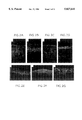
- FIGS. 2A, 2B, 2C, 2D, 2E, 2F, and 2G are immunofluorescence photomicrographs of corneal equivalents showing the distribution of ZO1 protein in corneal equivalents with an endothelial cell layer from various species and sources.
- FIGS. 3A, 3B, and 3C are a series of transmission electron micrographs showing the effects of various strains of endothelial cells on basal lamina formation in tri-layered corneal constructs.
- FIGS. 4A and 4B are immunofluorescent photomicrographs of the distribution of keratin 3 under different environmental conditions.
- Keratin 3 is a marker specific for corneal epithelial cells and is normally present in all suprabasal cell layers of the corneal limbus and the cells of the central cornea.
- Schermer, A., Galvin, S., and Sun, T.T. "Differentiation-related expression of a major 64K corneal keratin in vivo and in culture suggests limbal location of cornea epithelial stem cells," J.Cell. Biol. 103:49 (1986).
- the photomicrographs depict 14 day submerged (FIG.
- corneal constructs showing staining for keratin 3 (labeled using AE5 antibody) is strong and present in all suprabasal layers as in normal corneal limbus in vivo.
- FIGS. 5A and 5B are immunofluorescent photomicrographs of the distribution of alpha 6 -integrin under submerged or moist air-lift environmental conditions.
- alpha 6 -Integrin is a marker for hemidesmosomes in corneal epithelium.
- FIGS. 6A and 6B are immunofluorescence photomicrographs of corneal equivalents showing the distribution of type VII collagen in corneal equivalents in under different environmental conditions.
- FIGS. 7A and 7B are immunofluorescence photomicrographs of corneal equivalents showing the distribution of Laminin in corneal equivalents under different environmental conditions.
- FIGS. 8A and 8B are immunofluorescent photomicrographs showing the distribution of enolase under different environmental conditions.
- Enolase is a marker for the proliferative cell population in corneal epithelium. It is normally present in basal cells of the limbal region. (Zieske, J.D., Bukusoglu, G., Yankauckas, M.A., "Characterization of a potential marker of corneal epithelial stem cells," Invest. Opthalmol. Vis. Sci. 33:143-152 (1992).)
- FIGS. 9A and 9B are a series of transmission electron micrographs showing ultrastructural specialization in tri-layered constructs in a submerged state.
- FIG. 9A is a transmission electron micrograph showing vermiform ridges on the epithelial surface of the submerged culture at 13 days (equivalent to 6 days post moist air-lift).
- FIG. 9B is a transmission electron micrograph showing formation of basal lamina in tri-layered constructs in a submerged state at 13 days.
- FIGS. 10A and 10B are a series of transmission electron micrographs showing basal lamina production in skin equivalent constructs with an endothelial layer (FIG. 10A) and without an endothelial layer (FIG. 10B).
- FIGS. 11A-11D show a diagrammatic representation of tissue culture model.
- FIGS. 12A and 12B show a diagram of human eye.
- FIG. 12A is a diagram of the eye cut in a meridional plane that passes through the equator of the eye horizontally, dividing the eye into an upper and a lower half.
- FIG. 12B is a section through the human cornea, showing the five layers. (Diagrams from Functional Histology, Borysenko et al., Little Brown, publishers, pages 216-217, 1979).
- the outermost layer of the eye is the fibrous tunic, composed of dense avascular connective tissue.
- the fibrous tunic has two different regions: the sclera and the cornea.
- the sclera the "white" of the eye, forms the posterior portion of the fibrous tunic.
- the anterior sixth of the fibrous tunic is modified to form the transparent cornea.
- the cornea is a multilayered tissue consisting of an external epithelial layer, an underlying basement membrane, a collagenous extracellular matrix (termed the stroma), a second basement membrane (termed Descemet's membrane), and an inner single-cell-layered endothelium.
- Cornea epithelium is five to seven cell layers, with a single layer of columnar basal cells, two to three cell layers of wing cells, and two to three cell layers of flattened supeficial cells.
- a transition zone the limbus
- the external stratified squamous epithelium merges with the ocular conjunctiva at the sclera-cornea junction.
- a simple squamous epithelium also called the corneal endothelium, lines the inner face of the cornea.
- the middle layer of the cornea is clear, the result of the regular arrangement of its collagen fibers. (FIG. 12B.)
- Basement membranes are specialized extracellular matrices which function to support cells, and appear to be involved in molecular sieving as well as regulation of cell attachment, growth, and differentiation. These functions involve the binding of cells to the basement membrane through a group of binding proteins that include the integrin family. However, the mechanisms involved in the assembly of a basement membrane are not known. It has been found that the basement membrane assembly regulates the differentiation of epithelium and basement membrane formation in the in vitro tissue and organ equivalents. It has also been found that the culture environment influences corneal epithelial growth and differentiation.
- Constructing the cornea equivalent involves the tissue culturing and generation of the three distinct cell layers in the cornea: the external layer, a stratified squamous epithelium; the middle layer, collagen fibers; and the inner layer, the corneal endothelium.
- the method of constructing the cornea equivalent results in a structure analogous to the eye cornea in vivo.
- FIGS. 11A to 11D shows a tissue culture model.
- endothelial cells are seeded into a culture insert and grown to confluence.
- FIG. 11B a collagen layer containing stromal fibroblasts is cast atop the confluent endothelial cell layer and allowed to contract submerged in medium.
- FIG. 11A endothelial cells are seeded into a culture insert and grown to confluence.
- FIG. 11B a collagen layer containing stromal fibroblasts is cast atop the confluent endothelial cell layer and allowed to contract submerged in medium.
- a suspension of corneal epithelial cells is seeded onto the central area of the contracted lattice and grown submerged until epithelial coverage of the central raised area is nearly complete.
- the culture is placed at a moist air-liquid interface.
- the endothelial cells are seeded onto membranes of a cell culture insert. These endothelial cells will form the inner layer, or basal layer, of the corneal equivalent.
- the walls of the cell culture insert may consist of polystyrene, polycarbonate, resin, polypropylene (or other biocompatible plastic) with a porous membrane base of polycarbonate or other culture compatible porous membrane such as membranes made of collagen, cellulose, glass fiber or nylon attached to the bottom on which cells can be cultured.
- the porosity of the membrane can vary from 0.2 ⁇ m to 10 ⁇ m, with 3 ⁇ m being preferable.
- the insert is either suspended or supported in the culture dish to allow culture medium to access the underside of the culture.
- An acellular collagen layer is cast onto the cell culture membrane and allowed to gel at room temperature. The amount of acellular layer cast will depend upon the cell culture membrane used, but will typically be from 1 mL to about 5 mL.
- a K-RESIN® culture insert with a 3 ⁇ m porous polycarbonate membrane base approximately 2 cm 2 in area is used.
- a 1 mL acellular layer is cast onto the polycarbonate membrane and allowed to gel.
- the acellular collagen layer comprises 686 ⁇ g acid extracted bovine tendon collagen in 0.05% acetic acid, 8.1% 10 ⁇ Minimal Essential Eagle Medium, 4 mM 1-glutamine, 50 ⁇ g/ml gentamicin, 1.8 mg/mL sodium bicarbonate and 10% Dulbecco's modified Eagle's medium (DMEM) containing 10% newborn calf serum (NBCS).
- DMEM Dulbecco's modified Eagle's medium
- the endothelial layer is then submerged in DMEM containing 10% NBCS, 4 mM 1-glutamine, and 50 ⁇ g/ml gentamicin for four days at 37° C., 10% CO 2 .
- the acellular collagen layer may be omitted and the endothelial cells seeded directly onto the porous membrane.
- the use of an acellular layer is preferable when using transformed endothelial cells to inhibit overgrowth of the non-contact-inhibited cells on the underside of the membrane.
- the acellular layer may be made of Type IV collagen, laminin or a hydrogel.
- the endothelial cells used to form the endothelial layer can be derived from a variety of sources. Non-transformed corneal endothelial cells derived from sheep, rabbit, and cow have been used. Mouse corneal endothelial cells were transformed with large T antigen of SV40. (Muragaki, Y., Shiota, C., Inoue, M., Ooshima, A., Olsen, B.R., and Ninomiya, Y., "Alpha-1-VIII collagen gene transcripts encode a short-chain collagen polypeptide and are expressed by various epithelial endothelial and mesenchymal cells in newborn mouse tissues," Eur. J. Biochem.
- the preferred cell types are the transformed mouse corneal endothelial cell line, or normal corneal endothelial cells derived from sheep or rabbit. Most preferable are normal rabbit corneal endothelial cells.
- the normal rabbit endothelial cells are derived from enzymatically dissociated corneal endothelium or from explants of cornea and are serially cultivated in MSBM medium Johnson, W.E., Meunier, S.F., Roy, C.J. and Parenteau, N.L., "Serial Cultivation of Normal Human Keratinocytes: A Defined System Studying the Regulation of Growth and Differentiation," In Vitro Cell. Dev. Biol.
- Transformed endothelial cells are cultivated in DMEM-10%NBCS.
- Endothelial cells from a noncorneal origin may also be used in this invention.
- the noncorneal origin endothelial cells that have also been used in this invention include ovine and canine vascular and human umbilical vein endothelial cells.
- the endothelial cells may be transformed with a recombinant retrovirus containing the large T antigen of SV40 (Muragaki, et al., 1992, supra). Transformed cells continue to grow in the corneal equivalent and form mounds on top of the acellular layer due to their lack of contact inhibition. Non-transformed cells will form a monolayer underlying the stromal cell-collagen layer.
- normal endothelial cells may be transfected as above, but with the addition of a recombinant construct that expresses a heat sensitive gene. These transformed cells will grow in continuous culture under reduced temperature. After establishment of a confluent endothelial cell layer, the temperature can be raised to deactivate the transforming gene, allowing the cells to resume their normal regulation and exhibit contact inhibition, to form an endothelial cell monolayer similar to the non-transformed cells. Most peptides are heat sensitive (with the exception of heat shock proteins) so that there is a wide choice of peptides that can be deactivated by raising culturing temperature. Transformation in this way also facilitates the use of hard to obtain and cultivate cell types such as human corneal endothelial cells.
- collagen is mixed with keratocytes (stromal fibroblast cells) to achieve cell-collagen mixture.
- the cell-collagen mixture contains approximately 100 stromal fibroblast cells per ⁇ g acid extracted bovine tendon collagen.
- the fibroblasts contract the gel to form a raised area (mesa) of approximately 2.5 cm 2 .
- the stromal cell-collagen mixture comprises the middle layer of the cornea equivalent.
- the types of collagen that can be used are acid extracted bovine tendon collagen, enzyme extracted bovine tendon collagen, or rat tail collagen.
- the collagen may also consist of a mixture of Types I and III collagen as commonly extracted from dermis or a mixture of Types I, V and VI as extracted from corneal stroma.
- purified acid-extracted Type I collagen extracted from bovine tendon is used for the initial gel.
- the stromal fibroblasts will synthesize additional collagen types such as V and VI as well as additional Type I collagen as they modify the collagen matrix during cultivation.
- the epithelial cells will contribute Type IV and VII collagen at the epithelial-stromal junction and the endothelial cells will contribute Type XII collagen and other components of Descemet's membrane at the endothelial-stromal junction. (Muragaki et al., 1992, supra.)
- Any mammalian stromal fibroblast may be used in this cell layer. Any connective tissue fibroblast such as those derived from sclera, dermis, tendon, or fascia may be used. When corneal cells are used, fibroblasts derived from rabbit or human corneal stroma are preferable. The cells are enzymatically dissociated from normal corneal stroma, cultured in DMEM-10% NBCS and serially passaged. The cells incorporated into the construct are preferably used at passage four.
- the medium is removed from the cell culture inserts containing the confluent endothelial layer (typically 1.7-2.5 ⁇ 105cells/insert).
- the cell-collagen mixture is transferred and contacted with the surface of the endothelial cell layer.
- the cell-collagen mixture contains the same proportions of materials as the acellular layer with the addition of 5 ⁇ 10 4 stromal fibroblasts/mL cast mixture. Three mL of this mixture is pipetted into each cell culture insert and allowed to gel. The construct is then submerged in DMEM-10% NBCS and allowed to contract at 37° C., 10% CO 2 for seven days.
- These two layers which will eventually comprise the endothelial layer and the collagen layer of the cornea model, are cultured under conditions, known to those of skill in the art, to form a condensed collagen lattice, preferably by submerging in Dulbecco's-10% NBCS at 37° C., 10% CO 2 for seven days, to form a central raised area or a "mesa," resulting from the contraction of the collagen by the stromal fibroblasts.
- Normal rabbit stromal fibroblasts are cultured for seven days, but culture times may be shorter or longer (normally 2-10 days) depending on the species, cell type and number used.
- DMEM 10% NBCS is the preferred culture medium but any medium which normally supports the growth of fibroblasts may be used.
- corneal epithelial cells are plated onto the raised area of the collagen to form the apical, or external, layer of the cornea equivalent.
- the corneal epithelial cells can be derived from a variety of mammalian sources.
- the preferred epithelial cell is a rabbit or human corneal epithelial cell (corneal keratinocyte) but any mammalian corneal epithelial cell may be used.
- Other epithelial cells or keratinocytes such as those derived from the sclera (outer white opaque portion) of the eye or epidermis may be substituted, but corneal epithelial cells are preferable.
- the medium is removed from the culture insert (containing the contracted stromal matrix and endothelial layer) and its surround.
- Normal rabbit corneal epithelial cells, passage 4 are trypsinized and seeded on top of the membrane at a density of 7.2 ⁇ 10 4 -1.4 ⁇ 10 5 cells/cm 2 .
- the constructs are then incubated without media for four hours at 37° C., 10% CO 2 to allow the epithelial cells to attach. After the incubation the constructs are submerged in Corneal Maintenance Medium (CMM) (Johnson et al., 1992, supra.)
- CMS Corneal Maintenance Medium
- the epithelial cells are cultured until the mesa is covered with the epithelial cells. Completeness of epithelial coverage can be ascertained by a variety of methods, for illustration by staining the culture with a solution of Nile Blue sulfate (1:10,000 in phosphate buffered saline).
- the constructs are aseptically transferred to new culturing trays with sufficient cornea maintenance medium (CMM) to achieve a fluid level just to the surface of the construct to maintain a moist interface without submersion of the epithelial layer.
- CMM cornea maintenance medium
- the constructs are incubated at 37° C., 10% CO 2 , and greater than 60% humidity, with the CMM, making media changes, as necessary, typically, three times per week.
- the term "moist interface” is intended to mean a culture environment which is regulated so that the surface of the construct is moist, with high humidity, but not dry or submerged.
- the exact level of moisture and humidity in the culture environment is not critical, but it should be sufficiently moist and humid to avoid the formation of cornified cells.
- a moist interface can be characterized as trying to duplicate similar moisture levels of the human eye.
- An alternative method of achieving a moist interface at the epithelial layer utilizes a lipid/mucin mixture to simulate tear film.
- the specialized tear film may be formulated using a physiologic buffered salt solution containing protein-lipid surfactants or lipids and/or mucin, glycoasaminoglycans, hyaluronic add or other moisture holding substance.
- the film droplet is placed on top of the mesa to maintain a moist barrier between the epithelium and the atmosphere.
- the film is typically replaced when the media is changed.
- one or more of the components of the tear film can be added directly to the medium and allowed to wick over the surface of the construct during culturing to form the moist surface interface.
- maintenance of a moist interface may also be aided by the use of an artificial layer which can draw and hold moisture over the surface of the culture. This can be achieved by the application of a thin layer made of agarose, hydrogel, or alginate.
- a moist interface can be achieved using a dialysis membrane or polymer, such as contact lens material, cut slightly larger than the mesa, may be used to draw and hold fluid and prevent moisture loss.
- an endothelial layer promotes improved morphology, expression of biochemical and physiological markers, cell spreading, epithelial attachment to the matrix, and uniformity of epithelial coverage in vitro.
- the results on the influence of the endothelium in achieving a higher level of epithelial differentiation in vitro and promoting basement membrane formation was applied in other tissue equivalent in vitro culturing methods.
- tissue equivalents that can be modified according to this invention.
- passaged dermal fibroblasts are mixed with type I collagen to form a cellular collagen lattice inside a culture insert.
- This lattice then served as a substrate for epidermal keratinocytes.
- the keratinocytes grow to confluence and stratify while the culture remains submerged in culture medium for the first 4 days.
- the skin equivalent is then cultured at the air-liquid interface to allow differentiation of the epidermis to proceed.
- a first layer of endothelial cells can be cultured, as described above in section one, prior to casting collagen onto the endothelial layer.
- the endothelial cell layer is used to modify in vitro skin equivalent models, such as those described in U.S. Pat. No. 4,485,096, incorporated herein by reference, to promote epithelial differentiation and basement membrane formation.
- Draize eye irritation test has served as the standard for evaluating a product's ocular irritation potential for the past forty-five years. (Draize, J.H., Woodard, G., Calvery, H.O., "Methods for the study of irritation and toxicity of substances applied topically to the skin and mucous membranes," J. Pharmacel. Exp. Ther. 82:377-390 (1944).)
- the organotypic model proposed here overcomes some of the inherent limitations of monolayer culture by providing a model system which more closely simulates the target organ of interest.
- the physical configuration of this test cornea allows the topical application of test samples in vehicles (e.g., petrolatum and mineral oil) which approximate the mode of exposure in vivo.
- the organotypic culture method may also be used to form graftable human tissue either as an adjunct to conventional transplantation or as a substitute.
- the use of cultured corneal endothelial cells has already been shown to be beneficial as a replacement for the often damaged or inadequate endothelium of graft material.
- the use of cultured corneal epithelium has also shown some benefit in promoting wound closure.
- Roat, M.I., Thoft, R.A. "Ocular surface epithelial transplantation," Int. Opthalmol. Clin.
- the organotypic corneal construct comprising an endothelium, stroma and epithelium could be used for ocular wound closure and full-thickness repair of the cornea.
- the endothelial cells provided by the construct will regulate fluid transport to the corneal stroma and further stimulate the stromal fibroblasts to continue to organize the matrix and produce the appropriate collagens and glycosaminoglycans necessary for corneal clarity.
- the in vitro corneal equivalent may be constructed with more or less extracellular matrix or stroma to facilitate remodeling. Wound closure would be maintained by the presence of the well-adhered corneal epithelium, thereby limiting hyperproliferation and scarring of the stromal matrix.
- SCE2 a. ovine corneal endothelial cells
- DVEC canine vena caval endothelial cells
- bovine corneal endothelial cells (BCE 15960).
- constructs were prepared without endothelial cells.
- the Corneal Maintenance Medium consisted of a 3:1 mixture of calcium-free, Dulbecco's modified Eagle's Medium:Ham's F-12 with 1.1 ⁇ M hydrocortisone, 5 ⁇ g/mL insulin, 5 ⁇ g/mL transferrin, 20 pM triiodothyronine, 10 -4 M ethanolamine, 10 -4 M o-phosphoryl-ethanolamine, 1 mM strontium chloride, 50 ⁇ g/mL gentamicin, 4 mM 1-glutamine, 90 ⁇ M adenine, 3 ⁇ 10 -6 M selenium, 1.8 mM calcium chloride and 0.3% NBCS.
- CCM Corneal Maintenance Medium
- Endothelial cells were taken from frozen stocks and cultured one passage before use in the corneal constructs. Endothelial cells were seeded at 3.0 ⁇ 10 4 cells per transwell on a layer of acellular 1 mg/ml type I collagen gel and grown to confluence for 7 days while submerged.
- HUVEC, SEC 023VC, BCE 15960, and SEC 006A were cultured in Ham's F-12+10% NBCS, 25 ⁇ g/mL Heparin, 50 ⁇ g/mL ECGS and 50 ⁇ L/mL gentamicin.
- MCEC, DVEC, SCE2, and HEC were cultured in DMEM+10% NBCS and 50 ⁇ g/mL gentamicin while incubated at 37° C. in an atmosphere of 10% CO 2 .
- Rabbit corneal keratocytes at a density of 5.0 ⁇ 10 5 cells/mL in 3 mL of 1 mg/mL Type I collagen were laid on top of the endothelial layer.
- the collagen containing the cells gelled and medium (DMEM--10% NBCS and 50 ⁇ g/mL gentamicin) was added to the outside of the transwell.
- the constructs were incubated at 37° C. with 10% CO 2 for the following seven days while the collagen lattice containing the fibroblasts were contracted by the corneal fibroblasts.
- the collagen lattices had contracted away from the sides of the transwell to form a mesa.
- the medium was removed from the well and 50 ⁇ L suspension rabbit corneal epithelial cells (3.6 ⁇ 10 6 cells/mL) was seeded onto the center of the mesa, at 1.8 ⁇ 10 5 cells/mesa in CMM. 12 mL CMM was added back, submerging the entire construct. At seven days post-epithelialization, the rabbit corneal epithelial cells proliferated to completely cover the mesa.
- Epithelial coverage was monitored by Nile Blue sulfate-staining. Staining was done by adding 8 mL 1:10,000 Nile Blue Sulfate in phosphate buffered saline (PBS) for 30 minutes. Adherence was tested by attempting to peel the stained epithelium from the collagen lattice with forceps. Resistance to peeling indicated a positive effect of the endothelial cell layer.
- PBS phosphate buffered saline
- the constructs were transferred to a new tray containing two cotton pads and enough medium, 11 mL, (9 ml at feeds hereafter as the cotton pads together retain 2 mL) to just meet the surface of the culture and wick over the surface of the construct. Doing this step provided a moist apical surface to prevent abnormal squamous differentiation.
- the cultures were maintained in the incubator at 37° C. in 10% CO 2 and medium changes were made every 2-3 days for the remainder of the culture period.
- Nile Blue sulfate staining showed complete, although varied coverage in all conditions.
- Qualitative peel testing indicated adherence in the MCE (positive control), and BCE cultures, slight adherence in the SEC 023VC and SCE2 cultures and no adherence in the HUVEC, DVEC, SEC 006A and no endothelium cultures at 7 days post-epithelialization.
- Immunocytochemistry Immunocytochemistry using indirect fluorescence was performed using anti-laminin antibody to detect deposition of basement membrane components, and anti-ZO1 antibody to detect distribution of tight junction-associated protein.
- Laminin Distribution In previous studies described in. U.S. Pat. No. 5,374,515, small amounts of laminin were localized in patches along the basement membrane zone in moist air-lift cultures lacking endothelial cells. The addition of MCE greatly enhanced laminin deposition resulting in a strongly staining, continuous distribution along the epithelial-stromal junction. HUVEC and SEC 006A cultures showed continuous distribution of laminin. DVEC, SCE2, and SEC 023VC showed nearly continuous distribution. BCE showed a distribution similar o that previously obtained in moist cultures lacking an endothelial cell layer. (See FIGS. 1A to IF).
- ZO1 is a protein associated with tight junctions in corneal epithelium. Normal distribution should be limited to distinct areas in the upper layers of the corneal epithelium. Positive MCE controls and DVEC showed a striking localization in the uppermost layers of epithelium. SCE2 and SEC 023VC cultures also showed limited distribution, while HUVEC, BCE and SEC 006A cultures showed widely distributed staining indicating lack of specialized localization of the ZO1 protein. (See FIGS. 2A to 2G).
- basement Membrane Formation The assembly of basement membrane was examined by electron microscopy. (See FIGS. 3A to 3C). MCE-containing cultures form recognizable basement membrane and well-formed hemidesmosomes. Hemidesmosomes and associated basement membrane structures were detected in SEC 006A and SCE2 containing cultures.
- Cultures were prepared according to the previous example using MCEC in the endothelial layer. At time of moist air-lift, a portion of the cultures were maintained submerged in culture medium. Both groups were maintained for an additional one and two weeks. Corneal constructs were examined at 14 days submerged and 7 days post moist air-lift by histology, immunocytochemistry and electron microscopy.
- Endothelial cells were taken from frozen stocks (the transformed mouse corneal endothelial cells (MCE) from Example 1) and cultured one passage before use in the construct. Endothelial cells were seeded at 3.0 ⁇ 10 4 cells per transwell on a layer of acellular 1 mg/ml type I collagen gel and grown to confluence for 7 days while submerged in DMEM--10% NBCS and 50 ⁇ g/mL gentamicin while incubated at 37° C. in an atmosphere of 10% CO 2 .
- MCE mouse corneal endothelial cells
- Human dermal fibroblasts (HDF), at a density of 2.5 ⁇ 10 5 cells/mL in 3 mL of 1 mg/mL Type I collagen, were laid on top of the endothelial layer.
- the collagen containing the cells gelled and medium (DMEM--10% NBCS and 50 ⁇ g/mL gentamicin) was added to the outside of the transwell.
- the constructs were incubated at 37° C with 10% C02 for the following seven days while the collagen lattice containing the fibroblasts were contracted by the corneal fibroblasts.
- the collagen lattices had contracted away from the sides of the transwell to form a mesa.
- the media was removed from the well and a 50 ⁇ g/mL suspensions of human epidermal cells (3.33 ⁇ 10 6 cells/mL) were seeded onto the center of the mesa in MSBM (3:1 Calcium-free, glucose-free DMEM:Ham's F-12 supplemented with 4 mM L-glutamine, 1.1 ⁇ M hydrocortisone, 5 ⁇ g/mL insulin, 5 ⁇ g/mL triiodothyronine, 10 -4 M ethanolamine, 10 -4 M, o-phosphoryl-ethanolamine, 0.18 mM adenine, 2 ⁇ 10 -9 M propesterone, 5.26 ⁇ 10 -8 M selenium, 0.3% bovine serum, 1.8 mM calcium chloride and 10 ng/mL epidermal growth factor ).
- MSBM medium was added back to the outside of the transwell submerging the entire construct,
- the human epidermal cells proliferated to completely cover the mesa.
- Medium was removed from the inside and outside chambers.
- the inner chamber was removed and two cotton pads were placed in the well to raise the construct to the air-liquid interface.
- Constructs were maintained in culture conditions suitable for skin equivalent culture. At four days following airlift, the medium was replaced with 9 mL maintenanceSBM with calcium (1:1 Calcium-free, glucose-free DMEM:Ham's F-12 supplemented with 4 mM L-glutamine, 1.1 ⁇ M hydrocortisone, 5 ⁇ g/mL insulin, 5 ⁇ g/mL triiodothyronine, 10 -4 M ethanolamine, 10 -4 M, o-phosphoryl-ethanolamine, 0.18 mM adenine, 5.26 ⁇ 10 -8 M selenium, 1.0% bovine serum, 1 ng/mL epidermal growth factor and 1.8 mM calcium chloride). Cultures were maintained at the air-liquid interface at day 4 following epidermalization. Cultures were maintained for one week at the air-liquid interface and examined for formation of basement membrane by electron microscopy at seven days post air-lift.
Abstract
Description
Claims (36)
Priority Applications (7)
| Application Number | Priority Date | Filing Date | Title |
|---|---|---|---|
| US08/337,830 US5827641A (en) | 1992-11-13 | 1994-11-08 | In vitro cornea equivalent model |
| EP95939790A EP0790768A4 (en) | 1994-11-08 | 1995-11-08 | In vitro tissue and organ equivalent models |
| CA002204869A CA2204869A1 (en) | 1994-11-08 | 1995-11-08 | In vitro tissue and organ equivalent models |
| JP8515480A JPH10508522A (en) | 1994-11-08 | 1995-11-08 | In vitro tissue and organ equivalent model |
| AU41475/96A AU703320B2 (en) | 1994-11-08 | 1995-11-08 | In vitro tissue and organ equivalent models |
| PCT/US1995/014415 WO1996013974A1 (en) | 1994-11-08 | 1995-11-08 | In vitro tissue and organ equivalent models |
| MXPA/A/1997/003321A MXPA97003321A (en) | 1994-11-08 | 1997-05-07 | In vitro fabric and equivalen organ models |
Applications Claiming Priority (2)
| Application Number | Priority Date | Filing Date | Title |
|---|---|---|---|
| US07/974,740 US5374515A (en) | 1992-11-13 | 1992-11-13 | In vitro cornea equivalent model |
| US08/337,830 US5827641A (en) | 1992-11-13 | 1994-11-08 | In vitro cornea equivalent model |
Related Parent Applications (1)
| Application Number | Title | Priority Date | Filing Date |
|---|---|---|---|
| US07/974,740 Continuation-In-Part US5374515A (en) | 1992-11-13 | 1992-11-13 | In vitro cornea equivalent model |
Publications (1)
| Publication Number | Publication Date |
|---|---|
| US5827641A true US5827641A (en) | 1998-10-27 |
Family
ID=23322206
Family Applications (1)
| Application Number | Title | Priority Date | Filing Date |
|---|---|---|---|
| US08/337,830 Expired - Lifetime US5827641A (en) | 1992-11-13 | 1994-11-08 | In vitro cornea equivalent model |
Country Status (6)
| Country | Link |
|---|---|
| US (1) | US5827641A (en) |
| EP (1) | EP0790768A4 (en) |
| JP (1) | JPH10508522A (en) |
| AU (1) | AU703320B2 (en) |
| CA (1) | CA2204869A1 (en) |
| WO (1) | WO1996013974A1 (en) |
Cited By (27)
| Publication number | Priority date | Publication date | Assignee | Title |
|---|---|---|---|---|
| WO2000030672A1 (en) * | 1998-11-19 | 2000-06-02 | The Schepens Eye Research Institute | Collagen based biomaterials and methods of preparation and use |
| WO2001056379A1 (en) * | 2000-02-07 | 2001-08-09 | The Schepens Eye Research Institute, Inc. | A method of quieting a corneal inflammatory response |
| WO2003009783A1 (en) * | 2001-07-25 | 2003-02-06 | Japan Tissue Engineering Co., Ltd. | Laminates for treating cornea and process for producing the same |
| US6544286B1 (en) | 2000-07-18 | 2003-04-08 | Tissue Engineering Refraction, Inc. | Pre-fabricated corneal tissue lens method of corneal overlay to correct vision |
| US20040137616A1 (en) * | 2000-02-29 | 2004-07-15 | Isseroff Roslyn R. | Corneal epithelial graft composites |
| US20050287126A1 (en) * | 2001-11-19 | 2005-12-29 | Amniotec Inc. | Ectocornea-like sheet and method of constructing the same |
| US20060020267A1 (en) * | 2004-07-15 | 2006-01-26 | Marmo J C | Intrastromal devices and methods for improving vision |
| US7004953B2 (en) | 2001-07-23 | 2006-02-28 | Fos Holding S.A. | Device for separating the epithelium layer from the surface of the cornea of an eye |
| US20060228693A1 (en) * | 2005-04-12 | 2006-10-12 | Soll David B | Composition and method for in vitro preservation of corneal tissues |
| US20070092550A1 (en) * | 2003-10-10 | 2007-04-26 | Cellular Bioengineering, Inc. | Methods and compositions for growing corneal endothelial and related cells on biopolymers and creation of artifical corneal transplants |
| CN1326502C (en) * | 2003-08-07 | 2007-07-18 | 中山大学中山眼科中心 | Artificial tissue engineeing biological cornea |
| CN100333702C (en) * | 2004-04-28 | 2007-08-29 | 浙江大学医学院附属邵逸夫医院 | Exogenous cornea substrate without cells and its preparation method and use |
| CN100386061C (en) * | 2005-05-17 | 2008-05-07 | 浙江大学医学院附属邵逸夫医院 | Low antigen hetero stroma of cornea treated by frozen, and its prepn. method |
| WO2008101100A2 (en) * | 2007-02-14 | 2008-08-21 | Indiana University Research And Technology Corporation | Composition for stimulating formation of vascular structures |
| US7497866B2 (en) | 2000-07-18 | 2009-03-03 | Tissue Engineering Refraction Inc. | Methods for producing epithelial flaps on the cornea and for placement of ocular devices and lenses beneath an epithelial flap or membrane, epithelial delaminating devices, and structures of epithelium and ocular devices and lenses |
| US7585075B2 (en) | 2004-05-20 | 2009-09-08 | Forsight Labs, Llc | Corneal onlays and wavefront aberration correction to enhance vision |
| US20100035297A1 (en) * | 2008-08-08 | 2010-02-11 | Indiana University Research And Technology Corporation | Methods and compositions for vasculogenic potential determination |
| WO2010027789A2 (en) * | 2008-08-25 | 2010-03-11 | University Of South Florida | Volume exclusion agent to enhance formation of extracelluar matrix |
| US7828844B2 (en) | 2002-09-13 | 2010-11-09 | Forsight Labs, Llc | Inserting lenses into corneal epithelial pockets to improve vision |
| US7883520B2 (en) | 2006-04-10 | 2011-02-08 | Forsight Labs, Llc | Corneal epithelial pocket formation systems, components and methods |
| US20140099290A1 (en) * | 2009-02-26 | 2014-04-10 | Wake Forest University Health Sciences | Endothelial scaffolds |
| WO2015069619A1 (en) * | 2013-11-05 | 2015-05-14 | President And Fellows Of Harvard College | Method of printing a tissue construct with embedded vasculature |
| WO2016065312A1 (en) * | 2014-10-24 | 2016-04-28 | The Trustees Of The University Of Pennsylvania | Methods and devices for modeling the eye |
| CN106388973A (en) * | 2016-04-20 | 2017-02-15 | 珐博进(中国)医药技术开发有限公司 | Device for preparing collagen cornea |
| WO2020232283A1 (en) * | 2019-05-14 | 2020-11-19 | The Trustees Of Princeton University | In vitro cell-based replacement for the draize test |
| EP3975931A4 (en) * | 2019-06-03 | 2023-06-28 | Advanced Solutions Life Sciences, LLC | System and method for fabricating a cornea |
| US11795431B2 (en) | 2017-08-17 | 2023-10-24 | The Trustees Of Princeton University | Ultrathin interfacial layer on a hydrogel to direct its surface properties and cell adhesion |
Families Citing this family (17)
| Publication number | Priority date | Publication date | Assignee | Title |
|---|---|---|---|---|
| EP1014880A4 (en) * | 1997-02-20 | 2000-08-16 | Gerigene Medical Corp | Augmentation and repair of dermal, subcutaneous, and vocal cord tissue defects |
| ES2249928T3 (en) * | 1998-11-19 | 2006-04-01 | Organogenesis Inc. | MANIPULATED FABRIC CONSTRUCTS THROUGH BIOINGENIERIA AND METHODS TO PRODUCE AND USE THEM. |
| US6479064B1 (en) * | 1999-12-29 | 2002-11-12 | Children's Medical Center Corporation | Culturing different cell populations on a decellularized natural biostructure for organ reconstruction |
| JP4554051B2 (en) * | 2000-09-08 | 2010-09-29 | Hoya株式会社 | How to rebuild the cornea |
| ITRM20010476A1 (en) * | 2001-08-03 | 2003-02-03 | Idi Irccs | HUMAN CORNEAL EPITHELIUM SHEETS RECONSTRUCTED AND METHOD FOR THEIR OBTAINING. |
| WO2003092762A1 (en) * | 2002-04-30 | 2003-11-13 | Amniotec Inc. | Corneal endothelium-like sheet and method of constructing the same |
| JP4834802B2 (en) * | 2004-02-18 | 2011-12-14 | 聡 山上 | Culture layer laminate of human corneal endothelial cells and method for producing the same |
| JP2009521228A (en) * | 2005-12-21 | 2009-06-04 | ヴァックスデザイン コーポレーション | Porous membrane devices that promote the differentiation of monocytes into dendritic cells |
| US8105634B2 (en) | 2006-08-15 | 2012-01-31 | Anthrogenesis Corporation | Umbilical cord biomaterial for medical use |
| US8071135B2 (en) | 2006-10-04 | 2011-12-06 | Anthrogenesis Corporation | Placental tissue compositions |
| NZ576372A (en) | 2006-10-06 | 2012-02-24 | Anthrogenesis Corp | Native (telopeptide) placental collagen compositions |
| US20100183572A1 (en) | 2007-02-28 | 2010-07-22 | Takeshi Nakajima | Cell capable of expressing lacritin at high level |
| FR2927632B1 (en) * | 2008-02-14 | 2013-07-19 | Basf Beauty Care Solutions F | CORNEA AND MUQUEUSE RECONSTRUCTED. |
| EP2793964B1 (en) | 2011-12-20 | 2019-03-20 | LifeCell Corporation | Sheet tissue products |
| DK3501558T3 (en) | 2011-12-20 | 2021-03-01 | Lifecell Corp | LIQUID TISSUE PRODUCTS |
| US9271821B2 (en) | 2012-01-24 | 2016-03-01 | Lifecell Corporation | Elongated tissue matrices |
| CN114259601B (en) * | 2021-12-27 | 2023-05-26 | 暨南大学 | Living cell bionic cornea anterior lamina and preparation method thereof |
Citations (5)
| Publication number | Priority date | Publication date | Assignee | Title |
|---|---|---|---|---|
| US4485096A (en) * | 1982-02-26 | 1984-11-27 | Massachusetts Institute Of Technology | Tissue-equivalent and method for preparation thereof |
| US4546500A (en) * | 1981-05-08 | 1985-10-15 | Massachusetts Institute Of Technology | Fabrication of living blood vessels and glandular tissues |
| US4760020A (en) * | 1985-05-03 | 1988-07-26 | Eye Research Institute Of Retina Foundation | Method for testing biocompatibility |
| US5131907A (en) * | 1986-04-04 | 1992-07-21 | Thomas Jefferson University | Method of treating a synthetic naturally occurring surface with a collagen laminate to support microvascular endothelial cell growth, and the surface itself |
| US5374515A (en) * | 1992-11-13 | 1994-12-20 | Organogenesis, Inc. | In vitro cornea equivalent model |
-
1994
- 1994-11-08 US US08/337,830 patent/US5827641A/en not_active Expired - Lifetime
-
1995
- 1995-11-08 AU AU41475/96A patent/AU703320B2/en not_active Ceased
- 1995-11-08 CA CA002204869A patent/CA2204869A1/en not_active Abandoned
- 1995-11-08 JP JP8515480A patent/JPH10508522A/en active Pending
- 1995-11-08 WO PCT/US1995/014415 patent/WO1996013974A1/en not_active Application Discontinuation
- 1995-11-08 EP EP95939790A patent/EP0790768A4/en not_active Withdrawn
Patent Citations (5)
| Publication number | Priority date | Publication date | Assignee | Title |
|---|---|---|---|---|
| US4546500A (en) * | 1981-05-08 | 1985-10-15 | Massachusetts Institute Of Technology | Fabrication of living blood vessels and glandular tissues |
| US4485096A (en) * | 1982-02-26 | 1984-11-27 | Massachusetts Institute Of Technology | Tissue-equivalent and method for preparation thereof |
| US4760020A (en) * | 1985-05-03 | 1988-07-26 | Eye Research Institute Of Retina Foundation | Method for testing biocompatibility |
| US5131907A (en) * | 1986-04-04 | 1992-07-21 | Thomas Jefferson University | Method of treating a synthetic naturally occurring surface with a collagen laminate to support microvascular endothelial cell growth, and the surface itself |
| US5374515A (en) * | 1992-11-13 | 1994-12-20 | Organogenesis, Inc. | In vitro cornea equivalent model |
Non-Patent Citations (28)
| Title |
|---|
| Eliason, J.A. and Elliott, J. P., Investigative Ophthalmol. & Visual Science 28 :1963 60 (1987). * |
| Eliason, J.A. and Elliott, J. P., Investigative Ophthalmol. & Visual Science 28:1963-60 (1987). |
| Goldberg, A.M., Fd. Chem. Toxic. 23 :205 208 (1985). * |
| Goldberg, A.M., Fd. Chem. Toxic. 23:205-208 (1985). |
| Gordon, V.C. and Kelly, C.P., Cosmetics & Toiletries 104: 69 74 (1989). * |
| Gordon, V.C. and Kelly, C.P., Cosmetics & Toiletries 104:69-74 (1989). |
| Insler, M.S., et al., Curr. Eye Res. 5( 12 ):967 972 (1986). * |
| Insler, M.S., et al., Curr. Eye Res. 5(12):967-972 (1986). |
| Muragaki, Y., et al., Eur J. Biochem. 207 ( 3 ):895 902 (1992). * |
| Muragaki, Y., et al., Eur J. Biochem. 207(3):895-902 (1992). |
| Parish, W.E., Fd. Chem. Toxic. 23 :215 227 (1985). * |
| Parish, W.E., Fd. Chem. Toxic. 23:215-227 (1985). |
| Roat, M.I. and Thoft, R.A., Int. Opthalmol. Clin. 28(2):169 174 (1988). * |
| Roat, M.I. and Thoft, R.A., Int. Opthalmol. Clin. 28(2):169-174 (1988). |
| Schermer, A., et al., J. Cell Bio. 103 :49 62 (1986). * |
| Schermer, A., et al., J. Cell Bio. 103:49-62 (1986). |
| Shopsis, C., et al., Fd. Chem Toxic. 23 :259 66 (1985). * |
| Shopsis, C., et al., Fd. Chem Toxic. 23:259-66 (1985). |
| Simmons, S.J., et al., Toxicology and Applied Pharmacology 88 :13 23 (1987). * |
| Simmons, S.J., et al., Toxicology and Applied Pharmacology 88:13-23 (1987). |
| Sundar Raj, C.V., et al., Investigative Ophthalmol. Vis. Sci. 19:1222 30 (1980). * |
| Sundar-Raj, C.V., et al., Investigative Ophthalmol. Vis. Sci. 19:1222-30 (1980). |
| Trinkaus Randall, V., et al., Investig. Ophthal. & Visual Science 29 :1800 1809 (1988). * |
| Trinkaus-Randall, V., et al., Investig. Ophthal. & Visual Science 29:1800-1809 (1988). |
| Xie, L., et al., Developmental Biology 25 :20 22 (1989). * |
| Xie, L., et al., Developmental Biology 25:20-22 (1989). |
| Zieske, J.D., Bukusoglu, G., and Yankauckas, M.A., Invest. Ophthalmol. & Vis. Sci. 33 :143 152 (1992). * |
| Zieske, J.D., Bukusoglu, G., and Yankauckas, M.A., Invest. Ophthalmol. & Vis. Sci. 33:143-152 (1992). |
Cited By (44)
| Publication number | Priority date | Publication date | Assignee | Title |
|---|---|---|---|---|
| WO2000030672A1 (en) * | 1998-11-19 | 2000-06-02 | The Schepens Eye Research Institute | Collagen based biomaterials and methods of preparation and use |
| WO2001056379A1 (en) * | 2000-02-07 | 2001-08-09 | The Schepens Eye Research Institute, Inc. | A method of quieting a corneal inflammatory response |
| US20040137616A1 (en) * | 2000-02-29 | 2004-07-15 | Isseroff Roslyn R. | Corneal epithelial graft composites |
| US20050070942A1 (en) * | 2000-07-18 | 2005-03-31 | Edward Perez | Device for lifting an epitheleal layer and placing a corrective lens beneath it |
| US20030083743A1 (en) * | 2000-07-18 | 2003-05-01 | Edward Perez | Method of producing an epithelial flap |
| US6544286B1 (en) | 2000-07-18 | 2003-04-08 | Tissue Engineering Refraction, Inc. | Pre-fabricated corneal tissue lens method of corneal overlay to correct vision |
| US7497866B2 (en) | 2000-07-18 | 2009-03-03 | Tissue Engineering Refraction Inc. | Methods for producing epithelial flaps on the cornea and for placement of ocular devices and lenses beneath an epithelial flap or membrane, epithelial delaminating devices, and structures of epithelium and ocular devices and lenses |
| US6880558B2 (en) | 2000-07-18 | 2005-04-19 | Tissue Engineering Refractions, Inc. | Method of lifting an epithelial layer and placing a corrective lens beneath it |
| US20050124982A1 (en) * | 2000-07-18 | 2005-06-09 | Edward Perez | Method of producing an epithelial flap (II) |
| US7004953B2 (en) | 2001-07-23 | 2006-02-28 | Fos Holding S.A. | Device for separating the epithelium layer from the surface of the cornea of an eye |
| US7708750B2 (en) | 2001-07-23 | 2010-05-04 | Fos Holdings S.A. | Device for separating the epithelium layer from the surface of the cornea of an eye |
| WO2003009783A1 (en) * | 2001-07-25 | 2003-02-06 | Japan Tissue Engineering Co., Ltd. | Laminates for treating cornea and process for producing the same |
| US20050287126A1 (en) * | 2001-11-19 | 2005-12-29 | Amniotec Inc. | Ectocornea-like sheet and method of constructing the same |
| US20140220102A1 (en) * | 2001-11-19 | 2014-08-07 | Takahiro Nakamura | Ectocornea-like sheet and method of constructing the same |
| US7828844B2 (en) | 2002-09-13 | 2010-11-09 | Forsight Labs, Llc | Inserting lenses into corneal epithelial pockets to improve vision |
| CN1326502C (en) * | 2003-08-07 | 2007-07-18 | 中山大学中山眼科中心 | Artificial tissue engineeing biological cornea |
| US20070092550A1 (en) * | 2003-10-10 | 2007-04-26 | Cellular Bioengineering, Inc. | Methods and compositions for growing corneal endothelial and related cells on biopolymers and creation of artifical corneal transplants |
| AU2004281694B2 (en) * | 2003-10-10 | 2012-05-03 | Cellular Bioengineering, Inc. | Methods and compositions for growing corneal endothelial and related cells on biopolymers and creation of artifical corneal transplants |
| CN100333702C (en) * | 2004-04-28 | 2007-08-29 | 浙江大学医学院附属邵逸夫医院 | Exogenous cornea substrate without cells and its preparation method and use |
| US7585075B2 (en) | 2004-05-20 | 2009-09-08 | Forsight Labs, Llc | Corneal onlays and wavefront aberration correction to enhance vision |
| US20060020267A1 (en) * | 2004-07-15 | 2006-01-26 | Marmo J C | Intrastromal devices and methods for improving vision |
| US7601487B2 (en) | 2005-04-12 | 2009-10-13 | Cleo Cosmetic And Pharmaceutical Co., Llc | Composition using HMW dextran and method for in vitro preservation of corneal tissues |
| US20060228693A1 (en) * | 2005-04-12 | 2006-10-12 | Soll David B | Composition and method for in vitro preservation of corneal tissues |
| CN100386061C (en) * | 2005-05-17 | 2008-05-07 | 浙江大学医学院附属邵逸夫医院 | Low antigen hetero stroma of cornea treated by frozen, and its prepn. method |
| US7883520B2 (en) | 2006-04-10 | 2011-02-08 | Forsight Labs, Llc | Corneal epithelial pocket formation systems, components and methods |
| WO2008101100A2 (en) * | 2007-02-14 | 2008-08-21 | Indiana University Research And Technology Corporation | Composition for stimulating formation of vascular structures |
| WO2008101100A3 (en) * | 2007-02-14 | 2009-12-30 | Indiana University Research And Technology Corporation | Composition for stimulating formation of vascular structures |
| US20100143476A1 (en) * | 2007-02-14 | 2010-06-10 | March Keith L | Composition for stimulating formation of vascular structures |
| US20100035297A1 (en) * | 2008-08-08 | 2010-02-11 | Indiana University Research And Technology Corporation | Methods and compositions for vasculogenic potential determination |
| US8623646B2 (en) | 2008-08-25 | 2014-01-07 | University Of South Florida | Volume exclusion agent to enhance formation of extracellular matrix |
| WO2010027789A2 (en) * | 2008-08-25 | 2010-03-11 | University Of South Florida | Volume exclusion agent to enhance formation of extracelluar matrix |
| WO2010027789A3 (en) * | 2008-08-25 | 2010-06-24 | University Of South Florida | Volume exclusion agent to enhance formation of extracelluar matrix |
| US9707256B2 (en) * | 2009-02-26 | 2017-07-18 | Wake Forest Institute for Regenerative Medicine | Methods of making endothelial scaffolds |
| US20140099290A1 (en) * | 2009-02-26 | 2014-04-10 | Wake Forest University Health Sciences | Endothelial scaffolds |
| WO2015069619A1 (en) * | 2013-11-05 | 2015-05-14 | President And Fellows Of Harvard College | Method of printing a tissue construct with embedded vasculature |
| US10117968B2 (en) | 2013-11-05 | 2018-11-06 | President And Fellows Of Harvard College | Method of printing a tissue construct with embedded vasculature |
| US10360819B2 (en) | 2014-10-24 | 2019-07-23 | The Trustees Of The University Of Pennsylvania | Methods and devices for modeling the eye |
| WO2016065312A1 (en) * | 2014-10-24 | 2016-04-28 | The Trustees Of The University Of Pennsylvania | Methods and devices for modeling the eye |
| US20190340957A1 (en) * | 2014-10-24 | 2019-11-07 | The Trustees Of The University Of Pennsylvania | Methods and Devices for Modeling the Eye |
| US10783803B2 (en) * | 2014-10-24 | 2020-09-22 | The Trustees Of The University Of Pennsylvania | Methods and devices for modeling the eye |
| CN106388973A (en) * | 2016-04-20 | 2017-02-15 | 珐博进(中国)医药技术开发有限公司 | Device for preparing collagen cornea |
| US11795431B2 (en) | 2017-08-17 | 2023-10-24 | The Trustees Of Princeton University | Ultrathin interfacial layer on a hydrogel to direct its surface properties and cell adhesion |
| WO2020232283A1 (en) * | 2019-05-14 | 2020-11-19 | The Trustees Of Princeton University | In vitro cell-based replacement for the draize test |
| EP3975931A4 (en) * | 2019-06-03 | 2023-06-28 | Advanced Solutions Life Sciences, LLC | System and method for fabricating a cornea |
Also Published As
| Publication number | Publication date |
|---|---|
| AU4147596A (en) | 1996-05-31 |
| AU703320B2 (en) | 1999-03-25 |
| JPH10508522A (en) | 1998-08-25 |
| EP0790768A1 (en) | 1997-08-27 |
| MX9703321A (en) | 1997-07-31 |
| EP0790768A4 (en) | 2002-09-18 |
| CA2204869A1 (en) | 1996-05-17 |
| WO1996013974A1 (en) | 1996-05-17 |
Similar Documents
| Publication | Publication Date | Title |
|---|---|---|
| US5827641A (en) | In vitro cornea equivalent model | |
| EP0621766B1 (en) | In vitro produced cornea equivalent model | |
| Zieske et al. | Basement membrane assembly and differentiation of cultured corneal cells: importance of culture environment and endothelial cell interaction | |
| Alaminos et al. | Construction of a complete rabbit cornea substitute using a fibrin-agarose scaffold | |
| Flanagan et al. | A collagen-glycosaminoglycan co-culture model for heart valve tissue engineering applications | |
| Orwin et al. | In vitro culture characteristics of corneal epithelial, endothelial, and keratocyte cells in a native collagen matrix | |
| San Choi et al. | Bioengineering endothelialized neo-corneas using donor-derived corneal endothelial cells and decellularized corneal stroma | |
| Proulx et al. | Reconstruction of a human cornea by the self-assembly approach of tissue engineering using the three native cell types | |
| US6471958B2 (en) | Non-contracting tissue equivalent | |
| El Ghalbzouri et al. | The use of PEGT/PBT as a dermal scaffold for skin tissue engineering | |
| US6645715B1 (en) | Artificial cornea | |
| AU2004282525B2 (en) | Human corneal endothelial cells and methods of obtaining and culturing cells for corneal cell transplantation | |
| Kinikoglu et al. | Reconstruction of a full-thickness collagen-based human oral mucosal equivalent | |
| RU2498808C9 (en) | Method of treating patient's oral diseases (versions) | |
| JPH09504968A (en) | Stimulation of epithelial cell adhesion and hemidesmosome formation by soluble factors | |
| EP0321545A1 (en) | Human epithelium originating from cell cultures | |
| Sweeney et al. | A comparison of biological coatings for the promotion of corneal epithelialization of synthetic surface in vivo | |
| MXPA97003321A (en) | In vitro fabric and equivalen organ models | |
| KR100818636B1 (en) | Artificial skin containing mesenchymal stem cells and construction method thereof | |
| Zieske et al. | Human corneal organotypic cultures | |
| KR20230124246A (en) | Method of endotheliocyte isolation and culture in cornea | |
| Alaminos Mingorance et al. | Construction of a complete rabbit cornea substitute using a fibrin-agarose scaffold | |
| FUKAI et al. | Basement Membrane Assembly and Differentiation of Cultured Corneal Cells: Importance of Culture Environment and Endothelial Cell lnteraction | |
| Dalton | Migration mechanisms of corneal epithelial tissue and cells | |
| Audet et al. | Reconstruction of a human cornea by the self-assembly approach of tissue engineering using the three native cell types |
Legal Events
| Date | Code | Title | Description |
|---|---|---|---|
| AS | Assignment |
Owner name: PRESIDENT AND FELLOWS OF HARVARD COLLEGE, THE, MAS Free format text: ASSIGNMENT OF ASSIGNORS INTEREST;ASSIGNOR:OLSEN, BJORN REINO;REEL/FRAME:007392/0053 Effective date: 19950223 Owner name: ORGANOGENESIS, INC., MASSACHUSETTS Free format text: ASSIGNMENT OF ASSIGNORS INTEREST;ASSIGNORS:PARENTEAU, NANCY LOUISE;MASON, VALERIE SUSAN;REEL/FRAME:007392/0055;SIGNING DATES FROM 19950223 TO 19950226 |
|
| STCF | Information on status: patent grant |
Free format text: PATENTED CASE |
|
| AS | Assignment |
Owner name: FLEET NATIONAL BANK, MASSACHUSETTS Free format text: SECURITY INTEREST;ASSIGNOR:ORGANOGENESIS INC.;REEL/FRAME:011806/0407 Effective date: 20010501 |
|
| AS | Assignment |
Owner name: BERKSHIRE BANK, THE, NEW YORK Free format text: SECURITY AGREEMENT;ASSIGNORS:ORGANOGENESIS INC.;DAN CAPITAL CORP.;ECM PHARMA, INC.;REEL/FRAME:012002/0266 Effective date: 20010629 |
|
| FPAY | Fee payment |
Year of fee payment: 4 |
|
| REMI | Maintenance fee reminder mailed | ||
| FPAY | Fee payment |
Year of fee payment: 8 |
|
| AS | Assignment |
Owner name: ORGANOGENESIS INC., MASSACHUSETTS Free format text: RELEASE BY SECURED PARTY;ASSIGNOR:THE BERKSHIRE BANK;REEL/FRAME:022846/0100 Effective date: 20021125 |
|
| AS | Assignment |
Owner name: ORGANOGENESIS, INC., MASSACHUSETTS Free format text: RELEASE BY SECURED PARTY;ASSIGNOR:FLEET NATIONAL BANK N/K/A BANK OF AMERICA N.A.;REEL/FRAME:022856/0363 Effective date: 20090619 |
|
| REMI | Maintenance fee reminder mailed | ||
| FPAY | Fee payment |
Year of fee payment: 12 |
|
| SULP | Surcharge for late payment |
Year of fee payment: 11 |
|
| AS | Assignment |
Owner name: WELLS FARGO BANK, N.A., MASSACHUSETTS Free format text: SECURITY AGREEMENT;ASSIGNOR:ORGANOGENESIS, INC.;REEL/FRAME:026954/0666 Effective date: 20110922 |
|
| AS | Assignment |
Owner name: ORGANOGENESIS INC., MASSACHUSETTS Free format text: RELEASE BY SECURED PARTY;ASSIGNOR:WELLS FARGO BANK, NATIONAL ASSOCIATION;REEL/FRAME:042072/0977 Effective date: 20170321 |