US5705360A - Chemokine N-terminal deletion mutations - Google Patents
Chemokine N-terminal deletion mutations Download PDFInfo
- Publication number
- US5705360A US5705360A US08/683,785 US68378596A US5705360A US 5705360 A US5705360 A US 5705360A US 68378596 A US68378596 A US 68378596A US 5705360 A US5705360 A US 5705360A
- Authority
- US
- United States
- Prior art keywords
- mcp
- nucleic acid
- chemokine
- derivative
- sequence
- Prior art date
- Legal status (The legal status is an assumption and is not a legal conclusion. Google has not performed a legal analysis and makes no representation as to the accuracy of the status listed.)
- Expired - Fee Related
Links
Images


Classifications
-
- C—CHEMISTRY; METALLURGY
- C07—ORGANIC CHEMISTRY
- C07K—PEPTIDES
- C07K14/00—Peptides having more than 20 amino acids; Gastrins; Somatostatins; Melanotropins; Derivatives thereof
- C07K14/435—Peptides having more than 20 amino acids; Gastrins; Somatostatins; Melanotropins; Derivatives thereof from animals; from humans
- C07K14/52—Cytokines; Lymphokines; Interferons
- C07K14/521—Chemokines
-
- A—HUMAN NECESSITIES
- A61—MEDICAL OR VETERINARY SCIENCE; HYGIENE
- A61P—SPECIFIC THERAPEUTIC ACTIVITY OF CHEMICAL COMPOUNDS OR MEDICINAL PREPARATIONS
- A61P1/00—Drugs for disorders of the alimentary tract or the digestive system
-
- A—HUMAN NECESSITIES
- A61—MEDICAL OR VETERINARY SCIENCE; HYGIENE
- A61P—SPECIFIC THERAPEUTIC ACTIVITY OF CHEMICAL COMPOUNDS OR MEDICINAL PREPARATIONS
- A61P11/00—Drugs for disorders of the respiratory system
-
- A—HUMAN NECESSITIES
- A61—MEDICAL OR VETERINARY SCIENCE; HYGIENE
- A61P—SPECIFIC THERAPEUTIC ACTIVITY OF CHEMICAL COMPOUNDS OR MEDICINAL PREPARATIONS
- A61P23/00—Anaesthetics
-
- A—HUMAN NECESSITIES
- A61—MEDICAL OR VETERINARY SCIENCE; HYGIENE
- A61P—SPECIFIC THERAPEUTIC ACTIVITY OF CHEMICAL COMPOUNDS OR MEDICINAL PREPARATIONS
- A61P29/00—Non-central analgesic, antipyretic or antiinflammatory agents, e.g. antirheumatic agents; Non-steroidal antiinflammatory drugs [NSAID]
-
- A—HUMAN NECESSITIES
- A61—MEDICAL OR VETERINARY SCIENCE; HYGIENE
- A61P—SPECIFIC THERAPEUTIC ACTIVITY OF CHEMICAL COMPOUNDS OR MEDICINAL PREPARATIONS
- A61P37/00—Drugs for immunological or allergic disorders
-
- A—HUMAN NECESSITIES
- A61—MEDICAL OR VETERINARY SCIENCE; HYGIENE
- A61P—SPECIFIC THERAPEUTIC ACTIVITY OF CHEMICAL COMPOUNDS OR MEDICINAL PREPARATIONS
- A61P43/00—Drugs for specific purposes, not provided for in groups A61P1/00-A61P41/00
-
- A—HUMAN NECESSITIES
- A61—MEDICAL OR VETERINARY SCIENCE; HYGIENE
- A61P—SPECIFIC THERAPEUTIC ACTIVITY OF CHEMICAL COMPOUNDS OR MEDICINAL PREPARATIONS
- A61P9/00—Drugs for disorders of the cardiovascular system
- A61P9/08—Vasodilators for multiple indications
-
- A—HUMAN NECESSITIES
- A61—MEDICAL OR VETERINARY SCIENCE; HYGIENE
- A61P—SPECIFIC THERAPEUTIC ACTIVITY OF CHEMICAL COMPOUNDS OR MEDICINAL PREPARATIONS
- A61P9/00—Drugs for disorders of the cardiovascular system
- A61P9/10—Drugs for disorders of the cardiovascular system for treating ischaemic or atherosclerotic diseases, e.g. antianginal drugs, coronary vasodilators, drugs for myocardial infarction, retinopathy, cerebrovascula insufficiency, renal arteriosclerosis
-
- C—CHEMISTRY; METALLURGY
- C07—ORGANIC CHEMISTRY
- C07K—PEPTIDES
- C07K14/00—Peptides having more than 20 amino acids; Gastrins; Somatostatins; Melanotropins; Derivatives thereof
- C07K14/435—Peptides having more than 20 amino acids; Gastrins; Somatostatins; Melanotropins; Derivatives thereof from animals; from humans
- C07K14/52—Cytokines; Lymphokines; Interferons
- C07K14/521—Chemokines
- C07K14/523—Beta-chemokines, e.g. RANTES, I-309/TCA-3, MIP-1alpha, MIP-1beta/ACT-2/LD78/SCIF, MCP-1/MCAF, MCP-2, MCP-3, LDCF-1, LDCF-2
-
- A—HUMAN NECESSITIES
- A61—MEDICAL OR VETERINARY SCIENCE; HYGIENE
- A61K—PREPARATIONS FOR MEDICAL, DENTAL OR TOILETRY PURPOSES
- A61K38/00—Medicinal preparations containing peptides
-
- C—CHEMISTRY; METALLURGY
- C07—ORGANIC CHEMISTRY
- C07K—PEPTIDES
- C07K2319/00—Fusion polypeptide
Definitions
- Chemokines share a high degree of amino acid sequence similarity (Oppenheim, J. J. et al., Annu. Rev. Immunol. 9:617-648 (1991); Rollins, B. J., In Mertelsmann, R., and F. Herrmann (ed.), Hematopoietic Growth Factors in Clinical Applications, pg. 257-280, 2nd ed. Marcel Dekker, Inc., New York (1994); Schall, T. J., Cytokine 3:165-183 (1991)). Chemokines have 4 cysteines in conserved positions, but two subfamilies can be distinguished based on the absence or presence of an intervening amino acid between the two cysteines nearest the N-termini of the proteins.
- the former subfamily is denoted C--C and includes monocyte chemoattractant protein-1 (MCP-1), which is a potent chemoattractant for monocytes and T lymphocytes (Rollins, Molecular and Cellular Biology, 9(11):4687-4695 (1989); Carr M. W et al., Proc. Natl Acad. Sci USA 91:3653-3656 (1994); Matsushima, K., et al., J. Exp. Med. 169:1485-1490 (1989); Yoshimura, T. et al., J. Exp. Med 169:1449-1459 (1989)) and induces histamine release from basophils (Bischoff, S. C.
- MCP-1 monocyte chemoattractant protein-1
- the latter subfamily is denoted C--X--C and includes the neutrophil chemoattractant, IL-8.
- chemokine structures including those of IL-8, platelet factor 4 (PF4), MIP-1 ⁇ and MCP-1, have been studied and the tertiary structures of these chemokine monomers are considerably similar. However, under conditions required for crystallization or NMR analysis, these chemokines form dimers, and despite the nearly identical structures of their monomeric subunits, the structures of the dimers differ appreciably.
- the IL-8 dimer is compact (Baldwin, E. T. et al., Proc. Natl. Acad. Sci. USA 88:502-506 (1991) and Clore, G. M.
- MIP-1 ⁇ dimer another C--C chemokine
- MCP-1 ⁇ dimer another C--C chemokine
- the structure of the MCP-1 dimer is similar to that of MIP-1 ⁇ (Handel et al., Chemotactic Cytokines, IBC USA Conferences, Inc., Washington, D.C. (1994)).
- Chemokines are proinflammatory cytokines that are chemoattractants and activators of specific types of leukocytes and have been identified as playing a significant role in many disease states.
- Aberrant expression of MCP-1 occurs in a variety of diseases that feature a monocyte-rich inflammatory component, such as atherosclerosis (Nelken, N. A. et al., J. Clin. Invest. 88:1121-1127 (1991); Yla-Herttuala, S. et al., Proc. Natl. Acad. Sci. USA 88:5252-5256 (1991)) and rheumatoid arthritis (Koch, A. E. et al., J. Clin. Invest. 90:772-779 (1992)).
- atherosclerosis Nelken, N. A. et al., J. Clin. Invest. 88:1121-1127 (1991); Yla-Herttuala, S. et al., Proc. Natl. Acad. Sci. USA 88:5252-5256 (1991)
- rheumatoid arthritis Kerat, A. E. et al., J. Clin.
- chemokine inhibitors Rational design of chemokine inhibitors to treat these diseases depends on understanding how the chemokine interacts with its receptor, in the case of MCP-1, a seven-transmembrane spanning (7-TMS), G protein-coupled receptor (Charo, I. F. et al., Proc. Natl. Acad. Sci. USA 91:2752-2756 (1994)).
- a critical and contentious issue about chemokine:receptor interaction is whether the ligands bind to their receptors as dimers.
- Estimates for the affinity of chemokine dimerization place the K d for monomer association several logs above the EC 50 for biological activity and the K d for receptor binding (Paolini, J. F. et al., J.
- the invention relates to the surprising discovery that N-terminal deletions of chemokines are potent dominant negative inhibitors of chemokine-receptor interaction by forming heterodimers with the corresponding endogenous monomer of the chemokine.
- the invention relates to chemokine N-terminal deletion mutations, their use to form heterodimers with the corresponding endogenous chemokine monomer, in vivo or in vitro, and their use to treat chemokine-mediated diseases.
- FIG. 1A illustrates a dimerization assay of one ng 125 I-radiolabeled MCP-1 or RANTES (at the same specific activity) mixed with 10 ng non-radiolabeled FX2 (C-terminal FLAG epitope-tagged MCP-1) or 7ND (C-terminal FLAG epitope-tagged mutant MCP-1) as indicated in a total volume of 0.5 ml. Mixtures were subjected to immunoprecipitation using anti-FLAG antibody or murine IgG and analyzed by SDS-PAGE.
- FIG. 1B is the same as 1A except that 125 I-radiolabeled IL-8 was used instead of RANTES, and 600 ng FX2 or 7ND was used.
- FIGS. 2A, 2B and 2C are illustrations of gel filtration chromatography of MCP-1 variants on Superdex-75. The column was calibrated with standard proteins and M r 's were determined for each species.
- FIG. 2C N-terminal FLAG epitope-tagged MCP-1 (M r 30,600).
- Molecular weight markers include bovine ⁇ -globulin ( ⁇ G, M r 158,000), chicken ovalbumin (Ova, M r 44,000), equine myoglobin (Mb, M r 17,000), and vitamin B 12 (B 12 , M r 1350).
- FIG. 3 illustrates crosslinked MCP-1 tested in an in vitro monocyte chemoattraction assay. Molar concentrations were determined by assuming an average molecular weight based on the proportional contribution of each molecular species to the crosslinked preparation. Error bars indicate the standard error of measurements in duplicate wells; this experiment is representative of three.
- FIG. 4 shows increasing amounts of purified 7ND added to constant amounts of uncrosslinked MCP-1 (10 ng/ml; 0.9 nM; solid line) or crosslinked MCP-1 (19 ng/ml; 0.7 nM; dotted line).
- the mixtures were then tested for their ability to attract monocytes in vitro relative to their chemoattractant activity in the absence of 7ND. Error bars indicate the standard error of measurements in duplicate wells; this experiment is representative of three.
- FIG. 5 is a non-crosslinked (dotted line) and crosslinked (solid line) N-terminal FLAG-MCP-1 tested in monocyte chemoattraction assays. Error bars indicate the standard error of measurements in duplicate wells; this experiment is representative of two.
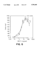
- FIG. 6 is a response curve of wild type MCP-1 and FLAG epitope tagged MCP-1 proteins.
- FIG. 7 is a chart of the mutations prepared and the specific chemotaxis activity of the mutants.
- FIG. 8 is a dose response curve for MCP-1 inhibition of four MCP-1 inhibitors.
- FIG. 9 illustrates the inhibition of 7ND on MCP-1 monocyte chemotaxis.
- the invention relates to mutant chemokines comprising an N-terminal deletion mutation of the corresponding chemokine, wherein the mutant chemokine inhibits binding and/or activation of a chemokine receptor responsive to the corresponding, endogenous chemokine.
- the chemokine can be a C--C or a C--X--C chemokine.
- the chemokine can be, for example, MCP-1, MCP-2, MCP-3, MIP-1 ⁇ , MIP-1 ⁇ , RANTES, platelet factor 4, IP-10, GRO ⁇ , GRO ⁇ , GRO ⁇ , NAP-2, ENA-78, CTAP-III, MIG and I-309.
- the mutant chemokine of the present invention does not include N-terminal deletion mutations of interleukin-8.
- the N-terminal region, the chemokine conserved amino acids therein or a significant portion thereof is deleted.
- amino acids between about 1 to about 13, about 1 to about 10 or about 2 to about 7 of the corresponding chemokine are deleted.
- the mutant chemokine can additionally contain other mutations as well.
- one or more amino acids of the chemokine can additionally be deleted, substituted or added.
- the additional mutation can, for example, enhance the inhibitory activity of the mutant cytokine.
- the mutant chemokine possesses at least about 85% and preferably at least about 95% homology with the corresponding endogenous chemokine.
- the additional mutation is at the 28-tyrosine and/or 24-arginine, such as the substitution of these amino acids with, for example, aspartate and phenylalanine, respectively.
- the mutant chemokines of the present invention are capable of inhibiting one or more activities of the corresponding endogenous chemokine, including binding and/or activating a responsive receptor.
- responsive receptor it is meant a receptor which is activated by the chemokine. Chemokines have been reported to bind and activate multiple receptors. In a preferred embodiment, the responsive receptor is that which is most highly activated by the chemokine. For example, in the case of MCP-1, the preferred receptor is the MCP-1 receptor.
- FX2 still preferentially complexed with 125 I-MCP-1, although a small amount of 125 I-IL-8 co-precipitated with FX2 using anti-FLAG antibody.
- control IgG also precipitated a similar amount of 125 I-IL-8, suggesting that the specificity of homodimerization persisted at concentrations as high as 120 nM.
- FIG. 3 shows an example of a crosslinked MCP-1 preparation in which the relative contributions of monomeric and multimeric species, as determined by laser densitometry, were 3% monomers, 63% dimers, 21% trimers, and 13% tetrameres. Independent crosslinking experiments performed under the same conditions routinely produced similar product distributions.
- This material was tested for monocyte chemoattractant activity, and using a molecular weight based on the abundance of each species, its EC 50 was approximately 400 pM, the same as the specific activity of non-crosslinked MCP-1. (Matsushima, K. et al., J. Exp. Med. 169:1485-1490 (1989); Yashimura, T. et al., J. Exp. Med. 169:1449-1459 (1989)).
- the data of FIG. 3 indicate that multimers of MCP-1, including dimers, are fully active as chemoattractants, but they do not show that dimerization is necessary for activity.
- an MCP-1 variant was used which lacks amino acids 2-8 of the processed protein. This variant, called 7ND, is a potent and specific inhibitor of monocyte chemotaxis in response to MCP-1.
- FIG. 1 shows that anti-FLAG antibodies co-precipitated radiolabeled MCP-1 (but not RANTES or IL-8) along with 7ND, which contains the FLAG epitope.
- 7ND could inhibit wild-type MCP-1 activity by two possible mechanisms: either it is a competitive inhibitor for receptor binding, or it is a dominant negative.
- the crosslinked dimer was used to distinguish between these possibilities.
- FIG. 4 shows that 7ND inhibited monocyte chemotaxis in response to non-crosslinked MCP-1, but had no effect on crosslinked MCP-1. This indicates that 7ND exerts its effects as a dominant negative, and further suggests that the biologically active form of MCP-1 is a dimer.
- MCP-1 has been demonstrated to specifically form homodimers at physiological concentrations.
- Dimeric MCP-1 is fully active as a monocyte chemoattractant because the specific activity of chemically crosslinked MCP-1 was indistinguishable from non-crosslinked MCP-1.
- the need for dimerization was examined by using 7ND, an N-terminal deletion mutant of MCP-1 that inhibits the monocyte chemoattractant activity of non-crosslinked MCP-1.
- 7ND's inability to inhibit monocyte chemotaxis in response to crosslinked MCP-1 supports the conclusion that 7ND acts as a dominant negative mutant of MCP-1 which means, in turn, that MCP-1 must activate its receptor as a dimer.
- supportive evidence for dimerization came from the observation that crosslinking an inactive N-terminal FLAG insertional mutant restores its activity.
- the N-terminus and first ⁇ sheet may be essential because they interact directly with the receptor, and dimerization is necessary because two of these areas must bind to one receptor in order to generate a signal.
- the N-terminal insertion would prevent two N-termini from interacting with the receptor due to steric hindrance or charge repulsion (the FLAG epitope is essentially a polyanion of aspartic acids).
- Crosslinking, which occurs at the N-termini would then constrain the N-termini to fit into their binding region in the receptor.
- MCP-1 monomer-dimer equilibria Direct measurements of MCP-1 monomer-dimer equilibria suggest that the dissociation constant for the dimer is several logs above MCP-1's EC 50 (Paolini, J. F. et al., J. Immunol. 153:2704-2717 (1994)). These observations do not contradict the findings disclosed herein.
- the equilibrium in solution may not reflect the equilibrium at the receptor, since the binding site for MCP-1 may have a conformation that greatly favors dimerization.
- local concentrations of MCP-1 may be influenced by other molecules at the cell surface.
- chemokines interact with glycosaminoglycans which cover the surface of monocytes and endothelial cells (Webb, L. M. C.
- the extremely steep gradient may prevent aberrant leukocyte migration, and may enable cells to be precisely directed over extremely short distances, providing greater control and specificity for leukocyte trafficking.
- This mechanism also identifies a new drug target for inhibiting chemokine activity, namely the dimer interface.
- the published K d for monomer dissociation may be incorrect.
- the published values were derived using equilibrium sedimentation centrifugation, gel filtration chromatography, and chemical crosslinking. All of these techniques are somewhat indirect.
- the monomer affinities were measured directly herein by biospecific interaction analysis (BIAcoreTM from Pharmacia Biotechnology, Inc.) and found that the K d for monomer dissociation in MCP-1 homodimers is 84 pM. This value, determined by direct real-time observation, indicates that at physiological concentrations, most MCP-1 will be present as dimers, and places the application of dominant negative inhibition, already established empirically, on firm theoretical grounds.
- the invention further relates to the use of mutant chemokines which inhibit the activity of the endogenous or corresponding cytokine in vivo or in vitro.
- the mutant chemokine can be added in a sample (such as a blood sample) to be subjected to an assay to selectively inhibit a targeted chemokine.
- the assay can accurately measure the chemoattractant or other activity of the targeted or different chemokine.
- the mutant chemokine can be added to assays such as those described in Falk et al., J. Immunol. Methods 33:239-247 (1980) or in Westwick, J. "Cytokines, a practical approach," F. R. Balkwill, ed., 1991, IRL Press, Oxford, pp. 197-204.
- the mutant chemokine is administered to a mammal, such as an individual or a patient, for the treatment of a chemokine mediated disease, such as inflammation, auto-immune disease and coronary artery restenosis.
- a chemokine mediated disease such as inflammation, auto-immune disease and coronary artery restenosis.
- Chemokines are implicated in the attracting leukocytes, such as monocytes, to damaged coronary arteries, such as the damage caused by coronary artery angioplasty, and are involved in causing restenosis.
- Administration of inhibitors to the recruitment of leukocytes will be useful in preventing coronary artery restenosis, autoimmune diseases, such as rheumatoid arthritis, inflammatory bowel disease and multiple sclerosis, chronic pulmonary inflammation, such as pulmonary fibrosis, and other conditions linked to the recruitment of leukocytes, such as monocytes.
- the derivatives of the claimed invention can be administered alone or in a suitable pharmaceutical composition.
- Modes of administration are those generally known in the art, such as parenteral application and the administration of a recombinant host cell, DNA or infectious vector which, upon administration, will express the protein in vivo. See, e.g. U.S. Pat. No. 5,399,346 to Anderson et al., which is incorporated herein by reference.
- Suitable pharmaceutical carriers include, but are not limited to water, salt solutions, alcohols, polyethylene glycols, gelatin, carbohydrates such as lactose, amylose or starch, magnesium stearate, talc, silicic acid, viscous paraffin, fatty acid esters, hydroxymethylcellulose, polyvinyl pyrrolidone, etc.
- the pharmaceutical preparations can be sterilized and if desired, mixed with auxiliary agents, e.g., lubricants, preservatives, stabilizers, wetting agents, emulsifiers, salts for influencing osmotic pressure, buffers, coloring, and/or aromatic substances and the like which do not deleteriously react with the active compounds. They can also be combined where desired with other active agents, e.g., enzyme inhibitors, to reduce metabolic degradation.
- auxiliary agents e.g., lubricants, preservatives, stabilizers, wetting agents, emulsifiers, salts for influencing osmotic pressure,
- injectable, sterile solutions preferably oily or aqueous solutions, as well as suspensions, emulsions, or implants, including suppositories.
- Ampoules are convenient unit dosages.
- the actual amounts of the MCP-1 derivative in a specific case will vary according to the specific compound being utilized, the particular composition formulated, the mode of administration and the age, weight and condition of the patient, for example. Dosages for a particular patient can be determined by one of ordinary skill in the art using conventional considerations, (e.g. by means of an appropriate, conventional pharmacological protocol).
- the invention also relates to the isolated DNA, encoding the mutant chemokine described herein, vectors and recombinant host cells containing the same.
- the isolated DNA can be the endogenous genomic or cDNA encoding the chemokine, deleting the codons corresponding to the amino acids deleted in the mutant chemokine. Also envisioned are silent mutations thereof (wherein one or more base pairs are modified, substituted or mutated such that the encoded protein sequence is the same).
- the isolated DNA can hybridize to, preferably under stringent conditions, the corresponding genomic or cDNA molecule encoding the endogenous chemokine.
- the isolated DNA is at least about 85% or, more preferably, at least about 95%, homologous to the corresponding genomic or cDNA sequence.
- the invention further relates to vectors containing the isolated DNA.
- vectors include infections (such as retroviral, adenoviral and vaccinia vectors, for example) and non-infections vectors.
- infections such as retroviral, adenoviral and vaccinia vectors, for example
- non-infections vectors such as retroviral, adenoviral and vaccinia vectors, for example.
- the vectors of the claimed invention can be readily prepared employing known techniques (such as, Ausubel et al. Current Protocols in Molecular Biology, Greene Publishing Assoc. and Wiley-Interscience (1989) and Sambrook et al., Molec. Cloning, Cold Spring Harbor Press (1989)) from commercially available starting materials.
- the vectors generally contain a suitable promoter (such as, a constitutive or inducible promoter recognized by the recipient host cell) and, optionally, a selectable marker, such that, upon insertion of the DNA into the host cell, the cell can express and, preferably, secrete the mutant chemokine.
- a suitable promoter such as, a constitutive or inducible promoter recognized by the recipient host cell
- a selectable marker such that, upon insertion of the DNA into the host cell, the cell can express and, preferably, secrete the mutant chemokine.
- Promoters and selectable markers are well known in the art and can, generally, be obtained commercially.
- the promoter is the endogenous chemokine promoter.
- vectors can be used directly for the administration to an individual for in vivo expression of a therapeutically effective amount of the mutant chemokine or can be used to deliver the isolated DNA to a host cell in vitro.
- the cell can be transfected (e.g., by calcium phosphate precipitation, electroporation or microinjection) or infected (e.g., by retroviral, adenoviral or vaccinia infection) employing well known techniques in the art, as described in the references cited above.
- Suitable host cells include commercially available cells, e.g., mammalian cell lines, such as Chinese hamster ovary cells (CHO cells) and human cell lines or TIL cells.
- the recombinant cells of the present invention thus made include the recombinant DNA encoding the mutant chemokine under the control of a suitable promoter and, preferably, a selectable marker.
- the recombinant cells thus obtained can then be selected, cloned and/or cultured in vitro and/or administered to the individual for in vivo protein expression, as described above.
- the recombinant host cells are cultured in vitro, thereby expressing and secreting the mutant chemokine which can then be purified to the desired degree by methods generally known in the art.
- pGEM-hJE34 human MCP-1 cDNA in pGEM-7 (Promega, Wisconsin)
- recombinant PCR was used to insert 30 nucleotides immediately 5' to the termination codon (position 366) encoding the FLAG epitope (Prickett, (1989)) with a two glycine spacer.
- Sequence analysis of the resulting cDNA revealed no other base alterations, and the entire cDNA was cloned into the expression vector pmt21 (Genetics Institute) to yield a plasmid designated pFX2.
- COS cells were suspended in serum-free Dulbecco's modified Eagle's medium (DMEM) at 5 ⁇ 10 6 cells/ml.
- DMEM Dulbecco's modified Eagle's medium
- Four ⁇ 10 6 cells (0.8 ml) were placed in an electroporation cuvette with a 0.4 cm gap, 10 ⁇ g plasmid DNA was added, and the cells were electroporated at 0.28 kV, 960 ⁇ F (yielding a time constant of 10-13 msec).
- Cells were allowed to recover in the cuvette at room temperature for 10 minutes, then plated in two 100 mm dishes in DMEM with 10% bovine calf serum. After 24 hours, medium was changed to serum-free DMEM. After an additional 24 hours, conditioned medium was collected, cells and debris were removed by centrifugation, and the medium was stored at -70° C.
- Wild-type MCP-1 was also produced by COS cell transfection, as described above, and obtained from Genzyme Corp. (Boston, Mass.).
- 125 I-radiolabeled MCP-1, IL-8, and RANTES were purchased from NEN/DuPont (Boston, Mass.) and all had specific activities of 2200 Ci/mmol.
- FX2 C-terminal epitope-tagged MCP-1
- N-terminal epitope-tagged MCP-1 N-terminal epitope-tagged MCP-1
- 7ND N-terminal MCP-1 deletion mutant
- Conditioned medium from transfected COS cells was boiled in sample buffer and subjected to electrophoresis through a 15% polyacrylamide gel in SDS. In some cases, medium was first concentrated using a Centricon-10 device (Amicon, Danvers, Mass.).
- Proteins were electrophoretically transferred to nitrocellulose and probed either with an anti-FLAG M1 monoclonal antibody (International Biotechnologies, Inc., New Haven, Conn.) or with rabbit anti-MCP-1 antiserum.
- an anti-FLAG M1 monoclonal antibody International Biotechnologies, Inc., New Haven, Conn.
- rabbit anti-MCP-1 antiserum rabbit anti-MCP-1 antiserum.
- Blots were developed using the appropriate horseradish peroxidase-conjugated secondary antibody and substrate solution (Vector Laboratories, Burlingame, Calif.).
- MCP-1 was expressed as a FLAG fusion protein in E. coli using the FLAG Biosystem and purified using an anti-FLAG affinity column (International Biotechnologies, Inc., New Haven, Conn.).
- Known amounts of pure FLAG-MCP-1 fusion protein were included in every immunoblot for MCP-1 produced by COS cells. Immunoblots were then analyzed by laser densitometry (Pharmacia, Piscataway, N.J.) and the mass of each COS cell-produced protein was determined by reference to the FLAG-MCP-1 fusion standard. The amount of COS cell supernatant loaded in each well generated an immunoblot signal within the linear response range of the laser densitometer. When possible, samples were quantitated using both anti-FLAG and anti-MCP-1 antibodies. There were no disparities between amounts of protein determined with the two antibodies except when one of the epitopes was absent.
- each of the mutations constructed, illustrated in FIG. 7, were tested for monocyte chemotaxis activity in the same manner.
- the mutations were constructed in three regions, the N-terminus, the first inter-cysteine loop, and the C-terminus.
- insertion of the FLAG epitope immediately after the N-terminal glutamine destroyed the protein's monocyte chemoattractant activity (FX3).
- FX3 monocyte chemoattractant activity
- deletion of amino acids 2-8 also yielded an inactive protein.
- the two charged amino acids were changed in this region (aspartate-3 and asparagine-6) to alanine. Changing aspartate-3 significantly reduced the activity of the protein, while changing asparagine-6 yielded a protein that retained 52.9% of wild-type MCP-1's activity.
- Mutated MCP-1 derivatives were tested for their ability to inhibit monocyte chemotaxis in vitro in response to non-mutated MCP-1.
- FX2 a fixed concentration of medium conditioned by COS cells expressing mutant proteins
- FIG. 8 shows that Y28D and 7ND were able to inhibit monocyte chemotaxis to FX2 by approximately 50% at the highest ratios tested.
- Y28D was slightly more effective than 7ND on a molar basis with an ID 50 corresponding to ⁇ 300 ng/ml in the presence of 10 ng/ml wild-type MCP-1.
- R24F reduced MCP-1's chemoattractant effect to 69% of control at a molar ratio of 1:1.
- R24F itself had monocyte chemoattractant activity of only 5% of wild-type, addition of more R24F beyond a 1:1 molar ratio with wild-type led to increasing chemoattractant activity.
- D3A reduced MCP-1's activity to 80% of control at a molar ratio of 10:1, but since it also has chemoattractant activity, higher molar ratios showed loss of inhibition. Inhibitory activity was specific for MCP-1 since the mutations did not inhibit monocyte chemotaxis in response to formyl-methionyl-leucyl-phenylalanine (fMLP).
- fMLP formyl-methionyl-leucyl-phenylalanine
- 7ND was purified and added to a constant amount of wild-type MCP-1.
- FIG. 9 shows that 7ND could completely inhibit monocyte chemotaxis in response to 5 mg/ml wild-type MCP-1 with an IC 50 that occurred at a molar ratio of 75:1.
- the N-terminal deletion mutant, 7ND can fully inhibit the chemoattractant activity of MCP-1.
- 125 I-radiolabeled chemokines were mixed with epitope-tagged chemokines in PBSA and incubated at 4° C. for 1 hour, then 10 ⁇ g anti-FLAG M2 antibody (International Biotechnologies, Inc., New Haven, Conn.) or murine IgG (Sigma, St. Louis, Mo.) were added. The mixture was incubated at 4° C. for 1 hour with gentle rotation, after which 50 ⁇ l of staphylococcal protein A beads (BioRad, Richmond, Calif.) was added and the incubation continued for an additional hour.
- chemokine Five ⁇ g of chemokine was resuspended in 445 ⁇ l PBS at 4° C. and 5 ⁇ l of a 20 mM solution of DSS 1 (Pierce, Rockford, Ill.) was added. The mixture was incubated at 4° C. for 45 minutes, then concentrated and cleared of crosslinker by centrifugation using a Centricon-10 device (Amicon, Danvers, Mass.). Crosslinked products were fractionated by SDS-PAGE on 15% polyacrylamide gels. After silver staining, the proportion of monomers and multimeric species was determined by laser densitometry (Pharmacia, Piscataway, N.J.).
- Monocyte chemoattraction assays were performed on human peripheral blood mononuclear cells using a multi-well modified Boyden chamber device as described above. The results are set forth in FIGS. 3, 4, and 5.
Abstract
The invention relates to mutant chemokines and pharmaceutical compositions thereof, wherein the mutant inhibits the corresponding endogenous chemokine from binding or activating a responsive receptor. The claimed mutant chemokines can be administered to a patient for the treatment or prophylaxis of a chemokine-mediated disease. The invention further relates to isolated DNAs encoding the mutant chemokines, vectors and recombinant host cells containing the same.
Description
The invention described herein was supported in whole or in part by Grant No.: CA53091 from the National Institutes of Health. The Government has certain rights in the invention.
This application is a division of application Ser. No. 08/455,629 filed May 31, 1995, which is a Continuation-in-Part of Ser. No. 08/330,218 filed Oct. 27, 1994, now U.S. Pat. No. 5,459,128, which is a Continuation-in-Part of Ser. No. 08/152,301 filed Nov. 12, 1993, now abandoned.
Chemokines share a high degree of amino acid sequence similarity (Oppenheim, J. J. et al., Annu. Rev. Immunol. 9:617-648 (1991); Rollins, B. J., In Mertelsmann, R., and F. Herrmann (ed.), Hematopoietic Growth Factors in Clinical Applications, pg. 257-280, 2nd ed. Marcel Dekker, Inc., New York (1994); Schall, T. J., Cytokine 3:165-183 (1991)). Chemokines have 4 cysteines in conserved positions, but two subfamilies can be distinguished based on the absence or presence of an intervening amino acid between the two cysteines nearest the N-termini of the proteins. The former subfamily is denoted C--C and includes monocyte chemoattractant protein-1 (MCP-1), which is a potent chemoattractant for monocytes and T lymphocytes (Rollins, Molecular and Cellular Biology, 9(11):4687-4695 (1989); Carr M. W et al., Proc. Natl Acad. Sci USA 91:3653-3656 (1994); Matsushima, K., et al., J. Exp. Med. 169:1485-1490 (1989); Yoshimura, T. et al., J. Exp. Med 169:1449-1459 (1989)) and induces histamine release from basophils (Bischoff, S. C. et al., J. Exp. Med. 125:1271-1275 (1992); Kuna, P. et al., J. Exp. Med. 175:489-493 (1992)). The latter subfamily is denoted C--X--C and includes the neutrophil chemoattractant, IL-8.
Solved chemokine structures, including those of IL-8, platelet factor 4 (PF4), MIP-1β and MCP-1, have been studied and the tertiary structures of these chemokine monomers are considerably similar. However, under conditions required for crystallization or NMR analysis, these chemokines form dimers, and despite the nearly identical structures of their monomeric subunits, the structures of the dimers differ appreciably. For example, the IL-8 dimer is compact (Baldwin, E. T. et al., Proc. Natl. Acad. Sci. USA 88:502-506 (1991) and Clore, G. M. et al., Biochemistry 29:1689-1696 (1990)), while the MIP-1β dimer (another C--C chemokine) is elongated and cylindrical (Lodi, P. J. et al., Science 263:1762-1767 (1994)). The structure of the MCP-1 dimer is similar to that of MIP-1β (Handel et al., Chemotactic Cytokines, IBC USA Conferences, Inc., Washington, D.C. (1994)).
Chemokines are proinflammatory cytokines that are chemoattractants and activators of specific types of leukocytes and have been identified as playing a significant role in many disease states.
Aberrant expression of MCP-1, for example, occurs in a variety of diseases that feature a monocyte-rich inflammatory component, such as atherosclerosis (Nelken, N. A. et al., J. Clin. Invest. 88:1121-1127 (1991); Yla-Herttuala, S. et al., Proc. Natl. Acad. Sci. USA 88:5252-5256 (1991)) and rheumatoid arthritis (Koch, A. E. et al., J. Clin. Invest. 90:772-779 (1992)). Rational design of chemokine inhibitors to treat these diseases depends on understanding how the chemokine interacts with its receptor, in the case of MCP-1, a seven-transmembrane spanning (7-TMS), G protein-coupled receptor (Charo, I. F. et al., Proc. Natl. Acad. Sci. USA 91:2752-2756 (1994)). A critical and contentious issue about chemokine:receptor interaction is whether the ligands bind to their receptors as dimers. Estimates for the affinity of chemokine dimerization place the Kd for monomer association several logs above the EC50 for biological activity and the Kd for receptor binding (Paolini, J. F. et al., J. Immunol. 1531:2704-2717 (1994); Rajarathnam, K., et al., Science 264:90-92 (1994)). In addition, variant IL-8 proteins that are unable to dimerize in solution have full biological activity on neutrophils (Rajarathnam, K., et al., Science 264:90-92 (1994)).
Thus, there is a need to further elucidate the chemokine-receptor interaction and develop inhibitors to treat chemokine-mediated disease.
The invention relates to the surprising discovery that N-terminal deletions of chemokines are potent dominant negative inhibitors of chemokine-receptor interaction by forming heterodimers with the corresponding endogenous monomer of the chemokine. Thus, the invention relates to chemokine N-terminal deletion mutations, their use to form heterodimers with the corresponding endogenous chemokine monomer, in vivo or in vitro, and their use to treat chemokine-mediated diseases.
FIG. 1A illustrates a dimerization assay of one ng 125 I-radiolabeled MCP-1 or RANTES (at the same specific activity) mixed with 10 ng non-radiolabeled FX2 (C-terminal FLAG epitope-tagged MCP-1) or 7ND (C-terminal FLAG epitope-tagged mutant MCP-1) as indicated in a total volume of 0.5 ml. Mixtures were subjected to immunoprecipitation using anti-FLAG antibody or murine IgG and analyzed by SDS-PAGE. FIG. 1B is the same as 1A except that 125 I-radiolabeled IL-8 was used instead of RANTES, and 600 ng FX2 or 7ND was used.
FIGS. 2A, 2B and 2C are illustrations of gel filtration chromatography of MCP-1 variants on Superdex-75. The column was calibrated with standard proteins and Mr 's were determined for each species. FIG. 2A FX2 (Mr 30,600); FIG. 2B 7ND (Mr 15,500). FIG. 2C N-terminal FLAG epitope-tagged MCP-1 (Mr 30,600). Molecular weight markers include bovine γ-globulin (γG, Mr 158,000), chicken ovalbumin (Ova, Mr 44,000), equine myoglobin (Mb, Mr 17,000), and vitamin B12 (B12, Mr 1350).
FIG. 3 illustrates crosslinked MCP-1 tested in an in vitro monocyte chemoattraction assay. Molar concentrations were determined by assuming an average molecular weight based on the proportional contribution of each molecular species to the crosslinked preparation. Error bars indicate the standard error of measurements in duplicate wells; this experiment is representative of three.
FIG. 4 shows increasing amounts of purified 7ND added to constant amounts of uncrosslinked MCP-1 (10 ng/ml; 0.9 nM; solid line) or crosslinked MCP-1 (19 ng/ml; 0.7 nM; dotted line). The mixtures were then tested for their ability to attract monocytes in vitro relative to their chemoattractant activity in the absence of 7ND. Error bars indicate the standard error of measurements in duplicate wells; this experiment is representative of three.
FIG. 5 is a non-crosslinked (dotted line) and crosslinked (solid line) N-terminal FLAG-MCP-1 tested in monocyte chemoattraction assays. Error bars indicate the standard error of measurements in duplicate wells; this experiment is representative of two.
FIG. 6 is a response curve of wild type MCP-1 and FLAG epitope tagged MCP-1 proteins.
FIG. 7 is a chart of the mutations prepared and the specific chemotaxis activity of the mutants.
FIG. 8 is a dose response curve for MCP-1 inhibition of four MCP-1 inhibitors.
FIG. 9 illustrates the inhibition of 7ND on MCP-1 monocyte chemotaxis.
The invention relates to mutant chemokines comprising an N-terminal deletion mutation of the corresponding chemokine, wherein the mutant chemokine inhibits binding and/or activation of a chemokine receptor responsive to the corresponding, endogenous chemokine. The chemokine can be a C--C or a C--X--C chemokine. The chemokine can be, for example, MCP-1, MCP-2, MCP-3, MIP-1α, MIP-1β, RANTES, platelet factor 4, IP-10, GROα, GROβ, GROγ, NAP-2, ENA-78, CTAP-III, MIG and I-309. The mutant chemokine of the present invention does not include N-terminal deletion mutations of interleukin-8.
In a preferred embodiment, the N-terminal region, the chemokine conserved amino acids therein or a significant portion thereof is deleted. For example, amino acids between about 1 to about 13, about 1 to about 10 or about 2 to about 7 of the corresponding chemokine are deleted.
The mutant chemokine can additionally contain other mutations as well. For example, one or more amino acids of the chemokine can additionally be deleted, substituted or added. the additional mutation can, for example, enhance the inhibitory activity of the mutant cytokine. In a particularly preferred embodiment, the mutant chemokine possesses at least about 85% and preferably at least about 95% homology with the corresponding endogenous chemokine.
In one embodiment, wherein the chemokine is MCP-1, the additional mutation is at the 28-tyrosine and/or 24-arginine, such as the substitution of these amino acids with, for example, aspartate and phenylalanine, respectively.
The mutant chemokines of the present invention are capable of inhibiting one or more activities of the corresponding endogenous chemokine, including binding and/or activating a responsive receptor. By "responsive receptor" it is meant a receptor which is activated by the chemokine. Chemokines have been reported to bind and activate multiple receptors. In a preferred embodiment, the responsive receptor is that which is most highly activated by the chemokine. For example, in the case of MCP-1, the preferred receptor is the MCP-1 receptor.
The experimentation described herein indicates that chemokine monomers, such as MCP-1, interact specifically to form homodimers at physiological concentrations. These studies used a C-terminal FLAG epitope-tagged variant of MCP-1 (called FX2) that has full biological activity (Zhang, Y. J. et al., J. Biol. Chem. 269:15918-15924 (1994)). When FX2 was mixed with 125 I-MCP-1 at a final chemokine concentration of 2 nM, an anti-FLAG antibody co-precipitated radiolabeled MCP-1 (FIG. 1A). FX2's apparent molecular weight on gel chromatography is 30,600 kD (FIG. 2A), similar to that of unmodified MCP-1 (Rollins, B. J. et al., Am. J. Pathol. 136:1229-1233 (1990)), and double its apparent molecular weight on SDS-PAGE (Zhang, Y. J. et al., J. Biol. Chem. 269:15918-15924 (1994)). FX2's molecular weight is also double the apparent molecular weight of 7ND, an N-terminal deletion mutant that does not form dimers (see below) and whose predicted molecular weight is only 680 daltons less than monomeric FX2 (FIG. 2B). These observations indicate that MCP-1 forms dimers at physiologically relevant concentrations. Serial immune precipitations showed that approximately 65% of radiolabeled MCP-1 was complexed with FX2 at a concentration of 2 nM.
The specificity of homodimerization was reflected by the fact that FX2 did not form complexes with 125 I-RANTES (a C--C chemokine like MCP-1) or 125 I-IL-8 (a C--X--C chemokine), both radiolabeled to the same specific activity as 125 I-MCP-1. In FIG. 1A, no detectable interaction occurred between FX2 and 125 I-RANTES at a physiological chemokine concentration of 2 Nm. In FIG. 1B, we increased FX2's concentration 60-fold. Under these conditions, FX2 still preferentially complexed with 125 I-MCP-1, although a small amount of 125 I-IL-8 co-precipitated with FX2 using anti-FLAG antibody. However, control IgG also precipitated a similar amount of 125 I-IL-8, suggesting that the specificity of homodimerization persisted at concentrations as high as 120 nM.
Having determined that MCP-1 forms dimers, MCP-1 dimers were demonstrated to be biologically active by using the bifunctional crosslinker disuccinimidyl suberate (DSS) to construct stably dimerized molecules. FIG. 3 shows an example of a crosslinked MCP-1 preparation in which the relative contributions of monomeric and multimeric species, as determined by laser densitometry, were 3% monomers, 63% dimers, 21% trimers, and 13% tetrameres. Independent crosslinking experiments performed under the same conditions routinely produced similar product distributions. This material was tested for monocyte chemoattractant activity, and using a molecular weight based on the abundance of each species, its EC50 was approximately 400 pM, the same as the specific activity of non-crosslinked MCP-1. (Matsushima, K. et al., J. Exp. Med. 169:1485-1490 (1989); Yashimura, T. et al., J. Exp. Med. 169:1449-1459 (1989)).
The data of FIG. 3 indicate that multimers of MCP-1, including dimers, are fully active as chemoattractants, but they do not show that dimerization is necessary for activity. To demonstrate this, an MCP-1 variant was used which lacks amino acids 2-8 of the processed protein. This variant, called 7ND, is a potent and specific inhibitor of monocyte chemotaxis in response to MCP-1.
Two experiments indicated that 7ND is a dominant negative. First, 7ND specifically forms heterodimers with wild-type MCP-1. FIG. 1 shows that anti-FLAG antibodies co-precipitated radiolabeled MCP-1 (but not RANTES or IL-8) along with 7ND, which contains the FLAG epitope. Second, 7ND could inhibit wild-type MCP-1 activity by two possible mechanisms: either it is a competitive inhibitor for receptor binding, or it is a dominant negative. The crosslinked dimer was used to distinguish between these possibilities. FIG. 4 shows that 7ND inhibited monocyte chemotaxis in response to non-crosslinked MCP-1, but had no effect on crosslinked MCP-1. This indicates that 7ND exerts its effects as a dominant negative, and further suggests that the biologically active form of MCP-1 is a dimer.
Finally, the need for dimerization was demonstrated in a different way by examining an inactive MCP-1 variant in which the FLAG epitope was inserted immediately after the N-terminal glutamine. Although this variant appears to form homodimers by the criteria of its size on gel filtration chromatography (FIG. 2C) and its ability to co-precipitate with MCP-1 (data now shown), it has only 0.2% of wild-type chemoattractant activity (Zhang, Y. J. et al., J. Biol. Chem. 269:15918-15924 (1994)). However, because it has a full-length N-terminus, DSS can crosslink these homodimers, and crosslinking restores its ability to attract monocytes. Thus, physically constraining the altered N-termini of N-terminal FLAG MCP-1 dimers produces monocyte chemoattractant activity, again indicating that dimeric MCP-1 activates its receptor.
Using co-precipitation, MCP-1 has been demonstrated to specifically form homodimers at physiological concentrations. Dimeric MCP-1 is fully active as a monocyte chemoattractant because the specific activity of chemically crosslinked MCP-1 was indistinguishable from non-crosslinked MCP-1. The need for dimerization was examined by using 7ND, an N-terminal deletion mutant of MCP-1 that inhibits the monocyte chemoattractant activity of non-crosslinked MCP-1. However, 7ND's inability to inhibit monocyte chemotaxis in response to crosslinked MCP-1 supports the conclusion that 7ND acts as a dominant negative mutant of MCP-1 which means, in turn, that MCP-1 must activate its receptor as a dimer. Finally, supportive evidence for dimerization came from the observation that crosslinking an inactive N-terminal FLAG insertional mutant restores its activity.
Taken together the results suggest a model in which dimeric MCP-1 binds to its receptor. The N-terminus and one face of the first predicted β sheet are essential for MCP-1's chemoattractant activity. Without being limited to a particular mechanism of action, these observations can be combined with our current results in three different models. First, mutating the N-terminus and first β sheet can destroy activity simply by preventing dimerization, and dimerization per se can be essential for activity because apposing non-contiguous regions of the proteins (not at the N-terminus) creates the active ligand. However, this model would not explain why 7ND/MCP-1 heterodimers are inactive. Alternatively, the N-terminus and first β sheet may be essential because they interact directly with the receptor, and dimerization is necessary because two of these areas must bind to one receptor in order to generate a signal. This would explain restoration of crosslinked N-terminal FLAG-MCP-1's activity by forcing its dimerization. The N-terminal insertion would prevent two N-termini from interacting with the receptor due to steric hindrance or charge repulsion (the FLAG epitope is essentially a polyanion of aspartic acids). Crosslinking, which occurs at the N-termini, would then constrain the N-termini to fit into their binding region in the receptor. This model would also explain 7ND's dominant negative activity: 7ND/MCP-1 heterodimers could dock in the receptor but could not activate it because one of the two required N-termini would be missing. There is precedent for peptide ligands that interact with 7-TMS receptors at multiple sites (Siciliano, S. J. et al., Proc. Natl. Acad. Sci. USA 91:1214-1218 (1994)). In a third model, MCP-1 dimers might bring two receptors together for their activation, by analogy to tyrosine kinase receptors. Our data are consistent with this model, but there is no evidence yet to support 7-TMS receptor interaction.
Direct measurements of MCP-1 monomer-dimer equilibria suggest that the dissociation constant for the dimer is several logs above MCP-1's EC50 (Paolini, J. F. et al., J. Immunol. 153:2704-2717 (1994)). These observations do not contradict the findings disclosed herein. First, the equilibrium in solution may not reflect the equilibrium at the receptor, since the binding site for MCP-1 may have a conformation that greatly favors dimerization. Second, local concentrations of MCP-1 may be influenced by other molecules at the cell surface. For example, chemokines interact with glycosaminoglycans which cover the surface of monocytes and endothelial cells (Webb, L. M. C. et al., Proc. Natl. Acad. Sci. USA 90:7158-7162 (1993)). As chemokines are added to monocytes in vitro or as they appear at the endothelial cell surface in vivo, their binding to glycosaminoglycans could create extremely high local concentrations that might exceed the Kd for dimerization, thereby providing a mechanism for the modulation of their leukocyte chemoattractant activity. Thus, where the Kd for MCP-1 dimerization is higher than the Kd for receptor binding, then the appearance of active MCP-1 depends on achieving high local concentrations. This effectively changes the shape of the chemoattractant gradient to one that is much steeper near its source and falls off more rapidly with distance than it otherwise would. "Self-inactivation" would then occur rapidly with increasing distance from the secreting cell. The extremely steep gradient may prevent aberrant leukocyte migration, and may enable cells to be precisely directed over extremely short distances, providing greater control and specificity for leukocyte trafficking. This mechanism also identifies a new drug target for inhibiting chemokine activity, namely the dimer interface.
Finally, the published Kd for monomer dissociation may be incorrect. The published values were derived using equilibrium sedimentation centrifugation, gel filtration chromatography, and chemical crosslinking. All of these techniques are somewhat indirect. The monomer affinities were measured directly herein by biospecific interaction analysis (BIAcore™ from Pharmacia Biotechnology, Inc.) and found that the Kd for monomer dissociation in MCP-1 homodimers is 84 pM. This value, determined by direct real-time observation, indicates that at physiological concentrations, most MCP-1 will be present as dimers, and places the application of dominant negative inhibition, already established empirically, on firm theoretical grounds.
Thus, the invention further relates to the use of mutant chemokines which inhibit the activity of the endogenous or corresponding cytokine in vivo or in vitro.
For example, the mutant chemokine can be added in a sample (such as a blood sample) to be subjected to an assay to selectively inhibit a targeted chemokine. In such a method, the assay can accurately measure the chemoattractant or other activity of the targeted or different chemokine. Thus, the mutant chemokine can be added to assays such as those described in Falk et al., J. Immunol. Methods 33:239-247 (1980) or in Westwick, J. "Cytokines, a practical approach," F. R. Balkwill, ed., 1991, IRL Press, Oxford, pp. 197-204.
In a particularly preferred embodiment, the mutant chemokine is administered to a mammal, such as an individual or a patient, for the treatment of a chemokine mediated disease, such as inflammation, auto-immune disease and coronary artery restenosis.
Chemokines are implicated in the attracting leukocytes, such as monocytes, to damaged coronary arteries, such as the damage caused by coronary artery angioplasty, and are involved in causing restenosis. Administration of inhibitors to the recruitment of leukocytes will be useful in preventing coronary artery restenosis, autoimmune diseases, such as rheumatoid arthritis, inflammatory bowel disease and multiple sclerosis, chronic pulmonary inflammation, such as pulmonary fibrosis, and other conditions linked to the recruitment of leukocytes, such as monocytes.
The derivatives of the claimed invention can be administered alone or in a suitable pharmaceutical composition. Modes of administration are those generally known in the art, such as parenteral application and the administration of a recombinant host cell, DNA or infectious vector which, upon administration, will express the protein in vivo. See, e.g. U.S. Pat. No. 5,399,346 to Anderson et al., which is incorporated herein by reference.
Suitable pharmaceutical carriers include, but are not limited to water, salt solutions, alcohols, polyethylene glycols, gelatin, carbohydrates such as lactose, amylose or starch, magnesium stearate, talc, silicic acid, viscous paraffin, fatty acid esters, hydroxymethylcellulose, polyvinyl pyrrolidone, etc. The pharmaceutical preparations can be sterilized and if desired, mixed with auxiliary agents, e.g., lubricants, preservatives, stabilizers, wetting agents, emulsifiers, salts for influencing osmotic pressure, buffers, coloring, and/or aromatic substances and the like which do not deleteriously react with the active compounds. They can also be combined where desired with other active agents, e.g., enzyme inhibitors, to reduce metabolic degradation.
For parenteral application, particularly suitable are injectable, sterile solutions, preferably oily or aqueous solutions, as well as suspensions, emulsions, or implants, including suppositories. Ampoules are convenient unit dosages.
It will be appreciated that the actual amounts of the MCP-1 derivative in a specific case will vary according to the specific compound being utilized, the particular composition formulated, the mode of administration and the age, weight and condition of the patient, for example. Dosages for a particular patient can be determined by one of ordinary skill in the art using conventional considerations, (e.g. by means of an appropriate, conventional pharmacological protocol).
The invention also relates to the isolated DNA, encoding the mutant chemokine described herein, vectors and recombinant host cells containing the same.
The isolated DNA can be the endogenous genomic or cDNA encoding the chemokine, deleting the codons corresponding to the amino acids deleted in the mutant chemokine. Also envisioned are silent mutations thereof (wherein one or more base pairs are modified, substituted or mutated such that the encoded protein sequence is the same). In a particularly preferred embodiment, the isolated DNA can hybridize to, preferably under stringent conditions, the corresponding genomic or cDNA molecule encoding the endogenous chemokine. In a more preferred embodiment, the isolated DNA is at least about 85% or, more preferably, at least about 95%, homologous to the corresponding genomic or cDNA sequence.
As stated above, the invention further relates to vectors containing the isolated DNA. Such vectors include infections (such as retroviral, adenoviral and vaccinia vectors, for example) and non-infections vectors. The vectors of the claimed invention can be readily prepared employing known techniques (such as, Ausubel et al. Current Protocols in Molecular Biology, Greene Publishing Assoc. and Wiley-Interscience (1989) and Sambrook et al., Molec. Cloning, Cold Spring Harbor Press (1989)) from commercially available starting materials.
The vectors generally contain a suitable promoter (such as, a constitutive or inducible promoter recognized by the recipient host cell) and, optionally, a selectable marker, such that, upon insertion of the DNA into the host cell, the cell can express and, preferably, secrete the mutant chemokine. Promoters and selectable markers are well known in the art and can, generally, be obtained commercially. In a preferred embodiment, the promoter is the endogenous chemokine promoter.
These vectors can be used directly for the administration to an individual for in vivo expression of a therapeutically effective amount of the mutant chemokine or can be used to deliver the isolated DNA to a host cell in vitro.
In the latter embodiment, the cell can be transfected (e.g., by calcium phosphate precipitation, electroporation or microinjection) or infected (e.g., by retroviral, adenoviral or vaccinia infection) employing well known techniques in the art, as described in the references cited above. Suitable host cells include commercially available cells, e.g., mammalian cell lines, such as Chinese hamster ovary cells (CHO cells) and human cell lines or TIL cells.
The recombinant cells of the present invention thus made include the recombinant DNA encoding the mutant chemokine under the control of a suitable promoter and, preferably, a selectable marker. The recombinant cells thus obtained can then be selected, cloned and/or cultured in vitro and/or administered to the individual for in vivo protein expression, as described above. In one embodiment, the recombinant host cells are cultured in vitro, thereby expressing and secreting the mutant chemokine which can then be purified to the desired degree by methods generally known in the art.
The invention is further specifically illustrated by the following examples.
Construction and expression of epitope-tagged MCP-1 and wild-type MCP-1
Starting with pGEM-hJE34 (human MCP-1 cDNA in pGEM-7 (Promega, Wisconsin)) as a template, recombinant PCR was used to insert 30 nucleotides immediately 5' to the termination codon (position 366) encoding the FLAG epitope (Prickett, (1989)) with a two glycine spacer. Sequence analysis of the resulting cDNA revealed no other base alterations, and the entire cDNA was cloned into the expression vector pmt21 (Genetics Institute) to yield a plasmid designated pFX2.
COS cells were suspended in serum-free Dulbecco's modified Eagle's medium (DMEM) at 5×10⁶ cells/ml. Four×106 cells (0.8 ml) were placed in an electroporation cuvette with a 0.4 cm gap, 10 μg plasmid DNA was added, and the cells were electroporated at 0.28 kV, 960 μF (yielding a time constant of 10-13 msec). Cells were allowed to recover in the cuvette at room temperature for 10 minutes, then plated in two 100 mm dishes in DMEM with 10% bovine calf serum. After 24 hours, medium was changed to serum-free DMEM. After an additional 24 hours, conditioned medium was collected, cells and debris were removed by centrifugation, and the medium was stored at -70° C.
Wild-type MCP-1 was also produced by COS cell transfection, as described above, and obtained from Genzyme Corp. (Boston, Mass.).
Construction and expression of MCP-1 mutations
Similar techniques as described above were used to insert the FLAG epitope and spacer immediately 3' to the codon for aspartate-3 in processed MCP-1 (nucleotide position 146); this expression plasmid was designated pFX3. Based on modeling considerations as well as structure/activity data from other chemokines (in particular IL-8), MCP-1 mutations in three regions were constructed: namely the N-terminus, the first inter-cysteine loop, and the C-terminal predicted α-helix (FIGS. 2 and 4). With pFX2 as template, recombinant PCR was used to insert termination codons or single amino acid changes. All mutations were confirmed by sequence analysis of both DNA strands.
125 I-radiolabeled MCP-1, IL-8, and RANTES were purchased from NEN/DuPont (Boston, Mass.) and all had specific activities of 2200 Ci/mmol. FX2 (C-terminal epitope-tagged MCP-1), an N-terminal epitope-tagged MCP-1, and 7ND (N-terminal MCP-1 deletion mutant) were prepared by expression of their cDNA's in Chinese hamster ovary cells (Rollins, B. J. and Sunday, M. E., Mol. Cell. Biol. 11:3125-3131 (1991) or E. coli (Zhang, Y. J. et al., J. Biol. Chem. 269:15918-15924 (1994)) and were purified by FLAG affinity purification as described (Zhang, Y. J. et al., J. Biol. Chem. 269:15918-15924 (1994)).
Immunoblotting
Conditioned medium from transfected COS cells was boiled in sample buffer and subjected to electrophoresis through a 15% polyacrylamide gel in SDS. In some cases, medium was first concentrated using a Centricon-10 device (Amicon, Danvers, Mass.).
Proteins were electrophoretically transferred to nitrocellulose and probed either with an anti-FLAG M1 monoclonal antibody (International Biotechnologies, Inc., New Haven, Conn.) or with rabbit anti-MCP-1 antiserum.
Blots were developed using the appropriate horseradish peroxidase-conjugated secondary antibody and substrate solution (Vector Laboratories, Burlingame, Calif.).
Quantitation of MCP-1 and its derivatives
MCP-1 was expressed as a FLAG fusion protein in E. coli using the FLAG Biosystem and purified using an anti-FLAG affinity column (International Biotechnologies, Inc., New Haven, Conn.). Known amounts of pure FLAG-MCP-1 fusion protein were included in every immunoblot for MCP-1 produced by COS cells. Immunoblots were then analyzed by laser densitometry (Pharmacia, Piscataway, N.J.) and the mass of each COS cell-produced protein was determined by reference to the FLAG-MCP-1 fusion standard. The amount of COS cell supernatant loaded in each well generated an immunoblot signal within the linear response range of the laser densitometer. When possible, samples were quantitated using both anti-FLAG and anti-MCP-1 antibodies. There were no disparities between amounts of protein determined with the two antibodies except when one of the epitopes was absent.
Monocyte chemotaxis assay
Human peripheral blood mononuclear cells were prepared from volunteers as described (Rollins, Blood, 78, pp 1112-1116 (1989)). Chemotaxis assays were performed using a multiwell chamber fitted with a polycarbonate filter having 5μ pores as described. Each COS cell supernatant was tested over a wide range of dilutions, and the concentration of monocyte chemoattractant activity (MCA) in each supernatant was defined as the inverse of the dilution giving half-maximal chemotactic response.
Wild-type MCP-1 and FX2 had similar dose response characteristics, FIG. 6. Using the quantitation results from immunoblotting, analysis of the dose response curves showed that the specific activity of FX2 is 408,000 U/ml while that of wild-type MCP-1 is 442,000 U/ml. Both values compared favorably with the specific activity determined for purified, eukaryotically-produced, recombinant MCP-1, suggesting that the epitope tag does not interfere with MCP-1's ability to attract monocytes in vivo.
Each of the mutations constructed, illustrated in FIG. 7, were tested for monocyte chemotaxis activity in the same manner. As set forth above, the mutations were constructed in three regions, the N-terminus, the first inter-cysteine loop, and the C-terminus. At the N-terminus, insertion of the FLAG epitope immediately after the N-terminal glutamine destroyed the protein's monocyte chemoattractant activity (FX3). Consistent with that finding was the fact that deletion of amino acids 2-8 also yielded an inactive protein. In an attempt to accomplish finer mapping, the two charged amino acids were changed in this region (aspartate-3 and asparagine-6) to alanine. Changing aspartate-3 significantly reduced the activity of the protein, while changing asparagine-6 yielded a protein that retained 52.9% of wild-type MCP-1's activity.
Next, four point mutations in the first intercysteine loop were constructed. Mutations of arginine-24 to phenylalanine, tyrosine-28 to aspartate, and arginine-30 to leucine all produced proteins with activities that were only a fraction of wild-type. However, mutation of another polar amino acid in the same region, namely serine-27, produced a protein with 60% of the activity of wild-type.
Finally, manipulations of the C-terminal α-helix produced proteins that were still able to signal, but had reduced potency compared to wild-type. Deletions of half (D2) or all (D1) of the α-helix yielded proteins with 17% and 11.3% of wild-type activity, respectively. In the predicted model structure of dimeric MCP-1, aspartate-68 projects into a predicted receptor binding cleft between two α-helices. Mutation of this amino acid to leucine had the same effect as deletion of the entire helix.
Competition for biological effects
Mutated MCP-1 derivatives were tested for their ability to inhibit monocyte chemotaxis in vitro in response to non-mutated MCP-1. To a fixed concentration of FX2, increasing amounts of medium conditioned by COS cells expressing mutant proteins were added. Only Y28D, R24F, 7ND, and D3A demonstrated an ability to inhibit wild type MCP-1. FIG. 8 shows that Y28D and 7ND were able to inhibit monocyte chemotaxis to FX2 by approximately 50% at the highest ratios tested. (The upper limit was dictated by the concentration of mutation protein in conditioned medium.) Y28D was slightly more effective than 7ND on a molar basis with an ID50 corresponding to ≈300 ng/ml in the presence of 10 ng/ml wild-type MCP-1. R24F reduced MCP-1's chemoattractant effect to 69% of control at a molar ratio of 1:1. Although R24F itself had monocyte chemoattractant activity of only 5% of wild-type, addition of more R24F beyond a 1:1 molar ratio with wild-type led to increasing chemoattractant activity. D3A reduced MCP-1's activity to 80% of control at a molar ratio of 10:1, but since it also has chemoattractant activity, higher molar ratios showed loss of inhibition. Inhibitory activity was specific for MCP-1 since the mutations did not inhibit monocyte chemotaxis in response to formyl-methionyl-leucyl-phenylalanine (fMLP).
7ND was purified and added to a constant amount of wild-type MCP-1. FIG. 9 shows that 7ND could completely inhibit monocyte chemotaxis in response to 5 mg/ml wild-type MCP-1 with an IC50 that occurred at a molar ratio of 75:1. Thus the N-terminal deletion mutant, 7ND, can fully inhibit the chemoattractant activity of MCP-1.
Immune precipitations
125 I-radiolabeled chemokines were mixed with epitope-tagged chemokines in PBSA and incubated at 4° C. for 1 hour, then 10 μg anti-FLAG M2 antibody (International Biotechnologies, Inc., New Haven, Conn.) or murine IgG (Sigma, St. Louis, Mo.) were added. The mixture was incubated at 4° C. for 1 hour with gentle rotation, after which 50 μl of staphylococcal protein A beads (BioRad, Richmond, Calif.) was added and the incubation continued for an additional hour. Beads were collected by centrifugation, then washed twice with RIPA buffer (50 mM Tris hydrochloride pH 7.5!, 150 mM NaCl, 0.1% SDS, 1% Triton X-100, 0.5% deoxycholate, 1 mM phenylmethylsulfonyl fluoride) and twice with 50 mM Tris hydrochloride (pH 7.5), 1M NaCl. Beads were resuspended in sample buffer containing SDS and β-mercaptoethanol, and then boiled. Released proteins were fractionated by SDS-PAGE through 15% polyacrylamide, and dried gels were analyzed by autoradiography. The results are shown in FIGS. 1A and 1B.
Gel filtration chromatography
Seven to 9 μg of MCP-1 or its derivatives were loaded on a 25 ml Superdex-75 column (Pharmacia, Piscataway, N.J.) which was developed in 50 mM sodium phosphate (pH 7.4), 150 mM NaCl at a flow rate of 0.5 ml/min. The column was calibrated in the same buffer using standards obtained from BioRad (Richmond, Calif.). The results are shown in FIGS. 2A, 2B and 2C.
Crosslinking
Five μg of chemokine was resuspended in 445 μl PBS at 4° C. and 5 μl of a 20 mM solution of DSS1 (Pierce, Rockford, Ill.) was added. The mixture was incubated at 4° C. for 45 minutes, then concentrated and cleared of crosslinker by centrifugation using a Centricon-10 device (Amicon, Danvers, Mass.). Crosslinked products were fractionated by SDS-PAGE on 15% polyacrylamide gels. After silver staining, the proportion of monomers and multimeric species was determined by laser densitometry (Pharmacia, Piscataway, N.J.).
Monocyte chemoattraction assays
Monocyte chemoattraction assays were performed on human peripheral blood mononuclear cells using a multi-well modified Boyden chamber device as described above. The results are set forth in FIGS. 3, 4, and 5.
Equivalents
Those skilled in the art will recognize, or be able to ascertain using no more than routine experimentation, many equivalents to the specific embodiments of the invention described specifically herein. Such equivalents are intended to be encompassed in the scope of the following claims.
Claims (24)
1. A modified nucleic acid molecule which encodes a monocyte Chemoattractant Protein-1 (MCP-1) derivative capable of inhibiting the monocyte chemoattractant activity of endogenous MCP-1, with the proviso that the derivative has not been modified by the substitution of 28-tyrosine by leucine and/or the substitution of 30-arginine by valine.
2. The modified nucleic acid molecule of claim 1 wherein the molecule is a DNA molecule.
3. The modified nucleic acid molecule of claim 1 wherein the nucleic acid sequence substantially corresponds to a naturally occurring MCP-1 sequence.
4. A vector comprising the nucleic acid of claim 1 operably linked to a regulatory sequence.
5. A recombinant host cell comprising the vector of claim 4.
6. A method of preparing a monocyte Chemoattractant Protein-1 (MCP-1) derivative capable of inhibiting the monocyte chemoattractant activity of endogenous MCP-1 comprising culturing a recombinant host cell of claim 5; and expressing the MCP-1 derivative.
7. A modified nucleic acid molecule which encodes a monocyte Chemoattractant Protein-1 (MCP-1) derivative capable of inhibiting monocyte chemoattractant activity of endogenous MCP-1 wherein one or more of 24-arginine, 3-aspartate and the 2-8 amino acid sequence have been deleted or substituted by one or more amino acids.
8. The modified nucleic acid molecule of claim 7 wherein the molecule is a DNA molecule.
9. The modified nucleic acid molecule of claim 7 wherein the nucleic acid sequence substantially corresponds to a naturally occurring MCP-1 sequence.
10. The modified nucleic acid molecule of claim 9 wherein the nucleic acid sequence corresponds to a naturally occurring MCP-1 sequence except in the codons coding for 24-arginine, 3-aspartate and amino acids between 2-8 of the MCP-1 sequence.
11. A modified nucleic acid molecule which encodes a monocyte Chemoattractant Protein-1 (MCP-1) derivative capable of inhibiting monocyte chemoattractant activity of endogenous MCP-1 wherein the MCP-1 has been modified in one or more of the following ways:
(a) the 28-tyrosine is substituted by aspartate;
(b) the 24-arginine is substituted by phenylalanine;
(c) the 3-aspartate is substituted by alanine; and/or
(d) the 2-8 amino acid sequence is deleted.
12. The modified nucleic acid molecule of claim 11 wherein the molecule is a DNA molecule.
13. A vector comprising the nucleic acid of claim 11 operably linked to a regulatory sequence.
14. A recombinant host cell comprising the vector of claim 13.
15. A method of preparing a monocyte Chemoattractant Protein-1 (MCP-1) derivative capable of inhibiting the monocyte chemoattractant activity of endogenous MCP-1 comprising culturing a recombinant host cell of claim 14; and expressing the MCP-1 derivative.
16. The modified nucleic acid molecule of claim 11 wherein the nucleic acid sequence substantially corresponds to a naturally occurring MCP-1 sequence.
17. The modified nucleic acid molecule of claim 16 wherein the nucleic acid sequence corresponds to a naturally occurring MCP-1 sequence except in the codons coding for 28-tyrosine, 24-arginine, 3-aspartate and amino acids between 2-8 of the MCP-1 sequence.
18. The modified nucleic acid of claim 17 wherein the 28-tyrosine is substituted by aspartate in the MCP-1 derivative.
19. The modified nucleic acid of claim 17 wherein the 24-arginine is substituted by phenylalanine in the MCP-1 derivative.
20. The modified nucleic acid of claim 17 wherein the 3-aspartate is substituted by alanine in the MCP-1 derivative.
21. The modified nucleic acid of claim 17 wherein the 2-8 amino acids have been deleted in the MCP-1 derivative.
22. A vector comprising the nucleic acid of claim 17 operably linked to a regulatory sequence.
23. A recombinant host cell comprising the vector claim 22.
24. A method of preparing a monocyte Chemoattractant Protein-1 (MCP-1) derivative capable of inhibiting the monocyte chemoattractant activity of endogenous MCP-1 comprising culturing a recombinant host cell of claim 23; and expressing the MCP-1 derivative.
Priority Applications (1)
| Application Number | Priority Date | Filing Date | Title |
|---|---|---|---|
| US08/683,785 US5705360A (en) | 1993-11-12 | 1996-07-16 | Chemokine N-terminal deletion mutations |
Applications Claiming Priority (4)
| Application Number | Priority Date | Filing Date | Title |
|---|---|---|---|
| US15230193A | 1993-11-12 | 1993-11-12 | |
| US08/330,218 US5459128A (en) | 1993-11-12 | 1994-10-27 | Human monocyte chemoattractant protein-1 (MCP-1) derivatives |
| US08/455,629 US5739103A (en) | 1993-11-12 | 1995-05-31 | Chemokine N-terminal deletion mutations |
| US08/683,785 US5705360A (en) | 1993-11-12 | 1996-07-16 | Chemokine N-terminal deletion mutations |
Related Parent Applications (1)
| Application Number | Title | Priority Date | Filing Date |
|---|---|---|---|
| US08/455,629 Division US5739103A (en) | 1993-11-12 | 1995-05-31 | Chemokine N-terminal deletion mutations |
Publications (1)
| Publication Number | Publication Date |
|---|---|
| US5705360A true US5705360A (en) | 1998-01-06 |
Family
ID=23809616
Family Applications (3)
| Application Number | Title | Priority Date | Filing Date |
|---|---|---|---|
| US08/455,629 Expired - Fee Related US5739103A (en) | 1993-11-12 | 1995-05-31 | Chemokine N-terminal deletion mutations |
| US08/683,785 Expired - Fee Related US5705360A (en) | 1993-11-12 | 1996-07-16 | Chemokine N-terminal deletion mutations |
| US08/956,708 Expired - Fee Related US5854412A (en) | 1993-11-12 | 1997-10-23 | Chemokine N-terminal deletion mutations |
Family Applications Before (1)
| Application Number | Title | Priority Date | Filing Date |
|---|---|---|---|
| US08/455,629 Expired - Fee Related US5739103A (en) | 1993-11-12 | 1995-05-31 | Chemokine N-terminal deletion mutations |
Family Applications After (1)
| Application Number | Title | Priority Date | Filing Date |
|---|---|---|---|
| US08/956,708 Expired - Fee Related US5854412A (en) | 1993-11-12 | 1997-10-23 | Chemokine N-terminal deletion mutations |
Country Status (4)
| Country | Link |
|---|---|
| US (3) | US5739103A (en) |
| EP (2) | EP1647597A1 (en) |
| JP (1) | JPH11506005A (en) |
| WO (1) | WO1996038559A1 (en) |
Cited By (19)
| Publication number | Priority date | Publication date | Assignee | Title |
|---|---|---|---|---|
| US5854412A (en) * | 1993-11-12 | 1998-12-29 | Dana-Farber Cancer Institute | Chemokine N-terminal deletion mutations |
| US6204024B1 (en) | 1997-09-12 | 2001-03-20 | Akzo Nobel N.V. | CCR5 RNA transcription based amplification assay |
| US6498015B1 (en) | 1995-06-07 | 2002-12-24 | Icos Corporation | Methods of identifying agents that modulate the binding between MDC and an MDC receptor |
| US6541208B1 (en) | 1998-03-17 | 2003-04-01 | University Of Maryland Biotechnology Institute | Diagnostic method for distinguishing HIV-associated dementia from other forms of dementia |
| US20030162737A1 (en) * | 2000-05-26 | 2003-08-28 | Kensuke Egashira | Preventives and remedies for pulmonary hypertension |
| US6737513B1 (en) | 1996-06-07 | 2004-05-18 | Icos Corporation | Macrophage derived chemokine (MDC) and chemokine analogs and assay to identify modulators of MDC activity, and therapeutic uses for same |
| US20040116341A1 (en) * | 2001-10-23 | 2004-06-17 | Holtzman Michael J. | Methods for ameliorating childhood infections |
| US20050053603A1 (en) * | 2000-10-11 | 2005-03-10 | Daiichi Pharmaceutical Co., Ltd | Drugs for liver diseases |
| US20050118680A1 (en) * | 1994-05-16 | 2005-06-02 | Human Genome Sciences, Inc. | Human chemotactic protein |
| US20050222019A1 (en) * | 2001-11-02 | 2005-10-06 | Kensuke Egashira | Preventives and/or remedies for post-transplant arteriosclerosis as rejection of organ transplant |
| US6989435B2 (en) | 1997-09-11 | 2006-01-24 | Cambridge University Technical Services Ltd. | Compounds and methods to inhibit or augment an inflammatory response |
| US7018627B1 (en) | 1995-06-07 | 2006-03-28 | Icos Corporation | Macrophage derived chemokine (MDC), MDC analogs, MDC inhibitor substances, and uses thereof |
| US7067117B1 (en) | 1997-09-11 | 2006-06-27 | Cambridge University Technical Services, Ltd. | Compounds and methods to inhibit or augment an inflammatory response |
| US20060198820A1 (en) * | 1998-07-22 | 2006-09-07 | Mcdonald John R | Methods and compositions for treating secondary tissue damage and other inflammatory conditions and disorders |
| US20070036750A1 (en) * | 2005-08-12 | 2007-02-15 | Schering Corporation | MCP1 fusions |
| US7238711B1 (en) | 1999-03-17 | 2007-07-03 | Cambridge University Technical Services Ltd. | Compounds and methods to inhibit or augment an inflammatory response |
| US20070280958A1 (en) * | 2003-10-16 | 2007-12-06 | Applied Research Systems Ars Holding N.V. | Therapeutic Uses Of Chemokine Variants |
| US20100166733A1 (en) * | 2006-06-21 | 2010-07-01 | Zurit Levin | Mcp-1 splice variants and methods of using same |
| US8524217B2 (en) | 2010-05-11 | 2013-09-03 | Merck Sharp & Dohme Corp. | MCP1-Ig fusion variants |
Families Citing this family (32)
| Publication number | Priority date | Publication date | Assignee | Title |
|---|---|---|---|---|
| PT713495E (en) | 1993-06-08 | 2004-03-31 | Smithkline Beecham Corp | METHODS OF INCREASED CHEMICAL BIOACTIVITY |
| US6075124A (en) * | 1994-05-16 | 2000-06-13 | Human Genome Sciences, Inc. | Human chemotactin protein |
| US6491906B1 (en) | 1995-06-01 | 2002-12-10 | The Regents Of The University Of Michigan | CXC chemokines as regulators of angiogenesis |
| US7265083B2 (en) | 1995-06-06 | 2007-09-04 | The Regents Of The University Of Michigan | Treatment of idiopathic pulmonary fibrosis using IP-10 |
| US6025154A (en) | 1995-06-06 | 2000-02-15 | Human Genome Sciences, Inc. | Polynucleotides encoding human G-protein chemokine receptor HDGNR10 |
| US5871723A (en) * | 1995-06-06 | 1999-02-16 | The Regent Of The University Of Michigan | CXC chemokines as regulators of angiogenesis |
| US6743594B1 (en) | 1995-06-06 | 2004-06-01 | Human Genome Sciences, Inc. | Methods of screening using human G-protein chemokine receptor HDGNR10 (CCR5) |
| FR2748938B1 (en) * | 1996-05-22 | 1998-07-31 | Pasteur Institut | USE OF ANTAGONIST MOLECULES OF CHEMOKINES FOR THEIR ANTIVIRAL ACTIVITY IN PARTICULAR AGAINST HIV-TYPE RETROVIRUSES |
| US6852508B1 (en) | 1997-02-28 | 2005-02-08 | Genetics Institute, Llc | Chemokine with amino-terminal modifications |
| US6100387A (en) | 1997-02-28 | 2000-08-08 | Genetics Institute, Inc. | Chimeric polypeptides containing chemokine domains |
| US6028169A (en) * | 1997-03-31 | 2000-02-22 | Human Genome Sciences, Inc. | Chemokine β-6 antagonists |
| EP0906954A1 (en) | 1997-09-29 | 1999-04-07 | Applied Research Systems ARS Holding N.V. | Amino-terminal truncated c-c chemokines as chemokine antagonist |
| EP1133521A1 (en) * | 1998-11-16 | 2001-09-19 | Genetics Institute, Inc. | The cxc chemokine h174 and methods for preventing damage to the nervous system |
| AU2862300A (en) * | 1999-01-29 | 2000-08-18 | Millennium Pharmaceuticals, Inc. | Methods for preventing graft rejection and ischemia-reperfusion injury |
| US6319675B1 (en) | 1999-11-24 | 2001-11-20 | Millennium Pharmaceuticals, Inc. | Methods for detecting and/or identifying agents which bind and/or modulate function of “bonzo” chemokine receptor |
| US7208152B2 (en) * | 1999-11-24 | 2007-04-24 | Millennium Pharmaceuticals, Inc. | Antibodies for “Bonzo” chemokine receptor and therapeutic uses thereof |
| WO2001060406A1 (en) * | 2000-02-18 | 2001-08-23 | Millennium Pharmaceuticals, Inc. | Therapeutic methods that target fractalkine or cx3cr1 |
| UA77950C2 (en) * | 2000-10-04 | 2007-02-15 | Applied Research Systems | Use of mutants of cc chemokines for treatment of multiple sclerosis |
| WO2002059608A2 (en) * | 2000-11-29 | 2002-08-01 | Lifespan Biosciences, Inc. | Compositions and methods related to chemokine (c motif) xc receptor 1 (ccxcr1) |
| US7175988B2 (en) | 2001-02-09 | 2007-02-13 | Human Genome Sciences, Inc. | Human G-protein Chemokine Receptor (CCR5) HDGNR10 |
| US7393934B2 (en) | 2001-12-21 | 2008-07-01 | Human Genome Sciences, Inc. | Human G-protein chemokine receptor (CCR5) HDGNR10 |
| AU2003240758B2 (en) * | 2002-04-04 | 2008-03-13 | Laboratoires Serono Sa | Chemokines mutants having improved oral bioavailability |
| US7541435B2 (en) * | 2002-06-12 | 2009-06-02 | Merck Serono Sa | Antagonists of cxcr3-binding cxc chemokines |
| WO2004050706A2 (en) * | 2002-12-03 | 2004-06-17 | Medical Research Council | Regulatory t-cells |
| AU2003302755B2 (en) * | 2002-12-23 | 2009-01-08 | Laboratoires Serono Sa | Use of CC-chemokine mutants against liver diseases |
| EP1723178A4 (en) | 2004-03-12 | 2007-12-12 | Human Genome Sciences Inc | Human g-protein chemokine receptor (ccr5) hdgnr10 |
| WO2007095345A2 (en) * | 2006-02-15 | 2007-08-23 | Northwestern University | Methods and compositions for regulation of neurological conditions |
| US10371705B2 (en) | 2014-11-06 | 2019-08-06 | The Trustees Of The University Of Pennsylvania | Atomic description of immune complex that causes heparin-induced thrombocytopenia |
| WO2018106931A1 (en) | 2016-12-07 | 2018-06-14 | Progenity Inc. | Gastrointestinal tract detection methods, devices and systems |
| US10980739B2 (en) | 2016-12-14 | 2021-04-20 | Progenity, Inc. | Treatment of a disease of the gastrointestinal tract with a chemokine/chemokine receptor inhibitor |
| EP3883635A1 (en) | 2018-11-19 | 2021-09-29 | Progenity, Inc. | Methods and devices for treating a disease with biotherapeutics |
| US11707610B2 (en) | 2019-12-13 | 2023-07-25 | Biora Therapeutics, Inc. | Ingestible device for delivery of therapeutic agent to the gastrointestinal tract |
Citations (5)
| Publication number | Priority date | Publication date | Assignee | Title |
|---|---|---|---|---|
| WO1992006112A1 (en) * | 1990-10-09 | 1992-04-16 | Sri International | Activated ctap and analogs thereof and their use |
| WO1992019737A1 (en) * | 1991-05-09 | 1992-11-12 | Dainippon Pharmaceutical Co., Ltd. | Process for producing a monocyte chemotactic factor polypeptide and a strain for the production thereof |
| WO1994021277A1 (en) * | 1993-03-15 | 1994-09-29 | The Research Foundation Of State University Of New York | Method of inhibiting pro-inflammatory mediator release from basophils and mast cells |
| WO1994029341A1 (en) * | 1993-06-08 | 1994-12-22 | Smithkline Beecham Corporation | Methods of enhancing bioactivity of chemokines |
| WO1995013295A1 (en) * | 1993-11-12 | 1995-05-18 | Dana Farber Cancer Institute | Human monocyte chemoattractant protein-1 (mcp-1) derivatives |
Family Cites Families (3)
| Publication number | Priority date | Publication date | Assignee | Title |
|---|---|---|---|---|
| US5212073A (en) * | 1989-05-12 | 1993-05-18 | Genetics Institute, Inc. | Process for producing human JE cytokine |
| US5288711A (en) * | 1992-04-28 | 1994-02-22 | American Home Products Corporation | Method of treating hyperproliferative vascular disease |
| US5739103A (en) * | 1993-11-12 | 1998-04-14 | Dana-Farber Cancer Institute | Chemokine N-terminal deletion mutations |
-
1995
- 1995-05-31 US US08/455,629 patent/US5739103A/en not_active Expired - Fee Related
-
1996
- 1996-05-22 EP EP05024078A patent/EP1647597A1/en not_active Withdrawn
- 1996-05-22 WO PCT/US1996/007368 patent/WO1996038559A1/en active Application Filing
- 1996-05-22 EP EP96920353A patent/EP0828833A1/en not_active Ceased
- 1996-05-22 JP JP8536535A patent/JPH11506005A/en active Pending
- 1996-07-16 US US08/683,785 patent/US5705360A/en not_active Expired - Fee Related
-
1997
- 1997-10-23 US US08/956,708 patent/US5854412A/en not_active Expired - Fee Related
Patent Citations (5)
| Publication number | Priority date | Publication date | Assignee | Title |
|---|---|---|---|---|
| WO1992006112A1 (en) * | 1990-10-09 | 1992-04-16 | Sri International | Activated ctap and analogs thereof and their use |
| WO1992019737A1 (en) * | 1991-05-09 | 1992-11-12 | Dainippon Pharmaceutical Co., Ltd. | Process for producing a monocyte chemotactic factor polypeptide and a strain for the production thereof |
| WO1994021277A1 (en) * | 1993-03-15 | 1994-09-29 | The Research Foundation Of State University Of New York | Method of inhibiting pro-inflammatory mediator release from basophils and mast cells |
| WO1994029341A1 (en) * | 1993-06-08 | 1994-12-22 | Smithkline Beecham Corporation | Methods of enhancing bioactivity of chemokines |
| WO1995013295A1 (en) * | 1993-11-12 | 1995-05-18 | Dana Farber Cancer Institute | Human monocyte chemoattractant protein-1 (mcp-1) derivatives |
Non-Patent Citations (20)
| Title |
|---|
| Clark Lewis, Ian et al., Structure activity relationships of chemokines, Journal of Leukocyte Biology, 57(5):703 711 (1995). * |
| Clark-Lewis, Ian et al., "Structure-activity relationships of chemokines," Journal of Leukocyte Biology, 57(5):703-711 (1995). |
| Gong, J H et al., Antagonist of Monocyte Chemoattractant Protein 1 by Modification of the Functionally Critical N terminal Residues, J. Exp. Med., 181:631 640 (Feb. 1995). * |
| Gong, J-H et al., "Antagonist of Monocyte Chemoattractant Protein-1 by Modification of the Functionally Critical N-terminal Residues," J. Exp. Med., 181:631-640 (Feb. 1995). |
| Gong, Jiang Hong et al., RANTES and MCP 3 Antagonists Bind Multiple Chemokine Receptors, Journal of Biological Chemistry, 271(18):10521 10527 (1996). * |
| Gong, Jiang-Hong et al., "RANTES and MCP-3 Antagonists Bind Multiple Chemokine Receptors," Journal of Biological Chemistry, 271(18):10521-10527 (1996). |
| Koch, A.E. et al., "Enhanced production of chemotactic cytokines interleukin-8 and monocyte chemoattractant protein-1 in human abdominal aortic aneurysms," Am. J. Pathol., 142(s):1423-1431 (May 1993). |
| Koch, A.E. et al., Enhanced production of chemotactic cytokines interleukin 8 and monocyte chemoattractant protein 1 in human abdominal aortic aneurysms, Am. J. Pathol., 142(s):1423 1431 (May 1993). * |
| Moser, B. et al., "Interleukin-8 Antagonists Generated by N-Terminal Modification," J. Biol. Chem., 268(10):7125-7128 (Apr. 1993). |
| Moser, B. et al., Interleukin 8 Antagonists Generated by N Terminal Modification, J. Biol. Chem., 268(10):7125 7128 (Apr. 1993). * |
| Prickett, K.S. et al., "A Calcium-dependent Antibody for Identification and Purification of Recombinant Proteins," Biotechniques, 7(6):580-589 (1989). |
| Prickett, K.S. et al., A Calcium dependent Antibody for Identification and Purification of Recombinant Proteins, Biotechniques, 7(6):580 589 (1989). * |
| Rollins, Barrett et al., "Recombinant Human MCP-1/JE Induces Chemotaxis, Calcium Flux and Respiratory Burst in Human Monocytes," Blood, 78(4):1112-1116 (1991). |
| Rollins, Barrett et al., Recombinant Human MCP 1/JE Induces Chemotaxis, Calcium Flux and Respiratory Burst in Human Monocytes, Blood, 78(4):1112 1116 (1991). * |
| Schnitzel, Wolfgang et al., "Monomer-dimer equilibria of interleukin-8 and neutrophil-activating peptide 2. Evidence of IL-8 binding as a dimer and oligomer to IL-8 receptor B," J. Leukocyte Biol., 55:763-770 (1994). |
| Schnitzel, Wolfgang et al., Monomer dimer equilibria of interleukin 8 and neutrophil activating peptide 2. Evidence of IL 8 binding as a dimer and oligomer to IL 8 receptor B, J. Leukocyte Biol., 55:763 770 (1994). * |
| Zhang, Y. et al., "Structure/Activity Analysis of Human Monocyte Chemoattractant Protein-1 by Mutagenesis," J. Biol. Chem., 269(22):15918-15924 (Jun. 3, 1994). |
| Zhang, Y. et al., Structure/Activity Analysis of Human Monocyte Chemoattractant Protein 1 by Mutagenesis, J. Biol. Chem., 269(22):15918 15924 (Jun. 3, 1994). * |
| Zhang, Yujun and Rollins, Barrett J., "A Dominant Negative Inhibitor Indicates that Monocyte Chemoattractant Protein 1 Functions as a Dimer," Molecular and Cellular Biology, 15(9):3851-4855 (1995). |
| Zhang, Yujun and Rollins, Barrett J., A Dominant Negative Inhibitor Indicates that Monocyte Chemoattractant Protein 1 Functions as a Dimer, Molecular and Cellular Biology, 15(9):3851 4855 (1995). * |
Cited By (33)
| Publication number | Priority date | Publication date | Assignee | Title |
|---|---|---|---|---|
| US5854412A (en) * | 1993-11-12 | 1998-12-29 | Dana-Farber Cancer Institute | Chemokine N-terminal deletion mutations |
| US20050118680A1 (en) * | 1994-05-16 | 2005-06-02 | Human Genome Sciences, Inc. | Human chemotactic protein |
| US6498015B1 (en) | 1995-06-07 | 2002-12-24 | Icos Corporation | Methods of identifying agents that modulate the binding between MDC and an MDC receptor |
| US7018627B1 (en) | 1995-06-07 | 2006-03-28 | Icos Corporation | Macrophage derived chemokine (MDC), MDC analogs, MDC inhibitor substances, and uses thereof |
| US6737513B1 (en) | 1996-06-07 | 2004-05-18 | Icos Corporation | Macrophage derived chemokine (MDC) and chemokine analogs and assay to identify modulators of MDC activity, and therapeutic uses for same |
| US6989435B2 (en) | 1997-09-11 | 2006-01-24 | Cambridge University Technical Services Ltd. | Compounds and methods to inhibit or augment an inflammatory response |
| US7067117B1 (en) | 1997-09-11 | 2006-06-27 | Cambridge University Technical Services, Ltd. | Compounds and methods to inhibit or augment an inflammatory response |
| US7700087B2 (en) | 1997-09-11 | 2010-04-20 | Cambridge Enterprise Limited | Compounds and methods to inhibit or augment an inflammatory response |
| US6204024B1 (en) | 1997-09-12 | 2001-03-20 | Akzo Nobel N.V. | CCR5 RNA transcription based amplification assay |
| US6541208B1 (en) | 1998-03-17 | 2003-04-01 | University Of Maryland Biotechnology Institute | Diagnostic method for distinguishing HIV-associated dementia from other forms of dementia |
| US20110160126A1 (en) * | 1998-07-22 | 2011-06-30 | Mcdonald John R | Methods and compositions for treating secondary tissue damage and other inflammatory conditions and disorders |
| US20060198820A1 (en) * | 1998-07-22 | 2006-09-07 | Mcdonald John R | Methods and compositions for treating secondary tissue damage and other inflammatory conditions and disorders |
| US8481558B2 (en) | 1999-03-17 | 2013-07-09 | Cambridge Enterprise Limited | Compounds to inhibit or augment an inflammatory response |
| US7989466B2 (en) | 1999-03-17 | 2011-08-02 | Cambridge Enterprise Limited | Methods to inhibit or augment an inflammatory response |
| US7238711B1 (en) | 1999-03-17 | 2007-07-03 | Cambridge University Technical Services Ltd. | Compounds and methods to inhibit or augment an inflammatory response |
| US20080045557A1 (en) * | 1999-03-17 | 2008-02-21 | Cambridge University Technical Services Ltd. | Compounds and methods to inhibit or augment an inflammatory response |
| US20030162737A1 (en) * | 2000-05-26 | 2003-08-28 | Kensuke Egashira | Preventives and remedies for pulmonary hypertension |
| US7309693B2 (en) | 2000-05-26 | 2007-12-18 | Kensuke Egashira | Preventives and remedies for pulmonary hypertension |
| US20050053603A1 (en) * | 2000-10-11 | 2005-03-10 | Daiichi Pharmaceutical Co., Ltd | Drugs for liver diseases |
| US7335647B2 (en) * | 2000-10-11 | 2008-02-26 | Kensuke Egashira | Drugs for liver diseases |
| US20040116341A1 (en) * | 2001-10-23 | 2004-06-17 | Holtzman Michael J. | Methods for ameliorating childhood infections |
| US7304040B2 (en) * | 2001-11-02 | 2007-12-04 | Kensuke Egashira | Suppression of post-transplant arteriosclerosis |
| US20050222019A1 (en) * | 2001-11-02 | 2005-10-06 | Kensuke Egashira | Preventives and/or remedies for post-transplant arteriosclerosis as rejection of organ transplant |
| US7740833B2 (en) * | 2003-10-16 | 2010-06-22 | Merck Serono Sa | Therapeutic uses of chemokine variants |
| US20070280958A1 (en) * | 2003-10-16 | 2007-12-06 | Applied Research Systems Ars Holding N.V. | Therapeutic Uses Of Chemokine Variants |
| US7713521B2 (en) | 2005-08-12 | 2010-05-11 | Schering Corporation | MCP1 fusions |
| US20100172904A1 (en) * | 2005-08-12 | 2010-07-08 | Schering Corporation | Mcp1 fusions |
| US7972591B2 (en) | 2005-08-12 | 2011-07-05 | Schering Corporation | Methods for treating rheumatoid arthritis and multiple sclerosis using MCP1 fusions |
| US20070036750A1 (en) * | 2005-08-12 | 2007-02-15 | Schering Corporation | MCP1 fusions |
| US20110206669A1 (en) * | 2005-08-12 | 2011-08-25 | Schering Corporation | Mcp1 fusions |
| US8282914B2 (en) | 2005-08-12 | 2012-10-09 | Merck, Sharp & Dohme Corp. | Method for treating atherosclerosis by administering human MCP1 fusions |
| US20100166733A1 (en) * | 2006-06-21 | 2010-07-01 | Zurit Levin | Mcp-1 splice variants and methods of using same |
| US8524217B2 (en) | 2010-05-11 | 2013-09-03 | Merck Sharp & Dohme Corp. | MCP1-Ig fusion variants |
Also Published As
| Publication number | Publication date |
|---|---|
| EP0828833A1 (en) | 1998-03-18 |
| WO1996038559A1 (en) | 1996-12-05 |
| US5739103A (en) | 1998-04-14 |
| EP1647597A1 (en) | 2006-04-19 |
| JPH11506005A (en) | 1999-06-02 |
| US5854412A (en) | 1998-12-29 |
Similar Documents
| Publication | Publication Date | Title |
|---|---|---|
| US5705360A (en) | Chemokine N-terminal deletion mutations | |
| US5459128A (en) | Human monocyte chemoattractant protein-1 (MCP-1) derivatives | |
| Zhang et al. | Structure/activity analysis of human monocyte chemoattractant protein-1 (MCP-1) by mutagenesis. Identification of a mutated protein that inhibits MCP-1-mediated monocyte chemotaxis. | |
| Warren | Interleukins and tumor necrosis factor in inflammation | |
| Zhang et al. | A dominant negative inhibitor indicates that monocyte chemoattractant protein 1 functions as a dimer | |
| Wuyts et al. | Isolation of the CXC chemokines ENA‐78, GROα and GROγ from tumor cells and leukocytes reveals NH2‐terminal heterogeneity: Functional comparison of different natural isoforms | |
| EP1439231B1 (en) | Amino-terminally truncated rantes as chemokine antagonists | |
| US5652133A (en) | Cloning and expression of the human macrophage inflammatory protein-1.alpha.α) /rantes receptor | |
| Luo et al. | Serologic analysis of the mouse beta chemokine JE/monocyte chemoattractant protein-1. | |
| US6096300A (en) | Treatment of myeloproliferative disease with exodus chemokine | |
| AU7379096A (en) | Short forms of chemokine beta-8 | |
| CA2222655A1 (en) | Chemokine n-terminal deletion mutations | |
| Baggiolini et al. | Interleukin-8 (NAP-1) and related chemotactic cytokines | |
| US20020007047A1 (en) | Short forms of chemokine beta-8 | |
| Nibbs et al. | Macrophage inflammatory protein 1-α | |
| CN1198186U (en) | Short form of chemokine beta-8 | |
| Proudfoot et al. | Chemokines | |
| US20050064519A1 (en) | Methods of using GST-Omega-2 | |
| Crisman | Identification of amino acids that are responsible for the binding of macrophage inflammatory protein-1-alpha to the CKR1 chemokine receptor | |
| CA2233367A1 (en) | Short forms of chemokine .beta.-8 | |
| MXPA98002380A (en) | Short shapes of bet chemiscino | |
| MXPA00002881A (en) | Amino-terminally truncated rantes as chemokine antagonists |
Legal Events
| Date | Code | Title | Description |
|---|---|---|---|
| FEPP | Fee payment procedure |
Free format text: PAT HLDR NO LONGER CLAIMS SMALL ENT STAT AS INDIV INVENTOR (ORIGINAL EVENT CODE: LSM1); ENTITY STATUS OF PATENT OWNER: SMALL ENTITY |
|
| FPAY | Fee payment |
Year of fee payment: 4 |
|
| FEPP | Fee payment procedure |
Free format text: PAT HOLDER CLAIMS SMALL ENTITY STATUS, ENTITY STATUS SET TO SMALL (ORIGINAL EVENT CODE: LTOS); ENTITY STATUS OF PATENT OWNER: SMALL ENTITY |
|
| FPAY | Fee payment |
Year of fee payment: 8 |
|
| REMI | Maintenance fee reminder mailed | ||
| REMI | Maintenance fee reminder mailed | ||
| LAPS | Lapse for failure to pay maintenance fees | ||
| STCH | Information on status: patent discontinuation |
Free format text: PATENT EXPIRED DUE TO NONPAYMENT OF MAINTENANCE FEES UNDER 37 CFR 1.362 |
|
| FP | Lapsed due to failure to pay maintenance fee |
Effective date: 20100106 |