US5583201A - Methods for diagnosis of peripheral nerve damage - Google Patents
Methods for diagnosis of peripheral nerve damage Download PDFInfo
- Publication number
- US5583201A US5583201A US08/242,980 US24298094A US5583201A US 5583201 A US5583201 A US 5583201A US 24298094 A US24298094 A US 24298094A US 5583201 A US5583201 A US 5583201A
- Authority
- US
- United States
- Prior art keywords
- pain
- proteins
- protein
- spot
- patients
- Prior art date
- Legal status (The legal status is an assumption and is not a legal conclusion. Google has not performed a legal analysis and makes no representation as to the accuracy of the status listed.)
- Expired - Lifetime
Links
- 208000028389 Nerve injury Diseases 0.000 title claims abstract description 67
- 230000008764 nerve damage Effects 0.000 title claims abstract description 66
- 210000000578 peripheral nerve Anatomy 0.000 title claims abstract description 42
- 238000000034 method Methods 0.000 title abstract description 58
- 238000003745 diagnosis Methods 0.000 title description 19
- 238000000539 two dimensional gel electrophoresis Methods 0.000 claims abstract description 15
- 102100029470 Apolipoprotein E Human genes 0.000 claims description 71
- 101710095339 Apolipoprotein E Proteins 0.000 claims description 35
- 102000004169 proteins and genes Human genes 0.000 abstract description 157
- 108090000623 proteins and genes Proteins 0.000 abstract description 157
- 206010028836 Neck pain Diseases 0.000 abstract description 48
- 108010025628 Apolipoproteins E Proteins 0.000 abstract description 37
- 208000008035 Back Pain Diseases 0.000 abstract description 34
- 230000001684 chronic effect Effects 0.000 abstract description 29
- 238000003018 immunoassay Methods 0.000 abstract description 11
- 238000002405 diagnostic procedure Methods 0.000 abstract description 7
- 239000003550 marker Substances 0.000 abstract description 6
- 210000001124 body fluid Anatomy 0.000 abstract description 4
- 239000010839 body fluid Substances 0.000 abstract description 4
- 238000012544 monitoring process Methods 0.000 abstract description 3
- 102000013918 Apolipoproteins E Human genes 0.000 abstract 1
- 239000000499 gel Substances 0.000 description 99
- 208000002193 Pain Diseases 0.000 description 63
- 238000012360 testing method Methods 0.000 description 52
- 239000000427 antigen Substances 0.000 description 40
- 102000036639 antigens Human genes 0.000 description 40
- 108091007433 antigens Proteins 0.000 description 40
- 230000001965 increasing effect Effects 0.000 description 25
- 210000005036 nerve Anatomy 0.000 description 25
- 239000000523 sample Substances 0.000 description 25
- 238000004458 analytical method Methods 0.000 description 23
- 239000000243 solution Substances 0.000 description 22
- XLYOFNOQVPJJNP-UHFFFAOYSA-N water Substances O XLYOFNOQVPJJNP-UHFFFAOYSA-N 0.000 description 20
- 102000004506 Blood Proteins Human genes 0.000 description 18
- 108010017384 Blood Proteins Proteins 0.000 description 18
- 230000006378 damage Effects 0.000 description 14
- 208000008930 Low Back Pain Diseases 0.000 description 13
- 239000008367 deionised water Substances 0.000 description 13
- 229910021641 deionized water Inorganic materials 0.000 description 13
- 239000012528 membrane Substances 0.000 description 13
- 230000008929 regeneration Effects 0.000 description 13
- 238000011069 regeneration method Methods 0.000 description 13
- 206010041591 Spinal osteoarthritis Diseases 0.000 description 12
- 239000000872 buffer Substances 0.000 description 12
- LFQSCWFLJHTTHZ-UHFFFAOYSA-N Ethanol Chemical compound CCO LFQSCWFLJHTTHZ-UHFFFAOYSA-N 0.000 description 11
- 241000283973 Oryctolagus cuniculus Species 0.000 description 11
- 238000001514 detection method Methods 0.000 description 11
- 208000014674 injury Diseases 0.000 description 11
- 208000005801 spondylosis Diseases 0.000 description 11
- 238000001356 surgical procedure Methods 0.000 description 11
- 206010072005 Spinal pain Diseases 0.000 description 10
- 239000003153 chemical reaction reagent Substances 0.000 description 10
- 230000036470 plasma concentration Effects 0.000 description 10
- QTBSBXVTEAMEQO-UHFFFAOYSA-N Acetic acid Chemical compound CC(O)=O QTBSBXVTEAMEQO-UHFFFAOYSA-N 0.000 description 9
- 239000000020 Nitrocellulose Substances 0.000 description 9
- 238000003556 assay Methods 0.000 description 9
- 230000003247 decreasing effect Effects 0.000 description 9
- 238000001962 electrophoresis Methods 0.000 description 9
- 229920001220 nitrocellulos Polymers 0.000 description 9
- 230000011514 reflex Effects 0.000 description 9
- 238000010186 staining Methods 0.000 description 9
- 208000011231 Crohn disease Diseases 0.000 description 8
- 241000282414 Homo sapiens Species 0.000 description 8
- 208000031481 Pathologic Constriction Diseases 0.000 description 8
- PXIPVTKHYLBLMZ-UHFFFAOYSA-N Sodium azide Chemical compound [Na+].[N-]=[N+]=[N-] PXIPVTKHYLBLMZ-UHFFFAOYSA-N 0.000 description 8
- 208000027418 Wounds and injury Diseases 0.000 description 8
- 208000037265 diseases, disorders, signs and symptoms Diseases 0.000 description 8
- 210000003127 knee Anatomy 0.000 description 8
- 238000003127 radioimmunoassay Methods 0.000 description 8
- 230000004044 response Effects 0.000 description 8
- 230000036262 stenosis Effects 0.000 description 8
- 208000037804 stenosis Diseases 0.000 description 8
- 102000007592 Apolipoproteins Human genes 0.000 description 7
- 108010071619 Apolipoproteins Proteins 0.000 description 7
- 241000283707 Capra Species 0.000 description 7
- 208000000094 Chronic Pain Diseases 0.000 description 7
- 208000005392 Spasm Diseases 0.000 description 7
- 210000004369 blood Anatomy 0.000 description 7
- 239000008280 blood Substances 0.000 description 7
- 210000004027 cell Anatomy 0.000 description 7
- 230000008859 change Effects 0.000 description 7
- 201000010099 disease Diseases 0.000 description 7
- 239000003814 drug Substances 0.000 description 7
- 238000003119 immunoblot Methods 0.000 description 7
- 210000002414 leg Anatomy 0.000 description 7
- 108090000765 processed proteins & peptides Proteins 0.000 description 7
- 208000011580 syndromic disease Diseases 0.000 description 7
- PEDCQBHIVMGVHV-UHFFFAOYSA-N Glycerine Chemical compound OCC(O)CO PEDCQBHIVMGVHV-UHFFFAOYSA-N 0.000 description 6
- 208000002607 Pseudarthrosis Diseases 0.000 description 6
- DBMJMQXJHONAFJ-UHFFFAOYSA-M Sodium laurylsulphate Chemical compound [Na+].CCCCCCCCCCCCOS([O-])(=O)=O DBMJMQXJHONAFJ-UHFFFAOYSA-M 0.000 description 6
- 208000007103 Spondylolisthesis Diseases 0.000 description 6
- 238000000692 Student's t-test Methods 0.000 description 6
- XSQUKJJJFZCRTK-UHFFFAOYSA-N Urea Chemical compound NC(N)=O XSQUKJJJFZCRTK-UHFFFAOYSA-N 0.000 description 6
- 150000001413 amino acids Chemical class 0.000 description 6
- 208000019804 backache Diseases 0.000 description 6
- KRKNYBCHXYNGOX-UHFFFAOYSA-N citric acid Chemical compound OC(=O)CC(O)(C(O)=O)CC(O)=O KRKNYBCHXYNGOX-UHFFFAOYSA-N 0.000 description 6
- 229940079593 drug Drugs 0.000 description 6
- 230000006698 induction Effects 0.000 description 6
- 238000002360 preparation method Methods 0.000 description 6
- 206010039073 rheumatoid arthritis Diseases 0.000 description 6
- 238000000926 separation method Methods 0.000 description 6
- 206010048998 Acute phase reaction Diseases 0.000 description 5
- 108010062271 Acute-Phase Proteins Proteins 0.000 description 5
- 102000011767 Acute-Phase Proteins Human genes 0.000 description 5
- 102000004190 Enzymes Human genes 0.000 description 5
- 108090000790 Enzymes Proteins 0.000 description 5
- 108010001336 Horseradish Peroxidase Proteins 0.000 description 5
- 241001465754 Metazoa Species 0.000 description 5
- 206010037751 Radial nerve palsy Diseases 0.000 description 5
- 230000005856 abnormality Effects 0.000 description 5
- 230000004658 acute-phase response Effects 0.000 description 5
- 238000003149 assay kit Methods 0.000 description 5
- 230000015572 biosynthetic process Effects 0.000 description 5
- 230000000903 blocking effect Effects 0.000 description 5
- 238000002347 injection Methods 0.000 description 5
- 239000007924 injection Substances 0.000 description 5
- 206010025135 lupus erythematosus Diseases 0.000 description 5
- 208000009873 radial neuropathy Diseases 0.000 description 5
- 231100000241 scar Toxicity 0.000 description 5
- 210000002966 serum Anatomy 0.000 description 5
- 238000012546 transfer Methods 0.000 description 5
- YBJHBAHKTGYVGT-ZKWXMUAHSA-N (+)-Biotin Chemical compound N1C(=O)N[C@@H]2[C@H](CCCCC(=O)O)SC[C@@H]21 YBJHBAHKTGYVGT-ZKWXMUAHSA-N 0.000 description 4
- HRPVXLWXLXDGHG-UHFFFAOYSA-N Acrylamide Chemical compound NC(=O)C=C HRPVXLWXLXDGHG-UHFFFAOYSA-N 0.000 description 4
- 108010088751 Albumins Proteins 0.000 description 4
- 102000009027 Albumins Human genes 0.000 description 4
- 108010059886 Apolipoprotein A-I Proteins 0.000 description 4
- 102000005666 Apolipoprotein A-I Human genes 0.000 description 4
- 108010025614 Apolipoproteins D Proteins 0.000 description 4
- 108010074051 C-Reactive Protein Proteins 0.000 description 4
- 102100032752 C-reactive protein Human genes 0.000 description 4
- WSFSSNUMVMOOMR-UHFFFAOYSA-N Formaldehyde Chemical compound O=C WSFSSNUMVMOOMR-UHFFFAOYSA-N 0.000 description 4
- 230000001154 acute effect Effects 0.000 description 4
- HVYWMOMLDIMFJA-DPAQBDIFSA-N cholesterol Chemical compound C1C=C2C[C@@H](O)CC[C@]2(C)[C@@H]2[C@@H]1[C@@H]1CC[C@H]([C@H](C)CCCC(C)C)[C@@]1(C)CC2 HVYWMOMLDIMFJA-DPAQBDIFSA-N 0.000 description 4
- 238000002591 computed tomography Methods 0.000 description 4
- 239000003599 detergent Substances 0.000 description 4
- 238000010790 dilution Methods 0.000 description 4
- 239000012895 dilution Substances 0.000 description 4
- 230000004927 fusion Effects 0.000 description 4
- 238000001502 gel electrophoresis Methods 0.000 description 4
- 210000004408 hybridoma Anatomy 0.000 description 4
- 238000001155 isoelectric focusing Methods 0.000 description 4
- 150000002632 lipids Chemical class 0.000 description 4
- 230000033001 locomotion Effects 0.000 description 4
- 238000002595 magnetic resonance imaging Methods 0.000 description 4
- 239000011159 matrix material Substances 0.000 description 4
- 238000005259 measurement Methods 0.000 description 4
- 238000013508 migration Methods 0.000 description 4
- 230000005012 migration Effects 0.000 description 4
- 210000000944 nerve tissue Anatomy 0.000 description 4
- 230000007971 neurological deficit Effects 0.000 description 4
- 229920003023 plastic Polymers 0.000 description 4
- 239000004033 plastic Substances 0.000 description 4
- 229920002401 polyacrylamide Polymers 0.000 description 4
- 230000002285 radioactive effect Effects 0.000 description 4
- 230000008653 root damage Effects 0.000 description 4
- 230000035945 sensitivity Effects 0.000 description 4
- 230000020341 sensory perception of pain Effects 0.000 description 4
- 239000007790 solid phase Substances 0.000 description 4
- 239000000758 substrate Substances 0.000 description 4
- 210000001519 tissue Anatomy 0.000 description 4
- 238000005406 washing Methods 0.000 description 4
- 238000001262 western blot Methods 0.000 description 4
- WONRDHPFOHAWOG-UHFFFAOYSA-N 2-chloronaphthalen-1-ol Chemical compound C1=CC=C2C(O)=C(Cl)C=CC2=C1 WONRDHPFOHAWOG-UHFFFAOYSA-N 0.000 description 3
- HKJKONMZMPUGHJ-UHFFFAOYSA-N 4-amino-5-hydroxy-3-[(4-nitrophenyl)diazenyl]-6-phenyldiazenylnaphthalene-2,7-disulfonic acid Chemical compound OS(=O)(=O)C1=CC2=CC(S(O)(=O)=O)=C(N=NC=3C=CC=CC=3)C(O)=C2C(N)=C1N=NC1=CC=C([N+]([O-])=O)C=C1 HKJKONMZMPUGHJ-UHFFFAOYSA-N 0.000 description 3
- 206010001497 Agitation Diseases 0.000 description 3
- 102000013933 Apolipoproteins D Human genes 0.000 description 3
- 208000002109 Argyria Diseases 0.000 description 3
- CURLTUGMZLYLDI-UHFFFAOYSA-N Carbon dioxide Chemical compound O=C=O CURLTUGMZLYLDI-UHFFFAOYSA-N 0.000 description 3
- SXRSQZLOMIGNAQ-UHFFFAOYSA-N Glutaraldehyde Chemical compound O=CCCCC=O SXRSQZLOMIGNAQ-UHFFFAOYSA-N 0.000 description 3
- 206010061218 Inflammation Diseases 0.000 description 3
- OKKJLVBELUTLKV-UHFFFAOYSA-N Methanol Chemical compound OC OKKJLVBELUTLKV-UHFFFAOYSA-N 0.000 description 3
- 241000699670 Mus sp. Species 0.000 description 3
- 206010028980 Neoplasm Diseases 0.000 description 3
- 206010037779 Radiculopathy Diseases 0.000 description 3
- HEMHJVSKTPXQMS-UHFFFAOYSA-M Sodium hydroxide Chemical compound [OH-].[Na+] HEMHJVSKTPXQMS-UHFFFAOYSA-M 0.000 description 3
- 208000005298 acute pain Diseases 0.000 description 3
- 230000004075 alteration Effects 0.000 description 3
- 108010073614 apolipoprotein A-IV Proteins 0.000 description 3
- 210000001217 buttock Anatomy 0.000 description 3
- 239000004202 carbamide Substances 0.000 description 3
- 235000011089 carbon dioxide Nutrition 0.000 description 3
- 210000003169 central nervous system Anatomy 0.000 description 3
- 210000001175 cerebrospinal fluid Anatomy 0.000 description 3
- 208000036319 cervical spondylosis Diseases 0.000 description 3
- 238000006243 chemical reaction Methods 0.000 description 3
- 230000000875 corresponding effect Effects 0.000 description 3
- 230000007850 degeneration Effects 0.000 description 3
- 238000000326 densiometry Methods 0.000 description 3
- 238000010586 diagram Methods 0.000 description 3
- 230000000694 effects Effects 0.000 description 3
- 238000011156 evaluation Methods 0.000 description 3
- 239000012634 fragment Substances 0.000 description 3
- 230000006870 function Effects 0.000 description 3
- 239000011521 glass Substances 0.000 description 3
- 230000001900 immune effect Effects 0.000 description 3
- 230000002757 inflammatory effect Effects 0.000 description 3
- 230000004054 inflammatory process Effects 0.000 description 3
- 230000028709 inflammatory response Effects 0.000 description 3
- 210000004705 lumbosacral region Anatomy 0.000 description 3
- 230000004048 modification Effects 0.000 description 3
- 238000012986 modification Methods 0.000 description 3
- 230000000399 orthopedic effect Effects 0.000 description 3
- 230000003076 paracrine Effects 0.000 description 3
- 230000002093 peripheral effect Effects 0.000 description 3
- 230000008569 process Effects 0.000 description 3
- 102000004196 processed proteins & peptides Human genes 0.000 description 3
- 230000009257 reactivity Effects 0.000 description 3
- 230000008439 repair process Effects 0.000 description 3
- 239000012723 sample buffer Substances 0.000 description 3
- 210000003497 sciatic nerve Anatomy 0.000 description 3
- 238000001179 sorption measurement Methods 0.000 description 3
- 238000007619 statistical method Methods 0.000 description 3
- 238000003860 storage Methods 0.000 description 3
- 208000024891 symptom Diseases 0.000 description 3
- 238000000954 titration curve Methods 0.000 description 3
- 230000008733 trauma Effects 0.000 description 3
- 210000000689 upper leg Anatomy 0.000 description 3
- 230000000007 visual effect Effects 0.000 description 3
- RXYPXQSKLGGKOL-UHFFFAOYSA-N 1,4-dimethylpiperazine Chemical compound CN1CCN(C)CC1 RXYPXQSKLGGKOL-UHFFFAOYSA-N 0.000 description 2
- UMCMPZBLKLEWAF-BCTGSCMUSA-N 3-[(3-cholamidopropyl)dimethylammonio]propane-1-sulfonate Chemical compound C([C@H]1C[C@H]2O)[C@H](O)CC[C@]1(C)[C@@H]1[C@@H]2[C@@H]2CC[C@H]([C@@H](CCC(=O)NCCC[N+](C)(C)CCCS([O-])(=O)=O)C)[C@@]2(C)[C@@H](O)C1 UMCMPZBLKLEWAF-BCTGSCMUSA-N 0.000 description 2
- QFVHZQCOUORWEI-UHFFFAOYSA-N 4-[(4-anilino-5-sulfonaphthalen-1-yl)diazenyl]-5-hydroxynaphthalene-2,7-disulfonic acid Chemical compound C=12C(O)=CC(S(O)(=O)=O)=CC2=CC(S(O)(=O)=O)=CC=1N=NC(C1=CC=CC(=C11)S(O)(=O)=O)=CC=C1NC1=CC=CC=C1 QFVHZQCOUORWEI-UHFFFAOYSA-N 0.000 description 2
- LVSPDZAGCBEQAV-UHFFFAOYSA-N 4-chloronaphthalen-1-ol Chemical compound C1=CC=C2C(O)=CC=C(Cl)C2=C1 LVSPDZAGCBEQAV-UHFFFAOYSA-N 0.000 description 2
- KDCGOANMDULRCW-UHFFFAOYSA-N 7H-purine Chemical compound N1=CNC2=NC=NC2=C1 KDCGOANMDULRCW-UHFFFAOYSA-N 0.000 description 2
- 102100037320 Apolipoprotein A-IV Human genes 0.000 description 2
- IJGRMHOSHXDMSA-UHFFFAOYSA-N Atomic nitrogen Chemical compound N#N IJGRMHOSHXDMSA-UHFFFAOYSA-N 0.000 description 2
- 108091003079 Bovine Serum Albumin Proteins 0.000 description 2
- 241001260012 Bursa Species 0.000 description 2
- 208000032170 Congenital Abnormalities Diseases 0.000 description 2
- 208000025962 Crush injury Diseases 0.000 description 2
- 208000009347 Cubital Tunnel Syndrome Diseases 0.000 description 2
- BWGNESOTFCXPMA-UHFFFAOYSA-N Dihydrogen disulfide Chemical compound SS BWGNESOTFCXPMA-UHFFFAOYSA-N 0.000 description 2
- 238000002965 ELISA Methods 0.000 description 2
- 102000006395 Globulins Human genes 0.000 description 2
- 108010044091 Globulins Proteins 0.000 description 2
- MHAJPDPJQMAIIY-UHFFFAOYSA-N Hydrogen peroxide Chemical compound OO MHAJPDPJQMAIIY-UHFFFAOYSA-N 0.000 description 2
- 108060003951 Immunoglobulin Proteins 0.000 description 2
- 241000699666 Mus <mouse, genus> Species 0.000 description 2
- 206010060860 Neurological symptom Diseases 0.000 description 2
- 208000010886 Peripheral nerve injury Diseases 0.000 description 2
- 241000700159 Rattus Species 0.000 description 2
- 238000012300 Sequence Analysis Methods 0.000 description 2
- 241000473945 Theria <moth genus> Species 0.000 description 2
- IQFYYKKMVGJFEH-XLPZGREQSA-N Thymidine Chemical compound O=C1NC(=O)C(C)=CN1[C@@H]1O[C@H](CO)[C@@H](O)C1 IQFYYKKMVGJFEH-XLPZGREQSA-N 0.000 description 2
- 239000007983 Tris buffer Substances 0.000 description 2
- 230000002159 abnormal effect Effects 0.000 description 2
- 238000009825 accumulation Methods 0.000 description 2
- 230000032683 aging Effects 0.000 description 2
- 238000013019 agitation Methods 0.000 description 2
- ROOXNKNUYICQNP-UHFFFAOYSA-N ammonium persulfate Chemical compound [NH4+].[NH4+].[O-]S(=O)(=O)OOS([O-])(=O)=O ROOXNKNUYICQNP-UHFFFAOYSA-N 0.000 description 2
- 238000010171 animal model Methods 0.000 description 2
- 210000003423 ankle Anatomy 0.000 description 2
- 230000000692 anti-sense effect Effects 0.000 description 2
- 206010003074 arachnoiditis Diseases 0.000 description 2
- 231100000893 auditory nerve damage Toxicity 0.000 description 2
- 230000008901 benefit Effects 0.000 description 2
- 229960002685 biotin Drugs 0.000 description 2
- 235000020958 biotin Nutrition 0.000 description 2
- 239000011616 biotin Substances 0.000 description 2
- 238000010241 blood sampling Methods 0.000 description 2
- 238000007469 bone scintigraphy Methods 0.000 description 2
- 229940098773 bovine serum albumin Drugs 0.000 description 2
- 210000003461 brachial plexus Anatomy 0.000 description 2
- 210000004556 brain Anatomy 0.000 description 2
- 208000003295 carpal tunnel syndrome Diseases 0.000 description 2
- 230000015556 catabolic process Effects 0.000 description 2
- 239000007795 chemical reaction product Substances 0.000 description 2
- 235000012000 cholesterol Nutrition 0.000 description 2
- 208000037976 chronic inflammation Diseases 0.000 description 2
- 230000036461 convulsion Effects 0.000 description 2
- 230000002596 correlated effect Effects 0.000 description 2
- CVSVTCORWBXHQV-UHFFFAOYSA-N creatine Chemical compound NC(=[NH2+])N(C)CC([O-])=O CVSVTCORWBXHQV-UHFFFAOYSA-N 0.000 description 2
- 230000007812 deficiency Effects 0.000 description 2
- 238000006731 degradation reaction Methods 0.000 description 2
- 238000011161 development Methods 0.000 description 2
- 230000018109 developmental process Effects 0.000 description 2
- 238000004090 dissolution Methods 0.000 description 2
- 230000002068 genetic effect Effects 0.000 description 2
- 230000036541 health Effects 0.000 description 2
- 229920000669 heparin Polymers 0.000 description 2
- 210000002758 humerus Anatomy 0.000 description 2
- FDGQSTZJBFJUBT-UHFFFAOYSA-N hypoxanthine Chemical compound O=C1NC=NC2=C1NC=N2 FDGQSTZJBFJUBT-UHFFFAOYSA-N 0.000 description 2
- 238000003384 imaging method Methods 0.000 description 2
- 230000028993 immune response Effects 0.000 description 2
- 102000018358 immunoglobulin Human genes 0.000 description 2
- 230000006872 improvement Effects 0.000 description 2
- 230000004968 inflammatory condition Effects 0.000 description 2
- 238000011835 investigation Methods 0.000 description 2
- 210000002540 macrophage Anatomy 0.000 description 2
- 238000004519 manufacturing process Methods 0.000 description 2
- 239000000463 material Substances 0.000 description 2
- 239000000203 mixture Substances 0.000 description 2
- 210000003205 muscle Anatomy 0.000 description 2
- 230000001537 neural effect Effects 0.000 description 2
- 238000010606 normalization Methods 0.000 description 2
- 210000001328 optic nerve Anatomy 0.000 description 2
- 239000012071 phase Substances 0.000 description 2
- 230000002980 postoperative effect Effects 0.000 description 2
- 239000002244 precipitate Substances 0.000 description 2
- 239000000047 product Substances 0.000 description 2
- 230000017854 proteolysis Effects 0.000 description 2
- 230000005855 radiation Effects 0.000 description 2
- 208000000029 referred pain Diseases 0.000 description 2
- 230000008458 response to injury Effects 0.000 description 2
- 230000037390 scarring Effects 0.000 description 2
- SQGYOTSLMSWVJD-UHFFFAOYSA-N silver(1+) nitrate Chemical compound [Ag+].[O-]N(=O)=O SQGYOTSLMSWVJD-UHFFFAOYSA-N 0.000 description 2
- 239000007787 solid Substances 0.000 description 2
- 238000007447 staining method Methods 0.000 description 2
- 239000011550 stock solution Substances 0.000 description 2
- 230000002459 sustained effect Effects 0.000 description 2
- 238000003786 synthesis reaction Methods 0.000 description 2
- 238000002560 therapeutic procedure Methods 0.000 description 2
- 206010048627 thoracic outlet syndrome Diseases 0.000 description 2
- 208000037816 tissue injury Diseases 0.000 description 2
- 210000000623 ulna Anatomy 0.000 description 2
- DGVVWUTYPXICAM-UHFFFAOYSA-N β‐Mercaptoethanol Chemical compound OCCS DGVVWUTYPXICAM-UHFFFAOYSA-N 0.000 description 2
- ZCZQDTUCMRSEAS-DHDLBFDBSA-N (4ar,7s,7ar,12bs)-9-methoxy-3-methyl-2,4,4a,7,7a,13-hexahydro-1h-4,12-methanobenzofuro[3,2-e]isoquinoline-7-ol;n-(4-hydroxyphenyl)acetamide;phosphoric acid Chemical compound OP(O)(O)=O.CC(=O)NC1=CC=C(O)C=C1.C([C@H]1C(N(CC[C@@]112)C)C3)=C[C@H](O)[C@@H]1OC1=C2C3=CC=C1OC ZCZQDTUCMRSEAS-DHDLBFDBSA-N 0.000 description 1
- QKNYBSVHEMOAJP-UHFFFAOYSA-N 2-amino-2-(hydroxymethyl)propane-1,3-diol;hydron;chloride Chemical compound Cl.OCC(N)(CO)CO QKNYBSVHEMOAJP-UHFFFAOYSA-N 0.000 description 1
- GNXFOGHNGIVQEH-UHFFFAOYSA-N 2-hydroxy-3-(2-methoxyphenoxy)propyl carbamate Chemical compound COC1=CC=CC=C1OCC(O)COC(N)=O GNXFOGHNGIVQEH-UHFFFAOYSA-N 0.000 description 1
- TVZGACDUOSZQKY-LBPRGKRZSA-N 4-aminofolic acid Chemical compound C1=NC2=NC(N)=NC(N)=C2N=C1CNC1=CC=C(C(=O)N[C@@H](CCC(O)=O)C(O)=O)C=C1 TVZGACDUOSZQKY-LBPRGKRZSA-N 0.000 description 1
- ZCYVEMRRCGMTRW-UHFFFAOYSA-N 7553-56-2 Chemical compound [I] ZCYVEMRRCGMTRW-UHFFFAOYSA-N 0.000 description 1
- RZVAJINKPMORJF-UHFFFAOYSA-N Acetaminophen Chemical compound CC(=O)NC1=CC=C(O)C=C1 RZVAJINKPMORJF-UHFFFAOYSA-N 0.000 description 1
- VHUUQVKOLVNVRT-UHFFFAOYSA-N Ammonium hydroxide Chemical compound [NH4+].[OH-] VHUUQVKOLVNVRT-UHFFFAOYSA-N 0.000 description 1
- 206010002556 Ankylosing Spondylitis Diseases 0.000 description 1
- 102000009333 Apolipoprotein D Human genes 0.000 description 1
- 208000006820 Arthralgia Diseases 0.000 description 1
- 206010003694 Atrophy Diseases 0.000 description 1
- 208000019775 Back disease Diseases 0.000 description 1
- DWRXFEITVBNRMK-UHFFFAOYSA-N Beta-D-1-Arabinofuranosylthymine Natural products O=C1NC(=O)C(C)=CN1C1C(O)C(O)C(CO)O1 DWRXFEITVBNRMK-UHFFFAOYSA-N 0.000 description 1
- 206010006074 Brachial plexus injury Diseases 0.000 description 1
- 102000014914 Carrier Proteins Human genes 0.000 description 1
- 108010078791 Carrier Proteins Proteins 0.000 description 1
- 208000001387 Causalgia Diseases 0.000 description 1
- 206010008334 Cervicobrachial syndrome Diseases 0.000 description 1
- 206010008479 Chest Pain Diseases 0.000 description 1
- 208000017667 Chronic Disease Diseases 0.000 description 1
- 108010028780 Complement C3 Proteins 0.000 description 1
- 102000016918 Complement C3 Human genes 0.000 description 1
- 208000023890 Complex Regional Pain Syndromes Diseases 0.000 description 1
- 206010011224 Cough Diseases 0.000 description 1
- 208000020406 Creutzfeldt Jacob disease Diseases 0.000 description 1
- 208000003407 Creutzfeldt-Jakob Syndrome Diseases 0.000 description 1
- 208000010859 Creutzfeldt-Jakob disease Diseases 0.000 description 1
- 206010012289 Dementia Diseases 0.000 description 1
- 208000016192 Demyelinating disease Diseases 0.000 description 1
- 206010012305 Demyelination Diseases 0.000 description 1
- 102000016911 Deoxyribonucleases Human genes 0.000 description 1
- 108010053770 Deoxyribonucleases Proteins 0.000 description 1
- 206010017076 Fracture Diseases 0.000 description 1
- 102000003886 Glycoproteins Human genes 0.000 description 1
- 108090000288 Glycoproteins Proteins 0.000 description 1
- HTTJABKRGRZYRN-UHFFFAOYSA-N Heparin Chemical group OC1C(NC(=O)C)C(O)OC(COS(O)(=O)=O)C1OC1C(OS(O)(=O)=O)C(O)C(OC2C(C(OS(O)(=O)=O)C(OC3C(C(O)C(O)C(O3)C(O)=O)OS(O)(=O)=O)C(CO)O2)NS(O)(=O)=O)C(C(O)=O)O1 HTTJABKRGRZYRN-UHFFFAOYSA-N 0.000 description 1
- UGQMRVRMYYASKQ-UHFFFAOYSA-N Hypoxanthine nucleoside Natural products OC1C(O)C(CO)OC1N1C(NC=NC2=O)=C2N=C1 UGQMRVRMYYASKQ-UHFFFAOYSA-N 0.000 description 1
- 102000001706 Immunoglobulin Fab Fragments Human genes 0.000 description 1
- 108010054477 Immunoglobulin Fab Fragments Proteins 0.000 description 1
- 206010061216 Infarction Diseases 0.000 description 1
- 208000003618 Intervertebral Disc Displacement Diseases 0.000 description 1
- 206010061246 Intervertebral disc degeneration Diseases 0.000 description 1
- 206010024453 Ligament sprain Diseases 0.000 description 1
- 241000124008 Mammalia Species 0.000 description 1
- 208000002472 Morton Neuroma Diseases 0.000 description 1
- 206010028851 Necrosis Diseases 0.000 description 1
- 239000004677 Nylon Substances 0.000 description 1
- 206010030113 Oedema Diseases 0.000 description 1
- 206010031264 Osteonecrosis Diseases 0.000 description 1
- 208000001132 Osteoporosis Diseases 0.000 description 1
- 239000002033 PVDF binder Substances 0.000 description 1
- 206010033557 Palpitations Diseases 0.000 description 1
- 208000018737 Parkinson disease Diseases 0.000 description 1
- 102000004160 Phosphoric Monoester Hydrolases Human genes 0.000 description 1
- 108090000608 Phosphoric Monoester Hydrolases Proteins 0.000 description 1
- 206010035226 Plasma cell myeloma Diseases 0.000 description 1
- 206010035664 Pneumonia Diseases 0.000 description 1
- 241000276498 Pollachius virens Species 0.000 description 1
- 239000002202 Polyethylene glycol Substances 0.000 description 1
- 239000004793 Polystyrene Substances 0.000 description 1
- 108010071690 Prealbumin Proteins 0.000 description 1
- 102000007584 Prealbumin Human genes 0.000 description 1
- 241000288906 Primates Species 0.000 description 1
- 102000006382 Ribonucleases Human genes 0.000 description 1
- 108010083644 Ribonucleases Proteins 0.000 description 1
- 206010064932 Sacralisation Diseases 0.000 description 1
- 208000018286 Shoulder injury Diseases 0.000 description 1
- BQCADISMDOOEFD-UHFFFAOYSA-N Silver Chemical compound [Ag] BQCADISMDOOEFD-UHFFFAOYSA-N 0.000 description 1
- 208000010040 Sprains and Strains Diseases 0.000 description 1
- 208000026062 Tissue disease Diseases 0.000 description 1
- 101710120037 Toxin CcdB Proteins 0.000 description 1
- 102000004338 Transferrin Human genes 0.000 description 1
- 108090000901 Transferrin Proteins 0.000 description 1
- 230000001594 aberrant effect Effects 0.000 description 1
- 230000002378 acidificating effect Effects 0.000 description 1
- 230000009471 action Effects 0.000 description 1
- 239000003463 adsorbent Substances 0.000 description 1
- 102000015395 alpha 1-Antitrypsin Human genes 0.000 description 1
- 108010050122 alpha 1-Antitrypsin Proteins 0.000 description 1
- 229940024142 alpha 1-antitrypsin Drugs 0.000 description 1
- 229960003896 aminopterin Drugs 0.000 description 1
- 239000000908 ammonium hydroxide Substances 0.000 description 1
- 229910001870 ammonium persulfate Inorganic materials 0.000 description 1
- 210000000628 antibody-producing cell Anatomy 0.000 description 1
- 238000013459 approach Methods 0.000 description 1
- 239000012298 atmosphere Substances 0.000 description 1
- 230000037444 atrophy Effects 0.000 description 1
- 230000003305 autocrine Effects 0.000 description 1
- 210000000467 autonomic pathway Anatomy 0.000 description 1
- 210000003050 axon Anatomy 0.000 description 1
- IQFYYKKMVGJFEH-UHFFFAOYSA-N beta-L-thymidine Natural products O=C1NC(=O)C(C)=CN1C1OC(CO)C(O)C1 IQFYYKKMVGJFEH-UHFFFAOYSA-N 0.000 description 1
- 238000010876 biochemical test Methods 0.000 description 1
- 210000004204 blood vessel Anatomy 0.000 description 1
- 210000001759 blood-nerve barrier Anatomy 0.000 description 1
- 238000009835 boiling Methods 0.000 description 1
- 230000003683 cardiac damage Effects 0.000 description 1
- 238000005119 centrifugation Methods 0.000 description 1
- 239000003638 chemical reducing agent Substances 0.000 description 1
- 230000006020 chronic inflammation Effects 0.000 description 1
- 208000037893 chronic inflammatory disorder Diseases 0.000 description 1
- 208000014439 complex regional pain syndrome type 2 Diseases 0.000 description 1
- 230000006835 compression Effects 0.000 description 1
- 238000007906 compression Methods 0.000 description 1
- 230000001010 compromised effect Effects 0.000 description 1
- 238000007796 conventional method Methods 0.000 description 1
- 238000001816 cooling Methods 0.000 description 1
- 238000009223 counseling Methods 0.000 description 1
- 230000008878 coupling Effects 0.000 description 1
- 238000010168 coupling process Methods 0.000 description 1
- 238000005859 coupling reaction Methods 0.000 description 1
- 229960003624 creatine Drugs 0.000 description 1
- 239000006046 creatine Substances 0.000 description 1
- 208000018180 degenerative disc disease Diseases 0.000 description 1
- 230000002638 denervation Effects 0.000 description 1
- 125000000664 diazo group Chemical group [N-]=[N+]=[*] 0.000 description 1
- 238000009792 diffusion process Methods 0.000 description 1
- 239000000539 dimer Substances 0.000 description 1
- 208000035475 disorder Diseases 0.000 description 1
- 230000005684 electric field Effects 0.000 description 1
- 238000010828 elution Methods 0.000 description 1
- 230000002996 emotional effect Effects 0.000 description 1
- 230000002124 endocrine Effects 0.000 description 1
- 230000007368 endocrine function Effects 0.000 description 1
- 210000002889 endothelial cell Anatomy 0.000 description 1
- 238000005516 engineering process Methods 0.000 description 1
- 238000007824 enzymatic assay Methods 0.000 description 1
- 230000002255 enzymatic effect Effects 0.000 description 1
- 210000003195 fascia Anatomy 0.000 description 1
- 235000013861 fat-free Nutrition 0.000 description 1
- 210000002683 foot Anatomy 0.000 description 1
- 239000008098 formaldehyde solution Substances 0.000 description 1
- 238000007710 freezing Methods 0.000 description 1
- 230000008014 freezing Effects 0.000 description 1
- 210000005095 gastrointestinal system Anatomy 0.000 description 1
- 230000013595 glycosylation Effects 0.000 description 1
- 238000006206 glycosylation reaction Methods 0.000 description 1
- ZFGMDIBRIDKWMY-PASTXAENSA-N heparin Chemical group CC(O)=N[C@@H]1[C@@H](O)[C@H](O)[C@@H](COS(O)(=O)=O)O[C@@H]1O[C@@H]1[C@@H](C(O)=O)O[C@@H](O[C@H]2[C@@H]([C@@H](OS(O)(=O)=O)[C@@H](O[C@@H]3[C@@H](OC(O)[C@H](OS(O)(=O)=O)[C@H]3O)C(O)=O)O[C@@H]2O)CS(O)(=O)=O)[C@H](O)[C@H]1O ZFGMDIBRIDKWMY-PASTXAENSA-N 0.000 description 1
- 229960002897 heparin Drugs 0.000 description 1
- 210000005161 hepatic lobe Anatomy 0.000 description 1
- 230000003053 immunization Effects 0.000 description 1
- 230000000984 immunochemical effect Effects 0.000 description 1
- 238000011534 incubation Methods 0.000 description 1
- 230000001939 inductive effect Effects 0.000 description 1
- 230000007574 infarction Effects 0.000 description 1
- 208000015181 infectious disease Diseases 0.000 description 1
- 230000008595 infiltration Effects 0.000 description 1
- 238000001764 infiltration Methods 0.000 description 1
- 238000007689 inspection Methods 0.000 description 1
- 208000021600 intervertebral disc degenerative disease Diseases 0.000 description 1
- PNDPGZBMCMUPRI-UHFFFAOYSA-N iodine Chemical compound II PNDPGZBMCMUPRI-UHFFFAOYSA-N 0.000 description 1
- 229910052740 iodine Inorganic materials 0.000 description 1
- 239000011630 iodine Substances 0.000 description 1
- 208000024765 knee pain Diseases 0.000 description 1
- 238000009533 lab test Methods 0.000 description 1
- 238000011005 laboratory method Methods 0.000 description 1
- 230000003902 lesion Effects 0.000 description 1
- 239000003446 ligand Substances 0.000 description 1
- 239000007788 liquid Substances 0.000 description 1
- 210000004185 liver Anatomy 0.000 description 1
- 238000011068 loading method Methods 0.000 description 1
- 210000003141 lower extremity Anatomy 0.000 description 1
- 238000013507 mapping Methods 0.000 description 1
- 230000007246 mechanism Effects 0.000 description 1
- 230000002503 metabolic effect Effects 0.000 description 1
- 210000001616 monocyte Anatomy 0.000 description 1
- 210000004980 monocyte derived macrophage Anatomy 0.000 description 1
- 201000000050 myeloid neoplasm Diseases 0.000 description 1
- 208000010125 myocardial infarction Diseases 0.000 description 1
- 229940090008 naprosyn Drugs 0.000 description 1
- CMWTZPSULFXXJA-VIFPVBQESA-N naproxen Chemical compound C1=C([C@H](C)C(O)=O)C=CC2=CC(OC)=CC=C21 CMWTZPSULFXXJA-VIFPVBQESA-N 0.000 description 1
- 230000017074 necrotic cell death Effects 0.000 description 1
- 210000002569 neuron Anatomy 0.000 description 1
- 230000009689 neuronal regeneration Effects 0.000 description 1
- 239000003076 neurotropic agent Substances 0.000 description 1
- 229910052757 nitrogen Inorganic materials 0.000 description 1
- 229920001778 nylon Polymers 0.000 description 1
- 201000008482 osteoarthritis Diseases 0.000 description 1
- 230000008447 perception Effects 0.000 description 1
- 230000010412 perfusion Effects 0.000 description 1
- 208000033808 peripheral neuropathy Diseases 0.000 description 1
- 208000031232 peroneal neuropathy Diseases 0.000 description 1
- 230000002085 persistent effect Effects 0.000 description 1
- 230000026731 phosphorylation Effects 0.000 description 1
- 238000006366 phosphorylation reaction Methods 0.000 description 1
- 230000006461 physiological response Effects 0.000 description 1
- 108010088880 plasmagel Proteins 0.000 description 1
- 238000002264 polyacrylamide gel electrophoresis Methods 0.000 description 1
- 239000004417 polycarbonate Substances 0.000 description 1
- 229920000515 polycarbonate Polymers 0.000 description 1
- 229920001223 polyethylene glycol Polymers 0.000 description 1
- 229920001184 polypeptide Polymers 0.000 description 1
- 229920002223 polystyrene Polymers 0.000 description 1
- 229920002981 polyvinylidene fluoride Polymers 0.000 description 1
- 230000001323 posttranslational effect Effects 0.000 description 1
- 235000008476 powdered milk Nutrition 0.000 description 1
- 230000001376 precipitating effect Effects 0.000 description 1
- 238000001556 precipitation Methods 0.000 description 1
- 238000012545 processing Methods 0.000 description 1
- 238000002331 protein detection Methods 0.000 description 1
- 239000012474 protein marker Substances 0.000 description 1
- 238000001243 protein synthesis Methods 0.000 description 1
- 238000000746 purification Methods 0.000 description 1
- 238000011002 quantification Methods 0.000 description 1
- 238000012113 quantitative test Methods 0.000 description 1
- 210000002979 radial nerve Anatomy 0.000 description 1
- 238000002601 radiography Methods 0.000 description 1
- 230000000306 recurrent effect Effects 0.000 description 1
- 230000001172 regenerating effect Effects 0.000 description 1
- 238000011160 research Methods 0.000 description 1
- 238000012552 review Methods 0.000 description 1
- 229940063637 robaxin Drugs 0.000 description 1
- 238000009666 routine test Methods 0.000 description 1
- 150000003839 salts Chemical class 0.000 description 1
- 210000001991 scapula Anatomy 0.000 description 1
- 201000000980 schizophrenia Diseases 0.000 description 1
- 210000004116 schwann cell Anatomy 0.000 description 1
- 206010039722 scoliosis Diseases 0.000 description 1
- 238000012216 screening Methods 0.000 description 1
- 230000001953 sensory effect Effects 0.000 description 1
- 238000013207 serial dilution Methods 0.000 description 1
- 229910052709 silver Inorganic materials 0.000 description 1
- 239000004332 silver Substances 0.000 description 1
- 229910001961 silver nitrate Inorganic materials 0.000 description 1
- HAAYBYDROVFKPU-UHFFFAOYSA-N silver;azane;nitrate Chemical compound N.N.[Ag+].[O-][N+]([O-])=O HAAYBYDROVFKPU-UHFFFAOYSA-N 0.000 description 1
- 206010041232 sneezing Diseases 0.000 description 1
- AKHNMLFCWUSKQB-UHFFFAOYSA-L sodium thiosulfate Chemical compound [Na+].[Na+].[O-]S([O-])(=O)=S AKHNMLFCWUSKQB-UHFFFAOYSA-L 0.000 description 1
- 235000019345 sodium thiosulphate Nutrition 0.000 description 1
- 230000003381 solubilizing effect Effects 0.000 description 1
- 239000002904 solvent Substances 0.000 description 1
- 210000003594 spinal ganglia Anatomy 0.000 description 1
- 210000000273 spinal nerve root Anatomy 0.000 description 1
- 210000000952 spleen Anatomy 0.000 description 1
- 210000004989 spleen cell Anatomy 0.000 description 1
- 230000002269 spontaneous effect Effects 0.000 description 1
- 238000012289 standard assay Methods 0.000 description 1
- 230000004936 stimulating effect Effects 0.000 description 1
- 238000010254 subcutaneous injection Methods 0.000 description 1
- 239000007929 subcutaneous injection Substances 0.000 description 1
- 239000000126 substance Substances 0.000 description 1
- 230000002889 sympathetic effect Effects 0.000 description 1
- 201000004595 synovitis Diseases 0.000 description 1
- 230000009885 systemic effect Effects 0.000 description 1
- 238000010998 test method Methods 0.000 description 1
- 238000001757 thermogravimetry curve Methods 0.000 description 1
- 210000000115 thoracic cavity Anatomy 0.000 description 1
- 229940104230 thymidine Drugs 0.000 description 1
- 230000000451 tissue damage Effects 0.000 description 1
- 231100000827 tissue damage Toxicity 0.000 description 1
- 239000012581 transferrin Substances 0.000 description 1
- 230000014616 translation Effects 0.000 description 1
- 210000004881 tumor cell Anatomy 0.000 description 1
- 230000002792 vascular Effects 0.000 description 1
- 229940000146 vicodin Drugs 0.000 description 1
- 229920002554 vinyl polymer Polymers 0.000 description 1
- 238000011179 visual inspection Methods 0.000 description 1
- 238000012800 visualization Methods 0.000 description 1
- 238000010626 work up procedure Methods 0.000 description 1
- 239000012224 working solution Substances 0.000 description 1
Images


Classifications
-
- G—PHYSICS
- G01—MEASURING; TESTING
- G01N—INVESTIGATING OR ANALYSING MATERIALS BY DETERMINING THEIR CHEMICAL OR PHYSICAL PROPERTIES
- G01N33/00—Investigating or analysing materials by specific methods not covered by groups G01N1/00 - G01N31/00
- G01N33/48—Biological material, e.g. blood, urine; Haemocytometers
- G01N33/50—Chemical analysis of biological material, e.g. blood, urine; Testing involving biospecific ligand binding methods; Immunological testing
- G01N33/68—Chemical analysis of biological material, e.g. blood, urine; Testing involving biospecific ligand binding methods; Immunological testing involving proteins, peptides or amino acids
- G01N33/6893—Chemical analysis of biological material, e.g. blood, urine; Testing involving biospecific ligand binding methods; Immunological testing involving proteins, peptides or amino acids related to diseases not provided for elsewhere
- G01N33/6896—Neurological disorders, e.g. Alzheimer's disease
-
- C—CHEMISTRY; METALLURGY
- C07—ORGANIC CHEMISTRY
- C07K—PEPTIDES
- C07K16/00—Immunoglobulins [IGs], e.g. monoclonal or polyclonal antibodies
- C07K16/18—Immunoglobulins [IGs], e.g. monoclonal or polyclonal antibodies against material from animals or humans
-
- G—PHYSICS
- G01—MEASURING; TESTING
- G01N—INVESTIGATING OR ANALYSING MATERIALS BY DETERMINING THEIR CHEMICAL OR PHYSICAL PROPERTIES
- G01N33/00—Investigating or analysing materials by specific methods not covered by groups G01N1/00 - G01N31/00
- G01N33/48—Biological material, e.g. blood, urine; Haemocytometers
- G01N33/50—Chemical analysis of biological material, e.g. blood, urine; Testing involving biospecific ligand binding methods; Immunological testing
- G01N33/53—Immunoassay; Biospecific binding assay; Materials therefor
- G01N33/558—Immunoassay; Biospecific binding assay; Materials therefor using diffusion or migration of antigen or antibody
- G01N33/561—Immunoelectrophoresis
-
- G—PHYSICS
- G01—MEASURING; TESTING
- G01N—INVESTIGATING OR ANALYSING MATERIALS BY DETERMINING THEIR CHEMICAL OR PHYSICAL PROPERTIES
- G01N33/00—Investigating or analysing materials by specific methods not covered by groups G01N1/00 - G01N31/00
- G01N33/48—Biological material, e.g. blood, urine; Haemocytometers
- G01N33/50—Chemical analysis of biological material, e.g. blood, urine; Testing involving biospecific ligand binding methods; Immunological testing
- G01N33/68—Chemical analysis of biological material, e.g. blood, urine; Testing involving biospecific ligand binding methods; Immunological testing involving proteins, peptides or amino acids
- G01N33/6893—Chemical analysis of biological material, e.g. blood, urine; Testing involving biospecific ligand binding methods; Immunological testing involving proteins, peptides or amino acids related to diseases not provided for elsewhere
-
- G—PHYSICS
- G01—MEASURING; TESTING
- G01N—INVESTIGATING OR ANALYSING MATERIALS BY DETERMINING THEIR CHEMICAL OR PHYSICAL PROPERTIES
- G01N2800/00—Detection or diagnosis of diseases
- G01N2800/10—Musculoskeletal or connective tissue disorders
-
- G—PHYSICS
- G01—MEASURING; TESTING
- G01N—INVESTIGATING OR ANALYSING MATERIALS BY DETERMINING THEIR CHEMICAL OR PHYSICAL PROPERTIES
- G01N2800/00—Detection or diagnosis of diseases
- G01N2800/28—Neurological disorders
- G01N2800/2842—Pain, e.g. neuropathic pain, psychogenic pain
Landscapes
- Health & Medical Sciences (AREA)
- Life Sciences & Earth Sciences (AREA)
- Engineering & Computer Science (AREA)
- Immunology (AREA)
- Chemical & Material Sciences (AREA)
- Molecular Biology (AREA)
- Biomedical Technology (AREA)
- Hematology (AREA)
- Urology & Nephrology (AREA)
- General Health & Medical Sciences (AREA)
- Medicinal Chemistry (AREA)
- Microbiology (AREA)
- Biochemistry (AREA)
- Cell Biology (AREA)
- Pathology (AREA)
- Food Science & Technology (AREA)
- Proteomics, Peptides & Aminoacids (AREA)
- Physics & Mathematics (AREA)
- Analytical Chemistry (AREA)
- Biotechnology (AREA)
- General Physics & Mathematics (AREA)
- Organic Chemistry (AREA)
- Electrochemistry (AREA)
- Chemical Kinetics & Catalysis (AREA)
- Neurology (AREA)
- Biophysics (AREA)
- Genetics & Genomics (AREA)
- Neurosurgery (AREA)
- Investigating Or Analysing Biological Materials (AREA)
- Ultra Sonic Daignosis Equipment (AREA)
- Medicines Containing Antibodies Or Antigens For Use As Internal Diagnostic Agents (AREA)
- Saccharide Compounds (AREA)
- Measurement And Recording Of Electrical Phenomena And Electrical Characteristics Of The Living Body (AREA)
- Magnetic Resonance Imaging Apparatus (AREA)
Abstract
Methods of diagnosing peripheral nerve damage, including diagnosing and monitoring chronic back and cervical pain are disclosed. The methods involve subjecting a body fluid sample from a patient suspected of having chronic lumbar or cervical pain and peripheral nerve damage to two-dimensional electrophoresis or an immunoassay and measuring relative amounts of protein or proteins which increase or decrease in concentration as compared to a standard control. A preferred method employs an Apo-E variant as a marker of peripheral nerve damage. Also disclosed are kits for use with the diagnostic methods.
Description
This application is a divisional of Ser. No. 07/938,443, filed on Dec. 1, 1992, now U.S. Pat. No. 5,364,793, which is a continuation-in-part of PCT application Ser. No. PCT/US91/08552, filed on Nov. 15, 1991, which is a continuation-in-part of U.S. Ser. No. 07/620,104, filed Nov. 30, 1990, now abandoned.
This invention relates to novel methods for the diagnosis of peripheral nerve damage, including that damage which causes back and neck pain, particularly chronic back and neck pain.
Conditions which cause pain are obviously very prevalent in medicine. Very often, the cause of the pain is apparent. However, frequently, the physiological cause of the pain is not known. It is, of course, important to the clinician to determine the cause of the pain, so that proper treatment can be instituted. Prime examples of painful conditions wherein it is difficult to determine the cause of the pain are in patients experiencing spinal pain (i.e, lumbar, thoracic and cervical), particularly lower back ache or neck pain, and more particularly chronic cases. It is crucial in these conditions to determine whether they are caused by muscle or fibrous tissue injury, or are actually a result of nerve damage. The proper determination of the etiology will guide the clinician in the proper form of treatment.
Eighty-five percent of the United States population, at one time or another, seek medical consultation for back ache, particularly chronic back ache. Over 40 million people claim disability due to chronic back pain (or low back syndrome) and the medical costs alone to care for this group is over 40 billion dollars [Aronoff, G. M., Evaluation and Treatment of Chronic Pain, Urban and Schwarzenberg, Baltimore (1985)]. This does not include the enormous socio-economic loss, estimated to be in the trillions of dollars. In 1985, 2.7 million individuals received social security disability insurance at a overall cost of $ 18.9 billion [Social Security Administration, Report of the Commission on Evaluation of Pain, Washington D.C., Department of Health and Human Services (1986)].
Clinically, chronic pain, as opposed to acute pain, is continuous pain which persists for six months or more. Pain has been defined as an unpleasant sensory and emotional experience associated with actual or potential tissue damage, or described in terms of such damage [International Association for the Study of Pain, chaired by Mersky (1979)]. Vasudevan has noted that there are several aspects to pain: nociception (the perception of pain, a physical stimulus); interpretation of the stimuli as "painful"; and the evaluation of the pain as creating suffering. [Vasudevan et al., "Counseling the Patient with Chronic Pain-The Role of the Physician", In Persistent Pain, Kluwer Academic Publishers, Boston (1988)].
In the present state of the art, there is no objective, accurate test for spinal pain, particularly chronic lower back (or lumbar) pain (hereinafter referred to as "CLP" which stands for "chronic lumbar pain") and/or radiating pain, and chronic cervical (neck) pain (hereinafter referred to as "CCP" which stands for "chronic cervical pain"). More specifically, there is no protein-based clinical test which quantitatively detects the presence or absence of CLP or CCP, much less one having the capability to quantitate or monitor the progression or regression of CLP or CCP. The existence of such a test would be of infinite value to the patient, the doctor, and the society which bears the cost-burden of this problem (i.e., insurance companies and government health and social security departments). An objective test for CCP or CLP would allow for the following:
1. Verify the presence or absence of the syndromes of CCP or CLP based on an organic cause. Ideally, this test would be performed on the first visit so that a baseline could be established to take advantage of the quantitative aspects of the test. If no organic cause existed, according to the test, then it would not be necessary to proceed with the more costly examinations (e.g., MRI's, CT scans, myelograms, discograms, bone scans, electromyelograms, and consultations) which are, in the present state of the art, required to rule out causes for this syndrome. If an organic cause is determined to exist, then the routine work-up could proceed with high expectations for success. At this point, if the routine tests for CLP or CCP are negative and the protein-based analysis disclosed herein is positive for back syndrome, then the treating physician would be justified in continuing to seek a correctable, organic cause.
2. Monitor the progress and effectiveness of treatment. All chronic back or cervical syndromes are treated conservatively, initially. The effect of this treatment could be assayed and one or more of the following judgments could be made: continue with an improving test; discontinue with a worsening test; change treatment with a worsening test; recommend initial or revision surgery only when the test was worsening or not showing any improvement. Thus, an objective, biochemical test would increase the efficiency of conservative patient management and eliminate any unnecessary surgery. It is foreseeable that the test disclosed herein would become the standard for assessment, wherein surgery would be indicated only if the protein analysis indicated it.
3. Identify the point of maximal medical improvement. By periodically administering the test during a course of treatment, the quantitative characteristic of the test would allow the physician to assess the degree of the symptoms of peripheral nerve damage of backache and/or radiating pain (particularly radiculopathy, which is currently thought to be due to nerve root damage) (CLP) and CCP and assist him in identifying the point where medical treatment should cease. At this point, treatment and rehabilitation efforts can stop and the physician, patient, and employer can feel comfortable with a recommendation to return to full-time work, limited work, settle claims, retirement, etc. Medical costs should be reduced while the efficacy of medical treatment improves.
4. Identify those patients who are suffering disability from the pain of CLP or CCP from those who are not suffering from the pain of neck pain, backache and/or radiating pain. (Radiating pain is defined as pain that is perceived in one or both buttocks and/or one or both lower extremities. Radiating pain is currently divided into two categories: (1) referred pain which means pain that radiates into the buttock(s) and thighs and remains above the knee; and (2) radiculopathy which means pain which radiates into the buttock(s), thigh and below the knee, sometimes to the foot. Referred pain may be due to muscles, fascia, etc., while radiculopathy is thought to be due to nerve root damage.) This will assist the proper authorities in placing those who qualify for financial assistance because of an objectively documented back pain condition in the appropriate social program, and to identify and remove those who do not medically qualify.
5. Aid the courts and others concerned with assessing correctly the compensable damages of pain and suffering secondary to neck pain, backache and/or radiating pain.
An objective test for peripheral nerve damage, in general, would allow the clinician to verify whether patients with peripheral nerve problems with neurological symptoms (for example: carpal-tunnel syndrome; brachial plexus problems; thoracic outlet syndrome; peripheral nerve injuries; peripheral nerve damage as a result of disease, ageing, congenital abnormalities, neoplasms; optic or auditory nerve damage due to many conditions, etc.) suffer from nerve damage, which would dictate a particular course of therapy.
Clinical tests for CCP or CLP include inspection, palpitation and manipulation. The vast majority of clinical tests depends upon the patient reporting a painful or other type of response, and are therefore unreliably subjective. Objective clinical tests in the current state of the art include reflex changes, spasm and properly performed straight leg raising tests, and may or may not aid in the diagnosis of lower back syndrome. Moreover, they neither quantitate nor monitor the progression of lower back syndrome.
Thermograms, psychological interviews (e.g., McGill and MMPI tests), polygraphs and instrumentation tests may also be used to assist in the diagnosis of CLP and CCP. However, none of these is completely accurate because they are also subjective and depend on the patient reporting the type and degree of response sustained.
Laboratory tests such as X-rays, CT scans, MRI's, myelograms, discograms, EMG's and bone scans can only delineate the presence or absence of possible pain-producing lesions which must then be correlated with the clinical findings of CLP or CCP. They do not detect the presence or absence of CLP or CCP per se, nor in any way quantitate them. Further, it is not uncommon to have false positive and false negative results with these tests (reported rates of error of about 20-50%). All or any of these tests may be negative and the patient may continue to complain; on the contrary, all or any of these tests may be positive and a patient may remain asymptomatic. Moreover, not only are these tests expensive, some of these tests expose the patient to unnecessary radiation.
The capacity to obtain diagnostic information from proteins, particularly blood proteins, has progressed rapidly since the middle of the 19th century when it was believed that serum contained but a single protein, albumin. By 1887, Lewith had demonstrated, by salt precipitation, that serum proteins could be separated into the albumins and globulins. The ratio of albumin to globulin (A/G ratio) was shown to have diagnostic value and is still in use today. With the introduction of electrophoretic separations, immuno-analytic techniques and enzymatic assays, the number of plasma proteins of diagnostic value has grown exponentially. The examination of specific blood proteins has proven to be an invaluable diagnostic aid, as in the monitoring of creatine phosphatase levels in determining cardiac damage following a myocardial infarct. The increased resolution and detection of plasma proteins with two-dimensional electrophoresis [O'Farrell, J. Biol. Chem., Vol. 250, pp. 4007-4021 (1975)] combined with silver-staining [Merril, Proc. Natl. Acad. Sci., USA, vol. 76, pp. 4335-4339(1979)] allows investigators an examination of over one thousand proteins in human plasma and approximately 300 proteins in human cerebrospinal fluid.
Anderson et al. [Proc. Natl. Acad. Sci., USA, Vol. 74, pp. 5421-5425 (1977)] initiated the mapping and the identification of the plasma proteins resolved by two-dimensional electrophoresis. The goal of this work was to use these proteins for screening genetic variants. By 1984, they were able to identify only 38 of 646 serumproteins visualized by their electrophoretic and staining systems [Anderson et al., Plasma Proteins, Vol. IV, pp. 221-269, Academic Press, New York (1984)].
It has been suggested that two-dimensional gel electrophoresis can be used to correlate the presence of a protein in serum or tissue, or an increase in its amount, with various diseases [Tracy et al., "Two-Dimensional Gel Electrophoresis: Methods and Potential Applications in the Clinical Laboratory", J. Clin. Lab. Autom.,Vol. 3, No. 4, p. 235 (1983)]. It has also been noted that development of a protein "profile" for disease states may be useful in diagnosis [Tracy et al., supra at 242].
However, the increase in resolution provided by two-dimensional electrophoretic techniques and the increased detection available with recently developed staining methods has not yet resulted in widespread clinical applications of this methodology. Thus, the diagnoses of disease states in general, and chronic back pain in particular, by way of two-dimensional gel analysis is new, there being only one such reported method. This method utilizes two-dimensional gel protein analysis of cerebrospinal fluid to distinguish Creutzfeldt-Jakob disease from other causes of dementia (Harrington et al., U.S. Pat. No. 4,892,814).
Harrington et al. [Clinical Chem., Vol. 31, pp. 722-726 (1985)] also found some proteins associated with Parkinson's disease and schizophrenia, which may or may not be of diagnostic value. Some proteins mapped and identified by two-dimensional electrophoresis of plasma [Anderson et al., 1984, supra] and cerebrospinal fluid [Goldman et al., Clin. Chem., Vol. 26, pp. 1317-1322 (1980)] have demonstrated to be polymorphic and thus may provide for genetic and forensic applications, but have not proven reliable as diagnostic markers for particular diseases.
To overcome the aforementioned deficiencies in the art, the present inventors have developed an objective, quantitative test for diagnosing peripheral nerve damage, particularly that which causes spinal pain and more-particularly CLP or CCP. The test utilizes two-dimensional electrophoresis to analyze the increased or decreased concentrations of certain proteins in a body fluid sample from a patient as compared to a normal control. During the course of developing this test, the present inventors discovered a protein marker, which is indicative of peripheral nerve damage.
It is an object of the present invention to provide for an objective, diagnostic test for peripheral nerve damage, particularly that which causes chronic spinal pain, and more particularly chronic lower back and neck pain. As used herein, "peripheral nerve damage" refers to all peripheral nerve problems with neurological symptoms (for example: cubital or carpal-tunnel syndrome; brachial plexus problems; thoracic outlet syndrome; peripheral nerve injuries; peripheral nerve damage as a result of disease, ageing, congenital abnormalities, neoplasms; optic or auditory nerve damage due to many conditions; conditions involving the autonomic nerve system; etc.).
It is a further object of this invention to provide a method for determining the severity of the peripheral nerve damage, particularly that which causes chronic spinal pain and more particularly CLP or CCP.
It is yet a further object of this invention to provide for a method of determining the type (i.e.,conservative versus surgery) and the effectiveness of a course of treatment for conditions resulting from peripheral nerve damage, particularly that which causes spinal pain and more particularly chronic lower back or cervical pain.
In the course of developing the foregoing objects, the present inventors have also identified proteins associated with CCP and CLP according to their migration on two-dimensional polyacrylamide gels. Thus, the so-identified proteins and products derivable therefrom (for example, antibodies) are also part of the present invention. As one skilled in the art would recognize, a spot on a stained two-dimensional gel could represent one or more proteins.
A number of proteins detectable by the methods of the present invention have been found to be altered in concentration and/or migration pattern. As would be expected in studies of this nature, some protein spots have been found to be of a higher diagnostic value than others when subjected to statistical analysis.
According to an initial study of the present invention, chronic lower back pain is accurately diagnosed by the detection of increased or decreased levels of forty-four proteins in patients suspected of suffering from chronic back pain as compared with normal controls (i.e., normal volunteers with no clinical evidence of chronic back pain). In patients with chronic back pain, 29 proteins are found to be increased to levels at least three-fold as compared to normal controls, with statistical significance. (Statistical significance is defined herein as a p value of less than or equal to 0.05 by the Student t test or the log Student t test.) Of these 29 proteins, 13 are found to only occur in chronic back pain patients. On the other hand, patients with CLP exhibit decreased levels of at least three-fold of 13 proteins as compared to controls. Seven of these proteins are totally absent in patients with the back pain syndrome. A further investigative study ("second study") found one additional spot which has very high predictability of the presence or absence of CCP and CLP and, thus, is favored for diagnostic value, as well as ease of observation.
Thus, the diagnostic methods of the present invention can employ techniques to identify the increase or decrease, or presence or absence, of these proteins in a sample to diagnose CLP or CCP. Of course, the presence or absence of a defined protein or proteins in a spot in a specific location on the gel may be due to changes in the migration of proteins altered in charge or molecular weight. Also, one may perform multivariate analyses on an array of more than one of the proteins by usual statistical methods.
These proteins are identified by their relative molecular weight and isoelectric point (i.e., their migration on two-dimensional electrophoresis gels). The exact identity of only one of the proteins is known (spot 1bp13-14.719). However, as CLP and CCP may be associated with inflammation, some of the proteins which appear to be increased in CLP and CCP may belong to the class of plasma proteins which are known to be increased in response to tissue injury and disease. This class of proteins was first discovered by the observation of their induction in patients with pneumococcus pneumonia [MacCarty, M., "Historical Perspective on C-Reactive Protein.", In Kushner et al (Eds.,) C-Reactive Protein and the Plasma Protein Response to Injury, Annals of the New York Academy of Sciences, Vol. 389, pp. 1-10 (1982)]. Since these initial observations, the metabolic and physiological changes that occur in the acute phase response have been studied in numerous laboratories. It has been found that the acute phase response may be invoked by many different types of stimuli, such as trauma, infections, noninfectious inflammatory states, and tissue infarctions. See, Kushner et al. (Eds.), supra. While it is known that most of these proteins are synthesized in the liver, the nature of their induction is not yet known. Induction could be by blood borne substances or by neuronal factors since there are both blood vessels and nerves in the region of synthesis, the hepatic lobes [MacIntyre et al., "Biosynthesis of C-Reactive Protein.", In Kushner et al. (Eds.), supra, pp. 76-87].
Some of the acute phase response proteins have been induced in mice, and their relative positions have been identified with two-dimensional electrophoresis [Pluschke et al., Clin. Exp. Immunology, Vol. 66, pp. 331-339 (1986)]. The present applicants could not be sure that the proteins affected in the present invention are the previously observed acute phase response proteins or, perhaps, new members of this class of response proteins. However, the applicants sent an aliquot from each of the clinical samples to a commercial laboratory for measurement of complement C3, alpha-1 antitrypsin, transferrin, alpha-1 acidic glycoprotein, and C-reactive protein (some of the well characterized acute phase response proteins). No elevation of these proteins could be detected by standard assays.
One of the markers found in the present invention to be a highly predictable marker of CLP or CCP is the spot referred to as 1bp13-14.719 (or sometimes referred to herein as "719"). The present inventors focused on this particular marker, and further investigations revealed that this spot is actually an apolipoprotein E variant. It is documented in the art that apolipoproteins accumulate markedly in the area immediately local to the nerve tissue during the regeneration of damaged peripheral nerves (less in the regeneration of damage to the CNS). This led the present inventors to investigate whether peripheral nerve damage, in general, would show increased amounts of spot 719 in the plasma of patients with peripheral nerve damage other than that nerve damage which causes CLP and CCP. As disclosed in the present application, positive results for the increase of the apo-E variant were seen in patients with other types of peripheral nerve damage. It should also be noted that increases in the density of spot 719 can be seen with the naked eye on two-dimensional gels, without the need for computer scanning densitometry. When quantitative measurements of density are measured, this spot 719 is about five-fold (can range from 2 to 5-fold) greater in spot density in peripheral nerve damage of patients as compared to normal controls.
Based on the data obtained by the present inventors in this application in connection with chronic conditions, and in view of the contemporary knowledge of nerve damage in the literature, it is contemplated that the methods of the present invention can be used to diagnose peripheral nerve damage at any time after injury to the nerve has occurred (i.e., in the acute phase as well as the chronic phase), particularly with respect to the observation of spot 719 (the apo-E variant).
It is unlikely that the protein changes noted herein are due to drugs, such as those the patients may have taken to alleviate their pain, since three of the patients in the initial study were not taking any medication for their chronic back pain. These three patients displayed protein alterations that were similar to those taking medication. It is also unlikely that the protein changes are artifacts of storage. Tracy et al. demonstrated the occurrence of plasma proteins which are altered by freezing and storage at -20° C. [Tracy et al., Clin. Chem., Vol. 28, pp. 890-899 (1982)]. Our spots 1305, 1318, 1323 and 4614 are in the region noted for the appearance of such spots by Tracy et al. However, as the patient and the age and sex matched control samples were drawn at the same time and stored under identical conditions, it is unlikely that the proteins of interest in this study are storage artifacts.
The methods of the present invention include one of particularly significance. That is, the present inventors have developed a plasma test for peripheral nerve damage and repair by focusing their attention on the apo-E variant of spot 719. Secondarily, this discovery leads to a plasma test for spinal pain and other pain due to nerve damage. Consequently, the present inventors have discovered a plasma test for pain. In other words, peripheral nerve damage can produce spinal pain (and/or other neurological deficits) and pain (and/or other neurological deficits) outside the spinal column. Peripheral nerve damage can be diagnosed by the presence of an increase in the apo-E variant (spot 719) in the plasma. Therefore, spinal pain (and/or other neurological deficits) and pain (and/or other neurological deficits) outside the spinal column can be diagnosed by an increase in the plasma apo-E variant.
FIG. 1: Plasma gel stained with silver. This gel was made with 1.47 ul of plasma from a control. Circles mark proteins not generally visible in controls but present in patients with chronic back pain. Numbers preceded by `M` designate landmarking proteins identified in Table I. The remaining labeled proteins are those which either increased or decreased by a factor of three or more and were statistically significant, as indicated in Tables II and III.
FIG. 2: A computer generated diagram of all the proteins analyzed in the initial study. The numbered proteins are the same as those identified in FIG. 1.
FIG. 3: A scatter diagram illustrating proteins which showed robust correlations with CLP in the initial study. The numbers to the left of the circles indicate the frequency greater than 1 that a patient with that value was observed. The circle and bar to the right of each group of data indicate the mean and the standard error of the mean for that group.
FIG. 4: A scatter diagram illustrating proteins which showed moderately robust correlations with chronic back pain in the initial study. The numbers to the left of a circle indicate the frequency greater than 1 that a patient with that value was observed. The circle and bar to the right of each group of data indicate the mean and the standard error of the mean for that group.
FIG. 5: A patient by patient comparison of protein 1318 densities with the degree of lower back disability. The degree of disability was scored by using a number of factors, such as: measurements of back and leg motion limitations, abnormalities in the knee jerk reflexes and history of back surgery.
FIG. 6: A patient by patient comparison of protein 1316 densities with the degree of lower back disability.
FIG. 7: A patient by patient comparison of protein 1204 densities with the degree of lower back disability.
FIG. 8: A patient by patient comparison of protein 1305 densities with the degree of lower back disability.
FIG. 9: A patient by patient comparison of protein 3203 densities with the degree of lower back disability.
FIG. 10: A patient by patient comparison of protein 3211 densities with the degree of lower back disability.
FIG. 11: A patient by patient comparison of the average of proteins 1316 and 1318 densities with the degree of lower back disability.
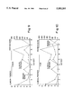
FIGS. 12-14: 2-D gel images of three patients with chronic lower pain back in the second study, showing the spot 1bp13-14.719.
FIGS. 15-17: 2-D gel images of three controls run side-by-side with the gels of FIGS. 12-14. Open circles represent area where 1bp13-14.719 spot is missing.
FIGS. 18-20: Represent enlargements of the areas blocked off in FIGS. 12-14.
FIGS. 21-23: Represent enlargements of the areas blocked off in FIGS. 15-17.
FIGS. 24a-c: These are photographs of 2-D gels of the present invention. FIG. 24a shows the location of spots other than spot 719 (such as other apolipoproteins and some acute phase reactant proteins). See further the legend on FIG. 25a for description of spot numbers. FIG. 24b is a 2-D gel of a patient with CLP; the boxed area is enlarged in the lower, right-hand corner of the figure. FIG. 24c is a 2-D gel of a normal control showing a much-diminished, barely-visible spot 719; as with FIG. 24b, the lower, right-hand corner of the figure is an enlarged view of the boxed area.
FIGS. 25a and b: This set of figures are bar graphs comparing the measured density [% TID (or total integrated density)] of the spots shown in FIGS. 24a-c between chronic lower back pain patients and normal controls. While there are observed increases in the other apolipoproteins, these have not been proven to be as statistically significant as spot 719 (apo-E variant).
FIG. 26: Immunoblot prepared in accordance with the example in the present invention, showing spot 719 is positive for anti-apo-E reactivity. The other positive spots (680 and 684), other forms of apo-E, were also analyzed for quantitative variations correlating with CLP, but there was no significant correlation (see FIGS. 25a and b).
FIG. 27: N-terminal sequence analysis of spot 719 protein [SEQ ID NO:1]. There is 100% homology with the known N-terminal sequence of plasma apo-E [SEQ ID NO:2].
This invention involves methods of diagnosing peripheral nerve damage, particularly that which causes spinal pain, and more particularly CLP and CCP wherein protein samples from both normal and abnormal individuals are subject to electrophoresis and/or immunoassays. In the case of two-dimensional gel electrophoresis, a large number of protein spots common to both types of individuals and spots which appear or disappear in the abnormal patient group are determined. Initially, the number of protein spots to be examined is reduced to only those showing statistically significant differences between normal controls and patients with chronic back pain. This is determined by performing a Student's t test or a log Student's t test on the spot intensity data. Those proteins that have statistical differences at a significance level of 0.05 on either or both of these tests are chosen for further study. In addition, the present invention also contemplates the use of one-dimensional electrophoresis.
CLP has been shown to arise from trauma to spinal nerve roots [Schonstrom, N. et al., Spine, Vol.9, pp. 604-607 (1984)]. While it is difficult to examine biochemical alterations in human nerve injuries, molecular changes associated with nerve root damage have been studied in several animal models. [Ignatius, M. J., Progress in Brain Research, Vol.71, pp.177-184 (1987)]. Changes in the injured nerve include elevation in the local concentrations of acute phase reactant proteins, infiltration by circulating monocytes, increased levels of protein synthesis and increases in apolipoprotein concentrations. One of the most striking physiological differences in these animal models of nerve damage is a 250-fold increase in the local concentration of apolipoprotein E (apo-E), with sciatic nerve crush injuries [Skene, J. H. P, Proc Natl. Acad. Sci. U.S.A., Vol. 80, pp. 4169-4173 (1983)]. This nerve damage-associated elevation in the area surrounding the nerve correlates with the present inventors' finding that a subset of the apolipoprotein E complex of spots in our 2-D gels was increased greater than five-fold in the plasma of individuals suffering CLP and CCP. Other apolipoproteins and acute phase reactants that might be expected to be elevated in plasma of individuals suffering nerve root damage, based on the animal studies, (most notably apo-D, apo-AI and apo-AIV) were identified by their 2-D gel location, but were found not to be elevated to a statistically significant degree in our analysis. To better understand the appearance of the apolipoprotein E variant (spot 719), the present inventors wish to determine the following: (1) was pain required for the protein change or was only a biomechanical abnormality necessary?; (2) was it limited to the lumbar region or did it apply to the whole spine?; (3) was it present in all individuals with acute or chronic pain?; (4) was it found in cases of peripheral nerve damage?; and (5) was it associated with an inflammatory response? To help answer these questions, the present inventors also analyzed the plasma of patients suffering from a variety of other chronic and acute painful conditions, as well as patients that had recovered from CLP and individuals with chronic inflammatory conditions. Based on this analysis and previously-reported observations, the present inventors proposed that the apo-E abnormality results from a chronic inflammatory insult, causing continuous attempts to regenerate damaged nerve. Further, although other investigators have documented that the concentration of apo-E immediately surrounding the damaged nerve tissue increases greatly, none of the prior art discloses or suggests that an increase in apo-E in the plasma, in general, and human plasma, in particular, would also occur.
In order to better understand the reason for increased concentration of spot 719 in the present studies, the present inventors undertook to identify the protein comprising this spot. Two-dimensional gel maps of plasma proteins were used to locate spot 719 in the area of the two-dimensional gel, which contains transthyretin ("TTR") dimer and the apo-E complex of spots. Spot 719 appears to have a relative molecular weight of about 32-36 kD and a pI of about 6.0-6.2 as determined from the 2-D gels. Immunoblot analysis of the two-dimensional gel showed strong reactivity of spot 719 within the apo-E monoclonal antibody (FIG. 26). N-terminal microsequence analysis was performed to confirm the identity of spot 719 as apo-E (FIG. 27). In addition, microsequence analysis independently verified the identity of another spot in the region not related to lower back pain as TTR. Circulating plasma apo-E is active in lipid transport from the gastrointestinal system to the periphery through the plasma. In addition to this endocrine function, apo-E also has paracrine and autocrine activities for cholesterol redistribution. It has also been shown to be a regulator of the immune response and a neurotropic factor. The paracrine activity of apo-E in nerve regeneration has been well described in sciatic nerve crush in rats [Schubert, D. et al., J. Cell Biol., Vol. 104, pp.635-642 (1987)]. In experimental models, by three weeks after nerve injury as much as five percent of the total soluble protein of the nerve is apo-E [Mahley, R. W. et al., Science, Vol. 240, pp.622-630 (1988)]. While in the CNS, there are similar levels of apo-E synthesis in response to injury, response to accumulation around the tissue is not observed [Muller, H. W. et al, Science, Vol. 228, pp. 499-501 (1985)]. It has been suggested that the lack of accumulation of apo-E in the CNS is an important factor in the lack of regeneration of neurons in the brain.
The source of the apo-E comprising spot no. 719 is not obvious. The blood nerve barrier in the dorsal root ganglion and other nerve roots has been shown to be compromised by nerve crush injury. [Howe, J. F. et al, Pain, Vol. 3, pp. 25-41 (1977); and Wiesel, S. W. et al, Spine, Vol. 10, pp. 549-551 (1984)]. This may lead to proteins entering the circulation at the point of injury, although this is mere speculation. The extraordinary amount of paracrine apo-E that can enter the circulation at the site of injury may account for the observed increase of this polypeptide in the plasma. However, it is very noteworthy that elevated levels of the other apolipoproteins observed in the literature (e.g., apo-D, apo-AI and apo-AIV) to accumulate in the local area around the nerve tissue do not correlate with statistically significant increases in levels in the plasma, as the present inventors have observed with the apo-E of spot 719.
It is also possible that the source of increased apo-E in the plasma of individuals with CLP or CCP is endocrine and produced in response to acute phase reactants resulting from the injury. However, the levels of the few acute phase reactants whose locations can be determined on 2-D gels do not differ between CLP patients and normal controls (see FIG. 25a and 25b). While the source of the apo-E/spot 719 increase has not yet been identified, the present inventors believe its variation in individuals suffering CLP and CCP is important in the diagnosis and management of these conditions, as well as in peripheral nerve damage generally. It may also provide insight into the mechanism of neuronal regeneration.
Any number of protocols can be used to develop protein data for use in performing the diagnostic methods of the present invention. The protocol used in the present studies and as exemplified herein was approved by the IRB of St. Luke's Hospital in Houston, Tex., and patients and sex and age matched volunteers each of whom signed an informed consent letter. The patients were complaining of chronic (six months or more in duration) low back pain secondary to a reported injury and were randomly selected from one of the applicants orthopedic practice. The patients were requested to remain drug-free for at least one week prior to blood sampling. The controls were free of significant medical problems as determined by medical history and physical examination. The initial study consisted of 10 patients and 10 sex and age matched controls. In the second study, a similar protocol consisted of 64 plasma samples from 36 lower back pain patients and 28 controls. The data for this study were obtained from three separate studies: two independent blinded studies, performed using the apo-E variant (spot 719) to determine which individuals had CLP, provided the correct diagnosis in sixteen out of seventeen patients (94.1%) and 14 out of 14 controls (100%); and a third study, which was not blinded, was performed and was accurate in 18 out of 19 patients (94.7%) and 14 out of 14 controls (100%).
Additional studies were undertaken which focused on spot 719. These studies are set forth in the examples which follow.
To prevent degradation of samples of tissue, serum, or other body fluids (preferably blood and more preferably plasma) from the subjects, the sample is initially frozen in dry ice. At any point prior to electrophoresis a portion of the sample may be removed for counting and assaying the amount of protein by, for example, the Lowry method.
The first stage gels for the two-dimensional electrophoresis generally contain urea at a concentration of about 9M and about 2% nonionic detergent, both of which aid in dissociating proteins. The nonionic detergent helps keep the separated proteins from precipitating at their isoelectric points. These reagents and their proportions can vary somewhat, provided that these objectives are accomplished. An ampholyte (e.g., 2% 4-8 pH ampholyte) is also desirable to maintain a pH gradient across the length of the gel, although other reagents which maintain the pH gradient could be substituted. The acrylamide concentration of the first stage gel should be such as to permit protein movement to the isoelectric point. The first stage gel can be in a number of forms; for example, it can be housed in an isoelectric focusing tube or in a slab form. Preferably, the first dimension is in the form of a tube gel.
The samples are usually prepared for the first stage by solubilizing in either 10% sodium dodecyl sulfate (SDS) or urea at a concentration of about 9M. The reducing agent 2-mercaptoethanol is also usually included to separate disulfide-linked subunits. An ampholyte to maintain the pH gradient, a nonionic detergent which does not affect the protein charge, and dithiothrietol (DTT) which disrupts disulfide bonds, may also be included. Other reagents may also be added, or other reagents which accomplish the foregoing functions may be substituted. For example, prior to subjecting the samples to gel electrophoresis, the samples may be placed in a sample buffer (for example, 2% SDS, 2% DTT, 20% glycerol, 2% ampholines and 2% CHAPS) and placed in a boiling water bath (at about 100° C.) for about two minutes to aid in dissolution. This temperature and the time exposed thereto has been found to not cause protein degradation; however, both can be varied provided that dissolution takes place and protein degradation does not. The sample buffer unfolds the protein, separates the disulfide-linked subunits, and maintains the pH. Other sample buffers which accomplish the foregoing functions, and other methods of dissolving the protein samples, can also be used.
The samples may then be cooled on ice and treated with DNase and RNase to reduce the viscosity. The samples can then be snap-frozen in liquid nitrogen and packed on dry ice if they are not to be run on gels immediately. This adequately preserves the samples. However, it has been found that any method that cools a dissolved sample to -70° C. or more will also preserve the samples.
Polyacrylamide gel electrophoresis in the presence of SDS is usually used for the second dimension separation. SDS is an ionic detergent and binds strongly to proteins. It eliminates the native protein charge characteristics and unfolds the protein into a rod-like form. See Tracy et al., J. Clin. Lab, Autom., supra. Thus, when protein is subjected to an electric field in the polyacrylamide gel matrix, the uniform negative charge and the relatively uniform shape of the SDS-protein complexes allow separation essentially by molecular weight, with the polyacrylamide gel matrix acting as a sieve.
The second stage gel is preferably SDS-equilibrated, to eliminate the protein charge, and contains a higher acrylamide concentration than the first stage gel, to aid in separating proteins by molecular weight. Other reagents can be added or substituted. Further, the second gel can be in a variety of forms; preferably, however, the second stage is in the form of a slab gel.
The spots on the gels can be viewed by any number of methods including staining with Coomassie blue and silver staining. They can be visualized for relative protein density manually, but it is preferred that they be scanned with an appropriate camera system with a normalization standard (available from the National Bureau of Standards, Gaithersburg, Md.) and analyzed with a computer densitometer to measure relative protein spot staining intensities or densities.
In order to perform immunological tests for the diagnosis and/or monitoring of CLP or CCP, the first step is to obtain antibodies to the proteins of interest. There are many methods of accomplishing this which are well known to those skilled in the art. (For comprehensive laboratory methods, see Harlow et al., Antibodies: A Laboratory Manual, Cold Spring Harbor Laboratory (1988), which is incorporated by reference herein.) For antibodies with sufficient specificity for western blots and immunoassays, the antigen must be purified to homogeneity or the antigen should be used to prepare monoclonal antibodies. Since the proteins of interest in this invention are seen as unique spots on the second dimension polyacrylamide gel, preferably the gel can be used as the final purification step of the individual antigens. One can obtain a pure antigen preparation by excising the spots which show increased or decreased intensity in CLP or CCP patients. This gel piece can be injected into an animal to raise antibodies. Alternatively, one may cut out the spot of protein and electroelute it from the gel to obtain a protein in solution for injection. Still another technique for processing the protein for injection after separation on gels is to electrophoretically transfer the proteins to nitrocellulose, locate the proteins of interest by staining (e.g., with Ponceau S), excise the spots and cut into pieces for injection. The particular method used is only limited by the ability to elicit an immune response to the proteins of interest.
As an alternative to using the antigen purified by separation on an electrophoretic gel directly, one may elute a protein spot, obtain a partial sequence by any method well known in the art, and use the sequence to manufacture synthetic peptides (usually with an automated machine using solid-phase techniques). The synthetic peptide should be at least six amino acids long to elicit antibodies that bind to the original protein. The purified synthetic peptides would then be coupled to carrier proteins, and these conjugates are then used to immunize animals.
The polyclonal antibodies in the antisera obtained with the foregoing methods can be used for western blots and other immunological tests. However, one may further utilize hybridoma technology to obtain monoclonal antibodies, which may be the best choice for immunochemical techniques. Methods of monoclonal production are well known in the art and were first described by Kohler and Milstein in 1975. Briefly, antibody-secreting cells are fused to, for example, myeloma cells to create hybridoma cells which are cloned and screened by appropriate methods for the desired antibodies. While a monoclonal antibody for apo-E is available (Chemicon), this antibody reacts not only with spot 719, but with the other apo-E spots on the 2-D gel. In addition, the inventors' investigations showed that this commercially available monoclonal antibody does not demonstrate quantitative differences between CLP plasma and normal plasma. Therefore, it is an object of the present invention to obtain a monoclonal antibody specific for spot 719 to overcome the disadvantages with the currently available apo-E monoclonal. It is only spot 719, out of the complex of apo-E spots on the 2-D gel, which is increased in patients with peripheral nerve damage, in particular those patients suffering from CLP or CCP. The location of this spot on the 2-D gels suggests that it is a variant form of apo-E, which differs from the other apo-E proteins by its glycosylation or phosphorylation pattern or other novel post-translational event, or that it is associated with another protein or lipid which is not completely denatured prior to gel analysis.
One of the diagnostic methods of the present invention involves the detection of proteins which are present in patients with CLP or CCP, yet absent in normal controls. Besides locating these protein spots by staining on a two-dimensional gel, one may detect the proteins by immunoblotting, or western blotting, utilizing the polyclonal or monoclonal antibodies raised to the particular protein of interest. Protocols for immunoblotting are well known in the art and generally comprise the steps of gel electrophoresis, transfer, blocking, addition of antibody, and detection. The preparation of the sample and the two-dimensional gel electrophoresis is discussed above. At the completion of electrophoresis, proteins are transferred from the gel to a matrix, such as nitrocellulose, activated (diazo groups) paper and activated (positively charged) nylon. Nitrocellulose membranes are preferred for relatively low background and cost considerations; however, any membrane which will sufficiently bind the transferred proteins can be used. Preferably, transfer of the proteins is accomplished by electrophoretic elution; however, simple diffusion or vacuum-assisted solvent flow can also be used. After transfer, the membranes may, optionally, be stained to determine the position of molecular weight markers.
Prior to antigen detection, one must block the membrane to prevent non-specific adsorption of immunological reagents. Most preferably, the blocking solution would be composed of nonfat dried milk or bovine serum albumin. After blocking, antigens can be detected directly or indirectly. Direct detection utilizes labelled primary antibodies. The antibodies, labelled with iodine, enzymes or biotin, can be prepared by methods well known to those skilled in the art. In indirect detection, the primary antibody (unlabelled) is first added to membrane, followed by a secondary antibody (an anti-primary antibody) which is labelled with radioactive iodine or an enzyme, such as horseradish peroxidase. The antigen is then detected by exposing a radiolabeled membrane to X-ray film or, in the case of enzyme-labelled antibody, by adding substrate to the membrane.
Alternative method of quantifying or detecting the presence of protein for the diagnosis of CLP or CCP is the use of immunoassays performed directly on the body fluid sample. Several immunoassays would be useful in the context of the present invention, including: antibody capture (Ab excess); antigen capture (antigen competition); and the two-antibody sandwich technique. All immunoassays rely on labeled antigens, antibodies, or secondary reagents for detection and quantitation. The label used can be radioactive or enzymatic, or one may label with fluorochromes or biotin. The choice of label is a matter of discretion with the diagnostician, taking into consideration cost, sensitivity, radioactivity exposure, etc. The term "label" as used herein refers to any of the foregoing.
In an antibody capture type of assay, the test sample is allowed to bind directly to a solid phase and any unbound antigen is washed away. The antibody specific for the antigen is added and allowed to bind. The amount of antibody bound to the solid phase, after washing away unbound antibody, is determined using a secondary reagent. Suitable secondary reagents include anti-immunoglobulin antibody, protein A or protein G. These can be obtained from commercial sources or prepared by methods known in the art. Detailed protocols can be found in Harlow et al., supra, and are incorporated herein by reference, the particular methods used not being limited or essential to the practice of the present invention.
Antigen capture type assays measure the amount of antigen in a test sample via a competition between labeled and unlabeled antigen. This type of assay is exemplified by a "radioimmunoassay" or RIA. The first step in this type of assay is to bind unlabeled antibody to a solid support (either directly or through an intermediate protein, e.g., an anti-immunoglobulin antibody). A sample of known antigen of known quantity is labeled and a sample of this is added to the test material containing an unknown amount of antigen, and the mixture is added to the bound antibody. The antigen in the test sample competes with the labeled antigen for binding to the antibody bound to the solid support. Following removal of the unbound antigen, the amount of labeled bound antigen is measured. The higher the concentration of antigen in the unknown test sample is, the more effectively it competes with the labeled antigen; therefore, a decreasing amount of label is detected with an increasing amount of unlabeled antigen. Thus, generated standard titration curves will yield relative levels of antigen.
Another immunoassay to quantitate antigen concentration is the two-antibody sandwich technique. This type of assay requires two antibodies that bind to two separate epitopes of the antigen. Thus, one may use two monoclonals that recognize two separate sites on the antigen, or a batch of purified polyclonals can be used. The essential steps are as follows: 1) one purified antibody is bound to a solid phase and the antigen in the test sample is allowed to bind to the first antibody; 2) unbound antigen is washed away and a labeled second antibody is allowed to bind to the antigen; and 3) after washing, the second labeled antibody that is bound to the matrix is quantitated. As in other assays, a standard titration curve with known dilutions is plotted and the unknown sample is compared thereto. In order to determine absolute amounts, a standard curve generated with known quantities of antigen is used.
A wide variety of test kits are possible to take advantage of the advances in the diagnostic arts made possible by this invention. Some will be described here; others can be devised by those skilled in the art.
The central reaction in a test kit could be between any one of the aberrant proteins found in patients with peripheral nerve damage, in particular that which causes CLP and CCP and the antibodies prepared as set forth above and in the Examples below. The Examples below are directed to the preparation of antibody from rabbits, and the following description and other Examples of test kits and test methods will be based on the rabbit preparation. The rabbit is the preferred source of immunoglobulin and its fractions; however, the skilled artisan will recognize that the following Examples utilize the rabbit only as exemplary. Other animals can be used and this will require some modification of the other reagents used in the tests and the kits, and are readily apparent to one skilled in the art.
In the test kits, any of a variety of adsorbents can be used including, for example, glass or plastic surfaces which may be the inner surfaces of test tubes or the surfaces of test plates. Examples of flat surfaces especially useful in an enzyme-linked immunosorbant assay (ELISA) or a radioimmunoassay (RIA) include glass, nitrocellulose paper, or plastics such as polystyrene, polycarbonate or various polyvinyls. The ligands can be attached to the surface by direct adsorption, forced adsorption and coupling, in accordance with known procedures. Typical test kits are set forth in the Examples below.
The following Examples illustrate the utility of the diagnostic methods of the present invention, and are not intended to limit the scope of this invention. For instance, any of the known immunoassays or other known methods of protein detection may be used to aid in the diagnosis of peripheral nerve damage, in particular that nerve damage which causes CLP or CCP. Also, the method of diagnosis is not limited to the specific proteins elucidated by the present Examples. Modifications of the procedures as would be apparent to one skilled in the art are within the scope of the teachings.
In a first (initial) study, ten patients with chronic back pain were randomly selected from a group of patients complaining of CLP of six months or more secondary to a reported injury. These patients, 3 females and 7 males, ranged in age between 20 and 55 years. Of these patients, 7 were taking medication for their pain. However, 3 of these patients took no medication. The degree of lower back disability was evaluated by a number of factors such as: the history of the back pain (including radiations and the induction of pain with coughing and/or sneezing); physical examination including the loss of sensitivity in the dermatomes; measurements of back and leg motion limitations; abnormalities in the ankle and/or knee jerk reflexes; and the analysis of spinal radiographies (for spondy losis, stenosis, herniated discs, degenerations, the narrowing of the intervertebral space, etc.) and other special studies such as MRI, CT scan, myelograms, discograms and electromyelograms. Controls were selected for age and sex to match the patient group. The controls were free of significant medical problems as determined by medical history and physical examination.
Ten ml of blood was collected by venipuncture, within one minute of tourniquet application, using Vacutainer tubes containing 143 USP units of heparin. The control samples were collected at the same time as those from the patients and all samples were collected during afternoon hours. Plasma was isolated by centrifugation of the whole blood at 2000× g for ten minutes followed by the separation of the plasma from the packed red and white cells by pipetting. The plasma was frozen at -20° C. prior to shipment (in dry ice) to the laboratory for analysis. The samples were stored at -70° C. until electrophoresis, which was performed within three months of venipuncture.
Gel electrophoresis. Plasma samples were thawed and 20 ul of each sample were added to 20 ul of denaturing solution, containing 10% w/v SDS and 2.3% DTT w/v? The samples were then heated to 95° C. for 4 minutes followed by cooling to room temperature. Then 96 ul of electrophoresis solution, containing 0.1 g DTT, 0.4 g CHAPS, 5.4 g urea, 0.5 ml pH 3.5-10 ampholytes and 6.5 ml deionized water were added to each sample. The samples were mixed on a Vortex mixer and 10 ul of each processed sample (containing 1.47 ul plasma) were added to the first dimension isoelectric focusing (IEF) gels. Isoelectric focusing was performed in 3% (w/v) acrylamide gels with 4% w/v ampholytes (containing a 1:1 mixture of pH 3.5-10 and pH 5-7 ampholytes) and crosslinked with 0.03% diacryloylpiperazine, i.e., 3% T/1% C. Electrophoresis was performed for 18,000 volt hours, beginning with 1000 volts for 17 hours followed by 2000 volts for 30 minutes.
The second dimension, wherein proteins are separated by mass, was performed with 160 cm×200 cm×1.5 mm slab gels using a Bio-Rad Protean II chamber. These gels were formed with 12.2% acrylamide (w/v), 0.2M TRIS-HCl (pH 8.8), 0.7% sodium thiosulfate (w/v), 0.3% diacryloylpiperazine (w/v), 0.5% 1,4-dimethylpiperazine (v/v), and 0.07% ammonium persulfate (w/v). Electrophoresis was performed at 7° C. with a constant current of 40 mA per gel until a dye front reached at or near the bottom of the gel.
Silver staining. At the end of the run, the gels were removed from the glass plates and washed for 5 minutes in water (no protein loss is detected during this period). The gels were then soaked in a solution of ethanol/acetic acid/deionized water (40/10/50) for one hour on an orbital shaker at 36 rpm. This solution was then replaced with a solution of ethanol/acetic acid/deionized water (5/5/90) and the gels were soaked for at least 3 hours. The gels were washed with deionized water for 5 minutes and soaked in 10% gluteraldehyde solution for 30 minutes. Extensive washes with deionized water were performed to entirely remove the gluteraldehyde 3×10 and 4×30 minutes). [Cold deionized water (<15° C.) removes gluteraldehyde more efficiently.] The gels were then stained for 10 minutes in an ammoniacal silver nitrate solution (6 g of silver nitrate dissolved in 30 ml of deionized water), which is slowly mixed into a solution containing 160 ml of water, 10 ml of concentrated ammonium hydroxide, and 1.5 ml of sodium hydroxide, 10 mol/liter; this solution is then diluted with deionized water to a final volume of 750 ml) (solution H). The temperature of solution H was 20° C. After staining, the gels were washed with deionized water for 5 minutes×3. The image was then developed in a citric acid and formaldehyde solution (0.1 g citric acid and 1 ml formaldehyde in 1 liter of deionized water) (solution V) until a slight background stain appeared. (The optimum temperature of solution V is 15°-18° C.) The development process was stopped with an acetic acid/deionized water solution (5/95) for at least 15 minutes. Stained gels were stored in a glycerol/ethanol/deionized water solution (7/10/83). [This staining method is described in Hochstrasser et al., Analytical Biochemistry, 173, pp. 424-435 (1988).]
Gel analysis. In order to quantitate proteins, gels were scanned with an Eikonics Series 78/99 digital scanner and the gel images created thus were analyzed using PDQUEST software (Protein Database, Inc., Huntington Station, N.Y.) on a SUN 4-260 minicomputer. Gel images were normalized for protein loading and staining variation using the average log-ratio normalization procedure of the PDQUEST software. Thirteen hundred proteins were analyzed on each gel and the proteins were matched and compared quantitatively. This analysis was performed by using the PDQUEST software aided by visual examination and operator intervention in gel areas containing complex spot patterns. All proteins which increased or decreased in concentration by three-fold or more, and were found to be statistically significant (by way of the Student t test or the log Student t test) were considered to be spots of interest.
FIG. 1 represents a stained gel of a plasma sample from a control subject. Marker proteins are designated by the prefix M, and Table I lists the marker plasma proteins landmarked on the gel. Proteins which were found to be increased or decreased in patients with CLP are listed in Tables II and III, respectively, and are indicated by number on the gel shown in FIG. 1. Those proteins which are absent in patients' plasma are illustrated by open circles on the control gel in FIG. 1. A computer-generated master map of the proteins analyzed in this Example is represented FIG. 2.
              TABLE I
______________________________________
Landmarked Plasma Proteins
Identification No.
                  Protein
______________________________________
M1                beta-Haptoglobins
M2                alpha.sub.1 -Antitrypsins
M3                Albumin
M4                IgG heavy chains
M5                IgG light chains
M6                Apo A-1 lipoproteins
______________________________________
              TABLE II
______________________________________
Plasma Proteins Increased With CLP
Fold
                                     In-
                                     crease
Pro- Controls (N = 9)
                     Patients (N = 10)
                                     or
tein Mean            Fre-  Mean        Fre-  De-
ID#  Conc.*  S.E.M.  quency
                           Conc.*
                                 S.E.M.
                                       quency
                                             crease
______________________________________
4210 0.0     0.0     0     51.5  24.2  5     >51.5
1204 0.0     0.0     0     46.5  12.3  8     >44.5
1318 0.0     0.0     0     42.5  15.3  7     >42.5
1324 0.0     0.0     0     38.3  14.7  6     >38.3
1702 0.0     0.0     0     31.2  13.9  4     >31.2
4821 0.0     0.0     0     31.2  16.6  4     >31.2
0508 0.0     0.0     0     26.4  9.2   5     >26.4
3211 0.0     0.0     0     25.5  9.7   5     >25.5
4109 0.0     0.0     0     21.9  10.1  4     >21.9
5524 0.0     0.0     0     20.2  10.5  4     >20.2
7622 0.0     0.0     0     19.8  8.2   4     >19.8
6331 0.0     0.0     0     13.0  5.4   4     >13.0
8322 0.0     0.0     0     10.1  4.5   4     >10.1
3151 1.6     1.6     1     46.8  15.2  7     29.3
1323 2.0     2.0     1     50.0  18.2  6     25.0
5240 3.4     3.4     1     69.2  30.7  6     20.3
2105 2.0     2.0     1     31.5  11.8  6     15.8
1316 4.9     4.9     1     75.2  18.1  9     15.3
6104 7.4     4.7     2     94.5  41.2  6     12.8
7109 18.5    12.3    3     131.2 42.9  6     7.1
3606 63.7    30.5    4     402.8 130.0 8     6.3
1305 17.3    7.4     5     103.8 28.0  9     6.0
3146 3.7     3.7     1     21.0  4.8   7     5.7
7114 23.3    15.5    2     118.2 38.5  7     5.1
7448 67.7    46.3    3     283.4 79.4  9     4.2
3203 4.2     4.2     1     17.5  8.2   6     4.2
0609 37.3    19.1    3     148.3 46.5  8     4.0
0604 62.9    29.0    4     236.0 61.5  9     3.8
7711 15.4    15.4    1     49.4  17.3  6     3.2
5615 89.1    89.1    1     226.8 64.9  8     2.5
______________________________________
 *Concentrations are in arbitrary density units.
 p is less than or equal to 0.05
              TABLE III
______________________________________
Plasma Proteins Decreased With CLP
Fold
                                     In-
                                     crease
Pro- Controls (N = 9)
                     Patients (N = 10)
                                     or
tein Mean            Fre-  Mean        Fre-  De-
ID#  Conc.*  S.E.M.  quency
                           Conc.*
                                 S.E.M.
                                       quency
                                             crease
______________________________________
4737 224.0   152.1   5     0.0   0.0   0     >224.0
4624 112.5   52.0    4     0.0   0.0   0     >112.5
7212 78.1    28.1    6     0.0   0.0   0     >78.1
7309 13.0    5.8     4     0.0   0.0   0     >13.0
7214 11.7    6.0     3     0.0   0.0   0     >11.7
2806 11.6    4.7     4     0.0   0.0   0     >11.6
5013 10.8    5.6     3     0.0   0.0   0     >10.8
4614 12.5    4.4     6     2.3   1.6   2     5.4
3155 76.0    28.5    7     16.5  9.1   4     4.6
8309 53.8    11.0    9     13.1  4.7   5     4.1
8418 744.1   185.6   8     191.1 108.6 5     3.9
7455 95.6    26.8    7     25.5  10.9  4     3.7
5411 79.4    23.4    7     25.7  13.2  3     3.1
7515 8.2     2.6     6     3.2   1.9   3     2.6
______________________________________
 *Concentrations are in arbitrary density units.
 p is less than or equal to 0.05
In a further, expanded study ("second study"), 36 patients with chronic low back pain (two cases having lumbar and thoracic pain--cases 14 and 32) and controls, were subjected to the methods set forth in Example 1. This protocol was approved by the Institutional Review Board of St. Luke's Hospital in Houston, Tex. All CLP patients (n=36), selected from one of the present inventors' (BMC) clinical practice, all normal volunteers (n=28), and all patients with conditions other than CLP (n=34) (see further Example 3) signed an approved consent form and were requested, prior to blood sampling, to remain drug-free for at least one week. The CLP patients were diagnosed by clinically correlating the information obtained from their medical history, physical examination, x-ray examination, selected imaging techniques (MRI and/or CT scanning) and invasive techniques when indicated (myelograms and/or discograms). Healthy controls were questioned regarding and CLP symptoms and were eliminated if they had any history of CLP. Patients with the other conditions (see further Example 3) were diagnosed using standard history and physical examinations and the indicated laboratory, x-ray and imaging techniques.
Venapuncture from the anticubital fossa was performed and each 5 mls. of blood was collected into a Vacutainer tube containing 143 USP units of sodium heparin. It was centrifuged at 783×G for ten minutes at room temperature and the plasma was removed and placed into a plastic tube and frozen at -20° C. These samples were shipped overnight to the electrophoresis facility and remained frozen at -70° C. until used. The plasma samples were subjected to 2-D gel electrophoresis and staining as in Example 1.
In an effort to identify new spots, the patient gels were compared to control gels visually without the aid of computer analysis, as well as also using the computer analysis. It was observed that one particular protein(s) spot appeared in patients and was virtually absent in controls. It is postulated that the computer did not identify this spot because it was incorporating it into another, very closely located spot on the gel. This new spot has been designated 1bp13-14.719 (or spot 719). Its presence is indicated on FIGS. 12-14 and 18-20, and the corresponding absence of the spot on the control gels is indicated by an open circle in FIGS. 15-17. In FIGS. 21-23 (control gels), one can see the corresponding absence of the spot which is visible in FIGS. 18-20.
A summary of the data obtained in this study is shown in Tables IV and V. In gels where spot 719 was barely or not visible, they were scored as negative (-). In gels which clearly contained the spot, they were scored as positive (+). The data in Table V were obtained from three separate studies: two independent blinded studies and a third nonblinded study. The studies were "blind" to the extent that the investigator performing the analyses was unaware which gels were of back pain samples and which were controls. As can be seen from Tables IV and V, the accuracy in predicting which samples belonged to which group is remarkably high (97.22% overall). Moreover, this new spot allows for visual identification without the need for computer-assisted analysis, thereby simplifying the diagnostic procedure. In order to quantify the increase in spot 719, a portion of the gels were subjected to computer-assisted analysis using the ELSIE software (developed by Mark Miller and Arthur Olson at the National Cancer Institute, U.S.A.), the results of which are shown in FIGS. 25a and b.
TABLE IV __________________________________________________________________________ Back Pain Study of Example 2 Pt. Age SexWC Atty Surg 719 Diagnosis Med __________________________________________________________________________ 1. 31 M + + 1 + HNP L5-S1. 0 2. 35 M + 0 1 + HNP L4-5, L5-S1; Scar. 0 3. 47F 0 0 1 + Spondylosis, HNP L4-5. 0 4. 56F 0 0 1 + Pseudoarthrosis L4-5. T13;R 5. 44 M + + 2 - Donor Site Pain. 0 6. 43 M + + 5 + Pseudoarthosis L4-S1; 0 Stenosis L3-4, L4-5;Scar 7. 24 M + + 0 + HNP L4-5, L5-S1.T13 8. 49M 0 0 2 + Stenosis L2-3; VIC Pseudoarthrosis, L4-5. 9. 43 M + + 5 + Deg.Spondylolisthesis 0 L3-4; Pseudoarth. L4-S1. 10. 37 M + + 1 + HNP, Spondylosis, 0 Stenosis L4-5. 47M 0 0 1 + HNP L4-5, L5-S1. 0 26 M + + 2 + Spondylolysthesis L5-S1; 0 Pseudoarthrosis L5-S1. 30 M + + 2 + Recurrent HNP L4-5. 0 60F 0 0 0 + Osteoporosis, 0 Osteoarthritis. 50F 0 0 0 + Deg.Spondylolisthesis 0 L4-5; DDD. L4-5; L5-S1. 63 M + 0 0 + HNP L4-5; L5-S1. 0 44 M + 0 4 + DDD, HNP, 0 Pseudoarthrosis L4-5. 48 M + + 2 + DDD, HNP, Spondylosis, 0 Pseudoarthrosis, L4-5. 26 F + 0 0 + HNP L5-S1; DDD L4-5. 0 20. 42 F + + 2 + Lumbar Stenosis; 0 Pseudoarthrosis, L4-5. 49 M + + 5 + HNP L5-S1; Arachnoiditis. 0 27 F + 0 0 + HNP L4-5. 0 34 M + + 0 + HNP L4-5. 0 44M 0 0 1 + HNP L4-5. 0 44 F + + 1 + HNP L4-5. VIC 26 M + + 0 - Spondylosis L5-S1. 0 52 F + 0 3 + DDD, Spondylosis L3-4; 0 HNP L4-5. 48 M + + 1 + HNP L3-4; Buldge L4-5. 0 29 M + + 3 + Lumbar Stenosis L5-S1; 0 Sacralization L-5 30. 38 F + + 2 + HNP L4-5, L5-S1; NAP. DDD L4-5; Stenosis L5-S1. ROB. 52 M + 0 2 + HNP, Spondylosis L4-5. 0 59F 0 0 0 + Scoliosis; Spondylosis. 0 31 M + + 6 + HNP L5-S1;Facet damage 0 L5; Arachnoiditis. 42 M + + 3 + HNP, Instability L3-4; 0 Stenosis L4-5. 31 M + + 2 + HNP, facet damage L5-S1. 5 0 34 M + + 1 + Spondylolisthesis L5-S1. 0 __________________________________________________________________________
Table IV. Clinical data from 36 CLP patients in a study which included 28 controls: WC=workman's compensation case; Atty=attorney involvement; Surg=number of back operations at the time of venapuncture; 719=increased (+) or decrease (-) in the plasma level of the apolipoprotein E variant scored as described in Methods; HNP=herniated nucleus pulposis; DDD=degenerative disc disease; Med=drugs; NAP=Naprosyn; ROB=Robaxin; VIC.=Vicodin; T# 3=Tylenol # 3.
              TABLE V
______________________________________
Back Pain Study of Example 2
                                  Patients
                                  and
Subjects  CLP Patients
                      Controls    Controls
______________________________________
Apo E increase
          34+     2-      0+    28-   62/64
Correlation
          34/36       28/28       62/64
Test      Sensitivity Specificity Efficiency
Percentage
          94.44%      100.00%     97.22%
______________________________________
 Table V. Statistical analysis of CLP data: Results of the plasma studies
 of 36 CLP patients and 28 normal controls. The efficiency was calculated
 as = (sensitivity + specificity)/2.
Five additional groups of patients' plasma were analyzed with respect to 1bp13-14.719. These groups consisted of:
A. Three patients who were relieved of their CLP through surgery and two unoperated, asymptomatic individuals who had biomechanical abnormalities that often produced chronic lower back pain. None of these patients displayed an increase in the apolipoprotein E variant (spot 719) associated with chronic lower back pain (see Table VI).
B. In order to determine the significance of the location of the pain in the spinal column in inducing the presence of the apolipoprotein E variant from patients with CLP, plasma samples were analyzed from four individuals with chronic cervical pain (CCP). All four patients with CCP demonstrated the apolipoprotein E variant (spot 719) (see Table VII).
C. To determine if normal nociception without nerve damage induced an increase in the apolipoprotein E variant (spot 719), the plasma proteins of 11 patients with painful orthopedic injuries without nerve damage were analyzed. Only one of these patients, who may have sustained nerve damage, displayed an increase in the apolipoprotein E variant (see Table VIII).
D. Nerve damage is thought to be associated with CLP. To determine whether other peripheral nerve damage causes the induction of apolipoprotein E variant (spot 719) and plasma, which is associated with CLP, the plasma samples of six individuals with various peripheral nerve problems were examined. Five of the six displayed an increase in the apolipoprotein E variant. The remaining patient had a painless radial nerve palsy. (See Table IX).
E. Local inflammatory responses (which often include edema and demyelination) of the nerve root have been demonstrated with chronic lower back pain. To determine if the increase in apolipoprotein E (spot 719) was associated with known inflammatory conditions, the plasma of seven patients with chronic systemic inflammatory changes (lupus erythematosus, rheumatoid arthritis, and Crohn's disease) was studied. Three patients with Crohn's disease and two with lupus displayed an increase in the apolipoprotein E variant (spot 719), while the two with rheumatoid arthritis did not. (See Table X).
The above samples were subjected to the methods and analyses of Example 2, wherein the presence or absence of 1bp13-14.719 was determined.
These studies were undertaken to answer the questions previously posed in this specification. In particular, (1) is pain required or is only a biomechanical change required to increase the plasma level of the apolipoprotein E variant? In Group A, five asymptomatic patients with biomechanical changes were tested: two with spondylolisthesis, two with post-operative discectomies, and one with a post-operative discectomy and fusion. None exhibited an increase in the apolipoprotein E variant. This study indicated the association of the protein (spot 719) with pain, and also strongly suggested that the protein difference was not due to painless surgical scarring (see Table VI).
(2) Is the induction of the apolipoprotein E variant (spot 719) from the spine limited only to chronic pain in the lumbar region? To determine if the increase in the plasma level of apolipoprotein E variant is specific for pain in the lumbar region, we examined the plasma of four patients with CCP (Group B). All four demonstrated a significant increase in this protein. This study indicated that pain associated with nerve damage anywhere in the spinal column may be associated with increased plasma concentrations of this protein.
(3) Is the apolipoprotein E variant increased in all patients with acute or chronic pain? For this study, the area of nociception was extended to the periphery and 11 patients were selected with various painful conditions without nerve damage (Group C). These conditions ranged from a fractured ulna to a ligamentous shoulder injury. Ten were negative for an increase (90.9%) and one was positive for an increase in the apolipoprotein E variant (see Table VIII). This one patient had a multitude of serious orthopedic injuries (with probable nerve damage), and it could not be determined which of them was responsible for the positive result. This study suggested that peripheral pain without nerve damage does not produce the protein (spot 719).
(4) Is an increase in the plasma apolipoprotein E variant found in patients with painful conditions associated with peripheral nerve damage? An increase in this protein was found in individuals with chronic pain associated with the vertebral column, but was not found in individuals with peripheral pain who did not have evidence of nerve damage. This suggested that the protein variant found in the plasma of chronic spinal column pain patients was associated with chronic nerve damage, but was not associated with normal nociception with no nerve damage. On the other hand, it was postulated that peripheral pain due to nerve damage would be associated with an increase in the plasma level of the apolipoprotein E variant (spot 719). To test this hypothesis, the plasma from six patients with peripheral nerve damage (Group D) was tested. (See Table IX). Five of six were positive for an increase in the plasma concentration of the apolipoprotein E variant. The patient with an adventitious bursa was later diagnosed with cervical spondylosis and operated with relief of her symptoms. The one patient who displayed no increase in this protein had a painless radial nerve palsy. Since apolipoprotein E is known to be associated with nerve regeneration, it is possible that there was no physiological stimulus to repair the nerve. The incidence of spontaneous regeneration of the radial nerve following fractures of the humerus has been reported variously to be 70-92% with neuropraxia or axonotomesis, but 0% with neuronotomesis. The absence of an increase in the plasma level of apolipoprotein E variant and the absence of regeneration correlated with the clinical picture. From this study and those above, it is reasonable to suggest that nerve damage and regeneration are necessary to produce an increase in the plasma level of the apolipoprotein E variant (spot 719). Thus, the absence, or substantial absence, of spot 719 in a patient diagnosed by conventional methods to have peripheral nerve damage (for example, a radial nerve palsy), would indicate that there has been a lack of nerve regeneration. This would guide the clinician to treat the patient with therapy known to stimulate nerve regeneration. This would be especially helpful to a clinician watching the clinical course of a brachial plexus injury, a radial nerve palsy, a peroneal palsy, a peripheral neuropathy, a causalgia, etc.
(5) Is an increase in plasma apolipoprotein E variant (spot 719) related to inflammation? In addition to stimulating regeneration, nerve damage has been noted to stimulate a local, neural inflammatory response. It is possible that mediators of inflammation are necessary for nerve regeneration, because apolipoprotein E is known to be involved in both responses. To answer this, a group of patients with diseases known to stimulate chronic inflammation (Crohn's disease, lupus erythematosus and rheumatoid arthritis) was studied (Group E). Three patients with Crohn's disease and two with lupus were positive; but the two with rheumatoid arthritis were negative (see Table X). From this study, it is possible to conclude that the increase in the plasma concentration of this apolipoprotein E variant may be associated with chronic inflammatory diseases, which can produce false positives with respect to the diagnosis of peripheral nerve damage, and in particular, CLP and CCP.
It is known in the literature from biochemical studies of crushed sciatic nerves in rats, rabbits and primates that apolipoprotein E was essential for nerve repair in mammals, and that its local concentration may be increased 250-fold. There is evidence that apolipoprotein E enters the nerve to a limited degree from the plasma, but it is produced in massive quantities by the resident macrophages and endothelial cells within the nerve, as well as monocyte-derived macrophages which enter the nerve in response to denervation. It is thought that apolipoprotein E may be involved in the redistribution of lipid (involving macrophages and Schwann cells), including the cholesterol released during degeneration to the regenerating axons. Apolipoprotein D, apolipoprotein A-I and AIV are also thought to be associated with this lipid transfer. Although these other apolipoproteins have been observed in the present studies, it is significant to note that these proteins do not appear to be useful as diagnostic markers of peripheral nerve damage. For instance, apo-D is reported in the literature to be locally increased (around the nerve tissue) 500-fold, but a corresponding increase in the plasma is not seen (see FIGS. 25a and b). The experimental data and human data in the prior art indicate that nerve pressure or tension can produce intraneural vascular changes, which ultimately result in degeneration which stimulates regeneration. It was postulated in the prior art that these perturbations produced a loss of nerve function and/or hyperexcitability with pain from ectopic generation. The increase in apolipoprotein E, which has been observed in animal nerve injury studies, is consistent with the increased plasma levels with apolipoprotein E variant observed in the plasma of patients with nerve injury in this study. This protein alteration may be part of a normal physiological response to nerve damage, and its presence can serve as a useful marker in the disorders associated with nerve damage, in particular in the diagnosis and management of patients with CLP and CCP.
TABLE VI ______________________________________ GROUP A Patient AgeSex Diagnosis Spot 719 ______________________________________ 1 67 M Spondylolisthesis - L4-5 2 45 M Spondylolisthesis - L5-S1 3 49 M Lam., Disectomy, - HNP L5-S1 4 59 M Lam., Disectomy, - Fusion, L5-S1 5 44 M Lam., Disectomy, - L5-S1 ______________________________________
              TABLE VII
______________________________________
GROUP B
Patient
       Age    Sex     Diagnosis      Spot 719
______________________________________
1      35     F       Cervical Spondylosis
                                     +
2      39     F       Cervical Spondylosis
                                     +
3      38     F       Cerv. Spondy.; Lam.,
                                     +
                      fusion C5-6
4      41     M       Cervical Spondy.; Detach.
                                     +
                      Deltoid.
______________________________________
              TABLE VIII
______________________________________
GROUP C
Patient
      Age     Sex    Diagnosis       Spot 719
______________________________________
1     68      F      Lumbar Spondylosis; THR.
                                     -
2     59      M      Failed THR.     -
3     22      M      Early Aseptic Necrosis,
                                     -
                     hip.
4     34      M      Ligamentous Tear,
                                     -
                     Shoulder
5     15      M      Right Knee MCL Sprain
                                     -
6     34      M      Asep. Nec. L Hip; Discl.
                                     -
                     SC Jt.
7     47      F      Shoulder Pain.  -
8     34      F      Fractured Right Ulna,
                                     -
9     40      M      Asept. Necrosis, Bilat.
                                     -
                     Hips
10    29      F      Knee Pain: Synovitis,
                                     -
                     tear MM.
11    58      M      Failed THR; Multi. Comp.
                                     +
                     Fracts.
______________________________________
              TABLE IX
______________________________________
GROUP D
Patient
       Age    Sex     Diagnosis      Spot 719
______________________________________
1      24     M       Radial nerve palsy; frct.
                                     -
                      humerus
2      42     F       Cubital Tunnel Syndrome
                                     +
3      47     M       Morton's Neuroma
                                     +
4      33     M       Reflex Sympathetic
                                     +
                      Dystrophy
5      47     M       Left knee; Failed fixator
                                     +
6      46     F       Adventitious Bursa, Left
                                     +
                      scapula
______________________________________
              TABLE X
______________________________________
GROUP E
Patient
       Age     Sex     Diagnosis     Spot 719
______________________________________
1      32      F       Crohn's Disease
                                     +
2      27      F       Crohn's Disease
                                     +
3      36      F       Crohn's Disease
                                     +
4      33      F       Crohn's Disease
                                     +
5      33      F       Lupus.        +
6      60      F       Rheumatoid Arthritis.
                                     -
7      76      M       Rheumatoid Arthritis.
                                     -
______________________________________
Immunoblot analysis was conducted for spot 719. Proteins separated by one and two dimensional gel electrophoresis electro-blotted on PVDF membranes (Immobilon-P, Millipore), prepared according to the manufacturer's instructions, for 40 minutes at 0.8 amps using a transblot (BioRad) semi-dry electroblotter. The blots were then dried and placed in methanol followed by blocking overnight with PBS containing bovine serumalbumin. ECL (Amersham) detection was performed according to the manufacturer's instructions using 1:1000 dilution of monoclonal anti-apo-E (Chemicon) for two hours at room temperature and 1:25000 antimouse immunoglobulin (Pierce) for 30 minutes. Spot 719 was positive for anti-apo-E reactivity (FIG. 26). Other modified forms of Apo-E were also detected. These latter spot densities were analyzed by computer-assisted densitometry for quantitative variations correlating with chronic lower back pain, and there were none (FIGS. 25a and 25b).
Amino acid N-terminal microsequence of amido black-stained electroblotted protein was performed. Automated peptide microsequencing was performed by dansyl-Edman degradation as described by B. S. Hartley (1970), Strategy and Tactics in Protein Chemistry, Biochem. J., Vol. 119, pp. 805-822, with modifications described by John M. Walker (1984) Proteins, Vol. 1, The Humana Press, pp. 221-242. In order to determine the preparative ability of the 2-D gel, the maximum plasma protein capacity for resolving spots 719 was determined by titering the sample load. It was determined that the optimal load for 2 D resolution and visualization with amido black of spot 719 was 60 microliters of sample (three times the analytical load). Spot 719 was excised from five amido black-stained preparative 2-D gel electroblots. N-terminal microsequence analysis was performed on spot 719 and was determined to be apo-E (FIG. 27). N-terminal sequence analysis determined 100% homology with the known sequence for apo-E.
To determine whether there is a correlation between protein spot density and the severity of pain, the densities of the various proteins of interest in the initial study were compared to the degree of lower back disability, as scored by historical as well as physical and radiological determinations.
A short, abbreviated scoring system (abbreviated from a scale of 144 possible factors) was devised which is similar to the Waddell approach [Waddell and Main, "Assessment of severity in low-back disorders", Spine Vol 9 pp. 204-208 (1984); Waddell, Main, Morris, Paola and Gray, 1984; Waddell et al., 1980]. The physical signs which Waddell et al selected as being significant were: degree of lumbar flexion; straight leg raising; root compression signs; and previous lumbar surgeries. The six clinical signs used in the abbreviated scale were similar and constituted the clinical objective scale (COS):
1. Scar from previous back surgery. All back surgery creates permanent scarring with permanent changes, no matter how subtle. This contributes to the presence of minute to major back pain. Each surgery scored 2.
2. True spasm which the patient cannot control (the most significant sign of all). This is not a limitation of motion which the patient can control, but true, uncontrolled spasm which the physician can recognize. Although sometimes painless in a condition such as burned out ankylosing spondylitis, it otherwise invariably signals severe pain. The physician can easily recognize this exception. This finding scored 4.
3. Straight leg raising (SLR) (right and left). This must be differentiated from hamstring spasm and a functional SLR. Hamstring spasm or tightness produces pain locally in the thigh, not in the back with SLR. Functional SLR is easily recognized by having the patient sit on the edge of the table and casually extend the knee. If the patient does not complain of back pain, there is no positive SLR. In knowledgeable hands, this is an objective finding. Each SLR scored 2.
4. Knee or ankle reflex change (right and left). These are objective findings in all cases. It is possible for them to be present without pain because of old trauma or surgery. Some reflex changes are associated with weakness or atrophy, but because these findings are frequently not recorded, it is not practical to score the latter. Each reflex change scored 1.
Table XI summarizes the COS.
              TABLE XI
______________________________________
Summary of COS
FACTOR                VALUE
______________________________________
Each back incision (scar)
                      2
Spasm                 4
Right Straight Leg Raising (RSLR)
                      2
Left Straight Leg Raising (LSLR)
                      2
Right Reflex Change (RRC)
                      1
Left Reflex Change (LRC)
                      1
______________________________________
The abbreviated scoring of the patients of this study are seen in Table XII, below. The numbers indicated next to the gel are arbitrarily assigned patient numbers.
              TABLE XII
______________________________________
Scores of COS of Patients.
                                             TO-
Patient
      SCAR    SPASM    RSLR  LSLR  RRC  LRC  TAL
______________________________________
gel 1 2       4              2               8
gel 3                        2               2
gel 5                                        0
gel 7 2                2                     4
gel 9                                        0
gel 11                 2     2     1         5
gel 13
      2                      2               4
gel 15
      2       4        2     2          1    11
gel 17                                       0
gel 19
      2                      2          1    5
______________________________________
By comparing the above COS scores to the amount of protein 1318 in the gels, it was found that the protein designated 1318 displayed a correlation with pain severity. This is represented in FIG. 5.
The same comparison was made with proteins 1316, 1204, 1305, 3203 and 3211. Also a comparison was made with the average of proteins 1318 and 1316. These results are shown in FIGS. 6-11.
Using similar methods, a correlation may also be made with spot 719 and any of the conditions which result from peripheral nerve damage.
To examine the effectiveness of a course of treatment for conditions resulting from peripheral nerve damage, a blood sample would be obtained from a patient prior to the treatment. A two-dimensional gel or immunoassay would be run, stained and analyzed for levels of one or more of the proteins listed in Tables II, III,IV and V to obtain a baseline. After instituting treatment, one or more blood samples are taken from the patient and analyzed for levels of the same protein or proteins which were initially analyzed. A proportional increase or decrease to normal levels (depending on whether the protein analyzed is one which is found to increase or decrease in the patients) signifies that the treatment is successful.
A. Preparing Antigens. After two-dimensional gel electrophoresis is performed on a patient, for instance one with confirmed CLP, the gel would be stained with Coomassie blue in order to locate a protein of interest. The gel is rinsed with deionized water for a few minutes, changing the water several times. The spot containing a protein is cut out of the gel with a scalpel, and placed on a piece of parafilm or plastic wrap. The edge of a paper towel is used to remove by capillary action any standing water. Next, the plungers from the barrels of two 5 cc syringes are removed, and the gel piece is placed into one of the barrels. The plunger is then replaced and the syringe outlet is positioned in the barrel of the second syringe. Using rapid, firm pressure on the plunger, the gel is pushed into the barrel of the second syringe. This process is repeated several times back and forth between the two syringes. Then, 21-gauge needles are placed onto the outlet of the syringes, and the process is repeated. A small amount of buffer (PBS) may be necessary to keep the small fragments passing back and forth between the syringes. The samples are now ready for injection.
B. Preparing Antisera. Antibodies are raised in rabbits immunized by injecting the antigen preparation (above). An initial subcutaneous injection of approximately 150 ug of one of the protein preparations would be followed by two monthly injections of approximately 100 ug of the antigen. This will lead to a sufficient antibody titer for use in an immunoassay.
C. Preparing Monoclonals. Monoclonal antibodies may be prepared according to the method of K ohler and Milstein. This method involves immunizing mice with an antigen bearing one or more epitopes (i.e., one of the lower back pain proteins). The mice develop spleen cells making anti-epitope(s) which appear as an antibody (or antibodies) in the serum. The spleen is removed and the individual cells fused in polyethylene glycol with constantly dividing (i.e., immortal) B-tumor cells selected for a purine enzyme deficiency and often for their inability to secrete Ig. The resulting cells are distributed into micro-well plates in HAT (hypoxanthine, aminopterin, thymidine) medium which kills off the perfusion partners, at such a high dilution that, on average, each well will contain less than one hybridoma cell. Each hybridoma being the fusion product of a single antibody-forming cell and a tumor cell will have the ability of the former to secrete a single species of antibody and the immortality of the latter enabling it to proliferate continuously, clonal progeny providing an unending supply of antibody.
D. Western Blot (Immunoblot). Proteins from the two-dimensional gels of Examples 1 and 2 may be electrophoretically eluted, prior to staining, to 0.2 um nitrocellulose membranes. The membranes are rinsed with PBS and incubated with BSA (3% BSA (fraction V, 0.02% sodium azide in PBS). The membranes are then incubated with primary rabbit polyclonal antibodies (obtainable by the above method) at a concentration of about between 1 and 50 ug/ml in PBS. The membranes are then washed with several changes of PBS, followed by incubation with goat anti-rabbit IgG labeled with horseradish peroxidase. Finally, after rinsing with PBS, the membranes are developed with 4-chloro-1-naphthol (stock solution is 0.3 g chloronaphthol in 10 ml absolute ethanol; working solution is 0.1 ml stock added to 10 ml of 50 mM TRIS, pH 7.6; the white precipitate is filtered and 10 ul of 30% hydrogen peroxide is added). The reaction is stopped by rinsing with PBS. A positive result is seen if the spots of interest develop into a blue-black color.
In the foregoing immunoblot procedure, monoclonal antibodies may be substituted for the primary polyclonal antisera to obtain a higher specificity.
E. Radioimmunoassay (RIA). To perform this assay, one would use a monoclonal antibody to one of the proteins of interest (Such as those exemplified in Examples 1 and 2), which would be prepared according to known methods as discussed above.
A sheet of nitrocellulose paper is cut to the size of a dot blot apparatus. The sheet is pre-wetted with water, and fitted onto the apparatus. A plasma sample from the patient is placed in the wells (30 ul/well) in serial dilutions, and incubated for two hours in a humid atmosphere. The sheet is then washed with two changes of PBS. The sheet is then blocked by incubating with a solution of 3% BSA/PBS with 0.02% sodium azide for at least 2 hours. Following washing with PBS, the primary monoclonal antibody (in a solution of 3% BSA/PBS With 0.02% sodium azide) is added at a suitable dilution and incubated for 2 hours with agitation. Unbound antibody is washed away with PBS. An I125 -labeled goat anti-rabbit IgG (in 3% BSA/PBS with 0.02% sodium azide) is then incubated with the sheet for about 2 hours with agitation. Unbound labeled antibody is removed by washing four times with PBS for 5 minutes each. The amount of bound labeled antibody is determined by autoradiographic detection. This is done by placing the sample sheet in direct contact with an X-ray film and storing this system at -70° C. with an intensifying screen. Results can be crudely quantitated by visual examination of the exposed film and more finely quantitatively by densitometric tracing. The relative amounts of antigen in different samples are determined by comparing midpoints of the titration curves. Absolute amounts of antigen can be determined by comparing these values with those obtained using known amounts of antigen.
F. Enzyme-linked Immunosorbent Assay (ELISA). This immunoassay is performed as set forth above in the RIA method; however, rather than the secondary antibody, goat anti-rabbit IgG, being labeled with I125, it is labeled with horseradish peroxidase (HRP). In order to detect and quantify the HRP, the dot blot is developed with chloro-naphthol. 4-Chloro-1-naphthol (0.3 g) is dissolved in 10 ml of absolute ethanol to prepare a stock solution. Immediately prior to developing the assay, 0.1 ml of the stock is added to 10 ml of 50 mM TRIS (pH 7.6). The white precipitate formed is filtered with Whatman No. 1 filter paper. 10 ul of 30% H2 O2 is added to the solution. The chloro-naphthol solution is added to the nitrocellulose sheet and agitated until the spots are suitably dark (about 30 min.). The reaction is stopped by rinsing with PBS. The results can be determined as with the RIA, and quantification performed by visual inspection or by reflection densitometry.
Typical test kits for use with RIA or ELISA tests will contain:
1. A plate with absorbed rabbit Fab fragment IgG (to any of the proteins set forth in Tables II, III, IV and V, preferably the protein of spot 1bp13-14.719), or nitrocellulose sheets with the absorbed rabbit IgG.
2. Rabbit whole IgG (to the same protein as above).
3. Labeled goat anti-rabbit IgG (Fc portion).
B
1. Mouse monoclonal (to any of the proteins listed in Tables II, III, IV and V preferably the protein of spot 1bp13-14.719).
2. Labeled goat anti-mouse.
These kits may also contain appropriate buffers such as PBS, blocking solution, and appropriate enzyme substrates (for ELISAs). These materials may be provided with the kit or may be separately provided or prepared.
The term "plate" is used in the broad sense to include any flat surface which can be employed with an RIA or ELISA.
In practice the test kit A (above) would be employed as follows:
1. Incubate the plate with the serum of the patient under test for an appropriate time and temperature (e.g., from 2-4 hours at 37° C.).
2. Wash with BSA/PBS.
3. Incubate with rabbit whole IgG and wash with buffer.
4. Incubate with labeled goat anti-rabbit IgG (Fc portion) and wash with the same buffer.
5. Detect the formation of a reaction product (or radioactive signal) in the case of a positive test by any of the aforementioned procedures.
The test kit B would be employed as follows:
1. Incubate a substrate (plate, nitrocellulose paper, etc.) with an unknown sample (such as plasma) for an appropriate time and temperature.
2. Wash with BSA/PBS.
3. Incubate substrate with the mouse monoclonal and wash with buffer.
4. Incubate with goat anti-rabbit IgG and wash with same buffer.
5. Detect the formation of a reaction product (or radioactive signal) in the case of a positive test by an appropriate procedure.
__________________________________________________________________________ SEQUENCE LISTING (1) GENERAL INFORMATION: (iii) NUMBER OF SEQUENCES: 2 (2) INFORMATION FOR SEQ ID NO:1: (i) SEQUENCE CHARACTERISTICS: (A) LENGTH: 14 amino acids (B) TYPE: amino acid (D) TOPOLOGY: linear (ii) MOLECULE TYPE: peptide (iii) HYPOTHETICAL: NO (iv) ANTI-SENSE: NO (v) FRAGMENT TYPE: N-terminal (vi) ORIGINAL SOURCE: (A) ORGANISM: Homo sapiens (xi) SEQUENCE DESCRIPTION: SEQ ID NO:1: XaaValGluGlnAlaValGluThrGluProGluXaaGluLeu 1510 (2) INFORMATION FOR SEQ ID NO:2: (i) SEQUENCE CHARACTERISTICS: (A) LENGTH: 24 amino acids (B) TYPE: amino acid (D) TOPOLOGY: linear (ii) MOLECULE TYPE: peptide (iii) HYPOTHETICAL: NO (iv) ANTI-SENSE: NO (v) FRAGMENT TYPE: N-terminal (vi) ORIGINAL SOURCE: (A) ORGANISM: Homo sapiens (xi) SEQUENCE DESCRIPTION: SEQ ID NO:2: GlyCysGlnAlaLysValGluGlnAlaValGluThrGluProGluPro 151015GluLeuArgGlnGlnThrGluTrp 20 __________________________________________________________________________
Claims (1)
1. An isolated and purified apolipoprotein E variant obtainable from patients with peripheral nerve damage, which, when subject to two dimensional gel electrophoresis, migrates to spot 1bp13-14.719 as shown in FIG. 24b.
Priority Applications (2)
| Application Number | Priority Date | Filing Date | Title |
|---|---|---|---|
| US08/242,980 US5583201A (en) | 1990-11-30 | 1994-05-16 | Methods for diagnosis of peripheral nerve damage |
| US08/475,345 US5844097A (en) | 1990-11-30 | 1995-06-07 | Methods for the diagnosis of peripheral nerve damage |
Applications Claiming Priority (4)
| Application Number | Priority Date | Filing Date | Title |
|---|---|---|---|
| US62010490A | 1990-11-30 | 1990-11-30 | |
| PCT/US1991/008552 WO1992010760A1 (en) | 1990-11-30 | 1991-11-15 | Methods for the diagnosis of chronic lower back and cervical pain |
| US07/983,443 US5364793A (en) | 1990-11-30 | 1992-12-01 | Methods for the diagnosis of peripheral nerve damage |
| US08/242,980 US5583201A (en) | 1990-11-30 | 1994-05-16 | Methods for diagnosis of peripheral nerve damage |
Related Parent Applications (1)
| Application Number | Title | Priority Date | Filing Date |
|---|---|---|---|
| US07/983,443 Division US5364793A (en) | 1990-11-30 | 1992-12-01 | Methods for the diagnosis of peripheral nerve damage |
Related Child Applications (1)
| Application Number | Title | Priority Date | Filing Date |
|---|---|---|---|
| US08/475,345 Continuation-In-Part US5844097A (en) | 1990-11-30 | 1995-06-07 | Methods for the diagnosis of peripheral nerve damage |
Publications (1)
| Publication Number | Publication Date |
|---|---|
| US5583201A true US5583201A (en) | 1996-12-10 |
Family
ID=24484596
Family Applications (2)
| Application Number | Title | Priority Date | Filing Date |
|---|---|---|---|
| US07/983,443 Expired - Lifetime US5364793A (en) | 1990-11-30 | 1992-12-01 | Methods for the diagnosis of peripheral nerve damage |
| US08/242,980 Expired - Lifetime US5583201A (en) | 1990-11-30 | 1994-05-16 | Methods for diagnosis of peripheral nerve damage |
Family Applications Before (1)
| Application Number | Title | Priority Date | Filing Date |
|---|---|---|---|
| US07/983,443 Expired - Lifetime US5364793A (en) | 1990-11-30 | 1992-12-01 | Methods for the diagnosis of peripheral nerve damage |
Country Status (11)
| Country | Link |
|---|---|
| US (2) | US5364793A (en) |
| EP (1) | EP0559795B1 (en) |
| JP (1) | JPH06503418A (en) |
| AT (1) | ATE132269T1 (en) |
| AU (1) | AU667462B2 (en) |
| CA (1) | CA2097426A1 (en) |
| DE (1) | DE69115926T2 (en) |
| DK (1) | DK0559795T3 (en) |
| ES (1) | ES2083152T3 (en) |
| GR (1) | GR3018546T3 (en) |
| WO (1) | WO1992010760A1 (en) |
Cited By (12)
| Publication number | Priority date | Publication date | Assignee | Title |
|---|---|---|---|---|
| US5871731A (en) * | 1995-12-22 | 1999-02-16 | Sprotte; Guenter | Oral administration of immunoglobulin preparations for treatment of chronic pain syndrome |
| US5908383A (en) * | 1997-09-17 | 1999-06-01 | Brynjestad; Ulf | Knowledge-based expert interactive system for pain |
| WO2000070340A2 (en) * | 1999-05-14 | 2000-11-23 | Karolinska Innovations Ab | Materials and methods relating to disease diagnosis |
| US6231525B1 (en) | 1999-10-25 | 2001-05-15 | William C. Paske | System and method for providing quantified hand analysis |
| US6251587B1 (en) | 1997-12-16 | 2001-06-26 | Nova Molecular, Inc. | Method for determining the prognosis of a patient with a neurological disease |
| US6264621B1 (en) | 1999-10-29 | 2001-07-24 | William C. Paske | System and method for providing quantified and qualitative hand analysis |
| US20030113808A1 (en) * | 2001-12-13 | 2003-06-19 | George Jackowski | Apolipoprotein biopolymer markers predictive of alzheimers disease |
| US20030216338A1 (en) * | 2000-07-25 | 2003-11-20 | Merril Carl R | Bacteriophage having multiple host range |
| US20040097838A1 (en) * | 2002-11-19 | 2004-05-20 | Paske William C. | System and apparatus for providing quantified hand analysis |
| US20040215074A1 (en) * | 2003-04-24 | 2004-10-28 | Vincenzo Giuliano | Imaging and scoring method for cervical spinal impairment using magnetic resonance imaging |
| WO2013071300A1 (en) * | 2011-11-11 | 2013-05-16 | Popinchalk Sam | Method to diagnose and characterize lesions in the peripheral nervous system |
| WO2017120298A1 (en) * | 2016-01-05 | 2017-07-13 | University Of Rochester | Treatment of radiculopathies with 4-aminopyridine or derivatives thereof |
Families Citing this family (17)
| Publication number | Priority date | Publication date | Assignee | Title |
|---|---|---|---|---|
| DK145393D0 (en) * | 1993-12-23 | 1993-12-23 | Stig Haunsoe | PROTEIN MARKETS FOR THE PREVENTION OF ACUTE ALLOTRANGE PLANTS |
| GB9509410D0 (en) * | 1995-05-10 | 1995-07-05 | Imperial College | Molecular imaging |
| US6048691A (en) * | 1996-05-13 | 2000-04-11 | Motorola, Inc. | Method and system for performing a binding assay |
| AUPO403496A0 (en) * | 1996-12-05 | 1997-01-02 | Macquarie Research Limited | Electrophoresis separation methods |
| GB9812290D0 (en) * | 1998-06-09 | 1998-08-05 | Univ Nottingham | Back pain diagnosis |
| ES2264278T3 (en) | 1998-11-23 | 2006-12-16 | Proteome Sciences, Inc. | METHODS AND COMPOSITIONS FOR TREATMENT. |
| US7179609B1 (en) | 2000-10-04 | 2007-02-20 | Atrophus Ab | Screening method for gastritis |
| WO2005089430A2 (en) * | 2004-03-17 | 2005-09-29 | Neurorecovery, Inc. | System and method for neurological injury detection, classification and subsequent injury amelioration |
| AU2006207953B2 (en) | 2005-01-28 | 2012-08-23 | The Regents Of The University Of California | Systems and methods using nuclear magnetic resonance (NMR) spectroscopy to evaluate pain and degenerative properties of tissue |
| US20080015465A1 (en) * | 2006-06-15 | 2008-01-17 | Scuderi Gaetano J | Methods for diagnosing and treating pain in the spinal cord |
| US7709215B2 (en) * | 2007-06-01 | 2010-05-04 | Cytonics Corporation | Method for diagnosing and treating acute joint injury |
| US8825131B2 (en) | 2009-10-14 | 2014-09-02 | Nocimed, Llc | MR spectroscopy system and method for diagnosing painful and non-painful intervertebral discs |
| US8761860B2 (en) | 2009-10-14 | 2014-06-24 | Nocimed, Llc | MR spectroscopy system and method for diagnosing painful and non-painful intervertebral discs |
| US9280718B2 (en) | 2010-11-24 | 2016-03-08 | Nocimed, Llc | Systems and methods for automated voxelation of regions of interest for magnetic resonance spectroscopy |
| US8965094B2 (en) | 2012-04-14 | 2015-02-24 | Nocimed, Llc | Magnetic resonance spectroscopy pulse sequence, acquisition, and processing system and method |
| AU2017282665B2 (en) | 2016-06-19 | 2022-05-19 | Aclarion, Inc. | Magnetic resonance spectroscopy system and method for diagnosing pain or infection associated with propionic acid |
| AU2019264996A1 (en) * | 2018-05-10 | 2020-11-26 | The Methodist Hospital | Methods for prognosis and management of disease |
Citations (62)
| Publication number | Priority date | Publication date | Assignee | Title |
|---|---|---|---|---|
| US3540850A (en) * | 1966-07-16 | 1970-11-17 | Merck Ag E | Test for the early diagnosis of phenylketonuria,leucinosis and hypervalinemia |
| US3873433A (en) * | 1973-02-12 | 1975-03-25 | Immuno Ag | Method of demonstrating normal and pathological plasm-lipoprotein patterns in human body liquids |
| US3930983A (en) * | 1974-02-14 | 1976-01-06 | Behringwerke Aktiengesellschaft | Arrangement and process for determining antigens |
| US3979507A (en) * | 1973-11-23 | 1976-09-07 | Wilson Pharmaceutical & Chemical Corporation | Diagnostic system for organ abnormalities |
| US4118469A (en) * | 1976-04-27 | 1978-10-03 | Research Corporation | Antigen for trachoma lymphogranuloma venereum (LGV) and non-gonococcal urethritis (NGU) |
| US4124470A (en) * | 1974-04-30 | 1978-11-07 | Harald Dahms | Method and apparatus for electrophoresis |
| US4178285A (en) * | 1978-12-20 | 1979-12-11 | Felts James M | Separation of active α1 -acid glycoprotein and utilization in the lipoprotein lipase enzyme system |
| GB1561574A (en) * | 1977-02-18 | 1980-02-27 | Akzo Nv | Test-kit and method for the estimation of lp-x |
| US4264589A (en) * | 1978-12-20 | 1981-04-28 | Felts James M | Separation of active α1 -acid glycoprotein and utilization in the lipoprotein lipase enzyme system |
| US4311788A (en) * | 1978-07-05 | 1982-01-19 | Heuck Claus Christian | Process for the quantitative determination of a serum protein in turbid serum and plasma samples |
| US4416998A (en) * | 1981-04-02 | 1983-11-22 | The Upjohn Company | Silver stain procedure and kit for same |
| US4466951A (en) * | 1982-11-12 | 1984-08-21 | University Of California | Intracellular trapping of therapeutics or tracer agents |
| US4468466A (en) * | 1982-02-16 | 1984-08-28 | The United States Of America As Represented By The Department Of Health And Human Services | Silver stains for protein in gels--a modified procedure |
| US4558007A (en) * | 1983-06-17 | 1985-12-10 | Health Research, Inc. (Roswell Park Divison) | Process for detecting LDHk isozyme activity in human serum for use as a diagnostic aid and for monitoring response to cancer therapy |
| US4579735A (en) * | 1983-08-26 | 1986-04-01 | Behringwerke Aktiengesellschaft | Process for the pasteurization of human residual plasma |
| US4579825A (en) * | 1983-07-02 | 1986-04-01 | Boehringer Mannheim Gmbh | Process for the complete and rapid removal of turbidity in a biological fluid |
| US4603106A (en) * | 1982-02-22 | 1986-07-29 | The Rockefeller University | Lipoprotein lipase suppression by endotoxin-induced mediator (shock assay) |
| US4619895A (en) * | 1984-05-10 | 1986-10-28 | The Regents Of The University Of California | Lipoprotein marker for hypertriglyceridemia |
| US4645748A (en) * | 1985-02-01 | 1987-02-24 | Charles Hurwitz | Protein which is characteristic of rheumatoid arthritis |
| US4648974A (en) * | 1983-03-24 | 1987-03-10 | Intermedicat Gmbh | Process for the selective extracorporeal separation of blood constituents |
| US4654301A (en) * | 1983-06-17 | 1987-03-31 | Health Research, Inc. (Roswell Park Division) | Process for detecting LDHk isozyme activity in human serum for use as a diagnostic aid and for monitoring response to cancer therapy |
| US4666578A (en) * | 1985-02-27 | 1987-05-19 | Olympus Optical Co., Ltd. | Method of measuring total protein of sample with the aid of electrophoretic image |
| US4677057A (en) * | 1985-03-11 | 1987-06-30 | Scripps Clinic And Research Foundation | Diagnostic assay for the presence of apolipoproteins associated with plasma high density lipoproteins |
| US4698298A (en) * | 1984-03-09 | 1987-10-06 | Commissariat A L'energie Atomique | Competitive immunoassay protocal for targets including lipoproteins and alpha-fetoprotein |
| US4722893A (en) * | 1983-07-06 | 1988-02-02 | Shiraimatsu Shinyaku Kabushiki Kaisha | Reagents for enzyme immunoassay for apolipoprotein B |
| EP0257788A1 (en) * | 1986-07-28 | 1988-03-02 | Amoco Corporation | Process for the continuous production of aromatic carboxylic acid |
| US4746605A (en) * | 1983-10-26 | 1988-05-24 | Boehringer Mannheim Gmbh | Process and a reagent for the determination of low density lipoproteins (LDL) |
| US4772549A (en) * | 1986-05-30 | 1988-09-20 | Biotechnology Research Partners, Ltd. | Polymorphisms related to lipid metabolism: ApoB, ApoCII, ApoE, ApoAIV |
| US4778768A (en) * | 1983-09-09 | 1988-10-18 | Pharmacia Ab | Method of monitoring the progressive destruction of articular cartilage in a joint |
| US4791066A (en) * | 1984-08-07 | 1988-12-13 | Kyowa Medex Co., Ltd. | Immunoelectrophoresis method for diagnosing and differentiating cancer using lectins |
| EP0301667A1 (en) * | 1987-07-28 | 1989-02-01 | Istituto Sieroterapico Milanese "S. Belfanti" | Method for the immunoassay of apolipoproteins |
| US4820505A (en) * | 1985-04-04 | 1989-04-11 | Scripps Clinic And Research Foundation | Detection of activated platelets with antibodies to thrombospondin |
| US4822776A (en) * | 1981-09-08 | 1989-04-18 | The Rockefeller University | Lipoprotein lipase suppression by endotoxin-induced mediator (shock assay) |
| US4828986A (en) * | 1986-09-29 | 1989-05-09 | Scripps Clinic And Research Foundation | Assay method and diagnostic system for determining the ratio of APO B-100 to APO A-I in a blood sample |
| US4877746A (en) * | 1985-09-27 | 1989-10-31 | Pharmacia Ab | Methods of carrying out assays for lipoproteins and apolipoproteins |
| US4892815A (en) * | 1986-10-29 | 1990-01-09 | Boehringer Mannheim Gmbh | Process and reagent for the specific determination of the cholesterol of the HDL fraction |
| US4892814A (en) * | 1987-06-22 | 1990-01-09 | The United States Of America As Represented By The Department Of Health And Human Services | Method for distinguishing Creutzfeldt-Jakob disease from other dementias |
| US4895558A (en) * | 1985-07-15 | 1990-01-23 | University Of Queensland | Autologous plasma delipidation using a continuous flow system |
| US4920115A (en) * | 1988-12-28 | 1990-04-24 | Virginia Commonwealth University | Method of lowering LDL cholesterol in blood |
| WO1990004416A1 (en) * | 1988-10-25 | 1990-05-03 | Invitron Corporation | Specific removal of ldl from blood |
| US4923439A (en) * | 1981-09-10 | 1990-05-08 | B. Braun-Ssc Ag | Process for the selective extracorporeal precipitation of low-density lipoproteins from whole serum or plasma |
| US4935363A (en) * | 1987-03-30 | 1990-06-19 | Board Of Regents, The University Of Texas System | Sterol regulatory elements |
| US4943527A (en) * | 1985-10-04 | 1990-07-24 | California Biotechnology Inc. | Mature apoai protein production under serum free culturing conditions |
| US4943522A (en) * | 1987-06-01 | 1990-07-24 | Quidel | Lateral flow, non-bibulous membrane assay protocols |
| US4945040A (en) * | 1988-02-29 | 1990-07-31 | Arch Development Corporation | Immunoassay for lipoprotein(a) |
| US4948723A (en) * | 1987-01-16 | 1990-08-14 | Bioscience International, Inc. | Method of diagnosis and severity-assessment of pancreatic disease |
| US4962091A (en) * | 1986-05-23 | 1990-10-09 | Syntex (U.S.A.) Inc. | Controlled release of macromolecular polypeptides |
| WO1990012032A1 (en) * | 1989-03-31 | 1990-10-18 | Applied Research Systems Ars Holding N.V. | Protein complex |
| US4970144A (en) * | 1984-12-31 | 1990-11-13 | International Genetic Engineering | Peptide fragments of human apolipoprotein, type-specific antibodies and methods of use |
| US4973638A (en) * | 1986-04-30 | 1990-11-27 | Southwest Foundation For Biomedical Researche | Polyamide resin and method for preparation of reagents for immunodiagnostic use |
| US4975528A (en) * | 1989-04-07 | 1990-12-04 | Affinity Biotech, Inc. | Process for separation and concentration of apolipoproteins using perfluorocarbon emulsion |
| US4978503A (en) * | 1984-06-13 | 1990-12-18 | Ares-Serono Research & Development Limited Partnership | Devices for use in chemical test procedures |
| EP0407035A2 (en) * | 1989-06-09 | 1991-01-09 | Nicolae Dr. Luca | Method for the determination of lipid and/or apoprotein moiety of intact lipoproteins |
| US4997916A (en) * | 1983-07-15 | 1991-03-05 | Bio-Technology General Corp. | Method for recovering a purified animal growth hormone or polypeptide analog thereof from a bacterial cell |
| US5030722A (en) * | 1988-03-30 | 1991-07-09 | The Johns Hopkins University | Odorant-binding protein from rat |
| US5032511A (en) * | 1987-03-31 | 1991-07-16 | Mitsubishi Kasei Corporation | DNA fragments coding for antigens specific to non-A non-B hepatitis, expression vectors containing said DNA fragments, transformants and process for producing said antigens |
| US5045480A (en) * | 1989-02-09 | 1991-09-03 | Miles Inc. | Gel particles having hapten moieties bound thereto as immunoassay reagent |
| US5049488A (en) * | 1985-11-08 | 1991-09-17 | Genentech, Inc. | Method and nucleic acid for the preparation of lecithin:cholesterol acyltransferase |
| US5055396A (en) * | 1987-11-03 | 1991-10-08 | Scripps Clinic And Research Foundation | Diagnostic methods and systems for quantifying apo ai |
| US5059528A (en) * | 1987-05-28 | 1991-10-22 | Ucb, S.A. | Expression of human proapolipoprotein a-i |
| US5064769A (en) * | 1982-09-27 | 1991-11-12 | Societe A Responsabilite Limitee Dite: Spiral | Immunoassay of apolipoprotein-B of low density lipoproteins in plasma |
| US5089602A (en) * | 1988-02-08 | 1992-02-18 | Rotkreuzstiftung Zentrallaboratorium Blutspendedienst Srk | Process for the manufacture of apolipoproteins from human blood plasma or serum |
Family Cites Families (7)
| Publication number | Priority date | Publication date | Assignee | Title |
|---|---|---|---|---|
| CA1121290A (en) * | 1978-02-14 | 1982-04-06 | Yasuji Suhara | Amino sugar derivatives |
| JPS5668695A (en) * | 1979-11-10 | 1981-06-09 | Sankyo Co Ltd | Enzyme inhibitor griseolic acid and its preparation |
| JPS58134992A (en) * | 1982-01-21 | 1983-08-11 | Kitasato Inst:The | Antibiotic substance am-2604-a and its preparation |
| AR241654A1 (en) * | 1982-05-05 | 1992-10-30 | Genentech Inc | Human tissue plasminogen activator, pharmaceutical compositions containing it, processes for making it, and dna and transformed cell intermediates therefor |
| WO1985001517A1 (en) * | 1983-09-30 | 1985-04-11 | Massachusetts Institute Of Technology | PRODUCTION OF cDNA REPRESENTING HEPATITIS A VIRAL SEQUENCES |
| EP0213099A3 (en) * | 1985-08-23 | 1989-07-19 | Cederroth Nordic Aktiebolag | Medicaments with an antiphlogistic, immunostimulating and cytoprotective effect, their manufacture and their pharmaceutical applications |
| JPH0667948B2 (en) * | 1986-12-23 | 1994-08-31 | 花王株式会社 | Antibiotic YI-HU3 and method for producing the same |
-
1991
- 1991-11-15 ES ES92901813T patent/ES2083152T3/en not_active Expired - Lifetime
- 1991-11-15 WO PCT/US1991/008552 patent/WO1992010760A1/en active IP Right Grant
- 1991-11-15 CA CA002097426A patent/CA2097426A1/en not_active Abandoned
- 1991-11-15 DE DE69115926T patent/DE69115926T2/en not_active Expired - Fee Related
- 1991-11-15 AT AT92901813T patent/ATE132269T1/en not_active IP Right Cessation
- 1991-11-15 EP EP92901813A patent/EP0559795B1/en not_active Expired - Lifetime
- 1991-11-15 JP JP4501949A patent/JPH06503418A/en active Pending
- 1991-11-15 DK DK92901813.3T patent/DK0559795T3/en active
- 1991-11-15 AU AU91169/91A patent/AU667462B2/en not_active Ceased
-
1992
- 1992-12-01 US US07/983,443 patent/US5364793A/en not_active Expired - Lifetime
-
1994
- 1994-05-16 US US08/242,980 patent/US5583201A/en not_active Expired - Lifetime
-
1995
- 1995-12-28 GR GR950403366T patent/GR3018546T3/en unknown
Patent Citations (65)
| Publication number | Priority date | Publication date | Assignee | Title |
|---|---|---|---|---|
| US3540850A (en) * | 1966-07-16 | 1970-11-17 | Merck Ag E | Test for the early diagnosis of phenylketonuria,leucinosis and hypervalinemia |
| US3873433A (en) * | 1973-02-12 | 1975-03-25 | Immuno Ag | Method of demonstrating normal and pathological plasm-lipoprotein patterns in human body liquids |
| US3979507A (en) * | 1973-11-23 | 1976-09-07 | Wilson Pharmaceutical & Chemical Corporation | Diagnostic system for organ abnormalities |
| US3930983A (en) * | 1974-02-14 | 1976-01-06 | Behringwerke Aktiengesellschaft | Arrangement and process for determining antigens |
| US4124470A (en) * | 1974-04-30 | 1978-11-07 | Harald Dahms | Method and apparatus for electrophoresis |
| US4118469A (en) * | 1976-04-27 | 1978-10-03 | Research Corporation | Antigen for trachoma lymphogranuloma venereum (LGV) and non-gonococcal urethritis (NGU) |
| US4211530A (en) * | 1977-02-18 | 1980-07-08 | Akzo N.V. | Test-kit and method for the estimation of LP-X |
| GB1561574A (en) * | 1977-02-18 | 1980-02-27 | Akzo Nv | Test-kit and method for the estimation of lp-x |
| US4311788A (en) * | 1978-07-05 | 1982-01-19 | Heuck Claus Christian | Process for the quantitative determination of a serum protein in turbid serum and plasma samples |
| US4264589A (en) * | 1978-12-20 | 1981-04-28 | Felts James M | Separation of active α1 -acid glycoprotein and utilization in the lipoprotein lipase enzyme system |
| US4178285A (en) * | 1978-12-20 | 1979-12-11 | Felts James M | Separation of active α1 -acid glycoprotein and utilization in the lipoprotein lipase enzyme system |
| US4416998A (en) * | 1981-04-02 | 1983-11-22 | The Upjohn Company | Silver stain procedure and kit for same |
| US4822776A (en) * | 1981-09-08 | 1989-04-18 | The Rockefeller University | Lipoprotein lipase suppression by endotoxin-induced mediator (shock assay) |
| US4923439A (en) * | 1981-09-10 | 1990-05-08 | B. Braun-Ssc Ag | Process for the selective extracorporeal precipitation of low-density lipoproteins from whole serum or plasma |
| US4468466A (en) * | 1982-02-16 | 1984-08-28 | The United States Of America As Represented By The Department Of Health And Human Services | Silver stains for protein in gels--a modified procedure |
| US4603106A (en) * | 1982-02-22 | 1986-07-29 | The Rockefeller University | Lipoprotein lipase suppression by endotoxin-induced mediator (shock assay) |
| US5064769A (en) * | 1982-09-27 | 1991-11-12 | Societe A Responsabilite Limitee Dite: Spiral | Immunoassay of apolipoprotein-B of low density lipoproteins in plasma |
| US4466951A (en) * | 1982-11-12 | 1984-08-21 | University Of California | Intracellular trapping of therapeutics or tracer agents |
| US4648974A (en) * | 1983-03-24 | 1987-03-10 | Intermedicat Gmbh | Process for the selective extracorporeal separation of blood constituents |
| US4654301A (en) * | 1983-06-17 | 1987-03-31 | Health Research, Inc. (Roswell Park Division) | Process for detecting LDHk isozyme activity in human serum for use as a diagnostic aid and for monitoring response to cancer therapy |
| US4558007A (en) * | 1983-06-17 | 1985-12-10 | Health Research, Inc. (Roswell Park Divison) | Process for detecting LDHk isozyme activity in human serum for use as a diagnostic aid and for monitoring response to cancer therapy |
| US4579825A (en) * | 1983-07-02 | 1986-04-01 | Boehringer Mannheim Gmbh | Process for the complete and rapid removal of turbidity in a biological fluid |
| US4708939A (en) * | 1983-07-02 | 1987-11-24 | Boehringer Mannheim Gmbh | Process and reagent for the complete and rapid removal of a turbidity in a biological fluid |
| US4722893A (en) * | 1983-07-06 | 1988-02-02 | Shiraimatsu Shinyaku Kabushiki Kaisha | Reagents for enzyme immunoassay for apolipoprotein B |
| US4997916A (en) * | 1983-07-15 | 1991-03-05 | Bio-Technology General Corp. | Method for recovering a purified animal growth hormone or polypeptide analog thereof from a bacterial cell |
| US4579735A (en) * | 1983-08-26 | 1986-04-01 | Behringwerke Aktiengesellschaft | Process for the pasteurization of human residual plasma |
| US4778768A (en) * | 1983-09-09 | 1988-10-18 | Pharmacia Ab | Method of monitoring the progressive destruction of articular cartilage in a joint |
| US4746605A (en) * | 1983-10-26 | 1988-05-24 | Boehringer Mannheim Gmbh | Process and a reagent for the determination of low density lipoproteins (LDL) |
| US4698298A (en) * | 1984-03-09 | 1987-10-06 | Commissariat A L'energie Atomique | Competitive immunoassay protocal for targets including lipoproteins and alpha-fetoprotein |
| US4619895A (en) * | 1984-05-10 | 1986-10-28 | The Regents Of The University Of California | Lipoprotein marker for hypertriglyceridemia |
| US4978503A (en) * | 1984-06-13 | 1990-12-18 | Ares-Serono Research & Development Limited Partnership | Devices for use in chemical test procedures |
| US4791066A (en) * | 1984-08-07 | 1988-12-13 | Kyowa Medex Co., Ltd. | Immunoelectrophoresis method for diagnosing and differentiating cancer using lectins |
| US4970144A (en) * | 1984-12-31 | 1990-11-13 | International Genetic Engineering | Peptide fragments of human apolipoprotein, type-specific antibodies and methods of use |
| US4645748A (en) * | 1985-02-01 | 1987-02-24 | Charles Hurwitz | Protein which is characteristic of rheumatoid arthritis |
| US4666578A (en) * | 1985-02-27 | 1987-05-19 | Olympus Optical Co., Ltd. | Method of measuring total protein of sample with the aid of electrophoretic image |
| US4677057A (en) * | 1985-03-11 | 1987-06-30 | Scripps Clinic And Research Foundation | Diagnostic assay for the presence of apolipoproteins associated with plasma high density lipoproteins |
| US4820505A (en) * | 1985-04-04 | 1989-04-11 | Scripps Clinic And Research Foundation | Detection of activated platelets with antibodies to thrombospondin |
| US4895558A (en) * | 1985-07-15 | 1990-01-23 | University Of Queensland | Autologous plasma delipidation using a continuous flow system |
| US4877746A (en) * | 1985-09-27 | 1989-10-31 | Pharmacia Ab | Methods of carrying out assays for lipoproteins and apolipoproteins |
| US4943527A (en) * | 1985-10-04 | 1990-07-24 | California Biotechnology Inc. | Mature apoai protein production under serum free culturing conditions |
| US5049488A (en) * | 1985-11-08 | 1991-09-17 | Genentech, Inc. | Method and nucleic acid for the preparation of lecithin:cholesterol acyltransferase |
| US4973638A (en) * | 1986-04-30 | 1990-11-27 | Southwest Foundation For Biomedical Researche | Polyamide resin and method for preparation of reagents for immunodiagnostic use |
| US4962091A (en) * | 1986-05-23 | 1990-10-09 | Syntex (U.S.A.) Inc. | Controlled release of macromolecular polypeptides |
| US4772549A (en) * | 1986-05-30 | 1988-09-20 | Biotechnology Research Partners, Ltd. | Polymorphisms related to lipid metabolism: ApoB, ApoCII, ApoE, ApoAIV |
| EP0257788A1 (en) * | 1986-07-28 | 1988-03-02 | Amoco Corporation | Process for the continuous production of aromatic carboxylic acid |
| US4828986A (en) * | 1986-09-29 | 1989-05-09 | Scripps Clinic And Research Foundation | Assay method and diagnostic system for determining the ratio of APO B-100 to APO A-I in a blood sample |
| US4892815A (en) * | 1986-10-29 | 1990-01-09 | Boehringer Mannheim Gmbh | Process and reagent for the specific determination of the cholesterol of the HDL fraction |
| US4948723A (en) * | 1987-01-16 | 1990-08-14 | Bioscience International, Inc. | Method of diagnosis and severity-assessment of pancreatic disease |
| US4935363A (en) * | 1987-03-30 | 1990-06-19 | Board Of Regents, The University Of Texas System | Sterol regulatory elements |
| US5032511A (en) * | 1987-03-31 | 1991-07-16 | Mitsubishi Kasei Corporation | DNA fragments coding for antigens specific to non-A non-B hepatitis, expression vectors containing said DNA fragments, transformants and process for producing said antigens |
| US5059528A (en) * | 1987-05-28 | 1991-10-22 | Ucb, S.A. | Expression of human proapolipoprotein a-i |
| US4943522A (en) * | 1987-06-01 | 1990-07-24 | Quidel | Lateral flow, non-bibulous membrane assay protocols |
| US4892814A (en) * | 1987-06-22 | 1990-01-09 | The United States Of America As Represented By The Department Of Health And Human Services | Method for distinguishing Creutzfeldt-Jakob disease from other dementias |
| GB2208317A (en) * | 1987-07-28 | 1989-03-22 | Inst Sieroterapico Milanese | New method of immunoassay |
| EP0301667A1 (en) * | 1987-07-28 | 1989-02-01 | Istituto Sieroterapico Milanese "S. Belfanti" | Method for the immunoassay of apolipoproteins |
| US5055396A (en) * | 1987-11-03 | 1991-10-08 | Scripps Clinic And Research Foundation | Diagnostic methods and systems for quantifying apo ai |
| US5089602A (en) * | 1988-02-08 | 1992-02-18 | Rotkreuzstiftung Zentrallaboratorium Blutspendedienst Srk | Process for the manufacture of apolipoproteins from human blood plasma or serum |
| US4945040A (en) * | 1988-02-29 | 1990-07-31 | Arch Development Corporation | Immunoassay for lipoprotein(a) |
| US5030722A (en) * | 1988-03-30 | 1991-07-09 | The Johns Hopkins University | Odorant-binding protein from rat |
| WO1990004416A1 (en) * | 1988-10-25 | 1990-05-03 | Invitron Corporation | Specific removal of ldl from blood |
| US4920115A (en) * | 1988-12-28 | 1990-04-24 | Virginia Commonwealth University | Method of lowering LDL cholesterol in blood |
| US5045480A (en) * | 1989-02-09 | 1991-09-03 | Miles Inc. | Gel particles having hapten moieties bound thereto as immunoassay reagent |
| WO1990012032A1 (en) * | 1989-03-31 | 1990-10-18 | Applied Research Systems Ars Holding N.V. | Protein complex |
| US4975528A (en) * | 1989-04-07 | 1990-12-04 | Affinity Biotech, Inc. | Process for separation and concentration of apolipoproteins using perfluorocarbon emulsion |
| EP0407035A2 (en) * | 1989-06-09 | 1991-01-09 | Nicolae Dr. Luca | Method for the determination of lipid and/or apoprotein moiety of intact lipoproteins |
Non-Patent Citations (110)
| Title |
|---|
| Ahonen et al., Acta Neurologica Scandinavica, vol. 58, Fasc. 6 (1978) 358 365. * |
| Ahonen et al., Acta Neurologica Scandinavica, vol. 58, Fasc. 6 (1978) 358-365. |
| Basu et al., Proc. Natl. Acad. Sci. USA, vol. 78, No. 12, pp. 7545 7549 Dec. 1981. * |
| Basu et al., Proc. Natl. Acad. Sci. USA, vol. 78, No. 12, pp. 7545-7549 Dec. 1981. |
| Basu et al., Science, vol. 219, Feb., 1983, pp. 871 873. * |
| Basu et al., Science, vol. 219, Feb., 1983, pp. 871-873. |
| Boyles et al., J. Biol. Chem., vol. 265, No. 29, Oct., 1990, pp. 17805 17815. * |
| Boyles et al., J. Biol. Chem., vol. 265, No. 29, Oct., 1990, pp. 17805-17815. |
| Boyles et al., J. Clin. Invest, vol. 76, Oct. 1985, 1501 1513. * |
| Boyles et al., J. Clin. Invest, vol. 76, Oct. 1985, 1501-1513. |
| Boyles et al., J. Clin. Invest, vol. 83, Mar. 1989, 1015 1031. * |
| Boyles et al., J. Clin. Invest, vol. 83, Mar. 1989, 1015-1031. |
| Breslow, Ann. Rev. Biochem. 1985, 54:699 727. * |
| Breslow, Ann. Rev. Biochem. 1985, 54:699-727. |
| Carlsson, et al., Clinica Chimica Acta, "Clinical relevance of the gantification of apoliprotein E in cerebrospinal fluid", vol. 196, (1991) pp. 167-176. |
| Carlsson, et al., Clinica Chimica Acta, Clinical relevance of the gantification of apoliprotein E in cerebrospinal fluid , vol. 196, (1991) pp. 167 176. * |
| Cuthbert et al., Proc. Natl. Acad. Sci. USA, vol. 81, pp. 4539 4543, Jul., 1984. * |
| Cuthbert et al., Proc. Natl. Acad. Sci. USA, vol. 81, pp. 4539-4543, Jul., 1984. |
| Elshourbagy et al., Proc. Natl. Acad. Sci. USA, vol. 82, pp. 203 207, Jan., 1985. * |
| Elshourbagy et al., Proc. Natl. Acad. Sci. USA, vol. 82, pp. 203-207, Jan., 1985. |
| Felgenhauer et al., Clinica Chimica Acta, 100 (1980) pp. 121 132. * |
| Felgenhauer et al., Clinica Chimica Acta, 100 (1980) pp. 121-132. |
| Folkman et al., Nature, vol. 273, Jun. 1, 1978 pp. 345 349. * |
| Folkman et al., Nature, vol. 273, Jun. 1, 1978 pp. 345-349. |
| Frohlich et al., Clin. Biohem., vol. 22, pp. 51 56, 1989. * |
| Frohlich et al., Clin. Biohem., vol. 22, pp. 51-56, 1989. |
| Frot et al., Marker Proteins in Inflammation, pp. 253 273 (1982). * |
| Frot et al., Marker Proteins in Inflammation, pp. 253-273 (1982). |
| Hallman et al., Am. J. Hum. Genet. 49:338:349, 1991. * |
| Harrington et al., Clin. Chem. vol. 30, No. 12, pp. 1933 1937 (1982). * |
| Harrington et al., Clin. Chem. vol. 30, No. 12, pp. 1933-1937 (1982). |
| Havel et al., "Lipoproteins and Lipid Transport" in Metabolic Control and Diseases, pp. 393-494 (1980). |
| Havel et al., Lipoproteins and Lipid Transport in Metabolic Control and Diseases, pp. 393 494 (1980). * |
| Hochstrasser et al., Applied & Theoretical Electro., pp. 73 76 (1988). * |
| Hochstrasser et al., Applied & Theoretical Electro., pp. 73-76 (1988). |
| Horie et al. "The Functional Characteristics of a Human Apolipoprotein E Variant (Cysteine at Residue 142) May Explain Its Association w/Dominant Expression of Type III Hyperlipoprotenemia" JBC 267 (3):1962-1968 1992. |
| Horie et al. The Functional Characteristics of a Human Apolipoprotein E Variant (Cysteine at Residue 142) May Explain Its Association w/Dominant Expression of Type III Hyperlipoprotenemia JBC 267 (3):1962 1968 1992. * |
| Hui et al., J. Biol. Chem., pp. 11775 11781 (1980). * |
| Hui et al., J. Biol. Chem., pp. 11775-11781 (1980). |
| Ignatius et al., PNAS USA, vol. 83, pp. 1125 1129 (1986). * |
| Ignatius et al., PNAS USA, vol. 83, pp. 1125-1129 (1986). |
| Ignatius et al., Prog. in Brain Res., vol. 71, pp. 177 184 (1987). * |
| Ignatius et al., Prog. in Brain Res., vol. 71, pp. 177-184 (1987). |
| Ignatius et al., Science, vol. 236, pp 959 962 (1987). * |
| Ignatius et al., Science, vol. 236, pp 959-962 (1987). |
| Khignesse et al. "Influences of Common Variants of Apolipoprotein E on Measures of Lipid Metabolism in a Sample Selected for Health" Arterioscler. Throm. 11(4):1100-1110 1991. |
| Khignesse et al. Influences of Common Variants of Apolipoprotein E on Measures of Lipid Metabolism in a Sample Selected for Health Arterioscler. Throm. 11(4):1100 1110 1991. * |
| Kostner, Biochemical Society Transactions, 629th Meeting, London, vol. 17, pp. 639 641 no date given. * |
| Kostner, Biochemical Society Transactions, 629th Meeting, London, vol. 17, pp. 639-641 no date given. |
| Kushner, Annals of the New York Acad. of Sci., pp. 39 48 (1982). * |
| Kushner, Annals of the New York Acad. of Sci., pp. 39-48 (1982). |
| Lalazar et al., J. Biol. Chem., vol. 264,No. 15, May 25, 1989, pp. 8447 8450. * |
| Lalazar et al., J. Biol. Chem., vol. 264,No. 15, May 25, 1989, pp. 8447-8450. |
| Lehtim a ki et al. Association of Apolipoprotein E and B Polymosphisms w/Seruim Lipids Ann. Med. 23: 657 662 1991. * |
| Lehtim aki et al. "Association of Apolipoprotein E and B Polymosphisms w/Seruim Lipids" Ann. Med. 23: 657-662 1991. |
| Mahley, Science, vol. 240, Apr. 1988, pp. 622 630. * |
| Mahley, Science, vol. 240, Apr. 1988, pp. 622-630. |
| Majack et al., J. Cell. Biol., vol. 107, Sep. 1988, 1207 1213. * |
| Majack et al., J. Cell. Biol., vol. 107, Sep. 1988, 1207-1213. |
| Marcel et al. Biochem Biophys Acta 573 175 183 1979. * |
| Marcel et al. Biochem Biophys Acta 573 175-183 1979. |
| McCarty, Annals of the New York Acad. of Sci., pp. 1 10 (1982). * |
| McCarty, Annals of the New York Acad. of Sci., pp. 1-10 (1982). |
| Menzel et al., Electrophoresis 1986, 7, 492 495. * |
| Menzel et al., Electrophoresis 1986, 7, 492-495. |
| Merril et al., Clin. Chem. vol. 28, No. 4, pp. 1015 1020 (1982). * |
| Merril et al., Clin. Chem. vol. 28, No. 4, pp. 1015-1020 (1982). |
| Muller et al., Journal of Neuroscience Research 18:222:229 (1987). * |
| Pitas et al., Biochimica et Biophysica Acta 917 (1987) 148 161. * |
| Pitas et al., Biochimica et Biophysica Acta 917 (1987) 148-161. |
| Pitas, J. Biol. Chem, vol. 262, No. 29, Oct 15, 1987, pp. 14352 14360. * |
| Pitas, J. Biol. Chem, vol. 262, No. 29, Oct 15, 1987, pp. 14352-14360. |
| Rall, Jr. et al., J. Clin. Invest., vol. 83, Apr. 1989, pp. 1095 1101. * |
| Rall, Jr. et al., J. Clin. Invest., vol. 83, Apr. 1989, pp. 1095-1101. |
| Roheim et al., European Heart Journal (1990) 11 (Supplement E), 225 229. * |
| Roheim et al., European Heart Journal (1990) 11 (Supplement E), 225-229. |
| Roheim et al., Proc. Natl. Acad. Sci. USA, vol. 76, No. 9, pp. 4646 4649 Sep., 1979. * |
| Roheim et al., Proc. Natl. Acad. Sci. USA, vol. 76, No. 9, pp. 4646-4649 Sep., 1979. |
| Ross, The New England Journal of Medicine, vol., 314, No. 8, Feb. 20, 1986, pp. 488 500. * |
| Ross, The New England Journal of Medicine, vol., 314, No. 8, Feb. 20, 1986, pp. 488-500. |
| Snipes et al., Proc Natl. Acad. Sci. USA, vol. 83, pp. 1130 1134, Feb., 1986, Neurobiology. * |
| Snipes et al., Proc Natl. Acad. Sci. USA, vol. 83, pp. 1130-1134, Feb., 1986, Neurobiology. |
| Sprecher et al., Clin. Chem. 30/12, 2084 2092 (1984). * |
| Sprecher et al., Clin. Chem. 30/12, 2084-2092 (1984). |
| Tracy et al., Clin. Chem. vol. 28, No. 4, pp. 890 899 (1982). * |
| Tracy et al., Clin. Chem. vol. 28, No. 4, pp. 890-899 (1982). |
| Tracy et al., Clin. Chem. vol. 28, No. 4, pp. 900 907 (1982). * |
| Tracy et al., Clin. Chem. vol. 28, No. 4, pp. 900-907 (1982). |
| Tracy et al., Clin. Chem. vol. 28, No. 4, pp. 915 919 (1982). * |
| Tracy et al., Clin. Chem. vol. 28, No. 4, pp. 915-919 (1982). |
| Tracy et al., J. Clin. Laboratory Automation, vol. 3, No. 4, (1983) pp. 235 243. * |
| Tracy et al., J. Clin. Laboratory Automation, vol. 3, No. 4, (1983) pp. 235-243. |
| Utermann et al., J. Lipid Research, vol. 25, 1984, pp. 378 382. * |
| Utermann et al., J. Lipid Research, vol. 25, 1984, pp. 378-382. |
| Varma et al., Experimental Cell Research 173 (1987) pp. 163 173. * |
| Varma et al., Experimental Cell Research 173 (1987) pp. 163-173. |
| Weisgraber et al., Biochemical and Biophysical Research Communications, vol., 157, No. 3, 1988, pp. 1212 1217. * |
| Weisgraber et al., Biochemical and Biophysical Research Communications, vol., 157, No. 3, 1988, pp. 1212-1217. |
| Weisgraber et al., J. Biol. Chem., vol. 257, No. 5, Mar. 10, 1982, pp. 2518 2521. * |
| Weisgraber et al., J. Biol. Chem., vol. 257, No. 5, Mar. 10, 1982, pp. 2518-2521. |
| Werb et al., J. Exp. Med., vol. 158, Oct., 1983, pp. 1272 1293. * |
| Werb et al., J. Exp. Med., vol. 158, Oct., 1983, pp. 1272-1293. |
| Willard et al., Clin. Chem. vol. 28, No. 4, pp. 1067 1073 (1982). * |
| Willard et al., Clin. Chem. vol. 28, No. 4, pp. 1067-1073 (1982). |
| Wilson et al., Science, vol. 252, Jun. 28, 1991, pp. 1817 1822. * |
| Wilson et al., Science, vol. 252, Jun. 28, 1991, pp. 1817-1822. |
| Zannis et al., Biochemistry, vol. 20, No. 4, 1981, pp. 1033 1041. * |
| Zannis et al., Biochemistry, vol. 20, No. 4, 1981, pp. 1033-1041. |
| Zannis et al., J. Lipid Research, vol. 23, 1982, pp. 911 914. * |
| Zannis et al., J. Lipid Research, vol. 23, 1982, pp. 911-914. |
Cited By (20)
| Publication number | Priority date | Publication date | Assignee | Title |
|---|---|---|---|---|
| US5871731A (en) * | 1995-12-22 | 1999-02-16 | Sprotte; Guenter | Oral administration of immunoglobulin preparations for treatment of chronic pain syndrome |
| US5908383A (en) * | 1997-09-17 | 1999-06-01 | Brynjestad; Ulf | Knowledge-based expert interactive system for pain |
| US6251587B1 (en) | 1997-12-16 | 2001-06-26 | Nova Molecular, Inc. | Method for determining the prognosis of a patient with a neurological disease |
| WO2000070340A2 (en) * | 1999-05-14 | 2000-11-23 | Karolinska Innovations Ab | Materials and methods relating to disease diagnosis |
| WO2000070340A3 (en) * | 1999-05-14 | 2001-02-08 | Karolinska Innovations Ab | Materials and methods relating to disease diagnosis |
| US6231525B1 (en) | 1999-10-25 | 2001-05-15 | William C. Paske | System and method for providing quantified hand analysis |
| US6264621B1 (en) | 1999-10-29 | 2001-07-24 | William C. Paske | System and method for providing quantified and qualitative hand analysis |
| US20030216338A1 (en) * | 2000-07-25 | 2003-11-20 | Merril Carl R | Bacteriophage having multiple host range |
| US7163818B2 (en) | 2000-07-25 | 2007-01-16 | The United States Of America As Represented By The Department Of Health And Human Services | Bacteriophage having multiple host range |
| WO2003054014A2 (en) * | 2001-12-13 | 2003-07-03 | Syn.X Pharma, Inc. | Apolipoprotein biopolymer markers predictive of alzheimers disease |
| WO2003054014A3 (en) * | 2001-12-13 | 2003-08-21 | Syn X Pharma Inc | Apolipoprotein biopolymer markers predictive of alzheimers disease |
| US20030113808A1 (en) * | 2001-12-13 | 2003-06-19 | George Jackowski | Apolipoprotein biopolymer markers predictive of alzheimers disease |
| US20040097838A1 (en) * | 2002-11-19 | 2004-05-20 | Paske William C. | System and apparatus for providing quantified hand analysis |
| WO2004045390A2 (en) | 2002-11-19 | 2004-06-03 | Kada Research, Inc. | System and apparatus for providing quantified hand analysis |
| US20040215074A1 (en) * | 2003-04-24 | 2004-10-28 | Vincenzo Giuliano | Imaging and scoring method for cervical spinal impairment using magnetic resonance imaging |
| WO2013071300A1 (en) * | 2011-11-11 | 2013-05-16 | Popinchalk Sam | Method to diagnose and characterize lesions in the peripheral nervous system |
| EP2775910A4 (en) * | 2011-11-11 | 2015-07-15 | Sam Popinchalk | Method to diagnose and characterize lesions in the peripheral nervous system |
| EP3918988A1 (en) * | 2011-11-11 | 2021-12-08 | Popinchalk, Sam | Method to diagnose and characterize lesions in the peripheral nervous system |
| US11305023B2 (en) | 2011-11-11 | 2022-04-19 | Sam Popinchalk | Method for diagnostically imaging lesions in the peripheral nervous system |
| WO2017120298A1 (en) * | 2016-01-05 | 2017-07-13 | University Of Rochester | Treatment of radiculopathies with 4-aminopyridine or derivatives thereof |
Also Published As
| Publication number | Publication date |
|---|---|
| JPH06503418A (en) | 1994-04-14 |
| EP0559795B1 (en) | 1995-12-27 |
| DK0559795T3 (en) | 1996-01-29 |
| AU667462B2 (en) | 1996-03-28 |
| EP0559795A1 (en) | 1993-09-15 |
| CA2097426A1 (en) | 1992-05-31 |
| AU9116991A (en) | 1992-07-08 |
| DE69115926D1 (en) | 1996-02-08 |
| WO1992010760A1 (en) | 1992-06-25 |
| DE69115926T2 (en) | 1996-05-30 |
| GR3018546T3 (en) | 1996-03-31 |
| ATE132269T1 (en) | 1996-01-15 |
| ES2083152T3 (en) | 1996-04-01 |
| US5364793A (en) | 1994-11-15 |
Similar Documents
| Publication | Publication Date | Title |
|---|---|---|
| US5583201A (en) | Methods for diagnosis of peripheral nerve damage | |
| US5844097A (en) | Methods for the diagnosis of peripheral nerve damage | |
| Charni et al. | Urinary type II collagen helical peptide (HELIX‐II) as a new biochemical marker of cartilage degradation in patients with osteoarthritis and rheumatoid arthritis | |
| Garnero et al. | Urinary type II collagen C-telopeptide levels are increased in patients with rapidly destructive hip osteoarthritis | |
| Garnero et al. | Cross-sectional association of 10 molecular markers of bone, cartilage, and synovium with disease activity and radiological joint damage in patients with hip osteoarthritis: the ECHODIAH cohort. | |
| RU2139541C1 (en) | Methods of assay of collagen destruction in vivo and sets for realization of methods, hybridoma, monoclonal antibody and its fragment, peptides of collagen and method of assay | |
| US5532169A (en) | Methods of detecting collagen degradation in vivo | |
| Preud'homme et al. | Synthesis of abnormal immunoglobulins in lymphoplasmacytic disorders with visceral light chain deposition | |
| Moulin et al. | The predictive value of cerebrospinal fluid electrophoresis in ‘possible’multiple sclerosis | |
| EP0742902B2 (en) | A method of assaying collagen fragments in body fluids, a test kit and means for carrying out the method and use of the method to diagnose the presence of disorders associated with the metabolism of collagen | |
| Tracy et al. | Two-dimensional gel electrophoresis of serum specimens from patients with monoclonal gammopathies. | |
| JP4838140B2 (en) | Immunosorbent blood test to assess paroxysmal brain firing | |
| Meulenbelt et al. | Clusters of biochemical markers are associated with radiographic subtypes of osteoarthritis (OA) in subject with familial OA at multiple sites. The GARP study | |
| Garnero et al. | Increased urinary type II collagen helical and C telopeptide levels are independently associated with a rapidly destructive hip osteoarthritis | |
| EP2825884A1 (en) | Oligomeric a in the diagnosis, prognosis, and monitoring of alzheimer's disease | |
| WO1995005604A2 (en) | Methods for the diagnosis of alzheimer's disease | |
| US7144708B2 (en) | Markers of blood barrier disruption and methods of using same | |
| JP4189033B2 (en) | Quantitative arthritic condition assay | |
| US8420331B2 (en) | Type IV collagen-like immunoreactive peptide | |
| RU2661022C2 (en) | Tropomyosin isoforms related to alzheimer's disease and moderate cognitive impairments | |
| Laudon et al. | Serum cartilage oligomeric matrix protein (COMP) expression in individuals who sustained a youth sport-related intra-articular knee injury 3–10 years previously and uninjured matched controls | |
| Ghonemi et al. | Role of Phosphorylated Neurofilament H as a diagnostic and prognostic marker in traumatic brain injury | |
| US20160139150A1 (en) | Methods for diagnosing and assessing neurological diseases | |
| US20030219843A1 (en) | Methods of diagnosing and treating abnormal growth | |
| US20220057411A1 (en) | In Vitro Method for Diagnosing Central Nervous System Injury |
Legal Events
| Date | Code | Title | Description |
|---|---|---|---|
| STCF | Information on status: patent grant |
Free format text: PATENTED CASE |
|
| FEPP | Fee payment procedure |
Free format text: PAYOR NUMBER ASSIGNED (ORIGINAL EVENT CODE: ASPN); ENTITY STATUS OF PATENT OWNER: LARGE ENTITY |
|
| FPAY | Fee payment |
Year of fee payment: 4 |
|
| FPAY | Fee payment |
Year of fee payment: 8 |
|
| FPAY | Fee payment |
Year of fee payment: 12 |