US5484726A - Antibodies specific for human stromelysin-3 and a method for detection of stromelysin-3 - Google Patents
Antibodies specific for human stromelysin-3 and a method for detection of stromelysin-3 Download PDFInfo
- Publication number
- US5484726A US5484726A US08/001,711 US171193A US5484726A US 5484726 A US5484726 A US 5484726A US 171193 A US171193 A US 171193A US 5484726 A US5484726 A US 5484726A
- Authority
- US
- United States
- Prior art keywords
- stromelysin
- rna
- cells
- expression
- breast
- Prior art date
- Legal status (The legal status is an assumption and is not a legal conclusion. Google has not performed a legal analysis and makes no representation as to the accuracy of the status listed.)
- Expired - Fee Related
Links
Images


Classifications
-
- C—CHEMISTRY; METALLURGY
- C12—BIOCHEMISTRY; BEER; SPIRITS; WINE; VINEGAR; MICROBIOLOGY; ENZYMOLOGY; MUTATION OR GENETIC ENGINEERING
- C12N—MICROORGANISMS OR ENZYMES; COMPOSITIONS THEREOF; PROPAGATING, PRESERVING, OR MAINTAINING MICROORGANISMS; MUTATION OR GENETIC ENGINEERING; CULTURE MEDIA
- C12N9/00—Enzymes; Proenzymes; Compositions thereof; Processes for preparing, activating, inhibiting, separating or purifying enzymes
- C12N9/14—Hydrolases (3)
- C12N9/48—Hydrolases (3) acting on peptide bonds (3.4)
- C12N9/50—Proteinases, e.g. Endopeptidases (3.4.21-3.4.25)
- C12N9/64—Proteinases, e.g. Endopeptidases (3.4.21-3.4.25) derived from animal tissue
- C12N9/6421—Proteinases, e.g. Endopeptidases (3.4.21-3.4.25) derived from animal tissue from mammals
- C12N9/6489—Metalloendopeptidases (3.4.24)
- C12N9/6491—Matrix metalloproteases [MMP's], e.g. interstitial collagenase (3.4.24.7); Stromelysins (3.4.24.17; 3.2.1.22); Matrilysin (3.4.24.23)
-
- A—HUMAN NECESSITIES
- A61—MEDICAL OR VETERINARY SCIENCE; HYGIENE
- A61K—PREPARATIONS FOR MEDICAL, DENTAL OR TOILETRY PURPOSES
- A61K38/00—Medicinal preparations containing peptides
Definitions
- the present invention relates to tumor-associated enzyme markers.
- the present invention provides methods for diagnosing cancer, specifically malignant breast cancer.
- cancers also share a number of similarities. Prime amongst these is the growth of undifferentiated tissue. However, even this is not 100% accurate, in that certain cancerous cells do exhibit a degree of differentiation, and this is shown in the sex cancers, such as those of breast and testicle, where tumors may be positive or negative for hormone receptors. Treatment of these tumors depends on the hormone state, and may be as simple as administration of the relevant hormone antagonist, such as tamoxifen.
- Another factor which most cancers share is that, in order to be fatal, they must metastasize. Until such time as metastasis occurs, a tumor, although it may be malignant, is confined to one area of the body. This may cause discomfort and/or pain, or even lead to more serious symptoms, but if it can be located, it may be surgically removed and, if done with adequate care, cause no further problems.
- surgical resection may remove the parent tumor, but cancerous cells have invaded the body, and only chemotherapy, or some particular form of targeting therapy, stands any chance of success.
- the ability to invade locally and to metastasize in organs distant from the primary tumor is the lethal event in the course of most cancers.
- Alteration/degradation of the extracellular matrix (ECM) surrounding the primary tumor, and modifications of the tumor cell adhesive properties, are known to be crucial for dissociation of the metastatic cells from the primary tumor cells (Liotta, Cancer Res. 46:1-7 (1986); Hart et al., Biochim. Biophys. Acta 989:65-84 (1989)).
- Tumor angiogenesis is essential for both primary tumor expansion and metastatic tumor spread, and angiogenesis itself requires ECM degradation (Blood et al., Biochim. Biophys. Acta 1032:89-118 (1990)).
- malignancy is a systemic disease in which interactions between the neoplastic cells and their environment play a crucial role during evolution of the pathological process (Fidler, I. J., Cancer Metastasis Rev. 5:29-49 (1986)).
- MMPs metalloproteinases
- MMPs are involved in a number of physiological and pathological processes in which ECM remodelling and cell migration are implicated, e.g. morphogenesis and embryonic development, rheumatoid arthritis, and tumor invasion and metastasis. MMP inhibitors are known to be able to block tumor invasion and angiogenesis, which are crucial for tumor progression, in experimental models.
- All members of the matrix metalloproteinase family are proteinases which degrade at least one component of ECM, are secreted in a latent form and require activation, such as proteolysis (e.g. by plasmin) to become active.
- Interstitial collagenases specifically attack connective tissue collagens (I to III), whereas type IV collagenases (72 kD and 92 kD) degrade collagens present in the basement membrane and fibronectin.
- stroma cells can modulate, both positively and negatively, the growth of normal mammary epithelium (Salomon et al., in Breast Cancer: Cellular and Molecular Biology (eds., Lippman, M. E. and Dickson, R. B.), pp. 363-389 (Kluwer, Boston, (1988)), and that interactions between the epithelial and stromal components can influence epithelial carcinogenesis in the mammary gland (DeOme et al., Cancer Res. 38:2103-2111 (1978)).
- ST3 stromelysin-3
- stromelysin-3 a previously uncharacterized protein is diagnostic of certain invasive cancers, especially breast carcinomas, head and neck squamous cell carcinomas and skin (squamous and basal cell types) carcinomas.
- the protein apparently belongs to the group of metalloproteinases, and is referred to as stromelysin-3 herein.
- FIG. 1A depicts a Northern blot analysis of total RNA from C1 breast carcinoma and F1 fibroadenoma.
- FIG. 1B depicts a Northern blot analysis of total RNA from C1 breast carcinoma and F1 fibroadenoma.
- FIG. 1C depicts a Northern blot analysis of total RNA from C1 breast carcinoma and F1 fibroadenoma.
- FIG. 1D depicts a Northern blot analysis of total RNA from C1 breast carcinoma and F1 fibroadenoma.
- RNA isolated from C1 breast carcinoma and F1 Fibroadenoma cells, was probed using four independently isolated cDNA probes as described in Example 1.
- FIG. 2A and FIB. 2B depict the nucleotide sequence of stromelysin-3 cDNA and the deduced amino acid sequence of stromelysin-3.
- FIG. 2A depicts the first 900 bases of the cDNA starting from the 5' end and the first 297 amino acids of the protein.
- the underlined nucleotide sequences correspond to the putative signal peptide, the PRCGVPD sequence characteristic of prometalloproteinase and the conserved histidine residues of the zinc-binding domain, respectively.
- FIG. 2B depicts base numbers 901 to 2256 of the cDNA counting from the 5' end, and amino acids 298 to 488 of the protein.
- the underlined nucleotide sequence corresponds to the poly(A + ) signal sequence.
- FIG. 3A, FIG. 3B and FIG. 3C depict the aligned amino acid sequences for stromelysin-3, stromelysin-2, stromelysin-1 and collagenase-1, all putative metalloproteinases, as described in Example 3.
- FIG. 3A depicts up to approximately 169 amino acids of each of these proteins.
- FIG. 3B continues the amino acid sequence for each protein.
- FIG. 3C continues the amino acid sequence for each protein to the terminus.
- FIG. 3D compares the similarity of stromelysin-1, stromelysin-2 and collagenase-1 to stromelysin-3; and compares the similarity of stromelysin-2 and collagenase-1 to the stromelysin-1.
- FIG. 4A, FIG. 4B, FIG. 4C, FIG. 4D, FIG. 4E and FIG. 4F depict photographs of Northern blot analyses of human metalloproteinases.
- Total RNA was prepared from four oestrogen receptor negative breast carcinomas (C1, grade II; C2, C3 and C4, grade III), six oestrogen receptor positive breast carcinomas (C5, C8 and C9, grade II; C6 and C7, grade 3; C10, grade I) and four breast fibroadenomas (F2-F5). Each analysis was carried as described in Example 4.
- FIG. 4A depicts the results obtained when the RNA was probed with stromelysin-3 RNA.
- FIG. 4B depicts the results obtained when the RNA was probed with type I collagenase RNA (COI).
- FIG. 4C depicts the results obtained when the RNA was probed with a 92 kd type IV collagenase RNA (COIV 92k).
- FIG. 4D depicts the results obtained when the RNA was probed with 72 kd type IV collagenase RNA (COIV 72k).
- FIG. 4E depicts the results obtained when the RNA was probed with stromelysin-1 and 2 RNA (ST1/2).
- FIG. 4F depicts the results obtained when the RNA was probed pump-1 RNA (PUI).
- FIG. 5A depicts a photograph showing the results of a Northern blot analysis of stromelysin-3 RNA from three normal and five metastatic auxiliary lymph nodes from patients having breast cancer.
- FIG. 5B is a photograph depicting the results of a Northern blot analysis of stromelysin-3 RNA from fur oestrogen receptor negative breast carcinoma cell lines (BT-20, MDA-231, SK-BR-3, HBL-100); and four oestrogen receptor positive breast carcinoma cell lines (T-47D, BT-474, ZR-75-1, MCF-7).
- FIG. 5C is a photograph depicting the results of a Northern blot analysis of stromelysin-3 RNA from ten normal human tissues.
- FIG. 5D is a photograph depicting the results of a Northern blot analysis of stromelysin-3 RNA from HFL-1 Human Fetal Deployed Fibroblasts (ATCC CCL 153) cultured in serum-free medium (1 and 2), in the absence (1) or presence (2) of TPA; cultured in serum-free media supplemented with 20 mg/ml insulin (3-6), in the absence (3) or presence (4) of TDGF, (5) of all EGF, or (6) of bFGF.
- FIG. 6A-FIG. 6L show the presence of stromelysin-3 RNA transcripts in sections of breast carcinomas and embryolimb bud.
- FIG. 6A, FIG. 6C, FIG. 6E, FIG. 6G, FIG. 6I and FIG. 6K each depict bright field of tissue sections ( ⁇ 100) stained with hematoxylin.
- FIG. 6B, FIG. 6D, FIG. 6F, FIG. 6H, FIG. 6J and FIG. 6L each show dark field images of the same sections (still stained with hematoxylin) after in situ hybridization with an antisense stromelysin-3 cRNA probe.
- FIG. 6A depicts a grade II ductal breast carcinoma (tumor C1).
- FIG. 6B depicts the same grade II ductal breast carcinoma after in situ hybridization with an antisense stromelysin-3 cRNA probe.
- FIG. 6C depicts a grade III ductal breast carcinoma.
- FIG. 6D depicts the same grade III ductal breast carcinoma after in situ hybridization with an antisense stromelysin-3 cRNA probe.
- FIG. 6E depicts a ductal carcinoma, together with two normal lobules (N).
- FIG. 6F depicts the same tissue section after in situ hybridization with an antisense stromelysin-3 cRNA probe.
- FIG. 6G depicts a ductal carcinoma.
- FIG. 6H depicts the same ductal carcinoma after in situ hybridization with an antisense stromelysin-3 cRNA probe.
- FIG. 6I depicts a ductal carcinoma.
- FIG. 6J depicts the same ductal carcinoma after in situ hybridization with an antisense stromelysin-3 cRNA probe.
- FIG. 6K depicts an interdigital region of an 8-week-old human embryolimb bud.
- FIG. 6L depicts the same tissue section after in situ hybridization with an antisense stromelysin-3 cRNA probe.
- FIG. 7A depicts the first 1020 bases of the cDNA sequence of mouse ST3 gene and the first 337 bases of human ST3 cDNA sequence.
- FIG. 7B continues the cDNA sequences of mouse ST3 gene and human ST3 cDNA.
- FIG. 8 depicts a Northern blot analysis of stromelysin-3 RNA in head and neck tissue. 20 ⁇ g of total RNA was loaded in each lane. Hybridization was performed with a 32 P-labeled ST3 cDNA probe and each blot was rehybridized with a 32 P-labeled 36B4 probe to check the amount of transferred RNA in each lane, as described in "Materials and Methods". Autoradiogaphy was for 48 h (ST3) and 6 h (36B4). T head and neck squamous cell carcinoma, L metastatic lymph node; and N normal mucosa; each case T L N set corresponds to samples from one patient.
- FIGS. 9A-9F are photographs depicting the results of in situ hybridization of stromelysin-3 RNA on head and neck tissue sections.
- FIG. 9A is a bright-field photomicrograph of paraffin-embedded normal mucosa tissue section ( ⁇ 100) stained with hematoxylin-eosin.
- FIG. 9B is a bright-field photomicrograph of paraffin-embedded in situ carcinoma tissue section ( ⁇ 100) stained with hematoxylin-eosin.
- FIG. 9C is a bright-field photomicrograph of paraffin-embedded invasive squamous cell carcinoma tissue section (scored 2+ for local invasiveness), stained with hematoxylin-eosin, after in situ hybridization with 32 S-labeled stromelysin-3 antisense RNA.
- E is normal epithelial; S is stroma and C is cancer cells.
- FIG. 9D is a dark-field photomicrograph of the same tissue section employed in FIG. 9A, where stromelysin-3 transcripts appear as white silver precipitate.
- FIG. 9E is a dark-field micrograph of the same tissue section employed in FIG. 9B, with stromelysin-3 transcripts appearing as white silver precipitate.
- FIG. 9F is a dark-field photomicrograph of the same tissue section employed in FIG. 9C, with stromelysin-3 transcripts appearing as white silver precipitate.
- FIG. 10A is a photomicrograph of a paraffin-embedded tissue section ( ⁇ 200) of an invasive squamous cell carcinoma (scored 3+ for local invasiveness), after immunohistochemical analysis with a polyclonal antibody (Ab 349) to the C-terminal part of stromelysin-3; C, cancer cell; S, stroma.
- FIG. 10B is a photomicrograph of the same paraffin-embedded tissue section ( ⁇ 500) that was employed in FIG. 10A.
- FIGS. 11A-11L are photomicrographs of serial paraffin-embedded tissue sections ( ⁇ 100) of invasive squamous cell carcinomas, after in situ hybridization with 35 S-labeled antisense RNA probes.
- FIG. 11A is a bright-field micrograph of said tissue sections stained with hematoxylin-eosin.
- FIG. 11B is another bright-field micrograph of an invasive squamous cell carcinoma tissue section stained with hematoxylin-eosin.
- FIG. 11C is another bright-field micrograph of a squamous cell carcinoma tissue section stained with hematoxylin-eosin.
- C designates cancer cells and S designates stroma.
- FIG. 11D is a dark-field micrograph of the tissue section shown in FIG. 11A after hybridization with stromelysin-3.
- FIG. 11E is a dark-field micrograph of the tissue section shown in FIG. 11B after hybridization with stromelysin-3.
- FIG. 11F is a dark-field micrograph of the tissue section shown in FIG. 11C after hybridization with stromelysin-3.
- FIG. 11G is a dark-field micrograph of the tissue section shown in FIG. 11A after hybridization with interstitial type I collagenase.
- FIG. 11H is a dark-field micrograph of the tissue section shown in FIG. 11B after hybridization with interstitial type I collagenase.
- FIG. 11I is a dark-field micrograph of the tissue section shown in FIG. 11C after hybridization with interstitial type I collagenase.
- FIG. 11J is a dark-field micrograph of the tissue section shown in FIG. 11A after hybridization with stromelysin-2 antisense RNA, where transcripts appear as white silver precipitate.
- FIG. 11K is a dark-field micrograph of the tissue section shown in FIG. 11B after hybridization with stromelysin-2 antisense RNA, where transcripts appear as white silver precipitate.
- FIG. 11L is a dark-field micrograph of the tissue section shown in FIG. 11C after hybridization with stromelysin-2 antisense RNA, where transcripts appear as white silver precipitate.
- the invasive squamous cell carcinoma of FIG. 11A and FIG. 11J scored (3+) for local invasiveness.
- the invasive squamous cell carcinoma of FIG. 11B and FIG. 11K scored (3+) for local invasiveness.
- the invasive squamous cell carcinoma of FIG. 11C and FIG. 11L scored (2+) for local invasiveness.
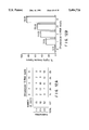
- FIG. 12A is a chart that shows the distribution of stromelysin-3 RNA levels as a function of local invasiveness in head and neck carcinomas.
- Stromelysin-3 RNA levels were quantified by Northern blot densitometry, and separated in classes as described herein; 0, no expression; I, >0-2.5 units; II, >2.5-6 units; III, >6-15 units; IV, levels higher than 15 units. Invasiveness was scored from (1+) to (3+) as described herein.
- FIG. 12B plots the percentage of highly invasive tumors, that is, the ratio of highly invasive tumors to the total number of tumors for each class of stromelysin-3 RNA level.
- FIGS. 13A-13L depict bright-field and dark-field photomicrographs of paraffin-embedded tissue sections stained with hematoxylin after in situ hybridization with [ 35 S]-labeled stromelysin-3 (ST3) antisense RNA.
- FIG. 13A is a bright-field photomicrograph where the tissue section is comedo carcinoma in situ ( * , ⁇ 100) with segmental ST3 gene expression in surrounding stromal cells (S).
- FIG. 13B is a dark-field photomicrograph of the comedo carcinoma in situ that is shown in FIG. 13A.
- FIG. 13C is a higher-power view ( ⁇ 400) of the tissue section shown in FIG. 13A, where arrows indicate fibroblastic cells expressing ST3 transcripts. Note that were the basement membrane is thickened (rounded arrow), there is no ST3-expressing fibroblastic cells.
- FIG. 13D is a bright-field photomicrograph of muciparous ductal carcinoma in situ ( * , ⁇ 100) with segmental ST3 gene expression in surrounding stromal cells (S).
- FIG. 13E is a dark-field photomicrograph of the tissue section shown in FIG. 13D.
- FIG. 13F is a higher-power view ( ⁇ 400) of the tissue section shown in FIG. 13D, where arrows indicate fibroblastic cells expressing ST3 transcripts in a tumoral area where basement membrane integrity is questionable.
- FIG. 13G is a bright-field photomicrograph of micropapillary ductal carcinoma in situ ( * , ⁇ 100) with focal ST3 gene expression in surrounding stromal cells (S).
- FIG. 13H is a dark-field photomicrograph of the tissue section shown in FIG. 13G.
- FIG. 13I is a bright-field photomicrograph of three cribriform ductal carcinoma in situ ( * , ⁇ 100), two of said carcinomas having circumferential ST3 gene expression in surrounding stromal cells (S) and one with no ST3 gene expression.
- FIG. 13J is a dark-field micrograph of the tissue section shown in FIG. 13I.
- FIG. 13K is a bright-field photomicrograph of two capillary ductal carcinomas in situ ( * , ⁇ 100), one having ST3 gene expression in the surrounding stromal cells (S), and one having no ST3 gene expression. Note the high level of ST3 transcripts in the stromal cells adjacent to the area of invasive carcinoma (C).
- FIG. 13L is a dark-field micrograph of the same tissue section shown in FIG. 13K.
- FIG. 13M-FIG. 13O are photomicrographs of frozen tissue sections ( ⁇ 200) that have been subject to indirect immunoperoxydase staining of stromelysin-3 (ST3) protein.
- FIG. 13M is a photomicrograph of frozen invasive carcinoma (C) after immunohistochemical analysis with polyclonal antibody 349.
- FIG. 13N is a photomicrograph of frozen invasive carcinoma (C) after immunohistochemical analysis with monoclonal antibody 5ST-4A9-3.
- FIG. 13O is a photomicrograph of frozen in situ ductal carcinoma ( * , comedo type) after immunohistochemical analysis with monoclonal antibody 5ST-4A9-3.
- ST3 is exclusively detected in elongated fibroblast-like cells tumoral stroma (S) surrounding neoplastic cells.
- FIG. 14A depicts northern blot analysis of stromelysin-3 RNA in breast tissue and breast cancer of metastatic lymph nodes. ST3 RNA has been detected in all the evasive ductal and lobular breast carcinomas so far tested by northern blot analysis, but not in the normal breast samples tested in parallel.
- FIG. 14B also depicts northern blot analysis of stromelysin-3 RNA in breast cancer metastatic lymph nodes. 8 ⁇ M of total RNA was loaded in each lane. Primary tumors (invasive ductal carcinomas) and metastatic lymph nodes in each vertical lane were obtained from the same patient. Hybridization was performed with [ 32 P]-labeled stromelysin-3 cDNA, and the blots were rehybridized with [ 32 P]-labeled 36B4 cDNA (Masiakowski et al., Nucleic Acid Res. 10:7895-7903 (1982)), to check for RNA loading and transferring each lane. Autoradiography was for two days.
- FIG. 15A-FIG. 15H are photomicrographs of paraffin-embedded tissue sections ( ⁇ 100) stained with hematoxylin, after in situ hybridization with [ 35 S]-labeled stromelysin-3 antisense RNA.
- FIG. 15A is a bright-field photomicrograph of a lymph node tissue section.
- FIG. 15B is a dark-field photomicrograph of the same section shown in FIG. 15A.
- FIG. 15C is a bright-field photomicrograph of a skin tissue sample.
- FIG. 15D is a dark-field photomicrograph of the same section shown in FIG. 15C.
- FIG. 15E is a bright-field photomicrograph of a pleura tissue section.
- FIG. 15F is a dark-field photomicrograph of the same section shown in FIG. 15E.
- FIG. 15G is a bright-field photomicrograph of a bone metastases tissue section.
- FIG. 15H is a dark-field photomicrograph of the same section shown in FIG. 15G.
- M refers to a metastatic tumor
- L refers to lymphocytes
- E refers to epiderm
- N refers to normal pleura.
- ST3 transcripts are exclusively detected in fibroblastic cells of tumor stroma surrounding neoplastic cells. Autoradiography was for four weeks.
- FIG. 16A-FIG. 16I are photomicrographs of serial paraffin-embedded tissue sections ( ⁇ 100) of ductal carcinomas stained with hematoxylin, after in situ hybridization with [ 35 S]-labeled antisense RNA probes.
- S stroma
- * in situ (comedo)
- C invasive carcinoma
- N subnormal breast
- V blood vessel.
- FIG. 16A is a bright-field photomicrograph of ductal carcinoma serial section.
- FIG. 16B is a dark-field photomicrograph of the same section hybridized with stromelysin-3.
- FIG. 16C is dark-field micrograph of the same serial section hybridized with 72-kDa type IV collagenase antisense RNAs.
- ST3 transcripts are detected in fibroblastic cells of tumor stroma immediately surrounding cancer cells, while type IV collagenase transcripts are observed in fibroblastic cells distributed throughout the stroma.
- FIG. 16D is a bright-field photomicrograph of a serial tissue section of ductal carcinoma.
- FIG. 16E is a dark-field photomicrograph of the same serial section as FIG. 16D after hybridization with ST3.
- FIG 16F is a dark-field photomicrograph of the same serial section as shown in FIG. 16D after hybridization with urokinase antisense RNAs.
- FIG. 16G is a bright-field photomicrograph of a serial ductal carcinoma tissue section.
- FIG. 16H is a dark-field photomicrograph of the same serial section as shown in FIG. 16G after hybridization with ST3.
- FIG. 16I is a dark-field photomicrograph of the same serial section shown in FIG. 16G after hybridization with urokinase antisense RNAs.
- FIGS. 16A-16I ST3 and urokinase transcripts are detected in the same fibroblastic cells of tumor stroma (S) immediately surrounding invasive (C) and in situ ( * ) neoplastic cells.
- S tumor stroma
- C tumor stroma
- * in situ neoplastic cells.
- FIG. 16C exposure time was for two weeks.
- FIG. 16B, FIG. 16E and FIG. 16F exposure time was for four weeks.
- FIG. 16H and FIG. 16I exposure time was for six weeks.
- FIG. 17 depicts a comparative nothern blot analysis of stromalysin-3 72-kDa type IV collagenase and urokinase RNAs in breast tumors. 8 ⁇ M of total RNA from four fibroid adenomas and 11 invasive carcinomas (1-10, ductal and 11, lobular) were loaded in each lane. Hybridization was successfully performed on the same blot [ 32 P]-labeled stromalysin-3 [ST3], 72-kDa type IV collagenase [IV COL (72)] and urokinase (u-PA) cDNAs.
- Hybridization with [ 32 P]-labeled 36B4 cDNA was made to check for RNA loading and transfer in each lane. Autoradiography was for two days (ST3), one day (IV COL) and four days (u-PA).
- the present invention provides a process for the diagnosis of invasive cancer, especially breast, head and neck, and skin carcinomas, comprising the detection of either stromelysin-3, or a nucleotide sequence encoding stromelysin-3.
- the present invention provides the use of an agent to interfer with the synthesis or activity of stromelysin-3 in the treatment or prophylaxis of invasive cancer, especially breast, head and neck, and skin carcinomas.
- metastatic tumors are invasive, but that invasive tumors are not necessarily metastatic (for example basal cell skin carcinomas).
- stromelysin-3 As expression of the stromelysin-3 gene is specific to regions of ECM degradation and apparently encodes a metalloproteinase, it is assumed that its ECM degrading activity is crucial to tumor progression into metastasis. Expression of stromelysin-3 by the stromal cells is likely to break down an important part of the ECM, thereby allowing cancerous cells to migrate away from the parent tumor.
- any agent which can affect the activity of stromelysin-3 will have an effect on metastasis.
- agents will suitably be those which either prevent synthesis of the protein, prevent maturation of the protein, or alter the activity of the enzyme, either by blocking or by altering its activity.
- stromelysin-3 gene was found to be, in the first instance, diagnostic of breast cancer in the metastatic phase. In fact, this result was achieved by the detection of mRNA in a variety of resetted tumors. Breast cancer was chosen, as this is responsible for the highest death rate, by cancer, in the non-smoking female population.
- Stromelysin-3 is a novel protein almost certainly belonging to the MMP family, and is associated with invasive breast carcinomas, irrespective of their hormonal status.
- the members of the MMP family require an activation step, which may be associated with removal of the pre- and pro- sequences, to become active.
- the amino acid sequence of pro- and mature stromelysin-3 is notably different from those of the previously characterized MMPs, and may exhibit distinct properties regarding maturation, activation and specificity for ECM components.
- the stromelysin-3 gene is expressed by all primary invasive breast carcinomas, by some of their metastases, and in tissues in which extensive ECM remodelling is known to occur (uterus, placenta and limb bud) analyzed for such expression, but not in breast fibroadenomas and normal adult tissues, suggesting that the stromelysin-3 gene product plays an important role in breast cancer progression. Also in agreement with this concept, the stromelysin-3 gene is not expressed in most in situ breast carcinomas, with the exception of in situ carcinomas of the comedo type (Example 11), which are usually considered as preinvasive lesions and are often associated with microinvasion. Thus the presence of stromelysin-3 RNA transcripts in other than the low concentrations found elsewhere in the body, other than uterus or placenta, is diagnostic of a metastatic cancer or of a cancer with a high risk of becoming invasive.
- Stromelysin-3 may be involved in the lyric processes which are likely to be associated with invasive tumor growth. Alternatively, it is possible that stromelysin-3 could also play a role in the formation of desmoplasia, which is associated with most invasive breast cancer lesions, and may represent a host reaction to prevent further malignant cell spread (Ahmed, A., Pathol. Annu. 25(Pt2):237-286 (1990)). In such an instance, enhancement of stromelysin-3 activity would be advantageous.
- the restricted expression of the stromelysin-3 gene in stromal fibroblasts immediately surrounding the neoplastic cell islands is strikingly in contrast to collagenase IV, another metalloproteinase known to be associated with the malignant conversion of some tumorigenic cells, and cathepsin D, a lysosomal aspartyl protease whose expression is increased in breast carcinomas, both of which are expressed, not in the fibroblasts, but in the neoplastic epithelial cells of breast cancers (Monteagudo et al., Am. J. Pathol. 136:585-592 (1990); Garcia et al., Steroid Biochem. 27:439-445 (1987)).
- a cDNA library was constructed, and substracted with poly (A+) RNA from a fibroadenoma source. By this process, the cDNA library was enriched for sequences characteristic of metastatic cancers.
- a number of clones was grown up and screened using probes derived from poly(A + ) RNA from metastatic tumors and from fibroadenomas. Those clones which bound more greatly to the probes derived from metastatic cancer poly(A + ) RNA were then grown up further.
- nucleotide probes to establish the occurrence of stromelysin-3 mRNA revealed a tissue distribution as described above, and also enabled photomicrographs to exactly locate the areas of expression of the stromelysin-3 gene by labelling.
- stromelysin-3 gene was not expressed in the cancerous cells itself, but in the surrounding stroma.
- the stroma did not exhibit any evidence of stromelysin-3 mRNA when the basement membrane of the tumor was still intact (see FIG. 6A-FIG. 6I).
- the stromelysin-3 gene is expressed by all primary invasive breast carcinomas, by some of their metastases nodes, and in tissues in which extensive ECM remodelling is known to occur (uterus, placenta and limb bud) analyzed for such expression, but not in breast fibroadenomas and normal adult tissues, suggesting that the stromelysin-3 gene product plays an important role in breast cancer progression.
- the stromelysin-3 gene is not expressed in most in situ breast carcinomas, with the exception of in situ carcinomas of the comedo type (Example 11), which are usually considered as preinvasive lesions and are often associated with microinvasion.
- Stromelysin-3 always occurs in the stroma of metastatic cancers, and does not occur in the stroma of in situ primary tumors (tumors still having a basement membrane and which are non-invasive).
- the presence of stromelysin-3 RNA transcripts in other than the low concentrations found elsewhere in the body, other than uterus or placenta is diagnostic of a metastatic cancer or of a cancer with a high risk of becoming invasive.
- stromelysin-3 gene was not detected in any ER-positive or negative breast cancer cell lines, even though some of them are known to secrete and possess receptors for EGF/TGF- ⁇ and FGF (factors which are implicated in expression of the stromelysin-3 gene).
- standard detection techniques applied to stromelysin-3, its precursors or its coding nucleotide sequences may be used to diagnose a metastatic cancer, or to confirm that a primary tumor has not yet reached the fatal metastatic phase.
- Such techniques may include detection with nucleotide probes, such as in the manner described above, or may comprise detection of the stromelysin-3 protein by, for example, antibodies or their equivalent.
- the nucleotide probes may be any that will hybridize more strongly to the sequence shown in the accompanying FIG. 2A and FIG. 2B than to other naturally occurring sequences.
- Types of probe include cDNA, riboprobes, synthetic oligonucleotides and genomic probes.
- the type of probe used will generally be dictated by the particular situation, such as riboprobes for in situ hybridization, and cDNA for Northern blotting, for example.
- the most preferred probes are those which correspond to the negative strand of the cDNA of FIG. 2A and FIG. 2B. It is also possible to provide probes which recognize introns located within the stromelysin-3 gene, but this is not necessarily as reliable as detecting RNA transcripts.
- stromelysin-3 encoding gene per se, will generally serve no purpose in diagnosis, but other forms of assay to detect transcripts and other expression products will generally be useful.
- the probes may be as short as is required to differentially recognize stromelysin-3 mRNA transcripts, and may be as short as, for example, 15 bases.
- the form of labelling of the probes may be any that is appropriate, such as the use of radioisotopes, for example, 32 P and 35 S. Labelling with radioisotopes may be achieved, whether the probe is synthesized chemically or biologically, by the use of suitably labelled bases. Other forms of labelling may include enzyme or antibody labelling such as is characteristic of ELISA, but detection of mRNA transcripts by labelled probes will generally be by way of X-radiography.
- RNA transcripts may be achieved by Northern blotting, for example, wherein a preparation of RNA is run on a denaturing agarose gel, and transferred to a suitable support, such as activated cellulose, nitrocellulose or glass or nylon membranes. Radiolabelled cDNA or RNA is then hybridized to the preparation, washed and analyzed by autoradiography.
- a suitable support such as activated cellulose, nitrocellulose or glass or nylon membranes.
- In situ hybridization visualization may also be employed (Example 6), wherein a [ 35 S]-labelled antisense cRNA probe is hybridized with a thin section of a biopsy sample, washed, cleaved with RNase and exposed to a sensitive emulsion for autoradiography.
- the samples may be stained with haematoxylin to demonstrate the histological composition of the sample, and dark field imaging with a suitable light filter shows up the developed emulsion.
- Immunohistochemistry may be used to detect expression of stromelysin-3 in a biopsy sample.
- a suitable antibody is brought into contact with, for example, a thin layer of cells, washed, and then contacted with a second, labelled antibody.
- Labelling may be by enzyme, such as peroxidase, avidin or by radiolabelling. Chromogenic labels are generally preferable, as they can be detected under a microscope.
- stromelysin-3 More generally preferred is to detect the protein by immunoassay, for example by ELISA or RIA, which can be extremely rapid.
- immunoassay for example by ELISA or RIA
- the substrate may not be necessary to label the substrate, provided that the product of the enzymatic process is detectable and characteristic in its own right (such as hydrogen peroxide for example). However, if it is necessary to label the substrate, then this may also comprise enzyme labelling, labelling with radioisotopes, antibody labelling, fluorescent marker labelling or any other suitable form which will be readily apparent to those skilled in the art.
- Antibodies may be prepared as described below, and used in any suitable manner to detect expression of stromelysin-3.
- Antibody-based techniques include ELISA (enzyme linked immunosorbent assay) and RIA (radioimmunoassay). Any conventional procedures may be employed for such immunoassays. The procedures may suitably be conducted such that: a stromelysin-3 standard is labelled with a radioisotope such as 125 I or 35 S, or an assayable enzyme, such as horseradish peroxidase or alkaline phosphatase and, together with the unlabelled sample, is brought into contact with the corresponding antibody, whereon a second antibody is used to bind the first and radioactivity or the immobilized enzyme assayed (competitive assay); alternatively, stromelysin-3 in the sample is allowed to react with the corresponding immobilized antibody, radioisotope- or enzyme-labelled anti-stromelysin-3 antibody is allowed to react with the system and radioactivity or the enzyme assayed (ELISA-sandwich assay). Other conventional methods may also be employed as suitable.
- the above techniques may be conducted essentially as a “one-step” or “two-step” assay.
- the “one-step” assay involves contacting antigen with immobilized antibody and, without washing, contacting the mixture with labeled antibody.
- the “two-step” assay involves washing before contacting the mixture with labeled antibody.
- Other conventional methods may also be employed as suitable.
- Enzymatic and radio-labelling of stromelysin-3 and/or the antibodies may be effected by conventional means.
- Such means will generally include covalent linking of the enzyme to the antigen or the antibody in question, such as by glutaraldehyde, specifically so as not to adversely affect the activity of the enzyme, by which is meant that the enzyme must still be capable of interacting with its substrate, although it is not necessary for all of the enzyme to be active, provided that enough remains active to permit the assay to be effected.
- some techniques for binding enzyme are non-specific (such as using formaldehyde), and will only yield a proportion of active enzyme.
- Enzymes employable for labelling are not particularly limited, but may be selected from the members of the oxidase group, for example. These catalyze the production of hydrogen peroxide by reaction with their substrates, and glucose oxidase is often used for its good stability, ease of availability and cheapness, as well as the ready availability of its substrate (glucose). Activity of the oxidase may be assayed by measuring the concentration of hydrogen peroxide formed after reaction of the enzyme-labelled antibody with the substrate under controlled conditions well-known in the art.
- stromelysin-3 may be detected according to preference.
- Western blotting Towbin et at., Proc. Nat. Acad. Sci. 76:4350 (1979)
- a suitably treated sample is run on an SDS PAGE gel before being transferred to a solid support, such as a nitrocellulose filter.
- Anti-stromelysin-3 antibodies are then brought into contact with the support and assayed by a secondary immunological reagent, such as labelled protein A or anti-immunoglobulin (suitable labels including 125 I, horseradish peroxidase and alkaline phosphatase).
- Samples for diagnostic purposes may be obtained from any relevant site.
- a sample obtained direct from the tumor such as the stroma or cytosol, may be ideal, but it may also be appropriate to obtain the sample from blood, for example.
- highly sensitive assays may be required, as the amount of stromelysin-3 would then be diluted through the bloodstream.
- diagnosis may be of particular importance in monitoring progress of a patient, such as after surgery to remove a tumor. If a reference reading is taken after the operation, then another taken at regular intervals, any rise could be indicative of a relapse, or possibly a metastasis. The taking of such readings may need to take into account activity in the uterus, for example.
- Anti-stromelysin-3 antibodies may also be used for imaging purposes.
- suitable labels include radioisotopes, iodine ( 125 I, 121 I), carbon ( 14 C), salphee ( 35 S), tritium ( 3 H), indium ( 112 In), and technetium ( 99m Tc), fluorescent labels, such as fluorescein and rhodamine, and biotin.
- Markers for this purpose may be any that do not substantially interfere with the antibody binding, but which allow external detection.
- Suitable markers may include those that may be detected by X-radiography, NMR or ESR.
- suitable markers include any radioisotope that emits detectable radiation but that is not overtly harmful to the patient, such as barium or caesium, for example.
- suitable markers for NMR and ESR generally include those with a detectable characteristic spin, such as deuterium, which may be incorporated into the antibody by suitable labelling of nutrients for the relevant hybridoma, for example.
- an antibody or antibody fragment which has been labelled with an appropriate detectable imaging moiety such as a radioisotope (for example, 131 I, 112 In, 99m Tc), a radio-opaque substance, or a material detectable by nuclear magnetic resonance, is introduced (for example, parenterally, subcutaneously or intraperitoneally) into the subject (such as a human) to be examined.
- an appropriate detectable imaging moiety such as a radioisotope (for example, 131 I, 112 In, 99m Tc), a radio-opaque substance, or a material detectable by nuclear magnetic resonance
- the size of the subject, and the imaging system used, will determine the quantity of imaging moiety needed to produce diagnostic images.
- the quantity of radioactivity injected will normally range from about 5 to 20 millicuries of technetium-99m.
- the labelled antibody or antibody fragment will then preferentially accumulate at the location of cells which contain stromelysin-3.
- the labelled antibody or antibody fragment can then be detected using known techniques.
- the antibodies may be raised against either a peptide of stromelysin-3 or the whole molecule.
- a peptide may be presented together with a carrier protein, such as an albumin, to an animal system or, if it is long enough, say 25 amino acid residues, without a carrier.
- Human antibodies are unlikely to be able to recognize stromelysin-3, as this protein will represent a self protein.
- peptide means any molecule comprising 2 or more amino acids linked via a peptide bond. As such, the term includes oligopeptides, polypeptides and proteins.
- Polyclonal antibodies generated by the above technique may be used direct, or suitable antibody producing cells may be isolated from the animal and used to form a hybridoma by known means (Kohler and Milstein, Nature 256:795 et seq. (1975)). Selection of an appropriate hybridoma will also be apparent to those skilled in the art, and the resulting antibody may be used in a suitable assay to identify stromelysin-3.
- Antibodies, or their equivalents, may also be used in accordance with the present invention for the treatment or prophylaxis of metastatic cancers.
- Administration of a suitable dose of the antibody may serve to block production, or to block the effective activity of stromelysin-3, and this may provide a crucial time window in which to treat the malignant growth.
- Prophylaxis may be appropriate even at very early stages of the disease, as it is not known what actually leads to metastasis in any given case.
- administration of the antibodies, their equivalents, or factors, such as TIMPs (naturally occurring compounds which regulate the MMPs--tissue inhibitors of metalloproteinases), which interfere with stromelysin-3 activity may be effected as soon as cancer is diagnosed, and treatment continued for as long as is necessary, preferably until the threat of the disease has been removed.
- a preferred form of treatment is to employ the so-called magic bullet technique, where a suitable toxin is attached to the antibodies which then target the area of the tumor.
- a suitable toxin is attached to the antibodies which then target the area of the tumor.
- toxins are well known in the art, and may comprise toxic radioisotopes, heavy metals, enzymes and complement activators, as well as such natural toxins as ricin which are capable of acting at the level of only one or two molecules per cell. It may also be possible to use such a technique to deliver localized doses of hormone antagonists or any other suitable physiologically active compounds, which may be used, for example, to treat cancers.
- antibodies for use in accordance with the present invention may be monoclonal or polyclonal as appropriate.
- Antibody equivalents of these may comprise: the Fab' fragments of the antibodies, such as Fab, Fab', F(ab') 2 and Fv; idiotopes; or the results of allotope grafting (where the recognition region of an animal antibody is grafted into the appropriate region of a human antibody to avoid an immune response in the patient), for example.
- Other suitable modifications and/or agents will be apparent to those skilled in the art.
- inhibitors may be general (for ECM degrading enzymes, for example), or specific for stromelysin-3.
- Tissue inhibitors of metalioproteinases are known to exist, and it is extremely likely that there is a specific TIMP for stromelysin-3. Such a TIMP is easily identifiable by standard techniques.
- Synthetic inhibitors of stromelysin-3 may also be manufactured, and these will generally correspond to the area of the substrate affected by the enzymatic activity. It is generally preferred that such inhibitors correspond to a frozen intermediate between the substrate and the cleavage products, but it is also possible to provide a sterically hindered version of the binding site, or a version of the binding site which will, itself, irreversibly bind to stromelysin-3. Other suitable inhibitors will be apparent to the skilled person.
- stromelysin-3 activity may also be employed. These may constitute denaturing agents, for example, although these tend to be non-specific and could only be adequately employed if they could be targeted, such as by the use of specific antibodies.
- denaturing agents for example, although these tend to be non-specific and could only be adequately employed if they could be targeted, such as by the use of specific antibodies.
- Other forms of stromelysin-3 blocking activity could be effected by blocking the progress from pre-proprotein through to protein. This process provides several target stages, and it is only necessary to identify a stage which can be independently blocked so as not to affect other vital enzymes, or which can again be targeted.
- peptides or other small molecules may also be possible to use peptides or other small molecules to selectively recognize a tertiary structure on stromelysin-3, thereby blocking its enzymic activity.
- an activity blocker need not necessarily bind the active site, but may serve to alter or freeze the tertiary structure of stromelysin-3, destroying, suspending or altering its activity.
- the blocker also need not necessarily act by itself, but may be linked to another molecule for this purpose, or may serve as a recognition site for a suitable inactivating agent.
- the ECM glycoprotein tenascin (Chiquet-Ehrismann et al., Cell 47:131-139 (1986)) appears to play an essential role in epithelial mesenchyme cell interactions and cell migration during normal development, including that of the mammary gland during organogenesis.
- Tenascin has consistently been found to be over-expressed in the fibrous stroma of malignant breast tumors, and appears to be induced in a similar manner to stromelysin-3. When compared with fibronectin, tenascin is a poor substrate for attachment of mammary tumor epithelial cells, suggesting that it may allow them to become invasive.
- stromelysin-3 may act in concert with tenascin during the invasive phase of breast cancer.
- Stromelysin-3 and tenascin may also be co-expressed during embryogenesis in the regions where epithelium-mesenchyme interactions are known to play an important role, and where cell migration is taking place.
- the present invention also provides a process for the diagnosis of metasiatic cancer as defined above, further comprising the detection of any of the foregoing proteins, or a nucleotide sequence encoding them.
- the invention also provides a use in the treatment or prophylaxis of metastatic cancer, further comprising the use of an agent to bind any of the foregoing proteins.
- the present invention further provides a nucleotide sequence encoding all or part of stromelysin-3.
- the sequence of stromelysin-3 is preferably that shown in FIG. 2A and FIG. 2B of the accompanying drawings, whilst the nucleotide sequence is also preferably that shown in FIG. 2A and 2B.
- the nucleotide sequence may be substantially different from that shown in the Figure, due to degeneracy in the genetic code, provided that it still encodes at least a part of stromelysin-3.
- the necessary sequence may vary even further, according to the use to which it is to be put. If it is intended for use to detect RNA transcripts in biological samples, then it will usually be preferable that it more nearly corresponds to the sequence given in FIG. 2A and FIG. 2B. However, the sequence may still vary, provided that hybridization is possible under the selected conditions of stringency.
- a probe may be reverse-engineered by one skilled in the art from the peptide sequence of FIG. 2A and FIG. 2B.
- use of such probes may be limited, as it will be appreciated that any one given reverse-engineered sequence will not necessarily hybridize well, or at all with any given complementary sequence reverse-engineered from the same peptide, owing to the degeneracy of the genetic code. This is a factor common in the calculations of those skilled in the art, and the degeneracy of any given sequence is frequently so broad as to yield a large number of probes for any one sequence.
- nucleotide sequence is required for expression of a stromelysin-3 peptide or entire enzyme, then there may be a considerably greater leeway, both as described above with respect to the genetic code, and also to the fact that some amino acid sequence of stromelysin-3 may be varied without significant effect on the structure or function of the enzyme.
- the present invention also includes any variants and mutants on the sequence which still show substantial stromelysin-3 activity, or which exhibit characteristic regions of stromelysin-3 for use in generating antibodies, for example.
- variants and routants include deletions, additions, insertions, inversions, repeats and type-substitutions (for example, substituting one hydrophilic residue for another, but not strongly hydrophilic for strongly hydrophobic as a rule).
- Small changes will generally have little effect on activity, unless they are an essential part of the molecule, and may be a side-product of genetic manipulation, for example, when generating extra restriction sites, should such be desired.
- Modification may also include replacement of one or more of the residues with any other suitable residue, and such replacement may either be 1:1 or any other suitable ratio, greater or less than unity.
- Spot mutations and other changes in the coding sequence may be effected to add or delete restriction sites, for example, to otherwise assist in genetic manipulation/expression, or to enhance or otherwise conveniently to modify the stromelysin-3 molecule.
- stromelysin-3 equivalent will be found in other animals, especially mammals, and sequence information from such sources can be of particular importance to elucidate the conserved regions of the stromelysin-3 molecule.
- the corresponding sequence in the mouse is ⁇ 80% conserved, including such as the 10 amino acid sequence in the prodomain characteristic of stromelysin-3 (Lefebvre et al., J. Cell Biol. 119:997-1002 (1992)).
- animal sequences corresponding to human stromelysin-3 sequences will be readily detectable by methods known in the art and described above, and such sequences and their peptides, as well as mutants and variants thereof, form a part of the invention.
- sequences of the invention may also be engineered to provide restriction sites, if desired. This can be done so as not to interfere with the peptide sequence of the encoded stromelysin-3, or may interfere to any extent desired or necessary, provided that the final product has the properties desired.
- hybridization can be an unreliable indication of sequence homology
- preferred sequences will generally be those showing in excess of 50%, preferably 70% and more preferably 80% homology with the sequence of FIG. 2A and FIG. 2B.
- stromelysin-3 is originally expressed as a pre-proenzyme. Thus, two stages of cleavage are observed in vivo. Cleavage is not necessarily a requirement for in vitro expression, and it may be possible for E. coli, for example, to express the mature protein.
- any suitable system can be used.
- suitable vectors, expression vectors and constructions therefor will be apparent to those skilled in the art.
- characteristic is meant any peptide which has a sequence unique to stromelysin-3. Such a sequence may be important to stromelysin-3 activity, or may just be a sequence not found in other peptides. However, sequences important to stromelysin-3 activity are generally preferred, as these are more likely to be conserved within a population.
- Suitable expression vectors may be based on phages or plasmids, both of which are generally host-specific, although these can often be engineered for other hosts.
- Other suitable vectors include cosmids and retroviruses, and any other vehicles, which may or may not be specific for a given system.
- control sequences such as recognition, promoter, operator, inducer, terminator and other sequences essential and/or useful in the regulation of expression, will be readily apparent to those skilled in the art, and may be associated with the natural stromelysin-3 sequence or with the vector used, or may be derived from any other source as suitable.
- the vectors may be modified or engineered in any suitable manner.
- a cDNA fragment encoding the stromelysin-3 of the invention may easily be inserted into a suitable vector.
- the receiving vector has suitable restriction sites for ease of insertion, but blunt-end ligation, for example, may also be used, although this may lead to uncertainty over reading frame and direction of insertion. In such an instance, it is a matter of course to test transformants for expression, 1 in 6 of which should have the correct reading frame.
- Suitable vectors may be selected as a matter of course by those skilled in the art according to the expression system desired.
- the desired stromelysin-3 By transforming a suitable organism or, preferably, eukaryotic cell line, such as HeLa, with the plasmid obtained, selecting the transformant with ampicillin or by other suitable means if required, and adding tryptophan or other suitable promoter-inducer (such as indoleacrylic acid) if necessary, the desired stromelysin-3 may be expressed.
- the extent of expression may be analyzed by SDS polyacrylamide gel electrophoresis--SDS-PAGE (Lemelli, Nature 227:680-685 (1970)).
- Suitable methods for growing and transforming cultures etc. are usefully illustrated in, for example, Maniatis (Molecular Cloning, A Laboratory Notebook, Maniatis et al. (eds.), Cold Spring Harbor Labs, N.Y. (1989)).
- Cultures useful for production of stromelysin-3, or a peptide thereof may suitably be cultures of any living cells, and may vary from prokaryotic expression systems up to eukaryotic expression systems.
- One preferred prokaryotic system is that of E. coli, owing to its ease of manipulation.
- a higher system such as a mammalian cell line, for expression of a eukaryotic protein.
- Currently preferred cell lines for transient expression are the HeLa and Cos cell lines.
- Other expression systems include the Chinese Hamster Ovary (CHO) cell line.
- baculovirus system wherein butterfly cells are cotransfected with a DNA vector encoding stromelysin-3, or a suitable peptide, and baculovirus DNA. Recombination occurs within the cell, and suitable baculovirus recombinants may be selected by standard techniques. Thereafter, the recombinant may be used to infect the cell line as desired, stromelysin-3 or peptide being expressed on infection.
- a particular advantage of this system is the amount of protein produced, which can be in the range of about 1 to about 500 mg/litre.
- E. coil does not employ the same system for processing pre-proproteins as mammalian cells.
- streptomycetes for example, and yeasts, such as Saccharomyces spp., especially S. cerevisiae. Any system may be used as desired, generally depending on what is required by the operator. Suitable systems may also be used to amplify the genetic material, but it is generally convenient to use E. coli for this purpose where only proliferation of the DNA is required.
- the peptide or nucleotide sequence may be any that is characteristic of stromelysin-3, having consideration to the purpose to which it is to be put. Ideally, the sequences would be completely characteristic of stromelysin-3, but the length of such sequences may vary according to the region of the stromelysin-3 molecule. The most preferred regions are those which are highly conserved, and which are not shared with other proteins, although it may be advantageous if the sequence is characteristic of the MMPs or, more particularly, those MMPs associated with invasive tumors.
- the invention includes and relates to equivalents of the above peptide and nucleotide sequences, the term "equivalent” being used in the sense of the preceding description, that is to say, equivalents in the sense of sequences having substitutions at the C- or N-terminals, or anywhere else.
- the invention also includes routants of the sequences, the term "mutants” being used with reference to deletions, additions, insertions, inversions and replacement of amino acid residues or bases in the sequence subject to the restrictions described above.
- the present invention further includes variants of the sequences, which term is used in relation to other naturally occurring stromelysin-3 which may be discovered from time to time and which shares essentially the same sequence as shown in FIG. 2A and FIG. 2B, but which vary therefrom in a manner to be expected within a large population.
- allelic variation and those peptides from other species showing a similar type of activity and having a related sequence are also included, although less preferred, are animal sequences.
- stromelysin-3 expression can be stimulated by, for example, growth factors and tumor promoters.
- growth factors and tumor promoters include EGF FGF and PDGF and TPA.
- detection of any of these factors in a tumor sample may also help to diagnose the metasiatic condition of a cancer.
- the invention also provides the treatment of a metastatic cancer by altering the expression of the stromelysin-3 gene. This may be effected by interfering with the factor required to stimulate stromelysin-3 production, such as by directing specific antibodies against the factor, which antibodies may be further modified to achieve the desired result. It may also be possible to block the receptor for the factor, something which may be more easily achieved by localization of the necessary binding agent, which may be an antibody or synthetic peptide, for example.
- Affecting stromelysin-3 gene expression may also be achieved more directly, such as by blocking of a site, such as the promoter, on the genomic DNA.
- the present invention provides for the administration of, for example, antibodies to a patient, then this may be by any suitable route. If the tumor is still thought to be, or diagnosed as, localized, then an appropriate method of administration may be by injection direct to the site. If the target is breast cancer, then an injection to the breast may suffice, or an implant may be used. If TIMPs are to be administered, for example, then it may also be possible to employ a dermal patch for prolonged administration.
- a further option may be oral administration, for example, by means of gargling.
- administration may instead, or additionally, be by injection, including subcutaneous, intramuscular, intravenous and intradermal injections.
- Formulations may be any that are appropriate to the route of administration, and will be apparent to those skilled in the art.
- the formulations may contain a suitable carrier, such as saline, and may also comprise bulking agents, other medicinal preparations, adjuvants and any other suitable pharmaceutical ingredients.
- Suitable preparations may also include vaccines comprising stromelysin-3 or a characteristic peptide thereof.
- Such vaccines may be active or passive, but passive is generally preferred as stromelysin-3 expression occurs in the uterus, and indefinite exposure to anti-stromelysin-3 antibodies may have undesirable effects.
- active vaccination may be advantageous, especially where a patient has had a hysterectomy, as no tissues will then normally express stromelysin-3.
- Other suitable vaccines include recombinant viruses containing a nucleotide sequence encoding a stromelysin-3 or a characteristic peptide thereof.
- One suitable such virus is the vaccinia virus.
- a breast cancer cDNA library was constructed in the ⁇ gt10 vector using poly(A + ) RNA from a surgical resection-sample (referred to as tumor C1) of a primary breast cancer.
- 50,000 plaques were differentially screened using (+) and (-) probes corresponding to cDNAs reverse-transcribed from C1-poly(A + ) RNA and poly(A + ) RNA from a breast fibroadenoma (referred to as F1), respectively.
- FIG. 1A-FIG. 1D show a Northern blot analysis of total RNA from C1-breast carcinoma and F1-fibroadenoma using cDNA probes of four genes (A-D) exhibiting higher levels of expression in the carcinoma than in the fibroadenoma. Each lane contained 8 ⁇ g of total RNA. The filters were reprobed using the 36B4 probe which corresponds to an ubiquitously expressed gene (Rio et al., Proc. Nat. Acad. Sci. USA 84:9243-9247 (1987)).
- RNA was prepared (Chirgwin et al., Biochemistry 18:5294-5299 (1979)) from surgical specimens stored in liquid nitrogen, and poly(A + ) RNA was selected by oligo(dT)-cellulose chromatography.
- a breast cancer-enriched cDNA library was constructed using cDNA prepared from an oestrogen receptor-negative, grade II, ductal carcinoma (referred to as C1), in which stromal cells represented approximately 50% of the total cell population.
- the single-stranded cDNA was substracted with an excess of poly(A + ) RNA from a breast fibroadenoma (referred to as F1), and the single-stranded enriched material was purified by hydroxyapatite chromatography (Davis et al., Proc. Nat. Acad. Sci. USA 81:2194-2198 (1984); Rhyner et al., Neuroscience Res. 16:167-181 (1986)).
- the breast cancer-enriched cDNA was made double-stranded and cloned into the EcoRI site of the ⁇ gt10 vector. Three million recombinant phages were obtained, and ⁇ 50,000 were differentially screened using replica nylon filters (Biodyne A, Pall Corporation) from plates containing ⁇ 5,000 cDNA clones.
- (+) and (-) probes were made using C1-breast cancer cDNA and F1-breast fibroadenoma cDNA, respectively. Both probes were substracted (Davis et al., Proc. Nat. Acad. Sci. USA 81:2194-2198 (1984); Rhyner et al., Neuroscience Res. 16:167-181 (1986)) with an excess of total human liver RNA before [ 32 P]-labeling using random priming synthesis.
- Hybridizations were for two days under stringent conditions (50% formamide, 42° C.) and washing was in 2 ⁇ SSC, 0.1% SDS, at 22° C., followed by 0.1 ⁇ SSC, 0.1% SDS at 55° C. 130 differentially labelled plaques were selected for a second screening.
- the cDNA inserts of five differential plaques taken at random were purified by PCR amplification, [ 32 P]-labelled, and hybridized to all of the differential plaques to identify related clones. This procedure was repeated several times with differential plaques taken at random, finally yielding four genes referred to as A to D, which exhibited higher levels of expression in C1-carcinoma than in F1-fibroadenoma.
- the Northern blots for C1-breast cancer and F1-breast fibroadenoma were prepared using total RNA (8 ⁇ g) separated by electrophoresis in 1% agarose gels containing formaldehyde and transferred to Hybond-N filters (Amersham).
- the blots were stained with methylene blue before prehybridization to check for the integrity and amounts of transferred RNA. Hybridization (18 h) and washing were performed under standard conditions, as described above, using [ 32 P]-labeled cDNA inserts corresponding to A-D genes.
- the C gene Although expressed in colon (not shown), the C gene was partially characterized because of its high level of differential expression (FIG. 1A-FIG. 1D). It was also expressed in a variety of transformed epithelial cell lines and in normal human skin (not shown). Sequencing of the cDNA of one C clone indicated that the corresponding gene belongs to the keratin gene superfamily (data not shown).
- the D gene (also referred to herein as the stromelysin-3 gene) was further studied, because of its marked differential expression between C1-carcinoma and F1-fibroadenoma (FIG. 1A-FIG. 1D), and also because it was not expressed in normal human colon and in a number of other human tissues (infra).
- FIG. 2A and FIG. 2B shows the nucleotide sequence of the full length D cDNA and the corresponding protein sequence.
- the cDNA open reading frame encoding a 488 amino acid-long protein, is followed by a 714 base 3'-untranslated region containing a poly(A) addition signal located 14 bases upstream from the 3'-end of the RNA.
- a presumptive initiation methionine is located at nucleotide position 10-12.
- the corresponding AUG is not associated with and located in a sequence which conforms to the Kozak consensus motif, translation is probably initiated at this AUG, since the sequence immediately downstream corresponds to that for a hydrophobic leader peptide, an expected feature (infra).
- FIG. 2A and FIG. 2B which shows the nucleotide sequence of stromelysin-3 cDNA and deduced amino acid sequence
- the nucleotide residues are numbered in the 5' to 3' direction and deduced amino acids in the open reading frame are designated by their one-letter codes.
- the underlined nucleotide sequences correspond to: the putative signal peptide (two potential cleavage sites are marked by arrows); the PRCGVPD sequence characteristic of prometalloproteinases; the conserved histidine residues of the zinc-binding domain (Matrisian, L. M., Trends Genet. 6:21-125 (1990)); and the poly(A) addition signal sequence.
- a cDNA insert corresponding to the 3'-part of D cDNA [250 bp including a 19 bp poly(AT) region] was [ 32 P]-labeled by random priming synthesis and used to screen a non-substracted ⁇ gt10 cDNA library generated from C1-breast tumor poly(A + ) RNA by the method of Gubler and Hoffmann (Gene 25:262-269 (1983)).
- Several independent clones were identified and subcloned in M13 sequencing vector. DNA sequence was determined by the dideoxy method using sequenase and the deaza-dGTP reagent kit from US Biochemical. The sequence was analyzed using the PC/GENE software package.
- FIG. 3A-FIG. 3D shows a comparison of the predicted amino-acid sequences of human stromelysins and human type I collagenase.
- FIG. 3A, FIG. 3B and FIG. 3C Amino-acid sequences were aligned using a multialignment program (Higgins et al., Gene 73:237-244 (1988)). Amino-acid residues identical in all of the four sequences are marked by stars. The arrows denote putative signal peptide cleavage sites of stromelysin-3. The arrowhead points to the cleavage which occurs on activation of type I procollagenase and prostromelysins. The 10 amino-acid residues specific to stromelysin-3 at the level of this cleavage site are boxed. The PRCGVPD sequence and the conserved residues of the putative zinc-binding domain are underlined.
- FIG. 3D Left, regions of similarity (in percent amino-acid identity) between stromelysin-3, stromelysin-1 (ST1, Whitham et al., Biochem. J. 240:913-916 (1986)), stromelysin-2 (ST2, Muller et al., Biochem. J. 253:187-192 (1988)) and type I collagenase (COI, Whitham et al., Biochem. J. 240:913-916 (1986));
- the new protein belongs to the family of secreted matrix metalloproteinases (MMPs) (FIG. 3A-FIG. 3C). Accordingly, the new protein possesses an hydrophobic N-terminal leader sequence candidate (underlined in FIG. 2A and FIG. 2B), and exhibits the highly conserved sequence PRCGVPD (amino-acid residues 78-84), which is characteristic of the prodomain of the MMPs, as well as having the zinc binding site of MMPs (amino-acid residues 212-225--FIG. 3a) (Matrisian, L. M., Trends Genet. 6:121-125 (1990)).
- MMPs matrix metalloproteinases
- the N-terminal amino acid of the mature protein is likely to correspond to phenylalanine 98 of the pre-proprotein (Whitham et al., Biochem. J. 240:913-916 (1986)) (FIG. 3A-FIG. 3C).
- the similarity between the putative mature protein is 40% with stromelysin-1 (Whitham et al., Biochem. J. 240:913-916 (1986)), 38% with stromelysin-2 (Muller et al., Biochem. J. 253:187-192 (1988)) and 36% with type I collagenase (Goldberg et al., J. Biol. Chem. 261:6600-6605 (1986)) (FIG. 3D).
- the substrate specificity of the new protein is not known.
- it is referred to as stromelysin-3, although its similarity with stromelysin-1 (40%) is clearly much below that existing between stromelysin-1 and stromelysin-2 (79% ), and even lower than the similarity existing between type I collagenase and stromelysin-1 (53%) (FIG. 3D).
- the protein is an MMP, the cognomen "stromelysin” is not necessarily strictly accurate, but is convenient.
- stromelysin-3 has a unique short sequence (amino-acid residues 88-97) at a position corresponding substantially precisely with the proprotein cleavage site of type I collagenase and the stromelysins (Whitham et al., supra). Further, stromelysin-3, as with type I collagenase and the other stromelysins, does not exhibit the fibronectin-like domain characteristic of type IV collagenases (Wilhelm et al., J. Biol. Chem. 264:17213-17221 (1989)).
- stromelysin-3 RNA transcripts The occurrence of stromelysin-3 RNA transcripts was studied in resected samples of 30 breast carcinomas and five breast fibroadenomas.
- FIG. 4A-FIG. 4F shows Northern blot analyses of human metalloproteinase RNAs in breast tumors:
- FIG. 4A stromelysin-3 RNA
- FIG. 4B type I collagenase RNA (COI);
- FIG. 4C 92-kD type IV collagenase RNA (COIV 92K);
- FIG. 4D 72-kD type IV collagenase RNA (COIV 72K);
- FIG. 4E stromelysin-1 and -2 RNA's (ST1/2).
- FIG. 4F pump-1 RNA (PUI).
- RNA was prepared from four oestrogen receptor-negative breast carcinomas (C1, grade II; C2, C3 and C4, grade III), six oestrogen receptor-positive breast carcinomas (C5, C8 and C9, grade II; C6 and C7 grade III; C10, grade I) and four breast fibroadenomas (F2-F5). Each lane contained 8 ⁇ g of RNA.
- the 36B4 signal corresponds to the RNA of a control gene (FIG. 1A-FIG. 1D).
- FIG. 1A-FIG. 1D Northern blots were prepared in parallel with identical RNA samples, as for FIG. 1A-FIG. 1D, and hybridized with either of the following cDNA probes: (a) 1.6 kb insert covering the 3' -part of stromelysin-3 cDNA, (b) COI cDNA, (e) ST2 cDNA (which cross-hybridizes with ST1 RNA), (f) PU1 cDNA (COI, ST2 and PU1 probes kindly provided by R. Breathnach, Muller et al., Biochem. J.
- the cDNA probes were ( 32 P)-labeled using random priming synthesis ( ⁇ 5 ⁇ 10 8 cpm/ ⁇ g) and the oligonucleotides were labeled using 5'-end kination ⁇ 10 8 cpm/ ⁇ g).
- Hybridizations were carried out under stringent conditions (42° C., 50% formamide) with ⁇ 10 6 cpm/ml.
- the filters were then washed in 2 ⁇ SSC, 0.1% SDS, at 22° C., followed by 0.1 ⁇ SSC, 0.1% SDS at 55° C.
- Autoradiography was for FIG. 4A, 18h, FIG. 4B, 20h, FIG. 4C, FIG. 4D and FIG. 4E, 4 days, FIG. 4F, 2 days, at -80° C. with an intensifying screen.
- Stromelysin-3 mRNA was found in all of the breast carcinomas, regardless of whether they were oestradiol receptor (ER) positive (C5-C10) or negative (C1-C4) (FIG. 4A), but not in the fibroadenoma samples, with one exception (F2) where the level of expression was similar to the lowest level observed in breast carcinomas.
- ER oestradiol receptor
- C1-C4 negative
- F2 the level of expression was similar to the lowest level observed in breast carcinomas.
- RNA transcripts of the other members of the MMP gene family was also investigated in the same samples (FIG. 4B-FIG. 4F).
- the first class includes the 72 kD type IV collagenase (COIV 72K, FIG. 4D), stromelysin-1 and -2 (ST1/2, FIG. 4E) and pump-1 (PU1, FIG. 4F), all of which genes were expressed in both malignant and benign tumors.
- the second class which includes stromelysin-3 (FIG. 4A), type I collagenase (COI, FIG. 4B) and the 92 kD type IV collagenase (COIV 92K, FIG. 4) genes, shows over-expression only in breast carcinomas, although only stromelysin-3 was consistently associated therewith.
- Type I collagenase RNA transcripts were not detected in the C5, C6, C7 and CIO carcinomas, and the 92 kD type IV collagenase RNA transcripts were not seen in the C7 and CIO samples, but the stromelysin-3 RNA transcripts were clearly detected in all tumors.
- stromelysin-3 appears to be diagnostic of invasive breast carcinomas, while type I collagenase and the 92 kD type IV collagenase may also be specifically involved in breast cancer progression in some cases.
- FIG. 5A-FIG. 5D show Northern blot analyses of stromelysin-3 RNA in various cell lines and tissues.
- FIG. 5A Three normal and five metastatic auxiliary lymph nodes from patients with breast cancers
- FIG. 5B four oestrogen receptor-negative (BT-20, MDA-231, SK-BR-3, HBL-100) and four oestrogen receptor-positive (T-47D, BT-474, ZR-75-1, MCF-7) breast cancer cell lines;
- FIG. 5C 10 normal human tissues
- FIG. 5D HFL-1 human foetal diploid fibroblasts (ATCC CCL 153) cultured in serum-free medium (1 and 2), in the absence (1) or presence (2) of TPA (10 ng/ml) or cultured in serum-free medium supplemented with 20 ⁇ g/ml insulin (3 to 6), in the absence (3) or presence (4) of PDGF (20 ng/ml, British Biotechnology), (5) of EGF (20 ng/ml, Collaborative Research) or (6) of bFGF (10 ng/ml, kindly provided by Pettmann (FEBS Lett. 189:102-108 (1985))).
- each lane contained 10 ⁇ g of total RNA with the exception of lane 5 (2 ⁇ g) and lane 6 (20 ⁇ g).
- each lane contained 8 ⁇ g of total RNA
- each line contained 5 ⁇ g of cytoplasmic RNA.
- FIG. 5A, FIG. 5B and FIG. 5C the blots were made and processed as indicated in FIG. 4A-FIG. 4F for stromelysin-3.
- FIG. 5D confluent HFL-1 fibroblasts were kept in serum-free DMEM culture medium. After 24 hrs, fresh medium was added and supplemented or not with TPA or growth factors, as indicated above. After 24 hrs of culture, the cells were harvested and cytoplasmic RNA prepared (Gough, N. M., Analyt. Biochem. 173:93-95 (1988).
- the blots were then prepared and processed as indicated in FIG. 4A-FIG. 4F for stromelysin-3, but the autoradiography was for three days.
- stromelysin-3 RNA transcripts could be detected under similar conditions in eight human breast cancer cell lines, irrespective of their ER status (FIG. 5B). Similarly, stromelysin-3 RNA transcripts could not be detected in a number of normal human adult tissues (FIG. 5C), with two notable exceptions, uterus and placenta.
- Stromelysin-3 is not apparently associated with all cancers, and only low levels of stromelysin-3 RNA transcripts were found in RNA samples from colon, ovary, kidney and lung cancers. However, high levels of expression, comparable to those found in breast cancers, were observed in larynx cancer RNA samples (data not shown).
- stromelysin-3 The expression of the stromelysin-3 gene in primitive breast carcinomas, but not in a number of established breast cancer cell lines, suggested that the gene was expressed in the stromal cells surrounding the tumor, rather than in the neoplastic cells themselves.
- in situ hybridization was carried out as described by Cox et al. (Dev. Biol. 101:485-502 (1984)).
- Deparaffinised and acid-treated sections (6 ⁇ m thick) were proteinase K-treated and hybridized overnight with [ 35 S]-labelled antisense transcripts from a stromelysin-3 cDNA insert (467 bp extending from nucleotides 1128 to 1594) subcloned in Bluescript II (Stratagene).
- Hybridization was followed by RNase treatment (20 ⁇ g/ml, 30 min, 37° C.) and stringent washing (2 ⁇ SSC, 50% formamide, 60° C., 2h), prior to autoradiography using NTB2 emulsion (Kodak).
- Autoradiography was for 15 days. No significant labeling above background was observed under similar conditions using a sense riboprobe (not shown).
- FIG. 6A-FIG. 6L shows the presence of stromelysin-3 RNA transcripts in sections of breast carcinomas and embryo limb bud.
- FIG. 6A, FIG. 6C, FIG. 6E, FIG. 6G, FIG. 6I and FIG. 6K bright fields of tissue sections ( ⁇ 100) stained with haematoxylin;
- FIG. 6B, FIG. 6D, FIG. 6F, FIG. 6H, FIG. 6J and FIG. 6L the same sections (still stained with haematoxylin) after in situ hybridization with an antisense stromelysin-3 cRNA probe and dark field imaging.
- FIG. 6A and FIG. 6B grade II ductal breast carcinoma (tumor C1, see FIG. 4A-FIG. 4F): infiltrating cancer cells (C) are surrounded by a stroma rich in fusiform cells (S); stromelysin-3 RNA transcripts are most abundant in the stromal cells immediately surrounding the neoplastic epithelial cells.
- FIG. 6C and FIG. 6D grade III ductal breast carcinoma (tumor C3, see FIG. 4A-FIG.
- FIG. 4F multiple islands of infiltrating breast cancer cells (C) are surrounded by stromal cells; the expression of the stromelysin-3 gene is weaker in the central part of most of the stromal trabeculae (S) i.e. in the region which is the farthest away from the neoplastic cells.
- FIG. 6E and FIG. 6F ductal carcinoma, (tumor C3, see FIG. 4A-FIG. 4F) together with two normal lobules (N); stromelysin-3 RNA transcripts were detected exclusively in the stroma apposing the infiltrating cancer cells (C), with the exception of a small area rich in lymphocytes (arrow).
- ductal carcinoma tumor C10, see FIG. 4A-FIG. 4F
- stromelysin-3 RNA transcripts can be detected above background in the stromal cells surrounding the infiltrating (upper corner, right) but not the in situ (star) breast cancer cells.
- FIG. 6I and FIG. 6J ductal carcinoma (tumor C11, ER-positive, grade II, carcinoma); left: carcinoma in situ (stars), no stromelysin-3 RNA transcripts can be detected in the stromal cells; right: infiltrating neoplastic cells surrounded by stromal cells expressing the stromelysin-3 gene.
- interdigital region of an 8-week-old human embryo limb bud stromelysin-3 RNA transcripts are detected in the mesoderm underlying the primitive epiderm, most notably in the interdigital area (M); note that the primitive epiderm (arrows), the cartilage in formation (PC), and the surrounding mesoderm are not labelled.
- stromelysin-3 RNA transcripts were detected only in the stromal cells surrounding the islands of malignant epithelial cells which formed the invasive component of the tumors (FIG. 6A and FIG. 6B, for tumor C1; FIG. 6C, FIG. 6D, FIG. 6E and FIG. 6F for tumor C3; FIG. 6G and FIG. 6H for tumor C10; FIG. 6I and FIG. 6J, right hand side, for tumor C11; and data not shown for tumors C5 and C9).
- metastatic lymph nodes (same source as C5) the expression of the stromelysin-3 gene was also restricted to the stromal cells surrounding the metasiatic epithelial cells (data not shown).
- fibroblasts and myofibroblasts are known to be present in the stroma of invasive breast carcinomas (Ahmed, A., Pathol. Annu. 25(Pt2):237-286 (1990)). Using our in situ hybridization technique, it was not possible to determine whether only one or both of these cell types expressed stromelysin-3 gene.
- stromelysin-3 gene in stromal cells is likely to be induced by a diffusible factor secreted by the neoplastic cells.
- Growth factors such as EGF, FGF and PDGF, as well as some cytokines (IL-1 ⁇ , ⁇ , and TNF- ⁇ ), and tumor promoters (e.g. TPA) are known to activate the transcription of MMP genes (Kerr et al., Science 242:1424-1427 (1988)). It has been also reported that tumor cells from several sources produce a factor(s) which stimulates collagenase I production by human fibroblasts (Lippman et al., Recent Prog. Hormone Res. 45:383-440 (1990)).
- PDGF, FGF and TGF- ⁇ activities are known to be secreted by breast cancer cells in vitro (Salomon et al., in Breast cancer: cellular and molecular biology (eds., Lippman, M. E. and Dickson, R. B.), pp. 363-389 (Kluwer, Boston, (1988)).
- stromelysin-3 gene expression could be modulated by exogenous stimuli
- human foetal diploid fibroblasts were grown in the absence or presence of either PDGF, bFGF and EGF, and also of the tumor promoter TPA. Addition of either of these growth factors or of TPA resulted in an increase of stromelysin-3 RNA transcripts in the fibroblasts, the strongest stimulation being observed with bFGF (FIG. 5D).
- stromelysin-3 gene was expressed in response to stimulation by growth factors in foetal fibroblasts, we investigated whether the gene might be normally expressed during embryo development.
- Stromelysin-3 transcripts were detected in several discrete regions of an 8-week-old human embryo, notably in the interdigital region of the limb buds (FIG. 6K and FIG. 6L, and data not shown), an area known to be associated with programmed cell death at this stage of embryogenesis (Milaire, J., Organogenesis (eds., De Haan, R. L. and Ursprung, H.), pp. 283-300 (Holt, Rinehart and Winston, New York, 1965)).
- the labelling was observed in the embryonic mesoderm underlying the primitive epiderm which remained unlabelled. Notably, the mesenchymal cells located at a distance from the epiblast were also mRNA-negative.
- stromelysin-3 gene is expressed during normal embryonic development in an area where tissue remodelling is well documented suggests that expression of stromelysin-3 in breast tumors plays a role in the ECM remodelling processes associated with cancer progression.
- a probe containing human cDNA encoding ST3 was used to screen a mouse placental cDNA library using standard procedures (Sambrook et al., Molecular Cloning: A Laboratory Manual, Cold Spring Harbor Press (1989)). The screening resulted in obtaining a full length mouse cDNA encoding ST3 (FIG. 7A and FIG. 7B).
- the pattern of expression in various mouse cells was determined using the methods described in Examples 4-8.
- the pattern of ST3 expression in mouse was found to be identical with respect to tissue specificity as that found with human tissue. The highest level of expression was found in placental and uteran tissue.
- Fresh tissue samples were obtained from 111 patients undergoing surgery for head and neck tumors, as previously described (Muller, D., et al., Int. J. Cancer 48:550-556 (1991)).
- the portion of the tumor used for Northern blot analysis was resected near the advancing edge of the tumor, avoiding its necrotic center.
- the tumor samples were immediately frozen and stored in liquid nitrogen until RNA extraction.
- the remaining resected tumor was fixed in formaldehyde and embedded in parrafin for further histologic evaluation and in situ hybridization experiments.
- 107 were primary squamous cell carcinomas and 4 corresponded to recurrent disease.
- RNA samples Twenty ⁇ g of total RNA from each tissue sample, prepared with the LiCl/urea method (Auffrey and Rougeon, Eur. J. Biochem. 107:303-314 (1980)), were analyzed by agarose gel electrophoresis, and Northern blots obtained as previously described (Muller, D., et al., Int. J. Cancer 48: 550-556 (1991)).
- the blots were hybridized with a ST3 cDNA probe (nucleotides 346 to 2,105, Basset, P., et al., Nature (Lond.) 348:699-704 (1990)), 32 P-labeled by random priming synthesis (Boehringer Mannheim), followed by washings with standard conditions and autoradiography at -70° C. For each blot, 3 successive exposures have been carried out for 6, 24 and 72 hours, respectively. The blots were systematically rehybridized with a 36B4 cDNA probe corresponding to an ubiquitous RNA (Masiakowski, P., et al., Nucleic Acids Res. 10:7895 (1982)).
- the levels of ST3 transcripts were quantified by densitometric analysis of autoradiographs of the Northern blots. To correct for film non-linearity, the amount of radioactivity bound in each lane was estimated from the optical density of the autoradiographic band by reference to a calibration curve obtained from the densitometric data of a serial dilution of a 32 -P-labeled standard sample. All results were corrected for RNA loading by densitometric data obtained for the 36B4 signals. Two of the samples analyzed in the study were included in each blot (one of them was assigned a value of 1 arbitrary unit, and consequently the second corresponded to 8 units) in order to standardize the reading from one blot to another.
- RNA levels were classified as not detectable (0) or detectable (I to IV), using in the later case 4 RNA classes defined on the basis of the quartiles of the statistical distribution.
- class I corresponded to RNA levels ranging from more than 0 to 2.5 units
- class II from more than 2.5 to 6 units
- class III from more than 6 to 15 units
- class IV to levels higher than 15 units.
- the histologic evaluation of tumors was performed on 7 to 15 tissue sections of each specimen obtained as described above.
- the local invasiveness of tumors was evaluated according to histologic characteristics (Muller, D., et al., Int. J. Cancer 48:550-556 (1991)), with a grading scheme derived from that of Jakobsson et al. (Jakobsson, P. A., et al., Acta Radiol. 12:1-18 (1976)). Briefly, local invasiveness was scored as weak (1+), moderate (2+) or high (3+), according to the extent of cancerous infiltration to contiguous extranodal anatomic structures, and to the type of cancerous invasion.
- in situ hybridization was performed on formaldehyde-fixed paraffin-embedded tissue sections as previously described (Basset, P., et al., Nature (Lond.) 348:699-704 (1990)).
- 8 ⁇ m-thick rehydrated and acid-treated sections were digested with proteinase K (2 ⁇ g/ml, 30 min, 37° C.).
- the sections were RNAse-treated (20 ⁇ g/ml, 30 min, 37° C.) and stringently washed (2 ⁇ SSC, 50% formamide, 60° C., 2 ⁇ 2h).
- ST3 was immunodetected with a rabbit polyclonal antiserum (Ab 349) obtained by immunization with a synthetic peptide corresponding to the 25 C-terminal amino-acid residues of human ST3, which do not exhibit significant similarity with the corresponding amino-acid residues present in the other stromelysins or in interstitial type I collagenase (Basset, P., et al., Nature (Lond.) 348:699-704 (1990)).
- Ab 349 rabbit polyclonal antiserum
- the peptide was synthesized in solid phase using Fmoc chemistry (model 431A peptide synthesizer, Applied Biosystem), verified by amino-acid analysis, and coupled to ovalbumine through the cysteine residue present in the peptide (Basset, P., et al., Nature (Lond.) 348:699-704 (1990)). Rabbits were injected sc 3 times at 4-week intervals with 200 ⁇ g of coupled peptide mixed with complete Freund adjuvant for the first injection, and booster injections with 20 ⁇ g of uncoupled peptide were administered at 4-week intervals. Serum was collected 12 days after the last injection and used at a 1/1000 dilution.
- Immunostaining was performed as previously described (Rio, M. C., et al., Proc. Natl. Acad. Sci. USA 84:9243-9247 (1987)), using the StrAviGen detection system with streptavidin conjugated to horseradish peroxidase (BioGenex).
- the Kruskal-Wallis test (Hollander and Wolfe, Non Parametric Statistical Methods, John Wiley & Sons, New York (1973)) was used to evaluate whether ST3 RNA levels in head and neck carcinoma were correlated to the degree of local invasiveness of tumors.
- the statistical analysis was performed using the STATXACT statistical software package for exact nonparametric inference (Statxact statistical software package for exact nonparametric inference, CYTEL Software Corporation, Cambridge, Mass. (1989)).
- ST3 gene expression was evaluated by Northern blot analysis in 107 primary head and neck squamous cell carcinomas, in 4 tissue samples of recurrent disease, in 21 metasiatic lymph nodes, and in 60 normal tissue samples.
- a 2.4 kb ST3 transcript (FIG. 8) was detected in 95% of primary carcinomas, in the 4 recurrent diseases and in 90% of metastatic lymph nodes (Table 1).
- ST3 RNA levels determined by densitometric analysis of Northern blots, ranged from 0.7 to 50 units, with a median value of 5.7 units (data not shown). However, only two normal tissue samples (3%) exhibited low ST3 RNA levels (Table 1).
- ST3 gene expression was similar in the 4 sites of tumor localization (oral cavity, oropharynx, hypopharynx and larynx, data not shown). No significant relation was observed between ST3 RNA levels in primary tumors and in metasiatic lymph nodes in the 21 patients for whom both types of tissue samples were available for Northern blot analysis (FIG. 8 and data not shown).
- stromelysin-3 RNA and protein in fibroblastic cells of head and neck carcinomas.
- ST3 RNA distribution was studied by in situ hybridization in 10 head and neck carcinomas. ST3 transcripts were observed in most of stromal areas surrounding islands of invasive cancer cells, and the highest levels of expression were found in the fibroblastic cells closest to the malignant cells, which themselves were not labeled (FIG. 9C and 9F). However, no significant expression of the ST3 gene could be detected in the stromal cells surrounding in situ carcinomas also observed in 3 of the tumors tested (FIG. 9B and FIG. 9E, and data not shown), nor in the fibroblasts of normal tissue adjacent to the tumors (FIG. 9A and FIG. 9D).
- ST3 protein also was detected in stromal cells of head and neck carcinomas. ST3 was specifically immunodetected in the cytoplasm of elongated fibroblast-like cells surrounding islands of invasive neoplastic cells, which did not themselves express the protein (FIG. 10A and FIG. 10B).
- in situ hybridization experiments were carried out on serial sections of 10 carcinomas (FIG. 11A-FIG. 11C, and data not shown), using antisense RNA probes specific to ST3 and other MMPs.
- the stromal distribution of ST3 transcripts (FIG. 11D-FIG. 11L) contrasted with that of ST2 (FIG. 11J-FIG. 11L) and of pump-1 (data not shown) transcripts which were found exclusively in epithelial cells.
- ST2-expressing cells were located at the periphery of neoplastic islands, in either all the cells of the peripheral layer (FIG. 11K and FIG. 11L) or in only some of them (FIG. 11J).
- ST2 gene expression was not detected in normal epithelial cells (data not shown).
- pump-1-expressing cells were randomly distributed in cancer cell islands and could also be detected in normal mucosa, often exhibiting glandular-type organization (dam not shown).
- Type I collagenase RNA was most often observed in fibroblastic cells of tumoral stroma, but was also detected in neoplastic cells (FIG. 11G and FIG. 11I).
- the same fibroblastic cell population could to express the type I collagenase gene and the ST3 gene (compare FIG. 11D and FIG. 11G), while in other tumoral areas only the ST3 gene was expressed at significant levels (compare FIG. 11E and FIG. 11H).
- type I collagenase transcripts exhibited a distribution pattern distinct from that of ST2 transcripts.
- Type I collagenase gene expression was generally not restricted to the peripheral cell layer of neoplastic islands, but was observed in cells scattered throughout the neoplastic islands (FIG. 11G and FIG. 11I). However, type I collagenase RNA was not detected in either epithelial or stromal cells of normal mucosa (data not shown). Finally, we could not detect ST1 transcripts in any of the 10 carcinomas tested, even after prolonged exposure time (6 weeks).
- ST3 gene expression may be specifically implicated in cancer cell invasion.
- ST3 RNA levels and the degree of local invasiveness of tumors were found to be highly correlated in the 107 tumors analyzed.
- ST3 RNA median level significantly increases with the degree of local invasiveness (Table 2, Kruskal-Wallis test, p ⁇ 0.0001).
- FIG. 12A, RNA classes III and IV only two were classified as weakly invasive (1+).
- the percentage of highly invasive tumors gradually increases with the level of ST3 RNA in the tumors (FIG. 12B).
- the ST3 gene is expressed in most of primary head and neck squamous cell carcinomas (102 of 107) and in most of metastatic lymph nodes (19 of 21), but not in the corresponding normal mucosa.
- the ST3 gene is similar to the ST2 and the type I collagenase genes which are also overexpressed in most head and neck carcinomas (Muller, D., et al., Int. J. Cancer 48:550-556 (1991)).
- the patterns of expression of the three genes are specific. ST3 gene expression is found only in fibroblastic cells, ST2 gene expression only in neoplastic cells, and type I collagenase gene expression in both cell types.
- the simultaneous expression of several MMP genes is consistent with the concept that ECM remodeling during tumor progression requires the synergestic action of several proteinases, produced by cancer cells or by stromal cells (Gottesman, M. M., Semin. Cancer Biol. 1:97-98 (1990); Nakajima and Chop, Semin. Cancer Biol. 2:115-127 (1991); Liotta and Stetler-Stevenson, Cancer Res. 51:5054s-5059s (1991); Nicolson, G. L., Semin. Cancer Biol. 2:143-154 (1991); Liotta, L. A., et al., Cell 64:327-336 (1991)).
- MMP genes in malignant tumors appears to be tissue specific.
- pump-1 gene expression which is observed both in normal and malignant tissues of breast (Basset, P., et al., Nature (Lond.) 348:699-704 (1990)) and of head and neck (Muller, D., et al., Int. J. Cancer 48:550-556 (1991) and the present study), is mainly restricted to malignant tissues in the case of prostate (Pajouh, M. S., et al., J. Cancer Res. Clin. Oncol. 117:144-150 (1991)), stomach (McDonnell, S., et al., Mol.
- in situ hybridization was performed on formalin-fixed paraffin-embedded tissue sections, as previously described (Basset, P., et al., Nature 348:699-704 (1990)) or using the modified procedure described by Shi et al. (J. Histochem. Cytochem. 39:741-748 (1991)).
- 6 ⁇ m-thick rehydrated and acid-treated sections were digested with proteinase K (2 ⁇ g/ml, 30 min, 37° C.) and, following overnight hybridization with [ 35 S]-labeled antisense RNA probes, the section were RNAse-treated (20 ⁇ g/ml, 30 min, 37° C.).
- RNAs were obtained by in vitro transcription from the appropriate cDNA fragments subcloned into pBluescript II vector (stratagene) :stromelysin-1 (ST1), stromelysin-2 (ST2), ST3, interstitial type I collagenase (I COL/F) and pump-1 cDNAs, as previously described (Muller, D., et al., Cancer Res., in press (1992)); a 92 k-Da type IV collagenase cDNA fragment (nucleotides 1678-2262; Wilhelm, S.
- Antiserum-containing polyclonal antibody 349 was obtained by rabbit immunization with a synthetic peptide corresponding to the 25C-terminal amino-acid residues of human ST3 (Basset, P., et al., Nature 348:699-704 (1990)), as previously described (Muller, D., et al., Cancer Res., in press (1993)). Antiserum was used at 1/1000 dilution for immunohistochemical analysis. Monoclonal antibody (Mab) 5ST-4A9-3 was obtained by mouse immunization with recombinant human ST3 produced in E. coli.
- Human ST3 cDNA (Basset, P., et al., Nature 348:699-704 (1990)) was mutagenized in vitro to engineer a Nde I ATG start-codon containing site, 5' to nucleotide 301.
- a 1367 bp long Nde I-Bst EII (filled) cDNA fragment, corresponding to the entire coding sequence for the putative mature form of human ST3 (Basset, P., et al., Nature 348:699-704 (1990)
- coli BL21 cells grown using standard conditions, and ST3 recombinant protein extracted from inclusion bodies of BL21 cells.
- Inclusion bodies were solubilized in the presence of 100 mM KCl, 1 mM EDTA, 20 mM HEPES pH. 7.9., 100 mM DTT, 0.1% NP-40 and 8M urea, and ST3 renatured by dialysis against buffers with urea at progressively decreasing concentrations. After final dialysis against PBS and 100 000 g centrifugation for 1h, 8-week-old female Balb/c mice were injected intraperitoneally 3 times at 2-week intervals, with 40 ⁇ g of solubilized ST3 (about 50% pure).
- mice received a booster intraperitoneal injection of antigen (8 ⁇ g), and then 1 ⁇ g (intravenous and intraperitoneal routes) every day until spleen removal.
- the spleen cells were fused with Sp2/0-Ag14 myeloma cells essentially as described by St. Groth and Scheidegger, J. Immunol. Methods 35:1-21 (1980), except that interleukin-6 (200 U/ml) was added to the macrophage feeder cell layer. All culture supernatants were screened by immunocytochemical analysis on Cos-1 cells transfected with human ST3 cDNA, and by a differential ELISA test.
- Immunostaining was with a peroxidase-antiperoxidase (PAP) system (Dako), and a grading scheme (1 to 3+) was used to evaluate the level of ST3 protein in breast tumors, according to the following parameters: intensity of cytoplasmic staining, surface of ST3-expressing tumor areas, and percentage of ST3-expressing cells in the positive tumor areas.
- PAP peroxidase-antiperoxidase
- RNA from surgical specimens was extracted by guanidinium isothiocyanate solubilization, and prepared by cesium chloride gradient centrifugation. 8 ⁇ g of total RNA were separated by electrophoresis in 1% agarose gels containing formaldehyde, and blotted onto Hybond-N filters (Amersham). Hybridization and washing were performed under standard conditions, as described earlier (Basset, P., et al., Nature 348:699-704 (1990)).
- the probes (ST3, 72-kDa-type IV collagenase and urokinase cDNAs, described above) were [ 32 P]-labeled, and, after dehybridization, the blots were reprobed with [ 32 P]-labeled 36B4 cDNA in order to check for RNA loading and transfer.
- ST3 gene expression was studied by RNA in situ hybridization in 35 ductal and 23 typical lobular in situ carcinomas (Table 3), corresponding to pure in situ carcinomas with no associated areas of invasive carcinoma.
- Table 3 typical lobular in situ carcinomas
- the number of tumors expressing detectable levels of ST3 transcripts in each subgroup of in situ carcinomas is correlated to the known risk that these tumors have to become invasive (Page and Dupont, Cancer 66:1326-1335 (1990); Harris, J. R., et al., N. Engl. J. Med. 327:319-328; 390-398; 473-480 (1992)).
- ST3 transcripts were detected in 61% of ductal in situ carcinomas of the comedo type, but in only 9% of lobular in situ carcinomas, and an intermediate value of 31% was obtained for ductal in situ carcinomas of the noncomedo types (Table 3). Furthermore, both the number of ducts associated with ST3 gene expression and the levels of ST3 transcripts were higher in in situ carcinomas of the comedo type, than in those of the other types (data not shown).
- ST3 RNA has been detected in all the invasive ductal and lobular breast carcinomas so far tested in our laboratory by Northern blot analysis (FIG. 14 A and B; Basset, P., et al., Nature 348:699-704 (1990); and data not shown), but not in the normal breast samples tested in parallel (FIG. 14A).
- 10 (83%) of 12 invasive lobular and 89 (97%) of 92 invasive ductal carcinomas were found to express the ST3 gene, while only one of 21 breast fibroadenomas, which had been surgically removed from a pregnant patient, did express significant levels of ST3 RNA (Table 3).
- the ST3 protein could also be detected in primary breast carcinomas. Using frozen sections, it was present in 80 (78%) of 103 invasive ductal carcinomas, but only in one of 6 invasive lobular carcinomas and 3 of 12 in situ ductal carcinomas (Table 4), suggesting that protein detection may be less sensitive than RNA to evaluate ST3 gene expression.
- the frequence of ST3 positive tumors was similar to that found by RNA in situ hybridization when the immunohistological analysis was performed on microwave-treated paraffin-embedded tissue sections.
- the ST3 protein was present in the cytoplasm of elongated, fibroblast-like cells immediately surrounding islands of neoplastic cells, using either the polyclonal antibody 349 (FIG.
- ST3 transcripts were detected by in situ hybridization in 19 (68%) of 28 breast cancer metastases (Table 3). ST3 transcripts were specifically detected in fibroblastic cells of metastatic tumors (FIG. 15A-FIG. 15H), as in the case of primary tumors. The tissular localization of the secondary tumors does not appear to be critical for the induction of ST3 gene expression, since ST3 transcripts were observed in fibroblastic cells of all types of breast cancer metastases tested, including those in lymph nodes, bone, skin, pleura and liver (Table 3; FIG. 15A-FIG. 15H; and data not shown). There was no strict correlation between ST3 RNA levels in primary tumors and metastatic lymph nodes from the same patient (FIG. 14B), and the low levels of ST3 transcripts in some metastatic nodes were not necessarily associated with a weak stromal reaction in the nodes (data not shown).
- ST3 gene expression in breast carcinoma differs from that of other MMP genes but is similar to urokinase gene expression.
- ST2 RNA was detected in one unique carcinoma, exhibiting malpighian differentiation, and in which ST2 transcripts were specifically detected in cancer cells distributed at the periphery of neoplastic islands (data not shown), as previously observed in squamous cell carcinomas of the head and neck (Muller, D., et al., Cancer Res., in press (1993)).
- type I collagenase transcripts were exclusively detected in stromal fibroblasts surrounding islands of cancer cells, but, in one given tumor, the percentage of tumoral areas expressing the type I collagenase gene was generally lower than in the case of the ST3 gene (data not shown).
- ST2 and type I collagenase transcripts were not detected in the 2 fibroadenomas tested, nor in the normal breast structures also present in the tissue sections (Table 5, and data not shown).
- the other genes were expressed in most of the tumors analyzed (Table 5), and could be divided into two classes by their pattern of expression.
- one class which comprises pump-1 and 92-kDa type IV collagenase [IV COL(92)] genes
- expression was exclusively observed in non-fibroblastic cells.
- Pump-1 transcripts were detected both in non-neoplastic and neoplastic epithelial cells, while 92-kDa type IV collagenase transcripts were found in inflammatory cells infiltrating the malignant tumors, and in few neoplastic cells (Table 5, and data not shown).
- the second class which comprises 72-kDa type IV collagenase [IV COL(72)], ST3 and urokinase (u-PA) genes
- expression was exclusively, or mainly, observed in fibroblastic cells (Table 5, and FIG. 16A-FIG. 16I).
- the 72-kDa type IV collagenase gene exhibited expression levels almost comparable in benign and malignant tumors, while ST3 and urokinase genes were expressed at significant levels in the malignant tumors only (FIG. 17).
- 72-kDa type IV collagenase (FIG. 16C) but not ST3 (FIG.
- the ST3 gene belongs to a subgroup of genes encoding proteinases possibly implicated in tumor progression, and which are not expressed in the cancer cells themselves, but in the fibroblastic cells of tumor stroma. In breast carcinomas this subgroup includes the genes encoding interstitial type I collagenase, the 72 kDa type IV collagenase and urokinase. ST3 transcripts are specifically detected in fibroblastic cells immediately surrounding the cancer cells in contrast to 72-kDa type IV collagenase transcripts, which are expressed in fibroblastic cells distributed throughout the tumoral stroma, as previously observed in skin (Pyke, C., et al., Cancer Res.
- ST3 gene in fibroblastic cells immediately surrounding the neoplastic islands suggests that the cancer cells produce some inducive stimulus to trigger ST3 gene expression in the tumor stroma.
- the ST3 gene is also expressed in physiological situations, and in particular during embryonic development (Basset, P., et al., Nature 348:699-704 (1990); Wolf, C., et al., J. Invest. Dermat., in press (1992)), mammary gland apoptosis (Lefebvre, O., et al., J. Cell.
- ST3 is likely to be a proteinase, since substitution of the zinc binding site of ST2 by the corresponding site of ST3, resulted in a chimera which exhibited strong proteolytic activity (M. Santavicca and PB, unpublished results).
- ST3 substrate is a prerequisite to better define the role of ST3 in cancer progression, and in particular to understand whether ST3 is a stroma-derived factor necessary for the progression of epithelial malignancies (Van den Hooff, Cancer Cells 3:186-187 (1991); Clarke, R., et al., in Nuclear Hormone Receptors, M. G. Parker, ed., Academic Press, 297-319 (1991)), or an autocrine factor necessary for the growth and/or migration of fibroblastic cells of tumoral stroma (Matrisian, L. M., BioEssays 14:455-463 (1992)).
- ST3 gene expression is highly correlated with tumor invasion, and suggest that the presence of ST3 could be used as a new prognostic marker to define subpopulations of aggressive breast carcinomas.
- mice 8 week-old female Balb/c mice were used for immunization, except in the case of the 1ST MAbs and of the 3ST MAb (see Table 6) for which immunization was carried out with high responder Biozzi mice.
- mice were injected intraperitoneally 3 times at 2-week intervals with 100 ⁇ g of recombinant human ST3 (HE form, see below), in 250 ⁇ l PBS/250 ⁇ l complete Freund's adjuvant for the first injection, 250 ⁇ l PBS/250 of incomplete Freund's adjuvant for the second one; PBS alone for the third injection, supplemented with 100 ⁇ g double-stranded RNA (poly I/C).
- HE form see below
- mice 4 days before the fusion, the mice were boostered intraperitoneally with 100 ⁇ g of coupled peptide or 8 ⁇ g of HE recombinant ST3 in presence of 100 ⁇ g poly I/C, followed every day until spleen removal by intravenous (150 ⁇ l) and intraperitoneal (500 ⁇ l) injections of 10 ⁇ g of coupled peptide or 1 ⁇ g of HE ST3, in PBS.
- Mouse spleen cells were fused to the non-secreting myeloma cells Sp2/0-Ag14 essentially as described by St. Groth and Scheidegger except that interleukin-6 (200 U/ml) was added to the macrophage feeder cell layer.
- interleukin-6 200 U/ml
- the initial screening of hybridoma culture supernatants was performed by an ELISA test using free (non coupled to ovalbumin) peptides as antigens. Positive cultures were then tested by immunofluorescence analysis (procedure described in Y. Lutz et al., Exp. Cell. Res. 175:109-124 (1988)) on Cos-1 cells, expressing various ST3 cDNAs.
- a third round of selection was performed by Western blot analysis using extracts of E. coli BL21 cells and of Cos-1 cells, expressing ST3 cDNAs. Finally, the hybridoma supernatants were tested immunohistochemistry on human tissue sections.
- Immunocytochemical analysis was performed in 96-well culture plates after fixation of Cos-1 cells (transfected with various ST3 cDNAs in pSG5 expressing vector) with paraformaldehyde (2%, 4 min., and permeabilization with Triton X-100 (0.1%, 2 ⁇ 10 min).
- the secondary antibody was coupled to alkaline phosphatase, and staining was carried out with 0.1M Tris-HCI buffer pH 8.2, containing 1 mg/ml Fast Red (Pierce) and 0.2 mg NAMP/ml (Naphtol AS-MX phosphate, Fluka).
- Hybridomas are normally cultured in RPMI medium containing 10% FCS. They can also be adapted to grow in defined culture media (SFRI.4 medium, SFRI laboratories, France, or Optimem medium, GIBCO, UK, supplemented with 2 g/l Albumex).
- defined culture media SFRI.4 medium, SFRI laboratories, France, or Optimem medium, GIBCO, UK, supplemented with 2 g/l Albumex).
- MAbs monoclonal antibodies
- 5ST-4A9.3.6 has been deposited at the ECACC as ECACC 93010608 on Jan. 6, 1993, to the ECACC under the Budapest Treaty, PHLS Centre for Applied Microbiology and Research, Porton Down, Salisbury, Wilts, SP4 OJG, U.K.
- This antibody has shown the highest binding affinity and specificity of the antibodies thus far secreted and has been used in various histological and immunological studies.
Abstract
Description
              TABLE 1
______________________________________
STROMELYSIN-3 GENE EXPRESSION IN HEAD AND
NECK TISSUES
                                    % of
Tissue  Number   Stromelysin-3 RNA levels.sup.a
                                    positive
Sample  of cases 0      1    II   III  IV   cases
______________________________________
Squamous
        107      5      26   25   26   25   95
cell
carcinoma
Recurrent
         4       0      2    1    1    0    100
disease
Metastatic
        21       2      4    7    3    5    90
lymph node
Normal  60       58     2    0    0    0     3
tissue
______________________________________
 .sup.a The numbers in the table correspond to the number of tumors found
 in each class of stromelysin3 RNA level: 0, no expression; I, >0-2.5, II,
 >2.5-6, III, >6-15, and IV, >15 units; units were derived from Northern
 blot densitometry, as described in "Material and Methods".
              TABLE 2
______________________________________
Relationship between Stromelysin-3 (ST3) RNA
levels and the degree of local invasiveness
in head and neck carcinomas
              Number of
                       ST3 RNA levels.sup.a
              Cases    Median.sup.c
                                Range.sup.d
______________________________________
INVASIVENESS.sup.b
             1+     15         1.8    0-14
             2+     40         3.9    0-40
             3+     52         9.3    1-50
______________________________________
 .sup.a Expressed in densitometric arbitrary units, as described in
 "Materials and Methods".
 .sup.b Evaluated according to histological parameters, as described in
 "Materials and Methods".
 .sup.c ST3 RNA levels and invasiveness are significantly correlated (p <
 0.0001, KruskalWallis test).
 .sup.d Minimal and maximal values.
              TABLE 3
______________________________________
Detection of stromelysin-3 RNA by in situ
hybridization in breast tumors.sup.1
                  Number of cases
                             Stromelysin-3
Diagnosis           Total    positive
______________________________________
Fibroadenomas           21       1     (5%)
In situ    lobular      25       2     (8%)
carcinomas ductal noncomedo
                        22       7    (31%)
           ductal comedo
                        13       8    (61%)
Invasive   lobular      12       10   (83%)
carcinomas ductal       92       89   (97%)
Metastases lymph nodes  13       11   (85%)
           bone          6       4    (67%)
           other organs  9       4    (44%)
______________________________________
 .sup.1 Each tumor was analysed with a single tissue section, and exposure
 time after hybridzation was 4 weeks, as described in "Materials and
 Methods")
              TABLE 4
______________________________________
Immunodetection.sup.1 of stromelysin-3
in breast carcinomas
              Number of cases
                  Stromelysin-3 positive
Diagnosis       Total   1+     2+   3+   Total
______________________________________
In situ carcinomas
            lobular 2       0    0    0    0
            ductal  12      1    1    1    3
Invasive carcinomas
            lobular 6       0    1    0    1
            ductal  103     20   30   30   80
______________________________________
 .sup.1 Using monoclonal antibody 5ST4A9-3 to human ST3 on frozen tissue
 sections
 ST3 immunoscoring is defined in "Materials and Methods"-
              TABLE 5
______________________________________
Comparative expression of Matrix
metalloproteinase RNAs and of urokinase
RNA in breast tumors.sup.1
Number of
Positive Tumors
RNA     Adenomas.sup.2
                  Carcinomas.sup.2
                             Expressing Cell type
______________________________________
ST1     0          0         --
ST2     0          1         Neoplastic cells
1 COL(F)
        0          3         Fibroblastic cells
Pump-1  2         10         Normal and neoplastic
                             epithelial cells
IV      0         11         Inflammatory and few
COL(92)                      neoplastic cells
IV      2         11         Fibroblastic cells
COL(72)
ST3     0         11         Fibroblastic cells
Urokinase
        0         11         Fibroblastic and few
                             neoplastic cells
______________________________________
 .sup.1 Analyses performed by in situ hybridization, as described in
 "Materials and Methods"-
 .sup.2 2 Fibroadenomas and 22 invasive carcinomas were analysed
                                  TABLE 6
__________________________________________________________________________
Monoclonal Antibodies to Stromelysin-3
         antigene.sup.1 /
                         immuno-
name     immunization
                 epitope histochemistry
                                 subclass
__________________________________________________________________________
1 ST - 2H10 - 4
         25 AT C-Term.
                 25 AA C-Term.
                         +/++    IgG1, κ
1 ST - 4F6 - 5
         25 AT C-Term.
                 25 AA C-T
                         0       IgG2b, κ
1 ST - 1H3 - 7
         25 AT C-Term.
                 25 AA C-T
                         0       IgG2b, κ
1 ST - 4D8 - 12
         25 AT C-Term.
                 25 AA C-T
                         0       IgG1, κ
1 ST - 4D12 - 2
         25 AT C-Term.
                 25 AA C-T
                         0       IgG1, κ
3 ST - 4F3 - 11
         HE      hemopexin dom.
                         0       IgG3, κ
5 ST - 1D12-38
         HE      catalytic dom.
                         0       IgG1, κ
5 ST - 2G2 - 4
         HE      catalytic dom.
                         0       IgG1, κ
5 ST 2H11-20
         HE      hemopexin dom.
                         ++      IgG1, λ
5 ST - 4A9 - 3.sup.3
         HE      hemopexin dom.
                         +++     IgG1, λ
5 ST - 4C10-19
         HE      catalytic dom.
                         0       IgG1, κ
5 ST- 1E4-2
         HE      hemopexin dom.
                         0       IgG1, κ
5 ST - 1F1 - 21
         HE      hemopexin dom.
                         0       IgG1, λ
5 ST - 1F7 - 4
         HE      hemopexin dom.
                         0       IgG1, κ
5 ST - 4F11 - 3
         HE      hemopexin dom.
                         ++/+++  IgG1, λ
5 ST - 3F4 - 4
         HE      hemopexin dom.
                         0       IgG1, λ
4 ST - 8D5 - 16
         AA 88 - 97.sup.2
                 AA 88 - 97
                         0       IgG1, κ
6 ST - 4A12 - 37
         AA 41 - 60.sup.3
                 AA 41 - 60
                         0       IgG1, κ
6 ST - 1E11 - 44
         AA 41 - 60
                 AA 41 - 60
                         +       IgG1, κ
6 ST - 2E1 - 24
         AA 41 - 60
                 AA 41 - 60
                         0       IgG1, κ
6 ST - 4E3 - 7
         AA 41 - 60
                 AA 41 - 60
                         0       IgG1, κ
6 ST - 4E5 - 12
         AA 41 - 60
                 AA 41 - 60
                         0       IgG1, κ
6 ST - 1F11 - 9
         AA 41 - 60
                 AA 41 - 60
                         +       IgG1, κ
7 ST - 1C3 - 1
         AA 57 - 76.sup.3
                 AA 57 - 76
                         0       IgG1, κ
7 ST - 1C4 - 18
         AA 57 - 76
                 AA 57 - 76
                         0       IgG1, κ
__________________________________________________________________________
 .sup.1 10 AA specific for ST3
 .sup.2 located in human ST3 prodomain
 .sup.3 deposited at the ECACC as ECACC 93010608
__________________________________________________________________________
SEQUENCE LISTING
(1) GENERAL INFORMATION:
(iii) NUMBER OF SEQUENCES: 4
(2) INFORMATION FOR SEQ ID NO:1:
(i) SEQUENCE CHARACTERISTICS:
(A) LENGTH: 2256 base pairs
(B) TYPE: nucleic acid
(C) STRANDEDNESS: both
(D) TOPOLOGY: linear
(ii) MOLECULE TYPE: DNA (genomic)
(ix) FEATURE:
 (A) NAME/KEY: CDS
(B) LOCATION: 10..1473
(xi) SEQUENCE DESCRIPTION: SEQ ID NO:1:
CCGGGGCGGATGGCTCCGGCCGCCTGGCTCCGCAGCGCGGCCGCGCGC48
MetAlaProAlaAlaTrpLeuArgSerAlaAlaAlaArg
 1510
GCCCTCCTGCCCCCGATGCTGCTGCTGCTGCTCCAGCCGCCGCCGCTG96
AlaLeuLeuProProMetLeuLeuLeuLeuLeuGlnProProProLeu
15 2025
CTGGCCCGGGCTCTGCCGCCGGACGTCCACCACCTCCATGCCGAGAGG144
LeuAlaArgAlaLeuProProAspValHisHisLeuHisAlaGluArg
303 54045
AGGGGGCCACAGCCCTGGCATGCAGCCCTGCCCAGTAGCCCGGCACCT192
ArgGlyProGlnProTrpHisAlaAlaLeuProSerSerProAlaPro
 505560
GCCCCTGCCACGCAGGAAGCCCCCCGGCCTGCCAGCAGCCTCAGGCCT240
AlaProAlaThrGlnGluAlaProArgProAlaSerSerLeuArgPro
 657075
CCCCGCTGTGGCGTGCCCGACCCATCTGATGGGCTGAGTGCCCGCAAC288
ProArgCysGlyValProAspProSerAspGlyLeuSerAlaArgAsn
8 08590
CGACAGAAGAGGTTCGTGCTTTCTGGCGGGCGCTGGGAGAAGACGGAC336
ArgGlnLysArgPheValLeuSerGlyGlyArgTrpGluLysThrAsp
95 100105
CTCACCTACAGGATCCTTCGGTTCCCATGGCAGTTGGTGCAGGAGCAG384
LeuThrTyrArgIleLeuArgPheProTrpGlnLeuValGlnGluGln
11011 5120125
GTGCGGCAGACGATGGCAGAGGCCCTAAAGGTATGGAGCGATGTGACG432
ValArgGlnThrMetAlaGluAlaLeuLysValTrpSerAspValThr
 130135140
CCACTCACCTTTACTGAGGTGCACGAGGGCCGTGCTGACATCATGATC480
ProLeuThrPheThrGluValHisGluGlyArgAlaAspIleMetIle
 145150155
GACTTCGCCAGGTACTGGCATGGGGACGACCTGCCGTTTGATGGGCCT528
AspPheAlaArgTyrTrpHisGlyAspAspLeuProPheAspGlyPro
16 0165170
GGGGGCATCCTGGCCCATGCCTTCTTCCCCAAGACTCACCGAGAAGGG576
GlyGlyIleLeuAlaHisAlaPhePheProLysThrHisArgGluGly
175 180185
GATGTCCACTTCGACTATGATGAGACCTGGACTATCGGGGATGACCAG624
AspValHisPheAspTyrAspGluThrTrpThrIleGlyAspAspGln
19019 5200205
GGCACAGACCTGCTGCAGGTGGCAGCCCATGAATTTGGCCACGTGCTG672
GlyThrAspLeuLeuGlnValAlaAlaHisGluPheGlyHisValLeu
 210215220
GGGCTGCAGCACACAACAGCAGCCAAGGCCCTGATGTCCGCCTTCTAC720
GlyLeuGlnHisThrThrAlaAlaLysAlaLeuMetSerAlaPheTyr
 225230235
ACCTTTCGCTACCCACTGAGTCTCAGCCCAGATGACTGCAGGGGCGTT768
ThrPheArgTyrProLeuSerLeuSerProAspAspCysArgGlyVal
24 0245250
CAACACCTATATGGCCAGCCCTGGCCCACTGTCACCTCCAGGACCCCA816
GlnHisLeuTyrGlyGlnProTrpProThrValThrSerArgThrPro
255 260265
GCCCTGGGCCCCCAGGCTGGGATAGACACCAATGAGATTGCACCGCTG864
AlaLeuGlyProGlnAlaGlyIleAspThrAsnGluIleAlaProLeu
27027 5280285
GAGCCAGACGCCCCGCCAGATGCCTGTGAGGCCTCCTTTGACGCGGTC912
GluProAspAlaProProAspAlaCysGluAlaSerPheAspAlaVal
 290295300
TCCACCATCCGAGGCGAGCTCTTTTTCTTCAAAGCGGGCTTTGTGTGG960
SerThrIleArgGlyGluLeuPhePhePheLysAlaGlyPheValTrp
 305310315
CGCCTCCGTGGGGGCCAGCTGCAGCCCGGCTACCCAGCATTGGCCTCT1008
ArgLeuArgGlyGlyGlnLeuGlnProGlyTyrProAlaLeuAlaSer
32 0325330
CGCCACTGGCAGGGACTGCCCAGCCCTGTGGACGCTGCCTTCGAGGAT1056
ArgHisTrpGlnGlyLeuProSerProValAspAlaAlaPheGluAsp
335 340345
GCCCAGGGCCACATTTGGTTCTTCCAAGGTGCTCAGTACTGGGTGTAC1104
AlaGlnGlyHisIleTrpPhePheGlnGlyAlaGlnTyrTrpValTyr
35035 5360365
GACGGTGAAAAGCCAGTCCTGGGCCCCGCACCCCTCACCGAGCTGGGC1152
AspGlyGluLysProValLeuGlyProAlaProLeuThrGluLeuGly
 370375380
CTGGTGAGGTTCCCGGTCCATGCTGCCTTGGTCTGGGGTCCCGAGAAG1200
LeuValArgPheProValHisAlaAlaLeuValTrpGlyProGluLys
 385390395
AACAAGATCTACTTCTTCCGAGGCAGGGACTACTGGCGTTTCCACCCC1248
AsnLysIleTyrPhePheArgGlyArgAspTyrTrpArgPheHisPro
40 0405410
AGCACCCGGCGTGTAGACAGTCCCGTGCCCCGCAGGGCCACTGACTGG1296
SerThrArgArgValAspSerProValProArgArgAlaThrAspTrp
415 420425
AGAGGGGTGCCCTCTGAGATCGACGCTGCCTTCCAGGATGCTGATGGC1344
ArgGlyValProSerGluIleAspAlaAlaPheGlnAspAlaAspGly
43043 5440445
TATGCCTACTTCCTGCGCGGCCGCCTCTACTGGAAGTTTGACCCTGTG1392
TyrAlaTyrPheLeuArgGlyArgLeuTyrTrpLysPheAspProVal
 450455460
AAGGTGAAGGCTCTGGAAGGCTTCCCCCGTCTCGTGGGTCCTGACTTC1440
LysValLysAlaLeuGluGlyPheProArgLeuValGlyProAspPhe
 465470475
TTTGGCTGTGCCGAGCCTGCCAACACTTTCCTCTGACCATGGCTTGGATGCCC1493
PheGlyCysAlaGluProAlaAsnThrPheLeu
48048 5
TCAGGGGTGCTGACCCCTGCCAGGCCACGAATATCAGGCTAGAGACCCATGGCCATCTTT1553
GTGGCTGTGGGCACCAGGCATGGGACTGAGCCCATGTCTCCTGCAGGGGGATGGGGTGGG1613
GTACAACCACCATGACAACTGCCGGGAGGGCCACGCAGGT CGTGGTCACCTGCCAGCGAC1673
TGTCTCAGACTGGGCAGGGAGGCTTTGGCATGACTTAAGAGGAAGGGCAGTCTTGGGACC1733
CGCTATGCAGGTCCTGGCAAACCTGGCTGCCCTGTCTCATCCCTGTCCCTCAGGGTAGCA1793
CCATGGCAGGACTG GGGGAACTGGAGTGTCCTTGCTGTATCCCTGTTGTGAGGTTCCTTC1853
CAGGGGCTGGCACTGAAGCAAGGGTGCTGGGGCCCCATGGCCTTCAGCCCTGGCTGAGCA1913
ACTGGGCTGTAGGGCAGGGCCACTTCCTGAGGTCAGGTCTTGGTAGGTGCCTGCATCT GT1973
CTGCCTTCTGGCTGACAATCCTGGAAATCTGTTCTCCAGAATCCAGGCCAAAAAGTTCAC2033
AGTCAAATGGGGAGGGGTATTCTTCATGCAGGAGACCCCAGGCCCTGGAGGCTGCAACAT2093
ACCTCAATCCTGTCCCAGGCCGGATCCTCCTG AAGCCCTTTTCGCAGCACTGCTATCCTC2153
CAAAGCCATTGTAAATGTGTGTACAGTGTGTATAAACCTTCTTCTTCTTTTTTTTTTTTA2213
AACTGAGGATTGTCATTAAACACAGTTGTTTTCTAAAAAAAAA2256
(2) INFORMATION FOR SEQ ID NO:2:
(i) SEQUENCE CHARACTERISTICS:
(A) LENGTH: 488 amino acids
(B) TYPE: amino acid
(D) TOPOLOGY: linear
(ii) MOLECULE TYPE: protein
(xi) SEQUENCE DESCRIPTION: SEQ ID NO:2:
MetAlaProAlaAlaTrpLeuArgSerAlaAlaAlaArgAlaLeuLeu
15 1015
ProProMetLeuLeuLeuLeuLeuGlnProProProLeuLeuAlaArg
202530
AlaLeuProProAspValHisHisLeuH isAlaGluArgArgGlyPro
354045
GlnProTrpHisAlaAlaLeuProSerSerProAlaProAlaProAla
505560
 ThrGlnGluAlaProArgProAlaSerSerLeuArgProProArgCys
65707580
GlyValProAspProSerAspGlyLeuSerAlaArgAsnArgGlnLys
 859095
ArgPheValLeuSerGlyGlyArgTrpGluLysThrAspLeuThrTyr
100105110
ArgIleLeu ArgPheProTrpGlnLeuValGlnGluGlnValArgGln
115120125
ThrMetAlaGluAlaLeuLysValTrpSerAspValThrProLeuThr
1301 35140
PheThrGluValHisGluGlyArgAlaAspIleMetIleAspPheAla
145150155160
ArgTyrTrpHisGlyAspAspLeuProPheA spGlyProGlyGlyIle
165170175
LeuAlaHisAlaPhePheProLysThrHisArgGluGlyAspValHis
180185 190
PheAspTyrAspGluThrTrpThrIleGlyAspAspGlnGlyThrAsp
195200205
LeuLeuGlnValAlaAlaHisGluPheGlyHisValLeuGlyLeuGln
 210215220
HisThrThrAlaAlaLysAlaLeuMetSerAlaPheTyrThrPheArg
225230235240
TyrProLeuSer LeuSerProAspAspCysArgGlyValGlnHisLeu
245250255
TyrGlyGlnProTrpProThrValThrSerArgThrProAlaLeuGly
260 265270
ProGlnAlaGlyIleAspThrAsnGluIleAlaProLeuGluProAsp
275280285
AlaProProAspAlaCysGluAlaSerPheA spAlaValSerThrIle
290295300
ArgGlyGluLeuPhePhePheLysAlaGlyPheValTrpArgLeuArg
305310315 320
GlyGlyGlnLeuGlnProGlyTyrProAlaLeuAlaSerArgHisTrp
325330335
GlnGlyLeuProSerProValAspAlaAlaPheGluAspAlaGlnGl y
340345350
HisIleTrpPhePheGlnGlyAlaGlnTyrTrpValTyrAspGlyGlu
355360365
LysProValLeu GlyProAlaProLeuThrGluLeuGlyLeuValArg
370375380
PheProValHisAlaAlaLeuValTrpGlyProGluLysAsnLysIle
385390 395400
TyrPhePheArgGlyArgAspTyrTrpArgPheHisProSerThrArg
405410415
ArgValAspSerProValProArgArgA laThrAspTrpArgGlyVal
420425430
ProSerGluIleAspAlaAlaPheGlnAspAlaAspGlyTyrAlaTyr
435440 445
PheLeuArgGlyArgLeuTyrTrpLysPheAspProValLysValLys
450455460
AlaLeuGluGlyPheProArgLeuValGlyProAspPhePheGlyCys
465 470475480
AlaGluProAlaAsnThrPheLeu
485
(2) INFORMATION FOR SEQ ID NO:3:
(i) SEQUENCE CHARACTERISTICS:
(A) LENGTH: 2260 base pairs
(B) TYPE: nucleic acid
(C) STRANDEDNESS: both
 (D) TOPOLOGY: linear
(ii) MOLECULE TYPE: DNA (genomic)
(ix) FEATURE:
(A) NAME/KEY: CDS
(B) LOCATION: 11..1486
(xi) SEQUENCE DESCRIPTION: SEQ ID NO:3:
CCCGGGGCGGATGGCACGGGCCGCCTGTCTCCTCCGCGCGATTTCGGGG49
MetAlaArgAl aAlaCysLeuLeuArgAlaIleSerGly
1510
TGCCTCCTGCTCCCGCTGCCTCTGCTGCTCCTGTTGCTGCTGCTCCTG97
CysLeuLeuLeuProLeuProLeu LeuLeuLeuLeuLeuLeuLeuLeu
152025
CCGTCGCCGCTGATGGCCCGGGCCAGGCCACCGGAGAGTCACCGTCAT145
ProSerProLeuMetAlaArgAlaArgPro ProGluSerHisArgHis
30354045
CACCCTGTGAAGAAAGGGCCTCGGCTCCTGCATGCAGCTCTGCCTAAT193
HisProValLysLysGlyProArg LeuLeuHisAlaAlaLeuProAsn
505560
ACCTTGACATCTGTCCCCGCGTCTCATTGGGTCCCTAGTCCTGCCGGT241
ThrLeuThrSerValProAla SerHisTrpValProSerProAlaGly
657075
AGCTCCAGGCCTCTACGATGTGGTGTGCCCGACCTGCCTGATGTACTG289
SerSerArgProLeuArgCys GlyValProAspLeuProAspValLeu
808590
AATGCCCGGAACCGACAGAAGCGCTTCGTCCTGTCAGGAGGACGCTGG337
AsnAlaArgAsnArgGlnLysArg PheValLeuSerGlyGlyArgTrp
95100105
GAGAAGACAGACCTCACCTATAGGATCCTCCGGTTCCCATGGCAGCTT385
GluLysThrAspLeuThrTyrArgIleLeu ArgPheProTrpGlnLeu
110115120125
GTAAGGGAGCAAGTCCGGCAGACAGTGGCAGAGGCCCTCCAGGTATGG433
ValArgGluGlnValArgGlnThr ValAlaGluAlaLeuGlnValTrp
130135140
AGTGAAGTGACCCCACTCACTTTCACTGAGGTGCACGAGGGACGCGCT481
SerGluValThrProLeuThr PheThrGluValHisGluGlyArgAla
145150155
GACATCATGATCGACTTCGCAAGGTACTGGGATGGTGACAACTTGCCG529
AspIleMetIleAspPheAla ArgTyrTrpAspGlyAspAsnLeuPro
160165170
TTTGACGGGCCTGGGGGCATCCTGGCCCATGGCTTCTTCCCTAAGACC577
PheAspGlyProGlyGlyIleLeu AlaHisGlyPhePheProLysThr
175180185
CACCGAGAAGGGGATGTCCACTTTGACTATGATGAAACTTGGACTATT625
HisArgGluGlyAspValHisPheAspTyr AspGluThrTrpThrIle
190195200205
GGGGACAACCAGGGAACTGACCTGCTGCAAGTGGCGGCTCATGAATTT673
GlyAspAsnGlnGlyThrAspLeu LeuGlnValAlaAlaHisGluPhe
210215220
GGCCATGTTCTGGGGCTACAACACACCACAGCAGCTAAGGCCCTCATG721
GlyHisValLeuGlyLeuGln HisThrThrAlaAlaLysAlaLeuMet
225230235
TCCCCTTTCTACACCTTCCGCTACCCTCTGAGCCTTAGCCCAGATGAC769
SerProPheTyrThrPheArg TyrProLeuSerLeuSerProAspAsp
240245250
CGAAGGGGCATCCAGCACCTCTATGGGCGGCCCCAGATGACCCCCACC817
ArgArgGlyIleGlnHisLeuTyr GlyArgProGlnMetThrProThr
255260265
TCCCCCGCCCCAACTTTGAGCTCCCAGGCTGGGACAGATACCAATGAG865
SerProAlaProThrLeuSerSerGlnAla GlyThrAspThrAsnGlu
270275280285
ATTGCACTGCTGGAGCCGGAAACCCCGCCAGATGTCTGTGAGACTTCC913
IleAlaLeuLeuGluProGluThr ProProAspValCysGluThrSer
290295300
TTCGACGCGGTTTCCACCATCCGAGGAGAGCTCTTCTTCTTCAAGGCA961
PheAspAlaValSerThrIle ArgGlyGluLeuPhePhePheLysAla
305310315
GGCTTTGTGTGGAGGCTGCGCAGTGGGCGACTGCAGCCCGGGTATCCT1009
GlyPheValTrpArgLeuArg SerGlyArgLeuGlnProGlyTyrPro
320325330
GCTTTGGCCTCTCGGCACTGGCAAGGACTGCCCAGCCCTGTGGATGCA1057
AlaLeuAlaSerArgHisTrpGln GlyLeuProSerProValAspAla
335340345
GCTTTTGAGGATGCCCAGGGCCAGATTTGGTTCTTCCAAGGTGCTCAG1105
AlaPheGluAspAlaGlnGlyGlnIleTrp PhePheGlnGlyAlaGln
350355360365
TACTGGGTATATGATGGTGAGAAGCCAGTCCTAGGCCCTGCACCACTC1153
TyrTrpValTyrAspGlyGluLys ProValLeuGlyProAlaProLeu
370375380
TCCAAGCTGGGCCTGCAAGGGTCCCCAGTTCATGCCGCCTTGGTCTGG1201
SerLysLeuGlyLeuGlnGly SerProValHisAlaAlaLeuValTrp
385390395
GGTCCTGAGAAGAACAAGATCTACTTCTTCCGAGGTGGAGACTATTGG1249
GlyProGluLysAsnLysIle TyrPhePheArgGlyGlyAspTyrTrp
400405410
CGTTTCCACCCCAGAACCCAGCGAGTGGACAATCCCGTGCCCCGGCGC1297
ArgPheHisProArgThrGlnArg ValAspAsnProValProArgArg
415420425
TCCACTGACTGGCGAGGGGTACCTTCTGAGATTGATGCTGCCTTCCAG1345
SerThrAspTrpArgGlyValProSerGlu IleAspAlaAlaPheGln
430435440445
GATGCTGAGGGCTATGCCTACTTCCTTCGTGGCCATCTCTACTGGAAG1393
AspAlaGluGlyTyrAlaTyrPhe LeuArgGlyHisLeuTyrTrpLys
450455460
TTTGATCCCGTGAAGGTGAAGGTCCTAGAAGGCTTTCCTCGCCCCGTA1441
PheAspProValLysValLys ValLeuGluGlyPheProArgProVal
465470475
GGTCCTGACTTCTTTGACTGTGCTGAGCCTGCCAATACTTTCCGC1486
GlyProAspPhePheAspCys AlaGluProAlaAsnThrPheArg
480485490
TGACAACACTTTGGATGCATTCAGGGGTACTGACTCCTGCCAGGGCACTTAGATCATGTA1546
AGAGACCCACAGCCATATCTGTGGCTCTGGC TTCAGGCATGGGACAGACAGGGCCTATGT1606
CTCCTCAGGGGAGTGGGTTGGGGTGCAGCCACTGTTTGTAGGAACGACCATGCTGTCATG1666
TCACCTGCCAACAATTGTCTCAGACTAGGCAAAGGCTTTGGTGTTACTTAAAAATAAGGG1726
AGGTTT TGGGCTGGCAATATTTCAGCTACCAATAATCCACAGTCAGCCTGGTTGCCCAAG1786
GTCTCCTATCTCTGTCCCTCAATGTAGAACCCCCACACAAACTCAGGAATCACCTGCAAT1846
GAGGTTCCTGTTGGGAGTGGTGTTGGTAATGAGATGCCCAGGGTACCATG CTGCCCCTGC1906
TAAGCAACTGGACCAGTATCTTTCCTGGTAAGTCAGCTCTGGAGAGATAGTGAACTGATC1966
ATATTCTGGCAGGTGATTCAGACAAGTGCTTCCTGGAACTCAGGCCCCAAGGTACACAGC2026
CAGCCAAGGAGGCAGCTGCTTCCT CCCAGAGACACGGAACCTCAAAGGCCCCACATACCT2086
CACAGCCTTGCCCCAGGCCATTTCTTTCTGGGGCCCTCTTCCTAGCACAGGTACCCTCTA2146
AGCCATGTACATGTGTATACAGTGTATAAAGACTTTTTTAAAAAAACAAAAAACCAAACC2206
CCAAAAAAGCCAAGACTGTCATTAAACATGAGTGTTTTCTAAAAAAAAAAAAAA2260
(2) INFORMATION FOR SEQ ID NO:4:
(i) SEQUENCE CHARACTERISTICS:
(A) LENGTH: 492 amino acids
(B) TYPE: amino acid
(D) TOPOLOGY: linear
(ii) MOLECULE TYPE: protein
(xi) SEQUENCE DESCRIPTION: SEQ ID NO:4:
MetAl aArgAlaAlaCysLeuLeuArgAlaIleSerGlyCysLeuLeu
151015
LeuProLeuProLeuLeuLeuLeuLeuLeuLeuLeuLeuProSerPro
 202530
LeuMetAlaArgAlaArgProProGluSerHisArgHisHisProVal
354045
LysLysGlyProArgLeuLeuHis AlaAlaLeuProAsnThrLeuThr
505560
SerValProAlaSerHisTrpValProSerProAlaGlySerSerArg
657075 80
ProLeuArgCysGlyValProAspLeuProAspValLeuAsnAlaArg
859095
AsnArgGlnLysArgPheValLeuSerGlyGlyArgTrp GluLysThr
100105110
AspLeuThrTyrArgIleLeuArgPheProTrpGlnLeuValArgGlu
115120125
GlnVa lArgGlnThrValAlaGluAlaLeuGlnValTrpSerGluVal
130135140
ThrProLeuThrPheThrGluValHisGluGlyArgAlaAspIleMet
145150 155160
IleAspPheAlaArgTyrTrpAspGlyAspAsnLeuProPheAspGly
165170175
ProGlyGlyIleLeuAlaHis GlyPhePheProLysThrHisArgGlu
180185190
GlyAspValHisPheAspTyrAspGluThrTrpThrIleGlyAspAsn
195200 205
GlnGlyThrAspLeuLeuGlnValAlaAlaHisGluPheGlyHisVal
210215220
LeuGlyLeuGlnHisThrThrAlaAlaLysAlaLeuMetSerProPhe
225230235240
TyrThrPheArgTyrProLeuSerLeuSerProAspAspArgArgGly
245250255
Il eGlnHisLeuTyrGlyArgProGlnMetThrProThrSerProAla
260265270
ProThrLeuSerSerGlnAlaGlyThrAspThrAsnGluIleAlaLeu
275 280285
LeuGluProGluThrProProAspValCysGluThrSerPheAspAla
290295300
ValSerThrIleArgGlyGluLeuPhePhe PheLysAlaGlyPheVal
305310315320
TrpArgLeuArgSerGlyArgLeuGlnProGlyTyrProAlaLeuAla
325330 335
SerArgHisTrpGlnGlyLeuProSerProValAspAlaAlaPheGlu
340345350
AspAlaGlnGlyGlnIleTrpPhePheGlnGlyAlaGln TyrTrpVal
355360365
TyrAspGlyGluLysProValLeuGlyProAlaProLeuSerLysLeu
370375380
GlyLeuGlnGl ySerProValHisAlaAlaLeuValTrpGlyProGlu
385390395400
LysAsnLysIleTyrPhePheArgGlyGlyAspTyrTrpArgPheHis
 405410415
ProArgThrGlnArgValAspAsnProValProArgArgSerThrAsp
420425430
TrpArgGlyValProSerGlu IleAspAlaAlaPheGlnAspAlaGlu
435440445
GlyTyrAlaTyrPheLeuArgGlyHisLeuTyrTrpLysPheAspPro
450455 460
ValLysValLysValLeuGluGlyPheProArgProValGlyProAsp
465470475480
PhePheAspCysAlaGluProAlaAsnThrPheArg
 485490
Claims (6)
Priority Applications (1)
| Application Number | Priority Date | Filing Date | Title |
|---|---|---|---|
| US08/001,711 US5484726A (en) | 1990-11-21 | 1993-01-07 | Antibodies specific for human stromelysin-3 and a method for detection of stromelysin-3 |
Applications Claiming Priority (4)
| Application Number | Priority Date | Filing Date | Title |
|---|---|---|---|
| GB9025326 | 1990-11-21 | ||
| GB9025326A GB2250993B (en) | 1990-11-21 | 1990-11-21 | Stromelysin-3 and its application in the diagnosis and treatment of malignant breast cancer |
| US07/794,393 US5236844A (en) | 1990-11-21 | 1991-11-21 | Analytical markers for malignant breast cancer |
| US08/001,711 US5484726A (en) | 1990-11-21 | 1993-01-07 | Antibodies specific for human stromelysin-3 and a method for detection of stromelysin-3 |
Related Parent Applications (1)
| Application Number | Title | Priority Date | Filing Date |
|---|---|---|---|
| US07/794,393 Continuation-In-Part US5236844A (en) | 1990-11-21 | 1991-11-21 | Analytical markers for malignant breast cancer |
Publications (1)
| Publication Number | Publication Date |
|---|---|
| US5484726A true US5484726A (en) | 1996-01-16 |
Family
ID=26297985
Family Applications (1)
| Application Number | Title | Priority Date | Filing Date |
|---|---|---|---|
| US08/001,711 Expired - Fee Related US5484726A (en) | 1990-11-21 | 1993-01-07 | Antibodies specific for human stromelysin-3 and a method for detection of stromelysin-3 |
Country Status (1)
| Country | Link |
|---|---|
| US (1) | US5484726A (en) |
Cited By (13)
| Publication number | Priority date | Publication date | Assignee | Title |
|---|---|---|---|---|
| US5797037A (en) * | 1995-03-31 | 1998-08-18 | Cirrus Logic, Inc. | Interrupt request control logic reducing the number of interrupts required for I/O data transfer |
| US6093398A (en) * | 1994-03-16 | 2000-07-25 | University Of Florida Research Found | Medical use of matrix metalloproteinase inhibitors for inhibiting tissue contraction |
| US6184022B1 (en) | 1993-11-30 | 2001-02-06 | Fuji Yakuhin Kogyo Kabushiki Kaisha | Metalloproteinase and encoding DNA therefor |
| WO2001054712A1 (en) * | 2000-01-27 | 2001-08-02 | The Board Of Trustees Of The University Of Arkansas | Compositions and methods for the early diagnosis of ovarian cancer |
| WO2002078516A2 (en) * | 2001-03-30 | 2002-10-10 | Corixa Corporation | Compositions and methods for the therapy and diagnosis of cancer |
| US6518028B1 (en) | 1997-03-19 | 2003-02-11 | The Board Of Trustees Of The University Of Arkansas System | Methods for the early diagnosis of ovarian and prostate cancer |
| US20030073808A1 (en) * | 2000-12-29 | 2003-04-17 | Stephen Quirk | Design and use of advanced zinc chelating peptides to regulate matrix metalloproteinases |
| US20030119073A1 (en) * | 2001-12-21 | 2003-06-26 | Stephen Quirk | Sensors and methods of detection for proteinase enzymes |
| US6600057B2 (en) | 2000-12-29 | 2003-07-29 | Kimberly-Clark Worldwide, Inc. | Matrix metalloproteinase inhibitors |
| US20040110152A1 (en) * | 2002-12-10 | 2004-06-10 | Isis Pharmaceuticals Inc. | Modulation of matrix metalloproteinase 11 expression |
| US6825024B1 (en) | 1993-11-30 | 2004-11-30 | Fuji Yakuhin Kogyo Kabushiki Kaisha | Metalloproteinase and encoding DNA therefor |
| US20050178726A1 (en) * | 2004-02-18 | 2005-08-18 | Robert Belly | Disruption of cells and tissues |
| WO2006131928A2 (en) * | 2005-06-08 | 2006-12-14 | Compugen Ltd. | Novel nucleotide and amino acid sequences, and assays and methods of use thereof for diagnosis |
-
1993
- 1993-01-07 US US08/001,711 patent/US5484726A/en not_active Expired - Fee Related
Non-Patent Citations (73)
| Title |
|---|
| Ahmed, Ali, "The Myofibroblast in Breast Disease", Pathol. Annu. 25(Pt 2): 237-286 (1990). |
| Ahmed, Ali, The Myofibroblast in Breast Disease , Pathol. Annu. 25(Pt 2): 237 286 (1990). * |
| Basset, Paul et al., "A Novel Metalloproteinase Gene Specifically Expressed in Stromal Cells of Breast Carcinomas", Nature 348:699-704 (Dec. 20, 1990). |
| Basset, Paul et al., "Expression of the Stromelysin-3 Gene in Fibroblastic Cells of Invasive Carcinomas of the Breast and Other Human Tissues: A Review", Breast Cancer Research and Treatment 24:185-193 (1993). |
| Basset, Paul et al., A Novel Metalloproteinase Gene Specifically Expressed in Stromal Cells of Breast Carcinomas , Nature 348:699 704 (Dec. 20, 1990). * |
| Basset, Paul et al., Expression of the Stromelysin 3 Gene in Fibroblastic Cells of Invasive Carcinomas of the Breast and Other Human Tissues: A Review , Breast Cancer Research and Treatment 24:185 193 (1993). * |
| Blood, Christine H. et al., "Tumor Interactions with the Vasculature: Angiogenesis and Tumor Metastasis", Biochim. et Biophys. Acta 1032:89-118 (1990). |
| Blood, Christine H. et al., Tumor Interactions with the Vasculature: Angiogenesis and Tumor Metastasis , Biochim. et Biophys. Acta 1032:89 118 (1990). * |
| Chiquet Ehrismann, Ruth et al., Tenascin: an Extracellular Matrix Protein Involved in Tissue Interactions during Fetal Development and Oncogenesis , Cell 47:131 139 (Oct. 10, 1986). * |
| Chiquet-Ehrismann, Ruth et al., "Tenascin: an Extracellular Matrix Protein Involved in Tissue Interactions during Fetal Development and Oncogenesis", Cell 47:131-139 (Oct. 10, 1986). |
| DeOme, K. B. et al., "Detection of Inapparent Nodule-transformed Cells in the Mammary Gland Tissues of Virgin Female BALB/cfC3H Mice", Cancer Research 38:2103-2111 (Jul. 1978). |
| DeOme, K. B. et al., Detection of Inapparent Nodule transformed Cells in the Mammary Gland Tissues of Virgin Female BALB/cfC3H Mice , Cancer Research 38:2103 2111 (Jul. 1978). * |
| Fidler, Isaiah J., "Origin and Biology of Cancer Metastasis", Cytometry 10:673-680 (1989). |
| Fidler, Isaiah J., "Rationale and Methods for the Use of Nude Mice to Study the Biology and Therapy of Human Cancer Metastasis", Cancer and Metastasis Reviews 5:29-49 (1986). |
| Fidler, Isaiah J., Origin and Biology of Cancer Metastasis , Cytometry 10:673 680 (1989). * |
| Fidler, Isaiah J., Rationale and Methods for the Use of Nude Mice to Study the Biology and Therapy of Human Cancer Metastasis , Cancer and Metastasis Reviews 5:29 49 (1986). * |
| Garcia, M. et al., "Immunohistochemical Distribution of the 52-kDa Protein in Mammary Tumors: A Marker Associated with Cell Proliferation Rather Than With Hormone Responsiveness", J. Steroid Biochem. 27(1-3):439-445. |
| Garcia, M. et al., Immunohistochemical Distribution of the 52 kDa Protein in Mammary Tumors: A Marker Associated with Cell Proliferation Rather Than With Hormone Responsiveness , J. Steroid Biochem. 27(1 3):439 445. * |
| Goldberg, Gregory I. et al., "Human Fibroblast Collagenase-Complete Primary Structure and Homology to an Oncogene Transformation-Induced Rat Protein", J. Biol. Chem. 261(14):6600-6605 (May 15, 1986). |
| Goldberg, Gregory I. et al., Human Fibroblast Collagenase Complete Primary Structure and Homology to an Oncogene Transformation Induced Rat Protein , J. Biol. Chem. 261(14):6600 6605 (May 15, 1986). * |
| Grey, Anne Marie et al., Purification of the Migration Stimulating Factor Produced by Fetal and Breast Cancer Patient Fibroblasts , PNAS USA 86:2438 2442 (Apr. 1989). * |
| Grey, Anne-Marie et al., "Purification of the Migration Stimulating Factor Produced by Fetal and Breast Cancer Patient Fibroblasts", PNAS USA 86:2438-2442 (Apr. 1989). |
| Gullick, W. J., "Growth Factors and Oncogenes in Breast Cancer", Progress in Growth Factor Research 2:1-13 (1990). |
| Gullick, W. J., Growth Factors and Oncogenes in Breast Cancer , Progress in Growth Factor Research 2:1 13 (1990). * |
| Hahnel et al. (1993) Journal of Cancer 55: 771 774. * |
| Hahnel et al. (1993) Journal of Cancer 55: 771-774. |
| Hart, Ian R. et al., "Molecular Aspects of the Metastatic Cascade", Biochim. et Biophys. Acta 989:65-84 (1989). |
| Hart, Ian R. et al., Molecular Aspects of the Metastatic Cascade , Biochim. et Biophys. Acta 989:65 84 (1989). * |
| Kerr, Lawrence D. et al., "Growth Factors Regulate Transin Gene Expression By c-fos-Dependent and c-fos-Independent Pathways", Science 242:1424-1427 (Dec. 9, 1988). |
| Kerr, Lawrence D. et al., Growth Factors Regulate Transin Gene Expression By c fos Dependent and c fos Independent Pathways , Science 242:1424 1427 (Dec. 9, 1988). * |
| Lefebvre, Olivier et al., "The Breast Cancer-Associated Stromelysin-3 Gene is Expressed During Mouse Mammary Gland Apoptosis", J. Cell Biology 119(4):997-1002 (Nov. 1992). |
| Lefebvre, Olivier et al., The Breast Cancer Associated Stromelysin 3 Gene is Expressed During Mouse Mammary Gland Apoptosis , J. Cell Biology 119(4):997 1002 (Nov. 1992). * |
| Levy, Annie et al., "Assignment of the Human Stromelysin 3 (STMY3) Gene to the q11.2 Region of Chromosome 22", Genomics 13:881-883 (1992). |
| Levy, Annie et al., Assignment of the Human Stromelysin 3 (STMY3) Gene to the q11.2 Region of Chromosome 22 , Genomics 13:881 883 (1992). * |
| Liotta, Lance A., "Tumor Invasion and Metastases-Role of the Extracellular Matrix: Rhoads Memorial Award Lecture", Cancer Research 46:1-7 (Jan. 1986). |
| Liotta, Lance A., Tumor Invasion and Metastases Role of the Extracellular Matrix: Rhoads Memorial Award Lecture , Cancer Research 46:1 7 (Jan. 1986). * |
| Lippman, Marc E. et al., "Mechanisms of Growth Control in Normal and Malignant Breast Epithellium", Recent Progress in Hormone Research 45:383-440 (1989). |
| Lippman, Marc E. et al., Mechanisms of Growth Control in Normal and Malignant Breast Epithellium , Recent Progress in Hormone Research 45:383 440 (1989). * |
| Majmudar et al. (1992) Journal of Investigative Dermatology 98: 572, Abstract 121. * |
| Matrisian, Lynn M., "Metalloproteinases and Their Inhibitors in Matrix Remodeling", Trends in Genetics 6(4):121-125 (Apr. 1990). |
| Matrisian, Lynn M., Metalloproteinases and Their Inhibitors in Matrix Remodeling , Trends in Genetics 6(4):121 125 (Apr. 1990). * |
| Milaire, J., "Aspects of Limb Morphogenesis", in Organogenesis (R. L. De Haan and H. Ursprung, eds.), Holt, Rinehart and Winston, New York, pp. 283-300 (1965). |
| Milaire, J., Aspects of Limb Morphogenesis , in Organogenesis (R. L. De Haan and H. Ursprung, eds.), Holt, Rinehart and Winston, New York, pp. 283 300 (1965). * |
| Monteagudo, Carlos et al., "Immunohistochemical Distribution of Type IV Collagenase in Normal, Benign, and Malignant Breast Tissue", Am. J. Path. 136(3):585-592 (Mar. 1990). |
| Monteagudo, Carlos et al., Immunohistochemical Distribution of Type IV Collagenase in Normal, Benign, and Malignant Breast Tissue , Am. J. Path. 136(3):585 592 (Mar. 1990). * |
| Muller, Dani le et al., Expression of Collagenase Related Metalloproteinase Genes in Human Lung or Head and Neck Tumours , Int. J. Cancer 48:550 556 (1991). * |
| Muller, Dani le et al., Increased Stromelysin 3 Gene Expression Is Associated with Increased Local Invasiveness in Head and Neck Squamous Cell Carcinomas , Cancer Research 53:165 169 (Jan. 1, 1993). * |
| Muller, Dani le et al., The Collagenase Gene Family in Humans Consists of at Least Four Members , Biochem. J. 253:187 192 (1988). * |
| Muller, Daniele et al., "Expression of Collagenase-Related Metalloproteinase Genes in Human Lung or Head and Neck Tumours", Int. J. Cancer 48:550-556 (1991). |
| Muller, Daniele et al., "Increased Stromelysin 3 Gene Expression Is Associated with Increased Local Invasiveness in Head and Neck Squamous Cell Carcinomas", Cancer Research 53:165-169 (Jan. 1, 1993). |
| Muller, Daniele et al., "The Collagenase Gene Family in Humans Consists of at Least Four Members", Biochem. J. 253:187-192 (1988). |
| Murphy, George J. P. et al., "The Origin of Matrix Metalloproteinases and Their Familial Relationships", FEBS 289(1):4-7 (Sep. 1991). |
| Murphy, George J. P. et al., The Origin of Matrix Metalloproteinases and Their Familial Relationships , FEBS 289(1):4 7 (Sep. 1991). * |
| Nowell, Peter C., "Mechanisms of Tumor Progression", Cancer Research 46:2203-2207 (May 1986). |
| Nowell, Peter C., Mechanisms of Tumor Progression , Cancer Research 46:2203 2207 (May 1986). * |
| Polette, Myriam et al., "Detection of mRNAs Encoding Collagenase I and Stromelysin 2 in Carcinomas of the Head and Neck by in situ Hybridization", Invasion Metastasis 11:76-83 (1991). |
| Polette, Myriam et al., Detection of mRNAs Encoding Collagenase I and Stromelysin 2 in Carcinomas of the Head and Neck by in situ Hybridization , Invasion Metastasis 11:76 83 (1991). * |
| Poupon, M. F. et al. (1991) Bulletin Du Cancer 78: 767 768. * |
| Poupon, M. F. et al. (1991) Bulletin Du Cancer 78: 767-768. |
| Salomon, David S. et al., "Tumor-Associated Growth Factors in Malignant Rodent and Human Mammary Epithelial Cells", in Breast Cancer: Cellular and Molecular Biology (M. E. Lippman and R. B. Dickson, eds.), Kluwer Academic Publishers, Boston, pp. 363-389 (1988). |
| Salomon, David S. et al., Tumor Associated Growth Factors in Malignant Rodent and Human Mammary Epithelial Cells , in Breast Cancer: Cellular and Molecular Biology (M. E. Lippman and R. B. Dickson, eds.), Kluwer Academic Publishers, Boston, pp. 363 389 (1988). * |
| Tremblay, Gilles, "Stromal Aspects of Breast Carcinoma", Experimental and Molecular Pathology 31:248-260 (1979). |
| Tremblay, Gilles, Stromal Aspects of Breast Carcinoma , Experimental and Molecular Pathology 31:248 260 (1979). * |
| Urbanski et al. (1992) British J. Cancer 66: 1188 1194. * |
| Urbanski et al. (1992) British J. Cancer 66: 1188-1194. |
| Weinberg, Robert A., "Oncogenes, Antioncogenes, and the Molecular Bases of Multistep Carcinogenesis", Cancer Research 49:3713-3721 (Jul. 15, 1989). |
| Weinberg, Robert A., Oncogenes, Antioncogenes, and the Molecular Bases of Multistep Carcinogenesis , Cancer Research 49:3713 3721 (Jul. 15, 1989). * |
| Whitham, Sarah E. et al., "Comparison of Human Stromelysin and Collagenase by Cloning and Sequence Analysis", Biochem. J. 240:913-916 (1986). |
| Whitham, Sarah E. et al., Comparison of Human Stromelysin and Collagenase by Cloning and Sequence Analysis , Biochem. J. 240:913 916 (1986). * |
| Willett, Walter, "The Search for the Causes of Breast and Colon Cancer", Nature 338:389-394 (Mar. 30, 1989). |
| Willett, Walter, The Search for the Causes of Breast and Colon Cancer , Nature 338:389 394 (Mar. 30, 1989). * |
| Wolf, Catherine et al., "Breast-Cancer-Associated Stromelysin-3 Gene Is Expressed in Basal Cell Carcinoma and During Cutaneous Wound Healing", J. Invest. Dermatol. 99:870-872 (Dec. 1992). |
| Wolf, Catherine et al., Breast Cancer Associated Stromelysin 3 Gene Is Expressed in Basal Cell Carcinoma and During Cutaneous Wound Healing , J. Invest. Dermatol. 99:870 872 (Dec. 1992). * |
Cited By (18)
| Publication number | Priority date | Publication date | Assignee | Title |
|---|---|---|---|---|
| US6184022B1 (en) | 1993-11-30 | 2001-02-06 | Fuji Yakuhin Kogyo Kabushiki Kaisha | Metalloproteinase and encoding DNA therefor |
| US6825024B1 (en) | 1993-11-30 | 2004-11-30 | Fuji Yakuhin Kogyo Kabushiki Kaisha | Metalloproteinase and encoding DNA therefor |
| US6759432B2 (en) | 1994-03-16 | 2004-07-06 | University Of Florida Research Foundation | Medical use of matrix metalloproteinase inhibitors for inhibiting tissue contraction |
| US6093398A (en) * | 1994-03-16 | 2000-07-25 | University Of Florida Research Found | Medical use of matrix metalloproteinase inhibitors for inhibiting tissue contraction |
| US5797037A (en) * | 1995-03-31 | 1998-08-18 | Cirrus Logic, Inc. | Interrupt request control logic reducing the number of interrupts required for I/O data transfer |
| US6316213B1 (en) | 1997-03-19 | 2001-11-13 | The Board Of Trustees Of The University Of Arkansas | Methods for the early diagnosis of ovarian, breast and lung cancer |
| US6518028B1 (en) | 1997-03-19 | 2003-02-11 | The Board Of Trustees Of The University Of Arkansas System | Methods for the early diagnosis of ovarian and prostate cancer |
| WO2001054712A1 (en) * | 2000-01-27 | 2001-08-02 | The Board Of Trustees Of The University Of Arkansas | Compositions and methods for the early diagnosis of ovarian cancer |
| US20030073808A1 (en) * | 2000-12-29 | 2003-04-17 | Stephen Quirk | Design and use of advanced zinc chelating peptides to regulate matrix metalloproteinases |
| US6600057B2 (en) | 2000-12-29 | 2003-07-29 | Kimberly-Clark Worldwide, Inc. | Matrix metalloproteinase inhibitors |
| WO2002078516A3 (en) * | 2001-03-30 | 2004-03-18 | Corixa Corp | Compositions and methods for the therapy and diagnosis of cancer |
| WO2002078516A2 (en) * | 2001-03-30 | 2002-10-10 | Corixa Corporation | Compositions and methods for the therapy and diagnosis of cancer |
| US20030119073A1 (en) * | 2001-12-21 | 2003-06-26 | Stephen Quirk | Sensors and methods of detection for proteinase enzymes |
| US20040110152A1 (en) * | 2002-12-10 | 2004-06-10 | Isis Pharmaceuticals Inc. | Modulation of matrix metalloproteinase 11 expression |
| US20050178726A1 (en) * | 2004-02-18 | 2005-08-18 | Robert Belly | Disruption of cells and tissues |
| WO2006131928A2 (en) * | 2005-06-08 | 2006-12-14 | Compugen Ltd. | Novel nucleotide and amino acid sequences, and assays and methods of use thereof for diagnosis |
| WO2006131928A3 (en) * | 2005-06-08 | 2007-06-07 | Compugen Ltd | Novel nucleotide and amino acid sequences, and assays and methods of use thereof for diagnosis |
| US20090215042A1 (en) * | 2005-06-08 | 2009-08-27 | Compugen Ltd. | Novel nucleotide and amino acid sequences, and assays and methods of use thereof for diagnosis |
Similar Documents
| Publication | Publication Date | Title |
|---|---|---|
| US5236844A (en) | Analytical markers for malignant breast cancer | |
| JP5797136B2 (en) | Diagnosis of carcinoma | |
| US9441262B2 (en) | MASP-2, a complement fixing enzyme, and uses for it | |
| US8512702B2 (en) | Prostate cancer diagnosis and treatment | |
| JP4404381B2 (en) | PSCA: Prostate stem cell antigen | |
| Polette et al. | Gelatinase A expression and localization in human breast cancers. An in situ hybridization study and immunohistochemical detection using confocal microscopy | |
| US5484726A (en) | Antibodies specific for human stromelysin-3 and a method for detection of stromelysin-3 | |
| JP4778617B2 (en) | Matriptase, serine protease and their applications | |
| KR19990087802A (en) | Compounds and methods for immunotherapy and immunodiagnosis of prostate cancer | |
| JP2004500116A (en) | Compositions and methods for inhibiting cancer invasion and angiogenesis | |
| JP2009148265A (en) | Tumor antigen useful in diagnosis and therapy of prostate cancer and colon cancer | |
| US20170184600A1 (en) | Methods for diagnosis and prognosis of cancer | |
| CZ182898A3 (en) | Inhibitor of endothelium cell proliferation and the use thereof | |
| JP2002507574A (en) | Urokinase plasminogen activator receptor as a target for metastasis diagnosis | |
| JP2003512036A (en) | TADG-15: Extracellular Serine Protease Overexpressed in Cancer | |
| JPH08500977A (en) | Maspin, a novel serpin with tumor suppressor activity | |
| US6753418B2 (en) | Suppressors of human breast cancer cell growth | |
| Johnson | Characterization of genes expressed in pancreatic cancer | |
| MXPA98003196A (en) | Protease of human cisteine noved |
Legal Events
| Date | Code | Title | Description |
|---|---|---|---|
| AS | Assignment |
Owner name: UNIVERSITE LOUIS PASTEUR, STRASBOURG I, FRANCE Free format text: ASSIGNMENT OF ASSIGNORS INTEREST.;ASSIGNORS:BASSET, PAUL;BELLOCQ, JEAN-PIERRE;CHAMBON, PIERRE;REEL/FRAME:006502/0995 Effective date: 19930316 Owner name: CENTRE NATIONAL DE LA RESCHERCE SCIENTIFIQUE, FRAN Free format text: ASSIGNMENT OF ASSIGNORS INTEREST.;ASSIGNORS:BASSET, PAUL;BELLOCQ, JEAN-PIERRE;CHAMBON, PIERRE;REEL/FRAME:006502/0995 Effective date: 19930316 Owner name: INSTITUT NATIONAL DE LA SANTE ET DE LA RECHERCHE M Free format text: ASSIGNMENT OF ASSIGNORS INTEREST.;ASSIGNORS:BASSET, PAUL;BELLOCQ, JEAN-PIERRE;CHAMBON, PIERRE;REEL/FRAME:006502/0995 Effective date: 19930316 |
|
| REMI | Maintenance fee reminder mailed | ||
| FPAY | Fee payment |
Year of fee payment: 4 |
|
| SULP | Surcharge for late payment | ||
| FPAY | Fee payment |
Year of fee payment: 8 |
|
| REMI | Maintenance fee reminder mailed | ||
| LAPS | Lapse for failure to pay maintenance fees | ||
| STCH | Information on status: patent discontinuation |
Free format text: PATENT EXPIRED DUE TO NONPAYMENT OF MAINTENANCE FEES UNDER 37 CFR 1.362 |
|
| FP | Lapsed due to failure to pay maintenance fee |
Effective date: 20080116 |