US5363838A - Fiberoptic intubating scope with camera and lightweight portable screen and method of using same - Google Patents
Fiberoptic intubating scope with camera and lightweight portable screen and method of using same Download PDFInfo
- Publication number
- US5363838A US5363838A US07/987,673 US98767392A US5363838A US 5363838 A US5363838 A US 5363838A US 98767392 A US98767392 A US 98767392A US 5363838 A US5363838 A US 5363838A
- Authority
- US
- United States
- Prior art keywords
- scope
- tube
- camera
- display means
- patient
- Prior art date
- Legal status (The legal status is an assumption and is not a legal conclusion. Google has not performed a legal analysis and makes no representation as to the accuracy of the status listed.)
- Expired - Lifetime
Links
Images


Classifications
-
- A—HUMAN NECESSITIES
- A61—MEDICAL OR VETERINARY SCIENCE; HYGIENE
- A61B—DIAGNOSIS; SURGERY; IDENTIFICATION
- A61B1/00—Instruments for performing medical examinations of the interior of cavities or tubes of the body by visual or photographical inspection, e.g. endoscopes; Illuminating arrangements therefor
- A61B1/267—Instruments for performing medical examinations of the interior of cavities or tubes of the body by visual or photographical inspection, e.g. endoscopes; Illuminating arrangements therefor for the respiratory tract, e.g. laryngoscopes, bronchoscopes
- A61B1/2676—Bronchoscopes
-
- A—HUMAN NECESSITIES
- A61—MEDICAL OR VETERINARY SCIENCE; HYGIENE
- A61B—DIAGNOSIS; SURGERY; IDENTIFICATION
- A61B1/00—Instruments for performing medical examinations of the interior of cavities or tubes of the body by visual or photographical inspection, e.g. endoscopes; Illuminating arrangements therefor
- A61B1/00002—Operational features of endoscopes
- A61B1/00043—Operational features of endoscopes provided with output arrangements
- A61B1/00045—Display arrangement
- A61B1/00048—Constructional features of the display
-
- A—HUMAN NECESSITIES
- A61—MEDICAL OR VETERINARY SCIENCE; HYGIENE
- A61B—DIAGNOSIS; SURGERY; IDENTIFICATION
- A61B1/00—Instruments for performing medical examinations of the interior of cavities or tubes of the body by visual or photographical inspection, e.g. endoscopes; Illuminating arrangements therefor
- A61B1/00002—Operational features of endoscopes
- A61B1/00043—Operational features of endoscopes provided with output arrangements
- A61B1/00045—Display arrangement
- A61B1/00052—Display arrangement positioned at proximal end of the endoscope body
-
- A—HUMAN NECESSITIES
- A61—MEDICAL OR VETERINARY SCIENCE; HYGIENE
- A61B—DIAGNOSIS; SURGERY; IDENTIFICATION
- A61B1/00—Instruments for performing medical examinations of the interior of cavities or tubes of the body by visual or photographical inspection, e.g. endoscopes; Illuminating arrangements therefor
- A61B1/04—Instruments for performing medical examinations of the interior of cavities or tubes of the body by visual or photographical inspection, e.g. endoscopes; Illuminating arrangements therefor combined with photographic or television appliances
- A61B1/042—Instruments for performing medical examinations of the interior of cavities or tubes of the body by visual or photographical inspection, e.g. endoscopes; Illuminating arrangements therefor combined with photographic or television appliances characterised by a proximal camera, e.g. a CCD camera
-
- A—HUMAN NECESSITIES
- A61—MEDICAL OR VETERINARY SCIENCE; HYGIENE
- A61B—DIAGNOSIS; SURGERY; IDENTIFICATION
- A61B1/00—Instruments for performing medical examinations of the interior of cavities or tubes of the body by visual or photographical inspection, e.g. endoscopes; Illuminating arrangements therefor
- A61B1/005—Flexible endoscopes
- A61B1/0051—Flexible endoscopes with controlled bending of insertion part
-
- A—HUMAN NECESSITIES
- A61—MEDICAL OR VETERINARY SCIENCE; HYGIENE
- A61B—DIAGNOSIS; SURGERY; IDENTIFICATION
- A61B1/00—Instruments for performing medical examinations of the interior of cavities or tubes of the body by visual or photographical inspection, e.g. endoscopes; Illuminating arrangements therefor
- A61B1/04—Instruments for performing medical examinations of the interior of cavities or tubes of the body by visual or photographical inspection, e.g. endoscopes; Illuminating arrangements therefor combined with photographic or television appliances
- A61B1/05—Instruments for performing medical examinations of the interior of cavities or tubes of the body by visual or photographical inspection, e.g. endoscopes; Illuminating arrangements therefor combined with photographic or television appliances characterised by the image sensor, e.g. camera, being in the distal end portion
Definitions
- This invention relates to fiberoptic scopes and, in particular, to an intubation scope having an associated integral lightweight portable screen. It is a modification of my previous invention of an intubation scope with camera and screen, U.S. Pat. No. 4,742,819, dated May 10, 1988, which is incorporated herein by reference.
- U.S. Pat. No. 3,776,222 to Smiddy entitled “Fiberoptic Intubator and Method of Intubation of the Trachea through the Nasopharynx,” discloses an intubator which facilitates intubation through visual means. That invention involves the introduction of an endotracheal tube through the nasal pharynx, facilitated by an internally disposed fiberoptic scope with a single eyepiece at its proximal end.
- the Smiddy device was devised for use in situations where the patient could assist in placement of the endotracheal tube by swallowing action (when the patient is in an upright position). In an emergency situation, assistance from the patient, even if he is able to maintain an upright posture, is unlikely. Intubation must be quick and accomplished by mechanical means guided only by the attending physician and/or technician.
- U.S. Pat. No. 4,086,919 to Bullard discloses a laryngoscope having a single eyepiece attached to the laryngoscope blade and handle. The eyepiece is illuminated by a fiberoptic system. This device also could be improved. If the attending physician looks through the Bullard scope and has to remove his eyes from the eyepiece to make an external assessment of the airway of the patient, a critical lag in time occurs before the physician can refocus on the internal images seen through the eyepiece. The critical lapse of time caused by the process of focusing and refocusing can affect the timely placement of the endotracheal tube and may even cause the physician to misinterpret certain landmarks, hindering the exact placement of the endotracheal tube.
- the physician may not be able to see deep enough into the oral pharynx and larynx to visualize the opening to the trachea.
- the movement of the physician's body and head, down to, and away from, the eyepiece can lead to erroneous placement of the endotracheal tube in a structure other than the patient's airway because of movement of the blade and handle and thus movement of the visual field of the eyepiece.
- Fiberoptic scopes designed specifically for anesthesiologists such as the scope sold by Olympus under the trade designation LF1 may be used for the intubation of a patient. However, they require techniques that are not usually used by attending physicians unless they are specifically skilled in the use of fiberoptic intubating scopes.
- the intubation of a patient using a fiberoptic scope with an eyepiece such as an LF1 usually calls for more familiarity than is usually attained by many physicians, and in an emergency situation a physician will tend to return to those techniques and instruments with which he or she is most familiar.
- a scope such as the LF1 may be used for intubation
- a physician will rarely, if ever, use a fiberoptic scope such as the LF1 in an emergency situation unless he or she is extremely well skilled in its use prior to the emergency situation.
- the use of a device that is not routinely used by a physician which calls for techniques other than those with which he or she usually uses to perform the intubating process, can lead to disaster by delaying the intubation process or leading to misposition of the endotracheal tube or failure to intubate the patient.
- the physician must often use both hands on the scope, requiring an assistant or other extra persons to help position the patient's head and open the mouth.
- the user must also look through an eyepiece and then remove his or her eye away from the eyepiece to look directly into the airway to adjust the position of the fiberoptic scope.
- These devices thus have the same disadvantages discussed above with respect to the Bullard scope.
- the LF1 scope and other bronchoscopes are not easily portable and require time to set up. The set up and intubation are usually time consuming, and are prone to failure in inexperienced hands.
- Fiberoptic scopes have been used in association with screens in other areas of medicine as well. Scopes used for arthroscopy with screens set on a large monitor off to one side of the operating room table are just one example. Another example is the use of fiberoptic scopes for the general surgeon in the performance of laparoscopic cholecystectomy. Again, the screen and monitoring images are removed from the direction of the operation. To use such fiberoptic scopes and devices for the intubation of the trachea, especially in patients who present airways that are extremely difficult and daunting to intubate and protect, is not the optimal answer to the emergency intubation situation.
- One object of the invention is to provide an intubating scope which facilitates the intubation of patients, especially patients whose pharynx, larynx and trachea are not easily visualized.
- Another object is to provide such an intubating scope which has little or no "learning curve,” and may be readily used by physicians.
- a third object is to provide such a scope which does not require the physician to turn his or her head away from the scope or direct visual field during intubation.
- a fourth object of this invention is to provide such a scope which may be set up quickly and easily.
- a fifth object of this invention is to provide such a scope which is self-contained, lightweight, and portable.
- a sixth object of this invention is to provide such a scope which allows the physician (or other user) to see almost simultaneously the more superficial structures of the oral pharynx by direct vision and the deeper structures of the larynx and trachea indirectly through the scope.
- the intubating scope herein disclosed is composed of a semi-malleable or flexible tube, a camera housed in said tube, and a lightweight portable screen which may be easily handled and held in one hand by the physician or an assistant or set in any position as desired by the primary physician using the invention.
- the camera which is either housed at the proximal end or at the distal end (in the form of a computer chip or other type of integrated circuit) of the semi-malleable or flexible tube, is connected to the lightweight portable screen either by electronic cables or by a fiberoptic system.
- the semi-malleable or flexible tube is slidably received in a standard hollow endotracheal tube.
- endotracheal tube It preferably but not necessarily extends just beyond the distal end of the endotracheal tube and may serve as a stylet for inserting the endotracheal tube into the trachea.
- the scope has fiberoptic bundles descending to the distal tip of the flexible tube to provide illumination of the distal structures of the airway and to provide illumination for viewing of the vital structures in the placement process.
- Light from the illuminated tract travels back through the fiberoptic tube or directly onto the computer chips where the image is focused and projected, through electronics or fiberoptics, onto the lightweight portable screen.
- the light source of the fiberoptic bundles and the power source of the camera are preferably provided by a self-contained power source.
- the apparatus may be powered by means of a peripheral power source plugged into the camera or display unit of the invention.
- Varying flexibility and length of the fiberoptic tube allows the device to be used as a bronchoscope in nonemergency situations and for other diagnostic and therapeutic purposes as well as for its primary purpose of being used as a stylet with an associated lightweight portable screen for the intubating of the airway in the emergency situations.
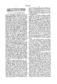
- FIG. 1 is a perspective view of an intubating scope of the present invention
- FIG. 2 is a schematic diagram of the intubating scope of FIG. 1;
- FIG. 3 is an illustration of the scope of FIG. 1 in use, illustrating the physician's lines of sight;
- FIG. 4 is an illustration of the use of a second embodiment of the present invention.
- Scope 1 generally refers to an intubation facilitating scope of the present invention.
- Scope 1 has a semi-malleable tube 3 depending from a handle 5.
- the semi-malleable tube portion 3 may serve as a stylet.
- a small optical camera 6 is carried in the tube's proximal end 7.
- Fiber optic bundles 8 extend the length of tube 3 to allow camera 6 to see internal structures and to provide illumination for the camera to view such internal structures.
- the camera alternatively, can be an electronic camera placed at the stylet's distal end 9.
- the distal end 9 may be moved to facilitate placement of an endotracheal tube, as will be explained below.
- a control lever 11 on handle 5 controls the movement of tube distal end 9. The lever 11 is not needed for optimal use of scope 1, but it is preferred.
- a very lightweight portable display unit 13 is attached, by way of a wire or fiberoptic cable 15, to camera 6 through handle 5.
- Unit 13 has a screen 17 which is preferably either a cathode ray tube (CRT) or an LCD screen. It may be black and white or color.
- An optional control knob 19 is used to adjust contrast, brightness, or any other features.
- the camera and screen are powered by a power unit 20 which is contained either in display unit 13, in handle 5, or at any other desired location.
- Display unit 13 with its self-contained power source in this preferred embodiment, is small and lightweight, as is the entire scope 1.
- display unit 13 may be simply placed on the patient's chest or on a boom or a mounting pad next to the physician's line-of-sight of the airway.
- the display unit may also be easily held in an assistant's or the user's hands, and the entire scope 1 can be quickly carried and placed in the desired position for the physician. This facilitates the physician's viewing and easy focusing of the airway directly and indirectly through the screen.
- FIG. 3 the use of scope 1 is described.
- An endotracheal tube 21 is slidably disposed over the stylet portion 3 until it abuts the handle 5 or a ring 23.
- the stylet's distal end 9 preferably just barely protrudes from the distal end of endotracheal tube 21.
- the ring 23 fits snugly around the proximal portion 7 of stylet 3. Ring 23 can move up and down stylet 3 to limit the extent to which the endotracheal tube may be slidably disposed thereon.
- tube 3 is malleable, it can help the user turn more oblique angles in positioning the endotracheal tube 21 in a difficult airway.
- the user can also use the lever 11 to control the distal end 9 to aid in the positioning of tube 3 in trachea T, to allow the endotracheal tube 21 to be placed into the trachea T.
- scope 1 the user is able to look directly into the pharynx and larynx along a direct line-of-sight 25 as shown from a physician's eye E in FIG. 3.
- endotracheal tube 21 passes out of the direct line-of-sight 25, the user then glances and moves his or her eyes to the line-of-sight 27 to view the image shown on screen 17 of the lightweight, portable display unit 13, which shows the deeper structures of the larynx and trachea as seen through the camera.
- scope 1 is removed from the endotracheal tube.
- the physician can indirectly view the internal structures using camera 6 and display unit 13.
- the display unit is lightweight, it can be placed in any desired place, such as just below the chin of a patient and next to the direct line-of-sight 25. This allows the physician to simultaneously directly view the airway by line-of-sight 25 and indirectly view the deeper structures of the airway on the lightweight portable screen by line-of-sight 27. Thus, the physician can effect an intubation of the airway without having to move his eyes, head or body at all.
- FIG. 4 shows a second embodiment of the scope. This is substantially the same as the embodiment of FIGS. 1 and 2. Rather than having an internal power source, however, this embodiment has an external power source 35. In this embodiment, the peripheral power source 35 receives the image from camera 6 in tube 3 over a line 37 and transmits the picture to the lightweight portable display unit 13 over a line 39. Of course, any number of variations of this configuration would also be suitable.
- the invention has utility in the intensive care unit, in the operating room, and in the emergency placement of an airway in patients with anatomic, pathophysiologic, or other problems, which would prevent the physician from easily protecting the airway and would prevent ease of placement of an endotracheal tube by standard methods and techniques. Numerous variations in the construction and use of intubating scope 1, within the scope of the appended claims, may occur to those skilled in the art in light of the foregoing disclosure.
- scope 1 may allow intubation through either the oral pharynx or nasal pharynx in both emergency and non-emergency situations and intubations where the patient is either conscious or unconscious. It can also be used in the operating room, or other situations such as the confirmation and placement of a double lumen and tracheal tube.
- a change necessary for other utilizations of the invention would be in stylet 3.
- the stylet for such other applications would be a freely malleable tube, much like the structure of the tube of a bronchoscope. This modification would allow the scope to be used in many other functions such as esophagoscopy, laparoscopy, or viewing of tissues in the retroperitoneum or to any structures that may not be easily reachable by a laparoscope or other rigid scope.
- the invention can be further modified and attached to suction or bovie coagulation instruments for use in problems such as bleeding nasal passages or rebleeding of tonsils. It may be used in the evaluation of distal structures of the oral pharynx or larynx.
- the device may also be modified to be used in urological procedures, allowing the physician to not have to bend down to look through an eyepiece. Rather he or she can manipulate the urethra, bladder and ureters by directly viewing the position of the instruments, as well as viewing indirectly what the instruments are doing--looking at them through a screen that is lightweight and easily mountable just above the direct visual field.
Abstract
Description
Claims (25)
Priority Applications (1)
| Application Number | Priority Date | Filing Date | Title |
|---|---|---|---|
| US07987673 US5363838B1 (en) | 1992-12-09 | 1992-12-09 | Fiberoptic intubating scope with camera and lightweight portable screen and method of using same |
Applications Claiming Priority (1)
| Application Number | Priority Date | Filing Date | Title |
|---|---|---|---|
| US07987673 US5363838B1 (en) | 1992-12-09 | 1992-12-09 | Fiberoptic intubating scope with camera and lightweight portable screen and method of using same |
Publications (2)
| Publication Number | Publication Date |
|---|---|
| US5363838A true US5363838A (en) | 1994-11-15 |
| US5363838B1 US5363838B1 (en) | 2000-03-28 |
Family
ID=25533460
Family Applications (1)
| Application Number | Title | Priority Date | Filing Date |
|---|---|---|---|
| US07987673 Expired - Lifetime US5363838B1 (en) | 1992-12-09 | 1992-12-09 | Fiberoptic intubating scope with camera and lightweight portable screen and method of using same |
Country Status (1)
| Country | Link |
|---|---|
| US (1) | US5363838B1 (en) |
Cited By (101)
| Publication number | Priority date | Publication date | Assignee | Title |
|---|---|---|---|---|
| US5543832A (en) * | 1994-03-29 | 1996-08-06 | Laser Surge, Inc. | Video display system for projecting an image on to a tilted screen adjacent a surgical field |
| WO1997037581A2 (en) * | 1996-04-10 | 1997-10-16 | Endoscopic Technologies, Inc. | Improving visualization during closed-chest surgery |
| WO1998014112A2 (en) | 1996-10-04 | 1998-04-09 | University Of Florida | Plastic optical fiber airway imaging system |
| US5800342A (en) * | 1994-03-18 | 1998-09-01 | Lee; Jai S. | Method of endotracheal intubation |
| US5800344A (en) * | 1996-10-23 | 1998-09-01 | Welch Allyn, Inc. | Video laryngoscope |
| US5812188A (en) * | 1996-07-12 | 1998-09-22 | Adair; Edwin L. | Sterile encapsulated endoscopic video monitor |
| DE19721138C1 (en) * | 1997-05-21 | 1998-09-24 | Wolf Gmbh Richard | Spreadable head for medical endoscope |
| US5827178A (en) * | 1997-01-02 | 1998-10-27 | Berall; Jonathan | Laryngoscope for use in trachea intubation |
| US5873814A (en) * | 1996-07-12 | 1999-02-23 | Adair; Edwin L. | Sterile encapsulated endoscopic video monitor and method |
| US5879289A (en) * | 1996-07-15 | 1999-03-09 | Universal Technologies International, Inc. | Hand-held portable endoscopic camera |
| EP0901772A1 (en) * | 1997-08-09 | 1999-03-17 | Willy Rüsch Ag | Laryngoscope |
| WO1999022636A1 (en) | 1997-10-31 | 1999-05-14 | Imagyn Medical Technologies, Inc. | Intubation device and method |
| WO1999035960A1 (en) | 1998-01-16 | 1999-07-22 | University Of Florida | Malleable endotracheal tube with fiberoptic scope |
| US5957831A (en) * | 1996-07-12 | 1999-09-28 | Adair; Edwin L. | Sterile encapsulated endoscopic video monitor |
| EP0955859A2 (en) * | 1996-05-03 | 1999-11-17 | Philip S. Green | System and method for endoscopic imaging and endosurgery |
| US6123666A (en) * | 1998-04-29 | 2000-09-26 | Vanderbilt University | Laryngoscope blade with fiberoptic scope for remote viewing and method for teaching the proper insertion of a laryngoscope blade into the airway of a patient |
| US6186944B1 (en) | 1998-11-25 | 2001-02-13 | Jory Tsai | Medical inspection device |
| US20020025114A1 (en) * | 2000-04-17 | 2002-02-28 | Vertical Computer Systems, Inc. | Apparatus and method for transmitting images over a single-filament fiber optic cable |
| US6361489B1 (en) | 1998-11-25 | 2002-03-26 | Jory Tsai | Medical inspection device |
| US6427686B2 (en) * | 1996-10-16 | 2002-08-06 | Augustine Medical, Inc. | Airway device with provision for coupling to an introducer |
| US6432046B1 (en) | 1996-07-15 | 2002-08-13 | Universal Technologies International, Inc. | Hand-held, portable camera for producing video images of an object |
| US20020118279A1 (en) * | 2001-02-28 | 2002-08-29 | Eastman Kodak Company | Intra-oral camera with integral display |
| US20020189618A1 (en) * | 1996-10-16 | 2002-12-19 | Augustine Scott Douglas | Airway device with provision for coupling to an introducer |
| WO2003005930A1 (en) * | 2001-07-12 | 2003-01-23 | Medic. Nrg Ltd. | A system for detecting apical foramen position in performing dental treatment |
| US6537207B1 (en) | 1999-04-07 | 2003-03-25 | Fovioptics, Inc. | Identification of protective covers for medical imaging devices |
| US20030078476A1 (en) * | 2001-07-24 | 2003-04-24 | Hill Stephen D. | Apparatus for intubation |
| US6554765B1 (en) | 1996-07-15 | 2003-04-29 | East Giant Limited | Hand held, portable camera with adaptable lens system |
| US6623425B2 (en) | 2001-07-23 | 2003-09-23 | Cartledge Medical Products, Llc | Modified laryngoscope blade to reduce dental injuries during intubation |
| US6626825B2 (en) | 1998-11-25 | 2003-09-30 | Jory Tsai | Medical inspection device |
| US6652453B2 (en) * | 1999-03-03 | 2003-11-25 | Vincent A. Smith | Portable video laryngoscope |
| US20040087843A1 (en) * | 2002-08-26 | 2004-05-06 | Rice Mark J. | Non-invasive psychophysical measurement of glucose using photodynamics |
| FR2852226A1 (en) * | 2003-03-10 | 2004-09-17 | Univ Joseph Fourier | Medical surgical instrument with display screen has screen mounted on adjustable strut to body of instrument |
| US20040220451A1 (en) * | 1996-10-04 | 2004-11-04 | Dietrich Gravenstein | Imaging scope |
| US6840903B2 (en) | 2002-03-21 | 2005-01-11 | Nuvista Technology Corporation | Laryngoscope with image sensor |
| US20050010084A1 (en) * | 1998-11-25 | 2005-01-13 | Jory Tsai | Medical inspection device |
| US20050043588A1 (en) * | 1998-11-25 | 2005-02-24 | Jory Tsai | Medical inspection device |
| US6860264B2 (en) | 1996-02-26 | 2005-03-01 | Evergreen Medical Incorporated | Method and apparatus for endotracheal intubation using a light wand and curved guide |
| US20050085694A1 (en) * | 2003-10-16 | 2005-04-21 | Nakao Naomi L. | Endoscope with open channels |
| US6889069B2 (en) | 2001-09-13 | 2005-05-03 | Fovioptics Inc. | Non-invasive measurement of blood analytes using photodynamics |
| US20050139220A1 (en) * | 1996-02-26 | 2005-06-30 | Evergreen Medical Incorporated | Method and apparatus for ventilation / oxygenation during guided insertion of an endotracheal tube |
| US20050182297A1 (en) * | 1996-10-04 | 2005-08-18 | Dietrich Gravenstein | Imaging scope |
| US20060004258A1 (en) * | 2004-07-02 | 2006-01-05 | Wei-Zen Sun | Image-type intubation-aiding device |
| US20060020171A1 (en) * | 2002-10-21 | 2006-01-26 | Gilreath Mark G | Intubation and imaging device and system |
| US20060025650A1 (en) * | 2002-10-03 | 2006-02-02 | Oren Gavriely | Tube for inspecting internal organs of a body |
| US20060180155A1 (en) * | 2005-01-26 | 2006-08-17 | Ezc Holding, Llc | Video-assisted laryngeal mask airway devices |
| WO2007022420A2 (en) * | 2005-08-17 | 2007-02-22 | University Of Rochester Medical Center | Combined flexible and rigid intubating video laryngoscope |
| US20070106117A1 (en) * | 2005-10-24 | 2007-05-10 | Pentax Corporation | Intubation assistance apparatus |
| US20070106122A1 (en) * | 2005-10-24 | 2007-05-10 | Pentax Corporation | Intubation assistance apparatus |
| US20070106121A1 (en) * | 2005-10-24 | 2007-05-10 | Junichi Koyama | Intubation assistance apparatus and intubation assistance used in the apparatus |
| US20070167686A1 (en) * | 2003-04-29 | 2007-07-19 | Mcgrath Matthew J | Laryngoscope with camera attachement |
| US20070179342A1 (en) * | 2006-01-12 | 2007-08-02 | Kb Port Llc | Wireless Laryngoscope with Internal Antennae and One Piece Construction Adapted for Laryngoscopy Training |
| US20080115783A1 (en) * | 2004-04-16 | 2008-05-22 | Brain Archibald I J | Laryngeal Mask Airway Device |
| US20080177146A1 (en) * | 2007-01-19 | 2008-07-24 | Tien-Sheng Chen | Double Vision Endotracheal Tube Installation System |
| US20080312507A1 (en) * | 2007-06-16 | 2008-12-18 | Taehoon Kim | Apparatus and method for imaging-assisted intubation using pre-existing practitioner skill set |
| US20090171155A1 (en) * | 2007-12-27 | 2009-07-02 | Koichi Tsunoda | Oral cavity insertion instrument and pharyngoscope apparatus |
| WO2009089043A2 (en) * | 2008-01-09 | 2009-07-16 | Ezc Medical Llc. | Intubation systems and methods |
| US20090192350A1 (en) * | 2008-01-28 | 2009-07-30 | Mauricio Mejia | Wireless video stylet with display mounted to laryngoscope blade and method for using the same |
| US20090192355A1 (en) * | 2008-01-28 | 2009-07-30 | Mauricio Mejia | Scope for managing difficult pathways and method to improve visibility of the same |
| US20090225159A1 (en) * | 2008-03-07 | 2009-09-10 | Scott Schneider | Visual inspection device |
| US20090322867A1 (en) * | 2006-08-07 | 2009-12-31 | Innovative Medical Devices , Inc | System to aid in the positioning, confirmation and documentation of an endotracheal tube |
| US20100094090A1 (en) * | 2008-01-28 | 2010-04-15 | Mauricio Mejia | Self-cleaning wireless video stylet with display mounted to laryngoscope blade and method for using the same |
| US20100108060A1 (en) * | 2006-06-01 | 2010-05-06 | Truphatek International Ltd | Hand operated articulated intubation stylet |
| US20100137687A1 (en) * | 2005-09-20 | 2010-06-03 | Ai Medical Devices, Inc. | Endotracheal intubation device |
| US20100305406A1 (en) * | 2009-05-26 | 2010-12-02 | Ori Braun | System, device and method for gynecological use |
| US20110060190A1 (en) * | 2007-08-07 | 2011-03-10 | Truphatek International Ltd. | Laryngoscope apparatus with enhanced viewing capability |
| WO2011038126A1 (en) * | 2009-09-25 | 2011-03-31 | Spectrum Health Innovations, LLC | Laryngoscope guide and related method of use |
| US20120016197A1 (en) * | 2009-05-28 | 2012-01-19 | Smiths Medical International Limited | Medico-surgical apparatus |
| EP2524713A1 (en) * | 2011-05-18 | 2012-11-21 | Centre Hospitalier Universitaire de Bordeaux | Method for positioning a disposable endotracheal tube, and corresponding system for intubation |
| WO2012156480A1 (en) * | 2011-05-18 | 2012-11-22 | Centre Hospitalier Universitaire De Bordeaux (C.H.U De Bordeaux) | Method for positioning a disposable sterile endotracheal tube, and corresponding system for intubation |
| US20120316398A1 (en) * | 2008-06-23 | 2012-12-13 | Intubrite, Llc | Adjustable display mechanism and method |
| US20130018227A1 (en) * | 2011-07-11 | 2013-01-17 | Ian Schoonbaert | Laryngoscopic Device |
| US20130014750A1 (en) * | 2011-07-14 | 2013-01-17 | Soheil Etesham | Intubation Apparatus |
| CN103142204A (en) * | 2013-01-23 | 2013-06-12 | 杭州好克光电仪器有限公司 | Electronic endoscope |
| US8512234B2 (en) | 2011-04-07 | 2013-08-20 | Truphatek International Ltd. | Laryngoscope assembly with enhanced viewing capability |
| US8652033B2 (en) | 2010-09-23 | 2014-02-18 | Karl Storz Endovision, Inc. | Video stylet with directable tip |
| US8667966B2 (en) | 2010-08-03 | 2014-03-11 | Hideo Koike | Intubating attachment and method |
| US20140235940A1 (en) * | 2012-09-13 | 2014-08-21 | Zhejiang Youyi Medical Apparatus Co., Ltd. | S-shaped Visible Hard Intubation Core |
| US20140316206A1 (en) * | 2010-07-30 | 2014-10-23 | Nilesh R. Vasan | Disposable, self-contained laryngoscope and method of using same |
| EP2567725A3 (en) * | 2011-09-09 | 2014-10-29 | Tien-Sheng Chen | Tracheal intubation device |
| US8998798B2 (en) | 2010-12-29 | 2015-04-07 | Covidien Lp | Multi-lumen tracheal tube with visualization device |
| US20150126808A1 (en) * | 2011-05-18 | 2015-05-07 | Centre Hospitalier Universitaire De Bordeaux (C.H.U. De Bordeaux) | Method for positioning a disposable sterile endotracheal tube, and corresponding system for intubation |
| US9107628B2 (en) | 2012-10-12 | 2015-08-18 | Karl Storz Gmbh & Co. Kg | Video laryngoscope with disposable blade |
| US9155854B2 (en) | 2011-08-31 | 2015-10-13 | Covidien Lp | Tracheal tube with visualization device and integrated flushing system |
| US9179831B2 (en) | 2009-11-30 | 2015-11-10 | King Systems Corporation | Visualization instrument |
| US9211060B2 (en) | 2011-04-05 | 2015-12-15 | Covidien Lp | Visualization device and holder for use with a tracheal tube |
| WO2017007829A1 (en) * | 2015-07-06 | 2017-01-12 | Werd, Llc | Temporaru tubes and a system for placing same in a patient |
| US20170086664A1 (en) * | 2011-05-18 | 2017-03-30 | Centre Hospitalier Universitaire De Bordeaux (C.H.U De Bordeaux) | Method for positioning a disposable sterile endotracheal tube, and corresponding system for intubation |
| US9622651B2 (en) | 2012-01-27 | 2017-04-18 | Kbport Llc | Wireless laryngoscope simulator with onboard event recording adapted for laryngoscopy training |
| US20170258312A1 (en) * | 2014-11-24 | 2017-09-14 | Shanghai Anqing Medical Instrument Co., Ltd | Electronic laryngoscope |
| US9820642B2 (en) | 2007-08-04 | 2017-11-21 | King Systems Corporation | Airway intubation device |
| US9840266B2 (en) | 2013-10-09 | 2017-12-12 | Glidemachines Llc | Apparatus and method for towing a load by a person |
| US20180000319A1 (en) * | 2016-06-30 | 2018-01-04 | Karl Storz Gmbh & Co. Kg | Apparatus For Video-Endoscopy |
| US10101576B2 (en) | 2016-09-14 | 2018-10-16 | David Torres | Bore scope system |
| US10149602B2 (en) | 2011-07-11 | 2018-12-11 | Ambu A/S | Endobronchial tube with integrated image sensor and a cleaning nozzle arrangement |
| US10238295B2 (en) | 2011-06-06 | 2019-03-26 | Percuvision, Llc | Sensing catheter emitting radiant energy |
| US10245402B2 (en) | 2011-07-11 | 2019-04-02 | Ambu A/S | Endobronchial tube with integrated image sensor |
| RU194891U1 (en) * | 2019-08-13 | 2019-12-26 | Дмитрий Сергеевич Костин | Endotracheal tube introducer |
| USD876625S1 (en) | 2018-08-07 | 2020-02-25 | Adroit Surgical, Llc | Laryngoscope |
| US10842368B2 (en) | 2016-06-10 | 2020-11-24 | Ambu A/S | Suction catheter with brush and method of use for lens cleaning |
| US20200367722A1 (en) * | 2013-03-15 | 2020-11-26 | Dvl, Inc. | System and device for visualization of an enclosed space |
| WO2022061313A1 (en) * | 2019-03-21 | 2022-03-24 | The Brigham And Women's Hospital, Inc. | Robotic artificial intelligence nasal/oral/rectal enteric tube |
Citations (2)
| Publication number | Priority date | Publication date | Assignee | Title |
|---|---|---|---|---|
| US4651202A (en) * | 1984-05-16 | 1987-03-17 | Fuji Photo Optical Co., Ltd. | Video endoscope system |
| US4742819A (en) * | 1987-03-23 | 1988-05-10 | George Gordon P | Intubating scope with camera and screen |
-
1992
- 1992-12-09 US US07987673 patent/US5363838B1/en not_active Expired - Lifetime
Patent Citations (2)
| Publication number | Priority date | Publication date | Assignee | Title |
|---|---|---|---|---|
| US4651202A (en) * | 1984-05-16 | 1987-03-17 | Fuji Photo Optical Co., Ltd. | Video endoscope system |
| US4742819A (en) * | 1987-03-23 | 1988-05-10 | George Gordon P | Intubating scope with camera and screen |
Cited By (165)
| Publication number | Priority date | Publication date | Assignee | Title |
|---|---|---|---|---|
| US5840013A (en) * | 1994-03-18 | 1998-11-24 | Lee; Jai S. | Method of introducing a tubular member at a site in the body |
| US5800342A (en) * | 1994-03-18 | 1998-09-01 | Lee; Jai S. | Method of endotracheal intubation |
| US5543832A (en) * | 1994-03-29 | 1996-08-06 | Laser Surge, Inc. | Video display system for projecting an image on to a tilted screen adjacent a surgical field |
| US6860264B2 (en) | 1996-02-26 | 2005-03-01 | Evergreen Medical Incorporated | Method and apparatus for endotracheal intubation using a light wand and curved guide |
| US20050139220A1 (en) * | 1996-02-26 | 2005-06-30 | Evergreen Medical Incorporated | Method and apparatus for ventilation / oxygenation during guided insertion of an endotracheal tube |
| WO1997037581A2 (en) * | 1996-04-10 | 1997-10-16 | Endoscopic Technologies, Inc. | Improving visualization during closed-chest surgery |
| WO1997037581A3 (en) * | 1996-04-10 | 1998-01-15 | Endoscopic Technologies Inc | Improving visualization during closed-chest surgery |
| US20030153810A1 (en) * | 1996-04-10 | 2003-08-14 | Bertolero Arthur A. | Visualization during closed-chest surgery |
| US7074180B2 (en) * | 1996-04-10 | 2006-07-11 | Endoscopic Technologies, Inc. | Visualization during closed-chest surgery |
| EP0955859A4 (en) * | 1996-05-03 | 2000-01-12 | Philip S Green | System and method for endoscopic imaging and endosurgery |
| EP0955859A2 (en) * | 1996-05-03 | 1999-11-17 | Philip S. Green | System and method for endoscopic imaging and endosurgery |
| US5812188A (en) * | 1996-07-12 | 1998-09-22 | Adair; Edwin L. | Sterile encapsulated endoscopic video monitor |
| US5873814A (en) * | 1996-07-12 | 1999-02-23 | Adair; Edwin L. | Sterile encapsulated endoscopic video monitor and method |
| US6132367A (en) * | 1996-07-12 | 2000-10-17 | Adair; Edwin L. | Sterile encapsulated endoscopic video monitor |
| US5957831A (en) * | 1996-07-12 | 1999-09-28 | Adair; Edwin L. | Sterile encapsulated endoscopic video monitor |
| US5879289A (en) * | 1996-07-15 | 1999-03-09 | Universal Technologies International, Inc. | Hand-held portable endoscopic camera |
| US6554765B1 (en) | 1996-07-15 | 2003-04-29 | East Giant Limited | Hand held, portable camera with adaptable lens system |
| US6432046B1 (en) | 1996-07-15 | 2002-08-13 | Universal Technologies International, Inc. | Hand-held, portable camera for producing video images of an object |
| US6692432B1 (en) | 1996-07-15 | 2004-02-17 | East Giant Limited | Hand-held portable camera for producing video images of an object |
| US6322498B1 (en) * | 1996-10-04 | 2001-11-27 | University Of Florida | Imaging scope |
| EP1281348A2 (en) | 1996-10-04 | 2003-02-05 | University Of Florida | Plastic optical fiber airway imaging system |
| US6115523A (en) * | 1996-10-04 | 2000-09-05 | University Of Florida | Plastic optical fiber airway imaging system |
| EP1281348A3 (en) * | 1996-10-04 | 2003-03-12 | University Of Florida | Plastic optical fiber airway imaging system |
| EP1262141A1 (en) | 1996-10-04 | 2002-12-04 | University Of Florida | Plastic optical fiber airway imaging system |
| US20050182297A1 (en) * | 1996-10-04 | 2005-08-18 | Dietrich Gravenstein | Imaging scope |
| US20040220451A1 (en) * | 1996-10-04 | 2004-11-04 | Dietrich Gravenstein | Imaging scope |
| WO1998014112A2 (en) | 1996-10-04 | 1998-04-09 | University Of Florida | Plastic optical fiber airway imaging system |
| US6830049B2 (en) * | 1996-10-16 | 2004-12-14 | Arizant Healthcare Inc. | Airway device with provision for coupling to an introducer |
| US20020189618A1 (en) * | 1996-10-16 | 2002-12-19 | Augustine Scott Douglas | Airway device with provision for coupling to an introducer |
| US6427686B2 (en) * | 1996-10-16 | 2002-08-06 | Augustine Medical, Inc. | Airway device with provision for coupling to an introducer |
| US5800344A (en) * | 1996-10-23 | 1998-09-01 | Welch Allyn, Inc. | Video laryngoscope |
| US5827178A (en) * | 1997-01-02 | 1998-10-27 | Berall; Jonathan | Laryngoscope for use in trachea intubation |
| DE19721138C1 (en) * | 1997-05-21 | 1998-09-24 | Wolf Gmbh Richard | Spreadable head for medical endoscope |
| US5954632A (en) * | 1997-05-21 | 1999-09-21 | Richard Wolf Gmbh | Endoscope in particular a mediastinoscope |
| DE19734591C1 (en) * | 1997-08-09 | 1999-06-17 | Ruesch Willy Ag | Laryngoscope |
| EP0901772A1 (en) * | 1997-08-09 | 1999-03-17 | Willy Rüsch Ag | Laryngoscope |
| US5913816A (en) * | 1997-10-31 | 1999-06-22 | Imagyn Medical Technologies, Inc. | Intubation device and method |
| WO1999022636A1 (en) | 1997-10-31 | 1999-05-14 | Imagyn Medical Technologies, Inc. | Intubation device and method |
| WO1999035960A1 (en) | 1998-01-16 | 1999-07-22 | University Of Florida | Malleable endotracheal tube with fiberoptic scope |
| US6123666A (en) * | 1998-04-29 | 2000-09-26 | Vanderbilt University | Laryngoscope blade with fiberoptic scope for remote viewing and method for teaching the proper insertion of a laryngoscope blade into the airway of a patient |
| US6626825B2 (en) | 1998-11-25 | 2003-09-30 | Jory Tsai | Medical inspection device |
| US6361489B1 (en) | 1998-11-25 | 2002-03-26 | Jory Tsai | Medical inspection device |
| US20050043588A1 (en) * | 1998-11-25 | 2005-02-24 | Jory Tsai | Medical inspection device |
| US20050010084A1 (en) * | 1998-11-25 | 2005-01-13 | Jory Tsai | Medical inspection device |
| US6186944B1 (en) | 1998-11-25 | 2001-02-13 | Jory Tsai | Medical inspection device |
| US7419467B2 (en) | 1998-11-25 | 2008-09-02 | M3 Electronics, Inc. | Medical inspection device |
| US7137948B2 (en) | 1998-11-25 | 2006-11-21 | Jory Tsai | Medical inspection device |
| US6652453B2 (en) * | 1999-03-03 | 2003-11-25 | Vincent A. Smith | Portable video laryngoscope |
| US6537207B1 (en) | 1999-04-07 | 2003-03-25 | Fovioptics, Inc. | Identification of protective covers for medical imaging devices |
| US6718103B2 (en) | 2000-04-17 | 2004-04-06 | Vertical Computer Systems, Inc. | Apparatus and method for transmitting images over a single-filament fiber optic cable |
| US20020025114A1 (en) * | 2000-04-17 | 2002-02-28 | Vertical Computer Systems, Inc. | Apparatus and method for transmitting images over a single-filament fiber optic cable |
| US20040190843A1 (en) * | 2000-04-17 | 2004-09-30 | Cruz Aluizio M. | Apparatus and method for transmitting images over a single-filament fiber optic cable |
| US7057639B2 (en) | 2001-02-28 | 2006-06-06 | Eastman Kodak Company | Intra-oral camera with integral display |
| US20020118279A1 (en) * | 2001-02-28 | 2002-08-29 | Eastman Kodak Company | Intra-oral camera with integral display |
| WO2003005930A1 (en) * | 2001-07-12 | 2003-01-23 | Medic. Nrg Ltd. | A system for detecting apical foramen position in performing dental treatment |
| US20040034281A1 (en) * | 2001-07-23 | 2004-02-19 | Richard Cartledge | Modified laryngoscope blade to reduce dental injuries during intubation |
| US7044910B2 (en) | 2001-07-23 | 2006-05-16 | Cartledge Medical Products, Inc. | Modified laryngoscope blade to reduce dental injuries during intubation |
| US6623425B2 (en) | 2001-07-23 | 2003-09-23 | Cartledge Medical Products, Llc | Modified laryngoscope blade to reduce dental injuries during intubation |
| US20030078476A1 (en) * | 2001-07-24 | 2003-04-24 | Hill Stephen D. | Apparatus for intubation |
| US6929600B2 (en) * | 2001-07-24 | 2005-08-16 | Stephen D. Hill | Apparatus for intubation |
| US6889069B2 (en) | 2001-09-13 | 2005-05-03 | Fovioptics Inc. | Non-invasive measurement of blood analytes using photodynamics |
| US6840903B2 (en) | 2002-03-21 | 2005-01-11 | Nuvista Technology Corporation | Laryngoscope with image sensor |
| US20050043590A1 (en) * | 2002-03-21 | 2005-02-24 | Mazzei William J. | Laryngoscope with image sensor |
| US20040087843A1 (en) * | 2002-08-26 | 2004-05-06 | Rice Mark J. | Non-invasive psychophysical measurement of glucose using photodynamics |
| US6895264B2 (en) | 2002-08-26 | 2005-05-17 | Fovioptics Inc. | Non-invasive psychophysical measurement of glucose using photodynamics |
| US20060025650A1 (en) * | 2002-10-03 | 2006-02-02 | Oren Gavriely | Tube for inspecting internal organs of a body |
| US20060020171A1 (en) * | 2002-10-21 | 2006-01-26 | Gilreath Mark G | Intubation and imaging device and system |
| US20060173290A1 (en) * | 2003-03-10 | 2006-08-03 | Stephane Lavallee | Localised medical instrument with tilt and swivel screen |
| WO2004080323A1 (en) * | 2003-03-10 | 2004-09-23 | Universite Joseph Fourier | Localised medical instrument with tilt and swivel screen |
| FR2852226A1 (en) * | 2003-03-10 | 2004-09-17 | Univ Joseph Fourier | Medical surgical instrument with display screen has screen mounted on adjustable strut to body of instrument |
| US20070299313A1 (en) * | 2003-04-29 | 2007-12-27 | Mcgrath Matthew J | Laryngoscope With Means to Restrict Re-Use of Blades |
| US11612313B2 (en) | 2003-04-29 | 2023-03-28 | Covidien Ag | Laryngoscope with camera attachment |
| US10786146B2 (en) | 2003-04-29 | 2020-09-29 | Aircraft Medical Limited | Laryngoscope with camera attachment |
| US10178947B2 (en) | 2003-04-29 | 2019-01-15 | Aircraft Medical Limited | Laryngoscope with camera attachment |
| US9820641B2 (en) | 2003-04-29 | 2017-11-21 | Aircraft Medical Limited | Laryngoscope with camera attachment |
| US9737202B2 (en) | 2003-04-29 | 2017-08-22 | Aircraft Medical Limited | Laryngoscope with camera attachment |
| US9687141B2 (en) | 2003-04-29 | 2017-06-27 | Aircraft Medical Limited | Laryngoscope with means to restrict re-use of blades |
| US20070167686A1 (en) * | 2003-04-29 | 2007-07-19 | Mcgrath Matthew J | Laryngoscope with camera attachement |
| US7762949B2 (en) * | 2003-10-16 | 2010-07-27 | Granit Medical Innovation, Llc | Endoscope with open channels |
| US20050085694A1 (en) * | 2003-10-16 | 2005-04-21 | Nakao Naomi L. | Endoscope with open channels |
| US20080115783A1 (en) * | 2004-04-16 | 2008-05-22 | Brain Archibald I J | Laryngeal Mask Airway Device |
| US20060004258A1 (en) * | 2004-07-02 | 2006-01-05 | Wei-Zen Sun | Image-type intubation-aiding device |
| US20060180155A1 (en) * | 2005-01-26 | 2006-08-17 | Ezc Holding, Llc | Video-assisted laryngeal mask airway devices |
| WO2007022420A3 (en) * | 2005-08-17 | 2007-11-01 | Univ Rochester Medical Ct | Combined flexible and rigid intubating video laryngoscope |
| WO2007022420A2 (en) * | 2005-08-17 | 2007-02-22 | University Of Rochester Medical Center | Combined flexible and rigid intubating video laryngoscope |
| US20070173697A1 (en) * | 2005-08-17 | 2007-07-26 | University Of Rochester Medical Center | Combined flexible and rigid intubating video laryngoscope |
| US20100137687A1 (en) * | 2005-09-20 | 2010-06-03 | Ai Medical Devices, Inc. | Endotracheal intubation device |
| US8079951B2 (en) | 2005-10-24 | 2011-12-20 | Hoya Corporation | Intubation assistance apparatus |
| US20070106117A1 (en) * | 2005-10-24 | 2007-05-10 | Pentax Corporation | Intubation assistance apparatus |
| US20070106121A1 (en) * | 2005-10-24 | 2007-05-10 | Junichi Koyama | Intubation assistance apparatus and intubation assistance used in the apparatus |
| US20070106122A1 (en) * | 2005-10-24 | 2007-05-10 | Pentax Corporation | Intubation assistance apparatus |
| US20070179342A1 (en) * | 2006-01-12 | 2007-08-02 | Kb Port Llc | Wireless Laryngoscope with Internal Antennae and One Piece Construction Adapted for Laryngoscopy Training |
| US20100108060A1 (en) * | 2006-06-01 | 2010-05-06 | Truphatek International Ltd | Hand operated articulated intubation stylet |
| US8505531B2 (en) | 2006-06-01 | 2013-08-13 | Truphatek International Ltd. | Hand operated articulated intubation stylet |
| US20090322867A1 (en) * | 2006-08-07 | 2009-12-31 | Innovative Medical Devices , Inc | System to aid in the positioning, confirmation and documentation of an endotracheal tube |
| US8416291B2 (en) | 2006-08-07 | 2013-04-09 | Innovative Medical Devices, Inc. | System to aid in the positioning, confirmation and documentation of an endotracheal tube |
| US20080177146A1 (en) * | 2007-01-19 | 2008-07-24 | Tien-Sheng Chen | Double Vision Endotracheal Tube Installation System |
| US20080312507A1 (en) * | 2007-06-16 | 2008-12-18 | Taehoon Kim | Apparatus and method for imaging-assisted intubation using pre-existing practitioner skill set |
| US9820642B2 (en) | 2007-08-04 | 2017-11-21 | King Systems Corporation | Airway intubation device |
| US20110060190A1 (en) * | 2007-08-07 | 2011-03-10 | Truphatek International Ltd. | Laryngoscope apparatus with enhanced viewing capability |
| US8251898B2 (en) | 2007-08-07 | 2012-08-28 | Truphatek International Ltd | Laryngoscope apparatus with enhanced viewing capability |
| US20090171155A1 (en) * | 2007-12-27 | 2009-07-02 | Koichi Tsunoda | Oral cavity insertion instrument and pharyngoscope apparatus |
| US9078559B2 (en) * | 2007-12-27 | 2015-07-14 | Koichi Tsunoda | Oral cavity insertion instrument and pharyngoscope apparatus |
| WO2009089043A2 (en) * | 2008-01-09 | 2009-07-16 | Ezc Medical Llc. | Intubation systems and methods |
| WO2009089043A3 (en) * | 2008-01-09 | 2009-09-11 | Ezc Medical Llc. | Intubation systems and methods |
| US20090209826A1 (en) * | 2008-01-09 | 2009-08-20 | Ezc Medical Llc | Intubation systems and methods |
| US20090192355A1 (en) * | 2008-01-28 | 2009-07-30 | Mauricio Mejia | Scope for managing difficult pathways and method to improve visibility of the same |
| US20090192350A1 (en) * | 2008-01-28 | 2009-07-30 | Mauricio Mejia | Wireless video stylet with display mounted to laryngoscope blade and method for using the same |
| US20100094090A1 (en) * | 2008-01-28 | 2010-04-15 | Mauricio Mejia | Self-cleaning wireless video stylet with display mounted to laryngoscope blade and method for using the same |
| US8888683B2 (en) | 2008-01-28 | 2014-11-18 | Mauricio Mejia | Modifications in endoscope apparatus, using fluid and gas dynamics, and methods for improving visibility during endoscopy |
| US20090225159A1 (en) * | 2008-03-07 | 2009-09-10 | Scott Schneider | Visual inspection device |
| US9986212B2 (en) | 2008-03-07 | 2018-05-29 | Milwaukee Electric Tool Corporation | Visual inspection device |
| US9693024B2 (en) | 2008-03-07 | 2017-06-27 | Milwaukee Electric Tool Corporation | Visual inspection device |
| US8189043B2 (en) | 2008-03-07 | 2012-05-29 | Milwaukee Electric Tool Corporation | Hand-held visual inspection device for viewing confined or difficult to access locations |
| US8659652B2 (en) | 2008-03-07 | 2014-02-25 | Milwaukee Electric Tool Corporation | Visual inspection device |
| US8988522B2 (en) | 2008-03-07 | 2015-03-24 | Milwaukee Electric Tool Corporation | Visual inspection device |
| US20120316398A1 (en) * | 2008-06-23 | 2012-12-13 | Intubrite, Llc | Adjustable display mechanism and method |
| US9095298B2 (en) * | 2008-06-23 | 2015-08-04 | Intubrite, Llc | Adjustable display mechanism and method |
| US20100305406A1 (en) * | 2009-05-26 | 2010-12-02 | Ori Braun | System, device and method for gynecological use |
| US20120016197A1 (en) * | 2009-05-28 | 2012-01-19 | Smiths Medical International Limited | Medico-surgical apparatus |
| US9801535B2 (en) * | 2009-05-28 | 2017-10-31 | Smiths Medical International Limited | Medico-surgical apparatus |
| US8366612B2 (en) | 2009-09-25 | 2013-02-05 | Spectrum Health Innovations, LLC | Laryngoscope guide and related method of use |
| WO2011038126A1 (en) * | 2009-09-25 | 2011-03-31 | Spectrum Health Innovations, LLC | Laryngoscope guide and related method of use |
| US20110201890A1 (en) * | 2009-09-25 | 2011-08-18 | Spectrum Health Innovations, LLC | Laryngoscope guide and related method of use |
| US20110077466A1 (en) * | 2009-09-25 | 2011-03-31 | Spectrum Health Innovations, LLC | Laryngoscope guide and related method of use |
| US9854962B2 (en) | 2009-11-30 | 2018-01-02 | King Systems Corporation | Visualization instrument |
| US9179831B2 (en) | 2009-11-30 | 2015-11-10 | King Systems Corporation | Visualization instrument |
| US20140316206A1 (en) * | 2010-07-30 | 2014-10-23 | Nilesh R. Vasan | Disposable, self-contained laryngoscope and method of using same |
| US9289114B2 (en) * | 2010-07-30 | 2016-03-22 | Nilesh R. Vasan | Disposable, self-contained laryngoscope and method of using same |
| US8667966B2 (en) | 2010-08-03 | 2014-03-11 | Hideo Koike | Intubating attachment and method |
| US8652033B2 (en) | 2010-09-23 | 2014-02-18 | Karl Storz Endovision, Inc. | Video stylet with directable tip |
| US8998798B2 (en) | 2010-12-29 | 2015-04-07 | Covidien Lp | Multi-lumen tracheal tube with visualization device |
| US9211060B2 (en) | 2011-04-05 | 2015-12-15 | Covidien Lp | Visualization device and holder for use with a tracheal tube |
| US8512234B2 (en) | 2011-04-07 | 2013-08-20 | Truphatek International Ltd. | Laryngoscope assembly with enhanced viewing capability |
| US20150126808A1 (en) * | 2011-05-18 | 2015-05-07 | Centre Hospitalier Universitaire De Bordeaux (C.H.U. De Bordeaux) | Method for positioning a disposable sterile endotracheal tube, and corresponding system for intubation |
| US20170086664A1 (en) * | 2011-05-18 | 2017-03-30 | Centre Hospitalier Universitaire De Bordeaux (C.H.U De Bordeaux) | Method for positioning a disposable sterile endotracheal tube, and corresponding system for intubation |
| EP2524713A1 (en) * | 2011-05-18 | 2012-11-21 | Centre Hospitalier Universitaire de Bordeaux | Method for positioning a disposable endotracheal tube, and corresponding system for intubation |
| WO2012156480A1 (en) * | 2011-05-18 | 2012-11-22 | Centre Hospitalier Universitaire De Bordeaux (C.H.U De Bordeaux) | Method for positioning a disposable sterile endotracheal tube, and corresponding system for intubation |
| US10238295B2 (en) | 2011-06-06 | 2019-03-26 | Percuvision, Llc | Sensing catheter emitting radiant energy |
| US10786205B2 (en) | 2011-06-06 | 2020-09-29 | Ake A. Hellstrom | Sensing catheter emitting radiant energy |
| US20130018227A1 (en) * | 2011-07-11 | 2013-01-17 | Ian Schoonbaert | Laryngoscopic Device |
| US9173545B2 (en) * | 2011-07-11 | 2015-11-03 | Ian Schoonbaert | Laryngoscopic device |
| US10888679B2 (en) | 2011-07-11 | 2021-01-12 | Ambu A/S | Endobronchial tube with integrated image sensor |
| US10406309B2 (en) | 2011-07-11 | 2019-09-10 | Ambu A/S | Endobronchial tube with integrated image sensor and a cleaning nozzle arrangement |
| US10245402B2 (en) | 2011-07-11 | 2019-04-02 | Ambu A/S | Endobronchial tube with integrated image sensor |
| US10149602B2 (en) | 2011-07-11 | 2018-12-11 | Ambu A/S | Endobronchial tube with integrated image sensor and a cleaning nozzle arrangement |
| US20130014750A1 (en) * | 2011-07-14 | 2013-01-17 | Soheil Etesham | Intubation Apparatus |
| US9155854B2 (en) | 2011-08-31 | 2015-10-13 | Covidien Lp | Tracheal tube with visualization device and integrated flushing system |
| EP2567725A3 (en) * | 2011-09-09 | 2014-10-29 | Tien-Sheng Chen | Tracheal intubation device |
| US9622651B2 (en) | 2012-01-27 | 2017-04-18 | Kbport Llc | Wireless laryngoscope simulator with onboard event recording adapted for laryngoscopy training |
| US20140235940A1 (en) * | 2012-09-13 | 2014-08-21 | Zhejiang Youyi Medical Apparatus Co., Ltd. | S-shaped Visible Hard Intubation Core |
| US9107628B2 (en) | 2012-10-12 | 2015-08-18 | Karl Storz Gmbh & Co. Kg | Video laryngoscope with disposable blade |
| CN103142204A (en) * | 2013-01-23 | 2013-06-12 | 杭州好克光电仪器有限公司 | Electronic endoscope |
| US20200367722A1 (en) * | 2013-03-15 | 2020-11-26 | Dvl, Inc. | System and device for visualization of an enclosed space |
| US9840266B2 (en) | 2013-10-09 | 2017-12-12 | Glidemachines Llc | Apparatus and method for towing a load by a person |
| US10405738B2 (en) * | 2014-11-24 | 2019-09-10 | Shanghai Anqing Instrument Co., Ltd | Electronic laryngoscope |
| US20170258312A1 (en) * | 2014-11-24 | 2017-09-14 | Shanghai Anqing Medical Instrument Co., Ltd | Electronic laryngoscope |
| WO2017007829A1 (en) * | 2015-07-06 | 2017-01-12 | Werd, Llc | Temporaru tubes and a system for placing same in a patient |
| US10842368B2 (en) | 2016-06-10 | 2020-11-24 | Ambu A/S | Suction catheter with brush and method of use for lens cleaning |
| US20180000319A1 (en) * | 2016-06-30 | 2018-01-04 | Karl Storz Gmbh & Co. Kg | Apparatus For Video-Endoscopy |
| US10101576B2 (en) | 2016-09-14 | 2018-10-16 | David Torres | Bore scope system |
| USD876625S1 (en) | 2018-08-07 | 2020-02-25 | Adroit Surgical, Llc | Laryngoscope |
| WO2022061313A1 (en) * | 2019-03-21 | 2022-03-24 | The Brigham And Women's Hospital, Inc. | Robotic artificial intelligence nasal/oral/rectal enteric tube |
| RU194891U9 (en) * | 2019-08-13 | 2020-02-11 | Дмитрий Сергеевич Костин | Endotracheal tube introducer |
| RU194891U1 (en) * | 2019-08-13 | 2019-12-26 | Дмитрий Сергеевич Костин | Endotracheal tube introducer |
Also Published As
| Publication number | Publication date |
|---|---|
| US5363838B1 (en) | 2000-03-28 |
Similar Documents
| Publication | Publication Date | Title |
|---|---|---|
| US5363838A (en) | Fiberoptic intubating scope with camera and lightweight portable screen and method of using same | |
| US5827178A (en) | Laryngoscope for use in trachea intubation | |
| US4742819A (en) | Intubating scope with camera and screen | |
| US7946981B1 (en) | Two-piece video laryngoscope | |
| US6123666A (en) | Laryngoscope blade with fiberoptic scope for remote viewing and method for teaching the proper insertion of a laryngoscope blade into the airway of a patient | |
| US5676635A (en) | Instrument for insertion of an endotracheal tube | |
| EP1738789B1 (en) | Endotracheal video device | |
| US6929600B2 (en) | Apparatus for intubation | |
| US6322498B1 (en) | Imaging scope | |
| US7182728B2 (en) | Laryngoscope with multi-directional eyepiece | |
| US20050090712A1 (en) | Res-Q-Scope | |
| US20070197873A1 (en) | Wireless optical endoscopic device | |
| JP2009524482A (en) | Device for introducing an airway tube into a trachea having visualization ability and method of using the same | |
| US20100210907A2 (en) | Intubation tube | |
| JP2013510699A (en) | Channel laryngoscope and system | |
| US20070195539A1 (en) | Ultra wide band wireless optical endoscopic device | |
| US20070173697A1 (en) | Combined flexible and rigid intubating video laryngoscope | |
| Weiss | Video-intuboscopy: a new aid to routine and difficult tracheal intubation. | |
| CN105169540A (en) | Brightness-adjustable double positioning video light stick for tracheal cannula | |
| Levitan | Design rationale and intended use of a short optical stylet for routine fiberoptic augmentation of emergency laryngoscopy | |
| Weiss et al. | Video-intuboscopic assistance is a useful aid to tracheal intubation in pediatric patients | |
| KR100894709B1 (en) | Video airway endscope | |
| US8425409B2 (en) | Laryngoscope | |
| Berci et al. | Optical stylet: an aid to intubation and teaching | |
| JPH07178173A (en) | Artificial respiration instrument |
Legal Events
| Date | Code | Title | Description |
|---|---|---|---|
| FEPP | Fee payment procedure |
Free format text: PAYOR NUMBER ASSIGNED (ORIGINAL EVENT CODE: ASPN); ENTITY STATUS OF PATENT OWNER: SMALL ENTITY |
|
| STCF | Information on status: patent grant |
Free format text: PATENTED CASE |
|
| CC | Certificate of correction | ||
| RR | Request for reexamination filed |
Effective date: 19970829 |
|
| FPAY | Fee payment |
Year of fee payment: 4 |
|
| AS | Assignment |
Owner name: CEDARS OF UTAH MEDICAL, L.L.C., UTAH Free format text: ASSIGNMENT OF ASSIGNORS INTEREST;ASSIGNOR:GEORGE, GORDON PETTY;REEL/FRAME:009314/0987 Effective date: 19970801 |
|
| B1 | Reexamination certificate first reexamination |
Free format text: CLAIMS 1, 3-6, 8, 9 AND 19-22 ARE CANCELLED. CLAIMS 2, 7, 10 AND 11-18, 23-25 ARE DETERMINED TO BE PATENTABLE AS AMENDED. NEW CLAIMS 26-30 ARE ADDED AND DETERMINED TO BE PATENTABLE. |
|
| FEPP | Fee payment procedure |
Free format text: PAYER NUMBER DE-ASSIGNED (ORIGINAL EVENT CODE: RMPN); ENTITY STATUS OF PATENT OWNER: SMALL ENTITY Free format text: PAYOR NUMBER ASSIGNED (ORIGINAL EVENT CODE: ASPN); ENTITY STATUS OF PATENT OWNER: SMALL ENTITY |
|
| FPAY | Fee payment |
Year of fee payment: 8 |
|
| REMI | Maintenance fee reminder mailed | ||
| REMI | Maintenance fee reminder mailed | ||
| FPAY | Fee payment |
Year of fee payment: 12 |
|
| SULP | Surcharge for late payment |
Year of fee payment: 11 |