US5334851A - Computed radiography patient identification system - Google Patents
Computed radiography patient identification system Download PDFInfo
- Publication number
- US5334851A US5334851A US07/963,036 US96303692A US5334851A US 5334851 A US5334851 A US 5334851A US 96303692 A US96303692 A US 96303692A US 5334851 A US5334851 A US 5334851A
- Authority
- US
- United States
- Prior art keywords
- storage phosphor
- bar code
- identifying
- patient
- ray
- Prior art date
- Legal status (The legal status is an assumption and is not a legal conclusion. Google has not performed a legal analysis and makes no representation as to the accuracy of the status listed.)
- Expired - Fee Related
Links
- 238000002601 radiography Methods 0.000 title claims abstract description 21
- OAICVXFJPJFONN-UHFFFAOYSA-N Phosphorus Chemical compound [P] OAICVXFJPJFONN-UHFFFAOYSA-N 0.000 claims abstract description 112
- 238000000034 method Methods 0.000 claims description 3
- 230000005855 radiation Effects 0.000 description 7
- 230000004936 stimulating effect Effects 0.000 description 4
- 238000003745 diagnosis Methods 0.000 description 2
- 238000010586 diagram Methods 0.000 description 2
- 230000007246 mechanism Effects 0.000 description 2
- 210000001015 abdomen Anatomy 0.000 description 1
- 230000005540 biological transmission Effects 0.000 description 1
- 238000002059 diagnostic imaging Methods 0.000 description 1
- 238000000605 extraction Methods 0.000 description 1
- 230000006870 function Effects 0.000 description 1
- 238000012986 modification Methods 0.000 description 1
- 230000004048 modification Effects 0.000 description 1
- 230000003287 optical effect Effects 0.000 description 1
- 230000008569 process Effects 0.000 description 1
- 230000000638 stimulation Effects 0.000 description 1
Images


Classifications
-
- G—PHYSICS
- G03—PHOTOGRAPHY; CINEMATOGRAPHY; ANALOGOUS TECHNIQUES USING WAVES OTHER THAN OPTICAL WAVES; ELECTROGRAPHY; HOLOGRAPHY
- G03B—APPARATUS OR ARRANGEMENTS FOR TAKING PHOTOGRAPHS OR FOR PROJECTING OR VIEWING THEM; APPARATUS OR ARRANGEMENTS EMPLOYING ANALOGOUS TECHNIQUES USING WAVES OTHER THAN OPTICAL WAVES; ACCESSORIES THEREFOR
- G03B42/00—Obtaining records using waves other than optical waves; Visualisation of such records by using optical means
- G03B42/02—Obtaining records using waves other than optical waves; Visualisation of such records by using optical means using X-rays
- G03B42/04—Holders for X-ray films
- G03B42/047—Holders for X-ray films provided with marking means
-
- A—HUMAN NECESSITIES
- A61—MEDICAL OR VETERINARY SCIENCE; HYGIENE
- A61B—DIAGNOSIS; SURGERY; IDENTIFICATION
- A61B6/00—Apparatus for radiation diagnosis, e.g. combined with radiation therapy equipment
- A61B6/44—Constructional features of apparatus for radiation diagnosis
- A61B6/4494—Means for identifying the diagnostic device
-
- G—PHYSICS
- G01—MEASURING; TESTING
- G01T—MEASUREMENT OF NUCLEAR OR X-RADIATION
- G01T1/00—Measuring X-radiation, gamma radiation, corpuscular radiation, or cosmic radiation
- G01T1/16—Measuring radiation intensity
- G01T1/20—Measuring radiation intensity with scintillation detectors
- G01T1/2012—Measuring radiation intensity with scintillation detectors using stimulable phosphors, e.g. stimulable phosphor sheets
Definitions
- This invention relates in general to computed radiography in which an X-ray image of a patient is stored in a photostimulable storage phosphor. More particularly, the present invention relates to a system for matching patient identification with the patient's X-ray image stored in a storage phosphor.
- a patient In conventional radiography, a patient is exposed to X-rays to produce an X-ray image on a photosensitive film. The film is developed and viewed by a radiologist who makes a diagnosis of the patient.
- a significant problem in medical imaging systems involves matching the patient name with the image recorded.
- an identification camera In current film based radiography systems, an identification camera is used to print the patient name, date and other information on the film after it has been exposed and prior to processing. Alternatively, pressure sensitive labels with patient information may be applied after the film has been processed.
- Temporary X-ray images stored in a storage phosphor are converted into an X-ray image digital signal which can be stored, processed and transmitted.
- a photostimulable phosphor sheet is exposed to an image-wise pattern of short wavelength radiation, such as X-ray radiation, to record a latent image pattern in the photostimulable phosphor sheet.
- the latent image is read out by stimulating the phosphor with a relatively long wavelength stimulating radiation, such as red or infrared light.
- the stimulable phosphor Upon stimulation, the stimulable phosphor releases emitted radiation of an intermediate wavelength, such as blue or violet light, in proportion to the quantity of X-ray radiation that was received.
- An X-ray image signal is produced by scanning the stimulable phosphor sheet in a raster pattern by means of a beam of laser light deflected by an oscillating or rotating scanning mirror. The emitted radiation is sensed by a photodetector to produce an electrical X-ray image signal. This signal may then be stored, transmitted, or displayed on a monitor or reproduced as an X-ray film.
- computed radiography requires the matching of an X-ray image with the patient.
- X-ray image signal As a function of X-ray exposure conditions, it is also desirable to match X-ray exposure conditions and other patient identification data with the X-ray image signal.
- diagnostician such as a radiologist
- patient information is entered into a workstation and is transferred to a magnetic card.
- a technician places the cassette containing the exposed storage phosphor into a reader and dumps the patient data into the reader by swiping the magnetic card through an associated magnetic card reader.
- Many problems exist with this system including double entry of patient data, which is typically entered into a computer at the time a patient is admitted into a hospital. Moreover, the specific ordering of computed radiography cassettes and patient data must be maintained.
- a storage phosphor is provided with a storage phosphor identifying bar code which uniquely identifies the storage phosphor.
- a patient identifying bar code uniquely identifies each patient.
- a mobile X-ray unit has an associated set of bar codes identifying X-ray examination types and/or x-ray exposure conditions.
- a portable bar code scanner reads the storage phosphor identifying bar code, the patient identifying bar code and the X-ray exam type bar code at the time of an X-ray of the patient. This information is stored in memory in the bar code scanner.
- a computed radiography storage phosphor reader reads an exposed storage phosphor to convert the stored X-ray image into an X--ray image signal.
- the patient ID, storage phosphor ID and X-ray exam type information is loaded into the storage phosphor reader where it is matched with the proper X-ray image signal.
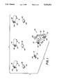
- FIG. 1 is a perspective view of a medical care facility incorporating an embodiment of the present invention
- FIG. 2 is a partially broken away perspective view of a storage phosphor cassette shown in FIG. 1;
- FIG. 3 is a diagrammatic view of an X-ray exam type chart shown in FIG. 1;
- FIG. 4 is a diagrammatic view of a patient ID chart shown in FIG. 1;
- FIG. 5 is a block diagram of the bar code scanner of FIG. 1;
- FIG. 6 is a perspective view of a computed radiography storage phosphor reader for reading storage phosphors used in the system of the present invention.
- FIG. 7 is a partial block diagram, partial diagrammatic view of the storage phosphor reader shown in FIG. 6.
- the medical care facility includes a plurality of beds 10, 12, 14, and 16 having respective patients 18, 20, 22, and 24 who require medical treatment.
- a mobile X-ray unit 26 has an X--ray source 28 mounted on a moveable arm 30 supported by cabinet 32.
- Cabinet 32 includes controls and power supply for X-ray source 28. Wheels 34 on cabinet 32 facilitate moving unit 26 from bed to bed.
- an X-ray image of a body part of a patient is produced in a stimulable storage phosphor contained in a cassette.
- storage phosphor cassettes 36, 38, 40, and 42 are provided for patients 18, 20, 22, and 24, respectively.
- Cassettes 36, 38, 40, and 42 have storage phosphor identifying bar codes 44, 46, 48 and 50 which uniquely identify each storage phosphor.
- storage phosphor cassette 42 has a removable storage phosphor plate 43 with bar code 50.
- An exemplary storage phosphor cassette is disclosed in commonly assigned, copending U.S. application Ser. No. 617,121 now U.S. Pat. No. 5,065,866.
- Each patient 18, 20, 22, and 24 is provided a unique patient identifying bar code 52, 54, 56, and 58 on respective patient charts 60, 62, 64, and 66.
- X-ray unit 26 has associated with it a chart 68 having a list of X-ray exam types and/or X-ray exposure conditions with a set of bar codes identifying each exam type and exposure condition.
- chart 68 has a list of X-ray exam types, such as, chest P/A Posterior/Anterior>; chest A/P Anterior/Posterior>; head; limb; abdomen, etc.
- Each X-ray exam type has a unique identifying bar code, 70A, 70B, 70C, 70D, 70E, etc.
- an X-ray technician who is responsible for taking X-rays at the medical care facility is provided with a portable bar code scanner 72.
- Bar code scanner 72 ⁇ see FIG. 5
- scanner 72 has a keyboard 78 for entering data which is stored in memory 76 and also has a display 80 for displaying the input data and other information.
- Control circuit 73, scanner 74, memory 76, display 80 and keypad 78 are internally connected by bus 82.
- a technician scans the patient identifying bar code, scans the storage phosphor identifying bar code and scans the bar code identifying the X-ray exam type.
- X-ray source 28 is positioned over patient 24 and storage phosphor cassette 42 is positioned under the chest area of patient 24.
- the technician uses bar code scanner 72 to scan patient identifying bar code 66 on patient chart 58, to scan storage phosphor identifying bar code 50 on storage phosphor cassette 42, and to scan X-ray examination type bar code 70A on exam type chart 68.
- Bar code 70A identifies the X-ray exam type as a chest Anterior to Posterior exam.
- Chart 68 may also contain other information relating to the X-ray examination, such as, X-ray exposure conditions, patient position, etc., which are also scanned by bar code scanner 72 and stored in memory 76.
- a technician identifying bar code may also be read. The technician can correct or manually enter data via keypad 78 at the time an X-ray exam is effected.
- X-ray unit 26 After the technician has finished an X-ray exam of patient 24, he can move X-ray unit 26 to the bedside of patients 18, 20, and 22 to produce X-ray images in storage phosphor cassettes 36, 38, and 40.
- the technician After a set of X-ray exposures have been taken and relevant data for each exposure scanned and stored in memory 76 of portable bar code scanner 72, the technician carries the storage phosphor cassettes 36, 38, 40, and 42 in a stack to a computed radiography storage phosphor reader station.
- a computed radiography storage phosphor reader station Such a station is shown in FIG. 6 and includes a computed radiography storage phosphor reader 84.
- Reader 84 has a bar code scanner download station 88.
- a storage phosphor or cassette stacker (not shown) is provided adjacent to reader 84 to sequentially feed exposed storage phosphors into slot 90 of reader 84.
- the exposed storage phosphor is read by reader 84 and converted to an X-ray image signal which is stored in a suitable memory.
- the technician inserts bar code scanner 72 into download station 88 where information relating to patient identification, storage phosphor identification, X-ray exam type, and other information relating to a specific X-ray exam is downloaded into memory associated with reader 84.
- the storage phosphor identifyin E bar code is read by a bar code reader in storage phosphor reader 84.
- the X-ray image signal read from a storage phosphor will be matched with the proper patient, X-ray exam type and other related information for further processing in workstation 86.
- storage phosphor reader 84 An exposed storage phosphor, such as 43, is removed from its cassette, such as 42, by a plate extraction mechanism ⁇ not shown>.
- Plate 43 stores a latent X-ray image of a patient.
- a beam 90 of stimulating radiation is generated by laser source 92.
- a deflector 94 such as a galvo mirror or polygon scanner, scans beam 90 ⁇ which has been focused by appropriate optics 96>point by point across storage phosphor 43 in the direction of arrow A.
- a translation mechanism 98 moves storage phosphor 43 in a direction ⁇ indicated by arrow B>perpendicular to the travel of the deflected beam 90, effecting a raster scan of the storage phosphor 43.
- the intensity of the stimulated fluorescence is detected by an electrooptic converter which is sensitive at the wavelength of the light emitted by storage phosphor 43.
- the converter may, for example, be a photomultiplier tube 98 or other photodetector which receives emitted light through an optical filter 100. Filter 100 blocks the stimulating light from laser 92 but passes the emitted light from storage phosphor 43.
- the signal from photoconverter 98 is amplified and filtered by amplifier 102 and converted to a digital signal by analog-to-digital converter (A/D) 104.
- A/D analog-to-digital converter
- the digital signal is stored in memory 106 which also receives the information downloaded from bar code scanner 72 by means of bar code download station 88.
- a separate bar code scanner 108 in reader 84 scans the storage phosphor identifying bar code 50 on storage phosphor 43 to identify the X-ray image signal.
- Reader 84 includes memory 106, a microprocessor 109, keyboard 110, and high resolution monitor 112 connected by bus 114.
- the patient identification and X-ray exam type (and related) information which have been downloaded from the bar code scanner, are matched by computer 109 to the X-ray image signal which has been read from storage phosphor 43. This information is stored in memory 106.
- the X-ray image signal is displayed on monitor 112 along with other information relating to the patient, X-ray exam, etc.
- Keyboard 110 is used to input data and to control reader 84.
Abstract
Description
Claims (5)
Priority Applications (1)
| Application Number | Priority Date | Filing Date | Title |
|---|---|---|---|
| US07/963,036 US5334851A (en) | 1991-02-15 | 1992-10-19 | Computed radiography patient identification system |
Applications Claiming Priority (2)
| Application Number | Priority Date | Filing Date | Title |
|---|---|---|---|
| US65690091A | 1991-02-15 | 1991-02-15 | |
| US07/963,036 US5334851A (en) | 1991-02-15 | 1992-10-19 | Computed radiography patient identification system |
Related Parent Applications (1)
| Application Number | Title | Priority Date | Filing Date |
|---|---|---|---|
| US65690091A Continuation | 1991-02-15 | 1991-02-15 |
Publications (1)
| Publication Number | Publication Date |
|---|---|
| US5334851A true US5334851A (en) | 1994-08-02 |
Family
ID=24635029
Family Applications (1)
| Application Number | Title | Priority Date | Filing Date |
|---|---|---|---|
| US07/963,036 Expired - Fee Related US5334851A (en) | 1991-02-15 | 1992-10-19 | Computed radiography patient identification system |
Country Status (5)
| Country | Link |
|---|---|
| US (1) | US5334851A (en) |
| EP (1) | EP0525170B1 (en) |
| JP (1) | JPH05506177A (en) |
| DE (1) | DE69210128T2 (en) |
| WO (1) | WO1992014403A1 (en) |
Cited By (32)
| Publication number | Priority date | Publication date | Assignee | Title |
|---|---|---|---|---|
| US5418355A (en) * | 1992-11-25 | 1995-05-23 | Eastman Kodak Company | Storage phosphor radiography patient identification system |
| US5740428A (en) * | 1995-02-07 | 1998-04-14 | Merge Technologies, Inc. | Computer based multimedia medical database management system and user interface |
| US5748173A (en) * | 1996-02-29 | 1998-05-05 | University Of Pittsburgh | Hybrid display for simultaneous side-by-side review of radiographs |
| EP0903618A2 (en) * | 1997-09-23 | 1999-03-24 | Eastman Kodak Company | Storage phosphor reader using bar code for cassette extraction and alignment |
| EP0944234A2 (en) * | 1998-03-17 | 1999-09-22 | Konica Corporation | Radiation image reading apparatus |
| US6032120A (en) * | 1997-12-16 | 2000-02-29 | Acuson Corporation | Accessing stored ultrasound images and other digital medical images |
| US6067372A (en) * | 1996-02-22 | 2000-05-23 | University Of Pittsburgh | Method and system to enhance robust identification of abnormal regions in radiographs |
| US6339502B1 (en) * | 1999-03-24 | 2002-01-15 | Fuji Photo Film Co., Ltd. | Cassette for stimulable phosphor sheet, ID recognition structure thereof, and image information reading apparatus |
| US20020122211A1 (en) * | 2000-11-22 | 2002-09-05 | Fuji Photo Film Co., Ltd. | Automatic image data transfer system |
| US20020152287A1 (en) * | 2001-04-17 | 2002-10-17 | Konica Corporation | Network system for radiographing radiation-images |
| US20030142858A1 (en) * | 2002-01-31 | 2003-07-31 | Wataru Motoki | Image reading system and method |
| US20040026501A1 (en) * | 1999-09-13 | 2004-02-12 | Walsh Christopher S. | Method of patient identifier verification |
| EP1416320A2 (en) * | 2002-10-31 | 2004-05-06 | Konica Minolta Holdings, Inc. | Medical radiographic imaging system and portable radiographic information apparatus |
| EP1447968A2 (en) * | 2003-02-10 | 2004-08-18 | Konica Minolta Holdings, Inc. | Medical image processing system, medical image pickup system and method of administrating medical images |
| US20050031087A1 (en) * | 2003-07-10 | 2005-02-10 | Michael Maschke | Apparatus and method for conducting medical procedures on multiple patients respectively at different locations |
| US20050151071A1 (en) * | 2002-05-06 | 2005-07-14 | Gorgen Nilsson | Method for performing in vivo dosimetry |
| US20050236593A1 (en) * | 2004-04-27 | 2005-10-27 | Agfa-Gevaert | Method and apparatus for associating patient an exposure related data with a radiation image |
| EP1591950A1 (en) * | 2004-04-27 | 2005-11-02 | Agfa-Gevaert | Method and apparatus for associating patient and exposure related data with a radiation image. |
| US20060039531A1 (en) * | 2004-08-19 | 2006-02-23 | Mahalingam Neelakantan | System and method for dividing images |
| WO2006106477A1 (en) * | 2005-04-08 | 2006-10-12 | Koninklijke Philips Electronics N.V. | Radiography system with storage means for image cassettes |
| US20070018125A1 (en) * | 2005-07-22 | 2007-01-25 | Eastman Kodak Company | Computed radiography cassette system |
| WO2007099735A1 (en) | 2006-03-02 | 2007-09-07 | Konica Minolta Medical & Graphic, Inc. | Medical image system |
| US20070253531A1 (en) * | 2006-04-03 | 2007-11-01 | Jiro Okuzawa | Radiographic image reading apparatus and diagnosis system |
| US20070269017A1 (en) * | 2006-04-05 | 2007-11-22 | Konica Minolta Medical & Graphic, Inc. | Diagnosis system |
| US20090212107A1 (en) * | 2008-02-22 | 2009-08-27 | Apteryx, Inc. | Auto-distribution of scanned digital images based on standardized identifiers |
| US20100042003A1 (en) * | 2007-02-16 | 2010-02-18 | Konica Minolta Medical & Graphic, Inc. | Small-scale diagnostic system and display control method |
| US20100054557A1 (en) * | 2008-08-27 | 2010-03-04 | Fujifilm Corporation | Apparatus for aiding photographing of medical image and computer program product for the same |
| US20100266187A1 (en) * | 2009-04-16 | 2010-10-21 | Apteryx, Inc. | Apparatus and method for virtual flaw removal from x-ray sensitive plates |
| US20100324930A1 (en) * | 2008-02-19 | 2010-12-23 | Konica Minolta Medical & Graphic, Inc | Imaging management apparatus for medical use |
| US9830424B2 (en) | 2013-09-18 | 2017-11-28 | Hill-Rom Services, Inc. | Bed/room/patient association systems and methods |
| CN107735030A (en) * | 2015-07-08 | 2018-02-23 | 特里赛尔公司 | Portable radiation box comprising patient's identification device |
| US11911325B2 (en) | 2019-02-26 | 2024-02-27 | Hill-Rom Services, Inc. | Bed interface for manual location |
Families Citing this family (9)
| Publication number | Priority date | Publication date | Assignee | Title |
|---|---|---|---|---|
| IT1251748B (en) * | 1991-10-30 | 1995-05-23 | Healtech Sa | PROCEDURE AND EQUIPMENT FOR THE UNIQUE COMBINATION OF RADIOGRAPHIC, ECHOGRAPHIC, TOMOGRAPHIC, ANGIOGRAPHIC, MAGNETIC-NUCLEAR RESONANCE DOCUMENTS AND, IN GENERAL, OF ALL OF THESE DOCUMENTS THAT ORIGINATE FROM DIAGNOSTIC PROCEDURE DEPRODUCT DEPOGEMENT PRACTICE. |
| US5376806A (en) * | 1993-06-30 | 1994-12-27 | Eastman Kodak Company | Storage phosphor reader having storage phosphor size and exposure speed detection |
| US5592374A (en) * | 1993-07-02 | 1997-01-07 | Eastman Kodak Company | Patient identification and x-ray exam data collection bar code system |
| EP0674187B1 (en) * | 1994-03-22 | 1998-02-04 | Agfa-Gevaert N.V. | Radiation image identifying method |
| EP0679909B1 (en) * | 1994-04-29 | 1998-03-18 | Agfa-Gevaert N.V. | Customized and configurated radiation image read out system |
| US6455843B1 (en) * | 2000-10-30 | 2002-09-24 | Eastman Kodak Company | Alignment fixture for image quality test target |
| WO2002042799A1 (en) * | 2000-11-21 | 2002-05-30 | Digidex Ltd. | A radiation detector comprising a storage phosphor, and a reader for such a detector |
| DE102004042875B4 (en) * | 2003-10-07 | 2006-07-20 | Klaus-Peter Bork | Method and apparatus for writing and guiding a patient X-ray passport |
| BE1017652A3 (en) * | 2007-06-19 | 2009-03-03 | Erasmushogeschool Br | METHOD AND APPARATUS FOR AUTOMATICALLY DIGITALIZING X-RAY PHOTOS. |
Citations (8)
| Publication number | Priority date | Publication date | Assignee | Title |
|---|---|---|---|---|
| US4160906A (en) * | 1977-06-23 | 1979-07-10 | General Electric Company | Anatomically coordinated user dominated programmer for diagnostic x-ray apparatus |
| USRE31847E (en) * | 1973-01-02 | 1985-03-12 | Eastman Kodak Company | Apparatus and method for producing images corresponding to patterns of high energy radiation |
| US4641242A (en) * | 1983-03-11 | 1987-02-03 | Fuji Photo Film Co., Ltd. | Radiation image recording and reproducing system |
| US4739480A (en) * | 1981-10-26 | 1988-04-19 | Fuji Photo Film Co., Ltd. | Radiation image reproducing apparatus with image storage ID code for identifying exposure data file |
| US4857713A (en) * | 1986-02-14 | 1989-08-15 | Brown Jack D | Hospital error avoidance system |
| US4885468A (en) * | 1987-07-13 | 1989-12-05 | Fuji Photo Film Co., Ltd. | Radiation image recording apparatus, and stimulable phosphor sheet feeding and loading apparatus |
| US4984260A (en) * | 1987-12-11 | 1991-01-08 | Kabushiki Kaisha Toshiba | Radiation diagnostic device |
| US5014045A (en) * | 1988-03-31 | 1991-05-07 | Fuji Photo Film Co., Ltd. | Radiation image read-out apparatus using menu |
Family Cites Families (3)
| Publication number | Priority date | Publication date | Assignee | Title |
|---|---|---|---|---|
| JPS5883840A (en) * | 1981-11-14 | 1983-05-19 | Fuji Photo Film Co Ltd | Accumulation type phosphor sheet and structure body consisting of cassette storing this sheet |
| EP0230458A4 (en) * | 1985-07-19 | 1990-12-27 | Clinicom Incorporated | Patient identification and verification system and method |
| US4916441A (en) * | 1988-09-19 | 1990-04-10 | Clinicom Incorporated | Portable handheld terminal |
-
1992
- 1992-02-11 JP JP92506456A patent/JPH05506177A/en active Pending
- 1992-02-11 DE DE69210128T patent/DE69210128T2/en not_active Expired - Fee Related
- 1992-02-11 WO PCT/US1992/001322 patent/WO1992014403A1/en active IP Right Grant
- 1992-02-11 EP EP92906636A patent/EP0525170B1/en not_active Expired - Lifetime
- 1992-10-19 US US07/963,036 patent/US5334851A/en not_active Expired - Fee Related
Patent Citations (8)
| Publication number | Priority date | Publication date | Assignee | Title |
|---|---|---|---|---|
| USRE31847E (en) * | 1973-01-02 | 1985-03-12 | Eastman Kodak Company | Apparatus and method for producing images corresponding to patterns of high energy radiation |
| US4160906A (en) * | 1977-06-23 | 1979-07-10 | General Electric Company | Anatomically coordinated user dominated programmer for diagnostic x-ray apparatus |
| US4739480A (en) * | 1981-10-26 | 1988-04-19 | Fuji Photo Film Co., Ltd. | Radiation image reproducing apparatus with image storage ID code for identifying exposure data file |
| US4641242A (en) * | 1983-03-11 | 1987-02-03 | Fuji Photo Film Co., Ltd. | Radiation image recording and reproducing system |
| US4857713A (en) * | 1986-02-14 | 1989-08-15 | Brown Jack D | Hospital error avoidance system |
| US4885468A (en) * | 1987-07-13 | 1989-12-05 | Fuji Photo Film Co., Ltd. | Radiation image recording apparatus, and stimulable phosphor sheet feeding and loading apparatus |
| US4984260A (en) * | 1987-12-11 | 1991-01-08 | Kabushiki Kaisha Toshiba | Radiation diagnostic device |
| US5014045A (en) * | 1988-03-31 | 1991-05-07 | Fuji Photo Film Co., Ltd. | Radiation image read-out apparatus using menu |
Cited By (56)
| Publication number | Priority date | Publication date | Assignee | Title |
|---|---|---|---|---|
| US5418355A (en) * | 1992-11-25 | 1995-05-23 | Eastman Kodak Company | Storage phosphor radiography patient identification system |
| US5740428A (en) * | 1995-02-07 | 1998-04-14 | Merge Technologies, Inc. | Computer based multimedia medical database management system and user interface |
| US6067372A (en) * | 1996-02-22 | 2000-05-23 | University Of Pittsburgh | Method and system to enhance robust identification of abnormal regions in radiographs |
| US5748173A (en) * | 1996-02-29 | 1998-05-05 | University Of Pittsburgh | Hybrid display for simultaneous side-by-side review of radiographs |
| EP0903618A2 (en) * | 1997-09-23 | 1999-03-24 | Eastman Kodak Company | Storage phosphor reader using bar code for cassette extraction and alignment |
| EP0903618A3 (en) * | 1997-09-23 | 1999-08-11 | Eastman Kodak Company | Storage phosphor reader using bar code for cassette extraction and alignment |
| US6032120A (en) * | 1997-12-16 | 2000-02-29 | Acuson Corporation | Accessing stored ultrasound images and other digital medical images |
| EP0944234A2 (en) * | 1998-03-17 | 1999-09-22 | Konica Corporation | Radiation image reading apparatus |
| EP0944234A3 (en) * | 1998-03-17 | 2001-05-16 | Konica Corporation | Radiation image reading apparatus |
| US6339502B1 (en) * | 1999-03-24 | 2002-01-15 | Fuji Photo Film Co., Ltd. | Cassette for stimulable phosphor sheet, ID recognition structure thereof, and image information reading apparatus |
| US20040026501A1 (en) * | 1999-09-13 | 2004-02-12 | Walsh Christopher S. | Method of patient identifier verification |
| US6910626B2 (en) * | 1999-09-13 | 2005-06-28 | Christopher S. Walsh | Method of patient identifier verification |
| US20020122211A1 (en) * | 2000-11-22 | 2002-09-05 | Fuji Photo Film Co., Ltd. | Automatic image data transfer system |
| US20020152287A1 (en) * | 2001-04-17 | 2002-10-17 | Konica Corporation | Network system for radiographing radiation-images |
| US7197529B2 (en) * | 2001-04-17 | 2007-03-27 | Konica Corporation | Network system for radiographing radiation-images |
| US20030142858A1 (en) * | 2002-01-31 | 2003-07-31 | Wataru Motoki | Image reading system and method |
| US7162067B2 (en) * | 2002-01-31 | 2007-01-09 | Konica Corporation | Image reading system and method for radiography with preregistration and post-registration options |
| CN1649643B (en) * | 2002-05-06 | 2010-05-05 | 格尔根·尼尔松 | Method for performing in vivo dosimetry |
| US20080191141A1 (en) * | 2002-05-06 | 2008-08-14 | Gorgen Nilsson | Apparatus for performing in vivo dosimetry |
| US7345274B2 (en) * | 2002-05-06 | 2008-03-18 | Nilsson Goergen | Method for performing in vivo dosimetry |
| US20050151071A1 (en) * | 2002-05-06 | 2005-07-14 | Gorgen Nilsson | Method for performing in vivo dosimetry |
| EP1416320A2 (en) * | 2002-10-31 | 2004-05-06 | Konica Minolta Holdings, Inc. | Medical radiographic imaging system and portable radiographic information apparatus |
| EP1416320A3 (en) * | 2002-10-31 | 2005-01-19 | Konica Minolta Holdings, Inc. | Medical radiographic imaging system and portable radiographic information apparatus |
| US20040089710A1 (en) * | 2002-10-31 | 2004-05-13 | Naoto Moriyama | Medical image radiographing system and portable radiographing information apparatus |
| US7324679B2 (en) | 2002-10-31 | 2008-01-29 | Konica Minolta Holdings, Inc. | Medical image radiographing system and portable radiographing information apparatus |
| US20040186370A1 (en) * | 2003-02-10 | 2004-09-23 | Konica Minolta Holdings, Inc. | Medical image processing system, medical image pickup system and method of administrating medical images |
| EP1447968A2 (en) * | 2003-02-10 | 2004-08-18 | Konica Minolta Holdings, Inc. | Medical image processing system, medical image pickup system and method of administrating medical images |
| EP1447968A3 (en) * | 2003-02-10 | 2004-12-22 | Konica Minolta Holdings, Inc. | Medical image processing system, medical image pickup system and method of administrating medical images |
| US20050031087A1 (en) * | 2003-07-10 | 2005-02-10 | Michael Maschke | Apparatus and method for conducting medical procedures on multiple patients respectively at different locations |
| US20050236593A1 (en) * | 2004-04-27 | 2005-10-27 | Agfa-Gevaert | Method and apparatus for associating patient an exposure related data with a radiation image |
| EP1591950A1 (en) * | 2004-04-27 | 2005-11-02 | Agfa-Gevaert | Method and apparatus for associating patient and exposure related data with a radiation image. |
| US7355195B2 (en) | 2004-04-27 | 2008-04-08 | Agfa Healthcare | Method and apparatus for associating patient and exposure related data with a radiation image |
| US20060039531A1 (en) * | 2004-08-19 | 2006-02-23 | Mahalingam Neelakantan | System and method for dividing images |
| US7391847B2 (en) * | 2004-08-19 | 2008-06-24 | Ge Medical Systems Information Technologies | System and method for dividing images |
| WO2006106477A1 (en) * | 2005-04-08 | 2006-10-12 | Koninklijke Philips Electronics N.V. | Radiography system with storage means for image cassettes |
| US7642537B2 (en) | 2005-04-08 | 2010-01-05 | Koninklijke Philips Electronics N.V. | Radiography system with storage means for image cassettes |
| US20080317214A1 (en) * | 2005-04-08 | 2008-12-25 | Koninklijke Philips Electronics, N.V. | Radiography System with Storage Means for Image Cassettes |
| US7329890B2 (en) | 2005-07-22 | 2008-02-12 | Carestream Health, Inc. | Computed radiography cassette system |
| US20070018125A1 (en) * | 2005-07-22 | 2007-01-25 | Eastman Kodak Company | Computed radiography cassette system |
| WO2007099735A1 (en) | 2006-03-02 | 2007-09-07 | Konica Minolta Medical & Graphic, Inc. | Medical image system |
| US20070253531A1 (en) * | 2006-04-03 | 2007-11-01 | Jiro Okuzawa | Radiographic image reading apparatus and diagnosis system |
| US20070269017A1 (en) * | 2006-04-05 | 2007-11-22 | Konica Minolta Medical & Graphic, Inc. | Diagnosis system |
| US7476834B2 (en) | 2006-04-05 | 2009-01-13 | Konica Minolta Medical & Graphic Inc. | Diagnosis system including correlating radiographed image data with patient information |
| US20100042003A1 (en) * | 2007-02-16 | 2010-02-18 | Konica Minolta Medical & Graphic, Inc. | Small-scale diagnostic system and display control method |
| US8357097B2 (en) | 2007-02-16 | 2013-01-22 | Konica Minolta Medical & Graphic, Inc. | Small-scale diagnostic system and display control method |
| US20100324930A1 (en) * | 2008-02-19 | 2010-12-23 | Konica Minolta Medical & Graphic, Inc | Imaging management apparatus for medical use |
| US7896229B2 (en) | 2008-02-22 | 2011-03-01 | Apteryx, Inc. | Auto-distribution of scanned digital images based on standardized identifiers |
| US20090212107A1 (en) * | 2008-02-22 | 2009-08-27 | Apteryx, Inc. | Auto-distribution of scanned digital images based on standardized identifiers |
| US20100054557A1 (en) * | 2008-08-27 | 2010-03-04 | Fujifilm Corporation | Apparatus for aiding photographing of medical image and computer program product for the same |
| US8391572B2 (en) * | 2008-08-27 | 2013-03-05 | Fujifilm Corporation | Apparatus for aiding photographing of medical image and computer program product for the same |
| US8265369B2 (en) | 2009-04-16 | 2012-09-11 | Apteryx, Inc. | Apparatus and method for virtual flaw removal from X-ray sensitive plates |
| US20100266187A1 (en) * | 2009-04-16 | 2010-10-21 | Apteryx, Inc. | Apparatus and method for virtual flaw removal from x-ray sensitive plates |
| US9830424B2 (en) | 2013-09-18 | 2017-11-28 | Hill-Rom Services, Inc. | Bed/room/patient association systems and methods |
| US11011267B2 (en) | 2013-09-18 | 2021-05-18 | Hill-Rom Services, Inc. | Bed/room/patient association systems and methods |
| CN107735030A (en) * | 2015-07-08 | 2018-02-23 | 特里赛尔公司 | Portable radiation box comprising patient's identification device |
| US11911325B2 (en) | 2019-02-26 | 2024-02-27 | Hill-Rom Services, Inc. | Bed interface for manual location |
Also Published As
| Publication number | Publication date |
|---|---|
| EP0525170B1 (en) | 1996-04-24 |
| WO1992014403A1 (en) | 1992-09-03 |
| DE69210128D1 (en) | 1996-05-30 |
| EP0525170A1 (en) | 1993-02-03 |
| DE69210128T2 (en) | 1996-11-28 |
| JPH05506177A (en) | 1993-09-16 |
Similar Documents
| Publication | Publication Date | Title |
|---|---|---|
| US5334851A (en) | Computed radiography patient identification system | |
| US5264684A (en) | Storage phosphor radiography patient identification system | |
| EP0632400B1 (en) | Exam data collection system | |
| US20030142119A1 (en) | Medical image displaying device, image obtaining and displaying device, method for displaying image in displaying device, and program for selecting display format | |
| US7896229B2 (en) | Auto-distribution of scanned digital images based on standardized identifiers | |
| JP3506746B2 (en) | X-ray image signal processing and routing method | |
| JPH06217966A (en) | Digital x-ray image quality control work- station which is operatable by manual mode and pass through mode | |
| EP0634670A1 (en) | Storage phosphor reader having storage phosphor size and exposure speed detection | |
| JPS59165047A (en) | Picture recording and reproducing system using radiation | |
| US5231572A (en) | Radiation image storage and reproduction system | |
| US5311032A (en) | Storage phosphor reader diagnostics | |
| JP2000258861A (en) | Accumulative phosphor sheet and cassette housing same | |
| US5111044A (en) | Medical image reproducing system | |
| JP3485339B2 (en) | X-ray image signal processing method | |
| EP0452570A1 (en) | Processing and digitizing apparatus for medical radiographic film | |
| US7170621B2 (en) | Automatic image data transfer system | |
| JP2633708B2 (en) | Scoliosis diagnostic system | |
| US6973163B2 (en) | Radiography system and machine readable medium storing program | |
| EP0218094A1 (en) | Radiation image recording and read-out apparatus | |
| US4992664A (en) | Radiation image read-out, processing and reproducing methods | |
| US6181809B1 (en) | Apparatus for processing and digitizing photographic film in one pass | |
| JP2596753B2 (en) | Feed loading device for stimulable phosphor sheet | |
| JPS6198343A (en) | Radiation picture information recorder/reader | |
| JP2779551B2 (en) | Medical image reproduction system | |
| JPH06215115A (en) | Size correction system of storage phosphorescent substance |
Legal Events
| Date | Code | Title | Description |
|---|---|---|---|
| FEPP | Fee payment procedure |
Free format text: PAYOR NUMBER ASSIGNED (ORIGINAL EVENT CODE: ASPN); ENTITY STATUS OF PATENT OWNER: LARGE ENTITY |
|
| FEPP | Fee payment procedure |
Free format text: PAYER NUMBER DE-ASSIGNED (ORIGINAL EVENT CODE: RMPN); ENTITY STATUS OF PATENT OWNER: LARGE ENTITY Free format text: PAYOR NUMBER ASSIGNED (ORIGINAL EVENT CODE: ASPN); ENTITY STATUS OF PATENT OWNER: LARGE ENTITY |
|
| FPAY | Fee payment |
Year of fee payment: 4 |
|
| FPAY | Fee payment |
Year of fee payment: 8 |
|
| REMI | Maintenance fee reminder mailed | ||
| LAPS | Lapse for failure to pay maintenance fees | ||
| STCH | Information on status: patent discontinuation |
Free format text: PATENT EXPIRED DUE TO NONPAYMENT OF MAINTENANCE FEES UNDER 37 CFR 1.362 |
|
| FP | Lapsed due to failure to pay maintenance fee |
Effective date: 20060802 |