US5093317A - Treating disorders by application of insulin-like growth factor - Google Patents
Treating disorders by application of insulin-like growth factor Download PDFInfo
- Publication number
- US5093317A US5093317A US07/361,595 US36159589A US5093317A US 5093317 A US5093317 A US 5093317A US 36159589 A US36159589 A US 36159589A US 5093317 A US5093317 A US 5093317A
- Authority
- US
- United States
- Prior art keywords
- igf
- polypeptide
- cells
- brain
- assay
- Prior art date
- Legal status (The legal status is an assumption and is not a legal conclusion. Google has not performed a legal analysis and makes no representation as to the accuracy of the status listed.)
- Expired - Lifetime
Links
- 108090000723 Insulin-Like Growth Factor I Proteins 0.000 title claims abstract description 98
- 102000013275 Somatomedins Human genes 0.000 title description 23
- 102000004218 Insulin-Like Growth Factor I Human genes 0.000 claims abstract description 76
- 238000000034 method Methods 0.000 claims abstract description 42
- 210000004027 cell Anatomy 0.000 claims abstract description 30
- 210000002569 neuron Anatomy 0.000 claims abstract description 22
- 241000124008 Mammalia Species 0.000 claims abstract description 14
- 230000001713 cholinergic effect Effects 0.000 claims abstract description 14
- 230000004083 survival effect Effects 0.000 claims abstract description 13
- 230000002708 enhancing effect Effects 0.000 claims abstract description 7
- 230000000394 mitotic effect Effects 0.000 claims abstract description 5
- 206010002026 amyotrophic lateral sclerosis Diseases 0.000 claims description 8
- 210000002932 cholinergic neuron Anatomy 0.000 claims description 7
- 102000048143 Insulin-Like Growth Factor II Human genes 0.000 abstract description 47
- 108090001117 Insulin-Like Growth Factor II Proteins 0.000 abstract description 46
- 108090000765 processed proteins & peptides Proteins 0.000 description 92
- 102000004196 processed proteins & peptides Human genes 0.000 description 77
- 229920001184 polypeptide Polymers 0.000 description 56
- 210000004556 brain Anatomy 0.000 description 31
- 230000000694 effects Effects 0.000 description 26
- 238000003556 assay Methods 0.000 description 24
- 102000005962 receptors Human genes 0.000 description 20
- 108020003175 receptors Proteins 0.000 description 20
- 101000599951 Homo sapiens Insulin-like growth factor I Proteins 0.000 description 16
- 230000004048 modification Effects 0.000 description 16
- 238000012986 modification Methods 0.000 description 16
- 102100037852 Insulin-like growth factor I Human genes 0.000 description 15
- 101001055320 Myxine glutinosa Insulin-like growth factor Proteins 0.000 description 15
- 230000008499 blood brain barrier function Effects 0.000 description 15
- 210000001218 blood-brain barrier Anatomy 0.000 description 15
- 102000052812 Ornithine decarboxylases Human genes 0.000 description 14
- 108700005126 Ornithine decarboxylases Proteins 0.000 description 14
- -1 2-thenoyl Chemical group 0.000 description 13
- 230000001965 increasing effect Effects 0.000 description 13
- 210000001519 tissue Anatomy 0.000 description 13
- 230000032258 transport Effects 0.000 description 13
- QTBSBXVTEAMEQO-UHFFFAOYSA-N acetic acid Substances CC(O)=O QTBSBXVTEAMEQO-UHFFFAOYSA-N 0.000 description 12
- 125000000539 amino acid group Chemical group 0.000 description 12
- 239000012634 fragment Substances 0.000 description 12
- AYFVYJQAPQTCCC-UHFFFAOYSA-N THREONINE Chemical compound CC(O)C(N)C(O)=O AYFVYJQAPQTCCC-UHFFFAOYSA-N 0.000 description 11
- 235000001014 amino acid Nutrition 0.000 description 11
- 210000003169 central nervous system Anatomy 0.000 description 11
- 208000037265 diseases, disorders, signs and symptoms Diseases 0.000 description 11
- 239000000203 mixture Substances 0.000 description 11
- 210000000278 spinal cord Anatomy 0.000 description 11
- 241000700159 Rattus Species 0.000 description 10
- 239000004473 Threonine Substances 0.000 description 10
- 235000018102 proteins Nutrition 0.000 description 10
- 102000004169 proteins and genes Human genes 0.000 description 10
- 108090000623 proteins and genes Proteins 0.000 description 10
- 229940024606 amino acid Drugs 0.000 description 9
- 125000003178 carboxy group Chemical group [H]OC(*)=O 0.000 description 9
- 150000001875 compounds Chemical class 0.000 description 9
- 208000024827 Alzheimer disease Diseases 0.000 description 8
- NAQMVNRVTILPCV-UHFFFAOYSA-N hexane-1,6-diamine Chemical compound NCCCCCCN NAQMVNRVTILPCV-UHFFFAOYSA-N 0.000 description 8
- 230000001225 therapeutic effect Effects 0.000 description 8
- 108010088751 Albumins Proteins 0.000 description 7
- 102000009027 Albumins Human genes 0.000 description 7
- 101001076308 Xenopus laevis Insulin-like growth factor III Proteins 0.000 description 7
- 125000003275 alpha amino acid group Chemical group 0.000 description 7
- 208000014674 injury Diseases 0.000 description 7
- 241001465754 Metazoa Species 0.000 description 6
- ZMXDDKWLCZADIW-UHFFFAOYSA-N N,N-Dimethylformamide Chemical compound CN(C)C=O ZMXDDKWLCZADIW-UHFFFAOYSA-N 0.000 description 6
- 208000012902 Nervous system disease Diseases 0.000 description 6
- 208000025966 Neurological disease Diseases 0.000 description 6
- 108010039918 Polylysine Proteins 0.000 description 6
- 208000027418 Wounds and injury Diseases 0.000 description 6
- 230000032683 aging Effects 0.000 description 6
- 150000001413 amino acids Chemical class 0.000 description 6
- 230000006378 damage Effects 0.000 description 6
- 201000010099 disease Diseases 0.000 description 6
- 238000006206 glycosylation reaction Methods 0.000 description 6
- NOESYZHRGYRDHS-UHFFFAOYSA-N insulin Chemical compound N1C(=O)C(NC(=O)C(CCC(N)=O)NC(=O)C(CCC(O)=O)NC(=O)C(C(C)C)NC(=O)C(NC(=O)CN)C(C)CC)CSSCC(C(NC(CO)C(=O)NC(CC(C)C)C(=O)NC(CC=2C=CC(O)=CC=2)C(=O)NC(CCC(N)=O)C(=O)NC(CC(C)C)C(=O)NC(CCC(O)=O)C(=O)NC(CC(N)=O)C(=O)NC(CC=2C=CC(O)=CC=2)C(=O)NC(CSSCC(NC(=O)C(C(C)C)NC(=O)C(CC(C)C)NC(=O)C(CC=2C=CC(O)=CC=2)NC(=O)C(CC(C)C)NC(=O)C(C)NC(=O)C(CCC(O)=O)NC(=O)C(C(C)C)NC(=O)C(CC(C)C)NC(=O)C(CC=2NC=NC=2)NC(=O)C(CO)NC(=O)CNC2=O)C(=O)NCC(=O)NC(CCC(O)=O)C(=O)NC(CCCNC(N)=N)C(=O)NCC(=O)NC(CC=3C=CC=CC=3)C(=O)NC(CC=3C=CC=CC=3)C(=O)NC(CC=3C=CC(O)=CC=3)C(=O)NC(C(C)O)C(=O)N3C(CCC3)C(=O)NC(CCCCN)C(=O)NC(C)C(O)=O)C(=O)NC(CC(N)=O)C(O)=O)=O)NC(=O)C(C(C)CC)NC(=O)C(CO)NC(=O)C(C(C)O)NC(=O)C1CSSCC2NC(=O)C(CC(C)C)NC(=O)C(NC(=O)C(CCC(N)=O)NC(=O)C(CC(N)=O)NC(=O)C(NC(=O)C(N)CC=1C=CC=CC=1)C(C)C)CC1=CN=CN1 NOESYZHRGYRDHS-UHFFFAOYSA-N 0.000 description 6
- 108010063222 insulin-like growth factor I (24-41) Proteins 0.000 description 6
- 210000004088 microvessel Anatomy 0.000 description 6
- 238000007911 parenteral administration Methods 0.000 description 6
- 229920000656 polylysine Polymers 0.000 description 6
- 239000000126 substance Substances 0.000 description 6
- XLYOFNOQVPJJNP-UHFFFAOYSA-N water Chemical compound O XLYOFNOQVPJJNP-UHFFFAOYSA-N 0.000 description 6
- ZLWOQMRVOZVGEB-UHFFFAOYSA-N 2-[[2-[[2-[[2-[[2-[[2-[[2-[(2-amino-3-hydroxypropanoyl)amino]-5-(diaminomethylideneamino)pentanoyl]amino]-3-methylbutanoyl]amino]-3-hydroxypropanoyl]amino]-5-(diaminomethylideneamino)pentanoyl]amino]-5-(diaminomethylideneamino)pentanoyl]amino]-3-hydroxypr Chemical compound NC(N)=NCCCC(NC(=O)C(N)CO)C(=O)NC(C(C)C)C(=O)NC(CO)C(=O)NC(CCCN=C(N)N)C(=O)NC(CCCN=C(N)N)C(=O)NC(CO)C(=O)NC(CCCN=C(N)N)C(O)=O ZLWOQMRVOZVGEB-UHFFFAOYSA-N 0.000 description 5
- 108010058699 Choline O-acetyltransferase Proteins 0.000 description 5
- 102100023460 Choline O-acetyltransferase Human genes 0.000 description 5
- 108091005804 Peptidases Proteins 0.000 description 5
- 102000035195 Peptidases Human genes 0.000 description 5
- 238000007385 chemical modification Methods 0.000 description 5
- 208000035475 disorder Diseases 0.000 description 5
- 239000003814 drug Substances 0.000 description 5
- 150000002148 esters Chemical class 0.000 description 5
- 238000009472 formulation Methods 0.000 description 5
- 230000013595 glycosylation Effects 0.000 description 5
- 239000003446 ligand Substances 0.000 description 5
- 239000000546 pharmaceutical excipient Substances 0.000 description 5
- 230000008569 process Effects 0.000 description 5
- 238000012552 review Methods 0.000 description 5
- 241000283690 Bos taurus Species 0.000 description 4
- 108010025020 Nerve Growth Factor Proteins 0.000 description 4
- 208000018737 Parkinson disease Diseases 0.000 description 4
- 208000006011 Stroke Diseases 0.000 description 4
- 239000012131 assay buffer Substances 0.000 description 4
- 230000004071 biological effect Effects 0.000 description 4
- 210000005013 brain tissue Anatomy 0.000 description 4
- 238000011161 development Methods 0.000 description 4
- 230000018109 developmental process Effects 0.000 description 4
- 230000004907 flux Effects 0.000 description 4
- 238000001727 in vivo Methods 0.000 description 4
- 230000006698 induction Effects 0.000 description 4
- 208000015122 neurodegenerative disease Diseases 0.000 description 4
- 235000019833 protease Nutrition 0.000 description 4
- 210000004116 schwann cell Anatomy 0.000 description 4
- 238000012453 sprague-dawley rat model Methods 0.000 description 4
- FUOOLUPWFVMBKG-UHFFFAOYSA-N 2-Aminoisobutyric acid Chemical compound CC(C)(N)C(O)=O FUOOLUPWFVMBKG-UHFFFAOYSA-N 0.000 description 3
- JKMHFZQWWAIEOD-UHFFFAOYSA-N 2-[4-(2-hydroxyethyl)piperazin-1-yl]ethanesulfonic acid Chemical compound OCC[NH+]1CCN(CCS([O-])(=O)=O)CC1 JKMHFZQWWAIEOD-UHFFFAOYSA-N 0.000 description 3
- FPQQSJJWHUJYPU-UHFFFAOYSA-N 3-(dimethylamino)propyliminomethylidene-ethylazanium;chloride Chemical compound Cl.CCN=C=NCCCN(C)C FPQQSJJWHUJYPU-UHFFFAOYSA-N 0.000 description 3
- IAZDPXIOMUYVGZ-UHFFFAOYSA-N Dimethylsulphoxide Chemical compound CS(C)=O IAZDPXIOMUYVGZ-UHFFFAOYSA-N 0.000 description 3
- WQZGKKKJIJFFOK-GASJEMHNSA-N Glucose Natural products OC[C@H]1OC(O)[C@H](O)[C@@H](O)[C@@H]1O WQZGKKKJIJFFOK-GASJEMHNSA-N 0.000 description 3
- 239000007995 HEPES buffer Substances 0.000 description 3
- 108010031794 IGF Type 1 Receptor Proteins 0.000 description 3
- 102000004877 Insulin Human genes 0.000 description 3
- 108090001061 Insulin Proteins 0.000 description 3
- 102000015336 Nerve Growth Factor Human genes 0.000 description 3
- 108010033276 Peptide Fragments Proteins 0.000 description 3
- 102000007079 Peptide Fragments Human genes 0.000 description 3
- 108010076181 Proinsulin Proteins 0.000 description 3
- HEMHJVSKTPXQMS-UHFFFAOYSA-M Sodium hydroxide Chemical compound [OH-].[Na+] HEMHJVSKTPXQMS-UHFFFAOYSA-M 0.000 description 3
- OIPILFWXSMYKGL-UHFFFAOYSA-N acetylcholine Chemical compound CC(=O)OCC[N+](C)(C)C OIPILFWXSMYKGL-UHFFFAOYSA-N 0.000 description 3
- 229960004373 acetylcholine Drugs 0.000 description 3
- 125000003277 amino group Chemical group 0.000 description 3
- 125000003118 aryl group Chemical group 0.000 description 3
- 230000009286 beneficial effect Effects 0.000 description 3
- 230000008901 benefit Effects 0.000 description 3
- UCMIRNVEIXFBKS-UHFFFAOYSA-N beta-alanine Chemical compound NCCC(O)=O UCMIRNVEIXFBKS-UHFFFAOYSA-N 0.000 description 3
- 230000015572 biosynthetic process Effects 0.000 description 3
- 239000000872 buffer Substances 0.000 description 3
- 230000032823 cell division Effects 0.000 description 3
- 238000005119 centrifugation Methods 0.000 description 3
- 238000006243 chemical reaction Methods 0.000 description 3
- KRKNYBCHXYNGOX-UHFFFAOYSA-N citric acid Chemical compound OC(=O)CC(O)(C(O)=O)CC(O)=O KRKNYBCHXYNGOX-UHFFFAOYSA-N 0.000 description 3
- 230000034994 death Effects 0.000 description 3
- 238000012217 deletion Methods 0.000 description 3
- 230000037430 deletion Effects 0.000 description 3
- LOKCTEFSRHRXRJ-UHFFFAOYSA-I dipotassium trisodium dihydrogen phosphate hydrogen phosphate dichloride Chemical compound P(=O)(O)(O)[O-].[K+].P(=O)(O)([O-])[O-].[Na+].[Na+].[Cl-].[K+].[Cl-].[Na+] LOKCTEFSRHRXRJ-UHFFFAOYSA-I 0.000 description 3
- 238000006073 displacement reaction Methods 0.000 description 3
- 231100000673 dose–response relationship Toxicity 0.000 description 3
- 229940079593 drug Drugs 0.000 description 3
- 230000001490 effect on brain Effects 0.000 description 3
- 206010015037 epilepsy Diseases 0.000 description 3
- 239000008103 glucose Substances 0.000 description 3
- 210000004884 grey matter Anatomy 0.000 description 3
- 239000003102 growth factor Substances 0.000 description 3
- 229940125396 insulin Drugs 0.000 description 3
- 239000002609 medium Substances 0.000 description 3
- 239000012528 membrane Substances 0.000 description 3
- 229940053128 nerve growth factor Drugs 0.000 description 3
- 230000004770 neurodegeneration Effects 0.000 description 3
- 239000008188 pellet Substances 0.000 description 3
- 210000000578 peripheral nerve Anatomy 0.000 description 3
- 239000002953 phosphate buffered saline Substances 0.000 description 3
- 239000011148 porous material Substances 0.000 description 3
- 125000002924 primary amino group Chemical group [H]N([H])* 0.000 description 3
- 230000035755 proliferation Effects 0.000 description 3
- 230000008439 repair process Effects 0.000 description 3
- 241000894007 species Species 0.000 description 3
- 238000006467 substitution reaction Methods 0.000 description 3
- 238000012360 testing method Methods 0.000 description 3
- ODHCTXKNWHHXJC-VKHMYHEASA-N 5-oxo-L-proline Chemical compound OC(=O)[C@@H]1CCC(=O)N1 ODHCTXKNWHHXJC-VKHMYHEASA-N 0.000 description 2
- 102000000844 Cell Surface Receptors Human genes 0.000 description 2
- 108010001857 Cell Surface Receptors Proteins 0.000 description 2
- FFEARJCKVFRZRR-SCSAIBSYSA-N D-methionine Chemical compound CSCC[C@@H](N)C(O)=O FFEARJCKVFRZRR-SCSAIBSYSA-N 0.000 description 2
- YXHKONLOYHBTNS-UHFFFAOYSA-N Diazomethane Chemical compound C=[N+]=[N-] YXHKONLOYHBTNS-UHFFFAOYSA-N 0.000 description 2
- 102000004190 Enzymes Human genes 0.000 description 2
- 108090000790 Enzymes Proteins 0.000 description 2
- 108010007979 Glycocholic Acid Proteins 0.000 description 2
- 241000282412 Homo Species 0.000 description 2
- 102100039688 Insulin-like growth factor 1 receptor Human genes 0.000 description 2
- WTDRDQBEARUVNC-LURJTMIESA-N L-DOPA Chemical compound OC(=O)[C@@H](N)CC1=CC=C(O)C(O)=C1 WTDRDQBEARUVNC-LURJTMIESA-N 0.000 description 2
- WTDRDQBEARUVNC-UHFFFAOYSA-N L-Dopa Natural products OC(=O)C(N)CC1=CC=C(O)C(O)=C1 WTDRDQBEARUVNC-UHFFFAOYSA-N 0.000 description 2
- 150000008575 L-amino acids Chemical group 0.000 description 2
- CKLJMWTZIZZHCS-REOHCLBHSA-N L-aspartic acid Chemical compound OC(=O)[C@@H](N)CC(O)=O CKLJMWTZIZZHCS-REOHCLBHSA-N 0.000 description 2
- LRQKBLKVPFOOQJ-YFKPBYRVSA-N L-norleucine Chemical compound CCCC[C@H]([NH3+])C([O-])=O LRQKBLKVPFOOQJ-YFKPBYRVSA-N 0.000 description 2
- 230000009471 action Effects 0.000 description 2
- 239000000443 aerosol Substances 0.000 description 2
- 239000000427 antigen Substances 0.000 description 2
- 108091007433 antigens Proteins 0.000 description 2
- 102000036639 antigens Human genes 0.000 description 2
- 235000003704 aspartic acid Nutrition 0.000 description 2
- 230000004888 barrier function Effects 0.000 description 2
- 125000003236 benzoyl group Chemical group [H]C1=C([H])C([H])=C(C([H])=C1[H])C(*)=O 0.000 description 2
- OQFSQFPPLPISGP-UHFFFAOYSA-N beta-carboxyaspartic acid Natural products OC(=O)C(N)C(C(O)=O)C(O)=O OQFSQFPPLPISGP-UHFFFAOYSA-N 0.000 description 2
- 230000037396 body weight Effects 0.000 description 2
- 230000015556 catabolic process Effects 0.000 description 2
- 230000010261 cell growth Effects 0.000 description 2
- 239000013553 cell monolayer Substances 0.000 description 2
- 230000004700 cellular uptake Effects 0.000 description 2
- 210000001638 cerebellum Anatomy 0.000 description 2
- 210000003710 cerebral cortex Anatomy 0.000 description 2
- 238000012512 characterization method Methods 0.000 description 2
- 239000003153 chemical reaction reagent Substances 0.000 description 2
- 239000003795 chemical substances by application Substances 0.000 description 2
- 238000006731 degradation reaction Methods 0.000 description 2
- 238000009792 diffusion process Methods 0.000 description 2
- 230000029087 digestion Effects 0.000 description 2
- 108010007093 dispase Proteins 0.000 description 2
- 230000002124 endocrine Effects 0.000 description 2
- 210000002889 endothelial cell Anatomy 0.000 description 2
- 229940088598 enzyme Drugs 0.000 description 2
- 125000002485 formyl group Chemical group [H]C(*)=O 0.000 description 2
- RFDAIACWWDREDC-FRVQLJSFSA-N glycocholic acid Chemical compound C([C@H]1C[C@H]2O)[C@H](O)CC[C@]1(C)[C@@H]1[C@@H]2[C@@H]2CC[C@H]([C@@H](CCC(=O)NCC(O)=O)C)[C@@]2(C)[C@@H](O)C1 RFDAIACWWDREDC-FRVQLJSFSA-N 0.000 description 2
- 238000011534 incubation Methods 0.000 description 2
- 238000001802 infusion Methods 0.000 description 2
- 239000007924 injection Substances 0.000 description 2
- 238000002347 injection Methods 0.000 description 2
- 238000001155 isoelectric focusing Methods 0.000 description 2
- JJTUDXZGHPGLLC-UHFFFAOYSA-N lactide Chemical compound CC1OC(=O)C(C)OC1=O JJTUDXZGHPGLLC-UHFFFAOYSA-N 0.000 description 2
- 150000002605 large molecules Chemical class 0.000 description 2
- 210000003140 lateral ventricle Anatomy 0.000 description 2
- 238000005259 measurement Methods 0.000 description 2
- 210000004379 membrane Anatomy 0.000 description 2
- 108020004999 messenger RNA Proteins 0.000 description 2
- WCYWZMWISLQXQU-UHFFFAOYSA-N methyl Chemical compound [CH3] WCYWZMWISLQXQU-UHFFFAOYSA-N 0.000 description 2
- 239000007923 nasal drop Substances 0.000 description 2
- 229940100662 nasal drops Drugs 0.000 description 2
- 210000005036 nerve Anatomy 0.000 description 2
- 210000003061 neural cell Anatomy 0.000 description 2
- 230000001537 neural effect Effects 0.000 description 2
- 230000016273 neuron death Effects 0.000 description 2
- 230000006576 neuronal survival Effects 0.000 description 2
- 230000000508 neurotrophic effect Effects 0.000 description 2
- 239000000137 peptide hydrolase inhibitor Substances 0.000 description 2
- 210000001428 peripheral nervous system Anatomy 0.000 description 2
- 239000004417 polycarbonate Substances 0.000 description 2
- 229920000515 polycarbonate Polymers 0.000 description 2
- 239000011541 reaction mixture Substances 0.000 description 2
- 230000004044 response Effects 0.000 description 2
- 210000002966 serum Anatomy 0.000 description 2
- 239000000243 solution Substances 0.000 description 2
- 230000000638 stimulation Effects 0.000 description 2
- 239000006228 supernatant Substances 0.000 description 2
- 238000003786 synthesis reaction Methods 0.000 description 2
- 230000001228 trophic effect Effects 0.000 description 2
- MTCFGRXMJLQNBG-REOHCLBHSA-N (2S)-2-Amino-3-hydroxypropansäure Chemical compound OC[C@H](N)C(O)=O MTCFGRXMJLQNBG-REOHCLBHSA-N 0.000 description 1
- QUOGESRFPZDMMT-RXMQYKEDSA-N (2r)-2-amino-6-(diaminomethylideneamino)hexanoic acid Chemical compound OC(=O)[C@H](N)CCCCNC(N)=N QUOGESRFPZDMMT-RXMQYKEDSA-N 0.000 description 1
- FQRURPFZTFUXEZ-MRVPVSSYSA-N (2s)-2,3,3,3-tetrafluoro-2-(n-fluoroanilino)propanoic acid Chemical group OC(=O)[C@](F)(C(F)(F)F)N(F)C1=CC=CC=C1 FQRURPFZTFUXEZ-MRVPVSSYSA-N 0.000 description 1
- WAMWSIDTKSNDCU-ZETCQYMHSA-N (2s)-2-azaniumyl-2-cyclohexylacetate Chemical compound OC(=O)[C@@H](N)C1CCCCC1 WAMWSIDTKSNDCU-ZETCQYMHSA-N 0.000 description 1
- ZAQXSMCYFQJRCQ-ZQMDXNIRSA-N (2s)-3-(1-adamantyl)-2-aminopropanoic acid Chemical compound C1C(C2)CC3CC2CC1(C[C@H](N)C(O)=O)C3 ZAQXSMCYFQJRCQ-ZQMDXNIRSA-N 0.000 description 1
- 125000004169 (C1-C6) alkyl group Chemical group 0.000 description 1
- 125000003088 (fluoren-9-ylmethoxy)carbonyl group Chemical group 0.000 description 1
- QWUWMCYKGHVNAV-UHFFFAOYSA-N 1,2-dihydrostilbene Chemical group C=1C=CC=CC=1CCC1=CC=CC=C1 QWUWMCYKGHVNAV-UHFFFAOYSA-N 0.000 description 1
- 125000004066 1-hydroxyethyl group Chemical group [H]OC([H])([*])C([H])([H])[H] 0.000 description 1
- YMRIDJQAEZFTSC-UHFFFAOYSA-N 2,3-dihydro-1h-tetrazole Chemical group N1NC=NN1 YMRIDJQAEZFTSC-UHFFFAOYSA-N 0.000 description 1
- NJRFVURYVWPLKB-UHFFFAOYSA-N 2-(1-adamantyl)-2-aminoacetic acid Chemical compound C1C(C2)CC3CC2CC1(C(C(O)=O)N)C3 NJRFVURYVWPLKB-UHFFFAOYSA-N 0.000 description 1
- CIHKVMHPDDJIIP-UHFFFAOYSA-N 2-methylperoxybenzoic acid Chemical compound COOC1=CC=CC=C1C(O)=O CIHKVMHPDDJIIP-UHFFFAOYSA-N 0.000 description 1
- QISAUDWTBBNJIR-UHFFFAOYSA-N 2-phenylmethoxyacetyl chloride Chemical compound ClC(=O)COCC1=CC=CC=C1 QISAUDWTBBNJIR-UHFFFAOYSA-N 0.000 description 1
- 102100022681 40S ribosomal protein S27 Human genes 0.000 description 1
- SLXKOJJOQWFEFD-UHFFFAOYSA-N 6-aminohexanoic acid Chemical compound NCCCCCC(O)=O SLXKOJJOQWFEFD-UHFFFAOYSA-N 0.000 description 1
- 108020004774 Alkaline Phosphatase Proteins 0.000 description 1
- 102000002260 Alkaline Phosphatase Human genes 0.000 description 1
- 108010039627 Aprotinin Proteins 0.000 description 1
- 108010001478 Bacitracin Proteins 0.000 description 1
- 108091003079 Bovine Serum Albumin Proteins 0.000 description 1
- CURLTUGMZLYLDI-UHFFFAOYSA-N Carbon dioxide Chemical compound O=C=O CURLTUGMZLYLDI-UHFFFAOYSA-N 0.000 description 1
- 102000008186 Collagen Human genes 0.000 description 1
- 108010035532 Collagen Proteins 0.000 description 1
- 102000029816 Collagenase Human genes 0.000 description 1
- 108060005980 Collagenase Proteins 0.000 description 1
- QNAYBMKLOCPYGJ-UHFFFAOYSA-N D-alpha-Ala Natural products CC([NH3+])C([O-])=O QNAYBMKLOCPYGJ-UHFFFAOYSA-N 0.000 description 1
- 150000008574 D-amino acids Chemical class 0.000 description 1
- 229930182818 D-methionine Natural products 0.000 description 1
- 206010012289 Dementia Diseases 0.000 description 1
- 229920002307 Dextran Polymers 0.000 description 1
- BWGNESOTFCXPMA-UHFFFAOYSA-N Dihydrogen disulfide Chemical compound SS BWGNESOTFCXPMA-UHFFFAOYSA-N 0.000 description 1
- 101800003838 Epidermal growth factor Proteins 0.000 description 1
- 102400001368 Epidermal growth factor Human genes 0.000 description 1
- 108090000371 Esterases Proteins 0.000 description 1
- 108010054218 Factor VIII Proteins 0.000 description 1
- 102000001690 Factor VIII Human genes 0.000 description 1
- 102000018233 Fibroblast Growth Factor Human genes 0.000 description 1
- 108050007372 Fibroblast Growth Factor Proteins 0.000 description 1
- 102100037362 Fibronectin Human genes 0.000 description 1
- 108010067306 Fibronectins Proteins 0.000 description 1
- 101710107035 Gamma-glutamyltranspeptidase Proteins 0.000 description 1
- 108010010803 Gelatin Proteins 0.000 description 1
- WHUUTDBJXJRKMK-UHFFFAOYSA-N Glutamic acid Natural products OC(=O)C(N)CCC(O)=O WHUUTDBJXJRKMK-UHFFFAOYSA-N 0.000 description 1
- 101710173228 Glutathione hydrolase proenzyme Proteins 0.000 description 1
- 101000678466 Homo sapiens 40S ribosomal protein S27 Proteins 0.000 description 1
- 101001076292 Homo sapiens Insulin-like growth factor II Proteins 0.000 description 1
- 108010001336 Horseradish Peroxidase Proteins 0.000 description 1
- 206010020880 Hypertrophy Diseases 0.000 description 1
- 108010031792 IGF Type 2 Receptor Proteins 0.000 description 1
- 102000038460 IGF Type 2 Receptor Human genes 0.000 description 1
- 102000003746 Insulin Receptor Human genes 0.000 description 1
- 108010001127 Insulin Receptor Proteins 0.000 description 1
- 229920001202 Inulin Polymers 0.000 description 1
- QUOGESRFPZDMMT-UHFFFAOYSA-N L-Homoarginine Natural products OC(=O)C(N)CCCCNC(N)=N QUOGESRFPZDMMT-UHFFFAOYSA-N 0.000 description 1
- AHLPHDHHMVZTML-BYPYZUCNSA-N L-Ornithine Chemical compound NCCC[C@H](N)C(O)=O AHLPHDHHMVZTML-BYPYZUCNSA-N 0.000 description 1
- ONIBWKKTOPOVIA-BYPYZUCNSA-N L-Proline Chemical compound OC(=O)[C@@H]1CCCN1 ONIBWKKTOPOVIA-BYPYZUCNSA-N 0.000 description 1
- AYFVYJQAPQTCCC-HRFVKAFMSA-N L-allothreonine Chemical compound C[C@H](O)[C@H](N)C(O)=O AYFVYJQAPQTCCC-HRFVKAFMSA-N 0.000 description 1
- WHUUTDBJXJRKMK-VKHMYHEASA-N L-glutamic acid Chemical compound OC(=O)[C@@H](N)CCC(O)=O WHUUTDBJXJRKMK-VKHMYHEASA-N 0.000 description 1
- QUOGESRFPZDMMT-YFKPBYRVSA-N L-homoarginine Chemical compound OC(=O)[C@@H](N)CCCCNC(N)=N QUOGESRFPZDMMT-YFKPBYRVSA-N 0.000 description 1
- KDXKERNSBIXSRK-YFKPBYRVSA-N L-lysine Chemical compound NCCCC[C@H](N)C(O)=O KDXKERNSBIXSRK-YFKPBYRVSA-N 0.000 description 1
- QEFRNWWLZKMPFJ-YGVKFDHGSA-N L-methionine S-oxide Chemical compound CS(=O)CC[C@H](N)C(O)=O QEFRNWWLZKMPFJ-YGVKFDHGSA-N 0.000 description 1
- COLNVLDHVKWLRT-QMMMGPOBSA-N L-phenylalanine Chemical compound OC(=O)[C@@H](N)CC1=CC=CC=C1 COLNVLDHVKWLRT-QMMMGPOBSA-N 0.000 description 1
- GUBGYTABKSRVRQ-QKKXKWKRSA-N Lactose Natural products OC[C@H]1O[C@@H](O[C@H]2[C@H](O)[C@@H](O)C(O)O[C@@H]2CO)[C@H](O)[C@@H](O)[C@H]1O GUBGYTABKSRVRQ-QKKXKWKRSA-N 0.000 description 1
- 108090001090 Lectins Proteins 0.000 description 1
- 102000004856 Lectins Human genes 0.000 description 1
- KDXKERNSBIXSRK-UHFFFAOYSA-N Lysine Natural products NCCCCC(N)C(O)=O KDXKERNSBIXSRK-UHFFFAOYSA-N 0.000 description 1
- 239000004472 Lysine Substances 0.000 description 1
- 102000029749 Microtubule Human genes 0.000 description 1
- 108091022875 Microtubule Proteins 0.000 description 1
- 102000006386 Myelin Proteins Human genes 0.000 description 1
- 108010083674 Myelin Proteins Proteins 0.000 description 1
- 125000000729 N-terminal amino-acid group Chemical group 0.000 description 1
- 102000007072 Nerve Growth Factors Human genes 0.000 description 1
- 206010029260 Neuroblastoma Diseases 0.000 description 1
- SUHOOTKUPISOBE-UHFFFAOYSA-N O-phosphoethanolamine Chemical compound NCCOP(O)(O)=O SUHOOTKUPISOBE-UHFFFAOYSA-N 0.000 description 1
- AHLPHDHHMVZTML-UHFFFAOYSA-N Orn-delta-NH2 Natural products NCCCC(N)C(O)=O AHLPHDHHMVZTML-UHFFFAOYSA-N 0.000 description 1
- UTJLXEIPEHZYQJ-UHFFFAOYSA-N Ornithine Natural products OC(=O)C(C)CCCN UTJLXEIPEHZYQJ-UHFFFAOYSA-N 0.000 description 1
- 239000002202 Polyethylene glycol Substances 0.000 description 1
- 229920002873 Polyethylenimine Polymers 0.000 description 1
- ONIBWKKTOPOVIA-UHFFFAOYSA-N Proline Natural products OC(=O)C1CCCN1 ONIBWKKTOPOVIA-UHFFFAOYSA-N 0.000 description 1
- 239000004365 Protease Substances 0.000 description 1
- 230000010799 Receptor Interactions Effects 0.000 description 1
- MTCFGRXMJLQNBG-UHFFFAOYSA-N Serine Natural products OCC(N)C(O)=O MTCFGRXMJLQNBG-UHFFFAOYSA-N 0.000 description 1
- FAPWRFPIFSIZLT-UHFFFAOYSA-M Sodium chloride Chemical compound [Na+].[Cl-] FAPWRFPIFSIZLT-UHFFFAOYSA-M 0.000 description 1
- 229930006000 Sucrose Natural products 0.000 description 1
- CZMRCDWAGMRECN-UGDNZRGBSA-N Sucrose Chemical compound O[C@H]1[C@H](O)[C@@H](CO)O[C@@]1(CO)O[C@@H]1[C@H](O)[C@@H](O)[C@H](O)[C@@H](CO)O1 CZMRCDWAGMRECN-UGDNZRGBSA-N 0.000 description 1
- 238000010521 absorption reaction Methods 0.000 description 1
- DHKHKXVYLBGOIT-UHFFFAOYSA-N acetaldehyde Diethyl Acetal Natural products CCOC(C)OCC DHKHKXVYLBGOIT-UHFFFAOYSA-N 0.000 description 1
- 150000001241 acetals Chemical class 0.000 description 1
- 125000002777 acetyl group Chemical group [H]C([H])([H])C(*)=O 0.000 description 1
- ODHCTXKNWHHXJC-UHFFFAOYSA-N acide pyroglutamique Natural products OC(=O)C1CCC(=O)N1 ODHCTXKNWHHXJC-UHFFFAOYSA-N 0.000 description 1
- 230000002378 acidificating effect Effects 0.000 description 1
- 230000004913 activation Effects 0.000 description 1
- 239000004480 active ingredient Substances 0.000 description 1
- 239000013543 active substance Substances 0.000 description 1
- 230000010933 acylation Effects 0.000 description 1
- 238000005917 acylation reaction Methods 0.000 description 1
- 238000001042 affinity chromatography Methods 0.000 description 1
- 125000003342 alkenyl group Chemical group 0.000 description 1
- 125000004453 alkoxycarbonyl group Chemical group 0.000 description 1
- 125000005257 alkyl acyl group Chemical group 0.000 description 1
- 125000004448 alkyl carbonyl group Chemical group 0.000 description 1
- 125000000217 alkyl group Chemical group 0.000 description 1
- 230000029936 alkylation Effects 0.000 description 1
- 238000005804 alkylation reaction Methods 0.000 description 1
- 125000000304 alkynyl group Chemical group 0.000 description 1
- 229960002684 aminocaproic acid Drugs 0.000 description 1
- 238000004458 analytical method Methods 0.000 description 1
- 239000003242 anti bacterial agent Substances 0.000 description 1
- 229940088710 antibiotic agent Drugs 0.000 description 1
- 238000013459 approach Methods 0.000 description 1
- 229960004405 aprotinin Drugs 0.000 description 1
- 239000007864 aqueous solution Substances 0.000 description 1
- 125000003435 aroyl group Chemical group 0.000 description 1
- 125000005098 aryl alkoxy carbonyl group Chemical group 0.000 description 1
- 125000005161 aryl oxy carbonyl group Chemical group 0.000 description 1
- 229960003071 bacitracin Drugs 0.000 description 1
- 229930184125 bacitracin Natural products 0.000 description 1
- CLKOFPXJLQSYAH-ABRJDSQDSA-N bacitracin A Chemical compound C1SC([C@@H](N)[C@@H](C)CC)=N[C@@H]1C(=O)N[C@@H](CC(C)C)C(=O)N[C@H](CCC(O)=O)C(=O)N[C@@H]([C@@H](C)CC)C(=O)N[C@@H]1C(=O)N[C@H](CCCN)C(=O)N[C@@H]([C@@H](C)CC)C(=O)N[C@H](CC=2C=CC=CC=2)C(=O)N[C@@H](CC=2N=CNC=2)C(=O)N[C@H](CC(O)=O)C(=O)N[C@@H](CC(N)=O)C(=O)NCCCC1 CLKOFPXJLQSYAH-ABRJDSQDSA-N 0.000 description 1
- WQZGKKKJIJFFOK-VFUOTHLCSA-N beta-D-glucose Chemical compound OC[C@H]1O[C@@H](O)[C@H](O)[C@@H](O)[C@@H]1O WQZGKKKJIJFFOK-VFUOTHLCSA-N 0.000 description 1
- 229940000635 beta-alanine Drugs 0.000 description 1
- 239000003150 biochemical marker Substances 0.000 description 1
- 210000004369 blood Anatomy 0.000 description 1
- 239000008280 blood Substances 0.000 description 1
- 210000004204 blood vessel Anatomy 0.000 description 1
- 210000000988 bone and bone Anatomy 0.000 description 1
- 229940098773 bovine serum albumin Drugs 0.000 description 1
- 210000004781 brain capillary Anatomy 0.000 description 1
- 210000000133 brain stem Anatomy 0.000 description 1
- 239000007853 buffer solution Substances 0.000 description 1
- 230000003327 cancerostatic effect Effects 0.000 description 1
- 239000002775 capsule Substances 0.000 description 1
- 150000001718 carbodiimides Chemical class 0.000 description 1
- 150000001720 carbohydrates Chemical class 0.000 description 1
- 235000011089 carbon dioxide Nutrition 0.000 description 1
- 125000002843 carboxylic acid group Chemical group 0.000 description 1
- 239000000969 carrier Substances 0.000 description 1
- 230000032677 cell aging Effects 0.000 description 1
- 230000030833 cell death Effects 0.000 description 1
- 230000004663 cell proliferation Effects 0.000 description 1
- 230000003833 cell viability Effects 0.000 description 1
- 230000001413 cellular effect Effects 0.000 description 1
- 230000002490 cerebral effect Effects 0.000 description 1
- 210000001175 cerebrospinal fluid Anatomy 0.000 description 1
- 229920001436 collagen Polymers 0.000 description 1
- 229960002424 collagenase Drugs 0.000 description 1
- 210000003022 colostrum Anatomy 0.000 description 1
- 235000021277 colostrum Nutrition 0.000 description 1
- 230000001268 conjugating effect Effects 0.000 description 1
- 230000021615 conjugation Effects 0.000 description 1
- 229920001577 copolymer Polymers 0.000 description 1
- 230000001054 cortical effect Effects 0.000 description 1
- 230000008878 coupling Effects 0.000 description 1
- 238000010168 coupling process Methods 0.000 description 1
- 238000005859 coupling reaction Methods 0.000 description 1
- 239000005331 crown glasses (windows) Substances 0.000 description 1
- 210000004748 cultured cell Anatomy 0.000 description 1
- 230000006735 deficit Effects 0.000 description 1
- 230000003412 degenerative effect Effects 0.000 description 1
- 229940009976 deoxycholate Drugs 0.000 description 1
- KXGVEGMKQFWNSR-LLQZFEROSA-N deoxycholic acid Chemical compound C([C@H]1CC2)[C@H](O)CC[C@]1(C)[C@@H]1[C@@H]2[C@@H]2CC[C@H]([C@@H](CCC(O)=O)C)[C@@]2(C)[C@@H](O)C1 KXGVEGMKQFWNSR-LLQZFEROSA-N 0.000 description 1
- 125000004177 diethyl group Chemical group [H]C([H])([H])C([H])([H])* 0.000 description 1
- 230000004069 differentiation Effects 0.000 description 1
- 125000000118 dimethyl group Chemical group [H]C([H])([H])* 0.000 description 1
- 229940042399 direct acting antivirals protease inhibitors Drugs 0.000 description 1
- 239000002552 dosage form Substances 0.000 description 1
- 238000007824 enzymatic assay Methods 0.000 description 1
- 229940116977 epidermal growth factor Drugs 0.000 description 1
- 201000010063 epididymitis Diseases 0.000 description 1
- 125000004185 ester group Chemical group 0.000 description 1
- 230000032050 esterification Effects 0.000 description 1
- 238000005886 esterification reaction Methods 0.000 description 1
- 239000005038 ethylene vinyl acetate Substances 0.000 description 1
- 238000002474 experimental method Methods 0.000 description 1
- 210000002219 extraembryonic membrane Anatomy 0.000 description 1
- 229960000301 factor viii Drugs 0.000 description 1
- 230000001605 fetal effect Effects 0.000 description 1
- 210000002950 fibroblast Anatomy 0.000 description 1
- 229940126864 fibroblast growth factor Drugs 0.000 description 1
- 238000001914 filtration Methods 0.000 description 1
- 102000006640 gamma-Glutamyltransferase Human genes 0.000 description 1
- 239000000499 gel Substances 0.000 description 1
- 239000008273 gelatin Substances 0.000 description 1
- 229920000159 gelatin Polymers 0.000 description 1
- 235000019322 gelatine Nutrition 0.000 description 1
- 235000011852 gelatine desserts Nutrition 0.000 description 1
- 239000011521 glass Substances 0.000 description 1
- 235000013922 glutamic acid Nutrition 0.000 description 1
- 239000004220 glutamic acid Substances 0.000 description 1
- 125000000291 glutamic acid group Chemical group N[C@@H](CCC(O)=O)C(=O)* 0.000 description 1
- 210000002288 golgi apparatus Anatomy 0.000 description 1
- 239000001963 growth medium Substances 0.000 description 1
- 230000003862 health status Effects 0.000 description 1
- 238000004128 high performance liquid chromatography Methods 0.000 description 1
- 210000001320 hippocampus Anatomy 0.000 description 1
- 230000001744 histochemical effect Effects 0.000 description 1
- 238000000265 homogenisation Methods 0.000 description 1
- 102000044162 human IGF1 Human genes 0.000 description 1
- 229910052739 hydrogen Inorganic materials 0.000 description 1
- 239000001257 hydrogen Substances 0.000 description 1
- 125000004435 hydrogen atom Chemical class [H]* 0.000 description 1
- 238000005984 hydrogenation reaction Methods 0.000 description 1
- 230000002209 hydrophobic effect Effects 0.000 description 1
- 238000010348 incorporation Methods 0.000 description 1
- ZPNFWUPYTFPOJU-LPYSRVMUSA-N iniprol Chemical compound C([C@H]1C(=O)NCC(=O)NCC(=O)N[C@H]2CSSC[C@H]3C(=O)N[C@@H](CCCCN)C(=O)N[C@@H](C)C(=O)N[C@@H](CCCNC(N)=N)C(=O)N[C@H](C(N[C@H](C(=O)N[C@@H](CCCNC(N)=N)C(=O)N[C@@H](CC=4C=CC(O)=CC=4)C(=O)N[C@@H](CC=4C=CC=CC=4)C(=O)N[C@@H](CC=4C=CC(O)=CC=4)C(=O)N[C@@H](CC(N)=O)C(=O)N[C@@H](C)C(=O)N[C@@H](CCCCN)C(=O)N[C@@H](C)C(=O)NCC(=O)N[C@@H](CC(C)C)C(=O)N[C@@H](CSSC[C@H](NC(=O)[C@H](CC(O)=O)NC(=O)[C@H](CCC(O)=O)NC(=O)[C@H](C)NC(=O)[C@H](CO)NC(=O)[C@H](CCCCN)NC(=O)[C@H](CC=4C=CC=CC=4)NC(=O)[C@H](CC(N)=O)NC(=O)[C@H](CC(N)=O)NC(=O)[C@H](CCCNC(N)=N)NC(=O)[C@H](CCCCN)NC(=O)[C@H](C)NC(=O)[C@H](CCCNC(N)=N)NC2=O)C(=O)N[C@@H](CCSC)C(=O)N[C@@H](CCCNC(N)=N)C(=O)N[C@@H]([C@@H](C)O)C(=O)N[C@@H](CSSC[C@H](NC(=O)[C@H](CC=2C=CC=CC=2)NC(=O)[C@H](CC(O)=O)NC(=O)[C@H]2N(CCC2)C(=O)[C@@H](N)CCCNC(N)=N)C(=O)N[C@@H](CC(C)C)C(=O)N[C@@H](CCC(O)=O)C(=O)N2[C@@H](CCC2)C(=O)N2[C@@H](CCC2)C(=O)N[C@@H](CC=2C=CC(O)=CC=2)C(=O)N[C@@H]([C@@H](C)O)C(=O)NCC(=O)N2[C@@H](CCC2)C(=O)N3)C(=O)NCC(=O)NCC(=O)N[C@@H](C)C(O)=O)C(=O)N[C@@H](CCC(N)=O)C(=O)N[C@H](C(=O)N[C@@H](CC=2C=CC=CC=2)C(=O)N[C@H](C(=O)N1)C(C)C)[C@@H](C)O)[C@@H](C)CC)=O)[C@@H](C)CC)C1=CC=C(O)C=C1 ZPNFWUPYTFPOJU-LPYSRVMUSA-N 0.000 description 1
- 210000004692 intercellular junction Anatomy 0.000 description 1
- 238000007917 intracranial administration Methods 0.000 description 1
- 238000007918 intramuscular administration Methods 0.000 description 1
- 238000007912 intraperitoneal administration Methods 0.000 description 1
- 238000001990 intravenous administration Methods 0.000 description 1
- 238000007914 intraventricular administration Methods 0.000 description 1
- JYJIGFIDKWBXDU-MNNPPOADSA-N inulin Chemical compound O[C@H]1[C@H](O)[C@@H](CO)O[C@@]1(CO)OC[C@]1(OC[C@]2(OC[C@]3(OC[C@]4(OC[C@]5(OC[C@]6(OC[C@]7(OC[C@]8(OC[C@]9(OC[C@]%10(OC[C@]%11(OC[C@]%12(OC[C@]%13(OC[C@]%14(OC[C@]%15(OC[C@]%16(OC[C@]%17(OC[C@]%18(OC[C@]%19(OC[C@]%20(OC[C@]%21(OC[C@]%22(OC[C@]%23(OC[C@]%24(OC[C@]%25(OC[C@]%26(OC[C@]%27(OC[C@]%28(OC[C@]%29(OC[C@]%30(OC[C@]%31(OC[C@]%32(OC[C@]%33(OC[C@]%34(OC[C@]%35(OC[C@]%36(O[C@@H]%37[C@@H]([C@@H](O)[C@H](O)[C@@H](CO)O%37)O)[C@H]([C@H](O)[C@@H](CO)O%36)O)[C@H]([C@H](O)[C@@H](CO)O%35)O)[C@H]([C@H](O)[C@@H](CO)O%34)O)[C@H]([C@H](O)[C@@H](CO)O%33)O)[C@H]([C@H](O)[C@@H](CO)O%32)O)[C@H]([C@H](O)[C@@H](CO)O%31)O)[C@H]([C@H](O)[C@@H](CO)O%30)O)[C@H]([C@H](O)[C@@H](CO)O%29)O)[C@H]([C@H](O)[C@@H](CO)O%28)O)[C@H]([C@H](O)[C@@H](CO)O%27)O)[C@H]([C@H](O)[C@@H](CO)O%26)O)[C@H]([C@H](O)[C@@H](CO)O%25)O)[C@H]([C@H](O)[C@@H](CO)O%24)O)[C@H]([C@H](O)[C@@H](CO)O%23)O)[C@H]([C@H](O)[C@@H](CO)O%22)O)[C@H]([C@H](O)[C@@H](CO)O%21)O)[C@H]([C@H](O)[C@@H](CO)O%20)O)[C@H]([C@H](O)[C@@H](CO)O%19)O)[C@H]([C@H](O)[C@@H](CO)O%18)O)[C@H]([C@H](O)[C@@H](CO)O%17)O)[C@H]([C@H](O)[C@@H](CO)O%16)O)[C@H]([C@H](O)[C@@H](CO)O%15)O)[C@H]([C@H](O)[C@@H](CO)O%14)O)[C@H]([C@H](O)[C@@H](CO)O%13)O)[C@H]([C@H](O)[C@@H](CO)O%12)O)[C@H]([C@H](O)[C@@H](CO)O%11)O)[C@H]([C@H](O)[C@@H](CO)O%10)O)[C@H]([C@H](O)[C@@H](CO)O9)O)[C@H]([C@H](O)[C@@H](CO)O8)O)[C@H]([C@H](O)[C@@H](CO)O7)O)[C@H]([C@H](O)[C@@H](CO)O6)O)[C@H]([C@H](O)[C@@H](CO)O5)O)[C@H]([C@H](O)[C@@H](CO)O4)O)[C@H]([C@H](O)[C@@H](CO)O3)O)[C@H]([C@H](O)[C@@H](CO)O2)O)[C@@H](O)[C@H](O)[C@@H](CO)O1 JYJIGFIDKWBXDU-MNNPPOADSA-N 0.000 description 1
- 229940029339 inulin Drugs 0.000 description 1
- 238000004255 ion exchange chromatography Methods 0.000 description 1
- 238000002955 isolation Methods 0.000 description 1
- 238000005304 joining Methods 0.000 description 1
- 230000006651 lactation Effects 0.000 description 1
- 239000008101 lactose Substances 0.000 description 1
- 239000002523 lectin Substances 0.000 description 1
- 239000002502 liposome Substances 0.000 description 1
- 239000006193 liquid solution Substances 0.000 description 1
- 239000006194 liquid suspension Substances 0.000 description 1
- 229920002521 macromolecule Polymers 0.000 description 1
- 238000012423 maintenance Methods 0.000 description 1
- 210000004962 mammalian cell Anatomy 0.000 description 1
- 238000004519 manufacturing process Methods 0.000 description 1
- 239000003550 marker Substances 0.000 description 1
- 239000000463 material Substances 0.000 description 1
- 235000013372 meat Nutrition 0.000 description 1
- 230000001404 mediated effect Effects 0.000 description 1
- 210000002418 meninge Anatomy 0.000 description 1
- 230000002503 metabolic effect Effects 0.000 description 1
- 125000002496 methyl group Chemical group [H]C([H])([H])* 0.000 description 1
- 238000000386 microscopy Methods 0.000 description 1
- 210000004688 microtubule Anatomy 0.000 description 1
- 239000007758 minimum essential medium Substances 0.000 description 1
- 230000011278 mitosis Effects 0.000 description 1
- 238000002715 modification method Methods 0.000 description 1
- 108091005601 modified peptides Proteins 0.000 description 1
- 108091005573 modified proteins Proteins 0.000 description 1
- 102000035118 modified proteins Human genes 0.000 description 1
- 230000000877 morphologic effect Effects 0.000 description 1
- 210000005012 myelin Anatomy 0.000 description 1
- 150000002790 naphthalenes Chemical class 0.000 description 1
- 210000000653 nervous system Anatomy 0.000 description 1
- 210000002241 neurite Anatomy 0.000 description 1
- 230000000926 neurological effect Effects 0.000 description 1
- 230000014511 neuron projection development Effects 0.000 description 1
- 239000003900 neurotrophic factor Substances 0.000 description 1
- 231100000252 nontoxic Toxicity 0.000 description 1
- 230000003000 nontoxic effect Effects 0.000 description 1
- 238000010899 nucleation Methods 0.000 description 1
- 235000015097 nutrients Nutrition 0.000 description 1
- 239000003921 oil Substances 0.000 description 1
- 210000000056 organ Anatomy 0.000 description 1
- 229960003104 ornithine Drugs 0.000 description 1
- 230000003204 osmotic effect Effects 0.000 description 1
- TVIDEEHSOPHZBR-AWEZNQCLSA-N para-(benzoyl)-phenylalanine Chemical compound C1=CC(C[C@H](N)C(O)=O)=CC=C1C(=O)C1=CC=CC=C1 TVIDEEHSOPHZBR-AWEZNQCLSA-N 0.000 description 1
- 239000002245 particle Substances 0.000 description 1
- 230000001575 pathological effect Effects 0.000 description 1
- 230000009518 penetrating injury Effects 0.000 description 1
- 230000002093 peripheral effect Effects 0.000 description 1
- 230000035699 permeability Effects 0.000 description 1
- 239000008194 pharmaceutical composition Substances 0.000 description 1
- COLNVLDHVKWLRT-UHFFFAOYSA-N phenylalanine Natural products OC(=O)C(N)CC1=CC=CC=C1 COLNVLDHVKWLRT-UHFFFAOYSA-N 0.000 description 1
- 239000008363 phosphate buffer Substances 0.000 description 1
- 230000003114 pinocytic effect Effects 0.000 description 1
- 238000007747 plating Methods 0.000 description 1
- 229920001200 poly(ethylene-vinyl acetate) Polymers 0.000 description 1
- 229920001515 polyalkylene glycol Polymers 0.000 description 1
- 229920001223 polyethylene glycol Polymers 0.000 description 1
- 229920000642 polymer Polymers 0.000 description 1
- 229920002503 polyoxyethylene-polyoxypropylene Polymers 0.000 description 1
- 230000003389 potentiating effect Effects 0.000 description 1
- 239000000843 powder Substances 0.000 description 1
- 239000002243 precursor Substances 0.000 description 1
- 238000002360 preparation method Methods 0.000 description 1
- 239000000047 product Substances 0.000 description 1
- 235000013930 proline Nutrition 0.000 description 1
- 125000001501 propionyl group Chemical group O=C([*])C([H])([H])C([H])([H])[H] 0.000 description 1
- 238000000746 purification Methods 0.000 description 1
- 230000002285 radioactive effect Effects 0.000 description 1
- 238000001525 receptor binding assay Methods 0.000 description 1
- 238000006268 reductive amination reaction Methods 0.000 description 1
- 230000002829 reductive effect Effects 0.000 description 1
- 230000001172 regenerating effect Effects 0.000 description 1
- 230000008929 regeneration Effects 0.000 description 1
- 238000011069 regeneration method Methods 0.000 description 1
- 238000009256 replacement therapy Methods 0.000 description 1
- 238000011160 research Methods 0.000 description 1
- 229920005989 resin Polymers 0.000 description 1
- 239000011347 resin Substances 0.000 description 1
- 238000007363 ring formation reaction Methods 0.000 description 1
- 210000003935 rough endoplasmic reticulum Anatomy 0.000 description 1
- 239000000523 sample Substances 0.000 description 1
- 238000000926 separation method Methods 0.000 description 1
- UQDJGEHQDNVPGU-UHFFFAOYSA-N serine phosphoethanolamine Chemical compound [NH3+]CCOP([O-])(=O)OCC([NH3+])C([O-])=O UQDJGEHQDNVPGU-UHFFFAOYSA-N 0.000 description 1
- 239000002356 single layer Substances 0.000 description 1
- 210000002027 skeletal muscle Anatomy 0.000 description 1
- 238000003307 slaughter Methods 0.000 description 1
- 150000003384 small molecules Chemical class 0.000 description 1
- 239000011780 sodium chloride Substances 0.000 description 1
- BEOOHQFXGBMRKU-UHFFFAOYSA-N sodium cyanoborohydride Chemical compound [Na+].[B-]C#N BEOOHQFXGBMRKU-UHFFFAOYSA-N 0.000 description 1
- 239000002904 solvent Substances 0.000 description 1
- 239000008223 sterile water Substances 0.000 description 1
- 230000004936 stimulating effect Effects 0.000 description 1
- 238000007920 subcutaneous administration Methods 0.000 description 1
- 125000001424 substituent group Chemical group 0.000 description 1
- 239000005720 sucrose Substances 0.000 description 1
- 150000003462 sulfoxides Chemical class 0.000 description 1
- 239000000725 suspension Substances 0.000 description 1
- 230000005062 synaptic transmission Effects 0.000 description 1
- 229940124597 therapeutic agent Drugs 0.000 description 1
- 238000002560 therapeutic procedure Methods 0.000 description 1
- 230000000699 topical effect Effects 0.000 description 1
- 231100000419 toxicity Toxicity 0.000 description 1
- 230000001988 toxicity Effects 0.000 description 1
- 230000001052 transient effect Effects 0.000 description 1
- 230000008733 trauma Effects 0.000 description 1
- 210000004881 tumor cell Anatomy 0.000 description 1
- 125000001493 tyrosinyl group Chemical group [H]OC1=C([H])C([H])=C(C([H])=C1[H])C([H])([H])C([H])(N([H])[H])C(*)=O 0.000 description 1
- VBEQCZHXXJYVRD-GACYYNSASA-N uroanthelone Chemical compound C([C@@H](C(=O)N[C@H](C(=O)N[C@@H](CS)C(=O)N[C@@H](CC(N)=O)C(=O)N[C@@H](CS)C(=O)N[C@H](C(=O)N[C@@H]([C@@H](C)CC)C(=O)NCC(=O)N[C@@H](CC=1C=CC(O)=CC=1)C(=O)N[C@@H](CO)C(=O)NCC(=O)N[C@@H](CC(O)=O)C(=O)N[C@@H](CCCNC(N)=N)C(=O)N[C@@H](CS)C(=O)N[C@@H](CCC(N)=O)C(=O)N[C@@H]([C@@H](C)O)C(=O)N[C@@H](CCCNC(N)=N)C(=O)N[C@@H](CC(O)=O)C(=O)N[C@@H](CC(C)C)C(=O)N[C@@H](CCCNC(N)=N)C(=O)N[C@@H](CC=1C2=CC=CC=C2NC=1)C(=O)N[C@@H](CC=1C2=CC=CC=C2NC=1)C(=O)N[C@@H](CCC(O)=O)C(=O)N[C@@H](CC(C)C)C(=O)N[C@@H](CCCNC(N)=N)C(O)=O)C(C)C)[C@@H](C)O)NC(=O)[C@H](CO)NC(=O)[C@H](CC(O)=O)NC(=O)[C@H](CC(C)C)NC(=O)[C@H](CO)NC(=O)[C@H](CCC(O)=O)NC(=O)[C@@H](NC(=O)[C@H](CC=1NC=NC=1)NC(=O)[C@H](CCSC)NC(=O)[C@H](CS)NC(=O)[C@@H](NC(=O)CNC(=O)CNC(=O)[C@H](CC(N)=O)NC(=O)[C@H](CC(C)C)NC(=O)[C@H](CS)NC(=O)[C@H](CC=1C=CC(O)=CC=1)NC(=O)CNC(=O)[C@H](CC(O)=O)NC(=O)[C@H](CC=1C=CC(O)=CC=1)NC(=O)[C@H](CO)NC(=O)[C@H](CO)NC(=O)[C@H]1N(CCC1)C(=O)[C@H](CS)NC(=O)CNC(=O)[C@H]1N(CCC1)C(=O)[C@H](CC=1C=CC(O)=CC=1)NC(=O)[C@H](CO)NC(=O)[C@@H](N)CC(N)=O)C(C)C)[C@@H](C)CC)C1=CC=C(O)C=C1 VBEQCZHXXJYVRD-GACYYNSASA-N 0.000 description 1
- 235000013311 vegetables Nutrition 0.000 description 1
- 238000011179 visual inspection Methods 0.000 description 1
- ORQXBVXKBGUSBA-QMMMGPOBSA-N β-cyclohexyl-alanine Chemical compound OC(=O)[C@@H](N)CC1CCCCC1 ORQXBVXKBGUSBA-QMMMGPOBSA-N 0.000 description 1
Images


Classifications
-
- C—CHEMISTRY; METALLURGY
- C07—ORGANIC CHEMISTRY
- C07K—PEPTIDES
- C07K14/00—Peptides having more than 20 amino acids; Gastrins; Somatostatins; Melanotropins; Derivatives thereof
- C07K14/435—Peptides having more than 20 amino acids; Gastrins; Somatostatins; Melanotropins; Derivatives thereof from animals; from humans
- C07K14/575—Hormones
- C07K14/65—Insulin-like growth factors (Somatomedins), e.g. IGF-1, IGF-2
-
- A—HUMAN NECESSITIES
- A61—MEDICAL OR VETERINARY SCIENCE; HYGIENE
- A61K—PREPARATIONS FOR MEDICAL, DENTAL OR TOILETRY PURPOSES
- A61K38/00—Medicinal preparations containing peptides
- A61K38/16—Peptides having more than 20 amino acids; Gastrins; Somatostatins; Melanotropins; Derivatives thereof
- A61K38/17—Peptides having more than 20 amino acids; Gastrins; Somatostatins; Melanotropins; Derivatives thereof from animals; from humans
- A61K38/22—Hormones
- A61K38/30—Insulin-like growth factors (Somatomedins), e.g. IGF-1, IGF-2
-
- A—HUMAN NECESSITIES
- A61—MEDICAL OR VETERINARY SCIENCE; HYGIENE
- A61P—SPECIFIC THERAPEUTIC ACTIVITY OF CHEMICAL COMPOUNDS OR MEDICINAL PREPARATIONS
- A61P25/00—Drugs for disorders of the nervous system
-
- A—HUMAN NECESSITIES
- A61—MEDICAL OR VETERINARY SCIENCE; HYGIENE
- A61P—SPECIFIC THERAPEUTIC ACTIVITY OF CHEMICAL COMPOUNDS OR MEDICINAL PREPARATIONS
- A61P25/00—Drugs for disorders of the nervous system
- A61P25/28—Drugs for disorders of the nervous system for treating neurodegenerative disorders of the central nervous system, e.g. nootropic agents, cognition enhancers, drugs for treating Alzheimer's disease or other forms of dementia
-
- C—CHEMISTRY; METALLURGY
- C07—ORGANIC CHEMISTRY
- C07K—PEPTIDES
- C07K1/00—General methods for the preparation of peptides, i.e. processes for the organic chemical preparation of peptides or proteins of any length
- C07K1/107—General methods for the preparation of peptides, i.e. processes for the organic chemical preparation of peptides or proteins of any length by chemical modification of precursor peptides
- C07K1/113—General methods for the preparation of peptides, i.e. processes for the organic chemical preparation of peptides or proteins of any length by chemical modification of precursor peptides without change of the primary structure
Definitions
- the present invention relates to therapeutic polypeptides useful, e.g., for the treatment of neurological and other disorders.
- IGFs Insulin-like growth factors
- the IGFs each of which has a molecular weight of about 7,500 daltons, are chemically related to human proinsulin: i.e.
- a and B domains that (1) are highly homologous to the corresponding domains of proinsulin, and (2) are connected by a smaller and unrelated C domain.
- a carboxyl-terminal extension, the D domain, is also present in IGFs but is not found in proinsulin.
- Certain polypeptide fragments of the IGFs have proven to be useful as antigens to raise antibodies specific for each of the IGFs (see, e.g., Japanese Patent Application No. 59065058; Hintz and Liu, J. Clin. Endocr. Metab. 54:442-446 (1982); Hintz et al., Horm. Metab. Res. 20:344-347 (1988)).
- IGF-I and IGF-II have been found in a variety of tissues, including the mammalian central nervous system (CNS); the presence in the CNS of mRNAs encoding these polypeptides suggests local synthesis in the CNS (see Baskin et al., TINS 11:107-111 (1988) for review).
- IGF-III or "brain IGF”
- a truncated form of IGF-I lacking the latter protein's three N-terminal amino acid residues has been found in fetal and adult human brain (Sara et al., Proc. Natl. Acad.
- IGF-I and IGF-II appear to exert a stimulatory effect on development or proliferation of a wide range of susceptible cell types (see Daughaday et al., 1989 for review).
- Treatment with the IGFs or with certain polypeptide fragments thereof has been variously suggested as a bone repair and replacement therapy (European Patent Application No. 88303855.6), as a means to counteract certain harmful side effects of carcinostatic drugs (Japanese Patent Application No. 63196524), and as a way to increase lactation and meat production in cattle and other farm animals (Larsen et al., U.S. Pat. No. 4,783,524).
- IGFs also appears to enhance the survival, proliferation and/or neurite outgrowth of cultured embryonic neurons (which, unlike mature neurons, have not yet lost their ability to undergo cell division) from various parts of the CNS (Aizenman et al., Brain Res. 406:32-42 (1987); Fellows et al., Soc. Neurosci. Abstr. 13:1615 (1987); Onifer et al., Soc. Neurosci. Abstr. 13:1615 (1987); European Patent Application No. 86850417.6, and from the peripheral nervous system (Bothwell, J. Neurosci. Res. 8:225-231 (1982); Recio-Pinto et al., J. Neurosci.
- IGFs have been shown to affect the development of undifferentiated neural cells: human neuroblastoma tumor cells were shown to respond to added IGFs by extending neurites (Recio-Pinto and Ishii, J. Neurosci. Res. 19:312-320 (1988)) as well as by undergoing mitosis (Mattson et al., J. Cell Biol. 102:1949-54 (1986). As the induction of the enzyme ornithine decarboxylase has been shown to correlate with the stimulation of mitotic activity of these cells, an assay for cell proliferation has been developed based upon measuring the level of activity of this enzyme (Mattsson et al., 1986).
- Neurotrophic factors other than the IGFs have been proposed as a potential means of enhancing neuronal survival, for example as a treatment for the neurodegenerative diseases amyotrophic lateral sclerosis (using skeletal muscle-derived proteins having apparent molecular weights in the 20,000-22,000 dalton and 16,000-18,000 dalton ranges: PCT Application No. PCT/US88/01393), and Alzheimer's disease (using phosphoethanolamine: PCT Application No. PCT/US88/01693). Sara et al., although finding a "significant elevation" in serum and cerebrospinal fluid somatomedin (IGF) levels in patients suffering from Alzheimer's disease compared to normal controls, nevertheless conclude:
- somatomedins play a casual (sic) role in the etiology of the dementia disorders of the Alzheimer type remains to be determined.
- somatomedins stimulate the uptake of amino acids into brain tissue, their administration may provide beneficial therapeutic effects.
- the fall in somatomedins observed in normal elderly patients raises the general question of their role in cell aging. (citation omitted; Sara et al., Neurobiol. Aging 3:117-120, 119 (1982)).
- IGF-I immunoreactivity is observed in regenerating peripheral nerves after any injury and seems to form part of a general reaction pattern, most evident in the Schwann cells.
- Our ultrastructural studies have revealed that the Schwann cells undergo hypertrophy after vibration trauma, and show signs of activation, i.e. the granular endoplasmic reticulum and Golgi complex increased in extent.
- the search for a more practical method has focused on enhancing transport of the polypeptide of interest across the blood-brain barrier, such as by making the polypeptide more lipophilic, by conjugating the polypeptide of interest to a molecule which is naturally transported across the barrier, or by reducing the overall length of the polypeptide chain (Pardridge, Endocrine Reviews 7:314-330 (1986); U.S. Pat. No. 4,801,575.
- the invention features a method of enhancing the survival of cells at risk of death, preferably neuronal cells, more preferably non-mitotic neuronal cells and/or cholinergic neuronal cells, in a mammal, preferably in the context of a therapeutic treatment of neuronal tissues which are suffering from the effects of aging, of injury, or of a disease such as Alzheimer's disease, stroke, epilepsy, amyotrophic lateral sclerosis, or Parkinson's disease, by administering to the mammal an effective amount of a functional derivative, e.g., a fragment or analog of IGF-I or of IGF-II, alone or in a biologically active combination with another such functional derivative.
- a functional derivative e.g., a fragment or analog of IGF-I or of IGF-II
- the invention also features a method of enhancing the cholinergic activity (i.e., acetylcholine-synthesizing capacity) of cholinergic neuronal cells in a mammmal, preferably non-mitotic neuronal cells, and preferably in the context of a therapeutic treatment of neuronal tissues which are suffering from the effects of aging, of injury, or of a disease such as Alzheimer's disease, stroke, epilepsy, amyotrophic lateral sclerosis, or Parkinson's disease, by administering to the mammal an effective amount of a functional derivative of IGF-I or of IGF-II, preferably a fragment of IGF-I or IGF-II or, alternatively, an analog of IGF-I, of IGF-II, or of a fragment of IGF-I or IGF-II, alone or in a biologically active combination with another such functional derivative.
- a functional derivative of IGF-I or of IGF-II preferably a fragment of IGF-I
- the invention also features a method of modifying a ligand, preferably a neuroactive peptide, capable of binding to a receptor located on a cell surface, by first binding the ligand to a preparation of said receptor, then performing the modification procedure/preferably cationization, glycosylation, or increasing the lipophilicity of the polypeptide), and then releasing the modified ligand from the receptor.
- a ligand preferably a neuroactive peptide
- the method of the invention uses functional derivatives of IGF-I and of IGF-II to enhance the survival rate and/or the cholinergic activity of mammalian cells at increased risk of death due to some factor such as disease, injury, or natural aging processes, or where stimulation of cholinergic activity could have a beneficial effect on the mammal's condition.
- Some of the functional derivatives utilized by the method of the invention are known; others may be discovered by applying the routine methods disclosed herein.
- Survival of a treated neuronal cell denotes maintenance of the cell's viability to an extent greater than that of untreated control cells. Since the preponderance of neuronal cells of the mature CNS are commonly believed to be incapable of cell division, the ability of an agent to promote the survival of such cells may be measured by an assay indicative of cellular trophic response, such as the ornithine decarboxylase assay disclosed herein. Alternatively, one can utilize any other assay which reproducibly indicates relative numbers of surviving cells, such as directly counting cells which stain as viable cells, or assaying incorporation of appropriate labelled precursors into mRNA or protein. Where the effect of an added IGF or its functional derivative on the functioning of cholinergic neurons is of particular interest, an alternative assay which measures that functioning, such as the choline acetyltransferase assay disclosed herein, may be utilized.
- Either approach may be adapted to test the effect of treatment with IGF functional derivatives on particular subsets of neurons known to be vulnerable in specific degenerative diseases, such as spinal cord cholinergic neurons in amyotrophic lateral sclerosis.
- a preliminary screen for polypeptides which bind to the IGF receptors may first be employed to indicate likely candidates for the cell survival or cholinergic activity assay; disclosed herein is an IGF-I-receptor displacement assay designed for such a purpose.
- Those polypeptides which appear to promote cell survival or cholinergic activity under one or more of the above assays may be further tested, by appropriate in vivo administration, for their ability to counteract the degenerative effects of aging, injury or disease in the nervous system or other tissue of an animal.
- any polypeptide as a therapeutic raises the issue of stability of the polypeptide after administration to the organism, when it is exposed to the action of various peptidases both within and without the target tissue. Where lack of such stability is expected to be a problem, certain stability-enhancing modifications disclosed herein may be made to the polypeptide. Other modifications designed to facilitate transport of the polypeptide across the blood-brain barrier may be made to the polypeptide, as disclosed herein.
- the method of the invention is useful for therapeutically treating a disorder of a human or other mammal characterized by the death of cells, particularly neural cells, including disorders attributable to a disease or aging of, or injury to, such neuronal cells.
- the neurotrophic peptides including the IGFs and/or their functional derivatives, are useful for the treatment of neurodegenerative diseases such as Alzheimer's disease, stroke, epilepsy, amyotrophic lateral sclerosis and Parkinson's disease, as well as general age-related neuronal loss, conditions which have proven particularly intractible to treatment by alternative methods.
- FIG. 1 is a graph illustrating the effect of IGF-I on the survival of cholinergic neurons in rat spinal cord cultures.
- FIG. 2 is a graph showing the effect of IGF-II and IGF-III on the survival of cholinergic neurons in rat spinal cord cultures.
- FIG. 3 is a graph illustrating the effect of certain synthetic peptide fragments of IGF-I and IGF-II on the survival of cholinergic neurons in rat spinal cord cultures.
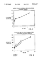
- FIG. 4 is a graph depicting the effect on brain ornithine decarboxylase activity of increasing doses of IGF-I injected into the brains of immature rats.
- FIG. 5 is a graph showing the effect on brain ornithine decarboxylase activity of injection of IGF-I or synthetic peptide fragments of IGFs into the brains of immature rats.
- FIG. 6 is a graph depicting the effect on brain ornithine decarboxylase activity of injection of IGF-I into the brains of mature rats.
- the present invention is directed to the modification of neuroactive polypeptides such as IGF-I and IGF-II and their functional derivatives, and their use as therapeutics for certain neurological diseases or disturbances characterized by increased vulnerability of neurons to dying.
- a "neuroactive polypeptide” is defined as a polypeptide which exerts a cell surface-receptor mediated effect on neuronal cells: e.g., the IGFs, Nerve Growth Factor (NGF), Epidermal Growth Factor, Fibroblast Growth Factor, and insulin.
- a "functional derivative” of a polypeptide is a compound which is a fragment or an analog of that molecule and which possesses the desired biological activity, herein defined as the ability to promote survival and/or cholinergic activity of neuronal cells.
- a “fragment” of a polypeptide refers to any polypeptide subset of that polypeptide.
- An “analog” of a polypeptide refers to a molecule having biological activity but possessing some structural differences compared to the polypeptide: e.g., an altered amino acid sequence, or the presence of additional chemical moieties not normally a part of the molecule. Such moieties (introduced, for example, by acylation, alkylation, cationization, or glycosylation reactions) may improve the molecule's solubility, absorption, transport, biological halflife, etc. Alternatively, or in addition, some moieties may decrease the toxicity of the molecule, or eliminate or attenuate any undesirable side effect of the molecule.
- Table 1 shows the amino acid sequences (expressed using single-letter abbreviations as defined in Table 2) of IGF-I, IGF-II, and a number of functional derivatives of IGF-I and IGF-II.
- peptides Since the ability of peptides to penetrate the blood-brain barrier is related to their lipophilicity or their net ionic charge, suitable modifications of these peptides (e.g., by substituting pentafluorophenylalanine for phenylalanine, or by conjugation to cationized albumin) to increase their transportability (Kastin et al, Pharmac. Biochem. Behav. 11:713-716 (1979); Rapoport et al., Science 207:84-86 (1980); Pardridge et al., Biochem. Biophys. Res. Commun.
- Functional derivatives of the invention include, among others, peptides which vary from the native IGF molecules in any one or more of the following ways:
- Cyclization that is, joining the amino and carboxyl ends of the linear peptide.
- the invention also utilizes as a preferred subgroup within the functional derivatives described above, those functional derivatives having the sequence: R 1 -AA 1 -AA 2 -AA 3 -AA 4 . . . AA n -R 2 , wherein AA 1 , AA 2 , AA 3 , AA 4 . . . AA n are amino acid residues of the IGF-peptide subsets or are conservative replacements for them as defined in Table 2, and n is any integer from 5 to 70 for IGF-I functional derivatives and 5-67 for IGF-II functional derivatives.
- R 1 is attached to the amino group AA 1 and selected from the group of hydrogen, lower (C 1-6 ) alkyl, lower alkyl carbonyl, lower alkenyl, lower alkynyl, formyl, lower (C 6-10 ) aryl, aroyl, aryloxy-carbonyl, aralkyloxy-carbonyl, lower alkyloxycarbonyl, benzoyl, 1- or 2-thenoyl, nicotinoyl, dihydronicotinoyl, N-alkyldihydronicotinoyl, isonicotinoyl, and N-alkyldihydroisonicotinoyl.
- the carboxyl-terminal substituent (R 2 ) of the peptides is selected from the following: OH; NH 2 ; OR 3 , wherein R 3 is a lower alkyl or a lower aryl; OR 3 OH, wherein R 3 is defined as above; and NH-R 3 or N(CH 3 )R 3 , wherein R 3 is defined as above.
- the carboxyl group of the carboxyl-terminal amino acid may be replaced by any one of --PO 3 H 2 , --B(OH) 2 , --CH 2 OH, --SO 3 H or a 5-tetrazole group.
- amino-terminal amino group and/or the lysine, serine or threonine side chains occurring within the peptide may optionally be acylated by formyl, acetyl, propionyl, and similar lower alkylacyl residues or by aryl or heterocyclic acyl residues such as benzoyl, thenoyl, nicotinoyl, isonicotinoyl, n-alkylnicotinoyl and their dihydro and tetrahydro derivatives.
- amino terminal amino acid may optionally be replaced by L-pyroglutamic acid.
- the fragment polypeptides of IGF-I and IGF-II are subsets of the corresponding IGF molecules containing fewer amino acid residues than the native molecules. Preferred are sequences of 5-40 residues and most preferred are sequences of 6-25 residues. A portion of the amino acids of these sequences may be substituted with conservative replacements or deletions which improve the chemical or biological stability of the product peptides or improve their transport across the blood-brain barrier. Preferably, no more than 30% and more preferably no more than 20%, of the amino acid residues are replaced or deleted. A listing of suitable conservative replacements is given in Table 2, along with a key to the single-letter abbreviations for the common, naturally-occurring amino acid residues found in proteins.
- Nle is meant norleucine
- Aib is meant aminoisobutyric acid
- AdaA is meant ⁇ -adamantylalanine
- AdaG is meant ⁇ -adamantylglycine
- homo-Arg is meant L-homoarginine
- D-homo-Arg is meant D-homoarginine
- Acp is meant ⁇ -aminocaproic acid
- Chg is meant L- ⁇ -cyclohexylglycine
- allo-Thr is meant L-allothreonine.
- Cha ⁇ -cyclohexyl-alanine
- Me is meant methyl (CH 3 )
- Orn is meant ornithine
- pyro-Glu is meant the pyroglutamyl group
- Met(O) and D-Met(O) are meant the sulfoxides derived from L- and D-methionine, respectively
- ⁇ -Ala is meant ⁇ -alanine
- Acm is meant acetamidomethyl
- L-Dopa 3-(3,4-dihydroxyphenyl)-L-alanine
- Bpa is meant 4-benzoyl-phenylalanine.
- the polypeptide may first be attached to its receptor in order to protect and maintain the receptor-binding site structure during the chemical modification process, which can comprise, for example, cationization (according to the method, for example, of Pardridge et al., 1987) or glycosylation (according to the method of Schwartz et al., Arch. Biochem. Biophys. 181:542-549 (1977)).
- the present invention provides a novel use of IGF-I and IGF-II and their functional derivatives, as agents for the treatment of diseases or disturbances characterized by an increased risk of cell death, including in particular, neuronal cell death.
- the bioactivity of each polypeptide of the invention may be conveniently assayed by either a brain ornithine decarboxylase assay or a spinal cord choline acetyl transferase assay, both of which are described in detail below.
- the polypeptides may first be screened by the receptor-IGF-I displacement assay described below, which measures the polypeptide's ability to displace labelled IGF-I bound to receptors in homogenized brain tissue.
- the peptides of this invention should be useful for administration to humans or other mammals who suffer from neurological diseases or disturbances characterized by increased risk of neuronal cell death, as described above.
- neurological diseases or disturbances include but are not limited to: Alzheimer's disease, Parkinson's disease, amyotrophic lateral sclerosis, stroke, and concussive or penetrating injuries of the brain or spinal cord.
- compositions of this invention are useful for parenteral administration, for example, intravenous, subcutaneous, intramuscular, intraorbital, ophthalmic, intraventricular, intracranial, intracapsular, intraspinal, intracisternal, intraperitoneal, topical, intranasal, aerosol, scarification, and also for oral, buccal, rectal or vaginal administration.
- the compositions can be formulated for parenteral administration to humans or other mammals in therapeutically effective amounts (e.g., amounts which eliminate or reduce the patient's pathological condition) to provide therapy for the neurological diseases described above.
- compositions can be formulated into pharmaceutical compositions by admixture with pharmaceutically acceptable nontoxic excipients and carriers.
- compositions may be prepared for use as parenteral administration, particularly in the form of liquid solutions or suspensions; for oral administration, particularly in the form of tablets or capsules; or intranasally, particularly in the form of powders, nasal drops, or aerosols.
- compositions may conveniently be administered in unit dosage form and may be prepared by any of the methods well known in the pharmaceutical art, for example, as described in Remington's Pharmaceutical Sciences.
- Formulations for parenteral administration may contain as common excipients sterile water or saline, polyalkylene glycols such as polyethylene glycol, oils of vegetable origin, hydrogenated naphthalenes and the like.
- biocompatible, biodegradable lactide polymer, lactide/glycolide copolymer, or polyoxyethylene-polyoxypropylene copolymers may be useful excipients to control the release of the peptides.
- parenteral delivery systems for these peptides include ethylene-vinyl acetate copolymer particles, osmotic pumps, implantable infusion systems, and liposomes.
- Formulations for inhalation administration contain as excipients, for example, lactose, or may be aqueous solutions containing, for example, polyoxyethylene-9-lauryl ether, glycocholate and deoxycholate, or oily solutions for administration in the form of nasal drops, or as a gel to be applied intranasally.
- Formulations for parenteral administration may also include glycocholate for buccal administration, methoxysalicylate for rectal administration, or citric acid for vaginal administration.
- the materials of this invention can be employed as the sole active agent in a pharmaceutical or can be used in combination with other active ingredients, e.g., other growth factors which could facilitate neuronal survival in neurological diseases, or peptidase or protease inhibitors.
- the concentration of the compounds described herein in a therapeutic composition will vary depending upon a number of factors, including the dosage of the drug to be administered, the chemical characteristics (e.g., hydrophobicity) of the compounds employed, and the route of administration.
- the compounds of this invention may be provided in an aqueous physiological buffer solution containing about 0.1 to 10% w/v compound for parenteral administration. Typical dose ranges are from about 1 ⁇ g/kg to about 1 g/kg of body weight per day; a preferred dose range is from about 0.01 mg/kg to 100 mg/kg of body weight per day.
- the preferred dosage of drug to be administered is likely to depend on such variables as the type and extent of progression of the neurological disease, the overall health status of the particular patient, the relative biological efficacy of the compound selected, the formulation of the compound excipients, and its route of administration.
- Recombinant human IGF-I, IGF-II, and IGF-III, as well as several chemically synthesized peptides consisting of partial sequences of IGF-I or IGF-II, were obtained from commercial sources as indicated in Table 1. 125 I-labeled [Threonine 59 ]IGF-I was obtained from Amersham (Arlington Heights, IL). Other peptides consisting of partial sequences of IGF-I or IGF-II were chemically synthesized using Fmoc chemistry on a Milligen Biosearch Model 9600 Peptide Synthesizer, and purified on Hewlett-Packard Models 1050 and 1090M HPLCs according to the method of Hudson, J. Org. Chem.
- Fmoc amino acids, BOP (Castro's reagent), and resins were purchased from Biosearch (San Raphael, CA 94901) and Bachem Bioscience, Inc. (Philadelphia, PA 19104). Solvents were purchased from Burdick and Jackson (Muskegon, MI 49442). Other reagents were purchased from Sigma Chemical Co. (St. Louis, MO 63178).
- Brain tissue containing the cerebral cortex and cerebellum was dissected from adult Sprague-Dawley rats (Hilltop Lab Animals, Inc. Scottsdale, PA) and homogenized at low power for 5 minutes in a Brinkmann Polytron homogenizer (Westbury, NY) containing 50 volumes of ice-cold buffer consisting of 10 mM HEPES, 0.5% BSA, 0.0125% NEM, 0.025% bacitracin, and 100 KIU/ml aprotinin, pH 7.6 (Bohannon et al., Endocrinology 119:943-945 (1986). Following homogenization, the tissue was collected after centrifugation at 7800 ⁇ g for 20 minutes and resuspended in 10 volumes of assay buffer.
- Tissue 50 ⁇ l
- 50 ⁇ l of buffer or peptides of varying concentration were added to 96-well plates and incubated on ice for 3 hours.
- the tissue was collected on Whatman GF/C filters that had been pre-soaked in 0.01% polyethylenimine and washed four times with ice-cold assay buffer using a Brandel cell harvester (Gaithersburg, MD).
- the filters were removed and the bound 125 I-[Threonine 59 ]IGF-I was measured using a Beckman Model 5500B Gamma Counter.
- Table 3 summarizes the results of the 125 I-[Threonine 59 ]IGF-I displacement assay utilizing native IGFs and IGF fragments. The results demonstrate that, while IGF-I and IGF-III are potent displacers of 125 I-[Threonine 59 ]IGF-I, IGF-II is essentially inactive, indicating that the assay is selective for the identification of IGF-I-like molecules.
- IGF-I(24-41) alone or in combination with IGF-II(54-67) were active in displacing 125 I-[Threonine 59 ]IGF-I.
- IGF-II(54-67) alone, and several other fragments listed in Table 3 were not significantly effective displacers of 125 I-[Threonine 59 ]IGF-I.
- Brains were removed intact from adult Sprague-Dawley rats, frozen on powdered dry ice, and cut into 20 ⁇ m sections (at the level of the cerebellum and brain stem) which were thaw-mounted onto gelatin-coated glass microscope slides (Herkenham and Pert, J. Neurosci. 2:1129-1149 (1982)).
- the tissue sections were covered with 250 ⁇ l of HEPES assay buffer (see Example 1) containing 0.01 nM 125 I-[Threonine 59 ]IGF-I alone or in combination with unlabeled IGF-I, IGF-II, or synthetic peptide fragments thereof.
- the sections were incubated at 4° C. for 24 hours and then rinsed in three 1-minute changes (200 ml each) of ice-cold HEPES assay buffer.
- the tissue sections were then wiped off the slides with filter paper, and the tissue-bound radioactivity was measured in a Beckman Model 5500B Gamma Counter.
- IGF-I, IGF-II, or synthetic peptide derivatives of these molecules was assayed on dissociated cultures of 14-day embryonic rat spinal cord neurons.
- the spinal cord neurons were obtained from trypsin-dissociated spinal cords, plated, incubated with peptides, and subsequently (48 hr later) assayed for choline acetyltransferase activity as described by McManaman et al., Dev. Biol. 112:248-252 (1985).
- IGF-I was found to produce a substantial, dose-dependent increase in choline acetyltransferase activity (FIG. 1), suggesting that IGF-I can dramatically enhance the cholinergic activity of spinal cord cholinergic neurons.
- IGF-II and IGF-III were found to be active in the spinal cord assay (FIG. 2).
- IGF-I(24-41) and IGF-II(33-40) were also found to produce a dose-dependent increase in choline acetyltransferase activity, indicating that each peptide is an active IGF functional derivative (FIG. 3).
- IGF-I, IGF-II or synthetic peptide derivatives of these molecules was tested using a biochemical marker for CNS neurotrophic activity, the induction of brain ornithine decarboxylase.
- the induction (i.e. increased activity) of ornithine decarboxylase has been reported to be a general marker for the actions of a variety of trophic factors. (Schwartz et al., Dev. Brain Res. 1:403-413 (1981); Kanje et al., Brain Res. 381:24-28 (1986); Russell et al., Life Sci. 19:1297-1306 (1976); MacDonnell et al. Proc. Natl. Acad. Sci. USA 74, 4681-4684 (1977); Rinehart et al. Proc. Natl. Acad. Sci. USA 82, 4365-4368 (1985)).
- Sprague-Dawley rats 4 days old, were injected intracerebrally (in the area of the lateral ventricle) with 5 ⁇ l of 0.1M phosphate-buffered saline (PBS) containing IGF-I, IGF-II or a synthetic peptide derivative (1.25-2.5 ⁇ g dose, with 6 animals per treatment group). After 6 hours, the brains were removed, and ornithine decarboxylase was assayed essentially as described by Lewis et al., Proc. Natl. Acad. Sci. USA 75:1021-1023 (1978).
- PBS phosphate-buffered saline
- IGF-I insulin growth factor-I
- IGF-I(24-41) and IGF-II(54-67) increased brain ornithine decarboxylase activity (FIG. 5; these peptides are referred to in FIG. 5 as IGF-I (2-4) and IGF-I(5-6), respectively).
- IGF-I was also injected intraventricularly into the lateral ventricles of adult Sprague-Dawley rats. After 6 hours, the brains were removed, dissected into several regions (cerebral cortex, medial septum, and hippocampus), and then assayed for ornithine decarboxylase activity as described in Example 4. As shown in FIG. 6, IGF-I stimulated ornithine decarboxylase activity in all brain regions assayed. This result indicates that IGF-related molecules have potential utility in widespread regions of the brain.
- Cationization is a process by which free carboxyl groups of acidic amino acid residues on a polypeptide (i.e., aspartic acid and glutamic acid residues) are modified in order to increase the net positive charge on the polypeptide.
- the process of cationization has been used to enhance the cellular uptake of large molecules such as albumin and horseradish peroxidase into mouse fibroblast cells (Shen et al., Proc. Nat. Acad. Sci. USA 75:1872-1876 (1978)). Kumagai et al., J. Biol. Chem.
- the polypeptide e.g., NGF, IGF-I, IGF-II or a functional derivative
- HMD hexamethylenediamine
- EDAC 1-ethyl-3[-3-dimethyl-aminopropyl] carbodiimide hydrochloride
- Unreacted species may be removed by filtration using Centricon-3 MPS-1 separation devices (Amicon, Danvers, MA) or ion exchange chromatography.
- the purified polypeptide may be analyzed using isoelectric focusing to determine the amount of cationization.
- the cationization process may be repeated as described above except that the polypeptide would be pre-bound to an appropriate receptor prior to cationization, in order to protect the receptor-binding site on the polypeptide.
- This protection procedure would be carried out as follows: Brain tissue containing receptors for the polypeptide of interest (e.g., IGF-I) is prepared as described above in Example 1. After incubation with the polypeptide ligand for 2 hours at 4° C. to permit receptor binding, the reaction mixture is brought to room temperature, and the cationization procedure carried out using HMD and EDAC as described above.
- the reaction mixture is then centrifuged at 16,000 rpm at 4° C. for 30 sec in an SS-34 rotor in a Sorvall RC5B centrifuge. The surpernatant is discarded and the pellet washed three times in PBS with bovine serum albumin (1 mg/ml). The pellet is resuspended in 100 mM acetic acid and incubated for 10 min @ 4° C. to release the cationized polypeptide from its receptors. After centrifugation again at 16,000 rpm, the supernatant, which contains the released cationized polypeptide, is pH-neutralized with NaOH. It may then be analyzed by isoelectric focusing, by a receptor-binding assay as described in Example 1, or by any appropriate assay for biological activity.
- polylysine to at least one free carboxyl group on polypeptide (such as IGF-I, IGF-II, or a functional derivative of either).
- polypeptide such as IGF-I, IGF-II, or a functional derivative of either.
- the procedure follows the method of Shen et al., 1978. For example, polylysine, IGF-I and carbodiimide are added in a 1:1:1 ratio in water or buffer for 3 hours at room temperature. The modified protein would be separated and analyzed as described above in Example 6.
- a third method for modifying protein carboxyl groups to enhance blood brain barrier transport is to form esters with diazomethane or N,N-dimethylformamide R acetals (DMF acetals), where R is dimethyl, diethyl, dibutyl, dibenzyl, etc.
- DMF acetals N,N-dimethylformamide R acetals
- R is dimethyl, diethyl, dibutyl, dibenzyl, etc.
- This type of modification rapidly forms esters from negatively charged carboxylic acid groups, thus increasing the overall positive charge.
- An additional benefit from this modification is that these added ester groups may be such that they increase the overall lipophilicity of the polypeptide and may be removed by intrinsic esterases in vivo to yield intact growth factor.
- the procedure for this modification is to react diazomethane or DMF acetals with the polypeptide in a 1:1 ratio in solution for 30 min. at room temperature, followed by purification and characterization as described above in Example 6.
- a fourth method of cationization combines the advantages of polylysine cationization with the formation of cleavable esters to enhance blood-brain barrier transport, as well as to yield intact growth factor following transport.
- Polylysine may be made reactive by reaction with benzyloxylacetyl chloride followed by hydrogenation and mild esterification procedures (Hassner et al., Tet. Let. 46:4475-4478 (1978); Mihara et al., Int. J. Peptide Protein Res. 28:141-145 (1986)).
- DMF acetal derivatives capable of reacting with polylysine could be used to link polylysine to free carboxyl groups using ester linkages.
- a further type of polypeptide modification is glycosylation: the introduction of glucose or similar residues by reductive amination using, for example, glucose and sodium cyanoborohydride (NaCNBH 3 ). Glycosylation of proteins has been shown to enhance the cellular uptake of these proteins and may prove useful for improving blood-brain barrier transport (Smith et al., Pharm. Res., in press).
- glycosylation is based on the method of Schwartz et al., 1977, wherein a polypeptide such as IGF-I, IGF-II, or a functional derivative of either is combined with glucose and NaCNBH 3 in a molar ratio of 1:300:1600 in 200 mM phosphate buffer at pH 7 for at least 24 hr at 37° C. Unreacted entities may be removed as described in Example 6, or with lectin affinity chromatography. In previous studies using glycosylated albumin, the modified albumin was taken up by rat epididymal microvessels at a greater rate than was native albumin (Williams et al., Proc. Nat. Acad. Sci. USA 78:2393-2397 (1981)).
- Microvessel endothelial cells are isolated from the cerebral gray matter of fresh bovine brains. Brains are obtained from a local slaughter house and transported to the laboratory in ice cold minimum essential medium (MEM) with antibiotics. Under sterile conditions the large surface blood vessels and meninges are removed. The cortical gray matter is removed by aspiration, then minced into ⁇ 1 mm cubes. The minced gray matter is then incubated with 0.5% dispase (BMB, Indianapolis, IN) for 3 hours at 37° C. in a shaking water bath. Following the 3 hour digestion, the mixture is concentrated by centrifugation (1000 ⁇ g for 10 min.), then resuspended in 13% dextran and centrifuged for 10 min. at 5800 ⁇ g.
- MEM minimum essential medium
- the crude microvessel pellet is resuspended in 1 mg/ml collagenase/dispase and incubated in a shaking water bath for 5 hours at 37° C. After the 5-hour digestion, the microvessel suspension is applied to a pre-established 50% Percoll gradient and centrifuged for 10 min at 1000 ⁇ g. The band containing purified endothelial cells (second band from the top of the gradient) is removed and washed two times with culture medium (50% MEM/50% F-12 nutrient mix). The cells are frozen (-80° C.) in medium containing 20% DMSO and 10% horse serum for later use.
- the cultured cells can be used in a wide variety of experiments where a model for polarized binding or transport is required. By plating the cells in multi-well plates, receptor and non-receptor binding of both large and small molecules can be conducted. In order to conduct transendothelial cell flux measurements, the cells are grown on porous polycarbonate membrane filters (Nucleopore, Pleasanton, CA.). Large pore size filters (5-12 ⁇ m) are used to avoid the possibility of the filter's becoming the rate-limiting barrier to molecular flux. The use of these large-pore filters does not permit cell growth under the filter and allows visual inspection of the cell monolayer.
- the cells Once the cells reach confluency, they are placed in a side-by-side diffusion cell apparatus (Crown Glass, Sommerville, NJ.).
- a side-by-side diffusion cell apparatus (Crown Glass, Sommerville, NJ.).
- the donor chamber of the diffusion cell is pulsed with a test substance, then at various times following the pulse, an aliquot is removed from the receiver chamber for analysis.
- Radioactive or fluorescently-labelled substances permit reliable quantitation of molecular flux.
- Monolayer integrity is simultaneously measured by the addition of a non-transportable test substance such as sucrose or inulin and replicates of at least 4 determinations are measured in order to ensure statistical significance.
Abstract
Description
              TABLE 3
______________________________________
IGF-I RECEPTOR COMPETITION ASSAY SUMMARY
                       PERCENT MAX.
PEPTIDE (CONC.)        BOUND (SD)
______________________________________
IGF-I (10 pM)          100      (1.1)
IGF-I (40 nM)          9.6      (0.7)
IGF-II (40 nM)         92.1     (0.7)
IGF-III (40 nM)        17.6     (2.6)
IGF-I(24-41) (100 μM)
                       44       (7)
IGF-I(24-41) (50 μM)
                       99       (6)
IGF-I(24-41) (50 μM) + IGF-II(54-67)
                       49       (11)
(50 μM)
IGF-II(54-67) (100 μM)
                       94       (6)
IGF-I(62-70) (100 μM)
                       83       (20)
IGF-I(30-41) (100 μM)
                       94       (1.4)
IGF-II(62-67) (100 μM)
                       83       (21)
IGF-II(33-40) (1 mM)   92       (1.8)
______________________________________
              TABLE 4
______________________________________
PEPTIDE (CONC.)  PERCENT MAX. BOUND
______________________________________
IGF-I (4 pM)     91
IGF-I (400 pM)   30
IGF-II (200 nM)  50
IGF-II (400 nM)  23
IGF-II (33-40)(1 mM)
                 76
IGF-II (33-40)(.10 mM)
                 82
IGF-II (54-67)(.25 mM)
                 167
IGF-II (54-67)(.025 mM)
                 132
______________________________________
Claims (4)
Priority Applications (16)
| Application Number | Priority Date | Filing Date | Title |
|---|---|---|---|
| US07/361,595 US5093317A (en) | 1989-06-05 | 1989-06-05 | Treating disorders by application of insulin-like growth factor |
| EP90909874A EP0476044B1 (en) | 1989-06-05 | 1990-06-05 | Use of IGF1 or IGF2 for the manufacture of a medicament in the treatment of amyotrophic lateral sclerosis |
| PCT/US1990/003166 WO1990014838A1 (en) | 1989-06-05 | 1990-06-05 | Treating disorders by application of insulin-like growth factors and analogs |
| DK90909874.1T DK0476044T3 (en) | 1989-06-05 | 1990-06-05 | Use of IGF1 or IGF2 for the preparation of a drug for the treatment of amyotrophic, lateral sclerosis |
| JP2509846A JPH0768138B2 (en) | 1989-06-05 | 1990-06-05 | Method of treating disorders using insulin-like growth factor and analogs |
| AT90909874T ATE156018T1 (en) | 1989-06-05 | 1990-06-05 | USE OF IGF1 OR IGF2 FOR PRODUCING A MEDICATION FOR THE TREATMENT OF AMYOTROPHIC LATERAL SCLERosis |
| EP97200539A EP0798000A3 (en) | 1989-06-05 | 1990-06-05 | Treating disorders by application of insulin-like growth factors and analogs |
| ES90909874T ES2106735T3 (en) | 1989-06-05 | 1990-06-05 | TREATMENT OF DISORDERS THROUGH THE APPLICATION OF GROWTH FACTORS OF TYPE OF INSULIN AND ANALOGS. |
| CA002058443A CA2058443C (en) | 1989-06-05 | 1990-06-05 | Treating disorders by application of insulin-like growth factors and analogs |
| DE69031168T DE69031168T2 (en) | 1989-06-05 | 1990-06-05 | Use of IGF1 or IGF2 for the manufacture of a medicament for the treatment of amyotrophic lateral sclerosis |
| US07/958,903 US5652214A (en) | 1989-06-05 | 1992-10-07 | Treating disorders by application of insulin-like growth factors and analogs |
| US08/462,018 US5703045A (en) | 1989-06-05 | 1995-06-05 | Treating disorders by application of insulin-like growth factors and analogs |
| US08/823,245 US5776897A (en) | 1989-06-05 | 1997-03-24 | Treating disorders by application of insulin-like growth factors and analogs |
| US09/064,159 US6723699B1 (en) | 1989-06-05 | 1998-04-22 | Treating disorders by application of insulin-like growth factors and analogs |
| HK98112043A HK1010989A1 (en) | 1989-06-05 | 1998-11-16 | Use of igf1 or igf2 for the manufacture of a medicament in the treatment of amyotrophic lateral sclerosis |
| US09/318,001 US6693076B1 (en) | 1989-06-05 | 1999-05-25 | Treating disorders by application of insulin-like growth factors and analogs |
Applications Claiming Priority (1)
| Application Number | Priority Date | Filing Date | Title |
|---|---|---|---|
| US07/361,595 US5093317A (en) | 1989-06-05 | 1989-06-05 | Treating disorders by application of insulin-like growth factor |
Related Child Applications (1)
| Application Number | Title | Priority Date | Filing Date |
|---|---|---|---|
| US53413990A Continuation-In-Part | 1989-06-05 | 1990-06-05 |
Publications (1)
| Publication Number | Publication Date |
|---|---|
| US5093317A true US5093317A (en) | 1992-03-03 |
Family
ID=23422661
Family Applications (1)
| Application Number | Title | Priority Date | Filing Date |
|---|---|---|---|
| US07/361,595 Expired - Lifetime US5093317A (en) | 1989-06-05 | 1989-06-05 | Treating disorders by application of insulin-like growth factor |
Country Status (10)
| Country | Link |
|---|---|
| US (1) | US5093317A (en) |
| EP (2) | EP0476044B1 (en) |
| JP (1) | JPH0768138B2 (en) |
| AT (1) | ATE156018T1 (en) |
| CA (1) | CA2058443C (en) |
| DE (1) | DE69031168T2 (en) |
| DK (1) | DK0476044T3 (en) |
| ES (1) | ES2106735T3 (en) |
| HK (1) | HK1010989A1 (en) |
| WO (1) | WO1990014838A1 (en) |
Cited By (81)
| Publication number | Priority date | Publication date | Assignee | Title |
|---|---|---|---|---|
| WO1993010806A1 (en) * | 1991-11-25 | 1993-06-10 | Institute Of Molecular Biology, Inc. | Medicament for promoting growth of mammalian nerve |
| WO1993020836A1 (en) * | 1992-04-15 | 1993-10-28 | Cephalon, Inc. | Treating disorders by application of insulin-like growth factors and analogs |
| WO1993023067A1 (en) * | 1992-05-08 | 1993-11-25 | Thomas Jefferson University | Igf-1 analogs |
| US5281582A (en) * | 1992-02-27 | 1994-01-25 | Alliance Pharmaceuticals, Corp. | Serum growth factor |
| US5298422A (en) * | 1991-11-06 | 1994-03-29 | Baylor College Of Medicine | Myogenic vector systems |
| WO1994017818A1 (en) * | 1993-02-08 | 1994-08-18 | The Syntex-Synergen Neuroscience Joint Venture | Methods for treating amyotrophic lateral sclerosis with cntf |
| WO1995013823A1 (en) * | 1993-11-15 | 1995-05-26 | Celtrix Pharmaceuticals, Inc. | Method of treating neurological disorders |
| WO1995016703A1 (en) * | 1993-12-15 | 1995-06-22 | Thomas Jefferson University | Igf-1 analogs |
| US5444047A (en) * | 1994-06-16 | 1995-08-22 | Dipasquale; Gene | Treatment of arthritic and post-surgical orthopedic conditions with Insulin-like Growth Factor-I |
| US5565428A (en) * | 1995-05-22 | 1996-10-15 | Genentech, Inc. | Method of administration of IGF-I |
| WO1996032407A1 (en) | 1995-04-14 | 1996-10-17 | Cephalon, Inc. | Igf-i purification process |
| WO1997009060A1 (en) * | 1995-09-08 | 1997-03-13 | Eli Lilly And Company | Method of treating insulin resistance |
| US5616562A (en) * | 1990-04-27 | 1997-04-01 | Murphy; Christopher J. | Methods and compositions using substance P to promote wound healing |
| WO1997011959A1 (en) * | 1995-09-29 | 1997-04-03 | Receptron, Inc. | Biologically active mhc peptides having a repeated amino acid sequence motif |
| US5629286A (en) * | 1994-03-31 | 1997-05-13 | Brewitt; Barbara | Homeopathic dilutions of growth factors |
| US5633230A (en) * | 1990-10-24 | 1997-05-27 | Allelix Biopharmaceuticals, Inc. | Treatment of cytomegalovirus infection |
| US5646120A (en) * | 1990-10-24 | 1997-07-08 | Allelix Biopharmaceuticals, Inc. | Peptide-based inhibitors of HIV replication |
| US5674849A (en) * | 1990-10-24 | 1997-10-07 | Allelix Biopharmaceuticals Inc. | Anti-viral compositions |
| US5714460A (en) * | 1991-08-01 | 1998-02-03 | Genentech Inc. | IFG-1 to improve neural outcome |
| US5716614A (en) * | 1994-08-05 | 1998-02-10 | Molecular/Structural Biotechnologies, Inc. | Method for delivering active agents to mammalian brains in a complex with eicosapentaenoic acid or docosahexaenoic acid-conjugated polycationic carrier |
| US5741776A (en) * | 1995-05-22 | 1998-04-21 | Genentech, Inc. | Method of administration of IGF-I |
| US5792831A (en) * | 1990-02-08 | 1998-08-11 | Magainin Pharmaceuticals Inc. | Analogues of magainin peptides containing D-amino acids |
| US5798715A (en) * | 1996-06-28 | 1998-08-25 | Saperstone; Dorothy Luhr | Method and device for accessing remote control functions |
| US5831001A (en) * | 1990-10-24 | 1998-11-03 | Allelix Biopharmaceuticals Inc. | Treatment of herpesvirus infection |
| US5861373A (en) * | 1991-08-01 | 1999-01-19 | Genentech, Inc | IGF-1 to improve the neural condition |
| WO1999033481A2 (en) * | 1997-12-26 | 1999-07-08 | Applied Research Systems Ars Holding N.V. | Process using the growth factor igf-i in the manufacture of compositions that are useful in the treatment of cerebellar ataxia |
| US5925564A (en) * | 1991-11-06 | 1999-07-20 | Baylor College Of Medicine | Expression vector systems and method of use |
| US6015786A (en) * | 1997-02-25 | 2000-01-18 | Celtrix Pharmaceuticals, Inc. | Method for increasing sex steroid levels using IGF or IGF/IGFBP-3 |
| WO2000005262A1 (en) * | 1998-07-22 | 2000-02-03 | The University Of Connecticut | Uses of igf-i e domain peptides |
| US6025368A (en) * | 1997-02-25 | 2000-02-15 | Celtrix Pharmaceuticals, Inc. | Method for treating the symptoms of chronic stress-related disorders using IGF |
| US6121416A (en) * | 1997-04-04 | 2000-09-19 | Genentech, Inc. | Insulin-like growth factor agonist molecules |
| US6204240B1 (en) * | 1991-11-22 | 2001-03-20 | Genentech, Inc. | TGF-β1 to improve neural outcome |
| US6207656B1 (en) | 1997-05-22 | 2001-03-27 | Cephalon, Inc. | Vitamin D analogues and their neuronal effects |
| US20010018414A1 (en) * | 1997-10-23 | 2001-08-30 | Regents Of The University Of Minnesota, A Minnesota Corporation | Modified vitamin K-dependent polypeptides |
| US6310040B1 (en) * | 1991-11-08 | 2001-10-30 | Cephalon, Inc. | Treating retinal neuronal disorders by the application of insulin-like growth factors and analogs |
| WO2002032449A2 (en) * | 2000-10-13 | 2002-04-25 | Chiron Corporation | Method for treating ischemic events affecting the central nervous system |
| US6420518B1 (en) | 1997-04-04 | 2002-07-16 | Genetech, Inc. | Insulin-like growth factor agonist molecules |
| US6440928B1 (en) | 1988-12-06 | 2002-08-27 | Colorado State University Research Foundation | Method for treating diabetic neuropathy with NGF |
| US20020165155A1 (en) * | 2001-02-09 | 2002-11-07 | Genentech, Inc. | Crystallization of IGF-1 |
| US6495513B1 (en) | 1991-03-11 | 2002-12-17 | Curis, Inc. | Morphogen-enhanced survival and repair of neural cells |
| US6514937B1 (en) | 1997-02-25 | 2003-02-04 | Celtrix Pharmaceuticals, Inc. | Method of treating psychological and metabolic disorders using IGF or IGF/IGFBP-3 |
| US20030027755A1 (en) * | 2000-12-08 | 2003-02-06 | Jian Guan | Compositions and methods for the rescue of white matter |
| US20030100506A1 (en) * | 1997-10-23 | 2003-05-29 | Nelsestuen Gary L. | Modified Vitamin K-dependent polypeptides |
| WO2003077940A1 (en) * | 2002-02-28 | 2003-09-25 | Consejo Superior De Investigaciones Científicas | Chemical igf-i composition for the treatment and prevention of neurodegenerative diseases |
| US20030211992A1 (en) * | 1997-04-04 | 2003-11-13 | Genentech, Inc. | Method for treating cartilage disorders |
| US6693076B1 (en) * | 1989-06-05 | 2004-02-17 | Cephalon, Inc. | Treating disorders by application of insulin-like growth factors and analogs |
| US6723699B1 (en) * | 1989-06-05 | 2004-04-20 | Cephalon, Inc. | Treating disorders by application of insulin-like growth factors and analogs |
| US20040116335A1 (en) * | 1998-07-22 | 2004-06-17 | Chen Thomas T. | Compositions and methods for inhibiting the proliferation and invasiveness of malignant cells comprising E-domain peptides of IGF-I |
| US6756484B1 (en) | 1995-04-14 | 2004-06-29 | Cephalon, Inc. | IGF-I purification process |
| US20040176704A1 (en) * | 2003-03-04 | 2004-09-09 | Stevens Timothy A | Collection device adapted to accept cartridge for point of care system |
| US6800603B2 (en) | 1991-03-11 | 2004-10-05 | Curis, Inc. | Morphogen-induced neural cell adhesion |
| US20050096339A1 (en) * | 1991-03-11 | 2005-05-05 | Thangavel Kuberasampath | Morphogen-induced modulation of inflammatory response |
| US20050101527A1 (en) * | 2000-08-29 | 2005-05-12 | Ishii Douglas N. | Method for treating the central nervous system by administration of igf structural analogs |
| US20050164932A1 (en) * | 2003-06-19 | 2005-07-28 | Maxygen Holdings, Ltd. | FVII or FVIIa Gla domain variants |
| US6949505B1 (en) | 1991-03-11 | 2005-09-27 | Curis, Inc. | Morphogen-induced dendritic growth |
| US20060019336A1 (en) * | 2000-02-11 | 2006-01-26 | Maxygen Aps | Factor VII or VIIa - like molecules |
| US20060064249A1 (en) * | 2004-08-30 | 2006-03-23 | Clark Ross G | Method and device for diagnosing and treating insulin-like growth factor deficiency disorders |
| US20060069008A1 (en) * | 2004-09-28 | 2006-03-30 | Sanjay Mistry | Treatment of neurological deficits in the striatum or substanta nigra pars compacta |
| US7041314B2 (en) | 2001-05-24 | 2006-05-09 | Neuren Pharmaceuticals Ltd. | GPE analogs and peptidominetics |
| US20060111282A1 (en) * | 2002-04-30 | 2006-05-25 | Maxygen Holdings Ltd. | Factor vii or viia polypeptide variants |
| US20060135429A1 (en) * | 1998-07-22 | 2006-06-22 | Chen Thomas T | Compositions and methods for inducing apoptosis in tumor cells |
| US20060166874A1 (en) * | 2002-09-30 | 2006-07-27 | Haaning Jesper M | Fvii or fviia variants having increased clotting activity |
| US20060211606A1 (en) * | 2005-03-18 | 2006-09-21 | Geoffrey Goldspink | Peptides |
| US20060251649A1 (en) * | 2001-05-24 | 2006-11-09 | Neuren Pharmaceuticals Limited | Treatment of non-convulsive seizures in brain injury using G-2-methyl-prolyl glutamate |
| US20070004641A1 (en) * | 2001-05-24 | 2007-01-04 | Neuren Pharmaceuticals Limited | Cognitive enhancement and cognitive therapy using glycyl-L-2-methylprolyl-L-glutamate |
| US20070054366A1 (en) * | 2003-03-20 | 2007-03-08 | Maxygen Holdings Ltd. | Fvii or fviia variants |
| US20070078089A1 (en) * | 1992-07-06 | 2007-04-05 | Ishii Douglas N | Method for effecting changes in the central nervous system by administration of IGF-I or IGF-II |
| US7220837B1 (en) | 2000-04-28 | 2007-05-22 | Regents Of The University Of Minnesota | Modified vitamin K-dependent polypeptides |
| US20070254840A1 (en) * | 2000-06-30 | 2007-11-01 | Nelsestuen Gary L | High Molecular Weight Derivatives of Vitamin K-Dependent Polypeptides |
| US20070298009A1 (en) * | 2001-05-24 | 2007-12-27 | Neuren Pharmaceuticals Limited | Effects of glycyl-2 methyl prolyl glutamate on neurodegeneration |
| US20080207510A1 (en) * | 2000-04-28 | 2008-08-28 | Nelsestuen Gary L | Modified vitamin k-dependent polypeptides |
| US20080254538A1 (en) * | 2004-09-28 | 2008-10-16 | Messina Darin J | Treatment of Neurological Deficits in the Striatum or Substanta Nigra Pars Compacta |
| US20090012499A1 (en) * | 2005-02-08 | 2009-01-08 | Japan As Rep. By Pres. Of Nat. Cardiovascular Ctr. | Novel method for treating chronic severe heart failure by using insulin-like growth factor-1 (igf-1) |
| US20090099077A1 (en) * | 2007-06-08 | 2009-04-16 | Mriganka Sur | Treatment of rett syndrome and other disorders |
| ES2322685A1 (en) * | 2006-11-21 | 2009-06-24 | Universidad De Malaga | Use of low doses of igf-ii in aging for its neuroprotective effects and hepatoprotectores. (Machine-translation by Google Translate, not legally binding) |
| US20090253628A1 (en) * | 2008-04-03 | 2009-10-08 | Bettina Holtmann | Use of PEGylated IGF-I variants for the treatment of neuromuscular disorders |
| US20090280107A1 (en) * | 1997-10-23 | 2009-11-12 | Nelsestuen Gary L | Modified vitamin k-dependent polypeptides |
| US20100035817A1 (en) * | 2006-08-31 | 2010-02-11 | Stephan Fischer | Method for the production of conjugates of insulin-like growth factor-1 and poly(ethylene glycol) |
| US20110003746A1 (en) * | 1999-01-06 | 2011-01-06 | Yves Dubaquie | Protein variants |
| US20120077749A1 (en) * | 2008-04-17 | 2012-03-29 | Baxter Healthcare S.A. | Biologically Active Peptides |
| WO2016064737A1 (en) * | 2014-10-20 | 2016-04-28 | Neuralstem, Inc. | Stable neural stem cells comprising an exogenous polynucleotide coding for a growth factor and methods of use thereof |
Families Citing this family (8)
| Publication number | Priority date | Publication date | Assignee | Title |
|---|---|---|---|---|
| SE9101341D0 (en) * | 1991-05-03 | 1991-05-03 | Kabi Pharmacia Ab | NEW MEDICINAL USE |
| AU683215B2 (en) * | 1992-06-12 | 1997-11-06 | Albert Einstein College Of Medicine Of Yeshiva University | Prevention and treatment of peripheral neuropathy |
| US5728676A (en) * | 1994-09-08 | 1998-03-17 | Ciba-Geigy Corporation | Use of insulin-like growth factors I and II for inhibition of inflammatory response |
| JP2000514038A (en) * | 1995-12-13 | 2000-10-24 | オーロゲン インコーポレイテッド | Methods for effecting changes in the central nervous system by administration of IGF-I or IGF-II |
| KR20010072354A (en) * | 1998-08-11 | 2001-07-31 | 다께우찌 마사야쓰 | Blood sugar level controlling agent |
| TWI305778B (en) * | 2001-12-03 | 2009-02-01 | Nishida Teruo | Peptides of an expressing unit for igf-i minimal activity and their pharmaceutical use |
| JP4253743B2 (en) * | 2001-12-03 | 2009-04-15 | 輝夫 西田 | Novel peptides and their pharmaceutical uses |
| CN106167789B (en) * | 2015-05-21 | 2020-08-04 | 中国科学院上海营养与健康研究所 | Hypoxia-treated mesenchymal stem cells and application thereof |
Citations (13)
| Publication number | Priority date | Publication date | Assignee | Title |
|---|---|---|---|---|
| US4341762A (en) * | 1981-04-07 | 1982-07-27 | Haast William E | Use of snake venoms for treatment of neurological and related disorders |
| JPS5965058A (en) * | 1982-10-05 | 1984-04-13 | Daiichi Rajio Isotope Kenkyusho:Kk | Heneicosapeptide |
| US4511390A (en) * | 1983-06-10 | 1985-04-16 | E. I. Du Pont De Nemours And Company | Aralkylcarbamoyl peptide alcohols |
| US4699875A (en) * | 1982-11-24 | 1987-10-13 | Baylor College Of Medicine | Diagnosis of amyotrophic lateral sclerosis by neurotrophic factors |
| WO1988001393A1 (en) * | 1986-08-12 | 1988-02-25 | William Abrams | Method for sonic analysis of an anomaly in a seafloor topographic representation |
| WO1988001693A1 (en) * | 1986-09-05 | 1988-03-10 | Young Niels O | Magnet ball pump |
| WO1988002760A1 (en) * | 1986-10-14 | 1988-04-21 | Bp Chemicals Limited | Modified polymer polyols |
| JPS63196524A (en) * | 1987-02-10 | 1988-08-15 | Wakunaga Pharmaceut Co Ltd | Carcinostatic action adjuster of transmucosal absorption type |
| US4783524A (en) * | 1985-09-17 | 1988-11-08 | Monsanto Company | Growth factors |
| US4801575A (en) * | 1986-07-30 | 1989-01-31 | The Regents Of The University Of California | Chimeric peptides for neuropeptide delivery through the blood-brain barrier |
| WO1989001343A1 (en) * | 1987-08-17 | 1989-02-23 | The Regents Of The University Of California | Cationized antibodies for delivery through the blood-brain barrier |
| WO1990003166A2 (en) * | 1988-09-27 | 1990-04-05 | The Health Foundation Limited | Method and composition for the treatment of burns |
| EP0289314B1 (en) * | 1987-04-28 | 1994-10-12 | Roche Diagnostics GmbH | Use of IGF-II in the treatment of bone disorders |
Family Cites Families (3)
| Publication number | Priority date | Publication date | Assignee | Title |
|---|---|---|---|---|
| SE8505920D0 (en) * | 1985-12-13 | 1985-12-13 | Kabigen Ab | NEW PROTEIN AND ITS USE |
| SE8703625D0 (en) * | 1987-09-18 | 1987-09-18 | Kabivitrum Ab | NEW MEDICAL USE |
| JPH0525056A (en) * | 1989-07-27 | 1993-02-02 | Max Planck Inst Fuer Saikaiatorii | Adjustment of nerve growth factor synthesis in central nervous system |
-
1989
- 1989-06-05 US US07/361,595 patent/US5093317A/en not_active Expired - Lifetime
-
1990
- 1990-06-05 DE DE69031168T patent/DE69031168T2/en not_active Expired - Lifetime
- 1990-06-05 DK DK90909874.1T patent/DK0476044T3/en active
- 1990-06-05 WO PCT/US1990/003166 patent/WO1990014838A1/en active IP Right Grant
- 1990-06-05 CA CA002058443A patent/CA2058443C/en not_active Expired - Lifetime
- 1990-06-05 AT AT90909874T patent/ATE156018T1/en not_active IP Right Cessation
- 1990-06-05 EP EP90909874A patent/EP0476044B1/en not_active Expired - Lifetime
- 1990-06-05 ES ES90909874T patent/ES2106735T3/en not_active Expired - Lifetime
- 1990-06-05 JP JP2509846A patent/JPH0768138B2/en not_active Expired - Lifetime
- 1990-06-05 EP EP97200539A patent/EP0798000A3/en not_active Withdrawn
-
1998
- 1998-11-16 HK HK98112043A patent/HK1010989A1/en not_active IP Right Cessation
Patent Citations (13)
| Publication number | Priority date | Publication date | Assignee | Title |
|---|---|---|---|---|
| US4341762A (en) * | 1981-04-07 | 1982-07-27 | Haast William E | Use of snake venoms for treatment of neurological and related disorders |
| JPS5965058A (en) * | 1982-10-05 | 1984-04-13 | Daiichi Rajio Isotope Kenkyusho:Kk | Heneicosapeptide |
| US4699875A (en) * | 1982-11-24 | 1987-10-13 | Baylor College Of Medicine | Diagnosis of amyotrophic lateral sclerosis by neurotrophic factors |
| US4511390A (en) * | 1983-06-10 | 1985-04-16 | E. I. Du Pont De Nemours And Company | Aralkylcarbamoyl peptide alcohols |
| US4783524A (en) * | 1985-09-17 | 1988-11-08 | Monsanto Company | Growth factors |
| US4801575A (en) * | 1986-07-30 | 1989-01-31 | The Regents Of The University Of California | Chimeric peptides for neuropeptide delivery through the blood-brain barrier |
| WO1988001393A1 (en) * | 1986-08-12 | 1988-02-25 | William Abrams | Method for sonic analysis of an anomaly in a seafloor topographic representation |
| WO1988001693A1 (en) * | 1986-09-05 | 1988-03-10 | Young Niels O | Magnet ball pump |
| WO1988002760A1 (en) * | 1986-10-14 | 1988-04-21 | Bp Chemicals Limited | Modified polymer polyols |
| JPS63196524A (en) * | 1987-02-10 | 1988-08-15 | Wakunaga Pharmaceut Co Ltd | Carcinostatic action adjuster of transmucosal absorption type |
| EP0289314B1 (en) * | 1987-04-28 | 1994-10-12 | Roche Diagnostics GmbH | Use of IGF-II in the treatment of bone disorders |
| WO1989001343A1 (en) * | 1987-08-17 | 1989-02-23 | The Regents Of The University Of California | Cationized antibodies for delivery through the blood-brain barrier |
| WO1990003166A2 (en) * | 1988-09-27 | 1990-04-05 | The Health Foundation Limited | Method and composition for the treatment of burns |
Non-Patent Citations (79)
| Title |
|---|
| Aizenman et al., Brain Res. 406:32 42 (1987). * |
| Aizenman et al., Brain Res. 406:32-42 (1987). |
| Baskin et al., TINS 11:107 111 (1988). * |
| Baskin et al., TINS 11:107-111 (1988). |
| Baxter et al., Comp. Biochem. Physiol. 91B:229 235 (1988). * |
| Baxter et al., Comp. Biochem. Physiol. 91B:229-235 (1988). |
| Blundell et al., Fed. Proc. 42:2592 2597 (1983). * |
| Blundell et al., Fed. Proc. 42:2592-2597 (1983). |
| Bodor et al., Science 214:1370 1372 (1981). * |
| Bodor et al., Science 214:1370-1372 (1981). |
| Bothwell, J. Neurosci. Res. 8:225 231 (1982). * |
| Bothwell, J. Neurosci. Res. 8:225-231 (1982). |
| Coy et al., Biochem. Biophys. Res. Commun. 73:632 8 (1976). * |
| Coy et al., Biochem. Biophys. Res. Commun. 73:632-8 (1976). |
| Creveling et al., Experientia 25:26 27 (1969). * |
| Creveling et al., Experientia 25:26-27 (1969). |
| D Ercole, J. Devel. Physiol. 9:481 495 (1987). * |
| Daughaday et al., Endocrine Rev. 10:68 91 (1989). * |
| Daughaday et al., Endocrine Rev. 10:68-91 (1989). |
| D'Ercole, J. Devel. Physiol. 9:481-495 (1987). |
| Fellows et al., Soc. Neurosci. Abstr. 13:1615 (1987). * |
| Francis et al., Biochem. J. 251:95 103 (1988). * |
| Francis et al., Biochem. J. 251:95-103 (1988). |
| Hansson et al. Act & Physiol. Scand. 126:609 614 (1986). * |
| Hansson et al. Act & Physiol. Scand. 126:609-614 (1986). |
| Hansson et al., Acta Physiol. Scand. 132:35 41 (1988). * |
| Hansson et al., Acta Physiol. Scand. 132:35-41 (1988). |
| Hintz et al., Horm. Metab. Res. 20:344 347 (1988). * |
| Hintz et al., Horm. Metab. Res. 20:344-347 (1988). |
| Hintz et al., J. Clinical Endocrinology and Metabolism 54:442 446. * |
| Hintz et al., J. Clinical Endocrinology and Metabolism 54:442-446. |
| Kanje et al., Brain Res. 381:24 28 (1986). * |
| Kanje et al., Brain Res. 381:24-28 (1986). |
| Kastin et al., Pharmac. Biochem. Behav. 11:713 716 (1979). * |
| Kastin et al., Pharmac. Biochem. Behav. 11:713-716 (1979). |
| MacDonnell et al., Proc. Natl. Acad. Sci. USA 74:4681 4684 (1977). * |
| MacDonnell et al., Proc. Natl. Acad. Sci. USA 74:4681-4684 (1977). |
| Mattson et al., J. Cell Biol. 102:1949 54 (1986). * |
| Mattson et al., J. Cell Biol. 102:1949-54 (1986). |
| McManaman et al., Dev. Biol. 112:248 252 (1985). * |
| McManaman et al., Dev. Biol. 112:248-252 (1985). |
| Nilsson et al., Neurosci. Let. 88:221 226 (1988). * |
| Nilsson et al., Neurosci. Let. 88:221-226 (1988). |
| Onifer et al., Soc. Neurosci. Abstr. 13:1615 (1987). * |
| Pardridge et al., Biochem. Biophys. Res. Commun. 146:307 313 (1987). * |
| Pardridge et al., Biochem. Biophys. Res. Commun. 146:307-313 (1987). |
| Pardridge, Endocrine Reviews 7:314 330 (1986). * |
| Pardridge, Endocrine Reviews 7:314-330 (1986). |
| Rapoport et al., Science 207:84 86 (1980). * |
| Rapoport et al., Science 207:84-86 (1980). |
| Recio Pinto and Ishii, J. Neurosci. Res. 19:312 320 (1988). * |
| Recio Pinto et al., J. Neurosci. 6:1211 1219 (1986). * |
| Recio-Pinto and Ishii, J. Neurosci. Res. 19:312-320 (1988). |
| Recio-Pinto et al., J. Neurosci. 6:1211-1219 (1986). |
| Riekkinen et al., Peptides 8:261 265 (1987). * |
| Riekkinen et al., Peptides 8:261-265 (1987). |
| Rinehart et al., Proc. Natl. Acad. Sci. USA 82:4365 4368 (1985). * |
| Rinehart et al., Proc. Natl. Acad. Sci. USA 82:4365-4368 (1985). |
| Russell et al., Life Sciences 19:1297 1306 (1976). * |
| Russell et al., Life Sciences 19:1297-1306 (1976). |
| Sara et al. Biol. Psychiat. 18:803 811 (1983). * |
| Sara et al. Biol. Psychiat. 18:803-811 (1983). |
| Sara et al., J. Dev. Physiol. 1:343 350 (1979). * |
| Sara et al., J. Dev. Physiol. 1:343-350 (1979). |
| Sara et al., Neurobiol. Aging 3:117 120 (1982). * |
| Sara et al., Neurobiol. Aging 3:117-120 (1982). |
| Sara et al., Neurosci. Let. 34:39 44 (1982). * |
| Sara et al., Neurosci. Let. 34:39-44 (1982). |
| Sara et al., Proc. Natl. Acad. Sci. USA 83:4904 4907 (1986). * |
| Sara et al., Proc. Natl. Acad. Sci. USA 83:4904-4907 (1986). |
| Sara et al., Prog. Brain Res. 73:87 99 (1988). * |
| Sara et al., Prog. Brain Res. 73:87-99 (1988). |
| Schwartz et al., Archives of Biochemistry & Biophysics 181:542 549 (1977). * |
| Schwartz et al., Archives of Biochemistry & Biophysics 181:542-549 (1977). |
| Schwartz et al., Dev. Brain Res. 1:403 413 (1981). * |
| Schwartz et al., Dev. Brain Res. 1:403-413 (1981). |
| Sjoberg et al., Brain Res. 485:102 108 (1989). * |
| Sjoberg et al., Brain Res. 485:102-108 (1989). |
| Smith et al., Pharm Res. (in press). * |
Cited By (212)
| Publication number | Priority date | Publication date | Assignee | Title |
|---|---|---|---|---|
| US6440928B1 (en) | 1988-12-06 | 2002-08-27 | Colorado State University Research Foundation | Method for treating diabetic neuropathy with NGF |
| US5776897A (en) * | 1989-06-05 | 1998-07-07 | Cephalon, Inc. | Treating disorders by application of insulin-like growth factors and analogs |
| US6693076B1 (en) * | 1989-06-05 | 2004-02-17 | Cephalon, Inc. | Treating disorders by application of insulin-like growth factors and analogs |
| US6723699B1 (en) * | 1989-06-05 | 2004-04-20 | Cephalon, Inc. | Treating disorders by application of insulin-like growth factors and analogs |
| US5652214A (en) * | 1989-06-05 | 1997-07-29 | Cephalon, Inc. | Treating disorders by application of insulin-like growth factors and analogs |
| US5792831A (en) * | 1990-02-08 | 1998-08-11 | Magainin Pharmaceuticals Inc. | Analogues of magainin peptides containing D-amino acids |
| US5616562A (en) * | 1990-04-27 | 1997-04-01 | Murphy; Christopher J. | Methods and compositions using substance P to promote wound healing |
| US5789531A (en) * | 1990-10-24 | 1998-08-04 | Allex Biopharmaceuticals, Inc. | Peptide-based inhibitors of HIV replication |
| US5674849A (en) * | 1990-10-24 | 1997-10-07 | Allelix Biopharmaceuticals Inc. | Anti-viral compositions |
| US5646120A (en) * | 1990-10-24 | 1997-07-08 | Allelix Biopharmaceuticals, Inc. | Peptide-based inhibitors of HIV replication |
| US5633230A (en) * | 1990-10-24 | 1997-05-27 | Allelix Biopharmaceuticals, Inc. | Treatment of cytomegalovirus infection |
| US5831001A (en) * | 1990-10-24 | 1998-11-03 | Allelix Biopharmaceuticals Inc. | Treatment of herpesvirus infection |
| US6495513B1 (en) | 1991-03-11 | 2002-12-17 | Curis, Inc. | Morphogen-enhanced survival and repair of neural cells |
| US6949505B1 (en) | 1991-03-11 | 2005-09-27 | Curis, Inc. | Morphogen-induced dendritic growth |
| US20050096339A1 (en) * | 1991-03-11 | 2005-05-05 | Thangavel Kuberasampath | Morphogen-induced modulation of inflammatory response |
| US20060025571A1 (en) * | 1991-03-11 | 2006-02-02 | Curis, Inc. | Morphogen-induced dendritic growth |
| US6800603B2 (en) | 1991-03-11 | 2004-10-05 | Curis, Inc. | Morphogen-induced neural cell adhesion |
| USRE43982E1 (en) | 1991-08-01 | 2013-02-05 | Genentech Inc. | IGF-1 to improve neural outcome |
| US5714460A (en) * | 1991-08-01 | 1998-02-03 | Genentech Inc. | IFG-1 to improve neural outcome |
| US5861373A (en) * | 1991-08-01 | 1999-01-19 | Genentech, Inc | IGF-1 to improve the neural condition |
| US5925564A (en) * | 1991-11-06 | 1999-07-20 | Baylor College Of Medicine | Expression vector systems and method of use |
| US5298422A (en) * | 1991-11-06 | 1994-03-29 | Baylor College Of Medicine | Myogenic vector systems |
| US5756264A (en) * | 1991-11-06 | 1998-05-26 | Baylor College Of Medicine | Expression vector systems and method of use |
| US6310040B1 (en) * | 1991-11-08 | 2001-10-30 | Cephalon, Inc. | Treating retinal neuronal disorders by the application of insulin-like growth factors and analogs |
| US6204240B1 (en) * | 1991-11-22 | 2001-03-20 | Genentech, Inc. | TGF-β1 to improve neural outcome |
| US20030004108A1 (en) * | 1991-11-22 | 2003-01-02 | Genentech, Inc. | TGF-beta to improve neural outcome |
| WO1993010806A1 (en) * | 1991-11-25 | 1993-06-10 | Institute Of Molecular Biology, Inc. | Medicament for promoting growth of mammalian nerve |
| US6506727B1 (en) | 1991-11-25 | 2003-01-14 | Institute Of Molecular Biology, Inc. | Nerve regeneration |
| US5281582A (en) * | 1992-02-27 | 1994-01-25 | Alliance Pharmaceuticals, Corp. | Serum growth factor |
| WO1993020836A1 (en) * | 1992-04-15 | 1993-10-28 | Cephalon, Inc. | Treating disorders by application of insulin-like growth factors and analogs |
| US5473054A (en) * | 1992-05-08 | 1995-12-05 | Thomas Jefferson University | IGF-1 analogs |
| WO1993023067A1 (en) * | 1992-05-08 | 1993-11-25 | Thomas Jefferson University | Igf-1 analogs |
| US20070078089A1 (en) * | 1992-07-06 | 2007-04-05 | Ishii Douglas N | Method for effecting changes in the central nervous system by administration of IGF-I or IGF-II |
| WO1994017818A1 (en) * | 1993-02-08 | 1994-08-18 | The Syntex-Synergen Neuroscience Joint Venture | Methods for treating amyotrophic lateral sclerosis with cntf |
| WO1995013823A1 (en) * | 1993-11-15 | 1995-05-26 | Celtrix Pharmaceuticals, Inc. | Method of treating neurological disorders |
| WO1995016703A1 (en) * | 1993-12-15 | 1995-06-22 | Thomas Jefferson University | Igf-1 analogs |
| US5629286A (en) * | 1994-03-31 | 1997-05-13 | Brewitt; Barbara | Homeopathic dilutions of growth factors |
| US5444047A (en) * | 1994-06-16 | 1995-08-22 | Dipasquale; Gene | Treatment of arthritic and post-surgical orthopedic conditions with Insulin-like Growth Factor-I |
| US5716614A (en) * | 1994-08-05 | 1998-02-10 | Molecular/Structural Biotechnologies, Inc. | Method for delivering active agents to mammalian brains in a complex with eicosapentaenoic acid or docosahexaenoic acid-conjugated polycationic carrier |
| US6756484B1 (en) | 1995-04-14 | 2004-06-29 | Cephalon, Inc. | IGF-I purification process |
| US6207806B1 (en) | 1995-04-14 | 2001-03-27 | Cephalon Inc. | IGF-I purification process |
| US5650496A (en) * | 1995-04-14 | 1997-07-22 | Cephalon, Inc. | IGF-I purification process |
| WO1996032407A1 (en) | 1995-04-14 | 1996-10-17 | Cephalon, Inc. | Igf-i purification process |
| US5741776A (en) * | 1995-05-22 | 1998-04-21 | Genentech, Inc. | Method of administration of IGF-I |
| US5565428A (en) * | 1995-05-22 | 1996-10-15 | Genentech, Inc. | Method of administration of IGF-I |
| US5939387A (en) * | 1995-09-08 | 1999-08-17 | Eli Lilly And Company | Method of treating insulin resistance |
| WO1997009060A1 (en) * | 1995-09-08 | 1997-03-13 | Eli Lilly And Company | Method of treating insulin resistance |
| WO1997011959A1 (en) * | 1995-09-29 | 1997-04-03 | Receptron, Inc. | Biologically active mhc peptides having a repeated amino acid sequence motif |
| US5798715A (en) * | 1996-06-28 | 1998-08-25 | Saperstone; Dorothy Luhr | Method and device for accessing remote control functions |
| US6025332A (en) * | 1997-02-25 | 2000-02-15 | Celtrix Pharmaceuticals, Inc. | Method for treating low circulating levels of sex hormone steroids associated with aging using IGF or IGF/IGFBP-3 |
| US6514937B1 (en) | 1997-02-25 | 2003-02-04 | Celtrix Pharmaceuticals, Inc. | Method of treating psychological and metabolic disorders using IGF or IGF/IGFBP-3 |
| US6015786A (en) * | 1997-02-25 | 2000-01-18 | Celtrix Pharmaceuticals, Inc. | Method for increasing sex steroid levels using IGF or IGF/IGFBP-3 |
| US6025368A (en) * | 1997-02-25 | 2000-02-15 | Celtrix Pharmaceuticals, Inc. | Method for treating the symptoms of chronic stress-related disorders using IGF |
| US6518238B1 (en) | 1997-02-25 | 2003-02-11 | Celtrix Pharmaceuticals, Inc. | Method of treating psychological and metabolic disorders using IGF or IGF/IGFBP-3 |
| US6632794B1 (en) | 1997-04-04 | 2003-10-14 | Genentech, Inc. | Insulin-like growth factor agonist molecules |
| US20030211992A1 (en) * | 1997-04-04 | 2003-11-13 | Genentech, Inc. | Method for treating cartilage disorders |
| US8110548B2 (en) | 1997-04-04 | 2012-02-07 | Genentech, Inc. | Method for treating cartilage disorders |
| US6121416A (en) * | 1997-04-04 | 2000-09-19 | Genentech, Inc. | Insulin-like growth factor agonist molecules |
| US6608028B1 (en) | 1997-04-04 | 2003-08-19 | Genentech, Inc. | Insulin-like growth factor agonist molecules |
| US6608031B1 (en) | 1997-04-04 | 2003-08-19 | Genentech, Inc. | Insulin-like growth factor agonist molecules |
| US6743894B1 (en) | 1997-04-04 | 2004-06-01 | Genentech, Inc. | Insulin-like growth factor agonist molecules |
| US6620789B1 (en) | 1997-04-04 | 2003-09-16 | Genentech, Inc. | Insulin-like growth factor agonist molecules |
| US6251865B1 (en) | 1997-04-04 | 2001-06-26 | Genentech, Inc. | Insulin-like growth factor agonist molecules |
| US7947650B2 (en) | 1997-04-04 | 2011-05-24 | Genentech, Inc. | Article of manufacture |
| US6420518B1 (en) | 1997-04-04 | 2002-07-16 | Genetech, Inc. | Insulin-like growth factor agonist molecules |
| US6635619B1 (en) | 1997-04-04 | 2003-10-21 | Genentech, Inc. | Insulin-like growth factor agonist molecules |
| US6645775B1 (en) | 1997-04-04 | 2003-11-11 | Genentech, Inc. | Insulin-like growth factor agonist molecules |
| US6949349B1 (en) | 1997-04-04 | 2005-09-27 | Genentech, Inc. | Insulin-like growth factor agonist molecules |
| US6677305B1 (en) | 1997-04-04 | 2004-01-13 | Genentech, Inc. | Insulin-like growth factor agonist molecules |
| US6680298B1 (en) | 1997-04-04 | 2004-01-20 | Genentech, Inc. | Insulin-like growth factor agonist molecules |
| US6683053B1 (en) | 1997-04-04 | 2004-01-27 | Genentech, Inc. | Insulin-like growth factor agonist molecules |
| US6689751B1 (en) | 1997-04-04 | 2004-02-10 | Genentech, Inc. | Insulin-like growth factor agonist molecules |
| US6750321B1 (en) | 1997-04-04 | 2004-06-15 | Genentech, Inc. | Insulin-like growth factor agonist molecules |
| US7423017B2 (en) | 1997-04-04 | 2008-09-09 | Genentech, Inc. | Method for treating cartilage disorders |
| US6693079B1 (en) | 1997-04-04 | 2004-02-17 | Genentech, Inc. | Insulin-like growth factor agonist molecules |
| US6693078B1 (en) | 1997-04-04 | 2004-02-17 | Genentech, Inc. | Insulin-like growth factor agonist molecules |
| US6713451B1 (en) | 1997-04-04 | 2004-03-30 | Genentech, Inc. | Insulin-like growth factor agonist molecules |
| US6716586B1 (en) | 1997-04-04 | 2004-04-06 | Genentech, Inc. | Insulin-like growth factor agonist molecules |
| US6207656B1 (en) | 1997-05-22 | 2001-03-27 | Cephalon, Inc. | Vitamin D analogues and their neuronal effects |
| US7553934B2 (en) | 1997-10-23 | 2009-06-30 | Regents Of The University Of Minnesota | Modified vitamin K-dependent polypeptides |
| US20100311658A1 (en) * | 1997-10-23 | 2010-12-09 | Regents Of The University Of Minnesota | Modified vitamin k-dependent polypeptides |
| US6693075B1 (en) * | 1997-10-23 | 2004-02-17 | Regents Of The University Of Minnesota | Modified vitamin K-dependent polypeptides |
| US20070066531A1 (en) * | 1997-10-23 | 2007-03-22 | Regents Of The University Of Minnesota, A Minnesota Corporation | Modified vitamin K-dependent polypeptides |
| US9266931B2 (en) | 1997-10-23 | 2016-02-23 | Regents Of The University Of Minnesota | Modified vitamin K-dependent polypeptides |
| US6762286B2 (en) | 1997-10-23 | 2004-07-13 | Regents Of The University Of Minnesota | Modified vitamin K-dependent polypeptides |
| US8642738B2 (en) | 1997-10-23 | 2014-02-04 | Regents Of The University Of Minnesota | Modified vitamin K-dependent polypeptides |
| US8415458B2 (en) | 1997-10-23 | 2013-04-09 | Regents Of The University Of Minnesota | Modified vitamin K-dependent polypeptides |
| US20040220106A1 (en) * | 1997-10-23 | 2004-11-04 | Regents Of The University Of Minnesota, A Mn Corporation | Modified vitamin K-dependent polypeptides |
| US20070161561A1 (en) * | 1997-10-23 | 2007-07-12 | Regents Of The University Of Minnesota, A Minnesota Corporation | Modified Vitamin K. Dependent Polypeptides |
| US8415457B2 (en) | 1997-10-23 | 2013-04-09 | Regents Of The University Of Minnesota | Modified vitamin K-dependent polypeptides |
| US7247708B2 (en) | 1997-10-23 | 2007-07-24 | Regents Of The University Of Minnesota | Modified vitamin K-dependent polypeptides |
| US20070225223A1 (en) * | 1997-10-23 | 2007-09-27 | Regents Of The University Of Minnesota, A Minnesota Corporation | Modified Vitamin K-Dependent Polypeptides |
| US20030100506A1 (en) * | 1997-10-23 | 2003-05-29 | Nelsestuen Gary L. | Modified Vitamin K-dependent polypeptides |
| US20070225224A1 (en) * | 1997-10-23 | 2007-09-27 | Regents Of The University Of Minnesota, A Minnesota Corporation | Modified Vitamin K-Dependent Polypeptides |
| US8048990B2 (en) | 1997-10-23 | 2011-11-01 | Regents Of The University Of Minnesota | Modified vitamin K-dependent polypeptides |
| US7294699B2 (en) | 1997-10-23 | 2007-11-13 | Regents Of The University Of Minnesota | Modified vitamin K-dependent polypeptides |
| US7479551B2 (en) | 1997-10-23 | 2009-01-20 | Regents Of The University Of Minnesota | Modified vitamin K-dependent polypeptides |
| US7553935B2 (en) | 1997-10-23 | 2009-06-30 | Regents Of The University Of Minnesota | Modified vitamin K-dependent polypeptides |
| US20060264373A1 (en) * | 1997-10-23 | 2006-11-23 | Regents Of The University Of Minnesota, A Minnesota Corporation | Modified vitamin K-dependent polypeptides |
| US7750120B2 (en) | 1997-10-23 | 2010-07-06 | Regents Of The University Of Minnesota | Modified vitamin K-dependent polypeptides |
| US20010018414A1 (en) * | 1997-10-23 | 2001-08-30 | Regents Of The University Of Minnesota, A Minnesota Corporation | Modified vitamin K-dependent polypeptides |
| US7662923B2 (en) | 1997-10-23 | 2010-02-16 | Regents Of The University Of Minnesota | Modified vitamin K-dependent polypeptides |
| US7612188B2 (en) | 1997-10-23 | 2009-11-03 | Regents Of The University Of Minnesota | Modified vitamin K. dependent polypeptides |
| US20090280107A1 (en) * | 1997-10-23 | 2009-11-12 | Nelsestuen Gary L | Modified vitamin k-dependent polypeptides |
| WO1999033481A3 (en) * | 1997-12-26 | 1999-09-10 | Applied Research Systems | Process using the growth factor igf-i in the manufacture of compositions that are useful in the treatment of cerebellar ataxia |
| WO1999033481A2 (en) * | 1997-12-26 | 1999-07-08 | Applied Research Systems Ars Holding N.V. | Process using the growth factor igf-i in the manufacture of compositions that are useful in the treatment of cerebellar ataxia |
| US20060135429A1 (en) * | 1998-07-22 | 2006-06-22 | Chen Thomas T | Compositions and methods for inducing apoptosis in tumor cells |
| US7700353B2 (en) | 1998-07-22 | 2010-04-20 | E-P Therapeutics, Inc. | Compositions and methods for inducing apoptosis in tumor cells |
| US7118752B2 (en) | 1998-07-22 | 2006-10-10 | University Of Connecticut | Compositions and methods for inhibiting the proliferation and invasiveness of malignant cells comprising E-domain peptides of IGF-I |
| US20110015126A1 (en) * | 1998-07-22 | 2011-01-20 | E-P Therapeutics, Inc. | Compositions and Methods For Inducing Apoptosis In Tumor Cells |
| US6358916B1 (en) | 1998-07-22 | 2002-03-19 | Thomas T. Chen | Biological activity of IGF-I E domain peptide |
| WO2000005262A1 (en) * | 1998-07-22 | 2000-02-03 | The University Of Connecticut | Uses of igf-i e domain peptides |
| US6610302B1 (en) | 1998-07-22 | 2003-08-26 | University Of Connecticut | Anti-tumor activity of Ea-2 and Ea-4-peptide of pro-IGF-I |
| US20040116335A1 (en) * | 1998-07-22 | 2004-06-17 | Chen Thomas T. | Compositions and methods for inhibiting the proliferation and invasiveness of malignant cells comprising E-domain peptides of IGF-I |
| US20110003746A1 (en) * | 1999-01-06 | 2011-01-06 | Yves Dubaquie | Protein variants |
| US8097587B2 (en) | 1999-01-06 | 2012-01-17 | Genentech, Inc. | IGF-I protein variants for treating IGFBP-1-related disorders |
| US20060252127A1 (en) * | 2000-02-11 | 2006-11-09 | Maxygen Holdings, Ltd. | Factor VII or VIIa - Like Molecules |
| US20060240524A1 (en) * | 2000-02-11 | 2006-10-26 | Maxygen Holdings, Ltd. | Factor VII or VIIa - Like Molecules |
| US20090023635A1 (en) * | 2000-02-11 | 2009-01-22 | Maxygen Holdings, Ltd. | Factor VII or VIIa - Like Molecules |
| US7442524B2 (en) | 2000-02-11 | 2008-10-28 | Maxygen Holdings Ltd. | Factor VII or VIIa-like molecules |
| US7511024B2 (en) | 2000-02-11 | 2009-03-31 | Bayer Healthcare Llc | Factor VII or VIIA-like molecules |
| US7517974B2 (en) | 2000-02-11 | 2009-04-14 | Bayer Healthcare Llc | Factor VII or VIIa-like molecules |
| US7427592B2 (en) | 2000-02-11 | 2008-09-23 | Maxygen Holdings Ltd. | Factor VII or VIIa - like molecules |
| US20060228782A1 (en) * | 2000-02-11 | 2006-10-12 | Maxygen Holdings, Ltd. | Factor VII or VIIa - Like Molecules |
| US7414022B2 (en) | 2000-02-11 | 2008-08-19 | Maxygen Holdings Ltd. | Factor VII or VIIa-like molecules |
| US7371543B2 (en) | 2000-02-11 | 2008-05-13 | Maxygen Holdings Ltd. | Factor VII or VIIa-like molecules |
| US7598056B2 (en) | 2000-02-11 | 2009-10-06 | Bayer Health Care Llc | Factor VII or VIIA-like molecules |
| US20060258585A1 (en) * | 2000-02-11 | 2006-11-16 | Maxygen Holdings, Ltd. | Factor VII or VIIa-like molecules |
| US8084591B2 (en) * | 2000-02-11 | 2011-12-27 | Bayer Healthcare Llc | Factor VII or VIIA-like molecules |
| US20060019336A1 (en) * | 2000-02-11 | 2006-01-26 | Maxygen Aps | Factor VII or VIIa - like molecules |
| US7754682B2 (en) | 2000-02-11 | 2010-07-13 | Bayer Healthcare Llc | Factor VII or VIIa—like molecules |
| US20060240525A1 (en) * | 2000-02-11 | 2006-10-26 | Maxygen Holdings, Ltd. | Factor VII or VIIa - Like Molecules |
| US20060252690A1 (en) * | 2000-02-11 | 2006-11-09 | Maxygen Holdings, Ltd. | Factor VII or VIIa - Like Molecules |
| US20110021430A1 (en) * | 2000-04-28 | 2011-01-27 | Regents Of The University Of Minnesota | Modified vitamin k-dependent polypeptides |
| US8628769B2 (en) | 2000-04-28 | 2014-01-14 | Regents Of The University Of Minnesota | Modified vitamin K-dependent polypeptides |
| US7812132B2 (en) | 2000-04-28 | 2010-10-12 | Regents Of The University Of Minnesota | Modified vitamin K-dependent polypeptides |
| US7220837B1 (en) | 2000-04-28 | 2007-05-22 | Regents Of The University Of Minnesota | Modified vitamin K-dependent polypeptides |
| US9493757B2 (en) | 2000-04-28 | 2016-11-15 | Regents Of The University Of Minnesota | Modified factor VII or factor VIIa polypeptides |
| US8809016B2 (en) | 2000-04-28 | 2014-08-19 | Regents Of The University Of Minnesota | Nucleic acids encoding modified vitamin K-dependent polypeptides |
| US20110027258A1 (en) * | 2000-04-28 | 2011-02-03 | Regents Of The University Of Minnesota | Modified vitamin k-dependent polypeptides |
| US20080207510A1 (en) * | 2000-04-28 | 2008-08-28 | Nelsestuen Gary L | Modified vitamin k-dependent polypeptides |
| US8168754B2 (en) | 2000-04-28 | 2012-05-01 | Regents Of The University Of Minnesota | Modified vitamin K-dependent polypeptides |
| US8101382B2 (en) | 2000-04-28 | 2012-01-24 | Regents Of The University Of Minnesota | Modified vitamin K-dependent polypeptides |
| US20080004221A1 (en) * | 2000-06-30 | 2008-01-03 | Nelsestuen Gary L | High Molecular Weight Derivatives of Vitamin K-Dependent Polypeptides |
| US8632771B2 (en) | 2000-06-30 | 2014-01-21 | Regents Of The University Of Minnesota | High molecular weight derivatives of vitamin K-dependent polypeptides |
| US9050372B2 (en) | 2000-06-30 | 2015-06-09 | Regents Of The University Of Minnesota | High molecular weight derivatives of vitamin K-dependent polypeptides |
| US20070254840A1 (en) * | 2000-06-30 | 2007-11-01 | Nelsestuen Gary L | High Molecular Weight Derivatives of Vitamin K-Dependent Polypeptides |
| US20050101527A1 (en) * | 2000-08-29 | 2005-05-12 | Ishii Douglas N. | Method for treating the central nervous system by administration of igf structural analogs |
| US20060105946A9 (en) * | 2000-08-29 | 2006-05-18 | Ishii Douglas N | Method for treating the central nervous system by administration of igf structural analogs |
| WO2002032449A2 (en) * | 2000-10-13 | 2002-04-25 | Chiron Corporation | Method for treating ischemic events affecting the central nervous system |
| WO2002032449A3 (en) * | 2000-10-13 | 2003-10-09 | Chiron Corp | Method for treating ischemic events affecting the central nervous system |
| US20030027755A1 (en) * | 2000-12-08 | 2003-02-06 | Jian Guan | Compositions and methods for the rescue of white matter |
| US7297763B2 (en) | 2001-02-09 | 2007-11-20 | Genentech, Inc. | Crystallization of IGF-1 |
| US7596455B2 (en) | 2001-02-09 | 2009-09-29 | Genentech, Inc. | Crystallization of IGF-1 |
| US20060270839A1 (en) * | 2001-02-09 | 2006-11-30 | Michelle Schaffer | Crystallization of IGF-1 |
| US7433788B2 (en) | 2001-02-09 | 2008-10-07 | Genentech, Inc. | Crystallization of IGF-1 |
| US20060276397A1 (en) * | 2001-02-09 | 2006-12-07 | Michelle Schaffer | Crystallization of IGF-1 |
| US20060281905A1 (en) * | 2001-02-09 | 2006-12-14 | Michelle Schaffer | Crystallization of IGF-1 |
| US20060287510A1 (en) * | 2001-02-09 | 2006-12-21 | Michelle Schaffer | Crystallization of IGF-1 |
| US20060287235A1 (en) * | 2001-02-09 | 2006-12-21 | Michelle Schaffer | Crystallization of IGF-1 |
| US20060293507A1 (en) * | 2001-02-09 | 2006-12-28 | Michelle Schaffer | Crystallization of IGF-1 |
| US20020165155A1 (en) * | 2001-02-09 | 2002-11-07 | Genentech, Inc. | Crystallization of IGF-1 |
| US7354769B2 (en) | 2001-02-09 | 2008-04-08 | Genentech, Inc. | Crystallization of IGF-1 |
| US7238658B2 (en) | 2001-02-09 | 2007-07-03 | Genentech, Inc. | Crystallization of IGF-1 |
| US20050215477A1 (en) * | 2001-02-09 | 2005-09-29 | Michelle Schaffer | Crystallization of IGF-1 |
| US7084240B2 (en) | 2001-02-09 | 2006-08-01 | Genentech, Inc. | Crystallization of IGF-1 |
| US7605177B2 (en) | 2001-05-24 | 2009-10-20 | Neuren Pharmaceuticals Limited | Effects of glycyl-2 methyl prolyl glutamate on neurodegeneration |
| US20070004641A1 (en) * | 2001-05-24 | 2007-01-04 | Neuren Pharmaceuticals Limited | Cognitive enhancement and cognitive therapy using glycyl-L-2-methylprolyl-L-glutamate |
| US7041314B2 (en) | 2001-05-24 | 2006-05-09 | Neuren Pharmaceuticals Ltd. | GPE analogs and peptidominetics |
| US20060251649A1 (en) * | 2001-05-24 | 2006-11-09 | Neuren Pharmaceuticals Limited | Treatment of non-convulsive seizures in brain injury using G-2-methyl-prolyl glutamate |
| US20070298009A1 (en) * | 2001-05-24 | 2007-12-27 | Neuren Pharmaceuticals Limited | Effects of glycyl-2 methyl prolyl glutamate on neurodegeneration |
| US7714020B2 (en) | 2001-05-24 | 2010-05-11 | Neuren Pharmaceuticals Limited | Treatment of non-convulsive seizures in brain injury using G-2-methyl-prolyl glutamate |
| ES2207387A1 (en) * | 2002-02-28 | 2004-05-16 | Consejo Sup. Investig. Cientificas | Chemical igf-i composition for the treatment and prevention of neurodegenerative diseases |
| WO2003077940A1 (en) * | 2002-02-28 | 2003-09-25 | Consejo Superior De Investigaciones Científicas | Chemical igf-i composition for the treatment and prevention of neurodegenerative diseases |
| US20050208126A1 (en) * | 2002-02-28 | 2005-09-22 | Lopez Cristina L | Chemical ifg-i composition for the treatment and prevention of neurodgenerative diseases |
| US20060270002A1 (en) * | 2002-04-30 | 2006-11-30 | Maxygen Holdings, Ltd. | Factor VII or VIIa Polypeptide Variants |
| US20060270001A1 (en) * | 2002-04-30 | 2006-11-30 | Maxygen Holdings, Ltd. | Factor VII or VIIa Polypeptide Variants |
| US7700733B2 (en) | 2002-04-30 | 2010-04-20 | Bayer Healthcare Llc | Factor VII or VIIa polypeptide variants |
| US20060276377A1 (en) * | 2002-04-30 | 2006-12-07 | Maxygen Holdings, Ltd. | Factor VII or VIIa Polypeptide Variants |
| US20060111282A1 (en) * | 2002-04-30 | 2006-05-25 | Maxygen Holdings Ltd. | Factor vii or viia polypeptide variants |
| US20060166874A1 (en) * | 2002-09-30 | 2006-07-27 | Haaning Jesper M | Fvii or fviia variants having increased clotting activity |
| US20040176704A1 (en) * | 2003-03-04 | 2004-09-09 | Stevens Timothy A | Collection device adapted to accept cartridge for point of care system |
| US7771996B2 (en) | 2003-03-20 | 2010-08-10 | Bayer Healthcare Llc | FVII or FVIIa variants |
| US20070054366A1 (en) * | 2003-03-20 | 2007-03-08 | Maxygen Holdings Ltd. | Fvii or fviia variants |
| US20060240526A1 (en) * | 2003-06-19 | 2006-10-26 | Maxygen Holdings Ltd. | Fvii or fviia gla domain variants |
| US20060252128A1 (en) * | 2003-06-19 | 2006-11-09 | Maxygen Holdings Ltd. | FVII OR FVIIa GLA DOMAIN VARIANTS |
| US8987202B2 (en) | 2003-06-19 | 2015-03-24 | Bayer Healthcare Llc | Factor VII or Factor VIIa Gla domain variants |
| US7807638B2 (en) | 2003-06-19 | 2010-10-05 | Bayer Healthcare Llc | Factor V11 or factor V11A GLA domain variants |
| US20050164932A1 (en) * | 2003-06-19 | 2005-07-28 | Maxygen Holdings, Ltd. | FVII or FVIIa Gla domain variants |
| US20060241041A1 (en) * | 2003-06-19 | 2006-10-26 | Maxygen Holdings Ltd. | Fvii or fviia gla domain variants |
| US20060064249A1 (en) * | 2004-08-30 | 2006-03-23 | Clark Ross G | Method and device for diagnosing and treating insulin-like growth factor deficiency disorders |
| US8108149B2 (en) | 2004-08-30 | 2012-01-31 | Tercica, Inc. | Method and device for diagnosing and treating insulin-like growth factor deficiency disorders |
| US20060069008A1 (en) * | 2004-09-28 | 2006-03-30 | Sanjay Mistry | Treatment of neurological deficits in the striatum or substanta nigra pars compacta |
| US20080254538A1 (en) * | 2004-09-28 | 2008-10-16 | Messina Darin J | Treatment of Neurological Deficits in the Striatum or Substanta Nigra Pars Compacta |
| US8846394B2 (en) | 2004-09-28 | 2014-09-30 | Ethicon, Inc. | Treatment of neurological deficits in the striatum or substanta nigra pars compacta |
| US20090149371A1 (en) * | 2004-09-28 | 2009-06-11 | Sanjay Mistry | Treatment of neurological deficits in the striatum or substanta nigra pars compacta |
| US20090012499A1 (en) * | 2005-02-08 | 2009-01-08 | Japan As Rep. By Pres. Of Nat. Cardiovascular Ctr. | Novel method for treating chronic severe heart failure by using insulin-like growth factor-1 (igf-1) |
| US20060211606A1 (en) * | 2005-03-18 | 2006-09-21 | Geoffrey Goldspink | Peptides |
| US20100035817A1 (en) * | 2006-08-31 | 2010-02-11 | Stephan Fischer | Method for the production of conjugates of insulin-like growth factor-1 and poly(ethylene glycol) |
| ES2322685A1 (en) * | 2006-11-21 | 2009-06-24 | Universidad De Malaga | Use of low doses of igf-ii in aging for its neuroprotective effects and hepatoprotectores. (Machine-translation by Google Translate, not legally binding) |
| US20090099077A1 (en) * | 2007-06-08 | 2009-04-16 | Mriganka Sur | Treatment of rett syndrome and other disorders |
| US7994127B2 (en) * | 2007-06-08 | 2011-08-09 | Massachusetts Institute Of Technology | Treatment of rett syndrome |
| US20090253628A1 (en) * | 2008-04-03 | 2009-10-08 | Bettina Holtmann | Use of PEGylated IGF-I variants for the treatment of neuromuscular disorders |
| US20110183903A1 (en) * | 2008-04-03 | 2011-07-28 | Bettina Holtmann | USE OF PEGylated IGF-I VARIANTS FOR THE TREATMENT OF NEUROMUSCULAR DISORDERS |
| US20120077749A1 (en) * | 2008-04-17 | 2012-03-29 | Baxter Healthcare S.A. | Biologically Active Peptides |
| US9206234B2 (en) | 2008-04-17 | 2015-12-08 | Baxalta Incorporated | Biologically active peptides |
| US8822638B2 (en) * | 2008-04-17 | 2014-09-02 | Baxter International Inc. | Procoagulant peptides |
| US9598464B2 (en) | 2008-04-17 | 2017-03-21 | Baxalta Incorporated | Biologically active peptides |
| US10287319B2 (en) | 2008-04-17 | 2019-05-14 | Baxalta Incorporated | Biologically active peptides |
| US10822376B2 (en) | 2008-04-17 | 2020-11-03 | Baxalta GmbH | Biologically active peptides |
| WO2016064737A1 (en) * | 2014-10-20 | 2016-04-28 | Neuralstem, Inc. | Stable neural stem cells comprising an exogenous polynucleotide coding for a growth factor and methods of use thereof |
| US10702555B2 (en) | 2014-10-20 | 2020-07-07 | Neuralstem, Inc. | Stable neural stem cells comprising an exogenous polynucleotide coding for a growth factor and methods of use thereof |
Also Published As
| Publication number | Publication date |
|---|---|
| EP0476044A1 (en) | 1992-03-25 |
| CA2058443C (en) | 1998-11-03 |
| CA2058443A1 (en) | 1990-12-06 |
| JPH04507240A (en) | 1992-12-17 |
| ES2106735T3 (en) | 1997-11-16 |
| DE69031168T2 (en) | 1997-12-04 |
| JPH0768138B2 (en) | 1995-07-26 |
| WO1990014838A1 (en) | 1990-12-13 |
| EP0476044A4 (en) | 1992-07-01 |
| EP0798000A3 (en) | 1998-01-14 |
| ATE156018T1 (en) | 1997-08-15 |
| EP0798000A2 (en) | 1997-10-01 |
| DK0476044T3 (en) | 1997-11-17 |
| DE69031168D1 (en) | 1997-09-04 |
| EP0476044B1 (en) | 1997-07-30 |
| HK1010989A1 (en) | 1999-07-02 |
Similar Documents
| Publication | Publication Date | Title |
|---|---|---|
| US5093317A (en) | Treating disorders by application of insulin-like growth factor | |
| US5703045A (en) | Treating disorders by application of insulin-like growth factors and analogs | |
| JP3445269B2 (en) | Treatment of retinal neuronal disorders by application of insulin-like growth factors and analogs | |
| RU2266129C2 (en) | Method for treating neurodegenerative disorder or myelinogenesis disorder | |
| CA2835369C (en) | Peptide fragments for inducing synthesis of extracellular matrix proteins | |
| CA2250075C (en) | Methods for diagnosing and treating alzheimer's disease | |
| AU751034B2 (en) | Cyclic prosaposin-derived peptides and uses thereof | |
| US6723699B1 (en) | Treating disorders by application of insulin-like growth factors and analogs | |
| US6458357B1 (en) | Retro-inverso neurotrophic and analgesic peptides | |
| WO1993000427A2 (en) | Neurite growth regulatory factors | |
| EP0979238A1 (en) | Method of alleviating neuropathic pain | |
| US6693076B1 (en) | Treating disorders by application of insulin-like growth factors and analogs | |
| WO1995015765A1 (en) | Peptides to overcome inhibition of nerve growth | |
| JP2001524944A (en) | How to relieve neuropathic pain | |
| WO1996019501A1 (en) | Modulators of bone cell function and uses thereof | |
| MXPA01001966A (en) | Cyclic prosaposin-derived peptides and uses thereof |
Legal Events
| Date | Code | Title | Description |
|---|---|---|---|
| AS | Assignment |
Owner name: CEPHALON, INC., WEST CHESTER, PA. A CORP. OF DE Free format text: ASSIGNMENT OF ASSIGNORS INTEREST.;ASSIGNORS:LEWIS, MICHAEL E.;KAUER, JAMES C.;SMITH, KEVIN R.;AND OTHERS;REEL/FRAME:005087/0790 Effective date: 19890602 |
|
| STCF | Information on status: patent grant |
Free format text: PATENTED CASE |
|
| CC | Certificate of correction | ||
| FEPP | Fee payment procedure |
Free format text: PAT HLDR NO LONGER CLAIMS SMALL ENT STAT AS SMALL BUSINESS (ORIGINAL EVENT CODE: LSM2); ENTITY STATUS OF PATENT OWNER: LARGE ENTITY |
|
| FPAY | Fee payment |
Year of fee payment: 4 |
|
| FPAY | Fee payment |
Year of fee payment: 8 |
|
| FEPP | Fee payment procedure |
Free format text: PAYOR NUMBER ASSIGNED (ORIGINAL EVENT CODE: ASPN); ENTITY STATUS OF PATENT OWNER: LARGE ENTITY |
|
| FPAY | Fee payment |
Year of fee payment: 12 |