US4811742A - Proportional response electrical muscle stimulation - Google Patents
Proportional response electrical muscle stimulation Download PDFInfo
- Publication number
- US4811742A US4811742A US06/743,635 US74363585A US4811742A US 4811742 A US4811742 A US 4811742A US 74363585 A US74363585 A US 74363585A US 4811742 A US4811742 A US 4811742A
- Authority
- US
- United States
- Prior art keywords
- emg
- signals
- ems
- electrodes
- signal
- Prior art date
- Legal status (The legal status is an assumption and is not a legal conclusion. Google has not performed a legal analysis and makes no representation as to the accuracy of the status listed.)
- Expired - Lifetime
Links
Images


Classifications
-
- A—HUMAN NECESSITIES
- A61—MEDICAL OR VETERINARY SCIENCE; HYGIENE
- A61B—DIAGNOSIS; SURGERY; IDENTIFICATION
- A61B5/00—Measuring for diagnostic purposes; Identification of persons
- A61B5/48—Other medical applications
- A61B5/486—Bio-feedback
-
- A—HUMAN NECESSITIES
- A61—MEDICAL OR VETERINARY SCIENCE; HYGIENE
- A61B—DIAGNOSIS; SURGERY; IDENTIFICATION
- A61B5/00—Measuring for diagnostic purposes; Identification of persons
- A61B5/24—Detecting, measuring or recording bioelectric or biomagnetic signals of the body or parts thereof
- A61B5/316—Modalities, i.e. specific diagnostic methods
- A61B5/389—Electromyography [EMG]
- A61B5/395—Details of stimulation, e.g. nerve stimulation to elicit EMG response
-
- A—HUMAN NECESSITIES
- A61—MEDICAL OR VETERINARY SCIENCE; HYGIENE
- A61B—DIAGNOSIS; SURGERY; IDENTIFICATION
- A61B5/00—Measuring for diagnostic purposes; Identification of persons
- A61B5/72—Signal processing specially adapted for physiological signals or for diagnostic purposes
- A61B5/7203—Signal processing specially adapted for physiological signals or for diagnostic purposes for noise prevention, reduction or removal
- A61B5/7217—Signal processing specially adapted for physiological signals or for diagnostic purposes for noise prevention, reduction or removal of noise originating from a therapeutic or surgical apparatus, e.g. from a pacemaker
-
- A—HUMAN NECESSITIES
- A61—MEDICAL OR VETERINARY SCIENCE; HYGIENE
- A61N—ELECTROTHERAPY; MAGNETOTHERAPY; RADIATION THERAPY; ULTRASOUND THERAPY
- A61N1/00—Electrotherapy; Circuits therefor
- A61N1/18—Applying electric currents by contact electrodes
- A61N1/32—Applying electric currents by contact electrodes alternating or intermittent currents
- A61N1/36—Applying electric currents by contact electrodes alternating or intermittent currents for stimulation
- A61N1/36003—Applying electric currents by contact electrodes alternating or intermittent currents for stimulation of motor muscles, e.g. for walking assistance
-
- Y—GENERAL TAGGING OF NEW TECHNOLOGICAL DEVELOPMENTS; GENERAL TAGGING OF CROSS-SECTIONAL TECHNOLOGIES SPANNING OVER SEVERAL SECTIONS OF THE IPC; TECHNICAL SUBJECTS COVERED BY FORMER USPC CROSS-REFERENCE ART COLLECTIONS [XRACs] AND DIGESTS
- Y10—TECHNICAL SUBJECTS COVERED BY FORMER USPC
- Y10S—TECHNICAL SUBJECTS COVERED BY FORMER USPC CROSS-REFERENCE ART COLLECTIONS [XRACs] AND DIGESTS
- Y10S128/00—Surgery
- Y10S128/905—Feedback to patient of biological signal other than brain electric signal
Definitions
- the invention relates generally to electrical stimulation of muscles in rehabilitation therapy, and more particularly, toward neuromuscular re-education by electrical stimulation of muscles in response to EMG measurements.
- muscular contraction In muscular contraction, electrical nerve impulses or action potentials are conducted along nerve pathways to muscle fibers at the myoneural junction. Muscle fibers, thus excited, contract as the action potential instantaneously propagates along the fibers. For a single muscle fiber, this event is known as a motor unit action potential. In muscular contraction, gross muscular tension is effected by a series of excitations through many muscle fibers.
- EMG electromyogram
- This latter method is preferential in sensory feedback motor training, wherein the overall magnitude of the myographic response is of primary importance.
- Changes in the level of response by a conscious effort to contract or relax muscle groups on the part of the subject is "fed back" to the subject in the form of sensory stimuli.
- the stimulus is either auditory, visual or a combination of the two, and its strength usually is proportional to the level of motor activity.
- the feedback stimulus provides a singular method for bringing muscular coordination into the arena of conscious awareness and control. While the mechanisms of sensory feedback are not as yet precisely known, the technique is found to produce positive results in many areas of clinical, physical, sports and behavioral medicine.
- Electrical stimulation of excitable nerve and muscle tissue is an important therapeutic modality applicable to the clinical treatment of neuromuscular and muscoloskeletal problems wherein the tension of skelatal musculature is affected through gross elicitation of motor unit contraction by an externally applied electrical current.
- Electrical muscle stimulation (EMS) devices typically employ time varying waveforms which are applied to specific surface sites on muscle groups.
- Clinical application of electrical stimulation includes facilitation of voluntary motor function, muscle strength enhancement, motion range improvement and spasticity inhibition.
- EMS stimulation is, however, applied with a "zero-bang" control strategy, that is, once the stimulation cycle has been voluntarily activated on the part of the subject, by virtue of the EMG magnitude, motor activation is realized purely as a function of the preprogrammed EMS cycle, rather than volitional control.
- volitional control electrical muscle stimulation not provided heretofore.
- application of volitional control electrical muscle stimulation incorporating the technologies of EMG and EMS therapy in a unique manner may produce syneristic and accelerated therapeutic benefits to the neurologically compromised subject beyond those of traditional EMG and EMS treatment. This is in contrast with the current clinical use of EMS wherein control of muscles by electrical stimulation is passive, i.e., not controlled as a function of neurological signals developed by the subject.
- Muscular re-education by electrical stimulation comprises measuring electromyographic (EMG) signals developed by muscles to be stimulated, processing the measured signals to obtain a measurement of the overall EMG signal magnitude and applying to the muscles an electrical muscle stimulation (EMS) signal whose amplitude is linearly or monotonically or otherwise related to the magnitude of the processed EMG signal.
- EMG electromyographic
- EMS electrical muscle stimulation
- the EMG signal is amplified, filtered and translated to a stimulus magnitude, while the residual pulse artifact is suppressed by time multiplexing and analog filtration.
- An apparatus for carrying out the invention comprises first electrodes to be applied to the skin of a patient near muscles to be stimulated, and first means coupled to the first electrodes for generating EMS signals. Second electrodes are applied to the skin in proximity to the first electrodes for making EMG measurements, and second means coupled to the second electrodes produce EMG measurement signals. A processor means responsive to the second means controls the first means to generate EMS signals proportional to the EMS measurement signals.
- the EMG signals are sampled following decay of EMG artifacts caused by the applied EMS signals, to avoid distortion or obscuration of the EMG measurements.
- the raw EMG signals are applied to an envelope detector to obtain an EMG envelope that is further processed and applied to control the magnitude of EMS pulses supplied to the first electrodes.
- the second electrodes are positioned on the skin of the subject outside a primary stimulation current path of the EMS signal produced by the first electrodes, to reduce the contribution of EMS pulse artifact.
- the invention thus provides electrical stimulation of muscles in an amount proportional to the EMG produced by the subject undergoing therapy.
- the first and second electrodes as well as the processor may be subcutaneously implanted in the subject for permanent in vivo operation as a neuromuscular "amplifier".
- the instrument has potential as a prosthetic device. It is thus an additional object of the invention to provide a prosthetic method of and system for neuromuscular amplification by electrical stimulation of muscles in proportion to measured EMG biofeedback.
- FIG. 1 is a diagram of a neuromuscular re-education system in accordance with the invention, connected to the arm of a subject;
- FIG. 2A is a diagram of a muscle site undergoing EMG measurement in the presence of EMS current, wherein the EMG electrodes are undesirably positioned in the primary stimulation current path;
- FIG. 2B corresponds to FIG. 2A, with the EMG electrodes positioned properly outside the primary stimulation current path, in accordance with the invention
- FIG. 3 is a simplified schematic diagram of a circuit for generating proportional response electrical muscle stimulation signals, in accordance with the invention.
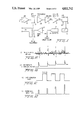
- FIG. 4A is a diagram of a waveform showing a raw EMG in the presence of periodic EMS signals
- FIG. 4B is a diagram of proportional EMS signals developed by the circuit of FIG. 3;
- FIG. 5 is a more detailed schematic diagram of a circuit for generating proportional response EMS signals in accordance with the invention.
- FIGS. 6A-6D are diagrams of waveforms developed by the circuit of FIG. 5.
- the EMG and EMS processing unit 10 is wired to a pair of transcutaneous electrodes 12 to be applied to an appropriate muscle group of a limb of a subject. Electrode types and placement, as well as electrical stimulation waveforms to be applied to muscle groups in muscle rehabilitation therapy, are described in detail in "Functional Electrical Stimulation--A Practical Clinical Guide", supra. Predetermined stimulus pulse rate, pulse width and waveform type are provided by circuitry within the processing unit 10.
- a set of EMG electrodes 16 detect EMG from enervated musculature, to be applied over lines 18 to one input of console 14.
- the EMG electrodes 16 are of a type and are positioned on the skin in a manner also discussed in detail in “Functional Electrical Stimulation--A Practical Guide", supra.
- a monitor 22 which preferably includes a cathode ray tube (CRT) 24, is controlled by circuitry in console 14 to display the measured EMG signal to the subject as well as to attending personnel, visual feedback during the therapeutic session.
- the console keyboard 20 enables the clinician/operator to select parameters influencing the processing of the EMG and stimulus waveforms in processor 10 as well as auditory and visual feedback via the console 22 by virtue of displayed selection "menus" on the CRT 24 prior to initiating a therapeutic session.
- circuitry within the processor 10 processes the EMG signals detected by electrodes 16 and, in response, controls the muscle stimulus waveform amplitude applied to the subject at electrodes 12.
- the relationship between processed EMG and applied EMS signals preferably is linear, although other functional relationships, such as logarithmic, etc., including negative or inverse relations, can also be programmed.
- Electrodes 12 and 16 are placed on the skin of the subject outside a primary stimulation current path produced in the skin by EMS electrodes 12.
- the orientation of electrodes shown in FIG. 2A, with EMG electrodes 16 positioned at least partially within the primary stimulation current flow path I has been found to be unsatisfactory, because the substantial EMS currents produce artifacts in the EMG as shown in FIGS. 4A and 6B wherein an EMG artifact is induced into the raw EMG signal following each EMS pulse applied to the skin of the subject.
- EMG electrodes 16 and EMS electrodes 12 proper positioning of EMG electrodes 16 and EMS electrodes 12 is shown.
- placement of the EMS and EMG electrodes 12, 16 is a very close approximation to common site stimulation and detection.
- the two different pairs of EMG electrodes 16A and 16B shown in the Figure are positioned on muscle site 28 just outside the primary current stimulation path I, whereby the EMG at the output of each buffer 30A, 30B is relatively free from EMG artifacts created by muscle stimulation current.
- some electrical stimulus will tend to "spill over" into the detected EMG signal. This effect is removed in accordance with the invention by time-multiplexing and bandpass filtering, to be described hereinbelow.
- the EMG signal conducted by electrodes 12 is amplified by amplifier 32, applied to a bandpass filter 34, then to a data acquisition interface 36.
- the amplifier 32 is a differential amplifier which amplifies by a predetermined gain the EMG differential signal at the two EMG electrodes 12.
- This EMG signal corresponds to EMG (A) or EMG (B) or a combination of the two in FIG. 2B.
- the bandpass filter which has a high Q pass band from 1 kHz to 1 kHz, compresses the stimulus artifact into narrow time domain bursts, as shown in FIG.
- the pulses of FIG. 4B are proportional in amplitude to the average value of the EMG during a time interval immediately preceding each pulse.
- the proportional response pulses are then current amplified in amplifier 40 to be applied as electrical stimulation signals to the muscle at output electrodes 16.
- the functional relationship between the processed EMG signal and generated EMS may be linear, although other relationships may be more appropriate in specific therapeutic applications.
- the EMG measured in the presence of electrical stimulation pulses comprises a first portion a which is an artifact consisting of a peak C created by the electrical muscle stimulation pulse applied to the muscle site and a decaying artifact component that follows.
- a portion b of the EMG which is free to the artifact, is a raw EMG signal that is neurologically developed by the subject and is not related directly to the periodically applied EMS signal. It is this portion b of the EMG that is processed by signal processor 38 to develop an output pulse, such as pulse c 1 in FIG. 4B to be applied as an electrical muscle stimulation signal (EMS) to the muscle site.
- EMS electrical muscle stimulation signal
- pulse c 1 in FIG. 4B is developed as a result of the raw EMG signal in FIG. 4A immediately preceding in time the generation of C 1 .
- the pulse c 1 in FIG. 4B is amplified by power amplifier 40 in FIG. 3 to impart to the muscle site an electrical stimulation signal having an amplitude that corresponds specifically to the amplitude of pulse c 1 .
- This pulse applied to the muscle site is measured at electrodes 12 as an EMG artifact, as shown at C 1 in FIG. 4A.
- the artifact C 1 decays until what remains is the raw EMG in region b of FIG. 4A.
- the EMG at region b is processed in signal processor 38 by envelope detection and averaging, as described in detail herein below, to develop a second pulse c 2 in FIG. 4B.
- the pulse c 2 is again amplified by amplifier 40 to develop a second EMS pulse to be applied to the muscle site.
- This pulse is detected by EMG electrodes 16 as another artifact C 2 which, together with its decaying trailing portion a, is "masked" to enable the following raw EMG to be again processed to obtain the third EMS drive pulse c 3 , and so on. Accordingly, the subject effectively controls his own EMS using EMS as a feedback component in neuromuscular re-education.
- FIG. 5 is a circuit diagram of a more detailed implementation of proportional response EMS generation in accordance with the invention, with envelope detection to enhance sampling, and electrical isolation from the subject of processing circuitry.
- Amplifier 32 is again a differential amplifier having a gain sufficient to amplify the raw EMG signal detected by electrodes 12 to a usable level.
- the output of amplifier 32 is coupled, through an optocoupler 48, to a programmable gain amplifier 32a that enables the subject or attending personnel to control the gain of the EMG signal to be applied for processing to microprocessor 42.
- the output of amplifier 32a is applied through a 1 kHz to 1 kHz bandpass filter 50 to an envelope detector 52 that converts the composite EMG and EMG artifact signal shown in FIG. 6A to an envelope shown in FIG. 6B having a magnitude that tracks the peak value of the composite EMG.
- This envelope is in turn applied, through an analog to digital converter 54, to microprocessor 42 programmed to sample [see FIG. 6(C)] the composite EMG only during the time period of each cycle when the EMG artifact has decayed to substantially zero.
- Microprocessor 42 samples the envelope waveform of FIG. 6B and, in response, develops digital data having values corresponding to the magnitude of the envelope of FIG. 6B, averaged over each period t as shown in FIG. 6C.
- Digital to analog converter 56 converts each digital value to the corresponding analog pulse, as shown in FIG. 6D, to be applied to amplifier 40 that drives the EMS electrodes 12 through an isolating pulse transformer 58.
- Programming of microprocessor 42 being known to one of ordinary skill in the art is omitted herein for brevity.
- EMS pulse train, pulse amplitude and pulse width developed by microprocessor 42 are all issued in real time via D/A converter 56, the converter may issue stimulus scale values and allow a timer/multiplexer network to generate the pulse train as a function of programmed pulse widths and pulse rates.
- D/A converter 56 may issue stimulus scale values and allow a timer/multiplexer network to generate the pulse train as a function of programmed pulse widths and pulse rates.
- the invention described herein is applicable as a potential technique of "muscle amplification" whereby the strength of particular muscle sites is increased beyond that of which it ordinarily is capable in resonse to neurological signals developed by the subject.
Abstract
Description
Claims (16)
Priority Applications (1)
| Application Number | Priority Date | Filing Date | Title |
|---|---|---|---|
| US06/743,635 US4811742A (en) | 1985-06-11 | 1985-06-11 | Proportional response electrical muscle stimulation |
Applications Claiming Priority (1)
| Application Number | Priority Date | Filing Date | Title |
|---|---|---|---|
| US06/743,635 US4811742A (en) | 1985-06-11 | 1985-06-11 | Proportional response electrical muscle stimulation |
Publications (1)
| Publication Number | Publication Date |
|---|---|
| US4811742A true US4811742A (en) | 1989-03-14 |
Family
ID=24989552
Family Applications (1)
| Application Number | Title | Priority Date | Filing Date |
|---|---|---|---|
| US06/743,635 Expired - Lifetime US4811742A (en) | 1985-06-11 | 1985-06-11 | Proportional response electrical muscle stimulation |
Country Status (1)
| Country | Link |
|---|---|
| US (1) | US4811742A (en) |
Cited By (48)
| Publication number | Priority date | Publication date | Assignee | Title |
|---|---|---|---|---|
| US5233999A (en) * | 1990-12-28 | 1993-08-10 | Alberto Dellacorna | Electromyograph with data transmission comprising no metallic conductors |
| US5263490A (en) * | 1992-07-27 | 1993-11-23 | Abbott Laboratories | Muscle function assessment |
| US5284153A (en) * | 1992-04-14 | 1994-02-08 | Brigham And Women's Hospital | Method for locating a nerve and for protecting nerves from injury during surgery |
| US5291894A (en) * | 1989-11-14 | 1994-03-08 | Nagy Lajos Z | Apparatus for treating a patient with acoustic waves |
| US5300096A (en) * | 1992-06-03 | 1994-04-05 | Hall H Eugene | Electromyographic treatment device |
| US5327902A (en) * | 1993-05-14 | 1994-07-12 | Lemmen Roger D | Apparatus for use in nerve conduction studies |
| US5482051A (en) * | 1994-03-10 | 1996-01-09 | The University Of Akron | Electromyographic virtual reality system |
| US5775331A (en) * | 1995-06-07 | 1998-07-07 | Uromed Corporation | Apparatus and method for locating a nerve |
| US5810747A (en) * | 1996-08-21 | 1998-09-22 | Interactive Remote Site Technology, Inc. | Remote site medical intervention system |
| US5976094A (en) * | 1997-07-01 | 1999-11-02 | Neurometrix, Inc. | Apparatus and methods for assessment of neuromuscular function |
| US6076011A (en) * | 1999-02-02 | 2000-06-13 | J&J Engineering | Electromyographic feedback monitor system |
| EP0938911A3 (en) * | 1998-02-25 | 2000-12-06 | MO, Seung Kee | Electrical apparatus for medical treatment using EMG envelope signal |
| US6267733B1 (en) | 1997-11-14 | 2001-07-31 | Scientific Learning Corporation | Apparatus and methods for treating motor control and somatosensory perception deficits |
| US6324432B1 (en) | 1999-11-01 | 2001-11-27 | Compex Sa | Electrical neuromuscular stimulator for measuring muscle responses to electrical stimulation pulses |
| US20020183647A1 (en) * | 1997-07-01 | 2002-12-05 | Gozani Shai N. | Apparatus and method for performing nerve conduction studies with localization of evoked responses |
| EP1311320A2 (en) * | 2000-08-15 | 2003-05-21 | Stimel Ltd. | Electrostimulation system with electromyographic and visual biofeedback |
| US20030199782A1 (en) * | 1998-12-01 | 2003-10-23 | Gozani Shai N. | Apparatus and method for stimulating human tissue |
| US20040024299A1 (en) * | 2000-11-02 | 2004-02-05 | Grace Lawrence J. | Method and apparatus for self-diagnostic evaluation of nerve sensory latency |
| US20040073271A1 (en) * | 2002-05-03 | 2004-04-15 | Afferent Corporation | Method and apparatus for neurophysiologic performance |
| US20040098064A1 (en) * | 2002-11-20 | 2004-05-20 | O'kelly Gregory C. | Method and device for electrochemically building of muscle |
| US20050283204A1 (en) * | 2004-01-30 | 2005-12-22 | Felix Buhlmann | Automated adaptive muscle stimulation method and apparatus |
| US20060100540A1 (en) * | 1997-07-01 | 2006-05-11 | Gozani Shai N | Methods for the assessment of neuromuscular function by F-wave latency |
| US20060173496A1 (en) * | 2005-02-03 | 2006-08-03 | Lombardi Daniel J | Method and apparatus for stimulus artifact suppression |
| US20060217768A1 (en) * | 2005-01-28 | 2006-09-28 | Felix Buhlmann | Independent protection system for an electrical muscle stimulation apparatus and method of using same |
| US20060217631A1 (en) * | 2004-02-17 | 2006-09-28 | Xuan Kong | Method for automated analysis of submaximal F-waves |
| US20060253165A1 (en) * | 2005-05-09 | 2006-11-09 | O'kelly Gregory | Method and device for electrochemical rejuvenation of skin and underlying tissue, and muscle building |
| US20070148624A1 (en) * | 2005-12-23 | 2007-06-28 | Avinoam Nativ | Kinesthetic training system with composite feedback |
| US20090326612A1 (en) * | 2008-06-26 | 2009-12-31 | Michael J. Distler | Electronic biofeedback stimulation device |
| US20100004715A1 (en) * | 2008-07-02 | 2010-01-07 | Brian Fahey | Systems and methods for automated muscle stimulation |
| US20100042180A1 (en) * | 2005-04-19 | 2010-02-18 | Compex Technologies, Inc | Electrical stimulation device and method for therapeutic treatment and pain management |
| US20100057149A1 (en) * | 2008-08-26 | 2010-03-04 | Brian Fahey | Device, system, and method to improve powered muscle stimulation performance in the presence of tissue edema |
| US20100217349A1 (en) * | 2009-02-20 | 2010-08-26 | Fahey Brian J | Systems and Methods of Powered Muscle Stimulation Using an Energy Guidance Field |
| US20110112605A1 (en) * | 2009-11-11 | 2011-05-12 | Fahey Brian J | Synergistic Muscle Activation Device |
| WO2012167177A1 (en) | 2011-06-01 | 2012-12-06 | Tech Team LLC | System and method for power-efficient transmission of emg data |
| US8620438B1 (en) | 2007-02-13 | 2013-12-31 | Encore Medical Asset Corporation | Method and apparatus for applying neuromuscular electrical stimulation |
| US8892210B2 (en) | 2008-07-02 | 2014-11-18 | Niveus Medical, Inc. | Devices, systems, and methods for automated optimization of energy delivery |
| DE202015005645U1 (en) | 2015-08-14 | 2015-09-02 | Nordin Kouache | Functional clothing with tactile stimulus module and EMG electrode |
| US9149386B2 (en) | 2008-08-19 | 2015-10-06 | Niveus Medical, Inc. | Devices and systems for stimulation of tissues |
| US20160143556A1 (en) * | 2003-01-15 | 2016-05-26 | Nuvasive, Inc. | System for Determining Nerve Direction to a Surgical Instrument |
| US9616234B2 (en) | 2002-05-03 | 2017-04-11 | Trustees Of Boston University | System and method for neuro-stimulation |
| US9878152B2 (en) | 2009-11-05 | 2018-01-30 | Koninklijke Philips N.V. | Electrical muscle stimulation |
| US10010259B2 (en) | 2015-05-01 | 2018-07-03 | Advancer Technologies, Llc | EMG circuit |
| EP3235540A4 (en) * | 2014-12-09 | 2018-08-01 | Beijing Galaxy Raintai Technology Co., Ltd. | Auxiliary device for training and auxiliary method for training |
| US10213602B2 (en) | 2015-01-13 | 2019-02-26 | Theranica Bio-Electronics Ltd. | Treatment of headaches by electrical stimulation |
| US11167135B2 (en) | 2017-05-21 | 2021-11-09 | Theranica Bio-Electronics Ltd. | Apparatus for providing pain relief therapy |
| US11357980B2 (en) | 2016-09-29 | 2022-06-14 | Theranica Bio-Electronics Ltd. | Apparatus for applying an electrical signal to a subject |
| US11583218B2 (en) | 2019-11-20 | 2023-02-21 | Advancer Technologies, Llc | EMG device |
| USD1015545S1 (en) | 2019-11-20 | 2024-02-20 | Advancer Technologies, Llc | Electromyography device |
Citations (11)
| Publication number | Priority date | Publication date | Assignee | Title |
|---|---|---|---|---|
| US3641993A (en) * | 1970-04-23 | 1972-02-15 | Prototypes Inc | Nonlinear electromyograph |
| DE2457854A1 (en) * | 1973-12-06 | 1975-06-12 | Joseph Brudny | DEVICE AND METHOD FOR MEASURING AND DISPLAYING ELECTROMYOGRAPHIC SIGNALS |
| US4031883A (en) * | 1974-07-29 | 1977-06-28 | Biofeedback Computers, Inc. | Multiple channel phase integrating biofeedback computer |
| US4170225A (en) * | 1976-09-20 | 1979-10-09 | Somatronics, Inc. | Biofeedback device |
| US4291705A (en) * | 1979-09-10 | 1981-09-29 | The Regents Of The University Of California | Neuromuscular block monitor |
| US4305402A (en) * | 1979-06-29 | 1981-12-15 | Katims Jefferson J | Method for transcutaneous electrical stimulation |
| US4474186A (en) * | 1979-07-17 | 1984-10-02 | Georgetown University | Computerized electro-oculographic (CEOG) system with feedback control of stimuli |
| US4492233A (en) * | 1982-09-14 | 1985-01-08 | Wright State University | Method and apparatus for providing feedback-controlled muscle stimulation |
| US4595018A (en) * | 1983-06-10 | 1986-06-17 | Instrumentarium Corp. | Method of further developing the measuring of a neuro-muscular junction |
| US4619266A (en) * | 1983-05-11 | 1986-10-28 | Hodgson John A | Electrode array for muscle stimulation and recording |
| US4690142A (en) * | 1980-12-10 | 1987-09-01 | Ross Sidney A | Method and system for utilizing electro-neuro stimulation in a bio-feedback system |
-
1985
- 1985-06-11 US US06/743,635 patent/US4811742A/en not_active Expired - Lifetime
Patent Citations (12)
| Publication number | Priority date | Publication date | Assignee | Title |
|---|---|---|---|---|
| US3641993A (en) * | 1970-04-23 | 1972-02-15 | Prototypes Inc | Nonlinear electromyograph |
| DE2457854A1 (en) * | 1973-12-06 | 1975-06-12 | Joseph Brudny | DEVICE AND METHOD FOR MEASURING AND DISPLAYING ELECTROMYOGRAPHIC SIGNALS |
| US3905355A (en) * | 1973-12-06 | 1975-09-16 | Joseph Brudny | System for the measurement, display and instrumental conditioning of electromyographic signals |
| US4031883A (en) * | 1974-07-29 | 1977-06-28 | Biofeedback Computers, Inc. | Multiple channel phase integrating biofeedback computer |
| US4170225A (en) * | 1976-09-20 | 1979-10-09 | Somatronics, Inc. | Biofeedback device |
| US4305402A (en) * | 1979-06-29 | 1981-12-15 | Katims Jefferson J | Method for transcutaneous electrical stimulation |
| US4474186A (en) * | 1979-07-17 | 1984-10-02 | Georgetown University | Computerized electro-oculographic (CEOG) system with feedback control of stimuli |
| US4291705A (en) * | 1979-09-10 | 1981-09-29 | The Regents Of The University Of California | Neuromuscular block monitor |
| US4690142A (en) * | 1980-12-10 | 1987-09-01 | Ross Sidney A | Method and system for utilizing electro-neuro stimulation in a bio-feedback system |
| US4492233A (en) * | 1982-09-14 | 1985-01-08 | Wright State University | Method and apparatus for providing feedback-controlled muscle stimulation |
| US4619266A (en) * | 1983-05-11 | 1986-10-28 | Hodgson John A | Electrode array for muscle stimulation and recording |
| US4595018A (en) * | 1983-06-10 | 1986-06-17 | Instrumentarium Corp. | Method of further developing the measuring of a neuro-muscular junction |
Non-Patent Citations (2)
| Title |
|---|
| Othotic Systems Using Functional Electrical Stimulation and Myeolectric Control, S. Rebersek et al, Final Report Project No. 19 P 58391 F 01, pp. 58 71. * |
| Othotic Systems Using Functional Electrical Stimulation and Myeolectric Control, S. Rebersek et al, Final Report Project No. 19-P-58391-F-01, pp. 58-71. |
Cited By (90)
| Publication number | Priority date | Publication date | Assignee | Title |
|---|---|---|---|---|
| US5291894A (en) * | 1989-11-14 | 1994-03-08 | Nagy Lajos Z | Apparatus for treating a patient with acoustic waves |
| US5233999A (en) * | 1990-12-28 | 1993-08-10 | Alberto Dellacorna | Electromyograph with data transmission comprising no metallic conductors |
| US5284153A (en) * | 1992-04-14 | 1994-02-08 | Brigham And Women's Hospital | Method for locating a nerve and for protecting nerves from injury during surgery |
| US5300096A (en) * | 1992-06-03 | 1994-04-05 | Hall H Eugene | Electromyographic treatment device |
| US5263490A (en) * | 1992-07-27 | 1993-11-23 | Abbott Laboratories | Muscle function assessment |
| US5327902A (en) * | 1993-05-14 | 1994-07-12 | Lemmen Roger D | Apparatus for use in nerve conduction studies |
| US5482051A (en) * | 1994-03-10 | 1996-01-09 | The University Of Akron | Electromyographic virtual reality system |
| US5775331A (en) * | 1995-06-07 | 1998-07-07 | Uromed Corporation | Apparatus and method for locating a nerve |
| US5810747A (en) * | 1996-08-21 | 1998-09-22 | Interactive Remote Site Technology, Inc. | Remote site medical intervention system |
| US5976094A (en) * | 1997-07-01 | 1999-11-02 | Neurometrix, Inc. | Apparatus and methods for assessment of neuromuscular function |
| US7628761B2 (en) | 1997-07-01 | 2009-12-08 | Neurometrix, Inc. | Apparatus and method for performing nerve conduction studies with localization of evoked responses |
| US20060100540A1 (en) * | 1997-07-01 | 2006-05-11 | Gozani Shai N | Methods for the assessment of neuromuscular function by F-wave latency |
| US20020183647A1 (en) * | 1997-07-01 | 2002-12-05 | Gozani Shai N. | Apparatus and method for performing nerve conduction studies with localization of evoked responses |
| US6409685B1 (en) | 1997-11-14 | 2002-06-25 | Scientific Learning Corporation | Method for improving motor control in an individual by sensory training |
| US6267733B1 (en) | 1997-11-14 | 2001-07-31 | Scientific Learning Corporation | Apparatus and methods for treating motor control and somatosensory perception deficits |
| US6289245B1 (en) * | 1998-02-25 | 2001-09-11 | Seung Kee Mo | Electrical apparatus for medical treatment using EMG envelope signal |
| EP0938911A3 (en) * | 1998-02-25 | 2000-12-06 | MO, Seung Kee | Electrical apparatus for medical treatment using EMG envelope signal |
| US20030199782A1 (en) * | 1998-12-01 | 2003-10-23 | Gozani Shai N. | Apparatus and method for stimulating human tissue |
| US6076011A (en) * | 1999-02-02 | 2000-06-13 | J&J Engineering | Electromyographic feedback monitor system |
| US6324432B1 (en) | 1999-11-01 | 2001-11-27 | Compex Sa | Electrical neuromuscular stimulator for measuring muscle responses to electrical stimulation pulses |
| US20030195586A1 (en) * | 1999-11-01 | 2003-10-16 | Compex Medical S.A. | Electrical neuromuscular stimulator for measuring muscle responses to electrical stimulation pulses |
| EP1311320A2 (en) * | 2000-08-15 | 2003-05-21 | Stimel Ltd. | Electrostimulation system with electromyographic and visual biofeedback |
| EP1311320A4 (en) * | 2000-08-15 | 2008-01-23 | Stimel Ltd | Electrostimulation system with electromyographic and visual biofeedback |
| US7058438B2 (en) * | 2000-11-02 | 2006-06-06 | Grace Lawrence J | Method and apparatus for self-diagnostic evaluation of nerve sensory latency |
| US20040024299A1 (en) * | 2000-11-02 | 2004-02-05 | Grace Lawrence J. | Method and apparatus for self-diagnostic evaluation of nerve sensory latency |
| US9616234B2 (en) | 2002-05-03 | 2017-04-11 | Trustees Of Boston University | System and method for neuro-stimulation |
| US20040073271A1 (en) * | 2002-05-03 | 2004-04-15 | Afferent Corporation | Method and apparatus for neurophysiologic performance |
| US7349739B2 (en) * | 2002-05-03 | 2008-03-25 | Afferent Corporation | Method and apparatus for neurophysiologic performance |
| US20040098064A1 (en) * | 2002-11-20 | 2004-05-20 | O'kelly Gregory C. | Method and device for electrochemically building of muscle |
| US6856837B2 (en) | 2002-11-20 | 2005-02-15 | O'kelly Gregory C. | Method and device for electrochemically building of muscle |
| US20160143556A1 (en) * | 2003-01-15 | 2016-05-26 | Nuvasive, Inc. | System for Determining Nerve Direction to a Surgical Instrument |
| US10993650B2 (en) * | 2003-01-15 | 2021-05-04 | Nuvasive, Inc. | System for determining nerve direction to a surgical instrument |
| US10080523B2 (en) | 2004-01-30 | 2018-09-25 | Djo Global Switzerland Sàrl | Automated adaptive muscle stimulation method and apparatus |
| US10463296B2 (en) | 2004-01-30 | 2019-11-05 | DJO Global Switzerland Sarl | Automated adaptive muscle stimulation method and apparatus |
| US8565888B2 (en) | 2004-01-30 | 2013-10-22 | Compex Medical S.A. | Automated adaptive muscle stimulation method and apparatus |
| US20050283204A1 (en) * | 2004-01-30 | 2005-12-22 | Felix Buhlmann | Automated adaptive muscle stimulation method and apparatus |
| US11389110B2 (en) | 2004-01-30 | 2022-07-19 | Djo Global Switzerland Sàrl | Automated adaptive muscle stimulation method and apparatus |
| US7499746B2 (en) | 2004-01-30 | 2009-03-03 | Encore Medical Asset Corporation | Automated adaptive muscle stimulation method and apparatus |
| US20090228068A1 (en) * | 2004-01-30 | 2009-09-10 | Felix Buhlmann | Automated adaptive muscle stimulation method and apparatus |
| US20060217631A1 (en) * | 2004-02-17 | 2006-09-28 | Xuan Kong | Method for automated analysis of submaximal F-waves |
| US8326410B2 (en) | 2004-02-17 | 2012-12-04 | Neurometrix, Inc. | Method for automated analysis of submaximal F-waves |
| US8140165B2 (en) | 2005-01-28 | 2012-03-20 | Encore Medical Asset Corporation | Independent protection system for an electrical muscle stimulation apparatus and method of using same |
| US20060217768A1 (en) * | 2005-01-28 | 2006-09-28 | Felix Buhlmann | Independent protection system for an electrical muscle stimulation apparatus and method of using same |
| US9808619B2 (en) | 2005-01-28 | 2017-11-07 | Encore Medical Asset Corporation | Independent protection system for an electrical muscle stimulation apparatus and method of using same |
| US7424322B2 (en) * | 2005-02-03 | 2008-09-09 | Cardinal Health 209, Inc. | Method and apparatus for stimulus artifact suppression |
| US20060173496A1 (en) * | 2005-02-03 | 2006-08-03 | Lombardi Daniel J | Method and apparatus for stimulus artifact suppression |
| US9669212B2 (en) | 2005-04-19 | 2017-06-06 | Djo, Llc | Electrical stimulation device and method for therapeutic treatment and pain management |
| US10328260B2 (en) | 2005-04-19 | 2019-06-25 | Djo, Llc | Electrical stimulation device and method for therapeutic treatment and pain management |
| US20100042180A1 (en) * | 2005-04-19 | 2010-02-18 | Compex Technologies, Inc | Electrical stimulation device and method for therapeutic treatment and pain management |
| US8958883B2 (en) | 2005-04-19 | 2015-02-17 | Pierre-Yves Mueller | Electrical stimulation device and method for therapeutic treatment and pain management |
| US20060253165A1 (en) * | 2005-05-09 | 2006-11-09 | O'kelly Gregory | Method and device for electrochemical rejuvenation of skin and underlying tissue, and muscle building |
| US7319902B2 (en) | 2005-05-09 | 2008-01-15 | O'kelly Gregory | Method and device for electrochemical rejuvenation of skin and underlying tissue, and muscle building |
| US7365647B2 (en) * | 2005-12-23 | 2008-04-29 | Avinoam Nativ | Kinesthetic training system with composite feedback |
| US20070148624A1 (en) * | 2005-12-23 | 2007-06-28 | Avinoam Nativ | Kinesthetic training system with composite feedback |
| US9352151B2 (en) | 2007-02-13 | 2016-05-31 | Encore Medical Asset Corporation | Method and apparatus for applying neuromuscular electrical stimulation |
| US8620438B1 (en) | 2007-02-13 | 2013-12-31 | Encore Medical Asset Corporation | Method and apparatus for applying neuromuscular electrical stimulation |
| US9669211B2 (en) | 2007-02-13 | 2017-06-06 | Encore Medical Asset Corporation | Method and apparatus for applying neuromuscular electrical stimulation |
| US20090326612A1 (en) * | 2008-06-26 | 2009-12-31 | Michael J. Distler | Electronic biofeedback stimulation device |
| US9302104B2 (en) | 2008-07-02 | 2016-04-05 | Niveus Medical, Inc. | Devices, systems, and methods for automated optimization of energy delivery |
| US20100004715A1 (en) * | 2008-07-02 | 2010-01-07 | Brian Fahey | Systems and methods for automated muscle stimulation |
| US10987510B2 (en) | 2008-07-02 | 2021-04-27 | Sage Products, Llc | Systems and methods for automated muscle stimulation |
| US10293152B2 (en) | 2008-07-02 | 2019-05-21 | Sage Products, Llc | Devices, systems, and methods for automated optimization of energy delivery |
| US8285381B2 (en) | 2008-07-02 | 2012-10-09 | Niveus Medical, Inc. | Systems and methods for automated muscle stimulation |
| US8892210B2 (en) | 2008-07-02 | 2014-11-18 | Niveus Medical, Inc. | Devices, systems, and methods for automated optimization of energy delivery |
| US9149386B2 (en) | 2008-08-19 | 2015-10-06 | Niveus Medical, Inc. | Devices and systems for stimulation of tissues |
| US9532899B2 (en) | 2008-08-19 | 2017-01-03 | Niveus Medical, Inc. | Devices and systems for stimulation of tissue |
| US20100057149A1 (en) * | 2008-08-26 | 2010-03-04 | Brian Fahey | Device, system, and method to improve powered muscle stimulation performance in the presence of tissue edema |
| US8265763B2 (en) | 2008-08-26 | 2012-09-11 | Niveus Medical, Inc. | Device, system, and method to improve powered muscle stimulation performance in the presence of tissue edema |
| US8676332B2 (en) | 2009-02-20 | 2014-03-18 | Niveus Medical, Inc. | Systems and methods of powered muscle stimulation using an energy guidance field |
| US20100217349A1 (en) * | 2009-02-20 | 2010-08-26 | Fahey Brian J | Systems and Methods of Powered Muscle Stimulation Using an Energy Guidance Field |
| US8433403B2 (en) | 2009-02-20 | 2013-04-30 | Niveus Medical, Inc. | Systems and methods of powered muscle stimulation using an energy guidance field |
| US9878152B2 (en) | 2009-11-05 | 2018-01-30 | Koninklijke Philips N.V. | Electrical muscle stimulation |
| US20110112605A1 (en) * | 2009-11-11 | 2011-05-12 | Fahey Brian J | Synergistic Muscle Activation Device |
| US8588901B2 (en) | 2009-11-11 | 2013-11-19 | Niveus Medical, Inc. | Synergistic muscle activation device |
| US9126039B2 (en) | 2009-11-11 | 2015-09-08 | Niveus Medical, Inc. | Synergistic muscle activation device |
| US10478622B2 (en) | 2009-11-11 | 2019-11-19 | Sage Products, Llc | Synergistic muscle activation device |
| US11839763B2 (en) | 2009-11-11 | 2023-12-12 | Sage Products, Llc | Synergistic muscle activation device |
| US8768428B2 (en) | 2011-06-01 | 2014-07-01 | Tech Team LLC | System and method for power-efficient transmission of EMG data |
| WO2012167177A1 (en) | 2011-06-01 | 2012-12-06 | Tech Team LLC | System and method for power-efficient transmission of emg data |
| US9042956B2 (en) | 2011-06-01 | 2015-05-26 | Tech Team LLC | System and method for power-efficient transmission of EMG data |
| EP3235540A4 (en) * | 2014-12-09 | 2018-08-01 | Beijing Galaxy Raintai Technology Co., Ltd. | Auxiliary device for training and auxiliary method for training |
| US10213602B2 (en) | 2015-01-13 | 2019-02-26 | Theranica Bio-Electronics Ltd. | Treatment of headaches by electrical stimulation |
| US10994135B2 (en) | 2015-01-13 | 2021-05-04 | Theranica Bio-Electronics Ltd. | Treatment of headaches by electrical stimulation |
| US10010259B2 (en) | 2015-05-01 | 2018-07-03 | Advancer Technologies, Llc | EMG circuit |
| DE202015005645U1 (en) | 2015-08-14 | 2015-09-02 | Nordin Kouache | Functional clothing with tactile stimulus module and EMG electrode |
| US11357980B2 (en) | 2016-09-29 | 2022-06-14 | Theranica Bio-Electronics Ltd. | Apparatus for applying an electrical signal to a subject |
| US11167135B2 (en) | 2017-05-21 | 2021-11-09 | Theranica Bio-Electronics Ltd. | Apparatus for providing pain relief therapy |
| US11904163B2 (en) | 2017-05-21 | 2024-02-20 | Theranica Bio-Electronics Ltd. | Apparatus for providing pain relief therapy |
| US11583218B2 (en) | 2019-11-20 | 2023-02-21 | Advancer Technologies, Llc | EMG device |
| USD1015545S1 (en) | 2019-11-20 | 2024-02-20 | Advancer Technologies, Llc | Electromyography device |
Similar Documents
| Publication | Publication Date | Title |
|---|---|---|
| US4811742A (en) | Proportional response electrical muscle stimulation | |
| US4690142A (en) | Method and system for utilizing electro-neuro stimulation in a bio-feedback system | |
| Dowman | Spinal and supraspinal correlates of nociception in man | |
| Chesler et al. | Surface EMG as a fatigue indicator during FES-induced isometric muscle contractions | |
| Chen et al. | Brain evoked potentials are functional correlates of induced pain in man | |
| Türker | Electromyography: some methodological problems and issues | |
| LARSON et al. | Evoked somatosensory potentials in man | |
| Gorman et al. | The effect of stimulus parameters on the recruitment characteristics of direct nerve stimulation | |
| Currier et al. | Effects of electrical and electromagnetic stimulation after anterior cruciate ligament reconstruction | |
| US6157861A (en) | Self-adjusting cochlear implant system and method for fitting same | |
| US7221980B2 (en) | Electrostimulation system with electromyographic and visual biofeedback | |
| Stefanovska et al. | FES and spasticity | |
| JP2008519609A (en) | Apparatus and method for measuring EMG signals | |
| CN101391129A (en) | Brain-machine interface intelligentized upper-limb recovery training device based on P300 signal and signal processing method | |
| Ackermann et al. | Influence of posture and voluntary background contraction upon compound muscle action potentials from anterior tibial and soleus muscle following transcranial magnetic stimulation | |
| US20200139115A1 (en) | Apparatus for neuromuscular stimulation | |
| Shalaby | Development of an electromyography detection system for the control of functional electrical stimulation in neurological rehabilitation | |
| Yochum et al. | A mixed FES/EMG system for real time analysis of muscular fatigue | |
| EP0203336B1 (en) | Electrical stimulator and method for treatment of the human and animal body with stimulation currents | |
| Berger et al. | Reversible proximal conduction block underlies rapid recovery in Guillain‐Barré syndrome | |
| Schimek et al. | Varying electrical acupuncture stimulation intensity: effects on dental pain-evoked potentials | |
| Omura | Electrical parameters for safe and effective electro-acupuncture and transcutaneous electrical stimulation: Threshold potentials for tingling, muscle contraction and pain; and how to prevent adverse effects of electro-therapy part i | |
| Davis et al. | Technical problems and advances in the cerebellar-stimulating systems used for reduction of spasticity and seizures | |
| Thorsen et al. | Enhancement of isometric ankle dorsiflexion by automyoelectrically controlled functional electrical stimulation on subjects with upper motor neuron lesions | |
| Balogun | Effects of ramp time on sensory, motor and tolerance thresholds during exogenous electrical |
Legal Events
| Date | Code | Title | Description |
|---|---|---|---|
| AS | Assignment |
Owner name: VERIMED, INC., 2888 N.E. 25TH COURT, FORT LAUDERDA Free format text: ASSIGNMENT OF ASSIGNORS INTEREST.;ASSIGNORS:HASSEL, WILLIAM;MEE, WILLIAM;REEL/FRAME:004420/0219 Effective date: 19850610 Owner name: VERIMED, INC., FLORIDA Free format text: ASSIGNMENT OF ASSIGNORS INTEREST;ASSIGNORS:HASSEL, WILLIAM;MEE, WILLIAM;REEL/FRAME:004420/0219 Effective date: 19850610 |
|
| STCF | Information on status: patent grant |
Free format text: PATENTED CASE |
|
| FEPP | Fee payment procedure |
Free format text: PAYOR NUMBER ASSIGNED (ORIGINAL EVENT CODE: ASPN); ENTITY STATUS OF PATENT OWNER: SMALL ENTITY |
|
| FPAY | Fee payment |
Year of fee payment: 4 |
|
| AS | Assignment |
Owner name: VERIMED INTERNATIONAL, INC., FLORIDA Free format text: A CORRECTIVE ASSIGNMENT TO CORRECT PATENT NUMBER. AN ASSIGNMENT PREVIOUSLY RECORDED ON REEL 7125, FRAMES 576;ASSIGNOR:VERIMED, INC.;REEL/FRAME:007562/0785 Effective date: 19940825 |
|
| FEPP | Fee payment procedure |
Free format text: PAYER NUMBER DE-ASSIGNED (ORIGINAL EVENT CODE: RMPN); ENTITY STATUS OF PATENT OWNER: SMALL ENTITY |
|
| FPAY | Fee payment |
Year of fee payment: 8 |
|
| FPAY | Fee payment |
Year of fee payment: 12 |