US20040230106A1 - Device and method for monitoring body fluid and electrolyte disorders - Google Patents
Device and method for monitoring body fluid and electrolyte disorders Download PDFInfo
- Publication number
- US20040230106A1 US20040230106A1 US10/699,610 US69961003A US2004230106A1 US 20040230106 A1 US20040230106 A1 US 20040230106A1 US 69961003 A US69961003 A US 69961003A US 2004230106 A1 US2004230106 A1 US 2004230106A1
- Authority
- US
- United States
- Prior art keywords
- tissue
- water
- radiation
- wavelengths
- metric
- Prior art date
- Legal status (The legal status is an assumption and is not a legal conclusion. Google has not performed a legal analysis and makes no representation as to the accuracy of the status listed.)
- Granted
Links
- 238000000034 method Methods 0.000 title claims abstract description 52
- 210000001124 body fluid Anatomy 0.000 title claims abstract description 18
- 239000010839 body fluid Substances 0.000 title claims abstract description 18
- 238000012544 monitoring process Methods 0.000 title description 3
- 239000003792 electrolyte Substances 0.000 title description 2
- XLYOFNOQVPJJNP-UHFFFAOYSA-N water Substances O XLYOFNOQVPJJNP-UHFFFAOYSA-N 0.000 claims abstract description 124
- 230000005855 radiation Effects 0.000 claims abstract description 67
- 239000000523 sample Substances 0.000 claims abstract description 38
- 238000001514 detection method Methods 0.000 claims abstract description 33
- 230000003287 optical effect Effects 0.000 claims abstract description 24
- 238000012545 processing Methods 0.000 claims abstract description 23
- 235000013861 fat-free Nutrition 0.000 claims abstract description 13
- 238000002798 spectrophotometry method Methods 0.000 claims abstract description 12
- 230000008569 process Effects 0.000 claims abstract description 5
- 238000005259 measurement Methods 0.000 claims description 77
- 239000012223 aqueous fraction Substances 0.000 claims description 54
- 108010054147 Hemoglobins Proteins 0.000 claims description 46
- 102000001554 Hemoglobins Human genes 0.000 claims description 46
- 210000004369 blood Anatomy 0.000 claims description 39
- 239000008280 blood Substances 0.000 claims description 39
- 239000000470 constituent Substances 0.000 claims description 37
- 238000002835 absorbance Methods 0.000 claims description 23
- 239000012530 fluid Substances 0.000 claims description 18
- 241000894007 species Species 0.000 claims description 13
- 238000010521 absorption reaction Methods 0.000 claims description 11
- 230000008859 change Effects 0.000 claims description 10
- 150000002632 lipids Chemical class 0.000 claims description 10
- 102000008015 Hemeproteins Human genes 0.000 claims description 8
- 108010089792 Hemeproteins Proteins 0.000 claims description 8
- 230000003595 spectral effect Effects 0.000 claims description 8
- 230000007246 mechanism Effects 0.000 claims description 7
- 210000004207 dermis Anatomy 0.000 claims description 5
- 238000000862 absorption spectrum Methods 0.000 claims description 4
- 230000001939 inductive effect Effects 0.000 claims description 4
- 230000002452 interceptive effect Effects 0.000 claims description 3
- QVGXLLKOCUKJST-UHFFFAOYSA-N atomic oxygen Chemical group [O] QVGXLLKOCUKJST-UHFFFAOYSA-N 0.000 claims description 2
- 230000001419 dependent effect Effects 0.000 claims description 2
- 230000031700 light absorption Effects 0.000 claims description 2
- 239000013307 optical fiber Substances 0.000 claims description 2
- 239000001301 oxygen Substances 0.000 claims description 2
- 229910052760 oxygen Inorganic materials 0.000 claims description 2
- 230000002792 vascular Effects 0.000 claims description 2
- 150000001875 compounds Chemical class 0.000 claims 2
- 230000023077 detection of light stimulus Effects 0.000 claims 2
- 238000003745 diagnosis Methods 0.000 abstract description 2
- 230000001225 therapeutic effect Effects 0.000 abstract description 2
- 210000001519 tissue Anatomy 0.000 description 134
- 210000003491 skin Anatomy 0.000 description 10
- 238000004422 calculation algorithm Methods 0.000 description 8
- 238000001802 infusion Methods 0.000 description 6
- FAPWRFPIFSIZLT-UHFFFAOYSA-M Sodium chloride Chemical compound [Na+].[Cl-] FAPWRFPIFSIZLT-UHFFFAOYSA-M 0.000 description 5
- 230000008901 benefit Effects 0.000 description 5
- 238000010586 diagram Methods 0.000 description 5
- 238000004088 simulation Methods 0.000 description 5
- 238000004611 spectroscopical analysis Methods 0.000 description 5
- 108010064719 Oxyhemoglobins Proteins 0.000 description 4
- 230000008602 contraction Effects 0.000 description 4
- 238000013461 design Methods 0.000 description 4
- 238000002474 experimental method Methods 0.000 description 4
- 235000019625 fat content Nutrition 0.000 description 4
- 102000004169 proteins and genes Human genes 0.000 description 4
- 108090000623 proteins and genes Proteins 0.000 description 4
- 239000000243 solution Substances 0.000 description 4
- INGWEZCOABYORO-UHFFFAOYSA-N 2-(furan-2-yl)-7-methyl-1h-1,8-naphthyridin-4-one Chemical compound N=1C2=NC(C)=CC=C2C(O)=CC=1C1=CC=CO1 INGWEZCOABYORO-UHFFFAOYSA-N 0.000 description 3
- 230000008033 biological extinction Effects 0.000 description 3
- 210000000476 body water Anatomy 0.000 description 3
- 210000000988 bone and bone Anatomy 0.000 description 3
- 238000004364 calculation method Methods 0.000 description 3
- 108010002255 deoxyhemoglobin Proteins 0.000 description 3
- 230000000694 effects Effects 0.000 description 3
- 238000012552 review Methods 0.000 description 3
- 206010012735 Diarrhoea Diseases 0.000 description 2
- 208000032843 Hemorrhage Diseases 0.000 description 2
- 238000012404 In vitro experiment Methods 0.000 description 2
- 238000004497 NIR spectroscopy Methods 0.000 description 2
- 206010030113 Oedema Diseases 0.000 description 2
- BPKIGYQJPYCAOW-FFJTTWKXSA-I calcium;potassium;disodium;(2s)-2-hydroxypropanoate;dichloride;dihydroxide;hydrate Chemical group O.[OH-].[OH-].[Na+].[Na+].[Cl-].[Cl-].[K+].[Ca+2].C[C@H](O)C([O-])=O BPKIGYQJPYCAOW-FFJTTWKXSA-I 0.000 description 2
- 238000003759 clinical diagnosis Methods 0.000 description 2
- DDRJAANPRJIHGJ-UHFFFAOYSA-N creatinine Chemical compound CN1CC(=O)NC1=N DDRJAANPRJIHGJ-UHFFFAOYSA-N 0.000 description 2
- 230000018044 dehydration Effects 0.000 description 2
- 238000006297 dehydration reaction Methods 0.000 description 2
- 238000005534 hematocrit Methods 0.000 description 2
- 230000035515 penetration Effects 0.000 description 2
- 230000010412 perfusion Effects 0.000 description 2
- 230000010349 pulsation Effects 0.000 description 2
- 238000011002 quantification Methods 0.000 description 2
- 238000009155 rehydration therapy Methods 0.000 description 2
- 230000002040 relaxant effect Effects 0.000 description 2
- 230000035945 sensitivity Effects 0.000 description 2
- 238000000926 separation method Methods 0.000 description 2
- 239000011780 sodium chloride Substances 0.000 description 2
- 206010001367 Adrenal insufficiency Diseases 0.000 description 1
- 108010003320 Carboxyhemoglobin Proteins 0.000 description 1
- 206010007559 Cardiac failure congestive Diseases 0.000 description 1
- 102000008186 Collagen Human genes 0.000 description 1
- 108010035532 Collagen Proteins 0.000 description 1
- 208000028399 Critical Illness Diseases 0.000 description 1
- 102000016942 Elastin Human genes 0.000 description 1
- 108010014258 Elastin Proteins 0.000 description 1
- 206010014418 Electrolyte imbalance Diseases 0.000 description 1
- 206010016803 Fluid overload Diseases 0.000 description 1
- 206010019280 Heart failures Diseases 0.000 description 1
- 206010020919 Hypervolaemia Diseases 0.000 description 1
- 206010021137 Hypovolaemia Diseases 0.000 description 1
- 206010053198 Inappropriate antidiuretic hormone secretion Diseases 0.000 description 1
- 208000032912 Local swelling Diseases 0.000 description 1
- 108010061951 Methemoglobin Proteins 0.000 description 1
- 208000001132 Osteoporosis Diseases 0.000 description 1
- 206010037660 Pyrexia Diseases 0.000 description 1
- 108010016811 Sulfhemoglobin Proteins 0.000 description 1
- 241000876443 Varanus salvator Species 0.000 description 1
- PNNCWTXUWKENPE-UHFFFAOYSA-N [N].NC(N)=O Chemical compound [N].NC(N)=O PNNCWTXUWKENPE-UHFFFAOYSA-N 0.000 description 1
- 230000003213 activating effect Effects 0.000 description 1
- 208000017515 adrenocortical insufficiency Diseases 0.000 description 1
- 230000004075 alteration Effects 0.000 description 1
- 230000005540 biological transmission Effects 0.000 description 1
- 238000010241 blood sampling Methods 0.000 description 1
- 210000004204 blood vessel Anatomy 0.000 description 1
- 230000037396 body weight Effects 0.000 description 1
- 230000001684 chronic effect Effects 0.000 description 1
- 229920001436 collagen Polymers 0.000 description 1
- 230000002301 combined effect Effects 0.000 description 1
- 229940109239 creatinine Drugs 0.000 description 1
- 230000007812 deficiency Effects 0.000 description 1
- 238000010790 dilution Methods 0.000 description 1
- 239000012895 dilution Substances 0.000 description 1
- 239000002934 diuretic Substances 0.000 description 1
- 230000001882 diuretic effect Effects 0.000 description 1
- 238000011863 diuretic therapy Methods 0.000 description 1
- 208000027720 dry mucous membrane Diseases 0.000 description 1
- 229920002549 elastin Polymers 0.000 description 1
- 210000002615 epidermis Anatomy 0.000 description 1
- 238000011156 evaluation Methods 0.000 description 1
- 230000007717 exclusion Effects 0.000 description 1
- 201000008284 inappropriate ADH syndrome Diseases 0.000 description 1
- 238000012423 maintenance Methods 0.000 description 1
- 230000027939 micturition Effects 0.000 description 1
- 230000004048 modification Effects 0.000 description 1
- 238000012986 modification Methods 0.000 description 1
- 238000010606 normalization Methods 0.000 description 1
- 238000013186 photoplethysmography Methods 0.000 description 1
- 238000000053 physical method Methods 0.000 description 1
- 230000035479 physiological effects, processes and functions Effects 0.000 description 1
- 238000003825 pressing Methods 0.000 description 1
- 230000000541 pulsatile effect Effects 0.000 description 1
- 239000008213 purified water Substances 0.000 description 1
- 230000009467 reduction Effects 0.000 description 1
- 238000000985 reflectance spectrum Methods 0.000 description 1
- 238000002310 reflectometry Methods 0.000 description 1
- 150000003839 salts Chemical class 0.000 description 1
- 210000002966 serum Anatomy 0.000 description 1
- 230000035900 sweating Effects 0.000 description 1
- 230000009897 systematic effect Effects 0.000 description 1
- 230000009885 systemic effect Effects 0.000 description 1
Images


Classifications
-
- G—PHYSICS
- G01—MEASURING; TESTING
- G01N—INVESTIGATING OR ANALYSING MATERIALS BY DETERMINING THEIR CHEMICAL OR PHYSICAL PROPERTIES
- G01N21/00—Investigating or analysing materials by the use of optical means, i.e. using sub-millimetre waves, infrared, visible or ultraviolet light
- G01N21/17—Systems in which incident light is modified in accordance with the properties of the material investigated
- G01N21/25—Colour; Spectral properties, i.e. comparison of effect of material on the light at two or more different wavelengths or wavelength bands
- G01N21/31—Investigating relative effect of material at wavelengths characteristic of specific elements or molecules, e.g. atomic absorption spectrometry
- G01N21/35—Investigating relative effect of material at wavelengths characteristic of specific elements or molecules, e.g. atomic absorption spectrometry using infrared light
- G01N21/3554—Investigating relative effect of material at wavelengths characteristic of specific elements or molecules, e.g. atomic absorption spectrometry using infrared light for determining moisture content
-
- A—HUMAN NECESSITIES
- A61—MEDICAL OR VETERINARY SCIENCE; HYGIENE
- A61B—DIAGNOSIS; SURGERY; IDENTIFICATION
- A61B5/00—Measuring for diagnostic purposes; Identification of persons
- A61B5/0048—Detecting, measuring or recording by applying mechanical forces or stimuli
- A61B5/0053—Detecting, measuring or recording by applying mechanical forces or stimuli by applying pressure, e.g. compression, indentation, palpation, grasping, gauging
-
- A—HUMAN NECESSITIES
- A61—MEDICAL OR VETERINARY SCIENCE; HYGIENE
- A61B—DIAGNOSIS; SURGERY; IDENTIFICATION
- A61B5/00—Measuring for diagnostic purposes; Identification of persons
- A61B5/0059—Measuring for diagnostic purposes; Identification of persons using light, e.g. diagnosis by transillumination, diascopy, fluorescence
-
- A—HUMAN NECESSITIES
- A61—MEDICAL OR VETERINARY SCIENCE; HYGIENE
- A61B—DIAGNOSIS; SURGERY; IDENTIFICATION
- A61B5/00—Measuring for diagnostic purposes; Identification of persons
- A61B5/145—Measuring characteristics of blood in vivo, e.g. gas concentration, pH value; Measuring characteristics of body fluids or tissues, e.g. interstitial fluid, cerebral tissue
- A61B5/14546—Measuring characteristics of blood in vivo, e.g. gas concentration, pH value; Measuring characteristics of body fluids or tissues, e.g. interstitial fluid, cerebral tissue for measuring analytes not otherwise provided for, e.g. ions, cytochromes
-
- G—PHYSICS
- G01—MEASURING; TESTING
- G01N—INVESTIGATING OR ANALYSING MATERIALS BY DETERMINING THEIR CHEMICAL OR PHYSICAL PROPERTIES
- G01N21/00—Investigating or analysing materials by the use of optical means, i.e. using sub-millimetre waves, infrared, visible or ultraviolet light
- G01N21/17—Systems in which incident light is modified in accordance with the properties of the material investigated
- G01N21/25—Colour; Spectral properties, i.e. comparison of effect of material on the light at two or more different wavelengths or wavelength bands
- G01N21/31—Investigating relative effect of material at wavelengths characteristic of specific elements or molecules, e.g. atomic absorption spectrometry
- G01N21/35—Investigating relative effect of material at wavelengths characteristic of specific elements or molecules, e.g. atomic absorption spectrometry using infrared light
- G01N21/359—Investigating relative effect of material at wavelengths characteristic of specific elements or molecules, e.g. atomic absorption spectrometry using infrared light using near infrared light
-
- A—HUMAN NECESSITIES
- A61—MEDICAL OR VETERINARY SCIENCE; HYGIENE
- A61B—DIAGNOSIS; SURGERY; IDENTIFICATION
- A61B5/00—Measuring for diagnostic purposes; Identification of persons
- A61B5/02—Detecting, measuring or recording pulse, heart rate, blood pressure or blood flow; Combined pulse/heart-rate/blood pressure determination; Evaluating a cardiovascular condition not otherwise provided for, e.g. using combinations of techniques provided for in this group with electrocardiography or electroauscultation; Heart catheters for measuring blood pressure
- A61B5/024—Detecting, measuring or recording pulse rate or heart rate
- A61B5/02438—Detecting, measuring or recording pulse rate or heart rate with portable devices, e.g. worn by the patient
-
- G—PHYSICS
- G01—MEASURING; TESTING
- G01N—INVESTIGATING OR ANALYSING MATERIALS BY DETERMINING THEIR CHEMICAL OR PHYSICAL PROPERTIES
- G01N21/00—Investigating or analysing materials by the use of optical means, i.e. using sub-millimetre waves, infrared, visible or ultraviolet light
- G01N21/17—Systems in which incident light is modified in accordance with the properties of the material investigated
- G01N21/25—Colour; Spectral properties, i.e. comparison of effect of material on the light at two or more different wavelengths or wavelength bands
- G01N21/31—Investigating relative effect of material at wavelengths characteristic of specific elements or molecules, e.g. atomic absorption spectrometry
- G01N21/314—Investigating relative effect of material at wavelengths characteristic of specific elements or molecules, e.g. atomic absorption spectrometry with comparison of measurements at specific and non-specific wavelengths
- G01N2021/3181—Investigating relative effect of material at wavelengths characteristic of specific elements or molecules, e.g. atomic absorption spectrometry with comparison of measurements at specific and non-specific wavelengths using LEDs
Definitions
- the indicator-dilution technique which provides the most accurate direct measure of water in body tissues, is the present de facto standard for assessment of body fluid distribution. It is, however, an invasive technique that requires blood sampling. Additionally, a number of patents have disclosed designs of electrical impedance monitors for measurement of total body water. The electrical-impedance technique is based on measuring changes in the high-frequency (typically 10 KHz-1 MHz) electrical impedance of a portion of the body. Mixed results have been obtained with the electrical-impedance technique in clinical studies of body fluid disturbances as reported by various investigators. The rather poor accuracy of the technique seen in many studies points to unresolved deficiencies of these designs when applied in a clinical setting.
- Embodiments of the present invention provide devices and methods that measure body fluid-related metrics using spectrophotometry that may be used to facilitate diagnosis and therapeutic interventions aimed at restoring body fluid balance.
- the disclosed invention facilitates rapid, non-invasive, and continuous measurement of fractional tissue water, ⁇ w . Additional embodiments facilitate intermittent measurement of ⁇ w .
- the specifications of source-detector spacings, wavelength ranges of optical measurement, and algorithms for combining the measurements, provide highly accurate and reproducible methods for determination of ⁇ w .
- the present invention provides a device for measuring a body-tissue water content metric as a fraction of the fat-free tissue content of a patient using optical spectrophotometry.
- the device includes a probe housing configured to be placed near a tissue location which is being monitored; light emission optics connected to the housing and configured to direct radiation at the tissue location; light detection optics connected to the housing and configured to receive radiation from the tissue location; and a processing device configured to process radiation from the light emission optics and the light detection optics to compute the metric where the metric includes a ratio of the water content of a portion of patient's tissue in relation to the lean or fat-free content of a portion of patient's tissue.
- the present invention provides a device for measuring a body-tissue metric using optical spectrophotometry.
- the device includes a probe housing configured to be placed near a tissue location which is being monitored; light emission optics connected to the housing and configured to direct radiation at the tissue location; light detection optics connected to the housing and configured to receive radiation from the tissue location; and a processing device configured to process radiation from the light emission optics and the light detection optics to compute the metric where the body tissue metric includes a quantified measure of a ratio of a difference between the water fraction in the blood and the water fraction in the extravascular tissue over the fractional volume concentration of hemoglobin in the blood.
- the present invention provides a method for measuring a body-tissue water content metric in a human tissue location as a fraction of the fat-free tissue content of a patient using optical spectrophotometry.
- the method includes placing a probe housing near the tissue location; emitting radiation at the tissue location using light emission optics that are configured to direct radiation at the tissue location.
- R( ⁇ ) is a measure of a received radiation at a wavelength
- the method may also include displaying the volume fraction of water on a display device.
- the present invention provides a method for measuring a body-tissue metric in a human tissue location using optical spectrophotometry.
- the method includes emitting and detecting radiation using light emission and detection optics.
- the method includes processing the radiation from light emission and detection optics to compute the metric where the body fluid-related metric is related to a quantified measure of a ratio of a difference between the water fraction in the blood and the water fraction in the extravascular tissue over the fractional volume concentration of hemoglobin in the blood.
- ⁇ w IV and ⁇ w EV are the fractional volume concentrations of water in blood and tissue, respectively
- ⁇ h IV is the fractional volume concentration of hemoglobin in the blood
- ( ⁇ R/R) ⁇ is the fractional change in reflectance at wavelength ⁇ , due to a blood volume change in the tissue
- ⁇ 0 and ⁇ 1 are calibration coefficients.
- the present invention provides a method for measuring a physiological parameter in a human tissue location.
- the physiological parameter is a an oxygen saturation values. In another aspect, the physiological parameter is a fractional hemoglobin concentration.
- the present invention provides a method of assessing changes in volume and osmolarity of body fluids near a tissue location.
- the method includes emitting radiation at a tissue location using light emission optics and detecting radiation using light detection optics that are configured to receive radiation from the tissue location.
- the method also includes processing the radiation from the light emission optics and the light detection optics; determining a water balance index using the processed radiation; determining a tissue water concentration and analyzing in combination the water balance index and the tissue water concentration to assess changes in volume and osmolarity of body fluids near the tissue location.
- FIG. 1 is a graph showing tissue water fraction measured on the ear of a pig during an experiment using reflectance measurements at two wavelengths.
- FIG. 2 is a graph showing an example regression for prediction of water from reflectances measured at three wavelengths.
- FIG. 3 is a graph showing an example regression of a two-wavelength algorithm for determination of the difference between the intravascular and extravascular water fraction from pulsatile reflectances measured at two wavelengths.
- FIG. 4 is a diagram of an intermittent-mode version of a fluid monitor.
- FIG. 5 is a diagram of a continuous-mode version of a fluid monitor.
- FIG. 6 is a block diagram of a handheld apparatus for noninvasive measurement and display of tissue water.
- FIG. 7 is a bar graph of water content as a percentage of total and lean mass for men and women between the ages of 20 and 79.
- FIG. 8 is a bar graph of water content as a percentage of fat-free and fat-free-bone-free mass for men and women between the ages of 20 and 79.
- FIG. 9 is a graph of the correlation between separate fat-free or lean volume water fraction (“ ⁇ w l ”) measurements on the same patient.
- Embodiments of the present invention overcome the problems of invasiveness, subjectivity, inaccuracy, and difficulty of interpretation for the purpose of clinical diagnosis and intervention, from which previous methods for body fluid assessment have suffered.
- the method of diffuse reflectance near-infrared (“NIR”) spectroscopy is employed to measure the fraction of water in skin.
- An increase or decrease in the water content of the skin produces unique alterations of its NIR reflectance spectrum in three primary bands of wavelengths (950-1400 nm, 1500-1800 nm, and 2000-2300 mn) in which none-heme proteins (primarily collagen and elastin), lipids, hemoglobin, and water absorb.
- the tissue water fraction, f w defined spectroscopically as the ratio of the absorbance of water and the sum of the absorbances of water and other constituents of the tissue, can be measured accurately in the presence of nonspecific scattering variation, temperature, and other interfering variables.
- tissue other than water
- all of the other major tissue constituents such as non-heme protein, lipid (“fat”), and hemoglobin, are included, resulting in the computation of the total tissue water fraction, ⁇ w T .
- certain constituents of the tissue are specifically excluded from the measured tissue water fraction. Spectroscopic methods for the removal of certain tissue constituents from the computation of tissue water fraction are disclosed, either by choosing spectral regions where the absorbance contribution due to these tissue constituents is small, or by appropriately combining spectroscopic measurements made at multiple wavelengths to cancel the absorbance contribution due to these tissue constituents.
- the present invention targets the dermis, simultaneously avoiding shallow penetration that would be indicative only of the outer dead layer of the skin as well as avoiding deep penetration into the underlying, high fat-content layer, or even further into bone-containing layers.
- Additional disclosures include the application of pressure at the tissue site of the optical measurement allowing various mobile constituents of the tissue to be included or excluded from the fractional water measurement.
- the fractional water is measured before and after the application of pressure at the tissue site, allowing the mobile intravascular portion of the tissue to be included or excluded from the measurement.
- measurements of the fractional water content in the intravascular space, ⁇ w IV , extravascular space, ⁇ w EV , and a difference between the two ⁇ w IV ⁇ w EV is accomplished.
- these measurements are accomplished by photoplethysmography, taking advantage of the natural arterial pulsation of blood through tissue.
- fractional tissue water fractional tissue water
- tissue water fraction tissue water fraction
- water fraction water fraction
- ⁇ w fractional tissue water
- ⁇ w T total tissue water fraction
- lean tissue water fraction ⁇ w l
- intravascular water fraction ⁇ w IV
- extravascular water fraction ⁇ w EV
- the apparatus and its associated measurement algorithm are designed according to the following guidelines:
- the light source and detector in optical reflectance probe have low numerical apertures, typically less than 0.3.
- the spacing between the source and detector in the probe is in the range of 1-5 mm to confine the light primarily to the dermis.
- the reflectances are measured at wavelengths greater than approximately 1150 nm to reduce the influence of hemoglobin absorption.
- reflectances are measured at wavelengths as short as 950 nm, but the influence of hemoglobin absorbance is reduced by appropriately combining measurements of reflectance at multiple wavelengths.
- the absorbance of hemoglobin is intentionally included in the denominator of the ratio used to compute tissue water fraction.
- the lengths of the optical paths through the dermis at the wavelengths at which the reflectances are measured are matched as closely as possible. This matching is achieved by judicious selection of wavelength sets that have similar water absorption characteristics. Such wavelength sets may be selected from any one of the three primary wavelength bands (950-1400 nm, 1500-1800 nm, and 2000-2300 nm) discussed above. Wavelength pairs or sets are chosen from within one of these three primary bands, and not from across the bands. More particularly the wavelength pair of 1180 and 1300 nm is one such wavelength set where the lengths of the optical paths through the dermis at these wavelengths are matched as closely as possible.
- the wavelengths at which the reflectances are measured are chosen to be either close to temperature isosbestic wavelengths in the water absorption spectrum or the reflectances are combined in a way that cancels the temperature dependencies of the individual reflectances.
- absorption peaks of various biological tissue constituents may shift with variations in temperature.
- wavelengths are selected at points in the absorption spectrum where no significant temperature shift occurs.
- wavelength sets may be chosen such that any temperature shift is mathematically canceled out when optical measurements are combined to compute the value of a tissue water metric.
- Such wavelength sets may be selected from any one of the three primary wavelength bands (950-1400 nm, 1500-1800 nm, and 2000-2300 nm) discussed above. Wavelength pairs or sets are chosen from within one of these three primary bands, and not from across the bands. More particularly the wavelength pair of 1180 and 1300 nm are one such pair of temperature isosbestic wavelengths in the water absorption spectrum.
- the reflectances measured at two or more wavelengths are combined to form either a single ratio, a sum of ratios, a ratio of ratios of the form log[R( ⁇ 1 )/R( ⁇ 2 )], or a ratio of weighted sums of log[R( ⁇ )] terms, in which the numerator depends primarily on the absorbance of water and the denominator depends primarily on the sum of the volume fractions of water and other specific tissue constituents, such that the denominator is equally sensitive to a change in the concentration of any of these specific constituents and water.
- the water fraction, ⁇ w is estimated according to the following equation, based on the measurement of reflectances, R( ⁇ ) at two wavelengths and the empirically chosen calibration constants c 0 and c 1 :
- the water fraction, ⁇ w is estimated according to Equation (2) below, based on the measurement of reflectances, R( ⁇ ) at three wavelengths and the empirically chosen calibration constants c 0 , c 1 and c 2 :
- ⁇ w c 2 log[R( ⁇ 1 )/ R ( ⁇ 2 )]+ c 1 log[ R ( ⁇ 2 )/ R ( ⁇ 3 )]+ c 0 (2)
- Equation (2) which incorporates reflectance measurements at an additional wavelength.
- Equations (3) Better absolute accuracy can be attained using Equations (3), as is attained using Equations (2), which also incorporates reflectance measurements at an additional wavelength.
- Additional numerical simulations indicate that accurate measurement of the lean tissue water content, ⁇ w l , can be accomplished using Equation (3), by combining reflectance measurements at 1125, 1185, and 1250 nm.
- An additional embodiment of the present invention is directed towards the measurement of water content as a fraction of fat-free or lean tissue content, ⁇ w 1 .
- a tissue water monitor provides the clinician with an indication of whether the patient requires more, less, or no water to achieve a normo-hydrated state.
- a measurement may be less universally applicable than clinically desired when it is determined using an instrument that reports fractional water relative to either total body weight or total tissue content, due to the high variability of fat content across the human population. Fat contains very little water, so variations in the fractional fat content of the body lead directly to variations in the fractional water content of the body.
- gender and age-related differences in fat content result in systematic variations in water content, a fact that has been well-documented in the literature, as is shown for example in FIG. 7. Values shown in FIG. 7 are computed from Tables II-III of Cohn et al., J. Lab. Clin. Med. (1985) 105(3), 305-311.
- the measurement off ⁇ w l in tissue will be closely related to the whole body water content as a fraction of the fat-free-bone-free body content.
- Equation (4) An obstacle to the quantification of tissue analytes is the high subject-to-subject variability of the scattering coefficient of tissue. Determination of the fractional tissue water in accordance with Equation (4) provides similar advantage as that of Equation (3) above, in that scattering variation is automatically cancelled, especially as long as the N+1 wavelengths are chosen from within the same wavelength band (950-1400 mn, 1500-1800 nm, or 2000-2300 nm). An explanation of the manner in which Equation (4) automatically cancels scattering variations is provided below.
- R is the tissue reflectance
- l is the mean pathlength of light at wavelength ⁇
- ⁇ j and c j are the extinction coefficient and concentration of constituent j in the tissue
- log ⁇ I 0 ( ⁇ ) ⁇ is a scattering offset term.
- the scattering dependence of tissue reflectance is due to the offset term, log ⁇ I 0 ( ⁇ ) ⁇ , and the pathlength variation term, l( ⁇ ). Since the scattering coefficient varies slowly with wavelength, by selecting all of the wavelengths from within the same wavelength band, the wavelength dependence of the scattering coefficient can be ignored to a good approximation.
- Equation (6) A review of Equation (6) shows that the scattering offset term has been cancelled, but the scattering dependent pathlength variation term, l, remains.
- Equation (7) depends only on the concentrations and extinction coefficients of the constituents of tissue and on the calibration coefficients p n and q m .
- Equation (4) allow a more general implementation by relaxing some of the constraints that are imposed by the use of Equation (3), above. For example:
- Equation (3) may need to be sensitive to changes in water concentration but insensitive to changes in all other tissue constituents.
- Equation (3) may require that the absorbance of all tissue constituents besides water (e.g. lipid, non-heme protein, hemoglobin) are nearly equal at wavelengths 1 and 2. This constraint is removed in Equation (4), where the coefficients p n are chosen to cancel out absorbance by all tissue constituents other than water.
- Equation (3) may need to be equally sensitive to concentration changes of all tissue constituents to which the water fraction is to be normalized.
- Equation (3) may require that the absorbance be equal at wavelengths 2 and 3 for all tissue constituents to be excluded from the water fraction normalization. This constraint is removed in Equation (4), where the coefficients, q m , can be chosen to cancel the absorbance contribution due to certain constituents, while equalizing the absorbance sensitivity to the remaining tissue constituents.
- the coefficients, p n , in the numerator of Equation (4) are chosen to cancel the contribution from all of the major light-absorbing constituents of tissue, except water.
- the coefficients, q m , in the denominator of Equation (4) are chosen to cancel the contribution from all tissue constituents other than water and protein.
- the coefficients, q m are chosen to equalize the sensitivity of the denominator to changes in water and protein on a volume fractional basis. By computing the ratio of these two terms, the result is a fractional volume measurement of water concentration in lean tissue.
- Equation (4) is accomplished by choosing the coefficients in the denominator of Equation (4), q m , so that all tissue constituents (including lipid) are equalized on a fractional volume basis.
- Equation (4) By relaxing some of the constraints imposed by Equation (3), the use of Equation (4) can be expected to produce a more accurate prediction of fractional tissue water content, for the reasons set forth above.
- Various wavelength combinations may be used based on the criteria disclosed above. In order to select one wavelength combination for use with Equation (4) for the purpose of measuring fractional water content in lean tissue, ⁇ w l , extinction coefficients of the major absorbing constituents of tissue (water, non-heme protein, lipid, and hemoglobin) were experimentally measured and various wavelength combinations of these were applied to a numerical model of tissue absorbance. The reproducibility of the algorithms incorporating the most promising of these wavelength combinations were then compared using real tissue data.
- the real tissue data were collected from 37 different volunteers at a local hospital, with Institutional Review Board (IRB) approval.
- the sensor measured reflected light from the pad of the finger, with a source-detector spacing of approximately 2.5 mm. The sensor was completely removed from the tissue between each pair of measurements.
- One such preferred algorithm combines measurements at 4 wavelengths, namely: 1180, 1245, 1275, and 1330 nm. Using this selection of wavelengths, the measurement-to-measurement reproducibility, as shown in FIG. 9, is 0.37%, indicating high reproducibility of the tissue water measurements using the methods disclosed herein.
- Equation (4) In addition to providing a method for measuring tissue water fraction, the method in accordance with Equation (4) above, also has general utility for the fractional quantification of analytes in tissue.
- Equation (4) is extendible to the fractional concentration measurement of any tissue constituent or combination of constituents in tissue with respect to any other constituent or combination of constituents. For example, this equation is also applicable for the determination of the fractional hemoglobin content in tissue.
- the fractional volume of total hemoglobin in tissue is determined using Equation (4) by combining reflectance measurements at wavelengths where hemoglobin is strongly absorbing with reflectance measurements where the remaining tissue constituents (such as water, lipid, and non-protein) are strongly absorbing.
- the coefficients, p n , in the numerator of Equation (4) are chosen to cancel the absorbance contributions from all tissue constituents except total hemoglobin.
- the coeffients, q m in the denominator of Equation (4) are chose to equalize the absorbance contributions of all major tissue constituents, on a volume fractional basis.
- One specific wavelength combination for accomplishing this measurement is 805 nm, 1185 nm, and 1310 nm.
- the absorbance by the oxy- and deoxyhemoglobin are approximately equal.
- the absorbance of water, non-heme protein, and lipid are nearly equal on a fractional volume basis.
- the tissue absorbance will be dominated by water.
- the method provides a means of measuring the fractional concentration of hemoglobin in a first set comprised of one or more species of hemoglobin with respect to the concentration of hemoglobin in a second set comprised of one or more hemoglobin species in tissue.
- the coefficients, p n , in the numerator of Equation (4) are chosen to cancel the absorbance contributions from all tissue constituents except the hemoglobin species included in set 1.
- the coeffients, q m in the denominator of Equation (4) are chose to equalize the absorbance contributions from all tissue constituents except the hemoglobin species included in set 2.
- Sets 1 and 2 are subsets of hemoglobin species that are present in the body tissue or blood.
- hemoglobin species include oxyhemoglobin, deoxyhemoglobin, carboxyhemoglobin, methemoglobin, sulfhemoglobin and, so on.
- other physiological parameters have other subsets of constituents each being capable of absorbing at different wavelengths.
- set 1 is comprised of oxyhemoglobin
- set 2 is comprised of oxy- and deoxyhemoglobin
- a specific wavelength combination for accomplishing the measurement is 735, 760, and 805 nm.
- An additional embodiment of the disclosed invention provides the ability to quantify shifts of fluid into and out of the bloodstream through a novel application of pulse spectrophotometry. This additional embodiment takes advantage of the observation that pulsations caused by expansion of blood vessels in the skin as the heart beats produce changes in the reflectance at a particular wavelength that are proportional to the difference between the effective absorption of light in the blood and the surrounding interstitial tissues.
- Equation (9) is the fractional volume concentration of hemoglobin in the blood, and a 0 and a 1 are calibration coefficients.
- Equation (9) to determine a water balance is equivalent to using Equation (8) above, where ⁇ h IV is set equal to 1.
- Equation (9) provides for a more accurate determination by not neglecting the influence of ⁇ h IV on the derived result. The effect of this omission can be understood by allowing total hemoglobin to vary over the normal physiological range and computing the difference between the results provided by Equation (9) when ⁇ h IV is fixed or allowed to vary.
- the quantity Q, provided by Equation (9) may be combined with a separate measurement of fractional hemoglobin concentration in blood, ⁇ h IV , (such as may be provided by standard clinical measurements of hematocrit or total hemoglobin) in order to provide a measure of the difference between the intravascular and extravascular water content, ⁇ w IV - ⁇ w EV .
- the quantity Q may have clinical utility without further manipulation.
- the embodiments of the present invention enable the provision of a clinical indication of changes in both volume and osmolarity of body fluids.
- FIGS. 4 and 5 show diagrams of two different versions of an instrument for measuring the amount of water in body tissues.
- the simplest version of the instrument 400 shown in FIG. 4 is designed for handheld operation and functions as a spot checker. Pressing the spring-loaded probe head 410 against the skin 412 automatically activates the display of percent tissue water 414 .
- the use of the spring-loaded probe head provides the advantages of automatically activating the display device when needed and turning the device off when not in use, thereby extending device and battery life.
- this unique use of a spring-loaded probe also provides the variable force needed to improve the reliability of measurements.
- Percent tissue water represents the absolute percentage of water in the skin beneath the probe (typically in the range 0.6-0.9).
- the force exerted by a spring or hydraulic mechanism (not shown) inside the probe head 410 is minimized, so that the fluid content of the tissue beneath the probe is not perturbed by its presence. In this manner, the tissue water fraction, including both intravascular and extravascular fluid fractions is measured.
- the force exerted by the probe head is sufficient to push out most of the blood in the skin below the probe to allow measurement of only the extravascular fluid fraction.
- a pressure transducer (not shown) within the probe head 410 measures the compressibility of the skin for deriving an index of the fraction of free (mobile) water.
- the more advanced version of the fluid monitor 500 shown in FIG. 5 is designed for use as a critical-care monitor.
- IFV intravascular fluid volume
- EVF extravascular fluid volume
- the monitor To measure the IFV-EFV difference or Q, the monitor records blood pulses in a manner similar to a pulse oximeter. Therefore, placement of the probe on the finger or other well-perfused area of the body would be required. In cases in which perfusion is too poor to obtain reliable pulse signals, the IFV-EFV or Q display would be blanked, but the tissue water fraction ( ⁇ w ) would continue to be displayed. A mechanism for mechanically inducing the pulse is built into the probe to improve the reliability of the measurement of IFV-EFV or Q under weak-pulse conditions.
- FIG. 6. is a block diagram of a handheld device 600 for measuring tissue water fraction, as well as shifts in water between the IFV and EFV compartments, or a measurement of Q, with a pulse inducing mechanism.
- a handheld device 600 for measuring tissue water fraction, as well as shifts in water between the IFV and EFV compartments, or a measurement of Q, with a pulse inducing mechanism.
- patient places his/her finger 610 in the probe housing.
- Rotary solenoid 612 acting through linkage 614 and collar 616 induces a mechanical pulse to improve the reliability of the measurement of IFV-EFV or Q.
- LEDs 618 emit light at selected wavelengths and photodiode 620 measure the transmitted light. Alternately, the photodiode 620 can be placed adjacent to the LEDs to allow for the measurement of the reflectance of the emitted light.
- Preamplifier 622 magnifies the detected signal for processing by the microprocessor 624 .
- Microprocessor 624 uses algorithms described above, determines the tissue water fraction ( ⁇ w ) (such as in the total tissue volume ( ⁇ w T ) , within the lean tissue volume ( ⁇ w l ) , and/or within the IFV ( ⁇ w IV ) and the EFV ( ⁇ w EV )), as well as shifts in water between the IFV and EFV (such as IFV-EFV or Q), and prepares this information for display on display device 626 .
- Microprocessor 624 is also programmed to handle the appropriate timing between the rotary solenoid's operation and the signal acquisition and processing.
- a means is provided for the user to input the fractional hemoglobin concentration ( ⁇ h IV ) or a quantity proportional to ⁇ h IV (such as hematocrit or total hemoglobin) in order to convert Q into IFV-EFV.
- the design of the device and the microprocessor integrates the method and apparatus for reducing the effect of noise on measuring physiological parameters as described in U.S. Pat. No. 5,853,364, assigned to Nellcor Puritan Bennett, Inc., the entire disclosure of which is hereby incorporated herein by reference. Additionally, the design of the device and the microprocessor also integrates the electronic processor as described in U.S. Pat. No. 5,348,004, assigned to Nellcor Incorporated, the entire disclosure of which is hereby incorporated herein by reference.
- the device can be operated in either a handheld or a tabletop mode, and it can be operated intermittently or continuously.
- individuals skilled in the art of near-infrared spectroscopy would recognize that additional terms can be added to the algorithms used herein to incorporate reflectance measurements made at additional wavelengths and thus improve accuracy further.
- light sources or light emission optics other then LED's including and not limited to incandescent light and narrowband light sources appropriately tuned to the desired wavelengths and associated light detection optics may be placed within the probe housing which is placed near the tissue location or may be positioned within a remote unit; and which deliver light to and receive light from the probe location via optical fibers.
- LED's including and not limited to incandescent light and narrowband light sources appropriately tuned to the desired wavelengths and associated light detection optics
- LED's including and not limited to incandescent light and narrowband light sources appropriately tuned to the desired wavelengths and associated light detection optics may be placed within the probe housing which is placed near the tissue location or may be positioned within a remote unit; and which deliver light to and receive light from the probe location via optical fibers.
- the specification describes embodiments functioning in a back-scattering or a reflection mode to make optical measurements of reflectances, other embodiments can be working in a forward-scattering or a transmission mode to make these measurements.
Abstract
Description
- This application is a continuation-in-part of U.S. patent application No. 10/441,943, filed on May 20, 2003, which is a continuation of U.S. patent application No. 09/810,918, filed on Mar. 16, 2001, now U.S. Pat. No. 6,591,122, the full disclosures of which are incorporated herein by reference.
- The maintenance of body fluid balance is of foremost concern in the care and treatment of critically ill patients, yet physicians have access to few diagnostic tools to assist them in this vital task. Patients with congestive heart failure, for example, frequently suffer from chronic systemic edema, which must be controlled within tight limits to ensure adequate tissue perfusion and prevent dangerous electrolyte disturbances. Dehydration of infants and children suffering from diarrhea can be life-threatening if not recognized and treated promptly.
- The most common method for judging the severity of edema or dehydration is based on the interpretation of subjective clinical signs (e.g., swelling of limbs, dry mucous membranes), with additional information provided by measurements of the frequency of urination, heart rate, serum urea nitrogen SUN/creatinine ratios, and blood electrolyte levels. None of these variables alone, however, is a direct and quantitative measure of water retention or loss.
- The indicator-dilution technique, which provides the most accurate direct measure of water in body tissues, is the present de facto standard for assessment of body fluid distribution. It is, however, an invasive technique that requires blood sampling. Additionally, a number of patents have disclosed designs of electrical impedance monitors for measurement of total body water. The electrical-impedance technique is based on measuring changes in the high-frequency (typically 10 KHz-1 MHz) electrical impedance of a portion of the body. Mixed results have been obtained with the electrical-impedance technique in clinical studies of body fluid disturbances as reported by various investigators. The rather poor accuracy of the technique seen in many studies points to unresolved deficiencies of these designs when applied in a clinical setting.
- Therefore, there exists a need for methods and devices for monitoring body water fractions which do not suffer from problems due to their being invasive, subjective, inaccurate, and difficult to interpret for the purpose of clinical diagnosis and intervention.
- Embodiments of the present invention provide devices and methods that measure body fluid-related metrics using spectrophotometry that may be used to facilitate diagnosis and therapeutic interventions aimed at restoring body fluid balance. The disclosed invention facilitates rapid, non-invasive, and continuous measurement of fractional tissue water, ƒ w. Additional embodiments facilitate intermittent measurement of ƒw. The specifications of source-detector spacings, wavelength ranges of optical measurement, and algorithms for combining the measurements, provide highly accurate and reproducible methods for determination of ƒw.
- In one embodiment, the present invention provides a device for measuring a body-tissue water content metric as a fraction of the fat-free tissue content of a patient using optical spectrophotometry. The device includes a probe housing configured to be placed near a tissue location which is being monitored; light emission optics connected to the housing and configured to direct radiation at the tissue location; light detection optics connected to the housing and configured to receive radiation from the tissue location; and a processing device configured to process radiation from the light emission optics and the light detection optics to compute the metric where the metric includes a ratio of the water content of a portion of patient's tissue in relation to the lean or fat-free content of a portion of patient's tissue.
- In another embodiment, the present invention provides a device for measuring a body-tissue metric using optical spectrophotometry. The device includes a probe housing configured to be placed near a tissue location which is being monitored; light emission optics connected to the housing and configured to direct radiation at the tissue location; light detection optics connected to the housing and configured to receive radiation from the tissue location; and a processing device configured to process radiation from the light emission optics and the light detection optics to compute the metric where the body tissue metric includes a quantified measure of a ratio of a difference between the water fraction in the blood and the water fraction in the extravascular tissue over the fractional volume concentration of hemoglobin in the blood.
- In another aspect, the present invention provides a method for measuring a body-tissue water content metric in a human tissue location as a fraction of the fat-free tissue content of a patient using optical spectrophotometry. The method includes placing a probe housing near the tissue location; emitting radiation at the tissue location using light emission optics that are configured to direct radiation at the tissue location. The method also includes detecting radiation using light detection optics that are configured to receive radiation from the tissue location; and processing the radiation from the light emission optics and the light detection optics; and computing the water content metric, where the water content metric, ƒ w l is determined such that

- and where:
- p n and qm are calibration coefficients;
- R(λ) is a measure of a received radiation at a wavelength;
- n=1−N and m=1−M represent indexes for a plurality of wavelengths which may consist of the same or different combinations of wavelengths. The method may also include displaying the volume fraction of water on a display device.
- In another embodiment, the present invention provides a method for measuring a body-tissue metric in a human tissue location using optical spectrophotometry. The method includes emitting and detecting radiation using light emission and detection optics. In addition, the method includes processing the radiation from light emission and detection optics to compute the metric where the body fluid-related metric is related to a quantified measure of a ratio of a difference between the water fraction in the blood and the water fraction in the extravascular tissue over the fractional volume concentration of hemoglobin in the blood. In one aspect, the metric is a water balance index Q, such that:

- where ƒ w IV and ƒw EV are the fractional volume concentrations of water in blood and tissue, respectively, ƒh IV is the fractional volume concentration of hemoglobin in the blood, (ΔR/R)λis the fractional change in reflectance at wavelength λ, due to a blood volume change in the tissue, and α0 and α1 are calibration coefficients.
- In another embodiment, the present invention provides a method for measuring a physiological parameter in a human tissue location. The method includes emitting radiation at the tissue location using light emission optics and detecting radiation using light detection optics. Furthermore, the method includes processing the radiation from the light emission optics and the light detection optics and computing the physiological parameter, where the parameter is determined such that it is equal to

- and where:
- p n and qm are calibration coefficients; R(λ) is a measure of a received radiation at a wavelength; n=1-N and m=1-M represent indexes for a plurality of wavelengths which may be the same or different combinations of wavelengths. In one aspect, the physiological parameter is a an oxygen saturation values. In another aspect, the physiological parameter is a fractional hemoglobin concentration.
- In yet another embodiment, the present invention provides a method of assessing changes in volume and osmolarity of body fluids near a tissue location. The method includes emitting radiation at a tissue location using light emission optics and detecting radiation using light detection optics that are configured to receive radiation from the tissue location. The method also includes processing the radiation from the light emission optics and the light detection optics; determining a water balance index using the processed radiation; determining a tissue water concentration and analyzing in combination the water balance index and the tissue water concentration to assess changes in volume and osmolarity of body fluids near the tissue location.
- For a fuller understanding of the nature and advantages of the embodiments of the present invention, reference should be made to the following detailed description taken in conjunction with the accompanying drawings.
- FIG. 1 is a graph showing tissue water fraction measured on the ear of a pig during an experiment using reflectance measurements at two wavelengths.
- FIG. 2 is a graph showing an example regression for prediction of water from reflectances measured at three wavelengths.
- FIG. 3 is a graph showing an example regression of a two-wavelength algorithm for determination of the difference between the intravascular and extravascular water fraction from pulsatile reflectances measured at two wavelengths.
- FIG. 4 is a diagram of an intermittent-mode version of a fluid monitor.
- FIG. 5 is a diagram of a continuous-mode version of a fluid monitor.
- FIG. 6 is a block diagram of a handheld apparatus for noninvasive measurement and display of tissue water.
- FIG. 7 is a bar graph of water content as a percentage of total and lean mass for men and women between the ages of 20 and 79.
- FIG. 8 is a bar graph of water content as a percentage of fat-free and fat-free-bone-free mass for men and women between the ages of 20 and 79.
- FIG. 9 is a graph of the correlation between separate fat-free or lean volume water fraction (“ƒ w l”) measurements on the same patient.
- Embodiments of the present invention overcome the problems of invasiveness, subjectivity, inaccuracy, and difficulty of interpretation for the purpose of clinical diagnosis and intervention, from which previous methods for body fluid assessment have suffered. The method of diffuse reflectance near-infrared (“NIR”) spectroscopy is employed to measure the fraction of water in skin. An increase or decrease in the water content of the skin produces unique alterations of its NIR reflectance spectrum in three primary bands of wavelengths (950-1400 nm, 1500-1800 nm, and 2000-2300 mn) in which none-heme proteins (primarily collagen and elastin), lipids, hemoglobin, and water absorb. According to the results of numerical simulations and experimental studies carried out by the inventors, the tissue water fraction, f w, defined spectroscopically as the ratio of the absorbance of water and the sum of the absorbances of water and other constituents of the tissue, can be measured accurately in the presence of nonspecific scattering variation, temperature, and other interfering variables.
- Various constituents of tissue, other than water, are included in the denominator of the ratio used to compute the tissue water fraction according to the embodiments of the present invention. In one embodiment, all of the other major tissue constituents, such as non-heme protein, lipid (“fat”), and hemoglobin, are included, resulting in the computation of the total tissue water fraction, ƒ w T. In other embodiments, certain constituents of the tissue are specifically excluded from the measured tissue water fraction. Spectroscopic methods for the removal of certain tissue constituents from the computation of tissue water fraction are disclosed, either by choosing spectral regions where the absorbance contribution due to these tissue constituents is small, or by appropriately combining spectroscopic measurements made at multiple wavelengths to cancel the absorbance contribution due to these tissue constituents. The use of such spectroscopic methods for removing the absorbance contribution due to lipid from the measurement, thereby providing fractional water in fat-free or lean tissue, ƒw l are described. Spectroscopic methods for the exclusion of hemoglobin from the fractional water measurement are also disclosed.
- In addition to these spectroscopic methods, physical methods for including and excluding certain tissue constituents are also described in the present invention. By disclosing source-detector separations in the range of 1-5 mm, the present invention targets the dermis, simultaneously avoiding shallow penetration that would be indicative only of the outer dead layer of the skin as well as avoiding deep penetration into the underlying, high fat-content layer, or even further into bone-containing layers. Additional disclosures include the application of pressure at the tissue site of the optical measurement allowing various mobile constituents of the tissue to be included or excluded from the fractional water measurement. In one embodiment, the fractional water is measured before and after the application of pressure at the tissue site, allowing the mobile intravascular portion of the tissue to be included or excluded from the measurement. By this means, measurements of the fractional water content in the intravascular space, ƒ w IV, extravascular space, ƒw EV, and a difference between the two ƒw IV−ƒw EV, is accomplished. In additional embodiments, these measurements are accomplished by photoplethysmography, taking advantage of the natural arterial pulsation of blood through tissue.
- In the following detailed descriptions of the embodiments of the invention, the terms “fractional tissue water”, “tissue water fraction”, “water fraction”, and “ƒ w” all have equivalent meanings and are meant as general terms that include all of the more specific measurements outlined above, including, but not limited to, total tissue water fraction (ƒw T), lean tissue water fraction (ƒw l), intravascular water fraction (ƒw IV), and extravascular water fraction (ƒw EV).
- In embodiments of the present invention, the apparatus and its associated measurement algorithm are designed according to the following guidelines:
- 1. To avoid the shunting of light through the superficial layers of the epidermis, the light source and detector in optical reflectance probe have low numerical apertures, typically less than 0.3.
- 2. The spacing between the source and detector in the probe is in the range of 1-5 mm to confine the light primarily to the dermis.
- 3. The reflectances are measured at wavelengths greater than approximately 1150 nm to reduce the influence of hemoglobin absorption. Alternatively, reflectances are measured at wavelengths as short as 950 nm, but the influence of hemoglobin absorbance is reduced by appropriately combining measurements of reflectance at multiple wavelengths. Or as a further alternative, the absorbance of hemoglobin is intentionally included in the denominator of the ratio used to compute tissue water fraction.
- 4. To ensure that the expression that relates the measured reflectances and water content yields estimates of water fraction that are insensitive to scattering variations, the lengths of the optical paths through the dermis at the wavelengths at which the reflectances are measured are matched as closely as possible. This matching is achieved by judicious selection of wavelength sets that have similar water absorption characteristics. Such wavelength sets may be selected from any one of the three primary wavelength bands (950-1400 nm, 1500-1800 nm, and 2000-2300 nm) discussed above. Wavelength pairs or sets are chosen from within one of these three primary bands, and not from across the bands. More particularly the wavelength pair of 1180 and 1300 nm is one such wavelength set where the lengths of the optical paths through the dermis at these wavelengths are matched as closely as possible.
- 5. To ensure that the expression that relates the measured reflectances and water fractions yields estimates of water fraction that are insensitive to temperature variations, the wavelengths at which the reflectances are measured are chosen to be either close to temperature isosbestic wavelengths in the water absorption spectrum or the reflectances are combined in a way that cancels the temperature dependencies of the individual reflectances. Typically, absorption peaks of various biological tissue constituents may shift with variations in temperature. Here, wavelengths are selected at points in the absorption spectrum where no significant temperature shift occurs. Alternately, by knowing the value of this temperature shift, wavelength sets may be chosen such that any temperature shift is mathematically canceled out when optical measurements are combined to compute the value of a tissue water metric. Such wavelength sets may be selected from any one of the three primary wavelength bands (950-1400 nm, 1500-1800 nm, and 2000-2300 nm) discussed above. Wavelength pairs or sets are chosen from within one of these three primary bands, and not from across the bands. More particularly the wavelength pair of 1180 and 1300 nm are one such pair of temperature isosbestic wavelengths in the water absorption spectrum.
- 6. The reflectances measured at two or more wavelengths are combined to form either a single ratio, a sum of ratios, a ratio of ratios of the form log[R(λ 1)/R(λ2)], or a ratio of weighted sums of log[R(λ)] terms, in which the numerator depends primarily on the absorbance of water and the denominator depends primarily on the sum of the volume fractions of water and other specific tissue constituents, such that the denominator is equally sensitive to a change in the concentration of any of these specific constituents and water.
- Thus, in one embodiment of the present invention the water fraction, ƒ w is estimated according to the following equation, based on the measurement of reflectances, R(λ) at two wavelengths and the empirically chosen calibration constants c0 and c1:
- ƒw =c 1 log[R(λ1)/R(λ2)]+c 0 (1)
- Numerical simulations and in vitro experiments indicate that the total tissue water fraction, ƒ w T, can be estimated with an accuracy of approximately +/−2% over a range of water contents between 50 and 80% using Equation (1), with reflectances R(λ) measured at two wavelengths and the calibration constants c0 and c1 chosen empirically. Examples of suitable wavelength pairs are λ1=1300 nm, λ2=1168 nm, and λ1=1230nm, λ2=1168nm.
- The ability to measure changes in the total tissue water content in the ear of a pig using two-wavelength NIR reflectometry was demonstrated experimentally in a study in which a massive hemorrhage was induced in a pig and the lost blood was replaced with lactated Ringer's solution over a period of several hours. Ringer's solution is a well-known solution of salts in boiled and purified water. FIG. 1 shows the total water fraction in the skin of the ear of a pig, measured using Equation (1) with λ 1=1300 nm and λ2=1168 nm. Referring to FIG. 1, it should be noted that experimental observations of concern to this embodiment commence when the lactated Ringer's solution was infused 120 minutes after the start of the experiment. It should also be noted that the drift in the total water fraction from approximately 77.5% to 75% before the infusion is not related to this infusion experiment, but is related to the base-line hemorrhage portion of the experiment. The results show that the method of the present embodiment correctly reflects the effect of the infusion by showing an increase in total tissue water fraction from approximately 75% to 79% while the infusion is continuing. These data suggest that the disclosed embodiment has a clear value as a monitor of rehydration therapy in a critical care setting.
- In another embodiment of the present invention the water fraction, ƒ w is estimated according to Equation (2) below, based on the measurement of reflectances, R(λ) at three wavelengths and the empirically chosen calibration constants c0, c1 and c2:
- ƒw =c 2 log[R(λ1)/R(λ2)]+c 1 log[R(λ2)/R(λ3)]+c 0 (2)
- Better absolute accuracy can be attained using Equation (2) which incorporates reflectance measurements at an additional wavelength. The results of in vitro experiments on excised skin indicate that the wavelength triple (λ 1=1190 nm, λ2=1170 nm, λ3=1274 nm) yields accurate estimates of total tissue water content based on Equation (2).
- In yet another embodiment of the present invention the water fraction, ƒ w is estimated according to Equation (3) below, based on the measurement of reflectances, R(λ) at three wavelengths and the empirically chosen calibration constants c0 and c1:

- Better absolute accuracy can be attained using Equations (3), as is attained using Equations (2), which also incorporates reflectance measurements at an additional wavelength. Numerical simulations as shown in FIG. 2 indicate that total tissue water accuracy better than +/−0.5% can be achieved using Equation (3), with reflectances measured at three closely spaced wavelengths: λ 1=1710 nm, λ2=1730 nm, and λ3=1740 nm. Additional numerical simulations indicate that accurate measurement of the lean tissue water content, ƒw l, can be accomplished using Equation (3), by combining reflectance measurements at 1125, 1185, and 1250 nm.
- An additional embodiment of the present invention is directed towards the measurement of water content as a fraction of fat-free or lean tissue content, ƒ w 1.
- Preferably, a tissue water monitor provides the clinician with an indication of whether the patient requires more, less, or no water to achieve a normo-hydrated state. Such a measurement may be less universally applicable than clinically desired when it is determined using an instrument that reports fractional water relative to either total body weight or total tissue content, due to the high variability of fat content across the human population. Fat contains very little water, so variations in the fractional fat content of the body lead directly to variations in the fractional water content of the body. When averaged across many patients, gender and age-related differences in fat content, result in systematic variations in water content, a fact that has been well-documented in the literature, as is shown for example in FIG. 7. Values shown in FIG. 7 are computed from Tables II-III of Cohn et al., J. Lab. Clin. Med. (1985) 105(3), 305-311.
- In contrast, when fat is excluded from the calculation, the fractional water content, ƒ w l, in healthy subjects, is consistent across both gender and age, as is shown, for example, in FIG. 7. This suggests that ƒw l, can be a more clinically useful measurement than ƒw for certain conditions. An additional reduction in the subject-to-subject variation in the “normal” level of fractional water content may observed if bone mass is excluded from the calculation, as may be seen in FIG. 8. This may be due to the fact that the bone content of the body tends to decrease with age (such as by osteoporosis). Due to the specified source-detector separations (e.g., 1-5 mm), wavelength ranges, and algorithms, the measurement off ƒw l in tissue according to the embodiments of the present invention will be closely related to the whole body water content as a fraction of the fat-free-bone-free body content.
- In yet another embodiment of the present invention, tissue water fraction, ƒ w, is estimated according to the following equation, based on the measurement of reflectances, R(λ), at a plurality of wavelengths:

- where p n and qm are calibration coefficients.
- An obstacle to the quantification of tissue analytes is the high subject-to-subject variability of the scattering coefficient of tissue. Determination of the fractional tissue water in accordance with Equation (4) provides similar advantage as that of Equation (3) above, in that scattering variation is automatically cancelled, especially as long as the N+1 wavelengths are chosen from within the same wavelength band (950-1400 mn, 1500-1800 nm, or 2000-2300 nm). An explanation of the manner in which Equation (4) automatically cancels scattering variations is provided below.
- Tissue reflectance can be modeled according to a modified form of the Beer-Lambert equation:

- where R is the tissue reflectance, l is the mean pathlength of light at wavelength λ, ε j and cj are the extinction coefficient and concentration of constituent j in the tissue, and log {I0(λ)} is a scattering offset term. According to this model, the scattering dependence of tissue reflectance is due to the offset term, log {I0(λ)}, and the pathlength variation term, l(λ). Since the scattering coefficient varies slowly with wavelength, by selecting all of the wavelengths from within the same wavelength band, the wavelength dependence of the scattering coefficient can be ignored to a good approximation. Under these conditions, by multiplying the log of the reflectance at wavelength N+1 (or M+1) by the negative of the sum of the coefficients used to multiply the log of the reflectances at the N (or M) other wavelengths, the scattering offset terms are cancelled in both the numerator and denominator of Equation (4). This can be seen, for example, by substituting Equation (5) into the numerator of Equation (4):
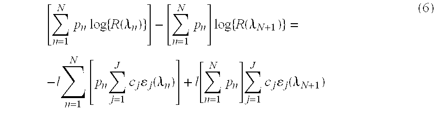
- A review of Equation (6) shows that the scattering offset term has been cancelled, but the scattering dependent pathlength variation term, l, remains. When the numerator and denominator of Equation (4) are combined, the pathlength variation term is also cancelled, as shown in Equation (7):
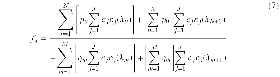
- A review of Equation (7) shows that Equation (7) depends only on the concentrations and extinction coefficients of the constituents of tissue and on the calibration coefficients p n and qm.
- In addition to providing for variable scattering compensation, the methods using Equation (4) allow a more general implementation by relaxing some of the constraints that are imposed by the use of Equation (3), above. For example:
- (a) In order to provide a certain level of accuracy for measurement of ƒ w, the numerator in Equation (3) may need to be sensitive to changes in water concentration but insensitive to changes in all other tissue constituents. For example, Equation (3) may require that the absorbance of all tissue constituents besides water (e.g. lipid, non-heme protein, hemoglobin) are nearly equal at
wavelengths - (b) In order to provide a certain level accuracy for measurement of ƒ w, the denominator in Equation (3) may need to be equally sensitive to concentration changes of all tissue constituents to which the water fraction is to be normalized. In addition, Equation (3) may require that the absorbance be equal at
wavelengths 2 and 3 for all tissue constituents to be excluded from the water fraction normalization. This constraint is removed in Equation (4), where the coefficients, qm, can be chosen to cancel the absorbance contribution due to certain constituents, while equalizing the absorbance sensitivity to the remaining tissue constituents. - In the case of measurement of the water fraction in lean tissue, ƒ w l, the coefficients, pn, in the numerator of Equation (4) are chosen to cancel the contribution from all of the major light-absorbing constituents of tissue, except water. Similarly, the coefficients, qm, in the denominator of Equation (4) are chosen to cancel the contribution from all tissue constituents other than water and protein. In addition, the coefficients, qm, are chosen to equalize the sensitivity of the denominator to changes in water and protein on a volume fractional basis. By computing the ratio of these two terms, the result is a fractional volume measurement of water concentration in lean tissue.
- In addition, application of Equation (4) to the measurement of fractional water content in total tissue volume, ƒ w T, is accomplished by choosing the coefficients in the denominator of Equation (4), qm, so that all tissue constituents (including lipid) are equalized on a fractional volume basis.
- By relaxing some of the constraints imposed by Equation (3), the use of Equation (4) can be expected to produce a more accurate prediction of fractional tissue water content, for the reasons set forth above. Various wavelength combinations may be used based on the criteria disclosed above. In order to select one wavelength combination for use with Equation (4) for the purpose of measuring fractional water content in lean tissue, ƒ w l, extinction coefficients of the major absorbing constituents of tissue (water, non-heme protein, lipid, and hemoglobin) were experimentally measured and various wavelength combinations of these were applied to a numerical model of tissue absorbance. The reproducibility of the algorithms incorporating the most promising of these wavelength combinations were then compared using real tissue data. The real tissue data were collected from 37 different volunteers at a local hospital, with Institutional Review Board (IRB) approval. The sensor measured reflected light from the pad of the finger, with a source-detector spacing of approximately 2.5 mm. The sensor was completely removed from the tissue between each pair of measurements. One such preferred algorithm combines measurements at 4 wavelengths, namely: 1180, 1245, 1275, and 1330 nm. Using this selection of wavelengths, the measurement-to-measurement reproducibility, as shown in FIG. 9, is 0.37%, indicating high reproducibility of the tissue water measurements using the methods disclosed herein.
- In addition to providing a method for measuring tissue water fraction, the method in accordance with Equation (4) above, also has general utility for the fractional quantification of analytes in tissue. In general, by appropriate choice of wavelengths and coefficients, Equation (4) is extendible to the fractional concentration measurement of any tissue constituent or combination of constituents in tissue with respect to any other constituent or combination of constituents. For example, this equation is also applicable for the determination of the fractional hemoglobin content in tissue.
- Thus, in one embodiment of the present invention, the fractional volume of total hemoglobin in tissue is determined using Equation (4) by combining reflectance measurements at wavelengths where hemoglobin is strongly absorbing with reflectance measurements where the remaining tissue constituents (such as water, lipid, and non-protein) are strongly absorbing. The coefficients, p n, in the numerator of Equation (4) are chosen to cancel the absorbance contributions from all tissue constituents except total hemoglobin. The coeffients, qm, in the denominator of Equation (4) are chose to equalize the absorbance contributions of all major tissue constituents, on a volume fractional basis. One specific wavelength combination for accomplishing this measurement is 805 nm, 1185 nm, and 1310 nm. At 805 nm the absorbance by the oxy- and deoxyhemoglobin are approximately equal. At 1185 nm, the absorbance of water, non-heme protein, and lipid, are nearly equal on a fractional volume basis. At 1300 nm the tissue absorbance will be dominated by water.
- In another embodiment of the present invention, measurement of fractional concentrations of different species of hemoglobin in tissue is performed. In general, the method provides a means of measuring the fractional concentration of hemoglobin in a first set comprised of one or more species of hemoglobin with respect to the concentration of hemoglobin in a second set comprised of one or more hemoglobin species in tissue. The coefficients, p n, in the numerator of Equation (4) are chosen to cancel the absorbance contributions from all tissue constituents except the hemoglobin species included in
set 1. The coeffients, qm, in the denominator of Equation (4) are chose to equalize the absorbance contributions from all tissue constituents except the hemoglobin species included inset 2.Sets - Individuals skilled in the art of near-infrared spectroscopy would recognize that, provided that the aforementioned guidelines are followed, additional terms can be added to Equations (1)-(4) and which may be used to incorporate reflectance measurements made at additional wavelengths and thus improve accuracy further.
- An additional embodiment of the disclosed invention provides the ability to quantify shifts of fluid into and out of the bloodstream through a novel application of pulse spectrophotometry. This additional embodiment takes advantage of the observation that pulsations caused by expansion of blood vessels in the skin as the heart beats produce changes in the reflectance at a particular wavelength that are proportional to the difference between the effective absorption of light in the blood and the surrounding interstitial tissues. Numerical simulation indicate that, if wavelengths are chosen at which water absorption is sufficiently strong, the difference between the fractions of water in the blood, ƒ w IV and surrounding tissue, ƒw EV is proportional to the ratio of the dc-normalized reflectance changes (ΔR/R) measured at two wavelengths, according to Equation (8) below:

- where c 0 and c1 are empirically determined calibration constants. This difference, integrated over time, provides a measure of the quantity of fluid that shifts into and out of the capillaries. FIG. 3 shows the prediction accuracy expected for the wavelength pair λ1=1320 nm and λ2=1160 nm.
- An additional embodiment of the present invention is directed towards the measurement of water balance index, Q, such that:

- where ƒ h IV is the fractional volume concentration of hemoglobin in the blood, and a0 and a1 are calibration coefficients. The use of Equation (9) to determine a water balance is equivalent to using Equation (8) above, where ƒh IV is set equal to 1. However, using Equation (9) provides for a more accurate determination by not neglecting the influence of ƒh IV on the derived result. The effect of this omission can be understood by allowing total hemoglobin to vary over the normal physiological range and computing the difference between the results provided by Equation (9) when ƒh IV is fixed or allowed to vary. For example, when calculations were performed with ƒw EV fixed at 0.65, ƒw IV varying between 0.75 and 0.80, and ƒh IV varying between 0.09 and 0.135 or held fixed at 0. 112, the resulting error was as large as +/−20%. In situations of extreme blood loss or vascular fluid overload (hypo- or hypervolemia) the error could be larger.
- The quantity Q, provided by Equation (9) may be combined with a separate measurement of fractional hemoglobin concentration in blood, ƒ h IV, (such as may be provided by standard clinical measurements of hematocrit or total hemoglobin) in order to provide a measure of the difference between the intravascular and extravascular water content, ƒw IV- ƒw EV. Alternatively, the quantity Q, may have clinical utility without further manipulation. For example, by providing a simultaneous measurement of both Q and fractional tissue water (either ƒw or ƒw l), the embodiments of the present invention enable the provision of a clinical indication of changes in both volume and osmolarity of body fluids. Table 1 lists the 6 combinations of volume and osmolarity changes in body fluids that are clinically observed (from Physiology, 2nd Edition, Linda S. Costanzo, Williams and Wilkins, Baltimore, 1998, pg. 156), and the expected direction and magnitude of the resultant change in fractional volume of water in blood (ƒw IV), the fractional volume of water in tissue (ƒw EV), the fractional volume of hemoglobin in blood (ƒh IV), the numerator of Q (Qn), the inverse of the denominator of Q (1/Qd), the combined result (Qn/Qd=Q), and the fractional volume of water in lean tissue, ƒw l. Taking the first row of Table 1 as an example, the result of isosmotic volume expansion, such as may be brought about by infusion with isotonic saline, would result in an increase in the fraction of water in blood (ƒw IV), a small increase in the extravascular water fraction (ƒw EV), and a large decrease in the fractional concentration of hemoglobin in the blood (ƒh IV) The combined effect of these 3 factors would result in a large increase in Q. A small increase in the fraction of water in the lean tissue, ƒw l, would also be expected. Notice that when Q and ƒw l are viewed in combination, they provide unique signatures for each of the 6 types of fluid balance change listed in Table 1. An instrument providing these measurements in a non-invasive and continuous fashion is thus able to provide a powerful tool for the monitoring of tissue water balance.
TABLE 1 Expected changes in Q and ƒw l resulting from changes in body fluid vo- lume and osmolarity Type Example ƒw IV ƒw EV ƒh IV Q n1/Qd Q ƒw I Isosmotic volume expansion Isotonic NaCl Infusion 


Isosmotic volume contraction Diarrhea 


Hyperosmotic volume expansion High NaCl intake 


0 Hyperosmotic volume contraction Sweating, Fever 


0 


Hyposmotic volume contraction SIADH 


0 


Hyposmotic volume contraction Adrenal Insufficiency 


0 - FIGS. 4 and 5 show diagrams of two different versions of an instrument for measuring the amount of water in body tissues. The simplest version of the
instrument 400 shown in FIG. 4 is designed for handheld operation and functions as a spot checker. Pressing the spring-loadedprobe head 410 against theskin 412 automatically activates the display ofpercent tissue water 414. The use of the spring-loaded probe head provides the advantages of automatically activating the display device when needed and turning the device off when not in use, thereby extending device and battery life. Moreover, this unique use of a spring-loaded probe also provides the variable force needed to improve the reliability of measurements. Percent tissue water represents the absolute percentage of water in the skin beneath the probe (typically in the range 0.6-0.9). In one embodiment of the present invention, the force exerted by a spring or hydraulic mechanism (not shown) inside theprobe head 410 is minimized, so that the fluid content of the tissue beneath the probe is not perturbed by its presence. In this manner, the tissue water fraction, including both intravascular and extravascular fluid fractions is measured. In another embodiment of the invention, the force exerted by the probe head is sufficient to push out most of the blood in the skin below the probe to allow measurement of only the extravascular fluid fraction. A pressure transducer (not shown) within theprobe head 410 measures the compressibility of the skin for deriving an index of the fraction of free (mobile) water. - The more advanced version of the
fluid monitor 500 shown in FIG. 5 is designed for use as a critical-care monitor. In addition to providing a continuous display of the volume fraction ofwater 510 at the site ofmeasurement 512, it also provides a trend display of the time-averaged difference between the intravascular fluid volume (“IFV”) and extravascular fluid volume (“EFV”) fractions (e.g., IFV-EFV=ƒw IV- ƒw EV) 514 or the quantity Q (as defined above with reference to Equation (9), updated every few seconds. This latter feature would give the physician immediate feedback on the net movement of water into or out of the blood and permit rapid evaluation of the effectiveness of diuretic or rehydration therapy. To measure the IFV-EFV difference or Q, the monitor records blood pulses in a manner similar to a pulse oximeter. Therefore, placement of the probe on the finger or other well-perfused area of the body would be required. In cases in which perfusion is too poor to obtain reliable pulse signals, the IFV-EFV or Q display would be blanked, but the tissue water fraction (ƒw) would continue to be displayed. A mechanism for mechanically inducing the pulse is built into the probe to improve the reliability of the measurement of IFV-EFV or Q under weak-pulse conditions. - FIG. 6. is a block diagram of a
handheld device 600 for measuring tissue water fraction, as well as shifts in water between the IFV and EFV compartments, or a measurement of Q, with a pulse inducing mechanism. Using thisdevice 600, patient places his/herfinger 610 in the probe housing.Rotary solenoid 612 acting throughlinkage 614 andcollar 616 induces a mechanical pulse to improve the reliability of the measurement of IFV-EFV orQ. LEDs 618 emit light at selected wavelengths andphotodiode 620 measure the transmitted light. Alternately, thephotodiode 620 can be placed adjacent to the LEDs to allow for the measurement of the reflectance of the emitted light.Preamplifier 622 magnifies the detected signal for processing by themicroprocessor 624.Microprocessor 624, using algorithms described above, determines the tissue water fraction (ƒw) (such as in the total tissue volume (ƒw T) , within the lean tissue volume (ƒw l) , and/or within the IFV (ƒw IV) and the EFV (ƒw EV)), as well as shifts in water between the IFV and EFV (such as IFV-EFV or Q), and prepares this information for display ondisplay device 626.Microprocessor 624 is also programmed to handle the appropriate timing between the rotary solenoid's operation and the signal acquisition and processing. In one embodiment, a means is provided for the user to input the fractional hemoglobin concentration (ƒh IV) or a quantity proportional to ƒh IV (such as hematocrit or total hemoglobin) in order to convert Q into IFV-EFV. The design of the device and the microprocessor integrates the method and apparatus for reducing the effect of noise on measuring physiological parameters as described in U.S. Pat. No. 5,853,364, assigned to Nellcor Puritan Bennett, Inc., the entire disclosure of which is hereby incorporated herein by reference. Additionally, the design of the device and the microprocessor also integrates the electronic processor as described in U.S. Pat. No. 5,348,004, assigned to Nellcor Incorporated, the entire disclosure of which is hereby incorporated herein by reference. - As will be understood by those skilled in the art, other equivalent or alternative methods for the measurement of the water fraction within tissue (ƒ w), as well as shifts in water between the intravascular and extravascular compartments, IVF-EVF or Q, according to the embodiments of the present invention can be envisioned without departing from the essential characteristics thereof. For example, the device can be operated in either a handheld or a tabletop mode, and it can be operated intermittently or continuously. Moreover, individuals skilled in the art of near-infrared spectroscopy would recognize that additional terms can be added to the algorithms used herein to incorporate reflectance measurements made at additional wavelengths and thus improve accuracy further. Also, light sources or light emission optics other then LED's including and not limited to incandescent light and narrowband light sources appropriately tuned to the desired wavelengths and associated light detection optics may be placed within the probe housing which is placed near the tissue location or may be positioned within a remote unit; and which deliver light to and receive light from the probe location via optical fibers. Additionally, although the specification describes embodiments functioning in a back-scattering or a reflection mode to make optical measurements of reflectances, other embodiments can be working in a forward-scattering or a transmission mode to make these measurements. These equivalents and alternatives along with obvious changes and modifications are intended to be included within the scope of the present invention. Accordingly, the foregoing disclosure is intended to be illustrative, but not limiting, of the scope of the invention which is set forth in the following claims.
Claims (47)


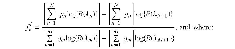


Priority Applications (13)
| Application Number | Priority Date | Filing Date | Title |
|---|---|---|---|
| US10/699,610 US7239902B2 (en) | 2001-03-16 | 2003-10-30 | Device and method for monitoring body fluid and electrolyte disorders |
| EP04796817A EP1701653B1 (en) | 2003-10-30 | 2004-10-28 | Device and method for monitoring body fluid and electrolyte disorders |
| AT04796817T ATE497722T1 (en) | 2003-10-30 | 2004-10-28 | DEVICE AND METHOD FOR MONITORING BODY FLUID AND ELECTROLYTE DISORDERS |
| DE602004031373T DE602004031373D1 (en) | 2003-10-30 | 2004-10-28 | DEVICE AND METHOD FOR MONITORING BODY FLUID AND ELECTROLYTE DISORDER |
| JP2006538345A JP2007509718A (en) | 2003-10-30 | 2004-10-28 | Devices and methods for monitoring body fluid and electrolyte disorders |
| ES04796817T ES2359065T3 (en) | 2003-10-30 | 2004-10-28 | DEVICE AND PROCEDURE FOR MONITORING DISORDERS RELATING TO BODY FLUIDS AND ELECTROLYTES. |
| AU2004285542A AU2004285542A1 (en) | 2003-10-30 | 2004-10-28 | Device and method for monitoring body fluid and electrolyte disorders |
| PCT/US2004/036106 WO2005041765A1 (en) | 2003-10-30 | 2004-10-28 | Device and method for monitoring body fluid and electrolyte disorders |
| CA002543063A CA2543063A1 (en) | 2003-10-30 | 2004-10-28 | Device and method for monitoring body fluid and electrolyte disorders |
| US11/239,815 US8457722B2 (en) | 2001-03-16 | 2005-09-30 | Device and method for monitoring body fluid and electrolyte disorders |
| US11/283,506 US7657292B2 (en) | 2001-03-16 | 2005-11-18 | Method for evaluating extracellular water concentration in tissue |
| US11/282,947 US8135448B2 (en) | 2001-03-16 | 2005-11-18 | Systems and methods to assess one or more body fluid metrics |
| US11/698,968 US8509866B2 (en) | 2001-03-16 | 2007-01-26 | Device and method for monitoring body fluid and electrolyte disorders |
Applications Claiming Priority (3)
| Application Number | Priority Date | Filing Date | Title |
|---|---|---|---|
| US09/810,918 US6591122B2 (en) | 2001-03-16 | 2001-03-16 | Device and method for monitoring body fluid and electrolyte disorders |
| US10/441,943 US7236811B2 (en) | 2001-03-16 | 2003-05-20 | Device and method for monitoring body fluid and electrolyte disorders |
| US10/699,610 US7239902B2 (en) | 2001-03-16 | 2003-10-30 | Device and method for monitoring body fluid and electrolyte disorders |
Related Parent Applications (1)
| Application Number | Title | Priority Date | Filing Date |
|---|---|---|---|
| US10/441,943 Continuation-In-Part US7236811B2 (en) | 2001-03-16 | 2003-05-20 | Device and method for monitoring body fluid and electrolyte disorders |
Related Child Applications (4)
| Application Number | Title | Priority Date | Filing Date |
|---|---|---|---|
| US11/239,815 Continuation US8457722B2 (en) | 2001-03-16 | 2005-09-30 | Device and method for monitoring body fluid and electrolyte disorders |
| US11/283,506 Continuation-In-Part US7657292B2 (en) | 2001-03-16 | 2005-11-18 | Method for evaluating extracellular water concentration in tissue |
| US11/282,947 Continuation-In-Part US8135448B2 (en) | 2001-03-16 | 2005-11-18 | Systems and methods to assess one or more body fluid metrics |
| US11/698,968 Division US8509866B2 (en) | 2001-03-16 | 2007-01-26 | Device and method for monitoring body fluid and electrolyte disorders |
Publications (2)
| Publication Number | Publication Date |
|---|---|
| US20040230106A1 true US20040230106A1 (en) | 2004-11-18 |
| US7239902B2 US7239902B2 (en) | 2007-07-03 |
Family
ID=34551018
Family Applications (3)
| Application Number | Title | Priority Date | Filing Date |
|---|---|---|---|
| US10/699,610 Expired - Fee Related US7239902B2 (en) | 2001-03-16 | 2003-10-30 | Device and method for monitoring body fluid and electrolyte disorders |
| US11/239,815 Expired - Fee Related US8457722B2 (en) | 2001-03-16 | 2005-09-30 | Device and method for monitoring body fluid and electrolyte disorders |
| US11/698,968 Expired - Fee Related US8509866B2 (en) | 2001-03-16 | 2007-01-26 | Device and method for monitoring body fluid and electrolyte disorders |
Family Applications After (2)
| Application Number | Title | Priority Date | Filing Date |
|---|---|---|---|
| US11/239,815 Expired - Fee Related US8457722B2 (en) | 2001-03-16 | 2005-09-30 | Device and method for monitoring body fluid and electrolyte disorders |
| US11/698,968 Expired - Fee Related US8509866B2 (en) | 2001-03-16 | 2007-01-26 | Device and method for monitoring body fluid and electrolyte disorders |
Country Status (9)
| Country | Link |
|---|---|
| US (3) | US7239902B2 (en) |
| EP (1) | EP1701653B1 (en) |
| JP (1) | JP2007509718A (en) |
| AT (1) | ATE497722T1 (en) |
| AU (1) | AU2004285542A1 (en) |
| CA (1) | CA2543063A1 (en) |
| DE (1) | DE602004031373D1 (en) |
| ES (1) | ES2359065T3 (en) |
| WO (1) | WO2005041765A1 (en) |
Cited By (95)
| Publication number | Priority date | Publication date | Assignee | Title |
|---|---|---|---|---|
| US20050203357A1 (en) * | 2004-03-09 | 2005-09-15 | Nellcor Puritan Bennett Incorporated | Pulse oximetry motion artifact rejection using near infrared absorption by water |
| US20060020181A1 (en) * | 2001-03-16 | 2006-01-26 | Schmitt Joseph M | Device and method for monitoring body fluid and electrolyte disorders |
| US20060084864A1 (en) * | 2001-03-16 | 2006-04-20 | Schmitt Joseph M | Device and method for monitoring body fluid and electrolyte disorders |
| US20060253016A1 (en) * | 2001-03-16 | 2006-11-09 | R Baker Clark Jr | Systems and methods to assess one or more body fluid metrics |
| US20070118027A1 (en) * | 2001-03-16 | 2007-05-24 | Baker Clark R Jr | Method for evaluating extracellular water concentration in tissue |
| US20080076983A1 (en) * | 2006-09-27 | 2008-03-27 | Nellcor Puritan Bennett Incorporated | Tissue hydration estimation by spectral absorption bandwidth measurement |
| US20080081966A1 (en) * | 2006-09-29 | 2008-04-03 | Nellcor Puritan Bennett Incorporated | Symmetric LED array for pulse oximetry |
| US20080221406A1 (en) * | 2007-03-09 | 2008-09-11 | Baker Clark R | Method and apparatus for estimating water reserves |
| US20080221412A1 (en) * | 2007-03-09 | 2008-09-11 | Nellcor Puritan Bennett Llc | Method and apparatus for spectroscopic tissue analyte measurement |
| WO2008112136A1 (en) * | 2007-03-09 | 2008-09-18 | Nellcor Puritan Bennett Llc | System and method for tissue hydration estimation |
| US20090080007A1 (en) * | 2007-09-25 | 2009-03-26 | Brother Kogyo Kabushiki Kaisha | Printing device and method therefor |
| US7643858B2 (en) * | 2006-09-28 | 2010-01-05 | Nellcor Puritan Bennett Llc | System and method for detection of brain edema using spectrophotometry |
| US7698002B2 (en) | 2006-09-29 | 2010-04-13 | Nellcor Puritan Bennett Llc | Systems and methods for user interface and identification in a medical device |
| US7706896B2 (en) | 2006-09-29 | 2010-04-27 | Nellcor Puritan Bennett Llc | User interface and identification in a medical device system and method |
| US20100113899A1 (en) * | 2008-11-06 | 2010-05-06 | Mark Ries Robinson | Alignment System for Optically Sampling a Hand |
| US7720516B2 (en) | 1996-10-10 | 2010-05-18 | Nellcor Puritan Bennett Llc | Motion compatible sensor for non-invasive optical blood analysis |
| US7725147B2 (en) | 2005-09-29 | 2010-05-25 | Nellcor Puritan Bennett Llc | System and method for removing artifacts from waveforms |
| US7725146B2 (en) | 2005-09-29 | 2010-05-25 | Nellcor Puritan Bennett Llc | System and method for pre-processing waveforms |
| WO2010085954A1 (en) * | 2009-01-29 | 2010-08-05 | Leo Pharma A/S | A method for determining the state of a skin disorder using near infrared (nir) spectroscopy |
| USD626562S1 (en) | 2008-06-30 | 2010-11-02 | Nellcor Puritan Bennett Llc | Triangular saturation pattern detection indicator for a patient monitor display panel |
| USD626561S1 (en) | 2008-06-30 | 2010-11-02 | Nellcor Puritan Bennett Llc | Circular satseconds indicator and triangular saturation pattern detection indicator for a patient monitor display panel |
| US20100305416A1 (en) * | 2007-05-07 | 2010-12-02 | Cybiocare, Inc. | Non-invasive pressured probing device |
| US7848891B2 (en) | 2006-09-29 | 2010-12-07 | Nellcor Puritan Bennett Llc | Modulation ratio determination with accommodation of uncertainty |
| US7890154B2 (en) | 2004-03-08 | 2011-02-15 | Nellcor Puritan Bennett Llc | Selection of ensemble averaging weights for a pulse oximeter based on signal quality metrics |
| US7922665B2 (en) | 2006-09-28 | 2011-04-12 | Nellcor Puritan Bennett Llc | System and method for pulse rate calculation using a scheme for alternate weighting |
| US7925511B2 (en) | 2006-09-29 | 2011-04-12 | Nellcor Puritan Bennett Llc | System and method for secure voice identification in a medical device |
| US20110208010A1 (en) * | 2010-02-22 | 2011-08-25 | Nellcor Puritan Bennett Llc | Motion energy harvesting with wireless sensors |
| US8007441B2 (en) | 2004-03-08 | 2011-08-30 | Nellcor Puritan Bennett Llc | Pulse oximeter with alternate heart-rate determination |
| US8064975B2 (en) | 2006-09-20 | 2011-11-22 | Nellcor Puritan Bennett Llc | System and method for probability based determination of estimated oxygen saturation |
| US8068891B2 (en) | 2006-09-29 | 2011-11-29 | Nellcor Puritan Bennett Llc | Symmetric LED array for pulse oximetry |
| US8068890B2 (en) | 2006-09-29 | 2011-11-29 | Nellcor Puritan Bennett Llc | Pulse oximetry sensor switchover |
| US8092993B2 (en) | 2007-12-31 | 2012-01-10 | Nellcor Puritan Bennett Llc | Hydrogel thin film for use as a biosensor |
| US8095192B2 (en) | 2003-01-10 | 2012-01-10 | Nellcor Puritan Bennett Llc | Signal quality metrics design for qualifying data for a physiological monitor |
| US8112375B2 (en) | 2008-03-31 | 2012-02-07 | Nellcor Puritan Bennett Llc | Wavelength selection and outlier detection in reduced rank linear models |
| US8109882B2 (en) | 2007-03-09 | 2012-02-07 | Nellcor Puritan Bennett Llc | System and method for venous pulsation detection using near infrared wavelengths |
| US8116852B2 (en) * | 2006-09-29 | 2012-02-14 | Nellcor Puritan Bennett Llc | System and method for detection of skin wounds and compartment syndromes |
| US8133176B2 (en) | 1999-04-14 | 2012-03-13 | Tyco Healthcare Group Lp | Method and circuit for indicating quality and accuracy of physiological measurements |
| US8140272B2 (en) | 2008-03-27 | 2012-03-20 | Nellcor Puritan Bennett Llc | System and method for unmixing spectroscopic observations with nonnegative matrix factorization |
| US8160683B2 (en) | 2006-09-29 | 2012-04-17 | Nellcor Puritan Bennett Llc | System and method for integrating voice with a medical device |
| US8160668B2 (en) | 2006-09-29 | 2012-04-17 | Nellcor Puritan Bennett Llc | Pathological condition detector using kernel methods and oximeters |
| US8195262B2 (en) | 2004-02-25 | 2012-06-05 | Nellcor Puritan Bennett Llc | Switch-mode oximeter LED drive with a single inductor |
| US8204567B2 (en) | 2007-12-13 | 2012-06-19 | Nellcor Puritan Bennett Llc | Signal demodulation |
| US8221319B2 (en) | 2009-03-25 | 2012-07-17 | Nellcor Puritan Bennett Llc | Medical device for assessing intravascular blood volume and technique for using the same |
| US8255025B2 (en) * | 2006-06-09 | 2012-08-28 | Nellcor Puritan Bennett Llc | Bronchial or tracheal tissular water content sensor and system |
| US8265724B2 (en) | 2007-03-09 | 2012-09-11 | Nellcor Puritan Bennett Llc | Cancellation of light shunting |
| US8275553B2 (en) | 2008-02-19 | 2012-09-25 | Nellcor Puritan Bennett Llc | System and method for evaluating physiological parameter data |
| US8280469B2 (en) | 2007-03-09 | 2012-10-02 | Nellcor Puritan Bennett Llc | Method for detection of aberrant tissue spectra |
| US8292809B2 (en) | 2008-03-31 | 2012-10-23 | Nellcor Puritan Bennett Llc | Detecting chemical components from spectroscopic observations |
| US8315684B2 (en) | 2004-02-25 | 2012-11-20 | Covidien Lp | Oximeter ambient light cancellation |
| US8346327B2 (en) | 2007-03-09 | 2013-01-01 | Covidien Lp | Method for identification of sensor site by local skin spectrum data |
| US8364221B2 (en) | 2005-09-30 | 2013-01-29 | Covidien Lp | Patient monitoring alarm escalation system and method |
| US8364224B2 (en) | 2008-03-31 | 2013-01-29 | Covidien Lp | System and method for facilitating sensor and monitor communication |
| US8376955B2 (en) | 2009-09-29 | 2013-02-19 | Covidien Lp | Spectroscopic method and system for assessing tissue temperature |
| US8380271B2 (en) | 2006-06-15 | 2013-02-19 | Covidien Lp | System and method for generating customizable audible beep tones and alarms |
| US8386000B2 (en) | 2008-09-30 | 2013-02-26 | Covidien Lp | System and method for photon density wave pulse oximetry and pulse hemometry |
| US8401607B2 (en) | 2001-07-19 | 2013-03-19 | Covidien Lp | Nuisance alarm reductions in a physiological monitor |
| US8406865B2 (en) | 2008-09-30 | 2013-03-26 | Covidien Lp | Bioimpedance system and sensor and technique for using the same |
| US8417309B2 (en) | 2008-09-30 | 2013-04-09 | Covidien Lp | Medical sensor |
| US8423109B2 (en) | 2005-03-03 | 2013-04-16 | Covidien Lp | Method for enhancing pulse oximery calculations in the presence of correlated artifacts |
| US8433382B2 (en) | 2008-09-30 | 2013-04-30 | Covidien Lp | Transmission mode photon density wave system and method |
| US8437822B2 (en) | 2008-03-28 | 2013-05-07 | Covidien Lp | System and method for estimating blood analyte concentration |
| US8494606B2 (en) | 2009-08-19 | 2013-07-23 | Covidien Lp | Photoplethysmography with controlled application of sensor pressure |
| US8494604B2 (en) | 2009-09-21 | 2013-07-23 | Covidien Lp | Wavelength-division multiplexing in a multi-wavelength photon density wave system |
| US8494786B2 (en) | 2009-07-30 | 2013-07-23 | Covidien Lp | Exponential sampling of red and infrared signals |
| US8509869B2 (en) | 2009-05-15 | 2013-08-13 | Covidien Lp | Method and apparatus for detecting and analyzing variations in a physiologic parameter |
| US8515511B2 (en) | 2009-09-29 | 2013-08-20 | Covidien Lp | Sensor with an optical coupling material to improve plethysmographic measurements and method of using the same |
| US20130245408A1 (en) * | 2008-03-28 | 2013-09-19 | Covidien Lp | Handheld pulse oximetry system |
| US8666467B2 (en) | 2001-05-17 | 2014-03-04 | Lawrence A. Lynn | System and method for SPO2 instability detection and quantification |
| US8690864B2 (en) | 2007-03-09 | 2014-04-08 | Covidien Lp | System and method for controlling tissue treatment |
| US8696593B2 (en) | 2006-09-27 | 2014-04-15 | Covidien Lp | Method and system for monitoring intracranial pressure |
| US8702606B2 (en) | 2006-03-21 | 2014-04-22 | Covidien Lp | Patient monitoring help video system and method |
| US8704666B2 (en) | 2009-09-21 | 2014-04-22 | Covidien Lp | Medical device interface customization systems and methods |
| US8728059B2 (en) | 2006-09-29 | 2014-05-20 | Covidien Lp | System and method for assuring validity of monitoring parameter in combination with a therapeutic device |
| US8728001B2 (en) | 2006-02-10 | 2014-05-20 | Lawrence A. Lynn | Nasal capnographic pressure monitoring system |
| US8750953B2 (en) | 2008-02-19 | 2014-06-10 | Covidien Lp | Methods and systems for alerting practitioners to physiological conditions |
| US20140171759A1 (en) * | 2012-02-15 | 2014-06-19 | Craig William WHITE | Noninvasive determination of intravascular and exctravascular hydration using near infrared spectroscopy |
| US8788001B2 (en) | 2009-09-21 | 2014-07-22 | Covidien Lp | Time-division multiplexing in a multi-wavelength photon density wave system |
| US8798704B2 (en) | 2009-09-24 | 2014-08-05 | Covidien Lp | Photoacoustic spectroscopy method and system to discern sepsis from shock |
| US8862196B2 (en) | 2001-05-17 | 2014-10-14 | Lawrence A. Lynn | System and method for automatic detection of a plurality of SP02 time series pattern types |
| US8862194B2 (en) | 2008-06-30 | 2014-10-14 | Covidien Lp | Method for improved oxygen saturation estimation in the presence of noise |
| US8930145B2 (en) | 2010-07-28 | 2015-01-06 | Covidien Lp | Light focusing continuous wave photoacoustic spectroscopy and its applications to patient monitoring |
| US8968193B2 (en) | 2008-09-30 | 2015-03-03 | Covidien Lp | System and method for enabling a research mode on physiological monitors |
| US8983800B2 (en) | 2003-01-13 | 2015-03-17 | Covidien Lp | Selection of preset filter parameters based on signal quality |
| US9031793B2 (en) | 2001-05-17 | 2015-05-12 | Lawrence A. Lynn | Centralized hospital monitoring system for automatically detecting upper airway instability and for preventing and aborting adverse drug reactions |
| US9042952B2 (en) | 1997-01-27 | 2015-05-26 | Lawrence A. Lynn | System and method for automatic detection of a plurality of SPO2 time series pattern types |
| US9053222B2 (en) | 2002-05-17 | 2015-06-09 | Lawrence A. Lynn | Patient safety processor |
| US9468378B2 (en) | 1997-01-27 | 2016-10-18 | Lawrence A. Lynn | Airway instability detection system and method |
| US9521971B2 (en) | 1997-07-14 | 2016-12-20 | Lawrence A. Lynn | System and method for automatic detection of a plurality of SPO2 time series pattern types |
| US9585606B2 (en) | 2009-09-29 | 2017-03-07 | Covidien Lp | Oximetry assembly |
| WO2017099636A1 (en) * | 2015-12-10 | 2017-06-15 | Общество С Ограниченной Ответственностью "Хилби" | Method for determining water deficiency in a person's body |
| WO2017187088A1 (en) * | 2016-04-27 | 2017-11-02 | Connected Physics | Method and apparatus for measuring the water concentration in a light-diffusing material |
| US9895068B2 (en) | 2008-06-30 | 2018-02-20 | Covidien Lp | Pulse oximeter with wait-time indication |
| CN109890287A (en) * | 2016-10-04 | 2019-06-14 | 特里邦美公司 | The method of Non-invasive measurement blood hemoglobin concentration and oxygen concentration |
| US10354753B2 (en) | 2001-05-17 | 2019-07-16 | Lawrence A. Lynn | Medical failure pattern search engine |
| US11678819B2 (en) * | 2019-04-24 | 2023-06-20 | Roxanne Abul-Haj | Noninvasive applied force / optical glucose concentration determination analyzer apparatus and method of use thereof |
Families Citing this family (31)
| Publication number | Priority date | Publication date | Assignee | Title |
|---|---|---|---|---|
| US8611977B2 (en) * | 2004-03-08 | 2013-12-17 | Covidien Lp | Method and apparatus for optical detection of mixed venous and arterial blood pulsation in tissue |
| US20070069898A1 (en) * | 2005-09-28 | 2007-03-29 | White Mark J | Glove with attached security device |
| WO2008021226A1 (en) * | 2006-08-12 | 2008-02-21 | Philometron, Inc. | Platform for detection of tissue structure change |
| US20080220512A1 (en) * | 2007-03-09 | 2008-09-11 | Nellcor Puritan Bennett Llc | Tunable laser-based spectroscopy system for non-invasively measuring body water content |
| CA2758827A1 (en) * | 2008-04-21 | 2009-10-29 | Philometron, Inc. | Metabolic energy monitoring system |
| JP2010004933A (en) * | 2008-06-24 | 2010-01-14 | Toyota Industries Corp | Non-invasive alcohol sensor |
| ES2336997B1 (en) * | 2008-10-16 | 2011-06-13 | Sabirmedical,S.L. | SYSTEM AND APPARATUS FOR NON-INVASIVE MEASUREMENT OF BLOOD PRESSURE. |
| WO2010051479A1 (en) * | 2008-10-31 | 2010-05-06 | Nellcor Puritan Bennett Llc | System and method for facilitating observation of monitored physiologic data |
| CA2741044A1 (en) * | 2008-10-31 | 2010-05-06 | Nellcor Puritan Bennett Llc | System and method for facilitating observation of monitored physiologic data |
| US8571621B2 (en) * | 2009-09-24 | 2013-10-29 | Covidien Lp | Minimax filtering for pulse oximetry |
| EP2480997A2 (en) * | 2009-09-24 | 2012-08-01 | Nellcor Puritan Bennett LLC | Determination of a physiological parameter |
| US8923945B2 (en) * | 2009-09-24 | 2014-12-30 | Covidien Lp | Determination of a physiological parameter |
| US8401608B2 (en) * | 2009-09-30 | 2013-03-19 | Covidien Lp | Method of analyzing photon density waves in a medical monitor |
| US8391943B2 (en) | 2010-03-31 | 2013-03-05 | Covidien Lp | Multi-wavelength photon density wave system using an optical switch |
| US8498683B2 (en) | 2010-04-30 | 2013-07-30 | Covidien LLP | Method for respiration rate and blood pressure alarm management |
| US7884933B1 (en) | 2010-05-05 | 2011-02-08 | Revolutionary Business Concepts, Inc. | Apparatus and method for determining analyte concentrations |
| US9380982B2 (en) | 2010-07-28 | 2016-07-05 | Covidien Lp | Adaptive alarm system and method |
| US8610769B2 (en) | 2011-02-28 | 2013-12-17 | Covidien Lp | Medical monitor data collection system and method |
| US9037204B2 (en) | 2011-09-07 | 2015-05-19 | Covidien Lp | Filtered detector array for optical patient sensors |
| US9833146B2 (en) | 2012-04-17 | 2017-12-05 | Covidien Lp | Surgical system and method of use of the same |
| US9717444B2 (en) * | 2012-08-02 | 2017-08-01 | Mark Bosin | Optical polarization shift measuring system |
| US10052064B2 (en) | 2013-04-04 | 2018-08-21 | Thermal Technologies, Inc. | Edema monitor |
| WO2015175360A1 (en) * | 2014-05-13 | 2015-11-19 | Montefiore Medical Center | Pulseless oximeter to estimate arterial oxygen saturation noninvasively in patients with weak or absent pulses |
| US20170202505A1 (en) * | 2014-07-22 | 2017-07-20 | Koninklijke Philips N.V. | Unobtrusive skin tissue hydration determining device and related method |
| US10499838B2 (en) | 2015-04-24 | 2019-12-10 | Drexel University | Non-invasive brain water monitoring device for cerebral edema and cerebral autoregulation monitoring system and method |
| US9498134B1 (en) | 2016-03-28 | 2016-11-22 | Cephalogics, LLC | Diffuse optical tomography methods and system for determining optical properties |
| WO2018187586A1 (en) * | 2017-04-05 | 2018-10-11 | The General Hospital Corporation | System and method for diagnosing soft tissue conditions |
| CN110115588B (en) * | 2018-02-05 | 2022-11-01 | 骆建铭 | Method and device for acquiring index of Kawasaki disease symptom by detecting physiological signal by optical method |
| US10966655B2 (en) | 2018-04-27 | 2021-04-06 | Hyrostasis, Inc. | Tissue hydration monitor |
| KR102640317B1 (en) * | 2018-08-23 | 2024-02-26 | 삼성전자주식회사 | Apparatus and method for measuring body fluid |
| MX2023001320A (en) * | 2020-08-03 | 2023-02-22 | Siemens Healthcare Diagnostics Inc | Absorbance spectroscopy analyzer and method of use. |
Citations (64)
| Publication number | Priority date | Publication date | Assignee | Title |
|---|---|---|---|---|
| US3998550A (en) * | 1974-10-14 | 1976-12-21 | Minolta Camera Corporation | Photoelectric oximeter |
| US4364008A (en) * | 1980-10-02 | 1982-12-14 | Jacques Steven L | Focusing probe for moisture measurement device |
| US4711244A (en) * | 1981-12-17 | 1987-12-08 | American Cyanamid Company | Digital moisture dermatometer |
| US4723554A (en) * | 1984-04-27 | 1988-02-09 | Massachusetts Institute Of Technology | Skin pallor and blush monitor |
| US4805365A (en) * | 1987-12-10 | 1989-02-21 | Hamilton Industries, Inc. | Corner post assembly |
| US4850365A (en) * | 1988-03-14 | 1989-07-25 | Futrex, Inc. | Near infrared apparatus and method for determining percent fat in a body |
| US4860753A (en) * | 1987-11-04 | 1989-08-29 | The Gillette Company | Monitoring apparatus |
| US4883055A (en) * | 1988-03-11 | 1989-11-28 | Puritan-Bennett Corporation | Artificially induced blood pulse for use with a pulse oximeter |
| US4907594A (en) * | 1987-07-18 | 1990-03-13 | Nicolay Gmbh | Method for the determination of the saturation of the blood of a living organism with oxygen and electronic circuit for performing this method |
| US5057695A (en) * | 1988-12-19 | 1991-10-15 | Otsuka Electronics Co., Ltd. | Method of and apparatus for measuring the inside information of substance with the use of light scattering |
| US5086781A (en) * | 1989-11-14 | 1992-02-11 | Bookspan Mark A | Bioelectric apparatus for monitoring body fluid compartments |
| US5111817A (en) * | 1988-12-29 | 1992-05-12 | Medical Physics, Inc. | Noninvasive system and method for enhanced arterial oxygen saturation determination and arterial blood pressure monitoring |
| US5146091A (en) * | 1990-04-19 | 1992-09-08 | Inomet, Inc. | Body fluid constituent measurement utilizing an interference pattern |
| US5224478A (en) * | 1989-11-25 | 1993-07-06 | Colin Electronics Co., Ltd. | Reflecting-type oxymeter probe |
| US5277181A (en) * | 1991-12-12 | 1994-01-11 | Vivascan Corporation | Noninvasive measurement of hematocrit and hemoglobin content by differential optical analysis |
| US5279295A (en) * | 1989-11-23 | 1994-01-18 | U.S. Philips Corporation | Non-invasive oximeter arrangement |
| US5282467A (en) * | 1992-08-13 | 1994-02-01 | Duke University | Non-invasive method for detecting deep venous thrombosis in the human body |
| US5337937A (en) * | 1991-10-18 | 1994-08-16 | United States Surgical Corporation | Surgical stapling apparatus |
| US5337745A (en) * | 1992-03-10 | 1994-08-16 | Benaron David A | Device and method for in vivo qualitative or quantative measurement of blood chromophore concentration using blood pulse spectrophotometry |
| US5348004A (en) * | 1993-03-31 | 1994-09-20 | Nellcor Incorporated | Electronic processor for pulse oximeter |
| US5355880A (en) * | 1992-07-06 | 1994-10-18 | Sandia Corporation | Reliable noninvasive measurement of blood gases |
| US5377674A (en) * | 1992-05-08 | 1995-01-03 | Kuestner; J. Todd | Method for non-invasive and in-vitro hemoglobin concentration measurement |
| US5499627A (en) * | 1990-10-06 | 1996-03-19 | In-Line Diagnostics Corporation | System for noninvasive hematocrit monitoring |
| US5615689A (en) * | 1994-12-12 | 1997-04-01 | St. Luke's-Roosevelt Hospital | Method of predicting body cell mass using bioimpedance analysis |
| US5687721A (en) * | 1992-12-15 | 1997-11-18 | Kuhls; Burkhard | Measurement device for non-invasively determining the concentration of polarising substances |
| US5701902A (en) * | 1994-09-14 | 1997-12-30 | Cedars-Sinai Medical Center | Spectroscopic burn injury evaluation apparatus and method |
| US5720284A (en) * | 1995-03-31 | 1998-02-24 | Nihon Kohden Corporation | Apparatus for measuring hemoglobin |
| US5735284A (en) * | 1992-06-24 | 1998-04-07 | N.I. Medical Ltd. | Method and system for non-invasive determination of the main cardiorespiratory parameters of the human body |
| US5747789A (en) * | 1993-12-01 | 1998-05-05 | Dynamics Imaging, Inc. | Method for investigation of distribution of physiological components in human body tissues and apparatus for its realization |
| US5755672A (en) * | 1995-11-30 | 1998-05-26 | Moritex Corporation | Measuring equipment of fat and water amount existing on the object |
| US5788643A (en) * | 1997-04-22 | 1998-08-04 | Zymed Medical Instrumentation, Inc. | Process for monitoring patients with chronic congestive heart failure |
| US5827181A (en) * | 1995-09-07 | 1998-10-27 | Hewlett-Packard Co. | Noninvasive blood chemistry measurement method and system |
| US5833602A (en) * | 1993-09-20 | 1998-11-10 | Osemwota; Omoigui | Process of continuous noninvasive hemometry |
| US5853364A (en) * | 1995-08-07 | 1998-12-29 | Nellcor Puritan Bennett, Inc. | Method and apparatus for estimating physiological parameters using model-based adaptive filtering |
| US5860919A (en) * | 1995-06-07 | 1999-01-19 | Masimo Corporation | Active pulse blood constituent monitoring method |
| US5906582A (en) * | 1994-09-14 | 1999-05-25 | Seiko Epson Corporation | Organism information measuring method and arm wear type pulse-wave measuring method |
| US5933226A (en) * | 1996-05-23 | 1999-08-03 | Minolta Co., Ltd. | Attachment for a concentration measuring apparatus and a concentration measuring system |
| US6064898A (en) * | 1998-09-21 | 2000-05-16 | Essential Medical Devices | Non-invasive blood component analyzer |
| US6125297A (en) * | 1998-02-06 | 2000-09-26 | The United States Of America As Represented By The United States National Aeronautics And Space Administration | Body fluids monitor |
| US6149591A (en) * | 1997-02-21 | 2000-11-21 | Duke University | Refractometric devices especially adapted for the in vivo detection of refractive indices of cervical mucus |
| US6178342B1 (en) * | 1993-09-09 | 2001-01-23 | Vasamedics | Surface perfusion pressure monitoring system |
| US6222189B1 (en) * | 1992-07-15 | 2001-04-24 | Optix, Lp | Methods of enhancing optical signals by mechanical manipulation in non-invasive testing |
| US6246894B1 (en) * | 1993-02-01 | 2001-06-12 | In-Line Diagnostics Corporation | System and method for measuring blood urea nitrogen, blood osmolarity, plasma free hemoglobin and tissue water content |
| US6280396B1 (en) * | 1998-08-03 | 2001-08-28 | American Weights And Measures | Apparatus and method for measuring body composition |
| US6336044B1 (en) * | 1998-09-11 | 2002-01-01 | Futrex Inc. | Reliable body fat measurement in self-service health parameter Measuring system |
| US6370426B1 (en) * | 1999-04-20 | 2002-04-09 | Nova Technology Corporation | Method and apparatus for measuring relative hydration of a substrate |
| US6400971B1 (en) * | 1999-10-12 | 2002-06-04 | Orsense Ltd. | Optical device for non-invasive measurement of blood-related signals and a finger holder therefor |
| US6402690B1 (en) * | 1999-04-23 | 2002-06-11 | Massachusetts Institute Of Technology | Isolating ring sensor design |
| US6412936B1 (en) * | 1998-03-03 | 2002-07-02 | Canon Kabushiki Kaisha | Ink, ink set, ink cartridge, recording unit, image recording process and image recording apparatus |
| US6442408B1 (en) * | 1999-07-22 | 2002-08-27 | Instrumentation Metrics, Inc. | Method for quantification of stratum corneum hydration using diffuse reflectance spectroscopy |
| US6466807B1 (en) * | 1997-08-12 | 2002-10-15 | Abbott Laboratories | Optical glucose detector |
| US6488677B1 (en) * | 2001-05-10 | 2002-12-03 | Thermal Technologies, Inc. | System for quantifying edema |
| US6591122B2 (en) * | 2001-03-16 | 2003-07-08 | Nellcor Puritan Bennett Incorporated | Device and method for monitoring body fluid and electrolyte disorders |
| US6592574B1 (en) * | 1999-07-28 | 2003-07-15 | Visx, Incorporated | Hydration and topography tissue measurements for laser sculpting |
| US6600946B1 (en) * | 2000-08-11 | 2003-07-29 | The Boeing Company | Methods and apparatus for quantifying dermal hydration |
| US6606509B2 (en) * | 2001-03-16 | 2003-08-12 | Nellcor Puritan Bennett Incorporated | Method and apparatus for improving the accuracy of noninvasive hematocrit measurements |
| US6636759B2 (en) * | 1998-10-29 | 2003-10-21 | Inlight Solutions, Inc. | Apparatus and method for determination of the adequacy of dialysis by non-invasive near-infrared spectroscopy |
| US6635491B1 (en) * | 2000-07-28 | 2003-10-21 | Abbott Labortories | Method for non-invasively determining the concentration of an analyte by compensating for the effect of tissue hydration |
| US6643543B2 (en) * | 2000-08-01 | 2003-11-04 | Tanita Corporation | Body water amount condition judging apparatus by multi-frequency bioelectric impedance measurement |
| US6654620B2 (en) * | 1998-05-18 | 2003-11-25 | Abbott Laboratories | Method for improving non-invasive determination of the concentration of analytes in a biological sample |
| US6675029B2 (en) * | 1999-07-22 | 2004-01-06 | Sensys Medical, Inc. | Apparatus and method for quantification of tissue hydration using diffuse reflectance spectroscopy |
| US6777240B2 (en) * | 2000-02-10 | 2004-08-17 | Sensys Medical, Inc. | Intra-serum and intra-gel for modeling human skin tissue |
| US6849046B1 (en) * | 1999-09-23 | 2005-02-01 | Elazar Eyal-Bickels | System and method for detecting the state of hydration of a living specimen |
| US6950699B1 (en) * | 2001-12-12 | 2005-09-27 | Brain Child Foundation | Water content probe |
Family Cites Families (41)
| Publication number | Priority date | Publication date | Assignee | Title |
|---|---|---|---|---|
| SE388045B (en) | 1974-11-28 | 1976-09-20 | Servo Med Ab | PROCEDURE FOR SATURATION OF FROM A SURFACE BY DIFFUSION GIVEN QUANTITY OF EXV. THE WATER AND THE DEVICE FOR PERFORMING THE PROCEDURE |
| DE2712521A1 (en) * | 1977-03-22 | 1978-09-28 | Wacker Chemitronic | PROCEDURE FOR FITTING DISCS |
| US4805623A (en) | 1987-09-04 | 1989-02-21 | Vander Corporation | Spectrophotometric method for quantitatively determining the concentration of a dilute component in a light- or other radiation-scattering environment |
| US5365066A (en) * | 1989-01-19 | 1994-11-15 | Futrex, Inc. | Low cost means for increasing measurement sensitivity in LED/IRED near-infrared instruments |
| US5077476A (en) * | 1990-06-27 | 1991-12-31 | Futrex, Inc. | Instrument for non-invasive measurement of blood glucose |
| JPH0440940A (en) | 1990-06-07 | 1992-02-12 | Minolta Camera Co Ltd | Instrument for measuring concentration of total hemoglobin |
| AU3583293A (en) | 1992-01-17 | 1993-08-03 | Government Of The United States Of America, As Represented By The Secretary Of The Department Of Health And Human Services, The | Optical method for monitoring arterial blood hematocrit |
| JP3255370B2 (en) | 1992-06-03 | 2002-02-12 | 浜松ホトニクス株式会社 | Method and apparatus for detecting location of water in skin |
| US5424545A (en) | 1992-07-15 | 1995-06-13 | Myron J. Block | Non-invasive non-spectrophotometric infrared measurement of blood analyte concentrations |
| US20050062609A9 (en) | 1992-08-19 | 2005-03-24 | Lynn Lawrence A. | Pulse oximetry relational alarm system for early recognition of instability and catastrophic occurrences |
| EP1491135A3 (en) | 1993-04-12 | 2005-09-07 | Hema Metrics, Inc. | Method and apparatus for monitoring blood constituents |
| FR2710517B1 (en) | 1993-09-27 | 1995-12-22 | Dior Christian Parfums | Method for evaluating the state of hydration of the skin and apparatus intended for its implementation. |
| JP4212007B2 (en) * | 1996-11-26 | 2009-01-21 | パナソニック電工株式会社 | Blood component concentration analyzer |
| GB9702018D0 (en) | 1997-01-31 | 1997-03-19 | Univ London | Determination of the ratio of optical absorbtion coefficients at different wavelengths in a scattering medium |
| US7039446B2 (en) * | 2001-01-26 | 2006-05-02 | Sensys Medical, Inc. | Indirect measurement of tissue analytes through tissue properties |
| DE69940053D1 (en) | 1998-02-05 | 2009-01-22 | Hema Metrics Inc | METHOD AND DEVICE FOR NON-INVASIVE OBSERVATION OF BLOOD COMPONENTS |
| JPH11244266A (en) | 1998-02-27 | 1999-09-14 | Matsushita Electric Works Ltd | Superficial organism tissue analytical method and superficial organism tissue analyzer |
| DE19855521A1 (en) | 1998-12-02 | 2000-06-08 | Tracoe Medizine Ges Fuer | Tube with sealed cuff |
| US6280381B1 (en) * | 1999-07-22 | 2001-08-28 | Instrumentation Metrics, Inc. | Intelligent system for noninvasive blood analyte prediction |
| DE19923658A1 (en) | 1999-05-22 | 2000-11-23 | Infralytic Gmbh | Device for measuring organization grade of water in mammals for diagnosis of disease conditions |
| US6512936B1 (en) | 1999-07-22 | 2003-01-28 | Sensys Medical, Inc. | Multi-tier method of classifying sample spectra for non-invasive blood analyte prediction |
| WO2001016577A1 (en) | 1999-08-31 | 2001-03-08 | Cme Telemetrix Inc. | Method for determination of analytes using nir, adjacent visible spectrum and discrete nir wavelengths |
| KR100398362B1 (en) | 2000-09-01 | 2003-09-19 | 스펙트론 테크 주식회사 | Method and apparatus for measuring skin moisture by using near-infrared reflectance spectroscopy |
| US8135448B2 (en) * | 2001-03-16 | 2012-03-13 | Nellcor Puritan Bennett Llc | Systems and methods to assess one or more body fluid metrics |
| US7239902B2 (en) | 2001-03-16 | 2007-07-03 | Nellor Puritan Bennett Incorporated | Device and method for monitoring body fluid and electrolyte disorders |
| US6898451B2 (en) | 2001-03-21 | 2005-05-24 | Minformed, L.L.C. | Non-invasive blood analyte measuring system and method utilizing optical absorption |
| AU2002355272A1 (en) | 2001-07-25 | 2003-02-17 | Argose, Inc. | Adjunct quantitative system and method for non-invasive measurement of in vivo analytes |
| US20040147034A1 (en) * | 2001-08-14 | 2004-07-29 | Gore Jay Prabhakar | Method and apparatus for measuring a substance in a biological sample |
| US6840904B2 (en) | 2001-10-11 | 2005-01-11 | Jason Goldberg | Medical monitoring device and system |
| WO2003071939A1 (en) | 2002-02-22 | 2003-09-04 | Masimo Corporation | Active pulse spectraphotometry |
| US8996090B2 (en) | 2002-06-03 | 2015-03-31 | Exostat Medical, Inc. | Noninvasive detection of a physiologic parameter within a body tissue of a patient |
| JP2004081427A (en) | 2002-08-26 | 2004-03-18 | Kenji Yoshikawa | Apparatus for measuring water content in living body |
| JP4284674B2 (en) | 2003-01-31 | 2009-06-24 | 日本光電工業株式会社 | Absorbent concentration measuring device in blood |
| US7283242B2 (en) | 2003-04-11 | 2007-10-16 | Thornton Robert L | Optical spectroscopy apparatus and method for measurement of analyte concentrations or other such species in a specimen employing a semiconductor laser-pumped, small-cavity fiber laser |
| JP3566277B1 (en) | 2003-06-23 | 2004-09-15 | 株式会社日立製作所 | Blood glucose meter |
| WO2005025399A2 (en) | 2003-09-12 | 2005-03-24 | Or-Nim Medical Ltd. | Noninvasive optical monitoring of region of interest |
| KR100624412B1 (en) | 2003-09-16 | 2006-09-18 | 삼성전자주식회사 | Blood components measuring apparatus and method of measuring blood components using the same |
| JP3590047B1 (en) | 2003-09-24 | 2004-11-17 | 株式会社日立製作所 | Optical measuring device and blood glucose measuring device using the same |
| US8036727B2 (en) * | 2004-08-11 | 2011-10-11 | Glt Acquisition Corp. | Methods for noninvasively measuring analyte levels in a subject |
| US20060167350A1 (en) * | 2005-01-27 | 2006-07-27 | Monfre Stephen L | Multi-tier method of developing localized calibration models for non-invasive blood analyte prediction |
| US8180419B2 (en) | 2006-09-27 | 2012-05-15 | Nellcor Puritan Bennett Llc | Tissue hydration estimation by spectral absorption bandwidth measurement |
-
2003
- 2003-10-30 US US10/699,610 patent/US7239902B2/en not_active Expired - Fee Related
-
2004
- 2004-10-28 ES ES04796817T patent/ES2359065T3/en active Active
- 2004-10-28 WO PCT/US2004/036106 patent/WO2005041765A1/en active Application Filing
- 2004-10-28 AT AT04796817T patent/ATE497722T1/en not_active IP Right Cessation
- 2004-10-28 AU AU2004285542A patent/AU2004285542A1/en not_active Abandoned
- 2004-10-28 JP JP2006538345A patent/JP2007509718A/en active Pending
- 2004-10-28 EP EP04796817A patent/EP1701653B1/en not_active Not-in-force
- 2004-10-28 DE DE602004031373T patent/DE602004031373D1/en active Active
- 2004-10-28 CA CA002543063A patent/CA2543063A1/en not_active Abandoned
-
2005
- 2005-09-30 US US11/239,815 patent/US8457722B2/en not_active Expired - Fee Related
-
2007
- 2007-01-26 US US11/698,968 patent/US8509866B2/en not_active Expired - Fee Related
Patent Citations (69)
| Publication number | Priority date | Publication date | Assignee | Title |
|---|---|---|---|---|
| US3998550A (en) * | 1974-10-14 | 1976-12-21 | Minolta Camera Corporation | Photoelectric oximeter |
| US4364008A (en) * | 1980-10-02 | 1982-12-14 | Jacques Steven L | Focusing probe for moisture measurement device |
| US4711244A (en) * | 1981-12-17 | 1987-12-08 | American Cyanamid Company | Digital moisture dermatometer |
| US4723554A (en) * | 1984-04-27 | 1988-02-09 | Massachusetts Institute Of Technology | Skin pallor and blush monitor |
| US4907594A (en) * | 1987-07-18 | 1990-03-13 | Nicolay Gmbh | Method for the determination of the saturation of the blood of a living organism with oxygen and electronic circuit for performing this method |
| US4860753A (en) * | 1987-11-04 | 1989-08-29 | The Gillette Company | Monitoring apparatus |
| US4805365A (en) * | 1987-12-10 | 1989-02-21 | Hamilton Industries, Inc. | Corner post assembly |
| US4883055A (en) * | 1988-03-11 | 1989-11-28 | Puritan-Bennett Corporation | Artificially induced blood pulse for use with a pulse oximeter |
| US4850365A (en) * | 1988-03-14 | 1989-07-25 | Futrex, Inc. | Near infrared apparatus and method for determining percent fat in a body |
| US5057695A (en) * | 1988-12-19 | 1991-10-15 | Otsuka Electronics Co., Ltd. | Method of and apparatus for measuring the inside information of substance with the use of light scattering |
| US5111817A (en) * | 1988-12-29 | 1992-05-12 | Medical Physics, Inc. | Noninvasive system and method for enhanced arterial oxygen saturation determination and arterial blood pressure monitoring |
| US5086781A (en) * | 1989-11-14 | 1992-02-11 | Bookspan Mark A | Bioelectric apparatus for monitoring body fluid compartments |
| US5279295A (en) * | 1989-11-23 | 1994-01-18 | U.S. Philips Corporation | Non-invasive oximeter arrangement |
| US5224478A (en) * | 1989-11-25 | 1993-07-06 | Colin Electronics Co., Ltd. | Reflecting-type oxymeter probe |
| US5146091A (en) * | 1990-04-19 | 1992-09-08 | Inomet, Inc. | Body fluid constituent measurement utilizing an interference pattern |
| US5499627A (en) * | 1990-10-06 | 1996-03-19 | In-Line Diagnostics Corporation | System for noninvasive hematocrit monitoring |
| US5803908A (en) * | 1990-10-06 | 1998-09-08 | In-Line Diagnostics Corporation | System for noninvasive hematocrit monitoring |
| US20010020122A1 (en) * | 1990-10-06 | 2001-09-06 | Steuer Robert R. | System and method for measuring blood urea nitrogen, blood osmolarity, plasma free hemoglobin and tissue water content |
| US6687519B2 (en) * | 1990-10-06 | 2004-02-03 | Hema Metrics, Inc. | System and method for measuring blood urea nitrogen, blood osmolarity, plasma free hemoglobin and tissue water content |
| US5337937A (en) * | 1991-10-18 | 1994-08-16 | United States Surgical Corporation | Surgical stapling apparatus |
| US5277181A (en) * | 1991-12-12 | 1994-01-11 | Vivascan Corporation | Noninvasive measurement of hematocrit and hemoglobin content by differential optical analysis |
| US5337745A (en) * | 1992-03-10 | 1994-08-16 | Benaron David A | Device and method for in vivo qualitative or quantative measurement of blood chromophore concentration using blood pulse spectrophotometry |
| US5377674A (en) * | 1992-05-08 | 1995-01-03 | Kuestner; J. Todd | Method for non-invasive and in-vitro hemoglobin concentration measurement |
| US5735284A (en) * | 1992-06-24 | 1998-04-07 | N.I. Medical Ltd. | Method and system for non-invasive determination of the main cardiorespiratory parameters of the human body |
| US5355880A (en) * | 1992-07-06 | 1994-10-18 | Sandia Corporation | Reliable noninvasive measurement of blood gases |
| US6222189B1 (en) * | 1992-07-15 | 2001-04-24 | Optix, Lp | Methods of enhancing optical signals by mechanical manipulation in non-invasive testing |
| US5282467A (en) * | 1992-08-13 | 1994-02-01 | Duke University | Non-invasive method for detecting deep venous thrombosis in the human body |
| US5687721A (en) * | 1992-12-15 | 1997-11-18 | Kuhls; Burkhard | Measurement device for non-invasively determining the concentration of polarising substances |
| US6246894B1 (en) * | 1993-02-01 | 2001-06-12 | In-Line Diagnostics Corporation | System and method for measuring blood urea nitrogen, blood osmolarity, plasma free hemoglobin and tissue water content |
| US5348004A (en) * | 1993-03-31 | 1994-09-20 | Nellcor Incorporated | Electronic processor for pulse oximeter |
| US6178342B1 (en) * | 1993-09-09 | 2001-01-23 | Vasamedics | Surface perfusion pressure monitoring system |
| US5833602A (en) * | 1993-09-20 | 1998-11-10 | Osemwota; Omoigui | Process of continuous noninvasive hemometry |
| US5747789A (en) * | 1993-12-01 | 1998-05-05 | Dynamics Imaging, Inc. | Method for investigation of distribution of physiological components in human body tissues and apparatus for its realization |
| US5701902A (en) * | 1994-09-14 | 1997-12-30 | Cedars-Sinai Medical Center | Spectroscopic burn injury evaluation apparatus and method |
| US5906582A (en) * | 1994-09-14 | 1999-05-25 | Seiko Epson Corporation | Organism information measuring method and arm wear type pulse-wave measuring method |
| US5615689A (en) * | 1994-12-12 | 1997-04-01 | St. Luke's-Roosevelt Hospital | Method of predicting body cell mass using bioimpedance analysis |
| US5720284A (en) * | 1995-03-31 | 1998-02-24 | Nihon Kohden Corporation | Apparatus for measuring hemoglobin |
| US5860919A (en) * | 1995-06-07 | 1999-01-19 | Masimo Corporation | Active pulse blood constituent monitoring method |
| US5853364A (en) * | 1995-08-07 | 1998-12-29 | Nellcor Puritan Bennett, Inc. | Method and apparatus for estimating physiological parameters using model-based adaptive filtering |
| US5827181A (en) * | 1995-09-07 | 1998-10-27 | Hewlett-Packard Co. | Noninvasive blood chemistry measurement method and system |
| US5755672A (en) * | 1995-11-30 | 1998-05-26 | Moritex Corporation | Measuring equipment of fat and water amount existing on the object |
| US5933226A (en) * | 1996-05-23 | 1999-08-03 | Minolta Co., Ltd. | Attachment for a concentration measuring apparatus and a concentration measuring system |
| US6149591A (en) * | 1997-02-21 | 2000-11-21 | Duke University | Refractometric devices especially adapted for the in vivo detection of refractive indices of cervical mucus |
| US5788643A (en) * | 1997-04-22 | 1998-08-04 | Zymed Medical Instrumentation, Inc. | Process for monitoring patients with chronic congestive heart failure |
| US6466807B1 (en) * | 1997-08-12 | 2002-10-15 | Abbott Laboratories | Optical glucose detector |
| US6125297A (en) * | 1998-02-06 | 2000-09-26 | The United States Of America As Represented By The United States National Aeronautics And Space Administration | Body fluids monitor |
| US6412936B1 (en) * | 1998-03-03 | 2002-07-02 | Canon Kabushiki Kaisha | Ink, ink set, ink cartridge, recording unit, image recording process and image recording apparatus |
| US6654620B2 (en) * | 1998-05-18 | 2003-11-25 | Abbott Laboratories | Method for improving non-invasive determination of the concentration of analytes in a biological sample |
| US6280396B1 (en) * | 1998-08-03 | 2001-08-28 | American Weights And Measures | Apparatus and method for measuring body composition |
| US6336044B1 (en) * | 1998-09-11 | 2002-01-01 | Futrex Inc. | Reliable body fat measurement in self-service health parameter Measuring system |
| US6064898A (en) * | 1998-09-21 | 2000-05-16 | Essential Medical Devices | Non-invasive blood component analyzer |
| US6615064B1 (en) * | 1998-09-21 | 2003-09-02 | Essential Medical Devices, Inc. | Non-invasive blood component analyzer |
| US6636759B2 (en) * | 1998-10-29 | 2003-10-21 | Inlight Solutions, Inc. | Apparatus and method for determination of the adequacy of dialysis by non-invasive near-infrared spectroscopy |
| US6370426B1 (en) * | 1999-04-20 | 2002-04-09 | Nova Technology Corporation | Method and apparatus for measuring relative hydration of a substrate |
| US6402690B1 (en) * | 1999-04-23 | 2002-06-11 | Massachusetts Institute Of Technology | Isolating ring sensor design |
| US6442408B1 (en) * | 1999-07-22 | 2002-08-27 | Instrumentation Metrics, Inc. | Method for quantification of stratum corneum hydration using diffuse reflectance spectroscopy |
| US6675029B2 (en) * | 1999-07-22 | 2004-01-06 | Sensys Medical, Inc. | Apparatus and method for quantification of tissue hydration using diffuse reflectance spectroscopy |
| US6592574B1 (en) * | 1999-07-28 | 2003-07-15 | Visx, Incorporated | Hydration and topography tissue measurements for laser sculpting |
| US6849046B1 (en) * | 1999-09-23 | 2005-02-01 | Elazar Eyal-Bickels | System and method for detecting the state of hydration of a living specimen |
| US6400971B1 (en) * | 1999-10-12 | 2002-06-04 | Orsense Ltd. | Optical device for non-invasive measurement of blood-related signals and a finger holder therefor |
| US6777240B2 (en) * | 2000-02-10 | 2004-08-17 | Sensys Medical, Inc. | Intra-serum and intra-gel for modeling human skin tissue |
| US6635491B1 (en) * | 2000-07-28 | 2003-10-21 | Abbott Labortories | Method for non-invasively determining the concentration of an analyte by compensating for the effect of tissue hydration |
| US6643543B2 (en) * | 2000-08-01 | 2003-11-04 | Tanita Corporation | Body water amount condition judging apparatus by multi-frequency bioelectric impedance measurement |
| US6600946B1 (en) * | 2000-08-11 | 2003-07-29 | The Boeing Company | Methods and apparatus for quantifying dermal hydration |
| US6668181B2 (en) * | 2000-09-25 | 2003-12-23 | Sensys Medical, Inc. | Method for quantification of stratum corneum hydration using diffuse reflectance spectroscopy |
| US6606509B2 (en) * | 2001-03-16 | 2003-08-12 | Nellcor Puritan Bennett Incorporated | Method and apparatus for improving the accuracy of noninvasive hematocrit measurements |
| US6591122B2 (en) * | 2001-03-16 | 2003-07-08 | Nellcor Puritan Bennett Incorporated | Device and method for monitoring body fluid and electrolyte disorders |
| US6488677B1 (en) * | 2001-05-10 | 2002-12-03 | Thermal Technologies, Inc. | System for quantifying edema |
| US6950699B1 (en) * | 2001-12-12 | 2005-09-27 | Brain Child Foundation | Water content probe |
Cited By (137)
| Publication number | Priority date | Publication date | Assignee | Title |
|---|---|---|---|---|
| US7720516B2 (en) | 1996-10-10 | 2010-05-18 | Nellcor Puritan Bennett Llc | Motion compatible sensor for non-invasive optical blood analysis |
| US9042952B2 (en) | 1997-01-27 | 2015-05-26 | Lawrence A. Lynn | System and method for automatic detection of a plurality of SPO2 time series pattern types |
| US9468378B2 (en) | 1997-01-27 | 2016-10-18 | Lawrence A. Lynn | Airway instability detection system and method |
| US9521971B2 (en) | 1997-07-14 | 2016-12-20 | Lawrence A. Lynn | System and method for automatic detection of a plurality of SPO2 time series pattern types |
| US8133176B2 (en) | 1999-04-14 | 2012-03-13 | Tyco Healthcare Group Lp | Method and circuit for indicating quality and accuracy of physiological measurements |
| US8932227B2 (en) | 2000-07-28 | 2015-01-13 | Lawrence A. Lynn | System and method for CO2 and oximetry integration |
| US10058269B2 (en) | 2000-07-28 | 2018-08-28 | Lawrence A. Lynn | Monitoring system for identifying an end-exhalation carbon dioxide value of enhanced clinical utility |
| US20070118027A1 (en) * | 2001-03-16 | 2007-05-24 | Baker Clark R Jr | Method for evaluating extracellular water concentration in tissue |
| US8229529B2 (en) | 2001-03-16 | 2012-07-24 | Nellcor Puritan Bennett Llc | Device and method for monitoring body fluid and electrolyte disorders |
| US8457722B2 (en) | 2001-03-16 | 2013-06-04 | Covidien Lp | Device and method for monitoring body fluid and electrolyte disorders |
| US8135448B2 (en) | 2001-03-16 | 2012-03-13 | Nellcor Puritan Bennett Llc | Systems and methods to assess one or more body fluid metrics |
| US8509866B2 (en) | 2001-03-16 | 2013-08-13 | Covidien Lp | Device and method for monitoring body fluid and electrolyte disorders |
| US20070129614A1 (en) * | 2001-03-16 | 2007-06-07 | Nellcor Puritan Bennett Inc. | Device and method for monitoring body fluid and electrolyte disorders |
| US20060253016A1 (en) * | 2001-03-16 | 2006-11-09 | R Baker Clark Jr | Systems and methods to assess one or more body fluid metrics |
| US20060084864A1 (en) * | 2001-03-16 | 2006-04-20 | Schmitt Joseph M | Device and method for monitoring body fluid and electrolyte disorders |
| US7657292B2 (en) | 2001-03-16 | 2010-02-02 | Nellcor Puritan Bennett Llc | Method for evaluating extracellular water concentration in tissue |
| US20060020181A1 (en) * | 2001-03-16 | 2006-01-26 | Schmitt Joseph M | Device and method for monitoring body fluid and electrolyte disorders |
| US11439321B2 (en) | 2001-05-17 | 2022-09-13 | Lawrence A. Lynn | Monitoring system for identifying an end-exhalation carbon dioxide value of enhanced clinical utility |
| US8862196B2 (en) | 2001-05-17 | 2014-10-14 | Lawrence A. Lynn | System and method for automatic detection of a plurality of SP02 time series pattern types |
| US9031793B2 (en) | 2001-05-17 | 2015-05-12 | Lawrence A. Lynn | Centralized hospital monitoring system for automatically detecting upper airway instability and for preventing and aborting adverse drug reactions |
| US8666467B2 (en) | 2001-05-17 | 2014-03-04 | Lawrence A. Lynn | System and method for SPO2 instability detection and quantification |
| US10032526B2 (en) | 2001-05-17 | 2018-07-24 | Lawrence A. Lynn | Patient safety processor |
| US10297348B2 (en) | 2001-05-17 | 2019-05-21 | Lawrence A. Lynn | Patient safety processor |
| US10354753B2 (en) | 2001-05-17 | 2019-07-16 | Lawrence A. Lynn | Medical failure pattern search engine |
| US10366790B2 (en) | 2001-05-17 | 2019-07-30 | Lawrence A. Lynn | Patient safety processor |
| US8838196B2 (en) | 2001-07-19 | 2014-09-16 | Covidien Lp | Nuisance alarm reductions in a physiological monitor |
| US8401607B2 (en) | 2001-07-19 | 2013-03-19 | Covidien Lp | Nuisance alarm reductions in a physiological monitor |
| US8401606B2 (en) | 2001-07-19 | 2013-03-19 | Covidien Lp | Nuisance alarm reductions in a physiological monitor |
| US9053222B2 (en) | 2002-05-17 | 2015-06-09 | Lawrence A. Lynn | Patient safety processor |
| US8095192B2 (en) | 2003-01-10 | 2012-01-10 | Nellcor Puritan Bennett Llc | Signal quality metrics design for qualifying data for a physiological monitor |
| US8983800B2 (en) | 2003-01-13 | 2015-03-17 | Covidien Lp | Selection of preset filter parameters based on signal quality |
| US8315684B2 (en) | 2004-02-25 | 2012-11-20 | Covidien Lp | Oximeter ambient light cancellation |
| US8195262B2 (en) | 2004-02-25 | 2012-06-05 | Nellcor Puritan Bennett Llc | Switch-mode oximeter LED drive with a single inductor |
| US8874181B2 (en) | 2004-02-25 | 2014-10-28 | Covidien Lp | Oximeter ambient light cancellation |
| US8007441B2 (en) | 2004-03-08 | 2011-08-30 | Nellcor Puritan Bennett Llc | Pulse oximeter with alternate heart-rate determination |
| US7890154B2 (en) | 2004-03-08 | 2011-02-15 | Nellcor Puritan Bennett Llc | Selection of ensemble averaging weights for a pulse oximeter based on signal quality metrics |
| US8560036B2 (en) | 2004-03-08 | 2013-10-15 | Covidien Lp | Selection of ensemble averaging weights for a pulse oximeter based on signal quality metrics |
| US7277741B2 (en) | 2004-03-09 | 2007-10-02 | Nellcor Puritan Bennett Incorporated | Pulse oximetry motion artifact rejection using near infrared absorption by water |
| US8175670B2 (en) | 2004-03-09 | 2012-05-08 | Nellcor Puritan Bennett Llc | Pulse oximetry signal correction using near infrared absorption by water |
| US8195263B2 (en) | 2004-03-09 | 2012-06-05 | Nellcor Puritan Bennett Llc | Pulse oximetry motion artifact rejection using near infrared absorption by water |
| US20050203357A1 (en) * | 2004-03-09 | 2005-09-15 | Nellcor Puritan Bennett Incorporated | Pulse oximetry motion artifact rejection using near infrared absorption by water |
| US9351674B2 (en) | 2005-03-03 | 2016-05-31 | Covidien Lp | Method for enhancing pulse oximetry calculations in the presence of correlated artifacts |
| US8818475B2 (en) | 2005-03-03 | 2014-08-26 | Covidien Lp | Method for enhancing pulse oximetry calculations in the presence of correlated artifacts |
| US8423109B2 (en) | 2005-03-03 | 2013-04-16 | Covidien Lp | Method for enhancing pulse oximery calculations in the presence of correlated artifacts |
| US7725146B2 (en) | 2005-09-29 | 2010-05-25 | Nellcor Puritan Bennett Llc | System and method for pre-processing waveforms |
| US8744543B2 (en) | 2005-09-29 | 2014-06-03 | Covidien Lp | System and method for removing artifacts from waveforms |
| US7725147B2 (en) | 2005-09-29 | 2010-05-25 | Nellcor Puritan Bennett Llc | System and method for removing artifacts from waveforms |
| US8364221B2 (en) | 2005-09-30 | 2013-01-29 | Covidien Lp | Patient monitoring alarm escalation system and method |
| US8728001B2 (en) | 2006-02-10 | 2014-05-20 | Lawrence A. Lynn | Nasal capnographic pressure monitoring system |
| US8702606B2 (en) | 2006-03-21 | 2014-04-22 | Covidien Lp | Patient monitoring help video system and method |
| US8255025B2 (en) * | 2006-06-09 | 2012-08-28 | Nellcor Puritan Bennett Llc | Bronchial or tracheal tissular water content sensor and system |
| US8380271B2 (en) | 2006-06-15 | 2013-02-19 | Covidien Lp | System and method for generating customizable audible beep tones and alarms |
| US8064975B2 (en) | 2006-09-20 | 2011-11-22 | Nellcor Puritan Bennett Llc | System and method for probability based determination of estimated oxygen saturation |
| US8538500B2 (en) | 2006-09-20 | 2013-09-17 | Covidien Lp | System and method for probability based determination of estimated oxygen saturation |
| US8696593B2 (en) | 2006-09-27 | 2014-04-15 | Covidien Lp | Method and system for monitoring intracranial pressure |
| US8180419B2 (en) | 2006-09-27 | 2012-05-15 | Nellcor Puritan Bennett Llc | Tissue hydration estimation by spectral absorption bandwidth measurement |
| US20080076983A1 (en) * | 2006-09-27 | 2008-03-27 | Nellcor Puritan Bennett Incorporated | Tissue hydration estimation by spectral absorption bandwidth measurement |
| US10022058B2 (en) | 2006-09-28 | 2018-07-17 | Covidien Lp | System and method for pulse rate calculation using a scheme for alternate weighting |
| US7643858B2 (en) * | 2006-09-28 | 2010-01-05 | Nellcor Puritan Bennett Llc | System and method for detection of brain edema using spectrophotometry |
| US8801622B2 (en) | 2006-09-28 | 2014-08-12 | Covidien Lp | System and method for pulse rate calculation using a scheme for alternate weighting |
| US7922665B2 (en) | 2006-09-28 | 2011-04-12 | Nellcor Puritan Bennett Llc | System and method for pulse rate calculation using a scheme for alternate weighting |
| US7706896B2 (en) | 2006-09-29 | 2010-04-27 | Nellcor Puritan Bennett Llc | User interface and identification in a medical device system and method |
| US8068891B2 (en) | 2006-09-29 | 2011-11-29 | Nellcor Puritan Bennett Llc | Symmetric LED array for pulse oximetry |
| US8116852B2 (en) * | 2006-09-29 | 2012-02-14 | Nellcor Puritan Bennett Llc | System and method for detection of skin wounds and compartment syndromes |
| US7848891B2 (en) | 2006-09-29 | 2010-12-07 | Nellcor Puritan Bennett Llc | Modulation ratio determination with accommodation of uncertainty |
| US7925511B2 (en) | 2006-09-29 | 2011-04-12 | Nellcor Puritan Bennett Llc | System and method for secure voice identification in a medical device |
| US7698002B2 (en) | 2006-09-29 | 2010-04-13 | Nellcor Puritan Bennett Llc | Systems and methods for user interface and identification in a medical device |
| US8160683B2 (en) | 2006-09-29 | 2012-04-17 | Nellcor Puritan Bennett Llc | System and method for integrating voice with a medical device |
| US8160726B2 (en) | 2006-09-29 | 2012-04-17 | Nellcor Puritan Bennett Llc | User interface and identification in a medical device system and method |
| US8728059B2 (en) | 2006-09-29 | 2014-05-20 | Covidien Lp | System and method for assuring validity of monitoring parameter in combination with a therapeutic device |
| US20080081966A1 (en) * | 2006-09-29 | 2008-04-03 | Nellcor Puritan Bennett Incorporated | Symmetric LED array for pulse oximetry |
| US8160668B2 (en) | 2006-09-29 | 2012-04-17 | Nellcor Puritan Bennett Llc | Pathological condition detector using kernel methods and oximeters |
| US8175667B2 (en) | 2006-09-29 | 2012-05-08 | Nellcor Puritan Bennett Llc | Symmetric LED array for pulse oximetry |
| US8068890B2 (en) | 2006-09-29 | 2011-11-29 | Nellcor Puritan Bennett Llc | Pulse oximetry sensor switchover |
| US8494617B2 (en) | 2006-09-29 | 2013-07-23 | Covidien Lp | System for detection of skin wounds and compartment syndromes |
| US20080221406A1 (en) * | 2007-03-09 | 2008-09-11 | Baker Clark R | Method and apparatus for estimating water reserves |
| WO2008112136A1 (en) * | 2007-03-09 | 2008-09-18 | Nellcor Puritan Bennett Llc | System and method for tissue hydration estimation |
| US20080221412A1 (en) * | 2007-03-09 | 2008-09-11 | Nellcor Puritan Bennett Llc | Method and apparatus for spectroscopic tissue analyte measurement |
| WO2008112134A1 (en) * | 2007-03-09 | 2008-09-18 | Nellcor Puritan Bennett Llc | Method and apparatus for estimating water reserves |
| US8346327B2 (en) | 2007-03-09 | 2013-01-01 | Covidien Lp | Method for identification of sensor site by local skin spectrum data |
| US8175665B2 (en) * | 2007-03-09 | 2012-05-08 | Nellcor Puritan Bennett Llc | Method and apparatus for spectroscopic tissue analyte measurement |
| US8357090B2 (en) * | 2007-03-09 | 2013-01-22 | Covidien Lp | Method and apparatus for estimating water reserves |
| US8280469B2 (en) | 2007-03-09 | 2012-10-02 | Nellcor Puritan Bennett Llc | Method for detection of aberrant tissue spectra |
| US8690864B2 (en) | 2007-03-09 | 2014-04-08 | Covidien Lp | System and method for controlling tissue treatment |
| US8109882B2 (en) | 2007-03-09 | 2012-02-07 | Nellcor Puritan Bennett Llc | System and method for venous pulsation detection using near infrared wavelengths |
| US8265724B2 (en) | 2007-03-09 | 2012-09-11 | Nellcor Puritan Bennett Llc | Cancellation of light shunting |
| US20100305416A1 (en) * | 2007-05-07 | 2010-12-02 | Cybiocare, Inc. | Non-invasive pressured probing device |
| US20090080007A1 (en) * | 2007-09-25 | 2009-03-26 | Brother Kogyo Kabushiki Kaisha | Printing device and method therefor |
| US8204567B2 (en) | 2007-12-13 | 2012-06-19 | Nellcor Puritan Bennett Llc | Signal demodulation |
| US8092993B2 (en) | 2007-12-31 | 2012-01-10 | Nellcor Puritan Bennett Llc | Hydrogel thin film for use as a biosensor |
| US10076276B2 (en) | 2008-02-19 | 2018-09-18 | Covidien Lp | Methods and systems for alerting practitioners to physiological conditions |
| US8275553B2 (en) | 2008-02-19 | 2012-09-25 | Nellcor Puritan Bennett Llc | System and method for evaluating physiological parameter data |
| US11298076B2 (en) | 2008-02-19 | 2022-04-12 | Covidien Lp | Methods and systems for alerting practitioners to physiological conditions |
| US8750953B2 (en) | 2008-02-19 | 2014-06-10 | Covidien Lp | Methods and systems for alerting practitioners to physiological conditions |
| US8781753B2 (en) | 2008-02-19 | 2014-07-15 | Covidien Lp | System and method for evaluating physiological parameter data |
| US8140272B2 (en) | 2008-03-27 | 2012-03-20 | Nellcor Puritan Bennett Llc | System and method for unmixing spectroscopic observations with nonnegative matrix factorization |
| US20130245408A1 (en) * | 2008-03-28 | 2013-09-19 | Covidien Lp | Handheld pulse oximetry system |
| US8437822B2 (en) | 2008-03-28 | 2013-05-07 | Covidien Lp | System and method for estimating blood analyte concentration |
| US8112375B2 (en) | 2008-03-31 | 2012-02-07 | Nellcor Puritan Bennett Llc | Wavelength selection and outlier detection in reduced rank linear models |
| US8292809B2 (en) | 2008-03-31 | 2012-10-23 | Nellcor Puritan Bennett Llc | Detecting chemical components from spectroscopic observations |
| US8364224B2 (en) | 2008-03-31 | 2013-01-29 | Covidien Lp | System and method for facilitating sensor and monitor communication |
| USD626562S1 (en) | 2008-06-30 | 2010-11-02 | Nellcor Puritan Bennett Llc | Triangular saturation pattern detection indicator for a patient monitor display panel |
| USD736250S1 (en) | 2008-06-30 | 2015-08-11 | Covidien Lp | Portion of a display panel with an indicator icon |
| US8862194B2 (en) | 2008-06-30 | 2014-10-14 | Covidien Lp | Method for improved oxygen saturation estimation in the presence of noise |
| USD626561S1 (en) | 2008-06-30 | 2010-11-02 | Nellcor Puritan Bennett Llc | Circular satseconds indicator and triangular saturation pattern detection indicator for a patient monitor display panel |
| US9895068B2 (en) | 2008-06-30 | 2018-02-20 | Covidien Lp | Pulse oximeter with wait-time indication |
| US8417309B2 (en) | 2008-09-30 | 2013-04-09 | Covidien Lp | Medical sensor |
| US8968193B2 (en) | 2008-09-30 | 2015-03-03 | Covidien Lp | System and method for enabling a research mode on physiological monitors |
| US8433382B2 (en) | 2008-09-30 | 2013-04-30 | Covidien Lp | Transmission mode photon density wave system and method |
| US8406865B2 (en) | 2008-09-30 | 2013-03-26 | Covidien Lp | Bioimpedance system and sensor and technique for using the same |
| US8386000B2 (en) | 2008-09-30 | 2013-02-26 | Covidien Lp | System and method for photon density wave pulse oximetry and pulse hemometry |
| US20100113899A1 (en) * | 2008-11-06 | 2010-05-06 | Mark Ries Robinson | Alignment System for Optically Sampling a Hand |
| WO2010085954A1 (en) * | 2009-01-29 | 2010-08-05 | Leo Pharma A/S | A method for determining the state of a skin disorder using near infrared (nir) spectroscopy |
| US8221319B2 (en) | 2009-03-25 | 2012-07-17 | Nellcor Puritan Bennett Llc | Medical device for assessing intravascular blood volume and technique for using the same |
| US8509869B2 (en) | 2009-05-15 | 2013-08-13 | Covidien Lp | Method and apparatus for detecting and analyzing variations in a physiologic parameter |
| US8494786B2 (en) | 2009-07-30 | 2013-07-23 | Covidien Lp | Exponential sampling of red and infrared signals |
| US9380969B2 (en) | 2009-07-30 | 2016-07-05 | Covidien Lp | Systems and methods for varying a sampling rate of a signal |
| US8494606B2 (en) | 2009-08-19 | 2013-07-23 | Covidien Lp | Photoplethysmography with controlled application of sensor pressure |
| US8704666B2 (en) | 2009-09-21 | 2014-04-22 | Covidien Lp | Medical device interface customization systems and methods |
| US8788001B2 (en) | 2009-09-21 | 2014-07-22 | Covidien Lp | Time-division multiplexing in a multi-wavelength photon density wave system |
| US8494604B2 (en) | 2009-09-21 | 2013-07-23 | Covidien Lp | Wavelength-division multiplexing in a multi-wavelength photon density wave system |
| US8798704B2 (en) | 2009-09-24 | 2014-08-05 | Covidien Lp | Photoacoustic spectroscopy method and system to discern sepsis from shock |
| US9585606B2 (en) | 2009-09-29 | 2017-03-07 | Covidien Lp | Oximetry assembly |
| US9597023B2 (en) | 2009-09-29 | 2017-03-21 | Covidien Lp | Oximetry assembly |
| US8515511B2 (en) | 2009-09-29 | 2013-08-20 | Covidien Lp | Sensor with an optical coupling material to improve plethysmographic measurements and method of using the same |
| US8376955B2 (en) | 2009-09-29 | 2013-02-19 | Covidien Lp | Spectroscopic method and system for assessing tissue temperature |
| US20150313519A1 (en) * | 2010-02-22 | 2015-11-05 | Covidien Lp | Motion energy harvesting with wireless sensors |
| US9078610B2 (en) * | 2010-02-22 | 2015-07-14 | Covidien Lp | Motion energy harvesting with wireless sensors |
| US20110208010A1 (en) * | 2010-02-22 | 2011-08-25 | Nellcor Puritan Bennett Llc | Motion energy harvesting with wireless sensors |
| US8930145B2 (en) | 2010-07-28 | 2015-01-06 | Covidien Lp | Light focusing continuous wave photoacoustic spectroscopy and its applications to patient monitoring |
| US20140171759A1 (en) * | 2012-02-15 | 2014-06-19 | Craig William WHITE | Noninvasive determination of intravascular and exctravascular hydration using near infrared spectroscopy |
| WO2017099636A1 (en) * | 2015-12-10 | 2017-06-15 | Общество С Ограниченной Ответственностью "Хилби" | Method for determining water deficiency in a person's body |
| WO2017187088A1 (en) * | 2016-04-27 | 2017-11-02 | Connected Physics | Method and apparatus for measuring the water concentration in a light-diffusing material |
| FR3050824A1 (en) * | 2016-04-27 | 2017-11-03 | Connected Physics | METHOD AND APPARATUS FOR MEASURING WATER CONCENTRATION IN LIGHT DIFFUSING MATERIAL. |
| CN109890287B (en) * | 2016-10-04 | 2021-11-02 | 特里邦美公司 | Method for non-invasive determination of hemoglobin concentration and oxygen concentration in blood |
| CN109890287A (en) * | 2016-10-04 | 2019-06-14 | 特里邦美公司 | The method of Non-invasive measurement blood hemoglobin concentration and oxygen concentration |
| US11678819B2 (en) * | 2019-04-24 | 2023-06-20 | Roxanne Abul-Haj | Noninvasive applied force / optical glucose concentration determination analyzer apparatus and method of use thereof |
Also Published As
| Publication number | Publication date |
|---|---|
| CA2543063A1 (en) | 2005-05-12 |
| US8509866B2 (en) | 2013-08-13 |
| EP1701653B1 (en) | 2011-02-09 |
| US8457722B2 (en) | 2013-06-04 |
| US7239902B2 (en) | 2007-07-03 |
| JP2007509718A (en) | 2007-04-19 |
| DE602004031373D1 (en) | 2011-03-24 |
| ES2359065T3 (en) | 2011-05-18 |
| WO2005041765A1 (en) | 2005-05-12 |
| ATE497722T1 (en) | 2011-02-15 |
| AU2004285542A1 (en) | 2005-05-12 |
| US20060084864A1 (en) | 2006-04-20 |
| EP1701653A1 (en) | 2006-09-20 |
| US20070129614A1 (en) | 2007-06-07 |
Similar Documents
| Publication | Publication Date | Title |
|---|---|---|
| US7239902B2 (en) | Device and method for monitoring body fluid and electrolyte disorders | |
| US6591122B2 (en) | Device and method for monitoring body fluid and electrolyte disorders | |
| US7657292B2 (en) | Method for evaluating extracellular water concentration in tissue | |
| US8135448B2 (en) | Systems and methods to assess one or more body fluid metrics | |
| US7532919B2 (en) | Measuring tissue oxygenation | |
| JP4176480B2 (en) | Method and apparatus for improving the accuracy of non-invasive hematocrit measurements | |
| US20080221412A1 (en) | Method and apparatus for spectroscopic tissue analyte measurement | |
| Soyemi et al. | Measuring tissue oxygenation |
Legal Events
| Date | Code | Title | Description |
|---|---|---|---|
| AS | Assignment |
Owner name: NELLCOR PURITAN BENNETT INCORPORATION, CALIFORNIA Free format text: RECORD TO CORRECT WRONG APPLICATION NUMBER 10/699619 ON AN ASSIGNMENT DOCUMENT PREVIOUSLY RECORDED AT REEL 015590 FRAME 0073;ASSIGNORS:SCHMITT, JOSEPH M.;DEBRECZENY, MARTIN;REEL/FRAME:015710/0358;SIGNING DATES FROM 20040617 TO 20040626 |
|
| STCF | Information on status: patent grant |
Free format text: PATENTED CASE |
|
| FPAY | Fee payment |
Year of fee payment: 4 |
|
| AS | Assignment |
Owner name: NELLCOR PURITAN BENNETT LLC, COLORADO Free format text: CHANGE OF NAME;ASSIGNOR:NELLCOR PURITAN BENNETT INCORPORATED;REEL/FRAME:028290/0199 Effective date: 20061220 |
|
| AS | Assignment |
Owner name: COVIDIEN LP, MASSACHUSETTS Free format text: ASSIGNMENT OF ASSIGNORS INTEREST;ASSIGNOR:NELLCOR PURITAN BENNETT LLC;REEL/FRAME:029433/0582 Effective date: 20120929 |
|
| FPAY | Fee payment |
Year of fee payment: 8 |
|
| FEPP | Fee payment procedure |
Free format text: MAINTENANCE FEE REMINDER MAILED (ORIGINAL EVENT CODE: REM.); ENTITY STATUS OF PATENT OWNER: LARGE ENTITY |
|
| LAPS | Lapse for failure to pay maintenance fees |
Free format text: PATENT EXPIRED FOR FAILURE TO PAY MAINTENANCE FEES (ORIGINAL EVENT CODE: EXP.); ENTITY STATUS OF PATENT OWNER: LARGE ENTITY |
|
| STCH | Information on status: patent discontinuation |
Free format text: PATENT EXPIRED DUE TO NONPAYMENT OF MAINTENANCE FEES UNDER 37 CFR 1.362 |
|
| FP | Lapsed due to failure to pay maintenance fee |
Effective date: 20190703 |