US20030190690A1 - Biosensor and method for analyzing blood components using it - Google Patents
Biosensor and method for analyzing blood components using it Download PDFInfo
- Publication number
- US20030190690A1 US20030190690A1 US10/398,711 US39871103A US2003190690A1 US 20030190690 A1 US20030190690 A1 US 20030190690A1 US 39871103 A US39871103 A US 39871103A US 2003190690 A1 US2003190690 A1 US 2003190690A1
- Authority
- US
- United States
- Prior art keywords
- reagent
- sample solution
- biosensor
- sample
- blood
- Prior art date
- Legal status (The legal status is an assumption and is not a legal conclusion. Google has not performed a legal analysis and makes no representation as to the accuracy of the status listed.)
- Abandoned
Links
Images


Classifications
-
- G—PHYSICS
- G01—MEASURING; TESTING
- G01N—INVESTIGATING OR ANALYSING MATERIALS BY DETERMINING THEIR CHEMICAL OR PHYSICAL PROPERTIES
- G01N33/00—Investigating or analysing materials by specific methods not covered by groups G01N1/00 - G01N31/00
- G01N33/48—Biological material, e.g. blood, urine; Haemocytometers
- G01N33/50—Chemical analysis of biological material, e.g. blood, urine; Testing involving biospecific ligand binding methods; Immunological testing
- G01N33/53—Immunoassay; Biospecific binding assay; Materials therefor
- G01N33/543—Immunoassay; Biospecific binding assay; Materials therefor with an insoluble carrier for immobilising immunochemicals
-
- G—PHYSICS
- G01—MEASURING; TESTING
- G01N—INVESTIGATING OR ANALYSING MATERIALS BY DETERMINING THEIR CHEMICAL OR PHYSICAL PROPERTIES
- G01N33/00—Investigating or analysing materials by specific methods not covered by groups G01N1/00 - G01N31/00
- G01N33/48—Biological material, e.g. blood, urine; Haemocytometers
- G01N33/50—Chemical analysis of biological material, e.g. blood, urine; Testing involving biospecific ligand binding methods; Immunological testing
- G01N33/53—Immunoassay; Biospecific binding assay; Materials therefor
- G01N33/558—Immunoassay; Biospecific binding assay; Materials therefor using diffusion or migration of antigen or antibody
-
- G—PHYSICS
- G01—MEASURING; TESTING
- G01N—INVESTIGATING OR ANALYSING MATERIALS BY DETERMINING THEIR CHEMICAL OR PHYSICAL PROPERTIES
- G01N33/00—Investigating or analysing materials by specific methods not covered by groups G01N1/00 - G01N31/00
- G01N33/48—Biological material, e.g. blood, urine; Haemocytometers
- G01N33/50—Chemical analysis of biological material, e.g. blood, urine; Testing involving biospecific ligand binding methods; Immunological testing
- G01N33/53—Immunoassay; Biospecific binding assay; Materials therefor
- G01N33/543—Immunoassay; Biospecific binding assay; Materials therefor with an insoluble carrier for immobilising immunochemicals
- G01N33/54366—Apparatus specially adapted for solid-phase testing
- G01N33/54386—Analytical elements
- G01N33/54387—Immunochromatographic test strips
- G01N33/54388—Immunochromatographic test strips based on lateral flow
Definitions
- the present invention relates to a biosensor and, more particularly, to a dry-type biosensor for analyzing an analyte in a sample solution, and a method for analyzing a constituent of blood by employing the biosensor.
- a measuring method by a chromatography sensor which utilizes an antigen-antibody reaction is generally used as a method for analyzing the bodily fluids.
- blood constituent such as a metabolic product, a protein, a fat, an electrolyte, an enzyme, an antigen, and an antibody.
- a general blood-constituent analysis method requires several operation processes, such as centrifuging blood which is previously collected, and analyzing the blood constituent employing obtained blood plasma or blood serum by an analytical instrument or a biosensor.
- the measurement requires a special apparatus, and the preprocessing as well as the inspection take a lot of time.
- the centrifugation method which requires a centrifugal machine is not adequate when a small number of specimens are to be processed quickly or when inspections in the field is performed.
- the obtained blood serum or blood plasma is smaller in amount as compared with the amount of blood.
- a cell contraction agent is employed to cause the blood cell components to constrict, and the constricted blood-cell components are developed on a chromatography carrier.
- the constricted blood cell components can be developed with blood plasma components, thereby requiring no preprocessing on the specimen.
- the present invention is made to solve the above-mentioned problems and has for its object to provide a simple, quick, and high-performance biosensor which can confirm the result of blood constituent analysis only by applying a slight amount of blood thereto, without requiring special apparatus, and a method for analyzing a blood constituent by employing the biosensor.
- a biosensor made of a dried porous material.
- the biosensor comprises: a sample introductory part for introducing a sample solution; and a developing layer for developing the sample solution, and in this biosensor the developing layer includes: a marker reagent holding part where a marker reagent is held in a dry state so that it can be eluted by the development of the sample solution; and a reagent immobilization part where a reagent which can bind to an analyte and is involved in a reaction is immobilized so that it is not eluted, and when the sample solution is introduced to the sample introductory part, the sample solution permeates the developing layer to reach the marker reagent holding part, and moves to the reagent immobilization part while eluting the marker reagent, whereby the reaction among the analyte, the marker reagent, and the immobilized reagent occurs, and an amount of marker reagent bound in the reagent immobil
- the applied sample solution is developed on the biosensor while being agitated by a turbulent flow caused by the retiform structure, whereby the permeability of a reactive layer carrier is enhanced, and a more uniform permeation is realized, resulting in a biosensor which enables a simple, quick, more sensitive, and higher-performance measurement.
- the retiform structure as mentioned above is formed by performing a molding process on a fiber or resin to reticulate the same, and it makes no difference whether the retiform structure itself has capillary activity or absorbability or not.
- the reticulum at this time may have any shape as long as it is polygonal, and the size thereof does not matter. It is preferable that meshes are arranged in regular sizes. Further, this retiform structure is preferably a single layer.
- the turbulent flow is a flow in which a fluid irregularly moves in disorder and a stream line shows a fine and irregular fluctuation.
- a biosensor made of a dried porous material.
- the biosensor comprises: a developing layer for developing a sample solution; a space forming part for forming a space on the developing layer; and a sample introductory part having a cavity in which the sample solution flows, in the formed space, and in this biosensor the developing layer includes: a marker reagent holding part where a marker reagent is held in a dry state so that it can be eluted by the development of the sample solution; and a reagent immobilization part where a reagent which can bind to an analyte and is involved in a reaction is immobilized so that it is not eluted, and when the sample solution is introduced to the sample introductory part, the sample solution permeates the developing layer to reach the marker reagent holding part, and moves to the reagent immobilization part while eluting the marker reagent, whereby the reaction among the analyte, the marker reagent,
- the present invention a specific amount of sample solution is sucked in the sample introductory part as the cavity formed by the space forming material, and the applied sample solution is developed on the biosensor while being efficiently agitated by a turbulent flow generated by the reriform tissue. Therefore, the permeability of a reactive layer carrier is enhanced, and a more uniform permeation is realized, resulting in a biosensor which enables a simple, quick, more sensitive, and higher-performance measurement.
- a biosensor made of a dried porous material.
- the biosensor comprises: a developing layer for developing a sample solution; a space forming part for forming a space on the developing layer; and a sample introductory part having a cavity in which the sample solution flows, in the formed space, and in this biosensor a reagent holding part where a marker reagent is held in a dry state so that it can be eluted by the flow of the sample solution is provided in the cavity, and the developing layer includes a reagent immobilization part where a reagent which can bind to an analyte and is involved in a reaction is immobilized so that it is not eluted, and when the sample solution is introduced to the sample introductory part, the sample solution is developed on the developing layer while eluting the marker reagent, and reaches the reagent immobilization part, whereby the reaction among the analyte, the marker reagent, and the immobilized
- the present invention a specific amount of sample solution is sucked in the sample introductory part as the cavity formed by the space forming material, and the applied sample solution is developed on the biosensor while being efficiently agitated by a turbulent flow generated by the reriform tissue and, thus, more thoroughly reacting with the marker reagent. Therefore, the permeability of a reactive layer carrier is enhanced, and a more uniform permeation is realized, resulting in a biosensor which enables a simple, quick, more sensitive, and higher-performance measurement.
- a cell contraction agent holding part for causing cellular components to constrict is provided in the sample introductory part or at a position on the sample introductory part side up to the marker reagent holding part.
- the retiform structure is arranged at the end part of the sample introductory part.
- the applied sample solution is developed on the biosensor while being efficiently agitated by a turbulent flow generated by the retiform structure, whereby a more uniform and effective cellular constriction is performed, and the sample solution permeates a reactive layer carrier uniformly, resulting in a biosensor which enables a simple, quick, more sensitive, and higher-performance measurement.
- the retiform structure and the cell contraction agent holding part are arranged so that the edges of the end parts thereof are kept aligned.
- the introduced sample solution can immediately react with the cell contraction agent as well as receive the effect of turbulent flow at the introduction, whereby the sample solution can efficiently react with the cell contraction agent. Therefore, a more uniform and effective cellular constriction is performed, and the sample solution permeates a reactive layer carrier uniformly, resulting in a biosensor which enables a simple, quick, more sensitive, and higher-performance measurement.
- a space enabling the sample solution to flow therein is arranged between the retiform structure and the cell contraction agent holding part.
- the sample solution is smoothly introduced, and a sufficient constriction reaction which occurs by means of the cell contraction agent and effect of turbulent flow due to the retiform structure can be obtained, whereby a more uniform and effective cellular constriction is performed, and the sample solution permeates a reactive layer carrier uniformly, resulting in a biosensor which enables a simple, quick, more sensitive, and higher-performance measurement.
- the retiform structure is arranged so that warp threads thereof are parallel to the direction in which the sample is developed on the developing layer.
- the sample solution which is developed while being efficiently agitated by a turbulent flow is efficiently led in the direction of the development by a capillary phenomenon caused by warp threads of the retiform structure that extend in the direction of the sample development. Therefore, the sample solution permeates a reactive layer carrier uniformly, resulting in a biosensor which enables a simple, quick, more sensitive, and higher-performance measurement.
- the warp thread as mentioned above is a part of the retiform structure in which a molded product of a fiber or resin forming the reticulum extends in the direction of the development. It is preferable that the warp thread is regularly arranged so that it is parallel to the direction of the development. Directions of tissues extending in other directions than that described above do not matter.
- the reitform tissue is made of synthetic resin.
- the retiform structure itself lacks the water retaining capacity, whereby the sample solution is well drained, and the applied sample solution is quickly developed on the biosensor while being efficiently agitated by a turbulent flow generated by the retiform structure. Therefore, a smaller amount of sample solution realizes a uniform permeation on a reactive layer carrier, resulting in a biosensor which enables a simple, quick, sensitive, and high-performance measurement.
- the retiform structure is made of a chemical fiber such as polyester.
- the retiform structure itself lacks the water retaining capacity, whereby the sample solution is well drained, and the applied sample solution is efficiently and quickly developed on the biosensor by a capillary phenomenon, while being efficiently agitated by a turbulent flow generated by the retiform structure. Therefore, a smaller amount of sample solution realizes a uniform permeation on a reactive layer carrier, resulting in a biosensor which enables a simple, quick, sensitive, and high-performance measurement.
- the retiform structure as mentioned above is formed by performing a welding process such as a thermo-compression bonding and a press work on a chemical fiber, to reticulate the same.
- the retiform structure is a fabric obtained by weaving the chemical fiber such as polyester.
- the retiform structure itself lacks the water retaining capacity, whereby the sample solution is well drained, and the applied sample solution is efficiently and quickly developed on the biosensor by a capillary phenomenon, while being efficiently agitated by a turbulent flow generated by the retiform structure. Therefore, a smaller amount of sample solution realizes a uniform permeation on a reactive layer carrier, resulting in a biosensor which enables a simple, quick, sensitive, and high-performance measurement.
- the retiform structure as mentioned above is a fabric or a knit formed by the process of weaving or knitting a chemical fiber.
- the retiform structure is treated with a surfactant so that it can permeate.
- the retiform structure does not repel the liquid sample, and the applied sample solution is quickly developed on the biosensor while being efficiently agitated by a turbulent flow generated by the retiform structure. Therefore, a smaller amount of sample solution realizes a uniform permeation on a reactive layer carrier, resulting in a biosensor which enables a simple, quick, sensitive, and high-performance measurement.
- a mesh of the retiform structure has a pore size of 0.1 mm to 2 mm.
- the sample solution is efficiently agitated by a turbulent flow generated by the retiform structure. Therefore, a smaller amount of sample solution realizes a uniform permeation on a reactive layer carrier, resulting in a biosensor which enables a simple, quick, sensitive, and high-performance measurement.
- the sample solution to be applied is blood.
- the sample solution to be applied is a solution including bacteria.
- the cell contraction agent is inorganic salt.
- the cellular components in the liquid solution are constricted, so that clogging by the cellular components in the liquid sample such as whole blood or a bacterial solution is avoided, thereby realizing a uniform permeation state. Therefore, a biosensor which is able to perform a more accurate measurement in a short time without inhibiting the reaction is realized.
- the inorganic salt as mentioned above is an inorganic compound including salt, such as sodium chloride, potassium chloride, and sodium phosphate.
- the cell contraction agent is amino acid.
- the cellular components in the liquid solution are constricted, so that clogging by the cellular components in the liquid sample such as whole blood or a bacterial solution is avoided, thereby realizing a uniform permeation state. Therefore, a biosensor which is able to perform a more accurate measurement in a short time without inhibiting the reaction is realized.
- the amino acid as mentioned above is a compound which has a carboxyl group and an amino group in an identical molecule, such as glycin and glutamic acid, and further includes imino acid such as proline and hydroxyproline.
- the cell contraction agent is saccharide
- the cellular components in the liquid solution are constricted, so that clogging by the cellular components in the liquid sample such as whole blood or a bacterial solution is avoided, thereby realizing a uniform permeation state. Therefore, a biosensor which is able to perform a more accurate measurement in a short time without inhibiting the reaction is realized.
- the saccharide as mentioned above includes a glucide such as glucose, scrose, and trehalose, as well as sugar alcohol such as glucitol.
- the biosensor in the biosensor as defined in any of claims 1 to 18, is a one-step immunochromatography test specimen.
- many measurement targets can be measured by obtaining antibodies or antigens for the measurement targets, and the applied sample solution is developed on the biosensor while being efficiently agitated by a turbulent blow generated by the retiform structure. Therefore, the permeability of a reactive layer carrier is enhanced, and a more uniform permeation is realized, resulting in a biosensor which enables a simple, quick, more sensitive, and higher-performance measurement.
- the “one-step” as mentioned above represents the measurement operation which only requires the sample solution to be dropped to the test specimen without the need to preprocess the sample solution, and does not require a developing solution to develop the sample solution after the dropping, nor require a washing process.
- the immunochromatography test specimen as mentioned above is a sensor for detecting an analyte in the sample solution on a carrier where chromatography development is performed, by utilizing an antigen-antibody reaction.
- a method for analyzing a constituent of blood by employing a biosensor made of a dried porous material comprises: a sample introductory part for introducing a sample solution; and a developing layer for developing the sample solution, and in this biosensor the developing layer includes: a marker reagent holding part where a marker reagent is held in a dry state so that it can be eluted by the development of the sample solution; and a reagent immobilization part where a reagent which can bind to an analyte and is involved in a reaction is immobilized so that it is not eluted, and when the sample solution is introduced to the sample introductory part, the sample solution permeates the developing layer to reach the marker reagent holding part, and moves to the reagent immobilization part while eluting the marker reagent, whereby the reaction among the analyte, the marker reagent, and the immobilized reagent occurs.
- the analysis method comprises: measuring an amount of marker reagent bound in the reagent immobilization part, thereby qualitatively or quantitatively measuring the analyte included in the sample solution, and in this analysis method a retiform structure is arranged in the sample introductory part, and the blood is developed.
- the applied sample solution is developed on the biosensor while being agitated by a turbulent flow caused by the retiform structure, whereby the permeability of a reactive layer carrier is enhanced, and a more uniform permeation is realized, resulting in a blood constituent analysis method which enables a simple, quick, more sensitive, and higher-performance measurement.
- the retiform structure as mentioned above is formed by performing a molding process on a fiber or resin to reticulate the same, and it makes no difference whether the retiform structure itself has capillary activity or absorbability or not.
- the reticulum at this time may have any shape as long as it is polygonal, and the size thereof does not matter. It is preferable that meshes are regularly arranged. Further, this retiform structure is preferably a single layer.
- the turbulent flow is a flow in which a fluid irregularly moves in disorder and a stream line shows a fine and irregular fluctuation.
- a method for analyzing a constituent-of blood by employing a biosensor made of a dried porous material comprises: a developing layer for developing a sample solution; a space forming part for forming a space on the developing layer; and a sample introductory part having a cavity in which the sample solution flows, in the formed space, and in this biosensor the developing layer includes: a marker reagent holding part where a marker reagent is held in a dry state so that it can be eluted by the development of the sample solution; and a reagent immobilization part where a reagent which can bind to an analyte and is involved in a reaction is immobilized so that it is not eluted, and when the sample solution is introduced to the sample introductory part, the sample solution permeates the developing layer to reach the marker reagent holding part, and moves to the reagent immobilization part while eluting the marker reagent, whereby the
- the analysis method comprises: measuring an amount of marker reagent bound in the reagent immobilization part, thereby qualitatively or quantitatively measuring the analyte included in the sample solution, and in this analysis method a retiform structure is arranged in the sample introductory part, and the blood is developed.
- the present invention a specific amount of sample solution is sucked in the sample introductory part as the cavity formed by the space forming material, and the applied sample solution is developed on the biosensor while being efficiently agitated by a turbulent flow generated by the reriform tissue. Therefore, the permeability of a reactive layer carrier is enhanced, and a more uniform permeation is realized, resulting in a blood constituent analysis method which enables a simple, quick, more sensitive, and higher-performance measurement.
- a method for analyzing a constituent of blood by employing a biosensor made of a dried porous material comprises: a developing layer for developing a sample solution; a space forming part for forming a space on the developing layer; and a sample introductory part having a cavity in which the sample solution flows, in the formed space, and in this biosensor a reagent holding part where a marker reagent is held in a dry state so that it can be eluted by the flow of the sample solution is provided in the cavity, and the developing layer includes a reagent immobilization part where a reagent which can bind to an analyte and is involved in a reaction is immobilized so that it is not eluted, and when the sample solution is introduced to the sample introductory part, the sample solution is developed on the developing layer while eluting the marker reagent, and reaches the reagent immobilization part, whereby the reaction among the analyt
- the analysis method comprises: measuring an amount of marker reagent bound in the reagent immobilization part, thereby qualitatively or quantitatively measuring the analyte included in the sample solution, and in this analysis method a retiform structure is arranged in the sample introductory part, and the blood is developed.
- the present invention a specific amount of sample solution is sucked in the sample introductory part as the cavity formed by the space forming material, and the applied sample solution is developed on the biosensor while being efficiently agitated by a turbulent flow generated by the reriform tissue and, thus, more thoroughly reacting with the marker reagent. Therefore, the permeability of a reactive layer carrier is enhanced, and a more uniform permeation is realized, resulting in a blood constituent analysis method which enables a simple, quick, more sensitive, and higher-performance measurement.
- a cell contraction agent holding part for causing cellular components to constrict is provided in the sample introductory part or at a position on the sample introductory part side up to the marker reagent holding part.
- the retiform structure is arranged at the end part of the sample introductory part.
- the applied sample solution is developed on the biosensor while being efficiently agitated by a turbulent flow generated by the retiform structure, whereby a more uniform and effective cellular constriction is performed, and the sample solution permeates a reactive layer carrier uniformly, resulting in a blood constituent analysis method which enables a simple, quick, more sensitive, and higher-performance measurement.
- the retiform structure and the cell contraction agent holding part are arranged so that the edges of the end parts thereof are kept aligned.
- the introduced sample solution can immediately react with the cell contraction agent as well as receive the effect of turbulent flow at the introduction, whereby the sample solution can efficiently react with the cell contraction agent and is developed on the biosensor with cells therein constricted more uniformly and effectively. Therefore, the sample solution permeates a reactive layer carrier uniformly, resulting in a blood constituent analysis method which enables a simple, quick, more sensitive, and higher-performance measurement.
- a space enabling the sample solution to flow therein is arranged between the retiform structure and the cell contraction agent holding part.
- the sample solution is smoothly introduced, and a sufficient constriction reaction which occurs by means of the cell contraction agent and effect of turbulent flow due to the retiform structure can be obtained, whereby the sample solution is developed on the biosensor with cells therein constricted more uniformly and effectively. Therefore, the sample solution permeates a reactive layer carrier uniformly, resulting in a biosensor which enables a simple, quick, more sensitive, and higher-performance measurement.
- the retiform structure in the blood constituent analysis method as defined in any of claims 20 to 26, is arranged so that warp threads thereof are parallel to the direction in which the sample is developed on the developing layer.
- the sample solution which is developed while being efficiently agitated by a turbulent flow is efficiently led in the direction of the development by a capillary phenomenon caused by warp threads of the retiform structure that extend in the direction of the sample development. Therefore, the sample solution permeates a reactive layer carrier uniformly, resulting in a blood constituent analysis method which enables a simple, quick, more sensitive, and higher-performance measurement.
- the warp thread as mentioned above is a part of the retiform structure in which a molded product of a fiber or resin forming the reticulum extends in the direction of the development. It is preferable that the warp thread is regularly arranged so that it is parallel to the direction of the development. Directions of tissues extending in other directions than that described above do not matter.
- the reitform tissue is made of synthetic resin.
- the retiform structure itself lacks the water retaining capacity, whereby the sample solution is well drained, and the applied sample solution is quickly developed on the biosensor while being efficiently agitated by a turbulent flow generated by the retiform structure. Therefore, a smaller amount of sample solution realizes a uniform permeation on a reactive layer carrier, resulting in a blood constituent analysis method which enables a simple, quick, sensitive, and high-performance measurement.
- the retiform structure is made of a chemical fiber such as polyester.
- the retiform structure itself lacks the water retaining capacity, whereby the sample solution is well drained, and the applied sample solution is efficiently and quickly developed on the biosensor by a capillary phenomenon, while being efficiently agitated by a turbulent flow generated by the retiform structure. Therefore, a smaller amount of sample solution realizes a uniform permeation on a reactive layer carrier, resulting in a blood constituent analysis method which enables a simple, quick, more sensitive, and higher-performance measurement.
- the retiform structure as mentioned above is formed by performing a welding process such as a thermo-compression bonding and a press work on a chemical fiber, to reticulate the same.
- the retiform structure is a fabric obtained by weaving the chemical fiber such as polyester.
- the retiform structure itself lacks the water retaining capacity, whereby the sample solution is well drained, and the applied sample solution is efficiently and quickly developed on the biosensor by a capillary phenomenon, while being efficiently agitated by a turbulent flow generated by the retiform structure. Therefore, a smaller amount of sample solution realizes a uniform permeation on a reactive layer carrier, resulting in a blood constituent analysis method which enables a simple, quick, more sensitive, and higher-performance measurement.
- the retiform structure as mentioned above is a fabric or a knit formed by the process of weaving or knitting a chemical fiber.
- the retiform structure is treated with a surfactant so that it can permeate.
- the retiform structure does not repel the liquid sample, and the applied sample solution is quickly developed on the biosensor while being efficiently agitated by a turbulent flow generated by the retiform structure. Therefore, a smaller amount of sample solution realizes a uniform permeation on a reactive layer carrier, resulting in a blood constituent analysis-method which enables a simple, quick, sensitive, and high-performance measurement.
- a mesh of the retiform structure has a pore size of 0.1 mm to 2 mm.
- the present invention clogging by the cellular components is avoided, and the sample solution is efficiently agitated by a turbulent flow generated by the retiform structure. Therefore, a smaller amount of sample solution realizes a uniform permeation on a reactive layer carrier, resulting in a blood constituent analysis method which enables a simple, quick, sensitive, and high-performance measurement.
- the sample solution to be applied is whole blood.
- the cell contraction agent in the blood constituent analysis method as defined in claim 23, is inorganic salt.
- the cellular components in the liquid solution are constricted, so that clogging by the cellular components in the liquid sample such as whole blood or a bacterial solution is avoided, thereby realizing a uniform permeation state. Therefore, a blood constituent analysis method by which a more accurate measurement can be performed in a short time without inhibiting the reaction is realized.
- the inorganic salt as mentioned above is an inorganic compound including salt, such as sodium chloride, potassium chloride, and sodium phosphate.
- the cell contraction agent is amino acid.
- the cellular components in the liquid solution are constricted, so that clogging by the cellular components in whole blood is avoided, thereby realizing a uniform permeation state. Therefore, a blood constituent analysis method by which a more accurate measurement can be performed in a short time without inhibiting the reaction is realized.
- the amino acid as mentioned above is a compound which has a carboxyl group and an amino group in an identical molecule, such as glycin and glutamic acid, and further includes imino acid such as proline and hydroxyproline.
- the cell contraction agent is saccharide.
- the cellular components in the liquid solution are constricted, so that clogging by the cellular components in whole blood is avoided, thereby realizing a uniform permeation state. Therefore, a blood constituent analysis method by which a more accurate measurement can be performed in a short time without inhibiting the reaction is realized.
- the saccharide as mentioned above includes a glucide such as glucose, scrose, and trehalose, as well as sugar alcohol such as glucitol.
- the biosensor in the blood constituent analysis method as defined in any of claims 20 to 22, is a one-step immunochromatography test specimen.
- many measurement targets can be measured by obtaining antibodies or antigens for the measurement targets, and the sample solution to be applied is subjected to no processing and no means is required after the sample application until the reaction end.
- the applied sample solution is developed on the biosensor while being efficiently agitated by a turbulent blow generated by the retiform structure. Therefore, the permeability of a reactive layer carrier is enhanced, and a more uniform permeation is realized, resulting in a blood constituent analysis method which enables a simple, quick, more sensitive, and higher-performance measurement.
- the “one-step” as mentioned above represents the measurement operation which only requires the sample solution to be dropped to the test specimen without the need to preprocess the sample solution, and does not require a developing solution to develop the sample solution after the dropping, nor require a washing process.
- the immunochromatography test specimen as mentioned above is a sensor for detecting an analyte in the sample solution on a carrier where chromatography development is performed, by utilizing an antigen-antibody reaction.
- FIG. 1 is a diagram of a biosensor according to a first embodiment of the present invention.
- FIG. 2 is a diagram of a biosensor according to a second embodiment of the present invention.
- FIG. 3 is a diagram of the biosensor according to the second embodiment of the invention.
- FIG. 4 is a diagram of the biosensor according to the second embodiment of the invention.
- FIG. 5 is a diagram illustrating a quantitative performance of the biosensor in a case where a retiform structure is not employed as an example.
- FIG. 6 is a diagram illustrating a quantitative performance in a case where the biosensor according to the present invention is employed as an example.
- FIG. 7 is a top view of a retiform structure according to the present invention.
- FIG. 8 is a side view of the retiform structure shown in FIG. 7.
- FIG. 1 is a diagram illustrating a biosensor for performing a chromatography measurement according to the first embodiment.
- numeral 1 denotes a reactive layer carrier support for supporting a chromatography material, which is made of plastic or the like.
- Numeral 2 denotes a developing layer for developing a sample solution, which is made of nitrocellulose or the like
- numeral 3 denotes a marker reagent holding part where a dissoluble marker reagent is held so as to permeate the developing layer
- numeral 4 denotes a sample immobilization part as an area on the developing layer 2 where a sample such as a specific protein is immobilized
- numeral 5 denotes a water absorbing part for finally absorbing the sample solution
- numeral 6 denotes a sample holding part for temporarily holding the applied sample, which is made of a nonwoven fabric, glass fiber filter paper or the like having high hydrophilia
- numeral 7 denotes a retiform structure
- numeral 8 denotes a liquid-impermeable sheet
- numeral 9 denotes a material for forming a space in which the sample solution is sucked to be held
- numeral 11 denotes a sample introductory part where the sample solution is applied or sucked.
- the sample immobilization part 4 In the sample immobilization part 4 , a binding reaction among the marker reagent dissolved from the area of the marker reagent holding part 3 , an analysis target in the liquid sample, and the immobilized reagent occurs. At this time, when the analysis target exists in the liquid sample, some color reaction is seen in the area of the sample immobilization part 4 . Finally, the sample solution is absorbed in the water absorbing part 5 , thereby ending the reaction.
- the cellular components in the sample solution reaches the sample holding part while being agitated.
- the dissolving cell contraction agent sufficiently reacts with the sample- solution in a cavity formed by the space forming material 9 through the influence of the retiform structure. Accordingly, the cellular components in the sample solution constrict with the elution of the shrinker. Therefore, the sample solution can pass on the developing layer without causing clogging, and smoothly permeates the biosensor in a state where the cellular components are mixed therein.
- the cellular components in the sample solution efficiently constrict through the influence of the retiform structure included in the biosensor, and the sample solution quickly permeates the developing layer without causing clogging in a state where the cellular components are mixed therein, thereby realizing a simpler, quicker, and higher-performance chromatography measurement without previously separating the cellular components such as blood cells.
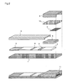
- FIG. 2 is a diagram illustrating a biosensor for performing a chromatography measurement according to the second embodiment.
- numeral 1 denotes a reactive layer carrier support for supporting a chromatography material, which is made of plastic or the like.
- Numeral 2 denotes a developing layer for developing a sample solution, which is made of nitrocellulose or the like
- numeral 3 denotes a marker reagent holding part where a dissoluble marker reagent is held so as to permeate the developing layer
- numeral 4 denotes a sample immobilization part as an area on the developing layer 2 where a sample such as a specific protein is immobilized
- numeral 5 denotes a water absorbing part for finally absorbing the sample solution
- numeral 6 denotes a sample holding part for temporarily holding an applied sample, which is made of a nonwoven fabric, glass fiber filter paper or the like having high hydrophilia
- numeral 7 denotes a retiform structure
- numeral 8 denotes a liquid-impermeable sheet
- numeral 9 denotes a material for forming a space in which the sample solution is sucked to be held
- numeral 10 denotes a cell contraction agent arranged between the space forming material 9 and the retiform structure 7
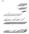
- FIG. 3 illustrates the construction which is different from that shown in FIG. 2 in that the sample holding part 6 is removed, and a sample solution is held in a cavity formed by the space forming material 9 .
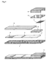
- FIG. 4 illustrates the construction which is different from that shown in FIG. 2 in that the marker reagent holding part 3 is removed, and the sample holding part 6 or the cell contraction agent holding part 10 also serves as the marker reagent holding part.
- the cell contraction agent 10 is provided between the space forming material 9 and the retiform structure 7 , and the edges of the end parts thereof are kept aligned.
- the cellular components in the liquid sample are constricted by the cell contraction agent.
- the retiform structure 7 is arranged on the sample holding part 6 , the sample solution efficiently reacts with the cell contraction agent while being agitated due to the retiform structure, and reaches the sample holding part. Therefore, the sample solution can permeate the developing layer without causing clogging, and smoothly permeate the biosensor in a state where the cellular components are mixed.
- the sample holding part 6 is not clogged by the cellular components. Therefore, there is no necessity to consider a material, a fiber density, or a pore size of the sample holding part 6 , thereby expanding the scope of selection.
- the biosensor shown in FIG. 3 is different from that shown in FIG. 2 in that the sample holding part 6 is removed, and the sample solution is held in the cavity formed by the space forming material 9 .
- the sample solution efficiently reacts with the cell contraction agent while being agitated through the influence of the retiform structure 7 . Further, there is no loss of sample solution that is caused by absorption in the sample holding part 6 , whereby a measurement can be performed with a smaller amount of sample solution to be applied.
- the biosensor shown in FIG. 4 is different from that shown in FIG. 2 in that the marker reagent holding part 3 is removed, and the sample holding part 6 or the cell contraction agent holding part 10 also serves as the marker reagent holding part 3 .
- the cell constriction reaction and the reaction between the analysis target and the marker reagent occur as soon as the sample is applied, whereby the reactions occur efficiently, and the time required for the reactions can be reduced.
- the cellular components in the sample solution efficiently constrict, and the sample solution quickly permeates the developing layer without causing clogging in a state where the cellular components are mixed therein, thereby realizing a simple, quick, and high-performance chromatography measurement with a smaller amount of sample solution without previously separating the cellular components such as blood cells.
- FIG. 7 is a top view of the retiform structure
- FIG. 8 is a side view thereof.
- meshes are regularly arranged.
- unevenness of the retiform structure generates a turbulent flow in the sample solution. This turbulent flow has an effect on the agitation between the sample solution and the cell contraction agent, resulting in a more efficient cell constriction reaction.
- a biosensor according to the present invention one which is made of a chromatography material composed of arbitrary porous carriers such as nitrocellulose and a nonwoven fabric or glass fiber filter paper is employed.
- the biosensor made of such material has the function of analytically detecting a specific material employing an arbitrary principle of measurement such as an antigen-antibody reaction, thereby qualitatively or quantitatively measuring the material.
- an antigen-antibody reaction employing the marker reagent occurs as an example, anything may be employed as long as it produces some change after the reaction, such as an enzyme.
- a biosensor made of an immunochromatography which includes an anti-CRP antibody A immobilization line and a broad band of a marker reagent composed of a complex of an anti-CRP antibody B and gold colloid on a nitrocellulose film is manufactured.
- the test specimen includes the antibody immobilization part 4 , the marker reagent holding part 3 as an area where the marker reagent which is the complex of an anti-CRP antibody B and gold colloid is held, the sample holding part 6 , the retiform structure 7 , and the sample introductory part 9 supporting the cell contraction agent holding part 10 .
- This biosensor is manufactured as follows.
- An anti-CRP antibody A solution which was diluted with a phosphate buffer solution to control the concentration was prepared. This antibody solution was applied on the nitrocellulose film by employing a solution discharge device. Thereby, a detecting antibody immobilization line was obtained on the nitrocellulose film. After being dried, this nitrocellulose film was immersed in a Tris-HCl buffer solution including 1% skim milk and shaken gently for 30 minutes. 30 minutes later, the film was moved into a Tris-HCl buffer solution tank, shaken gently for 10 minutes, and thereafter shaken gently in another Tris-HCl buffer solution tank for another 10 minutes, to wash the film. After washed twice, the film was taken out from the solution tank, and dried at room temperatures.
- the gold colloid was prepared by adding 1% citric acid solution to a refluxing 100° C.-solution of 0.01% chloroauric acid. After the reflux was continued for 30 minutes, it was cooled by being left at room temperatures.
- the anti-CRP antibody B was added to gold colloid solution which was prepared to pH9 by using 0.2M potassium carbonate solution, then the obtained solution was stirred for several minutes, and then 10% BSA (bovine serum albumin) solution of pH9 was added thereto by such an amount that 1% solution was finally obtained and stirred. Thereby, an antibody-gold colloid complex (marker antibody) was prepared.
- the marker antibody solution was centrifuged at 4° C. and 20000G for 50 minutes, whereby the marker antibody was isolated.
- the isolated marker antibody was suspended in a washing buffer solution (1% BSA ⁇ phosphate buffer solution) and thereafter further centrifuged under the above-described condition to wash and isolate the marker antibody.
- the marker antibody was suspended in a certain amount of washing buffer solution and filtrated through a 0.8 ⁇ m filter, thereby obtaining a marker antibody solution.
- the obtained marker antibody solution was prepared one-tenth as much as the gold colloid solution before being centrifuged, and stored at 4° C.
- the marker antibody solution was set in the solution discharge device and applied to a position on an anti-CRP antibody A immobilization dry film, apart from an antibody immobilization position, and thereafter the film was dried. Thereby, the marker reagent holding part was obtained on the immobilization film.
- the space forming material created by laminating a transparent PET of 100 ⁇ m thick was affixed to the film, thereby forming a cavity (5.0 mm wide ⁇ 12.0 mm long ⁇ 0.5 mm high).
- a potassium chloride solution prepared to 1.5M was dropped to the film, and thereafter the obtained film was immediately frozen with liquid nitrogen to be freeze-dried. Thereby, the space forming material having the shrinker holding part where the potassium chloride was impregnated was obtained.
- the antibody immobilization film including the marker reagent holding region prepared as described above was affixed on the reactive layer carrier support made of a while PET of 0.5 mm thick, the retifom tissue (a polyester mesh sheet), a nonwoven fabric as the sample holding part, and glass fiber filter paper as the water absorbing part were added thereto, and thereafter the film was cut into small pieces of 0.5 cm wide. After the cutting, the sample introductory part is affixed to the cut film, thereby manufacturing an immunochromatography test specimen. This is employed as a biosensor.
- FIGS. 5 and 6 are diagrams illustrating quantitative performances, FIG. 5 showing a case where the retiform structure 7 of the biosensor shown in FIG. 2 is not employed and FIG. 6 showing a case where the retiform structure 7 is employed as shown in FIG. 2.
- the abscissa represents the CRP concentration of a sample applied to the biosensor.
- the ordinate represents the converted value of the antigen concentration obtained by substituting the signal in the color area on the test specimen into the calibration curve.
- the immunochromatography test specimen which is made of a chromatography material composed of arbitrary porous carriers such as nitrocellulose and glass fiber filter paper is employed.
- the biosensor made of such material has the function of analytically detecting a specific material employing an arbitrary principle of measurement such as an antigen-antibody reaction, thereby qualitatively or quantitatively measuring the material.
- the marker reagent holding part supporting a marker reagent may be arranged on a porous carrier made of a material different from nitrocellulose, such as a nonwoven fabric, on a support body.
- gold colloid is employed as a marker composing the marker reagent, anything that produces some change after the reaction may be employed, such as a coloring material, a fluorescent material, a phosphorescent material, a light-emitting material, an oxidation-reduction material, an enzyme, a nucleic acid, and an endoplasmic reticulum.
- the water absorbing part may be excluded from the constituent members.
- sample solutions to be measured there are for example water, an aqueous solution, bodily fluids such as urine, blood, blood plasma, blood serum, and saliva, a solution in which a solid, fine particles, or gas is dissolved, and the like.
- Applications thereof include a urinalysis, a pregnancy test, a water examination, a fecal examination, soil analysis, food analysis and the like.
- CRP C-reactive protein
- it may be an antibody, immunoglobulin, hormone, a protein and a protein derivative such as an enzyme and peptide, a bacterium, a virus, the true fungi, mycoplasma, a parasite and a infectious material such as a product and a component thereof, a chemical drug such as a curative medicine and a drug of abuse, or a tumor marker.
- the analyte may be for example human chrionic gonadotropin (hCG), luteinizing hormone (LH), thyroid-stimulating hormone, follicular hormone, parathyroid hormone, adrenocorticotropic hormone, estradiol, prostatic specific antigen, Hepatitis B surface antigen, myoglobin, CRP, cardiac troponin, HbAlc, albumin or the like.
- hCG human chrionic gonadotropin
- LH luteinizing hormone
- thyroid-stimulating hormone follicular hormone
- parathyroid hormone adrenocorticotropic hormone
- estradiol estradiol
- prostatic specific antigen Hepatitis B surface antigen
- myoglobin CRP
- cardiac troponin HbAlc
- albumin albumin or the like.
- the permeability of the reactive layer carrier is enhanced, and a more uniform permeation is realized, thereby realizing a simple, quick, more sensitive, and higher-performance chromatography measurement.
- a biosensor according to the present invention is suited to enable a measurement with a small amount of specimen, for which whole blood with a reduced influence of blood cell can be used without previously separating blood plasma components from the blood.
Abstract
Description
- The present invention relates to a biosensor and, more particularly, to a dry-type biosensor for analyzing an analyte in a sample solution, and a method for analyzing a constituent of blood by employing the biosensor.
- As a means for diagnosing health conditions of persons, a biochemical examination of bodily fluids, especially blood, is widely conducted. A measuring method by a chromatography sensor, which utilizes an antigen-antibody reaction is generally used as a method for analyzing the bodily fluids. However, it is difficult to determine a kind or measure the concentration of blood constituent such as a metabolic product, a protein, a fat, an electrolyte, an enzyme, an antigen, and an antibody, by employing whole blood as it is. Thus, conventionally a general blood-constituent analysis method requires several operation processes, such as centrifuging blood which is previously collected, and analyzing the blood constituent employing obtained blood plasma or blood serum by an analytical instrument or a biosensor. However, when this method is employed, the measurement requires a special apparatus, and the preprocessing as well as the inspection take a lot of time. Thus, the centrifugation method which requires a centrifugal machine is not adequate when a small number of specimens are to be processed quickly or when inspections in the field is performed. Further, the obtained blood serum or blood plasma is smaller in amount as compared with the amount of blood.
- In recent years, a device which enables a quick, simple, and accurate measurement is desired under a concept of POC (Point of Care) in the medical diagnosis scene. However, in the conventional method as described above, in the case where a sample solution is to be applied to a sensor part, for example, when the sample solution is blood, a series of operations as described below is required. That is, blood is collected by the use of an injector, blood cells as material components and blood plasma are separated by the use of a centrifugal machine or the like in general, and the separated blood is applied to the sensor part with the use of an instrument such as a dispenser and a dropper. In this method, special skills in medical technology or the like are required to collect blood employing an injector, and further special apparatus and skills are required for the operation of centrifugal separation, and therefore this method cannot be employed in general households or when individuals without such techniques perform measurements for themselves. Further, since the instrument such as a dispenser is required to quantitatively measure the sample solution, the operation becomes complicated.
- Then, a method for separating blood plasma from whole blood by filtration has been considered. For example, there are a blood cell separation method employing glass fiber filter paper of a certain density as disclosed in Japanese Published Patent Application No. Sho.57-53661 and No. Hei.8-54387, and a blood adjustment method in which a water solution of inorganic salt or amino acid of a certain concentration is applied to whole blood and then blood cell components are filtered, as disclosed in Japanese Published Patent Application No. Hei.9-196908.
- In the method as disclosed in Japanese Published Patent Application No. Sho.57-53661 and No. Hei.8-54387, in order to more completely separate the blood cell components, glass fiber filter paper having an average diameter of 0.2˜5 μm and density of 0.1˜0.5 g/cm 3 is employed to make blood ooze therefrom, thereby obtaining separated blood plasma or blood serum. However, when this method is employed, while the efficiency of blood cell separation is surely enhanced, considerable time is required for almost completely separating the blood cells, and a large amount of blood is required to obtain a specimen in amount required for the inspection.
- Further, in the method as disclosed in Japanese Published Patent Application No. Hei.9-196908, in order to avoid clogging of the filtration material due to the blood cells, and to obtain a larger amount of blood plasma or blood serum component employing a smaller amount of blood, the water solution of amino acid or inorganic salt is mixed with whole blood, and thereafter the blood cell components are filtered. When this method is employed, operations of previously adding to obtained blood the water solution to be applied, and thereafter filtering the blood cell components are required, whereby the operation becomes complicated and the measurement takes time. Thus, inspections in emergency situations cannot be dealt with.
- Furthermore, in a method as disclosed in WO 01/92886A1, a cell contraction agent is employed to cause the blood cell components to constrict, and the constricted blood-cell components are developed on a chromatography carrier. When this method is employed, the constricted blood cell components can be developed with blood plasma components, thereby requiring no preprocessing on the specimen. However, it is difficult to uniformly constrict the blood cell components by the cell contraction agent, resulting in a poor measurement reproducibility.
- The present invention is made to solve the above-mentioned problems and has for its object to provide a simple, quick, and high-performance biosensor which can confirm the result of blood constituent analysis only by applying a slight amount of blood thereto, without requiring special apparatus, and a method for analyzing a blood constituent by employing the biosensor.
- According to
claim 1 of the present invention, there is provided a biosensor made of a dried porous material. The biosensor comprises: a sample introductory part for introducing a sample solution; and a developing layer for developing the sample solution, and in this biosensor the developing layer includes: a marker reagent holding part where a marker reagent is held in a dry state so that it can be eluted by the development of the sample solution; and a reagent immobilization part where a reagent which can bind to an analyte and is involved in a reaction is immobilized so that it is not eluted, and when the sample solution is introduced to the sample introductory part, the sample solution permeates the developing layer to reach the marker reagent holding part, and moves to the reagent immobilization part while eluting the marker reagent, whereby the reaction among the analyte, the marker reagent, and the immobilized reagent occurs, and an amount of marker reagent bound in the reagent immobilization part is measured, thereby qualitatively or quantitatively measuring the analyte included in the sample solution. In this biosensor a retiform structure is arranged in the sample introductory part. - According to the present invention, the applied sample solution is developed on the biosensor while being agitated by a turbulent flow caused by the retiform structure, whereby the permeability of a reactive layer carrier is enhanced, and a more uniform permeation is realized, resulting in a biosensor which enables a simple, quick, more sensitive, and higher-performance measurement.
- The retiform structure as mentioned above is formed by performing a molding process on a fiber or resin to reticulate the same, and it makes no difference whether the retiform structure itself has capillary activity or absorbability or not. The reticulum at this time may have any shape as long as it is polygonal, and the size thereof does not matter. It is preferable that meshes are arranged in regular sizes. Further, this retiform structure is preferably a single layer.
- Further, the turbulent flow is a flow in which a fluid irregularly moves in disorder and a stream line shows a fine and irregular fluctuation.
- According to
claim 2 of the present invention, there is provided a biosensor made of a dried porous material. The biosensor comprises: a developing layer for developing a sample solution; a space forming part for forming a space on the developing layer; and a sample introductory part having a cavity in which the sample solution flows, in the formed space, and in this biosensor the developing layer includes: a marker reagent holding part where a marker reagent is held in a dry state so that it can be eluted by the development of the sample solution; and a reagent immobilization part where a reagent which can bind to an analyte and is involved in a reaction is immobilized so that it is not eluted, and when the sample solution is introduced to the sample introductory part, the sample solution permeates the developing layer to reach the marker reagent holding part, and moves to the reagent immobilization part while eluting the marker reagent, whereby the reaction among the analyte, the marker reagent, and the immobilized reagent occurs, and an amount of marker reagent bound in the reagent immobilization part is measured, thereby qualitatively or quantitatively measuring the analyte included in the sample solution. In this biosensor a retiform structure is arranged in the sample introductory part. - According to the present invention, a specific amount of sample solution is sucked in the sample introductory part as the cavity formed by the space forming material, and the applied sample solution is developed on the biosensor while being efficiently agitated by a turbulent flow generated by the reriform tissue. Therefore, the permeability of a reactive layer carrier is enhanced, and a more uniform permeation is realized, resulting in a biosensor which enables a simple, quick, more sensitive, and higher-performance measurement.
- According to
claim 3 of the present invention, there is provided a biosensor made of a dried porous material. The biosensor comprises: a developing layer for developing a sample solution; a space forming part for forming a space on the developing layer; and a sample introductory part having a cavity in which the sample solution flows, in the formed space, and in this biosensor a reagent holding part where a marker reagent is held in a dry state so that it can be eluted by the flow of the sample solution is provided in the cavity, and the developing layer includes a reagent immobilization part where a reagent which can bind to an analyte and is involved in a reaction is immobilized so that it is not eluted, and when the sample solution is introduced to the sample introductory part, the sample solution is developed on the developing layer while eluting the marker reagent, and reaches the reagent immobilization part, whereby the reaction among the analyte, the marker reagent, and the immobilized reagent occurs, and an amount of marker reagent bound in the reagent immobilization part is measured, thereby qualitatively or quantitatively measuring the analyte included in the sample solution. In this biosensor a retiform structure is arranged in the sample introductory part. - According to the present invention, a specific amount of sample solution is sucked in the sample introductory part as the cavity formed by the space forming material, and the applied sample solution is developed on the biosensor while being efficiently agitated by a turbulent flow generated by the reriform tissue and, thus, more thoroughly reacting with the marker reagent. Therefore, the permeability of a reactive layer carrier is enhanced, and a more uniform permeation is realized, resulting in a biosensor which enables a simple, quick, more sensitive, and higher-performance measurement.
- According to
claim 4 of the present invention, in the biosensor as defined in any ofclaims 1 to 3, a cell contraction agent holding part for causing cellular components to constrict is provided in the sample introductory part or at a position on the sample introductory part side up to the marker reagent holding part. - According to the present invention, there is no need to previously remove the cellular components in the sample solution or fragmentizing the same, thereby realizing a biosesor which enables a simpler and quicker measurement.
- According to
claim 5 of the present invention, in the biosensor as defined in any ofclaims 1 to 4, the retiform structure is arranged at the end part of the sample introductory part. - According to the present invention, as soon as the sample is applied, the applied sample solution is developed on the biosensor while being efficiently agitated by a turbulent flow generated by the retiform structure, whereby a more uniform and effective cellular constriction is performed, and the sample solution permeates a reactive layer carrier uniformly, resulting in a biosensor which enables a simple, quick, more sensitive, and higher-performance measurement.
- According to
claim 6 of the present invention, in the biosensor as defined in any ofclaims 1 to 5, the retiform structure and the cell contraction agent holding part are arranged so that the edges of the end parts thereof are kept aligned. - According to the present invention, the introduced sample solution can immediately react with the cell contraction agent as well as receive the effect of turbulent flow at the introduction, whereby the sample solution can efficiently react with the cell contraction agent. Therefore, a more uniform and effective cellular constriction is performed, and the sample solution permeates a reactive layer carrier uniformly, resulting in a biosensor which enables a simple, quick, more sensitive, and higher-performance measurement.
- According to
claim 7 of the present invention, in the biosensor as defined in any ofclaims 1 to 6, a space enabling the sample solution to flow therein is arranged between the retiform structure and the cell contraction agent holding part. - According to the present invention, the sample solution is smoothly introduced, and a sufficient constriction reaction which occurs by means of the cell contraction agent and effect of turbulent flow due to the retiform structure can be obtained, whereby a more uniform and effective cellular constriction is performed, and the sample solution permeates a reactive layer carrier uniformly, resulting in a biosensor which enables a simple, quick, more sensitive, and higher-performance measurement.
- According to
claim 8 of the present invention, in the biosensor as defined in any ofclaims 1 to 7, the retiform structure is arranged so that warp threads thereof are parallel to the direction in which the sample is developed on the developing layer. - According to the present invention, the sample solution which is developed while being efficiently agitated by a turbulent flow is efficiently led in the direction of the development by a capillary phenomenon caused by warp threads of the retiform structure that extend in the direction of the sample development. Therefore, the sample solution permeates a reactive layer carrier uniformly, resulting in a biosensor which enables a simple, quick, more sensitive, and higher-performance measurement.
- The warp thread as mentioned above is a part of the retiform structure in which a molded product of a fiber or resin forming the reticulum extends in the direction of the development. It is preferable that the warp thread is regularly arranged so that it is parallel to the direction of the development. Directions of tissues extending in other directions than that described above do not matter.
- According to
claim 9 of the present invention, in the biosensor as defined in any ofclaims 1 to 8, the reitform tissue is made of synthetic resin. - According to the present invention, the retiform structure itself lacks the water retaining capacity, whereby the sample solution is well drained, and the applied sample solution is quickly developed on the biosensor while being efficiently agitated by a turbulent flow generated by the retiform structure. Therefore, a smaller amount of sample solution realizes a uniform permeation on a reactive layer carrier, resulting in a biosensor which enables a simple, quick, sensitive, and high-performance measurement.
- According to claim 10 of the present invention, in the biosensor as defined in any of
claims 1 to 9, the retiform structure is made of a chemical fiber such as polyester. - According to the present invention, the retiform structure itself lacks the water retaining capacity, whereby the sample solution is well drained, and the applied sample solution is efficiently and quickly developed on the biosensor by a capillary phenomenon, while being efficiently agitated by a turbulent flow generated by the retiform structure. Therefore, a smaller amount of sample solution realizes a uniform permeation on a reactive layer carrier, resulting in a biosensor which enables a simple, quick, sensitive, and high-performance measurement.
- The retiform structure as mentioned above is formed by performing a welding process such as a thermo-compression bonding and a press work on a chemical fiber, to reticulate the same.
- According to claim 11 of the present invention, in the biosensor as defined in
claim 10, the retiform structure is a fabric obtained by weaving the chemical fiber such as polyester. - According to the present invention, the retiform structure itself lacks the water retaining capacity, whereby the sample solution is well drained, and the applied sample solution is efficiently and quickly developed on the biosensor by a capillary phenomenon, while being efficiently agitated by a turbulent flow generated by the retiform structure. Therefore, a smaller amount of sample solution realizes a uniform permeation on a reactive layer carrier, resulting in a biosensor which enables a simple, quick, sensitive, and high-performance measurement.
- The retiform structure as mentioned above is a fabric or a knit formed by the process of weaving or knitting a chemical fiber.
- According to claim 12 of the present invention, in the biosensor as defined in any of
claims 9 to 11, the retiform structure is treated with a surfactant so that it can permeate. - According to the present invention, the retiform structure does not repel the liquid sample, and the applied sample solution is quickly developed on the biosensor while being efficiently agitated by a turbulent flow generated by the retiform structure. Therefore, a smaller amount of sample solution realizes a uniform permeation on a reactive layer carrier, resulting in a biosensor which enables a simple, quick, sensitive, and high-performance measurement.
- According to claim 13 of the present invention, in the biosensor as defined in any of
claims 1 to 12, a mesh of the retiform structure has a pore size of 0.1 mm to 2 mm. - According to the present invention, clogging by the cellular components is avoided, and the sample solution is efficiently agitated by a turbulent flow generated by the retiform structure. Therefore, a smaller amount of sample solution realizes a uniform permeation on a reactive layer carrier, resulting in a biosensor which enables a simple, quick, sensitive, and high-performance measurement.
- According to claim 14 of the present invention, in the biosensor as defined in any of
claims 1 to 3, the sample solution to be applied is blood. - According to the present invention, there is no need to previously subject blood to some processing, thereby realizing a biosensor enabling a simple, quick, sensitive, and high-performance measurement, by which a safer and more sanitary blood examination can be conducted.
- According to claim 15 of the present invention, in the biosensor as defined in any of
claims 1 to 3, the sample solution to be applied is a solution including bacteria. - According to the present invention, there is no need to subject the solution including bacteria to some processing, thereby realizing a biosensor which enables a safer, more sanitary, simple, quick, sensitive, and high-performance measurement.
- According to claim 16 of the present invention, in the biosensor as defined in
claim 4, the cell contraction agent is inorganic salt. - According to the present invention, the cellular components in the liquid solution are constricted, so that clogging by the cellular components in the liquid sample such as whole blood or a bacterial solution is avoided, thereby realizing a uniform permeation state. Therefore, a biosensor which is able to perform a more accurate measurement in a short time without inhibiting the reaction is realized. The inorganic salt as mentioned above is an inorganic compound including salt, such as sodium chloride, potassium chloride, and sodium phosphate.
- According to claim 17 of the present invention, in the biosensor as defined in
claim 4, the cell contraction agent is amino acid. - According to the present invention, the cellular components in the liquid solution are constricted, so that clogging by the cellular components in the liquid sample such as whole blood or a bacterial solution is avoided, thereby realizing a uniform permeation state. Therefore, a biosensor which is able to perform a more accurate measurement in a short time without inhibiting the reaction is realized. The amino acid as mentioned above is a compound which has a carboxyl group and an amino group in an identical molecule, such as glycin and glutamic acid, and further includes imino acid such as proline and hydroxyproline.
- According to claim 18 of the present invention, in the biosensor as defined in
claim 4, the cell contraction agent is saccharide. - According to the present invention, the cellular components in the liquid solution are constricted, so that clogging by the cellular components in the liquid sample such as whole blood or a bacterial solution is avoided, thereby realizing a uniform permeation state. Therefore, a biosensor which is able to perform a more accurate measurement in a short time without inhibiting the reaction is realized. The saccharide as mentioned above includes a glucide such as glucose, scrose, and trehalose, as well as sugar alcohol such as glucitol.
- According to claim 19 of the present invention, in the biosensor as defined in any of
claims 1 to 18, the biosensor is a one-step immunochromatography test specimen. - According to the present invention, many measurement targets can be measured by obtaining antibodies or antigens for the measurement targets, and the applied sample solution is developed on the biosensor while being efficiently agitated by a turbulent blow generated by the retiform structure. Therefore, the permeability of a reactive layer carrier is enhanced, and a more uniform permeation is realized, resulting in a biosensor which enables a simple, quick, more sensitive, and higher-performance measurement.
- The “one-step” as mentioned above represents the measurement operation which only requires the sample solution to be dropped to the test specimen without the need to preprocess the sample solution, and does not require a developing solution to develop the sample solution after the dropping, nor require a washing process. Further, the immunochromatography test specimen as mentioned above is a sensor for detecting an analyte in the sample solution on a carrier where chromatography development is performed, by utilizing an antigen-antibody reaction.
- According to claim 20 of the present invention, there is provided a method for analyzing a constituent of blood by employing a biosensor made of a dried porous material. The biosensor comprises: a sample introductory part for introducing a sample solution; and a developing layer for developing the sample solution, and in this biosensor the developing layer includes: a marker reagent holding part where a marker reagent is held in a dry state so that it can be eluted by the development of the sample solution; and a reagent immobilization part where a reagent which can bind to an analyte and is involved in a reaction is immobilized so that it is not eluted, and when the sample solution is introduced to the sample introductory part, the sample solution permeates the developing layer to reach the marker reagent holding part, and moves to the reagent immobilization part while eluting the marker reagent, whereby the reaction among the analyte, the marker reagent, and the immobilized reagent occurs. The analysis method comprises: measuring an amount of marker reagent bound in the reagent immobilization part, thereby qualitatively or quantitatively measuring the analyte included in the sample solution, and in this analysis method a retiform structure is arranged in the sample introductory part, and the blood is developed.
- According to the present invention, the applied sample solution is developed on the biosensor while being agitated by a turbulent flow caused by the retiform structure, whereby the permeability of a reactive layer carrier is enhanced, and a more uniform permeation is realized, resulting in a blood constituent analysis method which enables a simple, quick, more sensitive, and higher-performance measurement.
- The retiform structure as mentioned above is formed by performing a molding process on a fiber or resin to reticulate the same, and it makes no difference whether the retiform structure itself has capillary activity or absorbability or not. The reticulum at this time may have any shape as long as it is polygonal, and the size thereof does not matter. It is preferable that meshes are regularly arranged. Further, this retiform structure is preferably a single layer.
- Further, the turbulent flow is a flow in which a fluid irregularly moves in disorder and a stream line shows a fine and irregular fluctuation.
- According to claim 21 of the present invention, there is provided a method for analyzing a constituent-of blood by employing a biosensor made of a dried porous material. The biosensor comprises: a developing layer for developing a sample solution; a space forming part for forming a space on the developing layer; and a sample introductory part having a cavity in which the sample solution flows, in the formed space, and in this biosensor the developing layer includes: a marker reagent holding part where a marker reagent is held in a dry state so that it can be eluted by the development of the sample solution; and a reagent immobilization part where a reagent which can bind to an analyte and is involved in a reaction is immobilized so that it is not eluted, and when the sample solution is introduced to the sample introductory part, the sample solution permeates the developing layer to reach the marker reagent holding part, and moves to the reagent immobilization part while eluting the marker reagent, whereby the reaction among the analyte, the marker reagent, and the immobilized reagent occurs. The analysis method comprises: measuring an amount of marker reagent bound in the reagent immobilization part, thereby qualitatively or quantitatively measuring the analyte included in the sample solution, and in this analysis method a retiform structure is arranged in the sample introductory part, and the blood is developed.
- According to the present invention, a specific amount of sample solution is sucked in the sample introductory part as the cavity formed by the space forming material, and the applied sample solution is developed on the biosensor while being efficiently agitated by a turbulent flow generated by the reriform tissue. Therefore, the permeability of a reactive layer carrier is enhanced, and a more uniform permeation is realized, resulting in a blood constituent analysis method which enables a simple, quick, more sensitive, and higher-performance measurement.
- According to claim 22 of the present invention, there is provided a method for analyzing a constituent of blood by employing a biosensor made of a dried porous material. The biosensor comprises: a developing layer for developing a sample solution; a space forming part for forming a space on the developing layer; and a sample introductory part having a cavity in which the sample solution flows, in the formed space, and in this biosensor a reagent holding part where a marker reagent is held in a dry state so that it can be eluted by the flow of the sample solution is provided in the cavity, and the developing layer includes a reagent immobilization part where a reagent which can bind to an analyte and is involved in a reaction is immobilized so that it is not eluted, and when the sample solution is introduced to the sample introductory part, the sample solution is developed on the developing layer while eluting the marker reagent, and reaches the reagent immobilization part, whereby the reaction among the analyte, the marker reagent, and the immobilized reagent occurs. The analysis method comprises: measuring an amount of marker reagent bound in the reagent immobilization part, thereby qualitatively or quantitatively measuring the analyte included in the sample solution, and in this analysis method a retiform structure is arranged in the sample introductory part, and the blood is developed.
- According to the present invention, a specific amount of sample solution is sucked in the sample introductory part as the cavity formed by the space forming material, and the applied sample solution is developed on the biosensor while being efficiently agitated by a turbulent flow generated by the reriform tissue and, thus, more thoroughly reacting with the marker reagent. Therefore, the permeability of a reactive layer carrier is enhanced, and a more uniform permeation is realized, resulting in a blood constituent analysis method which enables a simple, quick, more sensitive, and higher-performance measurement.
- According to claim 23 of the present invention, in the blood constituent analysis method as defined in any of claims 20 to 22, a cell contraction agent holding part for causing cellular components to constrict is provided in the sample introductory part or at a position on the sample introductory part side up to the marker reagent holding part.
- According to the present invention, there is no need to previously remove the cellular components in the sample solution or fragmentizing the same, thereby realizing a blood constituent analysis method which enables a simpler and quicker measurement.
- According to claim 24 of the present invention, in the blood constituent analysis method as defined in claim 23, the retiform structure is arranged at the end part of the sample introductory part.
- According to the present invention, as soon as the sample is applied, the applied sample solution is developed on the biosensor while being efficiently agitated by a turbulent flow generated by the retiform structure, whereby a more uniform and effective cellular constriction is performed, and the sample solution permeates a reactive layer carrier uniformly, resulting in a blood constituent analysis method which enables a simple, quick, more sensitive, and higher-performance measurement.
- According to claim 25 of the present invention, in the blood constituent analysis method as defined in claim 24, the retiform structure and the cell contraction agent holding part are arranged so that the edges of the end parts thereof are kept aligned.
- According to the present invention, the introduced sample solution can immediately react with the cell contraction agent as well as receive the effect of turbulent flow at the introduction, whereby the sample solution can efficiently react with the cell contraction agent and is developed on the biosensor with cells therein constricted more uniformly and effectively. Therefore, the sample solution permeates a reactive layer carrier uniformly, resulting in a blood constituent analysis method which enables a simple, quick, more sensitive, and higher-performance measurement.
- According to claim 26 of the present invention, in the blood constituent analysis method as defined in claim 24, a space enabling the sample solution to flow therein is arranged between the retiform structure and the cell contraction agent holding part.
- According to the present invention, the sample solution is smoothly introduced, and a sufficient constriction reaction which occurs by means of the cell contraction agent and effect of turbulent flow due to the retiform structure can be obtained, whereby the sample solution is developed on the biosensor with cells therein constricted more uniformly and effectively. Therefore, the sample solution permeates a reactive layer carrier uniformly, resulting in a biosensor which enables a simple, quick, more sensitive, and higher-performance measurement.
- According to claim 27 of the present invention, in the blood constituent analysis method as defined in any of claims 20 to 26, the retiform structure is arranged so that warp threads thereof are parallel to the direction in which the sample is developed on the developing layer.
- According to the present invention, the sample solution which is developed while being efficiently agitated by a turbulent flow is efficiently led in the direction of the development by a capillary phenomenon caused by warp threads of the retiform structure that extend in the direction of the sample development. Therefore, the sample solution permeates a reactive layer carrier uniformly, resulting in a blood constituent analysis method which enables a simple, quick, more sensitive, and higher-performance measurement.
- The warp thread as mentioned above is a part of the retiform structure in which a molded product of a fiber or resin forming the reticulum extends in the direction of the development. It is preferable that the warp thread is regularly arranged so that it is parallel to the direction of the development. Directions of tissues extending in other directions than that described above do not matter.
- According to claim 28 of the present invention, in the blood constituent analysis method as defined in any of claims 20 to 27, the reitform tissue is made of synthetic resin.
- According to the present invention, the retiform structure itself lacks the water retaining capacity, whereby the sample solution is well drained, and the applied sample solution is quickly developed on the biosensor while being efficiently agitated by a turbulent flow generated by the retiform structure. Therefore, a smaller amount of sample solution realizes a uniform permeation on a reactive layer carrier, resulting in a blood constituent analysis method which enables a simple, quick, sensitive, and high-performance measurement.
- According to claim 29 of the present invention, in the blood constituent analysis method as defined in any of claims 20 to 28, the retiform structure is made of a chemical fiber such as polyester.
- According to the present invention, the retiform structure itself lacks the water retaining capacity, whereby the sample solution is well drained, and the applied sample solution is efficiently and quickly developed on the biosensor by a capillary phenomenon, while being efficiently agitated by a turbulent flow generated by the retiform structure. Therefore, a smaller amount of sample solution realizes a uniform permeation on a reactive layer carrier, resulting in a blood constituent analysis method which enables a simple, quick, more sensitive, and higher-performance measurement.
- Further, the retiform structure as mentioned above is formed by performing a welding process such as a thermo-compression bonding and a press work on a chemical fiber, to reticulate the same.
- According to claim 30 of the present invention, in the blood constituent analysis method as defined in claim 29, the retiform structure is a fabric obtained by weaving the chemical fiber such as polyester.
- According to the present invention, the retiform structure itself lacks the water retaining capacity, whereby the sample solution is well drained, and the applied sample solution is efficiently and quickly developed on the biosensor by a capillary phenomenon, while being efficiently agitated by a turbulent flow generated by the retiform structure. Therefore, a smaller amount of sample solution realizes a uniform permeation on a reactive layer carrier, resulting in a blood constituent analysis method which enables a simple, quick, more sensitive, and higher-performance measurement.
- Further, the retiform structure as mentioned above is a fabric or a knit formed by the process of weaving or knitting a chemical fiber.
- According to claim 31 of the present invention, in the blood constituent analysis method as defined in any of claims 28 to 30, the retiform structure is treated with a surfactant so that it can permeate.
- According to the present invention, the retiform structure does not repel the liquid sample, and the applied sample solution is quickly developed on the biosensor while being efficiently agitated by a turbulent flow generated by the retiform structure. Therefore, a smaller amount of sample solution realizes a uniform permeation on a reactive layer carrier, resulting in a blood constituent analysis-method which enables a simple, quick, sensitive, and high-performance measurement.
- According to claim 32 of the present invention, in the blood constituent analysis method as defined in any of claims 20 to 30, a mesh of the retiform structure has a pore size of 0.1 mm to 2 mm.
- According to the present invention, clogging by the cellular components is avoided, and the sample solution is efficiently agitated by a turbulent flow generated by the retiform structure. Therefore, a smaller amount of sample solution realizes a uniform permeation on a reactive layer carrier, resulting in a blood constituent analysis method which enables a simple, quick, sensitive, and high-performance measurement.
- According to claim 33 of the present invention, in the blood constituent analysis method as defined in any of claims 20 to 22, the sample solution to be applied is whole blood.
- According to the present invention, there is no need to previously subject blood to some processing, thereby realizing a blood constituent analysis method enabling a simple, quick, sensitive, and high-performance measurement, by which a safer and more sanitary blood examination can be conducted.
- According to claim 34 of the present invention, in the blood constituent analysis method as defined in claim 23, the cell contraction agent is inorganic salt.
- According to the present invention, the cellular components in the liquid solution are constricted, so that clogging by the cellular components in the liquid sample such as whole blood or a bacterial solution is avoided, thereby realizing a uniform permeation state. Therefore, a blood constituent analysis method by which a more accurate measurement can be performed in a short time without inhibiting the reaction is realized. The inorganic salt as mentioned above is an inorganic compound including salt, such as sodium chloride, potassium chloride, and sodium phosphate.
- According to claim 35 of the present invention, in the blood constituent analysis method as defined in claim 23, the cell contraction agent is amino acid.
- According to the present invention, the cellular components in the liquid solution are constricted, so that clogging by the cellular components in whole blood is avoided, thereby realizing a uniform permeation state. Therefore, a blood constituent analysis method by which a more accurate measurement can be performed in a short time without inhibiting the reaction is realized. The amino acid as mentioned above is a compound which has a carboxyl group and an amino group in an identical molecule, such as glycin and glutamic acid, and further includes imino acid such as proline and hydroxyproline.
- According to claim 36 of the present invention, in the blood constituent analysis method as defined in claim 23, the cell contraction agent is saccharide.
- According to the present invention, the cellular components in the liquid solution are constricted, so that clogging by the cellular components in whole blood is avoided, thereby realizing a uniform permeation state. Therefore, a blood constituent analysis method by which a more accurate measurement can be performed in a short time without inhibiting the reaction is realized. The saccharide as mentioned above includes a glucide such as glucose, scrose, and trehalose, as well as sugar alcohol such as glucitol.
- According to claim 37 of the present invention, in the blood constituent analysis method as defined in any of claims 20 to 22, the biosensor is a one-step immunochromatography test specimen.
- According to the present invention, many measurement targets can be measured by obtaining antibodies or antigens for the measurement targets, and the sample solution to be applied is subjected to no processing and no means is required after the sample application until the reaction end. The applied sample solution is developed on the biosensor while being efficiently agitated by a turbulent blow generated by the retiform structure. Therefore, the permeability of a reactive layer carrier is enhanced, and a more uniform permeation is realized, resulting in a blood constituent analysis method which enables a simple, quick, more sensitive, and higher-performance measurement.
- Further, the “one-step” as mentioned above represents the measurement operation which only requires the sample solution to be dropped to the test specimen without the need to preprocess the sample solution, and does not require a developing solution to develop the sample solution after the dropping, nor require a washing process. Further, the immunochromatography test specimen as mentioned above is a sensor for detecting an analyte in the sample solution on a carrier where chromatography development is performed, by utilizing an antigen-antibody reaction.
- As described above, according to the present invention, it is possible to realize a simple, quick, sensitive, and high-performance biosensor which requires no operation of previously removing the cellular components from the specimen including the cellular components, generates no clogging on the carrier by the cellular components, and has an enhanced permeability of the reactive layer carrier, and a blood constituent analysis method employing the biosensor.
- FIG. 1 is a diagram of a biosensor according to a first embodiment of the present invention.
- FIG. 2 is a diagram of a biosensor according to a second embodiment of the present invention.
- FIG. 3 is a diagram of the biosensor according to the second embodiment of the invention.
- FIG. 4 is a diagram of the biosensor according to the second embodiment of the invention.
- FIG. 5 is a diagram illustrating a quantitative performance of the biosensor in a case where a retiform structure is not employed as an example.
- FIG. 6 is a diagram illustrating a quantitative performance in a case where the biosensor according to the present invention is employed as an example.
- FIG. 7 is a top view of a retiform structure according to the present invention.
- FIG. 8 is a side view of the retiform structure shown in FIG. 7.
- Hereinafter, embodiments according to the present invention will be described with reference to the drawings. The embodiments described here are given only as examples and the present invention is not restricted to these embodiments.
- (Embodiment 1)
- A first embodiment of the present invention will be described with reference to the drawing.
- FIG. 1 is a diagram illustrating a biosensor for performing a chromatography measurement according to the first embodiment. In FIG. 1,
numeral 1 denotes a reactive layer carrier support for supporting a chromatography material, which is made of plastic or the like.Numeral 2 denotes a developing layer for developing a sample solution, which is made of nitrocellulose or the like, numeral 3 denotes a marker reagent holding part where a dissoluble marker reagent is held so as to permeate the developing layer, numeral 4 denotes a sample immobilization part as an area on the developinglayer 2 where a sample such as a specific protein is immobilized, numeral 5 denotes a water absorbing part for finally absorbing the sample solution, numeral 6 denotes a sample holding part for temporarily holding the applied sample, which is made of a nonwoven fabric, glass fiber filter paper or the like having high hydrophilia, numeral 7 denotes a retiform structure, numeral 8 denotes a liquid-impermeable sheet, numeral 9 denotes a material for forming a space in which the sample solution is sucked to be held, and numeral 11 denotes a sample introductory part where the sample solution is applied or sucked. Therespective regions 2˜11 are formed on the reactivelayer carrier support 1. Further, thesample holding part 6 may hold a cellular component shrinker. - Next, an operation of the biosensor according to the first embodiment will be described.
- On the biosensor shown in FIG. 1, when a sample solution is applied to the sample
introductory part 11, the sample solution reaches the area of thesample holding part 6, while being efficiently agitated by a turbulent flow caused by theretiform structure 7. The shrinker held in thearea 6 is dissolved by permeation of the sample solution and causes cellular components to constrict, and the sample solution in which the constricted cellular components are mixed permeates the developinglayer 2 and reaches the area of the markerreagent holding part 3. Then, the marker reagent held in the area of the markerreagent holding part 3 is dissolved by permeation of the sample solution, and is developed to reach thesample immobilization part 4. In thesample immobilization part 4, a binding reaction among the marker reagent dissolved from the area of the markerreagent holding part 3, an analysis target in the liquid sample, and the immobilized reagent occurs. At this time, when the analysis target exists in the liquid sample, some color reaction is seen in the area of thesample immobilization part 4. Finally, the sample solution is absorbed in thewater absorbing part 5, thereby ending the reaction. - Since the
retiform structure 7 is provided on the biosensor shown in FIG. 1, the cellular components in the sample solution reaches the sample holding part while being agitated. At this time, the dissolving cell contraction agent sufficiently reacts with the sample- solution in a cavity formed by thespace forming material 9 through the influence of the retiform structure. Accordingly, the cellular components in the sample solution constrict with the elution of the shrinker. Therefore, the sample solution can pass on the developing layer without causing clogging, and smoothly permeates the biosensor in a state where the cellular components are mixed therein. - As described above, according to the first embodiment, the cellular components in the sample solution efficiently constrict through the influence of the retiform structure included in the biosensor, and the sample solution quickly permeates the developing layer without causing clogging in a state where the cellular components are mixed therein, thereby realizing a simpler, quicker, and higher-performance chromatography measurement without previously separating the cellular components such as blood cells.
- (Embodiment 2)
- Hereinafter, a second embodiment of the present invention will be described with reference to FIGS. 2, 3 and 4.
- FIG. 2 is a diagram illustrating a biosensor for performing a chromatography measurement according to the second embodiment. In FIG. 2,
numeral 1 denotes a reactive layer carrier support for supporting a chromatography material, which is made of plastic or the like.Numeral 2 denotes a developing layer for developing a sample solution, which is made of nitrocellulose or the like, numeral 3 denotes a marker reagent holding part where a dissoluble marker reagent is held so as to permeate the developing layer, numeral 4 denotes a sample immobilization part as an area on the developinglayer 2 where a sample such as a specific protein is immobilized, numeral 5 denotes a water absorbing part for finally absorbing the sample solution, numeral 6 denotes a sample holding part for temporarily holding an applied sample, which is made of a nonwoven fabric, glass fiber filter paper or the like having high hydrophilia, numeral 7 denotes a retiform structure, numeral 8 denotes a liquid-impermeable sheet, numeral 9 denotes a material for forming a space in which the sample solution is sucked to be held, numeral 10 denotes a cell contraction agent arranged between thespace forming material 9 and theretiform structure 7, and numeral 11 denotes a sample introductory part where the sample solution is applied or sucked. Therespective regions 2˜11 are formed on the reactivelayer carrier support 1. - FIG. 3 illustrates the construction which is different from that shown in FIG. 2 in that the
sample holding part 6 is removed, and a sample solution is held in a cavity formed by thespace forming material 9. Further, FIG. 4 illustrates the construction which is different from that shown in FIG. 2 in that the markerreagent holding part 3 is removed, and thesample holding part 6 or the cell contractionagent holding part 10 also serves as the marker reagent holding part. - Next, an operation of the biosensor according to the second embodiment will be described.
- On each of the biosensors shown in FIGS. 2, 3, and 4, when a sample solution is applied to the sample
introductory part 11, the sample solution reaches the area of thesample holding part 6, while being efficiently agitated by a turbulent flow caused by theretiform structure 7. The shrinker held in thearea 6 is dissolved by permeation of the sample solution and causes cellular components to constrict, and the sample solution in which the constricted cellular components are mixed permeates the developinglayer 2 and reaches the area of the markerreagent holding part 3. Then, the marker reagent held in the area of the markerreagent holding part 3 is dissolved by permeation of the sample solution, and is developed to reach thesample immobilization part 4. In thesample immobilization part 4, a binding reaction among the marker reagent dissolved from the area of the markerreagent holding part 3, an analysis target in the liquid sample, and the immobilized reagent occurs. - At this time, when the analysis target solution exists in the liquid sample, some color reaction is seen in the area of the
sample immobilization part 4. Finally, the sample solution is absorbed in thewater absorbing part 5, thereby ending the reaction. - On the biosensor shown in FIG. 2, the
cell contraction agent 10 is provided between thespace forming material 9 and theretiform structure 7, and the edges of the end parts thereof are kept aligned. Thus, the moment when the sample solution is dropped, the cellular components in the liquid sample are constricted by the cell contraction agent. At this time, since theretiform structure 7 is arranged on thesample holding part 6, the sample solution efficiently reacts with the cell contraction agent while being agitated due to the retiform structure, and reaches the sample holding part. Therefore, the sample solution can permeate the developing layer without causing clogging, and smoothly permeate the biosensor in a state where the cellular components are mixed. Further, since there is no need for the reaction between the cellular components in the sample solution and the cell contraction agent in thesample holding part 6, thesample holding part 6 is not clogged by the cellular components. Therefore, there is no necessity to consider a material, a fiber density, or a pore size of thesample holding part 6, thereby expanding the scope of selection. - The biosensor shown in FIG. 3 is different from that shown in FIG. 2 in that the
sample holding part 6 is removed, and the sample solution is held in the cavity formed by thespace forming material 9. In this case, the sample solution efficiently reacts with the cell contraction agent while being agitated through the influence of theretiform structure 7. Further, there is no loss of sample solution that is caused by absorption in thesample holding part 6, whereby a measurement can be performed with a smaller amount of sample solution to be applied. - Further, the biosensor shown in FIG. 4 is different from that shown in FIG. 2 in that the marker
reagent holding part 3 is removed, and thesample holding part 6 or the cell contractionagent holding part 10 also serves as the markerreagent holding part 3. In this case, the cell constriction reaction and the reaction between the analysis target and the marker reagent occur as soon as the sample is applied, whereby the reactions occur efficiently, and the time required for the reactions can be reduced. - As described above, according to the second embodiment, through the influence of the retiform structure included in the biosensor, the cellular components in the sample solution efficiently constrict, and the sample solution quickly permeates the developing layer without causing clogging in a state where the cellular components are mixed therein, thereby realizing a simple, quick, and high-performance chromatography measurement with a smaller amount of sample solution without previously separating the cellular components such as blood cells.
- FIG. 7 is a top view of the retiform structure, and FIG. 8 is a side view thereof. As seen in FIGS. 7 and 8, meshes are regularly arranged. When a retiform structure composed of a single layer is employed, unevenness of the retiform structure generates a turbulent flow in the sample solution. This turbulent flow has an effect on the agitation between the sample solution and the cell contraction agent, resulting in a more efficient cell constriction reaction.
- As a biosensor according to the present invention, one which is made of a chromatography material composed of arbitrary porous carriers such as nitrocellulose and a nonwoven fabric or glass fiber filter paper is employed. The biosensor made of such material has the function of analytically detecting a specific material employing an arbitrary principle of measurement such as an antigen-antibody reaction, thereby qualitatively or quantitatively measuring the material. Further, the description has been given of the embodiments taking the case where the antigen-antibody reaction employing the marker reagent occurs as an example, anything may be employed as long as it produces some change after the reaction, such as an enzyme.
- According to the above-described embodiments, a more uniform permeation is realized, and thus it is possible to realize a simple, quick, sensitive, and high-performance chromatography measurement employing a small amount of specimen without the need to previously remove the cellular components.
- A method for implementing the present invention will be described in more detail through a following example. The present invention is not restricted to the following example.
- (Quantitative Analysis of CRP in Blood)
- A biosensor made of an immunochromatography which includes an anti-CRP antibody A immobilization line and a broad band of a marker reagent composed of a complex of an anti-CRP antibody B and gold colloid on a nitrocellulose film is manufactured. In the drawing, the test specimen includes the
antibody immobilization part 4, the markerreagent holding part 3 as an area where the marker reagent which is the complex of an anti-CRP antibody B and gold colloid is held, thesample holding part 6, theretiform structure 7, and the sampleintroductory part 9 supporting the cell contractionagent holding part 10. This biosensor is manufactured as follows. - a) Preparation of Biosensor
- An anti-CRP antibody A solution which was diluted with a phosphate buffer solution to control the concentration was prepared. This antibody solution was applied on the nitrocellulose film by employing a solution discharge device. Thereby, a detecting antibody immobilization line was obtained on the nitrocellulose film. After being dried, this nitrocellulose film was immersed in a Tris-HCl buffer solution including 1% skim milk and shaken gently for 30 minutes. 30 minutes later, the film was moved into a Tris-HCl buffer solution tank, shaken gently for 10 minutes, and thereafter shaken gently in another Tris-HCl buffer solution tank for another 10 minutes, to wash the film. After washed twice, the film was taken out from the solution tank, and dried at room temperatures.
- The gold colloid was prepared by adding 1% citric acid solution to a refluxing 100° C.-solution of 0.01% chloroauric acid. After the reflux was continued for 30 minutes, it was cooled by being left at room temperatures. The anti-CRP antibody B was added to gold colloid solution which was prepared to pH9 by using 0.2M potassium carbonate solution, then the obtained solution was stirred for several minutes, and then 10% BSA (bovine serum albumin) solution of pH9 was added thereto by such an amount that 1% solution was finally obtained and stirred. Thereby, an antibody-gold colloid complex (marker antibody) was prepared. The marker antibody solution was centrifuged at 4° C. and 20000G for 50 minutes, whereby the marker antibody was isolated. The isolated marker antibody was suspended in a washing buffer solution (1% BSA·phosphate buffer solution) and thereafter further centrifuged under the above-described condition to wash and isolate the marker antibody. The marker antibody was suspended in a certain amount of washing buffer solution and filtrated through a 0.8 μm filter, thereby obtaining a marker antibody solution. The obtained marker antibody solution was prepared one-tenth as much as the gold colloid solution before being centrifuged, and stored at 4° C.
- The marker antibody solution was set in the solution discharge device and applied to a position on an anti-CRP antibody A immobilization dry film, apart from an antibody immobilization position, and thereafter the film was dried. Thereby, the marker reagent holding part was obtained on the immobilization film.
- The space forming material created by laminating a transparent PET of 100 μm thick was affixed to the film, thereby forming a cavity (5.0 mm wide×12.0 mm long×0.5 mm high). A potassium chloride solution prepared to 1.5M was dropped to the film, and thereafter the obtained film was immediately frozen with liquid nitrogen to be freeze-dried. Thereby, the space forming material having the shrinker holding part where the potassium chloride was impregnated was obtained.
- The antibody immobilization film including the marker reagent holding region prepared as described above was affixed on the reactive layer carrier support made of a while PET of 0.5 mm thick, the retifom tissue (a polyester mesh sheet), a nonwoven fabric as the sample holding part, and glass fiber filter paper as the water absorbing part were added thereto, and thereafter the film was cut into small pieces of 0.5 cm wide. After the cutting, the sample introductory part is affixed to the cut film, thereby manufacturing an immunochromatography test specimen. This is employed as a biosensor.
- b) Preparation of Sample
- Human blood to which EDTA was added as an anticoagulant was prepared to have a hematocrit value of 45%. The CRP solutions of known concentrations were added to the blood, thereby preparing the CRP containing blood of various known concentrations.
- c) Measurement of the Degree of Coloration on Test Specimen
- On the immunochromatography test specimen, approximately 50 μl of whole blood including CRP was applied to the sample introductory part and developed in the direction of the water absorbing part, so that an antigen-antibody occurs, whereby a color reaction in the antibody immobilization part occurred. The
coloration state 5 minutes after the sample application to the immunochromatography test specimen was measured by employing a reflective spectrophotometer (CS9300; Shimadzu Corporation made), and the coloration degree was computed. - Whole blood including CRP of 0, 0.1, 1, and 10 mg/dl were applied to the immunochromatography test specimen to be developed. The coloration state of the judgement part on the immunochromatography test specimen for whole blood of each CRP concentration was measured by the reflective spectrophotometer. An absorbance at 635 nm was measured, and substituted into a previously formed calibration curve indicating a relationship between the CRP concentration and the absorbance. The result is shown in FIGS. 5 and 6. Essentially, when for example the absorbance in the case of whole blood including CRP of 1 mg/dl was measured, and the measured absorbance was substituted into the calibration curve, the CRP concentration should be 1 mg/dl. However, actually the CRP concentration slightly deviates from 1 mg/dl. The accuracy of measurement can be known by the amount of the deviation.
- FIGS. 5 and 6 are diagrams illustrating quantitative performances, FIG. 5 showing a case where the
retiform structure 7 of the biosensor shown in FIG. 2 is not employed and FIG. 6 showing a case where theretiform structure 7 is employed as shown in FIG. 2. The abscissa represents the CRP concentration of a sample applied to the biosensor. The ordinate represents the converted value of the antigen concentration obtained by substituting the signal in the color area on the test specimen into the calibration curve. - In the case where a biosensor which does not include the retiform structure is employed (FIG. 5), a CV value (coefficient of variation) ranges from 20 to 50% having a wide range of variations, which represents a low quantitative performance. On the other hand, in the case where a biosensor including the retiform structure is employed (FIG. 6), the CV value in each case is amazingly within 3 to 8%, which represents a sufficiently increased quantitative performance. From the above results, it can be understood that the arrangement of the retiform structure on the biosensor greatly concerns the enhancement in the quantitative performance.
- As a biosensor according to the embodiments of the present invention, the immunochromatography test specimen which is made of a chromatography material composed of arbitrary porous carriers such as nitrocellulose and glass fiber filter paper is employed. The biosensor made of such material has the function of analytically detecting a specific material employing an arbitrary principle of measurement such as an antigen-antibody reaction, thereby qualitatively or quantitatively measuring the material.
- While in this example the immunochromatography test specimen which is provided with the marker reagent holding part and the reagent immobilization part on an identical nitrocellulose film is employed, the marker reagent holding part supporting a marker reagent may be arranged on a porous carrier made of a material different from nitrocellulose, such as a nonwoven fabric, on a support body. While gold colloid is employed as a marker composing the marker reagent, anything that produces some change after the reaction may be employed, such as a coloring material, a fluorescent material, a phosphorescent material, a light-emitting material, an oxidation-reduction material, an enzyme, a nucleic acid, and an endoplasmic reticulum. Further, the water absorbing part may be excluded from the constituent members.
- As sample solutions to be measured, there are for example water, an aqueous solution, bodily fluids such as urine, blood, blood plasma, blood serum, and saliva, a solution in which a solid, fine particles, or gas is dissolved, and the like. Applications thereof include a urinalysis, a pregnancy test, a water examination, a fecal examination, soil analysis, food analysis and the like. While the description has been given of the example taking the C-reactive protein (CRP) as an example of the analyte, it may be an antibody, immunoglobulin, hormone, a protein and a protein derivative such as an enzyme and peptide, a bacterium, a virus, the true fungi, mycoplasma, a parasite and a infectious material such as a product and a component thereof, a chemical drug such as a curative medicine and a drug of abuse, or a tumor marker. Specifically, the analyte may be for example human chrionic gonadotropin (hCG), luteinizing hormone (LH), thyroid-stimulating hormone, follicular hormone, parathyroid hormone, adrenocorticotropic hormone, estradiol, prostatic specific antigen, Hepatitis B surface antigen, myoglobin, CRP, cardiac troponin, HbAlc, albumin or the like.
- According to the mode as described above, the permeability of the reactive layer carrier is enhanced, and a more uniform permeation is realized, thereby realizing a simple, quick, more sensitive, and higher-performance chromatography measurement.
- Applicability in Industry
- As described above, a biosensor according to the present invention is suited to enable a measurement with a small amount of specimen, for which whole blood with a reduced influence of blood cell can be used without previously separating blood plasma components from the blood.
Claims (37)
Applications Claiming Priority (2)
| Application Number | Priority Date | Filing Date | Title |
|---|---|---|---|
| JP2001243855 | 2001-08-10 | ||
| JP2001-243855 | 2001-08-10 |
Publications (1)
| Publication Number | Publication Date |
|---|---|
| US20030190690A1 true US20030190690A1 (en) | 2003-10-09 |
Family
ID=19073883
Family Applications (1)
| Application Number | Title | Priority Date | Filing Date |
|---|---|---|---|
| US10/398,711 Abandoned US20030190690A1 (en) | 2001-08-10 | 2002-08-12 | Biosensor and method for analyzing blood components using it |
Country Status (6)
| Country | Link |
|---|---|
| US (1) | US20030190690A1 (en) |
| EP (1) | EP1353181B1 (en) |
| JP (1) | JP3813150B2 (en) |
| KR (1) | KR100596278B1 (en) |
| CN (1) | CN1279357C (en) |
| WO (1) | WO2003014741A1 (en) |
Cited By (1)
| Publication number | Priority date | Publication date | Assignee | Title |
|---|---|---|---|---|
| US20110045579A1 (en) * | 2008-05-07 | 2011-02-24 | Panasonic Corporation | Biosensor |
Families Citing this family (20)
| Publication number | Priority date | Publication date | Assignee | Title |
|---|---|---|---|---|
| US20050103624A1 (en) | 1999-10-04 | 2005-05-19 | Bhullar Raghbir S. | Biosensor and method of making |
| PT1639352T (en) | 2003-06-20 | 2018-07-09 | Hoffmann La Roche | Method and reagent for producing narrow, homogenous reagent strips |
| US8148164B2 (en) | 2003-06-20 | 2012-04-03 | Roche Diagnostics Operations, Inc. | System and method for determining the concentration of an analyte in a sample fluid |
| US7718439B2 (en) | 2003-06-20 | 2010-05-18 | Roche Diagnostics Operations, Inc. | System and method for coding information on a biosensor test strip |
| US8206565B2 (en) | 2003-06-20 | 2012-06-26 | Roche Diagnostics Operation, Inc. | System and method for coding information on a biosensor test strip |
| US7645421B2 (en) | 2003-06-20 | 2010-01-12 | Roche Diagnostics Operations, Inc. | System and method for coding information on a biosensor test strip |
| US7645373B2 (en) | 2003-06-20 | 2010-01-12 | Roche Diagnostic Operations, Inc. | System and method for coding information on a biosensor test strip |
| US7452457B2 (en) | 2003-06-20 | 2008-11-18 | Roche Diagnostics Operations, Inc. | System and method for analyte measurement using dose sufficiency electrodes |
| US8071030B2 (en) | 2003-06-20 | 2011-12-06 | Roche Diagnostics Operations, Inc. | Test strip with flared sample receiving chamber |
| US8058077B2 (en) | 2003-06-20 | 2011-11-15 | Roche Diagnostics Operations, Inc. | Method for coding information on a biosensor test strip |
| JP4980884B2 (en) * | 2004-03-30 | 2012-07-18 | ジーイー・ヘルスケア・バイオサイエンス・コーポレイション | Lateral flow systems, materials, and methods |
| US7569126B2 (en) | 2004-06-18 | 2009-08-04 | Roche Diagnostics Operations, Inc. | System and method for quality assurance of a biosensor test strip |
| KR100841355B1 (en) * | 2006-04-04 | 2008-07-01 | 주식회사 올메디쿠스 | Biosensor chip provided with blood separation means |
| JP5221549B2 (en) | 2006-10-12 | 2013-06-26 | コーニンクレッカ フィリップス エレクトロニクス エヌ ヴィ | High-speed biosensor with reagent layer |
| US8088593B2 (en) | 2007-10-02 | 2012-01-03 | Theranos, Inc. | Modular point-of-care devices, systems, and uses thereof |
| JP5247158B2 (en) | 2008-01-16 | 2013-07-24 | パナソニック株式会社 | Sample solution analysis method and sample solution analyzer |
| CN111982829A (en) | 2011-01-21 | 2020-11-24 | 拉布拉多诊断有限责任公司 | System and method for maximizing sample usage |
| CN106372596A (en) * | 2016-08-30 | 2017-02-01 | 孟玲 | Biological information collection device |
| WO2018179983A1 (en) * | 2017-03-30 | 2018-10-04 | 帝人株式会社 | Blood cell separation membrane for immunochromatograph, and strip for immunochromatograph |
| CN110806489B (en) * | 2019-10-15 | 2023-04-25 | 南京大树生物医疗技术股份有限公司 | Anti-interference dry chemical combination detection device |
Citations (10)
| Publication number | Priority date | Publication date | Assignee | Title |
|---|---|---|---|---|
| US4259207A (en) * | 1979-09-19 | 1981-03-31 | American Hospital Supply Corporation | Suspending medium for immunologic reactions |
| US4789630A (en) * | 1985-10-04 | 1988-12-06 | Cetus Corporation | Ionic compounds containing the cationic meriquinone of a benzidine |
| US5073340A (en) * | 1987-10-08 | 1991-12-17 | Becton, Dickinson And Company | Depositing a binder on a solid support |
| US5166051A (en) * | 1990-08-08 | 1992-11-24 | Genesis Labs, Inc. | Membranes, membrane overlays for exclusion of erythrocytes, and method for immunoassay of whole blood analytes |
| US5558834A (en) * | 1991-10-03 | 1996-09-24 | Bayer Corporation | Device and method of seperating and assaying whole blood |
| US5628890A (en) * | 1995-09-27 | 1997-05-13 | Medisense, Inc. | Electrochemical sensor |
| US5985675A (en) * | 1997-12-31 | 1999-11-16 | Charm Sciences, Inc. | Test device for detection of an analyte |
| US6245577B1 (en) * | 1998-09-11 | 2001-06-12 | Midland Bioproducts Corporation | IgG antibody testing method |
| US20020100685A1 (en) * | 1999-11-11 | 2002-08-01 | Apex Biotechnology Corporation | Biosensor with multiple sampling ways |
| US20020137200A1 (en) * | 2000-06-01 | 2002-09-26 | Mie Takahashi | Biosensor and blood component analyzing method |
Family Cites Families (9)
| Publication number | Priority date | Publication date | Assignee | Title |
|---|---|---|---|---|
| JP3005303B2 (en) | 1991-01-31 | 2000-01-31 | 湧永製薬株式会社 | measuring device |
| JP2528350Y2 (en) * | 1992-03-24 | 1997-03-12 | ゼリア新薬工業株式会社 | Urine hormone detector |
| JPH07103973A (en) * | 1993-08-10 | 1995-04-21 | Dainippon Printing Co Ltd | Urine sampling member and urine inspection body |
| JPH0755809A (en) * | 1993-08-20 | 1995-03-03 | Arakusu:Kk | Hormone detector in urine |
| KR100292182B1 (en) * | 1997-09-18 | 2001-11-26 | 모리시타 요이찌 | Immunochromatography Devices |
| JP2001013141A (en) * | 1999-06-30 | 2001-01-19 | Nitto Denko Corp | Water-absorbent material for immunochromatography |
| JP4223163B2 (en) * | 1999-10-25 | 2009-02-12 | パナソニック株式会社 | Immunochromatographic test strip and chromatographic analysis method |
| TW416005B (en) * | 1999-11-11 | 2000-12-21 | Apex Biotechnology Corp | Biosensor with multiple sampling ways |
| CN100533147C (en) * | 2000-05-26 | 2009-08-26 | 松下电器产业株式会社 | Biosensor |
-
2002
- 2002-08-12 WO PCT/JP2002/008208 patent/WO2003014741A1/en not_active Application Discontinuation
- 2002-08-12 KR KR1020037005073A patent/KR100596278B1/en not_active IP Right Cessation
- 2002-08-12 EP EP02760610A patent/EP1353181B1/en not_active Expired - Lifetime
- 2002-08-12 CN CNB028026314A patent/CN1279357C/en not_active Expired - Fee Related
- 2002-08-12 JP JP2003519422A patent/JP3813150B2/en not_active Expired - Fee Related
- 2002-08-12 US US10/398,711 patent/US20030190690A1/en not_active Abandoned
Patent Citations (10)
| Publication number | Priority date | Publication date | Assignee | Title |
|---|---|---|---|---|
| US4259207A (en) * | 1979-09-19 | 1981-03-31 | American Hospital Supply Corporation | Suspending medium for immunologic reactions |
| US4789630A (en) * | 1985-10-04 | 1988-12-06 | Cetus Corporation | Ionic compounds containing the cationic meriquinone of a benzidine |
| US5073340A (en) * | 1987-10-08 | 1991-12-17 | Becton, Dickinson And Company | Depositing a binder on a solid support |
| US5166051A (en) * | 1990-08-08 | 1992-11-24 | Genesis Labs, Inc. | Membranes, membrane overlays for exclusion of erythrocytes, and method for immunoassay of whole blood analytes |
| US5558834A (en) * | 1991-10-03 | 1996-09-24 | Bayer Corporation | Device and method of seperating and assaying whole blood |
| US5628890A (en) * | 1995-09-27 | 1997-05-13 | Medisense, Inc. | Electrochemical sensor |
| US5985675A (en) * | 1997-12-31 | 1999-11-16 | Charm Sciences, Inc. | Test device for detection of an analyte |
| US6245577B1 (en) * | 1998-09-11 | 2001-06-12 | Midland Bioproducts Corporation | IgG antibody testing method |
| US20020100685A1 (en) * | 1999-11-11 | 2002-08-01 | Apex Biotechnology Corporation | Biosensor with multiple sampling ways |
| US20020137200A1 (en) * | 2000-06-01 | 2002-09-26 | Mie Takahashi | Biosensor and blood component analyzing method |
Cited By (2)
| Publication number | Priority date | Publication date | Assignee | Title |
|---|---|---|---|---|
| US20110045579A1 (en) * | 2008-05-07 | 2011-02-24 | Panasonic Corporation | Biosensor |
| US8303910B2 (en) | 2008-05-07 | 2012-11-06 | Panasonic Corporation | Biosensor |
Also Published As
| Publication number | Publication date |
|---|---|
| KR100596278B1 (en) | 2006-07-03 |
| CN1279357C (en) | 2006-10-11 |
| EP1353181B1 (en) | 2012-07-04 |
| JP3813150B2 (en) | 2006-08-23 |
| EP1353181A4 (en) | 2005-08-10 |
| JPWO2003014741A1 (en) | 2004-11-25 |
| WO2003014741A1 (en) | 2003-02-20 |
| KR20030036915A (en) | 2003-05-09 |
| CN1464977A (en) | 2003-12-31 |
| EP1353181A1 (en) | 2003-10-15 |
Similar Documents
| Publication | Publication Date | Title |
|---|---|---|
| EP1353181B1 (en) | Biosensor and method for analyzing blood components using it | |
| JP5479417B2 (en) | Controlled flow assay apparatus and method | |
| US5945345A (en) | Device for preventing assay interference using silver or lead to remove the interferant | |
| US7575915B2 (en) | Biosensor | |
| US7915051B2 (en) | Biosensor and blood component analytical method | |
| US6106779A (en) | Lysis chamber for use in an assay device | |
| JP2688529B2 (en) | Device and method for rapid qualitative or quantitative measurement of reactive ligand in fluid | |
| KR101254512B1 (en) | Immunochromatogaraphic test instrument and semiquantitative method using the same | |
| WO2002001227A1 (en) | Biosensor | |
| US20110045578A1 (en) | Method of manufacturing biosensor and biosensor produced thereby | |
| WO2003014726A1 (en) | A lateral flow plasma separation device | |
| JP2007139556A (en) | New analysis method and kit | |
| JPH09196920A (en) | Body fluid component analyzing instrument and analyzing method | |
| JP4980944B2 (en) | Immunological measurement method | |
| WO2005124347A1 (en) | Filter device, the method, kit and use thereof | |
| JP2010032396A (en) | Biosensor | |
| JPH08285849A (en) | Convenient measuring method and apparatus | |
| EP1202058A1 (en) | Chromatography measuring instrument | |
| JPH0255952A (en) | Dry chemical reagent release product, manufacture thereof and analysis method and test kit using the same | |
| JP2002207043A (en) | Immunoassay and immunoassay device | |
| JP2010091353A (en) | Biosensor and insoluble granular marker for biosensor | |
| Nadaoka et al. | Biosensor |
Legal Events
| Date | Code | Title | Description |
|---|---|---|---|
| AS | Assignment |
Owner name: MATSUSHITA ELECTRIC INDUSTRIAL CO., LTD., JAPAN Free format text: ASSIGNMENT OF ASSIGNORS INTEREST;ASSIGNORS:TAKAHASHI, MIE;NADAOKA, MASATAKA;TANAKA, HIROTAKA;AND OTHERS;REEL/FRAME:014149/0841;SIGNING DATES FROM 20030520 TO 20030521 |
|
| AS | Assignment |
Owner name: PANASONIC CORPORATION, JAPAN Free format text: CHANGE OF NAME;ASSIGNOR:MATSUSHITA ELECTRIC INDUSTRIAL CO., LTD.;REEL/FRAME:021897/0624 Effective date: 20081001 Owner name: PANASONIC CORPORATION,JAPAN Free format text: CHANGE OF NAME;ASSIGNOR:MATSUSHITA ELECTRIC INDUSTRIAL CO., LTD.;REEL/FRAME:021897/0624 Effective date: 20081001 |
|
| STCB | Information on status: application discontinuation |
Free format text: ABANDONED -- FAILURE TO RESPOND TO AN OFFICE ACTION |