EP3208612A1 - Compositions and methods for the treatment of immune related diseases - Google Patents
Compositions and methods for the treatment of immune related diseases Download PDFInfo
- Publication number
- EP3208612A1 EP3208612A1 EP17152167.7A EP17152167A EP3208612A1 EP 3208612 A1 EP3208612 A1 EP 3208612A1 EP 17152167 A EP17152167 A EP 17152167A EP 3208612 A1 EP3208612 A1 EP 3208612A1
- Authority
- EP
- European Patent Office
- Prior art keywords
- tigit
- amino acid
- antibody
- acid sequence
- cells
- Prior art date
- Legal status (The legal status is an assumption and is not a legal conclusion. Google has not performed a legal analysis and makes no representation as to the accuracy of the status listed.)
- Granted
Links
Images


Classifications
-
- G—PHYSICS
- G01—MEASURING; TESTING
- G01N—INVESTIGATING OR ANALYSING MATERIALS BY DETERMINING THEIR CHEMICAL OR PHYSICAL PROPERTIES
- G01N33/00—Investigating or analysing materials by specific methods not covered by groups G01N1/00 - G01N31/00
- G01N33/48—Biological material, e.g. blood, urine; Haemocytometers
- G01N33/50—Chemical analysis of biological material, e.g. blood, urine; Testing involving biospecific ligand binding methods; Immunological testing
- G01N33/53—Immunoassay; Biospecific binding assay; Materials therefor
- G01N33/564—Immunoassay; Biospecific binding assay; Materials therefor for pre-existing immune complex or autoimmune disease, i.e. systemic lupus erythematosus, rheumatoid arthritis, multiple sclerosis, rheumatoid factors or complement components C1-C9
-
- A—HUMAN NECESSITIES
- A61—MEDICAL OR VETERINARY SCIENCE; HYGIENE
- A61K—PREPARATIONS FOR MEDICAL, DENTAL OR TOILETRY PURPOSES
- A61K39/00—Medicinal preparations containing antigens or antibodies
- A61K39/395—Antibodies; Immunoglobulins; Immune serum, e.g. antilymphocytic serum
-
- A—HUMAN NECESSITIES
- A61—MEDICAL OR VETERINARY SCIENCE; HYGIENE
- A61P—SPECIFIC THERAPEUTIC ACTIVITY OF CHEMICAL COMPOUNDS OR MEDICINAL PREPARATIONS
- A61P1/00—Drugs for disorders of the alimentary tract or the digestive system
-
- A—HUMAN NECESSITIES
- A61—MEDICAL OR VETERINARY SCIENCE; HYGIENE
- A61P—SPECIFIC THERAPEUTIC ACTIVITY OF CHEMICAL COMPOUNDS OR MEDICINAL PREPARATIONS
- A61P1/00—Drugs for disorders of the alimentary tract or the digestive system
- A61P1/04—Drugs for disorders of the alimentary tract or the digestive system for ulcers, gastritis or reflux esophagitis, e.g. antacids, inhibitors of acid secretion, mucosal protectants
-
- A—HUMAN NECESSITIES
- A61—MEDICAL OR VETERINARY SCIENCE; HYGIENE
- A61P—SPECIFIC THERAPEUTIC ACTIVITY OF CHEMICAL COMPOUNDS OR MEDICINAL PREPARATIONS
- A61P17/00—Drugs for dermatological disorders
- A61P17/06—Antipsoriatics
-
- A—HUMAN NECESSITIES
- A61—MEDICAL OR VETERINARY SCIENCE; HYGIENE
- A61P—SPECIFIC THERAPEUTIC ACTIVITY OF CHEMICAL COMPOUNDS OR MEDICINAL PREPARATIONS
- A61P19/00—Drugs for skeletal disorders
- A61P19/02—Drugs for skeletal disorders for joint disorders, e.g. arthritis, arthrosis
-
- A—HUMAN NECESSITIES
- A61—MEDICAL OR VETERINARY SCIENCE; HYGIENE
- A61P—SPECIFIC THERAPEUTIC ACTIVITY OF CHEMICAL COMPOUNDS OR MEDICINAL PREPARATIONS
- A61P23/00—Anaesthetics
- A61P23/02—Local anaesthetics
-
- A—HUMAN NECESSITIES
- A61—MEDICAL OR VETERINARY SCIENCE; HYGIENE
- A61P—SPECIFIC THERAPEUTIC ACTIVITY OF CHEMICAL COMPOUNDS OR MEDICINAL PREPARATIONS
- A61P29/00—Non-central analgesic, antipyretic or antiinflammatory agents, e.g. antirheumatic agents; Non-steroidal antiinflammatory drugs [NSAID]
-
- A—HUMAN NECESSITIES
- A61—MEDICAL OR VETERINARY SCIENCE; HYGIENE
- A61P—SPECIFIC THERAPEUTIC ACTIVITY OF CHEMICAL COMPOUNDS OR MEDICINAL PREPARATIONS
- A61P31/00—Antiinfectives, i.e. antibiotics, antiseptics, chemotherapeutics
- A61P31/04—Antibacterial agents
-
- A—HUMAN NECESSITIES
- A61—MEDICAL OR VETERINARY SCIENCE; HYGIENE
- A61P—SPECIFIC THERAPEUTIC ACTIVITY OF CHEMICAL COMPOUNDS OR MEDICINAL PREPARATIONS
- A61P35/00—Antineoplastic agents
-
- A—HUMAN NECESSITIES
- A61—MEDICAL OR VETERINARY SCIENCE; HYGIENE
- A61P—SPECIFIC THERAPEUTIC ACTIVITY OF CHEMICAL COMPOUNDS OR MEDICINAL PREPARATIONS
- A61P37/00—Drugs for immunological or allergic disorders
-
- A—HUMAN NECESSITIES
- A61—MEDICAL OR VETERINARY SCIENCE; HYGIENE
- A61P—SPECIFIC THERAPEUTIC ACTIVITY OF CHEMICAL COMPOUNDS OR MEDICINAL PREPARATIONS
- A61P37/00—Drugs for immunological or allergic disorders
- A61P37/02—Immunomodulators
-
- A—HUMAN NECESSITIES
- A61—MEDICAL OR VETERINARY SCIENCE; HYGIENE
- A61P—SPECIFIC THERAPEUTIC ACTIVITY OF CHEMICAL COMPOUNDS OR MEDICINAL PREPARATIONS
- A61P43/00—Drugs for specific purposes, not provided for in groups A61P1/00-A61P41/00
-
- C—CHEMISTRY; METALLURGY
- C07—ORGANIC CHEMISTRY
- C07K—PEPTIDES
- C07K14/00—Peptides having more than 20 amino acids; Gastrins; Somatostatins; Melanotropins; Derivatives thereof
- C07K14/435—Peptides having more than 20 amino acids; Gastrins; Somatostatins; Melanotropins; Derivatives thereof from animals; from humans
- C07K14/46—Peptides having more than 20 amino acids; Gastrins; Somatostatins; Melanotropins; Derivatives thereof from animals; from humans from vertebrates
- C07K14/47—Peptides having more than 20 amino acids; Gastrins; Somatostatins; Melanotropins; Derivatives thereof from animals; from humans from vertebrates from mammals
- C07K14/4701—Peptides having more than 20 amino acids; Gastrins; Somatostatins; Melanotropins; Derivatives thereof from animals; from humans from vertebrates from mammals not used
- C07K14/4702—Regulators; Modulating activity
-
- C—CHEMISTRY; METALLURGY
- C07—ORGANIC CHEMISTRY
- C07K—PEPTIDES
- C07K14/00—Peptides having more than 20 amino acids; Gastrins; Somatostatins; Melanotropins; Derivatives thereof
- C07K14/435—Peptides having more than 20 amino acids; Gastrins; Somatostatins; Melanotropins; Derivatives thereof from animals; from humans
- C07K14/705—Receptors; Cell surface antigens; Cell surface determinants
- C07K14/70503—Immunoglobulin superfamily
-
- C—CHEMISTRY; METALLURGY
- C07—ORGANIC CHEMISTRY
- C07K—PEPTIDES
- C07K16/00—Immunoglobulins [IGs], e.g. monoclonal or polyclonal antibodies
- C07K16/18—Immunoglobulins [IGs], e.g. monoclonal or polyclonal antibodies against material from animals or humans
-
- C—CHEMISTRY; METALLURGY
- C07—ORGANIC CHEMISTRY
- C07K—PEPTIDES
- C07K16/00—Immunoglobulins [IGs], e.g. monoclonal or polyclonal antibodies
- C07K16/18—Immunoglobulins [IGs], e.g. monoclonal or polyclonal antibodies against material from animals or humans
- C07K16/28—Immunoglobulins [IGs], e.g. monoclonal or polyclonal antibodies against material from animals or humans against receptors, cell surface antigens or cell surface determinants
- C07K16/2803—Immunoglobulins [IGs], e.g. monoclonal or polyclonal antibodies against material from animals or humans against receptors, cell surface antigens or cell surface determinants against the immunoglobulin superfamily
-
- G—PHYSICS
- G01—MEASURING; TESTING
- G01N—INVESTIGATING OR ANALYSING MATERIALS BY DETERMINING THEIR CHEMICAL OR PHYSICAL PROPERTIES
- G01N33/00—Investigating or analysing materials by specific methods not covered by groups G01N1/00 - G01N31/00
- G01N33/48—Biological material, e.g. blood, urine; Haemocytometers
- G01N33/50—Chemical analysis of biological material, e.g. blood, urine; Testing involving biospecific ligand binding methods; Immunological testing
- G01N33/5005—Chemical analysis of biological material, e.g. blood, urine; Testing involving biospecific ligand binding methods; Immunological testing involving human or animal cells
- G01N33/5008—Chemical analysis of biological material, e.g. blood, urine; Testing involving biospecific ligand binding methods; Immunological testing involving human or animal cells for testing or evaluating the effect of chemical or biological compounds, e.g. drugs, cosmetics
- G01N33/5044—Chemical analysis of biological material, e.g. blood, urine; Testing involving biospecific ligand binding methods; Immunological testing involving human or animal cells for testing or evaluating the effect of chemical or biological compounds, e.g. drugs, cosmetics involving specific cell types
- G01N33/5047—Cells of the immune system
- G01N33/505—Cells of the immune system involving T-cells
-
- G—PHYSICS
- G01—MEASURING; TESTING
- G01N—INVESTIGATING OR ANALYSING MATERIALS BY DETERMINING THEIR CHEMICAL OR PHYSICAL PROPERTIES
- G01N33/00—Investigating or analysing materials by specific methods not covered by groups G01N1/00 - G01N31/00
- G01N33/48—Biological material, e.g. blood, urine; Haemocytometers
- G01N33/50—Chemical analysis of biological material, e.g. blood, urine; Testing involving biospecific ligand binding methods; Immunological testing
- G01N33/53—Immunoassay; Biospecific binding assay; Materials therefor
- G01N33/569—Immunoassay; Biospecific binding assay; Materials therefor for microorganisms, e.g. protozoa, bacteria, viruses
- G01N33/56966—Animal cells
- G01N33/56972—White blood cells
-
- A—HUMAN NECESSITIES
- A61—MEDICAL OR VETERINARY SCIENCE; HYGIENE
- A61K—PREPARATIONS FOR MEDICAL, DENTAL OR TOILETRY PURPOSES
- A61K39/00—Medicinal preparations containing antigens or antibodies
- A61K2039/505—Medicinal preparations containing antigens or antibodies comprising antibodies
-
- C—CHEMISTRY; METALLURGY
- C07—ORGANIC CHEMISTRY
- C07K—PEPTIDES
- C07K2317/00—Immunoglobulins specific features
- C07K2317/30—Immunoglobulins specific features characterized by aspects of specificity or valency
- C07K2317/33—Crossreactivity, e.g. for species or epitope, or lack of said crossreactivity
-
- C—CHEMISTRY; METALLURGY
- C07—ORGANIC CHEMISTRY
- C07K—PEPTIDES
- C07K2317/00—Immunoglobulins specific features
- C07K2317/30—Immunoglobulins specific features characterized by aspects of specificity or valency
- C07K2317/34—Identification of a linear epitope shorter than 20 amino acid residues or of a conformational epitope defined by amino acid residues
-
- C—CHEMISTRY; METALLURGY
- C07—ORGANIC CHEMISTRY
- C07K—PEPTIDES
- C07K2317/00—Immunoglobulins specific features
- C07K2317/50—Immunoglobulins specific features characterized by immunoglobulin fragments
- C07K2317/51—Complete heavy chain or Fd fragment, i.e. VH + CH1
-
- C—CHEMISTRY; METALLURGY
- C07—ORGANIC CHEMISTRY
- C07K—PEPTIDES
- C07K2317/00—Immunoglobulins specific features
- C07K2317/50—Immunoglobulins specific features characterized by immunoglobulin fragments
- C07K2317/515—Complete light chain, i.e. VL + CL
-
- C—CHEMISTRY; METALLURGY
- C07—ORGANIC CHEMISTRY
- C07K—PEPTIDES
- C07K2317/00—Immunoglobulins specific features
- C07K2317/50—Immunoglobulins specific features characterized by immunoglobulin fragments
- C07K2317/56—Immunoglobulins specific features characterized by immunoglobulin fragments variable (Fv) region, i.e. VH and/or VL
- C07K2317/565—Complementarity determining region [CDR]
-
- C—CHEMISTRY; METALLURGY
- C07—ORGANIC CHEMISTRY
- C07K—PEPTIDES
- C07K2317/00—Immunoglobulins specific features
- C07K2317/70—Immunoglobulins specific features characterized by effect upon binding to a cell or to an antigen
- C07K2317/73—Inducing cell death, e.g. apoptosis, necrosis or inhibition of cell proliferation
-
- C—CHEMISTRY; METALLURGY
- C07—ORGANIC CHEMISTRY
- C07K—PEPTIDES
- C07K2317/00—Immunoglobulins specific features
- C07K2317/70—Immunoglobulins specific features characterized by effect upon binding to a cell or to an antigen
- C07K2317/75—Agonist effect on antigen
-
- C—CHEMISTRY; METALLURGY
- C07—ORGANIC CHEMISTRY
- C07K—PEPTIDES
- C07K2317/00—Immunoglobulins specific features
- C07K2317/70—Immunoglobulins specific features characterized by effect upon binding to a cell or to an antigen
- C07K2317/76—Antagonist effect on antigen, e.g. neutralization or inhibition of binding
-
- Y—GENERAL TAGGING OF NEW TECHNOLOGICAL DEVELOPMENTS; GENERAL TAGGING OF CROSS-SECTIONAL TECHNOLOGIES SPANNING OVER SEVERAL SECTIONS OF THE IPC; TECHNICAL SUBJECTS COVERED BY FORMER USPC CROSS-REFERENCE ART COLLECTIONS [XRACs] AND DIGESTS
- Y02—TECHNOLOGIES OR APPLICATIONS FOR MITIGATION OR ADAPTATION AGAINST CLIMATE CHANGE
- Y02A—TECHNOLOGIES FOR ADAPTATION TO CLIMATE CHANGE
- Y02A50/00—TECHNOLOGIES FOR ADAPTATION TO CLIMATE CHANGE in human health protection, e.g. against extreme weather
- Y02A50/30—Against vector-borne diseases, e.g. mosquito-borne, fly-borne, tick-borne or waterborne diseases whose impact is exacerbated by climate change
Definitions
- the present invention relates to compositions and methods useful for the diagnosis and treatment of immune related diseases.
- Immune related and inflammatory diseases are the manifestation or consequence of fairly complex, often multiple interconnected biological pathways which in normal physiology are critical to respond to insult or injury, initiate repair from insult or injury, and mount innate and acquired defense against foreign organisms. Disease or pathology occurs when these normal physiological pathways cause additional insult or injury either as directly related to the intensity of the response, as a consequence of abnormal regulation or excessive stimulation, as a reaction to self, or as a combination of these.
- therapeutic intervention can occur by either antagonism of a detrimental process/pathway or stimulation of a beneficial process/pathway.
- immune-mediated inflammatory diseases include immune-mediated inflammatory diseases, non-immune-mediated inflammatory diseases, infectious diseases, immunodeficiency diseases, neoplasia, etc .
- T lymphocytes are an important component of a mammalian immune response. T cells recognize antigens which are associated with a self-molecule encoded by genes within the major histocompatibility complex (MHC). The antigen may be displayed together with MHC molecules on the surface of antigen presenting cells, virus infected cells, cancer cells, grafts, etc. The T cell system eliminates these altered cells which pose a health threat to the host mammal. T cells include helper T cells and cytotoxic T cells. Helper T cells proliferate extensively following recognition of an antigen -MHC complex on an antigen presenting cell. Helper T cells also secrete a variety of cytokines, i .
- MHC major histocompatibility complex
- helper T cells which play a central role in the activation ofB cells, cytotoxic T cells and a variety of other cells which participate in the immune response.
- T Fh follicular helper T cells
- CXC-chemokine receptor 5 Schaerli et al., J. Exp. Med. 192: 1553-62 (2000 )
- T Fh cells provide assistance to germinal-center B cells, particularly aiding the survival and propagation of B cells and potently inducing antibody production during coculture with B cells. They have also been implicated in tolerogenesis.
- T reg Regulatory T cells
- helper T cells are a subset of helper T cells that play a critical role in inhibition of self-reactive immune responses and are often found in sites of chronic inflammation such as in tumor tissue ( Wang, H.Y. & Wang, R.F., Curr Opin Immunol 19, 217-23 (2007 )).
- T regs are defined phenotypically by high cell surface expression of CD25, CLTA4, GITR, and neuropilin-1 ( Read, S., Malmstrom, V. & Powrie, F., J Exp Med 192, 295-302 (2000 ); Sakaguchi, S., et al., J Immunol 155, 1151-64 (1995 ); Takahashi, T.
- T regs perform their suppressive function on activated T cells through contact-dependent mechanism and cytokine production ( Fehervari, Z. & Sakaguchi, Curr Opin Immunol 16, 203-8 (2004 )).
- T regs also modulate immune responses by direct interaction with ligands on dendritic cells (DC), such as CTLA4 interaction with B7 molecules on DC that elicits the induction of indoleamine 2,3-dioxygenase (IDO) ( Fallarino, F. et al., Nat Immunol 4, 1206-12 (2003 )), and CD40L ligation ( Serra, P. et al., Immunity 19, 877-89 (2003 )).
- DCs are professional antigenpresenting cells capable of inducing immunity or tolerance against self or non-self antigens.
- DC-expanded T regs suppress alloreactivity responses in vitro ( Yamazaki, S.
- CTLA4 and GITR are representative of ligands defined within the CD28-B7 and TNF-superfamilies of co-stimulatory/-inhibitory molecules, respectively ( Greenwald, R.J., et al., Annu Rev Immunol 23, 515-48 (2005 )). These molecules are high on T regs but are also typically upregulated on activated T cells. In order to search for new costimulatory molecules expressed in T reg cells searches were performed to identify genes specifically expressed in T cells ( Abbas, A.R. et al., Genes Immun 6, 319-31 (2005 )) that had both Ig domains and immunoreceptor tyrosine-based activation or inhibition (ITAM/ITIM) motifs.
- ITAM/ITIM immunoreceptor tyrosine-based activation or inhibition
- TIGIT for T-Cell-Ig and ITIM domain
- TIGIT for T-Cell-Ig and ITIM domain
- the present invention concerns compositions and methods useful for the diagnosis and treatment of immune related disease in mammals, including humans.
- the present invention is based on the identification of proteins involved in the negative regulation of proliferation and function of certain types of immune cells.
- Immune related diseases can be treated by suppressing or enhancing the immune response. Molecules that enhance the immune response stimulate or potentiate the immune response to an antigen. Molecules which stimulate the immune response can be used therapeutically where enhancement of the immune response would be beneficial. Alternatively, molecules that suppress the immune response attenuate or reduce the immune response to an antigen (e.g ., neutralizing antibodies) can be used therapeutically where attenuation of the immune response would be beneficial ( e.g ., inflammation).
- an antigen e.g ., neutralizing antibodies
- TIGIT for "T-Cell-Ig and ITIM domain" protein specifically binds to poliovirus receptor (PVR, also known as CD155) and several other members of a newly elucidated protein family, and that this TIGIT-PVR interaction negatively regulates T cell activation and proliferation.
- PVR poliovirus receptor
- TIGIT polypeptides, agonists thereof, and antagonists thereof, as well as PVR polypeptides, agonists thereof and antagonists thereof are useful to prepare medicines and medicaments for the treatment of immune-related and inflammatory diseases.
- the invention also provides methods of treating immune-related and inflammatory diseases and methods and compositions for detecting and assessing the status of immune-related and inflammatory diseases.
- the invention provides an isolated polypeptide comprising an amino acid sequence comprising one or more of the following amino acids: an alanine at amino acid position corresponding to amino acid position 67 of human TIGIT, a glycine at an amino acid position corresponding to amino acid position 74 of human TIGIT, a proline at an amino acid position corresponding to amino acid position 114 of human TIGIT, and a glycine at an amino acid position corresponding to amino acid position 116 of human TIGIT.
- the polypeptide is not PVR, PVRL1, PVRL2, PVRL3, PVRL4, TIGIT, CD96, or CD226.
- the polypeptide further comprises one or more of: an amino acid selected from valine, isoleucine, and leucine at an amino acid position corresponding to amino acid position 54 of human TIGIT, an amino acid selected from serine and threonine at an amino acid position corresponding to amino acid position 55 of human TIGIT, a glutamine at an amino acid position corresponding to amino acid position 56 of human TIGIT, a threonine at an amino acid position corresponding to amino acid position 112 of human TIGIT, and an amino acid selected from phenylalanine and tyrosine at an amino acid position corresponding to amino acid position 113 of human TIGIT.
- the polypeptide further comprises one or more structural submotifs selected from the following:
- the invention provides a method of determining whether a test polypeptide is a member of the TLP family of polypeptides comprising aligning the amino acid sequence of the test polypeptide with an amino acid sequence of one or more members of the TLP family of polypeptides and assessing the presence or absence in the test polypeptide amino acid sequence of one or more of an alanine at amino acid position corresponding to amino acid position 67 of human TIGIT, a glycine at an amino acid position corresponding to amino acid position 74 of human TIGIT, a proline at an amino acid position corresponding to amino acid position 114 of human TIGIT, and a glycine at an amino acid position corresponding to amino acid position 116 of human TIGIT.
- the invention provides a method for identifying one or more members of the TLP protein family by identifying proteins in one or more sequence databases whose amino acid sequences comprise at least one amino acid selected from an alanine at amino acid position corresponding to amino acid position 67 of human TIGIT, a glycine at an amino acid position corresponding to amino acid position 74 of human TIGIT, a proline at an amino acid position corresponding to amino acid position 114 of human TIGIT, and a glycine at an amino acid position corresponding to amino acid position 116 of human TIGIT.
- the invention provides an isolated agent that specifically interacts with one or more conserved or substantially conserved regions of TLP family members.
- the agent is an antagonist of the expression and/or activity of a TLP family member.
- the antagonist is selected from a small molecule inhibitor, an inhibitory antibody or antigen-binding fragment thereof, an aptamer, an inhibitory nucleic acid, and an inhibitory palypeptide.
- the agent is an agonist of the expression and/or activity of a TLP family member.
- the agonist is selected from an agonizing antibody or antigen-binding fragment thereof, an agonizing peptide, and a small molecule or protein that activates TIGIT binding to PVR and/or TIGIT intracellular signaling mediated by PVR.
- the invention provides a method of identifying or detecting one or more TLP family members by contacting a putative TLP family member polypeptide with at least one of the above agents and determining the binding of the at least one agent to the putative TLP family member.
- the invention provides a method of determining whether a test immune cell is an activated or normal T reg , memory T cell, NK cell, or T Fh cell, comprising assessing the level of expression of TIGIT in the test immune cell and comparing it to the level of expression of TIGIT in a known activated or normal Treg, memory T cell, NK cell, or TFh cell, or by comparing the level of expression of TIGIT in the test immune cell to known standard TIGIT expression value(s).
- the invention provides a method for modulating immune system function and/or activity comprising modulating the binding of TIGIT to one or more of PVR, PVRL3, and PVRL2.
- the invention provides an anti-TIGIT antibody or a fragment thereof comprising at least one HVR comprising an amino acid sequence selected from the amino acid sequences set forth in SEQ ID NOs: 23-28. In another embodiment, the invention provides an anti-TIGIT antibody or a fragment thereof comprising at least one HVR comprising an amino acid sequence selected from the amino acid sequences set forth in SEQ ID NOs: 31-36. In another embodiment, the invention provides an anti-TIGIT antibody or a fragment thereof wherein the antibody light chain comprises the amino acid sequence set forth in SEQ ID NO: 21. In another embodiment, the invention provides an anti-TIGIT antibody or a fragment thereof wherein the antibody light chain comprises the amino acid sequence set forth in SEQ ID NO: 29.
- the invention provides an anti-TIGIT antibody or a fragment thereof wherein the antibody heavy chain comprises the amino acid sequence set forth in SEQ ID NO: 22 or a portion thereof. In another embodiment, the invention provides an anti-TIGIT antibody or a fragment thereof wherein the antibody heavy chain comprises the amino acid sequence set forth in SEQ ID NO: 30 or a portion thereof. In another embodiment, the invention provides an anti-TIGIT antibody or a fragment thereof wherein the antibody light chain comprises the amino acid sequence set forth in SEQ ID NO: 21 or a portion thereof and the antibody heavy chain comprises the amino acid sequence set forth in SEQ ID NO: 22 or a portion thereof.
- the invention provides an anti-TIGIT antibody or a fragment thereof wherein the antibody light chain comprises the amino acid sequence set forth in SEQ ID NO: 29 or a portion thereof and the antibody heavy chain comprises the amino acid sequence set forth in SEQ ID NO: 30 or a portion thereof.
- the invention provides an anti-TIGIT antibody or a fragment thereof wherein the antibody light chain is encoded by the nucleotide sequence of SEQ ID NO: 50 or a portion thereof.
- the invention provides an anti-TIGIT antibody or a fragment thereof wherein the antibody heavy chain is encoded by the nucleotide sequence of SEQ ID NO: 51 or a portion thereof.
- an antibody or antigen-binding fragment thereof of the invention is selected from a humanized antibody, a chimeric antibody, a bispecific antibody, a heteroconjugate antibody, and an immunotoxin.
- the at least one HVR of the invention is at least 90%, 91%, 92%, 93%, 94%, 95%, 96%, 97%, 98%, or 99% identical to an HVR set forth in any of SEQ ID NOs: 23-28. In another aspect, the at least one HVR of the invention is at least 90%, 91%, 92%, 93%, 94%, 95%, 96%, 97%, 98%, or 99% identical to an HVR set forth in any of SEQ ID NOs: 31-36.
- the light chain of an antibody or antigen-binding fragment of the invention comprises an amino acid sequence at least 90%, 91%, 92%, 93%, 94%, 95%, 96%, 97%, 98%, or 99% identical to the amino acid sequence set forth in SEQ ID NO: 21.
- the light chain of an antibody or antigen-binding fragment of the invention comprises an amino acid sequence at least 90%, 91%, 92%, 93%, 94%, 95%, 96%, 97%, 98%, or 99% identical to the amino acid sequence set forth in SEQ ID NO: 29.
- the heavy chain of an antibody or antigen-binding fragment of the invention comprises an amino acid sequence at least 90%, 91%, 92%, 93%, 94%, 95%, 96%, 97%, 98%, or 99% identical to the amino acid sequence set forth in SEQ ID NO: 22.
- the heavy chain of an antibody or antigen-binding fragment of the invention comprises an amino acid sequence at least 90%, 91%, 92%, 93%, 94%, 95%, 96%, 97%, 98%, or 99% identical to the amino acid sequence set forth in SEQ ID NO: 30.
- an antibody or antigen-binding fragment of the invention comprises a light chain comprising an amino acid sequence at least 90%, 91%, 92%, 93%, 94%, 95%, 96%, 97%, 98%, or 99% identical to the amino acid sequence set forth in SEQ ID NO: 21 and a heavy chain comprising an amino acid sequence at least 90%, 91%, 92%, 93%, 94%, 95%, 96%, 97%, 98%, or 99% identical to the amino acid sequence set forth in SEQ ID NO: 22.
- an antibody or antigen-binding fragment of the invention comprises a light chain comprising an amino acid sequence at least 90%, 91%, 92%, 93%, 94%, 95%, 96%, 97%, 98%, or 99% identical to the amino acid sequence set forth in SEQ ID NO: 29 and a heavy chain comprising an amino acid sequence at least 90%, 91%, 92%, 93%, 94%, 95%, 96%, 97%, 98%, or 99% identical to the amino acid sequence set forth in SEQ ID NO: 30.
- the invention provides a method of modulating a CD226-PVR interaction and/or a CD96-PVR interaction comprising administering at least one of TIGIT, an agonist ofTIGIT expression and/or activity, or an antagonist of TIGIT expression and/or activity in vivo or in vitro.
- TIGIT or an agonist of TIGIT expression and/or activity is administered and the CD226-PVR interaction and/or the CD96-PVR interaction is inhibited or blocked.
- an antagonist of TIGIT expression and/or activity is administered and the CD226-PVR interaction and/or the CD96-PVR interaction is stimulated.
- the invention provides a method of modulating immune cell function and/or activity by modulating TIGIT and/or PVR expression and/or activity, or by modulating the intracellular signaling mediated by TIGIT binding to PVR.
- the modulating is decreasing or inhibiting proliferation of one or more immune cells or proinflammatory cytokine release by one or more immune cells by treating the cells in vitro or in vivo with TIGIT, an agonist of TIGIT expression and/or activity, an agonist of PVR expression and/or activity, or by stimulating intracellular signaling mediated by TIGIT binding to PVR.
- the modulating is increasing or stimulating proliferation of one or more immune cells or proinflammatory cytokine release by one or more immune cells by treating the cells in vitro or in vivo with an antagonist of TIGIT expression and/or activity, an antagonist of PVR expression and/or activity, or by inhibiting intracellular signaling mediated by TIGIT binding to PVR.
- the invention provides a method of inhibiting an immune response by administering in vitro or in vivo TIGIT, an agonist of TIGIT expression and/or activity, an agonist of PVR expression and/or activity, or by stimulating intracellular signaling mediated by TIGIT binding to PVR.
- the invention provides a method of increasing or stimulating an immune response by administering in vitro or in vivo an antagonist of TIGIT expression and/or activity, an antagonist of PVR expression and/or activity, or by inhibiting intracellular signaling mediated by TIGIT binding to PVR.
- the invention provides a method of modulating the type and/or amount of cytokine production from an immune cell by modulating TIGIT or PVR expression and/or activity in vitro or in vivo.
- proinflammatory cytokine production is stimulated and/or increased by administration of an antagonist of TIGIT expression and/or activity, an antagonist of PVR expression and/or activity, or by inhibiting intracellular signaling mediated by TIGIT binding to PVR.
- proinflammatory cytokine production is inhibited by administration of an agonist of TIGIT expression and/or activity, an agonist of PVR expression and/or activity, or by stimulating intracellular signaling mediated by TIGIT binding to PVR.
- the invention provides a method of stimulating ERK phosphorylation and/or intracellular signaling through the ERK pathway in one or more immune cells comprising treating the one or more immune cells with TIGIT, an agonist of TIGIT expression and/or activity, or an agonist of PVR expression and/or activity.
- the invention provides a method of diagnosing an immune-related disease relating to aberrant immune cell response in a subject comprising assessing the expression and/or activity of TIGIT in a sample from the subject and comparing the expression and/or activity of TIGIT to a reference amount of TIGIT expression and/or activity or the amount of TIGIT expression and/or activity in a sample from a normal subject.
- the immune-related disease is selected from psoriasis, arthritis, inflammatory bowel disease or cancer.
- the cancer is breast cancer.
- the invention provides a method of assessing the severity of an immune-related disease relating to aberrant immune cell response in a subject comprising assessing the expression and/or activity of TIGIT in a sample from the subject and comparing the expression and/or activity of TIGIT to a reference amount of TIGIT expression and/or activity or the amount of TIGIT expression and/or activity in a sample from a normal subject.
- the immune-related disease is selected from psoriasis, arthritis, inflammatory bowel disease or cancer.
- the cancer is breast cancer.
- the invention provides a method of preventing an immune-related disease relating to aberrant immune cell response in a subject comprising modulating the expression and/or activity of TIGIT in the subject.
- the immune-related disease is selected from psoriasis, arthritis, inflammatory bowel disease or cancer.
- the cancer is breast cancer.
- the invention provides a method of treating or lessening the severity of an immune-related disease relating to aberrant immune cell response in a subject comprising modulating the expression and/or activity of TIGIT in the subject.
- the immune-related disease is selected from psoriasis, arthritis, inflammatory bowel disease or cancer.
- the cancer is breast cancer.
- TIGIT had previously been identified as a putative modulator of immune function (see, e.g., US patent publication no. US20040121370 , incorporated herein by reference).
- Applicants demonstrate that TIGIT is a member of a newly described family of immune-related proteins that includes poliovirus receptor (PVR, also known as NECL5 or CD155), PVR-like proteins 1-4 (PVRL1-4), CD96, and CD226.
- PVR poliovirus receptor
- PVRL1-4 PVR-like proteins 1-4
- CD96 CD226
- TIGIT binds tightly to PVR, and binds with lesser Kd to PVRL3 (also known as nectin-3 or CD113) and PVRL2 (also known as nectin-2 or CD112).
- PVR is a cell surface receptor highly expressed on dendritic cells (DC), as well as FDC, fibroblasts, endothelial cells, and some tumor cells ( Sakisaka, T. & Takai, Y., Curr Opin Cell Biol 16, 513-21 (2004 ); Fuchs, A. & Colonna, M., Semin Cancer Biol 16, 359-66 (2006 )).
- TIGIT is predominantly expressed on a variety of activated T cells, particularly regulatory T cells (T reg ), memory T cells, NK cells, and follicular T helper cells (T fh ).
- T reg regulatory T cells
- T fh follicular T helper cells
- the studies described herein demonstrate the interaction of TIGIT with PVR on DC, and show that this binding interaction modulates DC function, particularly cytokine production.
- TIGIT-bound human DC secreted high levels of IL-10 and fewer pro-inflammatory cytokines (such as IL-12p40 and IL-12p70).
- TIGIT binding to immature T cells inhibited T cell activation and proliferation.
- TIGIT + T cells suppress proliferation of not only other TIGIT - T cells, but also antigen presenting cells when present in a mixed population of immune cells, and that TIGIT itself is responsible for this suppressive effect, since inclusion of a blocking anti-TIGIT antibody in the mixture greatly reduces the observed suppression.
- TIGIT is increased in expression in arthritis, psoriasis, inflammatory bowel disorder, and breast cancer tissues relative to normal control tissues, as is shown herein.
- Applicants also directly demonstrate the ability of TIGIT to modulate immune response by showing that a TIGIT fusion protein inhibited human T cell responses in vitro and murine T cell activation in a delayed-type hypersensitivity in vivo assay.
- TIGIT significantly modified mature DC, and to a lesser extent immature DC, suggesting the TIGIT-PVR interaction may be important in fine-tuning a regulatory immune response once DC become fully activated antigen-presenting cells.
- the experiments presented herein suggest a mechanism by which TIGIT inhibits T cell activation through an inhibitory feedback loop via the induction of IL-10 in DC. Accordingly, the invention further provides novel methods of modulating immune function by modulating particular subsets of cytokines or particular subsets of immune cells.
- TIGIT polypeptide TIGIT protein
- TIGIT TIGIT polypeptide
- TIGIT protein TIGIT protein
- TIGIT TIGIT polypeptide
- TIGIT TIGIT protein
- TIGIT TIGIT polypeptide
- the terms “TIGIT polypeptide”, “TIGIT protein” and “TIGIT” are used interchangeably herein and refer to specific polypeptide sequences as described herein.
- the TIGIT polypeptides described herein may be isolated from a variety of sources, such as from human tissue or tissue from a nonhuman organism, or prepared by recombinant or synthetic methods.
- a TIGIT polypeptide has the amino acid sequence set forth in any of SEQ ID NO: 1-4. All disclosures in this specification which refer to the "TIGIT polypeptide” refer to each of the polypeptides individually as well as jointly.
- TIGIT polypeptide for example, descriptions of the preparation of, purification of, derivation of, formation of antibodies to or against, administration of, compositions containing, treatment of a disease with, etc., pertain to each polypeptide of the invention individually.
- the terms "TIGIT polypeptide”, “TIGIT protein”, or “TIGIT” also include variants of the TIGIT polypeptides disclosed herein or known in the art.
- a “native sequence TIGIT polypeptide” comprises a polypeptide having the same amino acid sequence as the corresponding TIGIT polypeptide derived from nature. Such native sequence TIGIT polypeptides can be isolated from nature or can be produced by recombinant or synthetic means.
- the term "native sequence TIGIT polypeptide” specifically encompasses naturally-occurring truncated or secreted forms of the specific TIGIT polypeptide ( e.g ., an extracellular domain sequence), naturally-occurring variant forms (e.g ., alternatively spliced forms) and naturally-occurring allelic variants of the polypeptide.
- the native sequence TIGIT polypeptides disclosed herein are mature or full-length native sequence polypeptides comprising the full-length amino acid sequences.
- the TIGIT polypeptide disclosed in the accompanying figures are shown to begin with methionine residues designated herein as amino acid position 1 in the figures, it is conceivable and possible that other methionine residues located either upstream or downstream from the amino acid position I in the figures may be employed as the starting amino acid residue for the TIGIT polypeptides.
- the TIGIT polypeptide "extracellular domain” or “ECD” refers to a form of the TIGIT polypeptide which is essentially free of the transmembrane and cytoplasmic domains. Ordinarily, a TIGIT polypeptide ECD will have less than 1% of such transmembrane and/or cytoplasmic domains and preferably, will have less than 0.5% of such domains. It will be understood that any transmembrane domains identified for the TIGIT polypeptides of the present invention are identified pursuant to criteria routinely employed in the art for identifying that type of hydrophobic domain. The exact boundaries of a transmembrane domain may vary but most likely by no more than about 5 amino acids at either end of the domain as identified herein.
- an extracellular domain of a TIGIT polypeptide may contain from about 5 or fewer amino acids on either side of the transmembrane domain/extracellular domain boundary and such polypeptides, with or without the associated signal peptide, and nucleic acid encoding them, are contemplated by the present invention.
- the TIGIT ECD encompasses amino acids 1-139 of the human TIGIT protein set forth in SEQ ID NO: 1.
- the approximate locations of the "signal peptides" of the various TIGIT polypeptides disclosed herein can be identified using art-known methods.
- the signal sequence of the human TIGIT polypeptide set forth in SEQ ID NO: 1 is predicted to span amino acids 1-15 (see, e.g., U.S. Patent publication no. US20040121370 ).
- the C-terminal boundary of a signal peptide may vary, but most likely by no more than about 5 amino acids on either side of the signal peptide C-terminal boundary as initially identified herein, wherein the C-terminal boundary of the signal peptide may be identified pursuant to criteria routinely employed in the art for identifying that type of amino acid sequence element (e.g., Nielsen et al., Prot. Eng. 10:1-6 (1997 ) and von Heinje et al., Nucl. Acids. Res. 14:4683-4690 (1986 )).
- cleavage of a signal sequence from a secreted polypeptide is not entirely uniform, resulting in more than one secreted species.
- These mature polypeptides, where the signal peptide is cleaved within no more than about 5 amino acids on either side of the C-terminal boundary of the signal peptide as identified herein, and the polynucleotides encoding them, are contemplated by the present invention.
- TIGIT polypeptide variant means an active TIGIT polypeptide as defined above or below having at least about 80% amino acid sequence identity with a full-length native sequence TIGIT polypeptide sequence as disclosed herein, a TIGIT polypeptide sequence lacking the signal peptide as disclosed herein, an extracellular domain of a TIGIT polypeptide, with or without the signal peptide, as disclosed herein or any other fragment of a full-length TIGIT polypeptide sequence.
- TIGIT polypeptide variants include, for instance, TIGIT polypeptides wherein one or more amino acid residues are added, or deleted, at the N- or C-terminus of the full-length native amino acid sequence.
- a TIGIT polypeptide variant will have at least about 80% amino acid sequence identity, alternatively at least about 81% amino acid sequence identity, alternatively at least about 82% amino acid sequence identity, alternatively at least about 83% amino acid sequence identity, alternatively at least about 84% amino acid sequence identity, alternatively at least about 85% amino acid sequence identity, alternatively at least about 86% amino acid sequence identity, alternatively at least about 87% amino acid sequence identity, alternatively at least about 88% amino acid sequence identity, alternatively at least about 89% amino acid sequence identity, alternatively at least about 90% amino acid sequence identity, alternatively at least about 91% amino acid sequence identity, alternatively at least about 92% amino acid sequence identity, alternatively at least about 93% amino acid sequence identity, alternatively at least about 94% amino acid sequence identity, alternatively at least about 95% amino acid sequence identity, alternatively at least about 96% amino acid sequence identity, alternatively at least about 97% amino acid sequence identity, alternatively at least about 98% amino acid sequence identity and alternatively at least about 99% amino acid sequence identity to a full
- TIGIT variant polypeptides are at least about 10 amino acids in length, alternatively at least about 20 amino acids in length, alternatively at least about 30 amino acids in length, alternatively at least about 40 amino acids in length, alternatively at least about 50 amino acids in length, alternatively at least about 60 amino acids in length, alternatively at least about 70 amino acids in length, alternatively at least about 80 amino acids in length, alternatively at least about 90 amino acids in length, alternatively at least about 100 amino acids in length, alternatively at least about 150 amino acids in length, alternatively at least about 200 amino acids in length, alternatively at least about 300 amino acids in length, or more.
- Percent (%) amino acid sequence identity with respect to the TIGIT polypeptide sequences identified herein is defined as the percentage of amino acid residues in a candidate sequence that are identical with the amino acid residues in the specific TIGIT polypeptide sequence, after aligning the sequences and introducing gaps, if necessary, to achieve the maximum percent sequence identity, and not considering any conservative substitutions as part of the sequence identity. Alignment for purposes of determining percent amino acid sequence identity can be achieved in various ways that are within the skill in the art, for instance, using publicly available computer software such as BLAST, BLAST-2, ALIGN or Megalign (DNASTAR) software. Those skilled in the art can determine appropriate parameters for measuring alignment, including any algorithms needed to achieve maximal alignment over the full length of the sequences being compared.
- % amino acid sequence identity values are generated using the sequence comparison computer program ALIGN-2, wherein the complete source code for the ALIGN-2 program is publicly available.
- the ALIGN-2 sequence comparison computer program was authored by Genentech, Inc. and the source code has been filed with user documentation in the U.S. Copyright Office, Washington D.C., 20559, where it is registered under U.S. Copyright Registration No. TXU510087.
- the ALIGN-2 program is also publicly available through Genentech, Inc., South San Francisco, California.
- the ALIGN-2 program should be compiled for use on a UNIX operating system, preferably digital UNIX V4.0D. All sequence comparison parameters are set by the ALIGN-2 program and do not vary.
- the % amino acid sequence identity of a given amino acid sequence A to, with, or against a given amino acid sequence B is calculated as follows: 100 times the fraction X / Y where X is the number of amino acid residues scored as identical matches by the sequence alignment program ALIGN-2 in that program's alignment of A and B, and where Y is the total number of amino acid residues in B.
- a % amino acid sequence identity value is determined by dividing (a) the number of matching identical amino acid residues between the amino acid sequence of the TIGIT polypeptide of interest having a sequence derived from the native TIGIT polypeptide and the comparison amino acid sequence of interest (i.e., the sequence against which the TIGIT polypeptide of interest is being compared which may be a TIGIT variant polypeptide) as determined by WU-BLAST-2 by (b) the total number of amino acid residues of the TIGIT polypeptide of interest.
- amino acid sequence A is the comparison amino acid sequence of interest and the amino acid sequence B is the amino acid sequence of the TIGIT polypeptide of interest.
- Percent amino acid sequence identity may also be determined using the sequence comparison program NCBI-BLAST2 ( Altschul et al., Nucleic Acids Res. 25:3389-3402 (1997 )).
- NCBI-BLAST2 sequence comparison program may be downloaded from http://www.ncbi.nlm.nih.gov or otherwise obtained from the National Institute of Health, Bethesda, MD.
- the % amino acid sequence identity of a given amino acid sequence A to, with, or against a given amino acid sequence B is calculated as follows: 100 times the fraction X / Y where X is the number of amino acid residues scored as identical matches by the sequence alignment program NCBI-BLAST2 in that program's alignment of A and B, and where Y is the total number of amino acid residues in B. It will be appreciated that where the length of amino acid sequence A is not equal to the length of amino acid sequence B, the % amino acid sequence identity of A to B will not equal the % amino acid sequence identity of B to A.
- TIGIT polynucleotide and "TIGIT nucleotide sequence” are used interchangeably herein and refer to specific polynucleotide sequences encoding a TIGIT polypeptide. These polynucleotides may comprise DNA or RNA or both DNA and RNA.
- the TIGIT polynucleotides described herein may be isolated from a variety of sources, such as from human tissue or tissue from a nonhuman organism, or prepared by recombinant or synthetic methods. All disclosures in this specification which refer to a "TIGIT polynucleotide” refer to each of the polynucleotides individually as well as jointly.
- TIGIT polynucleotide and “TIGIT nucleotide sequence” also include variants of the TIGIT polynucleotides disclosed herein.
- a “native sequence TIGIT polynucleotide” comprises a polynucleotide having the same nucleic acid sequence as the corresponding TIGIT polynucleotide derived from nature. Such native sequence TIGIT polynucleotides can be isolated from nature or can be produced by recombinant or synthetic means.
- the term "native sequence TIGIT polynucleotide” specifically encompasses polynucleotides encoding naturally-occurring truncated or secreted forms of the specific TIGIT polypeptide ( e .
- the native sequence TIGIT polynucleotides disclosed herein are mature or full-length native sequence polynucleotides comprising the full-length nucleic acid sequences.
- TIGIT variant polynucleotide or "TIGIT variant nucleic acid sequence” means a nucleic acid molecule which encodes an active TIGIT polypeptide as defined below and which has at least about 80% nucleic acid sequence identity with a nucleotide acid sequence encoding a full-length native sequence TIGIT polypeptide sequence as disclosed herein, a full-length native sequence TIGIT polypeptide sequence lacking the signal peptide as disclosed herein, an extracellular domain of a TIGIT polypeptide, with or without the signal peptide, as disclosed herein or any other fragment of a full-length TIGIT polypeptide sequence.
- a TIGIT variant polynucleotide will have at least about 80% nucleic acid sequence identity, alternatively at least about 81% nucleic acid sequence identity, alternatively at least about 82% nucleic acid sequence identity, alternatively at least about 83% nucleic acid sequence identity, alternatively at least about 84% nucleic acid sequence identity, alternatively at least about 85% nucleic acid sequence identity, alternatively at least about 86% nucleic acid sequence identity, alternatively at least about 87% nucleic acid sequence identity, alternatively at least about 88% nucleic acid sequence identity, alternatively at least about 89% nucleic acid sequence identity, alternatively at least about 90% nucleic acid sequence identity, alternatively at least about 91% nucleic acid sequence identity, alternatively at least about 92% nucleic acid sequence identity, alternatively at least about 93% nucleic acid sequence identity, alternatively at least about 94% nucleic acid sequence identity, alternatively at least about 95% nucleic acid sequence identity, alternatively at least about 96% nucleic acid sequence identity, alternatively at least
- TIGIT variant polynucleotides are at least about 30 nucleotides in length, alternatively at least about 60 nucleotides in length, alternatively at least about 90 nucleotides in length, alternatively at least about 120 nucleotides in length, alternatively at least about 150 nucleotides in length, alternatively at least about 180 nucleotides in length, alternatively at least about 210 nucleotides in length, alternatively at least about 240 nucleotides in length, alternatively at least about 270 nucleotides in length, alternatively at least about 300 nucleotides in length, alternatively at least about 450 nucleotides in length, alternatively at least about 600 nucleotides in length, alternatively at least about 900 nucleotides in length, or more.
- Percent (%) nucleic acid sequence identity with respect to TIGIT-encoding nucleic acid sequences identified herein is defined as the percentage of nucleotides in a candidate sequence that are identical with the nucleotides in the TIGIT nucleic acid sequence of interest, after aligning the sequences and introducing gaps, if necessary, to achieve the maximum percent sequence identity. Alignment for purposes of determining percent nucleic acid sequence identity can be achieved in various ways that are within the skill in the art, for instance, using publicly available computer software such as BLAST, BLAST-2, ALIGN, ALIGN-2 or Megalign (DNASTAR) software. The ALIGN-2 sequence comparison computer program was authored by Genentech, Inc.
- the ALIGN-2 program is publicly available through Genentech, Inc., South San Francisco, California or may be compiled from the publicly available source code.
- the ALIGN-2 program should be compiled for use on a UNIX operating system, preferably digital UNIX V4.0D. All sequence comparison parameters are set by the ALIGN-2 program and do not vary.
- the % nucleic acid sequence identity of a given nucleic acid sequence C to, with, or against a given nucleic acid sequence D is calculated as follows: 100 times the fraction W / Z where W is the number of nucleotides scored as identical matches by the sequence alignment program ALIGN-2 in that program's alignment of C and D, and where Z is the total number of nucleotides in D.
- nucleic acid sequence identity of the nucleic acid sequence designated "Comparison DNA.”
- TIGIT-DNA represents a hypothetical TIGIT-encoding nucleic acid sequence of interest
- Comparison DNA represents the nucleotide sequence of a nucleic acid molecule against which the "TIGIT-DNA” nucleic acid molecule of interest is being compared
- N represents different hypothetical nucleotides.
- a % nucleic acid sequence identity value is determined by dividing (a) the number of matching identical nucleotides between the nucleic acid sequence of the TIGIT polypeptide-encoding nucleic acid molecule of interest having a sequence derived from the native sequence TIGIT polypeptide-encoding nucleic acid and the comparison nucleic acid molecule of interest (i.e., the sequence against which the TIGIT polypeptide-encoding nucleic acid molecule of interest is being compared which may be a variant TIGIT polynucleotide) as determined by WU-BLAST-2 by (b) the total number of nucleotides of the TIGIT polypeptide-encoding nucleic acid molecule of interest.
- nucleic acid sequence A is the comparison nucleic acid molecule of interest and the nucleic acid sequence B is the nucleic acid sequence of the TIGIT polypeptide-encoding nucleic acid molecule of interest.
- Percent nucleic acid sequence identity may also be determined using the sequence comparison program NCBI-BLAST2 ( Altschul et al., Nucleic Acids Res. 25:3389-3402 (1997 )).
- NCBI-BLAST2 sequence comparison program may be downloaded from http://www.ncbi.nlm.nih.gov or otherwise obtained from the National Institute of Health, Bethesda, MD.
- the % nucleic acid sequence identity of a given nucleic acid sequence C to, with, or against a given nucleic acid sequence D is calculated as follows: 100 times the fraction W / Z where W is the number of nucleotides scored as identical matches by the sequence alignment program NCBI-BLAST2 in that program's alignment of C and D, and where Z is the total number of nucleotides in D. It will be appreciated that where the length of nucleic acid sequence C is not equal to the length of nucleic acid sequence D, the % nucleic acid sequence identity of C to D will not equal the % nucleic acid sequence identity of D to C.
- TIGIT variant polynucleotides are nucleic acid molecules that encode an active TIGIT polypeptide and which are capable of hybridizing, preferably under stringent hybridization and wash conditions, to nucleotide sequences encoding a full-length TIGIT polypeptide as disclosed herein.
- TIGIT variant polypeptides may be those that are encoded by a TIGIT variant polynucleotide.
- Isolated when used to describe the various polypeptides disclosed herein, means a polypeptide that has been identified and separated and/or recovered from a component of its natural environment. Contaminant components of its natural environment are materials that would typically interfere with diagnostic or therapeutic uses for the polypeptide, and may include enzymes, hormones, and other proteinaceous or non-proteinaceous solutes.
- the polypeptide will be purified (1) to a degree sufficient to obtain at least 15 residues of N-terminal or internal amino acid sequence by use of a spinning cup sequenator, or (2) to homogeneity by SDS-PAGE under non-reducing or reducing conditions using Coomassie blue or, preferably, silver stain.
- Isolated polypeptide includes polypeptide in situ within recombinant cells, since at least one component of the polypeptide natural environment will not be present. Ordinarily, however, isolated polypeptide will be prepared by at least one purification step.
- An "isolated" TIGIT polypeptide-encoding nucleic acid or other polypeptide-encoding nucleic acid is a nucleic acid molecule that is identified and separated from at least one contaminant nucleic acid molecule with which it is ordinarily associated in the natural source of the polypeptide-encoding nucleic acid.
- An isolated polypeptide-encoding nucleic acid molecule is other than in the form or setting in which it is found in nature. Isolated polypeptide-encoding nucleic acid molecules therefore are distinguished from the specific polypeptide-encoding nucleic acid molecule as it exists in natural cells.
- an isolated polypeptide-encoding nucleic acid molecule includes polypeptide-encoding nucleic acid molecules contained in cells that ordinarily express the polypeptide where, for example, the nucleic acid molecule is in a chromosomal location different from that of natural cells.
- control sequences refers to DNA sequences necessary for the expression of an operably linked coding sequence in a particular host organism.
- the control sequences that are suitable for prokaryotes include a promoter, optionally an operator sequence, and a ribosome binding site.
- Eukaryotic cells are known to utilize promoters, polyadenylation signals, and enhancers.
- Nucleic acid is "operably linked" when it is placed into a functional relationship with another nucleic acid sequence.
- DNA for a presequence or secretory leader is operably linked to DNA for a polypeptide if it is expressed as a preprotein that participates in the secretion of the polypeptide;
- a promoter or enhancer is operably linked to a coding sequence if it affects the transcription of the sequence; or
- a ribosome binding site is operably linked to a coding sequence if it is positioned so as to facilitate translation.
- operble linked means that the DNA sequences being linked are contiguous, and, in the case of a secretory leader, contiguous and in reading phase. However, enhancers do not have to be contiguous. Linking is accomplished by ligation at convenient restriction sites. It such sites do not exist, the synthetic oligonucleotide adaptors or linkers are used in accordance with conventional practice.
- antibody is used in the broadest sense and specifically covers, for example, single anti-TIGIT monoclonal antibodies or antibodies that specifically bind to any of the other polypeptides described herein (including agonist, antagonist, and neutralizing antibodies), anti-TIGIT or antibody compositions with polyepitopic specificity, single chain anti-TIGIT or other antibodies, and fragments of anti-TIGIT or other antibodies (see below).
- monoclonal antibody refers to an antibody obtained from a population of substantially homogeneous antibodies, i.e., the individual antibodies comprising the population are identical except for possible naturally-occurring mutations that may be present in minor amounts.
- “Stringency” of hybridization reactions is readily determinable by one of ordinary skill in the art, and generally is an empirical calculation dependent upon probe length, washing temperature, and salt concentration. In general, longer probes require higher temperatures for proper annealing, while shorter probes need lower temperatures. Hybridization generally depends on the ability of denatured DNA to reanneal when complementary strands are present in an environment below their melting temperature. The higher the degree of desired homology between the probe and hybridizable sequence, the higher the relative temperature which can be used. As a result, it follows that higher relative temperatures would tend to make the reaction conditions more stringent, while lower temperatures less so. For additional details and explanation of stringency of hybridization reactions, see Ausubel et al., Current Protocols in Molecular Biology, Wiley Interscience Publishers, (1995 ).
- “Stringent conditions” or “high stringency conditions”, as defined herein, may be identified by those that: (1) employ low ionic strength and high temperature for washing, for example 0.015 M sodium chloride/0.0015 M sodium citrate/0.1% sodium dodecyl sulfate at 50°C; (2) employ during hybridization a denaturing agent, such as formamide, for example, 50% (v/v) formamide with 0.1% bovine serum albumin/0.1% Ficoll/0.1% polyvinylpyrrolidone/50mM sodium phosphate buffer at pH 6.5 with 750 mM sodium chloride, 75 mM sodium citrate at 42°C; or (3) employ 50% formamide, 5 x SSC (0.75 M NaCl, 0.075 M sodium citrate), 50 mM sodium phosphate (pH 6.8), 0.1% sodium pyrophosphate, 5 x Denhardt's solution, sonicated salmon sperm DNA (50 ⁇ g/ml), 0.1% SDS, and 10% dextran sul
- Modely stringent conditions may be identified as described by Sambrook et al., Molecular Cloning: A Laboratory Manual, New York: Cold Spring Harbor Press, 1989 , and include the use of washing solution and hybridization conditions (e.g., temperature, ionic strength and %SDS) less stringent that those described above.
- washing solution and hybridization conditions e.g., temperature, ionic strength and %SDS
- moderately stringent conditions is overnight incubation at 37°C in a solution comprising: 20% formamide, 5 x SSC (150 mM NaCl, 15 mM trisodium citrate), 50 mM sodium phosphate (pH 7.6), 5 x Denhardt's solution, 10% dextran sulfate, and 20 mg/ml denatured sheared salmon sperm DNA, followed by washing the filters in 1 x SSC at about 37-50°C.
- the skilled artisan will recognize how to adjust the temperature, ionic strength, etc. as necessary to accommodate factors such as probe length and the like.
- epitope tagged when used herein refers to a chimeric polypeptide comprising a polypeptide of interest (as one nonlimiting example, a TIGIT polypeptide) fused to a "tag polypeptide".
- the tag polypeptide has enough residues to provide an epitope against which an antibody can be made, yet is short enough such that it does not interfere with activity of the polypeptide to which it is fused.
- the tag polypeptide preferably also is fairly unique so that the antibody does not substantially cross-react with other epitopes.
- Suitable tag polypeptides generally have at least six amino acid residues and usually between about 8 and 50 amino acid residues (preferably, between about 10 and 20 amino acid residues).
- immunoadhesin designates antibody-like molecules which combine the binding specificity of a heterologous protein (an “adhesin”) with the effector functions of immunoglobulin constant domains.
- the immunoadhesins comprise a fusion of an amino acid sequence with the desired binding specificity which is other than the antigen recognition and binding site of an antibody (i.e., is “heterologous"), and an immunoglobulin constant domain sequence.
- the adhesin part of an immunoadhesin molecule typically is a contiguous amino acid sequence comprising at least the binding site of a receptor or a ligand.
- the immunoglobulin constant domain sequence in the immunoadhesin may be obtained from any immunoglobulin, such as IgG-1, IgG-2, IgG-3, or IgG-4 subtypes, IgA (including IgA-1 and IgA-2), IgE, IgD or IgM.
- immunoglobulin such as IgG-1, IgG-2, IgG-3, or IgG-4 subtypes, IgA (including IgA-1 and IgA-2), IgE, IgD or IgM.
- Active or “activity” for the purposes herein refers to form(s) of a polypeptide (as a nonlimiting example, a TIGIT polypeptide) which retain a biological and/or an immunological activity of native or naturally-occurring form of that polypeptide (in the previous example, a TIGIT activity), wherein "biological” activity refers to a biological function (either inhibitory or stimulatory) caused by a native or naturally-occurring polypeptide other than the ability to induce the production of an antibody against an antigenic epitope possessed by a native or naturally-occurring polypeptide and an "immunological" activity refers to the ability to induce the production of an antibody against an antigenic epitope possessed by a native or naturally-occurring polypeptide (in the previous example, a TIGIT antigenic epitope).
- biological activity refers to a biological function (either inhibitory or stimulatory) caused by a native or naturally-occurring polypeptide other than the ability to induce the production of an
- aptamer refers to a nucleic acid molecule that is capable of binding to a target molecule, such as a polypeptide.
- a target molecule such as a polypeptide.
- an aptamer of the invention can specifically bind to a TIGIT polypeptide, or to a molecule in a signaling pathway that modulates the expression of TIGIT.
- the generation and therapeutic use of aptamers are well established in the art. See, e.g., U.S. Pat. No. 5,475,096 , and the therapeutic efficacy of Macugen® (Eyetech, New York) for treating age-related macular degeneration.
- antagonist is used in the broadest sense, and includes any molecule that partially or fully blocks, inhibits, or neutralizes a biological activity of a native polypeptide disclosed herein.
- agonist is used in the broadest sense and includes any molecule that mimics a biological activity of a native polypeptide disclosed herein.
- Suitable agonist or antagonist molecules specifically include agonist or antagonist antibodies or antibody fragments, fragments or amino acid sequence variants of native polypeptides, peptides, antisense oligonucleotides, small organic molecules, etc.
- Methods for identifying agonists or antagonists of a polypeptide may comprise contacting a polypeptide with a candidate agonist or antagonist molecule and measuring a detectable change in one or more biological activities normally associated with the polypeptide.
- TIGIT antagonist and "antagonist of TIGIT activity or TIGIT expression” are used interchangeably and refer to a compound that interferes with the normal functioning of TIGIT, either by decreasing transcription or translation of TIGIT-encoding nucleic acid, or by inhibiting or blocking TIGIT polypeptide activity, or both.
- TIGIT antagonists include, but are not limited to, antisense polynucleotides, interfering RNAs, catalytic RNAs, RNA-DNA chimeras, TIGIT-specific aptamers, anti-TIGIT antibodies, TIGIT-binding fragments of anti-TIGIT antibodies, TIGIT-binding small molecules, TIGIT-binding peptides, and other polypeptides that specifically bind TIGIT (including, but not limited to, TIGIT-binding fragments of one or more TIGIT ligands, optionally fused to one or more additional domains), such that the interaction between the TIGIT antagonist and TIGIT results in a reduction or cessation of TIGIT activity or expression.
- a TIGIT antagonist may antagonize one TIGIT activity without affecting another TIGIT activity.
- a desirable TIGIT antagonist for use in certain of the methods herein is a TIGIT antagonist that antagonizes TIGIT activity in response to one of PVR interaction, PVRL3 interaction, or PVRL2 interaction, e.g., without affecting or minimally affecting any of the other TIGIT interactions.
- PVR antagonist and “antagonist of PVR activity or PVR expression” are used interchangeably and refer to a compound that interferes with the normal functioning of PVR, either by decreasing transcription or translation of PVR-encoding nucleic acid, or by inhibiting or blocking PVR polypeptide activity, or both.
- PVR antagonists include, but are not limited to, antisense polynucleotides, interfering RNAs, catalytic RNAs, RNA-DNA chimeras, PVR-specific aptamers, anti-PVR antibodies, PVR-binding fragments of anti-PVR antibodies, PVR-binding small molecules, PVR-binding peptides, and other polypeptides that specifically bind PVR (including, but not limited to, PVR-binding fragments of one or more PVR ligands, optionally fused to one or more additional domains), such that the interaction between the PVR antagonist and PVR results in a reduction or cessation of PVR activity or expression.
- PVR antagonists include, but are not limited to, antisense polynucleotides, interfering RNAs, catalytic RNAs, RNA-DNA chimeras, PVR-specific aptamers, anti-PVR antibodies, PVR-binding fragments of anti-PVR antibodies, PVR-binding small
- a PVR antagonist may antagonize one PVR activity without affecting another PVR activity.
- a desirable PVR antagonist for use in certain of the methods herein is a PVR antagonist that antagonizes PVR activity in response to TIGIT interaction without impacting the PVR-CD96 and/or PVR-CD226 interactions.
- TIGIT agonist and "agonist of TIGIT activity or TIGIT expression” are used interchangeably and refer to a compound that enhances or stimulates the normal functioning of TIGIT, by increasing transcription or translation of TIGIT-encoding nucleic acid, and/or by inhibiting or blocking activity of a molecule that inhibits TIGIT expression or TIGIT activity, and/or by enhancing normal TIGIT activity (including, but not limited to, enhancing the stability of TIGIT or enhancing binding of TIGIT to one or more target ligands).
- the TIGIT agonist can be selected from an antibody, an antigen-binding fragment, an aptamer, an interfering RNA, a small molecule, a peptide, an antisense molecule, and another binding polypeptide.
- the TIGIT agonist can be a polynucleotide selected from an aptamer, interfering RNAs, or antisense molecule that interferes with the transcription and/or translation of a TIGIT-inhibitory molecule. It will be understood by one of ordinary skill in the art that in some instances, a TIGIT agonist may agonize one TIGIT activity without affecting another TIGIT activity.
- a desirable TIGIT agonist for use in certain of the methods herein is a TIGIT agonist that agonizes TIGIT activity in response to one of PVR interaction, PVRL3 interaction, or PVRL2 interaction, e.g., without affecting or minimally affecting any of the other TIGIT interactions.
- PVR agonist and "agonist of PVR activity or PVR expression” are used interchangeably and refer to a compound that enhances or stimulates the normal functioning of PVR, by increasing transcription or translation of PVR-encoding nucleic acid, and/or by inhibiting or blocking activity of a molecule that inhibits PVR expression or PVR activity, and/or by enhancing normal PVR activity (including, but not limited to, enhancing the stability of PVR or enhancing binding of PVR to one or more target ligands).
- the PVR agonist can be selected from an antibody, an antigen-binding fragment, an aptamer, an interfering RNA, a small molecule, a peptide, an antisense molecule, and another binding polypeptide.
- the PVR agonist can be a polynucleotide selected from an aptamer, interfering RNA, or antisense molecule that interferes with the transcription and/or translation of a PVR-inhibitory molecule.
- a PVR agonist may agonize one PVR activity without affecting another PVR activity.
- a desirable PVR agonist for use in certain of the methods herein is a PVR agonist that agonizes PVR activity in response to TIGIT interaction, or which mimics TIGIT in interacting with PVR, e.g., without affecting or minimally affecting PVR-CD96 or PVR-CD226 binding interactions.
- Treatment refers to both therapeutic treatment and prophylactic or preventative measures, wherein the object is to prevent or slow down (lessen) the targeted pathologic condition or disorder.
- Those in need of treatment include those already with the disorder as well as those prone to have the disorder or those in whom the disorder is to be prevented.
- Chronic administration refers to administration of the agent(s) in a continuous mode as opposed to an acute mode, so as to maintain the initial therapeutic effect (activity) for an extended period of time.
- Intermittent administration is treatment that is not consecutively done without interruption, but rather is cyclic in nature.
- “Mammal” for purposes of treatment refers to any animal classified as a mammal, including humans, domestic and farm animals, and zoo, sports, or pet animals, such as dogs, cats, cattle, horses, sheep, pigs, goats, rabbits, etc. Preferably, the mammal is human.
- Administration "in combination with” one or more further therapeutic agents includes simultaneous (concurrent) and consecutive administration in any order.
- Carriers as used herein include pharmaceutically acceptable carriers, excipients, or stabilizers which are nontoxic to the cell or mammal being exposed thereto at the dosages and concentrations employed. Often the physiologically acceptable carrier is an aqueous pH buffered solution.
- physiologically acceptable carriers include buffers such as phosphate, citrate, and other organic acids; antioxidants including ascorbic acid; low molecular weight (less than about 10 residues) polypeptide; proteins, such as serum albumin, gelatin, or immunoglobulins; hydrophilic polymers such as polyvinylpyrrolidone; amino acids such as glycine, glutamine, asparagine, arginine or lysine; monosaccharides, disaccharides, and other carbohydrates including glucose, mannose, or dextrins; chelating agents such as EDTA; sugar alcohols such as mannitol or sorbitol; salt-forming counterions such as sodium; and/or nonionic surfactants such as TWEENTM, polyethylene glycol (PEG), and PLURONICSTM.
- buffers such as phosphate, citrate, and other organic acids
- antioxidants including ascorbic acid
- proteins such as serum albumin,
- Antibody fragments comprise a portion of an intact antibody, preferably the antigen binding or variable region of the intact antibody.
- antibody fragments include Fab, Fab', F(ab') 2 , and Fv fragments; diabodies; linear antibodies ( Zapata et al., Protein Eng. 8(10): 1057-1062 [1995 ]); single-chain antibody molecules; and multispecific antibodies formed from antibody fragments.
- Papain digestion of antibodies produces two identical antigen-binding fragments, called “Fab” fragments, each with a single antigen-binding site, and a residual "Fc” fragment, a designation reflecting the ability to crystallize readily.
- Pepsin treatment yields an F(ab') 2 fragment that has two antigen-combining sites and is still capable of cross-linking antigen.
- Fv is the minimum antibody fragment which contains a complete antigen-recognition and -binding site. This region consists of a dimer of one heavy- and one light-chain variable domain in tight, non-covalent association. It is in this configuration that the three CDRs of each variable domain interact to define an antigen-binding site on the surface of the V H -V L dimer. Collectively, the six CDRs confer antigen-binding specificity to the antibody. However, even a single variable domain (or half of an Fv comprising only three CDRs specific for an antigen) has the ability to recognize and bind antigen, although at a lower affinity than the entire binding site.
- the Fab fragment also contains the constant domain of the light chain and the first constant domain (CH1) of the heavy chain.
- Fab fragments differ from Fab' fragments by the addition of a few residues at the carboxy terminus of the heavy chain CH1 domain including one or more cysteines from the antibody hinge region.
- Fab'-SH is the designation herein for Fab' in which the cysteine residue(s) of the constant domains bear a free thiol group.
- F(ab') 2 antibody fragments originally were produced as pairs of Fab' fragments which have hinge cysteines between them. Other chemical couplings of antibody fragments are also known.
- the "light chains" of antibodies (immunoglobulins) from any vertebrate species can be assigned to one of two clearly distinct types, called kappa and lambda, based on the amino acid sequences of their constant domains.
- immunoglobulins can be assigned to different classes. There are five major classes of immunoglobulins: IgA, IgD, IgE, IgG, and IgM, and several of these may be further divided into subclasses (isotypes), e.g., IgG1, IgG2, IgG3, IgG4, IgA, and IgA2.
- Single-chain Fv or “sFv” antibody fragments comprise the V H and V L domains of antibody, wherein these domains are present in a single polypeptide chain.
- the Fv polypeptide further comprises a polypeptide linker between the V H and V L domains which enables the sFv to form the desired structure for antigen binding.
- diabodies refers to small antibody fragments with two antigen-binding sites, which fragments comprise a heavy-chain variable domain (V H ) connected to a light-chain variable domain (V L ) in the same polypeptide chain (V H -V L ).
- V H heavy-chain variable domain
- V L light-chain variable domain
- the domains are forced to pair with the complementary domains of another chain and create two antigen-binding sites.
- Diabodies are described more fully in, for example, EP 404,097 ; WO 93/11161 ; and Hollinger et al., Proc. Natl. Acad. Sci. USA, 90:6444-6448 (1993 ).
- an “isolated” antibody is one which has been identified and separated and/or recovered from a component of its natural environment. Contaminant components of its natural environment are materials which would interfere with diagnostic or therapeutic uses for the antibody, and may include enzymes, hormones, and other proteinaceous or nonproteinaceous solutes.
- the antibody will be purified (1) to greater than 95% by weight of antibody as determined by the Lowry method, and most preferably more than 99% by weight, (2) to a degree sufficient to obtain at least 15 residues of N-terminal or internal amino acid sequence by use of a spinning cup sequenator, or (3) to homogeneity by SDS-PAGE under reducing or nonreducing conditions using a dye or stain such as, but not limited to, Coomassie blue or silver stain.
- Isolated antibody includes the antibody in situ within recombinant cells since at least one component of the antibody's natural environment will not be present. Ordinarily, however, isolated antibody will be prepared by at least one purification step.
- An antibody that "specifically binds to” or is “specific for” a particular polypeptide or an epitope on a particular polypeptide is one that binds to that particular polypeptide or epitope on a particular polypeptide without substantially binding to any other polypeptide or polypeptide epitope.
- hypervariable region when used herein refers to the regions of an antibody variable domain which are hypervariable in sequence and/or form structurally defined loops.
- antibodies comprise six HVRs; three in the VH (HI, H2, H3), and three in the VL (L1, L2, L3).
- H3 and L3 display the most diversity of the six HVRs, and H3 in particular is believed to play a unique role in conferring fine specificity to antibodies.
- HVR delineations are in use and are encompassed herein.
- the Kabat Complementarity Determining Regions are based on sequence variability and are the most commonly used ( Kabat et al., Sequences of Proteins of Immunological Interest, 5th Ed. Public Health Service, National Institutes of Health, Bethesda, MD. (1991 )). Chothia refers instead to the location of the structural loops ( Chothia and Lesk J. Mol. Biol. 196:901-917 (1987 )).
- the AbM HVRs represent a compromise between the Kabat HVRs and Chothia structural loops, and are used by Oxford Molecular's AbM antibody modeling software.
- the "contact” HVRs are based on an analysis of the available complex crystal structures. The residues from each of these HVRs are noted below. Loop Kabat AbM Chothia Contact L1 L24-L34 L24-L34 L26-L32 L30-L36 L2 L50-L56 L50-L56 L50-L52 L46-L55 L3 L89-L97 L89-L97 L91-L96 L89-L96 H1 H31-H35B H26-H35B H26-H32 H30-H35B (Kabat Numbering) H1 H31-H35 H26-H35 H26-H32 H30-H35 (Chothia Numbering) H2 H50-H65 H50-H58 H53-H55 H47-H58 H3 H95-H102 H95-H102 H96-H101 H93-H101
- HVRs may comprise "extended HVRs" as follows: 24-36 or 24-34 (L1), 46-56 or 50-56 (L2) and 89-97 or 89-96 (L3) in the VL and 26-35 (H1), 50-65 or 49-65 (H2) and 93-102, 94-102, or 95-102 (H3) in the VH.
- the variable domain residues are numbered according to Kabat et al., supra, for each of these definitions.
- Framework or "FR” residues are those variable domain residues other than the HVR residues as herein defined.
- variable domain residue numbering as in Kabat or "amino acid position numbering as in Kabat,” and variations thereof, refers to the numbering system used for heavy chain variable domains or light chain variable domains of the compilation of antibodies in Kabat et al., supra. Using this numbering system, the actual linear amino acid sequence may contain fewer or additional amino acids corresponding to a shortening of, or insertion into, a FR or HVR of the variable domain.
- a heavy chain variable domain may include a single amino acid insert (residue 52a according to Kabat) after residue 52 of H2 and inserted residues (e.g. residues 82a, 82b, and 82c, etc. according to Kabat) after heavy chain FR residue 82.
- the Kabat numbering of residues may be determined for a given antibody by alignment at regions of homology of the sequence of the antibody with a "standard" Kabat numbered sequence.
- the Kabat numbering system is generally used when referring to a residue in the variable domain (approximately residues 1-107 of the light chain and residues 1-113 of the heavy chain) (e.g, Kabat et al., Sequences of Immunological Interest. 5th Ed. Public Health Service, National Institutes of Health, Bethesda, Md. (1991 )).
- the "EU numbering system” or "EU index” is generally used when referring to a residue in an immunoglobulin heavy chain constant region (e.g., the EU index reported in Kabat et al., supra ).
- the "EU index as in Kabat” refers to the residue numbering of the human IgG1 EU antibody.
- references to residue numbers in the variable domain of antibodies means residue numbering by the Kabat numbering system. Unless stated otherwise herein, references to residue numbers in the constant domain of antibodies means residue numbering by the EU numbering system (e.g., see United States Provisional Application No. 60/640,323 , Figures for EU numbering).
- an “affinity matured” antibody is one with one or more alterations in one or more HVRs thereof which result in an improvement in the affinity of the antibody for antigen, compared to a parent antibody which does not possess those alteration(s).
- an affinity matured antibody has nanomolar or even picomolar affinities for the target antigen.
- Affinity matured antibodies may be produced using certain procedures known in the art. For example, Marks et al. Bio/Technology 10:779-783 (1992 ) describes affinity maturation by VH and VL domain shuffling. Random mutagenesis of HVR and/or framework residues is described by, for example, Barbas et al. Proc Nat. Acad. Sci.
- blocking antibody or an “antagonist” antibody is one which inhibits or reduces biological activity of the antigen it binds. Certain blocking antibodies or antagonist antibodies substantially or completely inhibit the biological activity of the antigen.
- An "agonist antibody,” as used herein, is an antibody which partially or fully mimics at least one of the functional activities of a polypeptide of interest.
- label when used herein refers to a detectable compound or composition which is conjugated directly or indirectly to the antibody so as to generate a "labeled" antibody.
- the label may be detectable by itself (e.g. radioisotope labels or fluorescent labels) or, in the case of an enzymatic label, may catalyze chemical alteration of a substrate compound or composition which is detectable.
- solid phase is meant a non-aqueous matrix to which the antibody of the present invention can adhere.
- solid phases encompassed herein include, but are not limited to, those formed partially or entirely of glass (e.g., controlled pore glass), polysaccharides (e.g., agarose), polyacrylamides, polystyrene, polyvinyl alcohol and silicones.
- the solid phase can comprise the well of an assay plate; in others it is a purification column (e.g., an affinity chromatography column). This term also includes a discontinuous solid phase of discrete particles, such as those described in U.S. Patent No. 4,275,149 .
- a “liposome” is a small vesicle composed of various types of lipids, phospholipids and/or surfactant which is useful for delivery of a drug (such as a polypeptide described herein or antibody thereto) to a mammal.
- the components of the liposome are commonly arranged in a bilayer formation, similar to the lipid arrangement of biological membranes.
- a "small molecule” is defined herein to have a molecular weight below about 500 Daltons.
- immune-related disease means a disease in which a component of the immune system of a mammal causes, mediates or otherwise contributes to a morbidity in the mammal. Also included are diseases in which stimulation or intervention of the immune response has an ameliorative effect on progression of the disease. Included within this term are immune-mediated inflammatory diseases, non-immune-mediated inflammatory diseases, infectious diseases, immunodeficiency diseases, neoplasia, etc.
- T cell mediated disease means an immune-related disease in which T cells directly or indirectly mediate or otherwise contribute to a morbidity in a mammal.
- the T cell mediated disease may be associated with cell mediated effects, lymphokine mediated effects, etc ., and even effects associated with B cells if the B cells are stimulated, for example, by the lymphokines secreted by T cells.
- immune-related and inflammatory diseases examples include systemic lupus erythematosis, rheumatoid arthritis, juvenile chronic arthritis, spondyloarthropathies, systemic sclerosis (scleroderma), idiopathic inflammatory myopathies (dermatomyositis, polymyositis), Sjögren's syndrome, systemic vasculitis, sarcoidosis, autoimmune hemolytic anemia (immune pancytopenia, paroxysmal nocturnal hemoglobinuria), autoimmune thrombocytopenia (idiopathic thrombocytopenic purpura, immune-mediated thrombocytopenia), thyroiditis (Grave's disease, Hashimoto's thyroiditis, juvenile lymphocytic thyroiditis, atrophic thyroiditis), diabetes mellitus, immune-mediated renal disease (glomerulonephritis, tubulointerstitial
- Infectious diseases including viral diseases such as AIDS (HIV infection), hepatitis A, B, C, D, and E, herpes, etc .
- viral diseases such as HIV infection
- hepatitis A, B, C, D, and E herpes, etc .
- bacterial infections, fungal infections, protozoal infections and parasitic infections also may have immune and/or inflammatory components and/or etiology.
- TIGIT expression is upregulated in psoriatic skin vs. normal skin. Modulation of TIGIT expression and/or activity may be useful in treating the symptoms or underlying causes of psoriasis.
- IBD inflammatory bowel disorder
- UC ulcerative colitis
- CD Crohn's disease
- CD differs from UC in that the inflammation extends through all layers of the intestinal wall and involves mesentery as well as lymph nodes. CD may affect any part of the alimentary canal from mouth to anus.
- the disease is often discontinuous, i.e., severely diseased segments of bowel are separated from apparently disease-free areas.
- the bowel wall also thickens which can lead to obstructions.
- fistulas and fissures are not uncommon.
- IBD is characterized by diverse manifestations often resulting in a chronic, unpredictable course. Bloody diarrhea and abdominal pain are often accompanied by fever and weight loss. Anemia is not uncommon, as is severe fatigue. Joint manifestations ranging from arthralgia to acute arthritis as well as abnormalities in liver function are commonly associated with IBD. Patients with IBD also have an increased risk of colon carcinomas compared to the general population. During acute "attacks" of IBD, work and other normal activity are usually impossible, and often a patient is hospitalized.
- IBD Inflammatory bowel syndrome
- TIGIT was upregulated both in CD and UC when compared to normal tissue. Modulation of the expression and/or activity of TIGIT may prove useful in the treatment of one or more forms of IBD.
- Rheumatoid arthritis is a chronic systemic autoimmune inflammatory disease that mainly involves the synovial membrane of multiple joints with resultant injury to the articular cartilage.
- the pathogenesis is T lymphocyte dependent and is associated with the production of rheumatoid factors, auto-antibodies directed against self IgG, with the resultant formation of immune complexes that attain high levels in joint fluid and blood.
- These complexes in the joint may induce the marked infiltrate of lymphocytes and monocytes into the synovium and subsequent marked synovial changes; the joint space/fluid if infiltrated by similar cells with the addition of numerous neutrophils.
- Tissues affected are primarily the joints, often in symmetrical pattern.
- extra-articular disease also occurs in two major forms.
- One form is the development of extra-articular lesions with ongoing progressive joint disease and typical lesions of pulmonary fibrosis, vasculitis, and cutaneous ulcers.
- the second form of extra-articular disease is the so called Felty's syndrome which occurs late in the RA disease course, sometimes after joint disease has become quiescent, and involves the presence of neutropenia, thrombocytopenia and splenomegaly. This can be accompanied by vasculitis in multiple organs with formations of infarcts, skin ulcers and gangrene.
- RA rheumatoid nodules
- pericarditis pleuritis
- coronary arteritis intestitial pneumonitis with pulmonary fibrosis
- keratoconjunctivitis sicca and rhematoid nodules.
- Juvenile chronic arthritis is a chronic idiopathic inflammatory disease which begins often at less than 16 years of age. Its phenotype has some similarities to RA; some patients which are rhematoid factor positive are classified as juvenile rheumatoid arthritis. The disease is sub-classified into three major categories: pauciarticular, polyarticular, and systemic. The arthritis can be severe and is typically destructive and leads to joint ankylosis and retarded growth. Other manifestations can include chronic anterior uveitis and systemic amyloidosis.
- an “effective amount” is a concentration or amount of a polypeptide and/or agonist/antagonist which results in achieving a particular stated purpose.
- An “effective amount” of a polypeptide or agonist or antagonist thereof may be determined empirically.
- a “therapeutically effective amount” is a concentration or amount of a polypeptide and/or agonist/antagonist which is effective for achieving a stated therapeutic effect. This amount may also be determined empirically.
- cytotoxic agent refers to a substance that inhibits or prevents the function of cells and/or causes destruction of cells.
- the term is intended to include radioactive isotopes (e . g., I 131 , I 125 , Y 90 and Re 186 ), chemotherapeutic agents, and toxins such as enzymatically active toxins of bacterial, fungal, plant or animal origin, or fragments thereof.
- chemotherapeutic agent is a chemical compound useful in the treatment of cancer.
- chemotherapeutic agents include adriamycin, doxorubicin, epirubicin, 5-fluorouracil, cytosine arabinoside ("Ara-C"), cyclophosphamide, thiotepa, busulfan, cytoxin, taxoids, e.g ., paclitaxel (Taxol, Bristol-Myers Squibb Oncology, Princeton, NJ), and doxetaxel (Taxotere, Rhône-Poulenc Rorer, Antony, France), toxotere, methotrexate, cisplatin, melphalan, vinblastine, bleomycin, etoposide, ifosfamide, mitomycin C, mitoxantrone, vincristine, vinorelbine, carboplatin, teniposide, daunomycin, carminomycin
- a “growth inhibitory agent” when used herein refers to a compound or composition which inhibits growth of a cell, especially cancer cell overexpressing any of the genes identified herein, either in vitro or in vivo.
- the growth inhibitory agent is one which significantly reduces the percentage of cells overexpressing such genes in S phase.
- growth inhibitory agents include agents that block cell cycle progression (at a place other than S phase), such as agents that induce G1 arrest and M-phase arrest.
- Classical M-phase blockers include the vincas (vincristine and vinblastine), taxol, and topo II inhibitors such as doxorubicin, epirubicin, daunorubicin, etoposide, and bleomycin.
- DNA alkylating agents such as tamoxifen, prednisone, dacarbazine, mechlorethamine, cisplatin, methotrexate, 5-fluorouracil, and ara-C.
- DNA alkylating agents such as tamoxifen, prednisone, dacarbazine, mechlorethamine, cisplatin, methotrexate, 5-fluorouracil, and ara-C.
- cytokine is a generic term for proteins released by one cell population which act on another cell as intercellular mediators.
- Certain examples of such cytokines are lymphokines, monokines, and traditional polypeptide hormones. Included among the cytokines are, e.g., growth hormone such as human growth hormone, N-methionyl human growth hormone, and bovine growth hormone; parathyroid hormone; thyroxine; insulin; proinsulin; relaxin; prorelaxin; glycoprotein hormones such as follicle stimulating hormone (FSH), thyroid stimulating hormone (TSH), and luteinizing hormone (LH); hepatic growth factor; fibroblast growth factor; prolactin; placental lactogen; tumor necrosis factor- ⁇ and - ⁇ ; mullerian-inhibiting substance; mouse gonadotropin-associated peptide; inhibin; activin; vascular endothelial growth factor; integrin; thrombopoietin (TPO); nerve growth factors such as NGF-
- immunoadhesin designates antibody-like molecules which combine the binding specificity of a heterologous protein (an “adhesin”) with the effector functions of immunoglobulin constant domains.
- the immunoadhesins comprise a fusion of an amino acid sequence with the desired binding specificity which is other than the antigen recognition and binding site of an antibody ( i.e ., is “heterologous"), and an immunoglobulin constant domain sequence.
- the adhesin part of an immunoadhesin molecule typically is a contiguous amino acid sequence comprising at least the binding site of a receptor or a ligand.
- the immunoglobulin constant domain sequence in the immunoadhesin may be obtained from any immunoglobulin, such as IgG1, IgG2, IgG3, or IgG4 subtypes, IgA (including IgA-1 and IgA-2), IgE, IgD or IgM.
- immunoglobulin such as IgG1, IgG2, IgG3, or IgG4 subtypes, IgA (including IgA-1 and IgA-2), IgE, IgD or IgM.
- inflammatory cells designates cells that enhance the inflammatory response such as mononuclear cells, eosinophils, macrophages, and polymorphonuclear neutrophils (PMN).
- TIGIT had previously been identified as a putative modulator of immune function (see, e.g., US patent publication no. US20040121370 , incorporated herein by reference).
- TIGIT is a member of a newly described family of immune-related proteins termed the "TIGIT-like protein" (TLP) family that includes poliovirus receptor (PVR, also known as NECL5 or CD155), PVR-like proteins 1-4 (PVRL1-4), CD96, and CD226.
- PVR poliovirus receptor
- PVRL1-4 PVR-like proteins 1-4
- CD96 CD226
- PVRL1-4 and PVR share a common domain architecture (IgV-IgC-IgV), whereas CD226 and CD96 lack the membrane proximal IgV domain.
- the intracellular segments of these eight proteins show only a limited similarity with each other outside of the afadin binding motif shared between PVRL1-3; PVRL4 lacks this sequence but still is known to bind afadin.
- the first and third motifs are predicted to lie in hairpin loops between the B and C and the F and G beta-strands, respectively.
- the second motif comprises the C' and C" beta-strands that are involved in forming part of the homodimeric interface for NECL-1.
- sequence motifs may play a role in specific homo- and heterotypic interactions observed between PVR family members.
- the TLP family members comprise a number of absolutely conserved amino acids, including alanine 67 , glycine 74 , proline 114 , and glycine 116 . Additionally, TLP family members comprise several amino acids which are substantially conserved (e.g., found in the majority of family members, but not in every family member), including an amino acid selected from valine, isoleucine, and leucine at position 54, an amino acid selected from serine and threonine at position 55, a glutamine at position 56, a threonine at position 112, and an amino acid selected from phenylalanine and tyrosine at position 113.
- TLP family also comprise three structural submotifs: valine/isoleucine 54 -serine/threonine 55 -glutamine 56 ; alanine 67 -X 68-73 -glycine 74 (where X is any amino acid); and threonine 112 -phenylalanine/tyrosine 113 -proline 114 -x 115 -glycine 116 (where X is any amino acid).
- the invention provides methods of determining whether a given protein is a member of the TLP family by aligning the sequence of the protein to the sequences of one or more of the above-identified family members and assessing the presence or absence in the given protein sequence of the above-identified absolutely conserved residues, the above-identified substantially conserved residues, and/or the above-identified structural submotifs.
- the invention also provides methods of identifying other members of the TLP protein family by searching one or more sequence databases for proteins whose amino acid sequences comprise the above-identified absolutely conserved residues, the above-identified substantially conserved residues, and/or the above-identified structural submotifs.
- TLP-interacting agents that specifically interact with such conserved or substantially conserved regions of TLP family members.
- Such agents may be used to identify one or more further members of the TLP family by assessing whether a candidate protein interacts with a TLP-interacting agent. Interaction of the candidate protein with the TLP-interacting agent may indicate that the protein may also be a TLP family member.
- TLP-interacting agents may modulate TLP activity.
- a TLP-interacting agent may be an antagonist of TLP activity, including, but not limited to, a small molecule inhibitor, an inhibitory antibody or antigen-binding fragment thereof, an aptamer, and an inhibitory peptide.
- a TLP-interacting agent may be an agonist of TLP activity, including, but not limited to, an agonizing antibody or antigen-binding fragment thereof, an agonizing peptide, and a small molecule that stabilizes a TLP protein structure to facilitate TLP protein activity.
- TLP-interacting agents may be identified in a variety of art-known ways, for example by using the screening methods described herein.
- TIGIT is predominantly expressed on a variety of activated T cells, particularly regulatory T cells (T reg ), memory T cells, NK cells, and follicular B cell helper T cells (T fh ) isolated from tonsillar tissue.
- T reg regulatory T cells
- T fh follicular B cell helper T cells isolated from tonsillar tissue.
- the invention thus provides methods of identifying whether or not a selected cell is a T reg , memory T cell, NK cell, or T Fh cell based on whether or not the cell expresses TIGIT.
- the invention also provides methods of using TIGIT to purify T reg , memory T cells, NK cells, and T Fh cells away from other types of immune cells that do not express TIGIT using any of the purification methods known in the art and/or described herein (as one nonlimiting example, by flow cytometry). Applicants also demonstrate that the highest expression of TIGIT in these cell populations occurs in activated T regs .
- the invention also provides methods of identifying whether a given cell is an activated T reg based on its expression level of TIGIT relative to TIGIT expression levels in one or more control samples (where the control samples may be predetermined values from exemplary T cell subset populations, or the control samples may be other samples from known T cell subpopulations such as activated T reg , unactivated T reg , na ⁇ ve T cells, memory T cells, NK cells, T Fh cells, or other T cell populations).
- control samples may be predetermined values from exemplary T cell subset populations, or the control samples may be other samples from known T cell subpopulations such as activated T reg , unactivated T reg , na ⁇ ve T cells, memory T cells, NK cells, T Fh cells, or other T cell populations.
- TIGIT binds tightly to PVR, and binds with lesser Kd to PVRL3 (also known as nectin-3 or CD113) and PVRL2 (also known as nectin-2 or CD112).
- PVRL3 also known as nectin-3 or CD113
- PVRL2 also known as nectin-2 or CD112
- TIGIT binding to PVR blocks the interaction of PVR with two other ligands, CD226 and CD96, and CD226 is a less effective inhibitor of the TIGIT-PVR interaction than TIGIT is of the PVR-CD226 interaction.
- anti-TIGIT antibodies for example, the anti-TIGIT antibody 10A7 described herein
- Applicants further produced other antibodies, such as the antibody 1F4 described herein, with different epitope specificities on TIGIT than 10A7.
- CD226 is not significantly expressed in T regs or T Fh , two cell types that highly express TIGIT.
- the invention provides agonists and antagonists of the TIGIT-PVR interaction, the TIGIT-PVRL2 interaction, and the TIGIT-PVRL3 interaction, and methods of modulating TIGIT-PVR binding, TIGIT-PVRL2 binding and TIGIT-PVRL3 binding in vitro or in vivo using such agonists and antagonists. Also provided are methods of modulating the CD226-PVR interaction and/or the CD96-PVR interaction by administering TIGIT (a competitor for PVR binding) or an anti-TIGIT antibody or antigen-binding fragment thereof in vitro or in vivo.
- the invention further includes anti-TIGIT antibodies and fragments thereof, both agonizing and antagonizing, and in particular anti-TIGIT antibodies 10A7 and 1F4 and alternate types of antibodies comprising the CDRs of anti-TIGIT antibody 10A7 and/or 1F4.
- PVR is a cell surface receptor known to be highly expressed on dendritic cells (DC), as well as FDC, fibroblasts, endothelial cells, and some tumor cells ( Sakisaka, T. & Takai, Y., Curr Opin Cell Biol 16, 513-21 (2004 ); Fuchs, A. & Colonna, M., Semin Cancer Biol 16, 359-66 (2006 )).
- cytokines such as IL-12p40, IL-12p70, IL-6, IL-18, and IFN ⁇ .
- TIGIT treatment did not affect the ability of immature monocyte-derived DC (iMDDC) to mature, nor did it directly induce maturation of those cells. Notably, this inhibition was reversed in the presence of an ERK inhibitor, indicating that ERK activation maybe an important step in the functioning of TIGIT to modulate DC activity.
- ERK inhibitor indicating that ERK activation maybe an important step in the functioning of TIGIT to modulate DC activity.
- Applicants demonstrate that binding of TIGIT to PVR results in phosphorylation of PVR and increased phosphorylation of pERK dimer but not pERK monomer. This was not a generalized effect, since, for example, the p38 intracellular signaling pathway was not modulated by TIGIT-Fc treatment of cells.
- TIGIT T cells suppress proliferation of not only other TIGIT - T cells, but also antigen presenting cells when present in a mixed population of immune cells.
- Applicants further demonstrate that the TIGIT-PVR interaction mediates the above observed effects, since inclusion of an anti-TIGIT antibody or an anti-PVR antibody in the experiments greatly reduced the observed inhibition of proliferation, modulation of DC cytokine production, and suppression of proliferation of other immune cells.
- TIGIT provides an immune system feedback mechanism by negatively regulating immune response.
- the invention provides methods of modulating immune cell (e.g., DC) function by modulating TIGIT or PVR expression and/or activity.
- immune cell e.g., DC
- methods are provided for decreasing or inhibiting proliferation of immune cells (for example, DC or antigen-presenting cells) by treating immune cells in vitro or in vivo with TIGIT, an agonist of TIGIT expression and/or activity, or an agonist of PVR expression and/or activity.
- Methods are also provided for increasing proliferation of immune cells (for example, DC or antigen-presenting cells) by treating immune cells in vitro or in vivo with an antagonist of TIGIT expression and/or activity or an antagonist of PVR expression and/or activity.
- the invention also provides methods for increasing/stimulating an immune response by administering an antagonist of TIGIT expression and/or activity or an antagonist of PVR expression and/or activity. Similarly provided are methods for decreasing/inhibiting an immune response by administering TIGIT, an agonist of TIGIT expression and/or activity or an agonist of PVR expression and/or activity.
- the invention provides methods of increasing IL-10 production by immune cells, for example DC, by treating cells in vitro or in vivo with TIGIT, an agonist of TIGIT expression and/or activity, or an agonist of PVR expression and/or activity.
- methods of decreasing proinflammatory cytokine production and/or release by immune cells, for example DC by treating cells in vitro or in vivo with TIGIT, an agonist of TIGIT expression and/or activity, or an agonist of PVR expression and/or activity.
- methods of decreasing IL-10 production by immune cells for example DC, by treating cells in vitro or in vivo with an antagonist of TIGIT expression and/or activity or an antagonist of PVR expression and/or activity are also provided.
- the invention further provides methods of increasing proinflammatory cytokine production and/or release by immune cells, for example, DC, by treating cells in vitro or in vivo with an antagonist of TIGIT expression and/or activity or an antagonist of PVR expression and/or activity.
- methods of stimulating ERK phosphorylation and/or intracellular signaling through the ERK pathway in one or more cells by treating the cells with TIGIT, an agonist of TIGIT expression and/or activity, or an agonist of PVR expression and/or activity.
- the invention provides methods of inhibiting or decreasing ERK phosphorylation and/or intracellular signaling through the ERK pathway in one or more cells by treating the cells with an antagonist of TIGIT expression and/or activity or an antagonist of PVR expression and/or activity.
- TIGIT is increased in expression in arthritis, psoriasis, inflammatory bowel disorder, and breast cancer tissues relative to normal control tissues, as is shown herein.
- breast cancer tissues Applicants show that TIGIT expression does not correlate with tumor cells per se, but rather with CD4 + immune cell infiltrates in tumors.
- Applicants also directly demonstrate the ability of TIGIT to modulate immune response by showing that a TIGIT fusion protein inhibited human T cell responses in vitro and murine T cell activation in a delayed-type hypersensitivity in vivo assay.
- the invention provides methods of diagnosing diseases/disorders involving aberrant immune cell response in a subject by assessing the expression and/or activity of TIGIT in a sample from the subject and comparing the expression and/or activity to a reference amount ofTIGIT expression and/or activity or the amount ofTIGIT expression and/or activity in a sample from a normal subject.
- the invention also provides methods of assessing the severity of a disease or disorder involving aberrant immune cell response (i.e., an immune-related disease) in a subject by assessing the expression and/or activity of TIGIT in a sample from the subject and comparing the expression and/or activity to a reference amount of TIGIT expression and/or activity or the amount of TIGIT expression and/or activity in a sample from a normal subject. Also provided are methods of preventing a disease or disorder involving aberrant immune cell response (i.e., an immune-related disease) by modulating TIGIT expression and/or activity.
- TIGIT expression and/or activity may take the form of inhibiting TIGIT activity and/or expression (i.e., with a TIGIT antagonist or a PVR antagonist) when the negative regulatory activities of TIGIT are contributing to the disease state.
- antagonizing TIGIT expression and/or activity may be desirable when an increase in proliferation of DC and/or increased production of proinflammatory cytokines by DC is desirable.
- Modulation of TIGIT expression and/or activity may take the form of activating or increasing TIGIT expression and/or activity (i.e., by administering TIGIT, a TIGIT agonist or a PVR agonist) when the negative regulatory activities of TIGIT are desirable to control a disease state.
- TIGIT a TIGIT agonist or a PVR agonist
- agonizing TIGIT expression and/or activity may be desirable when a decrease in proliferation of DC and/or decreased release of proinflammatory cytokines by DC is desirable.
- the present invention provides isolated nucleotide sequences encoding polypeptides referred to in the present application as TIGIT polypeptides.
- TIGIT polypeptides referred to in the present application as TIGIT polypeptides.
- cDNAs encoding various TIGIT polypeptides have been identified and isolated, as disclosed in further detail in the specification and Examples below.
- PVR polypeptides useful in the methods of the invention
- any of the description herein drawn specifically to the method of creation, production, labeling, posttranslational modification, use or other aspects of TIGIT polypeptides will also be applicable to other non-TIGIT polypeptides.
- TIGIT variants can be prepared. TIGIT variants can be prepared by introducing appropriate nucleotide changes into the TIGIT polynucleotide, and/or by synthesis of the desired TIGIT polypeptide. Those skilled in the art will appreciate that amino acid changes may alter post-translational processes of the TIGIT, such as changing the number or position of glycosylation sites or altering the membrane anchoring characteristics of the polypeptide.
- Variations in the native full-length sequence TIGIT or in various domains of the TIGIT described herein can be made, for example, using any of the techniques and guidelines for conservative and non-conservative mutations set forth, for instance, in U.S. Patent No. 5,364,934 .
- Variations may be a substitution, deletion and/or insertion of one or more codons encoding the TIGIT that results in a change in the amino acid sequence of the TIGIT as compared with the native sequence TIGIT.
- the variation is by substitution of at least one amino acid with any other amino acid in one or more of the domains of the TIGIT.
- Guidance in determining which amino acid residues may be inserted, substituted or deleted without adversely affecting the desired activity may be found by comparing the sequence of the TIGIT with that of homologous known protein molecules and minimizing the number of amino acid sequence changes made in regions of high homology.
- Amino acid substitutions can be the result of replacing one amino acid with another amino acid having similar structural and/or chemical properties, such as the replacement of a leucine with a serine, i.e., conservative amino acid replacements.
- Insertions or deletions may optionally be in the range of about 1 to 5 amino acids. The variation allowed may be determined by systematically making insertions, deletions or substitutions of amino acids in the sequence and testing the resulting variants for activity exhibited by the full-length or mature native sequence.
- TIGIT polypeptide fragments are also provided herein. Such fragments may be truncated at the N-terminus or C-terminus, or may lack internal residues, for example, when compared with a full length native protein. Certain fragments lack amino acid residues that are not essential for a desired biological activity of the TIGIT polypeptide.
- TIGIT fragments may be prepared by any of a number of conventional techniques. Desired peptide fragments may be chemically synthesized.
- An alternative approach involves generating TIGIT fragments by enzymatic digestion, e.g., by treating the protein with an enzyme known to cleave proteins at sites defined by particular amino acid residues, or by digesting the DNA with suitable restriction enzymes and isolating the desired fragment.
- Yet another suitable technique involves isolating and amplifying a DNA fragment encoding a desired polypeptide fragment, by polymerase chain reaction (PCR). Oligonucleotides that define the desired termini of the DNA fragment are employed at the 5' and 3' primers in the PCR.
- TIGIT polypeptide fragments share at least one biological and/or immunological activity with the native TIGIT polypeptide disclosed herein.
- conservative substitutions of interest are shown in Table 5 under the heading of preferred substitutions. If such substitutions result in a change in biological activity, then more substantial changes, denominated exemplary substitutions in Table 5, or as further described below in reference to amino acid classes, are introduced and the products screened.
- Substantial modifications in function or immunological identity of the polypeptide are accomplished by selecting substitutions that differ significantly in their effect on maintaining (a) the structure of the polypeptide backbone in the area of the substitution, for example, as a sheet or helical conformation, (b) the charge or hydrophobicity of the molecule at the target site, or (c) the bulk of the side chain.
- Naturally occurring residues are divided into groups based on common side-chain properties:
- Non-conservative substitutions entail exchanging a member of one of these classes for another class. Such substituted residues also may be introduced into the conservative substitution sites or, more preferably, into the remaining (non-conserved) sites.
- the variations can be made using methods known in the art such as oligonucleotide-mediated (site-directed) mutagenesis, alanine scanning, and PCR mutagenesis.
- Site-directed mutagenesis [ Carter et al., Nucl. Acids Res., 13:4331 (1986 ); Zoller et al., Nucl. Acids Res., 10:6487 (1987 )]
- cassette mutagenesis [ Wells et al., Gene, 34:315 (1985 )]
- restriction selection mutagenesis [ Wells et al., Philos. Trans. R. Soc. London SerA, 317:415 (1986 )] or other known techniques can be performed on the cloned DNA to produce the variant DNA.
- Scanning amino acid analysis can also be employed to identify one or more amino acids along a contiguous sequence.
- preferred scanning amino acids are relatively small, neutral amino acids.
- amino acids include alanine, glycine, serine, and cysteine.
- Alanine is typically a preferred scanning amino acid among this group because it eliminates the side-chain beyond the beta-carbon and is less likely to alter the main-chain conformation of the variant [ Cunningham and Wells, Science, 244: 1081-1085 (1989 )].
- Alanine is also typically preferred because it is the most common amino acid. Further, it is frequently found in both buried and exposed positions [ Creighton, The Proteins, (W.H. Freeman & Co., N.Y .); Chothia, J. Mol. Biol., 150:1 (1976 )]. If alanine substitution does not yield adequate amounts of variant, an isoteric amino acid can be used.
- Covalent modifications of TIGIT are included within the scope of this invention.
- One type of covalent modification includes reacting targeted amino acid residues of a polypeptide with an organic derivatizing agent that is capable of reacting with selected side chains or the N- or C- terminal residues of the TIGIT polypeptide.
- Derivatization with bifunctional agents is useful, for instance, for crosslinking TIGIT polypeptide to a water-insoluble support matrix or surface for use in the method for purifying anti-TIGIT antibodies, and vice-versa.
- cross-linking agents include, e.g., 1,1-bis(diazoacetyl)-2-phenylethane, glutaraldehyde, N-hydroxysuccinimide esters, for example, esters with 4-azidosalicylic acid, homobifunctional imidoesters, including disuccinimidyl esters such as 3,3'-dithiobis(succinimidylpropionate), bifunctional maleimides such as bis-N-maleimido-1,8-octane and agents such as methyl-3-[(p-azidophenyl)dithio]propioimidate.
- 1,1-bis(diazoacetyl)-2-phenylethane glutaraldehyde
- N-hydroxysuccinimide esters for example, esters with 4-azidosalicylic acid
- homobifunctional imidoesters including disuccinimidyl esters such as 3,3'-dithiobis(
- Another type of covalent modification of the TIGIT polypeptides included within the scope of this invention comprises altering the native glycosylation pattern of the polypeptide.
- "Altering the native glycosylation pattern” is intended for purposes herein to mean deleting one or more carbohydrate moieties found in a native sequence TIGIT (either by removing the underlying glycosylation site or by deleting the glycosylation by chemical and/or enzymatic means), and/or adding one or more glycosylation sites that are not present in the native sequence TIGIT.
- the phrase includes qualitative changes in the glycosylation of the native proteins, involving a change in the nature and proportions of the various carbohydrate moieties present.
- Addition of glycosylation sites to a polypeptide may be accomplished by altering the amino acid sequence.
- the alteration may be made, for example, by the addition of, or substitution by, one or more serine or threonine residues to the native sequence polypeptide (for O-linked glycosylation sites).
- the polypeptide's amino acid sequence may optionally be altered through changes at the DNA level, particularly by mutating the DNA encoding the polypeptide at preselected bases such that codons are generated that will translate into the desired amino acids.
- Another means of increasing the number of carbohydrate moieties on the polypeptide is by chemical or enzymatic coupling of glycosides to the polypeptide. Such methods are described in the art, e.g., in WO 87/05330 published 11 September 1987 , and in Aplin and Wriston, CRC Crit. Rev. Biochem., pp. 259-306 (1981 ).
- Removal of carbohydrate moieties present on the polypeptide may be accomplished chemically or enzymatically or by mutational substitution of codons encoding for amino acid residues that serve as targets for glycosylation.
- Chemical deglycosylation techniques are known in the art and described, for instance, by Hakimuddin, et al., Arch. Biochem. Biophys., 259:52 (1987 ) and by Edge et al., Anal. Biochem., 118:131 (1981 ).
- Enzymatic cleavage of carbohydrate moieties on polypeptides can be achieved by the use of a variety of endo- and exo-glycosidases as described by Thotakura et al., Meth. Enzymol., 138:350 (1987 ).
- Another type of covalent modification of a polypeptide disclosed herein comprises linking the polypeptide to one of a variety of nonproteinaceous polymers, e.g., polyethylene glycol (PEG), polypropylene glycol, or polyoxyalkylenes, in the manner set forth in U.S. Patent Nos. 4,640,835 ; 4,496,689 ; 4,301,144 ; 4,670,417 ; 4,791,192 or 4,179,337 .
- PEG polyethylene glycol
- polypropylene glycol polypropylene glycol
- polyoxyalkylenes polyoxyalkylenes
- polypeptides of the present invention may also be modified in a way to form a chimeric molecule comprising a polypeptide fused to another, heterologous polypeptide or amino acid sequence.
- such a chimeric molecule comprises a fusion of the polypeptide of interest with a tag polypeptide which provides an epitope to which an anti-tag antibody can selectively bind.
- the epitope tag is generally placed at the amino- or carboxyl- terminus of the polypeptide of interest. The presence of such epitope-tagged forms of the polypeptide of interest can be detected using an antibody against the tag polypeptide. Also, provision of the epitope tag enables the polypeptide of interest to be readily purified by affinity purification using an anti-tag antibody or another type of affinity matrix that binds to the epitope tag.
- tag polypeptides and their respective antibodies are well known in the art.
- poly-histidine poly-his
- poly-histidine-glycine poly-his-glycine tags
- flu HA tag polypeptide and its antibody 12CA5 Field et al., Mol. Cell. Biol., 8:2159-2165 (1988 )]
- c-myc tag and the 8F9, 3C7, 6E10, G4, B7 and 9E10 antibodies thereto [ Evan et al., Molecular and Cellular Biology, 5:3610-3616 (1985 )]
- Herpes Simplex virus glycoprotein D (gD) tag and its antibody [ Paborsky et al., Protein Engineering, 3(6):547-553 (1990 )].
- tag polypeptides include, but are not limited to, the Flag-peptide [ Hopp et al., BioTechnology, 6:1204-1210 (1988 )]; the KT3 epitope peptide [ Martin et al., Science, 255:192-194 (1992 )]; an alpha-tubulin epitope peptide [ Skinner et al., J. Biol. Chem., 266:15163-15166 (1991 )]; and the T7 gene 10 protein peptide tag [ Lutz-Freyermuth et al., Proc. Natl. Acad. Sci. USA, 87:6393-6397 (1990 )].
- the chimeric molecule may comprise a fusion of the polypeptide with an immunoglobulin or a particular region of an immunoglobulin.
- an immunoglobulin also referred to as an "immunoadhesin”
- a fusion could be to the Fc region of an IgG molecule.
- the Ig fusions preferably include the substitution of a soluble (transmembrane domain deleted or inactivated) form of a polypeptide in place of at least one variable region within an Ig molecule.
- the immunoglobulin fusion includes the hinge, CH2 and CH3, or the hinge, CH1, CH2 and CH3 regions of an IgG1 molecule.
- polypeptide sequence or portions thereof, may be produced by direct peptide synthesis using solid-phase techniques [see, e.g., Stewart et al., Solid-Phase Peptide Synthesis, W.H. Freeman Co., San Francisco, CA (1969 ); Merrifield, J. Am. Chem. Soc., 85:2149-2154 (1963 )].
- In vitro protein synthesis may be performed using manual techniques or by automation.
- Automated synthesis may be accomplished, for instance, using an Applied Biosystems Peptide Synthesizer (Foster City, CA) using manufacturer's instructions.
- Various portions of the polypeptide may be chemically synthesized separately and combined using chemical or enzymatic methods to produce the full-length polypeptide.
- DNA encoding a polypeptide of interest may be obtained from a cDNA library prepared from tissue believed to possess the polypeptide mRNA and to express it at a detectable level. Accordingly, human DNA encoding the polypeptide can be conveniently obtained from a cDNA library prepared from human tissue.
- the polypeptide-encoding gene may also be obtained from a genomic library or by known synthetic procedures (e.g., automated nucleic acid synthesis).
- Probes such as antibodies to the polypeptide or oligonucleotides of at least about 20-80 bases
- Screening the cDNA or genomic library with the selected probe may be conducted using standard procedures, such as described in Sambrook et al., Molecular Cloning: A Laboratory Manual (New York: Cold Spring Harbor Laboratory Press, 1989 ).
- An alternative means to isolate the gene encoding the polypeptide is to use PCR methodology [Sambrook et al., supra ; Dieffenbach et al., PCR Primer: A Laboratory Manual (Cold Spring Harbor Laboratory Press, 1995 )].
- the oligonucleotide sequences selected as probes should be of sufficient length and sufficiently unambiguous that false positives are minimized.
- the oligonucleotide is preferably labeled such that it can be detected upon hybridization to DNA in the library being screened. Methods of labeling are well known in the art, and include the use of radiolabels like 32 P-labeled ATP, biotinylation or enzyme labeling. Hybridization conditions, including moderate stringency and high stringency, are provided in Sambrook et al., supra .
- Sequences identified in such library screening methods can be compared and aligned to other known sequences deposited and available in public databases such as GenBank or other private sequence databases. Sequence identity (at either the amino acid or nucleotide level) within defined regions of the molecule or across the full-length sequence can be determined using methods known in the art and as described herein.
- Nucleic acid having protein coding sequence may be obtained by screening selected cDNA or genomic libraries using the deduced amino acid sequence disclosed herein for the first time, and, if necessary, using conventional primer extension procedures as described in Sambrook et al., supra , to detect precursors and processing intermediates of mRNA that may not have been reverse-transcribed into cDNA.
- Host cells are transfected or transformed with expression or cloning vectors described herein for polypeptide production and cultured in conventional nutrient media modified as appropriate for inducing promoters, selecting transformants, or amplifying the genes encoding the desired sequences.
- the culture conditions such as media, temperature, pH and the like, can be selected by the skilled artisan without undue experimentation. In general, principles, protocols, and practical techniques for maximizing the productivity of cell cultures can be found in Mammalian Cell Biotechnology: a Practical Approach, M. Butler, ed. (IRL Press, 1991 ) and Sambrook et al., supra .
- Methods of eukaryotic cell transfection and prokaryotic cell transformation are known to the ordinarily skilled artisan, for example, CaCl 2 , CaPO 4 , liposome-mediated and electroporation. Depending on the host cell used, transformation is performed using standard techniques appropriate to such cells.
- the calcium treatment employing calcium chloride, as described in Sambrook et al., supra , or electroporation is generally used for prokaryotes.
- Infection with Agrobacterium tumefaciens is used for transformation of certain plant cells, as described by Shaw et al., Gene, 23:315 (1983 ) and WO 89/05859 published 29 June 1989 .
- DNA into cells such as by nuclear microinjection, electroporation, bacterial protoplast fusion with intact cells, or polycations, e.g., polybrene, polyornithine, may also be used.
- polycations e.g., polybrene, polyornithine.
- Suitable host cells for cloning or expressing the DNA in the vectors herein include prokaryote, yeast, or higher eukaryote cells.
- Suitable prokaryotes include but are not limited to eubacteria, such as Gram-negative or Gram-positive organisms, for example, Enterobacteriaceae such as E. coli.
- Various E. coli strains are publicly available, such as E. coli K12 strain MM294 (ATCC 31,446); E. coli X1776 (ATCC 31,537); E . coli strain W3110 (ATCC 27,325) and K5 772 (ATCC 53,635).
- suitable prokaryotic host cells include Enterobacteriaceae such as Escherichia, e.g., E. coli , Enterobacter, Erwinia, Klebsiella, Proteus , Salmonella, e.g., Salmonella typhimurium, Serratia, e.g., Serratia marcescans , and Shigella, as well as Bacilli such as B. subtilis and B. licheniformis (e.g., B. licheniformis 41P disclosed in DD 266,710 published 12 April 1989 ), Pseudomonas such as P. aeruginosa , and Streptomyces. These examples are illustrative rather than limiting.
- Strain W3110 is one particularly preferred host or parent host because it is a common host strain for recombinant DNA product fermentations. Preferably, the host cell secretes minimal amounts of proteolytic enzymes.
- strain W3110 may be modified to effect a genetic mutation in the genes encoding proteins endogenous to the host, with examples of such hosts including E. coli W3110 strain 1 A2, which has the complete genotype tonA ; E. coli W3110 strain 9E4, which has the complete genotype tonA ptr3 ; E.
- coli W3110 strain 27C7 (ATCC 55,244), which has the complete genotype tonA ptr3 phoA E15 (argF-lac)169 degP ompT kan r ;
- E. coli W3110 strain 37D6 which has the complete genotype tonA ptr3 phoA E15 (argF-lac)169 degP ompT rbs7 ilvG kan r ;
- E. coli W3110 strain 40B4 which is strain 37D6 with a non-kanamycin resistant degP deletion mutation; and an E. coli strain having mutant periplasmic protease disclosed in U.S. Patent No. 4,946,783 issued 7 August 1990 .
- in vitro methods of cloning e.g., PCR or other nucleic acid polymerase reactions, are suitable.
- eukaryotic microbes such as filamentous fungi or yeast are suitable cloning or expression hosts for polypeptide-encoding vectors.
- Saccharomyces cerevisiae is a commonly used lower eukaryotic host microorganism.
- Others include Schizosaccharomyces pombe ( Beach and Nurse, Nature, 290: 140 [1981 ]; EP 139,383 published 2 May 1985 ); Kluyveromyces hosts ( U.S. Patent No. 4,943,529 ; Fleer et al., Bio/Technology, 9:968-975 (1991 )) such as, e.g., K.
- lactis (MW98-8C, CBS683, CBS4574; Louvencourt et al., J. Bacteriol., 154(2):737-742 [1983 ]), K . fragilis (ATCC 12,424), K . bulgaricus (ATCC 16,045), K. wickeramii (ATCC 24,178), K. waltii (ATCC 56,500), K. drosophilarum (ATCC 36,906; Van den Berg et al., Bio/Technology, 8:135 (1990 )), K. thermotolerans, and K.
- Schwanniomyces such as Schwanniomyces occidentalis ( EP 394,538 published 31 October 1990 ); and filamentous fungi such as, e.g., Neurospora, Penicillium, Tolypocladium ( WO 91/00357 published 10 January 1991 ), and Aspergillus hosts such as A. nidulans ( Ballance et al., Biochem. Biophys. Res. Commun., 112:284-289 [1983 ]; Tilburn et al., Gene, 26:205-221 [1983 ]; Yelton et al., Proc. Natl. Acad. Sci.
- Methylotropic yeasts are suitable herein and include, but are not limited to, yeast capable of growth on methanol selected from the genera consisting of Hansenula , Candida, Kloeckera , Pichia, Saccharomyces, Torulopsis, and Rhodotorula. A list of specific species that are exemplary of this class of yeasts may be found in C. Anthony, The Biochemistry of Methylotrophs, 269 (1982 ).
- Suitable host cells for the expression of glycosylated polypeptide are derived from multicellular organisms.
- invertebrate cells include insect cells such as Drosophila S2 and Spodoptera Sf9, as well as plant cells.
- useful mammalian host cell lines include Chinese hamster ovary (CHO) and COS cells. More specific examples include monkey kidney CV1 line transformed by SV40 (COS-7, ATCC CRL 1651); human embryonic kidney line (293 or 293 cells subcloned for growth in suspension culture, Graham et al., J. Gen Virol., 36:59 (1977 )); Chinese hamster ovary cells/-DHFR (CHO, Urlaub and Chasin, Proc. Natl. Acad. Sci.
- mice sertoli cells TM4, Mather, Biol. Reprod., 23:243-251 (1980 )
- human lung cells W138, ATCC CCL 75
- human liver cells Hep G2, HB 8065
- mouse mammary tumor MMT 060562, ATCC CCL51. The selection of the appropriate host cell is deemed to be within the skill in the art.
- the nucleic acid (e.g., cDNA or genomic DNA) encoding polypeptide may be inserted into a replicable vector for cloning (amplification of the DNA) or for expression.
- a replicable vector for cloning (amplification of the DNA) or for expression.
- the vector may, for example, be in the form of a plasmid, cosmid, viral particle, or phage.
- the appropriate nucleic acid sequence may be inserted into the vector by a variety of procedures. In general, DNA is inserted into an appropriate restriction endonuclease site(s) using techniques known in the art.
- Vector components generally include, but are not limited to, one or more of a signal sequence, an origin of replication, one or more marker genes, an enhancer element, a promoter, and a transcription termination sequence. Construction of suitable vectors containing one or more of these components employs standard ligation techniques which are known to the skilled artisan.
- the polypeptide may be produced recombinantly not only directly, but also as a fusion polypeptide with a heterologous polypeptide, which may be a signal sequence or other polypeptide having a specific cleavage site at the N-terminus of the mature protein or polypeptide.
- a heterologous polypeptide which may be a signal sequence or other polypeptide having a specific cleavage site at the N-terminus of the mature protein or polypeptide.
- the signal sequence may be a component of the vector, or it may be a part of the polypeptide-encoding DNA that is inserted into the vector.
- the signal sequence may be a prokaryotic signal sequence selected, for example, from the group of the alkaline phosphatase, penicillinase, Ipp, or heat-stable enterotoxin II leaders.
- the signal sequence may be, e.g., the yeast invertase leader, alpha factor leader (including Saccharomyces and Kluyveromyces ⁇ -factor leaders, the latter described in U.S. Patent No. 5,010,182 ), or acid phosphatase leader, the C . albicans glucoamylase leader ( EP 362,179 published 4 April 1990 ), or the signal described in WO 90/13646 published 15 November 1990 .
- mammalian signal sequences may be used to direct secretion of the protein, such as signal sequences from secreted polypeptides of the same or related species, as well as viral secretory leaders.
- Both expression and cloning vectors contain a nucleic acid sequence that enables the vector to replicate in one or more selected host cells. Such sequences are well known for a variety of bacteria, yeast, and viruses.
- the origin of replication from the plasmid pBR322 is suitable for most Gram-negative bacteria, the 2 ⁇ plasmid origin is suitable for yeast, and various viral origins (SV40, polyoma, adenovirus, VSV or BPV) are useful for cloning vectors in mammalian cells.
- Selection genes will typically contain a selection gene, also termed a selectable marker.
- Typical selection genes encode proteins that (a) confer resistance to antibiotics or other toxins, e.g., ampicillin, neomycin, methotrexate, or tetracycline, (b) complement auxotrophic deficiencies, or (c) supply critical nutrients not available from complex media, e.g., the gene encoding D-alanine racemase for Bacilli.
- suitable selectable markers for mammalian cells are those that enable the identification of cells competent to take up the polypeptide-encoding nucleic acid, such as DHFR or thymidine kinase.
- An appropriate host cell when wild-type DHFR is employed is the CHO cell line deficient in DHFR activity, prepared and propagated as described by Urlaub et al., Proc. Natl. Acad. Sci. USA, 77:4216 (1980 ).
- a suitable selection gene for use in yeast is the trp 1 gene present in the yeast plasmid YRp7 [ Stinchcomb et al., Nature, 282:39 (1979 ); Kingsman et al., Gene, 7:141 (1979 ); Tschemper et al., Gene, 10:157 (1980 )].
- the trp 1 gene provides a selection marker for a mutant strain of yeast lacking the ability to grow in tryptophan, for example, ATCC No. 44076 or PEP4-1 [ Jones, Genetics, 85:12 (1977 )].
- Expression and cloning vectors usually contain a promoter operably linked to the polypeptide-encoding nucleic acid sequence to direct mRNA synthesis. Promoters recognized by a variety of potential host cells are well known. Promoters suitable for use with prokaryotic hosts include the ⁇ -lactamase and lactose promoter systems [ Chang et al., Nature, 275:615 (1978 ); Goeddel et al., Nature, 281:544 (1979 )], alkaline phosphatase, a tryptophan (trp) promoter system [ Goeddel, Nucleic Acids Res., 8:4057 (1980 ); EP 36,776 ], and hybrid promoters such as the tac promoter [ deBoer et al., Proc. Natl. Acad. Sci. USA, 80:21-25 (1983 )]. Promoters for use in bacterial systems also will contain a Shine-Dalgarno (S.D.) sequence operably linked to the
- Suitable promoting sequences for use with yeast hosts include the promoters for 3-phosphoglycerate kinase [ Hitzeman et al., J. Biol. Chem., 255:2073 (1980 )] or other glycolytic enzymes [ Hess et al., J. Adv.
- yeast promoters which are inducible promoters having the additional advantage of transcription controlled by growth conditions, are the promoter regions for alcohol dehydrogenase 2, isocytochrome C, acid phosphatase, degradative enzymes associated with nitrogen metabolism, metallothionein, glyceraldehyde-3-phosphate dehydrogenase, and enzymes responsible for maltose and galactose utilization. Suitable vectors and promoters for use in yeast expression are further described in EP 73,657 .
- Polypeptide transcription from vectors in mammalian host cells is controlled, for example, by promoters obtained from the genomes of viruses such as polyoma virus, fowlpox virus ( UK 2,211,504 published 5 July 1989 ), adenovirus (such as Adenovirus 2), bovine papilloma virus, avian sarcoma virus, cytomegalovirus, a retrovirus, hepatitis-B virus and Simian Virus 40 (SV40), from heterologous mammalian promoters, e.g., the actin promoter or an immunoglobulin promoter, and from heat-shock promoters, provided such promoters are compatible with the host cell systems.
- viruses such as polyoma virus, fowlpox virus ( UK 2,211,504 published 5 July 1989 ), adenovirus (such as Adenovirus 2), bovine papilloma virus, avian sarcoma virus, cytomegalovirus, a retro
- Enhancers are cis-acting elements of DNA, usually about from 10 to 300 bp, that act on a promoter to increase its transcription.
- Many enhancer sequences are now known from mammalian genes (globin, elastase, albumin, ⁇ -fetoprotein, and insulin). Typically, however, one will use an enhancer from a eukaryotic cell virus.
- Examples include the SV40 enhancer on the late side of the replication origin (bp 100-270), the cytomegalovirus early promoter enhancer, the polyoma enhancer on the late side of the replication origin, and adenovirus enhancers.
- the enhancer may be spliced into the vector at a position 5' or 3' to the polypeptide coding sequence, but is preferably located at a site 5' from the promoter.
- Expression vectors used in eukaryotic host cells will also contain sequences necessary for the termination of transcription and for stabilizing the mRNA. Such sequences are commonly available from the 5' and, occasionally 3', untranslated regions of eukaryotic or viral DNAs or cDNAs. These regions contain nucleotide segments transcribed as polyadenylated fragments in the untranslated portion of the mRNA encoding the polypeptide.
- Gene amplification and/or expression may be measured in a sample directly, for example, by conventional Southern blotting, Northern blotting to quantitate the transcription of mRNA [ Thomas, Proc. Natl. Acad. Sci. USA, 77:5201-5205 (1980 )], dot blotting (DNA analysis), or in situ hybridization, using an appropriately labeled probe, based on the sequences provided herein.
- antibodies may be employed that can recognize specific duplexes, including DNA duplexes, RNA duplexes, and DNA-RNA hybrid duplexes or DNA-protein duplexes. The antibodies in turn may be labeled and the assay may be carried out where the duplex is bound to a surface, so that upon the formation of duplex on the surface, the presence of antibody bound to the duplex can be detected.
- Gene expression may be measured by immunological methods, such as immunohistochemical staining of cells or tissue sections and assay of cell culture or body fluids, to quantitate directly the expression of gene product.
- Antibodies useful for immunohistochemical staining and/or assay of sample fluids may be either monoclonal or polyclonal, and may be prepared in any mammal. Conveniently, the antibodies may be prepared against a native sequence polypeptide or against a synthetic peptide based on the DNA sequences provided herein or against exogenous sequence fused to DNA encoding the polypeptide and encoding a specific antibody epitope.
- Forms of a polypeptide of interest may be recovered from culture medium or from host cell lysates. If membrane-bound, it can be released from the membrane using a suitable detergent solution (e.g. Triton-X 100) or by enzymatic cleavage. Cells employed in expression of the polypeptide can be disrupted by various physical or chemical means, such as freeze-thaw cycling, sonication, mechanical disruption, or cell lysing agents.
- a suitable detergent solution e.g. Triton-X 100
- Cells employed in expression of the polypeptide can be disrupted by various physical or chemical means, such as freeze-thaw cycling, sonication, mechanical disruption, or cell lysing agents.
- polypeptide may be desired to purify the polypeptide from recombinant cell proteins or polypeptides.
- the following procedures are exemplary of suitable purification procedures: by fractionation on an ion-exchange column; ethanol precipitation; reverse phase HPLC; chromatography on silica or on a cation-exchange resin such as DEAE; chromatofocusing; SDS-PAGE; ammonium sulfate precipitation; gel filtration using, for example, Sephadex G-75; protein A Sepharose columns to remove contaminants such as IgG; and metal chelating columns to bind epitope-tagged forms of the polypeptide.
- tissue expressing the polypeptide can be identified by determining mRNA expression in various human tissues. The location of such genes provides information about which tissues are most likely to be affected by the stimulating and inhibiting activities of the polypeptides. The location of a gene in a specific tissue also provides sample tissue for the activity blocking/activating assays discussed below.
- gene expression in various tissues may be measured by conventional Southern blotting, Northern blotting to quantitate the transcription of mRNA ( Thomas, Proc. Natl. Acad. Sci. USA, 77:5201-5205 [1980 ]), dot blotting (DNA analysis), or in situ hybridization, using an appropriately labeled probe, based on the sequences provided herein.
- antibodies may be employed that can recognize specific duplexes, including DNA duplexes, RNA duplexes, and DNA-RNA hybrid duplexes or DNA-protein duplexes.
- Gene expression in various tissues may be measured by immunological methods, such as immunohistochemical staining of tissue sections and assay of cell culture or body fluids, to quantitate directly the expression of gene product.
- Antibodies useful for immunohistochemical staining and/or assay of sample fluids may be either monoclonal or polyclonal, and may be prepared in any mammal. Conveniently, the antibodies may be prepared against a native sequence of a polypeptide or against a synthetic peptide based on the DNA sequences encoding the polypeptide or against an exogenous sequence fused to a DNA encoding a polypeptide and encoding a specific antibody epitope.
- General techniques for generating antibodies, and special protocols for Northern blotting and in situ hybridization are provided below.
- polypeptide of the invention can be further verified by antibody binding studies, in which the ability of anti-polypeptide antibodies to inhibit the effect of the polypeptide on tissue cells is tested.
- exemplary antibodies include polyclonal, monoclonal, humanized, bispecific, and heteroconjugate antibodies, the preparation of which will be described hereinbelow.
- Antibody binding studies may be carried out in any known assay method, such as competitive binding assays, direct and indirect sandwich assays, and immunoprecipitation assays. See, e.g., Zola, Monoclonal Antibodies: A Manual of Techniques, pp.147-158 (CRC Press, Inc., 1987 ).
- ком ⁇ онентs rely on the ability of a labeled standard to compete with the test sample analyte for binding with a limited amount of antibody.
- the amount of target protein in the test sample is inversely proportional to the amount of standard that becomes bound to the antibodies.
- the antibodies preferably are insolubilized before or after the competition, so that the standard and analyte that are bound to the antibodies may conveniently be separated from the standard and analyte which remain unbound.
- Sandwich assays involve the use of two antibodies, each capable of binding to a different immunogenic portion, or epitope, of the protein to be detected.
- the test sample analyte is bound by a first antibody which is immobilized on a solid support, and thereafter a second antibody binds to the analyte, thus forming an insoluble three-part complex.
- the second antibody may itself be labeled with a detectable moiety (direct sandwich assays) or may be measured using an anti-immunoglobulin antibody that is labeled with a detectable moiety (indirect sandwich assay).
- sandwich assay is an ELISA assay, in which case the detectable moiety is an enzyme.
- the tissue sample may be fresh or frozen or may be embedded in paraffin and fixed with a preservative such as formalin, for example.
- Cell-based assays and animal models for immune related diseases can be used to further understand the relationship between the genes and polypeptides identified herein and the development and pathogenesis of immune related disease.
- cells of a cell type known to be involved in a particular immune related disease are transfected with the cDNAs described herein, and the ability of these cDNAs to stimulate or inhibit immune function is analyzed. Suitable cells can be transfected with the desired gene, and monitored for immune function activity. Such transfected cell lines can then be used to test the ability of poly- or monoclonal antibodies or antibody compositions to inhibit or stimulate immune function, for example to modulate T-cell proliferation or inflammatory cell infiltration. Cells transfected with the coding sequences of the genes identified herein can further be used to identify drug candidates for the treatment of immune related diseases.
- transgenic animals in addition, primary cultures derived from transgenic animals (as described below) can be used in the cell-based assays herein, although stable cell lines are more commonly used in the art. Techniques to derive continuous cell lines from transgenic animals are well known in the art (see, e.g., Small et al., Mol. Cell. Biol. 5: 642-648 [1985 ]).
- MLR mixed lymphocyte reaction
- a proliferative T cell response in an MLR assay may be due to direct mitogenic properties of an assayed molecule or to external antigen induced activation. Additional verification of the T cell stimulatory activity of the polypeptide can be obtained by a costimulation assay.
- T cell activation requires an antigen specific signal mediated through the T-cell receptor (TCR) and a costimulatory signal mediated through a second ligand binding interaction, for example, the B7 (CD80, CD86)/CD28 binding interaction.
- CD28 crosslinking increases lymphokine secretion by activated T cells.
- T cell activation has both negative and positive controls through the binding of ligands which have a negative or positive effect.
- CD28 and CTLA-4 are related glycoproteins in the Ig superfamily which bind to B7.
- CD28 binding to B7 has a positive costimulation effect of T cell activation; conversely, CTLA-4 binding to B7 has a T cell deactivating effect.
- the polypeptides are assayed for T cell costimulatory or inhibitory activity.
- an agonist stimulating compound has also been validated experimentally.
- activation of 4-1BB by treatment with an agonist anti-4-1BB antibody enhances eradication of tumors.
- Immunoadjuvant therapy for treatment of tumors is another example of the use of the stimulating compounds of the invention.
- an immune stimulating or enhancing effect can also be achieved by administration of a polypeptide which has vascular permeability enhancing properties.
- Enhanced vascular permeability would be beneficial to disorders which can be attenuated by local infiltration of immune cells (e.g ., monocytes, eosinophils, PMNs) and inflammation.
- TIGIT polypeptides as well as other compounds of the invention, which are direct inhibitors of T cell proliferation/activation, proinflammatory cytokine secretion, and/or vascular permeability can be directly used to suppress the immune response.
- These compounds are useful to reduce the degree of the immune response and to treat immune related diseases characterized by a hyperactive, superoptimal, or autoimmune response.
- This use of the compounds of the invention has been validated by the experiments described above in which CTLA-4 binding to receptor B7 deactivates T cells.
- the direct inhibitory compounds of the invention function in an analogous manner.
- the use of a compound which suppresses vascular permeability would be expected to reduce inflammation. Such uses would be beneficial in treating conditions associated with excessive inflammation.
- compounds which bind to TIGIT-inhibitory polypeptides and block the effect of these TIGIT-inhibitory polypeptides produce a net inhibitory effect and can also be used to suppress the T cell mediated immune response by leaving TIGIT free to inhibit T cell proliferation/activation and/or lymphokine secretion. Blocking the inhibitory effect of the polypeptides suppresses the immune response of the mammal.
- inhibiting or lessening TIGIT activity and/or expression or interfering with TIGIT's ability to bind to and/or signal through PVR may be beneficial for treatment.
- Such inhibition or lessening may be provided by administration of an antagonist of TIGIT expression and/or activity and/or an antagonist of PVR expression and/or activity.
- the results of the cell based in vitro assays can be further verified using in vivo animal models and assays for T-cell function.
- a variety of well known animal models can be used to further understand the role of the genes identified herein in the development and pathogenesis of immune related disease, and to test the efficacy of candidate therapeutic agents, including antibodies, and other antagonists of the native polypeptides, including small molecule antagonists.
- the in vivo nature of such models makes them predictive of responses in human patients.
- Animal models of immune related diseases include both non-recombinant and recombinant (transgenic) animals.
- Non-recombinant animal models include, for example, rodent, e.g ., murine models.
- Such models can be generated by introducing cells into syngeneic mice using standard techniques, e . g ., subcutaneous injection, tail vein injection, spleen implantation, intraperitoneal implantation, implantation under the renal capsule, etc.
- Graft-versus-host disease occurs when immunocompetent cells are transplanted into immunosuppressed or tolerant patients.
- the donor cells recognize and respond to host antigens. The response can vary from life threatening severe inflammation to mild cases of diarrhea and weight loss.
- Graft-versus-host disease models provide a means of assessing T cell reactivity against MHC antigens and minor transplant antigens. A suitable procedure is described in detail in Current Protocols in Immunology, above, unit 4.3.
- An animal model for skin allograft rejection is a means of testing the ability ofT cells to mediate in vivo tissue destruction and a measure of their role in transplant rejection.
- the most common and accepted models use murine tail-skin grafts.
- Repeated experiments have shown that skin allograft rejection is mediated by T cells, helper T cells and killer-effector T cells, and not antibodies.
- a suitable procedure is described in detail in Current Protocols in Immunology , above, unit 4.4.
- transplant rejection models which can be used to test the compounds of the invention are the allogeneic heart transplant models described by Tanabe, M. et al, Transplantation (1994) 58:23 and Tinubu, S. A. et al, J. Immunol. (1994) 4330-4338 .
- Delayed type hypersensitivity reactions are a T cell mediated in vivo immune response characterized by inflammation which does not reach a peak until after a period of time has elapsed after challenge with an antigen. These reactions also occur in tissue specific autoimmune diseases such as multiple sclerosis (MS) and experimental autoimmune encephalomyelitis (EAE, a model for MS). A suitable procedure is described in detail in Current Protocols in Immunology, above, unit 4.5.
- MS multiple sclerosis
- EAE experimental autoimmune encephalomyelitis
- EAE is a T cell mediated autoimmune disease characterized by T cell and mononuclear cell inflammation and subsequent demyelination of axons in the central nervous system.
- EAE is generally considered to be a relevant animal model for MS in humans. Bolton, C., Multiple Sclerosis (1995) 1:143 . Both acute and relapsing-remitting models have been developed.
- the compounds of the invention can be tested for T cell stimulatory or inhibitory activity against immune mediated demyelinating disease using the protocol described in Current Protocols in Immunology , above, units 15.1 and 15.2. See also the models for myelin disease in which oligodendrocytes or Schwann cells are grafted into the central nervous system as described in Duncan, I. D. et al, Molec. Med. Today (1997) 554-561 .
- Contact hypersensitivity is a simple delayed type hypersensitivity in vivo assay of cell mediated immune function. In this procedure, cutaneous exposure to exogenous haptens which gives rise to a delayed type hypersensitivity reaction which is measured and quantitated.
- Contact sensitivity involves an initial sensitizing phase followed by an elicitation phase. The elicitation phase occurs when the T lymphocytes encounter an antigen to which they have had previous contact. Swelling and inflammation occur, making this an excellent model of human allergic contact dermatitis.
- a suitable procedure is described in detail in Current Protocols in Immunology, Eds. J. E. Cologan, A. M. Kruisbeek, D. H. Margulies, E. M. Shevach and W. Strober, John Wiley & Sons, Inc., 1994, unit 4.2 . See also Grabbe, S. and Schwarz, T, Immun. Today 19(1): 37-44 (1998 ).
- An animal model for arthritis is collagen-induced arthritis. This model shares clinical, histological and immunological characteristics of human autoimmune rheumatoid arthritis and is an acceptable model for human autoimmune arthritis.
- Mouse and rat models are characterized by synovitis, erosion of cartilage and subchondral bone.
- the compounds of the invention can be tested for activity against autoimmune arthritis using the protocols described in Current Protocols in Immunology, above, units 15.5. See also the model using a monoclonal antibody to CD18 and VLA-4 integrins described in Issekutz, A.C. et al., Immunology (1996) 88:569 .
- the collagen-induced arthritis (CIA) model is considered a suitable model for studying potential drugs or biologics active in human arthritis because of the many immunological and pathological similarities to human rheumatoid arthritis (RA), the involvement of localized major histocompatibility, complete class-II-restricted T helper lymphocyte activation, and the similarity of histological lesions.
- RA rheumatoid arthritis
- features of this CIA model that are similar to that found in RA patients include: erosion of cartilage and bone at joint margins (as can be seen in radiographs), proliferative synovitis, symmetrical involvement of small and medium-sized peripheral joints in the appendicular, but not the axial, skeleton. Jamieson et al., Invest. Radiol. 20: 324-9 (1985 ).
- IL-1 and TN- ⁇ appear to be involved in CIA as in RA. Joosten et al., J. Immunol. 163: 5049-5055 (1999 ). TNF-neutralizing antibodies and separately, TNFR:Fc reduced the symptoms of RA in this model ( Williams et al., PNAS, 89:9784-9788 (1992 ); Wooley et al., J. Immunol. 151: 6602-6607 (1993 )).
- type II collagen is purified from bovine articular cartilage ( Miller, Biochemistry 11.4903 (1972 )) and used to immunized mice ( Williams et al, Proc. Natl. Acad. Sci. USA 91:2762 (1994 )).
- Symptoms of arthritis include erythema and/or swelling of the limbs as well as erosions or defects in cartilage and bone as determined by histology.
- a model of asthma has been described in which antigen-induced airway hyper-reactivity, pulmonary eosinophilia and inflammation are induced by sensitizing an animal with ovalbumin and then challenging the animal with the same protein delivered by aerosol.
- Several animal models (guinea pig, rat, non-human primate) show symptoms similar to atopic asthma in humans upon challenge with aerosol antigens.
- Murine models have many of the features of human asthma. Suitable procedures to test the compounds of the invention for activity and effectiveness in the treatment of asthma are described by Wolyniec, W. W. et al, Am. J. Respir. Cell Mol. Biol. (1998) 18:777 and the references cited therein.
- the compounds of the invention can be tested on animal models for psoriasis like diseases. Evidence suggests a T cell pathogenesis for psoriasis.
- the compounds of the invention can be tested in the scid/scid mouse model described by Schon, M. P. et al, Nat. Med. (1997) 3:183 , in which the mice demonstrate histopathologic skin lesions resembling psoriasis.
- Another suitable model is the human skin/scid mouse chimera prepared as described by Nickoloff, B. J. et al, Am. J. Path. (1995) 146:580 .
- Recombinant (transgenic) animal models can be engineered by introducing the coding portion of the genes identified herein into the genome of animals of interest, using standard techniques for producing transgenic animals.
- Animals that can serve as a target for transgenic manipulation include, without limitation, mice, rats, rabbits, guinea pigs, sheep, goats, pigs, and non-human primates, e.g., baboons, chimpanzees and monkeys.
- Techniques known in the art to introduce a transgene into such animals include pronucleic microinjection (Hoppe and Wanger, U.S. Patent No. 4,873,191 ); retrovirus-mediated gene transfer into germ lines (e.g ., Van der Putten et al., Proc.
- transgenic animals include those that carry the transgene only in part of their cells ("mosaic animals").
- the transgene can be integrated either as a single transgene, or in concatamers, e.g ., head-to-head or head-to-tail tandems. Selective introduction of a transgene into a particular cell type is also possible by following, for example, the technique of Lasko et al., Proc. Natl. Acad. Sci. USA 89, 6232-636 (1992 ).
- the expression of the transgene in transgenic animals can be monitored by standard techniques. For example, Southern blot analysis or PCR amplification can be used to verify the integration of the transgene. The level of mRNA expression can then be analyzed using techniques such as in situ hybridization, Northern blot analysis, PCR, or immunocytochemistry.
- the animals may be further examined for signs of immune disease pathology, for example by histological examination to determine infiltration of immune cells into specific tissues.
- Blocking experiments can also be performed in which the transgenic animals are treated with the compounds of the invention to determine the extent of the T cell proliferation stimulation or inhibition of the compounds. In these experiments, blocking antibodies which bind to a polypeptide of the invention, prepared as described above, are administered to the animal and the effect on immune function is determined.
- "knock out" animals can be constructed which have a defective or altered gene encoding a polypeptide identified herein, as a result of homologous recombination between the endogenous gene encoding the polypeptide and altered genomic DNA encoding the same polypeptide introduced into an embryonic cell of the animal.
- cDNA encoding a particular polypeptide can be used to clone genomic DNA encoding that polypeptide in accordance with established techniques.
- a portion of the genomic DNA encoding a particular polypeptide can be deleted or replaced with another gene, such as a gene encoding a selectable marker which can be used to monitor integration.
- flanking DNA typically, several kilobases of unaltered flanking DNA (both at the 5' and 3' ends) are included in the vector [see e.g., Thomas and Capecchi, Cell, 51:503 (1987 ) for a description of homologous recombination vectors].
- the vector is introduced into an embryonic stem cell line ( e.g ., by electroporation) and cells in which the introduced DNA has homologously recombined with the endogenous DNA are selected [see e.g., Li et al., Cell, 69:915 (1992 )].
- the selected cells are then injected into a blastocyst of an animal (e.g ., a mouse or rat) to form aggregation chimeras [see e.g., Bradley, in Teratocaicinomas and Embryonic Stem Cells: A Practical Approach, E. J. Robertson, ed. (IRL, Oxford, 1987), pp. 113-152 ].
- a chimeric embryo can then be implanted into a suitable pseudopregnant female foster animal and the embryo brought to term to create a "knock out" animal.
- Progeny harboring the homologously recombined DNA in their germ cells can be identified by standard techniques and used to breed animals in which all cells of the animal contain the homologously recombined DNA. Knockout animals can be characterized for instance, for their ability to defend against certain pathological conditions and for their development of pathological conditions due to absence of the polypeptide.
- the immunostimulating compounds of the invention can be used in immunoadjuvant therapy for the treatment of tumors (cancer).
- tumors cancer
- T cells recognize human tumor specific antigens.
- TIGIT expression correlates with immune cell infiltrate in breast cancer tumors.
- TIGIT is also demonstrated herein to inhibit proliferation of DC and other immune cells and to inhibit proinflammatory cytokine production from such cells.
- TIGIT overexpression in tumor immune infiltrate cells may be aberrant, since decreased T cell activity in tumors would be undesirable.
- TIGIT antagonists and/or antagonists of the TIGIT-PVR signaling interaction may be administered as adjuvants, alone or together with a growth regulating agent, cytotoxic agent or chemotherapeutic agent, to stimulate T cell proliferation/activation and an antitumor response to tumor antigens.
- the growth regulating, cytotoxic, or chemotherapeutic agent may be administered in conventional amounts using known administration regimes.
- Immunostimulating activity by the TIGIT-antagonistic and TIGIT activity-antagonistic compounds of the invention allows reduced amounts of the growth regulating, cytotoxic, or chemotherapeutic agents thereby potentially lowering the toxicity to the patient.
- Screening assays for drug candidates are designed to identify compounds that bind to or complex with the polypeptides encoded by the genes identified herein or a biologically active fragment thereof, or otherwise interfere with the interaction of the encoded polypeptides with other cellular proteins.
- Such screening assays include assays amenable to high-throughput screening of chemical libraries, making them particularly suitable for identifying small molecule drug candidates.
- Small molecules contemplated include synthetic organic or inorganic compounds, including peptides, preferably soluble peptides, (poly)peptide-immunoglobulin fusions, and, in particular, antibodies including, without limitation, poly- and monoclonal antibodies and antibody fragments, single-chain antibodies, anti-idiotypic antibodies, and chimeric or humanized versions of such antibodies or fragments, as well as human antibodies and antibody fragments.
- the assays can be performed in a variety of formats, including protein-protein binding assays, biochemical screening assays, immunoassays and cell based assays, which are well characterized in the art. All assays are common in that they call for contacting the drug candidate with a polypeptide encoded by a nucleic acid identified herein under conditions and for a time sufficient to allow these two components to interact.
- the interaction is binding and the complex formed can be isolated or detected in the reaction mixture.
- the polypeptide encoded by the gene identified herein or the drug candidate is immobilized on a solid phase, e . g ., on a microtiter plate, by covalent or non-covalent attachments.
- Non-covalent attachment generally is accomplished by coating the solid surface with a solution of the polypeptide and drying.
- an immobilized antibody e . g ., a monoclonal antibody, specific for the polypeptide to be immobilized can be used to anchor it to a solid surface.
- the assay is performed by adding the non-immobilized component, which maybe labeled by a detectable label, to the immobilized component, e.g ., the coated surface containing the anchored component.
- the non-reacted components are removed, e.g., by washing, and complexes anchored on the solid surface are detected.
- the detection of label immobilized on the surface indicates that complexing occurred.
- complexing can be detected, for example, by using a labeled antibody specifically binding the immobilized complex.
- the candidate compound interacts with but does not bind to a particular protein encoded by a gene identified herein, its interaction with that protein can be assayed by methods well known for detecting protein-protein interactions.
- assays include traditional approaches, such as, cross-linking, co-immunoprecipitation, and co-purification through gradients or chromatographic columns.
- protein-protein interactions can be monitored by using a yeast-based genetic system described by Fields and co-workers [ Fields and Song, Nature (London) 340, 245-246 (1989 ); Chien et al., Proc. Natl. Acad. Sci. USA 88, 9578-9582 (1991 )] as disclosed by Chevray and Nathans, Proc. Natl.
- yeast GAL4 Many transcriptional activators, such as yeast GAL4, consist of two physically discrete modular domains, one acting as the DNA-binding domain, while the other one functioning as the transcription activation domain.
- the yeast expression system described in the foregoing publications (generally referred to as the "two-hybrid system") takes advantage of this property, and employs two hybrid proteins, one in which the target protein is fused to the DNA-binding domain of GAL4, and another, in which candidate activating proteins are fused to the activation domain.
- the expression of a GAL1- lacZ reporter gene under control of a GAL4-activated promoter depends on reconstitution of GAL4 activity via protein-protein interaction.
- Colonies containing interacting polypeptides are detected with a chromogenic substrate for ⁇ -galactosidase.
- a complete kit (MATCHMAKERTM) for identifying protein-protein interactions between two specific proteins using the two-hybrid technique is commercially available from Clontech. This system can also be extended to map protein domains involved in specific protein interactions as well as to pinpoint amino acid residues that are crucial for these interactions.
- a reaction mixture is usually prepared containing the product of the gene and the intra- or extracellular component under conditions and for a time allowing for the interaction and binding of the two products.
- the reaction is run in the absence and in the presence of the test compound.
- a placebo may be added to a third reaction mixture, to serve as positive control.
- the binding (complex formation) between the test compound and the intra- or extracellular component present in the mixture is monitored as described above. The formation of a complex in the control reaction(s) but not in the reaction mixture containing the test compound indicates that the test compound interferes with the interaction of the test compound and its reaction partner.
- compositions useful in the treatment of immune related diseases include, without limitation, proteins, antibodies, small organic molecules, peptides, phosphopeptides, antisense and ribozyme molecules, triple helix molecules, etc. that inhibit or stimulate immune function, for example, T cell proliferation/activation, lymphokine release, or immune cell infiltration.
- antisense RNA and RNA molecules act to directly block the translation of mRNA by hybridizing to targeted mRNA and preventing protein translation.
- antisense DNA oligodeoxyribonucleotides derived from the translation initiation site, e.g., between about -10 and +10 positions of the target gene nucleotide sequence, are preferred.
- Ribozymes are enzymatic RNA molecules capable of catalyzing the specific cleavage of RNA. Ribozymes act by sequence-specific hybridization to the complementary target RNA, followed by endonucleolytic cleavage. Specific ribozyme cleavage sites within a potential RNA target can be identified by known techniques. For further details see, e.g. , Rossi, Current Biology 4, 469-471 (1994 ), and PCT publication No. WO 97/33551 (published September 18, 1997 ).
- Nucleic acid molecules in triple helix formation used to inhibit transcription should be single-stranded and composed of deoxynucleotides.
- the base composition of these oligonucleotides is designed such that it promotes triple helix formation via Hoogsteen base pairing rules, which generally require sizeable stretches of purines or pyrimidines on one strand of a duplex.
- Hoogsteen base pairing rules which generally require sizeable stretches of purines or pyrimidines on one strand of a duplex.
- the present invention further provides anti-TIGIT antibodies.
- Exemplary antibodies include polyclonal, monoclonal, humanized, bispecific, and heteroconjugate antibodies. It will be understood by one of ordinary skill in the art that the invention also provides antibodies against other polypeptides (i.e., anti-PVR antibodies) and that any of the description herein drawn specifically to the method of creation, production, varieties, use or other aspects of anti-TIGIT antibodies will also be applicable to antibodies specific for other non-TIGIT polypeptides.
- the anti-TIGIT antibodies may comprise polyclonal antibodies. Methods of preparing polyclonal antibodies are known to the skilled artisan. Polyclonal antibodies can be raised in a mammal, for example, by one or more injections of an immunizing agent and, if desired, an adjuvant. Typically, the immunizing agent and/or adjuvant will be injected in the mammal by multiple subcutaneous or intraperitoneal injections.
- the immunizing agent may include the TIGIT polypeptide or a fusion protein thereof. It may be useful to conjugate the immunizing agent to a protein known to be immunogenic in the mammal being immunized.
- immunogenic proteins include but are not limited to keyhole limpet hemocyanin, serum albumin, bovine thyroglobulin, and soybean trypsin inhibitor.
- adjuvants which may be employed include Freund's complete adjuvant and MPL-TDM adjuvant (monophosphoryl Lipid A, synthetic trehalose dicorynomycolate).
- the immunization protocol may be selected by one skilled in the art without undue experimentation.
- the anti-TIGIT antibodies may, alternatively, be monoclonal antibodies.
- Monoclonal antibodies may be prepared using hybridoma methods, such as those described by Kohler and Milstein, Nature, 256:495 (1975 ).
- a hybridoma method a mouse, hamster, or other appropriate host animal, is typically immunized with an immunizing agent to elicit lymphocytes that produce or are capable of producing antibodies that will specifically bind to the immunizing agent.
- the lymphocytes may be immunized in vitro .
- the immunizing agent will typically include the TIGIT polypeptide or a fusion protein thereof.
- peripheral blood lymphocytes PBLs
- spleen cells or lymph node cells are used if non-human mammalian sources are desired.
- the lymphocytes are then fused with an immortalized cell line using a suitable fusing agent, such as polyethylene glycol, to form a hybridoma cell [ Goding, Monoclonal Antibodies: Principles and Practice, Academic Press, (1986) pp. 59-103 ].
- Immortalized cell lines are usually transformed mammalian cells, particularly myeloma cells of rodent, bovine and human origin.
- rat or mouse myeloma cell lines are employed.
- the hybridoma cells may be cultured in a suitable culture medium that preferably contains one or more substances that inhibit the growth or survival of the unfused, immortalized cells.
- a suitable culture medium that preferably contains one or more substances that inhibit the growth or survival of the unfused, immortalized cells.
- the culture medium for the hybridomas typically will include hypoxanthine, aminopterin, and thymidine (“HAT medium”), which substances prevent the growth of HGPRT-deficient cells.
- Preferred immortalized cell lines are those that fuse efficiently, support stable high level expression of antibody by the selected antibody-producing cells, and are sensitive to a medium such as HAT medium. More preferred immortalized cell lines are murine myeloma lines, which can be obtained, for instance, from the Salk Institute Cell Distribution Center, San Diego, California and the American Type Culture Collection, Manassas, Virginia. Human myeloma and mouse-human heteromyeloma cell lines also have been described for the production of human monoclonal antibodies [ Kozbor, J. Immunol., 133:3001 (1984 ); Brodeur et al., Monoclonal Antibody Production Techniques and Applications, Marcel Dekker, Inc., New York, (1987) pp. 51-63 ].
- the culture medium in which the hybridoma cells are cultured can then be assayed for the presence of monoclonal antibodies directed against the polypeptide.
- the binding specificity of monoclonal antibodies produced by the hybridoma cells is determined by immunoprecipitation or by an in vitro binding assay, such as radioimmunoassay (RIA) or enzyme-linked immunoabsorbent assay (ELISA).
- RIA radioimmunoassay
- ELISA enzyme-linked immunoabsorbent assay
- the binding affinity of the monoclonal antibody can, for example, be determined by the Scatchard analysis of Munson and Pollard, Anal. Biochem., 107:220 (1980 ).
- the clones may be subcloned by limiting dilution procedures and grown by standard methods [Goding, supra ]. Suitable culture media for this purpose include, for example, Dulbecco's Modified Eagle's Medium and RPMI-1640 medium. Alternatively, the hybridoma cells may be grown in vivo as ascites in a mammal.
- the monoclonal antibodies secreted by the subclones may be isolated or purified from the culture medium or ascites fluid by conventional immunoglobulin purification procedures such as, for example, protein A-Sepharose, hydroxyl apatite chromatography, gel electrophoresis, dialysis, or affinity chromatography.
- the monoclonal antibodies may also be made by recombinant DNA methods, such as those described in U.S. Patent No. 4,816,567 .
- DNA encoding the monoclonal antibodies of the invention can be readily isolated and sequenced using conventional procedures (e.g., by using oligonucleotide probes that are capable of binding specifically to genes encoding the heavy and light chains of murine antibodies).
- the hybridoma cells of the invention serve as a preferred source of such DNA.
- the DNA may be placed into expression vectors, which are then transfected into host cells such as simian COS cells, Chinese hamster ovary (CHO) cells, or myeloma cells that do not otherwise produce immunoglobulin protein, to obtain the synthesis of monoclonal antibodies in the recombinant host cells.
- the DNA also may be modified, for example, by substituting the coding sequence for human heavy and light chain constant domains in place of the homologous murine sequences [ U.S. Patent No. 4,816,567 ; Morrison et al., supra ] or by covalently joining to the immunoglobulin coding sequence all or part of the coding sequence for a non-immunoglobulin polypeptide.
- non-immunoglobulin polypeptide can be substituted for the constant domains of an antibody of the invention, or can be substituted for the variable domains of one antigen-combining site of an antibody of the invention to create a chimeric bivalent antibody.
- the antibodies may be monovalent antibodies.
- Methods for preparing monovalent antibodies are well known in the art. For example, one method involves recombinant expression of immunoglobulin light chain and modified heavy chain.
- the heavy chain is truncated generally at any point in the Fc region so as to prevent heavy chain crosslinking.
- the relevant cysteine residues are substituted with another amino acid residue or are deleted so as to prevent crosslinking.
- In vitro methods are also suitable for preparing monovalent antibodies. Digestion of antibodies to produce fragments thereof, particularly, Fab fragments, can be accomplished using routine techniques known in the art.
- the anti-TIGIT antibodies of the invention may further comprise humanized antibodies or human antibodies.
- Humanized forms of non-human (e.g., murine) antibodies are chimeric immunoglobulins, immunoglobulin chains or fragments thereof (such as Fv, Fab, Fab', F(ab') 2 or other antigen-binding subsequences of antibodies) which contain minimal sequence derived from non-human immunoglobulin.
- Humanized antibodies include human immunoglobulins (recipient antibody) in which residues from a complementary determining region (CDR) of the recipient are replaced by residues from a CDR of a non-human species (donor antibody) such as mouse, rat or rabbit having the desired specificity, affinity and capacity.
- CDR complementary determining region
- Fv framework residues of the human immunoglobulin are replaced by corresponding non-human residues.
- Humanized antibodies may also comprise residues which are found neither in the recipient antibody nor in the imported CDR or framework sequences.
- the humanized antibody will comprise substantially all of at least one, and typically two, variable domains, in which all or substantially all of the CDR regions correspond to those of a non-human immunoglobulin and all or substantially all of the FR regions are those of a human immunoglobulin consensus sequence.
- the humanized antibody optimally also will comprise at least a portion of an immunoglobulin constant region (Fc), typically that of a human immunoglobulin [ Jones et al., Nature, 321:522-525 (1986 ); Riechmann et al., Nature, 332:323-329 (1988 ); and Presta, Curr. Op. Struct. Biol., 2:593-596 (1992 )].
- Fc immunoglobulin constant region
- a humanized antibody has one or more amino acid residues introduced into it from a source which is non-human. These non-human amino acid residues are often referred to as "import" residues, which are typically taken from an "import” variable domain.
- Humanization can be essentially performed following the method of Winter and co-workers [ Jones et al., Nature, 321:522-525 (1986 ); Riechmann et al., Nature, 332:323-327 (1988 ); Verhoeyen et al., Science, 239:1534-1536 (1988 )], by substituting rodent CDRs or CDR sequences for the corresponding sequences of a human antibody.
- humanized antibodies are chimeric antibodies ( U.S. Patent No. 4,816,567 ), wherein substantially less than an intact human variable domain has been substituted by the corresponding sequence from a non-human species.
- humanized antibodies are typically human antibodies in which some CDR residues and possibly some FR residues are substituted by residues from analogous sites in rodent antibodies.
- Human antibodies can also be produced using various techniques known in the art, including phage display libraries [ Hoogenboom and Winter, J. Mol. Biol., 227:381 (1991 ); Marks et al., J. Mol. Biol., 222:581 (1991 )].
- the techniques of Cole et al. and Boerner et al. are also available for the preparation of human monoclonal antibodies ( Cole et al., Monoclonal Antibodies and Cancer Therapy, Alan R. Liss, p. 77 (1985 ) and Boerner et al., J. Immunol., 147(1):86-95 (1991 )].
- human antibodies can be made by introducing of human immunoglobulin loci into transgenic animals, e.g., mice in which the endogenous immunoglobulin, genes have been partially or completely inactivated. Upon challenge, human antibody production is observed, which closely resembles that seen in humans in all respects, including gene rearrangement, assembly, and antibody repertoire. This approach is described, for example, in U.S. Patent Nos.
- the antibodies may also be affinity matured using known selection and/or mutagenesis methods as described above.
- Preferred affinity matured antibodies have an affinity which is five times, more preferably 10 times, even more preferably 20 or 30 times greater than the starting antibody (generally murine, humanized or human) from which the matured antibody is prepared.
- Bispecific antibodies are monoclonal, preferably human or humanized, antibodies that have binding specificities for at least two different antigens.
- one of the binding specificities is for TIGIT, the other one is for any other antigen, and preferably for a cell-surface protein or receptor or receptor subunit.
- bispecific antibodies are known in the art. Traditionally, the recombinant production of bispecific antibodies is based on the co-expression of two immunoglobulin heavy-chain/light-chain pairs, where the two heavy chains have different specificities [ Milstein and Cuello, Nature, 305:537-539 (1983 )]. Because of the random assortment of immunoglobulin, heavy and light chains, these hybridomas (quadromas) produce a potential mixture often different antibody molecules, of which only one has the correct bispecific structure. The purification of the correct molecule is usually accomplished by affinity chromatography steps. Similar procedures are disclosed in WO 93/08829, published 13 May 1993 , and in Traunecker et al., EMBO J., 10:3655-3659 (1991 ).
- Antibody variable domains with the desired binding specificities can be fused to immunoglobulin, constant domain sequences.
- the fusion preferably is with an immunoglobulin heavy-chain constant domain, comprising at least part of the hinge, CH2, and CH3 regions. It is preferred to have the first heavy-chain constant region (CH1) containing the site necessary for light-chain binding present in at least one of the fusions.
- DNAs encoding the immunoglobulin, heavy-chain fusions and, if desired, the immunoglobulin light chain are inserted into separate expression vectors, and are co-transfected into a suitable host organism.
- DNAs encoding the immunoglobulin, heavy-chain fusions and, if desired, the immunoglobulin light chain are inserted into separate expression vectors, and are co-transfected into a suitable host organism.
- the interface between a pair of antibody molecules can be engineered to maximize the percentage of heterodimers which are recovered from recombinant cell culture.
- the preferred interface comprises at least a part of the CH3 region of an antibody constant domain.
- one or more small amino acid side chains from the interface of the first antibody molecule are replaced with larger side chains (e.g. tyrosine or tryptophan).
- Compensatory "cavities" of identical or similar size to the large side chain(s) are created on the interface of the second antibody molecule by replacing large amino acid side chains with smaller ones (e.g. alanine or threonine). This provides a mechanism for increasing the yield of the heterodimer over other unwanted end-products such as homodimers.
- Bispecific antibodies can be prepared as full length antibodies or antibody fragments (e.g. F(ab') 2 bispecific antibodies). Techniques for generating bispecific antibodies from antibody fragments have been described in the literature. For example, bispecific antibodies can be prepared can be prepared using chemical linkage. Brennan et al., Science 229:81 (1985 ) describe a procedure wherein intact antibodies are proteolytically cleaved to generate F(ab') 2 fragments. These fragments are reduced in the presence of the dithiol complexing agent sodium arsenite to stabilize vicinal dithiols and prevent intermolecular disulfide formation. The Fab' fragments generated are then converted to thionitrobenzoate (TNB) derivatives.
- TAB thionitrobenzoate
- One of the Fab'-TNB derivatives is then reconverted to the Fab'-thiol by reduction with mercaptoethylamine and is mixed with an equimolar amount of the other Fab'-TNB derivative to form the bispecific antibody.
- the bispecific antibodies produced can be used as agents for the selective immobilization of enzymes.
- Fab' fragments may be directly recovered from E. coli and chemically coupled to form bispecific antibodies. Shalaby et al., J. Exp. Med. 175:217-225 (1992 ) describe the production of a fully humanized bispecific antibody F(ab') 2 molecule. Each Fab' fragment was separately secreted from E. coli and subjected to directed chemical coupling in vitro to form the bispecific antibody. The bispecific antibody thus formed was able to bind to cells overexpressing the ErbB2 receptor and normal human T cells, as well as trigger the lytic activity of human cytotoxic lymphocytes against human breast tumor targets.
- bispecific antibodies have been produced using leucine zippers.
- the leucine zipper peptides from the Fos and Jun proteins were linked to the Fab' portions of two different antibodies by gene fusion.
- the antibody homodimers were reduced at the hinge region to form monomers and then re-oxidized to form the antibody heterodimers. This method can also be utilized for the production of antibody homodimers.
- the fragments comprise a heavy-chain variable domain (V H ) connected to a light-chain variable domain (V L ) by a linker which is too short to allow pairing between the two domains on the same chain. Accordingly, the V H and V L domains of one fragment are forced to pair with the complementary V L and V H domains of another fragment, thereby forming two antigen-binding sites.
- V H and V L domains of one fragment are forced to pair with the complementary V L and V H domains of another fragment, thereby forming two antigen-binding sites.
- sFv single-chain Fv
- Antibodies with more than two valencies are contemplated.
- trispecific antibodies can be prepared. See, e.g., Tutt et al., J. Immunol. 147:60 (1991 ).
- bispecific antibodies may bind to two different epitopes on a given TIGIT polypeptide herein.
- an anti-TIGIT polypeptide arm may be combined with an arm which binds to a triggering molecule on a leukocyte such as a T-cell receptor molecule (e.g. CD2, CD3, CD28, or B7), or Fc receptors for IgG (Fc ⁇ R), such as Fc ⁇ RI (CD64), Fc ⁇ RII (CD32) and Fc ⁇ RIII (CD16) so as to focus cellular defense mechanisms to the cell expressing the particular TIGIT polypeptide.
- Bispecific antibodies may also be used to localize cytotoxic agents to cells which express a particular TIGIT polypeptide.
- TIGIT-binding arm possess a TIGIT-binding arm and an arm which binds a cytotoxic agent or a radionuclide chelator, such as EOTUBE, DPTA, DOTA, or TETA.
- a cytotoxic agent or a radionuclide chelator such as EOTUBE, DPTA, DOTA, or TETA.
- Another bispecific antibody of interest binds the TIGIT polypeptide and further binds tissue factor (TF).
- TF tissue factor
- Heteroconjugate antibodies are also within the scope of the present invention.
- Heteroconjugate antibodies are composed of two covalently joined antibodies. Such antibodies have, for example, been proposed to target immune system cells to unwanted cells [ U.S. Patent No. 4,676,980 ], and for treatment of HIV infection [ WO 91/00360 ; WO 92/200373 ; EP 03089 ].
- the antibodies may be prepared in vitro using known methods in synthetic protein chemistry, including those involving crosslinking agents.
- immunotoxins may be constructed using a disulfide exchange reaction or by forming a thioether bond. Examples of suitable reagents for this purpose include iminothiolate and methyl-4-mercaptobutyrimidate and those disclosed, for example, in U.S. Patent No. 4,676,980 .
- cysteine residue(s) may be introduced into the Fc region, thereby allowing interchain disulfide bond formation in this region.
- the homodimeric antibody thus generated may have improved internalization capability and/or increased complement-mediated cell killing and antibody-dependent cellular cytotoxicity (ADCC). See Caron et al., J. Exp Med., 176: 1191-1195 (1992 ) and Shopes, J. Immunol., 148: 2918-2922 (1992 ).
- Homodimeric antibodies with enhanced anti-tumor activity may also be prepared using heterobifunctional cross-linkers as described in Wolff et al. Cancer Research, 53: 2560-2565 (1993 ).
- an antibody can be engineered that has dual Fc regions and may thereby have enhanced complement lysis and ADCC capabilities. See Stevenson et al., Anti-Cancer Drug Design, 3: 219-230 (1989 ).
- the invention also pertains to immunoconjugates comprising an antibody conjugated to a cytotoxic agent such as a chemotherapeutic agent, toxin (e.g ., an enzymatically active toxin of bacterial, fungal, plant, or animal origin, or fragments thereof), or a radioactive isotope (i.e ., a radioconjugate).
- a cytotoxic agent such as a chemotherapeutic agent, toxin (e.g ., an enzymatically active toxin of bacterial, fungal, plant, or animal origin, or fragments thereof), or a radioactive isotope (i.e ., a radioconjugate).
- the invention also provides immunoconjugates (interchangeably referred to as "antibody-drug conjugates,” or “ADCs”) comprising an antibody conjugated to one or more cytotoxic agents, such as a chemotherapeutic agent, a drug, a growth inhibitory agent, a toxin (e.g., a protein toxin, an enzymatically active toxin of bacterial, fungal, plant, or animal origin, or fragments thereof), or a radioactive isotope (i.e., a radioconjugate).
- cytotoxic agents such as a chemotherapeutic agent, a drug, a growth inhibitory agent, a toxin (e.g., a protein toxin, an enzymatically active toxin of bacterial, fungal, plant, or animal origin, or fragments thereof), or a radioactive isotope (i.e., a radioconjugate).
- Immunoconjugates have been used for the local delivery of cytotoxic agents, i.e., drugs that kill or inhibit the growth or proliferation of cells, in the treatment of cancer ( Lambert, J. (2005) Curr. Opinion in Pharmacology 5:543-549 ; Wu et al (2005) Nature Biotechnology 23(9):1137-1146 ; Payne, G. (2003) i 3:207-212; Syrigos and Epenetos (1999) Anticancer Research 19:605-614 ; Niculescu-Duvaz and Springer (1997) Adv. Drug Deliv. Rev. 26:151-172 ; U.S. Pat. No. 4,975,278 ).
- cytotoxic agents i.e., drugs that kill or inhibit the growth or proliferation of cells
- Immunoconjugates allow for the targeted delivery of a drug moiety to a tumor, and intracellular accumulation therein, where systemic administration of unconjugated drugs may result in unacceptable levels of toxicity to normal cells as well as the tumor cells sought to be eliminated (Appel et al., Lancet (Mar. 15, 1986) pp. 603-05 ; Thorpe (1985) "Antibody Carriers Of Cytotoxic Agents In Cancer Therapy: A Review," in Monoclonal Antibodies '84: Biological And Clinical Applications (A. Pinchera et al., eds) pp. 475-506 . Both polyclonal antibodies and monoclonal antibodies have been reported as useful in these strategies ( Rowland et al., (1986) Cancer Immunol. Immunother.
- Drugs used in these methods include daunomycin, doxorubicin, methotrexate, and vindesine (Rowland et al., (1986) supra ).
- Toxins used in antibody-toxin conjugates include bacterial toxins such as diphtheria toxin, plant toxins such as ricin, small molecule toxins such as geldanamycin ( Mandler et al (2000) J. Nat. Cancer Inst. 92(19):1573-1581 ; Mandler et al (2000) Bioorganic & Med. Chem. Letters 10:1025-1028 ; Mandler et al (2002) Bioconjugate Chem.
- cytotoxic drugs may exert their cytotoxic effects by mechanisms including tubulin binding, DNA binding, or topoisomerase inhibition. Some cytotoxic drugs tend to be inactive or less active when conjugated to large antibodies or protein receptor ligands.
- ZEVALIN® is an antibody-radioisotope conjugate composed of a murine IgG1 kappa monoclonal antibody directed against the CD20 antigen found on the surface of normal and malignant B lymphocytes and 111In or 90Y radioisotope bound by a thiourea linker-chelator ( Wiseman et al (2000) Eur. Jour. Nucl. Med. 27(7):766-77 ; Wiseman et al (2002) Blood 99(12):4336-42 ; Witzig et al (2002) J. Clin. Oncol.
- ZEVALIN has activity against B-cell non-Hodgkin's Lymphoma (NHL), administration results in severe and prolonged cytopenias in most patients.
- MYLOTARGTM (gemtuzumab ozogamicin, Wyeth Pharmaceuticals), an antibody-drug conjugate composed of a huCD33 antibody linked to calicheamicin, was approved in 2000 for the treatment of acute myeloid leukemia by injection ( Drugs of the Future (2000) 25(7):686 ; US Patent Nos.
- Cantuzumab mertansine (Immunogen, Inc.), an antibody-drug conjugate composed of the huC242 antibody linked via the disulfide linker SPP to the maytansinoid drug moiety, DM1
- CanAg such as colon, pancreatic, gastric, and other cancers.
- MLN-2704 (Millennium Pharm., BZL Biologics, Immunogen Inc.), an antibody-drug conjugate composed of the anti-prostate specific membrane antigen (PSMA) monoclonal antibody linked to the maytansinoid drug moiety, DM1, is under development for the potential treatment of prostate tumors.
- PSMA anti-prostate specific membrane antigen
- AE auristatin E
- MMAE monomethylauristatin
- an immunoconjugate comprises an antibody and a chemotherapeutic agent or other toxin.
- Chemotherapeutic agents useful in the generation of immunoconjugates are described herein (e.g., above).
- Enzymatically active toxins and fragments thereof that can be used include diphtheria A chain, nonbinding active fragments of diphtheria toxin, exotoxin A chain (from Pseudomonas aeruginosa), ricin A chain, abrin A chain, modeccin A chain, alpha-sarcin, Aleurites fordii proteins, dianthin proteins, Phytolaca americana proteins (PAPI, FAPII, and PAP-S), momordica charantia inhibitor, curcin, crotin, sapaonaria officinalis inhibitor, gelonin, mitogellin, restrictocin, phenomycin, enomycin, and the tricothecenes.
- radionuclides are available for the production of radioconjugated antibodies. Examples include 212 Bi, 131 I, 131 In, 90 Y, and 186 Re.
- Conjugates of the antibody and cytotoxic agent are made using a variety of bifunctional protein-coupling agents such as N-succinimidyl-3-(2-pyridyldithiol.) propionate (SPDP), iminothiolane (IT), bifunctional derivatives of imidoesters (such as dimethyl adipimidate HCl), active esters (such as disuccinimidyl suberate), aldehydes (such as glutaraldehyde), bis-azido compounds (such as bis (p-azidobenzoyl) hexanediamine), bis-diazonium derivatives (such as bis-(p-diazoniumbenzoyl)-ethylenediamine), diisocyanates (such as toluene 2,6-diisocyanate), and bis-active fluorine compounds (such as 1,5-difluoro-2,4-dinitrobenzene).
- SPDP N-succinimidyl
- a ricin immunotoxin can be prepared as described in Vitetta et al., Science, 238: 1098 (1987 ).
- Carbon-14-labeled 1-isothiocyanatobenzyl-3-methyldiethylene triaminepentaacetic acid (MX-DTPA) is an exemplary chelating agent for conjugation of radionucleotide to the antibody. See WO94/11026 .
- Conjugates of an antibody and one or more small molecule toxins, such as a calicheamicin, maytansinoids, dolastatins, aurostatins, a trichothecene, and CC1065, and the derivatives of these toxins that have toxin activity, are also contemplated herein.
- the immunoconjugate comprises an antibody (full length or fragments) conjugated to one or more maytansinoid molecules.
- Maytansinoids are mitototic inhibitors which act by inhibiting tubulin polymerization. Maytansine was first isolated from the east African shrub Maytenus serrata ( U.S. Patent No. 3,896,111 ). Subsequently, it was discovered that certain microbes also produce maytansinoids, such as maytansinol and C-3 maytansinol esters ( U.S. Patent No. 4,151,042 ). Synthetic maytansinol and derivatives and analogues thereof are disclosed, for example, in U.S. Patent Nos.
- Maytansinoid drug moieties are attractive drug moieties in antibody drug conjugates because they are: (i) relatively accessible to prepare by fermentation or chemical modification, derivatization of fermentation products, (ii) amenable to derivatization with functional groups suitable for conjugation through the non-disulfide linkers to antibodies, (iii) stable in plasma, and (iv) effective against a variety of tumor cell lines.
- Immunoconjugates containing maytansinoids, methods of making same, and their therapeutic use are disclosed, for example, in U.S. Patent Nos. 5,208,020 , 5,416,064 and European Patent EP 0 425 235 B1 , the disclosures of which are hereby expressly incorporated by reference.
- Liu et al., Proc. Natl. Acad. Sci. USA 93:8618-8623 (1996 ) described immunoconjugates comprising a maytansinoid designated DM1 linked to the monoclonal antibody C242 directed against human colorectal cancer.
- the conjugate was found to be highly cytotoxic towards cultured colon cancer cells, and showed antitumor activity in an in vivo tumor growth assay.
- the drug conjugate achieved a degree of cytotoxicity similar to the free maytansinoid drug, which could be increased by increasing the number of maytansinoid molecules per antibody molecule.
- the A7-maytansinoid conjugate showed low systemic cytotoxicity in mice.
- Antibody-maytansinoid conjugates are prepared by chemically linking an antibody to a maytansinoid molecule without significantly diminishing the biological activity of either the antibody or the maytansinoid molecule. See, e.g., U.S. Patent No. 5,208,020 (the disclosure of which is hereby expressly incorporated by reference). An average of 3-4 maytansinoid molecules conjugated per antibody molecule has shown efficacy in enhancing cytotoxicity of target cells without negatively affecting the function or solubility of the antibody, although even one molecule of toxin/antibody would be expected to enhance cytotoxicity over the use of naked antibody. Maytansinoids are well known in the art and can be synthesized by known techniques or isolated from natural sources.
- Suitable maytansinoids are disclosed, for example, in U.S. Patent No. 5,208,020 and in the other patents and nonpatent publications referred to hereinabove.
- Preferred maytansinoids are maytansinol and maytansinol analogues modified in the aromatic ring or at other positions of the maytansinol molecule, such as various maytansinol esters.
- Antibody-maytansinoid conjugates comprising the linker component SMCC may be prepared as disclosed in U.S. Patent Application No. 10/960,602, filed Oct. 8, 2004 .
- the linking groups include disulfide groups, thioether groups, acid labile groups, photolabile groups, peptidase labile groups, or esterase labile groups, as disclosed in the above-identified patents, disulfide and thioether groups being preferred. Additional linking groups are described and exemplified herein.
- Conjugates of the antibody and maytansinoid may be made using a variety of bifunctional protein coupling agents such as N-succinimidyl-3-(2-pyridyldithio) propionate (SPDP), succinimidyl-4-(N-maleimidomethyl) cyclohexane-1-carboxylate (SMCC), iminothiolane (IT), bifunctional derivatives of imidoesters (such as dimethyl adipimidate HCl), active esters (such as disuccinimidyl suberate), aldehydes (such as glutaraldehyde), bis-azido compounds (such as bis (p-azidobenzoyl) hexanediamine), bis-diazonium derivatives (such as bis-(p-diazoniumbenzoyl)-ethylenediamine), diisocyanates (such as toluene 2,6-diisocyanate), and bis-active fluorine compounds (
- Particularly preferred coupling agents include N-succinimidyl-3-(2-pyridyldithio) propionate (SPDP) ( Carlsson et al., Biochem. J. 173:723-737 (1978 )) and N-succinimidyl-4-(2-pyridylthio)pentanoate (SPP) to provide for a disulfide linkage.
- SPDP N-succinimidyl-3-(2-pyridyldithio) propionate
- SPP N-succinimidyl-4-(2-pyridylthio)pentanoate
- the linker may be attached to the maytansinoid molecule at various positions, depending on the type of the link.
- an ester linkage may be formed by reaction with a hydroxyl group using conventional coupling techniques. The reaction may occur at the C-3 position having a hydroxyl group, the C-14 position modified with hydroxymethyl, the C-15 position modified with a hydroxyl group, and the C-20 position having a hydroxyl group.
- the linkage is formed at the C-3 position of maytansinol or a maytansinol analogue.
- the immunoconjugate comprises an antibody conjugated to dolastatins or dolostatin peptidic analogs and derivatives, the auristatins ( US Patent Nos. 5635483 ; 5780588 ).
- Dolastatins and auristatins have been shown to interfere with microtubule dynamics, GTP hydrolysis, and nuclear and cellular division ( Woyke et al (2001) Antimicrob. Agents and Chemother. 45(12):3580-3584 ) and have anticancer ( US 5663149 ) and antifungal activity ( Pettit et al (1998) Antimicrob. Agents Chemother. 42:2961-2965 ).
- the dolastatin or auristatin drug moiety may be attached to the antibody through the N (amino) terminus or the C (carboxyl) terminus of the peptidic drug moiety ( WO 02/088172 ).
- Exemplary auristatin embodiments include the N-terminus linked monomethylauristatin drug moieties DE and DF, disclosed in "Monomethylvaline Compounds Capable of Conjugation to Ligands", US Ser. No. 10/983,340, filed Nov. 5, 2004 , the disclosure of which is expressly incorporated by reference in its entirety.
- peptide-based drug moieties can be prepared by forming a peptide bond between two or more amino acids and/or peptide fragments.
- Such peptide bonds can be prepared, for example, according to the liquid phase synthesis method (see E. Schröder and K. Lübke, "The Peptides", volume 1, pp 76-136, 1965, Academic Press ) that is well known in the field of peptide chemistry.
- the auristatin/dolastatin drug moieties may be prepared according to the methods of: US 5635483 ; US 5780588 ; Pettit et al (1989) J. Am. Chem. Soc.
- the immunoconjugate comprises an antibody conjugated to one or more calicheamicin molecules.
- the calicheamicin family of antibiotics are capable of producing double-stranded DNA breaks at sub-picomolar concentrations.
- For the preparation of conjugates of the calicheamicin family see U.S. patents 5,712,374 , 5,714,586 , 5,739,116 , 5,767,285 , 5,770,701 , 5,770,710 , 5,773,001 , 5,877,296 (all to American Cyanamid Company).
- Structural analogues of calicheamicin which may be used include, but are not limited to, ⁇ 1I, ⁇ 2I, ⁇ 3I, N-acetyl- ⁇ 1I, PSAG and ⁇ I1 ( Hinman et al., Cancer Research 53:3336-3342 (1993 ), Lode et al., Cancer Research 58:2925-2928 (1998 ) and the aforementioned U.S. patents to American Cyanamid).
- Another anti-tumor drug that the antibody can be conjugated is QFA which is an antifolate.
- QFA is an antifolate.
- Both calicheamicin and QFA have intracellular sites of action and do not readily cross the plasma membrane. Therefore, cellular uptake of these agents through antibody mediated internalization greatly enhances their cytotoxic effects.
- antitumor agents that can be conjugated to the antibodies include BCNU, streptozoicin, vincristine and 5-fluorouracil, the family of agents known collectively LL-E33288 complex described in U.S. patents 5,053,394 , 5,770,710 , as well as esperamicins ( U.S. patent 5,877,296 ).
- Enzymatically active toxins and fragments thereof which can be used include diphtheria A chain, nonbinding active fragments of diphtheria toxin, exotoxin A chain (from Pseudomonas aeruginosa), ricin A chain, abrin A chain, modeccin A chain, alpha-sarcin, Aleurites fordii proteins, dianthin proteins, Phytolaca americana proteins (PAPI, PAPII, and PAP-S), momordica charantia inhibitor, curcin, crotin, sapaonaria officinalis inhibitor, gelonin, mitogellin, restrictocin, phenomycin, enomycin and the tricothecenes. See, for example, WO 93/21232 published October 28, 1993 .
- the present invention further contemplates an immunoconjugate formed between an antibody and a compound with nucleolytic activity (e.g., a ribonuclease or a DNA endonuclease such as a deoxyribonuclease; DNase).
- a compound with nucleolytic activity e.g., a ribonuclease or a DNA endonuclease such as a deoxyribonuclease; DNase.
- the antibody may comprise a highly radioactive atom.
- radioactive isotopes are available for the production of radioconjugated antibodies. Examples include At 211 , I 131 , I 125 , Y 90 , Re 186 , Re 188 , Sm 153 , Bi 212 , P 32 , Pb 212 and radioactive isotopes of Lu.
- the conjugate When used for detection, it may comprise a radioactive atom for scintigraphic studies, for example tc99m or I123, or a spin label for nuclear magnetic resonance (NMR) imaging (also known as magnetic resonance imaging, mri), such as iodine-123 again, iodine-131, indium-111, fluorine-19, carbon-13, nitrogen-15, oxygen-17, gadolinium, manganese or iron.
- NMR nuclear magnetic resonance
- the radio- or other labels may be incorporated in the conjugate in known ways.
- the peptide may be biosynthesized or may be synthesized by chemical amino acid synthesis using suitable amino acid precursors involving, for example, fluorine-19 in place of hydrogen.
- Labels such as tc 99 m or I 123 , Re 186 , Re 188 and In 111 can be attached via a cysteine residue in the peptide.
- Yttrium-90 can be attached via a lysine residue.
- the IODOGEN method Fraker et al (1978) Biochem. Biophys. Res. Commun. 80: 49-57
- " Monoclonal Antibodies in Immunoscintigraphy" (Chatal,CRC Press 1989 ) describes other methods in detail.
- Conjugates of the antibody and cytotoxic agent may be made using a variety of bifunctional protein coupling agents such as N-succinimidyl-3-(2-pyridyldithio) propionate (SPDP), succinimidyl-4-(N-maleimidomethyl) cyclohexane-1-carboxylate (SMCC), iminothiolane (IT), bifunctional derivatives of imidoesters (such as dimethyl adipimidate HCl), active esters (such as disuccinimidyl suberate), aldehydes (such as glutaraldehyde), bis-azido compounds (such as bis (p-azidobenzoyl) hexanediamine), bis-diazonium derivatives (such as bis-(p-diazoniumbenzoyl)-ethylenediamine), diisocyanates (such as toluene 2,6-diisocyanate), and bis-active fluorine compounds (such as
- a ricin immunotoxin can be prepared as described in Vitetta et al., Science 238:1098 (1987 ).
- Carbon-14-labeled 1-isothiocyanatobenzyl-3-methyldiethylene triaminepentaacetic acid (MX-DTPA) is an exemplary chelating agent for conjugation of radionucleotide to the antibody. See WO94/11026 .
- the linker may be a "cleavable linker" facilitating release of the cytotoxic drug in the cell.
- an acid-labile linker for example, an acid-labile linker, peptidase-sensitive linker, photolabile linker, dimethyl linker or disulfide-containing linker ( Chari et al., Cancer Research 52:127-131 (1992 ); U.S. Patent No. 5,208,020 ) may be used.
- the compounds expressly contemplate, but are not limited to, ADC prepared with cross-linker reagents: BMPS, EMCS, GMBS, HBVS, LC-SMCC, MBS, MPBH, SBAP, SIA, SIAB, SMCC, SMPB, SMPH, sulfo-EMCS, sulfo-GMBS, sulfo-KMUS, sulfo-MBS, sulfo-SIAB, sulfo-SMCC, and sulfo-SMPB, and SVSB (succinimidyl-(4-vinylsulfone)benzoate) which are commercially available (e.g., from Pierce Biotechnology, Inc., Rockford, IL., U.S.A). See pages 467-498, 2003-2004 Applications Handbook and Catalog.
- an antibody is conjugated to one or more drug moieties (D), e.g. about 1 to about 20 drug moieties per antibody, through a linker (L).
- the ADC of Formula I may be prepared by several routes, employing organic chemistry reactions, conditions, and reagents known to those skilled in the art, including: (1) reaction of a nucleophilic group of an antibody with a bivalent linker reagent, to form Ab-L, via a covalent bond, followed by reaction with a drug moiety D; and (2) reaction of a nucleophilic group of a drug moiety with a bivalent linker reagent, to form D-L, via a covalent bond, followed by reaction with the nucleophilic group of an antibody. Additional methods for preparing ADC are described herein.
- the linker may be composed of one or more linker components.
- exemplary linker components include 6-maleimidocaproyl ("MC”), maleimidopropanoyl ("MP”), valine-citrulline (“val-cit”), alanine-phenylalanine (“ala-phe”), p-aminobenzyloxycarbonyl (“PAB”), N-Succinimidyl 4-(2-pyridylthio) pentanoate (“SPP”), N-Succinimidyl 4-(N-maleimidomcthyt) cyclohexane-1 carboxylate (“SMCC'), and N-Succinimidyl (4-iodo-acetyl) aminobenzoate (“SIAB”).
- MC 6-maleimidocaproyl
- MP maleimidopropanoyl
- val-cit valine-citrulline
- ala-phe alanine-phenylalanine
- PAB
- the linker may comprise amino acid residues.
- Exemplary amino acid linker components include a dipeptide, a tripeptide, a tetrapeptide or a pentapeptide.
- Exemplary dipeptides include: valine-citrulline (vc or val-cit), alanine-phenylalanine (af or ala-phe).
- Exemplary tripeptides include: glycine-valine-citrulline (gly-val-cit) and glycine-glycine-glycine (gly-gly-gly).
- Amino acid residues which comprise an amino acid linker component include those occurring naturally, as well as minor amino acids and not-naturally occurring amino acid analogs, such as citrulline.
- Amino acid linker components can be designed and optimized in their selectivity for enzymatic cleavage by a particular enzymes, for example, a tumor-associated protease, cathepsin B, C and D, or a plasmin protease.
- Nucleophilic groups on antibodies include, but are not limited to: (i) N-terminal amine groups, (ii) side chain amine groups, e.g. lysine, (iii) side chain thiol groups, e.g. cysteine, and (iv) sugar hydroxyl or amino groups where the antibody is glycosylated.
- Amine, thiol, and hydroxyl groups are nucleophilic and capable of reacting to form covalent bonds with electrophilic groups on linker moieties and linker reagents including: (i) active esters such as NHS esters, HOBt esters, haloformates, and acid halides; (ii) alkyl and benzyl halides such as haloacetamides; (iii) aldehydes, ketones, carboxyl, and maleimide groups. Certain antibodies have reducible interchain disulfides, i.e. cysteine bridges. Antibodies may be made reactive for conjugation with linker reagents by treatment with a reducing agent such as DTT (dithiothreitol).
- a reducing agent such as DTT (dithiothreitol).
- Each cysteine bridge will thus form, theoretically, two reactive thiol nucleophiles.
- Additional nucleophilic groups can be introduced into antibodies through the reaction of lysines with 2-iminothiolane (Traut's reagent) resulting in conversion of an amine into a thiol.
- Reactive thiol groups may be introduced into the antibody (or fragment thereof) by introducing one, two, three, four, or more cysteine residues (e.g., preparing mutant antibodies comprising one or more non-native cysteine amino acid residues).
- Antibody drug conjugates may also be produced by modification of the antibody to introduce electrophilic moieties, which can react with nucleophilic substituents on the linker reagent or drug.
- the sugars of glycosylated antibodies may be oxidized, e.g. with periodate oxidizing reagents, to form aldehyde or ketone groups which may react with the amine group of linker reagents or drug moieties.
- the resulting imine Schiff base groups may form a stable linkage, or may be reduced, e.g. by borohydride reagents to form stable amine linkages.
- reaction of the carbohydrate portion of a glycosylated antibody with either glactose oxidase or sodium meta-periodate may yield carbonyl (aldehyde and ketone) groups in the protein that can react with appropriate groups on the drug (Hermanson, Bioconjugate Techniques).
- proteins containing N-terminal serine or threonine residues can react with sodium meta-periodate, resulting in production of an aldehyde in place of the first amino acid ( Geoghegan & Stroh, (1992) Bioconjugate Chem. 3:138-146 ; US 5362852 ).
- aldehyde can be reacted with a drug moiety or linker nucleophile.
- nucleophilic groups on a drug moiety include, but are not limited to: amine, thiol, hydroxyl, hydrazide, oxime, hydrazine, thiosemicarbazone, hydrazine carboxylate, and arylhydrazide groups capable of reacting to form covalent bonds with electrophilic groups on linker moieties and linker reagents including: (i) active esters such as NHS esters, HOBt esters, haloformates, and acid halides; (ii) alkyl and benzyl halides such as haloacetamides; (iii) aldehydes, ketones, carboxyl, and maleimide groups.
- a fusion protein comprising the antibody and cytotoxic agent may be made, e.g., by recombinant techniques or peptide synthesis.
- the length of DNA may comprise respective regions encoding the two portions of the conjugate either adjacent one another or separated by a region encoding a linker peptide which does not destroy the desired properties of the conjugate.
- the antibody may be conjugated to a "receptor” (such streptavidin) for utilization in tumor pre-targeting wherein the antibody-receptor conjugate is administered to the patient, followed by removal of unbound conjugate from the circulation using a clearing agent and then administration of a "ligand” (e.g., avidin) which is conjugated to a cytotoxic agent (e.g., a radionucleotide).
- a "receptor” such streptavidin
- a ligand e.g., avidin
- cytotoxic agent e.g., a radionucleotide
- the antibodies disclosed herein may also be formulated as immunoliposomes.
- Liposomes containing the antibody are prepared by methods known in the art, such as described in Epstein et al., Proc. Natl. Acad. Sci. USA, 82: 3688 (1985 ); Hwang et al., Proc. Natl Acad. Sci. USA, 77: 4030 (1980 ); and U.S. Pat. Nos. 4,485,045 and 4,544,545 .
- Liposomes with enhanced circulation time are disclosed in U.S. Patent No. 5,013,556 .
- Particularly useful liposomes can be generated by the reverse-phase evaporation method with a lipid composition comprising phosphatidylcholine, cholesterol, and PEG-derivatized phosphatidylethanolamine (PEG-PE). Liposomes are extruded through filters of defined pore size to yield liposomes with the desired diameter.
- Fab' fragments of the antibody of the present invention can be conjugated to the liposomes as described in Martin et al., J. Biol. Chem., 257: 286-288 (1982 ) via a disulfide-interchange reaction.
- a chemotherapeutic agent such as Doxorubicin is optionally contained within the liposome. See Gabizon et a/.. J. National Cancer Inst., 81(19): 1484 (1989 ).
- the active molecules of the invention can be administered for the treatment of immune related diseases, in the form of pharmaceutical compositions.
- Therapeutic formulations of an active molecule are prepared for storage by mixing the active molecule having the desired degree of purity with optional pharmaceutically acceptable carriers, excipients or stabilizers ( Remington's Pharmaceutical Sciences 16th edition, Osol, A. Ed. [1980 ]), in the form of lyophilized formulations or aqueous solutions.
- Acceptable carriers, excipients, or stabilizers are nontoxic to recipients at the dosages and concentrations employed, and include buffers such as phosphate, citrate, and other organic acids; antioxidants including ascorbic acid and methionine; preservatives (such as octadecyldimethylbenzyl ammonium chloride; hexamethonium chloride; benzalkonium chloride, benzethonium chloride; phenol, butyl or benzyl alcohol; alkyl parabens such as methyl or propyl paraben; catechol; resorcinol; cyclohexanol; 3-pentanol; and m-cresol); low molecular weight (less than about 10 residues) polypeptides; proteins, such as serum albumin, gelatin, or immunoglobulins; hydrophilic polymers such as polyvinylpyrrolidone; amino acids such as glycine, glutamine, asparagine, histidine,
- Lipofections or liposomes can also be used to deliver the active molecule into cells. Where antibody fragments are used, the smallest inhibitory fragment which specifically binds to the binding domain of the target protein is preferred. For example, based upon the variable region sequences of an antibody, peptide molecules can be designed which retain the ability to bind the target protein sequence. Such peptides can be synthesized chemically and/or produced by recombinant DNA technology (see, e.g., Marasco et a/., Proc. Natl. Acad. Sci. USA 90, 7889-7893 [1993 ]).
- the formulation herein may also contain more than one active compound as necessary for the particular indication being treated, preferably those with complementary activities that do not adversely affect each other.
- the composition may comprise a cytotoxic agent, cytokine or growth inhibitory agent.
- cytotoxic agent cytokine or growth inhibitory agent.
- Such molecules are suitably present in combination in amounts that are effective for the purpose intended.
- the active molecules may also be entrapped in microcapsules prepared, for example, by coacervation techniques or by interfacial polymerization, for example, hydroxymethylcellulose or gelatin-microcapsules and poly-(methylmethacylate) microcapsules, respectively, in colloidal drug delivery systems (for example, liposomes, albumin microspheres, microemulsions, nano-particles and nanocapsules) or in macroemulsions.
- colloidal drug delivery systems for example, liposomes, albumin microspheres, microemulsions, nano-particles and nanocapsules
- the formulations to be used for in vivo administration must be sterile. This is readily accomplished by filtration through sterile filtration membranes.
- sustained-release preparations of the active molecules may be prepared.
- suitable examples of sustained-release preparations include semipermeable matrices of solid hydrophobic polymers containing the antibody, which matrices are in the form of shaped articles, e . g ., films, or microcapsules.
- sustained-release matrices include polyesters, hydrogels (for example, poly(2-hydroxyethyl-methacrylate), or poly(vinylalcohol)), polylactides ( U.S. Pat. No.
- copolymers of L-glutamic acid and ⁇ -ethyl-L-glutamate non-degradable ethylene-vinyl acetate
- degradable lactic acid-glycolic acid copolymers such as the LUPRON DEPOTTM (injectable microspheres composed of lactic acid-glycolic acid copolymer and leuprolide acetate)
- poly-D-(-)-3-hydroxybutyric acid While polymers such as ethylene-vinyl acetate and lactic acid-glycolic acid enable release of molecules for over 100 days, certain hydrogels release proteins for shorter time periods.
- encapsulated antibodies When encapsulated antibodies remain in the body for a long time, they may denature or aggregate as a result of exposure to moisture at 37°C, resulting in a loss of biological activity and possible changes in immunogenicity. Rational strategies can be devised for stabilization depending on the mechanism involved. For example, if the aggregation mechanism is discovered to be intermolecular S-S bond formation through thio-disulfide interchange, stabilization may be achieved by modifying sulfhydryl residues, lyophilizing from acidic solutions, controlling moisture content, using appropriate additives, and developing specific polymer matrix compositions.
- the polypeptides, antibodies and other active compounds of the present invention may be used to treat various immune related diseases and conditions, such as T cell mediated diseases, including those characterized by infiltration of inflammatory cells into a tissue, stimulation of T-cell proliferation, inhibition czFT-ccll proliferation, increased or decreased cytokine production, and/or increased or decreased vascular permeability or the Inhibition thereof.
- TIGIT T cell mediated diseases
- modulation of TIGIT expression and/or activity may be efficacious in preventing and/or treating these diseases.
- Exemplary conditions or disorders to be treated with the polypeptides, antibodies and other compounds of the invention include, but are not limited to systemic lupus erythematosis, rheumatoid arthritis, juvenile chronic arthritis, osteoarthritis, spondyloarthropathies, systemic sclerosis (scleroderma), idiopathic inflammatory myopathies (dm-matornyositi.s, polymyositis), Sjögren's syndrome, systemic vasculitis, sarcoidosis, autoimmune hemolytic anemia (immune pancytopenia, paroxysmal nocturnal hemoglobinuria), autoimmune thrombocytopenia (idiopathic thrombocytopenic purpura, immune-mediated thrombocytopenia), thyroiditis (Grave's disease, Hashimoto's thyroiditis, juvenile lymphocytic thyroiditis, atrophic thyroiditis), diabetes mellitus, immune-mediated renal disease (glomerulonep
- T lymphocytes have not been shown to be directly involved in tissue damage, T lymphocytes are required for the development of auto-reactive antibodies. The genesis of the disease is thus T lymphocyte dependent. Multiple organs and systems are affected clinically including kidney, lung, musculoskeletal system, mucocutaneous, eye, central nervous system, cardiovascular system, gastrointestinal tract, bone marrow and blood.
- Rheumatoid arthritis is a chronic systemic autoimmune inflammatory disease that mainly involves the synovial membrane of multiple joints with resultant injury to the particular cartilage.
- the pathogenesis is T lymphocyte dependent and is associated with the production of rheumatoid factors, auto-antibodies directed against self IgG, with the resultant formation of immune complexes that attain high levels in joint fluid and blood.
- These complexes in the joint may induce the marked infiltrate of lymphocytes and monocytes into the synovium and subsequent marked synovial changes; the joint space/fluid if infiltrated by similar cells with the addition of numerous neutrophils.
- Tissues affected are primarily the joints, often in symmetrical pattern.
- extra-articular disease also occurs in two major forms.
- One form is the development of extra-articular lesions with ongoing progressive joint disease and typical lesions of pulmonary fibrosis, vasculitis, and cutaneous ulcers.
- the second form of extra-articular disease is the so called Felty's syndrome which occurs late in the RA disease course, sometimes after joint disease has become quiescent, and involves the presence of neutropenia, thrombocytopenia and splenomegaly. This can be accompanied by vasculitis in multiple organs with formations of infarcts, skin ulcers and gangrene.
- RA rheumatoid nodules
- pericarditis pleuritis
- coronary arteritis intestitial pneumonitis with pulmonary fibrosis
- keratoconjunctivitis sicca and rhematoid nodules.
- Juvenile chronic arthritis is a chronic idiopathic inflammatory disease which begins often at less than 16 years of age. Its phenotype has some similarities to RA; some patients which are rhematoid factor positive are classified as juvenile rheumatoid arthritis. The disease is sub-classified into three major categories: pauciarticular, polyarticular, and systemic. The arthritis can be severe and is typically destructive and leads to joint ankylosis and retarded growth. Other manifestations can include chronic anterior uveitis and systemic amyloidosis. Spondyloarthropathies are a group of disorders with some common clinical features and the common association with the expression of HLA-B27 gene product.
- the disorders include: ankylosing sponylitis, Reiter's syndrome (reactive arthritis), arthritis associated with inflammatory bowel disease, spondylitis associated with psoriasis, juvenile onset spondyloarthropathy and undifferentiated spondyloarthropathy.
- Distinguishing features include sacroileitis with or without spondylitis; inflammatory asymmetric arthritis; association with HLA-B27 (a serologicall.y defined allele of the HLA-B locus of class I MHC); ocular inflammation, and absence of autoantibodies associated with other rheumatoid disease.
- CD8 + T lymphocyte a cell which targets antigen presented by class I MHC molecules.
- CD8 + T cells may react against the class I MHC allele HLA-B27 as if it were a foreign peptide expressed by MHC class I molecules. It has been hypothesized that an epitope of HLA-B27 may mimic a bacterial or other microbial antigenic epitope and thus induce a CD8 + T cells response.
- TIGHT is expressed in CD8 + T cells, and modulation of TIGIT expression and/or activity in those cells may modulate the symptoms of and/or prevent this disease.
- Systemic sclerosis has an unknown etiology.
- a hallmark of the disease is induration of the skin; likely this is induced by an active inflammatory process.
- Scleroderma can be localized or systemic; vascular lesions are common and endothelial cell injury in the microvasculature is an early and important event in the development of systemic sclerosis; the vascular injury may be immune mediated.
- An immunologic basis is implied by the presence of mononuclear cell infiltrates in the cutaneous lesions and the presence of anti-nuclear antibodies in many patients.
- ICAM-1 is often upregulated on the cell surface of fibroblasts in skin lesions suggesting that T cell interaction with these cells may have a role in the pathogenesis of the disease.
- organs involved include: the gastrointestinal tract: smooth muscle atrophy and fibrosis resulting in abnormal peristalsis/motility; kidney: concentric subendothelial intimal proliferation affecting small arcuate and interlobular arteries with resultant reduced renal cortical blood flow, results in proteinuria, azotemia and hypertension; skeletal muscle: atrophy, interstitial fibrosis; inflammation; lung: interstitial pneumonitis and interstitial fibrosis; and heart: contraction band necrosis, scarring/fibrosis.
- Idiopathic inflammatory myopathies including dermatomyositis, polymyositis and others are disorders of chronic muscle inflammation of unknown etiology resulting in muscle weakness. Muscle injury/inflammation is often symmetric and progressive. Autoantibodies are associated with most forms. These myositis-specific autoantibodies are directed against and inhibit the function of components, proteins and RNA's, involved in protein synthesis.
- Sjögren's syndrome is due to immune-mediated inflammation and subsequent functional destruction of the tear glands and salivary glands.
- the disease can be associated with or accompanied by inflammatory connective tissue diseases.
- the disease is associated with autoantibody production against Ro and La antigens, both of which are small RNA-protein complexes. Lesions result in keratoconjunctivitis sicca, xerostomia, with other manifestations or associations including bilary cirrhosis, peripheral or sensory neuropathy, and palpable purpura.
- Systemic vasculitis are diseases in which the primary lesion is inflammation and subsequent damage to blood vessels which results in ischemia/necrosis/degeneration to tissues supplied by the affected vessels and eventual end-organ dysfunction in some cases.
- Vasculitides can also occur as a secondary lesion or sequelae to other immune-inflammatory mediated diseases such as rheumatoid arthritis, systemic sclerosis, etc ., particularly in diseases also associated with the formation of immune complexes.
- Systemic necrotizing vasculitis polyarteritis nodosa, allergic angiitis and granulomatosis, polyangiitis; Wegener's granulomatosis; lymphomatoid granulomatosis; and giant cell arteritis.
- Miscellaneous vasculitides include: mucocutaneous lymph node syndrome (MLNS or Kawasaki's disease), isolated CNS vasculitis, Behet's disease, thromboangiitis obliterans (Buerger's disease) and cutaneous necrotizing venulitis.
- MLNS mucocutaneous lymph node syndrome
- isolated CNS vasculitis Behet's disease
- thromboangiitis obliterans Buerger's disease
- cutaneous necrotizing venulitis The pathogenic mechanism of most of the types of vasculitis listed is believed to be primarily due to the deposition of immunoglobulin complexes in the vessels wall and subsequent induction of an inflammatory response either via
- Sarcoidosis is a condition of unknown etiology which is characterized by the presence of epithelioid granulomas in nearly any tissue in the body; involvement of the lung is most common.
- the pathogenesis involves the persistence of activated macrophages and lymphoid cells at sites of the disease with subsequent chronic sequelae resultant from the release of locally and systemically active products released by these cell types.
- Autoimmune hemolytic anemia including autoimmune hemolytic anemia, immune pancytopenia, and paroxysmal noctural hemoglobinuria is a result of production of antibodies that react with antigens expressed on the surface of red blood cells (and in some cases other blood cells including platelets as well) and is a reflection of the removal of those antibody coated cells via complement mediated lysis and/or ADCC/Fc-receptor-mediated mechanisms.
- platelet destruction/removal occurs as a result of either antibody or complement attaching to platelets and subsequent removal by complement lysis, ADCC or FC-receptor mediated mechanisms.
- Thyroiditis including Grave's disease, Hashimoto's thyroiditis, juvenile lymphocytic thyroiditis, and atrophic thyroiditis, are the result of an autoimmune response against thyroid antigens with production of antibodies that react with proteins present in and often specific for the thyroid gland.
- Experimental models exist including spontaneous models: rats (BUF and BB rats) and chickens (obese chicken strain); inducible models: immunization of animals with either thyroglobulin, thyroid microsomal antigen (thyroid peroxidase).
- Type I diabetes mellitus or insulin-dependent diabetes is the autoimmune destruction of pancreatic islet ⁇ cells; this destruction is mediated by auto-antibodies and auto-reactive T cells.
- Antibodies to insulin or the insulin receptor can also produce the phenotype of insulin-non-responsiveness.
- Immune mediated renal diseases are the result of antibody or T lymphocyte mediated injury to renal tissue either directly as a result of the production of autoreactive antibodies or T cells against renal antigens or indirectly as a result of the deposition of antibodies and/or immune complexes in the kidney that are reactive against other, non-renal antigens.
- immune-mediated diseases that result in the formation of immune-complexes can also induce immune mediated renal disease as an indirect sequelae.
- Both direct and indirect immune mechanisms result in inflammatory response that produces/induces lesion development in renal tissues with resultant organ function impairment and in some cases progression to renal failure. Both humoral and cellular immune mechanisms can be involved in the pathogenesis of lesions.
- Demyelinating diseases of the central and peripheral nervous systems including Multiple Sclerosis; idiopathic demyelinating polyneuropathy or Guillain-Barré syndrome; and Chronic Inflammatory Demyelinating Polyneuropathy, are believed to have an autoimmune basis and result in nerve demyelination as a result of damage caused to oligodendrocytes or to myelin directly.
- MS there is evidence to suggest that disease induction and progression is dependent on T lymphocytes.
- Multiple Sclerosis is a demyelinating disease that is T lymphocyte-dependent and has either a relapsing-remitting course or a chronic progressive course.
- T lymphocyte mediated, microglial cells and infiltrating macrophages CD4+ T lymphocytes are the predominant cell type at lesions.
- the mechanism of oligodendrocyte cell death and subsequent demyelination is not known but is likely T lymphocyte driven.
- Inflammatory and Fibrotic Lung Disease including Eosinophilic Pneumonias; Idiopathic Pulmonary Fibrosis, and Hypersensitivity Pneumonitis may involve a disregulated immune-inflammatory response. Inhibition of that response would be of therapeutic benefit.
- Autoimmune or Immune-mediated Skin Disease including Bullous Skin Diseases, Erythema Multiforme, and Contact Dermatitis are mediated by auto-antibodies, the genesis of which is T lymphocyte-dependent.
- Psoriasis is a T lymphocyte-mediated inflammatory disease. Lesions contain infiltrates of T lymphocytes, macrophages and antigen processing cells, and some neutrophils.
- Allergic diseases including asthma; allergic rhinitis; atopic dermatitis; food hypersensitivity; and urticaria are T lymphocytes dependent. These diseases are predominantly mediated by T lymphocyte induced inflammation, IgE mediated-inflammation or a combination of both.
- Transplantation associated diseases including Graft rejection and Graft-Versus-Host-Disease (GVHD) are T lymphocyte-dependent; inhibition ofT lymphocyte function is ameliorative.
- GVHD Graft rejection and Graft-Versus-Host-Disease
- infectious disease including but not limited to viral infection (including but not limited to AIDS, hepatitis A, B, C, D, E and herpes) bacterial infection, fungal infections, and protozoal and parasitic infections (molecules (or derivatives/agonists) which stimulate the MLR can be utilized therapeutically to enhance the immune response to infectious agents), diseases of immunodeficiency (molecules/derivatives/agonists) which stimulate the MLR can be utilized therapeutically to enhance the immune response for conditions of inherited, acquired, infectious induced (as in HIV infection), or iatrogenic ( i.e. , as from chemotherapy) immunodeficiency, and neoplasia.
- viral infection including but not limited to AIDS, hepatitis A, B, C, D, E and herpes
- bacterial infection including but not limited to AIDS, hepatitis A, B, C, D, E and herpes
- fungal infections including but not limited to AIDS, hepatitis A, B, C,
- Molecules that inhibit the lymphocyte response in the MLR also function in vivo during neoplasia to suppress the immune response to a neoplasm; such molecules can either be expressed by the neoplastic cells themselves or their expression can be induced by the neoplasm in other cells.
- Antagonism of such inhibitory molecules enhances immune-mediated tumor rejection.
- inhibition of molecules with proinflammatory properties may have therapeutic benefit in reperfusion injury; stroke; myocardial infarction; atherosclerosis; acute lung injury; hemorrhagic shock; burn; sepsis/septic shock; acute tubular necrosis; endometriosis; degenerative joint disease and pancreatis.
- the compounds of the present invention are administered to a mammal, preferably a human, in accord with known methods, such as intravenous administration as a bolus or by continuous infusion over a period of time, by intramuscular, intraperitoneal, intracerobrospinal, subcutaneous, intra-articular, intrasynovial, intrathecal, oral, topical, or inhalation (intranasal, intrapulmonary) routes.
- Intravenous, subcutaneous or inhaled administration of polypeptides and antibodies are most commonly used.
- an anti-cancer agent may be combined with the administration of the proteins, antibodies or compounds of the instant invention.
- the patient to be treated with, e.g., an immunoadjuvant of the invention may also receive an anti-cancer agent (chemotherapeutic agent) or radiation therapy.
- chemotherapeutic agent chemotherapeutic agent
- Preparation and dosing schedules for such chemotherapeutic agents may be used according to manufacturers' instructions or as determined empirically by the skilled practitioner. Preparation and dosing schedules for such chemotherapy are also described in Chemotherapy Service Ed., M.C. Perry, Williams & Wilkins, Baltimore, MD (1992 ).
- the chemotherapeutic agent may precede, or follow administration of the immunoadjuvant or may be given simultaneously therewith.
- an anti-estrogen compound such as tamoxifen or an anti-progesterone such as onapristone (see, EP 616812 ) may be given in dosages known for such molecules.
- TIGIT- polypeptides are coadministered with a growth inhibitory agent.
- the growth inhibitory agent may be administered first, followed by a TIGIT polypeptide.
- Suitable dosages for the growth inhibitory agent are those presently used and may be lowered due to the combined action (synergy) of the growth inhibitory agent and the, e.g., TIGIT polypeptide.
- an a compound of the invention for the treatment or reduction in the severity of immune related disease, the appropriate dosage of an a compound of the invention will depend on the type of disease to be treated, as defined above, the severity and course of the disease, whether the agent is administered for preventive or therapeutic purposes, previous therapy, the patient's clinical history and response to the compound, and the discretion of the attending physician.
- the compound may be suitably administered to the patient at one time or over a series of treatments.
- polypeptide or antibody is an initial candidate dosage for administration to the patient, whether, for example, by one or more separate administrations, or by continuous infusion.
- a typical daily dosage might range from about 1 ⁇ g/kg to 100 mg/kg or more, depending on the factors mentioned above.
- the treatment is sustained until a desired suppression of disease symptoms occurs.
- other dosage regimens may be useful. The progress of this therapy is easily monitored by conventional techniques and assays.
- an article of manufacture containing materials e . g ., comprising a TIGIT molecule, TIGIT agonist, TIGIT antagonist, PVR agonist, or PVR antagonist
- the article of manufacture comprises a container and an instruction.
- Suitable containers include, for example, bottles, vials, syringes, and test tubes.
- the containers may be formed from a variety of materials such as glass or plastic.
- the container holds a composition which is effective for diagnosing or treating the condition and may have a sterile access port (for example the container may be an intravenous solution bag or a vial having a stopper pierceable by a hypodermic injection needle).
- the active agent in the composition is usually a polypeptide or an antibody of the invention.
- An instruction or label on, or associated with, the container indicates that the composition is used for diagnosing or treating the condition of choice.
- the article of manufacture may further comprise a second container comprising a pharmaceutically-acceptable buffer, such as phosphate-buffered saline, Ringer's solution and dextrose solution. It may further include other materials desirable from a commercial and user standpoint, including other buffers, diluents, filters, needles, syringes, and package inserts with instructions for use.
- Cell surface proteins such as proteins which are overexpressed in certain immune related diseases (i.e., TIGIT), are excellent modulation targets for drug candidates or disease treatment.
- TIGIT immune related diseases
- the same proteins along with secreted proteins encoded by the genes amplified in immune related disease states find additional use in the diagnosis and prognosis of these diseases.
- antibodies directed against the protein products of genes amplified in rheumatoid arthritis or other immune related diseases can be used as diagnostics or prognostics.
- antibodies can be used to qualitatively or quantitatively detect the expression of proteins encoded by amplified or overexpressed genes ("marker gene products").
- the antibody preferably is equipped with a detectable, e . g ., fluorescent label, and binding can be monitored by light microscopy, flow cytometry, fluorimetry, or other techniques known in the art. These techniques are particularly suitable, if the overexpressed gene encodes a cell surface protein Such binding assays are performed essentially as described above.
- In situ detection of antibody binding to the marker gene products can be performed, for example, by immunofluorescence or immunoelectron microscopy.
- a histological specimen is removed from the patient, and a labeled antibody is applied to it, preferably by overlaying the antibody on a biological sample.
- This procedure also allows for determining the distribution of the marker gene product in the tissue examined. It will be apparent for those skilled in the art that a wide variety of histological methods are readily available for in situ detection. Other techniques are also well known in the art, for example fluorescence-assisted cell sorting (FACS).
- FACS fluorescence-assisted cell sorting
- TIGIT- had been previously identified (see, e.g., US patent publication no. US20040121370 , incorporated herein by reference in its entirety) in genome-wide search strategic targeting genes specifically expressed by immune cells which have a domain structure consisting of extracellular Ig domains, a type one transmembrane region, and an intracellular immunoreceptor tyrosine-based activation or inhibition (ITAM/ITIM) motif(s) ( Abbas, A.R. et al. Genes Immun 6, 319-31 (2005 ); Burshtyn, D.N. et al., J Biol Chem 272, 13066-72 (1997 ); Kashiwada, M.
- ITAM/ITIM immunoreceptor tyrosine-based activation or inhibition
- TIGIT human TIGIT and homologues from mouse (submitted to Genbank), rhesus monkey (Genbank accession no. XP_001107698) and dog (Genbank accession no. XP_545108) are shown in Figure 1 .
- a homology search was performed which identified the TIGIT Ig domain as being similar to the N-terminal IgV domains of the poliovirus receptor (PVR) protein and PVR-like proteins 1-4 (PVRLI-4), as well as the N-terminal IgV domains of CD96 and CD226 (see Figures 2A-2B ).
- PVR poliovirus receptor
- PVRLI-4 PVR-like proteins 1-4
- PVRL1-4 and PVR share a common domain architecture (IgV-IgC-IgV), whereas CD226 and CD96 lack the membrane proximal IgV domain.
- TIGIT is the most economical member of the family, consisting of a single IgV domain. The intracellular segments of these eight proteins show only a limited similarity with each other outside of the afadin binding motif shared between PVRL1-3. Based on the crystal structure of the related IgV domain of NECL-1 ( Dong, X. et al., J Biol Chem 281,10610-7 (2006 )), the first and third motifs are predicted to lie in hairpin loops between the B and C and the F and G beta-strands, respectively.
- the second motif comprises the C' and C" beta strands that are involved in forming part of the homodimeric interface for NECL-1.
- the observed sequence motifs in the TIGIT/PVR family may play a role in specific homo- and heterotypic interactions observed between PVR family members.
- PVR has previously been characterized as a nectin-like protein, but the above sequence analysis suggests that it should instead be considered a PVR family member, with certain nectins (i.e., PVRL1-4) being categorized as a branch of the PVR family.
- TIGIT-Fc Fc fusion of TIGIT-
- the resulting fusion protein was transiently expressed in and purified from CHO cells using standard affinity chromatography techniques.
- a library of individual secreted proteins fused to hexahistidine or Fc tags were screened for binding to TIGIT-Fc using the Octet system (ForteBio). Proteins were tested for binding in HBS-P (10 mM Hepes, pH 7.4; 0.15M NaCl; 0.005% Surfactant P20).
- TIGIT-Fc or a control Fc fusion protein was loaded onto anti-human Fc biosensors to saturation. The biosensors were washed in buffer (30 seconds), placed into wells containing 5 ⁇ g/mL protein for three minutes, and washed again for 30 seconds.
- the sensors were reloaded and washed after every two binding cycles. Binding was indicated as an increase in response level greater than 0.2 nm, and specificity was determined by comparison to a control Fc fusion protein.
- a single protein that bound TIGIT was identified in over 1000 proteins analyzed.
- a TIGIT-Fc fusion protein immobilized onto an anti-human Fc biosensor specifically interacted with a PVR-Fc fusion protein. The specificity of this interaction was supported by the lack of specific interaction of TIGIT with any other protein in the library, and further by the fact that biosensors loaded with other Ig domain-containing proteins did not elicit a response to PVR.
- Fc fusion proteins were constructed and purified for each of the proteins to be tested as described above for TIGIT-Fc. Specifically, amino acids 1-343 of PVR-like protein 1 (PVRL1), amino acids 1-360 of PVR-like protein 2 (PVRL2), amino acids 1-411 of PVR-like protein 3 (PVRL3), amino acids 1-349 of PVR-like protein 4 (PVRL4), amino acids 1-259 of CD226, or amino acids 1-500 of CD96 were fused immediately preceding the Fc region of human IgG1. The resulting Fc fusion proteins were tested for binding to TIGIT-Fc.
- PVRL1 amino acids 1-360 of PVR-like protein 2
- PVRL3 amino acids 1-411 of PVR-like protein 3
- PVRL4 amino acids 1-349 of PVR-like protein 4
- amino acids 1-259 of CD226, or amino acids 1-500 of CD96 were fused immediately preceding the Fc region of human IgG1.
- the resulting Fc fusion proteins
- PVR-Fc, PVRL3-Fc and PVRL2-Fc bound TIGIT-Fc, whereas CD226-Fc, CD96-Fc, PVRL1-Fc, and PVRL4-Fc did not bind TIGIT-Fc( Figure 4A ).
- PVR-Fc showed the greatest binding to TIGIT-Fc, followed by PVRL3-Fc, and the least amount of binding of the three to TIGIT-Fc was observed with PVRL2-Fc.
- FACS analyses were also performed to assess the binding of the PVR family member Fc fusions constructed above to CHO cells expressing TIGIT.
- Fc fusion proteins were biotinylated via amine coupling using NHS-PEO4-Biotin (Pierce) in PBS. Binding of biotin-ligands, was detected using phycoerythrin-conjugated streptavidin (Caltag). Mouse monoclonal antibody to gD tag (Genentech) was conjugated to AlexaFluor 647 (Invitrogen). Antibodies were conjugated to appropriate fluor labels using standard techniques. Cells were stained per the manufacturer's instructions. Prior to staining, cells were blocked with appropriate sera or purified IgG.
- gD-tagged proteins were verified by flow cytometry using the Alexa-647 anti-gD conjugate.
- Stably-transfected cell lines were sorted twice by FACS for purity before use.
- Fc-fusion proteins constructed as described above were iodinated ( 125 I) using the lodogen method. Binding studies were carried out on stable transfectants in triplicate with 0.1-3 nM iodinated ligand.. Iodinated proteins were incubated with 1x10 5 - 2x10 5 cells in the presence of a dilution series of unlabeled competitor protein (25 pM-5 ⁇ M) for four hours at 4 °C. Cell suspensions were harvested onto nitrocelluloae membranes (Millipore) and washed extensively. Dried filters were counted and Scatchard analyses were performed using NewLigand 1.05 software (Genentech) to determine binding affinity (K d ).
- Figures 5A and 5B show the binding of the radiolabeled TIGIT-Fc protein to PVR-expressing CHO cells.
- the average Kd for the TIGIT -Fc-PVR interaction over four experiments was 3.15 nM.
- Table 6 shows the results of all the analyses in tabular form. Table 6.
- Binding affinity (Kd) was determined by competition radioligand binding assay. Kd is indicated (nM) and is the average value from at least 3 independent assays, except where indicated (*).
- Iodinated Fc fusion protein (ligand) was bound to receptor-expressing CHO cells at the indicated concentration, and competed with 10-fold serial dilutions of CD226-Fc (8 ⁇ M on CHO-TIGIT; 5 ⁇ M on CHO-PVR), TIGIT-Fc (2 ⁇ M on CHO-PVR; 6 ⁇ M on CHO-CD226 and CHO-CD96).
- Non-specific binding was determined using 2000-fold excess cold ligand and subtracted from total binding.
- the competition studies showed that TIGIT effectively blocked the interaction of PVR to its other co-receptors CD226 and CD96, whereas CD226 was a less effective inhibitor of the TIGIT-PVR interaction ( Figure 6 ).
- TIGIT and PVR The relative distribution and expression of TIGIT and PVR on immune cells was assessed as an indicator of the role of these two molecules in normal immune function, and was compared to the expression of CD226, a molecule known previously and shown in Example 2 to interact with PVR in vivo.
- TIGIT is most strongly expressed in regulatory T cells (T reg ), and is also highly expressed in NK cells and T th cells from human tonsillar tissue. TIGIT is expressed to a lesser extent in unstimulated NK cells, in activated and resting memory T cells, in CD8 + T cells and in Th2 and Th1 cells. This data correlates with the data shown in US patent publication no. US20040121370 , where TIGIT was shown to be significantly overexpressed in isolated CD4 + T cells activated by anti-CD3/ICAM-1 and anti-CD3/anti-CD28 as compared to isolated resting CD4 + T cells.
- Complementary flow cytometric analyses were also performed, using the same methods as described in Example 2.
- Human ex vivo T cells were examined after activation for surface TIGIT expression using a hamster anti-murine TIGIT antibody (10A7) that cross-reacts to human TIGIT and blocks TIGIT interaction with PVR (see Figure 9 ).
- Anti-TIGIT antibodies were generated by immunizing hamsters with murine TIGIT-Fc fusion protein and obtaining hamster-anti-mouse antibodies therefrom using standard techniques. Two antibodies, 10A7 and 1F4, also specifically bound to human TIGIT (data not shown) and were used for further experiments.
- 10A7 and 1F4 bind to different epitopes on human TIGIT, as evidenced by the fact that 1F4 binding to TIGIT does not block 10A7 binding to TIGIT on the surface of 293 cells expressing TIGIT (data not shown).
- the amino acid sequences of the light and heavy chains of the 10A7 antibody were determined using standard techniques. The light chain sequence of this antibody is:
- the amino acid sequences of the light and heavy chains of the 1F4 antibody were determined using 5' RACE (see, e.g., Ozawa et al., BioTechniques 40(4): 469-478 (2006 )).
- the light chain sequence of this antibody is: (SEQ ID NO: 29) and the heavy chain sequence of this antibody is:
- the nucleotide sequence encoding the IF4 light chain was determined to be GATGTTGTGTTGACTCAAACTCCACTCTCCCTGTCTGTCAGCTTTGGAGATCAAGT TTCTATCTCTTGCAGGTCTAGTCAGAGTCTTGTAAACAGTTATGGGAACACCTTTT TGTCTTGGTACCTGCACAAGCCTGGCCAGTCTCCACAGCTCCTCATCTTTGGGATT TCCAACAGATTTTCTGGGGTGCCAGACAGGTTCAGTGGCAGTGGTTCAGGGACA GATTTCACACTCAAGATCAGCACAATAAAGCCTGAGGACTTGGGAATGTATTACT GCTTACAAGGTACGCATCAGCCTCCCACGTTCGGTCCTGGGACCAAGCTGGAGGT GAAA (SEQ ID NO: 50) and the nucleotide sequence encoding the 1F4 heavy chain was determined to be
- PBMC peripheral blood mononuclear cells
- RNA of the sorted cells was isolated with an RNeasy kit (Qiagen) and digested with DNAse I (Qiagen).
- Total cellular RNA was reverse-transcribed and analyzed by real-time TaqManTM PCR in triplicate according to the manufacturer's instructions using a 7500 Sequence Detection System (Applied Biosystems). Arbitrary expression units are given as fold-expression over unstimulated cells.
- the forward and reverse primers used to detect TIGIT were: TGCCAGGTTCCAGATTCCA (SEQ ID NO: 52) and ACGATGACTGCTGTGCAGATG (SEQ ID NO: 53), respectively, and the TIGIT probe sequence used was AGCCATGGCCGCGACGCT (SEQ ID NO: 54).
- CD4 + T cells were isolated from PBMC and activated with anti-CD3 and anti-CD8.
- Cell surface-expressed TIGIT was undetectable in unstimulated na ⁇ ve CD4 + CD45RA + cells, whereas unstimulated CD4 + CD45RO + cells had low but detectable expression ( Figures 10A-1 to 10A-2 ).
- TIGIT expression differed significantly from CD226 expression in RA + vs. RO + subsets of CD4 T cells.
- the CD4 + CD45RO + memory cells expressed 5.3-fold more TIGIT mRNA at day 1 than on day 0, whereas na ⁇ ve cells only increased expression of TIGIT by 1.4-fold relative to day 0 ( Figure 10C ). TIGIT expression was not detectable by day 6.
- TIGIT expression on T cells was also assessed. Briefly, CD4 + CD45RO + cells were isolated and activated with anti-CD3/anti-CD28 for one day. The cells were flow sorted by FACS for CD4 + and CD4 + TIGIT + populations. After resting for five days post-sorting, cells were restimulated with anti-CD3/anti-CD28 for up to three days and the cell surface TIGIT expression was determined by FACS. In a separate experiment, sorted TIGIT + cells and CD4 + cells were plated at a density of 2 x 10 5 cells/well onto 96-well plates coated with various concentrations of anti-CD3 (0-0.8 ⁇ g/mL), 100 ⁇ L volume and cultured for 4 days under standard conditions.
- TIGIT expression was induced in both TIGIT + cells and TIGIT - cells, indicating that TIGIT - cells can express TIGIT under certain circumstances and that TIGIT + cells are not a fixed cell population.
- TIGIT expression in T regs was assessed.
- T regs are phenotypically defined as CD25 hi cells, and are known to express the transcription factor FoxP3 ( Fontenot, J.D. et al., Immunity 22, 329-41 (2005 )). In mice, the transcription factor FoxP3 is used to co-define T reg populations ( Linsley, P.S. et al., Science 257, 792-5 (1992 )).
- TIGIT expression on human tonsil T (T FH ) cells was examined using flow cytometry, following standard protocols as described above, with the exception that for assays involving FoxP3, cells were stained with antibodies following the above protocol, followed by fixation and permeabilization of the cells and staining with anti-FoxP3 or control IgG.
- TIGIT expression correlated with high levels of co-expression of CXCR5 and ICOS in T cells, markers which are typically observed in T FH cells ( Figure 8A ).
- CD226 (DNAM) expression in those cells was low to nonexistent ( Figure 8A ).
- High levels of TIGIT expression were also observed in CD4 + CCR4 + CCR6 + IL-17-producing Th cells ( Figure 14 ).
- TIGIT was shown to be expressed by resting and activated T regulatory cells, human tonsillar T fh cells, IL17-producing helper T cells, resting and activated effector/memory T helper cells (CD4 + CD45RO + cells) and NK cells, and can be further upregulated upon activation of these cells.
- CD8 + cells also express TIGIT and this expression is only slightly upregulated upon cellular activation.
- CD226 is shown herein and is known in the art to be expressed by CD8 + T cells, on CD45RA + T cells, mast cells, platelets, natural killer (NK) cells, activated CD4 + CD45RA + T cells, and CD4 + CD45RO + T cells.
- TIGIT is specifically expressed on T reg and T Fh and resting effector/memory CD4 + CD45RO + cells; whereas CD226 is not expressed in these cells.
- TIGIT is highly expressed on selected populations of immune cells
- the expression levels of TIGIT, PVR, and CD226 were next assessed in tissues from different immune-related disease states, including psoriasis, inflammatory bowel disorder, arthritis, asthma, and cancer.
- a microarray-based system was used for the studies, and description of the appropriate microarray protocol can be found in the literature, for example in US patent publication no. US20080038264 , incorporated herein by reference.
- Figure 15 significant expression of TIGIT was observed in inflamed human synovial tissue relative to uninflamed tissue, particularly notable in the case of rheumatoid arthritis tissue.
- TIGIT expression was correlated mainly with T cells as opposed to macrophages or fibroblasts (see Figure 15 , right panel). This data was further confirmed in murine collagen-induced arthritis (CIA) models by RT-PCR analysis of TIGIT mRNA levels (see Figure 16 ).
- CIA murine collagen-induced arthritis
- DBA-1J mice were immunized with 100 ⁇ g bovine collagen type II in 100 ⁇ L of Complete Freund's Adjuvant (CFA) on Day 0 and Day 21 intradermally.
- CFA Complete Freund's Adjuvant
- RNA was extracted from joints from hind paws on days 28, 30 and 40 and assessed for TIGIT and CD226 expression as described above. As seen in Figure 16 , increased TIGIT expression was observed at day 40, while CD226 expression was significantly downregulated by day 40.
- TIGIT expression is significantly elevated in diseased tissue as compared to normal control tissue ( Figure 17 ).
- Breast cancer samples also exhibited greatly increased expression of TIGIT relative to normal breast tissue, with varying amounts in different types of breast cancer tissue.
- the largest expression of TIGIT is observed in tumor samples with the lowest percentage of tumor cells, suggesting that TIGIT expression is correlated with other cells infiltrating the tumor rather than with the tumor cells themselves.
- the lower panel of Figure 18A indicates that CD4 + cells are increased in tumor samples having low percentages of tumor cells.
- TIGIT may play a role in tumor regulation. For example, a tumor may evade the immune response of the host by recruiting/activating TIGIT + T regs .
- TIGIT T reg and memory T cells
- TIGIT-Fc in a mixed lymphocyte reaction (MLR) proliferation assay was assessed using monocyte-derived human DCs matured with TNF ⁇ . Briefly, monocytes were isolated by negative selection of human total PBMC (Miltenyi Biotec).
- Immature monocyte-derived DC were generated by incubating monocytes (3 x 10 5 cells/ml) in complete RPMI 1640 medium containing 10% FBS, penicillin and streptomycin, supplemented with human recombinant IL-4 (125 ng/mL, R&D Biosystems) and human recombinant GM-CSF (50 ng/mL, R&D Biosystems) in a humidified atmosphere at 37°C, 5% CO 2 for 5 days. GM-CSF and IL-4 were added again on day 2 and day 4 with fresh complete RPMI 1640 medium.
- T cell proliferation was assessed. No effect was observed upon incubation of anti-CD3-activated CD4 + CD45RO + T cells with 10A7.
- T cell proliferation increased two-fold (p ⁇ 0.01) and IFN ⁇ production increased four-fold (p ⁇ 0.001) ( Figure 19C ).
- TIGIT-Fc significantly inhibited T cell activation (p ⁇ 0.01) and IFN ⁇ production (p ⁇ 0.001) in the presence of CD11c + DC ( Figure 19D ).
- TIGIT-Fc When total PBMC were activated with anti-CD3, TIGIT-Fc had a milder effect than that observed in the previous experiment, suggesting that the amount of PVR present on the cells may be important for activity. No effect was observed on T cells alone, as expected, given that TIGIT does not bind to such cells. Anti-TIGIT antibody treatment was also found to block T reg suppression ofT cell proliferation only in the presence of APC. TIGIT.Fc was further found to regulate CD11c + cell function and to inhibit naive T cell proliferation in transwell assays, indicating that the observed modifications in cellular behavior and proliferation were due specifically to TIGIT binding. Taken together, these data suggested that TIGIT regulates T cell activation via interaction with a ligand on APC, most likely PVR.
- TNF ⁇ -matured MDDC expressed surface PVR, with the MDDC expressing higher levels of PVR than the iMDDC ( Figure 19A ).
- TNF ⁇ -matured MDDC also increased proliferation of T cells over unstimulated iMDDC ( Figure 19B ).
- TIGIT-Fc resulted in a modest yet significant decrease in proliferation, while TIGIT-Fc added to TNF ⁇ matured MDDC reduced proliferation to baseline levels.
- the TIGIT-induced inhibition of proliferation was prevented upon the further inclusion of anti-TIGIT antibody 10A7 or anti-PVR antibody TX21.
- TIGIT + T cells were isolated from human PBMC and activated for five days. On day six, cells were restimulated with anti-CD3/anti-CD28 overnight and TIGIT + cells were separately sorted from TIGIT - cells by FACS. TIGIT - cells were CFSE labeled and mixed at a ratio of 10:1 with CD11c + cells isolated from a second donor with or without the same number of TIGIT + cells in culture. Culture supernatants were collected at day seven for luminex analysis of cytokine production (IFN ⁇ or IL-17).
- IFN ⁇ or IL-17 luminex analysis of cytokine production
- TIGIT + T cells expressed lower levels of IFN ⁇ and IL-17 than TIGIT - T cells.
- TIGIT + T cells were mixed with TIGIT - T cells, the resulting culture was significantly lower in production of these two cytokines, indicating that TIGIT + T cells inhibit TIGIT - T cell production of these two cytokines.
- TIGIT + cells also inhibited proliferation of TIGIT - T cells ( Figure 20B ). This further supports the idea that TIGIT + cells are indeed regulatory cells and can act on CD4+ cells to inhibit their response either directly through secretion of inhibitory cytokines or indirectly via engagement of PVR on antigen-presenting cells.
- CD4 + CD25 hi T reg cells in particular highly express TIGIT
- assays were performed to examine the ability of the T reg T cell subset to inhibit proliferation of other immune cells.
- CD4 + CD25 hi T reg cells were isolated from buffy coat with a MACS kit (Miltenyi) following the manufacturer's instructions.
- CD4 + CD25 - cells were also prepared as the effector T cells to be used in the assay.
- Antigen-presenting cell (APC) populations were isolated by standard methods, by irradiating PBMC that had previously been depleted in T cells using MACS CD3 microbeads (Miltenyi).
- Isolated T reg , effector T cells and APC were mixed together at a 1:4:4 ratio and incubated with 0.5 ⁇ g/ml soluble anti-CD3.
- the cell mixtures were plated into wells coated with 10 ⁇ g/mL of either anti-TIGIT antibody 10A7 or a control IgG and cultured for four days with [ 3 H]-thymidine added for the final 18 hours of incubation. Cells from each well were solubilized and the amount of radioactivity in each cell sample quantitated. The indicated percent proliferation values were calculated relative to the amount of radioactivity observed in effector cells in the absence of T reg cells. The results are shown in Figure 21A .
- TIGIT + T cells inhibited proliferation of TIGIT - T cells.
- CD4 + CD25 hi TIGIT + T reg and CD4 + CD25 hi TIGIT - T reg were isolated and examined separately for their ability to suppress na ⁇ ve T cell proliferation, it was found that TIGIT + T reg were more potent at suppressing na ⁇ ve T cell proliferation than TIGIT - T reg .
- TIGIT + and TIGIT - T reg were isolated by FACS.
- CD11c + cells were positively selected using CD11c-PE (BD Biosciences) and anti-PE microbeads (MACS).
- Na ⁇ ve T cells were plated on U-bottom 96 well plate at a density of 4 x 10 5 , along with 2 x 10 5 T reg and 0.8x10 5 CD11c + antigen presenting cells.
- TIGIT + T regs were nearly twice as potent at suppressing na ⁇ ve T cell proliferation as TIGIT - T reg were, further supporting the finding that TIGIT + T reg may act as negative regulators of immune cell proliferation and function.
- RNAi inhibitory RNA
- On-Targetplus gene-specific siRNAs and negative control siRNA were obtained from Dharmacon RNAi Technology.
- Human CD45RO + T cells were purified from buffy coat with a MACSTM kit (Miltenyi Biotec) and labeled with CFSE.
- siRNAs (siRNA control ) or siRNA TIGIT ) were transfected into these cells with NucleofectorTM technology (Amaxxa) according to the manufacturer's instructions. After 24 hours, the transfected cells were activated with plate-bound anti-CD3 (5 ⁇ g/mL) alone or plus 2 ⁇ g/mL soluble anti-CD28.
- T cell proliferation was determined by FACS at day 5, as described above.
- qRT-PCR was performed as described above in Example 3(A), and RPL-19 mRNA levels in each sample were used as internal controls.
- the TIGIT primers are given above; human CTLA4 and CD226 primers and problems were obtained from Applied Biosystems.
- mIL-12p40 forward primer: 5'-ACATCTACCGAAGTCCAATGCA-3' (SEQ ID NO: 55); reverse primer: 5'-GGAATTGTAATAGCGATCCTGAGC-3' (SEQ ID NO: 56); probe: 5'-TGCACGCAGACATTCCCGCCT-3' (SEQ ID NO: 57); mIL-12p35: forward primer: 5'-TCTGAATCATAATGGCGAGACT-3' (SEQ ID NO: 58); reverse primer: 5'-TCACTCTGTAAGGGTCTGCTTCT-3' (SEQ ID NO: 59); probe: 5'-TGCGCCAGAAACCTCCTGTGG-3' (SEQ ID NO: 60); mIL-10: forward primer: 5'-TGAGTTCAGAGCTCCTAAGAGAGT-3' (SEQ ID NO: 61); reverse primer: 5'-AAAGGATCTCCCTGGTTTCTC-3' (SEQ ID NO: 61); reverse primer: 5'-AAAGGATCTCCCTGGTTTCTC-3
- RNAi specific for TIGIT were employed to specifically knock down TIGIT expression in primary human CD45RO + T cells, which normally express high levels of TIGIT ( Figures 10A-1 to 10A-2 ).
- the efficacy of TIGIT knockdown using this method was assessed by qRT-PCR and FACS analysis ( Figures 28A, 28B , and Table 7).
- TIGIT transcription was reduced by >90% by the siRNA TIGIT treatment as compared to a scrambled siRNA control , while CTLA4 mRNA (a control protein) was unchanged by the treatment.
- CTLA4 mRNA a control protein
- TIGIT expression in those same cells was 70% reduced as compared to expression in control cells ( Figures 28A and 28B , and Table 7).
- Knockdown of TIGIT had no significant effect on T cell proliferation in response to anti-CD3 (either at suboptimal or optimal concentrations) or to anti-CD3 plus anti-CD28 ( Figure 28C ).
- knockdown of TIGIT also had no observed effect on production of the cytokines IL-2, IL-4, IL-10, or IFN- ⁇ ( Figure 28E ).
- treatment of the cells with anti-TIGIT antibody 10A7 had no observed effect on activation of T cells expressing TIGIT under the same conditions as described above ( Figure 28D ). Table 7.
- TIGIT had a direct effect on DCs other than the above-described general effect on T cell maturation
- DC maturation and function in the presence and absence of TIGIT-Fc was assessed.
- the result in Example 4 regarding TIGIT's ability to modulate IFN ⁇ and IL-17 production in mixed T cell populations suggested that further studies of cytokine production by DC treated with TIGTT-Fc should be performed.
- Untreated T cells were purified by negative selection (CD4 T cell isolation kit, (Miltenyi Biotech) to a purity of>95%. Cells were resuspended in complete RPMI 1640 medium with standard nutritional supplements.
- Allogenic T cells (2 x 10 5 ) were cultured in the absence (medium alone) or presence of iMDDCs and MDDCs at the indicated ratio in 96-well U-bottomed ⁇ plates (Nunc) in 200 ⁇ L of medium per well. Cells were cultured for 72 hours followed by an 18 hour pulse with 1 ⁇ Ci (0.037 MBq) of [ 3 H]thymidine (Amersham). Cells were transferred to a Unifilter-96 plate GF/C using a cell harvester and [ 3 H]thymidine incorporation was measured in scintillation fluid using a scintillation counter (Canberra Packard Ltd.). All determinations were carried out in triplicate.
- Cytokine production by iMDDCs was analyzed on supernatants collected on day 5 of culture and stored at -80 °C.
- the same MDDCs were matured in the presence or absence of indicated stimuli for 24 hours in the presence or absence of TIGIT-Fc or TIGIT-Fc-DANA. After 48 hours of stimulation, supernatants were collected and stored at -80°C.
- Cytokine concentrations were measured by ELISA (R&D Biosystems) according to the manufacturer's instructions, or by using LINCOplex antibody-immobilized beads (LINCO Research) with detection by a Luminex 100 instrument (Luminex) according to the manufacturer's instructions.
- TGF ⁇ secretion was also increased in iMDDC in response to TIGIT-Fc treatment (see Figure 22D ).
- TIGIT-Fc did not, however, affect the ability of iMDDC to mature to MDDC, since CD80, CD86, CD83 and HLA-DR were equivalently upregulated in isotype control cultures ( Figure 22B ).
- the TIGIT-PVR interaction did not directly induce DC maturation.
- TIGIT-Fc TIGIT-Fc-DANA and CD226-Fc on TLR-mediated DC maturation pathways was also examined.
- Treatment with each of the three Fc proteins exhibited similar, though less robust increase of IL-10 production from LPS (TLR 4 -matured MDDC (p ⁇ 0.01), a decrease in IL-12/23p40 (p 0.07 to 0.18) and significant decrease in IL-12p70 production (p ⁇ 0.05 for all fusion proteins) (see Figures 22A-1 to 22A-3 ), and had no effect on the TLR2 maturation pathway.
- IL-12p40 is a known subunit of both IL-12p70 and IL-23, so the levels of production of both of those cytokines were measured in TIGIT-Fc-treated MDDC cultures.
- TIGIT-Fc treatment resulted in significantly decreased IL-12p70 production by MDDC matured with TNF ⁇ or CD40L ( Figure 22C ).
- IL-23 levels were relatively low and barely detectable under the assay conditions. TIGIT-Fc reduced both IL-6 and IL-18 under all matured MDDC conditions, but due to donor variability the observed reduction was not statistically significant.
- TIGIT-Fc-DANA Fc-mutated version of TIGIT-Fc in which FcyR binding was completely abrogated
- TIGIT TIGIT-induced maturation pathways
- TLR4 TLR2-mediated maturation remained unaffected.
- LPS and Pam3CSK4 activate ERK and p38 to various extents: LPS mainly activates p38 and Pam3CSK4 treatment results in high ERK kinase activity.
- TIGIT-Fc treatment of Pam3CSK4-matured DC showed little effect (see Figures 22A-1 to 22A-3 ).
- TIGIT function were also performed in a mouse model lacking B and T cells but which have macrophages and dendritic cells (scid mice). Briefly, CB17/SCID mice (6-8 weeks old) were treated once intravenously with 200 ⁇ g of TIGIT.Fc, TIGIT.DANA, or a control anti-ragweed antibody. Anti-CD40 monoclonal antibody or isotype control (200 ⁇ g/mice) was administered six hours later. Serum was collected 16 hours later to analyze levels of IL-10, MCP-1, IL-12p40 and IL-12p70 by ELISA assay.
- TIGIT-Fc or TIGIT-Fc-DANA administered in scid mice stimulated IL-10, and IL-12p40 production and decreased IL-12p70 production ( Figures 13A-C ). This finding was consistent with the in vitro data above, and suggests that TIGIT does not require B or T cells to exert its cytokine modulatory effects.
- TIGIT was restricted to T cells and NK cells, with the highest expression found in T regs .
- the balance of TIGIT and CD226 in vivo remains to be determined, the higher affinity of TIGIT for PVR suggests it plays a dominant role when both are co-expressed.
- TIGIT provides a feedback mechanism to down-regulate immune response.
- the MAPK signaling pathway is important in regulating the IL-10 pathway ( Xia, C.Q. & Kao, K.J., Scand J Immunol 58, 23-32 (2003 ))
- the activity of several members of the MAPK pathway was assessed in TIGIT-treated MDDC.
- CHO-PVR were serum-starved for three hours then treated with 50 ⁇ g/mL TIGIT-Fc or not treated for 15 minutes at 37°C.
- Day 5 iMDDC were treated with 10 ⁇ g/mL TIGIT-Fc or control human IgG for the indicated time period at 37°C.
- Total cell lysates were prepared in RIPA buffer and subjected to SDS-PAGE under reducing conditions and transferred to Immobilon polyvinylidene difluoride membrane (PVDF, Millipore). After blocking with 1% BSA in 50 mM Tris-HCl pH 7.6, 150 mM NaCl, 0.1% Tween-20, followed by chemiluminescent protein detection.
- PVDF Immobilon polyvinylidene difluoride membrane
- membranes were incubated in stripping buffer (62.5 mM Tris-HCl pH 6.7, 100 mM ⁇ -mercaptoethanol, 2% SDS) for 30 minutes at 50°C with occasional agitation. Detection of phosphotyrosine, phosphor-p38MAPK, and phosphor-ERK was carried out using polyclonal antibodies specific for anti-phosphotyrosine (Upstate), anti-phospho-p38MAPK (Cell Signaling Technology), and a monoclonal anti-phospho-p44/42 MAPK (Cell Signaling Technology).
- blots were re-probed with polyclonal antisera against ERK (Cell Signaling Technology), p38MAPK (Cell Signaling Technology) or ⁇ -actin (NeoMarkers), ⁇ -catenin (BD Pharmingen) or active ⁇ -catenin (Upstate).
- ERK Cell Signaling Technology
- p38MAPK Cell Signaling Technology
- ⁇ -actin Cell Signaling Technology
- BD Pharmingen ⁇ -catenin
- Upstate active ⁇ -catenin
- TIGIT through its interaction with PVR, modulates ERK activity and thus cytokine production by MDDC.
- an ERK kinase specific inhibitor was added together with TIGIT-Fc to MDDC cultures, and the levels of secreted IL-12 from those cultures were determined.
- TIGIT -Fc-mediated down-regulation of IL-12p40 production was reversed in the presence of the ERK inhibitor ( Figure 24A ).
- a similar effect was observed when a neutralizing anti-IL-10 antibody was included in the culture (see Figure 24B ).
- TIGIT-madulated cytokine production from MDDC was also blocked by anti-TIGIT antibody 10A7 or a blocking anti-PVR antibody ( Figure 24B ).
- TIGIT-PVR ligation affects ERK kinase activity and increases the ratio of IL-10/IL-12 cytokine production in DC relative to other produced cytokines.
- EXAMPLE 7 IMPACT OF TIGIT-MODULATED MDDC ON T-CELL ACTIVATION
- TIGIT-Fc-treated MDDC (matured with either TNF ⁇ or sCD40L) were cultured with T cells in an MLR response as described above, and the effect on T cells was monitored.
- T cell proliferation was inhibited by an average of 50% ( p ⁇ 0.05) when cultures containing TIGIT-modified DC were compared with control DC ( Figure 25A ).
- IL-2 levels in the cultures were two-fold reduced ( p ⁇ 0.01 ) ( Figure 25B )).
- TIGIT-modified MDDC inhibited T cells, which suggests that TIGIT can regulate DC functional capabilities once DC are fully matured.
- addition of TIGIT-Fc to MDDC-T cell cultures inhibited proliferation of the T cells, which indicates that TIGIT-Fc does not need to be present at the initiation of DC maturation to modify the DC.
- TIGIT treatment on the expression of other cell-surface molecules in activated human MDDC was also investigated. It had been known that the expression of certain immunoglobulin-like transcripts (ILT) receptors on DC is modulated in response to activation of those cells ( Velten et al., Eur. J. Immunol. 34: 2800-2811 (2004 ); Ju et al., Gene 331: 159-164 (2004 )). For example, expression of the ILT2 and ILT3 receptors is downregulated in CpG-DNA-activated DC, and expression of ILT2, ILT3, ILT4, and ILT5 is up-regulated in IL-10-induced DC.
- ILT immunoglobulin-like transcripts
- iMDDC were isolated as described above. Certain populations of iMDDC were activated with TNF or CD40L, and also treated with TIGIT-Fc or an isotype-matched control. Treated cells were sorted by FACS based on their expression of immunoglobulin-like transcript 2, 3, or 5 (ILT2, ILT3, or ILT5). As shown in Figure 26 , activation of iMDDC downregulates ILT2, ILT3, and IT5 expression.
- TIGIT -Fc results in a decreased down-regulation of ILT2, ILT3, and ILT5 expression relative to the down-regulation seen in iMDDC activated but untreated with TIGI-Fc.
- This observed effect may be due to the ability of TIGIT to stimulate IL-10 production in DC; IL-10-expressing DC are known to be tolerogenic and to express higher levels of IL Ts.
- down-regulation of ILTs such as ILT2, 3, and 5 may also be a direct effect of TIGIT, and provide another method by which TIGIT induces tolerance.
- TIGIT-Fc a well-documented inhibitor of T cell response ( Linsley, P.S. et al., Science 257, 792-5 (1992 )) in a delayed-type hypersensitivity (DTH) response.
- DTH delayed-type hypersensitivity
- mice were euthanized and spleens harvested.
- Single-cell suspensions were prepared and cultured in 96-well flat-bottom plates at a density of 1 x 10 6 cells/ml (200 ⁇ L/well) in DMEM containing 10% FBS, 2 mM glutamine penicillin (100 U/ml) and streptomycin (100 ⁇ g/mL).
- Cells were cultured in medium alone or in the presence of various concentrations of KLH.
- As a positive control for T cell activation cells were cultured on wells precoated with 5 ⁇ g/mL anti-CD3 (BD Biosciences) with 2 ⁇ g/mL soluble anti-CD28 (BD Biosciences).
- TIGIT-Fc-DANA was similar in its effects at inhibition of DTH as TIGIT-Fc, demonstrating that TIGIT-Fc did not require Fc-mediated cross-linking of PVR.
- Anti-TIGIT had no effect on DTH ( Figure 27C ).
- Assays were performed to determine in vitro recall responses to KLH in treated mice and demonstrated that proliferation, IL-2 and IFN ⁇ cytokine production was significantly decreased in TIGIT-Fc-treated wild type but not IL-10-deficient mice ( Figures 27D-G ).
- CD11c + DC were isolated from spleens in the DTH mice at study termination (day 7) and the effect of TIGIT-Fc on DC proliferation and cytokine profiles was assessed by qRT-PCR, as described above.
- Splenic T cells isolated from TIGIT-Fc and CTLA4-Fc-treated animals did not proliferate in response to KLH in recall assays as compared to isotype-treated control animals ( p ⁇ 0.001 for both treatment groups) ( Figure 27B ). This result indicates that TIGIT may be important during both T-cell priming and the effector phase of T-cell driven immune responses.
- the TIGIT-Fc treatment had only a minor effect on IL-12p40/p35 transcription in CD11c + cells derived from IL-10 KO mice, indicating that TIGIT-mediated down-regulation of IL-12p40/p35 mRNA levels is specific and TIGIT-mediated upregulation of IL-10 is required for down-regulation of proinflammatory cytokine IL-12 in this model.
- TIGIT knockout mice were generated using standard techniques. To confirm the absence of a functional TIGIT gene in these mice, total T cells were isolated from spleens of knockout or wild-type mice, and subsequently incubated with anti-CD3 antibodies and anti-CD28 antibodies for three days. Total RNA was isolated from the cells using an RNeasy kit (Qiagen) and subjected to real-time RT-PCR to measure TIGIT mRNA. CD96 mRNA levels were also assessed as a control. The results of the study demonstrated that the knockout mice were deficient in TIGIT expression.
- TIGIT knockout mice displayed increased numbers of memory CD4 + T cells, mDC, pDC, monocytes, CD11c + PVR1 hi T cells, and overall B cells as compared to wildtype mice.
- the populations of na ⁇ ve and mature CD4 + cells were similar between the knockout and wildtype mice.
- the knockout mice were also found to have increased numbers of MZB (B220 + CD21 hi ), NKT (DX5 + CD4 + or DX5 + CD8 + ), and memory CD8 + T cells in spleen, relative to wildtype mice.
- TIGIT deficient mice The activity ofT cells isolated from the TIGIT deficient mice was also investigated. Briefly, total splenocytes were isolated from 9-month-old TIGIT-deficient mice and wild-type littermates. 10 6 cells from each type of mice were seeded onto flat-bottom 96-well plates and stimulated with plate-bound anti-CD3 (10 ⁇ g/mL) plus anti-CD28 (2 ⁇ g/mL). On the second day, supernatants were collected and cytokine production was analyzed by Luminex. Cells were collected and subjected to FACS, sorting by the presence of intracellular IFN ⁇ and IL-4. Cell proliferation was measured by 3 H-thymidine incorporation, as described in Example 3A.
- MLR assays were performed generally according to the methods described in Example 4A. Specifically, CD4 + T cells were isolated from spleens of TIGIT-deficient mice or wild-type littermates by negative isolation (MACS). T-cell-depleted Balb/C sptenocytes were irradiated at 3000 rad and used as antigen presenting cells. 2 x 10 5 CD4 + T cells were stimulated with 1 ⁇ g/mL soluble anti-CD3 (T cells only) or mixed with allogenic antigen presenting cells at a 1:2 ratio. Proliferation was measured on the third day by 3 H-thymidine incorporation, as described in Example 3A. In a second experiment, the MLR assay was performed identically, but the CD4 + T cells were isolated from Balb/c mice and the antigen presenting cells were prepared from TIGIT-deficient mice or from wild-type mice.
- TIGIT-deficient mouse T cells proliferated similarly to T cells from wild-type mice in a standard proliferation assay ( Figure 30A , left panel). However, in the presence of antigen presenting cells, TIGIT-deficient T cells had increased proliferation relative to wild-type T cells ( Figure 30A , middle panel). Notably, antigen-presenting cells from TIGIT-deficient mouse spleen stimulated proliferation of wild-type T cells to the same extent as antigen presenting cells taken from wild-type mice ( Figure 30A , right panel). Combined, this data suggests that T cells are downregulated in proliferation by a mechanism involving TIGIT expressed on those T cells, rather than on antigen-presenting cells, and further confirms the activity of TIGIT in the down-regulation of T cell response.
Abstract
Description
- The present invention relates to compositions and methods useful for the diagnosis and treatment of immune related diseases.
- Immune related and inflammatory diseases are the manifestation or consequence of fairly complex, often multiple interconnected biological pathways which in normal physiology are critical to respond to insult or injury, initiate repair from insult or injury, and mount innate and acquired defense against foreign organisms. Disease or pathology occurs when these normal physiological pathways cause additional insult or injury either as directly related to the intensity of the response, as a consequence of abnormal regulation or excessive stimulation, as a reaction to self, or as a combination of these.
- Though the genesis of these diseases often involves multistep pathways and often multiple different biological systems/pathways, intervention at critical points in one or more of these pathways can have an ameliorative or therapeutic effect. Therapeutic intervention can occur by either antagonism of a detrimental process/pathway or stimulation of a beneficial process/pathway.
- Many immune related diseases are known and have been extensively studied. Such diseases include immune-mediated inflammatory diseases, non-immune-mediated inflammatory diseases, infectious diseases, immunodeficiency diseases, neoplasia, etc.
- T lymphocytes (T cells) are an important component of a mammalian immune response. T cells recognize antigens which are associated with a self-molecule encoded by genes within the major histocompatibility complex (MHC). The antigen may be displayed together with MHC molecules on the surface of antigen presenting cells, virus infected cells, cancer cells, grafts, etc. The T cell system eliminates these altered cells which pose a health threat to the host mammal. T cells include helper T cells and cytotoxic T cells. Helper T cells proliferate extensively following recognition of an antigen -MHC complex on an antigen presenting cell. Helper T cells also secrete a variety of cytokines, i.e., lymphokines, which play a central role in the activation ofB cells, cytotoxic T cells and a variety of other cells which participate in the immune response. Another subcategory of helper T cells are the follicular helper T cells (TFh) (for review, see Vineusa et al., Nat. Rev. Immunol. 5: 853-865 (2005)). Detectable by their characteristic expression of CXC-chemokine receptor 5 (Schaerli et al., J. Exp. Med. 192: 1553-62 (2000)), these cells have been found to produce IL-10 and possibly IL-21. TFh cells provide assistance to germinal-center B cells, particularly aiding the survival and propagation of B cells and potently inducing antibody production during coculture with B cells. They have also been implicated in tolerogenesis.
- Regulatory T cells (Treg) are a subset of helper T cells that play a critical role in inhibition of self-reactive immune responses and are often found in sites of chronic inflammation such as in tumor tissue (Wang, H.Y. & Wang, R.F., Curr Opin Immunol 19, 217-23 (2007)). Tregs are defined phenotypically by high cell surface expression of CD25, CLTA4, GITR, and neuropilin-1 (Read, S., Malmstrom, V. & Powrie, F., J Exp Med 192, 295-302 (2000); Sakaguchi, S., et al., J Immunol 155, 1151-64 (1995); Takahashi, T. et al., J Exp Med 192, 303-10 (2000); McHugh, R.S. et al., ); Bruder, D. et al., Eur J Immunol 34, 623-30 (2004)), and are under the control of the transcription factor FOXP3 (Hori, S., Nomura, T. & Sakaguchi, S., Science 299, 1057-61 (2003)). Tregs perform their suppressive function on activated T cells through contact-dependent mechanism and cytokine production (Fehervari, Z. & Sakaguchi, Curr Opin Immunol 16, 203-8 (2004)). Tregs also modulate immune responses by direct interaction with ligands on dendritic cells (DC), such as CTLA4 interaction with B7 molecules on DC that elicits the induction of
indoleamine 2,3-dioxygenase (IDO) (Fallarino, F. et al., Nat Immunol 4, 1206-12 (2003)), and CD40L ligation (Serra, P. et al., Immunity 19, 877-89 (2003)). DCs are professional antigenpresenting cells capable of inducing immunity or tolerance against self or non-self antigens. DC-expanded Tregs suppress alloreactivity responses in vitro (Yamazaki, S. et al., Proc Natl Acad Sci U S A 103, 2758-63 (2006); Ahn, J.S., Krishnadas, D.K. & Agrawal, Int Immunol 19, 227-37 (2007)), and when adoptively transferred, appropriate Tregs inhibited diabetes in NOD.scid mice (Tarbell, K.V. et al., J Exp Med 199, 1467-77 (2004)) or experimentally induced asthma (Lewkowich, I.P. et al. J Exp Med 202, 1549-61 (2005)). Specific interactions of ligands on DC with Tregs can also abrogate their suppressive function, such as engagement of GITR in mice (Shimizu, J., et al., Nat Immunol 3, 135-42 (2002)), suggesting DC may have a pluralistic role in modulating Treg function. - The molecules CTLA4 and GITR are representative of ligands defined within the CD28-B7 and TNF-superfamilies of co-stimulatory/-inhibitory molecules, respectively (Greenwald, R.J., et al., Annu Rev Immunol 23, 515-48 (2005)). These molecules are high on Tregs but are also typically upregulated on activated T cells. In order to search for new costimulatory molecules expressed in Treg cells searches were performed to identify genes specifically expressed in T cells (Abbas, A.R. et al., Genes Immun 6, 319-31 (2005)) that had both Ig domains and immunoreceptor tyrosine-based activation or inhibition (ITAM/ITIM) motifs. Through the intersection of these two genome-wide bioinformatics search strategies a novel cell surface-bound protein with the protein encoding an IgV domain, a transmembrane domain, and two putative immunoreceptor tyrosine inhibitory motifs was identified (see US patent publication no.
US20040121370 , incorporated herein by reference). The protein designated TIGIT (for T-Cell-Ig and ITIM domain) was shown to be expressed on T cells - particularly Treg and memory cell subsets - as well as NK cells. There is a need for new therapeutics and methods of treatment to address immune disorders, particularly autoimmune disorders. Herein, Applicants identify TIGIT binding partners and provide new compositions, detection methods, and methods of treatment for immune disorders modulated by TIGIT interaction with those binding partners and the elucidated TIGIT effects on T cell maturation and activity. - The present invention concerns compositions and methods useful for the diagnosis and treatment of immune related disease in mammals, including humans. The present invention is based on the identification of proteins involved in the negative regulation of proliferation and function of certain types of immune cells. Immune related diseases can be treated by suppressing or enhancing the immune response. Molecules that enhance the immune response stimulate or potentiate the immune response to an antigen. Molecules which stimulate the immune response can be used therapeutically where enhancement of the immune response would be beneficial. Alternatively, molecules that suppress the immune response attenuate or reduce the immune response to an antigen (e.g., neutralizing antibodies) can be used therapeutically where attenuation of the immune response would be beneficial (e.g., inflammation). Herein, Applicants demonstrate that TIGIT (for "T-Cell-Ig and ITIM domain") protein specifically binds to poliovirus receptor (PVR, also known as CD155) and several other members of a newly elucidated protein family, and that this TIGIT-PVR interaction negatively regulates T cell activation and proliferation. Accordingly, TIGIT polypeptides, agonists thereof, and antagonists thereof, as well as PVR polypeptides, agonists thereof and antagonists thereof are useful to prepare medicines and medicaments for the treatment of immune-related and inflammatory diseases. The invention also provides methods of treating immune-related and inflammatory diseases and methods and compositions for detecting and assessing the status of immune-related and inflammatory diseases.
- In one embodiment, the invention provides an isolated polypeptide comprising an amino acid sequence comprising one or more of the following amino acids: an alanine at amino acid position corresponding to amino acid position 67 of human TIGIT, a glycine at an amino acid position corresponding to
amino acid position 74 of human TIGIT, a proline at an amino acid position corresponding to amino acid position 114 of human TIGIT, and a glycine at an amino acid position corresponding to amino acid position 116 of human TIGIT. In one aspect, the polypeptide is not PVR, PVRL1, PVRL2, PVRL3, PVRL4, TIGIT, CD96, or CD226. In another aspect, the polypeptide further comprises one or more of: an amino acid selected from valine, isoleucine, and leucine at an amino acid position corresponding toamino acid position 54 of human TIGIT, an amino acid selected from serine and threonine at an amino acid position corresponding toamino acid position 55 of human TIGIT, a glutamine at an amino acid position corresponding toamino acid position 56 of human TIGIT, a threonine at an amino acid position corresponding toamino acid position 112 of human TIGIT, and an amino acid selected from phenylalanine and tyrosine at an amino acid position corresponding toamino acid position 113 of human TIGIT. In another aspect, the polypeptide further comprises one or more structural submotifs selected from the following: - a. an amino acid selected from valine and isoleucine at amino acid position 54-an amino acid selected from serine and threonine at amino acid position 55-a glutamine at
amino acid position 56; - b. an alanine at position 67-any amino acid at each of amino acid positions 68-73-a glycine at
amino acid position 74; and - c. a threonine at amino acid position 112-an amino acid selected from phenylalanine and tyrosine at amino acid position 113-a proline at amino acid position 114-any amino acid at amino acid position 115-a glycine at amino acid position 116,
- In another embodiment, the invention provides a method of determining whether a test polypeptide is a member of the TLP family of polypeptides comprising aligning the amino acid sequence of the test polypeptide with an amino acid sequence of one or more members of the TLP family of polypeptides and assessing the presence or absence in the test polypeptide amino acid sequence of one or more of an alanine at amino acid position corresponding to amino acid position 67 of human TIGIT, a glycine at an amino acid position corresponding to
amino acid position 74 of human TIGIT, a proline at an amino acid position corresponding to amino acid position 114 of human TIGIT, and a glycine at an amino acid position corresponding to amino acid position 116 of human TIGIT. In another embodiment, the invention provides a method for identifying one or more members of the TLP protein family by identifying proteins in one or more sequence databases whose amino acid sequences comprise at least one amino acid selected from an alanine at amino acid position corresponding to amino acid position 67 of human TIGIT, a glycine at an amino acid position corresponding toamino acid position 74 of human TIGIT, a proline at an amino acid position corresponding to amino acid position 114 of human TIGIT, and a glycine at an amino acid position corresponding to amino acid position 116 of human TIGIT. - In another embodiment, the invention provides an isolated agent that specifically interacts with one or more conserved or substantially conserved regions of TLP family members. In one aspect, the agent is an antagonist of the expression and/or activity of a TLP family member. In another aspect, the antagonist is selected from a small molecule inhibitor, an inhibitory antibody or antigen-binding fragment thereof, an aptamer, an inhibitory nucleic acid, and an inhibitory palypeptide. In another aspect, the agent is an agonist of the expression and/or activity of a TLP family member. In another aspect, the agonist is selected from an agonizing antibody or antigen-binding fragment thereof, an agonizing peptide, and a small molecule or protein that activates TIGIT binding to PVR and/or TIGIT intracellular signaling mediated by PVR. In another embodiment, the invention provides a method of identifying or detecting one or more TLP family members by contacting a putative TLP family member polypeptide with at least one of the above agents and determining the binding of the at least one agent to the putative TLP family member.
- In another embodiment, the invention provides a method of determining whether a test immune cell is an activated or normal Treg, memory T cell, NK cell, or TFh cell, comprising assessing the level of expression of TIGIT in the test immune cell and comparing it to the level of expression of TIGIT in a known activated or normal Treg, memory T cell, NK cell, or TFh cell, or by comparing the level of expression of TIGIT in the test immune cell to known standard TIGIT expression value(s). In another embodiment, the invention provides a method for modulating immune system function and/or activity comprising modulating the binding of TIGIT to one or more of PVR, PVRL3, and PVRL2.
- In another embodiment, the invention provides an anti-TIGIT antibody or a fragment thereof comprising at least one HVR comprising an amino acid sequence selected from the amino acid sequences set forth in SEQ ID NOs: 23-28. In another embodiment, the invention provides an anti-TIGIT antibody or a fragment thereof comprising at least one HVR comprising an amino acid sequence selected from the amino acid sequences set forth in SEQ ID NOs: 31-36. In another embodiment, the invention provides an anti-TIGIT antibody or a fragment thereof wherein the antibody light chain comprises the amino acid sequence set forth in SEQ ID NO: 21. In another embodiment, the invention provides an anti-TIGIT antibody or a fragment thereof wherein the antibody light chain comprises the amino acid sequence set forth in SEQ ID NO: 29. In another embodiment, the invention provides an anti-TIGIT antibody or a fragment thereof wherein the antibody heavy chain comprises the amino acid sequence set forth in SEQ ID NO: 22 or a portion thereof. In another embodiment, the invention provides an anti-TIGIT antibody or a fragment thereof wherein the antibody heavy chain comprises the amino acid sequence set forth in SEQ ID NO: 30 or a portion thereof. In another embodiment, the invention provides an anti-TIGIT antibody or a fragment thereof wherein the antibody light chain comprises the amino acid sequence set forth in SEQ ID NO: 21 or a portion thereof and the antibody heavy chain comprises the amino acid sequence set forth in SEQ ID NO: 22 or a portion thereof. In another embodiments, the invention provides an anti-TIGIT antibody or a fragment thereof wherein the antibody light chain comprises the amino acid sequence set forth in SEQ ID NO: 29 or a portion thereof and the antibody heavy chain comprises the amino acid sequence set forth in SEQ ID NO: 30 or a portion thereof. In another embodiment, the invention provides an anti-TIGIT antibody or a fragment thereof wherein the antibody light chain is encoded by the nucleotide sequence of SEQ ID NO: 50 or a portion thereof. In another embodiment, the invention provides an anti-TIGIT antibody or a fragment thereof wherein the antibody heavy chain is encoded by the nucleotide sequence of SEQ ID NO: 51 or a portion thereof. In one aspect, an antibody or antigen-binding fragment thereof of the invention is selected from a humanized antibody, a chimeric antibody, a bispecific antibody, a heteroconjugate antibody, and an immunotoxin.
- In another aspect, the at least one HVR of the invention is at least 90%, 91%, 92%, 93%, 94%, 95%, 96%, 97%, 98%, or 99% identical to an HVR set forth in any of SEQ ID NOs: 23-28. In another aspect, the at least one HVR of the invention is at least 90%, 91%, 92%, 93%, 94%, 95%, 96%, 97%, 98%, or 99% identical to an HVR set forth in any of SEQ ID NOs: 31-36. In another aspect, the light chain of an antibody or antigen-binding fragment of the invention comprises an amino acid sequence at least 90%, 91%, 92%, 93%, 94%, 95%, 96%, 97%, 98%, or 99% identical to the amino acid sequence set forth in SEQ ID NO: 21. In another aspect, the light chain of an antibody or antigen-binding fragment of the invention comprises an amino acid sequence at least 90%, 91%, 92%, 93%, 94%, 95%, 96%, 97%, 98%, or 99% identical to the amino acid sequence set forth in SEQ ID NO: 29. In another aspect, the heavy chain of an antibody or antigen-binding fragment of the invention comprises an amino acid sequence at least 90%, 91%, 92%, 93%, 94%, 95%, 96%, 97%, 98%, or 99% identical to the amino acid sequence set forth in SEQ ID NO: 22. In another aspect, the heavy chain of an antibody or antigen-binding fragment of the invention comprises an amino acid sequence at least 90%, 91%, 92%, 93%, 94%, 95%, 96%, 97%, 98%, or 99% identical to the amino acid sequence set forth in SEQ ID NO: 30. In another aspect, an antibody or antigen-binding fragment of the invention comprises a light chain comprising an amino acid sequence at least 90%, 91%, 92%, 93%, 94%, 95%, 96%, 97%, 98%, or 99% identical to the amino acid sequence set forth in SEQ ID NO: 21 and a heavy chain comprising an amino acid sequence at least 90%, 91%, 92%, 93%, 94%, 95%, 96%, 97%, 98%, or 99% identical to the amino acid sequence set forth in SEQ ID NO: 22. In another aspect, an antibody or antigen-binding fragment of the invention comprises a light chain comprising an amino acid sequence at least 90%, 91%, 92%, 93%, 94%, 95%, 96%, 97%, 98%, or 99% identical to the amino acid sequence set forth in SEQ ID NO: 29 and a heavy chain comprising an amino acid sequence at least 90%, 91%, 92%, 93%, 94%, 95%, 96%, 97%, 98%, or 99% identical to the amino acid sequence set forth in SEQ ID NO: 30.
- In another embodiment, the invention provides a method of modulating a CD226-PVR interaction and/or a CD96-PVR interaction comprising administering at least one of TIGIT, an agonist ofTIGIT expression and/or activity, or an antagonist of TIGIT expression and/or activity in vivo or in vitro. In one aspect, TIGIT or an agonist of TIGIT expression and/or activity is administered and the CD226-PVR interaction and/or the CD96-PVR interaction is inhibited or blocked. In another aspect, an antagonist of TIGIT expression and/or activity is administered and the CD226-PVR interaction and/or the CD96-PVR interaction is stimulated.
- In another embodiment, the invention provides a method of modulating immune cell function and/or activity by modulating TIGIT and/or PVR expression and/or activity, or by modulating the intracellular signaling mediated by TIGIT binding to PVR. In one aspect, the modulating is decreasing or inhibiting proliferation of one or more immune cells or proinflammatory cytokine release by one or more immune cells by treating the cells in vitro or in vivo with TIGIT, an agonist of TIGIT expression and/or activity, an agonist of PVR expression and/or activity, or by stimulating intracellular signaling mediated by TIGIT binding to PVR. In another aspect, the modulating is increasing or stimulating proliferation of one or more immune cells or proinflammatory cytokine release by one or more immune cells by treating the cells in vitro or in vivo with an antagonist of TIGIT expression and/or activity, an antagonist of PVR expression and/or activity, or by inhibiting intracellular signaling mediated by TIGIT binding to PVR.
- In another embodiment, the invention provides a method of inhibiting an immune response by administering in vitro or in vivo TIGIT, an agonist of TIGIT expression and/or activity, an agonist of PVR expression and/or activity, or by stimulating intracellular signaling mediated by TIGIT binding to PVR. In another embodiment, the invention provides a method of increasing or stimulating an immune response by administering in vitro or in vivo an antagonist of TIGIT expression and/or activity, an antagonist of PVR expression and/or activity, or by inhibiting intracellular signaling mediated by TIGIT binding to PVR. In another embodiment, the invention provides a method of modulating the type and/or amount of cytokine production from an immune cell by modulating TIGIT or PVR expression and/or activity in vitro or in vivo. In one aspect, proinflammatory cytokine production is stimulated and/or increased by administration of an antagonist of TIGIT expression and/or activity, an antagonist of PVR expression and/or activity, or by inhibiting intracellular signaling mediated by TIGIT binding to PVR. In another aspect, proinflammatory cytokine production is inhibited by administration of an agonist of TIGIT expression and/or activity, an agonist of PVR expression and/or activity, or by stimulating intracellular signaling mediated by TIGIT binding to PVR.
- In another embodiment, the invention provides a method of stimulating ERK phosphorylation and/or intracellular signaling through the ERK pathway in one or more immune cells comprising treating the one or more immune cells with TIGIT, an agonist of TIGIT expression and/or activity, or an agonist of PVR expression and/or activity.
- In another embodiment, the invention provides a method of diagnosing an immune-related disease relating to aberrant immune cell response in a subject comprising assessing the expression and/or activity of TIGIT in a sample from the subject and comparing the expression and/or activity of TIGIT to a reference amount of TIGIT expression and/or activity or the amount of TIGIT expression and/or activity in a sample from a normal subject. In one aspect, the immune-related disease is selected from psoriasis, arthritis, inflammatory bowel disease or cancer. In another aspect, the cancer is breast cancer. In another embodiment, the invention provides a method of assessing the severity of an immune-related disease relating to aberrant immune cell response in a subject comprising assessing the expression and/or activity of TIGIT in a sample from the subject and comparing the expression and/or activity of TIGIT to a reference amount of TIGIT expression and/or activity or the amount of TIGIT expression and/or activity in a sample from a normal subject. In one aspect, the immune-related disease is selected from psoriasis, arthritis, inflammatory bowel disease or cancer. In another aspect, the cancer is breast cancer. In another embodiment, the invention provides a method of preventing an immune-related disease relating to aberrant immune cell response in a subject comprising modulating the expression and/or activity of TIGIT in the subject. In one aspect, the immune-related disease is selected from psoriasis, arthritis, inflammatory bowel disease or cancer. In another aspect, the cancer is breast cancer. In another embodiment, the invention provides a method of treating or lessening the severity of an immune-related disease relating to aberrant immune cell response in a subject comprising modulating the expression and/or activity of TIGIT in the subject. In one aspect, the immune-related disease is selected from psoriasis, arthritis, inflammatory bowel disease or cancer. In another aspect, the cancer is breast cancer.
-
-
Figure 1 depicts an alignment of human, mouse, rhesus and dog TIGIT protein sequences. Shading indicates positions containing identical amino acids in three or four species. The signal sequence is indicated by a dashed line, the immunoglobulin V-set domain is indicated by a double line, N-glycosylation sites are indicated by a thin line above the requisite position, the transmembrane domain is indicated by a thick line, and the putative extended ITIM motif is indicated by a double dashed line. Human TIGIT shares 88%, 67%, and 58% identity with rhesus, dog and mouse sequences, respectively. -
Figures 2A and2B depict an alignment of protein sequences of IgV domains of the indicated PVR family proteins. Side chains that share similarity across sequences are marked according to property. V-frame fingerprint residues (black circle) and PVR-related fingerprint residues (thick line boxed residues) are indicated. For comparative purposes, six IgV domain sequences (set forth under the horizontal line) from non-PVR family members are also aligned. -
Figure 3 depicts the results of biosensor analyses to assess the ability of TIGIT-Fc (light grey line) or a control-Fc protein (black line) to bind to various proteins, as described in Example 2. The numbers 1-8 represent, respectively, ESAM, OTOR, TEK, TNFRSF10C, IGFBP4, PVR, IL-19, and TEK. -
Figure 4A depicts the results of biosensor assays to assess the binding of various Fc fusion proteins to immobilized TIGIT-Fc, as described in Example 2.Figures 4B-1 to 4B-6 depict the results of FACS analyses to assess the binding of biotinylated Fc-fusion proteins to receptor-expressing CHO stable transfectants, as described in Example 2. -
Figures 5A and 5B depict the results of one representative radioligand binding assay to determine the Kd for binding between TIGIT-Fc and PVR-expressing CHO cells, as described in Example 2. -
Figure 6 shows graphs depicting the results of competition binding studies among TIGIT, PVR, CD226 and CD96, as described in Example 2. -
Figure 7 shows the results of experiments assessing the ability of an anti-PVR antibody to block PVR binding to TIGIT or CD226, as described in Example 2.Figure 7A depicts the binding of biotinylated PVR-Fc to CHO transfectants expressing CD226 or TIGIT in the presence (dotted line) or absence (solid line) of a 10-fold molar excess of antibody D171. The results from a matched isotype control antibody are indicated by the shaded area.Figure 7B depicts the binding of PVR-Fc (top line) or buffer (bottom line) to biosensors loaded with CD226-Fc or TIGIT-Fc. The middle line indicates PVR-Fc binding to biosensor preloaded CD226-Fc or TIGIT-Fc that had been blocked with antibody D171 prior to exposure to PVR-Fc. -
Figure 8A depicts TIGIT expression data (left panel) or CD226 expression data (right panel) in a variety of immune cell types, as described in Example 2(A).Figure 8B depicts RT-PCR analyses of TIGIT and ICOS mRNA expression in tonsillar Tfh cells, as described in Example 2(A). -
Figures 9A-B depict the results of experiments testing the ability of anti-TIGIT antibody 10A7 to bind to TIGIT at the surface of cells, as described in Example 3.Figure 9A shows the binding of anti-TIGIT antibody 10A7 to stable 293-TIGIT cell lines (solid line) and the abrogation of that binding in the presence of PVR-Fc (dashed line). The grey region represents the binding of an isotype-matched control antibody.Figure 9B shows the results of FACS analyses demonstrating that TIGIT co-expresses with FoxP3 in GITR+ CD4 T-cells. The data shown is representative of two independent experiments. -
Figures 10A-F depict the results of experiments assessing TIGIT expression either by mRNA analysis or by binding studies at the cell surface, as described in Example 3.Figures 10A-1 to 10A-2 depict the results of flow cytometric experiments to determine the expression of TIGIT and CD226 on resting or activated (for one or two days) CD4+CD45RA+ (left panel) or CD4+CD45RO+ T cells (right panel), as described in Example 2(A).Figure 10B shows a bar graph indicating the fold-change in TIGIT mRNA in different types of immune cells sorted directly ex vivo from PBMC, as compared to the TIGIT mRNA levels in naïve CD4 CD45RA cells.Figure 10C shows bar graphs indicating the -fold increase in TIGIT mRNA levels on sorted CD4+CD45RO+, CD4+CD45RA+ and CD4+CD25hi Treg cells activated with anti-CD3 and anti-CD28 for 1 or 2 days or sorted CD56+ NK cells activated with IL-2 for one day, as compared to unstimulated cells. The FACS plots shown are from one representative experiment and the RT-PCR values are an average of three donors.Figure 10D shows the results of FACS assays showing that CD25- human PBMC cells lack TIGIT expression.Figure 10E depicts the results of FACS experiments assessing the cell surface expression of TIGIT on human PBMC cells expressing low or high amounts of CD25 and shows that expression of TIGIT correlates with expression of FOXP3.Figure 10F depicts the results of FACS experiments assessing TIGIT expression in sorted CD4+CD25hi T cells activated with anti-CD3 and anti-CD28 for 24 hours (left panel) and complementary RT-PCR analyses of TIGIT mRNA levels in resting or activated CD25- or CD25hi CD4+ cells. -
Figure 11 provides graphs showing the fold-change in TIGIT or CD226 expression on resting or activated (for one or two days) CD25, CD25+, CD45RA+, CD45RO+ cells, as described in Example 2(A). -
Figure 12A depicts the results of flow cytometry experiments to assess the stability of TIGIT expression on T cells, as described in Example 3.Figure 12B depicts the results of plate-based assays to assess TIGIT expression in sorted TIGIT+ and TIGIT- cells exposed to varying concentrations of anti-CD3, as described in Example 3. -
Figures 13A-C show plots depicting the results of experiments assessing the ability of TIGIT to modulate IL-10, IL-12p40 and IL-12p70 production in scid mice lacking B and T cell, as described in Example 5. -
Figure 14 depicts the results of flow cytometric experiments to assess TIGIT expression on IL-17-producing versus IL-2-producing T-helper cells, as described in Example 2(A). The data in each panel is representative of an experiment using PBMC from a different donor. -
Figure 15 depicts the results of mRNA analyses assessing the expression levels of TIGIT in disease tissue samples, as described in Example 3. The rightmost panel provides expression data from sorted cells taken from rheumatoid arthritis synovial tissue. PVR and CD226 expression were undetectable in these samples. -
Figure 16 depicts the results of RT-PCR experiments assessing the expression of TIGIT (top panel) or CD226 (lower panel) in tissues taken at various time points from mouse models of collagen-induced arthritis relative to normal samples. -
Figure 17 depicts the results of mRNA analyses assessing the expression levels of TIGIT, PVR, and CD226 in tissue samples from asthmatic and control rhesus monkeys, as described in Example 3. -
Figure 18A depicts the results of mRNA analyses assessing the expression levels of TIGIT (upper panel) in normal or cancerous cells or the expression of CD4 in various breast tumor samples (lower panel).Figures 18B-18D depict the results of mRNA analyses assessing the expression levels of TIGIT (Figure 18B ), PVR (Figure 18C ), and CD226 (Figure 18D ) in various cancer samples, as described in Example 3. The lower panels in each ofFigures 18B ,18C , and18D show levels of TIGIT, PVR, or CD226 expression, respectively, in cancer samples containing various percentages of tumor cells. Boxes in all panels represent statistically significant data. -
Figures 19A-D depict the results of experiments assessing the effect of TIGIT on T cell activation, as described in Example 4.Figure 19A depicts the results of FACS assays assessing PVR expression on CD14+ monocytes, iMDDC and MDDC. Anti-PVR experiments are shown without shading and isotype-matched controls are shown in grey. -
Figure 19B depicts the results of in vitro MLR assays using TNFα-matured DC and isolated CD4- T cells assessing the effect of TIGIT-Fc on T cell proliferation. The data indicated with the asterisk has a p<0.001.Figure 19C depicts the results of experiments assessing T cell proliferation by [3H]-thymidine incorporation (cpm) (left panel) and IFN-γ production by ELISA (right panel) in CD4+ T cells activated with soluble anti-CD3 in the presence of autologous CD11c+ DCs and anti-TIGIT antibody 10A7 (black bars) or isotype control (white bars). A single asterisk indicates a p<0.01; a double asterisk indicates a p<0.001.Figure 19D depicts the results of experiments assessing proliferation and IFN-γ production in naive CD4+CD25-T cells activated with autologous CD11c+ DC and soluble anti-CD3 in the presence of 100 µg/mL TIGIT-Fc (grey bars) or isotype control (white bars). A single asterisk indicates a p<0.01; a double asterisk indicates a p<0.001. -
Figures 20A and 20B depict the results of experiments assessing the ability of sorted TIGIT+ T cells to inhibit TIGIT- T cell proliferation in an MLR assay, as described in Example 4. -
Figure 21A depicts the results of proliferation assays assessing the effect of TIGIT+ Treg on proliferation of other T cells and APC in the presence and absence of anti-TIGIT antibody (10A7), as described in Example 4, as well as the production of IFNγ and IL-10 in those cell populations.Figure 21B depicts the results of proliferation assays assessing the effect of TIGIT+ Tregs on naïve T cell proliferation in comparison with TIGIT- Treg, as described in Example 4A. -
Figures 22A-D depict the results of experiments assessing the ability of TIGIT to modulate cytokine production in matured iMDDC and DC, as described in Example 5.Figures 22A-1 to 22A-3 show the results of ELISA assays measuring IL-10 or IL-12p40 production in iMDDC, iMDDC stimulated with TNFα, iMDDC stimulated with CD40L, iMDDC stimulated with LPS, or iMDDC stimulated with Pam3CSK4. The results shown are averages from three experiments. Lines in each panel represent data from each of three different donors.Figure 22B shows the results of FACS analyses to measure the expression of cell surface maturation markers HLA-DR, CD80, CD83, and CD86 in treated cells. Values are represented as mean fluorescence intensity (MFI), and the data shown is representative of three donors.Figure 22C shows data from experiments measuring TIGIT effects on other proinflammatory cytokine production from TNFα-matured or LPS-matured MDDC. The data shown are representative of three experiments. IL-6, IL12p70, and IL-18 levels were determined by LUMINEX analysis, as described in Example 5.Figure 22D shows a graph representing the relative amounts of TGFβ secretion in iMDDC in response to TIGIT.Fc or an isotype-matched control, as described in Example 5. -
Figures 23A-C depict the results of experiments assessing the effect of TIGIT treatment on activation of downstream signaling by PVR, as described in Example 6.Figure 23A shows Western blot analyses of the tyrosine phosphorylation state of PVR treated with TIGIT or a control.Figure 23B shows Western blot analyses of ERK dimerization state upon treatment of iMDDC with TIGIT-Fc, TIGIT-Fc-DANA, or control.Figure 23C shows Western blot analyses of active versus total β-catenin in TIGIT-treated versus control-treated iMDDC. -
Figures 24A-B depict the results of experiments assessing the effect of blockade of various downstream signaling molecules on TIGIT-induced decreases in IL-12p40 production in TNFα-matured MDDC, as described in Example 6.Figure 24A shows graphs of results from experiments testing the impact of a MAPK kinase inhibitor on TIGIT-Fc or TIGIT-Fc-DANA-induced decreases in IL-12p40 production.Figure 24B shows graphs of results from experiments assessing the impact of an anti-TIGIT antibody (10A7), an anti-IL-10 antibody, or an anti-CD32 antibody on TIGIT-mediated decreases in IL-12p40 production from TNFα-matured MDDC. -
Figures 25A-B depict the results of experiments assessing the impact of TIGIT-Fc treatment on T cell activation, as described in Example 7. Graphs of data from experiments assessing the amount ofT cell proliferation (Figure 25A ) or IL-2 production (Figure 25B ) induced by/in iMDDC or TNFα/CD40L-matured MDDC cultures treated with TIGIT-Fc or control antibody. -
Figure 26 depicts the results of experiments assessing the impact of TIGIT-Fc treatment on expression of ILTs in activated human MDDC, as described in Example 7. -
Figures 27A-H depict the results of experiments assessing the effect of TIGIT treatment on delayed type hypersensitivity responses in mice, as described in Example 7.Figure 27A shows a graph representing ear swelling data from wild-type or IL-10 knockout mice treated with anti-ragweed antibody, TIGIT-Fc, or CTLA4.Figure 27B shows data representing the proliferation response of spleen cells from TIGIT-Fc-, CTLA4-Fc-, or control-treated mice to KLH restimulation. The data shows as response ± standard deviation (n=3 per group; the in vitro recall assay was performed in triplicate wells).Figure 27C shows a graph representing ear swelling data from wild-type mice treated with TIGIT-Fc, TIGIT-Fc-DANA, or anti-TIGIT antibody 10A7.Figures 27D and 27E depict graphs indicating the proliferation response of spleen cells from wild-type (Figure 27D ) or IL-10 knockout (Figure 27E ) TIGIT-Fc-treated mice to KLH restimulation.Figures 27F and 27G depict graphs indicating the IL-2 or IFN-γ levels in culture supernatants from splenocytes isolated from wild-type (Figure 27F ) or IL-10 knockout (Figure 27G ) TIGIT-Fc-treated mice that had been reactivated with KLH for two days. Data are shown as mean ± s.d. (n=3 per group; in vitro recall was performed in triplicate wells). An asterisk indicates p<0.001.Figure 27H depicts graphs showing the relative mRNA levels of IL-10 (left panel), IL- 12/23p40 (center panel), and IL-12p35 (right panel) from CD11c+ splenocytes of TIGIT-Fc and isotype control-treated wild-type or IL-10-deficient mice, as determined by qRT-PCR (n=8). IL-10 mRNA levels from WT CD11c-depleted splenocytes were also determined as a control. Data represent arbitrary mRNA levels relative to corresponding mRNA levels from unimmunized mice. An asterisk indicates p<0.05. -
Figures 28A-28E depict the results of experiments assessing the effects of knock-down of TIGIT expression by TIGIT-specific siRNA, as described in Example 4(B).Figure 28A shows the results of qRT-PCR analysis of TIGIT knock down efficiency versus control siRNA. CTLA4 mRNA levels were determined as a non-target control.Figure 28B shows FACS analyses of surface TIGIT expression in siRNAcontrol and siRNATIGIT-treated cells (summarized in Table 7).Figures 28C and28D show the results of FACS analyses of cell proliferation of CD4+CD45RO+ human T cells activated with plate-bound anti-CD3 alone or in conjunction with anti-CD28 in the presence of siRNAcontrol or siRNATIGIT (Figure 27C ) or anti-TIGIT antibody 10A7 (Figure 27D ).Figure 28E depicts the results of analyses of cytokine production from the cells used in the assays inFigure 28C after two days of culture. The data shown is representative of four individual donors and experiments. -
Figures 29A-29E depict the results of experiments assessing the expression of CD226 on various cell types and upon various treatments.Figure 29A depicts the results of FACS analyses showing the surface expression of CD226 on resting and anti-CD3 and anti-CD28 activated (day I and 2) sorted naïve CD4+CD45RA+ cells (top panels) or memory CD4+CD45RO+ cells (bottom panels) using anti-CD226.Figure 29B provides graphs showing the fold-increase in mRNA levels on sorted CD4+CD45RO+, CD4+CD45RA+ and CD8+ cells activated with anti-CD3 plus anti-CD28 for 1 or 2 days, and sorted CD56+ NK cells activated with IL-2 plus IL-15 for one day, as compared to unstimulated cells.Figure 29C shows the relative mRNA levels of a variety of cell markers on cells sorted directly ex vivo from PBMC as determined by qRT-PCR, as an indicator of the populations of CD4+, CD8+, CD4+CD45RO+. CD4+CD25hiTregs, NK and CD11c+ DC cells relative to naive CD4+CD45RA+ cells. Data shown represents an average of data from three donors.Figure 29D depicts the results of FACS analyses to determine the co-expression of CD226 and CD25 on gated CD4+ cells taken from a population of total human PBMC stained with anti-CD4, anti-CD25, and anti-CD226. The plot shown is one representative from two donors.Figure 29E shows a graph depicting TIGIT and CD226 mRNA levels in activated and resting CD4+CD25- and CD4+CD25hi cells isolated from PBMC. mRNA levels are representated as fold-change over the resting CD4+CD25- cells and are an average of data from two donors. -
Figures 30A-C depict the results of experiments assessing immune cell functionality in TIGIT-deficient mice, as described in Example 8.Figure 30A shows graphs comparing the proliferation ofTIGIT-deficient (TIGIT.KO) T cells versus wild-type T cells in the absence (left panel) or presence (middle panel) of wild-type antigen-presenting cells. The right panel shows graphs comparing the proliferation of TIGIT.KO T cells to wild-type T cells in the presence of TIGIT.KO antigen-presenting cells.Figure 30B shows the results of FACS assays assessing IFNγ and IL-4 levels in TIGIT.KO versus wild-type T cells.Figure 30C are graphs showing the measured levels of the indicated cytokines in the supernatants of TIGIT.KO or wild-type T cells. - TIGIT had previously been identified as a putative modulator of immune function (see, e.g., US patent publication no.
US20040121370 , incorporated herein by reference). Herein, Applicants demonstrate that TIGIT is a member of a newly described family of immune-related proteins that includes poliovirus receptor (PVR, also known as NECL5 or CD155), PVR-like proteins 1-4 (PVRL1-4), CD96, and CD226. Applicants provide the conserved structural elements of this new family, whose members play roles in immune regulation and function, and provide methods to identify further family members. - Applicants show that TIGIT binds tightly to PVR, and binds with lesser Kd to PVRL3 (also known as nectin-3 or CD113) and PVRL2 (also known as nectin-2 or CD112). PVR is a cell surface receptor highly expressed on dendritic cells (DC), as well as FDC, fibroblasts, endothelial cells, and some tumor cells (Sakisaka, T. & Takai, Y., Curr ); Fuchs, A. & Colonna, M., )). Applicants show by mRNA and FACS analyses that TIGIT is predominantly expressed on a variety of activated T cells, particularly regulatory T cells (Treg), memory T cells, NK cells, and follicular T helper cells (Tfh). The studies described herein demonstrate the interaction of TIGIT with PVR on DC, and show that this binding interaction modulates DC function, particularly cytokine production. TIGIT-bound human DC secreted high levels of IL-10 and fewer pro-inflammatory cytokines (such as IL-12p40 and IL-12p70). TIGIT binding to immature T cells (as assessed using TIGIT fusion constructs) inhibited T cell activation and proliferation. Notably, this inhibition was reversed in the presence of an ERK inhibitor, indicating that ERK activation may be an important step in the functioning of TIGIT to modulate DC activity. Applicants show herein that TIGIT+ T cells suppress proliferation of not only other TIGIT- T cells, but also antigen presenting cells when present in a mixed population of immune cells, and that TIGIT itself is responsible for this suppressive effect, since inclusion of a blocking anti-TIGIT antibody in the mixture greatly reduces the observed suppression.
- TIGIT is increased in expression in arthritis, psoriasis, inflammatory bowel disorder, and breast cancer tissues relative to normal control tissues, as is shown herein. Applicants also directly demonstrate the ability of TIGIT to modulate immune response by showing that a TIGIT fusion protein inhibited human T cell responses in vitro and murine T cell activation in a delayed-type hypersensitivity in vivo assay. TIGIT significantly modified mature DC, and to a lesser extent immature DC, suggesting the TIGIT-PVR interaction may be important in fine-tuning a regulatory immune response once DC become fully activated antigen-presenting cells. The experiments presented herein suggest a mechanism by which TIGIT inhibits T cell activation through an inhibitory feedback loop via the induction of IL-10 in DC. Accordingly, the invention further provides novel methods of modulating immune function by modulating particular subsets of cytokines or particular subsets of immune cells. These and other aspects of the invention are described in greater detail hereinbelow.
- The terms "TIGIT polypeptide", "TIGIT protein" and "TIGIT" are used interchangeably herein and refer to specific polypeptide sequences as described herein. The TIGIT polypeptides described herein may be isolated from a variety of sources, such as from human tissue or tissue from a nonhuman organism, or prepared by recombinant or synthetic methods. In one embodiment, a TIGIT polypeptide has the amino acid sequence set forth in any of SEQ ID NO: 1-4. All disclosures in this specification which refer to the "TIGIT polypeptide" refer to each of the polypeptides individually as well as jointly. For example, descriptions of the preparation of, purification of, derivation of, formation of antibodies to or against, administration of, compositions containing, treatment of a disease with, etc., pertain to each polypeptide of the invention individually. The terms "TIGIT polypeptide", "TIGIT protein", or "TIGIT" also include variants of the TIGIT polypeptides disclosed herein or known in the art.
- A "native sequence TIGIT polypeptide" comprises a polypeptide having the same amino acid sequence as the corresponding TIGIT polypeptide derived from nature. Such native sequence TIGIT polypeptides can be isolated from nature or can be produced by recombinant or synthetic means. The term "native sequence TIGIT polypeptide" specifically encompasses naturally-occurring truncated or secreted forms of the specific TIGIT polypeptide (e.g., an extracellular domain sequence), naturally-occurring variant forms (e.g., alternatively spliced forms) and naturally-occurring allelic variants of the polypeptide. In various embodiments of the invention, the native sequence TIGIT polypeptides disclosed herein are mature or full-length native sequence polypeptides comprising the full-length amino acid sequences. However, while the TIGIT polypeptide disclosed in the accompanying figures are shown to begin with methionine residues designated herein as
amino acid position 1 in the figures, it is conceivable and possible that other methionine residues located either upstream or downstream from the amino acid position I in the figures may be employed as the starting amino acid residue for the TIGIT polypeptides. - The TIGIT polypeptide "extracellular domain" or "ECD" refers to a form of the TIGIT polypeptide which is essentially free of the transmembrane and cytoplasmic domains. Ordinarily, a TIGIT polypeptide ECD will have less than 1% of such transmembrane and/or cytoplasmic domains and preferably, will have less than 0.5% of such domains. It will be understood that any transmembrane domains identified for the TIGIT polypeptides of the present invention are identified pursuant to criteria routinely employed in the art for identifying that type of hydrophobic domain. The exact boundaries of a transmembrane domain may vary but most likely by no more than about 5 amino acids at either end of the domain as identified herein. Optionally, therefore, an extracellular domain of a TIGIT polypeptide may contain from about 5 or fewer amino acids on either side of the transmembrane domain/extracellular domain boundary and such polypeptides, with or without the associated signal peptide, and nucleic acid encoding them, are contemplated by the present invention. In one embodiment, the TIGIT ECD encompasses amino acids 1-139 of the human TIGIT protein set forth in SEQ ID NO: 1.
- The approximate locations of the "signal peptides" of the various TIGIT polypeptides disclosed herein can be identified using art-known methods. For example, the signal sequence of the human TIGIT polypeptide set forth in SEQ ID NO: 1 is predicted to span amino acids 1-15 (see, e.g., U.S. Patent publication no.
US20040121370 ). It is noted, however, that the C-terminal boundary of a signal peptide may vary, but most likely by no more than about 5 amino acids on either side of the signal peptide C-terminal boundary as initially identified herein, wherein the C-terminal boundary of the signal peptide may be identified pursuant to criteria routinely employed in the art for identifying that type of amino acid sequence element (e.g., Nielsen et al., Prot. Eng. 10:1-6 (1997) and von Heinje et al., Nucl. Acids. Res. 14:4683-4690 (1986)). Moreover, it is also recognized that, in some cases, cleavage of a signal sequence from a secreted polypeptide is not entirely uniform, resulting in more than one secreted species. These mature polypeptides, where the signal peptide is cleaved within no more than about 5 amino acids on either side of the C-terminal boundary of the signal peptide as identified herein, and the polynucleotides encoding them, are contemplated by the present invention. - "TIGIT polypeptide variant" means an active TIGIT polypeptide as defined above or below having at least about 80% amino acid sequence identity with a full-length native sequence TIGIT polypeptide sequence as disclosed herein, a TIGIT polypeptide sequence lacking the signal peptide as disclosed herein, an extracellular domain of a TIGIT polypeptide, with or without the signal peptide, as disclosed herein or any other fragment of a full-length TIGIT polypeptide sequence. Such TIGIT polypeptide variants include, for instance, TIGIT polypeptides wherein one or more amino acid residues are added, or deleted, at the N- or C-terminus of the full-length native amino acid sequence. Ordinarily, a TIGIT polypeptide variant will have at least about 80% amino acid sequence identity, alternatively at least about 81% amino acid sequence identity, alternatively at least about 82% amino acid sequence identity, alternatively at least about 83% amino acid sequence identity, alternatively at least about 84% amino acid sequence identity, alternatively at least about 85% amino acid sequence identity, alternatively at least about 86% amino acid sequence identity, alternatively at least about 87% amino acid sequence identity, alternatively at least about 88% amino acid sequence identity, alternatively at least about 89% amino acid sequence identity, alternatively at least about 90% amino acid sequence identity, alternatively at least about 91% amino acid sequence identity, alternatively at least about 92% amino acid sequence identity, alternatively at least about 93% amino acid sequence identity, alternatively at least about 94% amino acid sequence identity, alternatively at least about 95% amino acid sequence identity, alternatively at least about 96% amino acid sequence identity, alternatively at least about 97% amino acid sequence identity, alternatively at least about 98% amino acid sequence identity and alternatively at least about 99% amino acid sequence identity to a full-length native sequence TIGIT polypeptide sequence as disclosed herein, a TIGIT polypeptide sequence lacking the signal peptide as disclosed herein, an extracellular domain of a TIGIT polypeptide, with or without the signal peptide, as disclosed herein or any other specifically defined fragment of a full-length TIGIT polypeptide sequence. Ordinarily, TIGIT variant polypeptides are at least about 10 amino acids in length, alternatively at least about 20 amino acids in length, alternatively at least about 30 amino acids in length, alternatively at least about 40 amino acids in length, alternatively at least about 50 amino acids in length, alternatively at least about 60 amino acids in length, alternatively at least about 70 amino acids in length, alternatively at least about 80 amino acids in length, alternatively at least about 90 amino acids in length, alternatively at least about 100 amino acids in length, alternatively at least about 150 amino acids in length, alternatively at least about 200 amino acids in length, alternatively at least about 300 amino acids in length, or more.
- "Percent (%) amino acid sequence identity" with respect to the TIGIT polypeptide sequences identified herein is defined as the percentage of amino acid residues in a candidate sequence that are identical with the amino acid residues in the specific TIGIT polypeptide sequence, after aligning the sequences and introducing gaps, if necessary, to achieve the maximum percent sequence identity, and not considering any conservative substitutions as part of the sequence identity. Alignment for purposes of determining percent amino acid sequence identity can be achieved in various ways that are within the skill in the art, for instance, using publicly available computer software such as BLAST, BLAST-2, ALIGN or Megalign (DNASTAR) software. Those skilled in the art can determine appropriate parameters for measuring alignment, including any algorithms needed to achieve maximal alignment over the full length of the sequences being compared. For purposes herein, however, % amino acid sequence identity values are generated using the sequence comparison computer program ALIGN-2, wherein the complete source code for the ALIGN-2 program is publicly available. The ALIGN-2 sequence comparison computer program was authored by Genentech, Inc. and the source code has been filed with user documentation in the U.S. Copyright Office, Washington D.C., 20559, where it is registered under U.S. Copyright Registration No. TXU510087. The ALIGN-2 program is also publicly available through Genentech, Inc., South San Francisco, California. The ALIGN-2 program should be compiled for use on a UNIX operating system, preferably digital UNIX V4.0D. All sequence comparison parameters are set by the ALIGN-2 program and do not vary.
- In situations where ALIGN-2 is employed for amino acid sequence comparisons, the % amino acid sequence identity of a given amino acid sequence A to, with, or against a given amino acid sequence B (which can alternatively be phrased as a given amino acid sequence A that has or comprises a certain % amino acid sequence identity to, with, or against a given amino acid sequence B) is calculated as follows:

Table 1 Protein of interest XXXXXXXXXXXXXXX (Length = 15 amino acids) Comparison Protein XXXXXYYYYYYY (Length = 12 amino acids) 
Table 2 Protein of interest XXXXXXXXXX (Length = 10 amino acids) Comparison Protein XXXXXYYYYYYZZYZ (Length = 15 amino acids) 
- Unless specifically stated otherwise, all % amino acid sequence identity values used herein are obtained as described in the immediately preceding paragraph and Tables 1 and 2 using the ALIGN-2 computer program. However, % amino acid sequence identity values may also be obtained as described below by using the WU-BLAST-2 computer program (Altschul et al., Methods in Enzymology 266:460-480 (1996)). Most of the WU-BLAST-2 search parameters are set to the default values. Those not set to default values, i.e., the adjustable parameters, are set with the following values: overlap span = 1, overlap fraction = 0.125, word threshold (T) = 11, and scoring matrix = BLOSUM62. When WU-BLAST-2 is employed, a % amino acid sequence identity value is determined by dividing (a) the number of matching identical amino acid residues between the amino acid sequence of the TIGIT polypeptide of interest having a sequence derived from the native TIGIT polypeptide and the comparison amino acid sequence of interest (i.e., the sequence against which the TIGIT polypeptide of interest is being compared which may be a TIGIT variant polypeptide) as determined by WU-BLAST-2 by (b) the total number of amino acid residues of the TIGIT polypeptide of interest. For example, in the statement "a polypeptide comprising an the amino acid sequence A which has or having at least 80% amino acid sequence identity to the amino acid sequence B", the amino acid sequence A is the comparison amino acid sequence of interest and the amino acid sequence B is the amino acid sequence of the TIGIT polypeptide of interest.
- Percent amino acid sequence identity may also be determined using the sequence comparison program NCBI-BLAST2 (Altschul et al., Nucleic Acids Res. 25:3389-3402 (1997)). The NCBI-BLAST2 sequence comparison program may be downloaded from http://www.ncbi.nlm.nih.gov or otherwise obtained from the National Institute of Health, Bethesda, MD. NCBI-BLAST2 uses several search parameters, wherein all of those search parameters are set to default values including, for example, unmask = yes, strand = all, expected occurrences = 10, minimum low complexity length = 15/5, multi-pass e-value = 0.01, constant for multi-pass = 25, dropoff for final gapped alignment = 25 and scoring matrix = BLOSUM62.
- In situations where NCBI-BLAST2 is employed for amino acid sequence comparisons, the % amino acid sequence identity of a given amino acid sequence A to, with, or against a given amino acid sequence B (which can alternatively be phrased as a given amino acid sequence A that has or comprises a certain % amino acid sequence identity to, with, or against a given amino acid sequence B) is calculated as follows:

- The terms "TIGIT polynucleotide" and "TIGIT nucleotide sequence" are used interchangeably herein and refer to specific polynucleotide sequences encoding a TIGIT polypeptide. These polynucleotides may comprise DNA or RNA or both DNA and RNA. The TIGIT polynucleotides described herein may be isolated from a variety of sources, such as from human tissue or tissue from a nonhuman organism, or prepared by recombinant or synthetic methods. All disclosures in this specification which refer to a "TIGIT polynucleotide" refer to each of the polynucleotides individually as well as jointly. For example, descriptions of the preparation of, purification of, derivation of, administration of, compositions containing, treatment of a disease with, etc., pertain to each polynucleotide of the invention individually as well as collectively. The terms "TIGIT polynucleotide" and "TIGIT nucleotide sequence" also include variants of the TIGIT polynucleotides disclosed herein.
- A "native sequence TIGIT polynucleotide" comprises a polynucleotide having the same nucleic acid sequence as the corresponding TIGIT polynucleotide derived from nature. Such native sequence TIGIT polynucleotides can be isolated from nature or can be produced by recombinant or synthetic means. The term "native sequence TIGIT polynucleotide" specifically encompasses polynucleotides encoding naturally-occurring truncated or secreted forms of the specific TIGIT polypeptide (e.g., an extracellular domain sequence), naturally-occurring variant forms (e.g., alternatively spliced forms) and naturally-occurring allelic variants of the polypeptide. In various embodiments of the invention, the native sequence TIGIT polynucleotides disclosed herein are mature or full-length native sequence polynucleotides comprising the full-length nucleic acid sequences.
- A "TIGIT variant polynucleotide" or "TIGIT variant nucleic acid sequence" means a nucleic acid molecule which encodes an active TIGIT polypeptide as defined below and which has at least about 80% nucleic acid sequence identity with a nucleotide acid sequence encoding a full-length native sequence TIGIT polypeptide sequence as disclosed herein, a full-length native sequence TIGIT polypeptide sequence lacking the signal peptide as disclosed herein, an extracellular domain of a TIGIT polypeptide, with or without the signal peptide, as disclosed herein or any other fragment of a full-length TIGIT polypeptide sequence. Ordinarily, a TIGIT variant polynucleotide will have at least about 80% nucleic acid sequence identity, alternatively at least about 81% nucleic acid sequence identity, alternatively at least about 82% nucleic acid sequence identity, alternatively at least about 83% nucleic acid sequence identity, alternatively at least about 84% nucleic acid sequence identity, alternatively at least about 85% nucleic acid sequence identity, alternatively at least about 86% nucleic acid sequence identity, alternatively at least about 87% nucleic acid sequence identity, alternatively at least about 88% nucleic acid sequence identity, alternatively at least about 89% nucleic acid sequence identity, alternatively at least about 90% nucleic acid sequence identity, alternatively at least about 91% nucleic acid sequence identity, alternatively at least about 92% nucleic acid sequence identity, alternatively at least about 93% nucleic acid sequence identity, alternatively at least about 94% nucleic acid sequence identity, alternatively at least about 95% nucleic acid sequence identity, alternatively at least about 96% nucleic acid sequence identity, alternatively at least about 97% nucleic acid sequence identity, alternatively at least about 98% nucleic acid sequence identity and alternatively at least about 99% nucleic acid sequence identity with a nucleic acid sequence encoding a full-length native sequence TIGIT polypeptide sequence, a full-length native sequence TIGIT polypeptide sequence lacking the signal peptide, an extracellular domain of a TIGIT polypeptide, with or without the signal sequence, or any other fragment of a full-length TIGIT polypeptide sequence. Variants do not encompass the native nucleotide sequence.
- Ordinarily, TIGIT variant polynucleotides are at least about 30 nucleotides in length, alternatively at least about 60 nucleotides in length, alternatively at least about 90 nucleotides in length, alternatively at least about 120 nucleotides in length, alternatively at least about 150 nucleotides in length, alternatively at least about 180 nucleotides in length, alternatively at least about 210 nucleotides in length, alternatively at least about 240 nucleotides in length, alternatively at least about 270 nucleotides in length, alternatively at least about 300 nucleotides in length, alternatively at least about 450 nucleotides in length, alternatively at least about 600 nucleotides in length, alternatively at least about 900 nucleotides in length, or more.
- "Percent (%) nucleic acid sequence identity" with respect to TIGIT-encoding nucleic acid sequences identified herein is defined as the percentage of nucleotides in a candidate sequence that are identical with the nucleotides in the TIGIT nucleic acid sequence of interest, after aligning the sequences and introducing gaps, if necessary, to achieve the maximum percent sequence identity. Alignment for purposes of determining percent nucleic acid sequence identity can be achieved in various ways that are within the skill in the art, for instance, using publicly available computer software such as BLAST, BLAST-2, ALIGN, ALIGN-2 or Megalign (DNASTAR) software. The ALIGN-2 sequence comparison computer program was authored by Genentech, Inc. and the source code has been filed with user documentation in the U.S. Copyright Office, Washington D.C., 20559, where it is registered under U.S. Copyright Registration No. TXU510087. The ALIGN-2 program is publicly available through Genentech, Inc., South San Francisco, California or may be compiled from the publicly available source code. The ALIGN-2 program should be compiled for use on a UNIX operating system, preferably digital UNIX V4.0D. All sequence comparison parameters are set by the ALIGN-2 program and do not vary.
- In situations where ALIGN-2 is employed for nucleic acid sequence comparisons, the % nucleic acid sequence identity of a given nucleic acid sequence C to, with, or against a given nucleic acid sequence D (which can alternatively be phrased as a given nucleic acid sequence C that has or comprises a certain % nucleic acid sequence identity to, with, or against a given nucleic acid sequence D) is calculated as follows:

Table 3 DNA of interest NNNNNNNNNNNNNN (Length = 14 nucleotides) Comparison DNA NNNNNNLLLLLLLLLL (Length = 16 nucleotides) 
Table 4 DNA of interest NNNNNNNNNNNN (Length = 12 nucleotides) Comparison DNA NNNNLLLVV (Length = 9 nucleotides) 
- Unless specifically stated otherwise, all % nucleic acid sequence identity values used herein are obtained as described in the immediately preceding paragraph and Tables 3 and 4 using the ALIGN-2 computer program. However, % nucleic acid sequence identity values may also be obtained as described below by using the WU-BLAST-2 computer program (Altschul et al., Methods in Enzymology 266:460-480 (1996)). Most of the WU-BLAST-2 search parameters are set to the default values. Those not set to default values, i.e., the adjustable parameters, are set with the following values: overlap span = 1, overlap fraction = 0.125, word threshold (T) = 11, and scoring matrix = BLOSUM62. When WU-BLAST-2 is employed, a % nucleic acid sequence identity value is determined by dividing (a) the number of matching identical nucleotides between the nucleic acid sequence of the TIGIT polypeptide-encoding nucleic acid molecule of interest having a sequence derived from the native sequence TIGIT polypeptide-encoding nucleic acid and the comparison nucleic acid molecule of interest (i.e., the sequence against which the TIGIT polypeptide-encoding nucleic acid molecule of interest is being compared which may be a variant TIGIT polynucleotide) as determined by WU-BLAST-2 by (b) the total number of nucleotides of the TIGIT polypeptide-encoding nucleic acid molecule of interest. For example, in the statement "an isolated nucleic acid molecule comprising a nucleic acid sequence A which has or having at least 80% nucleic acid sequence identity to the nucleic acid sequence B", the nucleic acid sequence A is the comparison nucleic acid molecule of interest and the nucleic acid sequence B is the nucleic acid sequence of the TIGIT polypeptide-encoding nucleic acid molecule of interest.
- Percent nucleic acid sequence identity may also be determined using the sequence comparison program NCBI-BLAST2 (Altschul et al., Nucleic Acids Res. 25:3389-3402 (1997)). The NCBI-BLAST2 sequence comparison program may be downloaded from http://www.ncbi.nlm.nih.gov or otherwise obtained from the National Institute of Health, Bethesda, MD. NCBI-BLAST2 uses several search parameters, wherein all of those search parameters are set to default values including, for example, unmask = yes, strand = all, expected occurrences = 10, minimum low complexity length = 15/5, multi-pass e-value = 0.01, constant for multi-pass = 25, dropoff for final gapped alignment = 25 and scoring matrix = BLOSUM62.
- In situations where NCBI-BLAST2 is employed for sequence comparisons, the % nucleic acid sequence identity of a given nucleic acid sequence C to, with, or against a given nucleic acid sequence D (which can alternatively be phrased as a given nucleic acid sequence C that has or comprises a certain % nucleic acid sequence identity to, with, or against a given nucleic acid sequence D) is calculated as follows:

- In other embodiments, TIGIT variant polynucleotides are nucleic acid molecules that encode an active TIGIT polypeptide and which are capable of hybridizing, preferably under stringent hybridization and wash conditions, to nucleotide sequences encoding a full-length TIGIT polypeptide as disclosed herein. TIGIT variant polypeptides may be those that are encoded by a TIGIT variant polynucleotide.
- "Isolated," when used to describe the various polypeptides disclosed herein, means a polypeptide that has been identified and separated and/or recovered from a component of its natural environment. Contaminant components of its natural environment are materials that would typically interfere with diagnostic or therapeutic uses for the polypeptide, and may include enzymes, hormones, and other proteinaceous or non-proteinaceous solutes. In preferred embodiments, the polypeptide will be purified (1) to a degree sufficient to obtain at least 15 residues of N-terminal or internal amino acid sequence by use of a spinning cup sequenator, or (2) to homogeneity by SDS-PAGE under non-reducing or reducing conditions using Coomassie blue or, preferably, silver stain. Isolated polypeptide includes polypeptide in situ within recombinant cells, since at least one component of the polypeptide natural environment will not be present. Ordinarily, however, isolated polypeptide will be prepared by at least one purification step.
- An "isolated" TIGIT polypeptide-encoding nucleic acid or other polypeptide-encoding nucleic acid is a nucleic acid molecule that is identified and separated from at least one contaminant nucleic acid molecule with which it is ordinarily associated in the natural source of the polypeptide-encoding nucleic acid. An isolated polypeptide-encoding nucleic acid molecule is other than in the form or setting in which it is found in nature. Isolated polypeptide-encoding nucleic acid molecules therefore are distinguished from the specific polypeptide-encoding nucleic acid molecule as it exists in natural cells. However, an isolated polypeptide-encoding nucleic acid molecule includes polypeptide-encoding nucleic acid molecules contained in cells that ordinarily express the polypeptide where, for example, the nucleic acid molecule is in a chromosomal location different from that of natural cells.
- The term "control sequences" refers to DNA sequences necessary for the expression of an operably linked coding sequence in a particular host organism. The control sequences that are suitable for prokaryotes, for example, include a promoter, optionally an operator sequence, and a ribosome binding site. Eukaryotic cells are known to utilize promoters, polyadenylation signals, and enhancers.
- Nucleic acid is "operably linked" when it is placed into a functional relationship with another nucleic acid sequence. For example, DNA for a presequence or secretory leader is operably linked to DNA for a polypeptide if it is expressed as a preprotein that participates in the secretion of the polypeptide; a promoter or enhancer is operably linked to a coding sequence if it affects the transcription of the sequence; or a ribosome binding site is operably linked to a coding sequence if it is positioned so as to facilitate translation. Generally, "operable linked" means that the DNA sequences being linked are contiguous, and, in the case of a secretory leader, contiguous and in reading phase. However, enhancers do not have to be contiguous. Linking is accomplished by ligation at convenient restriction sites. It such sites do not exist, the synthetic oligonucleotide adaptors or linkers are used in accordance with conventional practice.
- The term "antibody" is used in the broadest sense and specifically covers, for example, single anti-TIGIT monoclonal antibodies or antibodies that specifically bind to any of the other polypeptides described herein (including agonist, antagonist, and neutralizing antibodies), anti-TIGIT or antibody compositions with polyepitopic specificity, single chain anti-TIGIT or other antibodies, and fragments of anti-TIGIT or other antibodies (see below). The term "monoclonal antibody" as used herein refers to an antibody obtained from a population of substantially homogeneous antibodies, i.e., the individual antibodies comprising the population are identical except for possible naturally-occurring mutations that may be present in minor amounts.
- "Stringency" of hybridization reactions is readily determinable by one of ordinary skill in the art, and generally is an empirical calculation dependent upon probe length, washing temperature, and salt concentration. In general, longer probes require higher temperatures for proper annealing, while shorter probes need lower temperatures. Hybridization generally depends on the ability of denatured DNA to reanneal when complementary strands are present in an environment below their melting temperature. The higher the degree of desired homology between the probe and hybridizable sequence, the higher the relative temperature which can be used. As a result, it follows that higher relative temperatures would tend to make the reaction conditions more stringent, while lower temperatures less so. For additional details and explanation of stringency of hybridization reactions, see Ausubel et al., Current Protocols in Molecular Biology, Wiley Interscience Publishers, (1995).
- "Stringent conditions" or "high stringency conditions", as defined herein, may be identified by those that: (1) employ low ionic strength and high temperature for washing, for example 0.015 M sodium chloride/0.0015 M sodium citrate/0.1% sodium dodecyl sulfate at 50°C; (2) employ during hybridization a denaturing agent, such as formamide, for example, 50% (v/v) formamide with 0.1% bovine serum albumin/0.1% Ficoll/0.1% polyvinylpyrrolidone/50mM sodium phosphate buffer at pH 6.5 with 750 mM sodium chloride, 75 mM sodium citrate at 42°C; or (3) employ 50% formamide, 5 x SSC (0.75 M NaCl, 0.075 M sodium citrate), 50 mM sodium phosphate (pH 6.8), 0.1% sodium pyrophosphate, 5 x Denhardt's solution, sonicated salmon sperm DNA (50 µg/ml), 0.1% SDS, and 10% dextran sulfate at 42°C, with washes at 42°C in 0.2 x SSC (sodium chloride/sodium citrate) and 50% formamide at 55°C, followed by a high-stringency wash consisting of 0.1 x SSC containing EDTA at 55°C.
- "Moderately stringent conditions" may be identified as described by Sambrook et al., Molecular Cloning: A Laboratory Manual, New York: Cold Spring Harbor Press, 1989, and include the use of washing solution and hybridization conditions (e.g., temperature, ionic strength and %SDS) less stringent that those described above. An example of moderately stringent conditions is overnight incubation at 37°C in a solution comprising: 20% formamide, 5 x SSC (150 mM NaCl, 15 mM trisodium citrate), 50 mM sodium phosphate (pH 7.6), 5 x Denhardt's solution, 10% dextran sulfate, and 20 mg/ml denatured sheared salmon sperm DNA, followed by washing the filters in 1 x SSC at about 37-50°C. The skilled artisan will recognize how to adjust the temperature, ionic strength, etc. as necessary to accommodate factors such as probe length and the like.
- The term "epitope tagged" when used herein refers to a chimeric polypeptide comprising a polypeptide of interest (as one nonlimiting example, a TIGIT polypeptide) fused to a "tag polypeptide". The tag polypeptide has enough residues to provide an epitope against which an antibody can be made, yet is short enough such that it does not interfere with activity of the polypeptide to which it is fused. The tag polypeptide preferably also is fairly unique so that the antibody does not substantially cross-react with other epitopes. Suitable tag polypeptides generally have at least six amino acid residues and usually between about 8 and 50 amino acid residues (preferably, between about 10 and 20 amino acid residues).
- As used herein, the term "immunoadhesin" designates antibody-like molecules which combine the binding specificity of a heterologous protein (an "adhesin") with the effector functions of immunoglobulin constant domains. Structurally, the immunoadhesins comprise a fusion of an amino acid sequence with the desired binding specificity which is other than the antigen recognition and binding site of an antibody (i.e., is "heterologous"), and an immunoglobulin constant domain sequence. The adhesin part of an immunoadhesin molecule typically is a contiguous amino acid sequence comprising at least the binding site of a receptor or a ligand. The immunoglobulin constant domain sequence in the immunoadhesin may be obtained from any immunoglobulin, such as IgG-1, IgG-2, IgG-3, or IgG-4 subtypes, IgA (including IgA-1 and IgA-2), IgE, IgD or IgM.
- "Active" or "activity" for the purposes herein refers to form(s) of a polypeptide (as a nonlimiting example, a TIGIT polypeptide) which retain a biological and/or an immunological activity of native or naturally-occurring form of that polypeptide (in the previous example, a TIGIT activity), wherein "biological" activity refers to a biological function (either inhibitory or stimulatory) caused by a native or naturally-occurring polypeptide other than the ability to induce the production of an antibody against an antigenic epitope possessed by a native or naturally-occurring polypeptide and an "immunological" activity refers to the ability to induce the production of an antibody against an antigenic epitope possessed by a native or naturally-occurring polypeptide (in the previous example, a TIGIT antigenic epitope).
- The term "aptamer" refers to a nucleic acid molecule that is capable of binding to a target molecule, such as a polypeptide. For example, an aptamer of the invention can specifically bind to a TIGIT polypeptide, or to a molecule in a signaling pathway that modulates the expression of TIGIT. The generation and therapeutic use of aptamers are well established in the art. See, e.g.,
U.S. Pat. No. 5,475,096 , and the therapeutic efficacy of Macugen® (Eyetech, New York) for treating age-related macular degeneration. - The term "antagonist" is used in the broadest sense, and includes any molecule that partially or fully blocks, inhibits, or neutralizes a biological activity of a native polypeptide disclosed herein. In a similar manner, the term "agonist," is used in the broadest sense and includes any molecule that mimics a biological activity of a native polypeptide disclosed herein. Suitable agonist or antagonist molecules specifically include agonist or antagonist antibodies or antibody fragments, fragments or amino acid sequence variants of native polypeptides, peptides, antisense oligonucleotides, small organic molecules, etc. Methods for identifying agonists or antagonists of a polypeptide may comprise contacting a polypeptide with a candidate agonist or antagonist molecule and measuring a detectable change in one or more biological activities normally associated with the polypeptide.
- The terms "TIGIT antagonist" and "antagonist of TIGIT activity or TIGIT expression" are used interchangeably and refer to a compound that interferes with the normal functioning of TIGIT, either by decreasing transcription or translation of TIGIT-encoding nucleic acid, or by inhibiting or blocking TIGIT polypeptide activity, or both. Examples of TIGIT antagonists include, but are not limited to, antisense polynucleotides, interfering RNAs, catalytic RNAs, RNA-DNA chimeras, TIGIT-specific aptamers, anti-TIGIT antibodies, TIGIT-binding fragments of anti-TIGIT antibodies, TIGIT-binding small molecules, TIGIT-binding peptides, and other polypeptides that specifically bind TIGIT (including, but not limited to, TIGIT-binding fragments of one or more TIGIT ligands, optionally fused to one or more additional domains), such that the interaction between the TIGIT antagonist and TIGIT results in a reduction or cessation of TIGIT activity or expression. It will be understood by one of ordinary skill in the art that in some instances, a TIGIT antagonist may antagonize one TIGIT activity without affecting another TIGIT activity. For example, a desirable TIGIT antagonist for use in certain of the methods herein is a TIGIT antagonist that antagonizes TIGIT activity in response to one of PVR interaction, PVRL3 interaction, or PVRL2 interaction, e.g., without affecting or minimally affecting any of the other TIGIT interactions.
- The terms "PVR antagonist" and "antagonist of PVR activity or PVR expression" are used interchangeably and refer to a compound that interferes with the normal functioning of PVR, either by decreasing transcription or translation of PVR-encoding nucleic acid, or by inhibiting or blocking PVR polypeptide activity, or both. Examples of PVR antagonists include, but are not limited to, antisense polynucleotides, interfering RNAs, catalytic RNAs, RNA-DNA chimeras, PVR-specific aptamers, anti-PVR antibodies, PVR-binding fragments of anti-PVR antibodies, PVR-binding small molecules, PVR-binding peptides, and other polypeptides that specifically bind PVR (including, but not limited to, PVR-binding fragments of one or more PVR ligands, optionally fused to one or more additional domains), such that the interaction between the PVR antagonist and PVR results in a reduction or cessation of PVR activity or expression. It will be understood by one of ordinary skill in the art that in some instances, a PVR antagonist may antagonize one PVR activity without affecting another PVR activity. For example, a desirable PVR antagonist for use in certain of the methods herein is a PVR antagonist that antagonizes PVR activity in response to TIGIT interaction without impacting the PVR-CD96 and/or PVR-CD226 interactions.
- The terms "TIGIT agonist" and "agonist of TIGIT activity or TIGIT expression" are used interchangeably and refer to a compound that enhances or stimulates the normal functioning of TIGIT, by increasing transcription or translation of TIGIT-encoding nucleic acid, and/or by inhibiting or blocking activity of a molecule that inhibits TIGIT expression or TIGIT activity, and/or by enhancing normal TIGIT activity (including, but not limited to, enhancing the stability of TIGIT or enhancing binding of TIGIT to one or more target ligands). For example, the TIGIT agonist can be selected from an antibody, an antigen-binding fragment, an aptamer, an interfering RNA, a small molecule, a peptide, an antisense molecule, and another binding polypeptide. In another example, the TIGIT agonist can be a polynucleotide selected from an aptamer, interfering RNAs, or antisense molecule that interferes with the transcription and/or translation of a TIGIT-inhibitory molecule. It will be understood by one of ordinary skill in the art that in some instances, a TIGIT agonist may agonize one TIGIT activity without affecting another TIGIT activity. For example, a desirable TIGIT agonist for use in certain of the methods herein is a TIGIT agonist that agonizes TIGIT activity in response to one of PVR interaction, PVRL3 interaction, or PVRL2 interaction, e.g., without affecting or minimally affecting any of the other TIGIT interactions.
- The terms "PVR agonist" and "agonist of PVR activity or PVR expression" are used interchangeably and refer to a compound that enhances or stimulates the normal functioning of PVR, by increasing transcription or translation of PVR-encoding nucleic acid, and/or by inhibiting or blocking activity of a molecule that inhibits PVR expression or PVR activity, and/or by enhancing normal PVR activity (including, but not limited to, enhancing the stability of PVR or enhancing binding of PVR to one or more target ligands). For example, the PVR agonist can be selected from an antibody, an antigen-binding fragment, an aptamer, an interfering RNA, a small molecule, a peptide, an antisense molecule, and another binding polypeptide. In another example, the PVR agonist can be a polynucleotide selected from an aptamer, interfering RNA, or antisense molecule that interferes with the transcription and/or translation of a PVR-inhibitory molecule. It will be understood by one of ordinary skill in the art that in some instances, a PVR agonist may agonize one PVR activity without affecting another PVR activity. For example, a desirable PVR agonist for use in certain of the methods herein is a PVR agonist that agonizes PVR activity in response to TIGIT interaction, or which mimics TIGIT in interacting with PVR, e.g., without affecting or minimally affecting PVR-CD96 or PVR-CD226 binding interactions.
- "Treatment" refers to both therapeutic treatment and prophylactic or preventative measures, wherein the object is to prevent or slow down (lessen) the targeted pathologic condition or disorder. Those in need of treatment include those already with the disorder as well as those prone to have the disorder or those in whom the disorder is to be prevented.
- "Chronic" administration refers to administration of the agent(s) in a continuous mode as opposed to an acute mode, so as to maintain the initial therapeutic effect (activity) for an extended period of time. "Intermittent" administration is treatment that is not consecutively done without interruption, but rather is cyclic in nature.
- "Mammal" for purposes of treatment refers to any animal classified as a mammal, including humans, domestic and farm animals, and zoo, sports, or pet animals, such as dogs, cats, cattle, horses, sheep, pigs, goats, rabbits, etc. Preferably, the mammal is human.
- Administration "in combination with" one or more further therapeutic agents includes simultaneous (concurrent) and consecutive administration in any order.
- "Carriers" as used herein include pharmaceutically acceptable carriers, excipients, or stabilizers which are nontoxic to the cell or mammal being exposed thereto at the dosages and concentrations employed. Often the physiologically acceptable carrier is an aqueous pH buffered solution. Examples of physiologically acceptable carriers include buffers such as phosphate, citrate, and other organic acids; antioxidants including ascorbic acid; low molecular weight (less than about 10 residues) polypeptide; proteins, such as serum albumin, gelatin, or immunoglobulins; hydrophilic polymers such as polyvinylpyrrolidone; amino acids such as glycine, glutamine, asparagine, arginine or lysine; monosaccharides, disaccharides, and other carbohydrates including glucose, mannose, or dextrins; chelating agents such as EDTA; sugar alcohols such as mannitol or sorbitol; salt-forming counterions such as sodium; and/or nonionic surfactants such as TWEEN™, polyethylene glycol (PEG), and PLURONICS™.
- "Antibody fragments" comprise a portion of an intact antibody, preferably the antigen binding or variable region of the intact antibody. Examples of antibody fragments include Fab, Fab', F(ab')2, and Fv fragments; diabodies; linear antibodies (Zapata et al., Protein Eng. 8(10): 1057-1062 [1995]); single-chain antibody molecules; and multispecific antibodies formed from antibody fragments.
- Papain digestion of antibodies produces two identical antigen-binding fragments, called "Fab" fragments, each with a single antigen-binding site, and a residual "Fc" fragment, a designation reflecting the ability to crystallize readily. Pepsin treatment yields an F(ab')2 fragment that has two antigen-combining sites and is still capable of cross-linking antigen.
- "Fv" is the minimum antibody fragment which contains a complete antigen-recognition and -binding site. This region consists of a dimer of one heavy- and one light-chain variable domain in tight, non-covalent association. It is in this configuration that the three CDRs of each variable domain interact to define an antigen-binding site on the surface of the VH-VL dimer. Collectively, the six CDRs confer antigen-binding specificity to the antibody. However, even a single variable domain (or half of an Fv comprising only three CDRs specific for an antigen) has the ability to recognize and bind antigen, although at a lower affinity than the entire binding site.
- The Fab fragment also contains the constant domain of the light chain and the first constant domain (CH1) of the heavy chain. Fab fragments differ from Fab' fragments by the addition of a few residues at the carboxy terminus of the heavy chain CH1 domain including one or more cysteines from the antibody hinge region. Fab'-SH is the designation herein for Fab' in which the cysteine residue(s) of the constant domains bear a free thiol group. F(ab')2 antibody fragments originally were produced as pairs of Fab' fragments which have hinge cysteines between them. Other chemical couplings of antibody fragments are also known.
- The "light chains" of antibodies (immunoglobulins) from any vertebrate species can be assigned to one of two clearly distinct types, called kappa and lambda, based on the amino acid sequences of their constant domains.
- Depending on the amino acid sequence of the constant domain of their heavy chains, immunoglobulins can be assigned to different classes. There are five major classes of immunoglobulins: IgA, IgD, IgE, IgG, and IgM, and several of these may be further divided into subclasses (isotypes), e.g., IgG1, IgG2, IgG3, IgG4, IgA, and IgA2.
- "Single-chain Fv" or "sFv" antibody fragments comprise the VH and VL domains of antibody, wherein these domains are present in a single polypeptide chain. Preferably, the Fv polypeptide further comprises a polypeptide linker between the VH and VL domains which enables the sFv to form the desired structure for antigen binding. For a review of sFv, see Pluckthun in The Pharmacology of Monoclonal Antibodies, vol. 113, Rosenburg and Moore eds., Springer-Verlag, New York, pp. 269-315 (1994).
- The term "diabodies" refers to small antibody fragments with two antigen-binding sites, which fragments comprise a heavy-chain variable domain (VH) connected to a light-chain variable domain (VL) in the same polypeptide chain (VH-VL). By using a linker that is too short to allow pairing between the two domains on the same chain, the domains are forced to pair with the complementary domains of another chain and create two antigen-binding sites. Diabodies are described more fully in, for example,
EP 404,097 WO 93/11161 - An "isolated" antibody is one which has been identified and separated and/or recovered from a component of its natural environment. Contaminant components of its natural environment are materials which would interfere with diagnostic or therapeutic uses for the antibody, and may include enzymes, hormones, and other proteinaceous or nonproteinaceous solutes. In certain embodiments, the antibody will be purified (1) to greater than 95% by weight of antibody as determined by the Lowry method, and most preferably more than 99% by weight, (2) to a degree sufficient to obtain at least 15 residues of N-terminal or internal amino acid sequence by use of a spinning cup sequenator, or (3) to homogeneity by SDS-PAGE under reducing or nonreducing conditions using a dye or stain such as, but not limited to, Coomassie blue or silver stain. Isolated antibody includes the antibody in situ within recombinant cells since at least one component of the antibody's natural environment will not be present. Ordinarily, however, isolated antibody will be prepared by at least one purification step.
- An antibody that "specifically binds to" or is "specific for" a particular polypeptide or an epitope on a particular polypeptide is one that binds to that particular polypeptide or epitope on a particular polypeptide without substantially binding to any other polypeptide or polypeptide epitope.
- The term "hypervariable region," "HVR," or "HV," when used herein refers to the regions of an antibody variable domain which are hypervariable in sequence and/or form structurally defined loops. Generally, antibodies comprise six HVRs; three in the VH (HI, H2, H3), and three in the VL (L1, L2, L3). In native antibodies, H3 and L3 display the most diversity of the six HVRs, and H3 in particular is believed to play a unique role in conferring fine specificity to antibodies. See, e.g., Xu et al., Immunity 13:37-45 (2000); Johnson and Wu, in Methods in Molecular Biology 248:1-25 (Lo, ed., Human Press, Totowa, NJ, 2003). Indeed, naturally occurring camelid antibodies consisting of a heavy chain only are functional and stable in the absence of light chain. See, e.g., Hamers-Casterman et al., Nature 363:446-448 (1993); Sheriff et al., Nature Struct. Biol. 3:733-736 (1996).
- A number of HVR delineations are in use and are encompassed herein. The Kabat Complementarity Determining Regions (CDRs) are based on sequence variability and are the most commonly used (Kabat et al., Sequences of Proteins of Immunological Interest, 5th Ed. Public Health Service, National Institutes of Health, Bethesda, MD. (1991)). Chothia refers instead to the location of the structural loops (Chothia and Lesk J. Mol. Biol. 196:901-917 (1987)). The AbM HVRs represent a compromise between the Kabat HVRs and Chothia structural loops, and are used by Oxford Molecular's AbM antibody modeling software. The "contact" HVRs are based on an analysis of the available complex crystal structures. The residues from each of these HVRs are noted below.
Loop Kabat AbM Chothia Contact L1 L24-L34 L24-L34 L26-L32 L30-L36 L2 L50-L56 L50-L56 L50-L52 L46-L55 L3 L89-L97 L89-L97 L91-L96 L89-L96 H1 H31-H35B H26-H35B H26-H32 H30-H35B (Kabat Numbering) H1 H31-H35 H26-H35 H26-H32 H30-H35 (Chothia Numbering) H2 H50-H65 H50-H58 H53-H55 H47-H58 H3 H95-H102 H95-H102 H96-H101 H93-H101 - HVRs may comprise "extended HVRs" as follows: 24-36 or 24-34 (L1), 46-56 or 50-56 (L2) and 89-97 or 89-96 (L3) in the VL and 26-35 (H1), 50-65 or 49-65 (H2) and 93-102, 94-102, or 95-102 (H3) in the VH. The variable domain residues are numbered according to Kabat et al., supra, for each of these definitions.
- "Framework" or "FR" residues are those variable domain residues other than the HVR residues as herein defined.
- The term "variable domain residue numbering as in Kabat" or "amino acid position numbering as in Kabat," and variations thereof, refers to the numbering system used for heavy chain variable domains or light chain variable domains of the compilation of antibodies in Kabat et al., supra. Using this numbering system, the actual linear amino acid sequence may contain fewer or additional amino acids corresponding to a shortening of, or insertion into, a FR or HVR of the variable domain. For example, a heavy chain variable domain may include a single amino acid insert (residue 52a according to Kabat) after residue 52 of H2 and inserted residues (e.g. residues 82a, 82b, and 82c, etc. according to Kabat) after heavy chain FR residue 82. The Kabat numbering of residues may be determined for a given antibody by alignment at regions of homology of the sequence of the antibody with a "standard" Kabat numbered sequence.
- The Kabat numbering system is generally used when referring to a residue in the variable domain (approximately residues 1-107 of the light chain and residues 1-113 of the heavy chain) (e.g, Kabat et al., Sequences of Immunological Interest. 5th Ed. Public Health Service, National Institutes of Health, Bethesda, Md. (1991)). The "EU numbering system" or "EU index" is generally used when referring to a residue in an immunoglobulin heavy chain constant region (e.g., the EU index reported in Kabat et al., supra). The "EU index as in Kabat" refers to the residue numbering of the human IgG1 EU antibody. Unless stated otherwise herein, references to residue numbers in the variable domain of antibodies means residue numbering by the Kabat numbering system. Unless stated otherwise herein, references to residue numbers in the constant domain of antibodies means residue numbering by the EU numbering system (e.g., see United States Provisional Application No.
60/640,323 - An "affinity matured" antibody is one with one or more alterations in one or more HVRs thereof which result in an improvement in the affinity of the antibody for antigen, compared to a parent antibody which does not possess those alteration(s). In one embodiment, an affinity matured antibody has nanomolar or even picomolar affinities for the target antigen. Affinity matured antibodies may be produced using certain procedures known in the art. For example, Marks et al. Bio/Technology 10:779-783 (1992) describes affinity maturation by VH and VL domain shuffling. Random mutagenesis of HVR and/or framework residues is described by, for example, Barbas et al. Proc Nat. Acad. Sci. USA 91:3809-3813 (1994); Schier et al. Gene 169:147-155 (1995); Yelton et al. J. Immunol. 155:1994-2004 (1995); Jackson et al., J. Immunol. 154(7):3310-9 (1995); and Hawkins et al, J. Mol. Biol. 226:889-896 (1992).
- A "blocking" antibody or an "antagonist" antibody is one which inhibits or reduces biological activity of the antigen it binds. Certain blocking antibodies or antagonist antibodies substantially or completely inhibit the biological activity of the antigen.
An "agonist antibody," as used herein, is an antibody which partially or fully mimics at least one of the functional activities of a polypeptide of interest. - The word "label" when used herein refers to a detectable compound or composition which is conjugated directly or indirectly to the antibody so as to generate a "labeled" antibody. The label may be detectable by itself (e.g. radioisotope labels or fluorescent labels) or, in the case of an enzymatic label, may catalyze chemical alteration of a substrate compound or composition which is detectable.
- By "solid phase" is meant a non-aqueous matrix to which the antibody of the present invention can adhere. Examples of solid phases encompassed herein include, but are not limited to, those formed partially or entirely of glass (e.g., controlled pore glass), polysaccharides (e.g., agarose), polyacrylamides, polystyrene, polyvinyl alcohol and silicones. In certain embodiments, depending on the context, the solid phase can comprise the well of an assay plate; in others it is a purification column (e.g., an affinity chromatography column). This term also includes a discontinuous solid phase of discrete particles, such as those described in
U.S. Patent No. 4,275,149 . - A "liposome" is a small vesicle composed of various types of lipids, phospholipids and/or surfactant which is useful for delivery of a drug (such as a polypeptide described herein or antibody thereto) to a mammal. The components of the liposome are commonly arranged in a bilayer formation, similar to the lipid arrangement of biological membranes.
- A "small molecule" is defined herein to have a molecular weight below about 500 Daltons.
- The term "immune-related disease" means a disease in which a component of the immune system of a mammal causes, mediates or otherwise contributes to a morbidity in the mammal. Also included are diseases in which stimulation or intervention of the immune response has an ameliorative effect on progression of the disease. Included within this term are immune-mediated inflammatory diseases, non-immune-mediated inflammatory diseases, infectious diseases, immunodeficiency diseases, neoplasia, etc.
- The term "T cell mediated disease" means an immune-related disease in which T cells directly or indirectly mediate or otherwise contribute to a morbidity in a mammal. The T cell mediated disease may be associated with cell mediated effects, lymphokine mediated effects, etc., and even effects associated with B cells if the B cells are stimulated, for example, by the lymphokines secreted by T cells.
- Examples of immune-related and inflammatory diseases, some of which are immune or T cell mediated, which can be treated according to the invention include systemic lupus erythematosis, rheumatoid arthritis, juvenile chronic arthritis, spondyloarthropathies, systemic sclerosis (scleroderma), idiopathic inflammatory myopathies (dermatomyositis, polymyositis), Sjögren's syndrome, systemic vasculitis, sarcoidosis, autoimmune hemolytic anemia (immune pancytopenia, paroxysmal nocturnal hemoglobinuria), autoimmune thrombocytopenia (idiopathic thrombocytopenic purpura, immune-mediated thrombocytopenia), thyroiditis (Grave's disease, Hashimoto's thyroiditis, juvenile lymphocytic thyroiditis, atrophic thyroiditis), diabetes mellitus, immune-mediated renal disease (glomerulonephritis, tubulointerstitial nephritis), demyelinating diseases of the central and peripheral nervous systems such as multiple sclerosis, idiopathic demyelinating polyneuropathy or Guillain-Barré syndrome, and chronic inflammatory demyelinating polyneuropathy, hepatobiliary diseases such as infectious hepatitis (hepatitis A, B, C, D, E and other non-hepatotropic viruses), autoimmune chronic active hepatitis, primary biliary cirrhosis, granulomatous hepatitis, and sclerosing cholangitis, inflammatory bowel disorder (IBD) (ulcerative colitis: Crohn's disease), gluten-sensitive enteropathy, and Whipple's disease, autoimmune or immune-mediated skin diseases including bullous skin diseases, erythema multiforme and contact dermatitis, psoriasis, allergic diseases such as asthma, allergic rhinitis, atopic dermatitis, food hypersensitivity and urticaria, immunologic diseases of the lung such as eosinophilic pneumonias, idiopathic pulmonary fibrosis and hypersensitivity pneumonitis, transplantation associated diseases including graft rejection and graft -versus-host-disease. Infectious diseases including viral diseases such as AIDS (HIV infection), hepatitis A, B, C, D, and E, herpes, etc., bacterial infections, fungal infections, protozoal infections and parasitic infections also may have immune and/or inflammatory components and/or etiology.
- Several diseases of the skin are correlated with an aberrant immune response and to autoimmunity. Diseases such as psoriasis are hallmarked by skin blistering, skin flaking, edema and the presence of autoantibodies that bind to skin proteins. In this application, experiments determine that TIGIT expression is upregulated in psoriatic skin vs. normal skin. Modulation of TIGIT expression and/or activity may be useful in treating the symptoms or underlying causes of psoriasis.
- The term inflammatory bowel disorder ("IBD") describes a group of chronic inflammatory disorders of unknown causes in which the intestine (bowel) becomes inflamed, often causing recurring cramps or diarrhea. The prevalence of IBD in the US is estimated to be about 200 per 100,000 population. Patients with IBD can be divided into two major groups, those with ulcerative colitis ("UC") and those with Crohn's disease ("CD").
- In patients with UC, there is an inflammatory reaction primarily involving the colonic mucosa. The inflammation is typically uniform and continuous with no intervening areas of normal mucosa. Surface mucosal cells as well as crypt epithelium and submucosa are involved in an inflammatory reaction with neutrophil infiltration. Ultimately, this situation typically progresses to epithelial damage with loss of epithelial cells resulting in multiple ulcerations, fibrosis, dysplasia and longitudinal retraction of the colon. CD differs from UC in that the inflammation extends through all layers of the intestinal wall and involves mesentery as well as lymph nodes. CD may affect any part of the alimentary canal from mouth to anus. The disease is often discontinuous, i.e., severely diseased segments of bowel are separated from apparently disease-free areas. In CD, the bowel wall also thickens which can lead to obstructions. In addition, fistulas and fissures are not uncommon.
- Clinically, IBD is characterized by diverse manifestations often resulting in a chronic, unpredictable course. Bloody diarrhea and abdominal pain are often accompanied by fever and weight loss. Anemia is not uncommon, as is severe fatigue. Joint manifestations ranging from arthralgia to acute arthritis as well as abnormalities in liver function are commonly associated with IBD. Patients with IBD also have an increased risk of colon carcinomas compared to the general population. During acute "attacks" of IBD, work and other normal activity are usually impossible, and often a patient is hospitalized.
- Although the cause of IBD remains unknown, several factors such as genetic, infectious and immunologic susceptibility have been implicated. IBD is much more common in Caucasians, especially those of Jewish descent. The chronic inflammatory nature of the condition has prompted an intense search for a possible infectious cause. Although agents have been found which stimulate acute inflammation, none has been found to cause the chronic inflammation associated with IBD. The hypothesis that IBD is an autoimmune disease is supported by the previously mentioned extraintestinal manifestation of IBD as joint arthritis, and the known positive response to IBD by treatment with therapeutic agents such as adrenal glucocorticoids, cyclosporine and azathioprine, which are known to suppress immune response. In addition, the GI tract, more than any other organ of the body, is continuously exposed to potential antigenic substances such as proteins from food, bacterial byproducts (LPS), etc.
- Further, the risk of colon cancer is highly elevated in patients with severe ulcerative colitis, particularly if the disease has existed for several years. About 20-25% of patients with IBD eventually require surgery for removal of the colon because of massive bleeding, chronic debilitating illness, performation of the colon, or risk of cancer. Surgery is also sometimes performed when other forms of medical treatment fail or when the side effects of steroids or other medications threaten the patient's health. As surgery is invasive and drastically life altering, it is not a highly desirable treatment regimen, and is typically the treatment of last resort. In order to better understand this disease and possibly treat it, experiments determined that TIGIT was upregulated both in CD and UC when compared to normal tissue. Modulation of the expression and/or activity of TIGIT may prove useful in the treatment of one or more forms of IBD.
- Rheumatoid arthritis (RA) is a chronic systemic autoimmune inflammatory disease that mainly involves the synovial membrane of multiple joints with resultant injury to the articular cartilage. The pathogenesis is T lymphocyte dependent and is associated with the production of rheumatoid factors, auto-antibodies directed against self IgG, with the resultant formation of immune complexes that attain high levels in joint fluid and blood. These complexes in the joint may induce the marked infiltrate of lymphocytes and monocytes into the synovium and subsequent marked synovial changes; the joint space/fluid if infiltrated by similar cells with the addition of numerous neutrophils. Tissues affected are primarily the joints, often in symmetrical pattern. However, extra-articular disease also occurs in two major forms. One form is the development of extra-articular lesions with ongoing progressive joint disease and typical lesions of pulmonary fibrosis, vasculitis, and cutaneous ulcers. The second form of extra-articular disease is the so called Felty's syndrome which occurs late in the RA disease course, sometimes after joint disease has become quiescent, and involves the presence of neutropenia, thrombocytopenia and splenomegaly. This can be accompanied by vasculitis in multiple organs with formations of infarcts, skin ulcers and gangrene. Patients often also develop rheumatoid nodules in the subcutis tissue overlying affected joints; the nodules late stage have necrotic centers surrounded by a mixed inflammatory cell infiltrate. Other manifestations which can occur in RA include: pericarditis, pleuritis, coronary arteritis, intestitial pneumonitis with pulmonary fibrosis, keratoconjunctivitis sicca, and rhematoid nodules.
- Juvenile chronic arthritis is a chronic idiopathic inflammatory disease which begins often at less than 16 years of age. Its phenotype has some similarities to RA; some patients which are rhematoid factor positive are classified as juvenile rheumatoid arthritis. The disease is sub-classified into three major categories: pauciarticular, polyarticular, and systemic. The arthritis can be severe and is typically destructive and leads to joint ankylosis and retarded growth. Other manifestations can include chronic anterior uveitis and systemic amyloidosis.
- The term "effective amount" is a concentration or amount of a polypeptide and/or agonist/antagonist which results in achieving a particular stated purpose. An "effective amount" of a polypeptide or agonist or antagonist thereof may be determined empirically. Furthermore, a "therapeutically effective amount" is a concentration or amount of a polypeptide and/or agonist/antagonist which is effective for achieving a stated therapeutic effect. This amount may also be determined empirically.
- The term "cytotoxic agent" as used herein refers to a substance that inhibits or prevents the function of cells and/or causes destruction of cells. The term is intended to include radioactive isotopes (e.g., I131, I125, Y90 and Re186), chemotherapeutic agents, and toxins such as enzymatically active toxins of bacterial, fungal, plant or animal origin, or fragments thereof.
- A "chemotherapeutic agent" is a chemical compound useful in the treatment of cancer. Examples of chemotherapeutic agents include adriamycin, doxorubicin, epirubicin, 5-fluorouracil, cytosine arabinoside ("Ara-C"), cyclophosphamide, thiotepa, busulfan, cytoxin, taxoids, e.g., paclitaxel (Taxol, Bristol-Myers Squibb Oncology, Princeton, NJ), and doxetaxel (Taxotere, Rhône-Poulenc Rorer, Antony, France), toxotere, methotrexate, cisplatin, melphalan, vinblastine, bleomycin, etoposide, ifosfamide, mitomycin C, mitoxantrone, vincristine, vinorelbine, carboplatin, teniposide, daunomycin, carminomycin, aminopterin, dactinomycin, mitomycins, esperamicins (see
U.S. Pat. No. 4,675,187 ), melphalan and other related nitrogen mustards. Also included in this definition are hormonal agents that act to regulate or inhibit hormone action on tumors such as tamoxifen and onapristone. - A "growth inhibitory agent" when used herein refers to a compound or composition which inhibits growth of a cell, especially cancer cell overexpressing any of the genes identified herein, either in vitro or in vivo. Thus, the growth inhibitory agent is one which significantly reduces the percentage of cells overexpressing such genes in S phase. Examples of growth inhibitory agents include agents that block cell cycle progression (at a place other than S phase), such as agents that induce G1 arrest and M-phase arrest. Classical M-phase blockers include the vincas (vincristine and vinblastine), taxol, and topo II inhibitors such as doxorubicin, epirubicin, daunorubicin, etoposide, and bleomycin. Those agents that arrest G1 also spill over into S-phase arrest, for example, DNA alkylating agents such as tamoxifen, prednisone, dacarbazine, mechlorethamine, cisplatin, methotrexate, 5-fluorouracil, and ara-C. Further information can be found, for example, in The Molecular Basis of Cancer, Mendelsohn and Israel, eds., .
- The term "cytokine" is a generic term for proteins released by one cell population which act on another cell as intercellular mediators. Certain examples of such cytokines are lymphokines, monokines, and traditional polypeptide hormones. Included among the cytokines are, e.g., growth hormone such as human growth hormone, N-methionyl human growth hormone, and bovine growth hormone; parathyroid hormone; thyroxine; insulin; proinsulin; relaxin; prorelaxin; glycoprotein hormones such as follicle stimulating hormone (FSH), thyroid stimulating hormone (TSH), and luteinizing hormone (LH); hepatic growth factor; fibroblast growth factor; prolactin; placental lactogen; tumor necrosis factor-α and -β; mullerian-inhibiting substance; mouse gonadotropin-associated peptide; inhibin; activin; vascular endothelial growth factor; integrin; thrombopoietin (TPO); nerve growth factors such as NGF-β; platelet-growth factor; transforming growth factors (TGFs) such as TGF-α and TGF-β; insulin-like growth factor-I and -II; erythropoietin (EPO); osteoinductive factors; interferons such as interferon-α, -β, and -γ; colony stimulating factors (CSFs) such as macrophage-CSF (M-CSF); granulocyte-macrophage-CSF (GM-CSF); and granulocyte-CSF (G-CSF); interleukins (ILs) such as IL-1, IL-1α, IL-2, IL-3, IL-4, IL-5, IL-6, IL-7, IL-8, IL-9, IL-11, IL-12; a tumor necrosis factor such as TNF-α or TNF-β; and other polypeptide factors including LIF and kit ligand (KL). As used herein, the term cytokine includes proteins from natural sources or from recombinant cell culture and biologically active equivalents of the native sequence cytokines.
- As used herein, the term "immunoadhesin" designates antibody-like molecules which combine the binding specificity of a heterologous protein (an "adhesin") with the effector functions of immunoglobulin constant domains. Structurally, the immunoadhesins comprise a fusion of an amino acid sequence with the desired binding specificity which is other than the antigen recognition and binding site of an antibody (i.e., is "heterologous"), and an immunoglobulin constant domain sequence. The adhesin part of an immunoadhesin molecule typically is a contiguous amino acid sequence comprising at least the binding site of a receptor or a ligand. The immunoglobulin constant domain sequence in the immunoadhesin may be obtained from any immunoglobulin, such as IgG1, IgG2, IgG3, or IgG4 subtypes, IgA (including IgA-1 and IgA-2), IgE, IgD or IgM.
- As used herein, the term "inflammatory cells" designates cells that enhance the inflammatory response such as mononuclear cells, eosinophils, macrophages, and polymorphonuclear neutrophils (PMN).
- TIGIT had previously been identified as a putative modulator of immune function (see, e.g., US patent publication no.
US20040121370 , incorporated herein by reference). Herein, Applicants demonstrate that TIGIT is a member of a newly described family of immune-related proteins termed the "TIGIT-like protein" (TLP) family that includes poliovirus receptor (PVR, also known as NECL5 or CD155), PVR-like proteins 1-4 (PVRL1-4), CD96, and CD226. Applicants provide the conserved structural elements of this new TLP family, whose members play roles in immune regulation and function, and provide methods to identify further family members. PVRL1-4 and PVR share a common domain architecture (IgV-IgC-IgV), whereas CD226 and CD96 lack the membrane proximal IgV domain. The intracellular segments of these eight proteins show only a limited similarity with each other outside of the afadin binding motif shared between PVRL1-3; PVRL4 lacks this sequence but still is known to bind afadin. Based on the crystal structure of the related IgV domain of NECL-1 (Dong et al., J. Biol. Chem. 281: 10610-17 (2006)) the first and third motifs are predicted to lie in hairpin loops between the B and C and the F and G beta-strands, respectively. These two loops are adjacent to each other at one end of the IgV fold. The second motif comprises the C' and C" beta-strands that are involved in forming part of the homodimeric interface for NECL-1. Thus, these sequence motifs may play a role in specific homo- and heterotypic interactions observed between PVR family members. - The TLP family members comprise a number of absolutely conserved amino acids, including alanine67, glycine74, proline114, and glycine116. Additionally, TLP family members comprise several amino acids which are substantially conserved (e.g., found in the majority of family members, but not in every family member), including an amino acid selected from valine, isoleucine, and leucine at
position 54, an amino acid selected from serine and threonine atposition 55, a glutamine atposition 56, a threonine atposition 112, and an amino acid selected from phenylalanine and tyrosine atposition 113. Members of the TLP family also comprise three structural submotifs: valine/isoleucine54-serine/threonine55-glutamine56; alanine67-X68-73-glycine74 (where X is any amino acid); and threonine112-phenylalanine/tyrosine113-proline114-x115-glycine116 (where X is any amino acid). It will be understood by one of ordinary skill in the art that the numbering used above is with respect to the human TIGIT protein sequence, and while the relative position of these conserved residues and motifs in different members of the TLP protein family are identical to the position of those amino acids in the human TIGIT sequence, the absolute numbering of those residues in other TLP family members may differ. - Given the involvement of the identified TLP family members in immune regulation and function, other members of this protein family are also likely to be involved in immune regulation and function. Accordingly, the invention provides methods of determining whether a given protein is a member of the TLP family by aligning the sequence of the protein to the sequences of one or more of the above-identified family members and assessing the presence or absence in the given protein sequence of the above-identified absolutely conserved residues, the above-identified substantially conserved residues, and/or the above-identified structural submotifs. The invention also provides methods of identifying other members of the TLP protein family by searching one or more sequence databases for proteins whose amino acid sequences comprise the above-identified absolutely conserved residues, the above-identified substantially conserved residues, and/or the above-identified structural submotifs.
- The identification of the TLP family by Applicants herein also presents the possibility that the common structural features of the TLP family members may permit two or more members of the TLP family to be similarly modulated. For example, if the conserved and substantially conserved amino acid residues and submotifs in each TLP family member give rise to similar three-dimensional structures in those family members in one or more domains of each protein, then those similar three-dimensional structures may be targeted in order to simultaneously modulate more than one TLP family member, or even all TLP family members at the same time. The invention thus also provides agents ("TLP-interacting agents") that specifically interact with such conserved or substantially conserved regions of TLP family members. Such agents may be used to identify one or more further members of the TLP family by assessing whether a candidate protein interacts with a TLP-interacting agent. Interaction of the candidate protein with the TLP-interacting agent may indicate that the protein may also be a TLP family member. TLP-interacting agents may modulate TLP activity. For example, a TLP-interacting agent may be an antagonist of TLP activity, including, but not limited to, a small molecule inhibitor, an inhibitory antibody or antigen-binding fragment thereof, an aptamer, and an inhibitory peptide. In another example, a TLP-interacting agent may be an agonist of TLP activity, including, but not limited to, an agonizing antibody or antigen-binding fragment thereof, an agonizing peptide, and a small molecule that stabilizes a TLP protein structure to facilitate TLP protein activity. TLP-interacting agents may be identified in a variety of art-known ways, for example by using the screening methods described herein.
- Applicants show by mRNA and FACS analyses that TIGIT is predominantly expressed on a variety of activated T cells, particularly regulatory T cells (Treg), memory T cells, NK cells, and follicular B cell helper T cells (Tfh) isolated from tonsillar tissue. The invention thus provides methods of identifying whether or not a selected cell is a Treg, memory T cell, NK cell, or TFh cell based on whether or not the cell expresses TIGIT. The invention also provides methods of using TIGIT to purify Treg, memory T cells, NK cells, and TFh cells away from other types of immune cells that do not express TIGIT using any of the purification methods known in the art and/or described herein (as one nonlimiting example, by flow cytometry). Applicants also demonstrate that the highest expression of TIGIT in these cell populations occurs in activated Tregs. Thus, the invention also provides methods of identifying whether a given cell is an activated Treg based on its expression level of TIGIT relative to TIGIT expression levels in one or more control samples (where the control samples may be predetermined values from exemplary T cell subset populations, or the control samples may be other samples from known T cell subpopulations such as activated Treg, unactivated Treg, naïve T cells, memory T cells, NK cells, TFh cells, or other T cell populations). Also provided are methods of determining whether a given Treg cell is activated, by determining its expression level of TIGIT relative to TIGIT expression levels in one or more control activated or unactivated Treg samples or relative to predetermined TIGIT expression values in known activated or unactivated Treg cell populations. Further provided are methods of separately isolating activated Treg from other T cells using any of the purification methods known in the art and/or described herein where the quantity of TIGIT expressed in the cell can be used to separate the cell from other cells (as one nonlimiting example, by flow cytometry).
- Applicants demonstrate herein that TIGIT binds tightly to PVR, and binds with lesser Kd to PVRL3 (also known as nectin-3 or CD113) and PVRL2 (also known as nectin-2 or CD112). As exemplified by Applicants, TIGIT binding to PVR blocks the interaction of PVR with two other ligands, CD226 and CD96, and CD226 is a less effective inhibitor of the TIGIT-PVR interaction than TIGIT is of the PVR-CD226 interaction. Applicants produced anti-TIGIT antibodies (for example, the anti-TIGIT antibody 10A7 described herein) which inhibited the binding of TIGIT or a TIGIT fusion protein to cell surface-expressed PVR. Applicants further produced other antibodies, such as the antibody 1F4 described herein, with different epitope specificities on TIGIT than 10A7. Notably, CD226 is not significantly expressed in Tregs or TFh, two cell types that highly express TIGIT.
- Supported by these findings, the invention provides agonists and antagonists of the TIGIT-PVR interaction, the TIGIT-PVRL2 interaction, and the TIGIT-PVRL3 interaction, and methods of modulating TIGIT-PVR binding, TIGIT-PVRL2 binding and TIGIT-PVRL3 binding in vitro or in vivo using such agonists and antagonists. Also provided are methods of modulating the CD226-PVR interaction and/or the CD96-PVR interaction by administering TIGIT (a competitor for PVR binding) or an anti-TIGIT antibody or antigen-binding fragment thereof in vitro or in vivo. The invention further includes anti-TIGIT antibodies and fragments thereof, both agonizing and antagonizing, and in particular anti-TIGIT antibodies 10A7 and 1F4 and alternate types of antibodies comprising the CDRs of anti-TIGIT antibody 10A7 and/or 1F4.
- The studies described herein demonstrate the interaction of TIGIT with PVR on DC, and show that this binding interaction modulates DC function, particularly cytokine production. PVR is a cell surface receptor known to be highly expressed on dendritic cells (DC), as well as FDC, fibroblasts, endothelial cells, and some tumor cells (Sakisaka, T. & Takai, Y., Curr ); Fuchs, A. & Colonna, M., )). TIGIT-bound human DC secreted high levels of IL-10 and fewer pro-inflammatory and other cytokines (such as IL-12p40, IL-12p70, IL-6, IL-18, and IFNγ). TIGIT had no effect on production of certain cytokines such as IL-23. This cytokine skewing upon TIGIT binding was only observed in cells that had been stimulated by TNFα or CD40/LPS, and not in TLR2- or Pam3CSK4-stimulated cells, suggesting that TIGIT is one means by which the immune system may fine-tune DC function. TIGIT binding to immature T cells (as assessed using TIGIT fusion constructs) inhibited T cell activation and proliferation. However, TIGIT treatment did not affect the ability of immature monocyte-derived DC (iMDDC) to mature, nor did it directly induce maturation of those cells. Notably, this inhibition was reversed in the presence of an ERK inhibitor, indicating that ERK activation maybe an important step in the functioning of TIGIT to modulate DC activity. In fact, Applicants demonstrate that binding of TIGIT to PVR results in phosphorylation of PVR and increased phosphorylation of pERK dimer but not pERK monomer. This was not a generalized effect, since, for example, the p38 intracellular signaling pathway was not modulated by TIGIT-Fc treatment of cells. Applicants show herein that TIGIT T cells suppress proliferation of not only other TIGIT- T cells, but also antigen presenting cells when present in a mixed population of immune cells. Applicants further demonstrate that the TIGIT-PVR interaction mediates the above observed effects, since inclusion of an anti-TIGIT antibody or an anti-PVR antibody in the experiments greatly reduced the observed inhibition of proliferation, modulation of DC cytokine production, and suppression of proliferation of other immune cells. Overall, the data provided by Applicants herein suggests that TIGIT provides an immune system feedback mechanism by negatively regulating immune response.
- Accordingly, the invention provides methods of modulating immune cell (e.g., DC) function by modulating TIGIT or PVR expression and/or activity. For example, methods are provided for decreasing or inhibiting proliferation of immune cells (for example, DC or antigen-presenting cells) by treating immune cells in vitro or in vivo with TIGIT, an agonist of TIGIT expression and/or activity, or an agonist of PVR expression and/or activity. Methods are also provided for increasing proliferation of immune cells (for example, DC or antigen-presenting cells) by treating immune cells in vitro or in vivo with an antagonist of TIGIT expression and/or activity or an antagonist of PVR expression and/or activity. The invention also provides methods for increasing/stimulating an immune response by administering an antagonist of TIGIT expression and/or activity or an antagonist of PVR expression and/or activity. Similarly provided are methods for decreasing/inhibiting an immune response by administering TIGIT, an agonist of TIGIT expression and/or activity or an agonist of PVR expression and/or activity.
- Also provided by the invention are methods of modulating the type and/or amount of cytokine production from an immune cell (e.g., DC) by modulating TIGIT or PVR expression and/or activity. Specifically, the invention provides methods of increasing IL-10 production by immune cells, for example DC, by treating cells in vitro or in vivo with TIGIT, an agonist of TIGIT expression and/or activity, or an agonist of PVR expression and/or activity. Also provided are methods of decreasing proinflammatory cytokine production and/or release by immune cells, for example DC, by treating cells in vitro or in vivo with TIGIT, an agonist of TIGIT expression and/or activity, or an agonist of PVR expression and/or activity. Similarly, methods of decreasing IL-10 production by immune cells, for example DC, by treating cells in vitro or in vivo with an antagonist of TIGIT expression and/or activity or an antagonist of PVR expression and/or activity are also provided. The invention further provides methods of increasing proinflammatory cytokine production and/or release by immune cells, for example, DC, by treating cells in vitro or in vivo with an antagonist of TIGIT expression and/or activity or an antagonist of PVR expression and/or activity. Also provided are methods of stimulating ERK phosphorylation and/or intracellular signaling through the ERK pathway in one or more cells by treating the cells with TIGIT, an agonist of TIGIT expression and/or activity, or an agonist of PVR expression and/or activity. Similarly, the invention provides methods of inhibiting or decreasing ERK phosphorylation and/or intracellular signaling through the ERK pathway in one or more cells by treating the cells with an antagonist of TIGIT expression and/or activity or an antagonist of PVR expression and/or activity.
- TIGIT is increased in expression in arthritis, psoriasis, inflammatory bowel disorder, and breast cancer tissues relative to normal control tissues, as is shown herein. With regard to the breast cancer tissues, Applicants show that TIGIT expression does not correlate with tumor cells per se, but rather with CD4+ immune cell infiltrates in tumors. Applicants also directly demonstrate the ability of TIGIT to modulate immune response by showing that a TIGIT fusion protein inhibited human T cell responses in vitro and murine T cell activation in a delayed-type hypersensitivity in vivo assay. Accordingly, the invention provides methods of diagnosing diseases/disorders involving aberrant immune cell response in a subject by assessing the expression and/or activity of TIGIT in a sample from the subject and comparing the expression and/or activity to a reference amount ofTIGIT expression and/or activity or the amount ofTIGIT expression and/or activity in a sample from a normal subject. The invention also provides methods of assessing the severity of a disease or disorder involving aberrant immune cell response (i.e., an immune-related disease) in a subject by assessing the expression and/or activity of TIGIT in a sample from the subject and comparing the expression and/or activity to a reference amount of TIGIT expression and/or activity or the amount of TIGIT expression and/or activity in a sample from a normal subject. Also provided are methods of preventing a disease or disorder involving aberrant immune cell response (i.e., an immune-related disease) by modulating TIGIT expression and/or activity. Further provided are methods of treating or lessening the severity of a disease or disorder involving aberrant immune cell response (i.e., an immune-related disease) by modulating TIGIT expression and/or activity. Modulation of TIGIT expression and/or activity may take the form of inhibiting TIGIT activity and/or expression (i.e., with a TIGIT antagonist or a PVR antagonist) when the negative regulatory activities of TIGIT are contributing to the disease state. For example, antagonizing TIGIT expression and/or activity may be desirable when an increase in proliferation of DC and/or increased production of proinflammatory cytokines by DC is desirable. Modulation of TIGIT expression and/or activity may take the form of activating or increasing TIGIT expression and/or activity (i.e., by administering TIGIT, a TIGIT agonist or a PVR agonist) when the negative regulatory activities of TIGIT are desirable to control a disease state. For example, agonizing TIGIT expression and/or activity may be desirable when a decrease in proliferation of DC and/or decreased release of proinflammatory cytokines by DC is desirable. These and other aspects of the invention are described in greater detail hereinbelow.
- The present invention provides isolated nucleotide sequences encoding polypeptides referred to in the present application as TIGIT polypeptides. In particular, cDNAs encoding various TIGIT polypeptides have been identified and isolated, as disclosed in further detail in the specification and Examples below. It will be understood by one of ordinary skill in the art that the invention also provides other polypeptides useful in the methods of the invention (i.e., PVR) and that any of the description herein drawn specifically to the method of creation, production, labeling, posttranslational modification, use or other aspects of TIGIT polypeptides will also be applicable to other non-TIGIT polypeptides.
- In addition to the full-length native sequence TIGIT polypeptides described herein, it is contemplated that TIGIT variants can be prepared. TIGIT variants can be prepared by introducing appropriate nucleotide changes into the TIGIT polynucleotide, and/or by synthesis of the desired TIGIT polypeptide. Those skilled in the art will appreciate that amino acid changes may alter post-translational processes of the TIGIT, such as changing the number or position of glycosylation sites or altering the membrane anchoring characteristics of the polypeptide.
- Variations in the native full-length sequence TIGIT or in various domains of the TIGIT described herein, can be made, for example, using any of the techniques and guidelines for conservative and non-conservative mutations set forth, for instance, in
U.S. Patent No. 5,364,934 . Variations may be a substitution, deletion and/or insertion of one or more codons encoding the TIGIT that results in a change in the amino acid sequence of the TIGIT as compared with the native sequence TIGIT. Optionally, the variation is by substitution of at least one amino acid with any other amino acid in one or more of the domains of the TIGIT. Guidance in determining which amino acid residues may be inserted, substituted or deleted without adversely affecting the desired activity may be found by comparing the sequence of the TIGIT with that of homologous known protein molecules and minimizing the number of amino acid sequence changes made in regions of high homology. Amino acid substitutions can be the result of replacing one amino acid with another amino acid having similar structural and/or chemical properties, such as the replacement of a leucine with a serine, i.e., conservative amino acid replacements. Insertions or deletions may optionally be in the range of about 1 to 5 amino acids. The variation allowed may be determined by systematically making insertions, deletions or substitutions of amino acids in the sequence and testing the resulting variants for activity exhibited by the full-length or mature native sequence. - TIGIT polypeptide fragments are also provided herein. Such fragments may be truncated at the N-terminus or C-terminus, or may lack internal residues, for example, when compared with a full length native protein. Certain fragments lack amino acid residues that are not essential for a desired biological activity of the TIGIT polypeptide.
- TIGIT fragments may be prepared by any of a number of conventional techniques. Desired peptide fragments may be chemically synthesized. An alternative approach involves generating TIGIT fragments by enzymatic digestion, e.g., by treating the protein with an enzyme known to cleave proteins at sites defined by particular amino acid residues, or by digesting the DNA with suitable restriction enzymes and isolating the desired fragment. Yet another suitable technique involves isolating and amplifying a DNA fragment encoding a desired polypeptide fragment, by polymerase chain reaction (PCR). Oligonucleotides that define the desired termini of the DNA fragment are employed at the 5' and 3' primers in the PCR. Preferably, TIGIT polypeptide fragments share at least one biological and/or immunological activity with the native TIGIT polypeptide disclosed herein.
- In certain embodiments, conservative substitutions of interest are shown in Table 5 under the heading of preferred substitutions. If such substitutions result in a change in biological activity, then more substantial changes, denominated exemplary substitutions in Table 5, or as further described below in reference to amino acid classes, are introduced and the products screened.
Table 5 Original Residue Exemplary Substitutions Preferred Substitutions Ala (A) val; leu; ile val lys; gln; asn lys Arg (R) gln; his; lys; arg gin Asn (N) glu Asp (D) glu ser Cys (C) ser asn Gln (Q) asn asp Glu (E) asp ala Gly (G) pro; ala arg His (H) Ile (I) asn; gin; lys; arg leu Leu (L) leu; val; met; ala; phe; norleucine ile norleucine; ile; val; met; ala; phe arg Lys (K) leu Met (M) arg; gln; asn leu Phe (F) leu; phe; ile ala Pro (P) leu; val; ile; ala; tyr thr Ser (S) ala ser Thr (T) thr tyr Trp (W) ser phe Tyr (Y) tyr; phe Val (V) leu trp; phe; thr; ser ile; leu; met; phe; ala; norleucine - Substantial modifications in function or immunological identity of the polypeptide are accomplished by selecting substitutions that differ significantly in their effect on maintaining (a) the structure of the polypeptide backbone in the area of the substitution, for example, as a sheet or helical conformation, (b) the charge or hydrophobicity of the molecule at the target site, or (c) the bulk of the side chain. Naturally occurring residues are divided into groups based on common side-chain properties:
- (1) hydrophobic: norleucine, met, ala, val, leu, ile;
- (2) neutral hydrophilic: cys, ser, thr;
- (3) acidic: asp, glu;
- (4) basic: asn, gln, his, lys, arg;
- (5) residues that influence chain orientation: gly, pro; and
- (6) aromatic: trp, tyr, phe.
- Non-conservative substitutions entail exchanging a member of one of these classes for another class. Such substituted residues also may be introduced into the conservative substitution sites or, more preferably, into the remaining (non-conserved) sites.
- The variations can be made using methods known in the art such as oligonucleotide-mediated (site-directed) mutagenesis, alanine scanning, and PCR mutagenesis. Site-directed mutagenesis [Carter et al., Nucl. Acids Res., 13:4331 (1986); Zoller et al., Nucl. Acids Res., 10:6487 (1987)], cassette mutagenesis [Wells et al., Gene, 34:315 (1985)], restriction selection mutagenesis [Wells et al., Philos. Trans. R. Soc. London SerA, 317:415 (1986)] or other known techniques can be performed on the cloned DNA to produce the variant DNA.
- Scanning amino acid analysis can also be employed to identify one or more amino acids along a contiguous sequence. Among the preferred scanning amino acids are relatively small, neutral amino acids. Such amino acids include alanine, glycine, serine, and cysteine. Alanine is typically a preferred scanning amino acid among this group because it eliminates the side-chain beyond the beta-carbon and is less likely to alter the main-chain conformation of the variant [Cunningham and Wells, Science, 244: 1081-1085 (1989)]. Alanine is also typically preferred because it is the most common amino acid. Further, it is frequently found in both buried and exposed positions [Creighton, The Proteins, (W.H. Freeman & Co., N.Y.); Chothia, J. Mol. Biol., 150:1 (1976)]. If alanine substitution does not yield adequate amounts of variant, an isoteric amino acid can be used.
- Covalent modifications of TIGIT are included within the scope of this invention. One type of covalent modification includes reacting targeted amino acid residues of a polypeptide with an organic derivatizing agent that is capable of reacting with selected side chains or the N- or C- terminal residues of the TIGIT polypeptide. Derivatization with bifunctional agents is useful, for instance, for crosslinking TIGIT polypeptide to a water-insoluble support matrix or surface for use in the method for purifying anti-TIGIT antibodies, and vice-versa. Commonly used cross-linking agents include, e.g., 1,1-bis(diazoacetyl)-2-phenylethane, glutaraldehyde, N-hydroxysuccinimide esters, for example, esters with 4-azidosalicylic acid, homobifunctional imidoesters, including disuccinimidyl esters such as 3,3'-dithiobis(succinimidylpropionate), bifunctional maleimides such as bis-N-maleimido-1,8-octane and agents such as methyl-3-[(p-azidophenyl)dithio]propioimidate.
- Other modifications include deamidation of glutaminyl and asparaginyl residues to the corresponding glutamyl and aspartyl residues, respectively, hydroxylation of proline and lysine, phosphorylation of hydroxyl groups of seryl or threonyl residues, methylation of the α-amino groups of lysine, arginine, and histidine side chains [T.E. Creighton, Proteins: Structure and Molecular Properties, W.H. Freeman & Co., San Francisco, pp. 79-86 (1983)], acetylation of the N-terminal amine, and amidation of any C-terminal carboxyl group.
- Another type of covalent modification of the TIGIT polypeptides included within the scope of this invention comprises altering the native glycosylation pattern of the polypeptide. "Altering the native glycosylation pattern" is intended for purposes herein to mean deleting one or more carbohydrate moieties found in a native sequence TIGIT (either by removing the underlying glycosylation site or by deleting the glycosylation by chemical and/or enzymatic means), and/or adding one or more glycosylation sites that are not present in the native sequence TIGIT. In addition, the phrase includes qualitative changes in the glycosylation of the native proteins, involving a change in the nature and proportions of the various carbohydrate moieties present. Addition of glycosylation sites to a polypeptide may be accomplished by altering the amino acid sequence. The alteration may be made, for example, by the addition of, or substitution by, one or more serine or threonine residues to the native sequence polypeptide (for O-linked glycosylation sites). The polypeptide's amino acid sequence may optionally be altered through changes at the DNA level, particularly by mutating the DNA encoding the polypeptide at preselected bases such that codons are generated that will translate into the desired amino acids.
- Another means of increasing the number of carbohydrate moieties on the polypeptide is by chemical or enzymatic coupling of glycosides to the polypeptide. Such methods are described in the art, e.g., in
WO 87/05330 published 11 September 1987 - Removal of carbohydrate moieties present on the polypeptide may be accomplished chemically or enzymatically or by mutational substitution of codons encoding for amino acid residues that serve as targets for glycosylation. Chemical deglycosylation techniques are known in the art and described, for instance, by Hakimuddin, et al., Arch. Biochem. Biophys., 259:52 (1987) and by Edge et al., Anal. Biochem., 118:131 (1981). Enzymatic cleavage of carbohydrate moieties on polypeptides can be achieved by the use of a variety of endo- and exo-glycosidases as described by Thotakura et al., Meth. Enzymol., 138:350 (1987).
- Another type of covalent modification of a polypeptide disclosed herein comprises linking the polypeptide to one of a variety of nonproteinaceous polymers, e.g., polyethylene glycol (PEG), polypropylene glycol, or polyoxyalkylenes, in the manner set forth in
U.S. Patent Nos. 4,640,835 ;4,496,689 ;4,301,144 ;4,670,417 ;4,791,192 or4,179,337 . - The polypeptides of the present invention may also be modified in a way to form a chimeric molecule comprising a polypeptide fused to another, heterologous polypeptide or amino acid sequence.
- In one embodiment, such a chimeric molecule comprises a fusion of the polypeptide of interest with a tag polypeptide which provides an epitope to which an anti-tag antibody can selectively bind. The epitope tag is generally placed at the amino- or carboxyl- terminus of the polypeptide of interest. The presence of such epitope-tagged forms of the polypeptide of interest can be detected using an antibody against the tag polypeptide. Also, provision of the epitope tag enables the polypeptide of interest to be readily purified by affinity purification using an anti-tag antibody or another type of affinity matrix that binds to the epitope tag. Various tag polypeptides and their respective antibodies are well known in the art. Examples include poly-histidine (poly-his) or poly-histidine-glycine (poly-his-gly) tags; the flu HA tag polypeptide and its antibody 12CA5 [Field et al., Mol. Cell. Biol., 8:2159-2165 (1988)]; the c-myc tag and the 8F9, 3C7, 6E10, G4, B7 and 9E10 antibodies thereto [Evan et al., Molecular and Cellular Biology, 5:3610-3616 (1985)]; and the Herpes Simplex virus glycoprotein D (gD) tag and its antibody [Paborsky et al., Protein Engineering, 3(6):547-553 (1990)]. Other tag polypeptides include, but are not limited to, the Flag-peptide [Hopp et al., BioTechnology, 6:1204-1210 (1988)]; the KT3 epitope peptide [Martin et al., Science, 255:192-194 (1992)]; an alpha-tubulin epitope peptide [Skinner et al., J. Biol. Chem., 266:15163-15166 (1991)]; and the
T7 gene 10 protein peptide tag [Lutz-Freyermuth et al., Proc. Natl. Acad. Sci. USA, 87:6393-6397 (1990)]. - In an alternative embodiment, the chimeric molecule may comprise a fusion of the polypeptide with an immunoglobulin or a particular region of an immunoglobulin. For a bivalent form of the chimeric molecule (also referred to as an "immunoadhesin"), such a fusion could be to the Fc region of an IgG molecule. The Ig fusions preferably include the substitution of a soluble (transmembrane domain deleted or inactivated) form of a polypeptide in place of at least one variable region within an Ig molecule. In one embodiment, the immunoglobulin fusion includes the hinge, CH2 and CH3, or the hinge, CH1, CH2 and CH3 regions of an IgG1 molecule. For the production of immunoglobulin fusions see also
US Patent No. 5,428,130 issued June 27, 1995 . - The description below relates primarily to production of polypeptides by culturing cells transformed or transfected with a vector containing nucleic acid encoding the polypeptide of interest. It is, of course, contemplated that alternative methods, which are well known in the art, may be employed to prepare polypeptides. For instance, the polypeptide sequence, or portions thereof, may be produced by direct peptide synthesis using solid-phase techniques [see, e.g., Stewart et al., Solid-Phase Peptide Synthesis, W.H. Freeman Co., San Francisco, CA (1969); Merrifield, J. Am. Chem. Soc., 85:2149-2154 (1963)]. In vitro protein synthesis may be performed using manual techniques or by automation. Automated synthesis may be accomplished, for instance, using an Applied Biosystems Peptide Synthesizer (Foster City, CA) using manufacturer's instructions. Various portions of the polypeptide may be chemically synthesized separately and combined using chemical or enzymatic methods to produce the full-length polypeptide.
- DNA encoding a polypeptide of interest may be obtained from a cDNA library prepared from tissue believed to possess the polypeptide mRNA and to express it at a detectable level. Accordingly, human DNA encoding the polypeptide can be conveniently obtained from a cDNA library prepared from human tissue. The polypeptide-encoding gene may also be obtained from a genomic library or by known synthetic procedures (e.g., automated nucleic acid synthesis).
- Libraries can be screened with probes (such as antibodies to the polypeptide or oligonucleotides of at least about 20-80 bases) designed to identify the gene of interest or the protein encoded by it. Screening the cDNA or genomic library with the selected probe may be conducted using standard procedures, such as described in Sambrook et al., Molecular Cloning: A Laboratory Manual (New York: Cold Spring Harbor Laboratory Press, 1989). An alternative means to isolate the gene encoding the polypeptide is to use PCR methodology [Sambrook et al., supra; Dieffenbach et al., PCR Primer: A Laboratory Manual (Cold Spring Harbor Laboratory Press, 1995)].
- The Examples below describe techniques for screening a cDNA library. The oligonucleotide sequences selected as probes should be of sufficient length and sufficiently unambiguous that false positives are minimized. The oligonucleotide is preferably labeled such that it can be detected upon hybridization to DNA in the library being screened. Methods of labeling are well known in the art, and include the use of radiolabels like 32P-labeled ATP, biotinylation or enzyme labeling. Hybridization conditions, including moderate stringency and high stringency, are provided in Sambrook et al., supra.
- Sequences identified in such library screening methods can be compared and aligned to other known sequences deposited and available in public databases such as GenBank or other private sequence databases. Sequence identity (at either the amino acid or nucleotide level) within defined regions of the molecule or across the full-length sequence can be determined using methods known in the art and as described herein.
- Nucleic acid having protein coding sequence may be obtained by screening selected cDNA or genomic libraries using the deduced amino acid sequence disclosed herein for the first time, and, if necessary, using conventional primer extension procedures as described in Sambrook et al., supra, to detect precursors and processing intermediates of mRNA that may not have been reverse-transcribed into cDNA.
- Host cells are transfected or transformed with expression or cloning vectors described herein for polypeptide production and cultured in conventional nutrient media modified as appropriate for inducing promoters, selecting transformants, or amplifying the genes encoding the desired sequences. The culture conditions, such as media, temperature, pH and the like, can be selected by the skilled artisan without undue experimentation. In general, principles, protocols, and practical techniques for maximizing the productivity of cell cultures can be found in Mammalian Cell Biotechnology: a Practical Approach, M. Butler, ed. (IRL Press, 1991) and Sambrook et al., supra.
- Methods of eukaryotic cell transfection and prokaryotic cell transformation are known to the ordinarily skilled artisan, for example, CaCl2, CaPO4, liposome-mediated and electroporation. Depending on the host cell used, transformation is performed using standard techniques appropriate to such cells. The calcium treatment employing calcium chloride, as described in Sambrook et al., supra, or electroporation is generally used for prokaryotes. Infection with Agrobacterium tumefaciens is used for transformation of certain plant cells, as described by Shaw et al., Gene, 23:315 (1983) and
WO 89/05859 published 29 June 1989 U.S. Patent No. 4,399,216 . Transformations into yeast are typically carried out according to the method of Van Solingen et al., J. Bact., 130:946 (1977) and Hsiao et al., Proc. Natl. Acad. Sci. (USA), 76:3829 (1979). However, other methods for introducing DNA into cells, such as by nuclear microinjection, electroporation, bacterial protoplast fusion with intact cells, or polycations, e.g., polybrene, polyornithine, may also be used. For various techniques for transforming mammalian cells, see Keown et al., Methods in Enzymology, 185:527-537 (1990) and Mansour et al., Nature, 336:348-352 (1988). - Suitable host cells for cloning or expressing the DNA in the vectors herein include prokaryote, yeast, or higher eukaryote cells. Suitable prokaryotes include but are not limited to eubacteria, such as Gram-negative or Gram-positive organisms, for example, Enterobacteriaceae such as E. coli. Various E. coli strains are publicly available, such as E. coli K12 strain MM294 (ATCC 31,446); E. coli X1776 (ATCC 31,537); E. coli strain W3110 (ATCC 27,325) and K5 772 (ATCC 53,635). Other suitable prokaryotic host cells include Enterobacteriaceae such as Escherichia, e.g., E. coli, Enterobacter, Erwinia, Klebsiella, Proteus, Salmonella, e.g., Salmonella typhimurium, Serratia, e.g., Serratia marcescans, and Shigella, as well as Bacilli such as B. subtilis and B. licheniformis (e.g., B. licheniformis 41P disclosed in
DD 266,710 published 12 April 1989 coli W3110 strain 1 A2, which has the complete genotype tonA ; E. coli W3110 strain 9E4, which has the complete genotype tonA ptr3; E. coli W3110 strain 27C7 (ATCC 55,244), which has the complete genotype tonA ptr3 phoA E15 (argF-lac)169 degP ompT kanr ; E. coli W3110 strain 37D6, which has the complete genotype tonA ptr3 phoA E15 (argF-lac)169 degP ompT rbs7 ilvG kanr ; E. coli W3110 strain 40B4, which is strain 37D6 with a non-kanamycin resistant degP deletion mutation; and an E. coli strain having mutant periplasmic protease disclosed inU.S. Patent No. 4,946,783 issued 7 August 1990 . Alternatively, in vitro methods of cloning, e.g., PCR or other nucleic acid polymerase reactions, are suitable. - In addition to prokaryotes, eukaryotic microbes such as filamentous fungi or yeast are suitable cloning or expression hosts for polypeptide-encoding vectors. Saccharomyces cerevisiae is a commonly used lower eukaryotic host microorganism. Others include Schizosaccharomyces pombe (Beach and Nurse, Nature, 290: 140 [1981];
EP 139,383 published 2 May 1985 U.S. Patent No. 4,943,529 ; Fleer et al., Bio/Technology, 9:968-975 (1991)) such as, e.g., K. lactis (MW98-8C, CBS683, CBS4574; Louvencourt et al., J. Bacteriol., 154(2):737-742 [1983]), K. fragilis (ATCC 12,424), K. bulgaricus (ATCC 16,045), K. wickeramii (ATCC 24,178), K. waltii (ATCC 56,500), K. drosophilarum (ATCC 36,906; Van den Berg et al., Bio/Technology, 8:135 (1990)), K. thermotolerans, and K. marxianus; yarrowia (EP 402,226 EP 183,070 EP 244,234 EP 394,538 published 31 October 1990 WO 91/00357 published 10 January 1991 - Suitable host cells for the expression of glycosylated polypeptide are derived from multicellular organisms. Examples of invertebrate cells include insect cells such as Drosophila S2 and Spodoptera Sf9, as well as plant cells. Examples of useful mammalian host cell lines include Chinese hamster ovary (CHO) and COS cells. More specific examples include monkey kidney CV1 line transformed by SV40 (COS-7, ATCC CRL 1651); human embryonic kidney line (293 or 293 cells subcloned for growth in suspension culture, Graham et al., J. Gen Virol., 36:59 (1977)); Chinese hamster ovary cells/-DHFR (CHO, Urlaub and Chasin, Proc. Natl. Acad. Sci. USA, 77:4216 (1980)); mouse sertoli cells (TM4, Mather, Biol. Reprod., 23:243-251 (1980)); human lung cells (W138, ATCC CCL 75); human liver cells (Hep G2, HB 8065); and mouse mammary tumor (MMT 060562, ATCC CCL51). The selection of the appropriate host cell is deemed to be within the skill in the art.
- The nucleic acid (e.g., cDNA or genomic DNA) encoding polypeptide may be inserted into a replicable vector for cloning (amplification of the DNA) or for expression. Various vectors are publicly available. The vector may, for example, be in the form of a plasmid, cosmid, viral particle, or phage. The appropriate nucleic acid sequence may be inserted into the vector by a variety of procedures. In general, DNA is inserted into an appropriate restriction endonuclease site(s) using techniques known in the art. Vector components generally include, but are not limited to, one or more of a signal sequence, an origin of replication, one or more marker genes, an enhancer element, a promoter, and a transcription termination sequence. Construction of suitable vectors containing one or more of these components employs standard ligation techniques which are known to the skilled artisan.
- The polypeptide may be produced recombinantly not only directly, but also as a fusion polypeptide with a heterologous polypeptide, which may be a signal sequence or other polypeptide having a specific cleavage site at the N-terminus of the mature protein or polypeptide. In general, the signal sequence may be a component of the vector, or it may be a part of the polypeptide-encoding DNA that is inserted into the vector. The signal sequence may be a prokaryotic signal sequence selected, for example, from the group of the alkaline phosphatase, penicillinase, Ipp, or heat-stable enterotoxin II leaders. For yeast secretion the signal sequence may be, e.g., the yeast invertase leader, alpha factor leader (including Saccharomyces and Kluyveromyces α-factor leaders, the latter described in
U.S. Patent No. 5,010,182 ), or acid phosphatase leader, the C. albicans glucoamylase leader (EP 362,179 published 4 April 1990 WO 90/13646 published 15 November 1990 - Both expression and cloning vectors contain a nucleic acid sequence that enables the vector to replicate in one or more selected host cells. Such sequences are well known for a variety of bacteria, yeast, and viruses. The origin of replication from the plasmid pBR322 is suitable for most Gram-negative bacteria, the 2µ plasmid origin is suitable for yeast, and various viral origins (SV40, polyoma, adenovirus, VSV or BPV) are useful for cloning vectors in mammalian cells.
- Expression and cloning vectors will typically contain a selection gene, also termed a selectable marker. Typical selection genes encode proteins that (a) confer resistance to antibiotics or other toxins, e.g., ampicillin, neomycin, methotrexate, or tetracycline, (b) complement auxotrophic deficiencies, or (c) supply critical nutrients not available from complex media, e.g., the gene encoding D-alanine racemase for Bacilli.
- Examples of suitable selectable markers for mammalian cells are those that enable the identification of cells competent to take up the polypeptide-encoding nucleic acid, such as DHFR or thymidine kinase. An appropriate host cell when wild-type DHFR is employed is the CHO cell line deficient in DHFR activity, prepared and propagated as described by Urlaub et al., Proc. Natl. Acad. Sci. USA, 77:4216 (1980). A suitable selection gene for use in yeast is the trp1 gene present in the yeast plasmid YRp7 [Stinchcomb et al., Nature, 282:39 (1979); Kingsman et al., Gene, 7:141 (1979); Tschemper et al., Gene, 10:157 (1980)]. The trp1 gene provides a selection marker for a mutant strain of yeast lacking the ability to grow in tryptophan, for example, ATCC No. 44076 or PEP4-1 [Jones, Genetics, 85:12 (1977)].
- Expression and cloning vectors usually contain a promoter operably linked to the polypeptide-encoding nucleic acid sequence to direct mRNA synthesis. Promoters recognized by a variety of potential host cells are well known. Promoters suitable for use with prokaryotic hosts include the β-lactamase and lactose promoter systems [Chang et al., Nature, 275:615 (1978); Goeddel et al., Nature, 281:544 (1979)], alkaline phosphatase, a tryptophan (trp) promoter system [Goeddel, Nucleic Acids Res., 8:4057 (1980);
EP 36,776 - Examples of suitable promoting sequences for use with yeast hosts include the promoters for 3-phosphoglycerate kinase [Hitzeman et al., J. Biol. Chem., 255:2073 (1980)] or other glycolytic enzymes [Hess et al., J. Adv. Enzyme Reg., 7:149 (1968); Holland, Biochemistry, 17:4900 (1978)], such as enolase, glyceraldehyde-3-phosphate dehydrogenase, hexokinase, pyruvate decarboxylase, phosphofructokinase, glucose-6-phosphate isomerase, 3-phosphoglycerate mutase, pyruvate kinase, triosephosphate isomerase, phosphoglucose isomerase, and glucokinase.
- Other yeast promoters, which are inducible promoters having the additional advantage of transcription controlled by growth conditions, are the promoter regions for
alcohol dehydrogenase 2, isocytochrome C, acid phosphatase, degradative enzymes associated with nitrogen metabolism, metallothionein, glyceraldehyde-3-phosphate dehydrogenase, and enzymes responsible for maltose and galactose utilization. Suitable vectors and promoters for use in yeast expression are further described inEP 73,657 - Polypeptide transcription from vectors in mammalian host cells is controlled, for example, by promoters obtained from the genomes of viruses such as polyoma virus, fowlpox virus (
UK 2,211,504 published 5 July 1989 - Transcription of a DNA encoding the polypeptide by higher eukaryotes maybe increased by inserting an enhancer sequence into the vector. Enhancers are cis-acting elements of DNA, usually about from 10 to 300 bp, that act on a promoter to increase its transcription. Many enhancer sequences are now known from mammalian genes (globin, elastase, albumin, α-fetoprotein, and insulin). Typically, however, one will use an enhancer from a eukaryotic cell virus. Examples include the SV40 enhancer on the late side of the replication origin (bp 100-270), the cytomegalovirus early promoter enhancer, the polyoma enhancer on the late side of the replication origin, and adenovirus enhancers. The enhancer may be spliced into the vector at a position 5' or 3' to the polypeptide coding sequence, but is preferably located at a site 5' from the promoter.
- Expression vectors used in eukaryotic host cells (yeast, fungi, insect, plant, animal, human, or nucleated cells from other multicellular organisms) will also contain sequences necessary for the termination of transcription and for stabilizing the mRNA. Such sequences are commonly available from the 5' and, occasionally 3', untranslated regions of eukaryotic or viral DNAs or cDNAs. These regions contain nucleotide segments transcribed as polyadenylated fragments in the untranslated portion of the mRNA encoding the polypeptide.
- Still other methods, vectors, and host cells suitable for adaptation to the synthesis of a polypeptide of interest in recombinant vertebrate cell culture are described in Gething et al., Nature, 293:620-625 (1981); Mantei et al., Nature, 281:40-46 (1979);
EP 117,060 EP 117,058 - Gene amplification and/or expression may be measured in a sample directly, for example, by conventional Southern blotting, Northern blotting to quantitate the transcription of mRNA [Thomas, Proc. Natl. Acad. Sci. USA, 77:5201-5205 (1980)], dot blotting (DNA analysis), or in situ hybridization, using an appropriately labeled probe, based on the sequences provided herein. Alternatively, antibodies may be employed that can recognize specific duplexes, including DNA duplexes, RNA duplexes, and DNA-RNA hybrid duplexes or DNA-protein duplexes. The antibodies in turn may be labeled and the assay may be carried out where the duplex is bound to a surface, so that upon the formation of duplex on the surface, the presence of antibody bound to the duplex can be detected.
- Gene expression, alternatively, may be measured by immunological methods, such as immunohistochemical staining of cells or tissue sections and assay of cell culture or body fluids, to quantitate directly the expression of gene product. Antibodies useful for immunohistochemical staining and/or assay of sample fluids may be either monoclonal or polyclonal, and may be prepared in any mammal. Conveniently, the antibodies may be prepared against a native sequence polypeptide or against a synthetic peptide based on the DNA sequences provided herein or against exogenous sequence fused to DNA encoding the polypeptide and encoding a specific antibody epitope.
- Forms of a polypeptide of interest may be recovered from culture medium or from host cell lysates. If membrane-bound, it can be released from the membrane using a suitable detergent solution (e.g. Triton-X 100) or by enzymatic cleavage. Cells employed in expression of the polypeptide can be disrupted by various physical or chemical means, such as freeze-thaw cycling, sonication, mechanical disruption, or cell lysing agents.
- It may be desired to purify the polypeptide from recombinant cell proteins or polypeptides. The following procedures are exemplary of suitable purification procedures: by fractionation on an ion-exchange column; ethanol precipitation; reverse phase HPLC; chromatography on silica or on a cation-exchange resin such as DEAE; chromatofocusing; SDS-PAGE; ammonium sulfate precipitation; gel filtration using, for example, Sephadex G-75; protein A Sepharose columns to remove contaminants such as IgG; and metal chelating columns to bind epitope-tagged forms of the polypeptide. Various methods of protein purification may be employed and such methods are known in the art and described for example in Deutscher, Methods in Enzymology, 182 (1990); Scopes, Protein Purification: Principles and Practice, Springer-Verlag, New York (1982). The purification step(s) selected will depend, for example, on the nature of the production process used and the particular polypeptide produced.
- The location of tissues expressing the polypeptide can be identified by determining mRNA expression in various human tissues. The location of such genes provides information about which tissues are most likely to be affected by the stimulating and inhibiting activities of the polypeptides. The location of a gene in a specific tissue also provides sample tissue for the activity blocking/activating assays discussed below.
- As noted before, gene expression in various tissues may be measured by conventional Southern blotting, Northern blotting to quantitate the transcription of mRNA (Thomas, Proc. Natl. Acad. Sci. USA, 77:5201-5205 [1980]), dot blotting (DNA analysis), or in situ hybridization, using an appropriately labeled probe, based on the sequences provided herein. Alternatively, antibodies may be employed that can recognize specific duplexes, including DNA duplexes, RNA duplexes, and DNA-RNA hybrid duplexes or DNA-protein duplexes.
- Gene expression in various tissues, alternatively, may be measured by immunological methods, such as immunohistochemical staining of tissue sections and assay of cell culture or body fluids, to quantitate directly the expression of gene product. Antibodies useful for immunohistochemical staining and/or assay of sample fluids may be either monoclonal or polyclonal, and may be prepared in any mammal. Conveniently, the antibodies may be prepared against a native sequence of a polypeptide or against a synthetic peptide based on the DNA sequences encoding the polypeptide or against an exogenous sequence fused to a DNA encoding a polypeptide and encoding a specific antibody epitope. General techniques for generating antibodies, and special protocols for Northern blotting and in situ hybridization are provided below.
- The activity of a polypeptide of the invention can be further verified by antibody binding studies, in which the ability of anti-polypeptide antibodies to inhibit the effect of the polypeptide on tissue cells is tested. Exemplary antibodies include polyclonal, monoclonal, humanized, bispecific, and heteroconjugate antibodies, the preparation of which will be described hereinbelow.
- Antibody binding studies may be carried out in any known assay method, such as competitive binding assays, direct and indirect sandwich assays, and immunoprecipitation assays. See, e.g., Zola, Monoclonal Antibodies: A Manual of Techniques, pp.147-158 (CRC Press, Inc., 1987).
- Competitive binding assays rely on the ability of a labeled standard to compete with the test sample analyte for binding with a limited amount of antibody. The amount of target protein in the test sample is inversely proportional to the amount of standard that becomes bound to the antibodies. To facilitate determining the amount of standard that becomes bound, the antibodies preferably are insolubilized before or after the competition, so that the standard and analyte that are bound to the antibodies may conveniently be separated from the standard and analyte which remain unbound.
- Sandwich assays involve the use of two antibodies, each capable of binding to a different immunogenic portion, or epitope, of the protein to be detected. In a sandwich assay, the test sample analyte is bound by a first antibody which is immobilized on a solid support, and thereafter a second antibody binds to the analyte, thus forming an insoluble three-part complex. See, e.g.,
US Patent No. 4,376,110 . The second antibody may itself be labeled with a detectable moiety (direct sandwich assays) or may be measured using an anti-immunoglobulin antibody that is labeled with a detectable moiety (indirect sandwich assay). For example, one type of sandwich assay is an ELISA assay, in which case the detectable moiety is an enzyme. - For immunohistochemistry, the tissue sample may be fresh or frozen or may be embedded in paraffin and fixed with a preservative such as formalin, for example.
- Cell-based assays and animal models for immune related diseases can be used to further understand the relationship between the genes and polypeptides identified herein and the development and pathogenesis of immune related disease.
- In a different approach, cells of a cell type known to be involved in a particular immune related disease are transfected with the cDNAs described herein, and the ability of these cDNAs to stimulate or inhibit immune function is analyzed. Suitable cells can be transfected with the desired gene, and monitored for immune function activity. Such transfected cell lines can then be used to test the ability of poly- or monoclonal antibodies or antibody compositions to inhibit or stimulate immune function, for example to modulate T-cell proliferation or inflammatory cell infiltration. Cells transfected with the coding sequences of the genes identified herein can further be used to identify drug candidates for the treatment of immune related diseases.
- In addition, primary cultures derived from transgenic animals (as described below) can be used in the cell-based assays herein, although stable cell lines are more commonly used in the art. Techniques to derive continuous cell lines from transgenic animals are well known in the art (see, e.g., Small et al., Mol. Cell. Biol. 5: 642-648 [1985]).
- One suitable cell based assay is the mixed lymphocyte reaction (MLR). Current Protocols in Immunology, unit 3.12; edited by J E Coligan, A M Kruisbeek, D H Marglies, E M Shevach, W Strober, National Institutes of Health, Published by John Wiley & Sons, Inc. In this assay, the ability of a test compound to stimulate or inhibit the proliferation of activated T cells is assayed. A suspension of responder T cells is cultured with allogeneic stimulator cells and the proliferation ofT cells is measured by uptake of tritiated thymidine. This assay is a general measure ofT cell reactivity. Since the majority of T cells respond to and produce IL-2 upon activation, differences in responsiveness in this assay in part reflect differences in IL-2 production by the responding cells. The MLR results can be verified by a standard lymphokine (IL-2) detection assay. Current Protocols in Immunology, above, 3.15, 6.3.
- A proliferative T cell response in an MLR assay may be due to direct mitogenic properties of an assayed molecule or to external antigen induced activation. Additional verification of the T cell stimulatory activity of the polypeptide can be obtained by a costimulation assay. T cell activation requires an antigen specific signal mediated through the T-cell receptor (TCR) and a costimulatory signal mediated through a second ligand binding interaction, for example, the B7 (CD80, CD86)/CD28 binding interaction. CD28 crosslinking increases lymphokine secretion by activated T cells. T cell activation has both negative and positive controls through the binding of ligands which have a negative or positive effect. CD28 and CTLA-4 are related glycoproteins in the Ig superfamily which bind to B7. CD28 binding to B7 has a positive costimulation effect of T cell activation; conversely, CTLA-4 binding to B7 has a T cell deactivating effect. Chambers, C. A. and Allison, J. P., Curr. Opin. Immunol. (1997) 9:396. Schwartz, R. H., Cell (1992) 71:1065; Linsey, P. S. and Ledbetter, J. A., Annu. Rev. Immunol. (1993) 11:191; June, C. H. et al, Immunol. Today (1994) 15:321; Jenkins, M. K., Immunity (1994) 1:405. In a costimulation assay, the polypeptides are assayed for T cell costimulatory or inhibitory activity.
- Direct use of a stimulating compound as in the invention has been validated in experiments with 4-1BB glycoprotein, a member of the tumor necrosis factor receptor family, which binds to a ligand (4-1BBL) expressed on primed T cells and signals T cell activation and growth. Alderson, M. E. et al, J. Immunol. (1994) 24:2219.
- The use of an agonist stimulating compound has also been validated experimentally. As one example, activation of 4-1BB by treatment with an agonist anti-4-1BB antibody enhances eradication of tumors. Hellstrom, I. and Hellstrom, K. E., Crit. Rev. Immunol. (1998) 18:1. Immunoadjuvant therapy for treatment of tumors, described in more detail below, is another example of the use of the stimulating compounds of the invention.
- Alternatively, an immune stimulating or enhancing effect can also be achieved by administration of a polypeptide which has vascular permeability enhancing properties. Enhanced vascular permeability would be beneficial to disorders which can be attenuated by local infiltration of immune cells (e.g., monocytes, eosinophils, PMNs) and inflammation.
- On the other hand, TIGIT polypeptides, as well as other compounds of the invention, which are direct inhibitors of T cell proliferation/activation, proinflammatory cytokine secretion, and/or vascular permeability can be directly used to suppress the immune response. These compounds are useful to reduce the degree of the immune response and to treat immune related diseases characterized by a hyperactive, superoptimal, or autoimmune response. This use of the compounds of the invention has been validated by the experiments described above in which CTLA-4 binding to receptor B7 deactivates T cells. The direct inhibitory compounds of the invention function in an analogous manner. The use of a compound which suppresses vascular permeability would be expected to reduce inflammation. Such uses would be beneficial in treating conditions associated with excessive inflammation.
- Similarly, compounds, e.g., antibodies, which bind to TIGIT-inhibitory polypeptides and block the effect of these TIGIT-inhibitory polypeptides produce a net inhibitory effect and can also be used to suppress the T cell mediated immune response by leaving TIGIT free to inhibit T cell proliferation/activation and/or lymphokine secretion. Blocking the inhibitory effect of the polypeptides suppresses the immune response of the mammal.
- Alternatively, for conditions associated with insufficient T cell mediated immune response and/or inflammation, inhibiting or lessening TIGIT activity and/or expression or interfering with TIGIT's ability to bind to and/or signal through PVR may be beneficial for treatment. Such inhibition or lessening may be provided by administration of an antagonist of TIGIT expression and/or activity and/or an antagonist of PVR expression and/or activity.
- The results of the cell based in vitro assays can be further verified using in vivo animal models and assays for T-cell function. A variety of well known animal models can be used to further understand the role of the genes identified herein in the development and pathogenesis of immune related disease, and to test the efficacy of candidate therapeutic agents, including antibodies, and other antagonists of the native polypeptides, including small molecule antagonists. The in vivo nature of such models makes them predictive of responses in human patients. Animal models of immune related diseases include both non-recombinant and recombinant (transgenic) animals. Non-recombinant animal models include, for example, rodent, e.g., murine models. Such models can be generated by introducing cells into syngeneic mice using standard techniques, e.g., subcutaneous injection, tail vein injection, spleen implantation, intraperitoneal implantation, implantation under the renal capsule, etc.
- Graft-versus-host disease occurs when immunocompetent cells are transplanted into immunosuppressed or tolerant patients. The donor cells recognize and respond to host antigens. The response can vary from life threatening severe inflammation to mild cases of diarrhea and weight loss. Graft-versus-host disease models provide a means of assessing T cell reactivity against MHC antigens and minor transplant antigens. A suitable procedure is described in detail in Current Protocols in Immunology, above, unit 4.3.
- An animal model for skin allograft rejection is a means of testing the ability ofT cells to mediate in vivo tissue destruction and a measure of their role in transplant rejection. The most common and accepted models use murine tail-skin grafts. Repeated experiments have shown that skin allograft rejection is mediated by T cells, helper T cells and killer-effector T cells, and not antibodies. Auchincloss, H. Jr. and Sachs, D. H., Fundamental Immunology, 2nd ed., W. E. Paul ed., Raven Press, NY, 1989, 889-992. A suitable procedure is described in detail in Current Protocols in Immunology, above, unit 4.4. Other transplant rejection models which can be used to test the compounds of the invention are the allogeneic heart transplant models described by Tanabe, M. et al, Transplantation (1994) 58:23 and Tinubu, S. A. et al, J. Immunol. (1994) 4330-4338.
- Animal models for delayed type hypersensitivity provides an assay of cell mediated immune function as well. Delayed type hypersensitivity reactions are a T cell mediated in vivo immune response characterized by inflammation which does not reach a peak until after a period of time has elapsed after challenge with an antigen. These reactions also occur in tissue specific autoimmune diseases such as multiple sclerosis (MS) and experimental autoimmune encephalomyelitis (EAE, a model for MS). A suitable procedure is described in detail in Current Protocols in Immunology, above, unit 4.5.
- EAE is a T cell mediated autoimmune disease characterized by T cell and mononuclear cell inflammation and subsequent demyelination of axons in the central nervous system. EAE is generally considered to be a relevant animal model for MS in humans. Bolton, C., Multiple Sclerosis (1995) 1:143. Both acute and relapsing-remitting models have been developed. The compounds of the invention can be tested for T cell stimulatory or inhibitory activity against immune mediated demyelinating disease using the protocol described in Current Protocols in Immunology, above, units 15.1 and 15.2. See also the models for myelin disease in which oligodendrocytes or Schwann cells are grafted into the central nervous system as described in Duncan, I. D. et al, Molec. Med. Today (1997) 554-561.
- Contact hypersensitivity is a simple delayed type hypersensitivity in vivo assay of cell mediated immune function. In this procedure, cutaneous exposure to exogenous haptens which gives rise to a delayed type hypersensitivity reaction which is measured and quantitated. Contact sensitivity involves an initial sensitizing phase followed by an elicitation phase. The elicitation phase occurs when the T lymphocytes encounter an antigen to which they have had previous contact. Swelling and inflammation occur, making this an excellent model of human allergic contact dermatitis. A suitable procedure is described in detail in Current Protocols in Immunology, Eds. J. E. Cologan, A. M. Kruisbeek, D. H. Margulies, E. M. Shevach and W. Strober, John Wiley & Sons, Inc., 1994, unit 4.2. See also Grabbe, S. and Schwarz, T, Immun. Today 19(1): 37-44 (1998).
- An animal model for arthritis is collagen-induced arthritis. This model shares clinical, histological and immunological characteristics of human autoimmune rheumatoid arthritis and is an acceptable model for human autoimmune arthritis. Mouse and rat models are characterized by synovitis, erosion of cartilage and subchondral bone. The compounds of the invention can be tested for activity against autoimmune arthritis using the protocols described in Current Protocols in Immunology, above, units 15.5. See also the model using a monoclonal antibody to CD18 and VLA-4 integrins described in Issekutz, A.C. et al., Immunology (1996) 88:569 .
- The collagen-induced arthritis (CIA) model is considered a suitable model for studying potential drugs or biologics active in human arthritis because of the many immunological and pathological similarities to human rheumatoid arthritis (RA), the involvement of localized major histocompatibility, complete class-II-restricted T helper lymphocyte activation, and the similarity of histological lesions. Features of this CIA model that are similar to that found in RA patients include: erosion of cartilage and bone at joint margins (as can be seen in radiographs), proliferative synovitis, symmetrical involvement of small and medium-sized peripheral joints in the appendicular, but not the axial, skeleton. Jamieson et al., Invest. Radiol. 20: 324-9 (1985). Furthermore, IL-1 and TN-α appear to be involved in CIA as in RA. Joosten et al., J. Immunol. 163: 5049-5055 (1999). TNF-neutralizing antibodies and separately, TNFR:Fc reduced the symptoms of RA in this model (Williams et al., PNAS, 89:9784-9788 (1992); Wooley et al., J. Immunol. 151: 6602-6607 (1993)).
- In this model for RA, type II collagen is purified from bovine articular cartilage (Miller, Biochemistry 11.4903 (1972)) and used to immunized mice (Williams et al, Proc. Natl. Acad. Sci. USA 91:2762 (1994)). Symptoms of arthritis include erythema and/or swelling of the limbs as well as erosions or defects in cartilage and bone as determined by histology. This widely used model is also described, for example, by Holmdahl et al., APMIS 97:575 (1989) and in Current Protocols in Immunology, supra, units 15.5, and in Issekutz et al., Immunology, 88:569 (1996), as well as in the Examples hereinbelow.
- A model of asthma has been described in which antigen-induced airway hyper-reactivity, pulmonary eosinophilia and inflammation are induced by sensitizing an animal with ovalbumin and then challenging the animal with the same protein delivered by aerosol. Several animal models (guinea pig, rat, non-human primate) show symptoms similar to atopic asthma in humans upon challenge with aerosol antigens. Murine models have many of the features of human asthma. Suitable procedures to test the compounds of the invention for activity and effectiveness in the treatment of asthma are described by Wolyniec, W. W. et al, Am. J. Respir. Cell Mol. Biol. (1998) 18:777 and the references cited therein.
- Additionally, the compounds of the invention can be tested on animal models for psoriasis like diseases. Evidence suggests a T cell pathogenesis for psoriasis. The compounds of the invention can be tested in the scid/scid mouse model described by Schon, M. P. et al, Nat. Med. (1997) 3:183, in which the mice demonstrate histopathologic skin lesions resembling psoriasis. Another suitable model is the human skin/scid mouse chimera prepared as described by Nickoloff, B. J. et al, Am. J. Path. (1995) 146:580 .
- Recombinant (transgenic) animal models can be engineered by introducing the coding portion of the genes identified herein into the genome of animals of interest, using standard techniques for producing transgenic animals. Animals that can serve as a target for transgenic manipulation include, without limitation, mice, rats, rabbits, guinea pigs, sheep, goats, pigs, and non-human primates, e.g., baboons, chimpanzees and monkeys. Techniques known in the art to introduce a transgene into such animals include pronucleic microinjection (Hoppe and Wanger,
U.S. Patent No. 4,873,191 ); retrovirus-mediated gene transfer into germ lines (e.g., Van der Putten et al., Proc. Natl. Acad. Sci. USA 82, 6148-615 [1985]); gene targeting in embryonic stem cells (Thompson et al., ]); electroporation of embryos (Lo, Mol. Cel. Biol. 3, 1803-1814 [1983]); sperm-mediated gene transfer (Lavitrano et al., Cell 57, 717-73 [1989]). For review, see, for example,U.S. Patent No. 4,736,866 . - For the purpose of the present invention, transgenic animals include those that carry the transgene only in part of their cells ("mosaic animals"). The transgene can be integrated either as a single transgene, or in concatamers, e.g., head-to-head or head-to-tail tandems. Selective introduction of a transgene into a particular cell type is also possible by following, for example, the technique of Lasko et al., Proc. Natl. Acad. Sci. USA 89, 6232-636 (1992).
- The expression of the transgene in transgenic animals can be monitored by standard techniques. For example, Southern blot analysis or PCR amplification can be used to verify the integration of the transgene. The level of mRNA expression can then be analyzed using techniques such as in situ hybridization, Northern blot analysis, PCR, or immunocytochemistry.
- The animals may be further examined for signs of immune disease pathology, for example by histological examination to determine infiltration of immune cells into specific tissues. Blocking experiments can also be performed in which the transgenic animals are treated with the compounds of the invention to determine the extent of the T cell proliferation stimulation or inhibition of the compounds. In these experiments, blocking antibodies which bind to a polypeptide of the invention, prepared as described above, are administered to the animal and the effect on immune function is determined.
- Alternatively, "knock out" animals can be constructed which have a defective or altered gene encoding a polypeptide identified herein, as a result of homologous recombination between the endogenous gene encoding the polypeptide and altered genomic DNA encoding the same polypeptide introduced into an embryonic cell of the animal. For example, cDNA encoding a particular polypeptide can be used to clone genomic DNA encoding that polypeptide in accordance with established techniques. A portion of the genomic DNA encoding a particular polypeptide can be deleted or replaced with another gene, such as a gene encoding a selectable marker which can be used to monitor integration. Typically, several kilobases of unaltered flanking DNA (both at the 5' and 3' ends) are included in the vector [see e.g., Thomas and Capecchi, Cell, 51:503 (1987) for a description of homologous recombination vectors]. The vector is introduced into an embryonic stem cell line (e.g., by electroporation) and cells in which the introduced DNA has homologously recombined with the endogenous DNA are selected [see e.g., Li et al., Cell, 69:915 (1992)]. The selected cells are then injected into a blastocyst of an animal (e.g., a mouse or rat) to form aggregation chimeras [see e.g., Bradley, in Teratocaicinomas and Embryonic Stem Cells: A Practical Approach, E. J. Robertson, ed. (IRL, Oxford, 1987), pp. 113-152]. A chimeric embryo can then be implanted into a suitable pseudopregnant female foster animal and the embryo brought to term to create a "knock out" animal. Progeny harboring the homologously recombined DNA in their germ cells can be identified by standard techniques and used to breed animals in which all cells of the animal contain the homologously recombined DNA. Knockout animals can be characterized for instance, for their ability to defend against certain pathological conditions and for their development of pathological conditions due to absence of the polypeptide.
- In one embodiment, the immunostimulating compounds of the invention can be used in immunoadjuvant therapy for the treatment of tumors (cancer). It is now well established that T cells recognize human tumor specific antigens. One group of tumor antigens, encoded by the MAGE, BAGE and GAGE families of genes, are silent in all adult normal tissues, but are expressed in significant amounts in tumors, such as melanomas, lung tumors, head and neck tumors, and bladder carcinomas. DeSmet, C. et a/., (1996) Proc. Natl. Acad. Sci. USA, 93:7149. It has been shown that costimulation of T cells induces tumor regression and an antitumor response both in vitro and in vivo. Melero, I. et al., Nature Medicine (1997) 3:682; Kwon, E. D. et al., Proc. Natl. Acad. Sci. USA (1997) 94: 8099; Lynch, D. H. et al, Nature Medicine (1997) 3:625; Finn, O. J. and Lotze, M. T., J. Immunol. (1998) 21:114. The data provided herein demonstrates that TIGIT expression correlates with immune cell infiltrate in breast cancer tumors. TIGIT is also demonstrated herein to inhibit proliferation of DC and other immune cells and to inhibit proinflammatory cytokine production from such cells. Thus, TIGIT overexpression in tumor immune infiltrate cells may be aberrant, since decreased T cell activity in tumors would be undesirable. TIGIT antagonists and/or antagonists of the TIGIT-PVR signaling interaction (i.e., PVR antagonists) maybe administered as adjuvants, alone or together with a growth regulating agent, cytotoxic agent or chemotherapeutic agent, to stimulate T cell proliferation/activation and an antitumor response to tumor antigens. The growth regulating, cytotoxic, or chemotherapeutic agent may be administered in conventional amounts using known administration regimes. Immunostimulating activity by the TIGIT-antagonistic and TIGIT activity-antagonistic compounds of the invention allows reduced amounts of the growth regulating, cytotoxic, or chemotherapeutic agents thereby potentially lowering the toxicity to the patient.
- Screening assays for drug candidates are designed to identify compounds that bind to or complex with the polypeptides encoded by the genes identified herein or a biologically active fragment thereof, or otherwise interfere with the interaction of the encoded polypeptides with other cellular proteins. Such screening assays include assays amenable to high-throughput screening of chemical libraries, making them particularly suitable for identifying small molecule drug candidates. Small molecules contemplated include synthetic organic or inorganic compounds, including peptides, preferably soluble peptides, (poly)peptide-immunoglobulin fusions, and, in particular, antibodies including, without limitation, poly- and monoclonal antibodies and antibody fragments, single-chain antibodies, anti-idiotypic antibodies, and chimeric or humanized versions of such antibodies or fragments, as well as human antibodies and antibody fragments. The assays can be performed in a variety of formats, including protein-protein binding assays, biochemical screening assays, immunoassays and cell based assays, which are well characterized in the art. All assays are common in that they call for contacting the drug candidate with a polypeptide encoded by a nucleic acid identified herein under conditions and for a time sufficient to allow these two components to interact.
- In binding assays, the interaction is binding and the complex formed can be isolated or detected in the reaction mixture. In a particular embodiment, the polypeptide encoded by the gene identified herein or the drug candidate is immobilized on a solid phase, e.g., on a microtiter plate, by covalent or non-covalent attachments. Non-covalent attachment generally is accomplished by coating the solid surface with a solution of the polypeptide and drying. Alternatively, an immobilized antibody, e.g., a monoclonal antibody, specific for the polypeptide to be immobilized can be used to anchor it to a solid surface. The assay is performed by adding the non-immobilized component, which maybe labeled by a detectable label, to the immobilized component, e.g., the coated surface containing the anchored component. When the reaction is complete, the non-reacted components are removed, e.g., by washing, and complexes anchored on the solid surface are detected. When the originally non-immobilized component carries a detectable label, the detection of label immobilized on the surface indicates that complexing occurred. Where the originally non-immobilized component does not carry a label, complexing can be detected, for example, by using a labeled antibody specifically binding the immobilized complex.
- If the candidate compound interacts with but does not bind to a particular protein encoded by a gene identified herein, its interaction with that protein can be assayed by methods well known for detecting protein-protein interactions. Such assays include traditional approaches, such as, cross-linking, co-immunoprecipitation, and co-purification through gradients or chromatographic columns. In addition, protein-protein interactions can be monitored by using a yeast-based genetic system described by Fields and co-workers [Fields and Song, Nature (London) 340, 245-246 (1989); Chien et al., Proc. Natl. )] as disclosed by Chevray and Nathans, Proc. Natl. Acad. Sci. USA 89, 5789-5793 (1991). Many transcriptional activators, such as yeast GAL4, consist of two physically discrete modular domains, one acting as the DNA-binding domain, while the other one functioning as the transcription activation domain. The yeast expression system described in the foregoing publications (generally referred to as the "two-hybrid system") takes advantage of this property, and employs two hybrid proteins, one in which the target protein is fused to the DNA-binding domain of GAL4, and another, in which candidate activating proteins are fused to the activation domain. The expression of a GAL1-lacZ reporter gene under control of a GAL4-activated promoter depends on reconstitution of GAL4 activity via protein-protein interaction. Colonies containing interacting polypeptides are detected with a chromogenic substrate for β-galactosidase. A complete kit (MATCHMAKER™) for identifying protein-protein interactions between two specific proteins using the two-hybrid technique is commercially available from Clontech. This system can also be extended to map protein domains involved in specific protein interactions as well as to pinpoint amino acid residues that are crucial for these interactions.
- In order to find compounds that interfere with the interaction of a gene identified herein and other intra- or extracellular components can be tested, a reaction mixture is usually prepared containing the product of the gene and the intra- or extracellular component under conditions and for a time allowing for the interaction and binding of the two products. To test the ability of a test compound to inhibit binding, the reaction is run in the absence and in the presence of the test compound. In addition, a placebo may be added to a third reaction mixture, to serve as positive control. The binding (complex formation) between the test compound and the intra- or extracellular component present in the mixture is monitored as described above. The formation of a complex in the control reaction(s) but not in the reaction mixture containing the test compound indicates that the test compound interferes with the interaction of the test compound and its reaction partner.
- The compositions useful in the treatment of immune related diseases include, without limitation, proteins, antibodies, small organic molecules, peptides, phosphopeptides, antisense and ribozyme molecules, triple helix molecules, etc. that inhibit or stimulate immune function, for example, T cell proliferation/activation, lymphokine release, or immune cell infiltration.
- For example, antisense RNA and RNA molecules act to directly block the translation of mRNA by hybridizing to targeted mRNA and preventing protein translation. When antisense DNA is used, oligodeoxyribonucleotides derived from the translation initiation site, e.g., between about -10 and +10 positions of the target gene nucleotide sequence, are preferred.
- Ribozymes are enzymatic RNA molecules capable of catalyzing the specific cleavage of RNA. Ribozymes act by sequence-specific hybridization to the complementary target RNA, followed by endonucleolytic cleavage. Specific ribozyme cleavage sites within a potential RNA target can be identified by known techniques. For further details see, e.g., Rossi, ), and
PCT publication No. WO 97/33551 (published September 18, 1997 - Nucleic acid molecules in triple helix formation used to inhibit transcription should be single-stranded and composed of deoxynucleotides. The base composition of these oligonucleotides is designed such that it promotes triple helix formation via Hoogsteen base pairing rules, which generally require sizeable stretches of purines or pyrimidines on one strand of a duplex. For further details see, e.g.,
PCT publication No. WO 97/33551 - These molecules can be identified by any or any combination of the screening assays discussed above and/or by any other screening techniques well known for those skilled in the art.
- The present invention further provides anti-TIGIT antibodies. Exemplary antibodies include polyclonal, monoclonal, humanized, bispecific, and heteroconjugate antibodies. It will be understood by one of ordinary skill in the art that the invention also provides antibodies against other polypeptides (i.e., anti-PVR antibodies) and that any of the description herein drawn specifically to the method of creation, production, varieties, use or other aspects of anti-TIGIT antibodies will also be applicable to antibodies specific for other non-TIGIT polypeptides.
- The anti-TIGIT antibodies may comprise polyclonal antibodies. Methods of preparing polyclonal antibodies are known to the skilled artisan. Polyclonal antibodies can be raised in a mammal, for example, by one or more injections of an immunizing agent and, if desired, an adjuvant. Typically, the immunizing agent and/or adjuvant will be injected in the mammal by multiple subcutaneous or intraperitoneal injections. The immunizing agent may include the TIGIT polypeptide or a fusion protein thereof. It may be useful to conjugate the immunizing agent to a protein known to be immunogenic in the mammal being immunized. Examples of such immunogenic proteins include but are not limited to keyhole limpet hemocyanin, serum albumin, bovine thyroglobulin, and soybean trypsin inhibitor. Examples of adjuvants which may be employed include Freund's complete adjuvant and MPL-TDM adjuvant (monophosphoryl Lipid A, synthetic trehalose dicorynomycolate). The immunization protocol may be selected by one skilled in the art without undue experimentation.
- The anti-TIGIT antibodies may, alternatively, be monoclonal antibodies. Monoclonal antibodies may be prepared using hybridoma methods, such as those described by Kohler and Milstein, Nature, 256:495 (1975). In a hybridoma method, a mouse, hamster, or other appropriate host animal, is typically immunized with an immunizing agent to elicit lymphocytes that produce or are capable of producing antibodies that will specifically bind to the immunizing agent. Alternatively, the lymphocytes may be immunized in vitro.
- The immunizing agent will typically include the TIGIT polypeptide or a fusion protein thereof. Generally, either peripheral blood lymphocytes ("PBLs") are used if cells of human origin are desired, or spleen cells or lymph node cells are used if non-human mammalian sources are desired. The lymphocytes are then fused with an immortalized cell line using a suitable fusing agent, such as polyethylene glycol, to form a hybridoma cell [Goding, Monoclonal Antibodies: Principles and Practice, Academic Press, (1986) pp. 59-103]. Immortalized cell lines are usually transformed mammalian cells, particularly myeloma cells of rodent, bovine and human origin. Usually, rat or mouse myeloma cell lines are employed. The hybridoma cells may be cultured in a suitable culture medium that preferably contains one or more substances that inhibit the growth or survival of the unfused, immortalized cells. For example, if the parental cells lack the enzyme hypoxanthine guanine phosphoribosyl transferase (HGPRT or HPRT), the culture medium for the hybridomas typically will include hypoxanthine, aminopterin, and thymidine ("HAT medium"), which substances prevent the growth of HGPRT-deficient cells.
- Preferred immortalized cell lines are those that fuse efficiently, support stable high level expression of antibody by the selected antibody-producing cells, and are sensitive to a medium such as HAT medium. More preferred immortalized cell lines are murine myeloma lines, which can be obtained, for instance, from the Salk Institute Cell Distribution Center, San Diego, California and the American Type Culture Collection, Manassas, Virginia. Human myeloma and mouse-human heteromyeloma cell lines also have been described for the production of human monoclonal antibodies [Kozbor, J. Immunol., 133:3001 (1984); Brodeur et al., Monoclonal Antibody Production Techniques and Applications, Marcel Dekker, Inc., New York, (1987) pp. 51-63].
- The culture medium in which the hybridoma cells are cultured can then be assayed for the presence of monoclonal antibodies directed against the polypeptide. Preferably, the binding specificity of monoclonal antibodies produced by the hybridoma cells is determined by immunoprecipitation or by an in vitro binding assay, such as radioimmunoassay (RIA) or enzyme-linked immunoabsorbent assay (ELISA). Such techniques and assays are known in the art. The binding affinity of the monoclonal antibody can, for example, be determined by the Scatchard analysis of Munson and Pollard, Anal. Biochem., 107:220 (1980).
- After the desired hybridoma cells are identified, the clones may be subcloned by limiting dilution procedures and grown by standard methods [Goding, supra]. Suitable culture media for this purpose include, for example, Dulbecco's Modified Eagle's Medium and RPMI-1640 medium. Alternatively, the hybridoma cells may be grown in vivo as ascites in a mammal.
- The monoclonal antibodies secreted by the subclones may be isolated or purified from the culture medium or ascites fluid by conventional immunoglobulin purification procedures such as, for example, protein A-Sepharose, hydroxyl apatite chromatography, gel electrophoresis, dialysis, or affinity chromatography.
- The monoclonal antibodies may also be made by recombinant DNA methods, such as those described in
U.S. Patent No. 4,816,567 . DNA encoding the monoclonal antibodies of the invention can be readily isolated and sequenced using conventional procedures (e.g., by using oligonucleotide probes that are capable of binding specifically to genes encoding the heavy and light chains of murine antibodies). The hybridoma cells of the invention serve as a preferred source of such DNA. Once isolated, the DNA may be placed into expression vectors, which are then transfected into host cells such as simian COS cells, Chinese hamster ovary (CHO) cells, or myeloma cells that do not otherwise produce immunoglobulin protein, to obtain the synthesis of monoclonal antibodies in the recombinant host cells. The DNA also may be modified, for example, by substituting the coding sequence for human heavy and light chain constant domains in place of the homologous murine sequences [U.S. Patent No. 4,816,567 ; Morrison et al., supra] or by covalently joining to the immunoglobulin coding sequence all or part of the coding sequence for a non-immunoglobulin polypeptide. Such a non-immunoglobulin polypeptide can be substituted for the constant domains of an antibody of the invention, or can be substituted for the variable domains of one antigen-combining site of an antibody of the invention to create a chimeric bivalent antibody. - The antibodies may be monovalent antibodies. Methods for preparing monovalent antibodies are well known in the art. For example, one method involves recombinant expression of immunoglobulin light chain and modified heavy chain. The heavy chain is truncated generally at any point in the Fc region so as to prevent heavy chain crosslinking. Alternatively, the relevant cysteine residues are substituted with another amino acid residue or are deleted so as to prevent crosslinking.
- In vitro methods are also suitable for preparing monovalent antibodies. Digestion of antibodies to produce fragments thereof, particularly, Fab fragments, can be accomplished using routine techniques known in the art.
- The anti-TIGIT antibodies of the invention may further comprise humanized antibodies or human antibodies. Humanized forms of non-human (e.g., murine) antibodies are chimeric immunoglobulins, immunoglobulin chains or fragments thereof (such as Fv, Fab, Fab', F(ab')2 or other antigen-binding subsequences of antibodies) which contain minimal sequence derived from non-human immunoglobulin. Humanized antibodies include human immunoglobulins (recipient antibody) in which residues from a complementary determining region (CDR) of the recipient are replaced by residues from a CDR of a non-human species (donor antibody) such as mouse, rat or rabbit having the desired specificity, affinity and capacity. In some instances, Fv framework residues of the human immunoglobulin are replaced by corresponding non-human residues. Humanized antibodies may also comprise residues which are found neither in the recipient antibody nor in the imported CDR or framework sequences. In general, the humanized antibody will comprise substantially all of at least one, and typically two, variable domains, in which all or substantially all of the CDR regions correspond to those of a non-human immunoglobulin and all or substantially all of the FR regions are those of a human immunoglobulin consensus sequence. The humanized antibody optimally also will comprise at least a portion of an immunoglobulin constant region (Fc), typically that of a human immunoglobulin [Jones et al., Nature, 321:522-525 (1986); Riechmann et al., Nature, 332:323-329 (1988); and Presta, Curr. Op. Struct. Biol., 2:593-596 (1992)].
- Methods for humanizing non-human antibodies are well known in the art. Generally, a humanized antibody has one or more amino acid residues introduced into it from a source which is non-human. These non-human amino acid residues are often referred to as "import" residues, which are typically taken from an "import" variable domain. Humanization can be essentially performed following the method of Winter and co-workers [Jones et al., Nature, 321:522-525 (1986); Riechmann et al., Nature, 332:323-327 (1988); Verhoeyen et al., Science, 239:1534-1536 (1988)], by substituting rodent CDRs or CDR sequences for the corresponding sequences of a human antibody. Accordingly, such "humanized" antibodies are chimeric antibodies (
U.S. Patent No. 4,816,567 ), wherein substantially less than an intact human variable domain has been substituted by the corresponding sequence from a non-human species. In practice, humanized antibodies are typically human antibodies in which some CDR residues and possibly some FR residues are substituted by residues from analogous sites in rodent antibodies. - Human antibodies can also be produced using various techniques known in the art, including phage display libraries [Hoogenboom and Winter, J. Mol. Biol., 227:381 (1991); Marks et al., J. Mol. Biol., 222:581 (1991)]. The techniques of Cole et al. and Boerner et al. are also available for the preparation of human monoclonal antibodies (Cole et al., Monoclonal Antibodies and Cancer Therapy, Alan R. Liss, p. 77 (1985) and Boerner et al., J. Immunol., 147(1):86-95 (1991)]. Similarly, human antibodies can be made by introducing of human immunoglobulin loci into transgenic animals, e.g., mice in which the endogenous immunoglobulin, genes have been partially or completely inactivated. Upon challenge, human antibody production is observed, which closely resembles that seen in humans in all respects, including gene rearrangement, assembly, and antibody repertoire. This approach is described, for example, in
U.S. Patent Nos. 5,545,807 ;5,545,806 ;5,569,825 ;5,625,126 ;5,633,425 ;5,661,016 , and in the following scientific publications: Marks et a/., Bio/); Lonberg et al., Nature 368 856-859 (1994); Morrison, Nature 368, 812-13 (1994); Fishwild et al., ); Neuberger, ); Lonberg and Huszar, Intern. Rev. Immunol. 13 65-93 (1995). - The antibodies may also be affinity matured using known selection and/or mutagenesis methods as described above. Preferred affinity matured antibodies have an affinity which is five times, more preferably 10 times, even more preferably 20 or 30 times greater than the starting antibody (generally murine, humanized or human) from which the matured antibody is prepared.
- Bispecific antibodies are monoclonal, preferably human or humanized, antibodies that have binding specificities for at least two different antigens. In the present case, one of the binding specificities is for TIGIT, the other one is for any other antigen, and preferably for a cell-surface protein or receptor or receptor subunit.
- Methods for making bispecific antibodies are known in the art. Traditionally, the recombinant production of bispecific antibodies is based on the co-expression of two immunoglobulin heavy-chain/light-chain pairs, where the two heavy chains have different specificities [Milstein and Cuello, Nature, 305:537-539 (1983)]. Because of the random assortment of immunoglobulin, heavy and light chains, these hybridomas (quadromas) produce a potential mixture often different antibody molecules, of which only one has the correct bispecific structure. The purification of the correct molecule is usually accomplished by affinity chromatography steps. Similar procedures are disclosed in
WO 93/08829, published 13 May 1993 - Antibody variable domains with the desired binding specificities (antibody-antigen combining sites) can be fused to immunoglobulin, constant domain sequences. The fusion preferably is with an immunoglobulin heavy-chain constant domain, comprising at least part of the hinge, CH2, and CH3 regions. It is preferred to have the first heavy-chain constant region (CH1) containing the site necessary for light-chain binding present in at least one of the fusions. DNAs encoding the immunoglobulin, heavy-chain fusions and, if desired, the immunoglobulin light chain, are inserted into separate expression vectors, and are co-transfected into a suitable host organism. For further details of generating bispecific antibodies see, for example, Suresh et al., Methods in Enzymology, 121:210 (1986).
- According to another approach described in
WO 96/27011 - Bispecific antibodies can be prepared as full length antibodies or antibody fragments (e.g. F(ab')2 bispecific antibodies). Techniques for generating bispecific antibodies from antibody fragments have been described in the literature. For example, bispecific antibodies can be prepared can be prepared using chemical linkage. Brennan et al., Science 229:81 (1985) describe a procedure wherein intact antibodies are proteolytically cleaved to generate F(ab')2 fragments. These fragments are reduced in the presence of the dithiol complexing agent sodium arsenite to stabilize vicinal dithiols and prevent intermolecular disulfide formation. The Fab' fragments generated are then converted to thionitrobenzoate (TNB) derivatives. One of the Fab'-TNB derivatives is then reconverted to the Fab'-thiol by reduction with mercaptoethylamine and is mixed with an equimolar amount of the other Fab'-TNB derivative to form the bispecific antibody. The bispecific antibodies produced can be used as agents for the selective immobilization of enzymes.
- Fab' fragments may be directly recovered from E. coli and chemically coupled to form bispecific antibodies. Shalaby et al., J. Exp. Med. 175:217-225 (1992) describe the production of a fully humanized bispecific antibody F(ab')2 molecule. Each Fab' fragment was separately secreted from E. coli and subjected to directed chemical coupling in vitro to form the bispecific antibody. The bispecific antibody thus formed was able to bind to cells overexpressing the ErbB2 receptor and normal human T cells, as well as trigger the lytic activity of human cytotoxic lymphocytes against human breast tumor targets.
- Various technique for making and isolating bispecific antibody fragments directly from recombinant cell culture have also been described. For example, bispecific antibodies have been produced using leucine zippers. Kostelny et al., J. Immunol. 148(5):1547-1553 (1992). The leucine zipper peptides from the Fos and Jun proteins were linked to the Fab' portions of two different antibodies by gene fusion. The antibody homodimers were reduced at the hinge region to form monomers and then re-oxidized to form the antibody heterodimers. This method can also be utilized for the production of antibody homodimers. The "diabody" technology described by Hollinger et al., Proc. Natl. Acad. Sci. USA 90:6444-6448 (1993) has provided an alternative mechanism for making bispecific antibody fragments. The fragments comprise a heavy-chain variable domain (VH) connected to a light-chain variable domain (VL) by a linker which is too short to allow pairing between the two domains on the same chain. Accordingly, the VH and VL domains of one fragment are forced to pair with the complementary VL and VH domains of another fragment, thereby forming two antigen-binding sites. Another strategy for making bispecific antibody fragments by the use of single-chain Fv (sFv) dimers has also been reported. See, Gruber et al., J. Immunol. 152:5368 (1994).
- Antibodies with more than two valencies are contemplated. As one nonlimiting example, trispecific antibodies can be prepared. See, e.g., Tutt et al., J. Immunol. 147:60 (1991).
- Exemplary bispecific antibodies may bind to two different epitopes on a given TIGIT polypeptide herein. Alternatively, an anti-TIGIT polypeptide arm may be combined with an arm which binds to a triggering molecule on a leukocyte such as a T-cell receptor molecule (e.g. CD2, CD3, CD28, or B7), or Fc receptors for IgG (FcγR), such as FcγRI (CD64), FcγRII (CD32) and FcγRIII (CD16) so as to focus cellular defense mechanisms to the cell expressing the particular TIGIT polypeptide. Bispecific antibodies may also be used to localize cytotoxic agents to cells which express a particular TIGIT polypeptide. These antibodies possess a TIGIT-binding arm and an arm which binds a cytotoxic agent or a radionuclide chelator, such as EOTUBE, DPTA, DOTA, or TETA. Another bispecific antibody of interest binds the TIGIT polypeptide and further binds tissue factor (TF).
- Heteroconjugate antibodies are also within the scope of the present invention. Heteroconjugate antibodies are composed of two covalently joined antibodies. Such antibodies have, for example, been proposed to target immune system cells to unwanted cells [
U.S. Patent No. 4,676,980 ], and for treatment of HIV infection [WO 91/00360 WO 92/200373 EP 03089 U.S. Patent No. 4,676,980 . - It may be desirable to modify the antibody of the invention with respect to effector function, so as to enhance, e.g., the effectiveness of the antibody in treating cancer. For example, cysteine residue(s) may be introduced into the Fc region, thereby allowing interchain disulfide bond formation in this region. The homodimeric antibody thus generated may have improved internalization capability and/or increased complement-mediated cell killing and antibody-dependent cellular cytotoxicity (ADCC). See Caron et al., J. Exp Med., 176: 1191-1195 (1992) and Shopes, J. Immunol., 148: 2918-2922 (1992). Homodimeric antibodies with enhanced anti-tumor activity may also be prepared using heterobifunctional cross-linkers as described in Wolff et al. Cancer Research, 53: 2560-2565 (1993). Alternatively, an antibody can be engineered that has dual Fc regions and may thereby have enhanced complement lysis and ADCC capabilities. See Stevenson et al., Anti-Cancer Drug Design, 3: 219-230 (1989).
- The invention also pertains to immunoconjugates comprising an antibody conjugated to a cytotoxic agent such as a chemotherapeutic agent, toxin (e.g., an enzymatically active toxin of bacterial, fungal, plant, or animal origin, or fragments thereof), or a radioactive isotope (i.e., a radioconjugate).
- The invention also provides immunoconjugates (interchangeably referred to as "antibody-drug conjugates," or "ADCs") comprising an antibody conjugated to one or more cytotoxic agents, such as a chemotherapeutic agent, a drug, a growth inhibitory agent, a toxin (e.g., a protein toxin, an enzymatically active toxin of bacterial, fungal, plant, or animal origin, or fragments thereof), or a radioactive isotope (i.e., a radioconjugate).
- Immunoconjugates have been used for the local delivery of cytotoxic agents, i.e., drugs that kill or inhibit the growth or proliferation of cells, in the treatment of cancer (Lambert, J. (2005) Curr. Opinion in Pharmacology 5:543-549; Wu et al (2005) Nature Biotechnology 23(9):1137-1146; Payne, G. (2003) i 3:207-212; Syrigos and Epenetos (1999) Anticancer Research 19:605-614; Niculescu-Duvaz and Springer (1997) Adv. Drug Deliv. Rev. 26:151-172;
U.S. Pat. No. 4,975,278 ). Immunoconjugates allow for the targeted delivery of a drug moiety to a tumor, and intracellular accumulation therein, where systemic administration of unconjugated drugs may result in unacceptable levels of toxicity to normal cells as well as the tumor cells sought to be eliminated (Baldwin et al., Lancet (Mar. 15, 1986) pp. 603-05; Thorpe (1985) "Antibody Carriers Of Cytotoxic Agents In Cancer Therapy: A Review," in Monoclonal Antibodies '84: Biological And Clinical Applications (A. Pinchera et al., eds) pp. 475-506. Both polyclonal antibodies and monoclonal antibodies have been reported as useful in these strategies (Rowland et al., (1986) Cancer Immunol. Immunother. 21:183-87). Drugs used in these methods include daunomycin, doxorubicin, methotrexate, and vindesine (Rowland et al., (1986) supra). Toxins used in antibody-toxin conjugates include bacterial toxins such as diphtheria toxin, plant toxins such as ricin, small molecule toxins such as geldanamycin (Mandler et al (2000) J. Nat. Cancer Inst. 92(19):1573-1581; Mandler et al (2000) Bioorganic & Med. Chem. Letters 10:1025-1028; Mandler et al (2002) Bioconjugate Chem. 13:786-791), maytansinoids (EP 1391213 ; Liu et al., (1996) Proc. Natl. Acad. Sci. USA 93:8618-8623), and calicheamicin (Lode et al (1998) Cancer Res. 58:2928; Hinman et al (1993) Cancer Res. 53:3336-3342). The toxins may exert their cytotoxic effects by mechanisms including tubulin binding, DNA binding, or topoisomerase inhibition. Some cytotoxic drugs tend to be inactive or less active when conjugated to large antibodies or protein receptor ligands. - ZEVALIN® (ibritumomab tiuxetan, Biogen/Idec) is an antibody-radioisotope conjugate composed of a murine IgG1 kappa monoclonal antibody directed against the CD20 antigen found on the surface of normal and malignant B lymphocytes and 111In or 90Y radioisotope bound by a thiourea linker-chelator (Wiseman et al (2000) Eur. Jour. Nucl. Med. 27(7):766-77; Wiseman et al (2002) Blood 99(12):4336-42; Witzig et al (2002) J. Clin. Oncol. 20(10):2453-63; Witzig et al (2002) J. Clin. Oncol. 20(15):3262-69). Although ZEVALIN has activity against B-cell non-Hodgkin's Lymphoma (NHL), administration results in severe and prolonged cytopenias in most patients. MYLOTARG™ (gemtuzumab ozogamicin, Wyeth Pharmaceuticals), an antibody-drug conjugate composed of a huCD33 antibody linked to calicheamicin, was approved in 2000 for the treatment of acute myeloid leukemia by injection (Drugs of the Future (2000) 25(7):686;
US Patent Nos. 4970198 ;5079233 ;5585089 ;5606040 ;5693762 ;5739116 ;5767285 ;5773001 ). Cantuzumab mertansine (Immunogen, Inc.), an antibody-drug conjugate composed of the huC242 antibody linked via the disulfide linker SPP to the maytansinoid drug moiety, DM1, is advancing into Phase II trials for the treatment of cancers that express CanAg, such as colon, pancreatic, gastric, and other cancers. MLN-2704 (Millennium Pharm., BZL Biologics, Immunogen Inc.), an antibody-drug conjugate composed of the anti-prostate specific membrane antigen (PSMA) monoclonal antibody linked to the maytansinoid drug moiety, DM1, is under development for the potential treatment of prostate tumors. The auristatin peptides, auristatin E (AE) and monomethylauristatin (MMAE), synthetic analogs of dolastatin, were conjugated to chimeric monoclonal antibodies cBR96 (specific to Lewis Y on carcinomas) and cAC10 (specific to CD30 on hematological malignancies) (Doronina et al (2003) Nature Biotechnol. 21(7):778-784) and are under therapeutic development. - In certain embodiments, an immunoconjugate comprises an antibody and a chemotherapeutic agent or other toxin. Chemotherapeutic agents useful in the generation of immunoconjugates are described herein (e.g., above). Enzymatically active toxins and fragments thereof that can be used include diphtheria A chain, nonbinding active fragments of diphtheria toxin, exotoxin A chain (from Pseudomonas aeruginosa), ricin A chain, abrin A chain, modeccin A chain, alpha-sarcin, Aleurites fordii proteins, dianthin proteins, Phytolaca americana proteins (PAPI, FAPII, and PAP-S), momordica charantia inhibitor, curcin, crotin, sapaonaria officinalis inhibitor, gelonin, mitogellin, restrictocin, phenomycin, enomycin, and the tricothecenes. See, e.g.,
WO 93/21232 published October 28, 1993 toluene 2,6-diisocyanate), and bis-active fluorine compounds (such as 1,5-difluoro-2,4-dinitrobenzene). For example, a ricin immunotoxin can be prepared as described in Vitetta et al., Science, 238: 1098 (1987). Carbon-14-labeled 1-isothiocyanatobenzyl-3-methyldiethylene triaminepentaacetic acid (MX-DTPA) is an exemplary chelating agent for conjugation of radionucleotide to the antibody. SeeWO94/11026
Conjugates of an antibody and one or more small molecule toxins, such as a calicheamicin, maytansinoids, dolastatins, aurostatins, a trichothecene, and CC1065, and the derivatives of these toxins that have toxin activity, are also contemplated herein. - In some embodiments, the immunoconjugate comprises an antibody (full length or fragments) conjugated to one or more maytansinoid molecules. Maytansinoids are mitototic inhibitors which act by inhibiting tubulin polymerization. Maytansine was first isolated from the east African shrub Maytenus serrata (
U.S. Patent No. 3,896,111 ). Subsequently, it was discovered that certain microbes also produce maytansinoids, such as maytansinol and C-3 maytansinol esters (U.S. Patent No. 4,151,042 ). Synthetic maytansinol and derivatives and analogues thereof are disclosed, for example, inU.S. Patent Nos. 4,137,230 ;4,248,870 ;4,256,746 ;4,260,608 ;4,265,814 ;4,294,757 ;4,307,016 ;4,308,268 ;4,308,269 ;4,309,428 ;4,313,946 ;4,315,929 ;4,317,821 ;4,322,348 ;4,331,598 ;4,361,650 ;4,364,866 ;4,424,219 ;4,450,254 ;4,362,663 ; and4,371,533 . - Maytansinoid drug moieties are attractive drug moieties in antibody drug conjugates because they are: (i) relatively accessible to prepare by fermentation or chemical modification, derivatization of fermentation products, (ii) amenable to derivatization with functional groups suitable for conjugation through the non-disulfide linkers to antibodies, (iii) stable in plasma, and (iv) effective against a variety of tumor cell lines.
- Immunoconjugates containing maytansinoids, methods of making same, and their therapeutic use are disclosed, for example, in
U.S. Patent Nos. 5,208,020 ,5,416,064 andEuropean Patent EP 0 425 235 B1 , the disclosures of which are hereby expressly incorporated by reference. Liu et al., Proc. Natl. Acad. Sci. USA 93:8618-8623 (1996) described immunoconjugates comprising a maytansinoid designated DM1 linked to the monoclonal antibody C242 directed against human colorectal cancer. The conjugate was found to be highly cytotoxic towards cultured colon cancer cells, and showed antitumor activity in an in vivo tumor growth assay. Chari et al., Cancer Research 52:127-131 (1992) describe immunoconjugates in which a maytansinoid was conjugated via a disulfide linker to the murine antibody A7 binding to an antigen on human colon cancer cell lines, or to another murine monoclonal antibody TA.1 that binds the HER-2/neu oncogene. The cytotoxicity of the TA.1-maytansinoid conjugate was tested in vitro on the human breast cancer cell line SK-BR-3, which expresses 3 x 10⁵ HER-2 surface antigens per cell. The drug conjugate achieved a degree of cytotoxicity similar to the free maytansinoid drug, which could be increased by increasing the number of maytansinoid molecules per antibody molecule. The A7-maytansinoid conjugate showed low systemic cytotoxicity in mice. - Antibody-maytansinoid conjugates are prepared by chemically linking an antibody to a maytansinoid molecule without significantly diminishing the biological activity of either the antibody or the maytansinoid molecule. See, e.g.,
U.S. Patent No. 5,208,020 (the disclosure of which is hereby expressly incorporated by reference). An average of 3-4 maytansinoid molecules conjugated per antibody molecule has shown efficacy in enhancing cytotoxicity of target cells without negatively affecting the function or solubility of the antibody, although even one molecule of toxin/antibody would be expected to enhance cytotoxicity over the use of naked antibody. Maytansinoids are well known in the art and can be synthesized by known techniques or isolated from natural sources. Suitable maytansinoids are disclosed, for example, inU.S. Patent No. 5,208,020 and in the other patents and nonpatent publications referred to hereinabove. Preferred maytansinoids are maytansinol and maytansinol analogues modified in the aromatic ring or at other positions of the maytansinol molecule, such as various maytansinol esters. - There are many linking groups known in the art for making antibody-maytansinoid conjugates, including, for example, those disclosed in
U.S. Patent No. 5,208,020 orEP Patent 0 425 235 B1U.S. Patent Application No. 10/960,602, filed Oct. 8, 2004 U.S. Patent Application No. 10/960,602, filed Oct. 8, 2004
Conjugates of the antibody and maytansinoid may be made using a variety of bifunctional protein coupling agents such as N-succinimidyl-3-(2-pyridyldithio) propionate (SPDP), succinimidyl-4-(N-maleimidomethyl) cyclohexane-1-carboxylate (SMCC), iminothiolane (IT), bifunctional derivatives of imidoesters (such as dimethyl adipimidate HCl), active esters (such as disuccinimidyl suberate), aldehydes (such as glutaraldehyde), bis-azido compounds (such as bis (p-azidobenzoyl) hexanediamine), bis-diazonium derivatives (such as bis-(p-diazoniumbenzoyl)-ethylenediamine), diisocyanates (such astoluene 2,6-diisocyanate), and bis-active fluorine compounds (such as 1,5-difluoro-2,4-dinitrobenzene). Particularly preferred coupling agents include N-succinimidyl-3-(2-pyridyldithio) propionate (SPDP) (Carlsson et al., Biochem. J. 173:723-737 (1978)) and N-succinimidyl-4-(2-pyridylthio)pentanoate (SPP) to provide for a disulfide linkage. - The linker may be attached to the maytansinoid molecule at various positions, depending on the type of the link. For example, an ester linkage may be formed by reaction with a hydroxyl group using conventional coupling techniques. The reaction may occur at the C-3 position having a hydroxyl group, the C-14 position modified with hydroxymethyl, the C-15 position modified with a hydroxyl group, and the C-20 position having a hydroxyl group. In a preferred embodiment, the linkage is formed at the C-3 position of maytansinol or a maytansinol analogue.
- In some embodiments, the immunoconjugate comprises an antibody conjugated to dolastatins or dolostatin peptidic analogs and derivatives, the auristatins (
US Patent Nos. 5635483 ;5780588 ). Dolastatins and auristatins have been shown to interfere with microtubule dynamics, GTP hydrolysis, and nuclear and cellular division (Woyke et al (2001) Antimicrob. Agents and Chemother. 45(12):3580-3584) and have anticancer (US 5663149 ) and antifungal activity (Pettit et al (1998) Antimicrob. Agents Chemother. 42:2961-2965). The dolastatin or auristatin drug moiety may be attached to the antibody through the N (amino) terminus or the C (carboxyl) terminus of the peptidic drug moiety (WO 02/088172 - Exemplary auristatin embodiments include the N-terminus linked monomethylauristatin drug moieties DE and DF, disclosed in "Monomethylvaline Compounds Capable of Conjugation to Ligands",
US Ser. No. 10/983,340, filed Nov. 5, 2004 - Typically, peptide-based drug moieties can be prepared by forming a peptide bond between two or more amino acids and/or peptide fragments. Such peptide bonds can be prepared, for example, according to the liquid phase synthesis method (see E. Schröder and K. Lübke, "The Peptides", ) that is well known in the field of peptide chemistry. The auristatin/dolastatin drug moieties may be prepared according to the methods of:
US 5635483 ;US 5780588 ; Pettit et al (1989) J. Am. Chem. Soc. 111:5463-5465; Pettit et al (1998) Anti-Cancer Drug Design 13:243-277; Pettit, G.R., et al. Synthesis, 1996, 719-725; and Pettit et al (1996) J. Chem. Soc. Perkin Trans. 1 5:859-863. See also Doronina (2003) Nat Biotechnol 21(7):778-784; "Monomethylvaline Compounds Capable of Conjugation to Ligands",US Ser. No. 10/983,340, filed Nov. 5, 2004 - In other embodiments, the immunoconjugate comprises an antibody conjugated to one or more calicheamicin molecules. The calicheamicin family of antibiotics are capable of producing double-stranded DNA breaks at sub-picomolar concentrations. For the preparation of conjugates of the calicheamicin family, see
U.S. patents 5,712,374 ,5,714,586 ,5,739,116 ,5,767,285 ,5,770,701 ,5,770,710 ,5,773,001 ,5,877,296 (all to American Cyanamid Company). Structural analogues of calicheamicin which may be used include, but are not limited to, γ1I, α2I, α3I, N-acetyl-γ1I, PSAG and θI1 (Hinman et al., Cancer Research 53:3336-3342 (1993), Lode et al., Cancer Research 58:2925-2928 (1998) and the aforementioned U.S. patents to American Cyanamid). Another anti-tumor drug that the antibody can be conjugated is QFA which is an antifolate. Both calicheamicin and QFA have intracellular sites of action and do not readily cross the plasma membrane. Therefore, cellular uptake of these agents through antibody mediated internalization greatly enhances their cytotoxic effects. - Other antitumor agents that can be conjugated to the antibodies include BCNU, streptozoicin, vincristine and 5-fluorouracil, the family of agents known collectively LL-E33288 complex described in
U.S. patents 5,053,394 ,5,770,710 , as well as esperamicins (U.S. patent 5,877,296 ). - Enzymatically active toxins and fragments thereof which can be used include diphtheria A chain, nonbinding active fragments of diphtheria toxin, exotoxin A chain (from Pseudomonas aeruginosa), ricin A chain, abrin A chain, modeccin A chain, alpha-sarcin, Aleurites fordii proteins, dianthin proteins, Phytolaca americana proteins (PAPI, PAPII, and PAP-S), momordica charantia inhibitor, curcin, crotin, sapaonaria officinalis inhibitor, gelonin, mitogellin, restrictocin, phenomycin, enomycin and the tricothecenes. See, for example,
WO 93/21232 published October 28, 1993 - The present invention further contemplates an immunoconjugate formed between an antibody and a compound with nucleolytic activity (e.g., a ribonuclease or a DNA endonuclease such as a deoxyribonuclease; DNase).
- For selective destruction of the tumor, the antibody may comprise a highly radioactive atom. A variety of radioactive isotopes are available for the production of radioconjugated antibodies. Examples include At211, I131, I125, Y90, Re186, Re188, Sm153, Bi212, P32, Pb212 and radioactive isotopes of Lu. When the conjugate is used for detection, it may comprise a radioactive atom for scintigraphic studies, for example tc99m or I123, or a spin label for nuclear magnetic resonance (NMR) imaging (also known as magnetic resonance imaging, mri), such as iodine-123 again, iodine-131, indium-111, fluorine-19, carbon-13, nitrogen-15, oxygen-17, gadolinium, manganese or iron.
- The radio- or other labels may be incorporated in the conjugate in known ways. For example, the peptide may be biosynthesized or may be synthesized by chemical amino acid synthesis using suitable amino acid precursors involving, for example, fluorine-19 in place of hydrogen. Labels such as tc99m or I123, Re186, Re188 and In111 can be attached via a cysteine residue in the peptide. Yttrium-90 can be attached via a lysine residue. The IODOGEN method (Fraker et al (1978) Biochem. Biophys. Res. Commun. 80: 49-57) can be used to incorporate iodine-123. "Monoclonal Antibodies in Immunoscintigraphy" (Chatal,CRC Press 1989) describes other methods in detail.
- Conjugates of the antibody and cytotoxic agent may be made using a variety of bifunctional protein coupling agents such as N-succinimidyl-3-(2-pyridyldithio) propionate (SPDP), succinimidyl-4-(N-maleimidomethyl) cyclohexane-1-carboxylate (SMCC), iminothiolane (IT), bifunctional derivatives of imidoesters (such as dimethyl adipimidate HCl), active esters (such as disuccinimidyl suberate), aldehydes (such as glutaraldehyde), bis-azido compounds (such as bis (p-azidobenzoyl) hexanediamine), bis-diazonium derivatives (such as bis-(p-diazoniumbenzoyl)-ethylenediamine), diisocyanates (such as
toluene 2,6-diisocyanate), and bis-active fluorine compounds (such as 1,5-difluoro-2,4-dinitrobenzene). For example, a ricin immunotoxin can be prepared as described in Vitetta et al., Science 238:1098 (1987). Carbon-14-labeled 1-isothiocyanatobenzyl-3-methyldiethylene triaminepentaacetic acid (MX-DTPA) is an exemplary chelating agent for conjugation of radionucleotide to the antibody. SeeWO94/11026 U.S. Patent No. 5,208,020 ) may be used. - The compounds expressly contemplate, but are not limited to, ADC prepared with cross-linker reagents: BMPS, EMCS, GMBS, HBVS, LC-SMCC, MBS, MPBH, SBAP, SIA, SIAB, SMCC, SMPB, SMPH, sulfo-EMCS, sulfo-GMBS, sulfo-KMUS, sulfo-MBS, sulfo-SIAB, sulfo-SMCC, and sulfo-SMPB, and SVSB (succinimidyl-(4-vinylsulfone)benzoate) which are commercially available (e.g., from Pierce Biotechnology, Inc., Rockford, IL., U.S.A). See pages 467-498, 2003-2004 Applications Handbook and Catalog.
- In the antibody drug conjugates (ADC), an antibody (Ab) is conjugated to one or more drug moieties (D), e.g. about 1 to about 20 drug moieties per antibody, through a linker (L). The ADC of Formula I may be prepared by several routes, employing organic chemistry reactions, conditions, and reagents known to those skilled in the art, including: (1) reaction of a nucleophilic group of an antibody with a bivalent linker reagent, to form Ab-L, via a covalent bond, followed by reaction with a drug moiety D; and (2) reaction of a nucleophilic group of a drug moiety with a bivalent linker reagent, to form D-L, via a covalent bond, followed by reaction with the nucleophilic group of an antibody. Additional methods for preparing ADC are described herein.
- The linker may be composed of one or more linker components. Exemplary linker components include 6-maleimidocaproyl ("MC"), maleimidopropanoyl ("MP"), valine-citrulline ("val-cit"), alanine-phenylalanine ("ala-phe"), p-aminobenzyloxycarbonyl ("PAB"), N-Succinimidyl 4-(2-pyridylthio) pentanoate ("SPP"), N-Succinimidyl 4-(N-maleimidomcthyt) cyclohexane-1 carboxylate ("SMCC'), and N-Succinimidyl (4-iodo-acetyl) aminobenzoate ("SIAB"). Additional linker components are known in the art and some are described herein. See also "Monomethylvaline Compounds Capable of Conjugation to Ligands",
US Ser. No. 10/983,340, filed Nov. 5,2004 - In some embodiments, the linker may comprise amino acid residues. Exemplary amino acid linker components include a dipeptide, a tripeptide, a tetrapeptide or a pentapeptide. Exemplary dipeptides include: valine-citrulline (vc or val-cit), alanine-phenylalanine (af or ala-phe). Exemplary tripeptides include: glycine-valine-citrulline (gly-val-cit) and glycine-glycine-glycine (gly-gly-gly). Amino acid residues which comprise an amino acid linker component include those occurring naturally, as well as minor amino acids and not-naturally occurring amino acid analogs, such as citrulline. Amino acid linker components can be designed and optimized in their selectivity for enzymatic cleavage by a particular enzymes, for example, a tumor-associated protease, cathepsin B, C and D, or a plasmin protease.
- Nucleophilic groups on antibodies include, but are not limited to: (i) N-terminal amine groups, (ii) side chain amine groups, e.g. lysine, (iii) side chain thiol groups, e.g. cysteine, and (iv) sugar hydroxyl or amino groups where the antibody is glycosylated. Amine, thiol, and hydroxyl groups are nucleophilic and capable of reacting to form covalent bonds with electrophilic groups on linker moieties and linker reagents including: (i) active esters such as NHS esters, HOBt esters, haloformates, and acid halides; (ii) alkyl and benzyl halides such as haloacetamides; (iii) aldehydes, ketones, carboxyl, and maleimide groups. Certain antibodies have reducible interchain disulfides, i.e. cysteine bridges. Antibodies may be made reactive for conjugation with linker reagents by treatment with a reducing agent such as DTT (dithiothreitol). Each cysteine bridge will thus form, theoretically, two reactive thiol nucleophiles. Additional nucleophilic groups can be introduced into antibodies through the reaction of lysines with 2-iminothiolane (Traut's reagent) resulting in conversion of an amine into a thiol. Reactive thiol groups may be introduced into the antibody (or fragment thereof) by introducing one, two, three, four, or more cysteine residues (e.g., preparing mutant antibodies comprising one or more non-native cysteine amino acid residues). Antibody drug conjugates may also be produced by modification of the antibody to introduce electrophilic moieties, which can react with nucleophilic substituents on the linker reagent or drug. The sugars of glycosylated antibodies may be oxidized, e.g. with periodate oxidizing reagents, to form aldehyde or ketone groups which may react with the amine group of linker reagents or drug moieties. The resulting imine Schiff base groups may form a stable linkage, or may be reduced, e.g. by borohydride reagents to form stable amine linkages. In one embodiment, reaction of the carbohydrate portion of a glycosylated antibody with either glactose oxidase or sodium meta-periodate may yield carbonyl (aldehyde and ketone) groups in the protein that can react with appropriate groups on the drug (Hermanson, Bioconjugate Techniques). In another embodiment, proteins containing N-terminal serine or threonine residues can react with sodium meta-periodate, resulting in production of an aldehyde in place of the first amino acid (Geoghegan & Stroh, (1992) Bioconjugate Chem. 3:138-146;
US 5362852 ). Such aldehyde can be reacted with a drug moiety or linker nucleophile. - Likewise, nucleophilic groups on a drug moiety include, but are not limited to: amine, thiol, hydroxyl, hydrazide, oxime, hydrazine, thiosemicarbazone, hydrazine carboxylate, and arylhydrazide groups capable of reacting to form covalent bonds with electrophilic groups on linker moieties and linker reagents including: (i) active esters such as NHS esters, HOBt esters, haloformates, and acid halides; (ii) alkyl and benzyl halides such as haloacetamides; (iii) aldehydes, ketones, carboxyl, and maleimide groups.
- Alternatively, a fusion protein comprising the antibody and cytotoxic agent may be made, e.g., by recombinant techniques or peptide synthesis. The length of DNA may comprise respective regions encoding the two portions of the conjugate either adjacent one another or separated by a region encoding a linker peptide which does not destroy the desired properties of the conjugate.
In yet another embodiment, the antibody may be conjugated to a "receptor" (such streptavidin) for utilization in tumor pre-targeting wherein the antibody-receptor conjugate is administered to the patient, followed by removal of unbound conjugate from the circulation using a clearing agent and then administration of a "ligand" (e.g., avidin) which is conjugated to a cytotoxic agent (e.g., a radionucleotide). - The antibodies disclosed herein may also be formulated as immunoliposomes. Liposomes containing the antibody are prepared by methods known in the art, such as described in Epstein et al., Proc. Natl. Acad. Sci. USA, 82: 3688 (1985); Hwang et al., Proc. Natl Acad. Sci. USA, 77: 4030 (1980); and
U.S. Pat. Nos. 4,485,045 and4,544,545 . Liposomes with enhanced circulation time are disclosed inU.S. Patent No. 5,013,556 . - Particularly useful liposomes can be generated by the reverse-phase evaporation method with a lipid composition comprising phosphatidylcholine, cholesterol, and PEG-derivatized phosphatidylethanolamine (PEG-PE). Liposomes are extruded through filters of defined pore size to yield liposomes with the desired diameter. Fab' fragments of the antibody of the present invention can be conjugated to the liposomes as described in Martin et al., J. Biol. Chem., 257: 286-288 (1982) via a disulfide-interchange reaction. A chemotherapeutic agent (such as Doxorubicin) is optionally contained within the liposome. See Gabizon et a/.. J. National Cancer Inst., 81(19): 1484 (1989).
- The active molecules of the invention (e.g., TIGIT- polypeptides, anti-TIGIT antibodies, variants of each, TIGIT agonists, TIGIT antagonists, PVR agonists and PVR antagonists) as well as other molecules identified by the screening assays disclosed above, can be administered for the treatment of immune related diseases, in the form of pharmaceutical compositions.
- Therapeutic formulations of an active molecule, for example a polypeptide or antibody of the invention, are prepared for storage by mixing the active molecule having the desired degree of purity with optional pharmaceutically acceptable carriers, excipients or stabilizers (Remington's Pharmaceutical Sciences 16th edition, Osol, A. Ed. [1980]), in the form of lyophilized formulations or aqueous solutions. Acceptable carriers, excipients, or stabilizers are nontoxic to recipients at the dosages and concentrations employed, and include buffers such as phosphate, citrate, and other organic acids; antioxidants including ascorbic acid and methionine; preservatives (such as octadecyldimethylbenzyl ammonium chloride; hexamethonium chloride; benzalkonium chloride, benzethonium chloride; phenol, butyl or benzyl alcohol; alkyl parabens such as methyl or propyl paraben; catechol; resorcinol; cyclohexanol; 3-pentanol; and m-cresol); low molecular weight (less than about 10 residues) polypeptides; proteins, such as serum albumin, gelatin, or immunoglobulins; hydrophilic polymers such as polyvinylpyrrolidone; amino acids such as glycine, glutamine, asparagine, histidine, arginine, or lysine; monosaccharides, disaccharides, and other carbohydrates including glucose, mannose, or dextrins; chelating agents such as EDTA; sugars such as sucrose, mannitol, trehalose or sorbitol; salt-forming counter-ions such as sodium; metal complexes (e.g., Zn-protein complexes); and/or non-ionic surfactants such as TWEEN™, PLURONICS™ or polyethylene glycol (PEG).
- Compounds identified by the screening assays disclosed herein can be formulated in an analogous manner, using standard techniques well known in the art.
- Lipofections or liposomes can also be used to deliver the active molecule into cells. Where antibody fragments are used, the smallest inhibitory fragment which specifically binds to the binding domain of the target protein is preferred. For example, based upon the variable region sequences of an antibody, peptide molecules can be designed which retain the ability to bind the target protein sequence. Such peptides can be synthesized chemically and/or produced by recombinant DNA technology (see, e.g., Marasco et a/., Proc. Natl. ]).
- The formulation herein may also contain more than one active compound as necessary for the particular indication being treated, preferably those with complementary activities that do not adversely affect each other. Alternatively, or in addition, the composition may comprise a cytotoxic agent, cytokine or growth inhibitory agent. Such molecules are suitably present in combination in amounts that are effective for the purpose intended.
- The active molecules may also be entrapped in microcapsules prepared, for example, by coacervation techniques or by interfacial polymerization, for example, hydroxymethylcellulose or gelatin-microcapsules and poly-(methylmethacylate) microcapsules, respectively, in colloidal drug delivery systems (for example, liposomes, albumin microspheres, microemulsions, nano-particles and nanocapsules) or in macroemulsions. Such techniques are disclosed in Remington's Pharmaceutical Sciences 16th edition, Osol, A. Ed. (1980).
- The formulations to be used for in vivo administration must be sterile. This is readily accomplished by filtration through sterile filtration membranes.
- Sustained-release preparations of the active molecules may be prepared. Suitable examples of sustained-release preparations include semipermeable matrices of solid hydrophobic polymers containing the antibody, which matrices are in the form of shaped articles, e.g., films, or microcapsules. Examples of sustained-release matrices include polyesters, hydrogels (for example, poly(2-hydroxyethyl-methacrylate), or poly(vinylalcohol)), polylactides (
U.S. Pat. No. 3,773,919 ), copolymers of L-glutamic acid and γ-ethyl-L-glutamate, non-degradable ethylene-vinyl acetate, degradable lactic acid-glycolic acid copolymers such as the LUPRON DEPOT™ (injectable microspheres composed of lactic acid-glycolic acid copolymer and leuprolide acetate), and poly-D-(-)-3-hydroxybutyric acid. While polymers such as ethylene-vinyl acetate and lactic acid-glycolic acid enable release of molecules for over 100 days, certain hydrogels release proteins for shorter time periods. When encapsulated antibodies remain in the body for a long time, they may denature or aggregate as a result of exposure to moisture at 37°C, resulting in a loss of biological activity and possible changes in immunogenicity. Rational strategies can be devised for stabilization depending on the mechanism involved. For example, if the aggregation mechanism is discovered to be intermolecular S-S bond formation through thio-disulfide interchange, stabilization may be achieved by modifying sulfhydryl residues, lyophilizing from acidic solutions, controlling moisture content, using appropriate additives, and developing specific polymer matrix compositions. - It is contemplated that the polypeptides, antibodies and other active compounds of the present invention may be used to treat various immune related diseases and conditions, such as T cell mediated diseases, including those characterized by infiltration of inflammatory cells into a tissue, stimulation of T-cell proliferation, inhibition czFT-ccll proliferation, increased or decreased cytokine production, and/or increased or decreased vascular permeability or the Inhibition thereof. Given the disclosures herein of TIGIT's role in modulating T cell proliferation and cytokine production, modulation of TIGIT expression and/or activity may be efficacious in preventing and/or treating these diseases.
- Exemplary conditions or disorders to be treated with the polypeptides, antibodies and other compounds of the invention, include, but are not limited to systemic lupus erythematosis, rheumatoid arthritis, juvenile chronic arthritis, osteoarthritis, spondyloarthropathies, systemic sclerosis (scleroderma), idiopathic inflammatory myopathies (dm-matornyositi.s, polymyositis), Sjögren's syndrome, systemic vasculitis, sarcoidosis, autoimmune hemolytic anemia (immune pancytopenia, paroxysmal nocturnal hemoglobinuria), autoimmune thrombocytopenia (idiopathic thrombocytopenic purpura, immune-mediated thrombocytopenia), thyroiditis (Grave's disease, Hashimoto's thyroiditis, juvenile lymphocytic thyroiditis, atrophic thyroiditis), diabetes mellitus, immune-mediated renal disease (glomerulonephritis, tubulointerstitiat nephritis), demyelinating diseases of the central and peripheral nervous systems such as multiple sclerosis, idiopathic demyelinating polyneuropathy or Guillain-Barré syndrome, and chronic inflammatory demyelinating polyneuropathy, hepatobiliary diseases such as infectious hepatitis (hepatitis A, B, C, D, E and other non-hepatotropic viruses), autoimmune chronic active hepatitis, primary biliary cirrhosis, granulomatous hepatitis, and sclerosing cholangitis, inflammatory bowel disease (ulcerative colitis: Crohn's disease), gluten-sensitive enteropathy, and Whipple's disease, autoimmune or immune-mediated skin diseases including bullous skin diseases, erythema multiforme and contact dermatitis, psoriasis, allergic diseases such as asthma, allergic rhinitis, atopic dermatitis, food hypersensitivity and urticaria, immunologic diseases of the lung such as eosinophilic pneumonias, idiopathic pulmonary fibrosis and hypersensitivity pneumonitis, transplantation associated diseases including graft rejection and graft -versus-host-disease.
- In systemic lupus erythematosus, the central mediator of disease is the production of auto-reactive antibodies to self proteins/tissues and the subsequent generation of immune-mediated inflammation. Antibodies either directly or indirectly mediate tissue injury. Though T lymphocytes have not been shown to be directly involved in tissue damage, T lymphocytes are required for the development of auto-reactive antibodies. The genesis of the disease is thus T lymphocyte dependent. Multiple organs and systems are affected clinically including kidney, lung, musculoskeletal system, mucocutaneous, eye, central nervous system, cardiovascular system, gastrointestinal tract, bone marrow and blood.
- Rheumatoid arthritis (RA) is a chronic systemic autoimmune inflammatory disease that mainly involves the synovial membrane of multiple joints with resultant injury to the particular cartilage. The pathogenesis is T lymphocyte dependent and is associated with the production of rheumatoid factors, auto-antibodies directed against self IgG, with the resultant formation of immune complexes that attain high levels in joint fluid and blood. These complexes in the joint may induce the marked infiltrate of lymphocytes and monocytes into the synovium and subsequent marked synovial changes; the joint space/fluid if infiltrated by similar cells with the addition of numerous neutrophils. Tissues affected are primarily the joints, often in symmetrical pattern. However, extra-articular disease also occurs in two major forms. One form is the development of extra-articular lesions with ongoing progressive joint disease and typical lesions of pulmonary fibrosis, vasculitis, and cutaneous ulcers. The second form of extra-articular disease is the so called Felty's syndrome which occurs late in the RA disease course, sometimes after joint disease has become quiescent, and involves the presence of neutropenia, thrombocytopenia and splenomegaly. This can be accompanied by vasculitis in multiple organs with formations of infarcts, skin ulcers and gangrene. Patients often also develop rheumatoid nodules in the subcutis tissue overlying affected joints; the nodules late stage have necrotic centers surrounded by a mixed inflammatory cell infiltrate. Other manifestations which can occur in RA include: pericarditis, pleuritis, coronary arteritis, intestitial pneumonitis with pulmonary fibrosis, keratoconjunctivitis sicca, and rhematoid nodules.
- Juvenile chronic arthritis is a chronic idiopathic inflammatory disease which begins often at less than 16 years of age. Its phenotype has some similarities to RA; some patients which are rhematoid factor positive are classified as juvenile rheumatoid arthritis. The disease is sub-classified into three major categories: pauciarticular, polyarticular, and systemic. The arthritis can be severe and is typically destructive and leads to joint ankylosis and retarded growth. Other manifestations can include chronic anterior uveitis and systemic amyloidosis. Spondyloarthropathies are a group of disorders with some common clinical features and the common association with the expression of HLA-B27 gene product. The disorders include: ankylosing sponylitis, Reiter's syndrome (reactive arthritis), arthritis associated with inflammatory bowel disease, spondylitis associated with psoriasis, juvenile onset spondyloarthropathy and undifferentiated spondyloarthropathy. Distinguishing features include sacroileitis with or without spondylitis; inflammatory asymmetric arthritis; association with HLA-B27 (a serologicall.y defined allele of the HLA-B locus of class I MHC); ocular inflammation, and absence of autoantibodies associated with other rheumatoid disease. The cell most implicated as key to induction of the disease is the CD8+ T lymphocyte, a cell which targets antigen presented by class I MHC molecules. CD8+ T cells may react against the class I MHC allele HLA-B27 as if it were a foreign peptide expressed by MHC class I molecules. It has been hypothesized that an epitope of HLA-B27 may mimic a bacterial or other microbial antigenic epitope and thus induce a CD8+ T cells response. As shown herein, TIGHT is expressed in CD8+ T cells, and modulation of TIGIT expression and/or activity in those cells may modulate the symptoms of and/or prevent this disease.
- Systemic sclerosis (scleroderma) has an unknown etiology. A hallmark of the disease is induration of the skin; likely this is induced by an active inflammatory process. Scleroderma can be localized or systemic; vascular lesions are common and endothelial cell injury in the microvasculature is an early and important event in the development of systemic sclerosis; the vascular injury may be immune mediated. An immunologic basis is implied by the presence of mononuclear cell infiltrates in the cutaneous lesions and the presence of anti-nuclear antibodies in many patients. ICAM-1 is often upregulated on the cell surface of fibroblasts in skin lesions suggesting that T cell interaction with these cells may have a role in the pathogenesis of the disease. Other organs involved include: the gastrointestinal tract: smooth muscle atrophy and fibrosis resulting in abnormal peristalsis/motility; kidney: concentric subendothelial intimal proliferation affecting small arcuate and interlobular arteries with resultant reduced renal cortical blood flow, results in proteinuria, azotemia and hypertension; skeletal muscle: atrophy, interstitial fibrosis; inflammation; lung: interstitial pneumonitis and interstitial fibrosis; and heart: contraction band necrosis, scarring/fibrosis.
- Idiopathic inflammatory myopathies including dermatomyositis, polymyositis and others are disorders of chronic muscle inflammation of unknown etiology resulting in muscle weakness. Muscle injury/inflammation is often symmetric and progressive. Autoantibodies are associated with most forms. These myositis-specific autoantibodies are directed against and inhibit the function of components, proteins and RNA's, involved in protein synthesis.
- Sjögren's syndrome is due to immune-mediated inflammation and subsequent functional destruction of the tear glands and salivary glands. The disease can be associated with or accompanied by inflammatory connective tissue diseases. The disease is associated with autoantibody production against Ro and La antigens, both of which are small RNA-protein complexes. Lesions result in keratoconjunctivitis sicca, xerostomia, with other manifestations or associations including bilary cirrhosis, peripheral or sensory neuropathy, and palpable purpura.
- Systemic vasculitis are diseases in which the primary lesion is inflammation and subsequent damage to blood vessels which results in ischemia/necrosis/degeneration to tissues supplied by the affected vessels and eventual end-organ dysfunction in some cases. Vasculitides can also occur as a secondary lesion or sequelae to other immune-inflammatory mediated diseases such as rheumatoid arthritis, systemic sclerosis, etc., particularly in diseases also associated with the formation of immune complexes. Diseases in the primary systemic vasculitis group include: systemic necrotizing vasculitis: polyarteritis nodosa, allergic angiitis and granulomatosis, polyangiitis; Wegener's granulomatosis; lymphomatoid granulomatosis; and giant cell arteritis. Miscellaneous vasculitides include: mucocutaneous lymph node syndrome (MLNS or Kawasaki's disease), isolated CNS vasculitis, Behet's disease, thromboangiitis obliterans (Buerger's disease) and cutaneous necrotizing venulitis. The pathogenic mechanism of most of the types of vasculitis listed is believed to be primarily due to the deposition of immunoglobulin complexes in the vessels wall and subsequent induction of an inflammatory response either via ADCC, complement activation, or both.
- Sarcoidosis is a condition of unknown etiology which is characterized by the presence of epithelioid granulomas in nearly any tissue in the body; involvement of the lung is most common. The pathogenesis involves the persistence of activated macrophages and lymphoid cells at sites of the disease with subsequent chronic sequelae resultant from the release of locally and systemically active products released by these cell types.
- Autoimmune hemolytic anemia including autoimmune hemolytic anemia, immune pancytopenia, and paroxysmal noctural hemoglobinuria is a result of production of antibodies that react with antigens expressed on the surface of red blood cells (and in some cases other blood cells including platelets as well) and is a reflection of the removal of those antibody coated cells via complement mediated lysis and/or ADCC/Fc-receptor-mediated mechanisms.
- In autoimmune thrombocytopenia including thrombocytopenic purpura, and immune-mediated thrombocytopenia in other clinical settings, platelet destruction/removal occurs as a result of either antibody or complement attaching to platelets and subsequent removal by complement lysis, ADCC or FC-receptor mediated mechanisms.
- Thyroiditis including Grave's disease, Hashimoto's thyroiditis, juvenile lymphocytic thyroiditis, and atrophic thyroiditis, are the result of an autoimmune response against thyroid antigens with production of antibodies that react with proteins present in and often specific for the thyroid gland. Experimental models exist including spontaneous models: rats (BUF and BB rats) and chickens (obese chicken strain); inducible models: immunization of animals with either thyroglobulin, thyroid microsomal antigen (thyroid peroxidase).
- Type I diabetes mellitus or insulin-dependent diabetes is the autoimmune destruction of pancreatic islet β cells; this destruction is mediated by auto-antibodies and auto-reactive T cells. Antibodies to insulin or the insulin receptor can also produce the phenotype of insulin-non-responsiveness.
- Immune mediated renal diseases, including glomerulonephritis and tubulointerstitial nephritis, are the result of antibody or T lymphocyte mediated injury to renal tissue either directly as a result of the production of autoreactive antibodies or T cells against renal antigens or indirectly as a result of the deposition of antibodies and/or immune complexes in the kidney that are reactive against other, non-renal antigens. Thus other immune-mediated diseases that result in the formation of immune-complexes can also induce immune mediated renal disease as an indirect sequelae. Both direct and indirect immune mechanisms result in inflammatory response that produces/induces lesion development in renal tissues with resultant organ function impairment and in some cases progression to renal failure. Both humoral and cellular immune mechanisms can be involved in the pathogenesis of lesions.
- Demyelinating diseases of the central and peripheral nervous systems, including Multiple Sclerosis; idiopathic demyelinating polyneuropathy or Guillain-Barré syndrome; and Chronic Inflammatory Demyelinating Polyneuropathy, are believed to have an autoimmune basis and result in nerve demyelination as a result of damage caused to oligodendrocytes or to myelin directly. In MS there is evidence to suggest that disease induction and progression is dependent on T lymphocytes. Multiple Sclerosis is a demyelinating disease that is T lymphocyte-dependent and has either a relapsing-remitting course or a chronic progressive course. The etiology is unknown; however, viral infections, genetic predisposition, environment, and autoimmunity all contribute. Lesions contain infiltrates of predominantly T lymphocyte mediated, microglial cells and infiltrating macrophages; CD4+ T lymphocytes are the predominant cell type at lesions. The mechanism of oligodendrocyte cell death and subsequent demyelination is not known but is likely T lymphocyte driven.
- Inflammatory and Fibrotic Lung Disease, including Eosinophilic Pneumonias; Idiopathic Pulmonary Fibrosis, and Hypersensitivity Pneumonitis may involve a disregulated immune-inflammatory response. Inhibition of that response would be of therapeutic benefit.
- Autoimmune or Immune-mediated Skin Disease including Bullous Skin Diseases, Erythema Multiforme, and Contact Dermatitis are mediated by auto-antibodies, the genesis of which is T lymphocyte-dependent.
- Psoriasis is a T lymphocyte-mediated inflammatory disease. Lesions contain infiltrates of T lymphocytes, macrophages and antigen processing cells, and some neutrophils.
- Allergic diseases, including asthma; allergic rhinitis; atopic dermatitis; food hypersensitivity; and urticaria are T lymphocytes dependent. These diseases are predominantly mediated by T lymphocyte induced inflammation, IgE mediated-inflammation or a combination of both.
- Transplantation associated diseases, including Graft rejection and Graft-Versus-Host-Disease (GVHD) are T lymphocyte-dependent; inhibition ofT lymphocyte function is ameliorative.
- Other diseases in which intervention of the immune and/or inflammatory response have benefit are infectious disease including but not limited to viral infection (including but not limited to AIDS, hepatitis A, B, C, D, E and herpes) bacterial infection, fungal infections, and protozoal and parasitic infections (molecules (or derivatives/agonists) which stimulate the MLR can be utilized therapeutically to enhance the immune response to infectious agents), diseases of immunodeficiency (molecules/derivatives/agonists) which stimulate the MLR can be utilized therapeutically to enhance the immune response for conditions of inherited, acquired, infectious induced (as in HIV infection), or iatrogenic (i.e., as from chemotherapy) immunodeficiency, and neoplasia.
- It has been demonstrated that some human cancer patients develop an antibody and/or T lymphocyte response to antigens on neoplastic cells. It has also been shown in animal models of neoplasia that enhancement of the immune response can result in rejection or regression of that particular neoplasm. Molecules that enhance the T lymphocyte response in the MLR have utility in vivo in enhancing the immune response against neoplasia. Molecules which enhance the T lymphocyte proliferative response in the MLR (or small molecule agonists or antibodies that affected the same receptor in an agonistic fashion) can be used therapeutically to treat cancer. Molecules that inhibit the lymphocyte response in the MLR (i.e., TIGIT) also function in vivo during neoplasia to suppress the immune response to a neoplasm; such molecules can either be expressed by the neoplastic cells themselves or their expression can be induced by the neoplasm in other cells. Antagonism of such inhibitory molecules (either with antibody, small molecule antagonists or other means) enhances immune-mediated tumor rejection.
- Additionally, inhibition of molecules with proinflammatory properties may have therapeutic benefit in reperfusion injury; stroke; myocardial infarction; atherosclerosis; acute lung injury; hemorrhagic shock; burn; sepsis/septic shock; acute tubular necrosis; endometriosis; degenerative joint disease and pancreatis.
- The compounds of the present invention, e.g., polypeptides, small molecules or antibodies, are administered to a mammal, preferably a human, in accord with known methods, such as intravenous administration as a bolus or by continuous infusion over a period of time, by intramuscular, intraperitoneal, intracerobrospinal, subcutaneous, intra-articular, intrasynovial, intrathecal, oral, topical, or inhalation (intranasal, intrapulmonary) routes. Intravenous, subcutaneous or inhaled administration of polypeptides and antibodies are most commonly used.
- In immunoadjuvant therapy, other therapeutic regimens, such administration of an anti-cancer agent, may be combined with the administration of the proteins, antibodies or compounds of the instant invention. For example, the patient to be treated with, e.g., an immunoadjuvant of the invention may also receive an anti-cancer agent (chemotherapeutic agent) or radiation therapy. Preparation and dosing schedules for such chemotherapeutic agents may be used according to manufacturers' instructions or as determined empirically by the skilled practitioner. Preparation and dosing schedules for such chemotherapy are also described in Chemotherapy Service Ed., M.C. Perry, Williams & Wilkins, Baltimore, MD (1992). The chemotherapeutic agent may precede, or follow administration of the immunoadjuvant or may be given simultaneously therewith. Additionally, an anti-estrogen compound such as tamoxifen or an anti-progesterone such as onapristone (see,
EP 616812 - It may be desirable to also administer antibodies against other immune disease associated or tumor associated antigens, including, but not limited to antibodies which bind to CD20, CD11a, CD18, ErbB2, EGFR, ErbB3, ErbB4, or vascular endothelial factor (VEGF). Alternatively, or in addition, two or more antibodies binding the same or two or more different antigens disclosed herein may be coadministered to the patient. Sometimes, it may be beneficial to also administer one or more cytokines to the patient. For example, in one embodiment, the TIGIT- polypeptides are coadministered with a growth inhibitory agent. For example, the growth inhibitory agent may be administered first, followed by a TIGIT polypeptide. However, simultaneous administration or administration first is also contemplated. Suitable dosages for the growth inhibitory agent are those presently used and may be lowered due to the combined action (synergy) of the growth inhibitory agent and the, e.g., TIGIT polypeptide.
- For the treatment or reduction in the severity of immune related disease, the appropriate dosage of an a compound of the invention will depend on the type of disease to be treated, as defined above, the severity and course of the disease, whether the agent is administered for preventive or therapeutic purposes, previous therapy, the patient's clinical history and response to the compound, and the discretion of the attending physician. The compound may be suitably administered to the patient at one time or over a series of treatments.
- For example, depending on the type and severity of the disease, about 1 µg/kg to 15 mg/kg (e.g., 0.1-20 mg/kg) of polypeptide or antibody is an initial candidate dosage for administration to the patient, whether, for example, by one or more separate administrations, or by continuous infusion. A typical daily dosage might range from about 1 µg/kg to 100 mg/kg or more, depending on the factors mentioned above. For repeated administrations over several days or longer, depending on the condition, the treatment is sustained until a desired suppression of disease symptoms occurs. However, other dosage regimens may be useful. The progress of this therapy is easily monitored by conventional techniques and assays.
- In another embodiment of the invention, an article of manufacture containing materials (e.g., comprising a TIGIT molecule, TIGIT agonist, TIGIT antagonist, PVR agonist, or PVR antagonist) useful for the diagnosis or treatment of the disorders described above is provided. The article of manufacture comprises a container and an instruction. Suitable containers include, for example, bottles, vials, syringes, and test tubes. The containers may be formed from a variety of materials such as glass or plastic. The container holds a composition which is effective for diagnosing or treating the condition and may have a sterile access port (for example the container may be an intravenous solution bag or a vial having a stopper pierceable by a hypodermic injection needle). The active agent in the composition is usually a polypeptide or an antibody of the invention. An instruction or label on, or associated with, the container indicates that the composition is used for diagnosing or treating the condition of choice. The article of manufacture may further comprise a second container comprising a pharmaceutically-acceptable buffer, such as phosphate-buffered saline, Ringer's solution and dextrose solution. It may further include other materials desirable from a commercial and user standpoint, including other buffers, diluents, filters, needles, syringes, and package inserts with instructions for use.
- Cell surface proteins, such as proteins which are overexpressed in certain immune related diseases (i.e., TIGIT), are excellent modulation targets for drug candidates or disease treatment. The same proteins along with secreted proteins encoded by the genes amplified in immune related disease states find additional use in the diagnosis and prognosis of these diseases. For example, antibodies directed against the protein products of genes amplified in rheumatoid arthritis or other immune related diseases can be used as diagnostics or prognostics.
- For example, antibodies, including antibody fragments, can be used to qualitatively or quantitatively detect the expression of proteins encoded by amplified or overexpressed genes ("marker gene products"). The antibody preferably is equipped with a detectable, e.g., fluorescent label, and binding can be monitored by light microscopy, flow cytometry, fluorimetry, or other techniques known in the art. These techniques are particularly suitable, if the overexpressed gene encodes a cell surface protein Such binding assays are performed essentially as described above.
- In situ detection of antibody binding to the marker gene products can be performed, for example, by immunofluorescence or immunoelectron microscopy. For this purpose, a histological specimen is removed from the patient, and a labeled antibody is applied to it, preferably by overlaying the antibody on a biological sample. This procedure also allows for determining the distribution of the marker gene product in the tissue examined. It will be apparent for those skilled in the art that a wide variety of histological methods are readily available for in situ detection. Other techniques are also well known in the art, for example fluorescence-assisted cell sorting (FACS).
- The following examples are offered for illustrative purposes only, and are not intended to limit the scope of the present invention in any way.
- All patent, patent publication and literature references cited in the present specification are hereby incorporated by reference in their entirety.
- TIGIT- had been previously identified (see, e.g., US patent publication no.
US20040121370 , incorporated herein by reference in its entirety) in genome-wide search strategic targeting genes specifically expressed by immune cells which have a domain structure consisting of extracellular Ig domains, a type one transmembrane region, and an intracellular immunoreceptor tyrosine-based activation or inhibition (ITAM/ITIM) motif(s) (Abbas, A.R. et al. ); Burshtyn, D.N. et al., J Biol Chem 272, 13066-72 (1997); Kashiwada, M. et al., J Immunol 167, 6382-7 (2001)). The sequence of human TIGIT and homologues from mouse (submitted to Genbank), rhesus monkey (Genbank accession no. XP_001107698) and dog (Genbank accession no. XP_545108) are shown inFigure 1 . To further elucidate the role of TIGIT in immune function, a homology search was performed which identified the TIGIT Ig domain as being similar to the N-terminal IgV domains of the poliovirus receptor (PVR) protein and PVR-like proteins 1-4 (PVRLI-4), as well as the N-terminal IgV domains of CD96 and CD226 (seeFigures 2A-2B ). The alignment of these proteins showed that the highly conserved residues that define the canonical IgV domain were conserved in TIGIT, and further suggested that those eight proteins may comprise a related subset of the Ig family. The conserved V-frame residues have been shown to be important for establishing the V-frame fold (Wiesmann, C. & de Vos, A.M. Cell Mol Life Sci 58, 748-59 (2001)). A number of residues were identified near the V-frame fold that were conserved among the eight proteins, including four absolutely conserved residues (A67, G74, P114, and G116) and five conserved residues (V/I/L54, S/T55, Q56, T112 and F/Y113) that comprise three submotifs (V/I54-S/T55-Q56), (A67-X(6)-G74) and (T112-F/Y113-P114-X-G116). In the case of TIGIT, these submotifs appear to be conserved across species (seeFigure 1 ) and are not present in other currently described IgV domain containing proteins. Those conserved residues may define a class of PVR-like proteins including PVR, PVR-like proteins 1-4, CD96, CD226, and TIGIT. - PVRL1-4 and PVR share a common domain architecture (IgV-IgC-IgV), whereas CD226 and CD96 lack the membrane proximal IgV domain. TIGIT is the most economical member of the family, consisting of a single IgV domain. The intracellular segments of these eight proteins show only a limited similarity with each other outside of the afadin binding motif shared between PVRL1-3. Based on the crystal structure of the related IgV domain of NECL-1 (Dong, X. et al., J Biol Chem 281,10610-7 (2006)), the first and third motifs are predicted to lie in hairpin loops between the B and C and the F and G beta-strands, respectively. These two loops are adjacent to each other at one end o the IgV fold. The second motif comprises the C' and C" beta strands that are involved in forming part of the homodimeric interface for NECL-1. Thus, the observed sequence motifs in the TIGIT/PVR family may play a role in specific homo- and heterotypic interactions observed between PVR family members. PVR has previously been characterized as a nectin-like protein, but the above sequence analysis suggests that it should instead be considered a PVR family member, with certain nectins (i.e., PVRL1-4) being categorized as a branch of the PVR family.
- Potential binding partners for TIGIT were identified by screening a large library of secreted proteins to look for proteins that bound immobilized TIGIT. Briefly, an Fc fusion of TIGIT- (TIGIT-Fc) was constructed by cloning amino acids 1-138 of human TIGIT- into a vector immediately preceding the Fc region of human IgG1 (TIGIT-Fc). An alternate version of TIGIT-Fc in which FcγR binding was abolished was also constructed by introducing two mutations into the Fc tail of TIGIT-Fc at D256A and N297A using standard site-directed mutagenesis techniques (TIGIT-Fc-DANA). The resulting fusion protein was transiently expressed in and purified from CHO cells using standard affinity chromatography techniques. A library of individual secreted proteins fused to hexahistidine or Fc tags were screened for binding to TIGIT-Fc using the Octet system (ForteBio). Proteins were tested for binding in HBS-P (10 mM Hepes, pH 7.4; 0.15M NaCl; 0.005% Surfactant P20). TIGIT-Fc or a control Fc fusion protein was loaded onto anti-human Fc biosensors to saturation. The biosensors were washed in buffer (30 seconds), placed into wells containing 5 µg/mL protein for three minutes, and washed again for 30 seconds. The sensors were reloaded and washed after every two binding cycles. Binding was indicated as an increase in response level greater than 0.2 nm, and specificity was determined by comparison to a control Fc fusion protein. A single protein that bound TIGIT was identified in over 1000 proteins analyzed. As shown in
Figure 3 , a TIGIT-Fc fusion protein immobilized onto an anti-human Fc biosensor specifically interacted with a PVR-Fc fusion protein. The specificity of this interaction was supported by the lack of specific interaction of TIGIT with any other protein in the library, and further by the fact that biosensors loaded with other Ig domain-containing proteins did not elicit a response to PVR. - Because it had previously been known that PVR, PVRL1-4, CD96, and CD226 interact with one another (He, Y. et al., ); Satoh-Horikawa, K. et al., J Biol Chem 275, 10291-9 (2000); Bottino, C. et al. ); Fuchs, A. et al., J Immunol 172, 3994-8 (2004); Reymond, N. et al., J Exp Med 199, 1331-41 (2004)), the interaction of TIGIT with each of these proteins was assessed using the biosensor system described above. Fc fusion proteins were constructed and purified for each of the proteins to be tested as described above for TIGIT-Fc. Specifically, amino acids 1-343 of PVR-like protein 1 (PVRL1), amino acids 1-360 of PVR-like protein 2 (PVRL2), amino acids 1-411 of PVR-like protein 3 (PVRL3), amino acids 1-349 of PVR-like protein 4 (PVRL4), amino acids 1-259 of CD226, or amino acids 1-500 of CD96 were fused immediately preceding the Fc region of human IgG1. The resulting Fc fusion proteins were tested for binding to TIGIT-Fc. PVR-Fc, PVRL3-Fc and PVRL2-Fc bound TIGIT-Fc, whereas CD226-Fc, CD96-Fc, PVRL1-Fc, and PVRL4-Fc did not bind TIGIT-Fc(
Figure 4A ). Of the three observed binders, PVR-Fc showed the greatest binding to TIGIT-Fc, followed by PVRL3-Fc, and the least amount of binding of the three to TIGIT-Fc was observed with PVRL2-Fc. - FACS analyses were also performed to assess the binding of the PVR family member Fc fusions constructed above to CHO cells expressing TIGIT. Fc fusion proteins were biotinylated via amine coupling using NHS-PEO4-Biotin (Pierce) in PBS. Binding of biotin-ligands, was detected using phycoerythrin-conjugated streptavidin (Caltag). Mouse monoclonal antibody to gD tag (Genentech) was conjugated to AlexaFluor 647 (Invitrogen). Antibodies were conjugated to appropriate fluor labels using standard techniques. Cells were stained per the manufacturer's instructions. Prior to staining, cells were blocked with appropriate sera or purified IgG. Acquisition was performed on a FACSCalibur (BD Biosciences) and analyzed with JoFlo software (Tree Star, Inc.). Forward and side scatter gated viable cells. The results are set forth in
Figures 4B-1 to 4B-6 , and show that the binding pattern observed in the artificial biosensor assay was the same as that observed in a more physiological setting at the cell surface. - To determine the strength of the PVR-TIGIT, PVRL2-TIGIT and PVRL3-TIGIT binding interactions, direct radioligand binding assays were performed using CHO cells stably transfected with those proteins. For CHO cell surface expression, TIGIT, PVR, PVRL2, PVRL3, CD226 and CD96 full-length DNAs were cloned into a vector immediately following a gD signal sequence (MGGTAARLGAVILFVVIVGLHGVRG (SEQ ID NO: 19)) and the gD tag (KYALADASLKMADPNRFRGKDLPVL (SEQ ID NO: 20)). Plasmids were transfected into CHO cells using Lipofectamine LTX (Invitrogen). Expression of gD-tagged proteins was verified by flow cytometry using the Alexa-647 anti-gD conjugate. Stably-transfected cell lines were sorted twice by FACS for purity before use. Fc-fusion proteins constructed as described above were iodinated (125I) using the lodogen method. Binding studies were carried out on stable transfectants in triplicate with 0.1-3 nM iodinated ligand.. Iodinated proteins were incubated with 1x10⁵ - 2x10⁵ cells in the presence of a dilution series of unlabeled competitor protein (25 pM-5µM) for four hours at 4 °C. Cell suspensions were harvested onto nitrocelluloae membranes (Millipore) and washed extensively. Dried filters were counted and Scatchard analyses were performed using NewLigand 1.05 software (Genentech) to determine binding affinity (Kd).
-
Figures 5A and 5B show the binding of the radiolabeled TIGIT-Fc protein to PVR-expressing CHO cells. The average Kd for the TIGIT -Fc-PVR interaction over four experiments was 3.15 nM. Table 6 shows the results of all the analyses in tabular form.Table 6. Cell binding of PVR family proteins. Receptors were expressed on CHO cells, and all ligands were -Fc constructs. MFI was determined by flow cytometry with biotinylated Fc-ligands, after gating on receptor-positive cells. Binding affinity (Kd) was determined by competition radioligand binding assay. Kd is indicated (nM) and is the average value from at least 3 independent assays, except where indicated (*). Receptor Ligand PVR TIGIT PVRL2 PVRL3 CD226 CD96 MFI Kd MFI Kd MFI Kd MFI Kd MFI Kd MFI PVR - - +++ 1.02 - - +++ 70.8 +++ 114* +++ TIGIT ++++ 3.15 - - ++ & +++ 38.9 - - - PVRL2 - - - - - - ++ 14-30 - - - PVRL3 ++ - ++ - +++ 3-13 - - - - - CD226 +++ 119 - - + & - - - - - CD96 +++ 37.6 - - - - - - - - - ++++ MFI>5000
+++ MFI=1000-4999
++ MFI= 100-999
+ MFI<100
- No binding
& Specific binding but Kd not elucidated
* average of two assaysFigure 6 ). This data was in agreement with the higher observed affinity of the PVR-TIGIT interaction (1-3 nM) as compared to the PVR-CD226 interaction (approximately 115 nM, according to Tahara-Hanaoka, S. et al. )). Direct competition studies with CD96 were not possible due to low expression of that protein, although TIGIT completely inhibited PVR binding to CD96-expressing CHO cells. The foregoing competition studies demonstrated that TIGIT, CD226, and CD96 share a common binding site or overlapping binding sites on PVR. This finding was further supported by the observation that the anti-PVR antibody D171, which binds to the N-terminal IgV domain oF PVR, blocks the binding of TIGIT and CD226 to PVR (Figure 7 ). - The relative distribution and expression of TIGIT and PVR on immune cells was assessed as an indicator of the role of these two molecules in normal immune function, and was compared to the expression of CD226, a molecule known previously and shown in Example 2 to interact with PVR in vivo. An earlier study had shown that the expression of TIGIT was specific to T and NK cells, across multiple immune cell types as well as an array of tissues (Abbas, A.R. et al., Genes lmmun 6, 319-31 (2005)). A further analysis of the expression ofTIGIT in a variety of immune cells and tissues ex vivo and after activation was performed. As shown in
Figures 8A and8B , TIGIT is most strongly expressed in regulatory T cells (Treg), and is also highly expressed in NK cells and Tth cells from human tonsillar tissue. TIGIT is expressed to a lesser extent in unstimulated NK cells, in activated and resting memory T cells, in CD8+ T cells and in Th2 and Th1 cells. This data correlates with the data shown in US patent publication no.US20040121370 , where TIGIT was shown to be significantly overexpressed in isolated CD4+ T cells activated by anti-CD3/ICAM-1 and anti-CD3/anti-CD28 as compared to isolated resting CD4+ T cells. By contrast, PVR has been reported to be expressed in endothelial cells, fibroblasts, osteoclasts, follicular dendritic cells, dendritic cells, and tumor cells (Sakisaka, T. & Takai, Y., Curr ); Fuchs, A. & Colonna, M., Semin Cancer Biol 16,359-66 (2006)). This data highlights that TIGIT is associated with T cells that produce regulatory cytokines that may suppress the immune response. - Complementary flow cytometric analyses were also performed, using the same methods as described in Example 2. Human ex vivo T cells were examined after activation for surface TIGIT expression using a hamster anti-murine TIGIT antibody (10A7) that cross-reacts to human TIGIT and blocks TIGIT interaction with PVR (see
Figure 9 ). Anti-TIGIT antibodies were generated by immunizing hamsters with murine TIGIT-Fc fusion protein and obtaining hamster-anti-mouse antibodies therefrom using standard techniques. Two antibodies, 10A7 and 1F4, also specifically bound to human TIGIT (data not shown) and were used for further experiments. Notably, 10A7 and 1F4 bind to different epitopes on human TIGIT, as evidenced by the fact that 1F4 binding to TIGIT does not block 10A7 binding to TIGIT on the surface of 293 cells expressing TIGIT (data not shown). The amino acid sequences of the light and heavy chains of the 10A7 antibody were determined using standard techniques. The light chain sequence of this antibody is: - DIVMTQSPSSLAVSPGEKVTMTCKSSQSLYYSGVKENLLAWYQQKPGQS PKLLIYYASIRFTGVPDRFTGSGSGTDYTLTITSVQAEDMGQYFCQQGINNPLTFGD GTKLEIKR (SEQ ID NO: 21) and the heavy chain sequence of this antibody is:
- EVQLVESGGGLTQPGKSLKLSCEASGFTFSSFTMHWVRQSPGKGLEWVAFIRSGSG IVFYADAVRGRFTISRDNAKNLLFLQMNDLKSEDTAMYYCARRPLGHNTFDSWGQ GTLVTVSS (SEQ ID NO: 22), where the complementarity determining regions (CDRs) of each chain are represented by bold text. Thus, CDR1 of the 10A7 light chain has the sequence KSSQSLYYSGVKENLLA (SEQ ID NO: 23), CDR2 of the 10A7 light chain has the sequence ASIRFT (SEQ ID NO: 24), and CDR3 of the 10A7 light chain has the sequence QQGINNPLT (SEQ ID NO: 25). CDR1 of the 10A7 heavy chain has the sequence GFTFSSFTMH (SEQ ID NO: 26), CDR2 of the 10A7 heavy chain has the sequence FIRSGSGIVFYADAVRG (SEQ ID NO: 27), and CDR3 of the 10A7 heavy chain has the sequence RPLGHNTFDS (SEQ ID NO: 28).
- The amino acid sequences of the light and heavy chains of the 1F4 antibody were determined using 5' RACE (see, e.g., Ozawa et al., BioTechniques 40(4): 469-478 (2006)). The light chain sequence of this antibody is:(SEQ ID NO: 29) and the heavy chain sequence of this antibody is:

- EVQLQQSGPELVKPGTSMKISCKASGYSFTGHLMNWVKQSHGKNLEWIGLIIPYNG GTSYNQKFKGKATLTVDKSSSTAYMELLSLTSDDSAVYFCSRGLRGFYAMDYWG QGTSVTVSS (SEQ ID NO: 30), where the complementarity determining regions (CDRs) of each chain are represented by bold text. Thus, CDR1 of the 1F4 light chain has the sequence RSSQSLVNSYGNTFLS (SEQ ID NO: 31), CDR2 of the 1F4 light chain has the sequence GISNRFS (SEQ ID NO: 32), and CDR3 of the 1F4 light chain has the sequence LQGTHQPPT (SEQ ID NO: 33). CDR1 of the 1F4 heavy chain has the sequence GYSFTGHLMN (SEQ ID NO: 34), CDR2 of the 1F4 heavy chain has the sequence LIIPYNGGTSYNQKFKG (SEQ ID NO: 35), and CDR3 of the 1F4 heavy chain has the sequence GLRGFYAMDY (SEQ ID NO: 36). The primers used for the RACE sequencing methodology were as follows: RT-PCR gene-specific primers: (i) heavy chain: IgGRace4:
- TTTYTTGTCCACCKTGGTGCTGC (SEQ ID NO: 37); IgGRace2:
- CTGGACAGGGATCCAGAGTTCC (SEQ ID NO: 38); IgGRace7:
- CARGTCAMDGTCACTGRCTCAG (SEQ ID NO: 39); IgGRace1:
- GAARTARCCCTTGACCAGGC (SEQ ID NO:64); (ii) light chain: KapRace3:
- GTAGAAGTTGTTCAAGAAG (SEQ ID NO: 40); KapRace2:
- GAGGCACCTCCAGATGTTAAC (SEQ ID NO: 41); KapRace7:
- CTGCTCACTGGATGGTGGGAAG (SEQ ID NO: 42); KapRace1:
- GAAGATGGATACAGTTGGTGC (SEQ ID NO: 43); and 5' RACE tail PCR primers: ODC2:
- GATTCAAATCTCAATTATATAATCCGAATATGTTTACCGGCTCGCTCATGGACCC CCCCCCCCCDN (SEQ ID NO: 44); ODC3: GAATTCCCCCCCCCCCCCC (SEQ ID NO: 45); ODC4: CTCATGGACCCCCCCCCCCC (SEQ ID NO: 46); ODC5:
- AAATATAATACCCCCCCCCCCCCC (SEQ ID NO: 47); ADC5:
- AAATATAATACCCCCCC (SEQ ID NO: 48), and ADC5X: CTCATGGACCCCCCC (SEQ ID NO: 49).
- AAATATAATACCCCCCCCCCCCCC (SEQ ID NO: 47); ADC5:
- GATTCAAATCTCAATTATATAATCCGAATATGTTTACCGGCTCGCTCATGGACCC CCCCCCCCCDN (SEQ ID NO: 44); ODC3: GAATTCCCCCCCCCCCCCC (SEQ ID NO: 45); ODC4: CTCATGGACCCCCCCCCCCC (SEQ ID NO: 46); ODC5:
- GAAGATGGATACAGTTGGTGC (SEQ ID NO: 43); and 5' RACE tail PCR primers: ODC2:
- CTGCTCACTGGATGGTGGGAAG (SEQ ID NO: 42); KapRace1:
- GAGGCACCTCCAGATGTTAAC (SEQ ID NO: 41); KapRace7:
- GTAGAAGTTGTTCAAGAAG (SEQ ID NO: 40); KapRace2:
- GAARTARCCCTTGACCAGGC (SEQ ID NO:64); (ii) light chain: KapRace3:
- CARGTCAMDGTCACTGRCTCAG (SEQ ID NO: 39); IgGRace1:
- CTGGACAGGGATCCAGAGTTCC (SEQ ID NO: 38); IgGRace7:
- TTTYTTGTCCACCKTGGTGCTGC (SEQ ID NO: 37); IgGRace2:
- The nucleotide sequence encoding the IF4 light chain was determined to be GATGTTGTGTTGACTCAAACTCCACTCTCCCTGTCTGTCAGCTTTGGAGATCAAGT TTCTATCTCTTGCAGGTCTAGTCAGAGTCTTGTAAACAGTTATGGGAACACCTTTT TGTCTTGGTACCTGCACAAGCCTGGCCAGTCTCCACAGCTCCTCATCTTTGGGATT TCCAACAGATTTTCTGGGGTGCCAGACAGGTTCAGTGGCAGTGGTTCAGGGACA GATTTCACACTCAAGATCAGCACAATAAAGCCTGAGGACTTGGGAATGTATTACT GCTTACAAGGTACGCATCAGCCTCCCACGTTCGGTCCTGGGACCAAGCTGGAGGT GAAA (SEQ ID NO: 50) and the nucleotide sequence encoding the 1F4 heavy chain was determined to be

- Human peripheral blood mononuclear cells (PBMC) were isolated from buffy coats by centrifugation over Ficoll-Paque Plus (Amersham Biosciences). Indicated subsets of cells were purified with corresponding MACS kits (Miltenyi). Purity of sorted cells was verified by flow cytometry and ranged from greater than 93% for cells purified by magnetic cell sorting to greater than 98% for cells purified by flow cytometry. All cells were blocked with 10-20% of the appropriate sera or purified IgG prior to staining. Quantitative PCR analyses were performed to assess the mRNA levels of proteins of interest in the sorted cell populations. Total RNA of the sorted cells was isolated with an RNeasy kit (Qiagen) and digested with DNAse I (Qiagen). Total cellular RNA was reverse-transcribed and analyzed by real-time TaqMan™ PCR in triplicate according to the manufacturer's instructions using a 7500 Sequence Detection System (Applied Biosystems). Arbitrary expression units are given as fold-expression over unstimulated cells. The forward and reverse primers used to detect TIGIT were: TGCCAGGTTCCAGATTCCA (SEQ ID NO: 52) and ACGATGACTGCTGTGCAGATG (SEQ ID NO: 53), respectively, and the TIGIT probe sequence used was AGCCATGGCCGCGACGCT (SEQ ID NO: 54).
- CD4+ T cells were isolated from PBMC and activated with anti-CD3 and anti-CD8. Cell surface-expressed TIGIT was undetectable in unstimulated naïve CD4+CD45RA+ cells, whereas unstimulated CD4+CD45RO+ cells had low but detectable expression (
Figures 10A-1 to 10A-2 ). As shown inFigures 10A-1 to 10A-2 , TIGIT expression differed significantly from CD226 expression in RA+ vs. RO+ subsets of CD4 T cells. Analysis of mRNA in immune cell populations sorted directly ex vivo from PBMC showed greater expression of TIGIT in Treg, RO, and NK cells than in other cell types studied relative to TIGIT expression in naïve CD4+CD45RA+ cells (Figure 10B ). After activation with anti-CD3 and CD28, cell surface-expressed TIGIT was upregulated in both naïve and memory T cell populations, as shown inFigures 10A-1 to 10A-2 . CD4+CD45RO+ memory cells had significantly higher levels of expression at 24 and 48 hours post-activation as compared to CD4+CD45RA+ naïve cells (Figures 10A-1 to 10A-2 ). The CD4+CD45RO+ memory cells expressed 5.3-fold more TIGIT mRNA atday 1 than onday 0, whereas naïve cells only increased expression of TIGIT by 1.4-fold relative to day 0 (Figure 10C ). TIGIT expression was not detectable byday 6. - The stability of TIGIT expression on T cells was also assessed. Briefly, CD4+CD45RO+ cells were isolated and activated with anti-CD3/anti-CD28 for one day. The cells were flow sorted by FACS for CD4+ and CD4+TIGIT+ populations. After resting for five days post-sorting, cells were restimulated with anti-CD3/anti-CD28 for up to three days and the cell surface TIGIT expression was determined by FACS. In a separate experiment, sorted TIGIT+ cells and CD4+ cells were plated at a density of 2 x 10⁵ cells/well onto 96-well plates coated with various concentrations of anti-CD3 (0-0.8 µg/mL), 100 µL volume and cultured for 4 days under standard conditions. 3H-thymidine was added for the final 18 hours of incubation, followed by washing. At the end of four days, the cells were solubilized and the radioactivity associated with each sample was measured by scintillation counting. As shown in
Figures 12A and12B , TIGIT expression was induced in both TIGIT+ cells and TIGIT- cells, indicating that TIGIT- cells can express TIGIT under certain circumstances and that TIGIT+ cells are not a fixed cell population. - Given the higher level of TIGIT expression on effector memory cells, expression in T cell subsets was further dissected. Given that co-stimulatory or co-inhibitory molecules expressed on activated effector/memory T cells are often expressed on induced Tregs, TIGIT expression in Tregs was assessed. Tregs are phenotypically defined as CD25hi cells, and are known to express the transcription factor FoxP3 (Fontenot, J.D. et al., )). In mice, the transcription factor FoxP3 is used to co-define Treg populations (Linsley, P.S. et al., Science 257, 792-5 (1992)). However, this association is not maintained in human T cells, since all activated human T cells express FoxP3 (Ziegler SF., Eur J Immunol. 37(1):21-3 (2007))). Ex vivo freshly isolated CD4+CD25hi cells expressed TIGIT, whereas CD25- cells were negative for cell surface expression of TIGIT (Figure R(D)). TIGIT+ T cells also co-expressed FoxP3 and GITR (
Figures 9 and10E ). Activation of sorted CD25+ cells resulted in an upregulation of TIGIT protein expression (Figure 10F ) and a 6.5-fold increase in mRNA levels (Figures 10C and10F ). The fold-increase in TIGIT mRNA was equivalent in Treg and memory T cells. - Comparison of mRNA levels from immune cells sorted directly ex vivo from donor PBMC showed that CD4+CD25hi Tregs, CD4+CD45RO+, and NK cells each had significant TIGIT expression, with Tregs exhibiting the highest expression (
Figure 10C ). TIGIT expression was not observed in resting or activated B cells or monocytes (Figure 10C and data not shown). Notably, CD226, another co-receptor for PVR, was not upregulated in CD4+CD25hi Treg cells, suggesting divergent regulatory roles of TIGIT versus CD226 (Figure 11 ). - In other experiments, TIGIT expression on human tonsil T (TFH) cells was examined using flow cytometry, following standard protocols as described above, with the exception that for assays involving FoxP3, cells were stained with antibodies following the above protocol, followed by fixation and permeabilization of the cells and staining with anti-FoxP3 or control IgG. TIGIT expression correlated with high levels of co-expression of CXCR5 and ICOS in T cells, markers which are typically observed in TFH cells (
Figure 8A ). By contrast, CD226 (DNAM) expression in those cells was low to nonexistent (Figure 8A ). High levels of TIGIT expression were also observed in CD4+CCR4+CCR6+ IL-17-producing Th cells (Figure 14 ). Overall, TIGIT was shown to be expressed by resting and activated T regulatory cells, human tonsillar Tfh cells, IL17-producing helper T cells, resting and activated effector/memory T helper cells (CD4+CD45RO+ cells) and NK cells, and can be further upregulated upon activation of these cells. CD8+ cells also express TIGIT and this expression is only slightly upregulated upon cellular activation. CD226 is shown herein and is known in the art to be expressed by CD8+ T cells, on CD45RA+ T cells, mast cells, platelets, natural killer (NK) cells, activated CD4+CD45RA+ T cells, and CD4+CD45RO+ T cells. TIGIT is specifically expressed on Treg and TFh and resting effector/memory CD4+CD45RO+ cells; whereas CD226 is not expressed in these cells. - Having determined that TIGIT is highly expressed on selected populations of immune cells, the expression levels of TIGIT, PVR, and CD226 were next assessed in tissues from different immune-related disease states, including psoriasis, inflammatory bowel disorder, arthritis, asthma, and cancer. A microarray-based system was used for the studies, and description of the appropriate microarray protocol can be found in the literature, for example in US patent publication no.
US20080038264 , incorporated herein by reference. As shown inFigure 15 , significant expression of TIGIT was observed in inflamed human synovial tissue relative to uninflamed tissue, particularly notable in the case of rheumatoid arthritis tissue.
Within the inflamed arthritis tissue samples, TIGIT expression was correlated mainly with T cells as opposed to macrophages or fibroblasts (seeFigure 15 , right panel). This data was further confirmed in murine collagen-induced arthritis (CIA) models by RT-PCR analysis of TIGIT mRNA levels (seeFigure 16 ). In the CIA model used herein, DBA-1J mice were immunized with 100 µg bovine collagen type II in 100 µL of Complete Freund's Adjuvant (CFA) onDay 0 andDay 21 intradermally. RNA was extracted from joints from hind paws ondays Figure 16 , increased TIGIT expression was observed atday 40, while CD226 expression was significantly downregulated byday 40. - Lesser increases in expression of TIGIT relative to normal tissues were observed in psoriasis tissue samples and inflammatory bowel disease tissue samples. Similar analyses in asthma tissue samples from rhesus monkeys showed that TIGIT expression is significantly elevated in diseased tissue as compared to normal control tissue (
Figure 17 ). Breast cancer samples also exhibited greatly increased expression of TIGIT relative to normal breast tissue, with varying amounts in different types of breast cancer tissue. As shown in the upper panel ofFigure 18B , the largest expression of TIGIT is observed in tumor samples with the lowest percentage of tumor cells, suggesting that TIGIT expression is correlated with other cells infiltrating the tumor rather than with the tumor cells themselves. The lower panel ofFigure 18A indicates that CD4+ cells are increased in tumor samples having low percentages of tumor cells. Given the data presented herein regarding expression of TIGIT on Treg and other T cell subsets, the observed high levels of TIGIT expression in the breast tumor samples with the lowest percentages of tumor cells suggests that TIGIT is being expressed by immune cell tumor infiltrates, most likely Treg infiltrates. The correlation of TIGIT expression with T cells in breast cancer samples suggests that TIGIT may play a role in tumor regulation. For example, a tumor may evade the immune response of the host by recruiting/activating TIGIT+ Tregs. - Given the high levels of expression of TIGIT by Treg and memory T cells shown above, and the known expression of PVR on dendritic cells (Pende, D. et al., )), the possibility that TIGIT might modify DC function and effect T cell activation was investigated.
- The effect of TIGIT-Fc in a mixed lymphocyte reaction (MLR) proliferation assay was assessed using monocyte-derived human DCs matured with TNFα. Briefly, monocytes were isolated by negative selection of human total PBMC (Miltenyi Biotec). Immature monocyte-derived DC (iMDDC) were generated by incubating monocytes (3 x 10⁵ cells/ml) in complete RPMI 1640 medium containing 10% FBS, penicillin and streptomycin, supplemented with human recombinant IL-4 (125 ng/mL, R&D Biosystems) and human recombinant GM-CSF (50 ng/mL, R&D Biosystems) in a humidified atmosphere at 37°C, 5% CO2 for 5 days. GM-CSF and IL-4 were added again on
day 2 andday 4 with fresh complete RPMI 1640 medium. After five days of culture, over 90% of the cells exhibit an immature DC phenotype (CD14-, MHC class II+, CD80+, CD86+ and CD83low) as verified by FACS analysis. These immature DC were used here for treatment with LPS, CD40L, TNFα, Pam3CSK4 and TSLP, and the indicated fusion proteins to induce their maturation. Phenotypic analysis of MDDCs and cell lines was carried out by immunofluorescence. Monoclonal antibodies used for cell surface staining included PE-labeled anti-CD83, FITC-HLA-DR, PE-anti-CD86, and FITC-anti-CD80. All incubations were performed in the presence of 10% human AB serum to prevent binding through the Fc portion of the fusions/antibodies. Inhibitor studies were performed by preincubation of the indicated molecules with 10 µM of a MEK1 inhibitor (PD98059), 1 µg/mL anti-IL-10 antibody, 10 µg/ml anti-CD32 antibody or 10 µg/ml anti-TIGIT antibody prior to stimulation with TNFα (0.1 µg/mL). The solvent DMSO or human IgG was used as a control. Cell culture supernatants were collected after 16 hours and assayed for production of IL-12 p40 by ELISA. - The effect of the blocking anti-TIGIT antibody 10A7 on T cell proliferation and activation was assessed. No effect was observed upon incubation of anti-CD3-activated CD4+ CD45RO+ T cells with 10A7. When T cells were cultured with anti-CD3 together with autologous CD11c+ DC, T cell proliferation increased two-fold (p< 0.01) and IFNγ production increased four-fold (p< 0.001) (
Figure 19C ). This exacerbation ofT cell activity was observed to a lesser degree in total PBMC. In contrast, TIGIT-Fc significantly inhibited T cell activation (p<0.01) and IFNγ production (p<0.001) in the presence of CD11c+ DC (Figure 19D ). When total PBMC were activated with anti-CD3, TIGIT-Fc had a milder effect than that observed in the previous experiment, suggesting that the amount of PVR present on the cells may be important for activity. No effect was observed on T cells alone, as expected, given that TIGIT does not bind to such cells. Anti-TIGIT antibody treatment was also found to block Treg suppression ofT cell proliferation only in the presence of APC. TIGIT.Fc was further found to regulate CD11c+ cell function and to inhibit naive T cell proliferation in transwell assays, indicating that the observed modifications in cellular behavior and proliferation were due specifically to TIGIT binding. Taken together, these data suggested that TIGIT regulates T cell activation via interaction with a ligand on APC, most likely PVR. - Both iMDDC and TNFα-matured MDDC expressed surface PVR, with the MDDC expressing higher levels of PVR than the iMDDC (
Figure 19A ). TNFα-matured MDDC also increased proliferation of T cells over unstimulated iMDDC (Figure 19B ). In the MLR assays, the addition of TIGIT-Fc resulted in a modest yet significant decrease in proliferation, while TIGIT-Fc added to TNFα matured MDDC reduced proliferation to baseline levels. The TIGIT-induced inhibition of proliferation was prevented upon the further inclusion of anti-TIGIT antibody 10A7 or anti-PVR antibody TX21. Secreted IL-10 levels measured onday 3 were significantly higher in the cultures containing TIGIT-Fc than those containing the isotype control (45 ± 5 pg/mL versus 29 ± 8 pg/mL, respectively with a p=0.04). Inclusion of anti-TIGIT antibody or anti-PVR antibody also blocked the TIGIT-Fc-induced increase in secreted IL-10 (data not shown). IFNγ levels were reduced by TIGIT-Fc treatment (data not shown). Taken together, this data suggested that TIGIT modulates T cell activation. - To examine the effect of TIGIT+ T cells on TIGIT- T cell proliferation in coculture, further MLR assays were performed. Briefly, CD4+CD45RO+ T cells were isolated from human PBMC and activated for five days. On day six, cells were restimulated with anti-CD3/anti-CD28 overnight and TIGIT+ cells were separately sorted from TIGIT- cells by FACS. TIGIT- cells were CFSE labeled and mixed at a ratio of 10:1 with CD11c+ cells isolated from a second donor with or without the same number of TIGIT+ cells in culture. Culture supernatants were collected at day seven for luminex analysis of cytokine production (IFNγ or IL-17). Cell proliferation was analyzed by FACS, gating for CFSE+ living cells, at day eight. The results are shown in
Figures 20A and 20B . As shown inFigure 20A , TIGIT+ T cells expressed lower levels of IFNγ and IL-17 than TIGIT- T cells. When TIGIT+ T cells were mixed with TIGIT- T cells, the resulting culture was significantly lower in production of these two cytokines, indicating that TIGIT+ T cells inhibit TIGIT- T cell production of these two cytokines. TIGIT+ cells also inhibited proliferation of TIGIT- T cells (Figure 20B ). This further supports the idea that TIGIT+ cells are indeed regulatory cells and can act on CD4+ cells to inhibit their response either directly through secretion of inhibitory cytokines or indirectly via engagement of PVR on antigen-presenting cells. - Based on the observation in Example 3(A) that CD4+CD25hi Treg cells in particular highly express TIGIT, assays were performed to examine the ability of the Treg T cell subset to inhibit proliferation of other immune cells. Briefly, CD4+CD25hi Treg cells were isolated from buffy coat with a MACS kit (Miltenyi) following the manufacturer's instructions. CD4+CD25- cells were also prepared as the effector T cells to be used in the assay. Antigen-presenting cell (APC) populations were isolated by standard methods, by irradiating PBMC that had previously been depleted in T cells using MACS CD3 microbeads (Miltenyi). Isolated Treg, effector T cells and APC were mixed together at a 1:4:4 ratio and incubated with 0.5 µg/ml soluble anti-CD3. The cell mixtures were plated into wells coated with 10 µg/mL of either anti-TIGIT antibody 10A7 or a control IgG and cultured for four days with [3H]-thymidine added for the final 18 hours of incubation. Cells from each well were solubilized and the amount of radioactivity in each cell sample quantitated. The indicated percent proliferation values were calculated relative to the amount of radioactivity observed in effector cells in the absence of Treg cells. The results are shown in
Figure 21A . In wells coated with the control IgG, approximately 55% cell proliferation was observed, in keeping with the above experimental finding that TIGIT+ T cells inhibited proliferation of TIGIT- T cells. Inclusion of an anti-TIGIT antibody in the wells significantly increased the observed proliferation, confirming that TIGIT mediates the suppressive effect. This evidence further suggests that TIGIT+ Treg may act as negative regulators of immune cell proliferation and function. In fact, when CD4+CD25hiTIGIT+ Treg and CD4+CD25hiTIGIT- Treg were isolated and examined separately for their ability to suppress naïve T cell proliferation, it was found that TIGIT+ Treg were more potent at suppressing naïve T cell proliferation than TIGIT- Treg. Briefly, TIGIT+ and TIGIT- Treg were isolated by FACS. CD11c+ cells were positively selected using CD11c-PE (BD Biosciences) and anti-PE microbeads (MACS). Naïve T cells were plated on U-bottom 96 well plate at a density of 4 x 10⁵, along with 2 x 10⁵ Treg and 0.8x10⁵ CD11c+ antigen presenting cells. As shown inFigure 21B , TIGIT+ Tregs were nearly twice as potent at suppressing naïve T cell proliferation as TIGIT- Treg were, further supporting the finding that TIGIT+ Treg may act as negative regulators of immune cell proliferation and function. - Using the stable cell line expressing gD-tagged TIGIT (293-TIGIT cells) constructed above, it was found that these cells did not exhibit phosphorylation of TIGIT upon interaction with exogenous PVR, cross-linked anti-TIGIT monoclonal antibody 10A7, or with pervanadate treatment. Additionally, 10A7 treatment of these cells resulted in no significant effect on TCR signaling. These data suggested that either the ITIM motifs in the expressed TIGIT in the constructed cells were not functional or that the stable cell line lacked one or more components necessary for TIGIT activation.
- To further elucidate cell-intrinsic functions for TIGIT, inhibitory RNA (RNAi) studies were performed. On-Targetplus gene-specific siRNAs and negative control siRNA were obtained from Dharmacon RNAi Technology. Human CD45RO+ T cells were purified from buffy coat with a MACS™ kit (Miltenyi Biotec) and labeled with CFSE. siRNAs (siRNAcontrol) or siRNATIGIT) were transfected into these cells with Nucleofector™ technology (Amaxxa) according to the manufacturer's instructions. After 24 hours, the transfected cells were activated with plate-bound anti-CD3 (5 µg/mL) alone or plus 2 µg/mL soluble anti-CD28. Some cells were collected at
day 2 orday 5 post activation for quantitative RT-PCR (qRT-PCR) or FACS analysis. T cell proliferation was determined by FACS atday 5, as described above. qRT-PCR was performed as described above in Example 3(A), and RPL-19 mRNA levels in each sample were used as internal controls. The TIGIT primers are given above; human CTLA4 and CD226 primers and problems were obtained from Applied Biosystems. The primer and probe sequences used to detect different species of murine IL-12 and IL-10 were as follows: mIL-12p40: forward primer: 5'-ACATCTACCGAAGTCCAATGCA-3' (SEQ ID NO: 55); reverse primer: 5'-GGAATTGTAATAGCGATCCTGAGC-3' (SEQ ID NO: 56); probe: 5'-TGCACGCAGACATTCCCGCCT-3' (SEQ ID NO: 57); mIL-12p35: forward primer: 5'-TCTGAATCATAATGGCGAGACT-3' (SEQ ID NO: 58); reverse primer: 5'-TCACTCTGTAAGGGTCTGCTTCT-3' (SEQ ID NO: 59); probe: 5'-TGCGCCAGAAACCTCCTGTGG-3' (SEQ ID NO: 60); mIL-10: forward primer: 5'-TGAGTTCAGAGCTCCTAAGAGAGT-3' (SEQ ID NO: 61); reverse primer: 5'-AAAGGATCTCCCTGGTTTCTC-3' (SEQ ID NO: 62); probe: 5'-TCCCAAGACCCATGAGTTTCTTCACA-3' (SEQ ID NO: 63). - RNAi specific for TIGIT were employed to specifically knock down TIGIT expression in primary human CD45RO+ T cells, which normally express high levels of TIGIT (
Figures 10A-1 to 10A-2 ). The efficacy of TIGIT knockdown using this method was assessed by qRT-PCR and FACS analysis (Figures 28A, 28B , and Table 7). By the second day of treatment, TIGIT transcription was reduced by >90% by the siRNATIGIT treatment as compared to a scrambled siRNAcontrol, while CTLA4 mRNA (a control protein) was unchanged by the treatment. The reduction in TIGIT mRNA resulted in a decrease of cell surface TIGIT from an average of 25% to <2% of the T cells (Figure 28B ). Byday 5, TIGIT expression in those same cells was 70% reduced as compared to expression in control cells (Figures 28A and 28B , and Table 7). Knockdown of TIGIT had no significant effect on T cell proliferation in response to anti-CD3 (either at suboptimal or optimal concentrations) or to anti-CD3 plus anti-CD28 (Figure 28C ). Similarly, knockdown of TIGIT also had no observed effect on production of the cytokines IL-2, IL-4, IL-10, or IFN-γ (Figure 28E ). Furthermore, treatment of the cells with anti-TIGIT antibody 10A7 had no observed effect on activation of T cells expressing TIGIT under the same conditions as described above (Figure 28D ).Table 7. TIGIT RNAi Knockdown Efficiency CT* siRNAcontrol siRNATIGIT Day 2 Day 5Day 2Day 5TIGIT mRNA 23.3 ±0.1 24.0 ±0.0 31.2 ±0.6 29.8 ±0.1 CTLA4 mRNA 27.2 ±0.4 24.3 ±0.3 27.6 ±0.3 23.8 ±0.3 *CT values are given as the CT value ± standard deviation for TIGIT or CTLA4 - To determine whether TIGIT had a direct effect on DCs other than the above-described general effect on T cell maturation, DC maturation and function in the presence and absence of TIGIT-Fc was assessed. The result in Example 4 regarding TIGIT's ability to modulate IFNγ and IL-17 production in mixed T cell populations suggested that further studies of cytokine production by DC treated with TIGTT-Fc should be performed. Untreated T cells were purified by negative selection (CD4 T cell isolation kit, (Miltenyi Biotech) to a purity of>95%. Cells were resuspended in complete RPMI 1640 medium with standard nutritional supplements. Allogenic T cells (2 x 10⁵) were cultured in the absence (medium alone) or presence of iMDDCs and MDDCs at the indicated ratio in 96-well U-bottomed µplates (Nunc) in 200 µL of medium per well. Cells were cultured for 72 hours followed by an 18 hour pulse with 1 µCi (0.037 MBq) of [3H]thymidine (Amersham). Cells were transferred to a Unifilter-96 plate GF/C using a cell harvester and [3H]thymidine incorporation was measured in scintillation fluid using a scintillation counter (Canberra Packard Ltd.). All determinations were carried out in triplicate. Cytokine production by iMDDCs was analyzed on supernatants collected on
day 5 of culture and stored at -80 °C. The same MDDCs were matured in the presence or absence of indicated stimuli for 24 hours in the presence or absence of TIGIT-Fc or TIGIT-Fc-DANA. After 48 hours of stimulation, supernatants were collected and stored at -80°C. Cytokine concentrations were measured by ELISA (R&D Biosystems) according to the manufacturer's instructions, or by using LINCOplex antibody-immobilized beads (LINCO Research) with detection by aLuminex 100 instrument (Luminex) according to the manufacturer's instructions. - When TIGIT-Fc, TIGIT-Fc-DANA, or CD226-Fc were added to iMDDC during maturation with TNFα or soluble CD40L, IL-12/23p40 production and IL-12p70 production were significantly reduced as compared to treatment with isotype-matched control (p=0.007 and p=0.03, respectively), to levels comparable for iMDDC (
Figures 22A-1 to 22A-3 ). Conversely, secreted IL-10 was increased by TIGIT-Fc, TIGIT-Fc-DANA, or CD226-Fc treatment relative to treatment with isotype-matched control (p=0.027 and p=0.18, respectively) (Figures 22A-1 to 22A-3 ). TGFβ secretion was also increased in iMDDC in response to TIGIT-Fc treatment (seeFigure 22D ). TIGIT-Fc did not, however, affect the ability of iMDDC to mature to MDDC, since CD80, CD86, CD83 and HLA-DR were equivalently upregulated in isotype control cultures (Figure 22B ). Notably, the TIGIT-PVR interaction did not directly induce DC maturation. - The effect of TIGIT-Fc, TIGIT-Fc-DANA and CD226-Fc on TLR-mediated DC maturation pathways was also examined. Treatment with each of the three Fc proteins exhibited similar, though less robust increase of IL-10 production from LPS (TLR4-matured MDDC (p<0.01), a decrease in IL-12/23p40 (p=0.07 to 0.18) and significant decrease in IL-12p70 production (p<0.05 for all fusion proteins) (see
Figures 22A-1 to 22A-3 ), and had no effect on the TLR2 maturation pathway. This modulation of IL-10 and IL-12p40 production by TIGIT treatment of DC was similar whether TIGIT-Fc was added to monocytes during differentiation with GM-CSF and IL-4, or when only added during the maturation phase (data not shown). The effects of TIGIT-Fc on IL-10 and IL-12p40 production in iMDDC not undergoing maturation were modest, but since the observed levels of those cytokines in iMDDC were low, statistically significant effects may have been difficult to detect (Figure 22B ). Notably, CD226 functioned similarly to TIGIT in these assays, supporting a role for PVR in MDDC. Given that CD226 has an ITAM motif and may act to enhance TCR signals (Dalhardon et al. J. Immunol. 175: 1558-1565 (2005)), the degree of expression of TIGIT and/or CD226 on different subsets of T cells may contribute to differential regulation of local inflammatory responses in vivo (Figure 10 ,Figure 29 ). - The levels of production of other proinflammatory cytokines by DC treated with TIGIT-Fc were also determined. Both IL-6 and IL-18 production was significantly reduced by TIGIT-Fc treatment in all matured MDDC populations. IL-12p40 is a known subunit of both IL-12p70 and IL-23, so the levels of production of both of those cytokines were measured in TIGIT-Fc-treated MDDC cultures. Compared to control cultures, TIGIT-Fc treatment resulted in significantly decreased IL-12p70 production by MDDC matured with TNFα or CD40L (
Figure 22C ). IL-23 levels were relatively low and barely detectable under the assay conditions. TIGIT-Fc reduced both IL-6 and IL-18 under all matured MDDC conditions, but due to donor variability the observed reduction was not statistically significant. - To assess whether the observed effect of TIGIT-Fc required cross-linking of PVR, an Fc-mutated version of TIGIT-Fc in which FcyR binding was completely abrogated (TIGIT-Fc-DANA, described in Example 1) was used. As shown in
Figures 22A-1 to 22A-3 , both TIGIT-Fc and TIGIT-Fc-DANA equally and significantly inhibited IL-12p40 and enhanced IL-10 production from DC matured with TNFα. This result indicated that cytokine skewing by the TIGIT fusion protein was not dependent on Fc-mediated cross-linking. - The ability of TIGIT to modify the cytokine production pattern from DC was not observed under all in vitro maturation conditions. The effect was most pronounced on TNFα, soluble CD40L and LPS (TLR4)-induced maturation pathways, whereas TLR2-mediated maturation remained unaffected. It has been shown that LPS and Pam3CSK4 activate ERK and p38 to various extents: LPS mainly activates p38 and Pam3CSK4 treatment results in high ERK kinase activity. Thus it is not surprising that TIGIT-Fc treatment of Pam3CSK4-matured DC showed little effect (see
Figures 22A-1 to 22A-3 ). The differential ability of these and other stimuli such as TNFα and CD40L to regulate the ERK/p38 pathways is significant in determining the outcome of MDDC function. Not only have DC been demonstrated to expand Tregs, but DC can also break Treg tolerance and induce activation and IL-2 production (Fehervari, Z. & Sakaguchi, S., )). The ability of TIGIT to modify DC under some maturation conditions but not others suggests that TIGIT modulation is one method by which Treg and activated T cells may fine-tune DC function. - Studies of TIGIT function were also performed in a mouse model lacking B and T cells but which have macrophages and dendritic cells (scid mice). Briefly, CB17/SCID mice (6-8 weeks old) were treated once intravenously with 200 µg of TIGIT.Fc, TIGIT.DANA, or a control anti-ragweed antibody. Anti-CD40 monoclonal antibody or isotype control (200 µg/mice) was administered six hours later. Serum was collected 16 hours later to analyze levels of IL-10, MCP-1, IL-12p40 and IL-12p70 by ELISA assay. Administration of TIGIT-Fc or TIGIT-Fc-DANA in scid mice stimulated IL-10, and IL-12p40 production and decreased IL-12p70 production (
Figures 13A-C ). This finding was consistent with the in vitro data above, and suggests that TIGIT does not require B or T cells to exert its cytokine modulatory effects. - From the preceding examples, expression of TIGIT was restricted to T cells and NK cells, with the highest expression found in Tregs. CD226, the low affinity ligand for PVR, is not expressed on Tregs despite expression on activated T cells (Abbas, A.R. et al., ); Dardalhon, V. et al., J Imnumol 175, 1558-65 (2005)). Although the balance of TIGIT and CD226 in vivo remains to be determined, the higher affinity of TIGIT for PVR suggests it plays a dominant role when both are co-expressed. Taken together, the high expression of TIGIT on activated T cells and Treg and interaction of TIGIT with PVR to induce IL- 10 and to inhibit proinflammatory cytokine release from mature DC suggest that TIGIT provides a feedback mechanism to down-regulate immune response.
- Since the MAPK signaling pathway is important in regulating the IL-10 pathway (Xia, C.Q. & Kao, K.J., Scand J Immunol 58, 23-32 (2003)), the activity of several members of the MAPK pathway was assessed in TIGIT-treated MDDC. CHO-PVR were serum-starved for three hours then treated with 50 µg/mL TIGIT-Fc or not treated for 15 minutes at 37°C. Cells were homogenized and membrane proteins were extracted using a Plasma Membrane Extraction Kit (BioVision) and subjected to sodium dodecyl sulfate-polyacrylamide gel electrophoresis (SDS-PAGE) under non-reducing conditions, followed by transfer to nitrocellulose membranes (BioRad). Membranes were blocked with 5% BSA in 50 mM Tris-HCl (pH 7.6), 150 mM NaCl, 0.1% Tween-20, and then probed with anti-phosphotyrosine-HRP (BD Bioscience), stripped with Restore Buffer (Pierce), and re-probed with anti-PVR goat polyclonal antibody (R&D Systems).
Day 5 iMDDC were treated with 10 µg/mL TIGIT-Fc or control human IgG for the indicated time period at 37°C. Total cell lysates were prepared in RIPA buffer and subjected to SDS-PAGE under reducing conditions and transferred to Immobilon polyvinylidene difluoride membrane (PVDF, Millipore). After blocking with 1% BSA in 50 mM Tris-HCl pH 7.6, 150 mM NaCl, 0.1% Tween-20, followed by chemiluminescent protein detection. For reprobing, membranes were incubated in stripping buffer (62.5 mM Tris-HCl pH 6.7, 100 mM β-mercaptoethanol, 2% SDS) for 30 minutes at 50°C with occasional agitation. Detection of phosphotyrosine, phosphor-p38MAPK, and phosphor-ERK was carried out using polyclonal antibodies specific for anti-phosphotyrosine (Upstate), anti-phospho-p38MAPK (Cell Signaling Technology), and a monoclonal anti-phospho-p44/42 MAPK (Cell Signaling Technology). As a control for protein loading, blots were re-probed with polyclonal antisera against ERK (Cell Signaling Technology), p38MAPK (Cell Signaling Technology) or β-actin (NeoMarkers), β-catenin (BD Pharmingen) or active β-catenin (Upstate). - The data shown in
Figure 23A demonstrate that PVR is phosphorylated upon binding to TTGIT (compare faint phosphorylated tyrosine band observed in isotype-matched control versus TIGIT-treated cells, while overall amounts of PVR remained constant as indicated by the equivalently dark bands in the lower portion of the figure). This suggested that TIGIT binding initiates a signaling function mediated by PVR. Increased phosphorylation of pERK dimer (91 KD) but not monomer (42KD) was observed in TIGIT-Fc and TIGIT-Fc-DANA-treated iMDDC (Figure 23B ). In contrast, p38 activity was not affected (Figure 23 B) . A recent report suggested that stimulation of E-cadherin and induction of active β-catenin caused murine bone marrow-derived DC to mature into tolerogenic DC capable of inhibiting immune responses in vivo (Jiang, A. et a]., )). Here, when human MDDC were treated with TIGIT-Fc the active form of the β-catenin pathway was induced, an effect not observed with the isotype matched control (Figure 23C ). - These results suggested that TIGIT, through its interaction with PVR, modulates ERK activity and thus cytokine production by MDDC. To confirm this observation, an ERK kinase specific inhibitor was added together with TIGIT-Fc to MDDC cultures, and the levels of secreted IL-12 from those cultures were determined. TIGIT -Fc-mediated down-regulation of IL-12p40 production was reversed in the presence of the ERK inhibitor (
Figure 24A ). A similar effect was observed when a neutralizing anti-IL-10 antibody was included in the culture (seeFigure 24B ). TIGIT-madulated cytokine production from MDDC was also blocked by anti-TIGIT antibody 10A7 or a blocking anti-PVR antibody (Figure 24B ). Together, these results indicated that TIGIT-PVR ligation affects ERK kinase activity and increases the ratio of IL-10/IL-12 cytokine production in DC relative to other produced cytokines. - To determine if the effect ofTIGIT on DC cytokine production had functional consequences, experiments were performed to assess the effect of TIGIT modulation of MDDC on T cell proliferation and cytokine production. TIGIT-Fc-treated MDDC (matured with either TNFα or sCD40L) were cultured with T cells in an MLR response as described above, and the effect on T cells was monitored. T cell proliferation was inhibited by an average of 50% (p< 0.05) when cultures containing TIGIT-modified DC were compared with control DC (
Figure 25A ). Additionally, IL-2 levels in the cultures were two-fold reduced (p<0.01) (Figure 25B )). This data correlates with the decrease in IL-12 and increase in IL-10 production in DC treated with TIGIT, as described in the preceding examples. Overall, TIGIT-modified MDDC inhibited T cells, which suggests that TIGIT can regulate DC functional capabilities once DC are fully matured. Notably, addition of TIGIT-Fc to MDDC-T cell cultures inhibited proliferation of the T cells, which indicates that TIGIT-Fc does not need to be present at the initiation of DC maturation to modify the DC. - The impact of TIGIT treatment on the expression of other cell-surface molecules in activated human MDDC was also investigated. It had been known that the expression of certain immunoglobulin-like transcripts (ILT) receptors on DC is modulated in response to activation of those cells (Velten et al., Eur. J. Immunol. 34: 2800-2811 (2004); Ju et al., Gene 331: 159-164 (2004)). For example, expression of the ILT2 and ILT3 receptors is downregulated in CpG-DNA-activated DC, and expression of ILT2, ILT3, ILT4, and ILT5 is up-regulated in IL-10-induced DC. Given that TIGIT stimulates IL-10 production in DC, the impact of TIGIT on ILT expression in activated DC was examined. iMDDC were isolated as described above. Certain populations of iMDDC were activated with TNF or CD40L, and also treated with TIGIT-Fc or an isotype-matched control. Treated cells were sorted by FACS based on their expression of immunoglobulin-
like transcript Figure 26 , activation of iMDDC downregulates ILT2, ILT3, and IT5 expression. In contrast, activation and simultaneous treatment with TIGIT -Fc results in a decreased down-regulation of ILT2, ILT3, and ILT5 expression relative to the down-regulation seen in iMDDC activated but untreated with TIGI-Fc.This observed effect may be due to the ability of TIGIT to stimulate IL-10 production in DC; IL-10-expressing DC are known to be tolerogenic and to express higher levels of IL Ts. However, down-regulation of ILTs such as ILT2, 3, and 5 may also be a direct effect of TIGIT, and provide another method by which TIGIT induces tolerance. - To determine whether the observed in vitro effects oF TIGIT treatment on T cell activation could be translated to an in vivo situation, the effects of TIGIT-Fc treatment were compared to those of CTLA4-Fc, a well-documented inhibitor of T cell response (Linsley, P.S. et al., Science 257, 792-5 (1992)) in a delayed-type hypersensitivity (DTH) response. Briefly, 8-10 week old C57BL/6 mice were immunized subcutaneously in the base of the tail with 100 µg keyhole limpet hemocyanin (KLH) (Sigma) in 100 µL CFA (Difco Laboratories). One cohort of animals (n= 10) was treated on
days day 6, right and left ear thickness was measured. The right ear was then injected with 25 µL saline and the left ear was challenged with 30 µg KLH in 25 µL saline. Onday 7, right and left ear thicknesses were again measured, and the difference betweenday 7 andday 6 ear thicknesses was defined as ear swelling. Ear swelling in ears injected with saline alone was less than 0.02 mm for each treatment group. After ear swelling measurement, mice were euthanized and spleens harvested. Single-cell suspensions were prepared and cultured in 96-well flat-bottom plates at a density of 1 x 10⁶ cells/ml (200 µL/well) in DMEM containing 10% FBS, 2 mM glutamine penicillin (100 U/ml) and streptomycin (100 µg/mL). Cells were cultured in medium alone or in the presence of various concentrations of KLH. As a positive control for T cell activation, cells were cultured on wells precoated with 5 µg/mL anti-CD3 (BD Biosciences) with 2 µg/mL soluble anti-CD28 (BD Biosciences). For proliferation analysis, 1 µCi [3H]thymidine (Perkin Elmer) was added to each well in a volume of 50 µL for the last 18 hours of a four-day culture, cells were harvested and incorporation of [3H]thymidine was measured by liquid scintillation counting. - Significantly lower ear swelling was measured in TIGIT-Fc and CTLA4-Fc-treated mice as compared to the control treatment, and potency was similar for both treatment groups (p<0.0001 for both groups) (
Figure 27A ). There was no statistical difference between TIGIT-Fc and CTLA4-Fc (p = 0.07). Significantly, in IL-10 deficient mice, TIGIT-Fc had no effect on DTH responses, in spite of inhibition of DTH with CTLA4-Fc (p=0.004), supporting the role of IL-10 in TIGIT-PVR function. TIGIT-Fc-DANA was similar in its effects at inhibition of DTH as TIGIT-Fc, demonstrating that TIGIT-Fc did not require Fc-mediated cross-linking of PVR. Anti-TIGIT had no effect on DTH (Figure 27C ). Assays were performed to determine in vitro recall responses to KLH in treated mice and demonstrated that proliferation, IL-2 and IFNγ cytokine production was significantly decreased in TIGIT-Fc-treated wild type but not IL-10-deficient mice (Figures 27D-G ). - CD11c+ DC were isolated from spleens in the DTH mice at study termination (day 7) and the effect of TIGIT-Fc on DC proliferation and cytokine profiles was assessed by qRT-PCR, as described above. Splenic T cells isolated from TIGIT-Fc and CTLA4-Fc-treated animals did not proliferate in response to KLH in recall assays as compared to isotype-treated control animals (p<0.001 for both treatment groups) (
Figure 27B ). This result indicates that TIGIT may be important during both T-cell priming and the effector phase of T-cell driven immune responses. Similar to the in vitro data obtained above from the MDDC studies, CD11c+ cells isolated from TIGIT-Fc-treated mice had increased IL-10 mRNA (p< 0.05) and decreased IL- 12/23p40 and IL-12p35 mRNA, although these latter measurements did not reach statistical significance (p = 0.07 and 0.08, respectively) (Figure 27H ). However, the TIGIT-Fc treatment had only a minor effect on IL-12p40/p35 transcription in CD11c+ cells derived from IL-10 KO mice, indicating that TIGIT-mediated down-regulation of IL-12p40/p35 mRNA levels is specific and TIGIT-mediated upregulation of IL-10 is required for down-regulation of proinflammatory cytokine IL-12 in this model. - TIGIT knockout mice were generated using standard techniques. To confirm the absence of a functional TIGIT gene in these mice, total T cells were isolated from spleens of knockout or wild-type mice, and subsequently incubated with anti-CD3 antibodies and anti-CD28 antibodies for three days. Total RNA was isolated from the cells using an RNeasy kit (Qiagen) and subjected to real-time RT-PCR to measure TIGIT mRNA. CD96 mRNA levels were also assessed as a control. The results of the study demonstrated that the knockout mice were deficient in TIGIT expression.
- Immune cell populations from mesenteric lymph nodes were examined in 9 month-old TIGIT knockout mice in comparison with wild-type mice, using FACS analyses as described in Example 3A. The TIGIT knockout mice displayed increased numbers of memory CD4+ T cells, mDC, pDC, monocytes, CD11c+ PVR1hi T cells, and overall B cells as compared to wildtype mice. The populations of naïve and mature CD4+ cells were similar between the knockout and wildtype mice. The knockout mice were also found to have increased numbers of MZB (B220+CD21hi), NKT (DX5+CD4+ or DX5+CD8+), and memory CD8+ T cells in spleen, relative to wildtype mice. This increased level of memory CD8+ T cells was also observed in mesenteric lymph nodes and Peyer's patch cells in the knockout mice. The increase in pDC and monocyte cell numbers observed in the mesenteric lymph node of the knockout mice was also observed in spleen and Peyer's patches of those mice, though the difference in levels relative to those in wild-type mice was less pronounced than in the mesenteric lymph node.
- The activity ofT cells isolated from the TIGIT deficient mice was also investigated. Briefly, total splenocytes were isolated from 9-month-old TIGIT-deficient mice and wild-type littermates. 106 cells from each type of mice were seeded onto flat-bottom 96-well plates and stimulated with plate-bound anti-CD3 (10µg/mL) plus anti-CD28 (2µg/mL). On the second day, supernatants were collected and cytokine production was analyzed by Luminex. Cells were collected and subjected to FACS, sorting by the presence of intracellular IFNγ and IL-4. Cell proliferation was measured by 3H-thymidine incorporation, as described in Example 3A. MLR assays were performed generally according to the methods described in Example 4A. Specifically, CD4+ T cells were isolated from spleens of TIGIT-deficient mice or wild-type littermates by negative isolation (MACS). T-cell-depleted Balb/C sptenocytes were irradiated at 3000 rad and used as antigen presenting cells. 2 x 10⁵ CD4+ T cells were stimulated with 1 µg/mL soluble anti-CD3 (T cells only) or mixed with allogenic antigen presenting cells at a 1:2 ratio. Proliferation was measured on the third day by 3H-thymidine incorporation, as described in Example 3A. In a second experiment, the MLR assay was performed identically, but the CD4+ T cells were isolated from Balb/c mice and the antigen presenting cells were prepared from TIGIT-deficient mice or from wild-type mice.
- The TIGIT-deficient mouse T cells proliferated similarly to T cells from wild-type mice in a standard proliferation assay (
Figure 30A , left panel). However, in the presence of antigen presenting cells, TIGIT-deficient T cells had increased proliferation relative to wild-type T cells (Figure 30A , middle panel). Notably, antigen-presenting cells from TIGIT-deficient mouse spleen stimulated proliferation of wild-type T cells to the same extent as antigen presenting cells taken from wild-type mice (Figure 30A , right panel). Combined, this data suggests that T cells are downregulated in proliferation by a mechanism involving TIGIT expressed on those T cells, rather than on antigen-presenting cells, and further confirms the activity of TIGIT in the down-regulation of T cell response. A greater proportion of the TIGIT-deficient mouse T cells had high intracellular IFNγ levels than the wild-type mouse T cells (Figure 30B ). Cytokine production analyses of supernates from TIGIT-deficient and wild-type T cells showed that IFNγ and TNFα production/secretion was increased in the TIGIT-deficient T cells relative to the wild-type T cells, while IL-2, IL-4, IL-5, IL-10, and lL-12p70 levels remained consistent between the two cell populations.
The following numbered paragraphs (paras.) contain further statements of various aspects of the present invention:- - 1. A method of determining whether a test immune cell is an activated or normal Treg, memory T cell, NK cell, or TFh, cell, comprising assessing the level of expression of TIGIT in the test immune cell and comparing it to the level of expression of TIGIT in a known activated or normal Treg, memory T cell, NK cell, or TFh cell, or by comparing the level of expression of TIGIT in the test immune cell to known standard TIGIT expression value(s).
- 2. An isolated polypeptide comprising an amino acid sequence comprising one or more of the following amino acids: an alanine at an amino acid position corresponding to amino acid position 67 of human TIGIT, a glycine at an amino acid position corresponding to
amino acid position 74 of human TIGIT, a proline at an amino acid position corresponding to amino acid position 114 of human TIGIT, and a glycine at an amino acid position corresponding to amino acid position 116 of human TIGIT. - 3. The polypeptide of para. 2, wherein the polypeptide is not PVR, PVRL1, PVRL2, PVRL3, PVRL4, TIGIT, CD96, or CD226.
- 4. The polypeptide of para. 2 or 3, wherein the polypeptide further comprises one or more of: an amino acid selected from valine, isoleucine, and leucine at an amino acid position corresponding to
amino acid position 54 of human TIGIT, an amino acid selected from serine, and threonine at an amino acid position corresponding toamino acid position 55 of human TIGIT, a glutamine at an amino acid position corresponding toamino acid position 56 of human TIGIT, a threonine at an amino acid position corresponding toamino acid position 112 of human TIGIT, and an amino acid selected from phenylalanine and tyrosine at an amino acid position corresponding toamino acid position 113 of human TIGIT. - 5. The polypeptide of para. 2 or 3, wherein the polypeptide further comprises one or more structural submotifs selected from the following:
- a. an amino acid selected from valine and isoleucine at amino acid position 54-an amino acid selected from serine and threonine at amino acid position 55-a glutamine at
amino acid position 56; - b. an alanine at position 67-any amino acid at each of amino acid positions 68-73-a glycine at
amino acid position 74; and - c. a threonine at amino acid position 112-an amino acid selected from phenylalanine and tyrosine at amino acid position 113-a proline at amino acid position 114-any amino acid at amino acid position 115-a glycine at amino acid position 116, and
- a. an amino acid selected from valine and isoleucine at amino acid position 54-an amino acid selected from serine and threonine at amino acid position 55-a glutamine at
- 6. A method of determining whether a test polypeptide is a member of the TLP family of polypeptides comprising aligning the amino acid sequence of the test polypeptide with an amino acid sequence of one or more members of the TLP family of polypeptides and assessing the presence or absence in the test polypeptide amino acid sequence of any of the amino acids as set forth in para. 2.
- 7. A method for identifying one or more members of the TLP protein family by identifying proteins in one or more sequence databases whose amino acid sequences comprise at least one of the amino acids as set forth in para. 2.
- 8. An isolated agent that specifically interacts with one or more conserved or substantially conserved regions of TLP family members.
- 9. The agent of para. 8, wherein the agent is an antagonist of the expression and/or activity of a TLP family member.
- 10. The agent of para. 9, wherein the antagonist is selected from a small molecule inhibitor, an inhibitory antibody or antigen-binding fragment thereof, an aptamer, an inhibitory nucleic acid, and an inhibitory polypeptide.
- 11. The agent of para. 8, wherein the agent is an agonist of the expression and/or activity of a TLP family member.
- 12. The agent of para. 11, wherein the agent is selected from an agonizing antibody or antigen-binding fragment thereof, an agonizing peptide, and a small molecule or protein that activates TIGIT binding to PVR and/or TIGIT intracellular signaling mediated by PVR.
- 13. A method of identifying or detecting one or more TLP family members by contacting a putative TLP family member polypeptide with the agent of para. 8 and determining the binding of the agent to the putative TLP family member.
- 14. A method for modulating immune system function and/or activity comprising modulating the binding of TIGIT to one or more of PVR, PVRL3, and PVRL2.
- 15. An anti-TIGIT antibody or a fragment thereof comprising at least one HVR comprising an amino acid sequence selected from the amino acid sequences set forth in SEX ID NOs: 23-28 or SEQ ID NOs: 31-36.
- 16. The anti-TIGIT antibody or antigen-binding fragment thereof of para. 15, wherein the antibody light chain comprises the amino acid sequence set forth in SEQ ID NOs: 21 or 29.
- 17. The anti-TIGIT antibody or antigen-binding fragment thereof of para. 15, wherein the antibody heavy chain comprises the amino acid sequence set forth in SEQ ID NOs: 22 or 30.
- 18. The anti-TIGIT antibody or antigen-binding fragment thereof of para. 15, wherein the antibody light chain comprises the amino acid sequence set forth in SEQ ID NOs: 21 or 29 and the antibody heavy chain comprises the amino acid sequence set forth in SEQ ID NOs: 22 or 30.
- 19. The anti-TIGIT antibody or antigen-binding fragment thereof of para. 15, wherein the antibody is selected from a humanized antibody, a chimeric antibody, a bispecific antibody, a heteroconjugate antibody, and an immunotoxin.
- 20. The anti-TIGIT antibody or antigen-binding fragment thereof of para. 15, wherein the at least one HVR is at least 90% identical to an HVR set forth in any of SEQ ID NOs: 23-28 or 31-36.
- 21. The anti-TIGIT antibody or fragment thereof of para. 16 or 17, wherein the light chain and/or heavy chain comprise amino acid sequences at least 90% identical to the amino acid sequences set forth in SEQ ID NOs: 21 or 29, or 22 or 30, respectively.
- 22. A method of modulating a CD226-PVR interaction and/or a CD96-PVR interaction comprising administering at least one of TIGIT, an agonist of TIGIT expression and/or activity, or an antagonist of TIGIT expression and/or activity in vivo or in vitro.
- 23. The method of para. 22, wherein TIGIT or an agonist of TIGIT expression and/or activity is administered and the CD226-PVR interaction and/or the CD96-PVR interaction is inhibited.
- 24. The method of para. 22, wherein an antagonist of TIGIT expression and/or activity is administered and the CD226-PVR interaction and/or the CD96-PVR interaction is stimulated.
- 25. A method of modulating immune cell function and/or activity by modulating TIGIT and/or PVR expression and/or activity, or by modulating the intracellular signaling mediated by TIGIT binding to PVR.
- 26. The method of para. 25, wherein the modulating is decreasing or inhibiting proliferation of one or more immune cells or proinflammatory cytokine release by one or more immune cells by treating the cells in vitro or in vivo with TIGIT, an agonist of TIGIT expression and/or activity, an agonist of PVR expression and/or activity, or by stimulating intracellular signaling mediated by TIGIT binding to PVR.
- 27. The method of para. 25, wherein the modulating is increasing or stimulating proliferation of one or more immune cells or proinflammatory cytokine release by one or more immune cells by treating the cells in vitro or in vivo with an antagonist of TIGIT expression and/or activity, an antagonist of PVR expression and/or activity, or by inhibiting intracellular signaling mediated by TIGIT binding to PVR.
- 28. A method of inhibiting an immune response by administering in vitro or in vivo TIGIT, an agonist of TIGIT expression and/or activity, an agonist of PVR expression and/or activity, or by stimulating intracellular signaling mediated by TIGIT binding to PVR.
- 29. A method of increasing or stimulating an immune response by administering in vitro or in vivo an antagonist of TIGIT expression and/or activity, an antagonist of PVR expression and/or activity, or by inhibiting intracellular signaling mediated by TIGIT binding to PVR.
- 30. A method of modulating the type and/or amount of cytokine production from an immune cell by modulating TIGIT or PVR expression and/or activity in vitro or in vivo.
- 31. The method of para. 30, wherein proinflammatory cytokine production is stimulated and/or increased by administration of an antagonist of TIGIT expression and/or activity, an antagonist of PVR expression and/or activity, or by inhibiting intracellular signaling mediated by TIGIT binding to PVR.
- 32. The method of para. 30, wherein proinflammatory cytokine production is inhibited by administration of an agonist of TIGIT expression and/or activity, an agonist of PVR expression and/or activity, or by stimulating intracellular signaling mediated by TIGIT, binding to PVR.
- 33. A method of stimulating ERK phosphorylation and/or intracellular signaling through the ERK pathway in one or more immune cells comprising treating the one or more immune cells with TIGIT, an agonist of TIGIT expression and/or activity, or an agonist of PVR expression and/or activity.
- 34. A method of diagnosing an immune-related disease relating to aberrant immune cell response in a subject comprising assessing the expression and/or activity of TIGIT in a sample from the subject and comparing the expression and/or activity of TIGIT to a reference amount of TIGIT expression and/or activity or the amount of TIGIT expression and/or activity in a sample from a normal subject.
- 35. A method of assessing the severity of an immune-related disease relating to aberrant immune cell response in a subject comprising assessing the expression and/or activity of TIGIT in a sample from the subject and comparing the expression and/or activity of TIGIT to a reference amount of TIGIT expression and/or activity or the amount of TIGIT expression and/or activity in a sample from a normal subject.
- 36. A. method of preventing an immune-related disease relating to aberrant immune cell response in a subject comprising modulating the expression and/or activity of TIGIT in the subject.
- 37. A method of treating or lessening the severity of an immune-related disease relating to aberrant immune cell response in a subject comprising modulating the expression and/or activity of TIGIT in the subject.
- 38. The methods of any of para. 34-37, wherein the immune-related disease is selected from psoriasis, arthritis, inflammatory bowel disease or cancer.
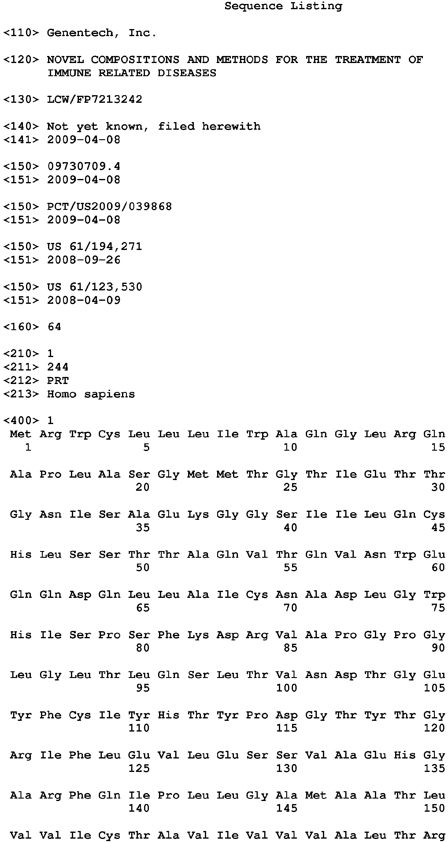
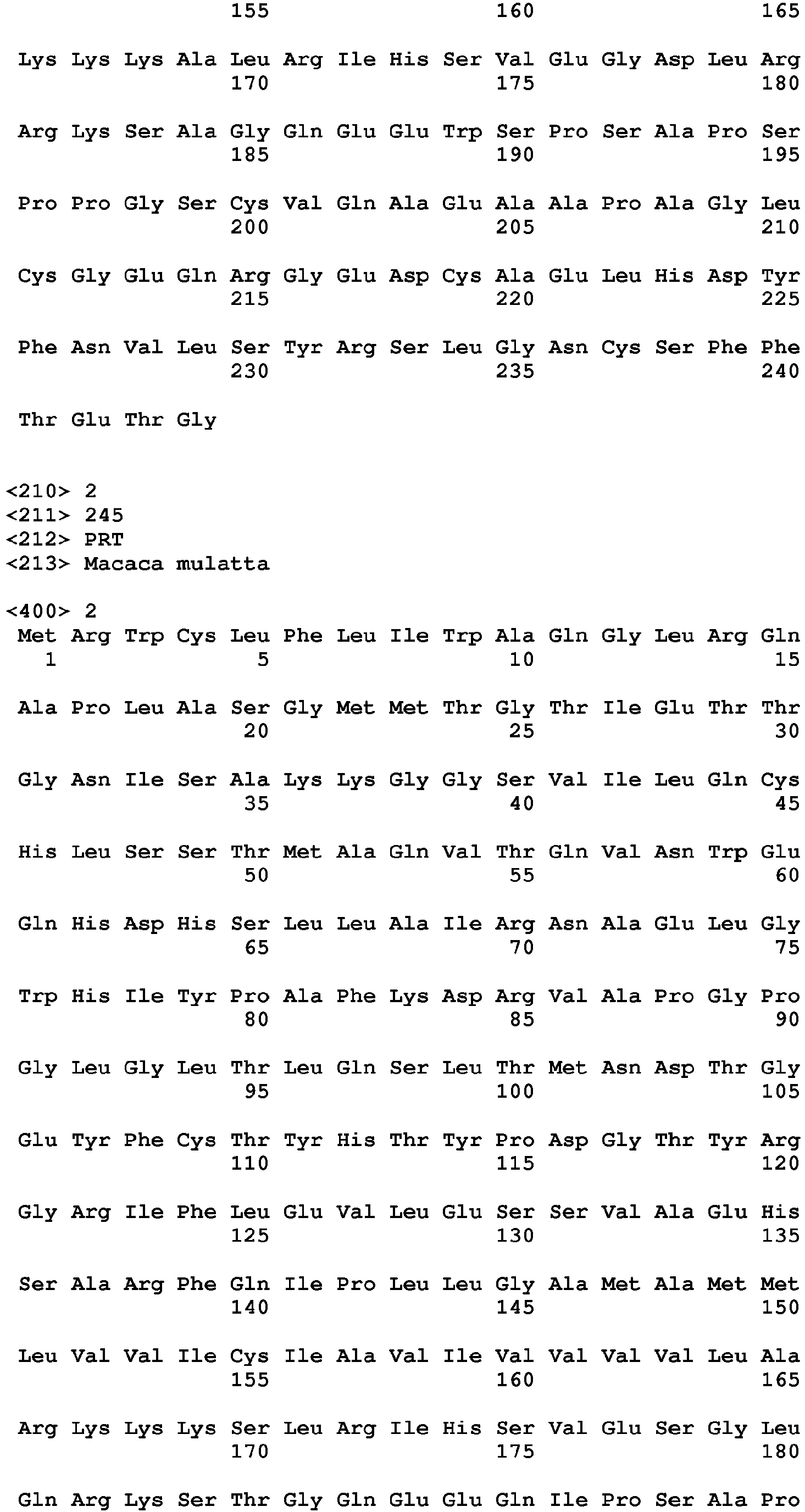
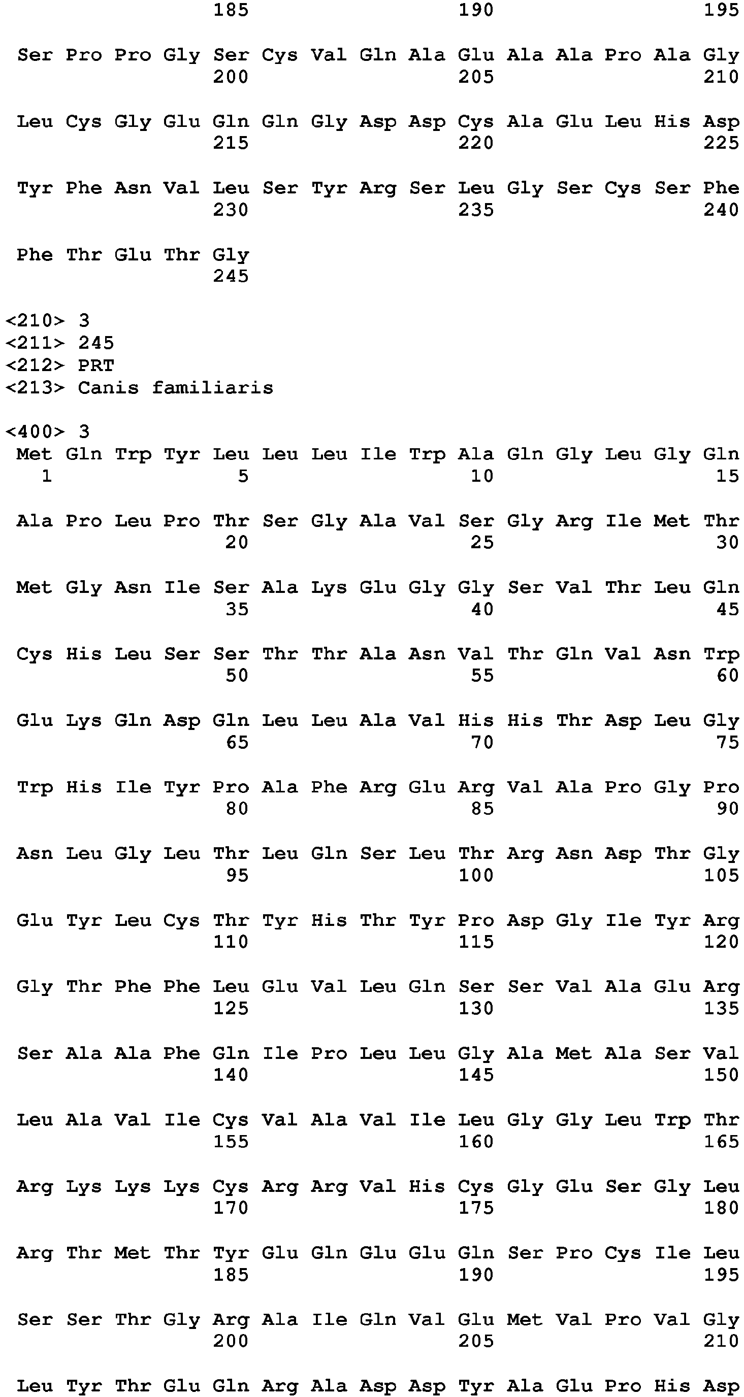


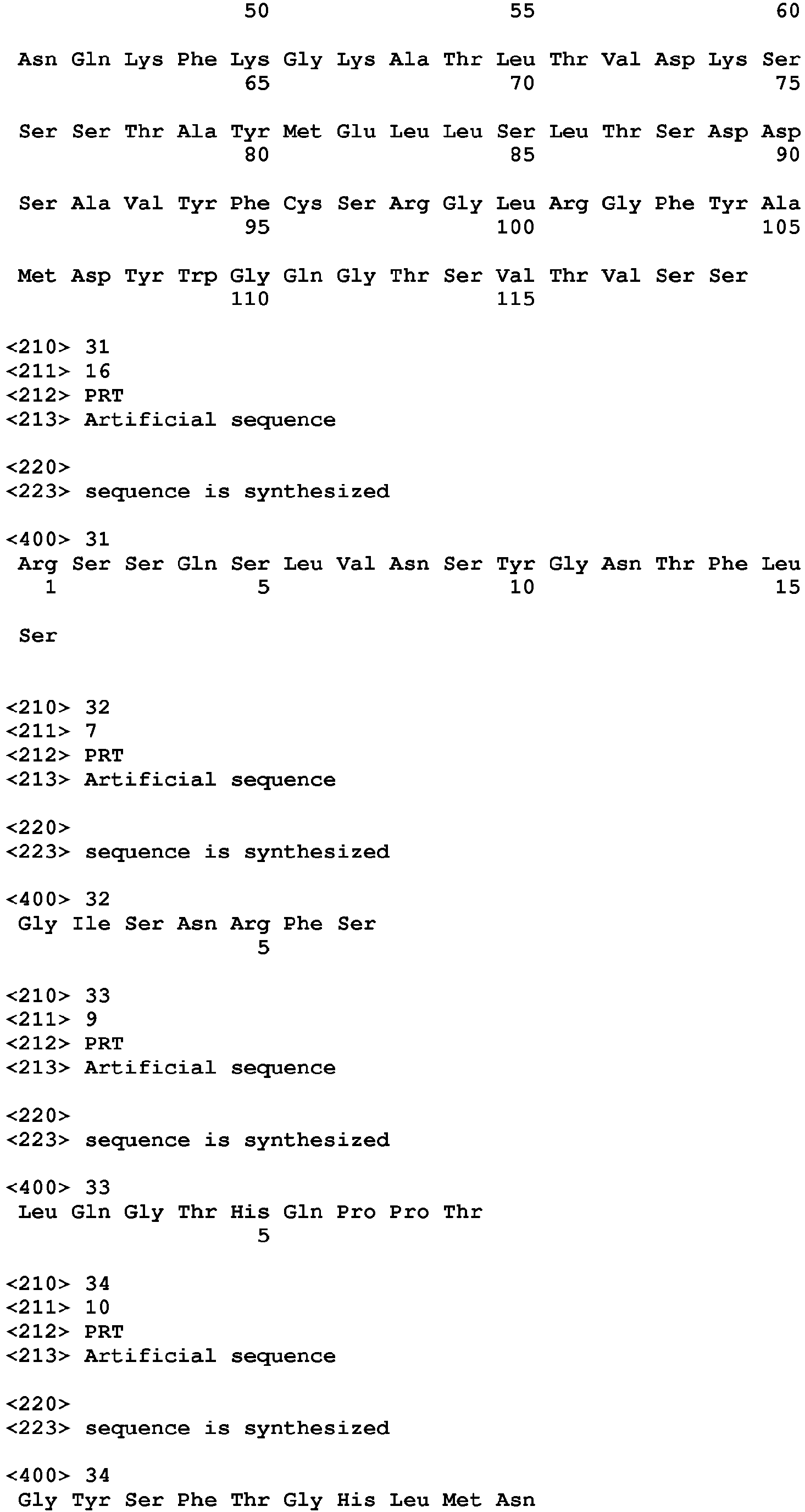


Claims (15)
- An isolated T-Cell-Ig and ITIM domain (TIGIT)-like protein (TLP)-interacting agent that specifically interacts with one or more conserved or substantially conserved regions of TLP family members, wherein the TLP-interacting agent is capable of binding to mouse TIGIT and human TIGIT.
- The TLP-interacting agent of claim 1, wherein the one or more conserved or substantially conserved regions of TLP family members comprise one or more of the following structural submotifs:(a) V/I54- S/T55 -Q56;(b) A67- X68- X69- X70- X71- X72- X13-G74, wherein X is any amino acid; and/or(c) T112- F/Y113- P114- X115- G116, wherein X is any amino acid.
- The TLP-interacting agent of claim 2, wherein the TLP-interacting agent is an antibody or antigen-binding fragment thereof.
- The TLP-interacting agent of claim 3, wherein the TLP-interacting agent is an anti-TIGIT antibody or antigen-binding fragment thereof.
- The TLP-interacting agent of claim 4, wherein the anti-TIGIT antibody or antigen-binding fragment thereof comprising a light chain comprising an HVR-L1 comprising SEQ ID NO: 23, an HVR-L2 comprising SEQ ID NO: 24, and an HVR-L3 comprising SEQ ID NO: 25, and a heavy chain comprising an HVR-H1 comprising SEQ ID NO: 26, an HVR-H2 comprising SEQ ID NO: 27, and an HVR-H3 comprising SEQ ID NO: 28.
- The TLP-interacting agent of claim 5, wherein the light chain comprises an amino acid sequence at least 90% identical to the amino acid sequence set forth in SEQ ID NO: 21 and/or the heavy chain comprises an amino acid sequence at least 90% identical to the amino acid sequence set forth in SEQ ID NO: 22.
- The TLP-interacting agent of claim 6, wherein the light chain comprises the amino acid sequence set forth in SEQ ID NO: 21 and/or the heavy chain comprises the amino acid sequence set forth in SEQ ID NO: 22.
- The TLP-interacting agent of claim 4, wherein the anti-TIGIT antibody or antigen-binding fragment thereof comprising a light chain comprising an HVR-L1 comprising SEQ ID NO: 31, an HVR-L2 comprising SEQ ID NO: 32, and an HVR-L3 comprising SEQ ID NO: 33, and a heavy chain comprising an HVR-H1 comprising SEQ ID NO: 34, an HVR-H2 comprising SEQ ID NO: 35, and an HVR-H3 comprising SEQ ID NO: 36.
- The TLP-interacting agent of claim 8, wherein the light chain comprises an amino acid sequence at least 90% identical to the amino acid sequence set forth in SEQ ID NO: 29 and/or the heavy chain comprises an amino acid sequence at least 90% identical to the amino acid sequence set forth in SEQ ID NO: 30.
- The TLP-interacting agent of claim 9, wherein the light chain comprises the amino acid sequence set forth in SEQ ID NO: 29 and/or the heavy chain comprises the amino acid sequence set forth in SEQ ID NO: 30.
- The TLP-interacting agent of any one of claims 4-10, wherein the anti-TIGIT antibody or antigen-binding fragment thereof is an antagonizing anti-TIGIT antibody or antigen-binding fragment thereof.
- The TLP-interacting agent of claim 11, wherein the antagonizing anti-TIGIT antibody or antigen-binding fragment thereof (a) specifically binds TIGIT and blocks TIGIT interaction with PVR and/or (b) is an antagonist of the expression and/or activity of TIGIT and/or PVR.
- The TLP-interacting agent of any one of claims 3-12, wherein the antibody or antigen-binding fragment thereof is selected from a humanized antibody, a chimeric antibody, a bispecific antibody, a heteroconjugate antibody, and an immunotoxin, or antigen-binding fragment thereof.
- A method of diagnosing an immune-related disease relating to aberrant immune cell response in a subject comprising assessing the expression and/or activity of TIGIT in a sample from the subject and comparing the expression and/or activity of TIGIT to a reference amount of TIGIT expression and/or activity or the amount of TIGIT expression and/or activity in a sample from a normal subject, wherein the determining comprises use of the TLP-interacting agent of any one of claims 1-13, optionally wherein the immune-related disease is selected from cancer, psoriasis, arthritis, and inflammatory bowel disease.
- An article of manufacture for use in the diagnosis or treatment of an immune-related disease, wherein the article of manufacture comprises the TLP-interacting agent of any one of claims 1-13, optionally wherein the article of manufacture further comprises an instruction or a label for using the TLP-interacting agent to detect TIGIT in a sample from a subject.
Priority Applications (2)
| Application Number | Priority Date | Filing Date | Title |
|---|---|---|---|
| EP19181712.1A EP3605088A1 (en) | 2008-04-09 | 2009-04-08 | Compositions and methods for the treatment of immune related diseases |
| PL17152167T PL3208612T3 (en) | 2008-04-09 | 2009-04-08 | Compositions and methods for the treatment of immune related diseases |
Applications Claiming Priority (4)
| Application Number | Priority Date | Filing Date | Title |
|---|---|---|---|
| US12353008P | 2008-04-09 | 2008-04-09 | |
| US19427108P | 2008-09-26 | 2008-09-26 | |
| PCT/US2009/039868 WO2009126688A2 (en) | 2008-04-09 | 2009-04-08 | Novel compositions and methods for the treatment of immune related diseases |
| EP09730709.4A EP2279412B1 (en) | 2008-04-09 | 2009-04-08 | Novel compositions and methods for the treatment of immune related diseases |
Related Parent Applications (2)
| Application Number | Title | Priority Date | Filing Date |
|---|---|---|---|
| EP09730709.4A Division-Into EP2279412B1 (en) | 2008-04-09 | 2009-04-08 | Novel compositions and methods for the treatment of immune related diseases |
| EP09730709.4A Division EP2279412B1 (en) | 2008-04-09 | 2009-04-08 | Novel compositions and methods for the treatment of immune related diseases |
Related Child Applications (2)
| Application Number | Title | Priority Date | Filing Date |
|---|---|---|---|
| EP19181712.1A Division EP3605088A1 (en) | 2008-04-09 | 2009-04-08 | Compositions and methods for the treatment of immune related diseases |
| EP19181712.1A Division-Into EP3605088A1 (en) | 2008-04-09 | 2009-04-08 | Compositions and methods for the treatment of immune related diseases |
Publications (2)
| Publication Number | Publication Date |
|---|---|
| EP3208612A1 true EP3208612A1 (en) | 2017-08-23 |
| EP3208612B1 EP3208612B1 (en) | 2019-09-18 |
Family
ID=40873503
Family Applications (3)
| Application Number | Title | Priority Date | Filing Date |
|---|---|---|---|
| EP09730709.4A Active EP2279412B1 (en) | 2008-04-09 | 2009-04-08 | Novel compositions and methods for the treatment of immune related diseases |
| EP19181712.1A Pending EP3605088A1 (en) | 2008-04-09 | 2009-04-08 | Compositions and methods for the treatment of immune related diseases |
| EP17152167.7A Active EP3208612B1 (en) | 2008-04-09 | 2009-04-08 | Compositions and methods for the treatment of immune related diseases |
Family Applications Before (2)
| Application Number | Title | Priority Date | Filing Date |
|---|---|---|---|
| EP09730709.4A Active EP2279412B1 (en) | 2008-04-09 | 2009-04-08 | Novel compositions and methods for the treatment of immune related diseases |
| EP19181712.1A Pending EP3605088A1 (en) | 2008-04-09 | 2009-04-08 | Compositions and methods for the treatment of immune related diseases |
Country Status (18)
| Country | Link |
|---|---|
| US (5) | US20090258013A1 (en) |
| EP (3) | EP2279412B1 (en) |
| JP (7) | JP5770624B2 (en) |
| KR (4) | KR20180132156A (en) |
| CN (3) | CN104655854A (en) |
| AU (1) | AU2009233708B2 (en) |
| BR (1) | BRPI0906550B8 (en) |
| CA (3) | CA2905334A1 (en) |
| DK (1) | DK2279412T3 (en) |
| ES (2) | ES2758126T3 (en) |
| HK (1) | HK1211081A1 (en) |
| HU (1) | HUE036780T2 (en) |
| IL (5) | IL208285A0 (en) |
| MX (3) | MX2010011088A (en) |
| PL (2) | PL2279412T3 (en) |
| SG (2) | SG10202112838YA (en) |
| SI (1) | SI2279412T1 (en) |
| WO (1) | WO2009126688A2 (en) |
Cited By (5)
| Publication number | Priority date | Publication date | Assignee | Title |
|---|---|---|---|---|
| US10124061B2 (en) | 2016-08-17 | 2018-11-13 | Compugen Ltd. | Anti-TIGIT antibodies, anti-PVRIG antibodies and combinations thereof |
| US11136384B2 (en) | 2016-11-30 | 2021-10-05 | Mereo Biopharma 5, Inc. | Methods for treatment of cancer comprising TIGIT-binding agents |
| US11225523B2 (en) | 2017-06-01 | 2022-01-18 | Compugen Ltd. | Triple combination antibody therapies |
| US11623955B2 (en) | 2015-02-19 | 2023-04-11 | Compugen Ltd. | Anti-PVRIG antibodies and methods of use |
| US11795209B2 (en) | 2015-02-19 | 2023-10-24 | Compugen Ltd. | PVRIG polypeptides and methods of treatment |
Families Citing this family (94)
| Publication number | Priority date | Publication date | Assignee | Title |
|---|---|---|---|---|
| AU2003270439B2 (en) | 2002-09-11 | 2009-09-24 | Genentech, Inc. | Novel composition and methods for the treatment of immune related diseases |
| CN104655854A (en) | 2008-04-09 | 2015-05-27 | 健泰科生物技术公司 | Novel compositions and methods for the treatment of immune related diseases |
| US8858941B2 (en) | 2011-09-23 | 2014-10-14 | Oncomed Pharmaceuticals, Inc. | VEGF/DLL4 binding agents and uses thereof |
| EP2928483A4 (en) * | 2012-12-04 | 2016-06-08 | Oncomed Pharm Inc | Immunotherapy with binding agents |
| WO2015009856A2 (en) | 2013-07-16 | 2015-01-22 | Genentech, Inc. | Methods of treating cancer using pd-1 axis binding antagonists and tigit inhibitors |
| WO2015006811A2 (en) * | 2013-07-19 | 2015-01-22 | The Walter And Eliza Hall Institute Of Medical Research | Diagnostic methods |
| SG11201601285XA (en) * | 2013-08-22 | 2016-03-30 | Queensland Inst Med Res | Immunoreceptor modulation for treating cancer and viral infections |
| AU2014308552B2 (en) * | 2013-08-22 | 2020-02-13 | The Council Of The Queensland Institute Of Medical Research | Immunoreceptor modulation for treating cancer and viral infections |
| JP6618908B2 (en) * | 2013-08-22 | 2019-12-11 | ザ・カウンシル・オヴ・ザ・クイーンズランド・インスティテュート・オヴ・メディカル・リサーチ | Use of a CD96 inhibitory antibody or antibody fragment to reduce or alleviate immunosuppression |
| CN103487572B (en) * | 2013-10-08 | 2017-03-29 | 北京大学人民医院 | Application of the folliculus helper T cell (Tfh) in autoimmune disease state of an illness early warning |
| GB201403775D0 (en) | 2014-03-04 | 2014-04-16 | Kymab Ltd | Antibodies, uses & methods |
| US20170107300A1 (en) | 2014-03-21 | 2017-04-20 | The Brigham And Women's Hospital, Inc. | Methods and compositions for treatment of immune-related diseases or disorders and/or therapy monitoring |
| WO2015191874A1 (en) * | 2014-06-12 | 2015-12-17 | Children's National Medical Center | Generation of broadly-specific, virus-immune cells targeting multiple hiv antigens for preventive and therapeutic use |
| MA40437A (en) * | 2014-07-16 | 2017-05-24 | Hoffmann La Roche | Methods of treating cancer using tigit inhibitors and anti-cancer agents |
| MX2017002229A (en) * | 2014-08-19 | 2017-05-09 | Merck Sharp & Dohme | Anti-tigit antibodies. |
| US20160152720A1 (en) | 2014-11-06 | 2016-06-02 | Genentech, Inc. | Combination therapy comprising ox40 binding agonists and tigit inhibitors |
| MY187045A (en) | 2014-12-23 | 2021-08-27 | Bristol Myers Squibb Co | Antibodies to tigit |
| MA41460A (en) | 2015-02-03 | 2017-12-12 | Oncomed Pharm Inc | TNFRSF LIAISON AGENTS AND THEIR USES |
| CN107969128A (en) | 2015-04-17 | 2018-04-27 | 高山免疫科学股份有限公司 | Immune modulator with adjustable affinity |
| WO2016179288A1 (en) * | 2015-05-04 | 2016-11-10 | University Of Florida Research Foundation, Inc. | Novel regulatory-cells, method for their isolation and uses |
| TWI715587B (en) * | 2015-05-28 | 2021-01-11 | 美商安可美德藥物股份有限公司 | Tigit-binding agents and uses thereof |
| CN108137687B (en) | 2015-05-29 | 2021-10-29 | 百时美施贵宝公司 | anti-OX 40 antibodies and uses thereof |
| JO3620B1 (en) * | 2015-08-05 | 2020-08-27 | Amgen Res Munich Gmbh | Immune-checkpoint inhibitors for use in the treatment of blood-borne cancers cancers |
| EP3334757A4 (en) * | 2015-08-14 | 2019-04-03 | Merck Sharp & Dohme Corp. | Anti-tigit antibodies |
| CN114605548A (en) | 2015-09-01 | 2022-06-10 | 艾吉纳斯公司 | anti-PD-1 antibodies and methods of use thereof |
| WO2017037707A1 (en) | 2015-09-02 | 2017-03-09 | Yissum Research Development Company Of The Hebrew University Of Jerusalem Ltd. | Antibodies specific to human t-cell immunoglobulin and itim domain (tigit) |
| US20180251548A1 (en) * | 2015-09-14 | 2018-09-06 | Compass Therapeutics Llc | Compositions and methods for treating cancer via antagonism of the cd155/tigit pathway and tgf-beta |
| JP6967003B2 (en) | 2015-09-23 | 2021-11-17 | メレオ バイオファーマ 5 インコーポレイテッド | Methods and compositions for the treatment of cancer |
| MA42925A (en) * | 2015-09-25 | 2018-08-01 | Hoffmann La Roche | ANTI-TIGIT ANTIBODIES AND METHODS OF USE |
| RU2022102624A (en) * | 2015-10-01 | 2022-03-10 | Хит Байолоджикс, Инк. | COMPOSITIONS AND METHODS FOR CONNECTING TYPE I AND TYPE II EXTRACELLULAR DOMAIN AS HETEROLOGOUS CHIMERIC PROTEINS |
| AR109625A1 (en) * | 2015-10-01 | 2019-01-09 | Potenza Therapeutics Inc | PROTEINS OF UNION TO ANTIGENS (ABP) THAT SELECTLY JOIN TIGIT AND METHODS FOR THE USE OF THE SAME |
| JO3555B1 (en) | 2015-10-29 | 2020-07-05 | Merck Sharp & Dohme | Antibody neutralizing human respiratory syncytial virus |
| CA3015619A1 (en) | 2016-03-01 | 2017-09-08 | Yissum Research Development Company Of The Hebrew University Of Jerusalem Ltd. | Antibodies specific to human poliovirus receptor (pvr) |
| UY37141A (en) | 2016-03-04 | 2017-09-29 | Jn Biosciences Llc | ANTIBODIES DIRECTED TO TIGIT |
| EA039540B1 (en) * | 2016-03-25 | 2022-02-08 | Онкомед Фармасьютикалс, Инк. | Tigit-binding agents and uses thereof |
| IL262366B1 (en) | 2016-04-15 | 2024-03-01 | Alpine Immune Sciences Inc | Cd80 variant immunomodulatory proteins and uses thereof |
| BR112018076260A2 (en) | 2016-06-20 | 2019-03-26 | Kymab Limited | antibody or fragment thereof that specifically binds to hpd-11, bispecific antibody or fusion protein, use of an antibody or fragment, method, pharmaceutical composition, modulation method, inhibition method, treatment method, nucleic acid, vector, host and immunocytocin |
| US9567399B1 (en) | 2016-06-20 | 2017-02-14 | Kymab Limited | Antibodies and immunocytokines |
| WO2018017864A2 (en) * | 2016-07-20 | 2018-01-25 | Oncomed Pharmaceuticals, Inc. | Pvrig-binding agents and uses thereof |
| US11471488B2 (en) | 2016-07-28 | 2022-10-18 | Alpine Immune Sciences, Inc. | CD155 variant immunomodulatory proteins and uses thereof |
| WO2018022945A1 (en) | 2016-07-28 | 2018-02-01 | Alpine Immune Sciences, Inc. | Cd112 variant immunomodulatory proteins and uses thereof |
| CN116640214A (en) | 2016-08-09 | 2023-08-25 | 科马布有限公司 | Isolated antibodies and uses thereof |
| CN107815467B (en) * | 2016-08-31 | 2021-03-16 | 百奥赛图(北京)医药科技股份有限公司 | Preparation method and application of humanized gene modified animal model |
| WO2018041120A1 (en) * | 2016-08-31 | 2018-03-08 | Beijing Biocytogen Co., Ltd | Genetically modified non-human animal with human or chimeric tigit |
| US11779604B2 (en) | 2016-11-03 | 2023-10-10 | Kymab Limited | Antibodies, combinations comprising antibodies, biomarkers, uses and methods |
| US10232053B2 (en) | 2016-12-30 | 2019-03-19 | Trieza Therapeutics, Inc. | Immunomodulatory oncolytic adenoviral vectors, and methods of production and use thereof for treatment of cancer |
| US11566060B2 (en) | 2017-01-05 | 2023-01-31 | Kahr Medical Ltd. | PD1-CD70 fusion protein and methods of use thereof |
| JP2020505009A (en) | 2017-01-05 | 2020-02-20 | カール メディカル リミテッド | PD1-41BBL fusion protein and method of using same |
| US11299530B2 (en) | 2017-01-05 | 2022-04-12 | Kahr Medical Ltd. | SIRP alpha-CD70 fusion protein and methods of use thereof |
| PT3565828T (en) | 2017-01-05 | 2022-02-08 | Kahr Medical Ltd | A sirp1 alpha-41bbl fusion protein and methods of use thereof |
| JOP20190203A1 (en) | 2017-03-30 | 2019-09-03 | Potenza Therapeutics Inc | Anti-tigit antigen-binding proteins and methods of use thereof |
| EP3612201B1 (en) | 2017-04-21 | 2023-10-25 | Sillajen, Inc. | Oncolytic vaccinia virus and checkpoint inhibitor combination therapy |
| US11760777B2 (en) | 2017-04-26 | 2023-09-19 | Bristol-Myers Squibb Company | Methods of antibody production that minimize disulfide bond reduction |
| ES2952961T3 (en) | 2017-05-01 | 2023-11-07 | Agenus Inc | Anti-TIGIT antibodies and methods of their use |
| KR20200003107A (en) | 2017-05-02 | 2020-01-08 | 머크 샤프 앤드 돔 코포레이션 | Preparation of Anti-LAG3 Antibodies, and Co-Formulations of Anti-LAG3 Antibodies and Anti-PD-1 Antibodies |
| JOP20190260A1 (en) | 2017-05-02 | 2019-10-31 | Merck Sharp & Dohme | Stable formulations of programmed death receptor 1 (pd-1) antibodies and methods of use thereof |
| US11122042B1 (en) | 2017-05-12 | 2021-09-14 | F5 Networks, Inc. | Methods for dynamically managing user access control and devices thereof |
| GB201709808D0 (en) * | 2017-06-20 | 2017-08-02 | Kymab Ltd | Antibodies |
| US20200407445A1 (en) | 2017-07-27 | 2020-12-31 | Iteos Therapeutics Sa | Anti-tigit antibodies |
| EP3689909A4 (en) | 2017-09-29 | 2021-12-29 | Jiangsu Hengrui Medicine Co., Ltd. | Tigit antibody, antigen-binding fragment thereof, and medical use thereof |
| EP4219540A3 (en) | 2017-10-10 | 2023-12-06 | Alpine Immune Sciences, Inc. | Ctla-4 variant immunomodulatory proteins and uses thereof |
| CN107988255A (en) * | 2017-10-20 | 2018-05-04 | 深圳市龙岗区妇幼保健院 | A kind of method of definite ATR and ERK calmodulin binding domain CaMs |
| US11629189B2 (en) | 2017-12-19 | 2023-04-18 | Kymab Limited | Bispecific antibody for ICOS and PD-L1 |
| MX2020008795A (en) | 2018-02-28 | 2020-10-08 | Yuhan Corp | Anti-tigit antibodies and uses thereof. |
| KR20200016114A (en) | 2018-08-06 | 2020-02-14 | 선일이씨티 주식회사 | Waste water advanced treatment system based on biological contact and circulation filtering |
| US11401339B2 (en) | 2018-08-23 | 2022-08-02 | Seagen Inc. | Anti-TIGIT antibodies |
| AR117327A1 (en) | 2018-12-20 | 2021-07-28 | 23Andme Inc | ANTI-CD96 ANTIBODIES AND METHODS OF USE OF THEM |
| CN110156888B (en) * | 2019-05-13 | 2021-07-30 | 沣潮医药科技(上海)有限公司 | Application of CD96 recombinant protein in preparation of pharmaceutical composition for immune diseases |
| KR20220042258A (en) | 2019-07-15 | 2022-04-04 | 상하이 준스 바이오사이언스 컴퍼니 리미티드 | Anti-TIGIT antibodies and their applications |
| US11667709B2 (en) | 2019-09-24 | 2023-06-06 | Industrial Technology Research Institute | Anti-TIGIT antibodies and methods of use |
| JP7280387B2 (en) | 2019-09-27 | 2023-05-23 | グラクソスミスクライン、インテレクチュアル、プロパティー、ディベロップメント、リミテッド | antigen binding protein |
| BR112022009265A2 (en) * | 2019-11-21 | 2022-08-02 | Beigene Ltd | METHOD OF TREATMENT OF CANCER AND METHOD TO INCREASE, POTENTIALIZE OR STIMULATE AN IMMUNE RESPONSE OR FUNCTION |
| TW202144417A (en) | 2020-03-13 | 2021-12-01 | 大陸商江蘇恆瑞醫藥股份有限公司 | Pvrig binding protein and its medical uses |
| CN113563470B (en) | 2020-04-29 | 2023-02-10 | 广州昂科免疫生物技术有限公司 | Antibody combined with TIGIT antigen and preparation method and application thereof |
| EP4157875A1 (en) | 2020-06-02 | 2023-04-05 | Arcus Biosciences, Inc. | Antibodies to tigit |
| BR112022025801A2 (en) | 2020-06-18 | 2023-10-03 | Hoffmann La Roche | METHODS FOR TREATING A PATIENT AND FOR TREATING A PATIENT WITH ADVANCED ESCC, KIT, ANTIBODY, USE OF AN ANTIBODY, AND USE OF A BINDING ANTAGONIST |
| TW202216778A (en) | 2020-07-15 | 2022-05-01 | 美商安進公司 | Tigit and cd112r blockade |
| CN111763660A (en) * | 2020-08-07 | 2020-10-13 | 南京大学 | Recombinant oncolytic vaccinia virus and preparation method and application thereof |
| WO2022044010A1 (en) | 2020-08-26 | 2022-03-03 | Yissum Research Development Company Of The Hebrew University Of Jerusalem Ltd. | Anti-t-cell immunoglobulin and itim domain (tigit) antibodies for the treatment of fungal infections |
| US20230374128A1 (en) * | 2020-10-09 | 2023-11-23 | Keio University | Therapeutic agent for immune/inflammatory disease |
| CN112294946B (en) * | 2020-10-28 | 2023-02-10 | 中国人民解放军空军军医大学 | Pharmaceutical application of soluble recombinant protein CD226-ECD in inhibition of allergic rhinitis asthma syndrome |
| WO2022098972A1 (en) | 2020-11-08 | 2022-05-12 | Seagen Inc. | Combination-therapy antibody drug conjugate with immune cell inhibitor |
| AU2022207020A1 (en) | 2021-01-14 | 2023-08-31 | Shanghai Junshi Biosciences Co., Ltd. | Anti-tigit antibody pharmaceutical composition and application thereof |
| BR112023019896A2 (en) | 2021-03-31 | 2023-11-07 | Ddbio Co Ltd Shang Hai | VECTORS AND METHOD FOR SCREENING FUNCTIONAL ANTIGEN-BINDING PROTEIN |
| WO2022217026A1 (en) | 2021-04-09 | 2022-10-13 | Seagen Inc. | Methods of treating cancer with anti-tigit antibodies |
| AR125753A1 (en) | 2021-05-04 | 2023-08-09 | Agenus Inc | ANTI-TIGIT ANTIBODIES, ANTI-CD96 ANTIBODIES AND METHODS OF USE OF THESE |
| AU2022302170A1 (en) | 2021-07-02 | 2023-12-21 | F. Hoffmann-La Roche Ag | Methods and compositions for treating cancer |
| WO2023010094A2 (en) | 2021-07-28 | 2023-02-02 | Genentech, Inc. | Methods and compositions for treating cancer |
| WO2023010095A1 (en) | 2021-07-28 | 2023-02-02 | F. Hoffmann-La Roche Ag | Methods and compositions for treating cancer |
| WO2023014360A1 (en) * | 2021-08-05 | 2023-02-09 | Octagon Therapeutics, Inc. | Treatment and diagnosis of immune disorders relating to aberrant immune cells |
| TW202321308A (en) | 2021-09-30 | 2023-06-01 | 美商建南德克公司 | Methods for treatment of hematologic cancers using anti-tigit antibodies, anti-cd38 antibodies, and pd-1 axis binding antagonists |
| WO2023170606A1 (en) | 2022-03-08 | 2023-09-14 | Alentis Therapeutics Ag | Use of anti-claudin-1 antibodies to increase t cell availability |
| WO2023178192A1 (en) | 2022-03-15 | 2023-09-21 | Compugen Ltd. | Il-18bp antagonist antibodies and their use in monotherapy and combination therapy in the treatment of cancer |
| WO2023240058A2 (en) | 2022-06-07 | 2023-12-14 | Genentech, Inc. | Prognostic and therapeutic methods for cancer |
Citations (100)
| Publication number | Priority date | Publication date | Assignee | Title |
|---|---|---|---|---|
| US3773919A (en) | 1969-10-23 | 1973-11-20 | Du Pont | Polylactide-drug mixtures |
| US3896111A (en) | 1973-02-20 | 1975-07-22 | Research Corp | Ansa macrolides |
| US4137230A (en) | 1977-11-14 | 1979-01-30 | Takeda Chemical Industries, Ltd. | Method for the production of maytansinoids |
| US4151042A (en) | 1977-03-31 | 1979-04-24 | Takeda Chemical Industries, Ltd. | Method for producing maytansinol and its derivatives |
| EP0003089A1 (en) | 1978-01-06 | 1979-07-25 | Bernard David | Drier for silkscreen printed sheets |
| US4179337A (en) | 1973-07-20 | 1979-12-18 | Davis Frank F | Non-immunogenic polypeptides |
| US4248870A (en) | 1978-10-27 | 1981-02-03 | Takeda Chemical Industries, Ltd. | Maytansinoids and use |
| US4256746A (en) | 1978-11-14 | 1981-03-17 | Takeda Chemical Industries | Dechloromaytansinoids, their pharmaceutical compositions and method of use |
| US4260608A (en) | 1978-11-14 | 1981-04-07 | Takeda Chemical Industries, Ltd. | Maytansinoids, pharmaceutical compositions thereof and methods of use thereof |
| US4265814A (en) | 1978-03-24 | 1981-05-05 | Takeda Chemical Industries | Matansinol 3-n-hexadecanoate |
| US4275149A (en) | 1978-11-24 | 1981-06-23 | Syva Company | Macromolecular environment control in specific receptor assays |
| EP0036776A2 (en) | 1980-03-24 | 1981-09-30 | Genentech, Inc. | A method of creating an expression plasmid |
| US4294757A (en) | 1979-01-31 | 1981-10-13 | Takeda Chemical Industries, Ltd | 20-O-Acylmaytansinoids |
| US4301144A (en) | 1979-07-11 | 1981-11-17 | Ajinomoto Company, Incorporated | Blood substitute containing modified hemoglobin |
| US4307016A (en) | 1978-03-24 | 1981-12-22 | Takeda Chemical Industries, Ltd. | Demethyl maytansinoids |
| US4308268A (en) | 1979-06-11 | 1981-12-29 | Takeda Chemical Industries, Ltd. | Maytansinoids, pharmaceutical compositions thereof and method of use thereof |
| US4308269A (en) | 1979-06-11 | 1981-12-29 | Takeda Chemical Industries, Ltd. | Maytansinoids, pharmaceutical compositions thereof and method of use thereof |
| US4309428A (en) | 1979-07-30 | 1982-01-05 | Takeda Chemical Industries, Ltd. | Maytansinoids |
| US4313946A (en) | 1981-01-27 | 1982-02-02 | The United States Of America As Represented By The Secretary Of Agriculture | Chemotherapeutically active maytansinoids from Trewia nudiflora |
| US4315929A (en) | 1981-01-27 | 1982-02-16 | The United States Of America As Represented By The Secretary Of Agriculture | Method of controlling the European corn borer with trewiasine |
| US4317821A (en) | 1979-06-08 | 1982-03-02 | Takeda Chemical Industries, Ltd. | Maytansinoids, their use and pharmaceutical compositions thereof |
| US4322348A (en) | 1979-06-05 | 1982-03-30 | Takeda Chemical Industries, Ltd. | Maytansinoids |
| US4331598A (en) | 1979-09-19 | 1982-05-25 | Takeda Chemical Industries, Ltd. | Maytansinoids |
| US4362663A (en) | 1979-09-21 | 1982-12-07 | Takeda Chemical Industries, Ltd. | Maytansinoid compound |
| US4364866A (en) | 1979-09-21 | 1982-12-21 | Takeda Chemical Industries, Ltd. | Maytansinoids |
| US4371533A (en) | 1980-10-08 | 1983-02-01 | Takeda Chemical Industries, Ltd. | 4,5-Deoxymaytansinoids, their use and pharmaceutical compositions thereof |
| US4376110A (en) | 1980-08-04 | 1983-03-08 | Hybritech, Incorporated | Immunometric assays using monoclonal antibodies |
| EP0073657A1 (en) | 1981-08-31 | 1983-03-09 | Genentech, Inc. | Preparation of hepatitis B surface antigen in yeast |
| US4399216A (en) | 1980-02-25 | 1983-08-16 | The Trustees Of Columbia University | Processes for inserting DNA into eucaryotic cells and for producing proteinaceous materials |
| US4424219A (en) | 1981-05-20 | 1984-01-03 | Takeda Chemical Industries, Ltd. | 9-Thiomaytansinoids and their pharmaceutical compositions and use |
| US4450254A (en) | 1980-11-03 | 1984-05-22 | Standard Oil Company | Impact improvement of high nitrile resins |
| EP0117058A2 (en) | 1983-01-19 | 1984-08-29 | Genentech, Inc. | Methods for producing mature protein in vertebrate host cells |
| EP0117060A2 (en) | 1983-01-19 | 1984-08-29 | Genentech, Inc. | Methods of screening and amplification in eukaryotic host cells, and nucleotide sequences and expression vectors for use therein |
| US4485045A (en) | 1981-07-06 | 1984-11-27 | Research Corporation | Synthetic phosphatidyl cholines useful in forming liposomes |
| US4496689A (en) | 1983-12-27 | 1985-01-29 | Miles Laboratories, Inc. | Covalently attached complex of alpha-1-proteinase inhibitor with a water soluble polymer |
| EP0139383A1 (en) | 1983-08-16 | 1985-05-02 | Zymogenetics, Inc. | Method for expressing foreign genes in schizosaccharomyces pombe and the use in therapeutic formulations of the products, DNA constructs and transformant strains of schizosaccharomyces pombe usable in such method and their preparation |
| US4544545A (en) | 1983-06-20 | 1985-10-01 | Trustees University Of Massachusetts | Liposomes containing modified cholesterol for organ targeting |
| EP0183070A2 (en) | 1984-10-30 | 1986-06-04 | Phillips Petroleum Company | Transformation of yeasts of the genus pichia |
| US4640835A (en) | 1981-10-30 | 1987-02-03 | Nippon Chemiphar Company, Ltd. | Plasminogen activator derivatives |
| US4670417A (en) | 1985-06-19 | 1987-06-02 | Ajinomoto Co., Inc. | Hemoglobin combined with a poly(alkylene oxide) |
| US4675187A (en) | 1983-05-16 | 1987-06-23 | Bristol-Myers Company | BBM-1675, a new antibiotic complex |
| US4676980A (en) | 1985-09-23 | 1987-06-30 | The United States Of America As Represented By The Secretary Of The Department Of Health And Human Services | Target specific cross-linked heteroantibodies |
| WO1987005330A1 (en) | 1986-03-07 | 1987-09-11 | Michel Louis Eugene Bergh | Method for enhancing glycoprotein stability |
| EP0244234A2 (en) | 1986-04-30 | 1987-11-04 | Alko Group Ltd. | Transformation of trichoderma |
| US4736866B1 (en) | 1984-06-22 | 1988-04-12 | Transgenic non-human mammals | |
| US4791192A (en) | 1986-06-26 | 1988-12-13 | Takeda Chemical Industries, Ltd. | Chemically modified protein with polyethyleneglycol |
| US4816567A (en) | 1983-04-08 | 1989-03-28 | Genentech, Inc. | Recombinant immunoglobin preparations |
| DD266710A3 (en) | 1983-06-06 | 1989-04-12 | Ve Forschungszentrum Biotechnologie | Process for the biotechnical production of alkaline phosphatase |
| WO1989005859A1 (en) | 1987-12-21 | 1989-06-29 | The Upjohn Company | Agrobacterium mediated transformation of germinating plant seeds |
| GB2211504A (en) | 1987-10-23 | 1989-07-05 | Nat Res Dev | Fowlpox virus promoters |
| US4873191A (en) | 1981-06-12 | 1989-10-10 | Ohio University | Genetic transformation of zygotes |
| EP0362179A2 (en) | 1988-08-25 | 1990-04-04 | Smithkline Beecham Corporation | Recombinant saccharomyces |
| US4943529A (en) | 1982-05-19 | 1990-07-24 | Gist-Brocades Nv | Kluyveromyces as a host strain |
| US4946783A (en) | 1987-01-30 | 1990-08-07 | President And Fellows Of Harvard College | Periplasmic protease mutants of Escherichia coli |
| EP0394538A1 (en) | 1989-04-28 | 1990-10-31 | Rhein Biotech Gesellschaft Für Neue Biotechnologische Prozesse Und Produkte Mbh | A yeast cell of the genus schwanniomyces |
| US4970198A (en) | 1985-10-17 | 1990-11-13 | American Cyanamid Company | Antitumor antibiotics (LL-E33288 complex) |
| WO1990013646A1 (en) | 1989-04-28 | 1990-11-15 | Transgene S.A. | Application of novel dna fragments as a coding sequence for a signal peptide for the secretion of mature proteins by recombinant yeast, expression cassettes, transformed yeasts and corresponding process for the preparation of proteins |
| US4975278A (en) | 1988-02-26 | 1990-12-04 | Bristol-Myers Company | Antibody-enzyme conjugates in combination with prodrugs for the delivery of cytotoxic agents to tumor cells |
| EP0402226A1 (en) | 1989-06-06 | 1990-12-12 | Institut National De La Recherche Agronomique | Transformation vectors for yeast yarrowia |
| EP0404097A2 (en) | 1989-06-22 | 1990-12-27 | BEHRINGWERKE Aktiengesellschaft | Bispecific and oligospecific, mono- and oligovalent receptors, production and applications thereof |
| WO1991000360A1 (en) | 1989-06-29 | 1991-01-10 | Medarex, Inc. | Bispecific reagents for aids therapy |
| WO1991000357A1 (en) | 1989-06-30 | 1991-01-10 | Cayla | New strain with filamentous fungi mutants, process for the production of recombinant proteins using said strain, and strains and proteins produced by said process |
| US5010182A (en) | 1987-07-28 | 1991-04-23 | Chiron Corporation | DNA constructs containing a Kluyveromyces alpha factor leader sequence for directing secretion of heterologous polypeptides |
| US5013556A (en) | 1989-10-20 | 1991-05-07 | Liposome Technology, Inc. | Liposomes with enhanced circulation time |
| US5053394A (en) | 1988-09-21 | 1991-10-01 | American Cyanamid Company | Targeted forms of methyltrithio antitumor agents |
| US5079233A (en) | 1987-01-30 | 1992-01-07 | American Cyanamid Company | N-acyl derivatives of the LL-E33288 antitumor antibiotics, composition and methods for using the same |
| WO1992020373A1 (en) | 1991-05-14 | 1992-11-26 | Repligen Corporation | Heteroconjugate antibodies for treatment of hiv infection |
| US5208020A (en) | 1989-10-25 | 1993-05-04 | Immunogen Inc. | Cytotoxic agents comprising maytansinoids and their therapeutic use |
| WO1993008829A1 (en) | 1991-11-04 | 1993-05-13 | The Regents Of The University Of California | Compositions that mediate killing of hiv-infected cells |
| WO1993011161A1 (en) | 1991-11-25 | 1993-06-10 | Enzon, Inc. | Multivalent antigen-binding proteins |
| WO1993021232A1 (en) | 1992-04-10 | 1993-10-28 | Research Development Foundation | IMMUNOTOXINS DIRECTED AGAINST c-erbB-2 (HER-2/neu) RELATED SURFACE ANTIGENS |
| WO1994011026A2 (en) | 1992-11-13 | 1994-05-26 | Idec Pharmaceuticals Corporation | Therapeutic application of chimeric and radiolabeled antibodies to human b lymphocyte restricted differentiation antigen for treatment of b cell lymphoma |
| EP0616812A1 (en) | 1993-03-24 | 1994-09-28 | Berlex Biosciences | Combination with anti-hormonal compounds and binding molecules for the treatment of cancer |
| US5362852A (en) | 1991-09-27 | 1994-11-08 | Pfizer Inc. | Modified peptide derivatives conjugated at 2-hydroxyethylamine moieties |
| US5364934A (en) | 1991-02-01 | 1994-11-15 | Genentech, Inc. | Plasma carboxypeptidase |
| US5428130A (en) | 1989-02-23 | 1995-06-27 | Genentech, Inc. | Hybrid immunoglobulins |
| US5475096A (en) | 1990-06-11 | 1995-12-12 | University Research Corporation | Nucleic acid ligands |
| US5545806A (en) | 1990-08-29 | 1996-08-13 | Genpharm International, Inc. | Ransgenic non-human animals for producing heterologous antibodies |
| US5545807A (en) | 1988-10-12 | 1996-08-13 | The Babraham Institute | Production of antibodies from transgenic animals |
| WO1996027011A1 (en) | 1995-03-01 | 1996-09-06 | Genentech, Inc. | A method for making heteromultimeric polypeptides |
| EP0425235B1 (en) | 1989-10-25 | 1996-09-25 | Immunogen Inc | Cytotoxic agents comprising maytansinoids and their therapeutic use |
| US5569825A (en) | 1990-08-29 | 1996-10-29 | Genpharm International | Transgenic non-human animals capable of producing heterologous antibodies of various isotypes |
| US5585089A (en) | 1988-12-28 | 1996-12-17 | Protein Design Labs, Inc. | Humanized immunoglobulins |
| US5606040A (en) | 1987-10-30 | 1997-02-25 | American Cyanamid Company | Antitumor and antibacterial substituted disulfide derivatives prepared from compounds possessing a methyl-trithio group |
| US5625126A (en) | 1990-08-29 | 1997-04-29 | Genpharm International, Inc. | Transgenic non-human animals for producing heterologous antibodies |
| US5633425A (en) | 1990-08-29 | 1997-05-27 | Genpharm International, Inc. | Transgenic non-human animals capable of producing heterologous antibodies |
| US5635483A (en) | 1992-12-03 | 1997-06-03 | Arizona Board Of Regents Acting On Behalf Of Arizona State University | Tumor inhibiting tetrapeptide bearing modified phenethyl amides |
| US5661016A (en) | 1990-08-29 | 1997-08-26 | Genpharm International Inc. | Transgenic non-human animals capable of producing heterologous antibodies of various isotypes |
| US5663149A (en) | 1994-12-13 | 1997-09-02 | Arizona Board Of Regents Acting On Behalf Of Arizona State University | Human cancer inhibitory pentapeptide heterocyclic and halophenyl amides |
| WO1997033551A2 (en) | 1996-03-15 | 1997-09-18 | Millennium Pharmaceuticals | Compositions and methods for the diagnosis, prevention, and treatment of neoplastic cell growth and proliferation |
| US5712374A (en) | 1995-06-07 | 1998-01-27 | American Cyanamid Company | Method for the preparation of substantiallly monomeric calicheamicin derivative/carrier conjugates |
| US5714586A (en) | 1995-06-07 | 1998-02-03 | American Cyanamid Company | Methods for the preparation of monomeric calicheamicin derivative/carrier conjugates |
| US5739116A (en) | 1994-06-03 | 1998-04-14 | American Cyanamid Company | Enediyne derivatives useful for the synthesis of conjugates of methyltrithio antitumor agents |
| US5770701A (en) | 1987-10-30 | 1998-06-23 | American Cyanamid Company | Process for preparing targeted forms of methyltrithio antitumor agents |
| US5780588A (en) | 1993-01-26 | 1998-07-14 | Arizona Board Of Regents | Elucidation and synthesis of selected pentapeptides |
| WO2002088172A2 (en) | 2001-04-30 | 2002-11-07 | Seattle Genetics, Inc. | Pentapeptide compounds and uses related thereto |
| EP1391213A1 (en) | 2002-08-21 | 2004-02-25 | Boehringer Ingelheim International GmbH | Compositions and methods for treating cancer using maytansinoid CD44 antibody immunoconjugates and chemotherapeutic agents |
| WO2004024068A2 (en) * | 2002-09-11 | 2004-03-25 | Genentech, Inc. | Novel composition and methods for the treatment of immune related diseases |
| WO2006124667A2 (en) * | 2005-05-12 | 2006-11-23 | Zymogenetics, Inc. | Compositions and methods for modulating immune responses |
| US20080038264A1 (en) | 2002-02-22 | 2008-02-14 | Genentech, Inc. | Compositions and methods for the treatment of immune related diseases |
Family Cites Families (71)
| Publication number | Priority date | Publication date | Assignee | Title |
|---|---|---|---|---|
| US1516629A (en) * | 1924-11-25 | Ernest blaker | ||
| US2408528A (en) * | 1945-01-16 | 1946-10-01 | Nassour Edward | Race recording system |
| US5989811A (en) | 1994-09-29 | 1999-11-23 | Urocor, Inc. | Sextant core biopsy predictive mechanism for non-organ confined disease status |
| US5750345A (en) | 1995-10-31 | 1998-05-12 | Evanston Hospital Corporation | Detection of human α-thalassemia mutations and their use as predictors of blood-related disorders |
| CA2272842C (en) * | 1996-11-27 | 2012-05-15 | Genentech, Inc. | Humanized anti-cd11a antibodies |
| AU4228699A (en) * | 1998-06-03 | 1999-12-20 | Northwestern University | Cellular proteins which mediate herpesvirus entry |
| US6518033B1 (en) | 1998-08-05 | 2003-02-11 | The Research Foundation Of State University Of New York | Method of detecting the presence of CD155 for diagnosis of cancer and to determine treatment |
| KR20010103046A (en) | 1999-03-08 | 2001-11-17 | 제넨테크, 인크. | Compositions and Methods for the Treatment of Immune Related Diseases |
| EP1173462A1 (en) | 1999-03-26 | 2002-01-23 | Human Genome Sciences, Inc. | 50 human secreted proteins |
| US7282570B2 (en) | 1999-04-20 | 2007-10-16 | Genentech, Inc. | Compositions and methods for the treatment of immune related diseases |
| DE60035693T2 (en) * | 1999-07-20 | 2008-05-15 | Genentech, Inc., South San Francisco | COMPOSITION AND METHOD FOR THE TREATMENT OF IMMUNOMATED DISEASES |
| US20080050809A1 (en) * | 1999-09-28 | 2008-02-28 | Alejandro Abuin | Novel human kinases and polynucleotides encoding the same |
| WO2001029221A2 (en) | 1999-10-20 | 2001-04-26 | Zymogenetics, Inc. | Proteins and polynucleotides encoding them |
| US20040219521A1 (en) * | 2000-01-21 | 2004-11-04 | Tang Y. Tom | Novel nucleic acids and polypeptides |
| IL151865A0 (en) | 2000-03-31 | 2003-04-10 | Genentech Inc | Compositions and methods for detecting and quantifying gene expression |
| US6569668B2 (en) | 2000-04-04 | 2003-05-27 | Schering Corporation | Isolated nucleic acids from Micromonospora rosaria plasmid pMR2 and vectors made therefrom |
| AU2001275285A1 (en) | 2000-06-06 | 2001-12-17 | Bristol-Myers Squibb Company | B7-related nucleic acids and polypeptides and their uses for immunomodulation |
| US7713705B2 (en) | 2002-12-24 | 2010-05-11 | Biosite, Inc. | Markers for differential diagnosis and methods of use thereof |
| WO2002099062A2 (en) * | 2001-06-04 | 2002-12-12 | Curagen Corporation | Novel antibodies that bind to antigenic polypeptides, nucleic acids encoding the antigens, and methods of use |
| US20040101876A1 (en) * | 2002-05-31 | 2004-05-27 | Liat Mintz | Methods and systems for annotating biomolecular sequences |
| WO2003054152A2 (en) | 2001-12-10 | 2003-07-03 | Nuvelo, Inc. | Novel nucleic acids and polypeptides |
| WO2003068943A2 (en) * | 2002-02-13 | 2003-08-21 | Incyte Corporation | Secreted proteins |
| US7193069B2 (en) * | 2002-03-22 | 2007-03-20 | Research Association For Biotechnology | Full-length cDNA |
| DK2206517T3 (en) | 2002-07-03 | 2023-11-06 | Ono Pharmaceutical Co | Immunopotentiating compositions comprising anti-PD-L1 antibodies |
| JP4395570B2 (en) * | 2002-07-30 | 2010-01-13 | 独立行政法人産業技術総合研究所 | Method for producing hydrogen by thermochemical decomposition of water |
| CA2497334A1 (en) * | 2002-09-11 | 2004-03-25 | Genentech, Inc. | Novel compositions and methods for the treatment of immune related diseases |
| CN1720336A (en) * | 2002-10-03 | 2006-01-11 | 健泰科生物技术公司 | Use of A33 antigens and JAM-IT |
| US6925898B2 (en) * | 2002-10-18 | 2005-08-09 | Logitech Europe S.A. | Variable sensor shaft retention apparatus |
| EP1481993A1 (en) | 2003-05-28 | 2004-12-01 | Xerion Pharmaceuticals AG | Modulation of the poliovirus receptor function |
| US7144032B2 (en) * | 2003-09-09 | 2006-12-05 | Autoliv Asp, Inc. | Expandable pelvic side airbag |
| PE20050928A1 (en) * | 2003-11-21 | 2005-11-08 | Schering Corp | THERAPEUTIC COMBINATIONS OF ANTI-IGFR1 ANTIBODY |
| AR047729A1 (en) * | 2003-11-28 | 2006-02-15 | Astrazeneca Ab | THERAPEUTIC ANTIBODIES THAT BIND APOLIPOPROTEIN E (APOE) |
| CN101035807A (en) * | 2004-06-09 | 2007-09-12 | 泰勒公司 | Diagnosis and treatment of SIGLEC-6 associated diseases |
| US7096772B2 (en) * | 2004-08-30 | 2006-08-29 | Caterpillar S.A.R.L. | System and method for controlling hydraulic fluid flow |
| TW200630106A (en) * | 2004-10-08 | 2006-09-01 | Wyeth Corp | Immunotherapy of autoimmune disorders |
| CA2607147C (en) | 2005-05-09 | 2018-07-17 | Ono Pharmaceutical Co., Ltd. | Human monoclonal antibodies to programmed death 1 (pd-1) and methods for treating cancer using anti-pd-1 antibodies alone or in combination with other immunotherapeutics |
| AU2006265108C1 (en) | 2005-07-01 | 2013-01-17 | E. R. Squibb & Sons, L.L.C. | Human monoclonal antibodies to programmed death ligand 1 (PD-L1) |
| US20100075377A1 (en) | 2006-04-13 | 2010-03-25 | West James W | Tetramerizing polypeptides and methods of use |
| EP2007806A2 (en) | 2006-04-13 | 2008-12-31 | ZymoGenetics, Inc. | Tetramerizing polypeptides and methods of use |
| CA2648915A1 (en) | 2006-04-13 | 2007-11-01 | Zymogenetics, Inc. | Tetramerizing polypeptides and methods of use |
| WO2007124383A2 (en) | 2006-04-19 | 2007-11-01 | Oregon Health & Science University | 1,1'-binaphthyl-based inhibitors of nad+-dependent deacetylase activity and sir2-family members |
| CN104655854A (en) | 2008-04-09 | 2015-05-27 | 健泰科生物技术公司 | Novel compositions and methods for the treatment of immune related diseases |
| DK2376535T3 (en) | 2008-12-09 | 2017-06-12 | Hoffmann La Roche | ANTI-PD-L1 ANTIBODIES AND THEIR USE TO PROMOTE T CELL FUNCTION |
| US20130017199A1 (en) | 2009-11-24 | 2013-01-17 | AMPLIMMUNE ,Inc. a corporation | Simultaneous inhibition of pd-l1/pd-l2 |
| CA2802017A1 (en) | 2010-06-09 | 2011-12-15 | Zymogenetics, Inc. | Dimeric vstm3 fusion proteins and related compositions and methods |
| KR20190133790A (en) | 2011-08-01 | 2019-12-03 | 제넨테크, 인크. | Methods of treating cancer using pd-1 axis binding antagonists and mek inhibitors |
| WO2013119202A1 (en) | 2012-02-06 | 2013-08-15 | Providence Health & Services - Oregon | Cancer treatment and monitoring methods using ox40 agonists |
| EP3508215A3 (en) | 2012-12-03 | 2019-10-02 | Bristol-Myers Squibb Company | Enhancing anti-cancer activity of immunomodulatory fc fusion proteins |
| EP2928483A4 (en) | 2012-12-04 | 2016-06-08 | Oncomed Pharm Inc | Immunotherapy with binding agents |
| CN103073644B (en) | 2012-12-31 | 2014-03-12 | 中国科学技术大学 | Specific anti-mouse TIGIT monoclonal antibody and preparation method, identification and application thereof |
| US20140242077A1 (en) | 2013-01-23 | 2014-08-28 | Abbvie, Inc. | Methods and compositions for modulating an immune response |
| NZ712314A (en) | 2013-03-15 | 2021-07-30 | Genentech Inc | Biomarkers and methods of treating pd-1 and pd-l1 related conditions |
| WO2015009856A2 (en) | 2013-07-16 | 2015-01-22 | Genentech, Inc. | Methods of treating cancer using pd-1 axis binding antagonists and tigit inhibitors |
| US20160272707A1 (en) | 2013-09-11 | 2016-09-22 | Compugen Ltd. | Vstm5 antibodies, and uses thereof for treatment of cancer, infectious diseases and immune related diseases |
| KR20160145624A (en) | 2014-03-31 | 2016-12-20 | 제넨테크, 인크. | Anti-ox40 antibodies and methods of use |
| CA2943834A1 (en) | 2014-03-31 | 2015-10-08 | Genentech, Inc. | Combination therapy comprising anti-angiogenesis agents and ox40 binding agonists |
| MA40437A (en) | 2014-07-16 | 2017-05-24 | Hoffmann La Roche | Methods of treating cancer using tigit inhibitors and anti-cancer agents |
| MX2017002229A (en) | 2014-08-19 | 2017-05-09 | Merck Sharp & Dohme | Anti-tigit antibodies. |
| BR112017006664A2 (en) | 2014-10-03 | 2017-12-26 | Novartis Ag | combination therapies |
| US20160152720A1 (en) | 2014-11-06 | 2016-06-02 | Genentech, Inc. | Combination therapy comprising ox40 binding agonists and tigit inhibitors |
| MY187045A (en) | 2014-12-23 | 2021-08-27 | Bristol Myers Squibb Co | Antibodies to tigit |
| MY198983A (en) | 2015-05-20 | 2023-10-06 | Janssen Biotech Inc | Anti-cd38 antibodies for treatment of light chain amyloidosis and other cd38-positive hematological malignancies |
| TWI715587B (en) | 2015-05-28 | 2021-01-11 | 美商安可美德藥物股份有限公司 | Tigit-binding agents and uses thereof |
| MA42925A (en) | 2015-09-25 | 2018-08-01 | Hoffmann La Roche | ANTI-TIGIT ANTIBODIES AND METHODS OF USE |
| WO2018102536A1 (en) | 2016-11-30 | 2018-06-07 | Oncomed Pharmaceuticals, Inc. | Methods for treatment of cancer comprising tigit-binding agents |
| WO2018129559A1 (en) | 2017-01-09 | 2018-07-12 | Tesaro, Inc. | Methods of treating cancer with anti-pd-1 antibodies |
| SG11201907278VA (en) | 2017-02-28 | 2019-09-27 | Seattle Genetics Inc | Anti-tigit antibodies |
| BR112019022515A2 (en) | 2017-04-27 | 2020-06-16 | Tesaro, Inc. | ANTIBODY AGENTS TARGETED AGAINST Lymphocyte Activation Gene 3 (LAG-3) AND USES OF THE SAME |
| ES2952961T3 (en) | 2017-05-01 | 2023-11-07 | Agenus Inc | Anti-TIGIT antibodies and methods of their use |
| TW202000702A (en) | 2018-02-26 | 2020-01-01 | 美商建南德克公司 | Dosing for treatment with anti-TIGIT and anti-PD-L1 antagonist antibodies |
| WO2020096915A1 (en) | 2018-11-05 | 2020-05-14 | Merck Sharp & Dohme Corp. | Dosing regimen of anti-tigit antibody for treatment of cancer |
-
2009
- 2009-04-08 CN CN201510024034.9A patent/CN104655854A/en active Pending
- 2009-04-08 SG SG10202112838YA patent/SG10202112838YA/en unknown
- 2009-04-08 EP EP09730709.4A patent/EP2279412B1/en active Active
- 2009-04-08 BR BRPI0906550A patent/BRPI0906550B8/en active IP Right Grant
- 2009-04-08 SI SI200931728T patent/SI2279412T1/en unknown
- 2009-04-08 PL PL09730709T patent/PL2279412T3/en unknown
- 2009-04-08 DK DK09730709.4T patent/DK2279412T3/en active
- 2009-04-08 KR KR1020187034436A patent/KR20180132156A/en not_active Application Discontinuation
- 2009-04-08 CA CA2905334A patent/CA2905334A1/en not_active Abandoned
- 2009-04-08 WO PCT/US2009/039868 patent/WO2009126688A2/en active Application Filing
- 2009-04-08 PL PL17152167T patent/PL3208612T3/en unknown
- 2009-04-08 SG SG10201402815VA patent/SG10201402815VA/en unknown
- 2009-04-08 CA CA3179151A patent/CA3179151A1/en active Pending
- 2009-04-08 ES ES17152167T patent/ES2758126T3/en active Active
- 2009-04-08 US US12/420,234 patent/US20090258013A1/en not_active Abandoned
- 2009-04-08 MX MX2010011088A patent/MX2010011088A/en active IP Right Grant
- 2009-04-08 HU HUE09730709A patent/HUE036780T2/en unknown
- 2009-04-08 KR KR1020107025044A patent/KR101692080B1/en active IP Right Grant
- 2009-04-08 CN CN201910073153.1A patent/CN110082533B/en active Active
- 2009-04-08 KR KR1020187012359A patent/KR101924831B1/en active IP Right Grant
- 2009-04-08 JP JP2011504145A patent/JP5770624B2/en active Active
- 2009-04-08 CA CA2719189A patent/CA2719189C/en active Active
- 2009-04-08 CN CN200980121734.2A patent/CN102057272B/en active Active
- 2009-04-08 MX MX2013010169A patent/MX364200B/en unknown
- 2009-04-08 EP EP19181712.1A patent/EP3605088A1/en active Pending
- 2009-04-08 KR KR1020167036368A patent/KR101855381B1/en active IP Right Grant
- 2009-04-08 AU AU2009233708A patent/AU2009233708B2/en active Active
- 2009-04-08 ES ES09730709.4T patent/ES2640533T3/en active Active
- 2009-04-08 EP EP17152167.7A patent/EP3208612B1/en active Active
-
2010
- 2010-09-21 IL IL208285A patent/IL208285A0/en unknown
- 2010-10-08 MX MX2018009945A patent/MX2018009945A/en unknown
-
2012
- 2012-10-09 US US13/648,191 patent/US9499596B2/en active Active
-
2013
- 2013-10-21 IL IL228988A patent/IL228988A/en active IP Right Grant
-
2015
- 2015-04-27 JP JP2015090362A patent/JP6246158B2/en active Active
- 2015-11-26 HK HK15111634.2A patent/HK1211081A1/en unknown
-
2016
- 2016-06-16 IL IL246293A patent/IL246293B/en active IP Right Grant
- 2016-08-08 US US15/231,032 patent/US20170145093A1/en active Granted
-
2017
- 2017-07-14 JP JP2017137728A patent/JP2017215332A/en active Pending
-
2018
- 2018-10-03 JP JP2018188409A patent/JP2019054793A/en active Pending
-
2019
- 2019-01-28 IL IL264498A patent/IL264498B/en active IP Right Grant
- 2019-08-09 US US16/537,280 patent/US11390678B2/en active Active
-
2020
- 2020-02-20 IL IL272817A patent/IL272817A/en unknown
- 2020-03-02 JP JP2020034632A patent/JP2020114211A/en active Pending
-
2021
- 2021-07-17 JP JP2021118349A patent/JP2021180663A/en active Pending
-
2022
- 2022-06-15 US US17/841,447 patent/US20230098521A1/en active Pending
-
2023
- 2023-01-11 JP JP2023002154A patent/JP2023055712A/en active Pending
Patent Citations (109)
| Publication number | Priority date | Publication date | Assignee | Title |
|---|---|---|---|---|
| US3773919A (en) | 1969-10-23 | 1973-11-20 | Du Pont | Polylactide-drug mixtures |
| US3896111A (en) | 1973-02-20 | 1975-07-22 | Research Corp | Ansa macrolides |
| US4179337A (en) | 1973-07-20 | 1979-12-18 | Davis Frank F | Non-immunogenic polypeptides |
| US4151042A (en) | 1977-03-31 | 1979-04-24 | Takeda Chemical Industries, Ltd. | Method for producing maytansinol and its derivatives |
| US4137230A (en) | 1977-11-14 | 1979-01-30 | Takeda Chemical Industries, Ltd. | Method for the production of maytansinoids |
| EP0003089A1 (en) | 1978-01-06 | 1979-07-25 | Bernard David | Drier for silkscreen printed sheets |
| US4265814A (en) | 1978-03-24 | 1981-05-05 | Takeda Chemical Industries | Matansinol 3-n-hexadecanoate |
| US4361650A (en) | 1978-03-24 | 1982-11-30 | Takeda Chemical Industries, Ltd. | Fermentation process of preparing demethyl maytansinoids |
| US4307016A (en) | 1978-03-24 | 1981-12-22 | Takeda Chemical Industries, Ltd. | Demethyl maytansinoids |
| US4248870A (en) | 1978-10-27 | 1981-02-03 | Takeda Chemical Industries, Ltd. | Maytansinoids and use |
| US4256746A (en) | 1978-11-14 | 1981-03-17 | Takeda Chemical Industries | Dechloromaytansinoids, their pharmaceutical compositions and method of use |
| US4260608A (en) | 1978-11-14 | 1981-04-07 | Takeda Chemical Industries, Ltd. | Maytansinoids, pharmaceutical compositions thereof and methods of use thereof |
| US4275149A (en) | 1978-11-24 | 1981-06-23 | Syva Company | Macromolecular environment control in specific receptor assays |
| US4294757A (en) | 1979-01-31 | 1981-10-13 | Takeda Chemical Industries, Ltd | 20-O-Acylmaytansinoids |
| US4322348A (en) | 1979-06-05 | 1982-03-30 | Takeda Chemical Industries, Ltd. | Maytansinoids |
| US4317821A (en) | 1979-06-08 | 1982-03-02 | Takeda Chemical Industries, Ltd. | Maytansinoids, their use and pharmaceutical compositions thereof |
| US4308268A (en) | 1979-06-11 | 1981-12-29 | Takeda Chemical Industries, Ltd. | Maytansinoids, pharmaceutical compositions thereof and method of use thereof |
| US4308269A (en) | 1979-06-11 | 1981-12-29 | Takeda Chemical Industries, Ltd. | Maytansinoids, pharmaceutical compositions thereof and method of use thereof |
| US4301144A (en) | 1979-07-11 | 1981-11-17 | Ajinomoto Company, Incorporated | Blood substitute containing modified hemoglobin |
| US4309428A (en) | 1979-07-30 | 1982-01-05 | Takeda Chemical Industries, Ltd. | Maytansinoids |
| US4331598A (en) | 1979-09-19 | 1982-05-25 | Takeda Chemical Industries, Ltd. | Maytansinoids |
| US4364866A (en) | 1979-09-21 | 1982-12-21 | Takeda Chemical Industries, Ltd. | Maytansinoids |
| US4362663A (en) | 1979-09-21 | 1982-12-07 | Takeda Chemical Industries, Ltd. | Maytansinoid compound |
| US4399216A (en) | 1980-02-25 | 1983-08-16 | The Trustees Of Columbia University | Processes for inserting DNA into eucaryotic cells and for producing proteinaceous materials |
| EP0036776A2 (en) | 1980-03-24 | 1981-09-30 | Genentech, Inc. | A method of creating an expression plasmid |
| US4376110A (en) | 1980-08-04 | 1983-03-08 | Hybritech, Incorporated | Immunometric assays using monoclonal antibodies |
| US4371533A (en) | 1980-10-08 | 1983-02-01 | Takeda Chemical Industries, Ltd. | 4,5-Deoxymaytansinoids, their use and pharmaceutical compositions thereof |
| US4450254A (en) | 1980-11-03 | 1984-05-22 | Standard Oil Company | Impact improvement of high nitrile resins |
| US4313946A (en) | 1981-01-27 | 1982-02-02 | The United States Of America As Represented By The Secretary Of Agriculture | Chemotherapeutically active maytansinoids from Trewia nudiflora |
| US4315929A (en) | 1981-01-27 | 1982-02-16 | The United States Of America As Represented By The Secretary Of Agriculture | Method of controlling the European corn borer with trewiasine |
| US4424219A (en) | 1981-05-20 | 1984-01-03 | Takeda Chemical Industries, Ltd. | 9-Thiomaytansinoids and their pharmaceutical compositions and use |
| US4873191A (en) | 1981-06-12 | 1989-10-10 | Ohio University | Genetic transformation of zygotes |
| US4485045A (en) | 1981-07-06 | 1984-11-27 | Research Corporation | Synthetic phosphatidyl cholines useful in forming liposomes |
| EP0073657A1 (en) | 1981-08-31 | 1983-03-09 | Genentech, Inc. | Preparation of hepatitis B surface antigen in yeast |
| US4640835A (en) | 1981-10-30 | 1987-02-03 | Nippon Chemiphar Company, Ltd. | Plasminogen activator derivatives |
| US4943529A (en) | 1982-05-19 | 1990-07-24 | Gist-Brocades Nv | Kluyveromyces as a host strain |
| EP0117058A2 (en) | 1983-01-19 | 1984-08-29 | Genentech, Inc. | Methods for producing mature protein in vertebrate host cells |
| EP0117060A2 (en) | 1983-01-19 | 1984-08-29 | Genentech, Inc. | Methods of screening and amplification in eukaryotic host cells, and nucleotide sequences and expression vectors for use therein |
| US4816567A (en) | 1983-04-08 | 1989-03-28 | Genentech, Inc. | Recombinant immunoglobin preparations |
| US4675187A (en) | 1983-05-16 | 1987-06-23 | Bristol-Myers Company | BBM-1675, a new antibiotic complex |
| DD266710A3 (en) | 1983-06-06 | 1989-04-12 | Ve Forschungszentrum Biotechnologie | Process for the biotechnical production of alkaline phosphatase |
| US4544545A (en) | 1983-06-20 | 1985-10-01 | Trustees University Of Massachusetts | Liposomes containing modified cholesterol for organ targeting |
| EP0139383A1 (en) | 1983-08-16 | 1985-05-02 | Zymogenetics, Inc. | Method for expressing foreign genes in schizosaccharomyces pombe and the use in therapeutic formulations of the products, DNA constructs and transformant strains of schizosaccharomyces pombe usable in such method and their preparation |
| US4496689A (en) | 1983-12-27 | 1985-01-29 | Miles Laboratories, Inc. | Covalently attached complex of alpha-1-proteinase inhibitor with a water soluble polymer |
| US4736866A (en) | 1984-06-22 | 1988-04-12 | President And Fellows Of Harvard College | Transgenic non-human mammals |
| US4736866B1 (en) | 1984-06-22 | 1988-04-12 | Transgenic non-human mammals | |
| EP0183070A2 (en) | 1984-10-30 | 1986-06-04 | Phillips Petroleum Company | Transformation of yeasts of the genus pichia |
| US4670417A (en) | 1985-06-19 | 1987-06-02 | Ajinomoto Co., Inc. | Hemoglobin combined with a poly(alkylene oxide) |
| US4676980A (en) | 1985-09-23 | 1987-06-30 | The United States Of America As Represented By The Secretary Of The Department Of Health And Human Services | Target specific cross-linked heteroantibodies |
| US4970198A (en) | 1985-10-17 | 1990-11-13 | American Cyanamid Company | Antitumor antibiotics (LL-E33288 complex) |
| WO1987005330A1 (en) | 1986-03-07 | 1987-09-11 | Michel Louis Eugene Bergh | Method for enhancing glycoprotein stability |
| EP0244234A2 (en) | 1986-04-30 | 1987-11-04 | Alko Group Ltd. | Transformation of trichoderma |
| US4791192A (en) | 1986-06-26 | 1988-12-13 | Takeda Chemical Industries, Ltd. | Chemically modified protein with polyethyleneglycol |
| US4946783A (en) | 1987-01-30 | 1990-08-07 | President And Fellows Of Harvard College | Periplasmic protease mutants of Escherichia coli |
| US5079233A (en) | 1987-01-30 | 1992-01-07 | American Cyanamid Company | N-acyl derivatives of the LL-E33288 antitumor antibiotics, composition and methods for using the same |
| US5010182A (en) | 1987-07-28 | 1991-04-23 | Chiron Corporation | DNA constructs containing a Kluyveromyces alpha factor leader sequence for directing secretion of heterologous polypeptides |
| GB2211504A (en) | 1987-10-23 | 1989-07-05 | Nat Res Dev | Fowlpox virus promoters |
| US5606040A (en) | 1987-10-30 | 1997-02-25 | American Cyanamid Company | Antitumor and antibacterial substituted disulfide derivatives prepared from compounds possessing a methyl-trithio group |
| US5770701A (en) | 1987-10-30 | 1998-06-23 | American Cyanamid Company | Process for preparing targeted forms of methyltrithio antitumor agents |
| US5770710A (en) | 1987-10-30 | 1998-06-23 | American Cyanamid Company | Antitumor and antibacterial substituted disulfide derivatives prepared from compounds possessing a methlytrithio group |
| WO1989005859A1 (en) | 1987-12-21 | 1989-06-29 | The Upjohn Company | Agrobacterium mediated transformation of germinating plant seeds |
| US4975278A (en) | 1988-02-26 | 1990-12-04 | Bristol-Myers Company | Antibody-enzyme conjugates in combination with prodrugs for the delivery of cytotoxic agents to tumor cells |
| EP0362179A2 (en) | 1988-08-25 | 1990-04-04 | Smithkline Beecham Corporation | Recombinant saccharomyces |
| US5053394A (en) | 1988-09-21 | 1991-10-01 | American Cyanamid Company | Targeted forms of methyltrithio antitumor agents |
| US5545807A (en) | 1988-10-12 | 1996-08-13 | The Babraham Institute | Production of antibodies from transgenic animals |
| US5693762A (en) | 1988-12-28 | 1997-12-02 | Protein Design Labs, Inc. | Humanized immunoglobulins |
| US5585089A (en) | 1988-12-28 | 1996-12-17 | Protein Design Labs, Inc. | Humanized immunoglobulins |
| US5428130A (en) | 1989-02-23 | 1995-06-27 | Genentech, Inc. | Hybrid immunoglobulins |
| EP0394538A1 (en) | 1989-04-28 | 1990-10-31 | Rhein Biotech Gesellschaft Für Neue Biotechnologische Prozesse Und Produkte Mbh | A yeast cell of the genus schwanniomyces |
| WO1990013646A1 (en) | 1989-04-28 | 1990-11-15 | Transgene S.A. | Application of novel dna fragments as a coding sequence for a signal peptide for the secretion of mature proteins by recombinant yeast, expression cassettes, transformed yeasts and corresponding process for the preparation of proteins |
| EP0402226A1 (en) | 1989-06-06 | 1990-12-12 | Institut National De La Recherche Agronomique | Transformation vectors for yeast yarrowia |
| EP0404097A2 (en) | 1989-06-22 | 1990-12-27 | BEHRINGWERKE Aktiengesellschaft | Bispecific and oligospecific, mono- and oligovalent receptors, production and applications thereof |
| WO1991000360A1 (en) | 1989-06-29 | 1991-01-10 | Medarex, Inc. | Bispecific reagents for aids therapy |
| WO1991000357A1 (en) | 1989-06-30 | 1991-01-10 | Cayla | New strain with filamentous fungi mutants, process for the production of recombinant proteins using said strain, and strains and proteins produced by said process |
| US5013556A (en) | 1989-10-20 | 1991-05-07 | Liposome Technology, Inc. | Liposomes with enhanced circulation time |
| US5208020A (en) | 1989-10-25 | 1993-05-04 | Immunogen Inc. | Cytotoxic agents comprising maytansinoids and their therapeutic use |
| EP0425235B1 (en) | 1989-10-25 | 1996-09-25 | Immunogen Inc | Cytotoxic agents comprising maytansinoids and their therapeutic use |
| US5416064A (en) | 1989-10-25 | 1995-05-16 | Immunogen, Inc. | Cytotoxic agents comprising maytansinoids and their therapeutic use |
| US5475096A (en) | 1990-06-11 | 1995-12-12 | University Research Corporation | Nucleic acid ligands |
| US5661016A (en) | 1990-08-29 | 1997-08-26 | Genpharm International Inc. | Transgenic non-human animals capable of producing heterologous antibodies of various isotypes |
| US5545806A (en) | 1990-08-29 | 1996-08-13 | Genpharm International, Inc. | Ransgenic non-human animals for producing heterologous antibodies |
| US5569825A (en) | 1990-08-29 | 1996-10-29 | Genpharm International | Transgenic non-human animals capable of producing heterologous antibodies of various isotypes |
| US5625126A (en) | 1990-08-29 | 1997-04-29 | Genpharm International, Inc. | Transgenic non-human animals for producing heterologous antibodies |
| US5633425A (en) | 1990-08-29 | 1997-05-27 | Genpharm International, Inc. | Transgenic non-human animals capable of producing heterologous antibodies |
| US5364934A (en) | 1991-02-01 | 1994-11-15 | Genentech, Inc. | Plasma carboxypeptidase |
| WO1992020373A1 (en) | 1991-05-14 | 1992-11-26 | Repligen Corporation | Heteroconjugate antibodies for treatment of hiv infection |
| US5362852A (en) | 1991-09-27 | 1994-11-08 | Pfizer Inc. | Modified peptide derivatives conjugated at 2-hydroxyethylamine moieties |
| WO1993008829A1 (en) | 1991-11-04 | 1993-05-13 | The Regents Of The University Of California | Compositions that mediate killing of hiv-infected cells |
| WO1993011161A1 (en) | 1991-11-25 | 1993-06-10 | Enzon, Inc. | Multivalent antigen-binding proteins |
| WO1993021232A1 (en) | 1992-04-10 | 1993-10-28 | Research Development Foundation | IMMUNOTOXINS DIRECTED AGAINST c-erbB-2 (HER-2/neu) RELATED SURFACE ANTIGENS |
| WO1994011026A2 (en) | 1992-11-13 | 1994-05-26 | Idec Pharmaceuticals Corporation | Therapeutic application of chimeric and radiolabeled antibodies to human b lymphocyte restricted differentiation antigen for treatment of b cell lymphoma |
| US5635483A (en) | 1992-12-03 | 1997-06-03 | Arizona Board Of Regents Acting On Behalf Of Arizona State University | Tumor inhibiting tetrapeptide bearing modified phenethyl amides |
| US5780588A (en) | 1993-01-26 | 1998-07-14 | Arizona Board Of Regents | Elucidation and synthesis of selected pentapeptides |
| EP0616812A1 (en) | 1993-03-24 | 1994-09-28 | Berlex Biosciences | Combination with anti-hormonal compounds and binding molecules for the treatment of cancer |
| US5877296A (en) | 1994-06-03 | 1999-03-02 | American Cyanamid Company | Process for preparing conjugates of methyltrithio antitumor agents |
| US5739116A (en) | 1994-06-03 | 1998-04-14 | American Cyanamid Company | Enediyne derivatives useful for the synthesis of conjugates of methyltrithio antitumor agents |
| US5767285A (en) | 1994-06-03 | 1998-06-16 | American Cyanamid Company | Linkers useful for the synthesis of conjugates of methyltrithio antitumor agents |
| US5773001A (en) | 1994-06-03 | 1998-06-30 | American Cyanamid Company | Conjugates of methyltrithio antitumor agents and intermediates for their synthesis |
| US5663149A (en) | 1994-12-13 | 1997-09-02 | Arizona Board Of Regents Acting On Behalf Of Arizona State University | Human cancer inhibitory pentapeptide heterocyclic and halophenyl amides |
| WO1996027011A1 (en) | 1995-03-01 | 1996-09-06 | Genentech, Inc. | A method for making heteromultimeric polypeptides |
| US5712374A (en) | 1995-06-07 | 1998-01-27 | American Cyanamid Company | Method for the preparation of substantiallly monomeric calicheamicin derivative/carrier conjugates |
| US5714586A (en) | 1995-06-07 | 1998-02-03 | American Cyanamid Company | Methods for the preparation of monomeric calicheamicin derivative/carrier conjugates |
| WO1997033551A2 (en) | 1996-03-15 | 1997-09-18 | Millennium Pharmaceuticals | Compositions and methods for the diagnosis, prevention, and treatment of neoplastic cell growth and proliferation |
| WO2002088172A2 (en) | 2001-04-30 | 2002-11-07 | Seattle Genetics, Inc. | Pentapeptide compounds and uses related thereto |
| US20080038264A1 (en) | 2002-02-22 | 2008-02-14 | Genentech, Inc. | Compositions and methods for the treatment of immune related diseases |
| EP1391213A1 (en) | 2002-08-21 | 2004-02-25 | Boehringer Ingelheim International GmbH | Compositions and methods for treating cancer using maytansinoid CD44 antibody immunoconjugates and chemotherapeutic agents |
| WO2004024068A2 (en) * | 2002-09-11 | 2004-03-25 | Genentech, Inc. | Novel composition and methods for the treatment of immune related diseases |
| US20040121370A1 (en) | 2002-09-11 | 2004-06-24 | Genentech, Inc. | Novel composition and methods for the treatment of immune related diseases |
| WO2006124667A2 (en) * | 2005-05-12 | 2006-11-23 | Zymogenetics, Inc. | Compositions and methods for modulating immune responses |
Non-Patent Citations (256)
| Title |
|---|
| "2003-2004 Applications Handbook and Catalog", ., pages: 467 - 498 |
| "Current Protocols in Immunology", 1994, JOHN WILEY & SONS, INC. |
| ABBAS, A.R ET AL., GENES IMMUN, vol. 6, 2005, pages 319 - 31 |
| ABBAS, A.R. ET AL., GENES IMMUN, vol. 6, 2005, pages 319 - 31 |
| AHN, J.S.; KRISHNADAS, D.K.; AGRAWAL, INT IMMUNOL, vol. 19, 2007, pages 227 - 37 |
| ALDERSON, M. E. ET AL., J. IMMUNOL., vol. 24, 1994, pages 2219 |
| ALTSCHUL ET AL., METHODS IN ENZYMOLOGY, vol. 266, 1996, pages 460 - 480 |
| ALTSCHUL ET AL., NUCLEIC ACIDS RES., vol. 25, 1997, pages 3389 - 3402 |
| ALTSCHUL, NUCLEIC ACIDS RES., vol. 25, 1997, pages 3389 - 3402 |
| APLIN; WRISTON, CRC CRIT. REV. BIOCHEM., 1981, pages 259 - 306 |
| AUCHINCLOSS, H. JR.; SACHS, D. H.: "Fundamental Immunology, 2nd ed.,", 1989, RAVEN PRESS, pages: 889 - 99 |
| AUSUBEL: "Current Protocols in Molecular Biology", 1995, WILEY INTERSCIENCE PUBLISHERS |
| BALDWIN ET AL., LANCET, 15 March 1986 (1986-03-15), pages 603 - 05 |
| BALLANCE ET AL., BIOCHEM. BIOPHVS. RES. COMMUN, vol. 112, 1983, pages 284 - 289 |
| BARBAS, PROC NAT. ACAD. SCI. USA, vol. 91, 1994, pages 3809 - 3813 |
| BEACH; NURSE, NATURE, vol. 290, 1981, pages 140 |
| BOERNER ET AL., J. IMMUNOL., vol. 147, no. 1, 1991, pages 86 - 95 |
| BOLTON, C., MULTIPLE SCLEROSIS, vol. 1, 1995, pages 143 |
| BOTTINO, C., J EXP MED, vol. 198, 2003, pages 557 - 67 |
| BRADLEY: "Teratocarcinomas and Embryonic Stem Cells: A Practical Approach", 1987, IRL, pages: 113 - 152 |
| BRENNAN, SCIENCE, vol. 229, 1985, pages 81 |
| BRODEUR ET AL.: "Monoclonal Antibody Production Techniques and Applications", 1987, MARCEL DEKKER, INC., pages: 51 - 63 |
| BRUDER, D. ET AL., EUR J IMMUNOL, vol. 34, 2004, pages 623 - 30 |
| BURSHTYN, D.N. ET AL., J BIOL CHEM, vol. 272, 1997, pages 13066 - 72 |
| C. ANTHONY, THE BIOCHEMISTRY OF METHYLOTROPHS, 1982, pages 269 |
| CARLSSON ET AL., BIOCHEM. J., vol. 173, 1978, pages 723 - 737 |
| CARON ET AL., J. EXP MED., vol. 176, 1992, pages 1191 - 1195 |
| CARTER ET AL., NUCL. ACIDS RES., vol. 13, 1986, pages 4331 |
| CASE ET AL., PROC. NATL. ACAD. SCI. USA, vol. 76, 1979, pages 5259 - 5263 |
| CELL, vol. 69, 1992, pages 915 |
| CHAMBERS, C. A.; ALLISON, J. P., CURR. OPIN. IMMUNOL., vol. 9, 1997, pages 396 |
| CHANG ET AL., NATURE, vol. 275, 1978, pages 615 |
| CHARI ET AL., CANCER RESEARCH, vol. 52, 1992, pages 127 - 131 |
| CHATAL: "Monoclonal Antibodies in Immunoscintigraphy", 1989, CRC PRESS |
| CHEVRAY; NATHANS, PROC. NATL. ACAD. SCI. USA, vol. 89, 1991, pages 5789 - 5793 |
| CHIEN ET AL., PROC. NAT!. ACAD. SCI. USA, vol. 88, 1991, pages 9578 - 9582 |
| CHOTHIA, J. MOL. BIOL., vol. 150, 1976, pages 1 |
| CHOTHIA; LESK, J. MOL. BIOL., vol. 196, 1987, pages 901 - 917 |
| COLE ET AL.: "Monoclonal Antibodies and Cancer Therapy", 1985, ALAN R. LISS, pages: 77 |
| CREIGHTON: "The Proteins", W.H. FREEMAN & CO. |
| CUNNINGHAM; WELLS, SCIENCE, vol. 244, 1989, pages 1081 - 1085 |
| DALHARDON ET AL., J. IMMUNOL., vol. 175, 2005, pages 1558 - 1565 |
| DARDALHON, V. ET AL., J IMMUNOL, vol. 175, 2005, pages 1558 - 65 |
| DEBOER ET AL., PROC. NATL. ACAD. SCI. USA, vol. 80, 1983, pages 21 - 25 |
| DESMET, C. ET AL., PROC. NATL. ACAD. SCI. USA, vol. 93, 1996, pages 7149 |
| DEUTSCHER, METHODS IN ENZYMOLOGV, vol. 182, 1990 |
| DIEFFENBACH ET AL.: "PCR Primer: A Laboratory Manual", 1995, COLD SPRING HARBOR LABORATORY PRESS |
| DONG ET AL., J. BIOL. CHEM., vol. 281, 2006, pages 10610 - 17 |
| DONG, X. ET AL., J BIOL CHEM, vol. 281, 2006, pages 10610 - 7 |
| DORONINA ET AL., NATURE BIOTECHNOL., vol. 21, no. 7, 2003, pages 778 - 784 |
| DORONINA, NAT BIOTECHNOL, vol. 21, no. 7, 2003, pages 778 - 784 |
| DRUGS OF THE FUTURE, vol. 25, no. 7, 2000, pages 686 |
| DUNCAN, I. D. ET AL., MOLEC. MED, TODAY, 1997, pages 554 - 561 |
| E. SCHRODER; K. LÜBKE: "The Peptides", vol. 1, 1965, ACADEMIC PRESS, pages: 76 - 136 |
| EDGE ET AL., ANAL. BIOCHEM., vol. 118, 1981, pages 131 |
| EPSTEIN, PROC. NATL. ACAD. SCI. USA, vol. 82, 1985, pages 3688 |
| EVAN ET AL., MOLECULAR AND CELLULAR BIOLOGY, vol. 5, 1985, pages 3610 - 3616 |
| FALLARINO, F. ET AL., NAT IMMUNOL, vol. 4, 2003, pages 1206 - 12 |
| FEHERVARI, Z.; SAKAGUCHI, CURR OPIN IMMUNOL, vol. 16, 2004, pages 203 - 8 |
| FEHERVARI, Z.; SAKAGUCHI, S., CURR OPIN IMMUNOL, vol. 16, 2004, pages 203 - 8 |
| FIELD ET AL., MOL. CELL. BIOL., vol. 8, 1988, pages 2159 - 2165 |
| FIELDS; SONG, NATURE (LONDON), vol. 340, 1989, pages 245 - 246 |
| FINN, O. J.; LOTZE, M. T., IMMUNOL, vol. 21, 1998, pages 114 |
| FISHWILD, NATURE BIOTECHNOLOGY, vol. 14, 1996, pages 845 - 51 |
| FLEER ET AL., BIO/TECHNOLOGY, vol. 9, 1991, pages 968 - 975 |
| FONTENOT, J.D. ET AL., IMMUNITY, vol. 22, 2005, pages 329 - 41 |
| FRAKER ET AL., BIOCHEM. BIOPHYS. RES. COMMUN, vol. 80, 1978, pages 49 - 57 |
| FUCHS, A. ET AL., J IMMUNOL, vol. 172, 2004, pages 3994 - 8 |
| FUCHS, A.; COLONNA, M., SEMIN CANCER BIOL, vol. 16, 2006, pages 359 - 66 |
| GABIZON ET AL., J. NATIONAL CANCER INST., vol. 81, no. 19, 1989, pages 1484 |
| GEOGHEGAN; STROH, BIOCONJUGATE CHEM., vol. 3, 1992, pages 138 - 146 |
| GETHING ET AL., NATURE, vol. 293, 1981, pages 620 - 625 |
| GODING: "Monoclonal Antibodies: Principles and Practice", 1986, ACADEMIC PRESS, pages: 59 - 103 |
| GOEDDEL ET AL., NATURE, vol. 281, 1979, pages 544 |
| GOEDDEL, NUCLEIC ACIDS RES., vol. 8, 1980, pages 4057 |
| GRABBE, S.; SCHWARZ, T, HUMAN. TODAY, vol. 19, no. 1, 1998, pages 37 - 44 |
| GRAHAM ET AL., J. GEN VIROL., vol. 36, 1977, pages 59 |
| GRAHAM; VAN DER EB, VIROLOGY, vol. 52, 1978, pages 456 - 457 |
| GREENWALD, R.J. ET AL., ANNU REV IMMUNOL, vol. 23, 2005, pages 515 - 48 |
| GRUBER ET AL., J. IMMUNOI., vol. 152, 1994, pages 5368 |
| HAKIMUDDIN ET AL., ARCH. BIOCHEM. BIOPHYS., vol. 259, 1987, pages 52 |
| HAMERS-CASTERMAN ET AL., NATURE, vol. 363, 1993, pages 446 - 448 |
| HAWKINS ET AL., J. MOL. BIOI., vol. 226, 1992, pages 889 - 896 |
| HE, Y. ET AL., J VIROL, vol. 77, 2003, pages 4827 - 35 |
| HEINJE ET AL., NUCL. ACIDS. RES., vol. 14, 1986, pages 4683 - 4690 |
| HELLSTROM, I.; HELLSTROM, K. E., CRIT. REV. IMMUNOL., vol. 18, 1998, pages 1 |
| HESS, J. ADV. ENZYME REG.., vol. 7, 1968, pages 149 |
| HINMAN ET AL., CANCER RES., vol. 53, 1993, pages 3336 - 3342 |
| HINMAN ET AL., CANCER RESEARCH, vol. 53, 1993, pages 3336 - 3342 |
| HITZEMAN ET AL., J. BIOL. CHEM., vol. 255, 1980, pages 2073 |
| HOLLAND, BIOCHEMISTRY, vol. 17, 1978, pages 4900 |
| HOLLINGER ET AL., PROC. NATL. ACAD. SCI. USA, vol. 90, 1993, pages 6444 - 6448 |
| HOLMDAHL ET AL., APMIS, vol. 97, 1989, pages 575 |
| HOOGENBOOM; WINTER, J. MOL. BIOL., vol. 227, 1991, pages 381 |
| HOPP ET AL., BIOTECHOLOGY, vol. 6, 1988, pages 1204 - 1210 |
| HORI, S.; NOMURA, T.; SAKAGUCHI, S., SCIENCE, vol. 299, 2003, pages 1057 - 61 |
| HSIAO ET AL., PROC. NATL. ACAD. SCI. (USA), vol. 76, 1979, pages 3829 |
| HWANG, PROC. NATL ACAD. SCI. USA, vol. 77, 1980, pages 4030 |
| ISSEKUTZ ET AL., IMMUNOLOGY, vol. 88, 1996, pages 569 |
| J E COLIGAN, A M KRUISBEEK, D H MARGLIES, E M SHEVACH, W STROBER,: "Current Protocols in Immunology", JOHN WILEY & SONS |
| JACKSON, J. IMMUNOL., vol. 154, no. 7, 1995, pages 3310 - 9 |
| JAMIESON, INVEST. RADIOL., vol. 20, 1985, pages 324 - 9 |
| JENKINS, M. K., IMMUNITY, vol. 1, 1994, pages 405 |
| JIANG, A, IMMUNITY, vol. 27, 2007, pages 610 - 24 |
| JOHNSON; WU: "Methods in Molecular Biology", vol. 248, 2003, HUMAN PRESS, pages: 1 - 25 |
| JONES ET AL., NATURE, vol. 321, 1986, pages 522 - 525 |
| JONES, GENETICS, vol. 85, 1977, pages 12 |
| JU ET AL., GENE, vol. 331, 2004, pages 159 - 164 |
| JUNE, C. H. ET AL., IMMUNOL TODAY, vol. 15, 1994, pages 32 |
| KABAT ET AL.: "Sequences of Immunological Interest, 5th Ed.", 1991, NATIONAL INSTITUTES OF HEALTH |
| KABAT ET AL.: "Sequences of Proteins of Immunological Interest, 5th Ed.", 1991, NATIONAL INSTITUTES OF HEALTH |
| KASHIWADA, M. ET AL., J IMMUNOL, vol. 167, 2001, pages 6382 - 7 |
| KELLY; HYNES, EMBO J., vol. 4, 1985, pages 475 - 479 |
| KEOWN ET AL., METHODS IN ENZYMOLOGY, vol. 185, 1990, pages 527 - 537 |
| KINGSMAN ET AL., GENE, vol. 7, 1979, pages 141 |
| KOHLER; MILSTEIN, NATURE, vol. 256, 1975, pages 495 |
| KOSTELNY ET AL., J. IMMUNOL., vol. 148, no. 5, 1992, pages 1547 - 1553 |
| KOZBOR, J. IMMUNOL., vol. 133, 1984, pages 3001 |
| KWON, E. D. ET AL., PROC. NATL. ACAD. SET. USA, vol. 94, 1997, pages 8099 |
| LAMBERT, J, CURR. OPINION IN PHARMACOLOGY, vol. 5, 2005, pages 543 - 549 |
| LASKO ET AL., PROC. NATL. ACAD. SET USA, vol. 89, 1992, pages 6232 - 636 |
| LAVITRANO ET AL., CELL, vol. 57, 1989, pages 717 - 73 |
| LEWKOWICH, I.P. ET AL., J EXP MED, vol. 202, 2005, pages 1549 - 61 |
| LINSEY, P. S.; LEDBETTER, J. A., ANNU. REV. IMMUNOL., vol. 11, 1993, pages 191 |
| LINSLEY, P.S. ET AL., SCIENCE, vol. 257, 1992, pages 792 - 5 |
| LIU ET AL., PROC. NATJ. ACAD. SCI. USA, vol. 93, 1996, pages 8618 - 8623 |
| LO, MOL. CEL. BIOL., vol. 3, 1983, pages 1803 - 1814 |
| LODE ET AL., CANCER RES., vol. 58, 1998, pages 2928 |
| LODE ET AL., CANCER RESEARCH, vol. 58, 1998, pages 2925 - 2928 |
| LONBERG ET AL., NATURE, vol. 368, 1994, pages 856 - 859 |
| LONBERG; HUSZAR, INTERN. REV. IMMUNOL., vol. 13, 1995, pages 65 - 93 |
| LOUVENCOURT ET AL., J. BACTERIOL., vol. 154, no. 2, 1983, pages 737 - 742 |
| LUTZ-FREYERMUTH ET AL., PROC. NATL. ACAD. SCI. USA, vol. 87, 1990, pages 6393 - 6397 |
| LYNCH, D. H. ET AL., NATURE MEDICINE, vol. 3, 1997, pages 625 |
| M. BUTLER,: "Mammalian Cell Biotechnology: a Practical Approach", 1991, IRL PRESS |
| M.C. PERRY,: "Chemotherapy Service", 1992, WILLIAMS & WILKINS |
| MANDLER ET AL., BIOCONJUGATE CHEM., vol. 13, 2002, pages 786 - 791 |
| MANDLER ET AL., BIOORGANIC <& MED. CHEM. LETTERS, vol. 10, 2000, pages 1 025 - 1028 |
| MANDLER ET AL., J. NAT. CANCER INST., vol. 92, no. 19, 2000, pages 1573 - 1581 |
| MANSOUR ET AL., NATURE, vol. 336, 1988, pages 348 - 352 |
| MANTEI ET AL., NATURE, vol. 281, 1979, pages 40 - 46 |
| MARASCO ET AL., PROC. NATL. ACAD. SCI. USA, vol. 90, 1993, pages 7889 - 7893 |
| MARKS ET AL., BIO/TECHNOLOGY, vol. 10, 1992, pages 779 - 783 |
| MARKS ET AL., J. MOL. BIOL., vol. 222, 1991, pages 581 |
| MARTIN ET AL., J. BIOL. CHEM., vol. 257, 1982, pages 286 - 288 |
| MARTIN ET AL., SCIENCE, vol. 255, 1992, pages 192 - 194 |
| MATHER, BIOL. REPROD, vol. 23, 1980, pages 243 - 251 |
| MCHUGH, R.S. ET AL., IMMUNITY, vol. 16, 2002, pages 311 - 23 |
| MELERO, I. ET AL., NATURE MEDICINE, vol. 1, 1997, pages 682 |
| MERRIFIELD, J. AM. CHEM. SOC., vol. 85, 1963, pages 2149 - 2154 |
| MILLER, BIOCHEMISTRY, vol. 11, 1972, pages 4903 |
| MILSTEIN; CUELLO, NATURE, vol. 305, 1983, pages 537 - 539 |
| MORRISON, NATURE, vol. 368, 1994, pages 812 - 13 |
| MUNSON; POLLARD, ANAL. BIOCHEM., vol. 107, 1980, pages 220 |
| MURAKAMI ET AL.: "The Molecular Basis of Cancer", 1995, WB SAUNDERS, article "Cell cycle regulation, oncogens, and antineoplastic drugs", pages: 13 |
| NEUBERGER, NATURE BIOTECHNOLOGY, vol. 14, 1996, pages 826 |
| NICKOLOFF, B. J. ET AL., AM. J. PATH, vol. 146, 1995, pages 580 |
| NICULESCU-DUVAZ; SPRINGER, ADV. DRUG DELIV. RE, vol. 26, 1997, pages 151 - 172 |
| NIELSEN ET AL., PROT. ENG., vol. 10, 1997, pages 1 - 6 |
| OSOL, A.: "Remington's Pharmaceutical Sciences 16th edition,", 1980 |
| OZAWA, BIOTECHNIQUES, vol. 40, no. 4, 2006, pages 469 - 478 |
| PABORSKY, PROTEIN ENGINEERING, vol. J, no. 6, 1990, pages 547 - 553 |
| PAYNE, G., I, vol. 3, 2003, pages 207 - 212 |
| PENDE, D. ET AL., BLOOD, vol. 107, 2006, pages 2030 - 6 |
| PETTIT ET AL., ANTI-CANCER DRUG DESIGN, vol. 13, 1998, pages 243 - 277 |
| PETTIT ET AL., J. AM. CHEM. SOC., vol. 1, no. 11, 1989, pages 5463 - 5465 |
| PETTIT ET AL., J. CHEM. SOC. PERKIN TRANS, vol. 1, no. 5, 1996, pages 859 - 863 |
| PETTIT, ANTIMICROB. AGENTS CHEMOTHER., vol. 42, 1998, pages 2961 - 2965 |
| PETTIT, G.R. ET AL., SYNTHESIS, 1996, pages 719 - 725 |
| PLUCKTHUN: "The Pharmacology of Monoclonal Antibodies", vol. 113, 1994, SPRINGER-VERLAG, pages: 269 - 315 |
| PRESTA, CURR. OP. STRUCT. BIOL., vol. 2, 1992, pages 593 - 596 |
| PROC. NATL. ACAD, SCI. USA, vol. 93, 1996, pages 8618 - 8623 |
| RA. JOOSTEN ET AL., J. IMMUNOL., vol. 163, 1999, pages 5049 - 5055 |
| READ, S.; MALMSTROM, V.; POWRIE, F., J EXP MED, vol. 192, 2000, pages 295 - 302 |
| REYMOND, N., J EXP MED, vol. 199, 2004, pages 1331 - 41 |
| RIECHMANN ET AL., NATURE, vol. 332, 1988, pages 323 - 327 |
| RIECHMANN ET AL., NATURE, vol. 332, 1988, pages 323 - 329 |
| ROSSI, CURRENT BIOLOGY, vol. 4, 1994, pages 469 - 471 |
| ROWLAND ET AL., CANCER IMMUNOL. IMMUNOTHER., vol. 21, 1986, pages 183 - 87 |
| SAKAGUCHI, S. ET AL., J IMMUNOL, vol. 155, 1995, pages 1151 - 64 |
| SAKISAKA, T.; TAKAI, Y., CURR OPIN CELL BIOI, vol. 16, 2004, pages 513 - 21 |
| SAKISAKA, T.; TAKAI, Y., CURR OPIN CELL BIOL, vol. 16, 2004, pages 513 - 21 |
| SAKISAKA, T; TAKAI, Y., CURR OPIN CELL BIOL, vol. 1, no. 6, 2004, pages 513 - 21 |
| SAMBROOK ET AL.: "Molecular Cloning: A Laboratory Manual", 1989, COLD SPRING HARBOR PRESS |
| SAMBROOK ET AL.: "Molecular Cloning; A Laboratory Manual", 1989, COLD SPRING HARBOR LABORATORY PRESS |
| SATOH-HORIKAWA, K. ET AL., J BIOL CHEM, vol. 275, 2000, pages 10291 - 9 |
| SCHAERLI ET AL., J. EXP. MED., vol. 192, 2000, pages 1553 - 62 |
| SCHIER ET AL., GENE, vol. 169, 1995, pages 147 - 155 |
| SCHON, M. P. ET AL., NAT. MED., vol. 3, 1997, pages 183 |
| SCHWARTZ, R. H., CELL, vol. 71, 1992, pages 1065 |
| SCOPES: "Protein Purification: Principles and Practice", 1982, SPRINGER-VERLAG |
| SERRA, P. ET AL., IMMUNITY, vol. 19, 2003, pages 877 - 89 |
| SHALABY ET AL., J. EXP. MED., vol. 175, 1992, pages 217 - 225 |
| SHAW ET AL., GENE, vol. 23, 1983, pages 315 |
| SHERIFF ET AL., NATURE STRUCT. BIOL., vol. 3, 1996, pages 733 - 736 |
| SHIMIZU, J. ET AL., NAT IMMUNOL, vol. 3, 2002, pages 135 - 42 |
| SHOPES, J. IMMUNOL., vol. 148, 1992, pages 2918 - 2922 |
| SKINNER ET AL., J. BIOL. CHEM., vol. 266, 1991, pages 15163 - 15166 |
| SMALL ET AL., MOL. CELL. BIOL., vol. 5, 1985, pages 642 - 648 |
| SRECKRISHNA ET AL., J. BASIC MICROBIOL., vol. 28, 1988, pages 265 - 278 |
| SSEKUTZ, A.C. ET AL., IMMUNOLOGY, vol. 88, 1996, pages 569 |
| STEVENSON ET AL., ANTI-CANCER DRUG DESIGN., vol. 3, 1989, pages 219 - 230 |
| STEWART ET AL.: "Solid-Phase Peptide Synthesis", 1969, W.H. FREEMAN CO. |
| STINCHCOMB ET AL., NATURE, vol. 282, 1979, pages 39 |
| SURESH ET AL., METHODS IN ENZYMOLOGY, vol. 121, 1986, pages 210 |
| SYRIGOS; EPENETOS, ANTICANCER RESEARCH, vol. 19, 1999, pages 605 - 614 |
| T.E. CREIGHTON: "Proteins: Structure and Molecular Properties", 1983, W.H. FREEMAN & CO., pages: 79 - 86 |
| TAHARA-HANAOKA, S. ET AL., INT IMMUNOL, vol. 16, 2004, pages 533 - 8 |
| TAKAHASHI, T ET AL., J EXP MED, vol. 192, 2000, pages 303 - 10 |
| TANABE, M ET AL., TRANSPLANTATION, vol. 58, 1994, pages 23 |
| TARBELL, K.V ET AL., J EXP MED, vol. 199, 2004, pages 1467 - 77 |
| THOMAS, PROC. NATL. ACAD. SCI. USA, vol. 77, 1980, pages 5201 - 5205 |
| THOMAS, PROC. NEITL. ACAD. SCI. USA, vol. 77, 1980, pages 5201 - 5205 |
| THOMAS; CAPECCHI, CELL, vol. 51, 1987, pages 503 |
| THOMPSON ET AL., CELL, vol. 56, 1989, pages 313 - 321 |
| THORPE ET AL.: "Monoclonal Antibodies '84: Biological And Clinical Applications", 1985, article "Antibody Carriers Of Cytotoxic Agents In Cancer Therapy: A Review", pages: 475 - 506 |
| THOTAKURA ET AL., METH. ENZYMOL., vol. 138, 1987, pages 350 |
| TILBURN ET AL., GENE, vol. 26, 1983, pages 205 - 221 |
| TINUBU, S. A., J. IMMUNOL., 1994, pages 43304338 |
| TRAUNECKER ET AL., EMBO J., vol. 10, 1991, pages 3655 - 3659 |
| TSCHEMPER ET AL., GENE, vol. 10, 1980, pages 157 |
| TUTT, J. IMMUNOL., vol. 147, 1991, pages 60 |
| URLAUB ET AL., PROC. NATL. ACAD. SCI. USA, vol. 77, 1980, pages 4216 |
| URLAUB; CHASIN, PROC. NATL. ACAD. SCI. USA, vol. 77, 1980, pages 4216 |
| VAN DEN BERG ET AL., BIO/TECHNOLOGY, 1990 |
| VAN DER PUTTEN ET AL., PROC. NATL. ACAD. SCI. USA, vol. 82, 1985, pages 6148 - 615 |
| VAN SOLINGEN ET AL., J. BAD., vol. 130, 1977, pages 946 |
| VELTEN ET AL., EUR. J. IMMUNOL, vol. 34, 2004, pages 2800 - 2811 |
| VERHOEYEN ET AL., SCIENCE, vol. 239, 1988, pages 1534 - 1536 |
| VINEUSA ET AL., NAT. REV. IMMUNOL, vol. 5, 2005, pages 853 - 865 |
| VITETTA ET AL., SCIENCE, vol. 238, 1987, pages 1098 |
| WANG, H.Y.; WANG, R.F, CURR OPIN IMMUNOL, vol. 19, 2007, pages 217 - 23 |
| WELLS ET AL., GENE, vol. 34, 1985, pages 315 |
| WELLS ET AL., PHILOS. TRANS. R. SOC. LONDON SERA, vol. 317, 1986, pages 415 |
| WIESMANN, C.; DE VOS, A.M., CELL MOL LIFE SCI, vol. 58, 2001, pages 748 - 59 |
| WILLIAMS ET AL., PROC. NATL. ACAD. SCI. USA, vol. 91, 1994, pages 2762 |
| WILLIAMS, PNAS, vol. 89, 1992, pages 9784 - 9788 |
| WISEMAN ET AL., BLOOD, vol. 99, no. 12, 2002, pages 4336 - 42 |
| WISEMAN ET AL., EUR. JOUR. NUCL. MED., vol. 27, no. 7, 2000, pages 766 - 77 |
| WITZIG ET AL., J. CLIN. ONCOL., vol. 20, no. 10, 2002, pages 2453 - 63 |
| WITZIG ET AL., J. CLIN. ONCOL., vol. 20, no. 15, 2002, pages 3262 - 69 |
| WOLFF ET AL., CANCER RESEARCH, vol. 53, 1993, pages 2560 - 2565 |
| WOLYNIEC, W. W., AM. J. RESPIR. CELL MOL. BIOL., vol. 18, 1998, pages 777 |
| WOOLEY ET AL., J. IMMUNOL, vol. 151, 1993, pages 6602 - 6607 |
| WOYKE ET AL., ANTIMICROB. AGENTS AND CHEMOTHER, vol. 45, no. 12, 2001, pages 3580 - 3584 |
| WU ET AL., NATURE BIOTECHNOLOGY, vol. 23, no. 9, 2005, pages 1137 - 1146 |
| XIA, C.Q.; KAO, K.J., SCAND J IMMUNOL, vol. 58, 2003, pages 23 - 32 |
| XU ET AL., IMMUNITY, vol. 13, 2000, pages 37 - 45 |
| YAMAZAKI, S. ET AL., PROC NATL ACAD SCI USA, vol. 103, 2006, pages 2758 - 63 |
| YELTON ET AL., J IMMUNOL., vol. 155, 1995, pages 1994 - 2004 |
| YELTON ET AL., PROC. NATL. ACAD. SCI. USA, vol. 81, 1984, pages 1470 - 1474 |
| YU X ET AL: "The surface protein TIGIT suppresses T cell activation by promoting the generation of mature immunoregulatory dendritic cells", NATURE IMMUNO,, vol. 10, no. 1, 16 November 2008 (2008-11-16), pages 48 - 57, XP008108747, ISSN: 1529-2908, [retrieved on 20081116], DOI: 10.1038/NI.1674 * |
| ZAPATA ET AL., PROTEIN ENG., vol. 8, no. 10, 1995, pages 1057 - 1062 |
| ZIEGLER SF, EUR J IMMUNOL., vol. 37, no. L, 2007, pages 21 - 3 |
| ZOLA: "Monoclonal Antibodies: A Manual of Techniques,", 1987, CRC PRESS, INC., pages: 147 - 158 |
| ZOLLER ET AL., NUCL. ACIDS RES., vol. 10, 1987, pages 6487 |
Cited By (10)
| Publication number | Priority date | Publication date | Assignee | Title |
|---|---|---|---|---|
| US11623955B2 (en) | 2015-02-19 | 2023-04-11 | Compugen Ltd. | Anti-PVRIG antibodies and methods of use |
| US11795220B2 (en) | 2015-02-19 | 2023-10-24 | Compugen Ltd. | Anti-PVRIG antibodies and methods of use |
| US11795209B2 (en) | 2015-02-19 | 2023-10-24 | Compugen Ltd. | PVRIG polypeptides and methods of treatment |
| US10124061B2 (en) | 2016-08-17 | 2018-11-13 | Compugen Ltd. | Anti-TIGIT antibodies, anti-PVRIG antibodies and combinations thereof |
| US10213505B2 (en) | 2016-08-17 | 2019-02-26 | Compugen Ltd. | Anti-TIGIT anibodies, anti-PVRIG antibodies and combinations thereof |
| US10751415B2 (en) | 2016-08-17 | 2020-08-25 | Compugen Ltd. | Anti-TIGIT antibodies, anti-PVRIG antibodies and combinations thereof |
| US11701424B2 (en) | 2016-08-17 | 2023-07-18 | Compugen Ltd. | Anti-TIGIT antibodies, anti-PVRIG antibodies and combinations thereof |
| US11136384B2 (en) | 2016-11-30 | 2021-10-05 | Mereo Biopharma 5, Inc. | Methods for treatment of cancer comprising TIGIT-binding agents |
| US11230596B2 (en) | 2016-11-30 | 2022-01-25 | Mereo Biopharma 5, Inc. | Methods for treatment of cancer comprising TIGIT-binding agents |
| US11225523B2 (en) | 2017-06-01 | 2022-01-18 | Compugen Ltd. | Triple combination antibody therapies |
Also Published As
Similar Documents
| Publication | Publication Date | Title |
|---|---|---|
| US11390678B2 (en) | Compositions and methods for the treatment of immune related diseases | |
| JP4901747B2 (en) | Novel compositions and methods for the treatment of immune related diseases | |
| JP2010187677A (en) | Novel composition and method for medical treatment of immune related disease | |
| JP2006515167A (en) | Novel compositions and methods for the treatment of immune related diseases | |
| AU2015202123B2 (en) | Novel compositions and methods for the treatment of immune related diseases |
Legal Events
| Date | Code | Title | Description |
|---|---|---|---|
| PUAI | Public reference made under article 153(3) epc to a published international application that has entered the european phase |
Free format text: ORIGINAL CODE: 0009012 |
|
| STAA | Information on the status of an ep patent application or granted ep patent |
Free format text: STATUS: THE APPLICATION HAS BEEN PUBLISHED |
|
| AC | Divisional application: reference to earlier application |
Ref document number: 2279412 Country of ref document: EP Kind code of ref document: P |
|
| AK | Designated contracting states |
Kind code of ref document: A1 Designated state(s): AT BE BG CH CY CZ DE DK EE ES FI FR GB GR HR HU IE IS IT LI LT LU LV MC MK MT NL NO PL PT RO SE SI SK TR |
|
| AX | Request for extension of the european patent |
Extension state: AL BA RS |
|
| RIN1 | Information on inventor provided before grant (corrected) |
Inventor name: HACKNEY, JASON Inventor name: EATON, DAN Inventor name: GROGAN, JANE L. Inventor name: CLARK, HILARY Inventor name: GONZALEZ, LINO, JR. |
|
| STAA | Information on the status of an ep patent application or granted ep patent |
Free format text: STATUS: REQUEST FOR EXAMINATION WAS MADE |
|
| 17P | Request for examination filed |
Effective date: 20180222 |
|
| RAX | Requested extension states of the european patent have changed |
Extension state: BA Payment date: 20180222 Extension state: RS Payment date: 20180222 Extension state: AL Payment date: 20180222 |
|
| RBV | Designated contracting states (corrected) |
Designated state(s): AT BE BG CH CY CZ DE DK EE ES FI FR GB GR HR HU IE IS IT LI LT LU LV MC MK MT NL NO PL PT RO SE SI SK TR |
|
| STAA | Information on the status of an ep patent application or granted ep patent |
Free format text: STATUS: EXAMINATION IS IN PROGRESS |
|
| REG | Reference to a national code |
Ref country code: HK Ref legal event code: DE Ref document number: 1242775 Country of ref document: HK |
|
| 17Q | First examination report despatched |
Effective date: 20180601 |
|
| GRAP | Despatch of communication of intention to grant a patent |
Free format text: ORIGINAL CODE: EPIDOSNIGR1 |
|
| STAA | Information on the status of an ep patent application or granted ep patent |
Free format text: STATUS: GRANT OF PATENT IS INTENDED |
|
| INTG | Intention to grant announced |
Effective date: 20190219 |
|
| RIN1 | Information on inventor provided before grant (corrected) |
Inventor name: GROGAN, JANE L. Inventor name: HARDEN, KRISTIN, D. Inventor name: GONZALEZ, LINO, JR. Inventor name: YU, XIN Inventor name: CLARK, HILARY Inventor name: HACKNEY, JASON Inventor name: EATON, DAN |
|
| GRAS | Grant fee paid |
Free format text: ORIGINAL CODE: EPIDOSNIGR3 |
|
| GRAA | (expected) grant |
Free format text: ORIGINAL CODE: 0009210 |
|
| STAA | Information on the status of an ep patent application or granted ep patent |
Free format text: STATUS: THE PATENT HAS BEEN GRANTED |
|
| AC | Divisional application: reference to earlier application |
Ref document number: 2279412 Country of ref document: EP Kind code of ref document: P |
|
| AK | Designated contracting states |
Kind code of ref document: B1 Designated state(s): AT BE BG CH CY CZ DE DK EE ES FI FR GB GR HR HU IE IS IT LI LT LU LV MC MK MT NL NO PL PT RO SE SI SK TR |
|
| AX | Request for extension of the european patent |
Extension state: AL BA RS |
|
| REG | Reference to a national code |
Ref country code: GB Ref legal event code: FG4D |
|
| REG | Reference to a national code |
Ref country code: CH Ref legal event code: EP |
|
| REG | Reference to a national code |
Ref country code: DE Ref legal event code: R096 Ref document number: 602009059934 Country of ref document: DE |
|
| REG | Reference to a national code |
Ref country code: AT Ref legal event code: REF Ref document number: 1181946 Country of ref document: AT Kind code of ref document: T Effective date: 20191015 |
|
| REG | Reference to a national code |
Ref country code: IE Ref legal event code: FG4D |
|
| REG | Reference to a national code |
Ref country code: CH Ref legal event code: NV Representative=s name: F. HOFFMANN-LA ROCHE AG, CH |
|
| REG | Reference to a national code |
Ref country code: RO Ref legal event code: EPE |
|
| REG | Reference to a national code |
Ref country code: NL Ref legal event code: FP |
|
| REG | Reference to a national code |
Ref country code: SE Ref legal event code: TRGR |
|
| PG25 | Lapsed in a contracting state [announced via postgrant information from national office to epo] |
Ref country code: BG Free format text: LAPSE BECAUSE OF FAILURE TO SUBMIT A TRANSLATION OF THE DESCRIPTION OR TO PAY THE FEE WITHIN THE PRESCRIBED TIME-LIMIT Effective date: 20191218 Ref country code: HR Free format text: LAPSE BECAUSE OF FAILURE TO SUBMIT A TRANSLATION OF THE DESCRIPTION OR TO PAY THE FEE WITHIN THE PRESCRIBED TIME-LIMIT Effective date: 20190918 Ref country code: NO Free format text: LAPSE BECAUSE OF FAILURE TO SUBMIT A TRANSLATION OF THE DESCRIPTION OR TO PAY THE FEE WITHIN THE PRESCRIBED TIME-LIMIT Effective date: 20191218 Ref country code: LT Free format text: LAPSE BECAUSE OF FAILURE TO SUBMIT A TRANSLATION OF THE DESCRIPTION OR TO PAY THE FEE WITHIN THE PRESCRIBED TIME-LIMIT Effective date: 20190918 Ref country code: FI Free format text: LAPSE BECAUSE OF FAILURE TO SUBMIT A TRANSLATION OF THE DESCRIPTION OR TO PAY THE FEE WITHIN THE PRESCRIBED TIME-LIMIT Effective date: 20190918 |
|
| REG | Reference to a national code |
Ref country code: LT Ref legal event code: MG4D |
|
| PG25 | Lapsed in a contracting state [announced via postgrant information from national office to epo] |
Ref country code: LV Free format text: LAPSE BECAUSE OF FAILURE TO SUBMIT A TRANSLATION OF THE DESCRIPTION OR TO PAY THE FEE WITHIN THE PRESCRIBED TIME-LIMIT Effective date: 20190918 |
|
| REG | Reference to a national code |
Ref country code: GR Ref legal event code: EP Ref document number: 20190403790 Country of ref document: GR Effective date: 20200318 |
|
| PG25 | Lapsed in a contracting state [announced via postgrant information from national office to epo] |
Ref country code: EE Free format text: LAPSE BECAUSE OF FAILURE TO SUBMIT A TRANSLATION OF THE DESCRIPTION OR TO PAY THE FEE WITHIN THE PRESCRIBED TIME-LIMIT Effective date: 20190918 Ref country code: PT Free format text: LAPSE BECAUSE OF FAILURE TO SUBMIT A TRANSLATION OF THE DESCRIPTION OR TO PAY THE FEE WITHIN THE PRESCRIBED TIME-LIMIT Effective date: 20200120 |
|
| REG | Reference to a national code |
Ref country code: ES Ref legal event code: FG2A Ref document number: 2758126 Country of ref document: ES Kind code of ref document: T3 Effective date: 20200504 |
|
| PG25 | Lapsed in a contracting state [announced via postgrant information from national office to epo] |
Ref country code: IS Free format text: LAPSE BECAUSE OF FAILURE TO SUBMIT A TRANSLATION OF THE DESCRIPTION OR TO PAY THE FEE WITHIN THE PRESCRIBED TIME-LIMIT Effective date: 20200224 Ref country code: CZ Free format text: LAPSE BECAUSE OF FAILURE TO SUBMIT A TRANSLATION OF THE DESCRIPTION OR TO PAY THE FEE WITHIN THE PRESCRIBED TIME-LIMIT Effective date: 20190918 Ref country code: SK Free format text: LAPSE BECAUSE OF FAILURE TO SUBMIT A TRANSLATION OF THE DESCRIPTION OR TO PAY THE FEE WITHIN THE PRESCRIBED TIME-LIMIT Effective date: 20190918 |
|
| REG | Reference to a national code |
Ref country code: AT Ref legal event code: UEP Ref document number: 1181946 Country of ref document: AT Kind code of ref document: T Effective date: 20190918 |
|
| REG | Reference to a national code |
Ref country code: DE Ref legal event code: R097 Ref document number: 602009059934 Country of ref document: DE |
|
| PLBE | No opposition filed within time limit |
Free format text: ORIGINAL CODE: 0009261 |
|
| STAA | Information on the status of an ep patent application or granted ep patent |
Free format text: STATUS: NO OPPOSITION FILED WITHIN TIME LIMIT |
|
| PG2D | Information on lapse in contracting state deleted |
Ref country code: IS |
|
| PG25 | Lapsed in a contracting state [announced via postgrant information from national office to epo] |
Ref country code: DK Free format text: LAPSE BECAUSE OF FAILURE TO SUBMIT A TRANSLATION OF THE DESCRIPTION OR TO PAY THE FEE WITHIN THE PRESCRIBED TIME-LIMIT Effective date: 20190918 Ref country code: IS Free format text: LAPSE BECAUSE OF FAILURE TO SUBMIT A TRANSLATION OF THE DESCRIPTION OR TO PAY THE FEE WITHIN THE PRESCRIBED TIME-LIMIT Effective date: 20200119 |
|
| 26N | No opposition filed |
Effective date: 20200619 |
|
| PG25 | Lapsed in a contracting state [announced via postgrant information from national office to epo] |
Ref country code: SI Free format text: LAPSE BECAUSE OF FAILURE TO SUBMIT A TRANSLATION OF THE DESCRIPTION OR TO PAY THE FEE WITHIN THE PRESCRIBED TIME-LIMIT Effective date: 20190918 |
|
| PG25 | Lapsed in a contracting state [announced via postgrant information from national office to epo] |
Ref country code: MC Free format text: LAPSE BECAUSE OF FAILURE TO SUBMIT A TRANSLATION OF THE DESCRIPTION OR TO PAY THE FEE WITHIN THE PRESCRIBED TIME-LIMIT Effective date: 20190918 |
|
| PG25 | Lapsed in a contracting state [announced via postgrant information from national office to epo] |
Ref country code: LU Free format text: LAPSE BECAUSE OF NON-PAYMENT OF DUE FEES Effective date: 20200408 |
|
| PG25 | Lapsed in a contracting state [announced via postgrant information from national office to epo] |
Ref country code: IE Free format text: LAPSE BECAUSE OF NON-PAYMENT OF DUE FEES Effective date: 20200408 |
|
| PG25 | Lapsed in a contracting state [announced via postgrant information from national office to epo] |
Ref country code: MT Free format text: LAPSE BECAUSE OF FAILURE TO SUBMIT A TRANSLATION OF THE DESCRIPTION OR TO PAY THE FEE WITHIN THE PRESCRIBED TIME-LIMIT Effective date: 20190918 Ref country code: CY Free format text: LAPSE BECAUSE OF FAILURE TO SUBMIT A TRANSLATION OF THE DESCRIPTION OR TO PAY THE FEE WITHIN THE PRESCRIBED TIME-LIMIT Effective date: 20190918 |
|
| PG25 | Lapsed in a contracting state [announced via postgrant information from national office to epo] |
Ref country code: MK Free format text: LAPSE BECAUSE OF FAILURE TO SUBMIT A TRANSLATION OF THE DESCRIPTION OR TO PAY THE FEE WITHIN THE PRESCRIBED TIME-LIMIT Effective date: 20190918 |
|
| PGFP | Annual fee paid to national office [announced via postgrant information from national office to epo] |
Ref country code: RO Payment date: 20230329 Year of fee payment: 15 Ref country code: FR Payment date: 20230320 Year of fee payment: 15 |
|
| PGFP | Annual fee paid to national office [announced via postgrant information from national office to epo] |
Ref country code: SE Payment date: 20230315 Year of fee payment: 15 Ref country code: PL Payment date: 20230324 Year of fee payment: 15 Ref country code: GR Payment date: 20230327 Year of fee payment: 15 Ref country code: GB Payment date: 20230315 Year of fee payment: 15 Ref country code: BE Payment date: 20230322 Year of fee payment: 15 |
|
| PGFP | Annual fee paid to national office [announced via postgrant information from national office to epo] |
Ref country code: NL Payment date: 20230323 Year of fee payment: 15 |
|
| PGFP | Annual fee paid to national office [announced via postgrant information from national office to epo] |
Ref country code: IT Payment date: 20230424 Year of fee payment: 15 Ref country code: ES Payment date: 20230509 Year of fee payment: 15 Ref country code: DE Payment date: 20230320 Year of fee payment: 15 Ref country code: CH Payment date: 20230501 Year of fee payment: 15 |
|
| PGFP | Annual fee paid to national office [announced via postgrant information from national office to epo] |
Ref country code: TR Payment date: 20230412 Year of fee payment: 15 Ref country code: AT Payment date: 20230327 Year of fee payment: 15 |