EP3018478A1 - A novel diagnostically relevant autoantibody - Google Patents
A novel diagnostically relevant autoantibody Download PDFInfo
- Publication number
- EP3018478A1 EP3018478A1 EP14003703.7A EP14003703A EP3018478A1 EP 3018478 A1 EP3018478 A1 EP 3018478A1 EP 14003703 A EP14003703 A EP 14003703A EP 3018478 A1 EP3018478 A1 EP 3018478A1
- Authority
- EP
- European Patent Office
- Prior art keywords
- polypeptide
- itpr1
- autoantibody
- disease
- variant
- Prior art date
- Legal status (The legal status is an assumption and is not a legal conclusion. Google has not performed a legal analysis and makes no representation as to the accuracy of the status listed.)
- Granted
Links
- 108090000765 processed proteins & peptides Proteins 0.000 claims abstract description 166
- 102000004196 processed proteins & peptides Human genes 0.000 claims abstract description 160
- 229920001184 polypeptide Polymers 0.000 claims abstract description 155
- 102100024039 Inositol 1,4,5-trisphosphate receptor type 1 Human genes 0.000 claims abstract description 97
- 101000975428 Homo sapiens Inositol 1,4,5-trisphosphate receptor type 1 Proteins 0.000 claims abstract description 96
- 208000037265 diseases, disorders, signs and symptoms Diseases 0.000 claims abstract description 66
- 238000009739 binding Methods 0.000 claims abstract description 65
- 238000000034 method Methods 0.000 claims abstract description 62
- 201000010099 disease Diseases 0.000 claims abstract description 61
- 238000003745 diagnosis Methods 0.000 claims abstract description 30
- 238000012360 testing method Methods 0.000 claims abstract description 18
- 239000008194 pharmaceutical composition Substances 0.000 claims abstract description 14
- 206010060860 Neurological symptom Diseases 0.000 claims abstract description 11
- 238000011282 treatment Methods 0.000 claims abstract description 10
- 239000007787 solid Substances 0.000 claims abstract description 9
- 230000015572 biosynthetic process Effects 0.000 claims abstract description 7
- 210000004027 cell Anatomy 0.000 claims description 46
- 150000007523 nucleic acids Chemical class 0.000 claims description 35
- 108020004707 nucleic acids Proteins 0.000 claims description 29
- 102000039446 nucleic acids Human genes 0.000 claims description 29
- 210000001519 tissue Anatomy 0.000 claims description 19
- 208000024891 symptom Diseases 0.000 claims description 15
- 210000002966 serum Anatomy 0.000 claims description 13
- 230000000926 neurological effect Effects 0.000 claims description 8
- 239000008280 blood Substances 0.000 claims description 7
- 208000019901 Anxiety disease Diseases 0.000 claims description 6
- 208000023275 Autoimmune disease Diseases 0.000 claims description 6
- 208000019505 Deglutition disease Diseases 0.000 claims description 6
- 206010013887 Dysarthria Diseases 0.000 claims description 6
- 208000016285 Movement disease Diseases 0.000 claims description 6
- 230000036506 anxiety Effects 0.000 claims description 6
- 210000001124 body fluid Anatomy 0.000 claims description 5
- 210000001175 cerebrospinal fluid Anatomy 0.000 claims description 5
- 206010014599 encephalitis Diseases 0.000 claims description 5
- 206010003591 Ataxia Diseases 0.000 claims description 4
- 208000020925 Bipolar disease Diseases 0.000 claims description 4
- 208000015879 Cerebellar disease Diseases 0.000 claims description 4
- 206010008072 Cerebellar syndrome Diseases 0.000 claims description 4
- 208000028698 Cognitive impairment Diseases 0.000 claims description 4
- 206010012289 Dementia Diseases 0.000 claims description 4
- 208000012661 Dyskinesia Diseases 0.000 claims description 4
- 206010017577 Gait disturbance Diseases 0.000 claims description 4
- 208000010877 cognitive disease Diseases 0.000 claims description 4
- 206010029864 nystagmus Diseases 0.000 claims description 4
- 208000011580 syndromic disease Diseases 0.000 claims description 4
- 206010072106 Paraneoplastic neurological syndrome Diseases 0.000 claims description 3
- 206010037180 Psychiatric symptoms Diseases 0.000 claims description 2
- 210000003296 saliva Anatomy 0.000 claims description 2
- 239000000523 sample Substances 0.000 description 50
- 150000001413 amino acids Chemical class 0.000 description 25
- 208000012902 Nervous system disease Diseases 0.000 description 22
- 239000000427 antigen Substances 0.000 description 21
- 102000036639 antigens Human genes 0.000 description 21
- 108091007433 antigens Proteins 0.000 description 21
- 208000025966 Neurological disease Diseases 0.000 description 17
- 229940024606 amino acid Drugs 0.000 description 17
- 235000001014 amino acid Nutrition 0.000 description 17
- 102000004169 proteins and genes Human genes 0.000 description 17
- 108090000623 proteins and genes Proteins 0.000 description 17
- 102000007640 Inositol 1,4,5-Trisphosphate Receptors Human genes 0.000 description 16
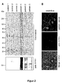
- 238000010185 immunofluorescence analysis Methods 0.000 description 14
- 235000018102 proteins Nutrition 0.000 description 14
- 239000012634 fragment Substances 0.000 description 13
- MMWCIQZXVOZEGG-UHFFFAOYSA-N 1,4,5-IP3 Natural products OC1C(O)C(OP(O)(O)=O)C(OP(O)(O)=O)C(O)C1OP(O)(O)=O MMWCIQZXVOZEGG-UHFFFAOYSA-N 0.000 description 10
- MMWCIQZXVOZEGG-HOZKJCLWSA-N [(1S,2R,3S,4S,5R,6S)-2,3,5-trihydroxy-4,6-diphosphonooxycyclohexyl] dihydrogen phosphate Chemical compound O[C@H]1[C@@H](O)[C@H](OP(O)(O)=O)[C@@H](OP(O)(O)=O)[C@H](O)[C@H]1OP(O)(O)=O MMWCIQZXVOZEGG-HOZKJCLWSA-N 0.000 description 10
- MMWCIQZXVOZEGG-XJTPDSDZSA-N D-myo-Inositol 1,4,5-trisphosphate Chemical compound O[C@@H]1[C@H](O)[C@@H](OP(O)(O)=O)[C@H](OP(O)(O)=O)[C@@H](O)[C@@H]1OP(O)(O)=O MMWCIQZXVOZEGG-XJTPDSDZSA-N 0.000 description 9
- 241000282414 Homo sapiens Species 0.000 description 9
- 108091008585 IP3 receptors Proteins 0.000 description 9
- 238000001514 detection method Methods 0.000 description 9
- 238000002415 sodium dodecyl sulfate polyacrylamide gel electrophoresis Methods 0.000 description 9
- 239000003795 chemical substances by application Substances 0.000 description 8
- 230000000694 effects Effects 0.000 description 8
- 239000007788 liquid Substances 0.000 description 8
- 108091006146 Channels Proteins 0.000 description 7
- 108010032354 Inositol 1,4,5-Trisphosphate Receptors Proteins 0.000 description 7
- 241000700159 Rattus Species 0.000 description 7
- 239000011324 bead Substances 0.000 description 7
- 239000011575 calcium Substances 0.000 description 7
- 238000006243 chemical reaction Methods 0.000 description 7
- 239000003153 chemical reaction reagent Substances 0.000 description 7
- 238000009396 hybridization Methods 0.000 description 7
- 238000003018 immunoassay Methods 0.000 description 7
- 238000010166 immunofluorescence Methods 0.000 description 7
- 239000012528 membrane Substances 0.000 description 7
- 238000002360 preparation method Methods 0.000 description 7
- 238000005406 washing Methods 0.000 description 7
- OYPRJOBELJOOCE-UHFFFAOYSA-N Calcium Chemical compound [Ca] OYPRJOBELJOOCE-UHFFFAOYSA-N 0.000 description 6
- 108091028043 Nucleic acid sequence Proteins 0.000 description 6
- 238000003556 assay Methods 0.000 description 6
- 230000004071 biological effect Effects 0.000 description 6
- 239000000872 buffer Substances 0.000 description 6
- 229910052791 calcium Inorganic materials 0.000 description 6
- 230000002163 immunogen Effects 0.000 description 6
- 238000010186 staining Methods 0.000 description 6
- 238000000018 DNA microarray Methods 0.000 description 5
- 101001091984 Homo sapiens Rho GTPase-activating protein 26 Proteins 0.000 description 5
- 102100035744 Rho GTPase-activating protein 26 Human genes 0.000 description 5
- 238000001042 affinity chromatography Methods 0.000 description 5
- 238000004458 analytical method Methods 0.000 description 5
- 239000007864 aqueous solution Substances 0.000 description 5
- 210000004369 blood Anatomy 0.000 description 5
- 208000035475 disorder Diseases 0.000 description 5
- 230000006870 function Effects 0.000 description 5
- 238000011534 incubation Methods 0.000 description 5
- 238000004949 mass spectrometry Methods 0.000 description 5
- 230000004048 modification Effects 0.000 description 5
- 238000012986 modification Methods 0.000 description 5
- 208000020016 psychiatric disease Diseases 0.000 description 5
- 238000001228 spectrum Methods 0.000 description 5
- 239000000758 substrate Substances 0.000 description 5
- 239000013598 vector Substances 0.000 description 5
- 238000001262 western blot Methods 0.000 description 5
- 102100026662 Delta and Notch-like epidermal growth factor-related receptor Human genes 0.000 description 4
- 108010077223 Homer Scaffolding Proteins Proteins 0.000 description 4
- 102000010029 Homer Scaffolding Proteins Human genes 0.000 description 4
- 101001054266 Homo sapiens Delta and Notch-like epidermal growth factor-related receptor Proteins 0.000 description 4
- 241000124008 Mammalia Species 0.000 description 4
- 241000699666 Mus <mouse, genus> Species 0.000 description 4
- FAPWRFPIFSIZLT-UHFFFAOYSA-M Sodium chloride Chemical compound [Na+].[Cl-] FAPWRFPIFSIZLT-UHFFFAOYSA-M 0.000 description 4
- 210000004899 c-terminal region Anatomy 0.000 description 4
- 210000001638 cerebellum Anatomy 0.000 description 4
- 238000002405 diagnostic procedure Methods 0.000 description 4
- 210000003527 eukaryotic cell Anatomy 0.000 description 4
- 239000011521 glass Substances 0.000 description 4
- 210000004962 mammalian cell Anatomy 0.000 description 4
- 230000026731 phosphorylation Effects 0.000 description 4
- 238000006366 phosphorylation reaction Methods 0.000 description 4
- 238000001742 protein purification Methods 0.000 description 4
- 102000005962 receptors Human genes 0.000 description 4
- 108020003175 receptors Proteins 0.000 description 4
- 150000003839 salts Chemical class 0.000 description 4
- 230000000007 visual effect Effects 0.000 description 4
- CSCPPACGZOOCGX-UHFFFAOYSA-N Acetone Chemical compound CC(C)=O CSCPPACGZOOCGX-UHFFFAOYSA-N 0.000 description 3
- 102100035359 Cerebellar degeneration-related protein 2-like Human genes 0.000 description 3
- 101000737792 Homo sapiens Cerebellar degeneration-related protein 2-like Proteins 0.000 description 3
- 101001071437 Homo sapiens Metabotropic glutamate receptor 1 Proteins 0.000 description 3
- 101001032845 Homo sapiens Metabotropic glutamate receptor 5 Proteins 0.000 description 3
- -1 IgLON5 Proteins 0.000 description 3
- 108060003951 Immunoglobulin Proteins 0.000 description 3
- 102100036834 Metabotropic glutamate receptor 1 Human genes 0.000 description 3
- 102100038357 Metabotropic glutamate receptor 5 Human genes 0.000 description 3
- 101100310657 Mus musculus Sox1 gene Proteins 0.000 description 3
- 206010028980 Neoplasm Diseases 0.000 description 3
- 239000000020 Nitrocellulose Substances 0.000 description 3
- 241000288906 Primates Species 0.000 description 3
- 102000007056 Recombinant Fusion Proteins Human genes 0.000 description 3
- 108010008281 Recombinant Fusion Proteins Proteins 0.000 description 3
- 241000282898 Sus scrofa Species 0.000 description 3
- 238000013459 approach Methods 0.000 description 3
- 201000011510 cancer Diseases 0.000 description 3
- 239000006059 cover glass Substances 0.000 description 3
- 230000001419 dependent effect Effects 0.000 description 3
- 238000013461 design Methods 0.000 description 3
- 229940079593 drug Drugs 0.000 description 3
- 239000003814 drug Substances 0.000 description 3
- 230000014509 gene expression Effects 0.000 description 3
- 102000018358 immunoglobulin Human genes 0.000 description 3
- 239000012133 immunoprecipitate Substances 0.000 description 3
- 239000003018 immunosuppressive agent Substances 0.000 description 3
- 239000003446 ligand Substances 0.000 description 3
- 239000011159 matrix material Substances 0.000 description 3
- 239000013642 negative control Substances 0.000 description 3
- 229920001220 nitrocellulos Polymers 0.000 description 3
- 238000000955 peptide mass fingerprinting Methods 0.000 description 3
- 238000000746 purification Methods 0.000 description 3
- 230000002441 reversible effect Effects 0.000 description 3
- 230000035945 sensitivity Effects 0.000 description 3
- ORJCWNHUOREFAT-UHFFFAOYSA-N 7,8-dimethylquinoxalino[2,3-f][1,10]phenanthroline Chemical compound C1=CC=C2N=C(C=3C(=NC=C(C=3C)C)C=3C4=CC=CN=3)C4=NC2=C1 ORJCWNHUOREFAT-UHFFFAOYSA-N 0.000 description 2
- 102000003678 AMPA Receptors Human genes 0.000 description 2
- 108090000078 AMPA Receptors Proteins 0.000 description 2
- 208000024827 Alzheimer disease Diseases 0.000 description 2
- 108010036280 Aquaporin 4 Proteins 0.000 description 2
- 102000012002 Aquaporin 4 Human genes 0.000 description 2
- 241000283690 Bos taurus Species 0.000 description 2
- 241000283707 Capra Species 0.000 description 2
- 206010008025 Cerebellar ataxia Diseases 0.000 description 2
- 102100040499 Contactin-associated protein-like 2 Human genes 0.000 description 2
- 108020004414 DNA Proteins 0.000 description 2
- 102100024441 Dihydropyrimidinase-related protein 5 Human genes 0.000 description 2
- 102100036966 Dipeptidyl aminopeptidase-like protein 6 Human genes 0.000 description 2
- 101710092625 Dipeptidyl aminopeptidase-like protein 6 Proteins 0.000 description 2
- 238000002965 ELISA Methods 0.000 description 2
- 102000004190 Enzymes Human genes 0.000 description 2
- 108090000790 Enzymes Proteins 0.000 description 2
- 241000283074 Equus asinus Species 0.000 description 2
- 241000283073 Equus caballus Species 0.000 description 2
- 102000004300 GABA-A Receptors Human genes 0.000 description 2
- 108090000839 GABA-A Receptors Proteins 0.000 description 2
- 102000017934 GABA-B receptor Human genes 0.000 description 2
- 108060003377 GABA-B receptor Proteins 0.000 description 2
- 102100035857 Glutamate decarboxylase 2 Human genes 0.000 description 2
- 102000011714 Glycine Receptors Human genes 0.000 description 2
- 108010076533 Glycine Receptors Proteins 0.000 description 2
- 241000238631 Hexapoda Species 0.000 description 2
- 101000749877 Homo sapiens Contactin-associated protein-like 2 Proteins 0.000 description 2
- 101001053479 Homo sapiens Dihydropyrimidinase-related protein 5 Proteins 0.000 description 2
- 101000873786 Homo sapiens Glutamate decarboxylase 2 Proteins 0.000 description 2
- 101000975421 Homo sapiens Inositol 1,4,5-trisphosphate receptor type 2 Proteins 0.000 description 2
- 101000975401 Homo sapiens Inositol 1,4,5-trisphosphate receptor type 3 Proteins 0.000 description 2
- 101000620451 Homo sapiens Leucine-rich glioma-inactivated protein 1 Proteins 0.000 description 2
- 101001115699 Homo sapiens Myelin-oligodendrocyte glycoprotein Proteins 0.000 description 2
- 101001094802 Homo sapiens Paraneoplastic antigen Ma1 Proteins 0.000 description 2
- 101001094820 Homo sapiens Paraneoplastic antigen Ma2 Proteins 0.000 description 2
- 101710170984 Inositol 1,4,5-trisphosphate receptor type 1 Proteins 0.000 description 2
- 102100024037 Inositol 1,4,5-trisphosphate receptor type 2 Human genes 0.000 description 2
- 102100024035 Inositol 1,4,5-trisphosphate receptor type 3 Human genes 0.000 description 2
- 102100022275 Leucine-rich glioma-inactivated protein 1 Human genes 0.000 description 2
- 102100035971 Molybdopterin molybdenumtransferase Human genes 0.000 description 2
- 102100023302 Myelin-oligodendrocyte glycoprotein Human genes 0.000 description 2
- 102000004868 N-Methyl-D-Aspartate Receptors Human genes 0.000 description 2
- 108090001041 N-Methyl-D-Aspartate Receptors Proteins 0.000 description 2
- 102100035457 Paraneoplastic antigen Ma1 Human genes 0.000 description 2
- 102100035467 Paraneoplastic antigen Ma2 Human genes 0.000 description 2
- 208000018737 Parkinson disease Diseases 0.000 description 2
- 241001494479 Pecora Species 0.000 description 2
- 102000018210 Recoverin Human genes 0.000 description 2
- 108010076570 Recoverin Proteins 0.000 description 2
- 230000004913 activation Effects 0.000 description 2
- 230000001154 acute effect Effects 0.000 description 2
- 102000004111 amphiphysin Human genes 0.000 description 2
- 108090000686 amphiphysin Proteins 0.000 description 2
- 230000003460 anti-nuclear Effects 0.000 description 2
- 238000011091 antibody purification Methods 0.000 description 2
- 238000000149 argon plasma sintering Methods 0.000 description 2
- 230000001363 autoimmune Effects 0.000 description 2
- 230000008901 benefit Effects 0.000 description 2
- 210000004556 brain Anatomy 0.000 description 2
- 210000005013 brain tissue Anatomy 0.000 description 2
- 239000013592 cell lysate Substances 0.000 description 2
- 238000005119 centrifugation Methods 0.000 description 2
- 238000012512 characterization method Methods 0.000 description 2
- 238000007385 chemical modification Methods 0.000 description 2
- 230000002860 competitive effect Effects 0.000 description 2
- 230000000295 complement effect Effects 0.000 description 2
- 230000002596 correlated effect Effects 0.000 description 2
- 230000000875 corresponding effect Effects 0.000 description 2
- 230000007423 decrease Effects 0.000 description 2
- 230000007547 defect Effects 0.000 description 2
- 229940039227 diagnostic agent Drugs 0.000 description 2
- 239000000032 diagnostic agent Substances 0.000 description 2
- 238000013399 early diagnosis Methods 0.000 description 2
- 230000009881 electrostatic interaction Effects 0.000 description 2
- 238000002474 experimental method Methods 0.000 description 2
- 239000007850 fluorescent dye Substances 0.000 description 2
- 230000004927 fusion Effects 0.000 description 2
- 239000000499 gel Substances 0.000 description 2
- 108010024999 gephyrin Proteins 0.000 description 2
- 230000013595 glycosylation Effects 0.000 description 2
- 238000006206 glycosylation reaction Methods 0.000 description 2
- 239000005090 green fluorescent protein Substances 0.000 description 2
- 102000054344 human ITPR1 Human genes 0.000 description 2
- 210000005260 human cell Anatomy 0.000 description 2
- 229940072221 immunoglobulins Drugs 0.000 description 2
- 229940124589 immunosuppressive drug Drugs 0.000 description 2
- 230000001771 impaired effect Effects 0.000 description 2
- 208000014674 injury Diseases 0.000 description 2
- 230000003993 interaction Effects 0.000 description 2
- 238000001990 intravenous administration Methods 0.000 description 2
- 230000002427 irreversible effect Effects 0.000 description 2
- QWTDNUCVQCZILF-UHFFFAOYSA-N isopentane Chemical compound CCC(C)C QWTDNUCVQCZILF-UHFFFAOYSA-N 0.000 description 2
- 230000000155 isotopic effect Effects 0.000 description 2
- 238000004519 manufacturing process Methods 0.000 description 2
- 239000000463 material Substances 0.000 description 2
- 238000005259 measurement Methods 0.000 description 2
- 230000007246 mechanism Effects 0.000 description 2
- 239000000203 mixture Substances 0.000 description 2
- 230000002018 overexpression Effects 0.000 description 2
- 239000013641 positive control Substances 0.000 description 2
- 238000004393 prognosis Methods 0.000 description 2
- 230000002250 progressing effect Effects 0.000 description 2
- 210000000449 purkinje cell Anatomy 0.000 description 2
- 238000001542 size-exclusion chromatography Methods 0.000 description 2
- 239000011780 sodium chloride Substances 0.000 description 2
- 239000011537 solubilization buffer Substances 0.000 description 2
- 241000894007 species Species 0.000 description 2
- 230000009897 systematic effect Effects 0.000 description 2
- 238000001890 transfection Methods 0.000 description 2
- 229960005486 vaccine Drugs 0.000 description 2
- 230000004393 visual impairment Effects 0.000 description 2
- 210000005253 yeast cell Anatomy 0.000 description 2
- QKNYBSVHEMOAJP-UHFFFAOYSA-N 2-amino-2-(hydroxymethyl)propane-1,3-diol;hydron;chloride Chemical compound Cl.OCC(N)(CO)CO QKNYBSVHEMOAJP-UHFFFAOYSA-N 0.000 description 1
- 125000004042 4-aminobutyl group Chemical group [H]C([*])([H])C([H])([H])C([H])([H])C([H])([H])N([H])[H] 0.000 description 1
- QRXMUCSWCMTJGU-UHFFFAOYSA-N 5-bromo-4-chloro-3-indolyl phosphate Chemical compound C1=C(Br)C(Cl)=C2C(OP(O)(=O)O)=CNC2=C1 QRXMUCSWCMTJGU-UHFFFAOYSA-N 0.000 description 1
- 102000002260 Alkaline Phosphatase Human genes 0.000 description 1
- 108020004774 Alkaline Phosphatase Proteins 0.000 description 1
- 208000000044 Amnesia Diseases 0.000 description 1
- 208000031091 Amnestic disease Diseases 0.000 description 1
- 208000008958 Anti-N-Methyl-D-Aspartate Receptor Encephalitis Diseases 0.000 description 1
- 206010003062 Apraxia Diseases 0.000 description 1
- 208000036640 Asperger disease Diseases 0.000 description 1
- 201000006062 Asperger syndrome Diseases 0.000 description 1
- 108010078286 Ataxins Proteins 0.000 description 1
- 102000014461 Ataxins Human genes 0.000 description 1
- 206010003805 Autism Diseases 0.000 description 1
- 208000020706 Autistic disease Diseases 0.000 description 1
- 206010003840 Autonomic nervous system imbalance Diseases 0.000 description 1
- 208000008035 Back Pain Diseases 0.000 description 1
- 241000894006 Bacteria Species 0.000 description 1
- 201000004569 Blindness Diseases 0.000 description 1
- 108091003079 Bovine Serum Albumin Proteins 0.000 description 1
- 206010006187 Breast cancer Diseases 0.000 description 1
- 208000026310 Breast neoplasm Diseases 0.000 description 1
- 101150020019 CLA4 gene Proteins 0.000 description 1
- 101000975407 Caenorhabditis elegans Inositol 1,4,5-trisphosphate receptor itr-1 Proteins 0.000 description 1
- 102000003922 Calcium Channels Human genes 0.000 description 1
- 108090000312 Calcium Channels Proteins 0.000 description 1
- 102000000584 Calmodulin Human genes 0.000 description 1
- 108010041952 Calmodulin Proteins 0.000 description 1
- 101710086403 Carbonic anhydrase-related protein Proteins 0.000 description 1
- 241000282693 Cercopithecidae Species 0.000 description 1
- 102100035361 Cerebellar degeneration-related protein 2 Human genes 0.000 description 1
- 206010008748 Chorea Diseases 0.000 description 1
- 206010010904 Convulsion Diseases 0.000 description 1
- 238000011537 Coomassie blue staining Methods 0.000 description 1
- 208000011231 Crohn disease Diseases 0.000 description 1
- CMSMOCZEIVJLDB-UHFFFAOYSA-N Cyclophosphamide Chemical compound ClCCN(CCCl)P1(=O)NCCCO1 CMSMOCZEIVJLDB-UHFFFAOYSA-N 0.000 description 1
- PMATZTZNYRCHOR-CGLBZJNRSA-N Cyclosporin A Chemical compound CC[C@@H]1NC(=O)[C@H]([C@H](O)[C@H](C)C\C=C\C)N(C)C(=O)[C@H](C(C)C)N(C)C(=O)[C@H](CC(C)C)N(C)C(=O)[C@H](CC(C)C)N(C)C(=O)[C@@H](C)NC(=O)[C@H](C)NC(=O)[C@H](CC(C)C)N(C)C(=O)[C@H](C(C)C)NC(=O)[C@H](CC(C)C)N(C)C(=O)CN(C)C1=O PMATZTZNYRCHOR-CGLBZJNRSA-N 0.000 description 1
- 108010036949 Cyclosporine Proteins 0.000 description 1
- 241000252233 Cyprinus carpio Species 0.000 description 1
- 101000975393 Drosophila melanogaster Inositol 1,4,5-trisphosphate receptor Proteins 0.000 description 1
- 206010013654 Drug abuse Diseases 0.000 description 1
- KCXVZYZYPLLWCC-UHFFFAOYSA-N EDTA Chemical compound OC(=O)CN(CC(O)=O)CCN(CC(O)=O)CC(O)=O KCXVZYZYPLLWCC-UHFFFAOYSA-N 0.000 description 1
- 208000030814 Eating disease Diseases 0.000 description 1
- 241000196324 Embryophyta Species 0.000 description 1
- 206010072378 Encephalitis autoimmune Diseases 0.000 description 1
- 241000206602 Eukaryota Species 0.000 description 1
- 206010016059 Facial pain Diseases 0.000 description 1
- 208000019454 Feeding and Eating disease Diseases 0.000 description 1
- 208000015872 Gaucher disease Diseases 0.000 description 1
- 102000005720 Glutathione transferase Human genes 0.000 description 1
- 108010070675 Glutathione transferase Proteins 0.000 description 1
- 108010043121 Green Fluorescent Proteins Proteins 0.000 description 1
- 102000004144 Green Fluorescent Proteins Human genes 0.000 description 1
- 206010019233 Headaches Diseases 0.000 description 1
- 208000028782 Hereditary disease Diseases 0.000 description 1
- 208000017604 Hodgkin disease Diseases 0.000 description 1
- 208000021519 Hodgkin lymphoma Diseases 0.000 description 1
- 208000010747 Hodgkins lymphoma Diseases 0.000 description 1
- 241000282412 Homo Species 0.000 description 1
- 101000737796 Homo sapiens Cerebellar degeneration-related protein 2 Proteins 0.000 description 1
- 101100126614 Homo sapiens ITPR1 gene Proteins 0.000 description 1
- 206010021113 Hypothermia Diseases 0.000 description 1
- 206010021133 Hypoventilation Diseases 0.000 description 1
- 206010021143 Hypoxia Diseases 0.000 description 1
- 101150114237 ITPR1 gene Proteins 0.000 description 1
- 102000006496 Immunoglobulin Heavy Chains Human genes 0.000 description 1
- 108010019476 Immunoglobulin Heavy Chains Proteins 0.000 description 1
- 102000013463 Immunoglobulin Light Chains Human genes 0.000 description 1
- 108010065825 Immunoglobulin Light Chains Proteins 0.000 description 1
- 208000022559 Inflammatory bowel disease Diseases 0.000 description 1
- VHJLVAABSRFDPM-IMJSIDKUSA-N L-1,4-dithiothreitol Chemical compound SC[C@H](O)[C@@H](O)CS VHJLVAABSRFDPM-IMJSIDKUSA-N 0.000 description 1
- ONIBWKKTOPOVIA-BYPYZUCNSA-N L-Proline Chemical compound OC(=O)[C@@H]1CCCN1 ONIBWKKTOPOVIA-BYPYZUCNSA-N 0.000 description 1
- CKLJMWTZIZZHCS-REOHCLBHSA-N L-aspartic acid Chemical compound OC(=O)[C@@H](N)CC(O)=O CKLJMWTZIZZHCS-REOHCLBHSA-N 0.000 description 1
- FBOZXECLQNJBKD-ZDUSSCGKSA-N L-methotrexate Chemical compound C=1N=C2N=C(N)N=C(N)C2=NC=1CN(C)C1=CC=C(C(=O)N[C@@H](CCC(O)=O)C(O)=O)C=C1 FBOZXECLQNJBKD-ZDUSSCGKSA-N 0.000 description 1
- COLNVLDHVKWLRT-QMMMGPOBSA-N L-phenylalanine Chemical compound OC(=O)[C@@H](N)CC1=CC=CC=C1 COLNVLDHVKWLRT-QMMMGPOBSA-N 0.000 description 1
- OUYCCCASQSFEME-QMMMGPOBSA-N L-tyrosine Chemical compound OC(=O)[C@@H](N)CC1=CC=C(O)C=C1 OUYCCCASQSFEME-QMMMGPOBSA-N 0.000 description 1
- 206010058467 Lung neoplasm malignant Diseases 0.000 description 1
- 208000002720 Malnutrition Diseases 0.000 description 1
- 101710175625 Maltose/maltodextrin-binding periplasmic protein Proteins 0.000 description 1
- 206010026749 Mania Diseases 0.000 description 1
- 102000012750 Membrane Glycoproteins Human genes 0.000 description 1
- 108010090054 Membrane Glycoproteins Proteins 0.000 description 1
- 241001465754 Metazoa Species 0.000 description 1
- FQISKWAFAHGMGT-SGJOWKDISA-M Methylprednisolone sodium succinate Chemical compound [Na+].C([C@@]12C)=CC(=O)C=C1[C@@H](C)C[C@@H]1[C@@H]2[C@@H](O)C[C@]2(C)[C@@](O)(C(=O)COC(=O)CCC([O-])=O)CC[C@H]21 FQISKWAFAHGMGT-SGJOWKDISA-M 0.000 description 1
- 208000019022 Mood disease Diseases 0.000 description 1
- 208000021642 Muscular disease Diseases 0.000 description 1
- 201000009623 Myopathy Diseases 0.000 description 1
- 230000004988 N-glycosylation Effects 0.000 description 1
- 208000032580 NMDA receptor encephalitis Diseases 0.000 description 1
- 238000005481 NMR spectroscopy Methods 0.000 description 1
- 206010028836 Neck pain Diseases 0.000 description 1
- 206010029260 Neuroblastoma Diseases 0.000 description 1
- 208000015914 Non-Hodgkin lymphomas Diseases 0.000 description 1
- 206010033128 Ovarian cancer Diseases 0.000 description 1
- 206010061535 Ovarian neoplasm Diseases 0.000 description 1
- 206010033799 Paralysis Diseases 0.000 description 1
- 206010033892 Paraplegia Diseases 0.000 description 1
- 208000007542 Paresis Diseases 0.000 description 1
- 108700019535 Phosphoprotein Phosphatases Proteins 0.000 description 1
- 102000045595 Phosphoprotein Phosphatases Human genes 0.000 description 1
- 108091000080 Phosphotransferase Proteins 0.000 description 1
- 206010036105 Polyneuropathy Diseases 0.000 description 1
- 229920001213 Polysorbate 20 Polymers 0.000 description 1
- 239000004793 Polystyrene Substances 0.000 description 1
- ONIBWKKTOPOVIA-UHFFFAOYSA-N Proline Natural products OC(=O)C1CCCN1 ONIBWKKTOPOVIA-UHFFFAOYSA-N 0.000 description 1
- 102000005569 Protein Phosphatase 1 Human genes 0.000 description 1
- 108010059000 Protein Phosphatase 1 Proteins 0.000 description 1
- 206010041067 Small cell lung cancer Diseases 0.000 description 1
- DBMJMQXJHONAFJ-UHFFFAOYSA-M Sodium laurylsulphate Chemical compound [Na+].CCCCCCCCCCCCOS([O-])(=O)=O DBMJMQXJHONAFJ-UHFFFAOYSA-M 0.000 description 1
- 208000009415 Spinocerebellar Ataxias Diseases 0.000 description 1
- 208000006011 Stroke Diseases 0.000 description 1
- QJJXYPPXXYFBGM-LFZNUXCKSA-N Tacrolimus Chemical compound C1C[C@@H](O)[C@H](OC)C[C@@H]1\C=C(/C)[C@@H]1[C@H](C)[C@@H](O)CC(=O)[C@H](CC=C)/C=C(C)/C[C@H](C)C[C@H](OC)[C@H]([C@H](C[C@H]2C)OC)O[C@@]2(O)C(=O)C(=O)N2CCCC[C@H]2C(=O)O1 QJJXYPPXXYFBGM-LFZNUXCKSA-N 0.000 description 1
- 206010043276 Teratoma Diseases 0.000 description 1
- 102100036407 Thioredoxin Human genes 0.000 description 1
- 101710120037 Toxin CcdB Proteins 0.000 description 1
- 208000031674 Traumatic Acute Stress disease Diseases 0.000 description 1
- 239000013504 Triton X-100 Substances 0.000 description 1
- 229920004890 Triton X-100 Polymers 0.000 description 1
- 102100024205 Ubiquitin carboxyl-terminal hydrolase MINDY-3 Human genes 0.000 description 1
- 208000003443 Unconsciousness Diseases 0.000 description 1
- 208000007097 Urinary Bladder Neoplasms Diseases 0.000 description 1
- 241000251539 Vertebrata <Metazoa> Species 0.000 description 1
- 206010047571 Visual impairment Diseases 0.000 description 1
- 208000027418 Wounds and injury Diseases 0.000 description 1
- 241000269368 Xenopus laevis Species 0.000 description 1
- 101100127670 Zea mays LA1 gene Proteins 0.000 description 1
- YVNQAIFQFWTPLQ-UHFFFAOYSA-O [4-[[4-(4-ethoxyanilino)phenyl]-[4-[ethyl-[(3-sulfophenyl)methyl]amino]-2-methylphenyl]methylidene]-3-methylcyclohexa-2,5-dien-1-ylidene]-ethyl-[(3-sulfophenyl)methyl]azanium Chemical compound C1=CC(OCC)=CC=C1NC1=CC=C(C(=C2C(=CC(C=C2)=[N+](CC)CC=2C=C(C=CC=2)S(O)(=O)=O)C)C=2C(=CC(=CC=2)N(CC)CC=2C=C(C=CC=2)S(O)(=O)=O)C)C=C1 YVNQAIFQFWTPLQ-UHFFFAOYSA-O 0.000 description 1
- 230000021736 acetylation Effects 0.000 description 1
- 238000006640 acetylation reaction Methods 0.000 description 1
- 208000026345 acute stress disease Diseases 0.000 description 1
- 208000009956 adenocarcinoma Diseases 0.000 description 1
- 230000004520 agglutination Effects 0.000 description 1
- 239000000556 agonist Substances 0.000 description 1
- 125000003277 amino group Chemical group 0.000 description 1
- 230000006986 amnesia Effects 0.000 description 1
- 238000000137 annealing Methods 0.000 description 1
- 208000029188 anti-NMDA receptor encephalitis Diseases 0.000 description 1
- 230000009830 antibody antigen interaction Effects 0.000 description 1
- 201000007201 aphasia Diseases 0.000 description 1
- 238000002617 apheresis Methods 0.000 description 1
- 239000007900 aqueous suspension Substances 0.000 description 1
- 229940009098 aspartate Drugs 0.000 description 1
- 230000005784 autoimmunity Effects 0.000 description 1
- 201000004562 autosomal dominant cerebellar ataxia Diseases 0.000 description 1
- 229960002170 azathioprine Drugs 0.000 description 1
- LMEKQMALGUDUQG-UHFFFAOYSA-N azathioprine Chemical compound CN1C=NC([N+]([O-])=O)=C1SC1=NC=NC2=C1NC=N2 LMEKQMALGUDUQG-UHFFFAOYSA-N 0.000 description 1
- 230000003542 behavioural effect Effects 0.000 description 1
- 230000008827 biological function Effects 0.000 description 1
- 230000033228 biological regulation Effects 0.000 description 1
- 229940098773 bovine serum albumin Drugs 0.000 description 1
- 201000008275 breast carcinoma Diseases 0.000 description 1
- 238000004364 calculation method Methods 0.000 description 1
- 238000011088 calibration curve Methods 0.000 description 1
- 239000002775 capsule Substances 0.000 description 1
- 238000007623 carbamidomethylation reaction Methods 0.000 description 1
- 239000000969 carrier Substances 0.000 description 1
- 206010007776 catatonia Diseases 0.000 description 1
- 239000006285 cell suspension Substances 0.000 description 1
- 210000003169 central nervous system Anatomy 0.000 description 1
- 230000002490 cerebral effect Effects 0.000 description 1
- 208000012601 choreatic disease Diseases 0.000 description 1
- 238000004587 chromatography analysis Methods 0.000 description 1
- 239000012501 chromatography medium Substances 0.000 description 1
- 239000003593 chromogenic compound Substances 0.000 description 1
- 230000001684 chronic effect Effects 0.000 description 1
- 229960001265 ciclosporin Drugs 0.000 description 1
- 238000000978 circular dichroism spectroscopy Methods 0.000 description 1
- 230000006329 citrullination Effects 0.000 description 1
- 238000003776 cleavage reaction Methods 0.000 description 1
- 238000010367 cloning Methods 0.000 description 1
- 230000004186 co-expression Effects 0.000 description 1
- 238000004440 column chromatography Methods 0.000 description 1
- 230000000052 comparative effect Effects 0.000 description 1
- 230000006957 competitive inhibition Effects 0.000 description 1
- 238000012790 confirmation Methods 0.000 description 1
- 230000008878 coupling Effects 0.000 description 1
- 238000010168 coupling process Methods 0.000 description 1
- 238000005859 coupling reaction Methods 0.000 description 1
- 239000003431 cross linking reagent Substances 0.000 description 1
- 238000012258 culturing Methods 0.000 description 1
- 229960004397 cyclophosphamide Drugs 0.000 description 1
- 229930182912 cyclosporin Natural products 0.000 description 1
- 125000000151 cysteine group Chemical group N[C@@H](CS)C(=O)* 0.000 description 1
- 230000001086 cytosolic effect Effects 0.000 description 1
- 230000006378 damage Effects 0.000 description 1
- 238000006114 decarboxylation reaction Methods 0.000 description 1
- 230000003247 decreasing effect Effects 0.000 description 1
- 229940009976 deoxycholate Drugs 0.000 description 1
- KXGVEGMKQFWNSR-LLQZFEROSA-N deoxycholic acid Chemical compound C([C@H]1CC2)[C@H](O)CC[C@]1(C)[C@@H]1[C@@H]2[C@@H]2CC[C@H]([C@@H](CCC(O)=O)C)[C@@]2(C)[C@@H](O)C1 KXGVEGMKQFWNSR-LLQZFEROSA-N 0.000 description 1
- 230000030609 dephosphorylation Effects 0.000 description 1
- 238000006209 dephosphorylation reaction Methods 0.000 description 1
- 238000002059 diagnostic imaging Methods 0.000 description 1
- 238000003748 differential diagnosis Methods 0.000 description 1
- 230000004069 differentiation Effects 0.000 description 1
- AFABGHUZZDYHJO-UHFFFAOYSA-N dimethyl butane Natural products CCCC(C)C AFABGHUZZDYHJO-UHFFFAOYSA-N 0.000 description 1
- 229940042399 direct acting antivirals protease inhibitors Drugs 0.000 description 1
- 235000014632 disordered eating Nutrition 0.000 description 1
- 238000010494 dissociation reaction Methods 0.000 description 1
- 230000005593 dissociations Effects 0.000 description 1
- 238000009826 distribution Methods 0.000 description 1
- VHJLVAABSRFDPM-QWWZWVQMSA-N dithiothreitol Chemical compound SC[C@@H](O)[C@H](O)CS VHJLVAABSRFDPM-QWWZWVQMSA-N 0.000 description 1
- 239000002552 dosage form Substances 0.000 description 1
- 231100000673 dose–response relationship Toxicity 0.000 description 1
- 238000012137 double-staining Methods 0.000 description 1
- 239000003937 drug carrier Substances 0.000 description 1
- 238000005538 encapsulation Methods 0.000 description 1
- 206010015037 epilepsy Diseases 0.000 description 1
- 125000003700 epoxy group Chemical group 0.000 description 1
- 239000003797 essential amino acid Substances 0.000 description 1
- 235000020776 essential amino acid Nutrition 0.000 description 1
- 238000011156 evaluation Methods 0.000 description 1
- 239000013604 expression vector Substances 0.000 description 1
- 239000003889 eye drop Substances 0.000 description 1
- 229940012356 eye drops Drugs 0.000 description 1
- 230000008713 feedback mechanism Effects 0.000 description 1
- 238000001914 filtration Methods 0.000 description 1
- 239000012530 fluid Substances 0.000 description 1
- GNBHRKFJIUUOQI-UHFFFAOYSA-N fluorescein Chemical compound O1C(=O)C2=CC=CC=C2C21C1=CC=C(O)C=C1OC1=CC(O)=CC=C21 GNBHRKFJIUUOQI-UHFFFAOYSA-N 0.000 description 1
- 238000000799 fluorescence microscopy Methods 0.000 description 1
- 238000005194 fractionation Methods 0.000 description 1
- 102000037865 fusion proteins Human genes 0.000 description 1
- 108020001507 fusion proteins Proteins 0.000 description 1
- 238000002523 gelfiltration Methods 0.000 description 1
- 238000012215 gene cloning Methods 0.000 description 1
- 102000034356 gene-regulatory proteins Human genes 0.000 description 1
- 108091006104 gene-regulatory proteins Proteins 0.000 description 1
- 230000002068 genetic effect Effects 0.000 description 1
- 238000010353 genetic engineering Methods 0.000 description 1
- PJJJBBJSCAKJQF-UHFFFAOYSA-N guanidinium chloride Chemical compound [Cl-].NC(N)=[NH2+] PJJJBBJSCAKJQF-UHFFFAOYSA-N 0.000 description 1
- 231100000869 headache Toxicity 0.000 description 1
- 206010019465 hemiparesis Diseases 0.000 description 1
- 210000001320 hippocampus Anatomy 0.000 description 1
- 230000002209 hydrophobic effect Effects 0.000 description 1
- 238000004191 hydrophobic interaction chromatography Methods 0.000 description 1
- 230000033444 hydroxylation Effects 0.000 description 1
- 238000005805 hydroxylation reaction Methods 0.000 description 1
- 230000002631 hypothermal effect Effects 0.000 description 1
- 230000007954 hypoxia Effects 0.000 description 1
- 230000003100 immobilizing effect Effects 0.000 description 1
- 230000001900 immune effect Effects 0.000 description 1
- 230000000951 immunodiffusion Effects 0.000 description 1
- 238000010820 immunofluorescence microscopy Methods 0.000 description 1
- 230000005847 immunogenicity Effects 0.000 description 1
- 238000001114 immunoprecipitation Methods 0.000 description 1
- 230000001506 immunosuppresive effect Effects 0.000 description 1
- 229960003444 immunosuppressant agent Drugs 0.000 description 1
- 238000002650 immunosuppressive therapy Methods 0.000 description 1
- 230000002779 inactivation Effects 0.000 description 1
- 208000015181 infectious disease Diseases 0.000 description 1
- 238000001802 infusion Methods 0.000 description 1
- 230000002401 inhibitory effect Effects 0.000 description 1
- 238000002347 injection Methods 0.000 description 1
- 239000007924 injection Substances 0.000 description 1
- 230000002452 interceptive effect Effects 0.000 description 1
- 230000003834 intracellular effect Effects 0.000 description 1
- 238000007917 intracranial administration Methods 0.000 description 1
- 230000008863 intramolecular interaction Effects 0.000 description 1
- 238000007918 intramuscular administration Methods 0.000 description 1
- 238000007919 intrasynovial administration Methods 0.000 description 1
- 238000007913 intrathecal administration Methods 0.000 description 1
- PGLTVOMIXTUURA-UHFFFAOYSA-N iodoacetamide Chemical compound NC(=O)CI PGLTVOMIXTUURA-UHFFFAOYSA-N 0.000 description 1
- 238000004255 ion exchange chromatography Methods 0.000 description 1
- 150000002500 ions Chemical class 0.000 description 1
- 238000002955 isolation Methods 0.000 description 1
- 230000000670 limiting effect Effects 0.000 description 1
- 150000002632 lipids Chemical class 0.000 description 1
- 238000011068 loading method Methods 0.000 description 1
- 230000004807 localization Effects 0.000 description 1
- 208000018769 loss of vision Diseases 0.000 description 1
- 231100000864 loss of vision Toxicity 0.000 description 1
- 201000005202 lung cancer Diseases 0.000 description 1
- 208000020816 lung neoplasm Diseases 0.000 description 1
- 210000002751 lymph Anatomy 0.000 description 1
- 239000006166 lysate Substances 0.000 description 1
- 238000012423 maintenance Methods 0.000 description 1
- 230000001071 malnutrition Effects 0.000 description 1
- 235000000824 malnutrition Nutrition 0.000 description 1
- 238000000816 matrix-assisted laser desorption--ionisation Methods 0.000 description 1
- 238000000074 matrix-assisted laser desorption--ionisation tandem time-of-flight detection Methods 0.000 description 1
- 230000001404 mediated effect Effects 0.000 description 1
- 238000002844 melting Methods 0.000 description 1
- 230000008018 melting Effects 0.000 description 1
- 230000003340 mental effect Effects 0.000 description 1
- 239000002207 metabolite Substances 0.000 description 1
- 229910052751 metal Inorganic materials 0.000 description 1
- 239000002184 metal Substances 0.000 description 1
- 125000001360 methionine group Chemical group N[C@@H](CCSC)C(=O)* 0.000 description 1
- 229960000485 methotrexate Drugs 0.000 description 1
- 230000011987 methylation Effects 0.000 description 1
- 238000007069 methylation reaction Methods 0.000 description 1
- 229960004584 methylprednisolone Drugs 0.000 description 1
- 238000007431 microscopic evaluation Methods 0.000 description 1
- 238000000386 microscopy Methods 0.000 description 1
- 238000010369 molecular cloning Methods 0.000 description 1
- 238000012544 monitoring process Methods 0.000 description 1
- 238000010995 multi-dimensional NMR spectroscopy Methods 0.000 description 1
- 238000012433 multimodal chromatography Methods 0.000 description 1
- 201000006417 multiple sclerosis Diseases 0.000 description 1
- 230000035772 mutation Effects 0.000 description 1
- 206010028417 myasthenia gravis Diseases 0.000 description 1
- RTGDFNSFWBGLEC-SYZQJQIISA-N mycophenolate mofetil Chemical compound COC1=C(C)C=2COC(=O)C=2C(O)=C1C\C=C(/C)CCC(=O)OCCN1CCOCC1 RTGDFNSFWBGLEC-SYZQJQIISA-N 0.000 description 1
- 229960004866 mycophenolate mofetil Drugs 0.000 description 1
- 210000000653 nervous system Anatomy 0.000 description 1
- 208000008795 neuromyelitis optica Diseases 0.000 description 1
- 210000002569 neuron Anatomy 0.000 description 1
- 239000002773 nucleotide Substances 0.000 description 1
- 125000003729 nucleotide group Chemical group 0.000 description 1
- 208000015380 nutritional deficiency disease Diseases 0.000 description 1
- 210000000287 oocyte Anatomy 0.000 description 1
- 230000003647 oxidation Effects 0.000 description 1
- 238000007254 oxidation reaction Methods 0.000 description 1
- 210000001819 pancreatic juice Anatomy 0.000 description 1
- 208000019906 panic disease Diseases 0.000 description 1
- 239000000123 paper Substances 0.000 description 1
- 239000013610 patient sample Substances 0.000 description 1
- 239000000137 peptide hydrolase inhibitor Substances 0.000 description 1
- 208000022821 personality disease Diseases 0.000 description 1
- COLNVLDHVKWLRT-UHFFFAOYSA-N phenylalanine Natural products OC(=O)C(N)CC1=CC=CC=C1 COLNVLDHVKWLRT-UHFFFAOYSA-N 0.000 description 1
- 102000020233 phosphotransferase Human genes 0.000 description 1
- 229920002401 polyacrylamide Polymers 0.000 description 1
- 230000007824 polyneuropathy Effects 0.000 description 1
- 239000000256 polyoxyethylene sorbitan monolaurate Substances 0.000 description 1
- 235000010486 polyoxyethylene sorbitan monolaurate Nutrition 0.000 description 1
- 229920002223 polystyrene Polymers 0.000 description 1
- 239000011148 porous material Substances 0.000 description 1
- 239000002244 precipitate Substances 0.000 description 1
- 229960004618 prednisone Drugs 0.000 description 1
- XOFYZVNMUHMLCC-ZPOLXVRWSA-N prednisone Chemical compound O=C1C=C[C@]2(C)[C@H]3C(=O)C[C@](C)([C@@](CC4)(O)C(=O)CO)[C@@H]4[C@@H]3CCC2=C1 XOFYZVNMUHMLCC-ZPOLXVRWSA-N 0.000 description 1
- 230000008569 process Effects 0.000 description 1
- 238000012545 processing Methods 0.000 description 1
- 230000000750 progressive effect Effects 0.000 description 1
- 210000001236 prokaryotic cell Anatomy 0.000 description 1
- 230000017854 proteolysis Effects 0.000 description 1
- 230000002285 radioactive effect Effects 0.000 description 1
- 238000003127 radioimmunoassay Methods 0.000 description 1
- 230000009257 reactivity Effects 0.000 description 1
- 230000002829 reductive effect Effects 0.000 description 1
- 230000001105 regulatory effect Effects 0.000 description 1
- 238000011160 research Methods 0.000 description 1
- 230000004043 responsiveness Effects 0.000 description 1
- 229960004641 rituximab Drugs 0.000 description 1
- 201000000980 schizophrenia Diseases 0.000 description 1
- 230000007017 scission Effects 0.000 description 1
- 238000012216 screening Methods 0.000 description 1
- 239000013049 sediment Substances 0.000 description 1
- 229910052710 silicon Inorganic materials 0.000 description 1
- 239000010703 silicon Substances 0.000 description 1
- 208000000587 small cell lung carcinoma Diseases 0.000 description 1
- 229940083575 sodium dodecyl sulfate Drugs 0.000 description 1
- 235000019333 sodium laurylsulphate Nutrition 0.000 description 1
- 210000000278 spinal cord Anatomy 0.000 description 1
- 230000002269 spontaneous effect Effects 0.000 description 1
- 239000007921 spray Substances 0.000 description 1
- 239000012086 standard solution Substances 0.000 description 1
- 230000004936 stimulating effect Effects 0.000 description 1
- 238000007920 subcutaneous administration Methods 0.000 description 1
- 239000000126 substance Substances 0.000 description 1
- 208000011117 substance-related disease Diseases 0.000 description 1
- 229940031626 subunit vaccine Drugs 0.000 description 1
- 239000006228 supernatant Substances 0.000 description 1
- 238000002198 surface plasmon resonance spectroscopy Methods 0.000 description 1
- 201000000596 systemic lupus erythematosus Diseases 0.000 description 1
- 239000003826 tablet Substances 0.000 description 1
- 229960001967 tacrolimus Drugs 0.000 description 1
- QJJXYPPXXYFBGM-SHYZHZOCSA-N tacrolimus Natural products CO[C@H]1C[C@H](CC[C@@H]1O)C=C(C)[C@H]2OC(=O)[C@H]3CCCCN3C(=O)C(=O)[C@@]4(O)O[C@@H]([C@H](C[C@H]4C)OC)[C@@H](C[C@H](C)CC(=C[C@@H](CC=C)C(=O)C[C@H](O)[C@H]2C)C)OC QJJXYPPXXYFBGM-SHYZHZOCSA-N 0.000 description 1
- 238000004885 tandem mass spectrometry Methods 0.000 description 1
- 230000001225 therapeutic effect Effects 0.000 description 1
- 150000003573 thiols Chemical class 0.000 description 1
- 108060008226 thioredoxin Proteins 0.000 description 1
- 229940094937 thioredoxin Drugs 0.000 description 1
- 239000003970 toll like receptor agonist Substances 0.000 description 1
- 230000002463 transducing effect Effects 0.000 description 1
- 238000012546 transfer Methods 0.000 description 1
- 230000001052 transient effect Effects 0.000 description 1
- 230000008733 trauma Effects 0.000 description 1
- 238000011269 treatment regimen Methods 0.000 description 1
- OUYCCCASQSFEME-UHFFFAOYSA-N tyrosine Natural products OC(=O)C(N)CC1=CC=C(O)C=C1 OUYCCCASQSFEME-UHFFFAOYSA-N 0.000 description 1
- 208000029257 vision disease Diseases 0.000 description 1
- 238000011179 visual inspection Methods 0.000 description 1
- 238000002424 x-ray crystallography Methods 0.000 description 1
- AFVLVVWMAFSXCK-UHFFFAOYSA-N α-cyano-4-hydroxycinnamic acid Chemical compound OC(=O)C(C#N)=CC1=CC=C(O)C=C1 AFVLVVWMAFSXCK-UHFFFAOYSA-N 0.000 description 1
Images


Classifications
-
- G—PHYSICS
- G01—MEASURING; TESTING
- G01N—INVESTIGATING OR ANALYSING MATERIALS BY DETERMINING THEIR CHEMICAL OR PHYSICAL PROPERTIES
- G01N33/00—Investigating or analysing materials by specific methods not covered by groups G01N1/00 - G01N31/00
- G01N33/48—Biological material, e.g. blood, urine; Haemocytometers
- G01N33/50—Chemical analysis of biological material, e.g. blood, urine; Testing involving biospecific ligand binding methods; Immunological testing
- G01N33/53—Immunoassay; Biospecific binding assay; Materials therefor
- G01N33/564—Immunoassay; Biospecific binding assay; Materials therefor for pre-existing immune complex or autoimmune disease, i.e. systemic lupus erythematosus, rheumatoid arthritis, multiple sclerosis, rheumatoid factors or complement components C1-C9
-
- G—PHYSICS
- G01—MEASURING; TESTING
- G01N—INVESTIGATING OR ANALYSING MATERIALS BY DETERMINING THEIR CHEMICAL OR PHYSICAL PROPERTIES
- G01N33/00—Investigating or analysing materials by specific methods not covered by groups G01N1/00 - G01N31/00
- G01N33/48—Biological material, e.g. blood, urine; Haemocytometers
- G01N33/50—Chemical analysis of biological material, e.g. blood, urine; Testing involving biospecific ligand binding methods; Immunological testing
- G01N33/68—Chemical analysis of biological material, e.g. blood, urine; Testing involving biospecific ligand binding methods; Immunological testing involving proteins, peptides or amino acids
- G01N33/6893—Chemical analysis of biological material, e.g. blood, urine; Testing involving biospecific ligand binding methods; Immunological testing involving proteins, peptides or amino acids related to diseases not provided for elsewhere
- G01N33/6896—Neurological disorders, e.g. Alzheimer's disease
Landscapes
- Health & Medical Sciences (AREA)
- Life Sciences & Earth Sciences (AREA)
- Engineering & Computer Science (AREA)
- Immunology (AREA)
- Hematology (AREA)
- Biomedical Technology (AREA)
- Urology & Nephrology (AREA)
- Molecular Biology (AREA)
- Chemical & Material Sciences (AREA)
- Medicinal Chemistry (AREA)
- Biochemistry (AREA)
- Cell Biology (AREA)
- Pathology (AREA)
- Biotechnology (AREA)
- Food Science & Technology (AREA)
- General Physics & Mathematics (AREA)
- Physics & Mathematics (AREA)
- Analytical Chemistry (AREA)
- Microbiology (AREA)
- General Health & Medical Sciences (AREA)
- Rheumatology (AREA)
- Rehabilitation Therapy (AREA)
- Neurology (AREA)
- Neurosurgery (AREA)
- Proteomics, Peptides & Aminoacids (AREA)
- Peptides Or Proteins (AREA)
Abstract
Description
- The present invention relates to a method for diagnosing a disease, preferably a disease associated with neurological symptoms, comprising the step detecting in a sample from a patient an autoantibody binding to ITPR1; a polypeptide comprising ITPR1 or a variant thereof, which is immobilized, preferably on a solid carrier; a use of a polypeptide comprising ITPR1 or a variant thereof for the diagnosis of a disease, preferably comprising the step detecting autoantibodies binding to ITPR1; a polypeptide comprising ITPR1 or a variant thereof, preferably immobilized, more preferably on a solid carrier, for use in the treatment of a disease; an autoantibody, preferably an isolated autoantibody, binding to a polypeptide comprising ITPR1, wherein the autoantibody is preferably in complex with said polypeptide; a method for isolating an autoantibody binding to ITPR1, comprising the steps contacting a sample comprising the autoantibody with a polypeptide comprising ITPR1 or a variant thereof under conditions compatible with formation of a complex, wherein said autoantibody binds to said polypeptide, isolating the complex formed in step a), dissociating the complex isolated in step b), and separating the autoantibody from the polypeptide; a pharmaceutical composition comprising a polypeptide comprising ITPR1 or a variant thereof; a medical or diagnostic device comprising a polypeptide comprising ITPR1 or a variant thereof; and a test kit for the diagnosis of a disease, comprising a polypeptide comprising ITPR1 or a variant thereof, wherein preferably the test kit comprises, in addition, a means for detecting the complex comprising an autoantibody binding to ITPR1.
- Developing diagnostic systems for neurological diseases is a continuing challenge in biomedical science, not in the least because many symptoms encountered may be accounted for by a huge variety of causes including genetically-inherited diseases, drug abuse, malnutrition, infection, injury, psychiatric illness, immunological defects and cancer.
- Since a neurological disease is rarely associated with a characteristic pattern of clinical symptoms, it is often difficult to provide a reliable diagnosis solely based on the observation and examination of the patients affected or their medical history.
- The importance of an early diagnosis cannot be overemphasized. Many neurological disorders, most prominently Alzheimer's and Parkinson's diseases, cannot be cured, but drugs are available that may be used to slow down their progression. The earlier the diagnosis, the better the chances to exploit the spectrum of available drugs to the full benefit of the patient.
- This holds all the more true in the case of neurological diseases associated with autoantibodies. In some cases, the link between a specific detectable autoantibody and a condition is sufficiently strong to allow for an immediate diagnosis.
- But even if it is not, the detection of autoantibodies may point the physician in charge to therapeutic means that may be used to ameliorate the patient's condition. There is a variety of widely used immunosuppressants that may be used regardless of the nature of the autoantibody's target. Alternatively, apheresis may be used to remove autoantibodies from the patient's blood. In many cases, patients went on to lead a normal life following early diagnosis and treatment of a neurological autoimmune disease.
- Diagnostic assays based on the detection of autoantibodies may also corroborate the diagnosis of diseases other than those associated autoantibodies. If it turns out that a blood sample is devoid of specific autoantibodies, this is likely to help the physician in charge exclude a range of possibilities and thus narrow down the spectrum of plausible conditions.
- Examples of neurological conditions coinciding with the emergence of autoantibodies include Neuromyelitis optica, a disease characterised by loss of vision and spinal cord function, and anti-NMDA receptor encephalitis, which is associated with autonomic dysfunction, hypoventilation, cerebellar ataxia, hemiparesis, loss of consciousness, or catatonia. Whilst the involvement of autoantibodies and the nature of these conditions as such was previously poorly understood, many of these disease can now be diagnosed and treated efficiently owing to the availability of assays based on the detection of autoantibodies.
- Therefore, it is paramount that new approaches to distinguish neurological conditions associated with autoantibodies from others be developed.
- A problem underlying the present invention is to provide an agent and a method for diagnosing neurological disease, more specifically distinguishing diseases associated with neurological symptoms and the emergence of autoantibodies from other types of diseases.
- Another problem underlying the present invention is to provide an autoantibody that, when found in a liquid sample taken from a patient, indicates that said patient is suffering from an autoimmune disease associated with neurological symptoms.
- Another problem underlying the present invention is to provide an agent and a method for diagnosing and treating an autoimmune disease associated with neurological symptoms.
- The problem underlying the present invention is solved by the subject-matter of the attached independent and dependent claims.
- In a first aspect, the problem underlying the present invention is solved by a method for diagnosing a disease, preferably a disease associated with neurological symptoms, comprising the step detecting in a sample from a patient an autoantibody binding to ITPR1.
- In a preferred embodiment, the sample is a bodily fluid comprising antibodies, preferably selected from the group comprising whole-blood, serum, cerebrospinal fluid and saliva.
- In a second aspect, the problem underlying the present invention is solved by a polypeptide comprising ITPR1 or a variant thereof, which is immobilized, preferably on a solid carrier.
- In a third aspect, the problem underlying the present invention is solved by a use of a polypeptide comprising ITPR1 or a variant thereof for the diagnosis of a disease, preferably comprising the step detecting autoantibodies binding to ITPR1.
- In a 4th aspect, the problem underlying the present invention is solved by a polypeptide comprising ITPR1 or a variant thereof, preferably immobilized, more preferably on a solid carrier, for use in the treatment of a disease.
- In a 5th aspect, the problem underlying the present invention is solved by an autoantibody, preferably an isolated autoantibody, binding to a polypeptide comprising ITPR1, wherein the autoantibody is preferably in complex with said polypeptide.
- In a 6th aspect, the problem underlying the present invention is solved by a method for isolating an autoantibody binding to ITPR1, comprising the steps
- a) contacting a sample comprising the autoantibody with a polypeptide comprising ITPR1 or a variant thereof under conditions compatible with formation of a complex, wherein said autoantibody binds to said polypeptide,
- b) isolating the complex formed in step a),
- c) dissociating the complex isolated in step b), and
- d) separating the autoantibody from the polypeptide.
- In a 7th aspect, the problem underlying the present invention is solved by a pharmaceutical composition comprising a polypeptide comprising ITPR1 or a variant thereof.
- In an 8th aspect, the problem underlying the present invention is solved by a medical or diagnostic device comprising a polypeptide comprising ITPR1 or a variant thereof.
- In a 9th aspect, the problem underlying the present invention is solved by a test kit for the diagnosis of a disease, comprising a polypeptide comprising ITPR1 or a variant thereof,
wherein preferably the test kit comprises, in addition, a means for detecting the complex comprising an autoantibody binding to ITPR1. - In a preferred embodiment of any aspect of the present invention, the patient has or the disease is associated with one or more symptoms from the group comprising ataxia, dyskinesia, nystagmus, dysphagia, dysarthria, gait instability, cognitive impairment, depression and anxiety.
- In a preferred embodiment of any aspect of the present invention, the disease is associated with neurological or psychiatric symptoms and is preferably a neurological autoimmune disease.
- In a preferred embodiment of any aspect of the present invention, the disease is selected from the group comprising cerebellar syndrome, cerebellitis, movement disorder, paraneoplastic neurological syndrome, dementia, borderline syndrome, and bipolar disorder.
- In a preferred embodiment of any aspect of the present invention, the polypeptide is provided in the form of a cell comprising a nucleic acid encoding said polypeptide or in the form of a tissue comprising said polypeptide.
- In a preferred embodiment of any aspect of the present invention, the polypeptide is a recombinant and/or isolated polypeptide.
- The present invention is based on the inventors' surprising finding that autoantibodies to ITPR1 may be detected in samples from a number of patients suffering from neurological symptoms, but not in samples obtained from healthy subjects. The presence of such autoantibodies suggests that ITPR1 activity and function are impaired in patients having ITPR1 autoantibodies to the effect that neurological symptoms occur.
- The present invention relates to a polypeptide comprising a mammalian, preferably human ITPR1 or variants thereof, preferably immunogenic variants binding to ITPR1 autoantibodies. In a most preferred embodiment, the polypeptide comprises ITPR1 encoded by the data base code Q14643. Throughout this application, any data base codes cited refer to the Uniprot data base, more specifically the version accessible on-line on October 27, 2014. D26070.1 represents a nucleotide sequence encoding ITPR1.
- Inositol trisphosphate receptor type 1 (ITPR1; also referred to in the state of art as IP3R 1, IP3 receptor,
type 1 InsP3 receptor,inositol 1,4,5-triphosphate receptor,type 1protein phosphatase 1, and regulatory subunit 94type 1inositol 1,4,5-trisphosphate receptor) is a membrane glycoprotein complex acting as a Ca 2+ channel following activation by inositol trisphosphate (IP3). In humans it is encoded by the ITPR1 gene (also referred to in the state of art as ACV; CLA4; IP3R; IP3R1; SCA15; SCA16; SCA29; INSP3R1; PPP1R94). - IP3-gated intracellular Ca2+ release channels are composed of four IP3 receptor subunits (N. Maeda,T. Kawasaki, S. Nakade, N. Yokota, T. Taguchi, M. Kasai, and K. Mikoshiba, (1991) J. Biol. Chem. 266, 1109-1116). There are at least three types of IP3 receptor (ITPR1, ITPR2, and ITPR3) (T. Furuichi, and K. Mikoshiba, (1995) J. Neurochem. 64, 953-960), and they exist both as homo- and heterotetramers (T. Monkawa, A. Miyawaki, T. Sugiyama, H. Yoneshima, M. Yamamoto-Hino, T. Furuichi, T. Saruta, M. Hasegawa, and K. Mikoshiba, (1995) J. Biol. Chem. 270, 14700-14704).
- At the functional level, the ITPR1 can be divided into five distinct domains: an N-terminal suppressor domain (amino acids 1-225), an IP3-binding core (amino acids 226-578), a modulatory and transducing domain (amino acids 579-2275), a channel domain with 6 transmembrane helices (amino acids 2276-2589), and finally a C-terminal coupling domain (amino acids 2590-2749) (K. Uchida, H. Miyauchi, T. Furuichi, T. Michikawa, K. Mikoshiba, Critical regions for activation gating of the ). The IP3-binding core is an essential region for specific IP3 binding (F. Yoshikawa, M. Morita, T. Monkawa, T. Michikawa, T. Furuichi, and K. Mikoshiba. Mutation analysis of the ligand binding site of the ) and contains 11 basic amino acids essential for its activity (Bosanac et al., 2002).
- Apart from a number of short loops between the
transmembrane helices - IP3-induced Ca2+ release is a positively cooperative process (T. Meyer, T. Wensel, and L. Stryer, Kinetics of calcium channel opening by .; J. Hirota, T. Michikawa, A. Miyawaki, T. Furuichi, I. Okura, and K. Mikoshiba, Kinetics of calcium release by immunoaffinity-purified ; T. Michikawa, J. Hirota, S. Kawano, M. Hiraoka, M. Yamada, T. Furuichi, and K. Mikoshiba. Calmodulin mediates calcium-dependent inactivation of the ), i.e. the binding of at least two IP3 molecules to a single tetrameric IP3 receptor channel, is required for channel opening. ITPR1 is activated by IP3, wherein its affinity to IP3 is lower than ITPR2's, but higher than ITPR3's (C.L. Newton, G.A. Mignery, T.C. Südhof, Co-expression in vertebrate tissues and cell lines of ); Missiaen et al., 1998; Miyakawa et al., 1998).
- In addition to IP3, cytosolic Ca2+ functions as a co-agonist. Importantly, Ca2+ activates the IP3 receptor at low concentrations, typically below 300 nM, while it inhibits IP3 receptor at higher concentrations (Lino, 1990; E.A. Finch, T.J. Turner, S.M. Goldin, Calcium as a coagonist of .; Bezprozvanny et al., 1991; J.B. Parys, S.W. Sernett, S. DeLisle, P.M. Snyder, M.J. Welsh, K.P. Campbell, Isolation, characterization, and localization of the .).
- Luminal Ca2+ is also deemed important, and the depletion of the Ca2+ stores leads to a decreased IP3 receptor sensitivity (L. Missiaen, C.W. Taylor, M.J. Berridge, Spontaneous calcium release from inositol trisphosphate-sensitive calcium stores, Nature 352 (1991) 241-244; L. Missiaen, H. De Smedt, G. Droogmans, R. Casteels, Ca2+ release induced by ;; D.L. Nunn, C.W. Taylor, Luminal Ca2+ increases the sensitivity of Ca2+ stores to .; J.B. Parys, L. Missiaen, H. De Smedt, R. Casteels, Loading dependence of .).
- Modulation of IP3 receptor activity occurs by three main mechanisms: (1) the local environment, including pH, ATP and Mg2+ concentration and redox status; (2) its phosphorylation status, which depends on the activity of many different kinases and phosphatases, some of them forming a complex with the ITPR1; (3) regulatory proteins that directly affect IP3 receptor activity by either a stimulatory or an inhibitory way (Mikoshiba, 2007; J.B. Parys, H. De Smedt, ; V. Vanderheyden, B. Devogelaere, L. Missiaen, H. De Smedt, G. Bultynck, J.B. Parys, Regulation of ).
- The teachings of the present invention may not only be carried out using polypeptides, in particular a polypeptide comprising ITPR1 as encoded by the data base code Q14643, or nucleic acids having the exact sequences referred to in this application explicitly, for example by function, name, sequence or accession number, or implicitly, but also using variants of such polypeptides or nucleic acids.
- In a preferred embodiment, the term "variant", as used herein, may refer to at least one fragment of the full length sequence referred to, more specifically one or more amino acid or nucleic acid sequence which is, relative to the full-length sequence, truncated at one or both termini by one or more amino acids. Such a fragment comprises or encodes for a peptide having at least 6, 7, 8, 10, 12, 15, 20, 25, 50, 75, 100, 150 or 200 successive amino acids of the original sequence or a variant thereof. The total length of the variant may be at least 6, 7, 8, 9, 10, 11, 12, 20, 25, 30, 40, 50, 60, 70, 80, 90, 100 or more amino acids. In a preferred embodiment, the fragment comprises amino acids 1-2282, more preferably 1-1251, more preferably a peptide comprising residues 602 and/or and/or 1059 and/or 1562. In a most preferred embodiment, the fragment comprises an immunogenic epitope comprising residue 602. In a most preferred embodiment, the fragment comprises an immunogenic epitope comprising residue 1059. In a most preferred embodiment, the fragment comprises an immunogenic epitope comprising residue 1562.
- In another preferred embodiment, the term "variant" relates not only to at least one fragment, but also to a polypeptide or a fragment thereof comprising amino acid sequences that are at least 40, 50, 60, 70, 75, 80, 85, 90, 92, 94, 95, 96, 97, 98 or 99 % identical to the reference amino acid sequence referred to or the fragment thereof, wherein amino acids other than those essential for the biological activity, for example the ability of an antigen to bind to an (auto)antibody, or the fold or structure of the polypeptide are deleted or substituted and/or one or more such essential amino acids are replaced in a conservative manner and/or amino acids are added such that the biological activity of the polypeptide is preserved. The state of the art comprises various methods that may be used to align two given nucleic acid or amino acid sequences and to calculate the degree of identity, see for example Arthur Lesk (2008), Introduction to bioinformatics, Oxford University Press, 2008, 3rd edition. In a preferred embodiment, the ClustalW software (Larkin, M. A., Blackshields, G., Brown, N. P., Chenna, R., McGettigan, P. A., McWilliam, H., Valentin, F., Wallace, I. M., Wilm, A., Lopez, R., Thompson, J. D., Gibson, T. J., Higgins, D. G. (2007). Clustal W and Clustal X version 2.0. Bioinformatics, 23, 2947-2948) is used using default settings.
- In a preferred embodiment, variants may, in addition, comprise chemical modifications, for example isotopic labels or covalent modifications such as glycosylation, phosphorylation, acetylation, decarboxylation, citrullination, hydroxylation and the like. The person skilled in the art is familiar with methods to modify polypeptides. Any modification is designed such that it does not abolish the biological activity of the variant.
- Moreover, variants may also be generated by fusion with other known polypeptides or variants thereof and comprise active portions or domains, preferably having a sequence identity of at least 70, 75, 80, 85, 90, 92, 94, 95, 96, 97, 98 or 99 % when aligned with the active portion of the reference sequence, wherein the term "active portion", as used herein, refers to an amino acid sequence, which is less than the full length amino acid sequence or, in the case of a nucleic acid sequence, codes for less than the full length amino acid sequence, respectively, and/or is a variant of the natural sequence, but retains at least some of the biological activity.
- In a preferred embodiment, the term "variant" of a nucleic acid comprises nucleic acids the complementary strand of which hybridizes, preferably under stringent conditions, to the reference or wild type nucleic acid. Stringency of hybridization reactions is readily determinable by one of ordinary skilled in the art, and in general is an empirical calculation dependent on probe length, washing temperature and salt concentration. In general longer probes require higher temperatures for proper annealing, while shorter probes less so. Hybridization generally depends on the ability of denatured DNA to reanneal to complementary strands present in an environment below their melting temperature: The higher the degree of desired homology between the probe and hybridizable sequence, the higher the relative temperature which may be used. As a result, higher relative temperatures would tend to make the reaction conditions more stringent, while lower temperature less so. For additional details and explanation of stringency of hybridization reactions, see Ausubel, F. M. (1995), Current Protocols in Molecular Biology. John Wiley & Sons, Inc. Moreover, the person skilled in the art may follow the instructions given in the manual Boehringer Mannheim GmbH (1993) The DIG System Users Guide for Filter Hybridization, Boehringer Mannheim GmbH, Mannheim, Germany and in Liebl, W., Ehrmann, M., Ludwig, W., and Schleifer, K. H. (1991) International Journal of Systematic Bacteriology 41: 255-260 on how to identify DNA sequences by means of hybridization. In a preferred embodiment, stringent conditions are applied for any hybridization, i.e. hybridization occurs only if the probe is 70 % or more identical to the target sequence. Probes having a lower degree of identity with respect to the target sequence may hybridize, but such hybrids are unstable and will be removed in a washing step under stringent conditions, for example lowering the concentration of salt to 2 x SSC or, optionally and subsequently, to 0,5 x SSC, while the temperature is, in order of increasing preference, approximately 50°C - 68°C, approximately 52°C - 68°C, approximately 54°C - 68°C, approximately 56°C - 68°C, approximately 58°C-68°C, approximately 60°C - 68°C, approximately 62°C - 68°C, approximately 64°C -68°C, approximately 66°C - 68°C. In a particularly preferred embodiment, the temperature is approximately 64°C - 68°C or approximately 66°C - 68°C. It is possible to adjust the concentration of salt to 0.2 x SSC or even 0.1 x SSC. Nucleic acid sequences having a degree of identity with respect to the reference or wild type sequence of at least 70, 80, 90, 91, 92, 93, 94, 95, 96, 97, 98, 99 % may be isolated. In a preferred embodiment, the term variant of a nucleic acid sequence, as used herein, refers to any nucleic acid sequence that encodes the same amino acid sequence and variants thereof as the reference nucleic acid sequence, in line with the degeneracy of the genetic code.
- The variant of the polypeptide has biological activity. In a preferred embodiment, such biological activity is the ability to bind specifically to the ITPR1 autoantibodies found in patients.
- The inventive polypeptide, which comprises ITPR1 or a variant thereof, when used to carry out the teachings of the present invention, may be provided in any form and at any degree of purification, from tissues or cells comprising said polypeptide in an endogenous form, more preferably cells overexpressing the polypeptide, crude or enriched lysates of such cells, to purified and/or isolated polypeptide which is essentially pure. In a preferred embodiment, the polypeptide is a native polypeptide, wherein the term "native polypeptide", as used herein, refers to a folded polypeptide, more preferably to a folded polypeptide purified from tissues or cells, more preferably from mammalian cells or tissues, optionally from non-recombinant tissues or cells. In another preferred embodiment, the polypeptide is a recombinant protein, wherein the term "recombinant", as used herein, refers to a polypeptide produced using genetic engineering approaches at any stage of the production process, for example by fusing a nucleic acid encoding the polypeptide to a strong promoter for overexpression in cells or tissues or by engineering the sequence of the polypeptide itself. The person skilled in the art is familiar with methods for engineering nucleic acids and polypeptides encoded (for example, described in Sambrook, J., Fritsch, E. F. and Maniatis, T. (1989), Molecular Cloning, CSH or in Brown T. A. (1986), Gene Cloning - an introduction, Chapman & Hall) and for producing and purifying native or recombinant polypeptides (for example Handbooks "Strategies for Protein Purification", "Antibody Purification", "Purifying Challenging Proteins", "Recombinant Protein Purification", "Affinity Chromatography", "Ion Exchange Chromatography", "Gel Filtration (Size Exclusion Chromatography)", "Hydrophobic Interaction Chromatography", "Multimodal Chromatography" (2009/2010), published by GE Healthcare Life Sciences, and in Burgess, R. R., Deutscher, M. P. (2009), Guide to Protein Purification). In a preferred embodiment, a polypeptide is pure if at least 60, 70, 80, 90, 95 or 99 percent of the polypeptide in the respective sample consists of said polypeptide as judged by SDS polyacrylamide gel electrophoresis followed by Coomassie blue staining and visual inspection.
- If the inventive polypeptide comprising ITPR1 or a variant thereof is provided in the form of tissue, it is preferred that the tissue is mammalian tissue, for example human, rat, primate, donkey, mouse, goat, horse, sheep, pig or cow, more preferably brain tissue. If a cell lysate is used, it is preferred that the cell lysate comprises the membranes associated with the surface of the cell. If said polypeptide is provided in the form of a recombinant cell, it is preferred that the recombinant cell is a eukaryotic cell such as a yeast cell, more preferably a cell from a multicellular eukaryote such as a plant, mammal, frog or insect, most preferably from a mammal, for example rat, human, primate, donkey, mouse, goat, horse, sheep, pig or cow. For example, the cell may be a HEK293 cell transfected with a nucleic acid functionally encoding the inventive polypeptide. The person skilled in the art is familiar with methods for preparing, transfecting and culturing such cells, for example those described in Phelan, M. C. (2001), Basic Techniques in Mammalian Cell Tissue Culture, John Wiley.
- The polypeptide used to carry out the inventive teachings, including any variants, is preferably designed such that it comprises epitopes recognized by and/or binds specifically to autoantibodies binding to ITPR1. In one embodiment, such polypeptide comprises a stretch of 6, 7, 8, 9, 10, 11, 12, 20, 25, 30, 40, 50, 60, 70, 80, 90, 100 or more, preferably at least 9 but no more than 16, consecutive amino acids from ITPR1. The person skilled in the art is familiar with guidelines used to design peptides having sufficient immunogenicity, for example those described in Jackson, D. C., Fitzmaurice, C. J., Brown, L. E., Zeng, W. (1999), Preparation and properties of totally synthetic immunogenes, Vaccine Volume 18, Issues 3-4, September 1999, Pages 355-361; and Black, M., Trent, A., Tirrell, M. and Olive, C. (2010), Advances in the design and delivery of peptide subunit vaccines with a focus on Toll-like receptor agonists, Expert Rev Vaccines, 2010 February; 9(2): 157-173. Briefly, it is desirable that the peptide meets as many as possible of the following requirements: (a) it has a high degree of hydrophilicity, (b) it comprises one or more residues selected from the group comprising aspartate, proline, tyrosine and phenylalanine, (c) is has, for higher specificity, no or little homology with other known peptides or polypeptides, (d) it needs to be sufficiently soluble and (e) it comprises no glycosylation or phosphorylation sites unless required for specific reasons. Alternatively, bioinformatics approaches may be followed, for example those described by Moreau, V., Fleury, C., Piquer, D., Nguyen, C., Novali, N., Villard, S., Laune, D., Granier, C. and Molina, F. (2008), PEPOP: Computational design of immunogenic peptides, BMC Bioinformatics 2008, 9:71.
- The inventive polypeptide, which comprises ITPR1 or a variant thereof, when used according to the present invention, may be provided in any kind of conformation. For example, the polypeptide may be an essentially unfolded, a partially or a fully folded polypeptide. In a preferred embodiment, the polypeptide is folded in the sense that the epitopes that are essential for the binding to the inventive autoantibody, or the protein or variant thereof in its entirety, adopt the fold adopted by the native protein in its natural environment. The person skilled in the art is familiar with methods suitable to determine whether or not a polypeptide is folded and if it is, which structure it has, for example limited proteolysis, NMR spectroscopy, CD spectroscopy or X-ray crystallography (see for example Banaszak L. J. (2008), Foundations of Structural Biology, Academics Press, or Teng Q. (2013), Structural Biology: Practical Applications, Springer), preferably multidimensional NMR spectroscopy is used.
- The inventive polypeptide may be a fusion protein which comprises amino acid sequences other than those taken from ITPR1, in particular a C-terminal or N-terminal tag, preferably a C-terminal tag, which is, in a preferred embodiment, as used herein, an additional sequence motif or polypeptide having a function that has some biological or physical function and may, for example, be used to purify, immobilize, precipitate or identify the inventive polypeptide. In a more preferred embodiment, the tag is a sequence or domain capable of binding specifically to a ligand, for example a tag selected from the group comprising His tags, thioredoxin, maltose binding protein, glutathione-S-transferase, a fluorescence tag, for example from the group comprising green fluorescent protein.
- The inventive polypeptide may be an immobilized polypeptide. In a preferred embodiment, the term "immobilized", as used herein, refers to a molecule bound to a solid carrier insoluble in an aqueous solution, more preferably via a covalent bond, electrostatic interactions, encapsulation or entrapment, for example by denaturing a globular polypeptide in a gel, or via hydrophobic interactions, most preferably via one or more covalent bonds. Various suitable carriers, for example paper, polystyrene, metal, silicon or glass surfaces, microfluidic channels, membranes, beads such as magnetic beads, column chromatography media, biochips, polyacrylamide gels and the like have been described in the literature, for example in Kim, D., and Herr, A. E. (2013), Protein immobilizsation techniques for microfluidic assays, Biomicrofluidics 7(4), 041501. This way, the immobilized molecule, together with the insoluble carrier, may be separated from an aqueous solution in a straightforward manner, for example by filtration, centrifugation or decanting. An immobilized molecule may be immobilized in a reversible or irreversible manner. For example, the immobilization is reversible if the molecule interacts with the carrier via ionic interactions that can be masked by addition of a high concentration of salt or if the molecule is bound via a cleavable covalent bond such as a disulphide bridge which may be cleaved by addition of thiol-containing reagents. By contrast, the immobilization is irreversible if the molecule is tethered to the carrier via a covalent bond that cannot be cleaved in aqueous solution, for example a bond formed by reaction of an epoxide group and an amine group as frequently used to couple lysine side chains to affinity columns. The protein may be indirectly immobilized, for example by immobilizing an antibody or other entity having affinity to the molecule, followed by formation of a complex to the effect that the molecule-antibody complex is immobilized. Various ways to immobilize molecules are described in the literature, for example in Kim, D., Herr, and A. E. (2013), Protein immobilizsation techniques for microfluidic assays, Biomicrofluidics 7(4), 041501. In addition, various reagents and kits for immobilization reactions are commercially available, for example from Pierce Biotechnology.
- It is essential that the sample used for the diagnosis in line with the present invention comprises antibodies, also referred to as immunglobulins. Typically the sample of a bodily fluid comprises a representative set of the entirety of the subject's immunglobulins. However, the sample, once provided, may be subjected to further processing which may include fractionation, centrifugation, enriching or isolating the entirety of immunglobulins or any immunglobulin class of the subject, which may affect the relative distribution of immunglobulins of the various classes.
- The reagents, devices, methods and uses described throughout this application may be used for the diagnosis of a disease. In a preferred embodiment, the disease is a neurological disease. In a more preferred embodiment, the term "neurological disease", as used herein, refers to any disease associated with a defect of the nervous system, more preferably the central nervous system, more preferably the brain. The neurological disease may be selected from the group of neurological diseases associated with developmental structural disorders. In another preferred embodiment, the disease is a psychiatric disease. In another more preferred embodiment, the term "psychiatric disease", as used herein, is a subgroup of neurological diseases characterized in that it comprises a mental or behavioral pattern or anomaly that causes either suffering or an impaired ability to function in ordinary life.
- In a preferred embodiment, the disease is a disease, more preferably a neurological disease, associated with one or more symptoms, more preferably two or more, most preferably three or more from the group comprising seizures, epilepsy, headache, hypothermia, hypoxia, visual impairment, trauma, facial pain, ataxia, dyskinesia, stroke, neck pain, back pain, nystagmus, dysphagia, dysarthria, gait instability, cognitive impairment, sleeping disorder, depression and anxiety, most preferably from the group comprising ataxia, dyskinesia, nystagmus, dysphagia, dysarthria, gait instability, cognitive impairment, depression and anxiety.
- In another preferred embodiment, the disease is a disease, more preferably a psychiatric disease, associated with one or more symptoms, more preferably two or more, most preferably three or more from the group comprising eating disorder, mood disorder, manic episodes, confusion, depression and anxiety, preferably depression and anxiety.
- In another preferred embodiment, the disease is a neurological disease selected from the group comprising Alzheimer's Disease, Autism, Aspergers's Syndrome, Apraxia, Aphasia, Cerebellar syndrome, Cerebellitis, Chorea, Encephalitis, Movement disorder, spinocerebellar ataxia, preferably a non-progressive form, Paralysis, Paraplegia, Gaucher's disease, Myopathy, Myasthenia gravis, Multiple Sclerosis, Parkinsons's disease, Polyneuropathy and Dementia, preferably Cerebellar syndrome, Cerebellitis, Movement disorder and Dementia.
- In another preferred embodiment, the disease is a psychiatric disease selected from the group comprising Acute stress disorder, Amnesia, Borderline syndrome, Schizophrenia, Panic disorder, Personality disorder and Bipolar disorder, preferably Borderline syndrome and Bipolar disorder.
- In another preferred embodiment, the disease is paraneoplastic neurological syndrome, for example associated with small-cell lung cancer, lung cancer, ovarian cancer, breast carcinoma, Hodgkin's lymphoma, neuroblastoma, teratoma, non-Hodgkin lymphoma and bladder cancers.
- In a preferred embodiment, the term "diagnosis", as used herein, refers to any kind of procedure aiming at obtaining information instrumental in the assessment whether a patient suffers or is likely or more likely than the average or a comparative subject, the latter preferably having similar symptoms, to suffer from certain a disease or disorder in the past, at the time of the diagnosis or in the future, to find out how the disease is progressing or is likely to progress in the future or to evaluate the responsiveness of a patient with regard to a certain treatment, for example the administration of immunosuppressive drugs. In other words, the term "diagnosis" comprises not only diagnosing, but also prognosticating and/or monitoring the course of a disease or disorder.
- In many cases the mere detection, in other words determining whether or not detectable levels of the antibody are present in the sample, is sufficient for the diagnosis. If the autoantibody can be detected, this will be information instrumental for the clinician's diagnosis and indicates an increased likelihood that the patient suffers from a disease. In a preferred embodiment, the relative concentration of the antibody in the serum, compared to the level that may be found in the average healthy subject, may be determined. While in many cases it may be sufficient to determine whether or not autoantibodies are present or detectable in the sample, the method carried out to obtain information instrumental for the diagnosis may involve determining whether the concentration is at least 0.1, preferably 0.2, 0.5, 1, 2, 5, 10, 20, 25, 50, 100, 200, 500, 1000, 10000 or 100000 times higher than the concentration found in the average healthy subject.
- The person skilled in the art will appreciate that a clinician does usually not conclude whether or not the patient suffers or is likely to suffer from a disease, condition or disorders solely on the basis of a single diagnostic parameter, but needs to take into account other aspects, for example the presence of other autoantibodies, markers, blood parameters, clinical assessment of the patient's symptoms or the results of medical imaging or other non-invasive methods such as polysomnography, to arrive at a conclusive diagnosis. See Baenkler H. W. (2012), General aspects of autoimmune diagnostics, in Renz, H., Autoimmune diagnostics, 2012, de Gruyter, . The value of a diagnostic agent or method may also reside the possibility to rule out one disease, thus allowing for the indirect diagnosis of another. In a preferred embodiment, the meaning of any symptoms or diseases referred to throughout this application is in line with the person skilled in the art's understanding as of October 31th, 2014 as evidenced by text books and scientific publications.
- Therefore, the term "diagnosis" does preferably not imply that the diagnostic methods or agents according to the present invention will be definitive and sufficient to finalize the diagnosis on the basis of a single test, let alone parameter, but may refer to a contribution to what is referred to as a "differential diagnosis", i. e. a systematic diagnostic procedure considering the likelihood of a range of possible conditions on the basis of a range of diagnostic parameters. Consequently, the inventive method, polypeptide or use, optionally for determining whether a patient suffers from the a disease, may comprise obtaining a sample from a patient, preferably a human patient, determining whether an autoantibody binding to ITPR1 is present in said sample, wherein said determining is performed by contacting the sample with the inventive polypeptide and detecting whether binding occurs between said polypeptide and said autoantibody, preferably using a labeled secondary antibody, more preferably using a method from the group comprising radioimmunoassay, Western blot, line blot, ELISA, indirect and immunofluorescence, wherein said autoantibody binds to said polypeptide if present in the sample, and diagnosing the patient as suffering or being more likely to suffer from said neurological disorder or cancer if the autoantibody was determined to be present in the sample.
- The term "diagnosis" may also refer to a method or agent used to distinguish between two or more conditions associated with similar or identical symptoms.
- The term "diagnosis" may also refer to a method or agent used to choose the most promising treatment regime for a patient. In other words, the method or agent may relate to selecting a treatment regimen for a subject. For example, the detection of autoantibodies may indicate that an immunosuppressive therapy is to be selected, which may include administrating to the patient one or more immunosuppressive drugs.
- The present invention relates to a complex comprising an antibody, preferably autoantibody, binding to the inventive polypeptide. Such a complex may be used or detected as part of a method for diagnosing a disease. A liquid sample comprising antibodies from a subject may be used to practice the method. Such a liquid sample may be any bodily fluid comprising a representative set of antibodies from the subject, preferably a sample comprising antibodies of the IgG immunglobulin class from the subject, more preferably from the group comprising IgG1, IgG2 and IgG4 subclasses, most preferably from the IgG4 subclass. For example, a sample may be cerebrospinal fluid (CSF), blood or blood serum, lymph, insterstitial fluid and is preferably serum or CSF, more preferably serum.
- The step contacting a liquid sample comprising antibodies with the inventive polypeptide may be carried out by incubating an immobilized form of said polypeptide in the presence of the sample comprising antibodies under conditions that are compatible with the formation of the complex comprising said polypeptide and an antibody, preferably an autoantibody, binding to the inventive polypeptide. The liquid sample, then depleted of antibodies binding to the inventive polypeptide may be removed subsequently, followed by one or more washing steps. Finally the complex comprising the antibody and the polypeptide may be detected. In a preferred embodiment, the term "conditions compatible with the formation of the complex" are conditions that allow for the specific antigen-antibody interactions to build up the complex comprising the polypeptide an the antibody. In a preferred embodiment such conditions may comprise incubating the polypeptide in sample diluted 1:100 in PBS buffer for 30 minutes at 25 °C. In a preferred embodiment, the term "autoantibody", as used herein, refers to an antibody binding specifically to an endogenous molecule of the animal, preferably mammal, which produces said autoantibody, wherein the level of such antibody is more preferably elevated compared the average of any other antibodies binding specifically to such an endogenous molecule. In a most preferred embodiment, the autoantibody is an autoantibody binding to ITPR1. Such an autoantibody may be isolated from samples taken from patients suffering from the neurological disorder characterized by two or more, preferably all symptoms selected from the group comprising mixed movement disorder, acute decline of visual performance, dysarthria and dysphagia.
- In a preferred embodiment, the detection of the complex for the prognosis, diagnosis, methods or test kit according to the present invention comprises the use of a method selected from the group comprising immunodiffusion techniques, immunoelectrophoretic techniques, light scattering immunoassays, light scattering immunoassays, agglutination techniques, labeled immunoassays such as those from the group comprising radiolabeled immunoassay, enzyme immunoassays, chemiluminscence immunoassays, and immunofluorescence techniques. The person skilled in the art is familiar with these methods, which are also described in the state of the art, for example in Zane, H. D. (2001), Immunology - Theoretical & Practical Concepts in Laboratory Medicine, W. B. Saunders Company, in particular in Chapter 14.
- Alternatively, a sample comprising tissue comprising the inventive polypeptide rather than a liquid sample may be used. The tissue sample is preferably from a tissue expressing endogenous ITPR1. Such a sample, which may be in the form of a tissue section fixed on a carrier, for example a glass slide for microscopic analysis, may then be contacted with the inventive antibody, preferably autoantibody, binding to the inventive polypeptide. The antibody is preferably labeled to allow for distinction from endogenous antibodies binding to the inventive polypeptide, so that newly formed complexes may be detected and, optionally, quantified. If the amount of complexes formed is lower than the amount found in a sample taken from a healthy subject, the subject from whom the sample examined has been taken is likely to suffer from a disease.
- Any data demonstrating the presence or absence of the complex comprising the antibody and the inventive polypeptide may be correlated with reference data. For example, detection of said complex indicates that the patient who provided the sample analyzed has suffered, is suffering or is likely to suffer in the future from a disease. If a patient has been previously diagnosed and the method for obtaining diagnostically relevant information is run again, the amount of complex detected in both runs may be correlated to find out about the progression of the disease and/or the success of a treatment. For example, if the amount of complex is found to increase, this suggests that the disorder is progressing, likely to manifest in the future and/or that any treatment attempted is unsuccessful.
- In a preferred embodiment, a microplate, membrane ELISA, dot blot, or line blot is used to carry out the diagnostic method according to the invention. The person skilled in the art is familiar with the experimental setup, which is described in the state of the art (Raoult, D., and Dasch, G. A. (1989), The line blot: an immunoassay for monoclonal and other antibodies. Its application to the serotyping of gram-negative bacteria. J. Immunol. Methods, 125 (1- 2), 57-65;
WO2013041540 ). Briefly, the one or more antigen of interest, in the case of the present invention the inventive polypeptide, may be attached to a carrier, for example nitrocellulose membrane, often in combination with additional antigens and controls. The nitrocellulose carrier is subsequently exposed to a sample comprising antibodies such as diluted serum. If the sample comprises an antibody binding to the antigen, a complex is formed which may be detected, preferably by incubation with a secondary antibody binding to the constant region of the first antibody, which secondary antibody comprises a detectable label, for example a radioactive isotope, a fluorescent dye or, in a preferred embodiment, an active enzyme fused or linked to the secondary antibody, such as alkaline phosphatase, which may be readily assayed using chromogenic substrates followed by simple visual examination. Suitable reagents, devices and software packages are commercially available, for example from EUROIMMUN, Lubeck, Germany. - In another preferred embodiment, the prognosis, diagnosis, methods or test kit in line with the inventive teachings contemplate the use of indirect immunofluorescence. The person skilled in the art is familiar with such techniques and the preparation of suitable samples, which are described in the state of the art (
US4647543 ; Voigt, J., Krause, C., Rohwäder, E, Saschenbrecker, S., Hahn, M., Danckwardt, M., Feirer, C., Ens, K, Fechner, K, Barth, E, Martinetz, T., and Stocker, W. (2012), Automated Indirect Immunofluorescence Evaluation of Antinuclear Autoantibodies on HEp-2 Cells," Clinical and Developmental Immunology, vol. 2012, doi:10.1155/2012/65105; Bonilla, E., Francis, L., Allam, F., et al., Immuno-fluorescence microscopy is superior to fluorescent beads for detection of antinuclear antibody reactivity in systemic lupus erythematosus patients, Clinical Immunology, vol. 124, no. 1, pp. 18-21, 2007). Briefly, a carrier, such as a cover glass for use in microscopy, is coated with cells or tissue sections comprising the antigen, in the case of the present invention the polypeptide comprising one or more sequences of ITPR1 or a variant thereof. The carrier comprising the antigen is exposed to a patient sample comprising antibodies such as diluted serum. If the sample comprises an antibody binding to the antigen, the resulting complex may be detected, preferably by incubation with a secondary antibody comprising a fluorescent dye such as fluorescein, followed by visual examination using fluorescence microscopy. Suitable reagents, devices and software packages are commercially available, for example from EUROIMMUN, Lubeck, Germany. - A sample subjected to a test determining only whether an autoantibody binding to ITPR1 is present, but it is preferred that diagnostic methods, tests, devices and the like contemplate determining the presence of autoantibodies against a variety of antigens relating to neurological autoimmune disease or variants thereof, for example Hu, Yo, Ri, CV2, PNMA1, PNMA2, DNER/Tr, ARHGAP26, ITPR1, CARPVIII, Zic4, Sox1, MAG, MP0, MBP, GAD65, amphiphysin, recoverin, GABA A receptor, GABA B receptor, glycine receptor, gephyrin, IgLON5, DPPX, aquaporin-4, MOG, NMDA receptor, AMPA receptors, GRM1, GRM5, LGI1 and CASPR2. Therefore, the method, use, kit, device or the like according to the present invention may comprise two or more, preferably three, four, five or more antigens or variants thereof selected from the group comprising Hu, Yo, Ri, CV2, PNMA1, PNMA2, DNER/Tr, ARHGAP26, ITPR1, CARPVIII, Zic4, Sox1, MAG, MP0, MBP, GAD65, amphiphysin, recoverin,
Homer 3, GABA A receptor, GABA B receptor, glycine receptor, gephyrin, IgLON5, DPPX, aquaporin-4, MOG, NMDA receptor, AMPA receptors, GRM1, CDR2L, GRM5, LGI1, CARPVIII and CASPR2, preferably from the group comprising Yo, CDR2L, DNER/Tr, ARHGAP26,Homer 3 and CARPVIII, more preferably ARHGAP26,Homer 3 and CARPVIII, which antigens are preferably immobilized, for example on a line blot. - According to the teachings of the present invention, an antibody, preferably an autoantibody binding to the inventive polypeptide used for the diagnosis of a disease is provided. The person skilled in the art is familiar with methods for purifying antibodies, for example those described in Hermanson, G. T., Mallia, A. K., and Smith, P. K. (1992), Immobilized Affinity Ligand Techniques, San Diego: Academic Press. Briefly, an antigen binding specifically to the antibody of interest, which antigen is the inventive polypeptide, is immobilized and used to purify, via affinity chromatography, the antibody of interest from an adequate source. A liquid sample comprising antibodies from a patient suffering from the neurological disorder identified by the inventors may be used as the source.
- According to the invention, an antibody, for example an autoantibody, is provided that is capable of binding specifically to the inventive polypeptide. In a preferred embodiment, the term "antibody", as used herein, refers to any immuglobulin-based binding moieties, more preferably one comprising at least one immunoglobulin heavy chain and one immunoglobulin light chain, including, but not limited to monoclonal and polyclonal antibodies as well as variants of an antibody, in particular fragments, which binding moieties are capable of binding to the respective antigen, more preferably binding specifically to it. In a preferred embodiment, the term "binding specifically", as used herein, means that the binding is stronger than a binding reaction characterized by a dissociation constant of 1 x 10⁻⁵ M, more preferably 1 x 10⁻⁷ M, more preferably 1 x 10⁻⁸ M, more preferably 1 x 10⁻⁹ M, more preferably 1 x 10⁻¹⁰ M, more preferably 1 x 10-11 M, more preferably 1 x 10-12 M, as determined by surface plasmon resonance using Biacore equipment at 25 °C in PBS buffer at pH 7. The antibody may be isolated or in a mixture comprising further antibodies, polypeptides, metabolites, cells and the like. In case the antibody is an autoantibody, it may be part of an autoantibody preparation which is heterogeneous or may be a homogenous autoantibody, wherein a heterogeneous preparation comprises a plurality of different autoantibody species as obtainable by preparation from the sera of human donors, for example by affinity chromatography using the immobilized antigen to purify any autoantibody capable of binding to said antigen. Preferably the antibody is an autoantibody, more preferably an autoantibody from the IgG class, most preferably from the group of subclasses comprising IgG1, IgG2 and IgG4, in particular IgG2. In another preferred embodiment, the antibody is a mammalian antibody, more preferably a primate antibody, most preferably a human antibody. The antibody may be glycosylated or non-glycosylated. The person skilled in the art is familiar with methods that may be used for the identification, production and purification of antibodies and variants thereof, for examples those described in
EP 2 423 226 A2 - The present invention provides a method for isolating an antibody, preferably an autoantibody, binding to the inventive polypeptide, comprising the steps a) contacting a sample comprising the antibody with the inventive polypeptide such that a complex is formed, b) isolating the complex formed in step a), c) dissociating the complex isolated in step b), and d) separating the antibody from the inventive polypeptide. A sample from a patient suffering from the novel neurological disorder identified by the inventors may be used as the source of antibody. Alternatively the antibody may be a recombinant antibody. It is preferred that the polypeptide is immobilized, for example on the matrix of a column suitable for affinity chromatography or on a magnetic bead, since it is straightforward to separate the complex comprising the polypeptide and the antibody in step b) if such is the case. Subsequently, the antibody may be separated from the immobilized antigen in step c), for example by eluting the antibody by addition of an excess of non-immobilized antigen or by adding an agent interfering with intramolecular interactions, for example guanidinium chloride or sodium chloride at a high concentration, the latter if that electrostatic interactions are essential to maintain the complex. One or more washing steps may be included to increase the purity of the complex and the sensitivity and/or specificity of the assay whenever the complex is formed as part of detection or purification methods. The person skilled in the art is familiar with methods to carry out each of these steps. Suitable methods are described in the state of the art, for example in the Handbooks "Affinity chromatography", "Strategies for Protein Purification" and "Antibody Purification" (2009/2010), published by GE Healthcare Life Sciences, and in in Philips, Terry, M., Analytical techniques in immunochemistry, 1992, Marcel Dekker, Inc.
- The invention provides a pharmaceutical composition comprising the inventive polypeptide, which composition is preferably suitable for administration to a subject, preferably a mammalian subject, more preferably to a human. Such a pharmaceutical composition may comprise a pharmaceutically acceptable carrier. The pharmaceutical composition may, for example, be administered orally, parenterally, by inhalation spray, topically, by eyedrops, rectally, nasally, buccally, vaginally or via an implanted reservoir, wherein the term "parentally", as used herein, comprises subcutaneous, intracutaneous, intravenous, intramuscular, intra-articular, intrasynovial, instrasternal, intrathecal, intralesional and intracranial injection or infusion techniques. The pharmaceutical composition may be provided in suitable dosage forms, for example capsules, tablets and aqueous suspensions and solutions, preferably in sterile form. It may be used in a method of treatment of a disease, which method comprises administering an effective amount of the inventive polypeptide to a subject.
- Within the scope of the present invention, a medical or diagnostic device comprising the inventive (auto)antibody and/or the inventive polypeptide is provided. Preferably such a medical or diagnostic device comprises the inventive polypeptide in a form that allows contacting it with an aqueous solution, more preferably the liquid human sample, in a straightforward manner. In particular, the inventive polypeptide comprising may be immobilized on the surface of a carrier, which carrier comprises, but is not limited to glass plates or slides, biochips, microtiter plates, beads, for example magnetic beads, chromatography columns, membranes or the like. Exemplary medical devices include line blots, microplates and biochips. In addition to the inventive polypeptide, the medical or diagnostic device may comprise additional polypeptides, for example positive or negative controls or known other antigens binding to autoantibodies of diagnostic value, particularly those related other diseases associated with one or more identical or similar symptoms.
- The inventive teachings provide a kit, preferably for diagnosing a disease. Such a kit may comprise instructions detailing how to use the kit and a means for contacting the inventive polypeptide with a bodily fluid sample from a subject, preferably a human subject, for example a line blot, wherein the inventive polypeptide is immobilized on the line blot. Furthermore, the kit may comprise a positive control, for example a batch of autoantibody or recombinant antibody known to bind to the inventive polypeptide and a negative control, for example a protein having no detectable affinity to the inventive polypeptide such as bovine serum albumin. Finally, such a kit may comprise a standard solution of the antibody or antigen for preparing a calibration curve.
- In a preferred embodiment, the kit comprises a means for detecting an antibody, more preferably an autoantibody, binding to the inventive polypeptide, preferably by detecting a complex comprising the inventive polypeptide and an antibody binding to the inventive polypeptide. Such means is preferably an agent that binds to said complex and modifies the complex or carries a label such that makes the complex detectable. For example, said means may be a labeled antibody binding to said polypeptide, at a binding site other than the binding site recognized by the primary antibody or to a constant region of the primary antibody. Alternatively, said means may be a secondary antibody binding to the constant region of the autoantibody, preferably a secondary antibody specific for mammalian IgG clas antibodies, more preferably mammalian IgG2 class antibodies. Alternatively, said means may be a crosslinking reagent chemically linking the antibody and the inventive polypeptide, so the complex may be identified on account of its increased molecular weight, for example by SDS PAGE followed by Coomassie staining or size-exclusion chromatography. A multitude of methods and means for detecting such a complex have been described in the state of the art, for example in Philips, Terry, M., Analytical techniques in immunochemistry, 1992, Marcel Dekker, Inc.
- The inventive polypeptide may be provided in the form of a cell comprising and/or expressing a nucleic acid encoding said polypeptide. If a nucleic acid comprising a sequence that encodes for the inventive polypeptide or variant thereof is used, such a nucleic acid may be an unmodified nucleic acid. In a preferred embodiment, the nucleic acid is a nucleic acid that, as such, does not occur in nature and comprises, compared to natural nucleic acid, at least one modification, for example an isotopic content or chemical modifications, for example a methylation, sequence modification, label or the like indicative of synthetic origin. In a preferred embodiment, the nucleic acid is a recombinant nucleic acid or part or a nucleic acid, and is, in a more preferred embodiment, part of a vector, in which it may be functionally linked with a promoter that allows for expression, preferably overexpression of the nucleic acid.
- In a preferred embodiment, said nucleic acid is inside a cell capable of expressing it to the effect that the inventive polypeptide or a variant thereof is made and, more preferably, routed to the surface of the cell. Said cell comprising the nucleic acid encoding the inventive polypeptide may be used according to the present invention. The cell may be any kind of cell capable of expressing the nucleic acid, for example a prokaryotic or eukaryotic cell. In preferred embodiment, the cell is a eukaryotic cell such as a yeast cell, a eukaryotic cell from a multicellular organism, for example an insect cell, more preferably a mammalian cell, for example a mouse cell, and most preferably a human cell.
- The person skilled in the art is familiar with methods used to synthesize, modify and amplify such a nucleic acid and to transfect cells using such a nucleic acid, preferably in a vector that allows for the transient or permanent maintenance or expression of the nucleic acid in the cell. The person skilled in the art is also familiar with a variety of suitable vectors, of which are commercially available, for example from Origene. For example, a vector encoding for fusion constructs with a C-terminal GFP may be used. The cell may be of eukaryotic or prokaryotic origin and is preferably a mammalian cell, for example a HEK293, CHO or COS-7 cell. The cell comprising the nucleic acid encoding for the inventive polypeptide may be a recombinant cell or an isolated cell wherein the term "isolated" means that the cell is enriched such that, compared to the environment of the wild type of said cell, fewer cells of other differentiation or species or in fact no such other cells are present.
- The inventive teachings may not only be used for a diagnosis, but also for preventing or treating a disease, more specifically a method for preventing or treating a disease, comprising the steps a) reducing the concentration of autoantibodies binding to the inventive polypeptide in the subject's blood and/or b) administering one or more immunosuppressive pharmaceutical substances, preferably selected from the group comprising rituximab, prednisone, methylprednisolone, cyclophosphamide, mycophenolatemofetil, intravenous immunoglobulin, tacrolimus, cyclosporine, methotrexate, azathioprine and/or the pharmaceutical composition according to claim 7, wherein the disease is preferably selected from the group comprising cancer, preferably adenocarcinoma, and a neurological disorder characterized by two or more symptoms selected from the group comprising mixed movement disorder, acute decline of visual performance, dysarthria and dysphagia.
- The present invention is further illustrated by the following figures and non-limiting sequences and examples from which further features, embodiments, aspects and advantages of the present invention may be taken.
-
Fig. 1 shows the result of an Indirect immunofluorescence analysis (IFA) of a positive patient serum that contains ITPR1 autoantibodies with the characteristic staining pattern indicated of the autoantibody. -
Fig. 2A , upper panel, shows the SDS-PAGE analysis of histo-immunoprecipitates from rat cerebellum. Three immunoprecipitated PCA positive reference sera showed, in addition to the immunoglobulins, a protein band corresponding to a molecular mass of approximately 300 kDa in SDS-PAGE stained with colloidal coomassie. The band was absent in the control samples. -
Fig. 2A , bottom panel, shows a Western blot analysis with a polyclonal anti-ITPR1 antibody using the same samples. No band was visible if fractions from negative control sera were analysed. -
Fig. 2B shows the results of double IFA staining using the polyclonal anti-ITPR1 antibody in addition to the index sera. -
Fig. 3A shows analysis of patients' sera and controls by IFA with HEK293 expressing ITPR1 and with mock-transfected HEK293. -
Fig. 3B shows the competitive abolition of antibody binding to Purkinje cells by HEK293 fractions containing ITPR1. The upper row shows the IFA staining in the absence and the lower row shows the IFA staining in the presence of ITPR1. - The following example demonstrates that ITPR1 autoantibody is associated with neurological disease. It could be detected in three out of 30 patients having various symptoms associated with neurological disease, whilst it was absent in sera from 50 healthy subjects
- Reagents were obtained from Merck, Darmstadt, Germany or Sigma-Aldrich, Heidelberg, Germany if not specified otherwise.
- IFA was conducted using slides with a biochip array of >60 tissue and cell substrates including brain tissue cryosections (monkey, rat, and pig cerebellum, rat hippocampus, mouse whole-brain) and 28 recombinant HEK293 cells separately expressing established brain autoantigens including Yo/CDR2, CDR2L, DNER, ARHGAP26, CARP,
Homer 3, Zic4, Sox1, GRM1, and GRM5 following the manufacturer's manual (Euroimmun, Lübeck, Germany). In some experiments, polyclonal antibody against ITPR1 (Dianova, Hamburg, Germany) was incubated in the first step followed by incubation with anti-rabbit IgG-Cy3 (Jackson Research, Suffolk, United Kingdom). Results were evaluated by two independent observers using a laser scanning microscope (LSM700, Zeiss, Jena, Germany). In competitive inhibition experiments, recombinant antigens were mixed with dilutedserum sample 30 minutes prior to the IFA as described in Stöcker W, Otte M, Ulrich S, Normann D, Finkbeiner H, Stöcker K, et al. Autoimmunity to pancreatic juice in Crohn's disease. Results of an autoantibody screening in patients with chronic inflammatory bowel disease. Scand J Gastroenterol Suppl 1987;139:41-52. [ - Cerebellum from rat was dissected and shock-frozen in -160°C isopentane. The tissue was then cryosected (4 µm) with a SM2000R microtome (Leica Microsystems, Nussloch, Germany), placed on the entire surface of glass slides, and dried. Whole slides were then incubated with patient's serum (diluted 1:100) at 4°C for 3 hours followed by 3 washing steps with PBS containing 0.2% (w/v) Tween 20. Immunocomplexes were extracted from the sections by incubation in solubilization buffer (100 mmol/L tris-HCl pH 7.4, 150 mmol/L sodium chloride, 2.5 mmol/L EDTA, 0.5% (w/v) deoxycholate, 1% (w/v) Triton X-100 containing protease inhibitors) at room temperature for 30 minutes. Detached material was homogenized and centrifuged at 16,000 x g at 4°C for 15 minutes. The clear supernatants were then incubated with Protein G Dynabeads (ThermoFisher Scientific, Dreieich, Germany) at 4°C overnight to capture immunocomplexes. The beads were then washed 3 times with PBS, and eluted with PBS containing 5 mmol/L dithiothreitol and 1% (w/v) sodium dodecylsulfate at 95°C for 10 minutes, followed by SDS-PAGE and mass spectrometry or Western blot.
- Proteins were analyzed by SDS-PAGE using the NuPAGE system (ThermoFisher Scientific). Separated proteins were either identified by mass spectrometric analysis or electrotransferred onto a nitrocellulose membrane by tank blotting with transfer buffer (ThermoFisher Scientific) according to the manufacturer's instructions. The membranes were blocked with Universal Blot Buffer plus (Euroimmun) for 15 min and incubated with human serum or the polyclonal antibody against ITPR1 in Universal Blot Buffer plus for 3 hours, followed by 3 washing steps with Universal Blot Buffer (Euroimmun), a second incubation for 30 minutes with anti-rabbit IgG-AP (Sigma-Aldrich), 3 washing steps, and staining with NBT/BCIP substrate (Euroimmun).
- Mass spectrometry sample preparation was performed as reported by Koy et al. (1). Unless otherwise indicated, hardware, software, MALDI targets, peptide standards and matrix reagents were obtained from Bruker Daltonics, Bremen, Germany.
- Briefly, samples were reduced with dithiothreitol and carbamidomethylated with iodoacetamide prior to SDS-PAGE. Proteins were visualized with Coomassie Brilliant Blue G-250 and visible protein bands were excised and destained. After tryptic digest peptides were extracted and spotted with α-cyano-4-hydroxycinnamic acid onto a MTP AnchorChip™ 384 TF target.
- MALDI-TOF/TOF measurements were performed with an Autoflex III smartbeam TOF/TOF200 System using flexControl 3.0 software. MS spectra for peptide mass fingerprinting (PMF) were recorded in positive ion reflector mode with 500 shots and in a mass range from 700 Da to 4000 Da. Spectra were calibrated externally with the commercially available Peptide Calibration Standard II, processed with flexAnalysis 3.0 and peak lists were analyzed with BioTools 3.2. The Mascot search engine Mascot Server 2.3 (Matrix Science, London, UK) was used for protein identification by searching against the NCBI database limited to Mammalia. Search parameters were as follows: Mass tolerance was set to 80 ppm, one missed cleavage site was accepted, and carbamidomethylation of cysteine residues as well as oxidation of methionine residues were set as fixed and variable modifications, respectively. To evaluate the protein hits, a significance threshold of p<0.05 was chosen.
- For further confirmation of the PMF hits two peptides of each identified protein were selected for MS/MS measurements using the WARP feedback mechanism of BioTools. Parent and fragment masses were recorded with 400 and 1000 shots, respectively. Spectra were processed and analyzed as described above with a fragment mass tolerance of 0.7 Da.
- The coding DNA for human ITPR1 (Genbank # BC172648, Source BioScience LifeSciences, Nottingham, UK) was transferred into the expression vector pTriEx-1 (Merck). The sequence of the resulting vector is referred to as
SEQ ID NO 2. The receptor was expressed in the human cell line HEK293 after ExGen500-mediated transfection (ThermoFisher Scientific) according to the manufacturer's instructions. For the preparation of substrates for IFA, HEK293 were grown on sterile cover glasses, transfected, and allowed to express the recombinant proteins for 48 hours. Cover glasses were washed with PBS, fixed with acetone for 10 minutes at room temperature, air-dried, cut into millimeter-sized fragments (biochips) and used as substrates in IFA as above. Alternatively, cells were transfected in standard T-flasks and the cells were harvested after 5 days. The cell suspension was centrifuged at 1'500 x g, 4 °C for 20 minutes and the resulting sediment was extracted with Solubilization Buffer (see above). The extracted were stored in aliquots at -80 °C until further use. - Indirect immunofluorescence analysis of a positive patient serum (henceforth referred to as index sera) revealed characteristic immunofluorescence patterns (
Fig. 1 ). - In histo-immunoprecipitates from rat cerebellum three immunoprecipitated PCA positive reference sera showed, in addition to the immunoglobulins, a protein band corresponding to a molecular mass of approximately 300 kDa in SDS-PAGE stained with colloidal coomassie (
Fig. 2A , upper panel). The band was absent in the control samples. The precipitated protein was identified as ITPR1 by mass spectrometric analysis. - Western blot analysis with a polyclonal anti-ITPR1 antibody showed a strong reaction at 300 kDa of the immunoprecipitate obtained with the index sera, while there were no reactions with fractions obtained with control sera (
Fig. 2A , bottom panel). When used for double staining in IFA the polyclonalclonal anti-ITPR1 antibody produced an overlay with the index sera (Fig. 2B ). - As a further proof for the correct antigen identification, the patients' sera and controls were then analyzed by IFA with HEK293 expressing ITPR1 and with mock-transfected HEK293 (
Fig. 3A ). All index sera but none of the controls reacted with the ITPR1-expressing cells similarly to the anti-ITPR1 antibody. In contrast, mock-transfection did not result in any antibody binding. The congruence of the patients' autoantibody target and ITPR1 was further demonstrated by the dose-dependent competitive abolition of antibody binding to Purkinje cells by HEK293 fractions containing ITPR1 (Fig. 3B ) Antibody binding was unaffected when comparable fractions from mock-transfected HEK293 were used. - Sera from 30 patients with clinical symptoms indicating a neurological disease that were shown not contain a range of autoantibodies indicative of autoantibody-associated neurological diseases (anti-NMDAR, anti-Hu, anti-Yo, anti-Ri, anti-AQP4, anti-LGI1, anti-CASPR2) and from 50 healthy controls were analyzed by IFA. None of the sera produced a similar immunofluorescence pattern as the index sera on the recombinant substrate.
- The person skilled in the art will appreciate that any of the preferred embodiments discussed throughout this application may be applied to any of the aspects of the inventions.


Claims (15)
- A method for diagnosing a disease, preferably a disease associated with neurological symptoms, comprising the step detecting in a sample from a patient an autoantibody binding to iTPR1.
- The method according to claim 1, wherein the sample is a bodily fluid comprising antibodies, preferably selected from the group comprising whole-blood, serum, cerebrospinal fluid and saliva.
- A polypeptide comprising ITPR1 or a variant thereof, which is immobilized, preferably on a solid carrier.
- A use of a polypeptide comprising ITPR1 or a variant thereof for the diagnosis of a disease, preferably comprising the step detecting autoantibodies binding to ITPR1.
- A polypeptide comprising ITPR1 or a variant thereof, preferably immobilized, more preferably on a solid carrier, for use in the treatment of a disease.
- An autoantibody, preferably an isolated autoantibody, binding to a polypeptide comprising ITPR1, wherein the autoantibody is preferably in complex with said polypeptide.
- A method for isolating an autoantibody binding to ITPR1, comprising the stepsa) contacting a sample comprising the autoantibody with a polypeptide comprising ITPR1 or a variant thereof under conditions compatible with formation of a complex, wherein said autoantibody binds to said polypeptide,b) isolating the complex formed in step a),c) dissociating the complex isolated in step b), andd) separating the autoantibody from the polypeptide.
- A pharmaceutical composition comprising a polypeptide comprising ITPR1 or a variant thereof.
- A medical or diagnostic device comprising a polypeptide comprising ITPR1 or a variant thereof.
- A test kit for the diagnosis of a disease, comprising a polypeptide comprising ITPR1 or a variant thereof,
wherein preferably the test kit comprises, in addition, a means for detecting the complex comprising an autoantibody binding to ITPR1. - The method, polypeptide use, autoantibody, pharmaceutical composition, device or test kit according to any of claims 1 to 10,
wherein the patient has or the disease is associated with one or more symptoms from the group comprising ataxia, dyskinesia, nystagmus, dysphagia, dysarthria, gait instability, cognitive impairment, depression and anxiety. - The method, polypeptide use, autoantibody, pharmaceutical composition, device or test kit according to any of claims 1 to 11, wherein the disease is associated with neurological or psychiatric symptoms and is preferably a neurological autoimmune disease.
- The method, polypeptide use, autoantibody, pharmaceutical composition, device or test kit according to any of claims 1 to 12, wherein the disease is selected from the group comprising cerebellar syndrome, cerebellitis, movement disorder, paraneoplastic neurological syndrome, dementia, borderline syndrome, and bipolar disorder.
- The autoantibody, method, pharmaceutical composition, device or test kit according to any of claims 5 to 13, wherein the polypeptide is provided in the form of a cell comprising a nucleic acid encoding said polypeptide or in the form of a tissue comprising said polypeptide.
- The autoantibody, method, pharmaceutical composition, device or test kit according to any of claims 5 to 13, wherein the polypeptide is a recombinant and/or isolated polypeptide.
Priority Applications (5)
| Application Number | Priority Date | Filing Date | Title |
|---|---|---|---|
| EP14003703.7A EP3018478B1 (en) | 2014-11-04 | 2014-11-04 | A novel diagnostically relevant autoantibody |
| SI201431669T SI3018478T1 (en) | 2014-11-04 | 2014-11-04 | A novel diagnostically relevant autoantibody |
| ES14003703T ES2828507T3 (en) | 2014-11-04 | 2014-11-04 | A novel, diagnostically relevant autoantibody |
| HUE14003703A HUE052493T2 (en) | 2014-11-04 | 2014-11-04 | A novel diagnostically relevant autoantibody |
| PL14003703.7T PL3018478T3 (en) | 2014-11-04 | 2014-11-04 | A novel diagnostically relevant autoantibody |
Applications Claiming Priority (1)
| Application Number | Priority Date | Filing Date | Title |
|---|---|---|---|
| EP14003703.7A EP3018478B1 (en) | 2014-11-04 | 2014-11-04 | A novel diagnostically relevant autoantibody |
Publications (2)
| Publication Number | Publication Date |
|---|---|
| EP3018478A1 true EP3018478A1 (en) | 2016-05-11 |
| EP3018478B1 EP3018478B1 (en) | 2020-08-19 |
Family
ID=51870792
Family Applications (1)
| Application Number | Title | Priority Date | Filing Date |
|---|---|---|---|
| EP14003703.7A Active EP3018478B1 (en) | 2014-11-04 | 2014-11-04 | A novel diagnostically relevant autoantibody |
Country Status (5)
| Country | Link |
|---|---|
| EP (1) | EP3018478B1 (en) |
| ES (1) | ES2828507T3 (en) |
| HU (1) | HUE052493T2 (en) |
| PL (1) | PL3018478T3 (en) |
| SI (1) | SI3018478T1 (en) |
Cited By (8)
| Publication number | Priority date | Publication date | Assignee | Title |
|---|---|---|---|---|
| WO2018048884A1 (en) * | 2016-09-09 | 2018-03-15 | Mayo Foundation For Medical Education And Research | Methods and materials for identifying and treating autoimmune gfap astrocytopathy |
| EP3505936A1 (en) | 2017-12-29 | 2019-07-03 | Euroimmun Medizinische Labordiagnostika AG | Method for the diagnosis of a borna virus infection |
| US10466239B2 (en) | 2017-04-20 | 2019-11-05 | Euroimmun Medizinishe Labordiagnostika Ag | Diagnosis of a neuroautoimmune disease |
| JP2019536006A (en) * | 2016-10-10 | 2019-12-12 | ドイッチェス・クレープスフォルシュングスツェントルムDeutsches Krebsforschungszentrum | REP protein as a protein antigen for use in diagnostic assays |
| US10725035B2 (en) | 2017-06-16 | 2020-07-28 | Euroimmun Medizinische Labordiagnostika Ag | Diagnosis of a neuroautoimmune disease |
| EP3872492A1 (en) | 2020-02-28 | 2021-09-01 | Seramun Diagnostica GmbH | Identifying borna disease virus (bodv) infection through detection of antibodies against the bodv glycoprotein |
| CN114736298A (en) * | 2022-04-08 | 2022-07-12 | 陕西脉元生物科技有限公司 | anti-Shroom 2 autoantibody for detecting autoimmune system diseases and product and application thereof |
| DE202022104518U1 (en) | 2021-08-10 | 2023-06-20 | Fundació Clínic Per A La Recerca Biomèdica (Fcrb) | Autoantibodies and means for detecting an autoantibody |
Citations (5)
| Publication number | Priority date | Publication date | Assignee | Title |
|---|---|---|---|---|
| US4647543A (en) | 1983-02-25 | 1987-03-03 | Stoecker Winfried | Process for analyses to be carried out on immobilized biological tissue |
| WO2005017151A1 (en) * | 2003-08-15 | 2005-02-24 | Riken | End-deficient mutant of ip3 receptor protein inducing apoptosis |
| US20100035360A1 (en) * | 2006-06-29 | 2010-02-11 | Riken | Reagent for detection of autoantibody and kit for diagnosis of autoimmune disease |
| EP2423226A2 (en) | 2006-11-10 | 2012-02-29 | Amgen Inc. | Antibody-based diagnostics and therapeutics |
| WO2013041540A1 (en) | 2011-09-24 | 2013-03-28 | Euroimmun Medizinische Labordiagnostika Ag | Calibration strip for an immunoblot |
Family Cites Families (1)
| Publication number | Priority date | Publication date | Assignee | Title |
|---|---|---|---|---|
| EP2952898B1 (en) * | 2014-06-06 | 2018-08-08 | Euroimmun Medizinische Labordiagnostika AG | Diagnosis of a neurological disease |
-
2014
- 2014-11-04 HU HUE14003703A patent/HUE052493T2/en unknown
- 2014-11-04 ES ES14003703T patent/ES2828507T3/en active Active
- 2014-11-04 EP EP14003703.7A patent/EP3018478B1/en active Active
- 2014-11-04 SI SI201431669T patent/SI3018478T1/en unknown
- 2014-11-04 PL PL14003703.7T patent/PL3018478T3/en unknown
Patent Citations (5)
| Publication number | Priority date | Publication date | Assignee | Title |
|---|---|---|---|---|
| US4647543A (en) | 1983-02-25 | 1987-03-03 | Stoecker Winfried | Process for analyses to be carried out on immobilized biological tissue |
| WO2005017151A1 (en) * | 2003-08-15 | 2005-02-24 | Riken | End-deficient mutant of ip3 receptor protein inducing apoptosis |
| US20100035360A1 (en) * | 2006-06-29 | 2010-02-11 | Riken | Reagent for detection of autoantibody and kit for diagnosis of autoimmune disease |
| EP2423226A2 (en) | 2006-11-10 | 2012-02-29 | Amgen Inc. | Antibody-based diagnostics and therapeutics |
| WO2013041540A1 (en) | 2011-09-24 | 2013-03-28 | Euroimmun Medizinische Labordiagnostika Ag | Calibration strip for an immunoblot |
Non-Patent Citations (46)
| Title |
|---|
| "Affinity chromatography'', ,,Strategies for Protein Purification'' and ''Antibody Purification", 2009, GE HEALTHCARE LIFE SCIENCES |
| "Handbooks ''Strategies for Protein Purification'', ''Antibody Purification'', ''Purifying Challenging Proteins'', ,,Recombinant Protein Purification'', ''Affinity Chromatography'', ,,Ion Exchange Chromatography'', ''Gel Filtration (Size Exclusion Chromatography)'', Hydrophobic Interaction Chromatogr", 2009, GE HEALTHCARE LIFE SCIENCES |
| "The DIG System Users Guide for Filter Hybridization", 1993, BOEHRINGER MANNHEIM GMBH |
| ARTHUR LESK: "Introduction to bioinformatics", 2008, OXFORD UNIVERSITY PRESS |
| AUSUBEL, F. M.: "Current Protocols in Molecular Biology", 1995, JOHN WILEY & SONS, INC |
| BAENKLER H. W., GENERAL ASPECTS OF AUTOIMMUNE DIAGNOSTICS, 2012 |
| BANASZAK L. J.: "Foundations of Structural Biology", 2008, ACADEMICS PRESS |
| BLACK, M.; TRENT, A.; TIRRELL, M.; OLIVE, C.: "Advances in the design and delivery of peptide subunit vaccines with a focus on Toll-like receptor agonists", EXPERT REV VACCINES, vol. 9, no. 2, February 2010 (2010-02-01), pages 157 - 173 |
| BONILLA, E.; FRANCIS, L.; ALLAM, F. ET AL.: "mmuno-f)uorescence microscopy is superior to fluorescent beads for detection of antinuclear antibody reactivity in systemic lupus erythematosus patients", CLINICAL IMMUNOLOGY, vol. 124, no. 1, 2007, pages 18 - 21 |
| BROWN T. A.: "Gene Cloning - an introduction", 1986, CHAPMAN & HALL |
| BURGESS, R. R.; DEUTSCHER, M. P., GUIDE TO PROTEIN PURIFICATION, 2009 |
| C.L. NEWTON; G.A. MIGNERY; T.C. SÜDHOF: "Co-expression in vertebrate tissues and cell lines of multiple inositol 1,4,5-trisphosphate (lnsP3) receptors with distinct affinities for InsP3", J. BIOL. CHEM., vol. 269, 1994, pages 28613 - 28619 |
| D.L. NUNN; C.W. TAYLOR: "Luminal Ca2+ increases the sensitivity of Ca2+ stores to inositol 1,4,5-trisphosphate", MO). PHARMACOL., vol. 41, 1992, pages 115 - 119 |
| DATABASE MEDLINE [online] US NATIONAL LIBRARY OF MEDICINE (NLM), BETHESDA, MD, US; 2009, KURIEN BIJI T: "Affinity purification of autoantibodies from an antigen strip excised from a nitrocellulose protein blot.", XP002737576, Database accession no. NLM19378059 * |
| E.A. FINCH; TJ. TURNER; S.M. GOLDIN: "Calcium as a coagonist of inositol 1,4,5-trisphosphate-induced calcium release", SCIENCE, vol. 252, 1991, pages 443 - 446 |
| F. YOSHIKAWA; M. MORITA; T. MONKAWA; T. MICHIKAWA; T. FURUICHI; K. MIKOSHIBA: "Mutation analysis of the ligand binding site of the inositol 1,4,5-trisphosphate receptor", J. BIOL. CHEM., vol. 271, 1996, pages 18277 - 18284 |
| HERMANSON, G. T.; MALLIA, A. K.; SMITH, P. K.: "Immobilized Affinity Ligand Techniques", 1992, ACADEMIC PRESS |
| J. HIROTA; T. MICHIKAWA; A. MIYAWAKI; T. FURUICHI; I. OKURA; K. MIKOSHIBA: "Kinetics of calcium release by immunoaffinity-purified inositol 1,4,5-trisphosphate receptor in reconstituted lipid vesicles", J. BIOL. CHEM., vol. 270, 1995, pages 19046 - 19051 |
| J.B. PARYS; H. DE SMEDT: "Inositol 1,4,5-trisphosphate and its receptors", ADV. EXP. MED. BIOL., vol. 740, 2012, pages 255 - 280 |
| J.B. PARYS; L. MISSIAEN; H. DE SMEDT; R. CASTEELS: "Loading dependence of inositol 1,4,5-trisphosphate-induced Ca2+ release In the clonal cell line A7r5. Implications for the mechanism of quantal Ca2+ release", J. BIOL. CHEM., vol. 268, 1993, pages 25206 - 25212 |
| J.B. PARYS; S.W. SERNETT; S. DELISLE; P.M. SNYDER; M.J. WELSH; K.P. CAMPBELL: "Isolation, characterization, and localization of the inositol 1,4,5-trisphosphate receptor protein in Xenopus laevis oocytes", J. BIOL. CHEM., vol. 267, 1992, pages 18776 - 18782 |
| JACKSON, D. C.; FITZMAURICE, C. J.; BROWN, L. E.; ZENG, W.: "Preparation and properties of totally synthetic immunogenes", VACCINE, vol. 18, no. 3-4, September 1999 (1999-09-01), pages 355 - 361 |
| K. UCHIDA; H. MIYAUCHI; T. FURUICHI; T. MICHIKAWA; K. MIKOSHIBA: "Critical regions for activation gating of the inositoi 1,4,5-trisphosphate receptor", J. BIOL. CHEM., vol. 278, 2003, pages 16551 - 16560 |
| KIM, D.; HERR, A. E.: "Protein immobilizsation techniques for microfluidic assays", BIOMICROFLUIDICS, vol. 7, no. 4, 2013, pages 041501 |
| KIM, D.; HERR: "Protein immobilizsation techniques for microfluidic assays", BIOMICROFLUIDICS, vol. 7, no. 4, 2013, pages 041501 |
| L MISSIAEN; H. DE SMEDT; G. DROOGMANS; R. CASTEELS: "Ca2+ release induced by inositol 1,4,5-trisphosphate is a steady-state phenomenon controlled by luminal Ca2+ in permeabilized cells", NATURE, vol. 357, 1992, pages 599 - 602 |
| L. MISSIAEN; C.W. TAYLOR; M.J. BERRIDGE: "Spontaneous calcium release from inositol trisphosphate-sensitive calcium stores", NATURE, vol. 352, 1991, pages 241 - 244 |
| LARKIN, M. A.; BLACKSHIELDS, G.; BROWN, N. P.; CHENNA, R.; MCGETTIGAN, P. A.; MCWILLIAM, H.; VALENTIN, F.; WALLACE, I. M.; WILM, A: "Clustal W and Clustal X version 2.0", BIOINFORMATICS, vol. 23, 2007, pages 2947 - 2948 |
| LIEBL, W.; EHRMANN, M.; LUDWIG, W.; SCHLEIFER, K. H., INTERNATIONAL JOURNAL OF SYSTEMATIC BACTERIOLOGY, vol. 41, 1991, pages 255 - 260 |
| METHODS IN MOLECULAR BIOLOGY (CLIFTON, N.J.) 2009, vol. 536, 2009, pages 201 - 211, ISSN: 1064-3745 * |
| MOREAU, V.; FLEURY, C.; PIQUER, D.; NGUYEN, C.; NOVA(I, N.; VILLARD, S.; LAUNE, D.; GRANIER, C.; MOLINA, F.: "PEPOP: Computational design of immunogenic peptides", BMC BIOINFORMATICS 2008, vol. 9, 2008, pages 71 |
| N. MAEDA; T. KAWASAKI; S. NAKADE; N. YOKOTA; T. TAGUCHI; M. KASAI; K. MIKOSHIBA, J. BIOL. CHEM., vol. 266, 1991, pages 1109 - 1116 |
| PHELAN, M. C.: "Basic Techniques in Mammalian Cell Tissue Culture", 2001, JOHN WILEY |
| PHILIPS, TERRY, M.: "Analytical techniques in immunochemistry", 1992, MARCEL DEKKER, INC. |
| RAOULT, D.; DASCH, G. A.: "The line blot: an immunoassay for monoclonal and other antibodies. Its application to the serotyping of gram-negative bacteria", J. IMMUNOL. METHODS, vol. 125, no. 1- 2, 1989, pages 57 - 65 |
| RENZ, H., AUTOIMMUNE DIAGNOSTICS, 2012, pages 3 |
| SAMBROOK, J.; FRITSCH, E. F.; MANIATIS, T.: "Molecular Cloning", 1989, CSH |
| STOCKER W; OTTE M; ULRICH S; NORMANN D; FINKBEINER H; STOCKER K ET AL.: "Autoimmunity to pancreatic juice in Crohn's disease. Results of an autoantibody screening in patients with chronic inflammatory bowel disease", SCAND J GASTROENTEROL, vol. 13, 1987, pages 41 - 52 |
| T. FURUICHI; K. MIKOSHIBA, J. NEUROCHEM., vol. 64, 1995, pages 953 - 960 |
| T. MEYER; T. WENSEL; L. STRYER: "Kinetics of calcium channel opening by inositol 1,4,5-trisphosphate", BIOCHEMISTRY, vol. 29, 1990, pages 32 - 37 |
| T. MICHIKAWA; J. HIROTA; S. KAWANO; M. HIRAOKA; M. YAMADA; T. FURUICHI; K. MIKOSHIBA: "Calmodulin mediates calcium-dependent inactivation of the cerebellar type 1 inositol 1,4,5-trisphosphate receptor", NEURON, vol. 23, 1999, pages 799 - 808 |
| T. MONKAWA; A. MIYAWAKI; T. SUGIYAMA; H. YONESHIMA; M. YAMAMOTO-HINO; T. FURUICHI; T. SARUTA; M. HASEGAWA; K. MIKOSHIBA, J. BIOL. CHEM., vol. 270, 1995, pages 14700 - 14704 |
| TENG Q.: "Structural Biology: Practical Applications", 2013, SPRINGER |
| V. VANDERHEYDEN; B. DEVOGELAERE; L. MISSIAEN; H. DE SMEDT; G. BULTYNCK; J.B. PARYS: "Regulation of inositol 1,4,5-trisphosphate-induced Ca2+ release by reversible phosphorylation and dephosphorylation", BIOCHIM. BIOPHYS. ACTA, vol. 1793, 2009, pages 959 - 970 |
| VOIGT, J.; KRAUSE, C.; ROHWADER. E; SASCHENBRECKER, S.; HAHN, M.; DANCKWARDT, M.; FEIRER, C.; ENS, K; FECHNER, K; BARTH, E: "Automated Indirect Immunofluorescence Evaluation of AntinuctearAutoantibodies on HEp-2 Cells", CLINICAL AND DEVELOPMENTAL IMMUNOLOGY, vol. 2012, 2012 |
| ZANE, H. D.: "Immunology - Theoretical & Practical Concepts in Laboratory Medicine", 2001, W. B. SAUNDERS COMPANY |
Cited By (13)
| Publication number | Priority date | Publication date | Assignee | Title |
|---|---|---|---|---|
| WO2018048884A1 (en) * | 2016-09-09 | 2018-03-15 | Mayo Foundation For Medical Education And Research | Methods and materials for identifying and treating autoimmune gfap astrocytopathy |
| US11402379B2 (en) | 2016-09-09 | 2022-08-02 | Mayo Foundation For Medical Education And Research | Methods and materials for identifying and treating autoimmune GFAP astrocytopathy |
| JP2019536006A (en) * | 2016-10-10 | 2019-12-12 | ドイッチェス・クレープスフォルシュングスツェントルムDeutsches Krebsforschungszentrum | REP protein as a protein antigen for use in diagnostic assays |
| US10466239B2 (en) | 2017-04-20 | 2019-11-05 | Euroimmun Medizinishe Labordiagnostika Ag | Diagnosis of a neuroautoimmune disease |
| US11371991B2 (en) | 2017-06-16 | 2022-06-28 | Euroimmun Medizinische Labordiagnostika Ag | Diagnosis of a neuroautoimmune disease |
| US10725035B2 (en) | 2017-06-16 | 2020-07-28 | Euroimmun Medizinische Labordiagnostika Ag | Diagnosis of a neuroautoimmune disease |
| US10802024B2 (en) | 2017-12-29 | 2020-10-13 | Euroimmun Medizinische Labordiagnostika Ag | Method for the diagnosis of bornavirus infection |
| DE102018009721A1 (en) | 2017-12-29 | 2019-07-04 | Euroimmun Medizinische Labordiagnostika Ag | Method for diagnosing bornavirus infection |
| EP3505936A1 (en) | 2017-12-29 | 2019-07-03 | Euroimmun Medizinische Labordiagnostika AG | Method for the diagnosis of a borna virus infection |
| EP3872492A1 (en) | 2020-02-28 | 2021-09-01 | Seramun Diagnostica GmbH | Identifying borna disease virus (bodv) infection through detection of antibodies against the bodv glycoprotein |
| DE202022104518U1 (en) | 2021-08-10 | 2023-06-20 | Fundació Clínic Per A La Recerca Biomèdica (Fcrb) | Autoantibodies and means for detecting an autoantibody |
| CN114736298A (en) * | 2022-04-08 | 2022-07-12 | 陕西脉元生物科技有限公司 | anti-Shroom 2 autoantibody for detecting autoimmune system diseases and product and application thereof |
| CN114736298B (en) * | 2022-04-08 | 2023-12-12 | 陕西脉元生物科技有限公司 | Anti-shrom 2 autoantibody for detecting autoimmune system diseases, and product and application thereof |
Also Published As
| Publication number | Publication date |
|---|---|
| HUE052493T2 (en) | 2021-04-28 |
| ES2828507T3 (en) | 2021-05-26 |
| PL3018478T3 (en) | 2021-02-22 |
| SI3018478T1 (en) | 2020-11-30 |
| EP3018478B1 (en) | 2020-08-19 |
Similar Documents
| Publication | Publication Date | Title |
|---|---|---|
| EP3018478B1 (en) | A novel diagnostically relevant autoantibody | |
| EP3101424B1 (en) | Diagnosis of a neuroautoimmune disease | |
| US10112982B2 (en) | Detection of anti-neurochondrin autoantibody in patients with cerebellar ataxia or cerebellitis | |
| US11112407B2 (en) | Diagnosis of a neurological disease | |
| US10466239B2 (en) | Diagnosis of a neuroautoimmune disease | |
| US11371991B2 (en) | Diagnosis of a neuroautoimmune disease | |
| CN105934673B (en) | Diagnosis of neurological diseases | |
| CN110824156B (en) | Diagnosis of neuroautoimmune disease | |
| CN112147324A (en) | Detection of autoantibodies | |
| US10989712B2 (en) | Diagnosis of a neuroautoimmune disease | |
| EP3786641A1 (en) | Detection of an autoantibody | |
| JP7326108B2 (en) | Diagnosis of autoimmune bullous disease | |
| US11835519B2 (en) | Autoantibodies to Septin-7 and diagnosis of neurological disease | |
| JP2017223636A (en) | Diagnosis of neurological autoimmune disease |
Legal Events
| Date | Code | Title | Description |
|---|---|---|---|
| PUAI | Public reference made under article 153(3) epc to a published international application that has entered the european phase |
Free format text: ORIGINAL CODE: 0009012 |
|
| AK | Designated contracting states |
Kind code of ref document: A1 Designated state(s): AL AT BE BG CH CY CZ DE DK EE ES FI FR GB GR HR HU IE IS IT LI LT LU LV MC MK MT NL NO PL PT RO RS SE SI SK SM TR |
|
| AX | Request for extension of the european patent |
Extension state: BA ME |
|
| STAA | Information on the status of an ep patent application or granted ep patent |
Free format text: STATUS: REQUEST FOR EXAMINATION WAS MADE |
|
| STAA | Information on the status of an ep patent application or granted ep patent |
Free format text: STATUS: EXAMINATION IS IN PROGRESS |
|
| 17P | Request for examination filed |
Effective date: 20161111 |
|
| RBV | Designated contracting states (corrected) |
Designated state(s): AL AT BE BG CH CY CZ DE DK EE ES FI FR GB GR HR HU IE IS IT LI LT LU LV MC MK MT NL NO PL PT RO RS SE SI SK SM TR |
|
| 17Q | First examination report despatched |
Effective date: 20161209 |
|
| GRAP | Despatch of communication of intention to grant a patent |
Free format text: ORIGINAL CODE: EPIDOSNIGR1 |
|
| STAA | Information on the status of an ep patent application or granted ep patent |
Free format text: STATUS: GRANT OF PATENT IS INTENDED |
|
| INTG | Intention to grant announced |
Effective date: 20190509 |
|
| RIN1 | Information on inventor provided before grant (corrected) |
Inventor name: KOMOROWSKI, LARS Inventor name: STOECKER, WINFRIED Inventor name: TIEDE, STEPHAN Inventor name: PROBST, CHRISTIAN Inventor name: DETTMANN, INGA-MADELEINE Inventor name: DENNO, YVONNE Inventor name: MISKE, RAMONA Inventor name: SCHARF, MADELEINE |
|
| GRAJ | Information related to disapproval of communication of intention to grant by the applicant or resumption of examination proceedings by the epo deleted |
Free format text: ORIGINAL CODE: EPIDOSDIGR1 |
|
| STAA | Information on the status of an ep patent application or granted ep patent |
Free format text: STATUS: EXAMINATION IS IN PROGRESS |
|
| INTC | Intention to grant announced (deleted) | ||
| GRAP | Despatch of communication of intention to grant a patent |
Free format text: ORIGINAL CODE: EPIDOSNIGR1 |
|
| STAA | Information on the status of an ep patent application or granted ep patent |
Free format text: STATUS: GRANT OF PATENT IS INTENDED |
|
| INTG | Intention to grant announced |
Effective date: 20200423 |
|
| GRAS | Grant fee paid |
Free format text: ORIGINAL CODE: EPIDOSNIGR3 |
|
| GRAA | (expected) grant |
Free format text: ORIGINAL CODE: 0009210 |
|
| STAA | Information on the status of an ep patent application or granted ep patent |
Free format text: STATUS: THE PATENT HAS BEEN GRANTED |
|
| AK | Designated contracting states |
Kind code of ref document: B1 Designated state(s): AL AT BE BG CH CY CZ DE DK EE ES FI FR GB GR HR HU IE IS IT LI LT LU LV MC MK MT NL NO PL PT RO RS SE SI SK SM TR |
|
| REG | Reference to a national code |
Ref country code: GB Ref legal event code: FG4D |
|
| REG | Reference to a national code |
Ref country code: CH Ref legal event code: EP |
|
| REG | Reference to a national code |
Ref country code: DE Ref legal event code: R096 Ref document number: 602014069050 Country of ref document: DE |
|
| REG | Reference to a national code |
Ref country code: AT Ref legal event code: REF Ref document number: 1304535 Country of ref document: AT Kind code of ref document: T Effective date: 20200915 |
|
| REG | Reference to a national code |
Ref country code: IE Ref legal event code: FG4D |
|
| REG | Reference to a national code |
Ref country code: SE Ref legal event code: TRGR |
|
| REG | Reference to a national code |
Ref country code: CH Ref legal event code: NV Representative=s name: FREI PATENTANWALTSBUERO AG, CH |
|
| REG | Reference to a national code |
Ref country code: NL Ref legal event code: FP |
|
| REG | Reference to a national code |
Ref country code: LT Ref legal event code: MG4D |
|
| PG25 | Lapsed in a contracting state [announced via postgrant information from national office to epo] |
Ref country code: GR Free format text: LAPSE BECAUSE OF FAILURE TO SUBMIT A TRANSLATION OF THE DESCRIPTION OR TO PAY THE FEE WITHIN THE PRESCRIBED TIME-LIMIT Effective date: 20201120 Ref country code: LT Free format text: LAPSE BECAUSE OF FAILURE TO SUBMIT A TRANSLATION OF THE DESCRIPTION OR TO PAY THE FEE WITHIN THE PRESCRIBED TIME-LIMIT Effective date: 20200819 Ref country code: BG Free format text: LAPSE BECAUSE OF FAILURE TO SUBMIT A TRANSLATION OF THE DESCRIPTION OR TO PAY THE FEE WITHIN THE PRESCRIBED TIME-LIMIT Effective date: 20201119 Ref country code: HR Free format text: LAPSE BECAUSE OF FAILURE TO SUBMIT A TRANSLATION OF THE DESCRIPTION OR TO PAY THE FEE WITHIN THE PRESCRIBED TIME-LIMIT Effective date: 20200819 Ref country code: FI Free format text: LAPSE BECAUSE OF FAILURE TO SUBMIT A TRANSLATION OF THE DESCRIPTION OR TO PAY THE FEE WITHIN THE PRESCRIBED TIME-LIMIT Effective date: 20200819 Ref country code: NO Free format text: LAPSE BECAUSE OF FAILURE TO SUBMIT A TRANSLATION OF THE DESCRIPTION OR TO PAY THE FEE WITHIN THE PRESCRIBED TIME-LIMIT Effective date: 20201119 Ref country code: PT Free format text: LAPSE BECAUSE OF FAILURE TO SUBMIT A TRANSLATION OF THE DESCRIPTION OR TO PAY THE FEE WITHIN THE PRESCRIBED TIME-LIMIT Effective date: 20201221 |
|
| PG25 | Lapsed in a contracting state [announced via postgrant information from national office to epo] |
Ref country code: RS Free format text: LAPSE BECAUSE OF FAILURE TO SUBMIT A TRANSLATION OF THE DESCRIPTION OR TO PAY THE FEE WITHIN THE PRESCRIBED TIME-LIMIT Effective date: 20200819 Ref country code: LV Free format text: LAPSE BECAUSE OF FAILURE TO SUBMIT A TRANSLATION OF THE DESCRIPTION OR TO PAY THE FEE WITHIN THE PRESCRIBED TIME-LIMIT Effective date: 20200819 Ref country code: IS Free format text: LAPSE BECAUSE OF FAILURE TO SUBMIT A TRANSLATION OF THE DESCRIPTION OR TO PAY THE FEE WITHIN THE PRESCRIBED TIME-LIMIT Effective date: 20201219 |
|
| REG | Reference to a national code |
Ref country code: HU Ref legal event code: AG4A Ref document number: E052493 Country of ref document: HU |
|
| PG25 | Lapsed in a contracting state [announced via postgrant information from national office to epo] |
Ref country code: SM Free format text: LAPSE BECAUSE OF FAILURE TO SUBMIT A TRANSLATION OF THE DESCRIPTION OR TO PAY THE FEE WITHIN THE PRESCRIBED TIME-LIMIT Effective date: 20200819 Ref country code: RO Free format text: LAPSE BECAUSE OF FAILURE TO SUBMIT A TRANSLATION OF THE DESCRIPTION OR TO PAY THE FEE WITHIN THE PRESCRIBED TIME-LIMIT Effective date: 20200819 Ref country code: EE Free format text: LAPSE BECAUSE OF FAILURE TO SUBMIT A TRANSLATION OF THE DESCRIPTION OR TO PAY THE FEE WITHIN THE PRESCRIBED TIME-LIMIT Effective date: 20200819 Ref country code: DK Free format text: LAPSE BECAUSE OF FAILURE TO SUBMIT A TRANSLATION OF THE DESCRIPTION OR TO PAY THE FEE WITHIN THE PRESCRIBED TIME-LIMIT Effective date: 20200819 |
|
| REG | Reference to a national code |
Ref country code: DE Ref legal event code: R097 Ref document number: 602014069050 Country of ref document: DE |
|
| REG | Reference to a national code |
Ref country code: ES Ref legal event code: FG2A Ref document number: 2828507 Country of ref document: ES Kind code of ref document: T3 Effective date: 20210526 |
|
| PG25 | Lapsed in a contracting state [announced via postgrant information from national office to epo] |
Ref country code: AL Free format text: LAPSE BECAUSE OF FAILURE TO SUBMIT A TRANSLATION OF THE DESCRIPTION OR TO PAY THE FEE WITHIN THE PRESCRIBED TIME-LIMIT Effective date: 20200819 |
|
| REG | Reference to a national code |
Ref country code: AT Ref legal event code: UEP Ref document number: 1304535 Country of ref document: AT Kind code of ref document: T Effective date: 20200819 |
|
| PLBE | No opposition filed within time limit |
Free format text: ORIGINAL CODE: 0009261 |
|
| STAA | Information on the status of an ep patent application or granted ep patent |
Free format text: STATUS: NO OPPOSITION FILED WITHIN TIME LIMIT |
|
| PG25 | Lapsed in a contracting state [announced via postgrant information from national office to epo] |
Ref country code: MC Free format text: LAPSE BECAUSE OF FAILURE TO SUBMIT A TRANSLATION OF THE DESCRIPTION OR TO PAY THE FEE WITHIN THE PRESCRIBED TIME-LIMIT Effective date: 20200819 Ref country code: SK Free format text: LAPSE BECAUSE OF FAILURE TO SUBMIT A TRANSLATION OF THE DESCRIPTION OR TO PAY THE FEE WITHIN THE PRESCRIBED TIME-LIMIT Effective date: 20200819 |
|
| 26N | No opposition filed |
Effective date: 20210520 |
|
| REG | Reference to a national code |
Ref country code: BE Ref legal event code: MM Effective date: 20201130 |
|
| PG25 | Lapsed in a contracting state [announced via postgrant information from national office to epo] |
Ref country code: TR Free format text: LAPSE BECAUSE OF FAILURE TO SUBMIT A TRANSLATION OF THE DESCRIPTION OR TO PAY THE FEE WITHIN THE PRESCRIBED TIME-LIMIT Effective date: 20200819 Ref country code: MT Free format text: LAPSE BECAUSE OF FAILURE TO SUBMIT A TRANSLATION OF THE DESCRIPTION OR TO PAY THE FEE WITHIN THE PRESCRIBED TIME-LIMIT Effective date: 20200819 Ref country code: CY Free format text: LAPSE BECAUSE OF FAILURE TO SUBMIT A TRANSLATION OF THE DESCRIPTION OR TO PAY THE FEE WITHIN THE PRESCRIBED TIME-LIMIT Effective date: 20200819 |
|
| PG25 | Lapsed in a contracting state [announced via postgrant information from national office to epo] |
Ref country code: MK Free format text: LAPSE BECAUSE OF FAILURE TO SUBMIT A TRANSLATION OF THE DESCRIPTION OR TO PAY THE FEE WITHIN THE PRESCRIBED TIME-LIMIT Effective date: 20200819 |
|
| PG25 | Lapsed in a contracting state [announced via postgrant information from national office to epo] |
Ref country code: BE Free format text: LAPSE BECAUSE OF NON-PAYMENT OF DUE FEES Effective date: 20201130 |
|
| PGFP | Annual fee paid to national office [announced via postgrant information from national office to epo] |
Ref country code: SE Payment date: 20221122 Year of fee payment: 9 Ref country code: NL Payment date: 20221118 Year of fee payment: 9 Ref country code: AT Payment date: 20221117 Year of fee payment: 9 |
|
| PGFP | Annual fee paid to national office [announced via postgrant information from national office to epo] |
Ref country code: PL Payment date: 20221024 Year of fee payment: 9 |
|
| P01 | Opt-out of the competence of the unified patent court (upc) registered |
Effective date: 20230605 |
|
| REG | Reference to a national code |
Ref country code: GB Ref legal event code: 746 Effective date: 20231109 |
|
| PGFP | Annual fee paid to national office [announced via postgrant information from national office to epo] |
Ref country code: GB Payment date: 20231123 Year of fee payment: 10 |
|
| PGFP | Annual fee paid to national office [announced via postgrant information from national office to epo] |
Ref country code: ES Payment date: 20231215 Year of fee payment: 10 |
|
| PGFP | Annual fee paid to national office [announced via postgrant information from national office to epo] |
Ref country code: SI Payment date: 20231020 Year of fee payment: 10 Ref country code: LU Payment date: 20231213 Year of fee payment: 10 Ref country code: IT Payment date: 20231130 Year of fee payment: 10 Ref country code: IE Payment date: 20231117 Year of fee payment: 10 Ref country code: HU Payment date: 20231027 Year of fee payment: 10 Ref country code: FR Payment date: 20231124 Year of fee payment: 10 Ref country code: DE Payment date: 20231120 Year of fee payment: 10 Ref country code: CZ Payment date: 20231023 Year of fee payment: 10 Ref country code: CH Payment date: 20231201 Year of fee payment: 10 |
|
| PGFP | Annual fee paid to national office [announced via postgrant information from national office to epo] |
Ref country code: PL Payment date: 20231020 Year of fee payment: 10 |