EP2952897A1 - Sensitive immunoassays using coated nanoparticles - Google Patents
Sensitive immunoassays using coated nanoparticles Download PDFInfo
- Publication number
- EP2952897A1 EP2952897A1 EP15172303.8A EP15172303A EP2952897A1 EP 2952897 A1 EP2952897 A1 EP 2952897A1 EP 15172303 A EP15172303 A EP 15172303A EP 2952897 A1 EP2952897 A1 EP 2952897A1
- Authority
- EP
- European Patent Office
- Prior art keywords
- analyte
- liquid sample
- labeled reagent
- test device
- porous membrane
- Prior art date
- Legal status (The legal status is an assumption and is not a legal conclusion. Google has not performed a legal analysis and makes no representation as to the accuracy of the status listed.)
- Granted
Links
Images
Classifications
-
- G—PHYSICS
- G01—MEASURING; TESTING
- G01N—INVESTIGATING OR ANALYSING MATERIALS BY DETERMINING THEIR CHEMICAL OR PHYSICAL PROPERTIES
- G01N33/00—Investigating or analysing materials by specific methods not covered by groups G01N1/00 - G01N31/00
- G01N33/48—Biological material, e.g. blood, urine; Haemocytometers
- G01N33/50—Chemical analysis of biological material, e.g. blood, urine; Testing involving biospecific ligand binding methods; Immunological testing
- G01N33/53—Immunoassay; Biospecific binding assay; Materials therefor
- G01N33/543—Immunoassay; Biospecific binding assay; Materials therefor with an insoluble carrier for immobilising immunochemicals
- G01N33/54366—Apparatus specially adapted for solid-phase testing
- G01N33/54373—Apparatus specially adapted for solid-phase testing involving physiochemical end-point determination, e.g. wave-guides, FETS, gratings
-
- B—PERFORMING OPERATIONS; TRANSPORTING
- B82—NANOTECHNOLOGY
- B82Y—SPECIFIC USES OR APPLICATIONS OF NANOSTRUCTURES; MEASUREMENT OR ANALYSIS OF NANOSTRUCTURES; MANUFACTURE OR TREATMENT OF NANOSTRUCTURES
- B82Y5/00—Nanobiotechnology or nanomedicine, e.g. protein engineering or drug delivery
-
- G—PHYSICS
- G01—MEASURING; TESTING
- G01N—INVESTIGATING OR ANALYSING MATERIALS BY DETERMINING THEIR CHEMICAL OR PHYSICAL PROPERTIES
- G01N33/00—Investigating or analysing materials by specific methods not covered by groups G01N1/00 - G01N31/00
- G01N33/48—Biological material, e.g. blood, urine; Haemocytometers
- G01N33/50—Chemical analysis of biological material, e.g. blood, urine; Testing involving biospecific ligand binding methods; Immunological testing
- G01N33/53—Immunoassay; Biospecific binding assay; Materials therefor
- G01N33/543—Immunoassay; Biospecific binding assay; Materials therefor with an insoluble carrier for immobilising immunochemicals
- G01N33/54366—Apparatus specially adapted for solid-phase testing
- G01N33/54386—Analytical elements
- G01N33/54387—Immunochromatographic test strips
- G01N33/54388—Immunochromatographic test strips based on lateral flow
-
- G—PHYSICS
- G01—MEASURING; TESTING
- G01N—INVESTIGATING OR ANALYSING MATERIALS BY DETERMINING THEIR CHEMICAL OR PHYSICAL PROPERTIES
- G01N33/00—Investigating or analysing materials by specific methods not covered by groups G01N1/00 - G01N31/00
- G01N33/48—Biological material, e.g. blood, urine; Haemocytometers
- G01N33/50—Chemical analysis of biological material, e.g. blood, urine; Testing involving biospecific ligand binding methods; Immunological testing
- G01N33/53—Immunoassay; Biospecific binding assay; Materials therefor
- G01N33/543—Immunoassay; Biospecific binding assay; Materials therefor with an insoluble carrier for immobilising immunochemicals
- G01N33/54366—Apparatus specially adapted for solid-phase testing
- G01N33/54386—Analytical elements
- G01N33/54387—Immunochromatographic test strips
- G01N33/54391—Immunochromatographic test strips based on vertical flow
-
- G—PHYSICS
- G01—MEASURING; TESTING
- G01N—INVESTIGATING OR ANALYSING MATERIALS BY DETERMINING THEIR CHEMICAL OR PHYSICAL PROPERTIES
- G01N33/00—Investigating or analysing materials by specific methods not covered by groups G01N1/00 - G01N31/00
- G01N33/48—Biological material, e.g. blood, urine; Haemocytometers
- G01N33/50—Chemical analysis of biological material, e.g. blood, urine; Testing involving biospecific ligand binding methods; Immunological testing
- G01N33/53—Immunoassay; Biospecific binding assay; Materials therefor
- G01N33/558—Immunoassay; Biospecific binding assay; Materials therefor using diffusion or migration of antigen or antibody
-
- G—PHYSICS
- G01—MEASURING; TESTING
- G01N—INVESTIGATING OR ANALYSING MATERIALS BY DETERMINING THEIR CHEMICAL OR PHYSICAL PROPERTIES
- G01N33/00—Investigating or analysing materials by specific methods not covered by groups G01N1/00 - G01N31/00
- G01N33/48—Biological material, e.g. blood, urine; Haemocytometers
- G01N33/50—Chemical analysis of biological material, e.g. blood, urine; Testing involving biospecific ligand binding methods; Immunological testing
- G01N33/58—Chemical analysis of biological material, e.g. blood, urine; Testing involving biospecific ligand binding methods; Immunological testing involving labelled substances
- G01N33/585—Chemical analysis of biological material, e.g. blood, urine; Testing involving biospecific ligand binding methods; Immunological testing involving labelled substances with a particulate label, e.g. coloured latex
- G01N33/587—Nanoparticles
Definitions
- Coated nanoparticles comprising a core surrounded by a shell that increases the reflectance of the nanoparticle, wherein the coated nanoparticle need not, but optionally can, include a Raman-active molecule, are provided.
- the coated nanoparticles disclosed herein are useful in test devices and methods for quantitative and/or qualitative determination of the presence or absence of an analyte in a liquid sample.
- Immunoassay technology provides a simple and relatively rapid means for determining the presence or absence of analytes in biological samples.
- the information provided from immunoassay diagnostic tests are often critical to patient care.
- Assays are typically performed to detect qualitatively or quantitatively the presence of particular analytes, for example, antibodies that are present when a human subject has a particular disease or condition.
- Immunoassays practiced in the art are numerous, and include assays for diseases, such as infections caused by bacteria or viruses, or conditions, such as pregnancy.
- lateral flow immunoassays utilize a solid support, such as nitrocellulose, plastic, or glass, for performing analyte detection. Instead of drawing the sample through the support perpendicularly, as in the case of a "flow-through" assay, the sample is permitted to flow laterally along the support by capillary and other forces from an application zone to a reaction zone on the surface.
- capture antibodies are striped onto the solid support. Detection antibodies are conjugated to a detection molecule, which provides a signal that is detectible.
- a liquid sample is placed in contact with the detection antibodies, and the sample/detection antibody mixture is allowed to flow along the solid support. If the analyte is present, a "sandwich" complex is formed at the location on the solid support where the capture antibodies have been striped. The signal from the detection molecule localized at the capture line is then detected, either visually or with an instrument.
- a lateral flow assay can be configured to detect proteins, nucleic acids, metabolites, cells, small molecules, or other analytes of interest.
- a flow-through immunoassay generally uses a porous material with a reagent-containing matrix layered thereon or incorporated therein. Test sample is applied to and flows through the porous material, and analyte in the sample reacts with the reagent(s) to produce a detectable signal on the porous material.
- These devices are generally encased in a plastic housing or casing with calibrations to aid in the detection of the particular analyte.
- detection molecules useful in lateral flow immunoassays are known in the art, such as fluorophores, gold colloids, labeled latex particles, and nanoparticles such as a quantum dot or a surface enhanced Raman scattering ("SERS”) nanoparticle.
- fluorophores such as fluorophores, gold colloids, labeled latex particles, and nanoparticles such as a quantum dot or a surface enhanced Raman scattering ("SERS”) nanoparticle.
- SERS surface enhanced Raman scattering
- SACNs SERS-active composite nanoparticles
- U. S. Patent No. 5,714,389 describes lateral flow immunoassay methods and test devices using a colored particle that may be a metal colloid, preferably gold.
- U. S. Patent No. 7,109,042 describes lateral flow immunoassay devices that use direct labels, such as gold sols and dye sols, which allow for the production of an instant analytical result without the need to add further reagents in order to develop a detectable signal.
- SERS is one of the most sensitive methods for performing chemical analyses, permitting detection of a single molecule. See Nie, S. and S. R. Emory, "Probing Single Molecules and Single Nanoparticles by Surface Enhanced Raman Scattering", Science, 275,1102 (1997 ).
- a Raman spectrum similar to an infrared spectrum, includes a wavelength distribution of bands corresponding to molecular vibrations specific to the sample being analyzed (the analyte).
- the beam from a light source is focused upon the sample to thereby generate inelastically scattered radiation, which is optically collected and directed into a wavelength-dispersive or Fourier transform spectrometer in which a detector converts the energy of impinging photons to electrical signal intensity.
- SERS nanoparticles have been used as a detection molecule in lateral flow immunoassays.
- Oxonica Keldlington, UK
- NanoplexTM nanoparticles for use in such assays.
- the nanoparticles consist of a gold nanoparticle core, onto which are adsorbed Raman reporter molecules capable of generating a surface enhanced Raman spectroscopy signal.
- the Raman-labeled gold nanoparticle is coated with a silica shell of approximately 10-50 nm thickness.
- the silica shell protects the reporter from desorption from the surface, prevents plasmon-plasmon interactions between adjacent gold particles, and also prevents the generation of SERS signals from components in the solution.
- SERS nanoparticles having a polymer coating in place of the silica coating are described in U. S. Patent Application Publication No. 2007/0165219 .
- the invention provides rapid and accurate methods for determining qualitatively or quantitatively the presence or absence of analytes in biological samples and devices and reagents to perform those methods.
- coated nanoparticles used as a detector molecule in a lateral flow or vertical flow-through immunoassay provide significant assay sensitivity advantages over detector molecules such as colloidal gold when the assay is read with a reflectance reader.
- the invention is a coated nanoparticle comprising a core and a shell that increases the reflectance of the nanoparticle, wherein the coated nanoparticle does not include a Raman-active molecule.
- the coated nanoparticle includes a Raman-active molecule.
- the core may, for example and without limitation, be a metal, such as a metal that exhibits plasmon resonance, for example, gold.
- the shell comprises silica, while in other embodiments the shell is another ceramic material, such as another oxide, and in further embodiments the shell is comprised of a polymer.
- the polymer may be, for example and without limitation, polyethylene glycol, polymethylmethacrylate, or polystyrene.
- the shell may completely or incompletely surround the core.
- the coated nanoparticles of the invention may be formed in any of a variety of shapes having different dimensions, including but not limited to, spheroids, rods, disks, pyramids, cubes, cylinders, etc.
- the coated nanoparticle has at least one dimension in the range of about 1 nm to about 1000 nm.
- the core of the coated nanoparticle is spherical.
- the diameter of the core is about 10-100 nm, while in other embodiments the diameter of the core is about 20-60 nm.
- the nanoparticles comprise multi-core aggregates, for example but not limited to, doublets.
- the shell of the nanoparticle is modified so as to allow for the conjugation of molecules to the surface of the nanoparticle.
- the modification introduces thiol groups onto the surface of the coated nanoparticle.
- a ligand for example and without limitation, an antibody, is conjugated to the shell of the coated nanoparticle via the thiol groups.
- the ligand may bind to any analyte of interest that may or may not be present in a sample.
- the ligand is an antibody that binds to proteins specific to influenza virus A or influenza virus B.
- the invention is a coated nanoparticle consisting essentially of a core and a shell that increases the reflectance of the nanoparticle, and a ligand bound to the surface of the shell.
- the invention is a test device for determining the presence or absence of an analyte in a liquid sample, comprising: (a) a sample receiving member; (b) a carrier in fluid communication with the sample receiving member; (c) a labeled reagent which is mobile in the carrier in the presence of the liquid sample, the labeled reagent comprising a ligand that binds to the analyte and a coated nanoparticle comprising a core and a shell that increases the reflectance of the nanoparticle having the ligand attached thereto, wherein the coated nanoparticle does not include a Raman-active molecule; and (d) a binding reagent effective to capture the analyte, when present, immobilized in a defined detection zone of the carrier; wherein the liquid sample applied to the sample receiving member mobilizes the labeled reagent such that the sample and labeled reagent are transported along the length of the carrier to pass into the detection zone, and wherein detection of the labele
- the labeled reagent used in the test device comprises a ligand that binds to the analyte and a coated nanoparticle consisting essentially of a core and a shell that increases the reflectance of the nanoparticle having the ligand attached thereto, wherein said ligand is bound to the surface of the shell.
- the carrier is, for example and without limitations, nitrocellulose, plastic, or glass.
- the test device further comprises an absorbent pad and/or a control zone in fluid communication with the detection zone.
- the test device of the invention may be used to qualitatively or quantitatively detect the presence or absence of any analyte of interest, such as and without limitation, a protein, nucleic acid, metabolite, small molecule, virus, or bacterium.
- the test device may be used to detect multiple analytes in a liquid sample with at least two different labeled reagents, wherein the ligands of the labeled reagents bind to different analytes, and at least two detection zones for detecting each of the at least two different labeled reagents.
- the invention is a system comprising the test device of the invention and a reflectometer adapted to detect the presence of the labeled reagent in the test device.
- the invention is a method for determining the presence or absence of an analyte in a liquid sample, comprising:
- the invention is a method for determining the presence or absence of an analyte in a liquid sample, comprising:
- the labeled reagent used in the methods of the invention comprises a ligand that binds to the analyte and a coated nanoparticle consisting essentially of a core and a shell that increases the reflectance of the nanoparticle having the ligand attached thereto, wherein said ligand is bound to the surface of the shell.
- the labeled reagent used in the methods of the invention comprises a ligand that binds to the analyte and a coated nanoparticle comprising a core, a molecule attached to the core and capable of generating a signal by surface enhanced Raman scattering, and a shell surrounding the core and the molecule having the ligand attached thereto.
- a reflectance reader is used to detect the labeled reagent in the detection zone, while in other embodiments, detection of the labeled reagent in the detection zone is determined visually.
- the analyte is detected quantitatively and in other embodiments, the analyte is detected qualitatively.
- the methods of the invention can be used to detect multiple analytes in a liquid sample with at least two different labeled reagents, wherein the ligands of the labeled reagents bind to different analytes, and at least two detection zones for detecting each of the at least two different labeled reagents.
- the invention is a kit for performing a flow-through analytical test for detecting the presence or absence of an analyte in a liquid sample by reflectometry, comprising: (a) a test device comprising a porous membrane comprising an upper surface and a lower surface and a binding reagent effective to capture the analyte, when present in the liquid sample, attached to the upper or lower surface of the porous membrane; and (b) a labeled reagent comprising a ligand that binds to the analyte and a coated nanoparticle comprising a core and a shell that increases the reflectance of the nanoparticle, wherein the coated nanoparticle does not include a Raman-active molecule.
- the labeled reagent used in the kits of the invention comprises a ligand that binds to the analyte and a coated nanoparticle consisting essentially of a core and a shell that increases the reflectance of the nanoparticle having the ligand attached thereto, wherein said ligand is bound to the surface of the shell.
- the labeled reagent used in the kits of the invention comprises a ligand that binds to the analyte and a coated nanoparticle comprising a core, a molecule attached to the core and capable of generating a signal by surface enhanced Raman scattering, and a shell surrounding the core and the molecule having the ligand attached thereto.
- test device of the kits of the invention further comprises an absorbent pad, wherein the lower surface of the porous membrane and the absorbent pad are in physical contact and in fluid communication, and wherein the binding reagent is attached to the upper surface of the porous membrane.
- the test device of the kits of the invention further comprises a housing for the porous membrane.
- kits of the invention may be used to qualitatively or quantitatively detect the presence or absence of any analyte of interest, such as and without limitation, a protein, nucleic acid, metabolite, small molecule, virus, or bacterium.
- the kits may be used to detect multiple analytes in a liquid sample with at least two different labeled reagents, wherein the ligands of the labeled reagents bind to different analytes, and at least two detection zones for detecting each of the at least two different labeled reagents.
- the invention is a system comprising the test device of the kits of the invention and a reflectometer adapted to detect the presence of the labeled reagent in the test device.
- the invention is a method for determining the presence or absence of an analyte in a liquid sample using a kit of the invention, said method comprising: (a) contacting the liquid sample with the upper surface of the porous membrane; (b) allowing the liquid sample to flow through the porous membrane such that at least a portion of the analyte, when present in the liquid sample, binds to the binding reagent; (c) contacting the labeled reagent with the upper surface of the porous membrane; (d) allowing the labeled reagent to flow through the porous membrane such that at least a portion of the labeled reagent binds to the analyte; and (e) detecting the presence of the labeled reagent on the porous membrane by measuring reflectance, wherein detection of the labeled reagent on the porous membrane is indicative of the presence of the analyte in the liquid sample, and failure to detect the presence of the labeled reagent on the porous membrane is
- the invention is a method for determining the presence or absence of an analyte in a liquid sample using a kit of the invention, said method comprising: (a) mixing the liquid sample with the labeled reagent such that the analyte, when present in the liquid sample, binds to the labeled reagent; (b) contacting the mixture of (a) with the upper surface of the porous membrane; (c) allowing the mixture of (a) to flow through the porous membrane such that at least a portion of the analyte bound to the labeled reagent binds to the binding reagent; and (d) detecting the presence of the labeled reagent on the porous membrane by measuring reflectance, wherein detection of the labeled reagent on the porous membrane is indicative of the presence of analyte in the liquid sample, and failure to detect the presence of the labeled reagent on the porous membrane is indicative of the absence of the analyte in the liquid sample.
- a reflectance reader is used to detect the labeled reagent in the detection zone, while in other embodiments, detection of the labeled reagent in the detection zone is determined visually.
- the analyte is detected quantitatively and in other embodiments, the analyte is detected qualitatively.
- the methods of the invention can be used to detect multiple analytes in a liquid sample with at least two different labeled reagents, wherein the ligands of the labeled reagents bind to different analytes, and at least two detection zones for detecting each of the at least two different labeled reagents.
- nanoparticle refers to particles comprising at least one core and a shell having one dimension in the range of about 1 to about 1000 nanometers ("nm").
- the nanoparticles of the invention may be of any shape. In certain embodiments the nanoparticles are spherical.
- the nanoparticles of the invention typically do not, but can, include a Raman-active molecule. In certain embodiments, the nanoparticles may comprise multiple cores and one shell.
- the term “core” refers to the internal portion of the nanoparticles of the invention.
- the core is a metal, for example but not limited to, gold.
- the nanoparticles of the invention also comprise a shell that enhances the reflectance of the nanoparticles.
- the shell may completely encapsulate the core, or incompletely encapsulate the core.
- the shell may be composed of any material or combination of materials, as long as it possesses the property of enhancing reflectance of the nanoparticle.
- the shell may comprise any material transparent in the required spectral range.
- the shell comprises silica, that is, glass.
- the shell comprises another ceramic material, for example but not limited to, transparent ceramics with a high refractive index, such as perovskite and ZrO 2 .
- the shell is composed of a polymer.
- the polymer comprises polyethylene glycol, polymethylmethacrylate, or polystyrene.
- the material forming the shell is treated or derivitized to permit attaching a ligand to the surface of the nanoparticle.
- the optimal choice of polymer may depend on the ligand being immobilized on the surface of the nanoparticle.
- the phrase "increases the reflectance of the nanoparticle” means that the presence of the shell results in a nanoparticle providing increased signal or sensitivity when measured by reflectance in, for example, a lateral flow immunoassay, as compared to a nanoparticle without the shell.
- the presence of the shell surrounding the core may directly cause the particle to reflect more light.
- ligands bound to the surface of the shell may be better oriented to participate in binding reactions when bound to the shell material as opposed to when passively adsorbed to the surface of the core.
- ligand means a molecule of any type that will bind to an analyte of interest.
- the ligand is an antibody, an antigen, a receptor, a nucleic acid, or an enzyme.
- analyte refers to any substance of interest that one may want to detect using the invention, including but not limited to drugs, including therapeutic drugs and drugs of abuse; hormones; vitamins; proteins, including antibodies of all classes; peptides; steroids; bacteria; fungi; viruses; parasites; components or products of bacteria, fungi, viruses, or parasites; allergens of all types; products or components of normal or malignant cells; etc.
- hCG human chorionic gonadotropin
- insulin insulin
- luteinizing hormone organisms causing or associated with various disease states, such as Streptococcus pyogenes (group A), Herpes Simplex I and II, cytomegalovirus, Chlamydia, rubella antibody, influenza A and B; etc.
- group A Streptococcus pyogenes
- Herpes Simplex I and II Herpes Simplex I and II
- cytomegalovirus Chlamydia
- rubella antibody rubella antibody
- influenza A and B influenza A and B
- the presence or absence of an analyte in a sample is determined qualitatively. In other embodiments, a quantitive determination of the amount or concentration of analyte in the sample is determined.
- sample refers to any biological sample that could contain an analyte for detection.
- the biological sample is in liquid form, while in others it can be changed into a liquid form.
- sample receiving member means the portion of the test device which is in direct contact with the liquid sample, that is, it receives the sample to be tested for the analyte of interest.
- the sample receiving member may be part of, or separate from, the carrier or porous membrane.
- the liquid sample can then migrate, through lateral or vertical flow, from the sample receiving member towards the detection zone.
- the sample receiving member is in liquid flow contact with the analyte detection zone. This could either be an overlap, top-to-bottom, or an end-to-end connection.
- the sample receiving member is made of porous material, for example and not limited to, paper.
- the term "carrier,” such as used in a lateral flow assay, refers to any substrate capable of providing liquid flow. This would include, for example, substrates such as nitrocellulose, nitrocellulose blends with polyester or cellulose, untreated paper, porous paper, rayon, glass fiber, acrylonitrile copolymer, plastic, glass, or nylon.
- the substrate may be porous. Typically, the pores of the substrate are of sufficient size such that the nanoparticles of the invention flow through the entirety of the carrier.
- the carrier may comprise one or more substrates in fluid communication.
- the reagent zone and detection zone may be present on the same substrate ( i.e., pad) or may be present on separate substrates ( i.e., pads) within the carrier.
- porous membrane such as used in a flow through assay, refers to a membrane or filter of any material that wets readily with an aqueous solution and has pores sufficient to allow the coated nanoparticles of the invention to pass through.
- Suitable materials include, for example, nitrocellulose, nitrocellulose blends with polyester or cellulose, untreated paper, porous paper, rayon, glass fiber, acrylonitrile copolymer, plastic, glass, or nylon.
- absorbent material refers to a porous material having an absorbing capacity sufficient to absorb substantially all the liquids of the assay reagents and any wash solutions and, optionally, to initiate capillary action and draw the assay liquids through the test device.
- Suitable materials include, for example, nitrocellulose, nitrocellulose blends with polyester or cellulose, untreated paper, porous paper, rayon, glass fiber, acrylonitrile copolymer, plastic, glass, or nylon.
- lateral flow devices may comprise a strip (or several strips in fluid communication) of material capable of transporting a solution by capillary action, i.e., a wicking or chromatographic action, wherein different areas or zones in the strip(s) contain assay reagents either diffusively or non-diffusively bound that produce a detectable signal as the solution is transported to or through such zones.
- assays comprise an application zone adapted to receive a liquid sample, a reagent zone spaced laterally from and in fluid communication with the application zone, and an detection zone spaced laterally from and in fluid communication with the reagent zone.
- the reagent zone may comprise a compound that is mobile in the liquid and capable of interacting with an analyte in the sample and/or with a molecule bound in the detection zone.
- the detection zone may comprise a binding molecule that is immobilized on the strip and is capable of interacting with the analyte and/or the reagent compound to produce a detectable signal.
- assays may be used to detect an analyte in a sample through direct (sandwich assay) or competitive binding. Examples of lateral flow devices are provided in U.S. Patent Nos. 6,194,220 to Malick et al.; 5,998,221 to Malick et al.; 5,798,273 to Shuler et al.; and RE38,430 to Rosenstein .
- a liquid sample that may or may not contain an analyte of interest is applied to the application zone and allowed to pass into the reagent zone by capillary action.
- the analyte if present, interacts with a labeled reagent in the reagent zone and the analyte-reagent complex moves by capillary action to the detection zone.
- the analyte-reagent complex becomes trapped in the detection zone by interacting with a binding molecule specific for the analyte and/or reagent. Unbound sample may move through the detection zone by capillary action to an absorbent pad laterally juxtaposed and in fluid communication with the detection zone.
- the labeled reagent may then be detected in the detection zone by appropriate means.
- a liquid sample that may or may not contain an analyte of interest is applied to the application zone and allowed to pass into the reagent zone by capillary action.
- the reagent zone comprises a labeled reagent, which may be the analyte itself, a homologue or derivative thereof, or a moiety that is capable of mimicking the analyte of interest when binding to an immobilized binder in the detection zone.
- the labeled reagent is mobile in the liquid phase and moves with the liquid sample to the detection zone by capillary action.
- the analyte contained in the liquid sample competes with the labeled reagent in binding to the immobilized binder in the detection zone.
- Unbound sample may move through the detection zone by capillary action to an absorbent pad laterally juxtaposed and in fluid communication with the detection zone.
- the labeled reagent may then be detected in the detection zone by appropriate means.
- the presence or absence of the analyte of interest may be determined through inspection of the detection zone, wherein the greater the amount of analyte present in the liquid sample, the lesser the amount of labeled receptor bound in the detection zone.
- flow through devices may comprise a membrane or layers of membranes stacked on top of each other that allow the passage of liquid through the device.
- the layers may contain assay reagents either diffusively or non-diffusively bound that produce a detectable signal as the solution is transported through the device.
- the device comprises first layer having an upper and lower surface, wherein said upper surface is adapted to receive a liquid sample, and an absorbent layer vertically juxtaposed and in fluid communication with the lower surface of the first layer that is adapted to draw the liquid sample through the first layer.
- the first layer may comprise a binding agent attached to the upper surface of the first layer that is capable of interacting with an analyte in the sample and trapping the analyte on the upper surface of the first layer.
- a binding agent attached to the upper surface of the first layer that is capable of interacting with an analyte in the sample and trapping the analyte on the upper surface of the first layer.
- a liquid sample that may or may not contain an analyte of interest is applied to the upper surface of a first layer comprising a binding agent specific for an analyte of interest.
- the liquid sample then flows through the first layer and into the absorbent layer. If analyte is present in the sample, it interacts with the binding agent and is trapped on the upper surface of the first layer.
- the first layer may then be treated with wash solutions in accordance with conventional immunoassay procedures.
- the first layer may then be treated with a labeled reagent that binds to the analyte trapped by the binding agent.
- the labeled reagent then flows through the first layer and into the absorbent layer.
- the first layer may be treated with wash solutions in accordance with conventional immunoassay procedures.
- the labeled reagent may then be detected by appropriate means.
- the liquid sample may be mixed with the labeled reagent before being applied to the upper surface of the first layer.
- Other suitable variations are known to those skilled in the
- Lateral and flow through assays may be used to detect multiple analytes in a sample.
- the reagent zone may comprise multiple labeled reagents, each capable of binding to (or mimicking) a different analyte in a liquid sample, or a single labeled reagent capable of binding to (or mimicking) multiple analytes.
- the detection zone in a lateral flow assay may comprise multiple binding molecules, each capable of binding to a different analyte in a liquid sample, or a single binding molecule capable of binding to multiple analytes.
- the porous membrane may comprise multiple binding agents, each capable of binding to a different analyte in a liquid sample, or a single binding agent capable of binding to multiple analytes.
- a mixture of labeled reagents may be used in a flow through assay, each configured to bind to a different analyte in a liquid sample, or a single labeled reagent configured bind multiple analytes. If multiple labeled reagents are used in a lateral or flow through assay, the reagents may be differentially labeled to distinguish different types of analytes in a liquid sample.
- the term “mobile” means diffusively or non-diffusively attached, or impregnated.
- the reagents which are mobile are capable of dispersing with the liquid sample and are carried by the liquid sample in the lateral or vertical flow.
- labeled reagent means any particle, protein, or molecule which recognizes or binds to the analyte of interest and has attached to it a substance capable of producing a signal that is detectable by visual or instrumental means, that is, a coated nanoparticle as defined herein.
- the particle or molecule recognizing the analyte can be either natural or non-natural.
- the molecule is a monoclonal or polyclonal antibody.
- binding reagent means any particle or molecule which recognizes or binds the analyte in question.
- the binding reagent is capable of forming a binding complex with the analyte-labeled reagent complex.
- the binding reagent is immobilized to the carrier in the detection zone or to the surface of the porous membrane.
- the binding reagent is not affected by the lateral or vertical flow of the liquid sample due to the immobilization to the carrier or porous membrane.
- the particle or molecule can be natural, or non-natural, that is, synthetic.
- detection zone means the portion of the carrier or porous membrane containing the immobilized binding reagent.
- control zone refers to a portion of the test device comprising a binding molecule configured to capture the labeled reagent.
- the control zone may be in liquid flow contact with the detection zone of the carrier, such that the labeled reagent is captured in the control zone as the liquid sample is transported out of the detection zone by capillary action.
- the control zone may be a separate portion of the porous membrane, such that the labeled reagent is applied both to the sample application portion of the porous membrane and the control zone. Detection of the labeled reagent in the control zone confirms that the assay is functioning for its intended purpose.
- housing refers to any suitable enclosure for the test devices of the invention. Exemplary housings will be known to those skilled in the art.
- the housing may have, for example, a base portion and a lid portion.
- the lid may include a top wall and a substantially vertical side wall.
- a rim may project upwardly from the top wall.
- the rim may define a recess having therein an insert with at least two openings in alignment with at least two other openings in the lid to form at least two wells in the housing.
- the housing may be constructed to ensure that there is no communication between the two or more wells.
- An example of such a housing is provided in U.S. Patent No. 7,052,831 to Fletcher et al.
- Other suitable housings include those used in the BD DirectigenTM EZ RSV lateral flow assay device.
- reflectance reader or “reflectometer” refers to an instrument capable of detecting the change in reflectance caused the by presence of the coated nanoparticle in the detection zone of the test device.
- Reflectance readers or reflectometers are known in the art. Representative instruments suitable for use in the invention include, but are not limited to the Immunochromato Reader C 10066 from Hamamatsu or the ESE-Quant from ESE GmbH.
- the detection zone is most commonly scanned by the detection area of the device or directly imaged on the detector of the reflectometer leading to a trace of reflectivity versus spatial coordinate. A suitable algorithm is then used to determine, for example, the maximum change of reflectivity in the detection zone.
- SACNs SERS-active composite nanoparticles
- metal nanoparticles comprising a metal nanoparticle that has attached or associated with its surface one or more Raman-active molecules and is encapsulated by a shell comprising a polymer, glass, or any other dielectric material.
- SACNs SERS-active composite nanoparticles
- Such particles may be produced by growing or otherwise placing a shell of a suitable encapsulant over a Raman-active metal nanoparticle core.
- Metal nanoparticles of the desired size can be grown as metal colloids by a number of techniques well known in the art, such as chemical or photochemical reduction of metal ions in solution using reducing agents.
- colloidal gold particles which are suspensions of sub-micrometer-sized particles of gold in fluid, may be produced in a liquid by reduction of chloroauric acid. After dissolving the acid, the solution is rapidly stirred while a reducing agent is added. This causes gold ions to be reduced to neutral gold atoms, which precipitate from the supersaturated solution and form particles. To prevent the particles from aggregating, a stabilizing agent that sticks to the nanoparticle surface may be added. Nanoparticles can also be made by electrical discharge in solution. The particles can be functionalized with various organic ligands to create organic-inorganic hybrids with advanced functionality.
- Suitable encapsulants include glass, polymers, metals, metal oxides, and metal sulfides. If the encapsulant is glass, the metal nanoparticle cores are preferably treated first with a glass primer. Glass is then grown over the metal nanoparticle by standard techniques. The thickness of the encapsulant can be easily varied depending on the physical properties required of the particle.
- the shells may be derivatized by standard techniques, allowing the particles to be conjugated to molecules (including biomolecules such as proteins and nucleic acids) or to solid supports.
- Oxonica NanoplexTM nanoparticles consist of a gold nanoparticle core, onto which are adsorbed Raman reporter molecules capable of generating a SERS signal.
- the Raman-labeled gold nanoparticle is then coated with a silica shell of approximately 10-50 nm thickness.
- the silica shell protects the reporter molecules from desorption from the surface of the core, prevents plasmon-plasmon interactions between adjacent gold particles, and also prevents the generation of SERS signals from components in the solution.
- the Oxonica NanoplexTM nanoparticles For a lateral flow assay read with a reflectance reader, one might expect the Oxonica NanoplexTM nanoparticles to give an assay sensitivity comparable to that of gold colloids of the same diameter as the Oxonica NanoplexTM nanoparticle's core and used at the same optical density. Instead, the inventors observed unexpected and significant advantages in assay sensitivity when gold colloids are replaced by Oxonica NanoplexTM nanoparticles in lateral flow assays read with a reflectance reader instead of a Raman signal detector. Importantly, the sensitivity improvements obtained with the Oxonica NanoplexTM nanoparticles were generally not obtainable merely by adjusting the diameter of the uncoated gold colloids. Also, importantly, when the Oxonica NanoplexTM nanoparticles are used as a labeled reagent, the sensitivity of the assay may be equivalent, whether the signal is read with a reflectometer or a SERS reader.
- the Raman-active molecules in the Oxonica NanoplexTM nanoparticles are not believed to contribute to the assay performance when a reflectometer is used to read the signal rather than a Raman reader. This expectation is borne out by experiments in which silica-coated nanoparticle (without Raman-active molecules) was found to perform identically and have been used in a reflectometer-based lateral flow assays to give significant sensitivity advantages over bare gold colloids.
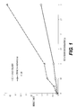
- This experiment compared the performance of silica-coated gold nanoparticles to that of gold colloids in a lateral flow immunoassay for influenza A virus.
- naso-pharyngeal aspirates that tested negative for influenza A virus were spiked with known amounts of a live H1N1 influenza A virus.
- the gold colloids obtained from British Biocell International ("BBInternational”; “BBI”), were 40 nm in diameter.
- the silica-coated gold nanoparticles used in the experiment were Oxonica NanoplexTM nanoparticles. These nanoparticles consist of a 60 nm gold core coated with the Raman reporter 4,4'-Dipyridyl and an outer silica shell. The silica shell thickness was approximately 30 nm.
- Influenza A detection antibodies were attached to the gold colloids by passive adsorption.
- the influenza A detection antibodies were covalently bound to thiol groups on the silica surface through maleimide chemistry.
- influenza A capture antibodies were striped onto Whatman AE99 nitrocellulose membranes.
- the nitrocellulose membranes were then assayed in a liquid ("dipstick") format in which the particles plus detection antibodies were mixed with the nasopharyngeal samples containing varying levels of virus. The same optical density was used for both the gold colloids and the coated nanoparticles.
- the particle/sample mixtures were then placed in the wells of a 96-well plate, and one end of a nitrocellulose membrane with capture antibodies was placed in each well. The sample was allowed to wick up the nitrocellulose membrane, and the wells were then filled with a wash buffer that then also wicked up the strips of nitrocellulose membrane.
- Figure 1 shows a plot of reflectometer signal as a function of virus concentration for lateral flow tests with gold colloids and with coated nanoparticles. A significantly stronger response was seen for the Oxonica nanoparticles versus the gold colloids.
- the sensitivity of silica-coated gold nanoparticles was compared with that of a commercial product: the BD DirectigenTM EZ Flu A+B test.
- the coated gold nanoparticles were assayed in a dipstick format using Whatman AE99 nitrocellulose.
- the DirectigenTM EZ Flu A+B was tested in its commercial embodiment ("cartridge" format).
- Capture and detection antibodies were the same for both the BD DirectigenTM EZ Flu A+B commercial product and the Oxonica NanoplexTM nanoparticles-based device. Samples were prepared as described in Example 1, except live B/Lee/40 influenza B virus was spiked into negative naso-pharyngeal aspirates rather than influenza A virus.
- the silica-coated gold nanoparticles used in the experiment were Oxonica NanoplexTM nanoparticles. These nanoparticles consist of a 60 nm gold core coated with the Raman reporter 4,4'-Dipyridyl and an outer silica shell. The silica shell thickness was approximately 30 nm.
- the BD commercial product uses 40 nm gold colloid that does not include a silica shell. The BD commercial product was used according to the package insert, only with the test line signal read with a reflectometer as well as visually.
- the data are shown in Table 1, which compares the limit of detection (Reliable Detection Limit) for the two particles.
- the Reliable Detection Limit (RDL) is defined as the concentration where the lower 95% confidence limit equals the upper 95% confidence limit of the blank.
- the RDLs expressed in arbitrary units (X) for the BD colloidal gold product and for silica-coated nanoparticles, are reported for three separate experiments.
- the same custom-built reflectometer from UMM Electronics was used to read the signal from both the silica-coated nanoparticle dipstick devices and the DirectigenTM EZ Flu A+B devices.
- the Sensitivity Improvement Factor is defined as the RDL of the uncoated gold-based product divided by the RDL of the silica-coated nanoparticle product.
- the Oxonica NanoplexTM nanoparticles gave up to 16-fold improved sensitivity compared to bare gold particles.
- the diameter of the gold colloid particles was increased from 40 nm to 60 nm and larger sizes. In these experiments, only limited sensitivity improvements (up to 2-fold) were observed, indicating that the difference in size between the gold core of the Oxonica NanoplexTM nanoparticles and the gold colloid is likely not the source of the sensitivity improvements.
- This experiment compared the sensitivity and specificity of gold colloids versus Oxonica NanoplexTM nanoparticles in an influenza B (B/Lee/40) lateral flow immunoassay test.
- Sixty clinical samples were prepared. The samples were all nasopharyngeal samples that tested negative for influenza B. Twenty of the samples served as negative controls. The remaining 40 samples were spiked with live influenza B virus at two levels to create a set of 40 positive samples. Twenty of these forty samples were spiked with live influenza B virus at a concentration of 0.5X (arbitrary units). The remaining twenty samples received 10-fold less flu B virus, for a concentration of 0.05X.
- All sixty samples were then tested using the BD DirectigenTM EZ Flu A+B commercial product in cartridge format and the dipstick device made using Oxonica NanoplexTM nanoparticles.
- the dipstick device was prepared using Whatman AE99 nitrocellulose.
- the Oxonica NanoplexTM nanoparticles consisted of a 60 nm gold core coated with the Raman reporter 4,4'-Dipyridyl and an outer silica shell that was approximately 30 nm thick. Capture and detection antibodies were the same for both the BD DirectigenTM EZ Flu A+B commercial product and the Oxonica NanoplexTM nanoparticles-based device.
- sensitivity was calculated as the percentage of the 40 positive samples correctly identified as positive by each test.
- Specificity was calculated as the percentage of the 20 negative samples correctly identified as negative by each test.
- a test was called positive if the measured instrument reading was greater than an established threshold value for each type of device. The threshold was established to ensure at least a 95% specificity for each device.
- the DirectigenTM product was read visually and with the custom-built reflectometer from UMM Electronics. The Oxonica NanoplexTM nanoparticles were read with the same reflectometer, and also with a research Raman reader built by BD (last row of the table).
- the research Raman reader used a 785 nm laser for excitation and an Acton Research Spectrometer (SpectraPro 25000i) and a CCD detector (Pixis 400) for detecting the Raman signal.
- Table 2 Test Sensitivity Specificity DirectigenTM EZ, visual read 45% 100% DirectigenTM EZ, reflectometer read 58% 95% Oxonica nanotags, reflectometer read 100% 100% Oxonica nanotags, Raman read 100% 100%
- This experiment compared the performance of silica-coated gold nanoparticles, with and without a surface enhanced Raman scattering (SERS) molecule, in a reflectance-based lateral flow immunoassay.
- SERS surface enhanced Raman scattering
- Oxonica NanoplexTM gold nanoparticles containing a SERS tag were obtained from Oxonica.
- the nanoparticles consist of a 60 nm gold core tagged with 4-4'-Dipyridyl and coated with a 35 nm thick silica shell. The diameter of the nanoparticles was 130 nm.
- Sulfo-SMCC chemistry (Pierce #22622) was used to covalently attach anti-influenza A antibodies to surface thiol groups on the nanoparticles..
- Gold nanoparticles lacking a SERS tag were purchased from BBI.
- the gold nanoparticles were then coated with silica by the following process. 10 mL of 60 nm gold colloid (BBI, ⁇ 2.6x10 10 particles/mL) was first treated with 75 ⁇ L of a 100 ⁇ M solution of 3-mercaptopropyl triethoxysilane in ethanol. After 3 hours, 75 ⁇ L of a 2.7% aqueous solution of sodium silicate was added, and the reaction was allowed to continue for 24 hours.
- BBI 60 nm gold colloid
- Thiol groups were then added to the surface of the silica-coated gold particles by reacting an aqueous suspension of glass-coated gold nanoparticles (10 mL, ⁇ 2.6x10 10 particles/mL) with a 1% ethanolic solution of mercaptopropyl trimethoxysilane (MPTMS) for 24 hours at room temperature.
- MPTMS mercaptopropyl trimethoxysilane
- the amount of MPTMS solution varied from 25 ⁇ L to 100 ⁇ L, to create varying levels of surface-active thiol groups at approximate loading levels of 0.5, 1.0, 1.5, and 2.0 relative to each other (for example, 2.0 is 4X greater than 0.5) through a non-optimized process.
- the particles were purified by repeated centrifugation in water.
- Sulfo-SMCC chemistry (Pierce #22622) was used to covalently attach anti-influenza A antibodies to surface thiol groups on the nanoparticles.
- the lateral flow immunoassay was performed in a dipstick format as follows. Influenza A capture antibodies were applied to Whatman AE99 nitrocellulose strips. The nitrocellulose strips were dipped into sample solutions containing nanoparticles adjusted to an optical density of 10 and various concentrations of influenza A H1N1 virus. The sample solutions were allowed to completely wick up the nitrocellulose strips. A wash buffer was then added and also wicked up the strips of nitrocellulose membrane. The strips were then dried, and the reflectance was read using a Hamamatsu reflectometer, model C10066. The results are summarized in Table 3.
- the data in Table 3 indicate that, if optimized, silica-coated gold nanoparticles without a SERS reporter perform as well as nanoparticles that include a SERS reporter in a lateral flow immunoassay when a reflectometer reader is used. Furthermore, the performance of the silica-coated gold particles may depend on the extent of thiolation.
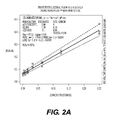
- This experiment was devised to test the performance of a prototype lateral flow device for detecting live H1N1 influenza A virus in clinical samples using Oxonica NanoplexTM silica-coated gold nanoparticles as the reporter.
- the Oxonica particles had a gold core diameter of approximately 60 nm, a Raman reporter of 4,4'-Dipyridyl, and an outer silica shell approximately 35 nm thick.
- the cartridge-based prototype device used the same capture and detection antibodies as the BD DirectigenTM EZ Flu A+B commercial product and was used in the same manner as the commercial product.
- the prototype test device comprised a backer strip supporting a length of Millipore HF135 nitrocellulose lateral flow membrane. Capture antibodies specific to influenza A nucleoprotein (Flu A NP) were striped across this membrane to form a test line. Anti-species immunoglobulin antibody was striped adjacent to the test line to form a control line. Oxonica NanoplexTM SERS nanoparticles were sprayed onto a conjugate pad (Arista MAPDS-0399) that had been treated with a 10% SEABLOCK solution (Pierce 37527). The nanoparticles were conjugated with a detection antibody to Flu A NP. The conjugate pad was adhered to the backer strip at one end of the lateral flow membrane.
- an absorbent wicking pad (Whatman #470) was attached.
- the resulting lateral flow assay strip was mounted within a two-part polystyrene cartridge. This cartridge completely enclosed the assay strip except for a central window revealing the test and control line region of the LF membrane, and a sample application well centered on the conjugate pad. This cartridge housing is used in the BD DirectigenTM EZ RSV lateral flow assay device.
- Example 1 pediatric nasopharyngeal aspirate samples testing negative for influenza A virus were spiked with known amounts of live influenza A virus. Final virus concentrations within the spiked samples ranged from 0.25X (arbitrary units) down to 0.0039X by two-fold serial dilution. A sample spiked with virus-free dilution medium ("0X") was included as a control. After the addition of live virus, the series of samples was processed using the BD DirectigenTM EZ Flu A+B sample preparation protocol. Briefly, a quantity of influenza A-spiked sample was mixed with extraction reagent in a flexible sample tube. The extracted sample was then expressed from the tube through a glass-fiber filtration tip and collected. Each sample was tested in triplicate by applying 100 ⁇ L to the sample well of each of three prototype test devices.
- each device made with the Oxonica NanoplexTM SERS nanoparticles was read using a Hamamatsu reflectometer (model C10066) at both 15 minutes and 30 minutes following sample application. After the 30 minute read, each lateral flow strip was removed from its cartridge, stripped of conjugate and wicking pads, and dried at ambient temperature and humidity for at least 1 hour. Each strip was read again by reflectometer after drying. A dose-response curve was plotted using data from each read-time and then used to calculate sensitivity of each read-time in terms of minimum detectable concentration (the lowest concentration for which the mean signal equals the upper confidence interval of the blank; MDC) and reliable detection limit (RDL). These results are shown in Table 4.
- Table 4 also shows the sensitivity improvement obtained using the Oxonica particles compared to the sensitivity of DirectigenTM EZ.
- DirectigenTM EZ uses 40 nm-diameter gold colloids without a silica coating.
- Table 4. Sensitivity of a Cartridge-Based Flu A Lateral Flow Test System Employing Oxonica SERS-Active Nanoparticles and Detection by Reflectometry Device Status and Time of Read Flu A Limit of Detection ⁇ by Reflectometer Read (Oxonica particles) RDL Improvement vs.
- the limit of detection (RDL) for the current DirectigenTM EZ Flu A device is approximately 0.5X Flu A virus concentration by visual read, and approximately 0.2X by reflectometer read. As shown in Table 4, the cartridge-based test device using Oxonica NanoplexTM reporter nanoparticles provides a marked increase in sensitivity relative to the current DirectigenTM EZ Flu A device.
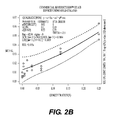
- the analytical sensitivity of a prototype lateral flow Flu A test using Oxonica NanoplexTM silica-coated gold nanoparticles was compared to the analytical sensitivity of the BD DirectigenTM EZ Flu A+B commercial product read with a reflectometer.
- the Oxonica particles had a gold core diameter of approximately 60 nm, Raman reporter molecule 4,4'-Dipyridyl attached to the gold surface, and a silica shell thickness of approximately 35 nm.
- the amount of SERS nanoparticles per lateral flow device was estimated to be slightly less than the amount of gold colloid particles in the commercial product. Capture and detection antibodies were the same for both the BD DirectigenTM EZ Flu A+B commercial product and the Oxonica NanoplexTM nanoparticles-based device.
- a test device for determining the presence or absence of an analyte in a liquid sample comprising: (a) a sample receiving member; (b) a carrier in fluid communication with the sample receiving member; (c) a labeled reagent which is mobile in the carrier in the presence of the liquid sample, the labeled reagent comprising a ligand that binds to the analyte and a coated nanoparticle comprising a core and a shell that increases the reflectance of the nanoparticle having the ligand attached thereto, wherein the coated nanoparticle does not include a Raman-active molecule; and (d) a binding reagent effective to capture the analyte, when present, immobilized in a defined detection zone of the carrier; wherein the liquid sample applied to the sample receiving member mobilizes the labeled reagent such that the sample and labeled reagent are transported along the length of the carrier to pass into the detection zone, and wherein detection of the labeled reagent in the detection zone is indicative
- test device as defined above, wherein the carrier comprises nitrocellulose, plastic, or glass.
- the carrier comprises nitrocellulose.
- test device as defined above, wherein the analyte is a protein, nucleic acid, metabolite, small molecule, virus, or bacterium.
- test device as defined above, further comprising an absorbent pad in fluid communication with the detection zone.
- test device as defined above, further comprising a control zone in fluid communication with the detection zone.
- test device as defined above, wherein the presence of the analyte in the liquid sample is determined quantitatively.
- test device as defined above, wherein the test device is configured to detect multiple analytes.
- test device as defined above, further comprising at least two different labeled reagents, wherein the ligands of the labeled reagents bind to different analytes, and at least two detection zones for detecting each of the at least two different labeled reagents.
- a system comprising the test device as defined above and a reflectometer adapted to detect the presence of the labeled reagent in the test device.
- a method for determining the presence or absence of an analyte in a liquid sample comprising:
- the method detects multiple analytes in the liquid sample.
- the test device further comprises at least two different labeled reagents, wherein the ligands of the labeled reagents bind to different analytes, and at least two detection zones for detecting each of the at least two different labeled reagents.
- a test device for determining the presence or absence of an analyte in a liquid sample comprising: (a) a sample receiving member; (b) a carrier in fluid communication with the sample receiving member; (c) a labeled reagent which is mobile in the carrier in the presence of the liquid sample, the labeled reagent comprising a ligand that binds to the analyte and a coated nanoparticle consisting essentially of a core and a shell that increases the reflectance of the nanoparticle having the ligand attached thereto, wherein said ligand is bound to the surface of the shell; and (d) a binding reagent effective to capture the analyte, when present, immobilized in a defined detection zone of the carrier; wherein the liquid sample applied to the sample receiving member mobilizes the labeled reagent such that the sample and labeled reagent are transported along the length of the carrier to pass into the detection zone, and wherein detection of labeled reagent in the detection zone is indicative of the
- test device as defined above, wherein the analyte is a protein, nucleic acid, metabolite, small molecule, virus, or bacterium.
- test device as defined above, further comprising an absorbent pad in fluid communication with the detection zone.
- test device as defined above, further comprising a control zone in fluid communication with the detection zone.
- test device as defined above, wherein the presence of the analyte in the liquid sample is determined quantitatively.
- test device as defined above, wherein the test device is configured to detect multiple analytes.
- test device as defined above, further comprising at least two different labeled reagents, wherein the ligands of the labeled reagents bind to different analytes, and at least two detection zones for detecting each of the at least two different labeled reagents.
- a system comprising the test device as defined above and a reflectometer adapted to detect the presence of the labeled reagent in the test device.
- a method for determining the presence or absence of analyte in a liquid sample comprising:
- the method detects multiple analytes in the liquid sample.
- the test device further comprises at least two different labeled reagents, wherein the ligands of the labeled reagents bind to different analytes, and at least two detection zones for detecting each of the at least two different labeled reagents.
- a method for determining the presence or absence of an analyte in a liquid sample comprising:
- the method detects multiple analytes in the liquid sample.
- the test device further comprises at least two different labeled reagents, wherein the ligands of the labeled reagents bind to different analytes, and at least two detection zones for detecting each of the at least two different labeled reagents.
- a method for determining the presence or absence of an analyte in a liquid sample comprising:
- test device further comprises at least two different labeled reagents, wherein the ligands of the labeled reagents bind to different analytes, and at least two detection zones for detecting each of the at least two different labeled reagents.
- a kit for performing a flow-through analytical test for detecting the presence or absence of an analyte in a liquid sample by reflectometry comprising: (a) a test device comprising a porous membrane comprising an upper surface and a lower surface and a binding reagent effective to capture the analyte, when present in the liquid sample, attached to the upper or lower surface of the porous membrane; and (b) a labeled reagent comprising a ligand that binds to the analyte and a coated nanoparticle comprising a core and a shell that increases the reflectance of the nanoparticle, wherein the coated nanoparticle does not include a Raman-active molecule.
- test device further comprises an absorbent pad, wherein the lower surface of the porous membrane and the absorbent pad are in physical contact and in fluid communication, and wherein the binding reagent is attached to the upper surface of the porous membrane.
- test device further comprises a housing for the porous membrane.
- test device configured to detect multiple analytes.
- test device further comprises at least two different labeled reagents, wherein the ligands of the labeled reagents bind to different analytes, and at least two detection zones for detecting each of the at least two different labeled reagents.
- a system comprising the test device of the kit as defined above and a reflectometer adapted to detect the presence of the labeled reagent in the test device.
- a method for determining the presence or absence of an analyte in a liquid sample using the kit as defined above comprising: (a) contacting the liquid sample with the upper surface of the porous membrane; (b) allowing the liquid sample to flow through the porous membrane such that at least a portion of the analyte, when present in the liquid sample, binds to the binding reagent; (c) contacting the labeled reagent with the upper surface of the porous membrane; (d) allowing the labeled reagent to flow through the porous membrane such that at least a portion of the labeled reagent binds to the analyte; and (e) detecting the presence of the labeled reagent on the porous membrane by measuring reflectance, wherein detection of the labeled reagent on the porous membrane is indicative of the presence of the analyte in the liquid sample, and failure to detect the presence of the labeled reagent on the porous membrane is indicative of the absence of the analyte
- the method detects multiple analytes in the liquid sample.
- the test device further comprises at least two different labeled reagents, wherein the ligands of the labeled reagents bind to different analytes, and at least two detection zones for detecting each of the at least two different labeled reagents.
- a method for determining the presence or absence of an analyte in a liquid sample using the kit as defined above comprising: (a) mixing the liquid sample with the labeled reagent such that the analyte, when present in the liquid sample, binds to the labeled reagent; (b) contacting the mixture of (a) with the upper surface of the porous membrane; (c) allowing the mixture of (a) to flow through the porous membrane such that at least a portion of the analyte bound to the labeled reagent binds to the binding reagent; and (d) detecting the presence of the labeled reagent on the porous membrane by measuring reflectance, wherein detection of the labeled reagent on the porous membrane is indicative of the presence of analyte in the liquid sample, and failure to detect the presence of the labeled reagent on the porous membrane is indicative of the absence of the analyte in the liquid sample.
- the method detects multiple analytes in the liquid sample.
- the test device further comprises at least two different labeled reagents, wherein the ligands of the labeled reagents bind to different analytes, and at least two detection zones for detecting each of the at least two different labeled reagents.
- a kit for performing a flow-through analytical test for detecting the presence or absence of an analyte in a liquid sample by reflectometry comprising: (a) a test device comprising a porous membrane comprising an upper surface and a lower surface and a binding reagent effective to capture the analyte, when present in the liquid sample, attached to the upper or lower surface of the porous membrane; and (b) a labeled reagent comprising a ligand that binds to the analyte and a coated nanoparticle consisting essentially of a core and a shell that increases the reflectance of the nanoparticle, wherein said ligand is bound to the surface of the shell.
- the analyte is a protein, nucleic acid, metabolite, small molecule, virus, or bacterium.
- test device further comprises an absorbent pad, wherein the lower surface of the porous membrane and the absorbent pad are in physical contact and in fluid communication, and wherein the binding reagent is attached to the upper surface of the porous membrane.
- test device further comprises a housing for the porous membrane.
- test device configured to detect multiple analytes.
- test device further comprises at least two different labeled reagents, wherein the ligands of the labeled reagents bind to different analytes, and at least two detection zones for detecting each of the at least two different labeled reagents.
- a system comprising the test device of the kit as defined above and a reflectometer adapted to detect the presence of the labeled reagent in the test device.
- a method for determining the presence or absence of an analyte in a liquid sample using the kit as defined above comprising: (a) contacting the liquid sample with the upper surface of the porous membrane; (b) allowing the liquid sample to flow through the porous membrane and into the absorbent material such that at least a portion of the analyte, when present in the liquid sample, binds to the binding reagent; (c) contacting the labeled reagent with the upper surface of the porous membrane; (d) allowing the labeled reagent to flow through the porous membrane and into the absorbent material such that at least a portion of the labeled reagent binds to the analyte; and (e) detecting the presence of the labeled reagent on the porous membrane by measuring reflectance, wherein detection of the labeled reagent on the porous membrane is indicative of the presence of analyte in the liquid sample, and failure to detect the presence of the labeled reagent on the porous membrane is
- the method detects multiple analytes in the liquid sample.
- the test device further comprises at least two different labeled reagents, wherein the ligands of the labeled reagents bind to different analytes, and at least two detection zones for detecting each of the at least two different labeled reagents.
- a method for determining the presence or absence of an analyte in a liquid sample using the kit as defined above comprising: (a) mixing the liquid sample with the labeled reagent such that the analyte, when present in the liquid sample, binds to the labeled reagent; (b) contacting the mixture of (a) with the upper surface of the porous membrane; (c) allowing the mixture of (a) to flow through the porous membrane such that at least a portion of the analyte bound to the labeled reagent binds to the binding reagent; and (d) detecting the presence of the labeled reagent on the porous membrane by measuring reflectance, wherein detection of the labeled reagent on the porous membrane is indicative of the presence of analyte in the liquid sample, and failure to detect the presence of the labeled reagent on the porous membrane is indicative of the absence of the analyte in the liquid sample.
- the method detects multiple analytes in the liquid sample.
- the test device further comprises at least two different labeled reagents, wherein the ligands of the labeled reagents bind to different analytes, and at least two detection zones for detecting each of the at least two different labeled reagents.
- a kit for performing a flow-through analytical test for detecting the presence or absence of an analyte in a liquid sample by reflectometry comprising: (a) a test device comprising a porous membrane comprising an upper surface and a lower surface and a binding reagent effective to capture the analyte, when present in the liquid sample, attached to the upper or lower surface of the porous membrane; and (b) a labeled reagent comprising a ligand that binds to the analyte and a coated nanoparticle comprising a core, a molecule linked to the core and capable of generating a signal by surface enhanced Raman scattering, and a shell surrounding the core and the molecule.
- analyte is a protein, nucleic acid, metabolite, small molecule, virus, or bacterium.
- test device further comprises an absorbent pad, wherein the lower surface of the porous membrane and the absorbent pad are in physical contact and in fluid communication, and wherein the binding reagent is attached to the upper surface of the porous membrane.
- test device further comprises a housing for the porous membrane.
- test device configured to detect multiple analytes.
- test device further comprises at least two different labeled reagents, wherein the ligands of the labeled reagents bind to different analytes, and at least two detection zones for detecting each of the at least two different labeled reagents.
- a system comprising the test device of the kit as defined above and a reflectometer adapted to detect the presence of the labeled reagent in the test device.
- a method for determining the presence or absence of an analyte in a liquid sample using the kit as defined above comprising: (a) contacting the liquid sample with the upper surface of the porous membrane; (b) allowing the liquid sample to flow through the porous membrane and into the absorbent material such that at least a portion of the analyte, when present in the liquid sample, binds to the binding reagent; (c) contacting the labeled reagent with the upper surface of the porous membrane; (d) allowing the labeled reagent to flow through the porous membrane and into the absorbent material such that at least a portion of the labeled reagent binds to the analyte; and (e) detecting the presence of the labeled reagent on the porous membrane by measuring reflectance, wherein detection of the labeled reagent on the porous membrane is indicative of the presence of analyte in the liquid sample, and failure to detect the presence of the labeled reagent on the porous membrane is
- the method detects multiple analytes in the liquid sample.
- the test device further comprises at least two different labeled reagents, wherein the ligands of the labeled reagents bind to different analytes, and at least two detection zones for detecting each of the at least two different labeled reagents.
- a method for determining the presence or absence of an analyte in a liquid sample using the kit as defined above comprising: (a) mixing the liquid sample with the labeled reagent such that the analyte, when present in the liquid sample, binds to the labeled reagent; (b) contacting the mixture of (a) with the upper surface of the porous membrane; (c) allowing the mixture of (a) to flow through the porous membrane such that at least a portion of the analyte bound to the labeled reagent binds to the binding reagent; and (d) detecting the presence of the labeled reagent on the porous membrane by measuring reflectance, wherein detection of the labeled reagent on the porous membrane is indicative of the presence of analyte in the liquid sample, and failure to detect the presence of the labeled reagent on the porous membrane is indicative of the absence of the analyte in the liquid sample.
- the method detects multiple analytes in the liquid sample.
- the test device further comprises at least two different labeled reagents, wherein the ligands of the labeled reagents bind to different analytes, and at least two detection zones for detecting each of the at least two different labeled reagents.
Abstract
Description
- This application claims priority to
U.S. Provisional Application No. 61/071,035, filed April 9, 2008 - Coated nanoparticles comprising a core surrounded by a shell that increases the reflectance of the nanoparticle, wherein the coated nanoparticle need not, but optionally can, include a Raman-active molecule, are provided. The coated nanoparticles disclosed herein are useful in test devices and methods for quantitative and/or qualitative determination of the presence or absence of an analyte in a liquid sample.
- Immunoassay technology provides a simple and relatively rapid means for determining the presence or absence of analytes in biological samples. The information provided from immunoassay diagnostic tests are often critical to patient care. Assays are typically performed to detect qualitatively or quantitatively the presence of particular analytes, for example, antibodies that are present when a human subject has a particular disease or condition. Immunoassays practiced in the art are numerous, and include assays for diseases, such as infections caused by bacteria or viruses, or conditions, such as pregnancy.
- Various types of immunoassays are known in the art. One type of immunoassay procedure is the lateral flow immunoassay. Lateral flow assays utilize a solid support, such as nitrocellulose, plastic, or glass, for performing analyte detection. Instead of drawing the sample through the support perpendicularly, as in the case of a "flow-through" assay, the sample is permitted to flow laterally along the support by capillary and other forces from an application zone to a reaction zone on the surface. In a lateral flow assay, capture antibodies are striped onto the solid support. Detection antibodies are conjugated to a detection molecule, which provides a signal that is detectible. A liquid sample is placed in contact with the detection antibodies, and the sample/detection antibody mixture is allowed to flow along the solid support. If the analyte is present, a "sandwich" complex is formed at the location on the solid support where the capture antibodies have been striped. The signal from the detection molecule localized at the capture line is then detected, either visually or with an instrument. A lateral flow assay can be configured to detect proteins, nucleic acids, metabolites, cells, small molecules, or other analytes of interest.
- Another immunoassay format is a flow-through immunoassay. A flow-through immunoassay generally uses a porous material with a reagent-containing matrix layered thereon or incorporated therein. Test sample is applied to and flows through the porous material, and analyte in the sample reacts with the reagent(s) to produce a detectable signal on the porous material. These devices are generally encased in a plastic housing or casing with calibrations to aid in the detection of the particular analyte.
- Many examples of different types of detection molecules useful in lateral flow immunoassays are known in the art, such as fluorophores, gold colloids, labeled latex particles, and nanoparticles such as a quantum dot or a surface enhanced Raman scattering ("SERS") nanoparticle. For example,
U. S. Patent No. 5,591,645 describes the use of visible "tracer" molecules, such as colloidal gold, that can be seen without the use of instrumentation. In addition,U.S. Patent No. 6,514,767 to Natan discloses SERS-active composite nanoparticles (SACNs) comprising a metal nanoparticle that has attached or associated with its surface one or more Raman-active molecules and is encapsulated by a shell comprising a polymer, glass, or any other dielectric material.U. S. Patent No. 5,714,389 describes lateral flow immunoassay methods and test devices using a colored particle that may be a metal colloid, preferably gold. Similarly,U. S. Patent No. 7,109,042 describes lateral flow immunoassay devices that use direct labels, such as gold sols and dye sols, which allow for the production of an instant analytical result without the need to add further reagents in order to develop a detectable signal. - SERS is one of the most sensitive methods for performing chemical analyses, permitting detection of a single molecule. See Nie, S. and S. R. Emory, "Probing Single Molecules and Single Nanoparticles by Surface Enhanced Raman Scattering", Science, 275,1102 (1997). A Raman spectrum, similar to an infrared spectrum, includes a wavelength distribution of bands corresponding to molecular vibrations specific to the sample being analyzed (the analyte). In the practice of Raman spectroscopy, the beam from a light source, generally a laser, is focused upon the sample to thereby generate inelastically scattered radiation, which is optically collected and directed into a wavelength-dispersive or Fourier transform spectrometer in which a detector converts the energy of impinging photons to electrical signal intensity.
- The very low conversion of incident radiation to inelastic scattered radiation limited Raman spectroscopy to applications that were difficult to perform by infrared spectroscopy, such as the analysis of aqueous solutions. It was discovered in 1974, however, that when a molecule in close proximity to a roughened silver electrode is subjected to a Raman excitation source, the intensity of the signal generated is increased by as much as six orders of magnitude. (Fleischmann, M., Hendra, P. J., and McQuillan, A. J., "Raman Spectra of Pyridine Adsorbed at a Silver Electrode," Chem. Phys. Lett, 26, 123, (1974), and Weaver, M. J., Farquharson, S., Tadayyoni, M. A., "Surface-enhancement factors for Raman scattering at silver electrodes. Role of adsorbate-surface interactions and electrode structure," J. Chem. Phys., 82, 4867 4874 (1985)). Briefly, incident laser photons couple to free conducting electrons within the metal which, confined by the particle surface, collectively cause the electron cloud to resonate. The resulting surface plasmon field provides an efficient pathway for the transfer of energy to the molecular vibrational modes of a molecule within the field, and thus generates Raman photons.
- SERS nanoparticles have been used as a detection molecule in lateral flow immunoassays. For example, Oxonica (Kidlington, UK) has developed Nanoplex™ nanoparticles for use in such assays. The nanoparticles consist of a gold nanoparticle core, onto which are adsorbed Raman reporter molecules capable of generating a surface enhanced Raman spectroscopy signal. The Raman-labeled gold nanoparticle is coated with a silica shell of approximately 10-50 nm thickness. The silica shell protects the reporter from desorption from the surface, prevents plasmon-plasmon interactions between adjacent gold particles, and also prevents the generation of SERS signals from components in the solution. SERS nanoparticles having a polymer coating in place of the silica coating are described in
U. S. Patent Application Publication No. 2007/0165219 . - While taking advantage of the high sensitivity of SERS, detection of the signal produced from a SERS nanoparticle requires the use of an instrument capable of detecting a Raman signal. It may be advantageous in some circumstances to have a nanoparticle for use in a lateral flow immunoassay possessing increased sensitivity perhaps comparable to that of a SERS nanoparticle, but requiring only relatively simple and inexpensive reflectance reader technology for detection.
- The invention provides rapid and accurate methods for determining qualitatively or quantitatively the presence or absence of analytes in biological samples and devices and reagents to perform those methods.
- The inventors have discovered that coated nanoparticles used as a detector molecule in a lateral flow or vertical flow-through immunoassay provide significant assay sensitivity advantages over detector molecules such as colloidal gold when the assay is read with a reflectance reader.
- In certain embodiments, the invention is a coated nanoparticle comprising a core and a shell that increases the reflectance of the nanoparticle, wherein the coated nanoparticle does not include a Raman-active molecule. In other embodiments, the coated nanoparticle includes a Raman-active molecule. The core may, for example and without limitation, be a metal, such as a metal that exhibits plasmon resonance, for example, gold.
- In some embodiments, the shell comprises silica, while in other embodiments the shell is another ceramic material, such as another oxide, and in further embodiments the shell is comprised of a polymer. The polymer may be, for example and without limitation, polyethylene glycol, polymethylmethacrylate, or polystyrene. The shell may completely or incompletely surround the core.
- The coated nanoparticles of the invention may be formed in any of a variety of shapes having different dimensions, including but not limited to, spheroids, rods, disks, pyramids, cubes, cylinders, etc. In certain embodiments, the coated nanoparticle has at least one dimension in the range of about 1 nm to about 1000 nm. In other embodiments the core of the coated nanoparticle is spherical. In some cases, the diameter of the core is about 10-100 nm, while in other embodiments the diameter of the core is about 20-60 nm. In some embodiments, the nanoparticles comprise multi-core aggregates, for example but not limited to, doublets.
- In certain embodiments, the shell of the nanoparticle is modified so as to allow for the conjugation of molecules to the surface of the nanoparticle. In particular embodiments, the modification introduces thiol groups onto the surface of the coated nanoparticle. In other embodiments, a ligand, for example and without limitation, an antibody, is conjugated to the shell of the coated nanoparticle via the thiol groups.
- The ligand may bind to any analyte of interest that may or may not be present in a sample. In certain embodiments, the ligand is an antibody that binds to proteins specific to influenza virus A or influenza virus B.
- In certain other embodiments, the invention is a coated nanoparticle consisting essentially of a core and a shell that increases the reflectance of the nanoparticle, and a ligand bound to the surface of the shell.
- In still other embodiments, the invention is a test device for determining the presence or absence of an analyte in a liquid sample, comprising: (a) a sample receiving member; (b) a carrier in fluid communication with the sample receiving member; (c) a labeled reagent which is mobile in the carrier in the presence of the liquid sample, the labeled reagent comprising a ligand that binds to the analyte and a coated nanoparticle comprising a core and a shell that increases the reflectance of the nanoparticle having the ligand attached thereto, wherein the coated nanoparticle does not include a Raman-active molecule; and (d) a binding reagent effective to capture the analyte, when present, immobilized in a defined detection zone of the carrier; wherein the liquid sample applied to the sample receiving member mobilizes the labeled reagent such that the sample and labeled reagent are transported along the length of the carrier to pass into the detection zone, and wherein detection of the labeled reagent in the detection zone is indicative of the presence of analyte in the liquid sample.
- In certain embodiments, the labeled reagent used in the test device comprises a ligand that binds to the analyte and a coated nanoparticle consisting essentially of a core and a shell that increases the reflectance of the nanoparticle having the ligand attached thereto, wherein said ligand is bound to the surface of the shell.
- In certain embodiments, the carrier is, for example and without limitations, nitrocellulose, plastic, or glass. In other embodiments, the test device further comprises an absorbent pad and/or a control zone in fluid communication with the detection zone.
- The test device of the invention may be used to qualitatively or quantitatively detect the presence or absence of any analyte of interest, such as and without limitation, a protein, nucleic acid, metabolite, small molecule, virus, or bacterium. In certain embodiments, the test device may be used to detect multiple analytes in a liquid sample with at least two different labeled reagents, wherein the ligands of the labeled reagents bind to different analytes, and at least two detection zones for detecting each of the at least two different labeled reagents.
- In certain embodiments, the invention is a system comprising the test device of the invention and a reflectometer adapted to detect the presence of the labeled reagent in the test device.
- In certain embodiments, the invention is a method for determining the presence or absence of an analyte in a liquid sample, comprising:
- a) providing a test device of the invention;
- b) contacting the liquid sample with the sample receiving member of the test device;
- c) allowing the liquid sample applied to the sample receiving member to mobilize said labeled reagent such that the liquid sample and labeled reagent move along the length of the carrier to pass into the detection zone;
- d) detecting the presence of the labeled reagent in the detection zone by measuring reflectance, wherein detection of the labeled reagent in the detection zone is indicative of the presence of analyte in the liquid sample, and failure to detect the presence of the labeled reagent in the detection zone is indicative of the absence of the analyte in the liquid sample.
- In other embodiments, the invention is a method for determining the presence or absence of an analyte in a liquid sample, comprising:
- a) providing a test device comprising: (i) a sample receiving member; (ii) a carrier in fluid communication with the sample receiving member; and (iii) a binding reagent effective to capture the analyte, when present, immobilized in a defined detection zone of the carrier;
- b) mixing the liquid sample with a labeled reagent comprising a ligand that binds to the analyte and a coated nanoparticle comprising a core and a shell that increases the reflectance of the nanoparticle having the ligand attached thereto, wherein the coated nanoparticle does not include a Raman-active molecule;
- c) contacting the mixture of b) with the sample receiving member of the test device;
- d) allowing the mixture of b) applied to the sample receiving member to move along the length of the carrier to pass into the detection zone;
- e) detecting the presence of the labeled reagent in the detection zone by measuring reflectance, wherein detection of the labeled reagent in the detection zone is indicative of the presence of analyte in the liquid sample, and failure to detect the presence of the labeled reagent in the detection zone is indicative of the absence of the analyte in the liquid sample.
- In some embodiments, the labeled reagent used in the methods of the invention comprises a ligand that binds to the analyte and a coated nanoparticle consisting essentially of a core and a shell that increases the reflectance of the nanoparticle having the ligand attached thereto, wherein said ligand is bound to the surface of the shell. In other embodiments, the labeled reagent used in the methods of the invention comprises a ligand that binds to the analyte and a coated nanoparticle comprising a core, a molecule attached to the core and capable of generating a signal by surface enhanced Raman scattering, and a shell surrounding the core and the molecule having the ligand attached thereto.
- In some embodiments a reflectance reader is used to detect the labeled reagent in the detection zone, while in other embodiments, detection of the labeled reagent in the detection zone is determined visually. In some embodiments the analyte is detected quantitatively and in other embodiments, the analyte is detected qualitatively. In come embodiments, the methods of the invention can be used to detect multiple analytes in a liquid sample with at least two different labeled reagents, wherein the ligands of the labeled reagents bind to different analytes, and at least two detection zones for detecting each of the at least two different labeled reagents.
- In other embodiments, the invention is a kit for performing a flow-through analytical test for detecting the presence or absence of an analyte in a liquid sample by reflectometry, comprising: (a) a test device comprising a porous membrane comprising an upper surface and a lower surface and a binding reagent effective to capture the analyte, when present in the liquid sample, attached to the upper or lower surface of the porous membrane; and (b) a labeled reagent comprising a ligand that binds to the analyte and a coated nanoparticle comprising a core and a shell that increases the reflectance of the nanoparticle, wherein the coated nanoparticle does not include a Raman-active molecule.
- In certain embodiments, the labeled reagent used in the kits of the invention comprises a ligand that binds to the analyte and a coated nanoparticle consisting essentially of a core and a shell that increases the reflectance of the nanoparticle having the ligand attached thereto, wherein said ligand is bound to the surface of the shell. In other embodiments, the labeled reagent used in the kits of the invention comprises a ligand that binds to the analyte and a coated nanoparticle comprising a core, a molecule attached to the core and capable of generating a signal by surface enhanced Raman scattering, and a shell surrounding the core and the molecule having the ligand attached thereto.
- In certain embodiments, the test device of the kits of the invention further comprises an absorbent pad, wherein the lower surface of the porous membrane and the absorbent pad are in physical contact and in fluid communication, and wherein the binding reagent is attached to the upper surface of the porous membrane. In other embodiments, the test device of the kits of the invention further comprises a housing for the porous membrane.
- The kits of the invention may be used to qualitatively or quantitatively detect the presence or absence of any analyte of interest, such as and without limitation, a protein, nucleic acid, metabolite, small molecule, virus, or bacterium. In certain embodiments, the kits may be used to detect multiple analytes in a liquid sample with at least two different labeled reagents, wherein the ligands of the labeled reagents bind to different analytes, and at least two detection zones for detecting each of the at least two different labeled reagents.
- In certain embodiments, the invention is a system comprising the test device of the kits of the invention and a reflectometer adapted to detect the presence of the labeled reagent in the test device.
- In still other embodiments, the invention is a method for determining the presence or absence of an analyte in a liquid sample using a kit of the invention, said method comprising: (a) contacting the liquid sample with the upper surface of the porous membrane; (b) allowing the liquid sample to flow through the porous membrane such that at least a portion of the analyte, when present in the liquid sample, binds to the binding reagent; (c) contacting the labeled reagent with the upper surface of the porous membrane; (d) allowing the labeled reagent to flow through the porous membrane such that at least a portion of the labeled reagent binds to the analyte; and (e) detecting the presence of the labeled reagent on the porous membrane by measuring reflectance, wherein detection of the labeled reagent on the porous membrane is indicative of the presence of the analyte in the liquid sample, and failure to detect the presence of the labeled reagent on the porous membrane is indicative of the absence of the analyte in the liquid sample.
- In other embodiments, the invention is a method for determining the presence or absence of an analyte in a liquid sample using a kit of the invention, said method comprising: (a) mixing the liquid sample with the labeled reagent such that the analyte, when present in the liquid sample, binds to the labeled reagent; (b) contacting the mixture of (a) with the upper surface of the porous membrane; (c) allowing the mixture of (a) to flow through the porous membrane such that at least a portion of the analyte bound to the labeled reagent binds to the binding reagent; and (d) detecting the presence of the labeled reagent on the porous membrane by measuring reflectance, wherein detection of the labeled reagent on the porous membrane is indicative of the presence of analyte in the liquid sample, and failure to detect the presence of the labeled reagent on the porous membrane is indicative of the absence of the analyte in the liquid sample.
- In some embodiments a reflectance reader is used to detect the labeled reagent in the detection zone, while in other embodiments, detection of the labeled reagent in the detection zone is determined visually. In some embodiments the analyte is detected quantitatively and in other embodiments, the analyte is detected qualitatively. In come embodiments, the methods of the invention can be used to detect multiple analytes in a liquid sample with at least two different labeled reagents, wherein the ligands of the labeled reagents bind to different analytes, and at least two detection zones for detecting each of the at least two different labeled reagents.
-
-
Figure 1 shows the results of a lateral flow test for influenza A virus comparing the sensitivity of colloidal gold and silica-coated gold nanoparticles when measured with a reflectometer. As seen in the Figure, a stronger response was observed for the silica-coated gold nanoparticles compared to the gold colloids. -
Figure 2 shows the results of a lateral flow test for influenza A virus comparing the sensitivity of a prototype device using silica-coated gold nanoparticles (Panel 2A) with the sensitivity of a commercial product, BD Directigen™ EZ A+B (Panel 2B), when both devices are read with a reflectometer. The device using the silica-coated gold nanoparticles is approximately 7-fold more sensitive than the BD Directigen™ EZ A+B when read with a reflectometer. - Unless otherwise defined, all technical and scientific terms used herein have the same meaning as commonly understood by one of ordinary skill in the art. Although methods and materials similar or equivalent to those described herein can be used in the practice of the claims, suitable methods and materials are described below. All publications, patent applications, patents, and other references mentioned herein are incorporated by reference in their entirety. In the case of conflict, the present specification, including definitions, will control. In addition, the materials, methods, and examples are illustrative only and not intended to be limiting.
- In this application, the use of the singular includes the plural unless specifically stated otherwise. In this application, the use of "or" means "and/or" unless stated otherwise. In the context of a multiple dependent claim, the use of "or" refers back to more than one preceding independent or dependent claim in the alternative only. Furthermore, the use of the term "including," as well as other forms, such as "includes" and "included," is not limiting. Also, terms such as "element" or "component" encompass both elements and components comprising one unit and elements and components that comprise more than one subunit unless specifically stated otherwise.
- Other features and advantages will be apparent from the following detailed description and claims.
- In order that the present application may be more readily understood, certain terms are first defined. Additional definitions are set forth throughout the detailed description.
- The term "nanoparticle" as used herein, refers to particles comprising at least one core and a shell having one dimension in the range of about 1 to about 1000 nanometers ("nm"). The nanoparticles of the invention may be of any shape. In certain embodiments the nanoparticles are spherical. The nanoparticles of the invention typically do not, but can, include a Raman-active molecule. In certain embodiments, the nanoparticles may comprise multiple cores and one shell.
- As used herein the term "core" refers to the internal portion of the nanoparticles of the invention. In certain embodiments the core is a metal, for example but not limited to, gold.
- The nanoparticles of the invention also comprise a shell that enhances the reflectance of the nanoparticles. The shell may completely encapsulate the core, or incompletely encapsulate the core. The shell may be composed of any material or combination of materials, as long as it possesses the property of enhancing reflectance of the nanoparticle. For example, in some embodiments the shell may comprise any material transparent in the required spectral range. In certain embodiments, the shell comprises silica, that is, glass. In other embodiments, the shell comprises another ceramic material, for example but not limited to, transparent ceramics with a high refractive index, such as perovskite and ZrO2. In other embodiments, the shell is composed of a polymer. In particular embodiments the polymer comprises polyethylene glycol, polymethylmethacrylate, or polystyrene. In some embodiments, the material forming the shell is treated or derivitized to permit attaching a ligand to the surface of the nanoparticle. The optimal choice of polymer may depend on the ligand being immobilized on the surface of the nanoparticle.
- In referring to the shell, the phrase "increases the reflectance of the nanoparticle" means that the presence of the shell results in a nanoparticle providing increased signal or sensitivity when measured by reflectance in, for example, a lateral flow immunoassay, as compared to a nanoparticle without the shell. Without being bound by theory, the presence of the shell surrounding the core may directly cause the particle to reflect more light. Alternatively, or in addition, ligands bound to the surface of the shell may be better oriented to participate in binding reactions when bound to the shell material as opposed to when passively adsorbed to the surface of the core.
- As used herein, "ligand" means a molecule of any type that will bind to an analyte of interest. For example and without limitation, in certain embodiments the ligand is an antibody, an antigen, a receptor, a nucleic acid, or an enzyme.
- The term "analyte" as used herein refers to any substance of interest that one may want to detect using the invention, including but not limited to drugs, including therapeutic drugs and drugs of abuse; hormones; vitamins; proteins, including antibodies of all classes; peptides; steroids; bacteria; fungi; viruses; parasites; components or products of bacteria, fungi, viruses, or parasites; allergens of all types; products or components of normal or malignant cells; etc. As particular examples, there may be mentioned human chorionic gonadotropin (hCG); insulin; luteinizing hormone; organisms causing or associated with various disease states, such as Streptococcus pyogenes (group A), Herpes Simplex I and II, cytomegalovirus, Chlamydia, rubella antibody, influenza A and B; etc. In certain embodiments of the invention, the presence or absence of an analyte in a sample is determined qualitatively. In other embodiments, a quantitive determination of the amount or concentration of analyte in the sample is determined.
- The term "sample" as used herein refers to any biological sample that could contain an analyte for detection. In some embodiments, the biological sample is in liquid form, while in others it can be changed into a liquid form.
- The term "sample receiving member" as used herein means the portion of the test device which is in direct contact with the liquid sample, that is, it receives the sample to be tested for the analyte of interest. The sample receiving member may be part of, or separate from, the carrier or porous membrane. The liquid sample can then migrate, through lateral or vertical flow, from the sample receiving member towards the detection zone. The sample receiving member is in liquid flow contact with the analyte detection zone. This could either be an overlap, top-to-bottom, or an end-to-end connection. In certain embodiments, the sample receiving member is made of porous material, for example and not limited to, paper.
- As used herein, the term "carrier," such as used in a lateral flow assay, refers to any substrate capable of providing liquid flow. This would include, for example, substrates such as nitrocellulose, nitrocellulose blends with polyester or cellulose, untreated paper, porous paper, rayon, glass fiber, acrylonitrile copolymer, plastic, glass, or nylon. The substrate may be porous. Typically, the pores of the substrate are of sufficient size such that the nanoparticles of the invention flow through the entirety of the carrier. One skilled in the art will be aware of other materials that allow liquid flow. The carrier may comprise one or more substrates in fluid communication. For example, the reagent zone and detection zone may be present on the same substrate (i.e., pad) or may be present on separate substrates (i.e., pads) within the carrier.
- As used herein, "porous membrane," such as used in a flow through assay, refers to a membrane or filter of any material that wets readily with an aqueous solution and has pores sufficient to allow the coated nanoparticles of the invention to pass through. Suitable materials include, for example, nitrocellulose, nitrocellulose blends with polyester or cellulose, untreated paper, porous paper, rayon, glass fiber, acrylonitrile copolymer, plastic, glass, or nylon.
- As used herein, "absorbent material" refers to a porous material having an absorbing capacity sufficient to absorb substantially all the liquids of the assay reagents and any wash solutions and, optionally, to initiate capillary action and draw the assay liquids through the test device. Suitable materials include, for example, nitrocellulose, nitrocellulose blends with polyester or cellulose, untreated paper, porous paper, rayon, glass fiber, acrylonitrile copolymer, plastic, glass, or nylon.
- As used herein the term "lateral flow" refers to liquid flow along the plane of a carrier. In general, lateral flow devices may comprise a strip (or several strips in fluid communication) of material capable of transporting a solution by capillary action, i.e., a wicking or chromatographic action, wherein different areas or zones in the strip(s) contain assay reagents either diffusively or non-diffusively bound that produce a detectable signal as the solution is transported to or through such zones. Typically, such assays comprise an application zone adapted to receive a liquid sample, a reagent zone spaced laterally from and in fluid communication with the application zone, and an detection zone spaced laterally from and in fluid communication with the reagent zone. The reagent zone may comprise a compound that is mobile in the liquid and capable of interacting with an analyte in the sample and/or with a molecule bound in the detection zone. The detection zone may comprise a binding molecule that is immobilized on the strip and is capable of interacting with the analyte and/or the reagent compound to produce a detectable signal. Such assays may be used to detect an analyte in a sample through direct (sandwich assay) or competitive binding. Examples of lateral flow devices are provided in
U.S. Patent Nos. 6,194,220 to Malick et al.; 5,998,221 to Malick et al.; 5,798,273 to Shuler et al.; and RE38,430 to Rosenstein . - In a sandwich lateral flow assay, a liquid sample that may or may not contain an analyte of interest is applied to the application zone and allowed to pass into the reagent zone by capillary action. The analyte, if present, interacts with a labeled reagent in the reagent zone and the analyte-reagent complex moves by capillary action to the detection zone. The analyte-reagent complex becomes trapped in the detection zone by interacting with a binding molecule specific for the analyte and/or reagent. Unbound sample may move through the detection zone by capillary action to an absorbent pad laterally juxtaposed and in fluid communication with the detection zone. The labeled reagent may then be detected in the detection zone by appropriate means.
- In a competitive lateral flow assay, a liquid sample that may or may not contain an analyte of interest is applied to the application zone and allowed to pass into the reagent zone by capillary action. The reagent zone comprises a labeled reagent, which may be the analyte itself, a homologue or derivative thereof, or a moiety that is capable of mimicking the analyte of interest when binding to an immobilized binder in the detection zone. The labeled reagent is mobile in the liquid phase and moves with the liquid sample to the detection zone by capillary action. The analyte contained in the liquid sample competes with the labeled reagent in binding to the immobilized binder in the detection zone. Unbound sample may move through the detection zone by capillary action to an absorbent pad laterally juxtaposed and in fluid communication with the detection zone. The labeled reagent may then be detected in the detection zone by appropriate means. The presence or absence of the analyte of interest may be determined through inspection of the detection zone, wherein the greater the amount of analyte present in the liquid sample, the lesser the amount of labeled receptor bound in the detection zone.
- As used herein, the terms "vertical flow" and "flow through" refer to liquid flow transverse to the plane of a carrier. In general, flow through devices may comprise a membrane or layers of membranes stacked on top of each other that allow the passage of liquid through the device. The layers may contain assay reagents either diffusively or non-diffusively bound that produce a detectable signal as the solution is transported through the device. Typically, the device comprises first layer having an upper and lower surface, wherein said upper surface is adapted to receive a liquid sample, and an absorbent layer vertically juxtaposed and in fluid communication with the lower surface of the first layer that is adapted to draw the liquid sample through the first layer. The first layer may comprise a binding agent attached to the upper surface of the first layer that is capable of interacting with an analyte in the sample and trapping the analyte on the upper surface of the first layer. Examples of flow through devices are provided in
U.S. Patent Nos. 4,920,046 to McFarland et al. and 7,052,831 to Fletcher et al. - In practice, a liquid sample that may or may not contain an analyte of interest is applied to the upper surface of a first layer comprising a binding agent specific for an analyte of interest. The liquid sample then flows through the first layer and into the absorbent layer. If analyte is present in the sample, it interacts with the binding agent and is trapped on the upper surface of the first layer. The first layer may then be treated with wash solutions in accordance with conventional immunoassay procedures. The first layer may then be treated with a labeled reagent that binds to the analyte trapped by the binding agent. The labeled reagent then flows through the first layer and into the absorbent layer. The first layer may be treated with wash solutions in accordance with conventional immunoassay procedures. The labeled reagent may then be detected by appropriate means. Alternatively, the liquid sample may be mixed with the labeled reagent before being applied to the upper surface of the first layer. Other suitable variations are known to those skilled in the art.
- Lateral and flow through assays may be used to detect multiple analytes in a sample. For example, in a lateral flow assay, the reagent zone may comprise multiple labeled reagents, each capable of binding to (or mimicking) a different analyte in a liquid sample, or a single labeled reagent capable of binding to (or mimicking) multiple analytes. Alternatively, or in addition, the detection zone in a lateral flow assay may comprise multiple binding molecules, each capable of binding to a different analyte in a liquid sample, or a single binding molecule capable of binding to multiple analytes. In a flow through assay, the porous membrane may comprise multiple binding agents, each capable of binding to a different analyte in a liquid sample, or a single binding agent capable of binding to multiple analytes. Alternatively, or in addition, a mixture of labeled reagents may be used in a flow through assay, each configured to bind to a different analyte in a liquid sample, or a single labeled reagent configured bind multiple analytes. If multiple labeled reagents are used in a lateral or flow through assay, the reagents may be differentially labeled to distinguish different types of analytes in a liquid sample.
- As used herein, the term "mobile" means diffusively or non-diffusively attached, or impregnated. The reagents which are mobile are capable of dispersing with the liquid sample and are carried by the liquid sample in the lateral or vertical flow.
- As used herein, the term "labeled reagent" means any particle, protein, or molecule which recognizes or binds to the analyte of interest and has attached to it a substance capable of producing a signal that is detectable by visual or instrumental means, that is, a coated nanoparticle as defined herein. The particle or molecule recognizing the analyte can be either natural or non-natural. In some embodiments the molecule is a monoclonal or polyclonal antibody.
- As used herein, the term "binding reagent" means any particle or molecule which recognizes or binds the analyte in question. The binding reagent is capable of forming a binding complex with the analyte-labeled reagent complex. The binding reagent is immobilized to the carrier in the detection zone or to the surface of the porous membrane. The binding reagent is not affected by the lateral or vertical flow of the liquid sample due to the immobilization to the carrier or porous membrane. The particle or molecule can be natural, or non-natural, that is, synthetic. Once the binding reagent binds the analyte-labeled reagent complex it prevents the analyte-labeled reagent complex from continuing with the flow of the liquid sample.
- As used herein, "detection zone" means the portion of the carrier or porous membrane containing the immobilized binding reagent.
- The term "control zone" refers to a portion of the test device comprising a binding molecule configured to capture the labeled reagent. In a lateral flow assay, the control zone may be in liquid flow contact with the detection zone of the carrier, such that the labeled reagent is captured in the control zone as the liquid sample is transported out of the detection zone by capillary action. In a flow through assay, the control zone may be a separate portion of the porous membrane, such that the labeled reagent is applied both to the sample application portion of the porous membrane and the control zone. Detection of the labeled reagent in the control zone confirms that the assay is functioning for its intended purpose.
- The term "housing" refers to any suitable enclosure for the test devices of the invention. Exemplary housings will be known to those skilled in the art. The housing may have, for example, a base portion and a lid portion. The lid may include a top wall and a substantially vertical side wall. A rim may project upwardly from the top wall. The rim may define a recess having therein an insert with at least two openings in alignment with at least two other openings in the lid to form at least two wells in the housing. The housing may be constructed to ensure that there is no communication between the two or more wells. An example of such a housing is provided in
U.S. Patent No. 7,052,831 to Fletcher et al. Other suitable housings include those used in the BD Directigen™ EZ RSV lateral flow assay device. - As used herein, "reflectance reader" or "reflectometer" refers to an instrument capable of detecting the change in reflectance caused the by presence of the coated nanoparticle in the detection zone of the test device. Reflectance readers or reflectometers are known in the art. Representative instruments suitable for use in the invention include, but are not limited to the Immunochromato Reader C 10066 from Hamamatsu or the ESE-Quant from ESE GmbH. The detection zone is most commonly scanned by the detection area of the device or directly imaged on the detector of the reflectometer leading to a trace of reflectivity versus spatial coordinate. A suitable algorithm is then used to determine, for example, the maximum change of reflectivity in the detection zone.
- Coated nanoparticles for use in SERS-based methods are known in the art. For example,
U.S. Patent No. 6,514,767 describes SERS-active composite nanoparticles (SACNs) comprising a metal nanoparticle that has attached or associated with its surface one or more Raman-active molecules and is encapsulated by a shell comprising a polymer, glass, or any other dielectric material. Such particles may be produced by growing or otherwise placing a shell of a suitable encapsulant over a Raman-active metal nanoparticle core. Metal nanoparticles of the desired size can be grown as metal colloids by a number of techniques well known in the art, such as chemical or photochemical reduction of metal ions in solution using reducing agents. For example, colloidal gold particles, which are suspensions of sub-micrometer-sized particles of gold in fluid, may be produced in a liquid by reduction of chloroauric acid. After dissolving the acid, the solution is rapidly stirred while a reducing agent is added. This causes gold ions to be reduced to neutral gold atoms, which precipitate from the supersaturated solution and form particles. To prevent the particles from aggregating, a stabilizing agent that sticks to the nanoparticle surface may be added. Nanoparticles can also be made by electrical discharge in solution. The particles can be functionalized with various organic ligands to create organic-inorganic hybrids with advanced functionality. - Suitable encapsulants include glass, polymers, metals, metal oxides, and metal sulfides. If the encapsulant is glass, the metal nanoparticle cores are preferably treated first with a glass primer. Glass is then grown over the metal nanoparticle by standard techniques. The thickness of the encapsulant can be easily varied depending on the physical properties required of the particle. The shells may be derivatized by standard techniques, allowing the particles to be conjugated to molecules (including biomolecules such as proteins and nucleic acids) or to solid supports.
- Commercially available Oxonica Nanoplex™ nanoparticles consist of a gold nanoparticle core, onto which are adsorbed Raman reporter molecules capable of generating a SERS signal. The Raman-labeled gold nanoparticle is then coated with a silica shell of approximately 10-50 nm thickness. The silica shell protects the reporter molecules from desorption from the surface of the core, prevents plasmon-plasmon interactions between adjacent gold particles, and also prevents the generation of SERS signals from components in the solution.
- For a lateral flow assay read with a reflectance reader, one might expect the Oxonica Nanoplex™ nanoparticles to give an assay sensitivity comparable to that of gold colloids of the same diameter as the Oxonica Nanoplex™ nanoparticle's core and used at the same optical density. Instead, the inventors observed unexpected and significant advantages in assay sensitivity when gold colloids are replaced by Oxonica Nanoplex™ nanoparticles in lateral flow assays read with a reflectance reader instead of a Raman signal detector. Importantly, the sensitivity improvements obtained with the Oxonica Nanoplex™ nanoparticles were generally not obtainable merely by adjusting the diameter of the uncoated gold colloids. Also, importantly, when the Oxonica Nanoplex™ nanoparticles are used as a labeled reagent, the sensitivity of the assay may be equivalent, whether the signal is read with a reflectometer or a SERS reader.
- The Raman-active molecules in the Oxonica Nanoplex™ nanoparticles are not believed to contribute to the assay performance when a reflectometer is used to read the signal rather than a Raman reader. This expectation is borne out by experiments in which silica-coated nanoparticle (without Raman-active molecules) was found to perform identically and have been used in a reflectometer-based lateral flow assays to give significant sensitivity advantages over bare gold colloids.
- This experiment compared the performance of silica-coated gold nanoparticles to that of gold colloids in a lateral flow immunoassay for influenza A virus. In this experiment, naso-pharyngeal aspirates that tested negative for influenza A virus were spiked with known amounts of a live H1N1 influenza A virus.
- The gold colloids, obtained from British Biocell International ("BBInternational"; "BBI"), were 40 nm in diameter. The silica-coated gold nanoparticles used in the experiment were Oxonica Nanoplex™ nanoparticles. These nanoparticles consist of a 60 nm gold core coated with the
Raman reporter 4,4'-Dipyridyl and an outer silica shell. The silica shell thickness was approximately 30 nm. Influenza A detection antibodies were attached to the gold colloids by passive adsorption. For the Nanoplex™ nanoparticles, the influenza A detection antibodies were covalently bound to thiol groups on the silica surface through maleimide chemistry. For both assays, influenza A capture antibodies were striped onto Whatman AE99 nitrocellulose membranes. The nitrocellulose membranes were then assayed in a liquid ("dipstick") format in which the particles plus detection antibodies were mixed with the nasopharyngeal samples containing varying levels of virus. The same optical density was used for both the gold colloids and the coated nanoparticles. The particle/sample mixtures were then placed in the wells of a 96-well plate, and one end of a nitrocellulose membrane with capture antibodies was placed in each well. The sample was allowed to wick up the nitrocellulose membrane, and the wells were then filled with a wash buffer that then also wicked up the strips of nitrocellulose membrane. - The resulting signal from the capture line was read with a reflectometer custom-built for Becton, Dickinson and Company ("BD") by UMM Electronics (Indianapolis, IN).
Figure 1 shows a plot of reflectometer signal as a function of virus concentration for lateral flow tests with gold colloids and with coated nanoparticles. A significantly stronger response was seen for the Oxonica nanoparticles versus the gold colloids. - In this example, the sensitivity of silica-coated gold nanoparticles was compared with that of a commercial product: the BD Directigen™ EZ Flu A+B test. As in Example 1, the coated gold nanoparticles were assayed in a dipstick format using Whatman AE99 nitrocellulose. The Directigen™ EZ Flu A+B was tested in its commercial embodiment ("cartridge" format). Capture and detection antibodies were the same for both the BD Directigen™ EZ Flu A+B commercial product and the Oxonica Nanoplex™ nanoparticles-based device. Samples were prepared as described in Example 1, except live B/Lee/40 influenza B virus was spiked into negative naso-pharyngeal aspirates rather than influenza A virus. As before, the silica-coated gold nanoparticles used in the experiment were Oxonica Nanoplex™ nanoparticles. These nanoparticles consist of a 60 nm gold core coated with the
Raman reporter 4,4'-Dipyridyl and an outer silica shell. The silica shell thickness was approximately 30 nm. The BD commercial product uses 40 nm gold colloid that does not include a silica shell. The BD commercial product was used according to the package insert, only with the test line signal read with a reflectometer as well as visually. - The data are shown in Table 1, which compares the limit of detection (Reliable Detection Limit) for the two particles. The Reliable Detection Limit (RDL) is defined as the concentration where the lower 95% confidence limit equals the upper 95% confidence limit of the blank.
- The RDLs, expressed in arbitrary units (X) for the BD colloidal gold product and for silica-coated nanoparticles, are reported for three separate experiments. The same custom-built reflectometer from UMM Electronics was used to read the signal from both the silica-coated nanoparticle dipstick devices and the Directigen™ EZ Flu A+B devices. The Sensitivity Improvement Factor is defined as the RDL of the uncoated gold-based product divided by the RDL of the silica-coated nanoparticle product.
Table 1 BD Directigen™ EZ with uncoated gold (read with a reflectometer) Lateral flow test with Oxonica nanoparticles (read with a reflectometer) Sensitivity Improvement Factor Experiment 1 0.23 0.015 15 Experiment 20.12 0.0075 16 Experiment 30.26 0.028 9 - As seen from the table, the Oxonica Nanoplex™ nanoparticles gave up to 16-fold improved sensitivity compared to bare gold particles.
- In separate experiments similar to those described above, the diameter of the gold colloid particles was increased from 40 nm to 60 nm and larger sizes. In these experiments, only limited sensitivity improvements (up to 2-fold) were observed, indicating that the difference in size between the gold core of the Oxonica Nanoplex™ nanoparticles and the gold colloid is likely not the source of the sensitivity improvements.
- This experiment compared the sensitivity and specificity of gold colloids versus Oxonica Nanoplex™ nanoparticles in an influenza B (B/Lee/40) lateral flow immunoassay test. Sixty clinical samples were prepared. The samples were all nasopharyngeal samples that tested negative for influenza B. Twenty of the samples served as negative controls. The remaining 40 samples were spiked with live influenza B virus at two levels to create a set of 40 positive samples. Twenty of these forty samples were spiked with live influenza B virus at a concentration of 0.5X (arbitrary units). The remaining twenty samples received 10-fold less flu B virus, for a concentration of 0.05X. All sixty samples (40 positives and 20 negative controls) were then tested using the BD Directigen™ EZ Flu A+B commercial product in cartridge format and the dipstick device made using Oxonica Nanoplex™ nanoparticles. The dipstick device was prepared using Whatman AE99 nitrocellulose. The Oxonica Nanoplex™ nanoparticles consisted of a 60 nm gold core coated with the
Raman reporter 4,4'-Dipyridyl and an outer silica shell that was approximately 30 nm thick. Capture and detection antibodies were the same for both the BD Directigen™ EZ Flu A+B commercial product and the Oxonica Nanoplex™ nanoparticles-based device. - The results are summarized in Table 2. In the table, sensitivity was calculated as the percentage of the 40 positive samples correctly identified as positive by each test. Specificity was calculated as the percentage of the 20 negative samples correctly identified as negative by each test. When instrument readings were used, a test was called positive if the measured instrument reading was greater than an established threshold value for each type of device. The threshold was established to ensure at least a 95% specificity for each device. In this example, the Directigen™ product was read visually and with the custom-built reflectometer from UMM Electronics. The Oxonica Nanoplex™ nanoparticles were read with the same reflectometer, and also with a research Raman reader built by BD (last row of the table). The research Raman reader used a 785 nm laser for excitation and an Acton Research Spectrometer (SpectraPro 25000i) and a CCD detector (Pixis 400) for detecting the Raman signal.
Table 2 Test Sensitivity Specificity Directigen™ EZ, visual read 45% 100% Directigen™ EZ, reflectometer read 58% 95% Oxonica nanotags, reflectometer read 100% 100% Oxonica nanotags, Raman read 100% 100% - As can be seen, a clear sensitivity advantage is seen with the Oxonica Nanoplex™ nanoparticles compared to the Directigen™ EZ particles, without loss of specificity.
- This experiment compared the performance of silica-coated gold nanoparticles, with and without a surface enhanced Raman scattering (SERS) molecule, in a reflectance-based lateral flow immunoassay.
- Oxonica Nanoplex™ gold nanoparticles containing a SERS tag were obtained from Oxonica. The nanoparticles consist of a 60 nm gold core tagged with 4-4'-Dipyridyl and coated with a 35 nm thick silica shell. The diameter of the nanoparticles was 130 nm. Sulfo-SMCC chemistry (Pierce #22622) was used to covalently attach anti-influenza A antibodies to surface thiol groups on the nanoparticles..
- Sixty-nanometer diameter gold nanoparticles lacking a SERS tag were purchased from BBI. The gold nanoparticles were then coated with silica by the following process. 10 mL of 60 nm gold colloid (BBI, ∼2.6x10¹⁰ particles/mL) was first treated with 75 µL of a 100 µM solution of 3-mercaptopropyl triethoxysilane in ethanol. After 3 hours, 75 µL of a 2.7% aqueous solution of sodium silicate was added, and the reaction was allowed to continue for 24 hours. In the next step, 40 mL of ethanol was added to the suspension followed by 1 mL of ammonia and 300 µL (5% solution in ethanol) of tetraethyl orthosilicate. The reaction was kept under agitation for 2 days and finally purified by repeated centrifugation. The thickness of the silica coating produced in this process was 30 nm, for a total particle diameter of the silica-coated gold of 120 nm. Thiol groups were then added to the surface of the silica-coated gold particles by reacting an aqueous suspension of glass-coated gold nanoparticles (10 mL, ∼2.6x10¹⁰ particles/mL) with a 1% ethanolic solution of mercaptopropyl trimethoxysilane (MPTMS) for 24 hours at room temperature. The amount of MPTMS solution varied from 25 µL to 100 µL, to create varying levels of surface-active thiol groups at approximate loading levels of 0.5, 1.0, 1.5, and 2.0 relative to each other (for example, 2.0 is 4X greater than 0.5) through a non-optimized process. After the reaction, the particles were purified by repeated centrifugation in water. Sulfo-SMCC chemistry (Pierce #22622) was used to covalently attach anti-influenza A antibodies to surface thiol groups on the nanoparticles.
- The lateral flow immunoassay was performed in a dipstick format as follows. Influenza A capture antibodies were applied to Whatman AE99 nitrocellulose strips. The nitrocellulose strips were dipped into sample solutions containing nanoparticles adjusted to an optical density of 10 and various concentrations of influenza A H1N1 virus. The sample solutions were allowed to completely wick up the nitrocellulose strips. A wash buffer was then added and also wicked up the strips of nitrocellulose membrane. The strips were then dried, and the reflectance was read using a Hamamatsu reflectometer, model C10066. The results are summarized in Table 3.
Table 3 Nanoparticle Type LOD - Reflectance Reader (units of X, measured as RDL) Oxonica Nanoplex™ SERS tag with 4,4'-dipyridyl Raman reporter molecule - replicate 1 0.0471 Oxonica Nanoplex™ SERS tag with 4,4'-dipyridyl Raman reporter molecule - replicate 2 0.0564 Silica-coated gold with no Raman reporter molecule - thiol loading "0.5" 0.0901 Silica-coated gold with no Raman reporter molecule - thiol loading "1.0" 0.135 Silica-coated gold with no Raman reporter molecule - thiol loading "1.5" 0.1063 Silica-coated gold with no Raman reporter molecule - thiol loading "2.0" 0.062 - The data in Table 3 indicate that, if optimized, silica-coated gold nanoparticles without a SERS reporter perform as well as nanoparticles that include a SERS reporter in a lateral flow immunoassay when a reflectometer reader is used. Furthermore, the performance of the silica-coated gold particles may depend on the extent of thiolation.
- This experiment was devised to test the performance of a prototype lateral flow device for detecting live H1N1 influenza A virus in clinical samples using Oxonica Nanoplex™ silica-coated gold nanoparticles as the reporter. The Oxonica particles had a gold core diameter of approximately 60 nm, a Raman reporter of 4,4'-Dipyridyl, and an outer silica shell approximately 35 nm thick. The cartridge-based prototype device used the same capture and detection antibodies as the BD Directigen™ EZ Flu A+B commercial product and was used in the same manner as the commercial product.
- The prototype test device comprised a backer strip supporting a length of Millipore HF135 nitrocellulose lateral flow membrane. Capture antibodies specific to influenza A nucleoprotein (Flu A NP) were striped across this membrane to form a test line. Anti-species immunoglobulin antibody was striped adjacent to the test line to form a control line. Oxonica Nanoplex™ SERS nanoparticles were sprayed onto a conjugate pad (Arista MAPDS-0399) that had been treated with a 10% SEABLOCK solution (Pierce 37527). The nanoparticles were conjugated with a detection antibody to Flu A NP. The conjugate pad was adhered to the backer strip at one end of the lateral flow membrane. At the opposite end of the lateral flow membrane, an absorbent wicking pad (Whatman #470) was attached. The resulting lateral flow assay strip was mounted within a two-part polystyrene cartridge. This cartridge completely enclosed the assay strip except for a central window revealing the test and control line region of the LF membrane, and a sample application well centered on the conjugate pad. This cartridge housing is used in the BD Directigen™ EZ RSV lateral flow assay device.
- Similarly to Example 1, pediatric nasopharyngeal aspirate samples testing negative for influenza A virus were spiked with known amounts of live influenza A virus. Final virus concentrations within the spiked samples ranged from 0.25X (arbitrary units) down to 0.0039X by two-fold serial dilution. A sample spiked with virus-free dilution medium ("0X") was included as a control. After the addition of live virus, the series of samples was processed using the BD Directigen™ EZ Flu A+B sample preparation protocol. Briefly, a quantity of influenza A-spiked sample was mixed with extraction reagent in a flexible sample tube. The extracted sample was then expressed from the tube through a glass-fiber filtration tip and collected. Each sample was tested in triplicate by applying 100 µL to the sample well of each of three prototype test devices.
- Each device made with the Oxonica Nanoplex™ SERS nanoparticles was read using a Hamamatsu reflectometer (model C10066) at both 15 minutes and 30 minutes following sample application. After the 30 minute read, each lateral flow strip was removed from its cartridge, stripped of conjugate and wicking pads, and dried at ambient temperature and humidity for at least 1 hour. Each strip was read again by reflectometer after drying. A dose-response curve was plotted using data from each read-time and then used to calculate sensitivity of each read-time in terms of minimum detectable concentration (the lowest concentration for which the mean signal equals the upper confidence interval of the blank; MDC) and reliable detection limit (RDL). These results are shown in Table 4. Table 4 also shows the sensitivity improvement obtained using the Oxonica particles compared to the sensitivity of Directigen™ EZ. Directigen™ EZ uses 40 nm-diameter gold colloids without a silica coating.
Table 4. Sensitivity of a Cartridge-Based Flu A Lateral Flow Test System Employing Oxonica SERS-Active Nanoparticles and Detection by Reflectometry Device Status and Time of Read Flu A Limit of Detection† by Reflectometer Read (Oxonica particles) RDL Improvement vs. Directigen™ EZ Flu A Device read with a reflectometer MDC (X) RDL (X) Wet at 15 minutes 0.0341 0.0666 3.0-fold Wet at 30 minutes 0.0131 0.0264 7.6-fold Dried post-30 minutes 0.0130 0.0242 8.3-fold † Expressed as minimum detectable concentration (MDC) and reliable detection limit (RDL) - The limit of detection (RDL) for the current Directigen™ EZ Flu A device is approximately 0.5X Flu A virus concentration by visual read, and approximately 0.2X by reflectometer read. As shown in Table 4, the cartridge-based test device using Oxonica Nanoplex™ reporter nanoparticles provides a marked increase in sensitivity relative to the current Directigen™ EZ Flu A device.
- In this example, the analytical sensitivity of a prototype lateral flow Flu A test using Oxonica Nanoplex™ silica-coated gold nanoparticles was compared to the analytical sensitivity of the BD Directigen™ EZ Flu A+B commercial product read with a reflectometer. The Oxonica particles had a gold core diameter of approximately 60 nm,
Raman reporter molecule 4,4'-Dipyridyl attached to the gold surface, and a silica shell thickness of approximately 35 nm. In this example, the amount of SERS nanoparticles per lateral flow device was estimated to be slightly less than the amount of gold colloid particles in the commercial product. Capture and detection antibodies were the same for both the BD Directigen™ EZ Flu A+B commercial product and the Oxonica Nanoplex™ nanoparticles-based device. - As in example 5, pediatric nasopharyngeal aspirate samples testing negative for influenza A virus were spiked with known amounts of live influenza A virus. Optimized prototype devices were assembled as described in example 5, and sample extraction was performed according to the BD Directigen™ EZ Flu A package insert. All devices (SERS-based prototypes and BD Directigen™ EZ Flu A+B) were read 15 minutes after sample application in a Hamamatsu reflectometer (model C 10066).
Figure 2 shows the reflectance signal as a function of live virus concentration for both assay formats, and Table 5 shows the reflectometer signal obtained at the highest test virus concentration (0.25X) along with the reliable detection limit (RDL) for the assays. In this experiment, devices using the SERS nanoparticles gave approximately 7-fold improved sensitivity and 8-fold improved brightness compared to BD Directigen™ EZ Flu A+B read with a reflectometer.TABLE 5 Prototype (using Nanoplex™ SERS particles) BD Directigen™ EZ A+B (using gold colloid) RDL (arbitrary units) 0.027x 0.185x Reflectance signal intensity at 0.25x (arb. Units) 0.419 0.054 - Those skilled in the art will recognize, or be able to ascertain using no more than routine experimentation, many equivalents to the specific embodiments described herein. Such equivalents are encompassed by the following claims.
- A test device for determining the presence or absence of an analyte in a liquid sample, comprising: (a) a sample receiving member; (b) a carrier in fluid communication with the sample receiving member; (c) a labeled reagent which is mobile in the carrier in the presence of the liquid sample, the labeled reagent comprising a ligand that binds to the analyte and a coated nanoparticle comprising a core and a shell that increases the reflectance of the nanoparticle having the ligand attached thereto, wherein the coated nanoparticle does not include a Raman-active molecule; and (d) a binding reagent effective to capture the analyte, when present, immobilized in a defined detection zone of the carrier; wherein the liquid sample applied to the sample receiving member mobilizes the labeled reagent such that the sample and labeled reagent are transported along the length of the carrier to pass into the detection zone, and wherein detection of the labeled reagent in the detection zone is indicative of the presence of analyte in the liquid sample.
- The test device as defined above, wherein the carrier comprises nitrocellulose, plastic, or glass. The test device as defined above, wherein the carrier comprises nitrocellulose.
- The test device as defined above, wherein the analyte is a protein, nucleic acid, metabolite, small molecule, virus, or bacterium.
- The test device as defined above, further comprising an absorbent pad in fluid communication with the detection zone.
- The test device as defined above, further comprising a control zone in fluid communication with the detection zone.
- The test device as defined above, wherein the presence of the analyte in the liquid sample is determined quantitatively.
- The test device as defined above, wherein the test device is configured to detect multiple analytes.
- The test device as defined above, further comprising at least two different labeled reagents, wherein the ligands of the labeled reagents bind to different analytes, and at least two detection zones for detecting each of the at least two different labeled reagents.
- A system comprising the test device as defined above and a reflectometer adapted to detect the presence of the labeled reagent in the test device.
- A method for determining the presence or absence of an analyte in a liquid sample, comprising:
- a) providing the test device as defined above;
- b) contacting the liquid sample with the sample receiving member of the test device;
- c) allowing the liquid sample applied to the sample receiving member to mobilize said labeled reagent such that the liquid sample and labeled reagent move along the length of the carrier to pass into the detection zone;
- d) detecting the presence of the labeled reagent in the detection zone by measuring reflectance, wherein detection of the labeled reagent in the detection zone is indicative of the presence of analyte in the liquid sample, and failure to detect the presence of the labeled reagent in the detection zone is indicative of the absence of the analyte in the liquid sample. The method as defined above, wherein a reflectance reader is used to detect the labeled reagent in the detection zone.
- The method as defined above, wherein the detection of the labeled reagent in the detection zone is determined visually.
- The method as defined above, wherein the presence of analyte in the liquid sample is determined quantitatively.
- The method as defined above, wherein the method detects multiple analytes in the liquid sample. The method as defined above, wherein the test device further comprises at least two different labeled reagents, wherein the ligands of the labeled reagents bind to different analytes, and at least two detection zones for detecting each of the at least two different labeled reagents. A method for determining the presence or absence of an analyte in a liquid sample, comprising:
- a) providing a test device comprising: (i) a sample receiving member; (ii) a carrier in fluid communication with the sample receiving member; and (iii) a binding reagent effective to capture the analyte, when present, immobilized in a defined detection zone of the carrier;
- b) mixing the liquid sample with a labeled reagent comprising a ligand that binds to the analyte and a coated nanoparticle comprising a core and a shell that increases the reflectance of the nanoparticle having the ligand attached thereto, wherein the coated nanoparticle does not include a Raman-active molecule;
- c) contacting the mixture of b) with the sample receiving member of the test device;
- d) allowing the mixture of b) applied to the sample receiving member to move along the length of the carrier to pass into the detection zone;
- e) detecting the presence of the labeled reagent in the detection zone by measuring reflectance, wherein detection of the labeled reagent in the detection zone is indicative of the presence of analyte in the liquid sample, and failure to detect the presence of the labeled reagent in the detection zone is indicative of the absence of the analyte in the liquid sample. The method as defined above, wherein a reflectance reader is used to detect the labeled reagent in the detection zone.
- The method as defined above, wherein the detection of the labeled reagent in the detection zone is determined visually.
- The method as defined above, wherein the presence of analyte in the liquid sample is determined quantitatively.
- The method as defined above, wherein the method detects multiple analytes in the liquid sample. The method as defined above, wherein the test device further comprises at least two different labeled reagents, wherein the ligands of the labeled reagents bind to different analytes, and at least two detection zones for detecting each of the at least two different labeled reagents. A test device for determining the presence or absence of an analyte in a liquid sample, comprising: (a) a sample receiving member; (b) a carrier in fluid communication with the sample receiving member; (c) a labeled reagent which is mobile in the carrier in the presence of the liquid sample, the labeled reagent comprising a ligand that binds to the analyte and a coated nanoparticle consisting essentially of a core and a shell that increases the reflectance of the nanoparticle having the ligand attached thereto, wherein said ligand is bound to the surface of the shell; and (d) a binding reagent effective to capture the analyte, when present, immobilized in a defined detection zone of the carrier; wherein the liquid sample applied to the sample receiving member mobilizes the labeled reagent such that the sample and labeled reagent are transported along the length of the carrier to pass into the detection zone, and wherein detection of labeled reagent in the detection zone is indicative of the presence of analyte in the liquid sample. The test device as defined above, wherein the carrier comprises nitrocellulose, plastic, or glass. The test device as defined above, wherein the carrier comprises nitrocellulose.
- The test device as defined above, wherein the analyte is a protein, nucleic acid, metabolite, small molecule, virus, or bacterium.
- The test device as defined above, further comprising an absorbent pad in fluid communication with the detection zone.
- The test device as defined above, further comprising a control zone in fluid communication with the detection zone.
- The test device as defined above, wherein the presence of the analyte in the liquid sample is determined quantitatively.
- The test device as defined above, wherein the test device is configured to detect multiple analytes.
- The test device as defined above, further comprising at least two different labeled reagents, wherein the ligands of the labeled reagents bind to different analytes, and at least two detection zones for detecting each of the at least two different labeled reagents.
- A system comprising the test device as defined above and a reflectometer adapted to detect the presence of the labeled reagent in the test device.
- A method for determining the presence or absence of analyte in a liquid sample, comprising:
- a) providing the test device as defined above;
- b) contacting the liquid sample with the sample receiving member of the test device;
- c) allowing the liquid sample applied to the sample receiving member to mobilize the labeled reagent such that the liquid sample and labeled reagent move along the length of the carrier to pass into the detection zone;
- d) detecting the presence of the labeled reagent in the detection zone by measuring reflectance, wherein detection of the labeled reagent in the detection zone is indicative of the presence of analyte in the liquid sample, and failure to detect the presence of the labeled reagent in the detection zone is indicative of the absence of the analyte in the liquid sample. The method as defined above, wherein a reflectance reader is used to detect the labeled reagent in the detection zone.
- The method as defined above, wherein the detection of the labeled reagent in the detection zone is determined visually.
- The method as defined above, wherein the presence of analyte in the liquid sample is determined quantitatively.
- The method as defined above, wherein the method detects multiple analytes in the liquid sample. The method as defined above, wherein the test device further comprises at least two different labeled reagents, wherein the ligands of the labeled reagents bind to different analytes, and at least two detection zones for detecting each of the at least two different labeled reagents. A method for determining the presence or absence of analyte in a liquid sample, comprising:
- a) providing a test device comprising: (i) a sample receiving member; (ii) a carrier in fluid communication with the sample receiving member; and (iii) a binding reagent effective to capture the analyte, when present, immobilized in a defined detection zone of the carrier;
- b) mixing the liquid sample with a labeled reagent consisting essentially of a core and a shell that increases the reflectance of the nanoparticle, and a ligand that will bind to the analyte bound to the surface of the shell;
- c) contacting the mixture of b) with the sample receiving member of the test device;
- d) allowing the mixture of b) applied to the sample receiving member to move along the length of the carrier to pass into the detection zone;
- e) detecting the presence of the labeled reagent in the detection zone by measuring reflectance, wherein detection of the labeled reagent in the detection zone is indicative of the presence of analyte in the liquid sample, and failure to detect the presence of the labeled reagent in the detection zone is indicative of the absence of the analyte in the liquid sample. The method as defined above, wherein a reflectance reader is used to detect the labeled reagent in the detection zone.
- The method as defined above, wherein the detection of the labeled reagent in the detection zone is determined visually.
- The method as defined above, wherein the presence of analyte in the liquid sample is determined quantitatively.
- The method as defined above, wherein the method detects multiple analytes in the liquid sample. The method as defined above, wherein the test device further comprises at least two different labeled reagents, wherein the ligands of the labeled reagents bind to different analytes, and at least two detection zones for detecting each of the at least two different labeled reagents. A method for determining the presence or absence of an analyte in a liquid sample, comprising:
- a) providing a test device comprising: (i) a sample receiving member; (ii) a carrier in fluid communication with the sample receiving member; (iii) a labeled reagent which is mobile in the carrier in the presence of the liquid sample, the labeled reagent comprising a ligand that binds to the analyte and a coated nanoparticle comprising a core, a molecule attached to the core and capable of generating a signal by surface enhanced Raman scattering, and a shell surrounding the core and the molecule having the ligand attached thereto; and (iv) a binding reagent effective to capture the analyte, when present, immobilized in a defined detection zone of the carrier;
- b) contacting the liquid sample with the sample receiving member of the test device;
- c) allowing the liquid sample applied to the sample receiving member to mobilize the labeled reagent such that the liquid sample and labeled reagent move along the length of the carrier to pass into the detection zone;
- d) detecting the presence of the labeled reagent in the detection zone by measuring reflectance, wherein detection of the labeled reagent in the detection zone is indicative of the presence of analyte in the liquid sample, and failure to detect the presence of the labeled reagent in the detection zone is indicative of the absence of the analyte in the liquid sample.
- The method as defined above, wherein a reflectance reader is used to detect the labeled reagent in the detection zone.
- The method as defined above, wherein the detection of the labeled reagent in the detection zone is determined visually.
- The method as defined above, wherein the presence of analyte in the liquid sample is determined quantitatively.
- The method as defined above, wherein the method detects multiple analytes in the liquid sample. The method as defined above, wherein the test device further comprises at least two different labeled reagents, wherein the ligands of the labeled reagents bind to different analytes, and at least two detection zones for detecting each of the at least two different labeled reagents. A method for determining the presence or absence of an analyte in a liquid sample, comprising:
- a) providing a test device comprising: (i) a sample receiving member; (ii) a carrier in fluid communication with the sample receiving member; and (iii) a binding reagent effective to capture the analyte, when present, immobilized in a defined detection zone of the carrier;
- b) mixing the liquid sample with a labeled reagent comprising a ligand that binds to the analyte and a coated nanoparticle comprising a core, a molecule attached to the core and capable of generating a signal by surface enhanced Raman scattering, and a shell surrounding the core and the molecule having the ligand attached thereto;
- c) contacting the mixture of b) with the sample receiving member of the test device;
- d) allowing the mixture of b) applied to the sample receiving member to move along the length of the carrier to pass into the detection zone;
- e) detecting the presence of the labeled reagent in the detection zone by measuring reflectance, wherein detection of the labeled reagent in the detection zone is indicative of the presence of analyte in the liquid sample, and failure to detect the presence of the labeled reagent in the detection zone is indicative of the absence of the analyte in the liquid sample. The method as defined above, wherein a reflectance reader is used to detect the labeled reagent in the detection zone.
- The method as defined above, wherein the detection of the labeled reagent in the detection zone is determined visually.
- The method as defined above, wherein the presence of analyte in the liquid sample is determined quantitatively.
- The method as defined above, wherein the method detects multiple analytes in the liquid sample.
- The method as defined above, wherein the test device further comprises at least two different labeled reagents, wherein the ligands of the labeled reagents bind to different analytes, and at least two detection zones for detecting each of the at least two different labeled reagents. A kit for performing a flow-through analytical test for detecting the presence or absence of an analyte in a liquid sample by reflectometry, comprising: (a) a test device comprising a porous membrane comprising an upper surface and a lower surface and a binding reagent effective to capture the analyte, when present in the liquid sample, attached to the upper or lower surface of the porous membrane; and (b) a labeled reagent comprising a ligand that binds to the analyte and a coated nanoparticle comprising a core and a shell that increases the reflectance of the nanoparticle, wherein the coated nanoparticle does not include a Raman-active molecule. The kit as defined above, wherein the at least one analyte is a protein, nucleic acid, metabolite, small molecule, virus, or bacterium.
- The kit as defined above, wherein the test device further comprises an absorbent pad, wherein the lower surface of the porous membrane and the absorbent pad are in physical contact and in fluid communication, and wherein the binding reagent is attached to the upper surface of the porous membrane.
- The kit as defined above, wherein the test device further comprises a housing for the porous membrane.
- The kit as defined above, wherein the presence of analyte in the liquid sample is determined quantitatively.
- The kit as defined above, wherein the test device is configured to detect multiple analytes.
- The kit as defined above, wherein the test device further comprises at least two different labeled reagents, wherein the ligands of the labeled reagents bind to different analytes, and at least two detection zones for detecting each of the at least two different labeled reagents. A system comprising the test device of the kit as defined above and a reflectometer adapted to detect the presence of the labeled reagent in the test device.
- A method for determining the presence or absence of an analyte in a liquid sample using the kit as defined above, said method comprising: (a) contacting the liquid sample with the upper surface of the porous membrane; (b) allowing the liquid sample to flow through the porous membrane such that at least a portion of the analyte, when present in the liquid sample, binds to the binding reagent; (c) contacting the labeled reagent with the upper surface of the porous membrane; (d) allowing the labeled reagent to flow through the porous membrane such that at least a portion of the labeled reagent binds to the analyte; and (e) detecting the presence of the labeled reagent on the porous membrane by measuring reflectance, wherein detection of the labeled reagent on the porous membrane is indicative of the presence of the analyte in the liquid sample, and failure to detect the presence of the labeled reagent on the porous membrane is indicative of the absence of the analyte in the liquid sample.
- The method as defined above, wherein a reflectance reader is used to detect the labeled reagent on the porous membrane.
- The method as defined above, wherein the detection of the labeled reagent on the porous membrane is determined visually.
- The method as defined above, wherein the presence of analyte in the liquid sample is determined quantitatively.
- The method as defined above, wherein the method detects multiple analytes in the liquid sample. The method as defined above, wherein the test device further comprises at least two different labeled reagents, wherein the ligands of the labeled reagents bind to different analytes, and at least two detection zones for detecting each of the at least two different labeled reagents. A method for determining the presence or absence of an analyte in a liquid sample using the kit as defined above, said method comprising: (a) mixing the liquid sample with the labeled reagent such that the analyte, when present in the liquid sample, binds to the labeled reagent; (b) contacting the mixture of (a) with the upper surface of the porous membrane; (c) allowing the mixture of (a) to flow through the porous membrane such that at least a portion of the analyte bound to the labeled reagent binds to the binding reagent; and (d) detecting the presence of the labeled reagent on the porous membrane by measuring reflectance, wherein detection of the labeled reagent on the porous membrane is indicative of the presence of analyte in the liquid sample, and failure to detect the presence of the labeled reagent on the porous membrane is indicative of the absence of the analyte in the liquid sample.
- The method as defined above, wherein a reflectance reader is used to detect the labeled reagent on the porous membrane.
- The method as defined above, wherein the detection of the labeled reagent on the porous membrane is determined visually.
- The method as defined above, wherein the presence of analyte in the liquid sample is determined quantitatively.
- The method as defined above, wherein the method detects multiple analytes in the liquid sample. The method as defined above, wherein the test device further comprises at least two different labeled reagents, wherein the ligands of the labeled reagents bind to different analytes, and at least two detection zones for detecting each of the at least two different labeled reagents. A kit for performing a flow-through analytical test for detecting the presence or absence of an analyte in a liquid sample by reflectometry, comprising: (a) a test device comprising a porous membrane comprising an upper surface and a lower surface and a binding reagent effective to capture the analyte, when present in the liquid sample, attached to the upper or lower surface of the porous membrane; and (b) a labeled reagent comprising a ligand that binds to the analyte and a coated nanoparticle consisting essentially of a core and a shell that increases the reflectance of the nanoparticle, wherein said ligand is bound to the surface of the shell. The kit as defined above, wherein the analyte is a protein, nucleic acid, metabolite, small molecule, virus, or bacterium.
- The kit as defined above, wherein the test device further comprises an absorbent pad, wherein the lower surface of the porous membrane and the absorbent pad are in physical contact and in fluid communication, and wherein the binding reagent is attached to the upper surface of the porous membrane.
- The kit as defined above, wherein the test device further comprises a housing for the porous membrane.
- The kit as defined above, wherein the presence of analyte in the liquid sample is determined quantitatively.
- The kit as defined above, wherein the test device is configured to detect multiple analytes.
- The kit as defined above, wherein the test device further comprises at least two different labeled reagents, wherein the ligands of the labeled reagents bind to different analytes, and at least two detection zones for detecting each of the at least two different labeled reagents. A system comprising the test device of the kit as defined above and a reflectometer adapted to detect the presence of the labeled reagent in the test device.
- A method for determining the presence or absence of an analyte in a liquid sample using the kit as defined above, said method comprising: (a) contacting the liquid sample with the upper surface of the porous membrane; (b) allowing the liquid sample to flow through the porous membrane and into the absorbent material such that at least a portion of the analyte, when present in the liquid sample, binds to the binding reagent; (c) contacting the labeled reagent with the upper surface of the porous membrane; (d) allowing the labeled reagent to flow through the porous membrane and into the absorbent material such that at least a portion of the labeled reagent binds to the analyte; and (e) detecting the presence of the labeled reagent on the porous membrane by measuring reflectance, wherein detection of the labeled reagent on the porous membrane is indicative of the presence of analyte in the liquid sample, and failure to detect the presence of the labeled reagent on the porous membrane is indicative of the absence of the analyte in the liquid sample.
- The method as defined above, wherein a reflectance reader is used to detect the labeled reagent on the porous membrane.
- The method as defined above, wherein the detection of the labeled reagent on the porous membrane is determined visually.
- The method as defined above, wherein the presence of analyte in the liquid sample is determined quantitatively.
- The method as defined above, wherein the method detects multiple analytes in the liquid sample. The method as defined above, wherein the test device further comprises at least two different labeled reagents, wherein the ligands of the labeled reagents bind to different analytes, and at least two detection zones for detecting each of the at least two different labeled reagents. A method for determining the presence or absence of an analyte in a liquid sample using the kit as defined above, said method comprising: (a) mixing the liquid sample with the labeled reagent such that the analyte, when present in the liquid sample, binds to the labeled reagent; (b) contacting the mixture of (a) with the upper surface of the porous membrane; (c) allowing the mixture of (a) to flow through the porous membrane such that at least a portion of the analyte bound to the labeled reagent binds to the binding reagent; and (d) detecting the presence of the labeled reagent on the porous membrane by measuring reflectance, wherein detection of the labeled reagent on the porous membrane is indicative of the presence of analyte in the liquid sample, and failure to detect the presence of the labeled reagent on the porous membrane is indicative of the absence of the analyte in the liquid sample.
- The method as defined above, wherein a reflectance reader is used to detect the labeled reagent on the porous membrane.
- The method as defined above, wherein the detection of the labeled reagent on the porous membrane is determined visually.
- The method as defined above, wherein the presence of analyte in the liquid sample is determined quantitatively.
- The method as defined above, wherein the method detects multiple analytes in the liquid sample. The method as defined above, wherein the test device further comprises at least two different labeled reagents, wherein the ligands of the labeled reagents bind to different analytes, and at least two detection zones for detecting each of the at least two different labeled reagents. A kit for performing a flow-through analytical test for detecting the presence or absence of an analyte in a liquid sample by reflectometry, comprising: (a) a test device comprising a porous membrane comprising an upper surface and a lower surface and a binding reagent effective to capture the analyte, when present in the liquid sample, attached to the upper or lower surface of the porous membrane; and (b) a labeled reagent comprising a ligand that binds to the analyte and a coated nanoparticle comprising a core, a molecule linked to the core and capable of generating a signal by surface enhanced Raman scattering, and a shell surrounding the core and the molecule.
- The kit as defined above, wherein the analyte is a protein, nucleic acid, metabolite, small molecule, virus, or bacterium.
- The kit as defined above, wherein the test device further comprises an absorbent pad, wherein the lower surface of the porous membrane and the absorbent pad are in physical contact and in fluid communication, and wherein the binding reagent is attached to the upper surface of the porous membrane.
- The kit as defined above, wherein the test device further comprises a housing for the porous membrane.
- The kit as defined above, wherein the presence of analyte in the liquid sample is determined quantitatively.
- The kit as defined above, wherein the test device is configured to detect multiple analytes.
- The kit as defined above, wherein the test device further comprises at least two different labeled reagents, wherein the ligands of the labeled reagents bind to different analytes, and at least two detection zones for detecting each of the at least two different labeled reagents. A system comprising the test device of the kit as defined above and a reflectometer adapted to detect the presence of the labeled reagent in the test device.
- A method for determining the presence or absence of an analyte in a liquid sample using the kit as defined above, said method comprising: (a) contacting the liquid sample with the upper surface of the porous membrane; (b) allowing the liquid sample to flow through the porous membrane and into the absorbent material such that at least a portion of the analyte, when present in the liquid sample, binds to the binding reagent; (c) contacting the labeled reagent with the upper surface of the porous membrane; (d) allowing the labeled reagent to flow through the porous membrane and into the absorbent material such that at least a portion of the labeled reagent binds to the analyte; and (e) detecting the presence of the labeled reagent on the porous membrane by measuring reflectance, wherein detection of the labeled reagent on the porous membrane is indicative of the presence of analyte in the liquid sample, and failure to detect the presence of the labeled reagent on the porous membrane is indicative of the absence of the analyte in the liquid sample.
- The method as defined above, wherein a reflectance reader is used to detect the labeled reagent on the porous membrane.
- The method as defined above, wherein the detection of the labeled reagent on the porous membrane is determined visually.
- The method as defined above, wherein the presence of analyte in the liquid sample is determined quantitatively.
- The method as defined above, wherein the method detects multiple analytes in the liquid sample. The method as defined above, wherein the test device further comprises at least two different labeled reagents, wherein the ligands of the labeled reagents bind to different analytes, and at least two detection zones for detecting each of the at least two different labeled reagents. A method for determining the presence or absence of an analyte in a liquid sample using the kit as defined above, said method comprising: (a) mixing the liquid sample with the labeled reagent such that the analyte, when present in the liquid sample, binds to the labeled reagent; (b) contacting the mixture of (a) with the upper surface of the porous membrane; (c) allowing the mixture of (a) to flow through the porous membrane such that at least a portion of the analyte bound to the labeled reagent binds to the binding reagent; and (d) detecting the presence of the labeled reagent on the porous membrane by measuring reflectance, wherein detection of the labeled reagent on the porous membrane is indicative of the presence of analyte in the liquid sample, and failure to detect the presence of the labeled reagent on the porous membrane is indicative of the absence of the analyte in the liquid sample.
- The method as defined above, wherein a reflectance reader is used to detect the labeled reagent on the porous membrane.
- The method as defined above, wherein the detection of the labeled reagent on the porous membrane is determined visually.
- The method as defined above, wherein the presence of analyte in the liquid sample is determined quantitatively.
- The method as defined above, wherein the method detects multiple analytes in the liquid sample. The method as defined above, wherein the test device further comprises at least two different labeled reagents, wherein the ligands of the labeled reagents bind to different analytes, and at least two detection zones for detecting each of the at least two different labeled reagents.
Claims (12)
- A method for determining the presence or absence of an analyte in a liquid sample, comprising:(a) providing a test device for determining the presence or absence of an analyte in a liquid sample, comprising:(i) a sample receiving member;(ii) a carrier in fluid communication with the sample receiving member;(iii) a labeled reagent which is mobile in the carrier in the presence of the liquid sample, the labeled reagent comprising a ligand that binds to the analyte and a coated nanoparticle consisting of multiple metal cores and one shell having the ligand attached thereto, wherein the shell comprises glass, a ceramic material such as perovskite and ZrO2, or a polymer such as polyethylene glycol, polymethylmethacrylate and polystyrene, and increases the reflectance of the nanoparticle, and wherein the coated nanoparticle does not include a Raman-active molecule; and(iv) a binding reagent effective to capture the analyte, when present, immobilized in a defined detection zone of the carrier;(b) contacting the liquid sample with the sample receiving member of the test device;(c) allowing the liquid sample applied to the sample receiving member to mobilize said labeled reagent such that the liquid sample and labeled reagent move along the length of the carrier to pass into the detection zone;(d) detecting the presence of the labeled reagent in the detection zone by measuring reflectance, wherein detection of the labeled reagent in the detection zone is indicative of the presence of analyte in the liquid sample, and failure to detect the presence of the labeled reagent in the detection zone is indicative of the absence of the analyte in the liquid sample.
- The method of claim 1, wherein(i) the carrier comprises nitrocellulose, plastic, or glass, preferably the carrier comprises nitrocellulose; or(ii) the analyte is a protein, nucleic acid, metabolite, small molecule, virus, or bacterium; or(iii) the test device further comprises an absorbent pad in fluid communication with the detection zone; or(iv) the test device further comprises a control zone in fluid communication with the detection zone; or(v) the presence of the analyte in the liquid sample is determined quantitatively; or(vi) the test device is configured to detect multiple analytes, preferably the test device further comprises at least two different labeled reagents, wherein the ligands of the labeled reagents bind to different analytes, and at least two detection zones for detecting each of the at least two different labeled reagents.
- A method for determining the presence or absence of an analyte in a liquid sample, comprising:a) providing a test device comprising: (i) a sample receiving member; (ii) a carrier in fluid communication with the sample receiving member; and (iii) a binding reagent effective to capture the analyte, when present, immobilized in a defined detection zone of the carrier;b) mixing the liquid sample with a labeled reagent comprising a ligand that binds to the analyte and a coated nanoparticle consisting of multiple metal cores and one shell having the ligand attached thereto, wherein the shell comprises glass, a ceramic material such as perovskite and ZrO2, or a polymer such as polyethylene glycol, polymethylmethacrylate and polystyrene, and increases the reflectance of the nanoparticle, wherein the coated nanoparticle does not include a Raman-active molecule;c) contacting the mixture of b) with the sample receiving member of the test device;d) allowing the mixture of b) applied to the sample receiving member to move along the length of the carrier to pass into the detection zone;e) detecting the presence of the labeled reagent in the detection zone by measuring reflectance, wherein detection of the labeled reagent in the detection zone is indicative of the presence of analyte in the liquid sample, and failure to detect the presence of the labeled reagent in the detection zone is indicative of the absence of the analyte in the liquid sample.
- The method of claim 1 or 3, wherein(i) a reflectance reader is used to detect the labeled reagent in the detection zone; or(ii) the presence of analyte in the liquid sample is determined quantitatively; or(iii) the method detects multiple analytes in the liquid sample, preferably the test device further comprises at least two different labeled reagents, wherein the ligands of the labeled reagents bind to different analytes, and at least two detection zones for detecting each of the at least two different labeled reagents; or(iv) the metal cores of the nanoparticle are gold cores.
- A method for determining the presence or absence of an analyte in a liquid sample using a kit for performing a flow-through analytical test for detecting the presence or absence of an analyte in a liquid sample by reflectometry, said kit comprising:(i) a test device comprising a porous membrane comprising an upper surface and a lower surface and a binding reagent effective to capture the analyte, when present in the liquid sample, attached to the upper or lower surface of the porous membrane; and(ii) a labeled reagent comprising a ligand that binds to the analyte and a coated nanoparticle consisting of multiple metal cores and one shell having the ligand attached thereto, wherein the shell comprises glass, a ceramic material such as perovskite and ZrO2, or a polymer such as polyethylene glycol, polymethylmethacrylate and polystyrene, and increases the reflectance of the nanoparticle, wherein the coated nanoparticle does not include a Raman-active molecule, said method comprising:(a) contacting the liquid sample with the upper surface of the porous membrane;(b) allowing the liquid sample to flow through the porous membrane such that at least a portion of the analyte, when present in the liquid sample, binds to the binding reagent;(c) contacting the labeled reagent with the upper surface of the porous membrane;(d) allowing the labeled reagent to flow through the porous membrane such that at least a portion of the labeled reagent binds to the analyte; and(e) detecting the presence of the labeled reagent on the porous membrane by measuring reflectance, wherein detection of the labeled reagent on the porous membrane is indicative of the presence of the analyte in the liquid sample, and failure to detect the presence of the labeled reagent on the porous membrane is indicative of the absence of the analyte in the liquid sample.
- A method for determining the presence or absence of an analyte in a liquid sample using a kit for performing a flow-through analytical test for detecting the presence or absence of an analyte in a liquid sample by reflectometry, said kit comprising:(i) a test device comprising a porous membrane comprising an upper surface and a lower surface and a binding reagent effective to capture the analyte, when present in the liquid sample, attached to the upper or lower surface of the porous membrane; and(ii) a labeled reagent comprising a ligand that binds to the analyte and a coated nanoparticle consisting of multiple metal cores and one shell having the ligand attached thereto, wherein the shell comprises glass, a ceramic material such as perovskite and ZrO2, or a polymer such as polyethylene glycol, polymethylmethacrylate and polystyrene, and increases the reflectance of the nanoparticle, wherein the coated nanoparticle does not include a Raman-active molecule, said method comprising:(a) mixing the liquid sample with the labeled reagent such that the analyte, when present in the liquid sample, binds to the labeled reagent;(b) contacting the mixture of (a) with the upper surface of the porous membrane;(c) allowing the mixture of (a) to flow through the porous membrane such that at least a portion of the analyte bound to the labeled reagent binds to the binding reagent; and(d) detecting the presence of the labeled reagent on the porous membrane by measuring reflectance, wherein detection of the labeled reagent on the porous membrane is indicative of the presence of analyte in the liquid sample, and failure to detect the presence of the labeled reagent on the porous membrane is indicative of the absence of the analyte in the liquid sample.
- The method of claim 5 or 6, wherein(i) a reflectance reader is used to detect the labeled reagent on the porous membrane; or(ii) the presence of analyte in the liquid sample is determined quantitatively; or(iii) the method detects multiple analytes in the liquid sample, preferably the test device further comprises at least two different labeled reagents, wherein the ligands of the labeled reagents bind to different analytes, and at least two detection zones for detecting each of the at least two different labeled reagents; or(iv) the metal cores of the nanoparticle are gold cores.
- The method of claim 5 or 6, wherein in the kit:(i) the analyte is a protein, nucleic acid, metabolite, small molecule, virus, or bacterium; or(ii) the test device further comprises an absorbent pad, wherein the lower surface of the porous membrane and the absorbent pad are in physical contact and in fluid communication, and wherein the binding reagent is attached to the upper surface of the porous membrane; or(iii) the test device further comprises a housing for the porous membrane; or(iv) the presence of analyte in the liquid sample is determined quantitatively; or(v) the test device is configured to detect multiple analytes, preferably the test device further comprises at least two different labeled reagents, wherein the ligands of the labeled reagents bind to different analytes, and at least two detection zones for detecting each of the at least two different labeled reagents.
- A system comprising(I) a test device for determining the presence or absence of an analyte in a liquid sample, comprising:(i) a sample receiving member;(ii) a carrier in fluid communication with the sample receiving member;(iii) a labeled reagent which is mobile in the carrier in the presence of the liquid sample, the labeled reagent comprising a ligand that binds to the analyte and a coated nanoparticle consisting of multiple metal cores and one shell having the ligand attached thereto, wherein the shell comprises glass, a ceramic material such as perovskite and ZrO2, or a polymer such as polyethylene glycol, polymethylmethacrylate and polystyrene, and increases the reflectance of the nanoparticle, and wherein the coated nanoparticle does not include a Raman-active molecule; and(iv) a binding reagent effective to capture the analyte, when present, immobilized in a defined detection zone of the carrier; wherein the liquid sample applied to the sample receiving member mobilizes the labeled reagent such that the sample and labeled reagent are transported along the length of the carrier to pass into the detection zone, and wherein detection of the labeled reagent in the detection zone is indicative of the presence of analyte in the liquid sample, and(II) a reflectometer adapted to detect the presence of the labeled reagent in the test device.
- The system of claim 9, wherein in the test device(i) the carrier comprises nitrocellulose, plastic, or glass, preferably the carrier comprises nitrocellulose; or(ii) the analyte is a protein, nucleic acid, metabolite, small molecule, virus, or bacterium; or(iii) the test device further comprises an absorbent pad in fluid communication with the detection zone; or(iv) the test device further comprises a control zone in fluid communication with the detection zone; or(v) the presence of the analyte in the liquid sample is determined quantitatively; or(vi) the test device is configured to detect multiple analytes, preferably the test device further comprises at least two different labeled reagents, wherein the ligands of the labeled reagents bind to different analytes, and at least two detection zones for detecting each of the at least two different labeled reagents; or(vii) the metal cores of the nanoparticle are gold cores..
- A system comprising(I) a a kit for performing a flow-through analytical test for detecting the presence or absence of an analyte in a liquid sample by reflectometry, comprising:(i) a test device comprising a porous membrane comprising an upper surface and a lower surface and a binding reagent effective to capture the analyte, when present in the liquid sample, attached to the upper or lower surface of the porous membrane; and(ii) a labeled reagent comprising a ligand that binds to the analyte and a coated nanoparticle consisting of multiple metal cores and one shell having the ligand attached thereto, wherein the shell comprises glass, a ceramic material such as perovskite and ZrO2, or a polymer such as polyethylene glycol, polymethylmethacrylate and polystyrene, and increases the reflectance of the nanoparticle, wherein the coated nanoparticle does not include a Raman-active molecule, and (II) a reflectometer adapted to detect the presence of the labeled reagent in the test device.
- The system of claim 11, wherein in the kit(i) the analyte is a protein, nucleic acid, metabolite, small molecule, virus, or bacterium; or(ii) the test device further comprises an absorbent pad, wherein the lower surface of the porous membrane and the absorbent pad are in physical contact and in fluid communication, and wherein the binding reagent is attached to the upper surface of the porous membrane; or(iii) the test device further comprises a housing for the porous membrane; or(iv) the presence of analyte in the liquid sample is determined quantitatively; or(v) the test device is configured to detect multiple analytes, preferably the test device further comprises at least two different labeled reagents, wherein the ligands of the labeled reagents bind to different analytes, and at least two detection zones for detecting each of the at least two different labeled reagents; or(vii) the metal cores of the nanoparticle are gold cores.
Applications Claiming Priority (2)
| Application Number | Priority Date | Filing Date | Title |
|---|---|---|---|
| US7103508P | 2008-04-09 | 2008-04-09 | |
| EP09730869.6A EP2271939B1 (en) | 2008-04-09 | 2009-04-08 | Sensitive immunoassays using coated nanoparticles |
Related Parent Applications (1)
| Application Number | Title | Priority Date | Filing Date |
|---|---|---|---|
| EP09730869.6A Division EP2271939B1 (en) | 2008-04-09 | 2009-04-08 | Sensitive immunoassays using coated nanoparticles |
Publications (2)
| Publication Number | Publication Date |
|---|---|
| EP2952897A1 true EP2952897A1 (en) | 2015-12-09 |
| EP2952897B1 EP2952897B1 (en) | 2017-03-22 |
Family
ID=40756968
Family Applications (2)
| Application Number | Title | Priority Date | Filing Date |
|---|---|---|---|
| EP15172303.8A Active EP2952897B1 (en) | 2008-04-09 | 2009-04-08 | Sensitive immunoassays using coated nanoparticles |
| EP09730869.6A Active EP2271939B1 (en) | 2008-04-09 | 2009-04-08 | Sensitive immunoassays using coated nanoparticles |
Family Applications After (1)
| Application Number | Title | Priority Date | Filing Date |
|---|---|---|---|
| EP09730869.6A Active EP2271939B1 (en) | 2008-04-09 | 2009-04-08 | Sensitive immunoassays using coated nanoparticles |
Country Status (11)
| Country | Link |
|---|---|
| US (2) | US20090305231A1 (en) |
| EP (2) | EP2952897B1 (en) |
| JP (3) | JP5792614B2 (en) |
| CN (2) | CN102016585B (en) |
| AU (1) | AU2009233755B2 (en) |
| BR (1) | BRPI0911067B8 (en) |
| CA (1) | CA2721064C (en) |
| ES (2) | ES2627190T3 (en) |
| MX (1) | MX2010011176A (en) |
| TW (1) | TW200946913A (en) |
| WO (1) | WO2009126735A1 (en) |
Cited By (1)
| Publication number | Priority date | Publication date | Assignee | Title |
|---|---|---|---|---|
| CN110554018A (en) * | 2018-05-31 | 2019-12-10 | 上海市刑事科学技术研究院 | Surface enhanced Raman material for detecting 4-bromomethcathinone in aqueous solution and preparation method thereof |
Families Citing this family (43)
| Publication number | Priority date | Publication date | Assignee | Title |
|---|---|---|---|---|
| US8470608B2 (en) * | 2008-05-20 | 2013-06-25 | Rapid Pathogen Screening, Inc | Combined visual/fluorescence analyte detection test |
| US8669052B2 (en) * | 2008-06-10 | 2014-03-11 | Rapid Pathogen Screening, Inc. | Lateral flow nucleic acid detector |
| US20090291508A1 (en) * | 2008-05-20 | 2009-11-26 | Rapid Pathogen Screening Inc. | Nanoparticles in diagnostic tests |
| US9068981B2 (en) | 2009-12-04 | 2015-06-30 | Rapid Pathogen Screening, Inc. | Lateral flow assays with time delayed components |
| US8815609B2 (en) | 2008-05-20 | 2014-08-26 | Rapid Pathogen Screening, Inc. | Multiplanar lateral flow assay with diverting zone |
| US8609433B2 (en) | 2009-12-04 | 2013-12-17 | Rapid Pathogen Screening, Inc. | Multiplanar lateral flow assay with sample compressor |
| US8962260B2 (en) | 2008-05-20 | 2015-02-24 | Rapid Pathogen Screening, Inc. | Method and device for combined detection of viral and bacterial infections |
| US20130196310A1 (en) | 2008-05-20 | 2013-08-01 | Rapid Pathogen Screening, Inc. | Method and Device for Combined Detection of Viral and Bacterial Infections |
| US20110086359A1 (en) | 2008-06-10 | 2011-04-14 | Rapid Pathogen Screening, Inc. | Lateral flow assays |
| US11377485B2 (en) | 2009-12-02 | 2022-07-05 | Academia Sinica | Methods for modifying human antibodies by glycan engineering |
| US10087236B2 (en) | 2009-12-02 | 2018-10-02 | Academia Sinica | Methods for modifying human antibodies by glycan engineering |
| WO2011130332A1 (en) * | 2010-04-12 | 2011-10-20 | Academia Sinica | Glycan arrays for high throughput screening of viruses |
| US10114020B2 (en) | 2010-10-11 | 2018-10-30 | Mbio Diagnostics, Inc. | System and device for analyzing a fluidic sample |
| EP2627987B1 (en) * | 2010-10-11 | 2017-09-13 | MBio Diagnostics, Inc. | Fluidic assay cartridge with controlled passive flow |
| CA2778561C (en) * | 2011-05-31 | 2020-10-13 | Ghandi, Khashayar | Magnetic nanocomposite material and processes for the production thereof |
| US9715579B2 (en) | 2011-09-09 | 2017-07-25 | Alverix, Inc. | Distributed network of in-vitro diagnostic devices |
| US9524372B2 (en) | 2011-09-09 | 2016-12-20 | Alverix, Inc. | In-vitro diagnostic device using external information in conjunction with test results |
| US10725033B2 (en) * | 2012-01-31 | 2020-07-28 | Regents Of The University Of Minnesota | Lateral flow assays with thermal contrast readers |
| US10816492B2 (en) * | 2012-01-31 | 2020-10-27 | Regents Of The University Of Minnesota | Lateral flow assays with thermal contrast readers |
| US9863884B2 (en) | 2012-08-10 | 2018-01-09 | Hamamatsu Photonics K.K. | Surface-enhanced Raman scattering element, and method for producing same |
| WO2014025035A1 (en) | 2012-08-10 | 2014-02-13 | 浜松ホトニクス株式会社 | Surface-enhanced raman scattering element |
| JP6151948B2 (en) | 2013-03-29 | 2017-06-21 | 浜松ホトニクス株式会社 | Surface-enhanced Raman scattering unit and Raman spectroscopic analysis method |
| WO2014070235A1 (en) | 2012-10-29 | 2014-05-08 | Mbio Diagnostics, Inc. | Biological particle identification system, cartridge and associated methods |
| EP2811301B1 (en) * | 2012-11-15 | 2017-05-10 | Ortho-Clinical Diagnostics, Inc. | Quality/process control of a lateral flow assay device based on flow monitoring |
| CN105682666B (en) | 2013-09-06 | 2021-06-01 | 中央研究院 | Activation of human iNKT cells using glycolipids |
| TWI717319B (en) | 2014-05-27 | 2021-02-01 | 中央研究院 | Fucosidase from bacteroides and methods using the same |
| TWI679020B (en) | 2014-05-27 | 2019-12-11 | 中央研究院 | Anti-her2 glycoantibodies and uses thereof |
| KR20170003720A (en) | 2014-05-27 | 2017-01-09 | 아카데미아 시니카 | Anti-cd20 glycoantibodies and uses thereof |
| KR102494193B1 (en) | 2014-05-28 | 2023-01-31 | 아카데미아 시니카 | Anti-tnf-alpha glycoantibodies and uses thereof |
| WO2016072806A2 (en) * | 2014-11-06 | 2016-05-12 | 포항공과대학교 산학협력단 | Perovskite nanocrystal particle light emitting body with core-shell structure, method for fabricating same, and light emitting element using same |
| US10495645B2 (en) | 2015-01-16 | 2019-12-03 | Academia Sinica | Cancer markers and methods of use thereof |
| US10808287B2 (en) | 2015-10-23 | 2020-10-20 | Rapid Pathogen Screening, Inc. | Methods and devices for accurate diagnosis of infections |
| JP7046845B2 (en) | 2016-06-22 | 2022-04-04 | ベクトン・ディキンソン・アンド・カンパニー | Modular assay reader device |
| JP7213549B2 (en) | 2016-08-22 | 2023-01-27 | シーエイチオー ファーマ インコーポレイテッド | Antibodies, Binding Fragments, and Methods of Use |
| US10796901B2 (en) * | 2016-09-29 | 2020-10-06 | Nanoco Technologies Ltd. | Shelling of halide perovskite nanoparticles for the prevention of anion exchange |
| CN111686826B (en) * | 2019-03-15 | 2023-05-23 | 国家纳米科学中心 | Microfluidic chip with layered structure and application thereof |
| WO2021252810A1 (en) | 2020-06-10 | 2021-12-16 | Checkable Medical Incorporated | In vitro diagnostic device |
| TWI756764B (en) * | 2020-07-31 | 2022-03-01 | 國立中興大學 | Photocurrent electrode and photoelectric immunosensing device |
| WO2022179647A1 (en) * | 2021-02-24 | 2022-09-01 | Univerzita Palackeho V Olomouci | Test strip for surface-enhanced raman spectroscopy, method for its preparation and use thereof |
| US20220317124A1 (en) | 2021-04-01 | 2022-10-06 | Becton Dickinson And Company | Methods for enhancing specificity and sensitivity of group a streptococcus immunoassay |
| TWI792554B (en) * | 2021-09-10 | 2023-02-11 | 財團法人工業技術研究院 | Method for testing perovskite precursor solution |
| WO2023055305A1 (en) * | 2021-09-28 | 2023-04-06 | National Science And Technology Development Agency | Qualitative dextran detection device and use thereof |
| CN115792216B (en) * | 2022-11-25 | 2023-11-03 | 中国农业科学院北京畜牧兽医研究所 | Lateral chromatography test strip for detecting toxins, preparation method and use method |
Citations (16)
| Publication number | Priority date | Publication date | Assignee | Title |
|---|---|---|---|---|
| US4920046A (en) | 1987-02-20 | 1990-04-24 | Becton, Dickinson And Company | Process, test device, and test kit for a rapid assay having a visible readout |
| US5591645A (en) | 1987-03-27 | 1997-01-07 | Becton, Dickinson & Co. | Solid phase chromatographic immunoassay |
| US5714389A (en) | 1988-06-27 | 1998-02-03 | Carter-Wallace, Inc. | Test device and method for colored particle immunoassay |
| US5798273A (en) | 1996-09-25 | 1998-08-25 | Becton Dickinson And Company | Direct read lateral flow assay for small analytes |
| US5998221A (en) | 1996-09-25 | 1999-12-07 | Becton, Dickinson And Company | Non-instrumented assay with quantitative and qualitative results |
| WO2000031538A1 (en) * | 1998-11-23 | 2000-06-02 | Praxsys Biosystems, Inc. | Improved lateral flow assays |
| US6136610A (en) * | 1998-11-23 | 2000-10-24 | Praxsys Biosystems, Inc. | Method and apparatus for performing a lateral flow assay |
| US6194220B1 (en) | 1996-09-25 | 2001-02-27 | Becton, Dickinson And Company | Non-instrumented assay with quantitative and qualitative results |
| WO2001018235A2 (en) * | 1999-09-09 | 2001-03-15 | Eichrom Technologies, Inc. | Lateral flow device with metal oxide indicator and assay method employing same |
| US6514767B1 (en) | 1999-10-06 | 2003-02-04 | Surromed, Inc. | Surface enhanced spectroscopy-active composite nanoparticles |
| USRE38430E1 (en) | 1987-03-27 | 2004-02-17 | Becton, Dickinson And Company | Solid phase chromatographic immunoassay |
| US20050130207A1 (en) * | 1994-07-29 | 2005-06-16 | Hainfeld James F. | Small organometallic probes |
| US7052831B2 (en) | 2000-09-29 | 2006-05-30 | Becton Dickinson And Company | Detection of multiple analytes from a single sample using a multi-well, multi-analyte flow-through diagnostic test device |
| US7109042B2 (en) | 1987-04-27 | 2006-09-19 | Inverness Medical Switzerland Gmbh | Assays |
| US20070165219A1 (en) | 2006-01-13 | 2007-07-19 | Oxonica, Inc. | Polymer coated sers nanotag |
| WO2007090058A2 (en) * | 2006-01-27 | 2007-08-09 | Oxonica, Inc. | Lateral flow immunoassay with encapsulated detection modality |
Family Cites Families (8)
| Publication number | Priority date | Publication date | Assignee | Title |
|---|---|---|---|---|
| CA1313616C (en) * | 1987-06-01 | 1993-02-16 | Robert B. Sargeant | Lateral flow, non-bibulous membrane protocols |
| WO1998013519A1 (en) * | 1996-09-25 | 1998-04-02 | Universal Healthwatch, Inc. | Diagnostic test devices with improved fluid movement and resistance to interferences |
| CA2324096A1 (en) * | 1998-03-10 | 1999-09-16 | Strategic Diagnostics, Inc. | Integrated assay device and methods of production and use |
| DE60230966D1 (en) * | 2002-06-07 | 2009-03-12 | Cholestech Corp | Automated cassette for a device for performing immunoanalytical tests and method of use thereof |
| WO2004081568A1 (en) * | 2003-03-10 | 2004-09-23 | Daiichi Pure Chemicals Co. Ltd. | Method of examining specimen and specimen container to be used in the examinaton method |
| JP2007506946A (en) * | 2003-09-23 | 2007-03-22 | エピネックス・ダイアグノスティクス・インコーポレーテッド | Rapid examination of glycoalbumin |
| US20050148100A1 (en) * | 2003-12-30 | 2005-07-07 | Intel Corporation | Methods and devices for using Raman-active probe constructs to assay biological samples |
| JP2006071520A (en) * | 2004-09-03 | 2006-03-16 | Enbiotec Laboratories:Kk | Method for simply and easily detecting pcb |
-
2009
- 2009-04-08 CA CA2721064A patent/CA2721064C/en active Active
- 2009-04-08 ES ES15172303.8T patent/ES2627190T3/en active Active
- 2009-04-08 WO PCT/US2009/039951 patent/WO2009126735A1/en active Application Filing
- 2009-04-08 CN CN200980116000.5A patent/CN102016585B/en active Active
- 2009-04-08 US US12/420,574 patent/US20090305231A1/en not_active Abandoned
- 2009-04-08 CN CN201710803380.6A patent/CN107561270B/en active Active
- 2009-04-08 ES ES09730869.6T patent/ES2544257T3/en active Active
- 2009-04-08 EP EP15172303.8A patent/EP2952897B1/en active Active
- 2009-04-08 JP JP2011504156A patent/JP5792614B2/en active Active
- 2009-04-08 MX MX2010011176A patent/MX2010011176A/en active IP Right Grant
- 2009-04-08 BR BRPI0911067A patent/BRPI0911067B8/en active IP Right Grant
- 2009-04-08 AU AU2009233755A patent/AU2009233755B2/en active Active
- 2009-04-08 EP EP09730869.6A patent/EP2271939B1/en active Active
- 2009-04-09 TW TW098111807A patent/TW200946913A/en unknown
-
2012
- 2012-10-25 US US13/659,977 patent/US20130281310A1/en not_active Abandoned
-
2015
- 2015-04-24 JP JP2015089316A patent/JP2015148629A/en active Pending
-
2016
- 2016-09-23 JP JP2016186126A patent/JP6379149B2/en active Active
Patent Citations (16)
| Publication number | Priority date | Publication date | Assignee | Title |
|---|---|---|---|---|
| US4920046A (en) | 1987-02-20 | 1990-04-24 | Becton, Dickinson And Company | Process, test device, and test kit for a rapid assay having a visible readout |
| US5591645A (en) | 1987-03-27 | 1997-01-07 | Becton, Dickinson & Co. | Solid phase chromatographic immunoassay |
| USRE38430E1 (en) | 1987-03-27 | 2004-02-17 | Becton, Dickinson And Company | Solid phase chromatographic immunoassay |
| US7109042B2 (en) | 1987-04-27 | 2006-09-19 | Inverness Medical Switzerland Gmbh | Assays |
| US5714389A (en) | 1988-06-27 | 1998-02-03 | Carter-Wallace, Inc. | Test device and method for colored particle immunoassay |
| US20050130207A1 (en) * | 1994-07-29 | 2005-06-16 | Hainfeld James F. | Small organometallic probes |
| US5798273A (en) | 1996-09-25 | 1998-08-25 | Becton Dickinson And Company | Direct read lateral flow assay for small analytes |
| US5998221A (en) | 1996-09-25 | 1999-12-07 | Becton, Dickinson And Company | Non-instrumented assay with quantitative and qualitative results |
| US6194220B1 (en) | 1996-09-25 | 2001-02-27 | Becton, Dickinson And Company | Non-instrumented assay with quantitative and qualitative results |
| WO2000031538A1 (en) * | 1998-11-23 | 2000-06-02 | Praxsys Biosystems, Inc. | Improved lateral flow assays |
| US6136610A (en) * | 1998-11-23 | 2000-10-24 | Praxsys Biosystems, Inc. | Method and apparatus for performing a lateral flow assay |
| WO2001018235A2 (en) * | 1999-09-09 | 2001-03-15 | Eichrom Technologies, Inc. | Lateral flow device with metal oxide indicator and assay method employing same |
| US6514767B1 (en) | 1999-10-06 | 2003-02-04 | Surromed, Inc. | Surface enhanced spectroscopy-active composite nanoparticles |
| US7052831B2 (en) | 2000-09-29 | 2006-05-30 | Becton Dickinson And Company | Detection of multiple analytes from a single sample using a multi-well, multi-analyte flow-through diagnostic test device |
| US20070165219A1 (en) | 2006-01-13 | 2007-07-19 | Oxonica, Inc. | Polymer coated sers nanotag |
| WO2007090058A2 (en) * | 2006-01-27 | 2007-08-09 | Oxonica, Inc. | Lateral flow immunoassay with encapsulated detection modality |
Non-Patent Citations (6)
| Title |
|---|
| DOERING W E ET AL: "SERS as a foundation for nanoscale, optically detected biological labels", ADVANCED MATERIALS 20071019 WILEY-VCH VERLAG DE, vol. 19, no. 20, 19 October 2007 (2007-10-19), pages 3100 - 3108, XP002538667 * |
| FLEISCHMANN, M.; HENDRA, P. J.; MCQUILLAN, A. J.: "Raman Spectra of Pyridine Adsorbed at a Silver Electrode", CHEM. PHYS. LETT, vol. 26, 1974, pages 123 |
| KAMATA ET AL: "Synthesis and Characterization of Monodispersed Core-Shell Spherical Colloids with Movable Cores", JOURNAL OF THE AMERICAN CHEMICAL SOCIETY, AMERICAN CHEMICAL SOCIETY, US, vol. 125, no. 9, 1 March 2003 (2003-03-01), pages 2384 - 2385, XP002299225, ISSN: 0002-7863, DOI: 10.1021/JA0292849 * |
| NIE, S.; S. R. EMORY: "Probing Single Molecules and Single Nanoparticles by Surface Enhanced Raman Scattering", SCIENCE, vol. 275, 1997, pages 1102, XP002106892, DOI: doi:10.1126/science.275.5303.1102 |
| WANG S. ET AL.: "Development of colloidal gold-based flow-through and lateral-flow immunoassays for the rapid detection of the insecticide carbaryl", ANALYTICA CHIMICA ACTA, vol. 546, 17 June 2005 (2005-06-17), pages 161 - 166, XP002538668 * |
| WEAVER, M. J.; FARQUHARSON, S.; TADAYYONI, M. A.: "Surface-enhancement factors for Raman scattering at silver electrodes. Role of adsorbate-surface interactions and electrode structure", J. CHEM. PHYS., vol. 82, 1985, pages 4867 4874 |
Cited By (1)
| Publication number | Priority date | Publication date | Assignee | Title |
|---|---|---|---|---|
| CN110554018A (en) * | 2018-05-31 | 2019-12-10 | 上海市刑事科学技术研究院 | Surface enhanced Raman material for detecting 4-bromomethcathinone in aqueous solution and preparation method thereof |
Also Published As
| Publication number | Publication date |
|---|---|
| JP2015148629A (en) | 2015-08-20 |
| ES2544257T3 (en) | 2015-08-28 |
| WO2009126735A1 (en) | 2009-10-15 |
| BRPI0911067A2 (en) | 2015-12-29 |
| CN107561270A (en) | 2018-01-09 |
| US20130281310A1 (en) | 2013-10-24 |
| JP5792614B2 (en) | 2015-10-14 |
| CN102016585B (en) | 2017-10-10 |
| TW200946913A (en) | 2009-11-16 |
| CA2721064C (en) | 2016-11-01 |
| ES2627190T3 (en) | 2017-07-27 |
| JP6379149B2 (en) | 2018-08-22 |
| JP2017015732A (en) | 2017-01-19 |
| CN102016585A (en) | 2011-04-13 |
| CA2721064A1 (en) | 2009-10-15 |
| AU2009233755A1 (en) | 2009-10-15 |
| EP2952897B1 (en) | 2017-03-22 |
| AU2009233755B2 (en) | 2015-11-26 |
| MX2010011176A (en) | 2011-02-24 |
| US20090305231A1 (en) | 2009-12-10 |
| BRPI0911067B1 (en) | 2020-10-20 |
| BRPI0911067B8 (en) | 2021-07-27 |
| EP2271939B1 (en) | 2015-06-17 |
| EP2271939A1 (en) | 2011-01-12 |
| CN107561270B (en) | 2019-06-04 |
| JP2011516889A (en) | 2011-05-26 |
Similar Documents
| Publication | Publication Date | Title |
|---|---|---|
| EP2271939B1 (en) | Sensitive immunoassays using coated nanoparticles | |
| Huang et al. | Lateral flow biosensors based on the use of micro-and nanomaterials: a review on recent developments | |
| US10209249B2 (en) | Labelled silica nanoparticles for immunochromatographic reagent, immunochromatographic test strip using the same, and immunochromomatographic fluorescence-detecting system or radiation-detecting system | |
| JP7240460B2 (en) | Signal amplification in plasmon-specific binding partner assays | |
| WO2016187588A1 (en) | Plasmonic nanoparticles and lspr-based assays | |
| EP2927688A1 (en) | Immunochromatography, and detector and reagent for use therein | |
| EP3646012B1 (en) | Sers-active opto-fluidic photonic crystal fiber probe including a biopsy needle and method using the probe | |
| WO2010134592A1 (en) | Plasmon excitation sensor for use in assay method based on spfs-lpfs system, and assay method | |
| JP2007024883A (en) | Method of improving sensitivity of poct device using gold and silver nanoparticle by substrate containing nanostructure or nanoparticles interacting with labeled particles | |
| JP2010145272A (en) | Assay method using plasmon exciting sensor | |
| JP2012242162A (en) | Composite particle as indicator | |
| Jelen | Nanogold in Rapid Tests | |
| WO2022179647A1 (en) | Test strip for surface-enhanced raman spectroscopy, method for its preparation and use thereof | |
| Osipov et al. | Metal nanoparticles as a new type of labels in rapid immunoassay methods | |
| CN116819088A (en) | Fluorescent probe, multichannel detection kit and method | |
| JP2009222562A (en) | Measuring chip and measuring method using it |
Legal Events
| Date | Code | Title | Description |
|---|---|---|---|
| PUAI | Public reference made under article 153(3) epc to a published international application that has entered the european phase |
Free format text: ORIGINAL CODE: 0009012 |
|
| AC | Divisional application: reference to earlier application |
Ref document number: 2271939 Country of ref document: EP Kind code of ref document: P |
|
| AK | Designated contracting states |
Kind code of ref document: A1 Designated state(s): AT BE BG CH CY CZ DE DK EE ES FI FR GB GR HR HU IE IS IT LI LT LU LV MC MK MT NL NO PL PT RO SE SI SK TR |
|
| 17P | Request for examination filed |
Effective date: 20160609 |
|
| RBV | Designated contracting states (corrected) |
Designated state(s): AT BE BG CH CY CZ DE DK EE ES FI FR GB GR HR HU IE IS IT LI LT LU LV MC MK MT NL NO PL PT RO SE SI SK TR |
|
| GRAP | Despatch of communication of intention to grant a patent |
Free format text: ORIGINAL CODE: EPIDOSNIGR1 |
|
| INTG | Intention to grant announced |
Effective date: 20160928 |
|
| RAP1 | Party data changed (applicant data changed or rights of an application transferred) |
Owner name: BECTON, DICKINSON AND COMPANY |
|
| GRAS | Grant fee paid |
Free format text: ORIGINAL CODE: EPIDOSNIGR3 |
|
| GRAA | (expected) grant |
Free format text: ORIGINAL CODE: 0009210 |
|
| REG | Reference to a national code |
Ref country code: FR Ref legal event code: PLFP Year of fee payment: 9 |
|
| AC | Divisional application: reference to earlier application |
Ref document number: 2271939 Country of ref document: EP Kind code of ref document: P |
|
| AK | Designated contracting states |
Kind code of ref document: B1 Designated state(s): AT BE BG CH CY CZ DE DK EE ES FI FR GB GR HR HU IE IS IT LI LT LU LV MC MK MT NL NO PL PT RO SE SI SK TR |
|
| REG | Reference to a national code |
Ref country code: GB Ref legal event code: FG4D |
|
| REG | Reference to a national code |
Ref country code: CH Ref legal event code: EP |
|
| REG | Reference to a national code |
Ref country code: AT Ref legal event code: REF Ref document number: 878296 Country of ref document: AT Kind code of ref document: T Effective date: 20170415 |
|
| REG | Reference to a national code |
Ref country code: IE Ref legal event code: FG4D |
|
| REG | Reference to a national code |
Ref country code: DE Ref legal event code: R096 Ref document number: 602009045030 Country of ref document: DE |
|
| REG | Reference to a national code |
Ref country code: NL Ref legal event code: FP |
|
| REG | Reference to a national code |
Ref country code: ES Ref legal event code: FG2A Ref document number: 2627190 Country of ref document: ES Kind code of ref document: T3 Effective date: 20170727 |
|
| PG25 | Lapsed in a contracting state [announced via postgrant information from national office to epo] |
Ref country code: GR Free format text: LAPSE BECAUSE OF FAILURE TO SUBMIT A TRANSLATION OF THE DESCRIPTION OR TO PAY THE FEE WITHIN THE PRESCRIBED TIME-LIMIT Effective date: 20170623 Ref country code: HR Free format text: LAPSE BECAUSE OF FAILURE TO SUBMIT A TRANSLATION OF THE DESCRIPTION OR TO PAY THE FEE WITHIN THE PRESCRIBED TIME-LIMIT Effective date: 20170322 Ref country code: FI Free format text: LAPSE BECAUSE OF FAILURE TO SUBMIT A TRANSLATION OF THE DESCRIPTION OR TO PAY THE FEE WITHIN THE PRESCRIBED TIME-LIMIT Effective date: 20170322 Ref country code: LT Free format text: LAPSE BECAUSE OF FAILURE TO SUBMIT A TRANSLATION OF THE DESCRIPTION OR TO PAY THE FEE WITHIN THE PRESCRIBED TIME-LIMIT Effective date: 20170322 Ref country code: NO Free format text: LAPSE BECAUSE OF FAILURE TO SUBMIT A TRANSLATION OF THE DESCRIPTION OR TO PAY THE FEE WITHIN THE PRESCRIBED TIME-LIMIT Effective date: 20170622 |
|
| REG | Reference to a national code |
Ref country code: LT Ref legal event code: MG4D |
|
| REG | Reference to a national code |
Ref country code: AT Ref legal event code: MK05 Ref document number: 878296 Country of ref document: AT Kind code of ref document: T Effective date: 20170322 |
|
| PG25 | Lapsed in a contracting state [announced via postgrant information from national office to epo] |
Ref country code: BG Free format text: LAPSE BECAUSE OF FAILURE TO SUBMIT A TRANSLATION OF THE DESCRIPTION OR TO PAY THE FEE WITHIN THE PRESCRIBED TIME-LIMIT Effective date: 20170622 Ref country code: LV Free format text: LAPSE BECAUSE OF FAILURE TO SUBMIT A TRANSLATION OF THE DESCRIPTION OR TO PAY THE FEE WITHIN THE PRESCRIBED TIME-LIMIT Effective date: 20170322 Ref country code: SE Free format text: LAPSE BECAUSE OF FAILURE TO SUBMIT A TRANSLATION OF THE DESCRIPTION OR TO PAY THE FEE WITHIN THE PRESCRIBED TIME-LIMIT Effective date: 20170322 |
|
| PG25 | Lapsed in a contracting state [announced via postgrant information from national office to epo] |
Ref country code: CZ Free format text: LAPSE BECAUSE OF FAILURE TO SUBMIT A TRANSLATION OF THE DESCRIPTION OR TO PAY THE FEE WITHIN THE PRESCRIBED TIME-LIMIT Effective date: 20170322 Ref country code: SK Free format text: LAPSE BECAUSE OF FAILURE TO SUBMIT A TRANSLATION OF THE DESCRIPTION OR TO PAY THE FEE WITHIN THE PRESCRIBED TIME-LIMIT Effective date: 20170322 Ref country code: RO Free format text: LAPSE BECAUSE OF FAILURE TO SUBMIT A TRANSLATION OF THE DESCRIPTION OR TO PAY THE FEE WITHIN THE PRESCRIBED TIME-LIMIT Effective date: 20170322 Ref country code: AT Free format text: LAPSE BECAUSE OF FAILURE TO SUBMIT A TRANSLATION OF THE DESCRIPTION OR TO PAY THE FEE WITHIN THE PRESCRIBED TIME-LIMIT Effective date: 20170322 Ref country code: EE Free format text: LAPSE BECAUSE OF FAILURE TO SUBMIT A TRANSLATION OF THE DESCRIPTION OR TO PAY THE FEE WITHIN THE PRESCRIBED TIME-LIMIT Effective date: 20170322 |
|
| PG25 | Lapsed in a contracting state [announced via postgrant information from national office to epo] |
Ref country code: PT Free format text: LAPSE BECAUSE OF FAILURE TO SUBMIT A TRANSLATION OF THE DESCRIPTION OR TO PAY THE FEE WITHIN THE PRESCRIBED TIME-LIMIT Effective date: 20170724 Ref country code: IS Free format text: LAPSE BECAUSE OF FAILURE TO SUBMIT A TRANSLATION OF THE DESCRIPTION OR TO PAY THE FEE WITHIN THE PRESCRIBED TIME-LIMIT Effective date: 20170722 Ref country code: PL Free format text: LAPSE BECAUSE OF FAILURE TO SUBMIT A TRANSLATION OF THE DESCRIPTION OR TO PAY THE FEE WITHIN THE PRESCRIBED TIME-LIMIT Effective date: 20170322 |
|
| REG | Reference to a national code |
Ref country code: CH Ref legal event code: PL |
|
| REG | Reference to a national code |
Ref country code: DE Ref legal event code: R097 Ref document number: 602009045030 Country of ref document: DE |
|
| REG | Reference to a national code |
Ref country code: IE Ref legal event code: MM4A |
|
| PLBE | No opposition filed within time limit |
Free format text: ORIGINAL CODE: 0009261 |
|
| STAA | Information on the status of an ep patent application or granted ep patent |
Free format text: STATUS: NO OPPOSITION FILED WITHIN TIME LIMIT |
|
| PG25 | Lapsed in a contracting state [announced via postgrant information from national office to epo] |
Ref country code: DK Free format text: LAPSE BECAUSE OF FAILURE TO SUBMIT A TRANSLATION OF THE DESCRIPTION OR TO PAY THE FEE WITHIN THE PRESCRIBED TIME-LIMIT Effective date: 20170322 Ref country code: MC Free format text: LAPSE BECAUSE OF FAILURE TO SUBMIT A TRANSLATION OF THE DESCRIPTION OR TO PAY THE FEE WITHIN THE PRESCRIBED TIME-LIMIT Effective date: 20170322 |
|
| 26N | No opposition filed |
Effective date: 20180102 |
|
| PG25 | Lapsed in a contracting state [announced via postgrant information from national office to epo] |
Ref country code: LI Free format text: LAPSE BECAUSE OF NON-PAYMENT OF DUE FEES Effective date: 20170430 Ref country code: SI Free format text: LAPSE BECAUSE OF FAILURE TO SUBMIT A TRANSLATION OF THE DESCRIPTION OR TO PAY THE FEE WITHIN THE PRESCRIBED TIME-LIMIT Effective date: 20170322 Ref country code: LU Free format text: LAPSE BECAUSE OF NON-PAYMENT OF DUE FEES Effective date: 20170408 Ref country code: CH Free format text: LAPSE BECAUSE OF NON-PAYMENT OF DUE FEES Effective date: 20170430 |
|
| REG | Reference to a national code |
Ref country code: FR Ref legal event code: PLFP Year of fee payment: 10 |
|
| PG25 | Lapsed in a contracting state [announced via postgrant information from national office to epo] |
Ref country code: IE Free format text: LAPSE BECAUSE OF NON-PAYMENT OF DUE FEES Effective date: 20170408 |
|
| PG25 | Lapsed in a contracting state [announced via postgrant information from national office to epo] |
Ref country code: MT Free format text: LAPSE BECAUSE OF NON-PAYMENT OF DUE FEES Effective date: 20170408 |
|
| PG25 | Lapsed in a contracting state [announced via postgrant information from national office to epo] |
Ref country code: HU Free format text: LAPSE BECAUSE OF FAILURE TO SUBMIT A TRANSLATION OF THE DESCRIPTION OR TO PAY THE FEE WITHIN THE PRESCRIBED TIME-LIMIT; INVALID AB INITIO Effective date: 20090408 |
|
| PG25 | Lapsed in a contracting state [announced via postgrant information from national office to epo] |
Ref country code: CY Free format text: LAPSE BECAUSE OF FAILURE TO SUBMIT A TRANSLATION OF THE DESCRIPTION OR TO PAY THE FEE WITHIN THE PRESCRIBED TIME-LIMIT Effective date: 20170322 |
|
| PG25 | Lapsed in a contracting state [announced via postgrant information from national office to epo] |
Ref country code: MK Free format text: LAPSE BECAUSE OF FAILURE TO SUBMIT A TRANSLATION OF THE DESCRIPTION OR TO PAY THE FEE WITHIN THE PRESCRIBED TIME-LIMIT Effective date: 20170322 |
|
| PG25 | Lapsed in a contracting state [announced via postgrant information from national office to epo] |
Ref country code: TR Free format text: LAPSE BECAUSE OF FAILURE TO SUBMIT A TRANSLATION OF THE DESCRIPTION OR TO PAY THE FEE WITHIN THE PRESCRIBED TIME-LIMIT Effective date: 20170322 |
|
| PGFP | Annual fee paid to national office [announced via postgrant information from national office to epo] |
Ref country code: FR Payment date: 20230321 Year of fee payment: 15 |
|
| PGFP | Annual fee paid to national office [announced via postgrant information from national office to epo] |
Ref country code: IT Payment date: 20230322 Year of fee payment: 15 Ref country code: GB Payment date: 20230321 Year of fee payment: 15 Ref country code: BE Payment date: 20230321 Year of fee payment: 15 |
|
| PGFP | Annual fee paid to national office [announced via postgrant information from national office to epo] |
Ref country code: NL Payment date: 20230321 Year of fee payment: 15 |
|
| PGFP | Annual fee paid to national office [announced via postgrant information from national office to epo] |
Ref country code: ES Payment date: 20230502 Year of fee payment: 15 Ref country code: DE Payment date: 20230321 Year of fee payment: 15 |