EP2840090A1 - Antibody purification by cation exchange chromatography - Google Patents
Antibody purification by cation exchange chromatography Download PDFInfo
- Publication number
- EP2840090A1 EP2840090A1 EP20140189095 EP14189095A EP2840090A1 EP 2840090 A1 EP2840090 A1 EP 2840090A1 EP 20140189095 EP20140189095 EP 20140189095 EP 14189095 A EP14189095 A EP 14189095A EP 2840090 A1 EP2840090 A1 EP 2840090A1
- Authority
- EP
- European Patent Office
- Prior art keywords
- antibody
- buffer
- cation exchange
- antibodies
- nacl
- Prior art date
- Legal status (The legal status is an assumption and is not a legal conclusion. Google has not performed a legal analysis and makes no representation as to the accuracy of the status listed.)
- Granted
Links
- 238000005277 cation exchange chromatography Methods 0.000 title abstract description 15
- 238000011091 antibody purification Methods 0.000 title description 2
- 238000000034 method Methods 0.000 claims abstract description 95
- 239000000356 contaminant Substances 0.000 claims abstract description 38
- 239000012149 elution buffer Substances 0.000 claims abstract description 34
- 238000005406 washing Methods 0.000 claims abstract description 34
- FAPWRFPIFSIZLT-UHFFFAOYSA-M Sodium chloride Chemical compound [Na+].[Cl-] FAPWRFPIFSIZLT-UHFFFAOYSA-M 0.000 claims description 114
- 239000000203 mixture Substances 0.000 claims description 75
- 238000005341 cation exchange Methods 0.000 claims description 70
- 239000011534 wash buffer Substances 0.000 claims description 70
- 239000000463 material Substances 0.000 claims description 68
- 108090000623 proteins and genes Proteins 0.000 claims description 65
- 102000004169 proteins and genes Human genes 0.000 claims description 60
- 239000011780 sodium chloride Substances 0.000 claims description 57
- 239000000872 buffer Substances 0.000 claims description 46
- 238000000746 purification Methods 0.000 claims description 30
- 102000005789 Vascular Endothelial Growth Factors Human genes 0.000 claims description 29
- 108010019530 Vascular Endothelial Growth Factors Proteins 0.000 claims description 29
- 229960000397 bevacizumab Drugs 0.000 claims description 26
- 239000006167 equilibration buffer Substances 0.000 claims description 18
- 238000011068 loading method Methods 0.000 claims description 16
- 241000699802 Cricetulus griseus Species 0.000 claims description 10
- 210000001672 ovary Anatomy 0.000 claims description 10
- 239000007993 MOPS buffer Substances 0.000 claims description 8
- 102100025912 Melanopsin Human genes 0.000 claims description 2
- 239000013213 metal-organic polyhedra Substances 0.000 claims description 2
- 238000012011 method of payment Methods 0.000 claims description 2
- 230000001965 increasing effect Effects 0.000 abstract description 13
- 210000004027 cell Anatomy 0.000 description 60
- 235000018102 proteins Nutrition 0.000 description 58
- 235000002639 sodium chloride Nutrition 0.000 description 55
- HEMHJVSKTPXQMS-UHFFFAOYSA-M Sodium hydroxide Chemical compound [OH-].[Na+] HEMHJVSKTPXQMS-UHFFFAOYSA-M 0.000 description 45
- 239000000427 antigen Substances 0.000 description 39
- 108091007433 antigens Proteins 0.000 description 39
- 102000036639 antigens Human genes 0.000 description 39
- 230000008569 process Effects 0.000 description 33
- 239000007987 MES buffer Substances 0.000 description 32
- 108090000765 processed proteins & peptides Proteins 0.000 description 32
- 102000004196 processed proteins & peptides Human genes 0.000 description 31
- 229920001184 polypeptide Polymers 0.000 description 30
- 108010073929 Vascular Endothelial Growth Factor A Proteins 0.000 description 28
- 229960004641 rituximab Drugs 0.000 description 27
- 102100022005 B-lymphocyte antigen CD20 Human genes 0.000 description 26
- 101000897405 Homo sapiens B-lymphocyte antigen CD20 Proteins 0.000 description 26
- 238000010828 elution Methods 0.000 description 22
- 210000003719 b-lymphocyte Anatomy 0.000 description 20
- -1 or NT-6) Proteins 0.000 description 20
- 239000012634 fragment Substances 0.000 description 15
- 108010021625 Immunoglobulin Fragments Proteins 0.000 description 14
- 102000008394 Immunoglobulin Fragments Human genes 0.000 description 14
- 239000000047 product Substances 0.000 description 14
- 108060003951 Immunoglobulin Proteins 0.000 description 13
- 102000018358 immunoglobulin Human genes 0.000 description 13
- 238000004519 manufacturing process Methods 0.000 description 13
- 102000005962 receptors Human genes 0.000 description 13
- 108020003175 receptors Proteins 0.000 description 13
- CEAZRRDELHUEMR-URQXQFDESA-N Gentamicin Chemical compound O1[C@H](C(C)NC)CC[C@@H](N)[C@H]1O[C@H]1[C@H](O)[C@@H](O[C@@H]2[C@@H]([C@@H](NC)[C@@](C)(O)CO2)O)[C@H](N)C[C@@H]1N CEAZRRDELHUEMR-URQXQFDESA-N 0.000 description 12
- 239000000243 solution Substances 0.000 description 12
- 108020004414 DNA Proteins 0.000 description 11
- 150000001413 amino acids Chemical class 0.000 description 11
- 238000004587 chromatography analysis Methods 0.000 description 11
- 230000003612 virological effect Effects 0.000 description 11
- 101100495232 Homo sapiens MS4A1 gene Proteins 0.000 description 10
- 206010028980 Neoplasm Diseases 0.000 description 10
- 239000011347 resin Substances 0.000 description 10
- 229920005989 resin Polymers 0.000 description 10
- 239000007790 solid phase Substances 0.000 description 10
- JKMHFZQWWAIEOD-UHFFFAOYSA-N 2-[4-(2-hydroxyethyl)piperazin-1-yl]ethanesulfonic acid Chemical compound OCC[NH+]1CCN(CCS([O-])(=O)=O)CC1 JKMHFZQWWAIEOD-UHFFFAOYSA-N 0.000 description 9
- 239000007995 HEPES buffer Substances 0.000 description 9
- 101000808011 Homo sapiens Vascular endothelial growth factor A Proteins 0.000 description 9
- 208000037265 diseases, disorders, signs and symptoms Diseases 0.000 description 9
- 238000011067 equilibration Methods 0.000 description 9
- 239000003102 growth factor Substances 0.000 description 9
- 102000058223 human VEGFA Human genes 0.000 description 9
- 150000003839 salts Chemical class 0.000 description 9
- 229940072360 garamycin Drugs 0.000 description 8
- WCUXLLCKKVVCTQ-UHFFFAOYSA-M Potassium chloride Chemical compound [Cl-].[K+] WCUXLLCKKVVCTQ-UHFFFAOYSA-M 0.000 description 7
- 229920002684 Sepharose Polymers 0.000 description 7
- 239000007864 aqueous solution Substances 0.000 description 7
- 208000035475 disorder Diseases 0.000 description 7
- 230000001976 improved effect Effects 0.000 description 7
- 238000011012 sanitization Methods 0.000 description 7
- 238000011282 treatment Methods 0.000 description 7
- 241000124008 Mammalia Species 0.000 description 6
- 241001529936 Murinae Species 0.000 description 6
- VYPSYNLAJGMNEJ-UHFFFAOYSA-N Silicium dioxide Chemical compound O=[Si]=O VYPSYNLAJGMNEJ-UHFFFAOYSA-N 0.000 description 6
- 229940024606 amino acid Drugs 0.000 description 6
- 201000011510 cancer Diseases 0.000 description 6
- 239000006143 cell culture medium Substances 0.000 description 6
- 230000001143 conditioned effect Effects 0.000 description 6
- 239000012535 impurity Substances 0.000 description 6
- 108020004707 nucleic acids Proteins 0.000 description 6
- 150000007523 nucleic acids Chemical class 0.000 description 6
- 102000039446 nucleic acids Human genes 0.000 description 6
- 238000002360 preparation method Methods 0.000 description 6
- 238000003860 storage Methods 0.000 description 6
- 239000013598 vector Substances 0.000 description 6
- 241000588724 Escherichia coli Species 0.000 description 5
- 241001465754 Metazoa Species 0.000 description 5
- 108091007491 NSP3 Papain-like protease domains Proteins 0.000 description 5
- VMHLLURERBWHNL-UHFFFAOYSA-M Sodium acetate Chemical compound [Na+].CC([O-])=O VMHLLURERBWHNL-UHFFFAOYSA-M 0.000 description 5
- 230000002494 anti-cea effect Effects 0.000 description 5
- 210000004369 blood Anatomy 0.000 description 5
- 239000008280 blood Substances 0.000 description 5
- 238000010367 cloning Methods 0.000 description 5
- 210000003292 kidney cell Anatomy 0.000 description 5
- 239000002245 particle Substances 0.000 description 5
- 238000000926 separation method Methods 0.000 description 5
- 239000001632 sodium acetate Substances 0.000 description 5
- 235000017281 sodium acetate Nutrition 0.000 description 5
- 230000001225 therapeutic effect Effects 0.000 description 5
- 239000003053 toxin Substances 0.000 description 5
- 231100000765 toxin Toxicity 0.000 description 5
- 108700012359 toxins Proteins 0.000 description 5
- NWUYHJFMYQTDRP-UHFFFAOYSA-N 1,2-bis(ethenyl)benzene;1-ethenyl-2-ethylbenzene;styrene Chemical compound C=CC1=CC=CC=C1.CCC1=CC=CC=C1C=C.C=CC1=CC=CC=C1C=C NWUYHJFMYQTDRP-UHFFFAOYSA-N 0.000 description 4
- 241000894006 Bacteria Species 0.000 description 4
- 108050005493 CD3 protein, epsilon/gamma/delta subunit Proteins 0.000 description 4
- 102000017420 CD3 protein, epsilon/gamma/delta subunit Human genes 0.000 description 4
- 229930182566 Gentamicin Natural products 0.000 description 4
- 241000699666 Mus <mouse, genus> Species 0.000 description 4
- 102100029268 Neurotrophin-3 Human genes 0.000 description 4
- 108090000099 Neurotrophin-4 Proteins 0.000 description 4
- 239000002202 Polyethylene glycol Substances 0.000 description 4
- 240000004808 Saccharomyces cerevisiae Species 0.000 description 4
- 235000014680 Saccharomyces cerevisiae Nutrition 0.000 description 4
- 235000001014 amino acid Nutrition 0.000 description 4
- 239000005557 antagonist Substances 0.000 description 4
- 230000008901 benefit Effects 0.000 description 4
- 239000003729 cation exchange resin Substances 0.000 description 4
- 238000004113 cell culture Methods 0.000 description 4
- 238000009472 formulation Methods 0.000 description 4
- 238000004255 ion exchange chromatography Methods 0.000 description 4
- 239000003446 ligand Substances 0.000 description 4
- 239000002609 medium Substances 0.000 description 4
- 239000003094 microcapsule Substances 0.000 description 4
- 239000012071 phase Substances 0.000 description 4
- 229920001223 polyethylene glycol Polymers 0.000 description 4
- 238000001890 transfection Methods 0.000 description 4
- XLYOFNOQVPJJNP-UHFFFAOYSA-N water Chemical compound O XLYOFNOQVPJJNP-UHFFFAOYSA-N 0.000 description 4
- 239000008215 water for injection Substances 0.000 description 4
- QTBSBXVTEAMEQO-UHFFFAOYSA-N Acetic acid Chemical compound CC(O)=O QTBSBXVTEAMEQO-UHFFFAOYSA-N 0.000 description 3
- 229920000936 Agarose Polymers 0.000 description 3
- 102100038080 B-cell receptor CD22 Human genes 0.000 description 3
- WVDDGKGOMKODPV-UHFFFAOYSA-N Benzyl alcohol Chemical compound OCC1=CC=CC=C1 WVDDGKGOMKODPV-UHFFFAOYSA-N 0.000 description 3
- 241000196324 Embryophyta Species 0.000 description 3
- 101000884305 Homo sapiens B-cell receptor CD22 Proteins 0.000 description 3
- 101001012157 Homo sapiens Receptor tyrosine-protein kinase erbB-2 Proteins 0.000 description 3
- 101000716102 Homo sapiens T-cell surface glycoprotein CD4 Proteins 0.000 description 3
- 102000004877 Insulin Human genes 0.000 description 3
- 108090001061 Insulin Proteins 0.000 description 3
- 108090000723 Insulin-Like Growth Factor I Proteins 0.000 description 3
- 102000004218 Insulin-Like Growth Factor I Human genes 0.000 description 3
- 102100025390 Integrin beta-2 Human genes 0.000 description 3
- 102100029185 Low affinity immunoglobulin gamma Fc region receptor III-B Human genes 0.000 description 3
- KDXKERNSBIXSRK-UHFFFAOYSA-N Lysine Natural products NCCCCC(N)C(O)=O KDXKERNSBIXSRK-UHFFFAOYSA-N 0.000 description 3
- 239000004472 Lysine Substances 0.000 description 3
- 241000699670 Mus sp. Species 0.000 description 3
- 108010025020 Nerve Growth Factor Proteins 0.000 description 3
- 208000015914 Non-Hodgkin lymphomas Diseases 0.000 description 3
- 102100030086 Receptor tyrosine-protein kinase erbB-2 Human genes 0.000 description 3
- 102100036011 T-cell surface glycoprotein CD4 Human genes 0.000 description 3
- 108010000499 Thromboplastin Proteins 0.000 description 3
- 102000002262 Thromboplastin Human genes 0.000 description 3
- 108060008682 Tumor Necrosis Factor Proteins 0.000 description 3
- 102000000852 Tumor Necrosis Factor-alpha Human genes 0.000 description 3
- 241000700605 Viruses Species 0.000 description 3
- 238000004458 analytical method Methods 0.000 description 3
- 230000004071 biological effect Effects 0.000 description 3
- 208000032852 chronic lymphocytic leukemia Diseases 0.000 description 3
- 150000001875 compounds Chemical class 0.000 description 3
- 229920001577 copolymer Polymers 0.000 description 3
- 231100000433 cytotoxic Toxicity 0.000 description 3
- 230000001472 cytotoxic effect Effects 0.000 description 3
- 238000012217 deletion Methods 0.000 description 3
- 230000037430 deletion Effects 0.000 description 3
- 239000003814 drug Substances 0.000 description 3
- 239000003937 drug carrier Substances 0.000 description 3
- 230000006870 function Effects 0.000 description 3
- 210000004602 germ cell Anatomy 0.000 description 3
- 239000001963 growth medium Substances 0.000 description 3
- 230000002163 immunogen Effects 0.000 description 3
- 230000016784 immunoglobulin production Effects 0.000 description 3
- 229940072221 immunoglobulins Drugs 0.000 description 3
- 238000001727 in vivo Methods 0.000 description 3
- 238000003780 insertion Methods 0.000 description 3
- 230000037431 insertion Effects 0.000 description 3
- 102000006495 integrins Human genes 0.000 description 3
- 108010044426 integrins Proteins 0.000 description 3
- 150000002500 ions Chemical class 0.000 description 3
- 210000000265 leukocyte Anatomy 0.000 description 3
- 239000012528 membrane Substances 0.000 description 3
- 201000006417 multiple sclerosis Diseases 0.000 description 3
- 229920000642 polymer Polymers 0.000 description 3
- 239000001103 potassium chloride Substances 0.000 description 3
- 235000011164 potassium chloride Nutrition 0.000 description 3
- 238000001742 protein purification Methods 0.000 description 3
- 230000002285 radioactive effect Effects 0.000 description 3
- 206010039073 rheumatoid arthritis Diseases 0.000 description 3
- 239000000377 silicon dioxide Substances 0.000 description 3
- 241000894007 species Species 0.000 description 3
- 235000000346 sugar Nutrition 0.000 description 3
- 238000002560 therapeutic procedure Methods 0.000 description 3
- 210000003556 vascular endothelial cell Anatomy 0.000 description 3
- MZOFCQQQCNRIBI-VMXHOPILSA-N (3s)-4-[[(2s)-1-[[(2s)-1-[[(1s)-1-carboxy-2-hydroxyethyl]amino]-4-methyl-1-oxopentan-2-yl]amino]-5-(diaminomethylideneamino)-1-oxopentan-2-yl]amino]-3-[[2-[[(2s)-2,6-diaminohexanoyl]amino]acetyl]amino]-4-oxobutanoic acid Chemical compound OC[C@@H](C(O)=O)NC(=O)[C@H](CC(C)C)NC(=O)[C@H](CCCN=C(N)N)NC(=O)[C@H](CC(O)=O)NC(=O)CNC(=O)[C@@H](N)CCCCN MZOFCQQQCNRIBI-VMXHOPILSA-N 0.000 description 2
- RTQWWZBSTRGEAV-PKHIMPSTSA-N 2-[[(2s)-2-[bis(carboxymethyl)amino]-3-[4-(methylcarbamoylamino)phenyl]propyl]-[2-[bis(carboxymethyl)amino]propyl]amino]acetic acid Chemical compound CNC(=O)NC1=CC=C(C[C@@H](CN(CC(C)N(CC(O)=O)CC(O)=O)CC(O)=O)N(CC(O)=O)CC(O)=O)C=C1 RTQWWZBSTRGEAV-PKHIMPSTSA-N 0.000 description 2
- 239000004475 Arginine Substances 0.000 description 2
- CIWBSHSKHKDKBQ-JLAZNSOCSA-N Ascorbic acid Chemical compound OC[C@H](O)[C@H]1OC(=O)C(O)=C1O CIWBSHSKHKDKBQ-JLAZNSOCSA-N 0.000 description 2
- 241000194108 Bacillus licheniformis Species 0.000 description 2
- 102000007350 Bone Morphogenetic Proteins Human genes 0.000 description 2
- 108010007726 Bone Morphogenetic Proteins Proteins 0.000 description 2
- 108010021064 CTLA-4 Antigen Proteins 0.000 description 2
- 229940045513 CTLA4 antagonist Drugs 0.000 description 2
- 108010022366 Carcinoembryonic Antigen Proteins 0.000 description 2
- 102100025475 Carcinoembryonic antigen-related cell adhesion molecule 5 Human genes 0.000 description 2
- 102000014914 Carrier Proteins Human genes 0.000 description 2
- 241000282693 Cercopithecidae Species 0.000 description 2
- 206010009944 Colon cancer Diseases 0.000 description 2
- 102000007644 Colony-Stimulating Factors Human genes 0.000 description 2
- 108010071942 Colony-Stimulating Factors Proteins 0.000 description 2
- 102100039498 Cytotoxic T-lymphocyte protein 4 Human genes 0.000 description 2
- 241000255925 Diptera Species 0.000 description 2
- 239000006144 Dulbecco’s modified Eagle's medium Substances 0.000 description 2
- 102000001301 EGF receptor Human genes 0.000 description 2
- 102000004190 Enzymes Human genes 0.000 description 2
- 108090000790 Enzymes Proteins 0.000 description 2
- 108090000386 Fibroblast Growth Factor 1 Proteins 0.000 description 2
- 102100031706 Fibroblast growth factor 1 Human genes 0.000 description 2
- 241000233866 Fungi Species 0.000 description 2
- WQZGKKKJIJFFOK-GASJEMHNSA-N Glucose Natural products OC[C@H]1OC(O)[C@H](O)[C@@H](O)[C@@H]1O WQZGKKKJIJFFOK-GASJEMHNSA-N 0.000 description 2
- DHMQDGOQFOQNFH-UHFFFAOYSA-N Glycine Chemical compound NCC(O)=O DHMQDGOQFOQNFH-UHFFFAOYSA-N 0.000 description 2
- 241000238631 Hexapoda Species 0.000 description 2
- 101000935040 Homo sapiens Integrin beta-2 Proteins 0.000 description 2
- 101000946843 Homo sapiens T-cell surface glycoprotein CD8 alpha chain Proteins 0.000 description 2
- 108010073807 IgG Receptors Proteins 0.000 description 2
- 108010067060 Immunoglobulin Variable Region Proteins 0.000 description 2
- 102000017727 Immunoglobulin Variable Region Human genes 0.000 description 2
- 108090001117 Insulin-Like Growth Factor II Proteins 0.000 description 2
- 102000048143 Insulin-Like Growth Factor II Human genes 0.000 description 2
- 108010008212 Integrin alpha4beta1 Proteins 0.000 description 2
- 102000006992 Interferon-alpha Human genes 0.000 description 2
- 108010047761 Interferon-alpha Proteins 0.000 description 2
- 108010063738 Interleukins Proteins 0.000 description 2
- 102000015696 Interleukins Human genes 0.000 description 2
- 244000285963 Kluyveromyces fragilis Species 0.000 description 2
- 241001138401 Kluyveromyces lactis Species 0.000 description 2
- ODKSFYDXXFIFQN-BYPYZUCNSA-P L-argininium(2+) Chemical compound NC(=[NH2+])NCCC[C@H]([NH3+])C(O)=O ODKSFYDXXFIFQN-BYPYZUCNSA-P 0.000 description 2
- WHUUTDBJXJRKMK-VKHMYHEASA-N L-glutamic acid Chemical compound OC(=O)[C@@H](N)CCC(O)=O WHUUTDBJXJRKMK-VKHMYHEASA-N 0.000 description 2
- KDXKERNSBIXSRK-YFKPBYRVSA-N L-lysine Chemical compound NCCCC[C@H](N)C(O)=O KDXKERNSBIXSRK-YFKPBYRVSA-N 0.000 description 2
- 108010000817 Leuprolide Proteins 0.000 description 2
- 108090001030 Lipoproteins Proteins 0.000 description 2
- 102000004895 Lipoproteins Human genes 0.000 description 2
- 101100335081 Mus musculus Flt3 gene Proteins 0.000 description 2
- SEQKRHFRPICQDD-UHFFFAOYSA-N N-tris(hydroxymethyl)methylglycine Chemical compound OCC(CO)(CO)[NH2+]CC([O-])=O SEQKRHFRPICQDD-UHFFFAOYSA-N 0.000 description 2
- 108090000742 Neurotrophin 3 Proteins 0.000 description 2
- 102000003683 Neurotrophin-4 Human genes 0.000 description 2
- 102100033857 Neurotrophin-4 Human genes 0.000 description 2
- 208000008589 Obesity Diseases 0.000 description 2
- 229910019142 PO4 Inorganic materials 0.000 description 2
- ISWSIDIOOBJBQZ-UHFFFAOYSA-N Phenol Natural products OC1=CC=CC=C1 ISWSIDIOOBJBQZ-UHFFFAOYSA-N 0.000 description 2
- 108010001014 Plasminogen Activators Proteins 0.000 description 2
- 102000001938 Plasminogen Activators Human genes 0.000 description 2
- 108010038512 Platelet-Derived Growth Factor Proteins 0.000 description 2
- 102000010780 Platelet-Derived Growth Factor Human genes 0.000 description 2
- 241000288906 Primates Species 0.000 description 2
- 102100029986 Receptor tyrosine-protein kinase erbB-3 Human genes 0.000 description 2
- 101710100969 Receptor tyrosine-protein kinase erbB-3 Proteins 0.000 description 2
- 102100029981 Receptor tyrosine-protein kinase erbB-4 Human genes 0.000 description 2
- 101710100963 Receptor tyrosine-protein kinase erbB-4 Proteins 0.000 description 2
- 241000283984 Rodentia Species 0.000 description 2
- 241000607720 Serratia Species 0.000 description 2
- 102000007562 Serum Albumin Human genes 0.000 description 2
- 108010071390 Serum Albumin Proteins 0.000 description 2
- 241000256251 Spodoptera frugiperda Species 0.000 description 2
- 102100034922 T-cell surface glycoprotein CD8 alpha chain Human genes 0.000 description 2
- IQFYYKKMVGJFEH-XLPZGREQSA-N Thymidine Chemical compound O=C1NC(=O)C(C)=CN1[C@@H]1O[C@H](CO)[C@@H](O)C1 IQFYYKKMVGJFEH-XLPZGREQSA-N 0.000 description 2
- 102100033571 Tissue-type plasminogen activator Human genes 0.000 description 2
- 108010009583 Transforming Growth Factors Proteins 0.000 description 2
- 102000009618 Transforming Growth Factors Human genes 0.000 description 2
- 239000007983 Tris buffer Substances 0.000 description 2
- 108090000435 Urokinase-type plasminogen activator Proteins 0.000 description 2
- 241000251539 Vertebrata <Metazoa> Species 0.000 description 2
- OIRDTQYFTABQOQ-KQYNXXCUSA-N adenosine Chemical compound C1=NC=2C(N)=NC=NC=2N1[C@@H]1O[C@H](CO)[C@@H](O)[C@H]1O OIRDTQYFTABQOQ-KQYNXXCUSA-N 0.000 description 2
- 125000000539 amino acid group Chemical group 0.000 description 2
- 238000005571 anion exchange chromatography Methods 0.000 description 2
- 230000010056 antibody-dependent cellular cytotoxicity Effects 0.000 description 2
- 239000002246 antineoplastic agent Substances 0.000 description 2
- 238000013459 approach Methods 0.000 description 2
- ODKSFYDXXFIFQN-UHFFFAOYSA-N arginine Natural products OC(=O)C(N)CCCNC(N)=N ODKSFYDXXFIFQN-UHFFFAOYSA-N 0.000 description 2
- 229940120638 avastin Drugs 0.000 description 2
- 239000003114 blood coagulation factor Substances 0.000 description 2
- 229940112869 bone morphogenetic protein Drugs 0.000 description 2
- 210000004899 c-terminal region Anatomy 0.000 description 2
- YCIMNLLNPGFGHC-UHFFFAOYSA-N catechol Chemical compound OC1=CC=CC=C1O YCIMNLLNPGFGHC-UHFFFAOYSA-N 0.000 description 2
- 150000001768 cations Chemical class 0.000 description 2
- 230000001413 cellular effect Effects 0.000 description 2
- 210000004978 chinese hamster ovary cell Anatomy 0.000 description 2
- 239000012539 chromatography resin Substances 0.000 description 2
- 229940047120 colony stimulating factors Drugs 0.000 description 2
- 230000004540 complement-dependent cytotoxicity Effects 0.000 description 2
- 229940127089 cytotoxic agent Drugs 0.000 description 2
- 230000034994 death Effects 0.000 description 2
- 239000003405 delayed action preparation Substances 0.000 description 2
- 201000010099 disease Diseases 0.000 description 2
- 239000002158 endotoxin Substances 0.000 description 2
- 238000005516 engineering process Methods 0.000 description 2
- 230000002255 enzymatic effect Effects 0.000 description 2
- 229940088598 enzyme Drugs 0.000 description 2
- 229950009760 epratuzumab Drugs 0.000 description 2
- 210000003527 eukaryotic cell Anatomy 0.000 description 2
- 229950003499 fibrin Drugs 0.000 description 2
- 238000001914 filtration Methods 0.000 description 2
- 239000008103 glucose Substances 0.000 description 2
- 229940088597 hormone Drugs 0.000 description 2
- 239000005556 hormone Substances 0.000 description 2
- 210000004408 hybridoma Anatomy 0.000 description 2
- 229960001001 ibritumomab tiuxetan Drugs 0.000 description 2
- 230000006698 induction Effects 0.000 description 2
- NOESYZHRGYRDHS-UHFFFAOYSA-N insulin Chemical compound N1C(=O)C(NC(=O)C(CCC(N)=O)NC(=O)C(CCC(O)=O)NC(=O)C(C(C)C)NC(=O)C(NC(=O)CN)C(C)CC)CSSCC(C(NC(CO)C(=O)NC(CC(C)C)C(=O)NC(CC=2C=CC(O)=CC=2)C(=O)NC(CCC(N)=O)C(=O)NC(CC(C)C)C(=O)NC(CCC(O)=O)C(=O)NC(CC(N)=O)C(=O)NC(CC=2C=CC(O)=CC=2)C(=O)NC(CSSCC(NC(=O)C(C(C)C)NC(=O)C(CC(C)C)NC(=O)C(CC=2C=CC(O)=CC=2)NC(=O)C(CC(C)C)NC(=O)C(C)NC(=O)C(CCC(O)=O)NC(=O)C(C(C)C)NC(=O)C(CC(C)C)NC(=O)C(CC=2NC=NC=2)NC(=O)C(CO)NC(=O)CNC2=O)C(=O)NCC(=O)NC(CCC(O)=O)C(=O)NC(CCCNC(N)=N)C(=O)NCC(=O)NC(CC=3C=CC=CC=3)C(=O)NC(CC=3C=CC=CC=3)C(=O)NC(CC=3C=CC(O)=CC=3)C(=O)NC(C(C)O)C(=O)N3C(CCC3)C(=O)NC(CCCCN)C(=O)NC(C)C(O)=O)C(=O)NC(CC(N)=O)C(O)=O)=O)NC(=O)C(C(C)CC)NC(=O)C(CO)NC(=O)C(C(C)O)NC(=O)C1CSSCC2NC(=O)C(CC(C)C)NC(=O)C(NC(=O)C(CCC(N)=O)NC(=O)C(CC(N)=O)NC(=O)C(NC(=O)C(N)CC=1C=CC=CC=1)C(C)C)CC1=CN=CN1 NOESYZHRGYRDHS-UHFFFAOYSA-N 0.000 description 2
- 102000028416 insulin-like growth factor binding Human genes 0.000 description 2
- 108091022911 insulin-like growth factor binding Proteins 0.000 description 2
- 230000002452 interceptive effect Effects 0.000 description 2
- 229940047122 interleukins Drugs 0.000 description 2
- 238000005342 ion exchange Methods 0.000 description 2
- 238000002955 isolation Methods 0.000 description 2
- RGLRXNKKBLIBQS-XNHQSDQCSA-N leuprolide acetate Chemical compound CC(O)=O.CCNC(=O)[C@@H]1CCCN1C(=O)[C@H](CCCNC(N)=N)NC(=O)[C@H](CC(C)C)NC(=O)[C@@H](CC(C)C)NC(=O)[C@@H](NC(=O)[C@H](CO)NC(=O)[C@H](CC=1C2=CC=CC=C2NC=1)NC(=O)[C@H](CC=1N=CNC=1)NC(=O)[C@H]1NC(=O)CC1)CC1=CC=C(O)C=C1 RGLRXNKKBLIBQS-XNHQSDQCSA-N 0.000 description 2
- 210000005229 liver cell Anatomy 0.000 description 2
- 125000003588 lysine group Chemical group [H]N([H])C([H])([H])C([H])([H])C([H])([H])C([H])([H])C([H])(N([H])[H])C(*)=O 0.000 description 2
- RLSSMJSEOOYNOY-UHFFFAOYSA-N m-cresol Chemical compound CC1=CC=CC(O)=C1 RLSSMJSEOOYNOY-UHFFFAOYSA-N 0.000 description 2
- 239000011159 matrix material Substances 0.000 description 2
- 239000004005 microsphere Substances 0.000 description 2
- 239000003900 neurotrophic factor Substances 0.000 description 2
- 239000002773 nucleotide Substances 0.000 description 2
- 125000003729 nucleotide group Chemical group 0.000 description 2
- 235000020824 obesity Nutrition 0.000 description 2
- AQIXEPGDORPWBJ-UHFFFAOYSA-N pentan-3-ol Chemical compound CCC(O)CC AQIXEPGDORPWBJ-UHFFFAOYSA-N 0.000 description 2
- 238000010647 peptide synthesis reaction Methods 0.000 description 2
- 239000000546 pharmaceutical excipient Substances 0.000 description 2
- NBIIXXVUZAFLBC-UHFFFAOYSA-K phosphate Chemical compound [O-]P([O-])([O-])=O NBIIXXVUZAFLBC-UHFFFAOYSA-K 0.000 description 2
- 239000010452 phosphate Substances 0.000 description 2
- 229940127126 plasminogen activator Drugs 0.000 description 2
- 238000011176 pooling Methods 0.000 description 2
- 229940002612 prodrug Drugs 0.000 description 2
- 239000000651 prodrug Substances 0.000 description 2
- QELSKZZBTMNZEB-UHFFFAOYSA-N propylparaben Chemical compound CCCOC(=O)C1=CC=C(O)C=C1 QELSKZZBTMNZEB-UHFFFAOYSA-N 0.000 description 2
- 238000010188 recombinant method Methods 0.000 description 2
- 230000008929 regeneration Effects 0.000 description 2
- 239000012557 regeneration buffer Substances 0.000 description 2
- 238000011069 regeneration method Methods 0.000 description 2
- GHMLBKRAJCXXBS-UHFFFAOYSA-N resorcinol Chemical compound OC1=CC=CC(O)=C1 GHMLBKRAJCXXBS-UHFFFAOYSA-N 0.000 description 2
- 210000002966 serum Anatomy 0.000 description 2
- 239000003381 stabilizer Substances 0.000 description 2
- 239000000126 substance Substances 0.000 description 2
- 238000006467 substitution reaction Methods 0.000 description 2
- 239000000758 substrate Substances 0.000 description 2
- 150000008163 sugars Chemical class 0.000 description 2
- 230000008685 targeting Effects 0.000 description 2
- 230000009466 transformation Effects 0.000 description 2
- 238000000108 ultra-filtration Methods 0.000 description 2
- VBEQCZHXXJYVRD-GACYYNSASA-N uroanthelone Chemical compound C([C@@H](C(=O)N[C@H](C(=O)N[C@@H](CS)C(=O)N[C@@H](CC(N)=O)C(=O)N[C@@H](CS)C(=O)N[C@H](C(=O)N[C@@H]([C@@H](C)CC)C(=O)NCC(=O)N[C@@H](CC=1C=CC(O)=CC=1)C(=O)N[C@@H](CO)C(=O)NCC(=O)N[C@@H](CC(O)=O)C(=O)N[C@@H](CCCNC(N)=N)C(=O)N[C@@H](CS)C(=O)N[C@@H](CCC(N)=O)C(=O)N[C@@H]([C@@H](C)O)C(=O)N[C@@H](CCCNC(N)=N)C(=O)N[C@@H](CC(O)=O)C(=O)N[C@@H](CC(C)C)C(=O)N[C@@H](CCCNC(N)=N)C(=O)N[C@@H](CC=1C2=CC=CC=C2NC=1)C(=O)N[C@@H](CC=1C2=CC=CC=C2NC=1)C(=O)N[C@@H](CCC(O)=O)C(=O)N[C@@H](CC(C)C)C(=O)N[C@@H](CCCNC(N)=N)C(O)=O)C(C)C)[C@@H](C)O)NC(=O)[C@H](CO)NC(=O)[C@H](CC(O)=O)NC(=O)[C@H](CC(C)C)NC(=O)[C@H](CO)NC(=O)[C@H](CCC(O)=O)NC(=O)[C@@H](NC(=O)[C@H](CC=1NC=NC=1)NC(=O)[C@H](CCSC)NC(=O)[C@H](CS)NC(=O)[C@@H](NC(=O)CNC(=O)CNC(=O)[C@H](CC(N)=O)NC(=O)[C@H](CC(C)C)NC(=O)[C@H](CS)NC(=O)[C@H](CC=1C=CC(O)=CC=1)NC(=O)CNC(=O)[C@H](CC(O)=O)NC(=O)[C@H](CC=1C=CC(O)=CC=1)NC(=O)[C@H](CO)NC(=O)[C@H](CO)NC(=O)[C@H]1N(CCC1)C(=O)[C@H](CS)NC(=O)CNC(=O)[C@H]1N(CCC1)C(=O)[C@H](CC=1C=CC(O)=CC=1)NC(=O)[C@H](CO)NC(=O)[C@@H](N)CC(N)=O)C(C)C)[C@@H](C)CC)C1=CC=C(O)C=C1 VBEQCZHXXJYVRD-GACYYNSASA-N 0.000 description 2
- HDTRYLNUVZCQOY-UHFFFAOYSA-N α-D-glucopyranosyl-α-D-glucopyranoside Natural products OC1C(O)C(O)C(CO)OC1OC1C(O)C(O)C(O)C(CO)O1 HDTRYLNUVZCQOY-UHFFFAOYSA-N 0.000 description 1
- WOWDZACBATWTAU-FEFUEGSOSA-N (2s)-2-[[(2s)-2-(dimethylamino)-3-methylbutanoyl]amino]-n-[(3r,4s,5s)-1-[(2s)-2-[(1r,2r)-3-[[(1s,2r)-1-hydroxy-1-phenylpropan-2-yl]amino]-1-methoxy-2-methyl-3-oxopropyl]pyrrolidin-1-yl]-3-methoxy-5-methyl-1-oxoheptan-4-yl]-n,3-dimethylbutanamide Chemical compound CC(C)[C@H](N(C)C)C(=O)N[C@@H](C(C)C)C(=O)N(C)[C@@H]([C@@H](C)CC)[C@H](OC)CC(=O)N1CCC[C@H]1[C@H](OC)[C@@H](C)C(=O)N[C@H](C)[C@@H](O)C1=CC=CC=C1 WOWDZACBATWTAU-FEFUEGSOSA-N 0.000 description 1
- XMQUEQJCYRFIQS-YFKPBYRVSA-N (2s)-2-amino-5-ethoxy-5-oxopentanoic acid Chemical compound CCOC(=O)CC[C@H](N)C(O)=O XMQUEQJCYRFIQS-YFKPBYRVSA-N 0.000 description 1
- 125000003287 1H-imidazol-4-ylmethyl group Chemical group [H]N1C([H])=NC(C([H])([H])[*])=C1[H] 0.000 description 1
- HVCOBJNICQPDBP-UHFFFAOYSA-N 3-[3-[3,5-dihydroxy-6-methyl-4-(3,4,5-trihydroxy-6-methyloxan-2-yl)oxyoxan-2-yl]oxydecanoyloxy]decanoic acid;hydrate Chemical compound O.OC1C(OC(CC(=O)OC(CCCCCCC)CC(O)=O)CCCCCCC)OC(C)C(O)C1OC1C(O)C(O)C(O)C(C)O1 HVCOBJNICQPDBP-UHFFFAOYSA-N 0.000 description 1
- GJMPSRSMBJLKKB-UHFFFAOYSA-N 3-methylphenylacetic acid Chemical compound CC1=CC=CC(CC(O)=O)=C1 GJMPSRSMBJLKKB-UHFFFAOYSA-N 0.000 description 1
- 102100031585 ADP-ribosyl cyclase/cyclic ADP-ribose hydrolase 1 Human genes 0.000 description 1
- 208000030507 AIDS Diseases 0.000 description 1
- 108010059616 Activins Proteins 0.000 description 1
- 102000005606 Activins Human genes 0.000 description 1
- 241000256118 Aedes aegypti Species 0.000 description 1
- 241000256173 Aedes albopictus Species 0.000 description 1
- 108010088751 Albumins Proteins 0.000 description 1
- 102000009027 Albumins Human genes 0.000 description 1
- 101100107610 Arabidopsis thaliana ABCF4 gene Proteins 0.000 description 1
- DCXYFEDJOCDNAF-UHFFFAOYSA-N Asparagine Natural products OC(=O)C(N)CC(N)=O DCXYFEDJOCDNAF-UHFFFAOYSA-N 0.000 description 1
- 241000228212 Aspergillus Species 0.000 description 1
- 241000351920 Aspergillus nidulans Species 0.000 description 1
- 241000228245 Aspergillus niger Species 0.000 description 1
- 101800001288 Atrial natriuretic factor Proteins 0.000 description 1
- 102400001282 Atrial natriuretic peptide Human genes 0.000 description 1
- 101800001890 Atrial natriuretic peptide Proteins 0.000 description 1
- 241001203868 Autographa californica Species 0.000 description 1
- 208000023275 Autoimmune disease Diseases 0.000 description 1
- 108010046304 B-Cell Activation Factor Receptor Proteins 0.000 description 1
- 102000007536 B-Cell Activation Factor Receptor Human genes 0.000 description 1
- 208000010839 B-cell chronic lymphocytic leukemia Diseases 0.000 description 1
- 102100024222 B-lymphocyte antigen CD19 Human genes 0.000 description 1
- 235000014469 Bacillus subtilis Nutrition 0.000 description 1
- DWRXFEITVBNRMK-UHFFFAOYSA-N Beta-D-1-Arabinofuranosylthymine Natural products O=C1NC(=O)C(C)=CN1C1C(O)C(O)C(CO)O1 DWRXFEITVBNRMK-UHFFFAOYSA-N 0.000 description 1
- 102000015081 Blood Coagulation Factors Human genes 0.000 description 1
- 108010039209 Blood Coagulation Factors Proteins 0.000 description 1
- 102000013585 Bombesin Human genes 0.000 description 1
- 108010051479 Bombesin Proteins 0.000 description 1
- 241000255789 Bombyx mori Species 0.000 description 1
- 241000409811 Bombyx mori nucleopolyhedrovirus Species 0.000 description 1
- 206010006187 Breast cancer Diseases 0.000 description 1
- 102100031151 C-C chemokine receptor type 2 Human genes 0.000 description 1
- 101710149815 C-C chemokine receptor type 2 Proteins 0.000 description 1
- 102100031092 C-C motif chemokine 3 Human genes 0.000 description 1
- 101710155856 C-C motif chemokine 3 Proteins 0.000 description 1
- 239000002126 C01EB10 - Adenosine Substances 0.000 description 1
- 102100024217 CAMPATH-1 antigen Human genes 0.000 description 1
- 108010036239 CD4-IgG(2) Proteins 0.000 description 1
- 101150013553 CD40 gene Proteins 0.000 description 1
- 108010065524 CD52 Antigen Proteins 0.000 description 1
- 108010009575 CD55 Antigens Proteins 0.000 description 1
- 101100289995 Caenorhabditis elegans mac-1 gene Proteins 0.000 description 1
- 102400000113 Calcitonin Human genes 0.000 description 1
- 108060001064 Calcitonin Proteins 0.000 description 1
- OYPRJOBELJOOCE-UHFFFAOYSA-N Calcium Chemical compound [Ca] OYPRJOBELJOOCE-UHFFFAOYSA-N 0.000 description 1
- 241000222120 Candida <Saccharomycetales> Species 0.000 description 1
- 241000282465 Canis Species 0.000 description 1
- 201000009030 Carcinoma Diseases 0.000 description 1
- 108010078791 Carrier Proteins Proteins 0.000 description 1
- 108010067225 Cell Adhesion Molecules Proteins 0.000 description 1
- 102000016289 Cell Adhesion Molecules Human genes 0.000 description 1
- 102000000844 Cell Surface Receptors Human genes 0.000 description 1
- 108010001857 Cell Surface Receptors Proteins 0.000 description 1
- 102000009410 Chemokine receptor Human genes 0.000 description 1
- 108050000299 Chemokine receptor Proteins 0.000 description 1
- 241001227713 Chiron Species 0.000 description 1
- 241000282552 Chlorocebus aethiops Species 0.000 description 1
- KRKNYBCHXYNGOX-UHFFFAOYSA-K Citrate Chemical compound [O-]C(=O)CC(O)(CC([O-])=O)C([O-])=O KRKNYBCHXYNGOX-UHFFFAOYSA-K 0.000 description 1
- 102100022641 Coagulation factor IX Human genes 0.000 description 1
- 208000001333 Colorectal Neoplasms Diseases 0.000 description 1
- 208000035473 Communicable disease Diseases 0.000 description 1
- 108010047041 Complementarity Determining Regions Proteins 0.000 description 1
- 108091035707 Consensus sequence Proteins 0.000 description 1
- 241000759568 Corixa Species 0.000 description 1
- 229920000742 Cotton Polymers 0.000 description 1
- 241000699800 Cricetinae Species 0.000 description 1
- FBPFZTCFMRRESA-FSIIMWSLSA-N D-Glucitol Natural products OC[C@H](O)[C@H](O)[C@@H](O)[C@H](O)CO FBPFZTCFMRRESA-FSIIMWSLSA-N 0.000 description 1
- FBPFZTCFMRRESA-KVTDHHQDSA-N D-Mannitol Chemical compound OC[C@@H](O)[C@@H](O)[C@H](O)[C@H](O)CO FBPFZTCFMRRESA-KVTDHHQDSA-N 0.000 description 1
- FBPFZTCFMRRESA-JGWLITMVSA-N D-glucitol Chemical compound OC[C@H](O)[C@@H](O)[C@H](O)[C@H](O)CO FBPFZTCFMRRESA-JGWLITMVSA-N 0.000 description 1
- WQZGKKKJIJFFOK-QTVWNMPRSA-N D-mannopyranose Chemical compound OC[C@H]1OC(O)[C@@H](O)[C@@H](O)[C@@H]1O WQZGKKKJIJFFOK-QTVWNMPRSA-N 0.000 description 1
- 102000016911 Deoxyribonucleases Human genes 0.000 description 1
- 108010053770 Deoxyribonucleases Proteins 0.000 description 1
- 239000004375 Dextrin Substances 0.000 description 1
- 229920001353 Dextrin Polymers 0.000 description 1
- 102100024746 Dihydrofolate reductase Human genes 0.000 description 1
- 108090000204 Dipeptidase 1 Proteins 0.000 description 1
- 102100020743 Dipeptidase 1 Human genes 0.000 description 1
- 241000255601 Drosophila melanogaster Species 0.000 description 1
- KCXVZYZYPLLWCC-UHFFFAOYSA-N EDTA Chemical compound OC(=O)CN(CC(O)=O)CCN(CC(O)=O)CC(O)=O KCXVZYZYPLLWCC-UHFFFAOYSA-N 0.000 description 1
- 108060006698 EGF receptor Proteins 0.000 description 1
- 241000588914 Enterobacter Species 0.000 description 1
- 241000588921 Enterobacteriaceae Species 0.000 description 1
- 102400001368 Epidermal growth factor Human genes 0.000 description 1
- 101800003838 Epidermal growth factor Proteins 0.000 description 1
- 108010066687 Epithelial Cell Adhesion Molecule Proteins 0.000 description 1
- 102000018651 Epithelial Cell Adhesion Molecule Human genes 0.000 description 1
- 241000588698 Erwinia Species 0.000 description 1
- 102000003951 Erythropoietin Human genes 0.000 description 1
- 108090000394 Erythropoietin Proteins 0.000 description 1
- 241000588722 Escherichia Species 0.000 description 1
- 241001522878 Escherichia coli B Species 0.000 description 1
- 241001302584 Escherichia coli str. K-12 substr. W3110 Species 0.000 description 1
- LFQSCWFLJHTTHZ-UHFFFAOYSA-N Ethanol Chemical compound CCO LFQSCWFLJHTTHZ-UHFFFAOYSA-N 0.000 description 1
- 108010076282 Factor IX Proteins 0.000 description 1
- 108010054218 Factor VIII Proteins 0.000 description 1
- 102000001690 Factor VIII Human genes 0.000 description 1
- 241000272190 Falco peregrinus Species 0.000 description 1
- 108010087819 Fc receptors Proteins 0.000 description 1
- 102000009109 Fc receptors Human genes 0.000 description 1
- 238000008416 Ferritin Methods 0.000 description 1
- 102000018233 Fibroblast Growth Factor Human genes 0.000 description 1
- 108050007372 Fibroblast Growth Factor Proteins 0.000 description 1
- 102100024785 Fibroblast growth factor 2 Human genes 0.000 description 1
- 108090000379 Fibroblast growth factor 2 Proteins 0.000 description 1
- 102000012673 Follicle Stimulating Hormone Human genes 0.000 description 1
- 108010079345 Follicle Stimulating Hormone Proteins 0.000 description 1
- 108010010803 Gelatin Proteins 0.000 description 1
- 108700028146 Genetic Enhancer Elements Proteins 0.000 description 1
- 101001070329 Geobacillus stearothermophilus 50S ribosomal protein L18 Proteins 0.000 description 1
- 102400000321 Glucagon Human genes 0.000 description 1
- 108060003199 Glucagon Proteins 0.000 description 1
- 239000004471 Glycine Substances 0.000 description 1
- 244000068988 Glycine max Species 0.000 description 1
- 235000010469 Glycine max Nutrition 0.000 description 1
- 229930186217 Glycolipid Natural products 0.000 description 1
- 102000006771 Gonadotropins Human genes 0.000 description 1
- 108010086677 Gonadotropins Proteins 0.000 description 1
- 241000219146 Gossypium Species 0.000 description 1
- 108010017080 Granulocyte Colony-Stimulating Factor Proteins 0.000 description 1
- 102000004269 Granulocyte Colony-Stimulating Factor Human genes 0.000 description 1
- 108010017213 Granulocyte-Macrophage Colony-Stimulating Factor Proteins 0.000 description 1
- 102100039620 Granulocyte-macrophage colony-stimulating factor Human genes 0.000 description 1
- 108010051696 Growth Hormone Proteins 0.000 description 1
- 239000000095 Growth Hormone-Releasing Hormone Substances 0.000 description 1
- 102100031573 Hematopoietic progenitor cell antigen CD34 Human genes 0.000 description 1
- HTTJABKRGRZYRN-UHFFFAOYSA-N Heparin Chemical compound OC1C(NC(=O)C)C(O)OC(COS(O)(=O)=O)C1OC1C(OS(O)(=O)=O)C(O)C(OC2C(C(OS(O)(=O)=O)C(OC3C(C(O)C(O)C(O3)C(O)=O)OS(O)(=O)=O)C(CO)O2)NS(O)(=O)=O)C(C(O)=O)O1 HTTJABKRGRZYRN-UHFFFAOYSA-N 0.000 description 1
- 208000009889 Herpes Simplex Diseases 0.000 description 1
- 101000777636 Homo sapiens ADP-ribosyl cyclase/cyclic ADP-ribose hydrolase 1 Proteins 0.000 description 1
- 101000980825 Homo sapiens B-lymphocyte antigen CD19 Proteins 0.000 description 1
- 101000777663 Homo sapiens Hematopoietic progenitor cell antigen CD34 Proteins 0.000 description 1
- 101001046686 Homo sapiens Integrin alpha-M Proteins 0.000 description 1
- 101001057504 Homo sapiens Interferon-stimulated gene 20 kDa protein Proteins 0.000 description 1
- 101001055144 Homo sapiens Interleukin-2 receptor subunit alpha Proteins 0.000 description 1
- 101000777628 Homo sapiens Leukocyte antigen CD37 Proteins 0.000 description 1
- 101000917858 Homo sapiens Low affinity immunoglobulin gamma Fc region receptor III-A Proteins 0.000 description 1
- 101000917839 Homo sapiens Low affinity immunoglobulin gamma Fc region receptor III-B Proteins 0.000 description 1
- 101000934338 Homo sapiens Myeloid cell surface antigen CD33 Proteins 0.000 description 1
- 101001136592 Homo sapiens Prostate stem cell antigen Proteins 0.000 description 1
- 108010000521 Human Growth Hormone Proteins 0.000 description 1
- 102000002265 Human Growth Hormone Human genes 0.000 description 1
- 239000000854 Human Growth Hormone Substances 0.000 description 1
- 102000008100 Human Serum Albumin Human genes 0.000 description 1
- 108091006905 Human Serum Albumin Proteins 0.000 description 1
- 235000003325 Ilex Nutrition 0.000 description 1
- 241000209035 Ilex Species 0.000 description 1
- DGAQECJNVWCQMB-PUAWFVPOSA-M Ilexoside XXIX Chemical class C[C@@H]1CC[C@@]2(CC[C@@]3(C(=CC[C@H]4[C@]3(CC[C@@H]5[C@@]4(CC[C@@H](C5(C)C)OS(=O)(=O)[O-])C)C)[C@@H]2[C@]1(C)O)C)C(=O)O[C@H]6[C@@H]([C@H]([C@@H]([C@H](O6)CO)O)O)O.[Na+] DGAQECJNVWCQMB-PUAWFVPOSA-M 0.000 description 1
- 108010009817 Immunoglobulin Constant Regions Proteins 0.000 description 1
- 102000009786 Immunoglobulin Constant Regions Human genes 0.000 description 1
- 108700005091 Immunoglobulin Genes Proteins 0.000 description 1
- 102000003781 Inhibitor of growth protein 1 Human genes 0.000 description 1
- 108090000191 Inhibitor of growth protein 1 Proteins 0.000 description 1
- 102100022338 Integrin alpha-M Human genes 0.000 description 1
- 102100022297 Integrin alpha-X Human genes 0.000 description 1
- 108010064593 Intercellular Adhesion Molecule-1 Proteins 0.000 description 1
- 102100037877 Intercellular adhesion molecule 1 Human genes 0.000 description 1
- 238000012695 Interfacial polymerization Methods 0.000 description 1
- 102000003996 Interferon-beta Human genes 0.000 description 1
- 108090000467 Interferon-beta Proteins 0.000 description 1
- 102000008070 Interferon-gamma Human genes 0.000 description 1
- 108010074328 Interferon-gamma Proteins 0.000 description 1
- 102100027268 Interferon-stimulated gene 20 kDa protein Human genes 0.000 description 1
- 102000014150 Interferons Human genes 0.000 description 1
- 108010050904 Interferons Proteins 0.000 description 1
- 108010002352 Interleukin-1 Proteins 0.000 description 1
- 108010002350 Interleukin-2 Proteins 0.000 description 1
- 102000010781 Interleukin-6 Receptors Human genes 0.000 description 1
- 108010038501 Interleukin-6 Receptors Proteins 0.000 description 1
- 108090001007 Interleukin-8 Proteins 0.000 description 1
- 241000588748 Klebsiella Species 0.000 description 1
- 241000235649 Kluyveromyces Species 0.000 description 1
- 241000235058 Komagataella pastoris Species 0.000 description 1
- DCXYFEDJOCDNAF-REOHCLBHSA-N L-asparagine Chemical compound OC(=O)[C@@H](N)CC(N)=O DCXYFEDJOCDNAF-REOHCLBHSA-N 0.000 description 1
- ZDXPYRJPNDTMRX-VKHMYHEASA-N L-glutamine Chemical compound OC(=O)[C@@H](N)CCC(N)=O ZDXPYRJPNDTMRX-VKHMYHEASA-N 0.000 description 1
- HNDVDQJCIGZPNO-YFKPBYRVSA-N L-histidine Chemical compound OC(=O)[C@@H](N)CC1=CN=CN1 HNDVDQJCIGZPNO-YFKPBYRVSA-N 0.000 description 1
- ROHFNLRQFUQHCH-YFKPBYRVSA-N L-leucine Chemical compound CC(C)C[C@H](N)C(O)=O ROHFNLRQFUQHCH-YFKPBYRVSA-N 0.000 description 1
- FFEARJCKVFRZRR-BYPYZUCNSA-N L-methionine Chemical compound CSCC[C@H](N)C(O)=O FFEARJCKVFRZRR-BYPYZUCNSA-N 0.000 description 1
- 241000481961 Lachancea thermotolerans Species 0.000 description 1
- 241000235651 Lachancea waltii Species 0.000 description 1
- ROHFNLRQFUQHCH-UHFFFAOYSA-N Leucine Natural products CC(C)CC(N)C(O)=O ROHFNLRQFUQHCH-UHFFFAOYSA-N 0.000 description 1
- 102100031586 Leukocyte antigen CD37 Human genes 0.000 description 1
- 102000009151 Luteinizing Hormone Human genes 0.000 description 1
- 108010073521 Luteinizing Hormone Proteins 0.000 description 1
- 235000007688 Lycopersicon esculentum Nutrition 0.000 description 1
- 108010064548 Lymphocyte Function-Associated Antigen-1 Proteins 0.000 description 1
- 208000031422 Lymphocytic Chronic B-Cell Leukemia Diseases 0.000 description 1
- 206010025323 Lymphomas Diseases 0.000 description 1
- 108090000542 Lymphotoxin-alpha Proteins 0.000 description 1
- 102000004083 Lymphotoxin-alpha Human genes 0.000 description 1
- 239000004907 Macro-emulsion Substances 0.000 description 1
- 108010046938 Macrophage Colony-Stimulating Factor Proteins 0.000 description 1
- 102000007651 Macrophage Colony-Stimulating Factor Human genes 0.000 description 1
- 102000009571 Macrophage Inflammatory Proteins Human genes 0.000 description 1
- 108010009474 Macrophage Inflammatory Proteins Proteins 0.000 description 1
- FYYHWMGAXLPEAU-UHFFFAOYSA-N Magnesium Chemical compound [Mg] FYYHWMGAXLPEAU-UHFFFAOYSA-N 0.000 description 1
- 229930195725 Mannitol Natural products 0.000 description 1
- 239000000637 Melanocyte-Stimulating Hormone Substances 0.000 description 1
- 108010052285 Membrane Proteins Proteins 0.000 description 1
- 229930192392 Mitomycin Natural products 0.000 description 1
- 102100030173 Muellerian-inhibiting factor Human genes 0.000 description 1
- 101710122877 Muellerian-inhibiting factor Proteins 0.000 description 1
- 102100025243 Myeloid cell surface antigen CD33 Human genes 0.000 description 1
- NWIBSHFKIJFRCO-WUDYKRTCSA-N Mytomycin Chemical compound C1N2C(C(C(C)=C(N)C3=O)=O)=C3[C@@H](COC(N)=O)[C@@]2(OC)[C@@H]2[C@H]1N2 NWIBSHFKIJFRCO-WUDYKRTCSA-N 0.000 description 1
- FSVCELGFZIQNCK-UHFFFAOYSA-N N,N-bis(2-hydroxyethyl)glycine Chemical compound OCCN(CCO)CC(O)=O FSVCELGFZIQNCK-UHFFFAOYSA-N 0.000 description 1
- 229930182474 N-glycoside Natural products 0.000 description 1
- 108090000028 Neprilysin Proteins 0.000 description 1
- 102000003729 Neprilysin Human genes 0.000 description 1
- 102000015336 Nerve Growth Factor Human genes 0.000 description 1
- 108010069196 Neural Cell Adhesion Molecules Proteins 0.000 description 1
- 102100027347 Neural cell adhesion molecule 1 Human genes 0.000 description 1
- 241000221960 Neurospora Species 0.000 description 1
- 241000221961 Neurospora crassa Species 0.000 description 1
- 108090000095 Neurotrophin-6 Proteins 0.000 description 1
- 244000061176 Nicotiana tabacum Species 0.000 description 1
- 235000002637 Nicotiana tabacum Nutrition 0.000 description 1
- 108020005187 Oligonucleotide Probes Proteins 0.000 description 1
- 241000283973 Oryctolagus cuniculus Species 0.000 description 1
- 102000003982 Parathyroid hormone Human genes 0.000 description 1
- 108090000445 Parathyroid hormone Proteins 0.000 description 1
- 241000228143 Penicillium Species 0.000 description 1
- 240000007377 Petunia x hybrida Species 0.000 description 1
- 108010089430 Phosphoproteins Proteins 0.000 description 1
- 102000007982 Phosphoproteins Human genes 0.000 description 1
- 102000004160 Phosphoric Monoester Hydrolases Human genes 0.000 description 1
- 108090000608 Phosphoric Monoester Hydrolases Proteins 0.000 description 1
- 102100025067 Potassium voltage-gated channel subfamily H member 4 Human genes 0.000 description 1
- 101710163352 Potassium voltage-gated channel subfamily H member 4 Proteins 0.000 description 1
- 108010076181 Proinsulin Proteins 0.000 description 1
- ONIBWKKTOPOVIA-UHFFFAOYSA-N Proline Natural products OC(=O)C1CCCN1 ONIBWKKTOPOVIA-UHFFFAOYSA-N 0.000 description 1
- 102100036735 Prostate stem cell antigen Human genes 0.000 description 1
- 101800004937 Protein C Proteins 0.000 description 1
- 108010076504 Protein Sorting Signals Proteins 0.000 description 1
- 241000588769 Proteus <enterobacteria> Species 0.000 description 1
- 241000589516 Pseudomonas Species 0.000 description 1
- 239000012980 RPMI-1640 medium Substances 0.000 description 1
- 244000088415 Raphanus sativus Species 0.000 description 1
- 235000006140 Raphanus sativus var sativus Nutrition 0.000 description 1
- 241000700159 Rattus Species 0.000 description 1
- 241000700157 Rattus norvegicus Species 0.000 description 1
- 108020004511 Recombinant DNA Proteins 0.000 description 1
- 102400000834 Relaxin A chain Human genes 0.000 description 1
- 101800000074 Relaxin A chain Proteins 0.000 description 1
- 102400000610 Relaxin B chain Human genes 0.000 description 1
- 101710109558 Relaxin B chain Proteins 0.000 description 1
- 208000006265 Renal cell carcinoma Diseases 0.000 description 1
- 102100028255 Renin Human genes 0.000 description 1
- 108090000783 Renin Proteins 0.000 description 1
- 241000219061 Rheum Species 0.000 description 1
- 101100068078 Saccharomyces cerevisiae (strain ATCC 204508 / S288c) GCN4 gene Proteins 0.000 description 1
- 241000607142 Salmonella Species 0.000 description 1
- 241000293869 Salmonella enterica subsp. enterica serovar Typhimurium Species 0.000 description 1
- 102400000827 Saposin-D Human genes 0.000 description 1
- 101800001700 Saposin-D Proteins 0.000 description 1
- 241000235347 Schizosaccharomyces pombe Species 0.000 description 1
- 241000311088 Schwanniomyces Species 0.000 description 1
- 241001123650 Schwanniomyces occidentalis Species 0.000 description 1
- 241000607768 Shigella Species 0.000 description 1
- 241000700584 Simplexvirus Species 0.000 description 1
- 240000003768 Solanum lycopersicum Species 0.000 description 1
- 244000061456 Solanum tuberosum Species 0.000 description 1
- 235000002595 Solanum tuberosum Nutrition 0.000 description 1
- 102100022831 Somatoliberin Human genes 0.000 description 1
- 101710142969 Somatoliberin Proteins 0.000 description 1
- 102100038803 Somatotropin Human genes 0.000 description 1
- 241000187747 Streptomyces Species 0.000 description 1
- 229930006000 Sucrose Natural products 0.000 description 1
- CZMRCDWAGMRECN-UGDNZRGBSA-N Sucrose Chemical compound O[C@H]1[C@H](O)[C@@H](CO)O[C@@]1(CO)O[C@@H]1[C@H](O)[C@@H](O)[C@H](O)[C@@H](CO)O1 CZMRCDWAGMRECN-UGDNZRGBSA-N 0.000 description 1
- RAHZWNYVWXNFOC-UHFFFAOYSA-N Sulphur dioxide Chemical group O=S=O RAHZWNYVWXNFOC-UHFFFAOYSA-N 0.000 description 1
- 102000019197 Superoxide Dismutase Human genes 0.000 description 1
- 108010012715 Superoxide dismutase Proteins 0.000 description 1
- UZMAPBJVXOGOFT-UHFFFAOYSA-N Syringetin Natural products COC1=C(O)C(OC)=CC(C2=C(C(=O)C3=C(O)C=C(O)C=C3O2)O)=C1 UZMAPBJVXOGOFT-UHFFFAOYSA-N 0.000 description 1
- 108091008874 T cell receptors Proteins 0.000 description 1
- 102000016266 T-Cell Antigen Receptors Human genes 0.000 description 1
- 210000001744 T-lymphocyte Anatomy 0.000 description 1
- 108010000449 TNF-Related Apoptosis-Inducing Ligand Receptors Proteins 0.000 description 1
- 102000002259 TNF-Related Apoptosis-Inducing Ligand Receptors Human genes 0.000 description 1
- 241000255588 Tephritidae Species 0.000 description 1
- 108090000190 Thrombin Proteins 0.000 description 1
- 102000011923 Thyrotropin Human genes 0.000 description 1
- 108010061174 Thyrotropin Proteins 0.000 description 1
- 108090000373 Tissue Plasminogen Activator Proteins 0.000 description 1
- 108050006955 Tissue-type plasminogen activator Proteins 0.000 description 1
- 241001149964 Tolypocladium Species 0.000 description 1
- 101710120037 Toxin CcdB Proteins 0.000 description 1
- 102000004338 Transferrin Human genes 0.000 description 1
- 108090000901 Transferrin Proteins 0.000 description 1
- 108090001012 Transforming Growth Factor beta Proteins 0.000 description 1
- 102000004887 Transforming Growth Factor beta Human genes 0.000 description 1
- 102000046299 Transforming Growth Factor beta1 Human genes 0.000 description 1
- 102000011117 Transforming Growth Factor beta2 Human genes 0.000 description 1
- 102400001320 Transforming growth factor alpha Human genes 0.000 description 1
- 101800004564 Transforming growth factor alpha Proteins 0.000 description 1
- 101800002279 Transforming growth factor beta-1 Proteins 0.000 description 1
- 101800000304 Transforming growth factor beta-2 Proteins 0.000 description 1
- 108090000097 Transforming growth factor beta-3 Proteins 0.000 description 1
- 102000056172 Transforming growth factor beta-3 Human genes 0.000 description 1
- HDTRYLNUVZCQOY-WSWWMNSNSA-N Trehalose Natural products O[C@@H]1[C@@H](O)[C@@H](O)[C@@H](CO)O[C@@H]1O[C@@H]1[C@H](O)[C@@H](O)[C@@H](O)[C@@H](CO)O1 HDTRYLNUVZCQOY-WSWWMNSNSA-N 0.000 description 1
- 241000223259 Trichoderma Species 0.000 description 1
- 239000007997 Tricine buffer Substances 0.000 description 1
- 102100036922 Tumor necrosis factor ligand superfamily member 13B Human genes 0.000 description 1
- 101710181056 Tumor necrosis factor ligand superfamily member 13B Proteins 0.000 description 1
- 102100029690 Tumor necrosis factor receptor superfamily member 13C Human genes 0.000 description 1
- 102100040245 Tumor necrosis factor receptor superfamily member 5 Human genes 0.000 description 1
- 102000003990 Urokinase-type plasminogen activator Human genes 0.000 description 1
- 108091008605 VEGF receptors Proteins 0.000 description 1
- 244000000188 Vaccinium ovalifolium Species 0.000 description 1
- 102000009484 Vascular Endothelial Growth Factor Receptors Human genes 0.000 description 1
- 229940122803 Vinca alkaloid Drugs 0.000 description 1
- 241000235013 Yarrowia Species 0.000 description 1
- 240000008042 Zea mays Species 0.000 description 1
- 235000005824 Zea mays ssp. parviglumis Nutrition 0.000 description 1
- 235000002017 Zea mays subsp mays Nutrition 0.000 description 1
- 229960000446 abciximab Drugs 0.000 description 1
- MNOGBRROKHFONU-CJUKMMNNSA-N ac1l2wzw Chemical compound C1N2C(C(C(C)=C(NCCOP(O)(O)=O)C3=O)=O)=C3[C@@H](COC(N)=O)[C@@]2(OC)[C@@H]2[C@H]1N2 MNOGBRROKHFONU-CJUKMMNNSA-N 0.000 description 1
- 239000002253 acid Substances 0.000 description 1
- 150000007513 acids Chemical class 0.000 description 1
- 230000009471 action Effects 0.000 description 1
- 230000004913 activation Effects 0.000 description 1
- 239000004480 active ingredient Substances 0.000 description 1
- 239000000488 activin Substances 0.000 description 1
- 230000001154 acute effect Effects 0.000 description 1
- 229960002964 adalimumab Drugs 0.000 description 1
- 229960005305 adenosine Drugs 0.000 description 1
- 238000001042 affinity chromatography Methods 0.000 description 1
- 125000000217 alkyl group Chemical group 0.000 description 1
- 102000015395 alpha 1-Antitrypsin Human genes 0.000 description 1
- 108010050122 alpha 1-Antitrypsin Proteins 0.000 description 1
- 229940024142 alpha 1-antitrypsin Drugs 0.000 description 1
- HDTRYLNUVZCQOY-LIZSDCNHSA-N alpha,alpha-trehalose Chemical compound O[C@@H]1[C@@H](O)[C@H](O)[C@@H](CO)O[C@@H]1O[C@@H]1[C@H](O)[C@@H](O)[C@H](O)[C@@H](CO)O1 HDTRYLNUVZCQOY-LIZSDCNHSA-N 0.000 description 1
- 230000004075 alteration Effects 0.000 description 1
- 238000012870 ammonium sulfate precipitation Methods 0.000 description 1
- 230000003321 amplification Effects 0.000 description 1
- 230000033115 angiogenesis Effects 0.000 description 1
- 230000002491 angiogenic effect Effects 0.000 description 1
- 230000003042 antagnostic effect Effects 0.000 description 1
- 239000003242 anti bacterial agent Substances 0.000 description 1
- 230000001455 anti-clotting effect Effects 0.000 description 1
- 230000002391 anti-complement effect Effects 0.000 description 1
- 230000003432 anti-folate effect Effects 0.000 description 1
- 230000003602 anti-herpes Effects 0.000 description 1
- 230000036436 anti-hiv Effects 0.000 description 1
- 230000003388 anti-hormonal effect Effects 0.000 description 1
- 229940088710 antibiotic agent Drugs 0.000 description 1
- 108010008730 anticomplement Proteins 0.000 description 1
- 229940127074 antifolate Drugs 0.000 description 1
- 230000000890 antigenic effect Effects 0.000 description 1
- 230000009876 antimalignant effect Effects 0.000 description 1
- 239000003963 antioxidant agent Substances 0.000 description 1
- 235000006708 antioxidants Nutrition 0.000 description 1
- 230000006907 apoptotic process Effects 0.000 description 1
- 230000002917 arthritic effect Effects 0.000 description 1
- 229960005070 ascorbic acid Drugs 0.000 description 1
- 235000010323 ascorbic acid Nutrition 0.000 description 1
- 239000011668 ascorbic acid Substances 0.000 description 1
- 229960001230 asparagine Drugs 0.000 description 1
- 235000009582 asparagine Nutrition 0.000 description 1
- 230000001580 bacterial effect Effects 0.000 description 1
- 244000052616 bacterial pathogen Species 0.000 description 1
- 229960003270 belimumab Drugs 0.000 description 1
- 229960000686 benzalkonium chloride Drugs 0.000 description 1
- 229960001950 benzethonium chloride Drugs 0.000 description 1
- UREZNYTWGJKWBI-UHFFFAOYSA-M benzethonium chloride Chemical compound [Cl-].C1=CC(C(C)(C)CC(C)(C)C)=CC=C1OCCOCC[N+](C)(C)CC1=CC=CC=C1 UREZNYTWGJKWBI-UHFFFAOYSA-M 0.000 description 1
- 235000019445 benzyl alcohol Nutrition 0.000 description 1
- CADWTSSKOVRVJC-UHFFFAOYSA-N benzyl(dimethyl)azanium;chloride Chemical compound [Cl-].C[NH+](C)CC1=CC=CC=C1 CADWTSSKOVRVJC-UHFFFAOYSA-N 0.000 description 1
- WQZGKKKJIJFFOK-VFUOTHLCSA-N beta-D-glucose Chemical compound OC[C@H]1O[C@@H](O)[C@H](O)[C@@H](O)[C@@H]1O WQZGKKKJIJFFOK-VFUOTHLCSA-N 0.000 description 1
- IQFYYKKMVGJFEH-UHFFFAOYSA-N beta-L-thymidine Natural products O=C1NC(=O)C(C)=CN1C1OC(CO)C(O)C1 IQFYYKKMVGJFEH-UHFFFAOYSA-N 0.000 description 1
- 239000007998 bicine buffer Substances 0.000 description 1
- 108091008324 binding proteins Proteins 0.000 description 1
- 238000002306 biochemical method Methods 0.000 description 1
- 238000005460 biophysical method Methods 0.000 description 1
- HUTDDBSSHVOYJR-UHFFFAOYSA-H bis[(2-oxo-1,3,2$l^{5},4$l^{2}-dioxaphosphaplumbetan-2-yl)oxy]lead Chemical compound [Pb+2].[Pb+2].[Pb+2].[O-]P([O-])([O-])=O.[O-]P([O-])([O-])=O HUTDDBSSHVOYJR-UHFFFAOYSA-H 0.000 description 1
- DNDCVAGJPBKION-DOPDSADYSA-N bombesin Chemical compound C([C@@H](C(=O)N[C@@H](CC(C)C)C(=O)N[C@@H](CCSC)C(N)=O)NC(=O)CNC(=O)[C@@H](NC(=O)[C@H](C)NC(=O)[C@H](CC=1NC2=CC=CC=C2C=1)NC(=O)[C@H](CCC(N)=O)NC(=O)[C@H](CC(N)=O)NC(=O)CNC(=O)[C@H](CC(C)C)NC(=O)[C@H](CCCNC(N)=N)NC(=O)[C@H](CCC(N)=O)NC(=O)[C@H]1NC(=O)CC1)C(C)C)C1=CN=CN1 DNDCVAGJPBKION-DOPDSADYSA-N 0.000 description 1
- 210000000988 bone and bone Anatomy 0.000 description 1
- 108010006025 bovine growth hormone Proteins 0.000 description 1
- 210000004556 brain Anatomy 0.000 description 1
- 210000000069 breast epithelial cell Anatomy 0.000 description 1
- 239000006172 buffering agent Substances 0.000 description 1
- DQXBYHZEEUGOBF-UHFFFAOYSA-N but-3-enoic acid;ethene Chemical compound C=C.OC(=O)CC=C DQXBYHZEEUGOBF-UHFFFAOYSA-N 0.000 description 1
- LRHPLDYGYMQRHN-UHFFFAOYSA-N butyl alcohol Substances CCCCO LRHPLDYGYMQRHN-UHFFFAOYSA-N 0.000 description 1
- 125000000484 butyl group Chemical group [H]C([*])([H])C([H])([H])C([H])([H])C([H])([H])[H] 0.000 description 1
- 239000006227 byproduct Substances 0.000 description 1
- 229960004015 calcitonin Drugs 0.000 description 1
- BBBFJLBPOGFECG-VJVYQDLKSA-N calcitonin Chemical compound N([C@H](C(=O)N[C@@H](CC(C)C)C(=O)NCC(=O)N[C@@H](CCCCN)C(=O)N[C@@H](CC(C)C)C(=O)N[C@@H](CO)C(=O)N[C@@H](CCC(N)=O)C(=O)N[C@@H](CCC(O)=O)C(=O)N[C@@H](CC(C)C)C(=O)N[C@@H](CC=1NC=NC=1)C(=O)N[C@@H](CCCCN)C(=O)N[C@@H](CC(C)C)C(=O)N[C@@H](CCC(N)=O)C(=O)N[C@@H]([C@@H](C)O)C(=O)N[C@@H](CC=1C=CC(O)=CC=1)C(=O)N1[C@@H](CCC1)C(=O)N[C@@H](CCCNC(N)=N)C(=O)N[C@@H]([C@@H](C)O)C(=O)N[C@@H](CC(N)=O)C(=O)N[C@@H]([C@@H](C)O)C(=O)NCC(=O)N[C@@H](CO)C(=O)NCC(=O)N[C@@H]([C@@H](C)O)C(=O)N1[C@@H](CCC1)C(N)=O)C(C)C)C(=O)[C@@H]1CSSC[C@H](N)C(=O)N[C@@H](CO)C(=O)N[C@@H](CC(N)=O)C(=O)N[C@@H](CC(C)C)C(=O)N[C@@H](CO)C(=O)N[C@@H]([C@@H](C)O)C(=O)N1 BBBFJLBPOGFECG-VJVYQDLKSA-N 0.000 description 1
- 239000011575 calcium Substances 0.000 description 1
- 229910052791 calcium Inorganic materials 0.000 description 1
- 150000001720 carbohydrates Chemical class 0.000 description 1
- 235000014633 carbohydrates Nutrition 0.000 description 1
- 239000001768 carboxy methyl cellulose Substances 0.000 description 1
- 235000010948 carboxy methyl cellulose Nutrition 0.000 description 1
- 239000008112 carboxymethyl-cellulose Substances 0.000 description 1
- 229940105329 carboxymethylcellulose Drugs 0.000 description 1
- NSQLIUXCMFBZME-MPVJKSABSA-N carperitide Chemical compound C([C@H]1C(=O)NCC(=O)NCC(=O)N[C@@H](CCCNC(N)=N)C(=O)N[C@@H](CCSC)C(=O)N[C@@H](CC(O)=O)C(=O)N[C@@H](CCCNC(N)=N)C(=O)N[C@H](C(NCC(=O)N[C@@H](C)C(=O)N[C@@H](CCC(N)=O)C(=O)N[C@@H](CO)C(=O)NCC(=O)N[C@@H](CC(C)C)C(=O)NCC(=O)N[C@@H](CSSC[C@@H](C(=O)N1)NC(=O)[C@H](CO)NC(=O)[C@H](CO)NC(=O)[C@H](CCCNC(N)=N)NC(=O)[C@H](CCCNC(N)=N)NC(=O)[C@H](CC(C)C)NC(=O)[C@@H](N)CO)C(=O)N[C@@H](CC(N)=O)C(=O)N[C@@H](CO)C(=O)N[C@@H](CC=1C=CC=CC=1)C(=O)N[C@@H](CCCNC(N)=N)C(=O)N[C@@H](CC=1C=CC(O)=CC=1)C(O)=O)=O)[C@@H](C)CC)C1=CC=CC=C1 NSQLIUXCMFBZME-MPVJKSABSA-N 0.000 description 1
- 239000000969 carrier Substances 0.000 description 1
- 229940023913 cation exchange resins Drugs 0.000 description 1
- 230000020411 cell activation Effects 0.000 description 1
- 230000010261 cell growth Effects 0.000 description 1
- 210000000170 cell membrane Anatomy 0.000 description 1
- 239000001913 cellulose Substances 0.000 description 1
- 229920002678 cellulose Polymers 0.000 description 1
- 238000005119 centrifugation Methods 0.000 description 1
- 239000000919 ceramic Substances 0.000 description 1
- 229960003115 certolizumab pegol Drugs 0.000 description 1
- 208000019065 cervical carcinoma Diseases 0.000 description 1
- 229960005395 cetuximab Drugs 0.000 description 1
- 230000008859 change Effects 0.000 description 1
- 239000002738 chelating agent Substances 0.000 description 1
- 238000006243 chemical reaction Methods 0.000 description 1
- 239000003153 chemical reaction reagent Substances 0.000 description 1
- 229940044683 chemotherapy drug Drugs 0.000 description 1
- 210000000038 chest Anatomy 0.000 description 1
- 238000011098 chromatofocusing Methods 0.000 description 1
- 238000011210 chromatographic step Methods 0.000 description 1
- 239000012504 chromatography matrix Substances 0.000 description 1
- 238000011097 chromatography purification Methods 0.000 description 1
- 230000001684 chronic effect Effects 0.000 description 1
- 238000005354 coacervation Methods 0.000 description 1
- 230000000295 complement effect Effects 0.000 description 1
- 238000004590 computer program Methods 0.000 description 1
- 230000001268 conjugating effect Effects 0.000 description 1
- 239000005289 controlled pore glass Substances 0.000 description 1
- 235000005822 corn Nutrition 0.000 description 1
- 238000012258 culturing Methods 0.000 description 1
- HPXRVTGHNJAIIH-UHFFFAOYSA-N cyclohexanol Chemical compound OC1CCCCC1 HPXRVTGHNJAIIH-UHFFFAOYSA-N 0.000 description 1
- 230000009089 cytolysis Effects 0.000 description 1
- 210000001151 cytotoxic T lymphocyte Anatomy 0.000 description 1
- 239000002254 cytotoxic agent Substances 0.000 description 1
- 231100000599 cytotoxic agent Toxicity 0.000 description 1
- 230000006378 damage Effects 0.000 description 1
- 230000003247 decreasing effect Effects 0.000 description 1
- 108700001680 des-(1-3)- insulin-like growth factor 1 Proteins 0.000 description 1
- 238000001514 detection method Methods 0.000 description 1
- 238000011161 development Methods 0.000 description 1
- 230000018109 developmental process Effects 0.000 description 1
- 235000019425 dextrin Nutrition 0.000 description 1
- 238000000502 dialysis Methods 0.000 description 1
- 238000001085 differential centrifugation Methods 0.000 description 1
- 230000029087 digestion Effects 0.000 description 1
- KCFYHBSOLOXZIF-UHFFFAOYSA-N dihydrochrysin Natural products COC1=C(O)C(OC)=CC(C2OC3=CC(O)=CC(O)=C3C(=O)C2)=C1 KCFYHBSOLOXZIF-UHFFFAOYSA-N 0.000 description 1
- 108020001096 dihydrofolate reductase Proteins 0.000 description 1
- 150000002016 disaccharides Chemical class 0.000 description 1
- BNIILDVGGAEEIG-UHFFFAOYSA-L disodium hydrogen phosphate Chemical compound [Na+].[Na+].OP([O-])([O-])=O BNIILDVGGAEEIG-UHFFFAOYSA-L 0.000 description 1
- 229910000397 disodium phosphate Inorganic materials 0.000 description 1
- 229940079593 drug Drugs 0.000 description 1
- 238000012377 drug delivery Methods 0.000 description 1
- 238000007876 drug discovery Methods 0.000 description 1
- 229960002224 eculizumab Drugs 0.000 description 1
- 229960000284 efalizumab Drugs 0.000 description 1
- 229940116977 epidermal growth factor Drugs 0.000 description 1
- 102000052116 epidermal growth factor receptor activity proteins Human genes 0.000 description 1
- 108700015053 epidermal growth factor receptor activity proteins Proteins 0.000 description 1
- 229940105423 erythropoietin Drugs 0.000 description 1
- 238000012869 ethanol precipitation Methods 0.000 description 1
- 239000005038 ethylene vinyl acetate Substances 0.000 description 1
- 229960004222 factor ix Drugs 0.000 description 1
- 230000002349 favourable effect Effects 0.000 description 1
- 239000003527 fibrinolytic agent Substances 0.000 description 1
- 229940126864 fibroblast growth factor Drugs 0.000 description 1
- 238000001943 fluorescence-activated cell sorting Methods 0.000 description 1
- 239000007850 fluorescent dye Substances 0.000 description 1
- 239000004052 folic acid antagonist Substances 0.000 description 1
- 229940028334 follicle stimulating hormone Drugs 0.000 description 1
- 238000005194 fractionation Methods 0.000 description 1
- 125000000524 functional group Chemical group 0.000 description 1
- 230000002538 fungal effect Effects 0.000 description 1
- 238000001502 gel electrophoresis Methods 0.000 description 1
- 239000008273 gelatin Substances 0.000 description 1
- 229920000159 gelatin Polymers 0.000 description 1
- 235000019322 gelatine Nutrition 0.000 description 1
- 235000011852 gelatine desserts Nutrition 0.000 description 1
- 238000003500 gene array Methods 0.000 description 1
- 102000034356 gene-regulatory proteins Human genes 0.000 description 1
- 108091006104 gene-regulatory proteins Proteins 0.000 description 1
- MASNOZXLGMXCHN-ZLPAWPGGSA-N glucagon Chemical compound C([C@@H](C(=O)N[C@H](C(=O)N[C@@H](CCC(N)=O)C(=O)N[C@@H](CC=1C2=CC=CC=C2NC=1)C(=O)N[C@@H](CC(C)C)C(=O)N[C@@H](CCSC)C(=O)N[C@@H](CC(N)=O)C(=O)N[C@@H]([C@@H](C)O)C(O)=O)C(C)C)NC(=O)[C@H](CC(O)=O)NC(=O)[C@H](CCC(N)=O)NC(=O)[C@H](C)NC(=O)[C@H](CCCNC(N)=N)NC(=O)[C@H](CCCNC(N)=N)NC(=O)[C@H](CO)NC(=O)[C@H](CC(O)=O)NC(=O)[C@H](CC(C)C)NC(=O)[C@H](CC=1C=CC(O)=CC=1)NC(=O)[C@H](CCCCN)NC(=O)[C@H](CO)NC(=O)[C@H](CC=1C=CC(O)=CC=1)NC(=O)[C@H](CC(O)=O)NC(=O)[C@H](CO)NC(=O)[C@@H](NC(=O)[C@H](CC=1C=CC=CC=1)NC(=O)[C@@H](NC(=O)CNC(=O)[C@H](CCC(N)=O)NC(=O)[C@H](CO)NC(=O)[C@@H](N)CC=1NC=NC=1)[C@@H](C)O)[C@@H](C)O)C1=CC=CC=C1 MASNOZXLGMXCHN-ZLPAWPGGSA-N 0.000 description 1
- 229960004666 glucagon Drugs 0.000 description 1
- 229960002989 glutamic acid Drugs 0.000 description 1
- ZDXPYRJPNDTMRX-UHFFFAOYSA-N glutamine Natural products OC(=O)C(N)CCC(N)=O ZDXPYRJPNDTMRX-UHFFFAOYSA-N 0.000 description 1
- 150000004676 glycans Chemical class 0.000 description 1
- 230000013595 glycosylation Effects 0.000 description 1
- 238000006206 glycosylation reaction Methods 0.000 description 1
- 239000002622 gonadotropin Substances 0.000 description 1
- 230000012010 growth Effects 0.000 description 1
- 239000000122 growth hormone Substances 0.000 description 1
- 229940093915 gynecological organic acid Drugs 0.000 description 1
- 239000012561 harvest cell culture fluid Substances 0.000 description 1
- 230000036541 health Effects 0.000 description 1
- 230000002607 hemopoietic effect Effects 0.000 description 1
- 229960002897 heparin Drugs 0.000 description 1
- 229920000669 heparin Polymers 0.000 description 1
- 208000006454 hepatitis Diseases 0.000 description 1
- 231100000283 hepatitis Toxicity 0.000 description 1
- 206010073071 hepatocellular carcinoma Diseases 0.000 description 1
- 229940022353 herceptin Drugs 0.000 description 1
- 239000012145 high-salt buffer Substances 0.000 description 1
- HNDVDQJCIGZPNO-UHFFFAOYSA-N histidine Natural products OC(=O)C(N)CC1=CN=CN1 HNDVDQJCIGZPNO-UHFFFAOYSA-N 0.000 description 1
- 238000000265 homogenisation Methods 0.000 description 1
- 230000006801 homologous recombination Effects 0.000 description 1
- 238000002744 homologous recombination Methods 0.000 description 1
- 230000008348 humoral response Effects 0.000 description 1
- 239000000017 hydrogel Substances 0.000 description 1
- 229920001477 hydrophilic polymer Polymers 0.000 description 1
- 238000004191 hydrophobic interaction chromatography Methods 0.000 description 1
- 229920001600 hydrophobic polymer Polymers 0.000 description 1
- 125000002887 hydroxy group Chemical group [H]O* 0.000 description 1
- 229910052588 hydroxylapatite Inorganic materials 0.000 description 1
- 230000033444 hydroxylation Effects 0.000 description 1
- 238000005805 hydroxylation reaction Methods 0.000 description 1
- 229920003063 hydroxymethyl cellulose Polymers 0.000 description 1
- 229940031574 hydroxymethyl cellulose Drugs 0.000 description 1
- 230000001900 immune effect Effects 0.000 description 1
- 230000003053 immunization Effects 0.000 description 1
- 238000002649 immunization Methods 0.000 description 1
- 239000002596 immunotoxin Substances 0.000 description 1
- 230000002637 immunotoxin Effects 0.000 description 1
- 229940051026 immunotoxin Drugs 0.000 description 1
- 231100000608 immunotoxin Toxicity 0.000 description 1
- 230000008676 import Effects 0.000 description 1
- 238000000338 in vitro Methods 0.000 description 1
- 238000012606 in vitro cell culture Methods 0.000 description 1
- 230000001939 inductive effect Effects 0.000 description 1
- 208000015181 infectious disease Diseases 0.000 description 1
- 229960000598 infliximab Drugs 0.000 description 1
- 206010022000 influenza Diseases 0.000 description 1
- 239000000893 inhibin Substances 0.000 description 1
- 230000002401 inhibitory effect Effects 0.000 description 1
- 230000005764 inhibitory process Effects 0.000 description 1
- ZPNFWUPYTFPOJU-LPYSRVMUSA-N iniprol Chemical compound C([C@H]1C(=O)NCC(=O)NCC(=O)N[C@H]2CSSC[C@H]3C(=O)N[C@@H](CCCCN)C(=O)N[C@@H](C)C(=O)N[C@@H](CCCNC(N)=N)C(=O)N[C@H](C(N[C@H](C(=O)N[C@@H](CCCNC(N)=N)C(=O)N[C@@H](CC=4C=CC(O)=CC=4)C(=O)N[C@@H](CC=4C=CC=CC=4)C(=O)N[C@@H](CC=4C=CC(O)=CC=4)C(=O)N[C@@H](CC(N)=O)C(=O)N[C@@H](C)C(=O)N[C@@H](CCCCN)C(=O)N[C@@H](C)C(=O)NCC(=O)N[C@@H](CC(C)C)C(=O)N[C@@H](CSSC[C@H](NC(=O)[C@H](CC(O)=O)NC(=O)[C@H](CCC(O)=O)NC(=O)[C@H](C)NC(=O)[C@H](CO)NC(=O)[C@H](CCCCN)NC(=O)[C@H](CC=4C=CC=CC=4)NC(=O)[C@H](CC(N)=O)NC(=O)[C@H](CC(N)=O)NC(=O)[C@H](CCCNC(N)=N)NC(=O)[C@H](CCCCN)NC(=O)[C@H](C)NC(=O)[C@H](CCCNC(N)=N)NC2=O)C(=O)N[C@@H](CCSC)C(=O)N[C@@H](CCCNC(N)=N)C(=O)N[C@@H]([C@@H](C)O)C(=O)N[C@@H](CSSC[C@H](NC(=O)[C@H](CC=2C=CC=CC=2)NC(=O)[C@H](CC(O)=O)NC(=O)[C@H]2N(CCC2)C(=O)[C@@H](N)CCCNC(N)=N)C(=O)N[C@@H](CC(C)C)C(=O)N[C@@H](CCC(O)=O)C(=O)N2[C@@H](CCC2)C(=O)N2[C@@H](CCC2)C(=O)N[C@@H](CC=2C=CC(O)=CC=2)C(=O)N[C@@H]([C@@H](C)O)C(=O)NCC(=O)N2[C@@H](CCC2)C(=O)N3)C(=O)NCC(=O)NCC(=O)N[C@@H](C)C(O)=O)C(=O)N[C@@H](CCC(N)=O)C(=O)N[C@H](C(=O)N[C@@H](CC=2C=CC=CC=2)C(=O)N[C@H](C(=O)N1)C(C)C)[C@@H](C)O)[C@@H](C)CC)=O)[C@@H](C)CC)C1=CC=C(O)C=C1 ZPNFWUPYTFPOJU-LPYSRVMUSA-N 0.000 description 1
- 150000002484 inorganic compounds Chemical class 0.000 description 1
- 229910010272 inorganic material Inorganic materials 0.000 description 1
- 229950004101 inotuzumab ozogamicin Drugs 0.000 description 1
- 238000007689 inspection Methods 0.000 description 1
- 229940125396 insulin Drugs 0.000 description 1
- 230000003993 interaction Effects 0.000 description 1
- 229940079322 interferon Drugs 0.000 description 1
- 230000003834 intracellular effect Effects 0.000 description 1
- 229940036646 iodine-131-tositumomab Drugs 0.000 description 1
- 239000003456 ion exchange resin Substances 0.000 description 1
- 230000037427 ion transport Effects 0.000 description 1
- 229920003303 ion-exchange polymer Polymers 0.000 description 1
- 238000001155 isoelectric focusing Methods 0.000 description 1
- 238000005304 joining Methods 0.000 description 1
- 210000003734 kidney Anatomy 0.000 description 1
- 230000002147 killing effect Effects 0.000 description 1
- 208000032839 leukemia Diseases 0.000 description 1
- 229960004338 leuprorelin Drugs 0.000 description 1
- 230000000670 limiting effect Effects 0.000 description 1
- 239000002502 liposome Substances 0.000 description 1
- 229940076783 lucentis Drugs 0.000 description 1
- 210000005265 lung cell Anatomy 0.000 description 1
- 229940066294 lung surfactant Drugs 0.000 description 1
- 239000003580 lung surfactant Substances 0.000 description 1
- 229940087857 lupron Drugs 0.000 description 1
- 206010025135 lupus erythematosus Diseases 0.000 description 1
- 229940040129 luteinizing hormone Drugs 0.000 description 1
- 210000005210 lymphoid organ Anatomy 0.000 description 1
- 208000002780 macular degeneration Diseases 0.000 description 1
- 239000011777 magnesium Substances 0.000 description 1
- 229910052749 magnesium Inorganic materials 0.000 description 1
- 230000014759 maintenance of location Effects 0.000 description 1
- 230000003211 malignant effect Effects 0.000 description 1
- 239000000594 mannitol Substances 0.000 description 1
- 235000010355 mannitol Nutrition 0.000 description 1
- 239000003550 marker Substances 0.000 description 1
- 230000007246 mechanism Effects 0.000 description 1
- 238000011140 membrane chromatography Methods 0.000 description 1
- 229910052751 metal Inorganic materials 0.000 description 1
- 239000002184 metal Chemical class 0.000 description 1
- MYWUZJCMWCOHBA-VIFPVBQESA-N methamphetamine Chemical compound CN[C@@H](C)CC1=CC=CC=C1 MYWUZJCMWCOHBA-VIFPVBQESA-N 0.000 description 1
- 229930182817 methionine Natural products 0.000 description 1
- 125000002496 methyl group Chemical group [H]C([H])([H])* 0.000 description 1
- 239000004292 methyl p-hydroxybenzoate Substances 0.000 description 1
- 235000010270 methyl p-hydroxybenzoate Nutrition 0.000 description 1
- 230000011987 methylation Effects 0.000 description 1
- 238000007069 methylation reaction Methods 0.000 description 1
- 229960002216 methylparaben Drugs 0.000 description 1
- 239000004530 micro-emulsion Substances 0.000 description 1
- 230000000813 microbial effect Effects 0.000 description 1
- 244000005700 microbiome Species 0.000 description 1
- 230000002297 mitogenic effect Effects 0.000 description 1
- 229960004857 mitomycin Drugs 0.000 description 1
- 229950003063 mitumomab Drugs 0.000 description 1
- 238000002156 mixing Methods 0.000 description 1
- 230000004048 modification Effects 0.000 description 1
- 238000012986 modification Methods 0.000 description 1
- 239000003607 modifier Substances 0.000 description 1
- 150000002772 monosaccharides Chemical class 0.000 description 1
- 230000035772 mutation Effects 0.000 description 1
- YOHYSYJDKVYCJI-UHFFFAOYSA-N n-[3-[[6-[3-(trifluoromethyl)anilino]pyrimidin-4-yl]amino]phenyl]cyclopropanecarboxamide Chemical compound FC(F)(F)C1=CC=CC(NC=2N=CN=C(NC=3C=C(NC(=O)C4CC4)C=CC=3)C=2)=C1 YOHYSYJDKVYCJI-UHFFFAOYSA-N 0.000 description 1
- OHDXDNUPVVYWOV-UHFFFAOYSA-N n-methyl-1-(2-naphthalen-1-ylsulfanylphenyl)methanamine Chemical compound CNCC1=CC=CC=C1SC1=CC=CC2=CC=CC=C12 OHDXDNUPVVYWOV-UHFFFAOYSA-N 0.000 description 1
- 239000002088 nanocapsule Substances 0.000 description 1
- 239000002105 nanoparticle Substances 0.000 description 1
- 230000021766 negative regulation of B cell proliferation Effects 0.000 description 1
- 229940053128 nerve growth factor Drugs 0.000 description 1
- 229940032018 neurotrophin 3 Drugs 0.000 description 1
- 239000002736 nonionic surfactant Substances 0.000 description 1
- 231100000252 nontoxic Toxicity 0.000 description 1
- 230000003000 nontoxic effect Effects 0.000 description 1
- 238000003199 nucleic acid amplification method Methods 0.000 description 1
- 235000015097 nutrients Nutrition 0.000 description 1
- 229960002450 ofatumumab Drugs 0.000 description 1
- 239000002751 oligonucleotide probe Substances 0.000 description 1
- 229960000470 omalizumab Drugs 0.000 description 1
- 238000005457 optimization Methods 0.000 description 1
- 229950007283 oregovomab Drugs 0.000 description 1
- 150000007524 organic acids Chemical class 0.000 description 1
- 235000005985 organic acids Nutrition 0.000 description 1
- 230000003204 osmotic effect Effects 0.000 description 1
- 230000002138 osteoinductive effect Effects 0.000 description 1
- LXCFILQKKLGQFO-UHFFFAOYSA-N p-hydroxybenzoic acid methyl ester Natural products COC(=O)C1=CC=C(O)C=C1 LXCFILQKKLGQFO-UHFFFAOYSA-N 0.000 description 1
- 239000006174 pH buffer Substances 0.000 description 1
- 239000000199 parathyroid hormone Substances 0.000 description 1
- 229960001319 parathyroid hormone Drugs 0.000 description 1
- 230000036961 partial effect Effects 0.000 description 1
- 230000001575 pathological effect Effects 0.000 description 1
- XYJRXVWERLGGKC-UHFFFAOYSA-D pentacalcium;hydroxide;triphosphate Chemical compound [OH-].[Ca+2].[Ca+2].[Ca+2].[Ca+2].[Ca+2].[O-]P([O-])([O-])=O.[O-]P([O-])([O-])=O.[O-]P([O-])([O-])=O XYJRXVWERLGGKC-UHFFFAOYSA-D 0.000 description 1
- FWZLYKYJQSQEPN-SKLAJPBESA-N peregrine Chemical compound OC1[C@H]2[C@@H]3C4([C@@H]5C6OC(C)=O)C(OC)CC[C@@]5(C)CN(CC)[C@H]4C6[C@@]2(OC)C[C@H](OC)[C@H]1C3 FWZLYKYJQSQEPN-SKLAJPBESA-N 0.000 description 1
- FWZLYKYJQSQEPN-UHFFFAOYSA-N peregrine Natural products OC1C2C3C4(C5C6OC(C)=O)C(OC)CCC5(C)CN(CC)C4C6C2(OC)CC(OC)C1C3 FWZLYKYJQSQEPN-UHFFFAOYSA-N 0.000 description 1
- 210000005259 peripheral blood Anatomy 0.000 description 1
- 239000011886 peripheral blood Substances 0.000 description 1
- 230000002093 peripheral effect Effects 0.000 description 1
- 210000001322 periplasm Anatomy 0.000 description 1
- 102000013415 peroxidase activity proteins Human genes 0.000 description 1
- 108040007629 peroxidase activity proteins Proteins 0.000 description 1
- 229960002087 pertuzumab Drugs 0.000 description 1
- 239000008194 pharmaceutical composition Substances 0.000 description 1
- 230000026731 phosphorylation Effects 0.000 description 1
- 238000006366 phosphorylation reaction Methods 0.000 description 1
- 239000013612 plasmid Substances 0.000 description 1
- 229920001200 poly(ethylene-vinyl acetate) Polymers 0.000 description 1
- 229920000747 poly(lactic acid) Polymers 0.000 description 1
- 229920002401 polyacrylamide Polymers 0.000 description 1
- 229920000728 polyester Polymers 0.000 description 1
- 229920002338 polyhydroxyethylmethacrylate Polymers 0.000 description 1
- 108010005636 polypeptide C Proteins 0.000 description 1
- 229920001282 polysaccharide Polymers 0.000 description 1
- 239000005017 polysaccharide Substances 0.000 description 1
- 229920003053 polystyrene-divinylbenzene Polymers 0.000 description 1
- 229920002451 polyvinyl alcohol Polymers 0.000 description 1
- 229920000036 polyvinylpyrrolidone Polymers 0.000 description 1
- 239000001267 polyvinylpyrrolidone Substances 0.000 description 1
- 235000013855 polyvinylpyrrolidone Nutrition 0.000 description 1
- 230000004481 post-translational protein modification Effects 0.000 description 1
- 230000001323 posttranslational effect Effects 0.000 description 1
- OXCMYAYHXIHQOA-UHFFFAOYSA-N potassium;[2-butyl-5-chloro-3-[[4-[2-(1,2,4-triaza-3-azanidacyclopenta-1,4-dien-5-yl)phenyl]phenyl]methyl]imidazol-4-yl]methanol Chemical compound [K+].CCCCC1=NC(Cl)=C(CO)N1CC1=CC=C(C=2C(=CC=CC=2)C2=N[N-]N=N2)C=C1 OXCMYAYHXIHQOA-UHFFFAOYSA-N 0.000 description 1
- 239000003755 preservative agent Substances 0.000 description 1
- 238000011137 process chromatography Methods 0.000 description 1
- 238000012545 processing Methods 0.000 description 1
- 230000000750 progressive effect Effects 0.000 description 1
- 210000001236 prokaryotic cell Anatomy 0.000 description 1
- 230000000069 prophylactic effect Effects 0.000 description 1
- 239000004405 propyl p-hydroxybenzoate Substances 0.000 description 1
- 235000010232 propyl p-hydroxybenzoate Nutrition 0.000 description 1
- 229960003415 propylparaben Drugs 0.000 description 1
- 108010087851 prorelaxin Proteins 0.000 description 1
- 229960000856 protein c Drugs 0.000 description 1
- 230000002797 proteolythic effect Effects 0.000 description 1
- 230000005180 public health Effects 0.000 description 1
- 229960003876 ranibizumab Drugs 0.000 description 1
- 230000006798 recombination Effects 0.000 description 1
- 238000005215 recombination Methods 0.000 description 1
- 230000009467 reduction Effects 0.000 description 1
- 230000001105 regulatory effect Effects 0.000 description 1
- 229940116176 remicade Drugs 0.000 description 1
- 229940107685 reopro Drugs 0.000 description 1
- 230000010076 replication Effects 0.000 description 1
- 238000004007 reversed phase HPLC Methods 0.000 description 1
- 238000012552 review Methods 0.000 description 1
- 238000012776 robust process Methods 0.000 description 1
- 238000013341 scale-up Methods 0.000 description 1
- HOZOZZFCZRXYEK-HNHWXVNLSA-M scopolamine butylbromide Chemical compound [Br-].C1([C@@H](CO)C(=O)OC2C[C@@H]3[N+]([C@H](C2)[C@@H]2[C@H]3O2)(C)CCCC)=CC=CC=C1 HOZOZZFCZRXYEK-HNHWXVNLSA-M 0.000 description 1
- 125000003607 serino group Chemical group [H]N([H])[C@]([H])(C(=O)[*])C(O[H])([H])[H] 0.000 description 1
- 210000000717 sertoli cell Anatomy 0.000 description 1
- 230000035939 shock Effects 0.000 description 1
- 229940115586 simulect Drugs 0.000 description 1
- 150000003384 small molecules Chemical class 0.000 description 1
- 229910052708 sodium Inorganic materials 0.000 description 1
- 239000011734 sodium Substances 0.000 description 1
- AJPJDKMHJJGVTQ-UHFFFAOYSA-M sodium dihydrogen phosphate Chemical compound [Na+].OP(O)([O-])=O AJPJDKMHJJGVTQ-UHFFFAOYSA-M 0.000 description 1
- 238000002415 sodium dodecyl sulfate polyacrylamide gel electrophoresis Methods 0.000 description 1
- 239000007787 solid Substances 0.000 description 1
- 239000002904 solvent Substances 0.000 description 1
- 229960000553 somatostatin Drugs 0.000 description 1
- 239000000600 sorbitol Substances 0.000 description 1
- VIDRYROWYFWGSY-UHFFFAOYSA-N sotalol hydrochloride Chemical compound Cl.CC(C)NCC(O)C1=CC=C(NS(C)(=O)=O)C=C1 VIDRYROWYFWGSY-UHFFFAOYSA-N 0.000 description 1
- 206010041823 squamous cell carcinoma Diseases 0.000 description 1
- SFVFIFLLYFPGHH-UHFFFAOYSA-M stearalkonium chloride Chemical compound [Cl-].CCCCCCCCCCCCCCCCCC[N+](C)(C)CC1=CC=CC=C1 SFVFIFLLYFPGHH-UHFFFAOYSA-M 0.000 description 1
- 210000000130 stem cell Anatomy 0.000 description 1
- 238000011146 sterile filtration Methods 0.000 description 1
- 239000005720 sucrose Substances 0.000 description 1
- 239000006228 supernatant Substances 0.000 description 1
- 239000013589 supplement Substances 0.000 description 1
- 238000004114 suspension culture Methods 0.000 description 1
- 238000013268 sustained release Methods 0.000 description 1
- 239000012730 sustained-release form Substances 0.000 description 1
- 229940036185 synagis Drugs 0.000 description 1
- 229950000301 teneliximab Drugs 0.000 description 1
- ZRKFYGHZFMAOKI-QMGMOQQFSA-N tgfbeta Chemical compound C([C@H](NC(=O)[C@H](C(C)C)NC(=O)CNC(=O)[C@H](CCC(O)=O)NC(=O)[C@H](CCCNC(N)=N)NC(=O)[C@H](CC(N)=O)NC(=O)[C@H](CC(C)C)NC(=O)[C@H]([C@@H](C)O)NC(=O)[C@H](CCC(O)=O)NC(=O)[C@H]([C@@H](C)O)NC(=O)[C@H](CC(C)C)NC(=O)CNC(=O)[C@H](C)NC(=O)[C@H](CO)NC(=O)[C@H](CCC(N)=O)NC(=O)[C@@H](NC(=O)[C@H](C)NC(=O)[C@H](C)NC(=O)[C@@H](NC(=O)[C@H](CC(C)C)NC(=O)[C@@H](N)CCSC)C(C)C)[C@@H](C)CC)C(=O)N[C@@H]([C@@H](C)O)C(=O)N[C@@H](C(C)C)C(=O)N[C@@H](CC=1C=CC=CC=1)C(=O)N[C@@H](C)C(=O)N1[C@@H](CCC1)C(=O)N[C@@H]([C@@H](C)O)C(=O)N[C@@H](CC(N)=O)C(=O)N[C@@H](CCC(O)=O)C(=O)N[C@@H](C)C(=O)N[C@@H](CC=1C=CC=CC=1)C(=O)N[C@@H](CCCNC(N)=N)C(=O)N[C@@H](C)C(=O)N[C@@H](CC(C)C)C(=O)N1[C@@H](CCC1)C(=O)N1[C@@H](CCC1)C(=O)N[C@@H](CCCNC(N)=N)C(=O)N[C@@H](CCC(O)=O)C(=O)N[C@@H](CCCNC(N)=N)C(=O)N[C@@H](CO)C(=O)N[C@@H](CCCNC(N)=N)C(=O)N[C@@H](CC(C)C)C(=O)N[C@@H](CC(C)C)C(O)=O)C1=CC=C(O)C=C1 ZRKFYGHZFMAOKI-QMGMOQQFSA-N 0.000 description 1
- 125000000341 threoninyl group Chemical group [H]OC([H])(C([H])([H])[H])C([H])(N([H])[H])C(*)=O 0.000 description 1
- 229960004072 thrombin Drugs 0.000 description 1
- 229940104230 thymidine Drugs 0.000 description 1
- 210000001519 tissue Anatomy 0.000 description 1
- 229960000187 tissue plasminogen activator Drugs 0.000 description 1
- 229960003989 tocilizumab Drugs 0.000 description 1
- 229960005267 tositumomab Drugs 0.000 description 1
- 239000011573 trace mineral Substances 0.000 description 1
- 235000013619 trace mineral Nutrition 0.000 description 1
- 230000005030 transcription termination Effects 0.000 description 1
- 238000012546 transfer Methods 0.000 description 1
- 239000012581 transferrin Substances 0.000 description 1
- 108010042974 transforming growth factor beta4 Proteins 0.000 description 1
- 230000009261 transgenic effect Effects 0.000 description 1
- 230000014616 translation Effects 0.000 description 1
- 238000002054 transplantation Methods 0.000 description 1
- 229960000575 trastuzumab Drugs 0.000 description 1
- 229940117957 triethanolamine hydrochloride Drugs 0.000 description 1
- LENZDBCJOHFCAS-UHFFFAOYSA-N tris Chemical compound OCC(N)(CO)CO LENZDBCJOHFCAS-UHFFFAOYSA-N 0.000 description 1
- 210000004881 tumor cell Anatomy 0.000 description 1
- 125000001493 tyrosinyl group Chemical group [H]OC1=C([H])C([H])=C(C([H])=C1[H])C([H])([H])C([H])(N([H])[H])C(*)=O 0.000 description 1
- 241001430294 unidentified retrovirus Species 0.000 description 1
- 210000002700 urine Anatomy 0.000 description 1
- 229960005356 urokinase Drugs 0.000 description 1
- 239000012646 vaccine adjuvant Substances 0.000 description 1
- 229940124931 vaccine adjuvant Drugs 0.000 description 1
- 238000010200 validation analysis Methods 0.000 description 1
- 229940099073 xolair Drugs 0.000 description 1
Images


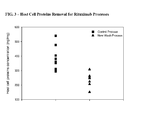
Classifications
-
- C—CHEMISTRY; METALLURGY
- C07—ORGANIC CHEMISTRY
- C07K—PEPTIDES
- C07K1/00—General methods for the preparation of peptides, i.e. processes for the organic chemical preparation of peptides or proteins of any length
- C07K1/14—Extraction; Separation; Purification
- C07K1/16—Extraction; Separation; Purification by chromatography
- C07K1/18—Ion-exchange chromatography
-
- A—HUMAN NECESSITIES
- A61—MEDICAL OR VETERINARY SCIENCE; HYGIENE
- A61P—SPECIFIC THERAPEUTIC ACTIVITY OF CHEMICAL COMPOUNDS OR MEDICINAL PREPARATIONS
- A61P19/00—Drugs for skeletal disorders
- A61P19/02—Drugs for skeletal disorders for joint disorders, e.g. arthritis, arthrosis
-
- A—HUMAN NECESSITIES
- A61—MEDICAL OR VETERINARY SCIENCE; HYGIENE
- A61P—SPECIFIC THERAPEUTIC ACTIVITY OF CHEMICAL COMPOUNDS OR MEDICINAL PREPARATIONS
- A61P19/00—Drugs for skeletal disorders
- A61P19/08—Drugs for skeletal disorders for bone diseases, e.g. rachitism, Paget's disease
-
- A—HUMAN NECESSITIES
- A61—MEDICAL OR VETERINARY SCIENCE; HYGIENE
- A61P—SPECIFIC THERAPEUTIC ACTIVITY OF CHEMICAL COMPOUNDS OR MEDICINAL PREPARATIONS
- A61P25/00—Drugs for disorders of the nervous system
-
- A—HUMAN NECESSITIES
- A61—MEDICAL OR VETERINARY SCIENCE; HYGIENE
- A61P—SPECIFIC THERAPEUTIC ACTIVITY OF CHEMICAL COMPOUNDS OR MEDICINAL PREPARATIONS
- A61P27/00—Drugs for disorders of the senses
- A61P27/02—Ophthalmic agents
-
- A—HUMAN NECESSITIES
- A61—MEDICAL OR VETERINARY SCIENCE; HYGIENE
- A61P—SPECIFIC THERAPEUTIC ACTIVITY OF CHEMICAL COMPOUNDS OR MEDICINAL PREPARATIONS
- A61P29/00—Non-central analgesic, antipyretic or antiinflammatory agents, e.g. antirheumatic agents; Non-steroidal antiinflammatory drugs [NSAID]
-
- A—HUMAN NECESSITIES
- A61—MEDICAL OR VETERINARY SCIENCE; HYGIENE
- A61P—SPECIFIC THERAPEUTIC ACTIVITY OF CHEMICAL COMPOUNDS OR MEDICINAL PREPARATIONS
- A61P35/00—Antineoplastic agents
-
- A—HUMAN NECESSITIES
- A61—MEDICAL OR VETERINARY SCIENCE; HYGIENE
- A61P—SPECIFIC THERAPEUTIC ACTIVITY OF CHEMICAL COMPOUNDS OR MEDICINAL PREPARATIONS
- A61P35/00—Antineoplastic agents
- A61P35/02—Antineoplastic agents specific for leukemia
-
- A—HUMAN NECESSITIES
- A61—MEDICAL OR VETERINARY SCIENCE; HYGIENE
- A61P—SPECIFIC THERAPEUTIC ACTIVITY OF CHEMICAL COMPOUNDS OR MEDICINAL PREPARATIONS
- A61P37/00—Drugs for immunological or allergic disorders
- A61P37/02—Immunomodulators
-
- A—HUMAN NECESSITIES
- A61—MEDICAL OR VETERINARY SCIENCE; HYGIENE
- A61P—SPECIFIC THERAPEUTIC ACTIVITY OF CHEMICAL COMPOUNDS OR MEDICINAL PREPARATIONS
- A61P43/00—Drugs for specific purposes, not provided for in groups A61P1/00-A61P41/00
-
- A—HUMAN NECESSITIES
- A61—MEDICAL OR VETERINARY SCIENCE; HYGIENE
- A61P—SPECIFIC THERAPEUTIC ACTIVITY OF CHEMICAL COMPOUNDS OR MEDICINAL PREPARATIONS
- A61P9/00—Drugs for disorders of the cardiovascular system
- A61P9/08—Vasodilators for multiple indications
-
- A—HUMAN NECESSITIES
- A61—MEDICAL OR VETERINARY SCIENCE; HYGIENE
- A61P—SPECIFIC THERAPEUTIC ACTIVITY OF CHEMICAL COMPOUNDS OR MEDICINAL PREPARATIONS
- A61P9/00—Drugs for disorders of the cardiovascular system
- A61P9/14—Vasoprotectives; Antihaemorrhoidals; Drugs for varicose therapy; Capillary stabilisers
-
- C—CHEMISTRY; METALLURGY
- C07—ORGANIC CHEMISTRY
- C07K—PEPTIDES
- C07K1/00—General methods for the preparation of peptides, i.e. processes for the organic chemical preparation of peptides or proteins of any length
- C07K1/14—Extraction; Separation; Purification
- C07K1/36—Extraction; Separation; Purification by a combination of two or more processes of different types
-
- C—CHEMISTRY; METALLURGY
- C07—ORGANIC CHEMISTRY
- C07K—PEPTIDES
- C07K16/00—Immunoglobulins [IGs], e.g. monoclonal or polyclonal antibodies
- C07K16/06—Immunoglobulins [IGs], e.g. monoclonal or polyclonal antibodies from serum
- C07K16/065—Purification, fragmentation
-
- C—CHEMISTRY; METALLURGY
- C07—ORGANIC CHEMISTRY
- C07K—PEPTIDES
- C07K16/00—Immunoglobulins [IGs], e.g. monoclonal or polyclonal antibodies
- C07K16/18—Immunoglobulins [IGs], e.g. monoclonal or polyclonal antibodies against material from animals or humans
- C07K16/22—Immunoglobulins [IGs], e.g. monoclonal or polyclonal antibodies against material from animals or humans against growth factors ; against growth regulators
-
- C—CHEMISTRY; METALLURGY
- C07—ORGANIC CHEMISTRY
- C07K—PEPTIDES
- C07K16/00—Immunoglobulins [IGs], e.g. monoclonal or polyclonal antibodies
- C07K16/18—Immunoglobulins [IGs], e.g. monoclonal or polyclonal antibodies against material from animals or humans
- C07K16/28—Immunoglobulins [IGs], e.g. monoclonal or polyclonal antibodies against material from animals or humans against receptors, cell surface antigens or cell surface determinants
- C07K16/2887—Immunoglobulins [IGs], e.g. monoclonal or polyclonal antibodies against material from animals or humans against receptors, cell surface antigens or cell surface determinants against CD20
-
- C—CHEMISTRY; METALLURGY
- C07—ORGANIC CHEMISTRY
- C07K—PEPTIDES
- C07K2317/00—Immunoglobulins specific features
- C07K2317/20—Immunoglobulins specific features characterized by taxonomic origin
- C07K2317/24—Immunoglobulins specific features characterized by taxonomic origin containing regions, domains or residues from different species, e.g. chimeric, humanized or veneered
Definitions
- This invention relates generally to protein purification.
- the invention relates to a method for purifying antibody from a composition comprising the antibody and at least one contaminant using cation exchange chromatography, wherein a high pH wash step is used to remove contaminants prior to eluting the desired antibody using an elution buffer with increased conductivity.
- proteins are produced by cell culture, using either eukaryotic or prokaryotic cell lines engineered to produce the protein of interest by insertion of a recombinant plasmid containing the gene for that protein. Since the cells typically used are living organisms, they must be fed with a complex growth medium, containing sugars, amino acids, and growth factors, usually supplied from preparations of animal serum. Separation of the desired protein from the mixture of compounds fed to the cells and from the by-products of the cells themselves to a purity sufficient for use as a human therapeutic poses a daunting challenge.
- Procedures for purification of proteins from cell debris initially depend on the site of expression of the protein. Some proteins can be cased to be secreted directly from the cell into the surrounding growth media; others are made intracellularly.
- the first step of a purification process involves lysis of the cell, which can be done by a variety of methods, including mechanical shear, osmotic shock, or enzymatic treatments. Such disruption releases the entire contents of the cell into the homogenate, and in addition produces subcellular fragments that are difficult to remove due to their small size. These are generally removed by differential centrifugation or by filtration. The same problem arises, although on a smaller scale, with directly secreted proteins due to the natural death of cells and release of intracellular host cell proteins in the course of the protein production run.
- Ion exchange chromatography is a chromatographic technique that is commonly used for the purification of proteins.
- ion exchange chromatography charged patches on the surface of the solute are attracted by opposite charges attached to a chromatography matrix, provided the ionic strength of the surrounding buffer is low. Elution is generally achieved by increasing the ionic strength (i.e. conductivity) of the buffer to compete with the solute for the charged sites of the ion exchange matrix.
- Changing the pH and thereby altering the charge of the solute is another way to achieve elution of the solute.
- the change in conductivity or pH may be gradual (gradient elution) or stepwise (step elution). In the past, these changes have been progressive; i.e., the pH or conductivity is increased or decreased in a single direction
- US Patent No. 5,110,913 refers to purifying an antibody in an aqueous solution by binding the antibody to an ion exchange resin at a first pH of 4.6, washing at a second pH of 5.5, and eluting the antibody at pH 6.5, wherein the ionic strength of the solutions of these three steps remains constant.
- Zhang et al. refer to Q membrane, anion exchange chromatography of a human antibody ( Zhang et al. "Q Membrane Chromatography Application for Human Antibody Purification Process," Poster presented at BioProduction, Oct. 26-27. Kunststoff, Germany, 2004 ).
- Other publications concerning protein purification include: Barnthouse et al. J. Biotech. 66: 125-136 (1998 ); Blank et al.
- the invention herein concerns an improved method for cation exchange chromatography of antibodies in which a high pH wash step is used to remove contaminants prior to eluting the desired antibody product.
- the process results, amongst other things, in improved removal of Chinese Hamster Ovary Proteins (CHOP) contaminants.
- CHOP Chinese Hamster Ovary Proteins
- the invention provides a method for purifying an antibody from a composition comprising the antibody and at least one contaminant, which method comprises the sequential steps of:
- the antibody binds human CD20, such as rituximab, or binds human vascular endothelial growth factor (VEGF), such as bevacizumab.
- human CD20 such as rituximab
- VEGF vascular endothelial growth factor
- the invention concerns a method for purifying an antibody that binds human CD20 from a composition comprising the antibody and one or more contaminants selected from the group consisting of Chinese Hamster Ovary Proteins (CHOP), leached protein A, DNA, and aggregated CD20 antibody, which method comprises the sequential steps of:
- the invention relates to a method for purifying an antibody that binds human vascular endothelial growth factor (VEGF) from a composition comprising the antibody and one or more contaminants selected from the group consisting of a cell culture media component, garamycin, Chinese Hamster Ovary Proteins (CHOP), DNA, viral contaminant, and aggregated VEGF antibody, which method comprises the sequential steps of:
- the invention also concerns a composition comprising rituximab in a buffer comprising about 25mM HEPES, at a pH of about 7.8.
- the invention provides a composition comprising bevacizumab in a buffer comprising about 25mM MOPS at a pH of about 7.0.
- Figures 1A and 1B provide the amino acid sequences of the heavy chain (SEQ ID No. 1) and light chain (SEQ ID No. 2) of rituximab antibody.
- Each of the framework regions (FR1-4) and each of the CDR regions (CDR1-3) in each variable region are identified, as are the human gamma 1 heavy chain constant sequence and human kappa light chain constant sequence.
- the variable heavy (VH) region is in SEQ ID No. 3.
- the variable light (VL) region is in SEQ ID No. 4.
- the sequence identifiers for the CDRs are: CDR H1 (SEQ ID No. 5), CDR H2 (SEQ ID No. 6), CDR H3 (SEQ ID No. 7), CDR L1 (SEQ ID No. 8), CDR L2 (SEQ ID No. 9), and CDR L3 (SEQ ID No. 10).
- Figures 2A and 2B provide the amino acid sequences of the heavy chain (SEQ ID No. 11) and light chain (SEQ ID No. 12) of bevacizumab antibody.
- the end of each variable region is indicated with
- the variable heavy (VH) region is in SEQ ID No. 13.
- the variable light (VL) region is in SEQ ID No. 14.
- Each of the three CDRs in each variable region is underlined.
- the sequence identifiers for the CDRs are: CDR H1 (SEQ ID No. 15), CDR H2 (SEQ ID No. 16), CDR H3 (SEQ ID No. 17), CDR L1 (SEQ ID No. 18), CDR L2 (SEQ ID No. 19), and CDR L3 (SEQ ID No. 20).
- Figure 3 provides a side-by-side comparison of host cell proteins removal by the cation exchange chromatography process of the improved rituximab process compared to the original process. Superior CHOP removal was achieved with the new process.
- the composition may be "partially purified” (i.e. having been subjected to one or more purification steps) or maybe obtained directly from a host cell or organism producing the antibody (e.g. the composition may comprise harvested cell culture fluid).
- polypeptide refers generally to peptides and proteins having more than about ten amino acids.
- the polypeptide is a mammalian protein, examples of which include: renin; a growth hormone, including human growth hormone and bovine growth hormone; growth hormone releasing factor; parathyroid hormone; thyroid stimulating hormone; lipoproteins; alpha-1-antitrypsin; insulin A-chain; insulin B-chain; proinsulin; follicle stimulating hormone; calcitonin; luteinizing hormone; glucagon; clotting factors such as factor VIIIC, factor IX, tissue factor, and von Willebrands factor; anti-clotting factors such as Protein C; atrial natriuretic factor; lung surfactant; a plasminogen activator, such as urokinase or human urine or tissue-type plasminogen activator (t-PA); bombesin; thrombin; hemopoietic growth factor; tumor necrosis factor-alpha and -be
- a preferred polypeptide is an intact antibody or an antibody fragment that binds to human CD20, for example, rituximab; or an intact antibody or an antibody fragment that binds to human vascular endothelial growth factor (VEGF), for example bevacizumab.
- VEGF vascular endothelial growth factor
- a “contaminant” is a material that is different from the desired antibody product.
- the contaminant includes, without limitation: host cell materials, such as Chinese Hamster Ovary Proteins (CHOP); leached protein A; nucleic acid; a variant, fragment, aggregate or derivative of the desired antibody; another polypeptide; endotoxin; viral contaminant; cell culture media component (e.g. garamycin; GENTAMYCIN®) etc.
- host cell materials such as Chinese Hamster Ovary Proteins (CHOP); leached protein A; nucleic acid; a variant, fragment, aggregate or derivative of the desired antibody; another polypeptide; endotoxin; viral contaminant; cell culture media component (e.g. garamycin; GENTAMYCIN®) etc.
- cation exchange material refers to a solid phase that is negatively charged and has free cations for exchange with cations in an aqueous solution passed over or through the solid phase.
- the charge may be provided by attaching one or more charged ligands to the solid phase, e.g. by covalent linking.
- the charge may be an inherent property of the solid phase (e.g. as is the case for silica, which has an overall negative charge).
- Commercially available cation exchange materials include carboxy-methyl-cellulose, BAKERBOND ABXTM, sulphopropyl (SP) immobilized on agarose (e.g.
- SP-SEPHAROSE FAST FLOWTM, SP-SEPHAROSE FAST FLOW XLTM or SP-SEPHAROSE HIGH PERFORMANCETM from GE Healthcare
- CAPTO STM GE Healthcare
- FRACTOGEL-SO3TM, FRACTOGEL-SE HICAPTM e.g. S-SEPHAROSE FAST FLOWTM from GE Healthcare
- SUPER SPTM Tosoh Biosciences
- a preferred cation exchange material herein comprises cross-linked poly(styrene-divinylbenzene) flow-through particles (solid phase) coated with a polyhydroxylated polymer functionalized with sulfopropyl groups (for example, POROS 50 HS® chromatography resin).
- solid phase is meant a non-aqueous matrix to which one or more charged ligands can adhere.
- the solid phase may be a purification column (including, without limitation, expanded bed and packed bed columns), a discontinuous phase of discrete particles, a membrane, or filter etc.
- materials for forming the solid phase include polysaccharides (such as agarose and cellulose) and other mechanically stable matrices such as silica (e.g. controlled pore glass), poly(styrene-divinylbenzene), polyacrylamide, ceramic particles and derivatives of any of the above.
- load herein refers to the composition loaded onto the cation exchange material.
- the cation exchange material is equilibrated with an equilibration buffer prior to loading the composition which is to be purified.
- a “buffer” is a solution that resists changes in pH by the action of its acid-base conjugate components.
- Various buffers which can be employed depending, for example, on the desired pH of the buffer are described in Buffers. A Guide for the Preparation and Use of Buffers in Biological Systems, Gueffroy, D., Ed. Calbiochem Corporation (1975 ).
- an “equilibration buffer” is a buffer that is used to equilibrate the cation exchange material, prior to loading the composition comprising the antibody of interest and one or more contaminants onto the cation exchange material.
- the pH of the equilibration buffer herein is in the range from about 5.0 to about 6.0, preferably about 5.5.
- the conductivity of the equilibration buffer herein is in the range from about 1 to about 8mS/cm, preferably from about 4 to about 8mS/cm, and most preferably from about 5 to about 8mS/cm.
- the equilibration buffer comprises a salt, such as NaCl, for example, in an amount from about 40mM to about 80mM, preferably about 60mM NaCl.
- wash buffer is used herein to refer to the buffer that is passed over the cation exchange material following loading of a composition and prior to elution of the protein of interest.
- the wash buffer may serve to remove one or more contaminants from the cation exchange material, without substantial elution of the desired antibody product.
- a “first wash buffer” and a “second wash buffer” are used.
- first wash buffer refers to a wash buffer having a pH increased relative to the pH of the load and/or equilibration buffer.
- the first wash buffer may be used herein to elute one or more contaminants from the cation exchange material, without substantially eluting the antibody product of interest therefrom.
- first should not be interpreted as excluding the use of one or more additional wash or other buffers between the load and the first wash buffer.
- the pH of the first wash buffer herein is in the range from about 6.8 to about 9.0, preferably from about 7.0 to about 8.0, and most preferably pH about 7.0 or pH about 7.8.
- the conductivity of the first wash buffer herein is in the range from about 0.01 to about 5mS/cm, preferably from about 0.1 to about 3mS/cm, and most preferably from about 0.2 to about 2mS/cm.
- the first wash buffer is substantially free of a salt (such as NaCl) therein.
- second wash buffer refers to a wash buffer used after the first wash buffer to prepare the cation exchange material for elution of the antibody of interest.
- second should not be interpreted as excluding the use of one or more additional wash or other buffers between the first wash buffer and second wash buffer.
- the pH of the second wash buffer herein is in the range from about 5.0 to about 6.0, preferably about 5.5, and most preferably pH 5.5.
- the conductivity of the second wash buffer herein is in the range from about 0.01 to about 5 mS/cm, preferably about 0.1 to about 3 mS/cm, and most preferably from about 0.5 to about 3.0mS/cm.
- Elution buffer is used to elute the antibody of interest from the solid phase.
- the elution buffer has a substantially increased conductivity relative to that of the second wash buffer, such that the desired antibody product is eluted from the cation exchange material.
- the conductivity of the elution buffer is substantially greater than that of the load and of each of the preceding buffers, namely of the equilibration buffer, first wash buffer, and second wash buffer.
- substantially greater conductivity is meant, for example, that the buffer has a conductivity which is at least 2, 3, 4, 5 or 6 conductivity units (mS/cm) greater than that of the composition or buffer to which it is being compared.
- the pH of the elution buffer is substantially the same as that of the equilibration and/or second wash buffer.
- the pH of the elution buffer herein is in the range from about 5.0 to about 6.0, preferably about 5.5, and most preferably pH 5.5.
- the conductivity of the elution buffer herein is in the range from about 10mS/cm to about 100mS/cm, preferably from about 12mS/cm to about 30mS/cm, and most preferably from about 12 to about 20mS/cm.
- Increased conductivity may be achieved by the addition of a salt, such as sodium chloride, sodium acetate, potassium chloride to the elution buffer.
- the elution buffer comprises from about 100 to about 300mM NaCl, preferably from about 150mM to about 200mM NaCl, for example about 175mM NaCl or about 160mM NaCl.
- a “regeneration buffer” may be used to regenerate the cation exchange material such that it can be re-used.
- the regeneration buffer has a conductivity and/or pH as required to remove substantially all contaminants and the antibody of interest from the cation exchange material.
- conductivity refers to the ability of an aqueous solution to conduct an electric current between two electrodes. In solution, the current flows by ion transport. Therefore, with an increasing amount of ions present in the aqueous solution, the solution will have a higher conductivity.
- the basic unit of measure for conductivity is the Siemen (or mho), mho (mS/cm), and can be measured using a conductivity meter, such as various models of Orion conductivity meters.
- electrolytic conductivity is the capacity of ions in a solution to carry electrical current
- the conductivity of a solution may be altered by changing the concentration of ions therein.
- concentration of a buffering agent and/or the concentration of a salt e.g. sodium chloride, sodium acetate, or potassium chloride
- the salt concentration of the various buffers is modified to achieve the desired conductivity.
- purifying an antibody from a composition comprising the antibody and one or more contaminants is meant increasing the degree of purity of the antibody in the composition by removing (completely or partially) at least one contaminant from the composition.
- a “purification step” may be part of an overall purification process resulting in a “homogeneous” composition.
- homogeneous is used herein to refer to a composition comprising at least about 70% by weight of the antibody of interest, based on total weight of the composition, preferably at least about 80% by weight, more preferably at least about 90% by weight, even more preferably at least about 95% by weight.
- binding a molecule to a cation exchange material is meant exposing the molecule to the cation exchange material under appropriate conditions (pH and/or conductivity) such that the molecule is reversibly immobilized in or on the cation exchange material by virtue of ionic interactions between the molecule and a charged group or charged groups of the cation exchange material.
- washing By “washing" the cation exchange material is meant passing an appropriate buffer through or over the cation exchange material.
- eluting a molecule (e.g. antibody or contaminant) from a cation exchange material is meant to remove the molecule therefrom.
- the antibody to be purified herein is a recombinant antibody.
- a “recombinant antibody” is one which has been produced in a host cell which has been transformed or transfected with nucleic acid encoding the antibody, or produces the antibody as a result of homologous recombination.
- Transformation and “transfection” are used interchangeably to refer to the process of introducing nucleic acid into a cell. Following transformation or transfection, the nucleic acid may integrate into the host cell genome, or may exist as an extrachromosomal element.
- the "host cell” includes a cell in in vitro cell culture as well as a cell within a host animal. Methods for recombinant production of polypeptides are described in US Patent No. 5,534,615 , expressly incorporated herein by reference, for example.
- a “variant” or “amino acid sequence variant” of a starting polypeptide is a polypeptide that comprises an amino acid sequence different from that of the starting polypeptide.
- a variant will possess at least 80% sequence identity, preferably at least 90% sequence identity, more preferably at least 95% sequence identity, and most preferably at least 98% sequence identity with the native polypeptide. Percentage sequence identity is determined, for example, by the Fitch et al., Proc. Natl. Acad. Sci. USA 80:1382-1386 (1983 ), version of the algorithm described by Needleman et al., J. Mol. Biol. 48:443-453 (1970 ), after aligning the sequences to provide for maximum homology.
- Amino acid sequence variants of a polypeptide may be prepared by introducing appropriate nucleotide changes into DNA encoding the polypeptide, or by peptide synthesis. Such variants include, for example, deletions from, and/or insertions into and/or substitutions of, residues within the amino acid sequence of the polypeptide of interest. Any combination of deletion, insertion, and substitution is made to arrive at the final construct, provided that the final construct possesses the desired characteristics.
- the amino acid changes also may alter post-translational processing of the polypeptide, such as by changing the number or position of glycosylation sites.
- antibody is used in the broadest sense and specifically covers monoclonal antibodies (including full length monoclonal antibodies), polyclonal antibodies, multispecific antibodies (e.g., bispecific antibodies), and antibody fragments so long as they exhibit the desired binding specificity.
- the antibody herein is directed against an "antigen" of interest.
- the antigen is a biologically important polypeptide and administration of the antibody to a mammal suffering from a disease or disorder can result in a therapeutic benefit in that mammal.
- antibodies directed against non-polypeptide antigens are also contemplated.
- the antigen is a polypeptide, it may be a transmembrane molecule (e.g. receptor) or ligand such as a growth factor.
- Exemplary antigens include those polypeptides discussed above.
- CD polypeptides such as CD3, CD4, CD8, CD19, CD20 and CD34; members of the HER receptor family such as the EGF receptor (HER1), HER2, HER3 or HER4 receptor; cell adhesion molecules such as LFA-1, Mac1, p150,95, VLA-4, ICAM-1, VCAM and av/b3 integrin including either a or b subunits thereof (e.g. anti-CD 11 a, anti-CD18 or anti-CD11b antibodies); growth factors such as VEGF; IgE; blood group antigens; flk2/flt3 receptor; obesity (OB) receptor; mpl receptor; CTLA-4; polypeptide C etc.
- HER1 EGF receptor
- HER2 HER2
- HER3 or HER4 receptor cell adhesion molecules
- cell adhesion molecules such as LFA-1, Mac1, p150,95, VLA-4, ICAM-1, VCAM and av/b3 integrin including either a or b
- Soluble antigens or fragments thereof, optionally conjugated to other molecules, can be used as immunogens for generating antibodies.
- immunogens for transmembrane molecules, such as receptors, fragments of these (e.g. the extracellular domain of a receptor) can be used as the immunogen.
- transmembrane molecules such as receptors
- fragments of these e.g. the extracellular domain of a receptor
- cells expressing the transmembrane molecule can be used as the immunogen.
- Such cells can be derived from a natural source (e.g. cancer cell lines) or may be cells which have been transformed by recombinant techniques to express the transmembrane molecule.
- antibodies to be purified herein include, but are not limited to: HER2 antibodies including trastuzumab (HERCEPTIN®) ( Carter et al., Proc. Natl. Acad. Sci. USA, 89:4285-4289 (1992 ), U.S. Patent No. 5,725,856 ) and pertuzumab (OMNITARGTM) ( WO01/00245 ); CD20 antibodies (see below); IL-8 antibodies ( St John et al., Chest, 103:932 (1993 ), and International Publication No.
- HERCEPTIN® trastuzumab
- OMNITARGTM pertuzumab
- CD20 antibodies see below
- IL-8 antibodies St John et al., Chest, 103:932 (1993 ), and International Publication No.
- VEGF or VEGF receptor antibodies including humanized and/or affinity matured VEGF antibodies such as the humanized VEGF antibody huA4.6.1 bevacizumab (AVASTIN®) and ranibizumab (LUCENTIS®) ( Kim et al., Growth Factors, 7:53-64 (1992 ), International Publication No. WO 96/30046 , and WO 98/45331, published October 15, 1998 ); PSCA antibodies ( WO01/40309 ); CD11a antibodies including efalizumab (RAPTIVA®) ( US Patent No. 5,622,700 , WO 98/23761 , Steppe et al., Transplant Intl.
- RPTIVA® efalizumab
- CD25 or Tac antibodies such as CHI-621 (SIMULECT®) and ZENAPAX® (See US Patent No. 5,693,762 issued December 2, 1997 ); CD4 antibodies such as the cM-7412 antibody ( Choy et al. Arthritics Rheum 39(1):52-56 (1996 )); CD52 antibodies such as CAMPATH-1H (ILEX/Berlex) ( Riechmann et al. Nature 332:323-337 (1988 )); Fc receptor antibodies such as the M22 antibody directed against Fc ⁇ RI as in Graziano et al. J. Immunol.
- carcinoembryonic antigen (CEA) antibodies such as hMN-14 ( Sharkey et al. Cancer Res. 55(23Suppl): 5935s-5945s (1995 )); antibodies directed against breast epithelial cells including huBrE-3, hu-Mc 3 and CHL6 ( Ceriani et al. Cancer Res. 55(23): 5852s-5856s (1995 ); and Richman et al. Cancer Res. 55(23 Supp): 5916s-5920s (1995 )); antibodies that bind to colon carcinoma cells such as C242 ( Litton et al. Eur J. Immunol.
- CD38 antibodies e.g. AT 13/5 ( Ellis et al. J. Immunol. 155(2):925-937 (1995 )); CD33 antibodies such as Hu M195 ( Jurcic et al. Cancer Res 55(23 Suppl):5908s-5910s (1995 )) and CMA-676 or CDP771; EpCAM antibodies such as 17-1A (PANOREX®); GpIIb/IIIa antibodies such as abciximab or c7E3 Fab (REOPRO®); RSV antibodies such as MEDI-493 (SYNAGIS®); CMV antibodies such as PROTOVIR®; HIV antibodies such as PRO542; hepatitis antibodies such as the Hep B antibody OSTAVIR®; CA 125 antibody OvaRex; idiotypic GD3 epitope antibody BEC2; ⁇ v ⁇ 3 antibody (e.g.
- human renal cell carcinoman antibody such as ch-G250; ING-1; anti-human 17-1An antibody (3622W94); anti-human colorectal tumor antibody (A33); anti-human melanoman antibody R24 directed against GD3 ganglioside; anti-human squamous-cell carcinoma (SF-25); human leukocyte antigen (HLA) antibody such as Smart ID10 and the anti-HLA DR antibody Oncolym (Lym-1); CD37 antibody such as TRU 016 (Trubion); IL-21 antibody (Zymogenetics/Novo Nordisk); anti-B cell antibody (Impheron); B cell targeting MAb (Immunogen/Aventis); 1D09C3 (Morphosys/GPC); LymphoRad 131 (HGS); Lym-1 antibody, such as Lym -1Y-90 (USC) or anti-Lym-1 Oncolym (USC/Peregrine); LIF 226 (Enhanced
- anti-complement antibody such as C5 antibody (e.g. eculizumab, 5G1.1; Alexion); oral formulation of human immunoglobulin (e.g. IgPO; Protein Therapeutics); IL-12 antibody such as ABT-874 (CAT/Abbott); Teneliximab (BMS-224818; BMS); CD40 antibodies, including S2C6 and humanized variants thereof ( WO00/75348 ) and TNX 100 (Chiron/Tanox); TNF- ⁇ antibodies including cA2 or infliximab (REMICADE®), CDP571, MAK-195, adalimumab (HUMIRATM), pegylated TNF- ⁇ antibody fragment such as CDP-870 (Celltech), D2E7 (Knoll), anti-TNF- ⁇ polyclonal antibody (e.g.
- C5 antibody e.g. eculizumab, 5G1.1; Alexion
- oral formulation of human immunoglobulin e.
- CD22 antibodies such as LL2 or epratuzumab (LYMPHOCIDE®; Immunomedics), including epratuzumab Y-90 and epratzumab I-131, Abiogen's CD22 antibody (Abiogen, Italy), CMC 544 (Wyeth/Celltech), combotox (UT Soutwestern), BL22 (NIH), and LympoScan Tc99 (Immunomedics).
- the antibody that is purified herein is a naked, intact antibody which binds to human CD20, or a naked, intact antibody which binds to human VEGF.
- CD20 antigen is an about 35-kDa, non-glycosylated phosphoprotein found on the surface of greater than 90% of B cells from peripheral blood or lymphoid organs. CD20 is present on both normal B cells as well as malignant B cells, but is not expressed on stem cells. Other names for CD20 in the literature include "B-lymphocyte-restricted antigen” and "Bp35". The CD20 antigen is described in Clark et al., Proc. Natl. Acad. Sci. (USA) 82:1766 (1985 ), for example.
- a "CD20 antibody antagonist” herein is an antibody that, upon binding to CD20 on B cells, destroys or depletes B cells in a subject and/or interferes with one or more B-cell functions, e.g. , by reducing or preventing a humoral response elicited by the B cell.
- the antibody antagonist preferably is able to deplete B cells (i.e., reduce circulating B-cell levels) in a subject treated therewith. Such depletion may be achieved via various mechanisms such antibody-dependent cell-mediated cytotoxicity (ADCC) and/or complement-dependent cytotoxicity (CDC), inhibition of B-cell proliferation and/or induction of B-cell death (e.g., via apoptosis).
- ADCC antibody-dependent cell-mediated cytotoxicity
- CDC complement-dependent cytotoxicity
- B cell depletion refers to a reduction in B cell levels in an animal or human generally after drug or antibody treatment, as compared to the level before treatment. B cell depletion can be partial or complete. B cell levels are measurable using well known techniques such as those described in Reff et al., Blood 83: 435-445 (1994 ), or US Patent No. 5,736,137 (Anderson et al. ).
- a mammal e.g. a normal primate
- peripheral B-cell concentrations may be determined, e.g. by a FACS method that counts B cells.
- CD20 antibodies examples include: “C2B8,” which is now called “rituximab” (“RITUXAN®”) ( US Patent No. 5,736,137 ); the yttrium-[90]-labelled 2B8 murine antibody designated “Y2B8” or “Ibritumomab Tiuxetan” (ZEVALIN®) commercially available from IDEC Pharmaceuticals, Inc. ( US Patent No. 5,736,137 ; 2B8 deposited with ATCC under accession no.
- C2B8 which is now called “rituximab”
- Y2B8 yttrium-[90]-labelled 2B8 murine antibody designated “Y2B8” or “Ibritumomab Tiuxetan” (ZEVALIN®) commercially available from IDEC Pharmaceuticals, Inc.
- CD20 binding molecules such as the AME series of antibodies, e.g ., AME 33 antibodies as set forth in WO 2004/103404 and US2005/0025764 (Watkins et al. , Eli Lilly/Applied Molecular Evolution, AME); CD20 binding molecules such as those described in US 2005/0025764 (Watkins et al.
- A20 antibody or variants thereof such as chimeric or humanized A20 antibody (cA20, hA20, respectively) or IMMU-106 ( US 2003/0219433 , Immunomedics); CD20-binding antibodies, including epitope-depleted Leu-16, 1H4, or 2B8, optionally conjugated with IL-2, as in US 2005/0069545A1 and WO 2005/16969 (Carr et al. ); bispecific antibody that binds CD22 and CD20, for example, hLL2xhA20 ( WO2005/14618, Chang et al.
- the preferred CD20 antibodies herein are chimeric, humanized, or human CD20 antibodies, more preferably rituximab, humanized 2H7, 2F2 (Hu-Max-CD20) human CD20 antibody (Genmab), and humanized A20 or IMMUN-106 antibody (Immunomedics).
- rituximab refers to a recombinant chimeric antibody which binds to the human CD20 antigen as described in US Patent No. 5,736,137, Anderson et al.
- Such antibody preferably comprises a heavy chain comprising CDR H1 (SEQ ID No. 5), CDR H2 (SEQ ID No. 6), CDR H3 (SEQ ID No. 7), and a light chain, wherein the light chain preferably comprises CDR L1 (SEQ ID No. 8), CDR L2 (SEQ ID No. 9), and CDR L3 (SEQ ID No.
- the heavy chain comprises a variable heavy (VH) region comprising SEQ ID No. 3 and a variable light (VL) region comprising SEQ ID No. 4; and most preferably comprises a heavy chain comprising SEQ ID No. 1 (with or without a C-terminal lysine residue), and a light chain, wherein the light chain preferably comprises SEQ ID No. 2.
- VH variable heavy
- VL variable light
- the terms expressly include variant forms such as described in Moorhouse et al. J. Charm Biomed. Anal. 16:593-603 (1997 ).
- human VEGF refers to the 165-amino acid human vascular endothelial cell growth factor, and related 121-, 189-, and 206-amino acid vascular endothelial cell growth factors, as described by Leung et al., Science 246:1306 (1989 ), and Houck et al., Mol. Endocrin. 5:1806 (1991 ) together with the naturally occurring allelic and processed forms of those growth factors.
- the present invention provides anti-VEGF antagonistic antibodies which are capable of inhibiting one or more of the biological activities of VEGF, for example, its mitogenic or angiogenic activity.
- Antagonists of VEGF act by interfering with the binding of VEGF to a cellular receptor, by incapacitating or killing cells which have been activated by VEGF, or by interfering with vascular endothelial cell activation after VEGF binding to a cellular receptor. All such points of intervention by a VEGF antagonist shall be considered equivalent for purposes of this invention.
- bevacizumab refers to a recombinant humanized monoclonal antibody which binds human vascular endothelial growth factor (VEGF) antigen (rhuMAb VEGF) as described in US Patent No. 7,169,901, Presta et al.
- VEGF vascular endothelial growth factor
- rhuMAb VEGF human vascular endothelial growth factor antigen
- Such antibody preferably comprises a heavy chain comprising CDR H1 (SEQ ID No. 15), CDR H2 (SEQ ID No. 16), CDR H3 (SEQ ID No. 17), and a light chain, wherein the light chain preferably comprises CDR L1 (SEQ ID No. 18), CDR L2 (SEQ ID No.
- the heavy chain comprises a variable heavy (VH) region comprising SEQ ID No. 13 and a variable light (VL) region comprising SEQ ID No. 14; and preferably comprises a heavy chain comprising SEQ ID No. 11 (with or without a C-terminal lysine residue), and a light chain, wherein the light chain preferably comprises SEQ ID No. 12.
- VH variable heavy
- VL variable light
- the terms expressly include variant forms that form during production of the recombinant antibody product.
- the term "monoclonal antibody” as used herein refers to an antibody obtained from a population of substantially homogeneous antibodies, i.e. , the individual antibodies comprising the population are identical except for possible naturally occurring mutations that may be present in minor amounts. Monoclonal antibodies are highly specific, being directed against a single antigenic site. Furthermore, in contrast to conventional (polyclonal) antibody preparations which typically include different antibodies directed against different determinants (epitopes), each monoclonal antibody is directed against a single determinant on the antigen.
- the modifier "monoclonal” indicates the character of the antibody as being obtained from a substantially homogeneous population of antibodies, and is not to be construed as requiring production of the antibody by any particular method.
- the monoclonal antibodies to be used in accordance with the present invention may be made by the hybridoma method first described by Kohler et al., Nature 256:495 (1975 ), or may be made by recombinant DNA methods (see, e.g., U.S. Patent No. 4,816,567 ).
- "monoclonal antibodies” can be isolated from antibody phage libraries generated using the techniques described in McCafferty et al., Nature, 348:552-554 (1990 ). Clackson et al., Nature, 352:624-628 (1991 ) and Marks et al., J. Mol.
- transgenic animals e.g., mice
- transgenic animals e.g., mice
- J H antibody heavy-chain joining region
- the monoclonal antibodies herein specifically include "chimeric" antibodies (immunoglobulins) in which a portion of the heavy and/or light chain is identical with or homologous to corresponding sequences in antibodies derived from a particular species or belonging to a particular antibody class or subclass, while the remainder of the chain(s) is identical with or homologous to corresponding sequences in antibodies derived from another species or belonging to another antibody class or subclass, as well as fragments of such antibodies, so long as they exhibit the desired biological activity ( U.S. Patent No. 4,816,567 ; and Morrison et al, Proc. Natl. Acad. Sci. USA 81:6851-6855 (1984 )).
- chimeric antibodies immunoglobulins in which a portion of the heavy and/or light chain is identical with or homologous to corresponding sequences in antibodies derived from a particular species or belonging to a particular antibody class or subclass, while the remainder of the chain(s) is identical with or homologous to corresponding sequences
- hypervariable region when used herein refers to the amino acid residues of an antibody which are responsible for antigen-binding.
- the hypervariable region comprises amino acid residues from a "complementarity determining region” or "CDR" (i.e. residues 24-34 (L1), 50-56 (L2) and 89-97 (L3) in the light chain variable domain and 31-35 (H1), 50-65 (H2) and 95-102 (H3) in the heavy chain variable domain; Kabat et al., Sequences of Polypeptides of Immunological Interest, 5th Ed. Public Health Service, National Institutes of Health, Bethesda, MD.
- CDR complementarity determining region
- Humanized forms of non-human (e.g., murine) antibodies are chimeric antibodies that contain minimal sequence derived from non-human immunoglobulin.
- humanized antibodies are human immunoglobulins (recipient antibody) in which residues from a hypervariable region of the recipient are replaced by residues from a hypervariable region of a non-human species (donor antibody) such as mouse, rat, rabbit or nonhuman primate having the desired specificity, affinity, and capacity.
- donor antibody such as mouse, rat, rabbit or nonhuman primate having the desired specificity, affinity, and capacity.
- Fv framework region (FR) residues of the human immunoglobulin are replaced by corresponding non-human residues.
- humanized antibodies may comprise residues which are not found in the recipient antibody or in the donor antibody. These modifications are made to further refine antibody performance.
- the humanized antibody will comprise substantially all of at least one, and typically two, variable domains, in which all or substantially all of the hypervariable loops correspond to those of a non-human immunoglobulin and all or substantially all of the FR regions are those of a human immunoglobulin sequence.
- the humanized antibody optionally also will comprise at least a portion of an immunoglobulin constant region (Fc), typically that of a human immunoglobulin.
- variable domains both light and heavy
- the choice of human variable domains, both light and heavy, to be used in making the humanized antibodies is very important to reduce antigenicity.
- sequence of the variable domain of a rodent antibody is screened against the entire library of known human variable-domain sequences.
- the human sequence which is closest to that of the rodent is then accepted as the human framework (FR) for the humanized antibody ( Sims et al., J. Immunol., 151:2296 (1993 ); Chothia et al., J. Mol. Biol., 196:901 (1987 )).
- Another method uses a particular framework derived from the consensus sequence of all human antibodies of a particular subgroup of light or heavy chains.
- the same framework may be used for several different humanized antibodies ( Carter et al., Proc. Natl. Acad. Sci. USA, 89:4285 (1992 ); Presta et al, J. Immnol., 151:2623 (1993 )).
- humanized antibodies are prepared by a process of analysis of the parental sequences and various conceptual humanized products using three-dimensional models of the parental and humanized sequences.
- Three-dimensional immunoglobulin models are commonly available and are familiar to those skilled in the art.
- Computer programs are available which illustrate and display probable three-dimensional conformational structures of selected candidate immunoglobulin sequences. Inspection of these displays permits analysis of the likely role of the residues in the functioning of the candidate immunoglobulin sequence, i.e., the analysis of residues that influence the ability of the candidate immunoglobulin to bind its antigen.
- FR residues can be selected and combined from the recipient and import sequences so that the desired antibody characteristic, such as increased affinity for the target antigen(s), is achieved.
- the CDR residues are directly and most substantially involved in influencing antigen binding.
- Antibody fragments comprise a portion of a full length antibody, generally the antigen binding or variable region thereof.
- Examples of antibody fragments include Fab, Fab', F(ab') 2 , and Fv fragments; diabodies; linear antibodies; single-chain antibody molecules; and multispecific antibodies formed from antibody fragments.
- Various techniques have been developed for the production of antibody fragments. Traditionally, these fragments were derived via proteolytic digestion of intact antibodies (see, e.g., Morimoto et al., Journal of Biochemical and Biophysical Methods 24:107-117 (1992 ) and Brennan et al., Science, 229:81 (1985 )). However, these fragments can now be produced directly by recombinant host cells.
- the antibody fragments can be isolated from the antibody phage libraries discussed above.
- Fab'-SH fragments can be directly recovered from E. coli and chemically coupled to form F(ab') 2 fragments ( Carter et al., Bio/Technology 10:163-167 (1992 )).
- the F(ab') 2 is formed using the leucine zipper GCN4 to promote assembly of the F(ab') 2 molecule.
- F(ab') 2 fragments can be isolated directly from recombinant host cell culture. Other techniques for the production of antibody fragments will be apparent to the skilled practitioner.
- the antibody of choice is a single chain Fv fragment (scFv). See WO 93/16185 .
- Single-chain Fv or “sFv” antibody fragments comprise the V H and V L domains of antibody, wherein these domains are present in a single polypeptide chain.
- the Fv polypeptide further comprises a polypeptide linker between the V H and V L domains which enables the sFv to form the desired structure for antigen binding.
- sFv see Pluckthun in The Pharmacology of Monoclonal Antibodies, vol. 113, Rosenburg and Moore eds. Springer-Verlag, New York, pp. 269-315 (1994 ).
- diabodies refers to small antibody fragments with two antigen-binding sites, which fragments comprise a heavy chain variable domain (V H ) connected to a light chain variable domain (V L ) in the same polypeptide chain (V H - V L ).
- V H heavy chain variable domain
- V L light chain variable domain
- the domains are forced to pair with the complementary domains of another chain and create two antigen-binding sites.
- Diabodies are described more fully in, for example, EP 404,097 ; WO 93/11161 ; and Hollinger et al., Proc. Natl. Acad. Sci. USA 90:6444-6448 (1993 ).
- linear antibodies when used throughout this application refers to the antibodies described in Zapata et al. Polypeptide Eng. 8(10):1057-1062 (1995 ). Briefly, these antibodies comprise a pair of tandem Fd segments (V H -C H 1-V H -C H 1) which form a pair of antigen binding regions. Linear antibodies can be bispecific or monospecific.
- Multispecific antibodies have binding specificities for at least two different epitopes, where the epitopes are usually from different antigens. While such molecules normally will only bind two antigens (i.e. bispecific antibodies, BsAbs), antibodies with additional specificities such as trispecific antibodies are encompassed by this expression when used herein.
- BsAbs include those with one arm directed against a tumor cell antigen and the other arm directed against a cytotoxic trigger molecule such as anti-Fc ⁇ RI/anti-CD15, anti-p185 HER2 /Fc ⁇ RIII (CD16), anti-CD3/anti-malignant B-cell (1D10), anti-CD3/anti-p185 HER2 , anti-CD3/anti-p97, anti-CD3/anti-renal cell carcinoma, anti-CD3/anti-OVCAR-3, anti-CD3/L-D1 (anti-colon carcinoma), anti-CD3/anti-melanocyte stimulating hormone analog, anti-EGF receptor/anti-CD3, anti-CD3/anti-CAMA1, anti-CD3/anti-CD19, anti-CD3/MoV18, anti-neural cell ahesion molecule (NCAM)/anti-CD3, anti-folate binding protein (FBP)/anti-CD3, anti-pan carcinoma associated antigen (AMOC-31)/anti-CD3; BsAbs with one arm which binds
- BsAbs for use in therapy of infectious diseases such as anti-CD3/anti-herpes simplex virus (HSV), anti-T-cell receptor:CD3 complex/anti-influenza, anti-Fc ⁇ R/anti-HIV; BsAbs for tumor detection in vitro or in vivo such as anti-CEA/anti-EOTUBE, anti-CEA/anti-DPTA, anti-p185 HER2 /anti-hapten; BsAbs as vaccine adjuvants; and BsAbs as diagnostic tools such as anti-rabbit IgG/anti-ferritin, anti-horse radish peroxidase (HRP)/anti-hormone, anti-somatostatin/anti-substance P, anti-HRP/anti-FITC, anti-CEA/anti- ⁇ -galactosidase.
- HRP anti-horse radish peroxidase
- HRP anti-somatostatin/anti-substance P
- trispecific antibodies examples include anti-CD3/anti-CD4/anti-CD37, anti-CD3/anti-CD5/anti-CD37 and anti-CD3/anti-CD8/anti-CD37.
- Bispecific antibodies can be prepared as full length antibodies or antibody fragments (e.g. F(ab') 2 bispecific antibodies).
- Antibodies with more than two valencies are contemplated.
- trispecific antibodies can be prepared. Tutt et al. J. Immunol. 147: 60 (1991 ).
- naked antibody for the purposes herein is an antibody that is not conjugated to a cytotoxic moiety or radiolabel.
- an “intact antibody” herein is one which comprises two antigen binding regions, and an Fc region.
- the intact antibody has a functional Fc region.
- Treatment refers to both therapeutic treatment and prophylactic or preventative measures. Those in need of treatment include those already with the disorder as well as those in which the disorder is to be prevented.
- a “disorder” is any condition that would benefit from treatment with the antibody purified as described herein. This includes both chronic and acute disorders and diseases and those pathological conditions which predispose the mammal to the disorder in question.
- label when used herein refers to a detectable compound or composition which is conjugated directly or indirectly to the antibody.
- the label may be itself be detectable (e.g. , radioisotope labels or fluorescent labels) or, in the case of an enzymatic label, may catalyze chemical alteration of a substrate compound or composition which is detectable.
- cytotoxic agent refers to a substance that inhibits or prevents the function of cells and/or causes destruction of cells.
- the term is intended to include radioactive isotopes (e.g. At 211 , I 131 , I 125 , Y 90 , Re 186 , Re 188 , Sm 153 , Bi 212 , P 32 and radioactive isotopes of Lu), chemotherapeutic agents, and toxins such as small-molecule toxins or enzymatically active toxins of bacterial, fungal, plant or animal origin, or fragments thereof.
- radioactive isotopes e.g. At 211 , I 131 , I 125 , Y 90 , Re 186 , Re 188 , Sm 153 , Bi 212 , P 32 and radioactive isotopes of Lu
- chemotherapeutic agents e.g. At 211 , I 131 , I 125 , Y 90 , Re 186 , Re 188
- the invention herein provides methods for purifying an antibody from a composition (e.g. an aqueous solution) comprising the antibody and one or more contaminants.
- the composition is generally one resulting from the recombinant production of the antibody, but may be that resulting from production of the antibody by peptide synthesis (or other synthetic means) or the antibody may be purified from a native source of the antibody.
- the antibody binds human CD20 antigen, such as rituximab, or binds human VEGF antigen, such as bevacizumab.
- the nucleic acid encoding it is isolated and inserted into a replicable vector for further cloning (amplification of the DNA) or for expression.
- DNA encoding the antibody is readily isolated and sequenced using conventional procedures (e.g., by using oligonucleotide probes that are capable of binding specifically to genes encoding the heavy and light chains of the antibody).
- Many vectors are available.
- the vector components generally include, but are not limited to, one or more of the following: a signal sequence, an origin of replication, one or more marker genes, an enhancer element, a promoter, and a transcription termination sequence (e.g. as described in US Patent 5,534,615 , specifically incorporated herein by reference).
- Suitable host cells for cloning or expressing the DNA in the vectors herein are the prokaryote, yeast, or higher eukaryotic cells.
- Suitable prokaryotes for this purpose include eubacteria, such as Gram-negative or Gram-positive organisms, for example, Enterobacteriaceae such as Escherichia, e.g., E. coli, Enterobacter , Erwinia, Klebsiella, Proteus , Salmonella , e.g., Salmonella typhimurium, Serratia, e.g., Serratia marcescans , and Shigella, as well as Bacilli such as B. subtilis and B. licheniformis (e.g., B.
- E. coli cloning host is E. coli 294 (ATCC 31,446), although other strains such as E. coli B, E. coli X1776 (ATCC 31,537), and E. coli W3110 (ATCC 27,325) are suitable. These examples are illustrative rather than limiting.
- eukaryotic microbes such as filamentous fungi or yeast are suitable cloning or expression hosts for antibody encoding vectors.
- Saccharomyces cerevisiae or common baker's yeast, is the most commonly used among lower eukaryotic host microorganisms.
- Kluyveromyces hosts such as, e.g., K. lactis, K. fragilis (ATCC 12,424), K. bulgaricus (ATCC 16,045), K. wickeramii (ATCC 24,178), K. waltii (ATCC 56,500), K.
- drosophilarum ATCC 36,906
- K . thermotolerans K. marxianus
- yarrowia EP 402,226
- Pichia pastoris EP 183,070
- Candida Trichoderma reesia
- Neurospora crassa Schwanniomyces such as Schwanniomyces occidentalis
- filamentous fungi such as, e.g., Neurospora, Penicillium, Tolypocladium, and Aspergillus hosts such as A. nidulans and A. niger.
- Suitable host cells for the expression of glycosylated antibody are derived from multicellular organisms.
- invertebrate cells include plant and insect cells.
- Numerous baculoviral strains and variants and corresponding permissive insect host cells from hosts such as Spodoptera frugiperda (caterpillar), Aedes aegypti (mosquito), Aedes albopictus (mosquito), Drosophila melanogaster (fruitfly), and Bombyx mori have been identified.
- a variety of viral strains for transfection are publicly available, e.g ., the L-1 variant of Autographa californica NPV and the Bm-5 strain of Bombyx mori NPV, and such viruses may be used as the virus herein according to the present invention, particularly for transfection of Spodoptera frugiperda cells.
- Plant cell cultures of cotton, corn, potato, soybean, petunia, tomato, and tobacco can also be utilized as hosts.
- mammalian host cell lines include, but are not limited to, monkey kidney CV1 cells transformed by SV40 (COS-7, ATCC CRL 1651); human embryonic kidney cells (293 or 293 cells subcloned for growth in suspension culture, Graham et al., J. Gen Virol. 36:59 (1977 )); baby hamster kidney cells (BHK, ATCC CCL 10); Chinese hamster ovary cells/- DHFR (CHO, Urlaub et al., Proc. Natl. Acad. Sci.
- COS-7 monkey kidney CV1 cells transformed by SV40
- human embryonic kidney cells (293 or 293 cells subcloned for growth in suspension culture, Graham et al., J. Gen Virol. 36:59 (1977 )
- baby hamster kidney cells BHK, ATCC CCL 10
- Chinese hamster ovary cells/- DHFR CHO, Urlaub et al., Proc. Natl. Acad. Sci.
- mice sertoli cells TM4, Mather, Biol. Reprod. 23:243-251 (1980 )); monkey kidney cells (CV1 ATCC CCL 70); African green monkey kidney cells (VERO-76, ATCC CRL-1587); human cervical carcinoma cells (HELA, ATCC CCL 2); canine kidney cells (MDCK, ATCC CCL 34); buffalo rat liver cells (BRL 3A, ATCC CRL 1442); human lung cells (W138, ATCC CCL 75); human liver cells (Hep G2, HB 8065); mouse mammary tumor (MMT 060562, ATCC CCL51); TRI cells ( Mather et al., Annals N.Y. Acad. Sci.
- CHO cells are preferred for the expression of antibodies, and may be advantageously used to produce the antibodies purified in accordance with the present invention.
- Host cells are transformed with the above-described expression or cloning vectors for antibody production and cultured in conventional nutrient media modified as appropriate for inducing promoters, selecting transformants, or amplifying the genes encoding the desired sequences.
- the host cells used to produce the antibody of this invention may be cultured in a variety of media.
- Commercially available media such as Ham's F10 (Sigma), Minimal Essential Medium ((MEM), (Sigma), RPMI-1640 (Sigma), and Dulbecco's Modified Eagle's Medium ((DMEM), Sigma) are suitable for culturing the host cells.
- any of these media may be supplemented as necessary with hormones and/or other growth factors (such as insulin, transferrin, or epidermal growth factor), salts (such as sodium chloride, calcium, magnesium, and phosphate), buffers (such as HEPES), nucleotides (such as adenosine and thymidine), antibiotics (such as garamycin; GENTAMYCIN®), trace elements (defined as inorganic compounds usually present at final concentrations in the micromolar range), and glucose or an equivalent energy source. Any other necessary supplements may also be included at appropriate concentrations that would be known to those skilled in the art.
- the culture conditions such as temperature, pH, and the like, are those previously used with the host cell selected for expression, and will be apparent to the ordinarily skilled artisan.
- the antibody can be produced intracellularly, in the periplasmic space, or directly secreted into the medium. If the antibody is produced intracellularly, as a first step, the particulate debris, either host cells or lysed cells (e.g. resulting from homogenization), is removed, for example, by centrifugation or ultrafiltration. Where the antibody is secreted into the medium, supernatants from such expression systems may be concentrated using a commercially available protein concentration filter, for example, an Amicon or Millipore Pellicon ultrafiltration unit.
- the composition to be subjected to the purification method herein is a recombinantly produced antibody, preferably an intact antibody, expressed by a Chinese Hamster Ovary (CHO) recombinant host cell culture.
- the composition has been subjected to at least one purification step prior to cation exchange chromatography.
- the composition contains the antibody of interest and one or more contaminants, such as Chinese Hamster Ovary Proteins (CHOP); leached protein A; nucleic acid; a variant, fragment, aggregate or derivative of the desired antibody; another polypeptide; endotoxin; viral contaminant; cell culture media component (e.g. garamycin; GENTAMYCIN®), etc.
- Examples of additional purification procedures which may be performed prior to, during, or following the cation exchange chromatography method include fractionation on a hydrophobic interaction chromatography (e.g . on PHENYL-SEPHAROSETM), ethanol precipitation, isoelectric focusing, Reverse Phase HPLC, chromatography on silica, chromatography on HEPARIN SEPHAROSETM, anion exchange chromatography, further cation exchange chromatography, mixed-mode ion exchange, chromatofocusing, SDS-PAGE, ammonium sulfate precipitation, hydroxyapatite chromatography, gel electrophoresis, dialysis, hydrophic charge induction chromatography, and affinity chromatography (e.g. using protein A, protein G, an antibody, or a specific substrate, ligand or antigen as the capture reagent).
- affinity chromatography e.g. using protein A, protein G, an antibody, or a specific substrate, ligand or antigen as the capture reagent.
- the cation exchange purification scheme typically includes the following steps performed sequentially: (1) equilibration of the cation exchange material; (2) loading the composition to be purified onto the cation exchange material, (3) a first wash step; (4) a second wash step, and (5) elution of the antibody of interest.
- the efficacy of purification can be significantly improved.
- performing the first wash step using a wash buffer with a pH in the range from about 6.8 to about 9.0 (e.g. from about 7.0 to 8.0), such as, for example, about pH 7.8 or about pH 7.0, contaminants as described above are removed more efficiently than using the conventional lower pH range of about 5.0 to about 5.5.
- the host cell protein content of the composition comprising the antibody eluted from the cation exchange material is typically less than about 200 ppm, which is below the approximately 500 ppm level achieved using one wash step at a pH of about 5 to 5.5.
- the cation exchange material comprises cross-linked poly(styrene-divinylbenzene) flow-through particles (solid phase) coated with a polyhydroxylated polymer functionalized with sulfopropyl groups, for example, a POROS 50 HS® column available from Applied Biosystems.
- an equilibration buffer is passed over or through the cation exchange material prior to loading the composition comprising the antibody of interest and one or more contaminants onto the material.
- the equilibration buffer has a pH from about 5.0 to about 6.0, for example about pH 5.5.
- One exemplary equilibration buffer comprises 19mM MES, 60mM NaCl, pH 5.50.
- Another exemplary equilibration buffer comprises 23mM MES, 60mM NaCl, pH 5.50.
- an aqueous solution comprising the antibody of interest and one or more contaminants is loaded onto the cation exchange material.
- the pH of the load is in the range from about 4.0 to about 6.0, for example about pH 5.0 or about pH 5.5.
- a conditioned product pool from a prior purification step is loaded.
- a Protein A pool from a prior Protein A chromatography purification pH 5.0 is loaded on the cation exchange material.
- a conditioned Q-SEPHAROSE® pool, pH 5.5 is loaded onto the cation exchange material.
- Exemplary load densities are in the range from about 10 to about 100 g/L resin, preferably from about 10 to about 60g/L resin, most preferably from about 15 to about 45 g/L resin.
- the antibody of interest is bound to the cation exchange material as a result of this loading step.
- the cation exchange material is washed in a first wash step with a first wash buffer.
- wash buffer is passed over the cation exchange material.
- the composition of the wash buffer is typically chosen to elute as many contaminants as possible from the resin without eluting a substantial amount of the antibody of interest.
- the pH of the first wash buffer is generally higher than that of the equilibration buffer and/or of the loaded composition, for example about 2 to about 3 pH units higher.
- the pH of the first wash buffer is in the range from about pH 6.8 to about 9.0, preferably from about pH 6.8 to about 8.0, for example about pH 7.8 or about pH 7.0.
- buffers which buffer in this pH range include, but are not limited to HEPES, MES, sodium acetate, TRIS/HCl, Triethanolamine hydrochloride/NaOH, Bicine/HCl, Tricine/HCl etc.
- the preferred first wash buffer comprises or consists of: (1) 25mM HEPES, pH 7.8 or (2) 25mM MOPS, pH 7.0.
- the present invention provides a composition comprising a recombinant chimeric CD20 antibody, such as rituximab, in 25mM HEPES, pH 7.8.
- a recombinant humanized VEGF antibody such as bevacizumab, in 25mM MOPS, pH 7.0.
- Such compositions are useful, among other things, as intermediate compositions used in the purification of these products.
- the invention herein generally entails at least one further, or a second, wash step using a second wash buffer.
- the pH of the second wash buffer preferably is lower than that of the first wash buffer, for example from about 2 to about 3 pH units lower. So, for example, the pH of the second wash buffer may be in the range from about pH 5.0 to about pH 6.0. Preferably, the pH of the second wash buffer is about 5.5.
- buffers which buffer in this pH range include, but are not limited to, MES, acetic acid/sodium acetate or NaOH, NaH 2 PO 3 /Na 2 HPO 4 , Bis.Tris/HCl. MES, pH 5.5 is the preferred buffer for the second wash.
- the second wash buffer comprises or consists of: 19mM MES, 10mM NaCl, pH 5.50. In another embodiment, the second wash buffer comprises or consists of 23mM MES, 10mM NaCl, pH 5.50.
- first and second wash steps may be employed, preferably only a first and second wash step are performed, prior to eluting the desired antibody.
- Contaminants such as those discussed above are removed from the cation exchange material during the first and/or second wash step.
- the first wash step removes most of the contaminants.
- the desired antibody is eluted from the cation exchange material. Elution of the antibody may be achieved by increasing the conductivity or ionic strength. Desirably, the conductivity of the elution buffer is greater than about 10mS/cm. Increased conductivity may be achieved by including a relatively high salt concentration in the elution buffer. Exemplary salts for this purpose include, without limitation, sodium acetate, sodium chloride (NaCl), and potassium chloride (KCl). In one embodiment, the elution buffer comprises from about 100 to about 300mM NaCl. The elution buffer generally will have approximately the same pH as the second wash buffer.
- a preferred elution buffer comprises: 19mM MES, 160mM NaCl, pH 5.5.
- Another preferred elution buffer comprises: 23mM MES, 175mM NaCl, pH 5.5.
- Elution preferably involves step elution (as opposed to gradient elution).
- elution step is optionally followed by a regeneration step, such is not necessary according to the preferred embodiment of the invention.
- the cation exchange purification method herein consists of only the following steps: equilibration (e.g . using equilibration buffer pH about 5.5), loading a composition comprising antibody and contaminant(s) (e.g. where pH of the loaded composition is about 5.0 or about 5.5), first wash step for eluting contaminants (e.g. using first wash buffer pH about 7.8 or first wash buffer pH about 7.0), second wash step (e.g. using second wash buffer pH about 5.5), and elution (e.g. using elution buffer pH about 5.5, and increased conductivity relative to each of the earlier steps for eluting antibody).
- equilibration e.g . using equilibration buffer pH about 5.5
- loading a composition comprising antibody and contaminant(s) e.g. where pH of the loaded composition is about 5.0 or about 5.5
- first wash step for eluting contaminants e.g. using first wash buffer pH about 7.8 or first wash buffer pH about 7.0
- the antibody preparation obtained according to the cation exchange chromatography method herein may be subjected to additional purification steps, if necessary. Exemplary further purification steps have been discussed above.
- the antibody is conjugated to one or more heterologous molecules as desired.
- the heterologous molecule may, for example, be one which increases the serum half-life of the antibody (e.g. polyethylene glycol, PEG), or it may be a label (e.g. an enzyme, fluorescent label and/or radionuclide), or a cytotoxic molecule (e.g. a toxin, chemotherapeutic drug, or radioactive isotope etc).
- a therapeutic formulation comprising the antibody, optionally conjugated with a heterologous molecule, may be prepared by mixing the antibody having the desired degree of purity with optional pharmaceutically acceptable carriers, excipients or stabilizers ( Remington's Pharmaceutical Sciences 16th edition, Osol, A. Ed. (1980 )), in the form of lyophilized formulations or aqueous solutions.
- “Pharmaceutically acceptable” carriers, excipients, or stabilizers are nontoxic to recipients at the dosages and concentrations employed, and include buffers such as phosphate, citrate, and other organic acids; antioxidants including ascorbic acid and methionine; preservatives (such as octadecyldimethylbenzyl ammonium chloride; hexamethonium chloride; benzalkonium chloride, benzethonium chloride; phenol, butyl or benzyl alcohol; alkyl parabens such as methyl or propyl paraben; catechol; resorcinol; cyclohexanol; 3-pentanol; and m-cresol); low molecular weight (less than about 10 residues) polypeptide; proteins, such as serum albumin, gelatin, or immunoglobulins; hydrophilic polymers such as polyvinylpyrrolidone; amino acids such as glycine, glutamine, asparagine,
- the active ingredients may also be entrapped in microcapsule prepared, for example, by coacervation techniques or by interfacial polymerization, for example, hydroxymethylcellulose or gelatin-microcapsule and poly-(methylmethacylate) microcapsule, respectively, in colloidal drug delivery systems (for example, liposomes, albumin microspheres, microemulsions, nano-particles and nanocapsules) or in macroemulsions.
- colloidal drug delivery systems for example, liposomes, albumin microspheres, microemulsions, nano-particles and nanocapsules
- the formulation to be used for in vivo administration must be sterile. This is readily accomplished by filtration through sterile filtration membranes.
- Sustained-release preparations may be prepared. Suitable examples of sustained-release preparations include semipermeable matrices of solid hydrophobic polymers containing the antibody variant, which matrices are in the form of shaped articles, e.g., films, or microcapsule. Examples of sustained-release matrices include polyesters, hydrogels (for example, poly(2-hydroxyethyl-methacrylate), or poly(vinylalcohol)), polylactides ( U.S. Pat. No.
- copolymers of L-glutamic acid and ⁇ ethyl-L-glutamate copolymers of L-glutamic acid and ⁇ ethyl-L-glutamate, non-degradable ethylene-vinyl acetate, degradable lactic acid-glycolic acid copolymers such as the LUPRON DEPOTTM (injectable microspheres composed of lactic acid-glycolic acid copolymer and leuprolide acetate), and poly-D-(-)-3-hydroxybutyric acid.
- LUPRON DEPOTTM injectable microspheres composed of lactic acid-glycolic acid copolymer and leuprolide acetate
- poly-D-(-)-3-hydroxybutyric acid poly-D-(-)-3-hydroxybutyric acid.
- the antibody purified as disclosed herein or the composition comprising the antibody and a pharmaceutically acceptable carrier is then used for various diagnostic, therapeutic or other uses known for such antibodies and compositions.
- the antibody may be used to treat a disorder in a mammal by administering a therapeutically effective amount of the antibody to the mammal.
- a CD20 antibody such as rituximab it can be used to deplete B-cells, treat lymphoma (for example Non-Hodgkin's Lymphoma, NHL), or leukemia (for example Chronic Lymphocytic Leukemia, CLL) as well as autoimmune diseases such as rheumatoid arthritis (RA), multiple sclerosis (MS), lupus etc.
- VEGF for an antibody that binds to VEGF, such as bevacizumab, it can be used to inhibit angiogenesis, treat cancer, and treat macular degeneration, etc.
- This example describes an improved cation exchange chromatography process for purifying a CD20 antibody, rituximab.
- Rituximab is used for therapy of NHL, CLL, RA, MS, etc.
- the structure of the Rituximab molecule is disclosed in 5,736,137, Anderson et al., (expressly incorporated herein by reference) as well as Figs. 1A-1B herein.
- Rituximab is commercially available from Genentech, Inc.
- Cation-exchange chromatography is used to further reduce the levels of CHOP, DNA, leached protein A, garamycin (GENTAMYCIN®), Rituximab aggregates, and potential viruses.
- Rituximab binds to the column under the load conditions. The column is then washed, eluted, regenerated/sanitized, and stored until the next use. Multiple cycles may be used to process an entire batch of affinity pool.
- the cation-exchange pool may be held at room temperature up to 30°C for up to 3 days or at 5 oC for up to 7 days.
- the cation-exchange resin (POROS 50 HS®, Applied Biosystems) is packed in a column to a bed height of 17-33 cm. Before the affinity pool is loaded, the cation-exchange column is purged of storage solution with equilibration buffer. After equilibration, the affinity pool is loaded onto the column. The product binds to the column under these conditions. The column is then washed with wash 1 buffer, followed by wash 2 buffer. Rituximab is eluted from the column using a high-ionic-strength elution buffer.
- Table 1 Comparison of Buffers for Rituximab Cation Exchange Chromatography Processes Phase Buffer composition (original process) Buffer composition (exemplified process) Pre-equilibration 20mM MES, 500mM NaCl, pH 5.50 None Equilibration 20mM MES, 60mM NaCl, pH 5.50 19mM MES, 60mM NaCl, pH 5.50 Load Conditioned Protein A pool, pH 5.00, Load density ⁇ 50g/L resin Conditioned Protein A pool, pH 5.00, Load density ⁇ 50g/L resin Wash 1 20mM MES, 60mM NaCl, pH 5.50 25mM HEPES, pH 7.80 Wash 2 None 19mM MES, 10mM NaCl, pH 5.50 Elution 20mM MES, 160mM NaCl, 19mM MES, 160mM NaCl, pH 5.50 pH 5.50
- the desired pH, conductivity and molarity ranges for the load and buffers in the rituximab process are provided in the following table.
- Table 2 Preferred pH, Conductivity and Molarity Ranges for Rituximab Process Buffer Buffer Composition Target pH Preferred Buffer Molarity Range Preferred Buffer pH Range Allowable Conductivity Range for Buffers Equilibration 19mM MES, 60mM NaCl 5.5 14 - 23mM MES 5.0 - 6.0 5.0 - 7.2 mS/cm 50 - 70 mM NaCl Load Conditioned Protein A Pool 5.0 NA 4.5 - 5.5 2.5 - 5.5 mS/cm Wash 1 25mM HEPES 7.8 15-35 mM HEPES 7.5 - 8.1 0.5 - 1.5 mS/cm Wash 2 19mM MES 5.5 14 - 23mM MES 5.0 - 6.0 0.6 - 2.2 mS/cm 10mM NaCl 5 - 15
- Fig. 3 illustrates the advantages of the present process in terms of host cell proteins removal.
- This example describes a cation exchange chromatography process for purifying a recombinant humanized vascular endothelial growth factor antibody (rhuMAb VEGF), bevacizumab.
- rhuMAb VEGF humanized vascular endothelial growth factor antibody
- bevacizumab The structure of the bevacizumab molecule is disclosed in US Patent 7,169,901, Presta et al. , expressly incorporated herein by reference. See also Figs. 2A-2B herein. Bevacizumab is commercially available from Genentech, Inc.
- CM SEPHAROSE FAST FLOW® CM SEPHAROSE FAST FLOW®
- SP SEPHAROSE FAST FLOW® SP SEPHAROSE FAST FLOW®
- POROS 50HS® The cation exchange purification processes using these three resins were evaluated with respect to: process performance (impurities removal, retrovirus removal, and step yield), product quality, process robustness and process fit at all current manufacturing sites. Based on the data generated in these studies, POROS 50HS® showed superior process performance and robustness and was selected as the cation exchange resin for the improved purification process.
- Cation exchange chromatography is the final chromatography step in the purification process. It serves to remove cell culture media components (garamycin), host cell derived impurities (CHOP, and DNA) and aggregated forms of bevacizumab. It also functions as a viral removal step.
- the column is operated in a bind-and-elute mode and is performed at ambient temperature.
- the column uses a cation exchange resin (POROS 50HS®).
- the resin consists of a porous, polystyrene-divinylbenzene bed support coupled with a negatively charged functional group.
- the column is removed from storage by washing with equilibration buffer.
- the viral filtered pool will be diluted with 0.3 volumes of water for injection (WFI) to meet the conductivity limit of ⁇ 5.5 mS/cm.
- WFI water for injection
- the viral filtered pool is then loaded onto the equilibrated column.
- the product binds to the resin.
- the column is washed with a high pH buffer to flush the load material through the column and remove CHOP impurities.
- the column is then washed with a low salt buffer to lower the pH and prepare the column for elution.
- Product is eluted using a step elution of high salt buffer with a maximum of 7 column volumes.
- the column and skid are sanitized with sanitization solution (0.5 N NaOH) prior to storage in storage solution (0.1 N NaOH) until its next use.
- Table 3 Provides a description of the conditions for the bevacizumab process of the invention herein.
- Table 3 Bevacizumab Process Phase Buffer / Solution Process Parameter Flow rate (cm/hr) Bed Height (cm) N/A 30 N/A Equilibration 23mM MES / 60mM NaCl pH 5.5 , cond. 6.9 mS/cm 4 CV 100 Load Conditioned Viral Filtered (VF) Pool pH 5.5 ⁇ 0.2, cond.
- the desired pH, conductivity and molarity ranges for the load and buffers in the bevacizumab process are provided in the following table.
- Table 4 Preferred pH, Conductivity and Molarity Ranges for Bevacizumab Process Target buffer Target pH range Target conductivity range Preferred buffer molarity range Preferred buffer pH range Equil.
- the present process was found to be superior to the original bevacizumab process which used a first wash buffer pH 5.5.
- the new process herein was able to achieve pools with lower CHOP levels, it achieved a higher step yield and was an overall more robust process to run in manufacturing.
Abstract
Description
- This application claims the benefit of
U.S. Provisional Patent Application No. 60/983825, filed 30 October 2007 - This invention relates generally to protein purification. In particular, the invention relates to a method for purifying antibody from a composition comprising the antibody and at least one contaminant using cation exchange chromatography, wherein a high pH wash step is used to remove contaminants prior to eluting the desired antibody using an elution buffer with increased conductivity.
- The large-scale, economic purification of proteins is an increasingly important problem for the biotechnology industry. Generally, proteins are produced by cell culture, using either eukaryotic or prokaryotic cell lines engineered to produce the protein of interest by insertion of a recombinant plasmid containing the gene for that protein. Since the cells typically used are living organisms, they must be fed with a complex growth medium, containing sugars, amino acids, and growth factors, usually supplied from preparations of animal serum. Separation of the desired protein from the mixture of compounds fed to the cells and from the by-products of the cells themselves to a purity sufficient for use as a human therapeutic poses a formidable challenge.
- Procedures for purification of proteins from cell debris initially depend on the site of expression of the protein. Some proteins can be cased to be secreted directly from the cell into the surrounding growth media; others are made intracellularly. For the latter proteins, the first step of a purification process involves lysis of the cell, which can be done by a variety of methods, including mechanical shear, osmotic shock, or enzymatic treatments. Such disruption releases the entire contents of the cell into the homogenate, and in addition produces subcellular fragments that are difficult to remove due to their small size. These are generally removed by differential centrifugation or by filtration. The same problem arises, although on a smaller scale, with directly secreted proteins due to the natural death of cells and release of intracellular host cell proteins in the course of the protein production run.
- Once a clarified solution containing the protein of interest has been obtained, its separation from the other proteins produced by the cell is usually attempted using a combination of different chromatography techniques. These techniques separate mixtures of proteins on the basis of their charge, degree of hydrophobicity, or size. Several different chromatography resins are available for each of these techniques, allowing accurate tailoring of the purification scheme to the particular protein involved. The essence of each of these separation methods is that proteins can be caused either to move at different rates down a long column, achieving a physical separation that increases as they pass further down the column, or to adhere selectively to the separation medium, being then differentially eluted by different solvents. In some cases, the desired protein is separated from impurities when the impurities specifically adhere to the column, and the protein of interest does not, that is, the protein of interest is present in the "flow-through".
- Ion exchange chromatography is a chromatographic technique that is commonly used for the purification of proteins. In ion exchange chromatography, charged patches on the surface of the solute are attracted by opposite charges attached to a chromatography matrix, provided the ionic strength of the surrounding buffer is low. Elution is generally achieved by increasing the ionic strength (i.e. conductivity) of the buffer to compete with the solute for the charged sites of the ion exchange matrix. Changing the pH and thereby altering the charge of the solute is another way to achieve elution of the solute. The change in conductivity or pH may be gradual (gradient elution) or stepwise (step elution). In the past, these changes have been progressive; i.e., the pH or conductivity is increased or decreased in a single direction
-
US Patent Nos. 6,339,142 ,6,417,355 ,6,489,447, and7,074,404 (Basey et al. ) describe ion exchange chromatography for purifying polypeptides.US Patent Nos. 6,127,526 ,6,333,398 , and6,797,814 (Blank, G. ) describe purifying proteins, such as anti-HER2 antibodies, by Protein A chromatography. Methods for purifying proteins, such as antibodies, by ion exchange chromatography are described inUS Application Publication No. 2004/0082047 . -
US Patent No. 5,110,913 refers to purifying an antibody in an aqueous solution by binding the antibody to an ion exchange resin at a first pH of 4.6, washing at a second pH of 5.5, and eluting the antibody at pH 6.5, wherein the ionic strength of the solutions of these three steps remains constant. Zhang et al. refer to Q membrane, anion exchange chromatography of a human antibody (Zhang et al. "Q Membrane Chromatography Application for Human Antibody Purification Process," Poster presented at BioProduction, Oct. 26-27. Munich, Germany, 2004). Other publications concerning protein purification include: Barnthouse et al. J. Biotech. 66: 125-136 (1998); Blank et al. Bioseparation 10: 65-71 (2001); Follman and Fahrner J. Chromatog. 1024: 79-85 (2004); Iyer et al. BioPharm 15(1):14-16, 18, 20, 53 (2002);US 2004/0082047A1 ;EP 333,574 EP 460,426 B1 EP 556,083 WO 89/05157 WO 92/22653 WO 93/06217 WO 95/22389 WO 96/33208 WO 96/40883 US 4,753,894 ;US 4,966,851 ;US 5,110,913 ;US 5,112,951 ;US 5,115,101 ;US 5,118,796 ;US 5,169,774 ;US 5,196,323 ;US 5,256,769 ;US 5,279,823 ;US 5,429,746 ;US 5,451,662 ;US 5,525,338 ;US 5,677,171 ;US 6,005,081 ;US 6,054,561 ;US 6,127,526 ;US 6,267,958 ;US 6,339,142 ;US 6,417,335 ;US 6,489,447 ; Adachi et al., Journal of Chromatography. A. 763(1-2):57-63 (Feb 28, 1997); Gagnon, P., Purification Tools for Monoclonal Antibodies, Tucson:Validated Biosystems, Inc., Chapter 4, pps. 57-86 (1996); Graf et al., Bioseparation 4(1):7-20 (Feb 1994); Mhatre et al., Journal of Chromatography A 707(2):225-231 (Jul 21, 1995); Neidhardt et al., Journal of Chromatography 590(2):255-261 (1992); Protein Purification Applications - A Practical Approach, Harris and Angal, IRL Press pps. 151-156 (1995); Sofer et al. Handbook of Process Chromatography: A Guide to Optimization, Scale-up, and Validation, San Diego:Academic Press pps. 65-80 (1997); Tishchenko et al., Journal of Chromatography B 706(1):157-166 (Feb 27, 1998). - The invention herein concerns an improved method for cation exchange chromatography of antibodies in which a high pH wash step is used to remove contaminants prior to eluting the desired antibody product. The process results, amongst other things, in improved removal of Chinese Hamster Ovary Proteins (CHOP) contaminants.
- According to a first aspect, the invention provides a method for purifying an antibody from a composition comprising the antibody and at least one contaminant, which method comprises the sequential steps of:
- (a) loading the composition onto a cation exchange material wherein the composition is at a first pH;
- (b) washing the cation exchange material with a first wash buffer at a pH which is greater than that of the composition in (a), wherein the pH of the first wash buffer is from about 6.8 to about 9.0;
- (c) washing the cation exchange material with a second wash buffer at a pH which is less than that of the first wash buffer; and
- (d) eluting the antibody from the cation exchange material with an elution buffer at a conductivity which is substantially greater than that of the second wash buffer.
- Preferably the antibody binds human CD20, such as rituximab, or binds human vascular endothelial growth factor (VEGF), such as bevacizumab.
- According to one preferred embodiment, the invention concerns a method for purifying an antibody that binds human CD20 from a composition comprising the antibody and one or more contaminants selected from the group consisting of Chinese Hamster Ovary Proteins (CHOP), leached protein A, DNA, and aggregated CD20 antibody, which method comprises the sequential steps of:
- (a) loading the composition onto a cation exchange material wherein the composition is at a pH from about 4.0 to about 6.0;
- (b) washing the cation exchange material with a first wash buffer at a pH from about 6.8 to about 9.0;
- (c) washing the cation exchange material with a second wash buffer at a pH from about 5.0 to about 6.0; and
- (d) eluting the antibody from the cation exchange material using an elution buffer with a pH from about 5.0 to about 6.0 and a conductivity from about 10 to about 100mS/cm. Preferably the CD20 antibody is rituximab.
- According to another preferred embodiment, the invention relates to a method for purifying an antibody that binds human vascular endothelial growth factor (VEGF) from a composition comprising the antibody and one or more contaminants selected from the group consisting of a cell culture media component, garamycin, Chinese Hamster Ovary Proteins (CHOP), DNA, viral contaminant, and aggregated VEGF antibody, which method comprises the sequential steps of:
- (a) loading the composition onto a cation exchange material wherein the composition is at a pH from about 4.0 to about 6.0;
- (b) washing the cation exchange material with a first wash buffer at a pH from about 6.8 to about 8.0;
- (c) washing the cation exchange material with a second wash buffer at a pH from about 5.0 to about 6.0; and
- (d) eluting the antibody from the cation exchange material using an elution buffer with a pH from about 5.0 to about 6.0 and a conductivity from about 10 to about 100mS/cm. Preferably, the VEGF antibody is bevacizumab.
- The invention also concerns a composition comprising rituximab in a buffer comprising about 25mM HEPES, at a pH of about 7.8.
- In addition, the invention provides a composition comprising bevacizumab in a buffer comprising about 25mM MOPS at a pH of about 7.0.
-
Figures 1A and1B provide the amino acid sequences of the heavy chain (SEQ ID No. 1) and light chain (SEQ ID No. 2) of rituximab antibody. Each of the framework regions (FR1-4) and each of the CDR regions (CDR1-3) in each variable region are identified, as are thehuman gamma 1 heavy chain constant sequence and human kappa light chain constant sequence. The variable heavy (VH) region is in SEQ ID No. 3. The variable light (VL) region is in SEQ ID No. 4. The sequence identifiers for the CDRs are: CDR H1 (SEQ ID No. 5), CDR H2 (SEQ ID No. 6), CDR H3 (SEQ ID No. 7), CDR L1 (SEQ ID No. 8), CDR L2 (SEQ ID No. 9), and CDR L3 (SEQ ID No. 10). -
Figures 2A and 2B provide the amino acid sequences of the heavy chain (SEQ ID No. 11) and light chain (SEQ ID No. 12) of bevacizumab antibody. The end of each variable region is indicated with ||. The variable heavy (VH) region is in SEQ ID No. 13. The variable light (VL) region is in SEQ ID No. 14. Each of the three CDRs in each variable region is underlined. The sequence identifiers for the CDRs are: CDR H1 (SEQ ID No. 15), CDR H2 (SEQ ID No. 16), CDR H3 (SEQ ID No. 17), CDR L1 (SEQ ID No. 18), CDR L2 (SEQ ID No. 19), and CDR L3 (SEQ ID No. 20). -
Figure 3 provides a side-by-side comparison of host cell proteins removal by the cation exchange chromatography process of the improved rituximab process compared to the original process. Superior CHOP removal was achieved with the new process. - Herein, numerical ranges or amounts prefaced by the term "about" expressly include the exact range or exact numerical amount.
- The "composition" to be purified herein comprises the antibody of interest and one or more contaminants. The composition may be "partially purified" (i.e. having been subjected to one or more purification steps) or maybe obtained directly from a host cell or organism producing the antibody (e.g. the composition may comprise harvested cell culture fluid).
- As used herein, "polypeptide" refers generally to peptides and proteins having more than about ten amino acids. Preferably, the polypeptide is a mammalian protein, examples of which include: renin; a growth hormone, including human growth hormone and bovine growth hormone; growth hormone releasing factor; parathyroid hormone; thyroid stimulating hormone; lipoproteins; alpha-1-antitrypsin; insulin A-chain; insulin B-chain; proinsulin; follicle stimulating hormone; calcitonin; luteinizing hormone; glucagon; clotting factors such as factor VIIIC, factor IX, tissue factor, and von Willebrands factor; anti-clotting factors such as Protein C; atrial natriuretic factor; lung surfactant; a plasminogen activator, such as urokinase or human urine or tissue-type plasminogen activator (t-PA); bombesin; thrombin; hemopoietic growth factor; tumor necrosis factor-alpha and -beta; enkephalinase; RANTES (regulated on activation normally T-cell expressed and secreted); human macrophage inflammatory protein (MIP-1-alpha); a serum albumin such as human serum albumin; Muellerian-inhibiting substance; relaxin A-chain; relaxin B-chain; prorelaxin; mouse gonadotropin-associated peptide; a microbial protein, such as beta-lactamase; DNase; IgE; a cytotoxic T-lymphocyte associated antigen (CTLA), such as CTLA-4; inhibin; activin; vascular endothelial growth factor (VEGF); receptors for hormones or growth factors; Protein A or D; rheumatoid factors; a neurotrophic factor such as bone-derived neurotrophic factor (BDNF), neurotrophin-3, -4, -5, or -6 (NT-3, NT-4, NT-5, or NT-6), or a nerve growth factor such as NGF-β; platelet-derived growth factor (PDGF); fibroblast growth factor such as aFGF and bFGF; epidermal growth factor (EGF); transforming growth factor (TGF) such as TGF-alpha and TGF-beta, including TGF-β1, TGF-β2, TGF-β3, TGF-β4, or TGF-β5; insulin-like growth factor-I and -II (IGF-I and IGF-II); des(1-3)-IGF-I (brain IGF-I), insulin-like growth factor binding proteins (IGFBPs); CD proteins such as CD3, CD4, CD8, CD 19 and CD20; erythropoietin; osteoinductive factors; immunotoxins; a bone morphogenetic protein (BMP); an interferon such as interferon-alpha, -beta, and - gamma; colony stimulating factors (CSFs), e.g., M-CSF, GM-CSF, and G-CSF; interleukins (ILs), e.g., IL-1 to IL-10; superoxide dismutase; T-cell receptors; surface membrane proteins; decay accelerating factor; viral antigen such as, for example, a portion of the AIDS envelope; transport proteins; homing receptors; addressins; regulatory proteins; integrins such as CD11a, CD11b, CD11c, CD18, an ICAM, VLA-4 and VCAM; a tumor associated antigen such as HER2, HER3 or HER4 receptor; and fragments and/or variants of any of the above-listed polypeptides as well as antibodies, including antibody fragments, binding to any of the above-listed polypeptides. A preferred polypeptide is an intact antibody or an antibody fragment that binds to human CD20, for example, rituximab; or an intact antibody or an antibody fragment that binds to human vascular endothelial growth factor (VEGF), for example bevacizumab.
- A "contaminant" is a material that is different from the desired antibody product. The contaminant includes, without limitation: host cell materials, such as Chinese Hamster Ovary Proteins (CHOP); leached protein A; nucleic acid; a variant, fragment, aggregate or derivative of the desired antibody; another polypeptide; endotoxin; viral contaminant; cell culture media component (e.g. garamycin; GENTAMYCIN®) etc.
- The phrase "cation exchange material" refers to a solid phase that is negatively charged and has free cations for exchange with cations in an aqueous solution passed over or through the solid phase. The charge may be provided by attaching one or more charged ligands to the solid phase, e.g. by covalent linking. Alternatively, or in addition, the charge may be an inherent property of the solid phase (e.g. as is the case for silica, which has an overall negative charge). Commercially available cation exchange materials include carboxy-methyl-cellulose, BAKERBOND ABX™, sulphopropyl (SP) immobilized on agarose (e.g. SP-SEPHAROSE FAST FLOW™, SP-SEPHAROSE FAST FLOW XL™ or SP-SEPHAROSE HIGH PERFORMANCE™, from GE Healthcare), CAPTO S™ (GE Healthcare), FRACTOGEL-SO3™, FRACTOGEL-SE HICAP™, and FRACTOPREP™ (EMD Merck), sulphonyl immobilized on agarose (e.g. S-SEPHAROSE FAST FLOW™ from GE Healthcare), and SUPER SP™ (Tosoh Biosciences). A preferred cation exchange material herein comprises cross-linked poly(styrene-divinylbenzene) flow-through particles (solid phase) coated with a polyhydroxylated polymer functionalized with sulfopropyl groups (for example,
POROS 50 HS® chromatography resin). - By "solid phase" is meant a non-aqueous matrix to which one or more charged ligands can adhere. The solid phase may be a purification column (including, without limitation, expanded bed and packed bed columns), a discontinuous phase of discrete particles, a membrane, or filter etc. Examples of materials for forming the solid phase include polysaccharides (such as agarose and cellulose) and other mechanically stable matrices such as silica (e.g. controlled pore glass), poly(styrene-divinylbenzene), polyacrylamide, ceramic particles and derivatives of any of the above.
- The term "load" herein refers to the composition loaded onto the cation exchange material. Preferably, the cation exchange material is equilibrated with an equilibration buffer prior to loading the composition which is to be purified.
- A "buffer" is a solution that resists changes in pH by the action of its acid-base conjugate components. Various buffers which can be employed depending, for example, on the desired pH of the buffer are described in Buffers. A Guide for the Preparation and Use of Buffers in Biological Systems, Gueffroy, D., Ed. Calbiochem Corporation (1975).
- An "equilibration buffer" is a buffer that is used to equilibrate the cation exchange material, prior to loading the composition comprising the antibody of interest and one or more contaminants onto the cation exchange material. Preferably the pH of the equilibration buffer herein is in the range from about 5.0 to about 6.0, preferably about 5.5. Preferably, the conductivity of the equilibration buffer herein is in the range from about 1 to about 8mS/cm, preferably from about 4 to about 8mS/cm, and most preferably from about 5 to about 8mS/cm. Optionally, the equilibration buffer comprises a salt, such as NaCl, for example, in an amount from about 40mM to about 80mM, preferably about 60mM NaCl.
- The term "wash buffer" is used herein to refer to the buffer that is passed over the cation exchange material following loading of a composition and prior to elution of the protein of interest. The wash buffer may serve to remove one or more contaminants from the cation exchange material, without substantial elution of the desired antibody product. According to the preferred embodiment of the invention herein a "first wash buffer" and a "second wash buffer" are used.
- Herein, the expression "first wash buffer" refers to a wash buffer having a pH increased relative to the pH of the load and/or equilibration buffer. The first wash buffer may be used herein to elute one or more contaminants from the cation exchange material, without substantially eluting the antibody product of interest therefrom. The term "first" should not be interpreted as excluding the use of one or more additional wash or other buffers between the load and the first wash buffer. Preferably the pH of the first wash buffer herein is in the range from about 6.8 to about 9.0, preferably from about 7.0 to about 8.0, and most preferably pH about 7.0 or pH about 7.8. Preferably, the conductivity of the first wash buffer herein is in the range from about 0.01 to about 5mS/cm, preferably from about 0.1 to about 3mS/cm, and most preferably from about 0.2 to about 2mS/cm. Optionally, the first wash buffer is substantially free of a salt (such as NaCl) therein.
- The expression "second wash buffer" for the purposes of this application refers to a wash buffer used after the first wash buffer to prepare the cation exchange material for elution of the antibody of interest. The term "second" should not be interpreted as excluding the use of one or more additional wash or other buffers between the first wash buffer and second wash buffer. Preferably the pH of the second wash buffer herein is in the range from about 5.0 to about 6.0, preferably about 5.5, and most preferably pH 5.5. Preferably, the conductivity of the second wash buffer herein is in the range from about 0.01 to about 5 mS/cm, preferably about 0.1 to about 3 mS/cm, and most preferably from about 0.5 to about 3.0mS/cm.
- "Elution buffer" is used to elute the antibody of interest from the solid phase. Herein, the elution buffer has a substantially increased conductivity relative to that of the second wash buffer, such that the desired antibody product is eluted from the cation exchange material. Preferably, the conductivity of the elution buffer is substantially greater than that of the load and of each of the preceding buffers, namely of the equilibration buffer, first wash buffer, and second wash buffer. By "substantially greater" conductivity is meant, for example, that the buffer has a conductivity which is at least 2, 3, 4, 5 or 6 conductivity units (mS/cm) greater than that of the composition or buffer to which it is being compared. In one embodiment, the pH of the elution buffer is substantially the same as that of the equilibration and/or second wash buffer. Preferably the pH of the elution buffer herein is in the range from about 5.0 to about 6.0, preferably about 5.5, and most preferably pH 5.5. Preferably, the conductivity of the elution buffer herein is in the range from about 10mS/cm to about 100mS/cm, preferably from about 12mS/cm to about 30mS/cm, and most preferably from about 12 to about 20mS/cm. Increased conductivity may be achieved by the addition of a salt, such as sodium chloride, sodium acetate, potassium chloride to the elution buffer. Preferably, the elution buffer comprises from about 100 to about 300mM NaCl, preferably from about 150mM to about 200mM NaCl, for example about 175mM NaCl or about 160mM NaCl.
- A "regeneration buffer" may be used to regenerate the cation exchange material such that it can be re-used. The regeneration buffer has a conductivity and/or pH as required to remove substantially all contaminants and the antibody of interest from the cation exchange material.
- The term "conductivity" refers to the ability of an aqueous solution to conduct an electric current between two electrodes. In solution, the current flows by ion transport. Therefore, with an increasing amount of ions present in the aqueous solution, the solution will have a higher conductivity. The basic unit of measure for conductivity is the Siemen (or mho), mho (mS/cm), and can be measured using a conductivity meter, such as various models of Orion conductivity meters. Since electrolytic conductivity is the capacity of ions in a solution to carry electrical current, the conductivity of a solution may be altered by changing the concentration of ions therein. For example, the concentration of a buffering agent and/or the concentration of a salt (e.g. sodium chloride, sodium acetate, or potassium chloride) in the solution may be altered in order to achieve the desired conductivity. Preferably, the salt concentration of the various buffers is modified to achieve the desired conductivity.
- By "purifying" an antibody from a composition comprising the antibody and one or more contaminants is meant increasing the degree of purity of the antibody in the composition by removing (completely or partially) at least one contaminant from the composition. A "purification step" may be part of an overall purification process resulting in a "homogeneous" composition. "Homogeneous" is used herein to refer to a composition comprising at least about 70% by weight of the antibody of interest, based on total weight of the composition, preferably at least about 80% by weight, more preferably at least about 90% by weight, even more preferably at least about 95% by weight.
- By "binding" a molecule to a cation exchange material is meant exposing the molecule to the cation exchange material under appropriate conditions (pH and/or conductivity) such that the molecule is reversibly immobilized in or on the cation exchange material by virtue of ionic interactions between the molecule and a charged group or charged groups of the cation exchange material.
- By "washing" the cation exchange material is meant passing an appropriate buffer through or over the cation exchange material.
- By "eluting" a molecule (e.g. antibody or contaminant) from a cation exchange material is meant to remove the molecule therefrom.
- In preferred embodiments of the invention, the antibody to be purified herein is a recombinant antibody. A "recombinant antibody" is one which has been produced in a host cell which has been transformed or transfected with nucleic acid encoding the antibody, or produces the antibody as a result of homologous recombination. "Transformation" and "transfection" are used interchangeably to refer to the process of introducing nucleic acid into a cell. Following transformation or transfection, the nucleic acid may integrate into the host cell genome, or may exist as an extrachromosomal element. The "host cell" includes a cell in in vitro cell culture as well as a cell within a host animal. Methods for recombinant production of polypeptides are described in
US Patent No. 5,534,615 , expressly incorporated herein by reference, for example. - A "variant" or "amino acid sequence variant" of a starting polypeptide is a polypeptide that comprises an amino acid sequence different from that of the starting polypeptide. Generally, a variant will possess at least 80% sequence identity, preferably at least 90% sequence identity, more preferably at least 95% sequence identity, and most preferably at least 98% sequence identity with the native polypeptide. Percentage sequence identity is determined, for example, by the Fitch et al., Proc. Natl. Acad. Sci. USA 80:1382-1386 (1983), version of the algorithm described by Needleman et al., J. Mol. Biol. 48:443-453 (1970), after aligning the sequences to provide for maximum homology. Amino acid sequence variants of a polypeptide may be prepared by introducing appropriate nucleotide changes into DNA encoding the polypeptide, or by peptide synthesis. Such variants include, for example, deletions from, and/or insertions into and/or substitutions of, residues within the amino acid sequence of the polypeptide of interest. Any combination of deletion, insertion, and substitution is made to arrive at the final construct, provided that the final construct possesses the desired characteristics. The amino acid changes also may alter post-translational processing of the polypeptide, such as by changing the number or position of glycosylation sites. Other post-translational modifications include hydroxylation of proline and lysine, phosphorylation of hydroxyl groups of seryl, threonyl or tyrosyl residues, methylation of the α-amino groups of lysine, arginine and histidine side chains (T.E. Creighton, Proteins: Structure and Molecular Properties, W.H. Freeman & Co., San Francisco, pp. 79-86 (1983)). Methods for generating amino acid sequence variants of polypeptides are described in
US Patent No. 5,534,615 , expressly incorporated herein by reference, for example. - The term "antibody" is used in the broadest sense and specifically covers monoclonal antibodies (including full length monoclonal antibodies), polyclonal antibodies, multispecific antibodies (e.g., bispecific antibodies), and antibody fragments so long as they exhibit the desired binding specificity.
- The antibody herein is directed against an "antigen" of interest. Preferably, the antigen is a biologically important polypeptide and administration of the antibody to a mammal suffering from a disease or disorder can result in a therapeutic benefit in that mammal. However, antibodies directed against non-polypeptide antigens (such as tumor-associated glycolipid antigens; see
US Patent 5,091,178 ) are also contemplated. Where the antigen is a polypeptide, it may be a transmembrane molecule (e.g. receptor) or ligand such as a growth factor. Exemplary antigens include those polypeptides discussed above. Preferred molecular targets for antibodies encompassed by the present invention include CD polypeptides such as CD3, CD4, CD8, CD19, CD20 and CD34; members of the HER receptor family such as the EGF receptor (HER1), HER2, HER3 or HER4 receptor; cell adhesion molecules such as LFA-1, Mac1, p150,95, VLA-4, ICAM-1, VCAM and av/b3 integrin including either a or b subunits thereof (e.g. anti-CD 11 a, anti-CD18 or anti-CD11b antibodies); growth factors such as VEGF; IgE; blood group antigens; flk2/flt3 receptor; obesity (OB) receptor; mpl receptor; CTLA-4; polypeptide C etc. Soluble antigens or fragments thereof, optionally conjugated to other molecules, can be used as immunogens for generating antibodies. For transmembrane molecules, such as receptors, fragments of these (e.g. the extracellular domain of a receptor) can be used as the immunogen. Alternatively, cells expressing the transmembrane molecule can be used as the immunogen. Such cells can be derived from a natural source (e.g. cancer cell lines) or may be cells which have been transformed by recombinant techniques to express the transmembrane molecule. - Examples of antibodies to be purified herein include, but are not limited to: HER2 antibodies including trastuzumab (HERCEPTIN®) (Carter et al., Proc. Natl. Acad. Sci. USA, 89:4285-4289 (1992),
U.S. Patent No. 5,725,856 ) and pertuzumab (OMNITARG™) (WO01/00245 WO 95/23865 WO 96/30046 WO 98/45331, published October 15, 1998 WO01/40309 US Patent No. 5,622,700 ,WO 98/23761 WO 95/19181 US Patent No. 5,714,338, issued February 3, 1998 orUS Patent No. 5,091,313, issued February 25, 1992 ,WO 93/04173 published March 4, 1993 PCT/US98/13410 filed June 30, 1998 US Patent No. 5,714,338 ); CD18 antibodies (US Patent No. 5,622,700, issued April 22, 1997 , or as inWO 97/26912, published July 31, 1997 WO 98/51793 published November 19, 1998 0 420 937 B1 granted November 9, 1994 ); α4-α7 integrin antibodies (WO 98/06248 published February 19, 1998 WO 96/40210 published December 19, 1996 US Patent No. 4,515,893 issued May 7, 1985 ); CD25 or Tac antibodies such as CHI-621 (SIMULECT®) and ZENAPAX® (SeeUS Patent No. 5,693,762 issued December 2, 1997 ); CD4 antibodies such as the cM-7412 antibody (Choy et al. Arthritics Rheum 39(1):52-56 (1996)); CD52 antibodies such as CAMPATH-1H (ILEX/Berlex) (Riechmann et al. Nature 332:323-337 (1988)); Fc receptor antibodies such as the M22 antibody directed against FcγRI as in Graziano et al. J. Immunol. 155(10):4996-5002 (1995); carcinoembryonic antigen (CEA) antibodies such as hMN-14 (Sharkey et al. Cancer Res. 55(23Suppl): 5935s-5945s (1995)); antibodies directed against breast epithelial cells including huBrE-3, hu-Mc 3 and CHL6 (Ceriani et al. Cancer Res. 55(23): 5852s-5856s (1995); and Richman et al. Cancer Res. 55(23 Supp): 5916s-5920s (1995)); antibodies that bind to colon carcinoma cells such as C242 (Litton et al. Eur J. Immunol. 26(1):1-9 (1996)); CD38 antibodies, e.g. AT 13/5 (Ellis et al. J. Immunol. 155(2):925-937 (1995)); CD33 antibodies such as Hu M195 (Jurcic et al. Cancer Res 55(23 Suppl):5908s-5910s (1995)) and CMA-676 or CDP771; EpCAM antibodies such as 17-1A (PANOREX®); GpIIb/IIIa antibodies such as abciximab or c7E3 Fab (REOPRO®); RSV antibodies such as MEDI-493 (SYNAGIS®); CMV antibodies such as PROTOVIR®; HIV antibodies such as PRO542; hepatitis antibodies such as the Hep B antibody OSTAVIR®; CA 125 antibody OvaRex; idiotypic GD3 epitope antibody BEC2; αvβ3 antibody (e.g. VITAXIN®; Medimmune); human renal cell carcinoman antibody such as ch-G250; ING-1; anti-human 17-1An antibody (3622W94); anti-human colorectal tumor antibody (A33); anti-human melanoman antibody R24 directed against GD3 ganglioside; anti-human squamous-cell carcinoma (SF-25); human leukocyte antigen (HLA) antibody such as Smart ID10 and the anti-HLA DR antibody Oncolym (Lym-1); CD37 antibody such as TRU 016 (Trubion); IL-21 antibody (Zymogenetics/Novo Nordisk); anti-B cell antibody (Impheron); B cell targeting MAb (Immunogen/Aventis); 1D09C3 (Morphosys/GPC); LymphoRad 131 (HGS); Lym-1 antibody, such as Lym -1Y-90 (USC) or anti-Lym-1 Oncolym (USC/Peregrine); LIF 226 (Enhanced Lifesci.); BAFF antibody (e.g.,WO 03/33658 WO 02/24909 WO00/75348 - The human "CD20" antigen, or "CD20," is an about 35-kDa, non-glycosylated phosphoprotein found on the surface of greater than 90% of B cells from peripheral blood or lymphoid organs. CD20 is present on both normal B cells as well as malignant B cells, but is not expressed on stem cells. Other names for CD20 in the literature include "B-lymphocyte-restricted antigen" and "Bp35". The CD20 antigen is described in Clark et al., Proc. Natl. Acad. Sci. (USA) 82:1766 (1985), for example.
- A "CD20 antibody antagonist" herein is an antibody that, upon binding to CD20 on B cells, destroys or depletes B cells in a subject and/or interferes with one or more B-cell functions, e.g., by reducing or preventing a humoral response elicited by the B cell. The antibody antagonist preferably is able to deplete B cells (i.e., reduce circulating B-cell levels) in a subject treated therewith. Such depletion may be achieved via various mechanisms such antibody-dependent cell-mediated cytotoxicity (ADCC) and/or complement-dependent cytotoxicity (CDC), inhibition of B-cell proliferation and/or induction of B-cell death (e.g., via apoptosis).
- As used herein, "B cell depletion" refers to a reduction in B cell levels in an animal or human generally after drug or antibody treatment, as compared to the level before treatment. B cell depletion can be partial or complete. B cell levels are measurable using well known techniques such as those described in Reff et al., Blood 83: 435-445 (1994), or
US Patent No. 5,736,137 (Anderson et al. ). By way of example, a mammal (e.g. a normal primate) may be treated with various dosages of the antibody or immunoadhesin, and peripheral B-cell concentrations may be determined, e.g. by a FACS method that counts B cells. - Examples of CD20 antibodies include: "C2B8," which is now called "rituximab" ("RITUXAN®") (
US Patent No. 5,736,137 ); the yttrium-[90]-labelled 2B8 murine antibody designated "Y2B8" or "Ibritumomab Tiuxetan" (ZEVALIN®) commercially available from IDEC Pharmaceuticals, Inc. (US Patent No. 5,736,137 ; 2B8 deposited with ATCC under accession no. HB11388 on June 22, 1993); murine IgG2a "B1," also called "Tositumomab," optionally labelled with 131I to generate the "131I-B1" or "iodine I131 tositumomab" antibody (BEXXAR™) commercially available from Corixa (see, also,US Patent No. 5,595,721 ); murine monoclonal antibody "1F5" (Press et al. Blood 69(2):584-591 (1987) and variants thereof including "framework patched" or humanized 1F5 (WO 2003/002607, Leung, S. ; ATCC deposit HB-96450); murine 2H7 and chimeric 2H7 antibody (US Patent No. 5,677,180 ); humanized 2H7 (WO 2004/056312, Lowman et al. , and as set forth below); 2F2 (HuMax-CD20), a fully human, high-affinity antibody targeted at the CD20 molecule in the cell membrane of B-cells (Genmab, Denmark; see, for example, Glennie and van de Winkel, Drug Discovery Today 8: 503-510 (2003) and Cragg et al., Blood 101: 1045-1052 (2003);WO 2004/035607 ;US2004/0167319 ); the human monoclonal antibodies set forth inWO 2004/035607 andUS2004/0167319 (Teeling et al. ); the antibodies having complex N-glycoside-linked sugar chains bound to the Fc region described inUS 2004/0093621 (Shitara et al. ); monoclonal antibodies and antigen-binding fragments binding to CD20 (WO 2005/000901, Tedder et al .) such as HB20-3, HB20-4, HB20-25, and MB20-11; CD20 binding molecules such as the AME series of antibodies, e.g., AME 33 antibodies as set forth inWO 2004/103404 andUS2005/0025764 (Watkins et al. , Eli Lilly/Applied Molecular Evolution, AME); CD20 binding molecules such as those described inUS 2005/0025764 (Watkins et al. ); A20 antibody or variants thereof such as chimeric or humanized A20 antibody (cA20, hA20, respectively) or IMMU-106 (US 2003/0219433 , Immunomedics); CD20-binding antibodies, including epitope-depleted Leu-16, 1H4, or 2B8, optionally conjugated with IL-2, as inUS 2005/0069545A1 andWO 2005/16969 (Carr et al. WO2005/14618, Chang et al. - For the purposes herein, the terms "rituximab," "RITUXAN®," and "C2B8" herein refer to a recombinant chimeric antibody which binds to the human CD20 antigen as described in
US Patent No. 5,736,137, Anderson et al. Such antibody preferably comprises a heavy chain comprising CDR H1 (SEQ ID No. 5), CDR H2 (SEQ ID No. 6), CDR H3 (SEQ ID No. 7), and a light chain, wherein the light chain preferably comprises CDR L1 (SEQ ID No. 8), CDR L2 (SEQ ID No. 9), and CDR L3 (SEQ ID No. 10); preferably the heavy chain comprises a variable heavy (VH) region comprising SEQ ID No. 3 and a variable light (VL) region comprising SEQ ID No. 4; and most preferably comprises a heavy chain comprising SEQ ID No. 1 (with or without a C-terminal lysine residue), and a light chain, wherein the light chain preferably comprises SEQ ID No. 2. The terms expressly include variant forms such as described in Moorhouse et al. J. Charm Biomed. Anal. 16:593-603 (1997). - The term "human VEGF" as used herein refers to the 165-amino acid human vascular endothelial cell growth factor, and related 121-, 189-, and 206-amino acid vascular endothelial cell growth factors, as described by Leung et al., Science 246:1306 (1989), and Houck et al., Mol. Endocrin. 5:1806 (1991) together with the naturally occurring allelic and processed forms of those growth factors.
- The present invention provides anti-VEGF antagonistic antibodies which are capable of inhibiting one or more of the biological activities of VEGF, for example, its mitogenic or angiogenic activity. Antagonists of VEGF act by interfering with the binding of VEGF to a cellular receptor, by incapacitating or killing cells which have been activated by VEGF, or by interfering with vascular endothelial cell activation after VEGF binding to a cellular receptor. All such points of intervention by a VEGF antagonist shall be considered equivalent for purposes of this invention.
- For the purposes herein, the terms "bevacizumab," "AVASTIN®," "F(ab)-12," and "rhuMAb VEGF" herein refer to a recombinant humanized monoclonal antibody which binds human vascular endothelial growth factor (VEGF) antigen (rhuMAb VEGF) as described in
US Patent No. 7,169,901, Presta et al. Such antibody preferably comprises a heavy chain comprising CDR H1 (SEQ ID No. 15), CDR H2 (SEQ ID No. 16), CDR H3 (SEQ ID No. 17), and a light chain, wherein the light chain preferably comprises CDR L1 (SEQ ID No. 18), CDR L2 (SEQ ID No. 19), and CDR L3 (SEQ ID No. 20); most preferably the heavy chain comprises a variable heavy (VH) region comprising SEQ ID No. 13 and a variable light (VL) region comprising SEQ ID No. 14; and preferably comprises a heavy chain comprising SEQ ID No. 11 (with or without a C-terminal lysine residue), and a light chain, wherein the light chain preferably comprises SEQ ID No. 12. The terms expressly include variant forms that form during production of the recombinant antibody product. - The term "monoclonal antibody" as used herein refers to an antibody obtained from a population of substantially homogeneous antibodies, i.e., the individual antibodies comprising the population are identical except for possible naturally occurring mutations that may be present in minor amounts. Monoclonal antibodies are highly specific, being directed against a single antigenic site. Furthermore, in contrast to conventional (polyclonal) antibody preparations which typically include different antibodies directed against different determinants (epitopes), each monoclonal antibody is directed against a single determinant on the antigen. The modifier "monoclonal" indicates the character of the antibody as being obtained from a substantially homogeneous population of antibodies, and is not to be construed as requiring production of the antibody by any particular method. For example, the monoclonal antibodies to be used in accordance with the present invention may be made by the hybridoma method first described by Kohler et al., Nature 256:495 (1975), or may be made by recombinant DNA methods (see, e.g.,
U.S. Patent No. 4,816,567 ). In a further embodiment, "monoclonal antibodies" can be isolated from antibody phage libraries generated using the techniques described in McCafferty et al., Nature, 348:552-554 (1990). Clackson et al., Nature, 352:624-628 (1991) and Marks et al., J. Mol. Biol., 222:581-597 (1991) describe the isolation of murine and human antibodies, respectively, using phage libraries. Subsequent publications describe the production of high affinity (nM range) human antibodies by chain shuffling (Marks et al., Bio/Technology, 10:779-783 (1992)), as well as combinatorial infection and in vivo recombination as a strategy for constructing very large phage libraries (Waterhouse et al., Nuc. Acids. Res., 21:2265-2266 (1993)). Thus, these techniques are viable alternatives to traditional monoclonal antibody hybridoma techniques for isolation of monoclonal antibodies. Alternatively, it is now possible to produce transgenic animals (e.g., mice) that are capable, upon immunization, of producing a full repertoire of human antibodies in the absence of endogenous immunoglobulin production. For example, it has been described that the homozygous deletion of the antibody heavy-chain joining region (JH) gene in chimeric and germ-line mutant mice results in complete inhibition of endogenous antibody production. Transfer of the human germ-line immunoglobulin gene array in such germ-line mutant mice will result in the production of human antibodies upon antigen challenge. See, e.g., Jakobovits et al., Proc. Natl. Acad. Sci. USA, 90:2551 (1993); Jakobovits et al., Nature, 362:255-258 (1993); Bruggermann et al., Year in Immuno., 7:33 (1993); and Duchosal et al. Nature 355:258 (1992). - The monoclonal antibodies herein specifically include "chimeric" antibodies (immunoglobulins) in which a portion of the heavy and/or light chain is identical with or homologous to corresponding sequences in antibodies derived from a particular species or belonging to a particular antibody class or subclass, while the remainder of the chain(s) is identical with or homologous to corresponding sequences in antibodies derived from another species or belonging to another antibody class or subclass, as well as fragments of such antibodies, so long as they exhibit the desired biological activity (
U.S. Patent No. 4,816,567 ; and Morrison et al, Proc. Natl. Acad. Sci. USA 81:6851-6855 (1984)). - The term "hypervariable region" when used herein refers to the amino acid residues of an antibody which are responsible for antigen-binding. The hypervariable region comprises amino acid residues from a "complementarity determining region" or "CDR" (i.e. residues 24-34 (L1), 50-56 (L2) and 89-97 (L3) in the light chain variable domain and 31-35 (H1), 50-65 (H2) and 95-102 (H3) in the heavy chain variable domain; Kabat et al., Sequences of Polypeptides of Immunological Interest, 5th Ed. Public Health Service, National Institutes of Health, Bethesda, MD. (1991)) and/or those residues from a "hypervariable loop" (i.e. residues 26-32 (L1), 50-52 (L2) and 91-96 (L3) in the light chain variable domain and 26-32 (H1), 53-55 (H2) and 96-101 (H3) in the heavy chain variable domain; Chothia and Lesk J. Mol. Biol. 196:901-917 (1987)). "Framework" or "FR" residues are those variable domain residues other than the hypervariable region residues as herein defined.
- "Humanized" forms of non-human (e.g., murine) antibodies are chimeric antibodies that contain minimal sequence derived from non-human immunoglobulin. For the most part, humanized antibodies are human immunoglobulins (recipient antibody) in which residues from a hypervariable region of the recipient are replaced by residues from a hypervariable region of a non-human species (donor antibody) such as mouse, rat, rabbit or nonhuman primate having the desired specificity, affinity, and capacity. In some instances, Fv framework region (FR) residues of the human immunoglobulin are replaced by corresponding non-human residues. Furthermore, humanized antibodies may comprise residues which are not found in the recipient antibody or in the donor antibody. These modifications are made to further refine antibody performance. In general, the humanized antibody will comprise substantially all of at least one, and typically two, variable domains, in which all or substantially all of the hypervariable loops correspond to those of a non-human immunoglobulin and all or substantially all of the FR regions are those of a human immunoglobulin sequence. The humanized antibody optionally also will comprise at least a portion of an immunoglobulin constant region (Fc), typically that of a human immunoglobulin.
- The choice of human variable domains, both light and heavy, to be used in making the humanized antibodies is very important to reduce antigenicity. According to the so-called "best-fit" method, the sequence of the variable domain of a rodent antibody is screened against the entire library of known human variable-domain sequences. The human sequence which is closest to that of the rodent is then accepted as the human framework (FR) for the humanized antibody (Sims et al., J. Immunol., 151:2296 (1993); Chothia et al., J. Mol. Biol., 196:901 (1987)).
- Another method uses a particular framework derived from the consensus sequence of all human antibodies of a particular subgroup of light or heavy chains. The same framework may be used for several different humanized antibodies (Carter et al., Proc. Natl. Acad. Sci. USA, 89:4285 (1992); Presta et al, J. Immnol., 151:2623 (1993)).
- It is further important that antibodies be humanized with retention of high affinity for the antigen and other favorable biological properties. To achieve this goal, according to a preferred method, humanized antibodies are prepared by a process of analysis of the parental sequences and various conceptual humanized products using three-dimensional models of the parental and humanized sequences. Three-dimensional immunoglobulin models are commonly available and are familiar to those skilled in the art. Computer programs are available which illustrate and display probable three-dimensional conformational structures of selected candidate immunoglobulin sequences. Inspection of these displays permits analysis of the likely role of the residues in the functioning of the candidate immunoglobulin sequence, i.e., the analysis of residues that influence the ability of the candidate immunoglobulin to bind its antigen. In this way, FR residues can be selected and combined from the recipient and import sequences so that the desired antibody characteristic, such as increased affinity for the target antigen(s), is achieved. In general, the CDR residues are directly and most substantially involved in influencing antigen binding.
- "Antibody fragments" comprise a portion of a full length antibody, generally the antigen binding or variable region thereof. Examples of antibody fragments include Fab, Fab', F(ab')2, and Fv fragments; diabodies; linear antibodies; single-chain antibody molecules; and multispecific antibodies formed from antibody fragments. Various techniques have been developed for the production of antibody fragments. Traditionally, these fragments were derived via proteolytic digestion of intact antibodies (see, e.g., Morimoto et al., Journal of Biochemical and Biophysical Methods 24:107-117 (1992) and Brennan et al., Science, 229:81 (1985)). However, these fragments can now be produced directly by recombinant host cells. For example, the antibody fragments can be isolated from the antibody phage libraries discussed above. Alternatively, Fab'-SH fragments can be directly recovered from E. coli and chemically coupled to form F(ab')2 fragments (Carter et al., Bio/Technology 10:163-167 (1992)). In another embodiment, the F(ab')2 is formed using the leucine zipper GCN4 to promote assembly of the F(ab')2 molecule. According to another approach, F(ab')2 fragments can be isolated directly from recombinant host cell culture. Other techniques for the production of antibody fragments will be apparent to the skilled practitioner.
- In other embodiments, the antibody of choice is a single chain Fv fragment (scFv). See
WO 93/16185 - The term "diabodies" refers to small antibody fragments with two antigen-binding sites, which fragments comprise a heavy chain variable domain (VH) connected to a light chain variable domain (VL) in the same polypeptide chain (VH - VL). By using a linker that is too short to allow pairing between the two domains on the same chain, the domains are forced to pair with the complementary domains of another chain and create two antigen-binding sites. Diabodies are described more fully in, for example,
EP 404,097 WO 93/11161 - The expression "linear antibodies" when used throughout this application refers to the antibodies described in Zapata et al. Polypeptide Eng. 8(10):1057-1062 (1995). Briefly, these antibodies comprise a pair of tandem Fd segments (VH-CH1-VH-CH1) which form a pair of antigen binding regions. Linear antibodies can be bispecific or monospecific.
- "Multispecific antibodies" have binding specificities for at least two different epitopes, where the epitopes are usually from different antigens. While such molecules normally will only bind two antigens (i.e. bispecific antibodies, BsAbs), antibodies with additional specificities such as trispecific antibodies are encompassed by this expression when used herein. Examples of BsAbs include those with one arm directed against a tumor cell antigen and the other arm directed against a cytotoxic trigger molecule such as anti-FcγRI/anti-CD15, anti-p185HER2/FcγRIII (CD16), anti-CD3/anti-malignant B-cell (1D10), anti-CD3/anti-p185HER2, anti-CD3/anti-p97, anti-CD3/anti-renal cell carcinoma, anti-CD3/anti-OVCAR-3, anti-CD3/L-D1 (anti-colon carcinoma), anti-CD3/anti-melanocyte stimulating hormone analog, anti-EGF receptor/anti-CD3, anti-CD3/anti-CAMA1, anti-CD3/anti-CD19, anti-CD3/MoV18, anti-neural cell ahesion molecule (NCAM)/anti-CD3, anti-folate binding protein (FBP)/anti-CD3, anti-pan carcinoma associated antigen (AMOC-31)/anti-CD3; BsAbs with one arm which binds specifically to a tumor antigen and one arm which binds to a toxin such as anti-saporin/anti-Id-1, anti-CD22/anti-saporin, anti-CD7/anti-saporin, anti-CD38/anti-saporin, anti-CEA/anti-ricin A chain, anti-interferon-α(IFN-α)/anti-hybridoma idiotype, anti-CEA/anti-vinca alkaloid; BsAbs for converting enzyme activated prodrugs such as anti-CD30/anti-alkaline phosphatase (which catalyzes conversion of mitomycin phosphate prodrug to mitomycin alcohol); BsAbs which can be used as fibrinolytic agents such as anti-fibrin/anti-tissue plasminogen activator (tPA), anti-fibrin/anti-urokinase-type plasminogen activator (uPA); BsAbs for targeting immune complexes to cell surface receptors such as anti-low density lipoprotein (LDL)/anti-Fc receptor (e.g. FcγRI, or FcγRIII); BsAbs for use in therapy of infectious diseases such as anti-CD3/anti-herpes simplex virus (HSV), anti-T-cell receptor:CD3 complex/anti-influenza, anti-FcγR/anti-HIV; BsAbs for tumor detection in vitro or in vivo such as anti-CEA/anti-EOTUBE, anti-CEA/anti-DPTA, anti-p185HER2/anti-hapten; BsAbs as vaccine adjuvants; and BsAbs as diagnostic tools such as anti-rabbit IgG/anti-ferritin, anti-horse radish peroxidase (HRP)/anti-hormone, anti-somatostatin/anti-substance P, anti-HRP/anti-FITC, anti-CEA/anti-β-galactosidase. Examples of trispecific antibodies include anti-CD3/anti-CD4/anti-CD37, anti-CD3/anti-CD5/anti-CD37 and anti-CD3/anti-CD8/anti-CD37. Bispecific antibodies can be prepared as full length antibodies or antibody fragments (e.g. F(ab')2 bispecific antibodies).
- Antibodies with more than two valencies are contemplated. For example, trispecific antibodies can be prepared. Tutt et al. J. Immunol. 147: 60 (1991).
- A "naked antibody" for the purposes herein is an antibody that is not conjugated to a cytotoxic moiety or radiolabel.
- An "intact antibody" herein is one which comprises two antigen binding regions, and an Fc region. Preferably, the intact antibody has a functional Fc region.
- "Treatment" refers to both therapeutic treatment and prophylactic or preventative measures. Those in need of treatment include those already with the disorder as well as those in which the disorder is to be prevented.
- A "disorder" is any condition that would benefit from treatment with the antibody purified as described herein. This includes both chronic and acute disorders and diseases and those pathological conditions which predispose the mammal to the disorder in question.
- The word "label" when used herein refers to a detectable compound or composition which is conjugated directly or indirectly to the antibody. The label may be itself be detectable (e.g., radioisotope labels or fluorescent labels) or, in the case of an enzymatic label, may catalyze chemical alteration of a substrate compound or composition which is detectable.
- The term "cytotoxic agent" as used herein refers to a substance that inhibits or prevents the function of cells and/or causes destruction of cells. The term is intended to include radioactive isotopes (e.g. At211, I131, I125, Y90, Re186, Re188, Sm153, Bi212, P32 and radioactive isotopes of Lu), chemotherapeutic agents, and toxins such as small-molecule toxins or enzymatically active toxins of bacterial, fungal, plant or animal origin, or fragments thereof.
- The invention herein provides methods for purifying an antibody from a composition (e.g. an aqueous solution) comprising the antibody and one or more contaminants. The composition is generally one resulting from the recombinant production of the antibody, but may be that resulting from production of the antibody by peptide synthesis (or other synthetic means) or the antibody may be purified from a native source of the antibody. Preferably the antibody binds human CD20 antigen, such as rituximab, or binds human VEGF antigen, such as bevacizumab.
- For recombinant production of the antibody, the nucleic acid encoding it is isolated and inserted into a replicable vector for further cloning (amplification of the DNA) or for expression. DNA encoding the antibody is readily isolated and sequenced using conventional procedures (e.g., by using oligonucleotide probes that are capable of binding specifically to genes encoding the heavy and light chains of the antibody). Many vectors are available. The vector components generally include, but are not limited to, one or more of the following: a signal sequence, an origin of replication, one or more marker genes, an enhancer element, a promoter, and a transcription termination sequence (e.g. as described in
US Patent 5,534,615 , specifically incorporated herein by reference). - Suitable host cells for cloning or expressing the DNA in the vectors herein are the prokaryote, yeast, or higher eukaryotic cells. Suitable prokaryotes for this purpose include eubacteria, such as Gram-negative or Gram-positive organisms, for example, Enterobacteriaceae such as Escherichia, e.g., E. coli, Enterobacter, Erwinia, Klebsiella, Proteus, Salmonella, e.g., Salmonella typhimurium, Serratia, e.g., Serratia marcescans, and Shigella, as well as Bacilli such as B. subtilis and B. licheniformis (e.g., B. licheniformis 41P disclosed in
DD 266,710 published 12 April 1989 - In addition to prokaryotes, eukaryotic microbes such as filamentous fungi or yeast are suitable cloning or expression hosts for antibody encoding vectors. Saccharomyces cerevisiae, or common baker's yeast, is the most commonly used among lower eukaryotic host microorganisms. However, a number of other genera, species, and strains are commonly available and useful herein, such as Schizosaccharomyces pombe; Kluyveromyces hosts such as, e.g., K. lactis, K. fragilis (ATCC 12,424), K. bulgaricus (ATCC 16,045), K. wickeramii (ATCC 24,178), K. waltii (ATCC 56,500), K. drosophilarum (ATCC 36,906), K . thermotolerans, and K. marxianus; yarrowia (
EP 402,226 EP 183,070 EP 244,234 - Suitable host cells for the expression of glycosylated antibody are derived from multicellular organisms. Examples of invertebrate cells include plant and insect cells. Numerous baculoviral strains and variants and corresponding permissive insect host cells from hosts such as Spodoptera frugiperda (caterpillar), Aedes aegypti (mosquito), Aedes albopictus (mosquito), Drosophila melanogaster (fruitfly), and Bombyx mori have been identified. A variety of viral strains for transfection are publicly available, e.g., the L-1 variant of Autographa californica NPV and the Bm-5 strain of Bombyx mori NPV, and such viruses may be used as the virus herein according to the present invention, particularly for transfection of Spodoptera frugiperda cells. Plant cell cultures of cotton, corn, potato, soybean, petunia, tomato, and tobacco can also be utilized as hosts.
- However, interest has been greatest in vertebrate cells, and propagation of vertebrate cells in culture (tissue culture) has become a routine procedure. Examples of useful mammalian host cell lines include, but are not limited to, monkey kidney CV1 cells transformed by SV40 (COS-7, ATCC CRL 1651); human embryonic kidney cells (293 or 293 cells subcloned for growth in suspension culture, Graham et al., J. Gen Virol. 36:59 (1977)); baby hamster kidney cells (BHK, ATCC CCL 10); Chinese hamster ovary cells/- DHFR (CHO, Urlaub et al., Proc. Natl. Acad. Sci. USA 77:4216 (1980)); mouse sertoli cells (TM4, Mather, Biol. Reprod. 23:243-251 (1980)); monkey kidney cells (CV1 ATCC CCL 70); African green monkey kidney cells (VERO-76, ATCC CRL-1587); human cervical carcinoma cells (HELA, ATCC CCL 2); canine kidney cells (MDCK, ATCC CCL 34); buffalo rat liver cells (BRL 3A, ATCC CRL 1442); human lung cells (W138, ATCC CCL 75); human liver cells (Hep G2, HB 8065); mouse mammary tumor (MMT 060562, ATCC CCL51); TRI cells (Mather et al., Annals N.Y. Acad. Sci. 383:44-68 (1982)); MRC 5 cells; FS4 cells; and human hepatoma cells (Hep G2). Often, CHO cells are preferred for the expression of antibodies, and may be advantageously used to produce the antibodies purified in accordance with the present invention.
- Host cells are transformed with the above-described expression or cloning vectors for antibody production and cultured in conventional nutrient media modified as appropriate for inducing promoters, selecting transformants, or amplifying the genes encoding the desired sequences.
- The host cells used to produce the antibody of this invention may be cultured in a variety of media. Commercially available media such as Ham's F10 (Sigma), Minimal Essential Medium ((MEM), (Sigma), RPMI-1640 (Sigma), and Dulbecco's Modified Eagle's Medium ((DMEM), Sigma) are suitable for culturing the host cells. In addition, any of the media described in Ham et al., Meth. Enz. 58:44 (1979), Barnes et al., Anal. Biochem. 102:255 (1980),
U.S. Pat. Nos. 4,767,704 ;4,657,866 ;4,927,762 ;4,560,655 ; or5,122,469 ;WO 90/03430 WO 87/00195 U.S. Patent Re. 30,985 may be used as culture media for the host cells. Any of these media may be supplemented as necessary with hormones and/or other growth factors (such as insulin, transferrin, or epidermal growth factor), salts (such as sodium chloride, calcium, magnesium, and phosphate), buffers (such as HEPES), nucleotides (such as adenosine and thymidine), antibiotics (such as garamycin; GENTAMYCIN®), trace elements (defined as inorganic compounds usually present at final concentrations in the micromolar range), and glucose or an equivalent energy source. Any other necessary supplements may also be included at appropriate concentrations that would be known to those skilled in the art. The culture conditions, such as temperature, pH, and the like, are those previously used with the host cell selected for expression, and will be apparent to the ordinarily skilled artisan. - When using recombinant techniques, the antibody can be produced intracellularly, in the periplasmic space, or directly secreted into the medium. If the antibody is produced intracellularly, as a first step, the particulate debris, either host cells or lysed cells (e.g. resulting from homogenization), is removed, for example, by centrifugation or ultrafiltration. Where the antibody is secreted into the medium, supernatants from such expression systems may be concentrated using a commercially available protein concentration filter, for example, an Amicon or Millipore Pellicon ultrafiltration unit.
- In the preferred embodiment of the invention, the composition to be subjected to the purification method herein is a recombinantly produced antibody, preferably an intact antibody, expressed by a Chinese Hamster Ovary (CHO) recombinant host cell culture. Optionally, the composition has been subjected to at least one purification step prior to cation exchange chromatography. The composition contains the antibody of interest and one or more contaminants, such as Chinese Hamster Ovary Proteins (CHOP); leached protein A; nucleic acid; a variant, fragment, aggregate or derivative of the desired antibody; another polypeptide; endotoxin; viral contaminant; cell culture media component (e.g. garamycin; GENTAMYCIN®), etc.
- Examples of additional purification procedures which may be performed prior to, during, or following the cation exchange chromatography method include fractionation on a hydrophobic interaction chromatography (e.g. on PHENYL-SEPHAROSE™), ethanol precipitation, isoelectric focusing, Reverse Phase HPLC, chromatography on silica, chromatography on HEPARIN SEPHAROSE™, anion exchange chromatography, further cation exchange chromatography, mixed-mode ion exchange, chromatofocusing, SDS-PAGE, ammonium sulfate precipitation, hydroxyapatite chromatography, gel electrophoresis, dialysis, hydrophic charge induction chromatography, and affinity chromatography (e.g. using protein A, protein G, an antibody, or a specific substrate, ligand or antigen as the capture reagent).
- According to the present invention, the cation exchange purification scheme typically includes the following steps performed sequentially: (1) equilibration of the cation exchange material; (2) loading the composition to be purified onto the cation exchange material, (3) a first wash step; (4) a second wash step, and (5) elution of the antibody of interest.
- By including at least two wash steps in the cation exchange purification scheme, at least the first of which is conducted at high pH (about pH 6.8 or greater), the efficacy of purification can be significantly improved. In particular, performing the first wash step using a wash buffer with a pH in the range from about 6.8 to about 9.0 (e.g. from about 7.0 to 8.0), such as, for example, about pH 7.8 or about pH 7.0, contaminants as described above are removed more efficiently than using the conventional lower pH range of about 5.0 to about 5.5. As a result, the host cell protein content of the composition comprising the antibody eluted from the cation exchange material is typically less than about 200 ppm, which is below the approximately 500 ppm level achieved using one wash step at a pH of about 5 to 5.5.
- In the preferred embodiment of the invention, the cation exchange material comprises cross-linked poly(styrene-divinylbenzene) flow-through particles (solid phase) coated with a polyhydroxylated polymer functionalized with sulfopropyl groups, for example, a
POROS 50 HS® column available from Applied Biosystems. - Usually, an equilibration buffer is passed over or through the cation exchange material prior to loading the composition comprising the antibody of interest and one or more contaminants onto the material. In the preferred embodiment of the invention, the equilibration buffer has a pH from about 5.0 to about 6.0, for example about pH 5.5. One exemplary equilibration buffer comprises 19mM MES, 60mM NaCl, pH 5.50. Another exemplary equilibration buffer comprises 23mM MES, 60mM NaCl, pH 5.50.
- Following equilibration, an aqueous solution comprising the antibody of interest and one or more contaminants is loaded onto the cation exchange material. Optionally, the pH of the load is in the range from about 4.0 to about 6.0, for example about pH 5.0 or about pH 5.5. In a preferred embodiment, a conditioned product pool from a prior purification step is loaded. In one embodiment, a Protein A pool from a prior Protein A chromatography purification, pH 5.0 is loaded on the cation exchange material. In another embodiment, a conditioned Q-SEPHAROSE® pool, pH 5.5 is loaded onto the cation exchange material. Exemplary load densities are in the range from about 10 to about 100 g/L resin, preferably from about 10 to about 60g/L resin, most preferably from about 15 to about 45 g/L resin. The antibody of interest is bound to the cation exchange material as a result of this loading step.
- After loading, the cation exchange material is washed in a first wash step with a first wash buffer. During the wash process, wash buffer is passed over the cation exchange material. The composition of the wash buffer is typically chosen to elute as many contaminants as possible from the resin without eluting a substantial amount of the antibody of interest. The pH of the first wash buffer is generally higher than that of the equilibration buffer and/or of the loaded composition, for example about 2 to about 3 pH units higher. Preferably the pH of the first wash buffer is in the range from about pH 6.8 to about 9.0, preferably from about pH 6.8 to about 8.0, for example about pH 7.8 or about pH 7.0. Examples of buffers which buffer in this pH range include, but are not limited to HEPES, MES, sodium acetate, TRIS/HCl, Triethanolamine hydrochloride/NaOH, Bicine/HCl, Tricine/HCl etc. The preferred first wash buffer comprises or consists of: (1) 25mM HEPES, pH 7.8 or (2) 25mM MOPS, pH 7.0.
- In this regard, the present invention provides a composition comprising a recombinant chimeric CD20 antibody, such as rituximab, in 25mM HEPES, pH 7.8. The invention also provides a recombinant humanized VEGF antibody, such as bevacizumab, in 25mM MOPS, pH 7.0. Such compositions are useful, among other things, as intermediate compositions used in the purification of these products.
- The invention herein generally entails at least one further, or a second, wash step using a second wash buffer. The pH of the second wash buffer preferably is lower than that of the first wash buffer, for example from about 2 to about 3 pH units lower. So, for example, the pH of the second wash buffer may be in the range from about pH 5.0 to about pH 6.0. Preferably, the pH of the second wash buffer is about 5.5. Examples of buffers which buffer in this pH range include, but are not limited to, MES, acetic acid/sodium acetate or NaOH, NaH2PO3/Na2HPO4, Bis.Tris/HCl. MES, pH 5.5 is the preferred buffer for the second wash. In one embodiment, the second wash buffer comprises or consists of: 19mM MES, 10mM NaCl, pH 5.50. In another embodiment, the second wash buffer comprises or consists of 23mM MES, 10mM NaCl, pH 5.50.
- While additional wash steps may be employed, preferably only a first and second wash step are performed, prior to eluting the desired antibody. Contaminants such as those discussed above are removed from the cation exchange material during the first and/or second wash step. Preferably, the first wash step removes most of the contaminants.
- Following the wash step(s) noted above, the desired antibody is eluted from the cation exchange material. Elution of the antibody may be achieved by increasing the conductivity or ionic strength. Desirably, the conductivity of the elution buffer is greater than about 10mS/cm. Increased conductivity may be achieved by including a relatively high salt concentration in the elution buffer. Exemplary salts for this purpose include, without limitation, sodium acetate, sodium chloride (NaCl), and potassium chloride (KCl). In one embodiment, the elution buffer comprises from about 100 to about 300mM NaCl. The elution buffer generally will have approximately the same pH as the second wash buffer. A preferred elution buffer comprises: 19mM MES, 160mM NaCl, pH 5.5. Another preferred elution buffer comprises: 23mM MES, 175mM NaCl, pH 5.5. Elution preferably involves step elution (as opposed to gradient elution).
- While the elution step is optionally followed by a regeneration step, such is not necessary according to the preferred embodiment of the invention.
- While additional steps are contemplated, preferably the cation exchange purification method herein consists of only the following steps: equilibration (e.g. using equilibration buffer pH about 5.5), loading a composition comprising antibody and contaminant(s) (e.g. where pH of the loaded composition is about 5.0 or about 5.5), first wash step for eluting contaminants (e.g. using first wash buffer pH about 7.8 or first wash buffer pH about 7.0), second wash step (e.g. using second wash buffer pH about 5.5), and elution (e.g. using elution buffer pH about 5.5, and increased conductivity relative to each of the earlier steps for eluting antibody).
- The antibody preparation obtained according to the cation exchange chromatography method herein may be subjected to additional purification steps, if necessary. Exemplary further purification steps have been discussed above.
- Optionally, the antibody is conjugated to one or more heterologous molecules as desired. The heterologous molecule may, for example, be one which increases the serum half-life of the antibody (e.g. polyethylene glycol, PEG), or it may be a label (e.g. an enzyme, fluorescent label and/or radionuclide), or a cytotoxic molecule (e.g. a toxin, chemotherapeutic drug, or radioactive isotope etc).
- A therapeutic formulation comprising the antibody, optionally conjugated with a heterologous molecule, may be prepared by mixing the antibody having the desired degree of purity with optional pharmaceutically acceptable carriers, excipients or stabilizers (Remington's Pharmaceutical Sciences 16th edition, Osol, A. Ed. (1980)), in the form of lyophilized formulations or aqueous solutions. "Pharmaceutically acceptable" carriers, excipients, or stabilizers are nontoxic to recipients at the dosages and concentrations employed, and include buffers such as phosphate, citrate, and other organic acids; antioxidants including ascorbic acid and methionine; preservatives (such as octadecyldimethylbenzyl ammonium chloride; hexamethonium chloride; benzalkonium chloride, benzethonium chloride; phenol, butyl or benzyl alcohol; alkyl parabens such as methyl or propyl paraben; catechol; resorcinol; cyclohexanol; 3-pentanol; and m-cresol); low molecular weight (less than about 10 residues) polypeptide; proteins, such as serum albumin, gelatin, or immunoglobulins; hydrophilic polymers such as polyvinylpyrrolidone; amino acids such as glycine, glutamine, asparagine, histidine, arginine, or lysine; monosaccharides, disaccharides, and other carbohydrates including glucose, mannose, or dextrins; chelating agents such as EDTA; sugars such as sucrose, mannitol, trehalose or sorbitol; salt-forming counter-ions such as sodium; metal complexes (e.g., Zn-protein complexes); and/or non-ionic surfactants such as TWEEN™, PLURONICS™ or polyethylene glycol (PEG).
- The active ingredients may also be entrapped in microcapsule prepared, for example, by coacervation techniques or by interfacial polymerization, for example, hydroxymethylcellulose or gelatin-microcapsule and poly-(methylmethacylate) microcapsule, respectively, in colloidal drug delivery systems (for example, liposomes, albumin microspheres, microemulsions, nano-particles and nanocapsules) or in macroemulsions. Such techniques are disclosed in Remington's Pharmaceutical Sciences 16th edition, Osol, A. Ed. (1980).
- The formulation to be used for in vivo administration must be sterile. This is readily accomplished by filtration through sterile filtration membranes.
- Sustained-release preparations may be prepared. Suitable examples of sustained-release preparations include semipermeable matrices of solid hydrophobic polymers containing the antibody variant, which matrices are in the form of shaped articles, e.g., films, or microcapsule. Examples of sustained-release matrices include polyesters, hydrogels (for example, poly(2-hydroxyethyl-methacrylate), or poly(vinylalcohol)), polylactides (
U.S. Pat. No. 3,773,919 ), copolymers of L-glutamic acid and γ ethyl-L-glutamate, non-degradable ethylene-vinyl acetate, degradable lactic acid-glycolic acid copolymers such as the LUPRON DEPOT™ (injectable microspheres composed of lactic acid-glycolic acid copolymer and leuprolide acetate), and poly-D-(-)-3-hydroxybutyric acid. - The antibody purified as disclosed herein or the composition comprising the antibody and a pharmaceutically acceptable carrier is then used for various diagnostic, therapeutic or other uses known for such antibodies and compositions. For example, the antibody may be used to treat a disorder in a mammal by administering a therapeutically effective amount of the antibody to the mammal. In the case of a CD20 antibody such as rituximab it can be used to deplete B-cells, treat lymphoma (for example Non-Hodgkin's Lymphoma, NHL), or leukemia (for example Chronic Lymphocytic Leukemia, CLL) as well as autoimmune diseases such as rheumatoid arthritis (RA), multiple sclerosis (MS), lupus etc. For an antibody that binds to VEGF, such as bevacizumab, it can be used to inhibit angiogenesis, treat cancer, and treat macular degeneration, etc.
- The following examples are offered by way of illustration and not by way of limitation. The disclosures of all citations in the specification are expressly incorporated herein by reference.
- This example describes an improved cation exchange chromatography process for purifying a CD20 antibody, rituximab. Rituximab is used for therapy of NHL, CLL, RA, MS, etc. The structure of the Rituximab molecule is disclosed in 5,736,137, Anderson et al., (expressly incorporated herein by reference) as well as
Figs. 1A-1B herein. Rituximab is commercially available from Genentech, Inc. - Cation-exchange chromatography is used to further reduce the levels of CHOP, DNA, leached protein A, garamycin (GENTAMYCIN®), Rituximab aggregates, and potential viruses. Rituximab binds to the column under the load conditions. The column is then washed, eluted, regenerated/sanitized, and stored until the next use. Multiple cycles may be used to process an entire batch of affinity pool. The cation-exchange pool may be held at room temperature up to 30°C for up to 3 days or at 5 oC for up to 7 days.
- The cation-exchange resin (
POROS 50 HS®, Applied Biosystems) is packed in a column to a bed height of 17-33 cm. Before the affinity pool is loaded, the cation-exchange column is purged of storage solution with equilibration buffer. After equilibration, the affinity pool is loaded onto the column. The product binds to the column under these conditions. The column is then washed withwash 1 buffer, followed by wash 2 buffer. Rituximab is eluted from the column using a high-ionic-strength elution buffer. - A comparison of the conditions for the process of the present invention compared to the original (control) process is provided in the following table.
Table 1: Comparison of Buffers for Rituximab Cation Exchange Chromatography Processes Phase Buffer composition (original process) Buffer composition (exemplified process) Pre-equilibration 20mM MES, 500mM NaCl, pH 5.50 None Equilibration 20mM MES, 60mM NaCl, pH 5.50 19mM MES, 60mM NaCl, pH 5.50 Load Conditioned Protein A pool, pH 5.00, Load density < 50g/L resin Conditioned Protein A pool, pH 5.00, Load density < 50g/ L resin Wash 1 20mM MES, 60mM NaCl, pH 5.50 25mM HEPES, pH 7.80 Wash 2 None 19mM MES, 10mM NaCl, pH 5.50 Elution 20mM MES, 160mM NaCl, 19mM MES, 160mM NaCl, pH 5.50 pH 5.50 Regeneration 20mM MES, 500mM NaCl, pH 5.50 None Sanitization 0.5 N NaOH 0.5 N NaOH Storage 0.1 N NaOH 0.1 N NaOH - The desired pH, conductivity and molarity ranges for the load and buffers in the rituximab process are provided in the following table.
Table 2: Preferred pH, Conductivity and Molarity Ranges for Rituximab Process Buffer Buffer Composition Target pH Preferred Buffer Molarity Range Preferred Buffer pH Range Allowable Conductivity Range for Buffers Equilibration 19mM MES, 60mM NaCl 5.5 14 - 23mM MES 5.0 - 6.0 5.0 - 7.2 mS/cm 50 - 70 mM NaCl Load Conditioned Protein A Pool 5.0 NA 4.5 - 5.5 2.5 - 5.5 mS/ cm Wash 1 25mM HEPES 7.8 15-35 mM HEPES 7.5 - 8.1 0.5 - 1.5 mS/cm Wash 2 19mM MES 5.5 14 - 23mM MES 5.0 - 6.0 0.6 - 2.2 mS/cm 10mM NaCl 5 - 15 mM NaCl Elution 19 mM MES 5.5 14 - 23 mM MES 5.3 - 5.7 13.4 - 17.2 mS/ cm 160 mM NaCl 140 -180 mM NaCl Sanitization 0.5N NaOH NA NA NA NA Storage 0.1N NaOH NA NA NA NA * Conductivity values measured with temperature compensation based on a temperature of 20°C and an alpha value of 1.77. - The exemplified process for Rituximab purification enhanced the robustness of host cell protein removal by enabling higher removal of host cell proteins in the wash phases, resulting in lower levels of host cell proteins in the product pool (elution pool) and facilitating the removal of the impurities in the subsequent downstream step.
Fig. 3 illustrates the advantages of the present process in terms of host cell proteins removal. - This example describes a cation exchange chromatography process for purifying a recombinant humanized vascular endothelial growth factor antibody (rhuMAb VEGF), bevacizumab. The structure of the bevacizumab molecule is disclosed in
US Patent 7,169,901, Presta et al. , expressly incorporated herein by reference. See alsoFigs. 2A-2B herein. Bevacizumab is commercially available from Genentech, Inc. - This example summarizes the development studies performed on the cation exchange step for an improved bevacizumab purification process. Three cation exchange resins were evaluated in these studies: CM SEPHAROSE FAST FLOW®, SP SEPHAROSE FAST FLOW® and POROS 50HS®. The cation exchange purification processes using these three resins were evaluated with respect to: process performance (impurities removal, retrovirus removal, and step yield), product quality, process robustness and process fit at all current manufacturing sites. Based on the data generated in these studies, POROS 50HS® showed superior process performance and robustness and was selected as the cation exchange resin for the improved purification process.
- Cation exchange chromatography is the final chromatography step in the purification process. It serves to remove cell culture media components (garamycin), host cell derived impurities (CHOP, and DNA) and aggregated forms of bevacizumab. It also functions as a viral removal step.
- The column is operated in a bind-and-elute mode and is performed at ambient temperature. The column uses a cation exchange resin (POROS 50HS®). The resin consists of a porous, polystyrene-divinylbenzene bed support coupled with a negatively charged functional group. The column is removed from storage by washing with equilibration buffer. The viral filtered pool will be diluted with 0.3 volumes of water for injection (WFI) to meet the conductivity limit of ≤ 5.5 mS/cm. The viral filtered pool is then loaded onto the equilibrated column. The product binds to the resin. After loading, the column is washed with a high pH buffer to flush the load material through the column and remove CHOP impurities. The column is then washed with a low salt buffer to lower the pH and prepare the column for elution. Product is eluted using a step elution of high salt buffer with a maximum of 7 column volumes. After elution, the column and skid are sanitized with sanitization solution (0.5 N NaOH) prior to storage in storage solution (0.1 N NaOH) until its next use.
- The following table provides a description of the conditions for the bevacizumab process of the invention herein.
Table 3 - Bevacizumab Process Phase Buffer / Solution Process Parameter Flow rate (cm/hr) Bed Height (cm) N/A 30 N/A Equilibration 23mM MES / 60mM NaCl pH 5.5 , cond. 6.9 mS/cm 4 CV 100 Load Conditioned Viral Filtered (VF) Pool pH 5.5 ± 0.2, cond. ≤ 5.5 mS/cm 15 - 45 g bevacizumab / L of resin 100 Wash 125mM MOPS, pH 7.0 3 CV 100 Wash 2 23mM MES / 10mM NaCl pH 5.5, cond.1.5 mS/cm 3 CV 100 Elution 23mM MES / 175 mM NaCl pH 5.5, cond. 18 mS/cm 7 CV 100 Start Pooling OD 280 ≥ 0.5 N/A End Pooling OD 280 ≤ 1.0 N/A Sanitization 0.5N NaOH 3 - 6 CV 50 - 100 Storage 0.1N NaOH 3 - 6 CV 50 - 100 - The desired pH, conductivity and molarity ranges for the load and buffers in the bevacizumab process are provided in the following table.
Table 4: Preferred pH, Conductivity and Molarity Ranges for Bevacizumab Process Target buffer Target pH range Target conductivity range Preferred buffer molarity range Preferred buffer pH range Equil. 23mM MES, 60mM NaCl 5.4 -5.60 6.1-7.7mS/cm 13-33mM MES 5.1-5.9 50-70mM NaCl Load VF pool diluted with water for injection (WFI) 5.3 -5.7 ≤ 5.5 mS/cm ≤ 6.5mS/cm 5.2-5.8 Wash 125mM MOPS 6.9-7.1 0.2-1.2 mS/cm 15-35mM MOPs 6.6-7.4 Wash 2 23mM MES 5.4-5.6 1.2-1.8 mS/cm 13-33mM MES 5.1-5.9 10mM NaCl 5-20mM NaCl Elution 23mM MES 5.45-5.55 17.5-18.5 mS/cm 13-33mM MES 5.4-5.6 175mM NaCl 160-190mM NaCl Sanitization 0.5N NaOH NA 50-60mS/cm (1:1 diluted) 0.5N NaCl NA Storage 0.1N NaOH NA 17-27mS/cm 0.1N NaOH NA - The present process was found to be superior to the original bevacizumab process which used a first wash buffer pH 5.5. The new process herein was able to achieve pools with lower CHOP levels, it achieved a higher step yield and was an overall more robust process to run in manufacturing.
- The following numbered clauses, describing aspects of the invention, are part of the description.
- 1. A method for purifying an antibody from a composition comprising the antibody and at least one contaminant, which method comprises the sequential steps of:
- (a) loading the composition onto a cation exchange material wherein the composition is at a first pH;
- (b) washing the cation exchange material with a first wash buffer at a pH which is greater than that of the composition in (a), wherein the pH of the first wash buffer is from about 6.8 to about 9.0;
- (c) washing the cation exchange material with a second wash buffer at a pH which is less than that of the first wash buffer; and
- (d) eluting the antibody from the cation exchange material with an elution buffer at a conductivity which is substantially greater than that of the second wash buffer.
- 2. The method of
clause 1 wherein the pH of the second wash buffer and the pH of the elution buffer are approximately the same. - 3. The method of
clause 1 wherein the antibody binds to human CD20. - 4. The method of
clause 1 wherein the antibody binds to human vascular endothelial growth factor (VEGF). - 5. The method of
clause 1 wherein the pH of the composition in (a) is from about 4.0 to about 6.0, the pH of the first wash buffer is from about 6.8 to about 8.0, the pH of the second wash buffer is from about 5.0 to about 6.0, and the pH of the elution buffer is from about 5.0 to about 6.0. - 6. The method of
clause 1 wherein the conductivity of the elution buffer is from about 10mS/cm to about 100mS/cm. - 7. The method of
clause 1 wherein the elution buffer comprises about 100 to about 300mM NaCl. - 8. The method of
clause 1 wherein the cation exchange material comprises cross-linked poly(styrene-divinylbenzene) flow-through particles coated with a polyhydroxylated polymer functionalized with sulfopropyl groups. - 9. The method of
clause 1 wherein the contaminant is selected from the group consisting of Chinese Hamster Ovary Proteins (CHOP), leached protein A, DNA, aggregated antibody, cell culture media component, garamycin, and viral contaminant. - 10. The method of
clause 1 further comprising subjecting the composition comprising the antibody to one or more further purification steps either before, during, or after steps (a) through (d) so as to obtain a homogeneous preparation of the antibody. - 11. The method of
clause 10 further comprising conjugating the purified antibody with a heterologous molecule. - 12. The method of
clause 10 or 11 further comprising preparing a pharmaceutical composition by combining the homogeneous preparation of the antibody or conjugated antibody with a pharmaceutically acceptable carrier. - 13. A method for purifying an antibody that binds human CD20 from a composition comprising the antibody and one or more contaminants selected from the group consisting of Chinese Hamster Ovary Proteins (CHOP), leached protein A, DNA, and aggregated CD20 antibody, which method comprises the sequential steps of:
- (a) loading the composition onto a cation exchange material wherein the composition is at a pH from about 4.0 to about 6.0;
- (b) washing the cation exchange material with a first wash buffer at a pH from about 6.8 to about 9.0;
- (c) washing the cation exchange material with a second wash buffer at a pH from about 5.0 to about 6.0; and
- (d) eluting the antibody from the cation exchange material using an elution buffer with a pH from about 5.0 to about 6.0 and a conductivity from about 10mS/cm to about 100mS/cm.
- 14. The method of clause 13 wherein the antibody is rituximab.
- 15. The method of clause 13 wherein the elution buffer comprises about 100 to about 300mM NaCl.
- 16. A method for purifying an antibody that binds human vascular endothelial growth factor (VEGF) from a composition comprising the antibody and one or more contaminants selected from the group consisting of a cell culture media component, garamycin, Chinese Hamster Ovary Proteins (CHOP), DNA, viral contaminant, and aggregated VEGF antibody, which method comprises the sequential steps of:
- (a) loading the composition onto a cation exchange material wherein the composition is at a pH from about 4.0 to about 6.0;
- (b) washing the cation exchange material with a first wash buffer at a pH from about 6.8 to about 8.0;
- (c) washing the cation exchange material with a second wash buffer at a pH from about 5.0 to about 6.0; and
- (d) eluting the antibody from the cation exchange material using an elution buffer with a pH from about 5.0 to about 6.0 and a conductivity from about 10mS/cm to about 100mS/cm.
- 17. The method of clause 16 wherein the antibody is bevacizumab.
- 18. The method of clause 16 wherein the elution buffer comprises about 100 to about 300mM NaCl.
- 19. A composition comprising rituximab in a buffer comprising about 25mM HEPES, at a pH of about 7.8.
- 20. A composition comprising bevacizumab in a buffer comprising about 25mM MOPS at a pH of about 7.0.


Claims (8)
- A composition comprising a purified anti-VEGF antibody in a buffer comprising 13 to 33 mM MES and 160 to 190 mM NaCl at a pH from 5.4 to 5.6.
- The composition of claim 1, wherein the buffer comprises 23 mM MES and 175 mM NaCl at pH 5.5.
- The composition of claim 1, wherein the anti-VEGF antibody comprises a heavy chain comprising CDR H1 (SEQ ID NO: 15), CDR H2 (SEQ ID NO:16), CDR H3 (SEQ ID NO:17), and a light chain comprising CDR L1 (SEQ ID NL:18), CDR L2 (SEQ ID NO:19), and CDR L3 (SEQ ID NO:20).
- The antibody of claim 1, obtained via a purification method from a composition comprising the antibody and one or more contaminants selected from the group consisting of Chinese Hamster Ovary Proteins (CHOP), leached protein A, DNA, and aggregated VEGF antibody, which method comprises the sequential steps of:(a) loading the composition onto a cation exchange material wherein the composition is at a pH from about 4.0 to about 6.0;(b) washing the cation exchange material with a first wash buffer at a pH from about 6.8 to about 8.0;(c) washing the cation exchange material with a second wash buffer at a pH from about 5.0 to about 6.0; and(d) eluting the antibody from the cation exchange material using an elution buffer with a pH from about 5.0 to about 6.0 and a conductivity from about 10mS/cm to about 100mS/cm.
- The antibody of claim 4, wherein the CHOP levels are less than 200 ng/mg.
- Use of an equilibration buffer comprising 23 mM MES and 60mM NaCl at a pH of 5.5 and a conductivity of 6.9 mS/cm,
a first wash buffer comprising 25 mM MOPS at a pH of 7.0,
a second wash buffer comprising 23 mM MES and 10 mM NaCl at a pH of 5.5 and a conductivity of 1.5 mS/cm, and
an elution buffer comprising 23 mM MES and 175 mM NaCl at a pH of 5.5 and a conductivity of 18 mS/cm
in purifying Bevacizumab. - Use of an equilibration buffer comprising 23 mM MES and 60mM NaCl at a pH of 5.4 - 5.60 and a conductivity of 6.1 - 7.7 mS/cm,
a first wash buffer comprising 25 mM MOPS at a pH of 6.9-7.1 and a conductivity of 0.2 - 1.2 mS/cm,
a second wash buffer comprising 23 mM MES and 10 mM NaCl at a pH of 5.4 - 5.6 and a conductivity of 1.2 - 1.8 mS/cm, and
an elution buffer comprising 23 mM MES and 175 mM NaCl at a pH of 5.45 - 5.55 and a conductivity of 17.5 - 18.5 mS/cm
in purifying Bevacizumab. - Use of an equilibration buffer comprising 13 - 33 mM MES and 50 - 70mM NaCl at a pH of 5.1-5.9,
a first wash buffer comprising 15 - 35mM MOPs at a pH of 6.6 - 7.4,
a second wash buffer comprising 13 - 33mM MES and 5 - 20mM NaCl at a pH of 5.1 - 5.9, and
an elution buffer comprising 13 - 33 mM MES and 160 - 190 mM NaCl at a pH of 5.4-5.6
in purifying Bevacizumab.
Priority Applications (4)
| Application Number | Priority Date | Filing Date | Title |
|---|---|---|---|
| SI200831945T SI2840090T1 (en) | 2007-10-30 | 2008-10-29 | Antibody purification by cation exchange chromatography |
| EP18151178.3A EP3441402A1 (en) | 2007-10-30 | 2008-10-29 | Antibody purification by cation exchange chromatography |
| PL14189095T PL2840090T3 (en) | 2007-10-30 | 2008-10-29 | Antibody purification by cation exchange chromatography |
| HRP20180943TT HRP20180943T1 (en) | 2007-10-30 | 2018-06-18 | Antibody purification by cation exchange chromatography |
Applications Claiming Priority (2)
| Application Number | Priority Date | Filing Date | Title |
|---|---|---|---|
| US98382507P | 2007-10-30 | 2007-10-30 | |
| EP08844379.1A EP2215117B2 (en) | 2007-10-30 | 2008-10-29 | Antibody purification by cation exchange chromatography |
Related Parent Applications (2)
| Application Number | Title | Priority Date | Filing Date |
|---|---|---|---|
| EP08844379.1A Division EP2215117B2 (en) | 2007-10-30 | 2008-10-29 | Antibody purification by cation exchange chromatography |
| EP08844379.1A Division-Into EP2215117B2 (en) | 2007-10-30 | 2008-10-29 | Antibody purification by cation exchange chromatography |
Related Child Applications (2)
| Application Number | Title | Priority Date | Filing Date |
|---|---|---|---|
| EP18151178.3A Division-Into EP3441402A1 (en) | 2007-10-30 | 2008-10-29 | Antibody purification by cation exchange chromatography |
| EP18151178.3A Division EP3441402A1 (en) | 2007-10-30 | 2008-10-29 | Antibody purification by cation exchange chromatography |
Publications (2)
| Publication Number | Publication Date |
|---|---|
| EP2840090A1 true EP2840090A1 (en) | 2015-02-25 |
| EP2840090B1 EP2840090B1 (en) | 2018-02-21 |
Family
ID=40262967
Family Applications (4)
| Application Number | Title | Priority Date | Filing Date |
|---|---|---|---|
| EP14189095.4A Active EP2840090B1 (en) | 2007-10-30 | 2008-10-29 | Antibody purification by cation exchange chromatography |
| EP18151178.3A Withdrawn EP3441402A1 (en) | 2007-10-30 | 2008-10-29 | Antibody purification by cation exchange chromatography |
| EP08844379.1A Active EP2215117B2 (en) | 2007-10-30 | 2008-10-29 | Antibody purification by cation exchange chromatography |
| EP12189190.7A Active EP2565206B1 (en) | 2007-10-30 | 2008-10-29 | Antibody purification by cation exchange chromatography |
Family Applications After (3)
| Application Number | Title | Priority Date | Filing Date |
|---|---|---|---|
| EP18151178.3A Withdrawn EP3441402A1 (en) | 2007-10-30 | 2008-10-29 | Antibody purification by cation exchange chromatography |
| EP08844379.1A Active EP2215117B2 (en) | 2007-10-30 | 2008-10-29 | Antibody purification by cation exchange chromatography |
| EP12189190.7A Active EP2565206B1 (en) | 2007-10-30 | 2008-10-29 | Antibody purification by cation exchange chromatography |
Country Status (32)
| Country | Link |
|---|---|
| US (3) | US20090148435A1 (en) |
| EP (4) | EP2840090B1 (en) |
| JP (5) | JP5237382B2 (en) |
| KR (3) | KR101241486B1 (en) |
| CN (3) | CN105315323A (en) |
| AR (2) | AR069097A1 (en) |
| AU (1) | AU2008318865B2 (en) |
| BR (1) | BRPI0817182A2 (en) |
| CA (1) | CA2703279C (en) |
| CL (1) | CL2008003218A1 (en) |
| CO (1) | CO6280422A2 (en) |
| CY (1) | CY1116129T1 (en) |
| DK (3) | DK2215117T4 (en) |
| ES (3) | ES2533266T5 (en) |
| HK (4) | HK1143821A1 (en) |
| HR (2) | HRP20150282T4 (en) |
| HU (3) | HUE024877T2 (en) |
| IL (4) | IL205310A0 (en) |
| ME (1) | ME02101B (en) |
| MX (1) | MX2010004740A (en) |
| NZ (1) | NZ584839A (en) |
| PE (1) | PE20091434A1 (en) |
| PH (1) | PH12013501128A1 (en) |
| PL (3) | PL2840090T3 (en) |
| PT (1) | PT2215117E (en) |
| RS (1) | RS53850B2 (en) |
| RU (1) | RU2498991C2 (en) |
| SG (2) | SG10201401690XA (en) |
| SI (3) | SI2215117T2 (en) |
| TW (2) | TWI448330B (en) |
| WO (1) | WO2009058812A1 (en) |
| ZA (2) | ZA201002850B (en) |
Families Citing this family (81)
| Publication number | Priority date | Publication date | Assignee | Title |
|---|---|---|---|---|
| ME01775B (en) | 2003-11-05 | 2011-02-28 | Glycart Biotechnology Ag | Cd20 antibodies with increased fc receptor binding affinity and effector function |
| JO3000B1 (en) | 2004-10-20 | 2016-09-05 | Genentech Inc | Antibody Formulations. |
| KR20190110637A (en) * | 2005-01-21 | 2019-09-30 | 제넨테크, 인크. | Fixed dosing of her antibodies |
| SI1850874T1 (en) | 2005-02-23 | 2014-01-31 | Genentech, Inc. | Extending time to disease progression or survival in ovarian cancer patients using pertuzumab |
| CA2910619A1 (en) | 2006-09-13 | 2008-03-20 | Abbvie Inc. | Cell culture improvements |
| US8911964B2 (en) | 2006-09-13 | 2014-12-16 | Abbvie Inc. | Fed-batch method of making human anti-TNF-alpha antibody |
| CN103432580A (en) | 2007-03-02 | 2013-12-11 | 健泰科生物技术公司 | Predicting response to a HER dimerisation inhibitor based on low HER3 expression |
| EP2840090B1 (en) | 2007-10-30 | 2018-02-21 | Genentech, Inc. | Antibody purification by cation exchange chromatography |
| TWI472339B (en) | 2008-01-30 | 2015-02-11 | Genentech Inc | Composition comprising antibody that binds to domain ii of her2 and acidic variants thereof |
| BRPI0812682A2 (en) | 2008-06-16 | 2010-06-22 | Genentech Inc | metastatic breast cancer treatment |
| TW201014605A (en) | 2008-09-16 | 2010-04-16 | Genentech Inc | Methods for treating progressive multiple sclerosis |
| US8268314B2 (en) | 2008-10-08 | 2012-09-18 | Hoffmann-La Roche Inc. | Bispecific anti-VEGF/anti-ANG-2 antibodies |
| CA2738499A1 (en) | 2008-10-20 | 2010-04-29 | Abbott Laboratories | Viral inactivation during purification of antibodies |
| TW201024318A (en) | 2008-10-20 | 2010-07-01 | Abbott Lab | Isolation and purification of antibodies using protein A affinity chromatography |
| AR078161A1 (en) | 2009-09-11 | 2011-10-19 | Hoffmann La Roche | VERY CONCENTRATED PHARMACEUTICAL FORMULATIONS OF AN ANTIBODY ANTI CD20. USE OF THE FORMULATION. TREATMENT METHOD |
| WO2011037983A1 (en) | 2009-09-23 | 2011-03-31 | Medarex, Inc. | Cation exchange chromatography |
| TWI426920B (en) | 2010-03-26 | 2014-02-21 | Hoffmann La Roche | Bispecific, bivalent anti-vegf/anti-ang-2 antibodies |
| CA2795043C (en) * | 2010-03-30 | 2019-04-23 | Janssen Biotech, Inc. | Humanized il-25 antibodies |
| NO2569436T3 (en) | 2010-05-14 | 2018-04-28 | ||
| BR112012029904A2 (en) * | 2010-05-25 | 2017-08-08 | Genentech Inc | polypeptide purification methods |
| GB201012603D0 (en) | 2010-07-27 | 2010-09-08 | Ucb Pharma Sa | Protein purification |
| EP2655412B1 (en) | 2010-12-21 | 2018-01-17 | F. Hoffmann-La Roche AG | Isoform enriched antibody preparation and method for obtaining it |
| CA2831787A1 (en) | 2011-03-29 | 2012-10-04 | Glaxosmithkline Llc | Buffer system for protein purification |
| WO2012160530A1 (en) * | 2011-05-26 | 2012-11-29 | Dr. Reddy's Laboratories Limited | Purification of antibodies |
| EP2691530B1 (en) | 2011-06-10 | 2018-03-07 | Oregon Health & Science University | Cmv glycoproteins and recombinant vectors |
| WO2013054250A1 (en) * | 2011-10-10 | 2013-04-18 | Dr Reddy's Laboratories Limited | Purification method |
| IL309917A (en) | 2011-10-14 | 2024-03-01 | Genentech Inc | Pertuzumab and Trastuzumab for use in therapy |
| RU2478646C1 (en) * | 2011-11-28 | 2013-04-10 | Федеральное государственное бюджетное учреждение науки Институт молекулярной биологии им.В.А.Энгельгардта Российской академии наук (ИМБ РАН) | Method for producing high-affinity polyclonal antibodies |
| BR112014015101A8 (en) | 2011-12-22 | 2021-06-08 | Genentech Inc | methods to enhance efficiency of downstream chromatography steps for antibody purification |
| LT2872534T (en) | 2012-07-13 | 2018-10-25 | Roche Glycart Ag | Bispecific anti-vegf/anti-ang-2 antibodies and their use in the treatment of ocular vascular diseases |
| US20150218208A1 (en) * | 2012-08-27 | 2015-08-06 | Asahi Kasei Medical Co., Ltd. | Method for purifying antibody by temperature-responsive chromatography |
| CN103217499B (en) * | 2013-01-15 | 2015-12-02 | 珠海市丽珠单抗生物技术有限公司 | A kind ofly measure the glycosylation of immunoglobulin charge isomer and the method for end modified situation |
| SG10201709131UA (en) | 2013-03-08 | 2017-12-28 | Genzyme Corp | Continuous purification of therapeutic proteins |
| UY35517A (en) | 2013-04-04 | 2014-10-31 | Mabxience S A | A PROCEDURE TO INCREASE THE FORMATION OF PYROGLUTAMIC ACID OF A PROTEIN |
| RU2737727C2 (en) | 2013-04-16 | 2020-12-02 | Дженентек, Инк. | Versions of pertuzumab and their analytical characteristics |
| US10351592B2 (en) | 2013-10-30 | 2019-07-16 | Celltrion, Inc. | Method for separating antibody isoforms using cation exchange chromatography |
| CN104628846B (en) * | 2013-11-06 | 2019-12-06 | 三生国健药业(上海)股份有限公司 | Method for purifying recombinant protein |
| US10457976B2 (en) | 2013-11-25 | 2019-10-29 | Seattle Genetics, Inc. | Preparing antibodies from CHO cell cultures for conjugation |
| TWI709570B (en) | 2014-01-17 | 2020-11-11 | 美商健臻公司 | Sterile chromatography and manufacturing processes |
| TWI709569B (en) | 2014-01-17 | 2020-11-11 | 美商健臻公司 | Sterile chromatography resin and use thereof in manufacturing processes |
| NZ722391A (en) * | 2014-02-04 | 2022-12-23 | Biogen Ma Inc | Use of cation-exchange chromatography in the flow-through mode to enrich post-translational modifications |
| ES2784749T3 (en) | 2014-03-10 | 2020-09-30 | Richter Gedeon Nyrt | Immunoglobulin purification with the use of precleaning steps |
| CN105017418B (en) * | 2014-03-27 | 2021-02-23 | 上海药明康德新药开发有限公司 | Monoclonal antibody purification process |
| RU2725093C2 (en) | 2014-04-25 | 2020-06-29 | Дженентек, Инк. | Methods of treating early breast cancer with trastuzumab-mcc-dm1 and pertuzumab |
| AU2015289560B2 (en) | 2014-07-16 | 2018-09-27 | Oregon Health & Science University | Human cytomegalovirus comprising exogenous antigens |
| CN105315369B (en) * | 2014-07-25 | 2020-03-13 | 山东博安生物技术有限公司 | Purification of proteins by cation exchange chromatography |
| SG11201707330RA (en) * | 2015-03-13 | 2017-10-30 | Bristol Myers Squibb Co | Use of alkaline washes during chromatography to remove impurities |
| CN107614015A (en) | 2015-05-30 | 2018-01-19 | 豪夫迈·罗氏有限公司 | Treat HER2 positives Locally Advanced or the previously method of untreated metastatic breast cancer |
| KR101657690B1 (en) * | 2015-06-05 | 2016-09-19 | 주식회사 녹십자홀딩스 | Methods for Preparing Hepatitis B immune globulin derived from plasma |
| WO2017087280A1 (en) | 2015-11-16 | 2017-05-26 | Genentech, Inc. | Methods of treating her2-positive cancer |
| US10688164B2 (en) | 2015-11-20 | 2020-06-23 | Oregon Health & Science University | CMV vectors comprising microRNA recognition elements |
| GB201600512D0 (en) | 2016-01-12 | 2016-02-24 | Univ York | Recombinant protein production |
| SG11201808426XA (en) | 2016-04-15 | 2018-10-30 | Univ Pennsylvania | Compositions for treatment of wet age-related macular degeneration |
| WO2017197199A1 (en) * | 2016-05-13 | 2017-11-16 | Askgene Pharma Inc. | Novel angiopoietin 2, vegf dual antagonists |
| JP7177035B2 (en) * | 2016-07-15 | 2022-11-22 | エフ.ホフマン-ラ ロシュ アーゲー | Method for purifying PEGylated erythropoietin |
| CN106222222B (en) * | 2016-08-08 | 2019-10-29 | 湖北医药学院 | A kind of preparation method of recombinant human leukemia inhibitory |
| WO2018045587A1 (en) * | 2016-09-12 | 2018-03-15 | 广东东阳光药业有限公司 | Purification method for anti-vegf type monoclonal antibody |
| CN106380519B (en) * | 2016-10-17 | 2019-11-01 | 深圳万乐药业有限公司 | A kind of purification process of monoclonal antibody |
| MA46588A (en) | 2016-10-18 | 2021-04-14 | Univ Oregon Health & Science | VECTORS OF CYTOMEGALOVIRUS RELEASING T-LYMPHOCYTES LIMITED BY MOLECULES OF MAJOR HISTOCOMPATIBILITY COMPLEX |
| WO2018078158A1 (en) * | 2016-10-31 | 2018-05-03 | Hexal Ag | Antibody preparation |
| AU2017387909A1 (en) | 2016-12-28 | 2019-06-27 | Genentech, Inc. | Treatment of advanced HER2 expressing cancer |
| MA47313B1 (en) | 2017-01-17 | 2020-12-31 | Hoffmann La Roche | Her2 antibody subcutaneous formulations |
| US11077189B2 (en) | 2017-03-02 | 2021-08-03 | Genentech Inc. | Adjuvant treatment of HER2-positive breast cancer |
| BE1025090B1 (en) * | 2017-03-30 | 2018-10-29 | Univercells Sa | PROCESS AND KIT FOR PROTEIN PURIFICATION |
| TWI679209B (en) * | 2017-04-14 | 2019-12-11 | 南韓商Cj醫藥健康股份有限公司 | Method for purifying analogous antibody using cation-exchange chromatography |
| KR20190140952A (en) | 2017-04-24 | 2019-12-20 | 제넨테크, 인크. | ERBB2 / HER2 mutations in transmembrane and proximal membrane domains |
| CA3066893A1 (en) | 2017-06-21 | 2018-12-27 | Cephalon, Inc. | Cation exchange chromatography wash buffer |
| CN111163804A (en) * | 2017-09-22 | 2020-05-15 | 伊缪诺金公司 | Separation of three light chain antibodies using cation exchange chromatography |
| CN110272491B (en) * | 2018-03-13 | 2023-01-24 | 江苏恒瑞医药股份有限公司 | Purification process of anti-PD-1 antibody |
| WO2019191416A1 (en) * | 2018-03-29 | 2019-10-03 | Bristol-Myers Squibb Company | Methods of purifying monomeric monoclonal antibodies |
| EP3791172B1 (en) * | 2018-05-08 | 2023-06-28 | Waters Technologies Corporation | Methods, compositions and kits useful for ph gradient cation exchange chromatography |
| BR112021003420A2 (en) | 2018-08-31 | 2021-05-18 | Genzyme Corporation | sterile chromatographic resin and its use in manufacturing processes |
| EP3643322A1 (en) * | 2018-10-26 | 2020-04-29 | Mabion SA | Low aggregate anti cd20 ligand formulation |
| CN109320611B (en) * | 2018-10-31 | 2022-06-03 | 鼎康(武汉)生物医药有限公司 | Purification method of human-mouse chimeric monoclonal antibody biological similar drug |
| CN112206327A (en) * | 2019-07-12 | 2021-01-12 | 上海药明生物技术有限公司 | Preparation of antibody coupling drug and high-throughput screening method thereof |
| US20220396598A1 (en) * | 2019-10-14 | 2022-12-15 | Pierce Biotechnology, Inc. | Peptide purification formulations and methods |
| US20230027029A1 (en) * | 2019-11-25 | 2023-01-26 | Akeso Biopharma, Inc. | Anti-pd-1-anti-vegfa bispecific antibody, pharmaceutical composition and use thereof |
| KR102153258B1 (en) * | 2020-02-21 | 2020-09-07 | 프레스티지바이오로직스 주식회사 | Optimized Method for Purification of Bevacizumab |
| US11416468B2 (en) * | 2020-07-21 | 2022-08-16 | International Business Machines Corporation | Active-active system index management |
| CN114591438B (en) * | 2022-04-25 | 2023-12-05 | 达石药业(广东)有限公司 | Method for purifying bispecific antibody by cation exchange chromatography |
| CN115850493B (en) * | 2022-11-08 | 2024-03-12 | 江苏耀海生物制药有限公司 | Separation and purification method of bivalent nano antibody cablevi |
Citations (81)
| Publication number | Priority date | Publication date | Assignee | Title |
|---|---|---|---|---|
| US3773919A (en) | 1969-10-23 | 1973-11-20 | Du Pont | Polylactide-drug mixtures |
| USRE30985E (en) | 1978-01-01 | 1982-06-29 | Serum-free cell culture media | |
| US4515893A (en) | 1979-04-26 | 1985-05-07 | Ortho Pharmaceutical Corporation | Hybrid cell line for producing complement-fixing monoclonal antibody to human T cells |
| US4560655A (en) | 1982-12-16 | 1985-12-24 | Immunex Corporation | Serum-free cell culture medium and process for making same |
| EP0183070A2 (en) | 1984-10-30 | 1986-06-04 | Phillips Petroleum Company | Transformation of yeasts of the genus pichia |
| WO1987000195A1 (en) | 1985-06-28 | 1987-01-15 | Celltech Limited | Animal cell culture |
| US4657866A (en) | 1982-12-21 | 1987-04-14 | Sudhir Kumar | Serum-free, synthetic, completely chemically defined tissue culture media |
| EP0244234A2 (en) | 1986-04-30 | 1987-11-04 | Alko Group Ltd. | Transformation of trichoderma |
| US4753894A (en) | 1984-02-08 | 1988-06-28 | Cetus Corporation | Monoclonal anti-human breast cancer antibodies |
| US4767704A (en) | 1983-10-07 | 1988-08-30 | Columbia University In The City Of New York | Protein-free culture medium |
| US4816567A (en) | 1983-04-08 | 1989-03-28 | Genentech, Inc. | Recombinant immunoglobin preparations |
| DD266710A3 (en) | 1983-06-06 | 1989-04-12 | Ve Forschungszentrum Biotechnologie | Process for the biotechnical production of alkaline phosphatase |
| WO1989005157A1 (en) | 1987-12-09 | 1989-06-15 | Invitron Corporation | Efficient large-scale purification of immunoglobulins and derivatives |
| EP0333574A2 (en) | 1988-03-18 | 1989-09-20 | FIDIA S.p.A. | Human nerve growth factor |
| WO1990003430A1 (en) | 1988-09-23 | 1990-04-05 | Cetus Corporation | Cell culture medium for enhanced cell growth, culture longevity and product expression |
| US4927762A (en) | 1986-04-01 | 1990-05-22 | Cell Enterprises, Inc. | Cell culture medium with antioxidant |
| US4966851A (en) | 1986-12-01 | 1990-10-30 | The University Of British Columbia | Process for isolation of lysozyme and avidin from egg white |
| EP0402226A1 (en) | 1989-06-06 | 1990-12-12 | Institut National De La Recherche Agronomique | Transformation vectors for yeast yarrowia |
| EP0404097A2 (en) | 1989-06-22 | 1990-12-27 | BEHRINGWERKE Aktiengesellschaft | Bispecific and oligospecific, mono- and oligovalent receptors, production and applications thereof |
| US5091313A (en) | 1988-08-05 | 1992-02-25 | Tanox Biosystems, Inc. | Antigenic epitopes of IgE present on B cell but not basophil surface |
| US5091178A (en) | 1986-02-21 | 1992-02-25 | Oncogen | Tumor therapy with biologically active anti-tumor antibodies |
| US5110913A (en) | 1990-05-25 | 1992-05-05 | Miles Inc. | Antibody purification method |
| US5112951A (en) | 1989-07-28 | 1992-05-12 | Hybritech Incorporated | Separation of anti-metal chelate antibodies |
| US5115101A (en) | 1988-06-08 | 1992-05-19 | Miles Inc. | Removal of protein A from antibody preparations |
| US5122469A (en) | 1990-10-03 | 1992-06-16 | Genentech, Inc. | Method for culturing Chinese hamster ovary cells to improve production of recombinant proteins |
| US5169774A (en) | 1984-02-08 | 1992-12-08 | Cetus Oncology Corporation | Monoclonal anti-human breast cancer antibodies |
| WO1992022653A1 (en) | 1991-06-14 | 1992-12-23 | Genentech, Inc. | Method for making humanized antibodies |
| WO1993004173A1 (en) | 1991-08-14 | 1993-03-04 | Genentech, Inc. | Immunoglobulin variants for specific fc epsilon receptors |
| US5196323A (en) | 1985-04-27 | 1993-03-23 | Boehringer Ingelheim International Gmbh | Process for preparing and purifying alpha-interferon |
| WO1993006217A1 (en) | 1991-09-19 | 1993-04-01 | Genentech, Inc. | EXPRESSION IN E. COLI OF ANTIBODY FRAGMENTS HAVING AT LEAST A CYSTEINE PRESENT AS A FREE THIOL, USE FOR THE PRODUCTION OF BIFUNCTIONAL F(ab')2 ANTIBODIES |
| WO1993011161A1 (en) | 1991-11-25 | 1993-06-10 | Enzon, Inc. | Multivalent antigen-binding proteins |
| EP0556083A1 (en) | 1992-01-29 | 1993-08-18 | Snow Brand Milk Products Co., Ltd. | Process for the production of biologically active substances from milk and related raw materials |
| WO1993016185A2 (en) | 1992-02-06 | 1993-08-19 | Creative Biomolecules, Inc. | Biosynthetic binding protein for cancer marker |
| US5256769A (en) | 1984-09-26 | 1993-10-26 | Takeda Chemical Industries, Ltd. | Mutual separation of proteins |
| US5279823A (en) | 1992-06-08 | 1994-01-18 | Genentech, Inc. | Purified forms of DNASE |
| EP0420937B1 (en) | 1988-06-21 | 1994-11-09 | Genentech, Inc. | Therapeutic compositions for the treatment of myocardial infarction |
| US5429746A (en) | 1994-02-22 | 1995-07-04 | Smith Kline Beecham Corporation | Antibody purification |
| WO1995019181A1 (en) | 1994-01-18 | 1995-07-20 | Genentech, Inc. | A METHOD OF TREATMENT OF PARASITIC INFECTION USING IgE ANTAGONISTS |
| WO1995023865A1 (en) | 1994-03-03 | 1995-09-08 | Genentech, Inc. | Anti-il-8 monoclonal antibodies for treatment of inflammatory disorders |
| US5451662A (en) | 1987-10-23 | 1995-09-19 | Schering Corporation | Method of purifying protein |
| US5525338A (en) | 1992-08-21 | 1996-06-11 | Immunomedics, Inc. | Detection and therapy of lesions with biotin/avidin conjugates |
| US5534615A (en) | 1994-04-25 | 1996-07-09 | Genentech, Inc. | Cardiac hypertrophy factor and uses therefor |
| WO1996030046A1 (en) | 1995-03-30 | 1996-10-03 | Genentech, Inc. | Vascular endothelial cell growth factor antagonists |
| WO1996033208A1 (en) | 1995-04-20 | 1996-10-24 | Genentech, Inc. | Antibody purification by low-ph hydrophobic interaction chromatoggraphy |
| WO1996040210A1 (en) | 1995-06-07 | 1996-12-19 | Imclone Systems Incorporated | Antibody and antibody fragments for inhibiting the growth of tumors |
| WO1996040883A1 (en) | 1995-06-07 | 1996-12-19 | Genetics Institute, Inc. | Novel factor ix purification methods |
| US5595721A (en) | 1993-09-16 | 1997-01-21 | Coulter Pharmaceutical, Inc. | Radioimmunotherapy of lymphoma using anti-CD20 |
| US5622700A (en) | 1992-08-21 | 1997-04-22 | Genentech, Inc. | Method for treating a LFA-1-mediated disorder |
| WO1997026912A2 (en) | 1996-01-23 | 1997-07-31 | Genentech, Inc. | Anti-cd18 antibodies for use against stroke |
| US5677171A (en) | 1988-01-12 | 1997-10-14 | Genentech, Inc. | Monoclonal antibodies directed to the HER2 receptor |
| US5677180A (en) | 1987-01-08 | 1997-10-14 | Xoma Corporation | Chimeric antibody with specificity to human B cell surface antigen |
| US5693762A (en) | 1988-12-28 | 1997-12-02 | Protein Design Labs, Inc. | Humanized immunoglobulins |
| US5714338A (en) | 1993-12-10 | 1998-02-03 | Genentech, Inc. | Methods for diagnosis of allergy |
| WO1998006248A2 (en) | 1996-08-15 | 1998-02-19 | Leukosite, Inc. | HUMANIZED IMMUNOGLOBULIN REACTIVE WITH α4β7 INTEGRIN |
| US5725856A (en) | 1988-01-12 | 1998-03-10 | Genentech, Inc. | Monoclonal antibodies directed to the HER2 receptor |
| US5736137A (en) | 1992-11-13 | 1998-04-07 | Idec Pharmaceuticals Corporation | Therapeutic application of chimeric and radiolabeled antibodies to human B lymphocyte restricted differentiation antigen for treatment of B cell lymphoma |
| WO1998023761A1 (en) | 1996-11-27 | 1998-06-04 | Genentech, Inc. | HUMANIZED ANTI-CD11a ANTIBODIES |
| WO1998045331A2 (en) | 1997-04-07 | 1998-10-15 | Genentech, Inc. | Anti-vegf antibodies |
| WO1998051793A1 (en) | 1997-05-15 | 1998-11-19 | Genentech, Inc. | Apo-2 RECEPTOR |
| WO1999057134A1 (en) * | 1998-05-06 | 1999-11-11 | Genentech, Inc. | Protein purification by ion exchange chromatography |
| US6005081A (en) | 1996-11-15 | 1999-12-21 | Genentech, Inc. | Purification of recombinant human neurotrophins |
| US6054561A (en) | 1984-02-08 | 2000-04-25 | Chiron Corporation | Antigen-binding sites of antibody molecules specific for cancer antigens |
| US6127526A (en) | 1996-11-27 | 2000-10-03 | Genentech, Inc. | Protein purification by Protein A chromatography |
| WO2001040309A2 (en) | 1999-10-29 | 2001-06-07 | Genentech, Inc. | Anti-prostate stem cell antigen (psca) antibody compositions and methods of use |
| US6267958B1 (en) | 1995-07-27 | 2001-07-31 | Genentech, Inc. | Protein formulation |
| WO2002024909A2 (en) | 2000-09-18 | 2002-03-28 | Biogen, Inc. | Receptor nucleic acids and polypeptides |
| US6417355B1 (en) | 2001-04-11 | 2002-07-09 | The United States Of America As Represented By The Secretary Of The Navy | Geminal-dinitro-1-5 diazocine derivatives |
| WO2003002607A1 (en) | 2001-06-27 | 2003-01-09 | Shawn Shui-On Leung | Reducing immunogenicities of immunoglobulins by framework-patching |
| WO2003033658A2 (en) | 2001-10-17 | 2003-04-24 | Human Genome Sciences, Inc. | Neutrokine-alpha and neutrokine-alpha splice variant |
| US20030219433A1 (en) | 2002-02-14 | 2003-11-27 | Immunomedics, Inc. | Anti-CD20 antibodies and fusion proteins thereof and methods of use |
| WO2004001007A2 (en) * | 2002-06-21 | 2003-12-31 | Idec Pharmaceuticals Corporation | Buffered formulations for concentrating antibodies and methods of use thereof |
| WO2004024866A2 (en) * | 2002-09-11 | 2004-03-25 | Genentech, Inc. | Protein purification |
| WO2004035607A2 (en) | 2002-10-17 | 2004-04-29 | Genmab A/S | Human monoclonal antibodies against cd20 |
| US20040093621A1 (en) | 2001-12-25 | 2004-05-13 | Kyowa Hakko Kogyo Co., Ltd | Antibody composition which specifically binds to CD20 |
| WO2004056312A2 (en) | 2002-12-16 | 2004-07-08 | Genentech, Inc. | Immunoglobulin variants and uses thereof |
| WO2004103404A1 (en) | 2003-05-20 | 2004-12-02 | Applied Molecular Evolution, Inc. | Cd20 binding molecules |
| WO2005000901A2 (en) | 2003-05-09 | 2005-01-06 | Duke University | Cd20-specific antibodies and methods of employing same |
| WO2005014618A2 (en) | 2003-08-08 | 2005-02-17 | Immunomedics, Inc. | Bispecific antibodies for inducing apoptosis of tumor and diseased cells |
| WO2005016969A2 (en) | 2003-08-14 | 2005-02-24 | Merck Patent Gmbh | Cd20-binding polypeptide compositions |
| US7169901B2 (en) | 1997-04-07 | 2007-01-30 | Genentech, Inc. | Anti-VEGF antibodies |
| US9813410B2 (en) | 2014-06-26 | 2017-11-07 | Rakuten, Inc. | Information processing apparatus, information processing method, and information processing program |
Family Cites Families (28)
| Publication number | Priority date | Publication date | Assignee | Title |
|---|---|---|---|---|
| GB2070818A (en) | 1980-02-04 | 1981-09-09 | Philips Electronic Associated | Regulated power supply for an image intensifier |
| WO1984002129A1 (en) | 1982-11-22 | 1984-06-07 | Takeda Chemical Industries Ltd | Human immune interferon protein and process for its preparation |
| EP0467466A1 (en) * | 1990-07-16 | 1992-01-22 | Eastman Kodak Company | Method for the purification of immunoreactive labeled thyroxine conjugates |
| DE122004000036I1 (en) | 1992-11-13 | 2005-07-07 | Biogen Idec Inc | Therapeutic use of chimeric and labeled antibodies to human B lymphocyte limited differentiation antigen for the treatment of B-cell lymphoma. |
| IL132239A0 (en) | 1997-04-07 | 2001-03-19 | Genentech Inc | Humanized antibodies and methods for forming humanized antibodies |
| US5994511A (en) | 1997-07-02 | 1999-11-30 | Genentech, Inc. | Anti-IgE antibodies and methods of improving polypeptides |
| DK1084136T3 (en) | 1998-06-01 | 2005-01-03 | Genentech Inc | Separation of monomers by an antibody from its multimers using ion exchange chromatography |
| ATE277950T1 (en) | 1998-06-09 | 2004-10-15 | Statens Seruminstitut | METHOD FOR PRODUCING IMMUNOGLOBULINS FOR INTRAVENOUS ADMINISTRATION AND OTHER IMMUNOGLOBULIN PRODUCTS |
| DK1189641T5 (en) | 1999-06-25 | 2011-04-11 | Genentech Inc | Humanized ANTI-ErbB2 antibodies and treatment with ANTI-ErbB2 antibodies |
| GB0113179D0 (en) * | 2001-05-31 | 2001-07-25 | Novartis Ag | Organic compounds |
| US6770027B2 (en) * | 2001-10-05 | 2004-08-03 | Scimed Life Systems, Inc. | Robotic endoscope with wireless interface |
| PL377213A1 (en) | 2002-12-13 | 2006-01-23 | Mitra Medical Technology Ab | Antilymphoma targeting agents with effector and affinity functions linked by a trifunctional reagent |
| ZA200505306B (en) | 2002-12-31 | 2006-09-27 | Altus Pharmaceuticals Inc | Complexes of protein crystals and ionic polymers |
| EP1614693A4 (en) * | 2003-03-31 | 2006-07-19 | Kirin Brewery | Purification of human monoclonal antibody and human polyclonal antibody |
| KR100524074B1 (en) | 2003-10-01 | 2005-10-26 | 삼성전자주식회사 | Electronic device having bezel structure |
| TW200533357A (en) † | 2004-01-08 | 2005-10-16 | Millennium Pharm Inc | 2-(amino-substituted)-4-aryl pyrimidines and related compounds useful for treating inflammatory diseases |
| AU2005247303A1 (en) * | 2004-04-16 | 2005-12-08 | Genentech, Inc. | Treatment of polychondritis and mononeuritis multiplex with anti-CD20 antibodies |
| CN101022829A (en) * | 2004-04-16 | 2007-08-22 | 健泰科生物技术公司 | Treatment of polychondritis or mononeuritis multiplex by using an antibody against CD20 |
| CA2592390A1 (en) * | 2004-12-22 | 2006-06-29 | Genentech, Inc. | Methods for producing soluble multi-membrane-spanning proteins |
| SI1869065T1 (en) * | 2005-03-11 | 2020-08-31 | Wyeth Llc | A method of weak partitioning chromatography |
| EA201100177A1 (en) * | 2005-06-17 | 2011-06-30 | Элан Фарма Интернэшнл Лимитед | METHODS OF CLEANING ANTIBODIES TO β-AMYLOID |
| US8246983B2 (en) * | 2005-09-02 | 2012-08-21 | Northwestern University | Encapsulated arsenic drugs |
| AU2007227700A1 (en) * | 2006-03-20 | 2007-09-27 | Medarex, Inc. | Protein purification |
| KR20150006085A (en) * | 2006-04-05 | 2015-01-15 | 애브비 바이오테크놀로지 리미티드 | Antibody purification |
| UA99262C2 (en) * | 2006-08-28 | 2012-08-10 | Арес Трейдінг С.А. | Process for reducing the concentration of free fc-moieties in a fluid comprising an fc-containing protein |
| US8620738B2 (en) | 2006-08-31 | 2013-12-31 | Visa U.S.A. Inc | Loyalty program incentive determination |
| KR101540823B1 (en) * | 2007-03-30 | 2015-07-30 | 메디뮨 엘엘씨 | Antibody formulation |
| EP2840090B1 (en) | 2007-10-30 | 2018-02-21 | Genentech, Inc. | Antibody purification by cation exchange chromatography |
-
2008
- 2008-10-29 EP EP14189095.4A patent/EP2840090B1/en active Active
- 2008-10-29 AR ARP080104725A patent/AR069097A1/en active IP Right Grant
- 2008-10-29 NZ NZ584839A patent/NZ584839A/en unknown
- 2008-10-29 PL PL14189095T patent/PL2840090T3/en unknown
- 2008-10-29 PT PT88443791T patent/PT2215117E/en unknown
- 2008-10-29 AU AU2008318865A patent/AU2008318865B2/en active Active
- 2008-10-29 DK DK08844379.1T patent/DK2215117T4/en active
- 2008-10-29 HU HUE08844379A patent/HUE024877T2/en unknown
- 2008-10-29 WO PCT/US2008/081516 patent/WO2009058812A1/en active Application Filing
- 2008-10-29 PE PE2008001848A patent/PE20091434A1/en active IP Right Grant
- 2008-10-29 KR KR1020107011765A patent/KR101241486B1/en active IP Right Grant
- 2008-10-29 BR BRPI0817182 patent/BRPI0817182A2/en not_active Application Discontinuation
- 2008-10-29 TW TW097141614A patent/TWI448330B/en active
- 2008-10-29 US US12/260,623 patent/US20090148435A1/en not_active Abandoned
- 2008-10-29 HU HUE14189095A patent/HUE037409T2/en unknown
- 2008-10-29 HU HUE12189190A patent/HUE027668T2/en unknown
- 2008-10-29 CN CN201510941252.9A patent/CN105315323A/en active Pending
- 2008-10-29 CN CN201310495290.7A patent/CN103554215B/en active Active
- 2008-10-29 MX MX2010004740A patent/MX2010004740A/en active IP Right Grant
- 2008-10-29 SI SI200831381T patent/SI2215117T2/en unknown
- 2008-10-29 EP EP18151178.3A patent/EP3441402A1/en not_active Withdrawn
- 2008-10-29 CA CA2703279A patent/CA2703279C/en active Active
- 2008-10-29 KR KR1020147036127A patent/KR20150008503A/en not_active Application Discontinuation
- 2008-10-29 TW TW103117553A patent/TWI554517B/en active
- 2008-10-29 ME MEP-2015-34A patent/ME02101B/en unknown
- 2008-10-29 JP JP2010532193A patent/JP5237382B2/en active Active
- 2008-10-29 ES ES08844379.1T patent/ES2533266T5/en active Active
- 2008-10-29 KR KR1020127028627A patent/KR20140015166A/en not_active Application Discontinuation
- 2008-10-29 CL CL2008003218A patent/CL2008003218A1/en unknown
- 2008-10-29 PL PL12189190T patent/PL2565206T3/en unknown
- 2008-10-29 EP EP08844379.1A patent/EP2215117B2/en active Active
- 2008-10-29 SG SG10201401690XA patent/SG10201401690XA/en unknown
- 2008-10-29 DK DK14189095.4T patent/DK2840090T3/en active
- 2008-10-29 ES ES14189095.4T patent/ES2666170T3/en active Active
- 2008-10-29 PL PL08844379T patent/PL2215117T5/en unknown
- 2008-10-29 SI SI200831945T patent/SI2840090T1/en unknown
- 2008-10-29 RU RU2010121816/10A patent/RU2498991C2/en active
- 2008-10-29 DK DK12189190.7T patent/DK2565206T3/en active
- 2008-10-29 SI SI200831634A patent/SI2565206T1/en unknown
- 2008-10-29 CN CN200880119331XA patent/CN101889025B/en active Active
- 2008-10-29 SG SG2011073087A patent/SG175597A1/en unknown
- 2008-10-29 RS RS20150156A patent/RS53850B2/en unknown
- 2008-10-29 ES ES12189190.7T patent/ES2572958T3/en active Active
- 2008-10-29 EP EP12189190.7A patent/EP2565206B1/en active Active
-
2010
- 2010-04-22 ZA ZA2010/02850A patent/ZA201002850B/en unknown
- 2010-04-25 IL IL205310A patent/IL205310A0/en unknown
- 2010-05-21 CO CO10061141A patent/CO6280422A2/en active IP Right Grant
- 2010-10-28 HK HK10110121.9A patent/HK1143821A1/en active IP Right Maintenance
- 2010-10-28 HK HK13109765.9A patent/HK1182401A1/en unknown
-
2011
- 2011-03-23 ZA ZA2011/02169A patent/ZA201102169B/en unknown
-
2013
- 2013-03-21 JP JP2013059102A patent/JP2013173747A/en not_active Withdrawn
- 2013-06-03 PH PH12013501128A patent/PH12013501128A1/en unknown
-
2014
- 2014-09-30 IL IL234901A patent/IL234901A0/en unknown
- 2014-09-30 IL IL234902A patent/IL234902A0/en unknown
- 2014-11-03 US US14/531,880 patent/US9896478B2/en active Active
-
2015
- 2015-02-19 JP JP2015030881A patent/JP2015155406A/en not_active Withdrawn
- 2015-02-19 JP JP2015030880A patent/JP5885864B2/en active Active
- 2015-03-12 HR HRP20150282TT patent/HRP20150282T4/en unknown
- 2015-03-19 CY CY20151100275T patent/CY1116129T1/en unknown
- 2015-06-18 IL IL239495A patent/IL239495B/en not_active IP Right Cessation
- 2015-07-28 HK HK15107237.1A patent/HK1206753A1/en unknown
-
2016
- 2016-07-12 JP JP2016137986A patent/JP2017019790A/en active Pending
- 2016-08-08 HK HK16109407.0A patent/HK1221234A1/en unknown
-
2017
- 2017-12-21 US US15/850,885 patent/US20180118781A1/en not_active Abandoned
-
2018
- 2018-03-09 AR ARP180100555A patent/AR111171A2/en unknown
- 2018-06-18 HR HRP20180943TT patent/HRP20180943T1/en unknown
Patent Citations (94)
| Publication number | Priority date | Publication date | Assignee | Title |
|---|---|---|---|---|
| US3773919A (en) | 1969-10-23 | 1973-11-20 | Du Pont | Polylactide-drug mixtures |
| USRE30985E (en) | 1978-01-01 | 1982-06-29 | Serum-free cell culture media | |
| US4515893A (en) | 1979-04-26 | 1985-05-07 | Ortho Pharmaceutical Corporation | Hybrid cell line for producing complement-fixing monoclonal antibody to human T cells |
| US4560655A (en) | 1982-12-16 | 1985-12-24 | Immunex Corporation | Serum-free cell culture medium and process for making same |
| US4657866A (en) | 1982-12-21 | 1987-04-14 | Sudhir Kumar | Serum-free, synthetic, completely chemically defined tissue culture media |
| US4816567A (en) | 1983-04-08 | 1989-03-28 | Genentech, Inc. | Recombinant immunoglobin preparations |
| DD266710A3 (en) | 1983-06-06 | 1989-04-12 | Ve Forschungszentrum Biotechnologie | Process for the biotechnical production of alkaline phosphatase |
| US4767704A (en) | 1983-10-07 | 1988-08-30 | Columbia University In The City Of New York | Protein-free culture medium |
| US5169774A (en) | 1984-02-08 | 1992-12-08 | Cetus Oncology Corporation | Monoclonal anti-human breast cancer antibodies |
| US4753894A (en) | 1984-02-08 | 1988-06-28 | Cetus Corporation | Monoclonal anti-human breast cancer antibodies |
| US6054561A (en) | 1984-02-08 | 2000-04-25 | Chiron Corporation | Antigen-binding sites of antibody molecules specific for cancer antigens |
| US5256769A (en) | 1984-09-26 | 1993-10-26 | Takeda Chemical Industries, Ltd. | Mutual separation of proteins |
| EP0183070A2 (en) | 1984-10-30 | 1986-06-04 | Phillips Petroleum Company | Transformation of yeasts of the genus pichia |
| US5196323A (en) | 1985-04-27 | 1993-03-23 | Boehringer Ingelheim International Gmbh | Process for preparing and purifying alpha-interferon |
| WO1987000195A1 (en) | 1985-06-28 | 1987-01-15 | Celltech Limited | Animal cell culture |
| US5091178A (en) | 1986-02-21 | 1992-02-25 | Oncogen | Tumor therapy with biologically active anti-tumor antibodies |
| US4927762A (en) | 1986-04-01 | 1990-05-22 | Cell Enterprises, Inc. | Cell culture medium with antioxidant |
| EP0244234A2 (en) | 1986-04-30 | 1987-11-04 | Alko Group Ltd. | Transformation of trichoderma |
| US4966851A (en) | 1986-12-01 | 1990-10-30 | The University Of British Columbia | Process for isolation of lysozyme and avidin from egg white |
| US5677180A (en) | 1987-01-08 | 1997-10-14 | Xoma Corporation | Chimeric antibody with specificity to human B cell surface antigen |
| US5451662A (en) | 1987-10-23 | 1995-09-19 | Schering Corporation | Method of purifying protein |
| WO1989005157A1 (en) | 1987-12-09 | 1989-06-15 | Invitron Corporation | Efficient large-scale purification of immunoglobulins and derivatives |
| US5118796A (en) | 1987-12-09 | 1992-06-02 | Centocor, Incorporated | Efficient large-scale purification of immunoglobulins and derivatives |
| US5725856A (en) | 1988-01-12 | 1998-03-10 | Genentech, Inc. | Monoclonal antibodies directed to the HER2 receptor |
| US5677171A (en) | 1988-01-12 | 1997-10-14 | Genentech, Inc. | Monoclonal antibodies directed to the HER2 receptor |
| EP0333574A2 (en) | 1988-03-18 | 1989-09-20 | FIDIA S.p.A. | Human nerve growth factor |
| US5115101A (en) | 1988-06-08 | 1992-05-19 | Miles Inc. | Removal of protein A from antibody preparations |
| EP0420937B1 (en) | 1988-06-21 | 1994-11-09 | Genentech, Inc. | Therapeutic compositions for the treatment of myocardial infarction |
| US5091313A (en) | 1988-08-05 | 1992-02-25 | Tanox Biosystems, Inc. | Antigenic epitopes of IgE present on B cell but not basophil surface |
| WO1990003430A1 (en) | 1988-09-23 | 1990-04-05 | Cetus Corporation | Cell culture medium for enhanced cell growth, culture longevity and product expression |
| US5693762A (en) | 1988-12-28 | 1997-12-02 | Protein Design Labs, Inc. | Humanized immunoglobulins |
| EP0402226A1 (en) | 1989-06-06 | 1990-12-12 | Institut National De La Recherche Agronomique | Transformation vectors for yeast yarrowia |
| EP0404097A2 (en) | 1989-06-22 | 1990-12-27 | BEHRINGWERKE Aktiengesellschaft | Bispecific and oligospecific, mono- and oligovalent receptors, production and applications thereof |
| US5112951A (en) | 1989-07-28 | 1992-05-12 | Hybritech Incorporated | Separation of anti-metal chelate antibodies |
| EP0460426B1 (en) | 1990-05-25 | 1997-09-17 | Bayer Corporation | Protein purification method |
| US5110913A (en) | 1990-05-25 | 1992-05-05 | Miles Inc. | Antibody purification method |
| US5122469A (en) | 1990-10-03 | 1992-06-16 | Genentech, Inc. | Method for culturing Chinese hamster ovary cells to improve production of recombinant proteins |
| WO1992022653A1 (en) | 1991-06-14 | 1992-12-23 | Genentech, Inc. | Method for making humanized antibodies |
| WO1993004173A1 (en) | 1991-08-14 | 1993-03-04 | Genentech, Inc. | Immunoglobulin variants for specific fc epsilon receptors |
| WO1993006217A1 (en) | 1991-09-19 | 1993-04-01 | Genentech, Inc. | EXPRESSION IN E. COLI OF ANTIBODY FRAGMENTS HAVING AT LEAST A CYSTEINE PRESENT AS A FREE THIOL, USE FOR THE PRODUCTION OF BIFUNCTIONAL F(ab')2 ANTIBODIES |
| WO1993011161A1 (en) | 1991-11-25 | 1993-06-10 | Enzon, Inc. | Multivalent antigen-binding proteins |
| EP0556083A1 (en) | 1992-01-29 | 1993-08-18 | Snow Brand Milk Products Co., Ltd. | Process for the production of biologically active substances from milk and related raw materials |
| WO1993016185A2 (en) | 1992-02-06 | 1993-08-19 | Creative Biomolecules, Inc. | Biosynthetic binding protein for cancer marker |
| US5279823A (en) | 1992-06-08 | 1994-01-18 | Genentech, Inc. | Purified forms of DNASE |
| US5525338A (en) | 1992-08-21 | 1996-06-11 | Immunomedics, Inc. | Detection and therapy of lesions with biotin/avidin conjugates |
| US5622700A (en) | 1992-08-21 | 1997-04-22 | Genentech, Inc. | Method for treating a LFA-1-mediated disorder |
| US5736137A (en) | 1992-11-13 | 1998-04-07 | Idec Pharmaceuticals Corporation | Therapeutic application of chimeric and radiolabeled antibodies to human B lymphocyte restricted differentiation antigen for treatment of B cell lymphoma |
| US5595721A (en) | 1993-09-16 | 1997-01-21 | Coulter Pharmaceutical, Inc. | Radioimmunotherapy of lymphoma using anti-CD20 |
| US5714338A (en) | 1993-12-10 | 1998-02-03 | Genentech, Inc. | Methods for diagnosis of allergy |
| WO1995019181A1 (en) | 1994-01-18 | 1995-07-20 | Genentech, Inc. | A METHOD OF TREATMENT OF PARASITIC INFECTION USING IgE ANTAGONISTS |
| US5429746A (en) | 1994-02-22 | 1995-07-04 | Smith Kline Beecham Corporation | Antibody purification |
| WO1995022389A1 (en) | 1994-02-22 | 1995-08-24 | Smithkline Beecham Corporation | Antibody purification |
| WO1995023865A1 (en) | 1994-03-03 | 1995-09-08 | Genentech, Inc. | Anti-il-8 monoclonal antibodies for treatment of inflammatory disorders |
| US5534615A (en) | 1994-04-25 | 1996-07-09 | Genentech, Inc. | Cardiac hypertrophy factor and uses therefor |
| WO1996030046A1 (en) | 1995-03-30 | 1996-10-03 | Genentech, Inc. | Vascular endothelial cell growth factor antagonists |
| WO1996033208A1 (en) | 1995-04-20 | 1996-10-24 | Genentech, Inc. | Antibody purification by low-ph hydrophobic interaction chromatoggraphy |
| WO1996040210A1 (en) | 1995-06-07 | 1996-12-19 | Imclone Systems Incorporated | Antibody and antibody fragments for inhibiting the growth of tumors |
| WO1996040883A1 (en) | 1995-06-07 | 1996-12-19 | Genetics Institute, Inc. | Novel factor ix purification methods |
| US6267958B1 (en) | 1995-07-27 | 2001-07-31 | Genentech, Inc. | Protein formulation |
| WO1997026912A2 (en) | 1996-01-23 | 1997-07-31 | Genentech, Inc. | Anti-cd18 antibodies for use against stroke |
| WO1998006248A2 (en) | 1996-08-15 | 1998-02-19 | Leukosite, Inc. | HUMANIZED IMMUNOGLOBULIN REACTIVE WITH α4β7 INTEGRIN |
| US6005081A (en) | 1996-11-15 | 1999-12-21 | Genentech, Inc. | Purification of recombinant human neurotrophins |
| US6333398B1 (en) | 1996-11-27 | 2001-12-25 | Genentech, Inc. | Protein purification |
| WO1998023761A1 (en) | 1996-11-27 | 1998-06-04 | Genentech, Inc. | HUMANIZED ANTI-CD11a ANTIBODIES |
| US6797814B2 (en) | 1996-11-27 | 2004-09-28 | Genentech, Inc. | Protein purification |
| US6127526A (en) | 1996-11-27 | 2000-10-03 | Genentech, Inc. | Protein purification by Protein A chromatography |
| WO1998045331A2 (en) | 1997-04-07 | 1998-10-15 | Genentech, Inc. | Anti-vegf antibodies |
| US7169901B2 (en) | 1997-04-07 | 2007-01-30 | Genentech, Inc. | Anti-VEGF antibodies |
| WO1998051793A1 (en) | 1997-05-15 | 1998-11-19 | Genentech, Inc. | Apo-2 RECEPTOR |
| US6339142B1 (en) | 1998-05-06 | 2002-01-15 | Genentech, Inc. | Protein purification |
| WO1999057134A1 (en) * | 1998-05-06 | 1999-11-11 | Genentech, Inc. | Protein purification by ion exchange chromatography |
| US7074404B2 (en) | 1998-05-06 | 2006-07-11 | Genentech, Inc. | Protein purification |
| US6417335B1 (en) | 1998-05-06 | 2002-07-09 | Genentech, Inc. | Protein purification |
| US6489447B1 (en) | 1998-05-06 | 2002-12-03 | Genentech, Inc. | Protein purification |
| WO2001040309A2 (en) | 1999-10-29 | 2001-06-07 | Genentech, Inc. | Anti-prostate stem cell antigen (psca) antibody compositions and methods of use |
| WO2002024909A2 (en) | 2000-09-18 | 2002-03-28 | Biogen, Inc. | Receptor nucleic acids and polypeptides |
| US6417355B1 (en) | 2001-04-11 | 2002-07-09 | The United States Of America As Represented By The Secretary Of The Navy | Geminal-dinitro-1-5 diazocine derivatives |
| WO2003002607A1 (en) | 2001-06-27 | 2003-01-09 | Shawn Shui-On Leung | Reducing immunogenicities of immunoglobulins by framework-patching |
| WO2003033658A2 (en) | 2001-10-17 | 2003-04-24 | Human Genome Sciences, Inc. | Neutrokine-alpha and neutrokine-alpha splice variant |
| US20040093621A1 (en) | 2001-12-25 | 2004-05-13 | Kyowa Hakko Kogyo Co., Ltd | Antibody composition which specifically binds to CD20 |
| US20030219433A1 (en) | 2002-02-14 | 2003-11-27 | Immunomedics, Inc. | Anti-CD20 antibodies and fusion proteins thereof and methods of use |
| WO2004001007A2 (en) * | 2002-06-21 | 2003-12-31 | Idec Pharmaceuticals Corporation | Buffered formulations for concentrating antibodies and methods of use thereof |
| WO2004024866A2 (en) * | 2002-09-11 | 2004-03-25 | Genentech, Inc. | Protein purification |
| US20040082047A1 (en) | 2002-09-11 | 2004-04-29 | Emery Jefferson C. | Protein purification |
| US20040167319A1 (en) | 2002-10-17 | 2004-08-26 | Jessica Teeling | Human monoclonal antibodies against CD20 |
| WO2004035607A2 (en) | 2002-10-17 | 2004-04-29 | Genmab A/S | Human monoclonal antibodies against cd20 |
| WO2004056312A2 (en) | 2002-12-16 | 2004-07-08 | Genentech, Inc. | Immunoglobulin variants and uses thereof |
| WO2005000901A2 (en) | 2003-05-09 | 2005-01-06 | Duke University | Cd20-specific antibodies and methods of employing same |
| WO2004103404A1 (en) | 2003-05-20 | 2004-12-02 | Applied Molecular Evolution, Inc. | Cd20 binding molecules |
| US20050025764A1 (en) | 2003-05-20 | 2005-02-03 | Watkins Jeffry D. | CD20 binding molecules |
| WO2005014618A2 (en) | 2003-08-08 | 2005-02-17 | Immunomedics, Inc. | Bispecific antibodies for inducing apoptosis of tumor and diseased cells |
| WO2005016969A2 (en) | 2003-08-14 | 2005-02-24 | Merck Patent Gmbh | Cd20-binding polypeptide compositions |
| US20050069545A1 (en) | 2003-08-14 | 2005-03-31 | Carr Francis Joseph | CD20-Binding polypeptide compositions and methods |
| US9813410B2 (en) | 2014-06-26 | 2017-11-07 | Rakuten, Inc. | Information processing apparatus, information processing method, and information processing program |
Non-Patent Citations (82)
| Title |
|---|
| ADACHI ET AL., JOURNAL OF CHROMATOGRAPHY. A., vol. 763, no. 1-2, 28 February 1997 (1997-02-28), pages 57 - 63 |
| BARNES ET AL., ANAL. BIOCHEM., vol. 102, 1980, pages 255 |
| BARNTHOUSE ET AL., J. BIOTECH., vol. 66, 1998, pages 125 - 136 |
| BLANK ET AL., BIOSEPARATION, vol. 10, 2001, pages 65 - 71 |
| BRENNAN ET AL., SCIENCE, vol. 229, 1985, pages 81 |
| BRUGGERMANN ET AL., YEAR IN IMMUNO., vol. 7, 1993, pages 33 |
| BUFFERS: "A Guide for the Preparation and Use of Buffers in Biological Systems", 1975, CALBIOCHEM CORPORATION |
| CARTER ET AL., BIO/TECHNOLOGY, vol. 10, 1992, pages 163 - 167 |
| CARTER ET AL., PROC. NATL. ACAD. SCI. USA, vol. 89, 1992, pages 4285 |
| CARTER ET AL., PROC. NATL. ACAD. SCI. USA, vol. 89, 1992, pages 4285 - 4289 |
| CERIANI ET AL., CANCER RES., vol. 55, no. 23, 1995, pages 5852S - 5856S |
| CHOTHIA ET AL., J. MOL. BIOL., vol. 196, 1987, pages 901 |
| CHOTHIA; LESK, J. MOL. BIOL., vol. 196, 1987, pages 901 - 917 |
| CHOY ET AL., ARTHRITIS RHEUM, vol. 39, no. 1, 1996, pages 52 - 56 |
| CLACKSON ET AL., NATURE, vol. 352, 1991, pages 624 - 628 |
| CLARK ET AL., PROC. NATL. ACAD. SCI. (USA, vol. 82, 1985, pages 1766 |
| CRAGG ET AL., BLOOD, vol. 101, 2003, pages 1045 - 1052 |
| DENTON G ET AL: "Direct isolation of monoclonal antibodies from tissue culture supernatant using the cation-exchange cellulose Express-Ion S", JOURNAL OF CHROMATOGRAPHY, ELSEVIER SCIENCE PUBLISHERS B.V, NL, vol. 908, no. 1-2, 26 January 2001 (2001-01-26), pages 223 - 234, XP004314135, ISSN: 0021-9673, DOI: 10.1016/S0021-9673(00)00834-7 * |
| DUCHOSAL ET AL., NATURE, vol. 355, 1992, pages 258 |
| ELLIS ET AL., J. IMMUNOL., vol. 155, no. 2, 1995, pages 925 - 937 |
| FAUDE ET AL: "Fast determination of conditions for maximum dynamic capacity in cation-exchange chromatography of human monoclonal antibodies", JOURNAL OF CHROMATOGRAPHY, ELSEVIER SCIENCE PUBLISHERS B.V, NL, vol. 1161, no. 1-2, 25 July 2007 (2007-07-25), pages 29 - 35, XP022169416, ISSN: 0021-9673, DOI: 10.1016/J.CHROMA.2007.03.114 * |
| FITCH ET AL., PROC. NATL. ACAD. SCI. USA, vol. 80, 1983, pages 1382 - 1386 |
| FOLLMAN; FAHMER J, CHROMATOG., vol. 1024, 2004, pages 79 - 85 |
| GAGNON, P.: "Purification Tools for Monoclonal Antibodies", 1996, TUCSON:VALIDATED BIOSYSTEMS, INC., article "Chapter 4,", pages: 57 - 86 |
| GLENNIE; WINKEL, DRUG DISCOVERY TODAY, vol. 8, 2003, pages 503 - 510 |
| GRAF ET AL., BIOSEPARATION, vol. 4, no. 1, February 1994 (1994-02-01), pages 7 - 20 |
| GRAF H ET AL: "Ion exchange resins for the purification of monoclonal antibodies from animal cell culture", BIOSEPARATION, KLUWER ACADEMIC PUBLISHERS, DORDRECHT, NL, vol. 4, no. 1, 1 February 1994 (1994-02-01), pages 7 - 20, XP002112750, ISSN: 0923-179X * |
| GRAHAM ET AL., J. GEN VIROL., vol. 36, 1977, pages 59 |
| GRAZIANO ET AL., J. IMMUNOL., vol. 155, no. 10, 1995, pages 4996 - 5002 |
| HAISMA ET AL., BLOOD, vol. 92, 1998, pages 184 |
| HAM ET AL., METH. ENZ., vol. 58, 1979, pages 44 |
| HARRIS; ANGAL: "Protein Purification Applications - A Practical Approach", 1995, IRL PRESS, pages: 151 - 156 |
| HOLLINGER ET AL., PROC. NATL. ACAD. SCI. USA, vol. 90, 1993, pages 6444 - 6448 |
| HOUCK ET AL., MOL. ENDOCRIN., vol. 5, 1991, pages 1806 |
| HOURMANT ET AL., TRANSPLANTATION, vol. 58, 1994, pages 377 - 380 |
| IYER ET AL., BIOPHARM, vol. 15, no. 1, 2002, pages 14 - 16,18,20,53 |
| JAKOBOVITS ET AL., NATURE, vol. 362, 1993, pages 255 - 258 |
| JAKOBOVITS ET AL., PROC. NATL. ACAD. SCI. USA, vol. 90, 1993, pages 2551 |
| JURCIC ET AL., CANCER RES, vol. 55, no. 23, 1995, pages 5908S - 5910S |
| KABAT ET AL.: "Sequences of Polypeptides of Immunological Interest, 5th Ed.", 1991, PUBLIC HEALTH SERVICE, NATIONAL INSTITUTES OF HEALTH |
| KIM ET AL., GROWTH FACTORS, vol. 7, 1992, pages 53 - 64 |
| KOHLER ET AL., NATURE, vol. 256, 1975, pages 495 |
| LEUNG ET AL., SCIENCE, vol. 246, 1989, pages 1306 |
| LITTON ET AL., EUR J. IMMUNOL., vol. 26, no. 1, 1996, pages 1 - 9 |
| MARKS ET AL., BIOLTECHNOLOGY, vol. 10, 1992, pages 779 - 783 |
| MARKS ET AL., J. MOL. BIOL., vol. 222, 1991, pages 581 - 597 |
| MATHER ET AL., ANNALS N.Y. ACAD. SCI., vol. 383, 1982, pages 44 - 68 |
| MATHER, BIOL. REPROD., vol. 23, 1980, pages 243 - 251 |
| MCCAFFERTY ET AL., NATURE, vol. 348, 1990, pages 552 - 554 |
| MHATRE ET AL., JOURNAL OF CHROMATOGRAPHY A, vol. 707, no. 2, 21 July 1995 (1995-07-21), pages 225 - 231 |
| MOORHOUSE ET AL., J. PHARM BIOMED. ANAL., vol. 16, 1997, pages 593 - 603 |
| MORDENTI J ET AL: "Efficacy and concentration-response of murine anti-VEGF monoclonal antibody in tumor-bearing mice and extrapolation to humans.", TOXICOLOGIC PATHOLOGY 1999 JAN-FEB, vol. 27, no. 1, January 1999 (1999-01-01), pages 14 - 21, XP002734237, ISSN: 0192-6233 * |
| MORIMOTO ET AL., JOURNAL OF BIOCHEMICAL AND BIOPHYSICAL METHODS, vol. 24, 1992, pages 107 - 117 |
| MORRISON ET AL., PROC. NATL. ACAD. SCI. USA, vol. 81, 1984, pages 6851 - 6855 |
| NAGIRA K ET AL: "Effects of organic pH buffers on a cell growth and an antibody production of human-human hybridoma HB4C5 cells in a serum-free culture", CYTOTECHNOLOGY, KLUWER ACADEMIC PUBLISHERS, DORDRECHT, NL, vol. 17, no. 2, 1 January 1995 (1995-01-01), pages 117 - 125, XP008173960, ISSN: 0920-9069, DOI: 10.1007/BF00749399 * |
| NECINA R ET AL: "Capture of human monoclonal antibodies from cell culture supernatant by ion exchange media exhibiting high charge density", BIOTECHNOLOGY AND BIOENGINEERING, WILEY & SONS, HOBOKEN, NJ, US, vol. 60, no. 6, 20 December 1998 (1998-12-20), pages 689 - 698, XP002616454, ISSN: 0006-3592, DOI: 10.1002/(SICI)1097-0290(19981220)60:6<689::AID-BIT6>3.0.CO;2-M * |
| NEEDLEMAN ET AL., J. MOL. BIOL., vol. 48, 1970, pages 443 - 453 |
| NEIDHARDT ET AL., JOURNAL OF CHROMATOGRAPHY, vol. 590, no. 2, 1992, pages 255 - 261 |
| OSOL, A.: "Remington's Pharmaceutical Sciences 16th edition,", 1980 |
| PRESS ET AL., BLOOD, vol. 69, no. 2, 1987, pages 584 - 591 |
| PRESTA ET AL., J. IMMNOL., vol. 151, 1993, pages 2623 |
| PRESTA ET AL., J. IMMUNOL., vol. 151, 1993, pages 2623 - 2632 |
| RAD FARHAD HAGHIGHI ET AL: "VEGF kinoid vaccine, a therapeutic approach against tumor angiogenesis and metastases", PROCEEDINGS OF THE NATIONAL ACADEMY OF SCIENCES, NATIONAL ACADEMY OF SCIENCES, US, vol. 104, no. 8, 20 February 2007 (2007-02-20), pages 2837 - 2842, XP009147120, ISSN: 0027-8424, DOI: 10.1073/PNAS.0611022104 * |
| REFF ET AL., BLOOD, vol. 83, 1994, pages 435 - 445 |
| RICHMAN ET AL., CANCER RES., vol. 55, no. 23, 1995, pages 5916S - 5920S |
| RIECHMANN ET AL., NATURE, vol. 332, 1988, pages 323 - 337 |
| ROSENBURG AND MOORE: "The Pharmacology of Monoclonal Antibodies", vol. 113, 1994, SPRINGER-VERLAG, pages: 269 - 315 |
| SHARKEY ET AL., CANCER RES., vol. 55, no. 23, 1995, pages 5935S - 5945S |
| SIMS ET AL., J. IMMUNOL., vol. 151, 1993, pages 2296 |
| SOFER ET AL.: "Handbook of Process Chromatography: A Guide to Optimization, Scale-up, and Validation", 1997, ACADEMIC PRESS, pages: 65 - 80 |
| ST JOHN ET AL., CHEST, vol. 103, 1993, pages 932 |
| STEPPE ET AL., TRANSPLANT INTL., vol. 4, 1991, pages 3 - 7 |
| T.E. CREIGHTON: "Proteins: Structure and Molecular Properties", 1983, W.H. FREEMAN & CO., pages: 79 - 86 |
| TISHCHENKO ET AL., JOURNAL OF CHROMATOGRAPHY B, vol. 706, no. 1, 27 February 1998 (1998-02-27), pages 157 - 166 |
| TUTT ET AL., J. IMMUNOL., vol. 147, 1991, pages 60 |
| URLAUB ET AL., PROC. NATL. ACAD. SCI. USA, vol. 77, 1980, pages 4216 |
| VALENTINE ET AL.: "Leukocyte Typing III", 1987, OXFORD UNIVERSITY PRESS, pages: 440 |
| WATERHOUSE ET AL., NUC. ACIDS. RES., vol. 21, 1993, pages 2265 - 2266 |
| WATKINS ET AL., ELI LILLY/APPLIED MOLECULAR EVOLUTION |
| ZAPATA ET AL., POLYPEPTIDE ENG., vol. 8, no. 10, 1995, pages 1057 - 1062 |
| ZHANG ET AL.: "Q Membrane Chromatography Application for Human Antibody Purification Process", POSTER PRESENTED AT BIOPRODUCTION, 24 October 2004 (2004-10-24) |
| ZHOU ET AL: "pH-conductivity hybrid gradient cation-exchange chromatography for process-scale monoclonal antibody purification", JOURNAL OF CHROMATOGRAPHY, ELSEVIER SCIENCE PUBLISHERS B.V, NL, vol. 1175, no. 1, 17 October 2007 (2007-10-17), pages 69 - 80, XP022354416, ISSN: 0021-9673, DOI: 10.1016/J.CHROMA.2007.10.028 * |
Also Published As
Similar Documents
| Publication | Publication Date | Title |
|---|---|---|
| US9896478B2 (en) | Antibody purification by cation exchange chromatography | |
| EP2321337B1 (en) | Methods for removing a contaminant using protein displacement ion exchange membrane chromatography | |
| AU2012227163B2 (en) | Antibody purification by cation exchange chromatography | |
| AU2015218432A1 (en) | Antibody purification by cation exchange chromatography |
Legal Events
| Date | Code | Title | Description |
|---|---|---|---|
| PUAI | Public reference made under article 153(3) epc to a published international application that has entered the european phase |
Free format text: ORIGINAL CODE: 0009012 |
|
| 17P | Request for examination filed |
Effective date: 20141015 |
|
| AC | Divisional application: reference to earlier application |
Ref document number: 2215117 Country of ref document: EP Kind code of ref document: P |
|
| AK | Designated contracting states |
Kind code of ref document: A1 Designated state(s): AT BE BG CH CY CZ DE DK EE ES FI FR GB GR HR HU IE IS IT LI LT LU LV MC MT NL NO PL PT RO SE SI SK TR |
|
| AX | Request for extension of the european patent |
Extension state: AL BA MK RS |
|
| REG | Reference to a national code |
Ref country code: DE Ref legal event code: R079 Ref document number: 602008054173 Country of ref document: DE Free format text: PREVIOUS MAIN CLASS: C07K0016060000 Ipc: C07K0001360000 |
|
| R17P | Request for examination filed (corrected) |
Effective date: 20150213 |
|
| RAX | Requested extension states of the european patent have changed |
Extension state: BA Payment date: 20150213 Extension state: MK Payment date: 20150213 Extension state: RS Payment date: 20150213 Extension state: AL Payment date: 20150213 |
|
| RBV | Designated contracting states (corrected) |
Designated state(s): AT BE BG CH CY CZ DE DK EE ES FI FR GB GR HR HU IE IS IT LI LT LU LV MC MT NL NO PL PT RO SE SI SK TR |
|
| RIC1 | Information provided on ipc code assigned before grant |
Ipc: C07K 1/18 20060101ALI20150305BHEP Ipc: C07K 16/22 20060101ALI20150305BHEP Ipc: C07K 16/06 20060101ALI20150305BHEP Ipc: C07K 1/36 20060101AFI20150305BHEP Ipc: C07K 16/28 20060101ALI20150305BHEP |
|
| 17Q | First examination report despatched |
Effective date: 20150317 |
|
| RIN1 | Information on inventor provided before grant (corrected) |
Inventor name: O'CONNOR, DEBORAH ANN Inventor name: SAFTA, AURELIA Inventor name: SHARMA, MANDAKINI Inventor name: LEBRETON, BENEDICTE ANDREE |
|
| REG | Reference to a national code |
Ref country code: HK Ref legal event code: DE Ref document number: 1206753 Country of ref document: HK |
|
| GRAP | Despatch of communication of intention to grant a patent |
Free format text: ORIGINAL CODE: EPIDOSNIGR1 |
|
| INTG | Intention to grant announced |
Effective date: 20161206 |
|
| GRAJ | Information related to disapproval of communication of intention to grant by the applicant or resumption of examination proceedings by the epo deleted |
Free format text: ORIGINAL CODE: EPIDOSDIGR1 |
|
| GRAP | Despatch of communication of intention to grant a patent |
Free format text: ORIGINAL CODE: EPIDOSNIGR1 |
|
| INTC | Intention to grant announced (deleted) | ||
| INTG | Intention to grant announced |
Effective date: 20170515 |
|
| GRAS | Grant fee paid |
Free format text: ORIGINAL CODE: EPIDOSNIGR3 |
|
| GRAA | (expected) grant |
Free format text: ORIGINAL CODE: 0009210 |
|
| AC | Divisional application: reference to earlier application |
Ref document number: 2215117 Country of ref document: EP Kind code of ref document: P |
|
| AK | Designated contracting states |
Kind code of ref document: B1 Designated state(s): AT BE BG CH CY CZ DE DK EE ES FI FR GB GR HR HU IE IS IT LI LT LU LV MC MT NL NO PL PT RO SE SI SK TR |
|
| AX | Request for extension of the european patent |
Extension state: AL BA MK RS |
|
| REG | Reference to a national code |
Ref country code: GB Ref legal event code: FG4D |
|
| REG | Reference to a national code |
Ref country code: CH Ref legal event code: EP |
|
| REG | Reference to a national code |
Ref country code: DE Ref legal event code: R096 Ref document number: 602008054173 Country of ref document: DE Ref country code: AT Ref legal event code: REF Ref document number: 971596 Country of ref document: AT Kind code of ref document: T Effective date: 20180315 |
|
| REG | Reference to a national code |
Ref country code: IE Ref legal event code: FG4D |
|
| REG | Reference to a national code |
Ref country code: CH Ref legal event code: NV Representative=s name: F. HOFFMANN-LA ROCHE AG, CH |
|
| REG | Reference to a national code |
Ref country code: DK Ref legal event code: T3 Effective date: 20180420 |
|
| REG | Reference to a national code |
Ref country code: NL Ref legal event code: FP |
|
| REG | Reference to a national code |
Ref country code: RO Ref legal event code: EPE Ref country code: ES Ref legal event code: FG2A Ref document number: 2666170 Country of ref document: ES Kind code of ref document: T3 Effective date: 20180503 |
|
| REG | Reference to a national code |
Ref country code: SE Ref legal event code: TRGR |
|
| REG | Reference to a national code |
Ref country code: HR Ref legal event code: NPPZ Ref document number: P20180943 Country of ref document: HR Ref country code: HR Ref legal event code: TUEP Ref document number: P20180943 Country of ref document: HR |
|
| REG | Reference to a national code |
Ref country code: LT Ref legal event code: MG4D |
|
| REG | Reference to a national code |
Ref country code: HR Ref legal event code: NPPU Ref document number: P20180943 Country of ref document: HR |
|
| PG25 | Lapsed in a contracting state [announced via postgrant information from national office to epo] |
Ref country code: LT Free format text: LAPSE BECAUSE OF FAILURE TO SUBMIT A TRANSLATION OF THE DESCRIPTION OR TO PAY THE FEE WITHIN THE PRESCRIBED TIME-LIMIT Effective date: 20180221 Ref country code: FI Free format text: LAPSE BECAUSE OF FAILURE TO SUBMIT A TRANSLATION OF THE DESCRIPTION OR TO PAY THE FEE WITHIN THE PRESCRIBED TIME-LIMIT Effective date: 20180221 Ref country code: NO Free format text: LAPSE BECAUSE OF FAILURE TO SUBMIT A TRANSLATION OF THE DESCRIPTION OR TO PAY THE FEE WITHIN THE PRESCRIBED TIME-LIMIT Effective date: 20180521 Ref country code: CY Free format text: LAPSE BECAUSE OF FAILURE TO SUBMIT A TRANSLATION OF THE DESCRIPTION OR TO PAY THE FEE WITHIN THE PRESCRIBED TIME-LIMIT Effective date: 20180221 |
|
| REG | Reference to a national code |
Ref country code: HR Ref legal event code: T1PR Ref document number: P20180943 Country of ref document: HR |
|
| REG | Reference to a national code |
Ref country code: HU Ref legal event code: AG4A Ref document number: E037409 Country of ref document: HU |
|
| PG25 | Lapsed in a contracting state [announced via postgrant information from national office to epo] |
Ref country code: LV Free format text: LAPSE BECAUSE OF FAILURE TO SUBMIT A TRANSLATION OF THE DESCRIPTION OR TO PAY THE FEE WITHIN THE PRESCRIBED TIME-LIMIT Effective date: 20180221 Ref country code: BG Free format text: LAPSE BECAUSE OF FAILURE TO SUBMIT A TRANSLATION OF THE DESCRIPTION OR TO PAY THE FEE WITHIN THE PRESCRIBED TIME-LIMIT Effective date: 20180521 |
|
| REG | Reference to a national code |
Ref country code: FR Ref legal event code: PLFP Year of fee payment: 11 |
|
| PG25 | Lapsed in a contracting state [announced via postgrant information from national office to epo] |
Ref country code: EE Free format text: LAPSE BECAUSE OF FAILURE TO SUBMIT A TRANSLATION OF THE DESCRIPTION OR TO PAY THE FEE WITHIN THE PRESCRIBED TIME-LIMIT Effective date: 20180221 |
|
| REG | Reference to a national code |
Ref country code: GR Ref legal event code: EP Ref document number: 20180401382 Country of ref document: GR Effective date: 20181012 |
|
| REG | Reference to a national code |
Ref country code: DE Ref legal event code: R097 Ref document number: 602008054173 Country of ref document: DE |
|
| PG25 | Lapsed in a contracting state [announced via postgrant information from national office to epo] |
Ref country code: SK Free format text: LAPSE BECAUSE OF FAILURE TO SUBMIT A TRANSLATION OF THE DESCRIPTION OR TO PAY THE FEE WITHIN THE PRESCRIBED TIME-LIMIT Effective date: 20180221 |
|
| PLBE | No opposition filed within time limit |
Free format text: ORIGINAL CODE: 0009261 |
|
| STAA | Information on the status of an ep patent application or granted ep patent |
Free format text: STATUS: NO OPPOSITION FILED WITHIN TIME LIMIT |
|
| 26N | No opposition filed |
Effective date: 20181122 |
|
| PG25 | Lapsed in a contracting state [announced via postgrant information from national office to epo] |
Ref country code: MC Free format text: LAPSE BECAUSE OF FAILURE TO SUBMIT A TRANSLATION OF THE DESCRIPTION OR TO PAY THE FEE WITHIN THE PRESCRIBED TIME-LIMIT Effective date: 20180221 Ref country code: LU Free format text: LAPSE BECAUSE OF NON-PAYMENT OF DUE FEES Effective date: 20181029 |
|
| REG | Reference to a national code |
Ref country code: HR Ref legal event code: ODRP Ref document number: P20180943 Country of ref document: HR Payment date: 20191009 Year of fee payment: 12 |
|
| PG25 | Lapsed in a contracting state [announced via postgrant information from national office to epo] |
Ref country code: MT Free format text: LAPSE BECAUSE OF NON-PAYMENT OF DUE FEES Effective date: 20181029 |
|
| PG25 | Lapsed in a contracting state [announced via postgrant information from national office to epo] |
Ref country code: PT Free format text: LAPSE BECAUSE OF FAILURE TO SUBMIT A TRANSLATION OF THE DESCRIPTION OR TO PAY THE FEE WITHIN THE PRESCRIBED TIME-LIMIT Effective date: 20180221 |
|
| PG25 | Lapsed in a contracting state [announced via postgrant information from national office to epo] |
Ref country code: IS Free format text: LAPSE BECAUSE OF FAILURE TO SUBMIT A TRANSLATION OF THE DESCRIPTION OR TO PAY THE FEE WITHIN THE PRESCRIBED TIME-LIMIT Effective date: 20180621 |
|
| REG | Reference to a national code |
Ref country code: HR Ref legal event code: ODRP Ref document number: P20180943 Country of ref document: HR Payment date: 20201001 Year of fee payment: 13 |
|
| REG | Reference to a national code |
Ref country code: AT Ref legal event code: UEP Ref document number: 971596 Country of ref document: AT Kind code of ref document: T Effective date: 20180221 |
|
| REG | Reference to a national code |
Ref country code: HR Ref legal event code: ODRP Ref document number: P20180943 Country of ref document: HR Payment date: 20220202 Year of fee payment: 14 |
|
| REG | Reference to a national code |
Ref country code: HR Ref legal event code: ODRP Ref document number: P20180943 Country of ref document: HR Payment date: 20221007 Year of fee payment: 15 |
|
| PGFP | Annual fee paid to national office [announced via postgrant information from national office to epo] |
Ref country code: NL Payment date: 20230922 Year of fee payment: 16 Ref country code: IT Payment date: 20230920 Year of fee payment: 16 Ref country code: IE Payment date: 20230921 Year of fee payment: 16 Ref country code: GB Payment date: 20230920 Year of fee payment: 16 Ref country code: CZ Payment date: 20230926 Year of fee payment: 16 |
|
| REG | Reference to a national code |
Ref country code: HR Ref legal event code: ODRP Ref document number: P20180943 Country of ref document: HR Payment date: 20230928 Year of fee payment: 16 |
|
| PGFP | Annual fee paid to national office [announced via postgrant information from national office to epo] |
Ref country code: SE Payment date: 20230922 Year of fee payment: 16 Ref country code: PL Payment date: 20230927 Year of fee payment: 16 Ref country code: HR Payment date: 20230928 Year of fee payment: 16 Ref country code: GR Payment date: 20230921 Year of fee payment: 16 Ref country code: FR Payment date: 20230920 Year of fee payment: 16 Ref country code: DK Payment date: 20230920 Year of fee payment: 16 Ref country code: BE Payment date: 20230920 Year of fee payment: 16 |
|
| PGFP | Annual fee paid to national office [announced via postgrant information from national office to epo] |
Ref country code: ES Payment date: 20231102 Year of fee payment: 16 |
|
| PGFP | Annual fee paid to national office [announced via postgrant information from national office to epo] |
Ref country code: TR Payment date: 20231002 Year of fee payment: 16 Ref country code: SI Payment date: 20231006 Year of fee payment: 16 Ref country code: RO Payment date: 20231003 Year of fee payment: 16 Ref country code: HU Payment date: 20231004 Year of fee payment: 16 Ref country code: DE Payment date: 20230920 Year of fee payment: 16 Ref country code: CH Payment date: 20231102 Year of fee payment: 16 Ref country code: AT Payment date: 20230921 Year of fee payment: 16 |