EP2722397A1 - Dual probe assay for the detection of heterogeneous amplicon populations - Google Patents
Dual probe assay for the detection of heterogeneous amplicon populations Download PDFInfo
- Publication number
- EP2722397A1 EP2722397A1 EP12188987.7A EP12188987A EP2722397A1 EP 2722397 A1 EP2722397 A1 EP 2722397A1 EP 12188987 A EP12188987 A EP 12188987A EP 2722397 A1 EP2722397 A1 EP 2722397A1
- Authority
- EP
- European Patent Office
- Prior art keywords
- amplicon
- nucleic acid
- probes
- sample
- probe
- Prior art date
- Legal status (The legal status is an assumption and is not a legal conclusion. Google has not performed a legal analysis and makes no representation as to the accuracy of the status listed.)
- Granted
Links
- 239000000523 sample Substances 0.000 title claims abstract description 295
- 108091093088 Amplicon Proteins 0.000 title claims abstract description 112
- 238000001514 detection method Methods 0.000 title abstract description 40
- 238000003556 assay Methods 0.000 title description 14
- 230000009977 dual effect Effects 0.000 title description 3
- 150000007523 nucleic acids Chemical class 0.000 claims abstract description 160
- 102000039446 nucleic acids Human genes 0.000 claims abstract description 157
- 108020004707 nucleic acids Proteins 0.000 claims abstract description 157
- 238000000034 method Methods 0.000 claims abstract description 73
- 238000003199 nucleic acid amplification method Methods 0.000 claims abstract description 66
- 230000003321 amplification Effects 0.000 claims abstract description 65
- 230000035772 mutation Effects 0.000 claims abstract description 29
- 238000009396 hybridization Methods 0.000 claims abstract description 10
- 238000006243 chemical reaction Methods 0.000 claims description 27
- 239000007787 solid Substances 0.000 claims description 26
- 239000003153 chemical reaction reagent Substances 0.000 claims description 22
- 239000002773 nucleotide Substances 0.000 claims description 21
- 125000003729 nucleotide group Chemical group 0.000 claims description 21
- 239000000178 monomer Substances 0.000 claims description 10
- 108020004711 Nucleic Acid Probes Proteins 0.000 claims description 8
- 239000002853 nucleic acid probe Substances 0.000 claims description 8
- 241000711549 Hepacivirus C Species 0.000 description 88
- 108091032973 (ribonucleotides)n+m Proteins 0.000 description 32
- 238000003752 polymerase chain reaction Methods 0.000 description 25
- 239000000203 mixture Substances 0.000 description 21
- 238000002474 experimental method Methods 0.000 description 19
- 238000012360 testing method Methods 0.000 description 14
- 239000000975 dye Substances 0.000 description 12
- 239000000463 material Substances 0.000 description 12
- 230000003612 virological effect Effects 0.000 description 12
- 239000000047 product Substances 0.000 description 11
- 108010014303 DNA-directed DNA polymerase Proteins 0.000 description 10
- 102000016928 DNA-directed DNA polymerase Human genes 0.000 description 10
- 108091034117 Oligonucleotide Proteins 0.000 description 10
- JLCPHMBAVCMARE-UHFFFAOYSA-N [3-[[3-[[3-[[3-[[3-[[3-[[3-[[3-[[3-[[3-[[3-[[5-(2-amino-6-oxo-1H-purin-9-yl)-3-[[3-[[3-[[3-[[3-[[3-[[5-(2-amino-6-oxo-1H-purin-9-yl)-3-[[5-(2-amino-6-oxo-1H-purin-9-yl)-3-hydroxyoxolan-2-yl]methoxy-hydroxyphosphoryl]oxyoxolan-2-yl]methoxy-hydroxyphosphoryl]oxy-5-(5-methyl-2,4-dioxopyrimidin-1-yl)oxolan-2-yl]methoxy-hydroxyphosphoryl]oxy-5-(6-aminopurin-9-yl)oxolan-2-yl]methoxy-hydroxyphosphoryl]oxy-5-(6-aminopurin-9-yl)oxolan-2-yl]methoxy-hydroxyphosphoryl]oxy-5-(6-aminopurin-9-yl)oxolan-2-yl]methoxy-hydroxyphosphoryl]oxy-5-(6-aminopurin-9-yl)oxolan-2-yl]methoxy-hydroxyphosphoryl]oxyoxolan-2-yl]methoxy-hydroxyphosphoryl]oxy-5-(5-methyl-2,4-dioxopyrimidin-1-yl)oxolan-2-yl]methoxy-hydroxyphosphoryl]oxy-5-(4-amino-2-oxopyrimidin-1-yl)oxolan-2-yl]methoxy-hydroxyphosphoryl]oxy-5-(5-methyl-2,4-dioxopyrimidin-1-yl)oxolan-2-yl]methoxy-hydroxyphosphoryl]oxy-5-(5-methyl-2,4-dioxopyrimidin-1-yl)oxolan-2-yl]methoxy-hydroxyphosphoryl]oxy-5-(6-aminopurin-9-yl)oxolan-2-yl]methoxy-hydroxyphosphoryl]oxy-5-(6-aminopurin-9-yl)oxolan-2-yl]methoxy-hydroxyphosphoryl]oxy-5-(4-amino-2-oxopyrimidin-1-yl)oxolan-2-yl]methoxy-hydroxyphosphoryl]oxy-5-(4-amino-2-oxopyrimidin-1-yl)oxolan-2-yl]methoxy-hydroxyphosphoryl]oxy-5-(4-amino-2-oxopyrimidin-1-yl)oxolan-2-yl]methoxy-hydroxyphosphoryl]oxy-5-(6-aminopurin-9-yl)oxolan-2-yl]methoxy-hydroxyphosphoryl]oxy-5-(4-amino-2-oxopyrimidin-1-yl)oxolan-2-yl]methyl [5-(6-aminopurin-9-yl)-2-(hydroxymethyl)oxolan-3-yl] hydrogen phosphate Chemical class Cc1cn(C2CC(OP(O)(=O)OCC3OC(CC3OP(O)(=O)OCC3OC(CC3O)n3cnc4c3nc(N)[nH]c4=O)n3cnc4c3nc(N)[nH]c4=O)C(COP(O)(=O)OC3CC(OC3COP(O)(=O)OC3CC(OC3COP(O)(=O)OC3CC(OC3COP(O)(=O)OC3CC(OC3COP(O)(=O)OC3CC(OC3COP(O)(=O)OC3CC(OC3COP(O)(=O)OC3CC(OC3COP(O)(=O)OC3CC(OC3COP(O)(=O)OC3CC(OC3COP(O)(=O)OC3CC(OC3COP(O)(=O)OC3CC(OC3COP(O)(=O)OC3CC(OC3COP(O)(=O)OC3CC(OC3COP(O)(=O)OC3CC(OC3COP(O)(=O)OC3CC(OC3COP(O)(=O)OC3CC(OC3COP(O)(=O)OC3CC(OC3CO)n3cnc4c(N)ncnc34)n3ccc(N)nc3=O)n3cnc4c(N)ncnc34)n3ccc(N)nc3=O)n3ccc(N)nc3=O)n3ccc(N)nc3=O)n3cnc4c(N)ncnc34)n3cnc4c(N)ncnc34)n3cc(C)c(=O)[nH]c3=O)n3cc(C)c(=O)[nH]c3=O)n3ccc(N)nc3=O)n3cc(C)c(=O)[nH]c3=O)n3cnc4c3nc(N)[nH]c4=O)n3cnc4c(N)ncnc34)n3cnc4c(N)ncnc34)n3cnc4c(N)ncnc34)n3cnc4c(N)ncnc34)O2)c(=O)[nH]c1=O JLCPHMBAVCMARE-UHFFFAOYSA-N 0.000 description 10
- 238000002866 fluorescence resonance energy transfer Methods 0.000 description 10
- 239000002245 particle Substances 0.000 description 10
- 210000002381 plasma Anatomy 0.000 description 10
- 108020004414 DNA Proteins 0.000 description 9
- 230000000694 effects Effects 0.000 description 9
- 238000010839 reverse transcription Methods 0.000 description 9
- 238000000926 separation method Methods 0.000 description 9
- 238000004458 analytical method Methods 0.000 description 8
- 238000000605 extraction Methods 0.000 description 8
- 238000003753 real-time PCR Methods 0.000 description 8
- 239000011521 glass Substances 0.000 description 7
- 108020004635 Complementary DNA Proteins 0.000 description 6
- 102000053602 DNA Human genes 0.000 description 6
- IAZDPXIOMUYVGZ-UHFFFAOYSA-N Dimethylsulphoxide Chemical compound CS(C)=O IAZDPXIOMUYVGZ-UHFFFAOYSA-N 0.000 description 6
- PEDCQBHIVMGVHV-UHFFFAOYSA-N Glycerine Chemical compound OCC(O)CO PEDCQBHIVMGVHV-UHFFFAOYSA-N 0.000 description 6
- KWYUFKZDYYNOTN-UHFFFAOYSA-M Potassium hydroxide Chemical compound [OH-].[K+] KWYUFKZDYYNOTN-UHFFFAOYSA-M 0.000 description 6
- 102100037111 Uracil-DNA glycosylase Human genes 0.000 description 6
- 239000012491 analyte Substances 0.000 description 6
- 238000010804 cDNA synthesis Methods 0.000 description 6
- 230000000295 complement effect Effects 0.000 description 6
- 239000002299 complementary DNA Substances 0.000 description 6
- 230000007423 decrease Effects 0.000 description 6
- 244000052769 pathogen Species 0.000 description 6
- 238000002360 preparation method Methods 0.000 description 6
- 230000002441 reversible effect Effects 0.000 description 6
- 230000035945 sensitivity Effects 0.000 description 6
- 230000008901 benefit Effects 0.000 description 5
- 230000000875 corresponding effect Effects 0.000 description 5
- 239000007850 fluorescent dye Substances 0.000 description 5
- 238000012544 monitoring process Methods 0.000 description 5
- 108090000623 proteins and genes Proteins 0.000 description 5
- 230000002829 reductive effect Effects 0.000 description 5
- 230000004044 response Effects 0.000 description 5
- 239000000126 substance Substances 0.000 description 5
- 108091023037 Aptamer Proteins 0.000 description 4
- LFQSCWFLJHTTHZ-UHFFFAOYSA-N Ethanol Chemical compound CCO LFQSCWFLJHTTHZ-UHFFFAOYSA-N 0.000 description 4
- 241000725303 Human immunodeficiency virus Species 0.000 description 4
- SEQKRHFRPICQDD-UHFFFAOYSA-N N-tris(hydroxymethyl)methylglycine Chemical compound OCC(CO)(CO)[NH2+]CC([O-])=O SEQKRHFRPICQDD-UHFFFAOYSA-N 0.000 description 4
- 108091005804 Peptidases Proteins 0.000 description 4
- 239000004365 Protease Substances 0.000 description 4
- 102100037486 Reverse transcriptase/ribonuclease H Human genes 0.000 description 4
- PXIPVTKHYLBLMZ-UHFFFAOYSA-N Sodium azide Chemical compound [Na+].[N-]=[N+]=[N-] PXIPVTKHYLBLMZ-UHFFFAOYSA-N 0.000 description 4
- ISAKRJDGNUQOIC-UHFFFAOYSA-N Uracil Chemical compound O=C1C=CNC(=O)N1 ISAKRJDGNUQOIC-UHFFFAOYSA-N 0.000 description 4
- 230000015572 biosynthetic process Effects 0.000 description 4
- 230000003196 chaotropic effect Effects 0.000 description 4
- 230000009089 cytolysis Effects 0.000 description 4
- 239000003814 drug Substances 0.000 description 4
- 239000012149 elution buffer Substances 0.000 description 4
- GNBHRKFJIUUOQI-UHFFFAOYSA-N fluorescein Chemical group O1C(=O)C2=CC=CC=C2C21C1=CC=C(O)C=C1OC1=CC(O)=CC=C21 GNBHRKFJIUUOQI-UHFFFAOYSA-N 0.000 description 4
- 238000002955 isolation Methods 0.000 description 4
- 239000003446 ligand Substances 0.000 description 4
- 239000006249 magnetic particle Substances 0.000 description 4
- 230000001717 pathogenic effect Effects 0.000 description 4
- SCVFZCLFOSHCOH-UHFFFAOYSA-M potassium acetate Chemical compound [K+].CC([O-])=O SCVFZCLFOSHCOH-UHFFFAOYSA-M 0.000 description 4
- 102000004169 proteins and genes Human genes 0.000 description 4
- 238000000746 purification Methods 0.000 description 4
- 238000012207 quantitative assay Methods 0.000 description 4
- 210000002966 serum Anatomy 0.000 description 4
- 238000012546 transfer Methods 0.000 description 4
- 239000011534 wash buffer Substances 0.000 description 4
- 238000005406 washing Methods 0.000 description 4
- 108091035707 Consensus sequence Proteins 0.000 description 3
- 102000004190 Enzymes Human genes 0.000 description 3
- 108090000790 Enzymes Proteins 0.000 description 3
- 241000701806 Human papillomavirus Species 0.000 description 3
- 108091026898 Leader sequence (mRNA) Proteins 0.000 description 3
- 108010072685 Uracil-DNA Glycosidase Proteins 0.000 description 3
- 241000700605 Viruses Species 0.000 description 3
- ZYGHJZDHTFUPRJ-UHFFFAOYSA-N benzo-alpha-pyrone Natural products C1=CC=C2OC(=O)C=CC2=C1 ZYGHJZDHTFUPRJ-UHFFFAOYSA-N 0.000 description 3
- 210000004369 blood Anatomy 0.000 description 3
- 239000008280 blood Substances 0.000 description 3
- 239000000872 buffer Substances 0.000 description 3
- 230000001351 cycling effect Effects 0.000 description 3
- 238000004925 denaturation Methods 0.000 description 3
- 230000036425 denaturation Effects 0.000 description 3
- 238000009826 distribution Methods 0.000 description 3
- 229940079593 drug Drugs 0.000 description 3
- 230000007274 generation of a signal involved in cell-cell signaling Effects 0.000 description 3
- 238000000338 in vitro Methods 0.000 description 3
- 230000000977 initiatory effect Effects 0.000 description 3
- 239000007788 liquid Substances 0.000 description 3
- 229920000642 polymer Polymers 0.000 description 3
- 108091033319 polynucleotide Proteins 0.000 description 3
- 102000040430 polynucleotide Human genes 0.000 description 3
- 239000002157 polynucleotide Substances 0.000 description 3
- 230000008569 process Effects 0.000 description 3
- 230000005855 radiation Effects 0.000 description 3
- 239000011541 reaction mixture Substances 0.000 description 3
- 239000000725 suspension Substances 0.000 description 3
- 238000002560 therapeutic procedure Methods 0.000 description 3
- 208000006154 Chronic hepatitis C Diseases 0.000 description 2
- AHCYMLUZIRLXAA-SHYZEUOFSA-N Deoxyuridine 5'-triphosphate Chemical compound O1[C@H](COP(O)(=O)OP(O)(=O)OP(O)(O)=O)[C@@H](O)C[C@@H]1N1C(=O)NC(=O)C=C1 AHCYMLUZIRLXAA-SHYZEUOFSA-N 0.000 description 2
- 241000701533 Escherichia virus T4 Species 0.000 description 2
- 208000005176 Hepatitis C Diseases 0.000 description 2
- 102100034343 Integrase Human genes 0.000 description 2
- 108091028043 Nucleic acid sequence Proteins 0.000 description 2
- 238000012408 PCR amplification Methods 0.000 description 2
- 108010092799 RNA-directed DNA polymerase Proteins 0.000 description 2
- UZMAPBJVXOGOFT-UHFFFAOYSA-N Syringetin Natural products COC1=C(O)C(OC)=CC(C2=C(C(=O)C3=C(O)C=C(O)C=C3O2)O)=C1 UZMAPBJVXOGOFT-UHFFFAOYSA-N 0.000 description 2
- 241000589596 Thermus Species 0.000 description 2
- 241000589499 Thermus thermophilus Species 0.000 description 2
- 239000007997 Tricine buffer Substances 0.000 description 2
- 239000007983 Tris buffer Substances 0.000 description 2
- 208000036142 Viral infection Diseases 0.000 description 2
- 241000710886 West Nile virus Species 0.000 description 2
- 239000012062 aqueous buffer Substances 0.000 description 2
- 230000001580 bacterial effect Effects 0.000 description 2
- 239000012148 binding buffer Substances 0.000 description 2
- 239000012472 biological sample Substances 0.000 description 2
- 239000008366 buffered solution Substances 0.000 description 2
- 230000001413 cellular effect Effects 0.000 description 2
- 239000003795 chemical substances by application Substances 0.000 description 2
- 150000001875 compounds Chemical class 0.000 description 2
- 238000011109 contamination Methods 0.000 description 2
- 230000002596 correlated effect Effects 0.000 description 2
- 235000001671 coumarin Nutrition 0.000 description 2
- 230000001186 cumulative effect Effects 0.000 description 2
- SUYVUBYJARFZHO-RRKCRQDMSA-N dATP Chemical compound C1=NC=2C(N)=NC=NC=2N1[C@H]1C[C@H](O)[C@@H](COP(O)(=O)OP(O)(=O)OP(O)(O)=O)O1 SUYVUBYJARFZHO-RRKCRQDMSA-N 0.000 description 2
- SUYVUBYJARFZHO-UHFFFAOYSA-N dATP Natural products C1=NC=2C(N)=NC=NC=2N1C1CC(O)C(COP(O)(=O)OP(O)(=O)OP(O)(O)=O)O1 SUYVUBYJARFZHO-UHFFFAOYSA-N 0.000 description 2
- RGWHQCVHVJXOKC-SHYZEUOFSA-J dCTP(4-) Chemical compound O=C1N=C(N)C=CN1[C@@H]1O[C@H](COP([O-])(=O)OP([O-])(=O)OP([O-])([O-])=O)[C@@H](O)C1 RGWHQCVHVJXOKC-SHYZEUOFSA-J 0.000 description 2
- HAAZLUGHYHWQIW-KVQBGUIXSA-N dGTP Chemical compound C1=NC=2C(=O)NC(N)=NC=2N1[C@H]1C[C@H](O)[C@@H](COP(O)(=O)OP(O)(=O)OP(O)(O)=O)O1 HAAZLUGHYHWQIW-KVQBGUIXSA-N 0.000 description 2
- 238000007405 data analysis Methods 0.000 description 2
- 230000001419 dependent effect Effects 0.000 description 2
- KCFYHBSOLOXZIF-UHFFFAOYSA-N dihydrochrysin Natural products COC1=C(O)C(OC)=CC(C2OC3=CC(O)=CC(O)=C3C(=O)C2)=C1 KCFYHBSOLOXZIF-UHFFFAOYSA-N 0.000 description 2
- 238000005516 engineering process Methods 0.000 description 2
- 230000002255 enzymatic effect Effects 0.000 description 2
- 238000011156 evaluation Methods 0.000 description 2
- 239000012530 fluid Substances 0.000 description 2
- 230000002068 genetic effect Effects 0.000 description 2
- 230000007614 genetic variation Effects 0.000 description 2
- 239000003365 glass fiber Substances 0.000 description 2
- 208000010710 hepatitis C virus infection Diseases 0.000 description 2
- 230000006872 improvement Effects 0.000 description 2
- 239000012535 impurity Substances 0.000 description 2
- 238000011534 incubation Methods 0.000 description 2
- 208000015181 infectious disease Diseases 0.000 description 2
- 230000005764 inhibitory process Effects 0.000 description 2
- 230000002452 interceptive effect Effects 0.000 description 2
- 230000002427 irreversible effect Effects 0.000 description 2
- 238000002372 labelling Methods 0.000 description 2
- 238000007834 ligase chain reaction Methods 0.000 description 2
- 239000012139 lysis buffer Substances 0.000 description 2
- 244000005700 microbiome Species 0.000 description 2
- 239000002777 nucleoside Substances 0.000 description 2
- 150000003833 nucleoside derivatives Chemical class 0.000 description 2
- 239000013610 patient sample Substances 0.000 description 2
- 230000008092 positive effect Effects 0.000 description 2
- 235000011056 potassium acetate Nutrition 0.000 description 2
- 238000012205 qualitative assay Methods 0.000 description 2
- 238000011002 quantification Methods 0.000 description 2
- 238000005215 recombination Methods 0.000 description 2
- 230000006798 recombination Effects 0.000 description 2
- 125000006853 reporter group Chemical group 0.000 description 2
- 150000003839 salts Chemical class 0.000 description 2
- 239000000243 solution Substances 0.000 description 2
- 238000001179 sorption measurement Methods 0.000 description 2
- 241000894007 species Species 0.000 description 2
- 238000003786 synthesis reaction Methods 0.000 description 2
- 230000009897 systematic effect Effects 0.000 description 2
- 239000013077 target material Substances 0.000 description 2
- ABZLKHKQJHEPAX-UHFFFAOYSA-N tetramethylrhodamine Chemical compound C=12C=CC(N(C)C)=CC2=[O+]C2=CC(N(C)C)=CC=C2C=1C1=CC=CC=C1C([O-])=O ABZLKHKQJHEPAX-UHFFFAOYSA-N 0.000 description 2
- 210000001519 tissue Anatomy 0.000 description 2
- 229940035893 uracil Drugs 0.000 description 2
- 230000009385 viral infection Effects 0.000 description 2
- 239000006226 wash reagent Substances 0.000 description 2
- XLYOFNOQVPJJNP-UHFFFAOYSA-N water Chemical compound O XLYOFNOQVPJJNP-UHFFFAOYSA-N 0.000 description 2
- JKMHFZQWWAIEOD-UHFFFAOYSA-N 2-[4-(2-hydroxyethyl)piperazin-1-yl]ethanesulfonic acid Chemical compound OCC[NH+]1CCN(CCS([O-])(=O)=O)CC1 JKMHFZQWWAIEOD-UHFFFAOYSA-N 0.000 description 1
- ASJSAQIRZKANQN-CRCLSJGQSA-N 2-deoxy-D-ribose Chemical compound OC[C@@H](O)[C@@H](O)CC=O ASJSAQIRZKANQN-CRCLSJGQSA-N 0.000 description 1
- OALHHIHQOFIMEF-UHFFFAOYSA-N 3',6'-dihydroxy-2',4',5',7'-tetraiodo-3h-spiro[2-benzofuran-1,9'-xanthene]-3-one Chemical compound O1C(=O)C2=CC=CC=C2C21C1=CC(I)=C(O)C(I)=C1OC1=C(I)C(O)=C(I)C=C21 OALHHIHQOFIMEF-UHFFFAOYSA-N 0.000 description 1
- 108010037497 3'-nucleotidase Proteins 0.000 description 1
- WCKQPPQRFNHPRJ-UHFFFAOYSA-N 4-[[4-(dimethylamino)phenyl]diazenyl]benzoic acid Chemical compound C1=CC(N(C)C)=CC=C1N=NC1=CC=C(C(O)=O)C=C1 WCKQPPQRFNHPRJ-UHFFFAOYSA-N 0.000 description 1
- 208000035657 Abasia Diseases 0.000 description 1
- 241000894006 Bacteria Species 0.000 description 1
- 241000537222 Betabaculovirus Species 0.000 description 1
- 230000006820 DNA synthesis Effects 0.000 description 1
- KCXVZYZYPLLWCC-UHFFFAOYSA-N EDTA Chemical compound OC(=O)CN(CC(O)=O)CCN(CC(O)=O)CC(O)=O KCXVZYZYPLLWCC-UHFFFAOYSA-N 0.000 description 1
- 108060002716 Exonuclease Proteins 0.000 description 1
- 238000001327 Förster resonance energy transfer Methods 0.000 description 1
- 239000007995 HEPES buffer Substances 0.000 description 1
- 101710163270 Nuclease Proteins 0.000 description 1
- 108020005187 Oligonucleotide Probes Proteins 0.000 description 1
- 206010036790 Productive cough Diseases 0.000 description 1
- 238000011529 RT qPCR Methods 0.000 description 1
- IWUCXVSUMQZMFG-AFCXAGJDSA-N Ribavirin Chemical compound N1=C(C(=O)N)N=CN1[C@H]1[C@H](O)[C@H](O)[C@@H](CO)O1 IWUCXVSUMQZMFG-AFCXAGJDSA-N 0.000 description 1
- 108091081021 Sense strand Proteins 0.000 description 1
- VYPSYNLAJGMNEJ-UHFFFAOYSA-N Silicium dioxide Chemical compound O=[Si]=O VYPSYNLAJGMNEJ-UHFFFAOYSA-N 0.000 description 1
- 108020004682 Single-Stranded DNA Proteins 0.000 description 1
- 108010006785 Taq Polymerase Proteins 0.000 description 1
- 241000206213 Thermosipho africanus Species 0.000 description 1
- 241000204652 Thermotoga Species 0.000 description 1
- 241000204666 Thermotoga maritima Species 0.000 description 1
- 241000589500 Thermus aquaticus Species 0.000 description 1
- 241000589501 Thermus caldophilus Species 0.000 description 1
- 241000589498 Thermus filiformis Species 0.000 description 1
- 102000006943 Uracil-DNA Glycosidase Human genes 0.000 description 1
- 108020000999 Viral RNA Proteins 0.000 description 1
- 241000193758 [Bacillus] caldotenax Species 0.000 description 1
- 238000000862 absorption spectrum Methods 0.000 description 1
- 230000002378 acidificating effect Effects 0.000 description 1
- 238000000137 annealing Methods 0.000 description 1
- 230000000798 anti-retroviral effect Effects 0.000 description 1
- 230000000692 anti-sense effect Effects 0.000 description 1
- 238000013459 approach Methods 0.000 description 1
- 235000021028 berry Nutrition 0.000 description 1
- 229960000517 boceprevir Drugs 0.000 description 1
- LHHCSNFAOIFYRV-DOVBMPENSA-N boceprevir Chemical compound O=C([C@@H]1[C@@H]2[C@@H](C2(C)C)CN1C(=O)[C@@H](NC(=O)NC(C)(C)C)C(C)(C)C)NC(C(=O)C(N)=O)CC1CCC1 LHHCSNFAOIFYRV-DOVBMPENSA-N 0.000 description 1
- 238000004364 calculation method Methods 0.000 description 1
- 238000006555 catalytic reaction Methods 0.000 description 1
- 239000013522 chelant Substances 0.000 description 1
- 238000003776 cleavage reaction Methods 0.000 description 1
- 229960000956 coumarin Drugs 0.000 description 1
- KCDCNGXPPGQERR-UHFFFAOYSA-N coumarin 343 Chemical compound C1CCC2=C(OC(C(C(=O)O)=C3)=O)C3=CC3=C2N1CCC3 KCDCNGXPPGQERR-UHFFFAOYSA-N 0.000 description 1
- 150000004775 coumarins Chemical class 0.000 description 1
- 238000012864 cross contamination Methods 0.000 description 1
- 239000008367 deionised water Substances 0.000 description 1
- 229910021641 deionized water Inorganic materials 0.000 description 1
- 230000010460 detection of virus Effects 0.000 description 1
- 238000003745 diagnosis Methods 0.000 description 1
- 238000010790 dilution Methods 0.000 description 1
- 239000012895 dilution Substances 0.000 description 1
- XPPKVPWEQAFLFU-UHFFFAOYSA-J diphosphate(4-) Chemical compound [O-]P([O-])(=O)OP([O-])([O-])=O XPPKVPWEQAFLFU-UHFFFAOYSA-J 0.000 description 1
- 235000011180 diphosphates Nutrition 0.000 description 1
- 201000010099 disease Diseases 0.000 description 1
- 208000037265 diseases, disorders, signs and symptoms Diseases 0.000 description 1
- 238000006073 displacement reaction Methods 0.000 description 1
- 239000012153 distilled water Substances 0.000 description 1
- 241001493065 dsRNA viruses Species 0.000 description 1
- 238000000295 emission spectrum Methods 0.000 description 1
- 239000000839 emulsion Substances 0.000 description 1
- 102000013165 exonuclease Human genes 0.000 description 1
- 238000013401 experimental design Methods 0.000 description 1
- 230000002349 favourable effect Effects 0.000 description 1
- 238000001506 fluorescence spectroscopy Methods 0.000 description 1
- 210000004392 genitalia Anatomy 0.000 description 1
- 244000144993 groups of animals Species 0.000 description 1
- ZJYYHGLJYGJLLN-UHFFFAOYSA-N guanidinium thiocyanate Chemical compound SC#N.NC(N)=N ZJYYHGLJYGJLLN-UHFFFAOYSA-N 0.000 description 1
- 238000010438 heat treatment Methods 0.000 description 1
- 208000002672 hepatitis B Diseases 0.000 description 1
- 238000007849 hot-start PCR Methods 0.000 description 1
- 230000002401 inhibitory effect Effects 0.000 description 1
- 238000013383 initial experiment Methods 0.000 description 1
- 238000011901 isothermal amplification Methods 0.000 description 1
- 238000012417 linear regression Methods 0.000 description 1
- 229940071125 manganese acetate Drugs 0.000 description 1
- UOGMEBQRZBEZQT-UHFFFAOYSA-L manganese(2+);diacetate Chemical compound [Mn+2].CC([O-])=O.CC([O-])=O UOGMEBQRZBEZQT-UHFFFAOYSA-L 0.000 description 1
- 238000004519 manufacturing process Methods 0.000 description 1
- 230000001404 mediated effect Effects 0.000 description 1
- 230000008018 melting Effects 0.000 description 1
- 238000002844 melting Methods 0.000 description 1
- 238000011880 melting curve analysis Methods 0.000 description 1
- 239000002923 metal particle Substances 0.000 description 1
- 230000000813 microbial effect Effects 0.000 description 1
- 238000010369 molecular cloning Methods 0.000 description 1
- -1 nucleotide compound Chemical class 0.000 description 1
- 239000002751 oligonucleotide probe Substances 0.000 description 1
- 238000002515 oligonucleotide synthesis Methods 0.000 description 1
- 230000003287 optical effect Effects 0.000 description 1
- 238000005457 optimization Methods 0.000 description 1
- 238000012803 optimization experiment Methods 0.000 description 1
- 230000008520 organization Effects 0.000 description 1
- 238000013081 phylogenetic analysis Methods 0.000 description 1
- 230000002035 prolonged effect Effects 0.000 description 1
- 238000011897 real-time detection Methods 0.000 description 1
- 230000008263 repair mechanism Effects 0.000 description 1
- 230000008439 repair process Effects 0.000 description 1
- 238000003757 reverse transcription PCR Methods 0.000 description 1
- 239000001022 rhodamine dye Substances 0.000 description 1
- 229960000329 ribavirin Drugs 0.000 description 1
- HZCAHMRRMINHDJ-DBRKOABJSA-N ribavirin Natural products O[C@@H]1[C@H](O)[C@@H](CO)O[C@H]1N1N=CN=C1 HZCAHMRRMINHDJ-DBRKOABJSA-N 0.000 description 1
- 239000012266 salt solution Substances 0.000 description 1
- 230000007017 scission Effects 0.000 description 1
- 238000012216 screening Methods 0.000 description 1
- 239000011343 solid material Substances 0.000 description 1
- 230000007928 solubilization Effects 0.000 description 1
- 238000005063 solubilization Methods 0.000 description 1
- 210000003802 sputum Anatomy 0.000 description 1
- 208000024794 sputum Diseases 0.000 description 1
- 239000007858 starting material Substances 0.000 description 1
- 230000003068 static effect Effects 0.000 description 1
- 239000011550 stock solution Substances 0.000 description 1
- 239000013589 supplement Substances 0.000 description 1
- 230000002459 sustained effect Effects 0.000 description 1
- 210000004243 sweat Anatomy 0.000 description 1
- 230000002194 synthesizing effect Effects 0.000 description 1
- 229960002935 telaprevir Drugs 0.000 description 1
- BBAWEDCPNXPBQM-GDEBMMAJSA-N telaprevir Chemical compound N([C@H](C(=O)N[C@H](C(=O)N1C[C@@H]2CCC[C@@H]2[C@H]1C(=O)N[C@@H](CCC)C(=O)C(=O)NC1CC1)C(C)(C)C)C1CCCCC1)C(=O)C1=CN=CC=N1 BBAWEDCPNXPBQM-GDEBMMAJSA-N 0.000 description 1
- 108010017101 telaprevir Proteins 0.000 description 1
- 238000005382 thermal cycling Methods 0.000 description 1
- ANRHNWWPFJCPAZ-UHFFFAOYSA-M thionine Chemical compound [Cl-].C1=CC(N)=CC2=[S+]C3=CC(N)=CC=C3N=C21 ANRHNWWPFJCPAZ-UHFFFAOYSA-M 0.000 description 1
- 238000013518 transcription Methods 0.000 description 1
- 230000035897 transcription Effects 0.000 description 1
- 239000001226 triphosphate Substances 0.000 description 1
- 235000011178 triphosphate Nutrition 0.000 description 1
- 125000002264 triphosphate group Chemical class [H]OP(=O)(O[H])OP(=O)(O[H])OP(=O)(O[H])O* 0.000 description 1
- LENZDBCJOHFCAS-UHFFFAOYSA-N tris Chemical compound OCC(N)(CO)CO LENZDBCJOHFCAS-UHFFFAOYSA-N 0.000 description 1
- 210000002700 urine Anatomy 0.000 description 1
- 238000012795 verification Methods 0.000 description 1
- 230000009265 virologic response Effects 0.000 description 1
Images


Classifications
-
- C—CHEMISTRY; METALLURGY
- C12—BIOCHEMISTRY; BEER; SPIRITS; WINE; VINEGAR; MICROBIOLOGY; ENZYMOLOGY; MUTATION OR GENETIC ENGINEERING
- C12Q—MEASURING OR TESTING PROCESSES INVOLVING ENZYMES, NUCLEIC ACIDS OR MICROORGANISMS; COMPOSITIONS OR TEST PAPERS THEREFOR; PROCESSES OF PREPARING SUCH COMPOSITIONS; CONDITION-RESPONSIVE CONTROL IN MICROBIOLOGICAL OR ENZYMOLOGICAL PROCESSES
- C12Q1/00—Measuring or testing processes involving enzymes, nucleic acids or microorganisms; Compositions therefor; Processes of preparing such compositions
- C12Q1/68—Measuring or testing processes involving enzymes, nucleic acids or microorganisms; Compositions therefor; Processes of preparing such compositions involving nucleic acids
- C12Q1/6813—Hybridisation assays
- C12Q1/6816—Hybridisation assays characterised by the detection means
-
- C—CHEMISTRY; METALLURGY
- C12—BIOCHEMISTRY; BEER; SPIRITS; WINE; VINEGAR; MICROBIOLOGY; ENZYMOLOGY; MUTATION OR GENETIC ENGINEERING
- C12Q—MEASURING OR TESTING PROCESSES INVOLVING ENZYMES, NUCLEIC ACIDS OR MICROORGANISMS; COMPOSITIONS OR TEST PAPERS THEREFOR; PROCESSES OF PREPARING SUCH COMPOSITIONS; CONDITION-RESPONSIVE CONTROL IN MICROBIOLOGICAL OR ENZYMOLOGICAL PROCESSES
- C12Q1/00—Measuring or testing processes involving enzymes, nucleic acids or microorganisms; Compositions therefor; Processes of preparing such compositions
- C12Q1/70—Measuring or testing processes involving enzymes, nucleic acids or microorganisms; Compositions therefor; Processes of preparing such compositions involving virus or bacteriophage
- C12Q1/701—Specific hybridization probes
- C12Q1/706—Specific hybridization probes for hepatitis
- C12Q1/707—Specific hybridization probes for hepatitis non-A, non-B Hepatitis, excluding hepatitis D
Definitions
- the present invention belongs to the field of in vitro diagnostics. Within this field, it concerns the amplification and detection of a target nucleic acid that may be present in a sample and particularly the amplification and detection of a target nucleic acid comprising subgroups with sequence variations and/or individual mutations, using at least two probes specific for different sequence portions of an amplicon.
- the invention further provides uses of and kits containing at least two probes specific for different sequence portions of an amplicon.
- nucleic acid amplification and detection are of considerable significance.
- examples for diagnostic applications of nucleic acid amplification and detection are the detection of viruses such as Human Papilloma Virus (HPV), West Nile Virus (WNV) or the routine screening of blood donations for the presence of Human Immunodeficiency Virus (HIV), Hepatitis B (HBV) and/or C Virus (HCV) and the like.
- said amplification techniques are suitable for bacterial targets, or the analysis of oncology markers, or other targets.
- a microorganism or pathogen is often classified according to distinct groups, genotypes or subtypes based on nucleic acid sequence variation (i.e. HCV, HIV, HPV etc).
- nucleic acid sequence variation i.e. HCV, HIV, HPV etc.
- all groups, genotypes or subtypes should be detected and/or correctly quantified to avoid false negative diagnosis or wrong titer determination.
- constant mutation and recombination of such pathogens generate within their target nucleic acids increasing diversity which must also be covered by the molecular diagnostic assay.
- PCR Polymerase Chain Reaction
- Other amplification reactions comprise, among others, the Ligase Chain Reaction, Polymerase Ligase Chain Reaction, Gap-LCR, Repair Chain Reaction, 3SR, NASBA, Strand Displacement Amplification (SDA), Transcription Mediated Amplification (TMA), and Q ⁇ -amplification.
- Detection of a microbial nucleic acid in a biological sample is crucial e.g. for recognizing an infection of an individual.
- one important requirement e.g. for an assay for detection of a viral infection is inclusivity, defined such that false-negative results or underquantification of titers due to variable sequence regions on a viral genome caused by mutations have to be avoided.
- Mutated or partially mutated sequences within the respective genome that are possibly not amplified and/or detected in combination with the low viral load enhance the possibility of obtaining false-negative or falsely quantified results.
- the prior art has e.g. provided methods for amplification and detection involving more than one probe based on homogeneous amplicon sequence with the aim to increase assay sensitivity ( Yip et al., 2005, Clin. Chem. 51 (10 )).
- An aspect of the present invention is a method for amplifying and detecting a target nucleic acid in a sample, said target nucleic acid comprising subgroups with sequence variations and/or individual mutations, wherein an amplification of the nucleic acids in said sample is carried out.
- This amplification involves a polymerase, nucleotide monomers, primers for generating an amplicon and at least two detectable probes specific for different sequence portions of said amplicon. Detection of the obtained amplicon is brought about by detecting hybridization of the probes mentioned above to said different sequence portions of the amplicon.
- the invention also relates to the use of at least two non-overlapping detectable nucleic acid probes specific for different sequence portions of the same amplicon.
- kits for amplifying and detecting a target nucleic acid that may be present in a sample, said target nucleic acid comprising subgroups with sequence variations and/or individual mutations.
- the kit comprises amplification reagents comprising a polymerase, nucleotide monomers, primers for generating an amplicon and at least two detectable probes specific for different sequence portions of said amplicon.
- the present invention relates to a method for amplifying and detecting a target nucleic acid that may be present in a sample, said target nucleic acid comprising subgroups with sequence variations and/or individual mutations, said method comprising the steps of:
- one or more steps of the method described above are automated. In further embodiments, all steps are automated.
- Automated systems provide a number of advantages as compared to manual methods, particularly in the field of in vitro diagnostics. The skilled person is enabled to leave the system after initiating the method, thus reducing hands-on time and providing the basis for a high sample throughput in a relatively short period of time, yet at the same time increasing reproducibility of the result. This is especially, but not only, an important feature in situations with a high number of clinical samples to be screened as quickly as possible, such as e.g. in blood banks.
- amplifying generally refers to the production of a plurality of nucleic acid molecules from a target nucleic acid wherein primers hybridize to specific sites on the target nucleic acid molecules in order to provide an initiation site for extension by a polymerase.
- Amplification can be carried out by any method generally known in the art, such as but not limited to: standard PCR, realtime PCR, long PCR, hot start PCR, qPCR, Reverse Transcription PCR and Isothermal Amplification.
- detecting or “detection” as used herein relates to a test aimed at assessing the presence or absence of a target nucleic acid in a sample.
- target nucleic acid is a polymeric compound of nucleotides as known to the expert skilled in the art.
- “Target nucleic acid” is used herein to denote a nucleic acid in a sample which should be analyzed, i.e. the presence, non-presence and/or amount thereof in a sample should be determined.
- the target nucleic acid may be a genomic sequence, e.g. part of a specific gene, or RNA.
- the target nucleic acid may be viral or bacterial.
- Target nucleic acids can comprise subgroups with distinct sequence variations or distinct individual mutations in the amplicon region. This is especially the case for nucleic acids of pathogens like viruses which show significant genetic variation due to high mutation or recombination rates and lacking repair mechanisms.
- amplicon refers to a polynucleotide molecule (or collectively the plurality of molecules) produced following the amplification of a particular target nucleic acid.
- the amplification method used to generate the amplicon can be any suitable method, most typically, for example, a PCR methodology.
- An amplicon is typically, but not exclusively, a DNA amplicon.
- An amplicon can be single-stranded or double-stranded, or a mixture thereof in any concentration ratio.
- the amplicon consists of subpopulations with heterogeneous sequences between the primer sequences.
- the method set out above is in some embodiments based on Fluorescence Resonance Energy Transfer (FRET) between a donor fluorescent moiety and an acceptor fluorescent moiety.
- the detectable probes specific for different sequence portions of the amplicon are FRET probes.
- a representative donor fluorescent moiety is fluorescein, and representative corresponding acceptor fluorescent moieties include LC-Red 640, LC-Red 705, CY5, and CY5.5.
- detection includes exciting the sample at a wavelength absorbed by the donor fluorescent moiety and visualizing and/or measuring the wavelength emitted by the corresponding acceptor fluorescent moiety.
- detection is in some embodiments followed by quantitating the FRET.
- FRET fluorescent resonance energy transfer
- Foerster resonance energy transfer can be used interchangeably and refer to a transfer of energy between at least two chromophores, a donor chromophore and an acceptor chromophore (referred to as a quencher).
- the donor typically transfers the energy to the acceptor when the donor is excited by light radiation with a suitable wavelength.
- the acceptor typically re-emits the transferred energy in the form of light radiation with a different wavelength. When the acceptor is a "dark" quencher, it dissipates the transferred energy in a form other than light.
- Whether a particular fluorophore acts as a donor or an acceptor depends on the properties of the other member of the FRET pair.
- Commonly used donor-acceptor pairs include the FAM-TAMRA pair.
- Commonly used donors are e.g. fluoresceins, coumarins, cyanines and rhodamines.
- Commonly used quenchers are DABCYL and TAMRA.
- Commonly used dark quenchers include BlackHole QuenchersTM (BHQ), (Biosearch Technologies, Inc., Novato, Cal.), Iowa BlackTM (Integrated DNA Tech., Inc., Coralville, Iowa), and BlackBerryTM Quencher 650 (BBQ-650) (Berry & Assoc., Dexter, Mich.).
- a common format of FRET technology utilizes two hybridization probes forming a HybProbe pair. Each probe can be labeled with a different fluorescent moiety.
- the probes are generally designed to hybridize in close proximity to each other in a target DNA molecule (e.g., an amplification product).
- a donor fluorescent moiety like e.g. fluorescein is excited at 470 nm by the light source e.g. of a LIGHTCYCLER® instrument.
- the fluorescein transfers its energy to an acceptor fluorescent moiety such as e.g. LIGHTCYCLER®-Red 640 (LC®-Red 640) or LIGHTCYCLER®-Red 705 (LC®-Red 705).
- the acceptor fluorescent moiety then emits light of a longer wavelength, which is detected by the optical detection system of the LIGHTCYCLER® instrument.
- Efficient FRET can only take place when the fluorescent moieties are in direct local proximity (usually about 1 to 5 nucleotides distance) and when the emission spectrum of the donor fluorescent moiety overlaps with the absorption spectrum of the acceptor fluorescent moiety.
- the intensity of the emitted signal can be correlated with the number of original target nucleic acid molecules.
- a HybProbe pair is to be understood as a functional unity and thus a single probe, since the two members of such a pair have to be used together.
- the distinct probes of a HybProbe pair do not form "detectable probes specific for different sequence portions" of an amplicon even though they do not overlap when binding to the amplicon.
- two or more HybProbe pairs are "detectable probes specific for different sequence portions of said amplicon" in the sense of the invention, since as described above a HybProbe pair is to be understood as a single probe.
- the detectable probes specific for different sequence portions of said amplicon are HybProbe pairs.
- melting curve analyses may be performed on each of the HybProbe pairs by monitoring the temperature dependence of their hybridization.
- melting curve analysis is suitable for verification of results or even provision of more detailed information e.g. on the identity of a target nucleic acid than monitoring hybridization at a single temperature may yield.
- Detection of amplicon formation on cobas® TaqMan® systems utilizes a single-stranded hybridization probe (also termed “5'-nuclease probe”).
- the term “5'-nuclease probe” refers to an oligonucleotide that comprises at least one light emitting labeling moiety and that is used in a 5'-nuclease reaction to effect target nucleic acid detection.
- a 5'-nuclease probe includes only a single light emitting moiety (e.g., a fluorescent dye, etc.).
- 5'-nuclease probes include regions of self-complementarity such that the probes are capable of forming hairpin structures under selected conditions.
- a 5'-nuclease probe comprises at least two labeling moieties and emits radiation of increased intensity after one of the two labels is cleaved or otherwise separated from the oligonucleotide.
- a 5'-nuclease probe is labeled with two different fluorescent dyes, e.g., a 5' terminus reporter dye and a 3' terminus quencher dye or moiety.
- 5'-nuclease probes are labeled at one or more positions other than, or in addition to, terminal positions.
- energy transfer typically occurs between the two fluorophores such that fluorescent emission from the reporter dye is quenched at least in part.
- a 5'-nuclease probe bound to a template nucleic acid is cleaved by the 5' to 3' nuclease activity of, e.g., a Taq polymerase or another polymerase having this activity like e.g. the Z05 polymerase, such that the fluorescent emission of the reporter dye is no longer quenched.
- a 5' nuclease probe may be labeled with two or more different reporter dyes and a 3' terminus quencher dye or moiety.
- Typical fluorescent dyes used in this format are for example, among others, FAM, HEX, CY5, JA270, Cyan500 and CY5.5.
- the detectable probes specific for different sequence portions of said amplicon are 5'-nuclease probes.
- the detectable probes can hybridize to the same or to different strands of a double-stranded amplicon.
- At least two detectable probes hybridize to different strands of said amplicon.
- the skilled person is provided with increased flexibility with regard to selecting the primer and probe sequences and thus binding sites on the respective amplicon. For instance, in the case of secondary structure formation due to a specific sequence within an oligonucleotide, it can be important to be able to switch to a different sequence and thus to a different binding site on said amplicon. Further, if the detectable probes bind to different strands, such as a first probe to the sense strand and a second probe to the antisense strand of a double-stranded amplicon, the risk of those probes interfering with each other at their respective binding sites is reduced.
- At least two detectable probes hybridize to the same strand of said amplicon.
- the detectable probes specific for different sequence portions of said amplicon hybridize to the amplicon with no more than 100 bases distance to each other, in some embodiments from 1, 5, 10, 20, 30, 40 or 50 bases, to 60, 70, 80, 90, or 100 bases distance to each other. In some embodiments, the distance is from 40 to 80, or from 50 to 70, or from 55 to 60 bases, or it is 58 bases.
- distance means the number of bases of the amplicon lying between those bases of the amplicon to which the detectable probes hybridize in case they hybridize to the same strand. If they hybridize to different strands, the distance is calculated accordingly, wherein each base of one strand of a double-stranded amplicon has a corresponding base on the other strand with which it forms a base pair.
- detection is performed after each cycling step of a cycle-based amplification technique such as PCR.
- detection is performed in real time.
- real-time PCR instrumentation e.g., LightCycler® or TaqMan®
- PCR amplification and detection of the amplification product can be combined in a single closed cuvette with considerably reduced cycling time. Since detection occurs concurrently with amplification, real-time PCR methods obviate the need for manipulation of the amplification product, and diminish the risk of cross-contamination between amplification products.
- the intensity of the emitted signal can principally be correlated with the number of original target nucleic acid molecules.
- sample is any material that can be subjected to a diagnostic assay and generally refers to the medium possibly containing the target nucleic acid.
- the “sample” is in some embodiments derived from a biological source.
- the sample can be e.g. a clinical sample.
- said sample is derived from a human and is a body liquid.
- the sample is human whole blood or serum, blood plasma, urine, sputum, sweat, genital or buccal or nasal swabs, pipettable stool, solubilized tissue samples, or spinal fluid or the like.
- sample can be pipetted or converted to a pipettable form, such that the term "sample” comprises homogeneous or homogenized liquids, but also emulsions, suspensions and the like.
- sample may also e.g. be an originally solid sample (i.e. tissue sample) which is subjected to a solubilization treatment for extraction and purification of nucleic acids.
- a “polymerase” as used herein is an enzyme capable of synthesizing nucleic acids from smaller elements such as nucleotides.
- the polymerase is a thermostable polymerase.
- the term “thermostable polymerase” refers to an enzyme that is stable to heat, is heat resistant, and retains sufficient activity to effect subsequent polynucleotide extension reactions and does not become irreversibly denatured (inactivated) when subjected to the elevated temperatures for the time necessary to effect denaturation of double-stranded nucleic acids.
- the heating conditions necessary for nucleic acid denaturation are well known in the art and are exemplified in, e.g., US Patent Nos.
- thermostable polymerase is suitable for use in a temperature cycling reaction such as the polymerase chain reaction ("PCR"). Irreversible denaturation for purposes herein refers to permanent and complete loss of enzymatic activity.
- enzymatic activity refers to the catalysis of the combination of the nucleotides in the proper manner to form polynucleotide extension products that are complementary to a template nucleic acid strand.
- said nucleotides are present in monomeric form, therefore they are also referred to as "nucleotide monomers" in the context of the present invention.
- thermostable DNA polymerases are e.g. nucleoside triphospohates, or nucleoside tetraphosphates, or the like.
- Thermostable DNA polymerases from thermophilic bacteria include, e.g., DNA polymerases from Thermotoga maritima, Thermus aquaticus, Thermus thermophilus, Thermus flavus, Thermus filiformis, Thermus species Sps17, Thermus species Z05, Thermus caldophilus, Bacillus caldotenax, Thermotoga neopolitana, and Thermosipho africanus.
- primer is used herein as known to the expert skilled in the art and refers to oligomeric compounds, primarily to oligonucleotides, but also to modified oligonucleotides that are able to prime DNA synthesis by a template-dependent DNA polymerase, i.e. the 3'-end of the primer provides a free 3'-OH group whereto further nucleotides may be attached by a template-dependent DNA polymerase establishing 3'- to 5'-phosphodiester linkage whereby deoxynucleoside triphosphates are used and whereby pyrophosphate is released.
- a “probe” or “detectable probe” also denotes a natural or modified oligonucleotide.
- a probe serves the purpose of detecting an analyte or amplificate.
- probes can e.g. be used to detect the amplificates of the target nucleic acid and/or a control nucleic acid.
- probes typically carry labels.
- the at least two detectable probes specific for different sequence portions of said amplicon carry the same type of label and thus the signal originating from the individual probe cannot be distinguished. In other embodiments, they carry different labels emitting signals of different wavelengths such that the signals from the at least two probes can be distinguished with the appropriate instrumentation.
- Labels are generally groups that make a nucleic acid, in particular oligonucleotides or modified oligonucleotides, as well as any nucleic acids bound thereto distinguishable from the remainder of the sample.
- Useful labels in the context of the invention are e.g. fluorescent labels, which may be fluorescent dyes such as for instance a fluorescein dye, a rhodamine dye, a cyanine dye, or a coumarin dye.
- Useful fluorescent dyes in the context of the invention are e.g. FAM, HEX, JA270, CAL635, Coumarin343, Quasar705, Cyan500, CY5.5, LC-Red 640, LC-Red 705, TAMRA, or CY5.
- any primer and/or probe may be chemically modified, i.e. the primer and/ or the probe comprise a modified nucleotide or a non-nucleotide compound.
- the probe or the primer is then a modified oligonucleotide.
- the term "specific" in the context of primers and probes implies that a primer or probe "specific” for a distinct nucleic acid binds to said nucleic acid under stringent conditions.
- the probes used in the context of the invention are at least 80% identical to the different sequence portions of the amplicon.
- the probe sequences comprise at least 12 contiguous nucleotides of a sequence selected from the group consisting of SEQ ID NOs 1 to 8 or the corresponding complementary nucleic acid sequences thereof, and the primers comprise at least 12 contiguous nucleotides of SEQ ID NOs 9 to 15.
- the selected probe and/ or primer sequences consist of 12 to 60 nucleotides, or of 20 to 60 nucleotides, or of the exact sequences selected from said SEQ ID NOs or their complementary nucleic acid sequences.
- a probe pair forming a functional entity such as e.g. a Hybprobe pair used in the LightCycler® format is not "at least two detectable probes specific for different sequence portions of said amplicon".
- the two Hybprobes of a pair are regarded as a unit and can only be detected together, while each of the at least two probes in the context of the invention is detectable alone.
- overlap means that two or more oligonucleotides, in particular the at least two detectable probes mentioned supra, comprise identical (when bound to the same strand) or complementary (when bound to different strands) sequence stretches.
- the probes used in the method described above in some embodiments do not overlap and thus do not compete in binding to a specific site on the amplicon. When bound, said two or more probes are hybridized to different sequence stretches of said amplicon.
- the detectable probes used in the context of the invention are advantageous as compared to overlapping probes.
- hybridize or “hybridization” generally refers to the base-pairing between different nucleic acid molecules consistent with their nucleotide sequences.
- hybridize and “anneal” can be used interchangeably.
- said life cycle of tests based on the method described above is prolonged, since it is able to deal even with new variants generated by microorganisms such as e.g. viruses through selective pressure for example as a consequence of new antiretroviral drugs.
- said target nucleic acid is a viral nucleic acid.
- a measure for the inclusivity is the detection of viral subgroups and isolates carrying mutations with equivalent sensitivity as the standard isolates not significantly deviating from the consensus sequence.
- Sensitivity of an assay is the LOD (Limit Of Detection), referring to the lowest detectable amount or concentration of a nucleic acid in a sample.
- a low “LOD” corresponds to high sensitivity and vice versa.
- the "LOD” is usually expressed either by means of the unit “cp/ml”, particularly if the nucleic acid is a viral nucleic acid, or as IU/ml.
- Cp/ml means "copies per milliliter” wherein a "copy” is a copy of the respective nucleic acid.
- IU/ml stands for "International units/ml”, referring to the WHO standard. The WHO standards are generally built from a standard isolate with a genome close to the consensus sequence.
- a widely used method for calculating an LOD is the "Probit Analysis” which is a method of analyzing the relationship between a stimulus (dose) and the quantal (all or nothing) response.
- Probit Analysis is a method of analyzing the relationship between a stimulus (dose) and the quantal (all or nothing) response.
- dose dose
- quantal all or nothing
- groups of animals are given different doses of a drug.
- the percent dying at each dose level is recorded. These data may then be analyzed using Probit Analysis.
- the Probit Model assumes that the percent response is related to the log dose as the cumulative normal distribution. That is, the log doses may be used as variables to read the percent dying from the cumulative normal.
- Using the normal distribution rather than other probability distributions, influences the predicted response rate at the high and low ends of possible doses, but has little influence near the middle.
- the Probit Analysis can be applied at distinct "hitrates".
- a "hitrate” is commonly expressed in percent [%] and indicates the percentage of positive results at a specific concentration of an analyte.
- an LOD can be determined at 95% hitrate, which means that the LOD is calculated for a setting in which 95% of the valid results for a true positive sample are determined as positive.
- the method described above is particularly advantageous in assays for detecting Hepatitis C-Virus (HCV).
- the target nucleic acid is a nucleic acid of HCV.
- hepatitis C virus type refers to the categorization of a hepatitis C virus based on its genomic organization (e.g., phylogenetic analysis).
- the categorization of an HCV isolate into a particular type category reflects its genomic relatedness to other HCV isolates and its relatively lesser relatedness to other HCV isolates.
- the HCV typing nomenclature used herein is consistent with the widely adopted nomenclature revised and proposed by Simmonds et al (2005) "Consensus Proposals for a Unified System of Nomenclature of Hepatitis C Virus Genotypes", Hepatology 42, No.4:962-973 .
- HCV genotypes places the known HCV isolates into one of six (6) HCV genotypes, namely genotypes 1 through 6. Each genotype is further subdivided into groupings termed subtypes that reflect relatedness among strains of the same genotype.
- An HCV subtype is written by a lowercase Roman letter following the genotype, e.g., subtype 1a, subtype 1c, subtype 6a, etc. Genetic variants found within an individual isolate are termed quasi species. Approximately 100 HCV subtypes encompassing all six genotypes are known worldwide. The number of subtypes is not static, i.e. as more HCV isolates are studied and sequenced, it is likely that additional subtypes (and possibly genotypes) may be recognized.
- the amplification reagents comprise a reverse transcriptase or a polymerase with reverse transcriptase activity.
- a primer suitable for annealing to an RNA template may also be suitable for amplification by PCR.
- a second primer complementary to the reverse transcribed cDNA strand, provides an initiation site for the synthesis of an extension product.
- the first extension reaction is reverse transcription using an RNA template, and a DNA strand is produced.
- the second extension reaction using the DNA template, produces a double-stranded DNA molecule.
- Thermostable DNA polymerases can be used in a coupled, one-enzyme reverse transcription/amplification reaction.
- the term "one-step Real-time PCR”, in this context, may refer to a reaction without reverse transcription step if target nucleic acid is DNA or a reaction including a reverse transcription step if target nucleic acid is RNA.
- one-step real-time PCR it is meant that following the reverse transcription (RT) step, there is no need to open the reaction vessel or otherwise adjust reaction components prior to the amplification step.
- RT reverse transcription
- a non-one-step real-time PCR reaction following reverse transcription and prior to amplification one or more of the reaction components such as the amplification reagents are e.g. adjusted, added, or diluted, for which the reaction vessel has to be opened, or at least its contents have to be manipulated.
- Both one-step real-time PCR and non-one-step real-time PCR embodiments are comprised by the scope of the invention.
- uracil-DNA glycosylases or uracil-N-glycosylases abbreviated as "UDG” or "UNG” (EC 3.2.2.3).
- UDG uracil-DNA glycosylase
- UNG uracil-N-glycosylase
- These enzymes comprising uracil-DNA glycosylase activity recognize uracil present in single-stranded or double-stranded DNA and cleave the N-glycosidic bond between the uracil base and the deoxyribose leaving an abasic site, see e.g. US 6.713.294 .
- a particularly good performance in detecting a nucleic acid of HCV is achieved when employing probes comprising or consisting of at least two sequences selected from the group consisting of SEQ ID NOs 1 to 8 or the respective complements thereof.
- an aspect of the invention is a method for amplifying and detecting a target nucleic acid of HCV that may be present in a sample, said method comprising the steps of:
- said at least two detectable probes comprise SEQ ID NOs 6 and 8
- the amplification reagents contain no further HCV-specific probes apart from SEQ ID NOs 6 and 8.
- the primers in the method mentioned above comprise at least one sequence selected from the group consisting of SEQ ID NOs 9 to 15. Said primers are particularly useful for creating an amplicon detectable with the probes mentioned above. In another embodiment, the primers in the method mentioned above consist of SEQ ID NOs 9, 10, and 11.
- an aspect of the invention is the method described above, wherein said primers comprise more than one forward and/or reverse primer.
- said primers comprise more than one forward and/or reverse primer.
- the respective forward and/or reverse primers are in some embodiments staggered, i.e. they overlap with respect to the template sequence they hybridize to. Such a staggered constellation further contributes to an increased coverage of genetic variants such as HCV genotypes and/or subtypes.
- the method described above is a method for simultaneously detecting Genotypes 1, 2, 3, 4, 5, and 6 of HCV that may be present in a sample.
- the method described above is a method for detecting Genotypes 1, 2, 3, and 5 (Subset 1) with a fully matching first probe and Genotypes 1 ,2, 4, and 6 (Subset 2) with a fully matching second probe.
- the method is a method for detecting Subtypes 1a, 1b, 2a, 2b and in some embodiments further Subtypes.
- the method described above is a method for simultaneously detecting Genotypes 1, 2, 3, 4, 5, and 6 as well as Subtypes 1a, 1b, 2a, 2b and in some embodiments further Subtypes of HCV that may be present in a sample.
- a further aspect of the invention is the method described above, further comprising prior to step a) the steps of:
- solid support as used herein relates to any type of solid support to which the analyte is capable of binding, either directly and non-specifically by adsorption, or indirectly and specifically.
- Indirect binding may be binding of an analyte to an antibody immobilized on the solid support, or binding of a tag to a tag binding compound, e.g. binding of 6xHis tags to Ni-chelate.
- the analyte is a nucleic acid
- such indirect binding may be by binding to a capture nucleic acid probe which is homologous to a target sequence of the nucleic acid of interest.
- Solid support material may be a polymer, or a composition of polymers.
- Other types of solid support material include magnetic silica particles, metal particles, magnetic glass particles, glass fibers, glass fiber filters, filter paper etc., while the solid support material is not limited to these materials.
- Immobilize in the context of the invention, means to capture objects such as nucleic acids in a reversible or irreversible manner.
- immobilized on the solid support material means that the object or objects are associated with the solid support material for the purpose of their separation from any surrounding media, and can be recovered e.g. by separation from the solid support material at a later point.
- immobilization can e.g. comprise the adsorption of nucleic acids to glass or other suitable surfaces of solid materials as described supra.
- nucleic acids can be “immobilized” specifically by binding to capture probes, wherein nucleic acids are bound to essentially complementary nucleic acids attached to a solid support by base-pairing. In the latter case, such specific immobilization leads to the predominant binding of target nucleic acids.
- a “separation station” is a device or a component of an analytical system allowing for the isolation of the solid support from the other material present in the sample.
- a separation station can e.g. comprise, while it is not limited to these components, a centrifuge, a rack with filter tubes, a magnet, or other suitable components.
- the separation station comprises one or more magnets.
- one or more magnets are used for the separation of magnetic particles, such as e.g. magnetic glass particles, as a solid support. If, for example, the sample and the solid support material are combined together in the wells of a multiwell plate, then one or more magnets comprised by the separation station can e.g. be contacted with the sample itself by introducing the magnets into the wells, or said one or more magnets can be brought close to the outer walls of the wells in order to attract the magnetic particles and subsequently separate them from the surrounding liquid.
- nucleic acids may be analyzed in a diagnostic assay e.g. by amplification, they typically have to be purified, isolated or extracted from biological samples containing complex mixtures of different components. For the first steps, processes may be used which allow the enrichment of the nucleic acids.
- wash buffer is a fluid that is designed to remove undesired components, especially in a purification procedure. Such buffers are well known in the art. In the context of the purification of nucleic acids, the wash buffer is suited to wash the solid support material in order to separate the immobilized nucleic acid from any unwanted components.
- the wash buffer may, for example, contain ethanol and/ or chaotropic agents in a buffered solution or solutions with an acidic pH without ethanol and/ or chaotropic agents as described above. Often the washing solution or other solutions are provided as stock solutions which have to be diluted before use.
- the nucleic acids including the target nucleic acid that may be present in the sample are separated from the remainder of the sample, such that the risk of inhibition of the subsequent steps by any potentially interfering substances in said sample is reduced.
- the nucleic acids may subsequently be eluted from the solid support e.g. by means of an appropriate elution buffer.
- an elution buffer may e.g. be distilled or deionized water or aqueous salt solutions, such as e.g. Tris buffers like Tris HCI, or HEPES, or other suitable buffers known to the skilled artisan.
- the solid support is present in the amplification reaction mixture during amplification and in some embodiments also detection.
- a control nucleic acid is added to the sample and/or the purified nucleic acids.
- Said control nucleic acid is in some embodiments a qualitative control nucleic acid, and in other embodiments a quantitative control nucleic acid, or both.
- a nucleic acid in a sample is crucial e.g. for recognizing an infection of an individual.
- an assay for detection e.g. of a viral infection
- a qualitative internal control nucleic acid is added to the detection mix.
- Said control is particularly important for confirming the validity of a test result: At least in the case of a negative result with regard to the respective target nucleic acid, the qualitative internal control reaction has to perform reactive within given settings, i.e. the qualitative internal control must be detected, or otherwise the test itself is considered to be inoperative.
- said qualitative internal control does not necessarily have to be detected in case of a positive result.
- the concentration of the qualitative internal control must be relatively low so that even in a situation e.g. of slight inhibition the qualitative internal control is not detected and therefore the test is invalidated.
- the presence of an amplification product of said control nucleic acid is indicative of an amplification occurring in the reaction mixture even in the absence of amplification products for said target nucleic acid.
- nucleic acid On the other hand and in addition to mere detection of the presence or absence of a nucleic acid in a sample, it is often important to determine the quantity of said nucleic acid. As an example, stage and severity of a viral disease may be assessed on the basis of the viral load. Further, monitoring of any therapy requires information on the quantity of a pathogen present in an individual in order to evaluate the therapy's success.
- an aspect of the invention is the method described above, further comprising the step of determining the quantity of the target nucleic acid comprising subgroups with sequence variations and/or individual mutations after and/or during step c).
- HCV RNA viral load tests are used as an aid in the management of chronic hepatitis C patients by evaluating treatment response and making clinical decisions e.g. regarding treatment duration.
- the primary goal of a therapy for chronic hepatitis C is to eradicate the HCV by achieving a sustained virologic response, e.g. in the case of treatment with medicaments like peginterferon alpha-2, alone or in combination with further drugs such as ribavirin and /or boceprevir or telaprevir.
- the method described above provides a viral load assay that reliably detects and quantifies HCV RNA leading to improved on-treatment monitoring.
- a quantitative assay it is necessary to introduce a quantitative standard nucleic acid serving as a reference for determining the absolute quantity of a target nucleic acid.
- a quantitative internal control nucleic acid is added to the detection mix. Said control is particularly important for quantification of the test result but also for confirming the validity of a test result:
- the quantitative internal control nucleic acid must be detected in the case of a negative and a positive result with regard to the respective target nucleic acid.
- the quantitative internal control reaction has to perform reactive within given settings or otherwise the test itself is considered to be inoperative. Quantitation can be effectuated either by referencing to an external calibration or by implementing an internal quantitative standard.
- a titer is calculated from input data of instrument-corrected fluorescence values from an entire PCR run.
- a set of samples containing a target nucleic acid and an internal control nucleic acid serving as a quantitative standard nucleic acid undergo PCR on a thermocycler using a specified temperature profile. At selected temperatures and times during the PCR profile samples are illuminated by filtered light and the filtered fluorescence data are collected for each sample for the target nucleic acid and the internal control nucleic acid.
- the fluorescence readings are processed to yield one set of dye concentration data for the internal control nucleic acid and one set of dye concentration data for the target nucleic acid. Each set of dye concentration data is processed in the same manner.
- the elbow values are calculated for the internal control nucleic acid and the target nucleic acid. The elbow value is defined as the point where the fluorescence of the target nucleic acid or the internal control nucleic acid crosses a predefined threshold (fluorescence concentration).
- Titer determination is based on the assumptions that the target nucleic acid and the internal control nucleic acid are amplified with the same efficiency and that at the calculated elbow value equal amounts of amplicon copies of target nucleic acid and internal control nucleic acid are amplified and detected. Therefore, the (ctQS - ctTarget) is linear to log (target conc / QS conc). In this context, QS denotes the internal control nucleic acid serving as a quantitative standard nucleic acid.
- important values for characterizing a good quantitative assay are e.g. the assay's linearity or linear range (determined by quantitation of a dilution series of the target material with subsequent linear regression of the resulting curve), accuracy (correlation between nominal and experimentally determined/assigned values), inclusivity (equivalent and accurate quantification of genotypes/subtypes/mutants/isolates) and precision (standard deviation of the log10 transformed concentration result determined by variance component analysis using data generated from linearity studies).
- another aspect of the invention is the use of at least two detectable nucleic acid probes for amplifying and detecting a target nucleic acid that may be present in a sample, said target nucleic acid comprising subgroups with sequence variations and/or individual mutations, wherein said detectable nucleic acid probes are specific for different sequence portions of the same amplicon.
- said detectable probes do not overlap.
- the use described above is a use of at least two detectable nucleic acid probes for amplifying and detecting a target nucleic acid of HCV that may be present in a sample, wherein said detectable nucleic acid probes are specific for different sequence portions of the same amplicon, and wherein said detectable probes comprise at least two sequences selected from the group consisting of SEQ ID NOs 1 to 8 or the respective complements thereof.
- said at least two detectable probes comprise SEQ ID NOs 6 and 8
- the kit contains no further HCV-specific probes apart from SEQ ID NOs 6 and 8.
- kits for amplifying and detecting a target nucleic acid that may be present in a sample, said target nucleic acid comprising subgroups with sequence variations and/or individual mutations, said kit comprising amplification reagents comprising a polymerase, nucleotide monomers, primers for generating an amplicon and at least two detectable probes specific for different sequence portions of said amplicon.
- said detectable probes do not overlap.
- the kit mentioned supra is a kit for amplifying and detecting a target nucleic acid of HCV that may be present in a sample, said kit comprising amplification reagents comprising a polymerase, nucleotide monomers, primers for generating an amplicon and at least two detectable probes specific for different sequence portions of said amplicon, wherein said detectable probes comprise at least two sequences selected from the group consisting of SEQ ID NOs 1 to 8 or the respective complements thereof.
- said at least two detectable probes comprise SEQ ID NOs 6 and 8
- the kit contains no further HCV-specific probes apart from SEQ ID NOs 6 and 8.
- the detectable probes of the kit described above can hybridize to the same or to different strands of a double-stranded amplicon.
- At least two detectable probes hybridize to different strands of said amplicon. In further embodiments, at least two detectable probes hybridize to the same strand of said amplicon.
- the detectable probes specific for different sequence portions of said amplicon hybridize to the amplicon with no more than 100 bases distance to each other, in some embodiments from 1, 5, 10, 20, 30, 40 or 50 bases, to 60, 70, 80, 90, or 100 bases distance to each other. In some embodiments, the distance is from 40 to 80, or from 50 to 70, or from 55 to 60 bases, or it is 58 bases.
- distance means the number of bases of the amplicon lying between those bases of the amplicon to which the detectable probes hybridize in case they hybridize to the same strand. If they hybridize to different strands, the distance is calculated accordingly, wherein each base of one strand of a double-stranded amplicon has a corresponding base on the other strand with which it forms a base pair.
- the primers of the kit described above comprise more than one forward and/or reverse primer.
- the primers in the kit mentioned above comprise at least one element selected from the group consisting of SEQ ID NOs 9 to 15. Said primers are particularly useful for creating an amplicon detectable with the probes mentioned above. In another embodiment, the primers in the kit mentioned above are SEQ ID NOs 9, 10 and 11.
- the basic master mix composition including the standard probe SEQ ID NO 6 was the same in all experiments and was designated mastermix RL1.1.
- RL1.1 was supplemented with additional 50% of either the original probe SEQ ID NO 6 or with one of the additional probes (SEQ ID NOs 1-5, 7, or 8) for evaluation one at a time, according to the present invention.
- Each probe was added and assessed individually at the same concentration (10% or 50% of the original probe SEQ ID NO 6).
- the different second probes partly overlapped or did not overlap with the standard probe SEQ ID NO 6. Some of the probes were located on the same strand as the standard probe SEQ ID NO 6, some were located on the opposite strand.
- probes SEQ ID NO 1-5 were assessed using HCV transcripts representing a standard GT1a and transcripts representing a mismatch isolate in the probe region.
- SEQ ID NOs 2, 3, 4, and 5 showed the best initial performances.
- SEQ ID NOs 3 and 4 were further evaluated together with additionally designed probes SEQ ID NOs 7 and 8.
- SEQ ID NO 8 showed best performance in all experiments.
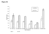
- a final evaluation using nine different transcripts representing a control transcript for HCV genotype 1a and transcripts with naturally occurring mutations in the probe binding region of the amplicon demonstrated that the addition of a second probe significantly increased the observed titer up to >100fold for the transcripts carrying mismatches.
- HCV patient samples for HCV subtype 1a representing the standard HCV samples
- HCV subtype 4a representing HCV samples with possible sequence variation in the standard probe binding region
- HCV RNA transcripts representing HCV GT1a consensus sequence and transcripts of the 5'untranslated region of naturally occurring HCV isolates in the amplicon region were used to evaluate the different second probes.
- Per reaction 1 ml of patient plasma sample material was used for nucleic acid extraction. If transcripts were used, about 500 cp/ml (for experiments shown in Figures 2 , 3a and 3b ) or about 50 000 cp/ml (for experiments shown in Figures 4a , 4b and 5 ) were added to a guanidinium thiocyanate-containing buffer to inactivate RNases and 1 ml was then processed in the same way as a normal sample. A number of 5 to 10 replicates of the patient samples and 4 to 6 replicates of transcript samples were tested in each of the experiments.
- Nucleic acid extraction methods are state-of-the-art and are known by the skilled artisan (see for example Sambrook et al., 2nd Edition 1989, Part 1-3, Molecular Cloning - A Laboratory Manual, Cold Spring Harbor Laboratory, Cold Spring Harbor, New York ; Oligonucleotide Synthesis (M.J. Gait, ed., 1984 ).
- commercially available nucleic acid extraction kits i.e. the High Pure Viral Nucleic Acid Kit (Roche Diagnostics) or the cobas® AmpliPrep Total Nucleic Acid Isolation Kit (TNAI) (Roche Diagnostics) can be used.
- the specimen preparation reagents consist of a magnetic glass particles suspension, a lysis reagent, a protease reagent, an elution buffer and a wash reagent.
- Quantitation Standard RNA was added to the specimens before nucleic acid extraction.
- the armored HCV particles and Quantitation Standard RNA armored particles are lysed by incubation with a protease and a chaotropic lysis/binding buffer that releases nucleic acids and protects the released HCV RNA from RNases in serum or plasma.
- the HCV RNA and Quantitation Standard RNA are bound to magnetic glass particles. Unbound substances such as salts, proteins and other cellular impurities are removed by washing the magnetic particles. The adsorbed nucleic acids are eluted at elevated temperature with an aqueous buffer.
- the master mixes evaluated consisted of the reference mastermix RL1.1 which was supplemented with the different probes.
- the reference mastermix RL1.1 was prepared in a large batch. For each experiment this master mix was supplemented with additional 10% or 50% of the standard probe SEQ ID NO 6 or with additional 10% or 50% of the individual second probes to be assessed as specified in the Figures 2-6 .
- ⁇ Aptamers are short, single-stranded DNA- or RNA-oligonucleotides (25-70 bases), which bind to a specific molecule (i.e. protein, Z05) through their 3D structure (see for example C. Tuerk and L. Gold: Systematic evolution of ligands by exponential enrichment: RNA ligands to bacteriophage T4 DNA polymerase, Science, volume 249, 1990, p. 505-510
- nucleic acid containing eluate 50 ⁇ L were added to 35 ⁇ L of master mix and 15 ⁇ L of 18 mM manganese acetate in PCR tubes and loaded onto the cobas® TaqMan®48 Analyzer.
- the positive effect of a probe was assessed either by i) titer increase versus the reference master mix, or ii) decrease in ct value together with RFI increase versus the reference master mix.
- the experiments presented here can also be carried out as follows: The commercially available cobas® AmpliPrep / cobas® TaqMan® HCV Test (manufactured by Roche) is used for extracting HCV RNA from the patient and the transcript samples. Specimen preparation is automated using the cobas® AmpliPrep Instrument and amplification/detection is automated using the cobas® TaqMan® Analyzer or the cobas® TaqMan® 48 Analyzer.
- the test is based on three major processes: (1) specimen preparation to isolate RNA from human EDTA plasma or serum and controls which are provided in secondary tubes on the cobas® AmpliPrep Instrument; (2) reverse transcription of the target RNA and the Quantitation Standard/Internal Control RNA to generate complementary DNA (cDNA) and (3) PCR amplification of target cDNA and Quantitation Standard/Internal Control cDNA with simultaneous detection of the generated amplicons on the cobas® TaqMan® Analyzer by cleavage of dual-labeled detection probes specific to the target and to the Quantitation Standard/Internal Control.
- the specimen preparation reagents consist of a magnetic glass particle suspension, a lysis reagent, a protease reagent, an elution buffer and a wash reagent.
- the HCV particles as well as the Quantitation Standard/Internal Control particles are lysed by incubation with a protease and a chaotropic lysis/binding buffer that releases nucleic acids and protects the released HCV RNA from RNases in serum or plasma. Subsequently, the HCV RNA and Quantitation Standard RNA are bound to magnetic glass particles. Unbound substances such as salts, proteins and other cellular impurities are removed by washing the magnetic particles.
- the adsorbed nucleic acids are eluted at elevated temperature with an aqueous buffer.
- the specimen or control eluate is added to the master mix and transferred to the cobas® TaqMan® Analyzer or the cobas® TaqMan® 48 Analyzer for amplification and detection.
- the cobas® AmpliPrep / cobas® TaqMan® HCV Test master mix is replaced by a mastermix according to the table shown below and in addition by adding different second probes according to the information given below.
- the reagent cassette with the modified mastermix is used on the cobas® AmpliPrep instrument.
- the master mix contains primer and probe pairs specific for both HCV RNA and Quantitation Standard/Internal Control RNA.
- the primer binding sites are shared by the HCV target and the Quantitation Standard/Internal Control. Primers and target probes are located in a highly conserved part of the 5'-untranslated region of the HCV genome.
- HCV target and Quantitation Standard The detection of HCV target and Quantitation Standard is performed using a target-specific and a Quantitation Standard-specific dual-labeled oligonucleotide probe which permits independent identification of HCV target amplicon and HCV Quantitation Standard amplicon.
- the HCV Quantitation Standard is automatically added to each specimen at a known copy number by the cobas® AmpliPrep and is carried through the entire specimen preparation, reverse transcription, amplification and detection steps along with the HCV target.
- the Quantitation Standard must give a positive signal in HCV target negative and positive specimens in order to enable titer determination. In partly suppressed or inhibited reactions the Quantitation Standard is affected similarly as the target and thus allows correct titer determination.
- the Quantitation Standard monitors HCV target negative reactions for inhibitory effects but due to its rather high concentration monitoring is not stringent.
- ⁇ Aptamers are short, single stranded DNA- or RNA-oligonucleotides (25-70 bases), which bind to a specific molecule (i.e. protein, Z05) through their 3D structure (see for example C. Tuerk and L. Gold: Systematic evolution of ligands by exponential enrichment: RNA ligands to bacteriophage T4 DNA polymerase, Science, volume 249, 1990, p. 505-510 ).
- the reference master mix RL1.1 without a second probe is prepared in a large batch. For each experiment this reference master mix is supplemented with the additional probes as shown in the Figures. These supplemented mastermix variations are filled into reagent cassettes and are loaded onto the cobas® AmpliPrep.
- the positive effect of a probe is assessed either by i) titer increase versus the reference master mix, or ii) decrease in ct value together with RFI increase versus the reference master mix.
Abstract
Description
- The present invention belongs to the field of in vitro diagnostics. Within this field, it concerns the amplification and detection of a target nucleic acid that may be present in a sample and particularly the amplification and detection of a target nucleic acid comprising subgroups with sequence variations and/or individual mutations, using at least two probes specific for different sequence portions of an amplicon. The invention further provides uses of and kits containing at least two probes specific for different sequence portions of an amplicon.
- In the field of molecular diagnostics, the amplification and detection of nucleic acids is of considerable significance. Examples for diagnostic applications of nucleic acid amplification and detection are the detection of viruses such as Human Papilloma Virus (HPV), West Nile Virus (WNV) or the routine screening of blood donations for the presence of Human Immunodeficiency Virus (HIV), Hepatitis B (HBV) and/or C Virus (HCV) and the like. Furthermore, said amplification techniques are suitable for bacterial targets, or the analysis of oncology markers, or other targets.
- Within a species, a microorganism or pathogen is often classified according to distinct groups, genotypes or subtypes based on nucleic acid sequence variation (i.e. HCV, HIV, HPV etc). In an in vitro diagnostic device, nevertheless, all groups, genotypes or subtypes should be detected and/or correctly quantified to avoid false negative diagnosis or wrong titer determination. This poses a considerable challenge for molecular diagnostic assays for e.g. detection of HIV and HCV. Furthermore constant mutation and recombination of such pathogens generate within their target nucleic acids increasing diversity which must also be covered by the molecular diagnostic assay.
- The most prominent and widely-used amplification technique is the Polymerase Chain Reaction (PCR). Other amplification reactions comprise, among others, the Ligase Chain Reaction, Polymerase Ligase Chain Reaction, Gap-LCR, Repair Chain Reaction, 3SR, NASBA, Strand Displacement Amplification (SDA), Transcription Mediated Amplification (TMA), and Qβ-amplification.
- Automated systems for PCR-based analysis often make use of real-time detection of product amplification during the PCR process in the same reaction vessel. Key to such methods is the use of modified oligonucleotides carrying reporter groups or labels.
- Detection of a microbial nucleic acid in a biological sample is crucial e.g. for recognizing an infection of an individual. Thereby, one important requirement e.g. for an assay for detection of a viral infection is inclusivity, defined such that false-negative results or underquantification of titers due to variable sequence regions on a viral genome caused by mutations have to be avoided. Mutated or partially mutated sequences within the respective genome that are possibly not amplified and/or detected in combination with the low viral load enhance the possibility of obtaining false-negative or falsely quantified results.
- Several options have been published to increase the inclusivity of a molecular assay. Recently, coamplification of two different and non-overlapping target sequences within the genome of a pathogen was established (
US 2010/0041040 ). This approach may, however, not be generally applicable if two reasonably conserved target regions cannot be identified within the genome of a pathogen or if the oligonucleotides for amplification and detection of two independent target regions interfere with each other in the master mix. - In this context, the prior art has e.g. provided methods for amplification and detection involving more than one probe based on homogeneous amplicon sequence with the aim to increase assay sensitivity (Yip et al., 2005, Clin. Chem. 51 (10)).
- An aspect of the present invention is a method for amplifying and detecting a target nucleic acid in a sample, said target nucleic acid comprising subgroups with sequence variations and/or individual mutations, wherein an amplification of the nucleic acids in said sample is carried out. This amplification involves a polymerase, nucleotide monomers, primers for generating an amplicon and at least two detectable probes specific for different sequence portions of said amplicon. Detection of the obtained amplicon is brought about by detecting hybridization of the probes mentioned above to said different sequence portions of the amplicon.
- The invention also relates to the use of at least two non-overlapping detectable nucleic acid probes specific for different sequence portions of the same amplicon.
- Furthermore, a kit is provided for amplifying and detecting a target nucleic acid that may be present in a sample, said target nucleic acid comprising subgroups with sequence variations and/or individual mutations. The kit comprises amplification reagents comprising a polymerase, nucleotide monomers, primers for generating an amplicon and at least two detectable probes specific for different sequence portions of said amplicon.
- In a first embodiment, the present invention relates to a method for amplifying and detecting a target nucleic acid that may be present in a sample, said target nucleic acid comprising subgroups with sequence variations and/or individual mutations, said method comprising the steps of:
- a) contacting nucleic acids from said sample with amplification reagents comprising a polymerase, nucleotide monomers, primers for generating an amplicon and at least two detectable probes specific for different sequence portions of said amplicon;
- b) incubating said nucleic acids with said amplification reagents for a period of time and under conditions sufficient for an amplification reaction to occur;
- c) detecting the presence or absence of said amplicon by detecting hybridization of said detectable probes to said different sequence portions of said amplicon,
- In some embodiments of the invention, one or more steps of the method described above are automated. In further embodiments, all steps are automated. Automated systems provide a number of advantages as compared to manual methods, particularly in the field of in vitro diagnostics. The skilled person is enabled to leave the system after initiating the method, thus reducing hands-on time and providing the basis for a high sample throughput in a relatively short period of time, yet at the same time increasing reproducibility of the result. This is especially, but not only, an important feature in situations with a high number of clinical samples to be screened as quickly as possible, such as e.g. in blood banks.
- In the context of the present invention, the term "amplifying" or "amplification" generally refers to the production of a plurality of nucleic acid molecules from a target nucleic acid wherein primers hybridize to specific sites on the target nucleic acid molecules in order to provide an initiation site for extension by a polymerase. Amplification can be carried out by any method generally known in the art, such as but not limited to: standard PCR, realtime PCR, long PCR, hot start PCR, qPCR, Reverse Transcription PCR and Isothermal Amplification.
- It can be favorable to monitor the amplification reaction in real time, i.e. to detect the target nucleic acids and/or their amplificates during the amplification itself.
- The term "detecting" or "detection" as used herein relates to a test aimed at assessing the presence or absence of a target nucleic acid in a sample.
- A "target nucleic acid" is a polymeric compound of nucleotides as known to the expert skilled in the art. "Target nucleic acid" is used herein to denote a nucleic acid in a sample which should be analyzed, i.e. the presence, non-presence and/or amount thereof in a sample should be determined. The target nucleic acid may be a genomic sequence, e.g. part of a specific gene, or RNA. In other embodiments, the target nucleic acid may be viral or bacterial. Target nucleic acids can comprise subgroups with distinct sequence variations or distinct individual mutations in the amplicon region. This is especially the case for nucleic acids of pathogens like viruses which show significant genetic variation due to high mutation or recombination rates and lacking repair mechanisms.
- The term "amplicon" refers to a polynucleotide molecule (or collectively the plurality of molecules) produced following the amplification of a particular target nucleic acid. The amplification method used to generate the amplicon can be any suitable method, most typically, for example, a PCR methodology. An amplicon is typically, but not exclusively, a DNA amplicon. An amplicon can be single-stranded or double-stranded, or a mixture thereof in any concentration ratio. In an embodiment of the invention, the amplicon consists of subpopulations with heterogeneous sequences between the primer sequences.
- The method set out above is in some embodiments based on Fluorescence Resonance Energy Transfer (FRET) between a donor fluorescent moiety and an acceptor fluorescent moiety. In these embodiments, the detectable probes specific for different sequence portions of the amplicon are FRET probes. A representative donor fluorescent moiety is fluorescein, and representative corresponding acceptor fluorescent moieties include LC-Red 640, LC-Red 705, CY5, and CY5.5. Typically, detection includes exciting the sample at a wavelength absorbed by the donor fluorescent moiety and visualizing and/or measuring the wavelength emitted by the corresponding acceptor fluorescent moiety. In the method described above, detection is in some embodiments followed by quantitating the FRET. In the context of the invention, the terms "FRET" or "fluorescent resonance energy transfer" or "Foerster resonance energy transfer" can be used interchangeably and refer to a transfer of energy between at least two chromophores, a donor chromophore and an acceptor chromophore (referred to as a quencher). The donor typically transfers the energy to the acceptor when the donor is excited by light radiation with a suitable wavelength. The acceptor typically re-emits the transferred energy in the form of light radiation with a different wavelength. When the acceptor is a "dark" quencher, it dissipates the transferred energy in a form other than light. Whether a particular fluorophore acts as a donor or an acceptor depends on the properties of the other member of the FRET pair. Commonly used donor-acceptor pairs include the FAM-TAMRA pair. Commonly used donors are e.g. fluoresceins, coumarins, cyanines and rhodamines. Commonly used quenchers are DABCYL and TAMRA. Commonly used dark quenchers include BlackHole Quenchers™ (BHQ), (Biosearch Technologies, Inc., Novato, Cal.), Iowa Black™ (Integrated DNA Tech., Inc., Coralville, Iowa), and BlackBerry™ Quencher 650 (BBQ-650) (Berry & Assoc., Dexter, Mich.).
- A common format of FRET technology utilizes two hybridization probes forming a HybProbe pair. Each probe can be labeled with a different fluorescent moiety. The probes are generally designed to hybridize in close proximity to each other in a target DNA molecule (e.g., an amplification product). A donor fluorescent moiety like e.g. fluorescein is excited at 470 nm by the light source e.g. of a LIGHTCYCLER® instrument. During FRET, the fluorescein transfers its energy to an acceptor fluorescent moiety such as e.g. LIGHTCYCLER®-Red 640 (LC®-Red 640) or LIGHTCYCLER®-Red 705 (LC®-Red 705). The acceptor fluorescent moiety then emits light of a longer wavelength, which is detected by the optical detection system of the LIGHTCYCLER® instrument. Efficient FRET can only take place when the fluorescent moieties are in direct local proximity (usually about 1 to 5 nucleotides distance) and when the emission spectrum of the donor fluorescent moiety overlaps with the absorption spectrum of the acceptor fluorescent moiety. The intensity of the emitted signal can be correlated with the number of original target nucleic acid molecules. In the context of the present invention, as also appreciated by the person skilled in the art, a HybProbe pair is to be understood as a functional unity and thus a single probe, since the two members of such a pair have to be used together. Hence, the distinct probes of a HybProbe pair do not form "detectable probes specific for different sequence portions" of an amplicon even though they do not overlap when binding to the amplicon. However, e.g. two or more HybProbe pairs are "detectable probes specific for different sequence portions of said amplicon" in the sense of the invention, since as described above a HybProbe pair is to be understood as a single probe.
- In an embodiment of the method described above, the detectable probes specific for different sequence portions of said amplicon are HybProbe pairs.
- This embodiment confers several advantages. For instance, since the probes in this detection format are not degraded, melting curve analyses may be performed on each of the HybProbe pairs by monitoring the temperature dependence of their hybridization. As the skilled person knows, melting curve analysis is suitable for verification of results or even provision of more detailed information e.g. on the identity of a target nucleic acid than monitoring hybridization at a single temperature may yield.
- Detection of amplicon formation on cobas® TaqMan® systems utilizes a single-stranded hybridization probe (also termed "5'-nuclease probe"). The term "5'-nuclease probe" refers to an oligonucleotide that comprises at least one light emitting labeling moiety and that is used in a 5'-nuclease reaction to effect target nucleic acid detection. In some embodiments, for example, a 5'-nuclease probe includes only a single light emitting moiety (e.g., a fluorescent dye, etc.). In certain embodiments, 5'-nuclease probes include regions of self-complementarity such that the probes are capable of forming hairpin structures under selected conditions. To further illustrate, in some embodiments a 5'-nuclease probe comprises at least two labeling moieties and emits radiation of increased intensity after one of the two labels is cleaved or otherwise separated from the oligonucleotide. In certain embodiments, a 5'-nuclease probe is labeled with two different fluorescent dyes, e.g., a 5' terminus reporter dye and a 3' terminus quencher dye or moiety. In some embodiments, 5'-nuclease probes are labeled at one or more positions other than, or in addition to, terminal positions. When the probe is intact, energy transfer typically occurs between the two fluorophores such that fluorescent emission from the reporter dye is quenched at least in part. During an extension step of a polymerase chain reaction, for example, a 5'-nuclease probe bound to a template nucleic acid is cleaved by the 5' to 3' nuclease activity of, e.g., a Taq polymerase or another polymerase having this activity like e.g. the Z05 polymerase, such that the fluorescent emission of the reporter dye is no longer quenched. Exemplary 5'-nuclease probes are described in, e.g.,
US 5,210,015 . In some embodiments, a 5' nuclease probe may be labeled with two or more different reporter dyes and a 3' terminus quencher dye or moiety. Typical fluorescent dyes used in this format are for example, among others, FAM, HEX, CY5, JA270, Cyan500 and CY5.5. - In an embodiment of the method described above, the detectable probes specific for different sequence portions of said amplicon are 5'-nuclease probes.
- The detectable probes can hybridize to the same or to different strands of a double-stranded amplicon.
- In some embodiments of the method described above, at least two detectable probes hybridize to different strands of said amplicon.
- In this case, the skilled person is provided with increased flexibility with regard to selecting the primer and probe sequences and thus binding sites on the respective amplicon. For instance, in the case of secondary structure formation due to a specific sequence within an oligonucleotide, it can be important to be able to switch to a different sequence and thus to a different binding site on said amplicon. Further, if the detectable probes bind to different strands, such as a first probe to the sense strand and a second probe to the antisense strand of a double-stranded amplicon, the risk of those probes interfering with each other at their respective binding sites is reduced.
- In further embodiments of the method described above, at least two detectable probes hybridize to the same strand of said amplicon.
- In that latter embodiment, it is possible to hybridize multiple detectable probes specific for different sequence portions of the amplicon in close vicinity to each other. This is surprising due to the circumstance that the respective exonuclease requires some space to bind and cleave the 5'-nuclease probes. Yet, the inventors have shown that even such probes can be hybridized to an amplicon at only few bases of distance between one another. This enables the skilled person to use more than one detectable probe specific for distinct sequence portions each to amplicons of relatively short length. According to the method described above, even short stretches of target nucleic acids can serve as suitable targets for multiple probes, thus conferring the benefits described supra, such as e.g. signal enhancement and increased tolerance against genetic variations like e.g. point mutations.
- Thus, in an embodiment of the method described above, the detectable probes specific for different sequence portions of said amplicon hybridize to the amplicon with no more than 100 bases distance to each other, in some embodiments from 1, 5, 10, 20, 30, 40 or 50 bases, to 60, 70, 80, 90, or 100 bases distance to each other. In some embodiments, the distance is from 40 to 80, or from 50 to 70, or from 55 to 60 bases, or it is 58 bases. In this context, "distance" means the number of bases of the amplicon lying between those bases of the amplicon to which the detectable probes hybridize in case they hybridize to the same strand. If they hybridize to different strands, the distance is calculated accordingly, wherein each base of one strand of a double-stranded amplicon has a corresponding base on the other strand with which it forms a base pair.
- In some embodiments, detection is performed after each cycling step of a cycle-based amplification technique such as PCR. In some embodiments, detection is performed in real time. By using commercially available real-time PCR instrumentation (e.g., LightCycler® or TaqMan®), PCR amplification and detection of the amplification product can be combined in a single closed cuvette with considerably reduced cycling time. Since detection occurs concurrently with amplification, real-time PCR methods obviate the need for manipulation of the amplification product, and diminish the risk of cross-contamination between amplification products. In both detection formats described above, the intensity of the emitted signal can principally be correlated with the number of original target nucleic acid molecules.
- A "sample" is any material that can be subjected to a diagnostic assay and generally refers to the medium possibly containing the target nucleic acid. The "sample" is in some embodiments derived from a biological source. The sample can be e.g. a clinical sample. In some embodiments, said sample is derived from a human and is a body liquid. In some embodiments of the invention, the sample is human whole blood or serum, blood plasma, urine, sputum, sweat, genital or buccal or nasal swabs, pipettable stool, solubilized tissue samples, or spinal fluid or the like. A sample can be pipetted or converted to a pipettable form, such that the term "sample" comprises homogeneous or homogenized liquids, but also emulsions, suspensions and the like. A sample may also e.g. be an originally solid sample (i.e. tissue sample) which is subjected to a solubilization treatment for extraction and purification of nucleic acids.
- A "polymerase" as used herein is an enzyme capable of synthesizing nucleic acids from smaller elements such as nucleotides. In some embodiments, the polymerase is a thermostable polymerase. The term "thermostable polymerase" refers to an enzyme that is stable to heat, is heat resistant, and retains sufficient activity to effect subsequent polynucleotide extension reactions and does not become irreversibly denatured (inactivated) when subjected to the elevated temperatures for the time necessary to effect denaturation of double-stranded nucleic acids. The heating conditions necessary for nucleic acid denaturation are well known in the art and are exemplified in, e.g.,
US Patent Nos. 4,683,202 ,4,683,195 , and4,965,188 . As used herein, a thermostable polymerase is suitable for use in a temperature cycling reaction such as the polymerase chain reaction ("PCR"). Irreversible denaturation for purposes herein refers to permanent and complete loss of enzymatic activity. For a thermostable polymerase, enzymatic activity refers to the catalysis of the combination of the nucleotides in the proper manner to form polynucleotide extension products that are complementary to a template nucleic acid strand. For amplification purposes, said nucleotides are present in monomeric form, therefore they are also referred to as "nucleotide monomers" in the context of the present invention. Often, such nucleotide monomers used by polymerases such as e.g. thermostable DNA polymerases are e.g. nucleoside triphospohates, or nucleoside tetraphosphates, or the like. Thermostable DNA polymerases from thermophilic bacteria include, e.g., DNA polymerases from Thermotoga maritima, Thermus aquaticus, Thermus thermophilus, Thermus flavus, Thermus filiformis, Thermus species Sps17, Thermus species Z05, Thermus caldophilus, Bacillus caldotenax, Thermotoga neopolitana, and Thermosipho africanus. - The term "primer" is used herein as known to the expert skilled in the art and refers to oligomeric compounds, primarily to oligonucleotides, but also to modified oligonucleotides that are able to prime DNA synthesis by a template-dependent DNA polymerase, i.e. the 3'-end of the primer provides a free 3'-OH group whereto further nucleotides may be attached by a template-dependent DNA polymerase establishing 3'- to 5'-phosphodiester linkage whereby deoxynucleoside triphosphates are used and whereby pyrophosphate is released.
- A "probe" or "detectable probe" also denotes a natural or modified oligonucleotide. As known in the art, a probe serves the purpose of detecting an analyte or amplificate. In the context of the invention, probes can e.g. be used to detect the amplificates of the target nucleic acid and/or a control nucleic acid. For the purpose of detectability, probes typically carry labels.
- In some embodiments of the method the at least two detectable probes specific for different sequence portions of said amplicon carry the same type of label and thus the signal originating from the individual probe cannot be distinguished. In other embodiments, they carry different labels emitting signals of different wavelengths such that the signals from the at least two probes can be distinguished with the appropriate instrumentation.
- "Labels", often referred to as "reporter groups", are generally groups that make a nucleic acid, in particular oligonucleotides or modified oligonucleotides, as well as any nucleic acids bound thereto distinguishable from the remainder of the sample. Useful labels in the context of the invention are e.g. fluorescent labels, which may be fluorescent dyes such as for instance a fluorescein dye, a rhodamine dye, a cyanine dye, or a coumarin dye. Useful fluorescent dyes in the context of the invention are e.g. FAM, HEX, JA270, CAL635, Coumarin343, Quasar705, Cyan500, CY5.5, LC-Red 640, LC-Red 705, TAMRA, or CY5.
- In the context of the invention, any primer and/or probe may be chemically modified, i.e. the primer and/ or the probe comprise a modified nucleotide or a non-nucleotide compound. The probe or the primer is then a modified oligonucleotide.
- As known by the person skilled in the art, the term "specific" in the context of primers and probes implies that a primer or probe "specific" for a distinct nucleic acid binds to said nucleic acid under stringent conditions. In some embodiments the probes used in the context of the invention are at least 80% identical to the different sequence portions of the amplicon. In another embodiment, the probe sequences comprise at least 12 contiguous nucleotides of a sequence selected from the group consisting of
SEQ ID NOs 1 to 8 or the corresponding complementary nucleic acid sequences thereof, and the primers comprise at least 12 contiguous nucleotides of SEQ ID NOs 9 to 15. In some embodiments, the selected probe and/ or primer sequences consist of 12 to 60 nucleotides, or of 20 to 60 nucleotides, or of the exact sequences selected from said SEQ ID NOs or their complementary nucleic acid sequences. The skilled person understands that, in the sense of the invention, a probe pair forming a functional entity such as e.g. a Hybprobe pair used in the LightCycler® format is not "at least two detectable probes specific for different sequence portions of said amplicon". The two Hybprobes of a pair are regarded as a unit and can only be detected together, while each of the at least two probes in the context of the invention is detectable alone. - Furthermore, in the context of the invention the term "overlap" means that two or more oligonucleotides, in particular the at least two detectable probes mentioned supra, comprise identical (when bound to the same strand) or complementary (when bound to different strands) sequence stretches. The probes used in the method described above in some embodiments do not overlap and thus do not compete in binding to a specific site on the amplicon. When bound, said two or more probes are hybridized to different sequence stretches of said amplicon. The detectable probes used in the context of the invention are advantageous as compared to overlapping probes.
- The term "hybridize" or "hybridization" generally refers to the base-pairing between different nucleic acid molecules consistent with their nucleotide sequences. The terms "hybridize" and "anneal" can be used interchangeably.
- Directing two or more probes to different sequence portions of the same amplicon, as performed in the method described supra, leads to a significantly reduced risk of obtaining false-negative results in qualitative assays. Moreover, in quantitative assays, the risk to underquantitate titers e.g. of a virus is reduced. A variety of different genotypes, subtypes and mutants within a given organism can be readily detected and quantified using a minimum number of oligonucleotides as a result of the increased inclusivity and robustness of the assay. In that respect, it may be the case that a certain genotype, subtype or mutant is not hybridized to by all of the at least two detectable probes. For instance, in the case of two probes, one might not bind to the target amplicon because of the specific sequence variant of the latter, whereas the other probe specific for a different sequence portion of said amplicon is still capable of hybridizing to its respective target sequence. This way, detection of the amplicon would still be possible. In embodiments where the different probes carry different labels, it may also be possible to determine which of the probes hybridized and which ones did not.
- The life cycle of tests based on the method described above is prolonged, since it is able to deal even with new variants generated by microorganisms such as e.g. viruses through selective pressure for example as a consequence of new antiretroviral drugs. In view of the high degree of mutations within viral genomes, in an embodiment of the method described above said target nucleic acid is a viral nucleic acid.
- Overall inclusivity of qualitative assays is significantly increased, and the variability of titer determinations in quantitative assays is minimized by applying the method described above.
- As known by the skilled person, a measure for the inclusivity is the detection of viral subgroups and isolates carrying mutations with equivalent sensitivity as the standard isolates not significantly deviating from the consensus sequence. Sensitivity of an assay is the LOD (Limit Of Detection), referring to the lowest detectable amount or concentration of a nucleic acid in a sample. A low "LOD" corresponds to high sensitivity and vice versa. The "LOD" is usually expressed either by means of the unit "cp/ml", particularly if the nucleic acid is a viral nucleic acid, or as IU/ml. "Cp/ml" means "copies per milliliter" wherein a "copy" is a copy of the respective nucleic acid. IU/ml stands for "International units/ml", referring to the WHO standard. The WHO standards are generally built from a standard isolate with a genome close to the consensus sequence.
- A widely used method for calculating an LOD is the "Probit Analysis" which is a method of analyzing the relationship between a stimulus (dose) and the quantal (all or nothing) response. In a typical quantal response experiment, groups of animals are given different doses of a drug. The percent dying at each dose level is recorded. These data may then be analyzed using Probit Analysis. The Probit Model assumes that the percent response is related to the log dose as the cumulative normal distribution. That is, the log doses may be used as variables to read the percent dying from the cumulative normal. Using the normal distribution, rather than other probability distributions, influences the predicted response rate at the high and low ends of possible doses, but has little influence near the middle.
- The Probit Analysis can be applied at distinct "hitrates". As known in the art, a "hitrate" is commonly expressed in percent [%] and indicates the percentage of positive results at a specific concentration of an analyte. Thus for example, an LOD can be determined at 95% hitrate, which means that the LOD is calculated for a setting in which 95% of the valid results for a true positive sample are determined as positive.
- The method described above is particularly advantageous in assays for detecting Hepatitis C-Virus (HCV). Thus, in an embodiment of the method described supra, the target nucleic acid is a nucleic acid of HCV.
- The expression "hepatitis C virus type" refers to the categorization of a hepatitis C virus based on its genomic organization (e.g., phylogenetic analysis). The categorization of an HCV isolate into a particular type category reflects its genomic relatedness to other HCV isolates and its relatively lesser relatedness to other HCV isolates. The HCV typing nomenclature used herein is consistent with the widely adopted nomenclature revised and proposed by Simmonds et al (2005) "Consensus Proposals for a Unified System of Nomenclature of Hepatitis C Virus Genotypes", Hepatology 42, No.4:962-973. The system of Simmonds et al (2005) places the known HCV isolates into one of six (6) HCV genotypes, namely genotypes 1 through 6. Each genotype is further subdivided into groupings termed subtypes that reflect relatedness among strains of the same genotype. An HCV subtype is written by a lowercase Roman letter following the genotype, e.g., subtype 1a, subtype 1c, subtype 6a, etc. Genetic variants found within an individual isolate are termed quasi species. Approximately 100 HCV subtypes encompassing all six genotypes are known worldwide. The number of subtypes is not static, i.e. as more HCV isolates are studied and sequenced, it is likely that additional subtypes (and possibly genotypes) may be recognized.
- Since HCV is an RNA virus, the person skilled in the art usually reversely transcribes the viral RNA into DNA prior to the actual amplification. In such a case, the amplification reagents comprise a reverse transcriptase or a polymerase with reverse transcriptase activity.
- A primer suitable for annealing to an RNA template may also be suitable for amplification by PCR. For PCR, a second primer, complementary to the reverse transcribed cDNA strand, provides an initiation site for the synthesis of an extension product.
- In the amplification of an RNA molecule by a DNA polymerase, the first extension reaction is reverse transcription using an RNA template, and a DNA strand is produced. The second extension reaction, using the DNA template, produces a double-stranded DNA molecule. Thus, synthesis of a complementary DNA strand from an RNA template by a DNA polymerase provides the starting material for amplification.
- Thermostable DNA polymerases can be used in a coupled, one-enzyme reverse transcription/amplification reaction. The term "one-step Real-time PCR", in this context, may refer to a reaction without reverse transcription step if target nucleic acid is DNA or a reaction including a reverse transcription step if target nucleic acid is RNA. By "one-step real-time PCR" it is meant that following the reverse transcription (RT) step, there is no need to open the reaction vessel or otherwise adjust reaction components prior to the amplification step. In a non-one-step real-time PCR reaction, following reverse transcription and prior to amplification one or more of the reaction components such as the amplification reagents are e.g. adjusted, added, or diluted, for which the reaction vessel has to be opened, or at least its contents have to be manipulated. Both one-step real-time PCR and non-one-step real-time PCR embodiments are comprised by the scope of the invention.
- In an embodiment of the invention carryover contamination of amplification products such as amplicons and high molecular weight products (polymerized amplicon) originating from earlier PCR reactions are prevented. A popular and effective way of preventing carryover contamination involves the use of uracil-DNA glycosylases or uracil-N-glycosylases, abbreviated as "UDG" or "UNG" (EC 3.2.2.3). These enzymes comprising uracil-DNA glycosylase activity recognize uracil present in single-stranded or double-stranded DNA and cleave the N-glycosidic bond between the uracil base and the deoxyribose leaving an abasic site, see e.g.
US 6.713.294 . - As shown by the examples herein, a particularly good performance in detecting a nucleic acid of HCV is achieved when employing probes comprising or consisting of at least two sequences selected from the group consisting of
SEQ ID NOs 1 to 8 or the respective complements thereof. - Hence, an aspect of the invention is a method for amplifying and detecting a target nucleic acid of HCV that may be present in a sample, said method comprising the steps of:
- a) contacting nucleic acids from said sample with amplification reagents comprising a polymerase, nucleotide monomers, primers for generating an amplicon and at least two detectable probes specific for different sequence portions of said amplicon, wherein said at least two detectable probes comprise at least two sequences selected from the group consisting of
SEQ ID NOs 1 to 8 or the respective complements thereof; - b) incubating said nucleic acids with said amplification reagents for a period of time and under conditions sufficient for an amplification reaction to occur;
- c) detecting the presence or absence of said amplicon by detecting hybridization of said detectable probes to said different sequence portions of said amplicon;
- In a further embodiment of the method described above, said at least two detectable probes comprise
SEQ ID NOs SEQ ID NOs - In an embodiment of the invention, the primers in the method mentioned above comprise at least one sequence selected from the group consisting of SEQ ID NOs 9 to 15. Said primers are particularly useful for creating an amplicon detectable with the probes mentioned above. In another embodiment, the primers in the method mentioned above consist of
SEQ ID NOs 9, 10, and 11. - Also an aspect of the invention is the method described above, wherein said primers comprise more than one forward and/or reverse primer. Such an arrangement can be especially useful when these primers lead to variable, overlapping amplicons detectable by the probes mentioned above. In the case of more than one forward and/or reverse primer, the respective forward and/or reverse primers are in some embodiments staggered, i.e. they overlap with respect to the template sequence they hybridize to. Such a staggered constellation further contributes to an increased coverage of genetic variants such as HCV genotypes and/or subtypes.
- The combination of the primers and probes mentioned above in the method set out supra is particularly useful for detecting a considerable variety of HCV genotypes. In an embodiment, the method described above is a method for simultaneously detecting
Genotypes Genotypes Genotypes Genotypes - A further aspect of the invention is the method described above, further comprising prior to step a) the steps of:
- i) combining together a solid support and said sample for a period of time and under conditions sufficient to permit nucleic acids comprising said target nucleic acid to be immobilized on said solid support;
- ii) isolating said solid support from the other material present in the sample in a separation station;
- iii) purifying the nucleic acids in the separation station by separating the sample from the solid support and washing the solid support one or more times with a wash buffer.
- In the context of the invention, the term "solid support" as used herein relates to any type of solid support to which the analyte is capable of binding, either directly and non-specifically by adsorption, or indirectly and specifically. Indirect binding may be binding of an analyte to an antibody immobilized on the solid support, or binding of a tag to a tag binding compound, e.g. binding of 6xHis tags to Ni-chelate. When the analyte is a nucleic acid, such indirect binding may be by binding to a capture nucleic acid probe which is homologous to a target sequence of the nucleic acid of interest. Thus, using capture probes attached on a solid support, a target analyte, or a target nucleic acid, can be separated from non-target material, or non-target nucleic acid. Such a capture probe is immobilized on the solid support. Solid support material may be a polymer, or a composition of polymers. Other types of solid support material include magnetic silica particles, metal particles, magnetic glass particles, glass fibers, glass fiber filters, filter paper etc., while the solid support material is not limited to these materials.
- "Immobilize", in the context of the invention, means to capture objects such as nucleic acids in a reversible or irreversible manner. Particularly, "immobilized on the solid support material", means that the object or objects are associated with the solid support material for the purpose of their separation from any surrounding media, and can be recovered e.g. by separation from the solid support material at a later point. In this context, "immobilization" can e.g. comprise the adsorption of nucleic acids to glass or other suitable surfaces of solid materials as described supra. Moreover, nucleic acids can be "immobilized" specifically by binding to capture probes, wherein nucleic acids are bound to essentially complementary nucleic acids attached to a solid support by base-pairing. In the latter case, such specific immobilization leads to the predominant binding of target nucleic acids.
- A "separation station" is a device or a component of an analytical system allowing for the isolation of the solid support from the other material present in the sample. Such a separation station can e.g. comprise, while it is not limited to these components, a centrifuge, a rack with filter tubes, a magnet, or other suitable components. In some embodiments, the separation station comprises one or more magnets. In certain embodiments, one or more magnets are used for the separation of magnetic particles, such as e.g. magnetic glass particles, as a solid support. If, for example, the sample and the solid support material are combined together in the wells of a multiwell plate, then one or more magnets comprised by the separation station can e.g. be contacted with the sample itself by introducing the magnets into the wells, or said one or more magnets can be brought close to the outer walls of the wells in order to attract the magnetic particles and subsequently separate them from the surrounding liquid.
- In the sense of the invention, "purification", "isolation" or "extraction" of nucleic acids relate to the following: Before nucleic acids may be analyzed in a diagnostic assay e.g. by amplification, they typically have to be purified, isolated or extracted from biological samples containing complex mixtures of different components. For the first steps, processes may be used which allow the enrichment of the nucleic acids.
- A "wash buffer" is a fluid that is designed to remove undesired components, especially in a purification procedure. Such buffers are well known in the art. In the context of the purification of nucleic acids, the wash buffer is suited to wash the solid support material in order to separate the immobilized nucleic acid from any unwanted components. The wash buffer may, for example, contain ethanol and/ or chaotropic agents in a buffered solution or solutions with an acidic pH without ethanol and/ or chaotropic agents as described above. Often the washing solution or other solutions are provided as stock solutions which have to be diluted before use.
- Summarizing, by applying the steps i) to iii) of the method described above, the nucleic acids including the target nucleic acid that may be present in the sample are separated from the remainder of the sample, such that the risk of inhibition of the subsequent steps by any potentially interfering substances in said sample is reduced.
- For downstream analysis, the nucleic acids may subsequently be eluted from the solid support e.g. by means of an appropriate elution buffer. Such an elution buffer may e.g. be distilled or deionized water or aqueous salt solutions, such as e.g. Tris buffers like Tris HCI, or HEPES, or other suitable buffers known to the skilled artisan.
- In some embodiments, the solid support is present in the amplification reaction mixture during amplification and in some embodiments also detection.
- In some embodiments of the method described supra, a control nucleic acid is added to the sample and/or the purified nucleic acids.
- Said control nucleic acid is in some embodiments a qualitative control nucleic acid, and in other embodiments a quantitative control nucleic acid, or both.
- Qualitative detection of a nucleic acid in a sample is crucial e.g. for recognizing an infection of an individual. Thereby, one important requirement for an assay for detection e.g. of a viral infection is that false-negative or false-positive results be avoided, since such results would almost inevitably lead to severe consequences with regard to treatment of the respective patient. Thus, especially in PCR-based methods, a qualitative internal control nucleic acid is added to the detection mix. Said control is particularly important for confirming the validity of a test result: At least in the case of a negative result with regard to the respective target nucleic acid, the qualitative internal control reaction has to perform reactive within given settings, i.e. the qualitative internal control must be detected, or otherwise the test itself is considered to be inoperative. However, in a qualitative setup, said qualitative internal control does not necessarily have to be detected in case of a positive result. For qualitative tests, it is especially important that the sensitivity of the reaction is guaranteed and therefore strictly controlled. In consequence, the concentration of the qualitative internal control must be relatively low so that even in a situation e.g. of slight inhibition the qualitative internal control is not detected and therefore the test is invalidated.
- Thus, in an embodiment of the method described above, the presence of an amplification product of said control nucleic acid is indicative of an amplification occurring in the reaction mixture even in the absence of amplification products for said target nucleic acid.
- On the other hand and in addition to mere detection of the presence or absence of a nucleic acid in a sample, it is often important to determine the quantity of said nucleic acid. As an example, stage and severity of a viral disease may be assessed on the basis of the viral load. Further, monitoring of any therapy requires information on the quantity of a pathogen present in an individual in order to evaluate the therapy's success.
- Hence, an aspect of the invention is the method described above, further comprising the step of determining the quantity of the target nucleic acid comprising subgroups with sequence variations and/or individual mutations after and/or during step c).
- For instance, HCV RNA viral load tests are used as an aid in the management of chronic hepatitis C patients by evaluating treatment response and making clinical decisions e.g. regarding treatment duration. The primary goal of a therapy for chronic hepatitis C is to eradicate the HCV by achieving a sustained virologic response, e.g. in the case of treatment with medicaments like peginterferon alpha-2, alone or in combination with further drugs such as ribavirin and /or boceprevir or telaprevir. The method described above provides a viral load assay that reliably detects and quantifies HCV RNA leading to improved on-treatment monitoring.
- For a quantitative assay, it is necessary to introduce a quantitative standard nucleic acid serving as a reference for determining the absolute quantity of a target nucleic acid. Thus, a quantitative internal control nucleic acid is added to the detection mix. Said control is particularly important for quantification of the test result but also for confirming the validity of a test result: The quantitative internal control nucleic acid must be detected in the case of a negative and a positive result with regard to the respective target nucleic acid. The quantitative internal control reaction has to perform reactive within given settings or otherwise the test itself is considered to be inoperative. Quantitation can be effectuated either by referencing to an external calibration or by implementing an internal quantitative standard.
- An example of how to perform calculation of quantitative results for signals generated on a cobas® TaqMan® system based on an internal control nucleic acid serving as a quantitative standard nucleic acid is described in the following: A titer is calculated from input data of instrument-corrected fluorescence values from an entire PCR run. A set of samples containing a target nucleic acid and an internal control nucleic acid serving as a quantitative standard nucleic acid undergo PCR on a thermocycler using a specified temperature profile. At selected temperatures and times during the PCR profile samples are illuminated by filtered light and the filtered fluorescence data are collected for each sample for the target nucleic acid and the internal control nucleic acid. After a PCR run is complete, the fluorescence readings are processed to yield one set of dye concentration data for the internal control nucleic acid and one set of dye concentration data for the target nucleic acid. Each set of dye concentration data is processed in the same manner. After several plausibility checks, the elbow values (CT) are calculated for the internal control nucleic acid and the target nucleic acid. The elbow value is defined as the point where the fluorescence of the target nucleic acid or the internal control nucleic acid crosses a predefined threshold (fluorescence concentration). Titer determination is based on the assumptions that the target nucleic acid and the internal control nucleic acid are amplified with the same efficiency and that at the calculated elbow value equal amounts of amplicon copies of target nucleic acid and internal control nucleic acid are amplified and detected. Therefore, the (ctQS - ctTarget) is linear to log (target conc / QS conc). In this context, QS denotes the internal control nucleic acid serving as a quantitative standard nucleic acid. The titer T can then be calculated for instance by using a polynomial calibration formula as in the following equation:

- The polynomial constants and the concentration of the quantitative standard nucleic acid are known, such that the only variable in the equation is the difference (ctQS - ctTarget).
- As known by the person skilled in the art, important values for characterizing a good quantitative assay are e.g. the assay's linearity or linear range (determined by quantitation of a dilution series of the target material with subsequent linear regression of the resulting curve), accuracy (correlation between nominal and experimentally determined/assigned values), inclusivity (equivalent and accurate quantification of genotypes/subtypes/mutants/isolates) and precision (standard deviation of the log10 transformed concentration result determined by variance component analysis using data generated from linearity studies).
- For both quantitative and qualitative tests, also properties like analytical sensitivity (described above in the context of LOD) or specificity (avoidance of false-positive results due to unspecific detection) are significant parameters. It is shown in the examples herein that the method described above displays improved properties with regard to inclusivity as discussed above.
- In line with the advantages of the method as discussed above, another aspect of the invention is the use of at least two detectable nucleic acid probes for amplifying and detecting a target nucleic acid that may be present in a sample, said target nucleic acid comprising subgroups with sequence variations and/or individual mutations, wherein said detectable nucleic acid probes are specific for different sequence portions of the same amplicon.
- In some embodiments of the use described above, said detectable probes do not overlap.
- In further embodiments, the use described above is a use of at least two detectable nucleic acid probes for amplifying and detecting a target nucleic acid of HCV that may be present in a sample, wherein said detectable nucleic acid probes are specific for different sequence portions of the same amplicon, and wherein said detectable probes comprise at least two sequences selected from the group consisting of
SEQ ID NOs 1 to 8 or the respective complements thereof. - In a further embodiment of the use described above, said at least two detectable probes comprise
SEQ ID NOs SEQ ID NOs - Further provided by the invention is a kit for amplifying and detecting a target nucleic acid that may be present in a sample, said target nucleic acid comprising subgroups with sequence variations and/or individual mutations, said kit comprising amplification reagents comprising a polymerase, nucleotide monomers, primers for generating an amplicon and at least two detectable probes specific for different sequence portions of said amplicon.
- In some embodiments of the kit described above, said detectable probes do not overlap.
- In an embodiment, the kit mentioned supra is a kit for amplifying and detecting a target nucleic acid of HCV that may be present in a sample, said kit comprising amplification reagents comprising a polymerase, nucleotide monomers, primers for generating an amplicon and at least two detectable probes specific for different sequence portions of said amplicon, wherein said detectable probes comprise at least two sequences selected from the group consisting of
SEQ ID NOs 1 to 8 or the respective complements thereof. - In a further embodiment of the kit described above, said at least two detectable probes comprise
SEQ ID NOs SEQ ID NOs - The detectable probes of the kit described above can hybridize to the same or to different strands of a double-stranded amplicon.
- In some embodiments of the method described above, at least two detectable probes hybridize to different strands of said amplicon. In further embodiments, at least two detectable probes hybridize to the same strand of said amplicon.
- In another embodiment of the kit described above, the detectable probes specific for different sequence portions of said amplicon hybridize to the amplicon with no more than 100 bases distance to each other, in some embodiments from 1, 5, 10, 20, 30, 40 or 50 bases, to 60, 70, 80, 90, or 100 bases distance to each other. In some embodiments, the distance is from 40 to 80, or from 50 to 70, or from 55 to 60 bases, or it is 58 bases. In this context, "distance" means the number of bases of the amplicon lying between those bases of the amplicon to which the detectable probes hybridize in case they hybridize to the same strand. If they hybridize to different strands, the distance is calculated accordingly, wherein each base of one strand of a double-stranded amplicon has a corresponding base on the other strand with which it forms a base pair.
- In an embodiment of the invention, the primers of the kit described above comprise more than one forward and/or reverse primer.
- In an embodiment of the invention, the primers in the kit mentioned above comprise at least one element selected from the group consisting of SEQ ID NOs 9 to 15. Said primers are particularly useful for creating an amplicon detectable with the probes mentioned above. In another embodiment, the primers in the kit mentioned above are
SEQ ID NOs 9, 10 and 11. - The advantages for said use and said kit are analogous to the ones described further supra in the context of the method according to the invention.
-
-
FIG. 1 : Overview of probes tested -
FIG. 2 : Assessment of probesSEQ ID NOs 1 to 5 using HCV RNA transcripts for HCV genotype 1a and for a naturally occurring mutant isolate (Lindauer) -
FIG. 3 : Assessment of non-overlapping probesSEQ ID NOs -
FIG. 4 : Assessment of non-overlapping probesSEQ ID NOs -
FIG. 5 : Final assessment of probeSEQ ID NO 8 versus mastermix without second probe (RL1.1) using HCV RNA transcripts for HCV genotype 1a and for seven different naturally occurring mutant isolates. -
FIG. 6 : Concentration optimization for probe SEQ ID NO 8 (addition of 35%, 50% and 65%) versus reference mastermix RL1.1 using one patient HCV GT1a and three GT4a samples. -
-
FIG. 1 : schematic overview of probes tested to supplement original probeSEQ ID NO 6. Primers and probe withSEQ ID NOs -
FIG. 2 : assessment of titers (cp/mL) for two HCV RNA transcripts, a control transcript for HCV genotype 1a and a transcript with two naturally occurring mutations in the probe binding region of the amplicon (Lindauer) with mastermixes containing 10% additional or 50% additional second probe (relative to concentration of standard probeSEQ ID NO 6 in RL1.1). For the reference Mastermix RL1.1, bars 1 and 2 andbars SEQ ID NO SEQ ID NOs SEQ ID NOs Figure 1 ). -
FIG. 3a : assessment of target ct values for an HCV GT1a and an HCV GT4a plasma sample with mastermixes containing 50% additional second probe (relative to concentration of standard probeSEQ ID NO 6 in RL1.1). Reference Mastermix RL1.1 contains only one probeSEQ ID NO 6. A second reference is RL1.1 with 50% increased concentration of standard probeSEQ ID NO 6, but no second probe. Low target ct indicates early sample recognition; a delta of 3.3 Ct values indicates a 10-fold titer difference. Addition of probesSEQ ID NOs SEQ ID NO 8. -
FIG. 3b : assessment of the fluorescence signal at the last PCR cycle for an HCV GT1a and an HCV GT4a plasma sample with mastermixes containing 50% additional second probe (relative to concentration of standard probeSEQ ID NO 6 in RL1.1). Reference Mastermix RL1.1 contains only one probe,SEQ ID NO 6. A second reference is RL1.1 with 50% increased concentration of standard probeSEQ ID NO 6, but no second probe. High relative fluorescence index (RFI) indicates efficient amplification and signal generation. Addition ofSEQ ID NOs SEQ ID NO 8. -
FIG. 4a : assessment of target ct values for three HCV RNA transcripts, a control transcript forHCV genotype 1a and 2 transcripts with naturally occurring mutations in the probe binding region of the amplicon with mastermixes containing 50% additional second probe (relative to concentration of standard probeSEQ ID NO 6 in RL1.1). Reference Mastermix RL1.1 contains only one probe,SEQ ID NO 6. A second reference is RL1.1 with 50% increased concentration of standard probeSEQ ID NO 6, but no second probe. Low target ct indicates early recognition of a sample; a difference of 3.3 Ct values indicates a 10-fold titer difference. Addition ofSEQ ID NOs -
FIG. 4b : assessment of fluorescence signal at the last PCR cycle for three HCV RNA transcripts, a control transcript for HCV genotype 1a and two transcripts with naturally occurring mutations in the probe binding region of the amplicon with mastermixes containing 50% additional second probe (relative to concentration of standard probeSEQ ID NO 6 in RL1.1). Reference Mastermix RL1.1 contains only one probe,SEQ ID NO 6. A second reference is RL1.1 with 50% increased concentration of standard probeSEQ ID NO 6, but no second probe. High relative fluorescence index (RFI) indicates efficient amplification and signal generation. Addition ofSEQ ID NOs SEQ ID NO 8. -
FIG. 5 : assessment of titers (cp/mL) for nine HCV RNA transcripts, a control transcript for HCV genotype 1a and transcripts with naturally occurring mutations in the probe binding region of the amplicon with mastermixes containing 50% additional second probe (relative to concentration of standard probeSEQ ID NO 6 in RL1.1). Reference Mastermix RL1.1 contains only one probe,SEQ ID NO 6. Addition ofSEQ ID NO 8 increased the concentration for all mismatch transcripts (with mismatches under the standard probe) as compared to reference RL1.1 up to >100fold. -
FIG. 6a : assessment of target ct values for an HCV GT1a and three HCV GT4a plasma samples with mastermixes containing 35%, 50% and 65% additional probe SEQ ID NO 8 (relative to concentration of standard probeSEQ ID NO 6 in RL1.1). Reference Mastermix RL1.1 contains only one probe,SEQ ID NO 6. Low target ct indicates early sample recognition; a delta of 3.3 Ct values indicates a 10-fold titer difference. All three concentrations ofSEQ ID NO 8 performed similarly. -
FIG. 6b : assessment of the fluorescence signal at the last PCR cycle for an HCV GT1a and three HCV GT4a plasma samples with mastermixes containing 35%, 50 % and 65% additional probe SEQ ID NO 8 (relative to concentration of standard probeSEQ ID NO 6 in RL1.1). Reference Mastermix RL1.1 contains only one probe,SEQ ID NO 6. High relative fluorescence index (RFI) indicates efficient amplification and signal generation. Best results were obtained for addition of 50% of second probeSEQ ID NO 8. - All experiments were performed under equivalent experimental conditions. The basic master mix composition including the standard probe
SEQ ID NO 6 was the same in all experiments and was designated mastermix RL1.1. RL1.1 was supplemented with additional 50% of either the original probeSEQ ID NO 6 or with one of the additional probes (SEQ ID NOs 1-5, 7, or 8) for evaluation one at a time, according to the present invention. Each probe was added and assessed individually at the same concentration (10% or 50% of the original probe SEQ ID NO 6). The different second probes partly overlapped or did not overlap with the standard probeSEQ ID NO 6. Some of the probes were located on the same strand as the standard probeSEQ ID NO 6, some were located on the opposite strand. - In all experiments the mastermix RL1.1 without a second probe was tested as reference (control 1) as well as the mastermix with 10% or 50% increased concentration of the original probe SEQ ID NO 6 (control 2). The same sample preparation profiles, thermocycling profiles and result interpretation parameters were used for the assessment of the different mastermixes. As samples, either natural HCV GT1a and GT4a samples were used and tested in 10 replicates each or transcripts of the 5'untranslated region of HCV were tested in 4 to 6-fold replicates. Mean values across the replicates and standard deviation are presented in the graphs.
- In initial experiments the probes SEQ ID NO 1-5 were assessed using HCV transcripts representing a standard GT1a and transcripts representing a mismatch isolate in the probe region.
SEQ ID NOs SEQ ID NOs SEQ ID NOs SEQ ID NO 8 showed best performance in all experiments. A final evaluation using nine different transcripts representing a control transcript for HCV genotype 1a and transcripts with naturally occurring mutations in the probe binding region of the amplicon demonstrated that the addition of a second probe significantly increased the observed titer up to >100fold for the transcripts carrying mismatches. - HCV patient samples for HCV subtype 1a, representing the standard HCV samples, and HCV subtype 4a, representing HCV samples with possible sequence variation in the standard probe binding region, were tested to check the effect of adding a second probe to the mastermix. HCV RNA transcripts representing HCV GT1a consensus sequence and transcripts of the 5'untranslated region of naturally occurring HCV isolates in the amplicon region were used to evaluate the different second probes.
- Per
reaction 1 ml of patient plasma sample material was used for nucleic acid extraction. If transcripts were used, about 500 cp/ml (for experiments shown inFigures 2 ,3a and3b ) or about 50 000 cp/ml (for experiments shown inFigures 4a ,4b and5 ) were added to a guanidinium thiocyanate-containing buffer to inactivate RNases and 1 ml was then processed in the same way as a normal sample. A number of 5 to 10 replicates of the patient samples and 4 to 6 replicates of transcript samples were tested in each of the experiments. - Nucleic acid extraction methods are state-of-the-art and are known by the skilled artisan (see for example Sambrook et al., 2nd Edition 1989, Part 1-3, Molecular Cloning - A Laboratory Manual, Cold Spring Harbor Laboratory, Cold Spring Harbor, New York; Oligonucleotide Synthesis (M.J. Gait, ed., 1984). Alternatively, commercially available nucleic acid extraction kits, i.e. the High Pure Viral Nucleic Acid Kit (Roche Diagnostics) or the cobas® AmpliPrep Total Nucleic Acid Isolation Kit (TNAI) (Roche Diagnostics) can be used.
- In the experiments described here the nucleic acid extraction was based on the cobas® AmpliPrep Total Nucleic Acid Isolation Kit (TNAI) (Roche Diagnostics). The specimen preparation reagents consist of a magnetic glass particles suspension, a lysis reagent, a protease reagent, an elution buffer and a wash reagent. Quantitation Standard RNA was added to the specimens before nucleic acid extraction. The armored HCV particles and Quantitation Standard RNA armored particles are lysed by incubation with a protease and a chaotropic lysis/binding buffer that releases nucleic acids and protects the released HCV RNA from RNases in serum or plasma. Subsequently, the HCV RNA and Quantitation Standard RNA are bound to magnetic glass particles. Unbound substances such as salts, proteins and other cellular impurities are removed by washing the magnetic particles. The adsorbed nucleic acids are eluted at elevated temperature with an aqueous buffer.
- The master mixes evaluated consisted of the reference mastermix RL1.1 which was supplemented with the different probes. The reference mastermix RL1.1 was prepared in a large batch. For each experiment this master mix was supplemented with additional 10% or 50% of the standard probe
SEQ ID NO 6 or with additional 10% or 50% of the individual second probes to be assessed as specified in theFigures 2-6 . -
Chemical Concentration Tricine 157 mM Potassium acetate 314 mM DMSO 15,8% Sodium Azide 0.09 % Glycerol 14,4% Potassium Hydroxide 36.9 mM dNTPs (dATP, dCTP, dGTP, dUTP) 1.29 mM each Fwd primer SEQ ID NO 9 2.14 µM Rev primer SEQ ID NO 101.07 µM Rev primer SEQ ID NO 11 1.07 µM Target probe SEQ ID NO 6428 nM Second probe SEQ ID NOs 1-5, 7 or 8* 43 nM (10%) or 214 nM (50%) QS Probe SEQ ID NO 16 428 nM Z05 polymerase 2280 KU/L UNG 114 KU/L Aptamer§ 860 nM pH 7.8 *different second probe in each experiment; different concentrations in experiments to obtain data of Fig. 6 .
§Aptamers are short, single-stranded DNA- or RNA-oligonucleotides (25-70 bases), which bind to a specific molecule (i.e. protein, Z05) through their 3D structure (see for example C. Tuerk and L. Gold: Systematic evolution of ligands by exponential enrichment: RNA ligands to bacteriophage T4 DNA polymerase, Science, volume 249, 1990, p. 505-510 - 50 µL of nucleic acid containing eluate were added to 35 µL of master mix and 15 µL of 18 mM manganese acetate in PCR tubes and loaded onto the cobas® TaqMan®48 Analyzer.
- The following thermal cycling steps were applied:
Duration Temperature Repetitions 5 min 50° C 1 30 min 66° C 1 15 sec 95°C 52 25 sec 58°C 52 2 min 40° C 1 - The titers, ct-values or Relative fluorescence indices (RFI) across replicates of a sample, obtained as results from the cobas® TaqMan®, were averaged and the mean values with standard deviation were plotted as bar graphs as shown in
Figures 2-5 . The positive effect of a probe was assessed either by i) titer increase versus the reference master mix, or ii) decrease in ct value together with RFI increase versus the reference master mix. - The experiments presented here can also be carried out as follows: The commercially available cobas® AmpliPrep / cobas® TaqMan® HCV Test (manufactured by Roche) is used for extracting HCV RNA from the patient and the transcript samples. Specimen preparation is automated using the cobas® AmpliPrep Instrument and amplification/detection is automated using the cobas® TaqMan® Analyzer or the cobas® TaqMan® 48 Analyzer. The test is based on three major processes: (1) specimen preparation to isolate RNA from human EDTA plasma or serum and controls which are provided in secondary tubes on the cobas® AmpliPrep Instrument; (2) reverse transcription of the target RNA and the Quantitation Standard/Internal Control RNA to generate complementary DNA (cDNA) and (3) PCR amplification of target cDNA and Quantitation Standard/Internal Control cDNA with simultaneous detection of the generated amplicons on the cobas® TaqMan® Analyzer by cleavage of dual-labeled detection probes specific to the target and to the Quantitation Standard/Internal Control.
- The specimen preparation reagents consist of a magnetic glass particle suspension, a lysis reagent, a protease reagent, an elution buffer and a wash reagent. The HCV particles as well as the Quantitation Standard/Internal Control particles are lysed by incubation with a protease and a chaotropic lysis/binding buffer that releases nucleic acids and protects the released HCV RNA from RNases in serum or plasma. Subsequently, the HCV RNA and Quantitation Standard RNA are bound to magnetic glass particles. Unbound substances such as salts, proteins and other cellular impurities are removed by washing the magnetic particles. The adsorbed nucleic acids are eluted at elevated temperature with an aqueous buffer. The specimen or control eluate is added to the master mix and transferred to the cobas® TaqMan® Analyzer or the cobas® TaqMan® 48 Analyzer for amplification and detection.
- For the experiments presented here, the cobas® AmpliPrep / cobas® TaqMan® HCV Test master mix is replaced by a mastermix according to the table shown below and in addition by adding different second probes according to the information given below. The reagent cassette with the modified mastermix is used on the cobas® AmpliPrep instrument. The master mix contains primer and probe pairs specific for both HCV RNA and Quantitation Standard/Internal Control RNA. The primer binding sites are shared by the HCV target and the Quantitation Standard/Internal Control. Primers and target probes are located in a highly conserved part of the 5'-untranslated region of the HCV genome. The detection of HCV target and Quantitation Standard is performed using a target-specific and a Quantitation Standard-specific dual-labeled oligonucleotide probe which permits independent identification of HCV target amplicon and HCV Quantitation Standard amplicon. The HCV Quantitation Standard is automatically added to each specimen at a known copy number by the cobas® AmpliPrep and is carried through the entire specimen preparation, reverse transcription, amplification and detection steps along with the HCV target. The Quantitation Standard must give a positive signal in HCV target negative and positive specimens in order to enable titer determination. In partly suppressed or inhibited reactions the Quantitation Standard is affected similarly as the target and thus allows correct titer determination. Finally, the Quantitation Standard monitors HCV target negative reactions for inhibitory effects but due to its rather high concentration monitoring is not stringent.
-
Chemical Concentration Tricine 157 mM Potassium acetate 314 mM DMSO 15,8% Sodium Azide 0.09 % Glycerol 14,4% Potassium Hydroxide 36.9 mM dNTPs (dATP, dCTP, dGTP, dUTP) 1.29 mM each Fwd primer SEQ ID NO 9 2.14 µM Rev primer SEQ ID NO 101.07 µM Rev primer SEQ ID NO 11 1.07 µM Target probe SEQ ID NO 6428 nM Second probe SEQ ID NOs 1-5, 7 or 8* 43 nM (10%) or 214 nM (50%) QS Probe SEQ ID NO 16 428 nM ZO5 polymerase 2280 KU/L UNG 114 KU/L Aptamer 860 nM pH 7.8 *different second probe in each experiment; different concentrations in experiments to obtain data of Fig. 6 .
§Aptamers are short, single stranded DNA- or RNA-oligonucleotides (25-70 bases), which bind to a specific molecule (i.e. protein, Z05) through their 3D structure (see for example C. Tuerk and L. Gold: Systematic evolution of ligands by exponential enrichment: RNA ligands to bacteriophage T4 DNA polymerase, Science, volume 249, 1990, p. 505-510). - The reference master mix RL1.1 without a second probe is prepared in a large batch. For each experiment this reference master mix is supplemented with the additional probes as shown in the Figures. These supplemented mastermix variations are filled into reagent cassettes and are loaded onto the cobas® AmpliPrep.
- The titers, ct-values or Relative fluorescence indices (RFI) across replicates of a sample, obtained as results from the cobas® TaqMan®, are averaged and the mean values with standard deviation are plotted as bar graphs as shown in
Figures 2-5 . The positive effect of a probe is assessed either by i) titer increase versus the reference master mix, or ii) decrease in ct value together with RFI increase versus the reference master mix. -
- 1.
SEQ ID NO 8 overall achieved the best results. In an assessment using nine different transcripts, one reference transcript for GT1a and eight transcripts with mutations in the probe binding region ofSEQ ID NO 6, a titer increase was observed for every mutant transcript, up to 100-fold. ProbeSEQ ID NO 8 does not overlap with the standard probeSEQ ID NO 6 and is located on the opposite strand. - 2. A concentration optimization experiment for addition of
SEQ ID NO 8 showed best results in terms of low ct values and high RFI values for an addition of 50% second probe relative to concentration of standard probeSEQ ID NO 6 in RL1.1. - 3.
SEQ ID NOs -
SEQ ID NO 2 overlaps with the standard probeSEQ ID NO 6 and is located on the opposite strand. -
SEQ ID NO 3 is located on the same strand asSEQ ID NO 6 with one base spacing with regard to the standard probeSEQ ID NO 6. -
SEQ ID NO SEQ ID NO 6 and are located on the opposite strand. -
SEQ ID NO 5 is analogous to the standard probeSEQ ID NO 6 but carries mutations which are not covered bySEQ ID NO 6.
-


Claims (15)
- A method for amplifying and detecting a target nucleic acid that may be present in a sample, said target nucleic acid comprising subgroups with sequence variations and/or individual mutations, said method comprising the steps of:a) contacting nucleic acids from said sample with amplification reagents comprising a polymerase, nucleotide monomers, primers for generating an amplicon and at least two detectable probes specific for different sequence portions of said amplicon;b) incubating said nucleic acids with said amplification reagents for a period of time and under conditions sufficient for an amplification reaction to occur;c) detecting the presence or absence of said amplicon by detecting hybridization of said detectable probes to said different sequence portions of said amplicon,
wherein the presence of said amplicon is indicative of the presence of said target nucleic acid comprising subgroups with sequence variations and/or individual mutations in said sample. - The method of claim 1, wherein said detectable probes do not overlap.
- The method of any of the preceding claims, wherein a control nucleic acid is added to the sample and/or the purified nucleic acids at any of the steps.
- The method of any of the preceding claims, further comprising the step of determining the quantity of the target nucleic acid comprising subgroups with sequence variations and/or individual mutations after and/or during step c).
- The method of any of the claims 1 to 4, wherein the detectable probes specific for different sequence portions of said amplicon are 5'-nuclease probes or HybProbe pairs.
- The method of any of the preceding claims, wherein the detectable probes specific for different sequence portions of said amplicon hybridize to the same strand or to different strands of said amplicon.
- The method of any of the preceding claims, wherein the detectable probes specific for different sequence portions of said amplicon hybridize to the amplicon with no more than 100 bases distance to each other.
- The method of any of the preceding claims, wherein the detectable probes specific for different sequence portions of said amplicon carry the same label or different labels.
- Use of at least two detectable nucleic acid probes for amplifying and detecting a target nucleic acid that may be present in a sample, said target nucleic acid comprising subgroups with sequence variations and/or individual mutations, wherein said detectable nucleic acid probes are specific for different sequence portions of the same amplicon.
- A kit for amplifying and detecting a target nucleic acid that may be present in a sample, said target nucleic acid comprising subgroups with sequence variations and/or individual mutations, said kit comprising amplification reagents comprising a polymerase, nucleotide monomers, primers for generating an amplicon and at least two detectable probes specific for different sequence portions of said amplicon.
- The kit of claim 10, wherein said kit further comprises a solid support and reagents for binding nucleic acids of said sample to said solid support.
- The kit of any of the claims 10 to 11, wherein the detectable probes specific for different sequence portions of said amplicon are 5'-nuclease probes or HybProbe pairs.
- The kit of any of the claims 10 to 12, wherein the detectable probes specific for different sequence portions of said amplicon hybridize to the same strand or to different strands of said amplicon.
- The kit of any of the claims 10 to 13, wherein the detectable probes specific for different sequence portions of said amplicon hybridize to the amplicon with no more than 100 bases distance to each other.
- The kit of any of the claims 10 to 14, wherein the detectable probes specific for different sequence portions of said amplicon carry the same label or different labels.
Priority Applications (3)
| Application Number | Priority Date | Filing Date | Title |
|---|---|---|---|
| EP12188987.7A EP2722397B1 (en) | 2012-10-18 | 2012-10-18 | Dual probe assay for the detection of heterogeneous amplicon populations |
| ES12188987.7T ES2659747T3 (en) | 2012-10-18 | 2012-10-18 | Double probe assay for the detection of heterogeneous amplicon populations |
| US14/055,469 US9963737B2 (en) | 2012-10-18 | 2013-10-16 | Dual probe assay for the detection of heterogeneous amplicon populations |
Applications Claiming Priority (1)
| Application Number | Priority Date | Filing Date | Title |
|---|---|---|---|
| EP12188987.7A EP2722397B1 (en) | 2012-10-18 | 2012-10-18 | Dual probe assay for the detection of heterogeneous amplicon populations |
Publications (2)
| Publication Number | Publication Date |
|---|---|
| EP2722397A1 true EP2722397A1 (en) | 2014-04-23 |
| EP2722397B1 EP2722397B1 (en) | 2017-12-13 |
Family
ID=47040585
Family Applications (1)
| Application Number | Title | Priority Date | Filing Date |
|---|---|---|---|
| EP12188987.7A Active EP2722397B1 (en) | 2012-10-18 | 2012-10-18 | Dual probe assay for the detection of heterogeneous amplicon populations |
Country Status (3)
| Country | Link |
|---|---|
| US (1) | US9963737B2 (en) |
| EP (1) | EP2722397B1 (en) |
| ES (1) | ES2659747T3 (en) |
Cited By (2)
| Publication number | Priority date | Publication date | Assignee | Title |
|---|---|---|---|---|
| WO2019070854A1 (en) * | 2017-10-03 | 2019-04-11 | Abbott Molecular Inc. | Assay for detecting hepatitis c virus (hcv) |
| CN111433372A (en) * | 2017-10-19 | 2020-07-17 | 生物辐射实验室股份有限公司 | Digital amplification assay with unconventional and/or inverse variation of photoluminescence |
Families Citing this family (1)
| Publication number | Priority date | Publication date | Assignee | Title |
|---|---|---|---|---|
| EP2722398B1 (en) | 2012-10-18 | 2017-07-12 | F. Hoffmann-La Roche AG | Dual probe assay for the detection of HCV |
Citations (9)
| Publication number | Priority date | Publication date | Assignee | Title |
|---|---|---|---|---|
| US4683202A (en) | 1985-03-28 | 1987-07-28 | Cetus Corporation | Process for amplifying nucleic acid sequences |
| US4683195A (en) | 1986-01-30 | 1987-07-28 | Cetus Corporation | Process for amplifying, detecting, and/or-cloning nucleic acid sequences |
| US4965188A (en) | 1986-08-22 | 1990-10-23 | Cetus Corporation | Process for amplifying, detecting, and/or cloning nucleic acid sequences using a thermostable enzyme |
| EP0529493A2 (en) * | 1991-08-27 | 1993-03-03 | F. Hoffmann-La Roche Ag | Methods and reagents for hepatitis C detection |
| US5210015A (en) | 1990-08-06 | 1993-05-11 | Hoffman-La Roche Inc. | Homogeneous assay system using the nuclease activity of a nucleic acid polymerase |
| US6713294B1 (en) | 1996-01-09 | 2004-03-30 | Hans E. Krokan | DNA glycosylases and their use |
| WO2007130519A2 (en) * | 2006-05-02 | 2007-11-15 | Government Of The Usa, As Represented By The Secretary, Department Of Health And Human Services | Viral nucleic acid microarray and method of use |
| US20100041040A1 (en) | 2008-06-06 | 2010-02-18 | Roche Molecular Systems, Inc. | Internally Controlled Multiplex Detection and Quantification of Microbial Nucleic Acids |
| WO2010101947A2 (en) * | 2009-03-02 | 2010-09-10 | Cooperative Diagnostics, Llc | Rapid oligo probes |
Family Cites Families (11)
| Publication number | Priority date | Publication date | Assignee | Title |
|---|---|---|---|---|
| DE69330372T2 (en) * | 1992-09-10 | 2002-03-14 | Isis Pharmaceuticals Inc | COMPOSITIONS AND METHODS FOR TREATING DISEASES ASSOCIATED WITH HEPATITIS C VIRUSES |
| US5922855A (en) * | 1993-12-17 | 1999-07-13 | Oregon Health Sciences University | Mammalian DNA mismatch repair genes MLH1 and PMS1 |
| US6218529B1 (en) * | 1995-07-31 | 2001-04-17 | Urocor, Inc. | Biomarkers and targets for diagnosis, prognosis and management of prostate, breast and bladder cancer |
| IT1284847B1 (en) * | 1996-06-07 | 1998-05-22 | Sorin Biomedica Diagnostics Sp | PROCEDURE FOR THE DETECTION OF HCV SPECIFIC NUCLEIC ACIDS |
| DE69836823D1 (en) * | 1997-10-21 | 2007-02-15 | Univ Glasgow | JMY, MIT ACTIVATOR FOR P300 / CBP, FOR CODING END NUCLEIC ACID AND ITS USES |
| EP1236804A1 (en) * | 2001-03-02 | 2002-09-04 | Boehringer Mannheim Gmbh | A method for determination of a nucleic acid using a control |
| US7465561B2 (en) | 2005-06-30 | 2008-12-16 | Roche Molecular Systems, Inc. | Probes and methods for hepatitis C virus typing using single probe analysis |
| WO2007084567A2 (en) * | 2006-01-17 | 2007-07-26 | The Government Of The United States Of America As Represented By The Secretary Of The Department Of Health And Human Services | Detection and discrimination of hepatitis c virus, human immunodeficiency virus type-1 and hepatitis b virus |
| WO2009087685A2 (en) * | 2008-01-04 | 2009-07-16 | Premas Biotech Pvt.Ltd | Method for detection of hepatitis nucleic acid and uses thereof |
| JP2011120556A (en) * | 2009-12-14 | 2011-06-23 | Nippon Paakaaraijingu Hiroshima Kojo:Kk | COMPREHENSIVE METHOD FOR DETECTING FIVE KINDS OF VIRUSES OF HIV, HCV, HBV, PvB19 AND WNV HAVING PLURALITY OF GENOTYPES, PRIMER SET FOR VIRUS DETECTION, MICROARRAY AND KIT FOR VIRUS DETECTION |
| EP2722398B1 (en) | 2012-10-18 | 2017-07-12 | F. Hoffmann-La Roche AG | Dual probe assay for the detection of HCV |
-
2012
- 2012-10-18 EP EP12188987.7A patent/EP2722397B1/en active Active
- 2012-10-18 ES ES12188987.7T patent/ES2659747T3/en active Active
-
2013
- 2013-10-16 US US14/055,469 patent/US9963737B2/en active Active
Patent Citations (11)
| Publication number | Priority date | Publication date | Assignee | Title |
|---|---|---|---|---|
| US4683202A (en) | 1985-03-28 | 1987-07-28 | Cetus Corporation | Process for amplifying nucleic acid sequences |
| US4683202B1 (en) | 1985-03-28 | 1990-11-27 | Cetus Corp | |
| US4683195A (en) | 1986-01-30 | 1987-07-28 | Cetus Corporation | Process for amplifying, detecting, and/or-cloning nucleic acid sequences |
| US4683195B1 (en) | 1986-01-30 | 1990-11-27 | Cetus Corp | |
| US4965188A (en) | 1986-08-22 | 1990-10-23 | Cetus Corporation | Process for amplifying, detecting, and/or cloning nucleic acid sequences using a thermostable enzyme |
| US5210015A (en) | 1990-08-06 | 1993-05-11 | Hoffman-La Roche Inc. | Homogeneous assay system using the nuclease activity of a nucleic acid polymerase |
| EP0529493A2 (en) * | 1991-08-27 | 1993-03-03 | F. Hoffmann-La Roche Ag | Methods and reagents for hepatitis C detection |
| US6713294B1 (en) | 1996-01-09 | 2004-03-30 | Hans E. Krokan | DNA glycosylases and their use |
| WO2007130519A2 (en) * | 2006-05-02 | 2007-11-15 | Government Of The Usa, As Represented By The Secretary, Department Of Health And Human Services | Viral nucleic acid microarray and method of use |
| US20100041040A1 (en) | 2008-06-06 | 2010-02-18 | Roche Molecular Systems, Inc. | Internally Controlled Multiplex Detection and Quantification of Microbial Nucleic Acids |
| WO2010101947A2 (en) * | 2009-03-02 | 2010-09-10 | Cooperative Diagnostics, Llc | Rapid oligo probes |
Non-Patent Citations (7)
| Title |
|---|
| "Oligonucleotide Synthesis", 1984 |
| C. TUERK; L. GOLD: "Systematic evolution of ligands by exponential enrichment: RNA ligands to bacteriophage T4 DNA polymerase", SCIENCE, vol. 249, 1990, pages 505 - 510 |
| M. SCHROTER ET AL: "Genotyping of Hepatitis C Virus Types 1, 2, 3, and 4 by a One-Step LightCycler Method Using Three Different Pairs of Hybridization Probes", JOURNAL OF CLINICAL MICROBIOLOGY, vol. 40, no. 6, 1 June 2002 (2002-06-01), pages 2046 - 2050, XP055055000, ISSN: 0095-1137, DOI: 10.1128/JCM.40.6.2046-2050.2002 * |
| ROLFE K J ET AL: "A real-time Taqman method for hepatitis C virus genotyping", JOURNAL OF CLINICAL VIROLOGY, ELSEVIER, AMSTERDAM, NL, vol. 34, no. 2, 1 October 2005 (2005-10-01), pages 115 - 121, XP027704068, ISSN: 1386-6532, [retrieved on 20051001] * |
| SAMBROOK ET AL.: "Molecular Cloning - A Laboratory Manual", 1989, COLD SPRING HARBOR LABORATORY |
| SIMMONDS ET AL.: "Consensus Proposals for a Unified System of Nomenclature of Hepatitis C Virus Genotypes", HEPATOLOGY, vol. 42, no. 4, 2005, pages 962 - 973 |
| YIP ET AL., CLIN. CHEM., vol. 51, no. 10, 2005 |
Cited By (5)
| Publication number | Priority date | Publication date | Assignee | Title |
|---|---|---|---|---|
| WO2019070854A1 (en) * | 2017-10-03 | 2019-04-11 | Abbott Molecular Inc. | Assay for detecting hepatitis c virus (hcv) |
| CN110892082A (en) * | 2017-10-03 | 2020-03-17 | 雅培分子公司 | Determination of Hepatitis C Virus (HCV) |
| US11021763B2 (en) | 2017-10-03 | 2021-06-01 | Abbott Molecular Inc. | Assay for detecting hepatitis C virus (HCV) |
| CN111433372A (en) * | 2017-10-19 | 2020-07-17 | 生物辐射实验室股份有限公司 | Digital amplification assay with unconventional and/or inverse variation of photoluminescence |
| CN111433372B (en) * | 2017-10-19 | 2024-01-26 | 生物辐射实验室股份有限公司 | Digital amplification assay with unusual and/or inverse variation of photoluminescence |
Also Published As
| Publication number | Publication date |
|---|---|
| ES2659747T3 (en) | 2018-03-19 |
| US20140134611A1 (en) | 2014-05-15 |
| EP2722397B1 (en) | 2017-12-13 |
| US9963737B2 (en) | 2018-05-08 |
Similar Documents
| Publication | Publication Date | Title |
|---|---|---|
| US10358675B2 (en) | Oligonucleotides for controlling amplification of nucleic acids | |
| JP2020092721A (en) | Dual probe assay for detection of target nucleic acid | |
| US20170292145A1 (en) | Qualitative and quantitative detection of microbial nucleic acids | |
| US9963737B2 (en) | Dual probe assay for the detection of heterogeneous amplicon populations | |
| EP3068897B1 (en) | Detecting single nucleotide polymorphism using overlapped primer and melting probe | |
| US9441268B2 (en) | Detecting single nucleotide polymorphism using overlapping hydrolysis probes | |
| US10370731B2 (en) | Compositions and methods for detection of hepatitis C virus genotype 3 | |
| US10640833B2 (en) | Rapid detection of infectious agents | |
| US20170283888A1 (en) | Use of rnase h for the selective amplification of viral dna | |
| US10443095B2 (en) | Helper oligonucleotide for improved efficiency of amplification and detection/quantitation of nucleic acids |
Legal Events
| Date | Code | Title | Description |
|---|---|---|---|
| PUAI | Public reference made under article 153(3) epc to a published international application that has entered the european phase |
Free format text: ORIGINAL CODE: 0009012 |
|
| AK | Designated contracting states |
Kind code of ref document: A1 Designated state(s): AL AT BE BG CH CY CZ DE DK EE ES FI FR GB GR HR HU IE IS IT LI LT LU LV MC MK MT NL NO PL PT RO RS SE SI SK SM TR |
|
| AX | Request for extension of the european patent |
Extension state: BA ME |
|
| 17P | Request for examination filed |
Effective date: 20141023 |
|
| RBV | Designated contracting states (corrected) |
Designated state(s): AL AT BE BG CH CY CZ DE DK EE ES FI FR GB GR HR HU IE IS IT LI LT LU LV MC MK MT NL NO PL PT RO RS SE SI SK SM TR |
|
| 17Q | First examination report despatched |
Effective date: 20170124 |
|
| GRAP | Despatch of communication of intention to grant a patent |
Free format text: ORIGINAL CODE: EPIDOSNIGR1 |
|
| INTG | Intention to grant announced |
Effective date: 20170925 |
|
| GRAS | Grant fee paid |
Free format text: ORIGINAL CODE: EPIDOSNIGR3 |
|
| GRAA | (expected) grant |
Free format text: ORIGINAL CODE: 0009210 |
|
| AK | Designated contracting states |
Kind code of ref document: B1 Designated state(s): AL AT BE BG CH CY CZ DE DK EE ES FI FR GB GR HR HU IE IS IT LI LT LU LV MC MK MT NL NO PL PT RO RS SE SI SK SM TR |
|
| REG | Reference to a national code |
Ref country code: GB Ref legal event code: FG4D |
|
| REG | Reference to a national code |
Ref country code: AT Ref legal event code: REF Ref document number: 954406 Country of ref document: AT Kind code of ref document: T Effective date: 20171215 Ref country code: CH Ref legal event code: EP |
|
| REG | Reference to a national code |
Ref country code: IE Ref legal event code: FG4D |
|
| REG | Reference to a national code |
Ref country code: DE Ref legal event code: R096 Ref document number: 602012040787 Country of ref document: DE |
|
| REG | Reference to a national code |
Ref country code: ES Ref legal event code: FG2A Ref document number: 2659747 Country of ref document: ES Kind code of ref document: T3 Effective date: 20180319 |
|
| REG | Reference to a national code |
Ref country code: NL Ref legal event code: MP Effective date: 20171213 |
|
| REG | Reference to a national code |
Ref country code: LT Ref legal event code: MG4D |
|
| PG25 | Lapsed in a contracting state [announced via postgrant information from national office to epo] |
Ref country code: SE Free format text: LAPSE BECAUSE OF FAILURE TO SUBMIT A TRANSLATION OF THE DESCRIPTION OR TO PAY THE FEE WITHIN THE PRESCRIBED TIME-LIMIT Effective date: 20171213 Ref country code: FI Free format text: LAPSE BECAUSE OF FAILURE TO SUBMIT A TRANSLATION OF THE DESCRIPTION OR TO PAY THE FEE WITHIN THE PRESCRIBED TIME-LIMIT Effective date: 20171213 Ref country code: NO Free format text: LAPSE BECAUSE OF FAILURE TO SUBMIT A TRANSLATION OF THE DESCRIPTION OR TO PAY THE FEE WITHIN THE PRESCRIBED TIME-LIMIT Effective date: 20180313 Ref country code: LT Free format text: LAPSE BECAUSE OF FAILURE TO SUBMIT A TRANSLATION OF THE DESCRIPTION OR TO PAY THE FEE WITHIN THE PRESCRIBED TIME-LIMIT Effective date: 20171213 |
|
| REG | Reference to a national code |
Ref country code: AT Ref legal event code: MK05 Ref document number: 954406 Country of ref document: AT Kind code of ref document: T Effective date: 20171213 |
|
| PG25 | Lapsed in a contracting state [announced via postgrant information from national office to epo] |
Ref country code: HR Free format text: LAPSE BECAUSE OF FAILURE TO SUBMIT A TRANSLATION OF THE DESCRIPTION OR TO PAY THE FEE WITHIN THE PRESCRIBED TIME-LIMIT Effective date: 20171213 Ref country code: BG Free format text: LAPSE BECAUSE OF FAILURE TO SUBMIT A TRANSLATION OF THE DESCRIPTION OR TO PAY THE FEE WITHIN THE PRESCRIBED TIME-LIMIT Effective date: 20180313 Ref country code: LV Free format text: LAPSE BECAUSE OF FAILURE TO SUBMIT A TRANSLATION OF THE DESCRIPTION OR TO PAY THE FEE WITHIN THE PRESCRIBED TIME-LIMIT Effective date: 20171213 Ref country code: GR Free format text: LAPSE BECAUSE OF FAILURE TO SUBMIT A TRANSLATION OF THE DESCRIPTION OR TO PAY THE FEE WITHIN THE PRESCRIBED TIME-LIMIT Effective date: 20180314 Ref country code: RS Free format text: LAPSE BECAUSE OF FAILURE TO SUBMIT A TRANSLATION OF THE DESCRIPTION OR TO PAY THE FEE WITHIN THE PRESCRIBED TIME-LIMIT Effective date: 20171213 |
|
| PG25 | Lapsed in a contracting state [announced via postgrant information from national office to epo] |
Ref country code: NL Free format text: LAPSE BECAUSE OF FAILURE TO SUBMIT A TRANSLATION OF THE DESCRIPTION OR TO PAY THE FEE WITHIN THE PRESCRIBED TIME-LIMIT Effective date: 20171213 |
|
| PG25 | Lapsed in a contracting state [announced via postgrant information from national office to epo] |
Ref country code: CY Free format text: LAPSE BECAUSE OF FAILURE TO SUBMIT A TRANSLATION OF THE DESCRIPTION OR TO PAY THE FEE WITHIN THE PRESCRIBED TIME-LIMIT Effective date: 20171213 Ref country code: EE Free format text: LAPSE BECAUSE OF FAILURE TO SUBMIT A TRANSLATION OF THE DESCRIPTION OR TO PAY THE FEE WITHIN THE PRESCRIBED TIME-LIMIT Effective date: 20171213 Ref country code: SK Free format text: LAPSE BECAUSE OF FAILURE TO SUBMIT A TRANSLATION OF THE DESCRIPTION OR TO PAY THE FEE WITHIN THE PRESCRIBED TIME-LIMIT Effective date: 20171213 Ref country code: CZ Free format text: LAPSE BECAUSE OF FAILURE TO SUBMIT A TRANSLATION OF THE DESCRIPTION OR TO PAY THE FEE WITHIN THE PRESCRIBED TIME-LIMIT Effective date: 20171213 |
|
| PG25 | Lapsed in a contracting state [announced via postgrant information from national office to epo] |
Ref country code: PL Free format text: LAPSE BECAUSE OF FAILURE TO SUBMIT A TRANSLATION OF THE DESCRIPTION OR TO PAY THE FEE WITHIN THE PRESCRIBED TIME-LIMIT Effective date: 20171213 Ref country code: SM Free format text: LAPSE BECAUSE OF FAILURE TO SUBMIT A TRANSLATION OF THE DESCRIPTION OR TO PAY THE FEE WITHIN THE PRESCRIBED TIME-LIMIT Effective date: 20171213 Ref country code: AT Free format text: LAPSE BECAUSE OF FAILURE TO SUBMIT A TRANSLATION OF THE DESCRIPTION OR TO PAY THE FEE WITHIN THE PRESCRIBED TIME-LIMIT Effective date: 20171213 Ref country code: RO Free format text: LAPSE BECAUSE OF FAILURE TO SUBMIT A TRANSLATION OF THE DESCRIPTION OR TO PAY THE FEE WITHIN THE PRESCRIBED TIME-LIMIT Effective date: 20171213 Ref country code: IS Free format text: LAPSE BECAUSE OF FAILURE TO SUBMIT A TRANSLATION OF THE DESCRIPTION OR TO PAY THE FEE WITHIN THE PRESCRIBED TIME-LIMIT Effective date: 20180413 |
|
| REG | Reference to a national code |
Ref country code: DE Ref legal event code: R097 Ref document number: 602012040787 Country of ref document: DE |
|
| REG | Reference to a national code |
Ref country code: FR Ref legal event code: PLFP Year of fee payment: 7 |
|
| PLBE | No opposition filed within time limit |
Free format text: ORIGINAL CODE: 0009261 |
|
| STAA | Information on the status of an ep patent application or granted ep patent |
Free format text: STATUS: NO OPPOSITION FILED WITHIN TIME LIMIT |
|
| 26N | No opposition filed |
Effective date: 20180914 |
|
| PG25 | Lapsed in a contracting state [announced via postgrant information from national office to epo] |
Ref country code: DK Free format text: LAPSE BECAUSE OF FAILURE TO SUBMIT A TRANSLATION OF THE DESCRIPTION OR TO PAY THE FEE WITHIN THE PRESCRIBED TIME-LIMIT Effective date: 20171213 |
|
| PG25 | Lapsed in a contracting state [announced via postgrant information from national office to epo] |
Ref country code: SI Free format text: LAPSE BECAUSE OF FAILURE TO SUBMIT A TRANSLATION OF THE DESCRIPTION OR TO PAY THE FEE WITHIN THE PRESCRIBED TIME-LIMIT Effective date: 20171213 |
|
| REG | Reference to a national code |
Ref country code: BE Ref legal event code: MM Effective date: 20181031 |
|
| PG25 | Lapsed in a contracting state [announced via postgrant information from national office to epo] |
Ref country code: MC Free format text: LAPSE BECAUSE OF FAILURE TO SUBMIT A TRANSLATION OF THE DESCRIPTION OR TO PAY THE FEE WITHIN THE PRESCRIBED TIME-LIMIT Effective date: 20171213 Ref country code: LU Free format text: LAPSE BECAUSE OF NON-PAYMENT OF DUE FEES Effective date: 20181018 |
|
| REG | Reference to a national code |
Ref country code: IE Ref legal event code: MM4A |
|
| PG25 | Lapsed in a contracting state [announced via postgrant information from national office to epo] |
Ref country code: BE Free format text: LAPSE BECAUSE OF NON-PAYMENT OF DUE FEES Effective date: 20181031 |
|
| PG25 | Lapsed in a contracting state [announced via postgrant information from national office to epo] |
Ref country code: IE Free format text: LAPSE BECAUSE OF NON-PAYMENT OF DUE FEES Effective date: 20181018 |
|
| PG25 | Lapsed in a contracting state [announced via postgrant information from national office to epo] |
Ref country code: MT Free format text: LAPSE BECAUSE OF NON-PAYMENT OF DUE FEES Effective date: 20181018 |
|
| PG25 | Lapsed in a contracting state [announced via postgrant information from national office to epo] |
Ref country code: TR Free format text: LAPSE BECAUSE OF FAILURE TO SUBMIT A TRANSLATION OF THE DESCRIPTION OR TO PAY THE FEE WITHIN THE PRESCRIBED TIME-LIMIT Effective date: 20171213 |
|
| PG25 | Lapsed in a contracting state [announced via postgrant information from national office to epo] |
Ref country code: PT Free format text: LAPSE BECAUSE OF FAILURE TO SUBMIT A TRANSLATION OF THE DESCRIPTION OR TO PAY THE FEE WITHIN THE PRESCRIBED TIME-LIMIT Effective date: 20171213 |
|
| PG25 | Lapsed in a contracting state [announced via postgrant information from national office to epo] |
Ref country code: HU Free format text: LAPSE BECAUSE OF FAILURE TO SUBMIT A TRANSLATION OF THE DESCRIPTION OR TO PAY THE FEE WITHIN THE PRESCRIBED TIME-LIMIT; INVALID AB INITIO Effective date: 20121018 Ref country code: MK Free format text: LAPSE BECAUSE OF NON-PAYMENT OF DUE FEES Effective date: 20171213 |
|
| PG25 | Lapsed in a contracting state [announced via postgrant information from national office to epo] |
Ref country code: AL Free format text: LAPSE BECAUSE OF FAILURE TO SUBMIT A TRANSLATION OF THE DESCRIPTION OR TO PAY THE FEE WITHIN THE PRESCRIBED TIME-LIMIT Effective date: 20171213 |
|
| P01 | Opt-out of the competence of the unified patent court (upc) registered |
Effective date: 20230425 |
|
| PGFP | Annual fee paid to national office [announced via postgrant information from national office to epo] |
Ref country code: IT Payment date: 20230920 Year of fee payment: 12 Ref country code: GB Payment date: 20230920 Year of fee payment: 12 |
|
| PGFP | Annual fee paid to national office [announced via postgrant information from national office to epo] |
Ref country code: FR Payment date: 20230920 Year of fee payment: 12 |
|
| PGFP | Annual fee paid to national office [announced via postgrant information from national office to epo] |
Ref country code: ES Payment date: 20231102 Year of fee payment: 12 |
|
| PGFP | Annual fee paid to national office [announced via postgrant information from national office to epo] |
Ref country code: DE Payment date: 20230920 Year of fee payment: 12 Ref country code: CH Payment date: 20231102 Year of fee payment: 12 |