EP2606897A1 - Methods and compositions for the treatment of diseases caused by enveloped viruses - Google Patents
Methods and compositions for the treatment of diseases caused by enveloped viruses Download PDFInfo
- Publication number
- EP2606897A1 EP2606897A1 EP11382392.6A EP11382392A EP2606897A1 EP 2606897 A1 EP2606897 A1 EP 2606897A1 EP 11382392 A EP11382392 A EP 11382392A EP 2606897 A1 EP2606897 A1 EP 2606897A1
- Authority
- EP
- European Patent Office
- Prior art keywords
- hiv
- antigen
- virus
- sialyllactose
- cells
- Prior art date
- Legal status (The legal status is an assumption and is not a legal conclusion. Google has not performed a legal analysis and makes no representation as to the accuracy of the status listed.)
- Withdrawn
Links
- 239000000203 mixture Substances 0.000 title claims abstract description 107
- 238000000034 method Methods 0.000 title claims abstract description 106
- 241000700605 Viruses Species 0.000 title claims description 148
- 208000037265 diseases, disorders, signs and symptoms Diseases 0.000 title claims description 80
- 201000010099 disease Diseases 0.000 title claims description 67
- 238000011282 treatment Methods 0.000 title claims description 41
- 239000000427 antigen Substances 0.000 claims abstract description 189
- 108091007433 antigens Proteins 0.000 claims abstract description 189
- 102000036639 antigens Human genes 0.000 claims abstract description 189
- 210000004443 dendritic cell Anatomy 0.000 claims abstract description 172
- OIZGSVFYNBZVIK-FHHHURIISA-N 3'-sialyllactose Chemical compound O1[C@@H]([C@H](O)[C@H](O)CO)[C@H](NC(=O)C)[C@@H](O)C[C@@]1(C(O)=O)O[C@@H]1[C@@H](O)[C@H](O[C@H]([C@H](O)CO)[C@H](O)[C@@H](O)C=O)O[C@H](CO)[C@@H]1O OIZGSVFYNBZVIK-FHHHURIISA-N 0.000 claims abstract description 96
- 150000002270 gangliosides Chemical class 0.000 claims abstract description 96
- 108010029176 Sialic Acid Binding Ig-like Lectin 1 Proteins 0.000 claims abstract description 93
- 230000003612 virological effect Effects 0.000 claims abstract description 32
- 239000003814 drug Substances 0.000 claims abstract description 24
- 229960005486 vaccine Drugs 0.000 claims abstract description 16
- 230000001225 therapeutic effect Effects 0.000 claims abstract description 15
- 229940124597 therapeutic agent Drugs 0.000 claims abstract description 12
- 239000000032 diagnostic agent Substances 0.000 claims abstract description 4
- 229940039227 diagnostic agent Drugs 0.000 claims abstract description 4
- 239000003112 inhibitor Substances 0.000 claims description 61
- 150000001875 compounds Chemical class 0.000 claims description 60
- 241000713772 Human immunodeficiency virus 1 Species 0.000 claims description 50
- 208000015181 infectious disease Diseases 0.000 claims description 49
- 150000002632 lipids Chemical class 0.000 claims description 44
- 230000027455 binding Effects 0.000 claims description 42
- 210000000612 antigen-presenting cell Anatomy 0.000 claims description 36
- 239000002245 particle Substances 0.000 claims description 31
- 230000003993 interaction Effects 0.000 claims description 25
- 230000028993 immune response Effects 0.000 claims description 23
- 125000005629 sialic acid group Chemical group 0.000 claims description 22
- 230000002163 immunogen Effects 0.000 claims description 18
- 208000030507 AIDS Diseases 0.000 claims description 16
- 208000031886 HIV Infections Diseases 0.000 claims description 16
- 208000037357 HIV infectious disease Diseases 0.000 claims description 13
- 208000033519 human immunodeficiency virus infectious disease Diseases 0.000 claims description 13
- 230000002265 prevention Effects 0.000 claims description 11
- 241000725303 Human immunodeficiency virus Species 0.000 claims description 10
- 241000711950 Filoviridae Species 0.000 claims description 9
- 239000002259 anti human immunodeficiency virus agent Substances 0.000 claims description 9
- 229940124411 anti-hiv antiviral agent Drugs 0.000 claims description 8
- 241001430294 unidentified retrovirus Species 0.000 claims description 8
- 229940124522 antiretrovirals Drugs 0.000 claims description 6
- 239000003903 antiretrovirus agent Substances 0.000 claims description 6
- 230000001939 inductive effect Effects 0.000 claims description 3
- 239000011859 microparticle Substances 0.000 claims description 3
- 102000001558 Sialic Acid Binding Ig-like Lectin 1 Human genes 0.000 claims 7
- 210000004027 cell Anatomy 0.000 abstract description 153
- 102100032855 Sialoadhesin Human genes 0.000 abstract description 96
- 239000012528 membrane Substances 0.000 abstract description 23
- 238000010168 coupling process Methods 0.000 abstract description 12
- 101000868472 Homo sapiens Sialoadhesin Proteins 0.000 abstract description 11
- 230000008878 coupling Effects 0.000 abstract description 11
- 238000005859 coupling reaction Methods 0.000 abstract description 11
- 230000002401 inhibitory effect Effects 0.000 abstract description 8
- 210000002845 virion Anatomy 0.000 abstract description 7
- 230000007502 viral entry Effects 0.000 abstract description 3
- -1 GM4 Chemical compound 0.000 description 38
- 108090000623 proteins and genes Proteins 0.000 description 38
- 102000004169 proteins and genes Human genes 0.000 description 37
- 108090000765 processed proteins & peptides Proteins 0.000 description 36
- 235000018102 proteins Nutrition 0.000 description 36
- 239000002502 liposome Substances 0.000 description 35
- 210000001744 T-lymphocyte Anatomy 0.000 description 34
- PFJKOHUKELZMLE-VEUXDRLPSA-N ganglioside GM3 Chemical compound O[C@@H]1[C@@H](O)[C@H](OC[C@@H]([C@H](O)/C=C/CCCCCCCCCCCCC)NC(=O)CCCCCCCCCCCCC\C=C/CCCCCCCC)O[C@H](CO)[C@H]1O[C@H]1[C@H](O)[C@@H](O[C@]2(O[C@H]([C@H](NC(C)=O)[C@@H](O)C2)[C@H](O)[C@H](O)CO)C(O)=O)[C@@H](O)[C@@H](CO)O1 PFJKOHUKELZMLE-VEUXDRLPSA-N 0.000 description 29
- 238000001514 detection method Methods 0.000 description 26
- 239000002243 precursor Substances 0.000 description 25
- 102000004196 processed proteins & peptides Human genes 0.000 description 25
- 238000003556 assay Methods 0.000 description 24
- 230000014509 gene expression Effects 0.000 description 21
- 239000000463 material Substances 0.000 description 20
- 239000000523 sample Substances 0.000 description 20
- 102100036011 T-cell surface glycoprotein CD4 Human genes 0.000 description 19
- SQVRNKJHWKZAKO-UHFFFAOYSA-N beta-N-Acetyl-D-neuraminic acid Natural products CC(=O)NC1C(O)CC(O)(C(O)=O)OC1C(O)C(O)CO SQVRNKJHWKZAKO-UHFFFAOYSA-N 0.000 description 19
- 238000002474 experimental method Methods 0.000 description 19
- 238000001943 fluorescence-activated cell sorting Methods 0.000 description 18
- 229920001184 polypeptide Polymers 0.000 description 18
- 208000024891 symptom Diseases 0.000 description 17
- 102000004127 Cytokines Human genes 0.000 description 16
- 108090000695 Cytokines Proteins 0.000 description 16
- 102000004190 Enzymes Human genes 0.000 description 15
- 108090000790 Enzymes Proteins 0.000 description 15
- 206010028980 Neoplasm Diseases 0.000 description 15
- 239000013566 allergen Substances 0.000 description 15
- 239000003795 chemical substances by application Substances 0.000 description 15
- 229940088598 enzyme Drugs 0.000 description 15
- HVYWMOMLDIMFJA-DPAQBDIFSA-N cholesterol Chemical compound C1C=C2C[C@@H](O)CC[C@]2(C)[C@@H]2[C@@H]1[C@@H]1CC[C@H]([C@H](C)CCCC(C)C)[C@@]1(C)CC2 HVYWMOMLDIMFJA-DPAQBDIFSA-N 0.000 description 14
- 230000000694 effects Effects 0.000 description 14
- 238000002360 preparation method Methods 0.000 description 14
- SQVRNKJHWKZAKO-OQPLDHBCSA-N sialic acid Chemical compound CC(=O)N[C@@H]1[C@@H](O)C[C@@](O)(C(O)=O)OC1[C@H](O)[C@H](O)CO SQVRNKJHWKZAKO-OQPLDHBCSA-N 0.000 description 14
- 239000000126 substance Substances 0.000 description 14
- 239000013598 vector Substances 0.000 description 14
- 102000004388 Interleukin-4 Human genes 0.000 description 13
- 108090000978 Interleukin-4 Proteins 0.000 description 13
- 150000001720 carbohydrates Chemical class 0.000 description 13
- 208000035475 disorder Diseases 0.000 description 13
- 230000002538 fungal effect Effects 0.000 description 13
- 238000000338 in vitro Methods 0.000 description 13
- 238000007427 paired t-test Methods 0.000 description 13
- 210000003819 peripheral blood mononuclear cell Anatomy 0.000 description 13
- 108010017213 Granulocyte-Macrophage Colony-Stimulating Factor Proteins 0.000 description 12
- 102100039620 Granulocyte-macrophage colony-stimulating factor Human genes 0.000 description 12
- 241000713340 Human immunodeficiency virus 2 Species 0.000 description 12
- 238000009472 formulation Methods 0.000 description 12
- 238000002955 isolation Methods 0.000 description 11
- 210000001519 tissue Anatomy 0.000 description 11
- KILNVBDSWZSGLL-KXQOOQHDSA-N 1,2-dihexadecanoyl-sn-glycero-3-phosphocholine Chemical compound CCCCCCCCCCCCCCCC(=O)OC[C@H](COP([O-])(=O)OCC[N+](C)(C)C)OC(=O)CCCCCCCCCCCCCCC KILNVBDSWZSGLL-KXQOOQHDSA-N 0.000 description 10
- GUBGYTABKSRVRQ-QKKXKWKRSA-N Lactose Natural products OC[C@H]1O[C@@H](O[C@H]2[C@H](O)[C@@H](O)C(O)O[C@@H]2CO)[C@H](O)[C@@H](O)[C@H]1O GUBGYTABKSRVRQ-QKKXKWKRSA-N 0.000 description 10
- 230000004069 differentiation Effects 0.000 description 10
- 230000007246 mechanism Effects 0.000 description 10
- 239000012071 phase Substances 0.000 description 10
- 239000003053 toxin Substances 0.000 description 10
- 231100000765 toxin Toxicity 0.000 description 10
- 108700012359 toxins Proteins 0.000 description 10
- 108091032973 (ribonucleotides)n+m Proteins 0.000 description 9
- TZCPCKNHXULUIY-RGULYWFUSA-N 1,2-distearoyl-sn-glycero-3-phosphoserine Chemical compound CCCCCCCCCCCCCCCCCC(=O)OC[C@H](COP(O)(=O)OC[C@H](N)C(O)=O)OC(=O)CCCCCCCCCCCCCCCCC TZCPCKNHXULUIY-RGULYWFUSA-N 0.000 description 9
- GUBGYTABKSRVRQ-XLOQQCSPSA-N Alpha-Lactose Chemical compound O[C@@H]1[C@@H](O)[C@@H](O)[C@@H](CO)O[C@H]1O[C@@H]1[C@@H](CO)O[C@H](O)[C@H](O)[C@H]1O GUBGYTABKSRVRQ-XLOQQCSPSA-N 0.000 description 9
- ZWZWYGMENQVNFU-UHFFFAOYSA-N Glycerophosphorylserin Natural products OC(=O)C(N)COP(O)(=O)OCC(O)CO ZWZWYGMENQVNFU-UHFFFAOYSA-N 0.000 description 9
- 239000002671 adjuvant Substances 0.000 description 9
- 230000036436 anti-hiv Effects 0.000 description 9
- 210000004369 blood Anatomy 0.000 description 9
- 239000008280 blood Substances 0.000 description 9
- 239000012634 fragment Substances 0.000 description 9
- 150000002327 glycerophospholipids Chemical class 0.000 description 9
- 239000008101 lactose Substances 0.000 description 9
- 150000007523 nucleic acids Chemical class 0.000 description 9
- 229920001542 oligosaccharide Polymers 0.000 description 9
- 150000002482 oligosaccharides Polymers 0.000 description 9
- IJGRMHOSHXDMSA-UHFFFAOYSA-N Atomic nitrogen Chemical compound N#N IJGRMHOSHXDMSA-UHFFFAOYSA-N 0.000 description 8
- 101150029707 ERBB2 gene Proteins 0.000 description 8
- 101710100968 Receptor tyrosine-protein kinase erbB-2 Proteins 0.000 description 8
- 102100030086 Receptor tyrosine-protein kinase erbB-2 Human genes 0.000 description 8
- 239000003153 chemical reaction reagent Substances 0.000 description 8
- 239000006071 cream Substances 0.000 description 8
- 239000004816 latex Substances 0.000 description 8
- 229920000126 latex Polymers 0.000 description 8
- 210000001616 monocyte Anatomy 0.000 description 8
- 102000039446 nucleic acids Human genes 0.000 description 8
- 108020004707 nucleic acids Proteins 0.000 description 8
- 239000000137 peptide hydrolase inhibitor Substances 0.000 description 8
- 239000008194 pharmaceutical composition Substances 0.000 description 8
- 239000003419 rna directed dna polymerase inhibitor Substances 0.000 description 8
- 239000007787 solid Substances 0.000 description 8
- 239000000243 solution Substances 0.000 description 8
- 102100029974 GTPase HRas Human genes 0.000 description 7
- 101710091881 GTPase HRas Proteins 0.000 description 7
- 108700018351 Major Histocompatibility Complex Proteins 0.000 description 7
- SQVRNKJHWKZAKO-PFQGKNLYSA-N N-acetyl-beta-neuraminic acid Chemical compound CC(=O)N[C@@H]1[C@@H](O)C[C@@](O)(C(O)=O)O[C@H]1[C@H](O)[C@H](O)CO SQVRNKJHWKZAKO-PFQGKNLYSA-N 0.000 description 7
- FAPWRFPIFSIZLT-UHFFFAOYSA-M Sodium chloride Chemical compound [Na+].[Cl-] FAPWRFPIFSIZLT-UHFFFAOYSA-M 0.000 description 7
- 229960004784 allergens Drugs 0.000 description 7
- 230000000890 antigenic effect Effects 0.000 description 7
- 239000011324 bead Substances 0.000 description 7
- 235000012000 cholesterol Nutrition 0.000 description 7
- 230000000052 comparative effect Effects 0.000 description 7
- 238000004624 confocal microscopy Methods 0.000 description 7
- 239000003446 ligand Substances 0.000 description 7
- 239000007788 liquid Substances 0.000 description 7
- 108091033319 polynucleotide Proteins 0.000 description 7
- 102000040430 polynucleotide Human genes 0.000 description 7
- 239000002157 polynucleotide Substances 0.000 description 7
- 239000000047 product Substances 0.000 description 7
- 102000005962 receptors Human genes 0.000 description 7
- 108020003175 receptors Proteins 0.000 description 7
- 239000000829 suppository Substances 0.000 description 7
- 230000020382 suppression by virus of host antigen processing and presentation of peptide antigen via MHC class I Effects 0.000 description 7
- 210000004881 tumor cell Anatomy 0.000 description 7
- HBOMLICNUCNMMY-XLPZGREQSA-N zidovudine Chemical compound O=C1NC(=O)C(C)=CN1[C@@H]1O[C@H](CO)[C@@H](N=[N+]=[N-])C1 HBOMLICNUCNMMY-XLPZGREQSA-N 0.000 description 7
- 208000023275 Autoimmune disease Diseases 0.000 description 6
- 108020004414 DNA Proteins 0.000 description 6
- XQSPYNMVSIKCOC-NTSWFWBYSA-N Emtricitabine Chemical compound C1=C(F)C(N)=NC(=O)N1[C@H]1O[C@@H](CO)SC1 XQSPYNMVSIKCOC-NTSWFWBYSA-N 0.000 description 6
- 101000946889 Homo sapiens Monocyte differentiation antigen CD14 Proteins 0.000 description 6
- 241001465754 Metazoa Species 0.000 description 6
- OKKJLVBELUTLKV-UHFFFAOYSA-N Methanol Chemical compound OC OKKJLVBELUTLKV-UHFFFAOYSA-N 0.000 description 6
- 102100035877 Monocyte differentiation antigen CD14 Human genes 0.000 description 6
- 102000005348 Neuraminidase Human genes 0.000 description 6
- 108010006232 Neuraminidase Proteins 0.000 description 6
- ATBOMIWRCZXYSZ-XZBBILGWSA-N [1-[2,3-dihydroxypropoxy(hydroxy)phosphoryl]oxy-3-hexadecanoyloxypropan-2-yl] (9e,12e)-octadeca-9,12-dienoate Chemical compound CCCCCCCCCCCCCCCC(=O)OCC(COP(O)(=O)OCC(O)CO)OC(=O)CCCCCCC\C=C\C\C=C\CCCCC ATBOMIWRCZXYSZ-XZBBILGWSA-N 0.000 description 6
- MCGSCOLBFJQGHM-SCZZXKLOSA-N abacavir Chemical compound C=12N=CN([C@H]3C=C[C@@H](CO)C3)C2=NC(N)=NC=1NC1CC1 MCGSCOLBFJQGHM-SCZZXKLOSA-N 0.000 description 6
- AWUCVROLDVIAJX-UHFFFAOYSA-N alpha-glycerophosphate Natural products OCC(O)COP(O)(O)=O AWUCVROLDVIAJX-UHFFFAOYSA-N 0.000 description 6
- 230000001580 bacterial effect Effects 0.000 description 6
- 235000014633 carbohydrates Nutrition 0.000 description 6
- 230000007613 environmental effect Effects 0.000 description 6
- PEDCQBHIVMGVHV-UHFFFAOYSA-N glycerol group Chemical group OCC(O)CO PEDCQBHIVMGVHV-UHFFFAOYSA-N 0.000 description 6
- 125000002887 hydroxy group Chemical group [H]O* 0.000 description 6
- 230000035800 maturation Effects 0.000 description 6
- 210000005259 peripheral blood Anatomy 0.000 description 6
- 239000011886 peripheral blood Substances 0.000 description 6
- 150000003904 phospholipids Chemical class 0.000 description 6
- 238000000926 separation method Methods 0.000 description 6
- 150000003408 sphingolipids Chemical class 0.000 description 6
- VCMJCVGFSROFHV-WZGZYPNHSA-N tenofovir disoproxil fumarate Chemical compound OC(=O)\C=C\C(O)=O.N1=CN=C2N(C[C@@H](C)OCP(=O)(OCOC(=O)OC(C)C)OCOC(=O)OC(C)C)C=NC2=C1N VCMJCVGFSROFHV-WZGZYPNHSA-N 0.000 description 6
- 238000012360 testing method Methods 0.000 description 6
- 238000005406 washing Methods 0.000 description 6
- 229960002555 zidovudine Drugs 0.000 description 6
- PORPENFLTBBHSG-MGBGTMOVSA-N 1,2-dihexadecanoyl-sn-glycerol-3-phosphate Chemical compound CCCCCCCCCCCCCCCC(=O)OC[C@H](COP(O)(O)=O)OC(=O)CCCCCCCCCCCCCCC PORPENFLTBBHSG-MGBGTMOVSA-N 0.000 description 5
- FWBHETKCLVMNFS-UHFFFAOYSA-N 4',6-Diamino-2-phenylindol Chemical compound C1=CC(C(=N)N)=CC=C1C1=CC2=CC=C(C(N)=N)C=C2N1 FWBHETKCLVMNFS-UHFFFAOYSA-N 0.000 description 5
- 206010012310 Dengue fever Diseases 0.000 description 5
- 238000002965 ELISA Methods 0.000 description 5
- 241001115402 Ebolavirus Species 0.000 description 5
- 108060003951 Immunoglobulin Proteins 0.000 description 5
- 102000004856 Lectins Human genes 0.000 description 5
- 108090001090 Lectins Proteins 0.000 description 5
- 241000712899 Lymphocytic choriomeningitis mammarenavirus Species 0.000 description 5
- 241000124008 Mammalia Species 0.000 description 5
- 108060008682 Tumor Necrosis Factor Proteins 0.000 description 5
- 102000000852 Tumor Necrosis Factor-alpha Human genes 0.000 description 5
- 229960004748 abacavir Drugs 0.000 description 5
- 239000004480 active ingredient Substances 0.000 description 5
- 238000004458 analytical method Methods 0.000 description 5
- 210000003719 b-lymphocyte Anatomy 0.000 description 5
- 239000012472 biological sample Substances 0.000 description 5
- 230000021615 conjugation Effects 0.000 description 5
- 238000011161 development Methods 0.000 description 5
- 230000018109 developmental process Effects 0.000 description 5
- 238000010828 elution Methods 0.000 description 5
- 229960000366 emtricitabine Drugs 0.000 description 5
- 239000012530 fluid Substances 0.000 description 5
- 125000000524 functional group Chemical group 0.000 description 5
- 238000003018 immunoassay Methods 0.000 description 5
- 102000018358 immunoglobulin Human genes 0.000 description 5
- 238000001727 in vivo Methods 0.000 description 5
- 230000001965 increasing effect Effects 0.000 description 5
- 206010022000 influenza Diseases 0.000 description 5
- 229960001627 lamivudine Drugs 0.000 description 5
- JTEGQNOMFQHVDC-NKWVEPMBSA-N lamivudine Chemical compound O=C1N=C(N)C=CN1[C@H]1O[C@@H](CO)SC1 JTEGQNOMFQHVDC-NKWVEPMBSA-N 0.000 description 5
- 239000002523 lectin Substances 0.000 description 5
- 239000000314 lubricant Substances 0.000 description 5
- 239000011159 matrix material Substances 0.000 description 5
- 230000001404 mediated effect Effects 0.000 description 5
- 210000004779 membrane envelope Anatomy 0.000 description 5
- 125000001483 monosaccharide substituent group Chemical group 0.000 description 5
- 239000013612 plasmid Substances 0.000 description 5
- 238000000159 protein binding assay Methods 0.000 description 5
- 239000007790 solid phase Substances 0.000 description 5
- 238000002560 therapeutic procedure Methods 0.000 description 5
- 238000013518 transcription Methods 0.000 description 5
- 230000035897 transcription Effects 0.000 description 5
- 201000008827 tuberculosis Diseases 0.000 description 5
- 102000040650 (ribonucleotides)n+m Human genes 0.000 description 4
- WTJKGGKOPKCXLL-VYOBOKEXSA-N 1-hexadecanoyl-2-(9Z-octadecenoyl)-sn-glycero-3-phosphocholine Chemical compound CCCCCCCCCCCCCCCC(=O)OC[C@H](COP([O-])(=O)OCC[N+](C)(C)C)OC(=O)CCCCCCC\C=C/CCCCCCCC WTJKGGKOPKCXLL-VYOBOKEXSA-N 0.000 description 4
- YDNKGFDKKRUKPY-JHOUSYSJSA-N C16 ceramide Natural products CCCCCCCCCCCCCCCC(=O)N[C@@H](CO)[C@H](O)C=CCCCCCCCCCCCCC YDNKGFDKKRUKPY-JHOUSYSJSA-N 0.000 description 4
- 108010022366 Carcinoembryonic Antigen Proteins 0.000 description 4
- 102100025475 Carcinoembryonic antigen-related cell adhesion molecule 5 Human genes 0.000 description 4
- 108010032976 Enfuvirtide Proteins 0.000 description 4
- 241000588724 Escherichia coli Species 0.000 description 4
- LFQSCWFLJHTTHZ-UHFFFAOYSA-N Ethanol Chemical compound CCO LFQSCWFLJHTTHZ-UHFFFAOYSA-N 0.000 description 4
- VJLLLMIZEJJZTE-VNQXHBPZSA-N HexCer(d18:1/16:0) Chemical compound CCCCCCCCCCCCCCCC(=O)N[C@H]([C@H](O)\C=C\CCCCCCCCCCCCC)COC1OC(CO)C(O)C(O)C1O VJLLLMIZEJJZTE-VNQXHBPZSA-N 0.000 description 4
- 101000716124 Homo sapiens T-cell surface glycoprotein CD1c Proteins 0.000 description 4
- 241000713666 Lentivirus Species 0.000 description 4
- 108091054438 MHC class II family Proteins 0.000 description 4
- 201000011013 Marburg hemorrhagic fever Diseases 0.000 description 4
- CRJGESKKUOMBCT-VQTJNVASSA-N N-acetylsphinganine Chemical compound CCCCCCCCCCCCCCC[C@@H](O)[C@H](CO)NC(C)=O CRJGESKKUOMBCT-VQTJNVASSA-N 0.000 description 4
- 108700006640 OspA Proteins 0.000 description 4
- 108700023315 OspC Proteins 0.000 description 4
- 102100040283 Peptidyl-prolyl cis-trans isomerase B Human genes 0.000 description 4
- 102000007066 Prostate-Specific Antigen Human genes 0.000 description 4
- 108010072866 Prostate-Specific Antigen Proteins 0.000 description 4
- 229940124158 Protease/peptidase inhibitor Drugs 0.000 description 4
- 101100431670 Rattus norvegicus Ybx3 gene Proteins 0.000 description 4
- 241000712907 Retroviridae Species 0.000 description 4
- VYPSYNLAJGMNEJ-UHFFFAOYSA-N Silicium dioxide Chemical compound O=[Si]=O VYPSYNLAJGMNEJ-UHFFFAOYSA-N 0.000 description 4
- CZMRCDWAGMRECN-UGDNZRGBSA-N Sucrose Chemical compound O[C@H]1[C@H](O)[C@@H](CO)O[C@@]1(CO)O[C@@H]1[C@H](O)[C@@H](O)[C@H](O)[C@@H](CO)O1 CZMRCDWAGMRECN-UGDNZRGBSA-N 0.000 description 4
- 229930006000 Sucrose Natural products 0.000 description 4
- 108091008874 T cell receptors Proteins 0.000 description 4
- 102000016266 T-Cell Antigen Receptors Human genes 0.000 description 4
- 108010003533 Viral Envelope Proteins Proteins 0.000 description 4
- 229940118555 Viral entry inhibitor Drugs 0.000 description 4
- 238000007792 addition Methods 0.000 description 4
- WQZGKKKJIJFFOK-VFUOTHLCSA-N beta-D-glucose Chemical compound OC[C@H]1O[C@@H](O)[C@H](O)[C@@H](O)[C@@H]1O WQZGKKKJIJFFOK-VFUOTHLCSA-N 0.000 description 4
- 229910052799 carbon Inorganic materials 0.000 description 4
- 239000000969 carrier Substances 0.000 description 4
- 229940106189 ceramide Drugs 0.000 description 4
- ZVEQCJWYRWKARO-UHFFFAOYSA-N ceramide Natural products CCCCCCCCCCCCCCC(O)C(=O)NC(CO)C(O)C=CCCC=C(C)CCCCCCCCC ZVEQCJWYRWKARO-UHFFFAOYSA-N 0.000 description 4
- 238000006243 chemical reaction Methods 0.000 description 4
- 210000004978 chinese hamster ovary cell Anatomy 0.000 description 4
- 238000002648 combination therapy Methods 0.000 description 4
- 238000012875 competitive assay Methods 0.000 description 4
- 108010048032 cyclophilin B Proteins 0.000 description 4
- 230000003247 decreasing effect Effects 0.000 description 4
- WHBIGIKBNXZKFE-UHFFFAOYSA-N delavirdine Chemical compound CC(C)NC1=CC=CN=C1N1CCN(C(=O)C=2NC3=CC=C(NS(C)(=O)=O)C=C3C=2)CC1 WHBIGIKBNXZKFE-UHFFFAOYSA-N 0.000 description 4
- 230000001419 dependent effect Effects 0.000 description 4
- 229940042399 direct acting antivirals protease inhibitors Drugs 0.000 description 4
- 239000003937 drug carrier Substances 0.000 description 4
- 239000002158 endotoxin Substances 0.000 description 4
- PEASPLKKXBYDKL-FXEVSJAOSA-N enfuvirtide Chemical compound C([C@@H](C(=O)N[C@@H](CO)C(=O)N[C@@H](CC(C)C)C(=O)N[C@@H]([C@@H](C)CC)C(=O)N[C@@H](CCC(O)=O)C(=O)N[C@@H](CCC(O)=O)C(=O)N[C@@H](CO)C(=O)N[C@@H](CCC(N)=O)C(=O)N[C@@H](CC(N)=O)C(=O)N[C@@H](CCC(N)=O)C(=O)N[C@@H](CCC(N)=O)C(=O)N[C@@H](CCC(O)=O)C(=O)N[C@@H](CCCCN)C(=O)N[C@@H](CC(N)=O)C(=O)N[C@@H](CCC(O)=O)C(=O)N[C@@H](CCC(N)=O)C(=O)N[C@@H](CCC(O)=O)C(=O)N[C@@H](CC(C)C)C(=O)N[C@@H](CC(C)C)C(=O)N[C@@H](CCC(O)=O)C(=O)N[C@@H](CC(C)C)C(=O)N[C@@H](CC(O)=O)C(=O)N[C@@H](CCCCN)C(=O)N[C@@H](CC=1C2=CC=CC=C2NC=1)C(=O)N[C@@H](C)C(=O)N[C@@H](CO)C(=O)N[C@@H](CC(C)C)C(=O)N[C@@H](CC=1C2=CC=CC=C2NC=1)C(=O)N[C@@H](CC(N)=O)C(=O)N[C@@H](CC=1C2=CC=CC=C2NC=1)C(=O)N[C@@H](CC=1C=CC=CC=1)C(N)=O)NC(=O)[C@@H](NC(=O)[C@H](CC(C)C)NC(=O)[C@H](CO)NC(=O)[C@@H](NC(=O)[C@H](CC=1C=CC(O)=CC=1)NC(C)=O)[C@@H](C)O)[C@@H](C)CC)C1=CN=CN1 PEASPLKKXBYDKL-FXEVSJAOSA-N 0.000 description 4
- 210000003743 erythrocyte Anatomy 0.000 description 4
- 229940125777 fusion inhibitor Drugs 0.000 description 4
- 229960001031 glucose Drugs 0.000 description 4
- 150000002313 glycerolipids Chemical class 0.000 description 4
- 150000002339 glycosphingolipids Chemical class 0.000 description 4
- PCHJSUWPFVWCPO-UHFFFAOYSA-N gold Chemical compound [Au] PCHJSUWPFVWCPO-UHFFFAOYSA-N 0.000 description 4
- 229940124524 integrase inhibitor Drugs 0.000 description 4
- 239000002850 integrase inhibitor Substances 0.000 description 4
- 229920006008 lipopolysaccharide Polymers 0.000 description 4
- 238000011068 loading method Methods 0.000 description 4
- 238000004519 manufacturing process Methods 0.000 description 4
- 239000003550 marker Substances 0.000 description 4
- 239000002609 medium Substances 0.000 description 4
- 108020004999 messenger RNA Proteins 0.000 description 4
- 230000003641 microbiacidal effect Effects 0.000 description 4
- 229940124561 microbicide Drugs 0.000 description 4
- 239000002855 microbicide agent Substances 0.000 description 4
- VELGMVLNORPMAO-JTFNWEOFSA-N n-[(e)-1-[(3r,4r,5s,6r)-5-[(2s,3r,4r,5r,6r)-5-[(2s,3r,4r,5r,6r)-3-acetamido-5-hydroxy-6-(hydroxymethyl)-4-[(2r,3r,4s,5r,6r)-3,4,5-trihydroxy-6-(hydroxymethyl)oxan-2-yl]oxyoxan-2-yl]oxy-3,4-dihydroxy-6-(hydroxymethyl)oxan-2-yl]oxy-3,4-dihydroxy-6-(hydroxym Chemical compound O[C@@H]1[C@@H](O)C(OCC(NC(=O)CCCCCCCCCCCCCCCCC)C(O)\C=C\CCCCCCCCCCCCC)O[C@H](CO)[C@H]1O[C@H]1[C@H](O)[C@@H](O)[C@@H](O[C@H]2[C@@H]([C@@H](O[C@H]3[C@@H]([C@@H](O)[C@@H](O)[C@@H](CO)O3)O)[C@@H](O)[C@@H](CO)O2)NC(C)=O)[C@@H](CO)O1 VELGMVLNORPMAO-JTFNWEOFSA-N 0.000 description 4
- 229920005615 natural polymer Polymers 0.000 description 4
- NQDJXKOVJZTUJA-UHFFFAOYSA-N nevirapine Chemical compound C12=NC=CC=C2C(=O)NC=2C(C)=CC=NC=2N1C1CC1 NQDJXKOVJZTUJA-UHFFFAOYSA-N 0.000 description 4
- VVGIYYKRAMHVLU-UHFFFAOYSA-N newbouldiamide Natural products CCCCCCCCCCCCCCCCCCCC(O)C(O)C(O)C(CO)NC(=O)CCCCCCCCCCCCCCCCC VVGIYYKRAMHVLU-UHFFFAOYSA-N 0.000 description 4
- 229910052757 nitrogen Inorganic materials 0.000 description 4
- 230000036961 partial effect Effects 0.000 description 4
- 230000001717 pathogenic effect Effects 0.000 description 4
- 230000001575 pathological effect Effects 0.000 description 4
- 108010044156 peptidyl-prolyl cis-trans isomerase b Proteins 0.000 description 4
- WTJKGGKOPKCXLL-RRHRGVEJSA-N phosphatidylcholine Chemical compound CCCCCCCCCCCCCCCC(=O)OC[C@H](COP([O-])(=O)OCC[N+](C)(C)C)OC(=O)CCCCCCCC=CCCCCCCCC WTJKGGKOPKCXLL-RRHRGVEJSA-N 0.000 description 4
- 201000000317 pneumocystosis Diseases 0.000 description 4
- 230000008569 process Effects 0.000 description 4
- 230000004044 response Effects 0.000 description 4
- 150000003839 salts Chemical class 0.000 description 4
- 239000011780 sodium chloride Substances 0.000 description 4
- 241000894007 species Species 0.000 description 4
- 239000011493 spray foam Substances 0.000 description 4
- 238000010561 standard procedure Methods 0.000 description 4
- 239000000758 substrate Substances 0.000 description 4
- 239000005720 sucrose Substances 0.000 description 4
- 239000006228 supernatant Substances 0.000 description 4
- MPLHNVLQVRSVEE-UHFFFAOYSA-N texas red Chemical compound [O-]S(=O)(=O)C1=CC(S(Cl)(=O)=O)=CC=C1C(C1=CC=2CCCN3CCCC(C=23)=C1O1)=C2C1=C(CCC1)C3=[N+]1CCCC3=C2 MPLHNVLQVRSVEE-UHFFFAOYSA-N 0.000 description 4
- 239000003981 vehicle Substances 0.000 description 4
- 230000007501 viral attachment Effects 0.000 description 4
- 230000009385 viral infection Effects 0.000 description 4
- XLYOFNOQVPJJNP-UHFFFAOYSA-N water Chemical compound O XLYOFNOQVPJJNP-UHFFFAOYSA-N 0.000 description 4
- JKMHFZQWWAIEOD-UHFFFAOYSA-N 2-[4-(2-hydroxyethyl)piperazin-1-yl]ethanesulfonic acid Chemical compound OCC[NH+]1CCN(CCS([O-])(=O)=O)CC1 JKMHFZQWWAIEOD-UHFFFAOYSA-N 0.000 description 3
- 241000712892 Arenaviridae Species 0.000 description 3
- AXRYRYVKAWYZBR-UHFFFAOYSA-N Atazanavir Natural products C=1C=C(C=2N=CC=CC=2)C=CC=1CN(NC(=O)C(NC(=O)OC)C(C)(C)C)CC(O)C(NC(=O)C(NC(=O)OC)C(C)(C)C)CC1=CC=CC=C1 AXRYRYVKAWYZBR-UHFFFAOYSA-N 0.000 description 3
- 108010019625 Atazanavir Sulfate Proteins 0.000 description 3
- OKTJSMMVPCPJKN-UHFFFAOYSA-N Carbon Chemical compound [C] OKTJSMMVPCPJKN-UHFFFAOYSA-N 0.000 description 3
- 208000001490 Dengue Diseases 0.000 description 3
- 201000011001 Ebola Hemorrhagic Fever Diseases 0.000 description 3
- WQZGKKKJIJFFOK-GASJEMHNSA-N Glucose Natural products OC[C@H]1OC(O)[C@H](O)[C@@H](O)[C@@H]1O WQZGKKKJIJFFOK-GASJEMHNSA-N 0.000 description 3
- 102100041003 Glutamate carboxypeptidase 2 Human genes 0.000 description 3
- 102000003886 Glycoproteins Human genes 0.000 description 3
- 108090000288 Glycoproteins Proteins 0.000 description 3
- 241000190708 Guanarito mammarenavirus Species 0.000 description 3
- 208000007514 Herpes zoster Diseases 0.000 description 3
- 101000892862 Homo sapiens Glutamate carboxypeptidase 2 Proteins 0.000 description 3
- 101000914484 Homo sapiens T-lymphocyte activation antigen CD80 Proteins 0.000 description 3
- 241000701806 Human papillomavirus Species 0.000 description 3
- 241000257303 Hymenoptera Species 0.000 description 3
- 108010021625 Immunoglobulin Fragments Proteins 0.000 description 3
- 102000008394 Immunoglobulin Fragments Human genes 0.000 description 3
- 241000710842 Japanese encephalitis virus Species 0.000 description 3
- 241000712890 Junin mammarenavirus Species 0.000 description 3
- KJHKTHWMRKYKJE-SUGCFTRWSA-N Kaletra Chemical compound N1([C@@H](C(C)C)C(=O)N[C@H](C[C@H](O)[C@H](CC=2C=CC=CC=2)NC(=O)COC=2C(=CC=CC=2C)C)CC=2C=CC=CC=2)CCCNC1=O KJHKTHWMRKYKJE-SUGCFTRWSA-N 0.000 description 3
- 241000712898 Machupo mammarenavirus Species 0.000 description 3
- 208000000932 Marburg Virus Disease Diseases 0.000 description 3
- 102100028389 Melanoma antigen recognized by T-cells 1 Human genes 0.000 description 3
- 208000002193 Pain Diseases 0.000 description 3
- 102000035195 Peptidases Human genes 0.000 description 3
- 108091005804 Peptidases Proteins 0.000 description 3
- 241000150350 Peribunyaviridae Species 0.000 description 3
- 241000288906 Primates Species 0.000 description 3
- 239000004365 Protease Substances 0.000 description 3
- 241000725643 Respiratory syncytial virus Species 0.000 description 3
- NCDNCNXCDXHOMX-UHFFFAOYSA-N Ritonavir Natural products C=1C=CC=CC=1CC(NC(=O)OCC=1SC=NC=1)C(O)CC(CC=1C=CC=CC=1)NC(=O)C(C(C)C)NC(=O)N(C)CC1=CSC(C(C)C)=N1 NCDNCNXCDXHOMX-UHFFFAOYSA-N 0.000 description 3
- 102100027222 T-lymphocyte activation antigen CD80 Human genes 0.000 description 3
- SUJUHGSWHZTSEU-UHFFFAOYSA-N Tipranavir Natural products C1C(O)=C(C(CC)C=2C=C(NS(=O)(=O)C=3N=CC(=CC=3)C(F)(F)F)C=CC=2)C(=O)OC1(CCC)CCC1=CC=CC=C1 SUJUHGSWHZTSEU-UHFFFAOYSA-N 0.000 description 3
- 102000002689 Toll-like receptor Human genes 0.000 description 3
- 108020000411 Toll-like receptor Proteins 0.000 description 3
- 241000711975 Vesicular stomatitis virus Species 0.000 description 3
- 108010067390 Viral Proteins Proteins 0.000 description 3
- 208000036142 Viral infection Diseases 0.000 description 3
- 230000003213 activating effect Effects 0.000 description 3
- 125000000217 alkyl group Chemical group 0.000 description 3
- 235000001014 amino acid Nutrition 0.000 description 3
- 150000001413 amino acids Chemical class 0.000 description 3
- 229960001830 amprenavir Drugs 0.000 description 3
- YMARZQAQMVYCKC-OEMFJLHTSA-N amprenavir Chemical compound C([C@@H]([C@H](O)CN(CC(C)C)S(=O)(=O)C=1C=CC(N)=CC=1)NC(=O)O[C@@H]1COCC1)C1=CC=CC=C1 YMARZQAQMVYCKC-OEMFJLHTSA-N 0.000 description 3
- 239000012491 analyte Substances 0.000 description 3
- 230000030741 antigen processing and presentation Effects 0.000 description 3
- 238000011225 antiretroviral therapy Methods 0.000 description 3
- 239000003443 antiviral agent Substances 0.000 description 3
- 210000000436 anus Anatomy 0.000 description 3
- 238000002820 assay format Methods 0.000 description 3
- 229960003277 atazanavir Drugs 0.000 description 3
- AXRYRYVKAWYZBR-GASGPIRDSA-N atazanavir Chemical compound C([C@H](NC(=O)[C@@H](NC(=O)OC)C(C)(C)C)[C@@H](O)CN(CC=1C=CC(=CC=1)C=1N=CC=CC=1)NC(=O)[C@@H](NC(=O)OC)C(C)(C)C)C1=CC=CC=C1 AXRYRYVKAWYZBR-GASGPIRDSA-N 0.000 description 3
- 230000015572 biosynthetic process Effects 0.000 description 3
- 230000037396 body weight Effects 0.000 description 3
- 210000001185 bone marrow Anatomy 0.000 description 3
- JORVRJNILJXMMG-OLNQLETPSA-N brecanavir Chemical compound C([C@@H]([C@H](O)CN(CC(C)C)S(=O)(=O)C=1C=C2OCOC2=CC=1)NC(=O)O[C@@H]1[C@@H]2CCO[C@@H]2OC1)C(C=C1)=CC=C1OCC1=CSC(C)=N1 JORVRJNILJXMMG-OLNQLETPSA-N 0.000 description 3
- 201000011510 cancer Diseases 0.000 description 3
- 150000007942 carboxylates Chemical class 0.000 description 3
- 238000004113 cell culture Methods 0.000 description 3
- 210000000170 cell membrane Anatomy 0.000 description 3
- 230000001413 cellular effect Effects 0.000 description 3
- 238000005119 centrifugation Methods 0.000 description 3
- 239000003593 chromogenic compound Substances 0.000 description 3
- 230000008045 co-localization Effects 0.000 description 3
- 238000013270 controlled release Methods 0.000 description 3
- 238000012258 culturing Methods 0.000 description 3
- 238000012217 deletion Methods 0.000 description 3
- 230000037430 deletion Effects 0.000 description 3
- 229940029030 dendritic cell vaccine Drugs 0.000 description 3
- 208000025729 dengue disease Diseases 0.000 description 3
- ZGSPNIOCEDOHGS-UHFFFAOYSA-L disodium [3-[2,3-di(octadeca-9,12-dienoyloxy)propoxy-oxidophosphoryl]oxy-2-hydroxypropyl] 2,3-di(octadeca-9,12-dienoyloxy)propyl phosphate Chemical compound [Na+].[Na+].CCCCCC=CCC=CCCCCCCCC(=O)OCC(OC(=O)CCCCCCCC=CCC=CCCCCC)COP([O-])(=O)OCC(O)COP([O-])(=O)OCC(OC(=O)CCCCCCCC=CCC=CCCCCC)COC(=O)CCCCCCCC=CCC=CCCCCC ZGSPNIOCEDOHGS-UHFFFAOYSA-L 0.000 description 3
- 229940079593 drug Drugs 0.000 description 3
- 239000000975 dye Substances 0.000 description 3
- 239000003995 emulsifying agent Substances 0.000 description 3
- 239000000839 emulsion Substances 0.000 description 3
- 229960002062 enfuvirtide Drugs 0.000 description 3
- 239000013604 expression vector Substances 0.000 description 3
- 239000000284 extract Substances 0.000 description 3
- 238000001914 filtration Methods 0.000 description 3
- 238000002073 fluorescence micrograph Methods 0.000 description 3
- 239000006260 foam Substances 0.000 description 3
- 239000011521 glass Substances 0.000 description 3
- 210000002865 immune cell Anatomy 0.000 description 3
- 238000009169 immunotherapy Methods 0.000 description 3
- 238000011534 incubation Methods 0.000 description 3
- 229960001936 indinavir Drugs 0.000 description 3
- CBVCZFGXHXORBI-PXQQMZJSSA-N indinavir Chemical compound C([C@H](N(CC1)C[C@@H](O)C[C@@H](CC=2C=CC=CC=2)C(=O)N[C@H]2C3=CC=CC=C3C[C@H]2O)C(=O)NC(C)(C)C)N1CC1=CC=CN=C1 CBVCZFGXHXORBI-PXQQMZJSSA-N 0.000 description 3
- 230000002458 infectious effect Effects 0.000 description 3
- 230000002757 inflammatory effect Effects 0.000 description 3
- 238000002347 injection Methods 0.000 description 3
- 239000007924 injection Substances 0.000 description 3
- 230000003834 intracellular effect Effects 0.000 description 3
- 150000002500 ions Chemical class 0.000 description 3
- 235000015110 jellies Nutrition 0.000 description 3
- 239000010410 layer Substances 0.000 description 3
- 230000000670 limiting effect Effects 0.000 description 3
- 229960004525 lopinavir Drugs 0.000 description 3
- 210000004698 lymphocyte Anatomy 0.000 description 3
- GSNHKUDZZFZSJB-QYOOZWMWSA-N maraviroc Chemical compound CC(C)C1=NN=C(C)N1[C@@H]1C[C@H](N2CC[C@H](NC(=O)C3CCC(F)(F)CC3)C=3C=CC=CC=3)CC[C@H]2C1 GSNHKUDZZFZSJB-QYOOZWMWSA-N 0.000 description 3
- 239000000693 micelle Substances 0.000 description 3
- 230000003278 mimic effect Effects 0.000 description 3
- 230000004048 modification Effects 0.000 description 3
- 238000012986 modification Methods 0.000 description 3
- 239000002324 mouth wash Substances 0.000 description 3
- 230000003287 optical effect Effects 0.000 description 3
- 244000052769 pathogen Species 0.000 description 3
- 230000037361 pathway Effects 0.000 description 3
- 239000008188 pellet Substances 0.000 description 3
- 239000000546 pharmaceutical excipient Substances 0.000 description 3
- 230000000144 pharmacologic effect Effects 0.000 description 3
- 150000003905 phosphatidylinositols Chemical class 0.000 description 3
- 238000012545 processing Methods 0.000 description 3
- 230000001737 promoting effect Effects 0.000 description 3
- 230000000069 prophylactic effect Effects 0.000 description 3
- 235000019419 proteases Nutrition 0.000 description 3
- CZFFBEXEKNGXKS-UHFFFAOYSA-N raltegravir Chemical compound O1C(C)=NN=C1C(=O)NC(C)(C)C1=NC(C(=O)NCC=2C=CC(F)=CC=2)=C(O)C(=O)N1C CZFFBEXEKNGXKS-UHFFFAOYSA-N 0.000 description 3
- 230000000306 recurrent effect Effects 0.000 description 3
- 230000002829 reductive effect Effects 0.000 description 3
- 229960000311 ritonavir Drugs 0.000 description 3
- NCDNCNXCDXHOMX-XGKFQTDJSA-N ritonavir Chemical compound N([C@@H](C(C)C)C(=O)N[C@H](C[C@H](O)[C@H](CC=1C=CC=CC=1)NC(=O)OCC=1SC=NC=1)CC=1C=CC=CC=1)C(=O)N(C)CC1=CSC(C(C)C)=N1 NCDNCNXCDXHOMX-XGKFQTDJSA-N 0.000 description 3
- 229960001852 saquinavir Drugs 0.000 description 3
- QWAXKHKRTORLEM-UGJKXSETSA-N saquinavir Chemical compound C([C@@H]([C@H](O)CN1C[C@H]2CCCC[C@H]2C[C@H]1C(=O)NC(C)(C)C)NC(=O)[C@H](CC(N)=O)NC(=O)C=1N=C2C=CC=CC2=CC=1)C1=CC=CC=C1 QWAXKHKRTORLEM-UGJKXSETSA-N 0.000 description 3
- 230000028327 secretion Effects 0.000 description 3
- 238000002764 solid phase assay Methods 0.000 description 3
- WWUZIQQURGPMPG-KRWOKUGFSA-N sphingosine Chemical compound CCCCCCCCCCCCC\C=C\[C@@H](O)[C@@H](N)CO WWUZIQQURGPMPG-KRWOKUGFSA-N 0.000 description 3
- 238000009987 spinning Methods 0.000 description 3
- 210000000952 spleen Anatomy 0.000 description 3
- 230000004083 survival effect Effects 0.000 description 3
- 230000002195 synergetic effect Effects 0.000 description 3
- 229920001059 synthetic polymer Polymers 0.000 description 3
- 239000003826 tablet Substances 0.000 description 3
- 125000003396 thiol group Chemical group [H]S* 0.000 description 3
- 229960000838 tipranavir Drugs 0.000 description 3
- NZPXPXAGXYTROM-FYBSXPHGSA-N tipranavir Chemical compound C([C@@]1(CCC)OC(O)=C([C@H](CC)C=2C=C(NS(=O)(=O)C=3N=CC(=CC=3)C(F)(F)F)C=CC=2)C(=O)C1)CC1=CC=CC=C1 NZPXPXAGXYTROM-FYBSXPHGSA-N 0.000 description 3
- QORWJWZARLRLPR-UHFFFAOYSA-H tricalcium bis(phosphate) Chemical compound [Ca+2].[Ca+2].[Ca+2].[O-]P([O-])([O-])=O.[O-]P([O-])([O-])=O QORWJWZARLRLPR-UHFFFAOYSA-H 0.000 description 3
- 230000001173 tumoral effect Effects 0.000 description 3
- 239000002691 unilamellar liposome Substances 0.000 description 3
- 238000002255 vaccination Methods 0.000 description 3
- 210000001215 vagina Anatomy 0.000 description 3
- 229940044959 vaginal cream Drugs 0.000 description 3
- 239000000522 vaginal cream Substances 0.000 description 3
- 238000001262 western blot Methods 0.000 description 3
- WQZGKKKJIJFFOK-SVZMEOIVSA-N (+)-Galactose Chemical compound OC[C@H]1OC(O)[C@H](O)[C@@H](O)[C@H]1O WQZGKKKJIJFFOK-SVZMEOIVSA-N 0.000 description 2
- VIYKYVYAKVNDPS-HKGPVOKGSA-N (2s)-2-azanyl-3-[3,4-bis(oxidanyl)phenyl]propanoic acid Chemical group OC(=O)[C@@H](N)CC1=CC=C(O)C(O)=C1.OC(=O)[C@@H](N)CC1=CC=C(O)C(O)=C1 VIYKYVYAKVNDPS-HKGPVOKGSA-N 0.000 description 2
- CUFQBQOBLVLKRF-RZDMPUFOSA-N (4r)-3-[(2s,3s)-2-hydroxy-3-[(3-hydroxy-2-methylbenzoyl)amino]-4-phenylbutanoyl]-5,5-dimethyl-n-[(2-methylphenyl)methyl]-1,3-thiazolidine-4-carboxamide Chemical compound CC1=CC=CC=C1CNC(=O)[C@@H]1C(C)(C)SCN1C(=O)[C@@H](O)[C@@H](NC(=O)C=1C(=C(O)C=CC=1)C)CC1=CC=CC=C1 CUFQBQOBLVLKRF-RZDMPUFOSA-N 0.000 description 2
- KYRSNWPSSXSNEP-ZRTHHSRSSA-N (4r,5s,6s,7r)-1,3-bis[(3-aminophenyl)methyl]-4,7-dibenzyl-5,6-dihydroxy-1,3-diazepan-2-one Chemical compound NC1=CC=CC(CN2C(N(CC=3C=C(N)C=CC=3)[C@H](CC=3C=CC=CC=3)[C@H](O)[C@@H](O)[C@H]2CC=2C=CC=CC=2)=O)=C1 KYRSNWPSSXSNEP-ZRTHHSRSSA-N 0.000 description 2
- QRXMUCSWCMTJGU-UHFFFAOYSA-L (5-bromo-4-chloro-1h-indol-3-yl) phosphate Chemical compound C1=C(Br)C(Cl)=C2C(OP([O-])(=O)[O-])=CNC2=C1 QRXMUCSWCMTJGU-UHFFFAOYSA-L 0.000 description 2
- MYRTYDVEIRVNKP-UHFFFAOYSA-N 1,2-Divinylbenzene Chemical compound C=CC1=CC=CC=C1C=C MYRTYDVEIRVNKP-UHFFFAOYSA-N 0.000 description 2
- CITHEXJVPOWHKC-UUWRZZSWSA-N 1,2-di-O-myristoyl-sn-glycero-3-phosphocholine Chemical compound CCCCCCCCCCCCCC(=O)OC[C@H](COP([O-])(=O)OCC[N+](C)(C)C)OC(=O)CCCCCCCCCCCCC CITHEXJVPOWHKC-UUWRZZSWSA-N 0.000 description 2
- SNKAWJBJQDLSFF-NVKMUCNASA-N 1,2-dioleoyl-sn-glycero-3-phosphocholine Chemical compound CCCCCCCC\C=C/CCCCCCCC(=O)OC[C@H](COP([O-])(=O)OCC[N+](C)(C)C)OC(=O)CCCCCCC\C=C/CCCCCCCC SNKAWJBJQDLSFF-NVKMUCNASA-N 0.000 description 2
- BIABMEZBCHDPBV-MPQUPPDSSA-N 1,2-palmitoyl-sn-glycero-3-phospho-(1'-sn-glycerol) Chemical compound CCCCCCCCCCCCCCCC(=O)OC[C@H](COP(O)(=O)OC[C@@H](O)CO)OC(=O)CCCCCCCCCCCCCCC BIABMEZBCHDPBV-MPQUPPDSSA-N 0.000 description 2
- VPFUWHKTPYPNGT-UHFFFAOYSA-N 3-(3,4-dihydroxyphenyl)-1-(5-hydroxy-2,2-dimethylchromen-6-yl)propan-1-one Chemical compound OC1=C2C=CC(C)(C)OC2=CC=C1C(=O)CCC1=CC=C(O)C(O)=C1 VPFUWHKTPYPNGT-UHFFFAOYSA-N 0.000 description 2
- 101800000504 3C-like protease Proteins 0.000 description 2
- 102100038222 60 kDa heat shock protein, mitochondrial Human genes 0.000 description 2
- 102100036464 Activated RNA polymerase II transcriptional coactivator p15 Human genes 0.000 description 2
- 102100034540 Adenomatous polyposis coli protein Human genes 0.000 description 2
- 102000002260 Alkaline Phosphatase Human genes 0.000 description 2
- 108020004774 Alkaline Phosphatase Proteins 0.000 description 2
- 102000003730 Alpha-catenin Human genes 0.000 description 2
- 108090000020 Alpha-catenin Proteins 0.000 description 2
- 208000003829 American Hemorrhagic Fever Diseases 0.000 description 2
- 101100504181 Arabidopsis thaliana GCS1 gene Proteins 0.000 description 2
- 241000713826 Avian leukosis virus Species 0.000 description 2
- 241000193738 Bacillus anthracis Species 0.000 description 2
- 241000894006 Bacteria Species 0.000 description 2
- 208000034200 Bolivian hemorrhagic fever Diseases 0.000 description 2
- 241000588832 Bordetella pertussis Species 0.000 description 2
- 241000776207 Bornaviridae Species 0.000 description 2
- 241000714266 Bovine leukemia virus Species 0.000 description 2
- 201000010424 Brazilian hemorrhagic fever Diseases 0.000 description 2
- 210000001266 CD8-positive T-lymphocyte Anatomy 0.000 description 2
- 102100035793 CD83 antigen Human genes 0.000 description 2
- 102000000905 Cadherin Human genes 0.000 description 2
- 108050007957 Cadherin Proteins 0.000 description 2
- 102100025570 Cancer/testis antigen 1 Human genes 0.000 description 2
- 241000222120 Candida <Saccharomycetales> Species 0.000 description 2
- 206010007134 Candida infections Diseases 0.000 description 2
- 241000283707 Capra Species 0.000 description 2
- 101710132601 Capsid protein Proteins 0.000 description 2
- 241000726768 Carpinus Species 0.000 description 2
- 102000000844 Cell Surface Receptors Human genes 0.000 description 2
- 108010001857 Cell Surface Receptors Proteins 0.000 description 2
- 102100025064 Cellular tumor antigen p53 Human genes 0.000 description 2
- 108010058432 Chaperonin 60 Proteins 0.000 description 2
- 241000606161 Chlamydia Species 0.000 description 2
- HEDRZPFGACZZDS-UHFFFAOYSA-N Chloroform Chemical compound ClC(Cl)Cl HEDRZPFGACZZDS-UHFFFAOYSA-N 0.000 description 2
- 108010049048 Cholera Toxin Chemical class 0.000 description 2
- 102000009016 Cholera Toxin Human genes 0.000 description 2
- 241000193403 Clostridium Species 0.000 description 2
- 206010009944 Colon cancer Diseases 0.000 description 2
- 208000003606 Congenital Rubella Syndrome Diseases 0.000 description 2
- 241000186216 Corynebacterium Species 0.000 description 2
- 241001337994 Cryptococcus <scale insect> Species 0.000 description 2
- PMATZTZNYRCHOR-CGLBZJNRSA-N Cyclosporin A Chemical compound CC[C@@H]1NC(=O)[C@H]([C@H](O)[C@H](C)C\C=C\C)N(C)C(=O)[C@H](C(C)C)N(C)C(=O)[C@H](CC(C)C)N(C)C(=O)[C@H](CC(C)C)N(C)C(=O)[C@@H](C)NC(=O)[C@H](C)NC(=O)[C@H](CC(C)C)N(C)C(=O)[C@H](C(C)C)NC(=O)[C@H](CC(C)C)N(C)C(=O)CN(C)C1=O PMATZTZNYRCHOR-CGLBZJNRSA-N 0.000 description 2
- 229930105110 Cyclosporin A Natural products 0.000 description 2
- 108010036949 Cyclosporine Proteins 0.000 description 2
- 206010012735 Diarrhoea Diseases 0.000 description 2
- 101100216227 Dictyostelium discoideum anapc3 gene Proteins 0.000 description 2
- BXZVVICBKDXVGW-NKWVEPMBSA-N Didanosine Chemical compound O1[C@H](CO)CC[C@@H]1N1C(NC=NC2=O)=C2N=C1 BXZVVICBKDXVGW-NKWVEPMBSA-N 0.000 description 2
- RTZKZFJDLAIYFH-UHFFFAOYSA-N Diethyl ether Chemical compound CCOCC RTZKZFJDLAIYFH-UHFFFAOYSA-N 0.000 description 2
- 102100025012 Dipeptidyl peptidase 4 Human genes 0.000 description 2
- 108010053187 Diphtheria Toxin Proteins 0.000 description 2
- 102000016607 Diphtheria Toxin Human genes 0.000 description 2
- QXNVGIXVLWOKEQ-UHFFFAOYSA-N Disodium Chemical compound [Na][Na] QXNVGIXVLWOKEQ-UHFFFAOYSA-N 0.000 description 2
- 108010031111 EBV-encoded nuclear antigen 1 Proteins 0.000 description 2
- XPOQHMRABVBWPR-UHFFFAOYSA-N Efavirenz Natural products O1C(=O)NC2=CC=C(Cl)C=C2C1(C(F)(F)F)C#CC1CC1 XPOQHMRABVBWPR-UHFFFAOYSA-N 0.000 description 2
- 241000283073 Equus caballus Species 0.000 description 2
- 241000713800 Feline immunodeficiency virus Species 0.000 description 2
- 241000714165 Feline leukemia virus Species 0.000 description 2
- 241000282326 Felis catus Species 0.000 description 2
- 241000710781 Flaviviridae Species 0.000 description 2
- 241000710831 Flavivirus Species 0.000 description 2
- 102100040578 G antigen 7 Human genes 0.000 description 2
- 229940126656 GS-4224 Drugs 0.000 description 2
- 101710177291 Gag polyprotein Proteins 0.000 description 2
- 101000930822 Giardia intestinalis Dipeptidyl-peptidase 4 Proteins 0.000 description 2
- 206010020164 HIV infection CDC Group III Diseases 0.000 description 2
- 241000606768 Haemophilus influenzae Species 0.000 description 2
- 206010061192 Haemorrhagic fever Diseases 0.000 description 2
- 206010019143 Hantavirus pulmonary infection Diseases 0.000 description 2
- 241000893570 Hendra henipavirus Species 0.000 description 2
- 241000711557 Hepacivirus Species 0.000 description 2
- 241000711549 Hepacivirus C Species 0.000 description 2
- 239000008053 Hepes sodium buffer Substances 0.000 description 2
- 241000238631 Hexapoda Species 0.000 description 2
- 102000018713 Histocompatibility Antigens Class II Human genes 0.000 description 2
- 241000228402 Histoplasma Species 0.000 description 2
- 101000946856 Homo sapiens CD83 antigen Proteins 0.000 description 2
- 101000856237 Homo sapiens Cancer/testis antigen 1 Proteins 0.000 description 2
- 101000721661 Homo sapiens Cellular tumor antigen p53 Proteins 0.000 description 2
- 101000893968 Homo sapiens G antigen 7 Proteins 0.000 description 2
- 101000578784 Homo sapiens Melanoma antigen recognized by T-cells 1 Proteins 0.000 description 2
- 241000714260 Human T-lymphotropic virus 1 Species 0.000 description 2
- 108010043496 Immunoglobulin Idiotypes Proteins 0.000 description 2
- 102000003816 Interleukin-13 Human genes 0.000 description 2
- 108090000176 Interleukin-13 Proteins 0.000 description 2
- 108010002350 Interleukin-2 Proteins 0.000 description 2
- 102000000588 Interleukin-2 Human genes 0.000 description 2
- 102100026878 Interleukin-2 receptor subunit alpha Human genes 0.000 description 2
- 108090001005 Interleukin-6 Proteins 0.000 description 2
- 241001313288 Labia Species 0.000 description 2
- 206010023927 Lassa fever Diseases 0.000 description 2
- 241000712902 Lassa mammarenavirus Species 0.000 description 2
- 240000007472 Leucaena leucocephala Species 0.000 description 2
- 235000010643 Leucaena leucocephala Nutrition 0.000 description 2
- 239000000232 Lipid Bilayer Substances 0.000 description 2
- 108010010995 MART-1 Antigen Proteins 0.000 description 2
- 102000043129 MHC class I family Human genes 0.000 description 2
- 108091054437 MHC class I family Proteins 0.000 description 2
- 102000043131 MHC class II family Human genes 0.000 description 2
- 101710105759 Major outer membrane porin Proteins 0.000 description 2
- 101710164702 Major outer membrane protein Proteins 0.000 description 2
- PEEHTFAAVSWFBL-UHFFFAOYSA-N Maleimide Chemical compound O=C1NC(=O)C=C1 PEEHTFAAVSWFBL-UHFFFAOYSA-N 0.000 description 2
- 241001115401 Marburgvirus Species 0.000 description 2
- 101710085938 Matrix protein Proteins 0.000 description 2
- 101710127721 Membrane protein Proteins 0.000 description 2
- 108010057081 Merozoite Surface Protein 1 Proteins 0.000 description 2
- 241000588655 Moraxella catarrhalis Species 0.000 description 2
- 241000711386 Mumps virus Species 0.000 description 2
- 241001529936 Murinae Species 0.000 description 2
- 241000714177 Murine leukemia virus Species 0.000 description 2
- 241000186359 Mycobacterium Species 0.000 description 2
- 241000526636 Nipah henipavirus Species 0.000 description 2
- 108091034117 Oligonucleotide Proteins 0.000 description 2
- 208000001388 Opportunistic Infections Diseases 0.000 description 2
- 208000007027 Oral Candidiasis Diseases 0.000 description 2
- 241000712464 Orthomyxoviridae Species 0.000 description 2
- FVJZSBGHRPJMMA-IOLBBIBUSA-N PG(18:0/18:0) Chemical compound CCCCCCCCCCCCCCCCCC(=O)OC[C@H](COP(O)(=O)OC[C@@H](O)CO)OC(=O)CCCCCCCCCCCCCCCCC FVJZSBGHRPJMMA-IOLBBIBUSA-N 0.000 description 2
- 229910019142 PO4 Inorganic materials 0.000 description 2
- 241000711504 Paramyxoviridae Species 0.000 description 2
- 208000002606 Paramyxoviridae Infections Diseases 0.000 description 2
- 102000003992 Peroxidases Human genes 0.000 description 2
- 206010049025 Persistent generalised lymphadenopathy Diseases 0.000 description 2
- XYFCBTPGUUZFHI-UHFFFAOYSA-N Phosphine Chemical compound P XYFCBTPGUUZFHI-UHFFFAOYSA-N 0.000 description 2
- 241000224016 Plasmodium Species 0.000 description 2
- 102100035181 Plastin-1 Human genes 0.000 description 2
- 241000233870 Pneumocystis Species 0.000 description 2
- 208000005384 Pneumocystis Pneumonia Diseases 0.000 description 2
- 241000233872 Pneumocystis carinii Species 0.000 description 2
- 206010073755 Pneumocystis jirovecii pneumonia Diseases 0.000 description 2
- 108091036414 Polyinosinic:polycytidylic acid Proteins 0.000 description 2
- 239000004793 Polystyrene Substances 0.000 description 2
- 238000010240 RT-PCR analysis Methods 0.000 description 2
- 241000711931 Rhabdoviridae Species 0.000 description 2
- 239000006146 Roswell Park Memorial Institute medium Substances 0.000 description 2
- 208000009714 Severe Dengue Diseases 0.000 description 2
- 241000713311 Simian immunodeficiency virus Species 0.000 description 2
- 241000258242 Siphonaptera Species 0.000 description 2
- XNKLLVCARDGLGL-JGVFFNPUSA-N Stavudine Chemical compound O=C1NC(=O)C(C)=CN1[C@H]1C=C[C@@H](CO)O1 XNKLLVCARDGLGL-JGVFFNPUSA-N 0.000 description 2
- PPBRXRYQALVLMV-UHFFFAOYSA-N Styrene Chemical compound C=CC1=CC=CC=C1 PPBRXRYQALVLMV-UHFFFAOYSA-N 0.000 description 2
- 230000024932 T cell mediated immunity Effects 0.000 description 2
- 230000005867 T cell response Effects 0.000 description 2
- QJJXYPPXXYFBGM-LFZNUXCKSA-N Tacrolimus Chemical compound C1C[C@@H](O)[C@H](OC)C[C@@H]1\C=C(/C)[C@@H]1[C@H](C)[C@@H](O)CC(=O)[C@H](CC=C)/C=C(C)/C[C@H](C)C[C@H](OC)[C@H]([C@H](C[C@H]2C)OC)O[C@@]2(O)C(=O)C(=O)N2CCCC[C@H]2C(=O)O1 QJJXYPPXXYFBGM-LFZNUXCKSA-N 0.000 description 2
- ZILOOGIOHVCEKS-HZFUHODCSA-N Telinavir Chemical compound C([C@@H]([C@H](O)CN(CC(C)C)C(=O)NC(C)(C)C)NC(=O)[C@H](CC(N)=O)NC(=O)C=1N=C2C=CC=CC2=CC=1)C1=CC=CC=C1 ZILOOGIOHVCEKS-HZFUHODCSA-N 0.000 description 2
- 208000002474 Tinea Diseases 0.000 description 2
- 241000710924 Togaviridae Species 0.000 description 2
- 241000223109 Trypanosoma cruzi Species 0.000 description 2
- 241000287411 Turdidae Species 0.000 description 2
- 108010046334 Urease Proteins 0.000 description 2
- 241000607626 Vibrio cholerae Species 0.000 description 2
- 241001533396 Walleye dermal sarcoma virus Species 0.000 description 2
- 241000710772 Yellow fever virus Species 0.000 description 2
- WREGKURFCTUGRC-POYBYMJQSA-N Zalcitabine Chemical compound O=C1N=C(N)C=CN1[C@@H]1O[C@H](CO)CC1 WREGKURFCTUGRC-POYBYMJQSA-N 0.000 description 2
- DSNRWDQKZIEDDB-GCMPNPAFSA-N [(2r)-3-[2,3-dihydroxypropoxy(hydroxy)phosphoryl]oxy-2-[(z)-octadec-9-enoyl]oxypropyl] (z)-octadec-9-enoate Chemical compound CCCCCCCC\C=C/CCCCCCCC(=O)OC[C@H](COP(O)(=O)OCC(O)CO)OC(=O)CCCCCCC\C=C/CCCCCCCC DSNRWDQKZIEDDB-GCMPNPAFSA-N 0.000 description 2
- 230000001594 aberrant effect Effects 0.000 description 2
- 238000009825 accumulation Methods 0.000 description 2
- 230000004913 activation Effects 0.000 description 2
- 230000033289 adaptive immune response Effects 0.000 description 2
- 230000001464 adherent effect Effects 0.000 description 2
- 230000002411 adverse Effects 0.000 description 2
- 230000004520 agglutination Effects 0.000 description 2
- 239000000556 agonist Substances 0.000 description 2
- 150000001299 aldehydes Chemical class 0.000 description 2
- 125000003342 alkenyl group Chemical group 0.000 description 2
- 125000000304 alkynyl group Chemical group 0.000 description 2
- WNROFYMDJYEPJX-UHFFFAOYSA-K aluminium hydroxide Chemical compound [OH-].[OH-].[OH-].[Al+3] WNROFYMDJYEPJX-UHFFFAOYSA-K 0.000 description 2
- 150000001412 amines Chemical class 0.000 description 2
- 125000000539 amino acid group Chemical group 0.000 description 2
- 125000003277 amino group Chemical group 0.000 description 2
- 125000000129 anionic group Chemical group 0.000 description 2
- 230000000692 anti-sense effect Effects 0.000 description 2
- RYMCFYKJDVMSIR-RNFRBKRXSA-N apricitabine Chemical compound O=C1N=C(N)C=CN1[C@@H]1S[C@H](CO)OC1 RYMCFYKJDVMSIR-RNFRBKRXSA-N 0.000 description 2
- 239000008346 aqueous phase Substances 0.000 description 2
- 239000007864 aqueous solution Substances 0.000 description 2
- 230000009286 beneficial effect Effects 0.000 description 2
- 125000001797 benzyl group Chemical group [H]C1=C([H])C([H])=C(C([H])=C1[H])C([H])([H])* 0.000 description 2
- 230000001588 bifunctional effect Effects 0.000 description 2
- 238000001574 biopsy Methods 0.000 description 2
- 230000000903 blocking effect Effects 0.000 description 2
- 210000001772 blood platelet Anatomy 0.000 description 2
- 210000004556 brain Anatomy 0.000 description 2
- 229950009079 brecanavir Drugs 0.000 description 2
- 210000004899 c-terminal region Anatomy 0.000 description 2
- 239000001506 calcium phosphate Substances 0.000 description 2
- 229910000389 calcium phosphate Inorganic materials 0.000 description 2
- 235000011010 calcium phosphates Nutrition 0.000 description 2
- 201000003984 candidiasis Diseases 0.000 description 2
- 229940077731 carbohydrate nutrients Drugs 0.000 description 2
- 125000002915 carbonyl group Chemical group [*:2]C([*:1])=O 0.000 description 2
- 125000002091 cationic group Chemical group 0.000 description 2
- 125000001549 ceramide group Chemical group 0.000 description 2
- 239000003067 chemokine receptor CCR5 antagonist Substances 0.000 description 2
- 229960001265 ciclosporin Drugs 0.000 description 2
- 239000002299 complementary DNA Substances 0.000 description 2
- 238000007796 conventional method Methods 0.000 description 2
- 230000000139 costimulatory effect Effects 0.000 description 2
- 125000000151 cysteine group Chemical group N[C@@H](CS)C(=O)* 0.000 description 2
- 229960005107 darunavir Drugs 0.000 description 2
- CJBJHOAVZSMMDJ-HEXNFIEUSA-N darunavir Chemical compound C([C@@H]([C@H](O)CN(CC(C)C)S(=O)(=O)C=1C=CC(N)=CC=1)NC(=O)O[C@@H]1[C@@H]2CCO[C@@H]2OC1)C1=CC=CC=C1 CJBJHOAVZSMMDJ-HEXNFIEUSA-N 0.000 description 2
- DIOQZVSQGTUSAI-UHFFFAOYSA-N decane Chemical compound CCCCCCCCCC DIOQZVSQGTUSAI-UHFFFAOYSA-N 0.000 description 2
- 230000007423 decrease Effects 0.000 description 2
- 230000007812 deficiency Effects 0.000 description 2
- 229960005319 delavirdine Drugs 0.000 description 2
- 230000004041 dendritic cell maturation Effects 0.000 description 2
- 201000002950 dengue hemorrhagic fever Diseases 0.000 description 2
- 229910000071 diazene Inorganic materials 0.000 description 2
- 229960002656 didanosine Drugs 0.000 description 2
- 235000014113 dietary fatty acids Nutrition 0.000 description 2
- 239000003085 diluting agent Substances 0.000 description 2
- 229960003724 dimyristoylphosphatidylcholine Drugs 0.000 description 2
- 241001493065 dsRNA viruses Species 0.000 description 2
- 229960003804 efavirenz Drugs 0.000 description 2
- XPOQHMRABVBWPR-ZDUSSCGKSA-N efavirenz Chemical compound C([C@]1(C2=CC(Cl)=CC=C2NC(=O)O1)C(F)(F)F)#CC1CC1 XPOQHMRABVBWPR-ZDUSSCGKSA-N 0.000 description 2
- 230000008030 elimination Effects 0.000 description 2
- 238000003379 elimination reaction Methods 0.000 description 2
- 238000005516 engineering process Methods 0.000 description 2
- 230000002255 enzymatic effect Effects 0.000 description 2
- 210000002919 epithelial cell Anatomy 0.000 description 2
- 150000002148 esters Chemical class 0.000 description 2
- 235000008995 european elder Nutrition 0.000 description 2
- 239000000194 fatty acid Substances 0.000 description 2
- 229930195729 fatty acid Natural products 0.000 description 2
- 238000000684 flow cytometry Methods 0.000 description 2
- 108700014844 flt3 ligand Proteins 0.000 description 2
- 239000007850 fluorescent dye Substances 0.000 description 2
- 229960003142 fosamprenavir Drugs 0.000 description 2
- MLBVMOWEQCZNCC-OEMFJLHTSA-N fosamprenavir Chemical compound C([C@@H]([C@H](OP(O)(O)=O)CN(CC(C)C)S(=O)(=O)C=1C=CC(N)=CC=1)NC(=O)O[C@@H]1COCC1)C1=CC=CC=C1 MLBVMOWEQCZNCC-OEMFJLHTSA-N 0.000 description 2
- 230000004927 fusion Effects 0.000 description 2
- 108020001507 fusion proteins Proteins 0.000 description 2
- 102000037865 fusion proteins Human genes 0.000 description 2
- 239000000499 gel Substances 0.000 description 2
- 229920000159 gelatin Polymers 0.000 description 2
- 235000019322 gelatine Nutrition 0.000 description 2
- 239000008103 glucose Substances 0.000 description 2
- 125000002791 glucosyl group Chemical group C1([C@H](O)[C@@H](O)[C@H](O)[C@H](O1)CO)* 0.000 description 2
- 235000011187 glycerol Nutrition 0.000 description 2
- 230000005484 gravity Effects 0.000 description 2
- 125000005067 haloformyl group Chemical group 0.000 description 2
- 201000005648 hantavirus pulmonary syndrome Diseases 0.000 description 2
- 102000022382 heparin binding proteins Human genes 0.000 description 2
- 108091012216 heparin binding proteins Proteins 0.000 description 2
- 235000008216 herbs Nutrition 0.000 description 2
- 210000004408 hybridoma Anatomy 0.000 description 2
- 230000001900 immune effect Effects 0.000 description 2
- 238000011065 in-situ storage Methods 0.000 description 2
- 229940028885 interleukin-4 Drugs 0.000 description 2
- JORABGDXCIBAFL-UHFFFAOYSA-M iodonitrotetrazolium chloride Chemical compound [Cl-].C1=CC([N+](=O)[O-])=CC=C1N1[N+](C=2C=CC(I)=CC=2)=NC(C=2C=CC=CC=2)=N1 JORABGDXCIBAFL-UHFFFAOYSA-M 0.000 description 2
- 239000012948 isocyanate Substances 0.000 description 2
- 150000002513 isocyanates Chemical class 0.000 description 2
- 239000008274 jelly Substances 0.000 description 2
- 150000002576 ketones Chemical class 0.000 description 2
- GZQKNULLWNGMCW-PWQABINMSA-N lipid A (E. coli) Chemical compound O1[C@H](CO)[C@@H](OP(O)(O)=O)[C@H](OC(=O)C[C@@H](CCCCCCCCCCC)OC(=O)CCCCCCCCCCCCC)[C@@H](NC(=O)C[C@@H](CCCCCCCCCCC)OC(=O)CCCCCCCCCCC)[C@@H]1OC[C@@H]1[C@@H](O)[C@H](OC(=O)C[C@H](O)CCCCCCCCCCC)[C@@H](NC(=O)C[C@H](O)CCCCCCCCCCC)[C@@H](OP(O)(O)=O)O1 GZQKNULLWNGMCW-PWQABINMSA-N 0.000 description 2
- 239000013554 lipid monolayer Substances 0.000 description 2
- KBEMFSMODRNJHE-JFWOZONXSA-N lodenosine Chemical compound C1=NC=2C(N)=NC=NC=2N1[C@@H]1O[C@H](CO)C[C@@H]1F KBEMFSMODRNJHE-JFWOZONXSA-N 0.000 description 2
- 210000001165 lymph node Anatomy 0.000 description 2
- 210000003563 lymphoid tissue Anatomy 0.000 description 2
- 210000002540 macrophage Anatomy 0.000 description 2
- 229960004710 maraviroc Drugs 0.000 description 2
- 238000004949 mass spectrometry Methods 0.000 description 2
- 108091070501 miRNA Proteins 0.000 description 2
- 239000002679 microRNA Substances 0.000 description 2
- 239000004005 microsphere Substances 0.000 description 2
- 238000007799 mixed lymphocyte reaction assay Methods 0.000 description 2
- 208000018962 mouth sore Diseases 0.000 description 2
- 229940051866 mouthwash Drugs 0.000 description 2
- 201000006417 multiple sclerosis Diseases 0.000 description 2
- 239000002088 nanocapsule Substances 0.000 description 2
- 229960000689 nevirapine Drugs 0.000 description 2
- 230000009871 nonspecific binding Effects 0.000 description 2
- 231100000252 nontoxic Toxicity 0.000 description 2
- 230000003000 nontoxic effect Effects 0.000 description 2
- GLDOVTGHNKAZLK-UHFFFAOYSA-N octadecan-1-ol Chemical compound CCCCCCCCCCCCCCCCCCO GLDOVTGHNKAZLK-UHFFFAOYSA-N 0.000 description 2
- 210000000056 organ Anatomy 0.000 description 2
- 230000008520 organization Effects 0.000 description 2
- 101800000607 p15 Proteins 0.000 description 2
- 230000036407 pain Effects 0.000 description 2
- 239000003961 penetration enhancing agent Substances 0.000 description 2
- KHIWWQKSHDUIBK-UHFFFAOYSA-N periodic acid Chemical compound OI(=O)(=O)=O KHIWWQKSHDUIBK-UHFFFAOYSA-N 0.000 description 2
- 108040007629 peroxidase activity proteins Proteins 0.000 description 2
- 230000002085 persistent effect Effects 0.000 description 2
- 201000003450 persistent generalized lymphadenopathy Diseases 0.000 description 2
- 125000001997 phenyl group Chemical group [H]C1=C([H])C([H])=C(*)C([H])=C1[H] 0.000 description 2
- CMPQUABWPXYYSH-UHFFFAOYSA-N phenyl phosphate Chemical compound OP(O)(=O)OC1=CC=CC=C1 CMPQUABWPXYYSH-UHFFFAOYSA-N 0.000 description 2
- NBIIXXVUZAFLBC-UHFFFAOYSA-K phosphate Chemical compound [O-]P([O-])([O-])=O NBIIXXVUZAFLBC-UHFFFAOYSA-K 0.000 description 2
- 239000010452 phosphate Substances 0.000 description 2
- 108010049148 plastin Proteins 0.000 description 2
- 239000013573 pollen allergen Substances 0.000 description 2
- 229940115272 polyinosinic:polycytidylic acid Drugs 0.000 description 2
- 229920000642 polymer Polymers 0.000 description 2
- 229920002223 polystyrene Polymers 0.000 description 2
- 239000011148 porous material Substances 0.000 description 2
- 125000002924 primary amino group Chemical group [H]N([H])* 0.000 description 2
- 230000002062 proliferating effect Effects 0.000 description 2
- 230000001681 protective effect Effects 0.000 description 2
- 238000010791 quenching Methods 0.000 description 2
- 230000000171 quenching effect Effects 0.000 description 2
- 230000002285 radioactive effect Effects 0.000 description 2
- 229960004742 raltegravir Drugs 0.000 description 2
- 239000011541 reaction mixture Substances 0.000 description 2
- 230000000717 retained effect Effects 0.000 description 2
- 206010039073 rheumatoid arthritis Diseases 0.000 description 2
- YIBOMRUWOWDFLG-ONEGZZNKSA-N rilpivirine Chemical compound CC1=CC(\C=C\C#N)=CC(C)=C1NC1=CC=NC(NC=2C=CC(=CC=2)C#N)=N1 YIBOMRUWOWDFLG-ONEGZZNKSA-N 0.000 description 2
- 210000003296 saliva Anatomy 0.000 description 2
- 239000000377 silicon dioxide Substances 0.000 description 2
- 230000009870 specific binding Effects 0.000 description 2
- 150000003410 sphingosines Chemical class 0.000 description 2
- 239000007921 spray Substances 0.000 description 2
- 229960001203 stavudine Drugs 0.000 description 2
- 230000000638 stimulation Effects 0.000 description 2
- UCSJYZPVAKXKNQ-HZYVHMACSA-N streptomycin Chemical compound CN[C@H]1[C@H](O)[C@@H](O)[C@H](CO)O[C@H]1O[C@@H]1[C@](C=O)(O)[C@H](C)O[C@H]1O[C@@H]1[C@@H](NC(N)=N)[C@H](O)[C@@H](NC(N)=N)[C@H](O)[C@H]1O UCSJYZPVAKXKNQ-HZYVHMACSA-N 0.000 description 2
- 125000000472 sulfonyl group Chemical group *S(*)(=O)=O 0.000 description 2
- 230000009885 systemic effect Effects 0.000 description 2
- 229960001967 tacrolimus Drugs 0.000 description 2
- QJJXYPPXXYFBGM-SHYZHZOCSA-N tacrolimus Natural products CO[C@H]1C[C@H](CC[C@@H]1O)C=C(C)[C@H]2OC(=O)[C@H]3CCCCN3C(=O)C(=O)[C@@]4(O)O[C@@H]([C@H](C[C@H]4C)OC)[C@@H](C[C@H](C)CC(=C[C@@H](CC=C)C(=O)C[C@H](O)[C@H]2C)C)OC QJJXYPPXXYFBGM-SHYZHZOCSA-N 0.000 description 2
- 230000008685 targeting Effects 0.000 description 2
- 229960004556 tenofovir Drugs 0.000 description 2
- 229960004693 tenofovir disoproxil fumarate Drugs 0.000 description 2
- 229940021747 therapeutic vaccine Drugs 0.000 description 2
- 230000002992 thymic effect Effects 0.000 description 2
- 230000003614 tolerogenic effect Effects 0.000 description 2
- 238000011200 topical administration Methods 0.000 description 2
- 230000002103 transcriptional effect Effects 0.000 description 2
- 238000012546 transfer Methods 0.000 description 2
- 238000013519 translation Methods 0.000 description 2
- 230000032258 transport Effects 0.000 description 2
- 238000004704 ultra performance liquid chromatography Methods 0.000 description 2
- 238000005199 ultracentrifugation Methods 0.000 description 2
- 231100000402 unacceptable toxicity Toxicity 0.000 description 2
- 241000712461 unidentified influenza virus Species 0.000 description 2
- 239000006213 vaginal ring Substances 0.000 description 2
- 239000002435 venom Substances 0.000 description 2
- 210000001048 venom Anatomy 0.000 description 2
- 231100000611 venom Toxicity 0.000 description 2
- 239000013603 viral vector Substances 0.000 description 2
- 238000012800 visualization Methods 0.000 description 2
- 239000001993 wax Substances 0.000 description 2
- 230000004580 weight loss Effects 0.000 description 2
- 229940051021 yellow-fever virus Drugs 0.000 description 2
- 229960000523 zalcitabine Drugs 0.000 description 2
- NIDRYBLTWYFCFV-FMTVUPSXSA-N (+)-calanolide A Chemical compound C1=CC(C)(C)OC2=C1C(O[C@H](C)[C@@H](C)[C@@H]1O)=C1C1=C2C(CCC)=CC(=O)O1 NIDRYBLTWYFCFV-FMTVUPSXSA-N 0.000 description 1
- WWUZIQQURGPMPG-UHFFFAOYSA-N (-)-D-erythro-Sphingosine Natural products CCCCCCCCCCCCCC=CC(O)C(N)CO WWUZIQQURGPMPG-UHFFFAOYSA-N 0.000 description 1
- CMEKTLLACGGQGP-LKRDHDLWSA-N (2R,4S,5R,6R)-5-acetyl-5-amino-2,4-dihydroxy-6-[(1R,2R)-1,2,3-trihydroxypropyl]oxane-2-carboxylic acid Chemical compound CC(=O)[C@@]1(N)[C@@H](O)C[C@](O)(C(O)=O)O[C@H]1[C@H](O)[C@H](O)CO CMEKTLLACGGQGP-LKRDHDLWSA-N 0.000 description 1
- YFPWTYBTBPCIQD-RZYRCRQQSA-N (2R,4S,5R,6R)-5-acetyl-5-amino-2,4-dihydroxy-6-[(1R,2S)-1,2,3-trihydroxy-4-oxopentyl]oxane-2-carboxylic acid Chemical compound C(C)(=O)[C@]1([C@H](C[C@@](C(O)=O)(O)O[C@H]1[C@H](O)[C@H](O)C(O)C(C)=O)O)N YFPWTYBTBPCIQD-RZYRCRQQSA-N 0.000 description 1
- ZRWBSKCFKHSDHD-LVQVYYBASA-N (2r,6s)-2-amino-7-[[(1r)-1-carboxyethyl]amino]-6-[[(4r)-4-carboxy-4-(heptanoylamino)butanoyl]amino]-7-oxoheptanoic acid Chemical compound CCCCCCC(=O)N[C@@H](C(O)=O)CCC(=O)N[C@H](C(=O)N[C@H](C)C(O)=O)CCC[C@@H](N)C(O)=O ZRWBSKCFKHSDHD-LVQVYYBASA-N 0.000 description 1
- PNIFFZXGBAYVMQ-RKKDRKJOSA-N (2s)-n-[(2s,3r)-4-[(3-aminophenyl)sulfonyl-(2-methylpropyl)amino]-3-hydroxy-1-phenylbutan-2-yl]-2-[[2-[(3-fluorophenyl)methylamino]acetyl]amino]-3,3-dimethylbutanamide Chemical compound C([C@@H]([C@H](O)CN(CC(C)C)S(=O)(=O)C=1C=C(N)C=CC=1)NC(=O)[C@@H](NC(=O)CNCC=1C=C(F)C=CC=1)C(C)(C)C)C1=CC=CC=C1 PNIFFZXGBAYVMQ-RKKDRKJOSA-N 0.000 description 1
- IXZYCIFRVZKVRJ-RKKDRKJOSA-N (2s)-n-[(2s,3r)-4-[(4-aminophenyl)sulfonyl-(2-methylpropyl)amino]-3-hydroxy-1-phenylbutan-2-yl]-2-[[2-[(3-fluorophenyl)methylamino]acetyl]amino]-3,3-dimethylbutanamide Chemical compound C([C@@H]([C@H](O)CN(CC(C)C)S(=O)(=O)C=1C=CC(N)=CC=1)NC(=O)[C@@H](NC(=O)CNCC=1C=C(F)C=CC=1)C(C)(C)C)C1=CC=CC=C1 IXZYCIFRVZKVRJ-RKKDRKJOSA-N 0.000 description 1
- ICOKEKQSBZSLIB-JBRSBNLGSA-N (2s)-n-[(2s,3r)-4-[tert-butylcarbamoyl(3-methylbutyl)amino]-3-hydroxy-1-phenylbutan-2-yl]-3,3-dimethyl-2-[[2-(methylamino)acetyl]amino]butanamide Chemical compound CNCC(=O)N[C@@H](C(C)(C)C)C(=O)N[C@H]([C@H](O)CN(CCC(C)C)C(=O)NC(C)(C)C)CC1=CC=CC=C1 ICOKEKQSBZSLIB-JBRSBNLGSA-N 0.000 description 1
- MZOFCQQQCNRIBI-VMXHOPILSA-N (3s)-4-[[(2s)-1-[[(2s)-1-[[(1s)-1-carboxy-2-hydroxyethyl]amino]-4-methyl-1-oxopentan-2-yl]amino]-5-(diaminomethylideneamino)-1-oxopentan-2-yl]amino]-3-[[2-[[(2s)-2,6-diaminohexanoyl]amino]acetyl]amino]-4-oxobutanoic acid Chemical compound OC[C@@H](C(O)=O)NC(=O)[C@H](CC(C)C)NC(=O)[C@H](CCCN=C(N)N)NC(=O)[C@H](CC(O)=O)NC(=O)CNC(=O)[C@@H](N)CCCCN MZOFCQQQCNRIBI-VMXHOPILSA-N 0.000 description 1
- YHQZWWDVLJPRIF-JLHRHDQISA-N (4R)-4-[[(2S,3R)-2-[acetyl-[(3R,4R,5S,6R)-3-amino-4-[(1R)-1-carboxyethoxy]-5-hydroxy-6-(hydroxymethyl)oxan-2-yl]amino]-3-hydroxybutanoyl]amino]-5-amino-5-oxopentanoic acid Chemical compound C(C)(=O)N([C@@H]([C@H](O)C)C(=O)N[C@H](CCC(=O)O)C(N)=O)C1[C@H](N)[C@@H](O[C@@H](C(=O)O)C)[C@H](O)[C@H](O1)CO YHQZWWDVLJPRIF-JLHRHDQISA-N 0.000 description 1
- XCVGQMUMMDXKCY-WZJLIZBTSA-N (4R,5S,6S,7R)-4,7-dibenzyl-5,6-dihydroxy-1,3-bis[4-(hydroxymethyl)benzyl]-1,3-diazepan-2-one Chemical compound C1=CC(CO)=CC=C1CN1C(=O)N(CC=2C=CC(CO)=CC=2)[C@H](CC=2C=CC=CC=2)[C@H](O)[C@@H](O)[C@H]1CC1=CC=CC=C1 XCVGQMUMMDXKCY-WZJLIZBTSA-N 0.000 description 1
- SSOORFWOBGFTHL-OTEJMHTDSA-N (4S)-5-[[(2S)-1-[[(2S)-1-[[(2S)-1-[[(2S)-1-[[(2S)-1-[[(2S)-1-[[(2S)-1-[[(2S)-6-amino-1-[[(2S)-1-[[(2S)-1-[[(2S)-1-[[(2S)-1-[[2-[(2S)-2-[[(2S)-1-[[(2S)-1-[[(2S)-1-[[(2S)-1-[[(2S)-1-[[(2S)-1-[[(2S)-6-amino-1-[[(2S)-1-[[(2S)-1-[[(2S,3S)-1-[[(2S)-1-[[(2S)-1-[[(2S)-6-amino-1-[[(2S)-1-[[(2S)-1-[[(2S)-1-[[(2S)-1-[[(2S)-1-[[(2S)-5-amino-1-[[(2S)-1-[[(2S)-1-[[(2S)-6-amino-1-[[(2S)-6-amino-1-[[(2S)-1-[[(2S)-1-[[(2S)-5-amino-1-[[(2S)-5-carbamimidamido-1-[[(2S)-5-carbamimidamido-1-[[(1S)-4-carbamimidamido-1-carboxybutyl]amino]-1-oxopentan-2-yl]amino]-1-oxopentan-2-yl]amino]-1,5-dioxopentan-2-yl]amino]-5-carbamimidamido-1-oxopentan-2-yl]amino]-5-carbamimidamido-1-oxopentan-2-yl]amino]-1-oxohexan-2-yl]amino]-1-oxohexan-2-yl]amino]-5-carbamimidamido-1-oxopentan-2-yl]amino]-4-methyl-1-oxopentan-2-yl]amino]-1,5-dioxopentan-2-yl]amino]-4-methyl-1-oxopentan-2-yl]amino]-3-hydroxy-1-oxopropan-2-yl]amino]-3-hydroxy-1-oxopropan-2-yl]amino]-3-hydroxy-1-oxopropan-2-yl]amino]-1-oxopropan-2-yl]amino]-1-oxohexan-2-yl]amino]-3-hydroxy-1-oxopropan-2-yl]amino]-1-oxo-3-phenylpropan-2-yl]amino]-3-methyl-1-oxopentan-2-yl]amino]-3-methyl-1-oxobutan-2-yl]amino]-5-carbamimidamido-1-oxopentan-2-yl]amino]-1-oxohexan-2-yl]amino]-3-methyl-1-oxobutan-2-yl]amino]-5-carbamimidamido-1-oxopentan-2-yl]amino]-3-methyl-1-oxobutan-2-yl]amino]-4-methyl-1-oxopentan-2-yl]amino]-1-oxopropan-2-yl]amino]-5-carbamimidamido-1-oxopentan-2-yl]carbamoyl]pyrrolidin-1-yl]-2-oxoethyl]amino]-3-(1H-indol-3-yl)-1-oxopropan-2-yl]amino]-4-methyl-1-oxopentan-2-yl]amino]-1-oxo-3-phenylpropan-2-yl]amino]-5-carbamimidamido-1-oxopentan-2-yl]amino]-1-oxohexan-2-yl]amino]-3-methyl-1-oxobutan-2-yl]amino]-5-carbamimidamido-1-oxopentan-2-yl]amino]-4-methyl-1-oxopentan-2-yl]amino]-1-oxo-3-phenylpropan-2-yl]amino]-3-(1H-imidazol-4-yl)-1-oxopropan-2-yl]amino]-3-methyl-1-oxobutan-2-yl]amino]-4-methyl-1-oxopentan-2-yl]amino]-4-[[(2S)-2-[[(2S)-2-[[(2S)-2,6-diaminohexanoyl]amino]-3-methylbutanoyl]amino]propanoyl]amino]-5-oxopentanoic acid Chemical compound CC[C@H](C)[C@H](NC(=O)[C@@H](NC(=O)[C@H](CCCNC(N)=N)NC(=O)[C@H](CCCCN)NC(=O)[C@@H](NC(=O)[C@H](CCCNC(N)=N)NC(=O)[C@@H](NC(=O)[C@H](CC(C)C)NC(=O)[C@H](C)NC(=O)[C@H](CCCNC(N)=N)NC(=O)[C@@H]1CCCN1C(=O)CNC(=O)[C@H](Cc1c[nH]c2ccccc12)NC(=O)[C@H](CC(C)C)NC(=O)[C@H](Cc1ccccc1)NC(=O)[C@H](CCCNC(N)=N)NC(=O)[C@H](CCCCN)NC(=O)[C@@H](NC(=O)[C@H](CCCNC(N)=N)NC(=O)[C@H](CC(C)C)NC(=O)[C@H](Cc1ccccc1)NC(=O)[C@H](Cc1c[nH]cn1)NC(=O)[C@@H](NC(=O)[C@H](CC(C)C)NC(=O)[C@H](CCC(O)=O)NC(=O)[C@H](C)NC(=O)[C@@H](NC(=O)[C@@H](N)CCCCN)C(C)C)C(C)C)C(C)C)C(C)C)C(C)C)C(C)C)C(=O)N[C@@H](Cc1ccccc1)C(=O)N[C@@H](CO)C(=O)N[C@@H](CCCCN)C(=O)N[C@@H](C)C(=O)N[C@@H](CO)C(=O)N[C@@H](CO)C(=O)N[C@@H](CO)C(=O)N[C@@H](CC(C)C)C(=O)N[C@@H](CCC(N)=O)C(=O)N[C@@H](CC(C)C)C(=O)N[C@@H](CCCNC(N)=N)C(=O)N[C@@H](CCCCN)C(=O)N[C@@H](CCCCN)C(=O)N[C@@H](CCCNC(N)=N)C(=O)N[C@@H](CCCNC(N)=N)C(=O)N[C@@H](CCC(N)=O)C(=O)N[C@@H](CCCNC(N)=N)C(=O)N[C@@H](CCCNC(N)=N)C(=O)N[C@@H](CCCNC(N)=N)C(O)=O SSOORFWOBGFTHL-OTEJMHTDSA-N 0.000 description 1
- LXRNKYYBEZMYNM-ACIOBRDBSA-N (4r)-3-[(2s,3s)-2-hydroxy-3-[(3-hydroxy-2-methylbenzoyl)amino]-4-phenylbutanoyl]-5,5-dimethyl-n-prop-2-enyl-1,3-thiazolidine-4-carboxamide Chemical compound CC1=C(O)C=CC=C1C(=O)N[C@H]([C@H](O)C(=O)N1[C@@H](C(C)(C)SC1)C(=O)NCC=C)CC1=CC=CC=C1 LXRNKYYBEZMYNM-ACIOBRDBSA-N 0.000 description 1
- AHWCYDXQIXZVRK-RPQLRNILSA-N (4r)-n-tert-butyl-3-[(2s,3s)-2-hydroxy-3-[[(2r)-2-[(2-isoquinolin-5-yloxyacetyl)amino]-3-methylsulfanylpropanoyl]amino]-4-phenylbutanoyl]-5,5-dimethyl-1,3-thiazolidine-4-carboxamide Chemical compound C([C@H](NC(=O)[C@@H](NC(=O)COC=1C2=CC=NC=C2C=CC=1)CSC)[C@H](O)C(=O)N1[C@@H](C(C)(C)SC1)C(=O)NC(C)(C)C)C1=CC=CC=C1 AHWCYDXQIXZVRK-RPQLRNILSA-N 0.000 description 1
- NTWKGLJNQAZSHF-ZRTHHSRSSA-N (4r,5s,6s,7r)-1-[(3-amino-1h-indazol-5-yl)methyl]-3,4,7-tribenzyl-5,6-dihydroxy-1,3-diazepan-2-one Chemical compound C([C@H]1N(C(N(CC=2C=CC=CC=2)[C@H](CC=2C=CC=CC=2)[C@H](O)[C@H]1O)=O)CC1=CC=C2NN=C(C2=C1)N)C1=CC=CC=C1 NTWKGLJNQAZSHF-ZRTHHSRSSA-N 0.000 description 1
- CGEJBZXGNPASAG-GKQHHHCTSA-N (4r,5s,6s,7r)-1-[(3-amino-1h-indazol-5-yl)methyl]-4,7-dibenzyl-3-butyl-5,6-dihydroxy-1,3-diazepan-2-one Chemical compound C([C@H]1N(C(N(CC=2C=C3C(N)=NNC3=CC=2)[C@H](CC=2C=CC=CC=2)[C@H](O)[C@H]1O)=O)CCCC)C1=CC=CC=C1 CGEJBZXGNPASAG-GKQHHHCTSA-N 0.000 description 1
- UXDWYQAXEGVSPS-GFUIURDCSA-N (4s)-6-chloro-4-[(e)-2-cyclopropylethenyl]-4-(trifluoromethyl)-1,3-dihydroquinazolin-2-one Chemical compound C(/[C@]1(C2=CC(Cl)=CC=C2NC(=O)N1)C(F)(F)F)=C\C1CC1 UXDWYQAXEGVSPS-GFUIURDCSA-N 0.000 description 1
- YYGNTYWPHWGJRM-UHFFFAOYSA-N (6E,10E,14E,18E)-2,6,10,15,19,23-hexamethyltetracosa-2,6,10,14,18,22-hexaene Chemical compound CC(C)=CCCC(C)=CCCC(C)=CCCC=C(C)CCC=C(C)CCC=C(C)C YYGNTYWPHWGJRM-UHFFFAOYSA-N 0.000 description 1
- GVJHHUAWPYXKBD-IEOSBIPESA-N (R)-alpha-Tocopherol Natural products OC1=C(C)C(C)=C2O[C@@](CCC[C@H](C)CCC[C@H](C)CCCC(C)C)(C)CCC2=C1C GVJHHUAWPYXKBD-IEOSBIPESA-N 0.000 description 1
- 0 *C=C[C@](C(CO*)NC(*)=O)O Chemical compound *C=C[C@](C(CO*)NC(*)=O)O 0.000 description 1
- SLKDGVPOSSLUAI-PGUFJCEWSA-N 1,2-dihexadecanoyl-sn-glycero-3-phosphoethanolamine zwitterion Chemical compound CCCCCCCCCCCCCCCC(=O)OC[C@H](COP(O)(=O)OCCN)OC(=O)CCCCCCCCCCCCCCC SLKDGVPOSSLUAI-PGUFJCEWSA-N 0.000 description 1
- UXDBPOWEWOXJCE-DIPNUNPCSA-N 1,2-dihexadecyl-sn-glycero-3-phosphoethanolamine Chemical compound CCCCCCCCCCCCCCCCOC[C@H](COP(O)(=O)OCCN)OCCCCCCCCCCCCCCCC UXDBPOWEWOXJCE-DIPNUNPCSA-N 0.000 description 1
- XTVRPSBYSGATQQ-UHFFFAOYSA-N 1,2-dinitrosodioxidane Chemical compound O=NOON=O XTVRPSBYSGATQQ-UHFFFAOYSA-N 0.000 description 1
- NRJAVPSFFCBXDT-HUESYALOSA-N 1,2-distearoyl-sn-glycero-3-phosphocholine Chemical compound CCCCCCCCCCCCCCCCCC(=O)OC[C@H](COP([O-])(=O)OCC[N+](C)(C)C)OC(=O)CCCCCCCCCCCCCCCCC NRJAVPSFFCBXDT-HUESYALOSA-N 0.000 description 1
- WNXJIVFYUVYPPR-UHFFFAOYSA-N 1,3-dioxolane Chemical compound C1COCO1 WNXJIVFYUVYPPR-UHFFFAOYSA-N 0.000 description 1
- WEYNBWVKOYCCQT-UHFFFAOYSA-N 1-(3-chloro-4-methylphenyl)-3-{2-[({5-[(dimethylamino)methyl]-2-furyl}methyl)thio]ethyl}urea Chemical compound O1C(CN(C)C)=CC=C1CSCCNC(=O)NC1=CC=C(C)C(Cl)=C1 WEYNBWVKOYCCQT-UHFFFAOYSA-N 0.000 description 1
- HDMHBHNRWDNNCD-UHFFFAOYSA-N 1-[(2-hydroxyethoxy)methyl]-6-(phenylsulfanyl)thymine Chemical compound OCCOCN1C(=O)NC(=O)C(C)=C1SC1=CC=CC=C1 HDMHBHNRWDNNCD-UHFFFAOYSA-N 0.000 description 1
- BCAWWPAPHSAUQZ-RNFRBKRXSA-N 1-[(2r,4r)-2-(hydroxymethyl)-1,3-dioxolan-4-yl]-5-methylpyrimidine-2,4-dione Chemical compound O=C1NC(=O)C(C)=CN1[C@@H]1O[C@H](CO)OC1 BCAWWPAPHSAUQZ-RNFRBKRXSA-N 0.000 description 1
- CMCBDXRRFKYBDG-UHFFFAOYSA-N 1-dodecoxydodecane Chemical class CCCCCCCCCCCCOCCCCCCCCCCCC CMCBDXRRFKYBDG-UHFFFAOYSA-N 0.000 description 1
- IIZPXYDJLKNOIY-JXPKJXOSSA-N 1-palmitoyl-2-arachidonoyl-sn-glycero-3-phosphocholine Chemical compound CCCCCCCCCCCCCCCC(=O)OC[C@H](COP([O-])(=O)OCC[N+](C)(C)C)OC(=O)CCC\C=C/C\C=C/C\C=C/C\C=C/CCCCC IIZPXYDJLKNOIY-JXPKJXOSSA-N 0.000 description 1
- FBUTXZSKZCQABC-UHFFFAOYSA-N 2-amino-1-methyl-7h-purine-6-thione Chemical compound S=C1N(C)C(N)=NC2=C1NC=N2 FBUTXZSKZCQABC-UHFFFAOYSA-N 0.000 description 1
- RTJUXLYUUDBAJN-KVQBGUIXSA-N 2-amino-9-[(2r,4s,5r)-4-fluoro-5-(hydroxymethyl)oxolan-2-yl]-3h-purin-6-one Chemical compound C1=2NC(N)=NC(=O)C=2N=CN1[C@H]1C[C@H](F)[C@@H](CO)O1 RTJUXLYUUDBAJN-KVQBGUIXSA-N 0.000 description 1
- UAIUNKRWKOVEES-UHFFFAOYSA-N 3,3',5,5'-tetramethylbenzidine Chemical compound CC1=C(N)C(C)=CC(C=2C=C(C)C(N)=C(C)C=2)=C1 UAIUNKRWKOVEES-UHFFFAOYSA-N 0.000 description 1
- UMTZYGZMIJRCFG-UHFFFAOYSA-N 3-phenyldioxetane Chemical compound C1OOC1C1=CC=CC=C1 UMTZYGZMIJRCFG-UHFFFAOYSA-N 0.000 description 1
- XZKIHKMTEMTJQX-UHFFFAOYSA-N 4-Nitrophenyl Phosphate Chemical compound OP(O)(=O)OC1=CC=C([N+]([O-])=O)C=C1 XZKIHKMTEMTJQX-UHFFFAOYSA-N 0.000 description 1
- ILAYIAGXTHKHNT-UHFFFAOYSA-N 4-[4-(2,4,6-trimethyl-phenylamino)-pyrimidin-2-ylamino]-benzonitrile Chemical compound CC1=CC(C)=CC(C)=C1NC1=CC=NC(NC=2C=CC(=CC=2)C#N)=N1 ILAYIAGXTHKHNT-UHFFFAOYSA-N 0.000 description 1
- HSBKFSPNDWWPSL-CAHLUQPWSA-N 4-amino-5-fluoro-1-[(2r,5s)-5-(hydroxymethyl)-2,5-dihydrofuran-2-yl]pyrimidin-2-one Chemical compound C1=C(F)C(N)=NC(=O)N1[C@H]1C=C[C@@H](CO)O1 HSBKFSPNDWWPSL-CAHLUQPWSA-N 0.000 description 1
- HSBKFSPNDWWPSL-VDTYLAMSSA-N 4-amino-5-fluoro-1-[(2s,5r)-5-(hydroxymethyl)-2,5-dihydrofuran-2-yl]pyrimidin-2-one Chemical compound C1=C(F)C(N)=NC(=O)N1[C@@H]1C=C[C@H](CO)O1 HSBKFSPNDWWPSL-VDTYLAMSSA-N 0.000 description 1
- 108010068327 4-hydroxyphenylpyruvate dioxygenase Proteins 0.000 description 1
- 101710169336 5'-deoxyadenosine deaminase Proteins 0.000 description 1
- LAOLDMMWVYDDID-JKUQZMGJSA-N 6-amino-3-[(2s,4s,5r)-4-hydroxy-5-(hydroxymethyl)oxolan-2-yl]-1,4-dihydro-1,3,5-triazin-2-one Chemical compound C1NC(N)=NC(=O)N1[C@H]1O[C@H](CO)[C@@H](O)C1 LAOLDMMWVYDDID-JKUQZMGJSA-N 0.000 description 1
- FHVDTGUDJYJELY-UHFFFAOYSA-N 6-{[2-carboxy-4,5-dihydroxy-6-(phosphanyloxy)oxan-3-yl]oxy}-4,5-dihydroxy-3-phosphanyloxane-2-carboxylic acid Chemical compound O1C(C(O)=O)C(P)C(O)C(O)C1OC1C(C(O)=O)OC(OP)C(O)C1O FHVDTGUDJYJELY-UHFFFAOYSA-N 0.000 description 1
- 206010001513 AIDS related complex Diseases 0.000 description 1
- 229940124718 AIDS vaccine Drugs 0.000 description 1
- 108010066676 Abrin Proteins 0.000 description 1
- 241000238876 Acari Species 0.000 description 1
- 108010042708 Acetylmuramyl-Alanyl-Isoglutamine Proteins 0.000 description 1
- 101000768957 Acholeplasma phage L2 Uncharacterized 37.2 kDa protein Proteins 0.000 description 1
- 101000823746 Acidianus ambivalens Uncharacterized 17.7 kDa protein in bps2 3'region Proteins 0.000 description 1
- 101000916369 Acidianus ambivalens Uncharacterized protein in sor 5'region Proteins 0.000 description 1
- 101000769342 Acinetobacter guillouiae Uncharacterized protein in rpoN-murA intergenic region Proteins 0.000 description 1
- 101000823696 Actinobacillus pleuropneumoniae Uncharacterized glycosyltransferase in aroQ 3'region Proteins 0.000 description 1
- 206010065051 Acute hepatitis C Diseases 0.000 description 1
- 208000024893 Acute lymphoblastic leukemia Diseases 0.000 description 1
- 208000014697 Acute lymphocytic leukaemia Diseases 0.000 description 1
- 108010038310 Adenomatous polyposis coli protein Proteins 0.000 description 1
- 101710137115 Adenylyl cyclase-associated protein 1 Proteins 0.000 description 1
- 102100021879 Adenylyl cyclase-associated protein 2 Human genes 0.000 description 1
- 101710137132 Adenylyl cyclase-associated protein 2 Proteins 0.000 description 1
- 229920000936 Agarose Polymers 0.000 description 1
- 101000786513 Agrobacterium tumefaciens (strain 15955) Uncharacterized protein outside the virF region Proteins 0.000 description 1
- 241000178320 Alfuy virus Species 0.000 description 1
- 101000618005 Alkalihalobacillus pseudofirmus (strain ATCC BAA-2126 / JCM 17055 / OF4) Uncharacterized protein BpOF4_00885 Proteins 0.000 description 1
- 241000219496 Alnus Species 0.000 description 1
- UXCAQJAQSWSNPQ-XLPZGREQSA-N Alovudine Chemical compound O=C1NC(=O)C(C)=CN1[C@@H]1O[C@H](CO)[C@@H](F)C1 UXCAQJAQSWSNPQ-XLPZGREQSA-N 0.000 description 1
- 229940077274 Alpha glucosidase inhibitor Drugs 0.000 description 1
- 241001664176 Alpharetrovirus Species 0.000 description 1
- 241000710929 Alphavirus Species 0.000 description 1
- 241000223600 Alternaria Species 0.000 description 1
- 239000005995 Aluminium silicate Substances 0.000 description 1
- 241000190711 Amapari mammarenavirus Species 0.000 description 1
- 206010001935 American trypanosomiasis Diseases 0.000 description 1
- 102100020724 Ankyrin repeat, SAM and basic leucine zipper domain-containing protein 1 Human genes 0.000 description 1
- 241000256837 Apidae Species 0.000 description 1
- 229940088872 Apoptosis inhibitor Drugs 0.000 description 1
- 101100162403 Arabidopsis thaliana ALEU gene Proteins 0.000 description 1
- 240000005528 Arctium lappa Species 0.000 description 1
- 201000009695 Argentine hemorrhagic fever Diseases 0.000 description 1
- 241000238421 Arthropoda Species 0.000 description 1
- 101000669426 Aspergillus restrictus Ribonuclease mitogillin Proteins 0.000 description 1
- 241000208837 Asterales Species 0.000 description 1
- 241000416162 Astragalus gummifer Species 0.000 description 1
- 241000972773 Aulopiformes Species 0.000 description 1
- 241000271566 Aves Species 0.000 description 1
- 241000711404 Avian avulavirus 1 Species 0.000 description 1
- 241001519465 Avian metapneumovirus Species 0.000 description 1
- 108090001008 Avidin Proteins 0.000 description 1
- 101000967489 Azorhizobium caulinodans (strain ATCC 43989 / DSM 5975 / JCM 20966 / LMG 6465 / NBRC 14845 / NCIMB 13405 / ORS 571) Uncharacterized protein AZC_3924 Proteins 0.000 description 1
- 102100035526 B melanoma antigen 1 Human genes 0.000 description 1
- 208000003950 B-cell lymphoma Diseases 0.000 description 1
- 241000223836 Babesia Species 0.000 description 1
- 241000193830 Bacillus <bacterium> Species 0.000 description 1
- 101000823761 Bacillus licheniformis Uncharacterized 9.4 kDa protein in flaL 3'region Proteins 0.000 description 1
- 101000819719 Bacillus methanolicus Uncharacterized N-acetyltransferase in lysA 3'region Proteins 0.000 description 1
- 101000789586 Bacillus subtilis (strain 168) UPF0702 transmembrane protein YkjA Proteins 0.000 description 1
- 101000792624 Bacillus subtilis (strain 168) Uncharacterized protein YbxH Proteins 0.000 description 1
- 101000790792 Bacillus subtilis (strain 168) Uncharacterized protein YckC Proteins 0.000 description 1
- 101000819705 Bacillus subtilis (strain 168) Uncharacterized protein YlxR Proteins 0.000 description 1
- 101000948218 Bacillus subtilis (strain 168) Uncharacterized protein YtxJ Proteins 0.000 description 1
- 101100242035 Bacillus subtilis (strain 168) pdhA gene Proteins 0.000 description 1
- 101000718627 Bacillus thuringiensis subsp. kurstaki Putative RNA polymerase sigma-G factor Proteins 0.000 description 1
- 101710141737 Bark lectin Proteins 0.000 description 1
- 102100026189 Beta-galactosidase Human genes 0.000 description 1
- 241001231757 Betaretrovirus Species 0.000 description 1
- 241000219429 Betula Species 0.000 description 1
- 235000003932 Betula Nutrition 0.000 description 1
- 235000018185 Betula X alpestris Nutrition 0.000 description 1
- 235000018212 Betula X uliginosa Nutrition 0.000 description 1
- 241000219495 Betulaceae Species 0.000 description 1
- 206010004593 Bile duct cancer Diseases 0.000 description 1
- 206010005003 Bladder cancer Diseases 0.000 description 1
- 241000238658 Blattella Species 0.000 description 1
- 241001674044 Blattodea Species 0.000 description 1
- 101000641200 Bombyx mori densovirus Putative non-structural protein Proteins 0.000 description 1
- 108030001720 Bontoxilysin Proteins 0.000 description 1
- 241000588807 Bordetella Species 0.000 description 1
- 241000588779 Bordetella bronchiseptica Species 0.000 description 1
- 241000588780 Bordetella parapertussis Species 0.000 description 1
- 241000724653 Borna disease virus Species 0.000 description 1
- 241000589968 Borrelia Species 0.000 description 1
- 241000589978 Borrelia hermsii Species 0.000 description 1
- 241000495356 Borrelia microti Species 0.000 description 1
- 241001148604 Borreliella afzelii Species 0.000 description 1
- 241000589969 Borreliella burgdorferi Species 0.000 description 1
- 241001148605 Borreliella garinii Species 0.000 description 1
- 241000283690 Bos taurus Species 0.000 description 1
- 108091003079 Bovine Serum Albumin Proteins 0.000 description 1
- 241000339490 Brachyachne Species 0.000 description 1
- 241000589893 Brachyspira hyodysenteriae Species 0.000 description 1
- 206010006187 Breast cancer Diseases 0.000 description 1
- 208000026310 Breast neoplasm Diseases 0.000 description 1
- 241000589562 Brucella Species 0.000 description 1
- 241001493154 Bunyamwera virus Species 0.000 description 1
- 102100031650 C-X-C chemokine receptor type 4 Human genes 0.000 description 1
- 108010041397 CD4 Antigens Proteins 0.000 description 1
- 229940123189 CD40 agonist Drugs 0.000 description 1
- 101150013553 CD40 gene Proteins 0.000 description 1
- QAGYKUNXZHXKMR-UHFFFAOYSA-N CPD000469186 Natural products CC1=C(O)C=CC=C1C(=O)NC(C(O)CN1C(CC2CCCCC2C1)C(=O)NC(C)(C)C)CSC1=CC=CC=C1 QAGYKUNXZHXKMR-UHFFFAOYSA-N 0.000 description 1
- NIDRYBLTWYFCFV-IUUKEHGRSA-N Calanolide A Natural products C1=CC(C)(C)OC2=C1C(O[C@H](C)[C@H](C)[C@@H]1O)=C1C1=C2C(CCC)=CC(=O)O1 NIDRYBLTWYFCFV-IUUKEHGRSA-N 0.000 description 1
- 241001493160 California encephalitis virus Species 0.000 description 1
- 102100033620 Calponin-1 Human genes 0.000 description 1
- 241000589876 Campylobacter Species 0.000 description 1
- 241000589875 Campylobacter jejuni Species 0.000 description 1
- 102100039510 Cancer/testis antigen 2 Human genes 0.000 description 1
- 241000222122 Candida albicans Species 0.000 description 1
- 241000712083 Canine morbillivirus Species 0.000 description 1
- 101710158575 Cap-specific mRNA (nucleoside-2'-O-)-methyltransferase Proteins 0.000 description 1
- KXDHJXZQYSOELW-UHFFFAOYSA-M Carbamate Chemical compound NC([O-])=O KXDHJXZQYSOELW-UHFFFAOYSA-M 0.000 description 1
- 239000004215 Carbon black (E152) Substances 0.000 description 1
- BVKZGUZCCUSVTD-UHFFFAOYSA-L Carbonate Chemical compound [O-]C([O-])=O BVKZGUZCCUSVTD-UHFFFAOYSA-L 0.000 description 1
- 241000036569 Carp sprivivirus Species 0.000 description 1
- 102100035882 Catalase Human genes 0.000 description 1
- 108010053835 Catalase Proteins 0.000 description 1
- 102000053642 Catalytic RNA Human genes 0.000 description 1
- 108090000994 Catalytic RNA Proteins 0.000 description 1
- 102100028906 Catenin delta-1 Human genes 0.000 description 1
- 241000700199 Cavia porcellus Species 0.000 description 1
- 241000218645 Cedrus Species 0.000 description 1
- 241000282693 Cercopithecidae Species 0.000 description 1
- 206010008342 Cervix carcinoma Diseases 0.000 description 1
- CXRFDZFCGOPDTD-UHFFFAOYSA-M Cetrimide Chemical compound [Br-].CCCCCCCCCCCCCC[N+](C)(C)C CXRFDZFCGOPDTD-UHFFFAOYSA-M 0.000 description 1
- 101710163595 Chaperone protein DnaK Proteins 0.000 description 1
- 241000255930 Chironomidae Species 0.000 description 1
- 241000256128 Chironomus <genus> Species 0.000 description 1
- 241001647372 Chlamydia pneumoniae Species 0.000 description 1
- 241001647378 Chlamydia psittaci Species 0.000 description 1
- 241000606153 Chlamydia trachomatis Species 0.000 description 1
- 208000006332 Choriocarcinoma Diseases 0.000 description 1
- 206010008761 Choriomeningitis lymphocytic Diseases 0.000 description 1
- 208000006154 Chronic hepatitis C Diseases 0.000 description 1
- 241000222290 Cladosporium Species 0.000 description 1
- 101000947633 Claviceps purpurea Uncharacterized 13.8 kDa protein Proteins 0.000 description 1
- 241000193163 Clostridioides difficile Species 0.000 description 1
- 241000193155 Clostridium botulinum Species 0.000 description 1
- 241000193468 Clostridium perfringens Species 0.000 description 1
- 101000583741 Clostridium perfringens Sialidase Proteins 0.000 description 1
- 241000193449 Clostridium tetani Species 0.000 description 1
- 208000001333 Colorectal Neoplasms Diseases 0.000 description 1
- 241000711573 Coronaviridae Species 0.000 description 1
- 241000557626 Corvus corax Species 0.000 description 1
- 241000723382 Corylus Species 0.000 description 1
- 235000001543 Corylus americana Nutrition 0.000 description 1
- 240000007582 Corylus avellana Species 0.000 description 1
- 235000007466 Corylus avellana Nutrition 0.000 description 1
- 241000186227 Corynebacterium diphtheriae Species 0.000 description 1
- 206010011224 Cough Diseases 0.000 description 1
- 239000004971 Cross linker Substances 0.000 description 1
- 108700032819 Croton tiglium crotin II Proteins 0.000 description 1
- 241000221204 Cryptococcus neoformans Species 0.000 description 1
- 240000005109 Cryptomeria japonica Species 0.000 description 1
- XFXPMWWXUTWYJX-UHFFFAOYSA-N Cyanide Chemical compound N#[C-] XFXPMWWXUTWYJX-UHFFFAOYSA-N 0.000 description 1
- 108010025464 Cyclin-Dependent Kinase 4 Proteins 0.000 description 1
- 102000013701 Cyclin-Dependent Kinase 4 Human genes 0.000 description 1
- 108010037897 DC-specific ICAM-3 grabbing nonintegrin Proteins 0.000 description 1
- 229940127399 DNA Polymerase Inhibitors Drugs 0.000 description 1
- 230000004544 DNA amplification Effects 0.000 description 1
- 241000209210 Dactylis Species 0.000 description 1
- 208000001840 Dandruff Diseases 0.000 description 1
- 206010011878 Deafness Diseases 0.000 description 1
- 102100037840 Dehydrogenase/reductase SDR family member 2, mitochondrial Human genes 0.000 description 1
- 102100031262 Deleted in malignant brain tumors 1 protein Human genes 0.000 description 1
- 241001663879 Deltaretrovirus Species 0.000 description 1
- 241000725619 Dengue virus Species 0.000 description 1
- 241000238710 Dermatophagoides Species 0.000 description 1
- 241000712471 Dhori virus Species 0.000 description 1
- 241000255925 Diptera Species 0.000 description 1
- 206010061818 Disease progression Diseases 0.000 description 1
- 241001520695 Duvenhage lyssavirus Species 0.000 description 1
- 102100025137 Early activation antigen CD69 Human genes 0.000 description 1
- 208000030820 Ebola disease Diseases 0.000 description 1
- 241000605314 Ehrlichia Species 0.000 description 1
- 241000224431 Entamoeba Species 0.000 description 1
- 241001133638 Entamoeba equi Species 0.000 description 1
- 241000224432 Entamoeba histolytica Species 0.000 description 1
- 101001095863 Enterobacteria phage T4 RNA ligase 1 Proteins 0.000 description 1
- 101000948901 Enterobacteria phage T4 Uncharacterized 16.0 kDa protein in segB-ipI intergenic region Proteins 0.000 description 1
- 101100373503 Enterobacteria phage T4 y06Q gene Proteins 0.000 description 1
- 241000194033 Enterococcus Species 0.000 description 1
- 241000194032 Enterococcus faecalis Species 0.000 description 1
- 241000194031 Enterococcus faecium Species 0.000 description 1
- 208000000832 Equine Encephalomyelitis Diseases 0.000 description 1
- 101000805958 Equine herpesvirus 4 (strain 1942) Virion protein US10 homolog Proteins 0.000 description 1
- 241000713730 Equine infectious anemia virus Species 0.000 description 1
- 241000588722 Escherichia Species 0.000 description 1
- 101000790442 Escherichia coli Insertion element IS2 uncharacterized 11.1 kDa protein Proteins 0.000 description 1
- 101000788354 Escherichia phage P2 Uncharacterized 8.2 kDa protein in gpA 5'region Proteins 0.000 description 1
- 241000238739 Euroglyphus Species 0.000 description 1
- 208000010201 Exanthema Diseases 0.000 description 1
- 101710082714 Exotoxin A Proteins 0.000 description 1
- 108010014173 Factor X Proteins 0.000 description 1
- 241000219427 Fagales Species 0.000 description 1
- 241000190598 Flexal mammarenavirus Species 0.000 description 1
- 238000012413 Fluorescence activated cell sorting analysis Methods 0.000 description 1
- 101000770304 Frankia alni UPF0460 protein in nifX-nifW intergenic region Proteins 0.000 description 1
- 241000233866 Fungi Species 0.000 description 1
- 229940127513 Fusion Protein Inhibitors Drugs 0.000 description 1
- 102100039717 G antigen 1 Human genes 0.000 description 1
- 102100039699 G antigen 4 Human genes 0.000 description 1
- 101710092267 G antigen 5 Proteins 0.000 description 1
- 102100039698 G antigen 5 Human genes 0.000 description 1
- 101710092269 G antigen 6 Proteins 0.000 description 1
- 102100039713 G antigen 6 Human genes 0.000 description 1
- 108091006027 G proteins Proteins 0.000 description 1
- 102000040452 GAGE family Human genes 0.000 description 1
- 108091072337 GAGE family Proteins 0.000 description 1
- 229940123039 GP41 antagonist Drugs 0.000 description 1
- 102000030782 GTP binding Human genes 0.000 description 1
- 108091000058 GTP-Binding Proteins 0.000 description 1
- 101710168592 Gag-Pol polyprotein Proteins 0.000 description 1
- 241001663880 Gammaretrovirus Species 0.000 description 1
- 102100030525 Gap junction alpha-4 protein Human genes 0.000 description 1
- 241001556449 Garrha rubella Species 0.000 description 1
- 108010010803 Gelatin Proteins 0.000 description 1
- 239000001828 Gelatine Substances 0.000 description 1
- 108700004714 Gelonium multiflorum GEL Proteins 0.000 description 1
- 208000034951 Genetic Translocation Diseases 0.000 description 1
- 206010056740 Genital discharge Diseases 0.000 description 1
- 101000797344 Geobacillus stearothermophilus Putative tRNA (cytidine(34)-2'-O)-methyltransferase Proteins 0.000 description 1
- 101000748410 Geobacillus stearothermophilus Uncharacterized protein in fumA 3'region Proteins 0.000 description 1
- 241000224466 Giardia Species 0.000 description 1
- 241000224467 Giardia intestinalis Species 0.000 description 1
- 241000713813 Gibbon ape leukemia virus Species 0.000 description 1
- 208000032612 Glial tumor Diseases 0.000 description 1
- 206010018338 Glioma Diseases 0.000 description 1
- SXRSQZLOMIGNAQ-UHFFFAOYSA-N Glutaraldehyde Chemical compound O=CCCCC=O SXRSQZLOMIGNAQ-UHFFFAOYSA-N 0.000 description 1
- 102000005720 Glutathione transferase Human genes 0.000 description 1
- 108010070675 Glutathione transferase Proteins 0.000 description 1
- JZNWSCPGTDBMEW-UHFFFAOYSA-N Glycerophosphorylethanolamin Natural products NCCOP(O)(=O)OCC(O)CO JZNWSCPGTDBMEW-UHFFFAOYSA-N 0.000 description 1
- 102000007390 Glycogen Phosphorylase Human genes 0.000 description 1
- 108010046163 Glycogen Phosphorylase Proteins 0.000 description 1
- 241001510533 Glycyphagus Species 0.000 description 1
- 102100039619 Granulocyte colony-stimulating factor Human genes 0.000 description 1
- 108010078851 HIV Reverse Transcriptase Proteins 0.000 description 1
- 229940033330 HIV vaccine Drugs 0.000 description 1
- 241000560067 HIV-1 group M Species 0.000 description 1
- 102000006354 HLA-DR Antigens Human genes 0.000 description 1
- 108010058597 HLA-DR Antigens Proteins 0.000 description 1
- 241000606790 Haemophilus Species 0.000 description 1
- 101000772675 Haemophilus influenzae (strain ATCC 51907 / DSM 11121 / KW20 / Rd) UPF0438 protein HI_0847 Proteins 0.000 description 1
- 101000631019 Haemophilus influenzae (strain ATCC 51907 / DSM 11121 / KW20 / Rd) Uncharacterized protein HI_0350 Proteins 0.000 description 1
- 101100406392 Haemophilus influenzae (strain ATCC 51907 / DSM 11121 / KW20 / Rd) omp26 gene Proteins 0.000 description 1
- 101000768938 Haemophilus phage HP1 (strain HP1c1) Uncharacterized 8.9 kDa protein in int-C1 intergenic region Proteins 0.000 description 1
- 241000150562 Hantaan orthohantavirus Species 0.000 description 1
- 101710178376 Heat shock 70 kDa protein Proteins 0.000 description 1
- 101710152018 Heat shock cognate 70 kDa protein Proteins 0.000 description 1
- 102100026973 Heat shock protein 75 kDa, mitochondrial Human genes 0.000 description 1
- 101710130649 Heat shock protein 75 kDa, mitochondrial Proteins 0.000 description 1
- 101710113864 Heat shock protein 90 Proteins 0.000 description 1
- 102100034051 Heat shock protein HSP 90-alpha Human genes 0.000 description 1
- 241000589989 Helicobacter Species 0.000 description 1
- 101710154606 Hemagglutinin Proteins 0.000 description 1
- 102100031573 Hematopoietic progenitor cell antigen CD34 Human genes 0.000 description 1
- 208000032969 Hemorrhagic Septicemia Diseases 0.000 description 1
- 241000035314 Henipavirus Species 0.000 description 1
- 241000700739 Hepadnaviridae Species 0.000 description 1
- 241000700721 Hepatitis B virus Species 0.000 description 1
- 208000005176 Hepatitis C Diseases 0.000 description 1
- 208000004898 Herpes Labialis Diseases 0.000 description 1
- 208000009889 Herpes Simplex Diseases 0.000 description 1
- 206010019973 Herpes virus infection Diseases 0.000 description 1
- 208000017604 Hodgkin disease Diseases 0.000 description 1
- 208000021519 Hodgkin lymphoma Diseases 0.000 description 1
- 208000010747 Hodgkins lymphoma Diseases 0.000 description 1
- 241000744855 Holcus Species 0.000 description 1
- 241000282412 Homo Species 0.000 description 1
- 101000985215 Homo sapiens 4-hydroxyphenylpyruvate dioxygenase Proteins 0.000 description 1
- 101000924577 Homo sapiens Adenomatous polyposis coli protein Proteins 0.000 description 1
- 101000785414 Homo sapiens Ankyrin repeat, SAM and basic leucine zipper domain-containing protein 1 Proteins 0.000 description 1
- 101000874316 Homo sapiens B melanoma antigen 1 Proteins 0.000 description 1
- 101000922348 Homo sapiens C-X-C chemokine receptor type 4 Proteins 0.000 description 1
- 101000945318 Homo sapiens Calponin-1 Proteins 0.000 description 1
- 101000889345 Homo sapiens Cancer/testis antigen 2 Proteins 0.000 description 1
- 101000844721 Homo sapiens Deleted in malignant brain tumors 1 protein Proteins 0.000 description 1
- 101000934374 Homo sapiens Early activation antigen CD69 Proteins 0.000 description 1
- 101000886137 Homo sapiens G antigen 1 Proteins 0.000 description 1
- 101000886678 Homo sapiens G antigen 2D Proteins 0.000 description 1
- 101000886136 Homo sapiens G antigen 4 Proteins 0.000 description 1
- 101000746367 Homo sapiens Granulocyte colony-stimulating factor Proteins 0.000 description 1
- 101000777663 Homo sapiens Hematopoietic progenitor cell antigen CD34 Proteins 0.000 description 1
- 101001057504 Homo sapiens Interferon-stimulated gene 20 kDa protein Proteins 0.000 description 1
- 101001055144 Homo sapiens Interleukin-2 receptor subunit alpha Proteins 0.000 description 1
- 101000917826 Homo sapiens Low affinity immunoglobulin gamma Fc region receptor II-a Proteins 0.000 description 1
- 101000917824 Homo sapiens Low affinity immunoglobulin gamma Fc region receptor II-b Proteins 0.000 description 1
- 101000934372 Homo sapiens Macrosialin Proteins 0.000 description 1
- 101001005720 Homo sapiens Melanoma-associated antigen 4 Proteins 0.000 description 1
- 101001036406 Homo sapiens Melanoma-associated antigen C1 Proteins 0.000 description 1
- 101001057156 Homo sapiens Melanoma-associated antigen C2 Proteins 0.000 description 1
- 101001057159 Homo sapiens Melanoma-associated antigen C3 Proteins 0.000 description 1
- 101001114057 Homo sapiens P antigen family member 1 Proteins 0.000 description 1
- 101000780643 Homo sapiens Protein argonaute-2 Proteins 0.000 description 1
- 101001130441 Homo sapiens Ras-related protein Rap-2a Proteins 0.000 description 1
- 101001062222 Homo sapiens Receptor-binding cancer antigen expressed on SiSo cells Proteins 0.000 description 1
- 101000820777 Homo sapiens Syncytin-1 Proteins 0.000 description 1
- 101000980827 Homo sapiens T-cell surface glycoprotein CD1a Proteins 0.000 description 1
- 101000716149 Homo sapiens T-cell surface glycoprotein CD1b Proteins 0.000 description 1
- 101000652736 Homo sapiens Transgelin Proteins 0.000 description 1
- 101000851370 Homo sapiens Tumor necrosis factor receptor superfamily member 9 Proteins 0.000 description 1
- 101000854936 Homo sapiens Visual system homeobox 1 Proteins 0.000 description 1
- 108010001336 Horseradish Peroxidase Proteins 0.000 description 1
- 108010048209 Human Immunodeficiency Virus Proteins Proteins 0.000 description 1
- 241000598436 Human T-cell lymphotropic virus Species 0.000 description 1
- 241000700588 Human alphaherpesvirus 1 Species 0.000 description 1
- 241000701074 Human alphaherpesvirus 2 Species 0.000 description 1
- 206010071038 Human anaplasmosis Diseases 0.000 description 1
- 241000192019 Human endogenous retrovirus K Species 0.000 description 1
- 241000005822 Human endogenous retrovirus W Species 0.000 description 1
- 108010016183 Human immunodeficiency virus 1 p16 protease Proteins 0.000 description 1
- 101900297506 Human immunodeficiency virus type 1 group M subtype B Reverse transcriptase/ribonuclease H Proteins 0.000 description 1
- 241000711920 Human orthopneumovirus Species 0.000 description 1
- 206010020843 Hyperthermia Diseases 0.000 description 1
- 101150027427 ICP4 gene Proteins 0.000 description 1
- 229940124753 IL-2 agonist Drugs 0.000 description 1
- 108010054477 Immunoglobulin Fab Fragments Proteins 0.000 description 1
- 102000001706 Immunoglobulin Fab Fragments Human genes 0.000 description 1
- 108700005091 Immunoglobulin Genes Proteins 0.000 description 1
- 108010067060 Immunoglobulin Variable Region Proteins 0.000 description 1
- 102000017727 Immunoglobulin Variable Region Human genes 0.000 description 1
- 241000711804 Infectious hematopoietic necrosis virus Species 0.000 description 1
- 241000546112 Infectious salmon anemia virus Species 0.000 description 1
- 108010034143 Inflammasomes Proteins 0.000 description 1
- 206010061218 Inflammation Diseases 0.000 description 1
- 108090000723 Insulin-Like Growth Factor I Proteins 0.000 description 1
- 102000004218 Insulin-Like Growth Factor I Human genes 0.000 description 1
- 102100034343 Integrase Human genes 0.000 description 1
- 102000008070 Interferon-gamma Human genes 0.000 description 1
- 108010074328 Interferon-gamma Proteins 0.000 description 1
- 102000000589 Interleukin-1 Human genes 0.000 description 1
- 108010002352 Interleukin-1 Proteins 0.000 description 1
- 241000555269 Ippy mammarenavirus Species 0.000 description 1
- 241000721662 Juniperus Species 0.000 description 1
- 101000782488 Junonia coenia densovirus (isolate pBRJ/1990) Putative non-structural protein NS2 Proteins 0.000 description 1
- 208000007766 Kaposi sarcoma Diseases 0.000 description 1
- 208000008839 Kidney Neoplasms Diseases 0.000 description 1
- 101000811523 Klebsiella pneumoniae Uncharacterized 55.8 kDa protein in cps region Proteins 0.000 description 1
- 241000178323 Kokobera virus Species 0.000 description 1
- 101100123255 Komagataeibacter xylinus aceC gene Proteins 0.000 description 1
- 241000235058 Komagataella pastoris Species 0.000 description 1
- 241000178324 Koutango virus Species 0.000 description 1
- 241000710912 Kunjin virus Species 0.000 description 1
- NJBBLOIWMSYVCQ-VZTVMPNDSA-N Kynostatin 272 Chemical compound C([C@H](NC(=O)[C@@H](NC(=O)COC=1C2=CC=NC=C2C=CC=1)CSC)[C@H](O)C(=O)N1[C@@H](CSC1)C(=O)NC(C)(C)C)C1=CC=CC=C1 NJBBLOIWMSYVCQ-VZTVMPNDSA-N 0.000 description 1
- 101000818409 Lactococcus lactis subsp. lactis Uncharacterized HTH-type transcriptional regulator in lacX 3'region Proteins 0.000 description 1
- 102100032241 Lactotransferrin Human genes 0.000 description 1
- 241000190596 Latino mammarenavirus Species 0.000 description 1
- 241000589248 Legionella Species 0.000 description 1
- 241000589242 Legionella pneumophila Species 0.000 description 1
- 208000007764 Legionnaires' Disease Diseases 0.000 description 1
- 241000222722 Leishmania <genus> Species 0.000 description 1
- 241000222732 Leishmania major Species 0.000 description 1
- 101000878851 Leptolyngbya boryana Putative Fe(2+) transport protein A Proteins 0.000 description 1
- 241000589902 Leptospira Species 0.000 description 1
- 241000589929 Leptospira interrogans Species 0.000 description 1
- 108010022337 Leucine Enkephalin Proteins 0.000 description 1
- 108010028921 Lipopeptides Proteins 0.000 description 1
- 108090001030 Lipoproteins Proteins 0.000 description 1
- 102000004895 Lipoproteins Human genes 0.000 description 1
- 241000186781 Listeria Species 0.000 description 1
- 241000186779 Listeria monocytogenes Species 0.000 description 1
- 241000209082 Lolium Species 0.000 description 1
- 102100029204 Low affinity immunoglobulin gamma Fc region receptor II-a Human genes 0.000 description 1
- 206010058467 Lung neoplasm malignant Diseases 0.000 description 1
- 208000016604 Lyme disease Diseases 0.000 description 1
- 206010025323 Lymphomas Diseases 0.000 description 1
- 241001082241 Lythrum hyssopifolia Species 0.000 description 1
- 102000016200 MART-1 Antigen Human genes 0.000 description 1
- 241001521394 Maackia amurensis Species 0.000 description 1
- 241000701076 Macacine alphaherpesvirus 1 Species 0.000 description 1
- 102100025136 Macrosialin Human genes 0.000 description 1
- 101710125418 Major capsid protein Proteins 0.000 description 1
- 201000005505 Measles Diseases 0.000 description 1
- 241000712079 Measles morbillivirus Species 0.000 description 1
- 240000004658 Medicago sativa Species 0.000 description 1
- 102100022430 Melanocyte protein PMEL Human genes 0.000 description 1
- 102000000440 Melanoma-associated antigen Human genes 0.000 description 1
- 108050008953 Melanoma-associated antigen Proteins 0.000 description 1
- 102100025077 Melanoma-associated antigen 4 Human genes 0.000 description 1
- 102100039447 Melanoma-associated antigen C1 Human genes 0.000 description 1
- 102100027252 Melanoma-associated antigen C2 Human genes 0.000 description 1
- 102100027248 Melanoma-associated antigen C3 Human genes 0.000 description 1
- 201000009906 Meningitis Diseases 0.000 description 1
- 206010027476 Metastases Diseases 0.000 description 1
- 101000758828 Methanosarcina barkeri (strain Fusaro / DSM 804) Uncharacterized protein Mbar_A1602 Proteins 0.000 description 1
- QWZLBLDNRUUYQI-UHFFFAOYSA-M Methylbenzethonium chloride Chemical compound [Cl-].CC1=CC(C(C)(C)CC(C)(C)C)=CC=C1OCCOCC[N+](C)(C)CC1=CC=CC=C1 QWZLBLDNRUUYQI-UHFFFAOYSA-M 0.000 description 1
- 101001122401 Middle East respiratory syndrome-related coronavirus (isolate United Kingdom/H123990006/2012) Non-structural protein ORF3 Proteins 0.000 description 1
- 241000555271 Mobala mammarenavirus Species 0.000 description 1
- 241000725171 Mokola lyssavirus Species 0.000 description 1
- 244000302512 Momordica charantia Species 0.000 description 1
- 235000009811 Momordica charantia Nutrition 0.000 description 1
- 241000712897 Mopeia mammarenavirus Species 0.000 description 1
- 241000588621 Moraxella Species 0.000 description 1
- 241000588622 Moraxella bovis Species 0.000 description 1
- 241000711408 Murine respirovirus Species 0.000 description 1
- 241000710908 Murray Valley encephalitis virus Species 0.000 description 1
- 101000868474 Mus musculus Sialoadhesin Proteins 0.000 description 1
- 241000699670 Mus sp. Species 0.000 description 1
- 241000186367 Mycobacterium avium Species 0.000 description 1
- 241000187482 Mycobacterium avium subsp. paratuberculosis Species 0.000 description 1
- 241000186362 Mycobacterium leprae Species 0.000 description 1
- 241000187480 Mycobacterium smegmatis Species 0.000 description 1
- 101000944608 Mycobacterium tuberculosis (strain ATCC 25618 / H37Rv) Chaperonin GroEL 2 Proteins 0.000 description 1
- 101100054729 Mycobacterium tuberculosis (strain ATCC 25618 / H37Rv) hspX gene Proteins 0.000 description 1
- 101100361221 Mycobacterium tuberculosis (strain ATCC 25618 / H37Rv) rpfC gene Proteins 0.000 description 1
- 101100361225 Mycobacterium tuberculosis (strain ATCC 25618 / H37Rv) rpfD gene Proteins 0.000 description 1
- 101001055788 Mycolicibacterium smegmatis (strain ATCC 700084 / mc(2)155) Pentapeptide repeat protein MfpA Proteins 0.000 description 1
- OVRNDRQMDRJTHS-CBQIKETKSA-N N-Acetyl-D-Galactosamine Chemical compound CC(=O)N[C@H]1[C@@H](O)O[C@H](CO)[C@H](O)[C@@H]1O OVRNDRQMDRJTHS-CBQIKETKSA-N 0.000 description 1
- OVRNDRQMDRJTHS-UHFFFAOYSA-N N-acelyl-D-glucosamine Natural products CC(=O)NC1C(O)OC(CO)C(O)C1O OVRNDRQMDRJTHS-UHFFFAOYSA-N 0.000 description 1
- PIJXCSUPSNFXNE-QRZOAFCBSA-N N-acetyl-4-(N-acetylglucosaminyl)muramoyl-L-alanyl-D-isoglutamine Chemical compound OC(=O)CC[C@H](C(N)=O)NC(=O)[C@H](C)NC(=O)[C@@H](C)O[C@@H]1[C@@H](NC(C)=O)[C@H](O)O[C@H](CO)[C@H]1O[C@H]1[C@H](NC(C)=O)[C@@H](O)[C@H](O)[C@@H](CO)O1 PIJXCSUPSNFXNE-QRZOAFCBSA-N 0.000 description 1
- MBLBDJOUHNCFQT-UHFFFAOYSA-N N-acetyl-D-galactosamine Natural products CC(=O)NC(C=O)C(O)C(O)C(O)CO MBLBDJOUHNCFQT-UHFFFAOYSA-N 0.000 description 1
- OVRNDRQMDRJTHS-RTRLPJTCSA-N N-acetyl-D-glucosamine Chemical compound CC(=O)N[C@H]1C(O)O[C@H](CO)[C@@H](O)[C@@H]1O OVRNDRQMDRJTHS-RTRLPJTCSA-N 0.000 description 1
- CILYIEBUXJIHCO-UITFWXMXSA-N N-acetyl-alpha-neuraminyl-(2->3)-beta-D-galactosyl-(1->4)-beta-D-glucose Chemical compound O1[C@@H]([C@H](O)[C@H](O)CO)[C@H](NC(=O)C)[C@@H](O)C[C@@]1(C(O)=O)O[C@@H]1[C@@H](O)[C@H](O[C@H]2[C@@H]([C@@H](O)[C@H](O)O[C@@H]2CO)O)O[C@H](CO)[C@@H]1O CILYIEBUXJIHCO-UITFWXMXSA-N 0.000 description 1
- MBLBDJOUHNCFQT-LXGUWJNJSA-N N-acetylglucosamine Natural products CC(=O)N[C@@H](C=O)[C@@H](O)[C@H](O)[C@H](O)CO MBLBDJOUHNCFQT-LXGUWJNJSA-N 0.000 description 1
- YDNKGFDKKRUKPY-TURZORIXSA-N N-hexadecanoylsphingosine Chemical compound CCCCCCCCCCCCCCCC(=O)N[C@@H](CO)[C@H](O)\C=C\CCCCCCCCCCCCC YDNKGFDKKRUKPY-TURZORIXSA-N 0.000 description 1
- 229910002651 NO3 Inorganic materials 0.000 description 1
- 108091007491 NSP3 Papain-like protease domains Proteins 0.000 description 1
- 206010061304 Nail infection Diseases 0.000 description 1
- 208000001894 Nasopharyngeal Neoplasms Diseases 0.000 description 1
- 206010061306 Nasopharyngeal cancer Diseases 0.000 description 1
- 241000588653 Neisseria Species 0.000 description 1
- 241000588652 Neisseria gonorrhoeae Species 0.000 description 1
- 241000588650 Neisseria meningitidis Species 0.000 description 1
- 241001244466 New world arenaviruses Species 0.000 description 1
- NHNBFGGVMKEFGY-UHFFFAOYSA-N Nitrate Chemical compound [O-][N+]([O-])=O NHNBFGGVMKEFGY-UHFFFAOYSA-N 0.000 description 1
- IOVCWXUNBOPUCH-UHFFFAOYSA-M Nitrite anion Chemical compound [O-]N=O IOVCWXUNBOPUCH-UHFFFAOYSA-M 0.000 description 1
- 239000000020 Nitrocellulose Substances 0.000 description 1
- 108010051791 Nuclear Antigens Proteins 0.000 description 1
- 102000019040 Nuclear Antigens Human genes 0.000 description 1
- 108091005461 Nucleic proteins Chemical group 0.000 description 1
- 229940122313 Nucleoside reverse transcriptase inhibitor Drugs 0.000 description 1
- GSFVFRFSBSNYNG-UHFFFAOYSA-N O-azidohydroxylamine Chemical group NON=[N+]=[N-] GSFVFRFSBSNYNG-UHFFFAOYSA-N 0.000 description 1
- HCUVEUVIUAJXRB-UHFFFAOYSA-N OC1=C(C=C(CNC(CCCC=2SC=CC=2)=O)C=C1)OC Chemical compound OC1=C(C=C(CNC(CCCC=2SC=CC=2)=O)C=C1)OC HCUVEUVIUAJXRB-UHFFFAOYSA-N 0.000 description 1
- REYJJPSVUYRZGE-UHFFFAOYSA-N Octadecylamine Chemical compound CCCCCCCCCCCCCCCCCCN REYJJPSVUYRZGE-UHFFFAOYSA-N 0.000 description 1
- 241001244462 Old world arenaviruses Species 0.000 description 1
- 241000795633 Olea <sea slug> Species 0.000 description 1
- 240000007817 Olea europaea Species 0.000 description 1
- 108010038807 Oligopeptides Proteins 0.000 description 1
- 102000015636 Oligopeptides Human genes 0.000 description 1
- 102000043276 Oncogene Human genes 0.000 description 1
- 108700020796 Oncogene Proteins 0.000 description 1
- 206010067152 Oral herpes Diseases 0.000 description 1
- 101000740670 Orgyia pseudotsugata multicapsid polyhedrosis virus Protein C42 Proteins 0.000 description 1
- 241000713112 Orthobunyavirus Species 0.000 description 1
- 241000150452 Orthohantavirus Species 0.000 description 1
- 241000150218 Orthonairovirus Species 0.000 description 1
- 241000016377 Orthoretrovirinae Species 0.000 description 1
- 241000283973 Oryctolagus cuniculus Species 0.000 description 1
- 101710093908 Outer capsid protein VP4 Proteins 0.000 description 1
- 101710135467 Outer capsid protein sigma-1 Proteins 0.000 description 1
- 101710116435 Outer membrane protein Proteins 0.000 description 1
- 206010033128 Ovarian cancer Diseases 0.000 description 1
- 206010061535 Ovarian neoplasm Diseases 0.000 description 1
- 102100023219 P antigen family member 1 Human genes 0.000 description 1
- 108060006580 PRAME Proteins 0.000 description 1
- 102000036673 PRAME Human genes 0.000 description 1
- 102100034640 PWWP domain-containing DNA repair factor 3A Human genes 0.000 description 1
- 108050007154 PWWP domain-containing DNA repair factor 3A Proteins 0.000 description 1
- 241000282579 Pan Species 0.000 description 1
- 241000282320 Panthera leo Species 0.000 description 1
- 241000190594 Parana mammarenavirus Species 0.000 description 1
- 229930182555 Penicillin Natural products 0.000 description 1
- JGSARLDLIJGVTE-MBNYWOFBSA-N Penicillin G Chemical compound N([C@H]1[C@H]2SC([C@@H](N2C1=O)C(O)=O)(C)C)C(=O)CC1=CC=CC=C1 JGSARLDLIJGVTE-MBNYWOFBSA-N 0.000 description 1
- 241000238661 Periplaneta Species 0.000 description 1
- 241000577979 Peromyscus spicilegus Species 0.000 description 1
- 241000009328 Perro Species 0.000 description 1
- 208000037581 Persistent Infection Diseases 0.000 description 1
- 108010081690 Pertussis Toxin Proteins 0.000 description 1
- 241000710778 Pestivirus Species 0.000 description 1
- 239000004264 Petrolatum Substances 0.000 description 1
- 241000745991 Phalaris Species 0.000 description 1
- 201000007100 Pharyngitis Diseases 0.000 description 1
- 241000255129 Phlebotominae Species 0.000 description 1
- 241000713137 Phlebovirus Species 0.000 description 1
- 241000746981 Phleum Species 0.000 description 1
- 102000004160 Phosphoric Monoester Hydrolases Human genes 0.000 description 1
- 108090000608 Phosphoric Monoester Hydrolases Proteins 0.000 description 1
- 101000769182 Photorhabdus luminescens Uncharacterized protein in pnp 3'region Proteins 0.000 description 1
- 101710099976 Photosystem I P700 chlorophyll a apoprotein A1 Proteins 0.000 description 1
- 241001674048 Phthiraptera Species 0.000 description 1
- 240000007643 Phytolacca americana Species 0.000 description 1
- 235000009074 Phytolacca americana Nutrition 0.000 description 1
- 101100413173 Phytolacca americana PAP2 gene Proteins 0.000 description 1
- 241000712910 Pichinde mammarenavirus Species 0.000 description 1
- 241000245926 Pirital mammarenavirus Species 0.000 description 1
- 206010035226 Plasma cell myeloma Diseases 0.000 description 1
- 241000223960 Plasmodium falciparum Species 0.000 description 1
- 101000983333 Plasmodium falciparum (isolate NF54) 25 kDa ookinete surface antigen Proteins 0.000 description 1
- 241000209464 Platanaceae Species 0.000 description 1
- 241000209466 Platanus Species 0.000 description 1
- 244000268528 Platanus occidentalis Species 0.000 description 1
- 235000006485 Platanus occidentalis Nutrition 0.000 description 1
- 101710183389 Pneumolysin Proteins 0.000 description 1
- 206010035664 Pneumonia Diseases 0.000 description 1
- 241000209048 Poa Species 0.000 description 1
- 241000209504 Poaceae Species 0.000 description 1
- 241001536628 Poales Species 0.000 description 1
- 239000004952 Polyamide Substances 0.000 description 1
- 239000002202 Polyethylene glycol Substances 0.000 description 1
- 239000004743 Polypropylene Substances 0.000 description 1
- 229920001214 Polysorbate 60 Polymers 0.000 description 1
- 241000881705 Porcine endogenous retrovirus Species 0.000 description 1
- 241000700625 Poxviridae Species 0.000 description 1
- 208000006664 Precursor Cell Lymphoblastic Leukemia-Lymphoma Diseases 0.000 description 1
- 206010036790 Productive cough Diseases 0.000 description 1
- 206010060862 Prostate cancer Diseases 0.000 description 1
- 208000000236 Prostatic Neoplasms Diseases 0.000 description 1
- 101710176177 Protein A56 Proteins 0.000 description 1
- 101710188053 Protein D Proteins 0.000 description 1
- 101000961392 Pseudescherichia vulneris Uncharacterized 29.9 kDa protein in crtE 3'region Proteins 0.000 description 1
- 241000589516 Pseudomonas Species 0.000 description 1
- 241000589517 Pseudomonas aeruginosa Species 0.000 description 1
- 101100134871 Pseudomonas aeruginosa (strain ATCC 15692 / DSM 22644 / CIP 104116 / JCM 14847 / LMG 12228 / 1C / PRS 101 / PAO1) aceE gene Proteins 0.000 description 1
- 101000731030 Pseudomonas oleovorans Poly(3-hydroxyalkanoate) polymerase 2 Proteins 0.000 description 1
- 101001065485 Pseudomonas putida Probable fatty acid methyltransferase Proteins 0.000 description 1
- 101710201576 Putative membrane protein Proteins 0.000 description 1
- 241000238711 Pyroglyphidae Species 0.000 description 1
- 108010092799 RNA-directed DNA polymerase Proteins 0.000 description 1
- 102100022851 Rab5 GDP/GTP exchange factor Human genes 0.000 description 1
- 241000711798 Rabies lyssavirus Species 0.000 description 1
- 102100031420 Ras-related protein Rap-2a Human genes 0.000 description 1
- 241000700159 Rattus Species 0.000 description 1
- 102100029165 Receptor-binding cancer antigen expressed on SiSo cells Human genes 0.000 description 1
- 108020004511 Recombinant DNA Proteins 0.000 description 1
- 206010038389 Renal cancer Diseases 0.000 description 1
- 108020005091 Replication Origin Proteins 0.000 description 1
- 101710203837 Replication-associated protein Proteins 0.000 description 1
- 101710132893 Resolvase Proteins 0.000 description 1
- 206010057190 Respiratory tract infections Diseases 0.000 description 1
- 101000711023 Rhizobium leguminosarum bv. trifolii Uncharacterized protein in tfuA 3'region Proteins 0.000 description 1
- 101000948156 Rhodococcus erythropolis Uncharacterized 47.3 kDa protein in thcA 5'region Proteins 0.000 description 1
- 101000917565 Rhodococcus fascians Uncharacterized 33.6 kDa protein in fasciation locus Proteins 0.000 description 1
- 108010039491 Ricin Proteins 0.000 description 1
- 241000606701 Rickettsia Species 0.000 description 1
- 241000606695 Rickettsia rickettsii Species 0.000 description 1
- 241000713124 Rift Valley fever virus Species 0.000 description 1
- 241000283984 Rodentia Species 0.000 description 1
- 241000220221 Rosales Species 0.000 description 1
- 241000702670 Rotavirus Species 0.000 description 1
- 241000714474 Rous sarcoma virus Species 0.000 description 1
- 241000710799 Rubella virus Species 0.000 description 1
- 241000710801 Rubivirus Species 0.000 description 1
- 241000736032 Sabia <angiosperm> Species 0.000 description 1
- 241000192617 Sabia mammarenavirus Species 0.000 description 1
- 240000004808 Saccharomyces cerevisiae Species 0.000 description 1
- 235000014680 Saccharomyces cerevisiae Nutrition 0.000 description 1
- 101000999689 Saimiriine herpesvirus 2 (strain 11) Transcriptional regulator ICP22 homolog Proteins 0.000 description 1
- 101000790284 Saimiriine herpesvirus 2 (strain 488) Uncharacterized 9.5 kDa protein in DHFR 3'region Proteins 0.000 description 1
- 241000607142 Salmonella Species 0.000 description 1
- 241000533331 Salmonella bongori Species 0.000 description 1
- 241001138501 Salmonella enterica Species 0.000 description 1
- 241000293871 Salmonella enterica subsp. enterica serovar Typhi Species 0.000 description 1
- 244000151637 Sambucus canadensis Species 0.000 description 1
- 235000018735 Sambucus canadensis Nutrition 0.000 description 1
- 240000006028 Sambucus nigra Species 0.000 description 1
- 235000003142 Sambucus nigra Nutrition 0.000 description 1
- 241001135555 Sandfly fever Sicilian virus Species 0.000 description 1
- 241000242680 Schistosoma mansoni Species 0.000 description 1
- 241000209056 Secale Species 0.000 description 1
- BUGBHKTXTAQXES-UHFFFAOYSA-N Selenium Chemical compound [Se] BUGBHKTXTAQXES-UHFFFAOYSA-N 0.000 description 1
- 241000607768 Shigella Species 0.000 description 1
- 241000607764 Shigella dysenteriae Species 0.000 description 1
- 241000607760 Shigella sonnei Species 0.000 description 1
- 241000134309 Simian foamy virus-gorilla Species 0.000 description 1
- 241000710960 Sindbis virus Species 0.000 description 1
- 108010003723 Single-Domain Antibodies Proteins 0.000 description 1
- 241001479108 Snakehead virus Species 0.000 description 1
- CDBYLPFSWZWCQE-UHFFFAOYSA-L Sodium Carbonate Chemical compound [Na+].[Na+].[O-]C([O-])=O CDBYLPFSWZWCQE-UHFFFAOYSA-L 0.000 description 1
- HVUMOYIDDBPOLL-XWVZOOPGSA-N Sorbitan monostearate Chemical compound CCCCCCCCCCCCCCCCCC(=O)OC[C@@H](O)[C@H]1OC[C@H](O)[C@H]1O HVUMOYIDDBPOLL-XWVZOOPGSA-N 0.000 description 1
- 235000011684 Sorghum saccharatum Nutrition 0.000 description 1
- 244000062793 Sorghum vulgare Species 0.000 description 1
- 241000713675 Spumavirus Species 0.000 description 1
- 241000710888 St. Louis encephalitis virus Species 0.000 description 1
- 241000191940 Staphylococcus Species 0.000 description 1
- 241000191963 Staphylococcus epidermidis Species 0.000 description 1
- 240000006694 Stellaria media Species 0.000 description 1
- 208000005718 Stomach Neoplasms Diseases 0.000 description 1
- 241000178332 Stratford virus Species 0.000 description 1
- 108010090804 Streptavidin Proteins 0.000 description 1
- 241000194017 Streptococcus Species 0.000 description 1
- 241000193985 Streptococcus agalactiae Species 0.000 description 1
- 101000936719 Streptococcus gordonii Accessory Sec system protein Asp3 Proteins 0.000 description 1
- 241000194019 Streptococcus mutans Species 0.000 description 1
- 241000193996 Streptococcus pyogenes Species 0.000 description 1
- 108010011834 Streptolysins Proteins 0.000 description 1
- 101000788499 Streptomyces coelicolor Uncharacterized oxidoreductase in mprA 5'region Proteins 0.000 description 1
- 101001102841 Streptomyces griseus Purine nucleoside phosphorylase ORF3 Proteins 0.000 description 1
- 101000708557 Streptomyces lincolnensis Uncharacterized 17.2 kDa protein in melC2-rnhH intergenic region Proteins 0.000 description 1
- UCKMPCXJQFINFW-UHFFFAOYSA-N Sulphide Chemical compound [S-2] UCKMPCXJQFINFW-UHFFFAOYSA-N 0.000 description 1
- 101800001271 Surface protein Proteins 0.000 description 1
- 101710143177 Synaptonemal complex protein 1 Proteins 0.000 description 1
- 102100036234 Synaptonemal complex protein 1 Human genes 0.000 description 1
- 102100021696 Syncytin-1 Human genes 0.000 description 1
- 230000006052 T cell proliferation Effects 0.000 description 1
- 208000000389 T-cell leukemia Diseases 0.000 description 1
- 208000028530 T-cell lymphoblastic leukemia/lymphoma Diseases 0.000 description 1
- 102100024219 T-cell surface glycoprotein CD1a Human genes 0.000 description 1
- 102100033082 TNF receptor-associated factor 3 Human genes 0.000 description 1
- 241000712908 Tacaribe mammarenavirus Species 0.000 description 1
- 241000190592 Tamiami mammarenavirus Species 0.000 description 1
- 208000024313 Testicular Neoplasms Diseases 0.000 description 1
- 206010057644 Testis cancer Diseases 0.000 description 1
- 108010055044 Tetanus Toxin Proteins 0.000 description 1
- BHEOSNUKNHRBNM-UHFFFAOYSA-N Tetramethylsqualene Natural products CC(=C)C(C)CCC(=C)C(C)CCC(C)=CCCC=C(C)CCC(C)C(=C)CCC(C)C(C)=C BHEOSNUKNHRBNM-UHFFFAOYSA-N 0.000 description 1
- 101000649826 Thermotoga neapolitana Putative anti-sigma factor antagonist TM1081 homolog Proteins 0.000 description 1
- ZMZDMBWJUHKJPS-UHFFFAOYSA-M Thiocyanate anion Chemical compound [S-]C#N ZMZDMBWJUHKJPS-UHFFFAOYSA-M 0.000 description 1
- 229920002807 Thiomer Polymers 0.000 description 1
- 240000001068 Thogoto virus Species 0.000 description 1
- 241000710771 Tick-borne encephalitis virus Species 0.000 description 1
- 241000130764 Tinea Species 0.000 description 1
- 241000223996 Toxoplasma Species 0.000 description 1
- 241000223997 Toxoplasma gondii Species 0.000 description 1
- 229920001615 Tragacanth Polymers 0.000 description 1
- 102000010912 Transferrin-Binding Proteins Human genes 0.000 description 1
- 108010062476 Transferrin-Binding Proteins Proteins 0.000 description 1
- 101800000385 Transmembrane protein Proteins 0.000 description 1
- 241000589886 Treponema Species 0.000 description 1
- 241000589892 Treponema denticola Species 0.000 description 1
- 241000589884 Treponema pallidum Species 0.000 description 1
- 241000224526 Trichomonas Species 0.000 description 1
- 241000224527 Trichomonas vaginalis Species 0.000 description 1
- 241000893966 Trichophyton verrucosum Species 0.000 description 1
- 102000005937 Tropomyosin Human genes 0.000 description 1
- 108010030743 Tropomyosin Proteins 0.000 description 1
- 241000223104 Trypanosoma Species 0.000 description 1
- 102000044209 Tumor Suppressor Genes Human genes 0.000 description 1
- 108700025716 Tumor Suppressor Genes Proteins 0.000 description 1
- 102100040245 Tumor necrosis factor receptor superfamily member 5 Human genes 0.000 description 1
- 102100036856 Tumor necrosis factor receptor superfamily member 9 Human genes 0.000 description 1
- 208000035896 Twin-reversed arterial perfusion sequence Diseases 0.000 description 1
- 241000132125 Tyrophagus Species 0.000 description 1
- 102100039094 Tyrosinase Human genes 0.000 description 1
- 108060008724 Tyrosinase Proteins 0.000 description 1
- 208000007097 Urinary Bladder Neoplasms Diseases 0.000 description 1
- 241000907517 Usutu virus Species 0.000 description 1
- 208000006105 Uterine Cervical Neoplasms Diseases 0.000 description 1
- 206010046865 Vaccinia virus infection Diseases 0.000 description 1
- 206010046914 Vaginal infection Diseases 0.000 description 1
- 201000009693 Venezuelan hemorrhagic fever Diseases 0.000 description 1
- 240000001866 Vernicia fordii Species 0.000 description 1
- 241000251539 Vertebrata <Metazoa> Species 0.000 description 1
- 241000256856 Vespidae Species 0.000 description 1
- 241000607598 Vibrio Species 0.000 description 1
- 101000827562 Vibrio alginolyticus Uncharacterized protein in proC 3'region Proteins 0.000 description 1
- 101000778915 Vibrio parahaemolyticus serotype O3:K6 (strain RIMD 2210633) Uncharacterized membrane protein VP2115 Proteins 0.000 description 1
- 108700010756 Viral Polyproteins Proteins 0.000 description 1
- 102100020673 Visual system homeobox 1 Human genes 0.000 description 1
- 206010047700 Vomiting Diseases 0.000 description 1
- 208000000260 Warts Diseases 0.000 description 1
- 241000710886 West Nile virus Species 0.000 description 1
- 241000205658 Whitewater Arroyo mammarenavirus Species 0.000 description 1
- 208000003152 Yellow Fever Diseases 0.000 description 1
- 241000607734 Yersinia <bacteria> Species 0.000 description 1
- 241000607447 Yersinia enterocolitica Species 0.000 description 1
- 241000607479 Yersinia pestis Species 0.000 description 1
- 241000607477 Yersinia pseudotuberculosis Species 0.000 description 1
- HCHKCACWOHOZIP-UHFFFAOYSA-N Zinc Chemical compound [Zn] HCHKCACWOHOZIP-UHFFFAOYSA-N 0.000 description 1
- RLAHNGKRJJEIJL-RFZPGFLSSA-N [(2r,4r)-4-(2,6-diaminopurin-9-yl)-1,3-dioxolan-2-yl]methanol Chemical compound C12=NC(N)=NC(N)=C2N=CN1[C@H]1CO[C@@H](CO)O1 RLAHNGKRJJEIJL-RFZPGFLSSA-N 0.000 description 1
- ATSMZGDYBPOXMZ-HTQZYQBOSA-N [(2r,4r)-4-[2-amino-6-(cyclopropylamino)purin-9-yl]-1,3-dioxolan-2-yl]methanol Chemical compound C=12N=CN([C@@H]3O[C@H](CO)OC3)C2=NC(N)=NC=1NC1CC1 ATSMZGDYBPOXMZ-HTQZYQBOSA-N 0.000 description 1
- UZQJVUCHXGYFLQ-AYDHOLPZSA-N [(2s,3r,4s,5r,6r)-4-[(2s,3r,4s,5r,6r)-4-[(2r,3r,4s,5r,6r)-4-[(2s,3r,4s,5r,6r)-3,5-dihydroxy-6-(hydroxymethyl)-4-[(2s,3r,4s,5s,6r)-3,4,5-trihydroxy-6-(hydroxymethyl)oxan-2-yl]oxyoxan-2-yl]oxy-3,5-dihydroxy-6-(hydroxymethyl)oxan-2-yl]oxy-3,5-dihydroxy-6-(hy Chemical compound O([C@H]1[C@H](O)[C@@H](CO)O[C@H]([C@@H]1O)O[C@H]1[C@H](O)[C@@H](CO)O[C@H]([C@@H]1O)O[C@H]1CC[C@]2(C)[C@H]3CC=C4[C@@]([C@@]3(CC[C@H]2[C@@]1(C=O)C)C)(C)CC(O)[C@]1(CCC(CC14)(C)C)C(=O)O[C@H]1[C@@H]([C@@H](O[C@H]2[C@@H]([C@@H](O[C@H]3[C@@H]([C@@H](O[C@H]4[C@@H]([C@@H](O[C@H]5[C@@H]([C@@H](O)[C@H](O)[C@@H](CO)O5)O)[C@H](O)[C@@H](CO)O4)O)[C@H](O)[C@@H](CO)O3)O)[C@H](O)[C@@H](CO)O2)O)[C@H](O)[C@@H](CO)O1)O)[C@@H]1O[C@H](CO)[C@@H](O)[C@H](O)[C@H]1O UZQJVUCHXGYFLQ-AYDHOLPZSA-N 0.000 description 1
- BINXAIIXOUQUKC-UIPNDDLNSA-N [(3as,4r,6ar)-2,3,3a,4,5,6a-hexahydrofuro[2,3-b]furan-4-yl] n-[(2s,3r)-3-hydroxy-4-[(4-methoxyphenyl)sulfonyl-(2-methylpropyl)amino]-1-phenylbutan-2-yl]carbamate Chemical compound C1=CC(OC)=CC=C1S(=O)(=O)N(CC(C)C)C[C@@H](O)[C@@H](NC(=O)O[C@@H]1[C@@H]2CCO[C@@H]2OC1)CC1=CC=CC=C1 BINXAIIXOUQUKC-UIPNDDLNSA-N 0.000 description 1
- QWXOJIDBSHLIFI-UHFFFAOYSA-N [3-(1-chloro-3'-methoxyspiro[adamantane-4,4'-dioxetane]-3'-yl)phenyl] dihydrogen phosphate Chemical compound O1OC2(C3CC4CC2CC(Cl)(C4)C3)C1(OC)C1=CC=CC(OP(O)(O)=O)=C1 QWXOJIDBSHLIFI-UHFFFAOYSA-N 0.000 description 1
- XYIPYISRNJUPBA-UHFFFAOYSA-N [3-(3'-methoxyspiro[adamantane-2,4'-dioxetane]-3'-yl)phenyl] dihydrogen phosphate Chemical compound O1OC2(C3CC4CC(C3)CC2C4)C1(OC)C1=CC=CC(OP(O)(O)=O)=C1 XYIPYISRNJUPBA-UHFFFAOYSA-N 0.000 description 1
- JLCPHMBAVCMARE-UHFFFAOYSA-N [3-[[3-[[3-[[3-[[3-[[3-[[3-[[3-[[3-[[3-[[3-[[5-(2-amino-6-oxo-1H-purin-9-yl)-3-[[3-[[3-[[3-[[3-[[3-[[5-(2-amino-6-oxo-1H-purin-9-yl)-3-[[5-(2-amino-6-oxo-1H-purin-9-yl)-3-hydroxyoxolan-2-yl]methoxy-hydroxyphosphoryl]oxyoxolan-2-yl]methoxy-hydroxyphosphoryl]oxy-5-(5-methyl-2,4-dioxopyrimidin-1-yl)oxolan-2-yl]methoxy-hydroxyphosphoryl]oxy-5-(6-aminopurin-9-yl)oxolan-2-yl]methoxy-hydroxyphosphoryl]oxy-5-(6-aminopurin-9-yl)oxolan-2-yl]methoxy-hydroxyphosphoryl]oxy-5-(6-aminopurin-9-yl)oxolan-2-yl]methoxy-hydroxyphosphoryl]oxy-5-(6-aminopurin-9-yl)oxolan-2-yl]methoxy-hydroxyphosphoryl]oxyoxolan-2-yl]methoxy-hydroxyphosphoryl]oxy-5-(5-methyl-2,4-dioxopyrimidin-1-yl)oxolan-2-yl]methoxy-hydroxyphosphoryl]oxy-5-(4-amino-2-oxopyrimidin-1-yl)oxolan-2-yl]methoxy-hydroxyphosphoryl]oxy-5-(5-methyl-2,4-dioxopyrimidin-1-yl)oxolan-2-yl]methoxy-hydroxyphosphoryl]oxy-5-(5-methyl-2,4-dioxopyrimidin-1-yl)oxolan-2-yl]methoxy-hydroxyphosphoryl]oxy-5-(6-aminopurin-9-yl)oxolan-2-yl]methoxy-hydroxyphosphoryl]oxy-5-(6-aminopurin-9-yl)oxolan-2-yl]methoxy-hydroxyphosphoryl]oxy-5-(4-amino-2-oxopyrimidin-1-yl)oxolan-2-yl]methoxy-hydroxyphosphoryl]oxy-5-(4-amino-2-oxopyrimidin-1-yl)oxolan-2-yl]methoxy-hydroxyphosphoryl]oxy-5-(4-amino-2-oxopyrimidin-1-yl)oxolan-2-yl]methoxy-hydroxyphosphoryl]oxy-5-(6-aminopurin-9-yl)oxolan-2-yl]methoxy-hydroxyphosphoryl]oxy-5-(4-amino-2-oxopyrimidin-1-yl)oxolan-2-yl]methyl [5-(6-aminopurin-9-yl)-2-(hydroxymethyl)oxolan-3-yl] hydrogen phosphate Polymers Cc1cn(C2CC(OP(O)(=O)OCC3OC(CC3OP(O)(=O)OCC3OC(CC3O)n3cnc4c3nc(N)[nH]c4=O)n3cnc4c3nc(N)[nH]c4=O)C(COP(O)(=O)OC3CC(OC3COP(O)(=O)OC3CC(OC3COP(O)(=O)OC3CC(OC3COP(O)(=O)OC3CC(OC3COP(O)(=O)OC3CC(OC3COP(O)(=O)OC3CC(OC3COP(O)(=O)OC3CC(OC3COP(O)(=O)OC3CC(OC3COP(O)(=O)OC3CC(OC3COP(O)(=O)OC3CC(OC3COP(O)(=O)OC3CC(OC3COP(O)(=O)OC3CC(OC3COP(O)(=O)OC3CC(OC3COP(O)(=O)OC3CC(OC3COP(O)(=O)OC3CC(OC3COP(O)(=O)OC3CC(OC3COP(O)(=O)OC3CC(OC3CO)n3cnc4c(N)ncnc34)n3ccc(N)nc3=O)n3cnc4c(N)ncnc34)n3ccc(N)nc3=O)n3ccc(N)nc3=O)n3ccc(N)nc3=O)n3cnc4c(N)ncnc34)n3cnc4c(N)ncnc34)n3cc(C)c(=O)[nH]c3=O)n3cc(C)c(=O)[nH]c3=O)n3ccc(N)nc3=O)n3cc(C)c(=O)[nH]c3=O)n3cnc4c3nc(N)[nH]c4=O)n3cnc4c(N)ncnc34)n3cnc4c(N)ncnc34)n3cnc4c(N)ncnc34)n3cnc4c(N)ncnc34)O2)c(=O)[nH]c1=O JLCPHMBAVCMARE-UHFFFAOYSA-N 0.000 description 1
- UCPOMLWZWRTIAA-UHFFFAOYSA-N [4-[3-(ethylamino)pyridin-2-yl]piperazin-1-yl]-(5-methoxy-1h-indol-2-yl)methanone Chemical compound CCNC1=CC=CN=C1N1CCN(C(=O)C=2NC3=CC=C(OC)C=C3C=2)CC1 UCPOMLWZWRTIAA-UHFFFAOYSA-N 0.000 description 1
- 241000606834 [Haemophilus] ducreyi Species 0.000 description 1
- GLWHPRRGGYLLRV-XLPZGREQSA-N [[(2s,3s,5r)-3-azido-5-(5-methyl-2,4-dioxopyrimidin-1-yl)oxolan-2-yl]methoxy-hydroxyphosphoryl] phosphono hydrogen phosphate Chemical compound O=C1NC(=O)C(C)=CN1[C@@H]1O[C@H](COP(O)(=O)OP(O)(=O)OP(O)(O)=O)[C@@H](N=[N+]=[N-])C1 GLWHPRRGGYLLRV-XLPZGREQSA-N 0.000 description 1
- 230000005856 abnormality Effects 0.000 description 1
- 206010000210 abortion Diseases 0.000 description 1
- 231100000176 abortion Toxicity 0.000 description 1
- 238000002835 absorbance Methods 0.000 description 1
- 238000010521 absorption reaction Methods 0.000 description 1
- 230000001133 acceleration Effects 0.000 description 1
- 101150094017 aceA gene Proteins 0.000 description 1
- 239000002253 acid Substances 0.000 description 1
- 230000002378 acidificating effect Effects 0.000 description 1
- 150000007513 acids Chemical class 0.000 description 1
- 150000003926 acrylamides Chemical class 0.000 description 1
- 150000001252 acrylic acid derivatives Chemical class 0.000 description 1
- 230000009471 action Effects 0.000 description 1
- 239000012190 activator Substances 0.000 description 1
- 230000001154 acute effect Effects 0.000 description 1
- 208000037621 acute hepatitis C virus infection Diseases 0.000 description 1
- 102000030621 adenylate cyclase Human genes 0.000 description 1
- 108060000200 adenylate cyclase Proteins 0.000 description 1
- 239000000443 aerosol Substances 0.000 description 1
- 238000001042 affinity chromatography Methods 0.000 description 1
- 238000001261 affinity purification Methods 0.000 description 1
- 125000003158 alcohol group Chemical group 0.000 description 1
- 229940072056 alginate Drugs 0.000 description 1
- 235000010443 alginic acid Nutrition 0.000 description 1
- 229920000615 alginic acid Polymers 0.000 description 1
- 230000002187 allostimulatory effect Effects 0.000 description 1
- NWMHDZMRVUOQGL-CZEIJOLGSA-N almurtide Chemical compound OC(=O)CC[C@H](C(N)=O)NC(=O)[C@H](C)NC(=O)CO[C@@H]([C@H](O)[C@H](O)CO)[C@@H](NC(C)=O)C=O NWMHDZMRVUOQGL-CZEIJOLGSA-N 0.000 description 1
- 229950004424 alovudine Drugs 0.000 description 1
- 239000003888 alpha glucosidase inhibitor Substances 0.000 description 1
- 229940087168 alpha tocopherol Drugs 0.000 description 1
- 108010026331 alpha-Fetoproteins Proteins 0.000 description 1
- 102000013529 alpha-Fetoproteins Human genes 0.000 description 1
- 108010001818 alpha-sarcin Proteins 0.000 description 1
- 229940037003 alum Drugs 0.000 description 1
- AZDRQVAHHNSJOQ-UHFFFAOYSA-N alumane Chemical class [AlH3] AZDRQVAHHNSJOQ-UHFFFAOYSA-N 0.000 description 1
- 229910021502 aluminium hydroxide Inorganic materials 0.000 description 1
- 235000012211 aluminium silicate Nutrition 0.000 description 1
- 101150078331 ama-1 gene Proteins 0.000 description 1
- 229950005846 amdoxovir Drugs 0.000 description 1
- 150000001408 amides Chemical class 0.000 description 1
- 125000002344 aminooxy group Chemical group [H]N([H])O[*] 0.000 description 1
- 239000002280 amphoteric surfactant Substances 0.000 description 1
- 210000003484 anatomy Anatomy 0.000 description 1
- 238000010171 animal model Methods 0.000 description 1
- 239000003945 anionic surfactant Substances 0.000 description 1
- 230000000844 anti-bacterial effect Effects 0.000 description 1
- 230000003092 anti-cytokine Effects 0.000 description 1
- 230000000842 anti-protozoal effect Effects 0.000 description 1
- 230000000798 anti-retroviral effect Effects 0.000 description 1
- 229940088710 antibiotic agent Drugs 0.000 description 1
- 229940121375 antifungal agent Drugs 0.000 description 1
- 229960005475 antiinfective agent Drugs 0.000 description 1
- 239000004599 antimicrobial Substances 0.000 description 1
- 239000003963 antioxidant agent Substances 0.000 description 1
- 235000006708 antioxidants Nutrition 0.000 description 1
- 229940036589 antiprotozoals Drugs 0.000 description 1
- 230000007416 antiviral immune response Effects 0.000 description 1
- 239000000158 apoptosis inhibitor Substances 0.000 description 1
- 230000006907 apoptotic process Effects 0.000 description 1
- 238000013459 approach Methods 0.000 description 1
- 229950007936 apricitabine Drugs 0.000 description 1
- SCJNCDSAIRBRIA-DOFZRALJSA-N arachidonyl-2'-chloroethylamide Chemical compound CCCCC\C=C/C\C=C/C\C=C/C\C=C/CCCC(=O)NCCCl SCJNCDSAIRBRIA-DOFZRALJSA-N 0.000 description 1
- 125000000637 arginyl group Chemical group N[C@@H](CCCNC(N)=N)C(=O)* 0.000 description 1
- 150000005840 aryl radicals Chemical group 0.000 description 1
- 238000013096 assay test Methods 0.000 description 1
- 229950006463 atevirdine Drugs 0.000 description 1
- 125000004429 atom Chemical group 0.000 description 1
- QVGXLLKOCUKJST-UHFFFAOYSA-N atomic oxygen Chemical compound [O] QVGXLLKOCUKJST-UHFFFAOYSA-N 0.000 description 1
- 229940068561 atripla Drugs 0.000 description 1
- 101150070136 axeA gene Proteins 0.000 description 1
- OHDRQQURAXLVGJ-HLVWOLMTSA-N azane;(2e)-3-ethyl-2-[(e)-(3-ethyl-6-sulfo-1,3-benzothiazol-2-ylidene)hydrazinylidene]-1,3-benzothiazole-6-sulfonic acid Chemical compound [NH4+].[NH4+].S/1C2=CC(S([O-])(=O)=O)=CC=C2N(CC)C\1=N/N=C1/SC2=CC(S([O-])(=O)=O)=CC=C2N1CC OHDRQQURAXLVGJ-HLVWOLMTSA-N 0.000 description 1
- 150000001540 azides Chemical class 0.000 description 1
- 125000000751 azo group Chemical group [*]N=N[*] 0.000 description 1
- 201000008680 babesiosis Diseases 0.000 description 1
- 230000008901 benefit Effects 0.000 description 1
- 229960002233 benzalkonium bromide Drugs 0.000 description 1
- 229960000686 benzalkonium chloride Drugs 0.000 description 1
- UREZNYTWGJKWBI-UHFFFAOYSA-M benzethonium chloride Chemical compound [Cl-].C1=CC(C(C)(C)CC(C)(C)C)=CC=C1OCCOCC[N+](C)(C)CC1=CC=CC=C1 UREZNYTWGJKWBI-UHFFFAOYSA-M 0.000 description 1
- 229960001950 benzethonium chloride Drugs 0.000 description 1
- HFACYLZERDEVSX-UHFFFAOYSA-N benzidine Chemical compound C1=CC(N)=CC=C1C1=CC=C(N)C=C1 HFACYLZERDEVSX-UHFFFAOYSA-N 0.000 description 1
- KHSLHYAUZSPBIU-UHFFFAOYSA-M benzododecinium bromide Chemical compound [Br-].CCCCCCCCCCCC[N+](C)(C)CC1=CC=CC=C1 KHSLHYAUZSPBIU-UHFFFAOYSA-M 0.000 description 1
- CADWTSSKOVRVJC-UHFFFAOYSA-N benzyl(dimethyl)azanium;chloride Chemical compound [Cl-].C[NH+](C)CC1=CC=CC=C1 CADWTSSKOVRVJC-UHFFFAOYSA-N 0.000 description 1
- WQZGKKKJIJFFOK-FPRJBGLDSA-N beta-D-galactose Chemical compound OC[C@H]1O[C@@H](O)[C@H](O)[C@@H](O)[C@H]1O WQZGKKKJIJFFOK-FPRJBGLDSA-N 0.000 description 1
- 108010005774 beta-Galactosidase Proteins 0.000 description 1
- 229960002246 beta-d-glucopyranose Drugs 0.000 description 1
- 230000033228 biological regulation Effects 0.000 description 1
- 230000005540 biological transmission Effects 0.000 description 1
- 239000002981 blocking agent Substances 0.000 description 1
- 235000007123 blue elder Nutrition 0.000 description 1
- 229940053031 botulinum toxin Drugs 0.000 description 1
- 239000000872 buffer Substances 0.000 description 1
- NIDRYBLTWYFCFV-UHFFFAOYSA-N calanolide F Natural products C1=CC(C)(C)OC2=C1C(OC(C)C(C)C1O)=C1C1=C2C(CCC)=CC(=O)O1 NIDRYBLTWYFCFV-UHFFFAOYSA-N 0.000 description 1
- YQXCVAGCMNFUMQ-UHFFFAOYSA-N capravirine Chemical compound C=1C(Cl)=CC(Cl)=CC=1SC1=C(C(C)C)N=C(COC(N)=O)N1CC1=CC=NC=C1 YQXCVAGCMNFUMQ-UHFFFAOYSA-N 0.000 description 1
- 229950008230 capravirine Drugs 0.000 description 1
- 210000000234 capsid Anatomy 0.000 description 1
- 239000002775 capsule Substances 0.000 description 1
- MKUVUOWJFDGHEU-UHFFFAOYSA-N carbamic acid;[5-(3,5-dichlorophenyl)sulfanyl-4-propan-2-yl-1-(pyridin-4-ylmethyl)imidazol-2-yl]methanol Chemical compound NC(O)=O.C=1C(Cl)=CC(Cl)=CC=1SC1=C(C(C)C)N=C(CO)N1CC1=CC=NC=C1 MKUVUOWJFDGHEU-UHFFFAOYSA-N 0.000 description 1
- 125000003917 carbamoyl group Chemical group [H]N([H])C(*)=O 0.000 description 1
- 150000001718 carbodiimides Chemical class 0.000 description 1
- 150000003857 carboxamides Chemical class 0.000 description 1
- 150000001244 carboxylic acid anhydrides Chemical group 0.000 description 1
- 125000002843 carboxylic acid group Chemical group 0.000 description 1
- 235000010418 carrageenan Nutrition 0.000 description 1
- 239000000679 carrageenan Substances 0.000 description 1
- 229920001525 carrageenan Polymers 0.000 description 1
- 229940113118 carrageenan Drugs 0.000 description 1
- 238000006555 catalytic reaction Methods 0.000 description 1
- 239000003093 cationic surfactant Substances 0.000 description 1
- 230000021164 cell adhesion Effects 0.000 description 1
- 230000034303 cell budding Effects 0.000 description 1
- 239000002458 cell surface marker Substances 0.000 description 1
- 239000001913 cellulose Substances 0.000 description 1
- 229920002678 cellulose Polymers 0.000 description 1
- 229920002301 cellulose acetate Polymers 0.000 description 1
- 239000000919 ceramic Substances 0.000 description 1
- 210000001175 cerebrospinal fluid Anatomy 0.000 description 1
- 201000010881 cervical cancer Diseases 0.000 description 1
- 208000019065 cervical carcinoma Diseases 0.000 description 1
- 210000003679 cervix uteri Anatomy 0.000 description 1
- 229960000541 cetyl alcohol Drugs 0.000 description 1
- YMKDRGPMQRFJGP-UHFFFAOYSA-M cetylpyridinium chloride Chemical compound [Cl-].CCCCCCCCCCCCCCCC[N+]1=CC=CC=C1 YMKDRGPMQRFJGP-UHFFFAOYSA-M 0.000 description 1
- 229960001927 cetylpyridinium chloride Drugs 0.000 description 1
- 230000008859 change Effects 0.000 description 1
- 239000007795 chemical reaction product Substances 0.000 description 1
- 108091016312 choline binding proteins Proteins 0.000 description 1
- 239000000544 cholinesterase inhibitor Substances 0.000 description 1
- 238000004587 chromatography analysis Methods 0.000 description 1
- 238000003776 cleavage reaction Methods 0.000 description 1
- 210000003029 clitoris Anatomy 0.000 description 1
- 238000010367 cloning Methods 0.000 description 1
- 229940110456 cocoa butter Drugs 0.000 description 1
- 235000019868 cocoa butter Nutrition 0.000 description 1
- 238000001246 colloidal dispersion Methods 0.000 description 1
- 229940000425 combination drug Drugs 0.000 description 1
- 229940014461 combivir Drugs 0.000 description 1
- 230000002860 competitive effect Effects 0.000 description 1
- 108010015408 connexin 37 Proteins 0.000 description 1
- 239000000470 constituent Substances 0.000 description 1
- 238000001816 cooling Methods 0.000 description 1
- 239000012228 culture supernatant Substances 0.000 description 1
- 210000004748 cultured cell Anatomy 0.000 description 1
- XLJMAIOERFSOGZ-UHFFFAOYSA-M cyanate Chemical compound [O-]C#N XLJMAIOERFSOGZ-UHFFFAOYSA-M 0.000 description 1
- 125000004093 cyano group Chemical group *C#N 0.000 description 1
- 229930182912 cyclosporin Natural products 0.000 description 1
- 230000000120 cytopathologic effect Effects 0.000 description 1
- 231100000050 cytotoxic potential Toxicity 0.000 description 1
- 230000006378 damage Effects 0.000 description 1
- 231100000895 deafness Toxicity 0.000 description 1
- 230000002950 deficient Effects 0.000 description 1
- 230000002939 deleterious effect Effects 0.000 description 1
- 108010031971 delta catenin Proteins 0.000 description 1
- 230000000779 depleting effect Effects 0.000 description 1
- 238000003795 desorption Methods 0.000 description 1
- UREBDLICKHMUKA-CXSFZGCWSA-N dexamethasone Chemical compound C1CC2=CC(=O)C=C[C@]2(C)[C@]2(F)[C@@H]1[C@@H]1C[C@@H](C)[C@@](C(=O)CO)(O)[C@@]1(C)C[C@@H]2O UREBDLICKHMUKA-CXSFZGCWSA-N 0.000 description 1
- 229960003957 dexamethasone Drugs 0.000 description 1
- 229950009751 dexelvucitabine Drugs 0.000 description 1
- 239000008121 dextrose Substances 0.000 description 1
- 238000002405 diagnostic procedure Methods 0.000 description 1
- 229930191339 dianthin Natural products 0.000 description 1
- RAABOESOVLLHRU-UHFFFAOYSA-N diazene Chemical compound N=N RAABOESOVLLHRU-UHFFFAOYSA-N 0.000 description 1
- 125000000664 diazo group Chemical group [N-]=[N+]=[*] 0.000 description 1
- 238000010790 dilution Methods 0.000 description 1
- 239000012895 dilution Substances 0.000 description 1
- 239000004205 dimethyl polysiloxane Substances 0.000 description 1
- XEYBRNLFEZDVAW-ARSRFYASSA-N dinoprostone Chemical compound CCCCC[C@H](O)\C=C\[C@H]1[C@H](O)CC(=O)[C@@H]1C\C=C/CCCC(O)=O XEYBRNLFEZDVAW-ARSRFYASSA-N 0.000 description 1
- 206010013023 diphtheria Diseases 0.000 description 1
- 230000006806 disease prevention Effects 0.000 description 1
- 230000005750 disease progression Effects 0.000 description 1
- UKWLRLAKGMZXJC-QIECWBMSSA-L disodium;[4-chloro-3-[(3r,5s)-1-chloro-3'-methoxyspiro[adamantane-4,4'-dioxetane]-3'-yl]phenyl] phosphate Chemical compound [Na+].[Na+].O1OC2([C@@H]3CC4C[C@H]2CC(Cl)(C4)C3)C1(OC)C1=CC(OP([O-])([O-])=O)=CC=C1Cl UKWLRLAKGMZXJC-QIECWBMSSA-L 0.000 description 1
- 238000010494 dissociation reaction Methods 0.000 description 1
- 230000005593 dissociations Effects 0.000 description 1
- PRAKJMSDJKAYCZ-UHFFFAOYSA-N dodecahydrosqualene Natural products CC(C)CCCC(C)CCCC(C)CCCCC(C)CCCC(C)CCCC(C)C PRAKJMSDJKAYCZ-UHFFFAOYSA-N 0.000 description 1
- 239000002552 dosage form Substances 0.000 description 1
- 231100000673 dose–response relationship Toxicity 0.000 description 1
- 230000002222 downregulating effect Effects 0.000 description 1
- 229950010294 droxinavir Drugs 0.000 description 1
- 239000000890 drug combination Substances 0.000 description 1
- 235000007124 elderberry Nutrition 0.000 description 1
- 238000004520 electroporation Methods 0.000 description 1
- 238000000132 electrospray ionisation Methods 0.000 description 1
- 230000009881 electrostatic interaction Effects 0.000 description 1
- 239000003480 eluent Substances 0.000 description 1
- 229950006528 elvucitabine Drugs 0.000 description 1
- MLILORUFDVLTSP-UHFFFAOYSA-N emivirine Chemical compound O=C1NC(=O)N(COCC)C(CC=2C=CC=CC=2)=C1C(C)C MLILORUFDVLTSP-UHFFFAOYSA-N 0.000 description 1
- 229950002002 emivirine Drugs 0.000 description 1
- 230000012202 endocytosis Effects 0.000 description 1
- 239000003623 enhancer Substances 0.000 description 1
- 108010028531 enomycin Proteins 0.000 description 1
- 230000000369 enteropathogenic effect Effects 0.000 description 1
- 231100000249 enterotoxic Toxicity 0.000 description 1
- 230000002242 enterotoxic effect Effects 0.000 description 1
- 244000309457 enveloped RNA virus Species 0.000 description 1
- 150000002118 epoxides Chemical class 0.000 description 1
- 229940019131 epzicom Drugs 0.000 description 1
- 210000003238 esophagus Anatomy 0.000 description 1
- 125000004185 ester group Chemical group 0.000 description 1
- ZINJLDJMHCUBIP-UHFFFAOYSA-N ethametsulfuron-methyl Chemical compound CCOC1=NC(NC)=NC(NC(=O)NS(=O)(=O)C=2C(=CC=CC=2)C(=O)OC)=N1 ZINJLDJMHCUBIP-UHFFFAOYSA-N 0.000 description 1
- UYMKPFRHYYNDTL-UHFFFAOYSA-N ethenamine Chemical class NC=C UYMKPFRHYYNDTL-UHFFFAOYSA-N 0.000 description 1
- 125000001033 ether group Chemical group 0.000 description 1
- 150000002170 ethers Chemical class 0.000 description 1
- PYGWGZALEOIKDF-UHFFFAOYSA-N etravirine Chemical compound CC1=CC(C#N)=CC(C)=C1OC1=NC(NC=2C=CC(=CC=2)C#N)=NC(N)=C1Br PYGWGZALEOIKDF-UHFFFAOYSA-N 0.000 description 1
- 210000003527 eukaryotic cell Anatomy 0.000 description 1
- 201000005884 exanthem Diseases 0.000 description 1
- 230000005284 excitation Effects 0.000 description 1
- 238000001125 extrusion Methods 0.000 description 1
- 150000004665 fatty acids Chemical class 0.000 description 1
- 150000002191 fatty alcohols Chemical class 0.000 description 1
- 101150020570 fbpC gene Proteins 0.000 description 1
- 210000003608 fece Anatomy 0.000 description 1
- 210000004700 fetal blood Anatomy 0.000 description 1
- 239000012091 fetal bovine serum Substances 0.000 description 1
- 210000002950 fibroblast Anatomy 0.000 description 1
- 239000000945 filler Substances 0.000 description 1
- MHMNJMPURVTYEJ-UHFFFAOYSA-N fluorescein-5-isothiocyanate Chemical compound O1C(=O)C2=CC(N=C=S)=CC=C2C21C1=CC=C(O)C=C1OC1=CC(O)=CC=C21 MHMNJMPURVTYEJ-UHFFFAOYSA-N 0.000 description 1
- 238000002189 fluorescence spectrum Methods 0.000 description 1
- 108010006620 fodrin Proteins 0.000 description 1
- 239000013568 food allergen Substances 0.000 description 1
- 125000002485 formyl group Chemical group [H]C(*)=O 0.000 description 1
- 229940099052 fuzeon Drugs 0.000 description 1
- 210000000973 gametocyte Anatomy 0.000 description 1
- 108010084448 gamma Catenin Proteins 0.000 description 1
- 102000054078 gamma Catenin Human genes 0.000 description 1
- 229940044627 gamma-interferon Drugs 0.000 description 1
- 206010017758 gastric cancer Diseases 0.000 description 1
- 239000008273 gelatin Substances 0.000 description 1
- 235000011852 gelatine desserts Nutrition 0.000 description 1
- 238000001879 gelation Methods 0.000 description 1
- 238000012215 gene cloning Methods 0.000 description 1
- 238000001415 gene therapy Methods 0.000 description 1
- 230000002068 genetic effect Effects 0.000 description 1
- 229910001679 gibbsite Inorganic materials 0.000 description 1
- 239000003365 glass fiber Substances 0.000 description 1
- 150000004676 glycans Chemical class 0.000 description 1
- 239000010931 gold Substances 0.000 description 1
- 229910052737 gold Inorganic materials 0.000 description 1
- 210000003714 granulocyte Anatomy 0.000 description 1
- 239000013574 grass pollen allergen Substances 0.000 description 1
- 239000001963 growth medium Substances 0.000 description 1
- 150000004820 halides Chemical group 0.000 description 1
- 125000001188 haloalkyl group Chemical group 0.000 description 1
- 230000036541 health Effects 0.000 description 1
- 208000016354 hearing loss disease Diseases 0.000 description 1
- 239000000185 hemagglutinin Substances 0.000 description 1
- 210000003958 hematopoietic stem cell Anatomy 0.000 description 1
- 208000006454 hepatitis Diseases 0.000 description 1
- 231100000283 hepatitis Toxicity 0.000 description 1
- 208000002672 hepatitis B Diseases 0.000 description 1
- 208000010710 hepatitis C virus infection Diseases 0.000 description 1
- 206010073071 hepatocellular carcinoma Diseases 0.000 description 1
- 108700004799 heptanoyl-gamma-D-glutamyl-L-meso-diaminopimelyl-D-alanine Proteins 0.000 description 1
- 239000002835 hiv fusion inhibitor Substances 0.000 description 1
- 239000004030 hiv protease inhibitor Substances 0.000 description 1
- 230000000652 homosexual effect Effects 0.000 description 1
- 229940046533 house dust mites Drugs 0.000 description 1
- 102000056960 human SIGLEC1 Human genes 0.000 description 1
- 201000009163 human granulocytic anaplasmosis Diseases 0.000 description 1
- 208000022340 human granulocytic ehrlichiosis Diseases 0.000 description 1
- 239000003906 humectant Substances 0.000 description 1
- 230000028996 humoral immune response Effects 0.000 description 1
- 229930195733 hydrocarbon Natural products 0.000 description 1
- 150000002430 hydrocarbons Chemical class 0.000 description 1
- ZMZDMBWJUHKJPS-UHFFFAOYSA-N hydrogen thiocyanate Natural products SC#N ZMZDMBWJUHKJPS-UHFFFAOYSA-N 0.000 description 1
- 230000005660 hydrophilic surface Effects 0.000 description 1
- 230000002209 hydrophobic effect Effects 0.000 description 1
- 125000004356 hydroxy functional group Chemical group O* 0.000 description 1
- 230000036031 hyperthermia Effects 0.000 description 1
- 150000003949 imides Chemical class 0.000 description 1
- 150000002466 imines Chemical class 0.000 description 1
- 239000012729 immediate-release (IR) formulation Substances 0.000 description 1
- 108091006058 immobilized fusion proteins Proteins 0.000 description 1
- 230000003100 immobilizing effect Effects 0.000 description 1
- 210000000987 immune system Anatomy 0.000 description 1
- 230000036039 immunity Effects 0.000 description 1
- 230000003053 immunization Effects 0.000 description 1
- 238000002649 immunization Methods 0.000 description 1
- 230000000984 immunochemical effect Effects 0.000 description 1
- 230000009851 immunogenic response Effects 0.000 description 1
- 229940072221 immunoglobulins Drugs 0.000 description 1
- 239000002955 immunomodulating agent Substances 0.000 description 1
- 229940121354 immunomodulator Drugs 0.000 description 1
- 230000002584 immunomodulator Effects 0.000 description 1
- 239000003018 immunosuppressive agent Substances 0.000 description 1
- 229940124589 immunosuppressive drug Drugs 0.000 description 1
- 230000001024 immunotherapeutic effect Effects 0.000 description 1
- 229940032219 immunotherapy vaccine Drugs 0.000 description 1
- 239000007943 implant Substances 0.000 description 1
- 230000002779 inactivation Effects 0.000 description 1
- 230000006698 induction Effects 0.000 description 1
- 230000004968 inflammatory condition Effects 0.000 description 1
- 230000004054 inflammatory process Effects 0.000 description 1
- 238000001802 infusion Methods 0.000 description 1
- 239000004615 ingredient Substances 0.000 description 1
- 230000005764 inhibitory process Effects 0.000 description 1
- 230000000977 initiatory effect Effects 0.000 description 1
- 229910010272 inorganic material Inorganic materials 0.000 description 1
- 239000011147 inorganic material Substances 0.000 description 1
- 238000003780 insertion Methods 0.000 description 1
- 230000037431 insertion Effects 0.000 description 1
- 230000002452 interceptive effect Effects 0.000 description 1
- 230000000968 intestinal effect Effects 0.000 description 1
- 238000010255 intramuscular injection Methods 0.000 description 1
- 238000001990 intravenous administration Methods 0.000 description 1
- 238000010253 intravenous injection Methods 0.000 description 1
- 229940111682 isentress Drugs 0.000 description 1
- NONOKGVFTBWRLD-UHFFFAOYSA-N isocyanatosulfanylimino(oxo)methane Chemical compound O=C=NSN=C=O NONOKGVFTBWRLD-UHFFFAOYSA-N 0.000 description 1
- NLYAJNPCOHFWQQ-UHFFFAOYSA-N kaolin Chemical compound O.O.O=[Al]O[Si](=O)O[Si](=O)O[Al]=O NLYAJNPCOHFWQQ-UHFFFAOYSA-N 0.000 description 1
- 125000000468 ketone group Chemical group 0.000 description 1
- 201000010982 kidney cancer Diseases 0.000 description 1
- 108010074280 kynostatin 227 Proteins 0.000 description 1
- 108010075606 kynostatin 272 Proteins 0.000 description 1
- 238000002372 labelling Methods 0.000 description 1
- 125000000686 lactone group Chemical group 0.000 description 1
- 210000001821 langerhans cell Anatomy 0.000 description 1
- 235000010445 lecithin Nutrition 0.000 description 1
- 239000000787 lecithin Substances 0.000 description 1
- 229940067606 lecithin Drugs 0.000 description 1
- 230000000503 lectinlike effect Effects 0.000 description 1
- URLZCHNOLZSCCA-UHFFFAOYSA-N leu-enkephalin Chemical compound C=1C=C(O)C=CC=1CC(N)C(=O)NCC(=O)NCC(=O)NC(C(=O)NC(CC(C)C)C(O)=O)CC1=CC=CC=C1 URLZCHNOLZSCCA-UHFFFAOYSA-N 0.000 description 1
- 208000032839 leukemia Diseases 0.000 description 1
- 238000001638 lipofection Methods 0.000 description 1
- 229940057995 liquid paraffin Drugs 0.000 description 1
- 210000004185 liver Anatomy 0.000 description 1
- 208000014018 liver neoplasm Diseases 0.000 description 1
- 229950003557 lodenosine Drugs 0.000 description 1
- 150000004668 long chain fatty acids Chemical group 0.000 description 1
- 208000018773 low birth weight Diseases 0.000 description 1
- 231100000533 low birth weight Toxicity 0.000 description 1
- 239000007937 lozenge Substances 0.000 description 1
- 230000001050 lubricating effect Effects 0.000 description 1
- 201000005202 lung cancer Diseases 0.000 description 1
- 208000020816 lung neoplasm Diseases 0.000 description 1
- 208000001419 lymphocytic choriomeningitis Diseases 0.000 description 1
- 210000005210 lymphoid organ Anatomy 0.000 description 1
- 125000003588 lysine group Chemical group [H]N([H])C([H])([H])C([H])([H])C([H])([H])C([H])([H])C([H])(N([H])[H])C(*)=O 0.000 description 1
- 101710130522 mRNA export factor Proteins 0.000 description 1
- 229920002521 macromolecule Polymers 0.000 description 1
- 230000005291 magnetic effect Effects 0.000 description 1
- 230000014759 maintenance of location Effects 0.000 description 1
- 238000001819 mass spectrum Methods 0.000 description 1
- 238000005259 measurement Methods 0.000 description 1
- 201000001441 melanoma Diseases 0.000 description 1
- 230000028161 membrane depolarization Effects 0.000 description 1
- 150000002734 metacrylic acid derivatives Chemical class 0.000 description 1
- 229910052751 metal Inorganic materials 0.000 description 1
- 239000002184 metal Substances 0.000 description 1
- 229910021645 metal ion Inorganic materials 0.000 description 1
- 150000002739 metals Chemical class 0.000 description 1
- 229960002285 methylbenzethonium chloride Drugs 0.000 description 1
- 238000012737 microarray-based gene expression Methods 0.000 description 1
- 230000000813 microbial effect Effects 0.000 description 1
- 239000003094 microcapsule Substances 0.000 description 1
- 238000000520 microinjection Methods 0.000 description 1
- JMUHBNWAORSSBD-WKYWBUFDSA-N mifamurtide Chemical compound CCCCCCCCCCCCCCCC(=O)OC[C@@H](OC(=O)CCCCCCCCCCCCCCC)COP(O)(=O)OCCNC(=O)[C@H](C)NC(=O)CC[C@H](C(N)=O)NC(=O)[C@H](C)NC(=O)[C@@H](C)O[C@H]1[C@H](O)[C@@H](CO)OC(O)[C@@H]1NC(C)=O JMUHBNWAORSSBD-WKYWBUFDSA-N 0.000 description 1
- 229960005225 mifamurtide Drugs 0.000 description 1
- 108700007621 mifamurtide Proteins 0.000 description 1
- 108010010621 modeccin Proteins 0.000 description 1
- 230000007193 modulation by symbiont of host erythrocyte aggregation Effects 0.000 description 1
- 238000010369 molecular cloning Methods 0.000 description 1
- 239000003068 molecular probe Substances 0.000 description 1
- 210000005087 mononuclear cell Anatomy 0.000 description 1
- 229950008798 mozenavir Drugs 0.000 description 1
- 210000004877 mucosa Anatomy 0.000 description 1
- 238000012243 multiplex automated genomic engineering Methods 0.000 description 1
- 201000000050 myeloid neoplasm Diseases 0.000 description 1
- PUPNJSIFIXXJCH-UHFFFAOYSA-N n-(4-hydroxyphenyl)-2-(1,1,3-trioxo-1,2-benzothiazol-2-yl)acetamide Chemical compound C1=CC(O)=CC=C1NC(=O)CN1S(=O)(=O)C2=CC=CC=C2C1=O PUPNJSIFIXXJCH-UHFFFAOYSA-N 0.000 description 1
- GOQYKNQRPGWPLP-UHFFFAOYSA-N n-heptadecyl alcohol Natural products CCCCCCCCCCCCCCCCCO GOQYKNQRPGWPLP-UHFFFAOYSA-N 0.000 description 1
- BXWNKGSJHAJOGX-UHFFFAOYSA-N n-hexadecyl alcohol Natural products CCCCCCCCCCCCCCCCO BXWNKGSJHAJOGX-UHFFFAOYSA-N 0.000 description 1
- 239000002086 nanomaterial Substances 0.000 description 1
- 230000018352 negative regulation of endocytosis Effects 0.000 description 1
- 229960000884 nelfinavir Drugs 0.000 description 1
- QAGYKUNXZHXKMR-HKWSIXNMSA-N nelfinavir Chemical compound CC1=C(O)C=CC=C1C(=O)N[C@H]([C@H](O)CN1[C@@H](C[C@@H]2CCCC[C@@H]2C1)C(=O)NC(C)(C)C)CSC1=CC=CC=C1 QAGYKUNXZHXKMR-HKWSIXNMSA-N 0.000 description 1
- 238000004848 nephelometry Methods 0.000 description 1
- 210000000653 nervous system Anatomy 0.000 description 1
- 210000004498 neuroglial cell Anatomy 0.000 description 1
- 230000000926 neurological effect Effects 0.000 description 1
- 230000007935 neutral effect Effects 0.000 description 1
- FSVCQIDHPKZJSO-UHFFFAOYSA-L nitro blue tetrazolium dichloride Chemical compound [Cl-].[Cl-].COC1=CC(C=2C=C(OC)C(=CC=2)[N+]=2N(N=C(N=2)C=2C=CC=CC=2)C=2C=CC(=CC=2)[N+]([O-])=O)=CC=C1[N+]1=NC(C=2C=CC=CC=2)=NN1C1=CC=C([N+]([O-])=O)C=C1 FSVCQIDHPKZJSO-UHFFFAOYSA-L 0.000 description 1
- 125000000449 nitro group Chemical group [O-][N+](*)=O 0.000 description 1
- 229920001220 nitrocellulos Polymers 0.000 description 1
- 231100000344 non-irritating Toxicity 0.000 description 1
- 229940042402 non-nucleoside reverse transcriptase inhibitor Drugs 0.000 description 1
- 208000002154 non-small cell lung carcinoma Diseases 0.000 description 1
- 239000002736 nonionic surfactant Substances 0.000 description 1
- 239000002726 nonnucleoside reverse transcriptase inhibitor Substances 0.000 description 1
- 238000010899 nucleation Methods 0.000 description 1
- 239000003865 nucleic acid synthesis inhibitor Substances 0.000 description 1
- 239000002773 nucleotide Substances 0.000 description 1
- 125000003729 nucleotide group Chemical group 0.000 description 1
- 231100000862 numbness Toxicity 0.000 description 1
- 229920002113 octoxynol Polymers 0.000 description 1
- 244000039328 opportunistic pathogen Species 0.000 description 1
- 239000003960 organic solvent Substances 0.000 description 1
- 239000007800 oxidant agent Substances 0.000 description 1
- 229910052760 oxygen Inorganic materials 0.000 description 1
- 239000001301 oxygen Substances 0.000 description 1
- 239000006179 pH buffering agent Substances 0.000 description 1
- 125000001312 palmitoyl group Chemical group O=C([*])C([H])([H])C([H])([H])C([H])([H])C([H])([H])C([H])([H])C([H])([H])C([H])([H])C([H])([H])C([H])([H])C([H])([H])C([H])([H])C([H])([H])C([H])([H])C([H])([H])C([H])([H])[H] 0.000 description 1
- 125000000913 palmityl group Chemical group [H]C([*])([H])C([H])([H])C([H])([H])C([H])([H])C([H])([H])C([H])([H])C([H])([H])C([H])([H])C([H])([H])C([H])([H])C([H])([H])C([H])([H])C([H])([H])C([H])([H])C([H])([H])C([H])([H])[H] 0.000 description 1
- 210000001819 pancreatic juice Anatomy 0.000 description 1
- 239000012188 paraffin wax Substances 0.000 description 1
- 230000005298 paramagnetic effect Effects 0.000 description 1
- 208000035824 paresthesia Diseases 0.000 description 1
- 239000006072 paste Substances 0.000 description 1
- 235000010603 pastilles Nutrition 0.000 description 1
- 230000008506 pathogenesis Effects 0.000 description 1
- 229940049954 penicillin Drugs 0.000 description 1
- YBVNFKZSMZGRAD-UHFFFAOYSA-N pentamidine isethionate Chemical compound OCCS(O)(=O)=O.OCCS(O)(=O)=O.C1=CC(C(=N)N)=CC=C1OCCCCCOC1=CC=C(C(N)=N)C=C1 YBVNFKZSMZGRAD-UHFFFAOYSA-N 0.000 description 1
- 229960001624 pentamidine isethionate Drugs 0.000 description 1
- 108010021711 pertactin Proteins 0.000 description 1
- 239000000575 pesticide Substances 0.000 description 1
- 229940066842 petrolatum Drugs 0.000 description 1
- 235000019271 petrolatum Nutrition 0.000 description 1
- 239000003208 petroleum Substances 0.000 description 1
- 239000008250 pharmaceutical cream Substances 0.000 description 1
- 108010076042 phenomycin Proteins 0.000 description 1
- 125000002467 phosphate group Chemical group [H]OP(=O)(O[H])O[*] 0.000 description 1
- 150000008104 phosphatidylethanolamines Chemical class 0.000 description 1
- 229910000073 phosphorus hydride Inorganic materials 0.000 description 1
- 238000005362 photophoresis Methods 0.000 description 1
- 210000005134 plasmacytoid dendritic cell Anatomy 0.000 description 1
- 239000013600 plasmid vector Substances 0.000 description 1
- 239000004033 plastic Substances 0.000 description 1
- 229920003023 plastic Polymers 0.000 description 1
- 229960001539 poliomyelitis vaccine Drugs 0.000 description 1
- 229920000435 poly(dimethylsiloxane) Polymers 0.000 description 1
- 229920002401 polyacrylamide Polymers 0.000 description 1
- 230000008488 polyadenylation Effects 0.000 description 1
- 229920002647 polyamide Polymers 0.000 description 1
- 239000004417 polycarbonate Substances 0.000 description 1
- 229920000515 polycarbonate Polymers 0.000 description 1
- 229920001223 polyethylene glycol Polymers 0.000 description 1
- 235000010482 polyoxyethylene sorbitan monooleate Nutrition 0.000 description 1
- 239000001818 polyoxyethylene sorbitan monostearate Substances 0.000 description 1
- 235000010989 polyoxyethylene sorbitan monostearate Nutrition 0.000 description 1
- 229920001155 polypropylene Polymers 0.000 description 1
- 229920001282 polysaccharide Polymers 0.000 description 1
- 239000005017 polysaccharide Substances 0.000 description 1
- 229920001296 polysiloxane Polymers 0.000 description 1
- 229940113124 polysorbate 60 Drugs 0.000 description 1
- 229920000053 polysorbate 80 Polymers 0.000 description 1
- 239000004800 polyvinyl chloride Substances 0.000 description 1
- 229920000915 polyvinyl chloride Polymers 0.000 description 1
- GRLPQNLYRHEGIJ-UHFFFAOYSA-J potassium aluminium sulfate Chemical compound [Al+3].[K+].[O-]S([O-])(=O)=O.[O-]S([O-])(=O)=O GRLPQNLYRHEGIJ-UHFFFAOYSA-J 0.000 description 1
- 230000003389 potentiating effect Effects 0.000 description 1
- 238000001556 precipitation Methods 0.000 description 1
- 230000003449 preventive effect Effects 0.000 description 1
- 210000004986 primary T-cell Anatomy 0.000 description 1
- 230000037452 priming Effects 0.000 description 1
- 230000001566 pro-viral effect Effects 0.000 description 1
- 230000000770 proinflammatory effect Effects 0.000 description 1
- 210000001236 prokaryotic cell Anatomy 0.000 description 1
- 230000035755 proliferation Effects 0.000 description 1
- 229940021993 prophylactic vaccine Drugs 0.000 description 1
- 238000011321 prophylaxis Methods 0.000 description 1
- 230000006916 protein interaction Effects 0.000 description 1
- 230000006337 proteolytic cleavage Effects 0.000 description 1
- 210000001243 pseudopodia Anatomy 0.000 description 1
- 238000005086 pumping Methods 0.000 description 1
- 238000000746 purification Methods 0.000 description 1
- 239000008213 purified water Substances 0.000 description 1
- 239000000784 purine nucleoside phosphorylase inhibitor Substances 0.000 description 1
- 238000011002 quantification Methods 0.000 description 1
- 150000003254 radicals Chemical class 0.000 description 1
- 238000003127 radioimmunoassay Methods 0.000 description 1
- ZAHRKKWIAAJSAO-UHFFFAOYSA-N rapamycin Natural products COCC(O)C(=C/C(C)C(=O)CC(OC(=O)C1CCCCN1C(=O)C(=O)C2(O)OC(CC(OC)C(=CC=CC=CC(C)CC(C)C(=O)C)C)CCC2C)C(C)CC3CCC(O)C(C3)OC)C ZAHRKKWIAAJSAO-UHFFFAOYSA-N 0.000 description 1
- 206010037844 rash Diseases 0.000 description 1
- 239000002464 receptor antagonist Substances 0.000 description 1
- 229940044551 receptor antagonist Drugs 0.000 description 1
- 230000009467 reduction Effects 0.000 description 1
- 230000010076 replication Effects 0.000 description 1
- 238000011160 research Methods 0.000 description 1
- 230000001177 retroviral effect Effects 0.000 description 1
- PYWVYCXTNDRMGF-UHFFFAOYSA-N rhodamine B Chemical compound [Cl-].C=12C=CC(=[N+](CC)CC)C=C2OC2=CC(N(CC)CC)=CC=C2C=1C1=CC=CC=C1C(O)=O PYWVYCXTNDRMGF-UHFFFAOYSA-N 0.000 description 1
- 108091092562 ribozyme Proteins 0.000 description 1
- 201000005404 rubella Diseases 0.000 description 1
- 235000019515 salmon Nutrition 0.000 description 1
- 229920006395 saturated elastomer Polymers 0.000 description 1
- 150000004671 saturated fatty acids Chemical class 0.000 description 1
- 235000003441 saturated fatty acids Nutrition 0.000 description 1
- 230000007017 scission Effects 0.000 description 1
- 238000004062 sedimentation Methods 0.000 description 1
- 229910052711 selenium Inorganic materials 0.000 description 1
- 239000011669 selenium Substances 0.000 description 1
- 229940031307 selzentry Drugs 0.000 description 1
- 210000000582 semen Anatomy 0.000 description 1
- 230000035945 sensitivity Effects 0.000 description 1
- 210000002966 serum Anatomy 0.000 description 1
- 238000007493 shaping process Methods 0.000 description 1
- 229920002545 silicone oil Polymers 0.000 description 1
- RYMZZMVNJRMUDD-HGQWONQESA-N simvastatin Chemical compound C([C@H]1[C@@H](C)C=CC2=C[C@H](C)C[C@@H]([C@H]12)OC(=O)C(C)(C)CC)C[C@@H]1C[C@@H](O)CC(=O)O1 RYMZZMVNJRMUDD-HGQWONQESA-N 0.000 description 1
- 229960002930 sirolimus Drugs 0.000 description 1
- QFJCIRLUMZQUOT-HPLJOQBZSA-N sirolimus Chemical compound C1C[C@@H](O)[C@H](OC)C[C@@H]1C[C@@H](C)[C@H]1OC(=O)[C@@H]2CCCCN2C(=O)C(=O)[C@](O)(O2)[C@H](C)CC[C@H]2C[C@H](OC)/C(C)=C/C=C/C=C/[C@@H](C)C[C@@H](C)C(=O)[C@H](OC)[C@H](O)/C(C)=C/[C@@H](C)C(=O)C1 QFJCIRLUMZQUOT-HPLJOQBZSA-N 0.000 description 1
- FLNVBBPBGKOJHN-KKAOYSRWSA-N sivmac Chemical compound O=C([C@H](C)NC(=O)[C@H](CO)NC(=O)[C@H]([C@@H](C)O)NC(=O)[C@@H]1CCCN1C(=O)[C@H](C)NC(=O)[C@H](C)NC(=O)[C@H]([C@@H](C)O)NC(=O)[C@H]([C@@H](C)O)NC(=O)[C@@H](NC(=O)[C@@H](NC(=O)[C@@H](NC(=O)[C@H](CO)NC(=O)[C@H](CO)NC(=O)[C@H](CCCCN)NC(=O)[C@@H](NC(=O)[C@H](CC(C)C)NC(=O)CNC(=O)[C@H](CC=1C2=CC=CC=C2NC=1)NC(=O)[C@H](CCCNC(N)=N)NC(=O)[C@H](CC(O)=O)NC(=O)[C@@H](NC(=O)[C@H](CCC(N)=O)NC(=O)[C@H](CO)NC(=O)[C@H](CCCCN)NC(=O)[C@H](CC(N)=O)NC(=O)[C@H](CS)NC(=O)[C@@H](N)CCCNC(N)=N)[C@@H](C)O)[C@@H](C)O)[C@@H](C)O)[C@@H](C)O)[C@@H](C)CC)N1CCC[C@H]1C(=O)N[C@@H](C(C)C)C(=O)N[C@@H](CO)C(=O)N[C@@H](CCC(O)=O)C(=O)N[C@@H](CCCCN)C(O)=O FLNVBBPBGKOJHN-KKAOYSRWSA-N 0.000 description 1
- 210000003491 skin Anatomy 0.000 description 1
- 206010040872 skin infection Diseases 0.000 description 1
- 201000010153 skin papilloma Diseases 0.000 description 1
- 231100000046 skin rash Toxicity 0.000 description 1
- 239000011734 sodium Substances 0.000 description 1
- 238000000527 sonication Methods 0.000 description 1
- 229950011392 sorbitan stearate Drugs 0.000 description 1
- 238000001179 sorption measurement Methods 0.000 description 1
- 125000006850 spacer group Chemical group 0.000 description 1
- 239000000934 spermatocidal agent Substances 0.000 description 1
- 239000012798 spherical particle Substances 0.000 description 1
- 210000000278 spinal cord Anatomy 0.000 description 1
- 230000003393 splenic effect Effects 0.000 description 1
- 210000003046 sporozoite Anatomy 0.000 description 1
- 230000007480 spreading Effects 0.000 description 1
- 238000003892 spreading Methods 0.000 description 1
- 210000003802 sputum Anatomy 0.000 description 1
- 208000024794 sputum Diseases 0.000 description 1
- 229940031439 squalene Drugs 0.000 description 1
- TUHBEKDERLKLEC-UHFFFAOYSA-N squalene Natural products CC(=CCCC(=CCCC(=CCCC=C(/C)CCC=C(/C)CC=C(C)C)C)C)C TUHBEKDERLKLEC-UHFFFAOYSA-N 0.000 description 1
- 208000017572 squamous cell neoplasm Diseases 0.000 description 1
- 238000010186 staining Methods 0.000 description 1
- 238000012289 standard assay Methods 0.000 description 1
- 238000007619 statistical method Methods 0.000 description 1
- 210000000130 stem cell Anatomy 0.000 description 1
- 230000004936 stimulating effect Effects 0.000 description 1
- 201000011549 stomach cancer Diseases 0.000 description 1
- 238000003860 storage Methods 0.000 description 1
- 229960005322 streptomycin Drugs 0.000 description 1
- 150000003440 styrenes Chemical class 0.000 description 1
- 238000010254 subcutaneous injection Methods 0.000 description 1
- KZNICNPSHKQLFF-UHFFFAOYSA-N succinimide Chemical class O=C1CCC(=O)N1 KZNICNPSHKQLFF-UHFFFAOYSA-N 0.000 description 1
- JJAHTWIKCUJRDK-UHFFFAOYSA-N succinimidyl 4-(N-maleimidomethyl)cyclohexane-1-carboxylate Chemical compound C1CC(CN2C(C=CC2=O)=O)CCC1C(=O)ON1C(=O)CCC1=O JJAHTWIKCUJRDK-UHFFFAOYSA-N 0.000 description 1
- 125000000475 sulfinyl group Chemical group [*:2]S([*:1])=O 0.000 description 1
- 239000000725 suspension Substances 0.000 description 1
- 230000002459 sustained effect Effects 0.000 description 1
- 210000000225 synapse Anatomy 0.000 description 1
- 208000011580 syndromic disease Diseases 0.000 description 1
- ZZIZZTHXZRDOFM-XFULWGLBSA-N tamsulosin hydrochloride Chemical compound [H+].[Cl-].CCOC1=CC=CC=C1OCCN[C@H](C)CC1=CC=C(OC)C(S(N)(=O)=O)=C1 ZZIZZTHXZRDOFM-XFULWGLBSA-N 0.000 description 1
- 229950005820 telinavir Drugs 0.000 description 1
- SGOIRFVFHAKUTI-ZCFIWIBFSA-N tenofovir (anhydrous) Chemical compound N1=CN=C2N(C[C@@H](C)OCP(O)(O)=O)C=NC2=C1N SGOIRFVFHAKUTI-ZCFIWIBFSA-N 0.000 description 1
- OQHZMGOXOOOFEE-SYQUUIDJSA-N tert-butyl n-[(2s,3r)-3-hydroxy-4-[[(2r,3s)-2-hydroxy-3-[(2-methylpropan-2-yl)oxycarbonylamino]-4-[4-(2-morpholin-4-yl-2-oxoethoxy)phenyl]butyl]amino]-1-phenylbutan-2-yl]carbamate Chemical compound C([C@H](NC(=O)OC(C)(C)C)[C@H](O)CNC[C@@H](O)[C@H](CC=1C=CC(OCC(=O)N2CCOCC2)=CC=1)NC(=O)OC(C)(C)C)C1=CC=CC=C1 OQHZMGOXOOOFEE-SYQUUIDJSA-N 0.000 description 1
- 201000003120 testicular cancer Diseases 0.000 description 1
- 229940118376 tetanus toxin Drugs 0.000 description 1
- 238000010257 thawing Methods 0.000 description 1
- 229940126585 therapeutic drug Drugs 0.000 description 1
- 150000007970 thio esters Chemical class 0.000 description 1
- 150000003573 thiols Chemical class 0.000 description 1
- 210000001541 thymus gland Anatomy 0.000 description 1
- AOBORMOPSGHCAX-DGHZZKTQSA-N tocofersolan Chemical compound OCCOC(=O)CCC(=O)OC1=C(C)C(C)=C2O[C@](CCC[C@H](C)CCC[C@H](C)CCCC(C)C)(C)CCC2=C1C AOBORMOPSGHCAX-DGHZZKTQSA-N 0.000 description 1
- 229960000984 tocofersolan Drugs 0.000 description 1
- 125000003944 tolyl group Chemical group 0.000 description 1
- 230000000699 topical effect Effects 0.000 description 1
- 125000002088 tosyl group Chemical group [H]C1=C([H])C(=C([H])C([H])=C1C([H])([H])[H])S(*)(=O)=O 0.000 description 1
- 235000010487 tragacanth Nutrition 0.000 description 1
- 239000000196 tragacanth Substances 0.000 description 1
- 229940116362 tragacanth Drugs 0.000 description 1
- 230000037317 transdermal delivery Effects 0.000 description 1
- 238000001890 transfection Methods 0.000 description 1
- 230000005945 translocation Effects 0.000 description 1
- 238000002054 transplantation Methods 0.000 description 1
- 238000011269 treatment regimen Methods 0.000 description 1
- 229910000391 tricalcium phosphate Inorganic materials 0.000 description 1
- 229940111527 trizivir Drugs 0.000 description 1
- 229940008349 truvada Drugs 0.000 description 1
- 125000001493 tyrosinyl group Chemical group [H]OC1=C([H])C([H])=C(C([H])=C1[H])C([H])([H])C([H])(N([H])[H])C(*)=O 0.000 description 1
- 241000701161 unidentified adenovirus Species 0.000 description 1
- 150000004670 unsaturated fatty acids Chemical class 0.000 description 1
- 235000021122 unsaturated fatty acids Nutrition 0.000 description 1
- 230000003827 upregulation Effects 0.000 description 1
- 201000005112 urinary bladder cancer Diseases 0.000 description 1
- 210000002700 urine Anatomy 0.000 description 1
- 208000007089 vaccinia Diseases 0.000 description 1
- 230000002477 vacuolizing effect Effects 0.000 description 1
- 229940044953 vaginal ring Drugs 0.000 description 1
- 239000006216 vaginal suppository Substances 0.000 description 1
- 229940044977 vaginal tablet Drugs 0.000 description 1
- 239000000003 vaginal tablet Substances 0.000 description 1
- 229940118696 vibrio cholerae Drugs 0.000 description 1
- 229920001567 vinyl ester resin Polymers 0.000 description 1
- 125000000391 vinyl group Chemical group [H]C([*])=C([H])[H] 0.000 description 1
- 238000011179 visual inspection Methods 0.000 description 1
- 239000003643 water by type Substances 0.000 description 1
- 208000016261 weight loss Diseases 0.000 description 1
- 238000009736 wetting Methods 0.000 description 1
- 239000000080 wetting agent Substances 0.000 description 1
- 239000011701 zinc Substances 0.000 description 1
- 229910052725 zinc Inorganic materials 0.000 description 1
- UHVMMEOXYDMDKI-JKYCWFKZSA-L zinc;1-(5-cyanopyridin-2-yl)-3-[(1s,2s)-2-(6-fluoro-2-hydroxy-3-propanoylphenyl)cyclopropyl]urea;diacetate Chemical compound [Zn+2].CC([O-])=O.CC([O-])=O.CCC(=O)C1=CC=C(F)C([C@H]2[C@H](C2)NC(=O)NC=2N=CC(=CC=2)C#N)=C1O UHVMMEOXYDMDKI-JKYCWFKZSA-L 0.000 description 1
- 239000002076 α-tocopherol Substances 0.000 description 1
- 235000004835 α-tocopherol Nutrition 0.000 description 1
Images

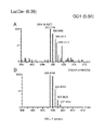

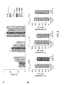


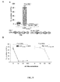


Classifications
-
- A—HUMAN NECESSITIES
- A61—MEDICAL OR VETERINARY SCIENCE; HYGIENE
- A61K—PREPARATIONS FOR MEDICAL, DENTAL OR TOILETRY PURPOSES
- A61K31/00—Medicinal preparations containing organic active ingredients
- A61K31/70—Carbohydrates; Sugars; Derivatives thereof
- A61K31/7016—Disaccharides, e.g. lactose, lactulose
-
- A—HUMAN NECESSITIES
- A61—MEDICAL OR VETERINARY SCIENCE; HYGIENE
- A61K—PREPARATIONS FOR MEDICAL, DENTAL OR TOILETRY PURPOSES
- A61K31/00—Medicinal preparations containing organic active ingredients
- A61K31/70—Carbohydrates; Sugars; Derivatives thereof
- A61K31/7028—Compounds having saccharide radicals attached to non-saccharide compounds by glycosidic linkages
-
- A—HUMAN NECESSITIES
- A61—MEDICAL OR VETERINARY SCIENCE; HYGIENE
- A61K—PREPARATIONS FOR MEDICAL, DENTAL OR TOILETRY PURPOSES
- A61K39/00—Medicinal preparations containing antigens or antibodies
- A61K39/12—Viral antigens
- A61K39/21—Retroviridae, e.g. equine infectious anemia virus
-
- A—HUMAN NECESSITIES
- A61—MEDICAL OR VETERINARY SCIENCE; HYGIENE
- A61P—SPECIFIC THERAPEUTIC ACTIVITY OF CHEMICAL COMPOUNDS OR MEDICINAL PREPARATIONS
- A61P31/00—Antiinfectives, i.e. antibiotics, antiseptics, chemotherapeutics
-
- A—HUMAN NECESSITIES
- A61—MEDICAL OR VETERINARY SCIENCE; HYGIENE
- A61P—SPECIFIC THERAPEUTIC ACTIVITY OF CHEMICAL COMPOUNDS OR MEDICINAL PREPARATIONS
- A61P31/00—Antiinfectives, i.e. antibiotics, antiseptics, chemotherapeutics
- A61P31/12—Antivirals
-
- A—HUMAN NECESSITIES
- A61—MEDICAL OR VETERINARY SCIENCE; HYGIENE
- A61P—SPECIFIC THERAPEUTIC ACTIVITY OF CHEMICAL COMPOUNDS OR MEDICINAL PREPARATIONS
- A61P31/00—Antiinfectives, i.e. antibiotics, antiseptics, chemotherapeutics
- A61P31/12—Antivirals
- A61P31/14—Antivirals for RNA viruses
-
- A—HUMAN NECESSITIES
- A61—MEDICAL OR VETERINARY SCIENCE; HYGIENE
- A61P—SPECIFIC THERAPEUTIC ACTIVITY OF CHEMICAL COMPOUNDS OR MEDICINAL PREPARATIONS
- A61P31/00—Antiinfectives, i.e. antibiotics, antiseptics, chemotherapeutics
- A61P31/12—Antivirals
- A61P31/14—Antivirals for RNA viruses
- A61P31/16—Antivirals for RNA viruses for influenza or rhinoviruses
-
- A—HUMAN NECESSITIES
- A61—MEDICAL OR VETERINARY SCIENCE; HYGIENE
- A61P—SPECIFIC THERAPEUTIC ACTIVITY OF CHEMICAL COMPOUNDS OR MEDICINAL PREPARATIONS
- A61P31/00—Antiinfectives, i.e. antibiotics, antiseptics, chemotherapeutics
- A61P31/12—Antivirals
- A61P31/14—Antivirals for RNA viruses
- A61P31/18—Antivirals for RNA viruses for HIV
-
- G—PHYSICS
- G01—MEASURING; TESTING
- G01N—INVESTIGATING OR ANALYSING MATERIALS BY DETERMINING THEIR CHEMICAL OR PHYSICAL PROPERTIES
- G01N33/00—Investigating or analysing materials by specific methods not covered by groups G01N1/00 - G01N31/00
- G01N33/48—Biological material, e.g. blood, urine; Haemocytometers
- G01N33/50—Chemical analysis of biological material, e.g. blood, urine; Testing involving biospecific ligand binding methods; Immunological testing
- G01N33/53—Immunoassay; Biospecific binding assay; Materials therefor
- G01N33/569—Immunoassay; Biospecific binding assay; Materials therefor for microorganisms, e.g. protozoa, bacteria, viruses
- G01N33/56983—Viruses
- G01N33/56988—HIV or HTLV
-
- A—HUMAN NECESSITIES
- A61—MEDICAL OR VETERINARY SCIENCE; HYGIENE
- A61K—PREPARATIONS FOR MEDICAL, DENTAL OR TOILETRY PURPOSES
- A61K39/00—Medicinal preparations containing antigens or antibodies
- A61K2039/51—Medicinal preparations containing antigens or antibodies comprising whole cells, viruses or DNA/RNA
- A61K2039/515—Animal cells
- A61K2039/5154—Antigen presenting cells [APCs], e.g. dendritic cells or macrophages
-
- A—HUMAN NECESSITIES
- A61—MEDICAL OR VETERINARY SCIENCE; HYGIENE
- A61K—PREPARATIONS FOR MEDICAL, DENTAL OR TOILETRY PURPOSES
- A61K8/00—Cosmetics or similar toiletry preparations
- A61K8/02—Cosmetics or similar toiletry preparations characterised by special physical form
- A61K8/14—Liposomes; Vesicles
-
- A—HUMAN NECESSITIES
- A61—MEDICAL OR VETERINARY SCIENCE; HYGIENE
- A61K—PREPARATIONS FOR MEDICAL, DENTAL OR TOILETRY PURPOSES
- A61K8/00—Cosmetics or similar toiletry preparations
- A61K8/18—Cosmetics or similar toiletry preparations characterised by the composition
- A61K8/30—Cosmetics or similar toiletry preparations characterised by the composition containing organic compounds
- A61K8/60—Sugars; Derivatives thereof
- A61K8/602—Glycosides, e.g. rutin
-
- A—HUMAN NECESSITIES
- A61—MEDICAL OR VETERINARY SCIENCE; HYGIENE
- A61K—PREPARATIONS FOR MEDICAL, DENTAL OR TOILETRY PURPOSES
- A61K8/00—Cosmetics or similar toiletry preparations
- A61K8/18—Cosmetics or similar toiletry preparations characterised by the composition
- A61K8/30—Cosmetics or similar toiletry preparations characterised by the composition containing organic compounds
- A61K8/68—Sphingolipids, e.g. ceramides, cerebrosides, gangliosides
-
- C—CHEMISTRY; METALLURGY
- C07—ORGANIC CHEMISTRY
- C07K—PEPTIDES
- C07K16/00—Immunoglobulins [IGs], e.g. monoclonal or polyclonal antibodies
- C07K16/18—Immunoglobulins [IGs], e.g. monoclonal or polyclonal antibodies against material from animals or humans
- C07K16/28—Immunoglobulins [IGs], e.g. monoclonal or polyclonal antibodies against material from animals or humans against receptors, cell surface antigens or cell surface determinants
- C07K16/2803—Immunoglobulins [IGs], e.g. monoclonal or polyclonal antibodies against material from animals or humans against receptors, cell surface antigens or cell surface determinants against the immunoglobulin superfamily
-
- C—CHEMISTRY; METALLURGY
- C12—BIOCHEMISTRY; BEER; SPIRITS; WINE; VINEGAR; MICROBIOLOGY; ENZYMOLOGY; MUTATION OR GENETIC ENGINEERING
- C12N—MICROORGANISMS OR ENZYMES; COMPOSITIONS THEREOF; PROPAGATING, PRESERVING, OR MAINTAINING MICROORGANISMS; MUTATION OR GENETIC ENGINEERING; CULTURE MEDIA
- C12N2740/00—Reverse transcribing RNA viruses
- C12N2740/00011—Details
- C12N2740/10011—Retroviridae
- C12N2740/16011—Human Immunodeficiency Virus, HIV
- C12N2740/16034—Use of virus or viral component as vaccine, e.g. live-attenuated or inactivated virus, VLP, viral protein
-
- C—CHEMISTRY; METALLURGY
- C12—BIOCHEMISTRY; BEER; SPIRITS; WINE; VINEGAR; MICROBIOLOGY; ENZYMOLOGY; MUTATION OR GENETIC ENGINEERING
- C12N—MICROORGANISMS OR ENZYMES; COMPOSITIONS THEREOF; PROPAGATING, PRESERVING, OR MAINTAINING MICROORGANISMS; MUTATION OR GENETIC ENGINEERING; CULTURE MEDIA
- C12N2740/00—Reverse transcribing RNA viruses
- C12N2740/00011—Details
- C12N2740/10011—Retroviridae
- C12N2740/16011—Human Immunodeficiency Virus, HIV
- C12N2740/16061—Methods of inactivation or attenuation
-
- G—PHYSICS
- G01—MEASURING; TESTING
- G01N—INVESTIGATING OR ANALYSING MATERIALS BY DETERMINING THEIR CHEMICAL OR PHYSICAL PROPERTIES
- G01N2333/00—Assays involving biological materials from specific organisms or of a specific nature
- G01N2333/005—Assays involving biological materials from specific organisms or of a specific nature from viruses
- G01N2333/08—RNA viruses
- G01N2333/15—Retroviridae, e.g. bovine leukaemia virus, feline leukaemia virus, feline leukaemia virus, human T-cell leukaemia-lymphoma virus
- G01N2333/155—Lentiviridae, e.g. visna-maedi virus, equine infectious virus, FIV, SIV
- G01N2333/16—HIV-1, HIV-2
-
- Y—GENERAL TAGGING OF NEW TECHNOLOGICAL DEVELOPMENTS; GENERAL TAGGING OF CROSS-SECTIONAL TECHNOLOGIES SPANNING OVER SEVERAL SECTIONS OF THE IPC; TECHNICAL SUBJECTS COVERED BY FORMER USPC CROSS-REFERENCE ART COLLECTIONS [XRACs] AND DIGESTS
- Y02—TECHNOLOGIES OR APPLICATIONS FOR MITIGATION OR ADAPTATION AGAINST CLIMATE CHANGE
- Y02A—TECHNOLOGIES FOR ADAPTATION TO CLIMATE CHANGE
- Y02A50/00—TECHNOLOGIES FOR ADAPTATION TO CLIMATE CHANGE in human health protection, e.g. against extreme weather
- Y02A50/30—Against vector-borne diseases, e.g. mosquito-borne, fly-borne, tick-borne or waterborne diseases whose impact is exacerbated by climate change
Definitions
- the present invention refers to method for preventing viral entry into cells expressing the CD169 cell surface receptor by inhibiting the coupling of sialyllactose molecules in the viral membrane gangliosides to the CD169 receptor.
- the invention also relates to inhibitors of this coupling, as well as pharmaceutical compositions containing the inhibitors of the invention, their methods of preparation and diagnostic applications.
- DCs Dendritic cells capture pathogens in the mucosa and then migrate to the secondary lymphoid tissue, where they acquire the mature phenotype required to induce efficiently adaptive immune responses.
- the potential role of mature DCs (mDCs) uptake for antigen presentation implies efficient antigen capture and transfer into the antigen presentation pathway. Down regulation of endocytosis is considered a hallmark of DC maturation, but there is increasing evidence that under inflammatory conditions mDCs capture, process, and present antigens without exclusively relying on prior pathogen exposure. See Mellman I, et al., Cell 2001; 106:255-258 , Platt C, et al., Proc. Natl. Acad. Sci.
- HIV-1 capture into mDCs appears to also critically enhance viral dissemination in lymphoid tissue by efficient presentation of infectious virus to T-cells in the DC-T-cell synapse, thus promoting pathogenesis and disease progression through trans-infection.
- In vitro studies have shown that, when HIV is incubated at low MOI with T cells, inclusion of DC results in much more efficient infection of the T cells.
- the mechanism for trans-infection has been a subject of some controversy.
- An HIV-1 gp120-independent mechanism of viral binding and uptake that is upregulated upon DC maturation has been previously identified in the art. See Izquierdo-Useros N, J. Virol. 2007; 81:7559-7570 .
- HIV-1 Gag eGFP-expressing fluorescent virus-like particles follow the same trafficking route as wild type HIV-1 in mDCs, and hence share a common molecular pattern that governs entry into mDCs. See Izquierdo-Useros N, et al., Blood 2009; 113:2732-2741 . However, the precise mechanism through which HIV-1 is internalized and accumulated into mDCs was unknown until now.
- the invention relates to an inhibitor of the interaction between sialoadhesin and sialyllactose for use in the treatment or prevention of a disease associated with an infection caused by an enveloped virus.
- the invention in another aspect, relates to a composition or kit-of-parts comprising an antigen-loaded antigen-presenting cell and an inhibitor of the interaction between sialoadhesin and sialyllactose.
- the invention relates to a method for detecting or isolating an enveloped virus in a sample comprising:
- the invention relates to a kit comprising immobilized sialoadhesin or a functionally equivalent variant thereof substantially preserving its ability to bind sialyllactose.
- the invention in another aspect, relates to a conjugate comprising sialoadhesin or a functionally equivalent variant thereof substantially preserving its ability to bind sialyllactose and a therapeutic or diagnostic agent.
- the invention relates to an in vitro method for delivering a compound of interest to an antigen-presenting cell which comprises contacting said antigen-presenting cell with a lipid microparticle comprising said compound of interest wherein said lipid particle comprises at least a molecule containing a sialyllactose moiety.
- the present invention identifies clearly a novel role for sialylated gangliosides in the membrane of viruses or vesicles as determinants for specific capture by mDCs. This capture is dependent on an exposed sialyllactose moiety, which is identified herein for the first time as a novel pathogen-associated molecular pattern.
- Gangliosides are a significant component of the plasma membrane lipidome suggesting that all enveloped viruses, which bud from the plasma membrane of infected cells, may be captured into mDCs by the reported mechanism unless they exclude sialyllactose containing gangliosides.
- ganglioside-carrying vesicles or virions support a model such as the one disclosed in the present invention, where a specific receptor present on the cell surface of mDCs (and possibly other cells), such as the CD169 cell surface receptor, recognizes the sialyllactose moiety on the virion or vesicle membranes. Specific recognition of vesicular gangliosides would then trigger uptake into an intracellular compartment. Subsequently, internalized material may either be recycled to the surface (as in HIV-1 transmission to T-cells) or could be fed into the antigen presentation pathway.
- AIDS refers to the symptomatic phase of HIV infection, and includes both Acquired Immune Deficiency Syndrome (commonly known as AIDS) and "ARC,” or AIDS-Related Complex. See Adler M, et al., Brit. Med. J. 1987; 294: 1145-1147 .
- the immunological and clinical manifestations of AIDS are well known in the art and include, for example, opportunistic infections and cancers resulting from immune deficiency.
- antibody refers to a protein consisting of one or more proteins substantially encoded by all or part of the recognized immunoglobulin genes, including but not limited to polyclonal antibodies, monoclonal antibodies, and antigen-binding fragments thereof such as, for instance, F(ab') 2 and Fab fragments, and single chain antibodies.
- the term antibody includes any type of known antibody, such as, for example, polyclonal antibodies, monoclonal antibodies and genetically engineered antibodies, such as chimeric antibodies, humanized antibodies, primatized antibodies, human antibodies and bispecific antibodies.
- anti-HIV agent refers to any compound or a pharmaceutically acceptable salt thereof which is capable of inhibiting the replication of HIV in a cell, such as a cell in a mammal or which is effective in treating, preventing, or delaying the onset or progression of HIV infection or AIDS or diseases or conditions arising therefrom or associated therewith.
- Suitable anti-HIV agents for use according to the present invention include, without limitation, HIV protease inhibitors, a HIV reverse transcriptase inhibitor, HIV entry inhibitors and HIV immunogens.
- antigen-binding region of an antibody includes also a synthetic or a genetically engineered polypeptide that binds to a specific antigen, such as polypeptides consisting of the light chain variable region, "Fv” fragments consisting of the variable regions of the heavy and light chains, recombinant single chain polypeptide molecules in which light and heavy variable regions are connected by a peptide linker (“scFv proteins”) and minimal recognition units consisting of the amino acid residues that mimic the hypervariable region.
- scFv proteins peptide linker
- antigen loading refers to a method of delivering antigens to dendritic cells by incubating dendritic cells or progenitor cells with the peptide, polypeptide, lipopeptide, DNA (naked or within a plasmid vector) or RNA; or with antigen-expressing recombinant bacterium or viruses (e.g. vaccinia, adenovirus or lentivirus) such that the antigenic epitopes thereof are loaded and expressed on the cell surface by MHC.
- antigen-expressing bacterium or viruses e.g. vaccinia, adenovirus or lentivirus
- antigen-presenting cell refers to any cell capable of processing and presenting an antigen via an MHC molecule (MHC class I or MHC class II molecules).
- MHC class I or MHC class II molecules MHC class I or MHC class II molecules.
- the APC could be capable of processing and presenting an antigen via MHC class I and MHC class II molecules.
- antigen presenting cells comprise dendritic cells, macrophages, B cells, epithelial cells, fibroblasts, glial cells and additional cells identifiable by a skilled person.
- APV are dendritic cells.
- antiretroviral agent includes any pharmacological, biological or cellular agent that has demonstrated the ability to a retrovirus.
- autologous refers to any material derived from a donor subject that is later reintroduced to the same individual.
- composition refers to a material composition that comprises at least two components, as well as any product resulting, directly or indirectly, from the combination of the different components in any quantity thereof.
- composition may be formulated as a single formulation or may be presented as separate formulations of each of the components, which may be combined for joint use as a combined preparation.
- the composition may be a kit-of-parts wherein each of the components is individually formulated and packaged.
- conjugate refers to two or more compounds which are covalently linked together so that the function of each compound is retained in the conjugate.
- dendritic cell is an antigen-presenting cell existing in vivo , in vitro , ex vivo , or in a host or subject, or which can be derived from a hematopoietic stem cell or a monocyte.
- Dendritic cells and their precursors can be isolated from a variety of lymphoid organs (e.g. spleen, lymph nodes), as well as from bone marrow and peripheral blood.
- the DC has a characteristic morphology with thin sheets (lamellipodia) extending in multiple directions away from the dendritic cell body.
- dendritic cells express high levels of MHC and costimulatory (e.g. B7-1 and B7-2) molecules.
- Dendritic cells can induce antigen specific differentiation of T cells in vitro, and are able to initiate primary T cell responses in vitro and in vivo.
- the term “dendritic cells” includes differentiated dendritic cells, whether immature and mature dendritic cells. These cells can be characterized by expression of certain cells surface markers (e.g. CD 11c, MHC class II, and at least low levels of CD80 and CD86). In addition, dendritic cells can be characterized functionally by their capacity to stimulate alloresponses and mixed lymphocyte reactions (MLR).
- the expression "dendritic cell preparation” refers to a composition that contains dendritic cells obtained from a subject in a media suitable for the pulsing of said cells.
- the expression "disease associated with a HIV infection”, as used herein, includes a state in which the subject has developed AIDS as well as a state in which the subject infected with HIV has not shown any sign or symptom of the disease.
- the compositions of the invention when administered to a subject that has no clinical signs of the infection can have a preventive activity, since they can prevent the onset of the disease.
- the compositions are capable of preventing or slowing the infection and destruction of healthy CD4+ T cells in such a subject.
- enveloped virus refers to any animal virus which possesses an outer membrane or envelope, which is a lipid bilayer containing viral proteins, surrounding the virus capsid.
- enveloped viruses include, but are not limited to, members of the poxviridae, hepadnaviridae, togaviridae, arenaviridae, flaviviridae, orthomyxoviridae, paramyxoviridae, bunyaviridae, rhabdoviridae, filoviridae, coronaviridae, retroviridae and bornaviridae virus families.
- ganglioside or “sialogangliosides”, as used herein, refers to glycosphingolipids which contain several monosaccharide units per molecule.
- suitable monosaccharide units which can be contained in the gangliosides or ganglioside derivatives are D-galactose, N-acetyl D-galactosamine, glucose and N-acetylneuraminic acid.
- SIV refers to simian immunodeficiency virus which is an HIV-like virus that infects monkeys, chimpanzees, and other nonhuman primates. SIV includes but is not limited to extracellular virus particles and the forms of SIV associated with SIV infected cells.
- HIV immunogen refers to a protein or peptide antigen derived from HIV that is capable of generating an immune response in a subject. HIV immunogens for use according to the present invention may be selected from any HIV isolate (e.g. any primary or cultured HIV- 1, HIV-2, or HIV-3 isolate, strain, or clade).
- HIV isolate e.g. any primary or cultured HIV- 1, HIV-2, or HIV-3 isolate, strain, or clade.
- immunogen refers to a substance or material (including antigens) that is able to induce an immune response alone or in conjunction with an adjuvant.
- the immune response generated by the vaccine may be a humoral or a cellular immune response.
- the expression "humoral immune response”, is used herein to describe an immune response against foreign antigen(s) that is mediated by T-cells and their secretion products.
- the “cellular immune response”, is used herein to describe an immune response against foreign antigen(s) that is mediated by antibodies produced by B-cells.
- the vaccine is systemically or locally administered.
- inhibitor of the interaction between sialoadhesin and sialyllactose refers to any molecule or compound which is capable of inhibiting the binding between sialoadhesin and sialyllactose or any other compound comprising sialyllactose. Inhibitors are capable of specifically binding to either of sialoadhesin and sialyllactose with an affinity that is higher than the affinity of the binding between sialoadhesin and sialyllactose.
- isolation refers to partial or complete removal of viral particles from the media in which they are produced.
- lipidic microvesicle or "liposome”, as used herein, refers to a microscopic vesicle comprising an outer lipid layer.
- the outer lipid layer could be a lipid monolayer or bilayer.
- the liposomes are also referred to as micelles.
- Liposomes may have one or more lipid membranes.
- the invention contemplates both single-layered liposomes, which are referred to as unilamellar, and multi-layered liposomes, which are referred to as multilamellar.
- molecule comprising a sialyllactose moiety refers to any molecule which contains within its structure a ⁇ -D-galactopyranosyl-(1 ⁇ 4)-D-glucose moiety bound to a sialic acid moiety.
- the ⁇ -D-galactopyranosyl-(1 ⁇ 4)-D-glucose moiety forms part of an oligosaccharide wherein the galactopyranosyl-(1 ⁇ 4)-D-glucose moiety may form the terminal two monosaccharide residues in the chain or may be linked to further monosaccharide residues on both sides.
- the oligosaccharide containing the galactopyranosyl-(1 ⁇ 4)-D-glucose moiety may contain at least 3, 4, 5, 6, 7, 8, 9, 10 or more monosaccharides residues.
- the oligosaccharide can be provided as such or form part of a ganglioside.
- % sequence identity is determined by comparing two optimally aligned sequences over a comparison window, where the fragment of the polypeptide or polynucleotide sequence in the comparison window may comprise additions or deletions (e.g. gaps or overhangs) as compared to the reference sequence (which does not comprise additions or deletions) for optimal alignment of the two sequences.
- the percentage is calculated by determining the number of positions at which the identical amino acid residue or nucleic acid residue occurs in both sequences to yield the number of matched positions, dividing the number of matched positions by the total number of positions in the comparison window and multiplying the result by 100 to provide the percentage of sequence identity. Algorithms to align sequences are known in the art.
- Optimal alignment of sequences for comparison can be conducted, for instance, by the Smith-Waterman local homology algorithm, by the Needleman-Wunsch homology alignment algorithm, by the Pearson-Lipman similarity search method, by computerized implementations of these algorithms or by manual alignment and visual inspection.
- Smith T Waterman M, Adv. Appl. Math. 1981; 2:482-489 ; Needleman S, Wunsch C, J. Mol. Biol. 1970; 48:443-453 ; Pearson W, Lipman D, Proc. Natl. Acad. Sci. USA 1988; 85:2444-2448 ; Tatusova T, Madden T, FEMS Microbiol. Lett.
- pharmaceutically acceptable carrier refers to a non-toxic solid, semisolid or liquid filler, diluent, encapsulating material or formulation auxiliary of any conventional type.
- a pharmaceutically acceptable carrier is essentially non-toxic to recipients at the dosages and concentrations employed, and is compatible with other ingredients of the formulation.
- the carrier for a formulation containing polypeptides would not normally include oxidizing agents and other compounds that are known to be deleterious to polypeptides.
- phospholipid refers to a lipid that contains one or more phosphate groups. Phospholipids are amphipathic in nature; that is, each molecule consists of a hydrophilic portion and a hydrophobic portion.
- phospholipid includes pharmaceutically acceptable salts and ester derivatives of such compounds. Phospholipids can be classified according to the type of alcohol in phosphoglycerides (or glycerophospholipids) when they carry a glycerol backbone and sphingolipids wherein the lipids contain sphingosine. Both classes are present in the biological membrane.
- Phosphoglycerides are the most abundant class of phospholipids found in nature and include, without limitation, phosphatidylcholine (e.g. lecithin), phosphatidylethanolamine, phosphatidylserine, phosphatidylinositol, phosphatidylglycerol, and cardiolipin.
- phosphatidylcholine e.g. lecithin
- phosphatidylethanolamine phosphatidylethanolamine
- phosphatidylserine phosphatidylinositol
- phosphatidylglycerol phosphatidylglycerol
- cardiolipin cardiolipin
- protease an enzyme required for the proteolytic cleavage of viral polyprotein precursors (e.g. viral GAG and GAG Pol polyproteins), into the individual functional proteins found in infectious HIV-1.
- viral polyprotein precursors e.g. viral GAG and GAG Pol polyproteins
- Retrovirus refers to virus belonging to the Retroviridae family, which are characterized by being an RNA virus that is replicated in a host cell via the enzyme reverse transcriptase to produce DNA from its RNA genome.
- reverse transcriptase inhibitors refers to any compound which inhibits the activity of HIV-1 reverse transcriptase, the enzyme which catalyzes the conversion of viral genomic HIV-1 RNA into proviral HIV-1 DNA.
- sialoadhesin sialic acid binding Ig-like lectin 1
- siglec-1 siglec-1
- CD169 immunoglobulin (Ig) domains, that binds to sialic acids forming a salt bridge between a highly conserved arginine residue and the carboxylate group of the sialic acid. See May A, et al., Mol. Cell 1998; 1:719-728 .
- Suitable sialoadhesin variants for use according to the present invention include the human sialoadhesin (described in the UniProt database under accession number Q9BZZ2), porcine sialoadhesin (described in the UniProt database under accession number A7LCJ3) and mouse sialoadhesin (described in the UniProt database under accession number Q62230.
- sialyllactose refers to a molecule comprising a lactose moiety ⁇ -D-galactopyranosyl-(1 ⁇ 4)-D-glucose) bound to a sialic acid moiety.
- the sialic acid may be coupled to the lactose by any possible position in the sialic acid and to any possible position in the lactose molecule.
- the sialic acid is linked to the lactose by the hydroxyl group at position 2 of the sialic acid (the numbering of the sialic acid structure begins at the carboxylate carbon and continues around the chain).
- the sialic acid is linked to the lactose by the hydroxyl groups at positions 3 or 6 in the lactose molecule.
- the "sialyllactose" is a2,3-sialyl-lactose or a2,6-sialyl-lactose.
- Sialyllactose may be of eukaryotic or prokaryotic origin.
- sialyllactose is of eukaryotic origin.
- the eukaryotic or prokaryotic cell may be pathogenic or non-pathogenic.
- subject is meant to include all animals shown to or expected to have antigen presenting cells.
- the subject is a mammal, a human or nonhuman primate, a dog, a cat, a horse, a cow, other farm animals, or a rodent (e.g. mice, rats, guinea pig).
- rodent e.g. mice, rats, guinea pig.
- treatment refers to any type of therapy, which aims at terminating, preventing, ameliorating or reducing the susceptibility to a clinical condition as described herein.
- the term treatment relates to prophylactic treatment (i.e. a therapy to reduce the susceptibility of a clinical condition), of a disorder or a condition as defined herein.
- prophylactic treatment i.e. a therapy to reduce the susceptibility of a clinical condition
- treatment refers to obtaining a desired pharmacologic or physiologic effect, covering any treatment of a pathological condition or disorder in a mammal, including a human.
- treatment includes (1) preventing the disorder from occurring or recurring in a subject, (2) inhibiting the disorder, such as arresting its development, (3) stopping or terminating the disorder or at least symptoms associated therewith, so that the host no longer suffers from the disorder or its symptoms, such as causing regression of the disorder or its symptoms, for example, by restoring or repairing a lost, missing or defective function, or stimulating an inefficient process, or (4) relieving, alleviating, or ameliorating the disorder, or symptoms associated therewith, where ameliorating is used in a broad sense to refer to at least a reduction in the magnitude of a parameter, such as inflammation, pain, or immune deficiency.
- a parameter such as inflammation, pain, or immune deficiency
- vaccine and "vaccine composition”, as used herein, refers to a formulation which contains a conjugate or a composition according to the present invention in a suitable form for administration to a vertebrate and induce a protective immune response.
- the conjugate or a composition is sufficient to induce immunity to prevent or ameliorate an infection or to reduce at least one symptom of an infection or to enhance the efficacy of another dose of conjugate or composition of the invention.
- vaginal cream refers to a semi-solid preparation suitable for application to the vaginal tract.
- excipients or vehicles known in the art can be used in its preparation.
- the excipients comprise materials of naturally occurring or synthetic origin that do not adversely affect the components of the formulation.
- Suitable carriers for use herein include but are not limited to purified water, white soft paraffin, mucoadhesive polymers, liquid paraffin, polysorbate 60, sorbitan stearate silicone, waxes, petroleum, jelly, polyethylene glycol, and a variety of other materials, depending on the specific type of formulation used.
- viral entry inhibitor refers to any compound capable of interfering with the entry of viruses into cells.
- viral immunogens refers to the whole HIV inactivated virions obtained by the inactivation process of the invention.
- the mechanism for HIV uptake by mDCs identified in the present invention may be relevant for uptake of any enveloped viruses and thus, the use of inhibitors of the interaction between sialoadhesin and sialyllactose may be useful for preventing infection by any enveloped virus.
- the invention relates to an inhibitor of the interaction between sialoadhesin and sialyllactose for use in the treatment or prevention of a disease associated with an infection caused by an enveloped virus.
- the invention relates to the use of an inhibitor of the interaction between sialoadhesin and sialyllactose for the preparation of a medicament for the treatment of a disease associated with an infection caused by an enveloped virus.
- the invention in another aspect, relates to a method for treatment or prevention of a disease associated with an infection caused by an enveloped virus in a subject in need thereof which comprises the administration to said subject of a an inhibitor of the interaction between sialoadhesin and sialyllactose.
- Lipids containing sialyllactose within their structure and which can be incorporated into the large unilamelar vesicles include mono-, di- and trisialylgangliosides containing a sialyllactose moiety such as, without limitation, GM1, GM2, GM3, GD1b and GT1b.
- Table 1 Gangliosides containing sialyllactose residues which inhibit the interaction between sialadhesin and sialyllactose.
- the invention also contemplates the use of ganglioside derivatives wherein one or more of the following functional groups are substituted or added to the backbone chain, in particular the ceramide backbone chain:
- the invention also comprises the use of fragments of the different types of antibodies mentioned above which substantially preserve the ability to bind sialoadhesin and preventing its interaction with molecules containing sialyllactose.
- antibody fragment includes antibody fragments such as Fab, F(ab') 2 , Fab', single chain Fv fragments (scFv), diabodies and nanobodies.
- the inhibitor of the interaction between sialoadhesin and sialyllactose is a sialyllactose-binding molecule.
- This type of molecules are capable of binding the sialyllactose found in the surface of the enveloped viruses and prevent their interaction with sialoadhesin.
- Suitable sialylactose-binding molecules include, without limitation, sialyllactose-binding lectins and anti-sialyllactose antibodies.
- Suitable sialyllactose-binding lectins include, without limitation, sialoadhesin or the extracellular domain thereof, the Sambucus nigra (elderberry) bark lectin (specfic for sialyllactose having a ⁇ 2,6-linkage), the Maackia amurensis lectin (specific for sialyllactose having a ⁇ 2,3-sialyl bond) and the Vibrio cholerae neuraminidase or a lectin-like domain thereof.
- the disease associated with an infection caused by an enveloped virus is a disease caused by a type I filoviridae virus.
- Type I filoviridae virus which has a single-stranded, unsegmented (-) sense RNA genome and which causes severe hemorrhagic fever in humans and non-human primates.
- the filoviridae virus is an Ebola virus, such as a Cote d'lrium (CI), Sudan (S), Zaire (Z) or Reston (R) species of Ebola virus.
- the filoviridae virus is a Marburg virus.
- the disease associated with an infection caused by an enveloped virus is a disease caused by an orthomyxoviridae virus, such as an influenza virus, Thogotovirus, Dhori virus, or infectious salmon anemia virus.
- an orthomyxoviridae virus such as an influenza virus, Thogotovirus, Dhori virus, or infectious salmon anemia virus.
- methods provided herein are used to treat or prevent infection of a human subject with an influenza type A virus, an influenza type B virus, or an influenza type C virus.
- the influenza type A virus is of subtype H1N1, H2N2, H3N2 or H5N1.
- the disease associated with an infection caused by an enveloped virus is a disease caused by a paramyxoviridae virus, such as human parainfluenza virus, human respiratory syncytial virus (RSV), Sendai virus, Newcastle disease virus, mumps virus, rubella (measles) virus, Hendra virus, Nipah virus, avian pneumo virus, or canine distemper virus.
- a paramyxoviridae virus such as human parainfluenza virus, human respiratory syncytial virus (RSV), Sendai virus, Newcastle disease virus, mumps virus, rubella (measles) virus, Hendra virus, Nipah virus, avian pneumo virus, or canine distemper virus.
- the disease associated with an infection caused by an enveloped virus is a disease caused by a rhabdoviridae virus, such as rabies virus, vesicular stomatitis virus (VSV), Mokola virus, Duvenhage virus, European bat virus, salmon infectious hematopoietic necrosis virus, viral hemorrhagic septicaemia virus, spring viremia of carp virus, or snakehead rhabdovirus.
- a rhabdoviridae virus such as rabies virus, vesicular stomatitis virus (VSV), Mokola virus, Duvenhage virus, European bat virus, salmon infectious hematopoietic necrosis virus, viral hemorrhagic septicaemia virus, spring viremia of carp virus, or snakehead rhabdovirus.
- the disease associated with an infection caused by an enveloped virus is a disease caused by a bornaviridae virus, such as Borna disease virus.
- the disease associated with an infection caused by an enveloped virus is a disease caused by a bunyaviridae virus, such as Bunyamwera virus, Hantaan virus, Crimean Congo virus, California encephalitis virus, Rift Valley fever virus, or sandfly fever virus.
- a bunyaviridae virus such as Bunyamwera virus, Hantaan virus, Crimean Congo virus, California encephalitis virus, Rift Valley fever virus, or sandfly fever virus.
- the disease associated with an infection caused by an enveloped virus is a disease caused by is an arenaviridae virus, such as Old World Arenaviruses, Lassa fever virus, Ippy virus, Lymphocytic choriomeningitis virus (LCMV), Mobala virus, Mopeia virus, or a New World Arenavirus, such as Junin virus (Argentine hemorrhagic fever), Sabia (Brazilian hemorrhagic fever), Amapari virus, Flexal virus, Guanarito virus (Venezuela hemorrhagic fever), Machupo virus (Bolivian hemorrhagic fever), Latino virus, Boliveros virus, Parana virus, Pichinde virus, Pirital virus, Tacaribe virus, Tamiami virus, or Whitewater Arroyo virus.
- the Arenaviridae virus is Lymphocytic choriomeningitis virus, Lassa virus, Junin Virus, Machupo Virus, Sabia virus, or Guan
- the disease associated with an infection caused by an enveloped virus is a disease caused by an arbovirus.
- Arboviruses comprise a large group of more than 400 enveloped RNA viruses that are transmitted primarily by arthropod vectors (e.g. mosquitoes, sand-flies, fleas, ticks, lice).
- the arbovirus is a togaviridae virus, such as an Alphavirus (e.g. Venezuela equine encephalitis virus or Sindbis virus) or a Rubivirus (e.g. Rubella virus).
- a compound provided herein is administered to a pregnant subject to treat or prevent congenital rubella syndrome (CRS) and symptoms related thereto, such as low birth weight, deafness, and abortion.
- CRS congenital rubella syndrome
- the disease associated with an infection caused by an enveloped virus is a disease caused by is a flaviviridae virus, such as a Flavivirus, a Pestivirus, a Hepadvirus, yellow fever virus, dengue fever virus, or Japanese encaphilitis (JE) virus.
- a flaviviridae virus such as a Flavivirus, a Pestivirus, a Hepadvirus, yellow fever virus, dengue fever virus, or Japanese encaphilitis (JE) virus.
- the disease associated with an infection caused by an enveloped virus is a disease caused by a hepacivirus, such as a hepatitis C virus or a hepatitis C-like virus.
- the disease associated with an infection caused by an enveloped virus is a disease caused by a henipavirus, such as Hendra virus or Nipah virus.
- the disease associated with an infection caused by an enveloped virus is a disease caused by a bunyaviridae (-)-sense RNA virus, such as an Orthobunyavirus, a Hantavirus, a Phlebovirus, or a Nairovirus.
- a bunyaviridae (-)-sense RNA virus such as an Orthobunyavirus, a Hantavirus, a Phlebovirus, or a Nairovirus.
- the disease associated with an infection caused by an enveloped virus is a disease caused by an arenavirius virus, such as Lymphocytic choriomeningitis virus (LCMV), Lassa virus, Junin virus, Machupo virus, or Guanarito virus.
- LCMV Lymphocytic choriomeningitis virus
- Lassa virus Junin virus
- Machupo virus Machupo virus
- Guanarito virus a virus caused by an arenavirius virus
- the disease associated with an infection caused by an enveloped virus is a disease caused by a Japanese encephalitis virus, such as Alfuy virus, Japanese encephalitis virus, Kokobera virus, Koutango virus, Kunjin virus, Murray Valley encephalitis virus, St. Louis encephalitis virus, Stratford virus, Usutu virus, or West Nile virus.
- a Japanese encephalitis virus such as Alfuy virus, Japanese encephalitis virus, Kokobera virus, Koutango virus, Kunjin virus, Murray Valley encephalitis virus, St. Louis encephalitis virus, Stratford virus, Usutu virus, or West Nile virus.
- the disease associated with an infection caused by an enveloped virus is a disease caused by a virus belonging to the retroviridae family.
- the enveloped virus is a virus belonging to the orthoretrovirinae subfamily.
- the enveloped virus belongs to the lentivirus genus.
- the enveloped virus is a primate lentivirus and, in particular, human immunodeficency virus (HIV) or Simian immunodeficency virus (SIV).
- retroviruses include but are not limited to the following virus genera: alpharetrovirus (e.g. avian leukosis virus (ALV) and rous sarcoma virus (RSV)), betaretrovirus (e.g. mouse mammary tumour virus (MMTV), SRV, HERV-K and JRSV), gammaretrovirus (e.g. murine leukemia virus (MLV), feline leukemia virus (FeLV), GALV, PERV, and HERV-W), deltaretrovirus (e.g. bovine leukemia virus (BLV), and cancer-causing human T-lymphotropic virus (HTLV-1 and HTLV-11)), epsilonretrovieris (e.g.
- alpharetrovirus e.g. avian leukosis virus (ALV) and rous sarcoma virus (RSV)
- betaretrovirus e.g. mouse mammary tumour virus (MMTV), SRV, HERV-K
- Walleye dermal sarcoma virus WDSV and SnRV
- lentivirus e.g. human immunodeficiency virus 1 (HIV-1), human immunodeficiency virus 2 (HIV-2), simian immunodeficiency virus (SIVmac and SIV), feline immunodeficiency virus (F1V), EIAV and MVV) and spumavirus (e.g. simian foamy virus (SFVcpz and SFVagm), FFV and BFV).
- the enveloped virus is a virus wherein at least some of the lipids within the envelope of said enveloped virus comprise sialyllactose.
- the enveloped virus comprises gangliosides in the viral envelope wherein the gangliosides contain at least one sialyllactose moiety.
- the gangliosides contain less than four sialyllactose molecules.
- the virus contains in its envelope one or more of the gangliosides shown in Table 1.
- the "disease associated with an infection caused by an enveloped virus” is a disease associated with an HIV virus.
- the present invention further relates to preventing or reducing symptoms associated with HIV infection.
- symptoms associated with the minor symptomatic phase of HIV infection including, for example, shingles, skin rash and nail infections, mouth sores, recurrent nose and throat infection and weight loss.
- further symptoms associated with the major symptomatic phase of HIV infection include, for instance, oral and vaginal thrush (Candida), persistent diarrhea, weight loss, persistent cough and reactivated tuberculosis or recurrent herpes infections, such as cold sores ( herpes simplex ).
- the compounds of the present invention may be useful in the methods of present invention in combination with one or more additional anti-HIV agent or agent.
- additional compounds may comprise compounds of the present invention or compounds, e.g., commercially available compounds, known to treat, prevent, or reduce the symptoms of HIV infections.
- the compounds of the invention may be used in combination with one or more of the following anti-HIV drugs:
- compositions according to the present invention may further comprise an antiinfective agent selected from the group consisting of antifungals, antibacterials, anti-neoplasties, anti-protozoals, DNA polymerase inhibitors, DNA synthesis inhibitors, anti-HIV antibodies, HIV antisense drugs, IL-2 agonists, ⁇ -glucosidase inhibitors, purine nucleoside phosphorylase inhibitors, apoptosis agonists, apoptosis inhibitors, and cholinesterase inhibitors, where the compounds are present in amounts effective for treatment of HIV when used in a combination therapy.
- an antiinfective agent selected from the group consisting of antifungals, antibacterials, anti-neoplasties, anti-protozoals, DNA polymerase inhibitors, DNA synthesis inhibitors, anti-HIV antibodies, HIV antisense drugs, IL-2 agonists, ⁇ -glucosidase inhibitors, purine nucleoside phosphorylase inhibitors, apoptosis agonist
- the combinations of the inhibitors according to the invention and the anti-HIV agents may result in a synergistic effect in terms of its anti-HIV activity.
- a synergistic effect may be calculated, for example, using suitable methods such as, for example, the Sigmoid-Emax, Loewe, and median-effect equations. See Holford K, Scheiner L, Clin. Pharmacokinet. 1981; 6:429-453 , Loewe S, Muischnek H, Arch. Exp. Pathol Pharmacol. 1926; 114:313-326 and Chou T, Talalay C, Adv. Enzyme Regul. 1984; 22:27-55 ).
- Each equation referred to above may be applied to experimental data to generate a corresponding graph to aid in assessing the effects of the drug combination.
- the corresponding graphs associated with the equations referred to above are the concentration-effect curve, isobologram curve and combination index curve, respectively.
- the inhibitors according to the invention may further comprise a pharmaceutically acceptable carrier.
- suitable carriers include, but are not limited to water, dextrose, glycerol, saline, ethanol, and combinations thereof.
- the carrier can contain additional agents such as wetting or emulsifying agents, pH buffering agents, or adjuvants which enhance the effectiveness of the formulation.
- anti-HIV therapeutic agents include nucleoside reverse transcriptase inhibitors, such as abacavir, AZT, didanosine, emtricitabine, lamivudine, stavudine, tenofovir, zalcitabine, or zidovudine; non-nucleoside reverse transcriptase inhibitors, such as delavirdine, efavirenz, or nevirapine; protease inhibitors, such as amprenavir, atazanavir, indinavir, lopinavir, nelfinavir osamprenavir, ritonavir, saquinavir, or tipranavir; or fusion protein inhibitors, such as enfuvirtide.
- nucleoside reverse transcriptase inhibitors such as abacavir, AZT, didanosine, emtricitabine, lamivudine, stavudine, tenofovir,
- the pharmaceutical composition of the invention may be applied to the vagina in a number of forms including aerosols, foams, sprays, pastes, gels, jellies, creams, suppositories, tablets, pessaries, tampons, or devices such as vaginal rings. They can be in the form of immediate release or controlled release. Foams, creams and gels are preferred forms. Compositions suitable for vaginal application and their methods of preparation are known in the art.
- the pharmaceutical composition is topically applied to the vagina.
- the topical application is carried out prior to the beginning of vaginal intercourse, suitably 0 to 60 minutes, preferably 0 to 5 minutes, prior to the beginning of vaginal intercourse.
- the application may be carried out into and around the vagina and vaginal area (e.g. the individual anatomical parts, such as, labia majora, labia minora, clitoris) of a female.
- Cream bases are water-washable, and contain an oil phase, an emulsifier and an aqueous phase.
- the oil phase also sometimes called the "internal" phase, is generally comprised of petrolatum and a fatty alcohol such as cetyl or stearyl alcohol; the aqueous phase usually, although not necessarily, exceeds the oil phase in volume, and generally contains a humectant.
- the emulsifier in a cream formulation is generally a nonionic, anionic, cationic or amphoteric surfactant.
- the suppository when the composition is in the form of a suppository (including vaginal suppositories), the suppository will usually be 1 to 5 grams, preferably about 3 grams, and the entire suppository will be applied.
- a vaginal tablet will suitably be 1 to 5 grams, preferably about 2 grams, and the entire tablet will be applied.
- vaginal cream suitably 0.1 to 2 grams, preferably about 0.5 grams of the cream will be applied.
- the composition is a water-soluble vaginal cream, suitably 0.1 to 2 grams, preferably about 0.6 grams, are applied.
- the composition is a vaginal spray- foam, suitably 0.1 to 2 grams, preferably about 0.5 grams, of the spray-foam are applied.
- composition is an anal cream, suitably 0.1 to 2 grams, preferably about 0.5 grams of the cream is applied.
- composition is an anal spray- foam, suitably 0.1 to 2 grams, preferably about 0.5 grams of the spray-foam are applied.
- composition is a mouthwash or gargle, suitably 1 to 10 ml, preferably about 5 ml are applied.
- compositions may also be in the form which releases the inhibitor of the invention in response to some event such as vaginal or anal intercourse.
- the composition may contain the inhibitor in vesicles or liposomes, which are disrupted by the mechanical action of intercourse.
- Compositions comprising liposomes are known in the art. See Deamer D, Uster P, Liposome preparation: methods and mechanisms, Ostro M, Ed., “Liposomes” (Marcel Dekker Inc., New York, NY, US, 1983, pp. 27-51 ), Breimer D, thoughr P, Eds., “Topics in Pharmaceutical Sciences” Elsevier Science Publishers B.V., New York, NY, US, 1985, pp. 345-358 ), Sessa J, et al., J. Biol. Chem. 1970; 245:3295-3300 , and Janoff A, et al., US 5,231,112
- compositions may be associated with an article, such as an intrauterine device (IUD), vaginal diaphragm, vaginal ring, vaginal sponge, pessary, or condom.
- IUD intrauterine device
- vaginal diaphragm vaginal diaphragm
- vaginal ring vaginal ring
- vaginal sponge vaginal sponge
- pessary pessary
- condom mechanical-release compositions are preferred.
- a device may be an IUD which contains one or more inhibitors. See Ramwell P, US 3,888,975 and Berthet J, et al., US 4,283,325 .
- This device may be an intravaginal sponge which comprises the inhibiting compound and releases it in a time-controlled fashion. See Robinson T, US 3,916,898 and Barrows T, US 4,360,013 .
- the device may also be a vaginal dispenser, which releases the inhibitor. See Wong P, US 4,961,931 .
- EP-A-0 457 127 describes a lubricant based on silicone oil for treating the latex of condoms
- EP-A-0 475 664 describes a lubricating composition and use thereof with condoms
- FR-A-2 666 587 describes a lubricant comprising polydimethylsiloxane.
- the composition and preparation of other lubricants and penetration enhancing agents are known in the art. See Copper E, US 4,557,934 , Cooper E, US 4,954,487 ; James M, et al., US 4,499,154 , and Kelly P, US 5,208,031 .
- the present invention refers to the uptake of HIV by dendritic cells.
- This uptake has been found to require the interaction of gangliosides found in the viral envelope and CD 169 present in the surface of the dendritic cells.
- the internalized virus can be processed and presented on the surface of the dendritic cells, thus inducing adaptative immune responses or can be transferred to CD4+ T cells.
- a composition comprising dendritic cells loaded with an antigen of interest and an inhibitor of the interaction of CD169 and gangliosides on the viral envelope, it would be possible to allow dendritic cells to contribute to the generation of the adaptive immune response while blocking their ability to internalize HIV and promoting trans-infection of the CD4+ T cells.
- the invention relates to a composition or kit-of-parts comprising an antigen-loaded antigen-presenting cell and an inhibitor of the interaction between sialoadhesin and sialyllactose.
- Dendritic cells suitable for this invention can be of different types such as, without limitation, myeloid DCs (myDCs), plasmacytoid DCs (pDCs), Langerhans cells and insterstitial DCs.
- myDCs myeloid DCs
- pDCs plasmacytoid DCs
- Langerhans cells Langerhans cells
- insterstitial DCs The most potent of the professional APCs are DCs of myeloid origin.
- DCs are myeloid DCs.
- samples of cells, tissues, or organs comprising dendritic cell precursors or dendritic cells are isolated from one or more subjects using methods known in the art. Such starting cell populations may be obtained from one subject or may be pooled from more than one donor.
- a starting population which comprises dendritic cells or dendritic cell precursors is derived from splenic tissue. In one embodiment, a starting cell population which comprises dendritic cells or dendritic cell precursors is derived from thymic tissue. In one embodiment, a starting cell population which comprises dendritic cells or dendritic cell precursors is derived from bone marrow. In one embodiment, a starting cell population which comprises dendritic cells or dendritic cell precursors is derived from peripheral blood (e.g. from whole blood) or by using leukophoresis. In one embodiment, a starting cell population of cells comprises dendritic cell precursors.
- a starting population of cells comprising dendritic cells can be obtained using methods known in the art.
- a population may comprise myeloid dendritic cells, plasmacytoid dendritic cells, or dendritic cells generated in culture from monocytes (e.g. MO-DCs, MDDCs).
- the dendritic cells or dendritic cell precursors can also be derived from a mixed cell population containing such cells (e.g. from the circulation or from tissue or an organ).
- the mixed cell population containing DCs or dendritic cell precursors is enriched such that DCs or dendritic cell precursors make up greater than 50% (e.g.
- dendritic cells can be isolated using the techniques known in the art. See Inaba K, et al., Curr. Protoc. Immunol. 2009; 86:3.7.1-3.7.19 and Woo J, et al., Transplantation 1994; 58:484-4914 . Those skilled in the art are able to implement modifications to the foregoing methods of isolating cells comprising dendritic cells or dendritic cell precursors without the exercise of undue experimentation.
- dendritic cells can be purified using fluorescence-activated cell sorting for antigens present on their surface (e.g. CD1 1c in the case of certain dendritic cells).
- a population of cells comprising dendritic cells and/or dendritic cell precursors can be obtained from pluripotential cells present in blood as PBMCs.
- the pluripotential cells may also be obtained from any tissue in which they reside, including bone marrow and spleen tissue.
- These pluripotential cells typically express CD14, CD32, CD68 and CD1 15 monocyte markers with little or no expression of CD83, p55 or accessory molecules such as CD40 and CD86.
- one or more of GM-CSF or IL-4 is used to promote the development of DCs ex vivo (e.g. by culture for 1-15 days, 2-10 days, 3-9 days, 4-8 days, or 5-6 days or such other time to obtain sufficient differentiation).
- induced dendritic cells are fully differentiated (either prior to, during, or after induction to produce induced tolerogenic dendritic cells or induced immunogenic dendritic cells.)
- a starting population of cells comprising DCs or DC precursors can be obtained from PBMCs.
- Methods of obtaining PBMCs from blood, using methods such as differential sedimentation through an appropriate medium e.g. Ficoll-HypaqueTM, Pharmacia Biotech, Uppsala, SE), are well known and suitable for use in this invention.
- the pluripotential cells are obtained by depleting populations of PBMCs of platelets, and T and B lymphocytes.
- Various methods may be used to accomplish the depletion of the non-pluripotential cells.
- immunomagnetic beads labeled with antibodies specific for cells to be removed e.g.
- T or B lymphocytes may be used to remove the T and B cells, either directly or indirectly, from the PBMC population.
- T cells may also be depleted from the PBMC population by rosetting with neuramimidase treated red blood cells. See O'Doherty U, et al., J. Exp. Med. 1993; 178:1067-1078 .
- cultures of immature dendritic cells may be obtained by culturing the pluripotential cells in the presence of cytokines which promote their differentiation for a time sufficient to achieve the desired level of differentiation (e.g. from 1-10 days, from 2-9 days, from 3-8 days, or from 4-7 days).
- cytokines which promote their differentiation for a time sufficient to achieve the desired level of differentiation
- a combination of GM-CSF and IL-4 at a concentration of each at between about 200 to about 2000 U/ml, between about 500 and 1000 U/ml, or about 800 U/ml (GM-CSF) and 1000 U/ml (IL-4) produces significant quantities of the immature dendritic cells.
- a combination of GM-CSF (10-200 ng/ml) and IL-4 (5-50 ng/ml) can also be used. It may also be desirable to vary the concentration of cytokines at different stages of the culture such that freshly cultured cells are cultured in the presence of higher concentrations of IL-4 (1000 U/ml) than established cultures (500 U/ml IL-4 after 2 days in culture). Other cytokines such as IL-13 may be found to substitute for IL-4. In another embodiment, FLT3 ligand can be used for this purpose. Other protocols for this purpose are known in the art.
- lymphocyte depleted PBMCs are plated in tissue culture plates at a density of about 1 million cells/cm in complete culture medium containing cytokines such as GM-CSF and IL-4 at concentrations of each at between about 800 to 1000 U/ml and IL-4 is present at about 1000 U/ml.
- Another source of immature dendritic cells is cultures of proliferating dendritic cell precursors. See Steinman R, et al., WO1993020185 . Since the dendritic cells prepared from the CD34 + proliferating precursors mature to dendritic cells expressing mature characteristics it is likely that they also pass through a development stage where they are pluripotential.
- a starting population of cells comprising dendritic cells can be enriched for the presence of mature dendritic cells by contacting the immature dendritic cells with a dendritic cell maturation factor.
- the dendritic cell maturation factor may actually be one or more specific substances which act alone or with another agent to cause the maturation of the immature dendritic cells, for example, with one or more of an adjuvant, a TLR agonist, a CD40 agonist, an inflammasome activator, an inflammatory cytokine, or combinations thereof.
- Dendritic cells can be generated in vitro from peripheral blood mononuclear cells (PBMCs) using a protocol which would basically consist of seeding PBMCs in a culture bottle such that the adhesion of said cells is allowed. After that the cells would be treated with interleukin 4 (IL4) and granulocyte-macrophage colony-stimulating factor (GM-CSF) leading to the differentiation of the cells into immature dendritic cells (iDCs) in approximately one week.
- IL4 interleukin 4
- GM-CSF granulocyte-macrophage colony-stimulating factor
- the cells can be maturated treating them with tumor necrosis factor alpha (TNF ⁇ ).
- TNF ⁇ tumor necrosis factor alpha
- dendritic-like cell/tumor cell hybridomas and pluralities of dendritic-like cell/tumor cell hybrids may be utilized. See Falo L, et al., EP 1168924 .
- hybrids and hybridomas are generated from the fusion of tumor cells with dendritic-like cells.
- immortal tumor cells from an autologous tumor cell line can be fused with autologous HLA-matched allogenic dendritic-like cells.
- the autologous tumor cell lines can be obtained from primary tumors and from their metastases.
- immortal dendritic-like cells of an autologous or allogenic HLA-matched dendritic-like cell line can be fused with autologous tumor cells. See Fitzpatrick D, et al., WO2002048167 . Another cell line that can be used is CB1. See Paglia P, et al., J. Exp. Med. 1993; 178:1893-1901 .
- the present invention may include use of any antigen suitable for loading into an APC to elicit an immune response.
- microbial antigens may be used.
- the antigenic molecule can be, for example, but is not limited to, a viral antigen, a bacterial antigen, a fungal antigen, a protozoal antigen, an allergen or environmental antigen, a differentiation antigen, a tumor antigen, an embryonic antigen, an antigen of oncogenes and mutated tumor-suppressor genes, a unique tumor antigen resulting from chromosomal translocations or derivatives thereof.
- the antigen used to obtain the antigen-loaded dendritic cells of the invention is a viral antigen.
- the viral antigen is a HIV antigen.
- HIV isolates are classified into discrete genetic subtypes.
- HIV-1 is known to comprise at least ten subtypes (A1, A2, A3, A4, B, C, D, E, PL F2, G, H, j and K). See Taylor B, et al., N. Engl. J. Med. 2008; 359(18):1965-1966 .
- HIV-2 is known to include at least five subtypes (A, B, C, D, and E).
- Subtype B has been associated with the HIV epidemic in homosexual men and intravenous drug users worldwide.
- Most HIV-1 immunogens, laboratory adapted isolates, reagents and mapped epitopes belong to subtype B.
- HIV-1 subtype B In sub-Saharan Africa, India, and China, areas where the incidence of new HIV infections is high, HIV-1 subtype B accounts for only a small minority of infections, and subtype HIV-1 C appears to be the most common infecting subtype. Thus, in certain embodiments, it may be preferable to select immunogens from particular subtypes (e.g. HIV-1 subtypes B or C). It may be desirable to include immunogens from multiple HIV subtypes (e.g. HIV-1 subtypes B and C HIV-2 subtypes A and B, or a combination of HIV-1, HIV-2, or HIV-3 subtypes) in a single immunological composition.
- the dendritic cell may be transfected with a vector which allows for the expression of a specific protein by the dendritic cell.
- the protein which is expressed by the dendritic cell may then be processed and presented on the cell surface.
- the transfected dendritic cell may then be used as an immunogenic composition to produce an immune response to the protein encoded by the vector.
- Vectors may be prepared to include a specific polynucleotide which encodes and expresses a protein to which an immunogenic response is desired.
- retroviral vectors are used to infect the cells. More preferably, adenoviral vectors are used to infect the cells.
- a vector may be targeted to an dendritic cell by modifying the viral vector to encode a protein or portions thereof that is recognized by a receptor on the dendritic cell, whereby occupation of the dendritic cell receptor by the vector will initiate endocytosis of the vector, allowing for processing and presentation of the antigen encoded by the nucleic acid of the viral vector.
- the nucleic acid which is delivered by the virus may be native to the virus, which when expressed on the dendritic cell encodes viral proteins which are then processed and presented on the MHC receptor of the dendritic cell.
- various methods can be used for transfecting a polynucleotide into a host cell.
- the methods include, but are not limited to, calcium phosphate precipitation, lipofection, particle bombardment, microinjection, electroporation, colloidal dispersion systems (i.e. macromolecule complexes, nanocapsules, microspheres, beads, and lipid-based systems including oil-in-water emulsions, micelles, mixed micelles, and liposomes). These methods are understood in the art and are described in published literature so as to enable one skilled in the art to perform these methods.
- a polynucleotide encoding an antigen can be cloned into an expression vector and the vector can be introduced into a dendritic cell to otherwise generate a loaded dendritic cell.
- the expression vector can be transferred into a host cell by physical, chemical or biological means. See Brown T, “Gene Cloning” (Chapman & Hall, London, GB, 1995 ); Watson R, et al., "Recombinant DNA", 2nd Ed.
- the introduction of the expression vector comprising a polynucleotide encoding an antigen yields a pulsed cell.
- the present invention includes various methods for pulsing dendritic cells including, but not limited to, loading dendritic cells with whole antigen in the form of a protein, cDNA or mRNA.
- the invention should not be construed to be limited to the specific form of the antigen used for pulsing the dendritic cell. Rather, the invention encompasses other methods known in the art for generating an antigen loaded dendritic cell.
- the dendritic cell is transfected with mRNA encoding a defined antigen.
- mRNA corresponding to a gene product whose sequence is known can be rapidly generated in vitro using appropriate primers and reverse transcriptase-polymerase chain reaction (RT-PCR) coupled with transcription reactions.
- RT-PCR reverse transcriptase-polymerase chain reaction
- Transfection of a dendritic cell with mRNA provides an advantage over other antigen-loading techniques for generating a pulsed APC. For example, the ability to amplify RNA from a microscopic amount of tissue (i.e. tumor tissue), extends the use of the APC for vaccination to a large number of patients.
- the immunogen-pulsed dendritic cells are recovered.
- Different strategies can be used to recover the immunogen-pulsed dendritic such as immunoisolation using any marker expressed by mature cells such as, for example CD80.
- cytokines such as IL-1 ⁇ , IL-6 and TNF ⁇ in combination with prostaglandin E 2
- TLR toll-like receptor
- a cocktail of pro-inflammatory cytokines, IL-1 ⁇ , IL-6 and TNF ⁇ in combination with prostaglandin E 2 may be employed to mature DC for immunotherapy of HIV.
- Another alternative is to mature DC with a combination of inflammatory cytokines, TNF ⁇ , IL-2, IFN ⁇ and IFN ⁇ , and dsRNA poly I:C (termed ⁇ DCI).
- the product obtained is suitable for being used as a therapeutic or prophylactic vaccine.
- the expression of cell surface markers can be determined, for example, by means of flow cytometry using conventional methods and apparatuses.
- the Calibur FACS fluorescent-activated cell sorting, Becton Dickinson Co., Franklin Lakes, NJ, US
- the cells presenting a signal for a specific cell surface marker in the flow cytometry above the background signal can be selected.
- the background signal is defined as the signal intensity given by a non-specific antibody of the same isotype as the specific antibody used to detect each surface marker in the conventional FACS analysis.
- the observed specific signal has to be more than 20%, preferably, 30%, 40%, 50%, 60%, 70%, 80%, 90%, 500%, 1000%, 5000%, 10000% or above, intense in relation to the intensity of the background signal using conventional methods and apparatuses.
- Said dendritic cell vaccine is preferably autologous to the subject.
- the most effective immunotherapeutic vaccines utilize antigen based on autologous HIV (i.e. the quasi-species of virus unique to each host).
- the most impressive results in anti-HIV immunotherapy trials to date have used dendritic cells (DCs) loaded with whole, inactivated HIV virions derived from the patients' autologous virus.
- the dendritic cells are also obtained from the same patient.
- the dendritic cell preparation is autologous to the subject from which the CD4+ T cells and the CD14+ monocytes have been isolated.
- the term "inhibitor of the interaction between sialoadhesin and sialyllactose” has been defined above.
- the inhibitor is selected from the group consisting of sialyllactose, a molecule comprising a sialyllactose moiety and an anti-sialoadhesin antibody.
- the molecule comprising a sialyllactose moiety is a ganglioside having less than four sialic acids.
- the ganglioside containing sialyllactose and having less than four sialic acids is selected from any ganglioside shown in Table 1 or any combination gangliosides shown in Table 1.
- compositions of the invention comprise at least 2, at least 3, at least 4, at least 5, at least 6, at least 7, at least 8, at least 9, at least 10 or more gangliosides wherein said gangliosides are selected from the gangliosides in Table 1 or correspond to any other gangliosides carrying sialyllactose.
- composition and kit-of-parts according to the invention can be used for generating an immune response in a subject against the antigen which is loaded in the antigen-presenting cell.
- the antigen-presenting cells will act a vaccine inducing priming of CD4+ and CD8+ cells in the subject while the presence of the inhibitor of the interaction between sialoadhesin and sialyllactose will prevent that, if the subject is infected with HIV, the antigen-presenting cells will not take up any virus and re-infect CD4+ T cells in the patient.
- compositions and kits-of-parts are particularly useful for the treatment of a disease associated with HIV infection (using antigen-presenting cells loaded with HIB antigen or mixtures of antigens), they are also useful for the treatment of diseases concomitant with HIV infection by promoting the stimulation of an immune response against an antigen which forms part of the cells causing the disease while minimizing further spreading of HIV by preventing uptake of HIV by antigen-presenting cells forming part of the immunogen/vaccine composition.
- the invention relates to a composition or kit-of-parts according to the invention for use in medicine.
- the invention relates to an immunogen or vaccine comprising composition or kit-of-parts according to the invention.
- the invention relates to a composition or kit-of-parts according to the invention for use in the treatment or prevention of a disease which requires an immune response against the antigen which is loaded in the antigen-presenting cell.
- the invention relates to the composition or kit-of-parts according to the invention for the preparation of a medicament for the treatment in a subject of a disease associated which requires an immune response against the antigen which is loaded in the antigen-presenting cell.
- the subject to which the composition or kit-of-parts is administered is a HIV-infected patient.
- the invention in another aspect, relates to a method of treatment of a subject afflicted with a disease which requires an immune response against an antigen or antigens which comprises the administration to said subject of a composition or kit-of-parts according to the invention wherein the antigen-presenting cells in said composition or kit-of-parts is loaded with said antigen or antigens against which an immune response is required.
- the dendritic cell vaccine of the invention can be a prophylactic AIDS vaccine designed to be administered to an already HIV infected subject that has not developed AIDS.
- the generation of an immune response to HIV may be evaluated by measuring, for instance, viral load, T-cell proliferation, T-cell survival, cytokine secretion by T-cells, or an increase in the production of antigen-specific antibodies (e.g. antibody concentration).
- the invention relates to a method for detecting an enveloped virus in a sample comprising:
- sialoadhesin when referred to sialoadhesin, is understood as all those polypeptides derived from the sialoadhesin by means of modification, insertion or deletion of one or more amino acids, provided that the function of binding to siallylactose or molecules containing siallylactose within their backbone is substantially preserved.
- Functionally equivalent variants of sialoadhesin which retain substantial binding activity for sialyllactose are those wherein the affinity towards sialyllactose is of at least 10 15 M -1 , 10 14 M -1 , 10 13 M -1 , 10 12 M -1 , 10 10 M -1 or 10 9 M -1 .
- functionally equivalent variants of sialoadhesin suitable for use in the present invention include those which show at least a 99%, 98%, 97%, 96%, 95%, 94%, 93%, 92%, 91%, 90%, 85%, 80%, 75%, 70%, >65%, 60% 55% 50%, 45%, 40%, 35% 30% or less of the binding activity of the naturally occurring sialoadhesin to sialyllactose or to sialyllactose containing molecules.
- Suitable functionally equivalent variants of sialoadhesin can be identified using standard assays for determining the binding affinity of sialoadhesin to siallylactose or to cells comprising gangliosides which contain siallylactose-containing gangliosides in the surface.
- suitable functionally equivalent variants of sialoadhesin can be identified by using a solid-phase assay based on the ability of immobilized sialoadhesin to bind to human erythrocytes which have been derivatized to contain sialic acid in different linkages (e.g.
- Functionally equivalent variants can also be those showing a degree of identity with respect to sialoadhesin higher than at least 25%, at least 40%, at least 60%, at least 70%, at least 80%, at least 90%, at least 95%, at least 96%, at least 97%, at least 98% or at least 99%.
- the diagnostic method according to the invention involves the contacting of a sample suspected to contain an enveloped virus with sialoadhesin.
- the sample to be analyzed according to the method of the present invention includes biological samples that are fluids (e.g. sera, blood, urine, saliva, pancreatic juice, cerebrospinal fluid, semen) as well as any fluidic biological sample (e.g. tissue or biopsy extracts, extracts of feces, sputum) may likewise be employed in the assays of the present invention.
- biological sample being assayed will be serum or plasma.
- Detection assays based on binding typically involve the use of a solid phase material to which the detection reagent becomes bound, but may be adapted to involve the binding of non-immobilized antigens and antibodies (i.e. a solution-phase immunoassay).
- the reaction product is separated from excess sample, assay reagents, and other substances by removing the solid phase from the reaction mixture (e.g. by washing).
- the sialoadhesin or functionally equivalent variant thereof is immobilized.
- any of a wide variety of solid supports may be employed in the assays of the present invention.
- Suitable materials for the solid support are synthetics such as polystyrene, polyvinyl chloride, polyamide, or other synthetic polymers, natural polymers such as cellulose, as well as derivatized natural polymers such as cellulose acetate or nitrocellulose, and glass, especially glass fibers.
- the support can take the form of spheres, rods, tubes, and microassay or microtiter plates. Sheet-like structures such as paper strips, small plates, and membranes are likewise suitable.
- the surface of the carriers can be permeable and impermeable for aqueous solutions.
- the enveloped virus is selected from the group consisting of HIV or Ebola virus.
- the HIV is HIV-1.
- the detection method according to the invention comprises the detection of the enveloped viruses bound to the said sialoadhesin or functionally equivalent variant thereof.
- Detection of enveloped virions can be performed using a variety of techniques known in the art, and include detection of the enveloped virus genome and more preferably, detection of enveloped virus nucleic acids (i.e. RNA or DNA as the case may be) and detection of enveloped virus proteins. Methods of detecting RNA and protein expression are well known in the art and have been described in general above.
- the enveloped virus is HIV.
- detection of HIV is carried out by the binding of an antibody against one or more HIV proteins. Suitable proteins of HIV that can be used for the detection include, without limitation, polypeptides present in the HIV envelope (env; e.g. NCBI Ref. Seq. NPJ357856), gag (e.g.
- pol e.g. UniProt P03366
- nef e.g. fenBank -CAA4I 585J, Shugars, 1993, supra .
- the sialoadhesin or functionally equivalent variant thereof is bound to a solid support (i.e. immobilized) and incubated in contact with the biological sample being tested for the presence of an anti-HIV antibody.
- a blocking agent may be added to reduce non-specific binding.
- One type of solid phase assay that may be used in accordance with the present invention is a sandwich assay.
- the label present on the solid phase is directly proportional to the amount of analyte present in the sample.
- This type of assay format is generally preferred, especially for the visualization of low analyte concentrations, because the appearance of label on the solid phase is more readily detected.
- a homogeneous assay format may alternatively be employed.
- one component of the binding pair may still be immobilized; however, the presence of the second component of the binding pair is detected without a bound-free separation.
- homogeneous optical methods are the EMIT method (Syva, Inc., Sunnyvale, CA, US), which operates through detection of fluorescence quenching; the laser nephelometry latex particle agglutination method (Behringwerke GmbH, Marburg, DE), which operates by detecting changes in light scatter; the LPIA latex particle agglutination method (Mitsubishi Chemical Industries Ltd., Tokyo, JP); the TDX fluorescence depolarization method (Abbott Laboratories, Inc., Abbott Park, IL, US); and the fluorescence energy transfer method (CisBio International SA, Paris, FR). Any of such assays may be adapted for use in accordance with the objectives of the present invention.
- the binding assay of the present invention may be configured as a competitive assay.
- a competitive assay the higher the concentration of enveloped virus present in the test sample, the lower the amount of label present on the solid phase.
- the presence of enzymatic labels may be detected through the use of chromogenic substrates (including those that evolve or adsorb fluorescent, UV, visible light) in response to catalysis by the enzyme label.
- the substrate may include chemoluminescent substrates such as AMPPD (R) (3-(2'-spiroadamantane)-4-methoxy-4-(3"-phosphoryloxy)phenyl-1,2-dioxetane), CDP-star (R) (disodium 4-chloro-3-(methoxyspiro ⁇ 1,2-dioxetane-3,2'-(5'-chloro)tricyclo[3.3.1.1 3,7 ]decan ⁇ -4-yl)phenyl phosphate) and CSPD (R) (disodium 3-(4-methoxyspiro ⁇ 1,2-dioxetane-3,2-(5'-chloro)tricyclo[3.3.1.1 3,7 ]
- chemical labels may be employed (e.g. colloidal gold, latex bead labels). Detection of label can be accomplished using multiple detectors, multipass filters, gratings, or spectrally distinct fluors. See Ward D, et al., US 5,759,781 . It is particularly preferred to employ peroxidase as an enzyme label, especially in concert with the chromogenic substrate 3,3', 5, 5'-tetramethylbenzidine (TMB), OPD, or ABTS. In the case of labeling of the antibodies with peroxidase as enzyme, it is possible to use the periodate technique or a heterobifunctional reagent. See Nakane P, et al., J. Histochem. Cytochem. 1974; 22:1084-1090 and Ishikawa E, et al., J. Immunoassay. 1983; 49(3):209-327 .
- the user just requires to add to a container a premeasured amount of a sample suspected of containing a measurable yet unknown amount of enveloped virus, a premeasured amount of the detectably labeled antibody present in the second container.
- a premeasured amount of the detectably labeled antibody present in the second container After an appropriate time for incubation, an immune complex is formed (if the sample contained enveloped virus) and is separated from the supernatant fluid, and the immune complex or the supernatant fluid are detected, as by radioactive counting, addition of an enzyme substrate, and color development, or by inclusion of a chemical label (e.g. colloidal gold, latex beads).
- a chemical label e.g. colloidal gold, latex beads
- the flow should be sufficiently slow to allow a satisfactory adsorption.
- the matrix is then washed (e.g. with an aqueous solution), in order to remove retained or loosely bound substances.
- the support to which the sialoadhesin is bound can, for example, in the form of separate particles, preferably porous and essentially spherical particles; a monolith; or a membrane.
- a system suitable for performing affinity chromatography which comprises the use of a separation matrix column as defined above.
- the column may be of a size suitable for analytical scale or for large scale chromatography.
- the support material has undesirable surface properties, it is possible to coat it with a hydrophilic polyhydroxy- functional material before coupling the ligand.
- the techniques and considerations for coupling affinity ligands to a suitable support to prepare a separation matrix are known in the art. See Berg H, et al., WO1998033572 .
- sialoadhesin as specific ligand for sialyllactose residues present in the gangliosides found in the envelope of enveloped virus allows the use of this molecule for the targeting to enveloped viruses of compounds of interest by coupling said compounds to sialoadhesin.
- the sialoadhesin or functionally equivalent variant thereof can furthermore be linked (i.e. directly or through a spacer molecule) to a therapeutic drug or to a dye, a fluorescent molecule, a diagnostic enzyme, or a radiolabeled entity to enable identification of enveloped viruses or to therapeutically target enveloped viruses.
- a functional group refers to a group of specific atoms in a molecule which are responsible for a characteristic chemical reaction of said molecule.
- the agent used to conjugate the sialoadhesin and the therapeutic agent can be photolytically, chemically, thermically or enzymatically processed.
- the use of linking agents which can be hydrolyzed by enzymes that are in the target cell, such that the therapeutically active compound is only released into the cell is of interest. See McCall J, et al., WO2004054622 , Chien H, et al., WO2006107617 , Chan C, et al., WO2007046893 and Govindan S, W02007112193 .
- compositions of the invention may be administered to the mammal (including a human) to be treated by any means well known in the art (e.g. orally, intranasally, subcutaneously, intramuscularly, intradermal, intravenously, intra-arterially, parenterally or by catheterization).
- the disease caused by an infection by an enveloped virus is selected from the group consisting of a disease caused by a virus of the filoviridae family and a disease caused by a retrovirus.
- the enveloped virus is HIV, in which case the conjugates are used for the treatment of a disease associated with an HIV infection.
- the invention in another embodiment, relates to a method for delivering a compound of interest to an antigen-presenting cell which comprises contacting said antigen-presenting cell with a lipid microvesicle comprising said compound and wherein said lipid particle comprises at least a molecule containing a sialyllactose moiety.
- the compound of interest is an antigen.
- the compound is an antiretroviral agent.
- the compound is an antigen.
- Reagents suitable for producing liposomes include, but are not limited to, phospholipids.
- the liposomes may contain glycerophospholipids, sphingolipids or a combination thereof.
- Sphingolipids suitable for preparing liposomes according to the invention include, without limitation, shpingomyelin.
- Stearylamine can be used when cationic liposomes are preferred, and natural acidic lipids, such as phosphatidylserine (PS), PG, phosphatidylinositol (PI), PA, and cardiolipin (CL) can be added when anionic liposomes are desired.
- PS phosphatidylserine
- PG phosphatidylinositol
- PA phosphatidylinositol
- CL cardiolipin
- cholesterol can be included to stabilize a liposome bilayer.
- Small amounts of antioxidants including but not limited to ⁇ -tocopherol or ⁇ -hydroxytoluidine (BHT), can be included when polyunsaturated neutral lipids are used.
- Liposomes range in size from 20 nanometers to over 1000 nanometers. Accordingly, liposomes may be 20 nm, 25 nm, 30 nm, 40 nm, 50 nm, 60 nm, 70 nm, 80 nm, 90 nm, 100 nm, 150 nm, 200 nm, 250 nm, 300 nm, 350 nm, 400 nm, 450 nm, 500 nm, 550 nm, 600 nm, 650 nm, 700 nm, 750 nm, 800 nm, 850 nm, 950 nm, or 100 nm.
- the liposomes range from 100 nm to 200 nm.
- the size of the liposomes can be controlled by methods such as sonication and filtration. Liposomes are diverse and can be formed in different sizes and lipid compositions. See Basu S, Basu M, Liposome methods and protocols, methods in molecular biology, Vol. 199, "Liposome Technology", 3rd Ed., Gregoriadis G, Ed. (Informa HealthCare Inc., New York, NY, US, 2006 ).
- the liposomes contain, in addition to the molecules containing a sialyllactose moiety, glycerolipids, cholesterol and sphigomyelin.
- the ratio of glycerolipids to cholesterol is 41/45. In a preferred embodiment, the ratio of glycerolipids to sphingolipids (preferably sphingolmyelin is 41/10. In another embodiment, the ratio of cholesterol to sphingolipids is 9/2 (w/w).
- the glycerolipids are 1-palmitoyl-2-oleoyl-sn-glycero-3-phosphocholine (POPC), 1,2-dipalmitoyl-sn-glycero-3-phosphocholine (DPPC) or a combination thereof.
- POPC 1-palmitoyl-2-oleoyl-sn-glycero-3-phosphocholine
- DPPC 1,2-dipalmitoyl-sn-glycero-3-phosphocholine
- the liposomes contain a 25/16 ratio of POPC to DPPC.
- the liposomes contain 25% mol of POPC, 16% mol of DPPC, 45% mol of cholesterol and sufficient sphingomyelin to arrive to 100% mol.
- the liposomes contain 4% gangliosides, in which case the content of sphingomyelin is 10%.
- Gangliosides suitable for use in the present invention include any ganglioside containing a sialyllactose residue and less than 4 sialic acid residues.
- the molecule containing a sialyllactose moiety is a ganglioside that contains less than four sialic acid groups.
- the gangliosides are as shown in Table 1.
- the ganglioside is selected from the group consisting of GM1, GM2, GM3, GD1b and GT1b.
- the core of the liposome is aqueous and can be used to hold the compound which is to be delivered to the antigen-presenting cell.
- the compound is an antigenic polypeptide, such as an immunogenic fragment of a viral antigen, bacterial antigen, a fungal antigen, a protozoal antigen, an allergen or environmental antigen, a differentiation antigen or a tumor antigen.
- the antigen is a viral antigen.
- the antigen is an HIV antigen.
- the antigen-presenting cell is a sialoadhesin-expressing cell. In a more preferred embodiment, the antigen-presenting cell is a dendritic cell.
- a lipid microvesicle comprising a compound of interest and wherein said lipid particle comprises at least a molecule containing a sialyllactose moiety can be contacted with an immune cell (e.g. a sialoadhesin-expressing cells and, more preferably, a dendritic cell) in vitro, such that that the compound is taken up by the cell.
- an immune cell e.g. a sialoadhesin-expressing cells and, more preferably, a dendritic cell
- the cell is then transferred to a patient (e.g. by injection) to treat a disorder (e.g. a cancer or autoimmune disease).
- immune cells e.g. dendritic cells
- the lipid microvesicle comprising a compound of interest such that the agent is taken up into the cells.
- the lipids microvesicles comprising a compound of interest can also be used for delivery of the compounds to cells in vivo , using methods which are known to those of skill in the art.
- the lipid microvesicles are typically administered parenterally (i.e. intraarticularly, intravenously, intraperitoneally, subcutaneously, intramuscularly, or subdermally, such as by an implanted device).
- the pharmaceutical compositions are administered intravenously or intraperitoneally by a bolus injection. See Stadler J, et al., US 5,286,634 . Methods of intracellular nucleic acid delivery are also known in the art.
- the lipids microvesicles comprising a compound of interest can also be used for delivery of the compounds to cells in vitro.
- MT-4 cells were infected with HIV-1 NL4-3 and co-cultured with uninfected cells. Virus was harvested before cytopathic effects were observed and purified. See Lorizate, 2009, supra. Briefly, the medium was cleared by filtration, and particles were concentrated by ultracentrifugation through a cushion of 20% (w/w) sucrose. Concentrated HIV-1 was further purified by velocity gradient centrifugation on an OptiPrepTM gradient (Axis-Shield PoC, Oslo, NO).
- PBMCs Peripheral blood mononuclear cells
- monocyte populations >97% CD14 +
- CD14 + positive selection magnetic beads Miltenyi Biotec GmbH, Bergisch Gladbach, DE
- DCs were obtained culturing these cells in the presence of 1,000 U/ml of granulocyte-macrophage colony-stimulating factor (GM-CSF) and IL-4 (R&D).
- GM-CSF granulocyte-macrophage colony-stimulating factor
- R&D IL-4
- the mDCs were differentiated by culturing iDCs at day five for two more days in the presence of 100 ng/ml of lipopolysaccharide (LPS; Sigma-Aldrich Co., Saint Louis, MO, US).
- LPS lipopolysaccharide
- the DCs were immunophenotyped at day 7 as previously described. See Izquierdo-Useros, 2007, supra. Adequate differentiation from monocytes to iDCs was based on the loss of CD14 and the acquisition of DC-SIGN, while DC maturation upregulated the expression of CD83, CD86, and HLA-DR.
- the HEK-293T cell line was maintained in a D-MEM medium (Invitrogen Corp., Carslbad, CA, US), while the CHO and MT4 cell lines were maintained in ⁇ -MEM and RPMI media, respectively. All media contained 10% fetal bovine serum, 100 U/ml of penicillin and 10 ⁇ g/ml of streptomycin (Invitrogen Corp., Carslbad, CA, US). VLP HIV-Gag-eGFP were obtained by transfecting the molecular clone pGag-eGFP (NIH AIDS Research and Reference Reagent Program, NIH, Bethesda, MD, US).
- the HEK-293T cells were transfected with calcium phosphate (CalPhos; BD Biosciences Corp., Franklin Lakes, NJ, US) in T75 flasks using 30 ⁇ g of plasmid DNA.
- the CHO cells were electroporated (0.24 Kv and 950 ⁇ F) using 7x10 6 cells and 40 ⁇ g of plasmid DNA.
- Supernatants containing VLPs were filtered (Millex HV, 0.45 ⁇ m; Millipore Corp., Billerica, MA, US) and frozen at -80°C until use.
- LUVs Large unilamellar vesicles
- the LU V HIV-tRed lipid composition was: 1-palmitoyl-2-oleoyl-sn-glycero-3-phosphocholine (POPC) 25 mol%: 1,2-dipalmitoyl-sn-glycero-3-phosphocholine (DPPC) 16 mol%:brain Sphingomyelin (SM) 14 mol%:Cholesterol (Chol) 45 mol% and when Cer, PS or Gangliosides were present (4 mol%) the SM amount was reduced to 10 mol%.
- the LUV POPC-tRed lipid composition was 96 mol% POPC containing or not 4 mol% of Cer, GM3, GM2 or GM1.
- LUVs contained 2 mol% of 1,2-dihexadecanoyl-sn-glycero-3-phosphoethanolamine (DHPE)-Texas Red (Molecular Probes; Invitrogen Corp., Carslbad, CA, US). Lipids were mixed in chloroform:methanol (2:1) and dried under nitrogen. Traces of organic solvent were removed by vacuum pumping for 1 to 2 h.
- DHPE 1,2-dihexadecanoyl-sn-glycero-3-phosphoethanolamine
- the dried lipid film was dispersed in 10 mM Hepes, 150 mM NaCl (pH 7.4) and subjected to 10 freeze-thaw cycles prior to extruding 10 times through two stacked polycarbonate membranes with a 100 nm pore size (Nucleopore, Inc., Pleasanton, CA, US) using the Thermo-barrel extruder (Lipex extruder; Northern Lipids, Inc., Burnaby, CA).
- Texas Red containing LUVs concentration was quantified following Bottcher's phosphate determination method and the fluorescence emission spectra was recorded setting the excitation at 580 nm in a SLM Aminco series 2 spectrofluorimeter (Spectronic Instruments Inc., Rochester, NY, US). See Bottcher C, et al., Anal. Chimica Acta 1961; 24:203-204 .
- Co-localization experiments were done by pulsing mDCs sequentially with LUV HIV-tRed and VLP HIV-Gag-eGFP for 3h as described in the capture assay above. Capture patterns were analyzed similarly, incubating cells with distinct LUV HIV-tRed and VLP HIV-Gag-eGFP separately. After extensive washing, cells were fixed and cytospun into glass-slides, mounted in DAPI-containing fluorescent media and sealed with nail polish to analyze them in a spinning disk confocal microscope. Z-sections were acquired at 0.1- ⁇ m steps using a 60x Nikon objective.
- Spinning disk confocal microscopy was performed on a Nikon TI Eclipse inverted optical microscope equipped with and Ultraview spinning disk setup (PerkinElmer Inc., Waltham, MA, US) fitted with a two CCD camera (Hamamatsu Photonics K.K., Hamamatsu City, Shizuoka, JP).
- the co-localization signals in percentages of the vesicle's area and the threshold Manders and Pearson coefficients were calculated for each image using Volocity 5.1 software (Improvision, PerkinElmer Inc., Waltham, MA, US).
- Volocity 5.1 software Improvision, PerkinElmer Inc., Waltham, MA, US.
- confocal Z stacks were processed with Volocity 5.1 software, employing the isosurface module for the nucleus and the maximum fluorescent intensity projection for LUVs and VLPs.
- a total of 2x10 5 DCs were pulsed for 2 h at 37°C with 25 ⁇ M of GM3-containing LUV HIV-tRed and 75 ng of sucrose-pelleted VLP HIV-Gag-eGFP Gag treated or not during 12 h at 37°C with 100 or 50 mU of neuraminidase from Clostridium perfringens Factor X Sigma-Aldrich Co., Saint Louis, MO, US).
- the 12 h incubation was done in a glass-coated plate (SMI-LabHut Ltd., Churcham, Gloucestershire, GB) in Hepes-Sodium buffer, and the reaction was stopped adding RPMI media containing FCS. Cells were washed and assessed by FACS to obtain the percentage of tRed and eGFP positive cells.
- Minimal energy structures in vacuum were computed using Chem3D Ultra software (CambridgeSoft Corp., Cambridge, MA, US) employing the MM2-force field and the steepest-descent-algorithm. Minimum root mean square gradient was set to 0.1; minimum and maximum move to 0.00001 and 1.0, respectively. Statistics were performed using GraphPad Prism v.5 software (GraphPad Software, Inc., La Jolla, CA, US).
- Gangliosides in the outer leaflet of HIV-1 or vesicular membranes can act as viral attachment factors yielding mDC uptake
- Glycosphingolipids are enriched in raft-like plasma membrane domains from where HIV-1 is thought to bud. Based on this premise, the potential role of glycosphingolipids for HIV-1 capture of mDCs was investigated. The presence of GM3 in HIV NL4.3 derived from the T-cell line MT-4 was confirmed by mass spectrometry. In addition, several other gangliosides including GM1, GM2 and GD1 were also detected in the HIV-1 membrane. See Fig. 1a .
- gangliosides in the outer leaflet of HIV-1 or vesicular membranes can act as viral attachment factors yielding mDC uptake.
- Texas Red (tRed) labeled Large Unilamellar Vesicles (LUV) mimicking the size and lipid composition of HIV-1 (LUV HIV-tRed ) and containing different gangliosides were prepared.
- tRed Texas Red labeled Large Unilamellar Vesicles
- LUV HIV-tRed Large Unilamellar Vesicles
- Mature DCs were pulsed with either LUV HIV-tRed or VLPs for four hours at 37°C and the percentage of fluorescent cells was determined by Fluorescence Activated Cell Sorting (FACS).
- FACS Fluorescence Activated Cell Sorting
- a high percentage of mDCs captured the fluorescent VLP HIV-Gag-eGFP .
- VLPs produced in the CHO cell line were also efficiently captured by mDCs.
- Fig. 8 Uptake into mDCs was further observed for the murine retrovirus MuLV, which was previously shown to also contain gangliosides. See Chan R, et al., J. Virol. 2008; 82:11228-11238 .
- Ganglioside-containing Large Unilamellar Vesicles and VLPs exploit a common entry mechanism into mDCs and reach the same compartment in mDCs
- GM-containing LUV HIV-tRed and VLP HIV-Gag-eGFP use a common entry mechanism to gain access into mDCs, which is dependent on the carbohydrate head group.
- GM-containing LUV HIV-tRed and VLP HIV-Gag-eGFP reach the same compartment in mDCs was investigated using spinning-disc confocal microscopy.
- three types of patterns for HIV-1 captured into mDC have been described: random, polarized, or sac-like compartments. See Izquierdo-Useros N, et al., J. Virol. 2007; 81:7559-7570 .
- the same patterns were also observed for GM-containing LUV HIV-tRed and the percentage of mDCs displaying the different patterns was similar regardless of the particle used. See Fig. 2a .
- VLP HIV-Gag-eGFP and GM-containing LUV HIV-tRed not only compete for internalization, but also traffic to an analogous compartment within mDCs.
- mDCs were pre-incubated three hours at 37°C with GM-containing LUV HIV-tRed and subsequently incubated with VLP HIV-Gag-eGFP for three additional hours. Confocal microscopy of fixed cells revealed that VLPs extensively co-localized with GM-containing LUV HIV-tRed (containing either GM1, GM2 or GM3) in the same intracellular compartment. See Fig. 2b .
- gangliosides are embedded in a liquid-ordered membrane. See Lorizate, 2009 and Chan, 2008, supra. Whether the liquid order or the specific lipid composition (other than gangliosides) of the particle membrane influence mDC capture was therefore assessed. Ganglioside interaction with cholesterol in lipid rafts is known to influence ganglioside conformation and alter its activity as a cellular receptor. See Lingwood D, et al., Nat. Chem. Biol. 2011; 7:260-262 , Simons K, et al., Cold Spring Harb. Perspect. Biol. 2011 , Simons, 2000 and Brown, 2000, supra.
- LUV POPC-tRed composed of 1-palmitoyl-2-oleoyl-sn-glycero-3-phosphocholine (POPC) with or without different gangliosides.
- LUV POPC-tRed In contrast to LUV HIV-tRed , LUV POPC-tRed have a liquid-disordered membrane structure. Results for LUV POPC-tRed were very similar as for LUV HIV-tRed with efficient capture if either GM1, GM2 or GM3 was present, while no uptake was observed for Cer containing LUV POPC-tRed or LUV POPC-red lacking gangliosides. See Fig. 3a .
Abstract
Description
- The present invention refers to method for preventing viral entry into cells expressing the CD169 cell surface receptor by inhibiting the coupling of sialyllactose molecules in the viral membrane gangliosides to the CD169 receptor. The invention also relates to inhibitors of this coupling, as well as pharmaceutical compositions containing the inhibitors of the invention, their methods of preparation and diagnostic applications.
- Dendritic cells (DCs) capture pathogens in the mucosa and then migrate to the secondary lymphoid tissue, where they acquire the mature phenotype required to induce efficiently adaptive immune responses. The potential role of mature DCs (mDCs) uptake for antigen presentation implies efficient antigen capture and transfer into the antigen presentation pathway. Down regulation of endocytosis is considered a hallmark of DC maturation, but there is increasing evidence that under inflammatory conditions mDCs capture, process, and present antigens without exclusively relying on prior pathogen exposure. See Mellman I, et al., Cell 2001; 106:255-258, Platt C, et al., Proc. Natl. Acad. Sci. USA 2010; 107:4287-4292 and Drutman S, et al., J. Immunol. 2010; 185:2140-2146. This scenario might be particularly relevant in chronic infections, such as the one caused by HIV-1, where increased translocation of bacteria from the intestinal lumen could stimulate DCs systemically and contribute to sustained antiviral immune responses. See Brenchley J, et al., Nat. Med. 2006; 12:1365-1371.
- Paradoxically, HIV-1 capture into mDCs appears to also critically enhance viral dissemination in lymphoid tissue by efficient presentation of infectious virus to T-cells in the DC-T-cell synapse, thus promoting pathogenesis and disease progression through trans-infection. In vitro studies have shown that, when HIV is incubated at low MOI with T cells, inclusion of DC results in much more efficient infection of the T cells. The mechanism for trans-infection has been a subject of some controversy. An HIV-1 gp120-independent mechanism of viral binding and uptake that is upregulated upon DC maturation has been previously identified in the art. See Izquierdo-Useros N, J. Virol. 2007; 81:7559-7570. In addition, HIV-1 Gag eGFP-expressing fluorescent virus-like particles (VLPHIV-Gag-eGFP) follow the same trafficking route as wild type HIV-1 in mDCs, and hence share a common molecular pattern that governs entry into mDCs. See Izquierdo-Useros N, et al., Blood 2009; 113:2732-2741. However, the precise mechanism through which HIV-1 is internalized and accumulated into mDCs was unknown until now.
- Accordingly, the identification of the mechanism by which uptake of HIV by DCs takes place would allow the development of tools useful for preventing said uptake, thus reducing the trans-infection of CD4+ T cells by DCs.
- In a first aspect, the invention relates to an inhibitor of the interaction between sialoadhesin and sialyllactose for use in the treatment or prevention of a disease associated with an infection caused by an enveloped virus.
- In another aspect, the invention relates to a composition or kit-of-parts comprising an antigen-loaded antigen-presenting cell and an inhibitor of the interaction between sialoadhesin and sialyllactose.
- In a further aspect, the invention relates to a method for detecting or isolating an enveloped virus in a sample comprising:
- (i) contacting said sample with sialoadhesin or a functionally equivalent variant thereof substantially preserving its ability to bind sialyllactose, and
- (ii) detecting or isolating the virus bound to said sialoadhesin or a functionally equivalent variant thereof.
- In a further aspect, the invention relates to a kit comprising immobilized sialoadhesin or a functionally equivalent variant thereof substantially preserving its ability to bind sialyllactose.
- In another aspect, the invention relates to a conjugate comprising sialoadhesin or a functionally equivalent variant thereof substantially preserving its ability to bind sialyllactose and a therapeutic or diagnostic agent.
- In yet another aspect, the invention relates to an in vitro method for delivering a compound of interest to an antigen-presenting cell which comprises contacting said antigen-presenting cell with a lipid microparticle comprising said compound of interest wherein said lipid particle comprises at least a molecule containing a sialyllactose moiety.
-
-
Figure 1 . Gangliosides are required for viral capture mediated by mDC. a. Ganglioside detection in lipid extracts from MT4 derived HIVNL4.3. Partial mass spectra (from 850 to 1550 amu) corresponding to the 5.3 and 6.5 min range of a chromatogram representative of three different viral isolations obtained by UPLC/TOF ESI(+) analysis. For each compound of interest identified, its [M+H]+ and [M+Na]+ ions are indicated. The retention time of each compound is given next to its abbreviation. The selected time range corresponds to the N hexadecanoyl (N-C16) species. Inset: exact mass ion cluster obtained at 5.56 min for GD1 (A) and exact mass ion cluster corresponding to the formula C82H144N4O39 with a charge state of 2 (B). The N-C22, N-C24 and N-C24:1 species were also observed. b. Comparative mDC capture of VLPHIV-Gag-eGFP and distinct fluorescent LUVHIV-tRed containing Cer, GM3, GM2, GM1 or PS. A total of 2x10⁵ DCs were pulsed for 4 h at 37°C with100 µM of LUV or 75 ng of VLPHIV-Gag-eGFP Gag in 0.2 ml, washed with PBS and asses by FACS to obtain the percentage of tRed or eGFP positive cells. Data show mean values and SEM from five independent experiments including cells from at least six donors. Mature DCs capture significantly higher amounts of GM3-containing LUVHIV-tRed than Cer or Ø LUVHIV-tRed (P<0.0001, paired t test). Mature DCs capture significantly higher amounts of GM1-containing LUVHIV-tRed than negatively charged PS-LUVHIV-tRed (P=0.0081, paired t test). Schematic representation of the gangliosides used in the LUVs for these experiments is shown at the bottom of the illustration. c. Capture competition between 75 ng of VLPHIV-Gag-eGFP Gag and decreasing amounts (µM) of GM2-containing LUVHIV-tRed. As controls, we used the maximum concentration of LUVHIV-tRed (100 µM) with or without Cer. Cells were incubated for 4 hours at 37°C, washed and analyzed by FACS to establish the percentage of eGFP- and tRed-positive cells. Data show mean values and SEM from three independent experiments including cells from at least four donors. mDCs capture fewer VLPHIV-Gag-eGFP in the presence of higher amounts of GM2-containing LUVHIV-tRed (P<0.0001, paired t test). -
Figure 2 . Ganglioside-containing LUVHIV-tRed traffic to the same compartment as VLP HIV-Gag-eGFP in mDCs. a. (Top) Fluorescence images showing the different patterns of 75 ng of VLPHIV-Gag-eGFP Gag and 100 µM of GM1-containing LUVHIV-tRed capture mediated by mDCs: random binding, polarized accumulation and formation of a sac-like compartment. 3D reconstructions of the x-y sections were performed collecting images every 0.1 µm throughout the whole mDC volume. Isosurface representation of DAPI stained nucleus is shown, computing the maximum intensity of the green and red fluorescence within a three-dimensional volumetric x-y-z data field. (Bottom) Percentage of mDCs with distinct capture patterns after 4 h of independent challenging with VLPHIV-Gag-eGFP or ganglioside-containing LUVHIV-tRed. Cells were classified using confocal microscopy displaying particles as indicated in the top images. Data show mean values and SEM of more than 100 cells from 5 different donors. b. Confocal microscopy analysis of mDCs previously pulsed with 100 µM of GM1, GM2 and GM3 containing LUVHIV-tRed and then exposed to 75 ng of VLPHIV-Gag-eGFP Gag. (Top images) 3D reconstructions of the x-y sections collected throughout the whole mDC z-volumeevery0,1 µm. Isosurface representation of DAPI stained nucleus is shown, computing the maximum intensity fluorescence of the sac-like vesicle surface within a three-dimensional volumetric x-y-z data field, where VLPHIV-Gag-eGFP and ganglioside-containing LUVHIV-tRed are accumulated within the same compartment. (Bottom graphs) Quantification of the percentage of VLPHIV-Gag-eGFP co-localizing with ganglioside-containing LUVHIV-tRed and vice versa, obtained analyzing at least 10 vesicles from mDCs of 3 different donors. The mean and standard deviation of the threshold correlation coefficients of Manders and Pearson (obtained considering all the images) were 0.752 ± 0.03 and 0.44 ± 0.11, respectively, indicating co-localization. -
Figure 3 . Liquid ordered independence of ganglioside-containing LUVHIV-tRed capture by mDCs. a. Comparative mDC capture of LUVPOPC-tRed containing or not Cer, GM3, GM2 or GM1. A total of 2x10⁵ DCs were pulsed for 4 h at 37°C with100 µM of LUVs, washed with PBS and assessed by FACS to obtain the percentage of tRed positive cells. Data show mean values and SEM from three independent experiments including cells from at least six donors. Mature DCs capture significantly higher amounts of GM3-containing LUVPOPC-tRed than Cer or LUVPOPC-tRed (P values on the graph, paired t test). b. (Top images) Fluorescence images showing the different phases of ganglioside-containing LUVPOPC-tRed capture mediated by mDCs: random binding, polarized accumulation and formation of a sac-like compartment. 3D reconstructions of the x-y sections were performed collecting images every 0.1 µm throughout the whole mDC z-volume. Isosurface representation of DAPI stained nucleus is shown, computing the maximum intensity of the green and red fluorescence within a three-dimensional volumetric x-y-z data field. (Bottom graph) Percentages of mDCs with distinct liposome capture pattern after 4 h of ganglioside-containing LUVPOPC-tRed challenging. Cells were classified using confocal microscopy displaying particles as indicated in the top images. Data show mean values and SEM of more than 100mDCs from 3 different donors. -
Figure 4 . mDC capture pattern of complex gangliosides. a. Comparative mDC capture of distinct LUVHIV-tRed containing GM1, polysialogangliosides such as GD1b, GT1b and GQ1b;PS and Cer. A total of 2x10⁵ DCs were pulsed for 4 h at 37°C with100 µM of LUVs, washed with PBS and assessed by FACS to obtain the percentage of tRed positive cells. Data show mean values and SEM from two independent experiments including cells from six donors. Mature DCs capture significantly higher amounts of GM1-containing LUVHIV-tRed than GQ1 b-containing LUVHIV-tRed (P<0.0001, paired t test). Schematic representation of the gangliosides in the LUVs employed for these experiments is shown in the bottom illustration. b. Capture competition between mDCs pulsed with 75 ng of VLPHIV-Gag-eGFP Gag and 100 µM of different polysialogangliosides LUVHIV-tRed. Cells were incubated for 4 hours at 37°C, washed and analyzed by FACS to determine the percentage of eGFP- and tRed-positive cells. Data show mean values and SEM from two independent experiments including cells from six donors. mDCs capture fewer VLPHIV-Gag-eGFP in the presence of GM1-containing LUVHIV-tRed than in the presence of the same concentration of GQ1b containing LUVHIV-tRed (P<0.0001, paired t test).Representative fluorescence images from mDCs exposed to the indicated LUVHIV- tRed in these experiments are shown in the bottom of the graph. Isosurface representation of DAPI stained nucleus is shown, computing the maximum intensity of the green and red fluorescence within a three-dimensional volumetric x-y-z data field. -
Figure 5 . Identification of the viral attachment domain present in gangliosides. a. Comparative mDC capture of distinct LUVHIV-tRed containing Cer, GM1 or GM1 lacking sialic acid (Asialo GM1). A total of 2x10⁵ DCs were pulsed for 4 h at 37°C with100 µM of LUVs, washed with PBS and assessed by FACS to obtain the percentage of tRed positive cells. Data show mean values and SEM from three independent experiments including cells from nine donors. Mature DCs capture significantly higher amounts of GM1-containing LUVHIV-tRed than Asialo GM1-containing LUVHIV-tRed (P<0.0001, paired t test). Schematic representation of the gangliosides present in the LUVs of these experiments is shown in the bottom illustration. b. Comparative mDC capture of GM3-containing LUVHIV-tRed and VLPHIV-Gag-eGFP treated or not with neuraminidase to remove sialic acid. A total of 2x10⁵ DCs were pulsed for 2 h at 37°C with25 µM of LUVs and 75 ng of VLPHIV-Gag-eGFP Gag treated or not with Clostridium perfringens neuraminidase ON, washed with PB S and assessed by FACS to obtain the percentage of tRed and eGFP positive cells. Data show mean values and SEM from two independent experiments including cells from five donors. Mature DCs capture significantly higher amounts of untreated particles than neuraminidase treated particles (P values on the graph, paired t test). c. Comparative mDC capture of distinct LUVHIV-tRed containing GalCer, GM4, GM3 or GM1. A total of 2x10⁵ DCs were pulsed for 4 h at 37°C with100 µM of LUVs, washed and assessed by FACS to obtain the percentage of tRed positive cells. Data show mean values and SEM from three independent experiments including cells from nine donors. Mature DCs capture significantly higher amounts of GM1-containing LUVHIV-tRed than GalCer or GM4-containing LUVHIV-tRed (P<0.0001, paired t test). Schematic representation of the molecules present in the LUVs for these experiments is shown in the bottom illustration. d. Graph representing the relative capture of GM3-containing LUVHIV-tRed and VLPHIV-Gag-eGFP by mDCs that had been pre-incubated with 10 mM of soluble lactose or with 5 to 10 mM of GM3 carbohydrate polar head group, normalized to the level of LUV/VLP capture by mock-treated mDCs (set at 100%). mDCs captured less particles upon treatment with GM3 polar head group (P values on the graph, paired t test).Data show mean values and SEM from three independent experiments including cells from at least nine donors. e. Minimal energy structures of the different gangliosides tested. Blue shadow indicates the proposed sialyllactose viral attachment moiety recognized by mDCs. -
Figure 6 . Ganglioside structures. 2D model of asialo-, mono-, di-, tri- and tetrasialogangliosides used in this study. -
Figure 7 . Comparative fluorescence of Texas Red-containing LUVs. Maximum emission fluorescence at 608 nm of LUVHIV-tRed or LUVPOPC-tRed containing the molecules indicated in the graphs. a) Comparison of LUVHIV-tRed used inFigs. 1 and2 , b) Comparison of LUVPOPC-tRed used inFig. 3 , c) Comparison of LUVHIV-tRed used inFig. 4 , d) Comparison of LUVHIV-tRed used inFig. 5 . Data show mean and SEM from independent measurements from at least two distinct LUV preparations. -
Figure 8 . Capture of VLPs produced in CHO cell line. a. Capture of VLPHIV-Gag-eGFP produced in CHO cell line, which is only able to synthesize gangliosides up to GM3. A total of 2x10⁵ mDCs were incubated for 4 hours at 37°C with 75 ng of sucrose-pelleted VLPHIV-Gag-eGFP Gag, washed and analyzed by FACS to determine the percentage of eGFP positive cells. Data show mean values and SEM from one representative experiment out of two including cells from three donors. - The present invention identifies clearly a novel role for sialylated gangliosides in the membrane of viruses or vesicles as determinants for specific capture by mDCs. This capture is dependent on an exposed sialyllactose moiety, which is identified herein for the first time as a novel pathogen-associated molecular pattern. Gangliosides are a significant component of the plasma membrane lipidome suggesting that all enveloped viruses, which bud from the plasma membrane of infected cells, may be captured into mDCs by the reported mechanism unless they exclude sialyllactose containing gangliosides.
- The efficient capture of ganglioside-carrying vesicles or virions support a model such as the one disclosed in the present invention, where a specific receptor present on the cell surface of mDCs (and possibly other cells), such as the CD169 cell surface receptor, recognizes the sialyllactose moiety on the virion or vesicle membranes. Specific recognition of vesicular gangliosides would then trigger uptake into an intracellular compartment. Subsequently, internalized material may either be recycled to the surface (as in HIV-1 transmission to T-cells) or could be fed into the antigen presentation pathway.
- The term "AIDS", as used herein, refers to the symptomatic phase of HIV infection, and includes both Acquired Immune Deficiency Syndrome (commonly known as AIDS) and "ARC," or AIDS-Related Complex. See Adler M, et al., Brit. Med. J. 1987; 294: 1145-1147. The immunological and clinical manifestations of AIDS are well known in the art and include, for example, opportunistic infections and cancers resulting from immune deficiency.
- The term "antibody", as used herein, refers to a protein consisting of one or more proteins substantially encoded by all or part of the recognized immunoglobulin genes, including but not limited to polyclonal antibodies, monoclonal antibodies, and antigen-binding fragments thereof such as, for instance, F(ab')2 and Fab fragments, and single chain antibodies. The term antibody includes any type of known antibody, such as, for example, polyclonal antibodies, monoclonal antibodies and genetically engineered antibodies, such as chimeric antibodies, humanized antibodies, primatized antibodies, human antibodies and bispecific antibodies.
- The term "anti-HIV agent", "HIV-inhibiting agent" and "HIV antiviral agent", as used herein, refers to any compound or a pharmaceutically acceptable salt thereof which is capable of inhibiting the replication of HIV in a cell, such as a cell in a mammal or which is effective in treating, preventing, or delaying the onset or progression of HIV infection or AIDS or diseases or conditions arising therefrom or associated therewith. Suitable anti-HIV agents for use according to the present invention include, without limitation, HIV protease inhibitors, a HIV reverse transcriptase inhibitor, HIV entry inhibitors and HIV immunogens.
- The term "antigen-binding region" of an antibody, as used herein, includes also a synthetic or a genetically engineered polypeptide that binds to a specific antigen, such as polypeptides consisting of the light chain variable region, "Fv" fragments consisting of the variable regions of the heavy and light chains, recombinant single chain polypeptide molecules in which light and heavy variable regions are connected by a peptide linker ("scFv proteins") and minimal recognition units consisting of the amino acid residues that mimic the hypervariable region.
- The term "antigen loading", as used herein, refers to a method of delivering antigens to dendritic cells by incubating dendritic cells or progenitor cells with the peptide, polypeptide, lipopeptide, DNA (naked or within a plasmid vector) or RNA; or with antigen-expressing recombinant bacterium or viruses (e.g. vaccinia, adenovirus or lentivirus) such that the antigenic epitopes thereof are loaded and expressed on the cell surface by MHC.
- The term "antigen-presenting cell (APC)", as used herein, refers to any cell capable of processing and presenting an antigen via an MHC molecule (MHC class I or MHC class II molecules). The APC could be capable of processing and presenting an antigen via MHC class I and MHC class II molecules. In particular, antigen presenting cells comprise dendritic cells, macrophages, B cells, epithelial cells, fibroblasts, glial cells and additional cells identifiable by a skilled person. Preferably, APV are dendritic cells.
- The term "antiretroviral agent", as used herein, includes any pharmacological, biological or cellular agent that has demonstrated the ability to a retrovirus.
- The term "autologous", as used herein, refers to any material derived from a donor subject that is later reintroduced to the same individual.
- The term "composition", as used herein, refers to a material composition that comprises at least two components, as well as any product resulting, directly or indirectly, from the combination of the different components in any quantity thereof. Those skilled in the art will observe that the composition may be formulated as a single formulation or may be presented as separate formulations of each of the components, which may be combined for joint use as a combined preparation. The composition may be a kit-of-parts wherein each of the components is individually formulated and packaged.
- The term "comprising" or "comprises", as used herein, discloses also "consisting of' according to the generally accepted patent practice.
- The term "conjugate", as used herein, refers to two or more compounds which are covalently linked together so that the function of each compound is retained in the conjugate.
- The term "dendritic cell" (DC), as used herein, is an antigen-presenting cell existing in vivo, in vitro, ex vivo, or in a host or subject, or which can be derived from a hematopoietic stem cell or a monocyte. Dendritic cells and their precursors can be isolated from a variety of lymphoid organs (e.g. spleen, lymph nodes), as well as from bone marrow and peripheral blood. The DC has a characteristic morphology with thin sheets (lamellipodia) extending in multiple directions away from the dendritic cell body. Typically, dendritic cells express high levels of MHC and costimulatory (e.g. B7-1 and B7-2) molecules. Dendritic cells can induce antigen specific differentiation of T cells in vitro, and are able to initiate primary T cell responses in vitro and in vivo. The term "dendritic cells" includes differentiated dendritic cells, whether immature and mature dendritic cells. These cells can be characterized by expression of certain cells surface markers (e.g. CD 11c, MHC class II, and at least low levels of CD80 and CD86). In addition, dendritic cells can be characterized functionally by their capacity to stimulate alloresponses and mixed lymphocyte reactions (MLR). The expression "dendritic cell preparation" refers to a composition that contains dendritic cells obtained from a subject in a media suitable for the pulsing of said cells.
- The expression "disease associated with a HIV infection", as used herein, includes a state in which the subject has developed AIDS as well as a state in which the subject infected with HIV has not shown any sign or symptom of the disease. Thus, the compositions of the invention when administered to a subject that has no clinical signs of the infection can have a preventive activity, since they can prevent the onset of the disease. The compositions are capable of preventing or slowing the infection and destruction of healthy CD4+ T cells in such a subject. It also refers to the prevention and slowing the onset of symptoms of the acquired immunodeficiency disease such as extreme low CD4+ T cell count and repeated infections by opportunistic pathogens such as Mycobacteria spp., Pneumocystis carinii, and Pneumocystis cryptococcus. Beneficial or desired clinical results include, but are not limited to, an increase in absolute naive CD4+ T-cell count (range 10-3520), an increase in the percentage of CD4+ T-cell over total circulating immune cells (range 1-50 percent), and/or an increase in CD4+ T-cell count as a percentage of normal CD4+ T-cell count in an uninfected subject (range 1-161 percent). "Treatment" can also mean prolonging survival of the infected subj ect as compared to expected survival if the subject did not receive any HIV targeted treatment.
- The expression "disease associated with an infection caused by an enveloped virus", as used herein, includes without limitation:
- 1) Diseases caused by enveloped viruses belonging to the filoviridae family including, without limitation, Marburg virus disease (Marburg hemorrhagic fever or MHF), caused by Marburgvirus and Ebola virus disease (Ebola Hemorrhagic Fever or EHF), caused by Ebola virus, and
- 2) Diseases caused by an enveloped virus belonging to the retroviridae family. Other diseases caused by an enveloped virus infection include, without limitation, Dengue fever, Dengue hemorrhagic fever (DHF), yellow fever, dengue fever, acute and chronic hepatitis C, Venezuelan hemorrhagic fever, Brazilian hemorrhagic fever, Bolivian hemorrhagic fever, lymphocytic choriomeningitis, Lassa fever, hantavirus pulmonary syndrome (HPS), meningitis and influenza.
- The term "enveloped virus", as used herein, refers to any animal virus which possesses an outer membrane or envelope, which is a lipid bilayer containing viral proteins, surrounding the virus capsid. Exemplary enveloped viruses include, but are not limited to, members of the poxviridae, hepadnaviridae, togaviridae, arenaviridae, flaviviridae, orthomyxoviridae, paramyxoviridae, bunyaviridae, rhabdoviridae, filoviridae, coronaviridae, retroviridae and bornaviridae virus families.
- The term "epitope", as used herein, refers to any protein determinant capable of specific binding to an immunoglobulin or of being presented by a Major Histocompatibility Complex (MHC) protein (e.g. Class I or Class II) to a T-cell receptor. Epitopic determinants are generally short peptides 5-30 amino acids long that fit within the groove of the MHC molecule that presents certain amino acid side groups toward the T cell receptor and has certain other residues in the groove (e.g. due to specific charge characteristics of the groove), the peptide side groups and the T cell receptor.
- The term "ganglioside" or "sialogangliosides", as used herein, refers to glycosphingolipids which contain several monosaccharide units per molecule. Examples of suitable monosaccharide units which can be contained in the gangliosides or ganglioside derivatives are D-galactose, N-acetyl D-galactosamine, glucose and N-acetylneuraminic acid. Particular preference is given to gangliosides which are derivatives of sphingosine (2-amino-4-octadecene-1,3-diol, sphing-4-enine), with, in particular, sugar residues being bonded on by way of the oxygen on the C-1 and a short (in particular C2-C18) fatty acid, which can be saturated or unsaturated, being bonded by way of the nitrogen on the C-2. Preference is furthermore given to the gangliosides which comprise:
- (i) N-acylsphingosine (ceramide), which has the general structure:

- (ii) an oligosaccharide chain bearing one or more N-acetylneuraminic acid (e.g. N-acetylneuraminic acid, NANA, sialic acid) linked to the terminal primary hydroxyl group of the ceramide. The N-acetylneuraminic acid residue or residues may be coupled to the oligosaccharide by any possible position in the sialic acid and to any possible position in the oligosaccharide molecule. In a preferred embodiment, the N-acetylneuraminic acid is linked to the oligosaccharide by the hydroxyl group at
position 2 of the sialic acid (the numbering of the sialic acid structure begins at the carboxylate carbon and continues around the chain). In another preferred embodiment, the sialic acid is linked to the oligosaccharide by the hydroxyl groups atpositions 3 or 6 in the galactose residue which forms part of the oligosaccharide. In preferred embodiments, the "N-acetylneuraminic acid" forms α2,3-sialyl-or α2,6-sialyl-oligosaccharides. - The term "HIV", as used herein, include HIV-1 and HIV-2 and SIV. "HIV- 1" means the human immunodeficiency virus type-1. HIV-1 includes but is not limited to extracellular virus particles and the forms of HIV-1 associated with HIV-1 infected cells. The HIV-1 virus may represent any of the known major subtypes (Classes A, B, C, D E, F, G and H) or outlying subtype (Group O) including laboratory strains and primary isolates. "HIV-2" means the human immunodeficiency virus type-2. HIV-2 includes but is not limited to extracellular virus particles and the forms of HIV-2 associated with HIV-2 infected cells. The term "SIV" refers to simian immunodeficiency virus which is an HIV-like virus that infects monkeys, chimpanzees, and other nonhuman primates. SIV includes but is not limited to extracellular virus particles and the forms of SIV associated with SIV infected cells.
- The term "HIV immunogen", as used herein, refers to a protein or peptide antigen derived from HIV that is capable of generating an immune response in a subject. HIV immunogens for use according to the present invention may be selected from any HIV isolate (e.g. any primary or cultured HIV- 1, HIV-2, or HIV-3 isolate, strain, or clade).
- The term "HIV infection", as used herein, refers to the verified presence of an HIV antibody, HIV antigen, or HIV nucleic acid in a subject as demonstrated by the detection of the presence of virus using HIV tests known to those skilled in the art (e.g. HIV EIA, Western blot, PCR tests).
- The term "immunogen", as used herein, refers to a substance or material (including antigens) that is able to induce an immune response alone or in conjunction with an adjuvant. As it will be understood, the immune response generated by the vaccine may be a humoral or a cellular immune response. The expression "humoral immune response", is used herein to describe an immune response against foreign antigen(s) that is mediated by T-cells and their secretion products. The "cellular immune response", is used herein to describe an immune response against foreign antigen(s) that is mediated by antibodies produced by B-cells. The vaccine is systemically or locally administered. The vaccine can be administered by means of a single administration, or with a boost by means of multiple administrations as has been previously described for the administration of the compositions of the invention. The terms "prevent," "preventing," and "prevention", as used herein, refer to a decrease in the occurrence of pathological cells in an animal. The prevention may be complete (e.g. the total absence of pathological cells in a subject). The prevention may also be partial, such that for example the occurrence of pathological cells in a subject is less than that which would have occurred without the present invention. Prevention also refers to reduced susceptibility to a clinical condition.
- The expression "inhibitor of the interaction between sialoadhesin and sialyllactose", as used herein, refers to any molecule or compound which is capable of inhibiting the binding between sialoadhesin and sialyllactose or any other compound comprising sialyllactose. Inhibitors are capable of specifically binding to either of sialoadhesin and sialyllactose with an affinity that is higher than the affinity of the binding between sialoadhesin and sialyllactose. As used in the present invention, the expression "specific binding" refers to the capacity of a first molecule to bind specifically to a second molecule by means of the existence of complementarity between the three-dimensional structures of the two molecules with a substantially higher affinity for non-specific binding such that the binding between said first and second molecule preferably takes place before the binding of any of said molecules with respect to the other molecules present in the reaction mixture. It is understood that there is high affinity in the binding of two molecules when the complex resulting from said binding has a dissociation constant (KD) of less than 10-6 M, less than 10-7 M, less than 10-8 M, less than 10-9 M, less than 10-10 M, less than 10-11 M, less than 10-12 M, less than 10-13 M, less than 10-14 M or less than 10-15 M.
- The term "isolation", as used herein, refers to partial or complete removal of viral particles from the media in which they are produced.
- The term "lectin", as used herein, refers to any protein different from an antibody and which is capable of binding to a carbohydrate or to a structure modified by a carbohydrate, including glycoproteins and glycosylated nanostructures.
- The term "lipidic microvesicle" or "liposome", as used herein, refers to a microscopic vesicle comprising an outer lipid layer. The outer lipid layer could be a lipid monolayer or bilayer. When the outer lipid layer is a lipid monolayer, the liposomes are also referred to as micelles. Liposomes may have one or more lipid membranes. The invention contemplates both single-layered liposomes, which are referred to as unilamellar, and multi-layered liposomes, which are referred to as multilamellar.
- The term "molecule comprising a sialyllactose moiety", as used herein, refers to any molecule which contains within its structure a β-D-galactopyranosyl-(1→4)-D-glucose moiety bound to a sialic acid moiety. In a preferred embodiment, the β-D-galactopyranosyl-(1→4)-D-glucose moiety forms part of an oligosaccharide wherein the galactopyranosyl-(1→4)-D-glucose moiety may form the terminal two monosaccharide residues in the chain or may be linked to further monosaccharide residues on both sides. The oligosaccharide containing the galactopyranosyl-(1→4)-D-glucose moiety may contain at least 3, 4, 5, 6, 7, 8, 9, 10 or more monosaccharides residues. The oligosaccharide can be provided as such or form part of a ganglioside.
- The term "% sequence identity", as used herein, is determined by comparing two optimally aligned sequences over a comparison window, where the fragment of the polypeptide or polynucleotide sequence in the comparison window may comprise additions or deletions (e.g. gaps or overhangs) as compared to the reference sequence (which does not comprise additions or deletions) for optimal alignment of the two sequences. The percentage is calculated by determining the number of positions at which the identical amino acid residue or nucleic acid residue occurs in both sequences to yield the number of matched positions, dividing the number of matched positions by the total number of positions in the comparison window and multiplying the result by 100 to provide the percentage of sequence identity. Algorithms to align sequences are known in the art. Optimal alignment of sequences for comparison can be conducted, for instance, by the Smith-Waterman local homology algorithm, by the Needleman-Wunsch homology alignment algorithm, by the Pearson-Lipman similarity search method, by computerized implementations of these algorithms or by manual alignment and visual inspection. See Smith T, Waterman M, Adv. Appl. Math. 1981; 2:482-489; Needleman S, Wunsch C, J. Mol. Biol. 1970; 48:443-453; Pearson W, Lipman D, Proc. Natl. Acad. Sci. USA 1988; 85:2444-2448; Tatusova T, Madden T, FEMS Microbiol. Lett. 1999; 174:247-250; the GAP, BESTFIT, FASTA and TFASTA programs, Wisconsin Genetics Software Package, Genetics Computer Group, Madison, WI, US; Ausubel F, et al., Eds., "Short Protocols in Molecular Biology", 4th Ed. (John Wiley and Sons, Inc., New York, NY, US, 1997).
- The terms "pharmaceutically acceptable carrier," "pharmaceutically acceptable diluent", "pharmaceutically acceptable excipient", or "pharmaceutically acceptable vehicle", used interchangeably herein, refer to a non-toxic solid, semisolid or liquid filler, diluent, encapsulating material or formulation auxiliary of any conventional type. A pharmaceutically acceptable carrier is essentially non-toxic to recipients at the dosages and concentrations employed, and is compatible with other ingredients of the formulation. For example, the carrier for a formulation containing polypeptides would not normally include oxidizing agents and other compounds that are known to be deleterious to polypeptides.
- The term "phospholipid", as used herein, refers to a lipid that contains one or more phosphate groups. Phospholipids are amphipathic in nature; that is, each molecule consists of a hydrophilic portion and a hydrophobic portion. Herein, the term "phospholipid" includes pharmaceutically acceptable salts and ester derivatives of such compounds. Phospholipids can be classified according to the type of alcohol in phosphoglycerides (or glycerophospholipids) when they carry a glycerol backbone and sphingolipids wherein the lipids contain sphingosine. Both classes are present in the biological membrane. Phosphoglycerides are the most abundant class of phospholipids found in nature and include, without limitation, phosphatidylcholine (e.g. lecithin), phosphatidylethanolamine, phosphatidylserine, phosphatidylinositol, phosphatidylglycerol, and cardiolipin. The structural diversity within each type of phosphoglyceride is due to the variability of the chain length and degree of saturation of the fatty acid ester groups.
- The term "protease inhibitor", as used herein, refers to HIV-1 protease, an enzyme required for the proteolytic cleavage of viral polyprotein precursors (e.g. viral GAG and GAG Pol polyproteins), into the individual functional proteins found in infectious HIV-1.
- The term "retrovirus", as used herein, refers to virus belonging to the Retroviridae family, which are characterized by being an RNA virus that is replicated in a host cell via the enzyme reverse transcriptase to produce DNA from its RNA genome.
- The term "reverse transcriptase inhibitors", as used herein, refers to any compound which inhibits the activity of HIV-1 reverse transcriptase, the enzyme which catalyzes the conversion of viral genomic HIV-1 RNA into proviral HIV-1 DNA.
- The terms "sialoadhesin", "sialic acid binding Ig-
like lectin 1", "siglec-1", CD169 are used herein interchangeably to refer to I-type lectin, composed of 17 immunoglobulin (Ig) domains, that binds to sialic acids forming a salt bridge between a highly conserved arginine residue and the carboxylate group of the sialic acid. See May A, et al., Mol. Cell 1998; 1:719-728. Suitable sialoadhesin variants for use according to the present invention include the human sialoadhesin (described in the UniProt database under accession number Q9BZZ2), porcine sialoadhesin (described in the UniProt database under accession number A7LCJ3) and mouse sialoadhesin (described in the UniProt database under accession number Q62230. - The term "sialyllactose", as used herein, refers to a molecule comprising a lactose moiety β-D-galactopyranosyl-(1→4)-D-glucose) bound to a sialic acid moiety. The sialic acid may be coupled to the lactose by any possible position in the sialic acid and to any possible position in the lactose molecule. In a preferred embodiment, the sialic acid is linked to the lactose by the hydroxyl group at
position 2 of the sialic acid (the numbering of the sialic acid structure begins at the carboxylate carbon and continues around the chain). In another preferred embodiment, the sialic acid is linked to the lactose by the hydroxyl groups atpositions 3 or 6 in the lactose molecule. In preferred embodiments, the "sialyllactose" is a2,3-sialyl-lactose or a2,6-sialyl-lactose. Sialyllactose, may be of eukaryotic or prokaryotic origin. Preferably, sialyllactose is of eukaryotic origin. The eukaryotic or prokaryotic cell may be pathogenic or non-pathogenic. - The term "subject", as used herein, is meant to include all animals shown to or expected to have antigen presenting cells. In particular embodiments, the subject is a mammal, a human or nonhuman primate, a dog, a cat, a horse, a cow, other farm animals, or a rodent (e.g. mice, rats, guinea pig). The term "subject" and "individual" are used interchangeably herein.
- The term "treatment", as used herein, refers to any type of therapy, which aims at terminating, preventing, ameliorating or reducing the susceptibility to a clinical condition as described herein. In a preferred embodiment, the term treatment relates to prophylactic treatment (i.e. a therapy to reduce the susceptibility of a clinical condition), of a disorder or a condition as defined herein. Thus, "treatment," "treating," and their equivalent terms refer to obtaining a desired pharmacologic or physiologic effect, covering any treatment of a pathological condition or disorder in a mammal, including a human. The effect may be prophylactic in terms of completely or partially preventing a disorder or symptom thereof and/or may be therapeutic in terms of a partial or complete cure for a disorder and/or adverse effect attributable to the disorder. That is, "treatment" includes (1) preventing the disorder from occurring or recurring in a subject, (2) inhibiting the disorder, such as arresting its development, (3) stopping or terminating the disorder or at least symptoms associated therewith, so that the host no longer suffers from the disorder or its symptoms, such as causing regression of the disorder or its symptoms, for example, by restoring or repairing a lost, missing or defective function, or stimulating an inefficient process, or (4) relieving, alleviating, or ameliorating the disorder, or symptoms associated therewith, where ameliorating is used in a broad sense to refer to at least a reduction in the magnitude of a parameter, such as inflammation, pain, or immune deficiency.
- The term "vaccine" and "vaccine composition", as used herein, refers to a formulation which contains a conjugate or a composition according to the present invention in a suitable form for administration to a vertebrate and induce a protective immune response. The conjugate or a composition is sufficient to induce immunity to prevent or ameliorate an infection or to reduce at least one symptom of an infection or to enhance the efficacy of another dose of conjugate or composition of the invention.
- The term "vaginal cream", as used herein, refers to a semi-solid preparation suitable for application to the vaginal tract. Various classes of excipients or vehicles known in the art can be used in its preparation. The excipients comprise materials of naturally occurring or synthetic origin that do not adversely affect the components of the formulation. Suitable carriers for use herein include but are not limited to purified water, white soft paraffin, mucoadhesive polymers, liquid paraffin,
polysorbate 60, sorbitan stearate silicone, waxes, petroleum, jelly, polyethylene glycol, and a variety of other materials, depending on the specific type of formulation used. - The term "viral entry inhibitor", as used herein, refers to any compound capable of interfering with the entry of viruses into cells.
- The term "viral immunogens", as used herein, refers to the whole HIV inactivated virions obtained by the inactivation process of the invention.
- The present invention discloses that gangliosides in the HIV-1 membrane are necessary for its uptake by mature dendritic cells (mDCs). In particular, the sialyllactose molecule present in specific viral gangliosides was identified as the determinant moiety for mDC uptake. In addition, the present invention discloses also that sialoadhesin (CD169, siglec-1) a cell adhesion protein located on the surface of several immune system cells, such as DCs, attaches to the sialyllactose molecule in the HIV-1 surface to enable viral intake by mDCs. Therefore, the interaction of sialodhesin and sialyllactose is what allows HIV-1 to gain entry into mDCs. In consequence, the inhibition of this interaction can be used for preventing the spread of HIV by blocking trans-infection of CD4+ T cells by mDCs. Enveloped viruses acquire their envelope from the budding from the membrane of the infected cell and therefore, the composition of the viral envelope will reflect the composition of the cells from which the virus has budded. Since biological membranes contain gangliosides, viral envelopes are predicted to contain gangliosides. Moreover, the ganglioside GM3 was previously detected in the membrane of HIV-1 and several other viruses (e.g. SFV, VSV, MuLV). See Chan R, et al., J. Virol. 2008; 82:11228-11238 and Kalvodova L, et al., J. Virol. 2009; 83: 7996-8003. Therefore, the mechanism for HIV uptake by mDCs identified in the present invention may be relevant for uptake of any enveloped viruses and thus, the use of inhibitors of the interaction between sialoadhesin and sialyllactose may be useful for preventing infection by any enveloped virus. Thus, in a first aspect, the invention relates to an inhibitor of the interaction between sialoadhesin and sialyllactose for use in the treatment or prevention of a disease associated with an infection caused by an enveloped virus.
- In another embodiment, the invention relates to the use of an inhibitor of the interaction between sialoadhesin and sialyllactose for the preparation of a medicament for the treatment of a disease associated with an infection caused by an enveloped virus.
- In another aspect, the invention relates to a method for treatment or prevention of a disease associated with an infection caused by an enveloped virus in a subject in need thereof which comprises the administration to said subject of a an inhibitor of the interaction between sialoadhesin and sialyllactose.
- Inhibitors suitable for use in the present invention can be identified using any known assay for detecting interactions between sialoadhesin and sialyllactose or any other compound comprising sialyllactose. For instance, inhibitors for use according to the present invention can be identified using the assay described in example 5 of the present invention based on the determination of the capability of the inhibitor to reduce the capture by mature dendritic cells of large unilamelar vesicles comprising lipids containing sialyllactose within their structure. Lipids containing sialyllactose within their structure and which can be incorporated into the large unilamelar vesicles include mono-, di- and trisialylgangliosides containing a sialyllactose moiety such as, without limitation, GM1, GM2, GM3, GD1b and GT1b.
- Suitable inhibitors for use according to the present invention include, without limitation, sialyllactose, a molecule comprising a sialyllactose moiety, an anti-sialoadhesin antibody and anti-sialyllactose antibody.
- In a preferred embodiment, the inhibitor for use according to the present invention is sialyllactose.
- In a preferred embodiment, the molecule comprising a sialyllactose moiety is a ganglioside having less than four sialic acids.
- Gangliosides suitable for use as inhibitors of the interaction between sialoadhesin and sialyllactoseare depicted in Table 1.
Table 1: Gangliosides containing sialyllactose residues which inhibit the interaction between sialadhesin and sialyllactose. aNeu5Ac = 5-acetyl-alpha-neuraminic acid; aNeu5Ac9Ac = 5,9-diacetyl-alpha-neuraminic acid; bDGalp = beta-D-galactopyranose; bDGalpNAc = N-acetyl-beta-D-galactopyranose; bDGlcp = beta-D-glucopyranose and Cer = ceramide (general N-acylated sphingoid). GM3 aNeu5Ac(2-3)bDGalp(1-4)bDGlcp(1-1)Cer GM2 bDGalpNAc(1-4)[aNeu5Ac(2-3)]bDGalp(1-4)bDGlcp(1-1)Cer GM2a aNeu5Ac(2-8)aNeu5Ac(2-3)bDGalp(1-4)bDGlcp(1-1)Cer GM1/GM1a bDGalp(1-3)bDGalNAc[aNeu5Ac(2-3)]bDGalp(1-4)bDGlcp(1-1)Cer GM1b aNeu5Ac(2-3)bDGalp(1-3)bDGalNAc(1-4)bDGalp(1-4)bDGlcp(1-1)Cer GD3 aNeu5Ac(2-8)aNeu5Ac(2-3)bDGalp(1-4)bDGlcp(1-1)Cer GD2 bDGalpNAc(1-4)[aNeu5Ac(2-8)aNeu5Ac(2-3)]bDGalp(1-4)bDGlcp(1-1)Cer GD1a aNeu5Ac(2-3)bDGalp(1-3)bDGalNAc(1-4)[aNeu5Ac(2-3)]bDGalp(1-4)bDGlcp(1-1)Cer GD1alpha aNeu5Ac(2-3)bDGalp(1-3)bDGalNAc(1-4)[aNeu5Ac(2-6)]bDGalp(1-4)bDGlcp(1-1)Cer GD1b bDGalp(1-3)bDGalNAc(1-4)[aNeu5Ac(2-8)aNeu5Ac(2-3)]bDGalp(1-4)bDGlcp(1-1)Cer GT1a aNeu5Ac(2-8)aNeu5Ac(2-3)bDGalp(1-3)bDGalNAc(1-4)[aNeu5Ac(2-3)]bDGalp(1-4)bDGlcp(1-1)Cer GT1,GT1b aNeu5Ac(2-3)bDGalp(1-3)bDGalNAc(1-4)[aNeu5Ac(2-8)aNeu5Ac(2-3)]bDGalp(1-4)bDGlcp(1-1)Cer OAc-GT1b aNeu5Ac(2-3)bDGalp(1-3)bDGalNAc(1-4)aXNeu5Ac9Ac(2-8)aNeu5Ac(2-3)]bDGalp(1-4)bDGlcp(1-1)Cer GT1c bDGalp(1-3)bDGalNAc(1-4)[aNeu5Ac(2-8)aNeu5Ac(2-8)aNeu5Ac(2-3)]bDGalp(1-4)bDGlcp(1-1)Cer GT3 aNeu5Ac(2-8)aNeu5Ac(2-8)aNeu5Ac(2-3)bDGal(1-4)bDGlc(1-1)Cer - The invention also contemplates the use of ganglioside derivatives wherein one or more of the following functional groups are substituted or added to the backbone chain, in particular the ceramide backbone chain:
- 1) a halide atom, bonded to an alkyl, alkenyl, alkynyl or aryl radical,
- 2) an alcohol group (primary, secondary or tertiary),
- 3) an ether group,
- 4) a carbonyl function (e.g. aldehyde or ketone),
- 5) a carboxylic acid group,
- 6) a carboxylic anhydride group,
- 7) a carbamoyl group,
- 8) a haloformyl group,
- 9) a cyano group, an ester group, including a lactone group,
- 10) a benzyl, phenyl, tosyl. tolyl or sulfonyl group,
- 11) an amino group (primary, secondary or tertiary),
- 12) an isocyanate, a cyanate, a thioisocyanate, a thiocyanate, a carbamate, or
- 13) an azide or a diazo group.
- In another embodiment, the "inhibitor of the interaction between sialoadhesin and sialyllactose" is an antibody specific for sialoadhesin or sialyllactose.
- The invention also comprises the use of fragments of the different types of antibodies mentioned above which substantially preserve the ability to bind sialoadhesin and preventing its interaction with molecules containing sialyllactose. The term "antibody fragment" includes antibody fragments such as Fab, F(ab')2, Fab', single chain Fv fragments (scFv), diabodies and nanobodies.
- In another embodiment, the inhibitor of the interaction between sialoadhesin and sialyllactose is a sialyllactose-binding molecule. This type of molecules are capable of binding the sialyllactose found in the surface of the enveloped viruses and prevent their interaction with sialoadhesin. Suitable sialylactose-binding molecules include, without limitation, sialyllactose-binding lectins and anti-sialyllactose antibodies. Suitable sialyllactose-binding lectins include, without limitation, sialoadhesin or the extracellular domain thereof, the Sambucus nigra (elderberry) bark lectin (specfic for sialyllactose having a α2,6-linkage), the Maackia amurensis lectin (specific for sialyllactose having a α2,3-sialyl bond) and the Vibrio cholerae neuraminidase or a lectin-like domain thereof.
- In an embodiment, the disease associated with an infection caused by an enveloped virus is a disease caused by a type I filoviridae virus. Type I filoviridae virus which has a single-stranded, unsegmented (-) sense RNA genome and which causes severe hemorrhagic fever in humans and non-human primates. In some aspects, the filoviridae virus is an Ebola virus, such as a Cote d'lvoire (CI), Sudan (S), Zaire (Z) or Reston (R) species of Ebola virus. In further aspects, the filoviridae virus is a Marburg virus.
- In another embodiment, the disease associated with an infection caused by an enveloped virus is a disease caused by an orthomyxoviridae virus, such as an influenza virus, Thogotovirus, Dhori virus, or infectious salmon anemia virus. For example, in some aspects, methods provided herein are used to treat or prevent infection of a human subject with an influenza type A virus, an influenza type B virus, or an influenza type C virus. In some aspects, the influenza type A virus is of subtype H1N1, H2N2, H3N2 or H5N1.
- In another embodiment, the disease associated with an infection caused by an enveloped virus is a disease caused by a paramyxoviridae virus, such as human parainfluenza virus, human respiratory syncytial virus (RSV), Sendai virus, Newcastle disease virus, mumps virus, rubella (measles) virus, Hendra virus, Nipah virus, avian pneumo virus, or canine distemper virus.
- In another embodiment, the disease associated with an infection caused by an enveloped virus is a disease caused by a rhabdoviridae virus, such as rabies virus, vesicular stomatitis virus (VSV), Mokola virus, Duvenhage virus, European bat virus, salmon infectious hematopoietic necrosis virus, viral hemorrhagic septicaemia virus, spring viremia of carp virus, or snakehead rhabdovirus.
- In another embodiment, the disease associated with an infection caused by an enveloped virus is a disease caused by a bornaviridae virus, such as Borna disease virus.
- In another embodiment, the disease associated with an infection caused by an enveloped virus is a disease caused by a bunyaviridae virus, such as Bunyamwera virus, Hantaan virus, Crimean Congo virus, California encephalitis virus, Rift Valley fever virus, or sandfly fever virus.
- In another embodiment, the disease associated with an infection caused by an enveloped virus is a disease caused by is an arenaviridae virus, such as Old World Arenaviruses, Lassa fever virus, Ippy virus, Lymphocytic choriomeningitis virus (LCMV), Mobala virus, Mopeia virus, or a New World Arenavirus, such as Junin virus (Argentine hemorrhagic fever), Sabia (Brazilian hemorrhagic fever), Amapari virus, Flexal virus, Guanarito virus (Venezuela hemorrhagic fever), Machupo virus (Bolivian hemorrhagic fever), Latino virus, Boliveros virus, Parana virus, Pichinde virus, Pirital virus, Tacaribe virus, Tamiami virus, or Whitewater Arroyo virus. In some aspects, the Arenaviridae virus is Lymphocytic choriomeningitis virus, Lassa virus, Junin Virus, Machupo Virus, Sabia virus, or Guanarito virus.
- In another embodiment, the disease associated with an infection caused by an enveloped virus is a disease caused by an arbovirus. Arboviruses comprise a large group of more than 400 enveloped RNA viruses that are transmitted primarily by arthropod vectors (e.g. mosquitoes, sand-flies, fleas, ticks, lice). In some aspects, the arbovirus is a togaviridae virus, such as an Alphavirus (e.g. Venezuela equine encephalitis virus or Sindbis virus) or a Rubivirus (e.g. Rubella virus). For example, in some aspects, a compound provided herein is administered to a pregnant subject to treat or prevent congenital rubella syndrome (CRS) and symptoms related thereto, such as low birth weight, deafness, and abortion.
- In another embodiment, the disease associated with an infection caused by an enveloped virus is a disease caused by is a flaviviridae virus, such as a Flavivirus, a Pestivirus, a Hepadvirus, yellow fever virus, dengue fever virus, or Japanese encaphilitis (JE) virus.
- In another embodiment, the disease associated with an infection caused by an enveloped virus is a disease caused by a hepacivirus, such as a hepatitis C virus or a hepatitis C-like virus.
- In another embodiment, the disease associated with an infection caused by an enveloped virus is a disease caused by a henipavirus, such as Hendra virus or Nipah virus.
- In another embodiment, the disease associated with an infection caused by an enveloped virus is a disease caused by a bunyaviridae (-)-sense RNA virus, such as an Orthobunyavirus, a Hantavirus, a Phlebovirus, or a Nairovirus.
- In another embodiment, the disease associated with an infection caused by an enveloped virus is a disease caused by an arenavirius virus, such as Lymphocytic choriomeningitis virus (LCMV), Lassa virus, Junin virus, Machupo virus, or Guanarito virus.
- In some aspects, the disease associated with an infection caused by an enveloped virus is a disease caused by a Japanese encephalitis virus, such as Alfuy virus, Japanese encephalitis virus, Kokobera virus, Koutango virus, Kunjin virus, Murray Valley encephalitis virus, St. Louis encephalitis virus, Stratford virus, Usutu virus, or West Nile virus.
- In a preferred embodiment, the disease associated with an infection caused by an enveloped virus is a disease caused by a virus belonging to the retroviridae family. In a more preferred embodiment, the enveloped virus is a virus belonging to the orthoretrovirinae subfamily. In a still more preferred embodiment, the enveloped virus belongs to the lentivirus genus. In a more preferred embodiment, the enveloped virus is a primate lentivirus and, in particular, human immunodeficency virus (HIV) or Simian immunodeficency virus (SIV).
- Examples of retroviruses include but are not limited to the following virus genera: alpharetrovirus (e.g. avian leukosis virus (ALV) and rous sarcoma virus (RSV)), betaretrovirus (e.g. mouse mammary tumour virus (MMTV), SRV, HERV-K and JRSV), gammaretrovirus (e.g. murine leukemia virus (MLV), feline leukemia virus (FeLV), GALV, PERV, and HERV-W), deltaretrovirus (e.g. bovine leukemia virus (BLV), and cancer-causing human T-lymphotropic virus (HTLV-1 and HTLV-11)), epsilonretroviriis (e.g. Walleye dermal sarcoma virus (WDSV) and SnRV), lentivirus (e.g. human immunodeficiency virus 1 (HIV-1), human immunodeficiency virus 2 (HIV-2), simian immunodeficiency virus (SIVmac and SIV), feline immunodeficiency virus (F1V), EIAV and MVV) and spumavirus (e.g. simian foamy virus (SFVcpz and SFVagm), FFV and BFV).
- In a preferred embodiment, the enveloped virus is a virus wherein at least some of the lipids within the envelope of said enveloped virus comprise sialyllactose. In a more preferred embodiment, the enveloped virus comprises gangliosides in the viral envelope wherein the gangliosides contain at least one sialyllactose moiety. In a still more preferred embodiment, the gangliosides contain less than four sialyllactose molecules. In yet another embodiment, the virus contains in its envelope one or more of the gangliosides shown in Table 1.
- In a preferred embodiment, the "disease associated with an infection caused by an enveloped virus" is a disease associated with an HIV virus.
- The present invention further relates to preventing or reducing symptoms associated with HIV infection. These include symptoms associated with the minor symptomatic phase of HIV infection, including, for example, shingles, skin rash and nail infections, mouth sores, recurrent nose and throat infection and weight loss. In addition, further symptoms associated with the major symptomatic phase of HIV infection, include, for instance, oral and vaginal thrush (Candida), persistent diarrhea, weight loss, persistent cough and reactivated tuberculosis or recurrent herpes infections, such as cold sores (herpes simplex). Other symptoms of full-blown AIDS which can be treated in accordance with the present invention include, for instance, diarrhea, nausea and vomiting, thrush and mouth sores, persistent, recurrent vaginal infections and cervical cancer, persistent generalized lymphadenopathy (PGL), severe skin infections, warts and ringworm, respiratory infections, pneumonia, especially Pneumocystis carinii pneumonia (PCP), herpes zoster (or shingles), nervous system problems, such as pains, numbness or "pins and needles" in the hands and feet, neurological abnormalities, Kaposi's sarcoma, lymphoma, tuberculosis or other similar opportunistic infections.
- Beneficial effects of the inhibitors of the invention include, for example, preventing or delaying initial infection of an individual exposed to HIV, reducing viral burden in an individual infected with HIV, prolonging the asymptomatic phase of HIV infection, maintaining low viral loads in HIV infected patients whose virus levels have been lowered via anti-retroviral therapy (ART), increasing levels of CD4 T cells or lessening the decrease in CD4 T cells, both HIV-1 specific and non-specific, in drug naive patients and in patients treated with ART, increasing overall health or quality of life in an individual with AIDS and prolonging life expectancy of an individual with AIDS. A clinician can compare the effect of immunization with the patient's condition prior to treatment, or with the expected condition of an untreated patient, or in a clinical trial of individuals treated and untreated with the vaccine to determine whether the treatment is effective in inhibiting AIDS.
- The compounds of the present invention may be useful in the methods of present invention in combination with one or more additional anti-HIV agent or agent. These additional compounds may comprise compounds of the present invention or compounds, e.g., commercially available compounds, known to treat, prevent, or reduce the symptoms of HIV infections.
- In non-limiting examples, the compounds of the invention may be used in combination with one or more of the following anti-HIV drugs:
- 1) Combination drugs: efavirenz, emtricitabine or tenofovir disoproxil fumarate (Atripla(R)BMS, Gilead); lamivudine or zidovudine (Combivir(R)/GSK); abacavir or lamivudine (Epzicom(R)/GSK); abacavir, lamivudine or zidovudine (Trizivir(R)/GSK); emtricitabine, tenofovir disoproxil fumarate (Truvada(R)/Gilead).
- 2) Entry and fusion inhibitors: maraviroc (Celsentri(R), Selzentry(R)/Pfizer); pentafuside or enfuvirtide (Fuzeon(R)/Roche, Trimeris). In some embodiments, the viral entry inhibitor is a fusion inhibitor, a CD4 receptor binding inhibitor, is a CD4 mimic or a gp120 mimic. In some further embodiments, the viral entry inhibitor is a gp41 antagonist, a CD4 monoclonal antibody or a CCR5 antagonist, including CCR5 antagonist subclasses such as, for example, zinc finger inhibitors. In yet another embodiment, the viral entry inhibitor is a CXCR4 co-receptor antagonist.
- 3) Integrase inhibitors: raltegravir or MK-0518 (Isentress(R)/Merck).
- 4) Reverse transcriptase inhibitors: Suitable reverse transcriptase inhibitors for use in the compositions according to the present invention is one or more compounds selected from the group consisting of emtricitabine, capravirine, tenofovir, lamivudine, zalcitabine, delavirdine, nevirapine, didanosine, stavudine, abacavir, alovudine, zidovudine, racemic emtricitabine, apricitabine, emivirine, elvucitabine, TMC-278, DPC-083, amdoxovir, (-)-beta-D-2,6- diamino-purine dioxolane, MIV-210 (FLG), D F C (dexelvucitabine), dioxolane thymidine, Calanolide A, etravirine (TMC-125), L697639, atevirdine (U87201E), MIV- 150, GSK- 695634, GSK-678248, TMC-278, KP1461, KP-1212, lodenosine (FddA), 5- [(3,5-dichlorophenyl)thio]-4-isopropyl-1-(4-pyridylmethyl)imidazole-2 -methanol carbamic acid, (-)-I2-D-2,6-diaminopurine dioxolane, AVX-754, BCH-13520, BMS- 56190 ((4S)-6-chloro-4-[(1E)-cyclopropylethenyl]-3,- 4-dihydro-4-trifluoromethyl-2 (1H)-quinazolinone), TMC-120, and L697639, where the compounds are present in amounts effective for treatment of HIV when used in a combination therapy.
- 5) Protease inhibitors: Suitable protease inhibitors that can be combined with the miRNAs or polynucleotides encoding miRNAs according to the invention is selected from the group consisting of ritonavir, lopinavir, saquinavir, amprenavir, fosamprenavir, nelfmavir, tipranavir, indinavir, atazanavir, TMC-126, darunavir, mozenavir (DMP-450), JE-2147 (AG1776), L-756423, KNI-272, DPC-681, DPC-684, telinavir (SC-52151), BMS 186318, droxinavir (SC- 55389a), DMP-323, KNI-227, 1-[(2-hydroxyethoxy)methyl]-6-(phenylthio)-thymine, AG-1859, RO-033-4649, R-944, DMP-850, DMP-851, and brecanavir (GW640385). Preferred protease inhibitors for use in combination with a compound of the present invention include saquinavir, ritonavir, indinavir, nelfhavir, amprenavir, lopinavir, atazanavir, darunavir, brecanavir, fosamprenavir, and tipranavir. Particularly useful such combinations include, for example, AZT+3TC; TDF+3TC; TDF+FTC; ABC+3TC; and Abacavir+3TC.
- Additionally, the compositions according to the present invention may further comprise an antiretroviral agent selected from the group consisting of vaccines, gene therapy treatments, cytokines, TAT inhibitors, and imrnunomodulators in amounts effective for treatment of HIV when used in a combination therapy.
- Additionally, the compositions according to the present invention may further comprise an antiinfective agent selected from the group consisting of antifungals, antibacterials, anti-neoplasties, anti-protozoals, DNA polymerase inhibitors, DNA synthesis inhibitors, anti-HIV antibodies, HIV antisense drugs, IL-2 agonists, α-glucosidase inhibitors, purine nucleoside phosphorylase inhibitors, apoptosis agonists, apoptosis inhibitors, and cholinesterase inhibitors, where the compounds are present in amounts effective for treatment of HIV when used in a combination therapy.
- Additionally, the compositions according to the present invention may further comprise an immunomodulator, which is selected from the group consisting of pentamidine isethionate, autologous CD8+ infusion, γ-interferon immunoglobulins, thymic peptides, IGF-I, anti-Leu3A, auto vaccination, biostimulation, extracorporeal photophoresis, cyclosporin, rapamycin, FK-565, FK-506, GCSF, GM-CSF, hyperthermia, isopinosine, rVIG, HIVIG, passive immunotherapy and polio vaccine hyperimmunization, where the compounds are present in amounts effective for treatment of HIV when used in a combination therapy.
- The combinations of the inhibitors according to the invention and the anti-HIV agents may result in a synergistic effect in terms of its anti-HIV activity. A synergistic effect may be calculated, for example, using suitable methods such as, for example, the Sigmoid-Emax, Loewe, and median-effect equations. See Holford K, Scheiner L, Clin. Pharmacokinet. 1981; 6:429-453, Loewe S, Muischnek H, Arch. Exp. Pathol Pharmacol. 1926; 114:313-326 and Chou T, Talalay C, Adv. Enzyme Regul. 1984; 22:27-55). Each equation referred to above may be applied to experimental data to generate a corresponding graph to aid in assessing the effects of the drug combination. The corresponding graphs associated with the equations referred to above are the concentration-effect curve, isobologram curve and combination index curve, respectively.
- The inhibitors according to the invention may further comprise a pharmaceutically acceptable carrier. Suitable carriers include, but are not limited to water, dextrose, glycerol, saline, ethanol, and combinations thereof. The carrier can contain additional agents such as wetting or emulsifying agents, pH buffering agents, or adjuvants which enhance the effectiveness of the formulation. Adjuvants could for example be selected from the group consisting of: AlK(SO4)2, AlNa(SO4)2, AlNH4 (SO4), silica, alum, Al(OH)3, Ca3(PO4)2, kaolin, carbon, aluminum hydroxide, muramyl dipeptides, N-acetyl-muramyl-L-threonyl-D-isoglutamine (thr-DMP), N-acetyl-nornuramyl-L-alanyl-D-isoglutamine (CGP 11687, also referred to as nor-MDP), N-acetylmuramyul-L-alanyl-D-isoglutaminyl-L-alanine-2-(1 '2'-dipalmitoyl-sn - glycero-3-hydroxphosphoryloxy)-ethylamine (CGP 19835A, also referred to as MTP- PE), RIBI (MPL+TDM+CWS) in a 2 percent squalene/Tween-80®emulsion, lipopolysaccharides and its various derivatives, including lipid A, Freund's Complete Adjuvant (FCA), Freund's Incomplete Adjuvants, Merck Adjuvant 65, polynucleotides (e.g. poly IC and poly AU acids), wax D from Mycobacterium, tuberculosis, substances found in Corynebacterium parvum, Bordetella pertussis, and members of the genus Brucella, Titermax, ISCOMS, Quil A, ALUN, Lipid A derivatives, choleratoxin derivatives, HSP derivatives, LPS derivatives, synthetic peptide matrixes or GMDP, interleukin 1, interleukin 2, Montanide ISA-51 and QS-21, CpG oligonucleotide, poly I:C and GM-CSF. See
Hunter R, US 5,554,372 , andJager E, Knuth A, WO1997028816 . - The inhibitors according to the invention can be administered by any means known to one skilled in the art, such as by intramuscular, subcutaneous or intravenous injection, and oral, nasal, or anal administration. See Banga A, Parenteral controlled delivery of therapeutic peptides and proteins, "Therapeutic Peptides and Proteins" (Technomic Publishing Co., Inc., Lancaster, PA, US, 1995). To extend the time during which the inhibitor is available to exert its effect, the inhibitor can be provided as an implant, an oily injection, or as a particulate system. The particulate system can be a microparticle, a microcapsule, a microsphere, a nanocapsule, or similar particle. See Banga, 1995, supra. A particulate carrier based on a synthetic polymer has been shown to act as an adjuvant to enhance the immune response, in addition to providing a controlled release. Aluminum salts can also be used as adjuvants to produce an immune response.
- The inhibitors according to the invention may be formulated as microbicide compositions. Microbicide compositions can be formulated in unit dosage form, suitable for individual administration of precise dosages. In pulse doses, a bolus administration of an immunogenic composition that includes a disclosed immunogen is provided, followed by a time-period wherein no disclosed immunogen is administered to the subject, followed by a second bolus administration. A therapeutically effective amount of an inhibitor can be administered in a single dose, or in multiple doses, for example daily, during a course of treatment. In specific, non-limiting examples, pulse doses of an immunogenic composition that include a disclosed immunogen are administered during the course of a day, during the course of a week, or during the course of a month. Microbicide compositions can be administered whenever the effect (such as decreased signs, symptom, or laboratory results of HIV-1 infection) is desired. Generally, the dose is sufficient to treat or ameliorate symptoms or signs of disease without producing unacceptable toxicity to the subject. Systemic or local administration can be utilized.
- Amounts effective for therapeutic use can depend on the severity of the disease and the age, weight, general state of the patient, and other clinical factors. Thus, the final determination of the appropriate treatment regimen will be made by the attending clinician. Typically, dosages used in vitro can provide useful guidance in the amounts useful for in situ administration of the pharmaceutical composition, and animal models may be used to determine effective dosages for treatment of particular disorders. See Gilman R, et al., Eds., "Goodman and Gilman's: The Pharmacological Basis of Therapeutics", 8th Ed. (Pergamon Press, New York, NY, US, 1990), and Gennaro A, Ed., "Remington's Pharmaceutical Sciences", 18th Ed. (Mack Publishing Co., Easton, PA, US, 1990). Typically, the dose range for an inhibitor is from about 0.1 µg/kg body weight to about 100 mg/kg body weight. Other suitable ranges include doses of from about 1 µg/kg to 10 mg/kg body weight. In one example, the dose is about 1.0 µg to about 50 mg, for example, 1 µg to 1 mg, such as 1 mg peptide per subject. The dosing schedule can vary from daily to as seldom as once a year, depending on clinical factors, such as the subject's sensitivity to the peptide and tempo of their disease. Therefore, a subject can receive a first dose of a disclosed therapeutic molecule, and then receive a second dose (or even more doses) at some later time(s), such as at least one day later, such as at least one week later.
- The pharmaceutical compositions disclosed herein can be prepared and administered in dose units. Solid dose units include tablets, capsules, transdermal delivery systems, and suppositories. The administration of a therapeutic amount can be carried out both by single administration in the form of an individual dose unit or else several smaller dose units and also by multiple administrations of subdivided doses at specific intervals. Suitable single or divided doses include, but are not limited to about 0.01, 0.1, 0.5, 1, 3, 5, 10, 15, 30, or 50 µg protein/kg/day.
- In therapeutic applications, a therapeutically effective amount of the inhibitor is administered to a subject prior to or following exposure to or infection by HIV. When administered prior to exposure, the therapeutic application can be referred to as a prophylactic administration (such as in the form of a vaccine). Single or multiple administrations of the compositions are administered depending on the dosage and frequency as required and tolerated by the subject. In one embodiment, the dosage is administered once as a bolus, but in another embodiment can be applied periodically until a therapeutic result, such as a protective immune response, is achieved. Generally, the dose is sufficient to treat or ameliorate symptoms or signs of disease without producing unacceptable toxicity to the subject. Systemic or local administration can be utilized.
- It may be advantageous to administer the microbicide compositions disclosed herein with other agents such as proteins, peptides, antibodies, and other anti-HIV agents. Examples of such anti-HIV therapeutic agents include nucleoside reverse transcriptase inhibitors, such as abacavir, AZT, didanosine, emtricitabine, lamivudine, stavudine, tenofovir, zalcitabine, or zidovudine; non-nucleoside reverse transcriptase inhibitors, such as delavirdine, efavirenz, or nevirapine; protease inhibitors, such as amprenavir, atazanavir, indinavir, lopinavir, nelfinavir osamprenavir, ritonavir, saquinavir, or tipranavir; or fusion protein inhibitors, such as enfuvirtide. In certain embodiments, the inhibitors are administered concurrently with other anti-HIV therapeutic agents. In certain embodiments, the immunogenic compositions are administered sequentially with other anti-HIV therapeutic agents, such as before or after the other agent. One of ordinary skill in the art would know that sequential administration can mean immediately following or after an appropriate period of time, such as hours days, weeks, months, or even years later.
- The pharmaceutical composition of the invention may be applied to the vagina in a number of forms including aerosols, foams, sprays, pastes, gels, jellies, creams, suppositories, tablets, pessaries, tampons, or devices such as vaginal rings. They can be in the form of immediate release or controlled release. Foams, creams and gels are preferred forms. Compositions suitable for vaginal application and their methods of preparation are known in the art. See
Vickery B, et al., US 4,368,186 ,Gazzani G, US 4,371,518 ,Tice T, et al., US 4,389,330 ,Joyce C, et al., US 4,415,585 , andRiley T, et al., US 4,551,148 . - In a particularly preferred embodiment, the pharmaceutical composition is topically applied to the vagina. Typically, the topical application is carried out prior to the beginning of vaginal intercourse, suitably 0 to 60 minutes, preferably 0 to 5 minutes, prior to the beginning of vaginal intercourse. The application may be carried out into and around the vagina and vaginal area (e.g. the individual anatomical parts, such as, labia majora, labia minora, clitoris) of a female.
- Pharmaceutical creams, as known in the art, are viscous liquid or semisolid emulsions, either oil-in-water or water-in-oil. Cream bases are water-washable, and contain an oil phase, an emulsifier and an aqueous phase. The oil phase, also sometimes called the "internal" phase, is generally comprised of petrolatum and a fatty alcohol such as cetyl or stearyl alcohol; the aqueous phase usually, although not necessarily, exceeds the oil phase in volume, and generally contains a humectant. The emulsifier in a cream formulation is generally a nonionic, anionic, cationic or amphoteric surfactant.
- For example, suitable vehicle bases include, but are not limited to, hydrocarbon bases or oleaginous bases, absorption bases, water-removable bases and water-soluble bases. In some embodiments, the vehicle base is non-irritating, non-staining, stable, non-pH dependent and/or compatible with the inhibitors according to the invention.
- Pharmaceutical formulations suitable for rectal administration wherein the carrier is a solid are most preferably presented as unit dose suppositories. Suitable carriers include cocoa butter and other materials commonly used in the art. The suppositories may be conveniently formed by admixture of the active ingredient with the softened or melted carrier(s) followed by chilling and shaping in moulds.
- In another embodiment, the present invention involves topical administration of the composition to the anus. The composition administered to the anus is suitably a foam, cream, or jelly such as those described above regarding vaginal application. In the case of anal application, it may be preferred to use an applicator which distributes the composition substantially evenly throughout the anus. For example, a suitable applicator is a tube 2.5 to 25 cm, preferably 5 to 10 cm, in length having holes distributed regularly along its length.
- In another embodiment, the present method may be carried out by applying the pharmaceutical composition orally. Oral application is suitably carried out by applying a composition which is in the form of a mouthwash or gargle. Oral application is especially preferred to prevent infection during dental procedures. Suitably, the composition is applied just prior to the beginning of the dental procedure and periodically throughout the procedure. Formulations suitable for topical administration in the mouth include lozenges comprising the active ingredient in a flavored base, usually sucrose and acacia or tragacanth; pastilles comprising the active ingredient in an inert basis such as gelatin and glycerin, or sucrose and acacia; and mouthwashes comprising the active ingredient in a suitable liquid carrier.
- It is noted that when the composition is in the form of a suppository (including vaginal suppositories), the suppository will usually be 1 to 5 grams, preferably about 3 grams, and the entire suppository will be applied. A vaginal tablet will suitably be 1 to 5 grams, preferably about 2 grams, and the entire tablet will be applied. When the composition is vaginal cream, suitably 0.1 to 2 grams, preferably about 0.5 grams of the cream will be applied. When the composition is a water-soluble vaginal cream, suitably 0.1 to 2 grams, preferably about 0.6 grams, are applied. When the composition is a vaginal spray- foam, suitably 0.1 to 2 grams, preferably about 0.5 grams, of the spray-foam are applied. When the composition is an anal cream, suitably 0.1 to 2 grams, preferably about 0.5 grams of the cream is applied. When the composition is an anal spray- foam, suitably 0.1 to 2 grams, preferably about 0.5 grams of the spray-foam are applied. When the composition is a mouthwash or gargle, suitably 1 to 10 ml, preferably about 5 ml are applied.
- The present compositions may also be in the form of a time-release composition. In this embodiment, the inhibitor is incorporated in a composition which will release the active ingredient at a rate which will result in an effective vaginal or anal concentration of said inhibitor. See Lew D, Ed., "Controlled Release of Pesticides and Pharmaceuticals" (Plenum Press, New York, NY, US 1981), Pfister J, et al., J. Contr. Rel. 1986; 3:229-233,
Lance W, US 5,248,700 ,Behan J, et al., US 5,185,155 , andViegas T, et al., US 5,143,731 . - The present compositions may also be in the form which releases the inhibitor of the invention in response to some event such as vaginal or anal intercourse. For example, the composition may contain the inhibitor in vesicles or liposomes, which are disrupted by the mechanical action of intercourse. Compositions comprising liposomes are known in the art. See Deamer D, Uster P, Liposome preparation: methods and mechanisms, Ostro M, Ed., "Liposomes" (Marcel Dekker Inc., New York, NY, US, 1983, pp. 27-51), Breimer D, Speiser P, Eds., "Topics in Pharmaceutical Sciences" Elsevier Science Publishers B.V., New York, NY, US, 1985, pp. 345-358), Sessa J, et al., J. Biol. Chem. 1970; 245:3295-3300, and
Janoff A, et al., US 5,231,112 - It should also be realized that the present compositions may be associated with an article, such as an intrauterine device (IUD), vaginal diaphragm, vaginal ring, vaginal sponge, pessary, or condom. In the case of an IUD or diaphragm, time-release or mechanical-release compositions may be preferred, while in the case of condoms, mechanical-release compositions are preferred.
- In another embodiment, the present invention provides novel devices, which are useful for the prevention of HIV infection. In particular, the present devices are those which release the inhibitor when placed on an appropriate body part or in an appropriate body cavity. Thus, the present invention provides IUDs, vaginal diaphragms, vaginal sponges, pessaries, or condoms which contain or are associated with an inhibitor.
- Thus, a device according to the present invention may be an IUD which contains one or more inhibitors. See
Ramwell P, US 3,888,975 andBerthet J, et al., US 4,283,325 . This device may be an intravaginal sponge which comprises the inhibiting compound and releases it in a time-controlled fashion. SeeRobinson T, US 3,916,898 andBarrows T, US 4,360,013 . The device may also be a vaginal dispenser, which releases the inhibitor. SeeWong P, US 4,961,931 . - The present device may also be a condom which is coated with an inhibitor. In a preferred embodiment, the condom is coated with a lubricant or penetration enhancing agent which comprises an inhibitor and a spermicide, which is optionally selected from benzalkonium chloride, benzethonium chloride, cetyl pyridinium chloride, methylbenzethonium chloride, tetra-decyltrimethyl ammonium bromide, benzalkonium bromide, monylphenyl ethers, lauryl ethers, and octoxynols. However, it is recommended that use of a condom should be associated with use of an appropriate lubricating agent (i.e. one that does not degrade the mechanical strength properties of the condom and that does not increase its porosity due to the latex being attacked). For example,
EP-A-0 457 127 describes a lubricant based on silicone oil for treating the latex of condoms,EP-A-0 475 664 describes a lubricating composition and use thereof with condoms, andFR-A-2 666 587 Copper E, US 4,557,934 ,Cooper E, US 4,954,487 ;James M, et al., US 4,499,154 , andKelly P, US 5,208,031 . - The present invention refers to the uptake of HIV by dendritic cells. This uptake has been found to require the interaction of gangliosides found in the viral envelope and CD 169 present in the surface of the dendritic cells. The internalized virus can be processed and presented on the surface of the dendritic cells, thus inducing adaptative immune responses or can be transferred to CD4+ T cells. Thus, by providing a composition comprising dendritic cells loaded with an antigen of interest and an inhibitor of the interaction of CD169 and gangliosides on the viral envelope, it would be possible to allow dendritic cells to contribute to the generation of the adaptive immune response while blocking their ability to internalize HIV and promoting trans-infection of the CD4+ T cells. Thus, in another aspect, the invention relates to a composition or kit-of-parts comprising an antigen-loaded antigen-presenting cell and an inhibitor of the interaction between sialoadhesin and sialyllactose.
- Dendritic cells suitable for this invention can be of different types such as, without limitation, myeloid DCs (myDCs), plasmacytoid DCs (pDCs), Langerhans cells and insterstitial DCs. The most potent of the professional APCs are DCs of myeloid origin. Thus, in a preferred embodiment DCs are myeloid DCs.
- To obtain starting cell populations which comprise dendritic cell precursors and/or dendritic cells, samples of cells, tissues, or organs comprising dendritic cell precursors or dendritic cells are isolated from one or more subjects using methods known in the art. Such starting cell populations may be obtained from one subject or may be pooled from more than one donor.
- In one embodiment, a starting population which comprises dendritic cells or dendritic cell precursors is derived from splenic tissue. In one embodiment, a starting cell population which comprises dendritic cells or dendritic cell precursors is derived from thymic tissue. In one embodiment, a starting cell population which comprises dendritic cells or dendritic cell precursors is derived from bone marrow. In one embodiment, a starting cell population which comprises dendritic cells or dendritic cell precursors is derived from peripheral blood (e.g. from whole blood) or by using leukophoresis. In one embodiment, a starting cell population of cells comprises dendritic cell precursors. In one embodiment, a population of cells comprising dendritic cell precursors can be harvested from the peripheral blood using standard mononuclear cell leukopheresis, a technique that is well known in the art. Dendritic cell precursors can then be collected (e.g. using sequential buoyant density centrifugation steps). For example, the leukopheresis product can be layered over a buoyant density solution (specific gravity = 1.077 g/mL) and centrifuged at 1,000 g for 20 minutes to deplete erythrocytes and granulocytes. The interface cells are collected, washed, layered over a second buoyant density solution (specific gravity = 1.065 g/mL), and centrifuged at 805 g for 30 minutes to deplete platelets and low-density monocytes and lymphocytes. The resulting cell pellet is enriched for dendritic cell precursors.
- In another embodiment, a starting population of cells comprising dendritic cells can be obtained using methods known in the art. Such a population may comprise myeloid dendritic cells, plasmacytoid dendritic cells, or dendritic cells generated in culture from monocytes (e.g. MO-DCs, MDDCs). In one embodiment, the dendritic cells or dendritic cell precursors can also be derived from a mixed cell population containing such cells (e.g. from the circulation or from tissue or an organ). In certain embodiments, the mixed cell population containing DCs or dendritic cell precursors is enriched such that DCs or dendritic cell precursors make up greater than 50% (e.g. 50%, 55%, 60%, 65%, 70%, 75%, 80%, 85%, 90%, 95%, 98%, 99%, 99.5%, 99.9% or more) of the cell population. In some embodiments, the dendritic cells described herein are purified by separation from some or all non-dendritic cells in a cell population. In exemplary embodiments, cells can be purified such that a starting population comprising dendritic cells or dendritic cell precursors contains at least 50% or more dendritic cells or dendritic cell precursors (e.g. a purity of 50%, 55%, 60%, 65%, 70%, 75%, 80%, 85%, 90%, 95%, 98%, 99%, 99.5%, 99.9% or more).
- In one embodiment, dendritic cells can be isolated using the techniques known in the art. See Inaba K, et al., Curr. Protoc. Immunol. 2009; 86:3.7.1-3.7.19 and Woo J, et al., Transplantation 1994; 58:484-4914. Those skilled in the art are able to implement modifications to the foregoing methods of isolating cells comprising dendritic cells or dendritic cell precursors without the exercise of undue experimentation. In one embodiment, dendritic cells can be purified using fluorescence-activated cell sorting for antigens present on their surface (e.g. CD1 1c in the case of certain dendritic cells). In one embodiment, DCs present in a starting population of cells express CD1 1c. In another embodiment, DCs or dendritic cell precursors present in a starting population of cells express class II molecules. A starting population of cells may be monitored for expression of various cell surface markers (e.g. including CD1 1c) using techniques known in the art.
- In another embodiment, a population of cells comprising dendritic cells and/or dendritic cell precursors can be obtained from pluripotential cells present in blood as PBMCs. Although most easily obtainable from blood, the pluripotential cells may also be obtained from any tissue in which they reside, including bone marrow and spleen tissue. These pluripotential cells typically express CD14, CD32, CD68 and CD1 15 monocyte markers with little or no expression of CD83, p55 or accessory molecules such as CD40 and CD86.
- In one embodiment, dendritic cell precursors can be differentiated into dendritic cells using methods known in the art prior to, during, or after treatment with at least one agent in a protocol to prepare induced tolerogenic or induced immunogenic dendritic cells. For example, when cultured in the presence of cytokines such as a combination of GM-CSF and IL-4 or IL-13, the pluripotential cells give rise to the immature dendritic cells. In another embodiment, FLT3 Ligand can be used for this purpose. For example, in one embodiment, a starting population of cells comprising dendritic cells or dendritic cell precursors can be cultured ex vivo in the presence of one or more agents which promote differentiation of DCs. In one embodiment, one or more of GM-CSF or IL-4 is used to promote the development of DCs ex vivo (e.g. by culture for 1-15 days, 2-10 days, 3-9 days, 4-8 days, or 5-6 days or such other time to obtain sufficient differentiation). In one embodiment, induced dendritic cells are fully differentiated (either prior to, during, or after induction to produce induced tolerogenic dendritic cells or induced immunogenic dendritic cells.)
- In another embodiment, a starting population of cells comprising DCs or DC precursors can be obtained from PBMCs. Methods of obtaining PBMCs from blood, using methods such as differential sedimentation through an appropriate medium (e.g. Ficoll-Hypaque™, Pharmacia Biotech, Uppsala, SE), are well known and suitable for use in this invention. In a preferred embodiment of the invention, the pluripotential cells are obtained by depleting populations of PBMCs of platelets, and T and B lymphocytes. Various methods may be used to accomplish the depletion of the non-pluripotential cells. According to one method, immunomagnetic beads labeled with antibodies specific for cells to be removed (e.g. T or B lymphocytes) may be used to remove the T and B cells, either directly or indirectly, from the PBMC population. T cells may also be depleted from the PBMC population by rosetting with neuramimidase treated red blood cells. See O'Doherty U, et al., J. Exp. Med. 1993; 178:1067-1078.
- As set forth above, cultures of immature dendritic cells may be obtained by culturing the pluripotential cells in the presence of cytokines which promote their differentiation for a time sufficient to achieve the desired level of differentiation (e.g. from 1-10 days, from 2-9 days, from 3-8 days, or from 4-7 days). As an example, a combination of GM-CSF and IL-4 at a concentration of each at between about 200 to about 2000 U/ml, between about 500 and 1000 U/ml, or about 800 U/ml (GM-CSF) and 1000 U/ml (IL-4) produces significant quantities of the immature dendritic cells. A combination of GM-CSF (10-200 ng/ml) and IL-4 (5-50 ng/ml) can also be used. It may also be desirable to vary the concentration of cytokines at different stages of the culture such that freshly cultured cells are cultured in the presence of higher concentrations of IL-4 (1000 U/ml) than established cultures (500 U/ml IL-4 after 2 days in culture). Other cytokines such as IL-13 may be found to substitute for IL-4. In another embodiment, FLT3 ligand can be used for this purpose. Other protocols for this purpose are known in the art.
- Methods for obtaining these immature dendritic cells from adherent blood mononuclear fractions are known in the art. See Romani N, et al., J. Exp. Med. 1994; 180(1):83-93 and Sallusto F, Lanzavecchia A, J. Exp. Med. 1994; 179:1109-1118. Briefly, lymphocyte depleted PBMCs are plated in tissue culture plates at a density of about 1 million cells/cm in complete culture medium containing cytokines such as GM-CSF and IL-4 at concentrations of each at between about 800 to 1000 U/ml and IL-4 is present at about 1000 U/ml. Another source of immature dendritic cells is cultures of proliferating dendritic cell precursors. See
Steinman R, et al., WO1993020185 . Since the dendritic cells prepared from the CD34+ proliferating precursors mature to dendritic cells expressing mature characteristics it is likely that they also pass through a development stage where they are pluripotential. - In one embodiment, a starting population of cells comprising dendritic cells can be enriched for the presence of mature dendritic cells by contacting the immature dendritic cells with a dendritic cell maturation factor. As referred to herein, the dendritic cell maturation factor may actually be one or more specific substances which act alone or with another agent to cause the maturation of the immature dendritic cells, for example, with one or more of an adjuvant, a TLR agonist, a CD40 agonist, an inflammasome activator, an inflammatory cytokine, or combinations thereof.
- Dendritic cells can be generated in vitro from peripheral blood mononuclear cells (PBMCs) using a protocol which would basically consist of seeding PBMCs in a culture bottle such that the adhesion of said cells is allowed. After that the cells would be treated with interleukin 4 (IL4) and granulocyte-macrophage colony-stimulating factor (GM-CSF) leading to the differentiation of the cells into immature dendritic cells (iDCs) in approximately one week. Optionally, the cells can be maturated treating them with tumor necrosis factor alpha (TNFα).
- Dendritic cells can be obtained using standard methods from many suitable sources. These sources for the isolation of dendritic cells include peripheral blood, spinal cord, tumor-infiltrating cells, peritumor tissue-infiltrating cells, biopsies of lymph nodes, thymus, spleen, skin, umbilical cord blood, monocytes obtained from peripheral blood, CD34- or CD14-positive cells obtained from peripheral blood, as well as any other suitable tissue or fluid.
- Optionally, stable cell cultures of dendritic cells can be used. For instance, dendritic-like cell/tumor cell hybridomas and pluralities of dendritic-like cell/tumor cell hybrids may be utilized. See
Falo L, et al., EP 1168924 . These hybrids and hybridomas are generated from the fusion of tumor cells with dendritic-like cells. For example, immortal tumor cells from an autologous tumor cell line can be fused with autologous HLA-matched allogenic dendritic-like cells. The autologous tumor cell lines can be obtained from primary tumors and from their metastases. Alternatively, immortal dendritic-like cells of an autologous or allogenic HLA-matched dendritic-like cell line can be fused with autologous tumor cells. SeeFitzpatrick D, et al., WO2002048167 . Another cell line that can be used is CB1. See Paglia P, et al., J. Exp. Med. 1993; 178:1893-1901. - The antigen-loaded dendritic cells of the compositions according to the invention are prepared by contacting a dendritic cell preparation with an immunogenic composition comprising the desired antigen under conditions adequate for the pulsing of said cells with the viral immunogen.
- As contemplated herein, the present invention may include use of any antigen suitable for loading into an APC to elicit an immune response. In one embodiment, microbial antigens may be used. The antigenic molecule can be, for example, but is not limited to, a viral antigen, a bacterial antigen, a fungal antigen, a protozoal antigen, an allergen or environmental antigen, a differentiation antigen, a tumor antigen, an embryonic antigen, an antigen of oncogenes and mutated tumor-suppressor genes, a unique tumor antigen resulting from chromosomal translocations or derivatives thereof. It is also possible that the antigenic polypeptide is an immunogenic fragment of a viral antigen, bacterial antigen, a fungal antigen, a protozoal antigen, an allergen or environmental antigen, a differentiation antigen or a tumor antigen. Examples of suitable antigens, include but are not limited to:
- 1) Viral antigens: viral antigens which are capable of eliciting an immune response against the virus include animal and human retro- and lentiviral antigens such as those of HIV-1, namely HIV-1 antigens (e.g. tat, nef, gp120 or gp 160, gp40, p24, gag, env, vif, vpr, vpu, rev) or Immediate Early protein (e.g. ICP27, ICP47, ICP4, ICP36 from HSV1 or HSV2), hepatitis B virus (e.g. hepatitis B surface antigen or hepatitis core antigen), hepatitis C virus (e.g. core, E1, NS3 or NS5 antigens), from paramyxoviruses (e.g. Respiratory Syncytial virus, such as F and G proteins or derivatives thereof), from parainfluenza virus, from rubella virus (e.g. proteins E1 and E2), measles virus, mumps virus, human papilloma viruses (e.g. HPV6, 11, 16, 18, eg LI, L2, E1, E2, E3, E4, E5, E6, E7), flaviviruses (e.g. Yellow Fever Virus, Dengue Virus, Tick-borne encephalitis virus, Japanese Encephalitis Virus) or Influenza virus cells (e.g. HA, NP, NA, or M proteins, or combinations thereof), rotavirus antigens (e.g. VP7sc and other rotaviral components), and similar viruses. See Fields B, Knipe e D . Eds., "Fundamental Virology", 2nd Edition (Raven Press, New York, NY, 1991).
- 2) Bacterial antigens: bacterial antigens such as antigens from Neisseria spp., including N. gonorrhea and N. meningitidis (e.g. transferrin-binding proteins, lactoferrin binding proteins, PiIC and adhesins); antigens from Streptococcus pyogenes (e.g. M proteins or fragments thereof and C5A protease); antigens from Streptococcus agalactiae, Streptococcus mutans; Haemophilus ducreyi; Moraxella spp., including M. catarrhalis, also known as Branhamella catarrhalis (e.g. high and low molecular weight adhesins and invasins); antigens from Bordetella spp., including B. pertussis, B. parapertussis and B. bronchiseptica (e.g. pertactin, pertussis toxin or derivatives thereof, filamenteous hemagglutinin, adenylate cyclase, fimbriae); antigens from Mycobacterium spp., including M. tuberculosis, M. bovis, M. leprae, M. avium, M. paratuberculosis, M. smegmatis; Legionella spp., including L. pneumophila; (e.g. ESAT6, Antigen 85A, -B or -C, MPT 44, MPT59, MPT45, HSPIO,HSP65, HSP70, HSP 75, HSP90, PPD 19kDa [Rv3763], PPD 38kDa [Rv0934] ); antigens from Escherichia spp., including enterotoxic E. coli (e.g. colonization factors, heat-labile toxin or derivatives thereof, heat-stable toxin or derivatives thereof), antigens from enterohemorragic E. coli and enteropathogenic E. coli (e.g. shiga toxin-like toxin or derivatives thereof); antigens from Vibrio spp., including V. cholera (e.g. cholera toxin or derivatives thereof); antigens from Shigella spp., including S. sonnei, S. dysenteriae, S. flexnerii; Yersinia spp., including Y. enterocolitica (e.g. Yop protein); antigens from Y. pestis, Y. pseudotuberculosis; Campylobacter spp., including C. jejuni (e.g. toxins, adhesins and invasins); antigens from Salmonella spp., including S. typhi, S. enterica and S. bongori; Listeria spp., including L. monocytogenes; Helicobacter spp., including H. pylori (e.g. urease, catalase, vacuolating toxin); antigens from Pseudomonas spp., including P. aeruginosa; Staphylococcus spp., including S. aureus, S. epidermidis; Enterococcus spp., including E. faecalis, E. faecium; Clostridium spp., including C. tetani (e.g. tetanus toxin and derivative thereof); antigens from C. botulinum (e.g. botulinum toxin and derivative thereof), antigens from C. difficile (e.g. clostridium toxins A or B and derivatives thereof); antigens from Bacillus spp., including B. anthracis (e.g. anthrax toxin and derivatives thereof); Corynebacterium spp., including C. diphtheriae (e.g. diphtheria toxin and derivatives thereof); antigens from Borrelia spp., including B. burgdorferi (e.g. OspA, OspC, DbpA, DbpB); antigens from B. garinii (e.g. OspA, OspC, DbpA, DbpB), B. afzelii (e.g. OspA, OspC, DbpA, DbpB), antigens from B. andersonfi (e.g. OspA, OspC, DbpA, DbpB and antigens from B. hermsii; antigens from Ehrlichia spp., including E. equi and the agent of the human granulocytic ehrlichiosis; Rickettsia spp., including R. rickettsii; Chlamydia spp., including C. trachomatis (e.g. MOMP, heparin-binding proteins); antigens from Chlamydia pneumoniae (e.g. MOMP, heparin-binding proteins), antigens from C. psittaci; Leptospira spp., including L. interrogans; Treponema spp., including T. pallidum (e.g. rare outer membrane proteins), antigens from T. denticola, T. hyodysenteriae, antigens from M. tuberculosis (e.g. Rv2557, Rv2558, RPFs: Rv0837c, Rv1884c, Rv2389c, Rv2450, Rv1009, aceA (Rv0467), PstS1, (Rv0932), SodA (Rv3846), Rv2031c 16kDal., Tb Ra12, Tb H9, Tb Ra35, Tb38-1, Erd 14, DPV, MTI, MSL, mTTC2 and hTCC1); antigens from Chlamydia (e.g. High Molecular Weight Protein (HWMP), ORF3 (
EP 366 412 - 3) Fungal antigens: fungal antigens such as antigens from Candida spp., including C. albicans; histoplasma fungal antigens (e.g. heat shock protein 60 (HSP60) and other histoplasma fungal antigen components); antigens from Cryptococcus spp., including C. neoformans (e.g. capsular polysaccharides and other cryptococcal fungal antigen components); coccidiodes fungal antigens (e.g. spherule antigens and other coccidiodes fungal antigen components); and tinea fungal antigens (e.g. trichophytin and other coccidiodes fungal antigen components).
- 4) Prootozoal antigens: protozoal antigens such as antigens from Plasmodium spp., including P. falciparum (e.g. merozoite surface antigens, sporozoite surface antigens, circumsporozoite antigens, gametocyte/gamete surface antigens, blood-stage antigen pf, 55/RESA) and other plasmodial antigen components (e.g. RTS.S, TRAP, MSP1, AMA1, MSP3, EBA, GLURP, RAP1, RAP2, Sequestrin, PfEMP1, Pf332, LSA1, LSA3, STARP, SALSA, PtEXP1, Pfs25, Pfs28, PFS27/25, Pfs16, Pfs48/45, Pfs230 and their analogues in Plasmodium spp.); antigens from Toxoplasma spp. and T. gondii (e.g. SAG2, SAGS, Tg34, p30 and other toxoplasmal antigen components); schistosomae antigens (e.g. glutathione-S-transferase, paramyosin, and other schistosomal antigen components); leishmania major and other leishmaniae antigens (e.g. gp63, lipophosphoglycan and its associated protein and other leishmanial antigen components); and Trypanosoma cruzi antigens (e.g. the 75-77 kDa antigen, the 56 kDa antigen and other trypanosomal antigen components), antigens from Entamoeba spp., including E. histolytica; Babesia spp., including B. microti; Trypanosoma spp., including T. cruzi; Giardia spp., including G. lamblia; leishmania spp., including L. major; Pneumocystis spp., including P. carinii; Trichomonas spp., including T. vaginalis; Schisostoma spp., including S. mansoni.
- 5) Allergen or environmental antigens: allergen or environmental antigens such as naturally occurring allergens like as pollen allergens (e.g. tree-, herb, weed-, and grass pollen allergens), insect allergens (e.g. inhalant, saliva and venom allergens), animal hair and dandruff allergens, and food allergens. Important pollen allergens from trees, grasses and herbs originated from the taxonomic orders of Fagales, Oleales, Pinoles and platanaceae including La birch (Betula), alder (Alnus), hazel (Corylus), hornbeam (Carpinus) and olive (Olea), cedar (Cryptomeria and Juniperus), Plane tree (Platanus), the order of Poales including (e.g. grasses of the genera Lolium, Phleum, Poa, Cynodon, Dactylis, Holcus, Phalaris, Secale, and Sorghum), the orders of Asterales and Urticales (e.g. herbs of the genera Ambrosia, Artemisia, and Parietaria). Other allergen antigens that may be used include allergens from house dust mites of the genus Dermatophagoides and Euroglyphus, storage mite (e.g. Lepidoglyphys, Glycyphagus and Tyrophagus), those from cockroaches, midges and fleas (e.g. Blatella, Periplaneta, Chironomus and Ctenocepphalides), those from mammals (e.g. cat, dog and horse), birds, venom allergens including such originating from stinging or biting insects (e.g. from the taxonomic order of Hymenoptera including bees (superfamily Apidae), wasps and ants (superfamily Formicoidae)). Still other allergen antigens that may be used include inhalation allergens from fungi (e.g. from the genus Alternaria and Cladosporium).
- 6) Tumoral antigens: tumoral antigens such as MAGE, MART-1/Melan-A, gp100, Dipeptidyl peptidase IV (DPPIV), adenosine deaminase-binding protein (ADAbp), cyclophilin b, Colorectal associated antigen (CRC)-0017-1A/GA733, Carcinoembryonic Antigen (CEA) and its antigenic epitopes CAP-1 and CAP-2, etv6, am11, Prostate Specific Antigen (PSA) and its antigenic epitopes PSA-1, PSA-2, and PSA-3, prostate-specific membrane antigen (PSMA), T-cell receptor/CD3-ξ chain, MAGE-family of tumor antigens (e.g. MAGE-A1, MAGE-A2, MAGE-A3, MAGEA4, MAGE-A5, MAGE-A6, MAGE-A7, MAGE-A8, MAGE-A9, MAGE-A10, MAGE-A11, MAGE-A12, MAGE-Xp2 (MAGE-B2), MAGE-Xp3 (MAGE-B3), MAGE-Xp4 (MAGE-B4), MAGE-C1, MAGE-C2, MAGE-C3, MAGE-C4, MAGE-C5), GAGE-family of tumor antigens (e.g. GAGE-1, GAGE-2, GAGE-3, GAGE-4, GAGE-5, GAGE-6, GAGE-7, GAGE-8, GAGE-9), BAGE, RAGE, LAGE-1, NAG, GnT-V, MUM-1, CDK4, tyrosinase, p53, MUC family, HER2/neu, p21ras, RCAS1, α-fetoprotein, E-cadherin, α-catenin,13-catenin, γ-catenin, p12Octn, gp100Pme1117, PRAME, NY-ESO-1, cdc27, adenomatous polyposis coli protein (APC), fodrin, Connexin 37, Immunoglobuline-idiotype (Ig-idiotype), p15, gp75, GM2 and GD2 gangliosides, viral products such as human papilloma virus proteins, Smad family of tumor antigens, Imp-1, P1A, EBV-encoded nuclear antigen (EBNA)-1, brain glycogen phosphorylase, SSX-1, SSX-2 (HOM-MEL40), SSX-3, SSX-4, SSX-5, SCP-1 and CT-7, and c-erbB-2, acute lymphoblastic leukemia (etv6, amll, cyclophilin b), B cell lymphoma (Ig-idiotype), glioma (E-cadherin, α-catenin, 13-catenin, 7-catenin, p120ctn), bladder cancer (p21ras), biliary cancer (p21ras), breast cancer (MUC family, HER2/neu, c-erbB-2), cervical carcinoma (p53, p21ras), colon carcinoma (p21ras, HER2/neu, c-erbB-2, MUC family), colorectal cancer (Colorectal associated antigen (CRC)-0017-1A/GA733, APC), choriocarcinoma (CEA), epithelial cell cancer (cyclophilin b), gastric cancer (HER2/neu, c-erbB-2, ga733 glycoprotein), hepatocellular cancer, Hodgkins lymphoma (Imp-1, EBNA-1), lung cancer (CEA, MAGE-3, NY-ESO-1), lymphoid cell-derived leukemia (cyclophilin b), melanoma (p15 protein, gp75, oncofetal antigen, GM2 and GD2 gangliosides, MelanA/MART-1, cdc27, MAGE-3, p21ras, gp100Pme1117), myeloma (MUC family, p21ras), non-small cell lung carcinoma (HER2/neu, c-erbB-2), nasopharyngeal cancer (1mp-1, EBNA-1), ovarian cancer (MUC family, HER2/neu, c-erbB-2), prostate cancer (Prostate Specific Antigen (PSA) and its antigenic epitopes PSA-1, PSA-2, and PSA-3, PSMA, HER2/neu, c-erbB-2, ga733 glycoprotein), renal cancer (HER2/neu, c-erbB-2), squamous cell cancers of the cervix and esophagus (viral products such as human papilloma virus proteins), testicular cancer (NY-ES0-1), and T cell leukemia (HTLV-1 epitopes).
- In a preferred embodiment, the antigen used to obtain the antigen-loaded dendritic cells of the invention is a viral antigen. In a more preferred embodiment, the viral antigen is a HIV antigen.
- As disclosed previously, HIV isolates are classified into discrete genetic subtypes. HIV-1 is known to comprise at least ten subtypes (A1, A2, A3, A4, B, C, D, E, PL F2, G, H, j and K). See Taylor B, et al., N. Engl. J. Med. 2008; 359(18):1965-1966. HIV-2 is known to include at least five subtypes (A, B, C, D, and E). Subtype B has been associated with the HIV epidemic in homosexual men and intravenous drug users worldwide. Most HIV-1 immunogens, laboratory adapted isolates, reagents and mapped epitopes belong to subtype B. In sub-Saharan Africa, India, and China, areas where the incidence of new HIV infections is high, HIV-1 subtype B accounts for only a small minority of infections, and subtype HIV-1 C appears to be the most common infecting subtype. Thus, in certain embodiments, it may be preferable to select immunogens from particular subtypes (e.g. HIV-1 subtypes B or C). It may be desirable to include immunogens from multiple HIV subtypes (e.g. HIV-1 subtypes B and C HIV-2 subtypes A and B, or a combination of HIV-1, HIV-2, or HIV-3 subtypes) in a single immunological composition.
- Suitable HIV immunogens include HIV envelope (env; e.g. NCBI Ref. Seq. NPJ357856), gag (e.g. p6, p7, p17, p24, GenBank AAD39400J), the protease encoded by pol (e.g. UniProt P03366), nef (e.g. fenBank -CAA4I 585J, Shugars D, et al., J. Virol. 1993; 67(8):4639-4650), as well as variants, derivatives, and fusion proteins thereof. See Gómez C, et al., Vaccine 2007; 25:1969-1992. Suitable strains and combinations may be selected by the skilled artisan as desired.
- The antigen-loaded dendritic cell, otherwise known as a "pulsed dendritic cell" of the invention, is produced by exposure of the dendritic cell to an antigen either in vitro or in vivo. In the case where the dendritic cell is pulsed in vitro, the dendritic cell can be plated on a culture dish and exposed to an antigen in a sufficient amount and for a sufficient period of time to allow the antigen to bind to the dendritic cell. The amount and time necessary to achieve binding of the antigen to the dendritic cell may be determined by using methods known in the art or otherwise disclosed herein. Other methods known to those of skill in the art, for example immunoassays or binding assays, may be used to detect the presence of antigen on the dendritic cell following exposure to the antigen.
- In a further embodiment of the invention, the dendritic cell may be transfected with a vector which allows for the expression of a specific protein by the dendritic cell. The protein which is expressed by the dendritic cell may then be processed and presented on the cell surface. The transfected dendritic cell may then be used as an immunogenic composition to produce an immune response to the protein encoded by the vector. Vectors may be prepared to include a specific polynucleotide which encodes and expresses a protein to which an immunogenic response is desired. Preferably, retroviral vectors are used to infect the cells. More preferably, adenoviral vectors are used to infect the cells.
- In another embodiment, a vector may be targeted to an dendritic cell by modifying the viral vector to encode a protein or portions thereof that is recognized by a receptor on the dendritic cell, whereby occupation of the dendritic cell receptor by the vector will initiate endocytosis of the vector, allowing for processing and presentation of the antigen encoded by the nucleic acid of the viral vector. The nucleic acid which is delivered by the virus may be native to the virus, which when expressed on the dendritic cell encodes viral proteins which are then processed and presented on the MHC receptor of the dendritic cell.
- As contemplated herein, various methods can be used for transfecting a polynucleotide into a host cell. The methods include, but are not limited to, calcium phosphate precipitation, lipofection, particle bombardment, microinjection, electroporation, colloidal dispersion systems (i.e. macromolecule complexes, nanocapsules, microspheres, beads, and lipid-based systems including oil-in-water emulsions, micelles, mixed micelles, and liposomes). These methods are understood in the art and are described in published literature so as to enable one skilled in the art to perform these methods.
- In another embodiment, a polynucleotide encoding an antigen can be cloned into an expression vector and the vector can be introduced into a dendritic cell to otherwise generate a loaded dendritic cell. Various types of vectors and methods of introducing nucleic acids into a cell are discussed in the available published literature. For example, the expression vector can be transferred into a host cell by physical, chemical or biological means. See Brown T, "Gene Cloning" (Chapman & Hall, London, GB, 1995); Watson R, et al., "Recombinant DNA", 2nd Ed. (Scientific American Books, New York, NY, US, 1992); Alberts B, et al., "Molecular Biology of the Cell" (Garland Publishing Inc., New York, NY, US, 2008); Innis M, et al., Eds., "PCR Protocols. A Guide to Methods and Applications" (Academic Press Inc., San Diego, CA, US, 1990); Erlich H, Ed., "PCR Technology. Principles and Applications for DNA Amplification" (Stockton Press, New York, NY, US, 1989); Sambrook J, et al., "Molecular Cloning. A Laboratory Manual" (Cold Spring Harbor Laboratory Press, Cold Spring Harbor, NY, US, 1989); Bishop T, et al., "Nucleic Acid and Protein Sequence. A Practical Approach" (IRL Press, Oxford, GB, 1987); Reznikoff W, Ed., "Maximizing Gene Expression" (Butterworths Publishers, Stoneham, MA, US, 1987); Davis L, et al., "Basic Methods in Molecular Biology" (Elsevier Science Publishing Co., New York, NY, US, 1986), Schleef M, Ed., "Plasmid for Therapy and Vaccination" (Wiley-VCH Verlag GmbH, Weinheim, DE, 2001).
- It is readily understood that the introduction of the expression vector comprising a polynucleotide encoding an antigen yields a pulsed cell. The present invention includes various methods for pulsing dendritic cells including, but not limited to, loading dendritic cells with whole antigen in the form of a protein, cDNA or mRNA. However, the invention should not be construed to be limited to the specific form of the antigen used for pulsing the dendritic cell. Rather, the invention encompasses other methods known in the art for generating an antigen loaded dendritic cell. Preferably, the dendritic cell is transfected with mRNA encoding a defined antigen. mRNA corresponding to a gene product whose sequence is known can be rapidly generated in vitro using appropriate primers and reverse transcriptase-polymerase chain reaction (RT-PCR) coupled with transcription reactions. Transfection of a dendritic cell with mRNA provides an advantage over other antigen-loading techniques for generating a pulsed APC. For example, the ability to amplify RNA from a microscopic amount of tissue (i.e. tumor tissue), extends the use of the APC for vaccination to a large number of patients.
- Once the dendritic cells have been pulsed with the antigen of interest, the immunogen-pulsed dendritic cells are recovered. Different strategies can be used to recover the immunogen-pulsed dendritic such as immunoisolation using any marker expressed by mature cells such as, for example CD80.
- Once the antigen of interest is loaded into immature dendritic cells, said cells can be submitted to in vitro maturation with cytokines, TLR (toll-like receptor) ligands and other agents. For example, the skilled in the art knows that a cocktail of pro-inflammatory cytokines, IL-1β, IL-6 and TNFα in combination with prostaglandin E2 may be employed to mature DC for immunotherapy of HIV. Another alternative is to mature DC with a combination of inflammatory cytokines, TNFα, IL-2, IFNγ and IFNα, and dsRNA poly I:C (termed αDCI). The product obtained is suitable for being used as a therapeutic or prophylactic vaccine.
- The expression of cell surface markers can be determined, for example, by means of flow cytometry using conventional methods and apparatuses. For example, the Calibur FACS (fluorescent-activated cell sorting, Becton Dickinson Co., Franklin Lakes, NJ, US) system using commercially available antibodies and usual protocols known in the art can be used. Thus, the cells presenting a signal for a specific cell surface marker in the flow cytometry above the background signal can be selected. The background signal is defined as the signal intensity given by a non-specific antibody of the same isotype as the specific antibody used to detect each surface marker in the conventional FACS analysis. In order for a marker to be considered positive, the observed specific signal has to be more than 20%, preferably, 30%, 40%, 50%, 60%, 70%, 80%, 90%, 500%, 1000%, 5000%, 10000% or above, intense in relation to the intensity of the background signal using conventional methods and apparatuses.
- Said dendritic cell vaccine is preferably autologous to the subject. The most effective immunotherapeutic vaccines utilize antigen based on autologous HIV (i.e. the quasi-species of virus unique to each host). The most impressive results in anti-HIV immunotherapy trials to date have used dendritic cells (DCs) loaded with whole, inactivated HIV virions derived from the patients' autologous virus. The dendritic cells are also obtained from the same patient. In a preferred embodiment the dendritic cell preparation is autologous to the subject from which the CD4+ T cells and the CD14+ monocytes have been isolated.
- The term "inhibitor of the interaction between sialoadhesin and sialyllactose" has been defined above. In a preferred embodiment, the inhibitor is selected from the group consisting of sialyllactose, a molecule comprising a sialyllactose moiety and an anti-sialoadhesin antibody. In another preferred embodiment, the molecule comprising a sialyllactose moiety is a ganglioside having less than four sialic acids. In another preferred embodiment, the ganglioside containing sialyllactose and having less than four sialic acids is selected from any ganglioside shown in Table 1 or any combination gangliosides shown in Table 1. In a preferred embodiment, the compositions of the invention comprise at least 2, at least 3, at least 4, at least 5, at least 6, at least 7, at least 8, at least 9, at least 10 or more gangliosides wherein said gangliosides are selected from the gangliosides in Table 1 or correspond to any other gangliosides carrying sialyllactose.
- The composition and kit-of-parts according to the invention can be used for generating an immune response in a subject against the antigen which is loaded in the antigen-presenting cell. The antigen-presenting cells will act a vaccine inducing priming of CD4+ and CD8+ cells in the subject while the presence of the inhibitor of the interaction between sialoadhesin and sialyllactose will prevent that, if the subject is infected with HIV, the antigen-presenting cells will not take up any virus and re-infect CD4+ T cells in the patient. Thus, while the compositions and kits-of-parts are particularly useful for the treatment of a disease associated with HIV infection (using antigen-presenting cells loaded with HIB antigen or mixtures of antigens), they are also useful for the treatment of diseases concomitant with HIV infection by promoting the stimulation of an immune response against an antigen which forms part of the cells causing the disease while minimizing further spreading of HIV by preventing uptake of HIV by antigen-presenting cells forming part of the immunogen/vaccine composition.
- Thus, in another aspect, the invention relates to a composition or kit-of-parts according to the invention for use in medicine.
- In another aspect, the invention relates to an immunogen or vaccine comprising composition or kit-of-parts according to the invention.
- In another aspect, the invention relates to a composition or kit-of-parts according to the invention for use in the treatment or prevention of a disease which requires an immune response against the antigen which is loaded in the antigen-presenting cell.
- In another aspect, the invention relates to the composition or kit-of-parts according to the invention for the preparation of a medicament for the treatment in a subject of a disease associated which requires an immune response against the antigen which is loaded in the antigen-presenting cell.
- In a preferred embodiment, the subject to which the composition or kit-of-parts is administered is a HIV-infected patient.
- In another aspect, the invention relates to a method of treatment of a subject afflicted with a disease which requires an immune response against an antigen or antigens which comprises the administration to said subject of a composition or kit-of-parts according to the invention wherein the antigen-presenting cells in said composition or kit-of-parts is loaded with said antigen or antigens against which an immune response is required.
- The dendritic cell vaccine of the invention can be a therapeutic vaccine, that is, a material given to already HIV infected subjects that have developed AIDS to help fight the disease by modulating their immune responses. Therapeutic HIV vaccines represent promising strategy as an adjunct or alternative to current antiretroviral treatment options for HIV.
- The dendritic cell vaccine of the invention can be a prophylactic AIDS vaccine designed to be administered to an already HIV infected subject that has not developed AIDS.
- The generation of an immune response to HIV may be evaluated by measuring, for instance, viral load, T-cell proliferation, T-cell survival, cytokine secretion by T-cells, or an increase in the production of antigen-specific antibodies (e.g. antibody concentration).
- Methods for the detection of stimulated T cells are known to the skilled person. However, the person skilled in the art can easily construe that any method suitable for assessing the stimulation of T cells in response to an Ag can be used. The procedures described below provide a few examples of suitable methods:
- 1) Enzyme-linked immunospot (ELISpot): non-adherent cells from pre-culture wells are transferred to a plate which has been coated with the desired anti-cytokine capture antibodies (Abs; e.g. anti-IFN, -IL-10, -IL-2, -IL-4). Revelation is carried out with biotinylated secondary Abs and standard colorimetric or fluorimetric detection methods such as streptavidin-alkaline phosphatase and NBT-BCIP and the spots counted. ELISpot readouts are then expressed as spot- forming cells (SFC)/106 PBMCs.
- 2) Supernatant cytokine assay: cytokines released in the culture supernatant are measured by different techniques, such as enzyme-linked immunosorbent assays (ELISA), BD cytometric bead array, Biorad Bio-Plex assay and others.
- 3) HLA Class II tetramers: with this procedure, Ag-reactive T cells recognizing specific peptide epitopes are detected, using either commercially available reagents (e.g. MHC Class II Ultimers™, ProImmune Ltd, Oxford, GB) or in-house generated ones (e.g. Novak E, et al., J. Clin. Invest. 1999; 104:R63-R67).
- 4) Upregulation of activation markers (e.g. CD69, CD25, CD137): with this procedure, Ag-specific T cell responses are detected by their differential expression of activation markers exposed on the membrane following Ag-recognition.
- 5) Cytokine capture assays: this system is a valid alternative to the ELISpot to visualize Ag-specific T cells according to their cytokine response (Miltenyi Biotec GmbH, Bergisch Gladbach, DE). In addition, it allows the direct sorting and cloning of the T cells of interest.
- 6) CD 154 assay: this procedure is limited to detection of Ag-specific CD4+ T cells. See Chattopadhyay P, et al., Nat. Med. 2005; 11:1113-11117 and Frentsch M, et al., Nat. Med. 2005; 11:1118-1124.
- 7) CD 107 assay: this procedure allows the visualization of Ag-specific CD8+ T cells with cytotoxic potential. See Betts M, et al., J. Immunol. Methods 2003; 281:65-78.
- 8) CFSE dilution assay: this procedure detects Ag-specific T cells (CD4+ and CD8+) according to their proliferation following Ag recognition. See Mannering S, et al., J. Immunol. Methods 2003; 283:173-183.
- The identification of the interaction between sialoadhesin and sialyllactose moieties present in certain gangliosides found in the envelope of enveloped viruses allows the detection of said virus by determining their ability for binding to sialoadhesin as well as the isolation of said virus by separating the virus bound to sialoadhesin from other components present in the sample. The detection and isolation method of the present invention can identify HIV virions by using a lipidic marker of the envelope rather than protein markers. This is significant, because viral protein markers mutate at a great rate during infection, such that no single viral detection system will be as effective for detecting viral infection in different patients, or for isolating virus from an individual patient. This method is useful, therefore, as a diagnostic assay for the detection or isolation of HIV from a sample as well as for the isolation of HIV. Thus, in another embodiment, the invention relates to a method for detecting an enveloped virus in a sample comprising:
- (i) contacting said sample with sialoadhesin or a functionally equivalent variant thereof substantially preserving its ability to bind sialyllactose, and
- (ii) detecting the virus bound to said sialoadhesin or functionally equivalent variant thereof.
- The term "functionally equivalent variant", when referred to sialoadhesin, is understood as all those polypeptides derived from the sialoadhesin by means of modification, insertion or deletion of one or more amino acids, provided that the function of binding to siallylactose or molecules containing siallylactose within their backbone is substantially preserved.
- Functionally equivalent variants of sialoadhesin which retain substantial binding activity for sialyllactose are those wherein the affinity towards sialyllactose is of at least 1015 M-1, 1014 M-1, 1013 M-1, 1012 M-1, 1010 M-1 or 109 M-1. In another embodiment, functionally equivalent variants of sialoadhesin suitable for use in the present invention include those which show at least a 99%, 98%, 97%, 96%, 95%, 94%, 93%, 92%, 91%, 90%, 85%, 80%, 75%, 70%, >65%, 60% 55% 50%, 45%, 40%, 35% 30% or less of the binding activity of the naturally occurring sialoadhesin to sialyllactose or to sialyllactose containing molecules.
- Suitable functionally equivalent variants of sialoadhesin can be identified using standard assays for determining the binding affinity of sialoadhesin to siallylactose or to cells comprising gangliosides which contain siallylactose-containing gangliosides in the surface. For instance, suitable functionally equivalent variants of sialoadhesin can be identified by using a solid-phase assay based on the ability of immobilized sialoadhesin to bind to human erythrocytes which have been derivatized to contain sialic acid in different linkages (e.g. NeuAca2-3Galb1-3GalNAc, NeuAca2-3Galb1-3(4)GlcNAc, or NeuAca2-6Galb1-4GlcNAc). See Vinson M, et al., J. Biol. Chem. 1996; 271:9267-9272). As control for the assay, underivatized erythrocytes can be used. Alternatively, suitable functionally equivalent variants of sialoadhesin can be identified by utilizing a solid-phase assay based on the detection of the binding by immobilized fusion proteins comprising sialoadhesin of polyacrylamide-containing glycoconjugates (e.g. NeuAcαa2,3Galβ1,4Glc or NeuAcα2,6Galβ1,4Glc). See Hartnell A, et al., Blood 2001; 97:288-296.
- Functionally equivalent variants can also be those showing a degree of identity with respect to sialoadhesin higher than at least 25%, at least 40%, at least 60%, at least 70%, at least 80%, at least 90%, at least 95%, at least 96%, at least 97%, at least 98% or at least 99%.
- In a first step, the diagnostic method according to the invention involves the contacting of a sample suspected to contain an enveloped virus with sialoadhesin.
- The sample to be analyzed according to the method of the present invention includes biological samples that are fluids (e.g. sera, blood, urine, saliva, pancreatic juice, cerebrospinal fluid, semen) as well as any fluidic biological sample (e.g. tissue or biopsy extracts, extracts of feces, sputum) may likewise be employed in the assays of the present invention. Most preferably, the biological sample being assayed will be serum or plasma.
- Detection assays based on binding typically involve the use of a solid phase material to which the detection reagent becomes bound, but may be adapted to involve the binding of non-immobilized antigens and antibodies (i.e. a solution-phase immunoassay). The reaction product is separated from excess sample, assay reagents, and other substances by removing the solid phase from the reaction mixture (e.g. by washing). Thus, in a preferred embodiment, the sialoadhesin or functionally equivalent variant thereof is immobilized.
- Any of a wide variety of solid supports may be employed in the assays of the present invention. Suitable materials for the solid support are synthetics such as polystyrene, polyvinyl chloride, polyamide, or other synthetic polymers, natural polymers such as cellulose, as well as derivatized natural polymers such as cellulose acetate or nitrocellulose, and glass, especially glass fibers. The support can take the form of spheres, rods, tubes, and microassay or microtiter plates. Sheet-like structures such as paper strips, small plates, and membranes are likewise suitable. The surface of the carriers can be permeable and impermeable for aqueous solutions.
- In a preferred embodiment, the enveloped virus is selected from the group consisting of HIV or Ebola virus. In a still more preferred embodiment, the HIV is HIV-1.
- In a second step, the detection method according to the invention comprises the detection of the enveloped viruses bound to the said sialoadhesin or functionally equivalent variant thereof.
- Detection of enveloped virions can be performed using a variety of techniques known in the art, and include detection of the enveloped virus genome and more preferably, detection of enveloped virus nucleic acids (i.e. RNA or DNA as the case may be) and detection of enveloped virus proteins. Methods of detecting RNA and protein expression are well known in the art and have been described in general above. In one aspect, the enveloped virus is HIV. In another embodiment, detection of HIV is carried out by the binding of an antibody against one or more HIV proteins. Suitable proteins of HIV that can be used for the detection include, without limitation, polypeptides present in the HIV envelope (env; e.g. NCBI Ref. Seq. NPJ357856), gag (e.g. p6, p7, p17, p24, GenBank AAD39400J), the protease encoded by pol (e.g. UniProt P03366), nef (e.g. fenBank -CAA4I 585J, Shugars, 1993, supra).
- In accordance with a preferred embodiment of the present invention, the sialoadhesin or functionally equivalent variant thereof is bound to a solid support (i.e. immobilized) and incubated in contact with the biological sample being tested for the presence of an anti-HIV antibody. A blocking agent may be added to reduce non-specific binding.
- As will be appreciated, the sialoadhesin or functionally equivalent variant thereof may be incubated with the biological sample in an unbound state and then subsequently bound to the solid support (i.e. immobilizable). The supports are then preferably extensively treated (e.g. by washing) to substantially remove unspecifically bound components. In consequence of such treatment, complexes between sialoadhesin or a functionally equivalent variant thereof and the enveloped virus may form.
- One type of solid phase assay that may be used in accordance with the present invention is a sandwich assay. In the sandwich assay, the label present on the solid phase is directly proportional to the amount of analyte present in the sample. This type of assay format is generally preferred, especially for the visualization of low analyte concentrations, because the appearance of label on the solid phase is more readily detected.
- A detectably labeled antibody (capable of binding to a component of the enveloped virus) is then preferably added and the support is incubated under conditions sufficient to permit the antibody to bind to any enveloped virus that may be present. The support is then preferably extensively treated (e.g. by washing) to substantially remove any unbound antibody. If enveloped viruses are present in the test sample, then the antibody and the enveloped virus will form an immune complex. In such an assay, the detection of antibody bound to the support is indicative of the presence of enveloped virus in the sample being tested. See
Schuurs A, et al., US 4,016,043 and byPankratz T, et al., US 5,876,935 . The antibody may be a natural immunoglobulin isolated from nonhuman species (e.g. anti-human IgG murine antibody, antihuman IgG goat antibody, anti-human IgM goat antibody), or it can be produced recombinantly or synthetically. It may be an intact immunoglobulin, or an immunoglobulin fragment (e.g. FAb, F(Ab)2). As desired, other binding molecules (capable of binding to enveloped viruses) may be employed in concert with or in lieu of such antibodies. For example, the antibodies can be biotinylated and the second antibody can be replaced with labeled avidin or streptavidin. - To eliminate the bound-free separation step and reduce the time and equipment needed for a chemical binding assay, a homogeneous assay format may alternatively be employed. In such assays, one component of the binding pair may still be immobilized; however, the presence of the second component of the binding pair is detected without a bound-free separation. Examples of homogeneous optical methods are the EMIT method (Syva, Inc., Sunnyvale, CA, US), which operates through detection of fluorescence quenching; the laser nephelometry latex particle agglutination method (Behringwerke GmbH, Marburg, DE), which operates by detecting changes in light scatter; the LPIA latex particle agglutination method (Mitsubishi Chemical Industries Ltd., Tokyo, JP); the TDX fluorescence depolarization method (Abbott Laboratories, Inc., Abbott Park, IL, US); and the fluorescence energy transfer method (CisBio International SA, Paris, FR). Any of such assays may be adapted for use in accordance with the objectives of the present invention.
- The binding assay of the present invention may be configured as a competitive assay. In a competitive assay, the higher the concentration of enveloped virus present in the test sample, the lower the amount of label present on the solid phase.
- In a manner similar to the sandwich assay, the competitive assay can be conducted by providing a defined amount of a labeled enveloped virus and determining whether the fluid being tested contains enveloped virus of the same type that would compete with the labeled antibody for binding to the support. In such a competitive assay, the amount of captured labeled enveloped virus is inversely proportional to the amount of analyte present in the test sample.
- In all such assay formats, at least one component of the assay reagents will preferably be labeled or otherwise detectable by the evolution or quenching of light. Such component may be a second antibody, anti-HIV antibody, or the peptide that binds to the anti-HIV antibody, depending on the immunoassay format employed. Radioisotopic-binding assay formats (e.g. a radioimmunoassay) employ a radioisotope as such label; the signal is detectable by the evolution of light in the presence of a fluorescent or fluorogenic moiety. See
Lucas F, et al., US 5,698,411 andLandrum E, et al., US 5,976,822 . Enzymatic-binding assay formats (e.g. ELISA) employ an enzyme as a label; the signal is detectable by the evolution of color or light in the presence of a chromogenic or fluorogenic moiety. Other labels, such as paramagnetic labels, materials used as colored particles, latex particles, colloidal metals such as selenium and gold, and dye particles may also be employed. SeeLeuvering J, US 4,313,734 ,Gribnau T, et al., US 4,373,932 , andBaugher B, et al., US 5,501,985 ) The use of enzymes (especially alkaline phosphatase, β-galactosidase, horse radish peroxidase, or urease) as the detectable label (i.e. an enzyme immunoassay or EM) is preferred. - The presence of enzymatic labels may be detected through the use of chromogenic substrates (including those that evolve or adsorb fluorescent, UV, visible light) in response to catalysis by the enzyme label. When the enzyme is alkaline phosphatase, the substrate may include chemoluminescent substrates such as AMPPD(R) (3-(2'-spiroadamantane)-4-methoxy-4-(3"-phosphoryloxy)phenyl-1,2-dioxetane), CDP-star(R) (disodium 4-chloro-3-(methoxyspiro{1,2-dioxetane-3,2'-(5'-chloro)tricyclo[3.3.1.13,7]decan}-4-yl)phenyl phosphate) and CSPD(R) (disodium 3-(4-methoxyspiro{1,2-dioxetane-3,2-(5'-chloro)tricyclo[3.3.1.13,7]decan}-4-yl)phenyl phosphate); chromogenic substrates such as p-nitrophenyl phosphate, 5-bromo-4-chloro-3-indolyl-phosphate (BCIP), 4-nitroblue tetrazolium chloride (NBT) and iodo nitro tetrazolium (INT).
- More preferably, chemical labels may be employed (e.g. colloidal gold, latex bead labels). Detection of label can be accomplished using multiple detectors, multipass filters, gratings, or spectrally distinct fluors. See
Ward D, et al., US 5,759,781 . It is particularly preferred to employ peroxidase as an enzyme label, especially in concert with thechromogenic substrate - Materials for use in the assay of the invention are ideally suited for the preparation of a kit. Such a kit may contain one or more container means, such vials or tubes; each of the container means comprising one of the separate elements to be used in the realization of the assay test. For example, one of the container means may comprise a sialoadhesin or functionally equivalent variant thereof, a second container may comprise soluble, detectably labeled anti-enveloped virus, preferably in lyophilized form, or in solution. In addition, the kit may also contain one or more containers, each of which comprises a (different) predetermined amount of an enveloped virus or lipid particles containing gangliosides which contain sialyllactose moieties. These latter containers can be used to prepare a standard curve into which can be interpolated the results obtained from the sample containing the unknown amount of enveloped virus.
- In using the kit, the user just requires to add to a container a premeasured amount of a sample suspected of containing a measurable yet unknown amount of enveloped virus, a premeasured amount of the detectably labeled antibody present in the second container. After an appropriate time for incubation, an immune complex is formed (if the sample contained enveloped virus) and is separated from the supernatant fluid, and the immune complex or the supernatant fluid are detected, as by radioactive counting, addition of an enzyme substrate, and color development, or by inclusion of a chemical label (e.g. colloidal gold, latex beads).
- In another embodiment, the invention provides a method for the isolation of enveloped viruses from a sample comprising:
- (i) contacting said sample with sialoadhesin or a functionally equivalent variant thereof substantially preserving its ability to bind sialyllactose, and
- (ii) isolating the virus bound to said sialoadhesin or functionally equivalent variant thereof.
- In a preferred embodiment, the sialoadhesin or the functionally equivalent variant thereof may be bound to matrices and used for the affinity purification of enveloped viruses from, for example, cell cultures, or biological tissues such as blood and liver. The sialoadhesin or functionally equivalent variant thereof, for example, may be attached to or immobilized on a substrate or support. The solution containing the enveloped viruses' determinants is then contacted with the immobilized sialoadhesin for a time and under conditions suitable for the formation of complexes between the sialoadhesin and the enveloped viruses. The conditions used during the contacting step are controlled (e.g. by pH or salt concentration, i.e. ionic strength in the solution). Care should be taken not to exceed the capacity of the amount of sialoadhesin or variant thereof (i.e. the flow should be sufficiently slow to allow a satisfactory adsorption). In this step, other components of the solution will pass through in principle unimpeded. Optionally, the matrix is then washed (e.g. with an aqueous solution), in order to remove retained or loosely bound substances.
- In a next step unbound material is separated from the bound complexes. The enveloped viruses are then separated from the support. The separation (aka elution) is usually carried out using a second solution denoted an eluent which is passed over the matrix under conditions that provide desorption (i.e. release of HIV from the sialoadhesin or variant thereof). Such conditions are commonly provided by a change of pH or salt concentration (e.g. ionic strength, hydrophobicity). Various elution schemes are known, such as gradient elution and step-wise elution. Elution can also be provided by a second solution comprising a competitive substance, which will replace the HIV on the matrix.
- In a preferred embodiment, the enveloped virus is selected from the group consisting of HIV or Ebola virus. In a still more preferred embodiment, the HIV is HIV-1.
- In another embodiment, the invention provides a kit comprising immobilized sialoadhesin or a functionally equivalent variant thereof substantially preserving its ability to bind sialyllactose. The immobilized sialoadhesin or functionally equivalent variant thereof is useful for the detection of enveloped virus in a sample as well as for the isolation of enveloped virus.
- The support to which the sialoadhesin is bound can, for example, in the form of separate particles, preferably porous and essentially spherical particles; a monolith; or a membrane. Also encompassed by the invention is a system suitable for performing affinity chromatography, which comprises the use of a separation matrix column as defined above. The column may be of a size suitable for analytical scale or for large scale chromatography.
- Suitable support materials are well known. In one embodiment, the support is a natural polymer (e.g. agarose, alginate, carrageenan, gelatine). Such natural polymers are known to form physically cross-linked networks spontaneously on cooling or on addition of divalent metal ions, and chemical cross-linkers can be added if desired.
- These supports are easily prepared according to standard methods, such as inverse suspension gelation. See Hjerten S, Biochim. Biophys. Acta 1964; 79(2):393-398. In another embodiment, the support is comprised of cross-linked synthetic polymers (e.g. styrene or styrene derivatives, divinylbenzene, acrylamides, acrylate esters, methacrylate esters, vinyl esters, vinyl amides). Such polymers are also easily produced according to standard methods. See Arshady R, Chimica e L'lndustria 1988; 70(9):70-75. Thus, in summary, the support material can in principle be any material that allows the covalent coupling of the gp120 binding compounds of the invention (e.g. the above-discussed polymers, inorganic materials, such as silica, ceramics).
- Many well-known methods are available for immobilizing ligands to a support through suitable functional groups. The exact choice of coupling method will depend on the structure of the ligand to be immobilized. In one embodiment, the support has hydrophilic surfaces, and if porous, the surfaces of the pores are also hydrophilic. This is advantageous in order to avoid or at least reduce any non-specific protein interactions. It is also advantageous if the surfaces have a high density of groups available for coupling of ligands. Such coupling groups are commonly hydroxyl groups, but may also include other radicals such as groups with double bonds available for grafting, amines, thioles or epoxides. If the support material has undesirable surface properties, it is possible to coat it with a hydrophilic polyhydroxy- functional material before coupling the ligand. The techniques and considerations for coupling affinity ligands to a suitable support to prepare a separation matrix are known in the art. See
Berg H, et al., WO1998033572 . - The identification of sialoadhesin as specific ligand for sialyllactose residues present in the gangliosides found in the envelope of enveloped virus allows the use of this molecule for the targeting to enveloped viruses of compounds of interest by coupling said compounds to sialoadhesin. The sialoadhesin or functionally equivalent variant thereof can furthermore be linked (i.e. directly or through a spacer molecule) to a therapeutic drug or to a dye, a fluorescent molecule, a diagnostic enzyme, or a radiolabeled entity to enable identification of enveloped viruses or to therapeutically target enveloped viruses.
- Thus, in another embodiment, the invention relates to a conjugate comprising sialoadhesin or a functionally equivalent variant thereof substantially preserving its ability to bind sialyllactose and a therapeutic agent.
- In a preferred embodiment, the therapeutic agent is an anti-HIV agent. In another preferred embodiment, the anti-HIV agent is selected from the group consisting of an entry and fusion inhibitor, an integrase inhibitor, a reverse transcriptase inhibitor and a protease inhibitor.
- The conjugate of the invention can be obtained using any method known for a person skilled in the art. It is thus possible to obtain sialoadhesin or the variant of said protein by any standard method. For example, sialoadhesin can be obtained by purification from cells wherein the polypeptide occurs in nature (e.g. macrophages) or by recombinant means from cDNA by means of expression in a heterologous organism such as, for example, Escherichia coli, Saccharomyces cerevisiae, or Pichia pastoris. Once a sufficient amount of the purified sialoadhesin or functionally equivalent variant thereof is available, the latter must be conjugated to the compound of interest. The conjugation can be carried out in different ways. One possibility is the direct conjugation of a functional group to the agent of interest in a position which does not interfere with the activity of said component. As understood in the present invention functional groups refer to a group of specific atoms in a molecule which are responsible for a characteristic chemical reaction of said molecule. Examples of functional groups include, without limitation, hydroxy, aldehyde, alkyl, alkenyl, alkynyl, amide, carboxamide, primary, secondary, tertiary and quaternary amines, aminoxy, azide, azo (diimide), benzyl, carbonate, ester, ether, glyoxylyl, haloalkyl, haloformyl, imine, imide, ketone, maleimide, isocyanide, isocyanate, carbonyl, nitrate, nitrite, nitro, nitroso, peroxide, phenyl, phosphine, phosphate, phosphono, pyridyl, sulfide, sulfonyl, sulfinyl, thioester, thiol and oxidized 3,4-dihydroxyphenylalanine (DOPA) groups. Examples of said groups are maleimide or glyoxylyl groups, which react specifically with thiol groups in the Apo A molecule and oxidized 3,4-dihydroxyphenylalanine (DOPA) groups which react with primary amino groups in the EDA molecule.
- Another possibility is to conjugate therapeutic agent to sialoadhesin or functionally equivalent variant thereof by means of the use of homo- or heterobifunctional groups. The bifunctional group can first be conjugated to the therapeutically active compound and, then, conjugated to the sialoadhesin or, alternatively, it is possible to conjugate the bifunctional group to the sialoadhesin and, then, conjugate the latter to the therapeutic agent. Illustrative examples of this type of conjugates include the conjugates known as ketone-oxime in which the first component of the conjugate comprises an aminoxy group which is bound to a ketone group present in a heterobifunctional group which, in turn, is bound to an amino group in the second component of the conjugate. See
Lam K, et al., US20050255042 . - In another embodiment, the agent used to conjugate the sialoadhesin and the therapeutic agent can be photolytically, chemically, thermically or enzymatically processed. In particular, the use of linking agents which can be hydrolyzed by enzymes that are in the target cell, such that the therapeutically active compound is only released into the cell, is of interest. See
McCall J, et al., WO2004054622 ,Chien H, et al., WO2006107617 ,Chan C, et al., WO2007046893 andGovindan S, W02007112193 . - In a preferred embodiment, wherein the therapeutically active compound is a compound of a peptide nature including both oligopeptides, peptides and proteins, it is possible to chemically modify a polypeptide chain using widely known methods to the person skilled in the art so that the protein can be covalently coupled to a second polypeptide. Thus, suitable methods for the covalent coupling of two polypeptides include methods based on the conjugation through the thiol groups present in the cysteine moieties, methods based on the conjugation through the primary amino groups present in the lysine moieties, methods based on the conjugation through the N- and C-terminal moieties can be used. See
Morseman J, et al., US 6,809,186 . Reagents suitable for the modification of polypeptides to allow their coupling to other compounds include: glutaraldehyde (i.e. allows binding compounds to the N-terminal end of polypeptides), carbodiimide (i.e. allows binding the compound to the C-terminal end of a polypeptide), succinimide esters (e.g. MBS, SMCC) which allow activating the N-terminal end and cysteine moieties, benzidine (BDB), which allows activating tyrosine moieties, and periodate, which allows activating carbohydrate moieties in those proteins which are glycosylated. - In the particular case in which the therapeutically active compound is of a peptide nature, it is possible to express the conjugate in a single step using a gene construct of the invention encoding said conjugate, for which said construct is introduced in a vector suitable for its expression in a heterologous organism together with transcription and, optionally, translation control elements. The transcription and, optionally, translation control elements present in the expression cassette of the invention include promoters, which direct the transcription of the nucleotide sequence to which they are operatively linked and other sequences which are necessary or suitable for the transcription and its suitable regulation in time and place, for example, initiation and termination signals, cleavage sites, polyadenylation signal, replication origin, transcriptional enhancers, or transcriptional silencers. Said elements, as well as the vectors used for constructing the expression cassettes and the recombinant vectors according to the invention are generally chosen according to the host cells to be used.
- The compositions of the invention may be administered to the mammal (including a human) to be treated by any means well known in the art (e.g. orally, intranasally, subcutaneously, intramuscularly, intradermal, intravenously, intra-arterially, parenterally or by catheterization).
- The invention further relates to the use of a composition comprising sialoadhesin or a functionally equivalent variant thereof as carrier for targeting one or more viral inhibitors to provide a synergistic effect against a viral infection, preferably a HIV/AIDS viral infection. Within the framework of this embodiment, the viral inhibitor is selected from the group consisting of an entry and fusion inhibitor, an integrase inhibitor, a reverse transcriptase inhibitor and a protease inhibitor.
- More generally, the invention relates to the conjugates of sialoadhesin and a suitable detectable label as conventionally used, such as a fluorescent label, a luminescent label, a chemoluminiscent label, an enzyme label, a radioactive label, or an absorbance label.
- The conjugates of the invention may be administered to a subject in need thereof for the treatment or prevention of diseases associated with an infection by an enveloped virus. Thus, in another aspect, the invention relates to a conjugate according to the invention for use in the treatment of a disease caused by an infection by an enveloped virus.
- In another aspect, the invention relates to the use of a conjugate according to the invention for the manufacture of a medicament for the treatment of a disease caused by an infection by an enveloped virus.
- In another aspect, the invention relates to a method for the treatment of a disease caused by an infection by an enveloped virus in a subject in need thereof comprising the administration to said subject of a conjugate according to the invention.
- In a preferred embodiment, the disease caused by an infection by an enveloped virus is selected from the group consisting of a disease caused by a virus of the filoviridae family and a disease caused by a retrovirus.
- In a preferred embodiment, the enveloped virus is HIV, in which case the conjugates are used for the treatment of a disease associated with an HIV infection.
- In another embodiment, the invention relates to a method for delivering a compound of interest to an antigen-presenting cell which comprises contacting said antigen-presenting cell with a lipid microvesicle comprising said compound and wherein said lipid particle comprises at least a molecule containing a sialyllactose moiety.
- Compounds that can be delivered to antigen-presenting cells using the method of the invention include, without limitation:
- 1) Antigens. Suitable antigens include one or more of the viral antigens, bacterial antigens, fungal antigens, prootozoal antigens, allergen or environmental antigens or tumoral antigens defined above.
- 2) An antiretroviral agent as defined above. Preferably, the antiretroviral agent is selected from the group consisting of an entry and fusion inhibitor, an integrase inhibitor, a reverse transcriptase inhibitor and a protease inhibitor.
- 3) A nucleic acid (e.g. dsRNA, immune-stimulating oligonucleotides, plasmids, antisense and ribozymes)
- 4) Radioisotopes such as 125I, 131I, 111In, 123I, 99mTc, and 32P. Such compositions are useful, for example, in treating subj ects having autoimmune diseases with aberrant dendritic cell activity. Elimination of dendritic cells by toxins may ease autoimmune diseases (e.g. multiple sclerosis, rheumatoid arthritis, autoimmune diseases).
- 5) Enzymatically active toxins and fragments thereof such as diphtheria A chain, nonbinding active fragments of diphtheria toxin, exotoxin A chain (from Pseudomonas aeruginosa), ricin A chain, abrin A chain, modeccin A chain, alpha-sarcin, Aleurites fordii proteins, dianthin proteins, Phytolacca americana proteins (PAPI, PAPII, and PAP-S), Momordica charantia inhibitor, curcin, crotin, Sapaonaria officinalis inhibitor, gelonin, mitogellin, restrictocin, phenomycin, enomycin, and the tricothecenes. Such compositions are useful, for example, in treating subj ects having autoimmune diseases with aberrant dendritic cell activity. Elimination of dendritic cells by toxins may ease autoimmune diseases (e.g. multiple sclerosis, rheumatoid arthritis).
- 6) Immunosuppressive drugs such as certain glucorticoids (e.g. dexamethasone, tacrolimus, Cyclosporin A) that inhibit the maturation and allostimulatory capacity of DCs by downregulating the expression of costimulatory molecules (i.e. CD80 and CD86) and the secretion of inflammatory cytokines (i.e. IL-6 and TNF-α).
- 7) A compound having a detectable group. The detectable group can be any material having a detectable physical or chemical property like a spectroscopic, photochemical, biochemical, immunochemical, electrical, optical or chemical means. Useful labels in the present invention include fluorescent dyes (e.g. fluorescein isothiocyanate, Alexa dyes, Texas red, rhodamine), radiolabels and calorimetric labels such as colloidal gold or colored glass or plastic (e.g. polystyrene, polypropylene, latex) beads.
- In a preferred embodiment, the compound of interest is an antigen. In a second aspect, the compound is an antiretroviral agent. Preferably, the compound is an antigen.
- Reagents suitable for producing liposomes include, but are not limited to, phospholipids. The liposomes may contain glycerophospholipids, sphingolipids or a combination thereof.
- Glycerophospholipids suitable for preparing liposomes according to the invention include, without limitation:
- 1) glycerophospholipids containing unsaturated fatty acids such as distearoyl-phosphatidylglycerol (DSPG), 1,2-distearoyl-s/z- glycero-3-phosphocholine (DSPC), dioleoyl phosphatidyl choline (DOPC), dioleoyl phosphatidylglycerol (DOPG), phosphatidylglycerol (PC), phosphatidic acid (PA), and/or phosphatidylglycerol (PG),
- 2) glycerophospholipids containing saturated fatty acids such as dimyristoylphosphatidylcholine (DMPC), dipalmitoyl phosphatidylcholine (DPPC), dipalmitoyl phosphatidic acid (DPPA), and dipalmitoyl phosphatidylglycerol (DMPG) can also be used in liposome production, and
- 3) glycerophospholipids containing saturared and unsaturated lipids such as 1-palmitoyl-2-oleoyl-sn-glycero-3-phosphocholine (POPC) and 1,2-dipalmitoyl-sn-glycero-3-phosphocholine (DPPC).
- Sphingolipids suitable for preparing liposomes according to the invention include, without limitation, shpingomyelin.
- Stearylamine can be used when cationic liposomes are preferred, and natural acidic lipids, such as phosphatidylserine (PS), PG, phosphatidylinositol (PI), PA, and cardiolipin (CL) can be added when anionic liposomes are desired. In some embodiments, cholesterol can be included to stabilize a liposome bilayer. Small amounts of antioxidants, including but not limited to α-tocopherol or β-hydroxytoluidine (BHT), can be included when polyunsaturated neutral lipids are used.
- Liposomes range in size from 20 nanometers to over 1000 nanometers. Accordingly, liposomes may be 20 nm, 25 nm, 30 nm, 40 nm, 50 nm, 60 nm, 70 nm, 80 nm, 90 nm, 100 nm, 150 nm, 200 nm, 250 nm, 300 nm, 350 nm, 400 nm, 450 nm, 500 nm, 550 nm, 600 nm, 650 nm, 700 nm, 750 nm, 800 nm, 850 nm, 950 nm, or 100 nm. In exemplary embodiments, the liposomes range from 100 nm to 200 nm. The size of the liposomes can be controlled by methods such as sonication and filtration. Liposomes are diverse and can be formed in different sizes and lipid compositions. See Basu S, Basu M, Liposome methods and protocols, methods in molecular biology, Vol. 199, "Liposome Technology", 3rd Ed., Gregoriadis G, Ed. (Informa HealthCare Inc., New York, NY, US, 2006).
- It will be appreciated that the ratio of the different components may be adjusted at will. In a preferred embodiment, the liposomes contain, in addition to the molecules containing a sialyllactose moiety, glycerolipids, cholesterol and sphigomyelin.
- In a preferred embodiment, the ratio of glycerolipids to cholesterol is 41/45. In a preferred embodiment, the ratio of glycerolipids to sphingolipids (preferably sphingolmyelin is 41/10. In another embodiment, the ratio of cholesterol to sphingolipids is 9/2 (w/w).
- In a preferred embodiment, the glycerolipids are 1-palmitoyl-2-oleoyl-sn-glycero-3-phosphocholine (POPC), 1,2-dipalmitoyl-sn-glycero-3-phosphocholine (DPPC) or a combination thereof. In a still more preferred embodiment, the liposomes contain a 25/16 ratio of POPC to DPPC. In another embodiment, the liposomes contain 25% mol of POPC, 16% mol of DPPC, 45% mol of cholesterol and sufficient sphingomyelin to arrive to 100% mol. In a preferred embodiment, the liposomes contain 4% gangliosides, in which case the content of sphingomyelin is 10%.
- Gangliosides suitable for use in the present invention include any ganglioside containing a sialyllactose residue and less than 4 sialic acid residues. In a preferred embodiment, the molecule containing a sialyllactose moiety is a ganglioside that contains less than four sialic acid groups. In a more preferred embodiment, the gangliosides are as shown in Table 1. In a still more preferred embodiment, the ganglioside is selected from the group consisting of GM1, GM2, GM3, GD1b and GT1b.
- The core of the liposome is aqueous and can be used to hold the compound which is to be delivered to the antigen-presenting cell. In a preferred embodiment, the compound is an antigenic polypeptide, such as an immunogenic fragment of a viral antigen, bacterial antigen, a fungal antigen, a protozoal antigen, an allergen or environmental antigen, a differentiation antigen or a tumor antigen. In a more preferred embodiment, the antigen is a viral antigen. In a still more preferred embodiment, the antigen is an HIV antigen.
- In a preferred embodiment, the antigen-presenting cell is a sialoadhesin-expressing cell. In a more preferred embodiment, the antigen-presenting cell is a dendritic cell.
- In one embodiment, the methods described herein are used in ex vivo therapy. For example, a lipid microvesicle comprising a compound of interest and wherein said lipid particle comprises at least a molecule containing a sialyllactose moiety can be contacted with an immune cell (e.g. a sialoadhesin-expressing cells and, more preferably, a dendritic cell) in vitro, such that that the compound is taken up by the cell. The cell is then transferred to a patient (e.g. by injection) to treat a disorder (e.g. a cancer or autoimmune disease). In one embodiment, immune cells (e.g. dendritic cells) are extracted from the patient, contacted with the lipid microvesicle comprising a compound of interest such that the agent is taken up into the cells.
- Alternatively, the lipids microvesicles comprising a compound of interest can also be used for delivery of the compounds to cells in vivo, using methods which are known to those of skill in the art. For in vivo administration, the lipid microvesicles are typically administered parenterally (i.e. intraarticularly, intravenously, intraperitoneally, subcutaneously, intramuscularly, or subdermally, such as by an implanted device). In particular embodiments, the pharmaceutical compositions are administered intravenously or intraperitoneally by a bolus injection. See
Stadler J, et al., US 5,286,634 . Methods of intracellular nucleic acid delivery are also known in the art. See Straubringer K, et al., "Methods in Enzymology" (Academic Press, New York, NY, US, 1983, pp. 512-527), Mannino R, et al., Biotechniques 1988; 6:682-690, Nicolau C, et al., Crit. Rev. Ther. Drug Carrier Syst. 1989; 6:239-271. Still other methods of administering lipid-based therapeutics have been previously described. SeeRahman Y, et al., US 3,993,754 ,Sears B, US 4,145,410 ,Schneider S, US 4,224,179 ,Papahadjopoulos D, et al., US 4,235,871 ,Lenk R, et al., US 4,522,803 , andFountain M, et al., US 4,588,578 . - In another embodiment, the lipids microvesicles comprising a compound of interest can also be used for delivery of the compounds to cells in vitro.
- All publications mentioned hereinabove are hereby incorporated in their entirety by reference.
- Having now generally described the invention, the same will be more readily understood through reference to the following examples, which are provided by way of illustration, and are not intended to be limiting of the present invention, unless specified.
- MT-4 cells were infected with HIV-1NL4-3 and co-cultured with uninfected cells. Virus was harvested before cytopathic effects were observed and purified. See Lorizate, 2009, supra. Briefly, the medium was cleared by filtration, and particles were concentrated by ultracentrifugation through a cushion of 20% (w/w) sucrose. Concentrated HIV-1 was further purified by velocity gradient centrifugation on an OptiPrep™ gradient (Axis-Shield PoC, Oslo, NO).
- The visible virus fraction was collected and concentrated by centrifugation. The final pellet was resuspended in 10 mM Hepes, 150 mM NaCl (pH 7.4) buffer, rapidly frozen in liquid nitrogen and stored at -80 °C. For lipid composition analysis, samples were resuspended in methanol upon thawing and then assessed in a UPLC coupled to an orthogonal acceleration time-of-flight mass spectrometer with an electrospray ionization interface (LCT Premier; Waters Corp., Milford, MA, US). Data were acquired using positive ionization mode over a mass range of m/z 50-1500 in W-mode. A scan time of 0.15 s and interscan delay of 0.01 s were used at a nominal instrument resolution of 11500 (FWHM). Leucine-enkephalin was used as the lock spray calibrant.
- Peripheral blood mononuclear cells (PBMCs) were obtained from HIV-1-seronegative subjects and monocyte populations (>97% CD14+) were isolated with CD14+ positive selection magnetic beads (Miltenyi Biotec GmbH, Bergisch Gladbach, DE). DCs were obtained culturing these cells in the presence of 1,000 U/ml of granulocyte-macrophage colony-stimulating factor (GM-CSF) and IL-4 (R&D). The mDCs were differentiated by culturing iDCs at day five for two more days in the presence of 100 ng/ml of lipopolysaccharide (LPS; Sigma-Aldrich Co., Saint Louis, MO, US). The DCs were immunophenotyped at day 7 as previously described. See Izquierdo-Useros, 2007, supra. Adequate differentiation from monocytes to iDCs was based on the loss of CD14 and the acquisition of DC-SIGN, while DC maturation upregulated the expression of CD83, CD86, and HLA-DR.
- The HEK-293T cell line was maintained in a D-MEM medium (Invitrogen Corp., Carslbad, CA, US), while the CHO and MT4 cell lines were maintained in α-MEM and RPMI media, respectively. All media contained 10% fetal bovine serum, 100 U/ml of penicillin and 10 µg/ml of streptomycin (Invitrogen Corp., Carslbad, CA, US). VLPHIV-Gag-eGFP were obtained by transfecting the molecular clone pGag-eGFP (NIH AIDS Research and Reference Reagent Program, NIH, Bethesda, MD, US). The HEK-293T cells were transfected with calcium phosphate (CalPhos; BD Biosciences Corp., Franklin Lakes, NJ, US) in T75 flasks using 30 µg of plasmid DNA. The CHO cells were electroporated (0.24 Kv and 950 µF) using 7x10⁶ cells and 40 µg of plasmid DNA. Supernatants containing VLPs were filtered (Millex HV, 0.45 µm; Millipore Corp., Billerica, MA, US) and frozen at -80°C until use. For studies with concentrated VLPs, medium was harvested, cleared by filtration, and particles were concentrated by ultracentrifugation (28,000
rpm 2 hour at 4°C in SW32 rotor) through 20% (w/w) sucrose. The final pellet was resuspended in 150 mM NaCl, 10 mM Hepes pH 7.4 (Hepes-Sodium buffer), rapidly frozen in liquid nitrogen and stored at -80°C. The p24Gag content of the infectious viral stocks and VLPHIV-Gag-eGFP were determined by an ELISA (PerkinElmer Inc., Waltham, MA, US) and by a quantitative western blot. Detection was carried out with aLiCoR Odyssey system employing an internally developed rabbit anti-capsid pAb and purified Gag protein as a standard. - Large unilamellar vesicles (LUVs) were prepared following an extrusion method described previously. See Mayer L, et al., Vesicles Biochim. Biophys. Acta 1986; 858:161-168. Lipids and gangliosides were acquired commercially (Avanti Polar Lipids, Inc., Alabaster, AL, US; Santa Cruz Biotechnology, Inc., Santa Cruz, CA, US). The LU VHIV-tRed lipid composition was: 1-palmitoyl-2-oleoyl-sn-glycero-3-phosphocholine (POPC) 25 mol%: 1,2-dipalmitoyl-sn-glycero-3-phosphocholine (DPPC) 16 mol%:brain Sphingomyelin (SM) 14 mol%:Cholesterol (Chol) 45 mol% and when Cer, PS or Gangliosides were present (4 mol%) the SM amount was reduced to 10 mol%. The LUVPOPC-tRed lipid composition was 96 mol% POPC containing or not 4 mol% of Cer, GM3, GM2 or GM1. All the LUVs contained 2 mol% of 1,2-dihexadecanoyl-sn-glycero-3-phosphoethanolamine (DHPE)-Texas Red (Molecular Probes; Invitrogen Corp., Carslbad, CA, US). Lipids were mixed in chloroform:methanol (2:1) and dried under nitrogen. Traces of organic solvent were removed by vacuum pumping for 1 to 2 h. Subsequently, the dried lipid film was dispersed in 10 mM Hepes, 150 mM NaCl (pH 7.4) and subjected to 10 freeze-thaw cycles prior to extruding 10 times through two stacked polycarbonate membranes with a 100 nm pore size (Nucleopore, Inc., Pleasanton, CA, US) using the Thermo-barrel extruder (Lipex extruder; Northern Lipids, Inc., Burnaby, CA). In order to perform mDC pulse with equal concentrations of LUV displaying similar fluorescence intensities, Texas Red containing LUVs concentration was quantified following Bottcher's phosphate determination method and the fluorescence emission spectra was recorded setting the excitation at 580 nm in a
SLM Aminco series 2 spectrofluorimeter (Spectronic Instruments Inc., Rochester, NY, US). See Bottcher C, et al., Anal. Chimica Acta 1961; 24:203-204. - All capture experiments were performed pulsing mDCs in parallel at a constant rate of 100 µM of distinct LUVtRed formulations and 75 ng of VLPHIV-Gag-eGFPGag quantified by western blot (2,500 pg of VLPHIV-Gag-eGFP p24Gag estimated by ELISA) per 2x10⁵ cells for 4 h at 37°C. After extensive washing, positive DCs were acquired by FACS with a FACSCalibur (Biosciences Corp., Franklin Lakes, NJ, US) using CellQuest software (Becton Dickinson Co., Franklin Lakes, NJ, US) to analyze the collected data. Forward-angle and side-scatter light gating were used to exclude dead cells and debris from all the analysis.
- Competition experiments were done incubating 2x10⁵ mDCs with 75 ng of VLPHIV-Gag-eGFPGag at a final concentration of 1x10⁶ cells/ml for 4 h at 37°C in the presence of decreasing amounts of GM2-containing LUVHIV-tRed or 100 µM of Cer- and PS- containing LUVHIV-tRed. Alternatively, cells were incubated with 75 ng of VLPHIV-Gag-eGFP Gag and 100 µM of LUVHIV-tRed including or not GM1, GD1b, GT1b, GQ1b, Cer and PS. Cells were then analyzed by FACS as previously described.
- Co-localization experiments were done by pulsing mDCs sequentially with LUVHIV-tRed and VLPHIV-Gag-eGFP for 3h as described in the capture assay above. Capture patterns were analyzed similarly, incubating cells with distinct LUVHIV-tRed and VLPHIV-Gag-eGFP separately. After extensive washing, cells were fixed and cytospun into glass-slides, mounted in DAPI-containing fluorescent media and sealed with nail polish to analyze them in a spinning disk confocal microscope. Z-sections were acquired at 0.1-µm steps using a 60x Nikon objective. Spinning disk confocal microscopy was performed on a Nikon TI Eclipse inverted optical microscope equipped with and Ultraview spinning disk setup (PerkinElmer Inc., Waltham, MA, US) fitted with a two CCD camera (Hamamatsu Photonics K.K., Hamamatsu City, Shizuoka, JP). The co-localization signals in percentages of the vesicle's area and the threshold Manders and Pearson coefficients were calculated for each image using Volocity 5.1 software (Improvision, PerkinElmer Inc., Waltham, MA, US). To obtain 3D reconstructions, confocal Z stacks were processed with Volocity 5.1 software, employing the isosurface module for the nucleus and the maximum fluorescent intensity projection for LUVs and VLPs.
- A total of 2x10⁵ DCs were pulsed for 2 h at 37°C with 25 µM of GM3-containing LUVHIV-tRed and 75 ng of sucrose-pelleted VLPHIV-Gag-eGFP Gag treated or not during 12 h at 37°C with 100 or 50 mU of neuraminidase from Clostridium perfringens Factor X Sigma-Aldrich Co., Saint Louis, MO, US). The 12 h incubation was done in a glass-coated plate (SMI-LabHut Ltd., Churcham, Gloucestershire, GB) in Hepes-Sodium buffer, and the reaction was stopped adding RPMI media containing FCS. Cells were washed and assessed by FACS to obtain the percentage of tRed and eGFP positive cells.
- mDCs were preincubated with or without 5 or 10 mM of lactose (Sigma-Aldrich Co., Saint Louis, MO, US) and soluble GM3 carbohydrate head group (Carbosynth Ltd., Compton, Berkshire, GB) for 30 min at RT. Cells were then pulsed with 50 µM of GM3-containing LUVHIV-tRed and 75 ng of sucrose-pelleted VLPHIV-Gag-eGFP Gag for 2 h at 37°C, at a final concentration of 5 or 10 mM for the compounds tested. Cells were analyzed by FACS as described previously.
- Minimal energy structures in vacuum were computed using Chem3D Ultra software (CambridgeSoft Corp., Cambridge, MA, US) employing the MM2-force field and the steepest-descent-algorithm. Minimum root mean square gradient was set to 0.1; minimum and maximum move to 0.00001 and 1.0, respectively. Statistics were performed using GraphPad Prism v.5 software (GraphPad Software, Inc., La Jolla, CA, US).
- Glycosphingolipids are enriched in raft-like plasma membrane domains from where HIV-1 is thought to bud. Based on this premise, the potential role of glycosphingolipids for HIV-1 capture of mDCs was investigated. The presence of GM3 in HIVNL4.3 derived from the T-cell line MT-4 was confirmed by mass spectrometry. In addition, several other gangliosides including GM1, GM2 and GD1 were also detected in the HIV-1 membrane. See
Fig. 1a . - To test whether gangliosides in the outer leaflet of HIV-1 or vesicular membranes can act as viral attachment factors yielding mDC uptake, Texas Red (tRed) labeled Large Unilamellar Vesicles (LUV) mimicking the size and lipid composition of HIV-1 (LUVHIV-tRed) and containing different gangliosides were prepared. See
Fig. 6 , Lorizate M, et al., J. Biol. Chem. 2009; 284:22238-22247. All LUVs displayed equal fluorescence intensities. SeeFig. 7 . Mature DCs were pulsed with either LUVHIV-tRed or VLPs for four hours at 37°C and the percentage of fluorescent cells was determined by Fluorescence Activated Cell Sorting (FACS). A high percentage of mDCs captured the fluorescent VLPHIV-Gag-eGFP. SeeFig. 1b . VLPs produced in the CHO cell line were also efficiently captured by mDCs. SeeFig. 8 . Uptake into mDCs was further observed for the murine retrovirus MuLV, which was previously shown to also contain gangliosides. See Chan R, et al., J. Virol. 2008; 82:11228-11238. - On the other hand, no significant uptake into mDCs was observed for LUVHIV-tRed, which contained the main lipid constituents of HIV-1, but were devoid of gangliosides. See
Fig. 1b . Uptake into mDCs remained negative for LUVHIV-tRed containing Ceramide (Cer) (P<0.0001, paired t test). SeeFig. 1b . This was completely different when monosialogangliosides such as GM3, GM2 or GM1 were incorporated into the LUVs; mDCs were able to capture these liposomes with equal efficiency as VLPHIV-Gag-eGFP. SeeFig. 1b . To ensure that this capture was not merely due to electrostatic interactions between negatively charged gangliosides and surface charges on mDCs, LUVHIV-tRed containing negatively charged phosphatidylserine (PS) were analyzed in parallel and were found to be negative for mDC capture (P=0.0081, paired t test). SeeFig. 1b . These results revealed that monosialogangliosides mediate vesicle capture by mDCs, and that the carbohydrate head group is essential for this process. - To determine whether ganglioside-containing LUVHIV-tRed and VLPHIV-Gag-eGFP (and HIV-1) exploit a common entry mechanism into mDCs, several competition experiments were performed. Mature DCs were pulsed with decreasing amounts of GM2-containing LUVHIV-tRed and a constant amount of VLPHIV-Gag-eGFP for four hours at 37°C. After extensive washing, the percentage of eGFP- and tRed-positive cells was determined by FACS. GM2-containing LUVHIV-tRed efficiently competed for the uptake of VLPHIV-Gag-eGFP into mDCs in a dose-dependent manner (P<0.0001, paired t test). See Fig. 1c. No competition for VLP uptake was observed for LUVHIV-tRed containing Cer or lacking glycosphingolipids. See Fig. 1c Hence, GM-containing LUVHIV-tRed and VLPHIV-Gag-eGFP use a common entry mechanism to gain access into mDCs, which is dependent on the carbohydrate head group.
- Next, whether GM-containing LUVHIV-tRed and VLPHIV-Gag-eGFP reach the same compartment in mDCs was investigated using spinning-disc confocal microscopy. Previously, three types of patterns for HIV-1 captured into mDC have been described: random, polarized, or sac-like compartments. See Izquierdo-Useros N, et al., J. Virol. 2007; 81:7559-7570. The same patterns were also observed for GM-containing LUVHIV-tRed and the percentage of mDCs displaying the different patterns was similar regardless of the particle used. See
Fig. 2a . Thus, VLPHIV-Gag-eGFP and GM-containing LUVHIV-tRed not only compete for internalization, but also traffic to an analogous compartment within mDCs. To determine whether VLPHIV-Gag-eGFP and GM-containing LUVHIV-tRed are captured into the same compartment, mDCs were pre-incubated three hours at 37°C with GM-containing LUVHIV-tRed and subsequently incubated with VLPHIV-Gag-eGFP for three additional hours. Confocal microscopy of fixed cells revealed that VLPs extensively co-localized with GM-containing LUVHIV-tRed (containing either GM1, GM2 or GM3) in the same intracellular compartment. SeeFig. 2b . - Within the HIV-1 membrane, gangliosides are embedded in a liquid-ordered membrane. See Lorizate, 2009 and Chan, 2008, supra. Whether the liquid order or the specific lipid composition (other than gangliosides) of the particle membrane influence mDC capture was therefore assessed. Ganglioside interaction with cholesterol in lipid rafts is known to influence ganglioside conformation and alter its activity as a cellular receptor. See Lingwood D, et al., Nat. Chem. Biol. 2011; 7:260-262, Simons K, et al., Cold Spring Harb. Perspect. Biol. 2011, Simons, 2000 and Brown, 2000, supra. Mature DCs were incubated with LUVPOPC-tRed composed of 1-palmitoyl-2-oleoyl-sn-glycero-3-phosphocholine (POPC) with or without different gangliosides. See
Fig. 3a . In contrast to LUVHIV-tRed, LUVPOPC-tRed have a liquid-disordered membrane structure. Results for LUVPOPC-tRed were very similar as for LUVHIV-tRed with efficient capture if either GM1, GM2 or GM3 was present, while no uptake was observed for Cer containing LUVPOPC-tRed or LUVPOPC-red lacking gangliosides. SeeFig. 3a . Furthermore, the percentage of mDCs displaying particles captured into random, polarized or sac-like compartments was again very similar for the different particles. SeeFig 3b . These results showed that ganglioside-containing LUVs use the same capture and trafficking pathway as VLPHIV-Gag-eGFP regardless of their lateral lipid organization, and suggested that gangliosides themselves were the key molecules responsible for mDC capture. - In order to gain further insight over the molecular structure required for efficient recognition by mDCs, LUVHIV-TRed carrying more complex gangliosides were produced. These LUVs included two, three and four sialic acid groups at diverse positions in the carbohydrate polar head group (di-, tri- and tetra-sialogangliosides). Mature DCs pulsed with an equal amount of LUVHIV-tRed containing gangliosides with two or three sialic acids (GD1b and GT1b, respectively) captured these particles with the same efficiency as GM1-LUVHIV-tRed. Capture was almost completely lost for LUVHIV-tRed containing a ganglioside with four sialic acids (GQ1b). See
Fig. 4a . Accordingly, LUVHIV-tRed carrying GDlb or GT1b efficiently competed for mDC uptake with VLPHIV-Gag-eGFP, while no competition was observed for LUVHIV-tRed carrying GQ1b, PS or Cer. SeeFig. 4b . These results indicated that complex gangliosides with up to three sialic acids located in distinct positions of the carbohydrate head group share a common structure determinant for mDC uptake. - The negative phenotype of Cer-containing LUVs indicated that the carbohydrate head group is specifically required for mDC capture. Sialic acid has been previously identified as cellular receptor for certain viruses. See Weis W, et al., Nature 1988; 333:426-431. Its importance for mDC capture was therefore tested. Incubation of mDCs with equal concentrations of LUVHIV-tRed containing Cer, GM1 or GM1 without the sialic acid group (Asialo GM1) revealed sialic acid-dependent capture. See
Fig. 5a . In addition, in situ neuraminidase treatment of GM3-containing LUVHIV-tRed and VLPHIV-Gag-eGFP, significantly reduced particle capture by mDCs. SeeFig. 5b . Thus, the sialic acid moiety in gangliosides is necessary for specific recognition by mDCs. LUVHIV-tRed containing either GM4 (lacking the glucose moiety of GM3) or GalCer (lacking both the glucose and sialic acid moieties of GM3) was prepared to assess the contribution of other components of the carbohydrate head group. SeeFig. 5c . Mature DCs incubated with GM4- or GalCer-containing LUVHIV-tRed showed only background levels of liposome capture, indicating that the glucose moiety of sphingolipids is also necessary for DC capture. SeeFig. 5c . - If the carbohydrate moiety constitutes the molecular recognition determinant for HIV-1 capture, soluble carbohydrates should compete for VLP and LUV uptake. Capture of GM3-containing LUVHIV-tRed or VLPHIV-Gag-eGFP by mDCs was completely blocked in the presence of soluble GM3, while equal concentrations of lactose (lacking the sialic acid group) had no effect. See
Fig. 5d . Taken together, these data clearly showed that the sialyllactose moiety of gangliosides is the molecular determinant required for efficient HIV-1 recognition and capture by mDCs. The high concentrations of soluble GM3 required for competition inFig. 5d compared to the low concentrations of gangliosides in LUVs (≈1000 fold less; Fig. 1c), suggested that the attachment of sialyllactose to Cer within membranes confers a higher binding affinity. In addition, the hydrophilic moiety of Cer itself could be part of the recognition domain, increasing directly the binding affinity to mDCs. - To further understand the structural determinants of the recognition domain, energy-minimized 3D models of the gangliosides tested were constructed. See Fig. 5e. These models indicated that sialyllactose is exposed in GM1, GM2, GM3, GDlb and GT1b, but absent in GM4 and Asialo GM1. See
Fig. 6 .
Claims (33)
- An inhibitor of the interaction between sialoadhesin and sialyllactose for use in the treatment or prevention of a disease associated with an infection caused by an enveloped virus.
- An inhibitor for use according to claim 1 wherein said inhibitor is selected from the group consisting of sialyllactose, a molecule comprising a sialyllactose moiety, an anti-sialoadhesin antibody and a sialyllactose-binding molecule.
- An inhibitor for use according to claim 2 wherein the molecule comprising a sialyllactose moiety is a ganglioside having less than four sialic acids.
- An inhibitor for use according to any of claims 1 to 3 wherein at least some of the lipids within the envelope of said enveloped virus comprise sialyllactose.
- An inhibitor for use according to any of claims 1 to 4 wherein the enveloped virus is selected from the group consisting of a retrovirus, preferably HIV, and a virus of the filoviridae family.
- An inhibitor for use according to claim 5 wherein the HIV is HIV-1.
- An inhibitor for use according to claim 4 wherein the disease associated with an infection by HIV is AIDS.
- A composition or kit-of-parts comprising an antigen-loaded antigen-presenting cell and an inhibitor of the interaction between sialoadhesin and sialyllactose.
- A composition or kit-of-parts as defined in claim 8 wherein said inhibitor is selected from the group consisting of sialyllactose, a molecule comprising a sialyllactose moiety, an anti-sialoadhesin antibody and a sialyllactose-binding molecule.
- A composition or kit-of-parts according to claim 9 wherein the molecule comprising a sialyllactose moiety is a ganglioside having less than four sialic acids.
- A composition or kit-of-parts according to any of claims 8 to 10 wherein the antigen-presenting cell is a dendritic cell.
- A composition or kit-of-parts according to any of claims 8 to 11 wherein the antigen is a viral antigen.
- A composition or kit-of-parts according to claim 12 wherein the viral antigen is an HIV antigen.
- A composition or kit-of-parts according to any of claims 8 to 12 for use in medicine.
- An immunogen or vaccine comprising a composition or kit-of-parts according to any of claims 8 to 12.
- A composition or kit-of-parts according to any of claims 8 to 12 for use in the treatment of a disease which requires an immune response against the antigen which is loaded in the antigen-presenting cell.
- A composition or kit-of-parts according to any of claims 8 to 12 for use in a method of inducing an immune response against the antigen which is loaded in the antigen-presenting cell.
- A method for detecting or isolating an enveloped virus in a sample comprising:(i) contacting said sample with sialoadhesin or a functionally equivalent variant thereof substantially preserving its ability to bind sialyllactose, and(ii) detecting or isolating the virus bound to said sialoadhesin or to said functionally equivalent variant thereof.
- A method according to claim 18 wherein the sialoadhesin is immobilized.
- A method according to any of claims 18 or 19 wherein the enveloped virus is selected from the group consisting of a retrovirus and a virus of the filoviridae family.
- A method according to claim 20 wherein the retrovirus is HIV.
- A method according to claim 19 wherein the HIV is HIV-1.
- A kit comprising immobilized sialoadhesin or a functionally equivalent variant thereof substantially preserving its ability to bind sialyllactose.
- A conjugate comprising sialoadhesin or a functionally equivalent variant thereof substantially preserving its ability to bind sialyllactose and a therapeutic or diagnostic agent.
- A conjugate according to claim 24 wherein the therapeutic agent is an anti-HIV agent.
- A conjugate according to claims 24 or 25 for use in medicine.
- A conjugate according to claims 24 or 25 for use in the treatment of a disease caused by an HIV infection.
- A conjugate according to claim 24 wherein the diagnostic agent is a detectable label.
- A method for delivering a compound of interest to an antigen-presenting cell which comprises contacting said antigen-presenting cell with a lipid microparticle comprising said compound of interest wherein said lipid particle comprises at least a molecule containing a sialyllactose moiety.
- A method according to claim 29 wherein the molecule containing a sialyllactose moiety is a ganglioside that contains less than four sialic acid groups.
- A method according to claims 29 or 30 wherein the antigen-presenting cell is a dendritic cell.
- A method according to any of claims 29 to 31 wherein the compound of interest is an antigen or an antiretroviral agent.
- A method according to claim 32 wherein the antigen is an HIV antigen.
Priority Applications (16)
| Application Number | Priority Date | Filing Date | Title |
|---|---|---|---|
| EP11382392.6A EP2606897A1 (en) | 2011-12-22 | 2011-12-22 | Methods and compositions for the treatment of diseases caused by enveloped viruses |
| MX2014007605A MX2014007605A (en) | 2011-12-22 | 2012-12-17 | Inhibitors of sialoadhesin for the treatment of diseases caused by enveloped viruses. |
| EP12806031.6A EP2793904A1 (en) | 2011-12-22 | 2012-12-17 | Inhibitors of sialoadhesin for the treatment of diseases caused by enveloped viruses |
| CN201280063973.9A CN104168903B (en) | 2011-12-22 | 2012-12-17 | For treating by the inhibitor of disease, composition and vaccine caused by enveloped virus |
| AU2012357968A AU2012357968B2 (en) | 2011-12-22 | 2012-12-17 | Inhibitors of sialoadhesin for the treatment of diseases caused by enveloped viruses |
| NZ627650A NZ627650B2 (en) | 2011-12-22 | 2012-12-17 | Inhibitors of sialoadhesin for the treatment of diseases caused by enveloped viruses |
| US14/366,839 US9789130B2 (en) | 2011-12-22 | 2012-12-17 | Method for blocking HIV entry into dendritic cells by inhibiting sialoadhesin-sialyllactose binding interactions with gangliosides |
| JP2014547900A JP6143781B2 (en) | 2011-12-22 | 2012-12-17 | Methods and compositions for treating diseases caused by enveloped viruses |
| BR112014015112A BR112014015112A2 (en) | 2011-12-22 | 2012-12-17 | sialoadhesin inhibitors for the treatment of diseases caused by encapsulated virus |
| CA2858499A CA2858499A1 (en) | 2011-12-22 | 2012-12-17 | Inhibitors of sialoadhesin for the treatment of diseases caused by enveloped viruses |
| KR20147019234A KR20140130664A (en) | 2011-12-22 | 2012-12-17 | Inhibitors of sialoadhesin for the treatment of diseases caused by enveloped viruses |
| PCT/EP2012/075831 WO2013092509A1 (en) | 2011-12-22 | 2012-12-17 | Inhibitors of sialoadhesin for the treatment of diseases caused by enveloped viruses |
| RU2014130022A RU2014130022A (en) | 2011-12-22 | 2012-12-17 | Sialoadhesin Inhibitors for the Treatment of Diseases Caused by Viruses Having a Shell |
| ZA2014/03861A ZA201403861B (en) | 2011-12-22 | 2014-05-27 | Inhibitors of sialoadhesin for the treatment of diseases caused by enveloped viruses |
| IL233207A IL233207A0 (en) | 2011-12-22 | 2014-06-17 | Inhibitors of sialoadhesin for the treatment of diseases caused by enveloped viruses |
| HK15103563.4A HK1202820A1 (en) | 2011-12-22 | 2015-04-13 | Inhibitors of sialoadhesin for the treatment of diseases caused by enveloped viruses |
Applications Claiming Priority (1)
| Application Number | Priority Date | Filing Date | Title |
|---|---|---|---|
| EP11382392.6A EP2606897A1 (en) | 2011-12-22 | 2011-12-22 | Methods and compositions for the treatment of diseases caused by enveloped viruses |
Publications (1)
| Publication Number | Publication Date |
|---|---|
| EP2606897A1 true EP2606897A1 (en) | 2013-06-26 |
Family
ID=45445920
Family Applications (2)
| Application Number | Title | Priority Date | Filing Date |
|---|---|---|---|
| EP11382392.6A Withdrawn EP2606897A1 (en) | 2011-12-22 | 2011-12-22 | Methods and compositions for the treatment of diseases caused by enveloped viruses |
| EP12806031.6A Withdrawn EP2793904A1 (en) | 2011-12-22 | 2012-12-17 | Inhibitors of sialoadhesin for the treatment of diseases caused by enveloped viruses |
Family Applications After (1)
| Application Number | Title | Priority Date | Filing Date |
|---|---|---|---|
| EP12806031.6A Withdrawn EP2793904A1 (en) | 2011-12-22 | 2012-12-17 | Inhibitors of sialoadhesin for the treatment of diseases caused by enveloped viruses |
Country Status (14)
| Country | Link |
|---|---|
| US (1) | US9789130B2 (en) |
| EP (2) | EP2606897A1 (en) |
| JP (1) | JP6143781B2 (en) |
| KR (1) | KR20140130664A (en) |
| CN (1) | CN104168903B (en) |
| AU (1) | AU2012357968B2 (en) |
| BR (1) | BR112014015112A2 (en) |
| CA (1) | CA2858499A1 (en) |
| HK (1) | HK1202820A1 (en) |
| IL (1) | IL233207A0 (en) |
| MX (1) | MX2014007605A (en) |
| RU (1) | RU2014130022A (en) |
| WO (1) | WO2013092509A1 (en) |
| ZA (1) | ZA201403861B (en) |
Cited By (2)
| Publication number | Priority date | Publication date | Assignee | Title |
|---|---|---|---|---|
| WO2018044238A1 (en) * | 2016-08-31 | 2018-03-08 | Agency For Science, Technology And Research | Methods for the identification, targeting and isolation of human dendritic cell (dc) precursors "pre-dc" and their uses thereof |
| EP3452052A4 (en) * | 2016-05-06 | 2020-04-01 | WRS Nutraceuticals Pty Ltd | Agent delivery system |
Families Citing this family (5)
| Publication number | Priority date | Publication date | Assignee | Title |
|---|---|---|---|---|
| WO2015160597A1 (en) * | 2014-04-16 | 2015-10-22 | Trustees Of Boston University | Gm3 functionalized nanoparticles |
| CN104587491A (en) * | 2014-12-30 | 2015-05-06 | 天津大学 | Application of SAMHD1-pLNCX2 in preparation of anti-tumor medicine |
| CN108165559B (en) * | 2017-11-29 | 2021-06-18 | 昆明理工大学 | C2H2 type transcription factor gene and application thereof |
| KR20230099676A (en) * | 2021-12-27 | 2023-07-04 | 주식회사 진켐 | Drug carrier for penetrating Blood-brain barrier using sialyllactose |
| WO2023178228A1 (en) * | 2022-03-16 | 2023-09-21 | Pharmalectin, Inc. | Lectin-binding carbohydrates for treating viral infections |
Citations (51)
| Publication number | Priority date | Publication date | Assignee | Title |
|---|---|---|---|---|
| US3888975A (en) | 1972-12-27 | 1975-06-10 | Alza Corp | Erodible intrauterine device |
| US3916898A (en) | 1964-05-20 | 1975-11-04 | Searle & Co | Administration of medicaments and the like |
| US3993754A (en) | 1974-10-09 | 1976-11-23 | The United States Of America As Represented By The United States Energy Research And Development Administration | Liposome-encapsulated actinomycin for cancer chemotherapy |
| US4016043A (en) | 1975-09-04 | 1977-04-05 | Akzona Incorporated | Enzymatic immunological method for the determination of antigens and antibodies |
| US4145410A (en) | 1976-10-12 | 1979-03-20 | Sears Barry D | Method of preparing a controlled-release pharmaceutical preparation, and resulting composition |
| US4224179A (en) | 1977-08-05 | 1980-09-23 | Battelle Memorial Institute | Process for the preparation of liposomes in aqueous solution |
| US4235871A (en) | 1978-02-24 | 1980-11-25 | Papahadjopoulos Demetrios P | Method of encapsulating biologically active materials in lipid vesicles |
| US4283325A (en) | 1978-08-25 | 1981-08-11 | Commissariat A L'energie Atomique | Hydrophobic substrate which is able to release a chemical substance |
| US4313734A (en) | 1978-07-13 | 1982-02-02 | Akzona Incorporated | Metal sol particle immunoassay |
| US4360013A (en) | 1980-04-21 | 1982-11-23 | Minnesota Mining And Manufacturing Company | Polymeric acid contraceptive devices |
| US4368186A (en) | 1981-05-07 | 1983-01-11 | Syntex (U.S.A.) Inc. | Methods and compositions for intravaginal contraception |
| US4371518A (en) | 1980-06-20 | 1983-02-01 | Crinos Industria Farmabiologica S.P.A. | Carriers for spermicidal substances |
| US4373932A (en) | 1980-01-11 | 1983-02-15 | Akzona Incorporated | Application of water-dispersible hydrophobic dyes or pigments as labels in immunoassays |
| US4389330A (en) | 1980-10-06 | 1983-06-21 | Stolle Research And Development Corporation | Microencapsulation process |
| US4415585A (en) | 1982-03-11 | 1983-11-15 | University Of Illinois Foundation | Contraceptive method |
| US4499154A (en) | 1982-09-03 | 1985-02-12 | Howard L. Podell | Dipped rubber article |
| US4522803A (en) | 1983-02-04 | 1985-06-11 | The Liposome Company, Inc. | Stable plurilamellar vesicles, their preparation and use |
| US4551148A (en) | 1982-09-07 | 1985-11-05 | Kv Pharmaceutical Company | Vaginal delivery systems and their methods of preparation and use |
| US4557934A (en) | 1983-06-21 | 1985-12-10 | The Procter & Gamble Company | Penetrating topical pharmaceutical compositions containing 1-dodecyl-azacycloheptan-2-one |
| US4588578A (en) | 1983-08-08 | 1986-05-13 | The Liposome Company, Inc. | Lipid vesicles prepared in a monophase |
| EP0366412A2 (en) | 1988-10-28 | 1990-05-02 | Exxon Chemical Patents Inc. | Graft polymers of functionalized ethylene-alpha-olefin copolymer with polypropylene, methods of preparation, and use in polypropylene compositions |
| US4954487A (en) | 1979-01-08 | 1990-09-04 | The Procter & Gamble Company | Penetrating topical pharmaceutical compositions |
| US4961931A (en) | 1982-07-29 | 1990-10-09 | Alza Corporation | Method for the management of hyperplasia |
| EP0457127A2 (en) | 1990-05-15 | 1991-11-21 | Bayer Ag | Spermicidal coating |
| FR2666587A1 (en) | 1990-09-10 | 1992-03-13 | Dow Corning Sa | LUBRICANT COMPOSITIONS AND THEIR USE |
| US5143731A (en) | 1990-08-07 | 1992-09-01 | Mediventures Incorporated | Body cavity drug delivery with thermo-irreversible polyoxyalkylene and ionic polysaccharide gels |
| US5185155A (en) | 1990-09-27 | 1993-02-09 | Unilever Patent Holdings B.V. | Encapsulating method and products containing encapsulated material |
| US5208031A (en) | 1989-06-06 | 1993-05-04 | Kelly Patrick D | Sexual lubricants containing zinc as an anti-viral agent |
| US5231112A (en) | 1984-04-12 | 1993-07-27 | The Liposome Company, Inc. | Compositions containing tris salt of cholesterol hemisuccinate and antifungal |
| US5248700A (en) | 1982-05-14 | 1993-09-28 | Akzo Nv | Active agent containing solid structures for prolonged release of active agents |
| WO1993020185A1 (en) | 1992-04-01 | 1993-10-14 | Steinman Ralph M | Method for in vitro proliferation of dendritic cell precursors and their use to produce immunogens |
| US5286634A (en) | 1989-09-28 | 1994-02-15 | Stadler Joan K | Synergistic method for host cell transformation |
| US5501985A (en) | 1990-07-18 | 1996-03-26 | Abbott Laboratories | Analyte-substitute reagent for use in specific binding assay methods, devices and kits |
| US5554372A (en) | 1986-09-22 | 1996-09-10 | Emory University | Methods and vaccines comprising surface-active copolymers |
| WO1997028816A1 (en) | 1996-02-09 | 1997-08-14 | Ludwig Institute For Cancer Research | Compositions containing immunogenic molecules and granulocyte-macrophage colony stimulating factor, as an adjuvant |
| US5698411A (en) | 1995-05-18 | 1997-12-16 | Coulter Corporation | Method for determining activity of enzymes in metabolically active whole cells |
| US5759781A (en) | 1995-12-22 | 1998-06-02 | Yale University | Multiparametric fluorescence in situ hybridization |
| WO1998033572A1 (en) | 1997-02-04 | 1998-08-06 | Amersham Pharmacia Biotech Ab | An adsorption/separation method and a medium for adsorption/separation |
| US5876935A (en) | 1997-01-08 | 1999-03-02 | Dade Behring Inc. | Luminescent specific binding assay |
| US5976822A (en) | 1995-05-18 | 1999-11-02 | Coulter International Corp. | Method and reagent for monitoring apoptosis and distinguishing apoptosis from necrosis |
| EP1168924A1 (en) | 1999-03-31 | 2002-01-09 | University Of Pittsburgh Of The Commonwealth System Of Higher Education | In vitro induction of antigen-specific t-cells using dendritic cell-tumor cell or dendritic cell-viral cell derived immunogens |
| WO2002048167A1 (en) | 2000-12-13 | 2002-06-20 | Immunex Corporation | Method for generating immortal dendritic cell lines |
| WO2004054622A1 (en) | 2002-12-13 | 2004-07-01 | Immunomedics, Inc. | Immunoconjugates with an intracellularly-cleavable linkage |
| US6809186B1 (en) | 1999-01-22 | 2004-10-26 | Martek Biosciences Corporation | Simple method for labeled conjugate production |
| US20050255042A1 (en) | 2003-11-24 | 2005-11-17 | The Regents Of The University Of California Office Of Technology Transfer, University Of California | On-demand cleavable linkers for radioconjugates for cancer imaging and therapy |
| WO2006107617A2 (en) | 2005-04-06 | 2006-10-12 | Ibc Pharmaceuticals, Inc. | Methods for generating stably linked complexes composed of homodimers, homotetramers or dimers of dimers and uses |
| WO2007046893A2 (en) | 2005-10-19 | 2007-04-26 | Ibc Pharmaceuticals, Inc. | Methods for generating bioactive assemblies and uses thereof |
| WO2007112193A2 (en) | 2006-03-23 | 2007-10-04 | Immunomedics, Inc. | Camptothecin-binding moiety conjugates |
| EP2060257A1 (en) * | 2007-11-08 | 2009-05-20 | Nestec S.A. | Prevention and treatment of secondary infections following viral infection |
| WO2011040361A1 (en) * | 2009-09-29 | 2011-04-07 | 雪印乳業株式会社 | Method for separating sialyllactose material |
| JP2012056904A (en) * | 2010-09-10 | 2012-03-22 | Kobe Univ | Glycosylated nucleic acid and use of the same |
Family Cites Families (10)
| Publication number | Priority date | Publication date | Assignee | Title |
|---|---|---|---|---|
| US4987071A (en) | 1986-12-03 | 1991-01-22 | University Patents, Inc. | RNA ribozyme polymerases, dephosphorylases, restriction endoribonucleases and methods |
| JP2754206B2 (en) * | 1987-11-17 | 1998-05-20 | メクト株式会社 | Monoclonal antibody that recognizes α2 → 3 linkage |
| US5624824A (en) | 1989-03-24 | 1997-04-29 | Yale University | Targeted cleavage of RNA using eukaryotic ribonuclease P and external guide sequence |
| IL105503A (en) | 1992-04-28 | 1999-05-09 | Astra Ab | Peptide-carbohydrate conjugates capable of generating t cell immunity |
| DE69935435T2 (en) | 1998-08-12 | 2007-12-06 | Yissum Research Development Company Of The Hebrew University Of Jerusalem | Ammonium sulfate gradient prepared liposomal analgesic compositions |
| US20030092091A1 (en) * | 2000-03-07 | 2003-05-15 | Abrahamson Julie A. | Sialoadhesin factor-2 antibodies |
| EP1358910A1 (en) * | 2002-05-02 | 2003-11-05 | B.R.A.H.M.S Aktiengesellschaft | Methods and means for prevention, inhibition and therapy of cancer |
| WO2005026322A2 (en) | 2003-09-11 | 2005-03-24 | Clontech Laboratories, Inc. | siRNA ENCODING CONSTRUCTS AND METHODS FOR USING THE SAME |
| JP2007526322A (en) * | 2004-03-02 | 2007-09-13 | マサチューセッツ インスティテュート オブ テクノロジー | Nanocell drug delivery system |
| US8591905B2 (en) * | 2008-10-12 | 2013-11-26 | The Brigham And Women's Hospital, Inc. | Nicotine immunonanotherapeutics |
-
2011
- 2011-12-22 EP EP11382392.6A patent/EP2606897A1/en not_active Withdrawn
-
2012
- 2012-12-17 EP EP12806031.6A patent/EP2793904A1/en not_active Withdrawn
- 2012-12-17 BR BR112014015112A patent/BR112014015112A2/en not_active Application Discontinuation
- 2012-12-17 JP JP2014547900A patent/JP6143781B2/en not_active Expired - Fee Related
- 2012-12-17 US US14/366,839 patent/US9789130B2/en not_active Expired - Fee Related
- 2012-12-17 MX MX2014007605A patent/MX2014007605A/en unknown
- 2012-12-17 RU RU2014130022A patent/RU2014130022A/en not_active Application Discontinuation
- 2012-12-17 KR KR20147019234A patent/KR20140130664A/en not_active Application Discontinuation
- 2012-12-17 CA CA2858499A patent/CA2858499A1/en not_active Abandoned
- 2012-12-17 WO PCT/EP2012/075831 patent/WO2013092509A1/en active Application Filing
- 2012-12-17 AU AU2012357968A patent/AU2012357968B2/en not_active Ceased
- 2012-12-17 CN CN201280063973.9A patent/CN104168903B/en not_active Expired - Fee Related
-
2014
- 2014-05-27 ZA ZA2014/03861A patent/ZA201403861B/en unknown
- 2014-06-17 IL IL233207A patent/IL233207A0/en unknown
-
2015
- 2015-04-13 HK HK15103563.4A patent/HK1202820A1/en unknown
Patent Citations (53)
| Publication number | Priority date | Publication date | Assignee | Title |
|---|---|---|---|---|
| US3916898A (en) | 1964-05-20 | 1975-11-04 | Searle & Co | Administration of medicaments and the like |
| US3888975A (en) | 1972-12-27 | 1975-06-10 | Alza Corp | Erodible intrauterine device |
| US3993754A (en) | 1974-10-09 | 1976-11-23 | The United States Of America As Represented By The United States Energy Research And Development Administration | Liposome-encapsulated actinomycin for cancer chemotherapy |
| US4016043A (en) | 1975-09-04 | 1977-04-05 | Akzona Incorporated | Enzymatic immunological method for the determination of antigens and antibodies |
| US4145410A (en) | 1976-10-12 | 1979-03-20 | Sears Barry D | Method of preparing a controlled-release pharmaceutical preparation, and resulting composition |
| US4224179A (en) | 1977-08-05 | 1980-09-23 | Battelle Memorial Institute | Process for the preparation of liposomes in aqueous solution |
| US4235871A (en) | 1978-02-24 | 1980-11-25 | Papahadjopoulos Demetrios P | Method of encapsulating biologically active materials in lipid vesicles |
| US4313734A (en) | 1978-07-13 | 1982-02-02 | Akzona Incorporated | Metal sol particle immunoassay |
| US4283325A (en) | 1978-08-25 | 1981-08-11 | Commissariat A L'energie Atomique | Hydrophobic substrate which is able to release a chemical substance |
| US4954487A (en) | 1979-01-08 | 1990-09-04 | The Procter & Gamble Company | Penetrating topical pharmaceutical compositions |
| US4373932A (en) | 1980-01-11 | 1983-02-15 | Akzona Incorporated | Application of water-dispersible hydrophobic dyes or pigments as labels in immunoassays |
| US4360013A (en) | 1980-04-21 | 1982-11-23 | Minnesota Mining And Manufacturing Company | Polymeric acid contraceptive devices |
| US4371518A (en) | 1980-06-20 | 1983-02-01 | Crinos Industria Farmabiologica S.P.A. | Carriers for spermicidal substances |
| US4389330A (en) | 1980-10-06 | 1983-06-21 | Stolle Research And Development Corporation | Microencapsulation process |
| US4368186A (en) | 1981-05-07 | 1983-01-11 | Syntex (U.S.A.) Inc. | Methods and compositions for intravaginal contraception |
| US4415585A (en) | 1982-03-11 | 1983-11-15 | University Of Illinois Foundation | Contraceptive method |
| US5248700A (en) | 1982-05-14 | 1993-09-28 | Akzo Nv | Active agent containing solid structures for prolonged release of active agents |
| US4961931A (en) | 1982-07-29 | 1990-10-09 | Alza Corporation | Method for the management of hyperplasia |
| US4499154A (en) | 1982-09-03 | 1985-02-12 | Howard L. Podell | Dipped rubber article |
| US4551148A (en) | 1982-09-07 | 1985-11-05 | Kv Pharmaceutical Company | Vaginal delivery systems and their methods of preparation and use |
| US4522803A (en) | 1983-02-04 | 1985-06-11 | The Liposome Company, Inc. | Stable plurilamellar vesicles, their preparation and use |
| US4557934A (en) | 1983-06-21 | 1985-12-10 | The Procter & Gamble Company | Penetrating topical pharmaceutical compositions containing 1-dodecyl-azacycloheptan-2-one |
| US4588578A (en) | 1983-08-08 | 1986-05-13 | The Liposome Company, Inc. | Lipid vesicles prepared in a monophase |
| US5231112A (en) | 1984-04-12 | 1993-07-27 | The Liposome Company, Inc. | Compositions containing tris salt of cholesterol hemisuccinate and antifungal |
| US5554372A (en) | 1986-09-22 | 1996-09-10 | Emory University | Methods and vaccines comprising surface-active copolymers |
| EP0366412A2 (en) | 1988-10-28 | 1990-05-02 | Exxon Chemical Patents Inc. | Graft polymers of functionalized ethylene-alpha-olefin copolymer with polypropylene, methods of preparation, and use in polypropylene compositions |
| US5208031A (en) | 1989-06-06 | 1993-05-04 | Kelly Patrick D | Sexual lubricants containing zinc as an anti-viral agent |
| US5286634A (en) | 1989-09-28 | 1994-02-15 | Stadler Joan K | Synergistic method for host cell transformation |
| EP0457127A2 (en) | 1990-05-15 | 1991-11-21 | Bayer Ag | Spermicidal coating |
| US5501985A (en) | 1990-07-18 | 1996-03-26 | Abbott Laboratories | Analyte-substitute reagent for use in specific binding assay methods, devices and kits |
| US5143731A (en) | 1990-08-07 | 1992-09-01 | Mediventures Incorporated | Body cavity drug delivery with thermo-irreversible polyoxyalkylene and ionic polysaccharide gels |
| EP0475664A1 (en) | 1990-09-10 | 1992-03-18 | Dow Corning France S.A. | Lubricant composition and use thereof |
| FR2666587A1 (en) | 1990-09-10 | 1992-03-13 | Dow Corning Sa | LUBRICANT COMPOSITIONS AND THEIR USE |
| US5185155A (en) | 1990-09-27 | 1993-02-09 | Unilever Patent Holdings B.V. | Encapsulating method and products containing encapsulated material |
| WO1993020185A1 (en) | 1992-04-01 | 1993-10-14 | Steinman Ralph M | Method for in vitro proliferation of dendritic cell precursors and their use to produce immunogens |
| US5698411A (en) | 1995-05-18 | 1997-12-16 | Coulter Corporation | Method for determining activity of enzymes in metabolically active whole cells |
| US5976822A (en) | 1995-05-18 | 1999-11-02 | Coulter International Corp. | Method and reagent for monitoring apoptosis and distinguishing apoptosis from necrosis |
| US5759781A (en) | 1995-12-22 | 1998-06-02 | Yale University | Multiparametric fluorescence in situ hybridization |
| WO1997028816A1 (en) | 1996-02-09 | 1997-08-14 | Ludwig Institute For Cancer Research | Compositions containing immunogenic molecules and granulocyte-macrophage colony stimulating factor, as an adjuvant |
| US5876935A (en) | 1997-01-08 | 1999-03-02 | Dade Behring Inc. | Luminescent specific binding assay |
| WO1998033572A1 (en) | 1997-02-04 | 1998-08-06 | Amersham Pharmacia Biotech Ab | An adsorption/separation method and a medium for adsorption/separation |
| US6809186B1 (en) | 1999-01-22 | 2004-10-26 | Martek Biosciences Corporation | Simple method for labeled conjugate production |
| EP1168924A1 (en) | 1999-03-31 | 2002-01-09 | University Of Pittsburgh Of The Commonwealth System Of Higher Education | In vitro induction of antigen-specific t-cells using dendritic cell-tumor cell or dendritic cell-viral cell derived immunogens |
| WO2002048167A1 (en) | 2000-12-13 | 2002-06-20 | Immunex Corporation | Method for generating immortal dendritic cell lines |
| WO2004054622A1 (en) | 2002-12-13 | 2004-07-01 | Immunomedics, Inc. | Immunoconjugates with an intracellularly-cleavable linkage |
| US20050255042A1 (en) | 2003-11-24 | 2005-11-17 | The Regents Of The University Of California Office Of Technology Transfer, University Of California | On-demand cleavable linkers for radioconjugates for cancer imaging and therapy |
| WO2006107617A2 (en) | 2005-04-06 | 2006-10-12 | Ibc Pharmaceuticals, Inc. | Methods for generating stably linked complexes composed of homodimers, homotetramers or dimers of dimers and uses |
| WO2007046893A2 (en) | 2005-10-19 | 2007-04-26 | Ibc Pharmaceuticals, Inc. | Methods for generating bioactive assemblies and uses thereof |
| WO2007112193A2 (en) | 2006-03-23 | 2007-10-04 | Immunomedics, Inc. | Camptothecin-binding moiety conjugates |
| EP2060257A1 (en) * | 2007-11-08 | 2009-05-20 | Nestec S.A. | Prevention and treatment of secondary infections following viral infection |
| WO2011040361A1 (en) * | 2009-09-29 | 2011-04-07 | 雪印乳業株式会社 | Method for separating sialyllactose material |
| AU2010301762A1 (en) * | 2009-09-29 | 2012-03-29 | Megmilk Snow Brand Co., Ltd. | Method for separating sialyllactose material |
| JP2012056904A (en) * | 2010-09-10 | 2012-03-22 | Kobe Univ | Glycosylated nucleic acid and use of the same |
Non-Patent Citations (72)
| Title |
|---|
| "Controlled Release of Pesticides and Pharmaceuticals", 1981, PLENUM PRESS |
| "Fundamental Virology", 1991, RAVEN PRESS |
| "Goodman and Gilman's: The Pharmacological Basis of Therapeutics", 1990, PERGAMON |
| "Maximizing Gene Expression", 1987, BUTTERWORTHS PUBLISHERS |
| "PCR Protocols. A Guide to Methods and Applications", 1990, ACADEMIC PRESS INC. |
| "PCR Technology. Principles and Applications for DNA Amplification", 1989, STOCKTON PRESS |
| "Plasmid for Therapy and Vaccination", 2001, WILEY-VCH VERLAG GMBH |
| "Remington's Pharmaceutical Sciences", 1990, MACK PUBLISHING CO. |
| "Topics in Pharmaceutical Sciences", 1985, ELSEVIER SCIENCE, pages: 345 - 358 |
| ADLER M ET AL., BRIT. MED. J., vol. 294, 1987, pages 1145 - 1147 |
| ALBERTS B ET AL.: "Molecular Biology of the Cell", 2008, GARLAND PUBLISHING INC. |
| ARSHADY R, CHIMICA E L'LNDUSTRIA, vol. 70, no. 9, 1988, pages 70 - 75 |
| AUSUBEL F ET AL.: "Short Protocols in Molecular Biology", 1997, JOHN WILEY AND SONS, INC. |
| BANGA A: "Therapeutic Peptides and Proteins", 1995, TECHNOMIC PUBLISHING CO., INC., article "Parenteral controlled delivery of therapeutic peptides and proteins" |
| BASU S; BASU M: "Liposome Technology", vol. 199, 2006, INFORMA HEALTHCARE INC., article "Liposome methods and protocols, methods in molecular biology" |
| BETTS M ET AL., J. IMMUNOL. METHODS, vol. 281, 2003, pages 65 - 78 |
| BISHOP T ET AL.: "Nucleic Acid and Protein Sequence. A Practical Approach", 1987, IRL PRESS |
| BOTTCHER C, ANAL. CHIMICA ACTA, vol. 24, 1961, pages 203 - 204 |
| BRENCHLEY J ET AL., NAT. MED., vol. 12, 2006, pages 1365 - 1371 |
| BROWN T: "Gene Cloning", 1995, CHAPMAN & HALL |
| CHAN R ET AL., J. VIROL., vol. 82, 2008, pages 11228 - 11238 |
| CHANR ET AL., J. VIROL., vol. 82, 2008, pages 11228 - 11238 |
| CHATTOPADHYAY P ET AL., NAT. MED., vol. 11, 2005, pages 1113 - 11117 |
| CHOU T; TALALAY C, ADV. ENZYME REGUL., vol. 22, 1984, pages 27 - 55 |
| DAVIS L ET AL.: "Basic Methods in Molecular Biology", 1986, ELSEVIER SCIENCE PUBLISHING CO. |
| DEAMER D; USTER P: "Liposomes", 1983, MARCEL DEKKER INC., article "Liposome preparation: methods and mechanisms", pages: 27 - 51 |
| DELRUE IRIS ET AL: "Susceptible cell lines for the production of porcine reproductive and respiratory syndrome virus by stable transfection of sialoadhesin and CD163", BMC BIOTECHNOLOGY, BIOMED CENTRAL LTD. LONDON, GB, vol. 10, no. 1, 29 June 2010 (2010-06-29), pages 48, XP021076443, ISSN: 1472-6750, DOI: 10.1186/1472-6750-10-48 * |
| DEPUTTE PETER L ET AL: "Porcine Arterivirus infection of alveolar macrophages is mediated by sialic acid on the virus", JOURNAL OF VIROLOGY, THE AMERICAN SOCIETY FOR MICROBIOLOGY, US, vol. 78, no. 15, 1 August 2004 (2004-08-01), pages 8094 - 8101, XP002533020, ISSN: 0022-538X, DOI: 10.1128/JVI.78.15.8094-8101.2004 * |
| DRUTMAN S ET AL., J. IMMUNOL., vol. 185, 2010, pages 2140 - 2146 |
| FRENTSCH M ET AL., NAT. MED., vol. 11, 2005, pages 1118 - 1124 |
| GOMEZ C ET AL., VACCINE, vol. 25, 2007, pages 1969 - 1992 |
| HARTNELL A ET AL., BLOOD, vol. 97, 2001, pages 288 - 296 |
| HJERTEN S, BIOCHIM. BIOPHYS. ACTA, vol. 79, no. 2, 1964, pages 393 - 398 |
| HOLFORD K; SCHEINER L, CLIN. PHARMACOKINET., vol. 6, 1981, pages 429 - 453 |
| INABA K ET AL., CURR. PROTOC. IMMUNOL., vol. 86, 2009, pages 3.7.1 - 3.7.19 |
| ISHIKAWA E ET AL., J. IMMUNOASSAY, vol. 49, no. 3, 1983, pages 209 - 327 |
| IZQUIERDO-USEROS N ET AL., BLOOD, vol. 113, 2009, pages 2732 - 2741 |
| IZQUIERDO-USEROS N ET AL., J. VIROL., vol. 81, 2007, pages 7559 - 7570 |
| IZQUIERDO-USEROS N, J. VIROL., vol. 81, 2007, pages 7559 - 7570 |
| KALVODOVA L ET AL., J. VIROL., vol. 83, 2009, pages 7996 - 8003 |
| LINGWOOD D ET AL., NAT. CHEM. BIOL., vol. 7, 2011, pages 260 - 262 |
| LOEWE S; MUISCHNEK H, ARCH. EXP. PATHOL PHARMACOL., vol. 114, 1926, pages 313 - 326 |
| LORIZATE M ET AL., J. BIOL. CHEM., vol. 284, 2009, pages 22238 - 22247 |
| MANNERING S, J. IMMUNOL. METHODS, vol. 283, 2003, pages 173 - 183 |
| MANNINO R ET AL., BIOTECHNIQUES, vol. 6, 1988, pages 682 - 690 |
| MAY A ET AL., MOL. CELL, vol. 1, 1998, pages 719 - 728 |
| MAYER L ET AL., VESICLES BIOCHIM. BIOPHYS. ACTA, vol. 858, 1986, pages 161 - 168 |
| MELLMAN I ET AL., CELL, vol. 106, 2001, pages 255 - 258 |
| NAKANE P ET AL., J. HISTOCHEM. CYTOCHEM., vol. 22, 1974, pages 1084 - 1090 |
| NEEDLEMAN S; WUNSCH C, J. MOL. BIOL., vol. 48, 1970, pages 443 - 453 |
| NICOLAU C ET AL., CRIT. REV. THER. DRUG CARRIER SYST., vol. 6, 1989, pages 239 - 271 |
| NOVAK E ET AL., J. CLIN. INVEST., vol. 104, 1999, pages R63 - R67 |
| O'DOHERTY U ET AL., J. EXP. MED., vol. 178, 1993, pages 1067 - 1078 |
| PAGLIA P ET AL., J. EXP. MED., vol. 178, 1993, pages 1893 - 1901 |
| PEARSON W; LIPMAN D, PROC. NATL. ACAD. SCI. USA, vol. 85, 1988, pages 2444 - 2448 |
| PFISTER J ET AL., J. CONTR. REL., vol. 3, 1986, pages 229 - 233 |
| PLATT C ET AL., PROC. NATL. ACAD. SCI. USA, vol. 107, 2010, pages 4287 - 4292 |
| ROMANI N ET AL., J. EXP. MED., vol. 180, no. 1, 1994, pages 83 - 93 |
| SALLUSTO F; LANZAVECCHIA A, J. EXP. MED., vol. 179, 1994, pages 1109 - 1118 |
| SAMBROOK J ET AL.: "Molecular Cloning. A Laboratory Manual", 1989, COLD SPRING HARBOR LABORATORY PRESS |
| SESSA J ET AL., J. BIOL. CHEM., vol. 245, 1970, pages 3295 - 3300 |
| SHUGARS D ET AL., J. VIROL., vol. 67, no. 8, 1993, pages 4639 - 4650 |
| SIMONS K ET AL., COLD SPRING HARB. PERSPECT. BIOL., 2011 |
| SMITH T; WATERMAN M, ADV. APPL. MATH., vol. 2, 1981, pages 482 - 489 |
| STRAUBRINGER K ET AL.: "Methods in Enzymology", 1983, ACADEMIC PRESS, pages: 512 - 527 |
| TATUSOVA T; MADDEN T, FEMS MICROBIOL. LETT., vol. 174, 1999, pages 247 - 250 |
| TAYLOR B ET AL., N. ENGL. J. MED., vol. 359, no. 18, 2008, pages 1965 - 1966 |
| VINSON M ET AL., J. BIOL. CHEM., vol. 271, 1996, pages 9267 - 9272 |
| VON SEGGERN CHRISTOPHER E ET AL: "Study of peptide-sugar non-covalent complexes by infrared atmospheric pressure matrix-assisted laser desorption/ionization", JOURNAL OF MASS SPECTROMETRY, vol. 39, no. 7, July 2004 (2004-07-01), pages 736 - 742, XP008153002, ISSN: 1076-5174 * |
| WATSON R ET AL.: "Recombinant DNA", 1992, SCIENTIFIC AMERICAN BOOKS |
| WEIS W ET AL., NATURE, vol. 333, 1988, pages 426 - 431 |
| WOO J ET AL., TRANSPLANTATION, vol. 58, 1994, pages 484 - 4914 |
Cited By (3)
| Publication number | Priority date | Publication date | Assignee | Title |
|---|---|---|---|---|
| EP3452052A4 (en) * | 2016-05-06 | 2020-04-01 | WRS Nutraceuticals Pty Ltd | Agent delivery system |
| WO2018044238A1 (en) * | 2016-08-31 | 2018-03-08 | Agency For Science, Technology And Research | Methods for the identification, targeting and isolation of human dendritic cell (dc) precursors "pre-dc" and their uses thereof |
| US11249081B2 (en) | 2016-08-31 | 2022-02-15 | Agency For Science, Technology And Research | Methods for the identification, targeting and isolation of human dendritic cell (DC) precursors “pre-DC” and their uses thereof |
Also Published As
| Publication number | Publication date |
|---|---|
| BR112014015112A2 (en) | 2017-06-13 |
| JP2015500854A (en) | 2015-01-08 |
| AU2012357968B2 (en) | 2017-10-05 |
| KR20140130664A (en) | 2014-11-11 |
| CN104168903A (en) | 2014-11-26 |
| EP2793904A1 (en) | 2014-10-29 |
| RU2014130022A (en) | 2016-02-10 |
| CA2858499A1 (en) | 2013-06-27 |
| NZ627650A (en) | 2016-01-29 |
| CN104168903B (en) | 2017-05-31 |
| US9789130B2 (en) | 2017-10-17 |
| WO2013092509A1 (en) | 2013-06-27 |
| IL233207A0 (en) | 2014-07-31 |
| ZA201403861B (en) | 2015-09-30 |
| US20140370052A1 (en) | 2014-12-18 |
| HK1202820A1 (en) | 2015-10-09 |
| MX2014007605A (en) | 2015-07-06 |
| JP6143781B2 (en) | 2017-06-07 |
| AU2012357968A1 (en) | 2014-08-14 |
Similar Documents
| Publication | Publication Date | Title |
|---|---|---|
| US9789130B2 (en) | Method for blocking HIV entry into dendritic cells by inhibiting sialoadhesin-sialyllactose binding interactions with gangliosides | |
| Beck et al. | Differential immune responses to HIV-1 envelope protein induced by liposomal adjuvant formulations containing monophosphoryl lipid A with or without QS21 | |
| US7488491B2 (en) | Use of glycosylceramides as adjuvants for vaccines against infections and cancer | |
| KR101783068B1 (en) | Lipid vesicle compositions and methods of use | |
| JP6608422B2 (en) | Non-toxic adjuvant formulation comprising monophosphoryl lipid A (MPLA) -containing liposome composition and saponin | |
| TR201807756T4 (en) | Vaccine composition containing synthetic adjuvant. | |
| US20130323279A1 (en) | Human Endogenous Retrovirus Polypeptide Compositions and Methods of Use Thereof | |
| AU1129300A (en) | Methods for isolation, quantification, characterization and modulation of antigen-specific t cells | |
| KR101651304B1 (en) | Monitoring and inhibiting human immunodeficiency virus infection by modulating hmgb1 dependent triggering of hiv-1 replication and persistence | |
| US20110046042A1 (en) | Long Interspersed Nuclear Element Polypeptide Compositions and Methods of Use Thereof | |
| Kijewski et al. | Access of HIV-2 to CD169-dependent dendritic cell-mediated trans infection pathway is attenuated | |
| Carmen et al. | A spike-ferritin nanoparticle vaccine induces robust innate immune activity and drives polyfunctional SARS-CoV-2-specific T cells | |
| WO2018162450A1 (en) | New inmunostimulatory compositions comprising an entity of cold inducible rna-binding protein with an antigen for the activation of dendritic cells | |
| Bimal et al. | Leishmania donovani: CD2 biased immune response skews the SAG mediated therapy for a predominant Th1 response in experimental infection | |
| NZ627650B2 (en) | Inhibitors of sialoadhesin for the treatment of diseases caused by enveloped viruses | |
| US20090136546A1 (en) | Immunomodulating compositions and uses therefor | |
| Valencia Hernandez | Inducing immunity to liver stage malaria through endogenous tissue resident memory cells | |
| J Kimura et al. | Application of innate immune molecules for a new class of drugs: infection, inflammation and beyond | |
| JP2014501225A (en) | vaccine | |
| Inizan | Human Immunodeficiency Virus Type I subverts T cell extracellular matrix to shelter cell-associated infectivity in a viral biofilm | |
| Aberle et al. | Primary HIV infection | |
| MXPA01010784A (en) | Hiv immunogenic compositions and methods |
Legal Events
| Date | Code | Title | Description |
|---|---|---|---|
| AK | Designated contracting states |
Kind code of ref document: A1 Designated state(s): AL AT BE BG CH CY CZ DE DK EE ES FI FR GB GR HR HU IE IS IT LI LT LU LV MC MK MT NL NO PL PT RO RS SE SI SK SM TR |
|
| AX | Request for extension of the european patent |
Extension state: BA ME |
|
| PUAI | Public reference made under article 153(3) epc to a published international application that has entered the european phase |
Free format text: ORIGINAL CODE: 0009012 |
|
| RAP1 | Party data changed (applicant data changed or rights of an application transferred) |
Owner name: RUPRECHT-KARLS-UNIVERSITAET HEIDELBERG Owner name: FUNDACIO PRIVADA INSTITUT DE RECERCA DE LA SIDA-CA Owner name: INSTITUCIO CATALANA DE RECERCA I ESTUDIS AVANCATS Owner name: LABORATORIOS DEL. DR. ESTEVE, S.A. |
|
| STAA | Information on the status of an ep patent application or granted ep patent |
Free format text: STATUS: THE APPLICATION IS DEEMED TO BE WITHDRAWN |
|
| 18D | Application deemed to be withdrawn |
Effective date: 20140103 |