EP2479288A1 - Markers to predict and monitor response to Aurora kinase B inhibitor therapy - Google Patents
Markers to predict and monitor response to Aurora kinase B inhibitor therapy Download PDFInfo
- Publication number
- EP2479288A1 EP2479288A1 EP12164247A EP12164247A EP2479288A1 EP 2479288 A1 EP2479288 A1 EP 2479288A1 EP 12164247 A EP12164247 A EP 12164247A EP 12164247 A EP12164247 A EP 12164247A EP 2479288 A1 EP2479288 A1 EP 2479288A1
- Authority
- EP
- European Patent Office
- Prior art keywords
- sample
- gene
- reverse
- copy number
- abcb1
- Prior art date
- Legal status (The legal status is an assumption and is not a legal conclusion. Google has not performed a legal analysis and makes no representation as to the accuracy of the status listed.)
- Withdrawn
Links
Images


Classifications
-
- C—CHEMISTRY; METALLURGY
- C12—BIOCHEMISTRY; BEER; SPIRITS; WINE; VINEGAR; MICROBIOLOGY; ENZYMOLOGY; MUTATION OR GENETIC ENGINEERING
- C12Q—MEASURING OR TESTING PROCESSES INVOLVING ENZYMES, NUCLEIC ACIDS OR MICROORGANISMS; COMPOSITIONS OR TEST PAPERS THEREFOR; PROCESSES OF PREPARING SUCH COMPOSITIONS; CONDITION-RESPONSIVE CONTROL IN MICROBIOLOGICAL OR ENZYMOLOGICAL PROCESSES
- C12Q1/00—Measuring or testing processes involving enzymes, nucleic acids or microorganisms; Compositions therefor; Processes of preparing such compositions
- C12Q1/68—Measuring or testing processes involving enzymes, nucleic acids or microorganisms; Compositions therefor; Processes of preparing such compositions involving nucleic acids
- C12Q1/6876—Nucleic acid products used in the analysis of nucleic acids, e.g. primers or probes
- C12Q1/6883—Nucleic acid products used in the analysis of nucleic acids, e.g. primers or probes for diseases caused by alterations of genetic material
- C12Q1/6886—Nucleic acid products used in the analysis of nucleic acids, e.g. primers or probes for diseases caused by alterations of genetic material for cancer
-
- C—CHEMISTRY; METALLURGY
- C12—BIOCHEMISTRY; BEER; SPIRITS; WINE; VINEGAR; MICROBIOLOGY; ENZYMOLOGY; MUTATION OR GENETIC ENGINEERING
- C12Q—MEASURING OR TESTING PROCESSES INVOLVING ENZYMES, NUCLEIC ACIDS OR MICROORGANISMS; COMPOSITIONS OR TEST PAPERS THEREFOR; PROCESSES OF PREPARING SUCH COMPOSITIONS; CONDITION-RESPONSIVE CONTROL IN MICROBIOLOGICAL OR ENZYMOLOGICAL PROCESSES
- C12Q2600/00—Oligonucleotides characterized by their use
- C12Q2600/106—Pharmacogenomics, i.e. genetic variability in individual responses to drugs and drug metabolism
-
- C—CHEMISTRY; METALLURGY
- C12—BIOCHEMISTRY; BEER; SPIRITS; WINE; VINEGAR; MICROBIOLOGY; ENZYMOLOGY; MUTATION OR GENETIC ENGINEERING
- C12Q—MEASURING OR TESTING PROCESSES INVOLVING ENZYMES, NUCLEIC ACIDS OR MICROORGANISMS; COMPOSITIONS OR TEST PAPERS THEREFOR; PROCESSES OF PREPARING SUCH COMPOSITIONS; CONDITION-RESPONSIVE CONTROL IN MICROBIOLOGICAL OR ENZYMOLOGICAL PROCESSES
- C12Q2600/00—Oligonucleotides characterized by their use
- C12Q2600/158—Expression markers
Definitions
- the present invention relates to diagnostic assays useful in classification of patients for selection of cancer therapy with one or more Aurora kinase B inhibitors.
- the present invention relates to identifying the presence or absence of one or more copy number gains in the ABCB1 gene, the ABCB4 gene or combinations thereof, identifying patients eligible to receive Aurora kinase inhibitor therapy, either as monotherapy or as part of combination therapy, and monitoring patient response to such therapy.
- the Aurora kinase family is a group of highly related serine/threonine kinases that function as key regulators of mitosis. Three Aurora kinases are expressed in mammalian cells. These Aurora kinases are Aurora A, Aurora B, and Aurora C. Each of these Aurora kinases exhibits a different subcellular localization and plays a distinct role (See, Carmena M.E.W., Nat. Rev. Mol, Cell Biol., 4:842-854 (2003 ) and ( Ducat, D.Z.Y,, Exp.
- Aurora A localizes to spindle poles and has a crucial role in bipolar spindle formation (See, Marumoto, T.Z.D., et al., Nat. Rev. Cancer, 5:42-50 (2005 )).
- Aurora B a chromosome passenger protein, localizes to centromeres in early mitosis and then the spindle midzone in anaphase. Aurora B is required for mitotic histone H3 phosphorylation, chromosome biorientation, the spindle assembly checkpoint and cytokinesis ( Andrews, P.D., et al., Curr. Opin. Cell Biol., 15:672-683 (2003 )).
- Aurora C is also a chromosomal passenger protein and, in normal cells, its expression is restricted to the testis where it functions primarily in male gametogenesis.
- Aurora kinases serve essential functions in mitosis, considerable attention has been given to targeting this family of kinases for cancer therapy.
- small-molecule inhibitors include Hesperadin, ZM447439, VX-6801MK0457, AZD1152 and MLN8054 (See, Ditchfield, C, J.V., et al., J. Cell Biol., 161:267-280 (2003 ), Harrington, E.A., et al., Nat.
- AZD1152 is a novel acetanilide-substituted pyrazole-aminoquinazoline prodrug that is rapidly converted to the active drug, AZD1152 HQPA, in human plasma (See, Mortlock, A.A., et al., J. Med. Chem., 50:2213-2224 (2007 )).
- AZD1152 HQPA is a highly potent and selective inhibitor of Aurora B (K i of 0.36 nM) compared to Aurora A (K i of 1369 nM) and is inactive against a panel of 50 other kinases.
- AZD1152 potently inhibits the growth of human colon, lung, and hematologic tumor xenografts in immunodeficient mice.
- Detailed pharmacodynamic analysis in SW620 colorectal tumor-bearing athymic rats treated intravenously with AZD 1152 revealed a temporal sequence of phenotypic events in tumors: transient suppression of histone H3 phosphorylation, accumulation of cells with 4n DNA, followed by an increase in the proportion of polyploid (>4n DNA) cells.
- MDR multidrug resistance
- the prototypical ABC transporter, multidrug resistance 1 (MDR1; also known as P-glycoprotein or P-gp; encoded by the gene ABCB1) is composed of two transmembrane domains and two nucleotide binding domains, which, through the hydrolysis of ATP, transports solutes against a concentration gradient into the extracellular space.
- Other ABC transporters such as breast cancer resistance protein (BRCP, which is encoded by the gene ABCG2) are expressed as half-transporters and dimerize to yield a mature, functional unit.
- BRCP breast cancer resistance protein
- MDR1 has been consistently prognostic of failure of chemotherapy and poor survival in individuals with acute myelogenous leukemia (AML) or myelodysplastic syndrome ( Pallis, M.R.N., Leukemia, 18:1927-1930 (2004 ) and van der Holt, B.L.B., et al., Blood, 106:2646-2654 (2005 )). Furthermore, MDR1 has been associated with reduced response to chemotherapy in a meta-analysis of 31 breast cancer trials (See, Trock, B.J., et al., J. Natl. Cancer Inst., 89:917-931 (1997 )).
- AZD1152 has shown desirable preclinical efficacy and is being evaluated in Phase I/II clinical trials in AML and solid tumors, the potential for development of resistance to AZD1152 has not been explored.
- the present invention relates to a method of classifying a patient for eligibility for treatment with an Aurora kinase B inhibitor.
- the method comprises the steps of:
- the present invention relates to a method of classifying a patient for eligibility for treatment with an Aurora kinase B inhibitor.
- the method comprises the steps of:
- the Aurora kinase B inhibitor can be AZD1152, ZM447439, VX-680/MK0457 or Hersperadin.
- test sample can comprise a tissue sample.
- the tissue sample comprises a peripheral blood sample, a tumor tissue or a suspected tumor tissue, a thin layer cytological sample, a fine needle aspirate sample, a bone marrow sample, a lymph node sample, a urine sample, an ascites sample, a lavage sample, an esophageal brushing sample, a bladder or lung wash sample, a spinal fluid sample, a brain fluid sample, a ductal aspirate sample, a nipple discharge sample, a pleural effusion sample, a fresh frozen tissue sample, a paraffin embedded tissue sample or an extract or processed sample produced from any of a peripheral blood sample, a tumor tissue or a suspected tumor tissue, a thin layer cytological sample, a fine needle aspirate sample, a bone marrow sample, a urine sample, an ascites sample, a lavage sample, an esophageal brushing sample, a bladder or lung wash sample, a spinal fluid sample, a brain
- the determining step (b) can be performed by in situ hybridization.
- the in situ hybridization can be performed with a nucleic acid probe that is fluorescently labeled. More specifically, the in situ hybridization can be performed with at least two nucleic acid probes. Alternatively, the in situ hybridization is performed with a peptide nucleic acid probe.
- the determining step (b) can be performed by polymerase chain reaction.
- the determining step (b) can be performed by a nucleic acid microarray assay.
- the cancer can be colorectal carcinoma or pancreatic carcinoma.
- the presence of a copy number gain in the ABCB1 gene correlates with an increase in expression of the MDR1 polypeptide.
- the presence of a copy number gain in the ABCB4 gene correlates with an increase in expression of the MDR3 polypeptide.
- the patient is being treated with an anti-sense agent designed to bind to at least one of the ABCB1 gene, the ABCB4 gene or a combination of the ABCB1 gene and ABCB4 gene.
- the patient can also optionally be treated with chemotherapy, radiation or combinations thereof.
- the present invention relates to a method of monitoring a patient suffering from cancer and being treated with an Aurora kinase B inhibitor.
- the method comprises the steps of:
- the present invention relates to a method of monitoring a patient suffering from cancer and being treated with an Aurora kinase B inhibitor.
- the method comprises the steps of:
- the Aurora kinase B inhibitor can be AZD1152, ZM447439, VX-680/MK0457 or Hersperadin.
- test sample can comprise a tissue sample.
- the tissue sample comprises a peripheral blood sample, a tumor tissue or a suspected tumor tissue, a thin layer cytological sample, a fine needle aspirate sample, a bone marrow sample, a lymph node sample, a urine sample, an ascites sample, a lavage sample, an esophageal brushing sample, a bladder or lung wash sample, a spinal fluid sample, a brain fluid sample, a ductal aspirate sample, a nipple discharge sample, a pleural effusion sample, a fresh frozen tissue sample, a paraffin embedded tissue sample or an extract or processed sample produced from any of a peripheral blood sample, a tumor tissue or a suspected tumor tissue, a thin layer cytological sample, a fine needle aspirate sample, a bone marrow sample, a urine sample, an ascites sample, a lavage sample, an esophageal brushing sample, a bladder or lung wash sample, a spinal fluid sample, a brain
- the determining step (b) can be performed by in situ hybridization.
- the in situ hybridization can be performed with a nucleic acid probe that is fluorescently labeled. More specifically, the in situ hybridization can be performed with at least two nucleic acid probes. Alternatively, the in situ hybridization is performed with a peptide nucleic acid probe.
- the determining step (b) can be performed by polymerase chain reaction.
- the determining step (b) can be performed by a nucleic acid microarray assay.
- the cancer can be colorectal carcinoma or pancreatic carcinoma.
- the presence of a copy number gain in the ABCB1 gene correlates with an increase in expression of the MDR1 polypeptide.
- the presence of a copy number gain in the ABCB4 gene correlates with an increase in expression of the MDR3 polypeptide.
- the patient is being treated with an anti-sense agent designed to bind to at least one of the ABCB1 gene, the ABCB4 gene or a combination of the ABCB1 gene and ABCB4 gene.
- the patient can also optionally be treated with chemotherapy, radiation or combinations thereof.
- the present invention relates to a method of classifying a patient having a cancer that is resistant to treatment with an Aurora kinase B inhibitor.
- the method comprises the steps of:
- the present invention relates to a method of classifying a patient having a cancer that is resistant to treatment with an Aurora kinase B inhibitor.
- the method comprises the steps of:
- the Aurora kinase B inhibitor can be AZD1152, ZM447439, VX-680/MK0457 or Hersperadin.
- test sample can comprise a tissue sample.
- the tissue sample comprises a peripheral blood sample, a tumor tissue or a suspected tumor tissue, a thin layer cytological sample, a fine needle aspirate sample, a bone marrow sample, a lymph node sample, a urine sample, an ascites sample, a lavage sample, an esophageal brushing sample, a bladder or lung wash sample, a spinal fluid sample, a brain fluid sample, a ductal aspirate sample, a nipple discharge sample, a pleural effusion sample, a fresh frozen tissue sample, a paraffin embedded tissue sample or an extract or processed sample produced from any of a peripheral blood sample, a tumor tissue or a suspected tumor tissue, a thin layer cytological sample, a fine needle aspirate sample, a bone marrow sample, a urine sample, an ascites sample, a lavage sample, an esophageal brushing sample, a bladder or lung wash sample, a spinal fluid sample, a brain
- the determining step (b) can be performed by in situ hybridization.
- the in situ hybridization can be performed with a nucleic acid probe that is fluorescently labeled. More specifically, the in situ hybridization can be performed with at least two nucleic acid probes. Alternatively, the in situ hybridization is performed with a peptide nucleic acid probe.
- the determining step (b) can be performed by polymerase chain reaction.
- the determining step (b) can be performed by a nucleic acid microarray assay.
- the cancer can be colorectal carcinoma or pancreatic carcinoma.
- the presence of a copy number gain in the ABCB1 gene correlates with an increase in expression of the MDR1 polypeptide.
- the presence of a copy number gain in the ABCB4 gene correlates with an increase in expression of the MDR3 polypeptide.
- the patient is being treated with an anti-sense agent designed to bind to at least one of the ABCB1 gene, the ABCB4 gene or a combination of the ABCB1 gene and ABCB4 gene.
- the patient can also optionally be treated with chemotherapy, radiation or combinations thereof.
- the present invention relates to a kit comprising:
- the reagents to determine the presence or absence of a copy number gain comprise detectably-labeled polynucleotides that hybridize to at least a portion of the ABCB 1 gene.
- the present invention relates to a kit comprising:
- the reagents to determine the presence or absence of a copy number gain comprise detectably-labeled polynucleotides that hybridize to at least a portion of the ABCB4 gene.
- the present invention provides methods and compositions for monitoring cancer and tumor cells for resistance to Aurora kinase B inhibitor therapy.
- the inventors discovered that the presence of a copy number gain for (i) the ABCB1 gene at chromosome locus 7q21.1; (ii) the ABCB4 gene at chromosome locus 7q21.1; or (iii) each of the ABCB1 gene and the ABCB4 gene at chromosome locus 7q21.1 is associated with resistance to therapy with an Aurora kinase B inhibitor.
- the inventors discovered the copy number gains described above using a microarray-based comparative genomic hybridization technique to detect gene copy number abnormalities (e.g, copy number gain and copy number loss) on a genome-wide scale, thus providing a whole-genome view of chromosomal aberrations accompanied by a change in the DNA copy number.
- This method is fully disclosed in METHODS FOR ASSEMBLING PANELS OF CANCER CELL LINES FOR USE IN TESTING THE EFFICACY OF ONE OR MORE PHARMACEUTICAL COMPOSITIONS, filed October 31, 2008 and assigned U.S. Serial No. 61/110,281 , which contents are incorporated herein by their entirety.
- the invention provides diagnostic assays for identifying, classifying and monitoring cancer patients which comprises assessing a test sample for the presence or absence of a copy number gain for (i) the ABCB1 gene; (ii) the ABCB4 gene; or (iii) each of the ABCB1 gene and the ABCB4 gene.
- inventive assays include assay methods for identifying patients eligible to receive Aurora kinase B therapy (as either a monotherapy or as part of a combination therapy (e.g., such as with chemotherapy, radiation or combinations thereof) and for monitoring patient response to such therapy.
- the invention comprises, for example, determining by fluorescent in situ hybridization the presence or absence of a copy number gain for (i) the ABCB1 gene; (ii) the ABCB4 gene; or (iii) each of the ABCB 1 gene and the ABCB4 gene.
- Patients classified as having an increase in copy number gain for the (i) the ABCB1 gene; (ii) the ABCB4 gene; or (iii) each of the ABCB1 gene and the ABCB4 gene are ineligible to receive Aurora kinase B therapy at least as a monotherapy because they are less likely to respond to this therapy.
- patients having this amplification can be resistant to other cancer therapies.
- the invention comprises a method for identifying or classifying a patient as eligible for treatment with an Aurora kinase B inhibitor (as either a monotherapy or part of a combination therapy), the method comprising the steps of:
- the cancer can be any type of cancer, such as colorectal carcinoma or pancreatic cancer.
- the gene amplification can be determined by a multi-color fluorescent in situ hybridization (FISH) assay, for example, performed on a lung cancer tumor biopsy sample.
- FISH fluorescent in situ hybridization
- Q-PCR quantitative polymerase chain reaction
- the invention comprises a method for identifying or classifying a patient having a cancer that is resistant to therapy with an Aurora kinase B inhibitor, the method comprising the steps of:
- the cancer can be any type of cancer, such as colorectal carcinoma or pancreatic cancer.
- the gene amplification can be determined by a multi-color fluorescent in situ hybridization (FISH) assay, for example, performed on a lung cancer tumor biopsy sample.
- FISH fluorescent in situ hybridization
- PCR polymerase chain reaction
- the invention is directed to methods for monitoring a patient being treated with an Aurora kinase B inhibitor, the method comprising the steps of:
- FISH and PCR methods can be used to detect the presence or absence of a copy number gain for (i) the ABCB1 gene; (ii) the ABCB4 gene; or (iii) the ABCB1 gene and the ABCB4 gene in a test sample obtained from a patient.
- kits that package, for example, oligo- or polynucleotide engineered to be used as PCR primers, FISH probes, etc.
- the invention has significant capability to provide improved stratification of patients for cancer therapy, and in particular for Aurora kinase B inhibitor therapy.
- the assessment of these biomarkers with the invention also allows tracking of individual patient response to the therapy.
- Aurora kinase B inhibitor refers to a therapeutic compound of any type (e.g., non-selective or selective), including small molecule-, antibody-, antisense-, small interfering RNA, or microRNA-based compounds, that binds to at least one of Aurora kinase B or Aurora B, and antagonizes the activity of the Aurora kinase B or Aurora B related nucleic acid or protein.
- a number of Aurora kinase B inhibitors are known to inhibit at least one of histone H3 phosphorylation or cell division.
- Aurora kinase B inhibitors are known to induce apoptosis in at least one cell system (such as an acute myeloid leukemia cell line, a primary acute myeloid leukemia culture, etc .)
- the methods of the present invention are useful with any known or hereafter developed Aurora kinase B inhibitor.
- Examples of an Aurora kinase B inhibitor are AZD1152, ZM447439, VX-680/MK0457 and Hesperadin.
- AZD1152 also known as, 2-[[3-( ⁇ 4-[(5- ⁇ 2-[(3-Fluorophenyl)amino]-2-oxoethyl ⁇ -1H-pyrazol-3-yl)amino]-quinazolin-7-yl ⁇ oxy)propyl](ethyl)amino]ethyl dihydrogen phosphate, is a prodrug of a pyrazoloquinazoline Aurora kinase inhibitor (AZD1152-hydroxyquinazolien pyrazol anilide (HQPA)) and is converted rapidly to the active AZD 1152-HQPA in plasma (See, Mortlock, AA, et al., J. Med. Chem., 50:2213-24 (2007 )).
- AZD 1152-HQPA is a highly potent and selective inhibitor of Aurora B.
- ZM447439 also known as 4-(4-(N-benzoylamino)anilino)-6-methoxy-7-(3-(1-morpholino)propoxy)quinazoline, is a quinazoline derivative, inhibits Aurora A and Aurora B.
- the chemical structure of ZM447439 is provided in Ditchfield, C., et al., J. Cell Bio., 161(2):267-280 (2003 ) and Montembault, E., et al., Drugs of the Future, 30(1):1-9 (2005 ).
- VX-680/MK0457 is a cyclopropane carboxylic acid of ⁇ 4-[4-(4-methyl-piperazin-1-yl)-6-(5-methyl-2H-pyrazol-3-ylamino)-pyrimidin-2-ylsulphanyl]-phenyl ⁇ -amide and inhibits Aurora A, Aurora B and Aurora C.
- the chemical structure of VX-6801MK0457 is provided in Montembault, E., et al., Drugs of the Future, 30(1):1-9 (2005 ).
- Hesperadin an indolinone, inhibits Aurora B.
- the chemical structure of Hesperadin is provided in Hauf, S., et al., J. Cell Bio., 161(2):281-294 (2003 ) and Montembault, E., et al., Drugs of the Future, 30(1):1-9 (2005 ).
- Consisting essentially of a polynucleotide having a % sequence identity means that the polynucleotide does not substantially differ in length, but may differ substantially in sequence.
- a polynucleotide "A” consisting essentially of a polynucleotide having at least 80% sequence identity to a known sequence "B" of 100 nucleotides means that polynucleotide "A” is about 100 nucleotides (nts) long, but up to 20 nts can vary from the "B" sequence.
- the polynucleotide sequence in question can be longer or shorter due to modification of the termini, such as, for example, the addition of 1-15 nucleotides to produce specific types of probes, primers and other molecular tools, etc ., such as the case of when substantially non-identical sequences are added to create intended secondary structures. Such non-identical nucleotides are not considered in the calculation of sequence identity when the sequence is modified by "consisting essentially of.”
- “Expression” refers to the production of a functional end-product. Expression of a gene involves transcription of the gene and translation of the mRNA into a precursor or mature protein. “Antisense inhibition” refers to the production of antisense RNA transcripts capable of suppressing the expression of the target protein. “Co-suppression” refers to the production of sense RNA transcripts capable of suppressing the expression of identical or substantially similar foreign or endogenous genes ( U.S. Patent No. 5,231,020 ).
- nucleic acid molecules or polynucleotides refers to a nucleic acid molecule or polynucleotide which is separated from other nucleic acid molecules or polynucleotides which arc present in the natural source of the nucleic acid molecule or polynucleotide.
- an "isolated" nucleic acid molecule or polynucleotide, such as a cDNA molecule can be substantially free of other cellular material, or culture medium when produced by recombinant techniques, or substantially free of chemical precursors or other chemicals when chemically synthesized.
- nucleic acid molecules or polynucleotides are isolated.
- Gene refers to a nucleic acid fragment that expresses a specific protein, including regulatory sequences preceding (5' non-coding sequences) and following (3' non-coding sequences) the coding sequence.
- chimeric construct refers to a combination of nucleic acid fragments that are not normally found together in nature. Accordingly, a chimeric construct may comprise regulatory sequences and coding sequences that are derived from different sources, or regulatory sequences and coding sequences derived from the same source, but arranged in a manner different than that normally found in nature.
- Percent (%) nucleic acid sequence identity with respect to nucleic acid sequences is defined as the percentage of nucleotides in a candidate sequence that are identical with the nucleotides in the sequence of interest, after aligning the sequences and introducing gaps, if necessary, to achieve the maximum percent sequence identity. Alignment for purposes of determining % nucleic acid sequence identity can be achieved in various ways that are within the skill in the art, for instance, using publicly available computer software such as BLAST, BLAST-2, ALIGN or Megalign (DNASTAR) software. Those skilled in the art can determine appropriate parameters for measuring alignment, including any algorithms needed to achieve maximal alignment over the full length of the sequences being compared.
- % nucleic acid sequence identity W / Z * 100 where W is the number of nucleotides scored as identical matches by the sequence alignment program's or algorithm's alignment of C and D and Z is the total number of nucleotides in D.
- the % nucleic acid sequence identity of C to D will not equal the % nucleic acid sequence identity of D to C.
- PCR Polymerase Chain Reaction
- PCR PCR is a technique for the synthesis of large quantities of specific DNA segments, consists of a series of repetitive cycles (Perkin Elmer Cetus Instruments, Norwalk, CT). Typically, the double stranded DNA is heat-denatured, the two primers complementary to the 3' boundaries of the target segment are annealed at low temperature and then extended at an intermediate temperature. One set of these three consecutive steps is referred to as a cycle.
- PCR is a powerful technique used to amplify DNA millions of fold, by repeated replication of a template, in a short period of time.
- European Patent Application No. 50,424 European Patent Application No. 84,796 ; European Patent Application No. 258,017 , European Patent Application No. 237,362 ; European Patent Application No. 201,184 , U.S. Patent No. 4,683,202 ; U.S. Patent No. 4,582,788 ; and U.S. Patent No. 4,683,194 ).
- the process uses sets of specific in vitro synthesized oligonucleotides to prime DNA synthesis.
- the design of the primers is dependent upon the sequences of DNA that are to be analyzed.
- the technique is carried out through many cycles (usually 20-50) of melting the template at high temperature, allowing the primers to anneal to complementary sequences within the template and then replicating the template with DNA polymerase.
- the products of PCR reactions can be analyzed by separation in agarose gels followed by ethidium bromide staining and visualization with UV transillumination.
- radioactive dNTPs can be added to the PCR in order to incorporate label into the products. In this case the products of PCR are visualized by exposure of the gel to x-ray film.
- the added advantage of radiolabeling PCR products is that the levels of individual amplification products can be quantitated.
- polynucleotide is a nucleic acid polymer of ribonucleic acid (RNA), deoxyribonucleic acid (DNA), modified RNA or DNA, or RNA or DNA mimetics (such as PNAs), and derivatives thereof, and homologues thereof.
- RNA ribonucleic acid
- DNA deoxyribonucleic acid
- PNAs RNA or DNA mimetics
- polynucleotides include polymers composed of naturally occurring nucleic bases, sugars and covalent inter-nucleoside (backbone) linkages as well as polymers having non-naturally-occurring portions that function similarly.
- modified or substituted nucleic acid polymers are well known in the art and are referred to as "analogues.”
- Oligonucleotides are generally short polynucleotides from about 10 to up to about 160 or 200 nucleotides.
- Polynucleotides also comprise primers that specifically hybridize to target sequences, including analogues and/or derivatives of the nucleic acid sequences, and homologues thereof,
- Polynucleotides can be prepared by conventional techniques, such as solid-phase synthesis using commercially available equipment, such as that available from Applied Biosystems USA Inc. (Foster City, CA; USA), DuPont, (Wilmington, DE; USA), or Milligen (Bedford, MA; USA). Modified polynucleotides, such as phosphorothioates and alkylated derivatives, can also be readily prepared by similar methods known in the art (See, U.S. Patent Nos. 4,948,882 , 5,464,746 , and 5,424,414 ).
- polynucleotide analogues refers to polymers having modified backbones or non-natural inter-nucleoside linkages
- Modified backbones include those retaining a phosphorus atom in the backbone, such as phosphorothioates, chiral phosphorothioates, phosphorodithioates, phosphotriesters, aminoalkylphosphotriesters, methyl and other alkyl phosphonates, as well as those no longer having a phosphorus atom, such as backbones formed by short chain alkyl or cycloalkyl inter-nucleoside linkages, mixed heteroatom and alkyl or cycloalkyl inter-nucleoside linkages, or one or more short chain heteroatomic or heterocyclic inter-nucleoside linkages.
- Modified nucleic acid polymers can contain one or more modified sugar moieties.
- RNA or DNA mimetics in which both the sugar and the inter-nucleoside linkage of the nucleotide units are replaced with novel groups, are also useful. In these mimetics, the base units are maintained for hybridization with the target sequence.
- An example of such a mimetic which has been shown to have excellent hybridization properties, is a peptide nucleic acid (PNA) (See, Buchardt, O., P. Nielsen, and R. Berg. 1992. Peptide Nucleic Acids ).
- the term "predetermined level” refers generally at an assay cut-off value that is used to assess diagnostic results by comparing the assay results against the predetermined level, and where the predetermined level already that has been linked or associated with various clinical parameters (e.g. , assessing risk, severity of disease, progression/non-progression/improvement, determining the age of a test sample, determining whether a test sample ( e.g. , serum or plasma) has hemolyzed, etc .).
- the present invention provides exemplary predetermined levels, and describes the initial linkage or association of such levels with clinical parameters for exemplary assays as described herein.
- cutoff values may vary dependent on the nature of the assay. It further is well within the ordinary skill of one in the art to adapt the invention herein for other assays to obtain assay-specific cut-off values for those other assays based on this description.
- a “probe” or “primer” as used herein is a polynucleotide that is at least 8 nucleotides in length and forms a hybrid structure with a target sequence, due to complementarity of at least one sequence in the probe or primer with a sequence in the target region.
- the polynucleotide regions of the probe can be composed of DNA and/or RNA and/or synthetic nucleotide analogs.
- the probe does not contain a sequence that is complementary to the sequence or sequences used to prime for a target sequence during the polymerase chain reaction.
- Recombinant refers to an artificial combination of two otherwise separated segments of sequence, e.g. , by chemical synthesis or by the manipulation of isolated segments of nucleic acids by genetic engineering techniques.
- Specifically hybridize refers to the ability of a nucleic acid to bind detectably and specifically to a second nucleic acid. Polynucleotides specifically hybridize with target nucleic acid strands under hybridization and wash conditions that minimize appreciable amounts of detectable binding by non-specific nucleic acids.
- hybridization stringency increases as the propensity to form DNA duplexes decreases.
- stringency can be chosen to favor specific hybridizations (high stringency). Less-specific hybridizations (low stringency) can be used to identify related, but not exact, DNA molecules (homologous, but not identical) or segments.
- DNA duplexes are stabilized by: (1) the number of complementary base pairs, (2) the type of base pairs, (3) salt concentration (ionic strength) of the reaction mixture, (4) the temperature of the reaction, and (5) the presence of certain organic solvents, such as formamide, which decrease DNA duplex stability.
- a common approach is to vary the temperature: higher relative temperatures result in more stringent reaction conditions (See, Ausubel, F.M., R. Brent, R.E. Scientific, et al. 1987. Current Protocols in Molecular Biology. John Wiley & Sons, New York ) provide an excellent explanation of stringency of hybridization reactions.
- Hybridization under "stringent conditions” means hybridization protocols in which nucleotide sequences at least 60% homologous to each other remain hybridized.
- Polynucleotides can include other appended groups such as peptides ( e.g. , for targeting host cell receptors in vivo), or agents facilitating transport across the cell membrane.
- oligonucleotides can be modified with hybridization-triggered cleavage agents (See, van der Krol et al., Biotechniques. 6:958-76 (1988 ) or intercalculating agents ( Zon, G., Pharm Res. 5:539-49 (1988 )).
- the oligonucleotide can be conjugated to another molecule, e.g. , a peptide, a hybridization triggered cross-linking agent, a transport agent, a hybridization-triggered cleavage agent, and the like.
- the terms “subject” and “patient” are used interchangeably irrespective of whether the subject has or is currently undergoing any form of treatment.
- the terms “subject” and “subjects” refer to any vertebrate, including, but not limited to, a mammal (e.g., cow, pig, camel, Ilama, horse, goat, rabbit, sheep, hamsters, guinea pig, cat, dog, rat, and mouse, a non-human primate (for example, a monkey, such as a cynomolgous monkey, chimpanzee, etc ) and a human).
- a mammal e.g., cow, pig, camel, Ilama, horse, goat, rabbit, sheep, hamsters, guinea pig, cat, dog, rat, and mouse
- a non-human primate for example, a monkey, such as a cynomolgous monkey, chimpanzee, etc
- the subject is a human
- Target sequence or "target nucleic acid sequence” means a nucleic acid sequence encompassing, for example, a gene, or complements or fragments thereof, that is amplified, detected, or both using a polynucleotide primer or probe. Additionally, while the term target sequence sometimes refers to a double stranded nucleic acid sequence; a target sequence can also be single-stranded. In cases where the target is double-stranded, polynucleotide primer sequences preferably amplify both strands of the target sequence.
- a target sequence can be selected that is more or less specific for a particular organism. For example, the target sequence can be specific to an entire genus, to more than one genus, to a species or subspecies, serogroup, auxotype, serotype, strain, isolate or other subset of organisms.
- Test sample means a sample taken from a subject, or a biological fluid, wherein the sample may contain a target sequence.
- a test sample can be taken from any source, for example, tissue, blood, saliva, sputa, mucus, sweat, urine, urethral swabs, cervical swabs, urogenital or anal swabs, conjunctival swabs, ocular lens fluid, cerebral spinal fluid, etc.
- a test sample can be used (i) directly as obtained from the source; or (ii) following a pre-treatment to modify the character of the sample.
- a test sample can be pre-treated prior to use by, for example, preparing plasma or serum from blood, disrupting cells or viral particles, preparing liquids from solid materials, diluting viscous fluids, filtering liquids, adding reagents, purifying nucleic acids, etc.
- treat refers to administering one or more active agents or compounds to a subject in an effort to (i) prevent a pathologic condition from occurring (e.g. prophylaxis); (ii) inhibit the pathologic condition or arrest its development; (iii) relieve a pathologic condition and/or prevent or reduce the severity one or more symptoms associated with such a pathologic condition, regardless of whether any of items (i) through (iii) are successful in a subject.
- a pathologic condition e.g. prophylaxis
- inhibit e.g. prophylaxis
- relieve a pathologic condition and/or prevent or reduce the severity one or more symptoms associated with such a pathologic condition regardless of whether any of items (i) through (iii) are successful in a subject.
- a “variant polynucleotide” or a “variant nucleic acid sequence” means a polynucleotide having at least about 60% nucleic acid sequence identity, more preferably at least about 61%, 62%, 63%, 64%, 65%, 66%, 67%, 68%, 69%, 70%, 71%, 72%, 73%, 74%, 75%, 76%, 77%, 78%, 79%, 80%, 81%, 82%, 83%, 84%, 85%, 86%, 87%, 88%, 89%, 90%, 91%, 92%, 93%, 94%, 95%, 96%, 97%, 98% nucleic acid sequence identity and yet more preferably at least about 99% nucleic acid sequence identity with a given nucleic acid sequence. Variants do not encompass the native nucleotide sequence.
- variant polynucleotides are at least about 8 nucleotides in length, often at least about 9, 10, 11, 12, 13, 14, 15, 16, 17, 18, 19, 20, 21, 22, 23, 24, 25, 26, 27, 28, 29 30, 35, 40, 45, 50, 55, 60 nucleotides in length, or even about 75-200 nucleotides in length, or more.
- nucleotides includes derivatives wherein the nucleic acid molecule has been covalently modified by substitution, chemical, enzymatic, or other appropriate means with a moiety other than a naturally occurring nucleotide.
- Nucleic acid assay methods useful in the invention comprise detection of the presence or absence of copy number gains by: (i) in situ hybridization assays to intact tissue or cellular samples, (ii) microarray hybridization assays to chromosomal DNA extracted from a tissue sample, and (iii) polymerase chain reaction (PCR) or other amplification assays to chromosomal DNA extracted from a tissue sample. Assays using synthetic analogs of nucleic acids, such as peptide nucleic acids, in any of these formats can also be used.
- the assays of the invention are used to identify copy number gains for (i) the ABCB1 gene; (ii) the ABCB4 gene; or (iii) the ABCB1 gene and the ABCB4 gene for use in both predicting therapy response and for monitoring patient response to Aurora kinase B inhibitor therapy.
- Assays for response prediction can be run before start of therapy, and patients that do not show or exhibit showing a copy number gain for (i) the ABCB1 gene; (ii) the ABCB4 gene; or (iii) the ABCB1 gene and the ABCB4 gene are eligible to receive Aurora kinase B inhibitor therapy.
- the copy number gain for (i) the ABCB1 gene; (ii) the ABCB4 gene; or (iii) the ABCB1 gene and the ABCB4 gene can also indicate resistance to other cancer therapy, such as chemotherapy or radiation therapy.
- the assay can be run at the initiation of therapy to establish baseline levels of the biomarker in the tissue sample, for example, the percent of total cells or number of cells showing the copy number gain in the sample.
- the same tissue is then sampled and assayed and the levels of the biomarker compared to the baseline. Where the levels remain the same or decrease, the therapy is likely being effective and can be continued. Where significant increase over baseline level occurs, the patient may not be responding or may have developed resistance to continued Aurora kinase B inhibitor therapy.
- the assays of the invention can be used with targeted cancer therapy, such as targeted therapies to solid tumors (e.g., sarcomas or carcinomas) or hematological malignancies (e.g., cancers that affect blood, bone marrow, and lymph nodes).
- targeted cancer therapy such as targeted therapies to solid tumors (e.g., sarcomas or carcinomas) or hematological malignancies (e.g., cancers that affect blood, bone marrow, and lymph nodes).
- solid tumors such as colorectal carcinoma, pancreatic carcinoma, thyroid cancer, prostate cancer, bladder cancer, liver cancer, bile duct cancer, oral cancer, non-small-cell lung carcinoma, small-cell lung carcinoma, ovarian cancer or breast cancer.
- the assays of the present invention can be used with hematological malignancies such as acute lymphocytic leukemia (ALL), acute myeloid leukemia (AML), chronic myelogenous leukemia (CML), diffuse large B-cell lymphoma (DLBCL), Philadelphia chromosome-positive acute lymphoblastic leukemia (Ph+ALL) and chronic lymphocytic leukemia (CLL).
- ALL acute lymphocytic leukemia
- AML acute myeloid leukemia
- CML chronic myelogenous leukemia
- DLBCL diffuse large B-cell lymphoma
- Ph+ALL Philadelphia chromosome-positive acute lymphoblastic leukemia
- CLL chronic lymphocytic leukemia
- the assays can be performed in relation to any cancer type in which amplification or over-expression of Aurora kinase B are involved.
- test sample such as a patient tissue sample of any type or on a derivative thereof, including peripheral blood, tumor or suspected tumor tissues (including fresh frozen and fixed paraffin-embedded tissue), cell isolates such as circulating epithelial cells separated or identified in a blood sample, lymph node tissue, bone marrow and fine-needle aspirates.
- tumor or suspected tumor tissues including fresh frozen and fixed paraffin-embedded tissue
- cell isolates such as circulating epithelial cells separated or identified in a blood sample, lymph node tissue, bone marrow and fine-needle aspirates.
- the present invention comprises detection of the genomic biomarkers by hybridization assays using detectably labeled nucleic acid-based probes, such as deoxyribonucleic acid (DNA) probes or protein nucleic acid (PNA) probes, or unlabeled primers which are designed/selected to hybridize to a specific chromosomal target.
- detectably labeled nucleic acid-based probes such as deoxyribonucleic acid (DNA) probes or protein nucleic acid (PNA) probes
- unlabeled primers are used in amplification assays, such as by polymerase chain reaction (PCR), in which after primer binding, a polymerase amplifies the target nucleic acid sequence for subsequent detection.
- PCR polymerase chain reaction
- the detection probes used in PCR or other amplification assays are preferably fluorescent, and still more preferably, detection probes useful in "real-time PCR”.
- Fluorescent labels are also preferred for use in situ hybridization but other detectable labels commonly used in hybridization techniques, e.g. , enzymatic, chromogenic and isotopic labels, can also be used.
- Useful probe labeling techniques are described in the literature ( Fan, Y.-S. 2002. Molecular cytogenetics: protocols and applications. Humana Press, Totowa, N.J. xiv, p. 411 , the contents of which are incorporated herein by reference). In detection of the genomic biomarkers by microarray analysis, these probe labeling techniques are applied to label a chromosomal DNA extracted from a patient sample, which is then hybridized to the microarray.
- the polynucleotide sequence for the human ABCB1 gene (SEQ ID NO:1; GenBank Accession No. NM_000927) is shown in Table 1. TABLE 1 Polynucleotide sequence of human ABCB1 (SEQ ID NO:1; Genbank Accession No. NM_000927 )
- the polynucleotide sequence for the human ABCB4 gene (SEQ ID NO:2; GenBank Accession No. NM_018849) is shown in Table 1. TABLE 2 Polynucleotide sequence of human ABCB4 (SEQ ID NO:2; Genbank Accession No. NM_018849)
- in situ hybridization is used to detect the presence of chromosomal copy number increase for (i) the ABCB1 gene; (ii) the ABCB4 gene; or (iii) the ABCB1 gene and the ABCB4 gene.
- Primer and probes can be made by one of skill in the art using the sequences of SEQ ID NO:1 and SEQ ID NO:2.
- Probes for use in the in situ hybridization methods of the invention fall into two broad groups: chromosome enumeration probes, i.e. , probes that hybridize to a chromosomal region, usually a repeat sequence region, and indicate the presence or absence of an entire chromosome; and locus specific probes, i.e. , probes that hybridize to a specific locus on a chromosome and detect the presence or absence of a specific locus.
- Chromosome arm probes i.e. , probes that hybridize to a chromosomal region and indicate the presence or absence of an arm of a specific chromosome, can also be used.
- locus specific probe that can detect changes of the unique chromosomal DNA sequences at the interrogated locus, such as the ABCB1 and ABCB4 loci.
- Methods for use of unique sequence probes for in situ hybridization are described in U.S. Patent No. 5,447,841 , the contents of which are incorporated herein by reference.
- a chromosome enumeration probe can hybridize to a repetitive sequence, located either near or removed from a centromere, or can hybridize to a unique sequence located at any position on a chromosome.
- a chromosome enumeration probe can hybridize with repetitive DNA associated with the centromere of a chromosome.
- Centromeres of primate chromosomes contain a complex family of long tandem repeats of DNA comprised of a monomer repeat length of about 171 base pairs, that are referred to as alpha- satellite DNA.
- Centromere fluorescent in situ hybridization probes to each of chromosomes 14 and 18 are commercially available from Abbott Molecular (Des Plaines, IL).
- Exceptionally useful in situ hybridization probes are directly labeled fluorescent probes, such as described in U.S. Patent No. 5,491,224 , incorporated herein by reference.
- U.S. Patent No, 5,491,224 also describes simultaneous FISH assays using more than one fluorescently labeled probe.
- Useful locus specific probes can be produced in any manner and generally contain sequences to hybridize to a chromosomal DNA target sequence of about 10,000 to about 1,000,000 bases long.
- the probe hybridizes to a target stretch of chromosomal DNA at the target locus of at least 100,000 bases long to about 500,000 bases long and also includes unlabeled blocking nucleic acid in the probe mix, as disclosed in U.S. Patent No. 5,756,696 , the contents of which are herein incorporated by reference, to avoid non-specific binding of the probe. It is also possible to use unlabeled, synthesized oligomeric nucleic acid or peptide nucleic acid as the blocking nucleic acid.
- the probes include nucleic acid sequences that span the gene and thus hybridize to both sides of the entire genomic coding locus of the gene.
- the probes can be produced starting with human DNA-containing clones such as Bacterial Artificial Chromosomes (BAC's) or the like.
- BAC libraries for the human genome are available from Invitrogen (Carlsbad, CA) and can be investigated for identification of useful clones. It is preferred to use the University of California Santa Cruz Genome Browser to identify DNA sequences in the target locus. These DNA sequences can then be used to synthesize PCR primers for use to screen BAC libraries to identify useful clones.
- the clones can then be labeled by conventional nick translation methods and tested as in situ hybridization probes.
- fluorophores that can be used in the in situ hybridization methods described herein are: 7-amino-4-methylcoumarin-3-acetic acid (AMCA); Texas RedTM (Molecular Probes, Inc., Eugene, OR); 5-(and-6)-carboxy-X-rhodamine; lissamine rhodamine B; 5-(and-6)-carboxyfluorescein; fluorescein-5-isothiocyanate (FITC); 7-diethylaminocoumarin-3-carboxylic acid, tetramethyl-rhodamine-5-(and-6)-isothiocyanate; 5-(and-6)-carboxytetramethylrhodamine; 7-hydroxy-coumarin-3-carboxylic acid; 6-[fluorescein 5-(and-6)-carboxamido]hexanoic acid; N-(4,4-difluoro-5,7-dimethyl-4-bora-3a, 4a diaza-3-indacene
- Probes can be viewed with a fluorescence microscope and an appropriate filter for each fluorophore, or by using dual or triple band-pass filter sets to observe multiple fluorophores. See, for example, U.S. Patent No. 5,776,688 , the contents of which are incorporated herein by reference. Any suitable microscopic imaging method can be used to visualize the hybridized probes, including automated digital imaging systems. Alternatively, techniques such as flow cytometry can be used to examine the hybridization pattern of the chromosomal probes.
- the genomic biomarkers can also be detected by quantitative PCR.
- chromosomal DNA is extracted from the tissue sample, and is then amplified by PCR using a pair of primers specific to at least one of (i) the ABCB1 gene; (ii) the ABCB4 gene; or (iii) the ABCB 1 gene and the ABCB4 gene, or by multiplex PCR, using multiple pairs of primers.
- Any primer sequence for the biomarkers can be used. Examples of primers that can be used are shown in Table 3.
- Microarray-based copy number analysis can also be used.
- the chromosomal DNA after extraction is labeled for hybridization to a microarray comprising a substrate having multiple immobilized unlabeled nucleic acid probes arrayed at probe densities up to several million probes per square centimeter of substrate surface.
- Multiple microarray formats exist and any of these can be used, in the present invention.
- microarrays examples include the Affymetrix GeneChip®Mapping 100K Set SNP Array (See Matsuzaki, H., et al., "Genotyping over 100,000 SNPs on a pair of oligonucleotide arrays," Nat Methods, 1:109-11 (2004 )); the Affymetrix GeneChip® Mapping 250K Assay Kits (such as the GcncChip® Human Mapping 250K Nsp Array or the GeneChip® Human Mapping 250K Sty Array) or the Affymetrix GeneChip® Mapping 500K Array Set, each of which is commercially available from Affymetrix, Inc., Santa Clara, CA), the Agilent Human Genome aCGH Microarray 44B (available from Agilent Technologies, Inc., Santa Clara, CA), Illumina microarrays (Illumina, Inc., San Diego, CA), Nimblegen aCGH microarrays (Nimblegen, Inc., Madison, WI), etc
- oligonucleotide microarray When using an oligonucleotide microarray to detect amplifications, it is preferred to use a microarray that has probe sequences to more than three separate locations in the targeted region. Examples of probes that can be used in the microarray are shown in below Table 4 and in SEQ ID NOS: 23-321. Flanking sequences for the probes listed below in Table 4 are shown below in Table 5.
- the level of gene expression for (i) the ABCB1 gene; (ii) the ABCB4 gene; or (iii) the ABCB1 gene and the ABCB4 gene can be determined by assessing the amount of one or more mRNAs in the test sample.
- Methods of measuring mRNA in samples are known in the art.
- the cells in a test sample can be lysed, and the levels of mRNA in the lysates or in RNA purified or semi-purified from lysates can be measured by any variety of methods familiar to those in the art.
- Such methods include hybridization assays using detectably labeled DNA or RNA probes (i.e ., Northern blotting) or quantitative or semi-quantitative RT-PCR methodologies using appropriate oligonucleotide primers.
- quantitative or semi-quantitative in situ hybridization assays can be carried out using, for example, tissue sections, or unlysed cell suspensions, and detectably labeled (e.g ., fluorescent, or enzyme-labeled) DNA or RNA probes.
- Additional methods for quantifying mRNA include RNA protection assay (RPA), cDNA and oligonucleotide microarrays, representation difference analysis (RDA), differential display, EST sequence analysis, and serial analysis of gene expression (SAGE).
- PCR amplification is used to detect for (i) the ABCB1 gene; (ii) the ABCB4 gene; or (iii) the ABCB1 gene and the ABCB4 gene in the test sample.
- two primer sequences are prepared that are complementary to regions on opposite complementary strands of the marker sequence, for example containing the sequences for (i) the ABCB1 gene; (ii) the ABCB4 gene; or (iii) the ABCB1 gene and the ABCB4 gene.
- An excess of deoxynucleotide triphosphates are added to a reaction mixture along with a DNA polymerase, e.g ., Taq polymerase.
- the primers will bind to the sequence and the polymerase will cause the primers to be extended along the marker sequence by adding on nucleotides.
- the extended primers will dissociate from the marker to form reaction products, excess primers will bind to the marker and to the reaction products and the process is repeated, thereby generating amplification products.
- a reverse transcriptase PCR amplification procedure can be performed in order to quantify the amount of mRNA amplified.
- any suitable fragment of (i) the ABCB1 gene; (ii) the ABCB4 gene; or (iii) the ABCB1 gene and the ABCB4 gene can be amplified and detected.
- Designing efficient primers for PCR is within the ordinary skill in the art. Examples of primers that can be used are shown in Table 3.
- amplified fragments for detection are approximately 50 to 300 nucleotides in length.
- Amplification products can be detected in several ways. Amplification products can be visualized by electrophoresis of the sample in a gel and then staining with a DNA binding dye, e.g ., ethidium bromide. Alternatively, the amplification products can be integrally labeled with a radio- or fluorescence nucleotide and then visualized using x-ray film or under the appropriate stimulating spectra.
- a DNA binding dye e.g ., ethidium bromide.
- the amplification products can be integrally labeled with a radio- or fluorescence nucleotide and then visualized using x-ray film or under the appropriate stimulating spectra.
- Amplification can be also monitored using "real-time" methods, Real-time PCR allows for the detection and quantitation of a nucleic acid target.
- this approach to quantitative PCR utilizes a fluorescent dye, which can be a double-strand specific dye, such as SYBR GREEN®.
- fluorescent dyes e.g., FAM or HEX
- FAM or HEX fluorescent dyes
- Various instruments capable of performing real time PCR are known in the art and include, for example, the ABI PRISM® 7900 (Applied Biosystems) and LIGHTCYCLER® systems (Roche).
- the fluorescent signal generated at each cycle of PCR is proportional to the amount of PCR product.
- a plot of fluorescence versus cycle number is used to describe the kinetics of amplification and a fluorescence threshold level is used to define a fractional cycle number related to initial template concentration.
- the methods can include amplifying multiple nucleic acids in sample, also known as “multiplex detection” or “multiplexing.”
- Multiplex PCR refers to PCR that involves adding more than one set of PCR primers to the reaction in order to detect and quantify multiple nucleic acids, including nucleic acids from one or more target gene markers.
- multiplexing with an internal control e.g ., 18S rRNA, GADPH, or actin provides a control for the PCR without reaction.
- the test sample of the present invention can be a tissue sample.
- the tissue sample to be assayed by the methods of the present invention can comprise any type, including a peripheral blood sample, a tumor tissue or a suspected tumor tissue, a thin layer cytological sample, a fine-needle aspirate sample, a bone marrow sample, a lymph node sample, a urine sample, an ascites sample, a lavage sample, an esophageal brushing sample, a bladder or lung wash sample, a spinal fluid sample, a brain fluid sample, a ductal aspirate sample, a nipple discharge sample, a pleural effusion sample, a fresh frozen tissue sample, a paraffin embedded tissue sample or an extract or processed sample produced from any of a peripheral blood sample, a tumor tissue or a suspected tumor tissue, a thin layer cytological sample, a fine needle aspirate sample, a bone marrow sample, a lymph node sample, a urine sample, an ascites
- a patient peripheral blood sample can be initially processed to extract an epithelial cell population, and this extract can then be assayed.
- a microdissection of the tissue sample to obtain a cellular sample enriched with suspected tumor cells can also be used.
- the preferred tissue samples for use herein are peripheral blood, tumor tissue or suspected tumor tissue, including fine needle aspirates, fresh frozen tissue and paraffin embedded tissue, and bone marrow.
- the tissue sample can be processed by any desirable method for performing in situ hybridization or other nucleic acid assays.
- a paraffin embedded tumor tissue sample or bone marrow sample is fixed on a glass microscope slide and deparaffinized with a solvent, typically xylene.
- a solvent typically xylene.
- Useful protocols for tissue deparaffinization and in situ hybridization are available from Abbott Molecular Inc. (Des Plaines, Illinois). Any suitable instrumentation or automation can be used in the performance of the inventive assays.
- PCR based assays can be performed on the m2000 instrument system (Abbott Molecular, Des Plaines, IL). Automated imaging can be used for the preferred fluorescent in situ hybridization assays.
- the sample comprises a peripheral blood sample from a patient which is processed to produce an extract of circulating tumor or cancer cells to be examined for the presence or absence of a copy number gain for (i) a ABCB1 gene; (ii) a ABCB4 gene; or (iii) a ABCB1 gene and a ABCB4 gene.
- the circulating tumor cells can be separated by immunomagnetic separation technology such as that available from Immunicon (Huntingdon Valley, Pennsylvania).
- the copy number determined for the circulating tumor cells is then compared to the baseline level or predetermined level of circulating tumor cells having a copy number determined at a previous point in time, such as at the start of therapy. Increases in the copy number compared to the baseline level or the predetermined level can indicate therapy failure.
- Test samples can comprise any number of cells that is sufficient for a clinical diagnosis, and typically contain at least about 100 cells. In a typical FISH assay, the hybridization pattern is assessed in about 25-1,000 cells. Test samples are typically considered "test positive" when found to contain the gene amplification in a sufficient proportion of the sample.
- the number of cells identified with chromosomal copy number and used to classify a particular sample as positive, in general, varies with the number of cells in the sample.
- the number of cells used for a positive classification is also known as the cut-off value. Examples of cut-off values that can be used in the determinations include about 5, 25, 50, 100 and 250 cells, or 5%, 10%, 15%, 20%, 25%, 30%, 35%, 40%, 50% and 60% of cells in the sample population.
- a sample As low as one cell can be sufficient to classify a sample as positive.
- kits for detecting the presence or absence of a copy number gain for (i) the ABCB1 gene; (ii) the ABCB4 gene; or (iii) the ABCB1 gene and the ABCB4 gene in a test sample can comprise one or more reagents for determining the presence or absence of the above described copy number gain.
- said kit can contain one or more nucleic acid probes.
- the kit can contain one or more nucleic acid primers.
- kits comprising one or more nucleic acid primers, nucleic acid probes or nucleic acid primers and probes described herein.
- the assays, kits and kit components of the present invention can be optimized for use on commercial platforms (e.g ., immunoassays on the Prism®, AxSYM® , ARCHITECT® and EIA (Bead) platforms of Abbott Laboratories, Abbott Park, IL, as well as other commercial and/or in vitro diagnostic assays).
- the assays, kits and kit components can be employed in other formats, for example, on electrochemical or other hand-held or point-of-care assay systems.
- the present disclosure is, for example, applicable to the commercial Abbott Point of Care (i-STAT®, Abbott Laboratories, Abbott Park, IL) electrochemical immunoassay system that performs sandwich immunoassays for several cardiac markers, including TnI, CKMB and BNP.
- Immunosensors and methods of operating them in single-use test devices are described, for example, in U.S. Patent Application Publication Nos. 2003/0170881 , 2004/0018577 , 2005/0054078 and 2006/0160164 , which are incorporated herein by reference. Additional background on the manufacture of electrochemical and other types of immunosensors is found in U.S. Patent No. 5,063,081 which is also incorporated by reference for its teachings regarding same.
- kits include quality control reagents (e.g ., sensitivity panels, calibrators, and positive controls). Preparation of quality control reagents is well known in the art, and is described, e.g ., on a variety of immunodiagnostic product insert sheets.
- the kit can incorporate a detectable label, such as a fluorophore, radioactive moiety, enzyme, biotin/avidin label, chromophore, chemiluminescent label, or the like, or the kit may include reagents for labeling the nucleic acid primers, the nucleic acid probes or the nucleic acid primers and nucleic acid probes for detecting the presence or absence of a copy number gain as described herein.
- the primers and/or probes, calibrators and/or controls can be provided in separate containers or pre-dispensed into an appropriate assay format, for example, into microtiter plates.
- kits can optionally include other reagents required to conduct a diagnostic assay or facilitate quality control evaluations, such as buffers, salts, enzymes, enzyme co-factors, substrates, detection reagents, and the like.
- Other components such as buffers and solutions for the isolation and/or treatment of a test sample (e.g ., pretreatment reagents), may also be included in the kit.
- the kit may additionally include one or more other controls.
- One or more of the components of the kit may be lyophilized and the kit may further comprise reagents suitable for the reconstitution of the lyophilized components.
- kits for holding or storing a sample (e.g ., a container or cartridge for a blood or urine sample).
- a sample e.g ., a container or cartridge for a blood or urine sample.
- the kit may also optionally contain reaction vessels, mixing vessels and other components that facilitate the preparation of reagents or the test sample.
- the kit may also include one or more instruments for assisting with obtaining a test sample, such as a syringe, pipette, forceps, measured spoon, or the like.
- the kit further can optionally include instructions for use, which may be provided in paper form or in computer-readable form, such as a disc, CD, DVD or the like.
- the kit (or components thereof), as well as the method of determining the presence or absence of a copy number gain for (i) the ABCB1 gene; (ii) the ABCB4 gene; or (iii) the ABCB1 gene and the ABCB4 gene using the components and methods described herein, can be adapted for use in a variety of automated and semi-automated systems (including those wherein the solid phase comprises a microparticle), as described, e.g ., in U.S. Patent Nos. 5,089,424 and 5,006,309 , and as commercially marketed, e.g ., by Abbott Laboratories (Abbott Park, IL) as ARCHITECT®.
- an automated or semi-automated system as compared to a non-automated system (e.g ., ELISA) include the substrate to which the first specific binding partner (e.g ., capture antibody) is attached (which can impact sandwich formation and analyte reactivity), and the length and timing of the capture, detection and/or any optional wash steps,
- a non-automated format such as an ELISA may require a relatively longer incubation time with sample and capture reagent (e.g ., about 2 hours)
- an automated or semi-automated format e.g ., ARCHITECT®, Abbott Laboratories
- may have a relatively shorter incubation time e.g ., approximately 18 minutes for ARCHITECT®).
- an automated or semi-automated format e.g ., ARCHITECT® may have a relatively shorter incubation time (e.g ., approximately 4 minutes for the ARCHITECT®).
- kits and kit components can be employed in other formats, for example, on electrochemical or other hand-held or point-of-care assay systems.
- the present disclosure is, for example, applicable to the commercial Abbott Point of Care (i-STAT®, Abbott Laboratories) electrochemical immunoassay system that performs sandwich immunoassays.
- kits as described herein necessarily encompass other reagents and methods for carrying out the assays.
- various buffers such as are known in the art and/or which can be readily prepared or optimized to be employed.
- Antibodies were purchased from the indicated suppliers as follows: MDR1/P-glycoprotein (Catalog No. 517310) were from Calbiochem (San Diego, CA), anti-BRCP antibody (Catalog No. sc-58222) was from Santa Cruz Biotechnology, Inc. (Santa Cruz, CA), anti-phospho-histone H3 (Ser10) (Catalog No. 9701), and anti-histone H3 (Catalog No. 9715) were from Cell Signaling Technology (Danvers, MA), phycoerythrin (PE)-conjugated goat anti-mouse IgG (Catalog No. 550589) and PE-conjugated anti-human CD44 (Catalog No.
- 555479 were from BD Biosciences (Franklin Lakes, NJ), ß-actin was from Sigma Aldrich (St. Louis, MO), Alexa Fluor 680-conjugated goat anti-rabbit IgG (Catalog No. A21109) was from Invitrogen (Carlsbad, CA), and IRDye 800-conjugated donkey anti-mouse (Catalog No. 610-732-124) was from Rockland Immunochemicals, Inc. (Gilbertsville, PA). Paclitaxel was purchased from Sigma Aldrich (St. Louis, MO), PSC-833 was purchased from Wenger Chemtech (Riehen, Switzerland), and Fumetrimorgin C was purchased from Alexis Biochemicals Corporation (San Diego, CA).
- MLN8075 inhibits Aurora A
- AZD1152 (2-[[3-( ⁇ 4-[(5- ⁇ 2-[(3-Fluorophenyl)amino]-2-oxoethyl ⁇ -1 H -pyrazol-3-yl)amino]-quinazolin-7-yl ⁇ oxy)propyl](ethyl)amino]ethyl Dihydrogen Phosphate), and VX-680/MK-0457 (cyclopropane carboxylic acid ⁇ 4-[4-(4-methyl-piperazin-1-yl)-6-(5-methyl-2H-pyrazol-3-ylamino)-pyrimidin-2-ylsulphanyl]-phenyl ⁇ -amide) have been disclosed and are known to those skilled in the art.
- SW620, HCT-15 and AsPC1 cell lines were obtained from the American Type Culture Collection (ATCC; Manassas, VA) and propagated according to ATCC recommendations.
- Polyclonal SW620 ABCB1/3 cells were selected by culture in the presence of 1 ⁇ M AZD1152 HQPA (changing the medium two times weekly) over a 3-month period, After 12 weeks, sensitivity to AZD1152 HQPA was assessed. The doubling time of each parent/drug-resistant pair was not significantly different after drug selection. All cells were maintained at 37°C in 5% CO 2 .
- SW620 ABCB1/3 and the respective parental cell lines were washed and 500 cells/well were seeded into six-well plates in drug-free medium. Then, 24 hours later, compounds were diluted in DMEM or RPMI, added to the cells, which were cultured at 37°C for 7-10 days. Cells were then fixed and stained with 0.2% crystal violet to visualize and count colonies.
- SW620 and SW620 ABCB1/3 cells were washed and allowed to grow in drug-free medium overnight.
- cells were treated for 90 minutes with AZD 1152 HQPA, then extracted immediately in cell extraction buffer (Catalog No. FNN0011) from Biosource (Camarillo, CA) supplemented with phosphatase inhibitor cocktails 1 and 2 and protease inhibitor cocktail (Sigma Aldrich (St. Louis, MO)).
- the lysates were then probe-sonicated for 10 seconds then clarified by centrifugation at 15,000g for 15 minutes at 4°C.
- SW620 and SW620 ABCB1/3 cells were washed and allowed to grow in drug-free medium overnight. The cells were then treated with 1 ⁇ M AZD1152 HQPA for 4 hours. Cytosolic drug accumulation was determined by LC-MS analysis. Briefly, cells were rinsed once with PBS and extracted in cell lysis buffer. The medium, PBS wash, and cell lysate were treated with 2 volumes of acidified MeOH. Crude whole-cell lysates were then clarified by centrifugation at 15000g for 15 minutes at 4°C yielding an insoluble cell pellet and cytosol. The insoluble cell pellet was diluted 1:10 with 50% acetonitrile and centrifuged at 11,000g for 5 minutes. The concentration of AZD1152 HQPA in each fraction was determined relative to a standard curve generated from using pure compound.
- Deconvoluted ON-TARGETplus SMARTpool of four individual siRNAs (ABCB1, Catalog No. LQ-003868, ABCB4; Catalog No. LQ-007302-00) and a Luciferase siRNA negative control (5'-AACGUACGCGGAAUACUUCGA-3' (SEQ ID NO:3) were purchased from Dharmacon, Inc. (Lafayette, CO).
- SW620 and SW620 ABCB1/3 cells were washed and seeded at 30,000 cells per well in a 24-well plate and allowed to adhere overnight.
- the reverse transcription polymerase chain reaction (RT-PCR) was performed using (OneStep RT-PCR kit) from Qiagen.
- Aurora B primers forward primer: 5'-GGAGAGTAGCAGTGCCTTGGACC-3' (SEQ ID NO:4), and reverse primer: 5'-AGGAGGAGGTAGAAAACAGATAAGGGAAC-3' (SEQ ID NO:5) were used for PCR amplification.
- the Aurora B nested primers (forward primer: 5'-AGTGCCTTGGACCCCAGCTCTC-3' (SEQ ID NO:6), and reverse primer: 5'-GAAAACAGATAAGGGAACAGTTAGGGATC-3' (SEQ ID NO:7)) were used for direct sequencing using BigDye Terminator v3.1 cycle sequencing kit (Applied Biosystems, Foster City, CA). DNA sequencing was carried out by the DNA sequencing laboratory at Abbott Laboratories. Briefly, direct sequencing of the RT-PCR product was carried out using Applied Biosystems Big DyeTM Terminator version 3.1 cycle sequencing reagents following the manufacturer's recommended protocol (P/N 4337035).
- Sequencing primers were nested internally to the 5 prime and 3' prime amplification primers by ten and eleven bases respectively. Electrophoresis of the fluorescent labeled sequencing products was carried out on a 3130x1 Genetic Analyzer using a 50 cm array and POP-7TM polymer. Base calling was performed via Sequence Analysis version 5.2 with the KB basecaller.
- Genomic DNA was isolated using a DNAeasy kit (Qiagen, Valencia, CA) and run on 100K SNP genotyping array sets (Affymetrix, Santa Clara, CA). The arrays were run according to the manufacturer's protocol, The raw microarray data files have been loaded into Gene Expression Omnibus (Accession No. GSE7068) (Gene Expression Omnibus is a gene expression/molecular abundance repository supporting MIAME compliant data submissions, and a curated, online resource for gene expression data browsing, query and retrieval) and Array Express (Accession No. E-MEXP-1008) (ArrayExpress is a public repository for transcriptomics data, which is aimed at storing MIAME- and MINSEQE- compliant data in accordance with MGED recommendations.
- Gene Expression Omnibus is a gene expression/molecular abundance repository supporting MIAME compliant data submissions, and a curated, online resource for gene expression data browsing, query and retrieval
- Array Express Accession No. E-MEXP-1008
- the ArrayExpress Warehouse stores gene-indexed expression profiles from a curated subset of experiments in the repository).
- the data were processed using the GTYPE software (Affymetrix, Santa Clara, CA) to create copy number ( .cnt ) files containing information on the inferred copy number for each probe set (SNP).
- the .cnt files contained combined information from both arrays in the set.
- GencWalker an internally developed UNIX-based software package (See, Olejniczak et al., Mol. Can. Res.,.5(4):331-339 (2007 )).
- C.B.-17 scid -bg ( scid-bg ) or C.B.-17 scid ( scid ) mice were obtained from Charles River Laboratories (Wilmington, MA, USA) at 5-6 weeks of age and used for studies when greater than 8 weeks of age and/or about 20 grams in size. All animal studies were conducted in a specific pathogen-free environment in accordance with the Internal Institutional Animal Care and Use Committee (IACUC), accredited by the American Association of Laboratory Animal Care under conditions that meet or exceed the standards set by the United States Department of Agriculture Animal Welfare Act, Public Health Service policy on humane care and use of animals and the NIH guide on laboratory animal welfare. Overt signs of dehydration, lack of grooming, lethargy, greater than 15% weight loss as well as tumor volume greater then 20% body weight were used to determine tumor end point.
- IACUC Internal Institutional Animal Care and Use Committee
- SW620 cell lines were obtained from the ATCC (Manassas, VA) and cultured according to their recommendations without antibiotics and routinely tested for Mycoplasma and confirmed to be microbe-free by infectious microbe PCR amplification test (IMPACT; Missouri Research Animal Diagnostic Laboratory, Columbia, MO) prior to in vivo inoculation.
- SW620 cells were grown in Dulbecco's minimal essential medium (DMEM) supplemented with 1mM L-glutamine and 10% fetal bovine serum (FBS), maintained at 37°C in a humidified atmosphere equilibrated with 5% CO 2 95% air and used between passages 3-7 when in log phase for tumor cell inoculation.
- DMEM Dulbecco's minimal essential medium
- FBS fetal bovine serum
- VX-680 was administered intraperitoneally (i.p., 50 mg/kg/day, twice a day (b.i.d.) to end; 17-21 days depending on when the end point was reached and the study was terminated) in a vehicle containing 10% Solutol (BASF, Florham Park, NJ) and 90% tartaric acid (Sigma Aldrich, St. Louis, MO).
- AZD1152 was administered intraperitoneally (100 mg/kg/day, b.i.d. x 3, 4 days on, 3 days off, 1-2 cycles) in a vehicle containing 2% ethanol, 5% Tween 20, 20% PEG-400 and 73% HPMC (Sigma Aldrich, St. Louis, MO).
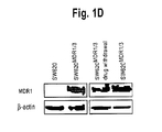
- MDR1 was highly upregulated at the protein level in cells propagated in the presence of AZD 1152 HQPA and persisted even after the selection pressure was removed ( Fig. 1C ).
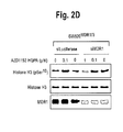
- the SW620 ABCB1/3 derivative required approximately 100-fold more AZD1152 HQPA to inhibit phosphorylation of the Aurora B substrate, histone H3, than the parental line ( Fig. 2A ).
- MDR1 is an ATP-dependent xenobiotic transporter
- AZD1152 HQPA may be eliminated by efflux from the intracellular compartment in SW620 ABCB1/3 , thus sparing histone H3 phosphorylation.
- Significantly less drug was measured in the cytosol of the resistant line compared to parental SW620 cells by LC-MS analysis ( Fig. 2B ).
- PSC-833 a small-molecule inhibitor of MDR1 (See, Girdler, F., et al., Chem.
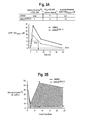
- the minimum intratumor concentration of AZD1152 HQPA necessary for inhibition of Aurora B was estimated by calculating the product of the intrinisic potency of AZD 1152 HQPA in SW620 or SW620 ABCB1/3 (0.02 or 2 ⁇ M, respectively) and the fold loss in potency of AZD 1152 HQPA when assayed in the presence of 50% (v/v) mouse plasma (the loss in potency is presumably due to plasma protein binding; data not shown). Based on this prediction, a minimum threshold concentration of AZD1152 HQPA of 0.1 or 10 ⁇ M must be achieved in SW620 or SW620 ABCB1/3 xenografts, respectively, to produce inhibition of histone H3 phosphorylation ( Fig. 3A , top panel).
- Tumor pharmacokinetics were assessed after a single IP administration of AZD1152 HQPA over a 24-hour period post-dose. This analysis demonstrated a reduction in the overall tumor AUC in SW620 ABCB1/3 xenografts compared to the parental cohort. Based on the aforementioned prediction, AZD1152 HQPA concentrations in the SW620 ABCB1/3 tumors exceeded the minimum threshold concentration for only a brief period (-6 hours), whereas in SW620 tumors, concentrations above threshold were achieved for at least 24 hours ( Fig. 3A , bottom panel). Correspondingly, only a transient inhibition of histone H3 phosphorylation was observed in SW620 ABCB1/3 compared to parental tumors.
- SW620 ABCB1/3 , HCT-15, and AsPC1 were significantly resistant to this compound (IC50s of 2, 1.4, and 2.2, .63 ⁇ M, respectively).
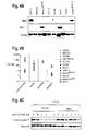
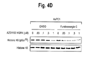
- the pan-Aurora kinase inhibitor, VX-680 exhibited a similar activity profile in the cell line panel, though the degree of resistance for SW620 ABCB1/3 , HCT-15, and AsPC1 was lower. As anticipated, SW620 ABCB1/3 and HCT-15, but not AsPC1 were relatively insensitive to the natural product, paclitaxel. No apparent loss in potency was observed for the Aurora A-selective compound, MLN8054. Importantly, the SW620 cell lines that were relatively sensitive to AZD1152 HQPA expressed no detectable ABCB4 (MDR3) by immunoblot analysis ( Fig. 4A and data not shown). It was confirmed that BRCP was required for resistance to AZD1152 HQPA in HCT-15 and AsPC1, respectively, using PSC-833 and fumitremorgin C ( Fig. 4C-D ).
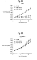
- HCT-15 colon carcinoma xenografts were unabated by treatment with either AZD1152 or VX-680 (Fig. 4E), whereas both therapies induced significant tumor growth inhibition in HCT116, an alternative colon carcinoma model ( Fig. 4D ), as well as in DoHH-2 B-cell lymphoma xenografts ( Fig. 4C ).
Abstract
Description
- This application claims the benefit of
U.S. Serial No. 61/148,957 filed on January 31, 2009 - The instant application contains a Sequence Listing which has been submitted via EFS-Web and is hereby incorporated by reference in its entirety. Said ASCII copy is named 9700.txt and is 79 kilobytes in size.
- The present invention relates to diagnostic assays useful in classification of patients for selection of cancer therapy with one or more Aurora kinase B inhibitors. In particular, the present invention relates to identifying the presence or absence of one or more copy number gains in the ABCB1 gene, the ABCB4 gene or combinations thereof, identifying patients eligible to receive Aurora kinase inhibitor therapy, either as monotherapy or as part of combination therapy, and monitoring patient response to such therapy.
- The Aurora kinase family is a group of highly related serine/threonine kinases that function as key regulators of mitosis. Three Aurora kinases are expressed in mammalian cells. These Aurora kinases are Aurora A, Aurora B, and Aurora C. Each of these Aurora kinases exhibits a different subcellular localization and plays a distinct role (See, Carmena M.E.W., Nat. Rev. Mol, Cell Biol., 4:842-854 (2003) and (Ducat, D.Z.Y,, Exp. Cell Res., 301:60-67 (2004)), Specifically, Aurora A localizes to spindle poles and has a crucial role in bipolar spindle formation (See, Marumoto, T.Z.D., et al., Nat. Rev. Cancer, 5:42-50 (2005)). Aurora B, a chromosome passenger protein, localizes to centromeres in early mitosis and then the spindle midzone in anaphase. Aurora B is required for mitotic histone H3 phosphorylation, chromosome biorientation, the spindle assembly checkpoint and cytokinesis (Andrews, P.D., et al., Curr. Opin. Cell Biol., 15:672-683 (2003)). Aurora C is also a chromosomal passenger protein and, in normal cells, its expression is restricted to the testis where it functions primarily in male gametogenesis. As the Aurora kinases serve essential functions in mitosis, considerable attention has been given to targeting this family of kinases for cancer therapy. Several small-molecule inhibitors have been developed including Hesperadin, ZM447439, VX-6801MK0457, AZD1152 and MLN8054 (See, Ditchfield, C, J.V., et al., J. Cell Biol., 161:267-280 (2003), Harrington, E.A., et al., Nat. Med., 10:262-267 (2004), Hauf, S., et al., J. Cell Biol., 161:281-294 (2003), Manfredi, M.G., et al., Proc. Natl. Acad. Sci., USA, 104:4106-4111 (2007)).
- AZD1152 is a novel acetanilide-substituted pyrazole-aminoquinazoline prodrug that is rapidly converted to the active drug, AZD1152 HQPA, in human plasma (See, Mortlock, A.A., et al., J. Med. Chem., 50:2213-2224 (2007)). AZD1152 HQPA is a highly potent and selective inhibitor of Aurora B (Ki of 0.36 nM) compared to Aurora A (Ki of 1369 nM) and is inactive against a panel of 50 other kinases. AZD1152 potently inhibits the growth of human colon, lung, and hematologic tumor xenografts in immunodeficient mice. Detailed pharmacodynamic analysis in SW620 colorectal tumor-bearing athymic rats treated intravenously with AZD 1152 revealed a temporal sequence of phenotypic events in tumors: transient suppression of histone H3 phosphorylation, accumulation of cells with 4n DNA, followed by an increase in the proportion of polyploid (>4n DNA) cells. Histologic analysis has shown aberrant cell division concurrent with an increase in apoptosis in AZD1152-treated tumors, namely, transient myelosuppression was observed secondary to inhibition of proliferation of the bone marrow, though this effect was fully reversible following cessation of AZD1152 treatment (See, Wilkinson, R.W., et al., Clin. Cancer Res., 13:3683-3688 (2007)).
- A major obstacle faced during cancer chemotherapy is the development of cross-resistance of tumors to cytotoxic agents, even to drugs to which the tumor cells were never exposed. This phenotype, known as multidrug resistance (MDR), is frequently observed following treatment with anticancer drugs. While the molecular basis for MDR is often complex, upregulation of members of the ATP-binding cassette (ABC) transporter superfamily has emerged as a core, cell-autonomous mechanism utilized by tumor cells to escape the activity of chemotherapeutic drugs that are pervasive among first- and second-line standards of care. As individuals that have failed previous chemotherapy are those most likely to receive newer, experimental medicines, MDR susceptibility represents a significant hurdle in drug development in oncology. The prototypical ABC transporter, multidrug resistance 1 (MDR1; also known as P-glycoprotein or P-gp; encoded by the gene ABCB1) is composed of two transmembrane domains and two nucleotide binding domains, which, through the hydrolysis of ATP, transports solutes against a concentration gradient into the extracellular space. Other ABC transporters, such as breast cancer resistance protein (BRCP, which is encoded by the gene ABCG2) are expressed as half-transporters and dimerize to yield a mature, functional unit. Although the contribution of BRCP to resistance to chemotherapy is not yet clear, upregulation of MDR1 has been consistently prognostic of failure of chemotherapy and poor survival in individuals with acute myelogenous leukemia (AML) or myelodysplastic syndrome (Pallis, M.R.N., Leukemia, 18:1927-1930 (2004) and van der Holt, B.L.B., et al., Blood, 106:2646-2654 (2005)). Furthermore, MDR1 has been associated with reduced response to chemotherapy in a meta-analysis of 31 breast cancer trials (See, Trock, B.J., et al., J. Natl. Cancer Inst., 89:917-931 (1997)). As a result, considerable effort has been invested in the development of substances that inhibit or modulate one or more ABC transporters. In fact, second- and third-generation inhibitors of this type are being evaluated as chemosensitizers in clinical trials (Bates, S.F., et al, Novartis Found. Symp., 83-96 (2002)).
- Although AZD1152 has shown desirable preclinical efficacy and is being evaluated in Phase I/II clinical trials in AML and solid tumors, the potential for development of resistance to AZD1152 has not been explored. Thus, there is a need in the art to identify the genes that confer tumor cell resistance to subjects being treated with Aurora kinase B inhibitors and to use the information obtained from these genes, such as the up regulation or down regulation of proteins enclosed by these genes, to develop diagnostic methods for determining or classifying whether a patient is eligible for treatment with an Aurora kinase B inhibitor and methods for monitoring patients suffering from cancer and being treated with one or more Aurora kinase B inhibitors for the development of drug resistance.
- In a first aspect, the present invention relates to a method of classifying a patient for eligibility for treatment with an Aurora kinase B inhibitor. The method comprises the steps of:
- a) providing a test sample from a patient;
- b) determining the presence or absence of a copy number gain for the
ABCB 1 genre at chromosome locus 7q21.1; and - c) classifying the patient as being eligible for receiving treatment with an Aurora kinase B inhibitor based on the presence or absence of a copy number gain for the
ABCB 1 gene at chromosome locus 7q21.1. - In a second aspect, the present invention relates to a method of classifying a patient for eligibility for treatment with an Aurora kinase B inhibitor. The method comprises the steps of:
- a) providing a test sample from a patient;
- b) determining the presence or absence of a copy number gain for the ABCB4 gene at chromosome locus 7q21.1; and
- c) classifying the patient as being eligible for receiving treatment with an Aurora kinase B inhibitor based on the presence or absence of a copy number gain in the ABCB4 gene at chromosome locus 7q21.1.
- In each of the above two aspects, the Aurora kinase B inhibitor can be AZD1152, ZM447439, VX-680/MK0457 or Hersperadin.
- In each of the above two aspects, test sample can comprise a tissue sample. Specifically, the tissue sample comprises a peripheral blood sample, a tumor tissue or a suspected tumor tissue, a thin layer cytological sample, a fine needle aspirate sample, a bone marrow sample, a lymph node sample, a urine sample, an ascites sample, a lavage sample, an esophageal brushing sample, a bladder or lung wash sample, a spinal fluid sample, a brain fluid sample, a ductal aspirate sample, a nipple discharge sample, a pleural effusion sample, a fresh frozen tissue sample, a paraffin embedded tissue sample or an extract or processed sample produced from any of a peripheral blood sample, a tumor tissue or a suspected tumor tissue, a thin layer cytological sample, a fine needle aspirate sample, a bone marrow sample, a urine sample, an ascites sample, a lavage sample, an esophageal brushing sample, a bladder or lung wash sample, a spinal fluid sample, a brain fluid sample, a ductal aspirate sample, a nipple discharge sample, a pleural effusion sample, a fresh frozen tissue sample or a paraffin embedded tissue sample.
- In each of the above two aspects, the determining step (b) can be performed by in situ hybridization. Specifically, the in situ hybridization can be performed with a nucleic acid probe that is fluorescently labeled. More specifically, the in situ hybridization can be performed with at least two nucleic acid probes. Alternatively, the in situ hybridization is performed with a peptide nucleic acid probe.
- Alternatively, in each of the above two aspects, the determining step (b) can be performed by polymerase chain reaction.
- Still further alternatively, the determining step (b) can be performed by a nucleic acid microarray assay.
- In each of the above two aspects, the cancer can be colorectal carcinoma or pancreatic carcinoma.
- In the first aspect, the presence of a copy number gain in the ABCB1 gene correlates with an increase in expression of the MDR1 polypeptide. In the second aspect, the presence of a copy number gain in the ABCB4 gene correlates with an increase in expression of the MDR3 polypeptide.
- In each of the above two aspects, the patient is being treated with an anti-sense agent designed to bind to at least one of the ABCB1 gene, the ABCB4 gene or a combination of the ABCB1 gene and ABCB4 gene.
- In each of the above two aspects, the patient can also optionally be treated with chemotherapy, radiation or combinations thereof.
- In a third aspect, the present invention relates to a method of monitoring a patient suffering from cancer and being treated with an Aurora kinase B inhibitor. The method comprises the steps of:
- a) providing a test sample from a patient suffering from cancer and currently being treated with at least one Aurora kinase B inhibitor;
- b) determining the presence or absence of a copy number gain for the
ABCB 1 gene at chromosome locus 7q21.1; - c) comparing the copy number of the ABCB1 gene in the test sample against a baseline level or a predetermined level; and
- d) determining whether the patient should continue to be treated with the Aurora kinase B inhibitor based on the comparison in step c).
- In a fourth aspect, the present invention relates to a method of monitoring a patient suffering from cancer and being treated with an Aurora kinase B inhibitor. The method comprises the steps of:
- a) providing a test sample from a patient suffering from cancer and currently being treated with at least one Aurora kinase B inhibitor;
- b) determining the presence or absence of a copy number gain for the ABCB4 gene at chromosome locus 7q21.1;
- c) comparing the copy number gain or absence for the ABCB4 gene in the test sample against a baseline level or a predetermined level; and
- d) determining whether the patient should continue to be treated with the Aurora kinase B inhibitor based on the comparison in step c).
- In each of the above two aspects, the Aurora kinase B inhibitor can be AZD1152, ZM447439, VX-680/MK0457 or Hersperadin.
- In each of the above two aspects, test sample can comprise a tissue sample. Specifically, the tissue sample comprises a peripheral blood sample, a tumor tissue or a suspected tumor tissue, a thin layer cytological sample, a fine needle aspirate sample, a bone marrow sample, a lymph node sample, a urine sample, an ascites sample, a lavage sample, an esophageal brushing sample, a bladder or lung wash sample, a spinal fluid sample, a brain fluid sample, a ductal aspirate sample, a nipple discharge sample, a pleural effusion sample, a fresh frozen tissue sample, a paraffin embedded tissue sample or an extract or processed sample produced from any of a peripheral blood sample, a tumor tissue or a suspected tumor tissue, a thin layer cytological sample, a fine needle aspirate sample, a bone marrow sample, a urine sample, an ascites sample, a lavage sample, an esophageal brushing sample, a bladder or lung wash sample, a spinal fluid sample, a brain fluid sample, a ductal aspirate sample, a nipple discharge sample, a pleural effusion sample, a fresh frozen tissue sample or a paraffin embedded tissue sample.
- In each of the above two aspects, the determining step (b) can be performed by in situ hybridization. Specifically, the in situ hybridization can be performed with a nucleic acid probe that is fluorescently labeled. More specifically, the in situ hybridization can be performed with at least two nucleic acid probes. Alternatively, the in situ hybridization is performed with a peptide nucleic acid probe.
- Alternatively, in each of the above two aspects, the determining step (b) can be performed by polymerase chain reaction.
- Still further alternatively, the determining step (b) can be performed by a nucleic acid microarray assay.
- In each of the above two aspects, the cancer can be colorectal carcinoma or pancreatic carcinoma.
- In the third aspect, the presence of a copy number gain in the ABCB1 gene correlates with an increase in expression of the MDR1 polypeptide. In the fourth aspect, the presence of a copy number gain in the ABCB4 gene correlates with an increase in expression of the MDR3 polypeptide.
- In each of the above two aspects, the patient is being treated with an anti-sense agent designed to bind to at least one of the ABCB1 gene, the ABCB4 gene or a combination of the ABCB1 gene and ABCB4 gene.
- In each of the above two aspects, the patient can also optionally be treated with chemotherapy, radiation or combinations thereof.
- In a fifth aspect, the present invention relates to a method of classifying a patient having a cancer that is resistant to treatment with an Aurora kinase B inhibitor. The method comprises the steps of:
- a) providing a test sample from a patient;
- b) determining the presence or absence of a copy number gain for the
ABCB 1 gene at chromosome locus 7q21.1; - c) comparing the presence or absence of the copy number gain for the ABCB1 gene in the test sample against a baseline level or a predetermined level; and
- d) classifying the patient as having a cancer that is resistant to Aurora kinase B inhibitor treatment on (i) the presence of a copy number gain in the ABCB1 gene at chromosome locus 7q21.1; and (ii) if the copy number gain in the test sample is higher then the baseline level or the predetermined level.
- In a sixth aspect, the present invention relates to a method of classifying a patient having a cancer that is resistant to treatment with an Aurora kinase B inhibitor. The method comprises the steps of:
- a) providing a test sample from a patient;
- b) determining the presence or absence of a copy number gain for the ABCB4 gene at chromosome locus 7q21.1;
- c) comparing the presence or absence of the copy number gain for the ABCB4 gene in the test sample against a baseline level or a predetermined level; and
- d) classifying the patient as having a cancer that is resistant to Aurora kinase B inhibitor treatment on (i) the presence of a copy number gain in the ABCB4 gene at chromosome locus 7q21.1; and (ii) if the copy number gain in the test sample is higher then the baseline level or the predetermined level.
- In each of the above two aspects, the Aurora kinase B inhibitor can be AZD1152, ZM447439, VX-680/MK0457 or Hersperadin.
- In each of the above two aspects, test sample can comprise a tissue sample. Specifically, the tissue sample comprises a peripheral blood sample, a tumor tissue or a suspected tumor tissue, a thin layer cytological sample, a fine needle aspirate sample, a bone marrow sample, a lymph node sample, a urine sample, an ascites sample, a lavage sample, an esophageal brushing sample, a bladder or lung wash sample, a spinal fluid sample, a brain fluid sample, a ductal aspirate sample, a nipple discharge sample, a pleural effusion sample, a fresh frozen tissue sample, a paraffin embedded tissue sample or an extract or processed sample produced from any of a peripheral blood sample, a tumor tissue or a suspected tumor tissue, a thin layer cytological sample, a fine needle aspirate sample, a bone marrow sample, a urine sample, an ascites sample, a lavage sample, an esophageal brushing sample, a bladder or lung wash sample, a spinal fluid sample, a brain fluid sample, a ductal aspirate sample, a nipple discharge sample, a pleural effusion sample, a fresh frozen tissue sample or a paraffin embedded tissue sample.
- In each of the above two aspects, the determining step (b) can be performed by in situ hybridization. Specifically, the in situ hybridization can be performed with a nucleic acid probe that is fluorescently labeled. More specifically, the in situ hybridization can be performed with at least two nucleic acid probes. Alternatively, the in situ hybridization is performed with a peptide nucleic acid probe.
- Alternatively, in each of the above two aspects, the determining step (b) can be performed by polymerase chain reaction.
- Still further alternatively, the determining step (b) can be performed by a nucleic acid microarray assay.
- In each of the above two aspects, the cancer can be colorectal carcinoma or pancreatic carcinoma.
- In the fifth aspect, the presence of a copy number gain in the ABCB1 gene correlates with an increase in expression of the MDR1 polypeptide. In the sixth aspect, the presence of a copy number gain in the ABCB4 gene correlates with an increase in expression of the MDR3 polypeptide.
- In each of the above two aspects, the patient is being treated with an anti-sense agent designed to bind to at least one of the ABCB1 gene, the ABCB4 gene or a combination of the ABCB1 gene and ABCB4 gene.
- In each of the above two aspects, the patient can also optionally be treated with chemotherapy, radiation or combinations thereof.
- In a seventh aspect, the present invention relates to a kit comprising:
- (a) reagents for determining the presence or absence of a copy number gain for the ABCB1 gene;
- (b) instructions for performing the test.
- In the above kit, the reagents to determine the presence or absence of a copy number gain comprise detectably-labeled polynucleotides that hybridize to at least a portion of the
ABCB 1 gene. - In an eighth embodiment, the present invention relates to a kit comprising:
- (a) reagents for determining the presence or absence of a copy number gain for the ABCB4 gene;
- (b) instructions for performing the test.
- In the above kit, the reagents to determine the presence or absence of a copy number gain comprise detectably-labeled polynucleotides that hybridize to at least a portion of the ABCB4 gene.
-
-
Figure 1 shows the identification of ABCB1 and ABCB4 as genes amplified and overexpressed in an SW620 derivative selected for resistance to AZD1152 HQPA as described in the Example. Specifically,Figure 1A shows the copy number of ABCB1 and ABCB4 determined by CGH using Affymetrix 100K SNP chips in parental SW620 cells, SW620ABCB1/3 cells, and SW620ABCB1/3 cells after 3 months in culture in drug-free medium. The vertical line indicates the position of the ABCB1 locus and the horizontal line indicates the normal DNA copy number (two copies).Figure 1B shows mRNA expression values for ABCB1 and ABCB4 compared to other solute transporters. The expression levels of ABCB1 (encoding MDR1) and ABCB4 (encoding MDR3) in SW620ABCB1/3 are indicated by arrows.Figure 1C shows the mRNA expression values for over 14,000 genes plus ESTs (-22,000 probe sets) determined using Affymetrix HG-U133A GeneChips. Data are presented as the fold change in gene expression for SW620ABCB1/3 cells compared to the parental SW620 cells compared to all genes whose expression increases 10-fold or greater,Figure 1D shows the relative expression of the MDR1 protein was determined by immunoblot analysis. β-actin was used as a loading control. -
Figure 2 shows that the inhibition of ABCB1 reverses resistance to AZD 1152 HQPA in the SW620ABCB1/3 derivative.Figure 2A shows SW620, SW620ABCB1/3, and SW620ABCB1/3 cells after 3 months in culture in drug-free medium that were treated with AZD1152 HQPA in dose response for 90 minutes. Phosphorylation of histone H3 at Ser10 was determined by immunoblot analysis.Figure 2B shows SW620 or SW620ABCB1/3 cells treated with 1 µM AZD1152 HQPA for 4 hours. Cells were fractionated, and the AZD1152 HQPA concentration was determined by LC-MS analysis in the respective sample fraction.Figure 2C shows SW620ABCB1/3 cells treated for 2 hours with either DMSO or 1 µM PSC-833 prior to AZD1152 HQPA in dose response for 90 minutes. Phosphorylation of histone H3 was then determined by immunoblotting.Figure 2D shows the effect of ABCB1 knockdown in the SW620ABCB1/3 derivative was assessed by transfecting either Luciferase (siLuciferase) or ABCB1 (siABCB1) siRNAs followed by treatment of the transfected cells with AZD1152 HQPA for 90 minutes. Immunoblot analysis of ABCB1 indicated that protein levels were reduced by about 75% with siABCB1 compared to siLuciferase. -
Figure 3 shows the relationship of pharmacokinetics, pharmacodynamics, and efficacy of AZD1152 HQPA in SW620 vs. SW620ABCB1/3 xenografts.Figure 3A (top panel) shows the projected threshold intratumor concentration required to inhibit xenograft histone H3 phosphorylation estimated by calculating the product of the intrinsic potency of AZD1152 HQPA in an assay of histone H3 phosphorylation and the fold reduction in potency of AZD1152 HQPA when assayed in the presence of 50% (vv-1) mouse plasma. The bottom panel shows the intratumor pharmacokinetics of AZD 1152 HQPA determined at 0, 2, 8 and 24 hours post-dose after a single intraperitoneal (i.p.) injection of 100 mg kg-1.Figure 3B shows mice bearing established SW620 and SW620ABCB1/3 tumor xenografts given a single dose of AZD1152 HQPA (100 mg kg-1, i.p.), and three tumors per time point were harvested. Tumors were extracted and phospho-histone H3 levels were determined by immunoblotting in SW620 tumor (light blue) and SW620ABCB1/3 tumor (dark blue) following treatment with AZD1152. Immunoblots were quantified, and the data expressed as the area under the curve. The mean values from individual time points are present ±s.e.m.Figure 3C and Figure 3D show SW620 and SW620ABCB1/3 cells injected subcutaneously into scid-bg mice as described in Example 1. Tumors were size-matched at approximately 500 mm3, and treatment with AZD1152 was initiated on Day 7 post-inoculation. AZD1152 was administered in a q2d schedule at doses of 50 or 100 mg/kg/day by i.p. injection for 2 weeks. Each point represents the mean ± s.d. of 10 tumors. -
Figure 4 shows that cell lines which overexpress ABCB1 are resistant to AZD1152 HQPA and VX-680/MK0457 in vitro.Figure 4A presents immunoblotting of cell lines used in xenograft studies showing the relative expression of ABCB1.Figure 4B shows a panel of cell lines that were evaluated for relative sensitivity to AZD1152 HQPA, VX-680/MK0457, MLN8054, and paclitaxel in 7-day colony formation (adherent lines: SW620, SW620ABCB1/3, HCT-15, AsPC1) or viability (non-adherent lines: RS; 411 and DoHH-2). Cells were treated in dose response to determine IC50s.Figure 4C shows HCT-15 cells that were treated with DMSO or 1 µM PSC-833 for 1 hour prior to the addition of AZD1152 HQPA at the indicated concentrations for an additional hour. Total and phospho-(Ser10)-histone H3 was determined by immunoblotting.Figure 4D shows AsPC1 cells that were treated with DMSO or 10 µM fumitremorgin C for 1 hour followed by AZD1152 HQPA as described inFigure 4C . - The present invention provides methods and compositions for monitoring cancer and tumor cells for resistance to Aurora kinase B inhibitor therapy. The inventors discovered that the presence of a copy number gain for (i) the ABCB1 gene at chromosome locus 7q21.1; (ii) the ABCB4 gene at chromosome locus 7q21.1; or (iii) each of the ABCB1 gene and the ABCB4 gene at chromosome locus 7q21.1 is associated with resistance to therapy with an Aurora kinase B inhibitor.
- The inventors discovered the copy number gains described above using a microarray-based comparative genomic hybridization technique to detect gene copy number abnormalities (e.g, copy number gain and copy number loss) on a genome-wide scale, thus providing a whole-genome view of chromosomal aberrations accompanied by a change in the DNA copy number. This method is fully disclosed in METHODS FOR ASSEMBLING PANELS OF CANCER CELL LINES FOR USE IN TESTING THE EFFICACY OF ONE OR MORE PHARMACEUTICAL COMPOSITIONS, filed October 31, 2008 and assigned
U.S. Serial No. 61/110,281 - The invention provides diagnostic assays for identifying, classifying and monitoring cancer patients which comprises assessing a test sample for the presence or absence of a copy number gain for (i) the ABCB1 gene; (ii) the ABCB4 gene; or (iii) each of the ABCB1 gene and the ABCB4 gene. The inventive assays include assay methods for identifying patients eligible to receive Aurora kinase B therapy (as either a monotherapy or as part of a combination therapy (e.g., such as with chemotherapy, radiation or combinations thereof) and for monitoring patient response to such therapy. The invention comprises, for example, determining by fluorescent in situ hybridization the presence or absence of a copy number gain for (i) the ABCB1 gene; (ii) the ABCB4 gene; or (iii) each of the
ABCB 1 gene and the ABCB4 gene. Patients classified as having an increase in copy number gain for the (i) the ABCB1 gene; (ii) the ABCB4 gene; or (iii) each of the ABCB1 gene and the ABCB4 gene are ineligible to receive Aurora kinase B therapy at least as a monotherapy because they are less likely to respond to this therapy. In addition, patients having this amplification can be resistant to other cancer therapies. Thus, determination of the presence of a copy number gain for (i) the ABCB1 gene; (ii) the ABCB4 gene; or (iii) each of the ABCB1 gene and the ABCB4 gene in cancer and tumor cells is useful as a general therapy stratification marker. - In one embodiment, the invention comprises a method for identifying or classifying a patient as eligible for treatment with an Aurora kinase B inhibitor (as either a monotherapy or part of a combination therapy), the method comprising the steps of:
- (a) providing a tissue sample from a patient;
- (b) determining the presence or absence of a copy number gain for (i) a ABCB1 gene; (ii) a ABCB4 gene; or (iii) a ABCB1 gene and a ABCB4 gene; and
- (c) classifying the patient as being eligible for treatment with an Aurora kinase B inhibitor based on the absence of a copy number gain for (i) a ABCB1 gene; (ii) a ABCB4 gene; or (iii) a
ABCB 1 gene and a ABCB4 gene. In the above method, a patient would be ineligible for treatment with an Aurora kinase B inhibitor (at least as a monotherapy) based on the presence of a copy number gain for (i) a ABCB1 I gene; (ii) a ABCB4 gene; or (iii) a ABCB1 gene and a ABCB4 gene. The patient from whom the test sample is obtained can be a patient suspected of or diagnosed with cancer. Moreover, the inventors found that a copy number gain in the ABCB1 gene correlates with an increase in expression of the MDR1 polypeptide and that a copy number gain in the ABCB4 gene correlates with an increase in expression of the MDR3 polypeptide. - In this embodiment, the cancer can be any type of cancer, such as colorectal carcinoma or pancreatic cancer. Moreover, in this embodiment, the gene amplification can be determined by a multi-color fluorescent in situ hybridization (FISH) assay, for example, performed on a lung cancer tumor biopsy sample. In other embodiments, the quantitative polymerase chain reaction (Q-PCR) method is used.
- In yet another embodiment, the invention comprises a method for identifying or classifying a patient having a cancer that is resistant to therapy with an Aurora kinase B inhibitor, the method comprising the steps of:
- (a) providing a test sample (e.g., such as a tissue sample) from a patient;
- (b) determining the presence or absence of a copy number gain for (i) a ABCB1 gene; (ii) a ABCB4 gene; or (iii) a ABCB1 gene and a ABCB4 gene; and
- (c) classifying the patient as having a cancer that is resistant to Aurora kinase B inhibitor based on the presence of a copy number gain for (i) a ABCB1 gene; (ii) a ABCB4 gene; or (iii) a ABCB1 gene and a ABCB4 gene.
- In this embodiment, the cancer can be any type of cancer, such as colorectal carcinoma or pancreatic cancer. Moreover, in this embodiment, the gene amplification can be determined by a multi-color fluorescent in situ hybridization (FISH) assay, for example, performed on a lung cancer tumor biopsy sample. In other embodiments, the polymerase chain reaction (PCR) is used.
- In still yet another embodiment, the invention is directed to methods for monitoring a patient being treated with an Aurora kinase B inhibitor, the method comprising the steps of:
- (a) providing a test sample from a cancer patient being treated with at least one Aurora kinase inhibitor (optionally, tumor or cancer cells obtained from a tissue sample can be identified or extracted);
- (b) determining in the test sample (for example, in the tumor or cancer cells) the presence or absence of a copy number gain for (i) a
ABCB 1 gene; (ii) a ABCB4 gene; or (iii) aABCB 1 gene and a ABCB4 gene; and - (c) comparing the copy number gain for (i) the ABCB1 gene; (ii) the ABCB4 gene; or (iii) the
ABCB 1 gene and the ABCB4 gene from the test sample (such as in the tumor or cancer cells) against a baseline level or a predetermined level; and - (d) determining whether the patient should continue to be treated with the Aurora kinase B inhibitor based on the comparison in step (c). Specifically, if the test sample (e.g., the tumor or cancer cells) having a copy number gain for (i) the ABCB1 gene; (ii) a ABCB4 gene; or (iii) a ABCB1 gene and a ABCB4 gene is the same as or higher then the baseline level or predetermined level, then treatment with the Aurora kinase B inhibitor can be discontinued, stopped or terminated (if it is being used solely as a monotherapy). Alternatively, the treating physician may decide to combine the Aurora kinase B inhibitor with at least a second therapy (for example, treatment with a second small molecule) as a combination therapy. However, if the copy number gain for (i) the ABCB1 gene; (ii) a ABCB4 gene; or (iii) a
ABCB 1 gene and a ABCB4 gene obtained from the test sample (e.g., the tumor or cancer cells) is less then the baseline level or the predetermined level or if no copy number gain for (i) theABCB 1 gene; (ii) a ABCB4 gene; or (iii) a ABCB1 gene and a ABCB4 gene is detected, then treatment with the Aurora kinase B inhibitor can be continued. Again, depending on the results obtained with said treatment, the treating physician may decide to combine the Aurora kinase B inhibitor with at least a second therapy (for example, treatment with a second small molecule) as a combination therapy. - Again, FISH and PCR methods can be used to detect the presence or absence of a copy number gain for (i) the ABCB1 gene; (ii) the ABCB4 gene; or (iii) the ABCB1 gene and the ABCB4 gene in a test sample obtained from a patient.
- The invention is also directed to kits that package, for example, oligo- or polynucleotide engineered to be used as PCR primers, FISH probes, etc.
- The invention has significant capability to provide improved stratification of patients for cancer therapy, and in particular for Aurora kinase B inhibitor therapy. The assessment of these biomarkers with the invention also allows tracking of individual patient response to the therapy.
- Section headings as used in this section and the entire disclosure herein are not intended to be limiting.
- As used herein, the singular forms "a," "an" and "the" include plural referents unless the context clearly dictates otherwise. For the recitation of numeric ranges herein, each intervening number there between with the same degree of precision is explicitly contemplated. For example, for the range 6-9, the
numbers 7 and 8 are contemplated in addition to 6 and 9, and for the range 6.0-7.0, the numbers 6.0, 6.1, 6.2, 6.3, 6.4, 6.5, 6.6, 6.7, 6.8, 6.9 and 7.0 are explicitly contemplated. - An "Aurora kinase B inhibitor" refers to a therapeutic compound of any type (e.g., non-selective or selective), including small molecule-, antibody-, antisense-, small interfering RNA, or microRNA-based compounds, that binds to at least one of Aurora kinase B or Aurora B, and antagonizes the activity of the Aurora kinase B or Aurora B related nucleic acid or protein. For example, a number of Aurora kinase B inhibitors are known to inhibit at least one of histone H3 phosphorylation or cell division. In addition, a number of Aurora kinase B inhibitors are known to induce apoptosis in at least one cell system (such as an acute myeloid leukemia cell line, a primary acute myeloid leukemia culture, etc.) The methods of the present invention are useful with any known or hereafter developed Aurora kinase B inhibitor. Examples of an Aurora kinase B inhibitor are AZD1152, ZM447439, VX-680/MK0457 and Hesperadin.
- AZD1152, also known as, 2-[[3-({4-[(5-{2-[(3-Fluorophenyl)amino]-2-oxoethyl}-1H-pyrazol-3-yl)amino]-quinazolin-7-yl}oxy)propyl](ethyl)amino]ethyl dihydrogen phosphate, is a prodrug of a pyrazoloquinazoline Aurora kinase inhibitor (AZD1152-hydroxyquinazolien pyrazol anilide (HQPA)) and is converted rapidly to the active AZD 1152-HQPA in plasma (See, Mortlock, AA, et al., J. Med. Chem., 50:2213-24 (2007)). AZD 1152-HQPA is a highly potent and selective inhibitor of Aurora B.
- ZM447439, also known as 4-(4-(N-benzoylamino)anilino)-6-methoxy-7-(3-(1-morpholino)propoxy)quinazoline, is a quinazoline derivative, inhibits Aurora A and Aurora B. The chemical structure of ZM447439 is provided in Ditchfield, C., et al., J. Cell Bio., 161(2):267-280 (2003) and Montembault, E., et al., Drugs of the Future, 30(1):1-9 (2005).
- VX-680/MK0457 is a cyclopropane carboxylic acid of {4-[4-(4-methyl-piperazin-1-yl)-6-(5-methyl-2H-pyrazol-3-ylamino)-pyrimidin-2-ylsulphanyl]-phenyl}-amide and inhibits Aurora A, Aurora B and Aurora C. The chemical structure of VX-6801MK0457 is provided in Montembault, E., et al., Drugs of the Future, 30(1):1-9 (2005).
- Hesperadin, an indolinone, inhibits Aurora B. The chemical structure of Hesperadin is provided in Hauf, S., et al., J. Cell Bio., 161(2):281-294 (2003) and Montembault, E., et al., Drugs of the Future, 30(1):1-9 (2005).
- "Consisting essentially of a polynucleotide having a % sequence identity" means that the polynucleotide does not substantially differ in length, but may differ substantially in sequence. Thus, a polynucleotide "A" consisting essentially of a polynucleotide having at least 80% sequence identity to a known sequence "B" of 100 nucleotides means that polynucleotide "A" is about 100 nucleotides (nts) long, but up to 20 nts can vary from the "B" sequence. The polynucleotide sequence in question can be longer or shorter due to modification of the termini, such as, for example, the addition of 1-15 nucleotides to produce specific types of probes, primers and other molecular tools, etc., such as the case of when substantially non-identical sequences are added to create intended secondary structures. Such non-identical nucleotides are not considered in the calculation of sequence identity when the sequence is modified by "consisting essentially of."
- "Expression" refers to the production of a functional end-product. Expression of a gene involves transcription of the gene and translation of the mRNA into a precursor or mature protein. "Antisense inhibition" refers to the production of antisense RNA transcripts capable of suppressing the expression of the target protein. "Co-suppression" refers to the production of sense RNA transcripts capable of suppressing the expression of identical or substantially similar foreign or endogenous genes (
U.S. Patent No. 5,231,020 ). - As used herein, the term "isolated" in the context of nucleic acid molecules or polynucleotides refers to a nucleic acid molecule or polynucleotide which is separated from other nucleic acid molecules or polynucleotides which arc present in the natural source of the nucleic acid molecule or polynucleotide. Moreover, an "isolated" nucleic acid molecule or polynucleotide, such as a cDNA molecule, can be substantially free of other cellular material, or culture medium when produced by recombinant techniques, or substantially free of chemical precursors or other chemicals when chemically synthesized. In one aspect, nucleic acid molecules or polynucleotides are isolated.
- "Gene" refers to a nucleic acid fragment that expresses a specific protein, including regulatory sequences preceding (5' non-coding sequences) and following (3' non-coding sequences) the coding sequence.
- "Native gene" refers to a gene as found in nature with its own regulatory sequences. In contrast, "chimeric construct" refers to a combination of nucleic acid fragments that are not normally found together in nature. Accordingly, a chimeric construct may comprise regulatory sequences and coding sequences that are derived from different sources, or regulatory sequences and coding sequences derived from the same source, but arranged in a manner different than that normally found in nature.
- "Percent (%) nucleic acid sequence identity" with respect to nucleic acid sequences is defined as the percentage of nucleotides in a candidate sequence that are identical with the nucleotides in the sequence of interest, after aligning the sequences and introducing gaps, if necessary, to achieve the maximum percent sequence identity. Alignment for purposes of determining % nucleic acid sequence identity can be achieved in various ways that are within the skill in the art, for instance, using publicly available computer software such as BLAST, BLAST-2, ALIGN or Megalign (DNASTAR) software. Those skilled in the art can determine appropriate parameters for measuring alignment, including any algorithms needed to achieve maximal alignment over the full length of the sequences being compared.
- When nucleotide sequences are aligned, the % nucleic acid sequence identity of a given nucleic acid sequence C to, with, or against a given nucleic acid sequence D (which can alternatively be phrased as a given nucleic acid sequence C that has or comprises a certain % nucleic acid sequence identity to, with, or against a given nucleic acid sequence D) can be calculated as follows:

where
W is the number of nucleotides scored as identical matches by the sequence alignment program's or algorithm's alignment of C and D
and
Z is the total number of nucleotides in D. - When the length of nucleic acid sequence C is not equal to the length of nucleic acid sequence D, the % nucleic acid sequence identity of C to D will not equal the % nucleic acid sequence identity of D to C.
- "Polymerase Chain Reaction" or "PCR" is a technique for the synthesis of large quantities of specific DNA segments, consists of a series of repetitive cycles (Perkin Elmer Cetus Instruments, Norwalk, CT). Typically, the double stranded DNA is heat-denatured, the two primers complementary to the 3' boundaries of the target segment are annealed at low temperature and then extended at an intermediate temperature. One set of these three consecutive steps is referred to as a cycle.
- PCR is a powerful technique used to amplify DNA millions of fold, by repeated replication of a template, in a short period of time. ((Mullis, K., et al., Cold Spring Harb Symp Quant Biol. 51 Pt 1:263-73 (1986)); European Patent Application No.
50,424 84,796 258,017 237,362 201,184 U.S. Patent No. 4,683,202 ;U.S. Patent No. 4,582,788 ; andU.S. Patent No. 4,683,194 ). The process uses sets of specific in vitro synthesized oligonucleotides to prime DNA synthesis. The design of the primers is dependent upon the sequences of DNA that are to be analyzed. The technique is carried out through many cycles (usually 20-50) of melting the template at high temperature, allowing the primers to anneal to complementary sequences within the template and then replicating the template with DNA polymerase. - The products of PCR reactions can be analyzed by separation in agarose gels followed by ethidium bromide staining and visualization with UV transillumination. Alternatively, radioactive dNTPs can be added to the PCR in order to incorporate label into the products. In this case the products of PCR are visualized by exposure of the gel to x-ray film. The added advantage of radiolabeling PCR products is that the levels of individual amplification products can be quantitated.
- A "polynucleotide" is a nucleic acid polymer of ribonucleic acid (RNA), deoxyribonucleic acid (DNA), modified RNA or DNA, or RNA or DNA mimetics (such as PNAs), and derivatives thereof, and homologues thereof. Thus, polynucleotides include polymers composed of naturally occurring nucleic bases, sugars and covalent inter-nucleoside (backbone) linkages as well as polymers having non-naturally-occurring portions that function similarly. Such modified or substituted nucleic acid polymers are well known in the art and are referred to as "analogues." Oligonucleotides are generally short polynucleotides from about 10 to up to about 160 or 200 nucleotides.
- Polynucleotides also comprise primers that specifically hybridize to target sequences, including analogues and/or derivatives of the nucleic acid sequences, and homologues thereof,
- Polynucleotides can be prepared by conventional techniques, such as solid-phase synthesis using commercially available equipment, such as that available from Applied Biosystems USA Inc. (Foster City, CA; USA), DuPont, (Wilmington, DE; USA), or Milligen (Bedford, MA; USA). Modified polynucleotides, such as phosphorothioates and alkylated derivatives, can also be readily prepared by similar methods known in the art (See,
U.S. Patent Nos. 4,948,882 ,5,464,746 , and5,424,414 ). - As used herein, the term "polynucleotide analogues" refers to polymers having modified backbones or non-natural inter-nucleoside linkages, Modified backbones include those retaining a phosphorus atom in the backbone, such as phosphorothioates, chiral phosphorothioates, phosphorodithioates, phosphotriesters, aminoalkylphosphotriesters, methyl and other alkyl phosphonates, as well as those no longer having a phosphorus atom, such as backbones formed by short chain alkyl or cycloalkyl inter-nucleoside linkages, mixed heteroatom and alkyl or cycloalkyl inter-nucleoside linkages, or one or more short chain heteroatomic or heterocyclic inter-nucleoside linkages. Modified nucleic acid polymers (analogues) can contain one or more modified sugar moieties.
- Analogs that are RNA or DNA mimetics, in which both the sugar and the inter-nucleoside linkage of the nucleotide units are replaced with novel groups, are also useful. In these mimetics, the base units are maintained for hybridization with the target sequence. An example of such a mimetic, which has been shown to have excellent hybridization properties, is a peptide nucleic acid (PNA) (See, Buchardt, O., P. Nielsen, and R. Berg. 1992. Peptide Nucleic Acids ).
- As used herein, the term "predetermined level" refers generally at an assay cut-off value that is used to assess diagnostic results by comparing the assay results against the predetermined level, and where the predetermined level already that has been linked or associated with various clinical parameters (e.g., assessing risk, severity of disease, progression/non-progression/improvement, determining the age of a test sample, determining whether a test sample (e.g., serum or plasma) has hemolyzed, etc.). The present invention provides exemplary predetermined levels, and describes the initial linkage or association of such levels with clinical parameters for exemplary assays as described herein. However, it is well known that cutoff values may vary dependent on the nature of the assay. It further is well within the ordinary skill of one in the art to adapt the invention herein for other assays to obtain assay-specific cut-off values for those other assays based on this description.
- A "probe" or "primer" as used herein is a polynucleotide that is at least 8 nucleotides in length and forms a hybrid structure with a target sequence, due to complementarity of at least one sequence in the probe or primer with a sequence in the target region. The polynucleotide regions of the probe can be composed of DNA and/or RNA and/or synthetic nucleotide analogs. Preferably, the probe does not contain a sequence that is complementary to the sequence or sequences used to prime for a target sequence during the polymerase chain reaction.
- "Recombinant" refers to an artificial combination of two otherwise separated segments of sequence, e.g., by chemical synthesis or by the manipulation of isolated segments of nucleic acids by genetic engineering techniques.
- "Specifically hybridize" refers to the ability of a nucleic acid to bind detectably and specifically to a second nucleic acid. Polynucleotides specifically hybridize with target nucleic acid strands under hybridization and wash conditions that minimize appreciable amounts of detectable binding by non-specific nucleic acids.
- The specificity of single stranded DNA to hybridize complementary fragments is determined by the stringency of the reaction conditions. Hybridization stringency increases as the propensity to form DNA duplexes decreases. In nucleic acid hybridization reactions, the stringency can be chosen to favor specific hybridizations (high stringency). Less-specific hybridizations (low stringency) can be used to identify related, but not exact, DNA molecules (homologous, but not identical) or segments.
- DNA duplexes are stabilized by: (1) the number of complementary base pairs, (2) the type of base pairs, (3) salt concentration (ionic strength) of the reaction mixture, (4) the temperature of the reaction, and (5) the presence of certain organic solvents, such as formamide, which decrease DNA duplex stability. A common approach is to vary the temperature: higher relative temperatures result in more stringent reaction conditions (See, Ausubel, F.M., R. Brent, R.E. Kingston, et al. 1987. Current Protocols in Molecular Biology. John Wiley & Sons, New York) provide an excellent explanation of stringency of hybridization reactions.
- Hybridization under "stringent conditions" means hybridization protocols in which nucleotide sequences at least 60% homologous to each other remain hybridized. Polynucleotides can include other appended groups such as peptides (e.g., for targeting host cell receptors in vivo), or agents facilitating transport across the cell membrane. In addition, oligonucleotides can be modified with hybridization-triggered cleavage agents (See, van der Krol et al., Biotechniques. 6:958-76 (1988) or intercalculating agents (Zon, G., Pharm Res. 5:539-49 (1988)). The oligonucleotide can be conjugated to another molecule, e.g., a peptide, a hybridization triggered cross-linking agent, a transport agent, a hybridization-triggered cleavage agent, and the like.
- As used herein, the terms "subject" and "patient" are used interchangeably irrespective of whether the subject has or is currently undergoing any form of treatment. As used herein, the terms "subject" and "subjects" refer to any vertebrate, including, but not limited to, a mammal (e.g., cow, pig, camel, Ilama, horse, goat, rabbit, sheep, hamsters, guinea pig, cat, dog, rat, and mouse, a non-human primate (for example, a monkey, such as a cynomolgous monkey, chimpanzee, etc) and a human). Preferably, the subject is a human. Subjects or patients can be living or expired.
- "Target sequence" or "target nucleic acid sequence" means a nucleic acid sequence encompassing, for example, a gene, or complements or fragments thereof, that is amplified, detected, or both using a polynucleotide primer or probe. Additionally, while the term target sequence sometimes refers to a double stranded nucleic acid sequence; a target sequence can also be single-stranded. In cases where the target is double-stranded, polynucleotide primer sequences preferably amplify both strands of the target sequence. A target sequence can be selected that is more or less specific for a particular organism. For example, the target sequence can be specific to an entire genus, to more than one genus, to a species or subspecies, serogroup, auxotype, serotype, strain, isolate or other subset of organisms.
- "Test sample" means a sample taken from a subject, or a biological fluid, wherein the sample may contain a target sequence. A test sample can be taken from any source, for example, tissue, blood, saliva, sputa, mucus, sweat, urine, urethral swabs, cervical swabs, urogenital or anal swabs, conjunctival swabs, ocular lens fluid, cerebral spinal fluid, etc. A test sample can be used (i) directly as obtained from the source; or (ii) following a pre-treatment to modify the character of the sample. Thus, a test sample can be pre-treated prior to use by, for example, preparing plasma or serum from blood, disrupting cells or viral particles, preparing liquids from solid materials, diluting viscous fluids, filtering liquids, adding reagents, purifying nucleic acids, etc.
- The terms "treat", "treating" or "treatment" as used herein refer to administering one or more active agents or compounds to a subject in an effort to (i) prevent a pathologic condition from occurring (e.g. prophylaxis); (ii) inhibit the pathologic condition or arrest its development; (iii) relieve a pathologic condition and/or prevent or reduce the severity one or more symptoms associated with such a pathologic condition, regardless of whether any of items (i) through (iii) are successful in a subject.
- A "variant polynucleotide" or a "variant nucleic acid sequence" means a polynucleotide having at least about 60% nucleic acid sequence identity, more preferably at least about 61%, 62%, 63%, 64%, 65%, 66%, 67%, 68%, 69%, 70%, 71%, 72%, 73%, 74%, 75%, 76%, 77%, 78%, 79%, 80%, 81%, 82%, 83%, 84%, 85%, 86%, 87%, 88%, 89%, 90%, 91%, 92%, 93%, 94%, 95%, 96%, 97%, 98% nucleic acid sequence identity and yet more preferably at least about 99% nucleic acid sequence identity with a given nucleic acid sequence. Variants do not encompass the native nucleotide sequence.
- Ordinarily, variant polynucleotides are at least about 8 nucleotides in length, often at least about 9, 10, 11, 12, 13, 14, 15, 16, 17, 18, 19, 20, 21, 22, 23, 24, 25, 26, 27, 28, 29 30, 35, 40, 45, 50, 55, 60 nucleotides in length, or even about 75-200 nucleotides in length, or more.
- The realm of nucleotides includes derivatives wherein the nucleic acid molecule has been covalently modified by substitution, chemical, enzymatic, or other appropriate means with a moiety other than a naturally occurring nucleotide.
- Nucleic acid assay methods useful in the invention comprise detection of the presence or absence of copy number gains by: (i) in situ hybridization assays to intact tissue or cellular samples, (ii) microarray hybridization assays to chromosomal DNA extracted from a tissue sample, and (iii) polymerase chain reaction (PCR) or other amplification assays to chromosomal DNA extracted from a tissue sample. Assays using synthetic analogs of nucleic acids, such as peptide nucleic acids, in any of these formats can also be used.
- The assays of the invention are used to identify copy number gains for (i) the ABCB1 gene; (ii) the ABCB4 gene; or (iii) the ABCB1 gene and the ABCB4 gene for use in both predicting therapy response and for monitoring patient response to Aurora kinase B inhibitor therapy. Assays for response prediction can be run before start of therapy, and patients that do not show or exhibit showing a copy number gain for (i) the ABCB1 gene; (ii) the ABCB4 gene; or (iii) the ABCB1 gene and the ABCB4 gene are eligible to receive Aurora kinase B inhibitor therapy. The copy number gain for (i) the ABCB1 gene; (ii) the ABCB4 gene; or (iii) the ABCB1 gene and the ABCB4 gene can also indicate resistance to other cancer therapy, such as chemotherapy or radiation therapy. For monitoring patient response, the assay can be run at the initiation of therapy to establish baseline levels of the biomarker in the tissue sample, for example, the percent of total cells or number of cells showing the copy number gain in the sample. The same tissue is then sampled and assayed and the levels of the biomarker compared to the baseline. Where the levels remain the same or decrease, the therapy is likely being effective and can be continued. Where significant increase over baseline level occurs, the patient may not be responding or may have developed resistance to continued Aurora kinase B inhibitor therapy.
- The assays of the invention can be used with targeted cancer therapy, such as targeted therapies to solid tumors (e.g., sarcomas or carcinomas) or hematological malignancies (e.g., cancers that affect blood, bone marrow, and lymph nodes). The assays of the present invention can be used with solid tumors such as colorectal carcinoma, pancreatic carcinoma, thyroid cancer, prostate cancer, bladder cancer, liver cancer, bile duct cancer, oral cancer, non-small-cell lung carcinoma, small-cell lung carcinoma, ovarian cancer or breast cancer. The assays of the present invention can be used with hematological malignancies such as acute lymphocytic leukemia (ALL), acute myeloid leukemia (AML), chronic myelogenous leukemia (CML), diffuse large B-cell lymphoma (DLBCL), Philadelphia chromosome-positive acute lymphoblastic leukemia (Ph+ALL) and chronic lymphocytic leukemia (CLL). The assays can be performed in relation to any cancer type in which amplification or over-expression of Aurora kinase B are involved. The inventive assays are performed on any type of test sample, such as a patient tissue sample of any type or on a derivative thereof, including peripheral blood, tumor or suspected tumor tissues (including fresh frozen and fixed paraffin-embedded tissue), cell isolates such as circulating epithelial cells separated or identified in a blood sample, lymph node tissue, bone marrow and fine-needle aspirates.
- The present invention comprises detection of the genomic biomarkers by hybridization assays using detectably labeled nucleic acid-based probes, such as deoxyribonucleic acid (DNA) probes or protein nucleic acid (PNA) probes, or unlabeled primers which are designed/selected to hybridize to a specific chromosomal target. The unlabeled primers are used in amplification assays, such as by polymerase chain reaction (PCR), in which after primer binding, a polymerase amplifies the target nucleic acid sequence for subsequent detection. The detection probes used in PCR or other amplification assays are preferably fluorescent, and still more preferably, detection probes useful in "real-time PCR". Fluorescent labels are also preferred for use in situ hybridization but other detectable labels commonly used in hybridization techniques, e.g., enzymatic, chromogenic and isotopic labels, can also be used. Useful probe labeling techniques are described in the literature (Fan, Y.-S. 2002. Molecular cytogenetics: protocols and applications. Humana Press, Totowa, N.J. xiv, p. 411, the contents of which are incorporated herein by reference). In detection of the genomic biomarkers by microarray analysis, these probe labeling techniques are applied to label a chromosomal DNA extracted from a patient sample, which is then hybridized to the microarray.
- The polynucleotide sequence for the human ABCB1 gene (SEQ ID NO:1; GenBank Accession No. NM_000927) is shown in Table 1.
TABLE 1
Polynucleotide sequence of human ABCB1 (SEQ ID NO:1; Genbank Accession No. NM_000927)

- The polynucleotide sequence for the human ABCB4 gene (SEQ ID NO:2; GenBank Accession No. NM_018849) is shown in Table 1.
TABLE 2
Polynucleotide sequence of human ABCB4 (SEQ ID NO:2; Genbank Accession No. NM_018849)
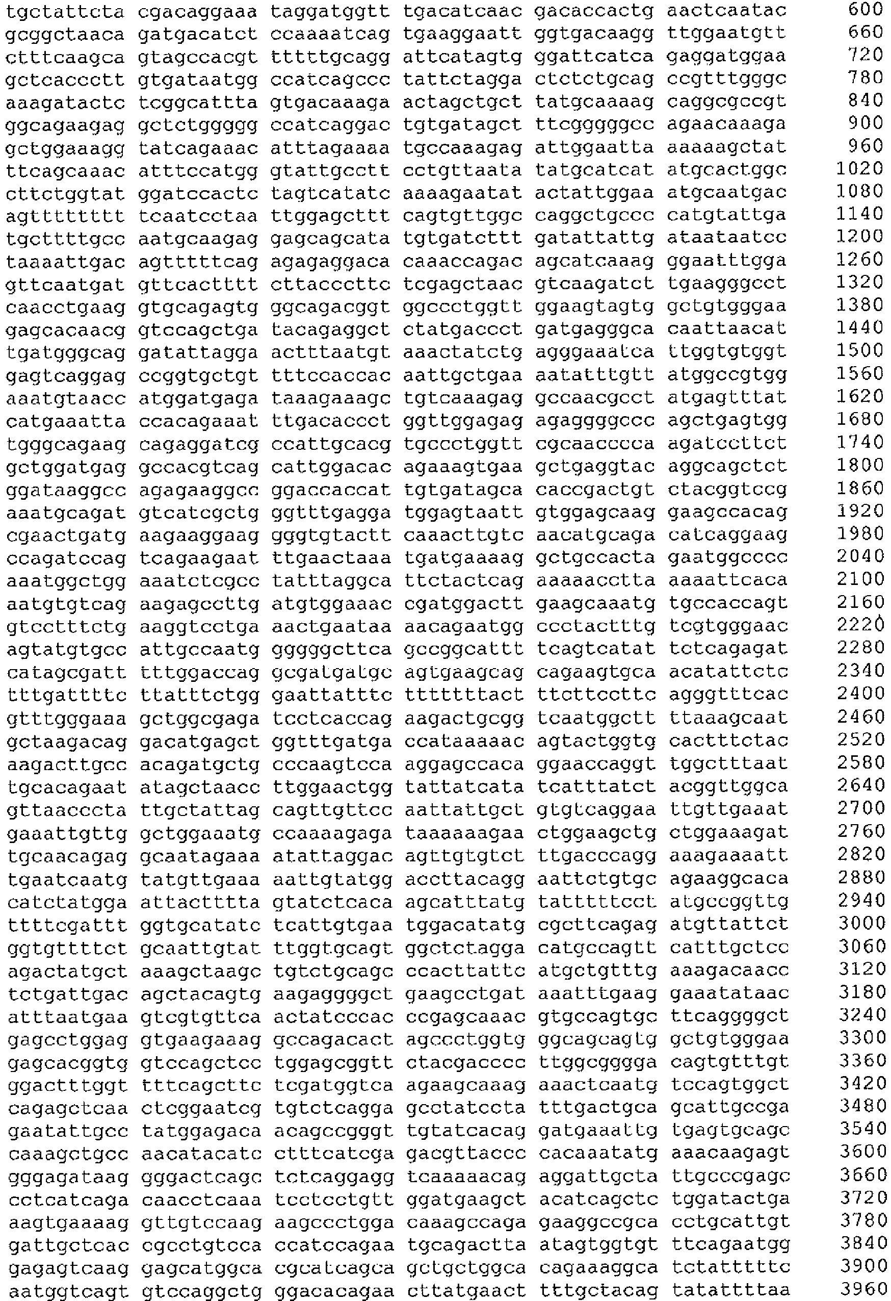

- Preferably, in situ hybridization is used to detect the presence of chromosomal copy number increase for (i) the ABCB1 gene; (ii) the ABCB4 gene; or (iii) the ABCB1 gene and the ABCB4 gene. Primer and probes can be made by one of skill in the art using the sequences of SEQ ID NO:1 and SEQ ID NO:2.
- Probes for use in the in situ hybridization methods of the invention fall into two broad groups: chromosome enumeration probes, i.e., probes that hybridize to a chromosomal region, usually a repeat sequence region, and indicate the presence or absence of an entire chromosome; and locus specific probes, i.e., probes that hybridize to a specific locus on a chromosome and detect the presence or absence of a specific locus. Chromosome arm probes, i.e., probes that hybridize to a chromosomal region and indicate the presence or absence of an arm of a specific chromosome, can also be used. It is preferred to use a locus specific probe that can detect changes of the unique chromosomal DNA sequences at the interrogated locus, such as the ABCB1 and ABCB4 loci. Methods for use of unique sequence probes for in situ hybridization are described in
U.S. Patent No. 5,447,841 , the contents of which are incorporated herein by reference. - A chromosome enumeration probe can hybridize to a repetitive sequence, located either near or removed from a centromere, or can hybridize to a unique sequence located at any position on a chromosome. For example, a chromosome enumeration probe can hybridize with repetitive DNA associated with the centromere of a chromosome. Centromeres of primate chromosomes contain a complex family of long tandem repeats of DNA comprised of a monomer repeat length of about 171 base pairs, that are referred to as alpha- satellite DNA. Centromere fluorescent in situ hybridization probes to each of chromosomes 14 and 18 are commercially available from Abbott Molecular (Des Plaines, IL).
- Exceptionally useful in situ hybridization probes are directly labeled fluorescent probes, such as described in
U.S. Patent No. 5,491,224 , incorporated herein by reference.U.S. Patent No, 5,491,224 also describes simultaneous FISH assays using more than one fluorescently labeled probe. - Useful locus specific probes can be produced in any manner and generally contain sequences to hybridize to a chromosomal DNA target sequence of about 10,000 to about 1,000,000 bases long. Preferably the probe hybridizes to a target stretch of chromosomal DNA at the target locus of at least 100,000 bases long to about 500,000 bases long and also includes unlabeled blocking nucleic acid in the probe mix, as disclosed in
U.S. Patent No. 5,756,696 , the contents of which are herein incorporated by reference, to avoid non-specific binding of the probe. It is also possible to use unlabeled, synthesized oligomeric nucleic acid or peptide nucleic acid as the blocking nucleic acid. For targeting the particular gene locus, it is preferred that the probes include nucleic acid sequences that span the gene and thus hybridize to both sides of the entire genomic coding locus of the gene. The probes can be produced starting with human DNA-containing clones such as Bacterial Artificial Chromosomes (BAC's) or the like. BAC libraries for the human genome are available from Invitrogen (Carlsbad, CA) and can be investigated for identification of useful clones. It is preferred to use the University of California Santa Cruz Genome Browser to identify DNA sequences in the target locus. These DNA sequences can then be used to synthesize PCR primers for use to screen BAC libraries to identify useful clones. The clones can then be labeled by conventional nick translation methods and tested as in situ hybridization probes. - Examples of fluorophores that can be used in the in situ hybridization methods described herein are: 7-amino-4-methylcoumarin-3-acetic acid (AMCA); Texas Red™ (Molecular Probes, Inc., Eugene, OR); 5-(and-6)-carboxy-X-rhodamine; lissamine rhodamine B; 5-(and-6)-carboxyfluorescein; fluorescein-5-isothiocyanate (FITC); 7-diethylaminocoumarin-3-carboxylic acid, tetramethyl-rhodamine-5-(and-6)-isothiocyanate; 5-(and-6)-carboxytetramethylrhodamine; 7-hydroxy-coumarin-3-carboxylic acid; 6-[fluorescein 5-(and-6)-carboxamido]hexanoic acid; N-(4,4-difluoro-5,7-dimethyl-4-bora-3a, 4a diaza-3-indacenepropionic acid; eosin-5-isothiocyanate; erythrosine-5-isothiocyanate; 5-(and-6)-carboxyrhodamine 6G; and Cascade™ blue aectylazide (Molecular Probes; an Invitrogen brand).
- Probes can be viewed with a fluorescence microscope and an appropriate filter for each fluorophore, or by using dual or triple band-pass filter sets to observe multiple fluorophores. See, for example,
U.S. Patent No. 5,776,688 , the contents of which are incorporated herein by reference. Any suitable microscopic imaging method can be used to visualize the hybridized probes, including automated digital imaging systems. Alternatively, techniques such as flow cytometry can be used to examine the hybridization pattern of the chromosomal probes. - Although the cell-by-cell gene amplification analysis resulting from in situ hybridization is preferred, the genomic biomarkers can also be detected by quantitative PCR. In this embodiment, chromosomal DNA is extracted from the tissue sample, and is then amplified by PCR using a pair of primers specific to at least one of (i) the ABCB1 gene; (ii) the ABCB4 gene; or (iii) the
ABCB 1 gene and the ABCB4 gene, or by multiplex PCR, using multiple pairs of primers. Any primer sequence for the biomarkers can be used. Examples of primers that can be used are shown in Table 3. The copy number of the tissue is then determined by comparison to a reference amplification standard,Table 3 SEQUENCE Type of Primer SEQ ID NO: 5'-GGAGAGTAGCAGTGCCTTGGACC-3' Forward SEQ ID NO:4 5'-AGGAGGAGGTAGAAAACAGATAAGGGAAC-3' Reverse SEQ ID NO:5 5'-AGTGCCTTGGACCCCAGCTCTC-3' Forward SEQ ID NO:6 5'-GAAAACAGATAAGGGAACAGTTAGGGATC-3' Reverse SEQ ID NO:7 - Microarray-based copy number analysis can also be used. In this embodiment, the chromosomal DNA after extraction is labeled for hybridization to a microarray comprising a substrate having multiple immobilized unlabeled nucleic acid probes arrayed at probe densities up to several million probes per square centimeter of substrate surface. Multiple microarray formats exist and any of these can be used, in the present invention. Examples of microarrays that can be used are the Affymetrix GeneChip®Mapping 100K Set SNP Array (See Matsuzaki, H., et al., "Genotyping over 100,000 SNPs on a pair of oligonucleotide arrays," Nat Methods, 1:109-11 (2004)); the Affymetrix GeneChip® Mapping 250K Assay Kits (such as the GcncChip® Human Mapping 250K Nsp Array or the GeneChip® Human Mapping 250K Sty Array) or the Affymetrix GeneChip® Mapping 500K Array Set, each of which is commercially available from Affymetrix, Inc., Santa Clara, CA), the Agilent Human Genome aCGH Microarray 44B (available from Agilent Technologies, Inc., Santa Clara, CA), Illumina microarrays (Illumina, Inc., San Diego, CA), Nimblegen aCGH microarrays (Nimblegen, Inc., Madison, WI), etc. When using an oligonucleotide microarray to detect amplifications, it is preferred to use a microarray that has probe sequences to more than three separate locations in the targeted region. Examples of probes that can be used in the microarray are shown in below Table 4 and in SEQ ID NOS: 23-321. Flanking sequences for the probes listed below in Table 4 are shown below in Table 5.
Table 4 SEQ ID NO: Probe Sequence Direction 23 AGCTTTAGAACCACCACTTCAGGTC Forward 24 AGCTTTAGAACCACCATTTCAGGTC Forward 25 GACCTGAAGTGGTGGTTC3'AAAGCT Reverse 26 TAGAACCACCATTTCAGGTCAATGT Forvard 27 TAGAACCACCACTTCAGGTCAATGT Forward 28 GACCTGAAATGGTGGTTCTAAAGCT Reverse 29 TTGACCTGAAGTGGTGGTTCTAAAG Reverse 30 TTGACCTGAAATGGTGGTTCTAAAG Reverse 31 ACATTGACCTGAAAGGTGGTTCTA Reverse 32 ACATTGACCTGAAGTGGTGGTTCTA Reverse 33 CATTGACCTGAAGTGGTGGTTCTAA Reverse 34 CATTGACCTGAAATGGTGGTTCTAA Reverse 35 AACCACCACTTCAGGTCAATGTTTT Forward 36 AACCACCATTTCAGGTCAATGTTTT Forward 37 AAAACATTGACCTGAAATGGTGGTT Reverse 38 AAAACATTGACCTGAAGTGGTGGTT Reverse 39 AAACATTGACCTGAAATGGTGGTTC Reverse 40 AAACATTGACCTGAAGTGGTGGTTC Reverse 41 TTTAGAACCACCACTTCAGGTCAAT Forward 42 TTTAGAACCACCATTTCAGGTCAAT Forward 43 TGTTAAAGGTTGTGCTATAATGAAT Forward 44 TCATTATAGCACAACCTTTAACACA Reverse 45 GTGTGTTAAAGATTGTGCTATAATG Forward 46 ATAGCACAATCTTTAACACACCACT Reverse 47 TCATTATAGCACAATCTTTAACACA Reverse 48 TAGCACAACCTTTAACACACCACTT Reverse 49 GTGTGTTAAAGGTTGTGCTATAATG Forward 50 ATTATAGCACAACCTTTAACACACC Reverse 51 ATTATAGCACAATCTTTAACACACC Reverse 52 ATTCATTATAGCACAATCTTTAACA Reverse 53 ATAGCACAACCTTTAACACACCACT Reverse 54 TTATAGCACAACCTTTAACACACCA Reverse 55 TTATAGCACAATCTTTAACACACCA Reverse 56 TGTTAAAGATTGTGCTATAATGAAT Forward 57 TGGTGTGTTAAAGGTTGTGCTATAA Forward 58 TGGTGTGTTAAAGATTGTGCTATAA Forward 59 AAGTGGTGTGTTAAAGATTGTGCTA Forward 60 ATTCATTATAGCACAACCTTTAACA Reverse 61 AAGTGGTGTGTTAAAGGTTGTGCTA Forward 62 TAGCACAATCTTTAACACACCACTT Reverse 63 ACTGAGATAGTGATAGCAATTTTTT Reverse 64 AAAAAATTGCTATCACTATCTCAGT Forward 65 AAAAAAAATTGCTGTCACTATCTCA Forward 66 AAAAAATTGCTGTCACTATCTCAGT Forward 67 GCTACTGAGATAGTGATAGCAATTT Reverse 68 GCTACTGAGATAGTGACAGCAATTT Reverse 69 ATGAAAAAAAATTGCTATCACTATC Forward 70 ATGAAAAAAAATTGCTGTCACTATC Forward 71 AAATTGCTGTCACTATCTCAGTACC Forward 72 AAATTGCTATCACTATCTCAGTAGC Forward 73 AAAAAAAATTGCTATCACTATCTCA Forward 74 AGATAGTGACAGCAATTTTTTTTCA Reverse 75 AGATAGTGATAGCAATTTTTTTTCA Reverse 76 ACTGAGATAGTGACAGCAATTTTTT Reverse 77 TACTGAGATAGTGATAGCAATTTTT Reverse 78 TACTGAGATAGTGACAGCAATTTTT Reverse 79 CTGAGATAGTGATAGCAATTTTTTT Reverse 80 CTGAGATAGTGACAGCAATTTTTTT Reverse 81 AAAAATTGCTATCACTATCTCAGTA Forward 82 AAAAATTGCTGTCACTATCTCAGTA Forward 83 TTATGCTGTAATACATCCATTAAGC Reverse 84 TTATGCTGTAATATATCCATTAAGC Reverse 85 AGTTATGCTGTAATATATCCATTAA Reverse 86 AGTTATGCTGTAATACATCCATTAA Reverse 87 TGCTGTAATATATCCATTAAGCTAT Reverse 88 TGCTGTAATACATCCATTAAGCTAT Reverse 89 ATGCTGTAATATATCCATTAAGCTA Reverse 90 ATGCTGTAATACATCCATTAAGCTA Reverse 91 AGCTTAATGGATGTATTACAGCATA Forward 92 AGCTTAATGGATATATTACAGCATA Forward 93 TAGTTATGCTGTAATACATCCATTA Reverse 94 TAGTTATGCTGTAATATATCCATTA Reverse 95 CTGTAATATATCCATTAAGCTATTT Reverse 96 TTAATGGATGTATTACAGCATAACT Forward 97 CTGTAATACATCCATTAAGCTATTT Reverse 98 TTAATGGATATATTACAGCATAACT Forward 99 TATGCTGTAATATATCCATTAAGCT Reverse 100 TATGCTGTAATACATCCATTAAGCT Reverse 101 ATAGCTTAATGGATATATTACAGCA Forward 102 TATAGTAGCTCAAGTCCCTTAGTCT Reverse 103 TATAGTAGCTGAAGTCCCTTAGTCT Reverse 104 TAGTAGCTCAAGTCCCTTAGTCTCT Reverse 105 TAGTAGCTGAAGTCCCTTAGTCTCT Reverse 106 CATAGTTATAGTAGCTGAAGTCCCT Reverse 107 CATAGTT'ATAGTAGCTCAAGTCCCT Reverse 108 ACTAAGGGACTTCAGCTACTATAAC Forward 109 ACTAAGGGACTTGAGCTACTATAAC Forward 110 GTTATAGTAGCTCAAGTCCCTTAGT Reverse 111 GTTATAGTAGCTGAAGTCCCTTAGT Reverse 112 TTATAGTAGCTCAAGTCCCTTAGTC Reverse 113 TTATAGTAGCTGAAGTCCCTTAGTC Reverse 114 AGACTAAGGGACTTCAGCTACTATA Forward 115 AGACTAAGGGACTTGAGCTACTATA Forward 116 GACTAAGGGACTTCAGCTACTATAA Forward 117 GACTAAGCGACTTGAGCTACTATAA Forward 118 AGTTATAGTAGCTGAAGTCCCTTAG Reverse 119 AGTTATAGTAGCTCAAGTCCCTTAG Reverse 120 AAGGGACTTCAGCTACTATAACTAT Forward 121 AAGGGACTTGAGCTACTATAACTAT Forward 122 AATACTTATGAGATTTATAGAGGAA Forward 123 CTTATGAGATTTATAGAGGAAGAAG Forward 124 CTTATGAGACTTATAGAGGAAGAAG Forward 125 ATACTTATGAGACTTATAGAGGAAG Forward 126 ATACTTATGAGATTTATAGAGGAAG Forward 127 ACTTCTTCCTCTATAAGTCTCATAA Reverse 128 ACTTCTTCCTCTATAAATCTCATAA Reverse 129 AATACTTATGAGACTTATAGAGGAA Forward 130 TACTTATGAGATTTATAGAGGAAGA Forward 131 TACTTATGAGACTTATAGAGGAAGA Forward 132 TTCCTCTATAAATCTCATAAGTATT Reverse 133 TCTTCCTCTATAAGTCTCATAAGTA Reverse 134 TCTTCCTCTATAAATCTCATAAGTA Reverse 135 CTCTATAAGTCTCATAAGTATTTGC Reverse 136 CTCTATAAATCTCATAAGTATTTGC Reverse 137 TTATGAGATTTATAGAGGAAGAAGT Forward 138 TTATGAGACTTATAGAGGAAGAAGT Forward 139 AAATACTTATGAGATTTATAGAGGA Forward 140 AAATACTTATGAGACTTATAGAGGA Forward 141 TTCCTCTATAAGTCTCATAAGTATT Reverse 142 TTAGAGCTGACTAATTAGATCCTAT Forward 143 TTAGAGCTGTCTAATTAGATCCTAT Forward 144 ATCTAATTAGACAGCTCTAAAACCT Reverse 145 ATCTAATTAGTCAGCTCTAAAACCT Reverse 146 AGGATCTAATTAGTCAGCTCTAAAA Reverse 147 ATAGGATCTAATTAGTCAGCTCTAA Reverse 148 ATAGGATCTAATTAGACAGCTCTAA Reverse 149 GGTTTTAGAGCTGACTAATTAGATC Forward 150 GGTTTTAGAGCTGTCTAATTAGATC Forward 151 TAGAGCTGACTAATTAGATCCTATG Forward 152 TAGAGCTGTCTAATTAGATCCTATG Forward 153 GGATCTAATTAGACAGCTCTAAAAC Reverse 154 GGATCTAATTAGTCAGCTCTAAAAC Reverse 155 AGGATCTAATTAGACAGCTCTAAAA Reverse 156 TTTTAGAGCTGTCTAATTAGATCCT Forward 157 GATCTAATTAGACAGCTCTAAAACC Reverse 158 GATCTAATTAGTCAGCTCTAAAACC Reverse 159 TTTTAGAGCTGACTAATTAGATCCT Forward 160 GAAGGTTTTAGAGCTGACTAATTAG Forward 161 GAAGGTTTTAGAGCTGTCTAATTAG Forward 162 GCAACAAATACTATATTATATACCA Reverse 163 GCAACAAATACCATATTATATACCA Reverse 164 GTATATAATATAGTATTTGTTGCTC Forward 165 GTATATAATATGGTATTTGTTGCTC Forward 166 TATAATATAGTATTTGTTGCTCTAG Forward 167 TATAATATGGTATTTGTTGCTCTAG Forward 168 AGAGCAACAAATACCATATTATATA Reverse 169 AGAGCAACAAATACTATATTATATA Reverse 170 TAATGGTATATAATATAGTATTTGT Forward 171 TAATGGTATATAATATGGTATTTGT Forward 172 CTAGAGCAACAAATACTATATTATA Reverse 173 AACAAATACTATATTATATACCATT Reverse 174 AACAAATACCATATTATATACCATT Reverse 175 CTAGAGCAACAAATACCATATTATA Reverse 176 GGTATATAATATGGTATTTGTTGGT Forward 177 GGTATATAATATAGTATTTGTTGCT Forward 178 AGCAACAAATACCATATTATATACC Reverse 179 AGCAACAAATACTATATTATATACC Reverse 180 TGGTATATAATATAGTATTTGTTGC Forward 181 TGGTATATAATATGGTATTTGTTGC Forward 182 TGTGGATTAAACTTGTGCGCTTAAA Reverse 183 TGTGGATTAAATTTGTGCGCTTAAA Reverse 184 ATTTAAGCGCACAAATTTAATGGAG Fonvard 185 ATTTAAGCGCACAAGTTTAATCCAC Forward 186 TTGTGGATTAAATTTGTGCGCTTAA Reverse 187 TTGTGGATTAAACTTGTGCGCTTAA Reverse 188 TAAGCGCACAAGTTTAATCCACAAC Forward 189 TAAGCGCACAAATTTAATCCACAAC Forward 190 GTGTTGTGGATTAAACTTGTGCGCT Reverse 191 GTGTTGTGGATTAAATTTGTGCGCT Reverse 192 GCGCACAAGTTTAATCCACAACACA Forward 193 GCGCACAAATTTAATCCACAACACA Forward 194 TTATTTAAGCGCACAAGTTTAATCC Forward 195 TTATTTAAGCGCACAAATTTAATCC Forward 196 TTTAAGCGCACAAGTTTAATCCACA Forward 197 TTTAAGCGCACAAATTTAATCCACA Forward 198 GTGGATTAAATTTGTGCGCTTAAAT Reverse 199 GTGGATTAAACTTGTGCGCTTAAAT Reverse 200 TGTGTTGTGGATTAAATTTGTGCGC Reverse 201 TGTGTTGTGGATTAAACTTGTGCGC Reverse 202 CACAAATGATAGCAGTAAGATAAAT Forward 203 TTTATCTTACTGCTACCATTTGTGT Reverse 204 AAAACACAAATGATAGCAGTAAGAT Forward 205 AAAACACAAATGGTAGCAGTAAGAT Forward 206 AAACACAAATGGTAGCAGTAAGATA Forward 207 AAACACAAATGATAGCAGTAAGATA Forward 208 ATCTTACTGCTACCATTTGTGTTTT Reverse 209 ATCTTACTGCTATCATTTGTGTTTT Reverse 210 ATTGAAAACACAAATGGTAGCAGTA Forward 211 TTTATCTTACTGCTATCATTTGTGT Reverse 212 GAAAACACAAATGGTAGCAGTAAGA Forward 213 GAAAACACAAATGATAGCAGTAAGA Forward 214 CACAAATGGTAGCAGTAAGATAAAT Forward 215 ATTTATCTTACTGCTATCATTTGTG Reverse 216 TGAAAACACAAATGGTAGCAGTAAG Forward 217 ATTTATCTTACTGCTACCATTTGTG Reverse 218 TGAAAACACAAATGATAGCAGTAAG Forward 219 CTTACTGCTACCATTTGTGTTTTCA Reverse 220 CTTACTGCTATCATTTGTGTTTTCA Reverse 221 ATTGAAAACACAAATGATAGCAGTA Forward 222 ACCTGCAAATCCGTAAAGTGTACTA Forward 223 ACCTGCAAATCTGTAAAGTGTACTA Forward 224 AGTACACTTTACAGATTTGCAGGTT Reverse 225 AGTACACTTTACGGATTTGCAGGTT Reverse 226 ACACTTTACGGATTTGCAGGTTTTG Reverse 227 AACCTGCAAATCTGTAAAGTGTACT Forward 228 AACCTGCAAATCCGTAAAGTGTACT Forward 229 TAGTACACTTTACGGATTTGCAGGT Reverse 230 TAGTACACTTTACAGATTTGCAGGT Reverse 231 ATATAGTACACTTTACGGATTTGCA Reverse 232 ATATAGTACACTTTACAGATTTGCA Reverse 233 ATAGTACACTTTACGGATTTGCAGG Reverse 234 AAACCTGCAAATCCGTAAAGTGTAC Forward 235 AAACCTGCAAATCTGTAAAGTGTAC Forward 236 GCAAAACCTGCAAATCTGTAAAGTG Forward 237 GCAAAACCTGCAAATCCGTAAAGTG Forward 238 CCTGCAAATCCGTAAAGTGTACTAT Forward 239 ACACTTTACAGATTTGCAGGTTTTG Reverse 240 CCTGCAAATCTGTAAAGTGTACTAT Forward 241 ATAGTACACTTTACAGATTTGCAGG Reverse 242 TTCTTTAATGGGTACAAAATGTCAA Reverse 243 TTTGACATTATGTACCCATTAAAGA Forward 244 TTTGACATTTTGTACCCATTAAAGA Forward 245 TAATGGGTACATAATGTCAAAAATA Reverse 246 TTGACATTTTGTACCCATTAAAGAA Forward 247 TTCTTTAATGGGTACATAATGTCAA Reverse 248 TATTTTTGACATTATGTACCCATTA Forward 249 ATATTTTTGACATTATGTACCCATT Forward 250 TTTAATGGGTACAAAATGTCAAAAA Reverse 251 TTTAATGGGTACATAATGTCAAAAA Reverse 252 ATATTTTTGACATTTTGTACCCATT Forward 253 TTGACATTATGTACCCATTAAAGAA Forward 254 TTAATGGGTACATAATGTCAAAAAT Reverse 255 TTAATGGGTACAAAATGTCAAAAAT Reverse 256 TATTTTTGACATTTTGTACCCATTA Forward 257 TGGGTACATAATGTCAAAAATATTT Reverse 258 ATTTTTGACATTATGTACCCATTAA Forward 259 ATTTTTGACATTTTGTACCCATTAA Forward 260 TGGGTACAAAATGTCAAAAATATTT Reverse 261 TAATGGGTACAAAATGTCAAAAATA Reverse 262 TTCTTTAATGCAGAGTAGGAGACAG Reverse 263 TCCTACTCTGCGTTAAAGAAGCCTG Forward 264 TGTGTCCTACTCTGCATTAAAGAAG Forward 265 CAGGCTTCTTTAACGCAGAGTAGGA Reverse 266 TACTCTGCATTAAAGAAGCCTGCAT Forward 267 CTGTGTCCTACTCTGCATTAAAGAA Forward 268 AGGCTTCTTTAATGCAGAGTAGGAC Reverse 269 AGGCTTCTTTAACGCAGAGTAGGAC Reverse 270 CTGTGTCCTACTCTGCGTTAAAGAA Forward 271 ATGCAGGCTTCTTTAATGCAGAGTA Reverse 272 TGTGTCCTACTCTGCGTTAAAGAAG Forward 273 GGCTTCTTTAACGCAGAGTAGGACA Reverse 274 GCAGGCTTCTTTAACGCAGAGTAGG Reverse 275 TCCTACTCTGCATTAAAGAAGCCTG Forward 276 TTCTTTAACGCAGAGTAGGACACAG Reverse 277 GGCTTCTTTAATGCAGAGTAGGACA Reverse 278 TACTCTGCGTTAAAGAAGCCTGCAT Forward 279 CAGGCTTCTTTAATGCAGAGTAGGA Reverse 280 ATGCAGGCTTCTTTAACGCAGAGTA Reverse 281 GCAGGCTTCTTTAATGCAGAGTAGG Reverse 282 ATTTTTCCTGCATGGCAAGAGTTTA Forward 283 ATTTTTCCTGCATAGCAAGAGTTTA Forward 284 CCTAAACTCTTGCCATGCAGGAAAA Reverse 285 TCCTAAACTCTTGCTATGCAGGAAA Reverse 286 TCCTAAACTCTTGCCATGCAGGAAA Reverse 287 TAATTTTTCCTGCATGGCAAGAGTT Forward 288 TTTCCTGCATGGCAAGAGTTTAGGA Forward 289 TAATTTTTCCTGCATAGCAAGAGTT Forward 290 TTTTCCTGCATGGCAAGAGTTTAGG Forward 291 TTTCCTGCATAGCAAGAGTTTAGGA Forward 292 CCTAAACTCTTGCTATGCAGGAAAA Reverse 293 ATAATTTTTCCTGCATGGCAAGAGT Forward 294 ATAATTTTTCCTGCATAGCAAGAGT Forward 295 TCCTGCATGGCAAGAGTTTAGGAGA Forward 296 TCCTGCATAGCAAGAGTTTAGGAGA Forward 297 AACTCTTGCCATGCAGGAAAAATTA Reverse 298 AACTCTTGCTATGCAGGAAAAATTA Reverse 299 TTTTCCTGCATAGCAAGAGTTTAGG Forward 300 CTAAACTCTTGCTATGCAGGAAAAA Reverse 301 CTAAACTCTTGCCATGCAGGAAAAA Reverse 302 TGGAAACATGGTTGGTCCGAATGTT Forward 303 TGGAAACATGGTTAGTCCGAATGTT Forward 304 GGAAACATGGTTAGTCCGAATGTTA Forward 305 GGAAACATGGTTGGTCCGAATGTTA Forward 306 AGTGGAAACATGGTTAGTCCGAATG Forward 307 AGTGGAAACATGGTTGGTCCGAATG Forward 308 ATTAACATTCGGACCAACCATGTTT Reverse 309 ATTAACATTCGGACTAACCATGTTT Reverse 310 GAAACATGGTTGGTCCGAATGTTAA Forward 311 GAAACATGGTTAGTCCGAATGTTAA Forward 312 AACATTCGGACCAACCATGTTTCCA Reverse 313 AACATTCGGACTAACCATGTTTCCA Reverse 314 ACATGGTTAGTCCGAATGTTAATCT Forward 315 ACATGGTTGGTCCGAATGTTAATCT Forward 316 TAGTGGAAAGATGGTTGGTCCGAAT Forward 317 TAGTGGAAACATGGTTAGTCCGAAT Forward 318 CATTCGGACCAACCATGTTTCCACT Reverse 319 CATTCGGACTAACCATGTTTCCACT Reverse 320 TAACATTCGGACCAACCATGTTTCC Reverse 321 TAACATTCGGACTAACCATGTTTCC Reverse Table 5 SEQ ID NO: FLANKING SEQUENCE Corresponding Probes (SEQ ID NOS) 8 ccatgttgaaaaacattgacctgaa[A/G]tggtgflttctaaaflcttcggtgaat 23-42 9 tgccctacaattcattatagcacaa[C/T]ctttaacacaccacttaataactgt 43-62 10 aaccatcaggctactgagatagtga[C/T]agcaattttttttcatacttcttct 63-82 11 ccaaaatattagttatgctgtaata[C/T]atccattaagctatttaagaaaaca 83-101 12 ctttccctacatagttatagtagct[C/G]aagtcccttagtctctccacattcc 102-121 13 agtcctgtaacttcttcctctalaa[A/G]tctcataagtatttgctttcttttc 122-141 14 tacaataaacataggatctaattag[A/T]cagctctaaaaccttcttcagtaag 142-161 15 tcttagaaactagagcaacaaatac[C/T]atattatataccattaaatactttt 162-181 16 taaatattatgtgttgtggattaaa[C/T]ttgtgcttcttaaataaatttcagtt 182-201 17 aaaattacaatttatcttactgcta[C/T]catttgtgttttcaatcttcatctt 202-221 18 taaggaaaaatatagtacaetttac[A/G]gatttgcaggttttgctatttataa 222-241 19 caagaccctttctttaatgggtaca[A/T]aatgtcaaaaatatttttatataat 242-261 20 gacaccttgatgcaggcttctttaa[C/T]gcagagtaggacacagatggctgga 262-281 21 atttcagtatctcctaaaclcttgc[C/T]atgcaggaaaaattattttatgtga 282-301 22 taagagcgaagattaacattcggac[C/T]aaccatttccactaaaccaatta 302-321 - The level of gene expression for (i) the ABCB1 gene; (ii) the ABCB4 gene; or (iii) the ABCB1 gene and the ABCB4 gene can be determined by assessing the amount of one or more mRNAs in the test sample. Methods of measuring mRNA in samples are known in the art. To measure mRNA levels, the cells in a test sample can be lysed, and the levels of mRNA in the lysates or in RNA purified or semi-purified from lysates can be measured by any variety of methods familiar to those in the art. Such methods include hybridization assays using detectably labeled DNA or RNA probes (i.e., Northern blotting) or quantitative or semi-quantitative RT-PCR methodologies using appropriate oligonucleotide primers. Alternatively, quantitative or semi-quantitative in situ hybridization assays can be carried out using, for example, tissue sections, or unlysed cell suspensions, and detectably labeled (e.g., fluorescent, or enzyme-labeled) DNA or RNA probes. Additional methods for quantifying mRNA include RNA protection assay (RPA), cDNA and oligonucleotide microarrays, representation difference analysis (RDA), differential display, EST sequence analysis, and serial analysis of gene expression (SAGE).
- In suitable embodiments, PCR amplification is used to detect for (i) the ABCB1 gene; (ii) the ABCB4 gene; or (iii) the ABCB1 gene and the ABCB4 gene in the test sample. Briefly, in PCR, two primer sequences are prepared that are complementary to regions on opposite complementary strands of the marker sequence, for example containing the sequences for (i) the ABCB1 gene; (ii) the ABCB4 gene; or (iii) the ABCB1 gene and the ABCB4 gene. An excess of deoxynucleotide triphosphates are added to a reaction mixture along with a DNA polymerase, e.g., Taq polymerase. If the target sequence is present in a sample, the primers will bind to the sequence and the polymerase will cause the primers to be extended along the marker sequence by adding on nucleotides. By raising and lowering the temperature of the reaction mixture, the extended primers will dissociate from the marker to form reaction products, excess primers will bind to the marker and to the reaction products and the process is repeated, thereby generating amplification products. A reverse transcriptase PCR amplification procedure can be performed in order to quantify the amount of mRNA amplified.
- Any suitable fragment of (i) the ABCB1 gene; (ii) the ABCB4 gene; or (iii) the ABCB1 gene and the ABCB4 gene can be amplified and detected. Designing efficient primers for PCR is within the ordinary skill in the art. Examples of primers that can be used are shown in Table 3. Typically, amplified fragments for detection are approximately 50 to 300 nucleotides in length.
- Amplification products can be detected in several ways. Amplification products can be visualized by electrophoresis of the sample in a gel and then staining with a DNA binding dye, e.g., ethidium bromide. Alternatively, the amplification products can be integrally labeled with a radio- or fluorescence nucleotide and then visualized using x-ray film or under the appropriate stimulating spectra.
- Amplification can be also monitored using "real-time" methods, Real-time PCR allows for the detection and quantitation of a nucleic acid target. Typically, this approach to quantitative PCR utilizes a fluorescent dye, which can be a double-strand specific dye, such as SYBR GREEN®. Alternatively, other fluorescent dyes (e.g., FAM or HEX) can be conjugated to an oligonucleotide probe or a primer. Various instruments capable of performing real time PCR are known in the art and include, for example, the ABI PRISM® 7900 (Applied Biosystems) and LIGHTCYCLER® systems (Roche). The fluorescent signal generated at each cycle of PCR is proportional to the amount of PCR product. A plot of fluorescence versus cycle number is used to describe the kinetics of amplification and a fluorescence threshold level is used to define a fractional cycle number related to initial template concentration. When amplification is performed and detected on an instrument capable of reading fluorescence during thermal cycling, the intended PCR product from non-specific PCR products can be differentiated using melting analysis. By measuring the change in fluorescence while gradually increasing the temperature of the reaction subsequent to amplification and signal generation it can be possible to determine the Tm of the intended product(s) as well as that of the nonspecific product.
- The methods can include amplifying multiple nucleic acids in sample, also known as "multiplex detection" or "multiplexing." Multiplex PCR" refers to PCR that involves adding more than one set of PCR primers to the reaction in order to detect and quantify multiple nucleic acids, including nucleic acids from one or more target gene markers. Furthermore, multiplexing with an internal control (e.g., 18S rRNA, GADPH, or actin) provides a control for the PCR without reaction.
- As discussed previously herein, the test sample of the present invention can be a tissue sample. The tissue sample to be assayed by the methods of the present invention can comprise any type, including a peripheral blood sample, a tumor tissue or a suspected tumor tissue, a thin layer cytological sample, a fine-needle aspirate sample, a bone marrow sample, a lymph node sample, a urine sample, an ascites sample, a lavage sample, an esophageal brushing sample, a bladder or lung wash sample, a spinal fluid sample, a brain fluid sample, a ductal aspirate sample, a nipple discharge sample, a pleural effusion sample, a fresh frozen tissue sample, a paraffin embedded tissue sample or an extract or processed sample produced from any of a peripheral blood sample, a tumor tissue or a suspected tumor tissue, a thin layer cytological sample, a fine needle aspirate sample, a bone marrow sample, a lymph node sample, a urine sample, an ascites sample, a lavage sample, an esophageal brushing sample, a bladder or lung wash sample, a spinal fluid sample, a brain fluid sample, a ductal aspirate sample, a nipple discharge sample, a pleural effusion sample, a fresh frozen tissue sample or a paraffin embedded tissue sample. For example, a patient peripheral blood sample can be initially processed to extract an epithelial cell population, and this extract can then be assayed. A microdissection of the tissue sample to obtain a cellular sample enriched with suspected tumor cells can also be used. The preferred tissue samples for use herein are peripheral blood, tumor tissue or suspected tumor tissue, including fine needle aspirates, fresh frozen tissue and paraffin embedded tissue, and bone marrow.
- The tissue sample can be processed by any desirable method for performing in situ hybridization or other nucleic acid assays. For the preferred in situ hybridization assays, a paraffin embedded tumor tissue sample or bone marrow sample is fixed on a glass microscope slide and deparaffinized with a solvent, typically xylene. Useful protocols for tissue deparaffinization and in situ hybridization are available from Abbott Molecular Inc. (Des Plaines, Illinois). Any suitable instrumentation or automation can be used in the performance of the inventive assays. PCR based assays can be performed on the m2000 instrument system (Abbott Molecular, Des Plaines, IL). Automated imaging can be used for the preferred fluorescent in situ hybridization assays.
- In one embodiment, the sample comprises a peripheral blood sample from a patient which is processed to produce an extract of circulating tumor or cancer cells to be examined for the presence or absence of a copy number gain for (i) a ABCB1 gene; (ii) a ABCB4 gene; or (iii) a ABCB1 gene and a ABCB4 gene. The circulating tumor cells can be separated by immunomagnetic separation technology such as that available from Immunicon (Huntingdon Valley, Pennsylvania). The copy number determined for the circulating tumor cells is then compared to the baseline level or predetermined level of circulating tumor cells having a copy number determined at a previous point in time, such as at the start of therapy. Increases in the copy number compared to the baseline level or the predetermined level can indicate therapy failure.
- Test samples can comprise any number of cells that is sufficient for a clinical diagnosis, and typically contain at least about 100 cells. In a typical FISH assay, the hybridization pattern is assessed in about 25-1,000 cells. Test samples are typically considered "test positive" when found to contain the gene amplification in a sufficient proportion of the sample. The number of cells identified with chromosomal copy number and used to classify a particular sample as positive, in general, varies with the number of cells in the sample. The number of cells used for a positive classification is also known as the cut-off value. Examples of cut-off values that can be used in the determinations include about 5, 25, 50, 100 and 250 cells, or 5%, 10%, 15%, 20%, 25%, 30%, 35%, 40%, 50% and 60% of cells in the sample population. As low as one cell can be sufficient to classify a sample as positive. In a typical paraffin embedded tissue sample, it is preferred to identify at least 30 cells as positive and more preferred to identify at least 20 cells as positive for having the chromosomal copy number gain. For example, detection in a typical paraffin embedded colorectal carcinoma of 30 cells would be sufficient to classify the tissue as positive and eligible for treatment.
- The present invention also contemplates kits for detecting the presence or absence of a copy number gain for (i) the ABCB1 gene; (ii) the ABCB4 gene; or (iii) the ABCB1 gene and the ABCB4 gene in a test sample. Such kits can comprise one or more reagents for determining the presence or absence of the above described copy number gain. For example, said kit can contain one or more nucleic acid probes. Alternatively, or in addition to the probes, the kit can contain one or more nucleic acid primers.
- Thus, the present disclosure further provides for diagnostic and quality control kits comprising one or more nucleic acid primers, nucleic acid probes or nucleic acid primers and probes described herein. Optionally the assays, kits and kit components of the present invention can be optimized for use on commercial platforms (e.g., immunoassays on the Prism®, AxSYM® , ARCHITECT® and EIA (Bead) platforms of Abbott Laboratories, Abbott Park, IL, as well as other commercial and/or in vitro diagnostic assays). Additionally, the assays, kits and kit components can be employed in other formats, for example, on electrochemical or other hand-held or point-of-care assay systems. The present disclosure is, for example, applicable to the commercial Abbott Point of Care (i-STAT®, Abbott Laboratories, Abbott Park, IL) electrochemical immunoassay system that performs sandwich immunoassays for several cardiac markers, including TnI, CKMB and BNP. Immunosensors and methods of operating them in single-use test devices are described, for example, in
U.S. Patent Application Publication Nos. 2003/0170881 ,2004/0018577 ,2005/0054078 and2006/0160164 , which are incorporated herein by reference. Additional background on the manufacture of electrochemical and other types of immunosensors is found inU.S. Patent No. 5,063,081 which is also incorporated by reference for its teachings regarding same. - Optionally the kits include quality control reagents (e.g., sensitivity panels, calibrators, and positive controls). Preparation of quality control reagents is well known in the art, and is described, e.g., on a variety of immunodiagnostic product insert sheets.
- The kit can incorporate a detectable label, such as a fluorophore, radioactive moiety, enzyme, biotin/avidin label, chromophore, chemiluminescent label, or the like, or the kit may include reagents for labeling the nucleic acid primers, the nucleic acid probes or the nucleic acid primers and nucleic acid probes for detecting the presence or absence of a copy number gain as described herein. The primers and/or probes, calibrators and/or controls can be provided in separate containers or pre-dispensed into an appropriate assay format, for example, into microtiter plates.
- The kits can optionally include other reagents required to conduct a diagnostic assay or facilitate quality control evaluations, such as buffers, salts, enzymes, enzyme co-factors, substrates, detection reagents, and the like. Other components, such as buffers and solutions for the isolation and/or treatment of a test sample (e.g., pretreatment reagents), may also be included in the kit. The kit may additionally include one or more other controls. One or more of the components of the kit may be lyophilized and the kit may further comprise reagents suitable for the reconstitution of the lyophilized components.
- The various components of the kit optionally are provided in suitable containers. As indicated above, one or more of the containers may be a microtiter plate. The kit further can include containers for holding or storing a sample (e.g., a container or cartridge for a blood or urine sample). Where appropriate, the kit may also optionally contain reaction vessels, mixing vessels and other components that facilitate the preparation of reagents or the test sample. The kit may also include one or more instruments for assisting with obtaining a test sample, such as a syringe, pipette, forceps, measured spoon, or the like.
- The kit further can optionally include instructions for use, which may be provided in paper form or in computer-readable form, such as a disc, CD, DVD or the like.
- The kit (or components thereof), as well as the method of determining the presence or absence of a copy number gain for (i) the ABCB1 gene; (ii) the ABCB4 gene; or (iii) the ABCB1 gene and the ABCB4 gene using the components and methods described herein, can be adapted for use in a variety of automated and semi-automated systems (including those wherein the solid phase comprises a microparticle), as described, e.g., in
U.S. Patent Nos. 5,089,424 and5,006,309 , and as commercially marketed, e.g., by Abbott Laboratories (Abbott Park, IL) as ARCHITECT®. - Some of the differences between an automated or semi-automated system as compared to a non-automated system (e.g., ELISA) include the substrate to which the first specific binding partner (e.g., capture antibody) is attached (which can impact sandwich formation and analyte reactivity), and the length and timing of the capture, detection and/or any optional wash steps, Whereas a non-automated format such as an ELISA may require a relatively longer incubation time with sample and capture reagent (e.g., about 2 hours) an automated or semi-automated format (e.g., ARCHITECT®, Abbott Laboratories) may have a relatively shorter incubation time (e.g., approximately 18 minutes for ARCHITECT®). Similarly, whereas a non-automated format such as an ELISA may incubate a detection antibody such as the conjugate reagent for a relatively longer incubation time (e.g., about 2 hours), an automated or semi-automated format (e.g., ARCHITECT®) may have a relatively shorter incubation time (e.g., approximately 4 minutes for the ARCHITECT®).
- Other platforms available from Abbott Laboratories include, but are not limited to, AxSYM®, IMx® (see, e.g.,
U.S. Patent No. 5,294,404 , which is hereby incorporated by reference in its entirety), PRISM®, EIA (bead), and Quantum™ II, as well as other platforms. Additionally, the assays, kits and kit components can be employed in other formats, for example, on electrochemical or other hand-held or point-of-care assay systems. The present disclosure is, for example, applicable to the commercial Abbott Point of Care (i-STAT®, Abbott Laboratories) electrochemical immunoassay system that performs sandwich immunoassays. Immunosensors and their methods of manufacture and operation in single-use test devices are described, for example in,U.S. Patent No. 5,063,081 ,U,S. Patent. Application Publication Nos. 2003/0170881 ,2004/0018577 ,2005/0054078 , and2006/0160164 , which are incorporated in their entireties by reference for their teachings regarding same. - It further goes without saying that the methods and kits as described herein necessarily encompass other reagents and methods for carrying out the assays. For instance, encompassed are various buffers such as are known in the art and/or which can be readily prepared or optimized to be employed.
- By way of example and not of limitation, an example of the present disclosure shall now be given.
- Antibodies were purchased from the indicated suppliers as follows: MDR1/P-glycoprotein (Catalog No. 517310) were from Calbiochem (San Diego, CA), anti-BRCP antibody (Catalog No. sc-58222) was from Santa Cruz Biotechnology, Inc. (Santa Cruz, CA), anti-phospho-histone H3 (Ser10) (Catalog No. 9701), and anti-histone H3 (Catalog No. 9715) were from Cell Signaling Technology (Danvers, MA), phycoerythrin (PE)-conjugated goat anti-mouse IgG (Catalog No. 550589) and PE-conjugated anti-human CD44 (Catalog No. 555479) were from BD Biosciences (Franklin Lakes, NJ), ß-actin was from Sigma Aldrich (St. Louis, MO), Alexa Fluor 680-conjugated goat anti-rabbit IgG (Catalog No. A21109) was from Invitrogen (Carlsbad, CA), and IRDye 800-conjugated donkey anti-mouse (Catalog No. 610-732-124) was from Rockland Immunochemicals, Inc. (Gilbertsville, PA). Paclitaxel was purchased from Sigma Aldrich (St. Louis, MO), PSC-833 was purchased from Wenger Chemtech (Riehen, Switzerland), and Fumetrimorgin C was purchased from Alexis Biochemicals Corporation (San Diego, CA).
- The chemical structures of MLN8054 (4-{[9-choloro-7-(2,6-difluorophenyl)-5H-pyrimido[5,4-D][2]benzazepin-2-yl]amino}-benzoic acid) (See, Manfriedi, MG, et al., Proc. Natl. Acad. Sci. USA 104:4106-4111 (2007)); MLN8075 inhibits Aurora A), AZD1152 (2-[[3-({4-[(5-{2-[(3-Fluorophenyl)amino]-2-oxoethyl}-1H-pyrazol-3-yl)amino]-quinazolin-7-yl}oxy)propyl](ethyl)amino]ethyl Dihydrogen Phosphate), and VX-680/MK-0457 (cyclopropane carboxylic acid {4-[4-(4-methyl-piperazin-1-yl)-6-(5-methyl-2H-pyrazol-3-ylamino)-pyrimidin-2-ylsulphanyl]-phenyl}-amide) have been disclosed and are known to those skilled in the art.
- SW620, HCT-15 and AsPC1 cell lines were obtained from the American Type Culture Collection (ATCC; Manassas, VA) and propagated according to ATCC recommendations.
- Polyclonal SW620ABCB1/3 cells were selected by culture in the presence of 1 µM AZD1152 HQPA (changing the medium two times weekly) over a 3-month period, After 12 weeks, sensitivity to AZD1152 HQPA was assessed. The doubling time of each parent/drug-resistant pair was not significantly different after drug selection. All cells were maintained at 37°C in 5% CO2.
- Determination of cell surface expression of BCRP or human CD44 was performed by flow cytometry using a Cytofix/Cytoperm kit (Catalog No. 554714, BD Biosciences (Franklin Lakes, NJ). Samples were run on a BD LSR II flow cytometer and analyzed using BD FACSDiva software (BD Biosciences, Franklin Lakes, NJ).
- SW620ABCB1/3 and the respective parental cell lines were washed and 500 cells/well were seeded into six-well plates in drug-free medium. Then, 24 hours later, compounds were diluted in DMEM or RPMI, added to the cells, which were cultured at 37°C for 7-10 days. Cells were then fixed and stained with 0.2% crystal violet to visualize and count colonies.
- Total RNA was isolated, and 5 µg was used for microarray analysis using the standard protocol provided by Affymetrix, Inc. (Santa Clara, CA). Fragmented, labeled cRNA was synthesized using an IVT labeling kit and hybridized to a high-density Affymetrix microarray (Affymetrix human genome U133A version 2.0) at 45°C overnight. The scanned image and intensity files were imported into Rosetta Resolver gene expression analysis software version 6.0 (Rosetta Inpharmatics, Kirkland, WA). Resolver's Affymetrix error model was applied, and replicates were combined. Expression profiles were derived from mRNA from three independent samples for each cell line.
- SW620 and SW620ABCB1/3 cells were washed and allowed to grow in drug-free medium overnight. To monitor phosphorylation of histone H3, cells were treated for 90 minutes with AZD 1152 HQPA, then extracted immediately in cell extraction buffer (Catalog No. FNN0011) from Biosource (Camarillo, CA) supplemented with
phosphatase inhibitor cocktails - SW620 and SW620ABCB1/3 cells were washed and allowed to grow in drug-free medium overnight. The cells were then treated with 1 µM AZD1152 HQPA for 4 hours. Cytosolic drug accumulation was determined by LC-MS analysis. Briefly, cells were rinsed once with PBS and extracted in cell lysis buffer. The medium, PBS wash, and cell lysate were treated with 2 volumes of acidified MeOH. Crude whole-cell lysates were then clarified by centrifugation at 15000g for 15 minutes at 4°C yielding an insoluble cell pellet and cytosol. The insoluble cell pellet was diluted 1:10 with 50% acetonitrile and centrifuged at 11,000g for 5 minutes. The concentration of AZD1152 HQPA in each fraction was determined relative to a standard curve generated from using pure compound.
- Deconvoluted ON-TARGETplus SMARTpool of four individual siRNAs (ABCB1, Catalog No. LQ-003868, ABCB4; Catalog No. LQ-007302-00) and a Luciferase siRNA negative control (5'-AACGUACGCGGAAUACUUCGA-3' (SEQ ID NO:3) were purchased from Dharmacon, Inc. (Lafayette, CO). SW620 and SW620ABCB1/3 cells were washed and seeded at 30,000 cells per well in a 24-well plate and allowed to adhere overnight. The following day, cells were transfected with siRNA oligos at a final concentration of 25 nM per oligo using Lipofectamine2000 (Invitrogen, Carlsbad, CA) according to the manufacturer's instructions. Cells were harvested 48 hour post-transfection.
- Total cellular RNA was isolated from cell lines using the RNeasy Mini Kit (Qiagen, Valencia, CA), and quantified by UV absorbance spectroscopy. The reverse transcription polymerase chain reaction (RT-PCR) was performed using (OneStep RT-PCR kit) from Qiagen. Aurora B primers (forward primer: 5'-GGAGAGTAGCAGTGCCTTGGACC-3' (SEQ ID NO:4), and reverse primer: 5'-AGGAGGAGGTAGAAAACAGATAAGGGAAC-3' (SEQ ID NO:5)) were used for PCR amplification. The Aurora B nested primers (forward primer: 5'-AGTGCCTTGGACCCCAGCTCTC-3' (SEQ ID NO:6), and reverse primer: 5'-GAAAACAGATAAGGGAACAGTTAGGGATC-3' (SEQ ID NO:7)) were used for direct sequencing using BigDye Terminator v3.1 cycle sequencing kit (Applied Biosystems, Foster City, CA). DNA sequencing was carried out by the DNA sequencing laboratory at Abbott Laboratories. Briefly, direct sequencing of the RT-PCR product was carried out using Applied Biosystems Big Dye™ Terminator version 3.1 cycle sequencing reagents following the manufacturer's recommended protocol (P/N 4337035). Sequencing primers were nested internally to the 5 prime and 3' prime amplification primers by ten and eleven bases respectively. Electrophoresis of the fluorescent labeled sequencing products was carried out on a 3130x1 Genetic Analyzer using a 50 cm array and POP-7™ polymer. Base calling was performed via Sequence Analysis version 5.2 with the KB basecaller.
- Genomic DNA was isolated using a DNAeasy kit (Qiagen, Valencia, CA) and run on 100K SNP genotyping array sets (Affymetrix, Santa Clara, CA). The arrays were run according to the manufacturer's protocol, The raw microarray data files have been loaded into Gene Expression Omnibus (Accession No. GSE7068) (Gene Expression Omnibus is a gene expression/molecular abundance repository supporting MIAME compliant data submissions, and a curated, online resource for gene expression data browsing, query and retrieval) and Array Express (Accession No. E-MEXP-1008) (ArrayExpress is a public repository for transcriptomics data, which is aimed at storing MIAME- and MINSEQE- compliant data in accordance with MGED recommendations. The ArrayExpress Warehouse stores gene-indexed expression profiles from a curated subset of experiments in the repository). The data were processed using the GTYPE software (Affymetrix, Santa Clara, CA) to create copy number (.cnt) files containing information on the inferred copy number for each probe set (SNP). The .cnt files contained combined information from both arrays in the set. The files were analyzed using GencWalker, an internally developed UNIX-based software package (See, Olejniczak et al., Mol. Can. Res.,.5(4):331-339 (2007)).
- C.B.-17 scid-bg (scid-bg) or C.B.-17 scid (scid) mice were obtained from Charles River Laboratories (Wilmington, MA, USA) at 5-6 weeks of age and used for studies when greater than 8 weeks of age and/or about 20 grams in size. All animal studies were conducted in a specific pathogen-free environment in accordance with the Internal Institutional Animal Care and Use Committee (IACUC), accredited by the American Association of Laboratory Animal Care under conditions that meet or exceed the standards set by the United States Department of Agriculture Animal Welfare Act, Public Health Service policy on humane care and use of animals and the NIH guide on laboratory animal welfare. Overt signs of dehydration, lack of grooming, lethargy, greater than 15% weight loss as well as tumor volume greater then 20% body weight were used to determine tumor end point.
- SW620 cell lines were obtained from the ATCC (Manassas, VA) and cultured according to their recommendations without antibiotics and routinely tested for Mycoplasma and confirmed to be microbe-free by infectious microbe PCR amplification test (IMPACT; Missouri Research Animal Diagnostic Laboratory, Columbia, MO) prior to in vivo inoculation. SW620 cells were grown in Dulbecco's minimal essential medium (DMEM) supplemented with 1mM L-glutamine and 10% fetal bovine serum (FBS), maintained at 37°C in a humidified atmosphere equilibrated with 5% CO2 95% air and used between passages 3-7 when in log phase for tumor cell inoculation. Cells (1-2 x 10⁶) were mixed 1:1 with matrigel (BD Biosciences, Franklin Lakes, NJ) and injected subcutaneously (0.2 ml) into the shaved flank of female mice. Tumors were size matched (408-605 mm3) and allocated into treatment groups before dosing was initiated. Two bisecting diameters were measured with calipers and tumor volumes were estimated from the formula: (length x width2)/2. Treatment effect on tumor growth rate was assessed by determining 96T/Cday calculated by: [(mean tumor volume of treated group on day X/ mean tumor volume of control vehicle group on day X) x 100]. %TGI was calculated by 100-%T/Cday calculated. VX-680 was administered intraperitoneally (i.p., 50 mg/kg/day, twice a day (b.i.d.) to end; 17-21 days depending on when the end point was reached and the study was terminated) in a vehicle containing 10% Solutol (BASF, Florham Park, NJ) and 90% tartaric acid (Sigma Aldrich, St. Louis, MO). AZD1152 was administered intraperitoneally (100 mg/kg/day, b.i.d. x 3, 4 days on, 3 days off, 1-2 cycles) in a vehicle containing 2% ethanol, 5
% Tween - To uncover potential mechanisms that cancer cells may cell-autonomously utilize to subvert the activity of inhibitors of Aurora kinases, attempts were made to generate cell lines that were intrinsically resistant to the active alcohol of AZD1152, AZD1152 HQPA. SW620 colon carcinoma cells were propagated in the presence of 1 µM AZD1152 HQPA (~50-fold the IC50) for a period of three months. Less than 0.01% of cells survived this treatment after 5 passages (data not shown). Cells that survived the initial selection phase were either maintained in the presence of 1 µM AZD 1152 HQPA for an additional three months or were propagated in the absence of drug for the same period. Next, genome-wide microarray analysis of the parental and drug-resistant cells was carried out to identify gene expression changes that could be correlated with resistance to AZD1152 HQPA. In the drug-resistant SW620 derivative (hereafter referred to as SW620ABCB1/3), ABCB1, which encodes MDR1, was the most highly overexpressed gene on the array, and was identified by two distinct probe sets (
Fig. 1A , inset). It was also observed that a second gene, ABCB4, which encodes MDR3, was also upregulated in SW620ABCB1/3 although not to the extent of ABCB1. Among the gene set that encodes known small molecule transporters,ABCB 1 and ABCB4 are the only two that show highly differential expression (Fig. 1A ). The apparent co-upregulation of ABCB1 and ABCB4 was curious given that these genes lie juxtaposed within a common genomic locus on the long arm of chromosome 7 (7q21.1). This suggested that the genomic region comprising ABCB1 and ABCB4 may have been amplified during selection in AZD1152 HQPA, resulting in the tandem overexpression of these transporter genes. An increase in DNA copy number was observed for both ABCB1 (5 copies) and ABCB4 (3 copies) in SW620ABCB1/3 relative to parental SW620 by comparative genomic hybridization (CGH) analysis (Fig. 1B ). In SW620ABCB1/3, the copy number alterations for both genes were maintained three months after withdrawal of AZD1152 HQPA from the culture medium (Fig. 1B ), indicating that the resistance phenotype involved a sustained genetic event consistent with gene amplification. MDR1 was highly upregulated at the protein level in cells propagated in the presence of AZD 1152 HQPA and persisted even after the selection pressure was removed (Fig. 1C ). - The SW620ABCB1/3 derivative required approximately 100-fold more AZD1152 HQPA to inhibit phosphorylation of the Aurora B substrate, histone H3, than the parental line (
Fig. 2A ). As MDR1 is an ATP-dependent xenobiotic transporter, it was rationalized that AZD1152 HQPA may be eliminated by efflux from the intracellular compartment in SW620ABCB1/3, thus sparing histone H3 phosphorylation. Significantly less drug was measured in the cytosol of the resistant line compared to parental SW620 cells by LC-MS analysis (Fig. 2B ). PSC-833, a small-molecule inhibitor of MDR1 (See, Girdler, F., et al., Chem. Biol., 15:552-562 (2008), Twentyman PR, Eur. J. Cancer, 27:1639-1642 (1991)) and MDR3 (See, Boesch, D., Cancer Res., 51:4226-4233 (1991)), was effective in reversing the resistance of SW620ABCB1/3 cells to AZD1152 HQPA (Fig. 2C ). Partial knockdown of ABCB1 (~75%) with siRNA partially restored inhibition of histone H3 phosphorylation by AZD1152 HQPA in SW620ABCB1/3 (Fig. 2D ), suggesting that ABCB1 is required for full resistance to AZD1152 HQPA in this model. - The minimum intratumor concentration of AZD1152 HQPA necessary for inhibition of Aurora B was estimated by calculating the product of the intrinisic potency of AZD 1152 HQPA in SW620 or SW620ABCB1/3 (0.02 or 2 µM, respectively) and the fold loss in potency of AZD 1152 HQPA when assayed in the presence of 50% (v/v) mouse plasma (the loss in potency is presumably due to plasma protein binding; data not shown). Based on this prediction, a minimum threshold concentration of AZD1152 HQPA of 0.1 or 10 µM must be achieved in SW620 or SW620ABCB1/3 xenografts, respectively, to produce inhibition of histone H3 phosphorylation (
Fig. 3A , top panel). Tumor pharmacokinetics were assessed after a single IP administration of AZD1152 HQPA over a 24-hour period post-dose. This analysis demonstrated a reduction in the overall tumor AUC in SW620ABCB1/3 xenografts compared to the parental cohort. Based on the aforementioned prediction, AZD1152 HQPA concentrations in the SW620ABCB1/3 tumors exceeded the minimum threshold concentration for only a brief period (-6 hours), whereas in SW620 tumors, concentrations above threshold were achieved for at least 24 hours (Fig. 3A , bottom panel). Correspondingly, only a transient inhibition of histone H3 phosphorylation was observed in SW620ABCB1/3 compared to parental tumors. As polyploidization is a manifestation of inhibiting Aurora B during mitosis, one would predict that AZD 1152 must to be present at the minimum threshold concentration long enough to allow proliferating cells in the tumor to attempt a single mitosis or, a period roughly equivalent to one cell cycle (15-20 hours). Sustained inhibition of Aurora B in parental SW620 xenografts is a concomitant of the highly efficacious activity observed in this model, whereas the transient inhibition observed in SW620ABCB1/3 tumors yields little or no antitumor effect at either dose (cf.Fig. 3B versusFig. 3C-D ). - Given that, in these models, upregulation of the genes ABCB 1 and ABCB4 conferred resistance to the anticancer properties of AZD 1152 both in vitro and in vivo, the next step was to ascertain whether or not the presence of these putative Aurora inhibitor resistance genes was indeed predictive of intrinsic tumor resistance. The internal gene expression data from a panel of human xenografts (data not shown) was queried to identify tumor models that displayed upregulation of ABCB1 (MDR1). Among those models, HCT-15 and AsPC1 were confirmed to express elevated levels of MDR1 at the protein level, respectively (
Fig. 4A ). Three representative Aurora kinase inhibitors, as well as paclitaxel, a known substrate of MDR1 (See, Smith AJ., J. Biol. Chem., 275:23530-23539 (2000)) were evaluated in colony formation and cell proliferation assays in a cell line panel that incorporated HCT-15 and AsPC1. In general, most cell lines were quite sensitive to AZD1152 HQPA displaying IC50s within the low nanomolar range (4-15 nM;Fig. 4B ). In contrast, SW620ABCB1/3, HCT-15, and AsPC1 were significantly resistant to this compound (IC50s of 2, 1.4, and 2.2, .63 µM, respectively). Curiously, the pan-Aurora kinase inhibitor, VX-680, exhibited a similar activity profile in the cell line panel, though the degree of resistance for SW620ABCB1/3, HCT-15, and AsPC1 was lower. As anticipated, SW620ABCB1/3 and HCT-15, but not AsPC1 were relatively insensitive to the natural product, paclitaxel. No apparent loss in potency was observed for the Aurora A-selective compound, MLN8054. Importantly, the SW620 cell lines that were relatively sensitive to AZD1152 HQPA expressed no detectable ABCB4 (MDR3) by immunoblot analysis (Fig. 4A and data not shown). It was confirmed that BRCP was required for resistance to AZD1152 HQPA in HCT-15 and AsPC1, respectively, using PSC-833 and fumitremorgin C (Fig. 4C-D ). - Growth of HCT-15 colon carcinoma xenografts was unabated by treatment with either AZD1152 or VX-680 (Fig. 4E), whereas both therapies induced significant tumor growth inhibition in HCT116, an alternative colon carcinoma model (
Fig. 4D ), as well as in DoHH-2 B-cell lymphoma xenografts (Fig. 4C ). - One skilled in the art would readily appreciate that the present disclosure is well adapted to carry out the objects and obtain the ends and advantages mentioned, as well as those inherent therein. The molecular complexes and the methods, procedures, treatments, molecules, specific compounds described herein are presently representative of preferred embodiments, are exemplary, and are not intended as limitations on the scope of the invention. It will be readily apparent to one skilled in the art that varying substitutions and modifications may be made to the invention disclosed herein without departing from the scope and spirit of the invention.
- All patents and publications mentioned in the specification are indicative of the levels of those skilled in the art to which the invention pertains. All patents and publications are herein incorporated by reference to the same extent as if each individual publication was specifically and individually indicated to be incorporated by reference.
- The invention illustratively described herein suitably may be practiced in the absence of any element or elements, limitation or limitations which is not specifically disclosed herein. Thus, for example, in each instance herein any of the terms "comprising," "consisting essentially of' and "consisting of' may be replaced with either of the other two terms. The terms and expressions which have been employed are used as terms of description and not of limitation, and there is no intention that in the use of such terms and expressions of excluding any equivalents of the features shown and described or portions thereof, but it is recognized that various modifications are possible within the scope of the invention claimed. Thus, it should be understood that although the present disclosure has been specifically disclosed by preferred embodiments and optional features, modification and variation of the concepts herein disclosed may be resorted to by those skilled in the art, and that such modifications and variations are considered to be within the scope of this invention as defined by the appended claims.


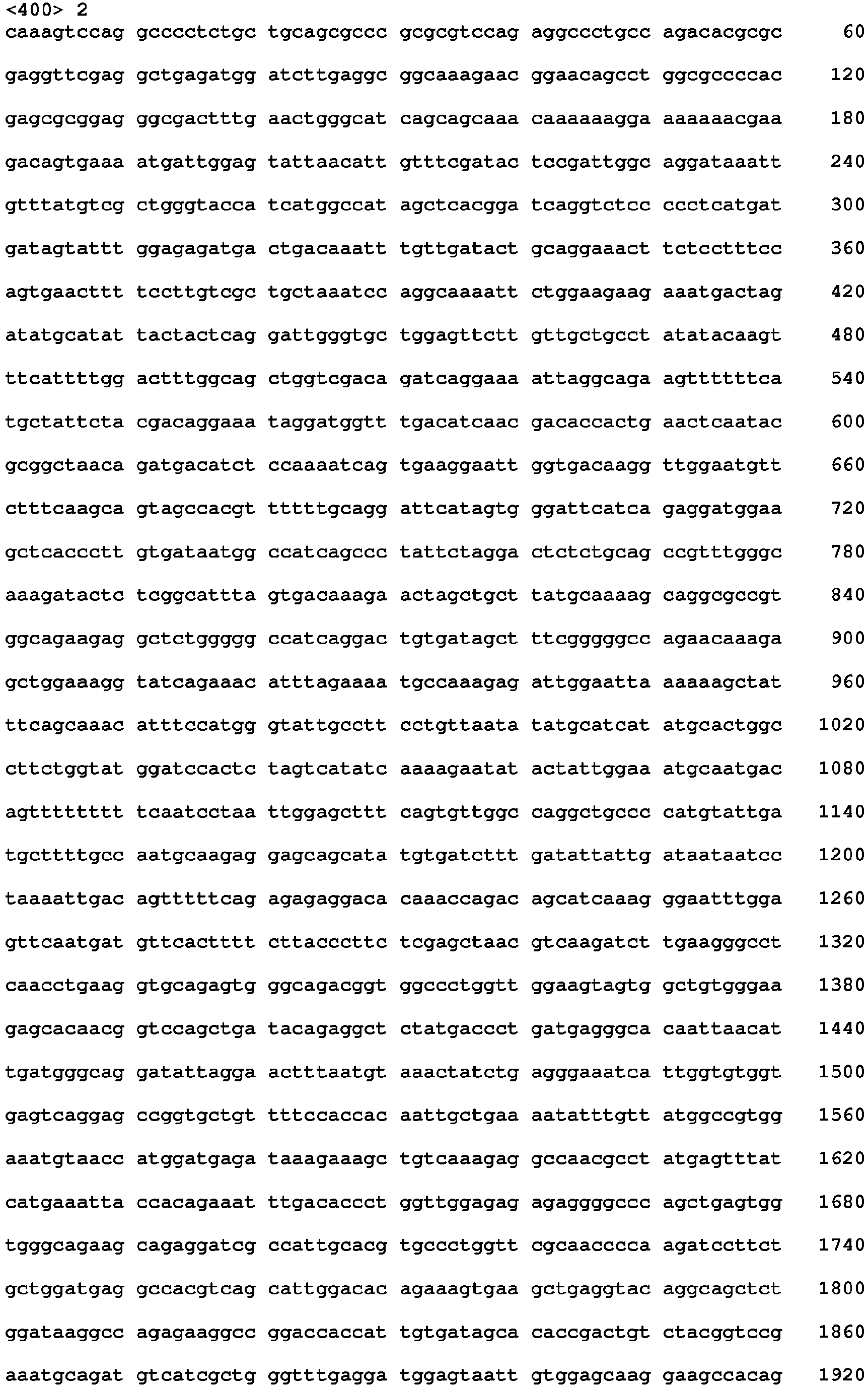
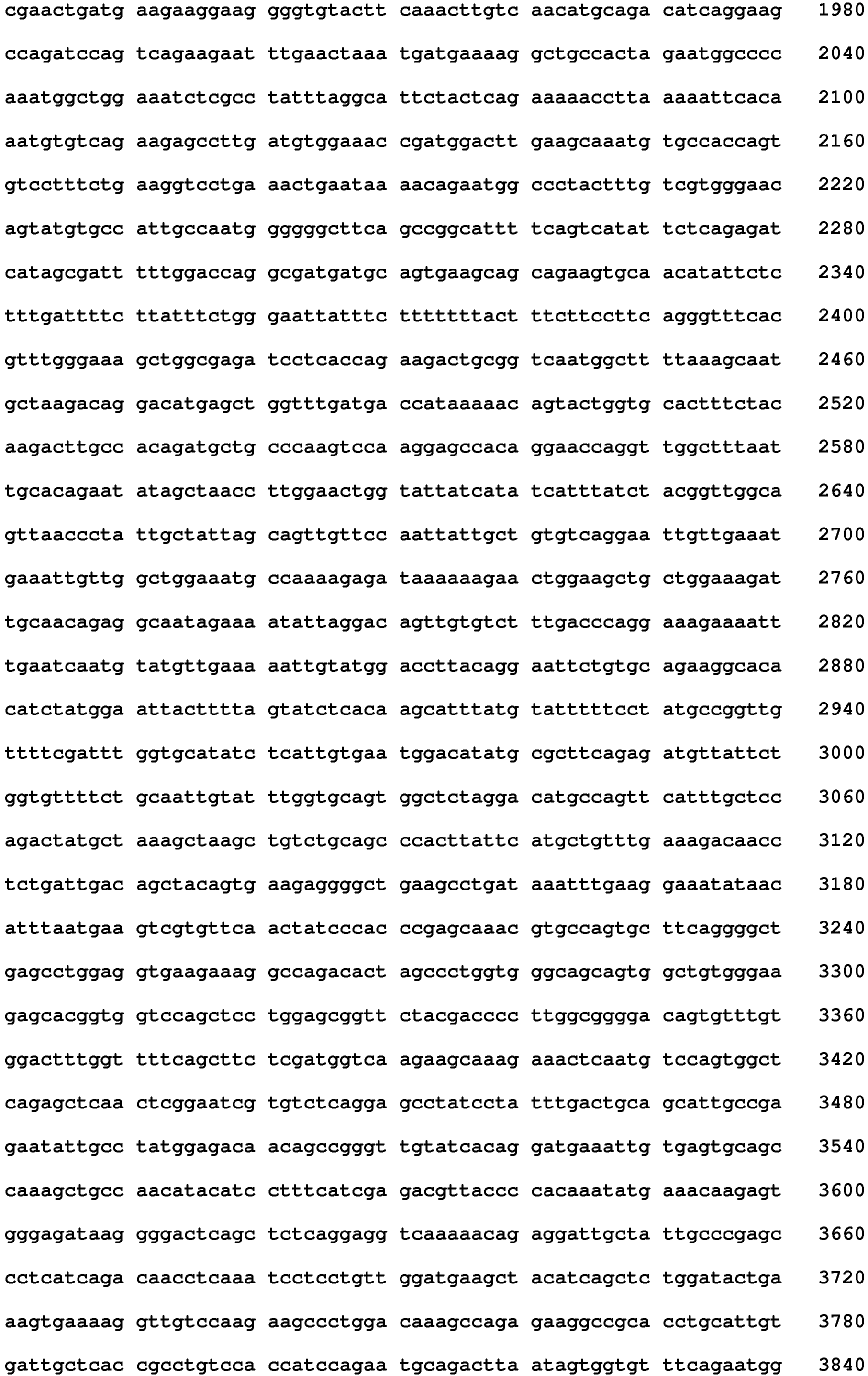


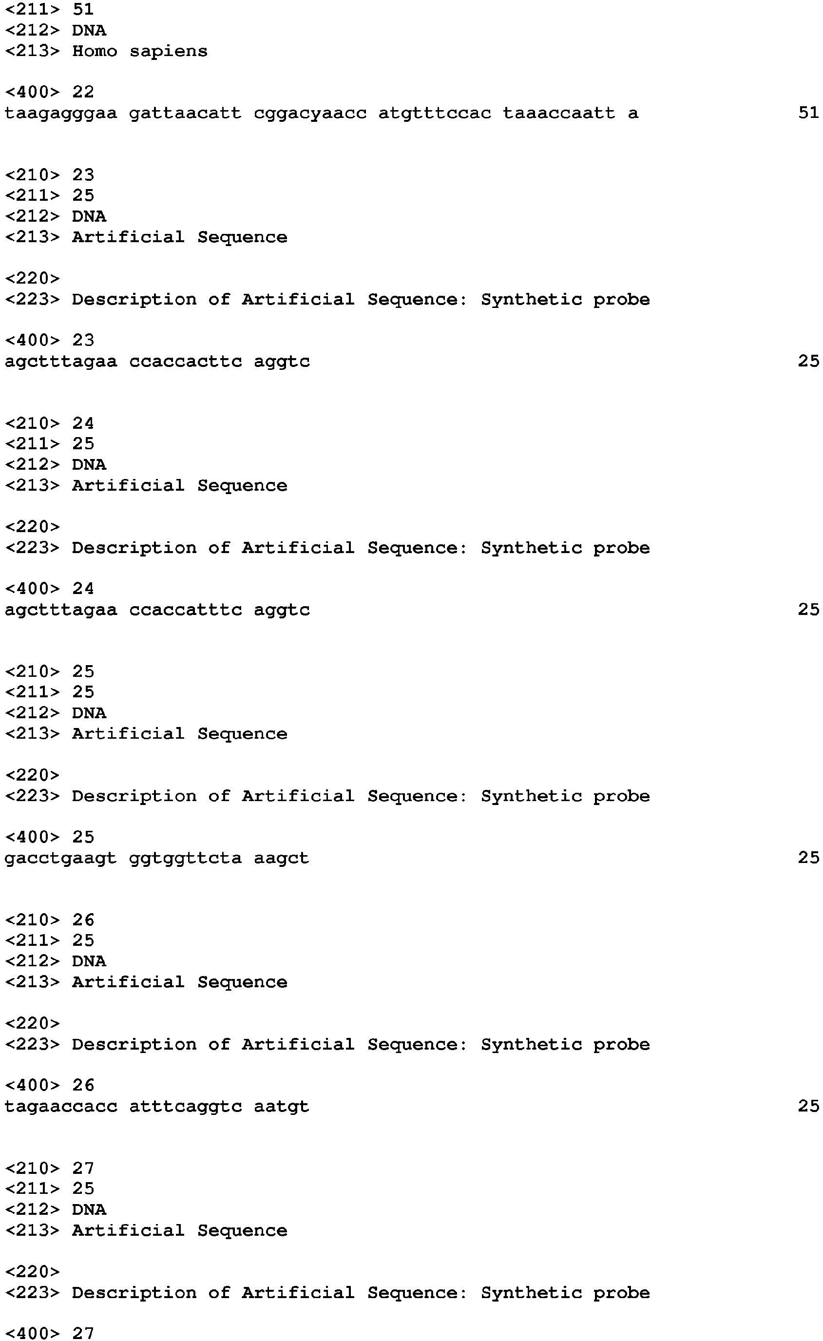


Claims (13)
- A method of classifying a patient for eligibility for treatment with an Aurora kinase B inhibitor, the method comprising the steps of:a) providing a test sample from a patient;b) determining the presence or absence of a copy number gain for the ABCB4 gene at chromosome locus 7q21.1; andc) classifying the patient as being eligible for receiving treatment with an Aurora kinase B inhibitor based on the presence or absence of a copy number gain for the ABCB4 gene at chromosome locus 7q21.1.
- A method of monitoring a patient suffering from cancer and being treated with an Aurora kinase B inhibitor, the method comprising the steps of:a) providing a test sample from a patient suffering from cancer and currently being treated with at least one Aurora kinase B inhibitor;b) determining the presence or absence of a copy number gain for the ABCB4 gene at chromosome locus 7q21.1;c) comparing the copy number of the ABCB4 gene in the test sample against a baseline level or a predetermined level; andd) determining whether the patient should continue to be treated with the Aurora kinase B inhibitor based on the comparison in step c).
- A method of classifying a patient having a cancer that is resistant to treatment with an Aurora kinase B inhibitor, the method comprising the steps of;a) providing a test sample from a patient;b) determining the presence or absence of a copy number gain for the ABCB4 gene at chromosome locus 7q21.1;c) comparing the presence or absence of the copy number gain for the ABCB4 gene in the test sample against a baseline level or a predetermined level; andd) classifying the patient as having a cancer that is resistant to Aurora kinase B inhibitor treatment on (i) the presence of a copy number gain in the ABCB4 gene at chromosome locus 7q21.1; and (ii) if the copy number gain in the test sample is higher then the baseline level or the predetermined level.
- The method of claims 1, 2 or 3, wherein the Aurora kinase B inhibitor is AZD1152, ZM447439, VX-680/MK0457 or Hersperadin.
- The method of claims 1, 2 or 3, wherein the test sample comprises a tissue sample.
- The method of claim 5, wherein the tissue sample comprises a peripheral blood sample, a tumor tissue or a suspected tumor tissue, a thin layer cytological sample, a fine needle aspirate sample, a bone marrow sample, a lymph node sample, a urine sample, an ascites sample, a lavage sample, an esophageal brushing sample, a bladder or lung wash sample, a spinal fluid sample, a brain fluid sample, a ductal aspirate sample, a nipple discharge sample, a pleural effusion sample, a fresh frozen tissue sample, a paraffin embedded tissue sample or an extract or processed sample produced from any of a peripheral blood sample, a tumor tissue or a suspected tumor tissue, a thin layer cytological sample, a fine needle aspirate sample, a bone marrow sample, a urine sample, an ascites sample, a lavage sample, an esophageal brushing sample, a bladder or lung wash sample, a spinal fluid sample, a brain fluid sample, a ductal aspirate sample, a nipple discharge sample, a pleural effusion sample, a fresh frozen tissue sample or a paraffin embedded tissue sample.
- The method of claims 1, 2 or 3, wherein the determining step (b) is performed by in situ hybridization.
- The method of claim 7, wherein the in situ hybridization is performed with(a) a nucleic acid probe that is fluorescently labeled; (b) at least two nucleic acid probes; or (c) a peptide nucleic acid probe.
- The method of claims 1, 2 or 3, wherein the determining step (b) is performed by polymerase chain reaction or a nucleic acid microarray assay.
- The method of claims 1, 2 or 3, wherein the cancer is colorectal carcinoma or pancreatic carcinoma.
- The method of claims 1, 2 or 3, wherein the presence of a copy number gain in the ABCB4 gene correlates with an increase in expression of the MDR3 polypeptide.
- The method of claims 1, 2 or 3, wherein the patient is also being treated with chemotherapy, radiation or combinations thereof.
- A kit comprising:(a) reagents for determining the presence or absence of a copy number gain for the ABCB4 gene, wherein the reagents to determine the presence or absence of a copy number gain comprise detectably-labeled polynucleotides that hybridize to at least a portion of the ABCB4 gene; and(b) instructions for performing the test.
Applications Claiming Priority (2)
| Application Number | Priority Date | Filing Date | Title |
|---|---|---|---|
| US14895709P | 2009-01-31 | 2009-01-31 | |
| EP10704252.5A EP2391734B1 (en) | 2009-01-31 | 2010-01-29 | Marker to predict and monitor response to aurora kinase b inhibitor therapy |
Related Parent Applications (1)
| Application Number | Title | Priority Date | Filing Date |
|---|---|---|---|
| EP10704252.5 Division | 2010-01-29 |
Publications (1)
| Publication Number | Publication Date |
|---|---|
| EP2479288A1 true EP2479288A1 (en) | 2012-07-25 |
Family
ID=42101875
Family Applications (2)
| Application Number | Title | Priority Date | Filing Date |
|---|---|---|---|
| EP10704252.5A Not-in-force EP2391734B1 (en) | 2009-01-31 | 2010-01-29 | Marker to predict and monitor response to aurora kinase b inhibitor therapy |
| EP12164247A Withdrawn EP2479288A1 (en) | 2009-01-31 | 2010-01-29 | Markers to predict and monitor response to Aurora kinase B inhibitor therapy |
Family Applications Before (1)
| Application Number | Title | Priority Date | Filing Date |
|---|---|---|---|
| EP10704252.5A Not-in-force EP2391734B1 (en) | 2009-01-31 | 2010-01-29 | Marker to predict and monitor response to aurora kinase b inhibitor therapy |
Country Status (7)
| Country | Link |
|---|---|
| US (1) | US20100196907A1 (en) |
| EP (2) | EP2391734B1 (en) |
| JP (1) | JP5616905B2 (en) |
| CN (1) | CN102365372B (en) |
| CA (1) | CA2749945A1 (en) |
| MX (1) | MX2011007989A (en) |
| WO (1) | WO2010088470A1 (en) |
Families Citing this family (5)
| Publication number | Priority date | Publication date | Assignee | Title |
|---|---|---|---|---|
| EP2502079B1 (en) * | 2009-11-20 | 2015-01-28 | Pharnext | New diagnostic tools for charcot-marie-tooth disease |
| CA2833534A1 (en) * | 2011-04-18 | 2012-10-26 | Cornell University | Molecular subtyping, prognosis and treatment of prostate cancer |
| EP2602330A1 (en) * | 2011-12-07 | 2013-06-12 | Palacky University, Olomouc | Method of determination of cancer cell drug sensitivity towards Aurora kinase inhibitors and overcoming their resistance |
| SI3311845T1 (en) * | 2013-09-16 | 2020-06-30 | Astrazeneca Ab | Therapeutic polymeric nanoparticles and methods of making and using same |
| EP3877056A4 (en) * | 2018-11-09 | 2022-08-17 | The Broad Institute, Inc. | Tepoxalin targeting of abcb1 overexpressing cancers |
Citations (24)
| Publication number | Priority date | Publication date | Assignee | Title |
|---|---|---|---|---|
| EP0050424A1 (en) | 1980-09-24 | 1982-04-28 | Cetus Corporation | Method for producing an analytical antibody probe, an analytical antibody probe, and a method for analysing a sample for certain antibodies |
| EP0084796A2 (en) | 1982-01-22 | 1983-08-03 | Cetus Corporation | HLA typing method and cDNA probes used therein |
| US4582788A (en) | 1982-01-22 | 1986-04-15 | Cetus Corporation | HLA typing method and cDNA probes used therein |
| EP0201184A2 (en) | 1985-03-28 | 1986-11-12 | F. Hoffmann-La Roche Ag | Process for amplifying nucleic acid sequences |
| US4683194A (en) | 1984-05-29 | 1987-07-28 | Cetus Corporation | Method for detection of polymorphic restriction sites and nucleic acid sequences |
| EP0237362A1 (en) | 1986-03-13 | 1987-09-16 | F. Hoffmann-La Roche Ag | Process for detecting specific nucleotide variations and genetic polymorphisms present in nucleic acids and kits therefor |
| EP0258017A2 (en) | 1986-08-22 | 1988-03-02 | F. Hoffmann-La Roche Ag | Purified thermostable enzyme and process for amplifying, detecting, and/or cloning nucleic acid sequences using said enzyme |
| US4948882A (en) | 1983-02-22 | 1990-08-14 | Syngene, Inc. | Single-stranded labelled oligonucleotides, reactive monomers and methods of synthesis |
| US5006309A (en) | 1988-04-22 | 1991-04-09 | Abbott Laboratories | Immunoassay device with liquid transfer between wells by washing |
| US5063081A (en) | 1988-11-14 | 1991-11-05 | I-Stat Corporation | Method of manufacturing a plurality of uniform microfabricated sensing devices having an immobilized ligand receptor |
| US5089424A (en) | 1988-06-14 | 1992-02-18 | Abbott Laboratories | Method and apparatus for heterogeneous chemiluminescence assay |
| US5231020A (en) | 1989-03-30 | 1993-07-27 | Dna Plant Technology Corporation | Genetic engineering of novel plant phenotypes |
| US5294404A (en) | 1991-06-03 | 1994-03-15 | Abbott Laboratories | Reagent pack for immunoassays |
| US5424414A (en) | 1991-12-17 | 1995-06-13 | Abbott Laboratories | Haptens, tracers, immunogens and antibodies for 3-phenyl-1-adamantaneacetic acids |
| US5447841A (en) | 1986-01-16 | 1995-09-05 | The Regents Of The Univ. Of California | Methods for chromosome-specific staining |
| US5464746A (en) | 1991-12-17 | 1995-11-07 | Abbott Laboratories | Haptens, tracers, immunogens and antibodies for carbazole and dibenzofuran derivatives |
| US5491224A (en) | 1990-09-20 | 1996-02-13 | Bittner; Michael L. | Direct label transaminated DNA probe compositions for chromosome identification and methods for their manufacture |
| US5756696A (en) | 1986-01-16 | 1998-05-26 | Regents Of The University Of California | Compositions for chromosome-specific staining |
| US20030170881A1 (en) | 2002-03-05 | 2003-09-11 | I-Stat Corporation | Apparatus and methods for analyte measurement and immuno assay |
| US20040018577A1 (en) | 2002-07-29 | 2004-01-29 | Emerson Campbell John Lewis | Multiple hybrid immunoassay |
| US20050054078A1 (en) | 2003-09-10 | 2005-03-10 | Miller Cary James | Immunoassay device with improved sample closure |
| US20060160164A1 (en) | 2003-09-10 | 2006-07-20 | Miller Cary J | Immunoassay device with immuno-reference electrode |
| WO2006123246A2 (en) * | 2005-02-11 | 2006-11-23 | Aurelium Biopharma Inc. | Methods for identifying chemotherapeutic resistance in non-hematopoietic tumors |
| US11028108B2 (en) | 2017-03-10 | 2021-06-08 | Jnc Corporation | Method for producing dialkylaminosilane |
Family Cites Families (1)
| Publication number | Priority date | Publication date | Assignee | Title |
|---|---|---|---|---|
| CA2739462A1 (en) | 2008-10-31 | 2010-05-06 | Abbott Laboratories | Methods for assembling panels of cancer cell lines for use in testing the efficacy of one or more pharmaceutical compositions |
-
2010
- 2010-01-29 EP EP10704252.5A patent/EP2391734B1/en not_active Not-in-force
- 2010-01-29 MX MX2011007989A patent/MX2011007989A/en active IP Right Grant
- 2010-01-29 CA CA2749945A patent/CA2749945A1/en not_active Abandoned
- 2010-01-29 US US12/696,116 patent/US20100196907A1/en not_active Abandoned
- 2010-01-29 CN CN201080014811.7A patent/CN102365372B/en not_active Expired - Fee Related
- 2010-01-29 EP EP12164247A patent/EP2479288A1/en not_active Withdrawn
- 2010-01-29 WO PCT/US2010/022518 patent/WO2010088470A1/en active Application Filing
- 2010-01-29 JP JP2011548326A patent/JP5616905B2/en not_active Expired - Fee Related
Patent Citations (27)
| Publication number | Priority date | Publication date | Assignee | Title |
|---|---|---|---|---|
| EP0050424A1 (en) | 1980-09-24 | 1982-04-28 | Cetus Corporation | Method for producing an analytical antibody probe, an analytical antibody probe, and a method for analysing a sample for certain antibodies |
| EP0084796A2 (en) | 1982-01-22 | 1983-08-03 | Cetus Corporation | HLA typing method and cDNA probes used therein |
| US4582788A (en) | 1982-01-22 | 1986-04-15 | Cetus Corporation | HLA typing method and cDNA probes used therein |
| US4948882A (en) | 1983-02-22 | 1990-08-14 | Syngene, Inc. | Single-stranded labelled oligonucleotides, reactive monomers and methods of synthesis |
| US4683194A (en) | 1984-05-29 | 1987-07-28 | Cetus Corporation | Method for detection of polymorphic restriction sites and nucleic acid sequences |
| US4683202B1 (en) | 1985-03-28 | 1990-11-27 | Cetus Corp | |
| US4683202A (en) | 1985-03-28 | 1987-07-28 | Cetus Corporation | Process for amplifying nucleic acid sequences |
| EP0201184A2 (en) | 1985-03-28 | 1986-11-12 | F. Hoffmann-La Roche Ag | Process for amplifying nucleic acid sequences |
| US5447841A (en) | 1986-01-16 | 1995-09-05 | The Regents Of The Univ. Of California | Methods for chromosome-specific staining |
| US5756696A (en) | 1986-01-16 | 1998-05-26 | Regents Of The University Of California | Compositions for chromosome-specific staining |
| EP0237362A1 (en) | 1986-03-13 | 1987-09-16 | F. Hoffmann-La Roche Ag | Process for detecting specific nucleotide variations and genetic polymorphisms present in nucleic acids and kits therefor |
| EP0258017A2 (en) | 1986-08-22 | 1988-03-02 | F. Hoffmann-La Roche Ag | Purified thermostable enzyme and process for amplifying, detecting, and/or cloning nucleic acid sequences using said enzyme |
| US5006309A (en) | 1988-04-22 | 1991-04-09 | Abbott Laboratories | Immunoassay device with liquid transfer between wells by washing |
| US5089424A (en) | 1988-06-14 | 1992-02-18 | Abbott Laboratories | Method and apparatus for heterogeneous chemiluminescence assay |
| US5063081A (en) | 1988-11-14 | 1991-11-05 | I-Stat Corporation | Method of manufacturing a plurality of uniform microfabricated sensing devices having an immobilized ligand receptor |
| US5231020A (en) | 1989-03-30 | 1993-07-27 | Dna Plant Technology Corporation | Genetic engineering of novel plant phenotypes |
| US5776688A (en) | 1990-09-20 | 1998-07-07 | Vysis, Inc. | Methods for detection by in situ hybridization of multiple chromosomes or regions thereof |
| US5491224A (en) | 1990-09-20 | 1996-02-13 | Bittner; Michael L. | Direct label transaminated DNA probe compositions for chromosome identification and methods for their manufacture |
| US5294404A (en) | 1991-06-03 | 1994-03-15 | Abbott Laboratories | Reagent pack for immunoassays |
| US5464746A (en) | 1991-12-17 | 1995-11-07 | Abbott Laboratories | Haptens, tracers, immunogens and antibodies for carbazole and dibenzofuran derivatives |
| US5424414A (en) | 1991-12-17 | 1995-06-13 | Abbott Laboratories | Haptens, tracers, immunogens and antibodies for 3-phenyl-1-adamantaneacetic acids |
| US20030170881A1 (en) | 2002-03-05 | 2003-09-11 | I-Stat Corporation | Apparatus and methods for analyte measurement and immuno assay |
| US20040018577A1 (en) | 2002-07-29 | 2004-01-29 | Emerson Campbell John Lewis | Multiple hybrid immunoassay |
| US20050054078A1 (en) | 2003-09-10 | 2005-03-10 | Miller Cary James | Immunoassay device with improved sample closure |
| US20060160164A1 (en) | 2003-09-10 | 2006-07-20 | Miller Cary J | Immunoassay device with immuno-reference electrode |
| WO2006123246A2 (en) * | 2005-02-11 | 2006-11-23 | Aurelium Biopharma Inc. | Methods for identifying chemotherapeutic resistance in non-hematopoietic tumors |
| US11028108B2 (en) | 2017-03-10 | 2021-06-08 | Jnc Corporation | Method for producing dialkylaminosilane |
Non-Patent Citations (37)
| Title |
|---|
| ANDREWS, P.D. ET AL., CURR. OPIN. CELL BIOL., vol. 15, 2003, pages 672 - 683 |
| AUSUBEL, F.M.; R. BRENT; R.E. KINGSTON: "Current Protocols in Molecular Biology", 1987, JOHN WILEY & SONS |
| BATES, S.F. ET AL., NOVARTIS FOUND. SYMP., 2002, pages 83 - 96 |
| BOESCH, D., CANCER RES., vol. 51, 1991, pages 4226 - 4233 |
| BUCHARDT, O.; P. NIELSEN; R. BERG., PEPTIDE NUCLEIC ACIDS, 1992 |
| CARMENA M.E.W., NAT. REV. MOL. CELL BIOL., vol. 4, 2003, pages 842 - 854 |
| DITCHFIELD, C, J.V. ET AL., J. CELL BIOL., vol. 161, 2003, pages 267 - 280 |
| DITCHFIELD, C. ET AL., J. CELL BIO., vol. 161, no. 2, 2003, pages 267 - 280 |
| DUCAT, D.Z.Y, EXP. CELL RES., vol. 301, 2004, pages 60 - 67 |
| FAN, Y.-S: "Molecular cytogenetics: protocols and applications", 2002, HUMANA PRESS, pages: 411 |
| GIRDLER F ET AL: "Molecular Basis of Drug Resistance in Aurora Kinases", CHEMISTRY AND BIOLOGY, CURRENT BIOLOGY, LONDON, GB LNKD- DOI:10.1016/J.CHEMBIOL.2008.04.013, vol. 15, no. 6, 23 June 2008 (2008-06-23), pages 552 - 562, XP022734025, ISSN: 1074-5521, [retrieved on 20080620] * |
| GIRDLER, F. ET AL., CHEM. BIOL., vol. 15, 2008, pages 552 - 562 |
| HARRINGTON, E.A. ET AL., NAT. MED., vol. 10, 2004, pages 262 - 267 |
| HAUF, S. ET AL., J. CELL BIOL., vol. 161, 2003, pages 281 - 294 |
| HAUF, S., J. CELL BIO., vol. 161, no. 2, 2003, pages 281 - 294 |
| KITADA ET AL: "The MDR1/ABCB1 regional amplification in large inverted repeats with asymmetric sequences and microhomologies at the junction sites", CANCER GENETICS AND CYTOGENETICS, ELSEVIER SCIENCE PUBLISHING, NEW YORK, NY, US, vol. 178, no. 2, 22 October 2007 (2007-10-22), pages 120 - 127, XP022308146, ISSN: 0165-4608 * |
| MAHON FRANCOIS-XAVIER ET AL: "MDR1 gene overexpression confers resistance to imatinib mesylate in leukemia cell line models", BLOOD, AMERICAN SOCIETY OF HEMATOLOGY, US LNKD- DOI:10.1182/BLOOD.V101.6.2368, vol. 101, no. 6, 15 March 2003 (2003-03-15), pages 2368 - 2373, XP002404659, ISSN: 0006-4971 * |
| MANFREDI, M.G. ET AL., PROC. NATL. ACAD. SCI., USA, vol. 104, 2007, pages 4106 - 4111 |
| MANFRIEDI, MG ET AL., PROC. NATT. ACAD. SCI. USA, vol. 104, 2007, pages 4106 - 4111 |
| MARUMOTO, T.Z.D. ET AL., NAT. REV. CANCER, vol. 5, 2005, pages 42 - 50 |
| MATSUZAKI, H. ET AL.: "Genotyping over 100,000 SNPs on a pair ofoligonucleotide arrays", NAT METHODS, vol. 1, 2004, pages 109 - 11, XP009082656, DOI: doi:10.1038/nmeth718 |
| MONTEMBAULT, E ET AL., DRUGS OF THE FUTURE, vol. 30, no. 1, 2005, pages 1 - 9 |
| MONTEMBAULT, E. ET AL., DRUGS OF THE FUTURE, vol. 30, no. 1, 2005, pages 1 - 9 |
| MORTLOCK, A.A. ET AL., J. MED. CHEM., vol. 50, 2007, pages 2213 - 2224 |
| MORTLOCK, AA ET AL., J. MED. CHEM., vol. 50, 2007, pages 2213 - 24 |
| MULLIS, K. ET AL., COLD SPRING HARB SYMP QUANT BIOL., vol. 51, 1986, pages 263 - 73 |
| OLEJNICZAK ET AL., MOL. CAN. RES., vol. 5, no. 4, 2007, pages 331 - 339 |
| PALLIS, M.R.N., LEUKEMIA, vol. 18, 2004, pages 1927 - 1930 |
| SMITH AJ., J. BIOL. CHEM., vol. 275, 2000, pages 23530 - 23539 |
| TROCK, B.J. ET AL., J. NATL. CANCER INST., vol. 89, 1997, pages 917 - 931 |
| TURTON N J ET AL: "Gene expression and amplification in breast carcinoma cells with intrinsic and acquired doxorubicin resistance", ONCOGENE, NATURE PUBLISHING GROUP, GB, vol. 20, 1 January 2001 (2001-01-01), pages 1300 - 1306, XP002980903, ISSN: 0950-9232, DOI: 10.1038/SJ.ONC.1204235 * |
| TWENTYMAN PR, EUR. J. CANCER, vol. 27, 1991, pages 1639 - 1642 |
| VAN DER HOLT, B.L.B., BLOOD, vol. 106, 2005, pages 2646 - 2654 |
| VAN DER KROL ET AL., BIOTECHNIQUES, vol. 6, 1988, pages 958 - 76 |
| WILKINSON ROBERT W ET AL: "AZD1152, a selective inhibitor of Aurora B kinase, inhibits human tumor xenograft growth by inducing apoptosis.", CLINICAL CANCER RESEARCH : AN OFFICIAL JOURNAL OF THE AMERICAN ASSOCIATION FOR CANCER RESEARCH 15 JUN 2007 LNKD- PUBMED:17575233, vol. 13, no. 12, 15 June 2007 (2007-06-15), pages 3682 - 3688, XP002578490, ISSN: 1078-0432 * |
| WILKINSON, R.W. ET AL., CLIN. CANCER RES., vol. 13, 2007, pages 3683 - 3688 |
| ZON, G., PHARM RES., vol. 5, 1988, pages 539 - 49 |
Also Published As
| Publication number | Publication date |
|---|---|
| WO2010088470A1 (en) | 2010-08-05 |
| JP2012516160A (en) | 2012-07-19 |
| JP5616905B2 (en) | 2014-10-29 |
| CA2749945A1 (en) | 2010-08-05 |
| EP2391734B1 (en) | 2013-06-05 |
| US20100196907A1 (en) | 2010-08-05 |
| CN102365372B (en) | 2016-05-25 |
| CN102365372A (en) | 2012-02-29 |
| EP2391734A1 (en) | 2011-12-07 |
| MX2011007989A (en) | 2011-08-17 |
Similar Documents
| Publication | Publication Date | Title |
|---|---|---|
| US9045800B2 (en) | Methods and compositions for identifying, classifying and monitoring subject having Bcl-2 family inhibitor-resistant tumors and cancers | |
| AU2019216598A1 (en) | Human papilloma virus as predictor of cancer prognosis | |
| JP2015226536A (en) | Susceptibility to HSP90 inhibitor | |
| ES2818823T3 (en) | PML risk assessment method | |
| KR20100095571A (en) | Methods and compositions for diagnostic use in cancer patients | |
| EP2479288A1 (en) | Markers to predict and monitor response to Aurora kinase B inhibitor therapy | |
| JP2018531223A (en) | Novel biomarkers and methods for treating cancer | |
| JP2018531223A6 (en) | Novel biomarkers and methods for treating cancer | |
| US20120270744A1 (en) | Methods to determine if a subject will respond to a bcr-abl inhibitor | |
| EP2557159B1 (en) | Prognostic method for pulmonary adenocarcinoma, pulmonary adenocarcinoma detection kit, and pharmaceutical composition for treating pulmonary adenocarcinoma | |
| US20220317127A1 (en) | Method and compositions for predicting anti-cancer efficacy of compounds targeting apoptosis pathway | |
| US20220233558A1 (en) | Method and composition for predicting efficacy of bcl2/bcl-xl inhibitors on cancer | |
| WO2016042114A1 (en) | Cxcl14 as a biomarker of hedgehog pathway activity for the diagnosis, prognosis and treatment of idiopathic pulmonary fibrosis | |
| US20220127680A1 (en) | Apoe genotyping in cancer prognostics and treatment | |
| US20160153052A1 (en) | Marker to predict and monitor response to aurora kinase b inhibitor therapy | |
| KR102049090B1 (en) | An anticancer agent for treating cancers resistant to EGFR inhibitor | |
| WO2021055528A1 (en) | Methods and kit for analyzing responsiveness of patients to cd19 immunotherapy | |
| EP3470528A1 (en) | Method for predicting effect of pharmacotherapy on cancer | |
| CN116457473A (en) | ER-modulating genome sleeve for use in endocrine therapy for monitoring breast cancer | |
| CN116287258A (en) | Application of novel molecular marker in breast cancer diagnosis or prognosis evaluation | |
| US20110184004A1 (en) | Predictive marker for egfr inhibitor treatment | |
| WO2013172922A1 (en) | Lmtk3 genotype analysis for use in predicting outcome and therapy selection |
Legal Events
| Date | Code | Title | Description |
|---|---|---|---|
| PUAI | Public reference made under article 153(3) epc to a published international application that has entered the european phase |
Free format text: ORIGINAL CODE: 0009012 |
|
| AC | Divisional application: reference to earlier application |
Ref document number: 2391734 Country of ref document: EP Kind code of ref document: P |
|
| AK | Designated contracting states |
Kind code of ref document: A1 Designated state(s): AT BE BG CH CY CZ DE DK EE ES FI FR GB GR HR HU IE IS IT LI LT LU LV MC MK MT NL NO PL PT RO SE SI SK SM TR |
|
| RIN1 | Information on inventor provided before grant (corrected) |
Inventor name: ANDERSON, MARK Inventor name: SHAH, JAMEEL Inventor name: GUO, JUN Inventor name: SEMIZAROV, DIMITRI |
|
| RIN1 | Information on inventor provided before grant (corrected) |
Inventor name: ANDERSON, MARK Inventor name: SHAH, JAMEEL Inventor name: SEMIZAROV, DIMITRI Inventor name: GUO, JUN |
|
| 17P | Request for examination filed |
Effective date: 20130125 |
|
| 17Q | First examination report despatched |
Effective date: 20130225 |
|
| RAP1 | Party data changed (applicant data changed or rights of an application transferred) |
Owner name: ABBVIE INC. |
|
| GRAP | Despatch of communication of intention to grant a patent |
Free format text: ORIGINAL CODE: EPIDOSNIGR1 |
|
| INTG | Intention to grant announced |
Effective date: 20130703 |
|
| STAA | Information on the status of an ep patent application or granted ep patent |
Free format text: STATUS: THE APPLICATION IS DEEMED TO BE WITHDRAWN |
|
| 18D | Application deemed to be withdrawn |
Effective date: 20131114 |