EP2444805A2 - Detection of mesothelin-/megakaryocyte potentiating factor-related peptides in peritoneal fluid for assessment of the peritoneum and the peritoneal cavity - Google Patents
Detection of mesothelin-/megakaryocyte potentiating factor-related peptides in peritoneal fluid for assessment of the peritoneum and the peritoneal cavity Download PDFInfo
- Publication number
- EP2444805A2 EP2444805A2 EP11183171A EP11183171A EP2444805A2 EP 2444805 A2 EP2444805 A2 EP 2444805A2 EP 11183171 A EP11183171 A EP 11183171A EP 11183171 A EP11183171 A EP 11183171A EP 2444805 A2 EP2444805 A2 EP 2444805A2
- Authority
- EP
- European Patent Office
- Prior art keywords
- mesothelial
- antibody
- fluid
- mmpff
- peptide
- Prior art date
- Legal status (The legal status is an assumption and is not a legal conclusion. Google has not performed a legal analysis and makes no representation as to the accuracy of the status listed.)
- Withdrawn
Links
Images


Classifications
-
- G—PHYSICS
- G01—MEASURING; TESTING
- G01N—INVESTIGATING OR ANALYSING MATERIALS BY DETERMINING THEIR CHEMICAL OR PHYSICAL PROPERTIES
- G01N33/00—Investigating or analysing materials by specific methods not covered by groups G01N1/00 - G01N31/00
- G01N33/48—Biological material, e.g. blood, urine; Haemocytometers
- G01N33/50—Chemical analysis of biological material, e.g. blood, urine; Testing involving biospecific ligand binding methods; Immunological testing
- G01N33/53—Immunoassay; Biospecific binding assay; Materials therefor
- G01N33/574—Immunoassay; Biospecific binding assay; Materials therefor for cancer
- G01N33/57407—Specifically defined cancers
-
- G—PHYSICS
- G01—MEASURING; TESTING
- G01N—INVESTIGATING OR ANALYSING MATERIALS BY DETERMINING THEIR CHEMICAL OR PHYSICAL PROPERTIES
- G01N33/00—Investigating or analysing materials by specific methods not covered by groups G01N1/00 - G01N31/00
- G01N33/48—Biological material, e.g. blood, urine; Haemocytometers
- G01N33/50—Chemical analysis of biological material, e.g. blood, urine; Testing involving biospecific ligand binding methods; Immunological testing
- G01N33/53—Immunoassay; Biospecific binding assay; Materials therefor
- G01N33/574—Immunoassay; Biospecific binding assay; Materials therefor for cancer
- G01N33/57484—Immunoassay; Biospecific binding assay; Materials therefor for cancer involving compounds serving as markers for tumor, cancer, neoplasia, e.g. cellular determinants, receptors, heat shock/stress proteins, A-protein, oligosaccharides, metabolites
- G01N33/57488—Immunoassay; Biospecific binding assay; Materials therefor for cancer involving compounds serving as markers for tumor, cancer, neoplasia, e.g. cellular determinants, receptors, heat shock/stress proteins, A-protein, oligosaccharides, metabolites involving compounds identifable in body fluids
-
- G—PHYSICS
- G01—MEASURING; TESTING
- G01N—INVESTIGATING OR ANALYSING MATERIALS BY DETERMINING THEIR CHEMICAL OR PHYSICAL PROPERTIES
- G01N2800/00—Detection or diagnosis of diseases
- G01N2800/50—Determining the risk of developing a disease
Definitions
- the invention relates generally to monitoring biochemical status of the peritoneal cavity, fluids therein, and the integrity of the peritoneal membrane.
- Mesothelial membranes line various body cavities, such as the peritoneal cavity, the pleural cavity, and the pericardial cavity. Under certain circumstances, cells of mesothelial origin produce mesothelin, and mesothelin protein is sometimes secreted or shed from cells of mesothelial membranes.
- Mesothelin is a protein which is expressed on the surface of at least mesothelial cells, mesotheliomas, some squamous cell carcinomas, and ovarian cancers ( Chang et al., ., 1996, Proc. Natl. Acad. Sci. USA 93:136-140 ). Mesothelin has been described in the literature (e.g., U.S. Patent No. 6,083,502 ). Antibodies that bind with mesothelin have also been described. For example, a monoclonal antibody designated K1 is described in U.S. Patent No.
- a useful diagnostic test should not only be able to detect occurrence of mesothelin and related proteins in a patient, it is also preferably inexpensive, non-invasive, and easy to use. Diagnostic tests that use blood or serum as a testing substrate require that blood be drawn from a patient to be screened, and sometimes require further separation, purification, or other treatment of the withdrawn blood sample. Drawing blood must be performed by a trained health care worker, is painful and invasive for the patient, requires specialized equipment and reagents for storage and transportation of blood samples, and carries the risk of transmission of blood-borne pathogens.
- fluid samples are obtained from many patients who exhibit fluid buildup (effusion) within a body cavity lined by a mesothelial membrane such as the peritoneum, the pleural membranes, and the pericardium.
- a mesothelial membrane such as the peritoneum, the pleural membranes, and the pericardium.
- the present invention provides a diagnostic test that can be performed using a fluid sample obtained from a mesothelial cavity in order to monitor the biochemical status of the cavity, organs or tissues within the cavity, or the mesothelium lining the cavity (collectively, to monitor "mesothelial cavity status").
- the test can ,be performed using peritoneal fluid to assess the status of peritoneal organs, peritoneal fluid, and the peritoneum.
- the invention relates to a method of monitoring mesothelial cavity status in a human patient.
- the method comprises assessing occurrence of a mesothelin/megakaryocyte potentiating factor family (MMPFF) peptide in a mesothelial fluid obtained from the patient. Occurrence of the MMPFF peptide in the mesothelial fluid is an indication that the patient is afflicted with mesothelioma.
- MMPFF mesothelin/megakaryocyte potentiating factor family
- the MMPFF peptide is assessed in the patient's mesothelial fluid by contacting the mesothelial fluid (or centrifuged mesothelial fluid) with a first antibody that binds specifically with the MMPFF peptide.
- a first antibody that binds specifically with the MMPFF peptide.
- the first antibody can be bound to a substrate, such as a plastic multi-well plate of the type adapted for automated analysis in a robotic apparatus.
- Binding of the MMPFF peptide and the first antibody can, for example, be assessed by contacting the first antibody with a second antibody that binds specifically with the MMPFF peptide and assessing co-localization of the first and second antibodies.
- the second antibody can be detectably labeled, such as with a compound selected from the group consisting of an enzyme, a radionuclide, a fluorophore, and a chromophore.
- the method can comprise contacting a plate having an anti-MMPFF peptide 'capture' antibody bound thereto with mesothelial fluid obtained from a patient.
- the mesothelial fluid can, optionally, be incubated with the plate for a period such as 10 minutes or an hour.
- the plate can then be contacted with a biotinylated second antibody that binds with an epitope of the MMPFF peptide than does the capture antibody.
- a streptavidin-linked enzyme can be bound to the biotinylated antibody to enable its detection in the presence of a chromogenic substrate (e.g., 3,3',5,5'-tetramethylbenzidine, which is a chromogenic substrate of horseradish peroxidase).
- a chromogenic substrate e.g., 3,3',5,5'-tetramethylbenzidine, which is a chromogenic substrate of horseradish peroxidase.
- the second antibody could instead be fluorescently labeled, radiolabeled, or otherwise detectably labeled.
- the first antibody can be contacted with a labeled substrate thereof contacting the first antibody with the mesothelial fluid.
- a labeled substrate thereof contacting the first antibody with the mesothelial fluid.
- kits and methods described herein can be used to detect a variety of MMPFF peptides, including naturally occurring MMPFF peptides (i.e., both full length proteins such as mesothelin and fragments of such proteins that are naturally produced in the bodies of patients) and synthetically produced MMPFF peptides.
- the amino acid sequence of the MMPFF peptide should comprise at least 10 (20, 50, or 200) consecutive residues of a sequence selected from the group consisting of SEQ ID NOs: 1-5.
- the MMPFF peptide can comprise a portion of at least 20 consecutive amino acid residues, wherein the amino acid sequence of the portion is at least 90% (or 95%) identical to 20 consecutive residues of a sequence selected from the group consisting of SEQ ID NOs: 1-5.
- kits and methods disclosed herein can be used in conjunction with known or hereafter developed kits and methods for assessing occurrence (e.g., in mesothelial fluid or serum) other markers of pathological status of tissues or organs within the corresponding mesothelial cavity.
- Assessment of occurrence of MMPFF peptide in a patient's mesothelial fluid can likewise be performed in conjunction with assessment of the MMPFF peptide in the patient's scrum.
- kits and methods can be used to assess the likelihood that a patient will develop a pathology of an organ or tissue of within a mesothelial cavity or of the mesothelial membrane lining the cavity. That is, occurrence of an MMPFF peptide in mesothelial fluid obtained from a patient is an indication that the patient is more likely to develop such pathology than an otherwise identical patient in whose mesothelial fluid the MMPFF does not occur.
- kits and methods can also be used to assess the efficacy of a therapy for a pathological condition of a tissue or organ within a mesothelial cavity or for a pathological condition of a mesothelial membrane in a patient.
- the amount of an MMPFF peptide in mesothelial fluid obtained from the patient following the therapy can be compared with the amount of the MMPFF in her mesothelial fluid before the therapy.
- a lower amount of the MMPFF peptide in the mesothelial fluid following the therapy is an indication that the therapy is efficacious.
- a greater amount of the MMPFF peptide in the mesothelial fluid following the therapy is an indication that the therapy is not efficacious.
- Such kits and methods can be used to compare the efficacy of various therapeutic agents or regimens.
- the invention in another aspect, relates to a kit for assessing occurrence of an MMPFF peptide in mesothelial fluid obtained from a patient.
- the kit comprises a first agent (e.g., an antibody) for binding the MMPFF peptide and an instructional material that describes contacting mesothelial fluid with the first agent.
- the format of the kit is not critical - many standard formats can be used, such as ELISA, latex bead aggregation, and surface plasmon resonance formats.
- the kit can include an MMPFF peptide for use as a positive control.
- the kit can also include reagents for assessing other known markers of pathological conditions of mesothelial membranes or tissues or organs within mesothelial cavities.
- Figure 1 is the amino acid sequence (SEQ ID NO: 1) of mesothelin precursor protein. This sequence is the same as that listed in GENBANK® accession number 2207386A and reported in Chang et al., 1996, Proc. Natl. Acad. Sci. USA 93(1):136-140 .
- Figure 2 is the amino acid sequence (SEQ ID NO: 2) of megakaryocyte potentiating factor (MPF) precursor protein. This sequence is the same as that listed in GENBANK® accession number NP_005814 and reported in Yamaguchi et al., 1994, J. Biol. Chem. 269(2):805-808 .
- Figure 3 is a partial amino acid sequence (SEQ lD NO: 3) of a soluble variant form of MPF precursor protein listed in GENBANK® accession number AF180951 and reported in Scholler et al., 1999, Proc. Natl. Acad. Sci. USA 96(20):11531-11536 .
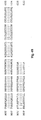
- Figure 4 is a comparison of residues 294-628 of SEQ ID NO: 1 and a nearly identical portion of SEQ ID NO: 2. Residue numbers are listed in the right margin. "MST” means mesothelin. "MPF” means megakaryocyte potentiating factor. In this alignment, identical amino acid residues are indicated by a vertical line, a conservative amino acid substitution is indicated by a cross (+), and dashes (-) indicate a gap inserted into SEQ ID NO: 2 for the purpose of aligning the sequences.
- Figure 5 is a comparison of similar portions of SEQ ID NOs: 1-3. Residue numbers are listed in the right margin.
- MST means mesothelin.
- MPF means megakaryocyte potentiating factor.
- SMR means the soluble variant form of MPF for which the sequence is listed in Fig. 3 .
- residues that are identical in all three sequences are represented by upper case single-letter codes, residues that are not identical in all three sequences are represented by lower case single-letter codes, and gaps inserted for the purpose of aligning the sequences are represented by dashes (-).
- Figure 6 comprises Figures 6A and 6B.
- Figure 6A is an amino acid sequence (SEQ ID NO: 4) that includes preferred epitopes for generation of antibodies described herein.
- Figure 6B is another amino acid sequence (SEQ ID NO: 5) that includes preferred epitopes for generation of antibodies described herein.
- the underlined, bold X represents any amino acid residue, preferably being either aspartate or asparagine.
- Other underlined, bold residues in Figure 6A represent residues that are not present in the sequence shown in Figure 6B .
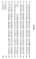
- Figure 7 is a graph that indicates peritoneal fluid MMPFF peptide levels in peritoneal dialysis samples described in Example 2.
- the invention relates to the discovery that one or more polypeptides having significant amino acid sequence similarity with a family of proteins that includes mesothelin, megakaryocyte potentiating factor (MPF), and a soluble variant of MPF occurs in the mesothelial fluid of patients afflicted with pathological mesothelial membranes or with pathological conditions of tissues, organs, or fluids within a mesothelial (e.g., peritoneal, pleural, or pericardial) cavity.
- MPF megakaryocyte potentiating factor
- a soluble variant of MPF occurs in the mesothelial fluid of patients afflicted with pathological mesothelial membranes or with pathological conditions of tissues, organs, or fluids within a mesothelial (e.g., peritoneal, pleural, or pericardial) cavity.
- mesothelial fluid polypeptides bind specifically with antibodies that are raised against proteins of that family
- Occurrence of these polypeptides in a patient's mesothelial fluid is diagnostic that the patient is afflicted with such a pathological condition.
- concentration or occurrence of the polypeptides can also be used to monitor the biochemical status of a mesothelial cavity, such as a fluid, tissue, or organ within the cavity or the mesothelial membrane lining the cavity.
- the amount of the polypeptides decreases with effective treatment of such pathological conditions.
- the mesothelial fluid polypeptides can also be used to assess the efficacy of various therapeutic methods.
- MMPFF peptide is a polypeptide that has an amino acid sequence that either (i) comprises at least 10 consecutive residues of a sequence selected from the group consisting of SEQ ID NOs: 1-5 or (ii) comprises a portion of at least 20 consecutive amino acid residues wherein the amino acid sequence of the portion is at least 90% (preferably at least 95% or 100%) identical to 20 consecutive residues of a sequence selected from the group consisting of SEQ ID NOs: 1-5.
- an “antibody” refers collectively and individually to immunoglobulin molecules and immunologically active portions of immunoglobulin molecules, i.e., molecules that contain an antigen binding site which specifically binds an MMPFF peptide.
- a molecule which specifically binds with an MMPFF peptide is a molecule which binds the peptide, but does not substantially bind other molecules in a sample (e.g., a mesothelial fluid sample) which naturally contains the polypeptide.
- immunologically active portions of immunoglobulin molecules include F(ab) and F(ab') 2 fragments which can be generated by treating the antibody with an enzyme such as papain or pepsin, respectively. Either or both of polyclonal and monoclonal antibodies can be used in the methods and kits described herein.
- monoclonal antibody refers to a population of antibody molecules that contain only one species of an antigen binding site capable of immunoreacting with a , I particular epitope.
- labeled with regard to an antibody or a peptide, includes direct labeling of the peptide or antibody by coupling (i.e., physically linking) a detectable substance to the peptide or antibody, as well as indirect labeling of the peptide or antibody by coupling it with another reagent that is directly labeled.
- indirect labeling include detection of a primary antibody using a fluorescently labeled secondary antibody and end-labeling of a peptide with biotin such that it can be detected with fluorescently labeled streptavidin.
- an "instructional material” is a publication, a recording, a diagram, or any other medium of expression which can be used to communicate how to use a kit described herein, information for interpreting occurrence of an MMPFF in mesothelial fluid as it relates to the biochemical status, pathological state, or both, of a mesothelial cavity component (e.g., a fluid, tissue, or organ contained within the cavity) or of a mesothelial cavity membrane (e.g., the peritoneum, a pleural membrane, or the pericardium).
- the instructional material of the kit of the invention can, for example, be affixed to a container which contains a kit of the invention or be shipped together with a container which contains the kit. Alternatively, the instructional material can be shipped separately from the container with the intention that the instructional material and the kit be used cooperatively by the recipient.
- a "mesothelial" cavity is a body cavity lined with a membrane of mesothelial origin, such as the peritoneal cavity, the pleural cavities, and the pericardial cavity.
- a “component of a mesothelial cavity” means the membrane lining the cavity, a fluid contained within the cavity, or a tissue or organ within the cavity.
- a “mesothelial fluid” means a fluid which either naturally occurs in the cavity (e.g., peritoneal fluid) or is artificially introduced into the cavity (e.g., a peritoneal washing or dialysis fluid).
- Soluble mesothelin-related peptides were first described as a serum marker for ovarian cancer and more generally as a serum marker for mesothelial cell related cancers. Indeed, recent reports indicate elevated levels of SMRP in serum samples taken from mesothelioma patients.
- the present invention demonstrates that SMRP can be detected in mesothelial cavity fluids of human patients and further, the presence of elevated levels of SMRP in these bodily fluids is indicative of the pathological and biochemical components of the corresponding cavity (i.e., mesothelial membrane and the fluid, tissue, and organs contained within it).
- mesothelin/megakaryocyte potentiating factor family (the mesothelin/megakaryocyte potentiating factor family; "MMPFF") occurs in the mesothelial fluid of patients.
- MMPFF mesothelin/megakaryocyte potentiating factor family
- These mesothelial fluid peptides can be detected, for example with an antibody raised against a protein of the MMPFF. Occurrence of such peptides in a patient's mesothelial fluid indicate the biochemical and pathological status of the components of the corresponding mesothelial cavity of the patient.
- the cancerous state of the mesothelial membrane lining the cavity or of one or more tissues or organs within the cavity can be assessed.
- the health and viability of the mesothelial cell lining can be assessed using the methods described herein.
- the invention includes a method of diagnosing the biochemical and pathological status of the components of a mesothelial cavity in a patient.
- the method comprises assessing occurrence of a mesothelin/megakaryocyte potentiating factor family (MMPFF) peptide in mesothelial fluid obtained from the patient.
- MMPFF mesothelin/megakaryocyte potentiating factor family
- the method used to assess occurrence of the MMPFF peptide in the patient's mesothelial fluid is not critical. Any method that can be used to assess whether the peptide is present or absent is suitable. Methods capable of assessing the quantity (e.g., concentration) of the MMPFF peptide in the mesothelial fluid are preferable in some embodiments, particularly when assessments made over a period of time are to be compared (e.g., as when assessing the efficacy of a regimen of anti-cancer therapy).
- occurrence of the MMPFF peptide in the patient's mesothelial fluid is assessed by contacting the mesothelial fluid with an antibody that binds specifically with the MMPFF peptide and assessing whether the MMPFF peptide has bound with the first antibody.
- MMPFF-binding antibodies are known in the art.
- the antibody described in U.S. Patent No. 5,320,956 and secreted by the hybridoma deposited with the ATCC as accession no. HB 10570 can be used. That antibody binds specifically with an approximately 40 kilodalton portion of mesothelin protein, as described by Chang et al. (1996, Proc. Natl. Acad. Sci. USA 93(1):136-140 ).
- Other suitable antibodies have been described by Scholler et al. (1999, Proc. Natl., Acad. Sci. USA 96(20):11531-11536 ) and in PCT publication number WO 00/50900 , wherein such antibodies are designated OV569, 4H3, 3G3, and 1A6.
- the two antibodies preferably do not compete to bind the same epitope.
- the antibody designated OV569 by Scholler et al. binds at an epitope which is independent of the epitopes bound by the antibodies designated 4H3, 3G3, and 1A6.
- OV569 can be bound to a substrate and used as a 'capture' antibody to bind MMPFF peptide present in a patient's mesothelial fluid under appropriate antibody-binding conditions, and one of antibodies 4H3, 3G3, and 1A6 can be detectably labeled and contacted with the 'capture' antibody after contacting the 'capture' antibody with the patient's mesothelial fluid. Binding of the detectably labeled antibody with the 'capture' antibody indicates that the MMPFF peptide was present in the patient's mesothelial fluid. Analysis of the amount of detectable label co-localized with the 'capture' antibody can indicate the quantity of the peptide that was present.
- Figs. 1-3 show at least portions of the amino acid sequences of three MMPFF proteins.
- Figure 4 and 5 show alignments of those sequences, indicating the regions of greatest sequence homology among these proteins. From these figures, it is apparent that the sequences shown in Figs. 6A and 6B (SEQ ID NOs: 4 and 5, respectively) represent useful portions of MMPFF peptides.
- Antibodies raised against all or a portion (e.g., 10, 20, 50, or 200 consecutive residues) of either of these sequences can be expected to bind specifically with a broad range of MMPFF peptides, including those that occur in the mesothelial fluids of patients.
- An MMPFF (e.g., one described herein or in the prior art), or a fragment thereof, can be used as an immunogen to generate antibodies using standard techniques for polyclonal and monoclonal antibody preparation.
- a fragment of a full-length MMPFF, when used as an immunogen, should comprise at least 10 (preferably 12, 15, 20, 50,100, or 20 or more) amino acid residues of the amino acid sequence of any of SEQ ID NOs: 1-5 or the amino acid sequence of another MMPFF member.
- Appropriate MMPFF peptides for generation of antibodies useful in the kits and methods described herein comprise a portion of at least 20 consecutive amino acid residues that is at least 90% (preferably at least 95% or 100%) identical to 20 consecutive residues of a sequence selected from the group consisting of SEQ ID NOs: 1-5 (preferably either SEQ ID NO: 4 or SEQ ID NO: 5).
- An immunogen typically is used to prepare antibodies by immunizing a suitable (i.e., immunocompetent) subject such as a rabbit, goat, mouse, or other mammal or vertebrate.
- a suitable (i.e., immunocompetent) subject such as a rabbit, goat, mouse, or other mammal or vertebrate.
- An appropriate immunogenic preparation can contain, for example, recombinantly-expressed or chemically-synthesized polypeptide.
- the preparation can further include an adjuvant, such as Freund's complete or incomplete adjuvant, or a similar immunostimulatory agent.
- Polyclonal antibodies can be prepared as described above by immunizing a suitable subject with a polypeptide of the invention as an immunogen.
- the antibody titer in the immunized subject can be monitored over time by standard techniques, such as with an enzyme linked immunosorbent assay (ELISA) using immobilized polypeptide.
- ELISA enzyme linked immunosorbent assay
- the antibody molecules can be harvested or isolated from the subject (e.g., from the blood or serum of the subject) and further purified by well-known techniques, such as protein A chromatography to obtain the IgG fraction.
- antibody-producing cells can be obtained from the subject and used to prepare monoclonal antibodies by standard techniques, such as the hybridoma technique originally described by Kohler and Milstein (1975) Nature 256:495-497 , the human B cell hybridoma technique ( Kozbor et al. (1983) Immunol. Today 4:72 ), the EBV-hybridoma technique ( Cole et al. (1985), Monoclonal Antibodies and Cancer Therapy, Alan R. Liss, Inc., pp. 77-96 ) or trioma techniques.
- Hybridoma cells producing a monoclonal antibody of the invention are detected by screening the hybridoma culture supernatants for antibodies that bind the polypeptide of interest, e.g., using a standard ELISA assay.
- a monoclonal antibody directed against a polypeptide of the invention can be identified and isolated by screening a recombinant combinatorial immunoglobulin library (e.g., an antibody phage display library) with the polypeptide of interest.
- Kits for generating and screening phage display libraries are commercially available (e.g., the Pharmacia Recombinant Phage Antibody System, Catalog No. 27-9400-01; and the Stratagene SURFZAPTM Phage Display Kit, Catalog No. 240612).
- examples of methods and reagents particularly amenable for use in generating and screening antibody display library can be found in, for example, U.S. Patent No.
- Recombinant antibodies such as chimeric and humanized monoclonal antibodies, comprising both human and non-human portions, which can be made using standard recombinant DNA techniques, are within the scope of the invention.
- Such chimeric and humanized monoclonal antibodies can be produced by recombinant DNA techniques known in the art, for example using methods described in PCT Publication No. WO 87/02671 ; European Patent Application J.84,187; European Patent Application 171,496 ; European Patent Application 173,494 ; PCT Publication No. WO 86/01533 ; U.S. Patent No. 4,816,567 ; European Patent Application 125,023 ; Better et al.
- the mesothelial fluid can be used in its naturally-expressed state or it can be partially purified or clarified prior to use.
- mesothelial fluid can be centrifuged using standard methods to substantially remove any sediment therefrom prior to contacting the mesothelial fluid with the antibody.
- the mesothelial fluid can be filtered or ultrafiltered to reduce its volume or to remove large contaminants.
- MMPFF peptides that occur in mesothelial fluid can be expected to be polypeptides having molecular weights in the range 10,000 - 100,000 Daltons
- selection of an appropriate filtration/ultrafiltration is necessary to prevent removal of the MMPFF peptide prior to its detection.
- a relatively large pore filter e.g., one that permits passage of globular particles having molecular weights of 250,000 to 1 million Daltons can be used to remove particulate material from mesothelial fluid and an ultrafiltration device equipped with a membrane that excludes passage of proteins having molecular weights greater than about 5,000 Daltons can be used to concentrate the filtrate.
- occurrence of the MMPFF can be assessed in the retentate in the ultrafiltration device.
- MMPFF peptides are often relatively abundant in mesothelial fluid of patients and because many of the methods (e.g., the immunological methods) described herein are relatively sensitive, concentration of mesothelial fluid is usually not necessary to detect occurrence of MMPFF peptides in mesothelial fluid of patients. In situations in which clarification of mesothelial fluid is desired, centrifugation can be preferable, since the potential that MMPFF peptides present in the mesothelial fluid will become bound with or enmeshed within the filter medium is not present.
- ELISA enzyme-linked immunosorbent assay
- Raw, clarified, or purified mesothelial fluid from the patient is contacted with the substrate under conditions (e.g., low concentrations of salt and detergents and non-protein-denaturing conditions), so that any MMPFF peptide present in the mesothelial fluid binds specifically with the capture antibody.
- the substrate is optionally rinsed with a fluid that does not comprise the MMPFF peptide to remove residual mesothelial fluid and any non-bound MMPFF.
- the substrate is then contacted with a second antibody that specifically binds with the MMPFF peptide at an epitope independent of the epitope at which the capture antibody binds.
- the second antibody is either detectably labeled or linked with either a ligand or a receptor of the ligand.
- binding of the second antibody is detected (and, optionally, quantitated) after rinsing the substrate with a fluid that does not comprise the second antibody.
- Detection of the second antibody is indicative of the presence in the mesothelial fluid of the MMPFF peptide. If the amount of the second antibody is quantitated, then the amount of the MMPFF peptide in the mesothelial fluid can be quantitated, for example, by comparison of the amount of second antibody detected with measurements made using control samples containing known amounts of the MMPFF.
- Detection an antibody can be facilitated by coupling the antibody to a detectable substance.
- detectable substances include various enzymes, prosthetic groups, fluorescent materials, luminescent materials, bioluminescent materials, and radioactive materials.
- suitable enzymes include horseradish peroxidase, alkaline phosphatase, beta-galactosidase, or acetylcholinesterase;
- suitable prosthetic group complexes include streptavidin / biotin and avidin / biotin;
- suitable fluorescent materials include umbelliferone, fluorescein, fluorescein isothiocyanate, rhodamine, dichlorotriazinylamine fluorescein, dansyl chloride or phycoerythrin;
- an example of a luminescent material includes luminol;
- bioluminescent materials include luciferase, luciferin, and aequorin, and examples of suitable radioactive material include 125 I, 131 I, 35 S
- standardized assay apparatus permits automation of the method described herein.
- many different standardized assay containers are known, such as 24-, 48-, 96-, and 384-well plastic plates. Where these containers are adapted to fit a robotic apparatus, automated analysis of samples can be achieved, permitting high-throughput screening of many samples in a relatively short time.
- Automated assay apparatus and containers adapted for their use in computer-controlled assays are known in the art and readily adapted for the kits and methods described herein.
- Another type of immunological assay that can be used to assess occurrence of an MMPFF peptide in mesothelial fluid obtained from a patient is commonly designated a 'competition' assay.
- an antibody that binds specifically with the MMPFF peptide is fixed to a substrate.
- Raw, clarified, or purified mesothelial fluid is contacted with the substrate (preferably for a period of minutes or hours), so that any MMPFF peptide present in the mesothelial fluid binds with the antibody.
- a labeled ligand (e.g., a labeled version of the same MMPFF peptide or another MMPFF peptide) that binds specifically with the antibody is then contacted with the substrate (preferably for a period of minutes or hours).
- the amount of labeled ligand bound with the substrate is assessed after rinsing the substrate with a fluid that does not comprise the label or the labeled ligand.
- the amount of label bound with the substrate is compared with the amount of label bound with an otherwise identically treated substrate that is not contacted with the patient's mesothelial fluid.
- the difference between the two amounts of bound label represents the amount of MMPFF peptide bound to the substrate, and is indicative of the amount of the MMPFF peptide that was present in the mesothelial fluid sample applied to the substrate.
- the labeled ligand of the antibody bound with the substrate should be as nearly identical to the MMPFF peptide known or expected to be present in the patient's mesothelial fluid as possible.
- the ligand and the MMPFF peptide should have or comprise the same amino acid sequence.
- the MMPFF peptide that may occur in the patient's mesothelial fluid is expected to by glycosylated, then the ligand should be glycosylated in the same way, at the same position, and to the same extent. In this way, affinity differences of the antibody for the ligand and for the MMPFF peptide can be minimized and the accuracy of the competition assay can be improved.
- kits and methods described herein can be used alone to assess occurrence of an MMPFF peptide in mesothelial fluid of a human patient, and such occurrence is indicative of the biochemical and pathological status of the components of the corresponding mesothelial cavity.
- a greater number of pathological or biochemical states can be detected and greater confidence in the status of the mesothelial cavity contents can be achieved if occurrence of more than one marker of the status of the mesothelial cavity content is assessed in the patient's mesothelial fluid.
- Any additional marker(s) can be a marker normally found in mesothelial fluid, a marker normally found in blood, or a marker normally found in both mesothelial fluid and blood.
- the kits and methods described herein for assessing occurrence of MMPFF peptides in mesothelial fluid can be used in conjunction with kits and methods of detecting the same or other MMPFF peptides in serum.
- kits and methods described herein for assessing occurrence of a MMPFF peptide in mesothelial fluid obtained from a patient can be used to assess the likelihood that the patient will develop a pathological condition for a component of the corresponding mesothelial cavity. Detection of occurrence of an MMPFF peptide in a patient's mesothelial fluid is an indication that the patient is more likely to develop such a pathological fluid than an otherwise identical patient in whose mesothelial fluid the MMPFF does not occur. It is recognized that the difference between being afflicted with a condition, the earliest stages of the condition, and enhanced susceptibility to the condition may be substantially indistinguishable. Nonetheless, the outcome of each of these processes is development of the condition to the detriment of the patient's health. For this reason, assessment of any of these states using the kits and methods disclosed herein is useful.
- Anti-MMPFF peptide antibodies can be used diagnostically or prognostically to monitor MMPFF levels in mesothelial fluid as part of a clinical testing procedure, e.g., to, for example, determine the efficacy of a given treatment regimen.
- kits and methods described herein can be used to determine whether an agent (e.g., an agonist, antagonist, peptidomimetic, protein, peptide, nucleic acid, small molecule, or other drug candidate) can be administered to a subject in order to alleviate, inhibit, reverse, or prevent pathological conditions of a component of the corresponding mesothelial cavity component.
- an agent e.g., an agonist, antagonist, peptidomimetic, protein, peptide, nucleic acid, small molecule, or other drug candidate
- agents e.g., an agonist, antagonist, peptidomimetic, protein, peptide, nucleic acid, small molecule, or other drug candidate
- an agent e.g., an agonist, antagonist, peptidomimetic, protein, peptide, nucleic acid, small molecule, or other drug candidate
- such methods can be used to determine whether a patient can be effectively treated using a specific agent or class of agents.
- kits for detecting occurrence of an MMPFF in a mesothelial fluid sample obtained from a mammal e.g., a human patient.
- the kit comprises a first agent for binding the MMPFF peptide, a second agent for assessing binding of the MMPFF peptide with the agent, and an instructional material that describes contacting mesothelial fluid with the first agent.
- kits can be used to determine if a subject is suffering from or is at increased risk of developing a pathological condition of a mesothelial cavity component.
- the kit can comprise a labeled compound or agent capable of detecting the MMPFF peptide in a mesothelial fluid sample.
- kits can also, or alternatively, contain means for determining the amount of the MMPFF peptide in the sample.
- Kits can include instructions for assessing whether the tested subject is suffering from or is at risk of developing a pathological condition if the amount of the MMPFF peptide is above or below a normal level (e.g., an absorbance measurement of at least 0.2 at 450 nanometers using the methodology set forth in Example 1).
- the kit can comprise, for example: (1) a first antibody (e.g., attached to a solid support) which specifically binds with an MMPFF peptide and, optionally, (2) a second, different antibody which specifically binds with either the MMPFF peptide (e.g., at a epitope distinct from the epitope at which the first antibody binds) or the first antibody and is conjugated with a detectable agent.
- the kit can comprise the first antibody and a labeled ligand of the first antibody. After contacting the first antibody with a patient's mesothelial fluid sample, binding of the labeled ligand with the first antibody can be assessed in a competition-type assay, as described herein.
- kits can further comprise reagents and/or instructions for assessing occurrence in mesothelial fluid or serum of the patient another marker indicative of occurrence of a pathological condition in the patient.
- a typical assay used to detect an MMPFF in a mesothelial fluid sample is now described. Assays were performed in a 96-well clear, flat-bottom microtiter plate (Nunc) that had been previously coated with 100 microliters of capture antibody (MAb, 4H3 as described in Scholler et al., 1999, Proc, Natl. Acad. Sci. USA 96(12):11531-11536 and in PCT publication WO 00/50900 ) at a concentration of 5 micrograms per milliliter in carbonate-bicarbonate buffer, pH 9.6 overnight at 4°C. Each well was then blocked by filling it with a 3% (w/v) suspension of bovine serum albumin (BSA) for 2 hours at room temperature, after which time the BSA suspension was removed from the wells.
- BSA bovine serum albumin
- Standard curves were prepared using a fusion protein comprising a portion of the mesothelin amino acid sequence (i.e., residues 294-628) fused to an immunoglobulin (Ig) constant region using a described vector encoding a human Ig sequence (as described in PCT publication number WO 00/50900 and in Hollenbaugh et al., 1995, J. Immunol. Meth. 188:1-7 ). This fusion protein was designated hIgG.
- Antigen was diluted to 10, 5, 2.5, 1.0, and 0.5 nanograms per milliliter. Sample diluent used for standard dilutions as well as detection antibodies was a 7% (w/v) suspension of BSA.
- TMB 3,3',5,5'-tetramethylbenzidine
- TMB Stop solution 0.1 normal sulfuric acid; obtained from Kirkegaard & Perry Laboratories, Inc., Gaithersburg, MD
- Blue color generated by the action of the peroxidase on TMB was assessed using a Molecular Devices SpectraMax® Plus 384 spectrophotometric plate reader (model number 02523) to measure the absorbance at 450 nanometers.
- Peritoneal dialysis is one of the treatments for end-stage renal disease and serves as a replacement for lost kidney function.
- Potential applications for the invention include, but are not restricted to, the ability to monitor the status of the peritoned cavity and determination of membrane integrity.
- the application inter alia comprises the following aspects:
Abstract
Description
- The invention relates generally to monitoring biochemical status of the peritoneal cavity, fluids therein, and the integrity of the peritoneal membrane.
- Mesothelial membranes line various body cavities, such as the peritoneal cavity, the pleural cavity, and the pericardial cavity. Under certain circumstances, cells of mesothelial origin produce mesothelin, and mesothelin protein is sometimes secreted or shed from cells of mesothelial membranes.
- Mesothelin is a protein which is expressed on the surface of at least mesothelial cells, mesotheliomas, some squamous cell carcinomas, and ovarian cancers (Chang et al., ., 1996, Proc. Natl. Acad. Sci. USA 93:136-140). Mesothelin has been described in the literature (e.g.,
U.S. Patent No. 6,083,502 ). Antibodies that bind with mesothelin have also been described. For example, a monoclonal antibody designated K1 is described inU.S. Patent No. 5,320,956 , and is secreted by the hybridoma that was deposited by others with the American Type Culture Collection (ATCC; 10801 University Boulevard; Manassas, VA 20110-2209; USA) as accession number HB 10570. Antibody K1 was found to bind with an approximately 40 kilodalton protein found on the surface of ovarian cancer and other cells (Chang et al., 1996, Proc. Natl. Acad. Sci. USA 93(1):136-140). That protein, designated mesothelin, is derived from a larger (approximately 69 kilodalton) precursor protein for which the sequence is shown inFig. 1 . Scholler et al. (1999, Proc. Natl. Acad. Sci. USA 96(20):11531-11536) recognized that an antigen having an amino acid sequence highly similar to the amino terminal sequences of mesothelin and of megakaryocyte potentiating factor (MPF) occurs in the serum of many patients afflicted with ovarian carcinoma. Scholler proposed that this antigen could be a useful serum marker for diagnosing ovarian carcinoma. - A useful diagnostic test should not only be able to detect occurrence of mesothelin and related proteins in a patient, it is also preferably inexpensive, non-invasive, and easy to use. Diagnostic tests that use blood or serum as a testing substrate require that blood be drawn from a patient to be screened, and sometimes require further separation, purification, or other treatment of the withdrawn blood sample. Drawing blood must be performed by a trained health care worker, is painful and invasive for the patient, requires specialized equipment and reagents for storage and transportation of blood samples, and carries the risk of transmission of blood-borne pathogens. By contrast, fluid samples are obtained from many patients who exhibit fluid buildup (effusion) within a body cavity lined by a mesothelial membrane such as the peritoneum, the pleural membranes, and the pericardium. For these reasons, diagnostic testing using mesothelial cavity samples as a testing substrate can be preferable to or synergistic with testing done with blood samples.
- The present invention provides a diagnostic test that can be performed using a fluid sample obtained from a mesothelial cavity in order to monitor the biochemical status of the cavity, organs or tissues within the cavity, or the mesothelium lining the cavity (collectively, to monitor "mesothelial cavity status"). By way of example, the test can ,be performed using peritoneal fluid to assess the status of peritoneal organs, peritoneal fluid, and the peritoneum.
- The invention relates to a method of monitoring mesothelial cavity status in a human patient. The method comprises assessing occurrence of a mesothelin/megakaryocyte potentiating factor family (MMPFF) peptide in a mesothelial fluid obtained from the patient. Occurrence of the MMPFF peptide in the mesothelial fluid is an indication that the patient is afflicted with mesothelioma.
- In one embodiment, the MMPFF peptide is assessed in the patient's mesothelial fluid by contacting the mesothelial fluid (or centrifuged mesothelial fluid) with a first antibody that binds specifically with the MMPFF peptide. By assessing whether the MMPFF peptide has bound with the first antibody, occurrence of the MMPFF peptide in the patient's mesothelial fluid can be assessed. For example, the first antibody can be bound to a substrate, such as a plastic multi-well plate of the type adapted for automated analysis in a robotic apparatus. Binding of the MMPFF peptide and the first antibody can, for example, be assessed by contacting the first antibody with a second antibody that binds specifically with the MMPFF peptide and assessing co-localization of the first and second antibodies. The second antibody can be detectably labeled, such as with a compound selected from the group consisting of an enzyme, a radionuclide, a fluorophore, and a chromophore.
- By way of example, the method can comprise contacting a plate having an anti-MMPFF peptide 'capture' antibody bound thereto with mesothelial fluid obtained from a patient. The mesothelial fluid can, optionally, be incubated with the plate for a period such as 10 minutes or an hour. The plate can then be contacted with a biotinylated second antibody that binds with an epitope of the MMPFF peptide than does the capture antibody. A streptavidin-linked enzyme can be bound to the biotinylated antibody to enable its detection in the presence of a chromogenic substrate (e.g., 3,3',5,5'-tetramethylbenzidine, which is a chromogenic substrate of horseradish peroxidase). Of course, the second antibody could instead be fluorescently labeled, radiolabeled, or otherwise detectably labeled.
- In another embodiment of the method of monitoring mesothelial cavity status, the first antibody can be contacted with a labeled substrate thereof contacting the first antibody with the mesothelial fluid. By comparing the amount of labeled ligand that binds with the first antibody that has been contacted with the mesothelial fluid with the amount of labeled ligand that binds with an equivalent amount of the first antibody that has not been contacted with the mesothelial fluid, one can assess how much of MMPFF from the mesothelial fluid has bound with the first antibody.
- The kits and methods described herein can be used to detect a variety of MMPFF peptides, including naturally occurring MMPFF peptides (i.e., both full length proteins such as mesothelin and fragments of such proteins that are naturally produced in the bodies of patients) and synthetically produced MMPFF peptides. The amino acid sequence of the MMPFF peptide should comprise at least 10 (20, 50, or 200) consecutive residues of a sequence selected from the group consisting of SEQ ID NOs: 1-5. Alternatively, the MMPFF peptide can comprise a portion of at least 20 consecutive amino acid residues, wherein the amino acid sequence of the portion is at least 90% (or 95%) identical to 20 consecutive residues of a sequence selected from the group consisting of SEQ ID NOs: 1-5.
- The kits and methods disclosed herein can be used in conjunction with known or hereafter developed kits and methods for assessing occurrence (e.g., in mesothelial fluid or serum) other markers of pathological status of tissues or organs within the corresponding mesothelial cavity. Assessment of occurrence of MMPFF peptide in a patient's mesothelial fluid can likewise be performed in conjunction with assessment of the MMPFF peptide in the patient's scrum.
- Substantially the same kits and methods can be used to assess the likelihood that a patient will develop a pathology of an organ or tissue of within a mesothelial cavity or of the mesothelial membrane lining the cavity. That is, occurrence of an MMPFF peptide in mesothelial fluid obtained from a patient is an indication that the patient is more likely to develop such pathology than an otherwise identical patient in whose mesothelial fluid the MMPFF does not occur.
- Substantially the same kits and methods can also be used to assess the efficacy of a therapy for a pathological condition of a tissue or organ within a mesothelial cavity or for a pathological condition of a mesothelial membrane in a patient. The amount of an MMPFF peptide in mesothelial fluid obtained from the patient following the therapy can be compared with the amount of the MMPFF in her mesothelial fluid before the therapy. A lower amount of the MMPFF peptide in the mesothelial fluid following the therapy is an indication that the therapy is efficacious. Similarly, a greater amount of the MMPFF peptide in the mesothelial fluid following the therapy is an indication that the therapy is not efficacious. Such kits and methods can be used to compare the efficacy of various therapeutic agents or regimens.
- In another aspect, the invention relates to a kit for assessing occurrence of an MMPFF peptide in mesothelial fluid obtained from a patient. The kit comprises a first agent (e.g., an antibody) for binding the MMPFF peptide and an instructional material that describes contacting mesothelial fluid with the first agent. The format of the kit is not critical - many standard formats can be used, such as ELISA, latex bead aggregation, and surface plasmon resonance formats. The kit can include an MMPFF peptide for use as a positive control. Of course, the kit can also include reagents for assessing other known markers of pathological conditions of mesothelial membranes or tissues or organs within mesothelial cavities.
- In all figures comprising amino acid sequences, standard single-letter amino acid codes are used to represent amino acid residues.
-
Figure 1 is the amino acid sequence (SEQ ID NO: 1) of mesothelin precursor protein. This sequence is the same as that listed in GENBANK® accession number 2207386A and reported in Chang et al., 1996, Proc. Natl. Acad. Sci. USA 93(1):136-140. -
Figure 2 is the amino acid sequence (SEQ ID NO: 2) of megakaryocyte potentiating factor (MPF) precursor protein. This sequence is the same as that listed in GENBANK® accession number NP_005814 and reported in Yamaguchi et al., 1994, J. Biol. Chem. 269(2):805-808. -
Figure 3 is a partial amino acid sequence (SEQ lD NO: 3) of a soluble variant form of MPF precursor protein listed in GENBANK® accession number AF180951 and reported in Scholler et al., 1999, Proc. Natl. Acad. Sci. USA 96(20):11531-11536. -
Figure 4 , comprisingFigures 4A and4B , is a comparison of residues 294-628 of SEQ ID NO: 1 and a nearly identical portion of SEQ ID NO: 2. Residue numbers are listed in the right margin. "MST" means mesothelin. "MPF" means megakaryocyte potentiating factor. In this alignment, identical amino acid residues are indicated by a vertical line, a conservative amino acid substitution is indicated by a cross (+), and dashes (-) indicate a gap inserted into SEQ ID NO: 2 for the purpose of aligning the sequences. -
Figure 5 , comprisingFigures 5A and5B , is a comparison of similar portions of SEQ ID NOs: 1-3. Residue numbers are listed in the right margin. "MST" means mesothelin. "MPF" means megakaryocyte potentiating factor. "SMR" means the soluble variant form of MPF for which the sequence is listed inFig. 3 . In this alignment, residues that are identical in all three sequences are represented by upper case single-letter codes, residues that are not identical in all three sequences are represented by lower case single-letter codes, and gaps inserted for the purpose of aligning the sequences are represented by dashes (-). -
Figure 6 comprisesFigures 6A and 6B. Figure 6A is an amino acid sequence (SEQ ID NO: 4) that includes preferred epitopes for generation of antibodies described herein.Figure 6B is another amino acid sequence (SEQ ID NO: 5) that includes preferred epitopes for generation of antibodies described herein. In each of SEQ ID NOs: 4 and 5, the underlined, bold X represents any amino acid residue, preferably being either aspartate or asparagine. Other underlined, bold residues inFigure 6A represent residues that are not present in the sequence shown inFigure 6B . -
Figure 7 is a graph that indicates peritoneal fluid MMPFF peptide levels in peritoneal dialysis samples described in Example 2. - The invention relates to the discovery that one or more polypeptides having significant amino acid sequence similarity with a family of proteins that includes mesothelin, megakaryocyte potentiating factor (MPF), and a soluble variant of MPF occurs in the mesothelial fluid of patients afflicted with pathological mesothelial membranes or with pathological conditions of tissues, organs, or fluids within a mesothelial (e.g., peritoneal, pleural, or pericardial) cavity. These mesothelial fluid polypeptides bind specifically with antibodies that are raised against proteins of that family. Occurrence of these polypeptides in a patient's mesothelial fluid is diagnostic that the patient is afflicted with such a pathological condition. The concentration or occurrence of the polypeptides can also be used to monitor the biochemical status of a mesothelial cavity, such as a fluid, tissue, or organ within the cavity or the mesothelial membrane lining the cavity. The amount of the polypeptides decreases with effective treatment of such pathological conditions. Thus, the mesothelial fluid polypeptides can also be used to assess the efficacy of various therapeutic methods.
- Definitions
- As used herein, each of the following terms has the meaning associated with it in this section.
- A mesothelin/megakaryocyte potentiating factor family peptide ("MMPFF peptide") is a polypeptide that has an amino acid sequence that either (i) comprises at least 10 consecutive residues of a sequence selected from the group consisting of SEQ ID NOs: 1-5 or (ii) comprises a portion of at least 20 consecutive amino acid residues wherein the amino acid sequence of the portion is at least 90% (preferably at least 95% or 100%) identical to 20 consecutive residues of a sequence selected from the group consisting of SEQ ID NOs: 1-5.
- An "antibody" refers collectively and individually to immunoglobulin molecules and immunologically active portions of immunoglobulin molecules, i.e., molecules that contain an antigen binding site which specifically binds an MMPFF peptide. A molecule which specifically binds with an MMPFF peptide is a molecule which binds the peptide, but does not substantially bind other molecules in a sample (e.g., a mesothelial fluid sample) which naturally contains the polypeptide. Examples of immunologically active portions of immunoglobulin molecules include F(ab) and F(ab')2 fragments which can be generated by treating the antibody with an enzyme such as papain or pepsin, respectively. Either or both of polyclonal and monoclonal antibodies can be used in the methods and kits described herein.
- The term "monoclonal antibody" refers to a population of antibody molecules that contain only one species of an antigen binding site capable of immunoreacting with a , I particular epitope.
- The term "labeled," with regard to an antibody or a peptide, includes direct labeling of the peptide or antibody by coupling (i.e., physically linking) a detectable substance to the peptide or antibody, as well as indirect labeling of the peptide or antibody by coupling it with another reagent that is directly labeled. Examples of indirect labeling include detection of a primary antibody using a fluorescently labeled secondary antibody and end-labeling of a peptide with biotin such that it can be detected with fluorescently labeled streptavidin.
- An "instructional material" is a publication, a recording, a diagram, or any other medium of expression which can be used to communicate how to use a kit described herein, information for interpreting occurrence of an MMPFF in mesothelial fluid as it relates to the biochemical status, pathological state, or both, of a mesothelial cavity component (e.g., a fluid, tissue, or organ contained within the cavity) or of a mesothelial cavity membrane (e.g., the peritoneum, a pleural membrane, or the pericardium). The instructional material of the kit of the invention can, for example, be affixed to a container which contains a kit of the invention or be shipped together with a container which contains the kit. Alternatively, the instructional material can be shipped separately from the container with the intention that the instructional material and the kit be used cooperatively by the recipient.
- A "mesothelial" cavity is a body cavity lined with a membrane of mesothelial origin, such as the peritoneal cavity, the pleural cavities, and the pericardial cavity.
- . A "component of a mesothelial cavity" means the membrane lining the cavity, a fluid contained within the cavity, or a tissue or organ within the cavity.
- A "mesothelial fluid" means a fluid which either naturally occurs in the cavity (e.g., peritoneal fluid) or is artificially introduced into the cavity (e.g., a peritoneal washing or dialysis fluid).
- Detailed Description
- Soluble mesothelin-related peptides (SMRP) were first described as a serum marker for ovarian cancer and more generally as a serum marker for mesothelial cell related cancers. Indeed, recent reports indicate elevated levels of SMRP in serum samples taken from mesothelioma patients. The present invention demonstrates that SMRP can be detected in mesothelial cavity fluids of human patients and further, the presence of elevated levels of SMRP in these bodily fluids is indicative of the pathological and biochemical components of the corresponding cavity (i.e., mesothelial membrane and the fluid, tissue, and organs contained within it).
- It has been discovered that one or more polypeptides having significant amino acid sequence similarity with a family of proteins (the mesothelin/megakaryocyte potentiating factor family; "MMPFF") occurs in the mesothelial fluid of patients. These mesothelial fluid peptides can be detected, for example with an antibody raised against a protein of the MMPFF. Occurrence of such peptides in a patient's mesothelial fluid indicate the biochemical and pathological status of the components of the corresponding mesothelial cavity of the patient. By way of example, the cancerous state of the mesothelial membrane lining the cavity or of one or more tissues or organs within the cavity can be assessed. Likewise, the health and viability of the mesothelial cell lining can be assessed using the methods described herein.
- Diagnostic Methods
- The invention includes a method of diagnosing the biochemical and pathological status of the components of a mesothelial cavity in a patient. The method comprises assessing occurrence of a mesothelin/megakaryocyte potentiating factor family (MMPFF) peptide in mesothelial fluid obtained from the patient. Occurrence of the MMPFF peptide in the mesothelial fluid is an indication of the biochemical and pathological status of the components of the corresponding mesothelial cavity.
- The method used to assess occurrence of the MMPFF peptide in the patient's mesothelial fluid is not critical. Any method that can be used to assess whether the peptide is present or absent is suitable. Methods capable of assessing the quantity (e.g., concentration) of the MMPFF peptide in the mesothelial fluid are preferable in some embodiments, particularly when assessments made over a period of time are to be compared (e.g., as when assessing the efficacy of a regimen of anti-cancer therapy).
- In one embodiment, occurrence of the MMPFF peptide in the patient's mesothelial fluid is assessed by contacting the mesothelial fluid with an antibody that binds specifically with the MMPFF peptide and assessing whether the MMPFF peptide has bound with the first antibody.
- Numerous MMPFF-binding antibodies are known in the art. For example, the antibody described in
U.S. Patent No. 5,320,956 and secreted by the hybridoma deposited with the ATCC as accession no. HB 10570 can be used. That antibody binds specifically with an approximately 40 kilodalton portion of mesothelin protein, as described by Chang et al. (1996, Proc. Natl. Acad. Sci. USA 93(1):136-140). Other suitable antibodies have been described by Scholler et al. (1999, Proc. Natl., Acad. Sci. USA 96(20):11531-11536) and inPCT publication number WO 00/50900 - In diagnostic kits and methods that use two anti-MMPFF peptide antibodies, the two antibodies preferably do not compete to bind the same epitope. By way of example, the antibody designated OV569 by Scholler et al. binds at an epitope which is independent of the epitopes bound by the antibodies designated 4H3, 3G3, and 1A6. Thus, for example, OV569 can be bound to a substrate and used as a 'capture' antibody to bind MMPFF peptide present in a patient's mesothelial fluid under appropriate antibody-binding conditions, and one of antibodies 4H3, 3G3, and 1A6 can be detectably labeled and contacted with the 'capture' antibody after contacting the 'capture' antibody with the patient's mesothelial fluid. Binding of the detectably labeled antibody with the 'capture' antibody indicates that the MMPFF peptide was present in the patient's mesothelial fluid. Analysis of the amount of detectable label co-localized with the 'capture' antibody can indicate the quantity of the peptide that was present.
- As described herein, portions of MMPFF proteins share significant amino acid sequence homology.
Figs. 1-3 show at least portions of the amino acid sequences of three MMPFF proteins.Figure 4 and5 show alignments of those sequences, indicating the regions of greatest sequence homology among these proteins. From these figures, it is apparent that the sequences shown inFigs. 6A and 6B (SEQ ID NOs: 4 and 5, respectively) represent useful portions of MMPFF peptides. Antibodies raised against all or a portion (e.g., 10, 20, 50, or 200 consecutive residues) of either of these sequences can be expected to bind specifically with a broad range of MMPFF peptides, including those that occur in the mesothelial fluids of patients. - An MMPFF (e.g., one described herein or in the prior art), or a fragment thereof, can be used as an immunogen to generate antibodies using standard techniques for polyclonal and monoclonal antibody preparation. A fragment of a full-length MMPFF, when used as an immunogen, should comprise at least 10 (preferably 12, 15, 20, 50,100, or 20 or more) amino acid residues of the amino acid sequence of any of SEQ ID NOs: 1-5 or the amino acid sequence of another MMPFF member.
- Appropriate MMPFF peptides for generation of antibodies useful in the kits and methods described herein comprise a portion of at least 20 consecutive amino acid residues that is at least 90% (preferably at least 95% or 100%) identical to 20 consecutive residues of a sequence selected from the group consisting of SEQ ID NOs: 1-5 (preferably either SEQ ID NO: 4 or SEQ ID NO: 5).
- Methods of generating antibodies are well known and only briefly summarized here. An immunogen typically is used to prepare antibodies by immunizing a suitable (i.e., immunocompetent) subject such as a rabbit, goat, mouse, or other mammal or vertebrate. An appropriate immunogenic preparation can contain, for example, recombinantly-expressed or chemically-synthesized polypeptide. The preparation can further include an adjuvant, such as Freund's complete or incomplete adjuvant, or a similar immunostimulatory agent.
- Polyclonal antibodies can be prepared as described above by immunizing a suitable subject with a polypeptide of the invention as an immunogen. The antibody titer in the immunized subject can be monitored over time by standard techniques, such as with an enzyme linked immunosorbent assay (ELISA) using immobilized polypeptide. If desired, the antibody molecules can be harvested or isolated from the subject (e.g., from the blood or serum of the subject) and further purified by well-known techniques, such as protein A chromatography to obtain the IgG fraction.
- At an appropriate time after immunization, e.g., when the specific antibody titers are highest, antibody-producing cells can be obtained from the subject and used to prepare monoclonal antibodies by standard techniques, such as the hybridoma technique originally described by Kohler and Milstein (1975) Nature 256:495-497, the human B cell hybridoma technique (Kozbor et al. (1983) Immunol. Today 4:72), the EBV-hybridoma technique (Cole et al. (1985), Monoclonal Antibodies and Cancer Therapy, Alan R. Liss, Inc., pp. 77-96) or trioma techniques. The technology for producing hybridomas is well known (see generally Current Protocols in Immunology (1994) Coligan et al. (Eds.) John Wiley & Sons, Inc., New York, NY). Hybridoma cells producing a monoclonal antibody of the invention are detected by screening the hybridoma culture supernatants for antibodies that bind the polypeptide of interest, e.g., using a standard ELISA assay.
- Alternative to preparing monoclonal antibody-secreting hybridomas, a monoclonal antibody directed against a polypeptide of the invention can be identified and isolated by screening a recombinant combinatorial immunoglobulin library (e.g., an antibody phage display library) with the polypeptide of interest. Kits for generating and screening phage display libraries are commercially available (e.g., the Pharmacia Recombinant Phage Antibody System, Catalog No. 27-9400-01; and the Stratagene SURFZAP™ Phage Display Kit, Catalog No. 240612). Additionally, examples of methods and reagents particularly amenable for use in generating and screening antibody display library can be found in, for example,
U.S. Patent No. 5,223,409 ;PCT Publication No. WO 92/18619 PCT Publication No. WO 91/17271 PCT Publication No. WO 92120791 PCT Publication No. WO 92/15679 PCT Publication No. WO 93/01288 PCT Publication No. WO 92/01047 PCT Publication No. WO 92/09690 PCT Publication No. WO 90/02809 - Recombinant antibodies, such as chimeric and humanized monoclonal antibodies, comprising both human and non-human portions, which can be made using standard recombinant DNA techniques, are within the scope of the invention. Such chimeric and humanized monoclonal antibodies can be produced by recombinant DNA techniques known in the art, for example using methods described in
PCT Publication No. WO 87/02671 171,496 173,494 PCT Publication No. WO 86/01533 U.S. Patent No. 4,816,567 ; European Patent Application125,023 U.S. Patent 5,225,539 ; Jones et al. (1986) Nature 321:552-525; Verhoeyan et al. (1988) Science 239:1534; and Beidler et al. (1988) J. Imununol, 141:4053-4060. - In embodiments in which an anti-MMPFF peptide antibody is contacted with mesothelial fluid obtained from a patient, the mesothelial fluid can be used in its naturally-expressed state or it can be partially purified or clarified prior to use. For example, mesothelial fluid can be centrifuged using standard methods to substantially remove any sediment therefrom prior to contacting the mesothelial fluid with the antibody. Alternatively, the mesothelial fluid can be filtered or ultrafiltered to reduce its volume or to remove large contaminants. Because MMPFF peptides that occur in mesothelial fluid can be expected to be polypeptides having molecular weights in the range 10,000 - 100,000 Daltons, selection of an appropriate filtration/ultrafiltration is necessary to prevent removal of the MMPFF peptide prior to its detection. By way of example, a relatively large pore filter (e.g., one that permits passage of globular particles having molecular weights of 250,000 to 1 million Daltons can be used to remove particulate material from mesothelial fluid and an ultrafiltration device equipped with a membrane that excludes passage of proteins having molecular weights greater than about 5,000 Daltons can be used to concentrate the filtrate. In this example, occurrence of the MMPFF can be assessed in the retentate in the ultrafiltration device.
- Because MMPFF peptides are often relatively abundant in mesothelial fluid of patients and because many of the methods (e.g., the immunological methods) described herein are relatively sensitive, concentration of mesothelial fluid is usually not necessary to detect occurrence of MMPFF peptides in mesothelial fluid of patients. In situations in which clarification of mesothelial fluid is desired, centrifugation can be preferable, since the potential that MMPFF peptides present in the mesothelial fluid will become bound with or enmeshed within the filter medium is not present.
- One preferred method for assessing occurrence of an MMPFF peptide in mesothelial fluid of a patient is the procedure commonly known as a "sandwich ELISA" assay. (ELISA is an abbreviation for enzyme-linked immunosorbent assay.) In this , I method, an antibody is bound to a substrate, such as a glass bead or the bottom surface of a plastic multi-well assay plate. This antibody is designated a 'capture' antibody. Raw, clarified, or purified mesothelial fluid from the patient is contacted with the substrate under conditions (e.g., low concentrations of salt and detergents and non-protein-denaturing conditions), so that any MMPFF peptide present in the mesothelial fluid binds specifically with the capture antibody. The substrate is optionally rinsed with a fluid that does not comprise the MMPFF peptide to remove residual mesothelial fluid and any non-bound MMPFF. The substrate is then contacted with a second antibody that specifically binds with the MMPFF peptide at an epitope independent of the epitope at which the capture antibody binds. The second antibody is either detectably labeled or linked with either a ligand or a receptor of the ligand. Either way, binding of the second antibody is detected (and, optionally, quantitated) after rinsing the substrate with a fluid that does not comprise the second antibody. Detection of the second antibody is indicative of the presence in the mesothelial fluid of the MMPFF peptide. If the amount of the second antibody is quantitated, then the amount of the MMPFF peptide in the mesothelial fluid can be quantitated, for example, by comparison of the amount of second antibody detected with measurements made using control samples containing known amounts of the MMPFF.
- Detection an antibody can be facilitated by coupling the antibody to a detectable substance. Examples of detectable substances include various enzymes, prosthetic groups, fluorescent materials, luminescent materials, bioluminescent materials, and radioactive materials. Examples of suitable enzymes include horseradish peroxidase, alkaline phosphatase, beta-galactosidase, or acetylcholinesterase; examples of suitable prosthetic group complexes include streptavidin / biotin and avidin / biotin; examples of suitable fluorescent materials include umbelliferone, fluorescein, fluorescein isothiocyanate, rhodamine, dichlorotriazinylamine fluorescein, dansyl chloride or phycoerythrin; an example of a luminescent material includes luminol; examples of bioluminescent materials include luciferase, luciferin, and aequorin, and examples of suitable radioactive material include 125I, 131I, 35S or 3H.
- Use of standardized assay apparatus permits automation of the method described herein. For example, many different standardized assay containers are known, such as 24-, 48-, 96-, and 384-well plastic plates. Where these containers are adapted to fit a robotic apparatus, automated analysis of samples can be achieved, permitting high-throughput screening of many samples in a relatively short time. Automated assay apparatus and containers adapted for their use in computer-controlled assays are known in the art and readily adapted for the kits and methods described herein.
- Another type of immunological assay that can be used to assess occurrence of an MMPFF peptide in mesothelial fluid obtained from a patient is commonly designated a 'competition' assay. In this assay, an antibody that binds specifically with the MMPFF peptide is fixed to a substrate. Raw, clarified, or purified mesothelial fluid is contacted with the substrate (preferably for a period of minutes or hours), so that any MMPFF peptide present in the mesothelial fluid binds with the antibody. A labeled ligand (e.g., a labeled version of the same MMPFF peptide or another MMPFF peptide) that binds specifically with the antibody is then contacted with the substrate (preferably for a period of minutes or hours). The amount of labeled ligand bound with the substrate is assessed after rinsing the substrate with a fluid that does not comprise the label or the labeled ligand. The amount of label bound with the substrate is compared with the amount of label bound with an otherwise identically treated substrate that is not contacted with the patient's mesothelial fluid. The difference between the two amounts of bound label represents the amount of MMPFF peptide bound to the substrate, and is indicative of the amount of the MMPFF peptide that was present in the mesothelial fluid sample applied to the substrate.
- When an immunological competition assay is used, the labeled ligand of the antibody bound with the substrate should be as nearly identical to the MMPFF peptide known or expected to be present in the patient's mesothelial fluid as possible. For example, the ligand and the MMPFF peptide should have or comprise the same amino acid sequence. Similarly, if the MMPFF peptide that may occur in the patient's mesothelial fluid is expected to by glycosylated, then the ligand should be glycosylated in the same way, at the same position, and to the same extent. In this way, affinity differences of the antibody for the ligand and for the MMPFF peptide can be minimized and the accuracy of the competition assay can be improved.
- The kits and methods described herein can be used alone to assess occurrence of an MMPFF peptide in mesothelial fluid of a human patient, and such occurrence is indicative of the biochemical and pathological status of the components of the corresponding mesothelial cavity. However, a greater number of pathological or biochemical states can be detected and greater confidence in the status of the mesothelial cavity contents can be achieved if occurrence of more than one marker of the status of the mesothelial cavity content is assessed in the patient's mesothelial fluid. Any additional marker(s) can be a marker normally found in mesothelial fluid, a marker normally found in blood, or a marker normally found in both mesothelial fluid and blood. Thus, the kits and methods described herein for assessing occurrence of MMPFF peptides in mesothelial fluid can be used in conjunction with kits and methods of detecting the same or other MMPFF peptides in serum.
- The kits and methods described herein for assessing occurrence of a MMPFF peptide in mesothelial fluid obtained from a patient can be used to assess the likelihood that the patient will develop a pathological condition for a component of the corresponding mesothelial cavity. Detection of occurrence of an MMPFF peptide in a patient's mesothelial fluid is an indication that the patient is more likely to develop such a pathological fluid than an otherwise identical patient in whose mesothelial fluid the MMPFF does not occur. It is recognized that the difference between being afflicted with a condition, the earliest stages of the condition, and enhanced susceptibility to the condition may be substantially indistinguishable. Nonetheless, the outcome of each of these processes is development of the condition to the detriment of the patient's health. For this reason, assessment of any of these states using the kits and methods disclosed herein is useful.
- Anti-MMPFF peptide antibodies can be used diagnostically or prognostically to monitor MMPFF levels in mesothelial fluid as part of a clinical testing procedure, e.g., to, for example, determine the efficacy of a given treatment regimen.
- The kits and methods described herein can be used to determine whether an agent (e.g., an agonist, antagonist, peptidomimetic, protein, peptide, nucleic acid, small molecule, or other drug candidate) can be administered to a subject in order to alleviate, inhibit, reverse, or prevent pathological conditions of a component of the corresponding mesothelial cavity component. For example, such methods can be used to determine whether a patient can be effectively treated using a specific agent or class of agents.
- Kits for Assessing Occurrence of MMPFF Peptides in a Mesothelial Fluid
- The invention also encompasses kits for detecting occurrence of an MMPFF in a mesothelial fluid sample obtained from a mammal (e.g., a human patient). The kit comprises a first agent for binding the MMPFF peptide, a second agent for assessing binding of the MMPFF peptide with the agent, and an instructional material that describes contacting mesothelial fluid with the first agent. These kits can be used to determine if a subject is suffering from or is at increased risk of developing a pathological condition of a mesothelial cavity component. For example, the kit can comprise a labeled compound or agent capable of detecting the MMPFF peptide in a mesothelial fluid sample. The kit can also, or alternatively, contain means for determining the amount of the MMPFF peptide in the sample. Kits can include instructions for assessing whether the tested subject is suffering from or is at risk of developing a pathological condition if the amount of the MMPFF peptide is above or below a normal level (e.g., an absorbance measurement of at least 0.2 at 450 nanometers using the methodology set forth in Example 1).
- For antibody-based kits, the kit can comprise, for example: (1) a first antibody (e.g., attached to a solid support) which specifically binds with an MMPFF peptide and, optionally, (2) a second, different antibody which specifically binds with either the MMPFF peptide (e.g., at a epitope distinct from the epitope at which the first antibody binds) or the first antibody and is conjugated with a detectable agent. Alternatively, the kit can comprise the first antibody and a labeled ligand of the first antibody. After contacting the first antibody with a patient's mesothelial fluid sample, binding of the labeled ligand with the first antibody can be assessed in a competition-type assay, as described herein.
- The kits can further comprise reagents and/or instructions for assessing occurrence in mesothelial fluid or serum of the patient another marker indicative of occurrence of a pathological condition in the patient.
- Examples
- The invention is now described with reference to the following Examples. These Examples are provided for the purpose of illustration only, and the invention is not limited to these Examples, but rather encompasses all variations which are evident as a result of the teaching provided herein.
- Example 1
- Sandwich ELISA Assay for Assessing MMPFF in a Sample
- A typical assay used to detect an MMPFF in a mesothelial fluid sample is now described. Assays were performed in a 96-well clear, flat-bottom microtiter plate (Nunc) that had been previously coated with 100 microliters of capture antibody (MAb, 4H3 as described in Scholler et al., 1999, Proc, Natl. Acad. Sci. USA 96(12):11531-11536 and in
PCT publication WO 00/50900 - Standard curves were prepared using a fusion protein comprising a portion of the mesothelin amino acid sequence (i.e., residues 294-628) fused to an immunoglobulin (Ig) constant region using a described vector encoding a human Ig sequence (as described in
PCT publication number WO 00/50900 - To perform the assay, 100 microliters of each standard dilution, controls, a mesothelial fluid sample, or a diluted mesothelial fluid sample was added to individual wells. The plate was incubated for 1 hour at room temperature, and the wells were thereafter washed with tris-buffered saline (pH 7.8 to 8) containing 0.05% (v/v)
TWEEN® 20 surfactant. 100 Microliters of a 200 nanogram per milliliter suspension of biotinylated monoclonal antibody OV569 (as described in Scholler et al., 1999, Proc, Natl. Acad. Sci, USA 96(20):11531-11536 and inPCT publication number WO 00150900 TWEEN® 20 surfactant, 50 microliters of a 1:5000 dilution of Streptavidin-horseradish peroxidase (obtained from Southern Biotechnology Associates, Inc., Birmingham, AL, catalog no. 7100-05) at was added to each well, and the plate was incubated for 1 hour at room temperature. 100 Microliters of 3,3',5,5'-tetramethylbenzidine (TMB; a chromogenic substrate for horseradish peroxidase; obtained from Kirkegaard & Perry Laboratories, Inc., Gaithersburg, MD) was added to each well to generate signal. The plate was incubated for 15 minutes at room temperature before adding 100 microliters of TMB Stop solution (0.1 normal sulfuric acid; obtained from Kirkegaard & Perry Laboratories, Inc., Gaithersburg, MD) to halt the peroxidase reaction. Blue color generated by the action of the peroxidase on TMB was assessed using a Molecular Devices SpectraMax® Plus 384 spectrophotometric plate reader (model number 02523) to measure the absorbance at 450 nanometers. - Example 2
- Detection of an MMPFF Peptide in Peritoneal Dialysis Fluid Samples Obtained from Patients
- Peritoneal dialysis is one of the treatments for end-stage renal disease and serves as a replacement for lost kidney function. Potential applications for the invention include, but are not restricted to, the ability to monitor the status of the peritoned cavity and determination of membrane integrity.
- 62 dialysis samples were obtained from 34 patients on different dates (ranging from 1, to 6 draws per patient). Samples were diluted 1:10 in dialysis solution. Dialysis solution was run as a negative control. 100 Microliters of each sample dilution was tested in the assay described in Example 1. The results, displayed in
Figure 7 , indicate that MMPFF peptide is useful as a marker in paritoneal dialysis fluid. - The application inter alia comprises the following aspects:
- A1. A method of monitoring a biochemical or pathological status of a mesothelial cavity component in a patient, the method comprising assessing occurrence of a mesothelin/megakaryocyte potentiating factor family (MMPFF) peptide in a mesothelial fluid obtained from the patient, whereby occurrence of the MMPFF peptide in the mesothelial fluid is an indication of the biochemical or pathological status of a components of the corresponding mesothelial cavity.
- A2. The method of aspect A1, wherein the mesothelial cavity is a pleural cavity.
- A3. The method of aspect A1, wherein the mesothelial cavity is the peritoneum.
- A4. The method of aspect A1, wherein the mesothelial cavity is the pericardium.
- A5. The method of aspect A1, wherein occurrence of the MMPFF peptide in the mesothelial fluid is assessed by contacting the mesothelial fluid with a first antibody that binds specifically with the MMPFF peptide and assessing whether the MMPFF peptide has bound with the first antibody.
- A6. The method of aspect A5, wherein the mesothelial fluid is centrifuged to substantially remove any sediment therefrom prior to contacting the mesothelial fluid with the first antibody.
- A7. The method of aspect A5, wherein the first antibody is bound to a substrate.
- A8. The method of aspect A7, wherein the substrate is a plastic container.
- A9. The method of aspect A8, wherein the container is a multi-well plate.
- A10. The method of aspect A9, wherein the plate is adapted for automated analysis by a robotic apparatus.
- A11. The method of aspect A5, wherein binding of the MMPFF peptide and the first antibody is assessed by contacting the first antibody with a second antibody that binds specifically with the MMPFF peptide after contacting the first antibody with the mesothelial fluid and assessing co-localization of the first and second antibodies.
- A12. The method of aspect A11, wherein the mesothelial fluid is contacted with the first antibody for at least 10 minutes before contacting the first antibody with the second antibody.
- A13. The method of aspect A11, wherein the mesothelial fluid is contacted with the first antibody for at least 60 minutes before contacting the first antibody with the second antibody.
- A14. The method of aspect A11, wherein the second antibody is detectably labeled.
- A15. The method of aspect A14, wherein the second antibody is linked with a compound selected from the group consisting of an enzyme, a radionuclide, a fluorophore, and a chromophore.
- A16. The method of aspect A11, wherein the second antibody is linked to a ligand and wherein co-localization of the first and second antibodies is assessed by contacting the second antibody with a compound comprising (i) a receptor that binds with the ligand and (ii) a detectable label.
- A17. The method of aspect A16, wherein the label is selected from the group consisting of an enzyme, a radionuclide, a fluorophore, and a chromophore.
- A18. The method of aspect A16, wherein the ligand is biotin and the receptor is an avidin.
- A19. The method of aspect A18, wherein the avidin is streptavidin.
- A20. The method of aspect A19, wherein the label is an enzyme.
- A21. The method of aspect A20, wherein the enzyme is horseradish peroxidase.
- A22. The method of aspect A21, wherein co-localization of the first and second antibodies is assessed in the presence of 3,3',5,5'-tetramethylbenzidine.
- A23. The method of aspect A5, wherein the first antibody is contacted with a labeled substrate of the first antibody after contacting the first antibody with the mesothelial fluid and comparing the amount of labeled ligand that binds with the first antibody that has been contacted with the mesothelial fluid and the amount of labeled ligand that binds with an equivalent amount of the first antibody that has not been contacted with the mesothelial fluid.
- A24. The method of aspect A23, wherein the labeled ligand has the same amino acid sequence as the MMPFF peptide.
- A25. The method of aspect A24, wherein the glycosylation state of the labeled ligand is the same as the glycosylation state of the MMPFF peptide.
- A26. The method of aspect A1, wherein the MMPFF peptide is glycosylated.
- A27. The method of aspect A1, wherein the amino acid sequence of the MMPFF peptide comprises at least 10 consecutive residues of a sequence selected from the group consisting of SEQ ID NOs: 1-5.
- A28. The method of aspect A27, wherein the amino acid sequence of the MMPFF peptide comprises at least 20 consecutive residues of a sequence selected from the group consisting of SEQ ID NOs: 1-5.
- A29. The method of aspect A27, wherein the amino acid sequence of the MMPFF peptide comprises at least 50 consecutive residues of a sequence selected from the group consisting of SEQ ID NOs: 1-5.
- A30. The method of aspect A27, wherein the amino acid sequence of the MMPFF peptide comprises at least 200 consecutive residues of a sequence selected from the group consisting of SEQ ID NOs: 1-5.
- A31. The method of aspect A27, wherein the amino acid sequence of the MMPFF peptide comprises a sequence selected from the group consisting of SEQ ID NOs: 1-5.
- A32. The method of aspect A27, wherein the amino acid sequence of the MMPFF peptide comprises at least 10 consecutive residues of SEQ ID NO: 4.
- A33. The method of aspect A27, wherein the amino acid sequence of the MMPFF peptide comprises at least 10 consecutive residues of SEQ ID NO: 5.
- A34. The method of aspect A1, wherein the MMPFF peptide comprises a portion of at least 20 consecutive amino acid residues and wherein the amino acid sequence of the portion is at least 90% identical to 20 consecutive residues of a sequence selected from the group consisting of SEQ ID NOs: 1-5.
- A35. The method of aspect A34, wherein the MMPFF peptide comprises a portion of at least 20 consecutive amino acid residues and wherein the amino acid sequence of the portion is at least 90°l° identical to 20 consecutive residues of a sequence selected from the group consisting of SEQ ID NOs: 4 and 5.
- A36. The method of aspect A1, further comprising assessing occurrence of a second marker of a pathological status of a mesothelial cavity component in the mesothelial fluid of the patient, whereby occurrence of the second marker in the mesothelial fluid is also an indication that the patient is afflicted with a disorder of a component of the corresponding mesothelial cavity.
- A37. The method of aspect A1, further comprising assessing occurrence of a second marker in the serum of the patient, whereby occurrence of the second marker in the serum is also an indication that the patient is afflicted with the disorder.
- A38. The method of aspect A1, further comprising assessing occurrence of the MMPFF peptide in the serum of the patient, whereby occurrence of the MMPFF peptide in the serum is also an indication that the patient is afflicted with the disorder.
- B. A method of assessing the likelihood that a patient will develop a pathological status of a mesothelial cavity component, the method comprising assessing occurrence of a mesothelin/megakaryocyte potentiating factor family (MMPFF) peptide in mesothelial fluid obtained from the patient, whereby occurrence of the MMPFF peptide in the mesothelial fluid is an indication that the patient is more likely to develop the pathological status than an otherwise identical patient in whose corresponding mesothelial fluid the MMPFF does not occur.
- C. A method of assessing the efficacy of a therapy for a pathological status of a mesothelial cavity component in a patient, the method comprising assessing the amount of a mesothelin/megakaryocyte potentiating factor family (MMPFF) peptide in mesothelial fluid obtained from the patient following the therapy and comparing that amount with the amount of the MMPFF in mesothelial fluid obtained from the patient before the therapy, whereby a lower amount of the MMPFF peptide in the mesothelial fluid following the therapy is an indication that the therapy is efficacious.
- D41. A kit for assessing occurrence of a mesothelin/megakaryocyte potentiating factor family (MMPFF) peptide in mesothelial fluid obtained from a patient, the kit comprising a first agent for binding the MMPFF peptide and an instructional material that describes contacting mesothelial fluid with the first agent.
- D42. The kit of aspect D41, wherein the first agent is an antibody.
- D43. The kit of aspect D42, wherein the antibody is bound to a thin metal film.
- D44. The kit of aspect D43, wherein the film is adapted for analysis in a surface plasmon resonance apparatus.
- D45. The kit of aspect D41, further comprising a second agent for assessing binding of the MMPFF peptide with the first agent.
- D46. The kit of aspect D45, wherein the second agent is a detectably labeled ligand of the first agent.
- D47. The kit of aspect D46, wherein the first agent is an antibody and the ligand is an MMPFF peptide.
- D48. The kit of aspect D46, wherein the ligand is labeled with a compound selected from the group consisting of an enzyme, a radionuclide, a fluorophore, and a chromophore.
- D49. The kit of aspect D45, wherein the second agent is a detectably labeled antibody.
- D50. The kit of aspect D49, wherein the antibody is a biotinylated antibody.
- D51. The kit of aspect D49, wherein the antibody is labeled with a compound selected from the group consisting of an enzyme, a radionuclide, a fluorophore, and a chromophore.
- D52. The kit of aspect D45, wherein the first agent is an antibody that binds with an epitope of the MMPFF that is distinct from the epitope that binds with the detectably labeled antibody.
- D53. The kit of aspect D41, further comprising a device for centrifuging a mesothelial fluid sample obtained from the patient.
- D54. The kit of aspect D41, wherein the first agent is bound to a substrate.
- D55. The kit of aspect D54, wherein the substrate is a plastic container.
- D56. The kit of aspect D55, wherein the container is a multi-well plate.
- D57. The kit of aspect D56, wherein the plate is adapted for automated analysis by a robotic apparatus.
- D58. The kit of aspect D55, wherein the first agent is an antibody.
- D59. The kit of aspect D58, wherein the second agent is second antibody.
- D60. The kit of aspect D59, wherein the second antibody is linked to a ligand.
- D61. The kit of aspect D60, further comprising a compound comprising (i) a receptor that binds with the ligand and (ii) a detectable label.
- D62. The kit of aspect D61, wherein the label is selected from the group consisting of an enzyme, a radionuclide, a fluorophore, and a chromophore.
- D63. The kit of aspect D62, wherein the ligand is biotin and the receptor is an avidin.
- D64. The kit of aspect D63, wherein the avidin is streptavidin.
- D65. The kit of aspect D64, wherein the label is an enzyme.
- D66. The kit of aspect D65, wherein the enzyme is horseradish peroxidase.
- D67. The kit of aspect D66, further comprising 3,3',5,5'-tetramethylbenzidine.
- D68. The kit of aspect D41, further comprising a control MMPFF peptide, wherein the amino acid sequence of the control peptide comprises at least 10 consecutive residues of a sequence selected from the group consisting of SEQ ID NOs: 1-5.
- D69. The kit of aspect D68, wherein the amino acid sequence of the control peptide comprises at least 20 consecutive residues of a sequence selected from the group consisting of SEQ ID NOs: 1-5.
- D70. The kit of aspect D68, wherein the amino acid sequence of the control peptide comprises at least 50 consecutive residues of a sequence selected from the group consisting of SEQ ID NOs: 1-5.
- D71. The kit of aspect D68, wherein the amino acid sequence of the control peptide comprises at least 200 consecutive residues of a sequence selected from the group consisting of SEQ ID NOs: 1-5.
- D72. The kit of aspect D68, wherein the amino acid sequence of the control peptide comprises a sequence selected from the group consisting of SEQ ID NOs: 1-5.
- D73. The kit of aspect D68, wherein the amino acid sequence of the control peptide comprises at least 10 consecutive residues of SEQ ID NO: 4.
- D74. The kit of aspect D68, wherein the amino acid sequence of the control peptide comprises at least 10 consecutive residues of SEQ ID NO: 5.
- D75. The kit of aspect D41, further comprising a control MMPFF peptide comprises a portion of at least 20 consecutive amino acid residues and wherein the amino acid sequence of the portion is at least 90% identical to 20 consecutive residues of a sequence selected from the group consisting of SEQ ID NOs: 1-5.
- D76. The kit of aspect D75, wherein the control peptide comprises a portion of at least 20 consecutive amino acid residues and wherein the amino acid sequence of the portion is at least 90% identical to 20 consecutive residues of a sequence selected from the group consisting of SEQ ID NOs: 4 and 5.
- D77. The kit of aspect D41, further comprising a reagent for assessing occurrence of a second marker of a pathological status of a component of the corresponding mesothelial cavity in the mesothelial fluid of the patient.
- D78. The kit of aspect D41, further comprising a reagent for assessing occurrence of a second of a pathological status of a component of the corresponding mesothelial cavity in the serum of the patient.


Claims (17)
- A method of monitoring a biochemical or pathological status of a mesothelial cavity component in a patient, the method comprising assessing occurrence of a mesothelin/megakaryocyte potentiating factor family (MMPFF) peptide in a mesothelial fluid obtained from the patient, whereby occurrence of the MMPFF peptide in the mesothelial fluid is an indication of the biochemical or pathological status of a components of the corresponding mesothelial cavity.
- The method of claim 1, wherein occurrence of the MMPFF peptide in the mesothelial fluid is assessed by contacting the mesothelial fluid with a first antibody that binds specifically with the MMPFF peptide and assessing whether the MMPFF peptide has bound with the first antibody.
- The method of claim 2, wherein the first antibody is bound to a substrate, the substrate being a plastic container, preferably a multi-well plate.
- The method of claim 2, wherein binding of the MMPFF peptide and the first antibody is assessed by contacting the first antibody with a second antibody that binds specifically with the MMPFF peptide after contacting the first antibody with the mesothelial fluid and assessing co-localization of the first and second antibodies.
- The method of claim 4, wherein the second antibody is detectably labeled and is preferably linked with a compound selected from the group consisting of an enzyme, a radionuclide, a fluorophore, and a chromophore; or wherein the second antibody is linked to a ligand and wherein co-localization of the first and second antibodies is assessed by contacting the second antibody with a compound comprising (i) a receptor that binds with the ligand and (ii) a detectable label, the label preferably being selected from the group consisting of an enzyme, a radionuclide, a fluorophore, and a chromophore, the ligand preferably being biotin and the receptor preferably being an avidin.
- The method of claim 2, wherein the first antibody is contacted with a labeled substrate of the first antibody after contacting the first antibody with the mesothelial fluid and comparing the amount of labeled ligand that binds with the first antibody that has been contacted with the mesothelial fluid and the amount of labeled ligand that binds with an equivalent amount of the first antibody that has not been contacted with the mesothelial fluid, wherein the labeled ligand has the same amino acid sequence as the MMPFF peptide, and wherein the glycosylation state of the labeled ligand preferably is the same as the glycosylation state of the MMPFF peptide.
- The method of any of claims 1 to 6, wherein the amino acid sequence of the MMPFF peptide comprises at least 10 consecutive residues of either SEQ ID NO: 4 or SEO ID NO: 5.
- The method of any of claims 1 to 6, wherein the MMPFF peptide comprises a portion of at least 20 consecutive amino acid residues and wherein the amino acid sequence of the portion is at least 90% identical to 20 consecutive residues of a sequence selected from the group consisting of SEQ ID NOs: 1-5, and preferably selected from the group consisting of SEQ ID NOs: 4 and 5.
- The method of any of the preceding claims, further comprising assessing occurrence of a second marker of a pathological status of a mesothelial cavity component in the mesothelial fluid of the patient, whereby occurrence of the second marker in the mesothelial fluid is also an indication that the patient is afflicted with a disorder of a component of the corresponding mesothelial cavity.
- A method of assessing the likelihood that a patient will develop a pathological status of a mesothelial cavity component, the method comprising assessing occurrence of a mesothelin/megakaryocyte potentiating factor family (MMPFF) peptide in mesothelial fluid obtained from the patient, whereby occurrence of the MMPFF peptide in the mesothelial fluid is an indication that the patient is more likely to develop the pathological status than an otherwise identical patient in whose corresponding mesothelial fluid the MMPFF does not occur.
- A method of assessing the efficacy of a therapy for a pathological status of a mesothelial cavity component in a patient, the method comprising assessing the amount of a mesothelin/megakaryocyte potentiating factor family (MMPFF) peptide in mesothelial fluid obtained from the patient following the therapy and comparing that amount with the amount of the MMPFF in mesothelial fluid obtained from the patient before the therapy, whereby a lower amount of the MMPFF peptide in the mesothelial fluid following the therapy is an indication that the therapy is efficacious.
- A kit for assessing occurrence of a mesothelin/megakaryocyte potentiating factor family (MMPFF) peptide in mesothelial fluid obtained from a patient, the kit comprising a first agent for binding the MMPFF peptide and an instructional material that describes contacting mesothelial fluid with the first agent, wherein the first agent is an antibody, wherein the antibody is preferably bound to a thin metal film, and the film can be adapted for analysis in a surface plasmon resonance apparatus.
- The kit of claim 12, further comprising a second agent for assessing binding of the MMPFF peptide with the first agent.
- The kit of claim 27, wherein the second agent is (a) a detectably labeled ligand of the first agent, or is (b) a detectably labeled antibody, wherein in variant (a), the first agent is an antibody and the ligand is an MMPFF peptide, or the ligand is labeled with a compound selected from the group consisting of an enzyme, a radionuclide, a fluorophore, and a chromophore, or wherein in variant (b), the antibody is a biotinylated antibody, or the antibody is labeled with a compound selected from the group consisting of an enzyme, a radionuclide, a fluorophore, and a chromophore.
- The kit of claim 13, wherein the first agent is an antibody that binds with an epitope of the MMPFF that is distinct from the epitope that binds with the detectably labeled antibody.
- The kit of any of claims 12 to 15, further comprising a control MMPFF peptide, wherein the amino acid sequence of the control peptide comprises a sequence selected from the group consisting of SEQ ID NOs: 1-5, the sequence preferably having at least 10 consecutive residues of SEQ ID NO: 4 or at least 10 consecutive residues of SEQ ID NO: 5.
- The kit of any of claims 12 to 16, further comprising a reagent for assessing occurrence of a second marker of a pathological status of a component of the corresponding mesothelial cavity in the mesothelial fluid or in the serum of the patient.
Applications Claiming Priority (2)
| Application Number | Priority Date | Filing Date | Title |
|---|---|---|---|
| US53807204P | 2004-01-21 | 2004-01-21 | |
| EP05712014A EP1714151A4 (en) | 2004-01-21 | 2005-01-21 | Detection of mesothelin-/megakaryocyte potentiating factor-related peptides in peritoneal fluid for assessment of the peritoneum and the peritoneal cavity |
Related Parent Applications (1)
| Application Number | Title | Priority Date | Filing Date |
|---|---|---|---|
| EP05712014.9 Division | 2005-01-21 |
Publications (2)
| Publication Number | Publication Date |
|---|---|
| EP2444805A2 true EP2444805A2 (en) | 2012-04-25 |
| EP2444805A3 EP2444805A3 (en) | 2012-06-20 |
Family
ID=34825961
Family Applications (2)
| Application Number | Title | Priority Date | Filing Date |
|---|---|---|---|
| EP05712014A Withdrawn EP1714151A4 (en) | 2004-01-21 | 2005-01-21 | Detection of mesothelin-/megakaryocyte potentiating factor-related peptides in peritoneal fluid for assessment of the peritoneum and the peritoneal cavity |
| EP11183171A Withdrawn EP2444805A3 (en) | 2004-01-21 | 2005-01-21 | Detection of mesothelin-/megakaryocyte potentiating factor-related peptides in peritoneal fluid for assessment of the peritoneum and the peritoneal cavity |
Family Applications Before (1)
| Application Number | Title | Priority Date | Filing Date |
|---|---|---|---|
| EP05712014A Withdrawn EP1714151A4 (en) | 2004-01-21 | 2005-01-21 | Detection of mesothelin-/megakaryocyte potentiating factor-related peptides in peritoneal fluid for assessment of the peritoneum and the peritoneal cavity |
Country Status (3)
| Country | Link |
|---|---|
| US (1) | US20060014211A1 (en) |
| EP (2) | EP1714151A4 (en) |
| WO (1) | WO2005072341A2 (en) |
Cited By (1)
| Publication number | Priority date | Publication date | Assignee | Title |
|---|---|---|---|---|
| TWI478124B (en) * | 2012-11-23 | 2015-03-21 |
Families Citing this family (3)
| Publication number | Priority date | Publication date | Assignee | Title |
|---|---|---|---|---|
| CN102584996A (en) * | 2006-12-08 | 2012-07-18 | 株式会社免疫生物研究所 | Diagnostic agent for mesothelioma, diagnosis kit for mesothelioma, and diagnosis method for mesothelioma |
| WO2008100936A2 (en) | 2007-02-12 | 2008-08-21 | Cedars-Sinai Medical Center | Methods for isolating and using hematopoietic and embryonic stem cells of the peritoneal cavity |
| MX345519B (en) | 2010-12-20 | 2017-02-01 | Genentech Inc | Anti-mesothelin antibodies and immunoconjugates. |
Citations (19)
| Publication number | Priority date | Publication date | Assignee | Title |
|---|---|---|---|---|
| EP0125023A1 (en) | 1983-04-08 | 1984-11-14 | Genentech, Inc. | Recombinant immunoglobulin preparations, methods for their preparation, DNA sequences, expression vectors and recombinant host cells therefor |
| EP0171496A2 (en) | 1984-08-15 | 1986-02-19 | Research Development Corporation of Japan | Process for the production of a chimera monoclonal antibody |
| EP0173494A2 (en) | 1984-08-27 | 1986-03-05 | The Board Of Trustees Of The Leland Stanford Junior University | Chimeric receptors by DNA splicing and expression |
| WO1986001533A1 (en) | 1984-09-03 | 1986-03-13 | Celltech Limited | Production of chimeric antibodies |
| EP0184187A2 (en) | 1984-12-04 | 1986-06-11 | Teijin Limited | Mouse-human chimaeric immunoglobulin heavy chain, and chimaeric DNA encoding it |
| WO1987002671A1 (en) | 1985-11-01 | 1987-05-07 | International Genetic Engineering, Inc. | Modular assembly of antibody genes, antibodies prepared thereby and use |
| WO1990002809A1 (en) | 1988-09-02 | 1990-03-22 | Protein Engineering Corporation | Generation and selection of recombinant varied binding proteins |
| WO1991017271A1 (en) | 1990-05-01 | 1991-11-14 | Affymax Technologies N.V. | Recombinant library screening methods |
| WO1992001047A1 (en) | 1990-07-10 | 1992-01-23 | Cambridge Antibody Technology Limited | Methods for producing members of specific binding pairs |
| WO1992009690A2 (en) | 1990-12-03 | 1992-06-11 | Genentech, Inc. | Enrichment method for variant proteins with altered binding properties |
| WO1992015679A1 (en) | 1991-03-01 | 1992-09-17 | Protein Engineering Corporation | Improved epitode displaying phage |
| WO1992018619A1 (en) | 1991-04-10 | 1992-10-29 | The Scripps Research Institute | Heterodimeric receptor libraries using phagemids |
| WO1992020791A1 (en) | 1990-07-10 | 1992-11-26 | Cambridge Antibody Technology Limited | Methods for producing members of specific binding pairs |
| WO1993001288A1 (en) | 1991-07-08 | 1993-01-21 | Deutsches Krebsforschungszentrum Stiftung des öffentlichen Rechts | Phagemide for screening antibodies |
| US5223409A (en) | 1988-09-02 | 1993-06-29 | Protein Engineering Corp. | Directed evolution of novel binding proteins |
| US5225539A (en) | 1986-03-27 | 1993-07-06 | Medical Research Council | Recombinant altered antibodies and methods of making altered antibodies |
| US5320956A (en) | 1990-10-12 | 1994-06-14 | The United States Of America As Represented By The Department Of Health And Human Services | Monoclonal antibody useful for the diagnosis of cancer |
| US6083502A (en) | 1996-01-05 | 2000-07-04 | The United States Of America As Represented By The Department Of Health And Human Services | Mesothelium antigen and methods and kits for targeting it |
| WO2000050900A2 (en) | 1999-02-26 | 2000-08-31 | Pacific Northwest Research Institute | Methods and compositions for diagnosing carcinomas |
Family Cites Families (14)
| Publication number | Priority date | Publication date | Assignee | Title |
|---|---|---|---|---|
| US5330448A (en) * | 1990-03-08 | 1994-07-19 | Cristina Chu | Method and apparatus for medical fluid transfer |
| DK0869970T3 (en) * | 1995-12-13 | 2004-07-05 | Abbott Lab | Endothelial cell proliferation inhibitor and its use |
| US6770445B1 (en) * | 1999-02-26 | 2004-08-03 | Pacific Northwest Research Institute | Methods and compositions for diagnosing carcinomas |
| US6812339B1 (en) * | 2000-09-08 | 2004-11-02 | Applera Corporation | Polymorphisms in known genes associated with human disease, methods of detection and uses thereof |
| US6991901B2 (en) * | 2001-01-11 | 2006-01-31 | Curagen Corporation | Proteins and nucleic acids encoding same |
| US6964849B2 (en) * | 2001-01-11 | 2005-11-15 | Curagen Corporation | Proteins and nucleic acids encoding same |
| WO2002071928A2 (en) * | 2001-03-14 | 2002-09-19 | Millennium Pharmaceuticals, Inc. | Nucleic acid molecules and proteins for the identification, assessment, prevention, and therapy of ovarian cancer |
| CA2449042A1 (en) * | 2001-05-30 | 2002-12-27 | Biomedical Center | In silico screening for phenotype-associated expressed sequences |
| US7125663B2 (en) * | 2001-06-13 | 2006-10-24 | Millenium Pharmaceuticals, Inc. | Genes, compositions, kits and methods for identification, assessment, prevention, and therapy of cervical cancer |
| US7189507B2 (en) * | 2001-06-18 | 2007-03-13 | Pdl Biopharma, Inc. | Methods of diagnosis of ovarian cancer, compositions and methods of screening for modulators of ovarian cancer |
| US7270960B2 (en) * | 2001-08-29 | 2007-09-18 | Pacific Northwest Research Institute | Diagnosis of ovarian carcinomas |
| WO2003050243A2 (en) * | 2001-12-10 | 2003-06-19 | Millennium Pharmaceuticals Inc. | Novel genes encoding colon cancer antigens |
| US20040180387A1 (en) * | 2003-03-13 | 2004-09-16 | Fujirebio Diagnostics, Inc. | Detection of urinary mesothelin-/megakaryocyte potentiating factor-related peptides for assessment of ovarian cancer |
| JP2007525971A (en) * | 2003-08-05 | 2007-09-13 | モルフォテック、インク. | Mutant cell surface molecules associated with cancer |
-
2005
- 2005-01-21 EP EP05712014A patent/EP1714151A4/en not_active Withdrawn
- 2005-01-21 WO PCT/US2005/002357 patent/WO2005072341A2/en active Application Filing
- 2005-01-21 US US11/040,240 patent/US20060014211A1/en not_active Abandoned
- 2005-01-21 EP EP11183171A patent/EP2444805A3/en not_active Withdrawn
Patent Citations (20)
| Publication number | Priority date | Publication date | Assignee | Title |
|---|---|---|---|---|
| EP0125023A1 (en) | 1983-04-08 | 1984-11-14 | Genentech, Inc. | Recombinant immunoglobulin preparations, methods for their preparation, DNA sequences, expression vectors and recombinant host cells therefor |
| US4816567A (en) | 1983-04-08 | 1989-03-28 | Genentech, Inc. | Recombinant immunoglobin preparations |
| EP0171496A2 (en) | 1984-08-15 | 1986-02-19 | Research Development Corporation of Japan | Process for the production of a chimera monoclonal antibody |
| EP0173494A2 (en) | 1984-08-27 | 1986-03-05 | The Board Of Trustees Of The Leland Stanford Junior University | Chimeric receptors by DNA splicing and expression |
| WO1986001533A1 (en) | 1984-09-03 | 1986-03-13 | Celltech Limited | Production of chimeric antibodies |
| EP0184187A2 (en) | 1984-12-04 | 1986-06-11 | Teijin Limited | Mouse-human chimaeric immunoglobulin heavy chain, and chimaeric DNA encoding it |
| WO1987002671A1 (en) | 1985-11-01 | 1987-05-07 | International Genetic Engineering, Inc. | Modular assembly of antibody genes, antibodies prepared thereby and use |
| US5225539A (en) | 1986-03-27 | 1993-07-06 | Medical Research Council | Recombinant altered antibodies and methods of making altered antibodies |
| US5223409A (en) | 1988-09-02 | 1993-06-29 | Protein Engineering Corp. | Directed evolution of novel binding proteins |
| WO1990002809A1 (en) | 1988-09-02 | 1990-03-22 | Protein Engineering Corporation | Generation and selection of recombinant varied binding proteins |
| WO1991017271A1 (en) | 1990-05-01 | 1991-11-14 | Affymax Technologies N.V. | Recombinant library screening methods |
| WO1992001047A1 (en) | 1990-07-10 | 1992-01-23 | Cambridge Antibody Technology Limited | Methods for producing members of specific binding pairs |
| WO1992020791A1 (en) | 1990-07-10 | 1992-11-26 | Cambridge Antibody Technology Limited | Methods for producing members of specific binding pairs |
| US5320956A (en) | 1990-10-12 | 1994-06-14 | The United States Of America As Represented By The Department Of Health And Human Services | Monoclonal antibody useful for the diagnosis of cancer |
| WO1992009690A2 (en) | 1990-12-03 | 1992-06-11 | Genentech, Inc. | Enrichment method for variant proteins with altered binding properties |
| WO1992015679A1 (en) | 1991-03-01 | 1992-09-17 | Protein Engineering Corporation | Improved epitode displaying phage |
| WO1992018619A1 (en) | 1991-04-10 | 1992-10-29 | The Scripps Research Institute | Heterodimeric receptor libraries using phagemids |
| WO1993001288A1 (en) | 1991-07-08 | 1993-01-21 | Deutsches Krebsforschungszentrum Stiftung des öffentlichen Rechts | Phagemide for screening antibodies |
| US6083502A (en) | 1996-01-05 | 2000-07-04 | The United States Of America As Represented By The Department Of Health And Human Services | Mesothelium antigen and methods and kits for targeting it |
| WO2000050900A2 (en) | 1999-02-26 | 2000-08-31 | Pacific Northwest Research Institute | Methods and compositions for diagnosing carcinomas |
Non-Patent Citations (27)
| Title |
|---|
| BEIDLER ET AL., J. IMMUNOL., vol. 141, 1988, pages 4053 - 4060 |
| BETTER ET AL., SCIENCE, vol. 240, 1988, pages 1041 - 1043 |
| CHANG ET AL., PROC. NATL. ACAD. SCI. USA, vol. 93, 1996, pages 136 - 140 |
| CHANG ET AL., PROC. NATL. ACAD. SCI. USA, vol. 93, no. 1, 1996, pages 136 - 140 |
| COLE ET AL.: "Monoclonal Antibodies and Cancer Therapy", 1985, ALAN R. LISS, INC., pages: 77 - 96 |
| COLIGAN ET AL.: "Current Protocols in Immunology", 1994, JOHN WILEY & SONS, INC. |
| FUCHS ET AL., BIO/TECHNOLOGY, vol. 9, 1991, pages 1370 - 1372 |
| GRIFFITHS ET AL., EMBO J., vol. 12, 1993, pages 725 - 734 |
| HAY ET AL., HUM. ANTIBOD. HYBRIDOMAS, vol. 3, 1992, pages 81 - 85 |
| HOLLENBAUGH ET AL., J. IMMUNOL. METH., vol. 188, 1995, pages 1 - 7 |
| HUSE ET AL., SCIENCE, vol. 246, 1989, pages 1275 - 1281 |
| JONES ET AL., NATURE, vol. 321, 1986, pages 552 - 525 |
| KOHLER, MILSTEIN, NATURE, vol. 256, 1975, pages 495 - 497 |
| KOZBOR ET AL., IMMUNOL. TODAY, vol. 4, 1983, pages 72 |
| LIU ET AL., J. IMMUNOL., vol. 139, 1987, pages 3521 - 3526 |
| LIU ET AL., PROC. NATL. ACAD. SCI. USA, vol. 84, 1987, pages 3439 - 3443 |
| MORRISON, SCIENCE, vol. 229, 1985, pages 1202 - 1207 |
| NISMMURA, CANCER RES., vol. 47, 1987, pages 999 - 1005 |
| OI ET AL., BIO/TECHNIQUES, vol. 4, 1986, pages 214 |
| SCHOLLER ET AL., PROC. NATL. ACAD. SCI. USA, vol. 96, no. 12, 1999, pages 11531 - 11536 |
| SCHOLLER ET AL., PROC. NATL. ACAD. SCI. USA, vol. 96, no. 20, 1999, pages 11531 - 11536 |
| SCHOLLER ET AL., PROC. NATL., ACAD. SCI. USA, vol. 96, no. 20, 1999, pages 11531 - 11536 |
| SHAW ET AL., J. NATL. CANCER INST., vol. 80, 1988, pages 1553 - 1559 |
| SUN ET AL., PROC. NATL. ACAD. SCI. USA, vol. 84, 1987, pages 214 - 218 |
| VERHOEYAN ET AL., SCIENCE, vol. 239, 1988, pages 1534 |
| WOOD ET AL., NATURE, vol. 314, 1985, pages 446 - 449 |
| YAMAGUCHI ET AL., J. BIOL. CHEM., vol. 269, no. 2, 1994, pages 805 - 808 |
Cited By (1)
| Publication number | Priority date | Publication date | Assignee | Title |
|---|---|---|---|---|
| TWI478124B (en) * | 2012-11-23 | 2015-03-21 |
Also Published As
| Publication number | Publication date |
|---|---|
| WO2005072341A2 (en) | 2005-08-11 |
| WO2005072341A3 (en) | 2006-04-20 |
| EP1714151A4 (en) | 2009-06-10 |
| EP2444805A3 (en) | 2012-06-20 |
| US20060014211A1 (en) | 2006-01-19 |
| EP1714151A2 (en) | 2006-10-25 |
Similar Documents
| Publication | Publication Date | Title |
|---|---|---|
| KR100768985B1 (en) | Early cancer tumor marker | |
| US8110369B2 (en) | Methods, kits, reagents and devices for detecting mesothelin and/or megakaryocyte potentiating factor in peritoneal fluids | |
| US6534281B2 (en) | Immunoassay for measuring human C-peptide and kit therefor | |
| JP5570029B2 (en) | Detection of urinary mesothelin / megakaryocyte potentiating factor family peptide for ovarian cancer assessment | |
| US20190302127A1 (en) | Method for diagnosing traumatic brain injury | |
| JPH10511185A (en) | Method and apparatus for diagnosing and differentiating chest pain early in its onset | |
| US20110065205A1 (en) | Method for determining prognosis of acute central nervous system disorder | |
| AU2011261308B2 (en) | Markers for renal disease | |
| JP2009020049A (en) | Method for diagnosing cerebrovascular disease | |
| EP2615110A1 (en) | Biomarker for diagnosing cancer and method of isolating cancer cell using the same | |
| EP2444805A2 (en) | Detection of mesothelin-/megakaryocyte potentiating factor-related peptides in peritoneal fluid for assessment of the peritoneum and the peritoneal cavity | |
| EP3260864A1 (en) | A diagnostic coincubation assay | |
| JP2010515023A (en) | Cardiovascular autoimmune disease panel and method of use thereof | |
| WO2005012907A1 (en) | Systems and methods for characterizing kidney diseases | |
| US20060014221A1 (en) | Detection of urinary mesothelin-/megakaryocyte potentiating factor-related peptides for assessment of mesothelioma | |
| JP2007525643A5 (en) | ||
| CA2671633A1 (en) | Diagnostic agent for mesothelioma, diagnosis kit for mesothelioma, and diagnosis method for mesothelioma | |
| CA2417465C (en) | Organ transplant rejection and associated conditions | |
| WO2015064348A1 (en) | Monoclonal antibody that recognizes sugar chain-deficient human iga1 hinge region, and use therefor | |
| WO2020227376A1 (en) | Non-hla markers of transplant rejection | |
| WO2003098212B1 (en) | Method of isolating an endotehelial cell and method of donor specific crossmatching | |
| EP3438669A1 (en) | Method for determining susceptibility to diabetes | |
| US20040192634A1 (en) | Method for predicting the risk of transplant rejection and immunological testkit | |
| JP3779294B2 (en) | Diagnostic agents for cancer and rheumatism, and examination / diagnosis methods | |
| US5312628A (en) | Isolation of a soluble 42 KD pancreatic islet cell autoantigen |
Legal Events
| Date | Code | Title | Description |
|---|---|---|---|
| AC | Divisional application: reference to earlier application |
Ref document number: 1714151 Country of ref document: EP Kind code of ref document: P |
|
| AK | Designated contracting states |
Kind code of ref document: A2 Designated state(s): AT BE BG CH CY CZ DE DK EE ES FI FR GB GR HU IE IS IT LI LT LU MC NL PL PT RO SE SI SK TR |
|
| PUAI | Public reference made under article 153(3) epc to a published international application that has entered the european phase |
Free format text: ORIGINAL CODE: 0009012 |
|
| PUAL | Search report despatched |
Free format text: ORIGINAL CODE: 0009013 |
|
| AK | Designated contracting states |
Kind code of ref document: A3 Designated state(s): AT BE BG CH CY CZ DE DK EE ES FI FR GB GR HU IE IS IT LI LT LU MC NL PL PT RO SE SI SK TR |
|
| RIC1 | Information provided on ipc code assigned before grant |
Ipc: G01N 33/53 20060101AFI20120515BHEP |
|
| STAA | Information on the status of an ep patent application or granted ep patent |
Free format text: STATUS: THE APPLICATION IS DEEMED TO BE WITHDRAWN |
|
| 18D | Application deemed to be withdrawn |
Effective date: 20121221 |