EP2412801A1 - Co-Culture of placental stem cells and stem cells from a second source - Google Patents
Co-Culture of placental stem cells and stem cells from a second source Download PDFInfo
- Publication number
- EP2412801A1 EP2412801A1 EP11166532A EP11166532A EP2412801A1 EP 2412801 A1 EP2412801 A1 EP 2412801A1 EP 11166532 A EP11166532 A EP 11166532A EP 11166532 A EP11166532 A EP 11166532A EP 2412801 A1 EP2412801 A1 EP 2412801A1
- Authority
- EP
- European Patent Office
- Prior art keywords
- cells
- stem cells
- placental
- source
- stem
- Prior art date
- Legal status (The legal status is an assumption and is not a legal conclusion. Google has not performed a legal analysis and makes no representation as to the accuracy of the status listed.)
- Withdrawn
Links
Images

Classifications
-
- C—CHEMISTRY; METALLURGY
- C12—BIOCHEMISTRY; BEER; SPIRITS; WINE; VINEGAR; MICROBIOLOGY; ENZYMOLOGY; MUTATION OR GENETIC ENGINEERING
- C12N—MICROORGANISMS OR ENZYMES; COMPOSITIONS THEREOF; PROPAGATING, PRESERVING, OR MAINTAINING MICROORGANISMS; MUTATION OR GENETIC ENGINEERING; CULTURE MEDIA
- C12N5/00—Undifferentiated human, animal or plant cells, e.g. cell lines; Tissues; Cultivation or maintenance thereof; Culture media therefor
- C12N5/06—Animal cells or tissues; Human cells or tissues
- C12N5/0602—Vertebrate cells
- C12N5/0607—Non-embryonic pluripotent stem cells, e.g. MASC
-
- C—CHEMISTRY; METALLURGY
- C12—BIOCHEMISTRY; BEER; SPIRITS; WINE; VINEGAR; MICROBIOLOGY; ENZYMOLOGY; MUTATION OR GENETIC ENGINEERING
- C12N—MICROORGANISMS OR ENZYMES; COMPOSITIONS THEREOF; PROPAGATING, PRESERVING, OR MAINTAINING MICROORGANISMS; MUTATION OR GENETIC ENGINEERING; CULTURE MEDIA
- C12N5/00—Undifferentiated human, animal or plant cells, e.g. cell lines; Tissues; Cultivation or maintenance thereof; Culture media therefor
- C12N5/06—Animal cells or tissues; Human cells or tissues
- C12N5/0602—Vertebrate cells
- C12N5/0634—Cells from the blood or the immune system
- C12N5/0647—Haematopoietic stem cells; Uncommitted or multipotent progenitors
-
- A—HUMAN NECESSITIES
- A61—MEDICAL OR VETERINARY SCIENCE; HYGIENE
- A61K—PREPARATIONS FOR MEDICAL, DENTAL OR TOILETRY PURPOSES
- A61K35/00—Medicinal preparations containing materials or reaction products thereof with undetermined constitution
- A61K35/12—Materials from mammals; Compositions comprising non-specified tissues or cells; Compositions comprising non-embryonic stem cells; Genetically modified cells
- A61K35/48—Reproductive organs
- A61K35/50—Placenta; Placental stem cells; Amniotic fluid; Amnion; Amniotic stem cells
-
- A—HUMAN NECESSITIES
- A61—MEDICAL OR VETERINARY SCIENCE; HYGIENE
- A61K—PREPARATIONS FOR MEDICAL, DENTAL OR TOILETRY PURPOSES
- A61K35/00—Medicinal preparations containing materials or reaction products thereof with undetermined constitution
- A61K35/12—Materials from mammals; Compositions comprising non-specified tissues or cells; Compositions comprising non-embryonic stem cells; Genetically modified cells
- A61K35/48—Reproductive organs
- A61K35/51—Umbilical cord; Umbilical cord blood; Umbilical stem cells
-
- A—HUMAN NECESSITIES
- A61—MEDICAL OR VETERINARY SCIENCE; HYGIENE
- A61P—SPECIFIC THERAPEUTIC ACTIVITY OF CHEMICAL COMPOUNDS OR MEDICINAL PREPARATIONS
- A61P43/00—Drugs for specific purposes, not provided for in groups A61P1/00-A61P41/00
-
- C—CHEMISTRY; METALLURGY
- C12—BIOCHEMISTRY; BEER; SPIRITS; WINE; VINEGAR; MICROBIOLOGY; ENZYMOLOGY; MUTATION OR GENETIC ENGINEERING
- C12N—MICROORGANISMS OR ENZYMES; COMPOSITIONS THEREOF; PROPAGATING, PRESERVING, OR MAINTAINING MICROORGANISMS; MUTATION OR GENETIC ENGINEERING; CULTURE MEDIA
- C12N5/00—Undifferentiated human, animal or plant cells, e.g. cell lines; Tissues; Cultivation or maintenance thereof; Culture media therefor
- C12N5/06—Animal cells or tissues; Human cells or tissues
- C12N5/0602—Vertebrate cells
- C12N5/0603—Embryonic cells ; Embryoid bodies
- C12N5/0605—Cells from extra-embryonic tissues, e.g. placenta, amnion, yolk sac, Wharton's jelly
-
- G—PHYSICS
- G01—MEASURING; TESTING
- G01N—INVESTIGATING OR ANALYSING MATERIALS BY DETERMINING THEIR CHEMICAL OR PHYSICAL PROPERTIES
- G01N33/00—Investigating or analysing materials by specific methods not covered by groups G01N1/00 - G01N31/00
- G01N33/48—Biological material, e.g. blood, urine; Haemocytometers
- G01N33/50—Chemical analysis of biological material, e.g. blood, urine; Testing involving biospecific ligand binding methods; Immunological testing
- G01N33/53—Immunoassay; Biospecific binding assay; Materials therefor
- G01N33/569—Immunoassay; Biospecific binding assay; Materials therefor for microorganisms, e.g. protozoa, bacteria, viruses
- G01N33/56966—Animal cells
-
- A—HUMAN NECESSITIES
- A61—MEDICAL OR VETERINARY SCIENCE; HYGIENE
- A61K—PREPARATIONS FOR MEDICAL, DENTAL OR TOILETRY PURPOSES
- A61K35/00—Medicinal preparations containing materials or reaction products thereof with undetermined constitution
- A61K35/12—Materials from mammals; Compositions comprising non-specified tissues or cells; Compositions comprising non-embryonic stem cells; Genetically modified cells
- A61K2035/124—Materials from mammals; Compositions comprising non-specified tissues or cells; Compositions comprising non-embryonic stem cells; Genetically modified cells the cells being hematopoietic, bone marrow derived or blood cells
-
- C—CHEMISTRY; METALLURGY
- C12—BIOCHEMISTRY; BEER; SPIRITS; WINE; VINEGAR; MICROBIOLOGY; ENZYMOLOGY; MUTATION OR GENETIC ENGINEERING
- C12N—MICROORGANISMS OR ENZYMES; COMPOSITIONS THEREOF; PROPAGATING, PRESERVING, OR MAINTAINING MICROORGANISMS; MUTATION OR GENETIC ENGINEERING; CULTURE MEDIA
- C12N2502/00—Coculture with; Conditioned medium produced by
- C12N2502/02—Coculture with; Conditioned medium produced by embryonic cells
- C12N2502/025—Coculture with; Conditioned medium produced by embryonic cells extra-embryonic cells, e.g. amniotic epithelium, placental cells, Wharton's jelly
Definitions
- the present invention provides in vitro and in vivo methods for optimizing the ratio of a placenta-derived stem cell population to a stem and/or progenitor cell population from a second source to create a combined stem cell population having improved engraftment potential over populations of placental stem cells, or stem cells from the second source, alone.
- the present invention also provides combined stem cell populations comprising placenta-derived stem cells and stem or progenitor cells derived from a second source, wherein the combination shows improved engraftment as compared to placental stem cells or the stem cells from a second source, alone.
- placenta-derived stem cells may be combined with, e.g.
- the combined stem cell populations may be transplanted into an individual in need of a transplantation of stem cells, for example, an individual who has undergone myeloablative therapy and requires re-establishment of an immune and hematopoietic system, or an individual having a disease, disorder or condition treatable by the introduction to said individual of stem cells.
- the combined stem cell populations may be used to treat any condition that would benefit from administration of stem cells, including blood disorders such as anemia, neurological disorders, immune disorders, and the like.
- Human stem cells are totipotential, pluripotential or multipotential precursor cells capable of generating a variety of mature human cell lineages. Stem cells can be employed to repopulate many, if not all, tissues and restore physiologic and anatomic functionality, For example, cell populations containing stem cells have been used in transplants to restore partial or full hematopoietic function in patients who have undergone ablative therapy.
- Hariri has reported the isolation of stem cells from mammalian placentas, and the characterization of those stem cells. See Hariri, U.S. Application Publication No. 2002/0123141 "Method of Collecting Placental Stem Cells," Harhi, U.S. Application Publication No. 2002/0160310 "Renovation and Repopulation of Decellularized Tissues and Cadaveric Organs by Stem Cells," Hariri, U.S. Application Publication No. 2003/0032179 "Post-partum Mammalian Placenta, Its Use and Placental Stem Cells Therefrom," and Hariri, U.S. Application Publication No. 2003/0180269 "Embryonic-like Stem Cells Derived From Post-partum Mammalian Placenta, and Uses and Methods of Treatment Using Said Cells".
- mammalian stem cells Many different types have been characterized. See, e.g., Caplan et al., U.S. Patent No. 5,486,359 (human mesenchymal stem cells); Hu et al., WO 00/73421 (methods of isolation, cryopreservation, and therapeutic use of human amniotic epithelial cells); Boyse et al., U.S. Patent No. 5,004,681 (fetal and neonatal hematopoietic stem and progenitor cells); Boyse et al., U.S.
- the success of transplantation of stem cells is significantly related to the numbers of engraftable cells administered.
- the number of engraftable cells in, for example, a unit of cord blood, and the amount of cord blood, that may be obtained from a single donor can vary by two orders of magnitude, See, e.g., Gluckman, Hematology, American Society of Hematology Education Program Book, 1-14 (1998 ). Therefore, a need exists for a method for improvement of the engraftment potential of units of cord blood, cord blood-derived nucleated cells, or other stem cells, especially prior to transplantation.
- the present invention provides a method of determining ratios of placenta-derived stem cells to stem cells from a second source to produce stem cell populations that produce greater numbers of colony-forming units, or improved engraftment in vivo, compared to placental stem cells or stem cells from a second source, alone.
- the present invention provides methods for enhancing and/or accelerating the engraftment potential of cultures or units of stem cells, progenitor cells, or tissues containing stem or progenitor cells, e.g. , cord blood, and combinations thereof.
- the invention provides methods and compositions for enhancing and/or accelerating the engraftment potential of a combination of placental stem cells and stem cells from a second source, e.g.
- the placental stem cells are placental stem cells contained within a population of cells obtained from placental perfusate.
- the invention provides a method of identifying a ratio of placental stem cells to stem cells from a second source, comprising identifying a ratio of placental stem cells to stem cells from a second source in a total number of cells that, when said placental stem cells and stem cells from a second source are cultured together for a time and under conditions sufficient to allow the formation of colony-forming units, produces a greater number of colony-forming units than a number of placental stem cells or a number of stem cells from a second source, equivalent to said total number of cells, alone, thereby identifying said combination as a combined stem cell population.
- said combined stem cell population improves engraftment in an individual in need of stem cells when said combined stem cell population is transplanted into said individual, compared to the transplantation of a number of placental stem cells equivalent to said number of cells, or stem cells from a second source equivalent to said number of cells, alone.
- the invention provides a method of identifying a combined stem cell population comprising contacting in vitro placental stem cells with stem cells from a second source in a plurality of ratios, for a time and under conditions that allow the formation of colony-forming units, and identifying a ratio within said plurality of ratios that produces the greatest number of colony-forming units, wherein said placental stem cells and said stem cells from a second source, when combined in said ratio, are identified as a combined stem cell population.
- said combined stem cell population improves engraftment in an individual in need of stem cells when said combined stem cell population is transplanted into said individual.
- said combined stem cell population improves engraftment in an individual in need of stem cells at least, or at, 1, 2, 3, 4, 5 , 6, 7, 8, 9, 10, 11, 12, 13, 14, 15,16, 17, 18, 19, 20 or 21 days post-transplant. In another more specific embodiment, said combined stem cell population improves engraftment in an individual in need of stem cells at least, or at, more than 21 days post-transplant. In specific embodiments, said combined stem cell population improves engraftment in an individual in need of stem cells at least, or at, more than 25, 30, 35, 40,45, 50, 55 weeks, or 1 year or longer post-transplant.
- said contacting comprises culturing said placental stem cells and said stem cells from a second source in the same physical space. In another specific embodiment, said contacting comprises culturing said placenta stem cells and said stem cells from a second source in separate physical spaces in shared culture medium.
- said stem cells from a second source are stem cells derived from cord blood.
- placental stem cells comprise CD34 + cells, for example, CD34 + CD38 + cells and/or CD34 + CD38 - cells.
- placental stem cells comprise cells that express one or more of markers CD10, CD29, CD44, CD54, CD90, CD73 or CD105, and lack one or more of markers CD34, CD38, CD45, SSEA3 and SSEA4.
- placental stem cells comprise cells that are positive for CD10, CD29, CD44, CD54, CD90, CD73 or CD105, and negative for CD34, CD38, CD45, SSEA3 and SSEA4.
- placental stem cells comprise cells that comprise one or more of markers CD10, CD29, CD44, CD54, CD90, CD73 and CD105, and lack one or more of markers CD34, CD38, CD45, SSEA3 and SSEA4.
- placental stem cells comprise cells that are positive for CD10, CD29, CD44, CD54, CD90, CD73 and CD105, and negative for CD34, CD38, CD45, SSEA3 and SSEA4,
- said placental stem cells comprise CD34 - cells.
- said placental stem cells are CD34 - CD38 - placental stem cells.
- said placental stem cells are OCT-4 + or ABC-p + .
- said placental stem cells are OCT-4 + and ABC-p + .
- said placental stem cells comprise cells that are positive for CD10, CD29, CD33, CD44, CD73, CD105, CD117, and CD133, and negative for CD34 or CD45.
- said placental stem cells comprise cells that are HLA-ABC + .
- said placental stem cells comprise cells that are HLA-ABC - .
- said placental stem cells comprise cells that are HLA-DR + .
- said placental stem cells comprise cells that are HLA-DR - .
- the placental stem cells comprise cells that are CD200 + and HLA-G + .
- the placental stem cells comprise cells that are CD73 + , CD105 + and CD200 + .
- the placental stem cells comprise cells that are CD200 + and OCT-4 + .
- the placental stem cells comprise cells that are CD73 + .
- the placental stem cells comprise cells that are CD73 + , CD105 + and HLA-G + .
- the placental stem cells comprise cells that are OCT-4 + and facilitate the formation of embryoid-like bodies in a population of isolated placental cells comprising said stem cells, when said population is cultured under conditions that allow the formation of embryoid-like bodies.
- said placental stem cells are obtained from a single placenta. In another specific embodiment, said placental stem cells are obtained from a plurality of placentas: In another specific embodiment, said placental stem cells are obtained from placental perfusate. In another specific embodiment, said placental stem cells are obtained from said placenta by perfusion of said placenta with a perfusion solution. In a more specific embodiment, said perfusion solution comprises a protease or a mucolytic enzyme. In another specific embodiment, said placental stem cells are obtained by physical disruption of the placenta, or a part of the placenta.
- said physical disruption comprises contacting said placenta with a protease or mucolytic enzyme.
- said protease is a collagenase (e.g. , collagenase I, collagenase IV), trypsin ( e.g. , trypsin-EDTA), elastase, dispase, or a combination thereof.
- said mucolytic enzyme is hyaluronidase.
- said stem cells from a second source are cord blood-derived stem cells.
- said cord blood-derived cells are hematopoietic stem cells.
- said cord blood-derived cells are non-hematopoictic stem cells.
- said placental stem cells and stem cells from a second source are combined in suspension.
- the method additionally comprises adding to said combination a bioactive molecule.
- said bioactive molecule is a cytokine or growth factor.
- the present invention also provides a combined stem cell population comprising a number of cells in vitro, said number of cells comprising placental stem cells and stem cells from a second source, wherein said combined stem cell population, when cultured for a time and under conditions that allow the formation of colony-forming units, produces more colony-forming units than a number of placental stem cells equivalent to the number of cells in the combined stem cell population or a number of stem cells from a second source equivalent to the number of cells in the combined stem cell population, alone.
- the present invention further provides a combined stem cell population comprising a number of placental stem cells and stem cells from a second source in vitro, wherein transplantation of said combined stem cell population enhances engraftment of said stem cells compared to transplantation of a number of said placental stem cells equivalent to the number of cells in the combined stem cell population or a number of stem cells from a second source equivalent to the number of cells in the combined stem cell population, alone.
- the combined stem cell population comprises said placental stem cells and said stem cells from a second source in a ratio, out a plurality of ratios, that, when cultured under conditions allowing the formation of colony forming units, produces the most colony forming units.
- said stem cells from a second source are cord blood stem cells, bone marrow stem cells, hematopoietic stem cells, or mesenchymal stem cells.
- said hematopoietic stem cells are cord blood hematopoietic stem cells.
- said hematopoietic stem cells are CD34 + cells.
- said placental stem cells comprise CD34 + cells.
- said placental stem cells comprise CD34 - cells.
- said placental stem cells comprise cells that are OCT4 + or ABC-p + .
- said placental stem cells comprise cells that are CD34 + and cells that are OCT4 + or ABC-p + .
- said placental stem cells are contained within placental perfusate substantially lacking red blood cells and cellular debris.
- the placental stem cells comprise, or are, placental stem cells isolated from placental perfusate.
- the placental stem cells are contained within total nucleated cells from placental perfusate.
- said placental stem cells are contained within a population of cells obtained from placental perfusate.
- said composition comprises placental cells isolated from enzyme-digested placental tissue.
- said placental stem cells and said stem cells from a second source are obtained from the same individual.
- said placental stem cells and said stem cells from a second source are obtained from different individuals.
- said placental stem cells are derived from a plurality of placentas.
- said stem cells from a second source are obtained from a plurality of individuals.
- placental stem cells in said combined stem cell population comprise CD34 + cells, for example, CD34 + CD38 + cells and/or CD34 + CD38 - cells.
- placental stem cells comprise cells that express one or more of markers CD10, CD29, CD44, CD54, CD90, CD73 or CD105, and lack one or more of markers CD34, CD38, CD45, SSEA3 and SSEA4.
- placental stem cells comprise cells that are positive for CD10, CD29, CD44, CD54, CD90, CD73 or CD105, and negative for CD34, CD38, CD45, SSEA3 and SSEA4.
- placental stem cells comprise cells that comprise one or more of markers CD10, CD29, CD44, CD54, CD90, CD73 and CD105, and lack one or more of markers CD34, CD38, CD45, SSEA3 and SSEA4.
- placental stem cells comprise cells that are positive for CD10, CD29, CD44, CD54, CD90, CD73 and CD105, and negative for CD34, CD38, CD45, SSEA3 and SSEA4.
- said placental stem cells comprise CD34 - cells.
- said placental stem cells are CD34 - CD38 - placental stem cells.
- said placental stem cells are OCT-4 + or ABC-p + .
- said placental stem cells are OCT-4 + and ABC-p + .
- said placental stem cells comprise cells that are positive for CD10, CD29, CD33, CD44, CD73, CD105, CD117, and CD133, and negative for CD34 or CD45.
- said placental stem cells comprise cells that are HLA-ABC + .
- said placental stem cells comprise cells that are HLA-ABC + .
- said placental stem cells comprise cells that are HLA-DR + .
- said placental stem cells comprise cells that are HLA-DR - .
- the placental stem cells comprise cells that are CD200 + and HLA-G + . In another specific embodiment, the placental stem cells comprise cells that are CD73 + , CD105 + and CD200 + . In another specific embodiment, the placental stem cells comprise cells that are CD200 + and OCT-4 + . In another specific embodiment, the placental stem cells comprise cells that are CD73 + , CD105 + and facilitate the formation of embryoid-like bodies in a population of isolated placental cells comprising said stem cells, when said population is cultured under conditions that allow the formation of embryoid-like bodies. In another specific embodiment, the placental stem cells comprise cells that are CD73 + , CD105 + and HLA-G + .
- the placental stem cells comprise cells that are OCT-4 + and facilitate the formation of embryoid-like bodies in a population of isolated placental cells comprising said stem cells, when said population is cultured under conditions that allow the formation of embryoid-like bodies.
- placental stem cells, or stem cells from a second source, in said combined steam cell population comprise CD34 + cells that are positive for aldehyde dehydrogenase (ALDH).
- ADH aldehyde dehydrogenase
- Such cells demonstrate detectable levels of ALDH activity in an ALDH assay.
- a combined stem cell population of the invention comprises CD34+ stem cells, where at least about 5%, 10%, 15%, 20%, 25%, 30%, 35%, 40%, 45%, 50%, 55%, 60%, 65%, 70%, 75%, 80%, 85%, 90%, or at least 95% of the CD34 + stem cells are ALDH + .
- the present invention also provides pharmaceutical compositions that comprise combined stem cell populations, e.g. , placental perfusate, placental enzymatic digestate, or placental stem cells derived therefrom, combined with umbilical cord blood or umbilical cord blood-derived stem cells, in a pharmaceutically-acceptable carrier.
- the placental stem cells in said combined stem cell population can be derived from a single donor, or from a plurality of donors; the stem cells from a second source may be derived from a single donor, or from a plurality of donors; or both the placental stem cells and the stem cells from a second source may be derived from single donor, or from a plurality of donors.
- the combined stem cell populations useful in the methods of the invention may comprise stem cell populations that are partially or completely non-HLA matched to an intended recipient, as well as stem or progenitor cell populations that are completely HLA-matched to an intended recipient.
- Combined stem cell populations e.g. , umbilical cord blood supplemented with placental perfusate or placental perfusate-derived stem and/or progenitor cells in an optimum ratio, have a multitude of uses, including prophylactic, therapeutic and diagnostic uses.
- the combined stem cell populations comprising placental stem cells and stem cells from a second source are used to renovate and repopulate tissues and organs, thereby replacing or repairing diseased tissues, organs or portions thereof.
- the combination stem cell populations comprising placental stem cells and stem cells from a second source are used to promote re-establishment of hematopoiesis in individuals that have undergone partial or complete myeloablation.
- the combination stem cell populations are used to promote re-establishment of hematopoiesis in an individual that has been exposed to a lethal or sub-lethal dose of radiation,
- the present invention also provides methods of transplantation, and of treating an individual in need thereof, by administration of a combined stem cell population, comprising transplanting to said individual a number of placental stem cells and stem cells from a second source in a ratio, wherein said combined stem cell population exhibits improved engraftment as compared to transplanting a number of placental stem cells equivalent to the number of cells in the combined stem cell population or a number of stem cells from a second source equivalent to the number of cells in the combined stem cell population, alone.
- transplantation of said combined stem cell population improves engraftment in an individual in need of stem cells at least, or at, 1, 2, 3, 4, 5, 6, 7, 8, 9, 10, 11, 12,13, 14,15, 16, 17, 18, 19, 20 or 21 days post-transplant, compared to transplantation of a number of placental stem cells equivalent to the number of cells in the combined stem cell population or stem cells from a second source equivalent to the number of cells in the combined stem cell population, alone.
- said combined stem cell population improves engraftment in an individual in need of stem cells more than 21 days post-transplant.
- said ratio is a ratio in a total number of cells that produces in vitro more colony-forming units than either a number of placental stem cells or stem cells from a second source, equivalent to said total number of cells, alone, under conditions that allow the formation of colony-forming units.
- said ratio is the ratio in a plurality of ratios of placental stem cells and stem cells from a second source that, when combined in vitro under conditions that allow the formation of colony-forming units, produces the greatest number of colony-forming units.
- the ratio of placental stem cells to stem cells from a second source produces in vitro more colony-forming units than either X placental stem cells alone, or X stem cells from a second source, alone.
- the invention further provides for the assembly of a bank of HLA-charactetized placenta-derived stem cells for use in producing combined stem cell populations of the invention.
- the invention provides a stem cell bank comprising a plurality of units of placenta-derived stem cells, wherein said placenta-derived stem cells are identified by at least one HLA marker.
- said placenta-derived stem cells are isolated from placental perfusate.
- said placenta-derived stem cells are contained within a population of nucleated cells isolated from placental perfusate.
- said placenta-derived stem cells are CD34 + stem cells.
- said placenta-derived stem cells are positive for CD73 or CD105, or are bound by antibodies SH2, SH3 or SH4.
- said stem cell bank additionally comprises a plurality of units of placental blood or umbilical cord blood.
- at least one unit of said plurality of units of placental blood or umbilical cord blood is identified by an HLA marker shared by one of said plurality of units of placenta-derived stem cells.
- a majority of units within said plurality of units of placental blood or umbilical cord blood is identified by an HLA marker shared by a majority of units within said plurality of units of placenta-derived stem cells.
- exsanguinated or “exsanguination,” when used with respect to the placenta, refers to the removal and/or draining of substantially all cord blood from the placenta.
- passaging means the aliquoting of a plurality of cells from one culture into a separate container to start a new culture of cells.
- passaging comprises the aliquoting of, e.g ., 10 4 -10 5 cells from one culture in one container into fresh medium in a separate container.
- Cells are typically passaged when a culture of cells approaches confluence, that is, when a monolayer of adherent cells forms a single layer over the entire area available for growth.
- the term “perfuse” or “perfusion” refers to the act of passing a fluid through the vasculature of a placenta with a force sufficient to collect a plurality of placental cells.
- placental perfusate refers to the fluid collected following its passage through a placenta, including cells that have been collected from the placenta during perfusion.
- placental blood and “umbilical cord blood” are equivalent.
- placental stem cell As used herein, the terms “placental stem cell” and “placenta-derived stem cell” are equivalent.
- placental stem cell refers to a stem cell that is obtained from or derived from a mammalian placenta, or a portion thereof ( e,g. , amnion, chorion, and the like) regardless of morphology, cell surface markers, etc. , but does not encompass a trophoblast.
- the phrase encompasses a stem cell obtained directly from a placenta, e.g. , as part of a population of placental cells in placental perfusate or digested placental tissue (digestate), or a stem cell that is part of a population of placental cells that has been expanded and/or passaged one or more times.
- the term does not, however, encompass stem cells derived solely from another tissue, e.g ., placental blood or umbilical cord blood.
- the placenta comprises stem cell populations having, and distinguishable from each other by, for example, distinct sets of markers.
- the term "positive,” in reference to a stem cell marker, means that the marker is present in a detectably higher amount, or detectably higher level, than the amount or level of said marker in a reference non-stem cell, e.g., a fibroblast, More generally, a cell is "positive"' for a marker when the cell can be differentiated from one or more other cell types on the basis of the presence of that marker in or on the cell.
- stem cell from a second source means any mammalian stem cell (including progenitor cells) from a source other than a mammalian placenta.
- stem cell encompasses stem cells and progenitor cells.
- the term "unit,” when applied to cord blood or placental blood, indicates a single collection of blood from a single donor, or the nucleated cells, or the stem cells, obtainable from such a collection.
- the volume of blood from a single donor ranges from about 50 to about 150 ml of blood.
- the term "unit,” when applied to placental perfusate, means the volume of perfusion fluid used to collect placental stem and progenitor cells from a single placenta, or the nucleated cells, or the stem cells, obtainable from such a volume of perfusion solution.
- the volume of placental perfusate in a unit is typically from about 100-500 ml to about 1000 ml.
- FIGS. 1A-1D Summary of FACS analysis of engrafted human cells in mice bone marrow using CD45 antibodies in two independent experiments.
- A First experiment, CD45+ cells present in bone marrow at 3 weeks for umbilical cord blood cells only (UCB), placental perfusate cells only (PP) or umbilical cord cells combined with placental perfusate cells (UCB+PP).
- X-axis numbers of cells per transplantation.
- B First experiment, CD45 + cells at 10 weeks post-transfusion.
- C Second experiment, CD45 + cells in bone marrow at 3 weeks post-transfusion.
- D Second experiment, CD45 + cells in bone marrow at 10 weeks post-transfusion.
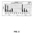
- FIG. 2 FACS analysis of engrafted human cells expressing lymphomyeloid cell markers in NOD/SCID mice. Co-expression of CD45 + with CD19 (left bar in each category); CD33 (middle bar); or CD7 (right bar).
- X-axis numbers of cells per transplantation.
- UCB transplantation of umbilical cord blood cells only;
- PP transplantation of placental perfusate cells only.
- UCB+PP transplantation of umbilical cord cells combined with placental perfusate cells.
- the present invention provides combinations of (1) placental stem cells, e.g. , placental stem cells in human placental perfusate, placental stem cells in placental enzymatic digestate, isolated placental stem and/or progenitor cells, and the like; and (2) stem cells from a second source, in a total number of cells, wherein the placental stem cells and stem cells from the second source are present in the combination in a ratio that produces a greater number of colony-forming units compared to a number of colony-forming units produced by placental stem cells or by stem cells from a second source, equivalent to said total number of cells, alone.
- placental stem cells e.g. , placental stem cells in human placental perfusate, placental stem cells in placental enzymatic digestate, isolated placental stem and/or progenitor cells, and the like
- stem cells from a second source in a total number of cells, wherein the placental stem cells and stem cells from the
- the invention further provides combinations of placental stem cells and stem cells from a second source that enhance engraftment in vivo compared to the number of colony-forming units produced by a number of placental stem cells equivalent to the number of cells in said combination, or a number of stem cells from a second source equivalent to the number of cells in said combination, alone.
- the present invention further provides methods of identifying such ratios, and such combinations, and methods of using the combined stem cell populations.
- the invention provides in vitro co-culture methods for identifying a combination of placental stem cells and stem cells from a second source that has improved engraftment potential as compared to a number of either placental stem cells or stem or progenitor cells from a second source, equivalent to the number of cells in said combination, alone.
- the in vitro co-culture assay thus identifies ratios of placental stem cells to stem cells from a second source that improve the number of colony-forming units, and engraftment, in a non-cell number-dependent manner.
- the invention provides a method of identifying a ratio of placental stem cells to stem cells from a second source, comprising identifying a ratio of placental stem cells to stem cells from a second source in a total number of cells that, when said placental stem cells and stem cells from a second source are cultured together for a time and under conditions that allow the formation of colony-forming units, produces a greater number of colony-forming units than a number of placental stem cells or stem cells from a second source, equivalent to the number of cells in said total number of cells, alone.
- the invention provides a method of identifying a ratio of placental stem cells and stem cells or progenitor cells from a second source in a total number of cells, comprising contacting a population of said placental stem cells in vitro with a population of said stem cells from a second source in a plurality of ratios for a time and under conditions sufficient to allow the formation of colony-forming units, and identifying a ratio within said plurality of ratios that yields the greatest number of colony-forming units.
- said ratio improves engraftment into a recipient as compared to engraftment by a number of placental stem cells or stem cells from a second source, equivalent to the number of cells in said total number of cells, alone.
- said combined stem cell population improves engraftment in an individual in need of stem cells for at least 1, 2, 3, 4, 5, 6, 7, 8, 9, 10, 11, 12, 13, 14, 15, 16, 17, 18, 19, 20 or 21 days post-transplant.
- said combined stem cell population improves engraftment in an individual in need of stem cells at a time more than 21 days post-transplant.
- Placenta-derived stem cells useful in the methods and compositions of the invention include, for example, embryonic-like cells, pluripotent cells, multipotent cells, committed progenitor cells, hematopoietic progenitor cells, and mesenchymal-lilse stem cells from placenta.
- the placenta-derive stem cells are contained within, or are derived from, placental perfusate.
- Placenta-derived stem cells used in the methods of the invention can be derived from a single placenta, or from a plurality of placentas, and may be obtained by any method.
- Placenta-derived stem cells can be obtained by, for example, perfusion, as disclosed in U.S. Application Publication Nos. 2002/0123141 and 2003/0032179 , the disclosures of each of which are incorporated herein by reference.
- Such perfusion can be perfusion by the pan method, wherein perfusion liquid is forced through the placental vasculature and perfusion fluid that exudes from the placenta, typically the maternal side, is collected in a pan containing the placenta.
- Perfusion can also be a closed-circuit perfusion, wherein perfusion fluid is passed through, and collected from, only the fetal vasculature of the placenta.
- perfusion can be continuous, that is, perfusion fluid that has been passed through the placenta, and which comprises a plurality of placental cells, is passed through a second time, or a plurality of times, prior to isolation of placental cells.
- Placenta-derived stem cells may also be obtained by physical or enzymatic disruption of the placenta using, e.g. , proteases and/or other tissue-disruptive enzymes to disrupt the multicellular structure of the placenta.
- proteases may include neutral proteases or metalloproteases, e.g. , collagenase, dispase, trypsin, elastase, and the like.
- Placental stem cells may also be obtained by physical disruption of the placenta using, e.g ., mucolytic enzymes, for example, hyaluronidase.
- the isolated perfused placenta of the invention provides a source of large quantities of stem cells enriched for CD34 + stem cells, e.g. , CD34 + CD38 - stem cells, e.g., CD34 + , CD38 - , lin - stem cells, and CD34 - stem cells, e.g. , CD34 - CD38 + stem cells.
- the first collection of blood from the placenta is referred to as cord blood which contains predominantly CD34 + CD38 + hematopoietic progenitor cells.
- high numbers e.g ., 1 x 10 5 to about 2 x 10 7
- high numbers e.g ., 1-10 million
- An isolated placenta that has been perfused for twenty-four hours or more provides a source of large quantities of stem cells enriched for CD34 - CD3 - stem cells.
- the combined stem cell populations of the invention comprise CD34 + placental stem cells that are positive for aldehyde dehydrogenase (ALDH).
- ALDH aldehyde dehydrogenase
- Such cells demonstrate detectable levels of ALDH activity in an ALDH assay.
- ALDH assays are known in the art (see, e.g., Bostian and Betts, Biochem. J., 173, 787, (1978 )).
- said ALDH assay uses ALDEFLUOR® (Aldagen, Inc., Ashland, Oregon) as a marker of aldehyde dehydrogenase activity.
- a combined stem cell population of the invention comprises CD34+ stem cells, where at least about 5%, 10%, 15%, 20%, 25%, 30%, 35%, 40%, 45%, 30%, 55%, 60%, 65%, 70%, 75%, 80%, 85%, 90%, or at least 95% of the CD34 + stem cells are ALDH + .
- At least one class of human placental stem cells has characteristics of embryonic stem or germ cells.
- stem cells of this class are SSEA3 - (stage-specific embryonic antigen 3), SSEA4 - , OCT-4 + (a stem cell transcription factor) and ABC-p + (ATP-binding cassette (ABC) transporter protein), a marker profile exhibited by pluripotent stem cells that have not yet undergone differentiation.
- SSEA3 - stage-specific embryonic antigen 3
- SSEA4 - a stem cell transcription factor
- ABC-p + ATP-binding cassette (ABC) transporter protein
- the methods and compositions of the invention can use or comprise non-embryonic, placental stem cells that are, e.g., SSEA33 - , SSEA4 - , OCT-4 + or ABC-p + .
- the placental stem cells are OCT-4 + ABC-p + , and, even more preferably, are SSEA3 - SSEA4 - OCT - 4 + AEC-p + .
- the invention encompasses the use of placental stem cells positive for at least one of CD10, CD29, CD44, CD54, CD90, CD73 or CD105, or negative for at least one of CD34, CD38, or CD45.
- the methods and compositions of the invention can use or comprise placental stem cells having or positive for CD10, CD29, CD44, CD54, CD90, CD73 or CD105, and lacking or negative for CD34, CD38, or CD45.
- the methods and compositions of the invention can use or comprise placental stem cells positive for at least one of CD10, CD29, CD44, CD54, CD90, CD73 or CD105, or negative for at least one of CD34, CD38, or CD45.
- the invention encompasses the use of placental stem cells having or positive for CD10, CD29, CD44, CD54, CD90, CD73 or CD105, and lacking or negative for CD34, CD38, or CD45.
- placental stem cells used in the methods and compositions of the invention are identified by the presence of the markers CD10, CD29, CD44, CD54, CD90, CD105 (SH2), CD73 (SH3, SH4), OCT-4, and/or A.BC-p, and/or the absence of the markers CD34, CD38, CD45, SSEA3, or SSEA4.
- the placental stem cells are CD10 + , CD29 + , CD34 - , CD38 - , CD44 + , CD45-, CD54 + , CD73 + , CD90 + , CD105 + , SH2 + , SH3 + .
- the placental stem cells are CD200 + and HLA-G + .
- SH2 + ", “SH3 + “ and “SH4 + " mean that a stem cell is bound by antibody SH2, SH3, or SH4, respectively.
- the placental stem cells are CD73 + , CD 105 + and CD200 + , In another specific embodiment, the placental stem cells are CD200 + and OCT-4 + .
- the placental stem cells are CD73 + , CD105 + and facilitate the formation of embryoid-like bodies in a population of isolated placental cells comprising said stem cells, when said population is cultured under conditions that allow the formation of embryoid-like bodies.
- the placental stem cells are CD73 + , CD105 + and HLA-G + .
- the placental stem cells are C7CT-4 + and facilitate the formation of embryoid-like bodies in a population of isolated placental cells comprising said stem cells, when said population is cultured under conditions that allow the formation of embryoid-like bodies.
- embryoid-like bodies refers to three-dimensional clusters of differentiating, and differentiated, cells that emerge from the adherent stem cell layer.
- the human placental stem cells do not express MHC Class 2 antigens.
- Populations of placental perfusate-derived stem cells comprise trophoblasts.
- Cell markers e.g., stem cell markers and cell surface markers
- FACS fluorescence-activated cell sorting
- cells may be washed in PBS and then double-stained with anti-CD34 phycoerythrin and anti-CD38 fluorescein isothiocyanate (Becton Dickinson, Mountain View, CA). The cells would then be analyzed using a standard flow cytometer.
- intra-cellular markers can also be examined via standard methodology.
- Antibody/fluorophore combinations to specific markers include, but are not limited to, fluorescein isothiocyanate (FITC) conjugated monoclonal antibodies against HLA-G (available from Serotec, Raleigh, North Camlina), CD10 0 (available from BD Immunocytometry Systems, San Jose, California), CD44 (available from BD Biosciences Pharmingen, San Jose, California), and CD105 (available from R&D Systems Inc., Minneapolis, Minnesota.); phycoerythrin (PE) conjugated monoclonal antibodies against CD44, CD200, CD117, and CD13 (BD Biosciences Pharmingen); phycoerythrin-Cy7 (PE Cy7) conjugated monoclonal antibodies against CD33 and CD10 (BD Biosciences Pharmingen); allophycocyacin (APC) conjugated streptavidin and monoclonal antibodies against CD3 8 (BD Biosciences Pharmingen); and Biotinylated CD90 (BD Biosciences Pharmingen).
- antibody/label combinations include, but are not limited to, CD133-APC (Miltenyi), KDR-Biotin (CD309, Abcam), CytokeratinK-Fite (Sigma or Dako), HLA ABC-Fitc (BD), HLA DRDQDP-PE (BD), ⁇ -2-microglobulin-PE (BD), CD80-PE (BD) and CD86-AFC (BD), CD45-PerCP (peridin chlorophyll protein); CD44-PE; CD19-PE; CD10-F (fluorescein); HLA-G-F and 7-amino-actinomycin-D (7-AAD); HLA-ABC-F; and the like.
- CD133-APC Miltenyi
- KDR-Biotin CD309, Abcam
- CytokeratinK-Fite Sigma or Dako
- HLA ABC-Fitc HLA ABC-Fitc
- BD HLA DRDQDP-PE
- BD ⁇ -2
- Placental stem cells e.g., placental stem cells contained in placental perfusate
- the stem cells can be cultured in medium comprising Notch agonist, e.g., a deletion form of a Notch protein consisting essentially of the intracellular domain of the Notch protein, or a Delta protein. See U.S. 2004/0067583 .
- Stem cells from a second source can comprise one or more types of stem cells, such as embryonic stem cells, embryonic germ cells, adult stem cells, mesenchymal stem cells, hematopoietic stem cells, non-hernatopoietic stem cells, bone marrow-derived stem cells, neural stem cells, cardiac stem cells, ocular stem cells, epithelial stem cells, endothelial stem cells, hepatic stem cells, pulmonary stem cells, muscle stem cells, intestinal stem cells, and the like.
- stem cells from a second source can comprise one or more types of stem cells, such as embryonic stem cells, embryonic germ cells, adult stem cells, mesenchymal stem cells, hematopoietic stem cells, non-hernatopoietic stem cells, bone marrow-derived stem cells, neural stem cells, cardiac stem cells, ocular stem cells, epithelial stem cells, endothelial stem cells, hepatic stem cells, pulmonary stem cells, muscle stem cells, intestinal stem cells, and the like.
- Stem cells from a second source can be stem cells isolated from the second, non-placental source, or can be tissue comprising the stem cells.
- stem cells can be isolated by perfusion of the organ(s) comprising the stem cells, or by tissue disruption and/or enzymatic digestion of the organ(s) comprising the stem cells.
- Stem cells from a second source can be, e.g., stem cells derived solely from umbilical cord, or solely from amniatic fluid.
- Stem cells from a second source may be obtained by providing a sample of a relevant tissue, and isolating stem cells from the tissue using one or more cell surface markers.
- hematopoietic stem cells may be obtained from blood (e.g., peripheral blood, placental blood, umbilical cord blood) or from bone marrow by obtaining a sample of blood or bone marrow, isolating mononuclear cells from the blood or bone marrow, and separating CD34 + cells from the isolated mononuclear cells.
- the stem cells can be provided in a population of total nucleated cells (TNC) from the blood, e.g., total nucleated cells from peripheral blood, placental blood, umbilical cord blood, and the like.
- TSC total nucleated cells
- Stem cells from other tissues may be isolated in a similar manner.
- Mesenchymal stem cells may be isolated from, e.g., bone marrow by isolation of cells positive for CD73, CD105 and/or CD45 ( see, e.g. , U.S. Patent No. 6,387,367 ).
- Ocular (limbal) stem cells may be obtained from the cornea by obtaining corneal cells and isolating SSEA-4 + cells ( see , e.g. , U.S. Application Publication No. 2005/0186672 ).
- Hepatic stem cells may be obtained from liver, particularly fetal liver, samples, by selecting cells expressing CD 14, CD34, CD38, ICAM, CD45, CD117, glycophorin A, connexin 32, osteopontin, bone sialoprotein, collagen I, collagen II, collagen III, collagen IV, or combinations thereof (see, e.g., U.S. Application Publication No. 2005/0148072 ).
- Muscle stem cells may be obtained from muscle tissue by selecting CD34 + CD45 - cells that do not express other hematopoietic cell markers ( see , e.g. , U.S. Application Publication No. 2005/0079606 ).
- Cardiac stem cells may be isolated from cardiac tissue by selecting c-kit - CD31 + CD38 + cells ( see , e.g. , U.S. Application Publication No. 2004/0126879 ). Isolation of stem cells may be accomplished using other known characteristics or markers, as well.
- said stem cells from a second source are cord blood stem cells.
- the cord blood stem cells are CD34 + stem cells, e.g., CD34 + , CD38 + stem cells, CD34 + , CD38 - stem cells, CD34 + , CD38 - , lin - stem cells, and the like.
- the CD34 + stem cells from a second source are ALDH + .
- Cord blood itself, or stem and/or progenitor cells obtained from cord blood can be used in the methods of the invention.
- said cord blood-derived cells comprise hematopoietic stem cells, where the combined stem cell population is to be used for hematopoietic engraftment.
- the stem cells from a second source may be derived from a single donor, or from a plurality of donors in equal or unequal amounts. Stem cells from a plurality of second (that is, non-placental) sources may be combined with placental stem cells, and used for the methods and compositions of the present invention.
- Stem cells from a second source can be used immediately after collection, or can be cultured for a period of time prior to assaying or administration to an individual in a combined stem cell population.
- the stem cells can be cultured in medium comprising Notch agonist, e.g. , a deletion form of a Notch protein consisting essentially of the intracellular domain of the Notch protein, or a Delta protein. See U.S. 2004/0067583
- the cells can be combined in an in vitro co-culture, or colony-forming, assay to determine if the number of stem cells in a particular combination produces more colony-forming units than a number of placental stem cells or stem cells from a second source, equivalent to the number of cells in said combination, alone. Any such combination of placental stem cells and stem cells from a second source in a ratio that produces more colony forming units than either placental stem cells or stem cells from a second source alone, for equivalent numbers of cells, is identified as a combined stem cell population of the invention.
- the identification of a combined stem cell population can use any colony forming unit assay commonly used and known in the art, provided the assay allows for the proliferation and differentiation of stem cells from placenta and from a second source, for example, colony forming assays provided by StemCell Technologies, Inc. Such an assay may use, e.g ., MESENCULTTM medium (Stem Cell Technologies, Inc., Vancouver British Columbia).
- the identification of combined stem cell populations can use cells that are freshly-prepared, or thawed from frozen stocks, or both. Preferably, both the placental stem cells and stem cells from a second source are in suspension when combined for co-culture.
- Placental stem cells, and stem cells from a second source may be assessed for viability, proliferation potential, and longevity using standard techniques known in the art, such as trypan blue exclusion assay, fluorescein diacetate uptake assay, propidium iodide uptake assay (to assess viability); and thymidine uptake assay, MUTT (3-(4,5-dimethylthiazol-2-yl)-2,5-diphenyltetrazolium bromide) cell proliferation assay (to assess proliferation).
- Longevity may be determined by methods well known in the art, such as by determining the maximum number of population doublings in an extended culture.
- a colony forming unit assay using placental stem cells and cord blood-derived stem cells is performed as follows. Fresh or thawed HLA/donor matched placental perfusate and cord blood units are obtained, and the number of total nucleated cells in each is determined with a hemacytometer. Where thawed units are used, cord blood samples can be hetastarch-separated, and placental perfusate units are preferably Ficoll-separated, Small samples of nucleated cells from each source are seeded together in suspension in two or more ratios in a co-culture, and expanded. The co-culture can be performed in, e.g.
- triplicate for one or more ratios of placental stem cells to stem cells from a second source in, for example, 35 mm dishes in an appropriate cell culture medium (e.g., RPMI 1640 medium supplemented with 2-10% fetal calf serum and, optionally, 1% Stemspan cytokine cocktail; Methocult GF + H4435 medium, etc .).
- Hematopoietic stem cells may be expanded in culture medium comprising GM-CSF, IL-3, IL-6, SCF and flt-3 ligand.
- the container used for the co-culture assay is preferably appropriate for tissue culture of stem cells.
- co-cultures may be performed in glass or plastic Petri dishes, 16-well plates, 32-well plates, 96-well plates, 128-well plates, and the like.
- the total number of nucleated cells from each source in each co-culture varies from 1 x 10 4 to 1 x 10 6 .
- Cells may also be co-cultured in a micropatterned configuration. See U.S. Patent No. 6,221,663 .
- the preferred ratio is any ratio that generates more colony forming units than that generated by said number ofplacental stem cells or said number of stem cells from a second source under the same conditions. More preferably, the ratio is a ratio that generates a higher number of colony-forming units than all other ratios tested. Statistical significance between ratios tested is desirable, but not necessary.
- the higher number of colony-forming units may be attributable to, or be derived from, both placental stem cells and stem cells from a second source; from predominantly or only the placental stem cells; or predominantly or only the stem cells from a second source.
- the combined stem cell population is cultured for a time sufficient for colony forking units to form, typically 10-20 days.
- Cell culture during expansion follows standard protocols known in the art of stem or progenitor cell culture, and includes, for example, daily or semidaily changes of medium; culture at about 37°C at 5% CO 2 in a humidified incubator, and the like.
- the number and morphology of colony forming units in the co-culture is determined (e.g. , for hematopoietic stem cells, the number of CFU-GM, CFU-L, CFU-M, CFU-G, CFU-DC, CFU-GEMM, CFU-E).
- nucleated cells from placenta perfusate, and nucleated cells from cord blood are combined a ratio of 1:1, 1:3 and 3:1 (where 1 equals, e.g. , 1 x 10 5 cells) in Methocult GF + H4435 medium.
- the co-culture is then expanded in tissue culture for about 14 days. The morphology of the co-cultured cells, and the number of colony forming units, is determined.
- the ratio of the nucleated cell samples from the two sources that provides the highest number of colony-forming units is designated an optimum ratio, and the two units, or stem and/or progenitor cells from one or both of the units, are combined in the optimum ratio for administration to a recipient in need of a stem cell transplant.
- Such an optimum ratio provides superior engraftment in vivo over the administration of either unit, or stem and/or progenitor cells from either unit, alone, where equivalent numbers of cells are administered.
- the placental stem cells and stem cells from a second source are contacted with each other during the co-culturing, either directly or indirectly. At a minimum, this comprises contacting one of the types of stem cells with culture medium in which the other type of stem cell has cultured for a period of time, e.g. , contacting one of the types of stem cells with medium that has been conditioned by the other type of stem cell.
- the placental stem cells, and stem cells from a second source may be cultured together in the same physical space during culture for colony-forming unit formation, e.g. , in the same culture dish or well in a multi-well plate.
- placental stem cells and stem cells from a second source may also be contacted with each other by culturing in separate physical spaces, but in common culture medium (e.g. , separated by a membrane, or in two wells of a multiwell plate wherein culture medium may move actively or passively between the wells, but cells cannot mix).
- placental stem cells and stem cells from a second source may be cultured in separate physical spaces with no common culture medium, and the stem cells brought into contact with each other by an exchange of part or all of the culture medium from one stem cell culture with that of the other.
- the cells in the co-culture are cultured in a manner that physically separates the cells, but allows biomolecules to diffuse between the two cultures. See , e.g.
- a bioactive molecule is added to the placental stem cells and stem cells from a second source during the assay, and a ratio of placental stem cells to stem cells from a second source is identified that, for a total number of cells, results in more colony-forming units, or enhanced engraftment, compared to a number of placental stem cells or stem cells from a second source, equivalent to said total number of cells in said combination, alone.
- Such a bioactive molecule may be a small organic molecule of less than 50 kDa, 30 kDa, 20 kDa, 10 kDa, 5 kDa, 3kDa, 2 kDa, 1 kDa, 500 Da, 300 Da, 200 Da, 100 Da or smaller.
- said small organic molecule is synthetic or nonnatural, that is, not derived from a natural source.
- said bioactive molecule is a cytokine or growth factor.
- Bioactive molecules that can be added to the co-culture include difterenliation-inducing agents such as, but are not limited to, Ca 2+ , EGF, ⁇ -FGF, ⁇ -FGF, PDGF, keratinocyte growth factor (KGF), TGF- ⁇ , cytokines (e.g. , IL-1 ⁇ , IL-1 ⁇ , IFN- ⁇ , TFN), retinoic acid, transferrin, hormones (e.g., androgen, estrogen, insulin, prolactin, triiodothyronine, hydrocortisone, dexamethasone), sodium butyrate, TPA, DMSO, NMF, DMF, matrix elements (e.g.
- Bioactive molecules that are differentiation suppressants may also be added, such as, but not limited to, human Delta-1 and human Serrate-1 polypeptides ( see , Sakano et al., U.S. Patent No. 6,337,387 entitled “Differentiation-suppressive polypeptide", issued January 8, 2002), leukemia inhibitory factor (LIF), and stem cell factor.
- LIF leukemia inhibitory factor
- the co-culture assay may be used to identify a positive effector of engraftment.
- the invention provides a method of identifying a bioactive molecule that is a positive effector of engraftment comprising contacting a combined stem cell population with said bioactive molecule, wherein said bioactive molecule is identified as a positive effector of engraftment if engrafment by said combined stem cell population is detectably enhanced compared to engraftment by a combined stem cell population not contacted with said bioactive molecule.
- the invention provides a method of identifying a positive effector of engraftment comprising combining placental stem cells and stem cells from a second source in vitro in one or more ratios in the presence of said bioactive molecule; culturing said placental stem cells and stem cells from a second source for a time sufficient for colony forming units to form; determining the number of colony-forming units for each of said one or more ratios; and determining, for at least one of said one or more ratios, whether the number of colony forming units in the presence of said bioactive molecule is greater than the number of colony forming units in the absence of said bioactive molecule, and, if so, identifying said bioactive molecule as a positive effector of engraftment.
- the in vitro assay may be performed on any placental stem cell population and stem cell population from a second source to determine an optimum ratio for engraftment.
- the in vitro co-culture assay can be used as a standard, routine procedure to characterize stem cell populations prior to transplantation.
- the results of the above in vitro assay may be confirmed using an in vivo engraftment assay.
- the in vivo assay may also be performed in the absence of the in vitro assay to determine an optimum ratio of placental stem cells, and stem cells from a second source, to maximize engraftment.
- placental stem cells and stem cells from a second source are transplanted into a plurality of model animals and given sufficient time to engraft (typically 6-10 weeks). The animals are subsequently sacrificed, and the degree of engraftment in each animal is determined for at least one tissue.
- the invention provides a method of identifying a ratio of placental stem cells and stem cells or progenitor cells from a second source for engraftment into a recipient, comprising identifying a ratio of placental stem cells to stem cells from a second source in a total number of cells that, when transplanted into an animal, results in enhanced engraftment compared to transplantation of a number of placental stem cells or stem cells from a second source, equivalent to the number of cells in said total number of cells, alone.
- said identifying a ratio of placental stem cells to stem cells from a second source comprises transplanting a number of placental stem cells and stem cells from a second source in a plurality of animals, in a plurality of ratios; determining the number of engrafted cells in at least one tissue of said animals for each of said plurality of ratios; and identifying the ratio in said plurality of ratios that yields the highest number of engrafted cells.
- the placental stem cells can be placental stem cells obtained by any means or present in any usable form.
- the placental stem cells may be contained in placental perfusate, or may be contained within isolated total nucleated cells from the placental perfusate, or may be a population of stem cells isolated from the total nucleated cells, or may be placental stem cells contained within enzyme-digested placental tissue, or may be placental stem cells isolated from enzyme-digested placental tissue, or may be placental stem cells that have been expanded and/or passaged in culture, etc.
- model animal Any standard model animal may be used in the in vivo co-culture assay.
- the model animal is one in which engraftment af xenografts may be readily accomplished.
- Small mammals such as standard laboratory rodents such as mice and rats are preferred because they require fewer administered stem cells to show engraftment. It is highly preferable that the model animal be immune-compromised.
- Animal models that may be used in the in vivo assay include, but are not limited to, NOD/SCD (non-obese diabetic /severe combined immune deficiency) mice ( see Hogan et al., Blood 90(1):85-96 (1997 )); beige/nude/x-Iinked immunodeficiency (BNX) mice ( see , e.g. , Kamal-Reid et al., Science 242:1706 (1988 )); SCID mice ( see , e.g. , Kamal-Reid et al., Science 246:1597 (1989 ).
- NOD/SCD non-obese diabetic /severe combined immune deficiency mice
- BNX beige/nude/x-Iinked immunodeficiency
- SCID mice see , e.g. , Kamal-Reid et al., Science 246:1597 (1989 ).
- Engraftment may be accomplished in other animal models, such as sheep fetuses ( see , e.g. , Shimizu et al., Blood 91(10):3688-3692 (1998 ); Zanjani et al., Int'l J. Hematol 63(3):179-182(1996 )).
- sheep fetuses see , e.g. , Shimizu et al., Blood 91(10):3688-3692 (1998 ); Zanjani et al., Int'l J. Hematol 63(3):179-182(1996 )).
- the determination of the number ofengrafted cells in tissues from the recipient animal may be accomplished by any means known in the art.
- detection of engrafted cells may be accomplished by detection of engrafted cell-specific nucleic acids, e.g. , by the polymerase chain reaction, or by detection of proteins specific for engrafted cells, e.g. , by immunohistochemstry.
- Identification of engraftment in vivo may be determined through the use of a sample, e.g. , biopsy specimen, taken at one or more locations on, and at one or more post-transplantation times from, a recipient.
- demonstration of engraftment of placental stem cells and/or cord blood-derived stem cells can be accomplished by taking a biopsy (e.g. , bone marrow aspirate or peripheral blood sample) and performing PCR to determine whether any non-recipient genetic markers are present, which would indicate engraftment.
- identification of engrafted cells is accomplished by selection of one or more antibodies that recognize markers expressed by the engrafted cells.
- the engrafted cells are human, and the one or more antibodies specifically recognize one or more human cell markers.
- Antibodies can be used to detect the markers by any art-accepted method, e.g. , immunohistochemical methods.
- determination of the presence of a cell surface marker can comprise sacrifice of a non-human host animal, obtaining a desired tissue, fixing and embedding the tissue in paraffin or a similar matrix; thin sectioning the tissue, optionally followed by staining; and contacting the tissue with one or more antibodies that recognize the marker.
- antibodies that recognize markers expressed by cells into which the engrafted stem cells can differentiate For example, placental stem cells or cord blood-derived stem cells differentiate into cells that express CD45 and vimentin; thus, antibodies to CD45 and vimentin may be used to determine the number of engrafting, and differentiating, stem cells.
- Antibodies that recognize, e.g. , human cell surface markers in preference to host cell markers, e.g. , mouse cell surface markers, are well-known in the art.
- a plurality of model animals e.g. , a plurality of mice of the species Mus musculus
- the host animals are sacrificed, and tissues (e.g.
- CD45 is a marker specific for leukocytes, including T- and B-lymphocytes, granulocytes, monocytes and macrophages. Certain CD45 antibodies, such as clone T29/33 (BioDesign, Saco, Maine), do not cross-react with mouse antigens. Vimentin is a marker for mesenchymal cells, such as fibroblasts, smooth muscle cells, lipocytes, Schwann cells, vascular endothelial cells, and the like.
- vimentin antibodies such as clone V9 (BioDesign, Saco, Maine) do not cross-react with mouse antigens. Staining with antibodies to these two markers, therefore, can establish generally the extent of engraftment of placental stem cells, and stem cells from a second source, in a variety of tissues. This example is not limiting; different antibodies may be used to determine the extent of engraftment of other cell types.
- bone marrow cells isolated from a primary engrafted animal e.g. , a mouse
- Assays for secondary engraftnemt are as listed above and include methods well known to those of skill in the art.
- the invention further provides combined stem cell compositions comprising placental stem cells, e.g. , cells from placental perfusate, e.g. , nucleated cells from placental perfusate, comprising placental stem cells and stem cells from a second source that, for a particular number of cells, results in a greater number of colony-forming units in a colony-forming unit assay, or enhanced engraftment in a transplant recipient, than the number of either placental stem cells or stem cells from a second source, alone.
- Combined stem cell populations identified by the above methods represent engraftment-enhanced combinations of stem cells based on the characteristics of the stem cell sources, that is, the number of engraftable cells contained in, e.g. , a unit of placenta perfusate, a unit of cord blood, etc.
- the invention encompasses a combined stem cell composition comprising a number of placental stem cells and stem cells from a second source in a ratio, wherein the stem cells from the composition show improved engraftment compared to a number of either the placental stem cells or the stem cells from a second source, equivalent to the number of cells in said composition, alone.
- the ratio is identified by combining placental stem cells and stem cells from a second source in vitro in a plurality of ratios for a time and under conditions sufficient to allow the formation of colony-forming units; and identifying a ratio in said plurality of ratios that yields the highest number of colony forming units.
- said stem or progenitor cells from a second source are cord blood stem or progenitor cells, bone marrow stem or progenitor cells, hematopoietic stem or progenitor cells, or mesenchymal stem or progenitor cells.
- said stem cells or progenitor cells from a second source are hematopoietic progenitor cells.
- said hematopoietic stem cells are cord blood hematopoietic stem cells.
- said hematopoietic cells are CD34 + cells.
- said placental stem cells comprise CD34 + cells, for example, CD34 + CD38 + cells and/or CD34 + CD38 - cells.
- said CD34 + CD38 - cells comprise CD34 + CD38 - lin - stem cells.
- said CD34+ placental stem cells comprise cells that are ALDH+, that is, CD34 + , A-LDH + placental stem cells.
- said placental stem cells are OCT-4 + or ABC-p + .
- said placental stem cells comprise cells that are OCT4 + ABC-p + .
- said placental stem cells comprise cells that are CD34 + and cells that are OCT4 + ABC-p + .
- said placental stem cells are contained within placental perfusate substantially lacking red blood cells and cellular debris.
- said composition comprises placental stem cells isolated from placental perfusate.
- placental stem cells comprise cells that express one or more of markers CD10, CD29, CD44, CD54, CD90, CD73 or CD105, and lack one or more of markers CD34, CD38, CD45, SSEA3 and SSEA4.
- placental stem cells comprise cells that are positive for CD10, CD29, CD44, CD54, CD90, CD73 or CD 105, and negative for CD34, CD38, CD45, SSEA3 and SSEA4.
- placental stem cells comprise cells that comprise one or more of markers CD10, CD29, CD44, CD54, CD90, CD73 and CD105, and lack one or more of markers CD34, CD38, CD45, SSEA3 and SSEA4,
- placental stem cells comprise cells that are positive for CD10, CD29, CD44, CD54, CD90, CD73 and CD105, and negative for CD34, CD38, CD45, SS1BA3 and SSEA4.
- said placental stem cells comprise CD34 - cells. In a specific embodiment, said placental stem cells are CD34 - CD38 - placental stem cells. In another embodiment, said placental stem cells comprise cells that are positive for at least one of CD10, CD29, CD33, CD44, CD73, CD105, CD 117, and CD133, and negative for at least one of CD34 or CD45. In another embodiment, said placental stem cells comprise cells that are positive for CD10, CD29, CD33, CD44, CD73, CD105, CD117, and CD133, and negative for CD34 or CD45. In a more specific embodiment, said placental stem cells comprise cells that are HLA-ABC + .
- said placental stem cells comprise cells that are HLA-ABC - . In a more specific embodiment, said placental stem cells comprise cells that are HLA-DR + . In a more specific embodiment, said placental stem cells comprise cells that are HLA-DR - . In another specific embodiment, said placental stem cells comprise cells that are CD200 + or HLA-O + . In another specific embodiment, the placental stem cells comprise cells that are CD200 + and HLA-G + . In another specific embodiment, the placental stem cells comprise cells that are CD73 + , CD105 + and CD200 + . In another specific embodiment, the placental stem cells comprise cells that are CD200 + and OCT-4 + .
- the placental stem cells comprise cells that are CD73 + , CD105 + and facilitate the formation of embryoid-like bodies in a population of isolated placental cells comprising said stem cells, when said population is cultured under conditions that allow the formation of embryoid-like bodies.
- the placental stem cells comprise cells that are CD73 + , CD105 + and HLA-G + .
- the placental stem cells comprise cells that are OCT-4 + and facilitate the formation of embryoid-like bodies in a population of isolated placental cells comprising said stem cells, when said population is cultured under conditions that allow the formation of embryoid-like bodies.
- said stem cells from a second source are stem cells derived from cord blood.
- the placental stem cells and the stem cells from a second source may be identically-HLA-matohed, that is, they may be derived from the same individual.
- the placental stem cells and the stem cells from a second source may be HLA-mismatched, that is, they may be derived from different individuals.
- the combination may also comprise stem cells that are either HLA-matched, partially HLA-matched, or HLA-mismatched to an intended recipient.
- the ratio of placental stem cells to stem cells from a second source can be about 100,000,000:1,50,000,000:1,20,000,000:1,10,000,000:1, 5,000,000:1, 2,000,000:1, 1,000,000:1, 500,000:1.200.000:1, 1,00.000:1, 50,000:1, 20,000:1, 10,000;1, 5,000:1,2,000:1,1,000:1,500:1,200:1,100:1, 50:1,20:1,10:1, 5:1,2:1,1:1; 1:2; 1:5; 1:10; 1:100; 1:200; 1:500; 1:1,000; 1:2,000; 1:5,000; 1:10,000; 1:20,000; 1:50,000; 1:100,000; 1:500,000; 1:1,000,000; 1:2,000,000; 1:5,000,000; 1:10,000,000; 1:20,000,000; 1:50,000,000; or about 1:100,000,000, comparing numbers of total nucleated cells in each population, or comparing total numbers of stem cells in each population.
- the ratio of placental stem cells to stem cells from, a second source can be about 1:10 to about 10:1. In other preferred embodiments, the ratio of placental stem cells to stem cells from a second source can be about 3:1 to about 1:3.
- the combined stem cell populations of the invention can comprise a therapeutically- effective amount of placental stem cells, stem cells from a second source, or both.
- the combined stem cell populations of the invention, and pharmaceutical compositions comprising a combined stem cell population can comprise at least 1 x 10 4 5 x 10 4 , 1 x 10 5 , 5 x 10 5 , 1 x 10 6 , 5 x 10 6 , 1 x 10 7 , 5 x 10 7 , 1 x 10 8 , 5 x 10 8 , 1 x 10 9 , 5 x 10 9 , 1 x 10 10 , 5 x 10 10 , or 1 x 10 11 placental stem cells, stem cells from a second source, or both, or no more than 1 x 10 4 , 5 x 10 4 , 1 x 10 5 , 5 x 10 5 , 1 x 10 6 , 5 x 10 6 , 1 x 10 7 , 5 x 10 7 , 1 x 10 8 , 5 x 10 8
- said combined stem cell population improves engraftment in an individual in need of stem cells at least, or at, 1, 2, 3, 4, 5, 6, 7, 8, 9, 10, 11, 12, 13, 14, 15, 16, 17, 18, 19, 20 or 21 days post-transplant. In another more specific embodiment, said combined stem cell population improves engraftment in an individual in need of stem cells at least, or at, more than 21 days post-transplant. In specific embodiments, said combined stem cell population improves engraftment in an individual in need of stem cells at least, or at, more than 25, 30, 35, 40, 45, 50, 55 weeks, or 1 year or longer post-transplant.
- the combined stem cell populations of the invention can be preserved, for example, cryopreserved for later use.
- Methods for cryopreservation of cells, such as stem cells are well known in the art, for example, cryopreservation using the methods of Boyse et al. ( U.S. Patent No. 5,192,553, issued March 9,1993 ) or Hu et al. (WO 00/73421, published December 7, 2000 ).
- Placenta-derived stem cells, and stem cells from a second source, which make up a combined stem cell population can be combined prior to cryopreservation, or can be cryopreserved separately, and combined in the appropriate ratio upon thawing, e.g ., within hours of use.
- the combined stem cell populations of the invention can be prepared in a form that is easily administrable to an individual.
- a combined stem cell population can be contained within a container suitable for medical use.
- a container can be, for example, a sterile plastic bag, flask, jar, or other container from which the combined stem cell population can be easily dispensed.
- the container is a container that allows, or facilitates, intravenous administration of a combined stem cell population.
- the container e.g., bag, can hold the placenta-derived stem cells and stem cells from a second source together, e.g., as a mixed cell population, or can hold the two stem cell populations separately.
- the bag preferably comprises multiple lumens or compartments that are interconnected to allow mixing of the placenta-derived stem cells and stem cells from a second source prior to, or during, administration.
- the container is preferably one that allows for cryopreservation of the combined stem cell population.
- the combined stem cell population in said container can comprise placenta-derived stem cells, stem cells from a second source, or both, that have been passaged at least, or at most, 1, 2, 3, 4, 5, 6, 7, 8, 9, 10, 11, 12, 13, 14, 15, 16, 17, 18, 19,20 times, or 25, 30, 35, 40 or more times.
- the invention also provides for combined stem cell populations that comprise, e.g., that are stored or maintained as, separate stem cell populations, e.g ., a population of placenta-derived stem cells and a population of stem cells from a second source, in combination with information on combining the two populations in an appropriate ratio prior to use, e.g., prior to administration to an individual in need of stem cells.
- a combined stem cell population would comprise a population of placenta-derived stem cells in a first container, a population of stem cells from a second source in a second container, and instructions for combining the two populations either before or during administration to an individual in need of stem cells.
- the invention provides a composition comprising a combined stem cell population in a container, wherein said combined stem cell population comprises placenta-derived stem cells and stem cells from a second source.
- the container is a bag, flask, or jar.
- said placenta-derived stem cells and said stem cells from a second source are contained together in said bag.
- said placenta-derived stem cells and said stem cells from a second source are contained separately within said bag.
- the composition comprises one or more compounds that facilitate cryopreservation of the combined stem cell population.
- said combined stem cell population is contained within a physiologically-acceptable aqueous solution.
- said physiologically-acceptable aqueous solution is a 0.9% NaCl solution.
- said bag is a sterile plastic bag.
- said bag allows or facilitates intravenous administration of said combined stem cell population.
- said combined stem cell population comprises placental cells that are HLA-matched to said stem cells from a second source.
- said combined stem cell population comprises placental cells that are at least partially HLA-mismatched to said stem cells from a second source.
- said placenta-derived stem cells are derived from a plurality of donors.
- said stem cells from a second source are derived from a plurality of donors.
- Combined stem cell populations can be cultured for a period of time prior to administration to an individual.
- the stem cells in a combined stem cell population can be cultured in medium comprising Notch agonist, e.g., a deletion form of a Notch protein consisting essentially of the intracellular domain of the Notch protein, or a Delta protein. See U.S. 2004/0067583
- the present invention encompasses pharmaceutical compositions that comprise combined stem cell populations of the invention, and a pharmaceutically-acceptable carrier.
- the combined stem cell populations of the invention may be formulated as an injectable (e.g ., WO 96/39101 , incorporated herein by reference in its entirety).
- the combined stem cell populations of the present invention may be formulated using polymerizable or cross linking hydrogels as described, e.g ., in U.S. Patent Nos. 5,709,854 ; 5,516,532 ; 5,654,381 .
- the invention provides for the maintenance of each stem cell population of the combined stem cell populations, prior to administration to an individual, as separate pharmaceutical compositions to be administered sequentially or jointly to create the combined stem cell population in vivo.
- Each component may be stored and/or used in a separate container, e.g ., one bag (e.g ., blood storage bag from Baxter, Becton-Dickinson, Medcep, National Hospital Products, Terumo, etc .) or separate syringe, which contains a single type of cell or cell population.
- cord blood, or cord blood-derived nucleated or stem cells are contained in one bag, and placental perfusate, or placental stem cells from placental perfusate, are contained in a second bag.
- a population of placental stem cells can be enriched.
- a population of cells comprising placental stem cells is enriched by removal of red blood cells and/or granulocytes according to standard methods, so that the remaining population of nucleated cells is enriched for placental stem cells relative to other cell types in placental perfusate.
- Such an enriched population of placental stem cells may be used unfrozen, or may be frozen for later use. If the population of cells is to be frozen, a standard cryopreservative (e.g ., DMSO, glycerol, EPILIFETM Cell Freezing Medium (Cascade Biologics)) is added to the enriched population of cells before it is frozen.
- a standard cryopreservative e.g ., DMSO, glycerol, EPILIFETM Cell Freezing Medium (Cascade Biologics
- compositions of the invention may comprise one or more agents that induce cell differentiation.
- an agent that induces differentiation includes, but is not limited to, Ca 2+ , EGF, ⁇ -FGF, ⁇ -FGF, PDGF, keratinocyte growth factor (KGF), TGF- ⁇ , cytokines (e.g ., IL-1 ⁇ , IL-1 ⁇ , IFN- ⁇ , TFN), retinoic acid, transferrin, hormones ( e.g ., androgen, estrogen, insulin, prolactin, triiodothyroxine, hydrocortisone, dexamethasone), sodium butyrate, TPA, DMSO, NMF, DMF, matrix elements (e.g ., collagen, laminin, heparan sulfate, MATRIGELTM), or combinations thereof.
- the pharmaceutical composition of the invention may comprise one or more agents that suppress cellular differentiation.
- an agent that suppresses differentiation includes, but is not limited to, human Delta-1 and human Serrate-1 polypeptides ( see , Sakano et al., U.S. Patent No. 6,337,387 ), leukemia inhibitory factor (LIF), stem cell factor, or combinations thereof.
- compositions of the present invention may be treated prior to administration to an individual with a compound that modulates the activity of TNF- ⁇ .
- a compound that modulates the activity of TNF- ⁇ Such compounds are disclosed in detail in, e.g ., U.S. Application Publication No. 2003/0235909 , which disclosure is incorporated herein in its entirety.
- Preferred compounds are referred to as IMiDs (immunomodulatory compounds) and SelCIDs (Selective Cytokine Inhibitory Drugs), and particularly preferred compounds are available under the trade names ACTIMIDTM, REVIMIDTM and REVLIMIDTM.
- the above method of identifying combined stem cell populations may be performed on paired units of, for example, placental perfusate or placental stem cells, and stem cells from a second source, e.g ., cord blood, cord blood stem cells, and the like, to produce combined stem cell populations for the treatment of an individual in need of stem cells.
- the individual is contacted with one or more combined stem cell populations.
- said contacting is the introduction, e.g ., transplantation, of said combined stem cell population into said individual.
- the method of producing combined stem cell populations may be performed as a first step in a procedure for introducing stem cells into any individual needing stem cells. Such a procedure can comprise use of pharmaceutical compositions comprising the combined stem cell populations, as described above.
- a population of placental stem cells of the invention is combined with a population of stem cells from a second source prior to administration to an individual in need thereof in a ratio that provides improved or enhanced engraftment over a number of said placental stem cells or said stem cells from a second source, equivalent to said total number of cells, alone.
- a population of placental stem cells of the invention is combined with a population of stem cells from a second source during, or simultaneously with, administration to a patient in need thereof, in an optimum ratio, wherein said ratio is identified by identifying a ratio of placental stem cells to stem cells from a second source, in a plurality of ratios, that yields the highest number of said colony-forming units when said placental stem cells and stem cells from a second source are cultured for a time and under conditions sufficient to allow the formation of colony-forming units.
- a population of placental stem cells of the invention and a population of umbilical cord blood cells are administered sequentially to a patient in need thereof to a final optimum ratio.
- the population of placental stem cells is administered first and the population of stem cells from a second source is administered second.
- the population of stem cells from a second source is administered first and the population of placental stem cells is administered second.
- said combined stem cell population is contained within one bag or container.
- the invention provides for use in transplantation of a population of placental stem cells, and stem cells from a second source, that are contained within separate bags or containers.
- stem cell populations contained in two bags may be mixed prior, in particular immediately prior, to or at the time of administration to a patient in need thereof.
- the contents of each bag may be administered separately to a patient, wherein two cell populations are used adjunctively in vivo.
- Combined populations of placental stem cells, and stem cells from a second source may be mixed, prior to transplantation, by any medically- acceptable means.
- the two populations are physically mixed.
- said placental stem cells and stem cells from a second source are mixed immediately prior to ( i.e ., within 1, 2, 3, 4, 5, 7, 10 minutes of) administration to said individual.
- said placental stem cells and stem cells from a second source are mixed at a point in time more than five minutes prior to administration to said individual.
- the placental stem cells, and/or stem cells from a second source are cryopreserved and thawed prior to administration to said individual.
- said placental stem cells and stem cells from a second source are mixed to form a combined stem cell population at a point in time more than 1, 2, 3, 4, 5, 6, 7, 8, 9, 10, 11, 12, 13, 14, 15, 16, 17, 18, 19, 20, 21, 22, 23, or 24 hours prior to administration to said individual, wherein either or both of said placental stem cells and stem cells from a second source have been cryopreserved and thawed prior to said administration.
- the combined stem cell populations may be administered more than once.
- the stem cells contained within the combined stem cell population are preconditioned prior to transplantation.
- preconditioning comprises storing the cells in a gas-permeable container generally for a period of time at about -5°C to about 23°C, about 0°C to about 10°C, or preferably about 4°C to about 5°C.
- the cells may be stored between 18 hours and 21 days, between 48 hours and 10 days, preferably between 3-5 days.
- the cells may be cryopreserved prior to preconditioning or, may be preconditioned immediately prior to administration.
- either or both of the placental stem cells, or stem cells from a second source may be differentiated prior to introduction to an individual in need of stem cells.
- the stem cells may be differentiated to cells in the hematopoietic lineage.
- the invention provides a method of introducing stem cells into an individual comprising determining a ratio of placental stern cells and stem cells from a second source in a total number of cells, wherein the ratio improves engraftment as compared to introduction of a number of placental stem cells or stem cells from a second source, equivalent to said total number of cells, alone; differentiating one or both of said placental stem cells or stem cells from a second source into cells of another cell type; and introducing said stem cells and/or differentiated cells to an individual.
- the method of transplantation of combined stem cell populations comprises (a) induction of differentiation of placental stem cells, (b) mixing the placental stem cells with a population of stem cells from a second source, e.g., cord blood stem cells, to form a combined cell population, and (c) administration of the combined cell population to an individual in need thereof.
- the method of transplantation comprises (a) induction of differentiation of stem cells from a second source; (b) mixing the differentiated cells with placental stem cells to form a combined cell population; and (c) administration of the combined cell population to an individual in need thereof.
- the method of transplantation of combined stem cell populations comprises (a) mixing placental stem cells with a population of cord blood cells; (b) induction of differentiation of the mixture of the cord blood cells and placental stem cells and (c) administration of the mixture to a patient in need thereof.
- the combined stem cell populations of the invention may be transplanted into a patient in any pharmaceutically or medically acceptable manner, including by injection, e.g., intravenous injection, intramuscular injection, intraperitoneal injection, intraocular injection, direct injection into a particular tissue, transfusion, etc.
- combined stem cell populations e.g., placental stem cells in combination with cord blood-derived stem cells
- a combined stem cell population comprising placental stem cells and cardiac stem cells, in suspension, may be injected directly into cardiac tissue, e.g., an ischemic area in a heart.
- the combined stem cell populations may comprise, or be suspended in, any pharmaceutically-acceptable carrier.
- the combined stem cell populations may be carried, stored, or transported in any pharmaceutically or medically acceptable container, for example, a blood bag, transfer bag, plastic tube or vial.
- engraftment in a human recipient may be assessed using, e.g., nucleic acid or protein detection or analytical methods.
- nucleic acid or protein detection or analytical methods e.g., the polymerase chain reaction (PCR), STR, SSCP, RFLP analysis, AFLP analysis, and the like, may be used to identify engrafted cell-specific nucleotide sequences in a tissue sample from the recipient.
- PCR polymerase chain reaction
- STR polymerase chain reaction
- SSCP SSCP
- RFLP analysis e.g., RFLP analysis, and the like
- engraftment may be determined by the appearance of engrafted cell-specific nucleic acids in a tissue sample from a recipient, which are distinguishable from background.
- the tissue sample analyzed may be, for example, a biopsy (e.g ., bone marrow aspirate) or a blood sample.
- a sample of peripheral blood is taken from a patient immediately prior to a medical procedure, e.g., myeloablation, After the procedure, a combined stem cell population of the invention is administered to the patient. At least once post-administration, a second sample of peripheral blood is taken.
- An STR profile is obtained for both samples, e.g., using PCR primers for markers (alleles) available from, e.g ., LabCorp (Laboratory Corporation of America). A difference in the number or characteristics of the markers (alleles) post-administration indicates that engraftment has taken place.
- Engraftment can also be demonstrated by detection of re-emergence of neutrophils.
- engrafted cell-specific markers may be detected in a tissue sample from the recipient using antibodies directed to markers specific to either the transplanted stem cells, or cell into which the transplanted stem cells would be expected to differentiate.
- engraftment of a combination of placental stem cells and cord blood-derived stem cells may be assessed by FACS analysis to determine the presence of CD45 + , CD19 + , CD33 + , CD7 + and/or CD3 + cells by adding the appropriate antibody and allowing binding; washing ( e.g ., with PBS); fixing the cells ( e.g ., with 1% paraformaldehyde); and analyzing on an appropriate FACS apparatus (e.g.
- engraftment of a combination of placental stem cells and cord blood-derived stem cells may be assessed by FACS analysis to determine the presence of CD200 + or HLA-G + cells.
- placental stem cells and/or stem cells from a second source are from an individual of a different sex than a recipient, e.g ., male donor and female recipient, engraftment can be determined by detection of sex-specific markers, e.g ., Y-chromosome-specific markers.
- Placental stem cells and/or stem cells from a second source may also be genetically modified to express a unique marker or nucleic acid sequence that facilitates identification, e.g ., an RFLP marker, expression of ⁇ -galactosidase or green fluorescent protein, or the like.
- a unique marker or nucleic acid sequence that facilitates identification, e.g ., an RFLP marker, expression of ⁇ -galactosidase or green fluorescent protein, or the like.
- the degree of engraftment may be assessed by any means known in the art.
- the degree of engraftment is assessed by a grading system as follows, which uses a thin section of fixed and antibody-bound tissue from the transplant recipient.
- engraftment is determined where greater than 0.5%, 1%, 2%, 3%, 4%, 5%, 7.5%, 10%, 15%, 20% or greater of the cells are positively stained.
- the degree of engraftment is determined by analysis of the gain of one or more biological functions carried out by the engrafted cells. For example, where a recipient, who has undergone myeloablative therapy, receives a transplant of a combined stem cell population comprising placental stem cells and cord blood-derived stem cells, the degree of engraftment may be determined by the degree to which normal hematopoiesis, blood cell populations and blood function return to normal.
- the combined stem cell population in whole or in part is HLA-mismatched to an intended recipient, it may be necessary to treat the recipient to reduce immunological rejection of the donor cells.
- Methods for reducing immunological rejection are disclosed in, e.g., U.S. Patent Nos. 5,800,539 and 5,806,529 , both of which are incorporated herein by reference.
- a patient receiving a stem cell infusion receives one unit of nucleated cells, where a unit is approximately 1 x 10 9 nucleated cells (corresponding to 1-2 X 10 6 CD34 + stem cells).
- Transplantation of a combined stem cell population into an individual comprises, in various embodiments, transplantation of at least one hundred thousand, 1 million, 10 million, 100-200 million, 1 billion, 3 billion, 5 billion, 10 billion, 15 billion, 20 billion, 30 billion, 40 billion, 50 billion or more, or, alternatively, 3, 5, 10, 20, 30, 40, or 50 units or more, of total nucleated cells, from both the placental stem cell population and the stem cell population from a second source.
- Transplantation of a combined stem cell population into an individual comprises, in other embodiments, transplantation of at least 10-20 million, 100 million, 300 million, 500 million, I billion, 1.5 billion, 2 billion, 3 billion, 4 billion, 5 billion, 6 billion, 7 billion, 8 billion, 9 billion, 10 billion or more stem cells.
- the number of nucleated cells administered to an individual is at least five times the number of cells normally administered in a bone marrow replacement.
- the number of nucleated cells administered to an individual is at least ten times the number of cells normally administered in a bone marrow replacement.
- the number of nucleated cells administered to an individual is at least fifteen times the number of cells normally administered in a bone marrow replacement.
- the total number of nucleated cells, which includes stem cells, administered to an individual is between 1-1000 x 10 8 per kilogram of body weight.
- the combined stem cell populations of the invention can be used to treat an individual in need of engraftable stem cells. Such an individual, for example, may require a transplantation of stem cells to effect hematopoietic reconstitution.
- the combined stem cell populations may be used to treat an individual having a blood cancer, a lysosomal storage disease, an inflammatory disorder, or an autoimmune disorder.
- the combined stem cell populations may be used to facilitate organ regeneration or repair, or may be used as a transgene carrier.
- the invention provides a method of treating an individual, comprising contacting ( e.g ., administering to) an individual with a combined stem cell population of the invention.
- the invention provides a method of treating an individual comprising identifying a combined stem cell population, and contacting said individual with said combined stem cell population.
- the combined stem cell populations comprise placental stem cells and stem cells from a second source in a ration, in a total number of cells, that improves or enhances engraftment compared to a number of placental stem cells or stem cells from a second source, equivalent to said total number of cells, alone.
- the invention provides a method of treating an individual, comprising introducing to said individual a composition comprising placental stem cells and stem cells from a second source in a ratio, wherein said ratio is selected by identifying a ratio in a plurality of ratios of numbers of placental stem cells to stem cells from a second source that, when cultured in vitro for a time and under conditions sufficient to allow the formation of colony-forming units, produces the greatest number of colony forming units, the numbers of cells in the colony-forming unit being equivalent in each condition, wherein said individual has a disease, disorder or condition treatable with stem cells.
- said stem cells from a second source are umbilical cord blood or placental blood stem cells.
- said stem cells from a second source are hematopoietic stem cells. In another specific embodiment, said stem cells from a second source are bone marrow-derived stem cells. In another specific embodiment, said treating is prophylactic. In another specific embodiment, said treating is therapeutic. In various embodiments, said disease, disorder or condition is one of the diseases, disorders or conditions listed below. The list of diseases, disorders, and conditions provided herein is not intended to be limiting.
- stem cell populations particularly stem cell populations comprising placental stem cells and umbilical cord blood, or umbilical cord blood-derived stem cells
- hematopoietic reconstitution in, e.g ., patients who have undergone partial or complete myeloablative therapy as part of an anticancer regimen.
- bone marrow stem cells are transplanted to effect hematopoietic reconstitution, at a dosage of approximately 1 x 10 8 to 2 x 10 8 bone marrow mononuclear cells per kilogram of patient weight must be infused for engraftment in a bone marrow transplantation, or about 1-8 X 10 6 CD34 + stem cells ( i.e ., about 70 ml of marrow for a 70 kg donor).
- Hematopoietic reconstitution may be accomplished by introduction to an individual of an equivalent number of total nucleated cells in a combined stem cell population comprising, e.g ., placental stem cells and stem cells from a second source, e,g , placental blood or cord blood.
- Placental stem cells and stem cells from a second source can be fully or partially immunologically matched to a recipient, or can be from a completely unrelated individual.
- individuals receiving a combined stem cell population receive ⁇ 3.5 x 10 7 total nucleated cells (TNC), e.g., from umbilical cord blood, per kg body weight for 5/6 HLA matched cells, or ⁇ 5.0 x 10 7 total nucleated cells (TNC)/kg body weight for 4/6 HLA matched cells.
- TNC total nucleated cells
- Infusion of TNC e.g., from UCB, is followed, e.g ., immediately, by an infusion of about 5 to about 30 x 10 6 TNC from placental perfusate per kg body weight.
- An individual can receive a single of such doses, or multiple such doses.
- combined stem cell populations comprising hematopoietic stem cells can be used to treat patients having a blood cancer, such as a lymphoma, leukemia (such as chronic or acute myelogenous leukemia, acute lymphocytic leukemia, Hodgkin's disease, etc .), myelodysplasia, myelodysplastic syndrome, and the like.
- a blood cancer such as a lymphoma, leukemia (such as chronic or acute myelogenous leukemia, acute lymphocytic leukemia, Hodgkin's disease, etc .), myelodysplasia, myelodysplastic syndrome, and the like.
- the disease, disorder or condition is chronic granulomatous disease.
- the present invention further encompasses the treatment of an individual with a stem cell combination of the invention, wherein the individual has an anemia or disorder of the blood hemoglobin.
- the anemia or disorder may be natural (e.g. , caused by genetics or disease), or may be artificially-induced ( e,g ., by accidental or deliberate poisoning, chemotherapy, and the like).
- the disease or disorder is a marrow failure syndrome (e.g ., aplastic anemia, Kostmann syndrome, Diamond-Blackfan anemia, amegakaryocytic thrombocytopenia, and the like), a bone marrow disorder or a hematopoietic disease or disorder.
- a marrow failure syndrome e.g ., aplastic anemia, Kostmann syndrome, Diamond-Blackfan anemia, amegakaryocytic thrombocytopenia, and the like
- a bone marrow disorder e.g aplastic anemia, Kostmann syndrome, Diamond-Blackfan anemia, amegakaryocytic thrombocytopenia, and the like
- the combined stem cell populations of the invention can be introduced into a damaged organ for organ neogenesis and repair of injury in vivo.
- injury may be due to conditions and disorders including, but not limited to, myocardial infarction, seizure disorder, multiple sclerosis, stroke, hypotension, cardiac arrest, ischemia, inflammation, age-related loss of cognitive function, cerebral palsy, neurodegenerative disease, Alzheimer's disease, Parkinson's disease, Leigh disease, AIDS dementia, memory loss, amyotrophic lateral sclerosis, ischemic renal disease, brain or spinal cord trauma, heart-lung bypass, glaucoma, retinal ischemia, or retinal trauma.
- the disease, disorder or condition treatable using the combined stem cell populations include, but are not limited to lysosomal storage diseases, such as Tay-Sachs, Niemann-Pick, Fabry's, Gaucher's disease (e.g ., glucocerebrosidase deficiency), Hunter's, and Hurler's syndromes, Maroteaux-Lamy syndrome, fucosidosis (fucosidase deficiency), Batten disease (CLN3), as well as other gangliosidoses, mucopolysaccharidoses, and glycogenoses.
- lysosomal storage diseases such as Tay-Sachs, Niemann-Pick, Fabry's, Gaucher's disease (e.g ., glucocerebrosidase deficiency), Hunter's, and Hurler's syndromes, Maroteaux-Lamy syndrome, fucosidosis (fucosidase deficiency), Batten disease (CL
- the combined stem cell populations can also be used to treat severe combined immunodeficiency disease, including, but not limited to, combined immunodeficiency disease (e.g ., Wiskott-Aldrich syndrome, severe DiGeorge syndrome, and the like).
- combined immunodeficiency disease e.g ., Wiskott-Aldrich syndrome, severe DiGeorge syndrome, and the like.
- combined stem cell populations may be used as autologous or heterologous transgene carriers in gene therapy to correct, for example, inborn errors of metabolism, adrenoleukodystrophy (e.g ., co-A ligase deficiency), metachromatic leukodystrophy (arylsulfatase A deficiency) (e.g ., symptomatic, or presymptomatic late infantile or juvenile forms), globoid cell leukodystrophy (Krabbe's disease; galactocerebrosidase deficiency), acid lipase deficiency (Wolman disease), cystic fibrosis, glycogen storage disease, hypothyroidism, sickle cell anemia, thalassemia (e.g ., beta thalassemia), Pearson syndrome, Pompe's disease, phenylketonuria (PKU), porphyrias, maple syrup urine disease, homocystinuria, mucop
- the disease, disorder or condition is a disease, disorder or condition requiring replacement or repair of one or more tissues.
- the combined stem cell populations of the invention can be used in therapeutic transplantation protocols, e.g. , to augment or replace stem or progenitor cells of the liver, pancreas, kidney, lung, nervous system, muscular system, bone, bone marrow, thymus, spleen, mucosal tissue, gonads, or hair.
- the combined stem cell populations of the invention can also be used for augmentation, repair or replacement of, e.g ., cartilage, tendon, or ligaments.
- prostheses e.g ., hip prostheses
- joints e.g ., knee
- Cartilage tissue constructs can also be employed in major reconstructive surgery for different types of joints (for protocols, see e.g., Resnick, D., and Niwayama, G., eds., 1988, DIAGNOSIS OF BONE AND JOINT DISORDERS, 2D ED., W. B. Saunders Co. ).
- the combined stem cell populations of the invention can be used to repair damage of tissues and organs resulting from trauma, metabolic disorders, or disease.
- a patient can be administered a combined stem cell population to regenerate or restore tissues or organs which have been damaged as a consequence of disease, e.g ., to repair heart tissue following myocardial infarction,
- the combined stem cell populations of the invention may be used to treat an individual who has received a lethal or sub-lethal dose of radiation. Such radiation may be accidentally received, for example in a nuclear incident, whether work- or aggression-related, or therapeutic, for example, as part of a medical procedure.
- the particular type of radiation e.g ., alpha, beta, gamma
- the combined stem cell populations of the invention may be used to ameliorate one or more symptoms of radiation sickness, for example, nausea, loss of appetite, lethargy, dyspnea, decreased white blood cell count, chronic anemia, fatigue, weakness, paleness, difficulty breathing, feelings of malaise, and the like, whether such symptoms are indicative of recoverable or fatal radiation sickness.
- the individual has one or more symptoms associated with acute radiation syndrome (ARS).
- ARS acute radiation syndrome
- the combined stem cell populations of the invention may also be used to partially or fully reconstitute the hematopoietic system of an individual that has received a lethal or sub-lethal dose of radiation, such that the individual becomes partially or fully chimeric. Such chimerism may be temporary or permanent ( e.g ., may persist for 1, 2, 3 weeks, or 1, 2, 3, 4, 5, 6, 7, 8, 9, 10, 11 months or longer).
- a combined stem cell population of the invention is provided to the individual within the first 24 hours after exposure.
- the individual may be administered a combined stem cell population within the first hour, 2 hours, 3 hours, 4 hours, 5 hours, 6 hours, 9 hours, 12 hours, 15 hours, 18 hours, or 21 hours after exposure to radiation.
- a combined stem cell population of the invention may also be administered within 2 days, 3 days, 4 days, 5 days, 6 days, one week, 2 weeks, 3 weeks, 4 weeks or 5 weeks after exposure to radiation.
- the combined stem cell populations are expected to have an anti-inflammatory effect when administered to an individual experiencing inflammation.
- the combined stem cell populations of the invention may be used to treat any disease, condition or disorder resulting from, or associated with, inflammation.
- the inflammation may be present in any organ or tissue, for example, muscle; nervous system, including the brain, spinal cord and peripheral nervous system; vascular tissues, including cardiac tissue; pancreas; intestine or other organs of the digestive tract; lung; kidney; liver; reproductive organs; endothelial tissue, or endodermal tissue.
- the combined stem cell populations may also be used to treat autoimmune or immune system-related disorders, including those associated with inflammation.
- the invention provides a method of treating an individual having an autoimmune disease or condition, comprising administering to such individual a therapeutically effective amount of the cells or supplemented cell populations of the invention, wherein said disease or disorder can be, but is not limited to, diabetes, amyotrophic lateral sclerosis, myasthenia gravis, diabetic neuropathy or lupus.
- the combined stem cell populations of the invention may be used to treat immune-related disorders, such as chronic or acute allergies.
- Combined stem cell populations may also be administered to a nominally healthy individual to increase the individual's overall health and well-being.
- Therapeutic or prophylactic treatment of an individual with combined stem cell populations may be considered effective if the disease, disorder or condition is measurably improved in any way. Such improvement may be shown by a number of indicators.
- Measurable indicators include, for example, detectable changes in a physiological condition or set of physiological conditions associated with a particular disease, disorder or condition (including, but not limited to, blood pressure, heart rate, respiratory rate, counts of various blood cell types, levels in the blood of certain proteins, carbohydrates, lipids or cytokines or modulation expression of genetic markers associated with the disease, disorder or condition).
- Treatment of an individual with the stem cells or supplemented cell populations of the invention would be considered effective if any one of such indicators responds to such treatment by changing to a value that is within, or closer to, the normal value.
- the normal value may be established by normal ranges that are known in the art for various indicators, or by comparison to such values in a control.
- Introduction of a combined stem cell population of the invention for the purposes of engraftment, e.g ., hematopoietic engraftment, would be considered successful if the individual to whom the combined stem cell population is introduced exhibits any indications of engraftment (e.g , markers of engrafted cells appearing in biopsy or tissue samples, or blood sample; detection of one or more biochemical functions performed by the engrafted cells, etc .).
- the efficacy of a treatment is also often characterized in terms of an individual's impressions and subjective feeling of the individual's state of health. Improvement therefore may also be characterized by subjective indicators, such as the individual's subjective feeling of improvement, increased well-being, increased state of health, improved level of energy, or the like, after administration of the stem cells or supplemented cell populations of the invention.
- the methods described above, particularly the in vitro method may be performed on individual units of, for example, placental perfusate, placental stem cells, cord blood, cord blood stem cells, and the like, to produce combined stem cell populations for the treatment of an individual in need of stem cells.
- the assay may be used as part of a method of stem cell banking or blood banking, including a cord blood banking, wherein providing stem cells is at least a part of said banking.
- the assay may be performed on each of a plurality of units of placental stem cells, and stem cells from a second source, used or provided by a blood bank, stem cell registry, or similar operation.
- the invention provides a method of stem cell banking comprising providing a plurality of units of combined stem cell populations comprising a number of placental stem cells and stem cells from a second source, wherein said combined stem cell populations exhibit improved or enhanced engraftment compared to a number of said placental stem cells or of said stem cells from a second source, equivalent to the number of cells in said combined stem cell population, alone.
- said combined stem cell populations are generated by a method comprising providing a plurality of units of placental stem cells; providing a second plurality of stem cells from a second source; matching each said units of placental stem cells with a unit of stem cells from a second source; and identifying a ratio of said placental stem cells to said stem cells from a second source in a total number of cells that, when combined for a time and under conditions sufficient to allow the formation of colony-forming units, produces a greater number of colony-forming units than a number of said placental stem cells or of said stem cells from a second source, equivalent to said total number of cells, alone.
- said stem cells from a second source are cord blood or placental blood stem cells.
- said stem cells from a second source are peripheral blood stem cells. In another specific embodiment, said stem cells from a second source are bone marrow stem cells. In another specific embodiment, said placental stem cells and said stem cells from a second source are randomly matched. In another specific embodiment, said placental stem cells and said stem cells from a second source are matched based on a characteristic of said unit of placental stem cells and of said unit of stem cells from a second source. In a more specific embodiment, said characteristic is the number of total nucleated cells in said unit of placental stem cells and in said unit of stem cells from a second source. In another more specific embodiment, said characteristic is the number of stem cells in said unit of placental stem cells and in said unit of stem cells from a second source. In another more specific embodiment, said characteristic is an immunological marker displayed by said placental stem cells and by said stem cells from a second source.
- the invention further provides a bank of placenta-derived stem cells, e.g ., a bank of units of placenta-derived stem cells and stem cells from a second source, wherein a number of said placenta-derived stem cells and stem cells from a second source are provided together in a ratio that produces more colony-forming units in a total number of cells, under conditions that allow the formation of colony-forming units, than a number of placental stem cells or said number of stem cells from a second source, equivalent to said total number of cells, alone.
- a bank of placenta-derived stem cells e.g ., a bank of units of placenta-derived stem cells and stem cells from a second source, wherein a number of said placenta-derived stem cells and stem cells from a second source are provided together in a ratio that produces more colony-forming units in a total number of cells, under conditions that allow the formation of colony-forming units, than a number of placental stem cells or said number
- the bank comprises a plurality of units of placenta-derived stem cells that are matched, or otherwise identified as combinable with, one or more units of stem cells from a second source in ratios, specific to the respective units, that, when the units are combined, show greater numbers of colony-arming units in a colony-forming unit assay, or improved engraftment when transplanted into a recipient, as compared to an equivalent number of placenta-derived stem cells or stem cells from a second source, alone.
- the bank can comprise separate, matched units of placenta-derived stem cells and stem cells from a second source, or units of combined stem cell populations.
- Placenta-derived stem cells contained within such a bank, or within units of combined stem cell populations within such a bank can be, for example, cells contained within perfusate obtained directly from a placenta, placenta-derived stem cells isolated from placental perfusate or enzymatic digestion of placenta and contained within a nucleated cell fraction, a population of placenta-derived stem cells isolated from the remainder of placenta cells according to, e.g., one or more cell surface markers, or a population of stem cells cultured and/or expanded from any of the foregoing.
- Stem cells from a second source can be contained within a tissue homogenate or other collection of tissue-specific cells, e.g., whole umbilical cord blood or placental blood, stem cells isolated from the second source to any degree, or stem cells cultured and/or expanded from any of the foregoing.
- tissue-specific cells e.g., whole umbilical cord blood or placental blood, stem cells isolated from the second source to any degree, or stem cells cultured and/or expanded from any of the foregoing.
- the placental stem cells and stem cells from a second source are derived from the same individual.
- said stem cells from a second source are cord blood and/or placental blood stem cells from the placenta from which the placental stem cells are obtained or derived.
- the bank comprises a plurality of units of combined stem cell populations comprising placental stem cells and stem cells from umbilical cord blood or placental blood units, from the same individual, in ratios, specific to the respective units, that produce greater numbers of colony-forming units in a colony-forming unit assay, or improved engraftment when transplanted into a recipient, for a total number of cells, compared to a number of placenta-derived stem cells or stem cells from a second source, equivalent to said total number of cells, alone.
- placenta-derived stem cells in the stem cell bank are characterized by at least one HLA marker.
- the bank comprises a plurality of units of HLA-characterized placenta-derived stem cells.
- the invention provides a stem cell bank comprising a plurality of units of placenta-derived stem cells, wherein said placenta-derived stem cells are identified by at least one HLA marker.
- said placenta-derived stem cells are isolated from placental perfusate.
- said placenta-derived stem cells are contained within a population of nucleated cells isolated from placental perfusate.
- said placenta-derived stem cells are CD34 + stem cells.
- said placenta-derived stem cells are positive for CD105 or CD73, or bind antibodies SH2, SH3 and/or SH4. In another specific embodiment, said placenta-derived stem cells are positive for OCT-4 and/or HLA-G.
- the stem cell bank of the invention comprises a plurality of units of blood or blood-derived stem cells, e.g., placental blood or umbilical cord blood, or stem cells obtained from umbilical cord or placental blood.
- at least one, and preferably a majority, of the units of blood or blood-derived stem cells contained within the stem cell bank are, or can be, HLA-matched to at least one, or preferably a majority, of the units of placenta-derived stem cells contained within the bank.
- said stem cell bank additionally comprises a plurality of units of blood or blood-derived stem cells.
- At least one unit of said plurality of units of blood or blood-derived stem cells is identified by at least one HLA marker shared by one of said plurality of units of placenta-derived stem cells.
- a majority of units within said plurality of units of placental blood or umbilical cord blood is identified by an HLA marker shared by a majority of units within said plurality of units of placenta-derived stem cells.
- the units of placenta-derived stem cells and units of blood-derived stem cells contained within the stem cell bank are preferably indexed and cross-matched for easy identification and combination to introduce into a specific individual.
- a specific individual having a particular HLA marker, or HLA marker profile can be matched to one or more units of placenta-derived stem cells and, preferably, one or more units of blood-derived stem cells, e.g., umbilical cord blood or placental blood.
- the placenta-derived stem cells and blood stem cells are combined to form a combined stem cell population of the invention prior to administration to said individual.
- Such a combined stem cell population may be produced according to the methods described elsewhere herein.
- the stem cell bank may comprise placenta-derived stem cells and/or matched units of blood obtained from any number of individuals.
- the stem cell bank of the invention may comprise units of placental stem cells and/or units of blood, e.g., placental blood and/or umbilical cord blood, obtained from at least 10, 20, 30, 40, 50, 60, 70, 80, 90, 100, 200, 300, 400, 500, 600, 700, 800, 900, 1000, 2000, 3000, 4000, 5000, 6000, 7000, 8000, 9000, 10000, 20000, 30000, 40000, 50000, 60000, 70000, 80000, 90000, 100000, 200000, 300000, 400000, 500000, 600000, 700000, 700000, 800000, 900000 or 1000000, or more, individuals.
- kits that can be used to identify and/or prepare the combined stem cell populations of the invention.
- Such kits enable the user to determine an appropriate ratio of placental stem cells and stem cells from a second source to use to prepare a combined stem cell population.
- Such kits can be used to prepare combined stem cell populations that reflect the physiological status of the individual unit or units of placental stem cells, and stem cells from a second source, used to make the combined stem cell populations.
- such kits enable a user to perform a colony-forming unit assay using placental stem cells and stem cells from a second source.
- the invention provides a kit comprising, in a sealed container, a population of placental stem cells and a plurality of containers suitable for performing a colony-forming assay.
- said plurality of containers is a plurality of wells in a tissue culture plate.
- Said plate may comprise at least 8, at least 12, at least 24, at least 48, at least 96, or at least 128 wells.
- said kit comprises a set of instructions for the co-culture of placental stem cells and stem cells from a second source.
- said instructions comprise instructions for culturing said placental stem cells and said stem cells from a second source for the production of colony-forming units.
- said instructions comprise instructions for co-culturing said placental stem cells and said stem cells from a second source in a plurality of ratios, and for selecting one of said plurality of ratios.
- said kit comprises one or more containers of medium suitable for the isolation of stem cells. In another specific embodiment, said kit contains one or more containers of medium suitable for the culture and/or differentiation of stem cells into colony-forming units. In a more specific embodiment, said medium is a methylcellulosebased or starch-based medium. In another more specific embodiment, said medium is a culture medium suitable for culturing stem cells.
- said medium is Methocult GF + H4435 medium, RPMI 1640 medium supplemented with 2% fetal calf serum and 1% Stemspan CC100 cytokine cocktail, Dulbecco's Modified Eagle's Medium (DMEM) or Iscove's Modified Dulbecco's Medium (IMDM).
- DMEM Dulbecco's Modified Eagle's Medium
- IMDM Iscove's Modified Dulbecco's Medium
- the kit comprises a scoring grid, wherein said scoring grid facilitates the counting of colony-forming units.
- said kit comprises a hemacytometer.
- the kit comprises a container suitable for combining and storing placental stem cells and stem cells from a second source in a ratio identified as described above.
- said container is a blood bag.
- the kit comprises one or more of a disposable (e.g. , gloves, towelettes, and the like); a log for recording results; labels for containers, etc.
- a disposable e.g. , gloves, towelettes, and the like
- a log for recording results e.g., labels for containers, etc.
- said kit comprises statistical software for determining which of a plurality of ratios of placental stem cells to stem cells from a second source yields a significantly higher number of colony-forming units than any other of said plurality of ratios.
- Total nucleated cells are isolated from a unit of cord blood by Hetastarch separation.
- Total nucleated placental cells are obtained from 750 milliliters of placental perfusate by Ficoll separation.
- the total nucleated cells from placenta and cord blood are combined in triplicate in 35 mm culture dishes in Methocult GF + H4435 medium (Stem Cell Technologies, Vancouver, Canada), or RPMI 1640 medium supplemented with 2% fetal calf serum and 1% Stemspan CC100 cytokine cocktail (Stem Cell Technologies, Vancouver, Canada). Cells are combined in at least two ratios (e.g.
- Mononuclear cells were seeded as follows: placental perfusate-derived stem cells (PP) alone at 50 (low seeding group), 250 (medium seeding group) and 5000 (high seeding group) x 10 3 /mL/dish; cord blood-derived stem cells (CB) alone at 50 x 10 3 /mL/dish; and placental perfusate-derived stem cells and cord blood-derived stem cells in co-culture, with 50 x 10 3 /mL/dish cord-blood-derived cells in combination with 50, 250 and 5000 x 10 3 /ml/dish placental perfusate-derived stem cells.
- the stem cell properties of cells present in the UCB and HPSC were evaluated using quantitative assessments in a xenogeneic transplant model using immunodeficient NOD/SCID mice. Reported herein are results of the initial experiments to determine the frequency and absolute number of NOD/SCID repopulating cells in UCB and HPSC by limiting dilution transplant studies.
- the UCB were harvested at hospital in a triple-bag system containing citrate/phosphate/dextrose solution. The units were stored and processed at room temperature within 48 hours of blood collection. A Hetastarch-based method was used to perform volume reduction and RBC depletion. The final TNC were frozen in a cryobag containing 40 mL of 10% DMSO and autologous plasma in LN2 tank in vapor phase.
- Placental stem cells were collected by placental perfusion according to the methods disclosed in United States Application Publication Nos. 2002/0123141 and 2003/0032179 , each of which is incorporated herein by reference in its entirety. Briefly, placentas from the umbilical cord blood donors were drained of umbilical cord and perfused with 0.9% NaCl solution at controlled pressure. A total of 750 mL perfusate was obtained. The cells were concentrated and separated by gradient separation (Ficoll-Hypaque) to deplete RBC and cell debris.
- gradient separation Ficoll-Hypaque
- Cell counts were performed with automated cell analyzers (Cell-Dyn 1700 or Cell-Dyn3200, Abbott; Wiesbaden, Germany) and by manual counting. The viability of the cells was determined using trypan blue exclusion.
- Single donor matching units of UCB and HPSC were maintained in liquid nitrogen until the time of use. On the day of transplant, the units were thawed. The viability of freshly thawed UCB and HPSC units was determined using trypan blue. Post-thaw recovery of total nucleated cell counts was also performed. Prior to transplant, an aliquot (0.5 ml) of freshly thawed cells was then used for FACS analysis for the following cell surface markers: CD34, CD38, CD33, CD14, CD7, CD3, CD56. CD10 and CD19.
- mice Quantitative studies using limiting dilution SCID repopulating cell (SRC) assays were carried out using NOD/SCID mice at 8-10 weeks of age.
- the mice were irradiated at 325-350 cGy with irradiation from a linear accelerator at an exposure rate of 20 cGy/min prior to transplantation.
- Mice were then transplanted intravenously via the lateral tail vein with 200 ⁇ l of cells from cord blood or placental perfusate or the combination of cord blood and placental perfusate.
- Transplants comprised approximately 2-10 x 10 5 stem cells per kg (non-expanded) or 1-2 x 10 6 stem cells per kg (expanded).
- mice Four cell doses were used in order to calculate the frequency of repopulating cells and 6 mice per group were transplanted. Mice were then analyzed for human cell engraftment at three weeks post transplant and at 10 weeks post transplant. Cells were obtained from 25 ⁇ l of aspirated bone marrow harvested from the femur and then analyzed by FACS analysis for human lympho-myeloid engraftment. For each aspiration a tuberculin syringe with a 28-gauge needle was prepared containing approximately 30-40 uL of PBS. At 10 weeks, mice were sacrificed and cells harvested from both femurs, both tibiae, and the thymus for engraftment analysis.
- necropsy was performed for all mice in the highest cell dose group and tissues collected for histology and presence of human cells.
- the tissues collected included: spleen, liver, lung, brain, heart, skeletal muscle, kidney and thymus. Engraftment was defined as ⁇ 0.5% CD45+ cells.
- Table I shows the TNC and post-thaw viability and TNC recovery rate of these cells.
- the two UCB units have TNC of 1237 x 10 6 and 778 x 10 6 and viability of 82% and 83%, respectively.
- HPSC units have a relatively lower TNC counts (752.5 x 10 6 and 661.5 x 10 6 ) and viability (67% and 66% respectively) compared to the UCB units.
- Table-1 shows the TNC and post-thaw viability and TNC recovery rate of these cells.
- Table 2 outlines the results of the phenotypic analysis of the cord blood and placental perfusate cells prior to transplant. As expected, there was variability between the cord blood donors in experiment 1 and 2 with respect to the percent of CD34+ cells, with 0.56% of the cells being CD34+ in experiment 1 versus 1.67% CD34+ cells in experiment 2. These differences reflect the natural variability in TNC and numbers of CD34 + cells in umbilical cord blood between donors. In either case, the percent of CD34+ cells was greater in the cord blood than the placental perfusate. The cord blood was lower in the myeloid markers (CD33 and CD 14), but higher in lymphoid markers (CD3 and CD7). A significantly higher number of cells in HPSC express CD10 than in cord blood. Table 2.
- Table 3 shows the cell doses of TNC infused to NOD/SCID mice in two independent experiments (Experiment 1 and 2). In both cases, equivalent numbers of CD34+ cells from UCB or HPSC were used in all mice received the UCB or HPSC. The TNC cell doses required for that number of CD34+ cells were infused to the mice accordingly. Six mice were used per dosage group. Table 3 Cell dose of TNC transplantation in NOD / SCID mice A.
- FIG 1 shows the summary of FACS analysis of engrafted human cells in mice bone marrow using CD45 antibodies in two independent experiments.
- mice bone marrow aspirates were analyzed for the presence of human CD45 + cells by FACS.
- Very low or undetectable numbers of human CD45 + cells were found in mice receiving placental cells alone in both time points in any cell doses.
- human cell engraftment was seen in mice transplanted with cord blood alone and with the combination of cord blood and placental perfusate.
- the SCID Repopulating Cell frequency is the ratio of primitive hematopoietic stem cells, able to engraft and repopulate the hematopoietic system of an individual, to the total number of cells transplanted.
- the ratio provides an indication of the relative ability of a cell population to provide engraftable cells to, for example, an irradiated individual.
- Table 4 lists the SRC calculations from experiment 2 ( see above). These numbers were calculated by limiting dilution transplants and application of the L-Calc software from StomCell Technologies. These studies did not demonstrate an enhancement of the SRC frequency, but as noted above, did show significant enhancement of overall human engraftment upon co-infusion of cord blood and placental perfusate.
- the data indicate that co-infusion of the placental perfusate with the UCB enhances stem cell engraftment, rather than increasing the overall number of stem cells.
- Table 4 Estimation of SRC frequency from UCB and HPSC Frequency Range WK-3 WK-3 UCB 1/17,791,258 12,060,000 to 26,245,000 PP NA NA UCB+PP 1/28,728,138 19,782,000 to 41,719,000 WK-10 WK-10 UCB 1/2,859,018 1,867,000 to 4,376,000 PP NA NA UCB+PP 1/7,864,065 5,186,000 to 11,923,000
- mice spleen Thin sections of the mice spleen were examined to detect the presence of human cells by staining with anti-vimentin and anti-CD45 antibodies that recognize human but not mouse proteins. Smooth muscle actin antibodies that recognize both human and mouse proteins were used as a positive control and IgG1 and IgG2a isotypes were used as negative controls. The results of the staining from each engraftment group are shown in Table 5. Table 5.
- a dose range pilot study was performed in which combinations of human umbilical cord blood cells and placental cells were administered to sub-lethally-irradiated NOD/SCID mice in different ratios, and in which the degree of engraftment of, and repopulation by, human cells was determined.
- NOD/SCID mice a model of human transplant engraftment
- FACS analysis was performed on the cells following combination and injection. Mice were monitored for engraftment of human cells by blood and bone marrow sampling at 10 weeks after administration.
- mice were administered one of the combinations of cord blood calls and placental cells shown in Table 6: Table 6: Combinations of umbilical cord cells and placental cells administered to NOD/SCID mice Quantitative SCR Assay in NOD/SCID Mice With Cord Blood Cells and Placental Cells* Group Cell Ratio Cord Blood Cells (Live TNC) Placental Cells (Live TNC) Total Cells (Live TNC) Dose Volume ( ⁇ L) Numer of Mice (Males) Subset A: 1 1:1 4.5 x 10 6 4.5 x 10⁶ 9 x 10 6 200 10 2 1:1 9 x 10 6 9 x 10⁶ 18 x 10 6 200 10 3 1:1 18 x 10 6 18 x 106 36 x 10 6 200 10 Subset B: 1 3:1 4.5 x 10 6 1.5 x 10 6 6 x 10 6 200 10 2 3:1 9 x 10 6 3 x 10⁶ 12 x 10 6 200 10 3 3:1 18 x 10 6 6 x 10⁶ 24 x 10 6 200 10 * Cell ratio subsets contained single
- NOD/SCID male mice (Taconic Laboratories, Germantown, New York), all between 7 and 10 weeks old, were sub-lethalty irradiated at 400 cOy using a 137 Cesium source at a rate of about 171 cGy/min. Unanesthetized animals were placed into a Mark I Model 68A Cesium irradiator for the 2.34 minute irradiation interval.
- Human placental cells and umbilical cord blood cells were isolated by positive pressure collection (PPC) or negative pressure collection (NPC), and were cryopreserved prior to administration. Cells were thawed in a 37°C water bath and diluted, then stored on wet ice. The diluent for the cells comprised 5% dextran (Baxter) and 2.5% human serum albumin (Bayer). Cells were counted and assayed for viability. Cells were administered in a single dose through the tail vein of each mouse. The mice were housed under standard conditions and sacrificed at 10 weeks post-irradiation to analyze engraftment.
- PPC positive pressure collection
- NPC negative pressure collection
- FACS analysis of marrow and thymus was performed for evidence of human cell engraftment, by assessment for frequency of CD45 + cells, as well as frequency of CD34 + , CD38 + , CD19 + , CD33 + , CD7 + and CD3 + cells.
- Cells were counted and about 500,000 cells were stained per well, at two wells per sample.
- Mouse Fc block (purified mouse IgG) was added at 1 ⁇ g per million cells, to reduce non-specific binding.
- Antibodies were added at about 1 ⁇ g per million cells.
- Isotype controls for each antibody were also used, at about 1 ⁇ g per million cells. Following antibody staining, the cells were incubated for 30 minutes at 2-8°C, washed three times with phosphate buffered saline, 1% bovine serum albumin and 0.05% sodium azide, fixed with 1% paraformaldehyde, and stored in the dark at 2-8°C until analysis. Samples were analyzed by a Becton-Dickinson FACSCalibur with forward- and side-scatter gates set to exclude debris and clumps. Optimal voltage settings and compensations were determined by isotype controls.
- Vimentin immunostaining was performed on paraffin sections of mouse sternum using a human-specific vimentin antibody. Scoring was performed semi-quantitatively using the following scale:
- Repopulation data are summarized in Table 7, and FACS data is summarized in Table 8. Repopulation was evident to some degree in all animal groups, and the effect appeared to be dose-dependent.
- the mean percentage of cells positive for each marker was compared at the two different ratios. CD7, CD33, and CD34 showed statistically significant differences between the two ratios, with the 1:1 ratio showing a lower percentage of positive cells than the 3:1 ratio
- Vimentin staining Almost all of the sternum sections were composed of 5-6 marrow cavities roughly rectangular in shape, showing some variation in size and shape and surrounded by bony and cartilage tissue. All vimentin positive cells were seen within the bone marrow along with outer erythroid and myeloid precursors in various stages or maturation. No vimentin positive cells were observed in the negative control. Each marrow cavity was scored individually. Generally, the vimentin score correlated well with the dose of cells injected. Both Groups 3 and 6, having the highest number of stem cells, had similar high scores of 3.4. At low and medium dose levels, there was a slight difference between the groups injected with the same number of cells. For example, the mean score for Group 4 (3:1 ratio cord blood cells to placental cells) was slightly higher than Group 1 (1:1 ratio), and the mean score for Group 5 (3:1 ratio) was slightly higher than group 2 (1:1).
- Denominator indicates the number of animals per group in which flow cytometry was performed.
- Table 8 Summary of FACS analysis results. SUMMARY OF FACS ANALYSIS BY TREATMENT GROUP CD34 CD45 CD19 CD38 CD33 CD3 CD7 Group T F A T F A T F A T F A T F A T F A 1 0.0 0.0 0.0 2.2 .
- HPDSC placenta-derived stem cells
- mice An experiment was carried out to determine the engraftment potential of placental perfusate stem cells in a xenogenic transplant model using immunodeficient NOD/SCID mice.
- the first part of the study evaluated the engraftment potential of placental perfusate cells and umbilical cord blood cells alone or in combination, with control groups receiving purified CD34 + cells.
- the second part of the study evaluated whether the influence of placental perfusate stem cells on enhanced engraftment is due to increased numbers of repopulating cells or the presence of facilitator cells.
- three groups of mice received either UCB alone, HPDSC alone, or a combination of HPDSC and UCB. Mice received the same number of CD34 + cells in each group.
- mice also received a combination of HPDSC and UCB, in which either the HPDSC or UCB cells had been irradiated to prevent repopulating ability, but to preserve any facilitator effect. Because there is known variability in the SCID repopulating ability between individual units of cord blood and placental perfusate, multiple pooled units were used in these experiments.
- mice 8-10 weeks old were obtained from Jackson Laboratory. Mice were handled aseptically and housed in micro isolator cages in accordance with standard laboratory practice. Mice were offered water and food ad libitum.
- HPDSC and UCB units were removed from liquid nitrogen and thawed. After washing, total nucleated cells number (TNC) and viability was determined for each unit.
- TTC total nucleated cells number
- HPDSC and UCB cells were thawed and prepared in a manner similar to that of the first part, except that HPDSC or UCB cells were irradiated in some of the dose groups. Combinations of HPDSC and UCB cell preparations were prepared by mixing appropriate amounts of HPDSC and UCB cells.
- Cell counts were performed with automated cell analyzers (Cell-Dyne 1700 or Cell Dyne 320, Abbot; Wiesbaden, Germany). The viability of cell preparation was determined by Trypan Blue exclusion method.
- SCID repopulating cell assays were carried out in NOD/ SCID mice at 8-10 weeks of age.
- the mice were irradiated at 325 - 350 cGy with irradiation from linear accelerator at an exposure rate of 20 cGy/min prior to transplantation.
- Mice were then transplanted intravenously via lateral tail vain with 200 ⁇ L of UCB or human placental perfusate cells or combination of UCB and human placental perfusate cells. Engraftment analysis was performed at 4 weeks and 12 weeks after transplant.
- Post thaw cell viability Post thaw cell viability. Post thaw cell viability of UCB and HPDSC was more than 70% for the units used in repopulation study.
- Human Cell Engraftment in NOD/SCID Mice Human cell engraftment (>0.5% CD45) was observed in all groups 4 weeks post infusion, including HPDSC alone, with 2 out of 6 mice positive for engraftment in the UCB group (mean CD45% of 0.62%), 2 out of 8 mice in the HPDSC group (mean CD45% of 0.52%), and 8 out of 9 mice in the group that received both UCB and HPDSC (mean CD45% of 2.84%).
- Table 11 Percent engraftment of lymphomyeloid and other lineage markers cells in bone marrow of NOD/SCID mice after 4 weeks of intravenous transplantation of human placenta derived stem cells and umbilical cord blood alone or in combination Percent human cell Engraftment UCB HPDSC UCB+HPDSC Control (Hi) Control(Low) CD45 0.62 ⁇ 0.92 0.52 ⁇ 0.76 2.84 ⁇ 1.89 1.33 ⁇ 0.90 0.09 ⁇ 0.14 CD33 0.51 ⁇ 0.78 0.43 ⁇ 0.67 2.41 ⁇ 1.52 0.91 ⁇ 0.68 0.02 ⁇ .05 CD19 0.17 ⁇ 0.24 0.15 ⁇ 0.31 0.76 ⁇ 1.0 0.44 ⁇ 0.43 0.04 ⁇ 0.08 Table 12: Percent engraftment of lymphomyeloid and other lineage markers cells in bone marrow of NOD/SCID mice after 12
- ALS Amyotrophic Lateral Sclerosis
- S-ALS sporadic
- F-ALS hereditary
- ALS occurs when specific nerve cells in the brain and spinal cord that control voluntary movement gradually degenerate.
- the cardinal feature of ALS is the loss of spinal motor neurons which causes the muscles under their control to weaken and waste away leading to paralysis.
- ALS manifests itself in different ways, depending on which muscles weaken first. ALS strikes in mid-life with men being one-and-a-half times more likely to have the disease as women. ALS is usually fatal within five years after diagnosis.
- ALS has both familial and sporadic forms, and the familial forms have now been linked to several distinct genetic loci. Only about 5-10% of ALS cases are familial. Of these, 15-20% are due to mutations in the gene encoding Cu/Zn superoxide dismutase 1 (SOD1). These appear to be "gain-of-function" mutations that confer toxic properties on the enzyme.
- SOD1 Cu/Zn superoxide dismutase 1
- the method involves intravenous infusion through a peripheral, temporary angiocatheter.
- ALS An individual having ALS is first assessed by the performance of standard laboratory analyses. Such analyses may include a metabolic profile; CBC with differential; lipid profile; fibrinogen level; ABO rH typing of the blood; liver function tests; and determination of BUN/creatine levels. Individuals are instructed the day prior to the transplant to take the following medications: diphenhydramine (BENADRYLTM), 25 mg t.i.d, and prednisone, 10 mg.
- BENADRYLTM diphenhydramine
- 25 mg t.i.d prednisone
- a combined stem cell population is produced from a unit of placental perfusate and a matched unit of cord blood (that is, the perfusate is taken from the same placenta from which the cord blood is obtained).
- Total nucleated cell populations from the perfusate and the cord blood are isolated, and samples of each are tested in vitro in a plurality of ratios to determine the ratio that produces the highest number of colony-forming units.
- the two populations are combined in approximately that ratio to create a combined stem cell population.
- This stem cell population is maintained for approximately two days prior to transplantation at a temperature of about 5°C.
- the individual is transplanted at an outpatient clinical center that has all facilities necessary for intravenous infusion, physiological monitoring and physical observation. Approximately one hour prior to transplantation, the individual receives diphenhydramine (BENADRYLTM), 25 mg x 1 P.O., and prednisone, 10 mg x 1 P.O. This is precautionary, and is meant to reduce the likelihood of an acute allergic reaction.
- BENADRYLTM diphenhydramine
- prednisone 10 mg x 1 P.O. This is precautionary, and is meant to reduce the likelihood of an acute allergic reaction.
- an 18 G indwelling peripheral venous line is placed into one of the individual's extremities, and is maintained open by infusion of D5 1 ⁇ 2 normal saline + 20 mEq KCl at a TKO rate.
- the individual is examined prior to transplantation, specifically to note heart rate, respiratory rate, temperature. Other monitoring may be performed, such as an electrocardiogram and blood pressure measurement.
- the combined stem cell population is then infused at a rate of approximately 1-2 x 10 9 total nucleated cells per hour in a total delivered fluid volume of 60 ml. Based upon data from pro-clinical studies in mice, a total of 2.0-2.5 x 10 8 cells per kilogram of body weight should be administered. For example, a 70 kilogram individual would receive approximately 14-18 x 10 9 total nucleated cells. The individual should be monitored for signs of allergic response or hypersensitivity, which are signals for immediate cessation of infusion.
- the individual should be monitored in a recumbent position for at least 60 minutes, whereupon he or she may resume normal activities.
Abstract
Description
- This application claims benefit of
U.S. Provisional Application No. 60/754,692, filed December 29, 2006 - The present invention provides in vitro and in vivo methods for optimizing the ratio of a placenta-derived stem cell population to a stem and/or progenitor cell population from a second source to create a combined stem cell population having improved engraftment potential over populations of placental stem cells, or stem cells from the second source, alone. The present invention also provides combined stem cell populations comprising placenta-derived stem cells and stem or progenitor cells derived from a second source, wherein the combination shows improved engraftment as compared to placental stem cells or the stem cells from a second source, alone. In accordance with the present invention, placenta-derived stem cells may be combined with, e.g., umbilical cord blood-derived stem or progenitor cells, fetal or neonatal stem cells or progenitor cells, adult stem cells or progenitor cells, hematopoietic stem cells or progenitor cells, stem or progenitor cells derived from bone marrow, etc. The combined stem cell populations may be transplanted into an individual in need of a transplantation of stem cells, for example, an individual who has undergone myeloablative therapy and requires re-establishment of an immune and hematopoietic system, or an individual having a disease, disorder or condition treatable by the introduction to said individual of stem cells. The combined stem cell populations may be used to treat any condition that would benefit from administration of stem cells, including blood disorders such as anemia, neurological disorders, immune disorders, and the like.
- Human stem cells are totipotential, pluripotential or multipotential precursor cells capable of generating a variety of mature human cell lineages. Stem cells can be employed to repopulate many, if not all, tissues and restore physiologic and anatomic functionality, For example, cell populations containing stem cells have been used in transplants to restore partial or full hematopoietic function in patients who have undergone ablative therapy.
- Recently, Hariri has reported the isolation of stem cells from mammalian placentas, and the characterization of those stem cells. See
Hariri, U.S. Application Publication No. 2002/0123141 "Method of Collecting Placental Stem Cells,"Harhi, U.S. Application Publication No. 2002/0160310 "Renovation and Repopulation of Decellularized Tissues and Cadaveric Organs by Stem Cells,"Hariri, U.S. Application Publication No. 2003/0032179 "Post-partum Mammalian Placenta, Its Use and Placental Stem Cells Therefrom," andHariri, U.S. Application Publication No. 2003/0180269 "Embryonic-like Stem Cells Derived From Post-partum Mammalian Placenta, and Uses and Methods of Treatment Using Said Cells". - Many different types of mammalian stem cells have been characterized. See, e.g.,
Caplan et al., U.S. Patent No. 5,486,359 (human mesenchymal stem cells);Hu et al., WO 00/73421 Boyse et al., U.S. Patent No. 5,004,681 (fetal and neonatal hematopoietic stem and progenitor cells);Boyse et al., U.S. 5,192,553 (same); Beltrami et al., cell 114(6):763-766 (2003) (cardiac stem cells); Forbes et al., J. Pathol. 197(4):510-518 (2002) (hepatic stem cells). - The success of transplantation of stem cells is significantly related to the numbers of engraftable cells administered. The number of engraftable cells in, for example, a unit of cord blood, and the amount of cord blood, that may be obtained from a single donor can vary by two orders of magnitude, See, e.g., Gluckman, Hematology, American Society of Hematology Education Program Book, 1-14 (1998). Therefore, a need exists for a method for improvement of the engraftment potential of units of cord blood, cord blood-derived nucleated cells, or other stem cells, especially prior to transplantation.
- The present invention provides a method of determining ratios of placenta-derived stem cells to stem cells from a second source to produce stem cell populations that produce greater numbers of colony-forming units, or improved engraftment in vivo, compared to placental stem cells or stem cells from a second source, alone. The present invention provides methods for enhancing and/or accelerating the engraftment potential of cultures or units of stem cells, progenitor cells, or tissues containing stem or progenitor cells, e.g., cord blood, and combinations thereof. In particular, the invention provides methods and compositions for enhancing and/or accelerating the engraftment potential of a combination of placental stem cells and stem cells from a second source, e.g., umbilical cord blood or placental blood, or of stem cells derived therefrom. Such populations are referred to herein as "combined stem cell populations". The invention further provides in vivo uses for the combined stem cell populations. In a preferred embodiment, the placental stem cells are placental stem cells contained within a population of cells obtained from placental perfusate.
- In one embodiment, the invention provides a method of identifying a ratio of placental stem cells to stem cells from a second source, comprising identifying a ratio of placental stem cells to stem cells from a second source in a total number of cells that, when said placental stem cells and stem cells from a second source are cultured together for a time and under conditions sufficient to allow the formation of colony-forming units, produces a greater number of colony-forming units than a number of placental stem cells or a number of stem cells from a second source, equivalent to said total number of cells, alone, thereby identifying said combination as a combined stem cell population. In a specific embodiment, said combined stem cell population improves engraftment in an individual in need of stem cells when said combined stem cell population is transplanted into said individual, compared to the transplantation of a number of placental stem cells equivalent to said number of cells, or stem cells from a second source equivalent to said number of cells, alone.
- In another embodiment, the invention provides a method of identifying a combined stem cell population comprising contacting in vitro placental stem cells with stem cells from a second source in a plurality of ratios, for a time and under conditions that allow the formation of colony-forming units, and identifying a ratio within said plurality of ratios that produces the greatest number of colony-forming units, wherein said placental stem cells and said stem cells from a second source, when combined in said ratio, are identified as a combined stem cell population. In a specific embodiment, said combined stem cell population improves engraftment in an individual in need of stem cells when said combined stem cell population is transplanted into said individual.
- In more specific embodiments, said combined stem cell population improves engraftment in an individual in need of stem cells at least, or at, 1, 2, 3, 4, 5, 6, 7, 8, 9, 10, 11, 12, 13, 14, 15,16, 17, 18, 19, 20 or 21 days post-transplant. In another more specific embodiment, said combined stem cell population improves engraftment in an individual in need of stem cells at least, or at, more than 21 days post-transplant. In specific embodiments, said combined stem cell population improves engraftment in an individual in need of stem cells at least, or at, more than 25, 30, 35, 40,45, 50, 55 weeks, or 1 year or longer post-transplant.
- In another specific embodiment, said contacting comprises culturing said placental stem cells and said stem cells from a second source in the same physical space. In another specific embodiment, said contacting comprises culturing said placenta stem cells and said stem cells from a second source in separate physical spaces in shared culture medium.
- In another embodiment, said stem cells from a second source are stem cells derived from cord blood. In another embodiment, placental stem cells comprise CD34+ cells, for example, CD34+CD38+ cells and/or CD34+CD38- cells. In another embodiment, placental stem cells comprise cells that express one or more of markers CD10, CD29, CD44, CD54, CD90, CD73 or CD105, and lack one or more of markers CD34, CD38, CD45, SSEA3 and SSEA4. In another embodiment, placental stem cells comprise cells that are positive for CD10, CD29, CD44, CD54, CD90, CD73 or CD105, and negative for CD34, CD38, CD45, SSEA3 and SSEA4. In another embodiment, placental stem cells comprise cells that comprise one or more of markers CD10, CD29, CD44, CD54, CD90, CD73 and CD105, and lack one or more of markers CD34, CD38, CD45, SSEA3 and SSEA4. In another embodiment, placental stem cells comprise cells that are positive for CD10, CD29, CD44, CD54, CD90, CD73 and CD105, and negative for CD34, CD38, CD45, SSEA3 and SSEA4, In another embodiment, said placental stem cells comprise CD34- cells. In a specific embodiment, said placental stem cells are CD34-CD38- placental stem cells. In another embodiment, said placental stem cells are OCT-4+ or ABC-p+. In a more specific embodiment, said placental stem cells are OCT-4+ and ABC-p+. In another embodiment, said placental stem cells comprise cells that are positive for CD10, CD29, CD33, CD44, CD73, CD105, CD117, and CD133, and negative for CD34 or CD45. In a more specific embodiment, said placental stem cells comprise cells that are HLA-ABC+, In a more specific embodiment, said placental stem cells comprise cells that are HLA-ABC-. In a more specific embodiment, said placental stem cells comprise cells that are HLA-DR+. In a more specific embodiment, said placental stem cells comprise cells that are HLA-DR-. In another specific embodiment, the placental stem cells comprise cells that are CD200+ and HLA-G+. In another specific embodiment, the placental stem cells comprise cells that are CD73+, CD105+ and CD200+. In another specific embodiment, the placental stem cells comprise cells that are CD200+ and OCT-4+. In another specific embodiment, the placental stem cells comprise cells that are CD73+. CD105+ and facilitate the formation of embryoid-like bodies in a population of isolated placental cells comprising said stem cells, when said population is cultured under conditions that allow the formation of embzyoid-like bodies. In another specific embodiment, the placental stem cells comprise cells that are CD73+, CD105+ and HLA-G+. In another specific embodiment, the placental stem cells comprise cells that are OCT-4+ and facilitate the formation of embryoid-like bodies in a population of isolated placental cells comprising said stem cells, when said population is cultured under conditions that allow the formation of embryoid-like bodies.
- In a specific embodiment, said placental stem cells are obtained from a single placenta. In another specific embodiment, said placental stem cells are obtained from a plurality of placentas: In another specific embodiment, said placental stem cells are obtained from placental perfusate. In another specific embodiment, said placental stem cells are obtained from said placenta by perfusion of said placenta with a perfusion solution. In a more specific embodiment, said perfusion solution comprises a protease or a mucolytic enzyme. In another specific embodiment, said placental stem cells are obtained by physical disruption of the placenta, or a part of the placenta. In a more specific embodiment, said physical disruption comprises contacting said placenta with a protease or mucolytic enzyme. In an even more specific embodiment, said protease is a collagenase (e.g., collagenase I, collagenase IV), trypsin (e.g., trypsin-EDTA), elastase, dispase, or a combination thereof. In another even more specific embodiment, said mucolytic enzyme is hyaluronidase.
- In another specific embodiment, said stem cells from a second source are cord blood-derived stem cells. In a more specific embodiment, said cord blood-derived cells are hematopoietic stem cells. In another more specific embodiment, said cord blood-derived cells are non-hematopoictic stem cells. In another specific embodiment, said placental stem cells and stem cells from a second source are combined in suspension. In another specific embodiment, the method additionally comprises adding to said combination a bioactive molecule. In a more specific embodiment, said bioactive molecule is a cytokine or growth factor.
- The present invention also provides a combined stem cell population comprising a number of cells in vitro, said number of cells comprising placental stem cells and stem cells from a second source, wherein said combined stem cell population, when cultured for a time and under conditions that allow the formation of colony-forming units, produces more colony-forming units than a number of placental stem cells equivalent to the number of cells in the combined stem cell population or a number of stem cells from a second source equivalent to the number of cells in the combined stem cell population, alone. The present invention further provides a combined stem cell population comprising a number of placental stem cells and stem cells from a second source in vitro, wherein transplantation of said combined stem cell population enhances engraftment of said stem cells compared to transplantation of a number of said placental stem cells equivalent to the number of cells in the combined stem cell population or a number of stem cells from a second source equivalent to the number of cells in the combined stem cell population, alone. In another specific embodiment, the combined stem cell population comprises said placental stem cells and said stem cells from a second source in a ratio, out a plurality of ratios, that, when cultured under conditions allowing the formation of colony forming units, produces the most colony forming units. In a specific embodiment, said stem cells from a second source are cord blood stem cells, bone marrow stem cells, hematopoietic stem cells, or mesenchymal stem cells. In a more specific embodiment, said hematopoietic stem cells are cord blood hematopoietic stem cells. In another more specific embodiment, said hematopoietic stem cells are CD34+ cells. In another specific embodiment, said placental stem cells comprise CD34+ cells. In another specific embodiment, said placental stem cells comprise CD34- cells. In another specific embodiment, said placental stem cells comprise cells that are OCT4+ or ABC-p+. In another specific embodiment, said placental stem cells comprise cells that are CD34+ and cells that are OCT4+ or ABC-p+. In another specific embodiment, said placental stem cells are contained within placental perfusate substantially lacking red blood cells and cellular debris. In another specific embodiment, the placental stem cells comprise, or are, placental stem cells isolated from placental perfusate. In another specific embodiment, the placental stem cells are contained within total nucleated cells from placental perfusate. In another specific embodiment, said placental stem cells are contained within a population of cells obtained from placental perfusate. In another specific embodiment, said composition comprises placental cells isolated from enzyme-digested placental tissue. In another specific embodiment, said placental stem cells and said stem cells from a second source are obtained from the same individual. In another specific embodiment, said placental stem cells and said stem cells from a second source are obtained from different individuals. In another specific embodiment, said placental stem cells are derived from a plurality of placentas. In another specific embodiment, said stem cells from a second source are obtained from a plurality of individuals.
- In another embodiment, placental stem cells in said combined stem cell population comprise CD34+ cells, for example, CD34+CD38+ cells and/or CD34+CD38- cells. In another embodiment, placental stem cells comprise cells that express one or more of markers CD10, CD29, CD44, CD54, CD90, CD73 or CD105, and lack one or more of markers CD34, CD38, CD45, SSEA3 and SSEA4. In another embodiment, placental stem cells comprise cells that are positive for CD10, CD29, CD44, CD54, CD90, CD73 or CD105, and negative for CD34, CD38, CD45, SSEA3 and SSEA4. In another embodiment, placental stem cells comprise cells that comprise one or more of markers CD10, CD29, CD44, CD54, CD90, CD73 and CD105, and lack one or more of markers CD34, CD38, CD45, SSEA3 and SSEA4. In another embodiment, placental stem cells comprise cells that are positive for CD10, CD29, CD44, CD54, CD90, CD73 and CD105, and negative for CD34, CD38, CD45, SSEA3 and SSEA4. In another embodiment, said placental stem cells comprise CD34- cells. In a specific embodiment, said placental stem cells are CD34-CD38- placental stem cells. In another embodiment, said placental stem cells are OCT-4+ or ABC-p+. In, a more specific embodiment, said placental stem cells are OCT-4+ and ABC-p+. In another embodiment, said placental stem cells comprise cells that are positive for CD10, CD29, CD33, CD44, CD73, CD105, CD117, and CD133, and negative for CD34 or CD45. In a more specific embodiment, said placental stem cells comprise cells that are HLA-ABC+. In a more specific embodiment, said placental stem cells comprise cells that are HLA-ABC+. In a more specific embodiment, said placental stem cells comprise cells that are HLA-DR+. In a more specific embodiment, said placental stem cells comprise cells that are HLA-DR-. In another specific embodiment, the placental stem cells comprise cells that are CD200+ and HLA-G+. In another specific embodiment, the placental stem cells comprise cells that are CD73+, CD105+ and CD200+. In another specific embodiment, the placental stem cells comprise cells that are CD200+ and OCT-4+, In another specific embodiment, the placental stem cells comprise cells that are CD73+, CD105+ and facilitate the formation of embryoid-like bodies in a population of isolated placental cells comprising said stem cells, when said population is cultured under conditions that allow the formation of embryoid-like bodies. In another specific embodiment, the placental stem cells comprise cells that are CD73+, CD105+ and HLA-G+. In another specific embodiment, the placental stem cells comprise cells that are OCT-4+ and facilitate the formation of embryoid-like bodies in a population of isolated placental cells comprising said stem cells, when said population is cultured under conditions that allow the formation of embryoid-like bodies.
- In another embodiment, placental stem cells, or stem cells from a second source, in said combined steam cell population comprise CD34+ cells that are positive for aldehyde dehydrogenase (ALDH). Such cells demonstrate detectable levels of ALDH activity in an ALDH assay. Thus, in various embodiments, a combined stem cell population of the invention comprises CD34+ stem cells, where at least about 5%, 10%, 15%, 20%, 25%, 30%, 35%, 40%, 45%, 50%, 55%, 60%, 65%, 70%, 75%, 80%, 85%, 90%, or at least 95% of the CD34+ stem cells are ALDH+.
- The present invention also provides pharmaceutical compositions that comprise combined stem cell populations, e.g., placental perfusate, placental enzymatic digestate, or placental stem cells derived therefrom, combined with umbilical cord blood or umbilical cord blood-derived stem cells, in a pharmaceutically-acceptable carrier. In various specific embodiments, the placental stem cells in said combined stem cell population can be derived from a single donor, or from a plurality of donors; the stem cells from a second source may be derived from a single donor, or from a plurality of donors; or both the placental stem cells and the stem cells from a second source may be derived from single donor, or from a plurality of donors. The combined stem cell populations useful in the methods of the invention may comprise stem cell populations that are partially or completely non-HLA matched to an intended recipient, as well as stem or progenitor cell populations that are completely HLA-matched to an intended recipient.
- Combined stem cell populations, e.g., umbilical cord blood supplemented with placental perfusate or placental perfusate-derived stem and/or progenitor cells in an optimum ratio, have a multitude of uses, including prophylactic, therapeutic and diagnostic uses. In one embodiment of the invention, the combined stem cell populations comprising placental stem cells and stem cells from a second source are used to renovate and repopulate tissues and organs, thereby replacing or repairing diseased tissues, organs or portions thereof. In another embodiment, the combination stem cell populations comprising placental stem cells and stem cells from a second source are used to promote re-establishment of hematopoiesis in individuals that have undergone partial or complete myeloablation. In another embodiment, the combination stem cell populations are used to promote re-establishment of hematopoiesis in an individual that has been exposed to a lethal or sub-lethal dose of radiation,
- The present invention also provides methods of transplantation, and of treating an individual in need thereof, by administration of a combined stem cell population, comprising transplanting to said individual a number of placental stem cells and stem cells from a second source in a ratio, wherein said combined stem cell population exhibits improved engraftment as compared to transplanting a number of placental stem cells equivalent to the number of cells in the combined stem cell population or a number of stem cells from a second source equivalent to the number of cells in the combined stem cell population, alone. In more specific embodiments, transplantation of said combined stem cell population improves engraftment in an individual in need of stem cells at least, or at, 1, 2, 3, 4, 5, 6, 7, 8, 9, 10, 11, 12,13, 14,15, 16, 17, 18, 19, 20 or 21 days post-transplant, compared to transplantation of a number of placental stem cells equivalent to the number of cells in the combined stem cell population or stem cells from a second source equivalent to the number of cells in the combined stem cell population, alone. In another more specific embodiment, said combined stem cell population improves engraftment in an individual in need of stem cells more than 21 days post-transplant.
- In a more specific embodiment, said ratio is a ratio in a total number of cells that produces in vitro more colony-forming units than either a number of placental stem cells or stem cells from a second source, equivalent to said total number of cells, alone, under conditions that allow the formation of colony-forming units. In another more specific embodiment, said ratio is the ratio in a plurality of ratios of placental stem cells and stem cells from a second source that, when combined in vitro under conditions that allow the formation of colony-forming units, produces the greatest number of colony-forming units. That is, if X is the number of placental stem cells plus the stem cells from a second source, in such an embodiment, the ratio of placental stem cells to stem cells from a second source produces in vitro more colony-forming units than either X placental stem cells alone, or X stem cells from a second source, alone.
- The invention further provides for the assembly of a bank of HLA-charactetized placenta-derived stem cells for use in producing combined stem cell populations of the invention. In one embodiment, the invention provides a stem cell bank comprising a plurality of units of placenta-derived stem cells, wherein said placenta-derived stem cells are identified by at least one HLA marker. In a specific embodiment, said placenta-derived stem cells are isolated from placental perfusate. In another specific embodiment, said placenta-derived stem cells are contained within a population of nucleated cells isolated from placental perfusate. In another specific embodiment, said placenta-derived stem cells are CD34+ stem cells. In another specific embodiment, said placenta-derived stem cells are positive for CD73 or CD105, or are bound by antibodies SH2, SH3 or SH4. In another specific embodiment, said stem cell bank additionally comprises a plurality of units of placental blood or umbilical cord blood. In another specific embodiment, at least one unit of said plurality of units of placental blood or umbilical cord blood is identified by an HLA marker shared by one of said plurality of units of placenta-derived stem cells. In another specific embodiment, a majority of units within said plurality of units of placental blood or umbilical cord blood is identified by an HLA marker shared by a majority of units within said plurality of units of placenta-derived stem cells.
- As used herein, the term "exsanguinated" or "exsanguination," when used with respect to the placenta, refers to the removal and/or draining of substantially all cord blood from the placenta.
- As used herein, "passage," with respect to cell culture, means the aliquoting of a plurality of cells from one culture into a separate container to start a new culture of cells. Typically, passaging comprises the aliquoting of, e.g., 104-105 cells from one culture in one container into fresh medium in a separate container. Cells are typically passaged when a culture of cells approaches confluence, that is, when a monolayer of adherent cells forms a single layer over the entire area available for growth.
- As used herein, the term "perfuse" or "perfusion" refers to the act of passing a fluid through the vasculature of a placenta with a force sufficient to collect a plurality of placental cells. As used herein, the term "placental perfusate" refers to the fluid collected following its passage through a placenta, including cells that have been collected from the placenta during perfusion.
- As used herein, the terms "placental blood" and "umbilical cord blood" are equivalent.
- As used herein, the terms "placental stem cell" and "placenta-derived stem cell" are equivalent.
- As used herein, the term "placental stem cell" refers to a stem cell that is obtained from or derived from a mammalian placenta, or a portion thereof (e,g., amnion, chorion, and the like) regardless of morphology, cell surface markers, etc., but does not encompass a trophoblast. The phrase encompasses a stem cell obtained directly from a placenta, e.g., as part of a population of placental cells in placental perfusate or digested placental tissue (digestate), or a stem cell that is part of a population of placental cells that has been expanded and/or passaged one or more times. The term does not, however, encompass stem cells derived solely from another tissue, e.g., placental blood or umbilical cord blood. The placenta comprises stem cell populations having, and distinguishable from each other by, for example, distinct sets of markers.
- As used herein, the term "positive," in reference to a stem cell marker, means that the marker is present in a detectably higher amount, or detectably higher level, than the amount or level of said marker in a reference non-stem cell, e.g., a fibroblast, More generally, a cell is "positive"' for a marker when the cell can be differentiated from one or more other cell types on the basis of the presence of that marker in or on the cell.
- As used herein, "stem cell from a second source" means any mammalian stem cell (including progenitor cells) from a source other than a mammalian placenta.
- As used herein, the term "stem cell" encompasses stem cells and progenitor cells.
- As used herein, the term "unit," when applied to cord blood or placental blood, indicates a single collection of blood from a single donor, or the nucleated cells, or the stem cells, obtainable from such a collection. Typically, the volume of blood from a single donor ranges from about 50 to about 150 ml of blood. The term "unit," when applied to placental perfusate, means the volume of perfusion fluid used to collect placental stem and progenitor cells from a single placenta, or the nucleated cells, or the stem cells, obtainable from such a volume of perfusion solution. The volume of placental perfusate in a unit is typically from about 100-500 ml to about 1000 ml.
-
FIGS. 1A-1D ; Summary of FACS analysis of engrafted human cells in mice bone marrow using CD45 antibodies in two independent experiments. (A): First experiment, CD45+ cells present in bone marrow at 3 weeks for umbilical cord blood cells only (UCB), placental perfusate cells only (PP) or umbilical cord cells combined with placental perfusate cells (UCB+PP). X-axis: numbers of cells per transplantation. (B): First experiment, CD45+ cells at 10 weeks post-transfusion. (C): Second experiment, CD45+ cells in bone marrow at 3 weeks post-transfusion. (D): Second experiment, CD45+ cells in bone marrow at 10 weeks post-transfusion. -
FIG. 2 : FACS analysis of engrafted human cells expressing lymphomyeloid cell markers in NOD/SCID mice. Co-expression of CD45+ with CD19 (left bar in each category); CD33 (middle bar); or CD7 (right bar). X-axis: numbers of cells per transplantation. UCB = transplantation of umbilical cord blood cells only; PP = transplantation of placental perfusate cells only. UCB+PP = transplantation of umbilical cord cells combined with placental perfusate cells. - The present invention provides combinations of (1) placental stem cells, e.g., placental stem cells in human placental perfusate, placental stem cells in placental enzymatic digestate, isolated placental stem and/or progenitor cells, and the like; and (2) stem cells from a second source, in a total number of cells, wherein the placental stem cells and stem cells from the second source are present in the combination in a ratio that produces a greater number of colony-forming units compared to a number of colony-forming units produced by placental stem cells or by stem cells from a second source, equivalent to said total number of cells, alone. The invention further provides combinations of placental stem cells and stem cells from a second source that enhance engraftment in vivo compared to the number of colony-forming units produced by a number of placental stem cells equivalent to the number of cells in said combination, or a number of stem cells from a second source equivalent to the number of cells in said combination, alone. The present invention further provides methods of identifying such ratios, and such combinations, and methods of using the combined stem cell populations.
- The invention provides in vitro co-culture methods for identifying a combination of placental stem cells and stem cells from a second source that has improved engraftment potential as compared to a number of either placental stem cells or stem or progenitor cells from a second source, equivalent to the number of cells in said combination, alone. The in vitro co-culture assay thus identifies ratios of placental stem cells to stem cells from a second source that improve the number of colony-forming units, and engraftment, in a non-cell number-dependent manner.
- In one embodiment, for example, the invention provides a method of identifying a ratio of placental stem cells to stem cells from a second source, comprising identifying a ratio of placental stem cells to stem cells from a second source in a total number of cells that, when said placental stem cells and stem cells from a second source are cultured together for a time and under conditions that allow the formation of colony-forming units, produces a greater number of colony-forming units than a number of placental stem cells or stem cells from a second source, equivalent to the number of cells in said total number of cells, alone. In another embodiment, where several ratios are compared, the invention provides a method of identifying a ratio of placental stem cells and stem cells or progenitor cells from a second source in a total number of cells, comprising contacting a population of said placental stem cells in vitro with a population of said stem cells from a second source in a plurality of ratios for a time and under conditions sufficient to allow the formation of colony-forming units, and identifying a ratio within said plurality of ratios that yields the greatest number of colony-forming units. In a specific embodiment, said ratio improves engraftment into a recipient as compared to engraftment by a number of placental stem cells or stem cells from a second source, equivalent to the number of cells in said total number of cells, alone. In more specific embodiments, said combined stem cell population improves engraftment in an individual in need of stem cells for at least 1, 2, 3, 4, 5, 6, 7, 8, 9, 10, 11, 12, 13, 14, 15, 16, 17, 18, 19, 20 or 21 days post-transplant. In another more specific embodiment, said combined stem cell population improves engraftment in an individual in need of stem cells at a time more than 21 days post-transplant.
- Placenta-derived stem cells useful in the methods and compositions of the invention include, for example, embryonic-like cells, pluripotent cells, multipotent cells, committed progenitor cells, hematopoietic progenitor cells, and mesenchymal-lilse stem cells from placenta. In one embodiment, the placenta-derive stem cells are contained within, or are derived from, placental perfusate.
- Placenta-derived stem cells used in the methods of the invention can be derived from a single placenta, or from a plurality of placentas, and may be obtained by any method. Placenta-derived stem cells can be obtained by, for example, perfusion, as disclosed in
U.S. Application Publication Nos. 2002/0123141 and2003/0032179 , the disclosures of each of which are incorporated herein by reference. Such perfusion can be perfusion by the pan method, wherein perfusion liquid is forced through the placental vasculature and perfusion fluid that exudes from the placenta, typically the maternal side, is collected in a pan containing the placenta. Perfusion can also be a closed-circuit perfusion, wherein perfusion fluid is passed through, and collected from, only the fetal vasculature of the placenta. In a specific embodiment, such perfusion can be continuous, that is, perfusion fluid that has been passed through the placenta, and which comprises a plurality of placental cells, is passed through a second time, or a plurality of times, prior to isolation of placental cells. - Placenta-derived stem cells may also be obtained by physical or enzymatic disruption of the placenta using, e.g., proteases and/or other tissue-disruptive enzymes to disrupt the multicellular structure of the placenta. Such proteases may include neutral proteases or metalloproteases, e.g., collagenase, dispase, trypsin, elastase, and the like. Placental stem cells may also be obtained by physical disruption of the placenta using, e.g., mucolytic enzymes, for example, hyaluronidase.
- The isolated perfused placenta of the invention provides a source of large quantities of stem cells enriched for CD34+ stem cells, e.g., CD34+CD38- stem cells, e.g., CD34+, CD38-, lin- stem cells, and CD34- stem cells, e.g., CD34-CD38+ stem cells. The first collection of blood from the placenta is referred to as cord blood which contains predominantly CD34+CD38+ hematopoietic progenitor cells. Within the first twenty-four hours of post-partum perfusion, high numbers (e.g., 1 x 10⁵ to about 2 x 10⁷) of CD34+CD38-hematopoietic progenitor cells may be isolated from the placenta, along with high concentrations of CD34-CD38+cells. After about twenty-four hours of perfusion, high numbers (e.g., 1-10 million) of CD34-CD38- cells can be isolated from the placenta along with the aforementioned cells. An isolated placenta that has been perfused for twenty-four hours or more provides a source of large quantities of stem cells enriched for CD34-CD3-stem cells.
- In another embodiment, the combined stem cell populations of the invention comprise CD34+ placental stem cells that are positive for aldehyde dehydrogenase (ALDH). Such cells demonstrate detectable levels of ALDH activity in an ALDH assay. Such assays are known in the art (see, e.g., Bostian and Betts, Biochem. J., 173, 787, (1978)). In a specific embodiment, said ALDH assay uses ALDEFLUOR® (Aldagen, Inc., Ashland, Oregon) as a marker of aldehyde dehydrogenase activity. Thus, in various embodiments, a combined stem cell population of the invention comprises CD34+ stem cells, where at least about 5%, 10%, 15%, 20%, 25%, 30%, 35%, 40%, 45%, 30%, 55%, 60%, 65%, 70%, 75%, 80%, 85%, 90%, or at least 95% of the CD34+ stem cells are ALDH+.
- At least one class of human placental stem cells has characteristics of embryonic stem or germ cells. For example, stem cells of this class are SSEA3- (stage-specific embryonic antigen 3), SSEA4-, OCT-4+ (a stem cell transcription factor) and ABC-p+ (ATP-binding cassette (ABC) transporter protein), a marker profile exhibited by pluripotent stem cells that have not yet undergone differentiation. Thus, the methods and compositions of the invention can use or comprise non-embryonic, placental stem cells that are, e.g., SSEA33-, SSEA4-, OCT-4+ or ABC-p+. Preferably, the placental stem cells are OCT-4+ABC-p+, and, even more preferably, are SSEA3-SSEA4-OCT-4+AEC-p+. In another embodiment, the invention encompasses the use of placental stem cells positive for at least one of CD10, CD29, CD44, CD54, CD90, CD73 or CD105, or negative for at least one of CD34, CD38, or CD45. In another embodiment, the methods and compositions of the invention can use or comprise placental stem cells having or positive for CD10, CD29, CD44, CD54, CD90, CD73 or CD105, and lacking or negative for CD34, CD38, or CD45. In another embodiment, the methods and compositions of the invention can use or comprise placental stem cells positive for at least one of CD10, CD29, CD44, CD54, CD90, CD73 or CD105, or negative for at least one of CD34, CD38, or CD45. In another embodiment, the invention encompasses the use of placental stem cells having or positive for CD10, CD29, CD44, CD54, CD90, CD73 or CD105, and lacking or negative for CD34, CD38, or CD45.
- In one embodiment, placental stem cells used in the methods and compositions of the invention are identified by the presence of the markers CD10, CD29, CD44, CD54, CD90, CD105 (SH2), CD73 (SH3, SH4), OCT-4, and/or A.BC-p, and/or the absence of the markers CD34, CD38, CD45, SSEA3, or SSEA4. In a specific embodiment, the placental stem cells are CD10+, CD29+, CD34-, CD38-, CD44+, CD45-, CD54+, CD73+, CD90+, CD105+, SH2+, SH3+ . SH4+, SSEA3-, SSEA4-, OCT-4+, and ABC-p+. In another specific embodiment, the placental stem cells are CD200+ and HLA-G+. In this context, "SH2+", "SH3+" and "SH4+" mean that a stem cell is bound by antibody SH2, SH3, or SH4, respectively. In another specific embodiment, the placental stem cells are CD73+, CD 105+ and CD200+, In another specific embodiment, the placental stem cells are CD200+ and OCT-4+. In another specific embodiment, the placental stem cells are CD73+, CD105+ and facilitate the formation of embryoid-like bodies in a population of isolated placental cells comprising said stem cells, when said population is cultured under conditions that allow the formation of embryoid-like bodies. In another specific embodiment, the placental stem cells are CD73+, CD105+ and HLA-G+. In another specific embodiment, the placental stem cells are C7CT-4+ and facilitate the formation of embryoid-like bodies in a population of isolated placental cells comprising said stem cells, when said population is cultured under conditions that allow the formation of embryoid-like bodies. As used herein, "embryoid-like bodies" refers to three-dimensional clusters of differentiating, and differentiated, cells that emerge from the adherent stem cell layer.
- In another embodiment, the human placental stem cells do not express
MHC Class 2 antigens. - Populations of placental perfusate-derived stem cells, in one embodiment, comprise trophoblasts.
- Cell markers, e.g., stem cell markers and cell surface markers, can be routinely determined according to methods well known in the art, e.g. by flow cytometry or fluorescence-activated cell sorting (FACS) analysis by washing and staining with an anti-cell surface marker antibody labeled with an appropriate fluorophore. For example, to determine the presence of CD34 or CD38, cells may be washed in PBS and then double-stained with anti-CD34 phycoerythrin and anti-CD38 fluorescein isothiocyanate (Becton Dickinson, Mountain View, CA). The cells would then be analyzed using a standard flow cytometer. Alternatively, intra-cellular markers can also be examined via standard methodology. Antibody/fluorophore combinations to specific markers include, but are not limited to, fluorescein isothiocyanate (FITC) conjugated monoclonal antibodies against HLA-G (available from Serotec, Raleigh, North Camlina), CD10 0 (available from BD Immunocytometry Systems, San Jose, California), CD44 (available from BD Biosciences Pharmingen, San Jose, California), and CD105 (available from R&D Systems Inc., Minneapolis, Minnesota.); phycoerythrin (PE) conjugated monoclonal antibodies against CD44, CD200, CD117, and CD13 (BD Biosciences Pharmingen); phycoerythrin-Cy7 (PE Cy7) conjugated monoclonal antibodies against CD33 and CD10 (BD Biosciences Pharmingen); allophycocyacin (APC) conjugated streptavidin and monoclonal antibodies against CD3 8 (BD Biosciences Pharmingen); and Biotinylated CD90 (BD Biosciences Pharmingen). Other antibody/label combinations that can be used include, but are not limited to, CD133-APC (Miltenyi), KDR-Biotin (CD309, Abcam), CytokeratinK-Fite (Sigma or Dako), HLA ABC-Fitc (BD), HLA DRDQDP-PE (BD), β-2-microglobulin-PE (BD), CD80-PE (BD) and CD86-AFC (BD), CD45-PerCP (peridin chlorophyll protein); CD44-PE; CD19-PE; CD10-F (fluorescein); HLA-G-F and 7-amino-actinomycin-D (7-AAD); HLA-ABC-F; and the like.
- Placental stem cells, e.g., placental stem cells contained in placental perfusate, can be used immediately after collection, or can be cultured for a period of time prior to assaying or administration to an individual in a combined stem cell population. For example, in one embodiment, the stem cells can be cultured in medium comprising Notch agonist, e.g., a deletion form of a Notch protein consisting essentially of the intracellular domain of the Notch protein, or a Delta protein. See
U.S. 2004/0067583 . - The methods and compositions described herein use placental stem cells in combination with stem cells from a second source, that is, stem cells from any source other than a mammalian placenta. Stem cells from a second source can comprise one or more types of stem cells, such as embryonic stem cells, embryonic germ cells, adult stem cells, mesenchymal stem cells, hematopoietic stem cells, non-hernatopoietic stem cells, bone marrow-derived stem cells, neural stem cells, cardiac stem cells, ocular stem cells, epithelial stem cells, endothelial stem cells, hepatic stem cells, pulmonary stem cells, muscle stem cells, intestinal stem cells, and the like. Stem cells from a second source can be stem cells isolated from the second, non-placental source, or can be tissue comprising the stem cells. As for the placenta, stem cells can be isolated by perfusion of the organ(s) comprising the stem cells, or by tissue disruption and/or enzymatic digestion of the organ(s) comprising the stem cells. Stem cells from a second source can be, e.g., stem cells derived solely from umbilical cord, or solely from amniatic fluid.
- Stem cells from a second source may be obtained by providing a sample of a relevant tissue, and isolating stem cells from the tissue using one or more cell surface markers. For example, hematopoietic stem cells may be obtained from blood (e.g., peripheral blood, placental blood, umbilical cord blood) or from bone marrow by obtaining a sample of blood or bone marrow, isolating mononuclear cells from the blood or bone marrow, and separating CD34+ cells from the isolated mononuclear cells. Such separation may be accomplished by methods routine in the art, e.g, using apherisis, followed by separation using magnetic beads or a column comprising one or more antibodies to the cell surface marker, e.g., CD34 or CD200; fluorescence-activated cell sorting (FACS), and the like. For blood, the stem cells can be provided in a population of total nucleated cells (TNC) from the blood, e.g., total nucleated cells from peripheral blood, placental blood, umbilical cord blood, and the like.
- Stem cells from other tissues may be isolated in a similar manner. Mesenchymal stem cells may be isolated from, e.g., bone marrow by isolation of cells positive for CD73, CD105 and/or CD45 (see, e.g.,
U.S. Patent No. 6,387,367 ). Ocular (limbal) stem cells may be obtained from the cornea by obtaining corneal cells and isolating SSEA-4+ cells (see, e.g.,U.S. Application Publication No. 2005/0186672 ). Hepatic stem cells may be obtained from liver, particularly fetal liver, samples, by selecting cells expressing CD 14, CD34, CD38, ICAM, CD45, CD117, glycophorin A, connexin 32, osteopontin, bone sialoprotein, collagen I, collagen II, collagen III, collagen IV, or combinations thereof (see, e.g.,U.S. Application Publication No. 2005/0148072 ). Muscle stem cells may be obtained from muscle tissue by selecting CD34+CD45- cells that do not express other hematopoietic cell markers (see, e.g.,U.S. Application Publication No. 2005/0079606 ). Cardiac stem cells may be isolated from cardiac tissue by selecting c-kit-CD31+CD38+ cells (see, e.g.,U.S. Application Publication No. 2004/0126879 ). Isolation of stem cells may be accomplished using other known characteristics or markers, as well. - In one embodiment, said stem cells from a second source are cord blood stem cells. In specific embodiments, the cord blood stem cells are CD34+ stem cells, e.g., CD34+, CD38+ stem cells, CD34+, CD38- stem cells, CD34+, CD38-, lin- stem cells, and the like. In a specific embodiment, the CD34+ stem cells from a second source are ALDH+. Cord blood itself, or stem and/or progenitor cells obtained from cord blood, can be used in the methods of the invention. In a specific embodiment, said cord blood-derived cells comprise hematopoietic stem cells, where the combined stem cell population is to be used for hematopoietic engraftment. The stem cells from a second source may be derived from a single donor, or from a plurality of donors in equal or unequal amounts. Stem cells from a plurality of second (that is, non-placental) sources may be combined with placental stem cells, and used for the methods and compositions of the present invention.
- Stem cells from a second source, e.g., hematopoietic stem cells from a second source, can be used immediately after collection, or can be cultured for a period of time prior to assaying or administration to an individual in a combined stem cell population. For example, in one embodiment, the stem cells can be cultured in medium comprising Notch agonist, e.g., a deletion form of a Notch protein consisting essentially of the intracellular domain of the Notch protein, or a Delta protein. See
U.S. 2004/0067583 - Once a population, of placental stem cells and a population of stem cells from a second source are obtained, the cells can be combined in an in vitro co-culture, or colony-forming, assay to determine if the number of stem cells in a particular combination produces more colony-forming units than a number of placental stem cells or stem cells from a second source, equivalent to the number of cells in said combination, alone. Any such combination of placental stem cells and stem cells from a second source in a ratio that produces more colony forming units than either placental stem cells or stem cells from a second source alone, for equivalent numbers of cells, is identified as a combined stem cell population of the invention.
- The identification of a combined stem cell population can use any colony forming unit assay commonly used and known in the art, provided the assay allows for the proliferation and differentiation of stem cells from placenta and from a second source, for example, colony forming assays provided by StemCell Technologies, Inc. Such an assay may use, e.g., MESENCULT™ medium (Stem Cell Technologies, Inc., Vancouver British Columbia). The identification of combined stem cell populations can use cells that are freshly-prepared, or thawed from frozen stocks, or both. Preferably, both the placental stem cells and stem cells from a second source are in suspension when combined for co-culture. Placental stem cells, and stem cells from a second source, may be assessed for viability, proliferation potential, and longevity using standard techniques known in the art, such as trypan blue exclusion assay, fluorescein diacetate uptake assay, propidium iodide uptake assay (to assess viability); and thymidine uptake assay, MUTT (3-(4,5-dimethylthiazol-2-yl)-2,5-diphenyltetrazolium bromide) cell proliferation assay (to assess proliferation). Longevity may be determined by methods well known in the art, such as by determining the maximum number of population doublings in an extended culture.
- In one embodiment of the in vitro method, a colony forming unit assay using placental stem cells and cord blood-derived stem cells is performed as follows. Fresh or thawed HLA/donor matched placental perfusate and cord blood units are obtained, and the number of total nucleated cells in each is determined with a hemacytometer. Where thawed units are used, cord blood samples can be hetastarch-separated, and placental perfusate units are preferably Ficoll-separated, Small samples of nucleated cells from each source are seeded together in suspension in two or more ratios in a co-culture, and expanded. The co-culture can be performed in, e.g., triplicate for one or more ratios of placental stem cells to stem cells from a second source, in, for example, 35 mm dishes in an appropriate cell culture medium (e.g., RPMI 1640 medium supplemented with 2-10% fetal calf serum and, optionally, 1% Stemspan cytokine cocktail; Methocult GF+ H4435 medium, etc.). Hematopoietic stem cells may be expanded in culture medium comprising GM-CSF, IL-3, IL-6, SCF and flt-3 ligand.
- The container used for the co-culture assay is preferably appropriate for tissue culture of stem cells. For example, co-cultures may be performed in glass or plastic Petri dishes, 16-well plates, 32-well plates, 96-well plates, 128-well plates, and the like. Typically, the total number of nucleated cells from each source in each co-culture varies from 1 x 10⁴ to 1 x 10⁶. Cells may also be co-cultured in a micropatterned configuration. See
U.S. Patent No. 6,221,663 . - When determining the ratio of placental stem cells to stem cells from a second source in a cell population that comprises a number of placental stem cells and stem cells from a second source, the preferred ratio is any ratio that generates more colony forming units than that generated by said number ofplacental stem cells or said number of stem cells from a second source under the same conditions. More preferably, the ratio is a ratio that generates a higher number of colony-forming units than all other ratios tested. Statistical significance between ratios tested is desirable, but not necessary. The higher number of colony-forming units may be attributable to, or be derived from, both placental stem cells and stem cells from a second source; from predominantly or only the placental stem cells; or predominantly or only the stem cells from a second source.
- The combined stem cell population is cultured for a time sufficient for colony forking units to form, typically 10-20 days. Cell culture during expansion follows standard protocols known in the art of stem or progenitor cell culture, and includes, for example, daily or semidaily changes of medium; culture at about 37°C at 5% CO2 in a humidified incubator, and the like. After 10-20 days, the number and morphology of colony forming units in the co-culture is determined (e.g., for hematopoietic stem cells, the number of CFU-GM, CFU-L, CFU-M, CFU-G, CFU-DC, CFU-GEMM, CFU-E).
- In a specific example of the co-culture assay, nucleated cells from placenta perfusate, and nucleated cells from cord blood are combined a ratio of 1:1, 1:3 and 3:1 (where 1 equals, e.g., 1 x 10⁵ cells) in Methocult GF+ H4435 medium. The co-culture is then expanded in tissue culture for about 14 days. The morphology of the co-cultured cells, and the number of colony forming units, is determined. The ratio of the nucleated cell samples from the two sources that provides the highest number of colony-forming units is designated an optimum ratio, and the two units, or stem and/or progenitor cells from one or both of the units, are combined in the optimum ratio for administration to a recipient in need of a stem cell transplant. Such an optimum ratio provides superior engraftment in vivo over the administration of either unit, or stem and/or progenitor cells from either unit, alone, where equivalent numbers of cells are administered.
- The placental stem cells and stem cells from a second source are contacted with each other during the co-culturing, either directly or indirectly. At a minimum, this comprises contacting one of the types of stem cells with culture medium in which the other type of stem cell has cultured for a period of time, e.g., contacting one of the types of stem cells with medium that has been conditioned by the other type of stem cell. For example, the placental stem cells, and stem cells from a second source may be cultured together in the same physical space during culture for colony-forming unit formation, e.g., in the same culture dish or well in a multi-well plate. The placental stem cells and stem cells from a second source may also be contacted with each other by culturing in separate physical spaces, but in common culture medium (e.g., separated by a membrane, or in two wells of a multiwell plate wherein culture medium may move actively or passively between the wells, but cells cannot mix). In another embodiment, placental stem cells and stem cells from a second source may be cultured in separate physical spaces with no common culture medium, and the stem cells brought into contact with each other by an exchange of part or all of the culture medium from one stem cell culture with that of the other. In another embodiment, the cells in the co-culture are cultured in a manner that physically separates the cells, but allows biomolecules to diffuse between the two cultures. See, e.g.,
U.S. Patent No. 5,665,596 "Device for Cell Co-culture and Method for Its Use in Culturing Cells". Where the stem cell cultures are separate, the number of colony-forming units in the separate, paired cultures is totaled for each replicate of ratio, and an optimum ratio determined, as above. - In another embodiment of the method, a bioactive molecule is added to the placental stem cells and stem cells from a second source during the assay, and a ratio of placental stem cells to stem cells from a second source is identified that, for a total number of cells, results in more colony-forming units, or enhanced engraftment, compared to a number of placental stem cells or stem cells from a second source, equivalent to said total number of cells in said combination, alone. Such a bioactive molecule may be a small organic molecule of less than 50 kDa, 30 kDa, 20 kDa, 10 kDa, 5 kDa, 3kDa, 2 kDa, 1 kDa, 500 Da, 300 Da, 200 Da, 100 Da or smaller. In a specific embodiment, said small organic molecule is synthetic or nonnatural, that is, not derived from a natural source. In another specific embodiment, said bioactive molecule is a cytokine or growth factor. Bioactive molecules that can be added to the co-culture include difterenliation-inducing agents such as, but are not limited to, Ca2+, EGF, α-FGF, β-FGF, PDGF, keratinocyte growth factor (KGF), TGF-β, cytokines (e.g., IL-1α, IL-1β, IFN-γ, TFN), retinoic acid, transferrin, hormones (e.g., androgen, estrogen, insulin, prolactin, triiodothyronine, hydrocortisone, dexamethasone), sodium butyrate, TPA, DMSO, NMF, DMF, matrix elements (e.g., collagen, laminin, heparan sulfate, MATRIGEL™), or combinations thereof Bioactive molecules that are differentiation suppressants may also be added, such as, but not limited to, human Delta-1 and human Serrate-1 polypeptides (see,
Sakano et al., U.S. Patent No. 6,337,387 entitled "Differentiation-suppressive polypeptide", issued January 8, 2002), leukemia inhibitory factor (LIF), and stem cell factor. - Where a bioactive molecule is added to the co-culture, the co-culture assay may be used to identify a positive effector of engraftment. In one embodiment, therefore, the invention provides a method of identifying a bioactive molecule that is a positive effector of engraftment comprising contacting a combined stem cell population with said bioactive molecule, wherein said bioactive molecule is identified as a positive effector of engraftment if engrafment by said combined stem cell population is detectably enhanced compared to engraftment by a combined stem cell population not contacted with said bioactive molecule. In another embodiment, the invention provides a method of identifying a positive effector of engraftment comprising combining placental stem cells and stem cells from a second source in vitro in one or more ratios in the presence of said bioactive molecule; culturing said placental stem cells and stem cells from a second source for a time sufficient for colony forming units to form; determining the number of colony-forming units for each of said one or more ratios; and determining, for at least one of said one or more ratios, whether the number of colony forming units in the presence of said bioactive molecule is greater than the number of colony forming units in the absence of said bioactive molecule, and, if so, identifying said bioactive molecule as a positive effector of engraftment.
- The in vitro assay may be performed on any placental stem cell population and stem cell population from a second source to determine an optimum ratio for engraftment. In this aspect, the in vitro co-culture assay can be used as a standard, routine procedure to characterize stem cell populations prior to transplantation.
- The results of the above in vitro assay may be confirmed using an in vivo engraftment assay. The in vivo assay may also be performed in the absence of the in vitro assay to determine an optimum ratio of placental stem cells, and stem cells from a second source, to maximize engraftment.
- In one embodiment of the in vivo assay, placental stem cells and stem cells from a second source are transplanted into a plurality of model animals and given sufficient time to engraft (typically 6-10 weeks). The animals are subsequently sacrificed, and the degree of engraftment in each animal is determined for at least one tissue. Thus, in one embodiment, the invention provides a method of identifying a ratio of placental stem cells and stem cells or progenitor cells from a second source for engraftment into a recipient, comprising identifying a ratio of placental stem cells to stem cells from a second source in a total number of cells that, when transplanted into an animal, results in enhanced engraftment compared to transplantation of a number of placental stem cells or stem cells from a second source, equivalent to the number of cells in said total number of cells, alone. In another embodiment, said identifying a ratio of placental stem cells to stem cells from a second source comprises transplanting a number of placental stem cells and stem cells from a second source in a plurality of animals, in a plurality of ratios; determining the number of engrafted cells in at least one tissue of said animals for each of said plurality of ratios; and identifying the ratio in said plurality of ratios that yields the highest number of engrafted cells.
- As in the in vitro assay, the placental stem cells can be placental stem cells obtained by any means or present in any usable form. For example, the placental stem cells may be contained in placental perfusate, or may be contained within isolated total nucleated cells from the placental perfusate, or may be a population of stem cells isolated from the total nucleated cells, or may be placental stem cells contained within enzyme-digested placental tissue, or may be placental stem cells isolated from enzyme-digested placental tissue, or may be placental stem cells that have been expanded and/or passaged in culture, etc.
- Any standard model animal may be used in the in vivo co-culture assay. Preferably, the model animal is one in which engraftment af xenografts may be readily accomplished. Small mammals such as standard laboratory rodents such as mice and rats are preferred because they require fewer administered stem cells to show engraftment. It is highly preferable that the model animal be immune-compromised. Animal models that may be used in the in vivo assay include, but are not limited to, NOD/SCD (non-obese diabetic /severe combined immune deficiency) mice (see Hogan et al., Blood 90(1):85-96 (1997)); beige/nude/x-Iinked immunodeficiency (BNX) mice (see, e.g., Kamal-Reid et al., Science 242:1706 (1988)); SCID mice (see, e.g., Kamal-Reid et al., Science 246:1597 (1989). Engraftment may be accomplished in other animal models, such as sheep fetuses (see, e.g., Shimizu et al., Blood 91(10):3688-3692 (1998); Zanjani et al., Int'l J. Hematol 63(3):179-182(1996)).
- The determination of the number ofengrafted cells in tissues from the recipient animal may be accomplished by any means known in the art. For example, detection of engrafted cells may be accomplished by detection of engrafted cell-specific nucleic acids, e.g., by the polymerase chain reaction, or by detection of proteins specific for engrafted cells, e.g., by immunohistochemstry. Identification of engraftment in vivo may be determined through the use of a sample, e.g., biopsy specimen, taken at one or more locations on, and at one or more post-transplantation times from, a recipient.
- In one embodiment, demonstration of engraftment of placental stem cells and/or cord blood-derived stem cells can be accomplished by taking a biopsy (e.g., bone marrow aspirate or peripheral blood sample) and performing PCR to determine whether any non-recipient genetic markers are present, which would indicate engraftment. In another embodiment, identification of engrafted cells is accomplished by selection of one or more antibodies that recognize markers expressed by the engrafted cells. In a specific embodiment, the engrafted cells are human, and the one or more antibodies specifically recognize one or more human cell markers. Antibodies can be used to detect the markers by any art-accepted method, e.g., immunohistochemical methods. For example, determination of the presence of a cell surface marker can comprise sacrifice of a non-human host animal, obtaining a desired tissue, fixing and embedding the tissue in paraffin or a similar matrix; thin sectioning the tissue, optionally followed by staining; and contacting the tissue with one or more antibodies that recognize the marker. In the same manner, one may use antibodies that recognize markers expressed by cells into which the engrafted stem cells can differentiate. For example, placental stem cells or cord blood-derived stem cells differentiate into cells that express CD45 and vimentin; thus, antibodies to CD45 and vimentin may be used to determine the number of engrafting, and differentiating, stem cells. Antibodies that recognize, e.g., human cell surface markers in preference to host cell markers, e.g., mouse cell surface markers, are well-known in the art.
- In a non-limiting example of the in vivo method, a plurality of model animals, e.g., a plurality of mice of the species Mus musculus, are transplanted with human placental stem cells and, e.g., human nucleated cells isolated from cord blood, including hematopoietic stem cells, in a plurality of ratios. After several days to several weeks (i.e., sufficient time to allow engraftment), the host animals are sacrificed, and tissues (e.g., spleen, lung, etc.) are examined to determine the approximate number of human cells that have engrafted, as evidenced by the number of cells staining for CD45 and/or vimentin, CD45 is a marker specific for leukocytes, including T- and B-lymphocytes, granulocytes, monocytes and macrophages. Certain CD45 antibodies, such as clone T29/33 (BioDesign, Saco, Maine), do not cross-react with mouse antigens. Vimentin is a marker for mesenchymal cells, such as fibroblasts, smooth muscle cells, lipocytes, Schwann cells, vascular endothelial cells, and the like. Certain vimentin antibodies, such as clone V9 (BioDesign, Saco, Maine), do not cross-react with mouse antigens. Staining with antibodies to these two markers, therefore, can establish generally the extent of engraftment of placental stem cells, and stem cells from a second source, in a variety of tissues. This example is not limiting; different antibodies may be used to determine the extent of engraftment of other cell types. In a long-term engraftment model, bone marrow cells isolated from a primary engrafted animal, e.g., a mouse, can be transplanted into a second engraftment model animal. Assays for secondary engraftnemt are as listed above and include methods well known to those of skill in the art.
- The invention further provides combined stem cell compositions comprising placental stem cells, e.g., cells from placental perfusate, e.g., nucleated cells from placental perfusate, comprising placental stem cells and stem cells from a second source that, for a particular number of cells, results in a greater number of colony-forming units in a colony-forming unit assay, or enhanced engraftment in a transplant recipient, than the number of either placental stem cells or stem cells from a second source, alone. Combined stem cell populations identified by the above methods represent engraftment-enhanced combinations of stem cells based on the characteristics of the stem cell sources, that is, the number of engraftable cells contained in, e.g., a unit of placenta perfusate, a unit of cord blood, etc.
- Thus, in one embodiment, the invention encompasses a combined stem cell composition comprising a number of placental stem cells and stem cells from a second source in a ratio, wherein the stem cells from the composition show improved engraftment compared to a number of either the placental stem cells or the stem cells from a second source, equivalent to the number of cells in said composition, alone. In a specific embodiment, the ratio is identified by combining placental stem cells and stem cells from a second source in vitro in a plurality of ratios for a time and under conditions sufficient to allow the formation of colony-forming units; and identifying a ratio in said plurality of ratios that yields the highest number of colony forming units. In a more specific embodiment, said stem or progenitor cells from a second source are cord blood stem or progenitor cells, bone marrow stem or progenitor cells, hematopoietic stem or progenitor cells, or mesenchymal stem or progenitor cells. In another more specific embodiment, said stem cells or progenitor cells from a second source are hematopoietic progenitor cells. In an even more specific embodiment, said hematopoietic stem cells are cord blood hematopoietic stem cells. In another even more specific embodiment, said hematopoietic cells are CD34+ cells.
- In another more specific embodiment, said placental stem cells comprise CD34+ cells, for example, CD34+CD38+ cells and/or CD34+CD38- cells. In a specific embodiment, said CD34+CD38- cells comprise CD34+CD38-lin- stem cells. In another specific embodiment, said CD34+ placental stem cells comprise cells that are ALDH+, that is, CD34+, A-LDH+ placental stem cells.
- In another more specific embodiment, said placental stem cells are OCT-4+ or ABC-p+. In another more specific embodiment, said placental stem cells comprise cells that are OCT4+ABC-p+. In another more specific embodiment, said placental stem cells comprise cells that are CD34+ and cells that are OCT4+ABC-p+. In another more specific embodiment, said placental stem cells are contained within placental perfusate substantially lacking red blood cells and cellular debris. In another more specific embodiment, said composition comprises placental stem cells isolated from placental perfusate.
- In another embodiment, placental stem cells comprise cells that express one or more of markers CD10, CD29, CD44, CD54, CD90, CD73 or CD105, and lack one or more of markers CD34, CD38, CD45, SSEA3 and SSEA4. In another embodiment, placental stem cells comprise cells that are positive for CD10, CD29, CD44, CD54, CD90, CD73 or CD 105, and negative for CD34, CD38, CD45, SSEA3 and SSEA4, In another embodiment, placental stem cells comprise cells that comprise one or more of markers CD10, CD29, CD44, CD54, CD90, CD73 and CD105, and lack one or more of markers CD34, CD38, CD45, SSEA3 and SSEA4, In another embodiment, placental stem cells comprise cells that are positive for CD10, CD29, CD44, CD54, CD90, CD73 and CD105, and negative for CD34, CD38, CD45, SS1BA3 and SSEA4. In another embodiment, said placental stem cells comprise CD34- cells. In a specific embodiment, said placental stem cells are CD34-CD38- placental stem cells. In another embodiment, said placental stem cells comprise cells that are positive for at least one of CD10, CD29, CD33, CD44, CD73, CD105, CD 117, and CD133, and negative for at least one of CD34 or CD45. In another embodiment, said placental stem cells comprise cells that are positive for CD10, CD29, CD33, CD44, CD73, CD105, CD117, and CD133, and negative for CD34 or CD45. In a more specific embodiment, said placental stem cells comprise cells that are HLA-ABC+. In a more specific embodiment, said placental stem cells comprise cells that are HLA-ABC-. In a more specific embodiment, said placental stem cells comprise cells that are HLA-DR+. In a more specific embodiment, said placental stem cells comprise cells that are HLA-DR-. In another specific embodiment, said placental stem cells comprise cells that are CD200+ or HLA-O+. In another specific embodiment, the placental stem cells comprise cells that are CD200+ and HLA-G+. In another specific embodiment, the placental stem cells comprise cells that are CD73+, CD105+ and CD200+. In another specific embodiment, the placental stem cells comprise cells that are CD200+ and OCT-4+. In another specific embodiment, the placental stem cells comprise cells that are CD73+, CD105+ and facilitate the formation of embryoid-like bodies in a population of isolated placental cells comprising said stem cells, when said population is cultured under conditions that allow the formation of embryoid-like bodies. In another specific embodiment, the placental stem cells comprise cells that are CD73+, CD105+ and HLA-G+. In another specific embodiment, the placental stem cells comprise cells that are OCT-4+ and facilitate the formation of embryoid-like bodies in a population of isolated placental cells comprising said stem cells, when said population is cultured under conditions that allow the formation of embryoid-like bodies.
- In another embodiment, said stem cells from a second source are stem cells derived from cord blood.
- In the combined stem cell populations of the invention, the placental stem cells and the stem cells from a second source may be identically-HLA-matohed, that is, they may be derived from the same individual. In another embodiment, the placental stem cells and the stem cells from a second source may be HLA-mismatched, that is, they may be derived from different individuals. For combined stem cell populations comprising cord blood or cord blood-derived stem cells, the combination may also comprise stem cells that are either HLA-matched, partially HLA-matched, or HLA-mismatched to an intended recipient. For combined stem cell populations comprising non-cord blood stem cells, it is preferred that at least the stem cells from a second source be HLA-matched or partially HLA-matched to the intended recipient.
- In various embodiments, the ratio of placental stem cells to stem cells from a second source can be about 100,000,000:1,50,000,000:1,20,000,000:1,10,000,000:1, 5,000,000:1, 2,000,000:1, 1,000,000:1, 500,000:1.200.000:1, 1,00.000:1, 50,000:1, 20,000:1, 10,000;1, 5,000:1,2,000:1,1,000:1,500:1,200:1,100:1, 50:1,20:1,10:1, 5:1,2:1,1:1; 1:2; 1:5; 1:10; 1:100; 1:200; 1:500; 1:1,000; 1:2,000; 1:5,000; 1:10,000; 1:20,000; 1:50,000; 1:100,000; 1:500,000; 1:1,000,000; 1:2,000,000; 1:5,000,000; 1:10,000,000; 1:20,000,000; 1:50,000,000; or about 1:100,000,000, comparing numbers of total nucleated cells in each population, or comparing total numbers of stem cells in each population. In a preferred embodiment, the ratio of placental stem cells to stem cells from, a second source can be about 1:10 to about 10:1. In other preferred embodiments, the ratio of placental stem cells to stem cells from a second source can be about 3:1 to about 1:3.
- The combined stem cell populations of the invention can comprise a therapeutically- effective amount of placental stem cells, stem cells from a second source, or both. The combined stem cell populations of the invention, and pharmaceutical compositions comprising a combined stem cell population, can comprise at least 1 x 10⁴ 5 x 10⁴, 1 x 10⁵, 5 x 10⁵, 1 x 10⁶, 5 x 10⁶, 1 x 10⁷, 5 x 10⁷, 1 x 10⁸, 5 x 10⁸, 1 x 10⁹, 5 x 10⁹, 1 x 10¹⁰, 5 x 10¹⁰, or 1 x 10¹¹ placental stem cells, stem cells from a second source, or both, or no more than 1 x 10⁴, 5 x 10⁴, 1 x 10⁵, 5 x 10⁵, 1 x 10⁶, 5 x 10⁶, 1 x 10⁷, 5 x 10⁷, 1 x 10⁸, 5 x 10⁸, 1 x 10⁹, 5 x 10⁹, 1 x 10¹⁰, 5 x 10¹⁰, or 1 x 10¹¹ placental stem cells, stem cells from a second source, or both.
- In other embodiments, said combined stem cell population improves engraftment in an individual in need of stem cells at least, or at, 1, 2, 3, 4, 5, 6, 7, 8, 9, 10, 11, 12, 13, 14, 15, 16, 17, 18, 19, 20 or 21 days post-transplant. In another more specific embodiment, said combined stem cell population improves engraftment in an individual in need of stem cells at least, or at, more than 21 days post-transplant. In specific embodiments, said combined stem cell population improves engraftment in an individual in need of stem cells at least, or at, more than 25, 30, 35, 40, 45, 50, 55 weeks, or 1 year or longer post-transplant.
- The combined stem cell populations of the invention can be preserved, for example, cryopreserved for later use. Methods for cryopreservation of cells, such as stem cells, are well known in the art, for example, cryopreservation using the methods of Boyse et al. (
U.S. Patent No. 5,192,553, issued March 9,1993 ) orHu et al. (WO 00/73421, published December 7, 2000 - The combined stem cell populations of the invention can be prepared in a form that is easily administrable to an individual. For example, a combined stem cell population can be contained within a container suitable for medical use. Such a container can be, for example, a sterile plastic bag, flask, jar, or other container from which the combined stem cell population can be easily dispensed. Preferably, the container is a container that allows, or facilitates, intravenous administration of a combined stem cell population. The container, e.g., bag, can hold the placenta-derived stem cells and stem cells from a second source together, e.g., as a mixed cell population, or can hold the two stem cell populations separately. In the latter embodiment, the bag preferably comprises multiple lumens or compartments that are interconnected to allow mixing of the placenta-derived stem cells and stem cells from a second source prior to, or during, administration. The container is preferably one that allows for cryopreservation of the combined stem cell population. The combined stem cell population in said container can comprise placenta-derived stem cells, stem cells from a second source, or both, that have been passaged at least, or at most, 1, 2, 3, 4, 5, 6, 7, 8, 9, 10, 11, 12, 13, 14, 15, 16, 17, 18, 19,20 times, or 25, 30, 35, 40 or more times.
- The invention also provides for combined stem cell populations that comprise, e.g., that are stored or maintained as, separate stem cell populations, e.g., a population of placenta-derived stem cells and a population of stem cells from a second source, in combination with information on combining the two populations in an appropriate ratio prior to use, e.g., prior to administration to an individual in need of stem cells. In this embodiment, a combined stem cell population would comprise a population of placenta-derived stem cells in a first container, a population of stem cells from a second source in a second container, and instructions for combining the two populations either before or during administration to an individual in need of stem cells.
- Thus, in one embodiment, the invention provides a composition comprising a combined stem cell population in a container, wherein said combined stem cell population comprises placenta-derived stem cells and stem cells from a second source. In a specific embodiment, the container is a bag, flask, or jar. In a more specific embodiment, said placenta-derived stem cells and said stem cells from a second source are contained together in said bag. In another more specific embodiment, said placenta-derived stem cells and said stem cells from a second source are contained separately within said bag. In another specific embodiment, the composition comprises one or more compounds that facilitate cryopreservation of the combined stem cell population. In another specific embodiment, said combined stem cell population is contained within a physiologically-acceptable aqueous solution. In a more specific embodiment, said physiologically-acceptable aqueous solution is a 0.9% NaCl solution. In another more specific embodiment, said bag is a sterile plastic bag. In a more specific embodiment, said bag allows or facilitates intravenous administration of said combined stem cell population. In another specific embodiment, said combined stem cell population comprises placental cells that are HLA-matched to said stem cells from a second source. In another specific embodiment, said combined stem cell population comprises placental cells that are at least partially HLA-mismatched to said stem cells from a second source. In another specific embodiment, said placenta-derived stem cells are derived from a plurality of donors. In another specific embodiment, said stem cells from a second source are derived from a plurality of donors.
- Combined stem cell populations can be cultured for a period of time prior to administration to an individual. For example, in one embodiment, the stem cells in a combined stem cell population can be cultured in medium comprising Notch agonist, e.g., a deletion form of a Notch protein consisting essentially of the intracellular domain of the Notch protein, or a Delta protein. See
U.S. 2004/0067583 - The present invention encompasses pharmaceutical compositions that comprise combined stem cell populations of the invention, and a pharmaceutically-acceptable carrier.
- In accordance with this embodiment, the combined stem cell populations of the invention may be formulated as an injectable (e.g.,
WO 96/39101 U.S. Patent Nos. 5,709,854 ;5,516,532 ;5,654,381 . - In another embodiment, the invention provides for the maintenance of each stem cell population of the combined stem cell populations, prior to administration to an individual, as separate pharmaceutical compositions to be administered sequentially or jointly to create the combined stem cell population in vivo. Each component may be stored and/or used in a separate container, e.g., one bag (e.g., blood storage bag from Baxter, Becton-Dickinson, Medcep, National Hospital Products, Terumo, etc.) or separate syringe, which contains a single type of cell or cell population. In a specific embodiment, cord blood, or cord blood-derived nucleated or stem cells, are contained in one bag, and placental perfusate, or placental stem cells from placental perfusate, are contained in a second bag.
- A population of placental stem cells can be enriched. In a specific embodiment, a population of cells comprising placental stem cells is enriched by removal of red blood cells and/or granulocytes according to standard methods, so that the remaining population of nucleated cells is enriched for placental stem cells relative to other cell types in placental perfusate. Such an enriched population of placental stem cells may be used unfrozen, or may be frozen for later use. If the population of cells is to be frozen, a standard cryopreservative (e.g., DMSO, glycerol, EPILIFE™ Cell Freezing Medium (Cascade Biologics)) is added to the enriched population of cells before it is frozen.
- The pharmaceutical compositions of the invention may comprise one or more agents that induce cell differentiation. In certain embodiments, an agent that induces differentiation includes, but is not limited to, Ca2+, EGF, α-FGF, β-FGF, PDGF, keratinocyte growth factor (KGF), TGF-β, cytokines (e.g., IL-1α, IL-1β, IFN-γ, TFN), retinoic acid, transferrin, hormones (e.g., androgen, estrogen, insulin, prolactin, triiodothyroxine, hydrocortisone, dexamethasone), sodium butyrate, TPA, DMSO, NMF, DMF, matrix elements (e.g., collagen, laminin, heparan sulfate, MATRIGEL™), or combinations thereof.
- In another embodiment, the pharmaceutical composition of the invention may comprise one or more agents that suppress cellular differentiation. In certain embodiments, an agent that suppresses differentiation includes, but is not limited to, human Delta-1 and human Serrate-1 polypeptides (see,
Sakano et al., U.S. Patent No. 6,337,387 ), leukemia inhibitory factor (LIF), stem cell factor, or combinations thereof. - The pharmaceutical compositions of the present invention may be treated prior to administration to an individual with a compound that modulates the activity of TNF-α. Such compounds are disclosed in detail in, e.g.,
U.S. Application Publication No. 2003/0235909 , which disclosure is incorporated herein in its entirety. Preferred compounds are referred to as IMiDs (immunomodulatory compounds) and SelCIDs (Selective Cytokine Inhibitory Drugs), and particularly preferred compounds are available under the trade names ACTIMID™, REVIMID™ and REVLIMID™. - The above method of identifying combined stem cell populations (see Section 5.1) may be performed on paired units of, for example, placental perfusate or placental stem cells, and stem cells from a second source, e.g., cord blood, cord blood stem cells, and the like, to produce combined stem cell populations for the treatment of an individual in need of stem cells. In one embodiment, the individual is contacted with one or more combined stem cell populations. In a specific embodiment, said contacting is the introduction, e.g., transplantation, of said combined stem cell population into said individual. Thus, the method of producing combined stem cell populations may be performed as a first step in a procedure for introducing stem cells into any individual needing stem cells. Such a procedure can comprise use of pharmaceutical compositions comprising the combined stem cell populations, as described above.
- In a specific embodiment, a population of placental stem cells of the invention is combined with a population of stem cells from a second source prior to administration to an individual in need thereof in a ratio that provides improved or enhanced engraftment over a number of said placental stem cells or said stem cells from a second source, equivalent to said total number of cells, alone. In another specific embodiment, a population of placental stem cells of the invention is combined with a population of stem cells from a second source during, or simultaneously with, administration to a patient in need thereof, in an optimum ratio, wherein said ratio is identified by identifying a ratio of placental stem cells to stem cells from a second source, in a plurality of ratios, that yields the highest number of said colony-forming units when said placental stem cells and stem cells from a second source are cultured for a time and under conditions sufficient to allow the formation of colony-forming units. In another specific embodiment, a population of placental stem cells of the invention and a population of umbilical cord blood cells are administered sequentially to a patient in need thereof to a final optimum ratio. In one embodiment, the population of placental stem cells is administered first and the population of stem cells from a second source is administered second. In another embodiment, the population of stem cells from a second source is administered first and the population of placental stem cells is administered second.
- In a specific embodiment, said combined stem cell population is contained within one bag or container. In another embodiment, the invention provides for use in transplantation of a population of placental stem cells, and stem cells from a second source, that are contained within separate bags or containers. In certain embodiments, stem cell populations contained in two bags may be mixed prior, in particular immediately prior, to or at the time of administration to a patient in need thereof. In other embodiments, the contents of each bag may be administered separately to a patient, wherein two cell populations are used adjunctively in vivo.
- Combined populations of placental stem cells, and stem cells from a second source, e.g., cord blood-derived stem or progenitor cells, or cord blood, including banked or cryopreserved cord blood may be mixed, prior to transplantation, by any medically- acceptable means. In one embodiment, the two populations are physically mixed. In another embodiment of the method, said placental stem cells and stem cells from a second source are mixed immediately prior to (i.e., within 1, 2, 3, 4, 5, 7, 10 minutes of) administration to said individual. In another embodiment, said placental stem cells and stem cells from a second source are mixed at a point in time more than five minutes prior to administration to said individual. In another embodiment of the method, the placental stem cells, and/or stem cells from a second source, are cryopreserved and thawed prior to administration to said individual. In another embodiment, said placental stem cells and stem cells from a second source are mixed to form a combined stem cell population at a point in time more than 1, 2, 3, 4, 5, 6, 7, 8, 9, 10, 11, 12, 13, 14, 15, 16, 17, 18, 19, 20, 21, 22, 23, or 24 hours prior to administration to said individual, wherein either or both of said placental stem cells and stem cells from a second source have been cryopreserved and thawed prior to said administration. In another embodiment, the combined stem cell populations may be administered more than once.
- In another embodiment, the stem cells contained within the combined stem cell population are preconditioned prior to transplantation. In a preferred embodiment, preconditioning comprises storing the cells in a gas-permeable container generally for a period of time at about -5°C to about 23°C, about 0°C to about 10°C, or preferably about 4°C to about 5°C. The cells may be stored between 18 hours and 21 days, between 48 hours and 10 days, preferably between 3-5 days. The cells may be cryopreserved prior to preconditioning or, may be preconditioned immediately prior to administration.
- Once an appropriate ratio of placental stem cells to stem cells from a second source is established, either or both of the placental stem cells, or stem cells from a second source, may be differentiated prior to introduction to an individual in need of stem cells. For example, for introduction for the purpose of hematopoietic engraftment, the stem cells may be differentiated to cells in the hematopoietic lineage. The combination of stem cells and differentiated cells, or combination of cells differentiated from both sources of stem cells, is encompassed within the term "combined stem cell population." Thus, the invention provides a method of introducing stem cells into an individual comprising determining a ratio of placental stern cells and stem cells from a second source in a total number of cells, wherein the ratio improves engraftment as compared to introduction of a number of placental stem cells or stem cells from a second source, equivalent to said total number of cells, alone; differentiating one or both of said placental stem cells or stem cells from a second source into cells of another cell type; and introducing said stem cells and/or differentiated cells to an individual. In certain embodiments of the invention, the method of transplantation of combined stem cell populations comprises (a) induction of differentiation of placental stem cells, (b) mixing the placental stem cells with a population of stem cells from a second source, e.g., cord blood stem cells, to form a combined cell population, and (c) administration of the combined cell population to an individual in need thereof. In another embodiment the method of transplantation comprises (a) induction of differentiation of stem cells from a second source; (b) mixing the differentiated cells with placental stem cells to form a combined cell population; and (c) administration of the combined cell population to an individual in need thereof. In another embodiment of the invention, the method of transplantation of combined stem cell populations comprises (a) mixing placental stem cells with a population of cord blood cells; (b) induction of differentiation of the mixture of the cord blood cells and placental stem cells and (c) administration of the mixture to a patient in need thereof.
- The combined stem cell populations of the invention may be transplanted into a patient in any pharmaceutically or medically acceptable manner, including by injection, e.g., intravenous injection, intramuscular injection, intraperitoneal injection, intraocular injection, direct injection into a particular tissue, transfusion, etc. For example, combined stem cell populations, e.g., placental stem cells in combination with cord blood-derived stem cells) may be transplanted by intravenous infusion. In another embodiment, a combined stem cell population comprising placental stem cells and cardiac stem cells, in suspension, may be injected directly into cardiac tissue, e.g., an ischemic area in a heart. The combined stem cell populations may comprise, or be suspended in, any pharmaceutically-acceptable carrier. The combined stem cell populations may be carried, stored, or transported in any pharmaceutically or medically acceptable container, for example, a blood bag, transfer bag, plastic tube or vial.
- After transplantation, engraftment in a human recipient may be assessed using, e.g., nucleic acid or protein detection or analytical methods. For example, the polymerase chain reaction (PCR), STR, SSCP, RFLP analysis, AFLP analysis, and the like, may be used to identify engrafted cell-specific nucleotide sequences in a tissue sample from the recipient. Such nucleic acid detection and analysis methods are well-known in the art. In one embodiment, engraftment may be determined by the appearance of engrafted cell-specific nucleic acids in a tissue sample from a recipient, which are distinguishable from background. The tissue sample analyzed may be, for example, a biopsy (e.g., bone marrow aspirate) or a blood sample.
- In one embodiment, a sample of peripheral blood is taken from a patient immediately prior to a medical procedure, e.g., myeloablation, After the procedure, a combined stem cell population of the invention is administered to the patient. At least once post-administration, a second sample of peripheral blood is taken. An STR profile is obtained for both samples, e.g., using PCR primers for markers (alleles) available from, e.g., LabCorp (Laboratory Corporation of America). A difference in the number or characteristics of the markers (alleles) post-administration indicates that engraftment has taken place.
- Engraftment can also be demonstrated by detection of re-emergence of neutrophils.
- In another example, engrafted cell-specific markers may be detected in a tissue sample from the recipient using antibodies directed to markers specific to either the transplanted stem cells, or cell into which the transplanted stem cells would be expected to differentiate. In one embodiment, engraftment of a combination of placental stem cells and cord blood-derived stem cells may be assessed by FACS analysis to determine the presence of CD45+, CD19+, CD33+, CD7+ and/or CD3+ cells by adding the appropriate antibody and allowing binding; washing (e.g., with PBS); fixing the cells (e.g., with 1% paraformaldehyde); and analyzing on an appropriate FACS apparatus (e.g., a FACSCalibur flow cytometer (Becton Dickinson)). In another embodiment, engraftment of a combination of placental stem cells and cord blood-derived stem cells may be assessed by FACS analysis to determine the presence of CD200+ or HLA-G+ cells. Where placental stem cells and/or stem cells from a second source are from an individual of a different sex than a recipient, e.g., male donor and female recipient, engraftment can be determined by detection of sex-specific markers, e.g., Y-chromosome-specific markers. Placental stem cells and/or stem cells from a second source may also be genetically modified to express a unique marker or nucleic acid sequence that facilitates identification, e.g., an RFLP marker, expression of β-galactosidase or green fluorescent protein, or the like.
- The degree of engraftment may be assessed by any means known in the art. In one embodiment, the degree of engraftment is assessed by a grading system as follows, which uses a thin section of fixed and antibody-bound tissue from the transplant recipient. In this example grading system, engraftment is graded as follows: 0 = no positive cells (that is, no cells bound by an antibody specific to an engrafted cell); 0.5 = one or two positive cells, perhaps positive, but difficult to differentiate from background or non-specific staining; 1 = 2-20 scattered positive cells; 2 = approximately 20-100 scattered or clustered positive cells throughout the tissue; 3 = more than 100 positive cells comprising less than 50% of the tissue; 4 = more than 50% of cells are positive. In specific embodiments, engraftment is determined where greater than 0.5%, 1%, 2%, 3%, 4%, 5%, 7.5%, 10%, 15%, 20% or greater of the cells are positively stained.
- In another embodiment, the degree of engraftment is determined by analysis of the gain of one or more biological functions carried out by the engrafted cells. For example, where a recipient, who has undergone myeloablative therapy, receives a transplant of a combined stem cell population comprising placental stem cells and cord blood-derived stem cells, the degree of engraftment may be determined by the degree to which normal hematopoiesis, blood cell populations and blood function return to normal.
- Where the combined stem cell population in whole or in part is HLA-mismatched to an intended recipient, it may be necessary to treat the recipient to reduce immunological rejection of the donor cells. Methods for reducing immunological rejection are disclosed in, e.g.,
U.S. Patent Nos. 5,800,539 and5,806,529 , both of which are incorporated herein by reference. - Typically, a patient receiving a stem cell infusion, for example for a bone marrow transplantation, receives one unit of nucleated cells, where a unit is approximately 1 x 10⁹ nucleated cells (corresponding to 1-2
X 106 CD34+ stem cells). Transplantation of a combined stem cell population into an individual comprises, in various embodiments, transplantation of at least one hundred thousand, 1 million, 10 million, 100-200 million, 1 billion, 3 billion, 5 billion, 10 billion, 15 billion, 20 billion, 30 billion, 40 billion, 50 billion or more, or, alternatively, 3, 5, 10, 20, 30, 40, or 50 units or more, of total nucleated cells, from both the placental stem cell population and the stem cell population from a second source. Transplantation of a combined stem cell population into an individual comprises, in other embodiments, transplantation of at least 10-20 million, 100 million, 300 million, 500 million, I billion, 1.5 billion, 2 billion, 3 billion, 4 billion, 5 billion, 6 billion, 7 billion, 8 billion, 9 billion, 10 billion or more stem cells. In another embodiment, the number of nucleated cells administered to an individual is at least five times the number of cells normally administered in a bone marrow replacement. In another specific embodiment of the method, the number of nucleated cells administered to an individual is at least ten times the number of cells normally administered in a bone marrow replacement. In another specific embodiment, the number of nucleated cells administered to an individual is at least fifteen times the number of cells normally administered in a bone marrow replacement. In another embodiment of the method, the total number of nucleated cells, which includes stem cells, administered to an individual is between 1-1000x 108 per kilogram of body weight. - The combined stem cell populations of the invention can be used to treat an individual in need of engraftable stem cells. Such an individual, for example, may require a transplantation of stem cells to effect hematopoietic reconstitution. In various other embodiments, the combined stem cell populations may be used to treat an individual having a blood cancer, a lysosomal storage disease, an inflammatory disorder, or an autoimmune disorder. In other embodiments, the combined stem cell populations may be used to facilitate organ regeneration or repair, or may be used as a transgene carrier.
- Thus, in one embodiment, the invention provides a method of treating an individual, comprising contacting (e.g., administering to) an individual with a combined stem cell population of the invention. In another embodiment, the invention provides a method of treating an individual comprising identifying a combined stem cell population, and contacting said individual with said combined stem cell population. In a specific embodiment, the combined stem cell populations comprise placental stem cells and stem cells from a second source in a ration, in a total number of cells, that improves or enhances engraftment compared to a number of placental stem cells or stem cells from a second source, equivalent to said total number of cells, alone. In another embodiment, the invention provides a method of treating an individual, comprising introducing to said individual a composition comprising placental stem cells and stem cells from a second source in a ratio, wherein said ratio is selected by identifying a ratio in a plurality of ratios of numbers of placental stem cells to stem cells from a second source that, when cultured in vitro for a time and under conditions sufficient to allow the formation of colony-forming units, produces the greatest number of colony forming units, the numbers of cells in the colony-forming unit being equivalent in each condition, wherein said individual has a disease, disorder or condition treatable with stem cells. In a specific embodiment, said stem cells from a second source are umbilical cord blood or placental blood stem cells. In another specific embodiment, said stem cells from a second source are hematopoietic stem cells. In another specific embodiment, said stem cells from a second source are bone marrow-derived stem cells. In another specific embodiment, said treating is prophylactic. In another specific embodiment, said treating is therapeutic. In various embodiments, said disease, disorder or condition is one of the diseases, disorders or conditions listed below. The list of diseases, disorders, and conditions provided herein is not intended to be limiting.
- One use of combined stem cell populations, particularly stem cell populations comprising placental stem cells and umbilical cord blood, or umbilical cord blood-derived stem cells, is hematopoietic reconstitution in, e.g., patients who have undergone partial or complete myeloablative therapy as part of an anticancer regimen. Typically bone marrow stem cells are transplanted to effect hematopoietic reconstitution, at a dosage of approximately 1 x 10⁸ to 2 x 10⁸ bone marrow mononuclear cells per kilogram of patient weight must be infused for engraftment in a bone marrow transplantation, or about 1-8
X 106 CD34+ stem cells (i.e., about 70 ml of marrow for a 70 kg donor). Hematopoietic reconstitution may be accomplished by introduction to an individual of an equivalent number of total nucleated cells in a combined stem cell population comprising, e.g., placental stem cells and stem cells from a second source, e,g, placental blood or cord blood. - Placental stem cells and stem cells from a second source can be fully or partially immunologically matched to a recipient, or can be from a completely unrelated individual. In one embodiment, individuals receiving a combined stem cell population receive ≥3.5 x 10⁷ total nucleated cells (TNC), e.g., from umbilical cord blood, per kg body weight for 5/6 HLA matched cells, or ≥5.0 x 10⁷ total nucleated cells (TNC)/kg body weight for 4/6 HLA matched cells. Infusion of TNC, e.g., from UCB, is followed, e.g., immediately, by an infusion of about 5 to about 30 x 106 TNC from placental perfusate per kg body weight. An individual can receive a single of such doses, or multiple such doses.
- In one embodiment, therefore, combined stem cell populations comprising hematopoietic stem cells can be used to treat patients having a blood cancer, such as a lymphoma, leukemia (such as chronic or acute myelogenous leukemia, acute lymphocytic leukemia, Hodgkin's disease, etc.), myelodysplasia, myelodysplastic syndrome, and the like. In another embodiment, the disease, disorder or condition is chronic granulomatous disease.
- Because hematopoietic reconstitution can be used in the treatment of anemias, the present invention further encompasses the treatment of an individual with a stem cell combination of the invention, wherein the individual has an anemia or disorder of the blood hemoglobin. The anemia or disorder may be natural (e.g., caused by genetics or disease), or may be artificially-induced (e,g., by accidental or deliberate poisoning, chemotherapy, and the like). In another embodiment, the disease or disorder is a marrow failure syndrome (e.g., aplastic anemia, Kostmann syndrome, Diamond-Blackfan anemia, amegakaryocytic thrombocytopenia, and the like), a bone marrow disorder or a hematopoietic disease or disorder.
- In another embodiment, the combined stem cell populations of the invention can be introduced into a damaged organ for organ neogenesis and repair of injury in vivo. Such injury may be due to conditions and disorders including, but not limited to, myocardial infarction, seizure disorder, multiple sclerosis, stroke, hypotension, cardiac arrest, ischemia, inflammation, age-related loss of cognitive function, cerebral palsy, neurodegenerative disease, Alzheimer's disease, Parkinson's disease, Leigh disease, AIDS dementia, memory loss, amyotrophic lateral sclerosis, ischemic renal disease, brain or spinal cord trauma, heart-lung bypass, glaucoma, retinal ischemia, or retinal trauma.
- In other embodiments, the disease, disorder or condition treatable using the combined stem cell populations include, but are not limited to lysosomal storage diseases, such as Tay-Sachs, Niemann-Pick, Fabry's, Gaucher's disease (e.g., glucocerebrosidase deficiency), Hunter's, and Hurler's syndromes, Maroteaux-Lamy syndrome, fucosidosis (fucosidase deficiency), Batten disease (CLN3), as well as other gangliosidoses, mucopolysaccharidoses, and glycogenoses.
- The combined stem cell populations can also be used to treat severe combined immunodeficiency disease, including, but not limited to, combined immunodeficiency disease (e.g., Wiskott-Aldrich syndrome, severe DiGeorge syndrome, and the like).
- In other embodiments, combined stem cell populations may be used as autologous or heterologous transgene carriers in gene therapy to correct, for example, inborn errors of metabolism, adrenoleukodystrophy (e.g., co-A ligase deficiency), metachromatic leukodystrophy (arylsulfatase A deficiency) (e.g., symptomatic, or presymptomatic late infantile or juvenile forms), globoid cell leukodystrophy (Krabbe's disease; galactocerebrosidase deficiency), acid lipase deficiency (Wolman disease), cystic fibrosis, glycogen storage disease, hypothyroidism, sickle cell anemia, thalassemia (e.g., beta thalassemia), Pearson syndrome, Pompe's disease, phenylketonuria (PKU), porphyrias, maple syrup urine disease, homocystinuria, mucoplysaccharidosis, chronic granulomatous disease and tyrosinemia and Tay-Sachs disease or to treat solid tumors or other pathological conditions.
- In other embodiments, the disease, disorder or condition is a disease, disorder or condition requiring replacement or repair of one or more tissues. For example, the combined stem cell populations of the invention can be used in therapeutic transplantation protocols, e.g., to augment or replace stem or progenitor cells of the liver, pancreas, kidney, lung, nervous system, muscular system, bone, bone marrow, thymus, spleen, mucosal tissue, gonads, or hair. The combined stem cell populations of the invention can also be used for augmentation, repair or replacement of, e.g., cartilage, tendon, or ligaments. For example, in certain embodiments, prostheses (e.g., hip prostheses) are coated with replacement cartilage tissue constructs grown from combined stem cell populations of the invention. In other embodiments, joints (e.g., knee) are reconstructed with cartilage tissue constructs grown from combined stem cell populations. Cartilage tissue constructs can also be employed in major reconstructive surgery for different types of joints (for protocols, see e.g., Resnick, D., and Niwayama, G., eds., 1988, DIAGNOSIS OF BONE AND JOINT DISORDERS, 2D ED., W. B. Saunders Co.). The combined stem cell populations of the invention can be used to repair damage of tissues and organs resulting from trauma, metabolic disorders, or disease. In one embodiment, a patient can be administered a combined stem cell population to regenerate or restore tissues or organs which have been damaged as a consequence of disease, e.g., to repair heart tissue following myocardial infarction,
- In another embodiment, the combined stem cell populations of the invention may be used to treat an individual who has received a lethal or sub-lethal dose of radiation. Such radiation may be accidentally received, for example in a nuclear incident, whether work- or aggression-related, or therapeutic, for example, as part of a medical procedure. The particular type of radiation (e.g., alpha, beta, gamma) is not critical. The combined stem cell populations of the invention may be used to ameliorate one or more symptoms of radiation sickness, for example, nausea, loss of appetite, lethargy, dyspnea, decreased white blood cell count, chronic anemia, fatigue, weakness, paleness, difficulty breathing, feelings of malaise, and the like, whether such symptoms are indicative of recoverable or fatal radiation sickness. In another embodiment, the individual has one or more symptoms associated with acute radiation syndrome (ARS). The combined stem cell populations of the invention may also be used to partially or fully reconstitute the hematopoietic system of an individual that has received a lethal or sub-lethal dose of radiation, such that the individual becomes partially or fully chimeric. Such chimerism may be temporary or permanent (e.g., may persist for 1, 2, 3 weeks, or 1, 2, 3, 4, 5, 6, 7, 8, 9, 10, 11 months or longer). In a preferred embodiment, a combined stem cell population of the invention is provided to the individual within the first 24 hours after exposure. The individual may be administered a combined stem cell population within the first hour, 2 hours, 3 hours, 4 hours, 5 hours, 6 hours, 9 hours, 12 hours, 15 hours, 18 hours, or 21 hours after exposure to radiation. A combined stem cell population of the invention may also be administered within 2 days, 3 days, 4 days, 5 days, 6 days, one week, 2 weeks, 3 weeks, 4 weeks or 5 weeks after exposure to radiation.
- The combined stem cell populations are expected to have an anti-inflammatory effect when administered to an individual experiencing inflammation. In a preferred embodiment, the combined stem cell populations of the invention may be used to treat any disease, condition or disorder resulting from, or associated with, inflammation. The inflammation may be present in any organ or tissue, for example, muscle; nervous system, including the brain, spinal cord and peripheral nervous system; vascular tissues, including cardiac tissue; pancreas; intestine or other organs of the digestive tract; lung; kidney; liver; reproductive organs; endothelial tissue, or endodermal tissue.
- The combined stem cell populations may also be used to treat autoimmune or immune system-related disorders, including those associated with inflammation. Thus, in certain embodiments, the invention provides a method of treating an individual having an autoimmune disease or condition, comprising administering to such individual a therapeutically effective amount of the cells or supplemented cell populations of the invention, wherein said disease or disorder can be, but is not limited to, diabetes, amyotrophic lateral sclerosis, myasthenia gravis, diabetic neuropathy or lupus. In related embodiments, the combined stem cell populations of the invention may be used to treat immune-related disorders, such as chronic or acute allergies.
- Combined stem cell populations may also be administered to a nominally healthy individual to increase the individual's overall health and well-being.
- Therapeutic or prophylactic treatment of an individual with combined stem cell populations may be considered effective if the disease, disorder or condition is measurably improved in any way. Such improvement may be shown by a number of indicators. Measurable indicators include, for example, detectable changes in a physiological condition or set of physiological conditions associated with a particular disease, disorder or condition (including, but not limited to, blood pressure, heart rate, respiratory rate, counts of various blood cell types, levels in the blood of certain proteins, carbohydrates, lipids or cytokines or modulation expression of genetic markers associated with the disease, disorder or condition). Treatment of an individual with the stem cells or supplemented cell populations of the invention would be considered effective if any one of such indicators responds to such treatment by changing to a value that is within, or closer to, the normal value. The normal value may be established by normal ranges that are known in the art for various indicators, or by comparison to such values in a control. Introduction of a combined stem cell population of the invention for the purposes of engraftment, e.g., hematopoietic engraftment, would be considered successful if the individual to whom the combined stem cell population is introduced exhibits any indications of engraftment (e.g, markers of engrafted cells appearing in biopsy or tissue samples, or blood sample; detection of one or more biochemical functions performed by the engrafted cells, etc.). In medical science, the efficacy of a treatment is also often characterized in terms of an individual's impressions and subjective feeling of the individual's state of health. Improvement therefore may also be characterized by subjective indicators, such as the individual's subjective feeling of improvement, increased well-being, increased state of health, improved level of energy, or the like, after administration of the stem cells or supplemented cell populations of the invention.
- The methods described above, particularly the in vitro method (see Section 5.1.1) may be performed on individual units of, for example, placental perfusate, placental stem cells, cord blood, cord blood stem cells, and the like, to produce combined stem cell populations for the treatment of an individual in need of stem cells. As such, the assay may be used as part of a method of stem cell banking or blood banking, including a cord blood banking, wherein providing stem cells is at least a part of said banking. The assay may be performed on each of a plurality of units of placental stem cells, and stem cells from a second source, used or provided by a blood bank, stem cell registry, or similar operation.
- For example, in one embodiment, the invention provides a method of stem cell banking comprising providing a plurality of units of combined stem cell populations comprising a number of placental stem cells and stem cells from a second source, wherein said combined stem cell populations exhibit improved or enhanced engraftment compared to a number of said placental stem cells or of said stem cells from a second source, equivalent to the number of cells in said combined stem cell population, alone. In a specific embodiment, said combined stem cell populations are generated by a method comprising providing a plurality of units of placental stem cells; providing a second plurality of stem cells from a second source; matching each said units of placental stem cells with a unit of stem cells from a second source; and identifying a ratio of said placental stem cells to said stem cells from a second source in a total number of cells that, when combined for a time and under conditions sufficient to allow the formation of colony-forming units, produces a greater number of colony-forming units than a number of said placental stem cells or of said stem cells from a second source, equivalent to said total number of cells, alone. In a specific embodiment, said stem cells from a second source are cord blood or placental blood stem cells. In another specific embodiment, said stem cells from a second source are peripheral blood stem cells. In another specific embodiment, said stem cells from a second source are bone marrow stem cells. In another specific embodiment, said placental stem cells and said stem cells from a second source are randomly matched. In another specific embodiment, said placental stem cells and said stem cells from a second source are matched based on a characteristic of said unit of placental stem cells and of said unit of stem cells from a second source. In a more specific embodiment, said characteristic is the number of total nucleated cells in said unit of placental stem cells and in said unit of stem cells from a second source. In another more specific embodiment, said characteristic is the number of stem cells in said unit of placental stem cells and in said unit of stem cells from a second source. In another more specific embodiment, said characteristic is an immunological marker displayed by said placental stem cells and by said stem cells from a second source.
- The invention further provides a bank of placenta-derived stem cells, e.g., a bank of units of placenta-derived stem cells and stem cells from a second source, wherein a number of said placenta-derived stem cells and stem cells from a second source are provided together in a ratio that produces more colony-forming units in a total number of cells, under conditions that allow the formation of colony-forming units, than a number of placental stem cells or said number of stem cells from a second source, equivalent to said total number of cells, alone. In a preferred embodiment, the bank comprises a plurality of units of placenta-derived stem cells that are matched, or otherwise identified as combinable with, one or more units of stem cells from a second source in ratios, specific to the respective units, that, when the units are combined, show greater numbers of colony-arming units in a colony-forming unit assay, or improved engraftment when transplanted into a recipient, as compared to an equivalent number of placenta-derived stem cells or stem cells from a second source, alone. The bank can comprise separate, matched units of placenta-derived stem cells and stem cells from a second source, or units of combined stem cell populations.
- Placenta-derived stem cells contained within such a bank, or within units of combined stem cell populations within such a bank, can be, for example, cells contained within perfusate obtained directly from a placenta, placenta-derived stem cells isolated from placental perfusate or enzymatic digestion of placenta and contained within a nucleated cell fraction, a population of placenta-derived stem cells isolated from the remainder of placenta cells according to, e.g., one or more cell surface markers, or a population of stem cells cultured and/or expanded from any of the foregoing. Stem cells from a second source can be contained within a tissue homogenate or other collection of tissue-specific cells, e.g., whole umbilical cord blood or placental blood, stem cells isolated from the second source to any degree, or stem cells cultured and/or expanded from any of the foregoing.
- Preferably, the placental stem cells and stem cells from a second source are derived from the same individual. In a specific embodiment, said stem cells from a second source are cord blood and/or placental blood stem cells from the placenta from which the placental stem cells are obtained or derived. In a preferred embodiment, the bank comprises a plurality of units of combined stem cell populations comprising placental stem cells and stem cells from umbilical cord blood or placental blood units, from the same individual, in ratios, specific to the respective units, that produce greater numbers of colony-forming units in a colony-forming unit assay, or improved engraftment when transplanted into a recipient, for a total number of cells, compared to a number of placenta-derived stem cells or stem cells from a second source, equivalent to said total number of cells, alone.
- Preferably, placenta-derived stem cells in the stem cell bank are characterized by at least one HLA marker. In a preferred embodiment, the bank comprises a plurality of units of HLA-characterized placenta-derived stem cells. In one embodiment, the invention provides a stem cell bank comprising a plurality of units of placenta-derived stem cells, wherein said placenta-derived stem cells are identified by at least one HLA marker. In a specific embodiment, said placenta-derived stem cells are isolated from placental perfusate. In another specific embodiment, said placenta-derived stem cells are contained within a population of nucleated cells isolated from placental perfusate. In another specific embodiment, said placenta-derived stem cells are CD34+ stem cells. In another specific embodiment, said placenta-derived stem cells are positive for CD105 or CD73, or bind antibodies SH2, SH3 and/or SH4. In another specific embodiment, said placenta-derived stem cells are positive for OCT-4 and/or HLA-G.
- In one embodiment, the stem cell bank of the invention comprises a plurality of units of blood or blood-derived stem cells, e.g., placental blood or umbilical cord blood, or stem cells obtained from umbilical cord or placental blood. Preferably, at least one, and preferably a majority, of the units of blood or blood-derived stem cells contained within the stem cell bank are, or can be, HLA-matched to at least one, or preferably a majority, of the units of placenta-derived stem cells contained within the bank. Thus, in another specific embodiment, said stem cell bank additionally comprises a plurality of units of blood or blood-derived stem cells. In another specific embodiment, at least one unit of said plurality of units of blood or blood-derived stem cells is identified by at least one HLA marker shared by one of said plurality of units of placenta-derived stem cells. In another specific embodiment, a majority of units within said plurality of units of placental blood or umbilical cord blood is identified by an HLA marker shared by a majority of units within said plurality of units of placenta-derived stem cells.
- The units of placenta-derived stem cells and units of blood-derived stem cells contained within the stem cell bank are preferably indexed and cross-matched for easy identification and combination to introduce into a specific individual. For example, a specific individual having a particular HLA marker, or HLA marker profile, can be matched to one or more units of placenta-derived stem cells and, preferably, one or more units of blood-derived stem cells, e.g., umbilical cord blood or placental blood. Preferably, the placenta-derived stem cells and blood stem cells are combined to form a combined stem cell population of the invention prior to administration to said individual. Such a combined stem cell population may be produced according to the methods described elsewhere herein.
- The stem cell bank may comprise placenta-derived stem cells and/or matched units of blood obtained from any number of individuals. In various embodiments, the stem cell bank of the invention may comprise units of placental stem cells and/or units of blood, e.g., placental blood and/or umbilical cord blood, obtained from at least 10, 20, 30, 40, 50, 60, 70, 80, 90, 100, 200, 300, 400, 500, 600, 700, 800, 900, 1000, 2000, 3000, 4000, 5000, 6000, 7000, 8000, 9000, 10000, 20000, 30000, 40000, 50000, 60000, 70000, 80000, 90000, 100000, 200000, 300000, 400000, 500000, 600000, 700000, 700000, 800000, 900000 or 1000000, or more, individuals.
- The invention further provides kits that can be used to identify and/or prepare the combined stem cell populations of the invention. Such kits enable the user to determine an appropriate ratio of placental stem cells and stem cells from a second source to use to prepare a combined stem cell population. Such kits can be used to prepare combined stem cell populations that reflect the physiological status of the individual unit or units of placental stem cells, and stem cells from a second source, used to make the combined stem cell populations. In particular, such kits enable a user to perform a colony-forming unit assay using placental stem cells and stem cells from a second source.
- Thus, in one embodiment, the invention provides a kit comprising, in a sealed container, a population of placental stem cells and a plurality of containers suitable for performing a colony-forming assay. In a specific embodiment, said plurality of containers is a plurality of wells in a tissue culture plate. Said plate may comprise at least 8, at least 12, at least 24, at least 48, at least 96, or at least 128 wells.
- In another specific embodiment, said kit comprises a set of instructions for the co-culture of placental stem cells and stem cells from a second source. In a more specific embodiment, said instructions comprise instructions for culturing said placental stem cells and said stem cells from a second source for the production of colony-forming units. In another more specific embodiment, said instructions comprise instructions for co-culturing said placental stem cells and said stem cells from a second source in a plurality of ratios, and for selecting one of said plurality of ratios.
- In another specific embodiment, said kit comprises one or more containers of medium suitable for the isolation of stem cells. In another specific embodiment, said kit contains one or more containers of medium suitable for the culture and/or differentiation of stem cells into colony-forming units. In a more specific embodiment, said medium is a methylcellulosebased or starch-based medium. In another more specific embodiment, said medium is a culture medium suitable for culturing stem cells. In an even more specific embodiment, said medium is Methocult GF+ H4435 medium, RPMI 1640 medium supplemented with 2% fetal calf serum and 1% Stemspan CC100 cytokine cocktail, Dulbecco's Modified Eagle's Medium (DMEM) or Iscove's Modified Dulbecco's Medium (IMDM).
- In other specific embodiments, the kit comprises a scoring grid, wherein said scoring grid facilitates the counting of colony-forming units. In another more specific embodiment, said kit comprises a hemacytometer.
- In another specific embodiment, the kit comprises a container suitable for combining and storing placental stem cells and stem cells from a second source in a ratio identified as described above. In more specific embodiments, said container is a blood bag.
- In various other embodiments, the kit comprises one or more of a disposable (e.g., gloves, towelettes, and the like); a log for recording results; labels for containers, etc.
- In another specific embodiment, said kit comprises statistical software for determining which of a plurality of ratios of placental stem cells to stem cells from a second source yields a significantly higher number of colony-forming units than any other of said plurality of ratios.
- Total nucleated cells are isolated from a unit of cord blood by Hetastarch separation. Total nucleated placental cells are obtained from 750 milliliters of placental perfusate by Ficoll separation. The total nucleated cells from placenta and cord blood are combined in triplicate in 35 mm culture dishes in Methocult GF+ H4435 medium (Stem Cell Technologies, Vancouver, Canada), or RPMI 1640 medium supplemented with 2% fetal calf serum and 1% Stemspan CC100 cytokine cocktail (Stem Cell Technologies, Vancouver, Canada). Cells are combined in at least two ratios (e.g., 2 x 10⁵:2 x 10⁵; 1 x 10⁵:3 x 10⁵; 3 x 10⁵: 1 x 10⁵), and are cultured for 14 days. The morphology of the cells is then examined under phase contrast microscope, and the total number of colony-forming units (e.g., CFU-GM, CFU-L, CFU-M, CFU-G, CFU-DC, CFU-GEMM, CFU-E) are recorded. A determination is then made as to which ratio produces the highest number of colony-forming units.
- Ten HLA/donor matched placental perfusate and cord blood units were thawed and total nucleated cells (TNC) were counted on a Cell-Dyn 1700 (Abbott Laboratories, Abbott Park, IL). The CFU assays of the co-culture experiments were studied in triplicate in 35 mm dishes in MethoCult GF+ H4435 Medium (StemCell Technologies, Vancouver, Canada). Mononuclear cells were seeded as follows: placental perfusate-derived stem cells (PP) alone at 50 (low seeding group), 250 (medium seeding group) and 5000 (high seeding group) x 103/mL/dish; cord blood-derived stem cells (CB) alone at 50 x 103/mL/dish; and placental perfusate-derived stem cells and cord blood-derived stem cells in co-culture, with 50 x 103/mL/dish cord-blood-derived cells in combination with 50, 250 and 5000 x 103/ml/dish placental perfusate-derived stem cells. Colony forming unit assays were read on day 14 after seeding. The increase in the number of total colony-forming units was calculated based on the formula: % increase in total colony-forming units = CB/PP - (CB+PP)/(CB/PP) x 100.
- A total of 10 matched CB and PP samples were co-cultured. In 4 of the 10 co-cultures, total CFU activity increased in counts per dish as compared to CB or PP culture alone. The percentage increase of total colony-forming unit activity varied from 7.1% to 43.1% in the low seeding group, 14.9% to 42.1% in the medium seeding group, and 24.4% to 43.8% in the high seeding group.
- The stem cell properties of cells present in the UCB and HPSC were evaluated using quantitative assessments in a xenogeneic transplant model using immunodeficient NOD/SCID mice. Reported herein are results of the initial experiments to determine the frequency and absolute number of NOD/SCID repopulating cells in UCB and HPSC by limiting dilution transplant studies.
- Briefly, after informed consent of the mother was obtained, the UCB were harvested at hospital in a triple-bag system containing citrate/phosphate/dextrose solution. The units were stored and processed at room temperature within 48 hours of blood collection. A Hetastarch-based method was used to perform volume reduction and RBC depletion. The final TNC were frozen in a cryobag containing 40 mL of 10% DMSO and autologous plasma in LN2 tank in vapor phase.
- Placental stem cells were collected by placental perfusion according to the methods disclosed in United States Application Publication Nos.
2002/0123141 and2003/0032179 , each of which is incorporated herein by reference in its entirety. Briefly, placentas from the umbilical cord blood donors were drained of umbilical cord and perfused with 0.9% NaCl solution at controlled pressure. A total of 750 mL perfusate was obtained. The cells were concentrated and separated by gradient separation (Ficoll-Hypaque) to deplete RBC and cell debris. - Cell counts were performed with automated cell analyzers (Cell-Dyn 1700 or Cell-Dyn3200, Abbott; Wiesbaden, Germany) and by manual counting. The viability of the cells was determined using trypan blue exclusion.
- Single donor matching units of UCB and HPSC were maintained in liquid nitrogen until the time of use. On the day of transplant, the units were thawed. The viability of freshly thawed UCB and HPSC units was determined using trypan blue. Post-thaw recovery of total nucleated cell counts was also performed. Prior to transplant, an aliquot (0.5 ml) of freshly thawed cells was then used for FACS analysis for the following cell surface markers: CD34, CD38, CD33, CD14, CD7, CD3, CD56. CD10 and CD19.
- Quantitative studies using limiting dilution SCID repopulating cell (SRC) assays were carried out using NOD/SCID mice at 8-10 weeks of age. The mice were irradiated at 325-350 cGy with irradiation from a linear accelerator at an exposure rate of 20 cGy/min prior to transplantation. Mice were then transplanted intravenously via the lateral tail vein with 200µl of cells from cord blood or placental perfusate or the combination of cord blood and placental perfusate. Transplants comprised approximately 2-10
x 105 stem cells per kg (non-expanded) or 1-2x 10⁶ stem cells per kg (expanded). Four cell doses were used in order to calculate the frequency of repopulating cells and 6 mice per group were transplanted. Mice were then analyzed for human cell engraftment at three weeks post transplant and at 10 weeks post transplant. Cells were obtained from 25µl of aspirated bone marrow harvested from the femur and then analyzed by FACS analysis for human lympho-myeloid engraftment. For each aspiration a tuberculin syringe with a 28-gauge needle was prepared containing approximately 30-40 uL of PBS. At 10 weeks, mice were sacrificed and cells harvested from both femurs, both tibiae, and the thymus for engraftment analysis. Additionally, in the second experiment carried out, necropsy was performed for all mice in the highest cell dose group and tissues collected for histology and presence of human cells. The tissues collected included: spleen, liver, lung, brain, heart, skeletal muscle, kidney and thymus. Engraftment was defined as ≥0.5% CD45+ cells. - Two matching units of HPSC and their matching UCB units were used independently for each experiment. Table I shows the TNC and post-thaw viability and TNC recovery rate of these cells. The two UCB units have TNC of 1237 x 106 and 778 x 106 and viability of 82% and 83%, respectively. HPSC units have a relatively lower TNC counts (752.5 x 10⁶ and 661.5 x 10⁶) and viability (67% and 66% respectively) compared to the UCB units.
Table-1. TNC and viability of two matching placenta and UCB units used in NOD/SCID BTM engraftment experiments Exp-1 Exp-1 Exp-2 UCB Exp-2 PP Prefreeze TNC (x106) 1237 752.5 778.0 661.5 Post-thaw Viability 83%, 67% 82% 66% Post-thaw TNC Recovery 99% 53% >100% >100% - Table 2 outlines the results of the phenotypic analysis of the cord blood and placental perfusate cells prior to transplant. As expected, there was variability between the cord blood donors in
experiment experiment 1 versus 1.67% CD34+ cells inexperiment 2. These differences reflect the natural variability in TNC and numbers of CD34+ cells in umbilical cord blood between donors. In either case, the percent of CD34+ cells was greater in the cord blood than the placental perfusate. The cord blood was lower in the myeloid markers (CD33 and CD 14), but higher in lymphoid markers (CD3 and CD7). A significantly higher number of cells in HPSC express CD10 than in cord blood.Table 2. FACS analysis of matching UCB and HPSC units used in the NOD / SCID mice BTMengraftment experiments Exp-1 UCB Exp-1 PP Exp.2 UCB Exp.2 PP CD34+ 0.56% 0,28% 1.67% 0.46% CD34+CD38+ 0.56% 0.28% 1.67% 0.46% CD33+ 26.0% 60% 28.00% 76.00% CD14+ 17.0% 46% 22.40% 58.40% CD7+ 38.5% 10.5% 63,00% 18.00% CD3+ 35.2% 11.8% 72.00% 29.00% GD56+ 7.7% 3.7% 16.50% 12.00% CD10+ 16.8% 53.0% 9.50% 59.00% CD19+ 15.6% 11.0% 8.80% 13.00% - Table 3 shows the cell doses of TNC infused to NOD/SCID mice in two independent experiments (
Experiment 1 and 2). In both cases, equivalent numbers of CD34+ cells from UCB or HPSC were used in all mice received the UCB or HPSC. The TNC cell doses required for that number of CD34+ cells were infused to the mice accordingly. Six mice were used per dosage group.Table 3 Cell dose of TNC transplantation in NOD/SCID mice A. Experiment 1 CD34 equivalent TNC/mouse: UCB PP UCB+PP Mice/grp 1.5 x 10⁵ 15 x 106 15 x 106 30 x 106 6 3 x 10⁴ 3 x 10⁶ 3 x 10⁶ 6 x 10⁶ 6 6 x 10³ 6 x 10⁵ 6 x 10⁵ 1.2 x 10⁶ 6 1.2 x 10³ 1.2 x 10⁵ 1.2 x 10⁵ 2.4 x 10⁵ 6 B. Experiment 2CD34 equivalent TNC/mouse: UCB PP UCB+PP Mice/grp 2.4 x 10⁵ 24 x 106 24 x 106 48 x 106 6 8 x 10⁴ 8 x 10⁶ 8 x 10⁶ 16 x 106 6 1.1 x 10⁴ 1.1 x 10⁶ 1.1 x 10⁶ 2.4 x 10⁶ 6 8.9 x 10³ 8.9 x 10⁵ 8.9 x 10⁶ 1.8 x 10⁶ 6 -
FIG 1 shows the summary of FACS analysis of engrafted human cells in mice bone marrow using CD45 antibodies in two independent experiments. Atweek 3 andweek 10, mice bone marrow aspirates were analyzed for the presence of human CD45+ cells by FACS. Very low or undetectable numbers of human CD45+ cells were found in mice receiving placental cells alone in both time points in any cell doses. In contrast, at both time points, human cell engraftment was seen in mice transplanted with cord blood alone and with the combination of cord blood and placental perfusate. At 3 weeks, there was no significant difference seen in the level of human engraftment between the cord blood and combined cord blood and placental perfusate. However, at 10 weeks, the degree of human cell engraftment was significantly enhanced in mice receiving both cord blood cells and placental perfusate (p = 0.3 inexperiment 1 and p = 0.0002 in experiment 2), as compared to engraftment in mice receiving umbilical cord blood stem cells or placental stem cells alone, indicating that placental stem cells enhance engraftment of the stem cells from a second source, e.g., umbilical cord blood. - To determine if the human engraftment cell included lymphomyeloid lineages, FACS analysis was also used to analyze co-expression of CD 19, CD33 and CD7 in CD45+ cells from mouse bone marrow. The results from this experiment are shown in
FIG 2 . These results show that the marrow of mice receiving both UCB and UCB+HPSC contained engrafted lymphoid and myeloid cells. - The SCID Repopulating Cell frequency is the ratio of primitive hematopoietic stem cells, able to engraft and repopulate the hematopoietic system of an individual, to the total number of cells transplanted. The ratio provides an indication of the relative ability of a cell population to provide engraftable cells to, for example, an irradiated individual. Table 4 lists the SRC calculations from experiment 2 (see above). These numbers were calculated by limiting dilution transplants and application of the L-Calc software from StomCell Technologies. These studies did not demonstrate an enhancement of the SRC frequency, but as noted above, did show significant enhancement of overall human engraftment upon co-infusion of cord blood and placental perfusate. Thus, the data, in this instance, indicate that co-infusion of the placental perfusate with the UCB enhances stem cell engraftment, rather than increasing the overall number of stem cells.
Table 4. Estimation of SRC frequency from UCB and HPSC Frequency Range WK-3 WK-3 UCB 1/17,791,258 12,060,000 to 26,245,000 PP NA NA UCB+ PP 1/28,728,138 19,782,000 to 41,719,000 WK-10 WK-10 UCB 1/2,859,018 1,867,000 to 4,376,000 PP NA NA UCB+ PP 1/7,864,065 5,186,000 to 11,923,000 - To determine if human cells from UCB, HPSC or UCB+HPSC are engrafted in mouse tissues other than the bone marrow, the presence of human cells in experimental mouse thymus was determined by FACS analysis, and immunohistochemical staining was performed on mouse spleen tissue.
- In
experiment 1, FACS analysis of cells from mouse thymus showed that one mouse out of six co-infused with UCB and HPSC showed 0.8% human CD45+ cells. Inexperiment 2, one mouse out of six infused with UCB (dose 2) showed 8% of CD45+ cells, but no CD3+ or CD7+ cells. However, in the UCB+HPSC group, all six mice showed human engraftment with 3-23% CD45+ cells and one of these mice has shown CD3/CD7 positive cells. - Thin sections of the mice spleen were examined to detect the presence of human cells by staining with anti-vimentin and anti-CD45 antibodies that recognize human but not mouse proteins. Smooth muscle actin antibodies that recognize both human and mouse proteins were used as a positive control and IgG1 and IgG2a isotypes were used as negative controls. The results of the staining from each engraftment group are shown in Table 5.
Table 5. Detection of human cells in the spleen of NOD/SCID mice by immunohistochemstry Mouse Number Product Vimentin CD45 Smooth muscle actin IgG1 IgG2a 304 UCB-1 & PP-1 3+ 2+ 2+ - - 305 UCB,-1 & PP-1 2+ 1+(few) 2+ - - 306 UCB-1 & PP-1 3+ 2+ 2+ - - 307 UCB-1 & PP-1 3+ 2+ 2+ - - 308 UCB-1 & PP-1 3+ 2+ 2+ - - 350 PP-1 - - 2+ - - 351 PP-1 - - 2+ - - 352 PP-1 - - 2+ - - 353 PP-1 - - 2+ - - 354 PP-1 ± (very few) - 2+ - - 355 PP-1 - - 2+ - - 370 UCB-1 2+ - 2+ - - 371 UCB-1 1+ (few) - 2+ - - 372 UCB-1 2+ 1+ (few) 2+ - - 373 UCB-1 ± (very few) - 2+ - - 374 UCB-1 1+ (few) - 2+ - - 375 UCB-1 1+ (few) - 2+ - - Human tonsil NA 3+ 3+ 2+ - - - Cells in the mouse spleen expressing human vimentin, a mesenchymal cell marker, were detectable in all mice receiving UCB cells alone. Vimentin staining was barely detectable in the mice receiving HPSC alone. However, significantly higher levels of vimentin staining were detected in the mice receiving both UCB and HPSC cells. Similar results were found when the spleen tissue was stained with antibodies to CD45, a hematopoietic cell marker. Smooth muscle actin (positive control) staining of the mouse spleen showed a uniform level of staining on all tissues. The isotype negative control antibodies did not stain the tissues.
- In these experiments, co-infusion of placental cells with cord blood cells from the same donor was shown to enhance the level of human stem cell engraftment in mice at 10 weeks over infusion of cord blood or placental cells alone. The enhanced human cell engraftment in NOD/SCID mice was also found in tissues including thymus and spleen. The engrafted cells are shown to include both myeloid and lymphoid cells. Engrafted human cells stained positive with vimentin in mouse spleens, indicating that the engraftment of human stem cells is enhanced by the UCB-HPSC co-infusion.
- A dose range pilot study was performed in which combinations of human umbilical cord blood cells and placental cells were administered to sub-lethally-irradiated NOD/SCID mice in different ratios, and in which the degree of engraftment of, and repopulation by, human cells was determined.
- Six groups of NOD/SCID mice, a model of human transplant engraftment, were sublethally irradiated at 400 cGy and dosed intravenously with one of three doses of cord blood cells and placental cells, based on the number of live total nucleated cells, at either a 3:1 or 1:1 ratio of cord blood cells to placental cells. FACS analysis was performed on the cells following combination and injection. Mice were monitored for engraftment of human cells by blood and bone marrow sampling at 10 weeks after administration.
- Mice were administered one of the combinations of cord blood calls and placental cells shown in Table 6:
Table 6: Combinations of umbilical cord cells and placental cells administered to NOD/SCID mice Quantitative SCR Assay in NOD/SCID Mice With Cord Blood Cells and Placental Cells* Group Cell Ratio Cord Blood Cells (Live TNC) Placental Cells (Live TNC) Total Cells (Live TNC) Dose Volume (µL) Numer of Mice (Males) Subset A: 1 1:1 4.5 x 10⁶ 4.5 x 10⁶ 9 x 10⁶ 200 10 2 1:1 9 x 10⁶ 9 x 10⁶ 18 x 106 200 10 3 1:1 18 x 106 18 x 106 36 x 106 200 10 Subset B: 1 3:1 4.5 x 10⁶ 1.5 x 10⁶ 6 x 10⁶ 200 10 2 3:1 9 x 10⁶ 3 x 10⁶ 12 x 106 200 10 3 3:1 18 x 106 6 x 10⁶ 24 x 106 200 10 * Cell ratio subsets contained single units at each dose level (i.e., groups 1 and 4 used the same unit,groups 2 and 5 used the same unit, andgroups 3 and 6 used the same unit). At the highest does, pooling was required. - Animals were handled in accordance with DHHS Publication No. (NIH) 86-23 (Revised, 1985) and the U.S. Department of Agriculture through the Animal Welfare Act (7 U.S.C. 2131), 1985 and Animal Welfare Standards incorporated in 9 C.F.R.
Part 3, 1991. - NOD/SCID male mice (Taconic Laboratories, Germantown, New York), all between 7 and 10 weeks old, were sub-lethalty irradiated at 400 cOy using a 137Cesium source at a rate of about 171 cGy/min. Unanesthetized animals were placed into a Mark I Model 68A Cesium irradiator for the 2.34 minute irradiation interval.
- Human placental cells and umbilical cord blood cells were isolated by positive pressure collection (PPC) or negative pressure collection (NPC), and were cryopreserved prior to administration. Cells were thawed in a 37°C water bath and diluted, then stored on wet ice. The diluent for the cells comprised 5% dextran (Baxter) and 2.5% human serum albumin (Bayer). Cells were counted and assayed for viability. Cells were administered in a single dose through the tail vein of each mouse. The mice were housed under standard conditions and sacrificed at 10 weeks post-irradiation to analyze engraftment.
- FACS analysis of marrow and thymus was performed for evidence of human cell engraftment, by assessment for frequency of CD45+ cells, as well as frequency of CD34+, CD38+, CD19+, CD33+, CD7+ and CD3+ cells. Cells were counted and about 500,000 cells were stained per well, at two wells per sample. Mouse Fc block (purified mouse IgG) was added at 1 µg per million cells, to reduce non-specific binding. Antibodies were added at about 1 µg per million cells. One well contained antibodies for CD45, CD34, CD38 and CD19, and the second contained antibodies for CD45, CD33, CD7 and CD3, Isotype controls for each antibody were also used, at about 1 µg per million cells. Following antibody staining, the cells were incubated for 30 minutes at 2-8°C, washed three times with phosphate buffered saline, 1% bovine serum albumin and 0.05% sodium azide, fixed with 1% paraformaldehyde, and stored in the dark at 2-8°C until analysis. Samples were analyzed by a Becton-Dickinson FACSCalibur with forward- and side-scatter gates set to exclude debris and clumps. Optimal voltage settings and compensations were determined by isotype controls.
- Vimentin immunostaining was performed on paraffin sections of mouse sternum using a human-specific vimentin antibody. Scoring was performed semi-quantitatively using the following scale:
- Score = 0:
- No positive cells
- Score =0.5:
- One or two positive cells, likely positive but cannot be ruled out as background
- Score = 1:
- 2-20 scattered positive cells
- Score = 2:
- Approximately 20-100 scattered or clustered positive cells throughout the tissue
- Score = 3:
- More than 100 positive cells, but making up less than 50% of tissue
- Score = 4:
- More than 50% of marrow cells are positive.
- Repopulation data are summarized in Table 7, and FACS data is summarized in Table 8. Repopulation was evident to some degree in all animal groups, and the effect appeared to be dose-dependent. The mean percentage of cells positive for each marker was compared at the two different ratios. CD7, CD33, and CD34 showed statistically significant differences between the two ratios, with the 1:1 ratio showing a lower percentage of positive cells than the 3:1 ratio
- Vimentin staining. Almost all of the sternum sections were composed of 5-6 marrow cavities roughly rectangular in shape, showing some variation in size and shape and surrounded by bony and cartilage tissue. All vimentin positive cells were seen within the bone marrow along with outer erythroid and myeloid precursors in various stages or maturation. No vimentin positive cells were observed in the negative control. Each marrow cavity was scored individually. Generally, the vimentin score correlated well with the dose of cells injected. Both
Groups 3 and 6, having the highest number of stem cells, had similar high scores of 3.4. At low and medium dose levels, there was a slight difference between the groups injected with the same number of cells. For example, the mean score for Group 4 (3:1 ratio cord blood cells to placental cells) was slightly higher than Group 1 (1:1 ratio), and the mean score for Group 5 (3:1 ratio) was slightly higher than group 2 (1:1). - Flow cytometry and immunohistochemical evaluations of bone marrow demonstrated substantial repopulation in a cell dose-dependent manner. Differences between the two cell ratios rose to the level of statistical significance for CD7, CD33 and CD34 engraftment. Where significant differences existed, animal deceiving 3:1 cord blood to placental cell ratio had a higher degree of repopulation than animals treated with a 1:1 ratio.
Table 7: Bone Marrow Repopulation Group Tibia Femur 1 9/10 6/10 2 6/7 5/7 3 9/9 9/9 4 4/10 5/10 5 7/7 7/7 6 9/9 9/9 Table 8: Summary of FACS analysis results. SUMMARY OF FACS ANALYSIS BY TREATMENT GROUP CD34 CD45 CD19 CD38 CD33 CD3 CD7 Group T F A T F A T F A T F A T F A T F A T F A 1 0.0 0.0 0.0 2.2 . 0.8 1.5 0.1 0.1 0.1 0.0 0.0 0.0 0.1 0.0 0.1 0.0 0.0 0.0 0.0 0.0 0.0 2 1.1 0.6 0.9 8.0 7.3 7.7 6.6 5.8 6.2 7.8 6.6 7.2 0.6 0.6 0.6 0.0 0.0 0.0 0.0 0.0 0.0 3 4.9 7.4 6.2 35.1 34.8 35.0 30.6 34.4 32.5 37.5 42.3 39.9 5.2 7.1 6.2 0.0 0.0 . 0.0 0.0 0.2 0.1 4 1.2 0.9 1.1 4.8 1.5 3.2 3.9 0.3 2.1 5.5 1.2 3.4 2.4 1.7 2.1 0.0 0.0 0.0 0.3 0.4 0.4 5 2.6 2.8 2.7 13.1 10.1 11.6 10.4 5.2 7.8 15.6. 10.6 13.1 2.0 2.7 2.4 0.0 0.0 0.0 0.4 0.4 0.4 6 10.2 10.7 10.5 47.9 46.6 47.3 35.9 35.6 35.8 51.0 51.8 51.4 11.0 10.4 10.7 0.0 0.0 0.0 1.2 1.1 1.2 T - Tibia; F = Femur; A = Average of Tibia and Femur. Numbers indicate the percentage of cells staining positive for the indicated human cell surface marker - Colony formation assays performed with CD34+ placenta-derived stem cells (HPDSC) have demonstrated the presence of functional hematopoietic stem and progenitor cells in placental perfusate. In addition, there is data suggesting that HPDSC contains other novel stem cell populations with more immature characteristics compared to umbilical cord blood (UCB).
- An experiment was carried out to determine the engraftment potential of placental perfusate stem cells in a xenogenic transplant model using immunodeficient NOD/SCID mice. The first part of the study evaluated the engraftment potential of placental perfusate cells and umbilical cord blood cells alone or in combination, with control groups receiving purified CD34+ cells. The second part of the study evaluated whether the influence of placental perfusate stem cells on enhanced engraftment is due to increased numbers of repopulating cells or the presence of facilitator cells. In this experiment, three groups of mice received either UCB alone, HPDSC alone, or a combination of HPDSC and UCB. Mice received the same number of CD34+ cells in each group. Separate groups of mice also received a combination of HPDSC and UCB, in which either the HPDSC or UCB cells had been irradiated to prevent repopulating ability, but to preserve any facilitator effect. Because there is known variability in the SCID repopulating ability between individual units of cord blood and placental perfusate, multiple pooled units were used in these experiments.
- Male NOD/SCID mice 8-10 weeks old were obtained from Jackson Laboratory. Mice were handled aseptically and housed in micro isolator cages in accordance with standard laboratory practice. Mice were offered water and food ad libitum.
- Frozen bags of HPDSC and frozen bags of human umbilical cord blood (UCB) were supplied by Celgene Cellular Therapeutics.
- On the day of transplantation, HPDSC and UCB units were removed from liquid nitrogen and thawed. After washing, total nucleated cells number (TNC) and viability was determined for each unit. In the second repopulation study part, HPDSC and UCB cells were thawed and prepared in a manner similar to that of the first part, except that HPDSC or UCB cells were irradiated in some of the dose groups. Combinations of HPDSC and UCB cell preparations were prepared by mixing appropriate amounts of HPDSC and UCB cells.
- Cell counts were performed with automated cell analyzers (Cell-Dyne 1700 or Cell Dyne 320, Abbot; Wiesbaden, Germany). The viability of cell preparation was determined by Trypan Blue exclusion method.
- SCID repopulating cell assays were carried out in NOD/ SCID mice at 8-10 weeks of age. The mice were irradiated at 325 - 350 cGy with irradiation from linear accelerator at an exposure rate of 20 cGy/min prior to transplantation. Mice were then transplanted intravenously via lateral tail vain with 200 µL of UCB or human placental perfusate cells or combination of UCB and human placental perfusate cells. Engraftment analysis was performed at 4 weeks and 12 weeks after transplant.
- Analysis of engraftment of human cells was performed by flow cytometric analysis. In brief, samples were stained with an antibody, fixed with 1% paraformaldehyde, and analyzed by using Becton Dickson FACSCalibur for human CD45 and panel of other lineage cell surface markers, including CD34, CD7, CD33, CD10, CD7 and CD3. Optimal voltage settings and compensastions were determined by isotype control. Four weeks engraftment analysis was performed with bone marrow aspirate obtained from anesthetized mice and at 12 weeks animals were sacrificed and bone marrow cells were flushed from femurs and tibias. Mice were considered engrafted if the percentage of human CD45 was >0.5%.
- Experimental Design. On the day of transplantation animals were irradiated and randomized into different treatment groups, as shown in Tables 9 and 10:
Table 9: Repopul ation study Part (A) Group Mice/Group Dose volume CD34#/mouse ACTUAL UCB 13 200 1 x 10⁵ HPDSC 13 200 5.1 x 10⁴ UCB+HPDSC 13 200 1 x 10⁵ + 5.1 x 10⁴ Control Hi 6 200 2.5 x 10⁵ Control Lo 5 200 1.25 x 10⁵ Table 10: Repopulation study Part (B) Irr = irradiated cells Group Mice/ Group CD34#/mouse -Estimated CD34#/mouse ACTUAL TNC/mouse -ACTUAL(106) UCB 15 3.0 x 10⁵ CD34/mouse 2.7 x 10⁵ CD34/mouse 28.4 PP1 15 3.0 x 10⁵ CD34/mouse 2.6 x 10⁵ CD34/mouse 17.72 UCB+PP1 (same total CD34 cell dose) 15 3.0 x 10⁵ CD34/mouse (1.5+1.5) 2.7 x 10⁵ CD34/mouse (1.4+1.3) 14.8 + 8.86 UCB irr +PP1 (same total CD34 cell dose) 15 3.0 x 10⁵ CD34/mouse (1.5+1.5) 2.7 x 10⁵ CD34/mouse (1.4+1.3) 14.8 + 8.86 UCB+PP1 irr (same total CD34 cell dose) 15 3.0 x 10⁵ CD34/mouse (1.5 + 1.5) 2.7 x 10⁵ CD34/mouse (1.4+1.3) 14.8 + 8.86 Control 5 3.0 x 10⁵ CD34/mouse - Post thaw cell viability. Post thaw cell viability of UCB and HPDSC was more than 70% for the units used in repopulation study.
- Human Cell Engraftment in NOD/SCID Mice. Human cell engraftment (>0.5% CD45) was observed in all groups 4 weeks post infusion, including HPDSC alone, with 2 out of 6 mice positive for engraftment in the UCB group (mean CD45% of 0.62%), 2 out of 8 mice in the HPDSC group (mean CD45% of 0.52%), and 8 out of 9 mice in the group that received both UCB and HPDSC (mean CD45% of 2.84%). There was a significant increase in human engraftment observed when comparing either the HPDSC group alone to the UCB+HPDSC group (p=0.006) and the UCB group to the UCB+HPDSC group (p=0.02). At 12 weeks post transplant, sustained engraftment in the HPDSC group alone was not observed with only 1 out of 8 animals engrafted at >0.5% CD45. In contrast, although there was no statistical difference observed in the overall level of human engraftment between mice that received UCB alone versus the UCB+HPDSC group (mean CD45% of 15.1% and 13.1%, respectively; p=0.82), only 3 out of 6 mice were engrafted in the UCB group as compared to 9 out of 9 mice in the UCB+HPDSC group. Mice engrafted with human cells also showed engraftment of lymphomyeloyid and other lineage cell types (Tables 11 and 12). These data indicate that co-infusion of HPDSC and UCB results in significant enhancement of both short-and longer-term human engraftment as compared to HPDSC or UCB alone.
Table 11: Percent engraftment of lymphomyeloid and other lineage markers cells in bone marrow of NOD/SCID mice after 4 weeks of intravenous transplantation of human placenta derived stem cells and umbilical cord blood alone or in combination Percent human cell Engraftment UCB HPDSC UCB+HPDSC Control (Hi) Control(Low) CD45 0.62±0.92 0.52±0.76 2.84±1.89 1.33±0.90 0.09±0.14 CD33 0.51±0.78 0.43±0.67 2.41±1.52 0.91±0.68 0.02±.05 CD19 0.17±0.24 0.15±0.31 0.76±1.0 0.44±0.43 0.04±0.08 Table 12: Percent engraftment of lymphomyeloid and other lineage markers cells in bone marrow of NOD/SCID mice after 12 weeks of intravenous transplantation of human placenta derived stem cells and umbilical cord blood alone or in combination. Percent human cell Engraftment UCB HPDSC UCB+HPDSC Control (Hi) Control(Low) CD45 15.0±22.09 0.65±1.51 13.09±11.56 11.3±8.11 0.04±0.04 CD34 3.67±5.55 0.21±0.54 3.21±3.41 2.76±2.08 0.01±0.01 CD33 6.33±9.18 0.40±1.04 5.61±5.19 4.18±3.56 0.01±0.01 CD19 9.96±16.66 0.30±0.70 8.52±9.97 8.02±5.67 0.02±0.03 CD10 12.02±17.51 0.26±0.59 8.74±10.04 8.28±5.60 0.02±0.02 CD7 1.05±1.65 0.21±0.32 1.01±1.36 1.05±0.78 0.01±0.01 CD3 0.14±0.15 0.03±0.03 0.10±0.07 0.10±0.06 0.02±0.01 - Facilitator effect. Enhanced human engraftment was seen in the UCB + HPDSC group as compared to the UCB or HPDSC group alone (Tables 13 and 14). Furthermore, although the group of mice that received irradiated HPDSC with UCB received half the number of functional CD34 cells per mice than the group of mice that received CB alone or CB + PP1, there was equivalent human engraftment in this group, suggesting a facilitator function of the HPSC.
- Delayed engraftment following cord blood transplantation remains a significant clinical problem, even in the case of double unit myeloablative cord blood transplantation, where the median time to neutrophil engraftment is about 23 days. These results also suggest clinical investigation of co-infusion of HPDSC with either single or double cord blood units for transplantation as a potential method to facilitate more rapid engraftment.
Table 13; Percent engraftment of lymphomyeloid and other lineage markers cells in bone marrow of NOD/SCID mice after 4 weeks of intravenous transplantation of human placenta derived stem cells and umbilical cord blood alone or in combination Percent human cell Engraftme nt UCB HPDSC UCB+HPDSC UCBirr+ HPDSC UCB+ HPDSC irr Control CD45 11.0±11. 52 0.69±0. 70 6.84±6.61 0.31±0.48 16.48±19.62 15.83± 11.25 CD34 5.45±5.5 0 0.34±0. 34 3.72±3.82 0.08±0.13 9.43±13.99 6.90± 5.32 CD33 5.94±5.5 9 0.52±0. 59 5.17±5.26 0.26±0.44 11.79±16.62 6.41± 5.10 CD19 5.65±8.0 7 0.09±0. 10 2.06±1.92 0.06±0.10 5.75±5.86 8.23±5.52 Table 14: Percent engraftment of lymphomyeloid and other lineage marker cells in bone marrow of NOD/SCID mice after 12 weeks of intravenous transplantation of human placenta derived stem cells and umbilical cord blood alone or in combination Percent human cell Engraftme nt UCB HPDSC UCB+HPD SC UCBirr + HPDSC UCB+HPD SC irr Control CD45 41.86±28. 70 15.10±23. 75 48.29±28.18 0.68±0. 66 51.62±29.91 33.37+19. 18 CD34 8.78±6.20 2.48±3.35 9.32±7.70 0.08±0. 09 8.20±6.38 10.29±5.7 7 CD33 7.98±5.12 2.66±4.01 8.32±6.29 0.17±0. 20 6.16±4.35 4.23±2.72 CD19 36.99±25. 69 13.48±21. 35 41.25±0.49 0.49±0. 69 47.04±27.07 28.31±16. 69 - Amyotrophic Lateral Sclerosis (ALS), also called Lou Gehrig's disease, is a fatal neurodegenerative disease affecting motor neurons of the cortex, brain stem and spinal cord. ALS affects as many as 20,000 Americans with 5,000 new cases occurring in the US each year. The majority of ALS cases are sporadic (S-ALS) while ∼ 5-10% are hereditary (familial - F-ALS). ALS occurs when specific nerve cells in the brain and spinal cord that control voluntary movement gradually degenerate. The cardinal feature of ALS is the loss of spinal motor neurons which causes the muscles under their control to weaken and waste away leading to paralysis. ALS manifests itself in different ways, depending on which muscles weaken first. ALS strikes in mid-life with men being one-and-a-half times more likely to have the disease as women. ALS is usually fatal within five years after diagnosis.
- ALS has both familial and sporadic forms, and the familial forms have now been linked to several distinct genetic loci. Only about 5-10% of ALS cases are familial. Of these, 15-20% are due to mutations in the gene encoding Cu/Zn superoxide dismutase 1 (SOD1). These appear to be "gain-of-function" mutations that confer toxic properties on the enzyme. The discovery of SOD mutations as a cause for ALS has paved the way for some progress in the understanding of the disease; animal models for the disease are now available and hypotheses are being developed and tested concerning the molecular events leading to cell death.
- Presented below is an example method of treating an individual having ALS with A combined stem cell population. The method involves intravenous infusion through a peripheral, temporary angiocatheter.
- An individual having ALS is first assessed by the performance of standard laboratory analyses. Such analyses may include a metabolic profile; CBC with differential; lipid profile; fibrinogen level; ABO rH typing of the blood; liver function tests; and determination of BUN/creatine levels. Individuals are instructed the day prior to the transplant to take the following medications: diphenhydramine (BENADRYL™), 25 mg t.i.d, and prednisone, 10 mg.
- A combined stem cell population is produced from a unit of placental perfusate and a matched unit of cord blood (that is, the perfusate is taken from the same placenta from which the cord blood is obtained). Total nucleated cell populations from the perfusate and the cord blood are isolated, and samples of each are tested in vitro in a plurality of ratios to determine the ratio that produces the highest number of colony-forming units. The two populations are combined in approximately that ratio to create a combined stem cell population. This stem cell population is maintained for approximately two days prior to transplantation at a temperature of about 5°C.
- The individual is transplanted at an outpatient clinical center that has all facilities necessary for intravenous infusion, physiological monitoring and physical observation. Approximately one hour prior to transplantation, the individual receives diphenhydramine (BENADRYL™), 25 mg x 1 P.O., and prednisone, 10 mg x 1 P.O. This is precautionary, and is meant to reduce the likelihood of an acute allergic reaction. At the time of transfusion, an 18 G indwelling peripheral venous line is placed into one of the individual's extremities, and is maintained open by infusion of D5 ½ normal saline + 20 mEq KCl at a TKO rate. The individual is examined prior to transplantation, specifically to note heart rate, respiratory rate, temperature. Other monitoring may be performed, such as an electrocardiogram and blood pressure measurement.
- The combined stem cell population is then infused at a rate of approximately 1-2
x 109 total nucleated cells per hour in a total delivered fluid volume of 60 ml. Based upon data from pro-clinical studies in mice, a total of 2.0-2.5x 10⁸ cells per kilogram of body weight should be administered. For example, a 70 kilogram individual would receive approximately 14-18x 109 total nucleated cells. The individual should be monitored for signs of allergic response or hypersensitivity, which are signals for immediate cessation of infusion. - Post-infusion, the individual should be monitored in a recumbent position for at least 60 minutes, whereupon he or she may resume normal activities.
- The present invention is not to be limited in scope by the specific embodiments described herein. Indeed, various modifications of the invention in addition to those described herein will become apparent to those skilled in the art from the foregoing description. Such modifications are intended to fall within the scope of the appended claims.
- All references cited herein are incorporated herein by reference in their entirety and for all purposes to the same extent as if each individual publication, patent or patent application was specifically and individually indicated to be incorporated by reference in its entirety for all purposes.
- The citation of any publication is for its disclosure prior to the filing date and should not be construed as an admission that the present invention is not entitled to antedate such publication by virtue of prior invention.
- Preferred Embodiments:
- 1. A method of identifying a ratio of placental stem cells to stem cells from a second source, comprising identifying a ratio of placental stem cells to stem cells from a second source in a total number of cells that, when a number of said placental stem cells and stem cells from a second source are cultured together for a time and under conditions sufficient to allow the formation of colony-forming units, produces a greater number of colony-forming units than a number of placental stem cells or stem cells from a second source, equivalent to said total number of cells, alone.
- 2. The method of
item 1, wherein said ratio is identified by culturing said placental stem cells and stem cells from a second source in a plurality of ratios for a time and under conditions sufficient to allow colony-forming units to form; and identifying the ratio in said plurality of ratios that produces the highest number of colony forming units. - 3. The method of
item 1, wherein said placental stem cells are derived from a single placenta. - 4. The method of
item 1, wherein said placental stem cells are derived from a plurality of placentas. - 5. The method of
item 1, wherein said placental stem cells are stem cells from placental perfusate. - 6. The method of
item 1, wherein said stem cells from a second source are cord blood-derived stem cells. - 7. The method of
item 1, wherein said cord blood-derived cells are hematopoietic stem cells. - 8. The method of
item 1, wherein said stem cells and stem cells or progenitor cells from a second source are combined in suspension. - 9. The method of
item 1 additionally comprising adding to said combination a bioactive molecule. - 10. The method of item 9, wherein said bioactive molecule is a cytokine or growth factor.
- 11. The method of
item 1, wherein said placental stem cells are obtained from a single individual. - 12. The method of item I, wherein said placental stem cells are obtained from a plurality of individuals.
- 13. A method of identifying a ratio of placental stem cells and stem cells from a second source, comprising combining placental stem cells and stem cells from a second source in a plurality of animals in a plurality of ratios; and identifying a ratio in said plurality of ratios that produces the highest number of engrafted cells in at least one tissue of said plurality of animals.
- 14. A stem cell bank comprising placenta-derived stem cells and stem cells from a second source in a ratio that produces a greater number of colony-forming units in a colony-forming unit assay than a number of placenta-derived stem cells equivalent to the number of cells in the combined stem cell population, or a number of stem cells from a second source equivalent to the number of cells in the combined stem cell population, alone.
- 15. The stem cell bank of item 14, wherein said placental stem cells are stem cells from placental perfusate.
- 16. The stem cell bank of item 14, wherein said placenta-derived stem cells are maintained separately from said stem cells from a second source.
- 17. The stem cell bank of item 14, wherein said ratio improves engraftment in vivo as compared to placenta-derived stem cells or stem cells from a second source, alone.
- 18. The stem cell bank of item 14, wherein said ratio is determined using a colony-forming unit assay.
- 19. The stem cell bank of item 14, wherein said stem cells from a second source are adult stem cells.
- 20. The stem cell bank of item 14, wherein said stem cells from a second source are cord blood stem cells, bone marrow stem cells, or mesenchymal stem cells.
- 21. The stem cell bank of item 13, wherein said stem cells from a second source are hematopoietic stem cells.
- 22. A method of providing a combined stem cell population, comprising providing a first plurality of units of placenta-derived stem cells and a second plurality of units of stem cells from a second source in a ratio that results in a higher number of colony-forming units in a colony-forming unit assay than a number of placenta-derived stem cells equivalent to the number of cells in the combined stem cell population, or stem cells from a second source equivalent to the number of cells in the combined stem cell population, alone.
- 23. A method of stem cell banking, comprising providing a plurality of units of stem cell populations comprising a number of placental stem cells and stem cells from a second source, wherein said combined stem cell populations exhibit improved or enhanced engraftment compared to a number of placental stem cells equivalent to the number of cells in the combined stem cell population, or stem cells from a second source equivalent to the number of cells in the combined stem cell population, alone.
- 24. The method of item 23, wherein said plurality of units of stem cell populations are generated by a method comprising providing a plurality of units of placental stem cells; providing a second plurality of stem cells from a second source; matching each of said units of placental stem cells with a unit of stem cells from a second source; and identifying a ratio of said placental stem cells to said stem cells from a second source that, when combined in a number of cells for a time and under conditions that allow the formation of colony-forming units, produces a greater number of colony-forming units than a number of placental stem cells equivalent to the number of cells in the combined stem cell population, or a number of said stem cells equivalent to the number of cells in the combined stem cell population, from a second source, alone.
- 25. The method of item 23, wherein said placental stem cells are placental stem cells from placental perfusate.
- 26. The method of item 23, wherein said stem cells from a second source are cord blood or placental blood stem cells.
- 27. The method of item 23, wherein said stem cells from a second source are peripheral blood stem cells.
- 28. The method of item 23, wherein said stem cells from a second source are bone marrow stem cells.
- 29. The method of item 23, wherein said placental stem cells and said stem cells from a second source are randomly matched.
- 30. The method of item 23, wherein said placenta stem cells and said stem cells from a second source are matched based on a characteristic of said unit of placental stem cells and of said unit of stem cells from a second source.
- 31. The method of
item 30, wherein said characteristic is the number of total nucleated cells in said unit of placental stem cells and in said unit of stem cells from a second source. - 32. The method of
item 30, wherein said characteristic is the number of stem cells in said unit of placental stem cells and in said unit of stem cells from a second source. - 33. The method of
item 30, wherein said characteristic is an immunological marker displayed by said placental stem cells and by said stem cells from a second source.
Claims (15)
- A combined cell population comprising a number of cells in vitro, said number of cells comprising isolated nucleated placental cells that comprise placental stem cells, and comprising stem cells from a second source, wherein said combined cell population, when cultured for a time and under conditions that allow the formation of colony-forming units, produces more colony-forming units than a number of said isolated nucleated placental cells equivalent to the number of cells in the combined cell population or a number of said stem cells from a second source equivalent to the number of cells in the combined cell population, alone, and wherein said isolated nucleated placental cells are obtained by perfusion of a post-partum human placenta with a perfusion solution and isolation of said isolated nucleated placental cells from the perfusate, wherein said placenta has been drained of cord blood.
- The combined cell population of claim 1, wherein said isolated nucleated placental cells and stem cells from a second source show improved engraftment when introduced into a recipient compared to a number of isolated nucleated placental cells equivalent to the number of cells in the combined cell population, or a number of stem cells from a second source equivalent to the number of cells in the combined cell population, alone.
- The combined cell population of claim 1 comprising said isolated nucleated placental cells and said stem cells from a second source in a ratio, out of a plurality of ratios, that, when cultured under conditions allowing the formation of colony forming units, produces the most colony forming units.
- The combined cell population of claim 1, wherein said stem cells from a second source are cord blood stem cells, bone marrow stem cells, hematopoietic stem cells, or mesenchymal stem cells.
- The combined cell population of claim 1, wherein said isolated nucleated cells or said stem cells from a second source are obtained from a plurality of individuals.
- A combined cell population comprising a number of isolated nucleated placental cells that comprise placental stem cells, and comprising stem cells from a second source, for use in a method of treating an individual in need of hematopoietic stem cells,wherein said combined stem cell population, when transplanted, results in enhanced engraftment in said individual compared to transplantation of a number of isolated nucleated placental cells equivalent to the number of cells in the combined stem cell population, or stem cells from a second source equivalent to the number of cells in the combined stem cell population, alone, andwherein said isolated nucleated placental cells are obtained by perfusion of a post-partum human placenta with a perfusion solution and isolation of said isolated nucleated placental cells from the perfusate; wherein said placenta has been drained of cord blood.
- The use of claim 6, wherein the combined cell population comprises said isolated placental cells and stem cells from a second source in a ratio that, when said combined cell population is cultured for a time and under conditions sufficient to allow formation of colony-forming units, produces a greater number of colony-forming units than a number of said nucleated placental cells equivalent to the number of cells in the combined cell population, or a number of said stem cells from a second source equivalent to the number of cells in the combined cell population, alone.
- The use of claim 7, wherein said ratio is selected by selecting, within a plurality of ratios of said isolated nucleated placental cells and stem cells from a second source cultured for a time and under conditions sufficient to allow the formation of colony-forming units, a ratio that produces the greatest number of colony-forming units.
- The use of claim 6, wherein said stem cells from a second source are hematopoietic stem cells.
- The use of claim 9, wherein said hematopoietic stem cells are CD34+ cells.
- A composition comprising a combined cell population in a container, wherein said combined cell population comprises a number of isolated nucleated placental cells comprising placental stem cells, and stem cells from a second source, wherein said composition produces detectably higher engraftment in an individual receiving the composition, compared to an individual who receives said number of isolated nucleated placental cells or said number of stem cells from a second source, alone, and wherein said isolated nucleated placental cells are obtained by perfusion of a post-partum human placenta with a perfusion solution and isolation of said isolated nucleated placental cells from the perfusate; wherein said placenta has been drained of cord blood.
- The composition of claim 11, wherein said container is a sterile plastic bag.
- The composition of claim 12, wherein said bag allows or facilitates intravenous administration of said combined cell population.
- The composition of claim 11, wherein said isolated nucleated placental perfusate cells or said stem cells from a second source are derived from a plurality of donors.
- The combined cell population of claim 1, the use of claim 6, or the composition of claim 11, wherein the ratio of said isolated nucleated placental cells and said stem cells from a second source is from about 1:10 to about 10:1, as determined by the total numbers of nucleated cells.
Applications Claiming Priority (2)
| Application Number | Priority Date | Filing Date | Title |
|---|---|---|---|
| US75469205P | 2005-12-29 | 2005-12-29 | |
| EP06849060A EP1976978A2 (en) | 2005-12-29 | 2006-12-28 | Co-culture of placental stem cells and stem cells from a second source |
Related Parent Applications (1)
| Application Number | Title | Priority Date | Filing Date |
|---|---|---|---|
| EP06849060.6 Division | 2006-12-28 |
Publications (1)
| Publication Number | Publication Date |
|---|---|
| EP2412801A1 true EP2412801A1 (en) | 2012-02-01 |
Family
ID=38228853
Family Applications (2)
| Application Number | Title | Priority Date | Filing Date |
|---|---|---|---|
| EP06849060A Withdrawn EP1976978A2 (en) | 2005-12-29 | 2006-12-28 | Co-culture of placental stem cells and stem cells from a second source |
| EP11166532A Withdrawn EP2412801A1 (en) | 2005-12-29 | 2006-12-28 | Co-Culture of placental stem cells and stem cells from a second source |
Family Applications Before (1)
| Application Number | Title | Priority Date | Filing Date |
|---|---|---|---|
| EP06849060A Withdrawn EP1976978A2 (en) | 2005-12-29 | 2006-12-28 | Co-culture of placental stem cells and stem cells from a second source |
Country Status (12)
| Country | Link |
|---|---|
| US (4) | US7700090B2 (en) |
| EP (2) | EP1976978A2 (en) |
| JP (1) | JP2009521930A (en) |
| KR (1) | KR20080081088A (en) |
| CN (1) | CN101389754A (en) |
| AU (1) | AU2006332679A1 (en) |
| CA (1) | CA2633775A1 (en) |
| IL (1) | IL191770A (en) |
| NZ (1) | NZ568618A (en) |
| PE (1) | PE20071327A1 (en) |
| WO (1) | WO2007079184A2 (en) |
| ZA (1) | ZA200804718B (en) |
Families Citing this family (53)
| Publication number | Priority date | Publication date | Assignee | Title |
|---|---|---|---|---|
| US20150174174A1 (en) * | 2000-12-06 | 2015-06-25 | Anthrogenesis Corporation | Placental Stem Cells Derived From Post-Partum Mammalian Placenta, And Uses And Methods Of Treatment Using Said Cells |
| AU2002220209B2 (en) | 2000-12-06 | 2006-05-25 | Robert J. Hariri | Method of collecting placental stem cells |
| US7311905B2 (en) * | 2002-02-13 | 2007-12-25 | Anthrogenesis Corporation | Embryonic-like stem cells derived from post-partum mammalian placenta, and uses and methods of treatment using said cells |
| US20080152629A1 (en) * | 2000-12-06 | 2008-06-26 | James Edinger | Placental stem cell populations |
| KR101012952B1 (en) | 2001-02-14 | 2011-02-08 | 안트로제네시스 코포레이션 | Post-partum mammalian placenta, its use and placental stem cells therefrom |
| US7498171B2 (en) | 2002-04-12 | 2009-03-03 | Anthrogenesis Corporation | Modulation of stem and progenitor cell differentiation, assays, and uses thereof |
| KR101042448B1 (en) * | 2002-11-26 | 2011-06-16 | 안트로제네시스 코포레이션 | Cytotherapeutics, cytotherapeutic units and methods for treatments using them |
| US8491883B2 (en) * | 2003-06-27 | 2013-07-23 | Advanced Technologies And Regenerative Medicine, Llc | Treatment of amyotrophic lateral sclerosis using umbilical derived cells |
| US9592258B2 (en) | 2003-06-27 | 2017-03-14 | DePuy Synthes Products, Inc. | Treatment of neurological injury by administration of human umbilical cord tissue-derived cells |
| ZA200607800B (en) * | 2004-03-26 | 2009-02-25 | Celgene Corp | System and methods for providing a stem cell bank |
| ES2452595T3 (en) | 2005-10-13 | 2014-04-02 | Anthrogenesis Corporation | Immunomodulation using placental stem cells |
| ZA200803929B (en) * | 2005-10-13 | 2009-08-26 | Anthrogenesis Corp | Production of oligodendrocytes from placenta-derived stem cells |
| AU2006332680A1 (en) | 2005-12-29 | 2007-07-12 | Anthrogenesis Corporation | Improved composition for collecting and preserving placental stem cells and methods of using the composition |
| KR20210122908A (en) | 2005-12-29 | 2021-10-12 | 안트로제네시스 코포레이션 | Placental stem cell populations |
| AU2006332679A1 (en) * | 2005-12-29 | 2007-07-12 | Anthrogenesis Corporation | Co-culture of placental stem cells and stem cells from a second source |
| US7993918B2 (en) * | 2006-08-04 | 2011-08-09 | Anthrogenesis Corporation | Tumor suppression using placental stem cells |
| DK2578081T3 (en) | 2006-10-11 | 2016-06-27 | Massachusetts Gen Hospital | Compositions, methods and devices for the treatment of liver disease |
| DK2084268T3 (en) | 2006-10-23 | 2019-01-21 | Celularity Inc | METHODS AND COMPOSITIONS FOR TREATING BONE JOIN DEFECTS WITH PLACENTACLE POPULATIONS |
| DK2120977T3 (en) | 2007-02-12 | 2013-08-12 | Anthrogenesis Corp | Treatment of inflammatory diseases using placental stem cells |
| EP2129775A1 (en) | 2007-02-12 | 2009-12-09 | Anthrogenesis Corporation | Hepatocytes and chondrocytes from adherent placental stem cells; and cd34+, cd45- placental stem cell-enriched cell populations |
| US9200253B1 (en) | 2007-08-06 | 2015-12-01 | Anthrogenesis Corporation | Method of producing erythrocytes |
| KR20210022148A (en) | 2007-09-28 | 2021-03-02 | 안트로제네시스 코포레이션 | Tumor suppression using human placental perfusate and human placenta-derived intermediate natural killer cells |
| KR101903049B1 (en) | 2008-08-20 | 2018-11-07 | 안트로제네시스 코포레이션 | Treatment of stroke using isolated placental cells |
| KR20110050521A (en) * | 2008-08-20 | 2011-05-13 | 안트로제네시스 코포레이션 | Improved cell composition and methods of making the same |
| KR20110050688A (en) | 2008-08-22 | 2011-05-16 | 안트로제네시스 코포레이션 | Methods and compositions for treatment of bone defects with placental cell populations |
| AU2009316541B2 (en) | 2008-11-19 | 2015-08-06 | Celularity Inc. | Amnion derived adherent cells |
| US10557116B2 (en) | 2008-12-19 | 2020-02-11 | DePuy Synthes Products, Inc. | Treatment of lung and pulmonary diseases and disorders |
| AU2010229651B2 (en) | 2009-03-26 | 2014-05-08 | Advanced Technologies And Regenerative Medicine, Llc | Human umbilical cord tissue cells as therapy for Alzheimer' s disease |
| CA2767014C (en) | 2009-07-02 | 2022-01-25 | Anthrogenesis Corporation | Method of producing erythrocytes without feeder cells |
| EP3284818B1 (en) | 2010-01-26 | 2022-03-09 | Celularity Inc. | Treatment of bone-related cancers using placental stem cells |
| AR080222A1 (en) | 2010-02-18 | 2012-03-21 | Osiris Therapeutics Inc | THERAPEUTIC PRODUCTS THAT INCLUDE VITALIZED PLACENTARY DISPERSIONS |
| KR20230054905A (en) | 2010-04-07 | 2023-04-25 | 셀룰래리티 인코포레이티드 | Angiogenesis using placental stem cells |
| WO2011127113A1 (en) | 2010-04-08 | 2011-10-13 | Anthrogenesis Corporation | Treatment of sarcoidosis using placental stem cells |
| US20130052169A1 (en) * | 2010-05-06 | 2013-02-28 | Stem Cell Medicine Ltd. | Stem cell bank for personalized medicine |
| US8883210B1 (en) | 2010-05-14 | 2014-11-11 | Musculoskeletal Transplant Foundation | Tissue-derived tissuegenic implants, and methods of fabricating and using same |
| US10130736B1 (en) | 2010-05-14 | 2018-11-20 | Musculoskeletal Transplant Foundation | Tissue-derived tissuegenic implants, and methods of fabricating and using same |
| US9352003B1 (en) | 2010-05-14 | 2016-05-31 | Musculoskeletal Transplant Foundation | Tissue-derived tissuegenic implants, and methods of fabricating and using same |
| ES2666746T3 (en) | 2010-07-13 | 2018-05-07 | Anthrogenesis Corporation | Methods to generate natural cytolytic lymphocytes |
| US8969315B2 (en) | 2010-12-31 | 2015-03-03 | Anthrogenesis Corporation | Enhancement of placental stem cell potency using modulatory RNA molecules |
| PL2714059T3 (en) | 2011-06-01 | 2019-04-30 | Celularity Inc | Treatment of pain using placental stem cells |
| WO2013055476A1 (en) * | 2011-09-09 | 2013-04-18 | Anthrogenesis Corporation | Treatment of amyotrophic lateral sclerosis using placental stem cells |
| US8940294B2 (en) | 2012-03-02 | 2015-01-27 | Tissuetech, Inc. | Methods of isolating and culturing stem cells |
| US20130273011A1 (en) * | 2012-04-17 | 2013-10-17 | Medistem, Inc. | Stem cells and stem cell generated nanoparticles for treatment of inflammatory conditions and acute radiation syndrome |
| US9763983B2 (en) | 2013-02-05 | 2017-09-19 | Anthrogenesis Corporation | Natural killer cells from placenta |
| NZ718571A (en) * | 2013-10-03 | 2023-12-22 | Celularity Inc | Therapy with cells from human placenta and hematopoietic cells |
| JP2016537362A (en) | 2013-11-15 | 2016-12-01 | アントフロゲネシス コーポレーション | Human placental perfusate cells, compositions comprising subpopulations thereof, and uses thereof |
| WO2016149492A1 (en) * | 2015-03-18 | 2016-09-22 | Anthrogenesis Corporation | Methods of banking placental tissue and cells |
| US10531957B2 (en) | 2015-05-21 | 2020-01-14 | Musculoskeletal Transplant Foundation | Modified demineralized cortical bone fibers |
| JP7084418B2 (en) * | 2017-05-02 | 2022-06-14 | エヌセイジ コーポレーション | A pharmaceutical composition for the prevention or treatment of neurological or cardiovascular diseases, which comprises stem cells that secrete sRAGE. |
| WO2019083995A1 (en) * | 2017-10-23 | 2019-05-02 | Cell Medicine, Inc. | Mesenchymal stem cell therapy of leigh syndrome |
| CN109652372A (en) * | 2019-01-09 | 2019-04-19 | 陕西九州细胞基因工程有限公司 | A kind of quick separating of human placenta source candidate stem cell, preparation method |
| WO2020236612A1 (en) * | 2019-05-17 | 2020-11-26 | Deverra Therapeutics Inc. | Compositions and methods for improving treatment outcomes for patients having hematological malignancies using an expanded stem cell product |
| CN110879293A (en) * | 2019-11-05 | 2020-03-13 | 暨南大学 | Method for screening hybridoma cell strain secreting pairing monoclonal antibody and application |
Citations (23)
| Publication number | Priority date | Publication date | Assignee | Title |
|---|---|---|---|---|
| US5004681A (en) | 1987-11-12 | 1991-04-02 | Biocyte Corporation | Preservation of fetal and neonatal hematopoietic stem and progenitor cells of the blood |
| US5192553A (en) | 1987-11-12 | 1993-03-09 | Biocyte Corporation | Isolation and preservation of fetal and neonatal hematopoietic stem and progenitor cells of the blood and methods of therapeutic use |
| US5486359A (en) | 1990-11-16 | 1996-01-23 | Osiris Therapeutics, Inc. | Human mesenchymal stem cells |
| US5516532A (en) | 1994-08-05 | 1996-05-14 | Children's Medical Center Corporation | Injectable non-immunogenic cartilage and bone preparation |
| WO1996039101A1 (en) | 1995-06-06 | 1996-12-12 | Advanced Tissue Sciences, Inc. | Compositions and methods for naturally secreted extracellular matrix |
| US5654381A (en) | 1995-06-16 | 1997-08-05 | Massachusetts Institute Of Technology | Functionalized polyester graft copolymers |
| US5665596A (en) | 1995-07-31 | 1997-09-09 | Becton, Dickinson And Company | Device for cell co-culture and method for its use in culturing cells |
| US5709854A (en) | 1993-04-30 | 1998-01-20 | Massachusetts Institute Of Technology | Tissue formation by injecting a cell-polymeric solution that gels in vivo |
| US5800539A (en) | 1995-11-08 | 1998-09-01 | Emory University | Method of allogeneic hematopoietic stem cell transplantation without graft failure or graft vs. host disease |
| US5806529A (en) | 1993-11-03 | 1998-09-15 | Yeda Research And Development Co. Ltd. | Bone marrow transplantation |
| WO2000073421A2 (en) | 1999-06-02 | 2000-12-07 | Lifebank Services, L.L.C. | Methods of isolation, cryopreservation, and therapeutic use of human amniotic epithelial cells |
| US6221663B1 (en) | 1997-05-14 | 2001-04-24 | The General Hospital Corporation Massachusetts Institute Of Technology | Co-cultivation of cells in a micropatterned configuration |
| US6337387B1 (en) | 1995-11-17 | 2002-01-08 | Asahi Kasei Kabushiki Kaisha | Differentiation-suppressive polypeptide |
| US6387367B1 (en) | 1998-05-29 | 2002-05-14 | Osiris Therapeutics, Inc. | Human mesenchymal stem cells |
| US20020123141A1 (en) | 2000-12-06 | 2002-09-05 | Hariri Robert J. | Method of collecting placental stem cells |
| US20020160510A1 (en) | 2001-02-14 | 2002-10-31 | Hariri Robert J. | Renovation and repopulation of decellularized tissues and cadaveric organs by stem cells |
| US20030032179A1 (en) | 2000-12-06 | 2003-02-13 | Hariri Robert J. | Post-partum mammalian placenta, its use and placental stem cells therefrom |
| WO2003068937A2 (en) * | 2002-02-13 | 2003-08-21 | Anthrogenesis Corporation | Embryonic-like stem cells derived from post-partum mammalian placenta and uses and methods of treatment using said cells |
| US20030180269A1 (en) | 2002-02-13 | 2003-09-25 | Hariri Robert J. | Embryonic-like stem cells derived from post-partum mammalian placenta, and uses and methods of treatment using said cells |
| US20030235909A1 (en) | 2002-04-12 | 2003-12-25 | Hariri Robert J. | Modulation of stem and progenitor cell differentiation, assays, and uses thereof |
| US20040067583A1 (en) | 2000-10-27 | 2004-04-08 | Bernstein Irwin D | Methods for immortalizing cells |
| US20050079606A1 (en) | 2001-09-20 | 2005-04-14 | Kyowa Hakko Kogyo Co., Ltd. | Pluripotent stem cells originating in skeletal muscle intestinal tissue |
| US20050186672A1 (en) | 2004-01-27 | 2005-08-25 | Reliance Life Sciences Pvt. Ltd. | Tissue system with undifferentiated stem cells derived from corneal limbus |
Family Cites Families (208)
| Publication number | Priority date | Publication date | Assignee | Title |
|---|---|---|---|---|
| US3862002A (en) | 1962-05-08 | 1975-01-21 | Sanfar Lab Inc | Production of physiologically active placental substances |
| US4829000A (en) | 1985-08-30 | 1989-05-09 | The United States Of America As Represented By The Secretary Of The Department Of Health And Human Services | Reconstituted basement membrane complex with biological activity |
| US5863531A (en) | 1986-04-18 | 1999-01-26 | Advanced Tissue Sciences, Inc. | In vitro preparation of tubular tissue structures by stromal cell culture on a three-dimensional framework |
| US5902741A (en) | 1986-04-18 | 1999-05-11 | Advanced Tissue Sciences, Inc. | Three-dimensional cartilage cultures |
| US5437994A (en) | 1989-06-15 | 1995-08-01 | Regents Of The University Of Michigan | Method for the ex vivo replication of stem cells, for the optimization of hematopoietic progenitor cell cultures, and for increasing the metabolism, GM-CSF secretion and/or IL-6 secretion of human stromal cells |
| US5635386A (en) | 1989-06-15 | 1997-06-03 | The Regents Of The University Of Michigan | Methods for regulating the specific lineages of cells produced in a human hematopoietic cell culture |
| US5399493A (en) | 1989-06-15 | 1995-03-21 | The Regents Of The University Of Michigan | Methods and compositions for the optimization of human hematopoietic progenitor cell cultures |
| US5763266A (en) | 1989-06-15 | 1998-06-09 | The Regents Of The University Of Michigan | Methods, compositions and devices for maintaining and growing human stem and/or hematopoietics cells |
| US5605822A (en) | 1989-06-15 | 1997-02-25 | The Regents Of The University Of Michigan | Methods, compositions and devices for growing human hematopoietic cells |
| US5464764A (en) | 1989-08-22 | 1995-11-07 | University Of Utah Research Foundation | Positive-negative selection methods and vectors |
| ES2127458T3 (en) | 1989-11-06 | 1999-04-16 | Cell Genesys Inc | PROTEIN PRODUCTION USING HOMOLOGICAL RECOMBINATION. |
| US5673346A (en) | 1989-11-24 | 1997-09-30 | Nippon Telegraph And Telephone Corporation | Optical jack for plug-jack optical connector |
| US5061620A (en) | 1990-03-30 | 1991-10-29 | Systemix, Inc. | Human hematopoietic stem cell |
| US5635387A (en) | 1990-04-23 | 1997-06-03 | Cellpro, Inc. | Methods and device for culturing human hematopoietic cells and their precursors |
| US6326198B1 (en) | 1990-06-14 | 2001-12-04 | Regents Of The University Of Michigan | Methods and compositions for the ex vivo replication of stem cells, for the optimization of hematopoietic progenitor cell cultures, and for increasing the metabolism, GM-CSF secretion and/or IL-6 secretion of human stromal cells |
| US5226914A (en) | 1990-11-16 | 1993-07-13 | Caplan Arnold I | Method for treating connective tissue disorders |
| US5733542A (en) | 1990-11-16 | 1998-03-31 | Haynesworth; Stephen E. | Enhancing bone marrow engraftment using MSCS |
| US5811094A (en) | 1990-11-16 | 1998-09-22 | Osiris Therapeutics, Inc. | Connective tissue regeneration using human mesenchymal stem cell preparations |
| US6010696A (en) | 1990-11-16 | 2000-01-04 | Osiris Therapeutics, Inc. | Enhancing hematopoietic progenitor cell engraftment using mesenchymal stem cells |
| US5197985A (en) | 1990-11-16 | 1993-03-30 | Caplan Arnold I | Method for enhancing the implantation and differentiation of marrow-derived mesenchymal cells |
| US5837539A (en) | 1990-11-16 | 1998-11-17 | Osiris Therapeutics, Inc. | Monoclonal antibodies for human mesenchymal stem cells |
| US5190556A (en) | 1991-03-19 | 1993-03-02 | O.B. Tech, Inc. | Cord cutter sampler |
| US5744361A (en) | 1991-04-09 | 1998-04-28 | Indiana University | Expansion of human hematopoietic progenitor cells in a liquid medium |
| AU679436B2 (en) | 1991-12-23 | 1997-07-03 | British Bio-Technology Limited | Stem cell inhibiting proteins |
| EP0590156A4 (en) | 1992-03-31 | 1994-07-13 | Toray Industries | Novel, physiologically active protein and hemopoietic stem cell growth promoter |
| US5460964A (en) | 1992-04-03 | 1995-10-24 | Regents Of The University Of Minnesota | Method for culturing hematopoietic cells |
| WO1994000484A1 (en) | 1992-06-22 | 1994-01-06 | Young Henry E | Scar inhibitory factor and use thereof |
| US5672499A (en) | 1992-07-27 | 1997-09-30 | California Institute Of Technology | Immoralized neural crest stem cells and methods of making |
| US5849553A (en) | 1992-07-27 | 1998-12-15 | California Institute Of Technology | Mammalian multipotent neural stem cells |
| US5672346A (en) | 1992-07-27 | 1997-09-30 | Indiana University Foundation | Human stem cell compositions and methods |
| EP0669974A1 (en) | 1992-11-16 | 1995-09-06 | Rhone-Poulenc Rorer Pharmaceuticals Inc. | Pluripotential quiescent stem cell population |
| US5772992A (en) | 1992-11-24 | 1998-06-30 | G.D. Searle & Co. | Compositions for co-administration of interleukin-3 mutants and other cytokines and hematopoietic factors |
| US5654186A (en) | 1993-02-26 | 1997-08-05 | The Picower Institute For Medical Research | Blood-borne mesenchymal cells |
| AU694111B2 (en) | 1993-03-31 | 1998-07-16 | Pro-Neuron, Inc. | Inhibitor of stem cell proliferation and uses thereof |
| GB9308271D0 (en) | 1993-04-21 | 1993-06-02 | Univ Edinburgh | Method of isolating and/or enriching and/or selectively propagating pluripotential animal cells and animals for use in said method |
| US5372581A (en) | 1993-07-21 | 1994-12-13 | Minneapolis Children's Services Corporation | Method and apparatus for placental blood collection |
| US5591625A (en) | 1993-11-24 | 1997-01-07 | Case Western Reserve University | Transduced mesenchymal stem cells |
| US6288030B1 (en) | 1993-12-22 | 2001-09-11 | Amgen Inc. | Stem cell factor formulations and methods |
| US6001654A (en) | 1994-01-28 | 1999-12-14 | California Institute Of Technology | Methods for differentiating neural stem cells to neurons or smooth muscle cells using TGT-β super family growth factors |
| US5942496A (en) | 1994-02-18 | 1999-08-24 | The Regent Of The University Of Michigan | Methods and compositions for multiple gene transfer into bone cells |
| PT952792E (en) | 1994-06-06 | 2003-12-31 | Osiris Therapeutics Inc | BIOMATRIZ FOR REGENERATION OF FABRICS |
| US6174333B1 (en) | 1994-06-06 | 2001-01-16 | Osiris Therapeutics, Inc. | Biomatrix for soft tissue regeneration using mesenchymal stem cells |
| DE4422667A1 (en) | 1994-06-30 | 1996-01-04 | Boehringer Ingelheim Int | Process for the production and cultivation of hematopoietic progenitor cells |
| US6103522A (en) | 1994-07-20 | 2000-08-15 | Fred Hutchinson Cancer Research Center | Human marrow stromal cell lines which sustain hematopoiesis |
| US5827742A (en) | 1994-09-01 | 1998-10-27 | Beth Israel Deaconess Medical Center, Inc. | Method of selecting pluripotent hematopioetic progenitor cells |
| US5665557A (en) | 1994-11-14 | 1997-09-09 | Systemix, Inc. | Method of purifying a population of cells enriched for hematopoietic stem cells populations of cells obtained thereby and methods of use thereof |
| DK0793714T3 (en) | 1994-11-16 | 2002-09-02 | Tosoh Corp | Use of stem cell factor and soluble interleukin-6 receptor for ex vivo expansion of hematopoietic multipotential cells |
| US5914268A (en) | 1994-11-21 | 1999-06-22 | National Jewish Center For Immunology & Respiratory Medicine | Embryonic cell populations and methods to isolate such populations |
| US5874301A (en) | 1994-11-21 | 1999-02-23 | National Jewish Center For Immunology And Respiratory Medicine | Embryonic cell populations and methods to isolate such populations |
| US5789147A (en) | 1994-12-05 | 1998-08-04 | New York Blood Center, Inc. | Method for concentrating white cells from whole blood by adding a red cell sedimentation reagent to whole anticoagulated blood |
| US5736396A (en) | 1995-01-24 | 1998-04-07 | Case Western Reserve University | Lineage-directed induction of human mesenchymal stem cell differentiation |
| US5695998A (en) | 1995-02-10 | 1997-12-09 | Purdue Research Foundation | Submucosa as a growth substrate for islet cells |
| US6011000A (en) | 1995-03-03 | 2000-01-04 | Perrine; Susan P. | Compositions for the treatment of blood disorders |
| US5906934A (en) | 1995-03-14 | 1999-05-25 | Morphogen Pharmaceuticals, Inc. | Mesenchymal stem cells for cartilage repair |
| US5716616A (en) | 1995-03-28 | 1998-02-10 | Thomas Jefferson University | Isolated stromal cells for treating diseases, disorders or conditions characterized by bone defects |
| US5733541A (en) | 1995-04-21 | 1998-03-31 | The Regent Of The University Of Michigan | Hematopoietic cells: compositions and methods |
| US5925567A (en) | 1995-05-19 | 1999-07-20 | T. Breeders, Inc. | Selective expansion of target cell populations |
| US5908782A (en) | 1995-06-05 | 1999-06-01 | Osiris Therapeutics, Inc. | Chemically defined medium for human mesenchymal stem cells |
| WO1996040875A1 (en) | 1995-06-07 | 1996-12-19 | Novartis Ag | Methods for obtaining compositions enriched for hematopoietic stem cells and antibodies for use therein |
| US5877299A (en) | 1995-06-16 | 1999-03-02 | Stemcell Technologies Inc. | Methods for preparing enriched human hematopoietic cell preparations |
| US6306575B1 (en) | 1995-06-16 | 2001-10-23 | Stemcell Technologies, Inc. | Methods for preparing enriched human hematopoietic cell preparations |
| US5858782A (en) | 1995-11-13 | 1999-01-12 | Regents Of The University Of Michigan | Functional human hematopoietic cells |
| US5922597A (en) | 1995-11-14 | 1999-07-13 | Regents Of The University Of Minnesota | Ex vivo culture of stem cells |
| WO1997018299A1 (en) | 1995-11-16 | 1997-05-22 | Case Western Reserve University | In vitro chondrogenic induction of human mesenchymal stem cells |
| US5716794A (en) | 1996-03-29 | 1998-02-10 | Xybernaut Corporation | Celiac antigen |
| EP0906415B1 (en) | 1996-04-19 | 2009-08-19 | Osiris Therapeutics, Inc. | Regeneration and augmentation of bone using mesenchymal stem cells |
| JP2000508922A (en) | 1996-04-26 | 2000-07-18 | ケース ウエスターン リザーブ ユニバーシティ | Skin regeneration using mesenchymal stem cells |
| US5919176A (en) | 1996-05-14 | 1999-07-06 | Children's Hospital Medical Center Of Northern California | Apparatus and method for collecting blood from an umbilical cord |
| US5827740A (en) | 1996-07-30 | 1998-10-27 | Osiris Therapeutics, Inc. | Adipogenic differentiation of human mesenchymal stem cells |
| US5851984A (en) | 1996-08-16 | 1998-12-22 | Genentech, Inc. | Method of enhancing proliferation or differentiation of hematopoietic stem cells using Wnt polypeptides |
| US5916202A (en) | 1996-08-30 | 1999-06-29 | Haswell; John N. | Umbilical cord blood collection |
| US6227202B1 (en) | 1996-09-03 | 2001-05-08 | Maulana Azad Medical College | Method of organogenesis and tissue regeneration/repair using surgical techniques |
| US5945337A (en) | 1996-10-18 | 1999-08-31 | Quality Biological, Inc. | Method for culturing CD34+ cells in a serum-free medium |
| US5919702A (en) | 1996-10-23 | 1999-07-06 | Advanced Tissue Science, Inc. | Production of cartilage tissue using cells isolated from Wharton's jelly |
| US5969105A (en) | 1996-10-25 | 1999-10-19 | Feng; Yiqing | Stem cell factor receptor agonists |
| US6335195B1 (en) | 1997-01-28 | 2002-01-01 | Maret Corporation | Method for promoting hematopoietic and mesenchymal cell proliferation and differentiation |
| US6152142A (en) | 1997-02-28 | 2000-11-28 | Tseng; Scheffer C. G. | Grafts made from amniotic membrane; methods of separating, preserving, and using such grafts in surgeries |
| US6231880B1 (en) | 1997-05-30 | 2001-05-15 | Susan P. Perrine | Compositions and administration of compositions for the treatment of blood disorders |
| ES2285779T3 (en) | 1997-07-03 | 2007-11-16 | Osiris Therapeutics, Inc. | MESENQUIMATOSAS HUMAN MOTHER CELLS OF PERIPHERAL BLOOD. |
| ATE307195T1 (en) | 1997-07-14 | 2005-11-15 | Osiris Therapeutics Inc | CARDIAC MUSCLE REGENERATION USING MESENCHYMAL STEM CELLS |
| US7514074B2 (en) | 1997-07-14 | 2009-04-07 | Osiris Therapeutics, Inc. | Cardiac muscle regeneration using mesenchymal stem cells |
| US6077708A (en) | 1997-07-18 | 2000-06-20 | Collins; Paul C. | Method of determining progenitor cell content of a hematopoietic cell culture |
| US5879318A (en) | 1997-08-18 | 1999-03-09 | Npbi International B.V. | Method of and closed system for collecting and processing umbilical cord blood |
| WO1999011287A1 (en) | 1997-09-04 | 1999-03-11 | Osiris Therapeutics, Inc. | Ligands that modulate differentiation of mesenchymal stem cells |
| US5968829A (en) | 1997-09-05 | 1999-10-19 | Cytotherapeutics, Inc. | Human CNS neural stem cells |
| US6093531A (en) | 1997-09-29 | 2000-07-25 | Neurospheres Holdings Ltd. | Generation of hematopoietic cells from multipotent neural stem cells |
| US6248587B1 (en) | 1997-11-26 | 2001-06-19 | University Of Southern Cailfornia | Method for promoting mesenchymal stem and lineage-specific cell proliferation |
| US6059968A (en) | 1998-01-20 | 2000-05-09 | Baxter International Inc. | Systems for processing and storing placenta/umbilical cord blood |
| WO1999046366A1 (en) | 1998-03-13 | 1999-09-16 | Osiris Therapeutics, Inc. | Uses for humane non-autologous mesenchymal stem cells |
| JP4740452B2 (en) | 1998-03-18 | 2011-08-03 | オシリス セラピューティクス,インコーポレイテッド | Methods, compositions and methods of using mesenchymal stem cells for the prevention and treatment of immune responses in transplantation |
| US6368636B1 (en) | 1998-03-18 | 2002-04-09 | Osiris Therapeutics, Inc. | Mesenchymal stem cells for prevention and treatment of immune responses in transplantation |
| DK1066060T3 (en) | 1998-04-03 | 2003-11-24 | Osiris Therapeutics Inc | Mesenchymal stem cells as immunosuppressive agents |
| WO1999056753A1 (en) | 1998-05-04 | 1999-11-11 | Point Therapeutics, Inc. | Hematopoietic stimulation |
| US6835377B2 (en) | 1998-05-13 | 2004-12-28 | Osiris Therapeutics, Inc. | Osteoarthritis cartilage regeneration |
| CA2328425A1 (en) | 1998-05-22 | 1999-12-02 | Osiris Therapeutics, Inc. | Production of megakaryocytes by co-culturing human mesenchymal stem cells with cd34+ cells |
| JP4526186B2 (en) | 1998-06-08 | 2010-08-18 | オシリス セラピューティクス,インコーポレイテッド | Methods and compositions for maintaining hematopoietic stem cells in vitro |
| CA2330190C (en) | 1998-06-08 | 2009-12-22 | Osiris Therapeutics, Inc. | Regulation of hematopoietic stem cell differentiation by the use of human mesenchymal stem cells |
| US6713245B2 (en) | 1998-07-06 | 2004-03-30 | Diacrin, Inc. | Methods for storing neural cells such that they are suitable for transplantation |
| JP4778143B2 (en) | 1998-07-28 | 2011-09-21 | アルツケム トロストベルク ゲゼルシャフト ミット ベシュレンクテル ハフツング | Use of creatine compounds for the treatment of bone or chondrocytes and tissues |
| US5958767A (en) | 1998-08-14 | 1999-09-28 | The Children's Medical Center Corp. | Engraftable human neural stem cells |
| JP3517359B2 (en) | 1998-09-14 | 2004-04-12 | テルモ株式会社 | Cell separation / collection apparatus and cell separation / collection method |
| WO2000017325A1 (en) | 1998-09-23 | 2000-03-30 | Mount Sinai Hospital | Trophoblast cell preparations |
| CN1180079C (en) | 1998-11-12 | 2004-12-15 | 基质细胞有限责任公司 | Lymphoid tissue-specific cell production from hematopoietic progenitor cells in three-dimensional devices |
| US6184035B1 (en) | 1998-11-18 | 2001-02-06 | California Institute Of Technology | Methods for isolation and activation of, and control of differentiation from, skeletal muscle stem or progenitor cells |
| US6102871A (en) | 1998-11-23 | 2000-08-15 | Coe; Rosemarie O. | Blood collection funnel |
| US6328765B1 (en) | 1998-12-03 | 2001-12-11 | Gore Enterprise Holdings, Inc. | Methods and articles for regenerating living tissue |
| IN191359B (en) | 1999-04-20 | 2003-11-29 | Nat Inst Immunology | |
| US8147824B2 (en) | 1999-08-05 | 2012-04-03 | Athersys, Inc. | Immunomodulatory properties of multipotent adult progenitor cells and uses thereof |
| US7015037B1 (en) | 1999-08-05 | 2006-03-21 | Regents Of The University Of Minnesota | Multiponent adult stem cells and methods for isolation |
| US8075881B2 (en) | 1999-08-05 | 2011-12-13 | Regents Of The University Of Minnesota | Use of multipotent adult stem cells in treatment of myocardial infarction and congestive heart failure |
| US7838289B2 (en) | 2001-02-14 | 2010-11-23 | Abt Holding Company | Assay utilizing multipotent adult stem cells |
| US6239157B1 (en) | 1999-09-10 | 2001-05-29 | Osiris Therapeutics, Inc. | Inhibition of osteoclastogenesis |
| US6685936B2 (en) | 1999-10-12 | 2004-02-03 | Osiris Therapeutics, Inc. | Suppressor cells induced by culture with mesenchymal stem cells for treatment of immune responses in transplantation |
| US6280718B1 (en) | 1999-11-08 | 2001-08-28 | Wisconsin Alumni Reasearch Foundation | Hematopoietic differentiation of human pluripotent embryonic stem cells |
| DE19954421A1 (en) | 1999-11-12 | 2001-05-31 | Lohmann Therapie Syst Lts | Film-like preparation for the biphase release of pharmacologically active or other substances |
| EP1263930A4 (en) | 2000-03-09 | 2004-07-21 | Saneron Ccel Therapeutics Inc | Human cord blood as a source of neural tissue for repair of the brain and spinal cord |
| US20010038836A1 (en) | 2000-04-04 | 2001-11-08 | Matthew During | Application of myeloid-origin cells to the nervous system |
| ATE332708T1 (en) | 2000-06-06 | 2006-08-15 | Glaxo Group Ltd | CANCER TREATMENT COMPOSITION CONTAINING AN ANTINEOPLASTIC AGENT AND PDE4 INHIBITOR |
| IL155728A0 (en) | 2000-11-22 | 2003-11-23 | Geron Corp | Tolerizing allografts of pluripotent stem cells |
| US20080152629A1 (en) * | 2000-12-06 | 2008-06-26 | James Edinger | Placental stem cell populations |
| US20020132343A1 (en) | 2001-03-19 | 2002-09-19 | Clark Lum | System and method for delivering umbilical cord-derived tissue-matched stem cells for transplantation |
| US20030044977A1 (en) | 2001-08-10 | 2003-03-06 | Norio Sakuragawa | Human stem cells originated from human amniotic mesenchymal cell layer |
| DE10139783C1 (en) * | 2001-08-14 | 2003-04-17 | Transtissue Technologies Gmbh | Cell compositions for the treatment of osteoarthritis, and methods for their production |
| EP1288293A1 (en) | 2001-08-30 | 2003-03-05 | Norio Sakuragawa | Human neural stem cells originated from human amniotic mesenchymal cell layer |
| US9969980B2 (en) | 2001-09-21 | 2018-05-15 | Garnet Biotherapeutics | Cell populations which co-express CD49c and CD90 |
| AU2002363659B2 (en) | 2001-11-15 | 2008-09-25 | Children's Medical Center Corporation | Methods of isolation, expansion and differentiation of fetal stem cells from chorionic villus, amniotic fluid, and placenta and therapeutic uses thereof |
| US7799324B2 (en) | 2001-12-07 | 2010-09-21 | Geron Corporation | Using undifferentiated embryonic stem cells to control the immune system |
| JP3934539B2 (en) | 2001-12-12 | 2007-06-20 | 独立行政法人科学技術振興機構 | Adult or postnatal tissue progenitor cells derived from placenta |
| WO2003055989A2 (en) * | 2001-12-21 | 2003-07-10 | Mount Sinai Hospital | Cellular compositions and methods of making and using them |
| US20030161818A1 (en) | 2002-02-25 | 2003-08-28 | Kansas State University Research Foundation | Cultures, products and methods using stem cells |
| US7736892B2 (en) | 2002-02-25 | 2010-06-15 | Kansas State University Research Foundation | Cultures, products and methods using umbilical cord matrix cells |
| WO2003077865A2 (en) | 2002-03-15 | 2003-09-25 | Baxter International Inc. | Methods and compositions for directing cells to target organs |
| US20030187515A1 (en) | 2002-03-26 | 2003-10-02 | Hariri Robert J. | Collagen biofabric and methods of preparing and using the collagen biofabric |
| MXPA04009997A (en) | 2002-04-12 | 2004-12-13 | Celgene Corp | Modulation of stem and progenitor cell differentiation, assays, and uses thereof. |
| KR20050000398A (en) * | 2002-04-12 | 2005-01-03 | 셀진 코포레이션 | Methods for identification of modulators of angiogenesis, compounds discovered thereby, and methods of treatment using the compounds |
| EP1497435A4 (en) | 2002-04-19 | 2005-07-27 | Univ Pittsburgh | Placental derived stem cells and uses thereof |
| US20040161419A1 (en) | 2002-04-19 | 2004-08-19 | Strom Stephen C. | Placental stem cells and uses thereof |
| JP2005528105A (en) | 2002-05-30 | 2005-09-22 | セルジーン・コーポレーション | Methods of using JNK or MKK inhibitors to modulate cell differentiation and to treat myeloproliferative diseases and myelodysplastic syndromes |
| US7422736B2 (en) | 2002-07-26 | 2008-09-09 | Food Industry Research And Development Institute | Somatic pluripotent cells |
| AU2003278212B2 (en) | 2002-07-31 | 2009-07-09 | Centre National De La Recherche Scientifique | Stem cells derived from adipous tissue and differentiated cells derived from said cells |
| WO2004018658A1 (en) | 2002-08-23 | 2004-03-04 | Srl, Inc. | Human bone stem cells |
| KR101042448B1 (en) | 2002-11-26 | 2011-06-16 | 안트로제네시스 코포레이션 | Cytotherapeutics, cytotherapeutic units and methods for treatments using them |
| JP4790592B2 (en) | 2003-02-11 | 2011-10-12 | ダビース,ジヨン・イー | Progenitor cells from Wharton's jelly of human umbilical cord |
| NZ566132A (en) | 2003-02-13 | 2009-09-25 | Anthrogenesis Corp | Use of umbilical cord blood to treat inflammation, ParkinsonÆs disease or diabetes |
| CN1548529A (en) | 2003-05-09 | 2004-11-24 | 中国人民解放军军事医学科学院基础医 | Separation method of buffering stem cell in human placenta |
| EP1641913B1 (en) | 2003-06-27 | 2016-01-06 | DePuy Synthes Products, Inc. | Postpartum cells derived from umbilical cord tissue, and methods of making and using the same |
| US7875272B2 (en) | 2003-06-27 | 2011-01-25 | Ethicon, Incorporated | Treatment of stroke and other acute neuraldegenerative disorders using postpartum derived cells |
| US20050042595A1 (en) | 2003-08-14 | 2005-02-24 | Martin Haas | Banking of multipotent amniotic fetal stem cells |
| US7167952B2 (en) * | 2003-09-17 | 2007-01-23 | International Business Machines Corporation | Method and system for performing a memory-mode write to cache |
| US20050089513A1 (en) | 2003-10-28 | 2005-04-28 | Norio Sakuragawa | Side population cells originated from human amnion and their uses |
| EP1682654A2 (en) | 2003-11-10 | 2006-07-26 | Amgen Inc. | Methods of using g-csf mobilized c-kit+cells in the production of embryoid body-like cell clusters for tissue repair and in the treatment of cardiac myopathy |
| JP2005151907A (en) | 2003-11-27 | 2005-06-16 | Shigeo Saito | Human stem cell derived from placenta or amnion and method for establishing the same and method for differentiation-induction to organ |
| WO2005055929A2 (en) | 2003-12-02 | 2005-06-23 | Celgene Corporation | Methods and compositions for the treatment and management of hemoglobinopathy and anemia |
| TWI338714B (en) | 2003-12-02 | 2011-03-11 | Cathay General Hospital | Method of isolation and enrichment of mesenchymal stem cells from amniotic fluid |
| US20050176139A1 (en) | 2004-01-12 | 2005-08-11 | Yao-Chang Chen | Placental stem cell and methods thereof |
| US20050266391A1 (en) * | 2004-01-15 | 2005-12-01 | Bennett Brydon L | Methods for preserving tissue |
| JP2007530543A (en) | 2004-03-22 | 2007-11-01 | オシリス セラピューティクス,インコーポレイテッド | Mesenchymal stem cells and use thereof |
| ZA200607800B (en) | 2004-03-26 | 2009-02-25 | Celgene Corp | System and methods for providing a stem cell bank |
| WO2005105992A1 (en) | 2004-04-21 | 2005-11-10 | New York Eye & Ear Infirmary | Chondrocyte culture formulations |
| US20060021762A1 (en) * | 2004-07-27 | 2006-02-02 | Tyco Fire Products Lp | Double interlock, preaction residential dry sprinkler fire protection system with a releasing control panel |
| JP2006006249A (en) | 2004-06-28 | 2006-01-12 | Hiroshima Univ | Method for culturing amnion-derived cell and utilization of the same |
| US7244759B2 (en) * | 2004-07-28 | 2007-07-17 | Celgene Corporation | Isoindoline compounds and methods of making and using the same |
| KR101322889B1 (en) | 2004-08-16 | 2013-10-30 | 셀리서치 코포레이션 피티이 리미티드 | Isolation of stem/progenitor cells from amniotic membrane of umbilical cord |
| WO2006030442A2 (en) | 2004-09-16 | 2006-03-23 | Gamida-Cell Ltd. | Methods of ex vivo progenitor and stem cell expansion by co-culture with mesenchymal cells |
| US7147626B2 (en) * | 2004-09-23 | 2006-12-12 | Celgene Corporation | Cord blood and placenta collection kit |
| US7909806B2 (en) * | 2004-09-23 | 2011-03-22 | Anthrogenesis Corporation | Cord blood and placenta collection kit |
| WO2006083394A2 (en) | 2004-12-21 | 2006-08-10 | Ethicon, Inc. | Postpartum cells derived from placental tissue, and methods of making, culturing, and using the same |
| WO2006101548A2 (en) | 2004-12-21 | 2006-09-28 | Ethicon, Inc. | Postpartum cells derived from umbilical cord tissue, and methods of making, culturing, and using the same |
| CA2592435C (en) | 2004-12-23 | 2017-03-28 | Ethicon, Incorporated | Treatment of stroke and other acute neural degenerative disorders using postpartum derived cells |
| EP1833494B1 (en) * | 2005-01-07 | 2016-05-18 | Wake Forest University Health Sciences | Regeneration of pancreatic islets by amniotic fluid stem cell therapy |
| US20060222634A1 (en) | 2005-03-31 | 2006-10-05 | Clarke Diana L | Amnion-derived cell compositions, methods of making and uses thereof |
| ZA200800144B (en) * | 2005-06-10 | 2010-02-24 | Celgene Corp | Human placental collagen compositions, processes for their preparation, methods of their use and kits comprising the compositions |
| EP1901789A2 (en) * | 2005-06-30 | 2008-03-26 | Anthrogenesis Corporation | Repair of tympanic membrane using placenta derived collagen biofabric |
| WO2007009062A2 (en) | 2005-07-13 | 2007-01-18 | Anthrogenesis Corporation | Treatment of leg ulcers using placenta derived collagen biofabric |
| EP1919365A2 (en) | 2005-07-13 | 2008-05-14 | Anthrogenesis Corporation | Ocular plug formed from placenta derived collagen biofabric |
| ES2452595T3 (en) | 2005-10-13 | 2014-04-02 | Anthrogenesis Corporation | Immunomodulation using placental stem cells |
| ZA200803929B (en) * | 2005-10-13 | 2009-08-26 | Anthrogenesis Corp | Production of oligodendrocytes from placenta-derived stem cells |
| CN100344757C (en) | 2005-10-18 | 2007-10-24 | 天津昂赛细胞基因工程有限公司 | Human placenta, umbilical cord mesenchyma stemcell stock and its construction method |
| US7700909B2 (en) * | 2005-10-27 | 2010-04-20 | Varian Medical Systems, Inc. | Method and apparatus for auto-calibration of a CT scanner |
| SG10201401805YA (en) | 2005-12-22 | 2014-08-28 | Jane Ennis | Viable cells from frozen umbilical cord tissue |
| WO2007076522A2 (en) * | 2005-12-28 | 2007-07-05 | Ethicon, Incorporated | Treatment of peripheral vascular disease using postpartum-derived cells |
| KR20210122908A (en) * | 2005-12-29 | 2021-10-12 | 안트로제네시스 코포레이션 | Placental stem cell populations |
| AU2006332679A1 (en) * | 2005-12-29 | 2007-07-12 | Anthrogenesis Corporation | Co-culture of placental stem cells and stem cells from a second source |
| AU2006332680A1 (en) * | 2005-12-29 | 2007-07-12 | Anthrogenesis Corporation | Improved composition for collecting and preserving placental stem cells and methods of using the composition |
| CN101501185A (en) * | 2006-06-09 | 2009-08-05 | 人类起源公司 | Placental niche and use thereof to culture stem cells |
| US7993918B2 (en) * | 2006-08-04 | 2011-08-09 | Anthrogenesis Corporation | Tumor suppression using placental stem cells |
| US8105634B2 (en) * | 2006-08-15 | 2012-01-31 | Anthrogenesis Corporation | Umbilical cord biomaterial for medical use |
| KR101385494B1 (en) * | 2006-08-31 | 2014-04-16 | 삼성전자주식회사 | Paper Handling System and Image Forming Apparatus having the same |
| US8122763B2 (en) * | 2006-09-01 | 2012-02-28 | Avair, Llc | Breathing gas supply visual broadcast apparatus |
| US20080131522A1 (en) * | 2006-10-03 | 2008-06-05 | Qing Liu | Use of placental biomaterial for ocular surgery |
| US8071135B2 (en) * | 2006-10-04 | 2011-12-06 | Anthrogenesis Corporation | Placental tissue compositions |
| CN101622007A (en) * | 2006-10-06 | 2010-01-06 | 人类起源公司 | Natural (end peptide) placental collagen compositions |
| DK2084268T3 (en) * | 2006-10-23 | 2019-01-21 | Celularity Inc | METHODS AND COMPOSITIONS FOR TREATING BONE JOIN DEFECTS WITH PLACENTACLE POPULATIONS |
| EP2129775A1 (en) * | 2007-02-12 | 2009-12-09 | Anthrogenesis Corporation | Hepatocytes and chondrocytes from adherent placental stem cells; and cd34+, cd45- placental stem cell-enriched cell populations |
| DK2120977T3 (en) * | 2007-02-12 | 2013-08-12 | Anthrogenesis Corp | Treatment of inflammatory diseases using placental stem cells |
| US20100172830A1 (en) * | 2007-03-29 | 2010-07-08 | Cellx Inc. | Extraembryonic Tissue cells and method of use thereof |
| DE102007034312B4 (en) * | 2007-07-24 | 2016-08-18 | Hawle Armaturen Gmbh | Housing for a ventilation valve for piping or fittings |
| KR101644659B1 (en) * | 2007-09-26 | 2016-08-01 | 안트로제네시스 코포레이션 | Angiogenic cells from human placental perfusate |
| KR20210022148A (en) * | 2007-09-28 | 2021-03-02 | 안트로제네시스 코포레이션 | Tumor suppression using human placental perfusate and human placenta-derived intermediate natural killer cells |
| KR20160040739A (en) * | 2007-11-07 | 2016-04-14 | 안트로제네시스 코포레이션 | Use of umbilical cord blood in the treatment of premature birth complications |
| US8577103B2 (en) * | 2008-07-16 | 2013-11-05 | Siemens Medical Solutions Usa, Inc. | Multimodal image reconstruction |
| KR101903049B1 (en) * | 2008-08-20 | 2018-11-07 | 안트로제네시스 코포레이션 | Treatment of stroke using isolated placental cells |
| KR20110050521A (en) * | 2008-08-20 | 2011-05-13 | 안트로제네시스 코포레이션 | Improved cell composition and methods of making the same |
| KR20110050688A (en) * | 2008-08-22 | 2011-05-16 | 안트로제네시스 코포레이션 | Methods and compositions for treatment of bone defects with placental cell populations |
| AU2009316541B2 (en) | 2008-11-19 | 2015-08-06 | Celularity Inc. | Amnion derived adherent cells |
| ES2731340T3 (en) | 2008-11-21 | 2019-11-15 | Celularity Inc | Treatment of diseases, disorders or lung conditions using placental cells |
| US20100323446A1 (en) * | 2009-06-05 | 2010-12-23 | Jill Renee Barnett | Method of collecting placental cells |
| CA2767014C (en) * | 2009-07-02 | 2022-01-25 | Anthrogenesis Corporation | Method of producing erythrocytes without feeder cells |
-
2006
- 2006-12-28 AU AU2006332679A patent/AU2006332679A1/en not_active Abandoned
- 2006-12-28 ZA ZA200804718A patent/ZA200804718B/en unknown
- 2006-12-28 US US11/648,802 patent/US7700090B2/en not_active Expired - Lifetime
- 2006-12-28 CA CA002633775A patent/CA2633775A1/en not_active Abandoned
- 2006-12-28 WO PCT/US2006/049492 patent/WO2007079184A2/en active Application Filing
- 2006-12-28 EP EP06849060A patent/EP1976978A2/en not_active Withdrawn
- 2006-12-28 EP EP11166532A patent/EP2412801A1/en not_active Withdrawn
- 2006-12-28 CN CNA2006800534464A patent/CN101389754A/en active Pending
- 2006-12-28 JP JP2008548746A patent/JP2009521930A/en active Pending
- 2006-12-28 US US11/648,824 patent/US8455250B2/en active Active
- 2006-12-28 NZ NZ568618A patent/NZ568618A/en unknown
- 2006-12-28 KR KR1020087018711A patent/KR20080081088A/en not_active Application Discontinuation
- 2006-12-29 PE PE2006001723A patent/PE20071327A1/en not_active Application Discontinuation
-
2007
- 2007-10-31 US US11/982,211 patent/US20080175824A1/en not_active Abandoned
-
2008
- 2008-05-27 IL IL191770A patent/IL191770A/en active IP Right Grant
-
2016
- 2016-06-22 US US15/189,914 patent/US20160298084A1/en not_active Abandoned
Patent Citations (24)
| Publication number | Priority date | Publication date | Assignee | Title |
|---|---|---|---|---|
| US5004681A (en) | 1987-11-12 | 1991-04-02 | Biocyte Corporation | Preservation of fetal and neonatal hematopoietic stem and progenitor cells of the blood |
| US5192553A (en) | 1987-11-12 | 1993-03-09 | Biocyte Corporation | Isolation and preservation of fetal and neonatal hematopoietic stem and progenitor cells of the blood and methods of therapeutic use |
| US5004681B1 (en) | 1987-11-12 | 2000-04-11 | Biocyte Corp | Preservation of fetal and neonatal hematopoietic stem and progenitor cells of the blood |
| US5486359A (en) | 1990-11-16 | 1996-01-23 | Osiris Therapeutics, Inc. | Human mesenchymal stem cells |
| US5709854A (en) | 1993-04-30 | 1998-01-20 | Massachusetts Institute Of Technology | Tissue formation by injecting a cell-polymeric solution that gels in vivo |
| US5806529A (en) | 1993-11-03 | 1998-09-15 | Yeda Research And Development Co. Ltd. | Bone marrow transplantation |
| US5516532A (en) | 1994-08-05 | 1996-05-14 | Children's Medical Center Corporation | Injectable non-immunogenic cartilage and bone preparation |
| WO1996039101A1 (en) | 1995-06-06 | 1996-12-12 | Advanced Tissue Sciences, Inc. | Compositions and methods for naturally secreted extracellular matrix |
| US5654381A (en) | 1995-06-16 | 1997-08-05 | Massachusetts Institute Of Technology | Functionalized polyester graft copolymers |
| US5665596A (en) | 1995-07-31 | 1997-09-09 | Becton, Dickinson And Company | Device for cell co-culture and method for its use in culturing cells |
| US5800539A (en) | 1995-11-08 | 1998-09-01 | Emory University | Method of allogeneic hematopoietic stem cell transplantation without graft failure or graft vs. host disease |
| US6337387B1 (en) | 1995-11-17 | 2002-01-08 | Asahi Kasei Kabushiki Kaisha | Differentiation-suppressive polypeptide |
| US6221663B1 (en) | 1997-05-14 | 2001-04-24 | The General Hospital Corporation Massachusetts Institute Of Technology | Co-cultivation of cells in a micropatterned configuration |
| US6387367B1 (en) | 1998-05-29 | 2002-05-14 | Osiris Therapeutics, Inc. | Human mesenchymal stem cells |
| WO2000073421A2 (en) | 1999-06-02 | 2000-12-07 | Lifebank Services, L.L.C. | Methods of isolation, cryopreservation, and therapeutic use of human amniotic epithelial cells |
| US20040067583A1 (en) | 2000-10-27 | 2004-04-08 | Bernstein Irwin D | Methods for immortalizing cells |
| US20020123141A1 (en) | 2000-12-06 | 2002-09-05 | Hariri Robert J. | Method of collecting placental stem cells |
| US20030032179A1 (en) | 2000-12-06 | 2003-02-13 | Hariri Robert J. | Post-partum mammalian placenta, its use and placental stem cells therefrom |
| US20020160510A1 (en) | 2001-02-14 | 2002-10-31 | Hariri Robert J. | Renovation and repopulation of decellularized tissues and cadaveric organs by stem cells |
| US20050079606A1 (en) | 2001-09-20 | 2005-04-14 | Kyowa Hakko Kogyo Co., Ltd. | Pluripotent stem cells originating in skeletal muscle intestinal tissue |
| WO2003068937A2 (en) * | 2002-02-13 | 2003-08-21 | Anthrogenesis Corporation | Embryonic-like stem cells derived from post-partum mammalian placenta and uses and methods of treatment using said cells |
| US20030180269A1 (en) | 2002-02-13 | 2003-09-25 | Hariri Robert J. | Embryonic-like stem cells derived from post-partum mammalian placenta, and uses and methods of treatment using said cells |
| US20030235909A1 (en) | 2002-04-12 | 2003-12-25 | Hariri Robert J. | Modulation of stem and progenitor cell differentiation, assays, and uses thereof |
| US20050186672A1 (en) | 2004-01-27 | 2005-08-25 | Reliance Life Sciences Pvt. Ltd. | Tissue system with undifferentiated stem cells derived from corneal limbus |
Non-Patent Citations (13)
| Title |
|---|
| "DIAGNOSIS OF BONE AND JOINT DISORDERS", 1988, W. B. SAUNDERS CO. |
| BELTRAMI ET AL., CELL, vol. 114, no. 6, 2003, pages 763 - 766 |
| BOSTIAN, BETTS, BIOCHEM. J., vol. 173, 1978, pages 787 |
| FORBES ET AL., J. PATHOL., vol. 197, no. 4, 2002, pages 510 - 518 |
| GLUCKMAN: "Hematology, American Society of Hematology Education Program Book", 1998, pages: 1 - 14 |
| HOGAN ET AL., BLOOD, vol. 90, no. 1, 1997, pages 85 - 96 |
| KAMAL-REID ET AL., SCIENCE, vol. 242, 1988, pages 1706 |
| KAMAL-REID ET AL., SCIENCE, vol. 246, 1989, pages 1597 |
| NOORT WILLY A ET AL: "Mesenchymal stem cells promote engraftment of human umbilical cord blood-derived CD34+ cells in NOD/SCID mice", EXPERIMENTAL HEMATOLOGY, ELSEVIER INC, US, vol. 30, no. 8, 1 August 2002 (2002-08-01), pages 870 - 878, XP002443447, ISSN: 0301-472X, DOI: 10.1016/S0301-472X(02)00820-2 * |
| SHIMIZU ET AL., BLOOD, vol. 91, no. 10, 1998, pages 3688 - 3692 |
| WANG JIN-FU ET AL: "Mesenchymal stem/progenitor cells in human umbilical cord blood as support for ex vivo expansion of CD34+ hematopoietic stem cells and for chondrogenic differentiation", HAEMATOLOGICA, FONDAZIONE FERRATA STORTI, ROME, IT, vol. 89, no. 7, 1 July 2004 (2004-07-01), pages 837 - 844, XP002389864, ISSN: 0390-6078 * |
| ZANJANI ET AL., INT 'L J. HEMATOL, vol. 63, no. 3, 1996, pages 179 - 182 |
| ZHANG YI ET AL: "Human placenta-derived mesenchymal progenitor cells support culture expansion of long-term culture-initiating cells from cord blood CD34+ cells", EXPERIMENTAL HEMATOLOGY, ELSEVIER INC, US, vol. 32, no. 7, 1 July 2004 (2004-07-01), pages 657 - 664, XP002389863, ISSN: 0301-472X, DOI: 10.1016/J.EXPHEM.2004.04.001 * |
Also Published As
| Publication number | Publication date |
|---|---|
| AU2006332679A1 (en) | 2007-07-12 |
| IL191770A0 (en) | 2008-12-29 |
| ZA200804718B (en) | 2010-04-28 |
| NZ568618A (en) | 2011-10-28 |
| WO2007079184A3 (en) | 2007-10-18 |
| US7700090B2 (en) | 2010-04-20 |
| WO2007079184A2 (en) | 2007-07-12 |
| US20070292910A1 (en) | 2007-12-20 |
| CA2633775A1 (en) | 2007-07-12 |
| CN101389754A (en) | 2009-03-18 |
| US20160298084A1 (en) | 2016-10-13 |
| EP1976978A2 (en) | 2008-10-08 |
| US20080175824A1 (en) | 2008-07-24 |
| PE20071327A1 (en) | 2008-01-14 |
| JP2009521930A (en) | 2009-06-11 |
| US20070292399A1 (en) | 2007-12-20 |
| IL191770A (en) | 2012-06-28 |
| KR20080081088A (en) | 2008-09-05 |
| US8455250B2 (en) | 2013-06-04 |
Similar Documents
| Publication | Publication Date | Title |
|---|---|---|
| US7700090B2 (en) | Co-culture of placental stem cells and stem cells from a second source | |
| US8057789B2 (en) | Placental stem cells derived from post-partum mammalian placenta, and uses and methods of treatment using said cells | |
| AU2008249204B2 (en) | Embryonic-like stem cells derived from post-partum mammalian placenta, and uses and methods of treatment using said cells | |
| US20080038231A1 (en) | Processing procedure for peripheral blood stem cells | |
| KR20070113694A (en) | A composition for promoting engraftment of hematopoietic stem cells | |
| AU2011202711B2 (en) | Embryonic-like stem cells derived from post-partum mammalian placenta, and uses and methods of treatment using said cells | |
| EP2186407A1 (en) | Embryonic-like stem cells derived from post-partum mammalian placenta and uses and methods of treatment using said cells | |
| EP4028553A1 (en) | Fibroblast and fibroblast-immunocyte combinations for treatment of subconcussive- and concussive-associated neurological damage | |
| Eichler et al. | Nonobese diabetic‐severe combined immunodeficient mice transplantation of volume‐reduced and thawed umbilical cord blood transplants following closed‐system immunomagnetic cell selection | |
| US20170224739A1 (en) | Placental stem cells derived from post-partum mammalian placenta, and uses and methods of treatment using said cells | |
| Zedan | Transplantation of Mesenchymal Stem Cells into Experimental Injured Mice Liver |
Legal Events
| Date | Code | Title | Description |
|---|---|---|---|
| AC | Divisional application: reference to earlier application |
Ref document number: 1976978 Country of ref document: EP Kind code of ref document: P |
|
| AK | Designated contracting states |
Kind code of ref document: A1 Designated state(s): AT BE BG CH CY CZ DE DK EE ES FI FR GB GR HU IE IS IT LI LT LU LV MC NL PL PT RO SE SI SK TR |
|
| AX | Request for extension of the european patent |
Extension state: AL BA HR MK RS |
|
| PUAI | Public reference made under article 153(3) epc to a published international application that has entered the european phase |
Free format text: ORIGINAL CODE: 0009012 |
|
| STAA | Information on the status of an ep patent application or granted ep patent |
Free format text: STATUS: THE APPLICATION IS DEEMED TO BE WITHDRAWN |
|
| 18D | Application deemed to be withdrawn |
Effective date: 20120802 |