EP2395353A1 - Device and method for measuring prothrombin time and hematocrit by analyzing change in reactance in a sample - Google Patents
Device and method for measuring prothrombin time and hematocrit by analyzing change in reactance in a sample Download PDFInfo
- Publication number
- EP2395353A1 EP2395353A1 EP11169337A EP11169337A EP2395353A1 EP 2395353 A1 EP2395353 A1 EP 2395353A1 EP 11169337 A EP11169337 A EP 11169337A EP 11169337 A EP11169337 A EP 11169337A EP 2395353 A1 EP2395353 A1 EP 2395353A1
- Authority
- EP
- European Patent Office
- Prior art keywords
- test card
- hct
- prothrombin time
- card assembly
- reactance
- Prior art date
- Legal status (The legal status is an assumption and is not a legal conclusion. Google has not performed a legal analysis and makes no representation as to the accuracy of the status listed.)
- Granted
Links
Images


Classifications
-
- G—PHYSICS
- G01—MEASURING; TESTING
- G01N—INVESTIGATING OR ANALYSING MATERIALS BY DETERMINING THEIR CHEMICAL OR PHYSICAL PROPERTIES
- G01N33/00—Investigating or analysing materials by specific methods not covered by groups G01N1/00 - G01N31/00
- G01N33/48—Biological material, e.g. blood, urine; Haemocytometers
- G01N33/483—Physical analysis of biological material
- G01N33/487—Physical analysis of biological material of liquid biological material
- G01N33/49—Blood
- G01N33/4905—Determining clotting time of blood
-
- G—PHYSICS
- G01—MEASURING; TESTING
- G01N—INVESTIGATING OR ANALYSING MATERIALS BY DETERMINING THEIR CHEMICAL OR PHYSICAL PROPERTIES
- G01N27/00—Investigating or analysing materials by the use of electric, electrochemical, or magnetic means
- G01N27/26—Investigating or analysing materials by the use of electric, electrochemical, or magnetic means by investigating electrochemical variables; by using electrolysis or electrophoresis
- G01N27/416—Systems
- G01N27/447—Systems using electrophoresis
-
- G—PHYSICS
- G01—MEASURING; TESTING
- G01N—INVESTIGATING OR ANALYSING MATERIALS BY DETERMINING THEIR CHEMICAL OR PHYSICAL PROPERTIES
- G01N2800/00—Detection or diagnosis of diseases
- G01N2800/22—Haematology
- G01N2800/224—Haemostasis or coagulation
-
- Y—GENERAL TAGGING OF NEW TECHNOLOGICAL DEVELOPMENTS; GENERAL TAGGING OF CROSS-SECTIONAL TECHNOLOGIES SPANNING OVER SEVERAL SECTIONS OF THE IPC; TECHNICAL SUBJECTS COVERED BY FORMER USPC CROSS-REFERENCE ART COLLECTIONS [XRACs] AND DIGESTS
- Y10—TECHNICAL SUBJECTS COVERED BY FORMER USPC
- Y10T—TECHNICAL SUBJECTS COVERED BY FORMER US CLASSIFICATION
- Y10T436/00—Chemistry: analytical and immunological testing
- Y10T436/11—Automated chemical analysis
Definitions
- the present invention relates to biochemical diagnostic devices, and more particularly, to devices and methods for determining prothrombin time (PT) and hematocrit (HCT) by analyzing a change in reactance in a sample.
- PT prothrombin time
- HCT hematocrit
- the inactive factor X is catalyzed into factor Xa.
- the prothrombin (factor II) can be transformed from the factor Xa to the thrombin (factor IIa) by the effects of factor Va, acidic phospholipids and calcium ions. The thrombin then transforms fibrinogen into fibrin, enhancing the platelet of the endothelial cells gathered at the injury.
- the thrombin can also enhance the role of factor XIII, linking each fibrous protein molecule to a stable fibrin. Therefore, inspecting the prothrombin time not only allows determining whether the function of external activation factors of the coagulation system are normal, but also allows assessing and monitoring oral anticoagulants treatment, liver function, vitamin K deficiency, coagulation factor deficiency, and disseminated intravascular coagulation (DIC) syndrome.
- DIC disseminated intravascular coagulation
- the test card assembly is designed with a single electrode or a plurality of electrodes according to the measurement demands of the device.
- the sample is contacted with the electrodes, which measure the change in impedance corresponding to the change of viscosity of the blood sample as it coagulates.
- This technique may result in test errors due to the hematocrit and the electrolyte concentration differences among blood test samples in individuals.
- U.S. Patent No. 7,005,857 discloses a coagulation inspection device with automatic collection of blood samples.
- the coagulation inspection device determines the coagulation time by measuring capacitance or impedance changes between two electrodes.
- a new biosensor device is needed and described herein for measuring prothrombin time (PT) and hematocrit (HCT), one capable of operating with short test times, simple procedures for the user and while achieving highly accurate results.
- PT prothrombin time
- HCT hematocrit
- One aspect of the present invention is to use reactance (X) measurements taken from a sample to calculate PT.
- reactance X
- using reactance measurements as opposed to impedance measurements provides a more accurate analysis of the blood's characteristics, reduces the chance of a test error, and improves measurement accuracy.
- the detection system includes a sensor device and a test card assembly.
- the test card assembly includes one or more pairs of the precious metal electrodes, set on the same plane or on different planes, respectively.
- Alternating current (AC) provided by the sensor device is used to measure and calculate the prothrombin time and HCT of the blood sample using the reactance analysis described herein.
- test card assembly with an improved blood sample and reagent contact area.
- the test card assembly utilizes porous materials, such as fiberglass substrate (FR-4), for at least a portion of a substrate of the test card. Since the surface of at least a portion of the substrate, preferably the entire surface of the substrate, is porous, e.g. , it may have a plurality of holes, voids, or cavities thereon, the sample (e.g ., blood) is improvably, uniformly dispersed on the substrate, thereby increasing the contact area of the blood sample and reagents, and effectively improving on drawbacks of traditional non-porous materials used for substrates. According to the present invention, the problems associated with having relatively poor contact between the sample and the reagent that occur when using non-porous materials for the substrate, or a portion thereof, e.g. , relatively high blood cohesion, are minimized or eliminated.
- porous materials such as fiberglass substrate (FR-4)
- a diagnostic device for measuring HCT and prothrombin time of a fluid includes: a electrode-type sensor device; and a blood test card assembly including one or more pairs of electrodes, wherein alternating current (AC) provided by the sensor device is used to measure and calculate prothrombin time and HCT of the blood test using the reactance analysis described herein.
- AC alternating current
- the sensor device includes: a test card receiving unit for accommodating the test card assembly; a temperature maintaining unit for controlling and maintaining temperature of the test card receiving unit at a constant temperature; an AC generation unit for providing an alternating current with predetermined frequency and voltage to the test card assembly; a signal receiving unit to receive a response signal from the test card assembly; a microprocessor for calculating the response signal and rendering results of the HCT and the prothrombin time; and a display unit for displaying inspected results of the HCT and the prothrombin time from the microprocessor.
- a method for measuring prothrombin time and HCT includes: providing a test card assembly to a test card receiving unit; controlling and maintaining temperature of the test card receiving unit at a constant temperature; providing a sample to be inspected to the test card assembly; providing an alternating current with predetermined frequency and voltage to the test card assembly by an AC generation unit; receiving a response signal from the test card assembly and calculating the HCT and the prothrombin time by a microprocessor; and providing an inspected result to a display unit.
- FIG. 1 is a schematic view of an exemplary diagnostic device for measuring prothrombin time and HCT in accordance with embodiments of the present invention
- FIG. 2 illustrates an explosive view of a blood test card in accordance with one embodiment of the present invention wherein the broken line indicates the relative positions between various elements;
- FIGs. 3A and 3B are photographs showing a side by side comparison of exemplary non-porous and porous base plates of substrates of test card assemblies taken using a scanning electron microscope;
- FIG. 4 is a flowchart schematically illustrating one embodiment of the diagnostic method for determining prothrombin time and HCT according to the present invention
- FIG. 5 is experimental graph showing the change of impedance vs. coagulation time in seconds, which illustrates a change in slope as a whole blood sample coagulates by a typical impedance measurement method;
- FIG. 6 is experimental graph showing the change of reactance vs. coagulation time in seconds, which illustrates a change in slope as a whole blood sample coagulates by the reactance measurement method;
- FIGs. 7 and 8 are the relation of HCT and impedance ( FIG. 7 ) and HCT and reactance ( FIG. 8 ) calculated from the experimental graphs of FIGs. 5 and 6 , respectively.
- FIGs. 9 and 10 depict exemplary impedance and reactance values measured by an LCR meter every 0.5 second over 60 seconds.
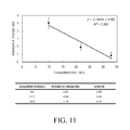
- FIGs. 11 and 12 depict exemplary PT vs. impedance change rate calibration curve and PT vs. reactance change rate calibration curve, respectively.
- FIGs. 13 and 14 depict exemplary graphs of Calibrated PT vs. Real PT by impedance and Calibrated PT vs. Real PT by reactance, respectively.
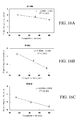
- FIGs. 15A and 15B are experimental graphs showing the blood coagulation analyses using a porous substrate and a non-porous substrate, respectively.
- FIGs. 16A-16C are experimental graphs showing blood coagulation analyses at different frequencies using the reactance methods of measurement according to the present invention.
- the following embodiments provide a system and methods for determining prothrombin time and hematocrit (HCT) of a sample (for example, a blood sample) by performing a reactance analysis of the sample, also referred to as a reactance measurement module.
- Measurements for blood coagulation, or HCT, by performing a reactance analysis as disclosed herein may be suitably used in quantitative analysis of prothrombin time (i.e. , clotting time).
- HCT hematocrit
- hematocrit refers to the percentage of packed red blood cells in a volume of whole blood.
- One exemplary embodiment of the invention provides an electrode-type sensor device with a sample test card including one or more pairs of electrodes.
- the base plate of the test card may be made of porous materials or non-porous materials, but preferably the substrate is made of porous materials as described and shown herein.
- the electrodes may be made of precious metals which include, but is not limited to, gold, silver, palladium, platinum, nickel, alloys thereof, and combinations thereof known to those skilled in the art.
- an alternating current (AC) module or an AC/DC power source provides a test signal to the blood test card with an oscillated frequency in a range between about 0.1 KHz and about 50 KHz.
- the amplitude of voltage applied to the test card is in a range of about 0.05 V to about 5 V.
- the signal is applied to the test card to measure the reactance of the sample.
- the term "about” refers to that variation in the measured quantity as would be expected by the skilled artisan performing the measurement and exercising a level of care commensurate with the objective of the measurement and the precision of the measuring apparatus being used.
- the responding signals are received and processed by the sensor device according to the slope differences depending on the prothrombin time periods analyzed by the reactance measurement.
- the electrodes may be gold electrodes.
- an AC module is adopted for taking a reactance measurement of the sample according to the responding oscillated test signal sensed from the blood test card, wherein as the blood coagulation caused by the enzyme reactions proceeds the resultant signals are received and processed by the sensor device according to the slope differences depending on the prothrombin time periods analyzed by the reactance measurement.
- the impedance of an AC circuit equals the sum of resistance (R) and the product of reactance (X) and the phase angle ⁇ : wherein reactance (X) is the imaginary part of the complex quantity, impedance (Z), representing the obstacles to the current flow created by a combination of inductances (L) and capacitances (C). Resistance (R) is the real part of the complex quantity
- reactance changes with changes in the frequency, changes in the capacitance and/or changes in the inductance of the AC circuit. When the reactance of the AC circuit changes, there will be a phase change between the circuit's current waveform and voltage waveform.
- an AC generation unit of the detection system provides an AC test signal to the test card.
- a sample is applied to the test card in a sample test area of the test card.
- the induced charges are polarized in the electric field to form capacitances and the sample reacts somewhat like a capacitor.
- a blood sample clots, during the clotting a media forms between the electrodes, impeding movement of charges in the sample such that charges accumulate on the electrodes. The accumulated charges thereby cause a capacitance to develop.
- the capacitance reactance variation per unit time and the capacitance variation per unit time during characteristics of the blood clotting in the sample can be determined.
- Prothrombin time (i.e., clotting time) measurement may thus be determined accurately by performing a slope calculation with the help of the reactance measurement module of the invention.
- FIG. 1 is a schematic view of an exemplary diagnostic device for measuring prothrombin time and HCT in accordance with embodiments of the present invention.
- a diagnostic device 100 for measuring HCT and prothrombin time of a fluid includes a relative electrode-type sensor device 120 and a sample test card assembly 110 including one or more pairs of electrodes, wherein alternating current (AC) provided by the sensor device is used to measure and calculate prothrombin time and HCT of blood test using the reactance analysis.
- the sensor device 120 includes a test card receiving unit 122 for accommodating the test card assembly 110.
- a temperature maintaining unit 134 is used for controlling and maintaining temperature of the test card receiving unit at a constant temperature.
- An AC generation unit 124 provides an alternating current with predetermined frequency and voltage to the test card assembly 110.
- a signal retrieving unit 128 is used to retrieve a response signal from the test card assembly.
- a microprocessor 130 is used for analyzing the response signal and rendering results of the HCT and the prothrombin time.
- a display unit 136 is used for displaying inspected results of the HCT and the prothrombin time from the microprocessor 130.
- FIG. 2 illustrates an explosive view of a sample test card for a sample in accordance with one embodiment of the present invention wherein the broken line indicates the relative positions between various elements.
- the sample test card includes an insulating substrate 210, an electrode system 220, a separation and reaction layer 230 and a cover 240.
- the insulating substrate 210 is electrically insulating, and its material may include, but is not limited to, a base plate composed of porous materials.
- the base plate of the test card assembly includes pores with diameters in a range of about 0.1 ⁇ m to about 10 ⁇ m, about 0.01 ⁇ m to about 100 ⁇ m, about 0.1 ⁇ m to about 50 ⁇ m, about 0.1 ⁇ m to about 20 ⁇ m, about 0.1 ⁇ m to about 5 ⁇ m, or about 5 ⁇ m to about 10 ⁇ m.
- the electrode system 220 may be made with any conductive materials, including but not limited to carbon, gold-silver, copper, carbon silver, palladium, nickel, and other similar materials and combinations thereof according to the invention.
- the electrode system 220 includes one or more pairs of the noble metal electrodes, set on the same plane or on different planes respectively.
- a set of testing electrodes 225 includes a pair of electrodes 226 and 228.
- the electrode structure is not limited to specific arrangements of the set of testing electrodes 225 as shown or the exact number of electrodes as shown. Additional electrodes may be provided according to different application needs.
- the electrode system further electrically connects the electrode system with a measurement device (not shown).
- the separation layer 230 is depicted as including spacers 232 disposed over the electrode system 220.
- the separation layer 230 further may include a reaction region 224 to expose a part of the reagent (not shown) and a sampling region 222.
- a channel 236 may connect the sampling region 222 and reaction region 224.
- the size of the reaction region 224 is preferably sufficient to expose part of the electrodes 226 and 228.
- the reaction region 224 is used for measurement of the prothrombin time, and the sampling region 222 may be used for measurement of the HCT.
- the cover 240 is disposed on the separation layer 230.
- the cover 240 includes an inlet 242 and a gas vent 244, which are respectively connected to the sampling region 222 and the reacting region 224.
- the size of the sampling space depends upon the thicknesses of the separation layer 230.
- FIGs. 3A and 3B are photographs showing a side by side comparison of exemplary non-porous and porous base plates of test card assemblies taken using a scanning electron microscope.
- the diameter of pore size of the base plate is in a range of about 0.1 ⁇ m to about 10 ⁇ m with an average diameter of about 3.39 ⁇ m. Pore distribution on the base plate is about 5.04 ⁇ 10 6 holes/cm 2 .
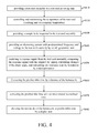
- FIG. 4 is a flowchart schematically illustrating one embodiment of a diagnostic method for determining prothrombin time and HCT according to the present invention.
- the method for measuring prothrombin time and HCT in FIG. 4 includes these steps: providing a test card assembly to a test card receiving unit (S410); controlling and maintaining the temperature of the test card receiving unit at a constant temperature (S420); providing a sample to be inspected to the test card assembly (S430); providing an alternating current with predetermined frequency and voltage to the test card assembly by an AC generation unit (S430); sensing a response signal from the test card assembly and comparing the response signal with the original AC signal, calculating a change in the phase of the current signal (phase shift), and calculating the reactance and the HCT using a microprocessor (S450); correcting the prothrombin time by reference of the HCT (S460); correcting the prothrombin time with an international normalized ratio (S470); and providing inspected results to a display unit (S480).
- S410 test card receiving
- the response signal retrieved from the test card is digitized and converted by the discrete fourier transform (DFT) by a microprocessor.
- DFT discrete fourier transform
- the step of comparing the sensed response signal from the sample with the original AC signal in S450 includes steps of: calculating the magnitude of impedance from the measured response signal and the applied voltage; calculating a change in the phase between the waveform applied and the waveform measured; calculating reactance from the change in the phase angle.
- the change in the phase angle is due to the change in capacitance in the sample as described above. Since the change in capacitance in the sample creates a change in reactance, the change in reactance and can yield HCT and PT as discussed below.
- the HCT can be calculated by interpolation.
- One example of calculating the HCT is described below.
- a traditional method of using the impedance in calculating the HCT is also described as a comparison.
- FIGs. 5 and 6 depict experimental graphs showing the impedance and reactance increase with higher percentage HCT, respectively.
- the reactance of different HCT 29, 39 and 47%) was respectively 620.29, 652.17 and 676.59 ohm.
- the relation of HCT and impedance FIG. 7
- HCT and reactance FIG. 8
- the optimal impedance sampling time of HCT was at about 20 sec or more
- x is the HCT
- y is the impedance or reactance.
- FIGs. 9 and 10 depict exemplary impedance and reactance values measured by an LCR meter every 0.5 second over 60 seconds.
- whole blood was collected from a subject, and various samples were prepared by adding different amounts of anticoagulant drug (heparin) to the collected whole blood.
- concentration of heparin used to modulate the coagulation time (PT) was between about 1 U and about 30 U per milliliter. Then, the blood samples with different PT were analyzed to measure the impedance or reactance by a LCR meter (Hioki Model No. 3532-50).
- FIGs. 11 and 12 depict an exemplary PT vs. impedance change rate calibration curve and an exemplary PT vs. reactance change rate calibration curve, respectively.
- the impedance or reactance change rate was computed every 10 sec by the LCR meter.
- the computation step was repeated to compute the impedance or reactance change rate of the blood for samples with different PTs, and thereby determine the PT vs. impedance change rate calibration curve ( FIG. 7 ) and the PT vs. reactance change rate calibration curve ( FIG. 8 ).
- x is the real PT
- y is the impedance or reactance change rate.
- the diagnostic results of the reactance measurement method showed a superior standard deviation (SD value) compared to the results from the typical impedance measurement method ( FIG. 11 ).
- SD value standard deviation
- adoption of the reactance measurement according to the present invention is advantageous compared to the typical impedance measurement method in that the standard deviation in the reactance measurement is significantly reduced and the linear regression value (R2) in this example is about 0.9986. Accordingly, the reactance measurement method according to the present invention yields a more accurate result than the traditional impedance measurement method, and the slope deviation is acceptable even as blood coagulation process is extended, thus easily adjusting the values.
- the linear regression (R 2 ) for the PT vs. impedance change rate calibration curve was about 0.939 as shown in FIG. 11 . Accordingly, the impedance method is more likely to cause an inaccurate measurement.
- the impedance and reactance of a blood sample were measured by the LCR meter, and its impedance change rate and reactance change rate were calculated.
- the real PT is calculated by the PT calibration curve.
- Different HCT may correspond with different PT calibration curve.
- FIGs. 13 and 14 depict exemplary graphs of Calibrated PT vs. Real PT by impedance and Calibrated PT vs. Real PT by reactance, respectively.
- the increase or decrease in the HCT influences blood clotting time (PT) and the impendence or reactance change rate value.
- PT blood clotting time
- the impendence or reactance change rate value may increase.
- the PT measurement process in some embodiments may include an HCT correction step. Therefore, the device according to some embodiments of the present invention includes an internal memory for storing interpolation data between the HCT and impedance change.
- FIGs. 15A and 15B are experimental graphs showing the blood coagulation analyses using a porous substrate and a non-porous substrate for the test card, respectively. As illustrated in the Figures, the analyses employing a porous substrate provided superior results.
- FIGs. 16A-16C depict experimental graphs showing the blood coagulation analyses at different frequencies by the reactance measurement according to the present invention. Coagulation times between 15-50 seconds is measured for the blood samples at frequencies of 0.1kHz, 10kHz and 50kHz, and test results R 2 were obtained by calculating regression analysis of 0.9636, 0.9923, and 0.9858 respectively. In this case, the regression analysis indicates that using frequencies of 10KHz and 50KHz provide greater accuracy as compared to a frequency of 0.1KHz.
Abstract
Description
- This application is based upon and claims the benefit of priority from
U.S. Provisional Application No. 61/353,137, filed on June 9, 2010 - The present invention relates to biochemical diagnostic devices, and more particularly, to devices and methods for determining prothrombin time (PT) and hematocrit (HCT) by analyzing a change in reactance in a sample.
- Two pathways or coagulation cascades, known as the intrinsic and extrinsic pathways, lead to the formation of a clot in blood. When a human body is injured, the extrinsic pathway is first triggered to control the body's blood coagulation. In addition to a blood sample, the coagulation reaction needs some additional tissue factors. The inactive factor X is catalyzed into factor Xa. The prothrombin (factor II) can be transformed from the factor Xa to the thrombin (factor IIa) by the effects of factor Va, acidic phospholipids and calcium ions. The thrombin then transforms fibrinogen into fibrin, enhancing the platelet of the endothelial cells gathered at the injury. The thrombin can also enhance the role of factor XIII, linking each fibrous protein molecule to a stable fibrin. Therefore, inspecting the prothrombin time not only allows determining whether the function of external activation factors of the coagulation system are normal, but also allows assessing and monitoring oral anticoagulants treatment, liver function, vitamin K deficiency, coagulation factor deficiency, and disseminated intravascular coagulation (DIC) syndrome.
- Conventional inspection methods for measuring the prothrombin time analyze the condensation phenomenon of transforming the serum soluble protein into an insoluble protein during blood coagulation. These inspection methods can be realized by detecting optical characteristics, such as changes of color, reflection, refraction, luminescence and fluorescence. Such inspection methods, however, require a substantial number of blood test samples and high purity reagents and are time-consuming, as disclosed in
U.S. Patent No. 5,418,141 , the entirety of which is hereby incorporated by reference. Moreover, these inspection methods require long detection times and a significant amount of supplies, resulting in inconvenience and higher costs. - Other conventional inspection methods for measurement of the prothrombin time use electrochemical inspection methods. For example,
U.S. Patent No. 3,699,437 , the entirety of which is hereby incorporated by reference, discloses observing the comparative decline rate of resistance from the initial to the lowest point. The calculated result is served as a basis for determining coagulation time in which the impedance measurement is related to the mechanism of blood coagulation. Further,U.S. Patent Nos. 6,060,323 ;6,338,821 ;6,066,504 ;7,673,622 ; and6,046,051 , the entirety of each of which is hereby incorporated by reference, disclose electronic sensor devices and a test card assembly for the measurement of the coagulation time of a blood sample. The test card assembly is designed with a single electrode or a plurality of electrodes according to the measurement demands of the device. The sample is contacted with the electrodes, which measure the change in impedance corresponding to the change of viscosity of the blood sample as it coagulates. This technique, however, may result in test errors due to the hematocrit and the electrolyte concentration differences among blood test samples in individuals.U.S. Patent No. 7,005,857 , the entirety of which is hereby incorporated by reference, discloses a coagulation inspection device with automatic collection of blood samples. The coagulation inspection device determines the coagulation time by measuring capacitance or impedance changes between two electrodes. These technologies may, therefore, improve the simplicity of the detection device, but they cannot achieve the relatively higher precision and accuracy that the above-discussed optical detection methods may achieve. - Accordingly, a new biosensor device is needed and described herein for measuring prothrombin time (PT) and hematocrit (HCT), one capable of operating with short test times, simple procedures for the user and while achieving highly accurate results.
- One aspect of the present invention is to use reactance (X) measurements taken from a sample to calculate PT. As described herein, using reactance measurements as opposed to impedance measurements provides a more accurate analysis of the blood's characteristics, reduces the chance of a test error, and improves measurement accuracy.
- Another aspect of the present invention is to provide a detection system and a measurement methods for determining prothrombin time and hematocrit (HCT) of a blood sample using a reactance analysis of the sample. In one embodiment, the detection system includes a sensor device and a test card assembly. The test card assembly includes one or more pairs of the precious metal electrodes, set on the same plane or on different planes, respectively. Alternating current (AC) provided by the sensor device is used to measure and calculate the prothrombin time and HCT of the blood sample using the reactance analysis described herein.
- Another aspect of the invention is to provide a test card assembly with an improved blood sample and reagent contact area. The test card assembly according to some embodiments of the present invention utilizes porous materials, such as fiberglass substrate (FR-4), for at least a portion of a substrate of the test card. Since the surface of at least a portion of the substrate, preferably the entire surface of the substrate, is porous, e.g., it may have a plurality of holes, voids, or cavities thereon, the sample (e.g., blood) is improvably, uniformly dispersed on the substrate, thereby increasing the contact area of the blood sample and reagents, and effectively improving on drawbacks of traditional non-porous materials used for substrates. According to the present invention, the problems associated with having relatively poor contact between the sample and the reagent that occur when using non-porous materials for the substrate, or a portion thereof, e.g., relatively high blood cohesion, are minimized or eliminated.
- According to one aspect of the invention, a diagnostic device for measuring HCT and prothrombin time of a fluid includes: a electrode-type sensor device; and a blood test card assembly including one or more pairs of electrodes, wherein alternating current (AC) provided by the sensor device is used to measure and calculate prothrombin time and HCT of the blood test using the reactance analysis described herein.
- In one embodiment, the sensor device includes: a test card receiving unit for accommodating the test card assembly; a temperature maintaining unit for controlling and maintaining temperature of the test card receiving unit at a constant temperature; an AC generation unit for providing an alternating current with predetermined frequency and voltage to the test card assembly; a signal receiving unit to receive a response signal from the test card assembly; a microprocessor for calculating the response signal and rendering results of the HCT and the prothrombin time; and a display unit for displaying inspected results of the HCT and the prothrombin time from the microprocessor.
- According to another aspect of the invention, a method for measuring prothrombin time and HCT includes: providing a test card assembly to a test card receiving unit; controlling and maintaining temperature of the test card receiving unit at a constant temperature; providing a sample to be inspected to the test card assembly; providing an alternating current with predetermined frequency and voltage to the test card assembly by an AC generation unit; receiving a response signal from the test card assembly and calculating the HCT and the prothrombin time by a microprocessor; and providing an inspected result to a display unit.
- The foregoing aspects and many of the attendant advantages of this invention will become more readily appreciated by reference to the following detailed description, when taken in conjunction with the accompanying pictures, wherein:
-
FIG. 1 is a schematic view of an exemplary diagnostic device for measuring prothrombin time and HCT in accordance with embodiments of the present invention; -
FIG. 2 illustrates an explosive view of a blood test card in accordance with one embodiment of the present invention wherein the broken line indicates the relative positions between various elements; -
FIGs. 3A and 3B are photographs showing a side by side comparison of exemplary non-porous and porous base plates of substrates of test card assemblies taken using a scanning electron microscope; -
FIG. 4 is a flowchart schematically illustrating one embodiment of the diagnostic method for determining prothrombin time and HCT according to the present invention; -
FIG. 5 is experimental graph showing the change of impedance vs. coagulation time in seconds, which illustrates a change in slope as a whole blood sample coagulates by a typical impedance measurement method; -
FIG. 6 is experimental graph showing the change of reactance vs. coagulation time in seconds, which illustrates a change in slope as a whole blood sample coagulates by the reactance measurement method; -
FIGs. 7 and8 are the relation of HCT and impedance (FIG. 7 ) and HCT and reactance (FIG. 8 ) calculated from the experimental graphs ofFIGs. 5 and 6 , respectively. -
FIGs. 9 and 10 depict exemplary impedance and reactance values measured by an LCR meter every 0.5 second over 60 seconds. -
FIGs. 11 and12 depict exemplary PT vs. impedance change rate calibration curve and PT vs. reactance change rate calibration curve, respectively. -
FIGs. 13 and14 depict exemplary graphs of Calibrated PT vs. Real PT by impedance and Calibrated PT vs. Real PT by reactance, respectively. -
FIGs. 15A and 15B are experimental graphs showing the blood coagulation analyses using a porous substrate and a non-porous substrate, respectively; and -
FIGs. 16A-16C are experimental graphs showing blood coagulation analyses at different frequencies using the reactance methods of measurement according to the present invention. - Reference will now be made in detail to several exemplary embodiments of the invention, examples of which are illustrated in the accompanying drawings and photographs. Wherever possible, the same reference numbers are used in the drawings and the description to refer to the same or like parts. In the drawings, the shape and thickness of an embodiment may be exaggerated for clarity and convenience. This description will be directed in particular to elements forming part of, or cooperating more directly with, apparatus in accordance with the present invention. It is to be understood that elements not specifically shown or described may take various forms well known to those skilled in the art. Further, when a layer is referred to as being on another layer or "on" a substrate, it may be directly on the other layer or on the substrate, or intervening layers may also be presented.
- Some exemplary embodiments of the present invention are described in greater detail by referring to the drawings and photographs that accompany the present application. It should be noted that the features illustrated in the drawings are not necessarily drawn to scale. Descriptions of well-known components, materials, and process techniques may be omitted so as to not unnecessarily obscure the embodiments of the invention. Any devices, components, materials, and steps described in the embodiments are only for illustration and not intended to limit the scope of the present invention.
- In view of the aforementioned problems noted of the conventional technologies, the following embodiments provide a system and methods for determining prothrombin time and hematocrit (HCT) of a sample (for example, a blood sample) by performing a reactance analysis of the sample, also referred to as a reactance measurement module. Measurements for blood coagulation, or HCT, by performing a reactance analysis as disclosed herein may be suitably used in quantitative analysis of prothrombin time (i.e., clotting time). As used herein, the term "hematocrit" refers to the percentage of packed red blood cells in a volume of whole blood.
- One exemplary embodiment of the invention provides an electrode-type sensor device with a sample test card including one or more pairs of electrodes. The base plate of the test card may be made of porous materials or non-porous materials, but preferably the substrate is made of porous materials as described and shown herein. The electrodes may be made of precious metals which include, but is not limited to, gold, silver, palladium, platinum, nickel, alloys thereof, and combinations thereof known to those skilled in the art. In one aspect of the invention, an alternating current (AC) module or an AC/DC power source provides a test signal to the blood test card with an oscillated frequency in a range between about 0.1 KHz and about 50 KHz. The amplitude of voltage applied to the test card is in a range of about 0.05 V to about 5 V. The signal is applied to the test card to measure the reactance of the sample. As used herein in connection with a measured quantity, the term "about" refers to that variation in the measured quantity as would be expected by the skilled artisan performing the measurement and exercising a level of care commensurate with the objective of the measurement and the precision of the measuring apparatus being used.
- In one preferred aspect of the invention, as blood coagulation in a sample caused by an enzymatic reactions proceeds, the responding signals are received and processed by the sensor device according to the slope differences depending on the prothrombin time periods analyzed by the reactance measurement. In one embodiment, the electrodes may be gold electrodes. In one example, an AC module is adopted for taking a reactance measurement of the sample according to the responding oscillated test signal sensed from the blood test card, wherein as the blood coagulation caused by the enzyme reactions proceeds the resultant signals are received and processed by the sensor device according to the slope differences depending on the prothrombin time periods analyzed by the reactance measurement.
- Principles of performing a reactance analysis according to embodiments of the present invention are discussed below. The impedance of an AC circuit equals the sum of resistance (R) and the product of reactance (X) and the phase angle θ:wherein reactance (X) is the imaginary part of the complex quantity, impedance (Z), representing the obstacles to the current flow created by a combination of inductances (L) and capacitances (C). Resistance (R) is the real part of the complex quantity As known to those skilled in the art, the reactance changes with changes in the frequency, changes in the capacitance and/or changes in the inductance of the AC circuit. When the reactance of the AC circuit changes, there will be a phase change between the circuit's current waveform and voltage waveform. The impedance (Z) is defined as:


where Z is impedance, R is resistance, j is phase, and X is reactance; and
where XC is capacitance reactance, XL is inductance reactance, π is a ratio of the circumference of a circle to its diameter, f is frequency, L is inductance, and C is capacitance. - In one aspect of the invention, an AC generation unit of the detection system provides an AC test signal to the test card. A sample is applied to the test card in a sample test area of the test card. As the sample between the electrodes is charged, the induced charges are polarized in the electric field to form capacitances and the sample reacts somewhat like a capacitor. As a blood sample clots, during the clotting a media forms between the electrodes, impeding movement of charges in the sample such that charges accumulate on the electrodes. The accumulated charges thereby cause a capacitance to develop. In a preferred aspect of the invention, since the frequency (f) of the AC generation unit of the detection system remains constant, the inductance (L) and the inductor reactance (XL) is also constant, and, therefore, the variation in the capacitance reactance in the sample equals essentially the variation in the overall reactance as shown by the following equation:

where XC2-XC1 is the variation in the capacitance reactance, and X2-X1 is the variation in the reactance. - By determining reactance variation of the sample per unit time, the capacitance reactance variation per unit time and the capacitance variation per unit time during characteristics of the blood clotting in the sample can be determined. Prothrombin time (i.e., clotting time) measurement may thus be determined accurately by performing a slope calculation with the help of the reactance measurement module of the invention.
-
FIG. 1 is a schematic view of an exemplary diagnostic device for measuring prothrombin time and HCT in accordance with embodiments of the present invention. As illustrated inFIG. 1 , adiagnostic device 100 for measuring HCT and prothrombin time of a fluid includes a relative electrode-type sensor device 120 and a sampletest card assembly 110 including one or more pairs of electrodes, wherein alternating current (AC) provided by the sensor device is used to measure and calculate prothrombin time and HCT of blood test using the reactance analysis. In this embodiment, thesensor device 120 includes a testcard receiving unit 122 for accommodating thetest card assembly 110. Atemperature maintaining unit 134 is used for controlling and maintaining temperature of the test card receiving unit at a constant temperature. AnAC generation unit 124 provides an alternating current with predetermined frequency and voltage to thetest card assembly 110. Asignal retrieving unit 128 is used to retrieve a response signal from the test card assembly. Amicroprocessor 130 is used for analyzing the response signal and rendering results of the HCT and the prothrombin time. Adisplay unit 136 is used for displaying inspected results of the HCT and the prothrombin time from themicroprocessor 130. -
FIG. 2 illustrates an explosive view of a sample test card for a sample in accordance with one embodiment of the present invention wherein the broken line indicates the relative positions between various elements. The sample test card includes an insulatingsubstrate 210, anelectrode system 220, a separation andreaction layer 230 and acover 240. The insulatingsubstrate 210 is electrically insulating, and its material may include, but is not limited to, a base plate composed of porous materials. In preferred embodiments of the invention, the base plate of the test card assembly includes pores with diameters in a range of about 0.1µm to about 10µm, about 0.01µm to about 100µm, about 0.1µm to about 50µm, about 0.1µm to about 20µm, about 0.1µm to about 5µm, or about 5µm to about 10µm. Theelectrode system 220 may be made with any conductive materials, including but not limited to carbon, gold-silver, copper, carbon silver, palladium, nickel, and other similar materials and combinations thereof according to the invention. Theelectrode system 220 includes one or more pairs of the noble metal electrodes, set on the same plane or on different planes respectively. For example, a set oftesting electrodes 225 includes a pair ofelectrodes testing electrodes 225 as shown or the exact number of electrodes as shown. Additional electrodes may be provided according to different application needs. The electrode system further electrically connects the electrode system with a measurement device (not shown). - The
separation layer 230 is depicted as includingspacers 232 disposed over theelectrode system 220. Theseparation layer 230 further may include areaction region 224 to expose a part of the reagent (not shown) and asampling region 222. Achannel 236 may connect thesampling region 222 andreaction region 224. The size of thereaction region 224 is preferably sufficient to expose part of theelectrodes reaction region 224 is used for measurement of the prothrombin time, and thesampling region 222 may be used for measurement of the HCT. - The
cover 240 is disposed on theseparation layer 230. In one embodiment, thecover 240 includes aninlet 242 and agas vent 244, which are respectively connected to thesampling region 222 and the reactingregion 224. The size of the sampling space depends upon the thicknesses of theseparation layer 230. -
FIGs. 3A and 3B are photographs showing a side by side comparison of exemplary non-porous and porous base plates of test card assemblies taken using a scanning electron microscope. InFigure 3B , the diameter of pore size of the base plate is in a range of about 0.1 µm to about 10 µm with an average diameter of about 3.39 µm. Pore distribution on the base plate is about 5.04×10⁶ holes/cm2. -
FIG. 4 is a flowchart schematically illustrating one embodiment of a diagnostic method for determining prothrombin time and HCT according to the present invention. The method for measuring prothrombin time and HCT inFIG. 4 includes these steps: providing a test card assembly to a test card receiving unit (S410); controlling and maintaining the temperature of the test card receiving unit at a constant temperature (S420); providing a sample to be inspected to the test card assembly (S430); providing an alternating current with predetermined frequency and voltage to the test card assembly by an AC generation unit (S430); sensing a response signal from the test card assembly and comparing the response signal with the original AC signal, calculating a change in the phase of the current signal (phase shift), and calculating the reactance and the HCT using a microprocessor (S450); correcting the prothrombin time by reference of the HCT (S460); correcting the prothrombin time with an international normalized ratio (S470); and providing inspected results to a display unit (S480). - According to one embodiment of the present invention, the response signal retrieved from the test card is digitized and converted by the discrete fourier transform (DFT) by a microprocessor. Thus, as known to those skilled in the art, the real value and imaginary value may be divided by the method shown below:


where X(k) is fourier value of digital signal, x(n) is original value of digital signal, n is current point of digital signal, and N is total number of digital signal. Further, the phase may be calculated by the imaginary value and real value according to the following formula:
where Im is the imaginary value (i.e., reactance) and Re is the real value (i.e., resistance). As shown in the formula (3), the phase will shift by the change in the reactance in the sample. - In some embodiments, the step of comparing the sensed response signal from the sample with the original AC signal in S450 (i.e., phase) above includes steps of: calculating the magnitude of impedance from the measured response signal and the applied voltage; calculating a change in the phase between the waveform applied and the waveform measured; calculating reactance from the change in the phase angle. As will be appreciated, since the alternating current has a constant frequency, the change in the phase angle is due to the change in capacitance in the sample as described above. Since the change in capacitance in the sample creates a change in reactance, the change in reactance and can yield HCT and PT as discussed below.
- From the measured reactance, the HCT can be calculated by interpolation. One example of calculating the HCT is described below. A traditional method of using the impedance in calculating the HCT is also described as a comparison.
-
FIGs. 5 and 6 depict experimental graphs showing the impedance and reactance increase with higher percentage HCT, respectively. As shown in the graph inFIG. 6 , in this particular example, in the 11th second, the reactance of different HCT (29, 39 and 47%) was respectively 620.29, 652.17 and 676.59 ohm. Then, the relation of HCT and impedance (FIG. 7 ) or HCT and reactance (FIG. 8 ) were calculated. As shown inFIG 5 , in this particular example, the optimal impedance sampling time of HCT was at about 20 sec or more, and Impedance vs. HCT calibration curve has an equation of y =90.253x +2347.7. Further, as shown inFIG 6 , the optimal reactance sampling time of HCT was at about 11 sec or more, and Reactance vs. HCT calibration curve had an equation of y = 3.1304x +529.68. As will be appreciated, x is the HCT, and y is the impedance or reactance. -
FIGs. 9 and 10 depict exemplary impedance and reactance values measured by an LCR meter every 0.5 second over 60 seconds. In one example, whole blood was collected from a subject, and various samples were prepared by adding different amounts of anticoagulant drug (heparin) to the collected whole blood. In one instance, the concentration of heparin used to modulate the coagulation time (PT) was between about 1 U and about 30 U per milliliter. Then, the blood samples with different PT were analyzed to measure the impedance or reactance by a LCR meter (Hioki Model No. 3532-50). -
FIGs. 11 and12 depict an exemplary PT vs. impedance change rate calibration curve and an exemplary PT vs. reactance change rate calibration curve, respectively. In this particular example, the impedance or reactance change rate was computed every 10 sec by the LCR meter. For example, the change rate between 30 to 40 second was computed by following formula:Impedance change rate 30 to 40 = (Z40 - Z30)/ (Time40 - Time30), wherein Z is impedance, andReactance change rate 30 to 40 = (X40 - X30)/ (Time40 - Time30), wherein X is reactance. The computation step was repeated to compute the impedance or reactance change rate of the blood for samples with different PTs, and thereby determine the PT vs. impedance change rate calibration curve (FIG. 7 ) and the PT vs. reactance change rate calibration curve (FIG. 8 ). The PT vs. impedance change rate calibration curve had an equation of y = -0.1849x + 4.562, and the PT vs. reactance change rate calibration curve had an equation of y = -0.0256x +0.3604. As will be appreciated, x is the real PT, and y is the impedance or reactance change rate. - As shown in
FIGs. 11 and12 , the diagnostic results of the reactance measurement method (FIG. 12 ) showed a superior standard deviation (SD value) compared to the results from the typical impedance measurement method (FIG. 11 ). Specifically, adoption of the reactance measurement according to the present invention is advantageous compared to the typical impedance measurement method in that the standard deviation in the reactance measurement is significantly reduced and the linear regression value (R2) in this example is about 0.9986. Accordingly, the reactance measurement method according to the present invention yields a more accurate result than the traditional impedance measurement method, and the slope deviation is acceptable even as blood coagulation process is extended, thus easily adjusting the values. - On the other hand, in this particular example, the linear regression (R2) for the PT vs. impedance change rate calibration curve was about 0.939 as shown in
FIG. 11 . Accordingly, the impedance method is more likely to cause an inaccurate measurement. - In the above example, the impedance and reactance of a blood sample were measured by the LCR meter, and its impedance change rate and reactance change rate were calculated. Specifically, the HCT was calculated by using the HCT calibration curve with the formula, HCT = (impedance - 2347.7) / 90.253 or HCT = (reactance - 529.68) / 3.1304. Then, the real PT is calculated by the PT calibration curve. Different HCT may correspond with different PT calibration curve. In this particular example, the PT was calculated by using the PT calibration curve with the formula, PT = (impedance change rate - 4.562) / -0.1849 or PT = (reactance change rate - 0.3604) / -0.0256.
-
FIGs. 13 and14 depict exemplary graphs of Calibrated PT vs. Real PT by impedance and Calibrated PT vs. Real PT by reactance, respectively. The PT values computed by the calibration curve and the real PT values measured by the automated blood coagulation analyzer (Sysmex CA-500 series) were compared. - The increase or decrease in the HCT influences blood clotting time (PT) and the impendence or reactance change rate value. Specifically, a higher HCT causes the impendence or reactance change rate value to increase. Hence, the PT measurement process in some embodiments may include an HCT correction step. Therefore, the device according to some embodiments of the present invention includes an internal memory for storing interpolation data between the HCT and impedance change.
- In one aspect of the invention, the PT values calculated from the above example may be corrected with an international normalized ratio shown in the following formula:

where INR is international normalized ratio, PT is prothrombin time, and ISI is an international sensitivity index. -
FIGs. 15A and 15B are experimental graphs showing the blood coagulation analyses using a porous substrate and a non-porous substrate for the test card, respectively. As illustrated in the Figures, the analyses employing a porous substrate provided superior results. -
FIGs. 16A-16C depict experimental graphs showing the blood coagulation analyses at different frequencies by the reactance measurement according to the present invention. Coagulation times between 15-50 seconds is measured for the blood samples at frequencies of 0.1kHz, 10kHz and 50kHz, and test results R2 were obtained by calculating regression analysis of 0.9636, 0.9923, and 0.9858 respectively. In this case, the regression analysis indicates that using frequencies of 10KHz and 50KHz provide greater accuracy as compared to a frequency of 0.1KHz. - While the invention has been described by way of examples and in terms of preferred embodiments, it would be apparent to those skilled in the art to make various equivalent replacements, amendments and modifications in view of specification of the invention. Therefore, the scope of the appended claims should be accorded the broadest interpretation so as to encompass all such replacements, amendments and modifications without departing from the spirit and scope of the invention.
Claims (17)
- A diagnostic device for measuring hematocrit (HCT) and/or prothrombin time of a fluid, comprising:a relative electrode-type sensor device; anda test card assembly including one or more pairs of electrodes,wherein alternating current (AC) provided by the sensor device is used to measure and calculate prothrombin time and/or HCT of blood test using reactance analysis.
- The diagnostic device as claimed in claim 1, wherein the reactance analysis comprises:comparing a response signal from the test card assembly in response to the AC with the AC signal, calculating a change in phase of the AC signal, and calculating a reactance and the HCT.
- The diagnostic device as claimed in claim 2, wherein the reactance analysis further comprises:calculating a capacitance, andtransforming the capacitance with algorithms and correcting to the prothrombin time by reference of the HCT.
- The diagnostic device as claimed in claim 3, wherein the reactance analysis further comprises correcting the prothrombin time with an international normalized ratio.
- The diagnostic device as claimed in claim 1, wherein the test card assembly comprises one or more pairs of the noble metal electrodes, set on the same plane or on different planes respectively.
- The diagnostic device as claimed in claim 1, wherein the blood test card assembly comprises a base plate comprising a porous material.
- The diagnostic device as claimed in claim 6, wherein the base plate of the test card assembly comprises pores with diameters approximately in a range of about 0.1µm to about 10µm.
- The diagnostic device as claimed in claim 1, wherein the sensor device comprises:a test card receiving unit for accommodating the test card assembly;a temperature maintaining unit for controlling and maintaining temperature of the test card receiving unit at a constant temperature;an AC generation unit for providing an alternating current with predetermined frequency and voltage to the test card assembly;a signal receiving unit to retrieve a response signal from the test card assembly;a microprocessor for calculating the response signal and rendering results of the HCT and the prothrombin time; anda display unit for displaying inspected results of the HCT and the prothrombin time from the microprocessor.
- The diagnostic device as claimed in claim 8, wherein the microprocessor compares the response signal with an original AC signal, calculates a change in phase of the AC signal, and calculates the reactance and the HCT.
- The diagnostic device as claimed in claim 9, wherein the microprocessor further transforms the capacitance with algorithms, corrects to the prothrombin time by reference of the HCT; and calculates the prothrombin time with an international normalized ratio.
- A method for measuring HCT and/or prothrombin time, comprising:providing a test card assembly to a test card receiving unit;controlling and maintaining temperature of the test card receiving unit at a constant temperature;providing a sample to be inspected to the test card assembly;providing an alternating current with predetermined frequency and voltage to the test card assembly by an AC generation unit;receiving a response signal from the test card assembly and calculating the HCT and/or the prothrombin time by a microprocessor; andproviding an inspected result to a display unit.
- The method as claimed in claim 11, wherein the step of receiving a response signal from the test card assembly and calculating the HCT and/or the prothrombin time by a microprocessor comprises:receiving a response signal from the test card assembly, comparing the response signal with an original AC signal, calculating a change in phase of the AC signal, and calculating a reactance and the HCT by a microprocessor.
- The method as claimed in claim 12, wherein the step of receiving a response signal from the test card assembly and calculating the prothrombin time further comprises:calculating a capacitance, andtransforming the capacitance with algorithms and correcting to the prothrombin time by reference of the HCT.
- The method as claimed in claim 13, wherein the step of receiving a response signal from the test card assembly and calculating the prothrombin time by a microprocessor further comprises:correcting the prothrombin time with an international normalized ratio.
- The method for measuring prothrombin time and HCT as claimed in claim 11, wherein wherein the blood test card assembly comprises one or more pairs of the noble metal electrodes, set on the same plane or on different planes respectively.
- The method for measuring prothrombin time and HCT as claimed in claim 11, wherein the blood test card assembly comprises a base plate composed of porous materials.
- The method for measuring prothrombin time and HCT as claimed in claim 11, wherein the base plate of the test card assembly includes pores with diameters approximately in a range of about 0.1µm to about 10µm.
Priority Applications (1)
| Application Number | Priority Date | Filing Date | Title |
|---|---|---|---|
| PL11169337T PL2395353T3 (en) | 2010-06-09 | 2011-06-09 | Device and method for measuring prothrombin time and hematocrit by analyzing change in reactance in a sample |
Applications Claiming Priority (1)
| Application Number | Priority Date | Filing Date | Title |
|---|---|---|---|
| US35313710P | 2010-06-09 | 2010-06-09 |
Publications (2)
| Publication Number | Publication Date |
|---|---|
| EP2395353A1 true EP2395353A1 (en) | 2011-12-14 |
| EP2395353B1 EP2395353B1 (en) | 2018-08-22 |
Family
ID=44508711
Family Applications (1)
| Application Number | Title | Priority Date | Filing Date |
|---|---|---|---|
| EP11169337.0A Active EP2395353B1 (en) | 2010-06-09 | 2011-06-09 | Device and method for measuring prothrombin time and hematocrit by analyzing change in reactance in a sample |
Country Status (6)
| Country | Link |
|---|---|
| US (2) | US8828322B2 (en) |
| EP (1) | EP2395353B1 (en) |
| JP (1) | JP5905212B2 (en) |
| ES (1) | ES2694080T3 (en) |
| PL (1) | PL2395353T3 (en) |
| TW (1) | TWI472766B (en) |
Cited By (5)
| Publication number | Priority date | Publication date | Assignee | Title |
|---|---|---|---|---|
| EP2980570A4 (en) * | 2013-03-29 | 2016-10-26 | Sony Corp | Blood state evaluation device, blood state evaluation system, blood state evaluation method, and program |
| EP2980571A4 (en) * | 2013-03-29 | 2016-10-26 | Sony Corp | Blood state analysis device, blood state analysis system, blood state analysis method, and program |
| CN107036738A (en) * | 2017-06-01 | 2017-08-11 | 黄昱 | A kind of blood platelet Micro-force sensor of the elastic film variable capacitance based on nanometer technique |
| US10281452B2 (en) | 2012-04-13 | 2019-05-07 | Sony Corporation | Blood coagulation system analyzer, blood coagulation system analysis method and program |
| WO2021186198A1 (en) * | 2020-03-20 | 2021-09-23 | Ainger Phill | Point of care sepsis assay device and method |
Families Citing this family (11)
| Publication number | Priority date | Publication date | Assignee | Title |
|---|---|---|---|---|
| TWI482964B (en) * | 2012-11-27 | 2015-05-01 | Broadmaster Biotech Corp | Method and device for measuring hematocrit (hct) |
| JP6421749B2 (en) * | 2013-03-15 | 2018-11-14 | ソニー株式会社 | Blood state analysis apparatus, blood state analysis system, blood state analysis method, and blood state analysis program for causing a computer to realize the method |
| EP2980558B1 (en) * | 2013-03-26 | 2019-11-06 | Sony Corporation | Measurement device and measurement method |
| US9395319B2 (en) | 2013-05-02 | 2016-07-19 | Lifescan Scotland Limited | Analytical test meter |
| TWI504889B (en) * | 2013-11-19 | 2015-10-21 | Apex Biotechnology Corp | Hematocrit measurement system and measurement method using the same |
| TWI531790B (en) | 2014-11-10 | 2016-05-01 | 五鼎生物技術股份有限公司 | Electrochemical test strip, measurement system and method for determining sample content in the reactive region of the electrochemical test strip |
| JP6589613B2 (en) | 2015-12-10 | 2019-10-16 | いすゞ自動車株式会社 | Reactance measuring device |
| US11237125B2 (en) | 2017-07-17 | 2022-02-01 | Hewlett-Packard Development Company, L.P. | Determining hematocrit level of a blood sample |
| KR102083514B1 (en) * | 2018-06-22 | 2020-03-02 | 광주과학기술원 | Apparatus and method for measuring deformability of red blood cells |
| JP6747491B2 (en) * | 2018-11-28 | 2020-08-26 | ソニー株式会社 | Blood state analysis device, blood state analysis system, blood state analysis method, and blood state analysis program for realizing the method on a computer |
| CN113063833B (en) * | 2021-03-12 | 2023-09-08 | 三诺生物传感股份有限公司 | Measurement method of hematocrit |
Citations (8)
| Publication number | Priority date | Publication date | Assignee | Title |
|---|---|---|---|---|
| US3699437A (en) | 1968-09-27 | 1972-10-17 | Amiram Ur | Blood coagulation detection method and apparatus |
| US4547735A (en) * | 1982-01-23 | 1985-10-15 | Holger Kiesewetter | Instrument for measuring the hematocrit value of blood |
| US5418141A (en) | 1994-05-06 | 1995-05-23 | Avocet Medical, Inc. | Test articles for performing dry reagent prothrombin time assays |
| US6046051A (en) | 1997-06-27 | 2000-04-04 | Hemosense, Inc. | Method and device for measuring blood coagulation or lysis by viscosity changes |
| US7005857B2 (en) | 2000-12-19 | 2006-02-28 | Lifescan Scotland Limited | Device for measuring blood coagulation and method thereof |
| WO2007075410A2 (en) * | 2005-12-16 | 2007-07-05 | Bayer Healthcare Llc | In-vivo non-invasive bioelectric impedance analysis of glucose-mediated changes in tissue |
| US20080224716A1 (en) * | 2006-03-18 | 2008-09-18 | Singer Michaeal G | Method and System For Determining Freshness and Palatability and Assessing Organ Vitality |
| US7673622B2 (en) | 2005-10-11 | 2010-03-09 | Mann + Hummel Gmbh | Air filter, secondary air charging system and seal arrangement for a secondary air charging system |
Family Cites Families (12)
| Publication number | Priority date | Publication date | Assignee | Title |
|---|---|---|---|---|
| US5144240A (en) | 1985-08-14 | 1992-09-01 | Picker International, Inc. | Nmr spectroscopy and imaging coil |
| WO2001063271A1 (en) | 2000-02-21 | 2001-08-30 | F. Hoffmann-La Roche Ag | Electrochemical sensor for determining blood clotting, corresponding system for measuring blood clotting and method for determining blood clotting |
| US20030146113A1 (en) | 2000-02-21 | 2003-08-07 | Volker Unkrig | Electrochemical sensor for determining blood clotting, corresponding system for measuring blood clotting and method for determining blood clotting |
| US20090053193A1 (en) | 2004-05-11 | 2009-02-26 | Novo Nordisk Healthcare A/G | Use of Factor VIIa for the Treatment of Burn Trauma |
| DK1885871T3 (en) | 2005-05-17 | 2012-07-02 | Radiometer Medical Aps | Enzyme sensor with a cover membrane layer of a porous polymer material covered by a hydrophilic polymer |
| WO2006122554A2 (en) | 2005-05-17 | 2006-11-23 | Radiometer Medical Aps | Enzyme sensor with a cover membrane layer covered by a hydrophilic polymer |
| EP1962710B1 (en) * | 2005-12-06 | 2015-08-12 | St. Jude Medical, Atrial Fibrillation Division, Inc. | Apparatus for displaying catheter electrode-tissue contact in electro-anatomic mapping and navigation system |
| US20090118666A1 (en) * | 2006-02-28 | 2009-05-07 | Andreas Blomqvist | Method and implantable device for measuring hematocrit |
| JP2008076143A (en) * | 2006-09-20 | 2008-04-03 | Citizen Holdings Co Ltd | Instrument for measuring hemoglobin concentration |
| US20080297169A1 (en) * | 2007-05-31 | 2008-12-04 | Greenquist Alfred C | Particle Fraction Determination of A Sample |
| US20090205399A1 (en) * | 2008-02-15 | 2009-08-20 | Bayer Healthcare, Llc | Auto-calibrating test sensors |
| KR100969667B1 (en) * | 2008-03-24 | 2010-07-14 | 디지탈 지노믹스(주) | Method for detecting biomolecules electrically and biochip provided with therefor |
-
2011
- 2011-06-09 PL PL11169337T patent/PL2395353T3/en unknown
- 2011-06-09 JP JP2011129559A patent/JP5905212B2/en active Active
- 2011-06-09 TW TW100120140A patent/TWI472766B/en active
- 2011-06-09 US US13/156,693 patent/US8828322B2/en active Active
- 2011-06-09 EP EP11169337.0A patent/EP2395353B1/en active Active
- 2011-06-09 ES ES11169337.0T patent/ES2694080T3/en active Active
-
2014
- 2014-07-25 US US14/340,924 patent/US9068967B2/en active Active
Patent Citations (11)
| Publication number | Priority date | Publication date | Assignee | Title |
|---|---|---|---|---|
| US3699437A (en) | 1968-09-27 | 1972-10-17 | Amiram Ur | Blood coagulation detection method and apparatus |
| US4547735A (en) * | 1982-01-23 | 1985-10-15 | Holger Kiesewetter | Instrument for measuring the hematocrit value of blood |
| US5418141A (en) | 1994-05-06 | 1995-05-23 | Avocet Medical, Inc. | Test articles for performing dry reagent prothrombin time assays |
| US6046051A (en) | 1997-06-27 | 2000-04-04 | Hemosense, Inc. | Method and device for measuring blood coagulation or lysis by viscosity changes |
| US6060323A (en) | 1997-06-27 | 2000-05-09 | Hemosense, Inc. | Method and device for measuring blood coagulation or lysis by viscosity changes |
| US6066504A (en) | 1997-06-27 | 2000-05-23 | Hemosense, Inc. | Coagulation or lysis assays using an electroactive species |
| US6338821B1 (en) | 1997-06-27 | 2002-01-15 | Arvind N. Jina | Method and device for measuring blood coagulation or lysis by viscosity changes |
| US7005857B2 (en) | 2000-12-19 | 2006-02-28 | Lifescan Scotland Limited | Device for measuring blood coagulation and method thereof |
| US7673622B2 (en) | 2005-10-11 | 2010-03-09 | Mann + Hummel Gmbh | Air filter, secondary air charging system and seal arrangement for a secondary air charging system |
| WO2007075410A2 (en) * | 2005-12-16 | 2007-07-05 | Bayer Healthcare Llc | In-vivo non-invasive bioelectric impedance analysis of glucose-mediated changes in tissue |
| US20080224716A1 (en) * | 2006-03-18 | 2008-09-18 | Singer Michaeal G | Method and System For Determining Freshness and Palatability and Assessing Organ Vitality |
Non-Patent Citations (2)
| Title |
|---|
| CHA K ET AL: "An electronic method for rapid measurement of haematocrit in blood samples", PHYSIOLOGICAL MEASUREMENT, INSTITUTE OF PHYSICS PUBLISHING, BRISTOL, GB, vol. 15, no. 2, 1 May 1994 (1994-05-01), pages 129 - 137, XP020073972, ISSN: 0967-3334, DOI: 10.1088/0967-3334/15/2/003 * |
| CHIA-CHERN CHEN: "Electric Impedance and Coagualation time Measurement of Human Whole Blood", June 2005 (2005-06-01), XP002658965, Retrieved from the Internet <URL:http://etds.lib.ncku.edu.tw/etdservice/download_file?etdun=U0026-0812200911380038&fileName=U0026-0> [retrieved on 20110914] * |
Cited By (6)
| Publication number | Priority date | Publication date | Assignee | Title |
|---|---|---|---|---|
| US10281452B2 (en) | 2012-04-13 | 2019-05-07 | Sony Corporation | Blood coagulation system analyzer, blood coagulation system analysis method and program |
| EP2836827B1 (en) * | 2012-04-13 | 2020-02-12 | Sony Corporation | Blood coagulation system analyzer, blood coagulation system analysis method and program |
| EP2980570A4 (en) * | 2013-03-29 | 2016-10-26 | Sony Corp | Blood state evaluation device, blood state evaluation system, blood state evaluation method, and program |
| EP2980571A4 (en) * | 2013-03-29 | 2016-10-26 | Sony Corp | Blood state analysis device, blood state analysis system, blood state analysis method, and program |
| CN107036738A (en) * | 2017-06-01 | 2017-08-11 | 黄昱 | A kind of blood platelet Micro-force sensor of the elastic film variable capacitance based on nanometer technique |
| WO2021186198A1 (en) * | 2020-03-20 | 2021-09-23 | Ainger Phill | Point of care sepsis assay device and method |
Also Published As
| Publication number | Publication date |
|---|---|
| ES2694080T3 (en) | 2018-12-17 |
| EP2395353B1 (en) | 2018-08-22 |
| US20140367261A1 (en) | 2014-12-18 |
| US8828322B2 (en) | 2014-09-09 |
| US20110303556A1 (en) | 2011-12-15 |
| PL2395353T3 (en) | 2019-02-28 |
| US9068967B2 (en) | 2015-06-30 |
| JP2011257403A (en) | 2011-12-22 |
| TWI472766B (en) | 2015-02-11 |
| TW201202705A (en) | 2012-01-16 |
| JP5905212B2 (en) | 2016-04-20 |
Similar Documents
| Publication | Publication Date | Title |
|---|---|---|
| EP2395353B1 (en) | Device and method for measuring prothrombin time and hematocrit by analyzing change in reactance in a sample | |
| CN102818822B (en) | To utilize in analyzing samples reactance change to measure diagnostic device and the method for prothrombin time and hematocrit ratio (HCT%) | |
| US9000770B2 (en) | Electrochemical blood test strips and diagnosis systems using the same | |
| EP1605253A1 (en) | Biosensor apparatus and method with sample type and volume detection | |
| EP2873969B1 (en) | Hematocrit measurement system and measurement method using the same | |
| WO2008049075A2 (en) | Electrochemical determination of analytes | |
| CN1918471A (en) | Electrochemical biosensor | |
| EP2193366A1 (en) | Biosensor and readout meter | |
| JP2019505792A (en) | Dielectric sensing for sample characterization | |
| US20160138075A1 (en) | Determining usability of analytical test strip | |
| KR101727447B1 (en) | Methods of scaling data used to construct biosensor algorithms as well as devices, apparatuses and systems incorporating the same | |
| RU2689154C1 (en) | Two-compartment analytical test strip | |
| TW201525454A (en) | Test strip resistance check | |
| CN111812180B (en) | Method for identifying type of sample or fault type and biosensor device | |
| US20170276632A1 (en) | Method and device for determining volumetric sufficiency in an electrochemical test strip | |
| US20190072560A1 (en) | Point-of-care apparatus and methods for detecting cancer using electrochemical impedance or capacitance spectroscopy | |
| US20190072545A1 (en) | Point-of-care apparatus and methods for detecting cancer using electrochemical impedance or capacitance spectroscopy | |
| JP2011033485A (en) | Biological fluid measurement apparatus and biological fluid measurement method |
Legal Events
| Date | Code | Title | Description |
|---|---|---|---|
| AK | Designated contracting states |
Kind code of ref document: A1 Designated state(s): AL AT BE BG CH CY CZ DE DK EE ES FI FR GB GR HR HU IE IS IT LI LT LU LV MC MK MT NL NO PL PT RO RS SE SI SK SM TR |
|
| AX | Request for extension of the european patent |
Extension state: BA ME |
|
| PUAI | Public reference made under article 153(3) epc to a published international application that has entered the european phase |
Free format text: ORIGINAL CODE: 0009012 |
|
| 17P | Request for examination filed |
Effective date: 20120612 |
|
| STAA | Information on the status of an ep patent application or granted ep patent |
Free format text: STATUS: EXAMINATION IS IN PROGRESS |
|
| 17Q | First examination report despatched |
Effective date: 20170830 |
|
| GRAP | Despatch of communication of intention to grant a patent |
Free format text: ORIGINAL CODE: EPIDOSNIGR1 |
|
| STAA | Information on the status of an ep patent application or granted ep patent |
Free format text: STATUS: GRANT OF PATENT IS INTENDED |
|
| INTG | Intention to grant announced |
Effective date: 20180322 |
|
| GRAS | Grant fee paid |
Free format text: ORIGINAL CODE: EPIDOSNIGR3 |
|
| GRAA | (expected) grant |
Free format text: ORIGINAL CODE: 0009210 |
|
| STAA | Information on the status of an ep patent application or granted ep patent |
Free format text: STATUS: THE PATENT HAS BEEN GRANTED |
|
| AK | Designated contracting states |
Kind code of ref document: B1 Designated state(s): AL AT BE BG CH CY CZ DE DK EE ES FI FR GB GR HR HU IE IS IT LI LT LU LV MC MK MT NL NO PL PT RO RS SE SI SK SM TR |
|
| REG | Reference to a national code |
Ref country code: GB Ref legal event code: FG4D |
|
| REG | Reference to a national code |
Ref country code: CH Ref legal event code: EP |
|
| REG | Reference to a national code |
Ref country code: AT Ref legal event code: REF Ref document number: 1033093 Country of ref document: AT Kind code of ref document: T Effective date: 20180915 |
|
| REG | Reference to a national code |
Ref country code: IE Ref legal event code: FG4D |
|
| REG | Reference to a national code |
Ref country code: DE Ref legal event code: R096 Ref document number: 602011051231 Country of ref document: DE |
|
| REG | Reference to a national code |
Ref country code: SE Ref legal event code: TRGR |
|
| REG | Reference to a national code |
Ref country code: NL Ref legal event code: FP |
|
| REG | Reference to a national code |
Ref country code: ES Ref legal event code: FG2A Ref document number: 2694080 Country of ref document: ES Kind code of ref document: T3 Effective date: 20181217 |
|
| REG | Reference to a national code |
Ref country code: LT Ref legal event code: MG4D |
|
| PG25 | Lapsed in a contracting state [announced via postgrant information from national office to epo] |
Ref country code: FI Free format text: LAPSE BECAUSE OF FAILURE TO SUBMIT A TRANSLATION OF THE DESCRIPTION OR TO PAY THE FEE WITHIN THE PRESCRIBED TIME-LIMIT Effective date: 20180822 Ref country code: GR Free format text: LAPSE BECAUSE OF FAILURE TO SUBMIT A TRANSLATION OF THE DESCRIPTION OR TO PAY THE FEE WITHIN THE PRESCRIBED TIME-LIMIT Effective date: 20181123 Ref country code: NO Free format text: LAPSE BECAUSE OF FAILURE TO SUBMIT A TRANSLATION OF THE DESCRIPTION OR TO PAY THE FEE WITHIN THE PRESCRIBED TIME-LIMIT Effective date: 20181122 Ref country code: RS Free format text: LAPSE BECAUSE OF FAILURE TO SUBMIT A TRANSLATION OF THE DESCRIPTION OR TO PAY THE FEE WITHIN THE PRESCRIBED TIME-LIMIT Effective date: 20180822 Ref country code: IS Free format text: LAPSE BECAUSE OF FAILURE TO SUBMIT A TRANSLATION OF THE DESCRIPTION OR TO PAY THE FEE WITHIN THE PRESCRIBED TIME-LIMIT Effective date: 20181222 Ref country code: LT Free format text: LAPSE BECAUSE OF FAILURE TO SUBMIT A TRANSLATION OF THE DESCRIPTION OR TO PAY THE FEE WITHIN THE PRESCRIBED TIME-LIMIT Effective date: 20180822 Ref country code: BG Free format text: LAPSE BECAUSE OF FAILURE TO SUBMIT A TRANSLATION OF THE DESCRIPTION OR TO PAY THE FEE WITHIN THE PRESCRIBED TIME-LIMIT Effective date: 20181122 |
|
| REG | Reference to a national code |
Ref country code: AT Ref legal event code: MK05 Ref document number: 1033093 Country of ref document: AT Kind code of ref document: T Effective date: 20180822 |
|
| PG25 | Lapsed in a contracting state [announced via postgrant information from national office to epo] |
Ref country code: HR Free format text: LAPSE BECAUSE OF FAILURE TO SUBMIT A TRANSLATION OF THE DESCRIPTION OR TO PAY THE FEE WITHIN THE PRESCRIBED TIME-LIMIT Effective date: 20180822 Ref country code: LV Free format text: LAPSE BECAUSE OF FAILURE TO SUBMIT A TRANSLATION OF THE DESCRIPTION OR TO PAY THE FEE WITHIN THE PRESCRIBED TIME-LIMIT Effective date: 20180822 Ref country code: AL Free format text: LAPSE BECAUSE OF FAILURE TO SUBMIT A TRANSLATION OF THE DESCRIPTION OR TO PAY THE FEE WITHIN THE PRESCRIBED TIME-LIMIT Effective date: 20180822 |
|
| PG25 | Lapsed in a contracting state [announced via postgrant information from national office to epo] |
Ref country code: AT Free format text: LAPSE BECAUSE OF FAILURE TO SUBMIT A TRANSLATION OF THE DESCRIPTION OR TO PAY THE FEE WITHIN THE PRESCRIBED TIME-LIMIT Effective date: 20180822 Ref country code: RO Free format text: LAPSE BECAUSE OF FAILURE TO SUBMIT A TRANSLATION OF THE DESCRIPTION OR TO PAY THE FEE WITHIN THE PRESCRIBED TIME-LIMIT Effective date: 20180822 Ref country code: EE Free format text: LAPSE BECAUSE OF FAILURE TO SUBMIT A TRANSLATION OF THE DESCRIPTION OR TO PAY THE FEE WITHIN THE PRESCRIBED TIME-LIMIT Effective date: 20180822 Ref country code: IT Free format text: LAPSE BECAUSE OF FAILURE TO SUBMIT A TRANSLATION OF THE DESCRIPTION OR TO PAY THE FEE WITHIN THE PRESCRIBED TIME-LIMIT Effective date: 20180822 |
|
| REG | Reference to a national code |
Ref country code: DE Ref legal event code: R097 Ref document number: 602011051231 Country of ref document: DE |
|
| PG25 | Lapsed in a contracting state [announced via postgrant information from national office to epo] |
Ref country code: DK Free format text: LAPSE BECAUSE OF FAILURE TO SUBMIT A TRANSLATION OF THE DESCRIPTION OR TO PAY THE FEE WITHIN THE PRESCRIBED TIME-LIMIT Effective date: 20180822 Ref country code: SK Free format text: LAPSE BECAUSE OF FAILURE TO SUBMIT A TRANSLATION OF THE DESCRIPTION OR TO PAY THE FEE WITHIN THE PRESCRIBED TIME-LIMIT Effective date: 20180822 Ref country code: SM Free format text: LAPSE BECAUSE OF FAILURE TO SUBMIT A TRANSLATION OF THE DESCRIPTION OR TO PAY THE FEE WITHIN THE PRESCRIBED TIME-LIMIT Effective date: 20180822 |
|
| PLBE | No opposition filed within time limit |
Free format text: ORIGINAL CODE: 0009261 |
|
| STAA | Information on the status of an ep patent application or granted ep patent |
Free format text: STATUS: NO OPPOSITION FILED WITHIN TIME LIMIT |
|
| 26N | No opposition filed |
Effective date: 20190523 |
|
| PG25 | Lapsed in a contracting state [announced via postgrant information from national office to epo] |
Ref country code: SI Free format text: LAPSE BECAUSE OF FAILURE TO SUBMIT A TRANSLATION OF THE DESCRIPTION OR TO PAY THE FEE WITHIN THE PRESCRIBED TIME-LIMIT Effective date: 20180822 |
|
| PG25 | Lapsed in a contracting state [announced via postgrant information from national office to epo] |
Ref country code: MC Free format text: LAPSE BECAUSE OF FAILURE TO SUBMIT A TRANSLATION OF THE DESCRIPTION OR TO PAY THE FEE WITHIN THE PRESCRIBED TIME-LIMIT Effective date: 20180822 |
|
| REG | Reference to a national code |
Ref country code: CH Ref legal event code: PL |
|
| REG | Reference to a national code |
Ref country code: BE Ref legal event code: MM Effective date: 20190630 |
|
| PG25 | Lapsed in a contracting state [announced via postgrant information from national office to epo] |
Ref country code: LI Free format text: LAPSE BECAUSE OF NON-PAYMENT OF DUE FEES Effective date: 20190630 Ref country code: LU Free format text: LAPSE BECAUSE OF NON-PAYMENT OF DUE FEES Effective date: 20190609 Ref country code: BE Free format text: LAPSE BECAUSE OF NON-PAYMENT OF DUE FEES Effective date: 20190630 Ref country code: CH Free format text: LAPSE BECAUSE OF NON-PAYMENT OF DUE FEES Effective date: 20190630 |
|
| PG25 | Lapsed in a contracting state [announced via postgrant information from national office to epo] |
Ref country code: PT Free format text: LAPSE BECAUSE OF FAILURE TO SUBMIT A TRANSLATION OF THE DESCRIPTION OR TO PAY THE FEE WITHIN THE PRESCRIBED TIME-LIMIT Effective date: 20181222 |
|
| PG25 | Lapsed in a contracting state [announced via postgrant information from national office to epo] |
Ref country code: CY Free format text: LAPSE BECAUSE OF FAILURE TO SUBMIT A TRANSLATION OF THE DESCRIPTION OR TO PAY THE FEE WITHIN THE PRESCRIBED TIME-LIMIT Effective date: 20180822 |
|
| PG25 | Lapsed in a contracting state [announced via postgrant information from national office to epo] |
Ref country code: HU Free format text: LAPSE BECAUSE OF FAILURE TO SUBMIT A TRANSLATION OF THE DESCRIPTION OR TO PAY THE FEE WITHIN THE PRESCRIBED TIME-LIMIT; INVALID AB INITIO Effective date: 20110609 Ref country code: MT Free format text: LAPSE BECAUSE OF FAILURE TO SUBMIT A TRANSLATION OF THE DESCRIPTION OR TO PAY THE FEE WITHIN THE PRESCRIBED TIME-LIMIT Effective date: 20180822 |
|
| PG25 | Lapsed in a contracting state [announced via postgrant information from national office to epo] |
Ref country code: MK Free format text: LAPSE BECAUSE OF FAILURE TO SUBMIT A TRANSLATION OF THE DESCRIPTION OR TO PAY THE FEE WITHIN THE PRESCRIBED TIME-LIMIT Effective date: 20180822 |
|
| PGFP | Annual fee paid to national office [announced via postgrant information from national office to epo] |
Ref country code: NL Payment date: 20230626 Year of fee payment: 13 Ref country code: IE Payment date: 20230508 Year of fee payment: 13 Ref country code: FR Payment date: 20230505 Year of fee payment: 13 Ref country code: DE Payment date: 20230505 Year of fee payment: 13 Ref country code: CZ Payment date: 20230608 Year of fee payment: 13 |
|
| PGFP | Annual fee paid to national office [announced via postgrant information from national office to epo] |
Ref country code: TR Payment date: 20230523 Year of fee payment: 13 Ref country code: SE Payment date: 20230508 Year of fee payment: 13 Ref country code: PL Payment date: 20230511 Year of fee payment: 13 |
|
| PGFP | Annual fee paid to national office [announced via postgrant information from national office to epo] |
Ref country code: GB Payment date: 20230512 Year of fee payment: 13 Ref country code: ES Payment date: 20230703 Year of fee payment: 13 |