EP2388036A2 - Cardiac monitoring and therapy using a device for providing pressure treatment of sleep disordered breathing - Google Patents
Cardiac monitoring and therapy using a device for providing pressure treatment of sleep disordered breathing Download PDFInfo
- Publication number
- EP2388036A2 EP2388036A2 EP11177456A EP11177456A EP2388036A2 EP 2388036 A2 EP2388036 A2 EP 2388036A2 EP 11177456 A EP11177456 A EP 11177456A EP 11177456 A EP11177456 A EP 11177456A EP 2388036 A2 EP2388036 A2 EP 2388036A2
- Authority
- EP
- European Patent Office
- Prior art keywords
- cardiac
- patient
- controller
- cpap
- cardiogenic
- Prior art date
- Legal status (The legal status is an assumption and is not a legal conclusion. Google has not performed a legal analysis and makes no representation as to the accuracy of the status listed.)
- Withdrawn
Links
Images

Classifications
-
- A—HUMAN NECESSITIES
- A61—MEDICAL OR VETERINARY SCIENCE; HYGIENE
- A61M—DEVICES FOR INTRODUCING MEDIA INTO, OR ONTO, THE BODY; DEVICES FOR TRANSDUCING BODY MEDIA OR FOR TAKING MEDIA FROM THE BODY; DEVICES FOR PRODUCING OR ENDING SLEEP OR STUPOR
- A61M16/00—Devices for influencing the respiratory system of patients by gas treatment, e.g. mouth-to-mouth respiration; Tracheal tubes
- A61M16/0057—Pumps therefor
- A61M16/0066—Blowers or centrifugal pumps
- A61M16/0069—Blowers or centrifugal pumps the speed thereof being controlled by respiratory parameters, e.g. by inhalation
-
- A—HUMAN NECESSITIES
- A61—MEDICAL OR VETERINARY SCIENCE; HYGIENE
- A61B—DIAGNOSIS; SURGERY; IDENTIFICATION
- A61B5/00—Measuring for diagnostic purposes; Identification of persons
- A61B5/02—Detecting, measuring or recording pulse, heart rate, blood pressure or blood flow; Combined pulse/heart-rate/blood pressure determination; Evaluating a cardiovascular condition not otherwise provided for, e.g. using combinations of techniques provided for in this group with electrocardiography or electroauscultation; Heart catheters for measuring blood pressure
- A61B5/0205—Simultaneously evaluating both cardiovascular conditions and different types of body conditions, e.g. heart and respiratory condition
-
- A—HUMAN NECESSITIES
- A61—MEDICAL OR VETERINARY SCIENCE; HYGIENE
- A61B—DIAGNOSIS; SURGERY; IDENTIFICATION
- A61B5/00—Measuring for diagnostic purposes; Identification of persons
- A61B5/02—Detecting, measuring or recording pulse, heart rate, blood pressure or blood flow; Combined pulse/heart-rate/blood pressure determination; Evaluating a cardiovascular condition not otherwise provided for, e.g. using combinations of techniques provided for in this group with electrocardiography or electroauscultation; Heart catheters for measuring blood pressure
- A61B5/024—Detecting, measuring or recording pulse rate or heart rate
- A61B5/02405—Determining heart rate variability
-
- A—HUMAN NECESSITIES
- A61—MEDICAL OR VETERINARY SCIENCE; HYGIENE
- A61B—DIAGNOSIS; SURGERY; IDENTIFICATION
- A61B5/00—Measuring for diagnostic purposes; Identification of persons
- A61B5/48—Other medical applications
- A61B5/4806—Sleep evaluation
- A61B5/4818—Sleep apnoea
-
- A—HUMAN NECESSITIES
- A61—MEDICAL OR VETERINARY SCIENCE; HYGIENE
- A61B—DIAGNOSIS; SURGERY; IDENTIFICATION
- A61B5/00—Measuring for diagnostic purposes; Identification of persons
- A61B5/48—Other medical applications
- A61B5/4836—Diagnosis combined with treatment in closed-loop systems or methods
-
- A—HUMAN NECESSITIES
- A61—MEDICAL OR VETERINARY SCIENCE; HYGIENE
- A61M—DEVICES FOR INTRODUCING MEDIA INTO, OR ONTO, THE BODY; DEVICES FOR TRANSDUCING BODY MEDIA OR FOR TAKING MEDIA FROM THE BODY; DEVICES FOR PRODUCING OR ENDING SLEEP OR STUPOR
- A61M16/00—Devices for influencing the respiratory system of patients by gas treatment, e.g. mouth-to-mouth respiration; Tracheal tubes
- A61M16/0003—Accessories therefor, e.g. sensors, vibrators, negative pressure
-
- A—HUMAN NECESSITIES
- A61—MEDICAL OR VETERINARY SCIENCE; HYGIENE
- A61M—DEVICES FOR INTRODUCING MEDIA INTO, OR ONTO, THE BODY; DEVICES FOR TRANSDUCING BODY MEDIA OR FOR TAKING MEDIA FROM THE BODY; DEVICES FOR PRODUCING OR ENDING SLEEP OR STUPOR
- A61M16/00—Devices for influencing the respiratory system of patients by gas treatment, e.g. mouth-to-mouth respiration; Tracheal tubes
- A61M16/0051—Devices for influencing the respiratory system of patients by gas treatment, e.g. mouth-to-mouth respiration; Tracheal tubes with alarm devices
-
- A—HUMAN NECESSITIES
- A61—MEDICAL OR VETERINARY SCIENCE; HYGIENE
- A61M—DEVICES FOR INTRODUCING MEDIA INTO, OR ONTO, THE BODY; DEVICES FOR TRANSDUCING BODY MEDIA OR FOR TAKING MEDIA FROM THE BODY; DEVICES FOR PRODUCING OR ENDING SLEEP OR STUPOR
- A61M16/00—Devices for influencing the respiratory system of patients by gas treatment, e.g. mouth-to-mouth respiration; Tracheal tubes
- A61M16/021—Devices for influencing the respiratory system of patients by gas treatment, e.g. mouth-to-mouth respiration; Tracheal tubes operated by electrical means
- A61M16/022—Control means therefor
- A61M16/024—Control means therefor including calculation means, e.g. using a processor
-
- A—HUMAN NECESSITIES
- A61—MEDICAL OR VETERINARY SCIENCE; HYGIENE
- A61M—DEVICES FOR INTRODUCING MEDIA INTO, OR ONTO, THE BODY; DEVICES FOR TRANSDUCING BODY MEDIA OR FOR TAKING MEDIA FROM THE BODY; DEVICES FOR PRODUCING OR ENDING SLEEP OR STUPOR
- A61M16/00—Devices for influencing the respiratory system of patients by gas treatment, e.g. mouth-to-mouth respiration; Tracheal tubes
- A61M16/06—Respiratory or anaesthetic masks
-
- A—HUMAN NECESSITIES
- A61—MEDICAL OR VETERINARY SCIENCE; HYGIENE
- A61M—DEVICES FOR INTRODUCING MEDIA INTO, OR ONTO, THE BODY; DEVICES FOR TRANSDUCING BODY MEDIA OR FOR TAKING MEDIA FROM THE BODY; DEVICES FOR PRODUCING OR ENDING SLEEP OR STUPOR
- A61M16/00—Devices for influencing the respiratory system of patients by gas treatment, e.g. mouth-to-mouth respiration; Tracheal tubes
- A61M16/08—Bellows; Connecting tubes ; Water traps; Patient circuits
- A61M16/0875—Connecting tubes
-
- A—HUMAN NECESSITIES
- A61—MEDICAL OR VETERINARY SCIENCE; HYGIENE
- A61B—DIAGNOSIS; SURGERY; IDENTIFICATION
- A61B5/00—Measuring for diagnostic purposes; Identification of persons
- A61B5/08—Detecting, measuring or recording devices for evaluating the respiratory organs
- A61B5/087—Measuring breath flow
-
- A—HUMAN NECESSITIES
- A61—MEDICAL OR VETERINARY SCIENCE; HYGIENE
- A61B—DIAGNOSIS; SURGERY; IDENTIFICATION
- A61B5/00—Measuring for diagnostic purposes; Identification of persons
- A61B5/24—Detecting, measuring or recording bioelectric or biomagnetic signals of the body or parts thereof
- A61B5/316—Modalities, i.e. specific diagnostic methods
- A61B5/318—Heart-related electrical modalities, e.g. electrocardiography [ECG]
- A61B5/346—Analysis of electrocardiograms
- A61B5/349—Detecting specific parameters of the electrocardiograph cycle
- A61B5/363—Detecting tachycardia or bradycardia
-
- A—HUMAN NECESSITIES
- A61—MEDICAL OR VETERINARY SCIENCE; HYGIENE
- A61M—DEVICES FOR INTRODUCING MEDIA INTO, OR ONTO, THE BODY; DEVICES FOR TRANSDUCING BODY MEDIA OR FOR TAKING MEDIA FROM THE BODY; DEVICES FOR PRODUCING OR ENDING SLEEP OR STUPOR
- A61M16/00—Devices for influencing the respiratory system of patients by gas treatment, e.g. mouth-to-mouth respiration; Tracheal tubes
- A61M16/0003—Accessories therefor, e.g. sensors, vibrators, negative pressure
- A61M2016/0027—Accessories therefor, e.g. sensors, vibrators, negative pressure pressure meter
-
- A—HUMAN NECESSITIES
- A61—MEDICAL OR VETERINARY SCIENCE; HYGIENE
- A61M—DEVICES FOR INTRODUCING MEDIA INTO, OR ONTO, THE BODY; DEVICES FOR TRANSDUCING BODY MEDIA OR FOR TAKING MEDIA FROM THE BODY; DEVICES FOR PRODUCING OR ENDING SLEEP OR STUPOR
- A61M16/00—Devices for influencing the respiratory system of patients by gas treatment, e.g. mouth-to-mouth respiration; Tracheal tubes
- A61M16/0003—Accessories therefor, e.g. sensors, vibrators, negative pressure
- A61M2016/003—Accessories therefor, e.g. sensors, vibrators, negative pressure with a flowmeter
- A61M2016/0033—Accessories therefor, e.g. sensors, vibrators, negative pressure with a flowmeter electrical
- A61M2016/0036—Accessories therefor, e.g. sensors, vibrators, negative pressure with a flowmeter electrical in the breathing tube and used in both inspiratory and expiratory phase
-
- A—HUMAN NECESSITIES
- A61—MEDICAL OR VETERINARY SCIENCE; HYGIENE
- A61M—DEVICES FOR INTRODUCING MEDIA INTO, OR ONTO, THE BODY; DEVICES FOR TRANSDUCING BODY MEDIA OR FOR TAKING MEDIA FROM THE BODY; DEVICES FOR PRODUCING OR ENDING SLEEP OR STUPOR
- A61M2205/00—General characteristics of the apparatus
- A61M2205/33—Controlling, regulating or measuring
- A61M2205/3303—Using a biosensor
-
- A—HUMAN NECESSITIES
- A61—MEDICAL OR VETERINARY SCIENCE; HYGIENE
- A61M—DEVICES FOR INTRODUCING MEDIA INTO, OR ONTO, THE BODY; DEVICES FOR TRANSDUCING BODY MEDIA OR FOR TAKING MEDIA FROM THE BODY; DEVICES FOR PRODUCING OR ENDING SLEEP OR STUPOR
- A61M2205/00—General characteristics of the apparatus
- A61M2205/33—Controlling, regulating or measuring
- A61M2205/3331—Pressure; Flow
- A61M2205/3344—Measuring or controlling pressure at the body treatment site
-
- A—HUMAN NECESSITIES
- A61—MEDICAL OR VETERINARY SCIENCE; HYGIENE
- A61M—DEVICES FOR INTRODUCING MEDIA INTO, OR ONTO, THE BODY; DEVICES FOR TRANSDUCING BODY MEDIA OR FOR TAKING MEDIA FROM THE BODY; DEVICES FOR PRODUCING OR ENDING SLEEP OR STUPOR
- A61M2205/00—General characteristics of the apparatus
- A61M2205/33—Controlling, regulating or measuring
- A61M2205/3379—Masses, volumes, levels of fluids in reservoirs, flow rates
-
- A—HUMAN NECESSITIES
- A61—MEDICAL OR VETERINARY SCIENCE; HYGIENE
- A61M—DEVICES FOR INTRODUCING MEDIA INTO, OR ONTO, THE BODY; DEVICES FOR TRANSDUCING BODY MEDIA OR FOR TAKING MEDIA FROM THE BODY; DEVICES FOR PRODUCING OR ENDING SLEEP OR STUPOR
- A61M2205/00—General characteristics of the apparatus
- A61M2205/50—General characteristics of the apparatus with microprocessors or computers
-
- A—HUMAN NECESSITIES
- A61—MEDICAL OR VETERINARY SCIENCE; HYGIENE
- A61M—DEVICES FOR INTRODUCING MEDIA INTO, OR ONTO, THE BODY; DEVICES FOR TRANSDUCING BODY MEDIA OR FOR TAKING MEDIA FROM THE BODY; DEVICES FOR PRODUCING OR ENDING SLEEP OR STUPOR
- A61M2230/00—Measuring parameters of the user
- A61M2230/04—Heartbeat characteristics, e.g. ECG, blood pressure modulation
-
- A—HUMAN NECESSITIES
- A61—MEDICAL OR VETERINARY SCIENCE; HYGIENE
- A61M—DEVICES FOR INTRODUCING MEDIA INTO, OR ONTO, THE BODY; DEVICES FOR TRANSDUCING BODY MEDIA OR FOR TAKING MEDIA FROM THE BODY; DEVICES FOR PRODUCING OR ENDING SLEEP OR STUPOR
- A61M2230/00—Measuring parameters of the user
- A61M2230/40—Respiratory characteristics
Definitions
- This application claims the priority of U.S. provisional application Serial No. 60/547,812 filed on February 25, 2004 .
- This invention relates to a method and apparatus for detecting cardiac signals in a CPAP patient's airflow and using the signals to monitor and treat cardiac conditions.
- apnea Cessation of breathing during sleep for more than 10 seconds is called an "apnea,” which leads to decreased blood oxygenation and disruption of sleep.
- Apneas are traditionally categorized as central, where there is no respiratory effort, or obstructive sleep apnea (OSA), where there is respiratory effort but the airway is blocked.
- OSA obstructive sleep apnea
- the airway is patent (or open), but the patient is not attempting to breathe.
- other central apneas and all obstructive apneas the airway is not patent (i.e., it is occluded). The occlusion is usually at the level of the tongue or soft palate.
- CPAP continuous or variable positive airway pressure
- Devices that provide CPAP treatment are described in U.S. Patent Nos. 5,704,345 , 6,532,957 , 6,575,163 , 6,484,719 , 6,688,307 , and 6,532,959 , incorporated herein by reference.
- the procedure for administering CPAP treatment has been well documented in both the technical and patent literature. Briefly stated, CPAP treatment acts as a pneumatic splint of the airway by the provision of positive pressure, usually in the range 4-20 cm H 2 O.
- the air is supplied to the airway by a motor driven blower whose outlet passes air via a delivery tube or hose to a nose (and/or mouth) mask sealingly engaged to a patient's face.
- An exhaust port is provided in the delivery tube proximate to the mask.
- CPAP therapy is also known to be beneficial to some cardiac pathology, for example, congestive heart failure.
- CPAP offers various (potential) direct benefits in heart failure, for example, impeding venous return (reducing preload), reducing the systolic pressure gradient against which the left ventricle must pump (reduced afterload), and reducing left-ventricular trans-mural pressure (improved contractile efficiency).
- CPAP may offer indirect benefits to heart-failure patients, e.g., to counter pulmonary edema, to increase lung volume (may aid ventilatory stability in Cheyne-Stokes respiration), and in patients with a disposition to obstructive apnea, to reduce sympathetic activation through prevention of repetitive OSA.
- U.S. Patent No. 5,245,995 describes how snoring and abnormal breathing patterns can be detected by inspiration and expiration pressure measurements while sleeping, thereby leading to early indication of pre-obstructive episodes or other forms of breathing disorder. Patterns of respiratory parameters are monitored, and CPAP pressure is raised on the detection of pre-defined patterns to provide increased airway pressure to subvert the occurrence of the obstructive episodes and the other forms of breathing disorder.
- Central apneas need not involve an obstruction of the airway, and often occur during very light sleep and in patients with various cardiac, cerebrovascular and endocrine conditions unrelated to the state of the upper airway. In cases where the apnea is occurring without obstruction of the airway, there may be little benefit in increasing CPAP pressure, in contrast to an obstructive apnea.
- U.S. Patent No. 6,029,665 teaches a CPAP system that monitors pulsatile airflow during the apnea event.
- a CPAP system that monitors pulsatile airflow during the apnea event.
- the chest wall were rigid this would create a partial vacuum in the chest cavity, and, if the upper airway were open and had zero flow resistance, a similar quantity of air would be sucked in through the trachea.
- the chest wall is not totally rigid, and the airways have finite airflow resistance. Consequently the measurable airflow (or cardiogenic oscillation) with each beat of the heart is of the order of 0.02 to 0.1 l/sec.
- the device of the '665 patent will sense cardiogenic oscillations in the air pressure, and determine that an unobstructed central apnea event has occurred. Conversely, if the airway is closed, the pressure waveform will not have any noticeable cardiogenic oscillations, and the device of the '665 patent will determine that the apnea event was an obstructed event.
- I mplementing the apparatus and method of the '665 patent prevents the inappropriate increase in the splinting CPAP air pressure during a central apnea, thereby preventing an unnecessary increase in pressure that may otherwise reflexively inhibit breathing and further aggravate the breathing disorder.
- the device is also used in a diagnostic mode, using nasal cannulae in the place of a face mask, where measurements of apneas, patency, and partial obstruction are logged, but no CPAP treatment is effected.
- the data provides a physician with the ability to diagnose conditions such as OSA and upper airway resistance syndrome.
- Neither the '665 patent nor other prior art utilizes measurements of cardiogenic oscillations in a CPAP patient's airflow for monitoring or treating conditions related to cardiac health.
- lt is an object of the invention to utilize a CPAP device that treats sleep disordered breathing (SDB) also as a cardiac treatment device by monitoring cardiac signals in a patient's airflow to determine cardiac health.
- SDB sleep disordered breathing
- cardiac morbidity conditions such as the existence of arrhythmias
- cardiac morbidity conditions such as the existence of arrhythmias
- a method is disclosed of sensing cardiogenic oscillations in a patient's airflow and monitoring the patient's cardiac condition from the cardiogenic oscillations.
- the apparatus diagnoses cardiac morbidity conditions, such as the existence of arrhythmias or other cardiac abnormalities and influences and optimizes cardiac stroke volume.
- the apparatus further monitors pulse-transit time, changes in the heart pre-ejection period, and the duration of the cardiac cycle.
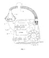
- Figure 1 is a diagram of an apparatus that treats sleep disordered breathing during sleep and monitors cardiac signals in a CPAP patient's airflow to assess cardiac health and treat cardiac conditions;
- Figure 2 is a graph illustrating, over a selected time period, airflow, ECG and mask air pressure.
- a device for treating SDB during sleep is disclosed that is capable of carrying out the features of the invention (such as a CPAP device), including sensing cardiogenic oscillations in air pressure/flow readings to determine whether an apnea event is central or obstructed.
- Mask flow is measured using a flow sensor 4f and/or pressure sensor 4p with a pneumotachograph and differential pressure transducer or similar device.
- a flow signal F(t) is derived and mask pressure is measured at a pressure tap using a pressure transducer to derive a pressure signal P mask (t).
- the pressure sensor 4p and flow sensor 4f have been shown only symbolically in Figure 1 since those skilled in the art would understand how to measure flow and pressure.
- Flow F(t) and pressure P mask (t) signals are sent to a controller or microprocessor 6 which then determines how to adjust the blower.
- the controller 6 may include integrated circuits, a memory and/or other instruction or data storage medium.
- Programmed instructions with control methodology may be coded on integrated chips in the memory of the device (e.g., firmware) or loaded as software.
- the pressure delivery device includes a blower 8, which preferably is an impellor,
- the impellor 8 is controlled by a servo 10, receives ambient air through an inlet 12 and delivers pressurized air through an outlet 14 defined by an air delivery conduit 16 and a mask 18 with an integrated exhaust vent 20.
- the impellor, motor, and controller assembly define a blower assembly and are located within the blower housing 22.
- Various switches 24 and displays 26 are provided in the blower housing.
- a number of sensors are provided within the blower to monitor, among other things, snore 28, motor speed 30, and motor current 32. Various devices known in the art can serve as these types of sensors.
- a communication interface 34 allows data to be transferred between the apparatus and an external device, such as a computer or controller.
- Figure 2 illustrates traces that may be recorded by appropriate equipment.
- Trace 42 illustrates a patient's electrocardiogram (ECG)
- trace 44 is the CPAP patient's airflow
- trace 46 is the patient's air pressure as measured using the CPAP treatment device. (Either or both of airflow and pressure may be monitored.)
- numeral 47 the respiratory flow hovers around zero , indicating an apnea event.
- the air pressure trace 46 still exhibits cardiogenic oscillations 48 indicative of an open airway (unobstructed) central apnea event.
- a band filter may be used.
- a suitable filter rejects signals of 30 Hz or lower (i.e., rejects those signals which are generally associated with respiration and physical movement of the patient) and also rejects signals higher than 60 Hz (i.e., reject those signals which are generally associated with system noise rather than being representative of cardiogenic events).
- cardiogenic information can be used to better manage conventional triggering circuits for a bi-level CPAP ventilator (which typically adjust the ventilator in response to inspiratory and expiratory flow), since distortions of air flow measurements attributable to cardiogenic oscillations can be ignored.
- a bi-level CPAP ventilator which typically adjust the ventilator in response to inspiratory and expiratory flow
- cardiogenic oscillations can be ignored.
- identification and filtering out of cardiogenic flow oscillation occurring at the end-expiration i.e., cardiogenic oscillation signals occurring at a part of the respiratory phase when it is desirable for the ventilator to most accurately cycle from expiration to inspiration in accordance with the applicable treatment algorithm).
- cardiogenic airflow may be detected during any portion of the patient's respiratory cycle
- the best resolution of the cardiogenic oscillations 48 occurs during the middle to end of the expiration portion 49 of the patient's breath. Monitoring the signal in only this relatively small window simplifies the processing needed to achieve the requisite signal resolution. Indeed, for some applications, it may be sufficient to monitor cardiogenic oscillations during only that portion of the respiratory cycle, i.e., significantly less than all the heartbeats per breath.
- the controller To locate the middle to end of the expiratory cycle, the controller detects the start of a new expiration cycle (with a threshold detector that detects the zero line transition), and identifies the end of the exhalation based on the recent averaged lapsed time of breathing cycles. Alternatively, the later portion of exhalation may be isolated using continuous phase monitoring of the patient's breathing, as disclosed in the '957 patent referenced above.
- the amplitude and/or frequency of the signal may be compared to thresholds representing expected or prior average heartbeat force and/or rhythm for the patient to determine any deviation from a norm.
- other patterns indicative of arrhythmia or normal cardiac force/rhythm may be stored as templates and compared to the signal to detect the presence of an arrhythmia or the absence of normal cardiac functioning.
- the device may send a signal to the patient, care provider or physician, or record the event for later observation.
- the signal to the patient may be in the form of an audible alarm.
- the signal to the care provider or physician may be in the form of an automated text messaging system using known telephonic circuitry and a subscription to a cellular provider. Immediate action and treatment is therefore enabled which is particularly useful in view of the known co-morbidity involving cardiac conditions and respiratory disorders such as SDB.
- cardiac timing is possible by monitoring the average time between cardiogenic oscillations such as 50 and 52. From this timing, heart rate parameters can be deduced such as average rate, variability and arrhythmia. All information regarding cardiac conditions may be observed in real time by way of suitable display, transmitted or recorded. Ventilatory support may be modified so as to assist cardiac function where, for example, CPAP therapy pressure is changed according to the cardiac cycle to assist right atria filling (pressure decrease), left ventricular ejection (pressure increase), and cardiac perfusion (pressure increase at early diastole), etc.
- CPAP therapy pressure is changed according to the cardiac cycle to assist right atria filling (pressure decrease), left ventricular ejection (pressure increase), and cardiac perfusion (pressure increase at early diastole), etc.
- cardiac stroke volume affects the amplitude of cardiogenic oscillations and that CPAP treatment affects stroke volume. Therefore, by monitoring cardiogenic oscillations in accordance with the present invention, it is possible to titrate CPAP treatment so as to influence and preferably to optimize cardiac stroke volume. This may be achieved without uninterrupted monitoring of heartbeats. Rather it may be achieved with the monitoring of only 1-2 heartbeats per breath, i.e., by monitoring only during a portion of the respiratory cycle, preferably during the middle to end expiration portion. For example, stroke volume may be maximized by examining the amplitude of the cardiogenic oscillations and servo-controlling the pressure treatment accordingly.
- PTT pulse-transit time
- the PTT is the time in which a pulse wave propagates the length of an arterial tree and is measured by the time interval that starts when half of the ventricular myocardium has been depolarized and ends when the blood is saturated with a predetermined percentage (depending on the age and condition of the patient) of oxyhemoglobin (SpO 2 ).
- SpO 2 oxyhemoglobin
- the former occurs when an R-wave is sensed in the ECG QRS complex (the entire time it takes for depolarization of the ventricles), and the latter occurs when a typical finger pulse oximeter senses photoplethysmographic (pulse) waveforms.
- the disadvantage of the typical measurements of the PPT is that the pre-ejection period (PEP) is included in the measured delay.
- PEP pre-ejection period
- the present invention allows for the achievement of a more accurate measure of pulse-transit time (i.e., a measure of pulse-transit time without the pre-ejection period component). By performing uninterrupted monitoring of cardiogenic oscillations concurrently with pulse oximetry, PTT may be estimated.
- An advantage of the present invention is that it uses cardiogenic oscillations for measuring cardiac timing.
- the cardiogenic oscillations relate to the heart's mechanical systolic events rather than the electrical systolic events, so the PEP is not included.
- Changes in the heart's PEP can also be assessed by the concurrent monitoring of cardiogenic oscillations against the ECG trace 42, and following the lag in time between electrical and mechanical systolic events.
- the changes in the PEP reflect the ability of the left ventricle to eject (perform mechanical systole events) and are another indication of cardiac health, and blood pressure, as well as peripheral vascular resistance and other cardio-circulatory conditions of interest in patient management.
- the apparatus may be configured or programmed to do the following while the patient is wearing a mask: measure airflow; identify and isolate the cardiogenic signal from the airflow; identify central apneas; calculate heart rate from the cardiogenic signal; determine abnormalities in heart rate (e.g., arrhythmias); generate notifications if an abnormality is determined, where the notifications include an alarm or other means of contacting selected individuals; monitor cardiac timing and assist in cardiac function; more accurately determine respiratory effort; and monitor PTT and PEP.
Abstract
Description
- This application claims the priority of
U.S. provisional application Serial No. 60/547,812 filed on February 25, 2004 - Cessation of breathing during sleep for more than 10 seconds is called an "apnea," which leads to decreased blood oxygenation and disruption of sleep. Apneas are traditionally categorized as central, where there is no respiratory effort, or obstructive sleep apnea (OSA), where there is respiratory effort but the airway is blocked. With purely central apneas, the airway is patent (or open), but the patient is not attempting to breathe. With other central apneas and all obstructive apneas, the airway is not patent (i.e., it is occluded). The occlusion is usually at the level of the tongue or soft palate.
- The common form of treatment of apneas is the administering of continuous or variable positive airway pressure (referred to herein generally as CPAP). Devices that provide CPAP treatment are described in
U.S. Patent Nos. 5,704,345 ,6,532,957 ,6,575,163 ,6,484,719 ,6,688,307 , and6,532,959 , incorporated herein by reference. The procedure for administering CPAP treatment has been well documented in both the technical and patent literature. Briefly stated, CPAP treatment acts as a pneumatic splint of the airway by the provision of positive pressure, usually in the range 4-20 cm H2O. The air is supplied to the airway by a motor driven blower whose outlet passes air via a delivery tube or hose to a nose (and/or mouth) mask sealingly engaged to a patient's face. An exhaust port is provided in the delivery tube proximate to the mask. More sophisticated forms of CPAP, such as bi-level CPAP and self-titrating CPAP, are described inU.S. Patent Nos. 5,148,802 and5,245,995 , respectively. - CPAP therapy is also known to be beneficial to some cardiac pathology, for example, congestive heart failure. By boosting intrathoracic pressure, CPAP offers various (potential) direct benefits in heart failure, for example, impeding venous return (reducing preload), reducing the systolic pressure gradient against which the left ventricle must pump (reduced afterload), and reducing left-ventricular trans-mural pressure (improved contractile efficiency). In addition, CPAP may offer indirect benefits to heart-failure patients, e.g., to counter pulmonary edema, to increase lung volume (may aid ventilatory stability in Cheyne-Stokes respiration), and in patients with a disposition to obstructive apnea, to reduce sympathetic activation through prevention of repetitive OSA.
- Various techniques are known for detecting abnormal breathing patterns indicative of obstructed breathing.
U.S. Patent No. 5,245,995 , for example, describes how snoring and abnormal breathing patterns can be detected by inspiration and expiration pressure measurements while sleeping, thereby leading to early indication of pre-obstructive episodes or other forms of breathing disorder. Patterns of respiratory parameters are monitored, and CPAP pressure is raised on the detection of pre-defined patterns to provide increased airway pressure to subvert the occurrence of the obstructive episodes and the other forms of breathing disorder. - Central apneas need not involve an obstruction of the airway, and often occur during very light sleep and in patients with various cardiac, cerebrovascular and endocrine conditions unrelated to the state of the upper airway. In cases where the apnea is occurring without obstruction of the airway, there may be little benefit in increasing CPAP pressure, in contrast to an obstructive apnea.
- To differentiate between central and obstructed apneas,
U.S. Patent No. 6,029,665 , incorporated herein by reference, teaches a CPAP system that monitors pulsatile airflow during the apnea event. With each beat of the heart, of the order of 66 ml of blood is ejected from the chest over about 0.3 sec, producing a pulsatile blood flow out of the chest of the order of 0.22 l/sec peak flow. If the chest wall were rigid this would create a partial vacuum in the chest cavity, and, if the upper airway were open and had zero flow resistance, a similar quantity of air would be sucked in through the trachea. In practice, the chest wall is not totally rigid, and the airways have finite airflow resistance. Consequently the measurable airflow (or cardiogenic oscillation) with each beat of the heart is of the order of 0.02 to 0.1 l/sec. - lf there is a central apnea with an open airway, the device of the '665 patent will sense cardiogenic oscillations in the air pressure, and determine that an unobstructed central apnea event has occurred. Conversely, if the airway is closed, the pressure waveform will not have any noticeable cardiogenic oscillations, and the device of the '665 patent will determine that the apnea event was an obstructed event.
- Implementing the apparatus and method of the '665 patent prevents the inappropriate increase in the splinting CPAP air pressure during a central apnea, thereby preventing an unnecessary increase in pressure that may otherwise reflexively inhibit breathing and further aggravate the breathing disorder. The device is also used in a diagnostic mode, using nasal cannulae in the place of a face mask, where measurements of apneas, patency, and partial obstruction are logged, but no CPAP treatment is effected. The data provides a physician with the ability to diagnose conditions such as OSA and upper airway resistance syndrome.
- Neither the '665 patent nor other prior art utilizes measurements of cardiogenic oscillations in a CPAP patient's airflow for monitoring or treating conditions related to cardiac health.
- lt is an object of the invention to utilize a CPAP device that treats sleep disordered breathing (SDB) also as a cardiac treatment device by monitoring cardiac signals in a patient's airflow to determine cardiac health.
- More specifically, it is an object of the invention to monitor the cardiac signals to screen and diagnose cardiac morbidity conditions, such as the existence of arrhythmias, and to influence and optimize cardiac stroke volume.
- It is a further object to monitor pulse-transit time, changes in the heart pre-ejection period, and the duration of the cardiac cycle.
- To satisfy the recited objectives, a method is disclosed of sensing cardiogenic oscillations in a patient's airflow and monitoring the patient's cardiac condition from the cardiogenic oscillations, The apparatus diagnoses cardiac morbidity conditions, such as the existence of arrhythmias or other cardiac abnormalities and influences and optimizes cardiac stroke volume. The apparatus further monitors pulse-transit time, changes in the heart pre-ejection period, and the duration of the cardiac cycle.
- To further satisfy the recited objectives, a detailed description of typical embodiments of the invention is provided with reference to appended drawings that are not intended to limit the scope of the invention, in which:
-
Figure 1 is a diagram of an apparatus that treats sleep disordered breathing during sleep and monitors cardiac signals in a CPAP patient's airflow to assess cardiac health and treat cardiac conditions; and -
Figure 2 is a graph illustrating, over a selected time period, airflow, ECG and mask air pressure. - Turning to
Figure 1 , a device for treating SDB during sleep is disclosed that is capable of carrying out the features of the invention (such as a CPAP device), including sensing cardiogenic oscillations in air pressure/flow readings to determine whether an apnea event is central or obstructed. Mask flow is measured using a flow sensor 4f and/orpressure sensor 4p with a pneumotachograph and differential pressure transducer or similar device. A flow signal F(t) is derived and mask pressure is measured at a pressure tap using a pressure transducer to derive a pressure signal Pmask(t). Thepressure sensor 4p and flow sensor 4f have been shown only symbolically inFigure 1 since those skilled in the art would understand how to measure flow and pressure. - Flow F(t) and pressure Pmask(t) signals are sent to a controller or microprocessor 6 which then determines how to adjust the blower. The controller 6 may include integrated circuits, a memory and/or other instruction or data storage medium. Programmed instructions with control methodology may be coded on integrated chips in the memory of the device (e.g., firmware) or loaded as software.
- The pressure delivery device includes a blower 8, which preferably is an impellor, The impellor 8 is controlled by a
servo 10, receives ambient air through aninlet 12 and delivers pressurized air through anoutlet 14 defined by anair delivery conduit 16 and amask 18 with an integratedexhaust vent 20. The impellor, motor, and controller assembly define a blower assembly and are located within theblower housing 22.Various switches 24 anddisplays 26 are provided in the blower housing. A number of sensors are provided within the blower to monitor, among other things,snore 28,motor speed 30, andmotor current 32. Various devices known in the art can serve as these types of sensors. Acommunication interface 34 allows data to be transferred between the apparatus and an external device, such as a computer or controller. - lf cardiogenic oscillations are not reflected in the pressure in a patient's mask during an apnea event, then the patient may be experiencing an obstructed central apnea event or an obstructed apnea event with respiratory effort. The above measuring technique, by itself, is incapable of differentiating the two conditions so that an indicator of respiratory effort is required. One type of known detector detects when the skin in the suprasternal notch is sucked inwards (during inhalation) and when the skin bulges outward (during expiratory efforts). Such a device is taught in
U.S. Patent No. 6,445,942 , incorporated herein by reference, which can be used to identify the occurrence of a central apnea. -
Figure 2 illustrates traces that may be recorded by appropriate equipment.Trace 42 illustrates a patient's electrocardiogram (ECG), trace 44 is the CPAP patient's airflow andtrace 46 is the patient's air pressure as measured using the CPAP treatment device. (Either or both of airflow and pressure may be monitored.) In the vicinity ofnumeral 47, the respiratory flow hovers around zero , indicating an apnea event. Theair pressure trace 46 still exhibitscardiogenic oscillations 48 indicative of an open airway (unobstructed) central apnea event. - When monitoring
air pressure 46, a band filter may be used. A suitable filter rejects signals of 30 Hz or lower (i.e., rejects those signals which are generally associated with respiration and physical movement of the patient) and also rejects signals higher than 60 Hz (i.e., reject those signals which are generally associated with system noise rather than being representative of cardiogenic events). - Once cardiogenic information is in hand, it can be used to better manage conventional triggering circuits for a bi-level CPAP ventilator (which typically adjust the ventilator in response to inspiratory and expiratory flow), since distortions of air flow measurements attributable to cardiogenic oscillations can be ignored. Of particular interest is the identification and filtering out of cardiogenic flow oscillation occurring at the end-expiration (i.e., cardiogenic oscillation signals occurring at a part of the respiratory phase when it is desirable for the ventilator to most accurately cycle from expiration to inspiration in accordance with the applicable treatment algorithm).
- Studying the presence of cardiogenic oscillations and, if present, their amplitude and frequency, during an open apnea event over a period of several seconds, without the complication of the concurrent existence of the airflow signal, provides information concerning the patient's cardiac condition. A medical practitioner can assess the patient's cardiac condition and treatment needs given the known association of central apneas and cardiac morbidity.
- While the cardiogenic airflow may be detected during any portion of the patient's respiratory cycle, the best resolution of the
cardiogenic oscillations 48 occurs during the middle to end of theexpiration portion 49 of the patient's breath. Monitoring the signal in only this relatively small window simplifies the processing needed to achieve the requisite signal resolution. Indeed, for some applications, it may be sufficient to monitor cardiogenic oscillations during only that portion of the respiratory cycle, i.e., significantly less than all the heartbeats per breath. - To locate the middle to end of the expiratory cycle, the controller detects the start of a new expiration cycle (with a threshold detector that detects the zero line transition), and identifies the end of the exhalation based on the recent averaged lapsed time of breathing cycles. Alternatively, the later portion of exhalation may be isolated using continuous phase monitoring of the patient's breathing, as disclosed in the '957 patent referenced above.
- Through long term monitoring of the
cardiogenic oscillations 48, irregularities in the force or rhythm of the heartbeat signal can be detected, which enables the determination of an arrhythmia. The amplitude and/or frequency of the signal may be compared to thresholds representing expected or prior average heartbeat force and/or rhythm for the patient to determine any deviation from a norm. Similarly, other patterns indicative of arrhythmia or normal cardiac force/rhythm may be stored as templates and compared to the signal to detect the presence of an arrhythmia or the absence of normal cardiac functioning. - If an arrhythmia is detected, then the device may send a signal to the patient, care provider or physician, or record the event for later observation. The signal to the patient may be in the form of an audible alarm. The signal to the care provider or physician may be in the form of an automated text messaging system using known telephonic circuitry and a subscription to a cellular provider. Immediate action and treatment is therefore enabled which is particularly useful in view of the known co-morbidity involving cardiac conditions and respiratory disorders such as SDB.
- The determination of cardiac timing is possible by monitoring the average time between cardiogenic oscillations such as 50 and 52. From this timing, heart rate parameters can be deduced such as average rate, variability and arrhythmia. All information regarding cardiac conditions may be observed in real time by way of suitable display, transmitted or recorded. Ventilatory support may be modified so as to assist cardiac function where, for example, CPAP therapy pressure is changed according to the cardiac cycle to assist right atria filling (pressure decrease), left ventricular ejection (pressure increase), and cardiac perfusion (pressure increase at early diastole), etc.
- It has been observed that cardiac stroke volume affects the amplitude of cardiogenic oscillations and that CPAP treatment affects stroke volume. Therefore, by monitoring cardiogenic oscillations in accordance with the present invention, it is possible to titrate CPAP treatment so as to influence and preferably to optimize cardiac stroke volume. This may be achieved without uninterrupted monitoring of heartbeats. Rather it may be achieved with the monitoring of only 1-2 heartbeats per breath, i.e., by monitoring only during a portion of the respiratory cycle, preferably during the middle to end expiration portion. For example, stroke volume may be maximized by examining the amplitude of the cardiogenic oscillations and servo-controlling the pressure treatment accordingly.
- It has been proposed that pulse-transit time (PTT) may serve as a non-invasive means of inferring respiratory effort and arousals. The PTT is the time in which a pulse wave propagates the length of an arterial tree and is measured by the time interval that starts when half of the ventricular myocardium has been depolarized and ends when the blood is saturated with a predetermined percentage (depending on the age and condition of the patient) of oxyhemoglobin (SpO2). The former occurs when an R-wave is sensed in the ECG QRS complex (the entire time it takes for depolarization of the ventricles), and the latter occurs when a typical finger pulse oximeter senses photoplethysmographic (pulse) waveforms.
- The disadvantage of the typical measurements of the PPT is that the pre-ejection period (PEP) is included in the measured delay. The present invention allows for the achievement of a more accurate measure of pulse-transit time (i.e., a measure of pulse-transit time without the pre-ejection period component). By performing uninterrupted monitoring of cardiogenic oscillations concurrently with pulse oximetry, PTT may be estimated. An advantage of the present invention is that it uses cardiogenic oscillations for measuring cardiac timing. The cardiogenic oscillations relate to the heart's mechanical systolic events rather than the electrical systolic events, so the PEP is not included.
- Changes in the heart's PEP can also be assessed by the concurrent monitoring of cardiogenic oscillations against the
ECG trace 42, and following the lag in time between electrical and mechanical systolic events. The changes in the PEP reflect the ability of the left ventricle to eject (perform mechanical systole events) and are another indication of cardiac health, and blood pressure, as well as peripheral vascular resistance and other cardio-circulatory conditions of interest in patient management. - In summary, the apparatus may be configured or programmed to do the following while the patient is wearing a mask: measure airflow; identify and isolate the cardiogenic signal from the airflow; identify central apneas; calculate heart rate from the cardiogenic signal; determine abnormalities in heart rate (e.g., arrhythmias); generate notifications if an abnormality is determined, where the notifications include an alarm or other means of contacting selected individuals; monitor cardiac timing and assist in cardiac function; more accurately determine respiratory effort; and monitor PTT and PEP.
- The present invention may be embodied in other specific forms without departing from its spirit or essential characteristics. The described embodiments are to be considered in all respects only as illustrative and not as restrictive. The scope of the invention is, therefore, indicated by the appended claims and their combination in whole or in part rather than by the foregoing description. All changes that come within the meaning and range of equivalency of the claims are to be embraced within their scope.
The following aspects are preferred embodiments of the invention. - 1. A method of determining a patient's cardiac condition by using a CPAP apparatus for treating sleep disordered breathing, comprising the steps of:
- sensing the patient's cardiogenic pressure or flow oscillations; and
- using the sensed cardiogenic oscillations to determine the patient's cardiac condition.
- 2. The method of aspect 1 wherein the occurrence of a central apnea event is identified by determining the occurrence of cardiogenic oscillations during a period of no airflow and wherein the patient's cardiac condition is determined based upon the known association of central apneas and cardiac morbidity.
- 3. The method of aspect 1 wherein cardiogenic oscillations in only the middle to later portion of exhalation are used to determine the patient's cardiac condition.
- 4. The method of aspect 3 wherein the middle to later portion of exhalation is determined by tracking the recent averaged lapsed time of prior breathing cycles and using such time in conjunction with the detection of the start of a breathing cycle.
- 5. The method of aspect 1 further comprising the step of sending a signal to the patient, care provider or physician, or recording an arrhythmia event for later observation, upon determining the existence of an arrhythmia event.
- 6. The method of aspect 1 further comprising the step of determining cardiac timing from the time between cardiogenic oscillations.
- 7. The method of aspect 1 further comprising the step of adjusting the patient's stroke volume by examining the amplitude of the cardiogenic oscillations and in accordance therewith adjusting the CPAP treatment pressure.
- 8. The method of aspect 1 further comprising the step of analyzing the cardiogenic oscillations to determine the patient's pulse transit time.
- 9. The method of aspect 1further comprising the step of analyzing the cardiogenic oscillations against ECG waveforms to determine changes in the patient's pre-ejection period.
- 10. The method of aspect 1 further comprising the step of assisting cardiac function in accordance with the determined cardiac condition by adjusting the CPAP treatment pressure to assist right atria filling, left ventricular ejection, or cardiac perfusion.
- 11. The method of aspect 1 further comprising the step of assisting cardiac function by adjusting the CPAP treatment pressure to assist right atria filling, left ventricular ejection, or cardiac perfusion. Or arterial (aortic) tone?
- 12. The method of aspect 1 further comprising the step of using cardiogenic oscillation information for managing triggering of a bi-level CPAP apparatus.
- 13. A method of determining a patient's cardiac condition and providing cardiac treatment by using a CPAP apparatus for treating sleep disordered breathing, comprising the steps of:
- sensing the patient's cardiogenic pressure or flow oscillations; and
- using the sensed cardiogenic oscillations to determine the patient's cardiac condition and adjust the pressure delivered by the CPAP apparatus to treat the patient's cardiac condition.
- 14. The method of aspect 13 wherein the occurrence of a central apnea event is identified by determining the occurrence of cardiogenic oscillations during a period of no airflow and wherein the patient's cardiac condition is determined based upon the known association of central apneas and cardiac morbidity.
- 15. The method of aspect 13 wherein cardiogenic oscillations in only the middle to later portion of exhalation are used to determine the patient's cardiac condition.
- 16. The method of aspect 15 wherein the middle to later portion of exhalation is determined by tracking the recent averaged lapsed time of prior breathing cycles and using such time in conjunction with the detection of the start of a breathing cycle.
- 17. The method of aspect 13 further comprising the step of sending a signal to the patient, care provider or physician, or recording an arrhythmia event for later observation, upon determining the existence of an arrhythmia event.
- 18. The method aspect 13 further comprising the step of determining cardiac timing from the time between cardiogenic oscillations.
- 19. The method of aspect 13 further comprising the step of adjusting the patient's stroke volume by examining the amplitude of the cardiogenic oscillations and in accordance therewith adjusting the CPAP treatment pressure.
- 20. The method of aspect 13 further comprising the step of analyzing the cardiogenic oscillations to determine the patient's pulse transit time.
- 21. The method of aspect 13 further comprising the step of analyzing the cardiogenic oscillations against ECG waveforms to determine changes in the patient's pre-ejection period.
- 22. The method of aspect 13 further comprising the step of assisting cardiac function in accordance with the determined cardiac condition by adjusting the CPAP treatment pressure to assist right atria filling, left ventricular ejection, or cardiac perfusion.
- 23. The method of aspect 13 further comprising the step of assisting cardiac function by adjusting the CPAP treatment pressure to assist right atria filling, left ventricular ejection, or cardiac perfusion.
- 24. The method of aspect 13 further comprising the step of using cardiogenic oscillation information for managing triggering of a bi-level CPAP apparatus.
- 25. A CPAP apparatus which, in addition to providing CPAP therapy, determines a patient's cardiac condition, the apparatus comprising a controller and a sensor for detecting pressure in the patient's CPAP mask, wherein the controller:
- senses the patient's cardiogenic pressure oscillations; and
- uses the sensed cardiogenic oscillations to determine the patient's cardiac condition.
- 26. The apparatus of aspect 25 wherein the controller:
- identifies a central apnea event by determining the occurrence of cardiogenic oscillations during a period of no airflow; and
- determines the patient's cardiac condition based upon the known association of central apneas and cardiac morbidity.
- 27. The apparatus of aspect 25 wherein the controller uses cardiogenic oscillations in only the middle to later portion of exhalation to determine the patient's cardiac condition.
- 28. The apparatus of aspect 27 wherein the controller determines the middle to later portion of exhalation by tracking the recent averaged lapsed time of prior breathing cycles and using such time in conjunction with the detection of the start of a breathing cycle.
- 29. The apparatus of aspect 25 wherein the controller sends a signal to the patient, care provider or physician, or recording an arrhythmia event for later observation, upon determining the existence of an arrhythmia event.
- 30. The apparatus of aspect 25 wherein the controller determines cardiac timing from the time between cardiogenic oscillations.
- 31. The apparatus of aspect 25 wherein the controller adjusts the patient's stroke volume by examining the amplitude of the cardiogenic oscillations and in accordance therewith adjusting the CPAP treatment pressure.
- 32. The apparatus of aspect 25 wherein the controller analyzes the cardiogenic oscillations to determine the patient's pulse transit time.
- 33. The apparatus of aspect 25 wherein the controller analyzes the cardiogenic oscillations against ECG waveforms to determine changes in the patient's pre-ejection period.
- 34. The apparatus of aspect 25 wherein the controller assists cardiac function in accordance with the determined cardiac condition by adjusting the CPAP treatment pressure to assist right atria filling, left ventricular ejection, or cardiac perfusion.
- 35. The apparatus of aspect 25 wherein the controller assists cardiac function by adjusting the CPAP treatment pressure to assist right atria filling, left ventricular ejection, or cardiac perfusion.
- 36. The apparatus of aspect 25 wherein the controller uses cardiogenic oscillation information for managing triggering of the CPAP apparatus.
- 37. A CPAP apparatus which, in addition to providing CPAP therapy, determines a patient's cardiac condition and provides cardiac treatment, the apparatus comprising a controller and a sensor for detecting pressure in the patient's CPAP mask, wherein the controller:
- senses the patient's cardiogenic pressure oscillations; and
- uses the sensed cardiogenic oscillations to determine the patient's cardiac condition.
- 38. The apparatus of aspect 37 wherein the controller:
- identifies the occurrence of a central apnea event by determining the occurrence of cardiogenic oscillations during a period of no airflow; and
- determines the patient's cardiac condition based upon the known association of central apneas and cardiac morbidity.
- 39. The apparatus of aspect 37 wherein the controller uses cardiogenic oscillations in only the middle to later portion of exhalation are used to determine the patient's cardiac condition.
- 40. The apparatus of aspect 39 wherein the controller determines the middle to later portion of exhalation by tracking the recent averaged lapsed time of prior breathing cycles and using such time in conjunction with the detection of the start of a breathing cycle.
- 41. The apparatus of aspect 37 wherein the controller sends a signal to the patient, care provider or physician, or recording an arrhythmia event for later observation, upon determining the existence of an arrhythmia event.
- 42. The apparatus of aspect 37wherein the controller determines cardiac timing from the time between cardiogenic oscillations.
- 43. The apparatus of aspect 37 wherein the controller adjusts the patient's stroke volume by examining the amplitude of the cardiogenic oscillations and in accordance therewith adjusting the CPAP treatment pressure.
- 44. The apparatus of aspect 37 wherein the controller analyzes the cardiogenic oscillations to determine the patient's pulse transit time.
- 45. The apparatus of aspect 37 wherein the controller analyzes the cardiogenic oscillations against ECG waveforms to determine changes in the patient's pre-ejection period.
- 46. The apparatus of aspect 37 wherein the controller assists cardiac function in accordance with the determined cardiac condition by adjusting the CPAP treatment pressure to assist right atria filling, left ventricular ejection, or cardiac perfusion.
- 47. The apparatus of aspect 37 wherein the controller assists cardiac function by adjusting the CPAP treatment pressure to assist right atria filling, left ventricular ejection, or cardiac perfusion.
- 48. The apparatus of aspect 37 wherein the controller uses cardiogenic oscillation information for managing triggering of a bi-level CPAP apparatus.
Claims (11)
- A CPAP apparatus which, in addition to providing CPAP therapy, determines a patient's cardiac condition and provides cardiac treatment, the apparatus comprising a controller and a sensor for detecting pressure in the patient's CPAP mask, wherein the controller:senses the patient's cardiogenic pressure oscillations; anduses the sensed cardiogenic oscillations to determine the patient's cardiac condition.
- The apparatus of claim 1 wherein the controller uses cardiogenic oscillations in only the middle to later portion of exhalation are used to determine the patient's cardiac condition.
- The apparatus of claim 2 wherein the controller determines the middle to later portion of exhalation by tracking the recent averaged lapsed time of prior breathing cycles and using such time in conjunction with the detection of the start of a breathing cycle.
- The apparatus of claim 1 wherein the controller sends a signal to the patient, care provider or physician, or recording an arrhythmia event for later observation, upon determining the existence of an arrhythmia event.
- The apparatus of claim 1' wherein the controller determines cardiac timing from the time between cardiogenic oscillations.
- The apparatus of claim 1' wherein the controller adjusts the patient's stroke volume by examining the amplitude of the cardiogenic oscillations and in accordance therewith adjusting the CPAP treatment pressure.
- The apparatus of claim 1 wherein the controller analyzes the cardiogenic oscillations to determine the patient's pulse transit time.
- The apparatus of claim 1 wherein the controller analyzes the cardiogenic oscillations against ECG waveforms to determine changes in the patient's pre-ejection period.
- The apparatus of claim 1 wherein the controller assists cardiac function in accordance with the determined cardiac condition by adjusting the CPAP treatment pressure to assist right atria filling, left ventricular ejection, or cardiac perfusion.
- The apparatus of claim 1 wherein the controller assists cardiac function by adjusting the CPAP treatment pressure to assist right atria filling, left ventricular ejection, or cardiac perfusion.
- The apparatus of claims 1 wherein the controller uses cardiogenic oscillation information for managing triggering of a bi-level CPAP apparatus.
Applications Claiming Priority (2)
| Application Number | Priority Date | Filing Date | Title |
|---|---|---|---|
| US54781204P | 2004-02-25 | 2004-02-25 | |
| EP05706284.6A EP1718356B1 (en) | 2004-02-25 | 2005-02-24 | Cardiac monitoring and therapy using a device for providing pressure treatment of sleep disordered breathing |
Related Parent Applications (2)
| Application Number | Title | Priority Date | Filing Date |
|---|---|---|---|
| EP05706284.6A Division-Into EP1718356B1 (en) | 2004-02-25 | 2005-02-24 | Cardiac monitoring and therapy using a device for providing pressure treatment of sleep disordered breathing |
| EP05706284.6 Division | 2005-02-24 |
Publications (2)
| Publication Number | Publication Date |
|---|---|
| EP2388036A2 true EP2388036A2 (en) | 2011-11-23 |
| EP2388036A3 EP2388036A3 (en) | 2013-03-13 |
Family
ID=34886312
Family Applications (2)
| Application Number | Title | Priority Date | Filing Date |
|---|---|---|---|
| EP05706284.6A Not-in-force EP1718356B1 (en) | 2004-02-25 | 2005-02-24 | Cardiac monitoring and therapy using a device for providing pressure treatment of sleep disordered breathing |
| EP11177456A Withdrawn EP2388036A3 (en) | 2004-02-25 | 2005-02-24 | Cardiac monitoring and therapy using a device for providing pressure treatment of sleep disordered breathing |
Family Applications Before (1)
| Application Number | Title | Priority Date | Filing Date |
|---|---|---|---|
| EP05706284.6A Not-in-force EP1718356B1 (en) | 2004-02-25 | 2005-02-24 | Cardiac monitoring and therapy using a device for providing pressure treatment of sleep disordered breathing |
Country Status (4)
| Country | Link |
|---|---|
| US (2) | US8794236B2 (en) |
| EP (2) | EP1718356B1 (en) |
| JP (1) | JP4699443B2 (en) |
| WO (1) | WO2005079897A1 (en) |
Cited By (1)
| Publication number | Priority date | Publication date | Assignee | Title |
|---|---|---|---|---|
| WO2024069500A1 (en) * | 2022-09-29 | 2024-04-04 | Resmed Sensor Technologies Limited | Systems and methods for cardiogenic oscillation detection |
Families Citing this family (47)
| Publication number | Priority date | Publication date | Assignee | Title |
|---|---|---|---|---|
| US7189204B2 (en) * | 2002-12-04 | 2007-03-13 | Cardiac Pacemakers, Inc. | Sleep detection using an adjustable threshold |
| US7610094B2 (en) * | 2003-09-18 | 2009-10-27 | Cardiac Pacemakers, Inc. | Synergistic use of medical devices for detecting medical disorders |
| EP1670547B1 (en) * | 2003-08-18 | 2008-11-12 | Cardiac Pacemakers, Inc. | Patient monitoring system |
| US20050142070A1 (en) * | 2003-09-18 | 2005-06-30 | Hartley Jesse W. | Methods and systems for assessing pulmonary disease with drug therapy control |
| US8606356B2 (en) | 2003-09-18 | 2013-12-10 | Cardiac Pacemakers, Inc. | Autonomic arousal detection system and method |
| US7575553B2 (en) * | 2003-09-18 | 2009-08-18 | Cardiac Pacemakers, Inc. | Methods and systems for assessing pulmonary disease |
| US7662101B2 (en) * | 2003-09-18 | 2010-02-16 | Cardiac Pacemakers, Inc. | Therapy control based on cardiopulmonary status |
| JP4699443B2 (en) * | 2004-02-25 | 2011-06-08 | レスメド・リミテッド | Heart monitoring and treatment using a device for performing pressurized therapy for sleep disordered breathing |
| US8442607B2 (en) * | 2006-09-07 | 2013-05-14 | Sotera Wireless, Inc. | Hand-held vital signs monitor |
| WO2010116276A1 (en) * | 2009-04-08 | 2010-10-14 | Koninklijke Philips Electronics, N.V. | System and method for monitoring pulmonary congestion |
| US9687177B2 (en) * | 2009-07-16 | 2017-06-27 | Resmed Limited | Detection of sleep condition |
| CN106955401B (en) * | 2010-03-25 | 2020-11-06 | 瑞思迈巴黎股份有限公司 | Breathable gas inlet control apparatus for respiratory therapy device |
| US8776792B2 (en) | 2011-04-29 | 2014-07-15 | Covidien Lp | Methods and systems for volume-targeted minimum pressure-control ventilation |
| EP2731561B1 (en) | 2011-07-14 | 2016-03-23 | Cook Medical Technologies LLC | A sling to be used in the treatment of obstructive sleep apnea |
| US20130025597A1 (en) * | 2011-07-29 | 2013-01-31 | Nellcor Puritan Bennett Llc | Methods and systems for monitoring a ventilated patient with an oximeter |
| US9993604B2 (en) | 2012-04-27 | 2018-06-12 | Covidien Lp | Methods and systems for an optimized proportional assist ventilation |
| WO2014189540A1 (en) | 2012-10-16 | 2014-11-27 | Catalano Peter J | Method and apparatus for treating obstructive sleep apnea (osa) |
| US9375542B2 (en) | 2012-11-08 | 2016-06-28 | Covidien Lp | Systems and methods for monitoring, managing, and/or preventing fatigue during ventilation |
| US9358355B2 (en) | 2013-03-11 | 2016-06-07 | Covidien Lp | Methods and systems for managing a patient move |
| EP3498239B1 (en) | 2013-08-01 | 2021-04-21 | Cook Medical Technologies LLC | Tissue adjustment implant |
| WO2015020953A1 (en) | 2013-08-05 | 2015-02-12 | Darin Schaeffer | Medical devices having a releasable tubular member and methods of using the same |
| EP3030302B1 (en) | 2013-09-04 | 2023-02-22 | Fisher&Paykel Healthcare Limited | Improvements to flow therapy |
| EP2898920B1 (en) | 2014-01-24 | 2018-06-06 | Cook Medical Technologies LLC | Articulating balloon catheter |
| US9974563B2 (en) | 2014-05-28 | 2018-05-22 | Cook Medical Technologies Llc | Medical devices having a releasable member and methods of using the same |
| EP3177219B1 (en) | 2014-08-04 | 2018-09-26 | Cook Medical Technologies LLC | Medical devices having a releasable tubular member |
| WO2016073945A1 (en) | 2014-11-07 | 2016-05-12 | Respirix, Inc. | Devices and methods for monitoring physiologic parameters |
| JP6399614B2 (en) * | 2015-02-03 | 2018-10-03 | 国立大学法人名古屋大学 | Heart rate signal detector |
| CA2980849A1 (en) | 2015-03-31 | 2016-10-06 | Fisher & Paykel Healthcare Limited | Methods and apparatus for oxygenation and/or co2 removal |
| US10905836B2 (en) | 2015-04-02 | 2021-02-02 | Hill-Rom Services Pte. Ltd. | Manifold for respiratory device |
| EP3291729B1 (en) * | 2015-05-07 | 2020-05-13 | Ecom Medical, Inc. | Method for fabricating invasive ecg probe |
| BR112017024655A2 (en) | 2015-06-29 | 2018-11-21 | Teijin Pharma Ltd | positive pressure therapy devices and positive pressure value computing. |
| EP3361939A4 (en) | 2015-11-16 | 2019-06-12 | Respirix, Inc. | Devices and methods for monitoring physiologic parameters |
| JP2019509985A (en) | 2016-02-01 | 2019-04-11 | インカーダ セラピューティクス, インコーポレイテッド | Combining electronic monitoring with inhalation medication to manage cardiac arrhythmias including atrial fibrillation |
| EP3451924B1 (en) * | 2016-04-29 | 2024-04-10 | Fisher&Paykel Healthcare Limited | System for determining airway patency |
| WO2018089789A1 (en) | 2016-11-10 | 2018-05-17 | The Research Foundation For The State University Of New York | System, method and biomarkers for airway obstruction |
| KR20190094214A (en) | 2016-12-15 | 2019-08-12 | 백스터 인터내셔널 인코포레이티드 | System and method for monitoring and determining patient parameters from sensed vein waveforms |
| EP3621616A4 (en) | 2017-05-10 | 2021-01-13 | InCarda Therapeutics, Inc. | Unit doses, aerosols, kits, and methods for treating heart conditions by pulmonary administration |
| US11357660B2 (en) | 2017-06-29 | 2022-06-14 | Cook Medical Technologies, LLC | Implantable medical devices for tissue repositioning |
| JP7313341B2 (en) * | 2017-09-28 | 2023-07-24 | コーニンクレッカ フィリップス エヌ ヴェ | Systems and methods for detecting stroke in patients during pressure support therapy |
| CN110049799B (en) | 2017-11-14 | 2022-04-26 | 柯惠有限合伙公司 | Method and system for driving pressure spontaneous ventilation |
| US10744087B2 (en) | 2018-03-22 | 2020-08-18 | Incarda Therapeutics, Inc. | Method to slow ventricular rate |
| US11039754B2 (en) | 2018-05-14 | 2021-06-22 | Baxter International Inc. | System and method for monitoring and determining patient parameters from sensed venous waveform |
| US11517691B2 (en) | 2018-09-07 | 2022-12-06 | Covidien Lp | Methods and systems for high pressure controlled ventilation |
| US11007185B2 (en) | 2019-08-01 | 2021-05-18 | Incarda Therapeutics, Inc. | Antiarrhythmic formulation |
| JP6999141B1 (en) | 2020-07-06 | 2022-01-18 | 株式会社コスモスウェブ | Biometric information collection system and sensor unit |
| CN115835812A (en) * | 2020-07-22 | 2023-03-21 | 艾姆皮尼亚公司 | Method and apparatus for real-time monitoring of respiration using embedded fiber bragg gratings |
| WO2023187686A1 (en) | 2022-03-30 | 2023-10-05 | ResMed Pty Ltd | Systems and methods for determining a positional sleep disordered breathing status |
Citations (6)
| Publication number | Priority date | Publication date | Assignee | Title |
|---|---|---|---|---|
| US5148802A (en) | 1989-09-22 | 1992-09-22 | Respironics Inc. | Method and apparatus for maintaining airway patency to treat sleep apnea and other disorders |
| US5245995A (en) | 1987-06-26 | 1993-09-21 | Rescare Limited | Device and method for monitoring breathing during sleep, control of CPAP treatment, and preventing of apnea |
| US5704345A (en) | 1993-11-05 | 1998-01-06 | Resmed Limited | Detection of apnea and obstruction of the airway in the respiratory system |
| US6445942B1 (en) | 1999-09-15 | 2002-09-03 | Resmed Ltd | Measurement of respiratory effort using a suprasternal sensor |
| US6484719B1 (en) | 1996-09-23 | 2002-11-26 | Resmed, Inc. | Method for providing ventilatory assistance in a spontaneously breathing subject |
| US6532959B1 (en) | 1998-05-22 | 2003-03-18 | Resmed, Ltd. | Ventilatory assistance for treatment of cardiac failure and cheyne-stokes breathing |
Family Cites Families (16)
| Publication number | Priority date | Publication date | Assignee | Title |
|---|---|---|---|---|
| CH571868A5 (en) * | 1973-11-21 | 1976-01-30 | Hoffmann La Roche | |
| GB1585091A (en) * | 1976-02-10 | 1981-02-25 | Venegas J G | Remedial apparatus for use in assisting the breathing of living creatures |
| US5803066A (en) * | 1992-05-07 | 1998-09-08 | New York University | Method and apparatus for optimizing the continuous positive airway pressure for treating obstructive sleep apnea |
| US6675797B1 (en) | 1993-11-05 | 2004-01-13 | Resmed Limited | Determination of patency of the airway |
| US5794615A (en) * | 1994-06-03 | 1998-08-18 | Respironics, Inc. | Method and apparatus for providing proportional positive airway pressure to treat congestive heart failure |
| US6105575A (en) * | 1994-06-03 | 2000-08-22 | Respironics, Inc. | Method and apparatus for providing positive airway pressure to a patient |
| AUPP026997A0 (en) * | 1997-11-07 | 1997-12-04 | Resmed Limited | Administration of cpap treatment pressure in presence of apnea |
| US6739335B1 (en) * | 1999-09-08 | 2004-05-25 | New York University School Of Medicine | Method and apparatus for optimizing controlled positive airway pressure using the detection of cardiogenic oscillations |
| US6752151B2 (en) * | 2000-09-25 | 2004-06-22 | Respironics, Inc. | Method and apparatus for providing variable positive airway pressure |
| AUPR193300A0 (en) * | 2000-12-07 | 2001-01-04 | Resmed Limited | Mask assembly |
| JP4348082B2 (en) * | 2000-12-11 | 2009-10-21 | レスメド・リミテッド | Device for judging the patient's situation after stroke onset |
| US7187965B2 (en) * | 2001-05-29 | 2007-03-06 | Bischoff Edward T | Cardiac rhythm monitoring device |
| IL147502A0 (en) * | 2002-01-07 | 2002-08-14 | Widemed Ltd | Self-adaptive system, for the analysis of biomedical signals of a patient |
| US20070135724A1 (en) * | 2003-10-17 | 2007-06-14 | Ujhazy Anthony J | Methods and apparatus for heart failure treatment |
| JP4699443B2 (en) * | 2004-02-25 | 2011-06-08 | レスメド・リミテッド | Heart monitoring and treatment using a device for performing pressurized therapy for sleep disordered breathing |
| CN103083768B (en) * | 2004-10-06 | 2016-07-06 | 瑞思迈有限公司 | Method and apparatus for non-invasive monitoring of respiratory parameters in sleep disordered breathing |
-
2005
- 2005-02-24 JP JP2007500006A patent/JP4699443B2/en active Active
- 2005-02-24 EP EP05706284.6A patent/EP1718356B1/en not_active Not-in-force
- 2005-02-24 US US10/598,255 patent/US8794236B2/en active Active
- 2005-02-24 EP EP11177456A patent/EP2388036A3/en not_active Withdrawn
- 2005-02-24 WO PCT/AU2005/000248 patent/WO2005079897A1/en active Application Filing
-
2014
- 2014-07-10 US US14/327,922 patent/US10398863B2/en active Active
Patent Citations (11)
| Publication number | Priority date | Publication date | Assignee | Title |
|---|---|---|---|---|
| US5245995A (en) | 1987-06-26 | 1993-09-21 | Rescare Limited | Device and method for monitoring breathing during sleep, control of CPAP treatment, and preventing of apnea |
| US5148802A (en) | 1989-09-22 | 1992-09-22 | Respironics Inc. | Method and apparatus for maintaining airway patency to treat sleep apnea and other disorders |
| US5148802B1 (en) | 1989-09-22 | 1997-08-12 | Respironics Inc | Method and apparatus for maintaining airway patency to treat sleep apnea and other disorders |
| US5704345A (en) | 1993-11-05 | 1998-01-06 | Resmed Limited | Detection of apnea and obstruction of the airway in the respiratory system |
| US6029665A (en) | 1993-11-05 | 2000-02-29 | Resmed Limited | Determination of patency of airway |
| US6484719B1 (en) | 1996-09-23 | 2002-11-26 | Resmed, Inc. | Method for providing ventilatory assistance in a spontaneously breathing subject |
| US6532957B2 (en) | 1996-09-23 | 2003-03-18 | Resmed Limited | Assisted ventilation to match patient respiratory need |
| US6575163B1 (en) | 1996-09-23 | 2003-06-10 | Resmed Ltd. | Method for calculating the instantaneous inspired volume of a subject during ventilatory assistance |
| US6688307B2 (en) | 1996-09-23 | 2004-02-10 | Resmed Limited | Methods and apparatus for determining instantaneous elastic recoil and assistance pressure during ventilatory support |
| US6532959B1 (en) | 1998-05-22 | 2003-03-18 | Resmed, Ltd. | Ventilatory assistance for treatment of cardiac failure and cheyne-stokes breathing |
| US6445942B1 (en) | 1999-09-15 | 2002-09-03 | Resmed Ltd | Measurement of respiratory effort using a suprasternal sensor |
Cited By (1)
| Publication number | Priority date | Publication date | Assignee | Title |
|---|---|---|---|---|
| WO2024069500A1 (en) * | 2022-09-29 | 2024-04-04 | Resmed Sensor Technologies Limited | Systems and methods for cardiogenic oscillation detection |
Also Published As
| Publication number | Publication date |
|---|---|
| US10398863B2 (en) | 2019-09-03 |
| EP1718356B1 (en) | 2016-09-21 |
| JP2007525267A (en) | 2007-09-06 |
| US20150182713A1 (en) | 2015-07-02 |
| US20080045813A1 (en) | 2008-02-21 |
| US8794236B2 (en) | 2014-08-05 |
| EP1718356A4 (en) | 2009-05-06 |
| JP4699443B2 (en) | 2011-06-08 |
| EP2388036A3 (en) | 2013-03-13 |
| WO2005079897A1 (en) | 2005-09-01 |
| EP1718356A1 (en) | 2006-11-08 |
Similar Documents
| Publication | Publication Date | Title |
|---|---|---|
| US10398863B2 (en) | Cardiac monitoring and therapy using a device for providing pressure treatment of sleep disordered breathing | |
| US11696725B2 (en) | Systems, methods, and/or apparatuses for non-invasive monitoring of respiratory parameters in sleep disordered breathing | |
| JP6099607B2 (en) | Noninvasive monitoring system for respiratory parameters of sleep disordered breathing | |
| US11298485B2 (en) | Esophageal pressure clinical decision support system | |
| US20160045154A1 (en) | Sleep apnea | |
| AU2011203234B2 (en) | Method and Apparatus for Non-Invasive Monitoring of Respiratory Parameters in Sleep Disordered Breathing |
Legal Events
| Date | Code | Title | Description |
|---|---|---|---|
| AC | Divisional application: reference to earlier application |
Ref document number: 1718356 Country of ref document: EP Kind code of ref document: P |
|
| AK | Designated contracting states |
Kind code of ref document: A2 Designated state(s): AT BE BG CH CY CZ DE DK EE ES FI FR GB GR HU IE IS IT LI LT LU MC NL PL PT RO SE SI SK TR |
|
| PUAI | Public reference made under article 153(3) epc to a published international application that has entered the european phase |
Free format text: ORIGINAL CODE: 0009012 |
|
| RIN1 | Information on inventor provided before grant (corrected) |
Inventor name: FARRUGIA, STEVEN PAUL Inventor name: CHAN, CHRISTINE WEI CHIH Inventor name: MARTIN, DION CHARLES CHEWE Inventor name: PHUAH, CHEE KEONG |
|
| RIN1 | Information on inventor provided before grant (corrected) |
Inventor name: PHUAH, CHEE KEONG Inventor name: MARTIN, DION CHARLES CHEWE Inventor name: CHAN, CHRISTINE WEI CHIH Inventor name: FARRUGIA, STEVEN PAUL |
|
| RIN1 | Information on inventor provided before grant (corrected) |
Inventor name: FARRUGIA, STEVEN PAUL Inventor name: CHAN, CHRISTINE WEI CHIH Inventor name: MARTIN, DION CHARLES CHEWE Inventor name: PHUAH, CHEE KEONG |
|
| PUAL | Search report despatched |
Free format text: ORIGINAL CODE: 0009013 |
|
| AK | Designated contracting states |
Kind code of ref document: A3 Designated state(s): AT BE BG CH CY CZ DE DK EE ES FI FR GB GR HU IE IS IT LI LT LU MC NL PL PT RO SE SI SK TR |
|
| RIC1 | Information provided on ipc code assigned before grant |
Ipc: A61B 5/087 20060101ALN20130205BHEP Ipc: A61M 16/00 20060101AFI20130205BHEP Ipc: A61B 5/024 20060101ALI20130205BHEP Ipc: A61B 5/0205 20060101ALI20130205BHEP |
|
| 17P | Request for examination filed |
Effective date: 20130909 |
|
| RBV | Designated contracting states (corrected) |
Designated state(s): AT BE BG CH CY CZ DE DK EE ES FI FR GB GR HU IE IS IT LI LT LU MC NL PL PT RO SE SI SK TR |
|
| 17Q | First examination report despatched |
Effective date: 20170303 |
|
| RAP1 | Party data changed (applicant data changed or rights of an application transferred) |
Owner name: RESMED PTY LTD |
|
| STAA | Information on the status of an ep patent application or granted ep patent |
Free format text: STATUS: THE APPLICATION IS DEEMED TO BE WITHDRAWN |
|
| 18D | Application deemed to be withdrawn |
Effective date: 20190903 |