EP2386640A2 - Delivering functional nucleic acids to mammalian cells via bacterially-derived, intact minicells - Google Patents
Delivering functional nucleic acids to mammalian cells via bacterially-derived, intact minicells Download PDFInfo
- Publication number
- EP2386640A2 EP2386640A2 EP20110171410 EP11171410A EP2386640A2 EP 2386640 A2 EP2386640 A2 EP 2386640A2 EP 20110171410 EP20110171410 EP 20110171410 EP 11171410 A EP11171410 A EP 11171410A EP 2386640 A2 EP2386640 A2 EP 2386640A2
- Authority
- EP
- European Patent Office
- Prior art keywords
- minicells
- minicell
- nucleic acid
- functional nucleic
- drug
- Prior art date
- Legal status (The legal status is an assumption and is not a legal conclusion. Google has not performed a legal analysis and makes no representation as to the accuracy of the status listed.)
- Granted
Links
- 150000007523 nucleic acids Chemical class 0.000 title claims abstract description 158
- 102000039446 nucleic acids Human genes 0.000 title claims abstract description 157
- 108020004707 nucleic acids Proteins 0.000 title claims abstract description 157
- 210000004962 mammalian cell Anatomy 0.000 title claims abstract description 68
- 108090000623 proteins and genes Proteins 0.000 claims abstract description 125
- 102000004169 proteins and genes Human genes 0.000 claims abstract description 81
- 206010059866 Drug resistance Diseases 0.000 claims abstract description 66
- 239000013612 plasmid Substances 0.000 claims abstract description 60
- 239000003814 drug Substances 0.000 claims description 130
- 229940079593 drug Drugs 0.000 claims description 125
- 206010028980 Neoplasm Diseases 0.000 claims description 89
- 239000000203 mixture Substances 0.000 claims description 71
- 238000011282 treatment Methods 0.000 claims description 59
- 239000003446 ligand Substances 0.000 claims description 53
- 201000011510 cancer Diseases 0.000 claims description 45
- 239000002246 antineoplastic agent Substances 0.000 claims description 43
- 229940044683 chemotherapy drug Drugs 0.000 claims description 43
- 230000027455 binding Effects 0.000 claims description 29
- 108090000765 processed proteins & peptides Proteins 0.000 claims description 25
- 102000004196 processed proteins & peptides Human genes 0.000 claims description 21
- 102000005962 receptors Human genes 0.000 claims description 19
- 108020003175 receptors Proteins 0.000 claims description 19
- 102000004190 Enzymes Human genes 0.000 claims description 9
- 108090000790 Enzymes Proteins 0.000 claims description 9
- 230000004927 fusion Effects 0.000 claims description 8
- 108020001507 fusion proteins Proteins 0.000 claims description 7
- 102000037865 fusion proteins Human genes 0.000 claims description 7
- 239000003937 drug carrier Substances 0.000 claims description 6
- 230000036210 malignancy Effects 0.000 claims description 6
- 230000000242 pagocytic effect Effects 0.000 claims description 6
- 208000030507 AIDS Diseases 0.000 claims description 4
- 150000003384 small molecules Chemical class 0.000 claims description 3
- 201000008827 tuberculosis Diseases 0.000 claims description 2
- 238000000034 method Methods 0.000 abstract description 83
- 230000006907 apoptotic process Effects 0.000 abstract description 21
- 230000008685 targeting Effects 0.000 abstract description 9
- 238000002512 chemotherapy Methods 0.000 abstract description 4
- 210000004027 cell Anatomy 0.000 description 191
- 239000004055 small Interfering RNA Substances 0.000 description 79
- 108020004459 Small interfering RNA Proteins 0.000 description 48
- 108091027967 Small hairpin RNA Proteins 0.000 description 47
- 230000001580 bacterial effect Effects 0.000 description 43
- 108091032973 (ribonucleotides)n+m Proteins 0.000 description 40
- 230000014509 gene expression Effects 0.000 description 39
- 102000040430 polynucleotide Human genes 0.000 description 37
- 108091033319 polynucleotide Proteins 0.000 description 37
- 239000002157 polynucleotide Substances 0.000 description 37
- 125000003729 nucleotide group Chemical group 0.000 description 35
- 239000002773 nucleotide Substances 0.000 description 32
- -1 MDR-2 Proteins 0.000 description 30
- 238000009472 formulation Methods 0.000 description 27
- 238000001727 in vivo Methods 0.000 description 26
- GHASVSINZRGABV-UHFFFAOYSA-N Fluorouracil Chemical compound FC1=CNC(=O)NC1=O GHASVSINZRGABV-UHFFFAOYSA-N 0.000 description 24
- 108091034117 Oligonucleotide Proteins 0.000 description 24
- 229960002949 fluorouracil Drugs 0.000 description 24
- UWKQSNNFCGGAFS-XIFFEERXSA-N irinotecan Chemical compound C1=C2C(CC)=C3CN(C(C4=C([C@@](C(=O)OC4)(O)CC)C=4)=O)C=4C3=NC2=CC=C1OC(=O)N(CC1)CCC1N1CCCCC1 UWKQSNNFCGGAFS-XIFFEERXSA-N 0.000 description 24
- 230000000694 effects Effects 0.000 description 22
- 229960004768 irinotecan Drugs 0.000 description 22
- 108020004999 messenger RNA Proteins 0.000 description 21
- 239000000074 antisense oligonucleotide Substances 0.000 description 20
- 238000012230 antisense oligonucleotides Methods 0.000 description 20
- 108090000994 Catalytic RNA Proteins 0.000 description 19
- 102000053642 Catalytic RNA Human genes 0.000 description 19
- 108091092562 ribozyme Proteins 0.000 description 19
- 239000000427 antigen Substances 0.000 description 18
- 230000000295 complement effect Effects 0.000 description 18
- 238000000338 in vitro Methods 0.000 description 17
- 229920001184 polypeptide Polymers 0.000 description 17
- 239000000243 solution Substances 0.000 description 17
- 201000010099 disease Diseases 0.000 description 16
- 208000037265 diseases, disorders, signs and symptoms Diseases 0.000 description 16
- 230000001225 therapeutic effect Effects 0.000 description 16
- 210000000805 cytoplasm Anatomy 0.000 description 15
- 102100021569 Apoptosis regulator Bcl-2 Human genes 0.000 description 14
- 206010009944 Colon cancer Diseases 0.000 description 14
- AOJJSUZBOXZQNB-TZSSRYMLSA-N Doxorubicin Chemical compound O([C@H]1C[C@@](O)(CC=2C(O)=C3C(=O)C=4C=CC=C(C=4C(=O)C3=C(O)C=21)OC)C(=O)CO)[C@H]1C[C@H](N)[C@H](O)[C@H](C)O1 AOJJSUZBOXZQNB-TZSSRYMLSA-N 0.000 description 14
- 101000971171 Homo sapiens Apoptosis regulator Bcl-2 Proteins 0.000 description 14
- 241000699670 Mus sp. Species 0.000 description 14
- 208000029742 colonic neoplasm Diseases 0.000 description 14
- 230000035772 mutation Effects 0.000 description 14
- 108091028664 Ribonucleotide Proteins 0.000 description 13
- 239000002336 ribonucleotide Substances 0.000 description 13
- 125000002652 ribonucleotide group Chemical class 0.000 description 13
- 230000014616 translation Effects 0.000 description 13
- 210000004881 tumor cell Anatomy 0.000 description 13
- 102000001301 EGF receptor Human genes 0.000 description 12
- 108060006698 EGF receptor Proteins 0.000 description 12
- 230000009368 gene silencing by RNA Effects 0.000 description 12
- 230000001404 mediated effect Effects 0.000 description 12
- 102100033350 ATP-dependent translocase ABCB1 Human genes 0.000 description 11
- 108020004414 DNA Proteins 0.000 description 11
- 241001465754 Metazoa Species 0.000 description 11
- 241000699666 Mus <mouse, genus> Species 0.000 description 11
- 230000000692 anti-sense effect Effects 0.000 description 11
- 230000002255 enzymatic effect Effects 0.000 description 11
- 230000007246 mechanism Effects 0.000 description 11
- 238000013519 translation Methods 0.000 description 11
- 206010006187 Breast cancer Diseases 0.000 description 10
- 108010047230 Member 1 Subfamily B ATP Binding Cassette Transporter Proteins 0.000 description 10
- 206010057249 Phagocytosis Diseases 0.000 description 10
- 108091007433 antigens Proteins 0.000 description 10
- 102000036639 antigens Human genes 0.000 description 10
- 230000015572 biosynthetic process Effects 0.000 description 10
- 230000009977 dual effect Effects 0.000 description 10
- 239000000047 product Substances 0.000 description 10
- 230000001105 regulatory effect Effects 0.000 description 10
- 108020000948 Antisense Oligonucleotides Proteins 0.000 description 9
- 208000026310 Breast neoplasm Diseases 0.000 description 9
- 108010021625 Immunoglobulin Fragments Proteins 0.000 description 9
- 102000008394 Immunoglobulin Fragments Human genes 0.000 description 9
- 102100038884 Major vault protein Human genes 0.000 description 9
- 241000293869 Salmonella enterica subsp. enterica serovar Typhimurium Species 0.000 description 9
- 230000036457 multidrug resistance Effects 0.000 description 9
- 210000001519 tissue Anatomy 0.000 description 9
- 102000000844 Cell Surface Receptors Human genes 0.000 description 8
- 108010001857 Cell Surface Receptors Proteins 0.000 description 8
- 101001030069 Homo sapiens Major vault protein Proteins 0.000 description 8
- 108091081024 Start codon Proteins 0.000 description 8
- JLCPHMBAVCMARE-UHFFFAOYSA-N [3-[[3-[[3-[[3-[[3-[[3-[[3-[[3-[[3-[[3-[[3-[[5-(2-amino-6-oxo-1H-purin-9-yl)-3-[[3-[[3-[[3-[[3-[[3-[[5-(2-amino-6-oxo-1H-purin-9-yl)-3-[[5-(2-amino-6-oxo-1H-purin-9-yl)-3-hydroxyoxolan-2-yl]methoxy-hydroxyphosphoryl]oxyoxolan-2-yl]methoxy-hydroxyphosphoryl]oxy-5-(5-methyl-2,4-dioxopyrimidin-1-yl)oxolan-2-yl]methoxy-hydroxyphosphoryl]oxy-5-(6-aminopurin-9-yl)oxolan-2-yl]methoxy-hydroxyphosphoryl]oxy-5-(6-aminopurin-9-yl)oxolan-2-yl]methoxy-hydroxyphosphoryl]oxy-5-(6-aminopurin-9-yl)oxolan-2-yl]methoxy-hydroxyphosphoryl]oxy-5-(6-aminopurin-9-yl)oxolan-2-yl]methoxy-hydroxyphosphoryl]oxyoxolan-2-yl]methoxy-hydroxyphosphoryl]oxy-5-(5-methyl-2,4-dioxopyrimidin-1-yl)oxolan-2-yl]methoxy-hydroxyphosphoryl]oxy-5-(4-amino-2-oxopyrimidin-1-yl)oxolan-2-yl]methoxy-hydroxyphosphoryl]oxy-5-(5-methyl-2,4-dioxopyrimidin-1-yl)oxolan-2-yl]methoxy-hydroxyphosphoryl]oxy-5-(5-methyl-2,4-dioxopyrimidin-1-yl)oxolan-2-yl]methoxy-hydroxyphosphoryl]oxy-5-(6-aminopurin-9-yl)oxolan-2-yl]methoxy-hydroxyphosphoryl]oxy-5-(6-aminopurin-9-yl)oxolan-2-yl]methoxy-hydroxyphosphoryl]oxy-5-(4-amino-2-oxopyrimidin-1-yl)oxolan-2-yl]methoxy-hydroxyphosphoryl]oxy-5-(4-amino-2-oxopyrimidin-1-yl)oxolan-2-yl]methoxy-hydroxyphosphoryl]oxy-5-(4-amino-2-oxopyrimidin-1-yl)oxolan-2-yl]methoxy-hydroxyphosphoryl]oxy-5-(6-aminopurin-9-yl)oxolan-2-yl]methoxy-hydroxyphosphoryl]oxy-5-(4-amino-2-oxopyrimidin-1-yl)oxolan-2-yl]methyl [5-(6-aminopurin-9-yl)-2-(hydroxymethyl)oxolan-3-yl] hydrogen phosphate Polymers Cc1cn(C2CC(OP(O)(=O)OCC3OC(CC3OP(O)(=O)OCC3OC(CC3O)n3cnc4c3nc(N)[nH]c4=O)n3cnc4c3nc(N)[nH]c4=O)C(COP(O)(=O)OC3CC(OC3COP(O)(=O)OC3CC(OC3COP(O)(=O)OC3CC(OC3COP(O)(=O)OC3CC(OC3COP(O)(=O)OC3CC(OC3COP(O)(=O)OC3CC(OC3COP(O)(=O)OC3CC(OC3COP(O)(=O)OC3CC(OC3COP(O)(=O)OC3CC(OC3COP(O)(=O)OC3CC(OC3COP(O)(=O)OC3CC(OC3COP(O)(=O)OC3CC(OC3COP(O)(=O)OC3CC(OC3COP(O)(=O)OC3CC(OC3COP(O)(=O)OC3CC(OC3COP(O)(=O)OC3CC(OC3COP(O)(=O)OC3CC(OC3CO)n3cnc4c(N)ncnc34)n3ccc(N)nc3=O)n3cnc4c(N)ncnc34)n3ccc(N)nc3=O)n3ccc(N)nc3=O)n3ccc(N)nc3=O)n3cnc4c(N)ncnc34)n3cnc4c(N)ncnc34)n3cc(C)c(=O)[nH]c3=O)n3cc(C)c(=O)[nH]c3=O)n3ccc(N)nc3=O)n3cc(C)c(=O)[nH]c3=O)n3cnc4c3nc(N)[nH]c4=O)n3cnc4c(N)ncnc34)n3cnc4c(N)ncnc34)n3cnc4c(N)ncnc34)n3cnc4c(N)ncnc34)O2)c(=O)[nH]c1=O JLCPHMBAVCMARE-UHFFFAOYSA-N 0.000 description 8
- RJURFGZVJUQBHK-UHFFFAOYSA-N actinomycin D Natural products CC1OC(=O)C(C(C)C)N(C)C(=O)CN(C)C(=O)C2CCCN2C(=O)C(C(C)C)NC(=O)C1NC(=O)C1=C(N)C(=O)C(C)=C2OC(C(C)=CC=C3C(=O)NC4C(=O)NC(C(N5CCCC5C(=O)N(C)CC(=O)N(C)C(C(C)C)C(=O)OC4C)=O)C(C)C)=C3N=C21 RJURFGZVJUQBHK-UHFFFAOYSA-N 0.000 description 8
- 230000001419 dependent effect Effects 0.000 description 8
- 229940088598 enzyme Drugs 0.000 description 8
- 210000002919 epithelial cell Anatomy 0.000 description 8
- 108091070501 miRNA Proteins 0.000 description 8
- 230000004048 modification Effects 0.000 description 8
- 238000012986 modification Methods 0.000 description 8
- 102000004127 Cytokines Human genes 0.000 description 7
- 108090000695 Cytokines Proteins 0.000 description 7
- 108010043121 Green Fluorescent Proteins Proteins 0.000 description 7
- 102000004144 Green Fluorescent Proteins Human genes 0.000 description 7
- 108010076504 Protein Sorting Signals Proteins 0.000 description 7
- 150000001413 amino acids Chemical group 0.000 description 7
- 238000013459 approach Methods 0.000 description 7
- 229960004679 doxorubicin Drugs 0.000 description 7
- 238000012377 drug delivery Methods 0.000 description 7
- 230000012202 endocytosis Effects 0.000 description 7
- 239000005090 green fluorescent protein Substances 0.000 description 7
- 238000005259 measurement Methods 0.000 description 7
- 210000001539 phagocyte Anatomy 0.000 description 7
- 238000000746 purification Methods 0.000 description 7
- 239000000523 sample Substances 0.000 description 7
- 231100000331 toxic Toxicity 0.000 description 7
- 230000002588 toxic effect Effects 0.000 description 7
- 102100025064 Cellular tumor antigen p53 Human genes 0.000 description 6
- 241000588724 Escherichia coli Species 0.000 description 6
- 101000721661 Homo sapiens Cellular tumor antigen p53 Proteins 0.000 description 6
- 241000699660 Mus musculus Species 0.000 description 6
- 108020005038 Terminator Codon Proteins 0.000 description 6
- 239000011324 bead Substances 0.000 description 6
- 230000037430 deletion Effects 0.000 description 6
- 238000012217 deletion Methods 0.000 description 6
- 238000002474 experimental method Methods 0.000 description 6
- 239000003102 growth factor Substances 0.000 description 6
- 230000003834 intracellular effect Effects 0.000 description 6
- 238000001990 intravenous administration Methods 0.000 description 6
- 210000004072 lung Anatomy 0.000 description 6
- 238000004519 manufacturing process Methods 0.000 description 6
- 239000012528 membrane Substances 0.000 description 6
- 239000002679 microRNA Substances 0.000 description 6
- 238000011580 nude mouse model Methods 0.000 description 6
- 208000024891 symptom Diseases 0.000 description 6
- WYWHKKSPHMUBEB-UHFFFAOYSA-N tioguanine Chemical compound N1C(N)=NC(=S)C2=C1N=CN2 WYWHKKSPHMUBEB-UHFFFAOYSA-N 0.000 description 6
- 238000013518 transcription Methods 0.000 description 6
- 230000035897 transcription Effects 0.000 description 6
- 230000014621 translational initiation Effects 0.000 description 6
- 238000000108 ultra-filtration Methods 0.000 description 6
- XLYOFNOQVPJJNP-UHFFFAOYSA-N water Substances O XLYOFNOQVPJJNP-UHFFFAOYSA-N 0.000 description 6
- 102000040650 (ribonucleotides)n+m Human genes 0.000 description 5
- 108010022366 Carcinoembryonic Antigen Proteins 0.000 description 5
- 102100025475 Carcinoembryonic antigen-related cell adhesion molecule 5 Human genes 0.000 description 5
- UHDGCWIWMRVCDJ-CCXZUQQUSA-N Cytarabine Chemical compound O=C1N=C(N)C=CN1[C@H]1[C@@H](O)[C@H](O)[C@@H](CO)O1 UHDGCWIWMRVCDJ-CCXZUQQUSA-N 0.000 description 5
- 108010092160 Dactinomycin Proteins 0.000 description 5
- 102000016621 Focal Adhesion Protein-Tyrosine Kinases Human genes 0.000 description 5
- 108010067715 Focal Adhesion Protein-Tyrosine Kinases Proteins 0.000 description 5
- 101000851181 Homo sapiens Epidermal growth factor receptor Proteins 0.000 description 5
- FBOZXECLQNJBKD-ZDUSSCGKSA-N L-methotrexate Chemical compound C=1N=C2N=C(N)N=C(N)C2=NC=1CN(C)C1=CC=C(C(=O)N[C@@H](CCC(O)=O)C(O)=O)C=C1 FBOZXECLQNJBKD-ZDUSSCGKSA-N 0.000 description 5
- 102000018697 Membrane Proteins Human genes 0.000 description 5
- 108010052285 Membrane Proteins Proteins 0.000 description 5
- 229910019142 PO4 Inorganic materials 0.000 description 5
- 229930012538 Paclitaxel Natural products 0.000 description 5
- 108010073929 Vascular Endothelial Growth Factor A Proteins 0.000 description 5
- 102000005789 Vascular Endothelial Growth Factors Human genes 0.000 description 5
- 108010019530 Vascular Endothelial Growth Factors Proteins 0.000 description 5
- 210000000481 breast Anatomy 0.000 description 5
- 230000032823 cell division Effects 0.000 description 5
- 239000002158 endotoxin Substances 0.000 description 5
- 239000003623 enhancer Substances 0.000 description 5
- 239000012634 fragment Substances 0.000 description 5
- 230000002068 genetic effect Effects 0.000 description 5
- 238000009396 hybridization Methods 0.000 description 5
- 229920006008 lipopolysaccharide Polymers 0.000 description 5
- 239000002609 medium Substances 0.000 description 5
- 210000004379 membrane Anatomy 0.000 description 5
- 229960000485 methotrexate Drugs 0.000 description 5
- 108090000743 multidrug resistance protein 3 Proteins 0.000 description 5
- 102000004233 multidrug resistance protein 3 Human genes 0.000 description 5
- 229960001592 paclitaxel Drugs 0.000 description 5
- 230000003285 pharmacodynamic effect Effects 0.000 description 5
- NBIIXXVUZAFLBC-UHFFFAOYSA-K phosphate Chemical compound [O-]P([O-])([O-])=O NBIIXXVUZAFLBC-UHFFFAOYSA-K 0.000 description 5
- 239000010452 phosphate Substances 0.000 description 5
- 230000008569 process Effects 0.000 description 5
- 235000000346 sugar Nutrition 0.000 description 5
- 239000006228 supernatant Substances 0.000 description 5
- RCINICONZNJXQF-MZXODVADSA-N taxol Chemical compound O([C@@H]1[C@@]2(C[C@@H](C(C)=C(C2(C)C)[C@H](C([C@]2(C)[C@@H](O)C[C@H]3OC[C@]3([C@H]21)OC(C)=O)=O)OC(=O)C)OC(=O)[C@H](O)[C@@H](NC(=O)C=1C=CC=CC=1)C=1C=CC=CC=1)O)C(=O)C1=CC=CC=C1 RCINICONZNJXQF-MZXODVADSA-N 0.000 description 5
- 239000013598 vector Substances 0.000 description 5
- STQGQHZAVUOBTE-UHFFFAOYSA-N 7-Cyan-hept-2t-en-4,6-diinsaeure Natural products C1=2C(O)=C3C(=O)C=4C(OC)=CC=CC=4C(=O)C3=C(O)C=2CC(O)(C(C)=O)CC1OC1CC(N)C(O)C(C)O1 STQGQHZAVUOBTE-UHFFFAOYSA-N 0.000 description 4
- IJGRMHOSHXDMSA-UHFFFAOYSA-N Atomic nitrogen Chemical compound N#N IJGRMHOSHXDMSA-UHFFFAOYSA-N 0.000 description 4
- 208000004736 B-Cell Leukemia Diseases 0.000 description 4
- 102100037152 BAG family molecular chaperone regulator 1 Human genes 0.000 description 4
- 101710089792 BAG family molecular chaperone regulator 1 Proteins 0.000 description 4
- GAGWJHPBXLXJQN-UORFTKCHSA-N Capecitabine Chemical compound C1=C(F)C(NC(=O)OCCCCC)=NC(=O)N1[C@H]1[C@H](O)[C@H](O)[C@@H](C)O1 GAGWJHPBXLXJQN-UORFTKCHSA-N 0.000 description 4
- 108091026890 Coding region Proteins 0.000 description 4
- 102100036279 DNA (cytosine-5)-methyltransferase 1 Human genes 0.000 description 4
- 108090000626 DNA-directed RNA polymerases Proteins 0.000 description 4
- 102000004163 DNA-directed RNA polymerases Human genes 0.000 description 4
- 108010070675 Glutathione transferase Proteins 0.000 description 4
- 102000005720 Glutathione transferase Human genes 0.000 description 4
- 241000282412 Homo Species 0.000 description 4
- 101000931098 Homo sapiens DNA (cytosine-5)-methyltransferase 1 Proteins 0.000 description 4
- 101001139134 Homo sapiens Krueppel-like factor 4 Proteins 0.000 description 4
- 102100020677 Krueppel-like factor 4 Human genes 0.000 description 4
- 241000124008 Mammalia Species 0.000 description 4
- 102000004855 Multi drug resistance-associated proteins Human genes 0.000 description 4
- 108090001099 Multi drug resistance-associated proteins Proteins 0.000 description 4
- 108700026244 Open Reading Frames Proteins 0.000 description 4
- 102000003923 Protein Kinase C Human genes 0.000 description 4
- 108090000315 Protein Kinase C Proteins 0.000 description 4
- 238000012228 RNA interference-mediated gene silencing Methods 0.000 description 4
- 102100031463 Serine/threonine-protein kinase PLK1 Human genes 0.000 description 4
- NKANXQFJJICGDU-QPLCGJKRSA-N Tamoxifen Chemical compound C=1C=CC=CC=1C(/CC)=C(C=1C=CC(OCCN(C)C)=CC=1)/C1=CC=CC=C1 NKANXQFJJICGDU-QPLCGJKRSA-N 0.000 description 4
- 241000700605 Viruses Species 0.000 description 4
- RJURFGZVJUQBHK-IIXSONLDSA-N actinomycin D Chemical compound C[C@H]1OC(=O)[C@H](C(C)C)N(C)C(=O)CN(C)C(=O)[C@@H]2CCCN2C(=O)[C@@H](C(C)C)NC(=O)[C@H]1NC(=O)C1=C(N)C(=O)C(C)=C2OC(C(C)=CC=C3C(=O)N[C@@H]4C(=O)N[C@@H](C(N5CCC[C@H]5C(=O)N(C)CC(=O)N(C)[C@@H](C(C)C)C(=O)O[C@@H]4C)=O)C(C)C)=C3N=C21 RJURFGZVJUQBHK-IIXSONLDSA-N 0.000 description 4
- 230000003213 activating effect Effects 0.000 description 4
- 235000014633 carbohydrates Nutrition 0.000 description 4
- 238000003776 cleavage reaction Methods 0.000 description 4
- 229940127089 cytotoxic agent Drugs 0.000 description 4
- 229960000640 dactinomycin Drugs 0.000 description 4
- STQGQHZAVUOBTE-VGBVRHCVSA-N daunorubicin Chemical compound O([C@H]1C[C@@](O)(CC=2C(O)=C3C(=O)C=4C=CC=C(C=4C(=O)C3=C(O)C=21)OC)C(C)=O)[C@H]1C[C@H](N)[C@H](O)[C@H](C)O1 STQGQHZAVUOBTE-VGBVRHCVSA-N 0.000 description 4
- 238000001085 differential centrifugation Methods 0.000 description 4
- 238000001914 filtration Methods 0.000 description 4
- 230000006870 function Effects 0.000 description 4
- 230000002209 hydrophobic effect Effects 0.000 description 4
- 229960002411 imatinib Drugs 0.000 description 4
- KTUFNOKKBVMGRW-UHFFFAOYSA-N imatinib Chemical compound C1CN(C)CCN1CC1=CC=C(C(=O)NC=2C=C(NC=3N=C(C=CN=3)C=3C=NC=CC=3)C(C)=CC=2)C=C1 KTUFNOKKBVMGRW-UHFFFAOYSA-N 0.000 description 4
- 238000011534 incubation Methods 0.000 description 4
- 230000003993 interaction Effects 0.000 description 4
- GLVAUDGFNGKCSF-UHFFFAOYSA-N mercaptopurine Chemical compound S=C1NC=NC2=C1NC=N2 GLVAUDGFNGKCSF-UHFFFAOYSA-N 0.000 description 4
- KKZJGLLVHKMTCM-UHFFFAOYSA-N mitoxantrone Chemical compound O=C1C2=C(O)C=CC(O)=C2C(=O)C2=C1C(NCCNCCO)=CC=C2NCCNCCO KKZJGLLVHKMTCM-UHFFFAOYSA-N 0.000 description 4
- 230000009871 nonspecific binding Effects 0.000 description 4
- 230000008782 phagocytosis Effects 0.000 description 4
- PTMHPRAIXMAOOB-UHFFFAOYSA-L phosphoramidate Chemical compound NP([O-])([O-])=O PTMHPRAIXMAOOB-UHFFFAOYSA-L 0.000 description 4
- 108010056274 polo-like kinase 1 Proteins 0.000 description 4
- 229920001282 polysaccharide Polymers 0.000 description 4
- 239000005017 polysaccharide Substances 0.000 description 4
- 229940002612 prodrug Drugs 0.000 description 4
- 239000000651 prodrug Substances 0.000 description 4
- 210000002307 prostate Anatomy 0.000 description 4
- 230000008707 rearrangement Effects 0.000 description 4
- 230000010837 receptor-mediated endocytosis Effects 0.000 description 4
- 230000010076 replication Effects 0.000 description 4
- 230000002441 reversible effect Effects 0.000 description 4
- 150000003839 salts Chemical class 0.000 description 4
- 230000007017 scission Effects 0.000 description 4
- 230000009870 specific binding Effects 0.000 description 4
- 230000006641 stabilisation Effects 0.000 description 4
- 238000011105 stabilization Methods 0.000 description 4
- 239000003981 vehicle Substances 0.000 description 4
- FDKXTQMXEQVLRF-ZHACJKMWSA-N (E)-dacarbazine Chemical compound CN(C)\N=N\c1[nH]cnc1C(N)=O FDKXTQMXEQVLRF-ZHACJKMWSA-N 0.000 description 3
- 102000005416 ATP-Binding Cassette Transporters Human genes 0.000 description 3
- 108010006533 ATP-Binding Cassette Transporters Proteins 0.000 description 3
- 101100043388 Arabidopsis thaliana SRK2D gene Proteins 0.000 description 3
- NOWKCMXCCJGMRR-UHFFFAOYSA-N Aziridine Chemical compound C1CN1 NOWKCMXCCJGMRR-UHFFFAOYSA-N 0.000 description 3
- 208000003950 B-cell lymphoma Diseases 0.000 description 3
- 235000014469 Bacillus subtilis Nutrition 0.000 description 3
- 108060000903 Beta-catenin Proteins 0.000 description 3
- 102000015735 Beta-catenin Human genes 0.000 description 3
- 102100022595 Broad substrate specificity ATP-binding cassette transporter ABCG2 Human genes 0.000 description 3
- 108010037462 Cyclooxygenase 2 Proteins 0.000 description 3
- CMSMOCZEIVJLDB-UHFFFAOYSA-N Cyclophosphamide Chemical compound ClCCN(CCCl)P1(=O)NCCCO1 CMSMOCZEIVJLDB-UHFFFAOYSA-N 0.000 description 3
- LFQSCWFLJHTTHZ-UHFFFAOYSA-N Ethanol Chemical compound CCO LFQSCWFLJHTTHZ-UHFFFAOYSA-N 0.000 description 3
- 101710113436 GTPase KRas Proteins 0.000 description 3
- 101000883804 Homo sapiens ATP-dependent RNA helicase DDX54 Proteins 0.000 description 3
- 101000823298 Homo sapiens Broad substrate specificity ATP-binding cassette transporter ABCG2 Proteins 0.000 description 3
- 101000653679 Homo sapiens Translationally-controlled tumor protein Proteins 0.000 description 3
- 101000776197 Human papillomavirus type 16 Probable protein E5 Proteins 0.000 description 3
- 101000767631 Human papillomavirus type 16 Protein E7 Proteins 0.000 description 3
- GQYIWUVLTXOXAJ-UHFFFAOYSA-N Lomustine Chemical compound ClCCN(N=O)C(=O)NC1CCCCC1 GQYIWUVLTXOXAJ-UHFFFAOYSA-N 0.000 description 3
- 108010021466 Mutant Proteins Proteins 0.000 description 3
- 102000008300 Mutant Proteins Human genes 0.000 description 3
- NWIBSHFKIJFRCO-WUDYKRTCSA-N Mytomycin Chemical compound C1N2C(C(C(C)=C(N)C3=O)=O)=C3[C@@H](COC(N)=O)[C@@]2(OC)[C@@H]2[C@H]1N2 NWIBSHFKIJFRCO-WUDYKRTCSA-N 0.000 description 3
- 206010033128 Ovarian cancer Diseases 0.000 description 3
- 206010061535 Ovarian neoplasm Diseases 0.000 description 3
- 102000012425 Polycomb-Group Proteins Human genes 0.000 description 3
- 108010022429 Polycomb-Group Proteins Proteins 0.000 description 3
- 102100038280 Prostaglandin G/H synthase 2 Human genes 0.000 description 3
- 208000000236 Prostatic Neoplasms Diseases 0.000 description 3
- 101710141955 RAF proto-oncogene serine/threonine-protein kinase Proteins 0.000 description 3
- 108091030066 RNAIII Proteins 0.000 description 3
- 102100030086 Receptor tyrosine-protein kinase erbB-2 Human genes 0.000 description 3
- 102000007056 Recombinant Fusion Proteins Human genes 0.000 description 3
- 108010008281 Recombinant Fusion Proteins Proteins 0.000 description 3
- 101100355601 Saccharomyces cerevisiae (strain ATCC 204508 / S288c) RAD53 gene Proteins 0.000 description 3
- FOCVUCIESVLUNU-UHFFFAOYSA-N Thiotepa Chemical compound C1CN1P(N1CC1)(=S)N1CC1 FOCVUCIESVLUNU-UHFFFAOYSA-N 0.000 description 3
- 102100029887 Translationally-controlled tumor protein Human genes 0.000 description 3
- 102000004243 Tubulin Human genes 0.000 description 3
- 108090000704 Tubulin Proteins 0.000 description 3
- 108060008682 Tumor Necrosis Factor Proteins 0.000 description 3
- 102000000852 Tumor Necrosis Factor-alpha Human genes 0.000 description 3
- 108010040002 Tumor Suppressor Proteins Proteins 0.000 description 3
- 102000001742 Tumor Suppressor Proteins Human genes 0.000 description 3
- JXLYSJRDGCGARV-WWYNWVTFSA-N Vinblastine Natural products O=C(O[C@H]1[C@](O)(C(=O)OC)[C@@H]2N(C)c3c(cc(c(OC)c3)[C@]3(C(=O)OC)c4[nH]c5c(c4CCN4C[C@](O)(CC)C[C@H](C3)C4)cccc5)[C@@]32[C@H]2[C@@]1(CC)C=CCN2CC3)C JXLYSJRDGCGARV-WWYNWVTFSA-N 0.000 description 3
- 230000001594 aberrant effect Effects 0.000 description 3
- 239000002253 acid Substances 0.000 description 3
- 230000009471 action Effects 0.000 description 3
- 238000007792 addition Methods 0.000 description 3
- 238000010171 animal model Methods 0.000 description 3
- VSRXQHXAPYXROS-UHFFFAOYSA-N azanide;cyclobutane-1,1-dicarboxylic acid;platinum(2+) Chemical compound [NH2-].[NH2-].[Pt+2].OC(=O)C1(C(O)=O)CCC1 VSRXQHXAPYXROS-UHFFFAOYSA-N 0.000 description 3
- 230000008901 benefit Effects 0.000 description 3
- 208000036815 beta tubulin Diseases 0.000 description 3
- 230000004993 binary fission Effects 0.000 description 3
- 230000037396 body weight Effects 0.000 description 3
- 210000004556 brain Anatomy 0.000 description 3
- 150000001720 carbohydrates Chemical class 0.000 description 3
- 238000005119 centrifugation Methods 0.000 description 3
- 230000008859 change Effects 0.000 description 3
- 239000003795 chemical substances by application Substances 0.000 description 3
- JCKYGMPEJWAADB-UHFFFAOYSA-N chlorambucil Chemical compound OC(=O)CCCC1=CC=C(N(CCCl)CCCl)C=C1 JCKYGMPEJWAADB-UHFFFAOYSA-N 0.000 description 3
- 229960004630 chlorambucil Drugs 0.000 description 3
- 230000002354 daily effect Effects 0.000 description 3
- 229960000975 daunorubicin Drugs 0.000 description 3
- 238000000432 density-gradient centrifugation Methods 0.000 description 3
- 238000005516 engineering process Methods 0.000 description 3
- 239000013613 expression plasmid Substances 0.000 description 3
- 210000001035 gastrointestinal tract Anatomy 0.000 description 3
- 238000001415 gene therapy Methods 0.000 description 3
- 102000045108 human EGFR Human genes 0.000 description 3
- HOMGKSMUEGBAAB-UHFFFAOYSA-N ifosfamide Chemical compound ClCCNP1(=O)OCCCN1CCCl HOMGKSMUEGBAAB-UHFFFAOYSA-N 0.000 description 3
- 229960001101 ifosfamide Drugs 0.000 description 3
- 230000002401 inhibitory effect Effects 0.000 description 3
- 230000005764 inhibitory process Effects 0.000 description 3
- 208000032839 leukemia Diseases 0.000 description 3
- 230000000670 limiting effect Effects 0.000 description 3
- 125000005647 linker group Chemical group 0.000 description 3
- 238000011068 loading method Methods 0.000 description 3
- 210000002540 macrophage Anatomy 0.000 description 3
- SGDBTWWWUNNDEQ-LBPRGKRZSA-N melphalan Chemical compound OC(=O)[C@@H](N)CC1=CC=C(N(CCCl)CCCl)C=C1 SGDBTWWWUNNDEQ-LBPRGKRZSA-N 0.000 description 3
- 229960001428 mercaptopurine Drugs 0.000 description 3
- 230000001394 metastastic effect Effects 0.000 description 3
- 206010061289 metastatic neoplasm Diseases 0.000 description 3
- 229960001156 mitoxantrone Drugs 0.000 description 3
- 230000001613 neoplastic effect Effects 0.000 description 3
- 230000002018 overexpression Effects 0.000 description 3
- 230000036961 partial effect Effects 0.000 description 3
- 239000002245 particle Substances 0.000 description 3
- 230000000144 pharmacologic effect Effects 0.000 description 3
- BASFCYQUMIYNBI-UHFFFAOYSA-N platinum Chemical compound [Pt] BASFCYQUMIYNBI-UHFFFAOYSA-N 0.000 description 3
- 108010014186 ras Proteins Proteins 0.000 description 3
- 230000002829 reductive effect Effects 0.000 description 3
- 208000016691 refractory malignant neoplasm Diseases 0.000 description 3
- 238000011160 research Methods 0.000 description 3
- 230000028327 secretion Effects 0.000 description 3
- 101150087667 spk1 gene Proteins 0.000 description 3
- 229960001052 streptozocin Drugs 0.000 description 3
- ZSJLQEPLLKMAKR-GKHCUFPYSA-N streptozocin Chemical compound O=NN(C)C(=O)N[C@H]1[C@@H](O)O[C@H](CO)[C@@H](O)[C@@H]1O ZSJLQEPLLKMAKR-GKHCUFPYSA-N 0.000 description 3
- 239000000126 substance Substances 0.000 description 3
- 238000006467 substitution reaction Methods 0.000 description 3
- 239000000758 substrate Substances 0.000 description 3
- 230000001629 suppression Effects 0.000 description 3
- 238000003786 synthesis reaction Methods 0.000 description 3
- RYYWUUFWQRZTIU-UHFFFAOYSA-K thiophosphate Chemical compound [O-]P([O-])([O-])=S RYYWUUFWQRZTIU-UHFFFAOYSA-K 0.000 description 3
- 229960001196 thiotepa Drugs 0.000 description 3
- 229960003087 tioguanine Drugs 0.000 description 3
- 239000000225 tumor suppressor protein Substances 0.000 description 3
- 210000001635 urinary tract Anatomy 0.000 description 3
- 229960003048 vinblastine Drugs 0.000 description 3
- JXLYSJRDGCGARV-XQKSVPLYSA-N vincaleukoblastine Chemical compound C([C@@H](C[C@]1(C(=O)OC)C=2C(=CC3=C([C@]45[C@H]([C@@]([C@H](OC(C)=O)[C@]6(CC)C=CCN([C@H]56)CC4)(O)C(=O)OC)N3C)C=2)OC)C[C@@](C2)(O)CC)N2CCC2=C1NC1=CC=CC=C21 JXLYSJRDGCGARV-XQKSVPLYSA-N 0.000 description 3
- 229960004528 vincristine Drugs 0.000 description 3
- OGWKCGZFUXNPDA-XQKSVPLYSA-N vincristine Chemical compound C([N@]1C[C@@H](C[C@]2(C(=O)OC)C=3C(=CC4=C([C@]56[C@H]([C@@]([C@H](OC(C)=O)[C@]7(CC)C=CCN([C@H]67)CC5)(O)C(=O)OC)N4C=O)C=3)OC)C[C@@](C1)(O)CC)CC1=C2NC2=CC=CC=C12 OGWKCGZFUXNPDA-XQKSVPLYSA-N 0.000 description 3
- OGWKCGZFUXNPDA-UHFFFAOYSA-N vincristine Natural products C1C(CC)(O)CC(CC2(C(=O)OC)C=3C(=CC4=C(C56C(C(C(OC(C)=O)C7(CC)C=CCN(C67)CC5)(O)C(=O)OC)N4C=O)C=3)OC)CN1CCC1=C2NC2=CC=CC=C12 OGWKCGZFUXNPDA-UHFFFAOYSA-N 0.000 description 3
- 238000005406 washing Methods 0.000 description 3
- FPVKHBSQESCIEP-UHFFFAOYSA-N (8S)-3-(2-deoxy-beta-D-erythro-pentofuranosyl)-3,6,7,8-tetrahydroimidazo[4,5-d][1,3]diazepin-8-ol Natural products C1C(O)C(CO)OC1N1C(NC=NCC2O)=C2N=C1 FPVKHBSQESCIEP-UHFFFAOYSA-N 0.000 description 2
- LKJPYSCBVHEWIU-KRWDZBQOSA-N (R)-bicalutamide Chemical compound C([C@@](O)(C)C(=O)NC=1C=C(C(C#N)=CC=1)C(F)(F)F)S(=O)(=O)C1=CC=C(F)C=C1 LKJPYSCBVHEWIU-KRWDZBQOSA-N 0.000 description 2
- NDMPLJNOPCLANR-UHFFFAOYSA-N 3,4-dihydroxy-15-(4-hydroxy-18-methoxycarbonyl-5,18-seco-ibogamin-18-yl)-16-methoxy-1-methyl-6,7-didehydro-aspidospermidine-3-carboxylic acid methyl ester Natural products C1C(CC)(O)CC(CC2(C(=O)OC)C=3C(=CC4=C(C56C(C(C(O)C7(CC)C=CCN(C67)CC5)(O)C(=O)OC)N4C)C=3)OC)CN1CCC1=C2NC2=CC=CC=C12 NDMPLJNOPCLANR-UHFFFAOYSA-N 0.000 description 2
- AOJJSUZBOXZQNB-VTZDEGQISA-N 4'-epidoxorubicin Chemical compound O([C@H]1C[C@@](O)(CC=2C(O)=C3C(=O)C=4C=CC=C(C=4C(=O)C3=C(O)C=21)OC)C(=O)CO)[C@H]1C[C@H](N)[C@@H](O)[C@H](C)O1 AOJJSUZBOXZQNB-VTZDEGQISA-N 0.000 description 2
- KDCGOANMDULRCW-UHFFFAOYSA-N 7H-purine Chemical compound N1=CNC2=NC=NC2=C1 KDCGOANMDULRCW-UHFFFAOYSA-N 0.000 description 2
- 241000242764 Aequorea victoria Species 0.000 description 2
- 238000011729 BALB/c nude mouse Methods 0.000 description 2
- 108010006654 Bleomycin Proteins 0.000 description 2
- COVZYZSDYWQREU-UHFFFAOYSA-N Busulfan Chemical compound CS(=O)(=O)OCCCCOS(C)(=O)=O COVZYZSDYWQREU-UHFFFAOYSA-N 0.000 description 2
- GAGWJHPBXLXJQN-UHFFFAOYSA-N Capecitabine Natural products C1=C(F)C(NC(=O)OCCCCC)=NC(=O)N1C1C(O)C(O)C(C)O1 GAGWJHPBXLXJQN-UHFFFAOYSA-N 0.000 description 2
- 201000009030 Carcinoma Diseases 0.000 description 2
- DLGOEMSEDOSKAD-UHFFFAOYSA-N Carmustine Chemical compound ClCCNC(=O)N(N=O)CCCl DLGOEMSEDOSKAD-UHFFFAOYSA-N 0.000 description 2
- 108010078791 Carrier Proteins Proteins 0.000 description 2
- 102000009410 Chemokine receptor Human genes 0.000 description 2
- 108050000299 Chemokine receptor Proteins 0.000 description 2
- JWBOIMRXGHLCPP-UHFFFAOYSA-N Chloditan Chemical compound C=1C=CC=C(Cl)C=1C(C(Cl)Cl)C1=CC=C(Cl)C=C1 JWBOIMRXGHLCPP-UHFFFAOYSA-N 0.000 description 2
- 108010035563 Chloramphenicol O-acetyltransferase Proteins 0.000 description 2
- 102000007644 Colony-Stimulating Factors Human genes 0.000 description 2
- 108010071942 Colony-Stimulating Factors Proteins 0.000 description 2
- 208000001333 Colorectal Neoplasms Diseases 0.000 description 2
- 108010047041 Complementarity Determining Regions Proteins 0.000 description 2
- 241000701022 Cytomegalovirus Species 0.000 description 2
- 101150068427 EP300 gene Proteins 0.000 description 2
- HTIJFSOGRVMCQR-UHFFFAOYSA-N Epirubicin Natural products COc1cccc2C(=O)c3c(O)c4CC(O)(CC(OC5CC(N)C(=O)C(C)O5)c4c(O)c3C(=O)c12)C(=O)CO HTIJFSOGRVMCQR-UHFFFAOYSA-N 0.000 description 2
- 102000003951 Erythropoietin Human genes 0.000 description 2
- 108090000394 Erythropoietin Proteins 0.000 description 2
- 241000588722 Escherichia Species 0.000 description 2
- 108091008794 FGF receptors Proteins 0.000 description 2
- 102000044168 Fibroblast Growth Factor Receptor Human genes 0.000 description 2
- 102000012673 Follicle Stimulating Hormone Human genes 0.000 description 2
- 108010079345 Follicle Stimulating Hormone Proteins 0.000 description 2
- ZRALSGWEFCBTJO-UHFFFAOYSA-N Guanidine Chemical compound NC(N)=N ZRALSGWEFCBTJO-UHFFFAOYSA-N 0.000 description 2
- 208000031886 HIV Infections Diseases 0.000 description 2
- 208000037357 HIV infectious disease Diseases 0.000 description 2
- 101001017818 Homo sapiens ATP-dependent translocase ABCB1 Proteins 0.000 description 2
- 101001056180 Homo sapiens Induced myeloid leukemia cell differentiation protein Mcl-1 Proteins 0.000 description 2
- 101001012157 Homo sapiens Receptor tyrosine-protein kinase erbB-2 Proteins 0.000 description 2
- 108010000521 Human Growth Hormone Proteins 0.000 description 2
- 102000002265 Human Growth Hormone Human genes 0.000 description 2
- 239000000854 Human Growth Hormone Substances 0.000 description 2
- 108060003951 Immunoglobulin Proteins 0.000 description 2
- 102000018071 Immunoglobulin Fc Fragments Human genes 0.000 description 2
- 108010091135 Immunoglobulin Fc Fragments Proteins 0.000 description 2
- 102100026539 Induced myeloid leukemia cell differentiation protein Mcl-1 Human genes 0.000 description 2
- 108090001061 Insulin Proteins 0.000 description 2
- 108090001005 Interleukin-6 Proteins 0.000 description 2
- 102000004889 Interleukin-6 Human genes 0.000 description 2
- 102000015696 Interleukins Human genes 0.000 description 2
- 108010063738 Interleukins Proteins 0.000 description 2
- 102100020880 Kit ligand Human genes 0.000 description 2
- 241000186660 Lactobacillus Species 0.000 description 2
- 102000009151 Luteinizing Hormone Human genes 0.000 description 2
- 108010073521 Luteinizing Hormone Proteins 0.000 description 2
- 108090000542 Lymphotoxin-alpha Proteins 0.000 description 2
- 102000004083 Lymphotoxin-alpha Human genes 0.000 description 2
- 229930192392 Mitomycin Natural products 0.000 description 2
- 101100400865 Mus musculus Abcb1b gene Proteins 0.000 description 2
- ZDZOTLJHXYCWBA-VCVYQWHSSA-N N-debenzoyl-N-(tert-butoxycarbonyl)-10-deacetyltaxol Chemical compound O([C@H]1[C@H]2[C@@](C([C@H](O)C3=C(C)[C@@H](OC(=O)[C@H](O)[C@@H](NC(=O)OC(C)(C)C)C=4C=CC=CC=4)C[C@]1(O)C3(C)C)=O)(C)[C@@H](O)C[C@H]1OC[C@]12OC(=O)C)C(=O)C1=CC=CC=C1 ZDZOTLJHXYCWBA-VCVYQWHSSA-N 0.000 description 2
- 108010057466 NF-kappa B Proteins 0.000 description 2
- 102000003945 NF-kappa B Human genes 0.000 description 2
- 108091028043 Nucleic acid sequence Proteins 0.000 description 2
- 229940122313 Nucleoside reverse transcriptase inhibitor Drugs 0.000 description 2
- 240000007817 Olea europaea Species 0.000 description 2
- 241000589516 Pseudomonas Species 0.000 description 2
- 102000009572 RNA Polymerase II Human genes 0.000 description 2
- 108010009460 RNA Polymerase II Proteins 0.000 description 2
- 102000014450 RNA Polymerase III Human genes 0.000 description 2
- 108010078067 RNA Polymerase III Proteins 0.000 description 2
- 241000607142 Salmonella Species 0.000 description 2
- 241000607768 Shigella Species 0.000 description 2
- 108010039445 Stem Cell Factor Proteins 0.000 description 2
- DPOPAJRDYZGTIR-UHFFFAOYSA-N Tetrazine Chemical compound C1=CN=NN=N1 DPOPAJRDYZGTIR-UHFFFAOYSA-N 0.000 description 2
- 108091036066 Three prime untranslated region Proteins 0.000 description 2
- 102000036693 Thrombopoietin Human genes 0.000 description 2
- 108010041111 Thrombopoietin Proteins 0.000 description 2
- 102000011923 Thyrotropin Human genes 0.000 description 2
- 108010061174 Thyrotropin Proteins 0.000 description 2
- 108010009583 Transforming Growth Factors Proteins 0.000 description 2
- 102000009618 Transforming Growth Factors Human genes 0.000 description 2
- DRTQHJPVMGBUCF-XVFCMESISA-N Uridine Chemical compound O[C@@H]1[C@H](O)[C@@H](CO)O[C@H]1N1C(=O)NC(=O)C=C1 DRTQHJPVMGBUCF-XVFCMESISA-N 0.000 description 2
- 108091008605 VEGF receptors Proteins 0.000 description 2
- 102000009484 Vascular Endothelial Growth Factor Receptors Human genes 0.000 description 2
- 229940122803 Vinca alkaloid Drugs 0.000 description 2
- 150000007513 acids Chemical class 0.000 description 2
- 239000013543 active substance Substances 0.000 description 2
- 229940045799 anthracyclines and related substance Drugs 0.000 description 2
- 230000002280 anti-androgenic effect Effects 0.000 description 2
- 230000000340 anti-metabolite Effects 0.000 description 2
- 239000000051 antiandrogen Substances 0.000 description 2
- 229940100197 antimetabolite Drugs 0.000 description 2
- 239000002256 antimetabolite Substances 0.000 description 2
- 229940041181 antineoplastic drug Drugs 0.000 description 2
- 238000003556 assay Methods 0.000 description 2
- 229960000997 bicalutamide Drugs 0.000 description 2
- 230000004071 biological effect Effects 0.000 description 2
- OYVAGSVQBOHSSS-UAPAGMARSA-O bleomycin A2 Chemical compound N([C@H](C(=O)N[C@H](C)[C@@H](O)[C@H](C)C(=O)N[C@@H]([C@H](O)C)C(=O)NCCC=1SC=C(N=1)C=1SC=C(N=1)C(=O)NCCC[S+](C)C)[C@@H](O[C@H]1[C@H]([C@@H](O)[C@H](O)[C@H](CO)O1)O[C@@H]1[C@H]([C@@H](OC(N)=O)[C@H](O)[C@@H](CO)O1)O)C=1N=CNC=1)C(=O)C1=NC([C@H](CC(N)=O)NC[C@H](N)C(N)=O)=NC(N)=C1C OYVAGSVQBOHSSS-UAPAGMARSA-O 0.000 description 2
- 229960002092 busulfan Drugs 0.000 description 2
- 244000309466 calf Species 0.000 description 2
- 229960004117 capecitabine Drugs 0.000 description 2
- 229960004562 carboplatin Drugs 0.000 description 2
- 238000004113 cell culture Methods 0.000 description 2
- 238000001516 cell proliferation assay Methods 0.000 description 2
- 210000002421 cell wall Anatomy 0.000 description 2
- 230000001413 cellular effect Effects 0.000 description 2
- 230000024321 chromosome segregation Effects 0.000 description 2
- DQLATGHUWYMOKM-UHFFFAOYSA-L cisplatin Chemical compound N[Pt](N)(Cl)Cl DQLATGHUWYMOKM-UHFFFAOYSA-L 0.000 description 2
- 229960004316 cisplatin Drugs 0.000 description 2
- 210000001072 colon Anatomy 0.000 description 2
- 229940047120 colony stimulating factors Drugs 0.000 description 2
- 238000011284 combination treatment Methods 0.000 description 2
- 239000002299 complementary DNA Substances 0.000 description 2
- 150000001875 compounds Chemical class 0.000 description 2
- 238000009295 crossflow filtration Methods 0.000 description 2
- 238000012258 culturing Methods 0.000 description 2
- 229960004397 cyclophosphamide Drugs 0.000 description 2
- 229960000684 cytarabine Drugs 0.000 description 2
- 230000001086 cytosolic effect Effects 0.000 description 2
- 239000002254 cytotoxic agent Substances 0.000 description 2
- 231100000599 cytotoxic agent Toxicity 0.000 description 2
- 229960003901 dacarbazine Drugs 0.000 description 2
- 238000002716 delivery method Methods 0.000 description 2
- 238000013461 design Methods 0.000 description 2
- 238000003745 diagnosis Methods 0.000 description 2
- 229940042399 direct acting antivirals protease inhibitors Drugs 0.000 description 2
- 239000012153 distilled water Substances 0.000 description 2
- 238000009826 distribution Methods 0.000 description 2
- 239000003534 dna topoisomerase inhibitor Substances 0.000 description 2
- VLCYCQAOQCDTCN-UHFFFAOYSA-N eflornithine Chemical compound NCCCC(N)(C(F)F)C(O)=O VLCYCQAOQCDTCN-UHFFFAOYSA-N 0.000 description 2
- 210000001842 enterocyte Anatomy 0.000 description 2
- 229960001904 epirubicin Drugs 0.000 description 2
- 229940105423 erythropoietin Drugs 0.000 description 2
- VJJPUSNTGOMMGY-MRVIYFEKSA-N etoposide Chemical compound COC1=C(O)C(OC)=CC([C@@H]2C3=CC=4OCOC=4C=C3[C@@H](O[C@H]3[C@@H]([C@@H](O)[C@@H]4O[C@H](C)OC[C@H]4O3)O)[C@@H]3[C@@H]2C(OC3)=O)=C1 VJJPUSNTGOMMGY-MRVIYFEKSA-N 0.000 description 2
- 238000001125 extrusion Methods 0.000 description 2
- 238000011354 first-line chemotherapy Methods 0.000 description 2
- 229960000961 floxuridine Drugs 0.000 description 2
- ODKNJVUHOIMIIZ-RRKCRQDMSA-N floxuridine Chemical compound C1[C@H](O)[C@@H](CO)O[C@H]1N1C(=O)NC(=O)C(F)=C1 ODKNJVUHOIMIIZ-RRKCRQDMSA-N 0.000 description 2
- 229960000390 fludarabine Drugs 0.000 description 2
- GIUYCYHIANZCFB-FJFJXFQQSA-N fludarabine phosphate Chemical compound C1=NC=2C(N)=NC(F)=NC=2N1[C@@H]1O[C@H](COP(O)(O)=O)[C@@H](O)[C@@H]1O GIUYCYHIANZCFB-FJFJXFQQSA-N 0.000 description 2
- 229960002074 flutamide Drugs 0.000 description 2
- MKXKFYHWDHIYRV-UHFFFAOYSA-N flutamide Chemical compound CC(C)C(=O)NC1=CC=C([N+]([O-])=O)C(C(F)(F)F)=C1 MKXKFYHWDHIYRV-UHFFFAOYSA-N 0.000 description 2
- OVBPIULPVIDEAO-LBPRGKRZSA-N folic acid Chemical compound C=1N=C2NC(N)=NC(=O)C2=NC=1CNC1=CC=C(C(=O)N[C@@H](CCC(O)=O)C(O)=O)C=C1 OVBPIULPVIDEAO-LBPRGKRZSA-N 0.000 description 2
- 150000002224 folic acids Chemical class 0.000 description 2
- 229940028334 follicle stimulating hormone Drugs 0.000 description 2
- YAKWPXVTIGTRJH-UHFFFAOYSA-N fotemustine Chemical compound CCOP(=O)(OCC)C(C)NC(=O)N(CCCl)N=O YAKWPXVTIGTRJH-UHFFFAOYSA-N 0.000 description 2
- 229960004783 fotemustine Drugs 0.000 description 2
- CHPZKNULDCNCBW-UHFFFAOYSA-N gallium nitrate Chemical compound [Ga+3].[O-][N+]([O-])=O.[O-][N+]([O-])=O.[O-][N+]([O-])=O CHPZKNULDCNCBW-UHFFFAOYSA-N 0.000 description 2
- 229960005277 gemcitabine Drugs 0.000 description 2
- SDUQYLNIPVEERB-QPPQHZFASA-N gemcitabine Chemical compound O=C1N=C(N)C=CN1[C@H]1C(F)(F)[C@H](O)[C@@H](CO)O1 SDUQYLNIPVEERB-QPPQHZFASA-N 0.000 description 2
- 108091008053 gene clusters Proteins 0.000 description 2
- 210000003494 hepatocyte Anatomy 0.000 description 2
- 238000004128 high performance liquid chromatography Methods 0.000 description 2
- 229940088597 hormone Drugs 0.000 description 2
- 239000005556 hormone Substances 0.000 description 2
- 208000033519 human immunodeficiency virus infectious disease Diseases 0.000 description 2
- 102000018358 immunoglobulin Human genes 0.000 description 2
- 230000006872 improvement Effects 0.000 description 2
- 238000010249 in-situ analysis Methods 0.000 description 2
- 230000001965 increasing effect Effects 0.000 description 2
- 239000003112 inhibitor Substances 0.000 description 2
- 238000003780 insertion Methods 0.000 description 2
- 230000037431 insertion Effects 0.000 description 2
- NOESYZHRGYRDHS-UHFFFAOYSA-N insulin Chemical compound N1C(=O)C(NC(=O)C(CCC(N)=O)NC(=O)C(CCC(O)=O)NC(=O)C(C(C)C)NC(=O)C(NC(=O)CN)C(C)CC)CSSCC(C(NC(CO)C(=O)NC(CC(C)C)C(=O)NC(CC=2C=CC(O)=CC=2)C(=O)NC(CCC(N)=O)C(=O)NC(CC(C)C)C(=O)NC(CCC(O)=O)C(=O)NC(CC(N)=O)C(=O)NC(CC=2C=CC(O)=CC=2)C(=O)NC(CSSCC(NC(=O)C(C(C)C)NC(=O)C(CC(C)C)NC(=O)C(CC=2C=CC(O)=CC=2)NC(=O)C(CC(C)C)NC(=O)C(C)NC(=O)C(CCC(O)=O)NC(=O)C(C(C)C)NC(=O)C(CC(C)C)NC(=O)C(CC=2NC=NC=2)NC(=O)C(CO)NC(=O)CNC2=O)C(=O)NCC(=O)NC(CCC(O)=O)C(=O)NC(CCCNC(N)=N)C(=O)NCC(=O)NC(CC=3C=CC=CC=3)C(=O)NC(CC=3C=CC=CC=3)C(=O)NC(CC=3C=CC(O)=CC=3)C(=O)NC(C(C)O)C(=O)N3C(CCC3)C(=O)NC(CCCCN)C(=O)NC(C)C(O)=O)C(=O)NC(CC(N)=O)C(O)=O)=O)NC(=O)C(C(C)CC)NC(=O)C(CO)NC(=O)C(C(C)O)NC(=O)C1CSSCC2NC(=O)C(CC(C)C)NC(=O)C(NC(=O)C(CCC(N)=O)NC(=O)C(CC(N)=O)NC(=O)C(NC(=O)C(N)CC=1C=CC=CC=1)C(C)C)CC1=CN=CN1 NOESYZHRGYRDHS-UHFFFAOYSA-N 0.000 description 2
- 229940047122 interleukins Drugs 0.000 description 2
- 238000002372 labelling Methods 0.000 description 2
- 239000002502 liposome Substances 0.000 description 2
- 229960002247 lomustine Drugs 0.000 description 2
- 208000020816 lung neoplasm Diseases 0.000 description 2
- 229940040129 luteinizing hormone Drugs 0.000 description 2
- 239000006249 magnetic particle Substances 0.000 description 2
- HAWPXGHAZFHHAD-UHFFFAOYSA-N mechlorethamine Chemical compound ClCCN(C)CCCl HAWPXGHAZFHHAD-UHFFFAOYSA-N 0.000 description 2
- 229960004961 mechlorethamine Drugs 0.000 description 2
- 229960001924 melphalan Drugs 0.000 description 2
- 229960004857 mitomycin Drugs 0.000 description 2
- 229960000350 mitotane Drugs 0.000 description 2
- 229940126619 mouse monoclonal antibody Drugs 0.000 description 2
- 210000000440 neutrophil Anatomy 0.000 description 2
- 229910052757 nitrogen Inorganic materials 0.000 description 2
- 210000004940 nucleus Anatomy 0.000 description 2
- 231100000590 oncogenic Toxicity 0.000 description 2
- 230000002246 oncogenic effect Effects 0.000 description 2
- 102000027450 oncoproteins Human genes 0.000 description 2
- 108091008819 oncoproteins Proteins 0.000 description 2
- 238000001543 one-way ANOVA Methods 0.000 description 2
- 230000002611 ovarian Effects 0.000 description 2
- 210000000496 pancreas Anatomy 0.000 description 2
- 229960002340 pentostatin Drugs 0.000 description 2
- FPVKHBSQESCIEP-JQCXWYLXSA-N pentostatin Chemical compound C1[C@H](O)[C@@H](CO)O[C@H]1N1C(N=CNC[C@H]2O)=C2N=C1 FPVKHBSQESCIEP-JQCXWYLXSA-N 0.000 description 2
- 239000000137 peptide hydrolase inhibitor Substances 0.000 description 2
- 239000000546 pharmaceutical excipient Substances 0.000 description 2
- 125000002467 phosphate group Chemical group [H]OP(=O)(O[H])O[*] 0.000 description 2
- 150000004713 phosphodiesters Chemical class 0.000 description 2
- 230000004962 physiological condition Effects 0.000 description 2
- 230000001766 physiological effect Effects 0.000 description 2
- 229920000642 polymer Polymers 0.000 description 2
- OXCMYAYHXIHQOA-UHFFFAOYSA-N potassium;[2-butyl-5-chloro-3-[[4-[2-(1,2,4-triaza-3-azanidacyclopenta-1,4-dien-5-yl)phenyl]phenyl]methyl]imidazol-4-yl]methanol Chemical compound [K+].CCCCC1=NC(Cl)=C(CO)N1CC1=CC=C(C=2C(=CC=CC=2)C2=N[N-]N=N2)C=C1 OXCMYAYHXIHQOA-UHFFFAOYSA-N 0.000 description 2
- 239000002243 precursor Substances 0.000 description 2
- 238000002360 preparation method Methods 0.000 description 2
- CPTBDICYNRMXFX-UHFFFAOYSA-N procarbazine Chemical compound CNNCC1=CC=C(C(=O)NC(C)C)C=C1 CPTBDICYNRMXFX-UHFFFAOYSA-N 0.000 description 2
- 229960000624 procarbazine Drugs 0.000 description 2
- 230000001737 promoting effect Effects 0.000 description 2
- 108020001580 protein domains Proteins 0.000 description 2
- 210000001243 pseudopodia Anatomy 0.000 description 2
- 150000003212 purines Chemical class 0.000 description 2
- RXWNCPJZOCPEPQ-NVWDDTSBSA-N puromycin Chemical compound C1=CC(OC)=CC=C1C[C@H](N)C(=O)N[C@H]1[C@@H](O)[C@H](N2C3=NC=NC(=C3N=C2)N(C)C)O[C@@H]1CO RXWNCPJZOCPEPQ-NVWDDTSBSA-N 0.000 description 2
- 150000003230 pyrimidines Chemical class 0.000 description 2
- 230000002285 radioactive effect Effects 0.000 description 2
- 229960004622 raloxifene Drugs 0.000 description 2
- GZUITABIAKMVPG-UHFFFAOYSA-N raloxifene Chemical compound C1=CC(O)=CC=C1C1=C(C(=O)C=2C=CC(OCCN3CCCCC3)=CC=2)C2=CC=C(O)C=C2S1 GZUITABIAKMVPG-UHFFFAOYSA-N 0.000 description 2
- 201000009410 rhabdomyosarcoma Diseases 0.000 description 2
- 238000005204 segregation Methods 0.000 description 2
- 210000002966 serum Anatomy 0.000 description 2
- 231100000004 severe toxicity Toxicity 0.000 description 2
- 241000894007 species Species 0.000 description 2
- 230000000087 stabilizing effect Effects 0.000 description 2
- 238000007619 statistical method Methods 0.000 description 2
- 210000002784 stomach Anatomy 0.000 description 2
- PVYJZLYGTZKPJE-UHFFFAOYSA-N streptonigrin Chemical compound C=1C=C2C(=O)C(OC)=C(N)C(=O)C2=NC=1C(C=1N)=NC(C(O)=O)=C(C)C=1C1=CC=C(OC)C(OC)=C1O PVYJZLYGTZKPJE-UHFFFAOYSA-N 0.000 description 2
- 150000008163 sugars Chemical class 0.000 description 2
- 239000000725 suspension Substances 0.000 description 2
- 230000009885 systemic effect Effects 0.000 description 2
- 229960001603 tamoxifen Drugs 0.000 description 2
- 229960001278 teniposide Drugs 0.000 description 2
- NRUKOCRGYNPUPR-QBPJDGROSA-N teniposide Chemical compound COC1=C(O)C(OC)=CC([C@@H]2C3=CC=4OCOC=4C=C3[C@@H](O[C@H]3[C@@H]([C@@H](O)[C@@H]4O[C@@H](OC[C@H]4O3)C=3SC=CC=3)O)[C@@H]3[C@@H]2C(OC3)=O)=C1 NRUKOCRGYNPUPR-QBPJDGROSA-N 0.000 description 2
- 238000002560 therapeutic procedure Methods 0.000 description 2
- 229940044693 topoisomerase inhibitor Drugs 0.000 description 2
- 238000012546 transfer Methods 0.000 description 2
- 230000001052 transient effect Effects 0.000 description 2
- 229960001099 trimetrexate Drugs 0.000 description 2
- NOYPYLRCIDNJJB-UHFFFAOYSA-O trimetrexate Chemical compound COC1=C(OC)C(OC)=CC(NCC=2C(=C3C(N)=[NH+]C(N)=NC3=CC=2)C)=C1 NOYPYLRCIDNJJB-UHFFFAOYSA-O 0.000 description 2
- 210000003932 urinary bladder Anatomy 0.000 description 2
- 229940124676 vascular endothelial growth factor receptor Drugs 0.000 description 2
- 229960004355 vindesine Drugs 0.000 description 2
- UGGWPQSBPIFKDZ-KOTLKJBCSA-N vindesine Chemical compound C([C@@H](C[C@]1(C(=O)OC)C=2C(=CC3=C([C@]45[C@H]([C@@]([C@H](O)[C@]6(CC)C=CCN([C@H]56)CC4)(O)C(N)=O)N3C)C=2)OC)C[C@@](C2)(O)CC)N2CCC2=C1N=C1[C]2C=CC=C1 UGGWPQSBPIFKDZ-KOTLKJBCSA-N 0.000 description 2
- GBABOYUKABKIAF-IELIFDKJSA-N vinorelbine Chemical compound C1N(CC=2C3=CC=CC=C3NC=22)CC(CC)=C[C@H]1C[C@]2(C(=O)OC)C1=CC([C@]23[C@H]([C@@]([C@H](OC(C)=O)[C@]4(CC)C=CCN([C@H]34)CC2)(O)C(=O)OC)N2C)=C2C=C1OC GBABOYUKABKIAF-IELIFDKJSA-N 0.000 description 2
- 229960002066 vinorelbine Drugs 0.000 description 2
- 239000013603 viral vector Substances 0.000 description 2
- 229940053867 xeloda Drugs 0.000 description 2
- BMKDZUISNHGIBY-ZETCQYMHSA-N (+)-dexrazoxane Chemical compound C([C@H](C)N1CC(=O)NC(=O)C1)N1CC(=O)NC(=O)C1 BMKDZUISNHGIBY-ZETCQYMHSA-N 0.000 description 1
- NNJPGOLRFBJNIW-HNNXBMFYSA-N (-)-demecolcine Chemical compound C1=C(OC)C(=O)C=C2[C@@H](NC)CCC3=CC(OC)=C(OC)C(OC)=C3C2=C1 NNJPGOLRFBJNIW-HNNXBMFYSA-N 0.000 description 1
- BRZYSWJRSDMWLG-DJWUNRQOSA-N (2r,3r,4r,5r)-2-[(1s,2s,3r,4s,6r)-4,6-diamino-3-[(2s,3r,4r,5s,6r)-3-amino-4,5-dihydroxy-6-[(1r)-1-hydroxyethyl]oxan-2-yl]oxy-2-hydroxycyclohexyl]oxy-5-methyl-4-(methylamino)oxane-3,5-diol Chemical compound O1C[C@@](O)(C)[C@H](NC)[C@@H](O)[C@H]1O[C@@H]1[C@@H](O)[C@H](O[C@@H]2[C@@H]([C@@H](O)[C@H](O)[C@@H]([C@@H](C)O)O2)N)[C@@H](N)C[C@H]1N BRZYSWJRSDMWLG-DJWUNRQOSA-N 0.000 description 1
- FLWWDYNPWOSLEO-HQVZTVAUSA-N (2s)-2-[[4-[1-(2-amino-4-oxo-1h-pteridin-6-yl)ethyl-methylamino]benzoyl]amino]pentanedioic acid Chemical compound C=1N=C2NC(N)=NC(=O)C2=NC=1C(C)N(C)C1=CC=C(C(=O)N[C@@H](CCC(O)=O)C(O)=O)C=C1 FLWWDYNPWOSLEO-HQVZTVAUSA-N 0.000 description 1
- CGMTUJFWROPELF-YPAAEMCBSA-N (3E,5S)-5-[(2S)-butan-2-yl]-3-(1-hydroxyethylidene)pyrrolidine-2,4-dione Chemical compound CC[C@H](C)[C@@H]1NC(=O)\C(=C(/C)O)C1=O CGMTUJFWROPELF-YPAAEMCBSA-N 0.000 description 1
- MZOFCQQQCNRIBI-VMXHOPILSA-N (3s)-4-[[(2s)-1-[[(2s)-1-[[(1s)-1-carboxy-2-hydroxyethyl]amino]-4-methyl-1-oxopentan-2-yl]amino]-5-(diaminomethylideneamino)-1-oxopentan-2-yl]amino]-3-[[2-[[(2s)-2,6-diaminohexanoyl]amino]acetyl]amino]-4-oxobutanoic acid Chemical compound OC[C@@H](C(O)=O)NC(=O)[C@H](CC(C)C)NC(=O)[C@H](CCCN=C(N)N)NC(=O)[C@H](CC(O)=O)NC(=O)CNC(=O)[C@@H](N)CCCCN MZOFCQQQCNRIBI-VMXHOPILSA-N 0.000 description 1
- QGKMIGUHVLGJBR-UHFFFAOYSA-M (4z)-1-(3-methylbutyl)-4-[[1-(3-methylbutyl)quinolin-1-ium-4-yl]methylidene]quinoline;iodide Chemical compound [I-].C12=CC=CC=C2N(CCC(C)C)C=CC1=CC1=CC=[N+](CCC(C)C)C2=CC=CC=C12 QGKMIGUHVLGJBR-UHFFFAOYSA-M 0.000 description 1
- XRBSKUSTLXISAB-XVVDYKMHSA-N (5r,6r,7r,8r)-8-hydroxy-7-(hydroxymethyl)-5-(3,4,5-trimethoxyphenyl)-5,6,7,8-tetrahydrobenzo[f][1,3]benzodioxole-6-carboxylic acid Chemical compound COC1=C(OC)C(OC)=CC([C@@H]2C3=CC=4OCOC=4C=C3[C@H](O)[C@@H](CO)[C@@H]2C(O)=O)=C1 XRBSKUSTLXISAB-XVVDYKMHSA-N 0.000 description 1
- XRBSKUSTLXISAB-UHFFFAOYSA-N (7R,7'R,8R,8'R)-form-Podophyllic acid Natural products COC1=C(OC)C(OC)=CC(C2C3=CC=4OCOC=4C=C3C(O)C(CO)C2C(O)=O)=C1 XRBSKUSTLXISAB-UHFFFAOYSA-N 0.000 description 1
- AESVUZLWRXEGEX-DKCAWCKPSA-N (7S,9R)-7-[(2S,4R,5R,6R)-4-amino-5-hydroxy-6-methyloxan-2-yl]oxy-6,9,11-trihydroxy-9-(2-hydroxyacetyl)-4-methoxy-8,10-dihydro-7H-tetracene-5,12-dione iron(3+) Chemical compound [Fe+3].COc1cccc2C(=O)c3c(O)c4C[C@@](O)(C[C@H](O[C@@H]5C[C@@H](N)[C@@H](O)[C@@H](C)O5)c4c(O)c3C(=O)c12)C(=O)CO AESVUZLWRXEGEX-DKCAWCKPSA-N 0.000 description 1
- JXVAMODRWBNUSF-KZQKBALLSA-N (7s,9r,10r)-7-[(2r,4s,5s,6s)-5-[[(2s,4as,5as,7s,9s,9ar,10ar)-2,9-dimethyl-3-oxo-4,4a,5a,6,7,9,9a,10a-octahydrodipyrano[4,2-a:4',3'-e][1,4]dioxin-7-yl]oxy]-4-(dimethylamino)-6-methyloxan-2-yl]oxy-10-[(2s,4s,5s,6s)-4-(dimethylamino)-5-hydroxy-6-methyloxan-2 Chemical compound O([C@@H]1C2=C(O)C=3C(=O)C4=CC=CC(O)=C4C(=O)C=3C(O)=C2[C@@H](O[C@@H]2O[C@@H](C)[C@@H](O[C@@H]3O[C@@H](C)[C@H]4O[C@@H]5O[C@@H](C)C(=O)C[C@@H]5O[C@H]4C3)[C@H](C2)N(C)C)C[C@]1(O)CC)[C@H]1C[C@H](N(C)C)[C@H](O)[C@H](C)O1 JXVAMODRWBNUSF-KZQKBALLSA-N 0.000 description 1
- IEXUMDBQLIVNHZ-YOUGDJEHSA-N (8s,11r,13r,14s,17s)-11-[4-(dimethylamino)phenyl]-17-hydroxy-17-(3-hydroxypropyl)-13-methyl-1,2,6,7,8,11,12,14,15,16-decahydrocyclopenta[a]phenanthren-3-one Chemical compound C1=CC(N(C)C)=CC=C1[C@@H]1C2=C3CCC(=O)C=C3CC[C@H]2[C@H](CC[C@]2(O)CCCO)[C@@]2(C)C1 IEXUMDBQLIVNHZ-YOUGDJEHSA-N 0.000 description 1
- AGNGYMCLFWQVGX-AGFFZDDWSA-N (e)-1-[(2s)-2-amino-2-carboxyethoxy]-2-diazonioethenolate Chemical compound OC(=O)[C@@H](N)CO\C([O-])=C\[N+]#N AGNGYMCLFWQVGX-AGFFZDDWSA-N 0.000 description 1
- VSNHCAURESNICA-NJFSPNSNSA-N 1-oxidanylurea Chemical compound N[14C](=O)NO VSNHCAURESNICA-NJFSPNSNSA-N 0.000 description 1
- BTOTXLJHDSNXMW-POYBYMJQSA-N 2,3-dideoxyuridine Chemical compound O1[C@H](CO)CC[C@@H]1N1C(=O)NC(=O)C=C1 BTOTXLJHDSNXMW-POYBYMJQSA-N 0.000 description 1
- BOMZMNZEXMAQQW-UHFFFAOYSA-N 2,5,11-trimethyl-6h-pyrido[4,3-b]carbazol-2-ium-9-ol;acetate Chemical compound CC([O-])=O.C[N+]1=CC=C2C(C)=C(NC=3C4=CC(O)=CC=3)C4=C(C)C2=C1 BOMZMNZEXMAQQW-UHFFFAOYSA-N 0.000 description 1
- QCXJFISCRQIYID-IAEPZHFASA-N 2-amino-1-n-[(3s,6s,7r,10s,16s)-3-[(2s)-butan-2-yl]-7,11,14-trimethyl-2,5,9,12,15-pentaoxo-10-propan-2-yl-8-oxa-1,4,11,14-tetrazabicyclo[14.3.0]nonadecan-6-yl]-4,6-dimethyl-3-oxo-9-n-[(3s,6s,7r,10s,16s)-7,11,14-trimethyl-2,5,9,12,15-pentaoxo-3,10-di(propa Chemical compound C[C@H]1OC(=O)[C@H](C(C)C)N(C)C(=O)CN(C)C(=O)[C@@H]2CCCN2C(=O)[C@H](C(C)C)NC(=O)[C@H]1NC(=O)C1=C(N=C2C(C(=O)N[C@@H]3C(=O)N[C@H](C(N4CCC[C@H]4C(=O)N(C)CC(=O)N(C)[C@@H](C(C)C)C(=O)O[C@@H]3C)=O)[C@@H](C)CC)=C(N)C(=O)C(C)=C2O2)C2=C(C)C=C1 QCXJFISCRQIYID-IAEPZHFASA-N 0.000 description 1
- ASJSAQIRZKANQN-CRCLSJGQSA-N 2-deoxy-D-ribose Chemical compound OC[C@@H](O)[C@@H](O)CC=O ASJSAQIRZKANQN-CRCLSJGQSA-N 0.000 description 1
- PWMYMKOUNYTVQN-UHFFFAOYSA-N 3-(8,8-diethyl-2-aza-8-germaspiro[4.5]decan-2-yl)-n,n-dimethylpropan-1-amine Chemical compound C1C[Ge](CC)(CC)CCC11CN(CCCN(C)C)CC1 PWMYMKOUNYTVQN-UHFFFAOYSA-N 0.000 description 1
- DODQJNMQWMSYGS-QPLCGJKRSA-N 4-[(z)-1-[4-[2-(dimethylamino)ethoxy]phenyl]-1-phenylbut-1-en-2-yl]phenol Chemical compound C=1C=C(O)C=CC=1C(/CC)=C(C=1C=CC(OCCN(C)C)=CC=1)/C1=CC=CC=C1 DODQJNMQWMSYGS-QPLCGJKRSA-N 0.000 description 1
- ZDRVLAOYDGQLFI-UHFFFAOYSA-N 4-[[4-(4-chlorophenyl)-1,3-thiazol-2-yl]amino]phenol;hydrochloride Chemical compound Cl.C1=CC(O)=CC=C1NC1=NC(C=2C=CC(Cl)=CC=2)=CS1 ZDRVLAOYDGQLFI-UHFFFAOYSA-N 0.000 description 1
- FWMNVWWHGCHHJJ-SKKKGAJSSA-N 4-amino-1-[(2r)-6-amino-2-[[(2r)-2-[[(2r)-2-[[(2r)-2-amino-3-phenylpropanoyl]amino]-3-phenylpropanoyl]amino]-4-methylpentanoyl]amino]hexanoyl]piperidine-4-carboxylic acid Chemical compound C([C@H](C(=O)N[C@H](CC(C)C)C(=O)N[C@H](CCCCN)C(=O)N1CCC(N)(CC1)C(O)=O)NC(=O)[C@H](N)CC=1C=CC=CC=1)C1=CC=CC=C1 FWMNVWWHGCHHJJ-SKKKGAJSSA-N 0.000 description 1
- TVZGACDUOSZQKY-LBPRGKRZSA-N 4-aminofolic acid Chemical compound C1=NC2=NC(N)=NC(N)=C2N=C1CNC1=CC=C(C(=O)N[C@@H](CCC(O)=O)C(O)=O)C=C1 TVZGACDUOSZQKY-LBPRGKRZSA-N 0.000 description 1
- IDPUKCWIGUEADI-UHFFFAOYSA-N 5-[bis(2-chloroethyl)amino]uracil Chemical compound ClCCN(CCCl)C1=CNC(=O)NC1=O IDPUKCWIGUEADI-UHFFFAOYSA-N 0.000 description 1
- NMUSYJAQQFHJEW-KVTDHHQDSA-N 5-azacytidine Chemical compound O=C1N=C(N)N=CN1[C@H]1[C@H](O)[C@H](O)[C@@H](CO)O1 NMUSYJAQQFHJEW-KVTDHHQDSA-N 0.000 description 1
- WYXSYVWAUAUWLD-SHUUEZRQSA-N 6-azauridine Chemical compound O[C@@H]1[C@H](O)[C@@H](CO)O[C@H]1N1C(=O)NC(=O)C=N1 WYXSYVWAUAUWLD-SHUUEZRQSA-N 0.000 description 1
- YCWQAMGASJSUIP-YFKPBYRVSA-N 6-diazo-5-oxo-L-norleucine Chemical compound OC(=O)[C@@H](N)CCC(=O)C=[N+]=[N-] YCWQAMGASJSUIP-YFKPBYRVSA-N 0.000 description 1
- 229960005538 6-diazo-5-oxo-L-norleucine Drugs 0.000 description 1
- ZGXJTSGNIOSYLO-UHFFFAOYSA-N 88755TAZ87 Chemical compound NCC(=O)CCC(O)=O ZGXJTSGNIOSYLO-UHFFFAOYSA-N 0.000 description 1
- HDZZVAMISRMYHH-UHFFFAOYSA-N 9beta-Ribofuranosyl-7-deazaadenin Natural products C1=CC=2C(N)=NC=NC=2N1C1OC(CO)C(O)C1O HDZZVAMISRMYHH-UHFFFAOYSA-N 0.000 description 1
- DLFVBJFMPXGRIB-UHFFFAOYSA-N Acetamide Chemical compound CC(N)=O DLFVBJFMPXGRIB-UHFFFAOYSA-N 0.000 description 1
- QTBSBXVTEAMEQO-UHFFFAOYSA-M Acetate Chemical compound CC([O-])=O QTBSBXVTEAMEQO-UHFFFAOYSA-M 0.000 description 1
- 108010059616 Activins Proteins 0.000 description 1
- 102000005606 Activins Human genes 0.000 description 1
- 206010067484 Adverse reaction Diseases 0.000 description 1
- 102100027211 Albumin Human genes 0.000 description 1
- 108010088751 Albumins Proteins 0.000 description 1
- CEIZFXOZIQNICU-UHFFFAOYSA-N Alternaria alternata Crofton-weed toxin Natural products CCC(C)C1NC(=O)C(C(C)=O)=C1O CEIZFXOZIQNICU-UHFFFAOYSA-N 0.000 description 1
- 108010005853 Anti-Mullerian Hormone Proteins 0.000 description 1
- 101710099705 Anti-lipopolysaccharide factor Proteins 0.000 description 1
- 101710081722 Antitrypsin Proteins 0.000 description 1
- 108010083590 Apoproteins Proteins 0.000 description 1
- 102000006410 Apoproteins Human genes 0.000 description 1
- 108010063104 Apoptosis Regulatory Proteins Proteins 0.000 description 1
- 102000010565 Apoptosis Regulatory Proteins Human genes 0.000 description 1
- 108010078554 Aromatase Proteins 0.000 description 1
- 102000014654 Aromatase Human genes 0.000 description 1
- 102000005427 Asialoglycoprotein Receptor Human genes 0.000 description 1
- 102000015790 Asparaginase Human genes 0.000 description 1
- 108010024976 Asparaginase Proteins 0.000 description 1
- 210000002237 B-cell of pancreatic islet Anatomy 0.000 description 1
- 101150071434 BAR1 gene Proteins 0.000 description 1
- 244000063299 Bacillus subtilis Species 0.000 description 1
- 241000894006 Bacteria Species 0.000 description 1
- 108090000363 Bacterial Luciferases Proteins 0.000 description 1
- VGGGPCQERPFHOB-MCIONIFRSA-N Bestatin Chemical compound CC(C)C[C@H](C(O)=O)NC(=O)[C@@H](O)[C@H](N)CC1=CC=CC=C1 VGGGPCQERPFHOB-MCIONIFRSA-N 0.000 description 1
- 102100026189 Beta-galactosidase Human genes 0.000 description 1
- 206010005003 Bladder cancer Diseases 0.000 description 1
- BTBUEUYNUDRHOZ-UHFFFAOYSA-N Borate Chemical compound [O-]B([O-])[O-] BTBUEUYNUDRHOZ-UHFFFAOYSA-N 0.000 description 1
- 241000283690 Bos taurus Species 0.000 description 1
- 208000003174 Brain Neoplasms Diseases 0.000 description 1
- 241000195940 Bryophyta Species 0.000 description 1
- 102100035875 C-C chemokine receptor type 5 Human genes 0.000 description 1
- 101710149870 C-C chemokine receptor type 5 Proteins 0.000 description 1
- 102100031650 C-X-C chemokine receptor type 4 Human genes 0.000 description 1
- 241000282472 Canis lupus familiaris Species 0.000 description 1
- KXDHJXZQYSOELW-UHFFFAOYSA-M Carbamate Chemical compound NC([O-])=O KXDHJXZQYSOELW-UHFFFAOYSA-M 0.000 description 1
- BVKZGUZCCUSVTD-UHFFFAOYSA-L Carbonate Chemical compound [O-]C([O-])=O BVKZGUZCCUSVTD-UHFFFAOYSA-L 0.000 description 1
- SHHKQEUPHAENFK-UHFFFAOYSA-N Carboquone Chemical compound O=C1C(C)=C(N2CC2)C(=O)C(C(COC(N)=O)OC)=C1N1CC1 SHHKQEUPHAENFK-UHFFFAOYSA-N 0.000 description 1
- AOCCBINRVIKJHY-UHFFFAOYSA-N Carmofur Chemical compound CCCCCCNC(=O)N1C=C(F)C(=O)NC1=O AOCCBINRVIKJHY-UHFFFAOYSA-N 0.000 description 1
- 241000700198 Cavia Species 0.000 description 1
- 206010008342 Cervix carcinoma Diseases 0.000 description 1
- XCDXSSFOJZZGQC-UHFFFAOYSA-N Chlornaphazine Chemical compound C1=CC=CC2=CC(N(CCCl)CCCl)=CC=C21 XCDXSSFOJZZGQC-UHFFFAOYSA-N 0.000 description 1
- MKQWTWSXVILIKJ-LXGUWJNJSA-N Chlorozotocin Chemical compound OC[C@@H](O)[C@@H](O)[C@H](O)[C@H](C=O)NC(=O)N(N=O)CCCl MKQWTWSXVILIKJ-LXGUWJNJSA-N 0.000 description 1
- 102100021809 Chorionic somatomammotropin hormone 1 Human genes 0.000 description 1
- PTOAARAWEBMLNO-KVQBGUIXSA-N Cladribine Chemical compound C1=NC=2C(N)=NC(Cl)=NC=2N1[C@H]1C[C@H](O)[C@@H](CO)O1 PTOAARAWEBMLNO-KVQBGUIXSA-N 0.000 description 1
- 102000005853 Clathrin Human genes 0.000 description 1
- 108010019874 Clathrin Proteins 0.000 description 1
- 108020004705 Codon Proteins 0.000 description 1
- 102100021906 Cyclin-O Human genes 0.000 description 1
- 201000003883 Cystic fibrosis Diseases 0.000 description 1
- IGXWBGJHJZYPQS-SSDOTTSWSA-N D-Luciferin Chemical compound OC(=O)[C@H]1CSC(C=2SC3=CC=C(O)C=C3N=2)=N1 IGXWBGJHJZYPQS-SSDOTTSWSA-N 0.000 description 1
- HMFHBZSHGGEWLO-SOOFDHNKSA-N D-ribofuranose Chemical compound OC[C@H]1OC(O)[C@H](O)[C@@H]1O HMFHBZSHGGEWLO-SOOFDHNKSA-N 0.000 description 1
- XUIIKFGFIJCVMT-GFCCVEGCSA-N D-thyroxine Chemical compound IC1=CC(C[C@@H](N)C(O)=O)=CC(I)=C1OC1=CC(I)=C(O)C(I)=C1 XUIIKFGFIJCVMT-GFCCVEGCSA-N 0.000 description 1
- 108010041052 DNA Topoisomerase IV Proteins 0.000 description 1
- 101710177611 DNA polymerase II large subunit Proteins 0.000 description 1
- 101710184669 DNA polymerase II small subunit Proteins 0.000 description 1
- WEAHRLBPCANXCN-UHFFFAOYSA-N Daunomycin Natural products CCC1(O)CC(OC2CC(N)C(O)C(C)O2)c3cc4C(=O)c5c(OC)cccc5C(=O)c4c(O)c3C1 WEAHRLBPCANXCN-UHFFFAOYSA-N 0.000 description 1
- CYCGRDQQIOGCKX-UHFFFAOYSA-N Dehydro-luciferin Natural products OC(=O)C1=CSC(C=2SC3=CC(O)=CC=C3N=2)=N1 CYCGRDQQIOGCKX-UHFFFAOYSA-N 0.000 description 1
- NNJPGOLRFBJNIW-UHFFFAOYSA-N Demecolcine Natural products C1=C(OC)C(=O)C=C2C(NC)CCC3=CC(OC)=C(OC)C(OC)=C3C2=C1 NNJPGOLRFBJNIW-UHFFFAOYSA-N 0.000 description 1
- FEWJPZIEWOKRBE-JCYAYHJZSA-N Dextrotartaric acid Chemical compound OC(=O)[C@H](O)[C@@H](O)C(O)=O FEWJPZIEWOKRBE-JCYAYHJZSA-N 0.000 description 1
- 101100239693 Dictyostelium discoideum myoD gene Proteins 0.000 description 1
- SAMRUMKYXPVKPA-VFKOLLTISA-N Enocitabine Chemical compound O=C1N=C(NC(=O)CCCCCCCCCCCCCCCCCCCCC)C=CN1[C@H]1[C@@H](O)[C@H](O)[C@@H](CO)O1 SAMRUMKYXPVKPA-VFKOLLTISA-N 0.000 description 1
- OBMLHUPNRURLOK-XGRAFVIBSA-N Epitiostanol Chemical compound C1[C@@H]2S[C@@H]2C[C@]2(C)[C@H]3CC[C@](C)([C@H](CC4)O)[C@@H]4[C@@H]3CC[C@H]21 OBMLHUPNRURLOK-XGRAFVIBSA-N 0.000 description 1
- 241000283086 Equidae Species 0.000 description 1
- 229930189413 Esperamicin Natural products 0.000 description 1
- 108090000371 Esterases Proteins 0.000 description 1
- JOYRKODLDBILNP-UHFFFAOYSA-N Ethyl urethane Chemical compound CCOC(N)=O JOYRKODLDBILNP-UHFFFAOYSA-N 0.000 description 1
- 108700024394 Exon Proteins 0.000 description 1
- 241000282326 Felis catus Species 0.000 description 1
- 102000018233 Fibroblast Growth Factor Human genes 0.000 description 1
- 108050007372 Fibroblast Growth Factor Proteins 0.000 description 1
- BJGNCJDXODQBOB-UHFFFAOYSA-N Fivefly Luciferin Natural products OC(=O)C1CSC(C=2SC3=CC(O)=CC=C3N=2)=N1 BJGNCJDXODQBOB-UHFFFAOYSA-N 0.000 description 1
- 208000031448 Genomic Instability Diseases 0.000 description 1
- WQZGKKKJIJFFOK-GASJEMHNSA-N Glucose Chemical compound OC[C@H]1OC(O)[C@H](O)[C@@H](O)[C@@H]1O WQZGKKKJIJFFOK-GASJEMHNSA-N 0.000 description 1
- 102000003886 Glycoproteins Human genes 0.000 description 1
- 108090000288 Glycoproteins Proteins 0.000 description 1
- 102000006771 Gonadotropins Human genes 0.000 description 1
- 108010086677 Gonadotropins Proteins 0.000 description 1
- BLCLNMBMMGCOAS-URPVMXJPSA-N Goserelin Chemical compound C([C@@H](C(=O)N[C@H](COC(C)(C)C)C(=O)N[C@@H](CC(C)C)C(=O)N[C@@H](CCCN=C(N)N)C(=O)N1[C@@H](CCC1)C(=O)NNC(N)=O)NC(=O)[C@H](CO)NC(=O)[C@H](CC=1C2=CC=CC=C2NC=1)NC(=O)[C@H](CC=1NC=NC=1)NC(=O)[C@H]1NC(=O)CC1)C1=CC=C(O)C=C1 BLCLNMBMMGCOAS-URPVMXJPSA-N 0.000 description 1
- 108010069236 Goserelin Proteins 0.000 description 1
- 108091027874 Group I catalytic intron Proteins 0.000 description 1
- NYHBQMYGNKIUIF-UUOKFMHZSA-N Guanosine Chemical group C1=NC=2C(=O)NC(N)=NC=2N1[C@@H]1O[C@H](CO)[C@@H](O)[C@H]1O NYHBQMYGNKIUIF-UUOKFMHZSA-N 0.000 description 1
- 241000589989 Helicobacter Species 0.000 description 1
- 241000590002 Helicobacter pylori Species 0.000 description 1
- 241000724709 Hepatitis delta virus Species 0.000 description 1
- 101710198385 Hexokinase-2 Proteins 0.000 description 1
- 101000922348 Homo sapiens C-X-C chemokine receptor type 4 Proteins 0.000 description 1
- 101000897441 Homo sapiens Cyclin-O Proteins 0.000 description 1
- 101001133056 Homo sapiens Mucin-1 Proteins 0.000 description 1
- 108010084873 Human Immunodeficiency Virus nef Gene Products Proteins 0.000 description 1
- 108010070875 Human Immunodeficiency Virus tat Gene Products Proteins 0.000 description 1
- 241000701806 Human papillomavirus Species 0.000 description 1
- VEXZGXHMUGYJMC-UHFFFAOYSA-N Hydrochloric acid Chemical compound Cl VEXZGXHMUGYJMC-UHFFFAOYSA-N 0.000 description 1
- CPELXLSAUQHCOX-UHFFFAOYSA-N Hydrogen bromide Chemical compound Br CPELXLSAUQHCOX-UHFFFAOYSA-N 0.000 description 1
- VSNHCAURESNICA-UHFFFAOYSA-N Hydroxyurea Chemical compound NC(=O)NO VSNHCAURESNICA-UHFFFAOYSA-N 0.000 description 1
- XQFRJNBWHJMXHO-RRKCRQDMSA-N IDUR Chemical compound C1[C@H](O)[C@@H](CO)O[C@H]1N1C(=O)NC(=O)C(I)=C1 XQFRJNBWHJMXHO-RRKCRQDMSA-N 0.000 description 1
- MPBVHIBUJCELCL-UHFFFAOYSA-N Ibandronate Chemical compound CCCCCN(C)CCC(O)(P(O)(O)=O)P(O)(O)=O MPBVHIBUJCELCL-UHFFFAOYSA-N 0.000 description 1
- XDXDZDZNSLXDNA-TZNDIEGXSA-N Idarubicin Chemical compound C1[C@H](N)[C@H](O)[C@H](C)O[C@H]1O[C@@H]1C2=C(O)C(C(=O)C3=CC=CC=C3C3=O)=C3C(O)=C2C[C@@](O)(C(C)=O)C1 XDXDZDZNSLXDNA-TZNDIEGXSA-N 0.000 description 1
- XDXDZDZNSLXDNA-UHFFFAOYSA-N Idarubicin Natural products C1C(N)C(O)C(C)OC1OC1C2=C(O)C(C(=O)C3=CC=CC=C3C3=O)=C3C(O)=C2CC(O)(C(C)=O)C1 XDXDZDZNSLXDNA-UHFFFAOYSA-N 0.000 description 1
- 108010054477 Immunoglobulin Fab Fragments Proteins 0.000 description 1
- 102000001706 Immunoglobulin Fab Fragments Human genes 0.000 description 1
- 206010062016 Immunosuppression Diseases 0.000 description 1
- 102000004877 Insulin Human genes 0.000 description 1
- 108090000723 Insulin-Like Growth Factor I Proteins 0.000 description 1
- 108090001117 Insulin-Like Growth Factor II Proteins 0.000 description 1
- 102100037852 Insulin-like growth factor I Human genes 0.000 description 1
- 102400000022 Insulin-like growth factor II Human genes 0.000 description 1
- 102000006992 Interferon-alpha Human genes 0.000 description 1
- 108010047761 Interferon-alpha Proteins 0.000 description 1
- 102000003996 Interferon-beta Human genes 0.000 description 1
- 108090000467 Interferon-beta Proteins 0.000 description 1
- 102000014150 Interferons Human genes 0.000 description 1
- 108010050904 Interferons Proteins 0.000 description 1
- 108010002352 Interleukin-1 Proteins 0.000 description 1
- 108090000177 Interleukin-11 Proteins 0.000 description 1
- 102000003815 Interleukin-11 Human genes 0.000 description 1
- 102000013462 Interleukin-12 Human genes 0.000 description 1
- 108010065805 Interleukin-12 Proteins 0.000 description 1
- 102000003812 Interleukin-15 Human genes 0.000 description 1
- 108090000172 Interleukin-15 Proteins 0.000 description 1
- 108010002350 Interleukin-2 Proteins 0.000 description 1
- 102000000588 Interleukin-2 Human genes 0.000 description 1
- 108010002386 Interleukin-3 Proteins 0.000 description 1
- 102000000646 Interleukin-3 Human genes 0.000 description 1
- 102000004388 Interleukin-4 Human genes 0.000 description 1
- 108090000978 Interleukin-4 Proteins 0.000 description 1
- 108010002616 Interleukin-5 Proteins 0.000 description 1
- 102000000743 Interleukin-5 Human genes 0.000 description 1
- 102100021592 Interleukin-7 Human genes 0.000 description 1
- 108010002586 Interleukin-7 Proteins 0.000 description 1
- 108090001007 Interleukin-8 Proteins 0.000 description 1
- 102000004890 Interleukin-8 Human genes 0.000 description 1
- 108010002335 Interleukin-9 Proteins 0.000 description 1
- 102000000585 Interleukin-9 Human genes 0.000 description 1
- 108091092195 Intron Proteins 0.000 description 1
- 108091026898 Leader sequence (mRNA) Proteins 0.000 description 1
- 229920001491 Lentinan Polymers 0.000 description 1
- 108010000817 Leuprolide Proteins 0.000 description 1
- 108060001084 Luciferase Proteins 0.000 description 1
- 239000005089 Luciferase Substances 0.000 description 1
- DDWFXDSYGUXRAY-UHFFFAOYSA-N Luciferin Natural products CCc1c(C)c(CC2NC(=O)C(=C2C=C)C)[nH]c1Cc3[nH]c4C(=C5/NC(CC(=O)O)C(C)C5CC(=O)O)CC(=O)c4c3C DDWFXDSYGUXRAY-UHFFFAOYSA-N 0.000 description 1
- 208000019693 Lung disease Diseases 0.000 description 1
- 206010058467 Lung neoplasm malignant Diseases 0.000 description 1
- 102000008072 Lymphokines Human genes 0.000 description 1
- 108010074338 Lymphokines Proteins 0.000 description 1
- 101710094960 Major vault protein Proteins 0.000 description 1
- VJRAUFKOOPNFIQ-UHFFFAOYSA-N Marcellomycin Natural products C12=C(O)C=3C(=O)C4=C(O)C=CC(O)=C4C(=O)C=3C=C2C(C(=O)OC)C(CC)(O)CC1OC(OC1C)CC(N(C)C)C1OC(OC1C)CC(O)C1OC1CC(O)C(O)C(C)O1 VJRAUFKOOPNFIQ-UHFFFAOYSA-N 0.000 description 1
- 206010027476 Metastases Diseases 0.000 description 1
- 102000029749 Microtubule Human genes 0.000 description 1
- 108091022875 Microtubule Proteins 0.000 description 1
- VFKZTMPDYBFSTM-KVTDHHQDSA-N Mitobronitol Chemical compound BrC[C@@H](O)[C@@H](O)[C@H](O)[C@H](O)CBr VFKZTMPDYBFSTM-KVTDHHQDSA-N 0.000 description 1
- 108010050619 Monokines Proteins 0.000 description 1
- 102000013967 Monokines Human genes 0.000 description 1
- 241000713333 Mouse mammary tumor virus Species 0.000 description 1
- 102100034256 Mucin-1 Human genes 0.000 description 1
- 102000011279 Multidrug resistance protein 1 Human genes 0.000 description 1
- 102000014842 Multidrug resistance proteins Human genes 0.000 description 1
- 108050005144 Multidrug resistance proteins Proteins 0.000 description 1
- 206010048723 Multiple-drug resistance Diseases 0.000 description 1
- 101710107068 Myelin basic protein Proteins 0.000 description 1
- OVBPIULPVIDEAO-UHFFFAOYSA-N N-Pteroyl-L-glutaminsaeure Natural products C=1N=C2NC(N)=NC(=O)C2=NC=1CNC1=CC=C(C(=O)NC(CCC(O)=O)C(O)=O)C=C1 OVBPIULPVIDEAO-UHFFFAOYSA-N 0.000 description 1
- CHJJGSNFBQVOTG-UHFFFAOYSA-N N-methyl-guanidine Natural products CNC(N)=N CHJJGSNFBQVOTG-UHFFFAOYSA-N 0.000 description 1
- 229910002651 NO3 Inorganic materials 0.000 description 1
- 241001045988 Neogene Species 0.000 description 1
- 102000048850 Neoplasm Genes Human genes 0.000 description 1
- 108700019961 Neoplasm Genes Proteins 0.000 description 1
- 108010025020 Nerve Growth Factor Proteins 0.000 description 1
- 102000007072 Nerve Growth Factors Human genes 0.000 description 1
- 102400000058 Neuregulin-1 Human genes 0.000 description 1
- 108090000556 Neuregulin-1 Proteins 0.000 description 1
- 241000221960 Neurospora Species 0.000 description 1
- NHNBFGGVMKEFGY-UHFFFAOYSA-N Nitrate Chemical compound [O-][N+]([O-])=O NHNBFGGVMKEFGY-UHFFFAOYSA-N 0.000 description 1
- IOVCWXUNBOPUCH-UHFFFAOYSA-M Nitrite anion Chemical compound [O-]N=O IOVCWXUNBOPUCH-UHFFFAOYSA-M 0.000 description 1
- SYNHCENRCUAUNM-UHFFFAOYSA-N Nitrogen mustard N-oxide hydrochloride Chemical compound Cl.ClCC[N+]([O-])(C)CCCl SYNHCENRCUAUNM-UHFFFAOYSA-N 0.000 description 1
- KGTDRFCXGRULNK-UHFFFAOYSA-N Nogalamycin Natural products COC1C(OC)(C)C(OC)C(C)OC1OC1C2=C(O)C(C(=O)C3=C(O)C=C4C5(C)OC(C(C(C5O)N(C)C)O)OC4=C3C3=O)=C3C=C2C(C(=O)OC)C(C)(O)C1 KGTDRFCXGRULNK-UHFFFAOYSA-N 0.000 description 1
- 238000000636 Northern blotting Methods 0.000 description 1
- TTZMPOZCBFTTPR-UHFFFAOYSA-N O=P1OCO1 Chemical compound O=P1OCO1 TTZMPOZCBFTTPR-UHFFFAOYSA-N 0.000 description 1
- 229930187135 Olivomycin Natural products 0.000 description 1
- 108700020796 Oncogene Proteins 0.000 description 1
- 241000283973 Oryctolagus cuniculus Species 0.000 description 1
- 101100378536 Ovis aries ADRB1 gene Proteins 0.000 description 1
- 108010067372 Pancreatic elastase Proteins 0.000 description 1
- 102000003982 Parathyroid hormone Human genes 0.000 description 1
- 108090000445 Parathyroid hormone Proteins 0.000 description 1
- 108010057150 Peplomycin Proteins 0.000 description 1
- 108091005804 Peptidases Proteins 0.000 description 1
- 102000035195 Peptidases Human genes 0.000 description 1
- 102000015731 Peptide Hormones Human genes 0.000 description 1
- 108010038988 Peptide Hormones Proteins 0.000 description 1
- 108091093037 Peptide nucleic acid Proteins 0.000 description 1
- 108700019535 Phosphoprotein Phosphatases Proteins 0.000 description 1
- 102000045595 Phosphoprotein Phosphatases Human genes 0.000 description 1
- KMSKQZKKOZQFFG-HSUXVGOQSA-N Pirarubicin Chemical compound O([C@H]1[C@@H](N)C[C@@H](O[C@H]1C)O[C@H]1C[C@@](O)(CC=2C(O)=C3C(=O)C=4C=CC=C(C=4C(=O)C3=C(O)C=21)OC)C(=O)CO)[C@H]1CCCCO1 KMSKQZKKOZQFFG-HSUXVGOQSA-N 0.000 description 1
- 108010003044 Placental Lactogen Proteins 0.000 description 1
- 239000000381 Placental Lactogen Substances 0.000 description 1
- 239000004695 Polyether sulfone Substances 0.000 description 1
- 108010046644 Polymeric Immunoglobulin Receptors Proteins 0.000 description 1
- 102100035187 Polymeric immunoglobulin receptor Human genes 0.000 description 1
- 239000004743 Polypropylene Substances 0.000 description 1
- 239000004793 Polystyrene Substances 0.000 description 1
- HFVNWDWLWUCIHC-GUPDPFMOSA-N Prednimustine Chemical compound O=C([C@@]1(O)CC[C@H]2[C@H]3[C@@H]([C@]4(C=CC(=O)C=C4CC3)C)[C@@H](O)C[C@@]21C)COC(=O)CCCC1=CC=C(N(CCCl)CCCl)C=C1 HFVNWDWLWUCIHC-GUPDPFMOSA-N 0.000 description 1
- 241000288906 Primates Species 0.000 description 1
- 108010076181 Proinsulin Proteins 0.000 description 1
- 102000003946 Prolactin Human genes 0.000 description 1
- 108010057464 Prolactin Proteins 0.000 description 1
- 206010060862 Prostate cancer Diseases 0.000 description 1
- 239000004365 Protease Substances 0.000 description 1
- 108700020978 Proto-Oncogene Proteins 0.000 description 1
- 102000052575 Proto-Oncogene Human genes 0.000 description 1
- 108010024221 Proto-Oncogene Proteins c-bcr Proteins 0.000 description 1
- 102000015690 Proto-Oncogene Proteins c-bcr Human genes 0.000 description 1
- CZPWVGJYEJSRLH-UHFFFAOYSA-N Pyrimidine Chemical compound C1=CN=CN=C1 CZPWVGJYEJSRLH-UHFFFAOYSA-N 0.000 description 1
- 239000012980 RPMI-1640 medium Substances 0.000 description 1
- AHHFEZNOXOZZQA-ZEBDFXRSSA-N Ranimustine Chemical compound CO[C@H]1O[C@H](CNC(=O)N(CCCl)N=O)[C@@H](O)[C@H](O)[C@H]1O AHHFEZNOXOZZQA-ZEBDFXRSSA-N 0.000 description 1
- 241000700159 Rattus Species 0.000 description 1
- 101710100968 Receptor tyrosine-protein kinase erbB-2 Proteins 0.000 description 1
- 108020004511 Recombinant DNA Proteins 0.000 description 1
- 108090000103 Relaxin Proteins 0.000 description 1
- 102000003743 Relaxin Human genes 0.000 description 1
- 108010057163 Ribonuclease III Proteins 0.000 description 1
- 102000003661 Ribonuclease III Human genes 0.000 description 1
- PYMYPHUHKUWMLA-LMVFSUKVSA-N Ribose Natural products OC[C@@H](O)[C@@H](O)[C@@H](O)C=O PYMYPHUHKUWMLA-LMVFSUKVSA-N 0.000 description 1
- 241000242583 Scyphozoa Species 0.000 description 1
- 208000000453 Skin Neoplasms Diseases 0.000 description 1
- FAPWRFPIFSIZLT-UHFFFAOYSA-M Sodium chloride Chemical compound [Na+].[Cl-] FAPWRFPIFSIZLT-UHFFFAOYSA-M 0.000 description 1
- 238000002105 Southern blotting Methods 0.000 description 1
- 241000251131 Sphyrna Species 0.000 description 1
- 208000005718 Stomach Neoplasms Diseases 0.000 description 1
- 108010090804 Streptavidin Proteins 0.000 description 1
- 108010091105 Subfamily B ATP Binding Cassette Transporter Proteins 0.000 description 1
- 102000018075 Subfamily B ATP Binding Cassette Transporter Human genes 0.000 description 1
- QAOWNCQODCNURD-UHFFFAOYSA-L Sulfate Chemical compound [O-]S([O-])(=O)=O QAOWNCQODCNURD-UHFFFAOYSA-L 0.000 description 1
- 229940123237 Taxane Drugs 0.000 description 1
- BPEGJWRSRHCHSN-UHFFFAOYSA-N Temozolomide Chemical compound O=C1N(C)N=NC2=C(C(N)=O)N=CN21 BPEGJWRSRHCHSN-UHFFFAOYSA-N 0.000 description 1
- CGMTUJFWROPELF-UHFFFAOYSA-N Tenuazonic acid Natural products CCC(C)C1NC(=O)C(=C(C)/O)C1=O CGMTUJFWROPELF-UHFFFAOYSA-N 0.000 description 1
- RYYWUUFWQRZTIU-UHFFFAOYSA-N Thiophosphoric acid Chemical class OP(O)(S)=O RYYWUUFWQRZTIU-UHFFFAOYSA-N 0.000 description 1
- 108010022394 Threonine synthase Proteins 0.000 description 1
- IVTVGDXNLFLDRM-HNNXBMFYSA-N Tomudex Chemical compound C=1C=C2NC(C)=NC(=O)C2=CC=1CN(C)C1=CC=C(C(=O)N[C@@H](CCC(O)=O)C(O)=O)S1 IVTVGDXNLFLDRM-HNNXBMFYSA-N 0.000 description 1
- IWEQQRMGNVVKQW-OQKDUQJOSA-N Toremifene citrate Chemical compound OC(=O)CC(O)(C(O)=O)CC(O)=O.C1=CC(OCCN(C)C)=CC=C1C(\C=1C=CC=CC=1)=C(\CCCl)C1=CC=CC=C1 IWEQQRMGNVVKQW-OQKDUQJOSA-N 0.000 description 1
- 101710120037 Toxin CcdB Proteins 0.000 description 1
- 108010033576 Transferrin Receptors Proteins 0.000 description 1
- 102100026144 Transferrin receptor protein 1 Human genes 0.000 description 1
- 102000004887 Transforming Growth Factor beta Human genes 0.000 description 1
- 108090001012 Transforming Growth Factor beta Proteins 0.000 description 1
- 102400001320 Transforming growth factor alpha Human genes 0.000 description 1
- 101800004564 Transforming growth factor alpha Proteins 0.000 description 1
- YCPOZVAOBBQLRI-WDSKDSINSA-N Treosulfan Chemical compound CS(=O)(=O)OC[C@H](O)[C@@H](O)COS(C)(=O)=O YCPOZVAOBBQLRI-WDSKDSINSA-N 0.000 description 1
- UMILHIMHKXVDGH-UHFFFAOYSA-N Triethylene glycol diglycidyl ether Chemical compound C1OC1COCCOCCOCCOCC1CO1 UMILHIMHKXVDGH-UHFFFAOYSA-N 0.000 description 1
- 102000003425 Tyrosinase Human genes 0.000 description 1
- 108060008724 Tyrosinase Proteins 0.000 description 1
- 108091023045 Untranslated Region Proteins 0.000 description 1
- ISAKRJDGNUQOIC-UHFFFAOYSA-N Uracil Chemical group O=C1C=CNC(=O)N1 ISAKRJDGNUQOIC-UHFFFAOYSA-N 0.000 description 1
- 208000007097 Urinary Bladder Neoplasms Diseases 0.000 description 1
- 208000006105 Uterine Cervical Neoplasms Diseases 0.000 description 1
- 108010003533 Viral Envelope Proteins Proteins 0.000 description 1
- SPJCRMJCFSJKDE-ZWBUGVOYSA-N [(3s,8s,9s,10r,13r,14s,17r)-10,13-dimethyl-17-[(2r)-6-methylheptan-2-yl]-2,3,4,7,8,9,11,12,14,15,16,17-dodecahydro-1h-cyclopenta[a]phenanthren-3-yl] 2-[4-[bis(2-chloroethyl)amino]phenyl]acetate Chemical compound O([C@@H]1CC2=CC[C@H]3[C@@H]4CC[C@@H]([C@]4(CC[C@@H]3[C@@]2(C)CC1)C)[C@H](C)CCCC(C)C)C(=O)CC1=CC=C(N(CCCl)CCCl)C=C1 SPJCRMJCFSJKDE-ZWBUGVOYSA-N 0.000 description 1
- IFJUINDAXYAPTO-UUBSBJJBSA-N [(8r,9s,13s,14s,17s)-17-[2-[4-[4-[bis(2-chloroethyl)amino]phenyl]butanoyloxy]acetyl]oxy-13-methyl-6,7,8,9,11,12,14,15,16,17-decahydrocyclopenta[a]phenanthren-3-yl] benzoate Chemical compound C([C@@H]1[C@@H](C2=CC=3)CC[C@]4([C@H]1CC[C@@H]4OC(=O)COC(=O)CCCC=1C=CC(=CC=1)N(CCCl)CCCl)C)CC2=CC=3OC(=O)C1=CC=CC=C1 IFJUINDAXYAPTO-UUBSBJJBSA-N 0.000 description 1
- IHGLINDYFMDHJG-UHFFFAOYSA-N [2-(4-methoxyphenyl)-3,4-dihydronaphthalen-1-yl]-[4-(2-pyrrolidin-1-ylethoxy)phenyl]methanone Chemical compound C1=CC(OC)=CC=C1C(CCC1=CC=CC=C11)=C1C(=O)C(C=C1)=CC=C1OCCN1CCCC1 IHGLINDYFMDHJG-UHFFFAOYSA-N 0.000 description 1
- XZSRRNFBEIOBDA-CFNBKWCHSA-N [2-[(2s,4s)-4-[(2r,4s,5s,6s)-4-amino-5-hydroxy-6-methyloxan-2-yl]oxy-2,5,12-trihydroxy-7-methoxy-6,11-dioxo-3,4-dihydro-1h-tetracen-2-yl]-2-oxoethyl] 2,2-diethoxyacetate Chemical compound O([C@H]1C[C@](CC2=C(O)C=3C(=O)C4=CC=CC(OC)=C4C(=O)C=3C(O)=C21)(O)C(=O)COC(=O)C(OCC)OCC)[C@H]1C[C@H](N)[C@H](O)[C@H](C)O1 XZSRRNFBEIOBDA-CFNBKWCHSA-N 0.000 description 1
- ZOZKYEHVNDEUCO-XUTVFYLZSA-N aceglatone Chemical compound O1C(=O)[C@H](OC(C)=O)[C@@H]2OC(=O)[C@@H](OC(=O)C)[C@@H]21 ZOZKYEHVNDEUCO-XUTVFYLZSA-N 0.000 description 1
- 229950002684 aceglatone Drugs 0.000 description 1
- 230000002378 acidificating effect Effects 0.000 description 1
- 208000037919 acquired disease Diseases 0.000 description 1
- 229930183665 actinomycin Natural products 0.000 description 1
- 239000000488 activin Substances 0.000 description 1
- 230000002411 adverse Effects 0.000 description 1
- 230000006838 adverse reaction Effects 0.000 description 1
- 150000001299 aldehydes Chemical class 0.000 description 1
- 229940098174 alkeran Drugs 0.000 description 1
- 229940045714 alkyl sulfonate alkylating agent Drugs 0.000 description 1
- 150000008052 alkyl sulfonates Chemical class 0.000 description 1
- 229940100198 alkylating agent Drugs 0.000 description 1
- 239000002168 alkylating agent Substances 0.000 description 1
- SHGAZHPCJJPHSC-YCNIQYBTSA-N all-trans-retinoic acid Chemical compound OC(=O)\C=C(/C)\C=C\C=C(/C)\C=C\C1=C(C)CCCC1(C)C SHGAZHPCJJPHSC-YCNIQYBTSA-N 0.000 description 1
- HMFHBZSHGGEWLO-UHFFFAOYSA-N alpha-D-Furanose-Ribose Natural products OCC1OC(O)C(O)C1O HMFHBZSHGGEWLO-UHFFFAOYSA-N 0.000 description 1
- 229960000473 altretamine Drugs 0.000 description 1
- 150000001408 amides Chemical class 0.000 description 1
- 150000001412 amines Chemical class 0.000 description 1
- 229960003437 aminoglutethimide Drugs 0.000 description 1
- ROBVIMPUHSLWNV-UHFFFAOYSA-N aminoglutethimide Chemical compound C=1C=C(N)C=CC=1C1(CC)CCC(=O)NC1=O ROBVIMPUHSLWNV-UHFFFAOYSA-N 0.000 description 1
- 229960002749 aminolevulinic acid Drugs 0.000 description 1
- 229960003896 aminopterin Drugs 0.000 description 1
- 239000003708 ampul Substances 0.000 description 1
- 229960001220 amsacrine Drugs 0.000 description 1
- XCPGHVQEEXUHNC-UHFFFAOYSA-N amsacrine Chemical compound COC1=CC(NS(C)(=O)=O)=CC=C1NC1=C(C=CC=C2)C2=NC2=CC=CC=C12 XCPGHVQEEXUHNC-UHFFFAOYSA-N 0.000 description 1
- YBBLVLTVTVSKRW-UHFFFAOYSA-N anastrozole Chemical compound N#CC(C)(C)C1=CC(C(C)(C#N)C)=CC(CN2N=CN=C2)=C1 YBBLVLTVTVSKRW-UHFFFAOYSA-N 0.000 description 1
- 229960002932 anastrozole Drugs 0.000 description 1
- BBDAGFIXKZCXAH-CCXZUQQUSA-N ancitabine Chemical compound N=C1C=CN2[C@@H]3O[C@H](CO)[C@@H](O)[C@@H]3OC2=N1 BBDAGFIXKZCXAH-CCXZUQQUSA-N 0.000 description 1
- 229950000242 ancitabine Drugs 0.000 description 1
- 239000003098 androgen Substances 0.000 description 1
- 229940030486 androgens Drugs 0.000 description 1
- 239000003242 anti bacterial agent Substances 0.000 description 1
- 230000003527 anti-angiogenesis Effects 0.000 description 1
- 230000006909 anti-apoptosis Effects 0.000 description 1
- 230000002424 anti-apoptotic effect Effects 0.000 description 1
- 229940046836 anti-estrogen Drugs 0.000 description 1
- 230000001833 anti-estrogenic effect Effects 0.000 description 1
- 239000000868 anti-mullerian hormone Substances 0.000 description 1
- 230000001475 anti-trypsic effect Effects 0.000 description 1
- 229940030495 antiandrogen sex hormone and modulator of the genital system Drugs 0.000 description 1
- 229940088710 antibiotic agent Drugs 0.000 description 1
- 239000013059 antihormonal agent Substances 0.000 description 1
- 229940045687 antimetabolites folic acid analogs Drugs 0.000 description 1
- 229940045985 antineoplastic platinum compound Drugs 0.000 description 1
- 238000011225 antiretroviral therapy Methods 0.000 description 1
- 238000003782 apoptosis assay Methods 0.000 description 1
- 239000012736 aqueous medium Substances 0.000 description 1
- 239000008135 aqueous vehicle Substances 0.000 description 1
- 150000008209 arabinosides Chemical class 0.000 description 1
- 108010006523 asialoglycoprotein receptor Proteins 0.000 description 1
- 229960003272 asparaginase Drugs 0.000 description 1
- DCXYFEDJOCDNAF-UHFFFAOYSA-M asparaginate Chemical compound [O-]C(=O)C(N)CC(N)=O DCXYFEDJOCDNAF-UHFFFAOYSA-M 0.000 description 1
- 239000012298 atmosphere Substances 0.000 description 1
- 230000002238 attenuated effect Effects 0.000 description 1
- 229960002756 azacitidine Drugs 0.000 description 1
- 229950011321 azaserine Drugs 0.000 description 1
- 150000001541 aziridines Chemical class 0.000 description 1
- 244000052616 bacterial pathogen Species 0.000 description 1
- 230000009286 beneficial effect Effects 0.000 description 1
- 108010005774 beta-Galactosidase Proteins 0.000 description 1
- DRTQHJPVMGBUCF-PSQAKQOGSA-N beta-L-uridine Natural products O[C@H]1[C@@H](O)[C@H](CO)O[C@@H]1N1C(=O)NC(=O)C=C1 DRTQHJPVMGBUCF-PSQAKQOGSA-N 0.000 description 1
- 229940108502 bicnu Drugs 0.000 description 1
- 239000012148 binding buffer Substances 0.000 description 1
- 230000031018 biological processes and functions Effects 0.000 description 1
- 238000001574 biopsy Methods 0.000 description 1
- 230000006696 biosynthetic metabolic pathway Effects 0.000 description 1
- 229950008548 bisantrene Drugs 0.000 description 1
- 229960001561 bleomycin Drugs 0.000 description 1
- 210000004369 blood Anatomy 0.000 description 1
- 239000008280 blood Substances 0.000 description 1
- 210000000601 blood cell Anatomy 0.000 description 1
- 210000002798 bone marrow cell Anatomy 0.000 description 1
- 108010006025 bovine growth hormone Proteins 0.000 description 1
- 210000004958 brain cell Anatomy 0.000 description 1
- 201000008274 breast adenocarcinoma Diseases 0.000 description 1
- 201000008275 breast carcinoma Diseases 0.000 description 1
- 210000000069 breast epithelial cell Anatomy 0.000 description 1
- 210000004899 c-terminal region Anatomy 0.000 description 1
- 108700002839 cactinomycin Proteins 0.000 description 1
- 229950009908 cactinomycin Drugs 0.000 description 1
- HXCHCVDVKSCDHU-LULTVBGHSA-N calicheamicin Chemical compound C1[C@H](OC)[C@@H](NCC)CO[C@H]1O[C@H]1[C@H](O[C@@H]2C\3=C(NC(=O)OC)C(=O)C[C@](C/3=C/CSSSC)(O)C#C\C=C/C#C2)O[C@H](C)[C@@H](NO[C@@H]2O[C@H](C)[C@@H](SC(=O)C=3C(=C(OC)C(O[C@H]4[C@@H]([C@H](OC)[C@@H](O)[C@H](C)O4)O)=C(I)C=3C)OC)[C@@H](O)C2)[C@@H]1O HXCHCVDVKSCDHU-LULTVBGHSA-N 0.000 description 1
- 229930195731 calicheamicin Natural products 0.000 description 1
- IVFYLRMMHVYGJH-PVPPCFLZSA-N calusterone Chemical compound C1C[C@]2(C)[C@](O)(C)CC[C@H]2[C@@H]2[C@@H](C)CC3=CC(=O)CC[C@]3(C)[C@H]21 IVFYLRMMHVYGJH-PVPPCFLZSA-N 0.000 description 1
- 229950009823 calusterone Drugs 0.000 description 1
- 208000035269 cancer or benign tumor Diseases 0.000 description 1
- 239000012830 cancer therapeutic Substances 0.000 description 1
- JJWKPURADFRFRB-UHFFFAOYSA-N carbonyl sulfide Chemical compound O=C=S JJWKPURADFRFRB-UHFFFAOYSA-N 0.000 description 1
- 229960002115 carboquone Drugs 0.000 description 1
- 229930188550 carminomycin Natural products 0.000 description 1
- XREUEWVEMYWFFA-CSKJXFQVSA-N carminomycin Chemical compound C1[C@H](N)[C@H](O)[C@H](C)O[C@H]1O[C@@H]1C2=C(O)C(C(=O)C3=C(O)C=CC=C3C3=O)=C3C(O)=C2C[C@@](O)(C(C)=O)C1 XREUEWVEMYWFFA-CSKJXFQVSA-N 0.000 description 1
- XREUEWVEMYWFFA-UHFFFAOYSA-N carminomycin I Natural products C1C(N)C(O)C(C)OC1OC1C2=C(O)C(C(=O)C3=C(O)C=CC=C3C3=O)=C3C(O)=C2CC(O)(C(C)=O)C1 XREUEWVEMYWFFA-UHFFFAOYSA-N 0.000 description 1
- 229960003261 carmofur Drugs 0.000 description 1
- 229960005243 carmustine Drugs 0.000 description 1
- 229950001725 carubicin Drugs 0.000 description 1
- 108010047060 carzinophilin Proteins 0.000 description 1
- 230000015556 catabolic process Effects 0.000 description 1
- 230000003197 catalytic effect Effects 0.000 description 1
- 230000012820 cell cycle checkpoint Effects 0.000 description 1
- 230000030833 cell death Effects 0.000 description 1
- 210000000170 cell membrane Anatomy 0.000 description 1
- 230000004663 cell proliferation Effects 0.000 description 1
- 230000003833 cell viability Effects 0.000 description 1
- 201000010881 cervical cancer Diseases 0.000 description 1
- 238000007385 chemical modification Methods 0.000 description 1
- 239000003153 chemical reaction reagent Substances 0.000 description 1
- 230000000973 chemotherapeutic effect Effects 0.000 description 1
- 239000012829 chemotherapy agent Substances 0.000 description 1
- WIIZWVCIJKGZOK-RKDXNWHRSA-N chloramphenicol Chemical compound ClC(Cl)C(=O)N[C@H](CO)[C@H](O)C1=CC=C([N+]([O-])=O)C=C1 WIIZWVCIJKGZOK-RKDXNWHRSA-N 0.000 description 1
- 229960005091 chloramphenicol Drugs 0.000 description 1
- 229950008249 chlornaphazine Drugs 0.000 description 1
- 229960001480 chlorozotocin Drugs 0.000 description 1
- 230000002759 chromosomal effect Effects 0.000 description 1
- 210000000349 chromosome Anatomy 0.000 description 1
- 229960002436 cladribine Drugs 0.000 description 1
- 229930193282 clathrin Natural products 0.000 description 1
- 230000006895 clathrin independent endocytosis Effects 0.000 description 1
- 230000021615 conjugation Effects 0.000 description 1
- 239000000470 constituent Substances 0.000 description 1
- 239000000356 contaminant Substances 0.000 description 1
- 238000011109 contamination Methods 0.000 description 1
- 238000007796 conventional method Methods 0.000 description 1
- 231100000433 cytotoxic Toxicity 0.000 description 1
- 230000001472 cytotoxic effect Effects 0.000 description 1
- 230000003247 decreasing effect Effects 0.000 description 1
- 230000007547 defect Effects 0.000 description 1
- 230000000593 degrading effect Effects 0.000 description 1
- 239000003405 delayed action preparation Substances 0.000 description 1
- 229960005052 demecolcine Drugs 0.000 description 1
- 210000004443 dendritic cell Anatomy 0.000 description 1
- 239000005547 deoxyribonucleotide Chemical class 0.000 description 1
- 125000002637 deoxyribonucleotide group Chemical class 0.000 description 1
- 229950003913 detorubicin Drugs 0.000 description 1
- 238000011161 development Methods 0.000 description 1
- 229960000605 dexrazoxane Drugs 0.000 description 1
- WVYXNIXAMZOZFK-UHFFFAOYSA-N diaziquone Chemical compound O=C1C(NC(=O)OCC)=C(N2CC2)C(=O)C(NC(=O)OCC)=C1N1CC1 WVYXNIXAMZOZFK-UHFFFAOYSA-N 0.000 description 1
- 229950002389 diaziquone Drugs 0.000 description 1
- 238000009792 diffusion process Methods 0.000 description 1
- 102000004419 dihydrofolate reductase Human genes 0.000 description 1
- 238000010790 dilution Methods 0.000 description 1
- 239000012895 dilution Substances 0.000 description 1
- 239000000539 dimer Substances 0.000 description 1
- 238000006471 dimerization reaction Methods 0.000 description 1
- SWSQBOPZIKWTGO-UHFFFAOYSA-N dimethylaminoamidine Natural products CN(C)C(N)=N SWSQBOPZIKWTGO-UHFFFAOYSA-N 0.000 description 1
- NAGJZTKCGNOGPW-UHFFFAOYSA-K dioxido-sulfanylidene-sulfido-$l^{5}-phosphane Chemical compound [O-]P([O-])([S-])=S NAGJZTKCGNOGPW-UHFFFAOYSA-K 0.000 description 1
- KPUWHANPEXNPJT-UHFFFAOYSA-N disiloxane Chemical class [SiH3]O[SiH3] KPUWHANPEXNPJT-UHFFFAOYSA-N 0.000 description 1
- 239000002270 dispersing agent Substances 0.000 description 1
- 125000002228 disulfide group Chemical group 0.000 description 1
- 229960003668 docetaxel Drugs 0.000 description 1
- POULHZVOKOAJMA-UHFFFAOYSA-M dodecanoate Chemical compound CCCCCCCCCCCC([O-])=O POULHZVOKOAJMA-UHFFFAOYSA-M 0.000 description 1
- 239000002552 dosage form Substances 0.000 description 1
- ZWAOHEXOSAUJHY-ZIYNGMLESA-N doxifluridine Chemical compound O[C@@H]1[C@H](O)[C@@H](C)O[C@H]1N1C(=O)NC(=O)C(F)=C1 ZWAOHEXOSAUJHY-ZIYNGMLESA-N 0.000 description 1
- 229950005454 doxifluridine Drugs 0.000 description 1
- NOTIQUSPUUHHEH-UXOVVSIBSA-N dromostanolone propionate Chemical compound C([C@@H]1CC2)C(=O)[C@H](C)C[C@]1(C)[C@@H]1[C@@H]2[C@@H]2CC[C@H](OC(=O)CC)[C@@]2(C)CC1 NOTIQUSPUUHHEH-UXOVVSIBSA-N 0.000 description 1
- 229950004683 drostanolone propionate Drugs 0.000 description 1
- 238000001647 drug administration Methods 0.000 description 1
- 239000003596 drug target Substances 0.000 description 1
- 239000000975 dye Substances 0.000 description 1
- 239000012636 effector Substances 0.000 description 1
- 229960002759 eflornithine Drugs 0.000 description 1
- 230000008030 elimination Effects 0.000 description 1
- 238000003379 elimination reaction Methods 0.000 description 1
- 229950000549 elliptinium acetate Drugs 0.000 description 1
- 239000000839 emulsion Substances 0.000 description 1
- 210000002889 endothelial cell Anatomy 0.000 description 1
- 238000011013 endotoxin removal Methods 0.000 description 1
- 229950011487 enocitabine Drugs 0.000 description 1
- 230000000688 enterotoxigenic effect Effects 0.000 description 1
- 229950002973 epitiostanol Drugs 0.000 description 1
- QTTMOCOWZLSYSV-QWAPEVOJSA-M equilin sodium sulfate Chemical compound [Na+].[O-]S(=O)(=O)OC1=CC=C2[C@H]3CC[C@](C)(C(CC4)=O)[C@@H]4C3=CCC2=C1 QTTMOCOWZLSYSV-QWAPEVOJSA-M 0.000 description 1
- 229950002017 esorubicin Drugs 0.000 description 1
- ITSGNOIFAJAQHJ-BMFNZSJVSA-N esorubicin Chemical compound O([C@H]1C[C@@](O)(CC=2C(O)=C3C(=O)C=4C=CC=C(C=4C(=O)C3=C(O)C=21)OC)C(=O)CO)[C@H]1C[C@H](N)C[C@H](C)O1 ITSGNOIFAJAQHJ-BMFNZSJVSA-N 0.000 description 1
- 150000002148 esters Chemical class 0.000 description 1
- 229960001842 estramustine Drugs 0.000 description 1
- FRPJXPJMRWBBIH-RBRWEJTLSA-N estramustine Chemical compound ClCCN(CCCl)C(=O)OC1=CC=C2[C@H]3CC[C@](C)([C@H](CC4)O)[C@@H]4[C@@H]3CCC2=C1 FRPJXPJMRWBBIH-RBRWEJTLSA-N 0.000 description 1
- 239000000328 estrogen antagonist Substances 0.000 description 1
- QSRLNKCNOLVZIR-KRWDZBQOSA-N ethyl (2s)-2-[[2-[4-[bis(2-chloroethyl)amino]phenyl]acetyl]amino]-4-methylsulfanylbutanoate Chemical compound CCOC(=O)[C@H](CCSC)NC(=O)CC1=CC=C(N(CCCl)CCCl)C=C1 QSRLNKCNOLVZIR-KRWDZBQOSA-N 0.000 description 1
- 229960005237 etoglucid Drugs 0.000 description 1
- 229960005420 etoposide Drugs 0.000 description 1
- 230000003203 everyday effect Effects 0.000 description 1
- 230000005284 excitation Effects 0.000 description 1
- 229940043168 fareston Drugs 0.000 description 1
- 229940126864 fibroblast growth factor Drugs 0.000 description 1
- 238000011049 filling Methods 0.000 description 1
- 210000003495 flagella Anatomy 0.000 description 1
- GNBHRKFJIUUOQI-UHFFFAOYSA-N fluorescein Chemical compound O1C(=O)C2=CC=CC=C2C21C1=CC=C(O)C=C1OC1=CC(O)=CC=C21 GNBHRKFJIUUOQI-UHFFFAOYSA-N 0.000 description 1
- 102000006815 folate receptor Human genes 0.000 description 1
- 108020005243 folate receptor Proteins 0.000 description 1
- 235000019152 folic acid Nutrition 0.000 description 1
- 239000011724 folic acid Substances 0.000 description 1
- 229960000304 folic acid Drugs 0.000 description 1
- 229940044658 gallium nitrate Drugs 0.000 description 1
- 206010017758 gastric cancer Diseases 0.000 description 1
- 238000001476 gene delivery Methods 0.000 description 1
- 230000030279 gene silencing Effects 0.000 description 1
- 230000009395 genetic defect Effects 0.000 description 1
- 238000010353 genetic engineering Methods 0.000 description 1
- 239000011521 glass Substances 0.000 description 1
- ZDXPYRJPNDTMRX-UHFFFAOYSA-N glutamine Natural products OC(=O)C(N)CCC(N)=O ZDXPYRJPNDTMRX-UHFFFAOYSA-N 0.000 description 1
- 210000004517 glycocalyx Anatomy 0.000 description 1
- 229930182470 glycoside Natural products 0.000 description 1
- PCHJSUWPFVWCPO-UHFFFAOYSA-N gold Chemical compound [Au] PCHJSUWPFVWCPO-UHFFFAOYSA-N 0.000 description 1
- 230000001456 gonadotroph Effects 0.000 description 1
- 239000002622 gonadotropin Substances 0.000 description 1
- 229960002913 goserelin Drugs 0.000 description 1
- 230000012010 growth Effects 0.000 description 1
- 239000000122 growth hormone Substances 0.000 description 1
- 229940037467 helicobacter pylori Drugs 0.000 description 1
- 239000003228 hemolysin Substances 0.000 description 1
- 230000002440 hepatic effect Effects 0.000 description 1
- 229940022353 herceptin Drugs 0.000 description 1
- IPCSVZSSVZVIGE-UHFFFAOYSA-M hexadecanoate Chemical compound CCCCCCCCCCCCCCCC([O-])=O IPCSVZSSVZVIGE-UHFFFAOYSA-M 0.000 description 1
- VKYKSIONXSXAKP-UHFFFAOYSA-N hexamethylenetetramine Chemical compound C1N(C2)CN3CN1CN2C3 VKYKSIONXSXAKP-UHFFFAOYSA-N 0.000 description 1
- UUVWYPNAQBNQJQ-UHFFFAOYSA-N hexamethylmelamine Chemical compound CN(C)C1=NC(N(C)C)=NC(N(C)C)=N1 UUVWYPNAQBNQJQ-UHFFFAOYSA-N 0.000 description 1
- 210000003630 histaminocyte Anatomy 0.000 description 1
- 229940125697 hormonal agent Drugs 0.000 description 1
- 230000005745 host immune response Effects 0.000 description 1
- 229940116978 human epidermal growth factor Drugs 0.000 description 1
- 230000007062 hydrolysis Effects 0.000 description 1
- 238000006460 hydrolysis reaction Methods 0.000 description 1
- 229960001330 hydroxycarbamide Drugs 0.000 description 1
- 210000003016 hypothalamus Anatomy 0.000 description 1
- 229940015872 ibandronate Drugs 0.000 description 1
- 229960000908 idarubicin Drugs 0.000 description 1
- 230000036039 immunity Effects 0.000 description 1
- 238000003018 immunoassay Methods 0.000 description 1
- 230000009851 immunogenic response Effects 0.000 description 1
- 230000001506 immunosuppresive effect Effects 0.000 description 1
- 239000003018 immunosuppressive agent Substances 0.000 description 1
- 229940125721 immunosuppressive agent Drugs 0.000 description 1
- 238000002513 implantation Methods 0.000 description 1
- DBIGHPPNXATHOF-UHFFFAOYSA-N improsulfan Chemical compound CS(=O)(=O)OCCCNCCCOS(C)(=O)=O DBIGHPPNXATHOF-UHFFFAOYSA-N 0.000 description 1
- 229950008097 improsulfan Drugs 0.000 description 1
- 230000001976 improved effect Effects 0.000 description 1
- 238000000099 in vitro assay Methods 0.000 description 1
- 238000005462 in vivo assay Methods 0.000 description 1
- 238000011503 in vivo imaging Methods 0.000 description 1
- 230000002779 inactivation Effects 0.000 description 1
- 230000028709 inflammatory response Effects 0.000 description 1
- 230000004941 influx Effects 0.000 description 1
- 238000001802 infusion Methods 0.000 description 1
- 239000000893 inhibin Substances 0.000 description 1
- ZPNFWUPYTFPOJU-LPYSRVMUSA-N iniprol Chemical compound C([C@H]1C(=O)NCC(=O)NCC(=O)N[C@H]2CSSC[C@H]3C(=O)N[C@@H](CCCCN)C(=O)N[C@@H](C)C(=O)N[C@@H](CCCNC(N)=N)C(=O)N[C@H](C(N[C@H](C(=O)N[C@@H](CCCNC(N)=N)C(=O)N[C@@H](CC=4C=CC(O)=CC=4)C(=O)N[C@@H](CC=4C=CC=CC=4)C(=O)N[C@@H](CC=4C=CC(O)=CC=4)C(=O)N[C@@H](CC(N)=O)C(=O)N[C@@H](C)C(=O)N[C@@H](CCCCN)C(=O)N[C@@H](C)C(=O)NCC(=O)N[C@@H](CC(C)C)C(=O)N[C@@H](CSSC[C@H](NC(=O)[C@H](CC(O)=O)NC(=O)[C@H](CCC(O)=O)NC(=O)[C@H](C)NC(=O)[C@H](CO)NC(=O)[C@H](CCCCN)NC(=O)[C@H](CC=4C=CC=CC=4)NC(=O)[C@H](CC(N)=O)NC(=O)[C@H](CC(N)=O)NC(=O)[C@H](CCCNC(N)=N)NC(=O)[C@H](CCCCN)NC(=O)[C@H](C)NC(=O)[C@H](CCCNC(N)=N)NC2=O)C(=O)N[C@@H](CCSC)C(=O)N[C@@H](CCCNC(N)=N)C(=O)N[C@@H]([C@@H](C)O)C(=O)N[C@@H](CSSC[C@H](NC(=O)[C@H](CC=2C=CC=CC=2)NC(=O)[C@H](CC(O)=O)NC(=O)[C@H]2N(CCC2)C(=O)[C@@H](N)CCCNC(N)=N)C(=O)N[C@@H](CC(C)C)C(=O)N[C@@H](CCC(O)=O)C(=O)N2[C@@H](CCC2)C(=O)N2[C@@H](CCC2)C(=O)N[C@@H](CC=2C=CC(O)=CC=2)C(=O)N[C@@H]([C@@H](C)O)C(=O)NCC(=O)N2[C@@H](CCC2)C(=O)N3)C(=O)NCC(=O)NCC(=O)N[C@@H](C)C(O)=O)C(=O)N[C@@H](CCC(N)=O)C(=O)N[C@H](C(=O)N[C@@H](CC=2C=CC=CC=2)C(=O)N[C@H](C(=O)N1)C(C)C)[C@@H](C)O)[C@@H](C)CC)=O)[C@@H](C)CC)C1=CC=C(O)C=C1 ZPNFWUPYTFPOJU-LPYSRVMUSA-N 0.000 description 1
- 238000011221 initial treatment Methods 0.000 description 1
- 150000002484 inorganic compounds Chemical class 0.000 description 1
- 229940125396 insulin Drugs 0.000 description 1
- 102000006495 integrins Human genes 0.000 description 1
- 108010044426 integrins Proteins 0.000 description 1
- 229940047124 interferons Drugs 0.000 description 1
- 210000005061 intracellular organelle Anatomy 0.000 description 1
- 230000006525 intracellular process Effects 0.000 description 1
- 238000007918 intramuscular administration Methods 0.000 description 1
- 238000010255 intramuscular injection Methods 0.000 description 1
- 239000007927 intramuscular injection Substances 0.000 description 1
- 238000007912 intraperitoneal administration Methods 0.000 description 1
- 230000002601 intratumoral effect Effects 0.000 description 1
- 238000007915 intraurethral administration Methods 0.000 description 1
- 210000003292 kidney cell Anatomy 0.000 description 1
- 230000003907 kidney function Effects 0.000 description 1
- 239000004816 latex Substances 0.000 description 1
- 229920000126 latex Polymers 0.000 description 1
- 229940070765 laurate Drugs 0.000 description 1
- 229940115286 lentinan Drugs 0.000 description 1
- GFIJNRVAKGFPGQ-LIJARHBVSA-N leuprolide Chemical compound CCNC(=O)[C@@H]1CCCN1C(=O)[C@H](CCCNC(N)=N)NC(=O)[C@H](CC(C)C)NC(=O)[C@@H](CC(C)C)NC(=O)[C@@H](NC(=O)[C@H](CO)NC(=O)[C@H](CC=1C2=CC=CC=C2NC=1)NC(=O)[C@H](CC=1N=CNC=1)NC(=O)[C@H]1NC(=O)CC1)CC1=CC=C(O)C=C1 GFIJNRVAKGFPGQ-LIJARHBVSA-N 0.000 description 1
- 229960004338 leuprorelin Drugs 0.000 description 1
- 210000005229 liver cell Anatomy 0.000 description 1
- 230000003908 liver function Effects 0.000 description 1
- 230000004807 localization Effects 0.000 description 1
- 229960003538 lonidamine Drugs 0.000 description 1
- WDRYRZXSPDWGEB-UHFFFAOYSA-N lonidamine Chemical compound C12=CC=CC=C2C(C(=O)O)=NN1CC1=CC=C(Cl)C=C1Cl WDRYRZXSPDWGEB-UHFFFAOYSA-N 0.000 description 1
- 201000005202 lung cancer Diseases 0.000 description 1
- 210000005265 lung cell Anatomy 0.000 description 1
- 210000004698 lymphocyte Anatomy 0.000 description 1
- 239000008176 lyophilized powder Substances 0.000 description 1
- VZCYOOQTPOCHFL-UPHRSURJSA-N maleic acid Chemical compound OC(=O)\C=C/C(O)=O VZCYOOQTPOCHFL-UPHRSURJSA-N 0.000 description 1
- 230000003211 malignant effect Effects 0.000 description 1
- MQXVYODZCMMZEM-ZYUZMQFOSA-N mannomustine Chemical compound ClCCNC[C@@H](O)[C@@H](O)[C@H](O)[C@H](O)CNCCCl MQXVYODZCMMZEM-ZYUZMQFOSA-N 0.000 description 1
- 229950008612 mannomustine Drugs 0.000 description 1
- 108010082117 matrigel Proteins 0.000 description 1
- 201000001441 melanoma Diseases 0.000 description 1
- 230000004060 metabolic process Effects 0.000 description 1
- 229910052751 metal Inorganic materials 0.000 description 1
- 239000002184 metal Substances 0.000 description 1
- 229910021645 metal ion Inorganic materials 0.000 description 1
- 150000002739 metals Chemical class 0.000 description 1
- 230000009401 metastasis Effects 0.000 description 1
- 125000001360 methionine group Chemical group N[C@@H](CCSC)C(=O)* 0.000 description 1
- VJRAUFKOOPNFIQ-TVEKBUMESA-N methyl (1r,2r,4s)-4-[(2r,4s,5s,6s)-5-[(2s,4s,5s,6s)-5-[(2s,4s,5s,6s)-4,5-dihydroxy-6-methyloxan-2-yl]oxy-4-hydroxy-6-methyloxan-2-yl]oxy-4-(dimethylamino)-6-methyloxan-2-yl]oxy-2-ethyl-2,5,7,10-tetrahydroxy-6,11-dioxo-3,4-dihydro-1h-tetracene-1-carboxylat Chemical compound O([C@H]1[C@@H](O)C[C@@H](O[C@H]1C)O[C@H]1[C@H](C[C@@H](O[C@H]1C)O[C@H]1C[C@]([C@@H](C2=CC=3C(=O)C4=C(O)C=CC(O)=C4C(=O)C=3C(O)=C21)C(=O)OC)(O)CC)N(C)C)[C@H]1C[C@H](O)[C@H](O)[C@H](C)O1 VJRAUFKOOPNFIQ-TVEKBUMESA-N 0.000 description 1
- 125000002496 methyl group Chemical group [H]C([H])([H])* 0.000 description 1
- HSMRCPIZVMDSHN-UHFFFAOYSA-N methylaminomethylphosphonic acid Chemical compound CNCP(O)(O)=O HSMRCPIZVMDSHN-UHFFFAOYSA-N 0.000 description 1
- YACKEPLHDIMKIO-UHFFFAOYSA-N methylphosphonic acid Chemical compound CP(O)(O)=O YACKEPLHDIMKIO-UHFFFAOYSA-N 0.000 description 1
- HPNSFSBZBAHARI-UHFFFAOYSA-N micophenolic acid Natural products OC1=C(CC=C(C)CCC(O)=O)C(OC)=C(C)C2=C1C(=O)OC2 HPNSFSBZBAHARI-UHFFFAOYSA-N 0.000 description 1
- 244000000010 microbial pathogen Species 0.000 description 1
- 210000004688 microtubule Anatomy 0.000 description 1
- 101150110189 minE gene Proteins 0.000 description 1
- 239000007758 minimum essential medium Substances 0.000 description 1
- CFCUWKMKBJTWLW-BKHRDMLASA-N mithramycin Chemical compound O([C@@H]1C[C@@H](O[C@H](C)[C@H]1O)OC=1C=C2C=C3C[C@H]([C@@H](C(=O)C3=C(O)C2=C(O)C=1C)O[C@@H]1O[C@H](C)[C@@H](O)[C@H](O[C@@H]2O[C@H](C)[C@H](O)[C@H](O[C@@H]3O[C@H](C)[C@@H](O)[C@@](C)(O)C3)C2)C1)[C@H](OC)C(=O)[C@@H](O)[C@@H](C)O)[C@H]1C[C@@H](O)[C@H](O)[C@@H](C)O1 CFCUWKMKBJTWLW-BKHRDMLASA-N 0.000 description 1
- 229960005485 mitobronitol Drugs 0.000 description 1
- 229960003539 mitoguazone Drugs 0.000 description 1
- MXWHMTNPTTVWDM-NXOFHUPFSA-N mitoguazone Chemical compound NC(N)=N\N=C(/C)\C=N\N=C(N)N MXWHMTNPTTVWDM-NXOFHUPFSA-N 0.000 description 1
- VFKZTMPDYBFSTM-GUCUJZIJSA-N mitolactol Chemical compound BrC[C@H](O)[C@@H](O)[C@@H](O)[C@H](O)CBr VFKZTMPDYBFSTM-GUCUJZIJSA-N 0.000 description 1
- 229950010913 mitolactol Drugs 0.000 description 1
- 238000002156 mixing Methods 0.000 description 1
- 108091005601 modified peptides Proteins 0.000 description 1
- 239000000178 monomer Substances 0.000 description 1
- FOYWNSCCNCUEPU-UHFFFAOYSA-N mopidamol Chemical compound C12=NC(N(CCO)CCO)=NC=C2N=C(N(CCO)CCO)N=C1N1CCCCC1 FOYWNSCCNCUEPU-UHFFFAOYSA-N 0.000 description 1
- 229950010718 mopidamol Drugs 0.000 description 1
- 235000011929 mousse Nutrition 0.000 description 1
- 101150035528 mukB gene Proteins 0.000 description 1
- 229960000951 mycophenolic acid Drugs 0.000 description 1
- HPNSFSBZBAHARI-RUDMXATFSA-N mycophenolic acid Chemical compound OC1=C(C\C=C(/C)CCC(O)=O)C(OC)=C(C)C2=C1C(=O)OC2 HPNSFSBZBAHARI-RUDMXATFSA-N 0.000 description 1
- NJSMWLQOCQIOPE-OCHFTUDZSA-N n-[(e)-[10-[(e)-(4,5-dihydro-1h-imidazol-2-ylhydrazinylidene)methyl]anthracen-9-yl]methylideneamino]-4,5-dihydro-1h-imidazol-2-amine Chemical compound N1CCN=C1N\N=C\C(C1=CC=CC=C11)=C(C=CC=C2)C2=C1\C=N\NC1=NCCN1 NJSMWLQOCQIOPE-OCHFTUDZSA-N 0.000 description 1
- 229940086322 navelbine Drugs 0.000 description 1
- 101150091879 neo gene Proteins 0.000 description 1
- QZGIWPZCWHMVQL-UIYAJPBUSA-N neocarzinostatin chromophore Chemical compound O1[C@H](C)[C@H](O)[C@H](O)[C@@H](NC)[C@H]1O[C@@H]1C/2=C/C#C[C@H]3O[C@@]3([C@@H]3OC(=O)OC3)C#CC\2=C[C@H]1OC(=O)C1=C(O)C=CC2=C(C)C=C(OC)C=C12 QZGIWPZCWHMVQL-UIYAJPBUSA-N 0.000 description 1
- 239000002547 new drug Substances 0.000 description 1
- 229960002653 nilutamide Drugs 0.000 description 1
- XWXYUMMDTVBTOU-UHFFFAOYSA-N nilutamide Chemical compound O=C1C(C)(C)NC(=O)N1C1=CC=C([N+]([O-])=O)C(C(F)(F)F)=C1 XWXYUMMDTVBTOU-UHFFFAOYSA-N 0.000 description 1
- 229960001420 nimustine Drugs 0.000 description 1
- VFEDRRNHLBGPNN-UHFFFAOYSA-N nimustine Chemical compound CC1=NC=C(CNC(=O)N(CCCl)N=O)C(N)=N1 VFEDRRNHLBGPNN-UHFFFAOYSA-N 0.000 description 1
- YMVWGSQGCWCDGW-UHFFFAOYSA-N nitracrine Chemical compound C1=CC([N+]([O-])=O)=C2C(NCCCN(C)C)=C(C=CC=C3)C3=NC2=C1 YMVWGSQGCWCDGW-UHFFFAOYSA-N 0.000 description 1
- 229950008607 nitracrine Drugs 0.000 description 1
- 229950009266 nogalamycin Drugs 0.000 description 1
- KGTDRFCXGRULNK-JYOBTZKQSA-N nogalamycin Chemical compound CO[C@@H]1[C@@](OC)(C)[C@@H](OC)[C@H](C)O[C@H]1O[C@@H]1C2=C(O)C(C(=O)C3=C(O)C=C4[C@@]5(C)O[C@H]([C@H]([C@@H]([C@H]5O)N(C)C)O)OC4=C3C3=O)=C3C=C2[C@@H](C(=O)OC)[C@@](C)(O)C1 KGTDRFCXGRULNK-JYOBTZKQSA-N 0.000 description 1
- 229940042402 non-nucleoside reverse transcriptase inhibitor Drugs 0.000 description 1
- 208000002154 non-small cell lung carcinoma Diseases 0.000 description 1
- 230000000683 nonmetastatic effect Effects 0.000 description 1
- 239000002726 nonnucleoside reverse transcriptase inhibitor Substances 0.000 description 1
- 231100000252 nontoxic Toxicity 0.000 description 1
- 230000003000 nontoxic effect Effects 0.000 description 1
- 230000001293 nucleolytic effect Effects 0.000 description 1
- 229940049964 oleate Drugs 0.000 description 1
- ZQPPMHVWECSIRJ-KTKRTIGZSA-N oleic acid Chemical compound CCCCCCCC\C=C/CCCCCCCC(O)=O ZQPPMHVWECSIRJ-KTKRTIGZSA-N 0.000 description 1
- 210000004248 oligodendroglia Anatomy 0.000 description 1
- CZDBNBLGZNWKMC-MWQNXGTOSA-N olivomycin Chemical class O([C@@H]1C[C@@H](O[C@H](C)[C@@H]1O)OC=1C=C2C=C3C[C@H]([C@@H](C(=O)C3=C(O)C2=C(O)C=1)O[C@H]1O[C@@H](C)[C@H](O)[C@@H](OC2O[C@@H](C)[C@H](O)[C@@H](O)C2)C1)[C@H](OC)C(=O)[C@@H](O)[C@@H](C)O)[C@H]1C[C@H](O)[C@H](OC)[C@H](C)O1 CZDBNBLGZNWKMC-MWQNXGTOSA-N 0.000 description 1
- 229950011093 onapristone Drugs 0.000 description 1
- 210000000056 organ Anatomy 0.000 description 1
- 150000002892 organic cations Chemical class 0.000 description 1
- 150000002894 organic compounds Chemical class 0.000 description 1
- 239000003960 organic solvent Substances 0.000 description 1
- 230000002138 osteoinductive effect Effects 0.000 description 1
- 238000012261 overproduction Methods 0.000 description 1
- DWAFYCQODLXJNR-BNTLRKBRSA-L oxaliplatin Chemical compound O1C(=O)C(=O)O[Pt]11N[C@@H]2CCCC[C@H]2N1 DWAFYCQODLXJNR-BNTLRKBRSA-L 0.000 description 1
- 229960001756 oxaliplatin Drugs 0.000 description 1
- 238000004806 packaging method and process Methods 0.000 description 1
- WRUUGTRCQOWXEG-UHFFFAOYSA-N pamidronate Chemical compound NCCC(O)(P(O)(O)=O)P(O)(O)=O WRUUGTRCQOWXEG-UHFFFAOYSA-N 0.000 description 1
- 229940046231 pamidronate Drugs 0.000 description 1
- 201000008129 pancreatic ductal adenocarcinoma Diseases 0.000 description 1
- 239000000199 parathyroid hormone Substances 0.000 description 1
- 229960001319 parathyroid hormone Drugs 0.000 description 1
- 244000052769 pathogen Species 0.000 description 1
- 230000037361 pathway Effects 0.000 description 1
- 239000008188 pellet Substances 0.000 description 1
- QIMGFXOHTOXMQP-GFAGFCTOSA-N peplomycin Chemical compound N([C@H](C(=O)N[C@H](C)[C@@H](O)[C@H](C)C(=O)N[C@@H]([C@H](O)C)C(=O)NCCC=1SC=C(N=1)C=1SC=C(N=1)C(=O)NCCCN[C@@H](C)C=1C=CC=CC=1)[C@@H](O[C@H]1[C@H]([C@@H](O)[C@H](O)[C@H](CO)O1)O[C@@H]1[C@H]([C@@H](OC(N)=O)[C@H](O)[C@@H](CO)O1)O)C=1NC=NC=1)C(=O)C1=NC([C@H](CC(N)=O)NC[C@H](N)C(N)=O)=NC(N)=C1C QIMGFXOHTOXMQP-GFAGFCTOSA-N 0.000 description 1
- 229950003180 peplomycin Drugs 0.000 description 1
- 239000000813 peptide hormone Substances 0.000 description 1
- 125000001151 peptidyl group Chemical group 0.000 description 1
- 238000002823 phage display Methods 0.000 description 1
- 239000008363 phosphate buffer Substances 0.000 description 1
- 150000003904 phospholipids Chemical class 0.000 description 1
- 150000008300 phosphoramidites Chemical group 0.000 description 1
- 239000002504 physiological saline solution Substances 0.000 description 1
- 229960000952 pipobroman Drugs 0.000 description 1
- NJBFOOCLYDNZJN-UHFFFAOYSA-N pipobroman Chemical compound BrCCC(=O)N1CCN(C(=O)CCBr)CC1 NJBFOOCLYDNZJN-UHFFFAOYSA-N 0.000 description 1
- NUKCGLDCWQXYOQ-UHFFFAOYSA-N piposulfan Chemical compound CS(=O)(=O)OCCC(=O)N1CCN(C(=O)CCOS(C)(=O)=O)CC1 NUKCGLDCWQXYOQ-UHFFFAOYSA-N 0.000 description 1
- 229950001100 piposulfan Drugs 0.000 description 1
- 229960001221 pirarubicin Drugs 0.000 description 1
- 239000004033 plastic Substances 0.000 description 1
- 229910052697 platinum Inorganic materials 0.000 description 1
- 150000003057 platinum Chemical class 0.000 description 1
- 150000003058 platinum compounds Chemical class 0.000 description 1
- 229960003171 plicamycin Drugs 0.000 description 1
- 229920006393 polyether sulfone Polymers 0.000 description 1
- 239000008389 polyethoxylated castor oil Substances 0.000 description 1
- 229920001155 polypropylene Polymers 0.000 description 1
- 229920002223 polystyrene Polymers 0.000 description 1
- 230000029279 positive regulation of transcription, DNA-dependent Effects 0.000 description 1
- 231100000683 possible toxicity Toxicity 0.000 description 1
- 230000032361 posttranscriptional gene silencing Effects 0.000 description 1
- 230000003389 potentiating effect Effects 0.000 description 1
- 229960004694 prednimustine Drugs 0.000 description 1
- 239000003755 preservative agent Substances 0.000 description 1
- 230000002335 preservative effect Effects 0.000 description 1
- 230000005522 programmed cell death Effects 0.000 description 1
- 229940097325 prolactin Drugs 0.000 description 1
- 230000000069 prophylactic effect Effects 0.000 description 1
- 108010087851 prorelaxin Proteins 0.000 description 1
- 238000001243 protein synthesis Methods 0.000 description 1
- WOLQREOUPKZMEX-UHFFFAOYSA-N pteroyltriglutamic acid Chemical compound C=1N=C2NC(N)=NC(=O)C2=NC=1CNC1=CC=C(C(=O)NC(CCC(=O)NC(CCC(=O)NC(CCC(O)=O)C(O)=O)C(O)=O)C(O)=O)C=C1 WOLQREOUPKZMEX-UHFFFAOYSA-N 0.000 description 1
- 229950010131 puromycin Drugs 0.000 description 1
- 125000001453 quaternary ammonium group Chemical group 0.000 description 1
- 230000005855 radiation Effects 0.000 description 1
- 229960004432 raltitrexed Drugs 0.000 description 1
- 229960002185 ranimustine Drugs 0.000 description 1
- BMKDZUISNHGIBY-UHFFFAOYSA-N razoxane Chemical compound C1C(=O)NC(=O)CN1C(C)CN1CC(=O)NC(=O)C1 BMKDZUISNHGIBY-UHFFFAOYSA-N 0.000 description 1
- 229960000460 razoxane Drugs 0.000 description 1
- 238000010188 recombinant method Methods 0.000 description 1
- 230000006798 recombination Effects 0.000 description 1
- 238000005215 recombination Methods 0.000 description 1
- 210000000664 rectum Anatomy 0.000 description 1
- 230000009467 reduction Effects 0.000 description 1
- 239000003488 releasing hormone Substances 0.000 description 1
- 230000008261 resistance mechanism Effects 0.000 description 1
- 230000000241 respiratory effect Effects 0.000 description 1
- 230000004044 response Effects 0.000 description 1
- 229930002330 retinoic acid Natural products 0.000 description 1
- PYWVYCXTNDRMGF-UHFFFAOYSA-N rhodamine B Chemical compound [Cl-].C=12C=CC(=[N+](CC)CC)C=C2OC2=CC(N(CC)CC)=CC=C2C=1C1=CC=CC=C1C(O)=O PYWVYCXTNDRMGF-UHFFFAOYSA-N 0.000 description 1
- 229960004641 rituximab Drugs 0.000 description 1
- 239000003419 rna directed dna polymerase inhibitor Substances 0.000 description 1
- 229950004892 rodorubicin Drugs 0.000 description 1
- VHXNKPBCCMUMSW-FQEVSTJZSA-N rubitecan Chemical compound C1=CC([N+]([O-])=O)=C2C=C(CN3C4=CC5=C(C3=O)COC(=O)[C@]5(O)CC)C4=NC2=C1 VHXNKPBCCMUMSW-FQEVSTJZSA-N 0.000 description 1
- YGSDEFSMJLZEOE-UHFFFAOYSA-M salicylate Chemical compound OC1=CC=CC=C1C([O-])=O YGSDEFSMJLZEOE-UHFFFAOYSA-M 0.000 description 1
- 229960001860 salicylate Drugs 0.000 description 1
- 108010005584 serpin-enzyme complex receptor Proteins 0.000 description 1
- 239000004017 serum-free culture medium Substances 0.000 description 1
- 201000000849 skin cancer Diseases 0.000 description 1
- 210000000329 smooth muscle myocyte Anatomy 0.000 description 1
- 239000011780 sodium chloride Substances 0.000 description 1
- 239000007787 solid Substances 0.000 description 1
- 238000010532 solid phase synthesis reaction Methods 0.000 description 1
- 239000002904 solvent Substances 0.000 description 1
- 238000000638 solvent extraction Methods 0.000 description 1
- 108010035597 sphingosine kinase Proteins 0.000 description 1
- 229950006315 spirogermanium Drugs 0.000 description 1
- 239000003381 stabilizer Substances 0.000 description 1
- 150000003431 steroids Chemical class 0.000 description 1
- 201000011549 stomach cancer Diseases 0.000 description 1
- 238000007920 subcutaneous administration Methods 0.000 description 1
- 150000003457 sulfones Chemical class 0.000 description 1
- 230000004083 survival effect Effects 0.000 description 1
- 239000000375 suspending agent Substances 0.000 description 1
- 229940095064 tartrate Drugs 0.000 description 1
- RCINICONZNJXQF-XAZOAEDWSA-N taxol® Chemical compound O([C@@H]1[C@@]2(CC(C(C)=C(C2(C)C)[C@H](C([C@]2(C)[C@@H](O)C[C@H]3OC[C@]3(C21)OC(C)=O)=O)OC(=O)C)OC(=O)[C@H](O)[C@@H](NC(=O)C=1C=CC=CC=1)C=1C=CC=CC=1)O)C(=O)C1=CC=CC=C1 RCINICONZNJXQF-XAZOAEDWSA-N 0.000 description 1
- 229940063683 taxotere Drugs 0.000 description 1
- 229960004964 temozolomide Drugs 0.000 description 1
- 230000002381 testicular Effects 0.000 description 1
- 238000012360 testing method Methods 0.000 description 1
- 229960005353 testolactone Drugs 0.000 description 1
- BPEWUONYVDABNZ-DZBHQSCQSA-N testolactone Chemical compound O=C1C=C[C@]2(C)[C@H]3CC[C@](C)(OC(=O)CC4)[C@@H]4[C@@H]3CCC2=C1 BPEWUONYVDABNZ-DZBHQSCQSA-N 0.000 description 1
- ZRKFYGHZFMAOKI-QMGMOQQFSA-N tgfbeta Chemical compound C([C@H](NC(=O)[C@H](C(C)C)NC(=O)CNC(=O)[C@H](CCC(O)=O)NC(=O)[C@H](CCCNC(N)=N)NC(=O)[C@H](CC(N)=O)NC(=O)[C@H](CC(C)C)NC(=O)[C@H]([C@@H](C)O)NC(=O)[C@H](CCC(O)=O)NC(=O)[C@H]([C@@H](C)O)NC(=O)[C@H](CC(C)C)NC(=O)CNC(=O)[C@H](C)NC(=O)[C@H](CO)NC(=O)[C@H](CCC(N)=O)NC(=O)[C@@H](NC(=O)[C@H](C)NC(=O)[C@H](C)NC(=O)[C@@H](NC(=O)[C@H](CC(C)C)NC(=O)[C@@H](N)CCSC)C(C)C)[C@@H](C)CC)C(=O)N[C@@H]([C@@H](C)O)C(=O)N[C@@H](C(C)C)C(=O)N[C@@H](CC=1C=CC=CC=1)C(=O)N[C@@H](C)C(=O)N1[C@@H](CCC1)C(=O)N[C@@H]([C@@H](C)O)C(=O)N[C@@H](CC(N)=O)C(=O)N[C@@H](CCC(O)=O)C(=O)N[C@@H](C)C(=O)N[C@@H](CC=1C=CC=CC=1)C(=O)N[C@@H](CCCNC(N)=N)C(=O)N[C@@H](C)C(=O)N[C@@H](CC(C)C)C(=O)N1[C@@H](CCC1)C(=O)N1[C@@H](CCC1)C(=O)N[C@@H](CCCNC(N)=N)C(=O)N[C@@H](CCC(O)=O)C(=O)N[C@@H](CCCNC(N)=N)C(=O)N[C@@H](CO)C(=O)N[C@@H](CCCNC(N)=N)C(=O)N[C@@H](CC(C)C)C(=O)N[C@@H](CC(C)C)C(O)=O)C1=CC=C(O)C=C1 ZRKFYGHZFMAOKI-QMGMOQQFSA-N 0.000 description 1
- 150000003568 thioethers Chemical class 0.000 description 1
- 229940034208 thyroxine Drugs 0.000 description 1
- XUIIKFGFIJCVMT-UHFFFAOYSA-N thyroxine-binding globulin Natural products IC1=CC(CC([NH3+])C([O-])=O)=CC(I)=C1OC1=CC(I)=C(O)C(I)=C1 XUIIKFGFIJCVMT-UHFFFAOYSA-N 0.000 description 1
- YFTWHEBLORWGNI-UHFFFAOYSA-N tiamiprine Chemical compound CN1C=NC([N+]([O-])=O)=C1SC1=NC(N)=NC2=C1NC=N2 YFTWHEBLORWGNI-UHFFFAOYSA-N 0.000 description 1
- 229950011457 tiamiprine Drugs 0.000 description 1
- 229960000303 topotecan Drugs 0.000 description 1
- UCFGDBYHRUNTLO-QHCPKHFHSA-N topotecan Chemical compound C1=C(O)C(CN(C)C)=C2C=C(CN3C4=CC5=C(C3=O)COC(=O)[C@]5(O)CC)C4=NC2=C1 UCFGDBYHRUNTLO-QHCPKHFHSA-N 0.000 description 1
- 229960005026 toremifene Drugs 0.000 description 1
- XFCLJVABOIYOMF-QPLCGJKRSA-N toremifene Chemical compound C1=CC(OCCN(C)C)=CC=C1C(\C=1C=CC=CC=1)=C(\CCCl)C1=CC=CC=C1 XFCLJVABOIYOMF-QPLCGJKRSA-N 0.000 description 1
- 239000003053 toxin Substances 0.000 description 1
- 231100000765 toxin Toxicity 0.000 description 1
- VZCYOOQTPOCHFL-UHFFFAOYSA-N trans-butenedioic acid Natural products OC(=O)C=CC(O)=O VZCYOOQTPOCHFL-UHFFFAOYSA-N 0.000 description 1
- 238000001890 transfection Methods 0.000 description 1
- 238000003151 transfection method Methods 0.000 description 1
- 230000005945 translocation Effects 0.000 description 1
- 229960000575 trastuzumab Drugs 0.000 description 1
- 238000011269 treatment regimen Methods 0.000 description 1
- 229960003181 treosulfan Drugs 0.000 description 1
- 229950001353 tretamine Drugs 0.000 description 1
- IUCJMVBFZDHPDX-UHFFFAOYSA-N tretamine Chemical compound C1CN1C1=NC(N2CC2)=NC(N2CC2)=N1 IUCJMVBFZDHPDX-UHFFFAOYSA-N 0.000 description 1
- 229960001727 tretinoin Drugs 0.000 description 1
- PXSOHRWMIRDKMP-UHFFFAOYSA-N triaziquone Chemical compound O=C1C(N2CC2)=C(N2CC2)C(=O)C=C1N1CC1 PXSOHRWMIRDKMP-UHFFFAOYSA-N 0.000 description 1
- 229960004560 triaziquone Drugs 0.000 description 1
- 229960001670 trilostane Drugs 0.000 description 1
- KVJXBPDAXMEYOA-CXANFOAXSA-N trilostane Chemical compound OC1=C(C#N)C[C@]2(C)[C@H]3CC[C@](C)([C@H](CC4)O)[C@@H]4[C@@H]3CC[C@@]32O[C@@H]31 KVJXBPDAXMEYOA-CXANFOAXSA-N 0.000 description 1
- 239000013638 trimer Substances 0.000 description 1
- 229950000212 trioxifene Drugs 0.000 description 1
- 239000001226 triphosphate Substances 0.000 description 1
- 235000011178 triphosphate Nutrition 0.000 description 1
- UNXRWKVEANCORM-UHFFFAOYSA-N triphosphoric acid Chemical compound OP(O)(=O)OP(O)(=O)OP(O)(O)=O UNXRWKVEANCORM-UHFFFAOYSA-N 0.000 description 1
- 229960000875 trofosfamide Drugs 0.000 description 1
- UMKFEPPTGMDVMI-UHFFFAOYSA-N trofosfamide Chemical compound ClCCN(CCCl)P1(=O)OCCCN1CCCl UMKFEPPTGMDVMI-UHFFFAOYSA-N 0.000 description 1
- 239000002753 trypsin inhibitor Substances 0.000 description 1
- HDZZVAMISRMYHH-LITAXDCLSA-N tubercidin Chemical compound C1=CC=2C(N)=NC=NC=2N1[C@@H]1O[C@@H](CO)[C@H](O)[C@H]1O HDZZVAMISRMYHH-LITAXDCLSA-N 0.000 description 1
- 230000004614 tumor growth Effects 0.000 description 1
- 229950009811 ubenimex Drugs 0.000 description 1
- 241001430294 unidentified retrovirus Species 0.000 description 1
- DRTQHJPVMGBUCF-UHFFFAOYSA-N uracil arabinoside Natural products OC1C(O)C(CO)OC1N1C(=O)NC(=O)C=C1 DRTQHJPVMGBUCF-UHFFFAOYSA-N 0.000 description 1
- 229960001055 uracil mustard Drugs 0.000 description 1
- 229940045145 uridine Drugs 0.000 description 1
- 201000005112 urinary bladder cancer Diseases 0.000 description 1
- 210000005166 vasculature Anatomy 0.000 description 1
- 230000035899 viability Effects 0.000 description 1
- CILBMBUYJCWATM-PYGJLNRPSA-N vinorelbine ditartrate Chemical compound OC(=O)[C@H](O)[C@@H](O)C(O)=O.OC(=O)[C@H](O)[C@@H](O)C(O)=O.C1N(CC=2C3=CC=CC=C3NC=22)CC(CC)=C[C@H]1C[C@]2(C(=O)OC)C1=CC([C@]23[C@H]([C@@]([C@H](OC(C)=O)[C@]4(CC)C=CCN([C@H]34)CC2)(O)C(=O)OC)N2C)=C2C=C1OC CILBMBUYJCWATM-PYGJLNRPSA-N 0.000 description 1
- 230000001018 virulence Effects 0.000 description 1
- 229950009268 zinostatin Drugs 0.000 description 1
- 229960000641 zorubicin Drugs 0.000 description 1
- FBTUMDXHSRTGRV-ALTNURHMSA-N zorubicin Chemical compound O([C@H]1C[C@@](O)(CC=2C(O)=C3C(=O)C=4C=CC=C(C=4C(=O)C3=C(O)C=21)OC)C(\C)=N\NC(=O)C=1C=CC=CC=1)[C@H]1C[C@H](N)[C@H](O)[C@H](C)O1 FBTUMDXHSRTGRV-ALTNURHMSA-N 0.000 description 1
Images

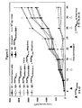

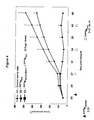
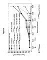

Classifications
-
- A—HUMAN NECESSITIES
- A61—MEDICAL OR VETERINARY SCIENCE; HYGIENE
- A61K—PREPARATIONS FOR MEDICAL, DENTAL OR TOILETRY PURPOSES
- A61K9/00—Medicinal preparations characterised by special physical form
- A61K9/48—Preparations in capsules, e.g. of gelatin, of chocolate
- A61K9/50—Microcapsules having a gas, liquid or semi-solid filling; Solid microparticles or pellets surrounded by a distinct coating layer, e.g. coated microspheres, coated drug crystals
- A61K9/5005—Wall or coating material
- A61K9/5063—Compounds of unknown constitution, e.g. material from plants or animals
- A61K9/5068—Cell membranes or bacterial membranes enclosing drugs
-
- A—HUMAN NECESSITIES
- A61—MEDICAL OR VETERINARY SCIENCE; HYGIENE
- A61K—PREPARATIONS FOR MEDICAL, DENTAL OR TOILETRY PURPOSES
- A61K31/00—Medicinal preparations containing organic active ingredients
- A61K31/33—Heterocyclic compounds
- A61K31/395—Heterocyclic compounds having nitrogen as a ring hetero atom, e.g. guanethidine or rifamycins
- A61K31/435—Heterocyclic compounds having nitrogen as a ring hetero atom, e.g. guanethidine or rifamycins having six-membered rings with one nitrogen as the only ring hetero atom
- A61K31/47—Quinolines; Isoquinolines
- A61K31/4738—Quinolines; Isoquinolines ortho- or peri-condensed with heterocyclic ring systems
- A61K31/4745—Quinolines; Isoquinolines ortho- or peri-condensed with heterocyclic ring systems condensed with ring systems having nitrogen as a ring hetero atom, e.g. phenantrolines
-
- A—HUMAN NECESSITIES
- A61—MEDICAL OR VETERINARY SCIENCE; HYGIENE
- A61K—PREPARATIONS FOR MEDICAL, DENTAL OR TOILETRY PURPOSES
- A61K31/00—Medicinal preparations containing organic active ingredients
- A61K31/70—Carbohydrates; Sugars; Derivatives thereof
- A61K31/7088—Compounds having three or more nucleosides or nucleotides
- A61K31/713—Double-stranded nucleic acids or oligonucleotides
-
- A—HUMAN NECESSITIES
- A61—MEDICAL OR VETERINARY SCIENCE; HYGIENE
- A61K—PREPARATIONS FOR MEDICAL, DENTAL OR TOILETRY PURPOSES
- A61K39/00—Medicinal preparations containing antigens or antibodies
- A61K39/395—Antibodies; Immunoglobulins; Immune serum, e.g. antilymphocytic serum
- A61K39/39533—Antibodies; Immunoglobulins; Immune serum, e.g. antilymphocytic serum against materials from animals
- A61K39/39558—Antibodies; Immunoglobulins; Immune serum, e.g. antilymphocytic serum against materials from animals against tumor tissues, cells, antigens
-
- A—HUMAN NECESSITIES
- A61—MEDICAL OR VETERINARY SCIENCE; HYGIENE
- A61K—PREPARATIONS FOR MEDICAL, DENTAL OR TOILETRY PURPOSES
- A61K39/00—Medicinal preparations containing antigens or antibodies
- A61K39/395—Antibodies; Immunoglobulins; Immune serum, e.g. antilymphocytic serum
- A61K39/40—Antibodies; Immunoglobulins; Immune serum, e.g. antilymphocytic serum bacterial
-
- A—HUMAN NECESSITIES
- A61—MEDICAL OR VETERINARY SCIENCE; HYGIENE
- A61K—PREPARATIONS FOR MEDICAL, DENTAL OR TOILETRY PURPOSES
- A61K47/00—Medicinal preparations characterised by the non-active ingredients used, e.g. carriers or inert additives; Targeting or modifying agents chemically bound to the active ingredient
- A61K47/46—Ingredients of undetermined constitution or reaction products thereof, e.g. skin, bone, milk, cotton fibre, eggshell, oxgall or plant extracts
-
- A—HUMAN NECESSITIES
- A61—MEDICAL OR VETERINARY SCIENCE; HYGIENE
- A61K—PREPARATIONS FOR MEDICAL, DENTAL OR TOILETRY PURPOSES
- A61K47/00—Medicinal preparations characterised by the non-active ingredients used, e.g. carriers or inert additives; Targeting or modifying agents chemically bound to the active ingredient
- A61K47/50—Medicinal preparations characterised by the non-active ingredients used, e.g. carriers or inert additives; Targeting or modifying agents chemically bound to the active ingredient the non-active ingredient being chemically bound to the active ingredient, e.g. polymer-drug conjugates
- A61K47/69—Medicinal preparations characterised by the non-active ingredients used, e.g. carriers or inert additives; Targeting or modifying agents chemically bound to the active ingredient the non-active ingredient being chemically bound to the active ingredient, e.g. polymer-drug conjugates the conjugate being characterised by physical or galenical forms, e.g. emulsion, particle, inclusion complex, stent or kit
- A61K47/6901—Conjugates being cells, cell fragments, viruses, ghosts, red blood cells or viral vectors
-
- A—HUMAN NECESSITIES
- A61—MEDICAL OR VETERINARY SCIENCE; HYGIENE
- A61K—PREPARATIONS FOR MEDICAL, DENTAL OR TOILETRY PURPOSES
- A61K47/00—Medicinal preparations characterised by the non-active ingredients used, e.g. carriers or inert additives; Targeting or modifying agents chemically bound to the active ingredient
- A61K47/50—Medicinal preparations characterised by the non-active ingredients used, e.g. carriers or inert additives; Targeting or modifying agents chemically bound to the active ingredient the non-active ingredient being chemically bound to the active ingredient, e.g. polymer-drug conjugates
- A61K47/69—Medicinal preparations characterised by the non-active ingredients used, e.g. carriers or inert additives; Targeting or modifying agents chemically bound to the active ingredient the non-active ingredient being chemically bound to the active ingredient, e.g. polymer-drug conjugates the conjugate being characterised by physical or galenical forms, e.g. emulsion, particle, inclusion complex, stent or kit
- A61K47/6921—Medicinal preparations characterised by the non-active ingredients used, e.g. carriers or inert additives; Targeting or modifying agents chemically bound to the active ingredient the non-active ingredient being chemically bound to the active ingredient, e.g. polymer-drug conjugates the conjugate being characterised by physical or galenical forms, e.g. emulsion, particle, inclusion complex, stent or kit the form being a particulate, a powder, an adsorbate, a bead or a sphere
- A61K47/6923—Medicinal preparations characterised by the non-active ingredients used, e.g. carriers or inert additives; Targeting or modifying agents chemically bound to the active ingredient the non-active ingredient being chemically bound to the active ingredient, e.g. polymer-drug conjugates the conjugate being characterised by physical or galenical forms, e.g. emulsion, particle, inclusion complex, stent or kit the form being a particulate, a powder, an adsorbate, a bead or a sphere the form being an inorganic particle, e.g. ceramic particles, silica particles, ferrite or synsorb
-
- A—HUMAN NECESSITIES
- A61—MEDICAL OR VETERINARY SCIENCE; HYGIENE
- A61K—PREPARATIONS FOR MEDICAL, DENTAL OR TOILETRY PURPOSES
- A61K48/00—Medicinal preparations containing genetic material which is inserted into cells of the living body to treat genetic diseases; Gene therapy
- A61K48/0008—Medicinal preparations containing genetic material which is inserted into cells of the living body to treat genetic diseases; Gene therapy characterised by an aspect of the 'non-active' part of the composition delivered, e.g. wherein such 'non-active' part is not delivered simultaneously with the 'active' part of the composition
-
- A—HUMAN NECESSITIES
- A61—MEDICAL OR VETERINARY SCIENCE; HYGIENE
- A61P—SPECIFIC THERAPEUTIC ACTIVITY OF CHEMICAL COMPOUNDS OR MEDICINAL PREPARATIONS
- A61P31/00—Antiinfectives, i.e. antibiotics, antiseptics, chemotherapeutics
- A61P31/12—Antivirals
- A61P31/14—Antivirals for RNA viruses
- A61P31/18—Antivirals for RNA viruses for HIV
-
- A—HUMAN NECESSITIES
- A61—MEDICAL OR VETERINARY SCIENCE; HYGIENE
- A61P—SPECIFIC THERAPEUTIC ACTIVITY OF CHEMICAL COMPOUNDS OR MEDICINAL PREPARATIONS
- A61P35/00—Antineoplastic agents
-
- A—HUMAN NECESSITIES
- A61—MEDICAL OR VETERINARY SCIENCE; HYGIENE
- A61P—SPECIFIC THERAPEUTIC ACTIVITY OF CHEMICAL COMPOUNDS OR MEDICINAL PREPARATIONS
- A61P43/00—Drugs for specific purposes, not provided for in groups A61P1/00-A61P41/00
-
- C—CHEMISTRY; METALLURGY
- C07—ORGANIC CHEMISTRY
- C07K—PEPTIDES
- C07K16/00—Immunoglobulins [IGs], e.g. monoclonal or polyclonal antibodies
- C07K16/12—Immunoglobulins [IGs], e.g. monoclonal or polyclonal antibodies against material from bacteria
- C07K16/1203—Immunoglobulins [IGs], e.g. monoclonal or polyclonal antibodies against material from bacteria from Gram-negative bacteria
- C07K16/1228—Immunoglobulins [IGs], e.g. monoclonal or polyclonal antibodies against material from bacteria from Gram-negative bacteria from Enterobacteriaceae (F), e.g. Citrobacter, Serratia, Proteus, Providencia, Morganella, Yersinia
- C07K16/1235—Immunoglobulins [IGs], e.g. monoclonal or polyclonal antibodies against material from bacteria from Gram-negative bacteria from Enterobacteriaceae (F), e.g. Citrobacter, Serratia, Proteus, Providencia, Morganella, Yersinia from Salmonella (G)
-
- C—CHEMISTRY; METALLURGY
- C07—ORGANIC CHEMISTRY
- C07K—PEPTIDES
- C07K16/00—Immunoglobulins [IGs], e.g. monoclonal or polyclonal antibodies
- C07K16/18—Immunoglobulins [IGs], e.g. monoclonal or polyclonal antibodies against material from animals or humans
- C07K16/28—Immunoglobulins [IGs], e.g. monoclonal or polyclonal antibodies against material from animals or humans against receptors, cell surface antigens or cell surface determinants
- C07K16/2863—Immunoglobulins [IGs], e.g. monoclonal or polyclonal antibodies against material from animals or humans against receptors, cell surface antigens or cell surface determinants against receptors for growth factors, growth regulators
-
- C—CHEMISTRY; METALLURGY
- C07—ORGANIC CHEMISTRY
- C07K—PEPTIDES
- C07K16/00—Immunoglobulins [IGs], e.g. monoclonal or polyclonal antibodies
- C07K16/40—Immunoglobulins [IGs], e.g. monoclonal or polyclonal antibodies against enzymes
-
- C—CHEMISTRY; METALLURGY
- C12—BIOCHEMISTRY; BEER; SPIRITS; WINE; VINEGAR; MICROBIOLOGY; ENZYMOLOGY; MUTATION OR GENETIC ENGINEERING
- C12N—MICROORGANISMS OR ENZYMES; COMPOSITIONS THEREOF; PROPAGATING, PRESERVING, OR MAINTAINING MICROORGANISMS; MUTATION OR GENETIC ENGINEERING; CULTURE MEDIA
- C12N15/00—Mutation or genetic engineering; DNA or RNA concerning genetic engineering, vectors, e.g. plasmids, or their isolation, preparation or purification; Use of hosts therefor
- C12N15/09—Recombinant DNA-technology
- C12N15/11—DNA or RNA fragments; Modified forms thereof; Non-coding nucleic acids having a biological activity
- C12N15/111—General methods applicable to biologically active non-coding nucleic acids
-
- C—CHEMISTRY; METALLURGY
- C12—BIOCHEMISTRY; BEER; SPIRITS; WINE; VINEGAR; MICROBIOLOGY; ENZYMOLOGY; MUTATION OR GENETIC ENGINEERING
- C12N—MICROORGANISMS OR ENZYMES; COMPOSITIONS THEREOF; PROPAGATING, PRESERVING, OR MAINTAINING MICROORGANISMS; MUTATION OR GENETIC ENGINEERING; CULTURE MEDIA
- C12N15/00—Mutation or genetic engineering; DNA or RNA concerning genetic engineering, vectors, e.g. plasmids, or their isolation, preparation or purification; Use of hosts therefor
- C12N15/09—Recombinant DNA-technology
- C12N15/11—DNA or RNA fragments; Modified forms thereof; Non-coding nucleic acids having a biological activity
- C12N15/113—Non-coding nucleic acids modulating the expression of genes, e.g. antisense oligonucleotides; Antisense DNA or RNA; Triplex- forming oligonucleotides; Catalytic nucleic acids, e.g. ribozymes; Nucleic acids used in co-suppression or gene silencing
- C12N15/1135—Non-coding nucleic acids modulating the expression of genes, e.g. antisense oligonucleotides; Antisense DNA or RNA; Triplex- forming oligonucleotides; Catalytic nucleic acids, e.g. ribozymes; Nucleic acids used in co-suppression or gene silencing against oncogenes or tumor suppressor genes
-
- C—CHEMISTRY; METALLURGY
- C12—BIOCHEMISTRY; BEER; SPIRITS; WINE; VINEGAR; MICROBIOLOGY; ENZYMOLOGY; MUTATION OR GENETIC ENGINEERING
- C12N—MICROORGANISMS OR ENZYMES; COMPOSITIONS THEREOF; PROPAGATING, PRESERVING, OR MAINTAINING MICROORGANISMS; MUTATION OR GENETIC ENGINEERING; CULTURE MEDIA
- C12N15/00—Mutation or genetic engineering; DNA or RNA concerning genetic engineering, vectors, e.g. plasmids, or their isolation, preparation or purification; Use of hosts therefor
- C12N15/09—Recombinant DNA-technology
- C12N15/11—DNA or RNA fragments; Modified forms thereof; Non-coding nucleic acids having a biological activity
- C12N15/113—Non-coding nucleic acids modulating the expression of genes, e.g. antisense oligonucleotides; Antisense DNA or RNA; Triplex- forming oligonucleotides; Catalytic nucleic acids, e.g. ribozymes; Nucleic acids used in co-suppression or gene silencing
- C12N15/1137—Non-coding nucleic acids modulating the expression of genes, e.g. antisense oligonucleotides; Antisense DNA or RNA; Triplex- forming oligonucleotides; Catalytic nucleic acids, e.g. ribozymes; Nucleic acids used in co-suppression or gene silencing against enzymes
-
- C—CHEMISTRY; METALLURGY
- C12—BIOCHEMISTRY; BEER; SPIRITS; WINE; VINEGAR; MICROBIOLOGY; ENZYMOLOGY; MUTATION OR GENETIC ENGINEERING
- C12N—MICROORGANISMS OR ENZYMES; COMPOSITIONS THEREOF; PROPAGATING, PRESERVING, OR MAINTAINING MICROORGANISMS; MUTATION OR GENETIC ENGINEERING; CULTURE MEDIA
- C12N15/00—Mutation or genetic engineering; DNA or RNA concerning genetic engineering, vectors, e.g. plasmids, or their isolation, preparation or purification; Use of hosts therefor
- C12N15/09—Recombinant DNA-technology
- C12N15/11—DNA or RNA fragments; Modified forms thereof; Non-coding nucleic acids having a biological activity
- C12N15/113—Non-coding nucleic acids modulating the expression of genes, e.g. antisense oligonucleotides; Antisense DNA or RNA; Triplex- forming oligonucleotides; Catalytic nucleic acids, e.g. ribozymes; Nucleic acids used in co-suppression or gene silencing
- C12N15/1138—Non-coding nucleic acids modulating the expression of genes, e.g. antisense oligonucleotides; Antisense DNA or RNA; Triplex- forming oligonucleotides; Catalytic nucleic acids, e.g. ribozymes; Nucleic acids used in co-suppression or gene silencing against receptors or cell surface proteins
-
- C—CHEMISTRY; METALLURGY
- C07—ORGANIC CHEMISTRY
- C07K—PEPTIDES
- C07K2317/00—Immunoglobulins specific features
- C07K2317/30—Immunoglobulins specific features characterized by aspects of specificity or valency
- C07K2317/31—Immunoglobulins specific features characterized by aspects of specificity or valency multispecific
-
- C—CHEMISTRY; METALLURGY
- C12—BIOCHEMISTRY; BEER; SPIRITS; WINE; VINEGAR; MICROBIOLOGY; ENZYMOLOGY; MUTATION OR GENETIC ENGINEERING
- C12N—MICROORGANISMS OR ENZYMES; COMPOSITIONS THEREOF; PROPAGATING, PRESERVING, OR MAINTAINING MICROORGANISMS; MUTATION OR GENETIC ENGINEERING; CULTURE MEDIA
- C12N2310/00—Structure or type of the nucleic acid
- C12N2310/10—Type of nucleic acid
- C12N2310/11—Antisense
- C12N2310/111—Antisense spanning the whole gene, or a large part of it
-
- C—CHEMISTRY; METALLURGY
- C12—BIOCHEMISTRY; BEER; SPIRITS; WINE; VINEGAR; MICROBIOLOGY; ENZYMOLOGY; MUTATION OR GENETIC ENGINEERING
- C12N—MICROORGANISMS OR ENZYMES; COMPOSITIONS THEREOF; PROPAGATING, PRESERVING, OR MAINTAINING MICROORGANISMS; MUTATION OR GENETIC ENGINEERING; CULTURE MEDIA
- C12N2310/00—Structure or type of the nucleic acid
- C12N2310/10—Type of nucleic acid
- C12N2310/14—Type of nucleic acid interfering N.A.
- C12N2310/141—MicroRNAs, miRNAs
-
- C—CHEMISTRY; METALLURGY
- C12—BIOCHEMISTRY; BEER; SPIRITS; WINE; VINEGAR; MICROBIOLOGY; ENZYMOLOGY; MUTATION OR GENETIC ENGINEERING
- C12N—MICROORGANISMS OR ENZYMES; COMPOSITIONS THEREOF; PROPAGATING, PRESERVING, OR MAINTAINING MICROORGANISMS; MUTATION OR GENETIC ENGINEERING; CULTURE MEDIA
- C12N2310/00—Structure or type of the nucleic acid
- C12N2310/50—Physical structure
- C12N2310/53—Physical structure partially self-complementary or closed
- C12N2310/531—Stem-loop; Hairpin
-
- C—CHEMISTRY; METALLURGY
- C12—BIOCHEMISTRY; BEER; SPIRITS; WINE; VINEGAR; MICROBIOLOGY; ENZYMOLOGY; MUTATION OR GENETIC ENGINEERING
- C12N—MICROORGANISMS OR ENZYMES; COMPOSITIONS THEREOF; PROPAGATING, PRESERVING, OR MAINTAINING MICROORGANISMS; MUTATION OR GENETIC ENGINEERING; CULTURE MEDIA
- C12N2320/00—Applications; Uses
- C12N2320/30—Special therapeutic applications
- C12N2320/32—Special delivery means, e.g. tissue-specific
-
- Y—GENERAL TAGGING OF NEW TECHNOLOGICAL DEVELOPMENTS; GENERAL TAGGING OF CROSS-SECTIONAL TECHNOLOGIES SPANNING OVER SEVERAL SECTIONS OF THE IPC; TECHNICAL SUBJECTS COVERED BY FORMER USPC CROSS-REFERENCE ART COLLECTIONS [XRACs] AND DIGESTS
- Y02—TECHNOLOGIES OR APPLICATIONS FOR MITIGATION OR ADAPTATION AGAINST CLIMATE CHANGE
- Y02A—TECHNOLOGIES FOR ADAPTATION TO CLIMATE CHANGE
- Y02A50/00—TECHNOLOGIES FOR ADAPTATION TO CLIMATE CHANGE in human health protection, e.g. against extreme weather
- Y02A50/30—Against vector-borne diseases, e.g. mosquito-borne, fly-borne, tick-borne or waterborne diseases whose impact is exacerbated by climate change
Definitions
- the present invention relates to ongoing efforts to achieve effective delivery of functional nucleic acids to mammalian cells. More specifically, the invention relates to using bacterial minicell vectors to deliver functional nucleic acids to mammalian cells.
- the invention has particular utility for eliminating drug resistance, especially in the context of cancer and AIDS therapy, for promoting apoptosis and for countering neoplasticity in targeted cells.
- liposome-based transfection methods can deliver exogenously produced nucleic acids.
- exogenous approach has the drawback, however, of effecting only transient inhibition of a target.
- liposomes are unstable in vivo.
- vectors can deliver plasmids that encode functional nucleic acids, which are produced endogenously. The viral vectors currently useful for this purpose, however, poses serious safety concerns.
- Illustrative problems include recombination with wild-type viruses, insertional and oncogenic potential, virus-induced immunosuppression, limited capacity of the viral vectors to carry large segments of DNA, reversion to virulence of attenuated viruses, difficulties in recombinant virus manufacture and distribution, low stability, and adverse reactions, such as an inflammatory response, caused by existing immunity.
- An approach that obviated these problems would offer significant benefit in making delivery of functional nucleic acids safer and more effective.
- NRTIs nucleoside reverse transcriptase inhibitors
- PIs protease inhibitors
- NRTI non-nucleoside reverse transcriptase inhibitor
- Clinical tumor resistance to chemotherapy can be intrinsic or acquired. Intrinsic resistance exists at the time of diagnosis in tumors that fail to respond to first-line chemotherapy. Acquired resistance occurs in tumors that may respond well to initial treatment, but exhibit a resistant phenotype upon recurrence. Such tumors gain resistance both to previously used drugs and to new drugs, including drugs with different structures and mechanisms of action.
- MDR multidrug resistance
- the mechanisms for multi-drug resistance are complex and multifactorial, owing largely to the high level of genomic instability and mutations in cancer cells.
- Exemplary mechanisms are drug inactivation, extrusion of drug by cell membrane pumps, decreased drug influx, mutations of drug targets and failure to initiate apoptosis (Bredel, 2001; Chen et al., 2001; White and McCubrey, 2001; Sun et al., 2001).
- Drug extrusion is particularly common, and can result from over-expression of membrane-associated proteins that pump drugs from the intracellular to the extracellular environment.
- Such pumps often are members of the ATP-binding cassette (ABC) transporter superfamily (Doige et al., 1993).
- P-glycoprotein (Pgp) is one such example, and is a major contributor to MDR in a variety of cancer cells (Endicott et al., 1989; Litman et al., 2001).
- Other examples include the MDR-associated protein (MRP; Cole et al., 1992), breast cancer resistance protein (BCRP; Litman et al., 2000), and lung resistance-related protein (LRP; a major vault protein; Scheffer et al., 2000).
- MRP MDR-associated protein
- BCRP Breast cancer resistance protein
- LRP lung resistance-related protein
- Other multidrug transporter proteins also have been identified in cancer cells (Gottesman et al., 2002) and in pathogenic microorganism
- Bcl-2 B-cell leukemia protein 2
- Bcl-X L Bcl-X L
- Bcl-W A1/Bf11
- Mcl-1 Mcl-1
- NF- ⁇ B transcription factor nuclear factor kappa B
- Drugs to counter MDR have been identified, such as drugs that block the action of P-glycoprotein (List et al., 1993; Miller et al., 1991; Wishart et al., 1992). Many such drugs were ineffective in clinical trials, however, because they bound to the plasma of patients, could not reach their destination (Ayesh et al., 1996a; Broxterman et al., 1987; Lehnert et al., 1996) and were toxic to normal cells. The use of functional nucleic acids to counter MDR also has been attempted. Yet, as noted above, existing vectors for this purpose are unstable or toxic, or they pose other serious safety issues, which hamper their use in humans (Sioud, 2004).
- the present invention provides, in one aspect, a method of delivering a functional nucleic acid, comprising (a) providing an intact minicell that contains a functional nucleic acid molecule or contains a plasmid comprising a segment that encodes a functional nucleic acid molecule, then (b) bringing the minicell into contact with a target mammalian cell, such that the mammalian cell engulfs the minicell. Following engulfment of the minicell, the functional nucleic acid molecule is released into the cytoplasm, transported to the nucleus and expressed by the target cell.
- the aforementioned plasmid also may contain a regulatory element, such as a promoter, a terminator, an enhancer or a signal sequence that is operably linked to the segment that encodes a functional nucleic acid molecule. It is particularly advantageous for the plasmid to comprise a promoter that is dependent on either RNA polymerase (pol) II or pol III, such as the RNA III polymerase promoters human 7SK, H1 and U6. Further, the plasmid may contain a reporter element, such as a nucleic acid segment coding for green fluorescent protein. Contact between the minicell and the mammalian cell may be in vitro or in vivo.
- a regulatory element such as a promoter, a terminator, an enhancer or a signal sequence that is operably linked to the segment that encodes a functional nucleic acid molecule. It is particularly advantageous for the plasmid to comprise a promoter that is dependent on either RNA polymerase (pol) II or pol III, such as the RNA
- the category of "functional nucleic acids” encompasses: siRNA molecules, including shRNA molecules; miRNA molecules, antisense molecules; and ribozyme molecules.
- the functional nucleic acid molecule targets the gene or transcript of a protein that promotes drug resistance, inhibits apoptosis, or contributes to a neoplastic phenotype.
- targets that contribute to drug resistance include ATP binding cassette transporters such as P-glycoprotein, MDR-2, MDR-3, BCRP, APT11a and LRP.
- Particularly useful targets that contribute to apoptosis resistance include Bcl-2 (B cell leukemia/lymphoma), Bcl-X L , A1/Bfl 1, focal adhesion kinase and p53 mutant protein.
- Other useful targets are oncogenic proteins and mutant tumor suppressor proteins.
- the invention provides a method of overcoming drug resistance or apoptosis resistance and treating a malignancy in a subject.
- the method comprises (a) providing an intact minicell that contains a functional nucleic acid molecule or a plasmid comprising a segment that encodes a functional nucleic acid molecule, where the functional nucleic acid molecule targets the transcript of a protein that promotes drug resistance (b) bringing the minicell into contact with a target mammalian cell, such that the mammalian cell engulfs the minicell, and (c) delivering a chemotherapeutic drug to the target mammalian cell.
- step (c) is performed after steps (a) and (b), to allow the functional nucleic acid to diminish resistance to the drug prior to the drug's administration.
- the drug may be delivered by any conventional means, but it preferably is delivered via an intact minicell.
- the minicell is brought into contact with the target mammalian cell via a bispecific ligand.
- the bispecific ligand has specificity for both a surface component on the minicell and a surface component on the mammalian cell, such as a receptor.
- the ligand causes the minicell to bind to the mammalian cell, the minicell is engulfed by the mammalian cell, and the minicell payload is released into the cytoplasm of the mammalian cell.
- the minicell is brought into contact with a target mammalian cell that is phagocytosis- or endocytosis-competent.
- the use of bispecific ligands is optional when a target cell is phagocytosis-competent.
- the invention provides a composition comprising (i), intact minicells and (ii) a pharmaceutically acceptable carrier therefor, where the minicells contain a functional nucleic acid molecule or a plasmid that encodes a functional nucleic acid molecule.
- the functional nucleic acid molecule may be an shRNA or miRNA or other siRNA molecule, an antisense molecule, or a ribozyme molecule.
- the functional nucleic acid molecule targets the gene or transcript of a protein that promotes drug resistance, inhibits apoptosis, or contributes to a neoplastic phenotype.
- Particularly useful targets that contribute to drug resistance include ATP binding cassette transporters such as P-glycoprotein, MDR-2 and MDR-3.
- Particularly useful targets that contribute to apoptosis resistance include Bcl-2 (B cell leukemia/lymphoma), Bcl-X L , Al/Bfl 1, focal adhesion kinase and p53 mutant protein.
- Other useful targets are oncogenic proteins and mutant tumor suppressor proteins.
- the plasmid may contain a regulatory element, such as a promoter, a terminator, an enhancer or a signal sequence that is operably linked to the segment that encodes a functional nucleic acid molecule. Further, the plasmid may contain a reporter element.
- the functional nucleic acid molecule may comprise multiple RNA interference sequences as miRNA or shRNA and these may be co-cistronic or expressed from separate promoters to enable simultaneous knockdown of multiple targets associated with drug resistance.
- the composition contains fewer than one contaminating parent cell per 10 7 , 10 8 , 10 9 , 10 10 , 10 11 or 10 12 minicells.
- the invention provides for a use of intact minicells in the preparation of a medicament for use in a method of overcoming drug resistance or promoting apoptosis by administration of the medicament to a cell, tissue or organ.
- minicells contain a functional nucleic acid molecule or a plasmid encoding a functional nucleic acid molecule, where the functional nucleic acid molecule targets the transcript of a protein that promotes drug resistance or inhibits apoptosis.
- the disease treated in this context may be a cancer, for example, or an acquired disease, such as AIDS and tuberculosis.
- the invention affords significant improvements over conventional methods and formulations for delivering functional nucleic acids in the context of cancer and HIV by (i) providing safe and stable vehicles for delivering functional nucleic acids, (ii) countering the principal mechanisms of drug resistance in diseased cells, (iii) reducing toxic side-effects associated with overcoming drug resistance, and (iv) providing targeted and drug-packaged vehicles to treat the disease.
- Figure 1 shows the effect of various treatments on the viability of human colon cancer cell line Caco-2.
- the treatments are shown on the x-axis (minicells are designated by "M") and percent cell viability is shown by the bars.
- M minicells are designated by "M”
- percent cell viability is shown by the bars.
- Each bar is a mean of six independent measurements and standard deviation of the mean is shown.
- Figure 2 shows regression of human colon cancer (Caco-2) xenografts in nude mice (11 mice per group) following dual treatment with (1) targeted recombinant minicells carrying shRNA encoding plasmids (anti-bcl2 or anti-Mdr1) and (2) targeted minicells packaged with the chemotherapeutic drug Irinotecan.
- the bispecific antibody used to target the colon cancer cells carried specificity against S. typhimurium O-antigen on one arm and human epidermal growth factor receptor (EGFR) on the other arm.
- the targeted recombinant minicells were injected intravenously on days 9 and 23, and the targeted Irinotecan packaged minicells were given intravenously on days 15, 18, 29 and 32.
- Control treatments administered intravenously include: Group 1 - tumor only, Group 2 - free irinotecan, Group 3 - EGFR minicells Irino , Group 4 - EGFR minicells shRNA-MDR-1 , Group 5 - EGFR minicells shRNA-bcl-2 and Group 6 - EGFR minicells shRNA-MDR-1 followed by free Irino.
- Tumor volume is shown on the y -axis. SEM is shown for each measurement.
- Figure 3 shows regression of human colon cancer (Caco-2) xenografts in nude mice (11 mice per group) following dual treatment with (1) targeted recombinant minicells carrying shRNA encoding plasmids (anti-bcl2 or anti-Mdr1) and (2) targeted minicells packaged with the chemotherapeutic drug 5-FU.
- the bispecific antibody used to target the colon cancer cells carried specificity against S. typhimurium O-antigen on one arm and human epidermal growth factor receptor (EGFR) on the other arm.
- the targeted recombinant minicells were injected intravenously on days 9 and 23, and the targeted 5-FU packaged minicells were given intravenously on days 15, 18, 29 and 32.
- control treatments administered intravenously include: G1 - tumor only, G2 (control), free 5-FU (5 x 10 4 ng/gm of mouse body weight ⁇ 1 x 10 6 ng per mouse), G3 (control), EGFR minicells 5-FU .
- G4 control
- EGFR minicells shRNA-MDR-1 G5 (control), EGFR minicells shRNA-bcl-2
- G6 control
- EGFR minicells shRNA-MDR-1 followed by CMV minicells 5-FU
- G7 control
- EGFR minicells shRNA-nonsense followed by EGFR minicells 5-FU
- G8 control
- G9 expt
- EGFR minicells shRNA-MDR-1 followed by EGFR minicells 5-FU
- G10 expt
- EGFR minicells shRNA-bcl-2 followed by EGFR minicells 5-FU .
- Tumor volume is shown on the y-axis. SEM is shown for each measurement.
- Figure 4 shows regression of human breast cancer (MDA-MB-468) xenografts in nude mice (11 mice per group) following dual treatment with (1) targeted recombinant minicells carrying shRNA encoding plasmid (anti-MDR-1) and (2) targeted minicells packaged with chemotherapeutic drug doxorubicin.
- the bispecific antibody used to target the breast cancer cells carried specificity against S. typhimurium O-antigen on one arm and human EGFR on the other arm.
- the targeted recombinant minicells were injected intravenously on day 21 and the targeted Dox-packaged minicells were given intravenously on days 27, 34 and 41.
- Treatments administered intravenously include: G1 - tumor only, G2 (control), EGFR minicells Dox , and G3 (expt), EGFR minicells shRNA-MDR-1 followed by EGFR minicells Dox .
- Tumor volume is shown on the y-axis. SEM is shown for each measurement.
- Figure 5 shows the effect of dosing schedules on reversal of drug-resistance and therapeutic effect.
- Human colon cancer (Caco-2) xenografts were established in nude mice and the following intravenous treatments were administered: G1 - tumor only, G2 (control), free irinotecan, G3 (expt), EGFR minicells shRNA-MDR-1 followed 96 hrs later by EGFR minicells Irino , G4 (expt), EGFR minicells shRNA-MDR-1 followed 120 hrs later by EGFR minicells Irino , and G5 (expt), EGFR minicells shRNA-MDR-1 followed 144 hrs later by EGFR minicells Irino .
- the bispecific antibody used to target the breast cancer cells carried specificity against S. typhimurium O-antigen on one arm and human EGFR on the other arm. Tumor volume is shown on the y-axis. SEM is shown for
- Figure 6 shows the effect of dosing schedules on reversal of drug-resistance and therapeutic effect.
- Human colon cancer (Caco-2) xenografts were established in nude mice and the following intravenous treatments were administered: G1 - tumor only, G2 (control), free 5-FU, G3 (expt), EGFR minicells shRNA-MDR-1 followed 96 hrs later by EGFR minicells 5-FU , G4 (expt), EGFR minicells shRNA-MDR-1 followed 120 hrs later by EGFR minicells 5-FU , and G5 (expt), EGFR minicells shRNA-MDR-1 followed 144 hrs later by EGFR minicells 5-FU .
- the bispecific antibody used to target the breast cancer cells carried specificity against S. typhimurium O-antigen on one arm and human EGFR on the other arm. Tumor volume is shown on the y-axis. SEM is shown for each measurement.
- the inventors have discovered that intact bacterially derived minicells can safely and effectively introduce into target mammalian cells any of a range of functional nucleic acids, such as an siRNA molecule, a miRNA molecule, a ribozyme, or an antisense nucleic acid.
- any of a range of functional nucleic acids such as an siRNA molecule, a miRNA molecule, a ribozyme, or an antisense nucleic acid.
- the introduction of functional nucleic acids into target cells, via intact minicells can diminish drug resistance or apoptosis resistance in the target cells.
- minicells can sequentially transfect the same target mammalian cells, particularly in vivo, and that minicells can sequentially deliver a range of different payloads to the same target mammalian cells.
- minicells that introduce multiple RNAi sequences into a host cell to counteract the multitude of genetic defects, and that following minicell-mediated RNAi delivery and allowing sufficient time for knockdown of target drug resistance-mediating proteins, cancer cells formerly resistant to specific chemotherapeutic drugs can effectively be treated with minicells packaged with the same drugs.
- This is the first in vivo demonstration of effectively treating cancer that is refractory to all other methods of treatment.
- concentration of chemotherapeutic drugs, delivered via minicells, required to treat drug-resistant cancer cells effectively is discovered to be over 1000-fold less than free drug treatment.
- RNAi reverse drug resistance
- inhibitors of drug resistance-mediating proteins still required drug concentrations that can cause severe toxicity to a mammalian subject.
- methods of the invention i.e., minicell-mediated delivery of RNAi followed by minicell-mediated chemotherapeutic drug, has the potential to treat cancer effectively without severe toxicity.
- the inventors have discovered that the serotype of minicells can be adapted to overcome a host immune response against minicells.
- Antisense oligonucleotide refers to a nucleic acid molecule complementary to a portion of a particular gene transcript that can hybridize to the transcript and block its translation.
- An antisense oligonucleotide may comprise RNA or DNA.
- Biomolecular sequence or “sequence” refers to all or a portion of a polynucleotide or polypeptide sequence.
- Cancer refers to cells or tissues that exhibit an aberrant growth phenotype characterized by a significant loss of control of cell proliferation.
- the methods and compositions of this invention particularly apply to precancerous, malignant, pre-metastatic, metastatic, and non-metastatic cells.
- “Complementary” refers to the topological compatibility or matching together of the interacting surfaces of two molecules, such as a functional nucleic acid molecule and its target.
- the molecules can be described as complementary, and furthermore, the contact surface characteristics are complementary to each other.
- a polynucleotide or sequence that "corresponds to” or “represents” a gene means that a sequence of the polynucleotide is present in the gene or in the nucleic acid gene product, e.g., mRNA.
- the polynucleotide may be wholly present within an exon of a genomic sequence of the gene, or different portions of the sequence of the polynucleotide may be present in different exons, e.g., such that the contiguous polynucleotide sequence is present in an mRNA, either pre- or post-splicing, that is an expression product of the gene.
- Cytokine is a generic term for proteins released by one cell population that act on another cell population as intercellular mediators.
- Drug refers to any physiologically or pharmacologically active substance that produces a local or systemic effect in animals, particularly mammals and humans.
- “Expression” generally refers to the process by which a polynucleotide sequence undergoes successful transcription and translation such that detectable levels of the amino acid sequence or protein are expressed.
- expression refers to the production of mRNA. In other contexts, expression refers to the production of protein.
- “Functional nucleic acid” refers to a nucleic acid molecule that, upon introduction into a host cell, specifically interferes with expression of a protein.
- functional nucleic acid molecules have the capacity to reduce expression of a protein by directly interacting with a transcript that encodes the protein.
- Ribozymes, antisense nucleic acids and siRNA molecules, including shRNA molecules, short RNAs (typically less than 400 bases in length), micro-RNAs (miRNAs) constitute exemplary functional nucleic acids.
- Gene refers to a polynucleotide sequence that comprises control and coding sequences necessary for the production of a polypeptide or precursor.
- the polypeptide can be encoded by a full length coding sequence or by any portion of the coding sequence.
- a gene may constitute an uninterrupted coding sequence or it may include one or more introns, bound by the appropriate splice junctions.
- a gene may contain one or more modifications in either the coding or the untranslated regions that could affect the biological activity or the chemical structure of the expression product, the rate of expression, or the manner of expression control. Such modifications include, but are not limited to, mutations, insertions, deletions, and substitutions of one or more nucleotides. In this regard, such modified genes may be referred to as "variants" of the "native" gene.
- “Host cell” refers to a cell that may be, or has been, used as a recipient for a recombinant vector or other transfer of polynucleotides, and includes the progeny of the original cell that has been transfected.
- the progeny of a single cell may not necessarily be completely identical in morphology or in genomic or total DNA complement as the original parent due to natural, accidental, or deliberate mutation.
- Hybridization refers to any process by which a polynucleotide sequence binds to a complementary sequence through base pairing.
- “Individual,” “subject,” “host,” and “patient,” used interchangeably herein, refer to any mammalian subject for whom diagnosis, treatment, or therapy is desired.
- the individual, subject, host, or patient is a human.
- Other subjects may include, but are not limited to, cattle, horses, dogs, cats, guinea pigs, rabbits, rats, primates, and mice.
- Label refers to agents that are capable of providing a detectable signal, either directly or through interaction with one or more additional members of a signal producing system. Labels that are directly detectable and may find use in the invention include fluorescent labels. Specific fluorophores include fluorescein, rhodamine, BODIPY, cyanine dyes and the like. The invention also contemplates the use of radioactive isotopes, such as 35 S, 32 P, 3 H, and the like as labels. Colorimetric labels such as colloidal gold or colored glass or plastic (e.g., polystyrene, polypropylene, latex) beads may also be utilized. For instance, see U.S. patents No. 4,366,241 , No. 4,277,437 , No. 4,275,149 , No. 3,996,345 , No. 3,939,350 , No. 3,850,752 , and No. 3,817,837 .
- Oligonucleotide refers to a polynucleotide comprising, for example, from about 10 nucleotides (nt) to about 1000 nt. Oligonucleotides for use in the invention are preferably from about 10 nt to about 150 nt.
- the oligonucleotide may be a naturally occurring oligonucleotide or a synthetic oligonucleotide. Oligonucleotides may be modified.
- minicell refers to anucleate forms of bacterial cells, engendered by a disturbance in the coordination, during binary fission, of cell division with DNA segregation. Minicells are distinct from other small vesicles that are generated and released spontaneously in certain situations and, in contrast to minicells, are not due to specific genetic rearrangements or episomal gene expression. For practicing the present invention, it is desirable for minicells to have intact cell walls ("intact minicells").
- Modified oligonucleotide and “Modified polynucleotide” refer to oligonucleotides or polynucleotides with one or more chemical modifications at the molecular level of the natural molecular structures of all or any of the bases, sugar moieties, internucleoside phosphate linkages, as well as to molecules having added substitutions or a combination of modifications at these sites.
- the internucleoside phosphate linkages may be phosphodiester, phosphotriester, phosphoramidate, siloxane, carbonate, carboxymethylester, acetamidate, carbamate, thioether, bridged phosphoramidate, bridged methylene phosphonate, phosphorothioate, methylphosphonate, phosphorodithioate, bridged phosphorothioate or sulfone internucleotide linkages, or 3'-3', 5'-3', or 5'-5' linkages, and combinations of such similar linkages.
- the phosphodiester linkage may be replaced with a substitute linkage, such as phosphorothioate, methylamino, methylphosphonate, phosphoramidate, and guanidine, and the ribose subunit of the polynucleotides may also be substituted (e.g., hexose phosphodiester; peptide nucleic acids).
- the modifications may be internal (single or repeated) or at the end(s) of the oligonucleotide molecule, and may include additions to the molecule of the internucleoside phosphate linkages, such as deoxyribose and phosphate modifications which cleave or crosslink to the opposite chains or to associated enzymes or other proteins.
- modified oligonucleotides and “modified polynucleotides” also include oligonucleotides or polynucleotides comprising modifications to the sugar moieties (e.g., 3'-substituted ribonucleotides or deoxyribonucleotide monomers), any of which are bound together via 5' to 3' linkages.
- modifications to the sugar moieties e.g., 3'-substituted ribonucleotides or deoxyribonucleotide monomers
- nucleic acid molecules and the term “polynucleotides” denote polymeric forms of nucleotides of any length, either ribonucleotides or deoxynucleotides. They include single-, double-, or multi-stranded DNA or RNA, genomic DNA, cDNA, DNA-RNA hybrids, or a polymer comprising purine and pyrimidine bases or other natural, chemically or biochemically modified, non-natural, or derivatized nucleotide bases.
- the backbone of a polynucleotide can comprise sugars and phosphate groups (as may typically be found in RNA or DNA), or modified or substituted sugar or phosphate groups.
- the backbone of the polynucleotide can comprise a polymer of synthetic subunits such as phosphoramidites and thus can be an oligodeoxynucleoside phosphoramidate or a mixed phosphoramidate-phosphodiester oligomer.
- a polynucleotide may comprise modified nucleotides, such as methylated nucleotides and nucleotide analogs, uracyl, other sugars, and linking groups such as fluororibose and thioate, and nucleotide branches.
- a polynucleotide may be further modified, such as by conjugation with a labeling component.
- modifications include caps, substitution of one or more of the naturally occurring nucleotides with an analog, and introduction of means for attaching the polynucleotide to proteins, metal ions, labeling components, other polynucleotides, or a solid support.
- “Pharmaceutically acceptable” refers to physiological compatibility.
- a pharmaceutically acceptable carrier or excipient does not abrogate biological activity of the composition being administered, is chemically inert and is not toxic to the organism in which it is administered.
- Polypeptide and “protein,” used interchangeably herein, refer to a polymeric form of amino acids of any length, which may include translated, untranslated, chemically modified, biochemically modified, and derivatized amino acids.
- a polypeptide or protein may be naturally occurring, recombinant, or synthetic, or any combination of these.
- a polypeptide or protein may comprise a fragment of a naturally occurring protein or peptide.
- a polypeptide or protein may be a single molecule or may be a multi-molecular complex.
- such polypeptides or proteins may have modified peptide backbones.
- the terms include fusion proteins, including fusion proteins with a heterologous amino acid sequence, fusions with heterologous and homologous leader sequences, with or without N-terminal methionine residues, immunologically tagged proteins, and the like.
- “Purified” refers to a compound that is removed from its natural environment and is at least about 60%, 65%, 70%, 75%, 80%, 85%, 90%, 91%, 92%, 93%, 94%, 95%, 96%, 97%, 98%, 99%, 99.9%, or 99.99% free from other components with which it is naturally associated.
- RNA molecules having an enzymatic activity that can repeatedly cleave other RNA molecules in a nucleotide base sequence-specific manner.
- RNA interference refers to sequence-specific or gene specific suppression of gene expression (protein synthesis) that is mediated by short interfering RNA (siRNA), short-haripin RNA, short RNA or micro-RNA.
- Sequence Identity refers to a degree of similarity or complementarity. There may be partial identity or complete identity.
- a partially complementary sequence is one that at least partially inhibits an identical sequence from hybridizing to a target polynucleotide; it is referred to using the functional term "substantially identical.”
- the inhibition of hybridization of the completely complementary sequence to the target sequence may be examined using a hybridization assay (Southern or Northern blot, solution hybridization, and the like) under conditions of low stringency.
- a substantially identical sequence or probe will compete for and inhibit the binding (i.e., the hybridization) of a completely identical sequence or probe to the target sequence under conditions of low stringency.
- low stringency conditions are such that non-specific binding is permitted; low stringency conditions require that the binding of two sequences to one another be a specific (i.e., selective) interaction.
- the absence of non-specific binding may be tested by the use of a second target sequence which lacks even a partial degree of complementarity (e.g., less than about 30% identity); in the absence of non-specific binding, the probe will not hybridize to the second non-complementary target sequence.
- sequence identity in the context to two nucleic acid or polypeptide sequences, entails referencing residues in the two sequences that are the same when aligned for maximum correspondence over a specified region.
- percentage of sequence identity means the value determined by comparing two optimally aligned sequences over a comparison window, wherein the portion of the polynucleotide sequence in the comparison window may comprise additions or deletions (i.e., gaps) as compared to the reference sequence (which does not comprise additions or deletions) for optimal alignment of the two sequences.
- the percentage is calculated by determining the number of positions at which the identical nucleic acid base occurs in both sequences to yield the number of matched positions, dividing the number of matched positions by the total number of positions in the window of comparison and multiplying the result by 100 to yield the percentage of sequence identity.
- siRNA short interfering RNA
- shRNA short interfering RNA
- treatment refers to obtaining a desired pharmacological and/or physiologic effect.
- the effect may be prophylactic in terms of completely or partially preventing a disease or symptom thereof and/or may be therapeutic in terms of a partial or complete stabilization or cure for a disease and/or adverse effect attributable to the disease.
- Treatment covers any treatment of a disease in a mammal, particularly a human, and includes: (a) preventing the disease or symptom from occurring in a subject which may be predisposed to the disease or symptom but has not yet been diagnosed as having it; (b) inhibiting the disease symptom, i.e., arresting its development; or (c) relieving the disease symptom, i.e., causing regression of the disease or symptom.
- the invention provides a method of delivering a functional nucleic acid to a target cell, comprising (a) providing an intact minicell that contains a functional nucleic acid molecule or a plasmid comprising a segment that encodes a functional nucleic acid molecule, then, (b) bringing the minicell into contact with a target mammalian cell, such that the mammalian cell engulfs the minicell. Following engulfment of the minicell, the functional nucleic acid molecule is released into the cytoplasm of the target cell or expressed by the target cell.
- Minicells may be brought into contact with the target mammalian cells via bispecific ligands, as described in WO 2005/056749 . Contact between the minicell and the target mammalian cell may be in vitro or in vivo.
- Minicells of the invention are anucleate forms of E. coli or other bacterial cells, engendered by a disturbance in the coordination, during binary fission, of cell division with DNA segregation.
- Prokaryotic chromosomal replication is linked to normal binary fission, which involves mid-cell septum formation.
- mutation of min genes, such as min CD can remove the inhibition of septum formation at the cell poles during cell division, resulting in production of a normal daughter cell and an anucleate minicell. See de Boer et al., 1992; Raskin & de Boer, 1999; Hu & Lutkenhaus, 1999; Harry, 2001.
- Minicells are distinct from other small vesicles that are generated and released spontaneously in certain situations and, in contrast to minicells, are not due to specific genetic rearrangements or episomal gene expression.
- it is desirable for minicells to have intact cell walls (“intact minicells").
- anucleate minicells are generated following a range of other genetic rearrangements or mutations that affect septum formation, for example in the div IVB1 in B. subtilis. See Reeve and Cornett, 1975; Levin et al., 1992.
- Minicells also can be formed following a perturbation in the levels of gene expression of proteins involved in cell division/chromosome segregation. For example, overexpression of min E leads to polar division and production of minicells.
- chromosome-less minicells may result from defects in chromosome segregation for example the smc mutation in Bacillus subtilis (Britton et al., 1998), spo OJ deletion in B.
- subtilis Ireton et al., 1994
- muk B mutation in E. coli Hiraga et al., 1989
- par C mutation in E. coli Stepwart and D'Ari, 1992
- Gene products may be supplied in trans.
- CafA may enhance the rate of cell division and/or inhibit chromosome partitioning after replication (Okada et al., 1994), resulting in formation of chained cells and anucleate minicells (Wachi et al., 1989; Okada et al., 1993).
- Minicells can be prepared from any bacterial cell of Gram-positive or Gram-negative origin.
- minicells contain a functional nucleic acid or a plasmid that encodes a functional nucleic acid for which delivery is desired.
- "Functional" nucleic acid molecules of the invention have the capacity to reduce expression of a protein by directly interacting with a transcript that encodes the protein.
- siRNA molecules, ribozymes, and antisense nucleic acids constitute exemplary functional nucleic acids.
- siRNA Short interfering RNA
- RNAi RNA interference
- siRNA generally refers to double-stranded RNA molecules from about 10 to about 30 nucleotides long that are named for their ability specifically to interfere with protein expression.
- siRNA molecules are 12-28 nucleotides long, more preferably 15-25 nucleotides long, still more preferably 19-23 nucleotides long and most preferably 21-23 nucleotides long. Therefore, preferred siRNA molecules are 12, 13, 14, 15, 16, 17, 18, 19, 20, 21, 22, 23, 24, 25, 26, 27 28 or 29 nucleotides in length.
- the length of one strand designates the length of an siRNA molecule.
- an siRNA that is described as 21 ribonucleotides long could comprise two opposite strands of RNA that anneal together for 19 contiguous base pairings. The two remaining ribonucleotides on each strand would form an "overhang.”
- the longer of the strands designates the length of the siRNA. For instance, a dsRNA containing one strand that is 21 nucleotides long and a second strand that is 20 nucleotides long, constitutes a 21-mer.
- siRNAs that comprise an overhang are desirable.
- the overhang may be at the 5' or the 3' end of a strand. Preferably, it is at the 3' end of the RNA strand.
- the length of an overhang may vary, but preferably is about 1 to about 5 bases, and more preferably is about 2 nucleotides long.
- the siRNA of the present invention will comprise a 3' overhang of about 2 to 4 bases. More preferably, the 3' overhang is 2 ribonucleotides long. Even more preferably, the 2 ribonucleotides comprising the 3' overhang are uridine (U).
- shRNAs short hairpin RNAs
- shRNAs comprise a single strand of RNA that forms a stem-loop structure, where the stem consists of the complementary sense and antisense strands that comprise a double-stranded siRNA, and the loop is a linker of varying size.
- the stem structure of shRNAs generally is from about 10 to about 30 nucleotides long.
- the stem of shRNA molecules are 12-28 nucleotides long, more preferably 15-25 nucleotides long, still more preferably 19-23 nucleotides long and most preferably 21-23 nucleotides long. Therefore, preferred shRNA molecules comprise stems that are 12, 13, 14, 15, 16, 17, 18, 19, 20, 21, 22, 23, 24, 25, 26, 27 28 or 29 nucleotides in length.
- siRNAs of the invention are designed to interact with a target ribonucleotide sequence, meaning they complement a target sequence sufficiently to hybridize to the target sequence.
- the invention provides an siRNA molecule comprising a ribonucleotide sequence at least 70%, 75%, 80%, 85% or 90% identical to a target ribonucleotide sequence or the complement of a target ribonucleotide sequence.
- the siRNA molecule is at least 90%, 95%, 96%, 97%, 98%, 99% or 100% identical to the target ribonucleotide sequence or the complement of the target ribonucleotide sequence.
- an siRNA will be 100% identical to the target nucleotide sequence or the complement of the ribonucleotide sequence.
- siRNA molecules with insertions, deletions or single point mutations relative to a target may also be effective.
- siRNA design tool is available on the internet at www.dharmacon.com.
- Ribozymes are RNA molecules having an enzymatic activity that can repeatedly cleave other RNA molecules in a nucleotide base sequence-specific manner. Such enzymatic RNA molecules may be targeted to virtually any RNA transcript, and efficient cleavage achieved in vitro.
- enzymatic polynucleotides act by first binding to a target RNA. Such binding occurs through the target binding portion of a enzymatic polynucleotide which is held in close proximity to an enzymatic portion of the molecule that acts to cleave the target RNA. Thus, the enzymatic polynucleotide first recognizes and then binds a target RNA through complementary base-pairing, and once bound to the correct site, acts enzymatically to cut the target RNA.
- RNA Strategic cleavage of such a target RNA will destroy its ability to direct synthesis of an encoded protein. After an enzymatic polynucleotide has bound and cleaved its RNA target, it is released from that RNA to search for another target and can repeatedly bind and cleave new targets.
- ribozyme The enzymatic nature of a ribozyme is advantageous. Because a single ribozyme molecule is able to cleave many molecules of target RNA, effective concentrations of ribozyme can be quite low.
- Useful ribozymes may comprise one of several motifs, including hammerhead (Rossi et al. (1992)), hairpin (Hampel and Tritz, (1989), Hampel et al. (1990)), hepatitis delta virus motif (Perrotta and Been (1992), group I intron ( U.S. Patent No. 4,987,071 ), RNaseP RNA in association with an RNA guide sequence (Guerrier-Takada et al. (1983)), and Neurospora VS RNA (Saville & Collins (1990); Saville & Collins (1991); Collins & Olive (1993)).
- Ribozymes of the invention may comprise modified oligonucleotides (e.g., for improved stability, targeting, etc.). Nucleic acid sequences encoding the ribozymes may be under the control of a strong constitutive promoter, such as, for example, RNA Polymerase II or RNA Polymerase III promoter, so that transfected cells will produce sufficient quantities of the ribozyme to destroy target endogenous messages and inhibit translation.
- a strong constitutive promoter such as, for example, RNA Polymerase II or RNA Polymerase III promoter
- Antisense oligonucleotides of the invention specifically hybridize with a nucleic acid encoding a protein, and interfere with transcription or translation of the protein.
- an antisense oligonucleotide targets DNA and interferes with its replication and/or transcription.
- an antisense oligonucleotide specifically hybridizes with RNA, including pre-mRNA and mRNA.
- Such antisense oligonucleotides may affect, for example, translocation of the RNA to the site of protein translation, translation of protein from the RNA, splicing of the RNA to yield one or more mRNA species, and catalytic activity that may be engaged in or facilitated by the RNA. The overall effect of such interference is to modulate, decrease, or inhibit target protein expression.
- an antisense oligonucleotide may bind the region encompassing the translation initiation codon, also known as the start codon, of the open reading frame.
- start codon and translation initiation codon generally refer to the portion of such mRNA or gene that encompasses from at least about 25 to at least about 50 contiguous nucleotides in either direction ( i.e., 5' or 3') from a translation initiation codon.
- stop codon region and “translation termination codon region” refer generally to a portion of such a mRNA or gene that encompasses from at least about 25 to at least about 50 contiguous nucleotides in either direction form a translation termination codon.
- the open reading frame or coding region also may be targeted effectively.
- the open reading frame is generally understood to refer to the region between the translation initiation codon and the translation termination codon.
- Another target region is the 5' untranslated region, which is the portion of a mRNA in the 5' direction from the translation initiation codon. It includes the nucleotides between the 5' cap site and the translation initiation codon of a mRNA or corresponding nucleotides on the gene.
- the 3' untranslated region may be used as a target for antisense oligonucleotides.
- the 3' untranslated region is that portion of the mRNA in the 3' direction from the translation termination codon, and thus includes the nucleotides between the translation termination codon and the 3' end of a mRNA or corresponding nucleotides of the gene.
- An antisense oligonucleotide may also target the 5' cap region of an mRNA.
- the 5' cap comprises an N7-methylated guanosine residue joined to the 5'-most residue of the mRNA via 5'-5' triphosphate linkage.
- the 5' cap region is considered to include the 5' cap structure itself as well as the first 50 nucleotides adjacent to the cap.
- mRNA splice sites i.e., intron-exon junctions
- intron-exon junctions represent possible target regions, and are particularly useful in situations where aberrant splicing is implicated in disease, or where an overproduction of a particular mRNA splice product is implicated in disease.
- aberrant fusion junctions due to rearrangements or deletions are also possible targets for antisense oligonucleotides.
- antisense oligonucleotides that are sufficiently complementary to the target polynucleotides must be chosen. There must be a sufficient degree of complementarity or precise pairing such that stable and specific binding occurs between the oligonucleotide and the polynucleotide target. Importantly, the sequence of an antisense oligonucleotide need not be 100% complementary to that of its target polynucleotide to be specifically hybridizable.
- An antisense oligonucleotide is specifically hybridizable when binding of the antisense oligonucleotide to the target polynucleotide interferes with the normal function of the target polynucleotide to cause a loss of utility, and there is a sufficient degree of complementarity to avoid non-specific binding of the antisense oligonucleotide to non-target sequences under conditions in which specific binding is desired, i.e., under physiological conditions in the case of in vivo assays or therapeutic treatment, and in the case of in vitro assays, under conditions in which the assays are performed.
- the antisense oligonucleotides may be at least about 8 nt to at least about 50 nt in length. In one embodiment, the antisense oligonucleotides may be about 12 to about 30 nt in length.
- the antisense oligonucleotides used in accordance with this invention may be conveniently and routinely made through the well-known technique of solid phase synthesis.
- Equipment for such synthesis is sold by several vendors including, for example, Applied Biosystems (Foster City, CA). Any other means for such synthesis known in the art may additionally or alternatively be employed. It is well known to use similar techniques to prepare oligonucleotides such as the phosphorothioates and alkylated derivatives.
- minicells comprise nucleic acids that encode functional nucleic acids.
- a plasmid may encode a functional nucleic acid that is expressed inside of mammalian target cells. This makes possible endogenous delivery of functional nucleic acids, which has advantages over the transient nature of exogenous delivery.
- recombinant intact minicells may carry plasmid DNA encoding one or more siRNA sequences aimed at silencing drug resistance or apoptosis resistance genes.
- minicells that encode multiple functional nucleic acids, it is possible to treat cells that express multiple drug resistance mechanisms.
- Different siRNA sequences can be expressed individually from different promoters.
- siRNA targeting Pgp mRNA can be expressed from the U6 promoter and siRNA targeting Bcl-2 mRNA can be expressed from the H1 promoter.
- These multiple expression cassettes preferably are carried on a single plasmid, but may also be on different plasmids.
- siRNA sequences also can be expressed from a single promoter, where the recombinant plasmid carries an expression cassette comprised of multiple siRNA-encoding sequences, which are linked together via noncoding polynucleotide sequences.
- a single gene transcription terminator can be placed downstream of the complete expression cassette.
- a plasmid encodes the sense and antisense strands of an siRNA as two independent transcripts that, after expression within a target cell, hybridize to form functional siRNA duplexes.
- a plasmid encodes one or more siRNAs that each are expressed as a single transcript that forms a short hairpin RNA stem-loop structure.
- the hairpin structure may be processed by a Dicer enzyme into functional siRNA.
- a nucleic acid molecule to be introduced via the approach of the present invention can include a reporter element.
- a reporter element confers on its recombinant host a readily detectable phenotype or characteristic, typically by encoding a polypeptide, not otherwise produced by the host, that can be detected, upon expression, by histological or in situ analysis, such as by in vivo imaging techniques.
- a reporter element delivered by an intact minicell could code for a protein that produces, in the engulfing host cell, a colorimetric or fluorometric change that is detectable by in situ analysis and that is a quantitative or semi-quantitative function of transcriptional activation.
- Illustrative of these proteins are esterases, phosphatases, proteases and other enzymes, the activity of which generates a detectable chromophore or fluorophore.
- E. coli ⁇ -galactosidase which effects a color change via cleavage of an indigogenic substrate, indolyl- ⁇ -D-galactoside, and a luciferase, which oxidizes a long-chain aldehyde (bacterial luciferase) or a heterocyclic carboxylic acid (luciferin), with the concomitant release of light.
- a reporter element that encodes the green fluorescent protein (GFP) of the jellyfish, Aequorea victoria, as described by Prasher et al. (1995).
- WO 095/21191 discloses a polynucleotide sequence encoding a 238 amino-acid GFP apoprotein, containing a chromophore formed from amino acids 65 through 67
- WO 095/21191 discloses a modification of the cDNA for the apopeptide of A. victoria GFP, providing a peptide having altered fluorescent properties
- WO 095/21191 discloses a modification of the cDNA for the apopeptide of A. victoria GFP, providing a peptide having altered fluorescent properties
- reporter element is associated with an expression product that renders the recombinant minicell resistant to a toxin.
- the neo gene protects a host against toxic levels of the antibiotic G418, while a gene encoding dihydrofolate reductase confers resistance to methotrexate, and the chloramphenicol acetyltransferase (CAT) gene bestows resistance to chloramphenicol.
- CAT chloramphenicol acetyltransferase
- genes for use as a reporter element include those that can transform a host minicell to express distinguishing cell-surface antigens, e.g., viral envelope proteins such as HIV gp120 or herpes gD, which are readily detectable by immunoassays.
- a nucleic acid molecule to be introduced via the approach of the present invention also can have a desired encoding segment linked operatively to a regulatory element, such as a promoter, a terminator, an enhancer and/or a signal sequence.
- a regulatory element such as a promoter, a terminator, an enhancer and/or a signal sequence.
- a suitable promoter can be tissue-specific or even tumor-specific, as the therapeutic context dictates.
- a promoter is "tissue-specific" when it is activated preferentially in a given tissue and, hence, is effective in driving expression, in the target tissue, of an operably linked structural sequence.
- the category of tissue-specific promoters includes, for example: the hepatocyte-specific promoter for albumin and a 1 ⁇ antitrypsin, respectively; the elastase I gene control region, which is active in pancreatic acinar cells; the insulin gene control region, active in pancreatic beta cells; the mouse mammary tumor virus control region, which is active in testicular, breast, lymphoid and mast cells; the myelin basic protein gene control region, active in oligodendrocyte cells in the brain; and the gonadotropic releasing hormone gene control region, which is active in cells of the hypothalamus.
- promoters that are expressed preferentially in certain tumor cells or in tumor cells per se, and that are useful for treating different cancers in accordance with the present invention.
- the class of promoters that are specific for cancer cells is illustrated by: the tyrosinase promoter, to target melanomas; the MUC1/Df3 promoter, to target breast carcinoma; the hybrid myo D enhancer/SV40 promoter, which targets expression to rhabdomyosarcoma (RMS); the carcinoembryonic antigen (CEA) promoter, which is specific for CEA-expressing cells such as colon cancer cells, and the hexokinase type II gene promoter, to target non-small cell lung carcinomas.
- RMS rhabdomyosarcoma
- CEA carcinoembryonic antigen
- Promoters that are dependent on either RNA polymerase (pol) II or pol II are preferred promoters. Highly preferred promoters are the RNA III polymerase promoters H1 and U6.
- a signal sequence can be used, according to the present invention, to effect secretion of an expression product or localization of an expression product to a particular cellular compartment.
- a therapeutic polynucleotide molecule that is delivered via intact minicells may include a signal sequence, in proper reading frame, such that the expression product of interest is secreted by an engulfing cell or its progeny, thereby to influence surrounding cells, in keeping with the chosen treatment paradigm.
- Illustrative signal sequences include the haemolysin C-terminal secretion sequence, described in U.S. patent No. 5,143,830 , the BAR1 secretion sequence, disclosed in U.S. patent No. 5,037,743 , and the signal sequence portion of the zsig32 polypeptide, described in U.S. patent No. 6,025,197 .
- Functional nucleic acids of the invention target the gene or transcript of a protein that promotes drug resistance, inhibits apoptosis or promotes a neoplastic phenotype.
- Successful application of functional nucleic acid strategies in these contexts have been achieved in the art, but without the benefits of minicell vectors. See, e.g., Sioud (2004), Caplen (2003), Wu et al. (2003), Nieth et al. (2003), Caplen and Mousses (2003), Duxbury et al. (2004), Yague et al. (2004), Duan et al. (2004),
- Proteins that contribute to drug resistance constitute preferred targets of functional nucleic acids.
- the proteins may contribute to acquired drug resistance or intrinsic drug resistance. When diseased cells, such as tumor cells, initially respond to drugs, but become refractory on subsequent treatment cycles, the resistant phenotype is acquired.
- Useful targets involved in acquired drug resistance include ATP binding cassette transporters such as P-glycoprotein (P-gp, P-170, PGY1, MDR1, ABCB1, MDR-associated protein, Multidrug resistance protein 1), MDR-2 and MDR-3.
- MRP2 multi-drug resistance associated protein
- BCR-ABL breakpoint cluster region ⁇ Abelson protooncogene
- a STI-571 resistance-associated protein lung resistance-related protein
- cyclooxygenase-2 nuclear factor kappa
- XRCC1 X-ray cross-complementing group 1
- ERCC1 Extra cross-complementing gene
- GSTP1 Glutathione S-transferase
- mutant ⁇ -tubulin growth factors such as IL-6 are additional targets involved in acquired drug resistance.
- LRP lung resistance-related protein
- Useful targets also include proteins that contribute to apoptosis resistance. These include Bcl-2 (B cell leukemia/lymphoma), Bcl-X L , A1/Bfl 1, focal adhesion kinase and p53 mutant protein.
- Useful targets further include oncogenic and mutant tumor suppressor proteins.
- Examples include ⁇ -Catenin, PKC- ⁇ (protein kinase C), C-RAF, K-Ras (V12), DP97 Dead box RNA helicase, DNMT1 (DNA methyltransferase 1), FLIP (Flice-like inhibitory protein), C-Sfc, 53BPI, Polycomb group protein EZH2 (Enhancer of zeste homologue), ErbB1, HPV-16 E5 and E7 (human papillomavirus early 5 and early 7), Fortilin & MCI1P (Myeloid cell leukemia 1 protein), DIP13 ⁇ (DDC interacting protein 13a), MBD2 (Methyl CpG binding domain), p21, KLF4 (Kruppel-like factor 4), tpt/TCTP (Translational controlled tumor protein), SPK1 & SPK2 (Sphingosine kinase), P300, PLK1 (Polo-
- targets include HIV-Tat, HIV-Rev, HIV-Vif, HIV-Nef, HIV-Gag, HIV-Env, LTR, CD4, CXCR4 (chemokine receptor) and CCR5 (chemokine receptor).
- the functional nucleic acids used in methods of the invention may require change over time. For instance, if biopsy samples reveal new mutations that result in acquired drug resistance, specific siRNAs can be designed and encoded on a suitable expression plasmid, which is transformed into a minicell-producing bacterial strain, which is used to produce recombinant minicells that are administered to address the acquired drug resistance.
- the invention provides a method of overcoming drug resistance and treating a disease, such as cancer or AIDS, in a subject.
- the method comprises (a) providing an intact minicell that contains a functional nucleic acid molecule or a plasmid comprising a segment that encodes a functional nucleic acid molecule, where the functional nucleic acid molecule targets the gene or transcript of a protein that promotes drug resistance, (b) bringing the minicell into contact with a target mammalian cell, such that the mammalian cell engulfs the minicell, and (c) delivering a drug to the target mammalian cell.
- step (c) is performed after steps (a) and (b), to allow the functional nucleic acid to diminish resistance to the drug prior to the drug's administration. Delivery of the drug and introduction of the functional nucleic acid can occur consecutively, in any order, or simultaneously.
- drugs may be delivered by any conventional means.
- drugs may be delivered orally, parenterally (including subcutaneously, intravenously, intramuscularly, intraperitoneally, and by infusion), topically, transdermally or by inhalation.
- parenterally including subcutaneously, intravenously, intramuscularly, intraperitoneally, and by infusion
- topically including subcutaneously, intravenously, intramuscularly, intraperitoneally, and by infusion
- transdermally or by inhalation The appropriate mode of delivery and dosage of each drug is easily ascertainable by those skilled in the medical arts.
- minicells Although drug delivery may occur via conventional means, delivery via minicells is preferred.
- the inventors have discovered that the same mammalian cells can be successfully re-transfected by targeted intact minicells that are packaged with different payloads.
- siRNA-encoding plasmid-packaged minicells can transfect a mammalian cell, after which drug-packaged minicells can deliver drug to the same mammalian cell.
- This discovery was a surprise, and indicates that the intracellular processes associated with minicell breakdown, endosomal release of a payload and escape of the payload to intracellular targets remains fully functional after the first round of transfection and payload delivery.
- the drug may be packaged in a separate minicell from the functional nucleic acid or plasmid encoding the functional nucleic acid.
- the drug may be packaged in the same minicell as the functional nucleic acid molecule or plasmid encoding the functional nucleic acid molecule.
- Certain drugs may interact with nucleic acids and preclude co-packaging of drug and nucleic acid in the same minicell. For example, Doxorubicin is known to interact with DNA.
- minicells of the invention contain a sufficient quantity of drug to exert the drug's physiological or pharmacological effect on a target cell.
- drugs contained within the minicells are heterologous, or foreign, to the minicells, meaning that the minicells' parent bacterial cells do not normally produce the drug.
- Both hydrophilic and hydrophobic drugs can be packaged in minicells by creating a concentration gradient of the drug between en extracellular medium containing minicells and the minicell cytoplasm.
- the extracellular medium contains a higher drug concentration than the minicell cytoplasm, the drug naturally moves down this concentration gradient, into the minicell cytoplasm.
- the concentration gradient is reversed, however, the drug does not move out of the minicells.
- the drugs initially can be dissolved in an appropriate solvent.
- Paclitaxel can be dissolved in a 1:1 blend of ethanol and cremophore EL (polyethoxylated castor oil), followed by a dilution in PBS to achieve a solution of Paclitaxel that is partly diluted in aqueous media and carries minimal amounts of the organic solvent to ensure that the drug remains in solution.
- Minicells can be incubated in this final medium for drug loading.
- the inventors discovered that even hydrophobic drugs can diffuse into the cytoplasm of minicells to achieve a high and therapeutically significant cytoplasmic drug load. This is unexpected because the minicell membrane is composed of a hydrophobic phospholipid bilayer, which would be expected to prevent diffusion of hydrophobic molecules into the cytoplasm.
- Another method of loading minicells with a drug involves culturing a recombinant parent bacterial cell under conditions wherein the parent bacterial cell transcribes and translates a nucleic acid encoding the drug, such that the drug is released into the cytoplasm of the parent bacterial cell.
- a gene cluster encoding the cellular biosynthetic pathway for a desired drug can be cloned and transferred into a parent bacterial strain that is capable of producing minicells. Genetic transcription and translation of the gene cluster results in biosynthesis of the drug within the cytoplasm of the parent bacterial cells, filling the bacterial cytoplasm with the drug.
- the minicells also contain the drug in their cytoplasm.
- the pre-packaged minicells can be purified by any of the minicell purification processes known in the art and described above.
- Drugs useful in the invention may be any physiologically or pharmacologically active substance that produces a desired local or systemic effect in animals, particularly mammals and humans.
- Drugs may be inorganic or organic compounds, without limitation, including peptides, proteins, nucleic acids, and small molecules, any of which may be characterized or uncharacterized. They may be in various forms, such as unchanged molecules, molecular complexes, pharmacologically acceptable salts, such as hydrochloride, hydrobromide, sulfate, laurate, palmitate, phosphate, nitrite, nitrate, borate, acetate, maleate, tartrate, oleate, salicylate, and the like.
- salts of metals, amines or organic cations for example, quaternary ammonium
- derivatives of drugs such as bases, esters and amides also can be used.
- a drug that is water insoluble can be used in a form that is a water soluble derivative thereof, or as a base derivative thereof, which in either instance, or by its delivery, is converted by enzymes, hydrolyzed by the body pH, or by other metabolic processes to the original therapeutically active form.
- Useful drugs include chemotherapeutic agents, immunosuppressive agents, cytokines, cytotoxic agents, nucleolytic compounds, radioactive isotopes, receptors, and pro-drug activating enzymes, which may be naturally occurring or produced by recombinant methods.
- Drugs that are affected by classical multidrug resistance have particular utility in the invention, such as vinca alkaloids (e.g., vinblastine and vincristine), the anthracyclines (e.g., doxorubicin and daunorubicin), RNA transcription inhibitors (e.g., actinomycin-D) and microtubule stabilizing drugs (e.g., paclitaxel).
- vinca alkaloids e.g., vinblastine and vincristine
- the anthracyclines e.g., doxorubicin and daunorubicin
- RNA transcription inhibitors e.g., actinomycin-D
- microtubule stabilizing drugs e.g., paclitaxel
- cancer chemotherapy agents are preferred drugs.
- Useful cancer chemotherapy drugs include nitrogen mustards, nitrosorueas, ethyleneimine, alkane sulfonates, tetrazine, platinum compounds, pyrimidine analogs, purine analogs, antimetabolites, folate analogs, anthracyclines, taxanes, vinca alkaloids, topoisomerase inhibitors and hormonal agents.
- Exemplary chemotherapy drugs are Actinomycin-D, Alkeran, Ara-C, Anastrozole, Asparaginase, BiCNU, Bicalutamide, Bleomycin, Busulfan, Capecitabine, Carboplatin, Carboplatinum, Carmustine, CCNU, Chlorambucil, Cisplatin, Cladribine, CPT-11, Cyclophosphamide, Cytarabine, Cytosine arabinoside, Cytoxan, dacarbazine, Dactinomycin, Daunorubicin, Dexrazoxane, Docetaxel, Doxorubicin, DTIC, Epirubicin, Ethyleneimine, Etoposide, Floxuridine, Fludarabine, Fluorouracil, Flutamide, Fotemustine, Gemcitabine, Herceptin, Hexamethylamine, Hydroxyurea, Idarubicin, Ifosfamide, Irinotecan, Lomustine, Mechloreth
- Useful cancer chemotherapy drugs also include alkylating agents such as Thiotepa and cyclosphosphamide; alkyl sulfonates such as Busulfan, Improsulfan and Piposulfan; aziridines such as Benzodopa, Carboquone, Meturedopa, and Uredopa; ethylenimines and methylamelamines including altretamine, triethylenemelamine, trietylenephosphoramide, triethylenethiophosphaoramide and trimethylolomelamine; nitrogen mustards such as Chlorambucil, Chlornaphazine, Cholophosphamide, Estramustine, Ifosfamide, mechlorethamine, mechlorethamine oxide hydrochloride, Melphalan, Novembiehin, Phenesterine, Prednimustine, Trofosfamide, uracil mustard; nitroureas such as Cannustine, Chlorozotocin, Fotem
- anti-hormonal agents that act to regulate or inhibit hormone action on tumors
- anti-estrogens including for example Tamoxifen, Raloxifene, aromatase inhibiting 4(5)-imidazoles, 4 Hydroxytamoxifen, Trioxifene, Keoxifene, Onapristone, And Toremifene (Fareston); and anti-androgens such as Flutamide, Nilutamide, Bicalutamide, Leuprolide, and Goserelin; and pharmaceutically acceptable salts, acids or derivatives of any of the above.
- Useful drugs also include cytokines.
- cytokines are lymphokines, monokines, and traditional polypeptide hormones. Included among the cytokines are growth hormones such as human growth hormone, N-methionyl human growth hormone, and bovine growth hormone; parathyroid hormone; thyroxine; insulin; proinsulin; relaxin; prorelaxin; glycoprotein hormones such as follicle stimulating hormone (FSH), thyroid stimulating hormone (TSH), and luteinizing hormone (LH); hepatic growth factor; fibroblast growth factor; prolactin; placental lactogen; tumor necrosis factor- ⁇ and - ⁇ ; mullerian-inhibiting substance; mouse gonadotropin-associated peptide; inhibin; activin; vascular endothelial growth factor; integrin; thrombopoietin (TPO); nerve growth factors such as NGF- ⁇ ; platelet growth factor; transforming growth factors (TGFs) such as TGF- ⁇ and TGF- ⁇
- the drugs may be prodrugs, subsequently activated by a prodrug-activating enzyme that converts a prodrug like a peptidyl chemotherapeutic agent to an active anti-cancer drug.
- a prodrug-activating enzyme that converts a prodrug like a peptidyl chemotherapeutic agent to an active anti-cancer drug.
- the enzyme component includes any enzyme capable of acting on a prodrug in such a way so as to covert it into its more active, cytotoxic form.
- a minicell is directed to a target mammalian cell via a bispecific ligand, as described in WO 2005/056749 .
- the bispecific ligand having specificity for both minicell and mammalian cell components, causes the minicell to bind to the mammalian cell, such that the minicell is engulfed by the mammalian cell, and the mammalian cell produces the functional nucleic acid molecule.
- This targeted delivery method may be performed in vivo or in vitro, or both in vivo and in vitro.
- bispecific ligand, minicell and mammalian cell may occur in a number of different ways.
- minicell, bispecific ligand and target cell all are brought into contact when the bispecific ligand-targeted minicell reaches the target cell in vivo.
- bispecific ligand and minicell can be separately administered in vivo.
- contact between the bispecific ligands, minicells and mammalian cells also may occur during one or more incubations in vitro.
- the three elements are incubated together all at once.
- step-wise incubations may be performed.
- minicells and bi-specific ligands are first incubated together to form bispecific ligand-targeted minicells, which are then incubated with target cells.
- bispecific ligands are first incubated with target cells, followed by an incubation with minicells.
- a combination of one or more in vitro incubations and in vivo administrations also may bring bispecific ligands, minicells and mammalian target cells into contact.
- the targeted delivery approach is broadly applicable to a range of mammalian cells, including cells that normally are refractory to specific adhesion and endocytosis of minicells.
- bispecific antibody ligands with anti-O-polysaccharide specificity on one arm and anti-HER2 receptor, anti-EGF receptor or anti-androgen receptor specificity on the other arm efficiently bind minicells to the respective receptors on a range of target non-phagocytic cells.
- These cells include lung, ovarian, brain, breast, prostate and skin cancer cells.
- the efficient binding precedes rapid endocytosis of the minicells by each of the non-phagocytic cells.
- Target cells of the invention include any cell into which a functional nucleic acid is to be introduced. Desirable target cells are characterized by expression of a cell surface receptor that, upon binding of a ligand, facilitates endocytosis. Preferred target cells are non-phagocytic, meaning that the cells are not professional phagocytes, such as macrophages, dendritic cells and Natural Killer (NK) cells. Preferred target cells also are mammalian.
- Ligands useful in the targeted delivery methods of this invention include any agent that binds to a surface component on a target cell and to a surface component on a minicell.
- the surface component on a target cell is a receptor, especially a receptor capable of mediating endocytosis.
- the ligands may comprise a polypeptide and/or carbohydrate component.
- Antibodies are preferred ligands.
- a bispecific antibody that carries dual specificities for a surface component on bacterially derived intact minicells and for a surface component on target mammalian cells, can be used efficiently to target the minicells to the target mammalian cells in vitro and in vivo.
- Useful ligands also include receptors, enzymes, binding peptides, fusion/chimeric proteins and small molecules.
- ligands preferably have a first arm that carries specificity for a bacterially derived intact minicell surface structure and a second arm that carries specificity for a mammalian cell surface structure.
- first and second arms may be multivalent.
- each arm is monospecific, even if multivalent.
- minicell surface structures that can be exploited for ligand binding include cell surface-exposed polypeptides and carbohydrates on outer membranes, such as pilli, fimbrae and flagella cell surface exposed peptide segments.
- one arm of the ligand is specific for a surface component of a mammalian cell.
- Such components include cell surface proteins, peptides and carbohydrates, whether characterized or uncharacterized.
- Cell surface receptors especially those capable of activating receptor-mediated endocytosis, are desirable cell surface components for targeting. Such receptors, if over-expressed on the target cell surface, confer additional selectivity for targeting the cells to be treated, thereby reducing the possibility for delivery to non-target cells.
- cell surface receptors include carcinoembryonic antigen (CEA), which is overexpressed in most colon, rectum, breast, lung, pancreas and gastrointestinal tract carcinomas (Marshall, 2003); heregulin receptors (HER-2, neu or c -erbB- 2), which is frequently overexpressed in breast, ovarian, colon, lung, prostate and cervical cancers (Hung et al., 2000); epidermal growth factor receptor (EGFR), which is highly expressed in a range of solid tumors including those of the breast, head and neck, non-small cell lung and prostate (Salomon et al., 1995); asialoglycoprotein receptor (Stockert, 1995); transferrin receptor (Singh, 1999); serpin enzyme complex receptor, which is expressed on hepatocytes (Ziady et al., 1997); fibroblast growth factor receptor (FGFR), which is overexpressed on pancreatic ductal adenocarcinoma cells (Kleeff et al.
- Preferred ligands comprise antibodies and/or antibody derivatives.
- antibody encompasses an immunoglobulin molecule obtained by in vitro or in vivo generation of an immunogenic response.
- antibody includes polyclonal, monospecific and monoclonal antibodies, as well as antibody derivatives, such as single-chain antibody fragments (scFv).
- scFv single-chain antibody fragments
- Wild type antibodies have four polypeptide chains, two identical heavy chains and two identical light chains. Both types of polypeptide chains have constant regions, which do not vary or vary minimally among antibodies of the same class, and variable regions. Variable regions are unique to a particular antibody and comprise an antigen binding domain that recognizes a specific epitope. The regions of the antigen binding domain that are most directly involved in antibody binding are "complementarity-determining regions" (CDRs).
- CDRs complementarity-determining regions
- antibody also encompasses derivatives of antibodies, such as antibody fragments that retain the ability to specifically bind to antigens.
- antibody fragments include Fab fragments (a fragment that contains the antigen-binding domain and comprises a light chain and part of a heavy chain bridged by a disulfide bond), Fab' (an antibody fragment containing a single antigen-binding domain comprising a Fab and an additional portion of the heavy chain through the hinge region, F(ab')2 (two Fab' molecules joined by interchain disulfide bonds in the hinge regions of the heavy chains), a bispecific Fab (a Fab molecule having two antigen binding domains, each of which may be directed to a different epitope), and an scFv (the variable, antigen-binding determinative region of a single light and heavy chain of an antibody linked together by a chain of amino acids.)
- antibodies including antibody fragments, constitute part or all of the ligands, they preferably are of human origin or are modified to be suitable for use in humans.
- So-called "humanized antibodies” are well known in the art. See, e.g., Osbourn et al., 2003. They have been modified by genetic manipulation and/or in vitro treatment to reduce their antigenicity in a human. Methods for humanizing antibodies are described, e.g., in U.S. patents No. 6,639,055 , No. 5,585,089 , and No. 5,530,101 .
- humanized antibodies are formed by grafting the antigen-binding loops, known as complementarity-determining regions (CDRs), from a mouse mAb into a human IgG.
- CDRs complementarity-determining regions
- the generation of high-affinity humanized antibodies generally requires the transfer of one or more additional residues from the so-called framework regions (FRs) of the mouse parent mAb.
- FRs framework regions
- Human antibodies rather than “humanized antibodies,” also may be employed in the invention. They have high affinity for their respective antigens and are routinely obtained from very large, single-chain variable fragments (scFvs) or Fab phage display libraries. See Griffiths et al., 1994; Vaughan et al., 1996; Sheets et al., 1998; de Haard et al., 1999; and Knappik et al., 2000.
- scFvs single-chain variable fragments
- Fab phage display libraries See Griffiths et al., 1994; Vaughan et al., 1996; Sheets et al., 1998; de Haard et al., 1999; and Knappik et al., 2000.
- Useful ligands also include bispecific single chain antibodies, which typically are recombinant polypeptides consisting of a variable light chain portion covalently attached through a linker molecule to a corresponding variable heavy chain portion. See U.S. patents No. 5,455,030 , No. 5,260,203 , and No. 4,496,778 .
- Bispecific antibodies also can be made by other methods. For example, chemical heteroconjugates can be created by chemically linking intact antibodies or antibody fragments of different specificities. See Karpovsky et al., 1984. However, such heteroconjugates are difficult to make in a reproducible manner and are at least twice as large as normal monoclonal antibodies. Bispecific antibodies also can be created by disulfide exchange, which involves enzymatic cleavage and reassociation of the antibody fragments. See Glennie et al., 1987.
- Fab and scFv fragments are monovalent they often have low affinity for target structures. Therefore, preferred ligands made from these components are engineered into dimeric, trimeric or tetrameric conjugates to increase functional affinity. See Tomlinson and Holliger, 2000; Carter, 2001; Hudson and Souriau, 2001; and Todorovska et al., 2001. Such conjugate structures may be created by chemical and/or genetic cross-links.
- Bispecific ligands of the invention preferably are monospecific at each end, i.e., specific for a single component on minicells at one end and specific for a single component on target cells at the other end.
- the ligands may be multivalent at one or both ends, for example, in the form of so-called diabodies, triabodies and tetrabodies. See Hudson and Souriau, 2003.
- a diabody is a bivalent dimer formed by a non-covalent association of two scFvs, which yields two Fv binding sites.
- a triabody results from the formation of a trivalent trimer of three scFvs, yielding three binding sites
- a tetrabody results from the formation of a tetravalent tetramer of four scFvs, yielding four binding sites.
- Antibody variable regions also can be fused to a broad range of protein domains. Fusion to human immunoglobulin domains such as IgG1 CH3 both adds mass and promotes dimerization. See Hu et al., 1996. Fusion to human Ig hinge-Fc regions can add effector functions. Also, fusion to heterologous protein domains from multimeric proteins promotes multimerization. For example, fusion of a short scFv to short amphipathic helices has been used to produce miniantibodies. See Pack and Pluckthun, 1992.
- Domains from proteins that form heterodimers, such as fos/jun, can be used to produce bispecific molecules (Kostelny et al., 1992) and, alternately, homodimerization domains can be engineered to form heterodimers by engineering strategies such as "knobs into holes" (Ridgway et al., 1996). Finally, fusion protein partners can be selected that provide both multimerization as well as an additional function, e.g. streptavidin. See Dubel et al., 1995.
- the invention further provides for delivery by means of bringing bacterially derived minicells into contact with mammalian cells that are phagocytosis-or endocytosis-competent.
- mammalian cells which are capable of engulfing parent bacterial cells in the manner of intracellular bacterial pathogens, likewise engulf the minicells, which release their payload into the cytoplasm of the mammalian cells.
- This delivery approach can be effected without the use of targeting ligands.
- phagocytosis is a well-documented process in which macrophages and other phagocyte cells, such as neutrophils, ingest particles by extending pseudopodia over the particle surface until the particle is totally enveloped. Although described as "non-specific" phagocytosis, the involvement of specific receptors in the process has been demonstrated. See Wright & Jong (1986); Speert et al. (1988).
- phagocytosis involves interaction between surface ligands and ligand-receptors located at the membranes of the pseudopodia (Shaw and Griffin, 1981). This attachment step, mediated by the specific receptors, is thought to be dependent on bacterial surface adhesins. With respect to less virulent bacteria, such as non-enterotoxigenic E. coli, phagocytosis also may occur in the absence of surface ligands for phagocyte receptors. See Pikaar et al. (1995), for instance.
- the present invention encompasses but is not limited to the use of minicells that either possess or lack surface adhesins, in keeping with the nature of their parent bacterial cells, and are engulfed by phagocytes (i.e., "phagocytosis-competent" host cells), of which neutrophils and macrophages are the primary types in mammals.
- phagocytes i.e., "phagocytosis-competent" host cells
- Endocytosis Another engulfing process is endocytosis, by which intracellular pathogens exemplified by species of Salmonella, Escherichia, Shigella, Helicobacter, Pseudomonas and Lactobacilli gain entry to mammalian epithelial cells and replicate there.
- pathogens exemplified by species of Salmonella, Escherichia, Shigella, Helicobacter, Pseudomonas and Lactobacilli gain entry to mammalian epithelial cells and replicate there.
- Two basic mechanisms in this regard are Clathrin-dependent receptor-mediated endocytosis, also known as "coated pit endocytosis" (Riezman, 1993), and Clathrin-independent endocytosis (Sandvig & Deurs, 1994).
- Either or both may be involved when an engulfing-competent cell that acts by endocytosis (i.e., an "endocytosis-competent" host cell) engulfs minicells in accordance with the invention.
- Representative endocytosis-competent cells are breast epithelial cells, enterocytes in the gastrointestinal tract, stomach epithelial cells, lung epithelial cells, and urinary tract and bladder epithelial cells.
- minicells When effecting delivery to an engulfing-competent mammalian cell without the use of a targeting ligand, the nature of the application contemplated will influence the choice of bacterial source for the minicells employed.
- Salmonella, Escherichia and Shigella species carry adhesins that are recognized by endocytosis-mediating receptors on enterocytes in the gastrointestinal tract, and may be suitable to deliver a drug that is effective for colon cancer cells.
- minicells derived from Helicobacter pylori, carrying adhesins specific for stomach epithelial cells could be suited for delivery aimed at stomach cancer cells.
- Inhalation or insufflation may be ideal for administering intact minicells derived from a Pseudomonas species that carry adhesins recognized by receptors on lung epithelial cells.
- Minicells derived from Lactobacilli bacteria, which carry adhesins specific for urinary tract and bladder epithelial cells, could be well-suited for intraurethral delivery of a drug to a urinary tract or a bladder cancer.
- the invention includes within its scope compositions, or formulations, comprising (a) an intact minicell and (b) a pharmaceutically acceptable carrier therefor, where the minicell contains a functional nucleic acid molecule or a plasmid comprising a segment that encodes a functional nucleic acid molecule.
- the functional nucleic acid may be any of those siRNAs, shRNAs, ribozymes or antisense molecules described herein.
- the functional nucleic acid also may be encoded by another nucleic acid, such as a plasmid, as described herein.
- the nucleic acid encoding the functional nucleic acid may have any of the regulatory elements or reporter elements, as described herein.
- the formulation optionally comprise a drug, as described herein.
- the minicell of the formulation contains the drug.
- the minicell may contain a nucleic acid molecule, such as a plasmid, that encodes the drug.
- the minicell-containing formulations preferably contain fewer than about 1 contaminating parent bacterial cell per 10 7 minicells, more preferably contain fewer than about 1 contaminating parent bacterial cell per 10 8 minicells, even more preferably contain fewer than about 1 contaminating parent bacterial cell per 10 9 minicells, still more preferably contain fewer than about 1 contaminating parent bacterial cell per 10 10 minicells and most preferably contain fewer than about 1 contaminating parent bacterial cell per 10 11 minicells.
- the formulations also optionally contain a bispecific ligand for targeting the minicell to a target cell.
- the minicell and ligand may be any of those described herein.
- the minicell contains a nucleic acid encoding a functional nucleic acid and the bispecific ligand preferably is capable of binding to a surface component of the minicell and to a surface component of a target mammalian cell.
- a formulation consisting essentially of minicells and, optionally drugs and bispecific ligands, of the present invention can be formulated in conventional manner, using one or more pharmaceutically acceptable carriers or excipients.
- Formulations may be presented in unit dosage form, e.g., in ampules or vials, or in multi-dose containers, with or without an added preservative.
- the formulation can be a solution, a suspension, or an emulsion in oily or aqueous vehicles, and may contain formulatory agents, such as suspending, stabilizing and/or dispersing agents.
- a suitable solution is isotonic with the blood of the recipient and is illustrated by saline, Ringer's solution, and dextrose solution.
- formulations may be in lyophilized powder form, for reconstitution with a suitable vehicle, e.g., sterile, pyrogen-free water or physiological saline.
- the formulations also may be in the form of a depot preparation.
- Such long-acting formulations may be administered by implantation (for example, subcutaneously or intramuscularly) or by intramuscular injection.
- Formulations of the invention can be administered via various routes and to various sites in a mammalian body, to achieve the therapeutic effect(s) desired, either locally or systemically. Delivery may be accomplished, for example, by oral administration, by application of the formulation to a body cavity, by inhalation or insufflation, or by parenteral, intramuscular, intravenous, intraportal, intrahepatic, peritoneal, subcutaneous, intratumoral, or intradermal administration. The mode and site of administration is dependent on the location of the target cells. For example, cystic-fibrotic cells may be efficiently targeted by inhaled delivery of the targeted minicells. Similarly, tumor metastasis may be more efficiently treated via intravenous delivery of targeted minicells. Primary ovarian cancer may be treated via intraperitoneal delivery of targeted minicells.
- Minicells of the invention are substantially free from contaminating parent bacterial cells.
- minicell-containing formulations of the invention preferably contain fewer than about 1 contaminating parent bacterial cell per 10 7 minicells, more preferably contain fewer than about 1 contaminating parent bacterial cell per 10 8 minicells, even more preferably contain fewer than about 1 contaminating parent bacterial cell per 10 9 minicells, still more preferably contain fewer than about 1 contaminating parent bacterial cell per 10 10 minicells and most preferably contain fewer than about 1 contaminating parent bacterial cell per 10 11 minicells.
- Methods of purifying minicells are known in the art and described in PCT/IB02/04632.
- One such method combines cross-flow filtration (feed flow is parallel to a membrane surface; Forbes, 1987) and dead-end filtration (feed flow is perpendicular to the membrane surface).
- the filtration combination can be preceded by a differential centrifugation, at low centrifugal force, to remove some portion of the bacterial cells and thereby enrich the supernatant for minicells.
- Another purification method employs density gradient centrifugation in a biologically compatible medium. After centrifugation, a minicell band is collected from the gradient, and, optionally, the minicells are subjected to further rounds of density gradient centrifugation to maximize purity.
- the method may further include a preliminary step of performing differential centrifugation on the minicell-containing sample. When performed at low centrifugal force, differential centrifugation will remove some portion of parent bacterial cells, thereby enriching the supernatant for minicells.
- a minicell purification method can include the steps of (a) subjecting a sample containing minicells to a condition that induces parent bacterial cells to adopt a filamentous form, followed by (b) filtering the sample to obtain a purified minicell preparation.
- Step A Differential centrifugation of a minicell producing bacterial cell culture. This step, which may be performed at 2000g for about 20 minutes, removes most parent bacterial cells, while leaving minicells in the supernatant.
- Step B Density gradient centrifugation using an isotonic and non-toxic density gradient medium. This step separates minicells from many contaminants, including parent bacterial cells, with minimal loss of minicells. Preferably, this step is repeated within a purification method.
- Step C Cross-flow filtration through a 0.45 ⁇ m filter to further reduce parent bacterial cell contamination.
- Step D Stress-induced filamentation of residual parent bacterial cells. This may be accomplished by subjecting the minicell suspension to any of several stress-inducing environmental conditions.
- Step E Antibiotic treatment to kill parent bacterial cells.
- Step F Cross-flow filtration to remove small contaminants, such as membrane blebs, membrane fragments, bacterial debris, nucleic acids, media components and so forth, and to concentrate the minicells.
- a 0.2 ⁇ m filter may be employed to separate minicells from small contaminants, and a 0.1 ⁇ m filter may be employed to concentrate minicells.
- Step G Dead-end filtration to eliminate filamentous dead bacterial cells.
- a 0.45 um filter may be employed for this step.
- Step H Removal of endotoxin from the minicell preparation.
- Anti-Lipid A coated magnetic beads may be employed for this step.
- the formulations disclosed herein may be used at appropriate dosages defined by routine testing, to obtain optimal physiological effect, while minimizing any potential toxicity.
- the dosage regimen may be selected in accordance with a variety of factors including age, weight, sex, medical condition of the patient; the severity of the condition to be treated, the route of administration, and the renal and hepatic function of the patient.
- Optimal precision in achieving concentrations of minicell and drug within the range that yields maximum efficacy with minimal side effects may require a regimen based on the kinetics of the minicell and drug availability to target sites and target cells. Distribution, equilibrium, and elimination of a minicell or drug may be considered when determining the optimal concentration for a treatment regimen.
- the dosages of the minicells and drugs may be adjusted when used in combination, to achieve desired effects.
- the dosage administration of the formulations may be optimized using a pharmacokinetic/pharmacodynamic modeling system. For example, one or more dosage regimens may be chosen and a pharmacokinetic/pharmacodynamic model may be used to determine the pharmacokinetic/pharmacodynamic profile of one or more dosage regimens. Next, one of the dosage regimens for administration may be selected which achieves the desired pharmacokinetic/pharmacodynamic response based on the particular pharmacokinetic/pharmacodynamic profile. See, e.g., WO 00/67776 .
- the formulations may be administered at least once a week over the course of several weeks. In one embodiment, the formulations are administered at least once a week over several weeks to several months.
- the formulations may be administered at least once a day for about 2, 3, 4, 5, 6, 7, 8, 9, 10, 11, 12, 13, 14, 15, 16, 17, 18, 19, 20, 21, 22, 23, 24, 25, 26, 27, 28, 29, 30 or 31 days.
- the formulations may be administered about once every day, about once every 2, 3, 4, 5, 6, 7, 8, 9, 10, 11, 12, 13, 14, 15, 16, 17, 18, 19, 20, 21, 22, 23, 24, 25, 26, 27, 28, 29, 30 or 31 days or more.
- the formulations may alternatively be administered about once every week, about once every 2, 3, 4, 5, 6, 7, 8, 9, 10, 11, 12, 13, 14, 15, 16, 17, 18, 19 or 20 weeks or more.
- the formulations may be administered at least once a week for about 2, 3, 4, 5, 6, 7, 8, 9, 10, 11, 12, 13, 14, 15, 16, 17, 18, 19 or 20 weeks or more.
- the formulations may be administered about once every month, about once every 2, 3, 4, 5, 6, 7, 8, 9, 10, 11 or 12 months or more.
- the formulations may be administered in a single daily dose, or the total daily dosage may be administered in divided doses of two, three, or four times daily.
- administration of the drug may occur anytime from several minutes to several hours after administration of the minicells.
- the drug may alternatively be administered anytime from several hours to several days, possibly several weeks up to several months after the minicells.
- the minicells may be administered at least about 1, 2, 3, 4, 5, 6, 7, 8, 9, 10, 11, 12, 13, 14, 15, 16, 17, 18, 19, 20, 21, 22, 23 or 24 hours before the drug. Moreover, the minicells may be administered at least about 1, 2, 3, 4, 5, 6, 7, 8, 9, 10, 11, 12, 13, 14, 15, 16, 17, 18, 19, 20, 21, 22, 23, 24, 25, 26, 27, 28, 29, 30 or 31 days before the administration of the drug. In yet another embodiment, the minicells may be administered at least about 1, 2, 3, 4, 5, 6, 7, 8, 9, 10, 11, 12, 13, 14, 15, 16, 17, 18, 19 or 20 weeks or more before the drug. In a further embodiment, the minicells may be administered at least about 1, 2, 3, 4, 5, 6, 7, 8, 9, 10, 11 or 12 months before the drug.
- the minicell is administered after the drug.
- the administration of the minicell may occur anytime from several minutes to several hours after the administration of the drug.
- the minicell may alternatively be administered anytime from several hours to several days, possibly several weeks up to several months after the drug.
- the following examples are illustrative only, rather than limiting, and provide a more complete understanding of the invention.
- the examples demonstrate that drug resistant tumor cells can be effectively treated in-vivo by (1) administration of targeted recombinant minicells carrying RNAi sequences designed to reduce or eliminate expression of drug resistance encoding gene(s), and (2) administration of targeted, drug-packaged minicells carrying the drug to which the cancer cells are made sensitive.
- Example 1 Anti-MDR1 and anti-bcl-2 shRNA expression plasmids and purification of recombinant minicells.
- Recombinant minicells carrying plasmids encoding shRNA sequences were generated as follows.
- the Mdr1 shRNA sequence used in this study was described by Wu et al., 2003 (5'-TCGA AAGAAACCAACTGTCAGTGTA gagtactg TACACTGACAGTTGGTTTCTTTTTT-3') (SEQ ID NO: 1) and the bcl-2 shRNA sequence used was described by Wacheck et al., 2003 (5'- TCGA TGTGGATGACTGAGTACCTGA gagtactg TCAGGTACTCAGTCATCCACATTTTT-3') (SEQ ID NO: 2).
- the respective shRNA sequences were synthesized and individually subcloned into plasmid IMG-800 (Imgenex Corp., San Diego, CA, USA) such that the sequences could be expressed from the plasmid U6 promoter.
- the plasmid carries the pUC origin of replication which enables high plasmid copy numbers in bacterial cells.
- the recombinant plasmids were sequenced to ensure that the shRNA sequences were correct and in-frame for expression from the U6 promoter.
- the recombinant plasmids were transformed into the S. typhimurium min CDE- mutant strain and minicells carrying the plasmids were purified as described in U.S. 10/602,201 .
- the recombinant minicells were designated minicells shRNA-MDR1 and minicellsshRNA-bcl2 respectively.
- Example 2 Demonstration of receptor-targeted recombinant minicell-mediated shRNA plasmid delivery to drug resistant cancer cells and reversal of drug resistance in-vitro.
- siRNAs directed against a range of different drug-resistance encoding transcripts have been shown to reverse drug resistance in cancer cells in-vitro
- the critical hurdle is targeted delivery of the siRNAs into cancer cells, particularly in-vivo.
- Recombinant minicells carrying anti-MDR1 shRNA plasmid (minicells shRNA-MDR1 ) and control shRNA against a nonsense RNA sequence (minicells shRNA-nonsense ) were purified and bispecific antibody carrying anti-S. typhimurium O-antigen and anti-human EGFR specificities was prepared and attached to the recombinant minicells, as described in patent application Wo 2005/056749 .
- minicells shRNA-MDR-1 and EGFR minicells shRNA-nonsense Minicells packaged with chemotherapeutic drugs 5-FU and irinotecan were also prepared and targeted as above and were designated EGFR minicells 5-FU and EGFR minicells Irino respectively.
- Human colon cancer cell line Caco-2 (ATCC), which is highly resistant to irinotecan and 5-FU, was selected for this in-vitro study to determine firstly, if EGFR-targeted recombinant minicells could successfully deliver the shRNA plasmids to the cancer cells and secondly, if the expression of anti-MDR-1 siRNA could reverse the drug resistance and make the Caco-2 cells sensitive to EGFR-targeted and drug-packaged minicells.
- Caco-2 cells were seeded at 3 x 10 6 cells/ flask in Minimum Essential Medium with 10% cosmic calf serum and incubated for 3 hours at 37°C, 5% CO 2 .
- the cells were treated with (a) EGFR minicells shRNA-MDR-1 , and (b) EGFR minicells shRNA-nonsense .
- a control flask was included that did not receive any treatment.
- Minicells were added at a concentration of 10 10 per flask and all flasks were incubated for 72 hrs.
- the cells from each treatment were trypsinised and seeded at 1 ⁇ 10 4 cells/ml/well in 24- well plates and were incubated for 3 hrs at 37°C, 5% CO 2 .
- control untreated cells were then incubated with (6 wells / treatment) (a) free irinotecan (25 ⁇ M), (b) free 5-FU (25 ⁇ M), (c) EGFR minicells Irino , and (d) EGFR minicells 5-FU .
- the EGFR minicells shRNA-MDR-1 treated Caco-2 cells were incubated with (6 wells / treatment) (a) CMV minicells 5-FU (non-specifically targeted since the bispecific antibody is targeted to a surface protein on cytomegalovirus), (b) free irinotecan, (c) free 5-FU, (d) EGFR minicells Irino , and (e) EGFR minicells 5-FU .
- the EGFR minicells shRNA-nonsense treated Caco-2 cells were then treated with EGFR minicells 5-FU ,
- Fig. 1 The results showed ( Fig. 1 ) that the Caco-2 cells were highly resistant to first-line chemotherapy drugs for colon cancer, i.e., irinotecan and 5-FU. Additionally, the cells remained resistant following treatments with EGFR minicells Irino , EGFR minicells 5-FU and EGFR minicells shRNA-MDR-1 . Cells that received the dual treatment, i.e., EGFR minicells shRNA-MDR-1 followed by EGFR minicells Irino or EGFR minicells 5-FU showed that this treatment was highly successful in reversing the drug resistance and after a single combination treatment > 50% cell death was observed.
- shRNAs can be effectively delivered to non-phagocytic mammalian cells via receptor-targeted recombinant minicells
- functional nucleic acid (shRNA) encoding plasmids escape from intracellular organelles where the minicells are broken down
- the plasmid is transported to the mammalian cell nucleus where the shRNA is expressed
- the shRNA is effective in degrading the mRNA encoding multi-drug resistance protein, MDR-1
- the same mammalian cells are receptive to the next wave of receptor-targeted minicells which now carry a drug instead of a plasmid
- the dual treatment protocol i.e., receptor-targeted minicell-mediated shRNA delivery followed by receptor-targeted minicell-mediated chemotherapeutic drug delivery is highly effective in reversing drug resistance in non-phagocytic mammalian cells.
- Example 3 In-vivo demonstration of tumor regression achieved using the method of invention i.e. dual treatment of receptor-targeted minicell-mediated shRNA delivery followed by receptor-targeted minicell-mediated drug delivery.
- This example demonstrates that receptor-targeted minicells can be used to reverse drug resistance in cancer cells in-vivo.
- S. typhimurium minCDE-derived minicells were purified and packaged with chemotherapeutic drug irinotecan.
- 7 x 10 minicells in BSG solution were centrifuged, the supernatant was discarded and the minicells were resuspended in 940 ⁇ l BSG and 60 ⁇ l of irinotecan solution (1 mg/ml; dissolved in sterile distilled water).
- the suspension was incubated overnight at 37° C with rotation to allow the irinotecan to diffuse into the minicell cytoplasm. Excess irinotecan non-specifically attached to the outer surface of the minicells was then washed away by stirred cell ultrafiltration as follows.
- Amicon stirred ultrafiltration cell Model 8010 (Millipore, Billerica, MA, USA) was assembled according to the manufacturer's instructions with an ultrafiltration membrane disc (polyethersulfone; molecular weight cut-off of 300 kDa; Millipore). The cell was washed three times with sterile distilled water followed by a further three washes with BSG. The cell was then filled with 9 ml of fresh BSG and the 1 ml solution of irinotecan-packaged minicells was added. The cell was kept under a pressure of 10 psi, stirred until the volume was reduced to 5 ml and topped-off with 5 ml BSG. Ultrafiltration was continued until the volume again dropped to 5 ml.
- This topping-off / ultrafiltration procedure was performed 6 times to enable a thorough washing of the exterior surfaces of the irinotecan-packaged minicells.
- the volume was reduced to 1 ml and the sample was transferred to a sterile Eppendorf centrifuge tube, followed by centrifugation at 13,200 rpm for 10 minutes to pellet the irinotecan-packaged minicells.
- a bispecific antibody was constructed as described above and in U.S. published Patent Application No. 2004-0265994 . Briefly, anti-S. typhimurium lipopolysaccharide (Biodesign, Saco, Maine, USA) and anti-human Epidermal Growth Factor Receptor (EGFR) mouse monoclonal antibodies (Oncogene Research Products, Cambridge, MA, USA) were linked to purified recombinant protein A/G via the Fc fragments of each monoclonal antibody.
- An anti-EGFR monoclonal antibody was selected because the xenografted cells were human colon cancer cells (Caco-2) that are known to overexpress the EGFR on the cell surface (Nyati et al., 2004).
- Purified recombinant protein A/G (Pierce Biotechnology, Rockford, IL, USA) was diluted to a final concentration of 100 ⁇ g/ml in Immunopure binding buffer (Pierce Biotechnology) and 0.5 ml of the solution was incubated overnight at 4°C with a premixed solution containing 20 ⁇ g/ml each of anti-S. typhimurium LPS and anti-human EGFR monoclonal antibodies. The excess antibodies unbound to protein A/G were then removed as follows.
- Dynabeads® Protein G solution (Dynabeads® [2.8 ⁇ m] coated with recombinant Protein G covalently coupled to the surface of the magnetic particles; Dynal Biotech, Oslo, Norway) was mixed gently and 100 ⁇ l of the solution was transferred into an Eppendorf centrifuge tube. The tube was placed in a Dynal MPC-S (Magnetic Particle Concentrator, type S) to immobilize the beads and the supernatant was discarded. The beads were resuspended in 0.5 ml of washing solution containing 0.1M Na-phosphate buffer (pH 5.0). The bead immobilization and washing steps were repeated three times.
- Dynal MPC-S Magnetic Particle Concentrator, type S
- the solution containing protein A/G-bispecific antibody mixture was added to the beads and incubated with gentle mixing at room temperature for 40 min.
- the tube was placed on the MPC-S stand to immobilize the beads and the protein A/G-bispecific antibody was removed with a pipette. This step eliminated the unbound excess monoclonal antibodies and provided a solution that carried the bispecific antibody linked to protein A/G via their Fc fragments.
- Recombinant minicells were incubated with the protein A/G-bispecific antibody for 1 hr at room temperature, to coat the minicells with the antibody via its anti-LPS Fab region.
- mice used in this example were purchased from Animal Resources Centre, Perth, WA, Australia, and all animal experiments were performed in compliance with the guide of care and use of laboratory animals and with Animal Ethics Committee approval. The experiments were performed in the NSW Agriculture accredited small animal facility at EnGeneIC Pty Ltd (Sydney, NSW, Australia).
- Human colon cancer cells (Caco-2, ATCC) were grown in tissue culture in RPMI 1640 medium supplemented with 5% Bovine Calf Serum (GIBCO-BRL Life Technologies, Invitrogen Corporation, Carlsbad, CA, USA) and glutamine (Invitrogen) in a humidified atmosphere of 95% air and 5% CO 2 at 37° C.
- Group 1 received no treatment.
- Group 2 control
- free irinotecan (1.2 x 10 4 ng/gm of mouse body weight ⁇ 2.4 x 10 5 ng per mouse) intravenously. This control was included to confirm the in-vitro results that the tumor cells were resistant to the drug.
- Group 3 control
- EGFR-targeted, irinotecan-packaged minicells designated EGFR minicells Irino ).
- Group 4 control
- EGFR minicells shRNA-MDR-1 .
- Group 5 control
- EGFR minicells shRNA-bcl-2 control
- Group 6 control
- EGFR minicells shRNA-MDR-1 followed by free irinotecan.
- Group 7 (experimental), EGFR minicells shRNA-MDR-1 followed by EGFR minicells Irino .
- Group 8 (expt.), EGFR minicells shRNA-bcl-2 followed by EGFR minicells Irino.
- Irinotecan quantitation studies by HPLC showed that 5 x 10 8 minicells packaged ⁇ 80 ng of the drug. All minicell treatments received 5 x 10 8 minicells and shRNA treatments were administered on days 9 and 23. All drug treatments were administered on days 15, 18, 29 and 32. This allowed a six day interval between shRNA and drug treatments to ensure that sufficient time was allowed for intracellular and nuclear delivery of shRNA, gene expression and suppression of expression of the drug resistance mediating protein, i.e., either MDR-1 or bcl-2.
- Example 4 Second in-vivo demonstration of tumor regression efficacy achieved using the method of invention i.e. dual treatment of minicell-mediated shRNA delivery followed by minicell-mediated drug delivery.
- Colorectal cancer cells are also known to be highly resistant to another first-line chemotherapeutic drug, 5-fluorouracil (5-FU), and this example shows that the methods of the invention enable not only the reversal of drug resistance in-vivo but also permit tumor stabilization / regression.
- 5-fluorouracil 5-fluorouracil
- minicells were obtained from an S. typhimurium minCDE- mutant strain and were purified using the gradient centrifugation / filamentation / filtration / endotoxin removal procedure.
- recombinant minicells carrying shRNA plasmids, shRNA-MDR-1, shRNA-bcl-2 and shRNA-nonsense were obtained and purified from the respective S. typhimurium min CDE-recombinant strains.
- the purified empty minicells were packaged with chemotherapeutic drug 5-FU as described for irinotecan in Example 3. HPLC analysis was used to determine the concentration of 5-FU packaged in the mincells.
- a bispecific antibody comprising anti-human EGFR and anti-S. typhimurium O-antigen dual specificities was constructed as described in Example 3. Recombinant minicells (10 10 ) were incubated with the bispecific antibody for 1 hour at room temperature, to coat the minicells with the antibody via its anti-O-antigen Fab region.
- G3 control
- EGFR-targeted, 5-FU-packaged minicells designated EGFR minicells 5-FU ).
- G4 control
- EGFRminicells shRNA-MDR-1 .
- G5 control
- G6 control
- G7 control
- G8 control
- G9 expt
- G10 (expt), EGFR minicells shRNA-bcl-2 followed by EGFR minicells 5-FU .
- the shRNA treatments were administered on days 9 and 23 and drug treatments were administered on days 15, 18, 29 and 32. This allowed a six day interval between shRNA and drug treatments to ensure that sufficient time was allowed for intracellular and nuclear delivery of shRNA, gene expression and suppression of expression of the drug resistance mediating protein, i.e., either MDR-1 or bcl-2.
- Example 5 In-vivo demonstration of tumor regression achieved in doxorubicin resistant human breast cancer cells using the method of invention.
- MDA-MB-468 human breast adenocarcinoma cell line
- doxorubicin mouse xenografts treated intravenously with EGFR minicells Dox stabilize / regress.
- MDA-MB-468 cells were cultivated in tissue culture and treated with increasing concentrations of Dox to develop a Dox-resistant clone. It is well established that such drug treatment in-vitro and in-vivo up-regulates the expression of multi-drug resistance proteins such as MDR-1 and bcl-2.
- Dox-resistant clones were obtained and one was used to establish a xenograft in Balb/c nude mice.
- This example demonstrates the effect of dosing schedules on the reversal of drug resistance and therapeutic effect. Allowing sufficient time for efficient delivery of shRNA to the tumor cells before the receptor-targeted, drug-packaged minicells are administered improves the results.
- G1 and G2 were tumor only and free irinotecan ( ⁇ 2.4 x 10 5 ng/dose) controls.
- the minicells were administered at 5 x 10 8 per dose and each dose carried ⁇ 80 ng of irinotecan packaged in minicells, which is a 3,000-fold lower dose than that administered as free drug.
- the results showed ( Fig. 5 ) a clear correlation between the time allowed for shRNA expression and subsequent administration of EGFR minicells Irino with 144 hr (G5) being most effective in reversing drug resistance and achieving a significant therapeutic effect.
Abstract
Description
- The present invention relates to ongoing efforts to achieve effective delivery of functional nucleic acids to mammalian cells. More specifically, the invention relates to using bacterial minicell vectors to deliver functional nucleic acids to mammalian cells. The invention has particular utility for eliminating drug resistance, especially in the context of cancer and AIDS therapy, for promoting apoptosis and for countering neoplasticity in targeted cells.
- Recent advances have highlighted a variety of techniques for introducing functional nucleic acids into cells. For example, liposome-based transfection methods can deliver exogenously produced nucleic acids. Such an exogenous approach has the drawback, however, of effecting only transient inhibition of a target. Additionally, liposomes are unstable in vivo. As an alternative to delivery of exogenously produced nucleic acids, vectors can deliver plasmids that encode functional nucleic acids, which are produced endogenously. The viral vectors currently useful for this purpose, however, poses serious safety concerns. Illustrative problems include recombination with wild-type viruses, insertional and oncogenic potential, virus-induced immunosuppression, limited capacity of the viral vectors to carry large segments of DNA, reversion to virulence of attenuated viruses, difficulties in recombinant virus manufacture and distribution, low stability, and adverse reactions, such as an inflammatory response, caused by existing immunity. An approach that obviated these problems would offer significant benefit in making delivery of functional nucleic acids safer and more effective.
- An effective method of delivering functional nucleic acids would be particularly beneficial for reversing drug resistance. Mammalian cells employ a variety of biological processes to resist drugs, which poses a major obstacle to the successful treatment of cancer. Similarly, drug resistance limits the efficacy of HIV treatment, particularly to highly active antiretroviral therapy (HAART), which is based on a combination of nucleoside reverse transcriptase inhibitors (NRTIs) and protease inhibitors (PIs) or a non-nucleoside reverse transcriptase inhibitor (NNRTI).
- Clinical tumor resistance to chemotherapy can be intrinsic or acquired. Intrinsic resistance exists at the time of diagnosis in tumors that fail to respond to first-line chemotherapy. Acquired resistance occurs in tumors that may respond well to initial treatment, but exhibit a resistant phenotype upon recurrence. Such tumors gain resistance both to previously used drugs and to new drugs, including drugs with different structures and mechanisms of action. The term MDR (multidrug resistance) describes this phenomenon in which tumor cells become cross-resistant to several structurally unrelated drugs after exposure to a single drug.
- The mechanisms for multi-drug resistance are complex and multifactorial, owing largely to the high level of genomic instability and mutations in cancer cells. Exemplary mechanisms are drug inactivation, extrusion of drug by cell membrane pumps, decreased drug influx, mutations of drug targets and failure to initiate apoptosis (Bredel, 2001; Chen et al., 2001; White and McCubrey, 2001; Sun et al., 2001).
- Drug extrusion is particularly common, and can result from over-expression of membrane-associated proteins that pump drugs from the intracellular to the extracellular environment. Such pumps often are members of the ATP-binding cassette (ABC) transporter superfamily (Doige et al., 1993). P-glycoprotein (Pgp) is one such example, and is a major contributor to MDR in a variety of cancer cells (Endicott et al., 1989; Litman et al., 2001). Other examples include the MDR-associated protein (MRP; Cole et al., 1992), breast cancer resistance protein (BCRP; Litman et al., 2000), and lung resistance-related protein (LRP; a major vault protein; Scheffer et al., 2000). Other multidrug transporter proteins also have been identified in cancer cells (Gottesman et al., 2002) and in pathogenic microorganisms (Van Bambeke et al., 2000).
- Resistance to apoptosis (programmed cell death) of tumor cells induced by cytotoxic agents and radiation (Sellers and Fisher, 1999) is another common mechanism. This mechanism frequently involves over-expression of anti-apoptotic proteins, such as B-cell leukemia protein 2 (Bcl-2), Bcl-XL, Bcl-W, A1/Bf11, Mcl-1 and mutations in the p53 protein. Although a precise understanding of how proteins like Bcl-2 exerts their anti-apoptotic effects remains elusive, the proteins are over-expressed in many cancers including colorectal, prostate, and breast cancers (Hanada, et al., 1995; Bakhshi et al., 1985; Wang et al., 1996). Increased expression of the transcription factor nuclear factor kappa B (NF-κB) also is a major mechanism for tumor cells to acquire chemotherapy resistance (Wang et al., 1999).
- Drugs to counter MDR have been identified, such as drugs that block the action of P-glycoprotein (List et al., 1993; Miller et al., 1991; Wishart et al., 1992). Many such drugs were ineffective in clinical trials, however, because they bound to the plasma of patients, could not reach their destination (Ayesh et al., 1996a; Broxterman et al., 1987; Lehnert et al., 1996) and were toxic to normal cells. The use of functional nucleic acids to counter MDR also has been attempted. Yet, as noted above, existing vectors for this purpose are unstable or toxic, or they pose other serious safety issues, which hamper their use in humans (Sioud, 2004).
- Accordingly, a continuing need exists for tools and methods for delivering functional nucleic acids that reduce drug resistance, promote apoptosis, and counter neoplasticity in target cells.
- To address these and other needs, the present invention provides, in one aspect, a method of delivering a functional nucleic acid, comprising (a) providing an intact minicell that contains a functional nucleic acid molecule or contains a plasmid comprising a segment that encodes a functional nucleic acid molecule, then (b) bringing the minicell into contact with a target mammalian cell, such that the mammalian cell engulfs the minicell. Following engulfment of the minicell, the functional nucleic acid molecule is released into the cytoplasm, transported to the nucleus and expressed by the target cell. The aforementioned plasmid also may contain a regulatory element, such as a promoter, a terminator, an enhancer or a signal sequence that is operably linked to the segment that encodes a functional nucleic acid molecule. It is particularly advantageous for the plasmid to comprise a promoter that is dependent on either RNA polymerase (pol) II or pol III, such as the RNA III polymerase promoters human 7SK, H1 and U6. Further, the plasmid may contain a reporter element, such as a nucleic acid segment coding for green fluorescent protein. Contact between the minicell and the mammalian cell may be in vitro or in vivo.
- In relation to this invention, the category of "functional nucleic acids" encompasses: siRNA molecules, including shRNA molecules; miRNA molecules, antisense molecules; and ribozyme molecules. Preferably, the functional nucleic acid molecule targets the gene or transcript of a protein that promotes drug resistance, inhibits apoptosis, or contributes to a neoplastic phenotype. Particularly useful targets that contribute to drug resistance include ATP binding cassette transporters such as P-glycoprotein, MDR-2, MDR-3, BCRP, APT11a and LRP. Particularly useful targets that contribute to apoptosis resistance include Bcl-2 (B cell leukemia/lymphoma), Bcl-XL, A1/
Bfl 1, focal adhesion kinase and p53 mutant protein. Other useful targets are oncogenic proteins and mutant tumor suppressor proteins. - In another aspect, the invention provides a method of overcoming drug resistance or apoptosis resistance and treating a malignancy in a subject. The method comprises (a) providing an intact minicell that contains a functional nucleic acid molecule or a plasmid comprising a segment that encodes a functional nucleic acid molecule, where the functional nucleic acid molecule targets the transcript of a protein that promotes drug resistance (b) bringing the minicell into contact with a target mammalian cell, such that the mammalian cell engulfs the minicell, and (c) delivering a chemotherapeutic drug to the target mammalian cell. Preferably, step (c) is performed after steps (a) and (b), to allow the functional nucleic acid to diminish resistance to the drug prior to the drug's administration. The drug may be delivered by any conventional means, but it preferably is delivered via an intact minicell.
- In certain embodiments of the invention, the minicell is brought into contact with the target mammalian cell via a bispecific ligand. The bispecific ligand has specificity for both a surface component on the minicell and a surface component on the mammalian cell, such as a receptor. As a result, the ligand causes the minicell to bind to the mammalian cell, the minicell is engulfed by the mammalian cell, and the minicell payload is released into the cytoplasm of the mammalian cell. In other embodiments of the invention, the minicell is brought into contact with a target mammalian cell that is phagocytosis- or endocytosis-competent. The use of bispecific ligands is optional when a target cell is phagocytosis-competent.
- In another aspect, the invention provides a composition comprising (i), intact minicells and (ii) a pharmaceutically acceptable carrier therefor, where the minicells contain a functional nucleic acid molecule or a plasmid that encodes a functional nucleic acid molecule. The functional nucleic acid molecule may be an shRNA or miRNA or other siRNA molecule, an antisense molecule, or a ribozyme molecule. Preferably, the functional nucleic acid molecule targets the gene or transcript of a protein that promotes drug resistance, inhibits apoptosis, or contributes to a neoplastic phenotype. Particularly useful targets that contribute to drug resistance include ATP binding cassette transporters such as P-glycoprotein, MDR-2 and MDR-3. Particularly useful targets that contribute to apoptosis resistance include Bcl-2 (B cell leukemia/lymphoma), Bcl-XL, Al/
Bfl 1, focal adhesion kinase and p53 mutant protein. Other useful targets are oncogenic proteins and mutant tumor suppressor proteins. The plasmid may contain a regulatory element, such as a promoter, a terminator, an enhancer or a signal sequence that is operably linked to the segment that encodes a functional nucleic acid molecule. Further, the plasmid may contain a reporter element. The functional nucleic acid molecule may comprise multiple RNA interference sequences as miRNA or shRNA and these may be co-cistronic or expressed from separate promoters to enable simultaneous knockdown of multiple targets associated with drug resistance. In preferred embodiments, the composition contains fewer than one contaminating parent cell per 107, 108, 109, 1010, 1011 or 1012 minicells. - In still another aspect, the invention provides for a use of intact minicells in the preparation of a medicament for use in a method of overcoming drug resistance or promoting apoptosis by administration of the medicament to a cell, tissue or organ. In the medicament, minicells contain a functional nucleic acid molecule or a plasmid encoding a functional nucleic acid molecule, where the functional nucleic acid molecule targets the transcript of a protein that promotes drug resistance or inhibits apoptosis. The disease treated in this context may be a cancer, for example, or an acquired disease, such as AIDS and tuberculosis.
- The invention affords significant improvements over conventional methods and formulations for delivering functional nucleic acids in the context of cancer and HIV by (i) providing safe and stable vehicles for delivering functional nucleic acids, (ii) countering the principal mechanisms of drug resistance in diseased cells, (iii) reducing toxic side-effects associated with overcoming drug resistance, and (iv) providing targeted and drug-packaged vehicles to treat the disease.
-
Figure 1 shows the effect of various treatments on the viability of human colon cancer cell line Caco-2. The treatments are shown on the x-axis (minicells are designated by "M") and percent cell viability is shown by the bars. Each bar is a mean of six independent measurements and standard deviation of the mean is shown. -
Figure 2 shows regression of human colon cancer (Caco-2) xenografts in nude mice (11 mice per group) following dual treatment with (1) targeted recombinant minicells carrying shRNA encoding plasmids (anti-bcl2 or anti-Mdr1) and (2) targeted minicells packaged with the chemotherapeutic drug Irinotecan. The bispecific antibody used to target the colon cancer cells carried specificity against S. typhimurium O-antigen on one arm and human epidermal growth factor receptor (EGFR) on the other arm. The targeted recombinant minicells were injected intravenously on days 9 and 23, and the targeted Irinotecan packaged minicells were given intravenously ondays -
Figure 3 shows regression of human colon cancer (Caco-2) xenografts in nude mice (11 mice per group) following dual treatment with (1) targeted recombinant minicells carrying shRNA encoding plasmids (anti-bcl2 or anti-Mdr1) and (2) targeted minicells packaged with the chemotherapeutic drug 5-FU. The bispecific antibody used to target the colon cancer cells carried specificity against S. typhimurium O-antigen on one arm and human epidermal growth factor receptor (EGFR) on the other arm. The targeted recombinant minicells were injected intravenously on days 9 and 23, and the targeted 5-FU packaged minicells were given intravenously ondays -
Figure 4 shows regression of human breast cancer (MDA-MB-468) xenografts in nude mice (11 mice per group) following dual treatment with (1) targeted recombinant minicells carrying shRNA encoding plasmid (anti-MDR-1) and (2) targeted minicells packaged with chemotherapeutic drug doxorubicin. The bispecific antibody used to target the breast cancer cells carried specificity against S. typhimurium O-antigen on one arm and human EGFR on the other arm. The targeted recombinant minicells were injected intravenously onday 21 and the targeted Dox-packaged minicells were given intravenously ondays -
Figure 5 shows the effect of dosing schedules on reversal of drug-resistance and therapeutic effect. Human colon cancer (Caco-2) xenografts were established in nude mice and the following intravenous treatments were administered: G1 - tumor only, G2 (control), free irinotecan, G3 (expt), EGFRminicellsshRNA-MDR-1 followed 96 hrs later by EGFRminicellsIrino, G4 (expt), EGFRminicellsshRNA-MDR-1 followed 120 hrs later by EGFRminicellsIrino, and G5 (expt), EGFRminicellsshRNA-MDR-1 followed 144 hrs later by EGFRminicellsIrino. The bispecific antibody used to target the breast cancer cells carried specificity against S. typhimurium O-antigen on one arm and human EGFR on the other arm. Tumor volume is shown on the y-axis. SEM is shown for each measurement. -
Figure 6 shows the effect of dosing schedules on reversal of drug-resistance and therapeutic effect. Human colon cancer (Caco-2) xenografts were established in nude mice and the following intravenous treatments were administered: G1 - tumor only, G2 (control), free 5-FU, G3 (expt), EGFRminicellsshRNA-MDR-1 followed 96 hrs later by EGFRminicells5-FU, G4 (expt), EGFRminicellsshRNA-MDR-1 followed 120 hrs later by EGFRminicells5-FU, and G5 (expt), EGFRminicellsshRNA-MDR-1 followed 144 hrs later by EGFRminicells5-FU. The bispecific antibody used to target the breast cancer cells carried specificity against S. typhimurium O-antigen on one arm and human EGFR on the other arm. Tumor volume is shown on the y-axis. SEM is shown for each measurement. - The inventors have discovered that intact bacterially derived minicells can safely and effectively introduce into target mammalian cells any of a range of functional nucleic acids, such as an siRNA molecule, a miRNA molecule, a ribozyme, or an antisense nucleic acid. In the particular context of cancer and HIV infection, the inventors have found that the introduction of functional nucleic acids into target cells, via intact minicells, can diminish drug resistance or apoptosis resistance in the target cells.
- The inventors also have discovered that minicells can sequentially transfect the same target mammalian cells, particularly in vivo, and that minicells can sequentially deliver a range of different payloads to the same target mammalian cells. These discoveries are the first for any macroparticulate delivery vehicle and provide, for the first time, a method to treat complex multifactorial diseases like cancer and HIV where different therapeutic payloads need to be delivered to the same cell before a therapeutic effect is achieved. Similarly, the inventors have discovered that the complex problem of drug resistance associated with multiple mutations in different genes can be addressed with minicells that introduce multiple RNAi sequences into a host cell to counteract the multitude of genetic defects, and that following minicell-mediated RNAi delivery and allowing sufficient time for knockdown of target drug resistance-mediating proteins, cancer cells formerly resistant to specific chemotherapeutic drugs can effectively be treated with minicells packaged with the same drugs. This is the first in vivo demonstration of effectively treating cancer that is refractory to all other methods of treatment. The concentration of chemotherapeutic drugs, delivered via minicells, required to treat drug-resistant cancer cells effectively is discovered to be over 1000-fold less than free drug treatment. This is a surprising discovery because all previous methods to reverse drug resistance using RNAi or inhibitors of drug resistance-mediating proteins still required drug concentrations that can cause severe toxicity to a mammalian subject. Thus, methods of the invention, i.e., minicell-mediated delivery of RNAi followed by minicell-mediated chemotherapeutic drug, has the potential to treat cancer effectively without severe toxicity.
- Additionally, the inventors have discovered that the serotype of minicells can be adapted to overcome a host immune response against minicells.
- The following description outlines the invention related to these discoveries, without limiting the invention to the particular embodiments, methodology, protocols, or reagents described. Likewise, the terminology used here describes particular embodiments only and does not limit the scope of the invention.
- Unless defined otherwise, all technical and scientific terms used in this description have the same meaning as commonly understood by those skilled in the relevant art.
- For convenience, the meaning of certain terms and phrases employed in the specification, examples, and appended claims are provided below. Other terms and phrases are defined throughout the specification.
- The singular forms "a," "an," and "the" include plural reference unless the context clearly dictates otherwise.
- "Antisense oligonucleotide" refers to a nucleic acid molecule complementary to a portion of a particular gene transcript that can hybridize to the transcript and block its translation. An antisense oligonucleotide may comprise RNA or DNA.
- "Biomolecular sequence" or "sequence" refers to all or a portion of a polynucleotide or polypeptide sequence.
- "Cancer," "neoplasm," "tumor," "malignancy" and "carcinoma," used interchangeably herein, refer to cells or tissues that exhibit an aberrant growth phenotype characterized by a significant loss of control of cell proliferation. The methods and compositions of this invention particularly apply to precancerous, malignant, pre-metastatic, metastatic, and non-metastatic cells.
- "Complementary" refers to the topological compatibility or matching together of the interacting surfaces of two molecules, such as a functional nucleic acid molecule and its target. The molecules can be described as complementary, and furthermore, the contact surface characteristics are complementary to each other.
- "Corresponds to" or "represents" when used in the context of, for example, a polynucleotide or sequence that "corresponds to" or "represents" a gene means that a sequence of the polynucleotide is present in the gene or in the nucleic acid gene product, e.g., mRNA. The polynucleotide may be wholly present within an exon of a genomic sequence of the gene, or different portions of the sequence of the polynucleotide may be present in different exons, e.g., such that the contiguous polynucleotide sequence is present in an mRNA, either pre- or post-splicing, that is an expression product of the gene.
- "Cytokine" is a generic term for proteins released by one cell population that act on another cell population as intercellular mediators.
- "Drug" refers to any physiologically or pharmacologically active substance that produces a local or systemic effect in animals, particularly mammals and humans.
- "Expression" generally refers to the process by which a polynucleotide sequence undergoes successful transcription and translation such that detectable levels of the amino acid sequence or protein are expressed. In certain contexts herein, expression refers to the production of mRNA. In other contexts, expression refers to the production of protein.
- "Functional nucleic acid" refers to a nucleic acid molecule that, upon introduction into a host cell, specifically interferes with expression of a protein. In general, functional nucleic acid molecules have the capacity to reduce expression of a protein by directly interacting with a transcript that encodes the protein. Ribozymes, antisense nucleic acids and siRNA molecules, including shRNA molecules, short RNAs (typically less than 400 bases in length), micro-RNAs (miRNAs) constitute exemplary functional nucleic acids.
- "Gene" refers to a polynucleotide sequence that comprises control and coding sequences necessary for the production of a polypeptide or precursor. The polypeptide can be encoded by a full length coding sequence or by any portion of the coding sequence. A gene may constitute an uninterrupted coding sequence or it may include one or more introns, bound by the appropriate splice junctions. Moreover, a gene may contain one or more modifications in either the coding or the untranslated regions that could affect the biological activity or the chemical structure of the expression product, the rate of expression, or the manner of expression control. Such modifications include, but are not limited to, mutations, insertions, deletions, and substitutions of one or more nucleotides. In this regard, such modified genes may be referred to as "variants" of the "native" gene.
- "Host cell" refers to a cell that may be, or has been, used as a recipient for a recombinant vector or other transfer of polynucleotides, and includes the progeny of the original cell that has been transfected. The progeny of a single cell may not necessarily be completely identical in morphology or in genomic or total DNA complement as the original parent due to natural, accidental, or deliberate mutation.
- "Hybridization" refers to any process by which a polynucleotide sequence binds to a complementary sequence through base pairing.
- "Individual," "subject," "host," and "patient," used interchangeably herein, refer to any mammalian subject for whom diagnosis, treatment, or therapy is desired. In one preferred embodiment, the individual, subject, host, or patient is a human. Other subjects may include, but are not limited to, cattle, horses, dogs, cats, guinea pigs, rabbits, rats, primates, and mice.
- "Label" refers to agents that are capable of providing a detectable signal, either directly or through interaction with one or more additional members of a signal producing system. Labels that are directly detectable and may find use in the invention include fluorescent labels. Specific fluorophores include fluorescein, rhodamine, BODIPY, cyanine dyes and the like. The invention also contemplates the use of radioactive isotopes, such as 35S, 32P, 3H, and the like as labels. Colorimetric labels such as colloidal gold or colored glass or plastic (e.g., polystyrene, polypropylene, latex) beads may also be utilized. For instance, see
U.S. patents No. 4,366,241 , No.4,277,437 , No.4,275,149 , No.3,996,345 , No.3,939,350 , No.3,850,752 , and No.3,817,837 . - "Oligonucleotide" refers to a polynucleotide comprising, for example, from about 10 nucleotides (nt) to about 1000 nt. Oligonucleotides for use in the invention are preferably from about 10 nt to about 150 nt. The oligonucleotide may be a naturally occurring oligonucleotide or a synthetic oligonucleotide. Oligonucleotides may be modified.
- "Minicell" refers to anucleate forms of bacterial cells, engendered by a disturbance in the coordination, during binary fission, of cell division with DNA segregation. Minicells are distinct from other small vesicles that are generated and released spontaneously in certain situations and, in contrast to minicells, are not due to specific genetic rearrangements or episomal gene expression. For practicing the present invention, it is desirable for minicells to have intact cell walls ("intact minicells").
- "Modified oligonucleotide" and "Modified polynucleotide" refer to oligonucleotides or polynucleotides with one or more chemical modifications at the molecular level of the natural molecular structures of all or any of the bases, sugar moieties, internucleoside phosphate linkages, as well as to molecules having added substitutions or a combination of modifications at these sites. The internucleoside phosphate linkages may be phosphodiester, phosphotriester, phosphoramidate, siloxane, carbonate, carboxymethylester, acetamidate, carbamate, thioether, bridged phosphoramidate, bridged methylene phosphonate, phosphorothioate, methylphosphonate, phosphorodithioate, bridged phosphorothioate or sulfone internucleotide linkages, or 3'-3', 5'-3', or 5'-5' linkages, and combinations of such similar linkages. The phosphodiester linkage may be replaced with a substitute linkage, such as phosphorothioate, methylamino, methylphosphonate, phosphoramidate, and guanidine, and the ribose subunit of the polynucleotides may also be substituted (e.g., hexose phosphodiester; peptide nucleic acids). The modifications may be internal (single or repeated) or at the end(s) of the oligonucleotide molecule, and may include additions to the molecule of the internucleoside phosphate linkages, such as deoxyribose and phosphate modifications which cleave or crosslink to the opposite chains or to associated enzymes or other proteins. The terms "modified oligonucleotides" and "modified polynucleotides" also include oligonucleotides or polynucleotides comprising modifications to the sugar moieties (e.g., 3'-substituted ribonucleotides or deoxyribonucleotide monomers), any of which are bound together via 5' to 3' linkages.
- The phrase "nucleic acid molecules" and the term "polynucleotides" denote polymeric forms of nucleotides of any length, either ribonucleotides or deoxynucleotides. They include single-, double-, or multi-stranded DNA or RNA, genomic DNA, cDNA, DNA-RNA hybrids, or a polymer comprising purine and pyrimidine bases or other natural, chemically or biochemically modified, non-natural, or derivatized nucleotide bases. The backbone of a polynucleotide can comprise sugars and phosphate groups (as may typically be found in RNA or DNA), or modified or substituted sugar or phosphate groups. Alternatively, the backbone of the polynucleotide can comprise a polymer of synthetic subunits such as phosphoramidites and thus can be an oligodeoxynucleoside phosphoramidate or a mixed phosphoramidate-phosphodiester oligomer. A polynucleotide may comprise modified nucleotides, such as methylated nucleotides and nucleotide analogs, uracyl, other sugars, and linking groups such as fluororibose and thioate, and nucleotide branches. A polynucleotide may be further modified, such as by conjugation with a labeling component. Other types of modifications include caps, substitution of one or more of the naturally occurring nucleotides with an analog, and introduction of means for attaching the polynucleotide to proteins, metal ions, labeling components, other polynucleotides, or a solid support.
- "Pharmaceutically acceptable" refers to physiological compatibility. A pharmaceutically acceptable carrier or excipient does not abrogate biological activity of the composition being administered, is chemically inert and is not toxic to the organism in which it is administered.
- "Polypeptide" and "protein," used interchangeably herein, refer to a polymeric form of amino acids of any length, which may include translated, untranslated, chemically modified, biochemically modified, and derivatized amino acids. A polypeptide or protein may be naturally occurring, recombinant, or synthetic, or any combination of these. Moreover, a polypeptide or protein may comprise a fragment of a naturally occurring protein or peptide. A polypeptide or protein may be a single molecule or may be a multi-molecular complex. In addition, such polypeptides or proteins may have modified peptide backbones. The terms include fusion proteins, including fusion proteins with a heterologous amino acid sequence, fusions with heterologous and homologous leader sequences, with or without N-terminal methionine residues, immunologically tagged proteins, and the like.
- "Purified" refers to a compound that is removed from its natural environment and is at least about 60%, 65%, 70%, 75%, 80%, 85%, 90%, 91%, 92%, 93%, 94%, 95%, 96%, 97%, 98%, 99%, 99.9%, or 99.99% free from other components with which it is naturally associated.
- "Ribozyme" refers to an RNA molecule having an enzymatic activity that can repeatedly cleave other RNA molecules in a nucleotide base sequence-specific manner.
- "RNA interference" (RNAi) refers to sequence-specific or gene specific suppression of gene expression (protein synthesis) that is mediated by short interfering RNA (siRNA), short-haripin RNA, short RNA or micro-RNA.
- "Sequence Identity" refers to a degree of similarity or complementarity. There may be partial identity or complete identity. A partially complementary sequence is one that at least partially inhibits an identical sequence from hybridizing to a target polynucleotide; it is referred to using the functional term "substantially identical." The inhibition of hybridization of the completely complementary sequence to the target sequence may be examined using a hybridization assay (Southern or Northern blot, solution hybridization, and the like) under conditions of low stringency. A substantially identical sequence or probe will compete for and inhibit the binding (i.e., the hybridization) of a completely identical sequence or probe to the target sequence under conditions of low stringency. This is not to say that conditions of low stringency are such that non-specific binding is permitted; low stringency conditions require that the binding of two sequences to one another be a specific (i.e., selective) interaction. The absence of non-specific binding may be tested by the use of a second target sequence which lacks even a partial degree of complementarity (e.g., less than about 30% identity); in the absence of non-specific binding, the probe will not hybridize to the second non-complementary target sequence.
- Another way of viewing sequence identity, in the context to two nucleic acid or polypeptide sequences, entails referencing residues in the two sequences that are the same when aligned for maximum correspondence over a specified region. As used here, "percentage of sequence identity" means the value determined by comparing two optimally aligned sequences over a comparison window, wherein the portion of the polynucleotide sequence in the comparison window may comprise additions or deletions (i.e., gaps) as compared to the reference sequence (which does not comprise additions or deletions) for optimal alignment of the two sequences. The percentage is calculated by determining the number of positions at which the identical nucleic acid base occurs in both sequences to yield the number of matched positions, dividing the number of matched positions by the total number of positions in the window of comparison and multiplying the result by 100 to yield the percentage of sequence identity.
- "Short interfering RNA" (siRNA) refers to double-stranded RNA molecules, generally, from about 10 to about 30 nucleotides long that are capable of mediating RNA interference (RNAi). As used herein, the term siRNA includes short hairpin RNAs, also known as shRNAs.
- The terms "treatment," "treating," "treat," and the like refer to obtaining a desired pharmacological and/or physiologic effect. The effect may be prophylactic in terms of completely or partially preventing a disease or symptom thereof and/or may be therapeutic in terms of a partial or complete stabilization or cure for a disease and/or adverse effect attributable to the disease. "Treatment" covers any treatment of a disease in a mammal, particularly a human, and includes: (a) preventing the disease or symptom from occurring in a subject which may be predisposed to the disease or symptom but has not yet been diagnosed as having it; (b) inhibiting the disease symptom, i.e., arresting its development; or (c) relieving the disease symptom, i.e., causing regression of the disease or symptom.
- In one aspect, the invention provides a method of delivering a functional nucleic acid to a target cell, comprising (a) providing an intact minicell that contains a functional nucleic acid molecule or a plasmid comprising a segment that encodes a functional nucleic acid molecule, then, (b) bringing the minicell into contact with a target mammalian cell, such that the mammalian cell engulfs the minicell. Following engulfment of the minicell, the functional nucleic acid molecule is released into the cytoplasm of the target cell or expressed by the target cell. Minicells may be brought into contact with the target mammalian cells via bispecific ligands, as described in
WO 2005/056749 . Contact between the minicell and the target mammalian cell may be in vitro or in vivo. - Minicells of the invention are anucleate forms of E. coli or other bacterial cells, engendered by a disturbance in the coordination, during binary fission, of cell division with DNA segregation. Prokaryotic chromosomal replication is linked to normal binary fission, which involves mid-cell septum formation. In E. coli, for example, mutation of min genes, such as minCD, can remove the inhibition of septum formation at the cell poles during cell division, resulting in production of a normal daughter cell and an anucleate minicell. See de Boer et al., 1992; Raskin & de Boer, 1999; Hu & Lutkenhaus, 1999; Harry, 2001. Minicells are distinct from other small vesicles that are generated and released spontaneously in certain situations and, in contrast to minicells, are not due to specific genetic rearrangements or episomal gene expression. For practicing the present invention, it is desirable for minicells to have intact cell walls ("intact minicells").
- In addition to min operon mutations, anucleate minicells also are generated following a range of other genetic rearrangements or mutations that affect septum formation, for example in the divIVB1 in B. subtilis. See Reeve and Cornett, 1975; Levin et al., 1992. Minicells also can be formed following a perturbation in the levels of gene expression of proteins involved in cell division/chromosome segregation. For example, overexpression of minE leads to polar division and production of minicells. Similarly, chromosome-less minicells may result from defects in chromosome segregation for example the smc mutation in Bacillus subtilis (Britton et al., 1998), spoOJ deletion in B. subtilis (Ireton et al., 1994), mukB mutation in E. coli (Hiraga et al., 1989), and parC mutation in E. coli (Stewart and D'Ari, 1992). Gene products may be supplied in trans. When over-expressed from a high-copy number plasmid, for example, CafA may enhance the rate of cell division and/or inhibit chromosome partitioning after replication (Okada et al., 1994), resulting in formation of chained cells and anucleate minicells (Wachi et al., 1989; Okada et al., 1993). Minicells can be prepared from any bacterial cell of Gram-positive or Gram-negative origin.
- In accordance with the invention, minicells contain a functional nucleic acid or a plasmid that encodes a functional nucleic acid for which delivery is desired. "Functional" nucleic acid molecules of the invention have the capacity to reduce expression of a protein by directly interacting with a transcript that encodes the protein. siRNA molecules, ribozymes, and antisense nucleic acids constitute exemplary functional nucleic acids.
- Short interfering RNA (siRNA) molecules are useful for performing RNA interference (RNAi), a post-transcriptional gene silencing mechanism. siRNA generally refers to double-stranded RNA molecules from about 10 to about 30 nucleotides long that are named for their ability specifically to interfere with protein expression. Preferably, siRNA molecules are 12-28 nucleotides long, more preferably 15-25 nucleotides long, still more preferably 19-23 nucleotides long and most preferably 21-23 nucleotides long. Therefore, preferred siRNA molecules are 12, 13, 14, 15, 16, 17, 18, 19, 20, 21, 22, 23, 24, 25, 26, 27 28 or 29 nucleotides in length.
- The length of one strand designates the length of an siRNA molecule. For instance, an siRNA that is described as 21 ribonucleotides long (a 21-mer) could comprise two opposite strands of RNA that anneal together for 19 contiguous base pairings. The two remaining ribonucleotides on each strand would form an "overhang." When an siRNA contains two strands of different lengths, the longer of the strands designates the length of the siRNA. For instance, a dsRNA containing one strand that is 21 nucleotides long and a second strand that is 20 nucleotides long, constitutes a 21-mer.
- siRNAs that comprise an overhang are desirable. The overhang may be at the 5' or the 3' end of a strand. Preferably, it is at the 3' end of the RNA strand. The length of an overhang may vary, but preferably is about 1 to about 5 bases, and more preferably is about 2 nucleotides long. Preferably, the siRNA of the present invention will comprise a 3' overhang of about 2 to 4 bases. More preferably, the 3' overhang is 2 ribonucleotides long. Even more preferably, the 2 ribonucleotides comprising the 3' overhang are uridine (U).
- According to the invention, the term siRNA includes short hairpin RNAs (shRNAs). shRNAs comprise a single strand of RNA that forms a stem-loop structure, where the stem consists of the complementary sense and antisense strands that comprise a double-stranded siRNA, and the loop is a linker of varying size. The stem structure of shRNAs generally is from about 10 to about 30 nucleotides long. Preferably, the stem of shRNA molecules are 12-28 nucleotides long, more preferably 15-25 nucleotides long, still more preferably 19-23 nucleotides long and most preferably 21-23 nucleotides long. Therefore, preferred shRNA molecules comprise stems that are 12, 13, 14, 15, 16, 17, 18, 19, 20, 21, 22, 23, 24, 25, 26, 27 28 or 29 nucleotides in length.
- siRNAs of the invention are designed to interact with a target ribonucleotide sequence, meaning they complement a target sequence sufficiently to hybridize to the target sequence. In one embodiment, the invention provides an siRNA molecule comprising a ribonucleotide sequence at least 70%, 75%, 80%, 85% or 90% identical to a target ribonucleotide sequence or the complement of a target ribonucleotide sequence. Preferably, the siRNA molecule is at least 90%, 95%, 96%, 97%, 98%, 99% or 100% identical to the target ribonucleotide sequence or the complement of the target ribonucleotide sequence. Most preferably, an siRNA will be 100% identical to the target nucleotide sequence or the complement of the ribonucleotide sequence. However, siRNA molecules with insertions, deletions or single point mutations relative to a target may also be effective.
- Tools to assist siRNA design are readily available to the public. For example, a computer-based siRNA design tool is available on the internet at www.dharmacon.com.
- Ribozymes are RNA molecules having an enzymatic activity that can repeatedly cleave other RNA molecules in a nucleotide base sequence-specific manner. Such enzymatic RNA molecules may be targeted to virtually any RNA transcript, and efficient cleavage achieved in vitro.
- Six basic varieties of naturally-occurring enzymatic RNAs are known presently. Each can catalyze the hydrolysis of RNA phosphodiester bonds in trans (and thus can cleave other RNA molecules) under physiological conditions. In general, enzymatic polynucleotides act by first binding to a target RNA. Such binding occurs through the target binding portion of a enzymatic polynucleotide which is held in close proximity to an enzymatic portion of the molecule that acts to cleave the target RNA. Thus, the enzymatic polynucleotide first recognizes and then binds a target RNA through complementary base-pairing, and once bound to the correct site, acts enzymatically to cut the target RNA. Strategic cleavage of such a target RNA will destroy its ability to direct synthesis of an encoded protein. After an enzymatic polynucleotide has bound and cleaved its RNA target, it is released from that RNA to search for another target and can repeatedly bind and cleave new targets.
- The enzymatic nature of a ribozyme is advantageous. Because a single ribozyme molecule is able to cleave many molecules of target RNA, effective concentrations of ribozyme can be quite low.
- Useful ribozymes may comprise one of several motifs, including hammerhead (Rossi et al. (1992)), hairpin (Hampel and Tritz, (1989), Hampel et al. (1990)), hepatitis delta virus motif (Perrotta and Been (1992), group I intron (
U.S. Patent No. 4,987,071 ), RNaseP RNA in association with an RNA guide sequence (Guerrier-Takada et al. (1983)), and Neurospora VS RNA (Saville & Collins (1990); Saville & Collins (1991); Collins & Olive (1993)). These specific motifs are not limiting, as all that is important in a ribozyme of this invention is that it has a specific substrate binding site that is complementary to one or more target RNA regions, and that it have nucleotide sequences within or surrounding that substrate binding site which impart an RNA cleaving activity to the molecule. - Ribozymes of the invention may comprise modified oligonucleotides (e.g., for improved stability, targeting, etc.). Nucleic acid sequences encoding the ribozymes may be under the control of a strong constitutive promoter, such as, for example, RNA Polymerase II or RNA Polymerase III promoter, so that transfected cells will produce sufficient quantities of the ribozyme to destroy target endogenous messages and inhibit translation.
- Antisense oligonucleotides of the invention specifically hybridize with a nucleic acid encoding a protein, and interfere with transcription or translation of the protein. In one embodiment, an antisense oligonucleotide targets DNA and interferes with its replication and/or transcription. In another embodiment, an antisense oligonucleotide specifically hybridizes with RNA, including pre-mRNA and mRNA. Such antisense oligonucleotides may affect, for example, translocation of the RNA to the site of protein translation, translation of protein from the RNA, splicing of the RNA to yield one or more mRNA species, and catalytic activity that may be engaged in or facilitated by the RNA. The overall effect of such interference is to modulate, decrease, or inhibit target protein expression.
- There are several sites within a gene that may be utilized in designing an antisense oligonucleotide. For example, an antisense oligonucleotide may bind the region encompassing the translation initiation codon, also known as the start codon, of the open reading frame. In this regard, "start codon and "translation initiation codon" generally refer to the portion of such mRNA or gene that encompasses from at least about 25 to at least about 50 contiguous nucleotides in either direction (i.e., 5' or 3') from a translation initiation codon.
- Another site for antisense interaction to occur is the termination codon of the open reading frame. The terms "stop codon region" and "translation termination codon region" refer generally to a portion of such a mRNA or gene that encompasses from at least about 25 to at least about 50 contiguous nucleotides in either direction form a translation termination codon.
- The open reading frame or coding region also may be targeted effectively. The open reading frame is generally understood to refer to the region between the translation initiation codon and the translation termination codon. Another target region is the 5' untranslated region, which is the portion of a mRNA in the 5' direction from the translation initiation codon. It includes the nucleotides between the 5' cap site and the translation initiation codon of a mRNA or corresponding nucleotides on the gene.
- Similarly, the 3' untranslated region may be used as a target for antisense oligonucleotides. The 3' untranslated region is that portion of the mRNA in the 3' direction from the translation termination codon, and thus includes the nucleotides between the translation termination codon and the 3' end of a mRNA or corresponding nucleotides of the gene.
- An antisense oligonucleotide may also target the 5' cap region of an mRNA. The 5' cap comprises an N7-methylated guanosine residue joined to the 5'-most residue of the mRNA via 5'-5' triphosphate linkage. The 5' cap region is considered to include the 5' cap structure itself as well as the first 50 nucleotides adjacent to the cap.
- Although some eukaryotic mRNA transcripts are directly translated, many contain one or more intron regions, which are excised from a transcript before it is translated. The remaining (and therefore translated) exon regions are spliced together to form a continuous mRNA sequence. mRNA splice sites, i.e., intron-exon junctions, represent possible target regions, and are particularly useful in situations where aberrant splicing is implicated in disease, or where an overproduction of a particular mRNA splice product is implicated in disease. Moreover, aberrant fusion junctions due to rearrangements or deletions are also possible targets for antisense oligonucleotides.
- With these various target sites in mind, antisense oligonucleotides that are sufficiently complementary to the target polynucleotides must be chosen. There must be a sufficient degree of complementarity or precise pairing such that stable and specific binding occurs between the oligonucleotide and the polynucleotide target. Importantly, the sequence of an antisense oligonucleotide need not be 100% complementary to that of its target polynucleotide to be specifically hybridizable. An antisense oligonucleotide is specifically hybridizable when binding of the antisense oligonucleotide to the target polynucleotide interferes with the normal function of the target polynucleotide to cause a loss of utility, and there is a sufficient degree of complementarity to avoid non-specific binding of the antisense oligonucleotide to non-target sequences under conditions in which specific binding is desired, i.e., under physiological conditions in the case of in vivo assays or therapeutic treatment, and in the case of in vitro assays, under conditions in which the assays are performed.
- The antisense oligonucleotides may be at least about 8 nt to at least about 50 nt in length. In one embodiment, the antisense oligonucleotides may be about 12 to about 30 nt in length.
- The antisense oligonucleotides used in accordance with this invention may be conveniently and routinely made through the well-known technique of solid phase synthesis. Equipment for such synthesis is sold by several vendors including, for example, Applied Biosystems (Foster City, CA). Any other means for such synthesis known in the art may additionally or alternatively be employed. It is well known to use similar techniques to prepare oligonucleotides such as the phosphorothioates and alkylated derivatives.
- In preferred embodiments of the invention, minicells comprise nucleic acids that encode functional nucleic acids. For example, a plasmid may encode a functional nucleic acid that is expressed inside of mammalian target cells. This makes possible endogenous delivery of functional nucleic acids, which has advantages over the transient nature of exogenous delivery.
- Thus, recombinant intact minicells may carry plasmid DNA encoding one or more siRNA sequences aimed at silencing drug resistance or apoptosis resistance genes. Using minicells that encode multiple functional nucleic acids, it is possible to treat cells that express multiple drug resistance mechanisms. Different siRNA sequences can be expressed individually from different promoters. For example, siRNA targeting Pgp mRNA can be expressed from the U6 promoter and siRNA targeting Bcl-2 mRNA can be expressed from the H1 promoter. These multiple expression cassettes preferably are carried on a single plasmid, but may also be on different plasmids. Different siRNA sequences also can be expressed from a single promoter, where the recombinant plasmid carries an expression cassette comprised of multiple siRNA-encoding sequences, which are linked together via noncoding polynucleotide sequences. A single gene transcription terminator can be placed downstream of the complete expression cassette.
- In one strategy, a plasmid encodes the sense and antisense strands of an siRNA as two independent transcripts that, after expression within a target cell, hybridize to form functional siRNA duplexes. In a second preferred strategy, a plasmid encodes one or more siRNAs that each are expressed as a single transcript that forms a short hairpin RNA stem-loop structure. The hairpin structure may be processed by a Dicer enzyme into functional siRNA.
- A nucleic acid molecule to be introduced via the approach of the present invention can include a reporter element. A reporter element confers on its recombinant host a readily detectable phenotype or characteristic, typically by encoding a polypeptide, not otherwise produced by the host, that can be detected, upon expression, by histological or in situ analysis, such as by in vivo imaging techniques. For example, a reporter element delivered by an intact minicell, according to the present invention, could code for a protein that produces, in the engulfing host cell, a colorimetric or fluorometric change that is detectable by in situ analysis and that is a quantitative or semi-quantitative function of transcriptional activation. Illustrative of these proteins are esterases, phosphatases, proteases and other enzymes, the activity of which generates a detectable chromophore or fluorophore.
- Preferred examples are E. coli β-galactosidase, which effects a color change via cleavage of an indigogenic substrate, indolyl-β-D-galactoside, and a luciferase, which oxidizes a long-chain aldehyde (bacterial luciferase) or a heterocyclic carboxylic acid (luciferin), with the concomitant release of light. Also useful in this context is a reporter element that encodes the green fluorescent protein (GFP) of the jellyfish, Aequorea victoria, as described by Prasher et al. (1995). The field of GFP-related technology is illustrated by two published
PCT applications, WO 095/21191 WO 095/21191 - Another type of a reporter element is associated with an expression product that renders the recombinant minicell resistant to a toxin. For instance, the neo gene protects a host against toxic levels of the antibiotic G418, while a gene encoding dihydrofolate reductase confers resistance to methotrexate, and the chloramphenicol acetyltransferase (CAT) gene bestows resistance to chloramphenicol.
- Other genes for use as a reporter element include those that can transform a host minicell to express distinguishing cell-surface antigens, e.g., viral envelope proteins such as HIV gp120 or herpes gD, which are readily detectable by immunoassays.
- A nucleic acid molecule to be introduced via the approach of the present invention also can have a desired encoding segment linked operatively to a regulatory element, such as a promoter, a terminator, an enhancer and/or a signal sequence. A suitable promoter can be tissue-specific or even tumor-specific, as the therapeutic context dictates.
- A promoter is "tissue-specific" when it is activated preferentially in a given tissue and, hence, is effective in driving expression, in the target tissue, of an operably linked structural sequence. The category of tissue-specific promoters includes, for example: the hepatocyte-specific promoter for albumin and a1― antitrypsin, respectively; the elastase I gene control region, which is active in pancreatic acinar cells; the insulin gene control region, active in pancreatic beta cells; the mouse mammary tumor virus control region, which is active in testicular, breast, lymphoid and mast cells; the myelin basic protein gene control region, active in oligodendrocyte cells in the brain; and the gonadotropic releasing hormone gene control region, which is active in cells of the hypothalamus. See Frain et al. (1990), Ciliberto et al. (1985), Pinkert et al., (1987), Kelsey et al. (1987), Swift et al. (1984), MacDonald (1987), Hanahan, (1985), Leder et al. (1986), Readhead et al. (1987), and Mason et al. (1986).
- There also are promoters that are expressed preferentially in certain tumor cells or in tumor cells per se, and that are useful for treating different cancers in accordance with the present invention. The class of promoters that are specific for cancer cells is illustrated by: the tyrosinase promoter, to target melanomas; the MUC1/Df3 promoter, to target breast carcinoma; the hybrid myoD enhancer/SV40 promoter, which targets expression to rhabdomyosarcoma (RMS); the carcinoembryonic antigen (CEA) promoter, which is specific for CEA-expressing cells such as colon cancer cells, and the hexokinase type II gene promoter, to target non-small cell lung carcinomas. See Hart (1996), Morton & Potter (1998), Kurane et al. (1998) and Katabi et al. (1999).
- Promoters that are dependent on either RNA polymerase (pol) II or pol II are preferred promoters. Highly preferred promoters are the RNA III polymerase promoters H1 and U6.
- A signal sequence can be used, according to the present invention, to effect secretion of an expression product or localization of an expression product to a particular cellular compartment. Thus, a therapeutic polynucleotide molecule that is delivered via intact minicells may include a signal sequence, in proper reading frame, such that the expression product of interest is secreted by an engulfing cell or its progeny, thereby to influence surrounding cells, in keeping with the chosen treatment paradigm. Illustrative signal sequences include the haemolysin C-terminal secretion sequence, described in
U.S. patent No. 5,143,830 , the BAR1 secretion sequence, disclosed inU.S. patent No. 5,037,743 , and the signal sequence portion of the zsig32 polypeptide, described inU.S. patent No. 6,025,197 . - Functional nucleic acids of the invention target the gene or transcript of a protein that promotes drug resistance, inhibits apoptosis or promotes a neoplastic phenotype. Successful application of functional nucleic acid strategies in these contexts have been achieved in the art, but without the benefits of minicell vectors. See, e.g., Sioud (2004), Caplen (2003), Wu et al. (2003), Nieth et al. (2003), Caplen and Mousses (2003), Duxbury et al. (2004), Yague et al. (2004), Duan et al. (2004),
- Proteins that contribute to drug resistance constitute preferred targets of functional nucleic acids. The proteins may contribute to acquired drug resistance or intrinsic drug resistance. When diseased cells, such as tumor cells, initially respond to drugs, but become refractory on subsequent treatment cycles, the resistant phenotype is acquired. Useful targets involved in acquired drug resistance include ATP binding cassette transporters such as P-glycoprotein (P-gp, P-170, PGY1, MDR1, ABCB1, MDR-associated protein, Multidrug resistance protein 1), MDR-2 and MDR-3. MRP2 (multi-drug resistance associated protein), BCR-ABL (breakpoint cluster region ― Abelson protooncogene), a STI-571 resistance-associated protein, lung resistance-related protein, cyclooxygenase-2, nuclear factor kappa, XRCC1 (X-ray cross-complementing group 1), ERCC1 (Excision cross-complementing gene), GSTP1 (Glutathione S-transferase), mutant β-tubulin, and growth factors such as IL-6 are additional targets involved in acquired drug resistance. When previously untreated cells fail to respond to one or more drugs, the resistant phenotype is intrinsic. An example of a protein contributing to intrinsic resistance is LRP (lung resistance-related protein).
- Useful targets also include proteins that contribute to apoptosis resistance. These include Bcl-2 (B cell leukemia/lymphoma), Bcl-XL, A1/
Bfl 1, focal adhesion kinase and p53 mutant protein. - Useful targets further include oncogenic and mutant tumor suppressor proteins. Examples include β-Catenin, PKC-α (protein kinase C), C-RAF, K-Ras (V12), DP97 Dead box RNA helicase, DNMT1 (DNA methyltransferase 1), FLIP (Flice-like inhibitory protein), C-Sfc, 53BPI, Polycomb group protein EZH2 (Enhancer of zeste homologue), ErbB1, HPV-16 E5 and E7 (human papillomavirus early 5 and early 7), Fortilin & MCI1P (
Myeloid cell leukemia 1 protein), DIP13α (DDC interacting protein 13a), MBD2 (Methyl CpG binding domain), p21, KLF4 (Kruppel-like factor 4), tpt/TCTP (Translational controlled tumor protein), SPK1 & SPK2 (Sphingosine kinase), P300, PLK1 (Polo-like kinase-1), Trp53, Ras, ErbB1, VEGF (Vascular endothelial growth factor), and BAG-1 (BCL2-associated athanogene 1). - With regard to HIV infection, targets include HIV-Tat, HIV-Rev, HIV-Vif, HIV-Nef, HIV-Gag, HIV-Env, LTR, CD4, CXCR4 (chemokine receptor) and CCR5 (chemokine receptor).
- Because of tumor cell heterogeneity, a number of different drug resistance or apoptosis resistance pathways may be operational in target cells. Therefore, the functional nucleic acids used in methods of the invention may require change over time. For instance, if biopsy samples reveal new mutations that result in acquired drug resistance, specific siRNAs can be designed and encoded on a suitable expression plasmid, which is transformed into a minicell-producing bacterial strain, which is used to produce recombinant minicells that are administered to address the acquired drug resistance.
- In another aspect, the invention provides a method of overcoming drug resistance and treating a disease, such as cancer or AIDS, in a subject. The method comprises (a) providing an intact minicell that contains a functional nucleic acid molecule or a plasmid comprising a segment that encodes a functional nucleic acid molecule, where the functional nucleic acid molecule targets the gene or transcript of a protein that promotes drug resistance, (b) bringing the minicell into contact with a target mammalian cell, such that the mammalian cell engulfs the minicell, and (c) delivering a drug to the target mammalian cell. Preferably, step (c) is performed after steps (a) and (b), to allow the functional nucleic acid to diminish resistance to the drug prior to the drug's administration. Delivery of the drug and introduction of the functional nucleic acid can occur consecutively, in any order, or simultaneously.
- According to the invention, drugs may be delivered by any conventional means. For example, drugs may be delivered orally, parenterally (including subcutaneously, intravenously, intramuscularly, intraperitoneally, and by infusion), topically, transdermally or by inhalation. The appropriate mode of delivery and dosage of each drug is easily ascertainable by those skilled in the medical arts.
- Although drug delivery may occur via conventional means, delivery via minicells is preferred. In this regard, the inventors have discovered that the same mammalian cells can be successfully re-transfected by targeted intact minicells that are packaged with different payloads. For example, siRNA-encoding plasmid-packaged minicells can transfect a mammalian cell, after which drug-packaged minicells can deliver drug to the same mammalian cell. This discovery was a surprise, and indicates that the intracellular processes associated with minicell breakdown, endosomal release of a payload and escape of the payload to intracellular targets remains fully functional after the first round of transfection and payload delivery.
- The drug may be packaged in a separate minicell from the functional nucleic acid or plasmid encoding the functional nucleic acid. Alternatively, the drug may be packaged in the same minicell as the functional nucleic acid molecule or plasmid encoding the functional nucleic acid molecule. Certain drugs may interact with nucleic acids and preclude co-packaging of drug and nucleic acid in the same minicell. For example, Doxorubicin is known to interact with DNA.
- Preferably, minicells of the invention contain a sufficient quantity of drug to exert the drug's physiological or pharmacological effect on a target cell. Also preferably, drugs contained within the minicells are heterologous, or foreign, to the minicells, meaning that the minicells' parent bacterial cells do not normally produce the drug.
- Both hydrophilic and hydrophobic drugs can be packaged in minicells by creating a concentration gradient of the drug between en extracellular medium containing minicells and the minicell cytoplasm. When the extracellular medium contains a higher drug concentration than the minicell cytoplasm, the drug naturally moves down this concentration gradient, into the minicell cytoplasm. When the concentration gradient is reversed, however, the drug does not move out of the minicells.
- To load minicells with drugs that normally are not water soluble, the drugs initially can be dissolved in an appropriate solvent. For example, Paclitaxel can be dissolved in a 1:1 blend of ethanol and cremophore EL (polyethoxylated castor oil), followed by a dilution in PBS to achieve a solution of Paclitaxel that is partly diluted in aqueous media and carries minimal amounts of the organic solvent to ensure that the drug remains in solution. Minicells can be incubated in this final medium for drug loading. Thus, the inventors discovered that even hydrophobic drugs can diffuse into the cytoplasm of minicells to achieve a high and therapeutically significant cytoplasmic drug load. This is unexpected because the minicell membrane is composed of a hydrophobic phospholipid bilayer, which would be expected to prevent diffusion of hydrophobic molecules into the cytoplasm.
- Another method of loading minicells with a drug involves culturing a recombinant parent bacterial cell under conditions wherein the parent bacterial cell transcribes and translates a nucleic acid encoding the drug, such that the drug is released into the cytoplasm of the parent bacterial cell. For example, a gene cluster encoding the cellular biosynthetic pathway for a desired drug can be cloned and transferred into a parent bacterial strain that is capable of producing minicells. Genetic transcription and translation of the gene cluster results in biosynthesis of the drug within the cytoplasm of the parent bacterial cells, filling the bacterial cytoplasm with the drug. When the parent bacterial cell divides and forms progeny minicells, the minicells also contain the drug in their cytoplasm. The pre-packaged minicells can be purified by any of the minicell purification processes known in the art and described above.
- Similarly, another method of loading minicells with a drug involves culturing a recombinant minicell that contains an expression plasmid encoding the drug under conditions such that the gene encoding the drug is transcribed and translated within the minicell.
- Drugs useful in the invention may be any physiologically or pharmacologically active substance that produces a desired local or systemic effect in animals, particularly mammals and humans. Drugs may be inorganic or organic compounds, without limitation, including peptides, proteins, nucleic acids, and small molecules, any of which may be characterized or uncharacterized. They may be in various forms, such as unchanged molecules, molecular complexes, pharmacologically acceptable salts, such as hydrochloride, hydrobromide, sulfate, laurate, palmitate, phosphate, nitrite, nitrate, borate, acetate, maleate, tartrate, oleate, salicylate, and the like. For acidic drugs, salts of metals, amines or organic cations, for example, quaternary ammonium, can be used. Derivatives of drugs, such as bases, esters and amides also can be used. A drug that is water insoluble can be used in a form that is a water soluble derivative thereof, or as a base derivative thereof, which in either instance, or by its delivery, is converted by enzymes, hydrolyzed by the body pH, or by other metabolic processes to the original therapeutically active form.
- Useful drugs include chemotherapeutic agents, immunosuppressive agents, cytokines, cytotoxic agents, nucleolytic compounds, radioactive isotopes, receptors, and pro-drug activating enzymes, which may be naturally occurring or produced by recombinant methods.
- Drugs that are affected by classical multidrug resistance have particular utility in the invention, such as vinca alkaloids (e.g., vinblastine and vincristine), the anthracyclines (e.g., doxorubicin and daunorubicin), RNA transcription inhibitors (e.g., actinomycin-D) and microtubule stabilizing drugs (e.g., paclitaxel). (Ambudkar et al., 1999)
- In general, cancer chemotherapy agents are preferred drugs. Useful cancer chemotherapy drugs include nitrogen mustards, nitrosorueas, ethyleneimine, alkane sulfonates, tetrazine, platinum compounds, pyrimidine analogs, purine analogs, antimetabolites, folate analogs, anthracyclines, taxanes, vinca alkaloids, topoisomerase inhibitors and hormonal agents. Exemplary chemotherapy drugs are Actinomycin-D, Alkeran, Ara-C, Anastrozole, Asparaginase, BiCNU, Bicalutamide, Bleomycin, Busulfan, Capecitabine, Carboplatin, Carboplatinum, Carmustine, CCNU, Chlorambucil, Cisplatin, Cladribine, CPT-11, Cyclophosphamide, Cytarabine, Cytosine arabinoside, Cytoxan, Dacarbazine, Dactinomycin, Daunorubicin, Dexrazoxane, Docetaxel, Doxorubicin, DTIC, Epirubicin, Ethyleneimine, Etoposide, Floxuridine, Fludarabine, Fluorouracil, Flutamide, Fotemustine, Gemcitabine, Herceptin, Hexamethylamine, Hydroxyurea, Idarubicin, Ifosfamide, Irinotecan, Lomustine, Mechlorethamine, Melphalan, Mercaptopurine, Methotrexate, Mitomycin, Mitotane, Mitoxantrone, Oxaliplatin, Paclitaxel, Pamidronate, Pentostatin, Plicamycin, Procarbazine, Rituximab, Steroids, Streptozocin, STI-571, Streptozocin, Tamoxifen, Temozolomide, Teniposide, Tetrazine, Thioguanine, Thiotepa, Tomudex, Topotecan, Treosulphan, Trimetrexate, Vinblastine, Vincristine, Vindesine, Vinorelbine, VP-16, and Xeloda.
- Useful cancer chemotherapy drugs also include alkylating agents such as Thiotepa and cyclosphosphamide; alkyl sulfonates such as Busulfan, Improsulfan and Piposulfan; aziridines such as Benzodopa, Carboquone, Meturedopa, and Uredopa; ethylenimines and methylamelamines including altretamine, triethylenemelamine, trietylenephosphoramide, triethylenethiophosphaoramide and trimethylolomelamine; nitrogen mustards such as Chlorambucil, Chlornaphazine, Cholophosphamide, Estramustine, Ifosfamide, mechlorethamine, mechlorethamine oxide hydrochloride, Melphalan, Novembiehin, Phenesterine, Prednimustine, Trofosfamide, uracil mustard; nitroureas such as Cannustine, Chlorozotocin, Fotemustine, Lomustine, Nimustine, and Ranimustine; antibiotics such as Aclacinomysins, Actinomycin, Authramycin, Azaserine, Bleomycins, Cactinomycin, Calicheamicin, Carabicin, Carminomycin, Carzinophilin, Chromoinycins, Dactinomycin, Daunorubicin, Detorubicin, 6-diazo-5-oxo-L-norleucine, Doxorubicin, Epirubicin, Esorubicin, Idambicin, Marcellomycin, Mitomycins, mycophenolic acid, Nogalamycin, Olivomycins, Peplomycin, Potfiromycin, Puromycin, Quelamycin, Rodorubicin, Streptonigrin, Streptozocin, Tubercidin, Ubenimex, Zinostatin, and Zorubicin; anti-metabolites such as Methotrexate and 5-fluorouracil (5-FU); folic acid analogues such as Denopterin, Methotrexate, Pteropterin, and Trimetrexate; purine analogs such as Fludarabine, 6-mercaptopurine, Thiamiprine, and Thioguanine; pyrimidine analogs such as Ancitabine, Azacitidine, 6-azauridine, Carmofur, Cytarabine, Dideoxyuridine, Doxifluridine, Enocitabine, Floxuridine, and 5-FU; androgens such as Calusterone, Dromostanolone Propionate, Epitiostanol, Rnepitiostane, and Testolactone; anti-adrenals such as aminoglutethimide, Mitotane, and Trilostane; folic acid replenisher such as frolinic acid; aceglatone; aldophosphamide glycoside; aminolevulinic acid; Amsacrine; Bestrabucil; Bisantrene; Edatraxate; Defofamine; Demecolcine; Diaziquone; Elfornithine; elliptinium acetate; Etoglucid; gallium nitrate; hydroxyurea; Lentinan; Lonidamine; Mitoguazone; Mitoxantrone; Mopidamol; Nitracrine; Pentostatin; Phenamet; Pirarubicin; podophyllinic acid; 2-ethylhydrazide; Procarbazine; PSK®; Razoxane; Sizofrran; Spirogermanium; tenuazonic acid; triaziquone; 2, 2',2"-trichlorotriethylamine; Urethan; Vindesine; Dacarbazine; Mannomustine; Mitobronitol; Mitolactol; Pipobroman; Gacytosine; Arabinoside ("Ara-C"); cyclophosphamide; thiotEPa; taxoids, e.g., Paclitaxel (TAXOL®, Bristol-Myers Squibb Oncology, Princeton, NJ) and Doxetaxel (TAXOTERE®, Rhone-Poulenc Rorer, Antony, France); Chlorambucil; Gemcitabine; 6-thioguanine; Mercaptopurine; Methotrexate; platinum analogs such as Cisplatin And Carboplatin; Vinblastine; platinum; etoposide (VP-16); Ifosfamide; Mitomycin C; Mitoxantrone; Vincristine; Vinorelbine; Navelbine; Novantrone; Teniposide; Daunomycin; Aminopterin; Xeloda; Ibandronate; CPT-11; topoisomerase inhibitor RFS 2000; difluoromethylornithine (DMFO); retinoic acid; Esperamicins; Capecitabine; and pharmaceutically acceptable salts, acids or derivatives of any of the above. Also included are anti-hormonal agents that act to regulate or inhibit hormone action on tumors such as anti-estrogens including for example Tamoxifen, Raloxifene, aromatase inhibiting 4(5)-imidazoles, 4 Hydroxytamoxifen, Trioxifene, Keoxifene, Onapristone, And Toremifene (Fareston); and anti-androgens such as Flutamide, Nilutamide, Bicalutamide, Leuprolide, and Goserelin; and pharmaceutically acceptable salts, acids or derivatives of any of the above.
- Useful drugs also include cytokines. Examples of such cytokines are lymphokines, monokines, and traditional polypeptide hormones. Included among the cytokines are growth hormones such as human growth hormone, N-methionyl human growth hormone, and bovine growth hormone; parathyroid hormone; thyroxine; insulin; proinsulin; relaxin; prorelaxin; glycoprotein hormones such as follicle stimulating hormone (FSH), thyroid stimulating hormone (TSH), and luteinizing hormone (LH); hepatic growth factor; fibroblast growth factor; prolactin; placental lactogen; tumor necrosis factor-α and -β; mullerian-inhibiting substance; mouse gonadotropin-associated peptide; inhibin; activin; vascular endothelial growth factor; integrin; thrombopoietin (TPO); nerve growth factors such as NGF-β; platelet growth factor; transforming growth factors (TGFs) such as TGF-α and TGF-β; insulin-like growth factor-I and -II; erythropoietin (EPO); osteoinductive factors; interferons such as interferon-α, -β and -y; colony stimulating factors (CSFs) such as macrophage-CSF (M-CSF); granulocyte-macrophage-CSF (GM-CSF); and granulocyte-CSF (GCSF); interleukins (ILs) such as IL-1, IL-la, IL-2, IL-3, IL-4, IL-5, IL-6, IL-7, IL-8, IL-9, IL-11, IL-12, IL-15; a tumor necrosis factor such as TNF-α or TNF-β; and other polypeptide factors including LIF and kit ligand (KL). As used herein, the tern cytokine includes proteins from natural sources or from recombinant cell culture and biologically active equivalents of the native sequence cytokines.
- The drugs may be prodrugs, subsequently activated by a prodrug-activating enzyme that converts a prodrug like a peptidyl chemotherapeutic agent to an active anti-cancer drug. See, e.g.,
WO 88/07378 WO 81/01145 U.S. Patent No. 4,975,278 . In general, the enzyme component includes any enzyme capable of acting on a prodrug in such a way so as to covert it into its more active, cytotoxic form. - In one aspect of the invention, a minicell is directed to a target mammalian cell via a bispecific ligand, as described in
WO 2005/056749 . The bispecific ligand, having specificity for both minicell and mammalian cell components, causes the minicell to bind to the mammalian cell, such that the minicell is engulfed by the mammalian cell, and the mammalian cell produces the functional nucleic acid molecule. This targeted delivery method may be performed in vivo or in vitro, or both in vivo and in vitro. - Contact between bispecific ligand, minicell and mammalian cell may occur in a number of different ways. For in vivo delivery, it is preferable to administer a minicell that already has the bispecific ligand attached to it. Thus, minicell, bispecific ligand and target cell all are brought into contact when the bispecific ligand-targeted minicell reaches the target cell in vivo. Alternatively, bispecific ligand and minicell can be separately administered in vivo.
- Contact between the bispecific ligands, minicells and mammalian cells also may occur during one or more incubations in vitro. In one embodiment, the three elements are incubated together all at once. Alternatively, step-wise incubations may be performed. In one example of a step-wise approach, minicells and bi-specific ligands are first incubated together to form bispecific ligand-targeted minicells, which are then incubated with target cells. In another example, bispecific ligands are first incubated with target cells, followed by an incubation with minicells. A combination of one or more in vitro incubations and in vivo administrations also may bring bispecific ligands, minicells and mammalian target cells into contact.
- The inventors found that the targeted delivery approach is broadly applicable to a range of mammalian cells, including cells that normally are refractory to specific adhesion and endocytosis of minicells. For example, bispecific antibody ligands with anti-O-polysaccharide specificity on one arm and anti-HER2 receptor, anti-EGF receptor or anti-androgen receptor specificity on the other arm efficiently bind minicells to the respective receptors on a range of target non-phagocytic cells. These cells include lung, ovarian, brain, breast, prostate and skin cancer cells. Moreover, the efficient binding precedes rapid endocytosis of the minicells by each of the non-phagocytic cells.
- Target cells of the invention include any cell into which a functional nucleic acid is to be introduced. Desirable target cells are characterized by expression of a cell surface receptor that, upon binding of a ligand, facilitates endocytosis. Preferred target cells are non-phagocytic, meaning that the cells are not professional phagocytes, such as macrophages, dendritic cells and Natural Killer (NK) cells. Preferred target cells also are mammalian.
- Ligands useful in the targeted delivery methods of this invention include any agent that binds to a surface component on a target cell and to a surface component on a minicell. Preferably, the surface component on a target cell is a receptor, especially a receptor capable of mediating endocytosis. The ligands may comprise a polypeptide and/or carbohydrate component. Antibodies are preferred ligands. For example, a bispecific antibody that carries dual specificities for a surface component on bacterially derived intact minicells and for a surface component on target mammalian cells, can be used efficiently to target the minicells to the target mammalian cells in vitro and in vivo. Useful ligands also include receptors, enzymes, binding peptides, fusion/chimeric proteins and small molecules.
- The selection of a particular ligand is made on two primary criteria: (i) specific binding to one or more domains on the surface of intact minicells and (ii) specific binding to one or more domains on the surface of the target cells. Thus, ligands preferably have a first arm that carries specificity for a bacterially derived intact minicell surface structure and a second arm that carries specificity for a mammalian cell surface structure. Each of the first and second arms may be multivalent. Preferably, each arm is monospecific, even if multivalent.
- For binding to bacterially derived minicells, it is desirable for one arm of the ligand to be specific for the O-polysaccharide component of a lipopolysaccharide found on the parent bacterial cell. Other minicell surface structures that can be exploited for ligand binding include cell surface-exposed polypeptides and carbohydrates on outer membranes, such as pilli, fimbrae and flagella cell surface exposed peptide segments.
- For binding to target cells, one arm of the ligand is specific for a surface component of a mammalian cell. Such components include cell surface proteins, peptides and carbohydrates, whether characterized or uncharacterized. Cell surface receptors, especially those capable of activating receptor-mediated endocytosis, are desirable cell surface components for targeting. Such receptors, if over-expressed on the target cell surface, confer additional selectivity for targeting the cells to be treated, thereby reducing the possibility for delivery to non-target cells.
- By way of example, one may target tumor cells, metastatic cells, vasculature cells, such as endothelial cells and smooth muscle cells, lung cells, kidney cells, blood cells, bone marrow cells, brain cells, liver cells, and so forth, or precursors of any selected cell by selecting a ligand that specifically binds a cell surface receptor motif on the desired cells. Examples of cell surface receptors include carcinoembryonic antigen (CEA), which is overexpressed in most colon, rectum, breast, lung, pancreas and gastrointestinal tract carcinomas (Marshall, 2003); heregulin receptors (HER-2, neu or c-erbB-2), which is frequently overexpressed in breast, ovarian, colon, lung, prostate and cervical cancers (Hung et al., 2000); epidermal growth factor receptor (EGFR), which is highly expressed in a range of solid tumors including those of the breast, head and neck, non-small cell lung and prostate (Salomon et al., 1995); asialoglycoprotein receptor (Stockert, 1995); transferrin receptor (Singh, 1999); serpin enzyme complex receptor, which is expressed on hepatocytes (Ziady et al., 1997); fibroblast growth factor receptor (FGFR), which is overexpressed on pancreatic ductal adenocarcinoma cells (Kleeff et al., 2002); vascular endothelial growth factor receptor (VEGFR), for anti-angiogenesis gene therapy (Becker et al., 2002 and Hoshida et al., 2002); folate receptor, which is selectively overexpressed in 90% of nonmucinous ovarian carcinomas (Gosselin and Lee, 2002); cell surface glycocalyx (Batra et al., 1994); carbohydrate receptors (Thurnher et al., 1994); and polymeric immunoglobulin receptor, which is useful for gene delivery to respiratory epithelial cells and attractive for treatment of lung diseases such as Cystic Fibrosis (Kaetzel et al., 1997).
- Preferred ligands comprise antibodies and/or antibody derivatives. As used herein, the term "antibody" encompasses an immunoglobulin molecule obtained by in vitro or in vivo generation of an immunogenic response. The term "antibody" includes polyclonal, monospecific and monoclonal antibodies, as well as antibody derivatives, such as single-chain antibody fragments (scFv). Antibodies and antibody derivatives useful in the present invention also may be obtained by recombinant DNA techniques.
- Wild type antibodies have four polypeptide chains, two identical heavy chains and two identical light chains. Both types of polypeptide chains have constant regions, which do not vary or vary minimally among antibodies of the same class, and variable regions. Variable regions are unique to a particular antibody and comprise an antigen binding domain that recognizes a specific epitope. The regions of the antigen binding domain that are most directly involved in antibody binding are "complementarity-determining regions" (CDRs).
- The term "antibody" also encompasses derivatives of antibodies, such as antibody fragments that retain the ability to specifically bind to antigens. Such antibody fragments include Fab fragments (a fragment that contains the antigen-binding domain and comprises a light chain and part of a heavy chain bridged by a disulfide bond), Fab' (an antibody fragment containing a single antigen-binding domain comprising a Fab and an additional portion of the heavy chain through the hinge region, F(ab')2 (two Fab' molecules joined by interchain disulfide bonds in the hinge regions of the heavy chains), a bispecific Fab (a Fab molecule having two antigen binding domains, each of which may be directed to a different epitope), and an scFv (the variable, antigen-binding determinative region of a single light and heavy chain of an antibody linked together by a chain of amino acids.)
- When antibodies, including antibody fragments, constitute part or all of the ligands, they preferably are of human origin or are modified to be suitable for use in humans. So-called "humanized antibodies" are well known in the art. See, e.g., Osbourn et al., 2003. They have been modified by genetic manipulation and/or in vitro treatment to reduce their antigenicity in a human. Methods for humanizing antibodies are described, e.g., in
U.S. patents No. 6,639,055 ,No. 5,585,089 , andNo. 5,530,101 . In the simplest case, humanized antibodies are formed by grafting the antigen-binding loops, known as complementarity-determining regions (CDRs), from a mouse mAb into a human IgG. See Jones et al., 1986; Riechmann et al., 1988; and Verhoeyen et al., 1988. The generation of high-affinity humanized antibodies, however, generally requires the transfer of one or more additional residues from the so-called framework regions (FRs) of the mouse parent mAb. Several variants of the humanization technology also have been developed. See Vaughan et al., 1998. - Human antibodies, rather than "humanized antibodies," also may be employed in the invention. They have high affinity for their respective antigens and are routinely obtained from very large, single-chain variable fragments (scFvs) or Fab phage display libraries. See Griffiths et al., 1994; Vaughan et al., 1996; Sheets et al., 1998; de Haard et al., 1999; and Knappik et al., 2000.
- Useful ligands also include bispecific single chain antibodies, which typically are recombinant polypeptides consisting of a variable light chain portion covalently attached through a linker molecule to a corresponding variable heavy chain portion. See
U.S. patents No. 5,455,030 ,No. 5,260,203 , andNo. 4,496,778 . Bispecific antibodies also can be made by other methods. For example, chemical heteroconjugates can be created by chemically linking intact antibodies or antibody fragments of different specificities. See Karpovsky et al., 1984. However, such heteroconjugates are difficult to make in a reproducible manner and are at least twice as large as normal monoclonal antibodies. Bispecific antibodies also can be created by disulfide exchange, which involves enzymatic cleavage and reassociation of the antibody fragments. See Glennie et al., 1987. - Because Fab and scFv fragments are monovalent they often have low affinity for target structures. Therefore, preferred ligands made from these components are engineered into dimeric, trimeric or tetrameric conjugates to increase functional affinity. See Tomlinson and Holliger, 2000; Carter, 2001; Hudson and Souriau, 2001; and Todorovska et al., 2001. Such conjugate structures may be created by chemical and/or genetic cross-links.
- Bispecific ligands of the invention preferably are monospecific at each end, i.e., specific for a single component on minicells at one end and specific for a single component on target cells at the other end. The ligands may be multivalent at one or both ends, for example, in the form of so-called diabodies, triabodies and tetrabodies. See Hudson and Souriau, 2003. A diabody is a bivalent dimer formed by a non-covalent association of two scFvs, which yields two Fv binding sites. Likewise, a triabody results from the formation of a trivalent trimer of three scFvs, yielding three binding sites, and a tetrabody results from the formation of a tetravalent tetramer of four scFvs, yielding four binding sites.
- Several humanized, human, and mouse monoclonal antibodies and fragments thereof that have specificity for receptors on mammalian cells have been approved for human therapeutic use, and the list is growing rapidly. See Hudson and Souriau, 2003. An example of such an antibody that can be used to form one arm of a bispecific ligand has specificity for HER2: Herceptin™; Trastuzumab.
- Antibody variable regions also can be fused to a broad range of protein domains. Fusion to human immunoglobulin domains such as IgG1 CH3 both adds mass and promotes dimerization. See Hu et al., 1996. Fusion to human Ig hinge-Fc regions can add effector functions. Also, fusion to heterologous protein domains from multimeric proteins promotes multimerization. For example, fusion of a short scFv to short amphipathic helices has been used to produce miniantibodies. See Pack and Pluckthun, 1992. Domains from proteins that form heterodimers, such as fos/jun, can be used to produce bispecific molecules (Kostelny et al., 1992) and, alternately, homodimerization domains can be engineered to form heterodimers by engineering strategies such as "knobs into holes" (Ridgway et al., 1996). Finally, fusion protein partners can be selected that provide both multimerization as well as an additional function, e.g. streptavidin. See Dubel et al., 1995.
- The invention further provides for delivery by means of bringing bacterially derived minicells into contact with mammalian cells that are phagocytosis-or endocytosis-competent. Such mammalian cells, which are capable of engulfing parent bacterial cells in the manner of intracellular bacterial pathogens, likewise engulf the minicells, which release their payload into the cytoplasm of the mammalian cells. This delivery approach can be effected without the use of targeting ligands.
- A variety of mechanisms may be involved in the engulfing of minicells by a given type of cell, and the present invention is not dependent on any particular mechanism in this regard. For example, phagocytosis is a well-documented process in which macrophages and other phagocyte cells, such as neutrophils, ingest particles by extending pseudopodia over the particle surface until the particle is totally enveloped. Although described as "non-specific" phagocytosis, the involvement of specific receptors in the process has been demonstrated. See Wright & Jong (1986); Speert et al. (1988).
- Thus, one form of phagocytosis involves interaction between surface ligands and ligand-receptors located at the membranes of the pseudopodia (Shaw and Griffin, 1981). This attachment step, mediated by the specific receptors, is thought to be dependent on bacterial surface adhesins. With respect to less virulent bacteria, such as non-enterotoxigenic E. coli, phagocytosis also may occur in the absence of surface ligands for phagocyte receptors. See Pikaar et al. (1995), for instance. Thus, the present invention encompasses but is not limited to the use of minicells that either possess or lack surface adhesins, in keeping with the nature of their parent bacterial cells, and are engulfed by phagocytes (i.e., "phagocytosis-competent" host cells), of which neutrophils and macrophages are the primary types in mammals.
- Another engulfing process is endocytosis, by which intracellular pathogens exemplified by species of Salmonella, Escherichia, Shigella, Helicobacter, Pseudomonas and Lactobacilli gain entry to mammalian epithelial cells and replicate there. Two basic mechanisms in this regard are Clathrin-dependent receptor-mediated endocytosis, also known as "coated pit endocytosis" (Riezman, 1993), and Clathrin-independent endocytosis (Sandvig & Deurs, 1994). Either or both may be involved when an engulfing-competent cell that acts by endocytosis (i.e., an "endocytosis-competent" host cell) engulfs minicells in accordance with the invention. Representative endocytosis-competent cells are breast epithelial cells, enterocytes in the gastrointestinal tract, stomach epithelial cells, lung epithelial cells, and urinary tract and bladder epithelial cells.
- When effecting delivery to an engulfing-competent mammalian cell without the use of a targeting ligand, the nature of the application contemplated will influence the choice of bacterial source for the minicells employed. For example, Salmonella, Escherichia and Shigella species carry adhesins that are recognized by endocytosis-mediating receptors on enterocytes in the gastrointestinal tract, and may be suitable to deliver a drug that is effective for colon cancer cells. Similarly, minicells derived from Helicobacter pylori, carrying adhesins specific for stomach epithelial cells, could be suited for delivery aimed at stomach cancer cells. Inhalation or insufflation may be ideal for administering intact minicells derived from a Pseudomonas species that carry adhesins recognized by receptors on lung epithelial cells. Minicells derived from Lactobacilli bacteria, which carry adhesins specific for urinary tract and bladder epithelial cells, could be well-suited for intraurethral delivery of a drug to a urinary tract or a bladder cancer.
- The invention includes within its scope compositions, or formulations, comprising (a) an intact minicell and (b) a pharmaceutically acceptable carrier therefor, where the minicell contains a functional nucleic acid molecule or a plasmid comprising a segment that encodes a functional nucleic acid molecule. The functional nucleic acid may be any of those siRNAs, shRNAs, ribozymes or antisense molecules described herein. The functional nucleic acid also may be encoded by another nucleic acid, such as a plasmid, as described herein. The nucleic acid encoding the functional nucleic acid may have any of the regulatory elements or reporter elements, as described herein.
- The formulation optionally comprise a drug, as described herein. Preferably, the minicell of the formulation contains the drug. Alternatively, the minicell may contain a nucleic acid molecule, such as a plasmid, that encodes the drug.
- The minicell-containing formulations preferably contain fewer than about 1 contaminating parent bacterial cell per 107 minicells, more preferably contain fewer than about 1 contaminating parent bacterial cell per 108 minicells, even more preferably contain fewer than about 1 contaminating parent bacterial cell per 109 minicells, still more preferably contain fewer than about 1 contaminating parent bacterial cell per 1010 minicells and most preferably contain fewer than about 1 contaminating parent bacterial cell per 1011 minicells.
- The formulations also optionally contain a bispecific ligand for targeting the minicell to a target cell. The minicell and ligand may be any of those described herein. Thus, the minicell contains a nucleic acid encoding a functional nucleic acid and the bispecific ligand preferably is capable of binding to a surface component of the minicell and to a surface component of a target mammalian cell.
- A formulation consisting essentially of minicells and, optionally drugs and bispecific ligands, of the present invention (that is, a formulation that includes such minicells, drugs and ligands with other constituents that do not interfere unduly with the nucleic acid or drug-delivering quality of the composition) can be formulated in conventional manner, using one or more pharmaceutically acceptable carriers or excipients.
- Formulations may be presented in unit dosage form, e.g., in ampules or vials, or in multi-dose containers, with or without an added preservative. The formulation can be a solution, a suspension, or an emulsion in oily or aqueous vehicles, and may contain formulatory agents, such as suspending, stabilizing and/or dispersing agents. A suitable solution is isotonic with the blood of the recipient and is illustrated by saline, Ringer's solution, and dextrose solution. Alternatively, formulations may be in lyophilized powder form, for reconstitution with a suitable vehicle, e.g., sterile, pyrogen-free water or physiological saline. The formulations also may be in the form of a depot preparation. Such long-acting formulations may be administered by implantation (for example, subcutaneously or intramuscularly) or by intramuscular injection.
- Formulations of the invention can be administered via various routes and to various sites in a mammalian body, to achieve the therapeutic effect(s) desired, either locally or systemically. Delivery may be accomplished, for example, by oral administration, by application of the formulation to a body cavity, by inhalation or insufflation, or by parenteral, intramuscular, intravenous, intraportal, intrahepatic, peritoneal, subcutaneous, intratumoral, or intradermal administration. The mode and site of administration is dependent on the location of the target cells. For example, cystic-fibrotic cells may be efficiently targeted by inhaled delivery of the targeted minicells. Similarly, tumor metastasis may be more efficiently treated via intravenous delivery of targeted minicells. Primary ovarian cancer may be treated via intraperitoneal delivery of targeted minicells.
- Minicells of the invention are substantially free from contaminating parent bacterial cells. Thus, minicell-containing formulations of the invention preferably contain fewer than about 1 contaminating parent bacterial cell per 107 minicells, more preferably contain fewer than about 1 contaminating parent bacterial cell per 108 minicells, even more preferably contain fewer than about 1 contaminating parent bacterial cell per 109 minicells, still more preferably contain fewer than about 1 contaminating parent bacterial cell per 1010 minicells and most preferably contain fewer than about 1 contaminating parent bacterial cell per 1011 minicells.
- Methods of purifying minicells are known in the art and described in PCT/IB02/04632. One such method combines cross-flow filtration (feed flow is parallel to a membrane surface; Forbes, 1987) and dead-end filtration (feed flow is perpendicular to the membrane surface). Optionally, the filtration combination can be preceded by a differential centrifugation, at low centrifugal force, to remove some portion of the bacterial cells and thereby enrich the supernatant for minicells.
- Another purification method employs density gradient centrifugation in a biologically compatible medium. After centrifugation, a minicell band is collected from the gradient, and, optionally, the minicells are subjected to further rounds of density gradient centrifugation to maximize purity. The method may further include a preliminary step of performing differential centrifugation on the minicell-containing sample. When performed at low centrifugal force, differential centrifugation will remove some portion of parent bacterial cells, thereby enriching the supernatant for minicells.
- Particularly effective purification methods exploit bacterial filamentation to increase minicell purity. Thus a minicell purification method can include the steps of (a) subjecting a sample containing minicells to a condition that induces parent bacterial cells to adopt a filamentous form, followed by (b) filtering the sample to obtain a purified minicell preparation.
- Known minicell purification methods also can be combined. One highly effective combination of methods is as follows:
- Step A: Differential centrifugation of a minicell producing bacterial cell culture. This step, which may be performed at 2000g for about 20 minutes, removes most parent bacterial cells, while leaving minicells in the supernatant.
- Step B: Density gradient centrifugation using an isotonic and non-toxic density gradient medium. This step separates minicells from many contaminants, including parent bacterial cells, with minimal loss of minicells. Preferably, this step is repeated within a purification method.
- Step C: Cross-flow filtration through a 0.45 µm filter to further reduce parent bacterial cell contamination.
- Step D: Stress-induced filamentation of residual parent bacterial cells. This may be accomplished by subjecting the minicell suspension to any of several stress-inducing environmental conditions.
- Step E: Antibiotic treatment to kill parent bacterial cells.
- Step F: Cross-flow filtration to remove small contaminants, such as membrane blebs, membrane fragments, bacterial debris, nucleic acids, media components and so forth, and to concentrate the minicells. A 0.2 µm filter may be employed to separate minicells from small contaminants, and a 0.1 µm filter may be employed to concentrate minicells.
- Step G: Dead-end filtration to eliminate filamentous dead bacterial cells. A 0.45 um filter may be employed for this step.
- Step H: Removal of endotoxin from the minicell preparation. Anti-Lipid A coated magnetic beads may be employed for this step.
- In general, the formulations disclosed herein may be used at appropriate dosages defined by routine testing, to obtain optimal physiological effect, while minimizing any potential toxicity. The dosage regimen may be selected in accordance with a variety of factors including age, weight, sex, medical condition of the patient; the severity of the condition to be treated, the route of administration, and the renal and hepatic function of the patient.
- Optimal precision in achieving concentrations of minicell and drug within the range that yields maximum efficacy with minimal side effects may require a regimen based on the kinetics of the minicell and drug availability to target sites and target cells. Distribution, equilibrium, and elimination of a minicell or drug may be considered when determining the optimal concentration for a treatment regimen. The dosages of the minicells and drugs may be adjusted when used in combination, to achieve desired effects.
- Moreover, the dosage administration of the formulations may be optimized using a pharmacokinetic/pharmacodynamic modeling system. For example, one or more dosage regimens may be chosen and a pharmacokinetic/pharmacodynamic model may be used to determine the pharmacokinetic/pharmacodynamic profile of one or more dosage regimens. Next, one of the dosage regimens for administration may be selected which achieves the desired pharmacokinetic/pharmacodynamic response based on the particular pharmacokinetic/pharmacodynamic profile. See, e.g.,
WO 00/67776 - Specifically, the formulations may be administered at least once a week over the course of several weeks. In one embodiment, the formulations are administered at least once a week over several weeks to several months.
- More specifically, the formulations may be administered at least once a day for about 2, 3, 4, 5, 6, 7, 8, 9, 10, 11, 12, 13, 14, 15, 16, 17, 18, 19, 20, 21, 22, 23, 24, 25, 26, 27, 28, 29, 30 or 31 days. Alternatively, the formulations may be administered about once every day, about once every 2, 3, 4, 5, 6, 7, 8, 9, 10, 11, 12, 13, 14, 15, 16, 17, 18, 19, 20, 21, 22, 23, 24, 25, 26, 27, 28, 29, 30 or 31 days or more.
- The formulations may alternatively be administered about once every week, about once every 2, 3, 4, 5, 6, 7, 8, 9, 10, 11, 12, 13, 14, 15, 16, 17, 18, 19 or 20 weeks or more. Alternatively, the formulations may be administered at least once a week for about 2, 3, 4, 5, 6, 7, 8, 9, 10, 11, 12, 13, 14, 15, 16, 17, 18, 19 or 20 weeks or more.
- Alternatively, the formulations may be administered about once every month, about once every 2, 3, 4, 5, 6, 7, 8, 9, 10, 11 or 12 months or more.
- The formulations may be administered in a single daily dose, or the total daily dosage may be administered in divided doses of two, three, or four times daily.
- In method in which minicells are administered before a drug, administration of the drug may occur anytime from several minutes to several hours after administration of the minicells. The drug may alternatively be administered anytime from several hours to several days, possibly several weeks up to several months after the minicells.
- More specifically, the minicells may be administered at least about 1, 2, 3, 4, 5, 6, 7, 8, 9, 10, 11, 12, 13, 14, 15, 16, 17, 18, 19, 20, 21, 22, 23 or 24 hours before the drug. Moreover, the minicells may be administered at least about 1, 2, 3, 4, 5, 6, 7, 8, 9, 10, 11, 12, 13, 14, 15, 16, 17, 18, 19, 20, 21, 22, 23, 24, 25, 26, 27, 28, 29, 30 or 31 days before the administration of the drug. In yet another embodiment, the minicells may be administered at least about 1, 2, 3, 4, 5, 6, 7, 8, 9, 10, 11, 12, 13, 14, 15, 16, 17, 18, 19 or 20 weeks or more before the drug. In a further embodiment, the minicells may be administered at least about 1, 2, 3, 4, 5, 6, 7, 8, 9, 10, 11 or 12 months before the drug.
- In another embodiment, the minicell is administered after the drug. The administration of the minicell may occur anytime from several minutes to several hours after the administration of the drug. The minicell may alternatively be administered anytime from several hours to several days, possibly several weeks up to several months after the drug.
- The following examples are illustrative only, rather than limiting, and provide a more complete understanding of the invention. The examples demonstrate that drug resistant tumor cells can be effectively treated in-vivo by (1) administration of targeted recombinant minicells carrying RNAi sequences designed to reduce or eliminate expression of drug resistance encoding gene(s), and (2) administration of targeted, drug-packaged minicells carrying the drug to which the cancer cells are made sensitive.
- Recombinant minicells carrying plasmids encoding shRNA sequences (Mdr1 or bcl-2) were generated as follows. The Mdr1 shRNA sequence used in this study was described by Wu et al., 2003 (5'-TCGA AAGAAACCAACTGTCAGTGTA gagtactg TACACTGACAGTTGGTTTCTT TTTTT-3') (SEQ ID NO: 1) and the bcl-2 shRNA sequence used was described by Wacheck et al., 2003 (5'-TCGATGTGGATGACTGAGTACCTGA gagtactg TCAGGTACTCAGTCATCCACATTTTT-3') (SEQ ID NO: 2). The respective shRNA sequences were synthesized and individually subcloned into plasmid IMG-800 (Imgenex Corp., San Diego, CA, USA) such that the sequences could be expressed from the plasmid U6 promoter. The plasmid carries the pUC origin of replication which enables high plasmid copy numbers in bacterial cells. The recombinant plasmids were sequenced to ensure that the shRNA sequences were correct and in-frame for expression from the U6 promoter. The recombinant plasmids were transformed into the S. typhimurium minCDE- mutant strain and minicells carrying the plasmids were purified as described in
U.S. 10/602,201 - While siRNAs directed against a range of different drug-resistance encoding transcripts have been shown to reverse drug resistance in cancer cells in-vitro, the critical hurdle is targeted delivery of the siRNAs into cancer cells, particularly in-vivo. Recombinant minicells carrying anti-MDR1 shRNA plasmid (minicellsshRNA-MDR1) and control shRNA against a nonsense RNA sequence (minicellsshRNA-nonsense) were purified and bispecific antibody carrying anti-S. typhimurium O-antigen and anti-human EGFR specificities was prepared and attached to the recombinant minicells, as described in patent application
Wo 2005/056749 . The targeted recombinant minicells were designated EGFR minicellsshRNA-MDR-1 and EGFRminicellsshRNA-nonsense. Minicells packaged with chemotherapeutic drugs 5-FU and irinotecan were also prepared and targeted as above and were designated EGFRminicells5-FU and EGFRminicellsIrino respectively. - Human colon cancer cell line Caco-2 (ATCC), which is highly resistant to irinotecan and 5-FU, was selected for this in-vitro study to determine firstly, if EGFR-targeted recombinant minicells could successfully deliver the shRNA plasmids to the cancer cells and secondly, if the expression of anti-MDR-1 siRNA could reverse the drug resistance and make the Caco-2 cells sensitive to EGFR-targeted and drug-packaged minicells. Caco-2 cells were seeded at 3 x 10⁶ cells/ flask in Minimum Essential Medium with 10% cosmic calf serum and incubated for 3 hours at 37°C, 5% CO2.
- The cells were treated with (a) EGFRminicellsshRNA-MDR-1, and (b) EGFRminicellsshRNA-nonsense. A control flask was included that did not receive any treatment. Minicells were added at a concentration of 1010 per flask and all flasks were incubated for 72 hrs. The cells from each treatment were trypsinised and seeded at 1×10⁴ cells/ml/well in 24- well plates and were incubated for 3 hrs at 37°C, 5% CO2. The control untreated cells were then incubated with (6 wells / treatment) (a) free irinotecan (25 µM), (b) free 5-FU (25 µM), (c) EGFRminicellsIrino, and (d) EGFRminicells5-FU.
- The EGFRminicellsshRNA-MDR-1 treated Caco-2 cells were incubated with (6 wells / treatment) (a) CMVminicells5-FU (non-specifically targeted since the bispecific antibody is targeted to a surface protein on cytomegalovirus), (b) free irinotecan, (c) free 5-FU, (d) EGFRminicellsIrino, and (e) EGFRminicells5-FU. The EGFRminicellsshRNA-nonsense treated Caco-2 cells were then treated with EGFRminicells5-FU,
- All cells were incubated for a further 72 hrs followed by the colorimetric MTT cell proliferation assay (Cory et al., 1991) using the
CellTiter 96 AQueous One Solution Cell Proliferation Assay (Promega Corp., Madison, WI, USA), according to the manufacturer's instructions. The colorimetric measurements were read at 490 nm. - The results showed (
Fig. 1 ) that the Caco-2 cells were highly resistant to first-line chemotherapy drugs for colon cancer, i.e., irinotecan and 5-FU. Additionally, the cells remained resistant following treatments with EGFRminicellsIrino, EGFRminicells5-FU and EGFRminicellsshRNA-MDR-1. Cells that received the dual treatment, i.e., EGFRminicellsshRNA-MDR-1 followed by EGFRminicellsIrino or EGFRminicells5-FU showed that this treatment was highly successful in reversing the drug resistance and after a single combination treatment > 50% cell death was observed. In contrast, a dual treatment of EGFRminicellsshRNA-nonsense followed by EGFRminicells5-FU had no effect on drug resistance, suggesting that the anti-MDR-1 shRNA expression in the Caco-2 cells was specifically responsible for the reversal of drug resistance. The combination treatment of EGFRminicellsshRNA-MDR-1 followed by free irinotecan or 5-FU was also effective in reversal of drug resistance but to a lesser extent giving ∼ 30% reduction in cell survival. This data also suggests that chemotherapeutic drug delivery via receptor-targeted and drug-packaged minicells may deliver a more potent concentration of drug intracellularly compared to free drug provided in the extracellular environment. - This result demonstrated that (a) shRNAs can be effectively delivered to non-phagocytic mammalian cells via receptor-targeted recombinant minicells, (b) functional nucleic acid (shRNA) encoding plasmids escape from intracellular organelles where the minicells are broken down, (c) the plasmid is transported to the mammalian cell nucleus where the shRNA is expressed, (d) the shRNA is effective in degrading the mRNA encoding multi-drug resistance protein, MDR-1, (e) the same mammalian cells are receptive to the next wave of receptor-targeted minicells which now carry a drug instead of a plasmid, (f) the dual treatment protocol, i.e., receptor-targeted minicell-mediated shRNA delivery followed by receptor-targeted minicell-mediated chemotherapeutic drug delivery is highly effective in reversing drug resistance in non-phagocytic mammalian cells.
- This example demonstrates that receptor-targeted minicells can be used to reverse drug resistance in cancer cells in-vivo.
- S. typhimurium minCDE-derived minicells were purified and packaged with chemotherapeutic drug irinotecan. 7 x 10 minicells in BSG solution were centrifuged, the supernatant was discarded and the minicells were resuspended in 940 µl BSG and 60 µl of irinotecan solution (1 mg/ml; dissolved in sterile distilled water). The suspension was incubated overnight at 37° C with rotation to allow the irinotecan to diffuse into the minicell cytoplasm. Excess irinotecan non-specifically attached to the outer surface of the minicells was then washed away by stirred cell ultrafiltration as follows. Amicon stirred ultrafiltration cell Model 8010 (Millipore, Billerica, MA, USA) was assembled according to the manufacturer's instructions with an ultrafiltration membrane disc (polyethersulfone; molecular weight cut-off of 300 kDa; Millipore). The cell was washed three times with sterile distilled water followed by a further three washes with BSG. The cell was then filled with 9 ml of fresh BSG and the 1 ml solution of irinotecan-packaged minicells was added. The cell was kept under a pressure of 10 psi, stirred until the volume was reduced to 5 ml and topped-off with 5 ml BSG. Ultrafiltration was continued until the volume again dropped to 5 ml. This topping-off / ultrafiltration procedure was performed 6 times to enable a thorough washing of the exterior surfaces of the irinotecan-packaged minicells. During the last ultrafiltration, the volume was reduced to 1 ml and the sample was transferred to a sterile Eppendorf centrifuge tube, followed by centrifugation at 13,200 rpm for 10 minutes to pellet the irinotecan-packaged minicells.
- A bispecific antibody was constructed as described above and in
U.S. published Patent Application No. 2004-0265994 . Briefly, anti-S. typhimurium lipopolysaccharide (Biodesign, Saco, Maine, USA) and anti-human Epidermal Growth Factor Receptor (EGFR) mouse monoclonal antibodies (Oncogene Research Products, Cambridge, MA, USA) were linked to purified recombinant protein A/G via the Fc fragments of each monoclonal antibody. An anti-EGFR monoclonal antibody was selected because the xenografted cells were human colon cancer cells (Caco-2) that are known to overexpress the EGFR on the cell surface (Nyati et al., 2004). - Purified recombinant protein A/G (Pierce Biotechnology, Rockford, IL, USA) was diluted to a final concentration of 100 µg/ml in Immunopure binding buffer (Pierce Biotechnology) and 0.5 ml of the solution was incubated overnight at 4°C with a premixed solution containing 20 µg/ml each of anti-S. typhimurium LPS and anti-human EGFR monoclonal antibodies. The excess antibodies unbound to protein A/G were then removed as follows. Dynabeads® Protein G solution (Dynabeads® [2.8 µm] coated with recombinant Protein G covalently coupled to the surface of the magnetic particles; Dynal Biotech, Oslo, Norway) was mixed gently and 100 µl of the solution was transferred into an Eppendorf centrifuge tube. The tube was placed in a Dynal MPC-S (Magnetic Particle Concentrator, type S) to immobilize the beads and the supernatant was discarded. The beads were resuspended in 0.5 ml of washing solution containing 0.1M Na-phosphate buffer (pH 5.0). The bead immobilization and washing steps were repeated three times. The solution containing protein A/G-bispecific antibody mixture was added to the beads and incubated with gentle mixing at room temperature for 40 min. The tube was placed on the MPC-S stand to immobilize the beads and the protein A/G-bispecific antibody was removed with a pipette. This step eliminated the unbound excess monoclonal antibodies and provided a solution that carried the bispecific antibody linked to protein A/G via their Fc fragments. Recombinant minicells were incubated with the protein A/G-bispecific antibody for 1 hr at room temperature, to coat the minicells with the antibody via its anti-LPS Fab region.
- The mice used in this example were purchased from Animal Resources Centre, Perth, WA, Australia, and all animal experiments were performed in compliance with the guide of care and use of laboratory animals and with Animal Ethics Committee approval. The experiments were performed in the NSW Agriculture accredited small animal facility at EnGeneIC Pty Ltd (Sydney, NSW, Australia). Human colon cancer cells (Caco-2, ATCC) were grown in tissue culture in RPMI 1640 medium supplemented with 5% Bovine Calf Serum (GIBCO-BRL Life Technologies, Invitrogen Corporation, Carlsbad, CA, USA) and glutamine (Invitrogen) in a humidified atmosphere of 95% air and 5% CO2 at 37° C. 1 × 10⁶ cells in 50 µl serum-free media together with 50 µl growth factor reduced matrigel (BD Biosciences, Franklin Lakes, NJ, USA) were injected subcutaneously between the shoulder blades of each mouse using a 23-gauge needle. The tumors were measured twice a week using an electronic digital caliper (Mitutoyo, Japan, precision to 0.001) and mean tumor volume was calculated using the formula, length (mm) x width2 (mm) X 0.5 = volume (mm3). The various treatments commenced once the tumors reached volumes between 50 mm3 and 80 mm3 , and mice were randomized to eight different groups of 11 per group.
- The various groups received the following treatments: Group 1 (control) received no treatment. Group 2 (control), free irinotecan (1.2 x 10⁴ ng/gm of mouse body weight ∼ 2.4 x 10⁵ ng per mouse) intravenously. This control was included to confirm the in-vitro results that the tumor cells were resistant to the drug. Group 3 (control), EGFR-targeted, irinotecan-packaged minicells (designated EGFRminicellsIrino). Group 4 (control), EGFRminicellsshRNA-MDR-1. Group 5 (control), EGFRminicellsshRNA-bcl-2. Group 6 (control), EGFRminicellsshRNA-MDR-1 followed by free irinotecan. Group 7 (experimental), EGFRminicellsshRNA-MDR-1 followed by EGFRminicellsIrino. Group 8 (expt.), EGFRminicellsshRNA-bcl-2 followed by EGFRminicellsIrino. Irinotecan quantitation studies by HPLC showed that 5 x 10⁸ minicells packaged ∼ 80 ng of the drug. All minicell treatments received 5 x 10⁸ minicells and shRNA treatments were administered on days 9 and 23. All drug treatments were administered on
days - The results revealed (
Fig. 2 ) a striking contrast between the mean tumor volumes in control groups (G 1 to 6) and experimental groups (G 7 and 8). The tumor volumes in the experimental groups were rapidly stabilized and showed significant stabilization in most of the 11 animals in each group. In contrast, the mean tumor volumes in all the different control groups continued to rise and byday 36 post-xenograft establishment the experiment was terminated because the control animals were too sick. The experimental animals, on the other hand, were healthy and did not show any toxic side effects of the treatment. Statistical analysis of the data using one-way ANOVA showed that experimental groups (7 and 8) were highly significant compared to thecontrol groups 1 to 6 (p = 0.0004). This result is a first demonstration of targeted in-vivo delivery of shRNA to address the serious problem of drug resistance in cancer. The result also demonstrated that the invention has general application, because two mechanistically different methods of drug resistance, i.e., over-expressed membrane-associated protein pump (MDR-1) and cytoplasmic anti-apoptosis protein (bcl-2), can be down-regulated in drug-resistant cancer cells in-vivo. Treating the same cells with another wave of receptor-targeted, chemotherapeutic drug-packaged minicells could effectively treat such tumors. - Colorectal cancer cells are also known to be highly resistant to another first-line chemotherapeutic drug, 5-fluorouracil (5-FU), and this example shows that the methods of the invention enable not only the reversal of drug resistance in-vivo but also permit tumor stabilization / regression.
- As described above, minicells were obtained from an S. typhimurium minCDE- mutant strain and were purified using the gradient centrifugation / filamentation / filtration / endotoxin removal procedure. Similarly recombinant minicells carrying shRNA plasmids, shRNA-MDR-1, shRNA-bcl-2 and shRNA-nonsense were obtained and purified from the respective S. typhimurium minCDE-recombinant strains. The purified empty minicells were packaged with chemotherapeutic drug 5-FU as described for irinotecan in Example 3. HPLC analysis was used to determine the concentration of 5-FU packaged in the mincells.
- A bispecific antibody comprising anti-human EGFR and anti-S. typhimurium O-antigen dual specificities was constructed as described in Example 3. Recombinant minicells (1010) were incubated with the bispecific antibody for 1 hour at room temperature, to coat the minicells with the antibody via its anti-O-antigen Fab region.
- Caco-2 cancer cell xenografts were established in Balb/c nude mice and once the tumors reached a volume between 50 mm3 and 80 mm3, mice were randomized into 10 groups (n = 11 mice per group). The 10 intravenous treatments included: (a) G1 - tumor only control. G2 (control), free 5-FU (5 × 10⁴ ng/gm of mouse body weight ∼ 1 × 10⁶ ng per mouse). This control was included to confirm the in-vitro results that the tumor cells were resistant to the drug. G3 (control), EGFR-targeted, 5-FU-packaged minicells (designated EGFRminicells5-FU). G4 (control), EGFRminicellsshRNA-MDR-1. G5 (control), EGFRminicellsshRNA-bcl-2. G6 (control), EGFRminicellsshRNA-MDR-1 followed by CMVminicells5-FU. The CMV antibody is targeted to a surface protein on cytomegalovirus and this serves as a non-specifically targeted control. G7 (control), EGFRminicellsshRNA-nonsense followed by EGFRminicells5-FU. G8 (control), EGFRminicellsshRNA-MDR-1 followed by free 5-FU,. G9 (expt), EGFRminicellsshRNA-MDR-1 followed by EGFRminicells5-FU. G10 (expt), EGFRminicellsshRNA-bcl-2 followed by EGFRminicells5-FU. The shRNA treatments were administered on days 9 and 23 and drug treatments were administered on
days - The results revealed (
Fig. 3 ) a striking contrast between the mean tumor volumes in control groups (G 1 to 8) and experimental groups (G 9 and 10). The tumor volumes in the experimental groups showed significant stabilization in most of the 11 animals in each group. In contrast, the mean tumor volumes in all the different control groups continued to rise and byday 36 post-xenograft establishment the experiment was terminated because the control animals were too sick. The experimental animals, on the other hand, were healthy and did not show any toxic side effects of the treatment. Statistical analysis of the data using one-way ANOVA showed that experimental groups (9 and 10) were highly significant compared to thecontrol groups 1 to 8 (p = 0.0008). - The inventors have shown that human breast adenocarcinoma cell line, MDA-MB-468 is highly sensitive to doxorubicin and that mouse xenografts treated intravenously with EGFRminicellsDox stabilize / regress.
- In this example, MDA-MB-468 cells were cultivated in tissue culture and treated with increasing concentrations of Dox to develop a Dox-resistant clone. It is well established that such drug treatment in-vitro and in-vivo up-regulates the expression of multi-drug resistance proteins such as MDR-1 and bcl-2. Several Dox-resistant clones were obtained and one was used to establish a xenograft in Balb/c nude mice. The intravenous treatment groups (n = 11 mice per group) included G2 - EGFRminicellsDox and G3 - EGFRminicellsshRNA-MDR-1 followed by EGFRminicellsDox. G1 mice were tumor only control. The shRNA treatment was administered on
day 21 and the drug treatments were given ondays - The results showed (
Fig. 4 ) that the EGFRminicellsshRNA-MDR-1 followed by EGFRminicellsDox treatment in G3 mice was highly effective in reversing Dox resistance in the cancer cells and that the tumors were stabilized. The control treatment with EGFRminicellsDox (G2) showed that the tumor cells were highly resistant to Dox and the tumors grew rapidly. - This example demonstrates the effect of dosing schedules on the reversal of drug resistance and therapeutic effect. Allowing sufficient time for efficient delivery of shRNA to the tumor cells before the receptor-targeted, drug-packaged minicells are administered improves the results. A time-course experiment was performed, wherein EGFRminicellsshRNA-MDR-1 were administered intravenously in nude mice carrying Caco-2 cell xenograft. In separate groups (n = 11 mice per group), mice were given EGFRminicellsIrino either at 96 hr (G3), 120 hr (G4) or 144 hr (G5) after the EGFRminicellsshRNA-MDR1 treatment. G1 and G2 were tumor only and free irinotecan (∼ 2.4 x 10⁵ ng/dose) controls. The minicells were administered at 5 x 10⁸ per dose and each dose carried ∼ 80 ng of irinotecan packaged in minicells, which is a 3,000-fold lower dose than that administered as free drug. The results showed (
Fig. 5 ) a clear correlation between the time allowed for shRNA expression and subsequent administration of EGFRminicellsIrino with 144 hr (G5) being most effective in reversing drug resistance and achieving a significant therapeutic effect. - This example demonstrates that the dosing schedule effect observed in example 6 is broadly applicable.
- The experiment described in example 6 was repeated with the same controls and experimental groups except that G2 received free 5-FU (1 x 10⁶ ng /dose) and in G3, G4 and G5, the second treatment was carried out with EGFRminicells5-FU. Minicells were administered at 1 x 10⁹ per dose and each dose carried ∼ 80 ng 5-FU, i.e., ∼ 12,500-fold lower than free drug administration in G2 mice.
- The results showed (
Fig. 6 ) that administration of EGFRminicells5-FU at 144 hrs after the administration of EGFRminicellsshRNA-MDR-1 (G5) resulted in maximal efficacy of reversal of drug resistance and therapeutic efficacy. The potency of the invention is evident from the concentration of drug required to effectively treat these highly resistant tumors since the minicells carried 3,000-fold and 12,500-fold less drug compared to free irinotecan and 5-FU treatments respectively. The free drugs had no effect on tumor growth as seen inFigs 5 and6 . - All publications and patents mentioned in this specification are incorporated herein by reference. Reference to a publication or patent, however, does not constitute an admission as to prior art.
- Ambudkar, et al., Annu. Rev. Pharmacol. Toxicol. 39: 361 (1999).
- Antoku, et al., Biochem. Biophys. Res. Commun., 286: 1003 (2001).
- Ayesh et al., Anticancer Drugs, 7(6): 678-86 (1996)
- Ayesh et al., Biochim. Biophys. Acta, 1316(1): 8-18 (1996)
- Bakhshi, et al., Cell, 41: 899 (1985).
- Bargou, et al., Int. J. Cancer, 60: 854 (1995).
- Batra et al., Gene Ther. 1(4): 255-60 (1994).
- Becker et al., Cancer Biol Ther., 1(5):548-53 (2002).
- Bredel, Brain Res. Rev., 35: 161 (2001).
- Britton et al., Genes Dev., 12: 1254 (1998).
- Broxterman et al., Cancer Lett., 35(1): 87-95 (1987).
- Brummelkamp, et al., Science 296:550 (2002).
- Caplen, Expert Opin. Biol. Ther., 3(4): 575-86 (2003).
- Caplen and Mousses, Ann. N.Y. Acad. Sci., 1002: 56-62 (2003).
- Carter, P. Nat Rev Cancer, 1(2): 118-29 (2001).
- Chen, et al., Int. J. Cancer, 93: 107 (2001).
- Ciliberto et al., Cell. 41: 531 (1985).
- Cole et al., Science 258:1650-1654 (1992).
- Collins & Olive, 32 Biochem. 2795-99 (1993).
- Cory et al., Cancer Commun. 3: 207-212 (1991).
- Cory, S. Annu. Rev. Immunol., 13: 513-543 (1995).
- de Boer et al., J. Bacteriol. 174: 63 (1992).
- de Haard, H. J. et al. J. Biol. Chem. 274, 18218-18230 (1999).
- Doige et al., Ann. Rev. Microbiol., 47: 291-319 (1993).
- Dubel et al., J. Immunol. Methods, 178: 201-209 (1995).
- Duan et al., Mol. Cancer Therapeutics, 3(7): 833-38 (2004).
- Duxbury et al., J. Am. Coll. Surg., 198: 953-59 (2004).
- Elbashir et al., Genes Dev., 15(2):188-200 (2001).
- Elbashir et al., Nature, 411(6836):494-8 (2001).
- Endicott et al., Ann. Rev. Biochem., 58 : 137-71 (1989).
- Fardel et al., Gen Pharmacol., 27(8):1283-1291 (1996).
- Forbes, Australian J. Biotechnology 1: 30 (1987).
- Frain et al., Mol. Cell Biol.., 10: 991 (1990).
- Frankel et al., Leukemia, 14: 576-585 (2000).
- Gariboldi et al., Int. J. Oncology, 22: 1057-64 (2003).
- Glennie et al., J. Immunol., 139(7):2367-75 (1987).
- Gosselin et al., Biotechnol. Annu. Rev., 8:103-31 (2002).
- Gottesman, et al., Nat. Rev. Cancer 2: 48 (2002).
- Griffiths et al. , EMBO J. 13: 3245-3260 (1994).
- Guerrier-Takada et al., Cell, 35: 849 (1983).
- Hampel and Tritz, Biochem., 28: 4929 (1989).
- Hampel et al., Nucleic Acids Research: 299 (1990)
- Hanada, et al., J. Biol. Chem., 270: 11962 (1995).
- Hanahan, Nature, 315: 115 (1985).
- Hannon, Nature (Lond.), 418: 244 (2002).
- Harry, Mol. Microbiol., 40: 795 (2001).
- Hart, Semin. Oncol., 23: 154 (1996).
- Heim et al., Proc. Nat'1. Acad. Sci. USA, 91: 12501 (1994).
- Hiraga et al., J. Bacteriol., 171: 1496 (1989).
- Hoshida et al., Pancreas, 25(2):111-21 (2002).
- Hu et al., Cancer Res., 56: 3055-3061 (1996).
- Hu et al., Mol. Microbiol., 34:82-90 (1999).
- Hunter et al., Mol. Cell. Biol., 16: 877 (1996).
- Hudson & Souriau, Expert Opin. Biol. Ther. 1: 845-855 (2001).
- Hudson & Souriau, Nat. Med., 9 (1):129-34 (2003).
- Hung et al., Adv. Exp. Med. Biol., 465:171-80 (2000).
- Hutvagner & Zamore, Curr. Opin. Genet. Dev., 12: 225 (2002).
- Ireton et al., J. Bacteriol. 176: 5320 (1994).
- Jones et al., Nature 321: 522-525 (1986).
- Juliano & Ling, Biochimica et Biophysica Acta, 455:152 (1976).
- Kaetzel et al., Biochem. Soc. Trans. 25:475-480 (1997).
- Karpovsky et al., J. Exp. Med., 160(6):1686-701 (1984).
- Katabi et al., Human Gene Therapy 10: 155 (1999).
- Kelsey et al., Genes and Devel. 1: 161 (1987).
- Kleeff et al., Cancer Gene Ther., 9(6):522-32 (2002).
- Knappik et al., J. Mol. Biol. 296: 57-86 (2000).
- Kostelny et al., J. Immunol. 148(5):1547-53 (1992).
- Kroemer, Nat. Med., 3: 614 (1997).
- Kurane et al., Jpn. J. Cancer Res. 89: 1212 (1998).
- Leder et al., Cell 45: 485 (1986).
- Lehnert, Eur. J. Cancer, A(6): 912-20 (1996).
- Levin et al., J. Bacteriol. 174: 6717 (1992).
- List, Oncology, 7(10): 23-8, 32, 35-38 (1993).
- List et al., J. Clin. Oncol., 11(9): 1652-60 (1993).
- Litman, et al., J. Cell Sci., 113: 2011 (2000).
- Litman et al., Cell Mol. Life Sci., 58(7): 931-59 (2001).
- MacDonald et al., Hepatology 7: 425 (1987).
- Mason et al., Science 234: 1372 (1986).
- McCubrey, et al., Cell Cycle Checkpoints and Cancer, pp. 17-53. Georgetown, TX: Landes Bioscience, (2001 a).
- McCubrey, et al., Leukemia, 15: 1203 (2001).
- Miller et al., J. Clin. Oncol., 9(1): 17-24 (1991).
- Morton & Potter, J. Pharmacology & Exper. Therapeutics 286: 1066 (1998).
- Nieth et al., FEBS Letters, 545: 144-50 (2003).
- Nyati et al., Clin Cancer Res. 10; 691-700 (2004).
- Okada et al., Sci. Prog. 77: 253 (1993-94).
- Okada et al., J. Bacteriol. 176: 917 (1994).
- Osbourn et al., Drug Delivery Tech., 8: 845-851 (2003).
- Pack et al., Biochemistry, 31(6):1579-84 (1992).
- Perrotta and Been, Biochem., 31: 16 (1992)
- Pikaar et al., J. Infect. Dis. 172: 481 (1995).
- Pinkert et al., Genes and Devel. 1: 268 (1987).
- Prasher et al., Trends in Genetics 11: 320 (1995).
- Raskin & de Boer, J. Bacteriol. 181: 6419 (1999).
- Readhead et al., Cell 48: 703 (1987).
- Reeve & Cornett, J. Virol. 15: 1308 (1975).
- Ridgway et al., Protein Eng., 9(7): 617-21 (1996).
- Riezman, Trends in Cell Biology, 3: 330 (1993).
- Rossi et al., Aids Research and Human Retroviruses, 8: 183 (1992)
- Salomon et al., Crit. Rev. Oncol. Hematol., 19: 183-232 (1995).
- Salveson et al., Cell, 91: 443 (1997).
- Sandvig & Deurs, Trends in Cell Biology, 4: 275 (1994).
- Sato, et al., Proc. Natl. Acad. Sci. USA, 91: 9238 (1994).
- Sattler, et al., Science (Wash. DC), 275: 983 (1997).
- Saville & Collins, Cell, 61: 685-96 (1990).
- Saville & Collins, PNAS (USA), 88: 8826-30 (1991).
- Scheffer, et al., Curr. Opin. Oncol., 12: 550 (2000).
- Sellers & Fisher, J. Clin. Invest., 104: 1655 (1999).
- Sharp, Genes Dev., 15: 485 (2001).
- Shaw & Griffen, Nature 289: 409 (1981).
- Sheets et al., Proc. Natl Acad. Sci. USA, 95: 6157-6162 (1998).
- Siould, "Therapeutic siRNAs," Trends in Pharmacological Sciences, 25(1): 22-28 (2004).
- Speert et al., J. Clin. Invest., 82: 872 (1988).
- Stewart & D'Ari, J. Bacteriol., 174: 4513 (1992).
- Sun, et al., Biochem. Biophys. Res. Commun., 280: 788 (2001).
- Swift et al., Cell, 38: 639 (1984).
- Thurnher et al., Glycobiology, 4(4):429-35 (1994).
- Todorovska et al., J. Immunol. Methods, 248: 47-66 (2001).
- Tomlinson & Holliger, Methods Enzymol., 326: 461-479 (2000).
-
U.S. patent No. 3,817,837 -
U.S. patent No. 3,850,752 -
U.S. patent No. 3,939,350 -
U.S. patent No. 3,996,345 -
U.S. patent No. 4,275,149 -
U.S. patent No. 4,277,437 -
U.S. patent No. 4,366,241 -
U.S. patent No. 4,496,778 -
U.S. patent No. 4,987,071 -
U.S. Patent No. 4,975,278 -
U.S. patent No. 5,037,743 -
U.S. patent No. 5,143,830 -
U.S. patent No. 5,260,203 -
U.S. patent No. 5,455,030 -
U.S. patent No. 5,530,101 -
U.S. patent No. 5,585,089 -
U.S. patent No. 6,025,197 -
U.S. patent No. 6,639,055 -
U.S. published patent application No. 2004-0265994 - Van Bambeke, et al., Biochem. Pharmacol. , 60: 457 (2000).
- Vaughan, et al., Nature Biotechnol., 14: 309-314 (1996).
- Vaughan et al., Nature Biotechnol., 16: 535-539 (1998).
- Wachi et al., J. Bacteriol., 171: 6511 (1989).
- Wang, et al., Nat. Med., 5: 412 (1999).
- Wang et al., Proc. Natl. Acad. Sci. USA, 93: 7063-7068 (1996).
- White & McCubrey, Leukemia, 15: 1011-1021 (2001).
-
WO 81/01145 -
WO 88/07378 -
WO 95/21191 -
WO 00/67776 -
WO 05/056749 - Wu, et al., Cancer Res., 63: 1515-19 (2003).
- Wacheck, et al., Oligonucleotides, 13: 393 (2003).
- Wright & Jong, Experimental Medi., 163: 1245 (1986).
- Yague et al., Gene Therapy, 11: 1170-74 (2004).
- Ziady et al., Am. J. Physiol., 273(2 Pt 1):G545-52 (1997).
-
Item 1. A composition comprising (a) an intact minicell that contains a functional nucleic acid molecule or a plasmid comprising a segment that encodes a functional nucleic acid molecule, and (b) a pharmaceutically acceptable carrier therefore, wherein said functional nucleic acid molecule targets the transcript of a protein that contributes to drug resistance, apoptosis resistance, or neoplasticity. -
Item 2. The composition ofitem 1, wherein said plasmid comprises a regulatory element operably linked to the segment that encodes a functional nucleic acid. - Item 3. The composition of
item 2, wherein said regulatory element is a promoter dependent on RNA polymerase. - Item 4. The composition of item 3, wherein said promoter is the RNA polymerase III promoter H1 or U6 or 7SK or the RNA polymerase II promoter CMV immediate early promoter.
-
Item 5. The composition ofitem 1, wherein said functional nucleic acid is an siRNA, shRNA or miRNA molecule that targets the transcript of a protein that contributes to drug resistance. - Item 6. The composition of
item 1, wherein said functional nucleic acid is an antisense molecule that targets the transcript of a protein that contributes to drug resistance. - Item 7. The composition of
item 1, wherein said functional nucleic acid molecule is a ribozyme that targets the transcript of a protein that contributes to drug resistance. -
Item 8. The composition ofitem 1, wherein said functional nucleic acid molecule targets the transcript of P-glycoprotein, MDR-2 or MDR-3. - Item 9. The composition of
item 1, wherein said functional nucleic acid molecule targets the transcript of MRP2, BCR-ABL, STI-571 resistance-associated protein, lung resistance-related protein, cyclooxygenase-2, nuclear factor kappa, XRCC1, ERCC1, GSTP1, mutant β-tubulin, or a growth factor. -
Item 10. The composition ofitem 1, wherein said functional nucleic acid molecule targets the transcript of a protein that contributes to apoptosis resistance. - Item 11. The composition of
item 10, wherein said functional nucleic acid molecule targets a transcript of Bcl-2, Bcl-XL, A1/Bfl 1, focal adhesion kinase or p53 protein.. -
Item 12. The composition ofitem 1, wherein said functional nucleic acid molecule targets a transcript of a protein that contributes to neoplasticity. - Item 13. The composition of
item 12, wherein said functional nucleic acid molecule targets a transcript of β-Catenin, PKC-α, C-RAF, K-Ras, DP97 Dead box RNA helicase, DNMT1, FLIP, C-Sfc, 53BPI, Polycomb group protein EZH2, ErbB1, HPV-16 E5 and E7, Fortilin & MCI1P, DIP13α, MBD2, p21, KLF4, tpt/TCTP, SPK1 & SPK2, P300, PLK1, Trp53, Ras, ErbB1, VEGF, or BAG-1. -
Item 14. The composition ofitem 1, wherein said plasmid encodes multiple functional nucleic acid molecules. -
Item 15. The composition ofitem 14, wherein said plasmid further comprises a promoter for each encoded functional nucleic acid molecule. - Item 16. The composition of
item 1, further comprising a drug. - Item 17. The composition of item 16, wherein said functional nucleic acid molecule targets the transcript of a protein that contributes to resistance to said drug.
-
Item 18. The composition of item 16, wherein said drug is packaged in an intact minicell. - Item 19. The composition of
item 18, wherein said functional nucleic acid molecule or plasmid, and said drug are packaged within the same minicell. -
Item 20. The composition ofitem 1, further comprising a bispecific ligand. -
Item 21. The composition ofitem 20, wherein said bispecific ligand comprises a first arm that carries specificity for a minicell surface structure and a second arm that carries specificity for a non-phagocytic mammalian cell surface receptor. -
Item 22. The composition ofitem 21, wherein said minicell surface structure is an O- polysaccharide component of a lipopolysaccharide on said minicell surface. - Item 23. The composition of
item 21, wherein said mammalian cell surface receptor is capable of activating receptor-mediated endocytosis of said minicell. -
Item 24. The composition ofitem 20, wherein said bispecific ligand comprises an antibody or antibody fragment. -
Item 25. The composition ofitem 1, wherein said composition contains fewer than about 1 contaminating parent bacterial cell per 107 minicells. - Item 26. The composition of
item 1, wherein said composition contains fewer than about 1 contaminating parent bacterial cell per 108 minicells -
Item 27. The composition ofitem 1, wherein said composition contains fewer than about 1 contaminating parent bacterial cell per 109 minicells - Item 28. The composition of
item 1, wherein said composition contains fewer than about 1 contaminating parent bacterial cell per 1010 minicells - Item29. The composition of
item 1, wherein said composition contains fewer than about 1 contaminating parent bacterial cell per 1011 minicells. - Item 30. A method of delivering a functional nucleic acid, comprising (a) providing an intact minicell that contains (i) a functional nucleic acid molecule or (ii) a plasmid comprised of a segment that encodes a functional nucleic acid molecule and then (b) bringing said minicell into contact with a target mammalian cell, such that said mammalian cell engulfs said minicell, whereby said functional nucleic acid is released into the cytoplasm of the target cell.
-
Item 31. The method of item 30, wherein said functional nucleic acid is expressed by the target cell. -
Item 32. The method of item 30, wherein said minicell contains a plasmid comprised of a regulatory element that is operably linked to said segment. - Item 33. The method of
item 31, wherein said regulatory element is a promoter dependent on RNA polymerase. -
Item 34. The method of item 33, wherein said promoter is the RNA III polymerase promoter H1 or U6. - Item 35. The method of item 30, wherein contact between said minicell and said mammalian cell occurs in vitro.
-
Item 36. The method of item 30, wherein contact between said minicell and said mammalian cell occurs in vivo. - Item 37. The method of item 30, wherein said functional nucleic acid molecule is an siRNA molecule that targets the transcript of a protein that contributes to drug-resistance.
-
Item 38. The method of item 30, wherein said functional nucleic acid molecule is an antisense molecule that targets the transcript of a protein that contributes to drug-resistance. - Item 39. The method of item 30, wherein said functional nucleic acid molecule is a ribozyme that targets the transcript of a protein that contributes to drug-resistance.
- Item 40. The method of item 30, wherein said functional nucleic acid molecule targets the transcript of P-glycoprotein, MDR-2 or MDR-3.
-
Item 41. The method of item 30, wherein said functional nucleic acid molecule targets the transcript of MRP2, BCR-ABL, STI-571 resistance-associated protein, lung resistance-related protein, cyclooxygenase-2, nuclear factor kappa, XRCC1, ERCC1, GSTP1, mutant β-tubulin, or a growth factor. - Item 42. The method of item 30, wherein said functional nucleic acid molecule targets the transcript of a protein that contributes to apoptosis resistance.
- Item 43. The method of item 42, wherein said functional nucleic acid molecule targets a transcript of Bcl-2, Bcl-XL, A1/
Bfl 1, focal adhesion kinase or p53 protein. - Item 44. The method of item 30, wherein said functional nucleic acid molecule targets a transcript of a protein that contributes to neoplasticity.
- Item 45. The method of item 44, wherein said functional nucleic acid molecule targets a transcript of β-Catenin, PKC-α, C-RAF, K-Ras, DP97 Dead box RNA helicase, DNMT1, FLIP, C-Sfc, 53BPI, Polycomb group protein EZH2, ErbB1, HPV-16 E5 and E7, Fortilin & MCI1P, DIP13α, MBD2, p21, KLF4, tpt/TCTP, SPK1 & SPK2, P300, PLK1, Trp53, Ras, ErbB1, VEGF, or BAG-1.
-
Item 46. The method of item 30, wherein said plasmid encodes multiple functional nucleic acid molecules. - Item 47. The method of
item 46, wherein said plasmid further comprises a promoter for each encoded functional nucleic acid molecule. - Item 48. The method of item 30, further comprising the step of (c) delivering a drug to said target mammalian cell.
- Item 49. The method of item 48, wherein said functional nucleic acid molecule targets the transcript of a protein that contributes to resistance to said drug.
-
Item 50. The method of item 48, wherein said drug is packaged in an intact minicell. - Item 51. The method of
item 50, wherein said functional nucleic acid molecule or plasmid, and said drug are packaged within the same minicell. - Item 52. The method of item 48, wherein step (c) occurs subsequent to steps (a) and (b).
- Item 53. The method of item 48, wherein said drug is delivered concurrently with step (b).
- Item 54. The method of item 30, wherein step (b) entails bringing a bispecific ligand into contact with said intact minicell and said target mammalian cell, such that (i) said bispecific ligand causes said minicell to bind to said mammalian cell.
- Item 55. The method of item 54, wherein said target mammalian cell is a non-phagocytic cell.
- Item 56. The method of item 54, wherein said bispecific ligand comprises a first arm that carries specificity for a minicell surface structure and a second arm that carries specificity for a non-phagocytic mammalian cell surface receptor.
- Item 57. The method of item 56, wherein said minicell surface structure is an O-polysaccharide component of a lipopolysaccharide on said minicell surface.
- Item 58. The method of item 56, wherein said mammalian cell surface receptor is capable of activating receptor-mediated endocytosis of said minicell.
- Item 59. The method of item 54, wherein said bispecific ligand comprises an antibody or antibody fragment.
- Item 60. The method of item 30, wherein said mammalian cell is phagocytosis- or endocytosis-competent.
Claims (15)
- A composition comprising (a) an intact, bacterially derived minicell that contains a functional nucleic acid molecule or a plasmid comprising a segment that encodes a functional nucleic acid molecule and (b) a pharmaceutically acceptable carrier therefor, wherein said functional nucleic acid molecule targets the gene or the transcript of a protein that contributes to resistance to a chemotherapeutic drug.
- The composition of claim 1 further comprising a chemotherapeutic drug to which the functional nucleic acid diminishes said drug resistance.
- A intact, bacterially derived minicell containing a functional nucleic acid molecule or a plasmid comprising a segment that encodes a functional nucleic acid molecule, where the functional nucleic acid molecule targets a gene or transcript of a protein that promotes resistance to a chemotherapeutic drug, for use as a medicament.
- An intact, bacterially derived minicell containing a functional nucleic acid molecule or a plasmid comprising a segment that encodes a functional nucleic acid molecule, where the functional nucleic acid molecule targets the gene or transcript of a protein that promotes resistance to a chemotherapeutic drug, for use in the treatment of a malignancy resistant to said chemotherapeutic drug, wherein said intact minicell is administered simultaneously or consecutively with said chemotherapeutic drug to allow the functional nucleic acid to diminish resistance to said chemotherapeutic drug.
- A chemotherapeutic drug for use in the treatment of a drug resistant malignancy, wherein an intact, bacterially derived minicell containing a functional nucleic acid molecule or a plasmid comprising a segment that encodes a functional nucleic acid molecule, where the functional nucleic acid molecule targets the gene or transcript of a protein that promotes resistance to said chemotherapeutic drug, is administered simultaneously or consecutively with said chemotherapeutic drug to allow the functional nucleic acid to diminish resistance to said chemotherapeutic drug.
- A product containing a chemotherapeutic drug and an intact, bacterially derived minicell, said minicell containing a functional nucleic acid molecule or a plasmid comprising a segment that encodes a functional nucleic acid molecule, where the functional nucleic acid molecule targets the gene or transcript of a protein that promotes resistance against said drug, for use in the treatment of a malignancy resistant to said chemotherapeutic drug, wherein said drug and intact minicell is administrated simultaneously or consecutively.
- The intact, bacterially derived minicell according to claim 4, the chemotherapeutic drug according to claim 5 or the product according to claim 6, wherein said chemotherapeutic drug and said minicell is administered consecutively, in any order.
- The intact, bacterially derived minicell according to claim 4, the chemotherapeutic drug according to claim 5 or the product according to claim 6, wherein said chemotherapeutic drug is administered after said minicell.
- The intact, bacterially derived minicell according to claim 4, the chemotherapeutic drug according to claim 5 or the product according to claim 6, wherein said chemotherapeutic drug and said minicell is administered simultaneously.
- The intact, bacterially derived minicell, the chemotherapeutic drug or the product according to any of the preceding claims, wherein said malignancy resistant to said chemotherapeutic drug is selected from the list consisting of cancer, AIDS and tuberculosis.
- The intact, bacterially derived minicell, the chemotherapeutic drug or the product according to any of the preceding claims, wherein said minicell further comprises a bispecific ligand.
- The intact, bacterially derived minicell, the chemotherapeutic drug or the product according to any of the preceding claims, wherein said bispecific ligand is selected from the list consisting of receptors, enzymes, binding peptides, fusion/chimeric proteins and small molecules.
- The intact, bacterially derived minicell, the chemotherapeutic drug or the product according to any of the preceding claims, wherein said bispecific ligand has specificity for both a surface component on the minicell and a surface component on a non-phagocytic mammalian cell.
- The intact bacterially derived minicell, the chemotherapeutic drug or the product according to any of the preceding claims, wherein the target of said minicell is a phagocysis- or endocytosis-competent mammalian cell.
- The intact, bacterially derived minicell, the chemotherapeutic drug or the product according to any of the preceding claims for administration at least once a week over several weeks to several months.
Priority Applications (3)
| Application Number | Priority Date | Filing Date | Title |
|---|---|---|---|
| SI200531962T SI2386640T1 (en) | 2004-08-26 | 2005-08-25 | Delivering functional nucleic acids to mammalian cells via bacterially-derived, intact minicells |
| PL11171410T PL2386640T3 (en) | 2004-08-26 | 2005-08-25 | Delivering functional nucleic acids to mammalian cells via bacterially-derived, intact minicells |
| CY20151100364T CY1116201T1 (en) | 2004-08-26 | 2015-04-20 | PROVIDING FUNCTIONAL NUCLEAR ACID IN Mammalian Cells Through Inhaled Microbial Bacterial Origin |
Applications Claiming Priority (2)
| Application Number | Priority Date | Filing Date | Title |
|---|---|---|---|
| US60443304P | 2004-08-26 | 2004-08-26 | |
| EP05806875A EP1791959A4 (en) | 2004-08-26 | 2005-08-25 | Delivering functional nucleic acids to mammalian cells via bacterially derived, intact minicells |
Related Parent Applications (2)
| Application Number | Title | Priority Date | Filing Date |
|---|---|---|---|
| EP05806875A Division EP1791959A4 (en) | 2004-08-26 | 2005-08-25 | Delivering functional nucleic acids to mammalian cells via bacterially derived, intact minicells |
| EP05806875.0 Division | 2005-08-25 |
Publications (4)
| Publication Number | Publication Date |
|---|---|
| EP2386640A2 true EP2386640A2 (en) | 2011-11-16 |
| EP2386640A9 EP2386640A9 (en) | 2011-12-28 |
| EP2386640A3 EP2386640A3 (en) | 2012-06-27 |
| EP2386640B1 EP2386640B1 (en) | 2015-01-28 |
Family
ID=35967912
Family Applications (2)
| Application Number | Title | Priority Date | Filing Date |
|---|---|---|---|
| EP05806875A Withdrawn EP1791959A4 (en) | 2004-08-26 | 2005-08-25 | Delivering functional nucleic acids to mammalian cells via bacterially derived, intact minicells |
| EP20110171410 Active EP2386640B1 (en) | 2004-08-26 | 2005-08-25 | Delivering functional nucleic acids to mammalian cells via bacterially-derived, intact minicells |
Family Applications Before (1)
| Application Number | Title | Priority Date | Filing Date |
|---|---|---|---|
| EP05806875A Withdrawn EP1791959A4 (en) | 2004-08-26 | 2005-08-25 | Delivering functional nucleic acids to mammalian cells via bacterially derived, intact minicells |
Country Status (16)
| Country | Link |
|---|---|
| US (11) | US8691963B2 (en) |
| EP (2) | EP1791959A4 (en) |
| JP (1) | JP4965447B2 (en) |
| CN (2) | CN102921019B (en) |
| AU (2) | AU2005276145C1 (en) |
| CA (1) | CA2577938C (en) |
| DK (1) | DK2386640T3 (en) |
| ES (1) | ES2535235T3 (en) |
| HK (1) | HK1163160A1 (en) |
| HU (1) | HUE024999T2 (en) |
| MX (1) | MX2007002294A (en) |
| NZ (1) | NZ553910A (en) |
| PL (1) | PL2386640T3 (en) |
| PT (1) | PT2386640E (en) |
| SI (1) | SI2386640T1 (en) |
| WO (1) | WO2006021894A2 (en) |
Cited By (1)
| Publication number | Priority date | Publication date | Assignee | Title |
|---|---|---|---|---|
| WO2017174847A1 (en) | 2016-04-05 | 2017-10-12 | Universidade De Santiago De Compostela | NOVEL VEHICLES FOR THE TRANSFECTION OF miRNAS |
Families Citing this family (51)
| Publication number | Priority date | Publication date | Assignee | Title |
|---|---|---|---|---|
| JP4458846B2 (en) * | 2001-10-15 | 2010-04-28 | エンジーンアイシー モレキュラー デリバリー ピーティーワイ リミテッド | Complete minicells as vectors for DNA transfer and gene therapy in vitro and in vivo |
| US7611885B2 (en) * | 2003-06-24 | 2009-11-03 | Engeneic Molecular Delivery Pty, Ltd. | Pharmaceutically compatible method for purifying intact bacterial minicells |
| JP4332558B2 (en) | 2003-12-09 | 2009-09-16 | エンジーンアイシー モレキュラー デリバリー ピーティーワイ リミテッド | Targeted gene delivery to non-phagocytic mammalian cells via intact minicells from bacteria |
| JP5060787B2 (en) | 2004-02-02 | 2012-10-31 | エンジーンアイシー モレキュラー デリバリー ピーティーワイ リミテッド | Compositions and methods for targeted in vitro and in vivo drug delivery to mammalian cells via intact minicells from bacteria |
| US8772013B2 (en) * | 2004-02-02 | 2014-07-08 | Engeneic Molecular Delivery Pty Ltd | Methods for targeted in vitro and in vivo drug delivery to mammalian cells via bacterially derived intact minicells |
| US8691963B2 (en) | 2004-08-26 | 2014-04-08 | Engeneic Molecular Delivery Pty. Ltd. | Delivering functional nucleic acids to mammalian cells via bacterially-derived, intact minicells |
| CA2591565C (en) | 2004-12-17 | 2014-06-10 | Beth Israel Deaconess Medical Center | Compositions for bacterial mediated gene silencing and methods of using same |
| CA2658058C (en) * | 2006-06-23 | 2016-08-23 | Engeneic Molecular Delivery Pty Ltd. | Targeted delivery of drugs, therapeutic nucleic acids and functional nucleic acids to mammalian cells via intact killed bacterial cells |
| SI2084529T1 (en) * | 2006-11-10 | 2011-02-28 | Absorption Systems Group Llc | Stable cell lines and methods for evaluating gastrointestinal absorption of chemicals |
| WO2008074461A2 (en) * | 2006-12-18 | 2008-06-26 | Signalomics Gmbh | Fluorescent nanoparticles |
| MX2009010411A (en) * | 2007-03-30 | 2009-11-23 | Engeneic Molecular Delivery Pty Ltd | Bacterially-derived, intact minicells that encompass plasmid-free functional nucleic acid for in vivo delivery to mammalian cells. |
| WO2008156702A2 (en) * | 2007-06-15 | 2008-12-24 | Cequent Pharmaceuticals, Inc. | Bacteria mediated gene silencing |
| EP2212341A4 (en) * | 2007-10-24 | 2011-09-21 | Merck Sharp & Dohme | Novel hiv targets |
| CN102056631B (en) * | 2008-04-11 | 2014-11-12 | 井户川雅史 | Apoptosis inducer |
| KR101623999B1 (en) | 2008-06-25 | 2016-05-24 | 벡션 테라퓨틱스 엘엘씨 | Regulated genetic suicide mechanism compositions and methods |
| JP5527950B2 (en) * | 2008-07-16 | 2014-06-25 | 独立行政法人海洋研究開発機構 | Method for producing mating transfer transformant and minicell used in the method |
| WO2010050109A1 (en) * | 2008-10-30 | 2010-05-06 | 国立大学法人岡山大学 | Kit for producing double-stranded rna with lactic acid bacterium, and use thereof |
| US8822663B2 (en) | 2010-08-06 | 2014-09-02 | Moderna Therapeutics, Inc. | Engineered nucleic acids and methods of use thereof |
| NZ608972A (en) | 2010-10-01 | 2015-09-25 | Moderna Therapeutics Inc | Engineered nucleic acids and methods of use thereof |
| EP2675474B1 (en) | 2011-02-15 | 2019-01-16 | Vaxiion Therapeutics, LLC | Therapeutic compositions and methods for antibody and fc-containing targeting molecule-based targeted delivery of bioactive molecules by bacterial minicells |
| AU2012236099A1 (en) | 2011-03-31 | 2013-10-03 | Moderna Therapeutics, Inc. | Delivery and formulation of engineered nucleic acids |
| US9464124B2 (en) | 2011-09-12 | 2016-10-11 | Moderna Therapeutics, Inc. | Engineered nucleic acids and methods of use thereof |
| DE19216461T1 (en) | 2011-10-03 | 2021-10-07 | Modernatx, Inc. | MODIFIED NUCLEOSIDES, NUCLEOTIDES AND NUCLEIC ACIDS AND USES THEREOF |
| CA2858315C (en) * | 2011-12-13 | 2020-05-12 | Engeneic Molecular Delivery Pty Ltd | Bacterially derived, intact minicells for delivery of therapeutic agents to brain tumors |
| WO2013090648A1 (en) | 2011-12-16 | 2013-06-20 | modeRNA Therapeutics | Modified nucleoside, nucleotide, and nucleic acid compositions |
| US9878056B2 (en) | 2012-04-02 | 2018-01-30 | Modernatx, Inc. | Modified polynucleotides for the production of cosmetic proteins and peptides |
| US9572897B2 (en) | 2012-04-02 | 2017-02-21 | Modernatx, Inc. | Modified polynucleotides for the production of cytoplasmic and cytoskeletal proteins |
| AU2013243948A1 (en) | 2012-04-02 | 2014-10-30 | Moderna Therapeutics, Inc. | Modified polynucleotides for the production of proteins associated with human disease |
| US9283287B2 (en) | 2012-04-02 | 2016-03-15 | Moderna Therapeutics, Inc. | Modified polynucleotides for the production of nuclear proteins |
| EP3750562A1 (en) | 2012-10-02 | 2020-12-16 | Vaxiion Therapeutics, LLC | Immunomodulatory minicells and methods of use |
| ES2921623T3 (en) | 2012-11-26 | 2022-08-30 | Modernatx Inc | terminally modified RNA |
| US9006200B2 (en) | 2013-03-13 | 2015-04-14 | Asbestos Diseases Research Foundation | MicroRNA-based approach to treating malignant pleural mesothelioma |
| US8980864B2 (en) | 2013-03-15 | 2015-03-17 | Moderna Therapeutics, Inc. | Compositions and methods of altering cholesterol levels |
| WO2015048744A2 (en) | 2013-09-30 | 2015-04-02 | Moderna Therapeutics, Inc. | Polynucleotides encoding immune modulating polypeptides |
| EA201690675A1 (en) | 2013-10-03 | 2016-08-31 | Модерна Терапьютикс, Инк. | POLYNUCLEOTES ENCODING THE RECEPTOR OF LOW DENSITY LIPOPROTEINS |
| BR112016007100A2 (en) * | 2013-10-04 | 2017-08-01 | Engeneic Molecular Delivery Pty Ltd | combined tumor treatment using interferon gamma and bispecific ligand drug-loaded minicells |
| JP7029957B2 (en) | 2014-10-03 | 2022-03-04 | エンジーンアイシー モレキュラー デリバリー ピーティーワイ リミテッド | Improved filling of small molecule compounds in intact bacterial vesicles |
| WO2016070045A1 (en) * | 2014-10-31 | 2016-05-06 | H. Lee Moffitt Cancer Center And Research Institute, Inc. | Tlr9 targeted cytotoxic agents |
| US20180002702A1 (en) | 2014-12-26 | 2018-01-04 | Nitto Denko Corporation | Methods and compositions for treating malignant tumors associated with kras mutation |
| US11045488B2 (en) | 2014-12-26 | 2021-06-29 | Nitto Denko Corporation | RNA interference agents for GST-π gene modulation |
| US10264976B2 (en) * | 2014-12-26 | 2019-04-23 | The University Of Akron | Biocompatible flavonoid compounds for organelle and cell imaging |
| US10792299B2 (en) | 2014-12-26 | 2020-10-06 | Nitto Denko Corporation | Methods and compositions for treating malignant tumors associated with kras mutation |
| EP3331575A4 (en) | 2015-08-04 | 2019-03-27 | Vaxiion Therapeutics, LLC | Ionizing irradiation sterilization of bacterial minicell-based biopharmaceuticals and methods of use |
| US10792374B2 (en) | 2015-10-09 | 2020-10-06 | Case Western Reserve University | Compositions and methods for the delivery of nucleic acids |
| US11185555B2 (en) | 2016-04-11 | 2021-11-30 | Noah James Harrison | Method to kill pathogenic microbes in a patient |
| EP3522935A4 (en) * | 2016-10-06 | 2020-05-13 | EnGeneIC Molecular Delivery Pty Ltd. | Bacterial minicells for delivering nucleic acid adjuvants and methods of using the same |
| US11167008B2 (en) | 2016-11-23 | 2021-11-09 | Vaxiion Therapeutics, Llc | Immunomodulatory and oncolytic minicells and methods of use |
| US11812743B2 (en) | 2017-09-25 | 2023-11-14 | Agrospheres, Inc. | Compositions and methods for scalable production and delivery of biologicals |
| SG11202009788TA (en) * | 2018-04-09 | 2020-10-29 | Orgenesis Inc | Bioxomes particles, redoxomes, method and composition |
| EP3826654A4 (en) | 2018-07-23 | 2022-07-06 | EnGeneIC Molecular Delivery Pty Ltd | Compositions comprising bacterially derived minicells and methods of using the same |
| CN111228292B (en) * | 2020-03-11 | 2021-04-13 | 福州载基生物科技有限公司 | Application of human TPT1/TCTP gene in preparation of antitumor drugs |
Citations (22)
| Publication number | Priority date | Publication date | Assignee | Title |
|---|---|---|---|---|
| US3817837A (en) | 1971-05-14 | 1974-06-18 | Syva Corp | Enzyme amplification assay |
| US3850752A (en) | 1970-11-10 | 1974-11-26 | Akzona Inc | Process for the demonstration and determination of low molecular compounds and of proteins capable of binding these compounds specifically |
| US3939350A (en) | 1974-04-29 | 1976-02-17 | Board Of Trustees Of The Leland Stanford Junior University | Fluorescent immunoassay employing total reflection for activation |
| US3996345A (en) | 1974-08-12 | 1976-12-07 | Syva Company | Fluorescence quenching with immunological pairs in immunoassays |
| WO1981001145A1 (en) | 1979-10-18 | 1981-04-30 | Univ Illinois | Hydrolytic enzyme-activatible pro-drugs |
| US4275149A (en) | 1978-11-24 | 1981-06-23 | Syva Company | Macromolecular environment control in specific receptor assays |
| US4277437A (en) | 1978-04-05 | 1981-07-07 | Syva Company | Kit for carrying out chemically induced fluorescence immunoassay |
| US4366241A (en) | 1980-08-07 | 1982-12-28 | Syva Company | Concentrating zone method in heterogeneous immunoassays |
| US4496778A (en) | 1983-10-03 | 1985-01-29 | Exxon Research & Engineering Co. | Process for the hydroxylation of olefins using molecular oxygen, an osmium containing catalyst, a copper Co-catalyst, and an aromatic amine based promoter |
| WO1988007378A1 (en) | 1987-03-09 | 1988-10-06 | Cancer Research Campaign Technology Ltd. | Improvements relating to drug delivery systems |
| US4975278A (en) | 1988-02-26 | 1990-12-04 | Bristol-Myers Company | Antibody-enzyme conjugates in combination with prodrugs for the delivery of cytotoxic agents to tumor cells |
| US4987071A (en) | 1986-12-03 | 1991-01-22 | University Patents, Inc. | RNA ribozyme polymerases, dephosphorylases, restriction endoribonucleases and methods |
| US5037743A (en) | 1988-08-05 | 1991-08-06 | Zymogenetics, Inc. | BAR1 secretion signal |
| US5143830A (en) | 1986-05-15 | 1992-09-01 | Holland Ian B | Process for the production of a polypeptide |
| US5260203A (en) | 1986-09-02 | 1993-11-09 | Enzon, Inc. | Single polypeptide chain binding molecules |
| WO1995021191A1 (en) | 1994-02-04 | 1995-08-10 | William Ward | Bioluminescent indicator based upon the expression of a gene for a modified green-fluorescent protein |
| US5530101A (en) | 1988-12-28 | 1996-06-25 | Protein Design Labs, Inc. | Humanized immunoglobulins |
| US6025197A (en) | 1997-03-19 | 2000-02-15 | Zymogenetics, Inc. | Secreted salivary zsig32 polypeptides |
| WO2000067776A1 (en) | 1999-05-11 | 2000-11-16 | Ortho-Mcneil Pharmaceuticals, Inc. | Pharmacokinetic and pharmacodynamic modeling of erythropoietin administration |
| US6639055B1 (en) | 1991-06-14 | 2003-10-28 | Genentech, Inc. | Method for making humanized antibodies |
| US20040265994A1 (en) | 2003-06-24 | 2004-12-30 | Engeneic Therapy Pty. Ltd. | Pharmaceutically compatible method for purifying intact bacterial minicells |
| WO2005056749A2 (en) | 2003-12-09 | 2005-06-23 | Engeneic Molecular Delivery Pty Ltd. | Targeted gene delivery to non-phagocytic mammalian cells via bacterially derived intact minicells |
Family Cites Families (24)
| Publication number | Priority date | Publication date | Assignee | Title |
|---|---|---|---|---|
| US4497796A (en) * | 1980-03-26 | 1985-02-05 | The Regents Of The University Of California | Gene transfer in intact mammals |
| US5866699A (en) | 1994-07-18 | 1999-02-02 | Hybridon, Inc. | Oligonucleotides with anti-MDR-1 gene activity |
| JPH1075793A (en) | 1996-09-06 | 1998-03-24 | Mitsui Petrochem Ind Ltd | Transformation of conjugation transfer of organelle |
| JP2002542263A (en) | 1999-04-21 | 2002-12-10 | ワイス | Methods and compositions for inhibiting the function of a polynucleotide sequence |
| EP1240322A2 (en) | 1999-12-23 | 2002-09-18 | Exiqon A/S | Therapeutic uses of lna-modified oligonucleotides |
| WO2002016590A2 (en) * | 2000-08-21 | 2002-02-28 | Clonex Development, Inc. | Methods and compositions for increasing protein yield from a cell culture |
| US7795232B1 (en) * | 2000-08-25 | 2010-09-14 | Genta Incorporated | Methods of treatment of a bcl-2 disorder using bcl-2 antisense oligomers |
| AU2002254134B2 (en) * | 2001-03-07 | 2006-06-01 | Children's Medical Center Corporation | Method to screen peptide display libraries using minicell display |
| US20050153914A1 (en) * | 2001-05-18 | 2005-07-14 | Sirna Therapeutics, Inc. | RNA interference mediated inhibition of MDR P-glycoprotein gene expression using short interfering nucleic acid (siNA) |
| US7011946B2 (en) * | 2001-05-22 | 2006-03-14 | Trustees Of Tufts College | In vivo assay for identification of antimicrobial agents |
| US20030194798A1 (en) * | 2001-05-24 | 2003-10-16 | Surber Mark W. | Minicell compositions and methods |
| JP4458846B2 (en) | 2001-10-15 | 2010-04-28 | エンジーンアイシー モレキュラー デリバリー ピーティーワイ リミテッド | Complete minicells as vectors for DNA transfer and gene therapy in vitro and in vivo |
| US20030203481A1 (en) | 2002-02-25 | 2003-10-30 | Surber Mark W. | Conjugated minicells |
| ES2541351T3 (en) | 2002-02-25 | 2015-07-17 | Vaxiion Therapeutics, Llc | Compositions of mini cells and procedures |
| US20040053289A1 (en) | 2002-09-09 | 2004-03-18 | The Regents Of The University Of California | Short interfering nucleic acid hybrids and methods thereof |
| US20040180844A1 (en) * | 2003-03-10 | 2004-09-16 | Fesik Stephen W. | Method of killing cancer cells |
| JP2005034834A (en) | 2003-06-26 | 2005-02-10 | Nitto Denko Corp | Cleaning member and cleaning method |
| US8772013B2 (en) * | 2004-02-02 | 2014-07-08 | Engeneic Molecular Delivery Pty Ltd | Methods for targeted in vitro and in vivo drug delivery to mammalian cells via bacterially derived intact minicells |
| JP5060787B2 (en) * | 2004-02-02 | 2012-10-31 | エンジーンアイシー モレキュラー デリバリー ピーティーワイ リミテッド | Compositions and methods for targeted in vitro and in vivo drug delivery to mammalian cells via intact minicells from bacteria |
| FR2866203B1 (en) * | 2004-02-13 | 2006-12-08 | Roquette Freres | PROCESS FOR PRODUCING A GLUTEN-BASED COOKING PRODUCT |
| US8691963B2 (en) * | 2004-08-26 | 2014-04-08 | Engeneic Molecular Delivery Pty. Ltd. | Delivering functional nucleic acids to mammalian cells via bacterially-derived, intact minicells |
| CA2591565C (en) | 2004-12-17 | 2014-06-10 | Beth Israel Deaconess Medical Center | Compositions for bacterial mediated gene silencing and methods of using same |
| CA2658058C (en) * | 2006-06-23 | 2016-08-23 | Engeneic Molecular Delivery Pty Ltd. | Targeted delivery of drugs, therapeutic nucleic acids and functional nucleic acids to mammalian cells via intact killed bacterial cells |
| MX2009010411A (en) | 2007-03-30 | 2009-11-23 | Engeneic Molecular Delivery Pty Ltd | Bacterially-derived, intact minicells that encompass plasmid-free functional nucleic acid for in vivo delivery to mammalian cells. |
-
2005
- 2005-08-25 US US11/211,098 patent/US8691963B2/en active Active
- 2005-08-25 ES ES11171410.1T patent/ES2535235T3/en active Active
- 2005-08-25 PL PL11171410T patent/PL2386640T3/en unknown
- 2005-08-25 CN CN201210395510.4A patent/CN102921019B/en active Active
- 2005-08-25 CA CA 2577938 patent/CA2577938C/en active Active
- 2005-08-25 EP EP05806875A patent/EP1791959A4/en not_active Withdrawn
- 2005-08-25 WO PCT/IB2005/003614 patent/WO2006021894A2/en active Application Filing
- 2005-08-25 DK DK11171410T patent/DK2386640T3/en active
- 2005-08-25 AU AU2005276145A patent/AU2005276145C1/en active Active
- 2005-08-25 JP JP2007529046A patent/JP4965447B2/en active Active
- 2005-08-25 EP EP20110171410 patent/EP2386640B1/en active Active
- 2005-08-25 CN CN2005800325572A patent/CN101072876B/en active Active
- 2005-08-25 HU HUE11171410A patent/HUE024999T2/en unknown
- 2005-08-25 MX MX2007002294A patent/MX2007002294A/en active IP Right Grant
- 2005-08-25 NZ NZ553910A patent/NZ553910A/en not_active IP Right Cessation
- 2005-08-25 PT PT111714101T patent/PT2386640E/en unknown
- 2005-08-25 SI SI200531962T patent/SI2386640T1/en unknown
-
2007
- 2007-12-05 HK HK12103437.1A patent/HK1163160A1/en unknown
-
2008
- 2008-03-21 US US12/053,197 patent/US8735566B2/en active Active
-
2010
- 2010-08-24 AU AU2010212520A patent/AU2010212520B2/en active Active
- 2010-12-29 US US12/980,781 patent/US8669101B2/en active Active
-
2013
- 2013-06-07 US US13/912,890 patent/US8956864B2/en active Active
-
2014
- 2014-01-10 US US14/152,979 patent/US9242007B2/en active Active
- 2014-03-12 US US14/207,304 patent/US9066982B2/en active Active
-
2015
- 2015-06-19 US US14/744,848 patent/US10098847B2/en active Active
- 2015-12-29 US US14/983,126 patent/US9730897B2/en active Active
-
2017
- 2017-05-24 US US15/604,555 patent/US10441546B2/en active Active
- 2017-08-11 US US15/675,548 patent/US10426736B2/en active Active
-
2018
- 2018-10-10 US US16/156,856 patent/US10383827B2/en active Active
Patent Citations (25)
| Publication number | Priority date | Publication date | Assignee | Title |
|---|---|---|---|---|
| US3850752A (en) | 1970-11-10 | 1974-11-26 | Akzona Inc | Process for the demonstration and determination of low molecular compounds and of proteins capable of binding these compounds specifically |
| US3817837A (en) | 1971-05-14 | 1974-06-18 | Syva Corp | Enzyme amplification assay |
| US3939350A (en) | 1974-04-29 | 1976-02-17 | Board Of Trustees Of The Leland Stanford Junior University | Fluorescent immunoassay employing total reflection for activation |
| US3996345A (en) | 1974-08-12 | 1976-12-07 | Syva Company | Fluorescence quenching with immunological pairs in immunoassays |
| US4277437A (en) | 1978-04-05 | 1981-07-07 | Syva Company | Kit for carrying out chemically induced fluorescence immunoassay |
| US4275149A (en) | 1978-11-24 | 1981-06-23 | Syva Company | Macromolecular environment control in specific receptor assays |
| WO1981001145A1 (en) | 1979-10-18 | 1981-04-30 | Univ Illinois | Hydrolytic enzyme-activatible pro-drugs |
| US4366241A (en) | 1980-08-07 | 1982-12-28 | Syva Company | Concentrating zone method in heterogeneous immunoassays |
| US4366241B1 (en) | 1980-08-07 | 1988-10-18 | ||
| US4496778A (en) | 1983-10-03 | 1985-01-29 | Exxon Research & Engineering Co. | Process for the hydroxylation of olefins using molecular oxygen, an osmium containing catalyst, a copper Co-catalyst, and an aromatic amine based promoter |
| US5143830A (en) | 1986-05-15 | 1992-09-01 | Holland Ian B | Process for the production of a polypeptide |
| US5455030A (en) | 1986-09-02 | 1995-10-03 | Enzon Labs, Inc. | Immunotheraphy using single chain polypeptide binding molecules |
| US5260203A (en) | 1986-09-02 | 1993-11-09 | Enzon, Inc. | Single polypeptide chain binding molecules |
| US4987071A (en) | 1986-12-03 | 1991-01-22 | University Patents, Inc. | RNA ribozyme polymerases, dephosphorylases, restriction endoribonucleases and methods |
| WO1988007378A1 (en) | 1987-03-09 | 1988-10-06 | Cancer Research Campaign Technology Ltd. | Improvements relating to drug delivery systems |
| US4975278A (en) | 1988-02-26 | 1990-12-04 | Bristol-Myers Company | Antibody-enzyme conjugates in combination with prodrugs for the delivery of cytotoxic agents to tumor cells |
| US5037743A (en) | 1988-08-05 | 1991-08-06 | Zymogenetics, Inc. | BAR1 secretion signal |
| US5530101A (en) | 1988-12-28 | 1996-06-25 | Protein Design Labs, Inc. | Humanized immunoglobulins |
| US5585089A (en) | 1988-12-28 | 1996-12-17 | Protein Design Labs, Inc. | Humanized immunoglobulins |
| US6639055B1 (en) | 1991-06-14 | 2003-10-28 | Genentech, Inc. | Method for making humanized antibodies |
| WO1995021191A1 (en) | 1994-02-04 | 1995-08-10 | William Ward | Bioluminescent indicator based upon the expression of a gene for a modified green-fluorescent protein |
| US6025197A (en) | 1997-03-19 | 2000-02-15 | Zymogenetics, Inc. | Secreted salivary zsig32 polypeptides |
| WO2000067776A1 (en) | 1999-05-11 | 2000-11-16 | Ortho-Mcneil Pharmaceuticals, Inc. | Pharmacokinetic and pharmacodynamic modeling of erythropoietin administration |
| US20040265994A1 (en) | 2003-06-24 | 2004-12-30 | Engeneic Therapy Pty. Ltd. | Pharmaceutically compatible method for purifying intact bacterial minicells |
| WO2005056749A2 (en) | 2003-12-09 | 2005-06-23 | Engeneic Molecular Delivery Pty Ltd. | Targeted gene delivery to non-phagocytic mammalian cells via bacterially derived intact minicells |
Non-Patent Citations (130)
| Title |
|---|
| AMBUDKAR ET AL., ANNU. REV. PHARMACOL. TOXICOL., vol. 39, 1999, pages 361 |
| ANTOKU ET AL., BIOCHEM. BIOPHYS. RES. COMMUN., vol. 286, 2001, pages 1003 |
| AYESH ET AL., ANTICANCER DRUGS, vol. 7, no. 6, 1996, pages 678 - 86 |
| AYESH ET AL., BIOCHIM. BIOPHYS. ACTA, vol. 1316, no. 1, 1996, pages 8 - 18 |
| BAKHSHI ET AL., CELL, vol. 41, 1985, pages 899 |
| BARGOU ET AL., INT. J. CANCER, vol. 60, 1995, pages 854 |
| BATRA ET AL., GENE THER, vol. 1, no. 4, 1994, pages 255 - 60 |
| BECKER ET AL., CANCER BIOL THER., vol. 1, no. 5, 2002, pages 548 - 53 |
| BREDEL, BRAIN RES. REV., vol. 35, 2001, pages 161 |
| BRITTON ET AL., GENES DEV., vol. 12, 1998, pages 1254 |
| BROXTERMAN ET AL., CANCER LETT., vol. 35, no. 1, 1987, pages 87 - 95 |
| BRUMMELKAMP ET AL., SCIENCE, vol. 296, 2002, pages 550 |
| CAPLEN, EXPERT OPIN. BIOL. THER., vol. 3, no. 4, 2003, pages 575 - 86 |
| CAPLEN, MOUSSES, ANN. N.Y. ACAD. SCI., vol. 1002, 2003, pages 56 - 62 |
| CARTER, P., NAT REV CANCER, vol. 1, no. 2, 2001, pages 118 - 29 |
| CHEN ET AL., INT. J. CANCER, vol. 93, 2001, pages 107 |
| CILIBERTO ET AL., CELL, vol. 41, 1985, pages 531 |
| COLE ET AL., SCIENCE, vol. 258, 1992, pages 1650 - 1654 |
| COLLINS, OLIVE, BIOCHEM., vol. 32, 1993, pages 2795 - 99 |
| CORY ET AL., CANCER COMMUN, vol. 3, 1991, pages 207 - 212 |
| CORY, S., ANNU. REV. IMMUNOL., vol. 13, 1995, pages 513 - 543 |
| DE BOER ET AL., J. BACTERIOL., vol. 174, 1992, pages 63 |
| DE HAARD, H. J. ET AL., J. BIOL. CHEM., vol. 274, 1999, pages 18218 - 18230 |
| DOIGE ET AL., ANN. REV. MICROBIOL., vol. 47, 1993, pages 291 - 319 |
| DUAN ET AL., MOL. CANCER THERAPEUTICS, vol. 3, no. 7, 2004, pages 833 - 38 |
| DUBEL ET AL., J. IMMUNOL. METHODS, vol. 178, 1995, pages 201 - 209 |
| DUXBURY ET AL., J. AM. COLL. SURG., vol. 198, 2004, pages 953 - 59 |
| ELBASHIR ET AL., GENES DEV., vol. 15, no. 2, 2001, pages 188 - 200 |
| ELBASHIR ET AL., NATURE, vol. 411, no. 6836, 2001, pages 494 - 8 |
| ENDICOTT ET AL., ANN. REV. BIOCHEM., vol. 58, 1989, pages 137 - 71 |
| FARDEL ET AL., GEN PHARMACOL., vol. 27, no. 8, 1996, pages 1283 - 1291 |
| FORBES, AUSTRALIAN J. BIOTECHNOLOGY, vol. 1, 1987, pages 30 |
| FRAIN ET AL., MOL. CELL BIOL.., vol. 10, 1990, pages 991 |
| FRANKEL ET AL., LEUKEMIA, vol. 14, 2000, pages 576 - 585 |
| GARIBOLDI ET AL., INT. J. ONCOLOGY, vol. 22, 2003, pages 1057 - 64 |
| GLENNIE ET AL., J. IMMUNOL., vol. 139, no. 7, 1987, pages 2367 - 75 |
| GOSSELIN ET AL., BIOTECHNOL. ANNU. REV., vol. 8, 2002, pages 103 - 31 |
| GOTTESMAN ET AL., NAT. REV. CANCER, vol. 2, 2002, pages 48 |
| GRIFFITHS ET AL., EMBO J., vol. 13, 1994, pages 3245 - 3260 |
| GUERRIER-TAKADA ET AL., CELL, vol. 35, 1983, pages 849 |
| HAMPEL ET AL., NUCLEIC ACIDS RESEARCH, 1990, pages 299 |
| HAMPEL, TRITZ, BIOCHEM., vol. 28, 1989, pages 4929 |
| HANADA ET AL., J. BIOL. CHEM., vol. 270, 1995, pages 11962 |
| HANAHAN, NATURE, vol. 315, 1985, pages 115 |
| HANNON, NATURE (LOND., vol. 418, 2002, pages 244 |
| HARRY, MOL. MICROBIOL., vol. 40, 2001, pages 795 |
| HART, SEMIN. ONCOL., vol. 23, 1996, pages 154 |
| HEIM ET AL., PROC. NAT'L. ACAD. SCI. USA, vol. 91, 1994, pages 12501 |
| HIRAGA ET AL., J. BACTERIOL., vol. 171, 1989, pages 1496 |
| HOSHIDA ET AL., PANCREAS, vol. 25, no. 2, 2002, pages 111 - 21 |
| HU ET AL., CANCER RES., vol. 56, 1996, pages 3055 - 3061 |
| HU ET AL., MOL. MICROBIOL., vol. 34, 1999, pages 82 - 90 |
| HUDSON, SOURIAU, EXPERT OPIN. BIOL. THER., vol. 1, 2001, pages 845 - 855 |
| HUDSON, SOURIAU, NAT. MED., vol. 9, no. 1, 2003, pages 129 - 34 |
| HUNG ET AL., ADV. EXP. MED. BIOL., vol. 465, 2000, pages 171 - 80 |
| HUNTER ET AL., MOL. CELL. BIOL., vol. 16, 1996, pages 877 |
| HUTVAGNER, ZAMORE, CURR. OPIN. GENET. DEV., vol. 12, 2002, pages 225 |
| IRETON ET AL., J. BACTERIOL., vol. 176, 1994, pages 5320 |
| JONES ET AL., NATURE, vol. 321, 1986, pages 522 - 525 |
| JULIANO, LING, BIOCHIMICA ET BIOPHYSICA ACTA, vol. 455, 1976, pages 152 |
| KAETZEL ET AL., BIOCHEM. SOC. TRANS., vol. 25, 1997, pages 475 - 480 |
| KARPOVSKY ET AL., J. EXP. MED., vol. 160, no. 6, 1984, pages 1686 - 701 |
| KATABI ET AL., HUMAN GENE THERAPY, vol. 10, 1999, pages 155 |
| KELSEY ET AL., GENES AND DEVEL., vol. 1, 1987, pages 161 |
| KLEEFF ET AL., CANCER GENE THER., vol. 9, no. 6, 2002, pages 522 - 32 |
| KNAPPIK ET AL., J. MOL. BIOL., vol. 296, 2000, pages 57 - 86 |
| KOSTELNY ET AL., J. IMMUNOL., vol. 148, no. 5, 1992, pages 1547 - 53 |
| KROEMER, NAT. MED., vol. 3, 1997, pages 614 |
| KURANE ET AL., JPN. J. CANCER RES., vol. 89, 1998, pages 1212 |
| LEDER ET AL., CELL, vol. 45, 1986, pages 485 |
| LEHNERT, EUR. J. CANCER, vol. A, no. 6, 1996, pages 912 - 20 |
| LEVIN ET AL., J. BACTERIOL., vol. 174, 1992, pages 6717 |
| LIST ET AL., J. CLIN. ONCOL., vol. 11, no. 9, 1993, pages 1652 - 60 |
| LIST, ONCOLOGY, vol. 7, no. 10, 1993, pages 23 - 8,32,35-38 |
| LITMAN ET AL., CELL MOL. LIFE SCI., vol. 58, no. 7, 2001, pages 931 - 59 |
| LITMAN ET AL., J. CELL SCI., vol. 113, 2000, pages 2011 |
| MACDONALD ET AL., HEPATOLOGY, vol. 7, 1987, pages 425 |
| MASON ET AL., SCIENCE, vol. 234, 1986, pages 1372 |
| MCCUBREY ET AL., LEUKEMIA, vol. 15, 2001, pages 1203 |
| MCCUBREY ET AL.: "Cell Cycle Checkpoints and Cancer", 2001, LANDES BIOSCIENCE, pages: 17 - 53 |
| MILLER ET AL., J. CLIN. ONCOL., vol. 9, no. 1, 1991, pages 17 - 24 |
| MORTON, POTTER, J. PHARMACOLOGY & EXPER. THERAPEUTICS, vol. 286, 1998, pages 1066 |
| NIETH ET AL., FEBS LETTERS, vol. 545, 2003, pages 144 - 50 |
| NYATI ET AL., CLIN CANCER RES., vol. 10, 2004, pages 691 - 700 |
| OKADA ET AL., J. BACTERIOL., vol. 176, 1994, pages 917 |
| OKADA ET AL., SCI. PROG., vol. 77, 1993, pages 253 |
| OSBOURN ET AL., DRUG DELIVERY TECH., vol. 8, 2003, pages 845 - 851 |
| PACK ET AL., BIOCHEMISTRY, vol. 31, no. 6, 1992, pages 1579 - 84 |
| PERROTTA, BEEN, BIOCHEM., vol. 31, 1992, pages 16 |
| PIKAAR ET AL., J. INFECT. DIS., vol. 172, 1995, pages 481 |
| PINKERT ET AL., GENES AND DEVEL., vol. 1, 1987, pages 268 |
| PRASHER ET AL., TRENDS IN GENETICS, vol. 11, 1995, pages 320 |
| RASKIN, DE BOER, J. BACTERIOL., vol. 181, 1999, pages 6419 |
| READHEAD ET AL., CELL, vol. 48, 1987, pages 703 |
| REEVE, CORNETT, J. VIROL., vol. 15, 1975, pages 1308 |
| RIDGWAY ET AL., PROTEIN ENG, vol. 9, no. 7, 1996, pages 617 - 21 |
| RIEZMAN, TRENDS IN CELL BIOLOGY, vol. 3, 1993, pages 330 |
| ROSSI ET AL., AIDS RESEARCH AND HUMAN RETROVIRUSES, vol. 8, 1992, pages 183 |
| SALOMON ET AL., CRIT. REV. ONCOL. HEMATOL., vol. 19, 1995, pages 183 - 232 |
| SALVESON ET AL., CELL, vol. 91, 1997, pages 443 |
| SANDVIG, DEURS, TRENDS IN CELL BIOLOGY, vol. 4, 1994, pages 275 |
| SATO ET AL., PROC. NATL. ACAD. SCI. USA, vol. 91, 1994, pages 9238 |
| SATTLER ET AL., SCIENCE (WASH. DC, vol. 275, 1997, pages 983 |
| SAVILLE, COLLINS, CELL, vol. 61, 1990, pages 685 - 96 |
| SAVILLE, COLLINS, PNAS (USA, vol. 88, 1991, pages 8826 - 30 |
| SCHEFFER ET AL., CURR. OPIN. ONCOL., vol. 12, 2000, pages 550 |
| SELLERS, FISHER, J. CLIN. INVEST., vol. 104, 1999, pages 1655 |
| SHARP, GENES DEV., vol. 15, 2001, pages 485 |
| SHAW, GRIFFEN, NATURE, vol. 289, 1981, pages 409 |
| SHEETS ET AL., PROC. NATL ACAD. SCI. USA, vol. 95, 1998, pages 6157 - 6162 |
| SIOULD: "Therapeutic siRNAs", TRENDS IN PHARMACOLOGICAL SCIENCES, vol. 25, no. 1, 2004, pages 22 - 28 |
| SPEERT ET AL., J. CLIN. INVEST., vol. 82, 1988, pages 872 |
| STEWART, D'ARI, J. BACTERIOL., vol. 174, 1992, pages 4513 |
| SUN ET AL., BIOCHEM. BIOPHYS. RES. COMMUN., vol. 280, 2001, pages 788 |
| SWIFT ET AL., CELL, vol. 38, 1984, pages 639 |
| THURNHER ET AL., GLYCOBIOLOGY, vol. 4, no. 4, 1994, pages 429 - 35 |
| TODOROVSKA ET AL., J. IMMUNOL. METHODS, vol. 248, 2001, pages 47 - 66 |
| TOMLINSON, HOLLIGER, METHODS ENZYMOL., vol. 326, 2000, pages 461 - 479 |
| VAN BAMBEKE ET AL., BIOCHEM. PHARMACOL., vol. 60, 2000, pages 457 |
| VAUGHAN ET AL., NATURE BIOTECHNOL., vol. 14, 1996, pages 309 - 314 |
| VAUGHAN ET AL., NATURE BIOTECHNOL., vol. 16, 1998, pages 535 - 539 |
| WACHECK ET AL., OLIGONUCLEOTIDES, vol. 13, 2003, pages 393 |
| WACHI ET AL., J. BACTERIOL., vol. 171, 1989, pages 6511 |
| WANG ET AL., NAT. MED., vol. 5, 1999, pages 412 |
| WANG ET AL., PROC. NATL. ACAD. SCI. USA, vol. 93, 1996, pages 7063 - 7068 |
| WHITE, MCCUBREY, LEUKEMIA, vol. 15, 2001, pages 1011 - 1021 |
| WRIGHT, JONG, EXPERIMENTAL MEDI., vol. 163, 1986, pages 1245 |
| WU ET AL., CANCER RES., vol. 63, 2003, pages 1515 - 19 |
| YAGUE ET AL., GENE THERAPY, vol. 11, 2004, pages 1170 - 74 |
| ZIADY ET AL., AM. J. PHYSIOL., vol. 273, 1997, pages 545 - 52 |
Cited By (2)
| Publication number | Priority date | Publication date | Assignee | Title |
|---|---|---|---|---|
| WO2017174847A1 (en) | 2016-04-05 | 2017-10-12 | Universidade De Santiago De Compostela | NOVEL VEHICLES FOR THE TRANSFECTION OF miRNAS |
| US10888529B2 (en) | 2016-04-05 | 2021-01-12 | Universidad Del Pais Vasco-Euskal Herriko Unibertsitatea | Vehicles for the transfection of miRNAs |
Also Published As
Similar Documents
| Publication | Publication Date | Title |
|---|---|---|
| US10426736B2 (en) | Delivering functional nucleic acids to mammalian cells via bacterially-derived, intact minicells | |
| US20180250413A1 (en) | Targeted delivery of drugs, therapeutic nucleic acids and functional nucleic acids to mammalian cells via intact killed bacterial cells | |
| CA2803995C (en) | Delivering functional nucleic acids to mammalian cells via bacterially derived, intact minicells | |
| AU2017203234B2 (en) | Delivering functional nucleic acids to mammalian cells via bacterially derived, intact minicells |
Legal Events
| Date | Code | Title | Description |
|---|---|---|---|
| AC | Divisional application: reference to earlier application |
Ref document number: 1791959 Country of ref document: EP Kind code of ref document: P |
|
| AK | Designated contracting states |
Kind code of ref document: A2 Designated state(s): AT BE BG CH CY CZ DE DK EE ES FI FR GB GR HU IE IS IT LI LT LU LV MC NL PL PT RO SE SI SK TR |
|
| PUAI | Public reference made under article 153(3) epc to a published international application that has entered the european phase |
Free format text: ORIGINAL CODE: 0009012 |
|
| REG | Reference to a national code |
Ref country code: DE Ref legal event code: R079 Ref document number: 602005045797 Country of ref document: DE Free format text: PREVIOUS MAIN CLASS: C12N0015630000 Ipc: A61K0047480000 |
|
| PUAL | Search report despatched |
Free format text: ORIGINAL CODE: 0009013 |
|
| AK | Designated contracting states |
Kind code of ref document: A3 Designated state(s): AT BE BG CH CY CZ DE DK EE ES FI FR GB GR HU IE IS IT LI LT LU LV MC NL PL PT RO SE SI SK TR |
|
| RIC1 | Information provided on ipc code assigned before grant |
Ipc: A61K 47/48 20060101AFI20120523BHEP Ipc: A61K 31/7088 20060101ALI20120523BHEP Ipc: A61P 35/00 20060101ALI20120523BHEP Ipc: A61K 48/00 20060101ALI20120523BHEP |
|
| REG | Reference to a national code |
Ref country code: HK Ref legal event code: DE Ref document number: 1163160 Country of ref document: HK |
|
| 17P | Request for examination filed |
Effective date: 20121217 |
|
| 17Q | First examination report despatched |
Effective date: 20130718 |
|
| GRAP | Despatch of communication of intention to grant a patent |
Free format text: ORIGINAL CODE: EPIDOSNIGR1 |
|
| INTG | Intention to grant announced |
Effective date: 20140811 |
|
| GRAS | Grant fee paid |
Free format text: ORIGINAL CODE: EPIDOSNIGR3 |
|
| GRAA | (expected) grant |
Free format text: ORIGINAL CODE: 0009210 |
|
| AC | Divisional application: reference to earlier application |
Ref document number: 1791959 Country of ref document: EP Kind code of ref document: P |
|
| AK | Designated contracting states |
Kind code of ref document: B1 Designated state(s): AT BE BG CH CY CZ DE DK EE ES FI FR GB GR HU IE IS IT LI LT LU LV MC NL PL PT RO SE SI SK TR |
|
| REG | Reference to a national code |
Ref country code: GB Ref legal event code: FG4D |
|
| REG | Reference to a national code |
Ref country code: CH Ref legal event code: EP |
|
| REG | Reference to a national code |
Ref country code: IE Ref legal event code: FG4D |
|
| REG | Reference to a national code |
Ref country code: DE Ref legal event code: R096 Ref document number: 602005045797 Country of ref document: DE Effective date: 20150312 |
|
| REG | Reference to a national code |
Ref country code: AT Ref legal event code: REF Ref document number: 707952 Country of ref document: AT Kind code of ref document: T Effective date: 20150315 |
|
| REG | Reference to a national code |
Ref country code: CH Ref legal event code: NV Representative=s name: FIAMMENGHI-FIAMMENGHI, CH |
|
| REG | Reference to a national code |
Ref country code: DK Ref legal event code: T3 Effective date: 20150420 |
|
| REG | Reference to a national code |
Ref country code: NL Ref legal event code: T3 |
|
| REG | Reference to a national code |
Ref country code: ES Ref legal event code: FG2A Ref document number: 2535235 Country of ref document: ES Kind code of ref document: T3 Effective date: 20150507 |
|
| REG | Reference to a national code |
Ref country code: SE Ref legal event code: TRGR |
|
| REG | Reference to a national code |
Ref country code: PT Ref legal event code: SC4A Free format text: AVAILABILITY OF NATIONAL TRANSLATION Effective date: 20150420 |
|
| REG | Reference to a national code |
Ref country code: EE Ref legal event code: FG4A Ref document number: E010459 Country of ref document: EE Effective date: 20150413 |
|
| REG | Reference to a national code |
Ref country code: GR Ref legal event code: EP Ref document number: 20150400911 Country of ref document: GR Effective date: 20150518 |
|
| REG | Reference to a national code |
Ref country code: FR Ref legal event code: PLFP Year of fee payment: 11 |
|
| REG | Reference to a national code |
Ref country code: SK Ref legal event code: T3 Ref document number: E 18518 Country of ref document: SK |
|
| PG25 | Lapsed in a contracting state [announced via postgrant information from national office to epo] |
Ref country code: IS Free format text: LAPSE BECAUSE OF FAILURE TO SUBMIT A TRANSLATION OF THE DESCRIPTION OR TO PAY THE FEE WITHIN THE PRESCRIBED TIME-LIMIT Effective date: 20150528 |
|
| REG | Reference to a national code |
Ref country code: PL Ref legal event code: T3 |
|
| REG | Reference to a national code |
Ref country code: DE Ref legal event code: R097 Ref document number: 602005045797 Country of ref document: DE |
|
| REG | Reference to a national code |
Ref country code: RO Ref legal event code: EPE |
|
| REG | Reference to a national code |
Ref country code: HK Ref legal event code: GR Ref document number: 1163160 Country of ref document: HK |
|
| PLBE | No opposition filed within time limit |
Free format text: ORIGINAL CODE: 0009261 |
|
| STAA | Information on the status of an ep patent application or granted ep patent |
Free format text: STATUS: NO OPPOSITION FILED WITHIN TIME LIMIT |
|
| 26N | No opposition filed |
Effective date: 20151029 |
|
| REG | Reference to a national code |
Ref country code: HU Ref legal event code: AG4A Ref document number: E024999 Country of ref document: HU |
|
| REG | Reference to a national code |
Ref country code: AT Ref legal event code: UEP Ref document number: 707952 Country of ref document: AT Kind code of ref document: T Effective date: 20150128 |
|
| REG | Reference to a national code |
Ref country code: FR Ref legal event code: PLFP Year of fee payment: 12 |
|
| REG | Reference to a national code |
Ref country code: DE Ref legal event code: R079 Ref document number: 602005045797 Country of ref document: DE Free format text: PREVIOUS MAIN CLASS: A61K0047480000 Ipc: A61K0047500000 |
|
| REG | Reference to a national code |
Ref country code: FR Ref legal event code: PLFP Year of fee payment: 13 |
|
| REG | Reference to a national code |
Ref country code: FR Ref legal event code: PLFP Year of fee payment: 14 |
|
| PGFP | Annual fee paid to national office [announced via postgrant information from national office to epo] |
Ref country code: NL Payment date: 20190821 Year of fee payment: 15 Ref country code: LU Payment date: 20190821 Year of fee payment: 15 |
|
| PGFP | Annual fee paid to national office [announced via postgrant information from national office to epo] |
Ref country code: PT Payment date: 20190719 Year of fee payment: 15 Ref country code: SK Payment date: 20190725 Year of fee payment: 15 Ref country code: SE Payment date: 20190821 Year of fee payment: 15 Ref country code: RO Payment date: 20190723 Year of fee payment: 15 Ref country code: FI Payment date: 20190822 Year of fee payment: 15 Ref country code: IE Payment date: 20190827 Year of fee payment: 15 Ref country code: CZ Payment date: 20190724 Year of fee payment: 15 Ref country code: TR Payment date: 20190821 Year of fee payment: 15 Ref country code: SI Payment date: 20190719 Year of fee payment: 15 Ref country code: EE Payment date: 20190826 Year of fee payment: 15 Ref country code: LT Payment date: 20190719 Year of fee payment: 15 Ref country code: MC Payment date: 20190819 Year of fee payment: 15 Ref country code: DK Payment date: 20190821 Year of fee payment: 15 Ref country code: CY Payment date: 20190816 Year of fee payment: 15 |
|
| PGFP | Annual fee paid to national office [announced via postgrant information from national office to epo] |
Ref country code: HU Payment date: 20190821 Year of fee payment: 15 Ref country code: GR Payment date: 20190822 Year of fee payment: 15 Ref country code: LV Payment date: 20190812 Year of fee payment: 15 Ref country code: PL Payment date: 20190802 Year of fee payment: 15 Ref country code: BG Payment date: 20190827 Year of fee payment: 15 Ref country code: BE Payment date: 20190821 Year of fee payment: 15 |
|
| PGFP | Annual fee paid to national office [announced via postgrant information from national office to epo] |
Ref country code: AT Payment date: 20190822 Year of fee payment: 15 |
|
| PGFP | Annual fee paid to national office [announced via postgrant information from national office to epo] |
Ref country code: CH Payment date: 20190821 Year of fee payment: 15 |
|
| REG | Reference to a national code |
Ref country code: EE Ref legal event code: MM4A Ref document number: E010459 Country of ref document: EE Effective date: 20200831 |
|
| REG | Reference to a national code |
Ref country code: FI Ref legal event code: MAE |
|
| REG | Reference to a national code |
Ref country code: DK Ref legal event code: EBP Effective date: 20200831 |
|
| REG | Reference to a national code |
Ref country code: SE Ref legal event code: EUG |
|
| PG25 | Lapsed in a contracting state [announced via postgrant information from national office to epo] |
Ref country code: MC Free format text: LAPSE BECAUSE OF NON-PAYMENT OF DUE FEES Effective date: 20200831 |
|
| REG | Reference to a national code |
Ref country code: CH Ref legal event code: PL |
|
| REG | Reference to a national code |
Ref country code: NL Ref legal event code: MM Effective date: 20200901 |
|
| REG | Reference to a national code |
Ref country code: AT Ref legal event code: MM01 Ref document number: 707952 Country of ref document: AT Kind code of ref document: T Effective date: 20200825 |
|
| REG | Reference to a national code |
Ref country code: SK Ref legal event code: MM4A Ref document number: E 18518 Country of ref document: SK Effective date: 20200825 |
|
| PG25 | Lapsed in a contracting state [announced via postgrant information from national office to epo] |
Ref country code: CH Free format text: LAPSE BECAUSE OF NON-PAYMENT OF DUE FEES Effective date: 20200831 Ref country code: EE Free format text: LAPSE BECAUSE OF NON-PAYMENT OF DUE FEES Effective date: 20200831 Ref country code: CZ Free format text: LAPSE BECAUSE OF NON-PAYMENT OF DUE FEES Effective date: 20200825 Ref country code: CY Free format text: LAPSE BECAUSE OF NON-PAYMENT OF DUE FEES Effective date: 20200825 Ref country code: GR Free format text: LAPSE BECAUSE OF NON-PAYMENT OF DUE FEES Effective date: 20210304 Ref country code: HU Free format text: LAPSE BECAUSE OF NON-PAYMENT OF DUE FEES Effective date: 20200826 Ref country code: LI Free format text: LAPSE BECAUSE OF NON-PAYMENT OF DUE FEES Effective date: 20200831 Ref country code: LU Free format text: LAPSE BECAUSE OF NON-PAYMENT OF DUE FEES Effective date: 20200825 Ref country code: PT Free format text: LAPSE BECAUSE OF NON-PAYMENT OF DUE FEES Effective date: 20210325 Ref country code: RO Free format text: LAPSE BECAUSE OF NON-PAYMENT OF DUE FEES Effective date: 20200825 Ref country code: FI Free format text: LAPSE BECAUSE OF NON-PAYMENT OF DUE FEES Effective date: 20200825 |
|
| REG | Reference to a national code |
Ref country code: BE Ref legal event code: MM Effective date: 20200831 |
|
| PG25 | Lapsed in a contracting state [announced via postgrant information from national office to epo] |
Ref country code: LV Free format text: LAPSE BECAUSE OF NON-PAYMENT OF DUE FEES Effective date: 20200825 Ref country code: SE Free format text: LAPSE BECAUSE OF NON-PAYMENT OF DUE FEES Effective date: 20200826 Ref country code: BG Free format text: LAPSE BECAUSE OF NON-PAYMENT OF DUE FEES Effective date: 20210228 Ref country code: AT Free format text: LAPSE BECAUSE OF NON-PAYMENT OF DUE FEES Effective date: 20200825 |
|
| REG | Reference to a national code |
Ref country code: LT Ref legal event code: MM4D Effective date: 20200825 |
|
| PG25 | Lapsed in a contracting state [announced via postgrant information from national office to epo] |
Ref country code: SK Free format text: LAPSE BECAUSE OF NON-PAYMENT OF DUE FEES Effective date: 20200825 |
|
| PG25 | Lapsed in a contracting state [announced via postgrant information from national office to epo] |
Ref country code: LT Free format text: LAPSE BECAUSE OF NON-PAYMENT OF DUE FEES Effective date: 20200825 |
|
| PG25 | Lapsed in a contracting state [announced via postgrant information from national office to epo] |
Ref country code: BE Free format text: LAPSE BECAUSE OF NON-PAYMENT OF DUE FEES Effective date: 20200831 Ref country code: DK Free format text: LAPSE BECAUSE OF NON-PAYMENT OF DUE FEES Effective date: 20200831 Ref country code: IE Free format text: LAPSE BECAUSE OF NON-PAYMENT OF DUE FEES Effective date: 20200825 Ref country code: SI Free format text: LAPSE BECAUSE OF NON-PAYMENT OF DUE FEES Effective date: 20200826 |
|
| REG | Reference to a national code |
Ref country code: SI Ref legal event code: KO00 Effective date: 20210811 |
|
| PG25 | Lapsed in a contracting state [announced via postgrant information from national office to epo] |
Ref country code: NL Free format text: LAPSE BECAUSE OF NON-PAYMENT OF DUE FEES Effective date: 20200901 |
|
| PG25 | Lapsed in a contracting state [announced via postgrant information from national office to epo] |
Ref country code: TR Free format text: LAPSE BECAUSE OF NON-PAYMENT OF DUE FEES Effective date: 20200825 |
|
| PG25 | Lapsed in a contracting state [announced via postgrant information from national office to epo] |
Ref country code: PL Free format text: LAPSE BECAUSE OF NON-PAYMENT OF DUE FEES Effective date: 20200825 |
|
| P01 | Opt-out of the competence of the unified patent court (upc) registered |
Effective date: 20230421 |
|
| PGFP | Annual fee paid to national office [announced via postgrant information from national office to epo] |
Ref country code: IT Payment date: 20230825 Year of fee payment: 19 Ref country code: GB Payment date: 20230822 Year of fee payment: 19 |
|
| PGFP | Annual fee paid to national office [announced via postgrant information from national office to epo] |
Ref country code: FR Payment date: 20230824 Year of fee payment: 19 Ref country code: DE Payment date: 20230821 Year of fee payment: 19 |
|
| PGFP | Annual fee paid to national office [announced via postgrant information from national office to epo] |
Ref country code: ES Payment date: 20231027 Year of fee payment: 19 |