EP2373241B1 - Feedback system for integrating interventional planning and navigation - Google Patents
Feedback system for integrating interventional planning and navigation Download PDFInfo
- Publication number
- EP2373241B1 EP2373241B1 EP09760315.3A EP09760315A EP2373241B1 EP 2373241 B1 EP2373241 B1 EP 2373241B1 EP 09760315 A EP09760315 A EP 09760315A EP 2373241 B1 EP2373241 B1 EP 2373241B1
- Authority
- EP
- European Patent Office
- Prior art keywords
- ablation
- component
- treatment
- feedback
- planning
- Prior art date
- Legal status (The legal status is an assumption and is not a legal conclusion. Google has not performed a legal analysis and makes no representation as to the accuracy of the status listed.)
- Active
Links
- 238000002679 ablation Methods 0.000 claims description 171
- 238000011282 treatment Methods 0.000 claims description 83
- 239000000523 sample Substances 0.000 claims description 56
- 238000003384 imaging method Methods 0.000 claims description 41
- 206010028980 Neoplasm Diseases 0.000 claims description 19
- 238000007674 radiofrequency ablation Methods 0.000 claims description 14
- 238000002604 ultrasonography Methods 0.000 claims description 11
- 239000002131 composite material Substances 0.000 claims description 6
- 238000011298 ablation treatment Methods 0.000 claims description 4
- 230000017531 blood circulation Effects 0.000 claims description 3
- 230000010412 perfusion Effects 0.000 claims description 3
- 238000002560 therapeutic procedure Methods 0.000 claims description 3
- 238000002113 ultrasound elastography Methods 0.000 claims description 3
- 238000010348 incorporation Methods 0.000 claims 2
- 230000003287 optical effect Effects 0.000 claims 2
- 239000002872 contrast media Substances 0.000 claims 1
- 238000000034 method Methods 0.000 description 31
- 210000001519 tissue Anatomy 0.000 description 14
- 238000004590 computer program Methods 0.000 description 5
- 238000010317 ablation therapy Methods 0.000 description 4
- 238000004070 electrodeposition Methods 0.000 description 4
- 238000012790 confirmation Methods 0.000 description 3
- 238000013152 interventional procedure Methods 0.000 description 3
- 238000012804 iterative process Methods 0.000 description 3
- 230000008569 process Effects 0.000 description 3
- 238000006243 chemical reaction Methods 0.000 description 2
- 230000008676 import Effects 0.000 description 2
- 230000011218 segmentation Effects 0.000 description 2
- 230000009466 transformation Effects 0.000 description 2
- 238000012800 visualization Methods 0.000 description 2
- 241001465754 Metazoa Species 0.000 description 1
- 230000006978 adaptation Effects 0.000 description 1
- 230000008081 blood perfusion Effects 0.000 description 1
- 230000030833 cell death Effects 0.000 description 1
- 230000008859 change Effects 0.000 description 1
- 238000002591 computed tomography Methods 0.000 description 1
- 230000006870 function Effects 0.000 description 1
- 238000010438 heat treatment Methods 0.000 description 1
- 230000010365 information processing Effects 0.000 description 1
- 238000011221 initial treatment Methods 0.000 description 1
- 238000003780 insertion Methods 0.000 description 1
- 230000037431 insertion Effects 0.000 description 1
- 238000012977 invasive surgical procedure Methods 0.000 description 1
- 230000005865 ionizing radiation Effects 0.000 description 1
- 230000003902 lesion Effects 0.000 description 1
- 230000003211 malignant effect Effects 0.000 description 1
- 238000013507 mapping Methods 0.000 description 1
- 239000000463 material Substances 0.000 description 1
- 238000012544 monitoring process Methods 0.000 description 1
- 238000012545 processing Methods 0.000 description 1
- 210000002307 prostate Anatomy 0.000 description 1
- 238000009877 rendering Methods 0.000 description 1
- 230000000241 respiratory effect Effects 0.000 description 1
- 230000003068 static effect Effects 0.000 description 1
- 238000006467 substitution reaction Methods 0.000 description 1
- 230000001225 therapeutic effect Effects 0.000 description 1
- 238000010318 tissue ablation therapy Methods 0.000 description 1
- 238000012546 transfer Methods 0.000 description 1
- 230000000007 visual effect Effects 0.000 description 1
Images
Classifications
-
- A—HUMAN NECESSITIES
- A61—MEDICAL OR VETERINARY SCIENCE; HYGIENE
- A61B—DIAGNOSIS; SURGERY; IDENTIFICATION
- A61B18/00—Surgical instruments, devices or methods for transferring non-mechanical forms of energy to or from the body
- A61B18/18—Surgical instruments, devices or methods for transferring non-mechanical forms of energy to or from the body by applying electromagnetic radiation, e.g. microwaves
-
- A—HUMAN NECESSITIES
- A61—MEDICAL OR VETERINARY SCIENCE; HYGIENE
- A61B—DIAGNOSIS; SURGERY; IDENTIFICATION
- A61B18/00—Surgical instruments, devices or methods for transferring non-mechanical forms of energy to or from the body
- A61B18/04—Surgical instruments, devices or methods for transferring non-mechanical forms of energy to or from the body by heating
- A61B18/12—Surgical instruments, devices or methods for transferring non-mechanical forms of energy to or from the body by heating by passing a current through the tissue to be heated, e.g. high-frequency current
- A61B18/1206—Generators therefor
-
- A—HUMAN NECESSITIES
- A61—MEDICAL OR VETERINARY SCIENCE; HYGIENE
- A61B—DIAGNOSIS; SURGERY; IDENTIFICATION
- A61B18/00—Surgical instruments, devices or methods for transferring non-mechanical forms of energy to or from the body
- A61B18/18—Surgical instruments, devices or methods for transferring non-mechanical forms of energy to or from the body by applying electromagnetic radiation, e.g. microwaves
- A61B18/1815—Surgical instruments, devices or methods for transferring non-mechanical forms of energy to or from the body by applying electromagnetic radiation, e.g. microwaves using microwaves
-
- A—HUMAN NECESSITIES
- A61—MEDICAL OR VETERINARY SCIENCE; HYGIENE
- A61B—DIAGNOSIS; SURGERY; IDENTIFICATION
- A61B18/00—Surgical instruments, devices or methods for transferring non-mechanical forms of energy to or from the body
- A61B18/04—Surgical instruments, devices or methods for transferring non-mechanical forms of energy to or from the body by heating
- A61B18/12—Surgical instruments, devices or methods for transferring non-mechanical forms of energy to or from the body by heating by passing a current through the tissue to be heated, e.g. high-frequency current
- A61B18/14—Probes or electrodes therefor
- A61B18/1477—Needle-like probes
-
- A—HUMAN NECESSITIES
- A61—MEDICAL OR VETERINARY SCIENCE; HYGIENE
- A61B—DIAGNOSIS; SURGERY; IDENTIFICATION
- A61B18/00—Surgical instruments, devices or methods for transferring non-mechanical forms of energy to or from the body
- A61B18/04—Surgical instruments, devices or methods for transferring non-mechanical forms of energy to or from the body by heating
- A61B18/12—Surgical instruments, devices or methods for transferring non-mechanical forms of energy to or from the body by heating by passing a current through the tissue to be heated, e.g. high-frequency current
- A61B18/14—Probes or electrodes therefor
- A61B2018/1405—Electrodes having a specific shape
- A61B2018/1425—Needle
-
- A—HUMAN NECESSITIES
- A61—MEDICAL OR VETERINARY SCIENCE; HYGIENE
- A61B—DIAGNOSIS; SURGERY; IDENTIFICATION
- A61B34/00—Computer-aided surgery; Manipulators or robots specially adapted for use in surgery
- A61B34/10—Computer-aided planning, simulation or modelling of surgical operations
- A61B2034/101—Computer-aided simulation of surgical operations
- A61B2034/102—Modelling of surgical devices, implants or prosthesis
- A61B2034/104—Modelling the effect of the tool, e.g. the effect of an implanted prosthesis or for predicting the effect of ablation or burring
-
- A—HUMAN NECESSITIES
- A61—MEDICAL OR VETERINARY SCIENCE; HYGIENE
- A61B—DIAGNOSIS; SURGERY; IDENTIFICATION
- A61B34/00—Computer-aided surgery; Manipulators or robots specially adapted for use in surgery
- A61B34/10—Computer-aided planning, simulation or modelling of surgical operations
- A61B2034/107—Visualisation of planned trajectories or target regions
-
- A—HUMAN NECESSITIES
- A61—MEDICAL OR VETERINARY SCIENCE; HYGIENE
- A61B—DIAGNOSIS; SURGERY; IDENTIFICATION
- A61B34/00—Computer-aided surgery; Manipulators or robots specially adapted for use in surgery
- A61B34/20—Surgical navigation systems; Devices for tracking or guiding surgical instruments, e.g. for frameless stereotaxis
- A61B2034/2046—Tracking techniques
- A61B2034/2051—Electromagnetic tracking systems
-
- A—HUMAN NECESSITIES
- A61—MEDICAL OR VETERINARY SCIENCE; HYGIENE
- A61B—DIAGNOSIS; SURGERY; IDENTIFICATION
- A61B90/00—Instruments, implements or accessories specially adapted for surgery or diagnosis and not covered by any of the groups A61B1/00 - A61B50/00, e.g. for luxation treatment or for protecting wound edges
- A61B90/36—Image-producing devices or illumination devices not otherwise provided for
- A61B90/37—Surgical systems with images on a monitor during operation
- A61B2090/378—Surgical systems with images on a monitor during operation using ultrasound
-
- A—HUMAN NECESSITIES
- A61—MEDICAL OR VETERINARY SCIENCE; HYGIENE
- A61B—DIAGNOSIS; SURGERY; IDENTIFICATION
- A61B34/00—Computer-aided surgery; Manipulators or robots specially adapted for use in surgery
- A61B34/20—Surgical navigation systems; Devices for tracking or guiding surgical instruments, e.g. for frameless stereotaxis
-
- A—HUMAN NECESSITIES
- A61—MEDICAL OR VETERINARY SCIENCE; HYGIENE
- A61N—ELECTROTHERAPY; MAGNETOTHERAPY; RADIATION THERAPY; ULTRASOUND THERAPY
- A61N7/00—Ultrasound therapy
- A61N7/02—Localised ultrasound hyperthermia
Definitions
- the present invention relates to the therapeutic arts, in particular to systems for integrating interventional planning and navigation, particularly for tissue ablation therapy.
- Radiofrequency ablation a radiofrequency ablation
- an electrode with an un-insulated tip is inserted into a tumor or lesion to be ablated under ultrasound, CT or MRI guidance.
- a radiofrequency current is applied to the tip which creates tissue heating and cell death when tissue temperatures exceed 60° Celsius.
- the needle tip needs to be repeatedly repositioned to ablate different parts of the tumor, partly overlapping with one another. This process needs to be repeated until the entire tumor is "covered" by the plurality of ablations, also referred to as the "composite ablation.”
- RFA planning systems have been developed to address the first problem outlined above, but the systems are stand-alone and primarily used to visualize the desired set of ablations. Also, navigation systems have been developed to improve conventional image guidance by, for example, referencing the electrode position directly to the 3D coordinates of a pre-acquired medical image (e.g. a CT scan). However, these navigation systems exist independently of planning systems and are thus not useful in executing the procedure plan.
- a pre-acquired medical image e.g. a CT scan
- US Patent Application Publication No. 2008/0033417 to Nields et al. relates to a thermal ablation system in which a controller compares actual temperature changes in the tissue during thermal ablation to a thermal ablation plan. No indication is given as to how applicator position is determined, and the thermal changes are not related to position feedback for revising the ablation plan.
- the invention is defined in the appended claims.
- a therapy planning and image guidance and navigation for an interventional procedure are combined in one system.
- the system includes an ablation probe for treating a tissue of interest with at least one of RF ablation, cryo-ablation, microwave ablation, ultrasound ablation, and other thermal or non-thermal ablation.
- a planning component is included for calculating a three-dimensional treatment plan including a plurality of desired ablation probe placement positions and orientations and a plurality of estimated ablation volumes to achieve a desired composite ablation volume that will treat the entire planned target volume (PTV).
- a navigation component comprising a tracking system is provided for spatially tracking the ablation probe and providing treatment instructions to an operator based on the three-dimensional treatment plan.
- An imaging component for obtaining planning and treatment images of the tissue to be treated is also provided.
- the system further includes a feedback component for determining actual probe position and orientation using the navigation component or imaging component, and for determining actual ablation volume based on treatment images provided by the imaging component, and feeding the actual probe position/orientation and actual ablation volume during treatment to the planning component.
- the planning component calculates a treatment iteration based on the executed treatment plan, the actual probe position/orientation and actual ablation volume, the treatment iteration resulting in at least one of a desired next probe position/orientation and a desired next ablation volume.
- the navigation component provides revised treatment instructions to the operator based on the treatment iteration until treatment is completed according to the desired planned target volume.
- the feedback component obtains information including imaging feedback and tracking feedback about the actual probe location, and provides the information including both the imaging feedback and tracking feedback to the planning component to update the three-dimensional treatment plan, wherein the feedback component further uses the information to update and improve registration between an imaging component and the tracking system.
- the planning component receives the information and updates the plan by subtracting a measured ablation size and shape at a measured ablation position from an initially segmented planned target volume, and computes a new plan for a remaining part of the planned target volume.
- the planning component utilizes center positions of individual ablations for calculating the three-dimensional treatment plan.
- the method includes calculating a three-dimensional treatment plan for a tissue of interest including a plurality of desired ablation probe placement positions and orientations and a plurality of estimated ablation volumes to achieve a desired composite ablation volume that will treat the entire planned target volume (PTV).
- the method further includes providing treatment instructions to an operator based on the three-dimensional treatment plan, and treating the tissue of interest with at least one of RF ablation, cryo-ablation, microwave ablation, ultrasound ablation, and other thermal or non-thermal ablation.
- Planning and treatment images of the tissue to be treated are obtained, and actual probe position/orientation and actual ablation volume based on the treatment images are determined.
- a treatment iteration is calculated based on the treatment plan, the actual probe position/orientation and actual ablation volume, the treatment iteration resulting in at least one of a desired next probe position and a desired next ablation volume.
- the method further includes providing revised treatment instructions to the operator based on the treatment iteration and continuing iterative treatment until treatment is completed according to the desired planned target volume.
- a control system for ablation treatment of tissue includes a planning component for calculating a three-dimensional treatment plan including a plurality of desired ablation probe placement positions and orientations and a plurality of estimated ablation volumes to achieve a desired planned target volume that will treat an entire tissue volume of interest.
- An imaging component for obtaining treatment images of the tissue to be treated is also included, along with a feedback component for calculating actual probe position/orientation of a probe and actual ablation volume based on treatment images provided by the imaging component, and feeding the actual probe position/orientation and actual ablation volume during treatment to the planning component.
- the planning component maps the actual probe location and actual ablation volume to the three-dimensional plan and calculates a treatment iteration based on the treatment plan, the actual probe position/orientation and actual ablation volume, the treatment iteration resulting in at least one of a desired next probe position/orientation and a desired next ablation volume.
- the feedback component obtains information including imaging feedback and tracking feedback about the actual probe location, and provides the information including both the imaging feedback and tracking feedback to the planning component to update the three-dimensional treatment plan, wherein the feedback component further uses the information to update and improve registration between the imaging component and a tracking system for spatially tracking the ablation probe.
- the planning component receives the information and updates the plan by subtracting a measured ablation size and shape at a measured ablation position from an initially segmented planned target volume, and computes a new plan for a remaining part of the planned target volume.
- the planning component utilizes center positions of individual ablations for calculating the three-dimensional treatment plan.
- Radiofrequency ablation is commonly carried out without dedicated or computer-assisted planning, and with only basic image guidance.
- RPA radiofrequency ablation
- complex procedures can be carried out more accurately, efficiently, and potentially with better clinical outcomes.
- Both procedure planning and advanced image guidance and navigation can improve the accuracy of procedure execution, yielding several advantages such as improved patient outcomes, reduced use of ionizing radiation, and reduced cost.
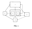
- FIG. 1 shows components of the present invention.
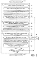
- Figure 2 is a flowchart of the ablation planning and navigation system with feedback.
- exemplary embodiments of the present disclosure are described with respect to ablative therapy of a human. It should be understood by one of ordinary skill in the art that the exemplary embodiments of the present disclosure can be applied to other types of ablative therapy and other portions of the body, whether human or animal. The use of the method and system of the exemplary embodiments of the present disclosure can be adapted for application to other types of applicators.
- a feedback system for integrating interventional planning and navigation of ablation treatment can include a planning component (1) for planning radiofrequency ablation (RFA) therapy (or other ablative therapy including, but not limited to, cryo-ablation, microwave ablation, ultrasound ablation, or other thermal or non-thermal ablation), and which is capable of creating an initial treatment plan, and adjusting the treatment plan to take into account data received during a procedure and transferring the treatment plan to a navigation component.
- RFA radiofrequency ablation
- the system includes a navigation system component (2) to guide an ablation probe (6) to target locations via a graphical user interface (7); and a feedback sub-system (3) for determining actual ablation probe positions/orientations and actual ablation size/shape via imaging (4) and/or tracking (5) systems, and enabling exchange of information between the navigation component (2) and the planning component (1).
- the planning system component (1) can allow computation of a composite ablation consisting of a plurality of optimal individual ablation positions/orientations based on input of a desired planned target volume (PTV) to be covered, and estimated or known individual ablation sizes.
- the desired composite ablation volume is comprised of a plurality of ablations that together will treat the entire PTV.
- the planning system component (1) utilizes image data acquired from the imaging component (4) of the system. Inputs to the planning component (1) can include a baseline image of the tissue to be treated from the imaging component (4), PTV segmentation, desired individual ablation shape and size, and desired skin entry point(s) selected by the treating physician.
- the planning system component (1) can calculate an initial plan for treatment, which is a 3-D mapping of the optimal number of ablations that will cover the PTV.
- the planning system component also has the capability to use feedback about the actual ablation positions or sizes, or actual tumor location relative to the ablation probe, to update or refine the plan iteratively.
- a plan includes the optimal number of ablations that will completely treat/ablate the PTV, the 3D location of each of those individual ablations, and the orientation of each of those individual ablations.
- the 3D location of an ablation is referred to as a planned target location in the navigation component (2).
- the planning system component (1) can output the plan information to a graphical user interface (GUI) (7) for use by a physician operator.
- GUI graphical user interface
- the navigation system component (2) with monitoring and feedback capability which provides visual guidance via a GUI (7) to assist in delivering an ablation probe (6) to any of the planned target locations can determine the actual ablation probe position (via the imaging component (4) or tracking sub-system(5)), the actual individual ablation size/shape (via the imaging component (4) when using e.g. ultrasound elastography), and the actual tumor location relative to the ablation probe (via imaging).
- Imaging means (4) may include handheld imaging devices, static imaging devices, etc. Other factors that may affect the execution of the procedure such as blood flow/perfusion near the ablation site, which can serve as a "heat sink” and thereby reduce the size of or deform the individual ablation shape can also be monitored.
- the navigation system component (2) registers the baseline image with the ablation probe and treatment images using the tracking system (5), and then provides guidance to the physician for guiding the needle to any of the target locations for ablation.
- the feedback component sub-system (3) can allow the navigation system component (2) to provide information about the ablation probe position and orientation, ablation size/shape, local perfusion, or other information obtained during the execution of one ablation to the planning system component.
- the feedback component (3) allows the planning system component (1) to receive any such information relevant for procedure planning from the navigation system component (2), to use it to update and refine the treatment plan iteratively, and to send the updated plan back to the navigation system component to guide subsequent ablations in an iterative process.
- the method starts at step 10 with acquisition of baseline images prior to treatment.
- the planning system component (1) allows determination of optimal ablation positions to treat an arbitrarily sized or shaped tumor with multiple, overlapping ablations, such as by RFA ablation.
- This component requires input and visualization of an image data set (the "baseline image") and the ability to segment a tumor or PTV, or to import the segmentation from elsewhere.
- the planning system component segments the baseline PTV 0 in the baseline image at step 12.
- the planning system component can also determine the skin entry point p E for the ablation needle at step 14, or this can be entered into the system after having been determined by a physician.
- GUI graphical user interface
- the physician operator can use the navigation system to guide the ablation needle to the skin entry point p E (step 22).
- the operator can then select one of the ablation centers p k,i as the target (step 24).
- the navigation component (2) and feedback component sub-system (3) comprise a system to guide the ablation electrode insertion to the specified target location p k,i inside the human body (step 26).
- One suitable guiding system is the commercially available Traxtal PercuNav TM system, which uses electromagnetic tracking to register the electrode position with a medical image of the patient.
- the navigation component (2) uses the same "baseline image" as the planning component (1), or can use a different image that is spatially registered with the baseline image.
- the navigation system component (2) has the ability to import an entire treatment plan from the planning component (1), consisting of multiple individual ablation locations, and potentially individual skin entry points for each of the ablation electrode placements.
- the navigation component will visualize the treatment plan by rendering the segmented PTV and the planned ablation locations in 3D, and will provide a GUI for the physician to select which of the planned target locations are to be executed next.
- the navigation system component (2) provides guidance of the electrode (6) to the planned skin entry point first, and from there to the planned ablation position. In case the physician selects a next target location that is near to the current ablation target, the system can provide guidance of the ablation probe electrode (6) directly to the next target without requiring complete retraction of the needle.
- the navigation system component (2) also contains a feedback sub-system (3) to obtain feedback about the actual electrode position/orientation during an ablation, and about the actual ablation size/shape and tumor location/orientation .
- the feedback sub-system (3) has the ability to provide that feedback to the planning component (1) for iterative refinement of the plan for subsequent ablations.
- the feedback subsystem (3) uses the imaging system (4) and/or tracking system (5) to determine the actual ablation center position p actual (step 28).
- the position feedback p actual can be obtained in any suitable way, such as the following:
- the combined information can be used to update/improve the coordinate registration between the image and tracking systems, thus allowing more accurate navigation in subsequent iterative steps.
- Ablation size/shape feedback to determine the actual ablation shape S actual can be obtained in the following way: A three-dimensional (3D) medical image of the ablation area (e.g. CT, MRI, ultrasound) that provides adequate grayscale-contrast to allow delineation of the ablation zone can be obtained during or shortly after an ablation, and the image transferred to the navigation component.
- a GUI can be provided for the user to measure the size/shape of the ablation in one or several dimensions, based on the visible ablation contrast in the image.
- an automated or semi-automated algorithm can extract the shape of the ablation with limited or no user input
- the size/shape information is transferred to the planning component in order to update the plan for the remaining ablations with the measured ablation size/shape as the new ablation size/shape estimate.
- the feedback component can be use to detect any changes in the tumor location e.g. due to patient motion including respiratory motion, by comparing the initial tumor location in the baseline image with the actual tumor location in the most recent image obtained by the imaging component (4).
- the location of the PTV is then updated according to the transformation T describing the change in tumor location: (step 31) PTV k ⁇ T * PTV k
- the planning component (1) receives the feedback information and updates the plan for the remainder of the procedure if the measured position or ablation size/shape deviate from the planned/assumed position and size/shape.
- the invention can be used with RFA ablation, cryo-ablation, microwave ablation, ultrasound ablation, or other thermal or non-thermal ablation, and is not intended to be limited in this regard.
- the invention can be realized in hardware, software, or a combination of hardware and software.
- the invention can be realized in a centralized fashion in one computer system, or in a distributed fashion where different elements are spread across several interconnected computer systems. Any kind of computer system or other apparatus adapted for carrying out the methods described herein is suited.
- a typical combination of hardware and software can be a general purpose computer system with a computer program that, when being loaded and executed, controls the computer system such that it carries out the methods described herein.
- the invention can be embedded in a computer program product.
- the computer program product can comprise a computer-readable storage medium in which is embedded a computer program comprising computer-executable code for directing a computing device or computer-based system to perform the various procedures, processes and methods described herein.
- Computer program in the present context means any expression, in any language, code or notation, of a set of instructions intended to cause a system having an information processing capability to perform a particular function either directly or after either or both of the following: a) conversion to another language, code or notation; b) reproduction in a different material form.
Description
- The present invention relates to the therapeutic arts, in particular to systems for integrating interventional planning and navigation, particularly for tissue ablation therapy.
- Interventional procedures such as radiofrequency ablation (RFA) have been performed in increasing numbers in recent years as an alternative to more invasive surgical procedures. During RFA, an electrode with an un-insulated tip is inserted into a tumor or lesion to be ablated under ultrasound, CT or MRI guidance. When the electrode is placed, a radiofrequency current is applied to the tip which creates tissue heating and cell death when tissue temperatures exceed 60° Celsius. In order to destroy tumors that are larger than the volume around the needle tip that is heated and destroyed in a single ablation, the needle tip needs to be repeatedly repositioned to ablate different parts of the tumor, partly overlapping with one another. This process needs to be repeated until the entire tumor is "covered" by the plurality of ablations, also referred to as the "composite ablation."
- There are two main difficulties associated with this process, which explain in part the relatively poor outcomes for ablations of large (>3cm) tumors:
- 1. The optimal three-dimensional (3D) positions of all individual ablations are difficult to plan manually/without computer assistance, where "optimal" refers to the use of the lowest number of overlapping ablations that fully covers the tumor plus surrounding safety margin (tumor + margin are also known as the Planned Target Volume, or PTV), and, for a given number of ablations, the placement of ablations that destroys the smallest volume of healthy, non-PTV tissue.
- 2. Even if the optimal positions of all individual ablations for one treatment (the Ablation Plan) were known, it is difficult to accurately execute a treatment plan, i.e. physically position the electrode in the desired locations, with conventional US, CT or MR guidance alone. Because of the uncertainty about the actual probe position during one ablation, this can affect the necessary placement of subsequent ablations.
- Additional problems arise because it is difficult to obtain patient-specific information about expected actual ablation sizes in order to create an optimal patient-specific plan, and so plans must use estimated ablation sizes which may not be accurate. Further, a slowdown of the procedure workflow is caused when trying to work with non-integrated planning and navigation feedback systems.
- RFA planning systems have been developed to address the first problem outlined above, but the systems are stand-alone and primarily used to visualize the desired set of ablations. Also, navigation systems have been developed to improve conventional image guidance by, for example, referencing the electrode position directly to the 3D coordinates of a pre-acquired medical image (e.g. a CT scan). However, these navigation systems exist independently of planning systems and are thus not useful in executing the procedure plan.
- Navigation systems have been suggested that assist in guiding an ablation electrode to a target location inside the human body, but the known systems are not integrated with RFA planning.
-
US Patent Application Publication No. 2007/0230757 to Trachtenberg et al. uses image processing to identify a target area for treatment of malignant prostate tissue, and to monitor the energy delivery during treatment. This publication does not deal with solving problems related to coverage of the tumor, nor in planning for treatment. -
US Patent Application Publication No. 2008/0033417 to Nields et al. relates to a thermal ablation system in which a controller compares actual temperature changes in the tissue during thermal ablation to a thermal ablation plan. No indication is given as to how applicator position is determined, and the thermal changes are not related to position feedback for revising the ablation plan.
The invention is defined in the appended claims. - In accordance with one aspect of the exemplary embodiments, a therapy planning and image guidance and navigation for an interventional procedure are combined in one system. The system includes an ablation probe for treating a tissue of interest with at least one of RF ablation, cryo-ablation, microwave ablation, ultrasound ablation, and other thermal or non-thermal ablation. A planning component is included for calculating a three-dimensional treatment plan including a plurality of desired ablation probe placement positions and orientations and a plurality of estimated ablation volumes to achieve a desired composite ablation volume that will treat the entire planned target volume (PTV). A navigation component comprising a tracking system is provided for spatially tracking the ablation probe and providing treatment instructions to an operator based on the three-dimensional treatment plan. An imaging component for obtaining planning and treatment images of the tissue to be treated is also provided. The system further includes a feedback component for determining actual probe position and orientation using the navigation component or imaging component, and for determining actual ablation volume based on treatment images provided by the imaging component, and feeding the actual probe position/orientation and actual ablation volume during treatment to the planning component. The planning component calculates a treatment iteration based on the executed treatment plan, the actual probe position/orientation and actual ablation volume, the treatment iteration resulting in at least one of a desired next probe position/orientation and a desired next ablation volume. The navigation component provides revised treatment instructions to the operator based on the treatment iteration until treatment is completed according to the desired planned target volume. The feedback component obtains information including imaging feedback and tracking feedback about the actual probe location, and provides the information including both the imaging feedback and tracking feedback to the planning component to update the three-dimensional treatment plan, wherein the feedback component further uses the information to update and improve registration between an imaging component and the tracking system. The planning component receives the information and updates the plan by subtracting a measured ablation size and shape at a measured ablation position from an initially segmented planned target volume, and computes a new plan for a remaining part of the planned target volume. The planning component utilizes center positions of individual ablations for calculating the three-dimensional treatment plan.
- Another aspect of the exemplary embodiments relates to a method of ablation therapy of tissue. The method includes calculating a three-dimensional treatment plan for a tissue of interest including a plurality of desired ablation probe placement positions and orientations and a plurality of estimated ablation volumes to achieve a desired composite ablation volume that will treat the entire planned target volume (PTV). The method further includes providing treatment instructions to an operator based on the three-dimensional treatment plan, and treating the tissue of interest with at least one of RF ablation, cryo-ablation, microwave ablation, ultrasound ablation, and other thermal or non-thermal ablation. Planning and treatment images of the tissue to be treated are obtained, and actual probe position/orientation and actual ablation volume based on the treatment images are determined. A treatment iteration is calculated based on the treatment plan, the actual probe position/orientation and actual ablation volume, the treatment iteration resulting in at least one of a desired next probe position and a desired next ablation volume. The method further includes providing revised treatment instructions to the operator based on the treatment iteration and continuing iterative treatment until treatment is completed according to the desired planned target volume.
- In another aspect of the exemplary arrangements of the invention, a control system for ablation treatment of tissue includes a planning component for calculating a three-dimensional treatment plan including a plurality of desired ablation probe placement positions and orientations and a plurality of estimated ablation volumes to achieve a desired planned target volume that will treat an entire tissue volume of interest. An imaging component for obtaining treatment images of the tissue to be treated is also included, along with a feedback component for calculating actual probe position/orientation of a probe and actual ablation volume based on treatment images provided by the imaging component, and feeding the actual probe position/orientation and actual ablation volume during treatment to the planning component. The planning component maps the actual probe location and actual ablation volume to the three-dimensional plan and calculates a treatment iteration based on the treatment plan, the actual probe position/orientation and actual ablation volume, the treatment iteration resulting in at least one of a desired next probe position/orientation and a desired next ablation volume. The feedback component obtains information including imaging feedback and tracking feedback about the actual probe location, and provides the information including both the imaging feedback and tracking feedback to the planning component to update the three-dimensional treatment plan, wherein the feedback component further uses the information to update and improve registration between the imaging component and a tracking system for spatially tracking the ablation probe. The planning component receives the information and updates the plan by subtracting a measured ablation size and shape at a measured ablation position from an initially segmented planned target volume, and computes a new plan for a remaining part of the planned target volume. The planning component utilizes center positions of individual ablations for calculating the three-dimensional treatment plan.
- Many interventional procedures such as radiofrequency ablation (RFA) are commonly carried out without dedicated or computer-assisted planning, and with only basic image guidance. By combining and integrating procedure planning and navigation, and by providing feedback from the navigation component back to the planning component about actual electrode position and ablation size/shape, complex procedures can be carried out more accurately, efficiently, and potentially with better clinical outcomes. Both procedure planning and advanced image guidance and navigation can improve the accuracy of procedure execution, yielding several advantages such as improved patient outcomes, reduced use of ionizing radiation, and reduced cost.
- The above-described and other features and advantages of the present disclosure will be appreciated and understood by those skilled in the art from the following detailed description, drawings, and appended claims.
-
Figure 1 shows components of the present invention. -
Figure 2 is a flowchart of the ablation planning and navigation system with feedback. - The exemplary embodiments of the present disclosure are described with respect to ablative therapy of a human. It should be understood by one of ordinary skill in the art that the exemplary embodiments of the present disclosure can be applied to other types of ablative therapy and other portions of the body, whether human or animal. The use of the method and system of the exemplary embodiments of the present disclosure can be adapted for application to other types of applicators.
- Referring to
Figure 1 , a feedback system for integrating interventional planning and navigation of ablation treatment can include a planning component (1) for planning radiofrequency ablation (RFA) therapy (or other ablative therapy including, but not limited to, cryo-ablation, microwave ablation, ultrasound ablation, or other thermal or non-thermal ablation), and which is capable of creating an initial treatment plan, and adjusting the treatment plan to take into account data received during a procedure and transferring the treatment plan to a navigation component. The system includes a navigation system component (2) to guide an ablation probe (6) to target locations via a graphical user interface (7); and a feedback sub-system (3) for determining actual ablation probe positions/orientations and actual ablation size/shape via imaging (4) and/or tracking (5) systems, and enabling exchange of information between the navigation component (2) and the planning component (1). By combining and integrating procedure planning and navigation, and by providing feedback from the navigation component (2) back to the planning component (1) about actual ablation probe position and actual ablation size, complex procedures can be carried out more accurately, efficiently, and potentially with better clinical outcomes. - The planning system component (1) can allow computation of a composite ablation consisting of a plurality of optimal individual ablation positions/orientations based on input of a desired planned target volume (PTV) to be covered, and estimated or known individual ablation sizes. The desired composite ablation volume is comprised of a plurality of ablations that together will treat the entire PTV. The planning system component (1) utilizes image data acquired from the imaging component (4) of the system. Inputs to the planning component (1) can include a baseline image of the tissue to be treated from the imaging component (4), PTV segmentation, desired individual ablation shape and size, and desired skin entry point(s) selected by the treating physician. The planning system component (1) can calculate an initial plan for treatment, which is a 3-D mapping of the optimal number of ablations that will cover the PTV. The planning system component also has the capability to use feedback about the actual ablation positions or sizes, or actual tumor location relative to the ablation probe, to update or refine the plan iteratively. A plan includes the optimal number of ablations that will completely treat/ablate the PTV, the 3D location of each of those individual ablations, and the orientation of each of those individual ablations. The 3D location of an ablation is referred to as a planned target location in the navigation component (2). The planning system component (1) can output the plan information to a graphical user interface (GUI) (7) for use by a physician operator.
- The navigation system component (2) with monitoring and feedback capability, which provides visual guidance via a GUI (7) to assist in delivering an ablation probe (6) to any of the planned target locations can determine the actual ablation probe position (via the imaging component (4) or tracking sub-system(5)), the actual individual ablation size/shape (via the imaging component (4) when using e.g. ultrasound elastography), and the actual tumor location relative to the ablation probe (via imaging). Imaging means (4) may include handheld imaging devices, static imaging devices, etc. Other factors that may affect the execution of the procedure such as blood flow/perfusion near the ablation site, which can serve as a "heat sink" and thereby reduce the size of or deform the individual ablation shape can also be monitored. The navigation system component (2) registers the baseline image with the ablation probe and treatment images using the tracking system (5), and then provides guidance to the physician for guiding the needle to any of the target locations for ablation.
- The feedback component sub-system (3) can allow the navigation system component (2) to provide information about the ablation probe position and orientation, ablation size/shape, local perfusion, or other information obtained during the execution of one ablation to the planning system component. The feedback component (3) allows the planning system component (1) to receive any such information relevant for procedure planning from the navigation system component (2), to use it to update and refine the treatment plan iteratively, and to send the updated plan back to the navigation system component to guide subsequent ablations in an iterative process.
- Referring to
Figure 2 , a detailed flowchart of the ablation planning and navigation method with feedback is illustrated. The method starts atstep 10 with acquisition of baseline images prior to treatment. - The planning system component (1) allows determination of optimal ablation positions to treat an arbitrarily sized or shaped tumor with multiple, overlapping ablations, such as by RFA ablation. This component requires input and visualization of an image data set (the "baseline image") and the ability to segment a tumor or PTV, or to import the segmentation from elsewhere. The planning system component segments the baseline PTV0 in the baseline image at
step 12. The planning system component can also determine the skin entry point pE for the ablation needle atstep 14, or this can be entered into the system after having been determined by a physician. - The planning system component at a minimum also requires as an input the estimated shape and size So of an individual ablation (step 16). Based on these inputs, the component can set the current (initial) iteration as k=0 (step 18), and allows creation of a treatment plan either manually or automatically (step 20). For manual plan creation, the component visualizes the PTV and provides a graphical user interface (GUI) for the user to manually place one or more ablations (each modeled by an estimated shape and size So) to cover the PTV. For automatic plan creation, the planning system component (1) determines an optimal set of ablation positions Pk, based on the assumed/estimated individual ablation size and shape:

- If the ablation probe needle has not (yet) been inserted, the physician operator can use the navigation system to guide the ablation needle to the skin entry point pE (step 22). The operator can then select one of the ablation centers pk,i as the target (step 24). The navigation component (2) and feedback component sub-system (3) comprise a system to guide the ablation electrode insertion to the specified target location pk,i inside the human body (step 26). One suitable guiding system is the commercially available Traxtal PercuNav™ system, which uses electromagnetic tracking to register the electrode position with a medical image of the patient. The navigation component (2) uses the same "baseline image" as the planning component (1), or can use a different image that is spatially registered with the baseline image.
- The navigation system component (2) has the ability to import an entire treatment plan from the planning component (1), consisting of multiple individual ablation locations, and potentially individual skin entry points for each of the ablation electrode placements. The navigation component will visualize the treatment plan by rendering the segmented PTV and the planned ablation locations in 3D, and will provide a GUI for the physician to select which of the planned target locations are to be executed next. For each ablation, the navigation system component (2) provides guidance of the electrode (6) to the planned skin entry point first, and from there to the planned ablation position. In case the physician selects a next target location that is near to the current ablation target, the system can provide guidance of the ablation probe electrode (6) directly to the next target without requiring complete retraction of the needle.
- Furthermore, the navigation system component (2) also contains a feedback sub-system (3) to obtain feedback about the actual electrode position/orientation during an ablation, and about the actual ablation size/shape and tumor location/orientation . The feedback sub-system (3) has the ability to provide that feedback to the planning component (1) for iterative refinement of the plan for subsequent ablations. The feedback subsystem (3) uses the imaging system (4) and/or tracking system (5) to determine the actual ablation center position pactual (step 28).
- The position feedback pactual can be obtained in any suitable way, such as the following:
- 1. For procedures performed on a CT or MRI scanner (independent of the use of a spatial tracking system), a confirmation CT or MRI image can be obtained with the electrode in position for an ablation, and the scan transferred to the navigation component. The confirmation scan is registered with and superimposed on the baseline navigation image. The electrode tip will be identified in the confirmation scan, and a GUI allows the user to transfer that position to the baseline image and send it to the planning component.
- 2. For procedures performed with spatial tracking of the ablation electrode tip (e.g. using the Traxtal PercuNav™ system), a GUI is provided for the user to initiate the conversion of the electrode's tracking coordinates into baseline image coordinates using the navigation component's registration transformation, and to send the position to the planning component.
- Optionally, when the position feedback is obtained with both imaging and tracking, the combined information can be used to update/improve the coordinate registration between the image and tracking systems, thus allowing more accurate navigation in subsequent iterative steps.
- Ablation size/shape feedback to determine the actual ablation shape Sactual (step 30) can be obtained in the following way: A three-dimensional (3D) medical image of the ablation area (e.g. CT, MRI, ultrasound) that provides adequate grayscale-contrast to allow delineation of the ablation zone can be obtained during or shortly after an ablation, and the image transferred to the navigation component. A GUI can be provided for the user to measure the size/shape of the ablation in one or several dimensions, based on the visible ablation contrast in the image. Alternatively, an automated or semi-automated algorithm can extract the shape of the ablation with limited or no user input The size/shape information is transferred to the planning component in order to update the plan for the remaining ablations with the measured ablation size/shape as the new ablation size/shape estimate.
- Optionally, the feedback component can be use to detect any changes in the tumor location e.g. due to patient motion including respiratory motion, by comparing the initial tumor location in the baseline image with the actual tumor location in the most recent image obtained by the imaging component (4). The location of the PTV is then updated according to the transformation T describing the change in tumor location: (step 31)

- The planning component (1) receives the feedback information and updates the plan for the remainder of the procedure if the measured position or ablation size/shape deviate from the planned/assumed position and size/shape. In particular, the planning component can subtract the measured ablation size/shape at the measured ablation position from the initially segmented PTVk, and compute a new plan for the remaining PTVk+1 (step 32):

- The iterative process will continue until the entire PTV volume is ablated. This can be determined by computing the volume (PTV k+1) = 0 (step 34). If the PTV volume is not zero, i.e. there are a finite number of unablated voxels in the PTV, the plan iteration is followed so that k→k+1, Sk = Sactual (step 36). If the PTV volume is zero, such that no non-ablated PTV remains, the treatment procedure ends at
step 38. In specific instances, the physician may exercise his/her judgment to terminate the iterative process in case the remaining volume of the PTV is relatively small and visualization of this small PTV in the GUI interface of the planning component provides confidence in making that decision. - It should be appreciated that the invention can be used with RFA ablation, cryo-ablation, microwave ablation, ultrasound ablation, or other thermal or non-thermal ablation, and is not intended to be limited in this regard.
- The invention, including the steps of the methodologies described above, can be realized in hardware, software, or a combination of hardware and software. The invention can be realized in a centralized fashion in one computer system, or in a distributed fashion where different elements are spread across several interconnected computer systems. Any kind of computer system or other apparatus adapted for carrying out the methods described herein is suited. A typical combination of hardware and software can be a general purpose computer system with a computer program that, when being loaded and executed, controls the computer system such that it carries out the methods described herein.
- The invention, including the steps of the methodologies described above, can be embedded in a computer program product. The computer program product can comprise a computer-readable storage medium in which is embedded a computer program comprising computer-executable code for directing a computing device or computer-based system to perform the various procedures, processes and methods described herein. Computer program in the present context means any expression, in any language, code or notation, of a set of instructions intended to cause a system having an information processing capability to perform a particular function either directly or after either or both of the following: a) conversion to another language, code or notation; b) reproduction in a different material form.
- The illustrations of embodiments described herein are intended to provide a general understanding of the structure of various embodiments, and they are not intended to serve as a complete description of all the elements and features of apparatus and systems that might make use of the structures described herein. Many other embodiments will be apparent to those of skill in the art upon reviewing the above description. Other embodiments may be utilized and derived therefrom, such that structural and logical substitutions and changes may be made without departing from the scope of this disclosure. Figures are also merely representational and may not be drawn to scale. Certain proportions thereof may be exaggerated, while others may be minimized. Accordingly, the specification and drawings are to be regarded in an illustrative rather than a restrictive sense.
- Thus, although specific embodiments have been illustrated and described herein, it should be appreciated that any arrangement calculated to achieve the same purpose may be substituted for the specific embodiments shown. This disclosure is intended to cover any and all adaptations or variations of various embodiments. Combinations of the above embodiments, and other embodiments not specifically described herein, will be apparent to those of skill in the art upon reviewing the above description. Therefore, it is intended that the disclosure not be limited to the particular embodiment(s) disclosed as the best mode contemplated for carrying out this invention, but that the invention will include all embodiments falling within the scope of the appended claims.
Claims (11)
- A system for ablation treatment of tissue, comprising:an ablation probe (6) for treating a tissue of interest with at least one of RF ablation, cryo-ablation, microwave ablation, ultrasound ablation, or other thermal or non-thermal ablation;a planning component (1) for calculating a three-dimensional treatment plan including a plurality of desired ablation probe placement positions and orientations and a plurality of estimated ablation volumes to achieve a composite ablation volume that will treat the entire planned target volume (PTV);a navigation component (2) comprising a tracking system (5) for spatially tracking the ablation probe and providing treatment instructions to an operator based on the three-dimensional treatment plan;an imaging component (4) for obtaining planning and treatment images of the tissue to be treated; anda feedback component (3) for determining actual probe position and orientation using the navigation component or imaging component, and for determining actual ablation volume based on treatment images provided by the imaging component (4), and feeding the actual probe position and orientation and an actual ablation volume during treatment to the planning component (1),wherein the planning component (1) is configured to determine a treatment iteration based on the treatment plan, the actual probe position and orientation and an actual ablation volume, the treatment iteration including at least one of a desired next probe position and orientation and a desired next ablation volume,wherein the navigation component (2) is configured to provide revised treatment instructions to the operator based on the treatment iteration until treatment is completed according to the desired planned target volume,wherein the the feedback component (3) is configured to obtain information including imaging feedback and tracking feedback about the actual probe location, and to provide the information including both the imaging feedback and tracking feedback to the planning component (1) to update the three-dimensional treatment plan, wherein the feedback component is further configured to use the information to update and improve registration between the imaging component (4) and the tracking system (5),wherein the planning component (1) is configured to receive the information and update the plan by subtracting a measured ablation size and shape at a measured ablation position from an initially segmented planned target volume, and compute a new plan for a remaining part of the planned target volume, andwherein the planning component (1) is configured to utilize center positions of individual ablations for calculating the three-dimensional treatment plan.
- The system according to claim 1, wherein the imaging component (4) is configured to provide at least one of ultrasound, CT, MR imaging, ultrasound elastography and Doppler ultrasound, and wherein the navigation component is configured to provide at least one of spatial tracking coordinates of the ablation probe and imaging, based on at least one of electromagnetic, optical, acoustic and magnetic tracking sensors.
- The system according to claim 1, further comprising a user interface for displaying the treatment iteration instructions from the navigation component (2) for access by a human operator.
- The system according to claim 1, wherein the feedback component (3) is configured to calculate information at least one of local micro-vascular perfusion and macro-vascular blood flow parameters near the ablation site and provide the information to the planning component (1) for incorporation into the treatment iteration.
- The system according to claim 1, wherein the feedback component (3) is configured to register treatment images with planning images to calculate further information including at least one of the actual probe position and orientation and tumor position and orientation, and use the information to update the three-dimensional treatment plan.
- A control system for ablation treatment of tissue, comprising:a planning component (1) for calculating a three-dimensional treatment plan including a plurality of desired ablation probe placement positions and orientations and a plurality of estimated ablation volumes to achieve a desired planned target volume that will treat an entire tissue volume of interest;an imaging component (4) for obtaining treatment images of the tissue to be treated; anda feedback component (3) for calculating actual probe position and orientation of a probe and actual ablation volume based on treatment images provided by the imaging component, and feeding the actual probe position and orientation and an actual ablation volume during treatment to the planning component,wherein the planning component (1) is configured to map the actual probe location and actual ablation volume to the three-dimensional plan and calculate a treatment iteration based on the treatment plan, the actual probe position and orientation and an actual ablation volume, the treatment iteration including at least one of a desired next probe position and orientation and a desired next ablation volume,wherein the feedback component (3) is configured to obtain information including imaging feedback and tracking feedback about the actual probe location, and to provide the information including both the imaging feedback and tracking feedback to the planning component (1) to update the three-dimensional treatment plan, wherein the feedback component is further configured to use the information to update and improve registration between the imaging component (4) and a tracking system (5) for spatially tracking the ablation probe,wherein the planning component (1) is configured to receive the information and update the plan by subtracting a measured ablation size and shape at a measured ablation position from an initially segmented planned target volume, and compute a new plan for a remaining part of the planned target volume, andwherein the planning component (1) is configured to utilize center positions of individual ablations for calculating the three-dimensional treatment plan.
- The control system according to claim 6, wherein the imaging component (4) is configured to provide at least one of ultrasound, CT, MR imaging, ultrasound elastography and Doppler ultrasound, and wherein the navigation component is configured to provide at least one of spatial tracking coordinates of the therapy probe and imaging device based on at least one of electromagnetic, optical, acoustic and magnetic sensors.
- The control system according to claim 6, wherein the control system is configured to deliver the next treatment ablation according to the calculated treatment iteration by at least one of moving the ablation probe to the desired next probe position and ablating the next desired ablation volume.
- The control system according to claim 6, wherein the feedback component (3) is configured to calculate at least one of local micro-vascular perfusion and macro-vascular blood flow parameters near the ablation site and provide this information to the planning component for incorporation into the treatment iteration.
- The control system according to claim 6, wherein the feedback component (3) is configured to register treatment images with planning images to calculate at least one of the actual probe position and orientation and tumor position and orientation, and use the information to update the three-dimensional treatment plan.
- The control system according to claim 7, wherein the imaging component (4) is configured to utilize a contrast medium.
Applications Claiming Priority (2)
| Application Number | Priority Date | Filing Date | Title |
|---|---|---|---|
| US11946408P | 2008-12-03 | 2008-12-03 | |
| PCT/IB2009/054923 WO2010064154A1 (en) | 2008-12-03 | 2009-11-05 | Feedback system for integrating interventional planning and navigation |
Publications (2)
| Publication Number | Publication Date |
|---|---|
| EP2373241A1 EP2373241A1 (en) | 2011-10-12 |
| EP2373241B1 true EP2373241B1 (en) | 2015-01-07 |
Family
ID=41526310
Family Applications (1)
| Application Number | Title | Priority Date | Filing Date |
|---|---|---|---|
| EP09760315.3A Active EP2373241B1 (en) | 2008-12-03 | 2009-11-05 | Feedback system for integrating interventional planning and navigation |
Country Status (5)
| Country | Link |
|---|---|
| US (1) | US9144461B2 (en) |
| EP (1) | EP2373241B1 (en) |
| JP (1) | JP5685546B2 (en) |
| CN (1) | CN102238921B (en) |
| WO (1) | WO2010064154A1 (en) |
Cited By (25)
| Publication number | Priority date | Publication date | Assignee | Title |
|---|---|---|---|---|
| US9598691B2 (en) | 2008-04-29 | 2017-03-21 | Virginia Tech Intellectual Properties, Inc. | Irreversible electroporation to create tissue scaffolds |
| US9867652B2 (en) | 2008-04-29 | 2018-01-16 | Virginia Tech Intellectual Properties, Inc. | Irreversible electroporation using tissue vasculature to treat aberrant cell masses or create tissue scaffolds |
| US10117707B2 (en) | 2008-04-29 | 2018-11-06 | Virginia Tech Intellectual Properties, Inc. | System and method for estimating tissue heating of a target ablation zone for electrical-energy based therapies |
| US10154874B2 (en) | 2008-04-29 | 2018-12-18 | Virginia Tech Intellectual Properties, Inc. | Immunotherapeutic methods using irreversible electroporation |
| US10238447B2 (en) | 2008-04-29 | 2019-03-26 | Virginia Tech Intellectual Properties, Inc. | System and method for ablating a tissue site by electroporation with real-time monitoring of treatment progress |
| US10245105B2 (en) | 2008-04-29 | 2019-04-02 | Virginia Tech Intellectual Properties, Inc. | Electroporation with cooling to treat tissue |
| US10272178B2 (en) | 2008-04-29 | 2019-04-30 | Virginia Tech Intellectual Properties Inc. | Methods for blood-brain barrier disruption using electrical energy |
| US10292755B2 (en) | 2009-04-09 | 2019-05-21 | Virginia Tech Intellectual Properties, Inc. | High frequency electroporation for cancer therapy |
| US10471254B2 (en) | 2014-05-12 | 2019-11-12 | Virginia Tech Intellectual Properties, Inc. | Selective modulation of intracellular effects of cells using pulsed electric fields |
| US10470822B2 (en) | 2008-04-29 | 2019-11-12 | Virginia Tech Intellectual Properties, Inc. | System and method for estimating a treatment volume for administering electrical-energy based therapies |
| US10694972B2 (en) | 2014-12-15 | 2020-06-30 | Virginia Tech Intellectual Properties, Inc. | Devices, systems, and methods for real-time monitoring of electrophysical effects during tissue treatment |
| US10702326B2 (en) | 2011-07-15 | 2020-07-07 | Virginia Tech Intellectual Properties, Inc. | Device and method for electroporation based treatment of stenosis of a tubular body part |
| US11254926B2 (en) | 2008-04-29 | 2022-02-22 | Virginia Tech Intellectual Properties, Inc. | Devices and methods for high frequency electroporation |
| US11272979B2 (en) | 2008-04-29 | 2022-03-15 | Virginia Tech Intellectual Properties, Inc. | System and method for estimating tissue heating of a target ablation zone for electrical-energy based therapies |
| US11311329B2 (en) | 2018-03-13 | 2022-04-26 | Virginia Tech Intellectual Properties, Inc. | Treatment planning for immunotherapy based treatments using non-thermal ablation techniques |
| US11382681B2 (en) | 2009-04-09 | 2022-07-12 | Virginia Tech Intellectual Properties, Inc. | Device and methods for delivery of high frequency electrical pulses for non-thermal ablation |
| US11453873B2 (en) | 2008-04-29 | 2022-09-27 | Virginia Tech Intellectual Properties, Inc. | Methods for delivery of biphasic electrical pulses for non-thermal ablation |
| US11607537B2 (en) | 2017-12-05 | 2023-03-21 | Virginia Tech Intellectual Properties, Inc. | Method for treating neurological disorders, including tumors, with electroporation |
| US11638603B2 (en) | 2009-04-09 | 2023-05-02 | Virginia Tech Intellectual Properties, Inc. | Selective modulation of intracellular effects of cells using pulsed electric fields |
| US11707629B2 (en) | 2009-05-28 | 2023-07-25 | Angiodynamics, Inc. | System and method for synchronizing energy delivery to the cardiac rhythm |
| US11723710B2 (en) | 2016-11-17 | 2023-08-15 | Angiodynamics, Inc. | Techniques for irreversible electroporation using a single-pole tine-style internal device communicating with an external surface electrode |
| US11779395B2 (en) | 2011-09-28 | 2023-10-10 | Angiodynamics, Inc. | Multiple treatment zone ablation probe |
| US11925405B2 (en) | 2018-03-13 | 2024-03-12 | Virginia Tech Intellectual Properties, Inc. | Treatment planning system for immunotherapy enhancement via non-thermal ablation |
| US11931096B2 (en) | 2010-10-13 | 2024-03-19 | Angiodynamics, Inc. | System and method for electrically ablating tissue of a patient |
| US11950835B2 (en) | 2020-06-29 | 2024-04-09 | Virginia Tech Intellectual Properties, Inc. | Cycled pulsing to mitigate thermal damage for multi-electrode irreversible electroporation therapy |
Families Citing this family (89)
| Publication number | Priority date | Publication date | Assignee | Title |
|---|---|---|---|---|
| US6994706B2 (en) | 2001-08-13 | 2006-02-07 | Minnesota Medical Physics, Llc | Apparatus and method for treatment of benign prostatic hyperplasia |
| US8556888B2 (en) * | 2006-08-04 | 2013-10-15 | INTIO, Inc. | Methods and apparatuses for performing and monitoring thermal ablation |
| US9232959B2 (en) | 2007-01-02 | 2016-01-12 | Aquabeam, Llc | Multi fluid tissue resection methods and devices |
| RU2492884C2 (en) * | 2007-11-15 | 2013-09-20 | Конинклейке Филипс Электроникс, Н.В. | Method and apparatus for tracking position of therapeutic ultrasonic sensor |
| EP2259742B1 (en) | 2008-03-06 | 2020-01-01 | AquaBeam LLC | Tissue ablation and cautery with optical energy carried in fluid stream |
| WO2010019481A1 (en) | 2008-08-11 | 2010-02-18 | Conceptx Medical, Inc. | Systems and methods for treating dyspnea, including via electrical afferent signal blocking |
| US8475441B2 (en) * | 2008-12-23 | 2013-07-02 | Cryomedix, Llc | Isotherm-based tissue ablation control system |
| WO2014127242A2 (en) | 2013-02-14 | 2014-08-21 | Procept Biorobotics Corporation | Aquablation aquabeam eye surgery methods and apparatus |
| US9277969B2 (en) | 2009-04-01 | 2016-03-08 | Covidien Lp | Microwave ablation system with user-controlled ablation size and method of use |
| US9895189B2 (en) | 2009-06-19 | 2018-02-20 | Angiodynamics, Inc. | Methods of sterilization and treating infection using irreversible electroporation |
| CN104246855B (en) | 2009-06-29 | 2017-08-15 | 皇家飞利浦电子股份有限公司 | Tumour ablation training system |
| US10448837B2 (en) | 2010-08-23 | 2019-10-22 | Knonklijke Philips N.V. | Mapping system and method for medical procedures |
| US20130072784A1 (en) * | 2010-11-10 | 2013-03-21 | Gnanasekar Velusamy | Systems and methods for planning image-guided interventional procedures |
| WO2012066449A1 (en) * | 2010-11-18 | 2012-05-24 | Koninklijke Philips Electronics N.V. | System and method for probabilistic ablation planning |
| US9079011B2 (en) * | 2011-01-03 | 2015-07-14 | Wisconsin Alumni Research Foundation | Microwave hyperthermia treatment system |
| KR20120096265A (en) * | 2011-02-22 | 2012-08-30 | 삼성전자주식회사 | Apparatus and method for tracking tumor for ultrasound therapy, ultrasound therapy system |
| US10736696B2 (en) * | 2011-06-07 | 2020-08-11 | Koninklijke Philips N.V. | Rotational position determination apparatus |
| US9757201B2 (en) | 2011-07-11 | 2017-09-12 | Koninklijke Philips N.V. | Energy application planning apparatus |
| CN103717167B (en) * | 2011-07-28 | 2017-06-27 | 皇家飞利浦有限公司 | Ablation planning system |
| US9724164B2 (en) * | 2011-08-26 | 2017-08-08 | EBM Corporation | Blood-vessel bloodstream simulation system, method therefor, and computer software program |
| DK2753243T3 (en) | 2011-09-06 | 2016-07-04 | Ezono Ag | Imaging probe and method for obtaining positional and / or information information |
| CN104203078B (en) | 2012-02-29 | 2018-04-20 | 普罗赛普特生物机器人公司 | The cutting tissue of automated image guiding and processing |
| WO2013149034A2 (en) * | 2012-03-28 | 2013-10-03 | Cibiem, Inc. | Carotid body modulation planning and assessment |
| JP5809770B2 (en) * | 2012-04-12 | 2015-11-11 | コーニンクレッカ フィリップス エヌ ヴェKoninklijke Philips N.V. | High density focused ultrasound for heating a target area larger than the electron focus area |
| EP2840993A4 (en) | 2012-04-24 | 2016-03-30 | Cibiem Inc | Endovascular catheters and methods for carotid body ablation |
| US20130316318A1 (en) * | 2012-05-22 | 2013-11-28 | Vivant Medical, Inc. | Treatment Planning System |
| US9439627B2 (en) * | 2012-05-22 | 2016-09-13 | Covidien Lp | Planning system and navigation system for an ablation procedure |
| EP2854681A4 (en) | 2012-06-01 | 2016-02-17 | Cibiem Inc | Percutaneous methods and devices for carotid body ablation |
| US9402677B2 (en) | 2012-06-01 | 2016-08-02 | Cibiem, Inc. | Methods and devices for cryogenic carotid body ablation |
| EP2676702A1 (en) | 2012-06-21 | 2013-12-25 | Koninklijke Philips N.V. | Improved high intensity focused ultrasound targeting |
| CN104602638B (en) * | 2012-06-27 | 2017-12-19 | 曼特瑞斯医药有限责任公司 | System for influenceing to treat tissue |
| WO2014005155A1 (en) | 2012-06-30 | 2014-01-03 | Cibiem, Inc. | Carotid body ablation via directed energy |
| US9993305B2 (en) | 2012-08-08 | 2018-06-12 | Ortoma Ab | Method and system for computer assisted surgery |
| US11096741B2 (en) * | 2012-10-10 | 2021-08-24 | Biosense Webster (Israel) Ltd. | Ablation power control based on contact force |
| US9846765B2 (en) | 2012-11-09 | 2017-12-19 | Siemens Healthcare Gmbh | System and method for patient specific modeling of liver tumor ablation |
| WO2014077396A1 (en) | 2012-11-16 | 2014-05-22 | 株式会社東芝 | Ultrasound diagnostic device and image processing method |
| US10588597B2 (en) | 2012-12-31 | 2020-03-17 | Intuitive Surgical Operations, Inc. | Systems and methods for interventional procedure planning |
| US10748438B2 (en) | 2013-02-26 | 2020-08-18 | Siemens Healthcare Gmbh | System and method for interactive patient specific simulation of radiofrequency ablation therapy |
| WO2014132209A1 (en) * | 2013-02-28 | 2014-09-04 | Koninklijke Philips N.V. | Segmentation of large objects from multiple three-dimensional views |
| GB201303917D0 (en) | 2013-03-05 | 2013-04-17 | Ezono Ag | System for image guided procedure |
| US9459087B2 (en) | 2013-03-05 | 2016-10-04 | Ezono Ag | Magnetic position detection system |
| US9257220B2 (en) | 2013-03-05 | 2016-02-09 | Ezono Ag | Magnetization device and method |
| CN104116523B (en) * | 2013-04-25 | 2016-08-03 | 深圳迈瑞生物医疗电子股份有限公司 | A kind of ultrasonic image analyzes system and the method for analysis thereof |
| WO2015035178A2 (en) * | 2013-09-06 | 2015-03-12 | Brigham And Women's Hospital, Inc. | System and method for a tissue resection margin measurement device |
| US10402535B2 (en) | 2014-02-26 | 2019-09-03 | Siemens Healthcare Gmbh | System and method for personalized computation of tissue ablation extent based on medical images |
| EP3116408B1 (en) | 2014-03-12 | 2018-12-19 | Cibiem, Inc. | Ultrasound ablation catheter |
| WO2015148378A1 (en) * | 2014-03-22 | 2015-10-01 | Endocare, Inc. | System and methods for ablation treatment planning and intraoperative position updates of ablation devices |
| US9589379B2 (en) | 2014-06-24 | 2017-03-07 | Siemens Healthcare Gmbh | System and method for visualization of cardiac changes under various pacing conditions |
| US20160000414A1 (en) | 2014-07-02 | 2016-01-07 | Covidien Lp | Methods for marking biopsy location |
| EP3164073B1 (en) | 2014-07-02 | 2020-07-01 | Covidien LP | System and method for detecting trachea |
| CA2953146A1 (en) | 2014-07-02 | 2016-01-07 | Covidien Lp | System and method for segmentation of lung |
| US9770216B2 (en) | 2014-07-02 | 2017-09-26 | Covidien Lp | System and method for navigating within the lung |
| WO2016004310A2 (en) | 2014-07-02 | 2016-01-07 | Covidien Lp | Real-time automatic registration feedback |
| US9754367B2 (en) | 2014-07-02 | 2017-09-05 | Covidien Lp | Trachea marking |
| US9603668B2 (en) | 2014-07-02 | 2017-03-28 | Covidien Lp | Dynamic 3D lung map view for tool navigation inside the lung |
| WO2016014949A1 (en) | 2014-07-24 | 2016-01-28 | Blake Robert C | System and method for cardiac ablation |
| US10643371B2 (en) | 2014-08-11 | 2020-05-05 | Covidien Lp | Treatment procedure planning system and method |
| US10354758B2 (en) | 2014-08-28 | 2019-07-16 | Siemens Healthcare Gmbh | System and method for patient-specific image-based simulation of atrial electrophysiology |
| CN105534593B (en) * | 2014-10-29 | 2019-04-23 | 深圳迈瑞生物医疗电子股份有限公司 | Intervention ablation simulation system and method |
| US10607738B2 (en) | 2015-05-15 | 2020-03-31 | University Health Network | System and method for minimally invasive thermal ablation treatment planning |
| US10986990B2 (en) | 2015-09-24 | 2021-04-27 | Covidien Lp | Marker placement |
| CN105286988A (en) * | 2015-10-12 | 2016-02-03 | 北京工业大学 | CT image-guided liver tumor thermal ablation needle location and navigation system |
| US10709352B2 (en) | 2015-10-27 | 2020-07-14 | Covidien Lp | Method of using lung airway carina locations to improve ENB registration |
| CN105286980A (en) * | 2015-11-18 | 2016-02-03 | 南京亿高微波系统工程有限公司 | Three-dimensional ablation system |
| AU2016370633A1 (en) * | 2015-12-14 | 2018-07-05 | Nuvasive, Inc. | 3D visualization during surgery with reduced radiation exposure |
| JP2019502472A (en) * | 2016-01-07 | 2019-01-31 | ウニヴェルジテート ベルン | Method and system for pause control ablation |
| CN105997245B (en) * | 2016-01-28 | 2018-04-06 | 杭州奥视图像技术有限公司 | A kind of method that the accurate analog radio frequency ablation techniques of tumour are covered using ellipsoid |
| US11419660B2 (en) * | 2016-02-09 | 2022-08-23 | Andrea Borsic | System and methods for ablation treatment of tissue |
| US11172821B2 (en) * | 2016-04-28 | 2021-11-16 | Medtronic Navigation, Inc. | Navigation and local thermometry |
| US10327853B2 (en) * | 2016-05-10 | 2019-06-25 | Covidien Lp | System and method of performing treatment along a lumen network |
| CN110831506A (en) * | 2017-05-04 | 2020-02-21 | 杰尼索尼克斯公司 | Method for monitoring ablation progress by Doppler ultrasound |
| WO2019010232A1 (en) | 2017-07-07 | 2019-01-10 | Canon U.S.A. Inc. | Multiple probe ablation planning |
| CN115944392A (en) * | 2017-11-21 | 2023-04-11 | 深圳迈瑞生物医疗电子股份有限公司 | Ultrasound system and method for planning ablation |
| US10751128B2 (en) | 2017-11-22 | 2020-08-25 | Canon U.S.A., Inc. | Devices, systems, and methods for ablation-zone simulation and visualization |
| US10918441B2 (en) | 2017-11-22 | 2021-02-16 | Canon U.S.A., Inc. | Devices, systems, and methods for ablation-zone simulation and visualization |
| US11224392B2 (en) | 2018-02-01 | 2022-01-18 | Covidien Lp | Mapping disease spread |
| CN108969091A (en) * | 2018-06-29 | 2018-12-11 | 安徽锐捷信息科技有限公司 | A kind of system of tumor treatment |
| EP3628251A1 (en) * | 2018-09-28 | 2020-04-01 | Koninklijke Philips N.V. | Ablation therapy planning system |
| US11413098B2 (en) | 2018-12-07 | 2022-08-16 | Biosense Webster (Israel) Ltd. | Semi-automated ablation system |
| US11166645B2 (en) * | 2018-12-18 | 2021-11-09 | Biosense Webster (Israel) Ltd. | Visualizing lesions formed by thermal ablation in a magnetic resonance imaging (MRI) scan |
| EP3777748A1 (en) * | 2019-08-16 | 2021-02-17 | Koninklijke Philips N.V. | Planning ablation probe configuration |
| FR3101191B1 (en) * | 2019-09-25 | 2023-05-12 | Schneider Electric Ind Sas | Determination of a state of a breaking device |
| US11633224B2 (en) | 2020-02-10 | 2023-04-25 | Icecure Medical Ltd. | Cryogen pump |
| EP3889966A1 (en) * | 2020-04-02 | 2021-10-06 | Koninklijke Philips N.V. | Ablation zone assessment and configuration |
| US11877818B2 (en) | 2020-06-26 | 2024-01-23 | Procept Biorobotics Corporation | Integration of robotic arms with surgical probes |
| US11096753B1 (en) | 2020-06-26 | 2021-08-24 | Procept Biorobotics Corporation | Systems and methods for defining and modifying range of motion of probe used in patient treatment |
| AU2020481596B2 (en) * | 2020-12-18 | 2024-02-15 | Siemens Healthineers International Ag | Methods and apparatus for radioablation treatment |
| CN114681053B (en) * | 2020-12-31 | 2023-07-04 | 华科精准(北京)医疗科技有限公司 | Magnetic resonance guided laser ablation treatment system |
| CN113907869A (en) * | 2021-10-08 | 2022-01-11 | 杭州维纳安可医疗科技有限责任公司 | Electrode needle assembly and ablation apparatus |
Citations (3)
| Publication number | Priority date | Publication date | Assignee | Title |
|---|---|---|---|---|
| US5485839A (en) * | 1992-02-28 | 1996-01-23 | Kabushiki Kaisha Toshiba | Method and apparatus for ultrasonic wave medical treatment using computed tomography |
| US5553618A (en) * | 1993-03-12 | 1996-09-10 | Kabushiki Kaisha Toshiba | Method and apparatus for ultrasound medical treatment |
| WO2007129308A2 (en) * | 2006-05-02 | 2007-11-15 | Galil Medical Ltd. | Cryotherapy planning and control system |
Family Cites Families (21)
| Publication number | Priority date | Publication date | Assignee | Title |
|---|---|---|---|---|
| JP3373602B2 (en) * | 1993-08-05 | 2003-02-04 | 株式会社東芝 | Ultrasound therapy equipment |
| US6241725B1 (en) * | 1993-12-15 | 2001-06-05 | Sherwood Services Ag | High frequency thermal ablation of cancerous tumors and functional targets with image data assistance |
| US6019724A (en) | 1995-02-22 | 2000-02-01 | Gronningsaeter; Aage | Method for ultrasound guidance during clinical procedures |
| US6428537B1 (en) * | 1998-05-22 | 2002-08-06 | Scimed Life Systems, Inc. | Electrophysiological treatment methods and apparatus employing high voltage pulse to render tissue temporarily unresponsive |
| US6451015B1 (en) * | 1998-11-18 | 2002-09-17 | Sherwood Services Ag | Method and system for menu-driven two-dimensional display lesion generator |
| US6643535B2 (en) | 1999-05-26 | 2003-11-04 | Endocare, Inc. | System for providing computer guided ablation of tissue |
| US6478793B1 (en) * | 1999-06-11 | 2002-11-12 | Sherwood Services Ag | Ablation treatment of bone metastases |
| US7697972B2 (en) * | 2002-11-19 | 2010-04-13 | Medtronic Navigation, Inc. | Navigation system for cardiac therapies |
| US20050085718A1 (en) * | 2003-10-21 | 2005-04-21 | Ramin Shahidi | Systems and methods for intraoperative targetting |
| US7207983B2 (en) | 2005-04-29 | 2007-04-24 | University Of Florida Research Foundation, Inc. | System and method for real-time feedback of ablation rate during laser refractive surgery |
| US7740584B2 (en) | 2005-08-16 | 2010-06-22 | The General Electric Company | Method and system for mapping physiology information onto ultrasound-based anatomic structure |
| US20070118100A1 (en) | 2005-11-22 | 2007-05-24 | General Electric Company | System and method for improved ablation of tumors |
| US20090227997A1 (en) | 2006-01-19 | 2009-09-10 | The Regents Of The University Of Michigan | System and method for photoacoustic imaging and monitoring of laser therapy |
| US8548562B2 (en) | 2006-04-04 | 2013-10-01 | John Trachtenberg | System and method of guided treatment within malignant prostate tissue |
| US20080033419A1 (en) | 2006-08-04 | 2008-02-07 | Nields Morgan W | Method for planning, performing and monitoring thermal ablation |
| US20080033417A1 (en) | 2006-08-04 | 2008-02-07 | Nields Morgan W | Apparatus for planning and performing thermal ablation |
| US7871406B2 (en) * | 2006-08-04 | 2011-01-18 | INTIO, Inc. | Methods for planning and performing thermal ablation |
| ATE554716T1 (en) | 2006-08-11 | 2012-05-15 | Koninkl Philips Electronics Nv | IMAGE-BASED PERFORMANCE FEEDBACK FOR OPTIMUM ULTRASONIC IMAGING OF RADIO FREQUENCY TISSUE ABLATION |
| US8273081B2 (en) | 2006-09-08 | 2012-09-25 | Stereotaxis, Inc. | Impedance-based cardiac therapy planning method with a remote surgical navigation system |
| EP2063801A1 (en) * | 2006-09-08 | 2009-06-03 | Medtronic, Inc. | System for identification of anatomical landmarks |
| CN101528144B (en) | 2006-10-24 | 2011-05-18 | 皇家飞利浦电子股份有限公司 | Thermal imaging feedback for optimizing radio frequency ablation therapy |
-
2009
- 2009-11-05 US US13/131,886 patent/US9144461B2/en active Active
- 2009-11-05 JP JP2011539118A patent/JP5685546B2/en active Active
- 2009-11-05 WO PCT/IB2009/054923 patent/WO2010064154A1/en active Application Filing
- 2009-11-05 EP EP09760315.3A patent/EP2373241B1/en active Active
- 2009-11-05 CN CN200980148382.XA patent/CN102238921B/en active Active
Patent Citations (3)
| Publication number | Priority date | Publication date | Assignee | Title |
|---|---|---|---|---|
| US5485839A (en) * | 1992-02-28 | 1996-01-23 | Kabushiki Kaisha Toshiba | Method and apparatus for ultrasonic wave medical treatment using computed tomography |
| US5553618A (en) * | 1993-03-12 | 1996-09-10 | Kabushiki Kaisha Toshiba | Method and apparatus for ultrasound medical treatment |
| WO2007129308A2 (en) * | 2006-05-02 | 2007-11-15 | Galil Medical Ltd. | Cryotherapy planning and control system |
Cited By (39)
| Publication number | Priority date | Publication date | Assignee | Title |
|---|---|---|---|---|
| US11655466B2 (en) | 2008-04-29 | 2023-05-23 | Virginia Tech Intellectual Properties, Inc. | Methods of reducing adverse effects of non-thermal ablation |
| US11254926B2 (en) | 2008-04-29 | 2022-02-22 | Virginia Tech Intellectual Properties, Inc. | Devices and methods for high frequency electroporation |
| US10117707B2 (en) | 2008-04-29 | 2018-11-06 | Virginia Tech Intellectual Properties, Inc. | System and method for estimating tissue heating of a target ablation zone for electrical-energy based therapies |
| US10154874B2 (en) | 2008-04-29 | 2018-12-18 | Virginia Tech Intellectual Properties, Inc. | Immunotherapeutic methods using irreversible electroporation |
| US10238447B2 (en) | 2008-04-29 | 2019-03-26 | Virginia Tech Intellectual Properties, Inc. | System and method for ablating a tissue site by electroporation with real-time monitoring of treatment progress |
| US10245098B2 (en) | 2008-04-29 | 2019-04-02 | Virginia Tech Intellectual Properties, Inc. | Acute blood-brain barrier disruption using electrical energy based therapy |
| US10245105B2 (en) | 2008-04-29 | 2019-04-02 | Virginia Tech Intellectual Properties, Inc. | Electroporation with cooling to treat tissue |
| US10272178B2 (en) | 2008-04-29 | 2019-04-30 | Virginia Tech Intellectual Properties Inc. | Methods for blood-brain barrier disruption using electrical energy |
| US10286108B2 (en) | 2008-04-29 | 2019-05-14 | Virginia Tech Intellectual Properties, Inc. | Irreversible electroporation to create tissue scaffolds |
| US9598691B2 (en) | 2008-04-29 | 2017-03-21 | Virginia Tech Intellectual Properties, Inc. | Irreversible electroporation to create tissue scaffolds |
| US9867652B2 (en) | 2008-04-29 | 2018-01-16 | Virginia Tech Intellectual Properties, Inc. | Irreversible electroporation using tissue vasculature to treat aberrant cell masses or create tissue scaffolds |
| US10470822B2 (en) | 2008-04-29 | 2019-11-12 | Virginia Tech Intellectual Properties, Inc. | System and method for estimating a treatment volume for administering electrical-energy based therapies |
| US10537379B2 (en) | 2008-04-29 | 2020-01-21 | Virginia Tech Intellectual Properties, Inc. | Irreversible electroporation using tissue vasculature to treat aberrant cell masses or create tissue scaffolds |
| US11737810B2 (en) | 2008-04-29 | 2023-08-29 | Virginia Tech Intellectual Properties, Inc. | Immunotherapeutic methods using electroporation |
| US11607271B2 (en) | 2008-04-29 | 2023-03-21 | Virginia Tech Intellectual Properties, Inc. | System and method for estimating a treatment volume for administering electrical-energy based therapies |
| US10828085B2 (en) | 2008-04-29 | 2020-11-10 | Virginia Tech Intellectual Properties, Inc. | Immunotherapeutic methods using irreversible electroporation |
| US10828086B2 (en) | 2008-04-29 | 2020-11-10 | Virginia Tech Intellectual Properties, Inc. | Immunotherapeutic methods using irreversible electroporation |
| US10959772B2 (en) | 2008-04-29 | 2021-03-30 | Virginia Tech Intellectual Properties, Inc. | Blood-brain barrier disruption using electrical energy |
| US11453873B2 (en) | 2008-04-29 | 2022-09-27 | Virginia Tech Intellectual Properties, Inc. | Methods for delivery of biphasic electrical pulses for non-thermal ablation |
| US11272979B2 (en) | 2008-04-29 | 2022-03-15 | Virginia Tech Intellectual Properties, Inc. | System and method for estimating tissue heating of a target ablation zone for electrical-energy based therapies |
| US11890046B2 (en) | 2008-04-29 | 2024-02-06 | Virginia Tech Intellectual Properties, Inc. | System and method for ablating a tissue site by electroporation with real-time monitoring of treatment progress |
| US11382681B2 (en) | 2009-04-09 | 2022-07-12 | Virginia Tech Intellectual Properties, Inc. | Device and methods for delivery of high frequency electrical pulses for non-thermal ablation |
| US11638603B2 (en) | 2009-04-09 | 2023-05-02 | Virginia Tech Intellectual Properties, Inc. | Selective modulation of intracellular effects of cells using pulsed electric fields |
| US10292755B2 (en) | 2009-04-09 | 2019-05-21 | Virginia Tech Intellectual Properties, Inc. | High frequency electroporation for cancer therapy |
| US11707629B2 (en) | 2009-05-28 | 2023-07-25 | Angiodynamics, Inc. | System and method for synchronizing energy delivery to the cardiac rhythm |
| US11931096B2 (en) | 2010-10-13 | 2024-03-19 | Angiodynamics, Inc. | System and method for electrically ablating tissue of a patient |
| US10702326B2 (en) | 2011-07-15 | 2020-07-07 | Virginia Tech Intellectual Properties, Inc. | Device and method for electroporation based treatment of stenosis of a tubular body part |
| US11779395B2 (en) | 2011-09-28 | 2023-10-10 | Angiodynamics, Inc. | Multiple treatment zone ablation probe |
| US10471254B2 (en) | 2014-05-12 | 2019-11-12 | Virginia Tech Intellectual Properties, Inc. | Selective modulation of intracellular effects of cells using pulsed electric fields |
| US11406820B2 (en) | 2014-05-12 | 2022-08-09 | Virginia Tech Intellectual Properties, Inc. | Selective modulation of intracellular effects of cells using pulsed electric fields |
| US10694972B2 (en) | 2014-12-15 | 2020-06-30 | Virginia Tech Intellectual Properties, Inc. | Devices, systems, and methods for real-time monitoring of electrophysical effects during tissue treatment |
| US11903690B2 (en) | 2014-12-15 | 2024-02-20 | Virginia Tech Intellectual Properties, Inc. | Devices, systems, and methods for real-time monitoring of electrophysical effects during tissue treatment |
| US11723710B2 (en) | 2016-11-17 | 2023-08-15 | Angiodynamics, Inc. | Techniques for irreversible electroporation using a single-pole tine-style internal device communicating with an external surface electrode |
| US11607537B2 (en) | 2017-12-05 | 2023-03-21 | Virginia Tech Intellectual Properties, Inc. | Method for treating neurological disorders, including tumors, with electroporation |
| US11925405B2 (en) | 2018-03-13 | 2024-03-12 | Virginia Tech Intellectual Properties, Inc. | Treatment planning system for immunotherapy enhancement via non-thermal ablation |
| US11311329B2 (en) | 2018-03-13 | 2022-04-26 | Virginia Tech Intellectual Properties, Inc. | Treatment planning for immunotherapy based treatments using non-thermal ablation techniques |
| US11952568B2 (en) | 2019-04-05 | 2024-04-09 | Virginia Tech Intellectual Properties, Inc. | Device and methods for delivery of biphasic electrical pulses for non-thermal ablation |
| US11950835B2 (en) | 2020-06-29 | 2024-04-09 | Virginia Tech Intellectual Properties, Inc. | Cycled pulsing to mitigate thermal damage for multi-electrode irreversible electroporation therapy |
| US11957405B2 (en) | 2020-10-16 | 2024-04-16 | Angiodynamics, Inc. | Methods of sterilization and treating infection using irreversible electroporation |
Also Published As
| Publication number | Publication date |
|---|---|
| JP5685546B2 (en) | 2015-03-18 |
| EP2373241A1 (en) | 2011-10-12 |
| US9144461B2 (en) | 2015-09-29 |
| CN102238921B (en) | 2014-03-26 |
| US20110251607A1 (en) | 2011-10-13 |
| CN102238921A (en) | 2011-11-09 |
| WO2010064154A1 (en) | 2010-06-10 |
| JP2012510332A (en) | 2012-05-10 |
Similar Documents
| Publication | Publication Date | Title |
|---|---|---|
| EP2373241B1 (en) | Feedback system for integrating interventional planning and navigation | |
| EP2640292B1 (en) | System for probabilistic ablation planning | |
| EP2621388B1 (en) | System for temperature feedback for adaptive radio frequency ablation | |
| JP6858127B2 (en) | Equipment and methods to support tissue ablation | |
| US10004479B2 (en) | Temperature distribution determining apparatus | |
| US10359323B2 (en) | Temperature distribution determining apparatus | |
| US20120277763A1 (en) | Dynamic ablation device | |
| EP2387963A1 (en) | Temperature distribution determining apparatus | |
| JP2014511111A (en) | System and method for planning an interventional procedure | |
| WO2004051409A2 (en) | System for providing computer guided ablation of tissue | |
| JP2012510332A5 (en) | ||
| EP3852660B1 (en) | Ablation therapy planning system | |
| JP7420746B2 (en) | Assisting in positioning thermal ablation devices | |
| EP1599134B1 (en) | System for providing computer guided ablation of tissue | |
| US20220071703A1 (en) | Systems for monitoring ablation progress using remote temperature probes |
Legal Events
| Date | Code | Title | Description |
|---|---|---|---|
| PUAI | Public reference made under article 153(3) epc to a published international application that has entered the european phase |
Free format text: ORIGINAL CODE: 0009012 |
|
| 17P | Request for examination filed |
Effective date: 20110704 |
|
| AK | Designated contracting states |
Kind code of ref document: A1 Designated state(s): AT BE BG CH CY CZ DE DK EE ES FI FR GB GR HR HU IE IS IT LI LT LU LV MC MK MT NL NO PL PT RO SE SI SK SM TR |
|
| DAX | Request for extension of the european patent (deleted) | ||
| 17Q | First examination report despatched |
Effective date: 20121218 |
|
| RAP1 | Party data changed (applicant data changed or rights of an application transferred) |
Owner name: KONINKLIJKE PHILIPS N.V. Owner name: THE UNITED STATES OF AMERICA, AS REPRESENTED BY TH |
|
| GRAP | Despatch of communication of intention to grant a patent |
Free format text: ORIGINAL CODE: EPIDOSNIGR1 |
|
| INTG | Intention to grant announced |
Effective date: 20140728 |
|
| GRAS | Grant fee paid |
Free format text: ORIGINAL CODE: EPIDOSNIGR3 |
|
| GRAA | (expected) grant |
Free format text: ORIGINAL CODE: 0009210 |
|
| AK | Designated contracting states |
Kind code of ref document: B1 Designated state(s): AT BE BG CH CY CZ DE DK EE ES FI FR GB GR HR HU IE IS IT LI LT LU LV MC MK MT NL NO PL PT RO SE SI SK SM TR |
|
| REG | Reference to a national code |
Ref country code: GB Ref legal event code: FG4D |
|
| REG | Reference to a national code |
Ref country code: CH Ref legal event code: EP |
|
| REG | Reference to a national code |
Ref country code: IE Ref legal event code: FG4D |
|
| REG | Reference to a national code |
Ref country code: AT Ref legal event code: REF Ref document number: 705144 Country of ref document: AT Kind code of ref document: T Effective date: 20150215 |
|
| REG | Reference to a national code |
Ref country code: DE Ref legal event code: R096 Ref document number: 602009028850 Country of ref document: DE Effective date: 20150226 |
|
| REG | Reference to a national code |
Ref country code: NL Ref legal event code: VDEP Effective date: 20150107 |
|
| REG | Reference to a national code |
Ref country code: AT Ref legal event code: MK05 Ref document number: 705144 Country of ref document: AT Kind code of ref document: T Effective date: 20150107 |
|
| REG | Reference to a national code |
Ref country code: LT Ref legal event code: MG4D |
|
| PG25 | Lapsed in a contracting state [announced via postgrant information from national office to epo] |
Ref country code: NO Free format text: LAPSE BECAUSE OF FAILURE TO SUBMIT A TRANSLATION OF THE DESCRIPTION OR TO PAY THE FEE WITHIN THE PRESCRIBED TIME-LIMIT Effective date: 20150407 Ref country code: SE Free format text: LAPSE BECAUSE OF FAILURE TO SUBMIT A TRANSLATION OF THE DESCRIPTION OR TO PAY THE FEE WITHIN THE PRESCRIBED TIME-LIMIT Effective date: 20150107 Ref country code: FI Free format text: LAPSE BECAUSE OF FAILURE TO SUBMIT A TRANSLATION OF THE DESCRIPTION OR TO PAY THE FEE WITHIN THE PRESCRIBED TIME-LIMIT Effective date: 20150107 Ref country code: ES Free format text: LAPSE BECAUSE OF FAILURE TO SUBMIT A TRANSLATION OF THE DESCRIPTION OR TO PAY THE FEE WITHIN THE PRESCRIBED TIME-LIMIT Effective date: 20150107 Ref country code: HR Free format text: LAPSE BECAUSE OF FAILURE TO SUBMIT A TRANSLATION OF THE DESCRIPTION OR TO PAY THE FEE WITHIN THE PRESCRIBED TIME-LIMIT Effective date: 20150107 Ref country code: LT Free format text: LAPSE BECAUSE OF FAILURE TO SUBMIT A TRANSLATION OF THE DESCRIPTION OR TO PAY THE FEE WITHIN THE PRESCRIBED TIME-LIMIT Effective date: 20150107 Ref country code: BG Free format text: LAPSE BECAUSE OF FAILURE TO SUBMIT A TRANSLATION OF THE DESCRIPTION OR TO PAY THE FEE WITHIN THE PRESCRIBED TIME-LIMIT Effective date: 20150407 |
|
| PG25 | Lapsed in a contracting state [announced via postgrant information from national office to epo] |
Ref country code: LV Free format text: LAPSE BECAUSE OF FAILURE TO SUBMIT A TRANSLATION OF THE DESCRIPTION OR TO PAY THE FEE WITHIN THE PRESCRIBED TIME-LIMIT Effective date: 20150107 Ref country code: IS Free format text: LAPSE BECAUSE OF FAILURE TO SUBMIT A TRANSLATION OF THE DESCRIPTION OR TO PAY THE FEE WITHIN THE PRESCRIBED TIME-LIMIT Effective date: 20150507 Ref country code: AT Free format text: LAPSE BECAUSE OF FAILURE TO SUBMIT A TRANSLATION OF THE DESCRIPTION OR TO PAY THE FEE WITHIN THE PRESCRIBED TIME-LIMIT Effective date: 20150107 Ref country code: GR Free format text: LAPSE BECAUSE OF FAILURE TO SUBMIT A TRANSLATION OF THE DESCRIPTION OR TO PAY THE FEE WITHIN THE PRESCRIBED TIME-LIMIT Effective date: 20150408 Ref country code: NL Free format text: LAPSE BECAUSE OF FAILURE TO SUBMIT A TRANSLATION OF THE DESCRIPTION OR TO PAY THE FEE WITHIN THE PRESCRIBED TIME-LIMIT Effective date: 20150107 Ref country code: PL Free format text: LAPSE BECAUSE OF FAILURE TO SUBMIT A TRANSLATION OF THE DESCRIPTION OR TO PAY THE FEE WITHIN THE PRESCRIBED TIME-LIMIT Effective date: 20150107 |
|
| REG | Reference to a national code |
Ref country code: DE Ref legal event code: R097 Ref document number: 602009028850 Country of ref document: DE |
|
| PG25 | Lapsed in a contracting state [announced via postgrant information from national office to epo] |
Ref country code: SK Free format text: LAPSE BECAUSE OF FAILURE TO SUBMIT A TRANSLATION OF THE DESCRIPTION OR TO PAY THE FEE WITHIN THE PRESCRIBED TIME-LIMIT Effective date: 20150107 Ref country code: EE Free format text: LAPSE BECAUSE OF FAILURE TO SUBMIT A TRANSLATION OF THE DESCRIPTION OR TO PAY THE FEE WITHIN THE PRESCRIBED TIME-LIMIT Effective date: 20150107 Ref country code: RO Free format text: LAPSE BECAUSE OF FAILURE TO SUBMIT A TRANSLATION OF THE DESCRIPTION OR TO PAY THE FEE WITHIN THE PRESCRIBED TIME-LIMIT Effective date: 20150107 Ref country code: CZ Free format text: LAPSE BECAUSE OF FAILURE TO SUBMIT A TRANSLATION OF THE DESCRIPTION OR TO PAY THE FEE WITHIN THE PRESCRIBED TIME-LIMIT Effective date: 20150107 Ref country code: DK Free format text: LAPSE BECAUSE OF FAILURE TO SUBMIT A TRANSLATION OF THE DESCRIPTION OR TO PAY THE FEE WITHIN THE PRESCRIBED TIME-LIMIT Effective date: 20150107 |
|
| PLBE | No opposition filed within time limit |
Free format text: ORIGINAL CODE: 0009261 |
|
| STAA | Information on the status of an ep patent application or granted ep patent |
Free format text: STATUS: NO OPPOSITION FILED WITHIN TIME LIMIT |
|
| 26N | No opposition filed |
Effective date: 20151008 |
|
| PG25 | Lapsed in a contracting state [announced via postgrant information from national office to epo] |
Ref country code: IT Free format text: LAPSE BECAUSE OF FAILURE TO SUBMIT A TRANSLATION OF THE DESCRIPTION OR TO PAY THE FEE WITHIN THE PRESCRIBED TIME-LIMIT Effective date: 20150107 |
|
| PG25 | Lapsed in a contracting state [announced via postgrant information from national office to epo] |
Ref country code: SI Free format text: LAPSE BECAUSE OF FAILURE TO SUBMIT A TRANSLATION OF THE DESCRIPTION OR TO PAY THE FEE WITHIN THE PRESCRIBED TIME-LIMIT Effective date: 20150107 |
|
| PG25 | Lapsed in a contracting state [announced via postgrant information from national office to epo] |
Ref country code: BE Free format text: LAPSE BECAUSE OF FAILURE TO SUBMIT A TRANSLATION OF THE DESCRIPTION OR TO PAY THE FEE WITHIN THE PRESCRIBED TIME-LIMIT Effective date: 20150107 |
|
| PG25 | Lapsed in a contracting state [announced via postgrant information from national office to epo] |
Ref country code: LU Free format text: LAPSE BECAUSE OF FAILURE TO SUBMIT A TRANSLATION OF THE DESCRIPTION OR TO PAY THE FEE WITHIN THE PRESCRIBED TIME-LIMIT Effective date: 20151105 Ref country code: MC Free format text: LAPSE BECAUSE OF FAILURE TO SUBMIT A TRANSLATION OF THE DESCRIPTION OR TO PAY THE FEE WITHIN THE PRESCRIBED TIME-LIMIT Effective date: 20150107 |
|
| REG | Reference to a national code |
Ref country code: CH Ref legal event code: PL |
|
| GBPC | Gb: european patent ceased through non-payment of renewal fee |
Effective date: 20151105 |
|
| PG25 | Lapsed in a contracting state [announced via postgrant information from national office to epo] |
Ref country code: CH Free format text: LAPSE BECAUSE OF NON-PAYMENT OF DUE FEES Effective date: 20151130 Ref country code: LI Free format text: LAPSE BECAUSE OF NON-PAYMENT OF DUE FEES Effective date: 20151130 |
|
| REG | Reference to a national code |
Ref country code: IE Ref legal event code: MM4A |
|
| REG | Reference to a national code |
Ref country code: FR Ref legal event code: ST Effective date: 20160729 |
|
| PG25 | Lapsed in a contracting state [announced via postgrant information from national office to epo] |
Ref country code: GB Free format text: LAPSE BECAUSE OF NON-PAYMENT OF DUE FEES Effective date: 20151105 Ref country code: IE Free format text: LAPSE BECAUSE OF NON-PAYMENT OF DUE FEES Effective date: 20151105 |
|
| PG25 | Lapsed in a contracting state [announced via postgrant information from national office to epo] |
Ref country code: FR Free format text: LAPSE BECAUSE OF NON-PAYMENT OF DUE FEES Effective date: 20151130 |
|
| PG25 | Lapsed in a contracting state [announced via postgrant information from national office to epo] |
Ref country code: SM Free format text: LAPSE BECAUSE OF FAILURE TO SUBMIT A TRANSLATION OF THE DESCRIPTION OR TO PAY THE FEE WITHIN THE PRESCRIBED TIME-LIMIT Effective date: 20150107 Ref country code: HU Free format text: LAPSE BECAUSE OF FAILURE TO SUBMIT A TRANSLATION OF THE DESCRIPTION OR TO PAY THE FEE WITHIN THE PRESCRIBED TIME-LIMIT; INVALID AB INITIO Effective date: 20091105 |
|
| PG25 | Lapsed in a contracting state [announced via postgrant information from national office to epo] |
Ref country code: CY Free format text: LAPSE BECAUSE OF FAILURE TO SUBMIT A TRANSLATION OF THE DESCRIPTION OR TO PAY THE FEE WITHIN THE PRESCRIBED TIME-LIMIT Effective date: 20150107 |
|
| PG25 | Lapsed in a contracting state [announced via postgrant information from national office to epo] |
Ref country code: MT Free format text: LAPSE BECAUSE OF FAILURE TO SUBMIT A TRANSLATION OF THE DESCRIPTION OR TO PAY THE FEE WITHIN THE PRESCRIBED TIME-LIMIT Effective date: 20150107 Ref country code: TR Free format text: LAPSE BECAUSE OF FAILURE TO SUBMIT A TRANSLATION OF THE DESCRIPTION OR TO PAY THE FEE WITHIN THE PRESCRIBED TIME-LIMIT Effective date: 20150107 |
|
| PG25 | Lapsed in a contracting state [announced via postgrant information from national office to epo] |
Ref country code: MK Free format text: LAPSE BECAUSE OF FAILURE TO SUBMIT A TRANSLATION OF THE DESCRIPTION OR TO PAY THE FEE WITHIN THE PRESCRIBED TIME-LIMIT Effective date: 20150107 Ref country code: PT Free format text: LAPSE BECAUSE OF FAILURE TO SUBMIT A TRANSLATION OF THE DESCRIPTION OR TO PAY THE FEE WITHIN THE PRESCRIBED TIME-LIMIT Effective date: 20150107 |
|
| P01 | Opt-out of the competence of the unified patent court (upc) registered |
Effective date: 20230529 |
|
| PGFP | Annual fee paid to national office [announced via postgrant information from national office to epo] |
Ref country code: DE Payment date: 20231127 Year of fee payment: 15 |