EP2360169B1 - PSMA antibodies - Google Patents
PSMA antibodies Download PDFInfo
- Publication number
- EP2360169B1 EP2360169B1 EP10184957.8A EP10184957A EP2360169B1 EP 2360169 B1 EP2360169 B1 EP 2360169B1 EP 10184957 A EP10184957 A EP 10184957A EP 2360169 B1 EP2360169 B1 EP 2360169B1
- Authority
- EP
- European Patent Office
- Prior art keywords
- psma
- antibody
- antigen
- cells
- antibodies
- Prior art date
- Legal status (The legal status is an assumption and is not a legal conclusion. Google has not performed a legal analysis and makes no representation as to the accuracy of the status listed.)
- Revoked
Links
- 101000892862 Homo sapiens Glutamate carboxypeptidase 2 Proteins 0.000 title claims description 433
- 102100041003 Glutamate carboxypeptidase 2 Human genes 0.000 title claims description 426
- 229920001481 poly(stearyl methacrylate) Polymers 0.000 title claims description 159
- 230000027455 binding Effects 0.000 claims description 215
- 239000012634 fragment Substances 0.000 claims description 157
- 239000000427 antigen Substances 0.000 claims description 149
- 102000036639 antigens Human genes 0.000 claims description 148
- 108091007433 antigens Proteins 0.000 claims description 146
- 206010028980 Neoplasm Diseases 0.000 claims description 98
- 230000000694 effects Effects 0.000 claims description 93
- 238000000034 method Methods 0.000 claims description 91
- 108090000765 processed proteins & peptides Proteins 0.000 claims description 79
- 102000004196 processed proteins & peptides Human genes 0.000 claims description 69
- 229920001184 polypeptide Polymers 0.000 claims description 60
- 239000000203 mixture Substances 0.000 claims description 51
- 206010060862 Prostate cancer Diseases 0.000 claims description 48
- 201000011510 cancer Diseases 0.000 claims description 47
- 208000000236 Prostatic Neoplasms Diseases 0.000 claims description 46
- 230000002255 enzymatic effect Effects 0.000 claims description 45
- 102000004157 Hydrolases Human genes 0.000 claims description 38
- 108090000604 Hydrolases Proteins 0.000 claims description 38
- 235000019152 folic acid Nutrition 0.000 claims description 36
- 229940014144 folate Drugs 0.000 claims description 35
- 239000011724 folic acid Substances 0.000 claims description 35
- 108090000369 Glutamate Carboxypeptidase II Proteins 0.000 claims description 32
- 239000003795 chemical substances by application Substances 0.000 claims description 29
- 238000011282 treatment Methods 0.000 claims description 26
- 150000001413 amino acids Chemical class 0.000 claims description 21
- 230000001225 therapeutic effect Effects 0.000 claims description 19
- 102100021023 Gamma-glutamyl hydrolase Human genes 0.000 claims description 18
- 108010062699 gamma-Glutamyl Hydrolase Proteins 0.000 claims description 18
- 108030006877 Dipeptidyl-dipeptidases Proteins 0.000 claims description 16
- 230000003308 immunostimulating effect Effects 0.000 claims description 16
- 102000004190 Enzymes Human genes 0.000 claims description 12
- 108090000790 Enzymes Proteins 0.000 claims description 12
- 239000003937 drug carrier Substances 0.000 claims description 12
- 102000004127 Cytokines Human genes 0.000 claims description 11
- 108090000695 Cytokines Proteins 0.000 claims description 11
- 230000028993 immune response Effects 0.000 claims description 9
- 239000002955 immunomodulating agent Substances 0.000 claims description 9
- 239000013636 protein dimer Substances 0.000 claims description 8
- 108010002350 Interleukin-2 Proteins 0.000 claims description 7
- 102000000588 Interleukin-2 Human genes 0.000 claims description 7
- 229940121354 immunomodulator Drugs 0.000 claims description 6
- 108010065805 Interleukin-12 Proteins 0.000 claims description 4
- 102000013462 Interleukin-12 Human genes 0.000 claims description 4
- 108091034117 Oligonucleotide Proteins 0.000 claims description 4
- 230000002584 immunomodulator Effects 0.000 claims description 4
- 230000001939 inductive effect Effects 0.000 claims description 4
- 108010017213 Granulocyte-Macrophage Colony-Stimulating Factor Proteins 0.000 claims description 3
- 102000003810 Interleukin-18 Human genes 0.000 claims description 3
- 108090000171 Interleukin-18 Proteins 0.000 claims description 3
- 230000000259 anti-tumor effect Effects 0.000 claims description 3
- 239000000710 homodimer Substances 0.000 claims description 3
- 102100039620 Granulocyte-macrophage colony-stimulating factor Human genes 0.000 claims 1
- 102100020995 Putative N-acetylated-alpha-linked acidic dipeptidase Human genes 0.000 claims 1
- 210000004027 cell Anatomy 0.000 description 308
- 241000282414 Homo sapiens Species 0.000 description 81
- 108090000623 proteins and genes Proteins 0.000 description 65
- 235000018102 proteins Nutrition 0.000 description 60
- 102000004169 proteins and genes Human genes 0.000 description 60
- 239000000539 dimer Substances 0.000 description 45
- 150000007523 nucleic acids Chemical class 0.000 description 41
- 108020004707 nucleic acids Proteins 0.000 description 39
- 102000039446 nucleic acids Human genes 0.000 description 39
- 208000037265 diseases, disorders, signs and symptoms Diseases 0.000 description 38
- 201000010099 disease Diseases 0.000 description 36
- 150000001875 compounds Chemical class 0.000 description 35
- 239000013612 plasmid Substances 0.000 description 33
- 108091026890 Coding region Proteins 0.000 description 32
- 239000006228 supernatant Substances 0.000 description 32
- 102000003958 Glutamate Carboxypeptidase II Human genes 0.000 description 31
- 241000699666 Mus <mouse, genus> Species 0.000 description 30
- 230000001404 mediated effect Effects 0.000 description 30
- 241000699670 Mus sp. Species 0.000 description 29
- 235000001014 amino acid Nutrition 0.000 description 29
- 210000002966 serum Anatomy 0.000 description 29
- 210000001519 tissue Anatomy 0.000 description 29
- 238000003556 assay Methods 0.000 description 28
- 238000000684 flow cytometry Methods 0.000 description 26
- 241001465754 Metazoa Species 0.000 description 25
- 238000010367 cloning Methods 0.000 description 25
- 108020004414 DNA Proteins 0.000 description 24
- 238000001727 in vivo Methods 0.000 description 24
- 239000012636 effector Substances 0.000 description 23
- 239000000178 monomer Substances 0.000 description 21
- 238000002965 ELISA Methods 0.000 description 20
- 108060003951 Immunoglobulin Proteins 0.000 description 20
- 239000003814 drug Substances 0.000 description 20
- 102000018358 immunoglobulin Human genes 0.000 description 20
- 238000002360 preparation method Methods 0.000 description 20
- 108700012359 toxins Proteins 0.000 description 20
- 125000003275 alpha amino acid group Chemical group 0.000 description 19
- 230000009089 cytolysis Effects 0.000 description 19
- 238000000338 in vitro Methods 0.000 description 19
- 238000002347 injection Methods 0.000 description 19
- 239000007924 injection Substances 0.000 description 19
- 239000003053 toxin Substances 0.000 description 18
- 231100000765 toxin Toxicity 0.000 description 18
- 210000004881 tumor cell Anatomy 0.000 description 18
- 239000000872 buffer Substances 0.000 description 17
- 108091028043 Nucleic acid sequence Proteins 0.000 description 16
- 230000003013 cytotoxicity Effects 0.000 description 16
- 231100000135 cytotoxicity Toxicity 0.000 description 16
- 238000010494 dissociation reaction Methods 0.000 description 16
- 230000005593 dissociations Effects 0.000 description 16
- 210000004408 hybridoma Anatomy 0.000 description 16
- 238000012360 testing method Methods 0.000 description 16
- WHUUTDBJXJRKMK-VKHMYHEASA-N L-glutamic acid Chemical compound OC(=O)[C@@H](N)CCC(O)=O WHUUTDBJXJRKMK-VKHMYHEASA-N 0.000 description 15
- 238000004458 analytical method Methods 0.000 description 15
- 239000002738 chelating agent Substances 0.000 description 15
- 230000000295 complement effect Effects 0.000 description 15
- 150000003839 salts Chemical class 0.000 description 15
- AOJJSUZBOXZQNB-TZSSRYMLSA-N Doxorubicin Chemical compound O([C@H]1C[C@@](O)(CC=2C(O)=C3C(=O)C=4C=CC=C(C=4C(=O)C3=C(O)C=21)OC)C(=O)CO)[C@H]1C[C@H](N)[C@H](O)[C@H](C)O1 AOJJSUZBOXZQNB-TZSSRYMLSA-N 0.000 description 14
- PXIPVTKHYLBLMZ-UHFFFAOYSA-N Sodium azide Chemical compound [Na+].[N-]=[N+]=[N-] PXIPVTKHYLBLMZ-UHFFFAOYSA-N 0.000 description 14
- MUMGGOZAMZWBJJ-DYKIIFRCSA-N Testostosterone Chemical compound O=C1CC[C@]2(C)[C@H]3CC[C@](C)([C@H](CC4)O)[C@@H]4[C@@H]3CCC2=C1 MUMGGOZAMZWBJJ-DYKIIFRCSA-N 0.000 description 14
- 230000004927 fusion Effects 0.000 description 14
- 238000011534 incubation Methods 0.000 description 14
- 238000006467 substitution reaction Methods 0.000 description 14
- 206010057248 Cell death Diseases 0.000 description 13
- FBOZXECLQNJBKD-ZDUSSCGKSA-N L-methotrexate Chemical compound C=1N=C2N=C(N)N=C(N)C2=NC=1CN(C)C1=CC=C(C(=O)N[C@@H](CCC(O)=O)C(O)=O)C=C1 FBOZXECLQNJBKD-ZDUSSCGKSA-N 0.000 description 13
- 229930195712 glutamate Natural products 0.000 description 13
- 230000003053 immunization Effects 0.000 description 13
- 230000009257 reactivity Effects 0.000 description 13
- 239000000523 sample Substances 0.000 description 13
- 238000002560 therapeutic procedure Methods 0.000 description 13
- 241001529936 Murinae Species 0.000 description 12
- 239000002246 antineoplastic agent Substances 0.000 description 12
- 238000002649 immunization Methods 0.000 description 12
- 229960000485 methotrexate Drugs 0.000 description 12
- 230000004044 response Effects 0.000 description 12
- 239000000126 substance Substances 0.000 description 12
- 108010076504 Protein Sorting Signals Proteins 0.000 description 11
- 238000000055 blue native polyacrylamide gel electrophoresis Methods 0.000 description 11
- 238000002474 experimental method Methods 0.000 description 11
- 230000014509 gene expression Effects 0.000 description 11
- 239000012071 phase Substances 0.000 description 11
- KCXVZYZYPLLWCC-UHFFFAOYSA-N EDTA Chemical compound OC(=O)CN(CC(O)=O)CCN(CC(O)=O)CC(O)=O KCXVZYZYPLLWCC-UHFFFAOYSA-N 0.000 description 10
- 206010035226 Plasma cell myeloma Diseases 0.000 description 10
- 238000007792 addition Methods 0.000 description 10
- 239000002671 adjuvant Substances 0.000 description 10
- 230000001588 bifunctional effect Effects 0.000 description 10
- 229940127089 cytotoxic agent Drugs 0.000 description 10
- 230000002401 inhibitory effect Effects 0.000 description 10
- 201000000050 myeloid neoplasm Diseases 0.000 description 10
- 239000008188 pellet Substances 0.000 description 10
- 230000008685 targeting Effects 0.000 description 10
- 238000001262 western blot Methods 0.000 description 10
- 241000283707 Capra Species 0.000 description 9
- 239000003098 androgen Substances 0.000 description 9
- 210000003719 b-lymphocyte Anatomy 0.000 description 9
- 230000006870 function Effects 0.000 description 9
- 238000004519 manufacturing process Methods 0.000 description 9
- 230000035772 mutation Effects 0.000 description 9
- -1 pancreas Diseases 0.000 description 9
- 210000002307 prostate Anatomy 0.000 description 9
- 241000124008 Mammalia Species 0.000 description 8
- NWIBSHFKIJFRCO-WUDYKRTCSA-N Mytomycin Chemical compound C1N2C(C(C(C)=C(N)C3=O)=O)=C3[C@@H](COC(N)=O)[C@@]2(OC)[C@@H]2[C@H]1N2 NWIBSHFKIJFRCO-WUDYKRTCSA-N 0.000 description 8
- 108010072866 Prostate-Specific Antigen Proteins 0.000 description 8
- 230000010056 antibody-dependent cellular cytotoxicity Effects 0.000 description 8
- 239000003124 biologic agent Substances 0.000 description 8
- 231100000433 cytotoxic Toxicity 0.000 description 8
- 239000002254 cytotoxic agent Substances 0.000 description 8
- 230000001472 cytotoxic effect Effects 0.000 description 8
- 239000013604 expression vector Substances 0.000 description 8
- 108020001507 fusion proteins Proteins 0.000 description 8
- 102000037865 fusion proteins Human genes 0.000 description 8
- 230000002637 immunotoxin Effects 0.000 description 8
- 229940051026 immunotoxin Drugs 0.000 description 8
- 239000002596 immunotoxin Substances 0.000 description 8
- 231100000608 immunotoxin Toxicity 0.000 description 8
- 230000001965 increasing effect Effects 0.000 description 8
- 239000008194 pharmaceutical composition Substances 0.000 description 8
- 230000005855 radiation Effects 0.000 description 8
- 230000002285 radioactive effect Effects 0.000 description 8
- 238000005406 washing Methods 0.000 description 8
- 101100476210 Caenorhabditis elegans rnt-1 gene Proteins 0.000 description 7
- 108010047041 Complementarity Determining Regions Proteins 0.000 description 7
- 206010039491 Sarcoma Diseases 0.000 description 7
- FAPWRFPIFSIZLT-UHFFFAOYSA-M Sodium chloride Chemical compound [Na+].[Cl-] FAPWRFPIFSIZLT-UHFFFAOYSA-M 0.000 description 7
- 239000002253 acid Substances 0.000 description 7
- 230000015572 biosynthetic process Effects 0.000 description 7
- 238000005119 centrifugation Methods 0.000 description 7
- 229960004679 doxorubicin Drugs 0.000 description 7
- 229940079593 drug Drugs 0.000 description 7
- 229960005420 etoposide Drugs 0.000 description 7
- VJJPUSNTGOMMGY-MRVIYFEKSA-N etoposide Chemical compound COC1=C(O)C(OC)=CC([C@@H]2C3=CC=4OCOC=4C=C3[C@@H](O[C@H]3[C@@H]([C@@H](O)[C@@H]4O[C@H](C)OC[C@H]4O3)O)[C@@H]3[C@@H]2C(OC3)=O)=C1 VJJPUSNTGOMMGY-MRVIYFEKSA-N 0.000 description 7
- 239000000499 gel Substances 0.000 description 7
- 230000005764 inhibitory process Effects 0.000 description 7
- 210000004698 lymphocyte Anatomy 0.000 description 7
- 201000001441 melanoma Diseases 0.000 description 7
- 238000011363 radioimmunotherapy Methods 0.000 description 7
- 238000001959 radiotherapy Methods 0.000 description 7
- 238000002415 sodium dodecyl sulfate polyacrylamide gel electrophoresis Methods 0.000 description 7
- 229960003604 testosterone Drugs 0.000 description 7
- 239000000020 Nitrocellulose Substances 0.000 description 6
- 102100038358 Prostate-specific antigen Human genes 0.000 description 6
- 108010084592 Saporins Proteins 0.000 description 6
- 239000012472 biological sample Substances 0.000 description 6
- 210000004369 blood Anatomy 0.000 description 6
- 239000008280 blood Substances 0.000 description 6
- 210000001124 body fluid Anatomy 0.000 description 6
- 239000013522 chelant Substances 0.000 description 6
- 230000009920 chelation Effects 0.000 description 6
- 238000003776 cleavage reaction Methods 0.000 description 6
- 238000001514 detection method Methods 0.000 description 6
- 230000012010 growth Effects 0.000 description 6
- 102000046689 human FOLH1 Human genes 0.000 description 6
- 238000001114 immunoprecipitation Methods 0.000 description 6
- 238000002372 labelling Methods 0.000 description 6
- 239000006166 lysate Substances 0.000 description 6
- 239000000463 material Substances 0.000 description 6
- 239000012528 membrane Substances 0.000 description 6
- 229920001220 nitrocellulos Polymers 0.000 description 6
- 238000011160 research Methods 0.000 description 6
- 230000007017 scission Effects 0.000 description 6
- 238000013207 serial dilution Methods 0.000 description 6
- 241000894007 species Species 0.000 description 6
- 239000000758 substrate Substances 0.000 description 6
- 230000004083 survival effect Effects 0.000 description 6
- 229940124597 therapeutic agent Drugs 0.000 description 6
- 206010006187 Breast cancer Diseases 0.000 description 5
- 208000026310 Breast neoplasm Diseases 0.000 description 5
- 102000019034 Chemokines Human genes 0.000 description 5
- 108010012236 Chemokines Proteins 0.000 description 5
- 206010009944 Colon cancer Diseases 0.000 description 5
- OFDNQWIFNXBECV-UHFFFAOYSA-N Dolastatin 10 Natural products CC(C)C(N(C)C)C(=O)NC(C(C)C)C(=O)N(C)C(C(C)CC)C(OC)CC(=O)N1CCCC1C(OC)C(C)C(=O)NC(C=1SC=CN=1)CC1=CC=CC=C1 OFDNQWIFNXBECV-UHFFFAOYSA-N 0.000 description 5
- 241000282412 Homo Species 0.000 description 5
- 229920001213 Polysorbate 20 Polymers 0.000 description 5
- DBMJMQXJHONAFJ-UHFFFAOYSA-M Sodium laurylsulphate Chemical compound [Na+].CCCCCCCCCCCCOS([O-])(=O)=O DBMJMQXJHONAFJ-UHFFFAOYSA-M 0.000 description 5
- 239000013504 Triton X-100 Substances 0.000 description 5
- 229920004890 Triton X-100 Polymers 0.000 description 5
- 229940125666 actinium-225 Drugs 0.000 description 5
- 230000004071 biological effect Effects 0.000 description 5
- 230000030833 cell death Effects 0.000 description 5
- 238000006243 chemical reaction Methods 0.000 description 5
- 239000012228 culture supernatant Substances 0.000 description 5
- 231100000599 cytotoxic agent Toxicity 0.000 description 5
- 230000007423 decrease Effects 0.000 description 5
- 238000010790 dilution Methods 0.000 description 5
- 239000012895 dilution Substances 0.000 description 5
- 238000011156 evaluation Methods 0.000 description 5
- 230000003993 interaction Effects 0.000 description 5
- 125000005647 linker group Chemical group 0.000 description 5
- 210000001165 lymph node Anatomy 0.000 description 5
- 230000003211 malignant effect Effects 0.000 description 5
- 239000002609 medium Substances 0.000 description 5
- 229910052751 metal Inorganic materials 0.000 description 5
- 239000002184 metal Substances 0.000 description 5
- 238000012986 modification Methods 0.000 description 5
- 230000004048 modification Effects 0.000 description 5
- 231100000252 nontoxic Toxicity 0.000 description 5
- 230000003000 nontoxic effect Effects 0.000 description 5
- 239000002773 nucleotide Substances 0.000 description 5
- 125000003729 nucleotide group Chemical group 0.000 description 5
- 235000010486 polyoxyethylene sorbitan monolaurate Nutrition 0.000 description 5
- 238000000746 purification Methods 0.000 description 5
- 238000011472 radical prostatectomy Methods 0.000 description 5
- 230000002829 reductive effect Effects 0.000 description 5
- 238000013391 scatchard analysis Methods 0.000 description 5
- 239000000243 solution Substances 0.000 description 5
- 210000000952 spleen Anatomy 0.000 description 5
- 229960005486 vaccine Drugs 0.000 description 5
- YBJHBAHKTGYVGT-ZKWXMUAHSA-N (+)-Biotin Chemical compound N1C(=O)N[C@@H]2[C@H](CCCCC(=O)O)SC[C@@H]21 YBJHBAHKTGYVGT-ZKWXMUAHSA-N 0.000 description 4
- NDMPLJNOPCLANR-UHFFFAOYSA-N 3,4-dihydroxy-15-(4-hydroxy-18-methoxycarbonyl-5,18-seco-ibogamin-18-yl)-16-methoxy-1-methyl-6,7-didehydro-aspidospermidine-3-carboxylic acid methyl ester Natural products C1C(CC)(O)CC(CC2(C(=O)OC)C=3C(=CC4=C(C56C(C(C(O)C7(CC)C=CCN(C67)CC5)(O)C(=O)OC)N4C)C=3)OC)CN1CCC1=C2NC2=CC=CC=C12 NDMPLJNOPCLANR-UHFFFAOYSA-N 0.000 description 4
- 108010006654 Bleomycin Proteins 0.000 description 4
- UHDGCWIWMRVCDJ-CCXZUQQUSA-N Cytarabine Chemical compound O=C1N=C(N)C=CN1[C@H]1[C@@H](O)[C@H](O)[C@@H](CO)O1 UHDGCWIWMRVCDJ-CCXZUQQUSA-N 0.000 description 4
- 229930189413 Esperamicin Natural products 0.000 description 4
- GHASVSINZRGABV-UHFFFAOYSA-N Fluorouracil Chemical compound FC1=CNC(=O)NC1=O GHASVSINZRGABV-UHFFFAOYSA-N 0.000 description 4
- 108010054477 Immunoglobulin Fab Fragments Proteins 0.000 description 4
- 102000001706 Immunoglobulin Fab Fragments Human genes 0.000 description 4
- 108700005091 Immunoglobulin Genes Proteins 0.000 description 4
- 108010067060 Immunoglobulin Variable Region Proteins 0.000 description 4
- 102000017727 Immunoglobulin Variable Region Human genes 0.000 description 4
- 229930012538 Paclitaxel Natural products 0.000 description 4
- 239000007983 Tris buffer Substances 0.000 description 4
- 238000002835 absorbance Methods 0.000 description 4
- 239000000443 aerosol Substances 0.000 description 4
- 238000000540 analysis of variance Methods 0.000 description 4
- 108010008739 auristatin PHE Proteins 0.000 description 4
- 238000001574 biopsy Methods 0.000 description 4
- 229960001561 bleomycin Drugs 0.000 description 4
- OYVAGSVQBOHSSS-UAPAGMARSA-O bleomycin A2 Chemical compound N([C@H](C(=O)N[C@H](C)[C@@H](O)[C@H](C)C(=O)N[C@@H]([C@H](O)C)C(=O)NCCC=1SC=C(N=1)C=1SC=C(N=1)C(=O)NCCC[S+](C)C)[C@@H](O[C@H]1[C@H]([C@@H](O)[C@H](O)[C@H](CO)O1)O[C@@H]1[C@H]([C@@H](OC(N)=O)[C@H](O)[C@@H](CO)O1)O)C=1N=CNC=1)C(=O)C1=NC([C@H](CC(N)=O)NC[C@H](N)C(N)=O)=NC(N)=C1C OYVAGSVQBOHSSS-UAPAGMARSA-O 0.000 description 4
- 239000010839 body fluid Substances 0.000 description 4
- HXCHCVDVKSCDHU-LULTVBGHSA-N calicheamicin Chemical compound C1[C@H](OC)[C@@H](NCC)CO[C@H]1O[C@H]1[C@H](O[C@@H]2C\3=C(NC(=O)OC)C(=O)C[C@](C/3=C/CSSSC)(O)C#C\C=C/C#C2)O[C@H](C)[C@@H](NO[C@@H]2O[C@H](C)[C@@H](SC(=O)C=3C(=C(OC)C(O[C@H]4[C@@H]([C@H](OC)[C@@H](O)[C@H](C)O4)O)=C(I)C=3C)OC)[C@@H](O)C2)[C@@H]1O HXCHCVDVKSCDHU-LULTVBGHSA-N 0.000 description 4
- 229930195731 calicheamicin Natural products 0.000 description 4
- 239000013592 cell lysate Substances 0.000 description 4
- JCKYGMPEJWAADB-UHFFFAOYSA-N chlorambucil Chemical compound OC(=O)CCCC1=CC=C(N(CCCl)CCCl)C=C1 JCKYGMPEJWAADB-UHFFFAOYSA-N 0.000 description 4
- 229960004630 chlorambucil Drugs 0.000 description 4
- 230000021615 conjugation Effects 0.000 description 4
- 238000011161 development Methods 0.000 description 4
- 230000018109 developmental process Effects 0.000 description 4
- 238000003745 diagnosis Methods 0.000 description 4
- OFDNQWIFNXBECV-VFSYNPLYSA-N dolastatin 10 Chemical compound CC(C)[C@H](N(C)C)C(=O)N[C@@H](C(C)C)C(=O)N(C)[C@@H]([C@@H](C)CC)[C@H](OC)CC(=O)N1CCC[C@H]1[C@H](OC)[C@@H](C)C(=O)N[C@H](C=1SC=CN=1)CC1=CC=CC=C1 OFDNQWIFNXBECV-VFSYNPLYSA-N 0.000 description 4
- 108010045524 dolastatin 10 Proteins 0.000 description 4
- LJQQFQHBKUKHIS-WJHRIEJJSA-N esperamicin Chemical compound O1CC(NC(C)C)C(OC)CC1OC1C(O)C(NOC2OC(C)C(SC)C(O)C2)C(C)OC1OC1C(\C2=C/CSSSC)=C(NC(=O)OC)C(=O)C(OC3OC(C)C(O)C(OC(=O)C=4C(=CC(OC)=C(OC)C=4)NC(=O)C(=C)OC)C3)C2(O)C#C\C=C/C#C1 LJQQFQHBKUKHIS-WJHRIEJJSA-N 0.000 description 4
- 229960002949 fluorouracil Drugs 0.000 description 4
- 210000004602 germ cell Anatomy 0.000 description 4
- 238000004128 high performance liquid chromatography Methods 0.000 description 4
- 238000001990 intravenous administration Methods 0.000 description 4
- 230000002147 killing effect Effects 0.000 description 4
- SGDBTWWWUNNDEQ-LBPRGKRZSA-N melphalan Chemical compound OC(=O)[C@@H](N)CC1=CC=C(N(CCCl)CCCl)C=C1 SGDBTWWWUNNDEQ-LBPRGKRZSA-N 0.000 description 4
- 229960001924 melphalan Drugs 0.000 description 4
- 210000004379 membrane Anatomy 0.000 description 4
- 229960004857 mitomycin Drugs 0.000 description 4
- 229960001592 paclitaxel Drugs 0.000 description 4
- 229910052697 platinum Inorganic materials 0.000 description 4
- BASFCYQUMIYNBI-UHFFFAOYSA-N platinum Substances [Pt] BASFCYQUMIYNBI-UHFFFAOYSA-N 0.000 description 4
- 239000000256 polyoxyethylene sorbitan monolaurate Substances 0.000 description 4
- 230000003389 potentiating effect Effects 0.000 description 4
- 239000000047 product Substances 0.000 description 4
- 238000004393 prognosis Methods 0.000 description 4
- 210000005267 prostate cell Anatomy 0.000 description 4
- 231100000654 protein toxin Toxicity 0.000 description 4
- 238000000926 separation method Methods 0.000 description 4
- 239000002904 solvent Substances 0.000 description 4
- 210000001082 somatic cell Anatomy 0.000 description 4
- 230000009870 specific binding Effects 0.000 description 4
- 238000002198 surface plasmon resonance spectroscopy Methods 0.000 description 4
- RCINICONZNJXQF-MZXODVADSA-N taxol Chemical compound O([C@@H]1[C@@]2(C[C@@H](C(C)=C(C2(C)C)[C@H](C([C@]2(C)[C@@H](O)C[C@H]3OC[C@]3([C@H]21)OC(C)=O)=O)OC(=O)C)OC(=O)[C@H](O)[C@@H](NC(=O)C=1C=CC=CC=1)C=1C=CC=CC=1)O)C(=O)C1=CC=CC=C1 RCINICONZNJXQF-MZXODVADSA-N 0.000 description 4
- 230000001988 toxicity Effects 0.000 description 4
- 231100000419 toxicity Toxicity 0.000 description 4
- LENZDBCJOHFCAS-UHFFFAOYSA-N tris Chemical compound OCC(N)(CO)CO LENZDBCJOHFCAS-UHFFFAOYSA-N 0.000 description 4
- 210000002700 urine Anatomy 0.000 description 4
- UGGWPQSBPIFKDZ-KOTLKJBCSA-N vindesine Chemical compound C([C@@H](C[C@]1(C(=O)OC)C=2C(=CC3=C([C@]45[C@H]([C@@]([C@H](O)[C@]6(CC)C=CCN([C@H]56)CC4)(O)C(N)=O)N3C)C=2)OC)C[C@@](C2)(O)CC)N2CCC2=C1N=C1[C]2C=CC=C1 UGGWPQSBPIFKDZ-KOTLKJBCSA-N 0.000 description 4
- 229960004355 vindesine Drugs 0.000 description 4
- 238000012447 xenograft mouse model Methods 0.000 description 4
- WOWDZACBATWTAU-FEFUEGSOSA-N (2s)-2-[[(2s)-2-(dimethylamino)-3-methylbutanoyl]amino]-n-[(3r,4s,5s)-1-[(2s)-2-[(1r,2r)-3-[[(1s,2r)-1-hydroxy-1-phenylpropan-2-yl]amino]-1-methoxy-2-methyl-3-oxopropyl]pyrrolidin-1-yl]-3-methoxy-5-methyl-1-oxoheptan-4-yl]-n,3-dimethylbutanamide Chemical compound CC(C)[C@H](N(C)C)C(=O)N[C@@H](C(C)C)C(=O)N(C)[C@@H]([C@@H](C)CC)[C@H](OC)CC(=O)N1CCC[C@H]1[C@H](OC)[C@@H](C)C(=O)N[C@H](C)[C@@H](O)C1=CC=CC=C1 WOWDZACBATWTAU-FEFUEGSOSA-N 0.000 description 3
- QTBSBXVTEAMEQO-UHFFFAOYSA-N Acetic acid Chemical compound CC(O)=O QTBSBXVTEAMEQO-UHFFFAOYSA-N 0.000 description 3
- CSCPPACGZOOCGX-UHFFFAOYSA-N Acetone Chemical compound CC(C)=O CSCPPACGZOOCGX-UHFFFAOYSA-N 0.000 description 3
- 108010039627 Aprotinin Proteins 0.000 description 3
- 238000011725 BALB/c mouse Methods 0.000 description 3
- 101000583086 Bunodosoma granuliferum Delta-actitoxin-Bgr2b Proteins 0.000 description 3
- 102000009109 Fc receptors Human genes 0.000 description 3
- 108010087819 Fc receptors Proteins 0.000 description 3
- WHUUTDBJXJRKMK-UHFFFAOYSA-N Glutamic acid Natural products OC(=O)C(N)CCC(O)=O WHUUTDBJXJRKMK-UHFFFAOYSA-N 0.000 description 3
- PEDCQBHIVMGVHV-UHFFFAOYSA-N Glycerine Chemical compound OCC(O)CO PEDCQBHIVMGVHV-UHFFFAOYSA-N 0.000 description 3
- 102000006992 Interferon-alpha Human genes 0.000 description 3
- 108010047761 Interferon-alpha Proteins 0.000 description 3
- 208000008839 Kidney Neoplasms Diseases 0.000 description 3
- 241000699660 Mus musculus Species 0.000 description 3
- ZDZOTLJHXYCWBA-VCVYQWHSSA-N N-debenzoyl-N-(tert-butoxycarbonyl)-10-deacetyltaxol Chemical compound O([C@H]1[C@H]2[C@@](C([C@H](O)C3=C(C)[C@@H](OC(=O)[C@H](O)[C@@H](NC(=O)OC(C)(C)C)C=4C=CC=CC=4)C[C@]1(O)C3(C)C)=O)(C)[C@@H](O)C[C@H]1OC[C@]12OC(=O)C)C(=O)C1=CC=CC=C1 ZDZOTLJHXYCWBA-VCVYQWHSSA-N 0.000 description 3
- 241000283973 Oryctolagus cuniculus Species 0.000 description 3
- 102000035195 Peptidases Human genes 0.000 description 3
- 108091005804 Peptidases Proteins 0.000 description 3
- 102000003992 Peroxidases Human genes 0.000 description 3
- 108090000829 Ribosome Inactivating Proteins Proteins 0.000 description 3
- HEMHJVSKTPXQMS-UHFFFAOYSA-M Sodium hydroxide Chemical compound [OH-].[Na+] HEMHJVSKTPXQMS-UHFFFAOYSA-M 0.000 description 3
- 108060008682 Tumor Necrosis Factor Proteins 0.000 description 3
- 102000000852 Tumor Necrosis Factor-alpha Human genes 0.000 description 3
- 241000700605 Viruses Species 0.000 description 3
- 238000012452 Xenomouse strains Methods 0.000 description 3
- 150000007513 acids Chemical class 0.000 description 3
- 230000005875 antibody response Effects 0.000 description 3
- 210000000628 antibody-producing cell Anatomy 0.000 description 3
- 229940041181 antineoplastic drug Drugs 0.000 description 3
- 229960004405 aprotinin Drugs 0.000 description 3
- 239000012736 aqueous medium Substances 0.000 description 3
- 229960002685 biotin Drugs 0.000 description 3
- 239000011616 biotin Substances 0.000 description 3
- 238000009835 boiling Methods 0.000 description 3
- 239000005018 casein Substances 0.000 description 3
- BECPQYXYKAMYBN-UHFFFAOYSA-N casein, tech. Chemical compound NCCCCC(C(O)=O)N=C(O)C(CC(O)=O)N=C(O)C(CCC(O)=N)N=C(O)C(CC(C)C)N=C(O)C(CCC(O)=O)N=C(O)C(CC(O)=O)N=C(O)C(CCC(O)=O)N=C(O)C(C(C)O)N=C(O)C(CCC(O)=N)N=C(O)C(CCC(O)=N)N=C(O)C(CCC(O)=N)N=C(O)C(CCC(O)=O)N=C(O)C(CCC(O)=O)N=C(O)C(COP(O)(O)=O)N=C(O)C(CCC(O)=N)N=C(O)C(N)CC1=CC=CC=C1 BECPQYXYKAMYBN-UHFFFAOYSA-N 0.000 description 3
- 235000021240 caseins Nutrition 0.000 description 3
- 238000004113 cell culture Methods 0.000 description 3
- 230000010261 cell growth Effects 0.000 description 3
- 230000022534 cell killing Effects 0.000 description 3
- 238000012512 characterization method Methods 0.000 description 3
- KRKNYBCHXYNGOX-UHFFFAOYSA-N citric acid Chemical compound OC(=O)CC(O)(C(O)=O)CC(O)=O KRKNYBCHXYNGOX-UHFFFAOYSA-N 0.000 description 3
- 208000029742 colonic neoplasm Diseases 0.000 description 3
- 239000007822 coupling agent Substances 0.000 description 3
- 238000012217 deletion Methods 0.000 description 3
- 230000037430 deletion Effects 0.000 description 3
- 239000003599 detergent Substances 0.000 description 3
- 229960003668 docetaxel Drugs 0.000 description 3
- 238000001962 electrophoresis Methods 0.000 description 3
- 238000010828 elution Methods 0.000 description 3
- 238000005516 engineering process Methods 0.000 description 3
- 238000001952 enzyme assay Methods 0.000 description 3
- FRPJXPJMRWBBIH-RBRWEJTLSA-N estramustine Chemical compound ClCCN(CCCl)C(=O)OC1=CC=C2[C@H]3CC[C@](C)([C@H](CC4)O)[C@@H]4[C@@H]3CCC2=C1 FRPJXPJMRWBBIH-RBRWEJTLSA-N 0.000 description 3
- 229960001842 estramustine Drugs 0.000 description 3
- 238000009472 formulation Methods 0.000 description 3
- 235000013922 glutamic acid Nutrition 0.000 description 3
- 239000004220 glutamic acid Substances 0.000 description 3
- 238000001794 hormone therapy Methods 0.000 description 3
- 210000000987 immune system Anatomy 0.000 description 3
- 229940127121 immunoconjugate Drugs 0.000 description 3
- ZPNFWUPYTFPOJU-LPYSRVMUSA-N iniprol Chemical compound C([C@H]1C(=O)NCC(=O)NCC(=O)N[C@H]2CSSC[C@H]3C(=O)N[C@@H](CCCCN)C(=O)N[C@@H](C)C(=O)N[C@@H](CCCNC(N)=N)C(=O)N[C@H](C(N[C@H](C(=O)N[C@@H](CCCNC(N)=N)C(=O)N[C@@H](CC=4C=CC(O)=CC=4)C(=O)N[C@@H](CC=4C=CC=CC=4)C(=O)N[C@@H](CC=4C=CC(O)=CC=4)C(=O)N[C@@H](CC(N)=O)C(=O)N[C@@H](C)C(=O)N[C@@H](CCCCN)C(=O)N[C@@H](C)C(=O)NCC(=O)N[C@@H](CC(C)C)C(=O)N[C@@H](CSSC[C@H](NC(=O)[C@H](CC(O)=O)NC(=O)[C@H](CCC(O)=O)NC(=O)[C@H](C)NC(=O)[C@H](CO)NC(=O)[C@H](CCCCN)NC(=O)[C@H](CC=4C=CC=CC=4)NC(=O)[C@H](CC(N)=O)NC(=O)[C@H](CC(N)=O)NC(=O)[C@H](CCCNC(N)=N)NC(=O)[C@H](CCCCN)NC(=O)[C@H](C)NC(=O)[C@H](CCCNC(N)=N)NC2=O)C(=O)N[C@@H](CCSC)C(=O)N[C@@H](CCCNC(N)=N)C(=O)N[C@@H]([C@@H](C)O)C(=O)N[C@@H](CSSC[C@H](NC(=O)[C@H](CC=2C=CC=CC=2)NC(=O)[C@H](CC(O)=O)NC(=O)[C@H]2N(CCC2)C(=O)[C@@H](N)CCCNC(N)=N)C(=O)N[C@@H](CC(C)C)C(=O)N[C@@H](CCC(O)=O)C(=O)N2[C@@H](CCC2)C(=O)N2[C@@H](CCC2)C(=O)N[C@@H](CC=2C=CC(O)=CC=2)C(=O)N[C@@H]([C@@H](C)O)C(=O)NCC(=O)N2[C@@H](CCC2)C(=O)N3)C(=O)NCC(=O)NCC(=O)N[C@@H](C)C(O)=O)C(=O)N[C@@H](CCC(N)=O)C(=O)N[C@H](C(=O)N[C@@H](CC=2C=CC=CC=2)C(=O)N[C@H](C(=O)N1)C(C)C)[C@@H](C)O)[C@@H](C)CC)=O)[C@@H](C)CC)C1=CC=C(O)C=C1 ZPNFWUPYTFPOJU-LPYSRVMUSA-N 0.000 description 3
- 239000000543 intermediate Substances 0.000 description 3
- 238000007918 intramuscular administration Methods 0.000 description 3
- 239000007788 liquid Substances 0.000 description 3
- 230000004807 localization Effects 0.000 description 3
- 210000004072 lung Anatomy 0.000 description 3
- 208000020816 lung neoplasm Diseases 0.000 description 3
- 231100000682 maximum tolerated dose Toxicity 0.000 description 3
- 238000005259 measurement Methods 0.000 description 3
- 238000013508 migration Methods 0.000 description 3
- 230000005012 migration Effects 0.000 description 3
- 238000012544 monitoring process Methods 0.000 description 3
- 230000009871 nonspecific binding Effects 0.000 description 3
- 210000000496 pancreas Anatomy 0.000 description 3
- 108040007629 peroxidase activity proteins Proteins 0.000 description 3
- 239000000546 pharmaceutical excipient Substances 0.000 description 3
- 229920002401 polyacrylamide Polymers 0.000 description 3
- 230000035755 proliferation Effects 0.000 description 3
- 230000001737 promoting effect Effects 0.000 description 3
- 208000023958 prostate neoplasm Diseases 0.000 description 3
- 238000000159 protein binding assay Methods 0.000 description 3
- 230000010076 replication Effects 0.000 description 3
- 238000002741 site-directed mutagenesis Methods 0.000 description 3
- 239000011780 sodium chloride Substances 0.000 description 3
- 239000003381 stabilizer Substances 0.000 description 3
- 238000010186 staining Methods 0.000 description 3
- 238000010561 standard procedure Methods 0.000 description 3
- 238000013268 sustained release Methods 0.000 description 3
- 238000012546 transfer Methods 0.000 description 3
- 230000004614 tumor growth Effects 0.000 description 3
- 239000013598 vector Substances 0.000 description 3
- OGWKCGZFUXNPDA-XQKSVPLYSA-N vincristine Chemical compound C([N@]1C[C@@H](C[C@]2(C(=O)OC)C=3C(=CC4=C([C@]56[C@H]([C@@]([C@H](OC(C)=O)[C@]7(CC)C=CCN([C@H]67)CC5)(O)C(=O)OC)N4C=O)C=3)OC)C[C@@](C1)(O)CC)CC1=C2NC2=CC=CC=C12 OGWKCGZFUXNPDA-XQKSVPLYSA-N 0.000 description 3
- 229960004528 vincristine Drugs 0.000 description 3
- OGWKCGZFUXNPDA-UHFFFAOYSA-N vincristine Natural products C1C(CC)(O)CC(CC2(C(=O)OC)C=3C(=CC4=C(C56C(C(C(OC(C)=O)C7(CC)C=CCN(C67)CC5)(O)C(=O)OC)N4C=O)C=3)OC)CN1CCC1=C2NC2=CC=CC=C12 OGWKCGZFUXNPDA-UHFFFAOYSA-N 0.000 description 3
- PUPZLCDOIYMWBV-UHFFFAOYSA-N (+/-)-1,3-Butanediol Chemical compound CC(O)CCO PUPZLCDOIYMWBV-UHFFFAOYSA-N 0.000 description 2
- JWDFQMWEFLOOED-UHFFFAOYSA-N (2,5-dioxopyrrolidin-1-yl) 3-(pyridin-2-yldisulfanyl)propanoate Chemical compound O=C1CCC(=O)N1OC(=O)CCSSC1=CC=CC=N1 JWDFQMWEFLOOED-UHFFFAOYSA-N 0.000 description 2
- GKSPIZSKQWTXQG-UHFFFAOYSA-N (2,5-dioxopyrrolidin-1-yl) 4-[1-(pyridin-2-yldisulfanyl)ethyl]benzoate Chemical compound C=1C=C(C(=O)ON2C(CCC2=O)=O)C=CC=1C(C)SSC1=CC=CC=N1 GKSPIZSKQWTXQG-UHFFFAOYSA-N 0.000 description 2
- XZKIHKMTEMTJQX-UHFFFAOYSA-N 4-Nitrophenyl Phosphate Chemical compound OP(O)(=O)OC1=CC=C([N+]([O-])=O)C=C1 XZKIHKMTEMTJQX-UHFFFAOYSA-N 0.000 description 2
- 102000002260 Alkaline Phosphatase Human genes 0.000 description 2
- 108020004774 Alkaline Phosphatase Proteins 0.000 description 2
- 102400000068 Angiostatin Human genes 0.000 description 2
- 108010079709 Angiostatins Proteins 0.000 description 2
- 206010005003 Bladder cancer Diseases 0.000 description 2
- 208000003174 Brain Neoplasms Diseases 0.000 description 2
- VYZAMTAEIAYCRO-UHFFFAOYSA-N Chromium Chemical compound [Cr] VYZAMTAEIAYCRO-UHFFFAOYSA-N 0.000 description 2
- 208000030808 Clear cell renal carcinoma Diseases 0.000 description 2
- 108020004705 Codon Proteins 0.000 description 2
- HVXBOLULGPECHP-WAYWQWQTSA-N Combretastatin A4 Chemical compound C1=C(O)C(OC)=CC=C1\C=C/C1=CC(OC)=C(OC)C(OC)=C1 HVXBOLULGPECHP-WAYWQWQTSA-N 0.000 description 2
- 241000699802 Cricetulus griseus Species 0.000 description 2
- 102000016622 Dipeptidyl Peptidase 4 Human genes 0.000 description 2
- 206010061818 Disease progression Diseases 0.000 description 2
- 239000006144 Dulbecco’s modified Eagle's medium Substances 0.000 description 2
- 102400001047 Endostatin Human genes 0.000 description 2
- 108010079505 Endostatins Proteins 0.000 description 2
- WSFSSNUMVMOOMR-UHFFFAOYSA-N Formaldehyde Chemical compound O=C WSFSSNUMVMOOMR-UHFFFAOYSA-N 0.000 description 2
- 101000930822 Giardia intestinalis Dipeptidyl-peptidase 4 Proteins 0.000 description 2
- 201000010915 Glioblastoma multiforme Diseases 0.000 description 2
- DHMQDGOQFOQNFH-UHFFFAOYSA-N Glycine Chemical compound NCC(O)=O DHMQDGOQFOQNFH-UHFFFAOYSA-N 0.000 description 2
- 102000004457 Granulocyte-Macrophage Colony-Stimulating Factor Human genes 0.000 description 2
- 208000032843 Hemorrhage Diseases 0.000 description 2
- 238000012450 HuMAb Mouse Methods 0.000 description 2
- 108010021625 Immunoglobulin Fragments Proteins 0.000 description 2
- 102000008394 Immunoglobulin Fragments Human genes 0.000 description 2
- 102000008070 Interferon-gamma Human genes 0.000 description 2
- 108010074328 Interferon-gamma Proteins 0.000 description 2
- 206010058467 Lung neoplasm malignant Diseases 0.000 description 2
- 239000007993 MOPS buffer Substances 0.000 description 2
- QPCDCPDFJACHGM-UHFFFAOYSA-N N,N-bis{2-[bis(carboxymethyl)amino]ethyl}glycine Chemical compound OC(=O)CN(CC(O)=O)CCN(CC(=O)O)CCN(CC(O)=O)CC(O)=O QPCDCPDFJACHGM-UHFFFAOYSA-N 0.000 description 2
- 238000005481 NMR spectroscopy Methods 0.000 description 2
- 101710160107 Outer membrane protein A Proteins 0.000 description 2
- 206010061902 Pancreatic neoplasm Diseases 0.000 description 2
- NBIIXXVUZAFLBC-UHFFFAOYSA-N Phosphoric acid Chemical compound OP(O)(O)=O NBIIXXVUZAFLBC-UHFFFAOYSA-N 0.000 description 2
- 108010004729 Phycoerythrin Proteins 0.000 description 2
- 241000276498 Pollachius virens Species 0.000 description 2
- 206010038389 Renal cancer Diseases 0.000 description 2
- 208000021712 Soft tissue sarcoma Diseases 0.000 description 2
- 108010090804 Streptavidin Proteins 0.000 description 2
- 208000024313 Testicular Neoplasms Diseases 0.000 description 2
- 206010057644 Testis cancer Diseases 0.000 description 2
- 208000007097 Urinary Bladder Neoplasms Diseases 0.000 description 2
- JLCPHMBAVCMARE-UHFFFAOYSA-N [3-[[3-[[3-[[3-[[3-[[3-[[3-[[3-[[3-[[3-[[3-[[5-(2-amino-6-oxo-1H-purin-9-yl)-3-[[3-[[3-[[3-[[3-[[3-[[5-(2-amino-6-oxo-1H-purin-9-yl)-3-[[5-(2-amino-6-oxo-1H-purin-9-yl)-3-hydroxyoxolan-2-yl]methoxy-hydroxyphosphoryl]oxyoxolan-2-yl]methoxy-hydroxyphosphoryl]oxy-5-(5-methyl-2,4-dioxopyrimidin-1-yl)oxolan-2-yl]methoxy-hydroxyphosphoryl]oxy-5-(6-aminopurin-9-yl)oxolan-2-yl]methoxy-hydroxyphosphoryl]oxy-5-(6-aminopurin-9-yl)oxolan-2-yl]methoxy-hydroxyphosphoryl]oxy-5-(6-aminopurin-9-yl)oxolan-2-yl]methoxy-hydroxyphosphoryl]oxy-5-(6-aminopurin-9-yl)oxolan-2-yl]methoxy-hydroxyphosphoryl]oxyoxolan-2-yl]methoxy-hydroxyphosphoryl]oxy-5-(5-methyl-2,4-dioxopyrimidin-1-yl)oxolan-2-yl]methoxy-hydroxyphosphoryl]oxy-5-(4-amino-2-oxopyrimidin-1-yl)oxolan-2-yl]methoxy-hydroxyphosphoryl]oxy-5-(5-methyl-2,4-dioxopyrimidin-1-yl)oxolan-2-yl]methoxy-hydroxyphosphoryl]oxy-5-(5-methyl-2,4-dioxopyrimidin-1-yl)oxolan-2-yl]methoxy-hydroxyphosphoryl]oxy-5-(6-aminopurin-9-yl)oxolan-2-yl]methoxy-hydroxyphosphoryl]oxy-5-(6-aminopurin-9-yl)oxolan-2-yl]methoxy-hydroxyphosphoryl]oxy-5-(4-amino-2-oxopyrimidin-1-yl)oxolan-2-yl]methoxy-hydroxyphosphoryl]oxy-5-(4-amino-2-oxopyrimidin-1-yl)oxolan-2-yl]methoxy-hydroxyphosphoryl]oxy-5-(4-amino-2-oxopyrimidin-1-yl)oxolan-2-yl]methoxy-hydroxyphosphoryl]oxy-5-(6-aminopurin-9-yl)oxolan-2-yl]methoxy-hydroxyphosphoryl]oxy-5-(4-amino-2-oxopyrimidin-1-yl)oxolan-2-yl]methyl [5-(6-aminopurin-9-yl)-2-(hydroxymethyl)oxolan-3-yl] hydrogen phosphate Polymers Cc1cn(C2CC(OP(O)(=O)OCC3OC(CC3OP(O)(=O)OCC3OC(CC3O)n3cnc4c3nc(N)[nH]c4=O)n3cnc4c3nc(N)[nH]c4=O)C(COP(O)(=O)OC3CC(OC3COP(O)(=O)OC3CC(OC3COP(O)(=O)OC3CC(OC3COP(O)(=O)OC3CC(OC3COP(O)(=O)OC3CC(OC3COP(O)(=O)OC3CC(OC3COP(O)(=O)OC3CC(OC3COP(O)(=O)OC3CC(OC3COP(O)(=O)OC3CC(OC3COP(O)(=O)OC3CC(OC3COP(O)(=O)OC3CC(OC3COP(O)(=O)OC3CC(OC3COP(O)(=O)OC3CC(OC3COP(O)(=O)OC3CC(OC3COP(O)(=O)OC3CC(OC3COP(O)(=O)OC3CC(OC3COP(O)(=O)OC3CC(OC3CO)n3cnc4c(N)ncnc34)n3ccc(N)nc3=O)n3cnc4c(N)ncnc34)n3ccc(N)nc3=O)n3ccc(N)nc3=O)n3ccc(N)nc3=O)n3cnc4c(N)ncnc34)n3cnc4c(N)ncnc34)n3cc(C)c(=O)[nH]c3=O)n3cc(C)c(=O)[nH]c3=O)n3ccc(N)nc3=O)n3cc(C)c(=O)[nH]c3=O)n3cnc4c3nc(N)[nH]c4=O)n3cnc4c(N)ncnc34)n3cnc4c(N)ncnc34)n3cnc4c(N)ncnc34)n3cnc4c(N)ncnc34)O2)c(=O)[nH]c1=O JLCPHMBAVCMARE-UHFFFAOYSA-N 0.000 description 2
- 239000004480 active ingredient Substances 0.000 description 2
- 238000001042 affinity chromatography Methods 0.000 description 2
- 150000001412 amines Chemical group 0.000 description 2
- 125000000539 amino acid group Chemical group 0.000 description 2
- 229940030486 androgens Drugs 0.000 description 2
- 239000004037 angiogenesis inhibitor Substances 0.000 description 2
- 230000001093 anti-cancer Effects 0.000 description 2
- 230000001028 anti-proliverative effect Effects 0.000 description 2
- 239000000051 antiandrogen Substances 0.000 description 2
- 230000000890 antigenic effect Effects 0.000 description 2
- 230000006907 apoptotic process Effects 0.000 description 2
- FZCSTZYAHCUGEM-UHFFFAOYSA-N aspergillomarasmine B Natural products OC(=O)CNC(C(O)=O)CNC(C(O)=O)CC(O)=O FZCSTZYAHCUGEM-UHFFFAOYSA-N 0.000 description 2
- 238000011717 athymic nude mouse Methods 0.000 description 2
- 230000001580 bacterial effect Effects 0.000 description 2
- 230000008901 benefit Effects 0.000 description 2
- 235000020958 biotin Nutrition 0.000 description 2
- 208000034158 bleeding Diseases 0.000 description 2
- 230000000740 bleeding effect Effects 0.000 description 2
- 201000008275 breast carcinoma Diseases 0.000 description 2
- 201000010983 breast ductal carcinoma Diseases 0.000 description 2
- 239000006172 buffering agent Substances 0.000 description 2
- 238000005251 capillar electrophoresis Methods 0.000 description 2
- 239000000969 carrier Substances 0.000 description 2
- 150000001768 cations Chemical class 0.000 description 2
- 230000001413 cellular effect Effects 0.000 description 2
- 230000008859 change Effects 0.000 description 2
- 239000003153 chemical reaction reagent Substances 0.000 description 2
- 229940044683 chemotherapy drug Drugs 0.000 description 2
- 210000004978 chinese hamster ovary cell Anatomy 0.000 description 2
- OSASVXMJTNOKOY-UHFFFAOYSA-N chlorobutanol Chemical compound CC(C)(O)C(Cl)(Cl)Cl OSASVXMJTNOKOY-UHFFFAOYSA-N 0.000 description 2
- 238000004587 chromatography analysis Methods 0.000 description 2
- 229910052804 chromium Inorganic materials 0.000 description 2
- 239000011651 chromium Substances 0.000 description 2
- 206010073251 clear cell renal cell carcinoma Diseases 0.000 description 2
- 238000002648 combination therapy Methods 0.000 description 2
- 238000013270 controlled release Methods 0.000 description 2
- 230000008878 coupling Effects 0.000 description 2
- 238000010168 coupling process Methods 0.000 description 2
- 238000005859 coupling reaction Methods 0.000 description 2
- 230000009260 cross reactivity Effects 0.000 description 2
- 208000035250 cutaneous malignant susceptibility to 1 melanoma Diseases 0.000 description 2
- 230000002380 cytological effect Effects 0.000 description 2
- 230000034994 death Effects 0.000 description 2
- 231100000517 death Toxicity 0.000 description 2
- 235000014113 dietary fatty acids Nutrition 0.000 description 2
- 239000003085 diluting agent Substances 0.000 description 2
- 230000005750 disease progression Effects 0.000 description 2
- 208000035475 disorder Diseases 0.000 description 2
- VHJLVAABSRFDPM-QWWZWVQMSA-N dithiothreitol Chemical compound SC[C@@H](O)[C@H](O)CS VHJLVAABSRFDPM-QWWZWVQMSA-N 0.000 description 2
- 201000011025 embryonal testis carcinoma Diseases 0.000 description 2
- 239000002158 endotoxin Substances 0.000 description 2
- 230000002708 enhancing effect Effects 0.000 description 2
- 229940011871 estrogen Drugs 0.000 description 2
- 239000000262 estrogen Substances 0.000 description 2
- 238000011347 external beam therapy Methods 0.000 description 2
- 229930195729 fatty acid Natural products 0.000 description 2
- 239000000194 fatty acid Substances 0.000 description 2
- 150000004665 fatty acids Chemical class 0.000 description 2
- 230000002349 favourable effect Effects 0.000 description 2
- MHMNJMPURVTYEJ-UHFFFAOYSA-N fluorescein-5-isothiocyanate Chemical compound O1C(=O)C2=CC(N=C=S)=CC=C2C21C1=CC=C(O)C=C1OC1=CC(O)=CC=C21 MHMNJMPURVTYEJ-UHFFFAOYSA-N 0.000 description 2
- 238000002073 fluorescence micrograph Methods 0.000 description 2
- 238000000799 fluorescence microscopy Methods 0.000 description 2
- 239000007850 fluorescent dye Substances 0.000 description 2
- 229940044627 gamma-interferon Drugs 0.000 description 2
- XGALLCVXEZPNRQ-UHFFFAOYSA-N gefitinib Chemical compound C=12C=C(OCCCN3CCOCC3)C(OC)=CC2=NC=NC=1NC1=CC=C(F)C(Cl)=C1 XGALLCVXEZPNRQ-UHFFFAOYSA-N 0.000 description 2
- 208000005017 glioblastoma Diseases 0.000 description 2
- 230000009036 growth inhibition Effects 0.000 description 2
- 230000036541 health Effects 0.000 description 2
- 229940088597 hormone Drugs 0.000 description 2
- 239000005556 hormone Substances 0.000 description 2
- 230000016784 immunoglobulin production Effects 0.000 description 2
- 238000003364 immunohistochemistry Methods 0.000 description 2
- 238000009169 immunotherapy Methods 0.000 description 2
- 238000010348 incorporation Methods 0.000 description 2
- 238000001802 infusion Methods 0.000 description 2
- 239000004615 ingredient Substances 0.000 description 2
- 238000007912 intraperitoneal administration Methods 0.000 description 2
- 210000003734 kidney Anatomy 0.000 description 2
- 201000010982 kidney cancer Diseases 0.000 description 2
- 208000032839 leukemia Diseases 0.000 description 2
- 238000011068 loading method Methods 0.000 description 2
- 201000005202 lung cancer Diseases 0.000 description 2
- 230000036210 malignancy Effects 0.000 description 2
- 208000015486 malignant pancreatic neoplasm Diseases 0.000 description 2
- 210000004962 mammalian cell Anatomy 0.000 description 2
- 230000007246 mechanism Effects 0.000 description 2
- 229910021645 metal ion Inorganic materials 0.000 description 2
- 206010061289 metastatic neoplasm Diseases 0.000 description 2
- 239000011325 microbead Substances 0.000 description 2
- BMGQWWVMWDBQGC-IIFHNQTCSA-N midostaurin Chemical compound CN([C@H]1[C@H]([C@]2(C)O[C@@H](N3C4=CC=CC=C4C4=C5C(=O)NCC5=C5C6=CC=CC=C6N2C5=C43)C1)OC)C(=O)C1=CC=CC=C1 BMGQWWVMWDBQGC-IIFHNQTCSA-N 0.000 description 2
- 229950010895 midostaurin Drugs 0.000 description 2
- 235000013336 milk Nutrition 0.000 description 2
- 239000008267 milk Substances 0.000 description 2
- 210000004080 milk Anatomy 0.000 description 2
- 239000013642 negative control Substances 0.000 description 2
- 201000002120 neuroendocrine carcinoma Diseases 0.000 description 2
- 208000002154 non-small cell lung carcinoma Diseases 0.000 description 2
- 239000000346 nonvolatile oil Substances 0.000 description 2
- 238000011580 nude mouse model Methods 0.000 description 2
- 238000011275 oncology therapy Methods 0.000 description 2
- 210000001672 ovary Anatomy 0.000 description 2
- 201000002528 pancreatic cancer Diseases 0.000 description 2
- 208000008443 pancreatic carcinoma Diseases 0.000 description 2
- 210000000277 pancreatic duct Anatomy 0.000 description 2
- IVSPPVMFGMVDLI-LSBAASHUSA-N pentaglutamyl folate Chemical compound C=1N=C2NC(N)=NC(=O)C2=NC=1CNC1=CC=C(C(=O)N[C@@H](CCC(=O)N[C@@H](CCC(=O)N[C@@H](CCC(=O)N[C@@H](CCC(=O)N[C@@H](CCC(O)=O)C(O)=O)C(O)=O)C(O)=O)C(O)=O)C(O)=O)C=C1 IVSPPVMFGMVDLI-LSBAASHUSA-N 0.000 description 2
- WEXRUCMBJFQVBZ-UHFFFAOYSA-N pentobarbital Chemical compound CCCC(C)C1(CC)C(=O)NC(=O)NC1=O WEXRUCMBJFQVBZ-UHFFFAOYSA-N 0.000 description 2
- 210000005259 peripheral blood Anatomy 0.000 description 2
- 239000011886 peripheral blood Substances 0.000 description 2
- 239000000825 pharmaceutical preparation Substances 0.000 description 2
- 230000001766 physiological effect Effects 0.000 description 2
- 238000002264 polyacrylamide gel electrophoresis Methods 0.000 description 2
- 229920001223 polyethylene glycol Polymers 0.000 description 2
- 239000003755 preservative agent Substances 0.000 description 2
- 229950003608 prinomastat Drugs 0.000 description 2
- YKPYIPVDTNNYCN-INIZCTEOSA-N prinomastat Chemical compound ONC(=O)[C@H]1C(C)(C)SCCN1S(=O)(=O)C(C=C1)=CC=C1OC1=CC=NC=C1 YKPYIPVDTNNYCN-INIZCTEOSA-N 0.000 description 2
- 235000019833 protease Nutrition 0.000 description 2
- 238000002708 random mutagenesis Methods 0.000 description 2
- 230000003362 replicative effect Effects 0.000 description 2
- 238000012216 screening Methods 0.000 description 2
- 230000003248 secreting effect Effects 0.000 description 2
- 239000007787 solid Substances 0.000 description 2
- 239000007790 solid phase Substances 0.000 description 2
- 125000006850 spacer group Chemical group 0.000 description 2
- 210000004988 splenocyte Anatomy 0.000 description 2
- 238000007920 subcutaneous administration Methods 0.000 description 2
- FIAFUQMPZJWCLV-UHFFFAOYSA-N suramin Chemical compound OS(=O)(=O)C1=CC(S(O)(=O)=O)=C2C(NC(=O)C3=CC=C(C(=C3)NC(=O)C=3C=C(NC(=O)NC=4C=C(C=CC=4)C(=O)NC=4C(=CC=C(C=4)C(=O)NC=4C5=C(C=C(C=C5C(=CC=4)S(O)(=O)=O)S(O)(=O)=O)S(O)(=O)=O)C)C=CC=3)C)=CC=C(S(O)(=O)=O)C2=C1 FIAFUQMPZJWCLV-UHFFFAOYSA-N 0.000 description 2
- 229960005314 suramin Drugs 0.000 description 2
- 239000012730 sustained-release form Substances 0.000 description 2
- 208000024891 symptom Diseases 0.000 description 2
- 201000003120 testicular cancer Diseases 0.000 description 2
- 206010062123 testicular embryonal carcinoma Diseases 0.000 description 2
- 231100000331 toxic Toxicity 0.000 description 2
- 230000002588 toxic effect Effects 0.000 description 2
- 230000009261 transgenic effect Effects 0.000 description 2
- 206010044412 transitional cell carcinoma Diseases 0.000 description 2
- 238000013519 translation Methods 0.000 description 2
- LWIHDJKSTIGBAC-UHFFFAOYSA-K tripotassium phosphate Chemical compound [K+].[K+].[K+].[O-]P([O-])([O-])=O LWIHDJKSTIGBAC-UHFFFAOYSA-K 0.000 description 2
- 230000002476 tumorcidal effect Effects 0.000 description 2
- 210000003932 urinary bladder Anatomy 0.000 description 2
- 201000005112 urinary bladder cancer Diseases 0.000 description 2
- 210000005166 vasculature Anatomy 0.000 description 2
- WORSVFBVUCBRIP-VNQPRFMTSA-N (2r,3s)-n-[(2s)-3,3-dimethyl-1-oxo-1-(pyridin-2-ylamino)butan-2-yl]-n'-hydroxy-3-methoxy-2-(2-methylpropyl)butanediamide Chemical compound ONC(=O)[C@@H](OC)[C@@H](CC(C)C)C(=O)N[C@@H](C(C)(C)C)C(=O)NC1=CC=CC=N1 WORSVFBVUCBRIP-VNQPRFMTSA-N 0.000 description 1
- GTXSRFUZSLTDFX-HRCADAONSA-N (2s)-n-[(2s)-3,3-dimethyl-1-(methylamino)-1-oxobutan-2-yl]-4-methyl-2-[[(2s)-2-sulfanyl-4-(3,4,4-trimethyl-2,5-dioxoimidazolidin-1-yl)butanoyl]amino]pentanamide Chemical compound CNC(=O)[C@H](C(C)(C)C)NC(=O)[C@H](CC(C)C)NC(=O)[C@@H](S)CCN1C(=O)N(C)C(C)(C)C1=O GTXSRFUZSLTDFX-HRCADAONSA-N 0.000 description 1
- LJRDOKAZOAKLDU-UDXJMMFXSA-N (2s,3s,4r,5r,6r)-5-amino-2-(aminomethyl)-6-[(2r,3s,4r,5s)-5-[(1r,2r,3s,5r,6s)-3,5-diamino-2-[(2s,3r,4r,5s,6r)-3-amino-4,5-dihydroxy-6-(hydroxymethyl)oxan-2-yl]oxy-6-hydroxycyclohexyl]oxy-4-hydroxy-2-(hydroxymethyl)oxolan-3-yl]oxyoxane-3,4-diol;sulfuric ac Chemical compound OS(O)(=O)=O.N[C@@H]1[C@@H](O)[C@H](O)[C@H](CN)O[C@@H]1O[C@H]1[C@@H](O)[C@H](O[C@H]2[C@@H]([C@@H](N)C[C@@H](N)[C@@H]2O)O[C@@H]2[C@@H]([C@@H](O)[C@H](O)[C@@H](CO)O2)N)O[C@@H]1CO LJRDOKAZOAKLDU-UDXJMMFXSA-N 0.000 description 1
- DEQANNDTNATYII-OULOTJBUSA-N (4r,7s,10s,13r,16s,19r)-10-(4-aminobutyl)-19-[[(2r)-2-amino-3-phenylpropanoyl]amino]-16-benzyl-n-[(2r,3r)-1,3-dihydroxybutan-2-yl]-7-[(1r)-1-hydroxyethyl]-13-(1h-indol-3-ylmethyl)-6,9,12,15,18-pentaoxo-1,2-dithia-5,8,11,14,17-pentazacycloicosane-4-carboxa Chemical compound C([C@@H](N)C(=O)N[C@H]1CSSC[C@H](NC(=O)[C@H]([C@@H](C)O)NC(=O)[C@H](CCCCN)NC(=O)[C@@H](CC=2C3=CC=CC=C3NC=2)NC(=O)[C@H](CC=2C=CC=CC=2)NC1=O)C(=O)N[C@H](CO)[C@H](O)C)C1=CC=CC=C1 DEQANNDTNATYII-OULOTJBUSA-N 0.000 description 1
- WRIDQFICGBMAFQ-UHFFFAOYSA-N (E)-8-Octadecenoic acid Natural products CCCCCCCCCC=CCCCCCCC(O)=O WRIDQFICGBMAFQ-UHFFFAOYSA-N 0.000 description 1
- QBPPRVHXOZRESW-UHFFFAOYSA-N 1,4,7,10-tetraazacyclododecane Chemical compound C1CNCCNCCNCCN1 QBPPRVHXOZRESW-UHFFFAOYSA-N 0.000 description 1
- NHBKXEKEPDILRR-UHFFFAOYSA-N 2,3-bis(butanoylsulfanyl)propyl butanoate Chemical compound CCCC(=O)OCC(SC(=O)CCC)CSC(=O)CCC NHBKXEKEPDILRR-UHFFFAOYSA-N 0.000 description 1
- UEJJHQNACJXSKW-UHFFFAOYSA-N 2-(2,6-dioxopiperidin-3-yl)-1H-isoindole-1,3(2H)-dione Chemical compound O=C1C2=CC=CC=C2C(=O)N1C1CCC(=O)NC1=O UEJJHQNACJXSKW-UHFFFAOYSA-N 0.000 description 1
- GXVUZYLYWKWJIM-UHFFFAOYSA-N 2-(2-aminoethoxy)ethanamine Chemical compound NCCOCCN GXVUZYLYWKWJIM-UHFFFAOYSA-N 0.000 description 1
- JKMHFZQWWAIEOD-UHFFFAOYSA-N 2-[4-(2-hydroxyethyl)piperazin-1-yl]ethanesulfonic acid Chemical compound OCC[NH+]1CCN(CCS([O-])(=O)=O)CC1 JKMHFZQWWAIEOD-UHFFFAOYSA-N 0.000 description 1
- AXAVXPMQTGXXJZ-UHFFFAOYSA-N 2-aminoacetic acid;2-amino-2-(hydroxymethyl)propane-1,3-diol Chemical compound NCC(O)=O.OCC(N)(CO)CO AXAVXPMQTGXXJZ-UHFFFAOYSA-N 0.000 description 1
- IVLXQGJVBGMLRR-UHFFFAOYSA-N 2-aminoacetic acid;hydron;chloride Chemical compound Cl.NCC(O)=O IVLXQGJVBGMLRR-UHFFFAOYSA-N 0.000 description 1
- NEAQRZUHTPSBBM-UHFFFAOYSA-N 2-hydroxy-3,3-dimethyl-7-nitro-4h-isoquinolin-1-one Chemical compound C1=C([N+]([O-])=O)C=C2C(=O)N(O)C(C)(C)CC2=C1 NEAQRZUHTPSBBM-UHFFFAOYSA-N 0.000 description 1
- KPGXRSRHYNQIFN-UHFFFAOYSA-N 2-oxoglutaric acid Chemical compound OC(=O)CCC(=O)C(O)=O KPGXRSRHYNQIFN-UHFFFAOYSA-N 0.000 description 1
- LQJBNNIYVWPHFW-UHFFFAOYSA-N 20:1omega9c fatty acid Natural products CCCCCCCCCCC=CCCCCCCCC(O)=O LQJBNNIYVWPHFW-UHFFFAOYSA-N 0.000 description 1
- DDYUBCCTNHWSQM-UHFFFAOYSA-N 3-(3-cyclopentyloxy-4-methoxyphenyl)-3-(1,3-dioxoisoindol-2-yl)propanamide Chemical compound COC1=CC=C(C(CC(N)=O)N2C(C3=CC=CC=C3C2=O)=O)C=C1OC1CCCC1 DDYUBCCTNHWSQM-UHFFFAOYSA-N 0.000 description 1
- NHFDRBXTEDBWCZ-ZROIWOOFSA-N 3-[2,4-dimethyl-5-[(z)-(2-oxo-1h-indol-3-ylidene)methyl]-1h-pyrrol-3-yl]propanoic acid Chemical compound OC(=O)CCC1=C(C)NC(\C=C/2C3=CC=CC=C3NC\2=O)=C1C NHFDRBXTEDBWCZ-ZROIWOOFSA-N 0.000 description 1
- QFVHZQCOUORWEI-UHFFFAOYSA-N 4-[(4-anilino-5-sulfonaphthalen-1-yl)diazenyl]-5-hydroxynaphthalene-2,7-disulfonic acid Chemical compound C=12C(O)=CC(S(O)(=O)=O)=CC2=CC(S(O)(=O)=O)=CC=1N=NC(C1=CC=CC(=C11)S(O)(=O)=O)=CC=C1NC1=CC=CC=C1 QFVHZQCOUORWEI-UHFFFAOYSA-N 0.000 description 1
- FWMNVWWHGCHHJJ-SKKKGAJSSA-N 4-amino-1-[(2r)-6-amino-2-[[(2r)-2-[[(2r)-2-[[(2r)-2-amino-3-phenylpropanoyl]amino]-3-phenylpropanoyl]amino]-4-methylpentanoyl]amino]hexanoyl]piperidine-4-carboxylic acid Chemical compound C([C@H](C(=O)N[C@H](CC(C)C)C(=O)N[C@H](CCCCN)C(=O)N1CCC(N)(CC1)C(O)=O)NC(=O)[C@H](N)CC=1C=CC=CC=1)C1=CC=CC=C1 FWMNVWWHGCHHJJ-SKKKGAJSSA-N 0.000 description 1
- CQXXYOLFJXSRMT-UHFFFAOYSA-N 5-diazocyclohexa-1,3-diene Chemical class [N-]=[N+]=C1CC=CC=C1 CQXXYOLFJXSRMT-UHFFFAOYSA-N 0.000 description 1
- QSBYPNXLFMSGKH-UHFFFAOYSA-N 9-Heptadecensaeure Natural products CCCCCCCC=CCCCCCCCC(O)=O QSBYPNXLFMSGKH-UHFFFAOYSA-N 0.000 description 1
- 108010066676 Abrin Proteins 0.000 description 1
- OPVPGKGADVGKTG-BQBZGAKWSA-N Ac-Asp-Glu Chemical compound CC(=O)N[C@@H](CC(O)=O)C(=O)N[C@H](C(O)=O)CCC(O)=O OPVPGKGADVGKTG-BQBZGAKWSA-N 0.000 description 1
- RZVAJINKPMORJF-UHFFFAOYSA-N Acetaminophen Chemical compound CC(=O)NC1=CC=C(O)C=C1 RZVAJINKPMORJF-UHFFFAOYSA-N 0.000 description 1
- 206010069754 Acquired gene mutation Diseases 0.000 description 1
- HRPVXLWXLXDGHG-UHFFFAOYSA-N Acrylamide Chemical compound NC(=O)C=C HRPVXLWXLXDGHG-UHFFFAOYSA-N 0.000 description 1
- 229920000936 Agarose Polymers 0.000 description 1
- 206010002091 Anaesthesia Diseases 0.000 description 1
- 108090000644 Angiozyme Proteins 0.000 description 1
- 108010032595 Antibody Binding Sites Proteins 0.000 description 1
- 101100191184 Arabidopsis thaliana PPI4 gene Proteins 0.000 description 1
- 101100481800 Arabidopsis thaliana TOC90 gene Proteins 0.000 description 1
- 108090001008 Avidin Proteins 0.000 description 1
- 101000950981 Bacillus subtilis (strain 168) Catabolic NAD-specific glutamate dehydrogenase RocG Proteins 0.000 description 1
- 206010004385 Benign neoplasm of prostate Diseases 0.000 description 1
- 241000283690 Bos taurus Species 0.000 description 1
- 108091003079 Bovine Serum Albumin Proteins 0.000 description 1
- 239000012619 Butyl Sepharose® Substances 0.000 description 1
- OYPRJOBELJOOCE-UHFFFAOYSA-N Calcium Chemical compound [Ca] OYPRJOBELJOOCE-UHFFFAOYSA-N 0.000 description 1
- 101001007681 Candida albicans (strain WO-1) Kexin Proteins 0.000 description 1
- BVKZGUZCCUSVTD-UHFFFAOYSA-L Carbonate Chemical compound [O-]C([O-])=O BVKZGUZCCUSVTD-UHFFFAOYSA-L 0.000 description 1
- 102100025064 Cellular tumor antigen p53 Human genes 0.000 description 1
- 102000006579 Chemokine CXCL10 Human genes 0.000 description 1
- 108010008978 Chemokine CXCL10 Proteins 0.000 description 1
- 229910021580 Cobalt(II) chloride Inorganic materials 0.000 description 1
- 108010035532 Collagen Proteins 0.000 description 1
- 102000008186 Collagen Human genes 0.000 description 1
- 108010062580 Concanavalin A Proteins 0.000 description 1
- 241000699800 Cricetinae Species 0.000 description 1
- BWGNESOTFCXPMA-UHFFFAOYSA-N Dihydrogen disulfide Chemical compound SS BWGNESOTFCXPMA-UHFFFAOYSA-N 0.000 description 1
- 238000012286 ELISA Assay Methods 0.000 description 1
- 241000196324 Embryophyta Species 0.000 description 1
- 241000588724 Escherichia coli Species 0.000 description 1
- PIICEJLVQHRZGT-UHFFFAOYSA-N Ethylenediamine Chemical compound NCCN PIICEJLVQHRZGT-UHFFFAOYSA-N 0.000 description 1
- 108010066792 FCE 26644 Proteins 0.000 description 1
- 229920001917 Ficoll Polymers 0.000 description 1
- LLEUXCDZPQOJMY-AAEUAGOBSA-N Glu-Trp Chemical compound C1=CC=C2C(C[C@H](NC(=O)[C@H](CCC(O)=O)N)C(O)=O)=CNC2=C1 LLEUXCDZPQOJMY-AAEUAGOBSA-N 0.000 description 1
- 102000016901 Glutamate dehydrogenase Human genes 0.000 description 1
- SXRSQZLOMIGNAQ-UHFFFAOYSA-N Glutaraldehyde Chemical compound O=CCCCC=O SXRSQZLOMIGNAQ-UHFFFAOYSA-N 0.000 description 1
- BCCRXDTUTZHDEU-VKHMYHEASA-N Gly-Ser Chemical compound NCC(=O)N[C@@H](CO)C(O)=O BCCRXDTUTZHDEU-VKHMYHEASA-N 0.000 description 1
- 239000004471 Glycine Substances 0.000 description 1
- 108090000288 Glycoproteins Proteins 0.000 description 1
- 102000003886 Glycoproteins Human genes 0.000 description 1
- 239000000579 Gonadotropin-Releasing Hormone Substances 0.000 description 1
- 239000007995 HEPES buffer Substances 0.000 description 1
- 108010001336 Horseradish Peroxidase Proteins 0.000 description 1
- 241000701044 Human gammaherpesvirus 4 Species 0.000 description 1
- DGAQECJNVWCQMB-PUAWFVPOSA-M Ilexoside XXIX Chemical compound C[C@@H]1CC[C@@]2(CC[C@@]3(C(=CC[C@H]4[C@]3(CC[C@@H]5[C@@]4(CC[C@@H](C5(C)C)OS(=O)(=O)[O-])C)C)[C@@H]2[C@]1(C)O)C)C(=O)O[C@H]6[C@@H]([C@H]([C@@H]([C@H](O6)CO)O)O)O.[Na+] DGAQECJNVWCQMB-PUAWFVPOSA-M 0.000 description 1
- 206010061598 Immunodeficiency Diseases 0.000 description 1
- 238000012404 In vitro experiment Methods 0.000 description 1
- 108090000978 Interleukin-4 Proteins 0.000 description 1
- 108010002616 Interleukin-5 Proteins 0.000 description 1
- ZDXPYRJPNDTMRX-VKHMYHEASA-N L-glutamine Chemical compound OC(=O)[C@@H](N)CCC(N)=O ZDXPYRJPNDTMRX-VKHMYHEASA-N 0.000 description 1
- 229930182816 L-glutamine Natural products 0.000 description 1
- 239000005411 L01XE02 - Gefitinib Substances 0.000 description 1
- 238000011050 LAL assay Methods 0.000 description 1
- 239000012741 Laemmli sample buffer Substances 0.000 description 1
- FYYHWMGAXLPEAU-UHFFFAOYSA-N Magnesium Chemical compound [Mg] FYYHWMGAXLPEAU-UHFFFAOYSA-N 0.000 description 1
- 108010090054 Membrane Glycoproteins Proteins 0.000 description 1
- 102000012750 Membrane Glycoproteins Human genes 0.000 description 1
- 206010027476 Metastases Diseases 0.000 description 1
- OTCCIMWXFLJLIA-UHFFFAOYSA-N N-acetyl-DL-aspartic acid Natural products CC(=O)NC(C(O)=O)CC(O)=O OTCCIMWXFLJLIA-UHFFFAOYSA-N 0.000 description 1
- OTCCIMWXFLJLIA-BYPYZUCNSA-N N-acetyl-L-aspartic acid Chemical compound CC(=O)N[C@H](C(O)=O)CC(O)=O OTCCIMWXFLJLIA-BYPYZUCNSA-N 0.000 description 1
- MBBZMMPHUWSWHV-BDVNFPICSA-N N-methylglucamine Chemical compound CNC[C@H](O)[C@@H](O)[C@H](O)[C@H](O)CO MBBZMMPHUWSWHV-BDVNFPICSA-N 0.000 description 1
- MSHZHSPISPJWHW-UHFFFAOYSA-N O-(chloroacetylcarbamoyl)fumagillol Chemical compound O1C(CC=C(C)C)C1(C)C1C(OC)C(OC(=O)NC(=O)CCl)CCC21CO2 MSHZHSPISPJWHW-UHFFFAOYSA-N 0.000 description 1
- 108010016076 Octreotide Proteins 0.000 description 1
- 239000005642 Oleic acid Substances 0.000 description 1
- ZQPPMHVWECSIRJ-UHFFFAOYSA-N Oleic acid Natural products CCCCCCCCC=CCCCCCCCC(O)=O ZQPPMHVWECSIRJ-UHFFFAOYSA-N 0.000 description 1
- 208000002193 Pain Diseases 0.000 description 1
- 241000609499 Palicourea Species 0.000 description 1
- 241001494479 Pecora Species 0.000 description 1
- 206010057249 Phagocytosis Diseases 0.000 description 1
- 108010089814 Plant Lectins Proteins 0.000 description 1
- 231100000742 Plant toxin Toxicity 0.000 description 1
- 229920002732 Polyanhydride Polymers 0.000 description 1
- 239000002202 Polyethylene glycol Substances 0.000 description 1
- 229920000954 Polyglycolide Polymers 0.000 description 1
- 229920001710 Polyorthoester Polymers 0.000 description 1
- ZLMJMSJWJFRBEC-UHFFFAOYSA-N Potassium Chemical compound [K] ZLMJMSJWJFRBEC-UHFFFAOYSA-N 0.000 description 1
- 239000004365 Protease Substances 0.000 description 1
- 229940124158 Protease/peptidase inhibitor Drugs 0.000 description 1
- 108010029485 Protein Isoforms Proteins 0.000 description 1
- 102000001708 Protein Isoforms Human genes 0.000 description 1
- 241000700159 Rattus Species 0.000 description 1
- 241001068263 Replication competent viruses Species 0.000 description 1
- 108010039491 Ricin Proteins 0.000 description 1
- 239000006146 Roswell Park Memorial Institute medium Substances 0.000 description 1
- 240000004808 Saccharomyces cerevisiae Species 0.000 description 1
- 240000003946 Saponaria officinalis Species 0.000 description 1
- 229920002684 Sepharose Polymers 0.000 description 1
- 101800001707 Spacer peptide Proteins 0.000 description 1
- UIRKNQLZZXALBI-MSVGPLKSSA-N Squalamine Chemical compound C([C@@H]1C[C@H]2O)[C@@H](NCCCNCCCCN)CC[C@]1(C)[C@@H]1[C@@H]2[C@@H]2CC[C@H]([C@H](C)CC[C@H](C(C)C)OS(O)(=O)=O)[C@@]2(C)CC1 UIRKNQLZZXALBI-MSVGPLKSSA-N 0.000 description 1
- UIRKNQLZZXALBI-UHFFFAOYSA-N Squalamine Natural products OC1CC2CC(NCCCNCCCCN)CCC2(C)C2C1C1CCC(C(C)CCC(C(C)C)OS(O)(=O)=O)C1(C)CC2 UIRKNQLZZXALBI-UHFFFAOYSA-N 0.000 description 1
- 101000857870 Squalus acanthias Gonadoliberin Proteins 0.000 description 1
- WDLRUFUQRNWCPK-UHFFFAOYSA-N Tetraxetan Chemical group OC(=O)CN1CCN(CC(O)=O)CCN(CC(O)=O)CCN(CC(O)=O)CC1 WDLRUFUQRNWCPK-UHFFFAOYSA-N 0.000 description 1
- IQFYYKKMVGJFEH-XLPZGREQSA-N Thymidine Chemical compound O=C1NC(=O)C(C)=CN1[C@@H]1O[C@H](CO)[C@@H](O)C1 IQFYYKKMVGJFEH-XLPZGREQSA-N 0.000 description 1
- 101710120037 Toxin CcdB Proteins 0.000 description 1
- 108010033576 Transferrin Receptors Proteins 0.000 description 1
- 102100026144 Transferrin receptor protein 1 Human genes 0.000 description 1
- GLNADSQYFUSGOU-GPTZEZBUSA-J Trypan blue Chemical compound [Na+].[Na+].[Na+].[Na+].C1=C(S([O-])(=O)=O)C=C2C=C(S([O-])(=O)=O)C(/N=N/C3=CC=C(C=C3C)C=3C=C(C(=CC=3)\N=N\C=3C(=CC4=CC(=CC(N)=C4C=3O)S([O-])(=O)=O)S([O-])(=O)=O)C)=C(O)C2=C1N GLNADSQYFUSGOU-GPTZEZBUSA-J 0.000 description 1
- 108090000704 Tubulin Proteins 0.000 description 1
- 102000004243 Tubulin Human genes 0.000 description 1
- 108091005956 Type II transmembrane proteins Proteins 0.000 description 1
- 101710204001 Zinc metalloprotease Proteins 0.000 description 1
- DNUXJWBKTMJNEP-JVSLBXKQSA-N [(2R)-3-[(2S)-2-[[(4R)-4-[[(2S)-2-[[(2R)-2-[(2R,3R,4R,5R)-2-acetamido-4-[(2S,3R,4R,5S,6R)-3-acetamido-4,5-dihydroxy-6-(hydroxymethyl)oxan-2-yl]oxy-5,6-dihydroxy-1-oxohexan-3-yl]oxypropanoyl]amino]propanoyl]amino]-5-amino-5-oxopentanoyl]amino]propanoyl]oxy-2-hexadecanoyloxypropyl] hexadecanoate Chemical compound CCCCCCCCCCCCCCCC(=O)OC[C@H](COC(=O)[C@H](C)NC(=O)CC[C@@H](NC(=O)[C@H](C)NC(=O)[C@@H](C)O[C@H]([C@@H](NC(C)=O)C=O)[C@H](O[C@@H]1O[C@H](CO)[C@@H](O)[C@H](O)[C@H]1NC(C)=O)[C@H](O)CO)C(N)=O)OC(=O)CCCCCCCCCCCCCCC DNUXJWBKTMJNEP-JVSLBXKQSA-N 0.000 description 1
- NYNRGZULARUZCC-UHFFFAOYSA-N [4-(4-azaniumyl-3,5-dimethylphenyl)-2,6-dimethylphenyl]azanium;dichloride Chemical compound Cl.Cl.CC1=C(N)C(C)=CC(C=2C=C(C)C(N)=C(C)C=2)=C1 NYNRGZULARUZCC-UHFFFAOYSA-N 0.000 description 1
- SXEHKFHPFVVDIR-UHFFFAOYSA-N [4-(4-hydrazinylphenyl)phenyl]hydrazine Chemical compound C1=CC(NN)=CC=C1C1=CC=C(NN)C=C1 SXEHKFHPFVVDIR-UHFFFAOYSA-N 0.000 description 1
- 230000001594 aberrant effect Effects 0.000 description 1
- 239000003070 absorption delaying agent Substances 0.000 description 1
- 238000010521 absorption reaction Methods 0.000 description 1
- 239000008351 acetate buffer Substances 0.000 description 1
- NGCGMRBZPXEPOZ-HBBGHHHDSA-N acetic acid;(2s)-n-[(2s)-1-[[(2s)-1-[[(2s)-1-[[(2s)-1-[[2-[[(2s)-1-[[(2s)-1-[(2s)-2-[(2-amino-2-oxoethyl)carbamoyl]pyrrolidin-1-yl]-5-(diaminomethylideneamino)-1-oxopentan-2-yl]amino]-4-methyl-1-oxopentan-2-yl]amino]-2-oxoethyl]amino]-3-(4-hydroxyphenyl)- Chemical compound CC(O)=O.C([C@@H](C(=O)NCC(=O)N[C@@H](CC(C)C)C(=O)N[C@@H](CCCN=C(N)N)C(=O)N1[C@@H](CCC1)C(=O)NCC(N)=O)NC(=O)[C@H](CO)NC(=O)[C@H](CC=1C2=CC=CC=C2NC=1)NC(=O)[C@H](CC=1NC=NC=1)NC(=O)[C@H]1NC(=O)CC1)C1=CC=C(O)C=C1 NGCGMRBZPXEPOZ-HBBGHHHDSA-N 0.000 description 1
- ZMPZNCDIFIQVJZ-UHFFFAOYSA-N acetic acid;n'-[2-(cyclohexylamino)ethyl]ethane-1,2-diamine Chemical compound CC(O)=O.CC(O)=O.CC(O)=O.CC(O)=O.CC(O)=O.NCCNCCNC1CCCCC1 ZMPZNCDIFIQVJZ-UHFFFAOYSA-N 0.000 description 1
- 230000009471 action Effects 0.000 description 1
- 239000013543 active substance Substances 0.000 description 1
- 239000000556 agonist Substances 0.000 description 1
- 125000001931 aliphatic group Chemical group 0.000 description 1
- 229910052784 alkaline earth metal Inorganic materials 0.000 description 1
- 150000001342 alkaline earth metals Chemical class 0.000 description 1
- 230000004075 alteration Effects 0.000 description 1
- 229940037003 alum Drugs 0.000 description 1
- 229910000147 aluminium phosphate Inorganic materials 0.000 description 1
- 229950010817 alvocidib Drugs 0.000 description 1
- BIIVYFLTOXDAOV-YVEFUNNKSA-N alvocidib Chemical compound O[C@@H]1CN(C)CC[C@@H]1C1=C(O)C=C(O)C2=C1OC(C=1C(=CC=CC=1)Cl)=CC2=O BIIVYFLTOXDAOV-YVEFUNNKSA-N 0.000 description 1
- 230000003321 amplification Effects 0.000 description 1
- 230000037005 anaesthesia Effects 0.000 description 1
- 239000003242 anti bacterial agent Substances 0.000 description 1
- 230000002280 anti-androgenic effect Effects 0.000 description 1
- 230000000844 anti-bacterial effect Effects 0.000 description 1
- 230000000692 anti-sense effect Effects 0.000 description 1
- 239000003429 antifungal agent Substances 0.000 description 1
- 229940121375 antifungal agent Drugs 0.000 description 1
- 238000013459 approach Methods 0.000 description 1
- 229940059720 apra Drugs 0.000 description 1
- 159000000032 aromatic acids Chemical class 0.000 description 1
- 238000002820 assay format Methods 0.000 description 1
- 210000002469 basement membrane Anatomy 0.000 description 1
- 239000011324 bead Substances 0.000 description 1
- 230000009286 beneficial effect Effects 0.000 description 1
- SMDHCQAYESWHAE-UHFFFAOYSA-N benfluralin Chemical compound CCCCN(CC)C1=C([N+]([O-])=O)C=C(C(F)(F)F)C=C1[N+]([O-])=O SMDHCQAYESWHAE-UHFFFAOYSA-N 0.000 description 1
- 229960000686 benzalkonium chloride Drugs 0.000 description 1
- JUHORIMYRDESRB-UHFFFAOYSA-N benzathine Chemical compound C=1C=CC=CC=1CNCCNCC1=CC=CC=C1 JUHORIMYRDESRB-UHFFFAOYSA-N 0.000 description 1
- CADWTSSKOVRVJC-UHFFFAOYSA-N benzyl(dimethyl)azanium;chloride Chemical compound [Cl-].C[NH+](C)CC1=CC=CC=C1 CADWTSSKOVRVJC-UHFFFAOYSA-N 0.000 description 1
- 239000011230 binding agent Substances 0.000 description 1
- 229920000249 biocompatible polymer Polymers 0.000 description 1
- OWMVSZAMULFTJU-UHFFFAOYSA-N bis-tris Chemical compound OCCN(CCO)C(CO)(CO)CO OWMVSZAMULFTJU-UHFFFAOYSA-N 0.000 description 1
- 230000000903 blocking effect Effects 0.000 description 1
- 210000001185 bone marrow Anatomy 0.000 description 1
- KGBXLFKZBHKPEV-UHFFFAOYSA-N boric acid Chemical compound OB(O)O KGBXLFKZBHKPEV-UHFFFAOYSA-N 0.000 description 1
- 239000004327 boric acid Substances 0.000 description 1
- 210000004556 brain Anatomy 0.000 description 1
- DQXBYHZEEUGOBF-UHFFFAOYSA-N but-3-enoic acid;ethene Chemical compound C=C.OC(=O)CC=C DQXBYHZEEUGOBF-UHFFFAOYSA-N 0.000 description 1
- 235000019437 butane-1,3-diol Nutrition 0.000 description 1
- 210000004899 c-terminal region Anatomy 0.000 description 1
- 229910052791 calcium Inorganic materials 0.000 description 1
- 239000011575 calcium Substances 0.000 description 1
- 238000004364 calculation method Methods 0.000 description 1
- 208000035269 cancer or benign tumor Diseases 0.000 description 1
- 150000001718 carbodiimides Chemical class 0.000 description 1
- WNRZHQBJSXRYJK-UHFFFAOYSA-N carboxyamidotriazole Chemical compound NC1=C(C(=O)N)N=NN1CC(C=C1Cl)=CC(Cl)=C1C(=O)C1=CC=C(Cl)C=C1 WNRZHQBJSXRYJK-UHFFFAOYSA-N 0.000 description 1
- 230000007681 cardiovascular toxicity Effects 0.000 description 1
- 231100000060 cardiovascular toxicity Toxicity 0.000 description 1
- 230000025084 cell cycle arrest Effects 0.000 description 1
- 230000006037 cell lysis Effects 0.000 description 1
- 210000000170 cell membrane Anatomy 0.000 description 1
- 230000015861 cell surface binding Effects 0.000 description 1
- 230000003833 cell viability Effects 0.000 description 1
- 210000002236 cellular spheroid Anatomy 0.000 description 1
- ZXFCRFYULUUSDW-OWXODZSWSA-N chembl2104970 Chemical compound C([C@H]1C2)C3=CC=CC(O)=C3C(=O)C1=C(O)[C@@]1(O)[C@@H]2CC(O)=C(C(=O)N)C1=O ZXFCRFYULUUSDW-OWXODZSWSA-N 0.000 description 1
- 239000007795 chemical reaction product Substances 0.000 description 1
- 239000003638 chemical reducing agent Substances 0.000 description 1
- 238000002512 chemotherapy Methods 0.000 description 1
- 229960004926 chlorobutanol Drugs 0.000 description 1
- OEYIOHPDSNJKLS-UHFFFAOYSA-N choline Chemical compound C[N+](C)(C)CCO OEYIOHPDSNJKLS-UHFFFAOYSA-N 0.000 description 1
- 229960001231 choline Drugs 0.000 description 1
- 229950009003 cilengitide Drugs 0.000 description 1
- AMLYAMJWYAIXIA-VWNVYAMZSA-N cilengitide Chemical compound N1C(=O)[C@H](CC(O)=O)NC(=O)CNC(=O)[C@H](CCCN=C(N)N)NC(=O)[C@H](C(C)C)N(C)C(=O)[C@H]1CC1=CC=CC=C1 AMLYAMJWYAIXIA-VWNVYAMZSA-N 0.000 description 1
- 238000000576 coating method Methods 0.000 description 1
- 229920001436 collagen Polymers 0.000 description 1
- 239000000084 colloidal system Substances 0.000 description 1
- 210000001072 colon Anatomy 0.000 description 1
- 229960005537 combretastatin A-4 Drugs 0.000 description 1
- HVXBOLULGPECHP-UHFFFAOYSA-N combretastatin A4 Natural products C1=C(O)C(OC)=CC=C1C=CC1=CC(OC)=C(OC)C(OC)=C1 HVXBOLULGPECHP-UHFFFAOYSA-N 0.000 description 1
- 238000012875 competitive assay Methods 0.000 description 1
- 230000009137 competitive binding Effects 0.000 description 1
- 230000006957 competitive inhibition Effects 0.000 description 1
- 230000004154 complement system Effects 0.000 description 1
- 239000002299 complementary DNA Substances 0.000 description 1
- 238000010668 complexation reaction Methods 0.000 description 1
- 239000012141 concentrate Substances 0.000 description 1
- 238000009833 condensation Methods 0.000 description 1
- 230000005494 condensation Effects 0.000 description 1
- 230000001143 conditioned effect Effects 0.000 description 1
- 230000003750 conditioning effect Effects 0.000 description 1
- 238000004624 confocal microscopy Methods 0.000 description 1
- 238000011443 conventional therapy Methods 0.000 description 1
- 238000012937 correction Methods 0.000 description 1
- 230000002596 correlated effect Effects 0.000 description 1
- 239000003431 cross linking reagent Substances 0.000 description 1
- 238000002681 cryosurgery Methods 0.000 description 1
- 125000000151 cysteine group Chemical group N[C@@H](CS)C(=O)* 0.000 description 1
- 230000001461 cytolytic effect Effects 0.000 description 1
- 210000001151 cytotoxic T lymphocyte Anatomy 0.000 description 1
- 238000011393 cytotoxic chemotherapy Methods 0.000 description 1
- 230000006378 damage Effects 0.000 description 1
- 238000007405 data analysis Methods 0.000 description 1
- 230000003247 decreasing effect Effects 0.000 description 1
- 230000007812 deficiency Effects 0.000 description 1
- 230000002939 deleterious effect Effects 0.000 description 1
- 230000001419 dependent effect Effects 0.000 description 1
- 238000011033 desalting Methods 0.000 description 1
- 238000002405 diagnostic procedure Methods 0.000 description 1
- 238000000502 dialysis Methods 0.000 description 1
- 150000001991 dicarboxylic acids Chemical class 0.000 description 1
- ZBCBWPMODOFKDW-UHFFFAOYSA-N diethanolamine Chemical compound OCCNCCO ZBCBWPMODOFKDW-UHFFFAOYSA-N 0.000 description 1
- 229940043237 diethanolamine Drugs 0.000 description 1
- RGLYKWWBQGJZGM-ISLYRVAYSA-N diethylstilbestrol Chemical compound C=1C=C(O)C=CC=1C(/CC)=C(\CC)C1=CC=C(O)C=C1 RGLYKWWBQGJZGM-ISLYRVAYSA-N 0.000 description 1
- 229960000452 diethylstilbestrol Drugs 0.000 description 1
- 125000005442 diisocyanate group Chemical group 0.000 description 1
- 230000003292 diminished effect Effects 0.000 description 1
- 206010013023 diphtheria Diseases 0.000 description 1
- 108700042119 disaccharide tripeptide Proteins 0.000 description 1
- 239000002270 dispersing agent Substances 0.000 description 1
- 239000002612 dispersion medium Substances 0.000 description 1
- 238000009826 distribution Methods 0.000 description 1
- 229930188854 dolastatin Natural products 0.000 description 1
- 239000002552 dosage form Substances 0.000 description 1
- 231100000673 dose–response relationship Toxicity 0.000 description 1
- 238000012377 drug delivery Methods 0.000 description 1
- 239000000975 dye Substances 0.000 description 1
- 230000008029 eradication Effects 0.000 description 1
- 210000003743 erythrocyte Anatomy 0.000 description 1
- 239000005038 ethylene vinyl acetate Substances 0.000 description 1
- 229940012017 ethylenediamine Drugs 0.000 description 1
- 230000005284 excitation Effects 0.000 description 1
- 238000001400 expression cloning Methods 0.000 description 1
- 238000000605 extraction Methods 0.000 description 1
- 239000012894 fetal calf serum Substances 0.000 description 1
- 239000000945 filler Substances 0.000 description 1
- 238000001914 filtration Methods 0.000 description 1
- GNBHRKFJIUUOQI-UHFFFAOYSA-N fluorescein Chemical compound O1C(=O)C2=CC=CC=C2C21C1=CC=C(O)C=C1OC1=CC(O)=CC=C21 GNBHRKFJIUUOQI-UHFFFAOYSA-N 0.000 description 1
- 229910052731 fluorine Inorganic materials 0.000 description 1
- 229960002074 flutamide Drugs 0.000 description 1
- MKXKFYHWDHIYRV-UHFFFAOYSA-N flutamide Chemical compound CC(C)C(=O)NC1=CC=C([N+]([O-])=O)C(C(F)(F)F)=C1 MKXKFYHWDHIYRV-UHFFFAOYSA-N 0.000 description 1
- 150000002224 folic acids Chemical class 0.000 description 1
- 238000013467 fragmentation Methods 0.000 description 1
- 238000006062 fragmentation reaction Methods 0.000 description 1
- 239000012737 fresh medium Substances 0.000 description 1
- 238000002523 gelfiltration Methods 0.000 description 1
- 230000002068 genetic effect Effects 0.000 description 1
- 229940045109 genistein Drugs 0.000 description 1
- TZBJGXHYKVUXJN-UHFFFAOYSA-N genistein Natural products C1=CC(O)=CC=C1C1=COC2=CC(O)=CC(O)=C2C1=O TZBJGXHYKVUXJN-UHFFFAOYSA-N 0.000 description 1
- 235000006539 genistein Nutrition 0.000 description 1
- ZCOLJUOHXJRHDI-CMWLGVBASA-N genistein 7-O-beta-D-glucoside Chemical compound O[C@@H]1[C@@H](O)[C@H](O)[C@@H](CO)O[C@H]1OC1=CC(O)=C2C(=O)C(C=3C=CC(O)=CC=3)=COC2=C1 ZCOLJUOHXJRHDI-CMWLGVBASA-N 0.000 description 1
- WHUUTDBJXJRKMK-VKHMYHEASA-L glutamate group Chemical group N[C@@H](CCC(=O)[O-])C(=O)[O-] WHUUTDBJXJRKMK-VKHMYHEASA-L 0.000 description 1
- 150000002306 glutamic acid derivatives Chemical class 0.000 description 1
- 125000000291 glutamic acid group Chemical group N[C@@H](CCC(O)=O)C(=O)* 0.000 description 1
- PCHJSUWPFVWCPO-UHFFFAOYSA-N gold Chemical compound [Au] PCHJSUWPFVWCPO-UHFFFAOYSA-N 0.000 description 1
- 229910052737 gold Inorganic materials 0.000 description 1
- 239000010931 gold Substances 0.000 description 1
- 229940035638 gonadotropin-releasing hormone Drugs 0.000 description 1
- 239000011544 gradient gel Substances 0.000 description 1
- 229940094952 green tea extract Drugs 0.000 description 1
- 235000020688 green tea extract Nutrition 0.000 description 1
- 238000003306 harvesting Methods 0.000 description 1
- 238000009396 hybridization Methods 0.000 description 1
- BHEPBYXIRTUNPN-UHFFFAOYSA-N hydridophosphorus(.) (triplet) Chemical compound [PH] BHEPBYXIRTUNPN-UHFFFAOYSA-N 0.000 description 1
- 230000007062 hydrolysis Effects 0.000 description 1
- 238000006460 hydrolysis reaction Methods 0.000 description 1
- 230000002209 hydrophobic effect Effects 0.000 description 1
- 238000004191 hydrophobic interaction chromatography Methods 0.000 description 1
- 125000002887 hydroxy group Chemical group [H]O* 0.000 description 1
- 206010020718 hyperplasia Diseases 0.000 description 1
- 230000002390 hyperplastic effect Effects 0.000 description 1
- 230000003463 hyperproliferative effect Effects 0.000 description 1
- 238000003384 imaging method Methods 0.000 description 1
- 238000007654 immersion Methods 0.000 description 1
- 238000010166 immunofluorescence Methods 0.000 description 1
- 230000002163 immunogen Effects 0.000 description 1
- 230000005847 immunogenicity Effects 0.000 description 1
- 229940072221 immunoglobulins Drugs 0.000 description 1
- 239000012133 immunoprecipitate Substances 0.000 description 1
- 239000007943 implant Substances 0.000 description 1
- 238000002513 implantation Methods 0.000 description 1
- 230000001976 improved effect Effects 0.000 description 1
- 239000012535 impurity Substances 0.000 description 1
- 229940102223 injectable solution Drugs 0.000 description 1
- 229940102213 injectable suspension Drugs 0.000 description 1
- 238000011081 inoculation Methods 0.000 description 1
- 239000002198 insoluble material Substances 0.000 description 1
- 230000002687 intercalation Effects 0.000 description 1
- 238000009830 intercalation Methods 0.000 description 1
- 229940117681 interleukin-12 Drugs 0.000 description 1
- 230000003834 intracellular effect Effects 0.000 description 1
- 102000027411 intracellular receptors Human genes 0.000 description 1
- 108091008582 intracellular receptors Proteins 0.000 description 1
- 230000009545 invasion Effects 0.000 description 1
- 238000011835 investigation Methods 0.000 description 1
- 238000004255 ion exchange chromatography Methods 0.000 description 1
- 229940084651 iressa Drugs 0.000 description 1
- QXJSBBXBKPUZAA-UHFFFAOYSA-N isooleic acid Natural products CCCCCCCC=CCCCCCCCCC(O)=O QXJSBBXBKPUZAA-UHFFFAOYSA-N 0.000 description 1
- 108010076560 isospaglumic acid Proteins 0.000 description 1
- 239000007951 isotonicity adjuster Substances 0.000 description 1
- 238000011005 laboratory method Methods 0.000 description 1
- 239000010410 layer Substances 0.000 description 1
- VHOGYURTWQBHIL-UHFFFAOYSA-N leflunomide Chemical compound O1N=CC(C(=O)NC=2C=CC(=CC=2)C(F)(F)F)=C1C VHOGYURTWQBHIL-UHFFFAOYSA-N 0.000 description 1
- 239000003446 ligand Substances 0.000 description 1
- 238000004811 liquid chromatography Methods 0.000 description 1
- 239000013627 low molecular weight specie Substances 0.000 description 1
- 208000037841 lung tumor Diseases 0.000 description 1
- 230000001926 lymphatic effect Effects 0.000 description 1
- 229910052749 magnesium Inorganic materials 0.000 description 1
- 239000011777 magnesium Substances 0.000 description 1
- 230000014759 maintenance of location Effects 0.000 description 1
- 238000013507 mapping Methods 0.000 description 1
- 229950008959 marimastat Drugs 0.000 description 1
- OCSMOTCMPXTDND-OUAUKWLOSA-N marimastat Chemical compound CNC(=O)[C@H](C(C)(C)C)NC(=O)[C@H](CC(C)C)[C@H](O)C(=O)NO OCSMOTCMPXTDND-OUAUKWLOSA-N 0.000 description 1
- 239000003550 marker Substances 0.000 description 1
- 238000004949 mass spectrometry Methods 0.000 description 1
- 108010082117 matrigel Proteins 0.000 description 1
- 210000003622 mature neutrocyte Anatomy 0.000 description 1
- MIKKOBKEXMRYFQ-WZTVWXICSA-N meglumine amidotrizoate Chemical compound C[NH2+]C[C@H](O)[C@@H](O)[C@H](O)[C@H](O)CO.CC(=O)NC1=C(I)C(NC(C)=O)=C(I)C(C([O-])=O)=C1I MIKKOBKEXMRYFQ-WZTVWXICSA-N 0.000 description 1
- 150000002739 metals Chemical class 0.000 description 1
- 230000001394 metastastic effect Effects 0.000 description 1
- 238000000386 microscopy Methods 0.000 description 1
- 230000003278 mimic effect Effects 0.000 description 1
- 150000007522 mineralic acids Chemical class 0.000 description 1
- 238000002156 mixing Methods 0.000 description 1
- 108010010621 modeccin Proteins 0.000 description 1
- 238000010369 molecular cloning Methods 0.000 description 1
- 238000004264 monolayer culture Methods 0.000 description 1
- 238000010172 mouse model Methods 0.000 description 1
- 210000000822 natural killer cell Anatomy 0.000 description 1
- 230000001338 necrotic effect Effects 0.000 description 1
- 238000011587 new zealand white rabbit Methods 0.000 description 1
- 229910052755 nonmetal Inorganic materials 0.000 description 1
- 238000003199 nucleic acid amplification method Methods 0.000 description 1
- 229960002700 octreotide Drugs 0.000 description 1
- ZQPPMHVWECSIRJ-KTKRTIGZSA-N oleic acid Chemical compound CCCCCCCC\C=C/CCCCCCCC(O)=O ZQPPMHVWECSIRJ-KTKRTIGZSA-N 0.000 description 1
- 230000006548 oncogenic transformation Effects 0.000 description 1
- 230000014207 opsonization Effects 0.000 description 1
- 238000005457 optimization Methods 0.000 description 1
- 238000011474 orchiectomy Methods 0.000 description 1
- 210000000056 organ Anatomy 0.000 description 1
- 150000007524 organic acids Chemical class 0.000 description 1
- 235000005985 organic acids Nutrition 0.000 description 1
- 229960003552 other antineoplastic agent in atc Drugs 0.000 description 1
- 230000002018 overexpression Effects 0.000 description 1
- 239000006174 pH buffer Substances 0.000 description 1
- 230000036407 pain Effects 0.000 description 1
- 210000002741 palatine tonsil Anatomy 0.000 description 1
- 206010033675 panniculitis Diseases 0.000 description 1
- 238000007911 parenteral administration Methods 0.000 description 1
- 239000002245 particle Substances 0.000 description 1
- 230000001575 pathological effect Effects 0.000 description 1
- 230000037361 pathway Effects 0.000 description 1
- 230000035515 penetration Effects 0.000 description 1
- 229960001639 penicillamine Drugs 0.000 description 1
- 229960003330 pentetic acid Drugs 0.000 description 1
- 229960001412 pentobarbital Drugs 0.000 description 1
- 239000000137 peptide hydrolase inhibitor Substances 0.000 description 1
- 230000010412 perfusion Effects 0.000 description 1
- 230000000737 periodic effect Effects 0.000 description 1
- 238000002823 phage display Methods 0.000 description 1
- 230000008782 phagocytosis Effects 0.000 description 1
- 125000001997 phenyl group Chemical group [H]C1=C([H])C([H])=C(*)C([H])=C1[H] 0.000 description 1
- 229940109328 photofrin Drugs 0.000 description 1
- 239000003123 plant toxin Substances 0.000 description 1
- 210000003720 plasmablast Anatomy 0.000 description 1
- 231100000614 poison Toxicity 0.000 description 1
- 230000007096 poisonous effect Effects 0.000 description 1
- 229920001200 poly(ethylene-vinyl acetate) Polymers 0.000 description 1
- 229920000747 poly(lactic acid) Polymers 0.000 description 1
- 239000004633 polyglycolic acid Substances 0.000 description 1
- 239000004626 polylactic acid Substances 0.000 description 1
- 235000010482 polyoxyethylene sorbitan monooleate Nutrition 0.000 description 1
- 229940068977 polysorbate 20 Drugs 0.000 description 1
- 229920000053 polysorbate 80 Polymers 0.000 description 1
- UVSMNLNDYGZFPF-UHFFFAOYSA-N pomalidomide Chemical compound O=C1C=2C(N)=CC=CC=2C(=O)N1C1CCC(=O)NC1=O UVSMNLNDYGZFPF-UHFFFAOYSA-N 0.000 description 1
- 239000011148 porous material Substances 0.000 description 1
- 239000013641 positive control Substances 0.000 description 1
- 239000011591 potassium Substances 0.000 description 1
- 229910052700 potassium Inorganic materials 0.000 description 1
- 229910000160 potassium phosphate Inorganic materials 0.000 description 1
- 235000011009 potassium phosphates Nutrition 0.000 description 1
- 238000002203 pretreatment Methods 0.000 description 1
- MFDFERRIHVXMIY-UHFFFAOYSA-N procaine Chemical compound CCN(CC)CCOC(=O)C1=CC=C(N)C=C1 MFDFERRIHVXMIY-UHFFFAOYSA-N 0.000 description 1
- 229960004919 procaine Drugs 0.000 description 1
- 230000008569 process Effects 0.000 description 1
- 229940002612 prodrug Drugs 0.000 description 1
- 239000000651 prodrug Substances 0.000 description 1
- 230000002062 proliferating effect Effects 0.000 description 1
- 230000000644 propagated effect Effects 0.000 description 1
- 230000000069 prophylactic effect Effects 0.000 description 1
- 238000011321 prophylaxis Methods 0.000 description 1
- 201000001514 prostate carcinoma Diseases 0.000 description 1
- 210000004908 prostatic fluid Anatomy 0.000 description 1
- 230000001681 protective effect Effects 0.000 description 1
- 108020001580 protein domains Proteins 0.000 description 1
- 238000000751 protein extraction Methods 0.000 description 1
- 230000017854 proteolysis Effects 0.000 description 1
- 230000002797 proteolythic effect Effects 0.000 description 1
- 230000002685 pulmonary effect Effects 0.000 description 1
- 238000011002 quantification Methods 0.000 description 1
- 238000000163 radioactive labelling Methods 0.000 description 1
- 229940051022 radioimmunoconjugate Drugs 0.000 description 1
- 229940121896 radiopharmaceutical Drugs 0.000 description 1
- 239000012217 radiopharmaceutical Substances 0.000 description 1
- 230000002799 radiopharmaceutical effect Effects 0.000 description 1
- 230000003439 radiotherapeutic effect Effects 0.000 description 1
- 230000035484 reaction time Effects 0.000 description 1
- 238000003259 recombinant expression Methods 0.000 description 1
- 238000010188 recombinant method Methods 0.000 description 1
- 208000037922 refractory disease Diseases 0.000 description 1
- 239000012557 regeneration buffer Substances 0.000 description 1
- 230000001105 regulatory effect Effects 0.000 description 1
- 230000000717 retained effect Effects 0.000 description 1
- PYWVYCXTNDRMGF-UHFFFAOYSA-N rhodamine B Chemical compound [Cl-].C=12C=CC(=[N+](CC)CC)C=C2OC2=CC(N(CC)CC)=CC=C2C=1C1=CC=CC=C1C(O)=O PYWVYCXTNDRMGF-UHFFFAOYSA-N 0.000 description 1
- 239000012146 running buffer Substances 0.000 description 1
- 239000012723 sample buffer Substances 0.000 description 1
- 238000011012 sanitization Methods 0.000 description 1
- 238000003345 scintillation counting Methods 0.000 description 1
- 230000028327 secretion Effects 0.000 description 1
- 239000006152 selective media Substances 0.000 description 1
- WUWDLXZGHZSWQZ-WQLSENKSSA-N semaxanib Chemical compound N1C(C)=CC(C)=C1\C=C/1C2=CC=CC=C2NC\1=O WUWDLXZGHZSWQZ-WQLSENKSSA-N 0.000 description 1
- 210000000582 semen Anatomy 0.000 description 1
- 230000035945 sensitivity Effects 0.000 description 1
- 239000004017 serum-free culture medium Substances 0.000 description 1
- 238000007493 shaping process Methods 0.000 description 1
- 229910052709 silver Inorganic materials 0.000 description 1
- 239000004332 silver Substances 0.000 description 1
- 238000011125 single therapy Methods 0.000 description 1
- 239000011734 sodium Substances 0.000 description 1
- 229910052708 sodium Inorganic materials 0.000 description 1
- RWVGQQGBQSJDQV-UHFFFAOYSA-M sodium;3-[[4-[(e)-[4-(4-ethoxyanilino)phenyl]-[4-[ethyl-[(3-sulfonatophenyl)methyl]azaniumylidene]-2-methylcyclohexa-2,5-dien-1-ylidene]methyl]-n-ethyl-3-methylanilino]methyl]benzenesulfonate Chemical compound [Na+].C1=CC(OCC)=CC=C1NC1=CC=C(C(=C2C(=CC(C=C2)=[N+](CC)CC=2C=C(C=CC=2)S([O-])(=O)=O)C)C=2C(=CC(=CC=2)N(CC)CC=2C=C(C=CC=2)S([O-])(=O)=O)C)C=C1 RWVGQQGBQSJDQV-UHFFFAOYSA-M 0.000 description 1
- JUJBNYBVVQSIOU-UHFFFAOYSA-M sodium;4-[2-(4-iodophenyl)-3-(4-nitrophenyl)tetrazol-2-ium-5-yl]benzene-1,3-disulfonate Chemical compound [Na+].C1=CC([N+](=O)[O-])=CC=C1N1[N+](C=2C=CC(I)=CC=2)=NC(C=2C(=CC(=CC=2)S([O-])(=O)=O)S([O-])(=O)=O)=N1 JUJBNYBVVQSIOU-UHFFFAOYSA-M 0.000 description 1
- 229950009136 solimastat Drugs 0.000 description 1
- 230000003381 solubilizing effect Effects 0.000 description 1
- 230000037439 somatic mutation Effects 0.000 description 1
- 238000002798 spectrophotometry method Methods 0.000 description 1
- 238000001228 spectrum Methods 0.000 description 1
- 210000004989 spleen cell Anatomy 0.000 description 1
- 229950001248 squalamine Drugs 0.000 description 1
- 230000010473 stable expression Effects 0.000 description 1
- 238000003860 storage Methods 0.000 description 1
- 229960005322 streptomycin Drugs 0.000 description 1
- 238000010254 subcutaneous injection Methods 0.000 description 1
- 239000007929 subcutaneous injection Substances 0.000 description 1
- 210000004304 subcutaneous tissue Anatomy 0.000 description 1
- 238000001356 surgical procedure Methods 0.000 description 1
- 239000000375 suspending agent Substances 0.000 description 1
- 239000000725 suspension Substances 0.000 description 1
- 230000002459 sustained effect Effects 0.000 description 1
- 230000002195 synergetic effect Effects 0.000 description 1
- 238000003786 synthesis reaction Methods 0.000 description 1
- 230000002194 synthesizing effect Effects 0.000 description 1
- XUWLTCQVQARIFB-UHFFFAOYSA-J tetrasodium;7-[[4-[[4-[[5-[[5-[(6,8-disulfonatonaphthalen-2-yl)carbamoyl]-1-methylpyrrol-3-yl]carbamoyl]-1-methylpyrrol-3-yl]carbamoylamino]-1-methylpyrrole-2-carbonyl]amino]-1-methylpyrrole-2-carbonyl]amino]naphthalene-1,3-disulfonate Chemical compound [Na+].[Na+].[Na+].[Na+].C1=C(S([O-])(=O)=O)C=C(S([O-])(=O)=O)C2=CC(NC(=O)C3=CC(=CN3C)NC(=O)C3=CC(NC(=O)NC4=CN(C)C(C(=O)NC5=CN(C)C(C(=O)NC=6C=C7C(=CC(=CC7=CC=6)S([O-])(=O)=O)S([O-])(=O)=O)=C5)=C4)=CN3C)=CC=C21 XUWLTCQVQARIFB-UHFFFAOYSA-J 0.000 description 1
- CXVCSRUYMINUSF-UHFFFAOYSA-N tetrathiomolybdate(2-) Chemical compound [S-][Mo]([S-])(=S)=S CXVCSRUYMINUSF-UHFFFAOYSA-N 0.000 description 1
- 229960003433 thalidomide Drugs 0.000 description 1
- 229940126585 therapeutic drug Drugs 0.000 description 1
- 238000000015 thermotherapy Methods 0.000 description 1
- RTKIYNMVFMVABJ-UHFFFAOYSA-L thimerosal Chemical compound [Na+].CC[Hg]SC1=CC=CC=C1C([O-])=O RTKIYNMVFMVABJ-UHFFFAOYSA-L 0.000 description 1
- 229940033663 thimerosal Drugs 0.000 description 1
- 238000004448 titration Methods 0.000 description 1
- 231100000167 toxic agent Toxicity 0.000 description 1
- 230000002110 toxicologic effect Effects 0.000 description 1
- 231100000759 toxicological effect Toxicity 0.000 description 1
- 230000007888 toxin activity Effects 0.000 description 1
- 230000014723 transformation of host cell by virus Effects 0.000 description 1
- 238000011830 transgenic mouse model Methods 0.000 description 1
- 238000002054 transplantation Methods 0.000 description 1
- 238000011277 treatment modality Methods 0.000 description 1
- 238000011269 treatment regimen Methods 0.000 description 1
- 230000001875 tumorinhibitory effect Effects 0.000 description 1
- 229910052721 tungsten Inorganic materials 0.000 description 1
- 238000005199 ultracentrifugation Methods 0.000 description 1
- 238000002604 ultrasonography Methods 0.000 description 1
- 241000701161 unidentified adenovirus Species 0.000 description 1
- 201000002327 urinary tract obstruction Diseases 0.000 description 1
- 238000002255 vaccination Methods 0.000 description 1
- 229950000578 vatalanib Drugs 0.000 description 1
- YCOYDOIWSSHVCK-UHFFFAOYSA-N vatalanib Chemical compound C1=CC(Cl)=CC=C1NC(C1=CC=CC=C11)=NN=C1CC1=CC=NC=C1 YCOYDOIWSSHVCK-UHFFFAOYSA-N 0.000 description 1
- 239000003981 vehicle Substances 0.000 description 1
- 230000035899 viability Effects 0.000 description 1
- 238000012800 visualization Methods 0.000 description 1
- XLYOFNOQVPJJNP-UHFFFAOYSA-N water Substances O XLYOFNOQVPJJNP-UHFFFAOYSA-N 0.000 description 1
- 230000003442 weekly effect Effects 0.000 description 1
- 239000000080 wetting agent Substances 0.000 description 1
- 229910052727 yttrium Inorganic materials 0.000 description 1
- 230000004572 zinc-binding Effects 0.000 description 1
Images

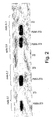
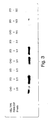
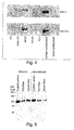
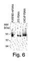


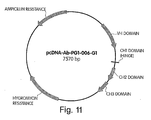

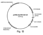


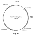
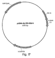

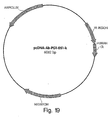

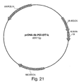
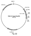
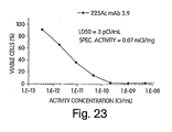
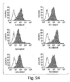
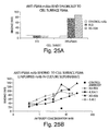


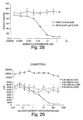
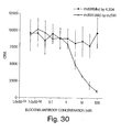
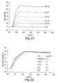

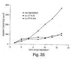

Classifications
-
- A—HUMAN NECESSITIES
- A61—MEDICAL OR VETERINARY SCIENCE; HYGIENE
- A61K—PREPARATIONS FOR MEDICAL, DENTAL OR TOILETRY PURPOSES
- A61K51/00—Preparations containing radioactive substances for use in therapy or testing in vivo
- A61K51/02—Preparations containing radioactive substances for use in therapy or testing in vivo characterised by the carrier, i.e. characterised by the agent or material covalently linked or complexing the radioactive nucleus
- A61K51/04—Organic compounds
- A61K51/08—Peptides, e.g. proteins, carriers being peptides, polyamino acids, proteins
- A61K51/10—Antibodies or immunoglobulins; Fragments thereof, the carrier being an antibody, an immunoglobulin or a fragment thereof, e.g. a camelised human single domain antibody or the Fc fragment of an antibody
- A61K51/1093—Antibodies or immunoglobulins; Fragments thereof, the carrier being an antibody, an immunoglobulin or a fragment thereof, e.g. a camelised human single domain antibody or the Fc fragment of an antibody conjugates with carriers being antibodies
- A61K51/1096—Antibodies or immunoglobulins; Fragments thereof, the carrier being an antibody, an immunoglobulin or a fragment thereof, e.g. a camelised human single domain antibody or the Fc fragment of an antibody conjugates with carriers being antibodies radioimmunotoxins, i.e. conjugates being structurally as defined in A61K51/1093, and including a radioactive nucleus for use in radiotherapeutic applications
-
- A—HUMAN NECESSITIES
- A61—MEDICAL OR VETERINARY SCIENCE; HYGIENE
- A61K—PREPARATIONS FOR MEDICAL, DENTAL OR TOILETRY PURPOSES
- A61K47/00—Medicinal preparations characterised by the non-active ingredients used, e.g. carriers or inert additives; Targeting or modifying agents chemically bound to the active ingredient
- A61K47/50—Medicinal preparations characterised by the non-active ingredients used, e.g. carriers or inert additives; Targeting or modifying agents chemically bound to the active ingredient the non-active ingredient being chemically bound to the active ingredient, e.g. polymer-drug conjugates
- A61K47/51—Medicinal preparations characterised by the non-active ingredients used, e.g. carriers or inert additives; Targeting or modifying agents chemically bound to the active ingredient the non-active ingredient being chemically bound to the active ingredient, e.g. polymer-drug conjugates the non-active ingredient being a modifying agent
- A61K47/68—Medicinal preparations characterised by the non-active ingredients used, e.g. carriers or inert additives; Targeting or modifying agents chemically bound to the active ingredient the non-active ingredient being chemically bound to the active ingredient, e.g. polymer-drug conjugates the non-active ingredient being a modifying agent the modifying agent being an antibody, an immunoglobulin or a fragment thereof, e.g. an Fc-fragment
- A61K47/6801—Drug-antibody or immunoglobulin conjugates defined by the pharmacologically or therapeutically active agent
- A61K47/6803—Drugs conjugated to an antibody or immunoglobulin, e.g. cisplatin-antibody conjugates
- A61K47/6811—Drugs conjugated to an antibody or immunoglobulin, e.g. cisplatin-antibody conjugates the drug being a protein or peptide, e.g. transferrin or bleomycin
- A61K47/6817—Toxins
- A61K47/6819—Plant toxins
- A61K47/6825—Ribosomal inhibitory proteins, i.e. RIP-I or RIP-II, e.g. Pap, gelonin or dianthin
-
- A—HUMAN NECESSITIES
- A61—MEDICAL OR VETERINARY SCIENCE; HYGIENE
- A61K—PREPARATIONS FOR MEDICAL, DENTAL OR TOILETRY PURPOSES
- A61K47/00—Medicinal preparations characterised by the non-active ingredients used, e.g. carriers or inert additives; Targeting or modifying agents chemically bound to the active ingredient
- A61K47/50—Medicinal preparations characterised by the non-active ingredients used, e.g. carriers or inert additives; Targeting or modifying agents chemically bound to the active ingredient the non-active ingredient being chemically bound to the active ingredient, e.g. polymer-drug conjugates
- A61K47/51—Medicinal preparations characterised by the non-active ingredients used, e.g. carriers or inert additives; Targeting or modifying agents chemically bound to the active ingredient the non-active ingredient being chemically bound to the active ingredient, e.g. polymer-drug conjugates the non-active ingredient being a modifying agent
- A61K47/68—Medicinal preparations characterised by the non-active ingredients used, e.g. carriers or inert additives; Targeting or modifying agents chemically bound to the active ingredient the non-active ingredient being chemically bound to the active ingredient, e.g. polymer-drug conjugates the non-active ingredient being a modifying agent the modifying agent being an antibody, an immunoglobulin or a fragment thereof, e.g. an Fc-fragment
- A61K47/6835—Medicinal preparations characterised by the non-active ingredients used, e.g. carriers or inert additives; Targeting or modifying agents chemically bound to the active ingredient the non-active ingredient being chemically bound to the active ingredient, e.g. polymer-drug conjugates the non-active ingredient being a modifying agent the modifying agent being an antibody, an immunoglobulin or a fragment thereof, e.g. an Fc-fragment the modifying agent being an antibody or an immunoglobulin bearing at least one antigen-binding site
- A61K47/6851—Medicinal preparations characterised by the non-active ingredients used, e.g. carriers or inert additives; Targeting or modifying agents chemically bound to the active ingredient the non-active ingredient being chemically bound to the active ingredient, e.g. polymer-drug conjugates the non-active ingredient being a modifying agent the modifying agent being an antibody, an immunoglobulin or a fragment thereof, e.g. an Fc-fragment the modifying agent being an antibody or an immunoglobulin bearing at least one antigen-binding site the antibody targeting a determinant of a tumour cell
- A61K47/6869—Medicinal preparations characterised by the non-active ingredients used, e.g. carriers or inert additives; Targeting or modifying agents chemically bound to the active ingredient the non-active ingredient being chemically bound to the active ingredient, e.g. polymer-drug conjugates the non-active ingredient being a modifying agent the modifying agent being an antibody, an immunoglobulin or a fragment thereof, e.g. an Fc-fragment the modifying agent being an antibody or an immunoglobulin bearing at least one antigen-binding site the antibody targeting a determinant of a tumour cell the tumour determinant being from a cell of the reproductive system: ovaria, uterus, testes, prostate
-
- A—HUMAN NECESSITIES
- A61—MEDICAL OR VETERINARY SCIENCE; HYGIENE
- A61K—PREPARATIONS FOR MEDICAL, DENTAL OR TOILETRY PURPOSES
- A61K51/00—Preparations containing radioactive substances for use in therapy or testing in vivo
- A61K51/02—Preparations containing radioactive substances for use in therapy or testing in vivo characterised by the carrier, i.e. characterised by the agent or material covalently linked or complexing the radioactive nucleus
- A61K51/04—Organic compounds
- A61K51/08—Peptides, e.g. proteins, carriers being peptides, polyamino acids, proteins
- A61K51/10—Antibodies or immunoglobulins; Fragments thereof, the carrier being an antibody, an immunoglobulin or a fragment thereof, e.g. a camelised human single domain antibody or the Fc fragment of an antibody
- A61K51/1045—Antibodies or immunoglobulins; Fragments thereof, the carrier being an antibody, an immunoglobulin or a fragment thereof, e.g. a camelised human single domain antibody or the Fc fragment of an antibody against animal or human tumor cells or tumor cell determinants
- A61K51/1072—Antibodies or immunoglobulins; Fragments thereof, the carrier being an antibody, an immunoglobulin or a fragment thereof, e.g. a camelised human single domain antibody or the Fc fragment of an antibody against animal or human tumor cells or tumor cell determinants the tumor cell being from the reproductive system, e.g. ovaria, uterus, testes or prostate
-
- A—HUMAN NECESSITIES
- A61—MEDICAL OR VETERINARY SCIENCE; HYGIENE
- A61P—SPECIFIC THERAPEUTIC ACTIVITY OF CHEMICAL COMPOUNDS OR MEDICINAL PREPARATIONS
- A61P13/00—Drugs for disorders of the urinary system
- A61P13/08—Drugs for disorders of the urinary system of the prostate
-
- A—HUMAN NECESSITIES
- A61—MEDICAL OR VETERINARY SCIENCE; HYGIENE
- A61P—SPECIFIC THERAPEUTIC ACTIVITY OF CHEMICAL COMPOUNDS OR MEDICINAL PREPARATIONS
- A61P31/00—Antiinfectives, i.e. antibiotics, antiseptics, chemotherapeutics
- A61P31/12—Antivirals
-
- A—HUMAN NECESSITIES
- A61—MEDICAL OR VETERINARY SCIENCE; HYGIENE
- A61P—SPECIFIC THERAPEUTIC ACTIVITY OF CHEMICAL COMPOUNDS OR MEDICINAL PREPARATIONS
- A61P35/00—Antineoplastic agents
-
- A—HUMAN NECESSITIES
- A61—MEDICAL OR VETERINARY SCIENCE; HYGIENE
- A61P—SPECIFIC THERAPEUTIC ACTIVITY OF CHEMICAL COMPOUNDS OR MEDICINAL PREPARATIONS
- A61P37/00—Drugs for immunological or allergic disorders
- A61P37/02—Immunomodulators
-
- A—HUMAN NECESSITIES
- A61—MEDICAL OR VETERINARY SCIENCE; HYGIENE
- A61P—SPECIFIC THERAPEUTIC ACTIVITY OF CHEMICAL COMPOUNDS OR MEDICINAL PREPARATIONS
- A61P37/00—Drugs for immunological or allergic disorders
- A61P37/02—Immunomodulators
- A61P37/04—Immunostimulants
-
- A—HUMAN NECESSITIES
- A61—MEDICAL OR VETERINARY SCIENCE; HYGIENE
- A61P—SPECIFIC THERAPEUTIC ACTIVITY OF CHEMICAL COMPOUNDS OR MEDICINAL PREPARATIONS
- A61P43/00—Drugs for specific purposes, not provided for in groups A61P1/00-A61P41/00
-
- A—HUMAN NECESSITIES
- A61—MEDICAL OR VETERINARY SCIENCE; HYGIENE
- A61P—SPECIFIC THERAPEUTIC ACTIVITY OF CHEMICAL COMPOUNDS OR MEDICINAL PREPARATIONS
- A61P9/00—Drugs for disorders of the cardiovascular system
-
- C—CHEMISTRY; METALLURGY
- C07—ORGANIC CHEMISTRY
- C07K—PEPTIDES
- C07K16/00—Immunoglobulins [IGs], e.g. monoclonal or polyclonal antibodies
- C07K16/18—Immunoglobulins [IGs], e.g. monoclonal or polyclonal antibodies against material from animals or humans
- C07K16/28—Immunoglobulins [IGs], e.g. monoclonal or polyclonal antibodies against material from animals or humans against receptors, cell surface antigens or cell surface determinants
- C07K16/30—Immunoglobulins [IGs], e.g. monoclonal or polyclonal antibodies against material from animals or humans against receptors, cell surface antigens or cell surface determinants from tumour cells
- C07K16/3069—Reproductive system, e.g. ovaria, uterus, testes, prostate
-
- A—HUMAN NECESSITIES
- A61—MEDICAL OR VETERINARY SCIENCE; HYGIENE
- A61K—PREPARATIONS FOR MEDICAL, DENTAL OR TOILETRY PURPOSES
- A61K39/00—Medicinal preparations containing antigens or antibodies
- A61K2039/505—Medicinal preparations containing antigens or antibodies comprising antibodies
-
- C—CHEMISTRY; METALLURGY
- C07—ORGANIC CHEMISTRY
- C07K—PEPTIDES
- C07K2317/00—Immunoglobulins specific features
- C07K2317/20—Immunoglobulins specific features characterized by taxonomic origin
- C07K2317/21—Immunoglobulins specific features characterized by taxonomic origin from primates, e.g. man
-
- C—CHEMISTRY; METALLURGY
- C07—ORGANIC CHEMISTRY
- C07K—PEPTIDES
- C07K2317/00—Immunoglobulins specific features
- C07K2317/50—Immunoglobulins specific features characterized by immunoglobulin fragments
- C07K2317/56—Immunoglobulins specific features characterized by immunoglobulin fragments variable (Fv) region, i.e. VH and/or VL
- C07K2317/565—Complementarity determining region [CDR]
-
- C—CHEMISTRY; METALLURGY
- C07—ORGANIC CHEMISTRY
- C07K—PEPTIDES
- C07K2317/00—Immunoglobulins specific features
- C07K2317/60—Immunoglobulins specific features characterized by non-natural combinations of immunoglobulin fragments
- C07K2317/62—Immunoglobulins specific features characterized by non-natural combinations of immunoglobulin fragments comprising only variable region components
- C07K2317/622—Single chain antibody (scFv)
Definitions
- This invention relates generally to the field of cancer associated polypeptides and antibodies that recognize native epitopes on the polypeptides.
- the invention relates in part to antibodies or antigen-binding fragments thereof which bind specifically to conformational epitopes on the extracellular domain of PSMA, multimeric forms of PSMA proteins, antibodies that selectively bind to the multimers, and compositions containing such antibodies or multimers.
- Prostate cancer is the most prevalent type of cancer and the second leading cause of death from cancer in American men, with an estimated 179,000 cases and 37,000 deaths in 1999, ( Landis, S.H. et al. CA Cancer J. Clin. 48:6-29 (1998 )).
- the number of men diagnosed with prostate cancer is steadily increasing as a result of the increasing population of older men as well as a greater awareness of the disease leading to its earlier diagnosis ( Parker et al., 1997, CA Cancer J. Clin. 47:5-280 ).
- the life time risk for men developing prostate cancer is about 1 in 5 for Caucasians, 1 in 6 for African Americans. High risk groups are represented by those with a positive family history of prostate cancer or African Americans.
- prostate cancer Over a lifetime, more than 2/3 of the men diagnosed with prostate cancer die of the disease ( Wingo et al., 1996, CA Cancer J. Clin. 46:113-25 ). Moreover, many patients who do not succumb to prostate cancer require continuous treatment to ameliorate symptoms such as pain, bleeding and urinary obstruction. Thus, prostate cancer also represents a major cause of suffering and increased health care expenditures.
- radical prostatectomy offers the best chance for eradication of the disease.
- the drawback of this procedure is that most cancers had spread beyond the bounds of the operation by the time they were detected. Patients with bulky, high-grade tumors are less likely to be successfully treated by radical prostatectomy.
- Radiation therapy has also been widely used as an alternative to radical prostatectomy.
- Patients generally treated by radiation therapy are those who are older and less healthy and those with higher-grade, more clinically advanced tumors.
- Particularly preferred procedures are external-beam therapy which involves three dimensional, confocal radiation therapy where the field of radiation is designed to conform to the volume of tissue treated; interstitial-radiation therapy where seeds of radioactive compounds are implanted using ultrasound guidance; and a combination of external-beam therapy and interstitial-radiation therapy.
- Hormonal therapy is the main form of treating men with disseminated prostate cancer. Orchiectomy reduces serum testosterone concentrations, while estrogen treatment is similarly beneficial. Diethylstilbestrol from estrogen is another useful hormonal therapy which has a disadvantage of causing cardiovascular toxicity.
- gonadotropin-releasing hormone agonists are administered testosterone concentrations are ultimately reduced.
- Flutamide and other nonsteroidal, anti-androgen agents block binding of testosterone to its intracellular receptors. As a result, it blocks the effect of testosterone, increasing serum testosterone concentrations and allows patients to remain potent--a significant problem after radical prostatectomy and radiation treatments.
- Cytotoxic chemotherapy is largely ineffective in treating prostate cancer. Its toxicity makes such therapy unsuitable for elderly patients.
- prostate cancer is relatively resistant to cytotoxic agents.
- the present invention relates to an isolated antibody or antigen-binding fragment thereof that specifically binds to dimeric forms of PSMA, but not monomeric forms of PSMA, and has the ability to bind live cells expressing PSMA.
- isolated antigen-binding fragments of antibodies can include a CDR of the foregoing antigen-binding fragments.
- the CDR is CDR3.
- Expression vectors including an isolated nucleic acid molecule encoding the foregoing isolated antibodies or antigen-binding fragments are described. Host cells transformed or transfected by these expression vectors also are described.
- the antibody or antigen-binding fragment thereof is selected for its ability to bind live cells, such as a tumor cell or a prostate cell, preferably LNCaP cells.
- the antibody or antigen-binding fragment thereof mediates cytolysis of cells expressing PSMA.
- cytolysis of cells expressing PSMA is mediated by effector cells or is complement mediated in the presence of effector cells.
- the antibody or antigen-binding fragment thereof inhibits the growth of cells expressing PSMA.
- the antibody or antigen-binding fragment thereof does not require cell lysis to bind to the extracellular domain of PSMA.
- the antibody or antigen-binding fragment thereof is selected from the group consisting of IgG1, IgG2, IgG3, IgG4, IgM, IgA1, IgA2, IgAsec, IgD, IgE or has immunoglobulin constant and/or variable domain of IgG1, IgG2, IgG3, IgG4, IgM, IgA1, IgA2, IgAsec, IgD or IgE.
- the antibody is a bispecific or multispecific antibody.
- the antibody is a recombinant antibody, a polyclonal antibody, a monoclonal antibody, a humanized antibody or a chimeric antibody, or a mixture of these.
- the antibody is a human antibody, e.g., a monoclonal antibody, polyclonal antibody or a mixture of monoclonal and polyclonal antibodies.
- the antibody is a bispecific or multispecific antibody.
- Preferred antigen-binding fragments include a Fab fragment, a F(ab') 2 fragment, and a Fv fragment CDR3.
- the antibody or antigen-binding fragment thereof binds to a conformational epitope and/or is internalized into a cell along with the prostate specific membrane antigen.
- the isolated antibody or antigen-binding fragment thereof is bound to a label, preferably one selected from the group consisting of a fluorescent label, an enzyme label, a radioactive label, a nuclear magnetic resonance active label, a luminescent label, and a chromophore label.
- the isolated antibody or antigen-binding fragment thereof is bound to at least one therapeutic moiety, such as a drug, preferably a cytotoxic drug, a replication-selective virus, a toxin or a fragment thereof, or an enzyme or a fragment thereof.
- a drug preferably a cytotoxic drug, a replication-selective virus, a toxin or a fragment thereof, or an enzyme or a fragment thereof.
- cytotoxic drug include: calicheamicin, esperamicin, methotrexate, doxorubicin, melphalan, chlorambucil, ARA-C, vindesine, mitomycin C, cis-platinum, etoposide, bleomycin, 5-fluorouracil, estramustine, vincristine, etoposide, doxorubicin, paclitaxel, docetaxel, dolastatin 10, auristatin E and auristatin PHE.
- the therapeutic moiety is an immunostimulatory or immunomodulating agent, preferably one selected from the group consisting of: a cytokine, chemokine and adjuvant.
- the antibodies or antigen-binding fragments of the invention specifically bind cell-surface PSMA and/or rsPSMA with a binding affinity of about 1 X 10 -9 M or less.
- the binding affinity is about 1 X 10 -10 M or less, more preferably the binding affinity is about 1 X 10 -11 M or less. In other embodiments the binding affinity is less than about 5 X 10 -10 M.
- the antibodies or antigen-binding fragments of the invention mediate specific cell killing of PSMA-expressing cells with an IC 50 s of less than about 1 X 10 -10 M.
- the IC 50 s is less than about 1 X 10 -11 M. More preferably the IC 50 s is less than about 1 X 10 -12 M. In other embodiments the IC 50 s is less than about 1.5 X 10 -11 M.
- the isolated antibody or antigen-binding fragment thereof is bound to a radioisotope.
- the radioisotope can emit ⁇ radiations, ⁇ radiations, or ⁇ radiations.
- the radioisotope is selected from the group consisting of 225 Ac, 211 At, 212 Bi, 213 Bi, 186 Rh, 188 Rh, 177 Lu, 90 Y, 131 I, 67 Cu, 125 I, 123 I, 77 Br, 153 Sm, 166 Ho, 64 Cu, 212 Pb, 224 Ra and 223 Ra.
- compositions are provided that include the foregoing antibodies or antigen-binding fragments thereof and a pharmaceutically acceptable carrier, excipient, or stabilizer.
- Other compositions include a combination of two or more of the foregoing antibodies or antigen-binding fragments thereof and a pharmaceutically acceptable carrier, excipient, or stabilizer.
- the compositions also include an antitumor agent, an immunostimulatory agent, an immunomodulator, or a combination thereof.
- Preferred antitumor agents include a cytotoxic agent, an agent that acts on tumor neovasculature, or a combination thereof.
- Preferred immunomodulators include ⁇ -interferon, ⁇ -interferon, tumor necrosis factor- ⁇ or a combination thereof.
- Preferred immunostimulatory agents include interleukin-2, immunostimulatory oligonucleotides, or a combination thereof.
- Kits for detecting prostate cancer for diagnosis, prognosis or monitoring can include the foregoing isolated labeled antibody or antigen-binding fragment thereof, and one or more compounds for detecting the label.
- the label is selected from the group consisting of a fluorescent label, an enzyme label, a radioactive label, a nuclear magnetic resonance active label, a luminescent label, and a chromophore label.
- One or more of the foregoing isolated antibodies or antigen-binding fragments thereof can be packaged in lyophilized form, or packaged in an aqueous medium.
- Methods for detecting the presence of PSMA, or a cell expressing PSMA can include contacting the sample with any of the foregoing antibodies or antigen-binding fragments thereof which specifically bind to an extracellular domain of PSMA, for a time sufficient to allow the formation of a complex between the antibody or antigen-binding fragment thereof and PSMA, and detecting the PSMA-antibody complex or PSMA-antigen-binding fragment complex.
- the presence of a complex in the sample is indicative of the presence in the sample of PSMA or a cell expressing PSMA.
- Methods for diagnosing a PSMA-mediated disease in a subject can include administering to a subject suspected of having or previously diagnosed with PSMA-mediated disease an amount of any of the foregoing antibodies or antigen-binding fragments thereof which specifically bind to an extracellular domain of prostate specific membrane antigen.
- the method also includes allowing the formation of a complex between the antibody or antigen-binding fragment thereof and PSMA, and detecting the formation of the PSMA-antibody complex or PSMA-antigen-binding fragment antibody complex to the target epitope.
- the presence of a complex in the subject suspected of having or previously diagnosed with prostate cancer is indicative of the presence of a PSMA-mediated disease.
- the PSMA-mediated disease can be prostate cancer.
- the PSMA-mediated disease is a non-prostate cancer, such as those selected from the group consisting of bladder cancer including transitional cell carcinoma; pancreatic cancer including pancreatic duct carcinoma; lung cancer including non-small cell lung carcinoma; kidney cancer including conventional renal cell carcinoma; sarcoma including soft tissue sarcoma; breast cancer including breast carcinoma; brain cancer including glioblastoma multiforme; neuroendocrine carcinoma; colon cancer including colonic carcinoma; testicular cancer including testicular embryonal carcinoma; and melanoma including malignant melanoma.
- the antibody or antigen-binding fragment thereof is labeled.
- a second antibody is administered to detect the first antibody or antigen-binding fragment thereof.
- Methods for assessing the prognosis of a subject with a PSMA-mediated disease can include administering to a subject suspected of having or previously diagnosed with PSMA-mediated disease an effective amount of an antibody or antigen-binding fragment thereof, allowing the formation of a complex between the antibody or antigen-binding fragment thereof and PSMA, and detecting the formation of the complex to the target epitope.
- the amount of the complex in the subject suspected of having or previously diagnosed with PSMA-mediated disease is indicative of the prognosis.
- Methods for assessing the effectiveness of a treatment of a subject with a PSMA-mediated disease can include administering to a subject treated for a PSMA-mediated disease an effective amount of the foregoing antibodies or antigen-binding fragments thereof, allowing the formation of a complex between the antibody or antigen-binding fragment thereof and PSMA, and detecting the formation of the complex to the target epitope.
- the amount of the complex in the subject suspected of having or previously diagnosed with PSMA-mediated disease is indicative of the effectiveness of the treatment.
- the PSMA-mediated disease can be prostate cancer.
- the PSMA-mediated disease is a non-prostate cancer.
- the non-prostate cancer preferably is selected from the group consisting of bladder cancer including transitional cell carcinoma; pancreatic cancer including pancreatic duct carcinoma; lung cancer including non-small cell lung carcinoma; kidney cancer including conventional renal cell carcinoma; sarcoma including soft tissue sarcoma; breast cancer including breast carcinoma; brain cancer including glioblastoma multiforme; neuroendocrine carcinoma; colon cancer including colonic carcinoma; testicular cancer including testicular embryonal carcinoma; and melanoma including malignant melanoma.
- the antibody or antigen-binding fragment thereof is labeled.
- a second antibody is administered to detect the first antibody or antigen-binding fragment thereof.
- Methods for inhibiting the growth of a cell expressing PSMA can include contacting a cell expressing PSMA with an amount of at least one of the foregoing antibodies or antigen-binding fragments thereof which specifically binds to an extracellular domain of PSMA effective to inhibit the growth of the cell expressing PSMA.
- Methods for inducing cytolysis of a cell expressing PSMA can include contacting a cell expressing PSMA with an amount of at least one of the foregoing antibodies or antigen-binding fragments thereof which specifically binds to an extracellular domain of PSMA effective to induce cytolysis of the cell expressing PSMA.
- the cytolysis occurs in the presence of an effector cell.
- the cytolysis is complement mediated.
- Methods for treating or preventing a PSMA-mediated disease can include administering to a subject having a PSMA-mediated disease an effective amount of at least one of the forgoing antibodies or antigen-binding fragments thereof to treat or prevent the PSMA-mediated disease.
- the PSMA-mediated disease is a cancer, such as prostate cancer or a non-prostate cancer (including the nonprostate cancers described elsewhere herein).
- Methods for treating or preventing a PSMA-mediated disease can include administering to a subject having a PSMA-mediated disease or at risk of having a PSMA-mediated disease an amount of at least one of the foregoing antibodies or antigen-binding fragments thereof effective to treat or prevent the PSMA-mediated disease.
- the PSMA-mediated disease is a cancer, such as prostate cancer or a non-prostate cancer (including the nonprostate cancers described elsewhere herein).
- the method also includes administering another therapeutic agent to treat or prevent the PSMA-mediated disease at any time before, during or after the administration of the antibody or antigen-binding fragment thereof.
- the therapeutic agent is a vaccine, and preferably the vaccine immunizes the subject against PSMA.
- the antibody or antigen-binding fragment thereof is bound to at least one therapeutic moiety, preferably a cytotoxic drug, a drug which acts on the tumor neovasculature and combinations thereof.
- cytotoxic drugs are selected from the group consisting of: calicheamicin, esperamicin, methotrexate, doxorubicin, melphalan, chlorambucil, ARA-C, vindesine, mitomycin C, cis-platinum, etoposide, bleomycin, 5-fluorouracil, estramustine, vincristine, etoposide, doxorubicin, paclitaxel, docetaxel, dolastatin 10, auristatin E and auristatin PHE.
- the antibody or antigen-binding fragment thereof is bound to a radioisotope and the radiations emitted by the radioisotope is selected from the group consisting of ⁇ , ⁇ and ⁇ radiations.
- the radioisotope is selected from the group consisting of 225 Ac, 211 At, 212 Bi, 213 Bi, 186 Rh, 188 Rh, 177 Lu, 90 Y, 131 I, 67 Cu, 125 I, 123 I, 77 Br, 153 Sm, 166 Ho, 64 Cu, 212 Pb, 224 Ra and 223 Ra.
- the antibodies and antigen-binding fragments of the present invention can be used in methods for modulating at least one enzymatic activity of PSMA.
- modulating an enzymatic activity of PSMA means enhancing or inhibiting the enzymatic activity.
- enhancing' and “inhibiting” in this context indicate that the enzymatic activity of PSMA is enhanced or inhibited in the presence of an antibody that specifically binds PSMA, or antigen-binding fragment thereof, relative to the level of activity in the absence of such an antibody or antigen-binding fragment thereof.
- Enzymatic activities of PSMA include folate hydrolase activity, N-acetylated ⁇ -linked acidic dipeptidase (NAALADase) activity, dipeptidyl dipeptidase IV activity and ⁇ -glutamyl hydrolase activity.
- Methods for modulating folate hydrolase activity can include contacting a folate hydrolase polypeptide with an amount of the foregoing isolated antibody or antigen-binding fragment thereof, under conditions wherein the isolated antibody or antigen-binding fragment thereof modulates the folate hydrolase activity.
- the folate hydrolase polypeptide can be isolated, contained in a sample such as a cell, a cell homogenate, a tissue, or a tissue homogenate, or contained in an organism.
- the organism preferably is an animal, particularly preferably a mammal.
- NAALADase N-acetylated ⁇ -linked acidic dipeptidase
- Methods for modulating N-acetylated ⁇ -linked acidic dipeptidase (NAALADase) activity can include contacting a NAALADase polypeptide with an amount of the foregoing isolated antibody or antigen-binding fragment thereof under conditions wherein the isolated antibody or antigen-binding fragment thereof modulates NAALADase activity.
- the NAALADase polypeptide can be isolated, contained in a sample such as a cell, a cell homogenate, a tissue, or a tissue homogenate, or contained in an organism.
- the organism preferably is an animal, particularly preferably a mammal.
- Methods for modulating dipeptidyl dipeptidase IV activity can include contacting a dipeptidyl dipeptidase IV polypeptide with an amount of the foregoing isolated antibody or antigen-binding fragment thereof under conditions wherein the isolated antibody or antigen-binding fragment thereof modulates dipeptidyl dipeptidase IV activity.
- the dipeptidyl dipeptidase IV polypeptide can be isolated, contained in a sample such as a cell, a cell homogenate, a tissue, or a tissue homogenate, or contained in an organism.
- the organism preferably is an animal, particularly preferably a mammal.
- Methods for modulating ⁇ -glutamyl hydrolase activity can include contacting a ⁇ -glutamyl hydrolase polypeptide with an amount of the foregoing isolated antibody or antigen-binding fragment thereof under conditions wherein the isolated antibody or antigen-binding fragment thereof modulates ⁇ -glutamyl hydrolase activity.
- the ⁇ -glutamyl hydrolase polypeptide can be isolated, contained in a sample such as a cell, a cell homogenate, a tissue, or a tissue homogenate, or contained in an organism.
- the organism preferably is an animal, particularly preferably a mammal.
- the antibodies and antigen-binding fragements of the present invention can be used in methods of specific delivery of at least one therapeutic agent to PSMA-expressing cells.
- the methods include administering an effective amount of at least one of the foregoing antibodies or antigen-binding fragments thereof conjugated to the at least one therapeutic agent.
- the therapeutic agent is a nucleic acid molecule, an antitumor drug, a toxin or a fragment thereof, an enzyme or a fragment thereof, a replication-selective virus, or an immunostimulatory or immunomodulating agent.
- Preferred antitumor drugs include cytotoxic drugs, drugs which act on the tumor neovasculature and combinations thereof.
- cytotoxic drugs include calicheamicin, esperamicin, methotrexate, doxorubicin, melphalan, chlorambucil, ARA-C, vindesine, mitomycin C, cis-platinum, etoposide, bleomycin, 5-fluorouracil, estramustine, vincristine, etoposide, doxorubicin, paclitaxel, docetaxel, dolastatin 10, auristatin E and auristatin PHE.
- Preferred immunostimulatory or immunomodulating agent included cytokines, chemokines and adjuvants.
- isolated antibodies that specifically bind a PSMA protein dimer are provided.
- at least one of the PSMA proteins forming the dimer is a recombinant, soluble PSMA (rsPSMA) polypeptide.
- rsPSMA soluble PSMA
- the rsPSMA polypeptide consists essentially of amino acids 44-750 of SEQ ID NO:1.
- isolated antibodies are provided that specifically bind a PSMA protein dimer and modulate one or more enzymatic activities of the PSMA protein dimer.
- modulating an enzymatic activity of a PSMA dimer means inhibiting the enzymatic activity.
- antibodies that inhibit an enzymatic activity of a PSMA dimer are provided.
- the term "inhibiting" in this context indicates that the enzymatic activity of a PSMA multimer is inhibited in the presence of an antibody that specifically binds the PSMA dimer, or antigen-binding fragment thereof, relative to the level of activity in the absence of such an antibody or antigen-binding fragment thereof.
- the enzymatic activity is selected from the group consisting of folate hydrolase activity, NAALADase activity, dipeptidyl dipeptidase IV activity and ⁇ -glutamyl hydrolase activity.
- the enzymatic activity is in the extracellular domain of the PSMA molecule.
- the antibody or antigen-binding fragment thereof specifically binds to an extracellular domain of PSMA.
- compositions that include one or more of the foregoing isolated antibodies, and an immunostimulatory molecule, such as an adjuvant and/or and a cytokine.
- an immunostimulatory molecule such as an adjuvant and/or and a cytokine.
- the immunostimulatory molecule is IL-2 or an immunostimulatory oligonucleotide.
- the foregoing compositions also include a pharmaceutically-acceptable carrier.
- Methods for inducing an immune response can include administering to a subject in need of such treatment an effective amount of the foregoing isolated antibodies or compositions.
- the invention provides, in another aspect, isolated antibodies or antigen-binding fragments thereof that specifically bind a PSMA protein dimer and modulate at least one enzymatic activity of PSMA.
- modulating an enzymatic activity of a PSMA means inhibiting the enzymatic activity.
- antibodies that inhibit an enzymatic activity of PSMA are provided.
- inhibiting in this context indicates that the enzymatic activity of PSMA is inhibited in the presence of an antibody that specifically binds PSMA, or antigen-binding fragment thereof, relative to the level of activity in the absence of such an antibody or antigen-binding fragment thereof.
- the enzyme in certain embodiments, is selected from the group consisting of hydrolases and peptidases.
- Preferred hydrolases include folate hydrolase and ⁇ -glutamyl hydrolase.
- the hydrolase is folate hydrolase and the antibody is mAb 5.4 or mAb 3.9.
- Preferred peptidases include NAALADase and dipeptidyl dipeptidase IV.
- the enzyme is active in cancer cells and has lesser activity in normal cells than in cancer cells or, preferably, no activity in normal cells.
- the cancer cells in which the enzyme is active are prostate cancer cells.
- Compositions including the foregoing isolated antibodies or antigen-binding fragments thereof, and a pharmaceutically acceptable carrier, also are provided by the invention.
- compositions also include an adjuvant and/or a cytokine or other immunostimulatory molecule.
- Preferred cytokines include IL-2, IL-12, IL-18 and GM-CSF.
- the foregoing compositions also include a pharmaceutically acceptable carrier.
- Methods for inducing an immune response can include administering to a subject in need of such treatment an effective amount of one or more of the foregoing compositions.
- the medicaments are useful in the treatment of conditions related to hyperproliferative diseases including cancer, and diseases of inappropriate NAALADase activity, folate hydrolase activity, dipeptidyl dipeptidase IV activity and/or ⁇ -glutamyl hydrolase activity.
- the present invention provides antibodies or antigen-binding fragments thereof which bind specifically to conformational epitopes on the extracellular domain of PSMA, compositions containing one or a combination of such antibodies or antigen-binding fragments thereof, hybridoma cell lines that produce the antibodies, and methods of using the antibodies or antigen-binding fragments thereof for cancer diagnosis and treatment.
- PSMA Prostate specific membrane antigen
- PSMA is expressed in increased amounts in prostate cancer, and elevated levels of PSMA are also detectable in the sera of these patients (Horoszewicz et al., 1987; Rochon et al., 1994, Prostate 25:219-223 ; Murphy et al., 1995, Prostate 26:164-168 ; and Murphy et al., 1995, Anticancer Res. 15:1473-1479 ).
- PSMA expression increases with disease progression, becoming highest in metastatic, hormone-refractory disease for which there is no present therapy.
- PSMA is also abundantly expressed on the neovasculature of a variety of other important tumors, including bladder, pancreas, sarcoma, melanoma, lung, and kidney tumor cells, but not on normal vasculature.
- Hybridomas were deposited with ATCC as an International Depository Authority and given the following Patent Deposit Designations (Table 1): Table 1. Antibody Hybridoma/Plasmid Patent Deposit Designation Date of Deposit PSMA 3.7 PSMA 3.7 PTA-3257 April 5, 2001 PSMA 3.9 PSMA 3.9 PTA-3258 April 5, 2001 PSMA 3.11 PSMA 3.11 PTA-3269 April 10, 2001 PSMA 5.4 PSMA 5.4 PTA-3268 April 10, 2001 PSMA 7.1 PSMA 7.1 PTA-3292 April 18, 2001 PSMA 7.3 PSMA 7.3 PTA-3293 April 18, 2001 PSMA 10.3 PSMA 10.3 PTA-3347 May 1, 2001 PSMA 10.3 HC in pcDNA (SEQ ID NO: 7) PTA-4413 May 29, 2002 PSMA 10.3 Kappa in pcDNA (SEQ ID NO: 13) PTA-4414 May 29, 2002 PSMA 1.8.3 PSMA 1.8.3 PTA-3906 Dec.
- Antibodies having particular sequences are decribed and are selected from the group consisting of antibodies comprising: a heavy chain encoded by a nucleic acid molecule comprising the heavy chain coding region or regions of a nucleotide sequence selected from the group consisting of nucleotide sequences set forth as SEQ ID NOs: 2-7, and a light chain encoded by a nucleic acid molecule comprising the light chain coding region or regions of a nucleotide sequence selected from the group consisting of nucleotide sequences set forth as SEQ ID NOs: 8-13. Also provided are antigen-binding fragments of the foregoing antibodies.
- the plasmids encoding the heavy and light chains of antibodies PSMA 10.3, AB-PG1-XG1-006, AB-PG1-XG1-026, AB-PG1-XG1-051, AB-PG1-XG1-069, AB-PG1-XG1-077 were also deposited with ATCC and are shown in Table 1 above.
- the names of the deposited hybridomas or plasmids may be used interchangeably with the names of the antibodies. It would be clear to one of skill in the art when the name is intended to refer to the antibody or when it refers to the plasmids or hybridomas that encode or produce the antibodies, respectively. Additionally, the antibody names may be an abbreviated form of the name shown in Table 1.
- antibody AB-PG1-XG1-006 may be referred to as AB-PG1-XG1-006, PG1-XG1-006, XG1-006, 006, etc.
- the antibody name PSMA 4.232.3 may be referred to as PSMA 4.232.1, 4.232.3, 4.232.1, 4.232, etc. It is intended that all of the variations in the name of the antibody refer to the same antibody and not a different one.
- Antibodies are also described that are encoded by particular sets of heavy and light chain sequences.
- Antibody (AB-PG1-XG1-006) is encoded by a nucleic acid molecule which comprises the coding region or regions of the nucleic acid sequences set forth as SEQ ID NOs: 2 and 8
- antibody (AB-PG1-XG1-026) is encoded by the nucleic acid molecules comprising the coding region or regions of nucleotide sequences set forth as SEQ ID NOs: 3 and 9
- antibody (AB-PG1-XG1-051) is encoded by the nucleic acid molecules comprising the coding region or regions of nucleotide sequences set forth as SEQ ID NOs: 4 and 10
- antibody (AB-PG1-XG1-069) is encoded by the nucleic acid molecules comprising the coding region or regions of nucleotide sequences set forth as SEQ ID NOs: 5 and 11
- antibody (AB-PG1-XG1-077) is encoded by the nucleic
- Antibodies that include a heavy chain variable region encoded by a nucleic acid molecule comprising the coding regions or regions of a nucleotide sequence selected from the group consisting of nucleotide sequences set forth as: SEQ ID NOs: 14, 18, 22, 26 and 30, and a light chain variable region encoded by a nucleic acid molecule comprising the coding region or region of a nucleotide sequence selected from the group consisting of nucleotide sequences set forth as: SEQ ID NOs: 16, 20, 24, 28 and 32 are also described.
- coding region refers to a region of a nucleotide sequence that encodes a polypeptide sequence; the coding region can include a region coding for a portion of a protein that is later cleaved off, such as a signal peptide.
- Antibody includes an immunoglobulin variable sequence encoded by nucleic acid molecules which include the coding region or regions of the nucleic acid sequences set forth as SEQ ID NOs: 14 and 16, or includes an immunoglobulin variable sequence which comprises the amino acid sequences set forth as SEQ ID NOs: 15 and 17.
- Antibody (AB-PG1-XG1-026) includes an immunoglobulin variable sequence encoded by nucleic acid molecules comprising the coding region or regions of nucleotide sequences set forth as SEQ ID NOs: 18 and 20 or includes an immunoglobulin variable sequence which comprises the amino acid sequences set forth as SEQ ID NOs: 19 and 21.
- Antibody (AB-PG1-XG1-051) includes an immunoglobulin variable sequence encoded by the nucleic acid molecules comprising the coding region or regions of nucleotide sequences set forth as SEQ ID NOs: 22 and 24 or includes an immunoglobulin variable sequence which comprises the amino acid sequences set forth as SEQ ID NOs: 23 and 25.
- Antibody (AB-PG1-XG1-069) includes an immunoglobulin variable sequence encoded by the nucleic acid molecules comprising the coding region or regions of nucleotide sequences set forth as SEQ ID NOs: 26 and 28 or includes an immunoglobulin variable sequence which comprises the amino acid sequences set forth as SEQ ID NOs: 27 and 29.
- Antibody includes an immunoglobulin variable sequence encoded by the nucleic acid molecules comprising the coding region or regions of nucleotide sequences set forth as SEQ ID NOs: 30 and 32 or includes an immunoglobulin variable sequence which comprises the amino acid sequences set forth as SEQ ID NOs: 31 and 33.
- the term "antibody” refers to a glycoprotein comprising at least two heavy (H) chains and two light (L) chains inter-connected by disulfide bonds.
- Each heavy chain is comprised of a heavy chain variable region (abbreviated herein as HCVR or V H ) and a heavy chain constant region.
- the heavy chain constant region is comprised of three domains, C H 1 , C H 2 and C H 3.
- Each light chain is comprised of a light chain variable region (abbreviated herein as LCVR or V L ) and a light chain constant region.
- the light chain constant region is comprised of one domain, CL.
- V H and V L regions can be further subdivided into regions of hypervariability, termed complementarity determining regions (CDR), interspersed with regions that are more conserved, termed framework regions (FR).
- CDR complementarity determining regions
- FR framework regions
- Each V H and V L is composed of three CDRs and four FRs, arranged from amino-terminus to carboxy-terminus in the following order: FR1, CDR1, FR2, CDR2, FR3, CDR3, FR4.
- the variable regions of the heavy and light chains contain a binding domain that interacts with an antigen.
- the constant regions of the antibodies may mediate the binding of the immunoglobulin to host tissues or factors, including various cells of the immune system (e.g., effector cells) and the first component (C1q) of the classical complement system.
- antigen-binding fragment of an antibody as used herein, refers to one or more portions of an antibody that retain the ability to specifically bind to an antigen (e.g., PSMA). It has been shown that the antigen-binding function of an antibody can be performed by fragments of a full-length antibody.
- binding fragments encompassed within the term "antigen-binding fragment" of an antibody include (i) a Fab fragment, a monovalent fragment consisting of the V L , V H , C L and C H 1 domains; (ii) a F(ab') 2 fragment, a bivalent fragment comprising two Fab fragments linked by a disulfide bridge at the hinge region; (iii) a Fd fragment consisting of the V H and CH1 domains; (iv) a Fv fragment consisting of the V L and V H domains of a single arm of an antibody, (v) a dAb fragment ( Ward et al., (1989) Nature 341:544-546 ) which consists of a V H domain; and (vi) an isolated complementarity determining region (CDR).
- a Fab fragment a monovalent fragment consisting of the V L , V H , C L and C H 1 domains
- F(ab') 2 fragment a bivalent fragment comprising two
- the two domains of the Fv fragment, V and V H are coded for by separate genes, they can be joined, using recombinant methods, by a synthetic linker that enables them to be made as a single protein chain in which the V L and V H regions pair to form monovalent molecules (known as single chain Fv (scFv); see e.g., Bird et al. (1988) Science 242:423-426 ; and Huston et al. (1988) Proc. Natl. Acad. Sci. USA 85:5879-5883 ).
- single chain Fv single chain Fv
- Such single chain antibodies are also intended to be encompassed within the term "antigen-binding portion" of an antibody.
- antibody fragments are obtained using conventional procedures, such as proteolytic fragmentation procedures, as described in J. Goding, Monoclonal Antibodies: Principles and Practice, pp 98-118 (N.Y. Academic Press 1983 ), as well as by other techniques known to those with skill in the art.
- the fragments are screened for utility in the same manner as are intact antibodies.
- an "isolated antibody”, as used herein, is intended to refer to an antibody which is substantially free of other antibodies having different antigenic specificities (e.g., an isolated antibody that specifically binds to PSMA is substantially free of antibodies that specifically bind antigens other than PSMA).
- An isolated antibody that specifically binds to an epitope, isoform or variant of PSMA may, however, have cross-reactivity to other related antigens, e.g., from other species (e.g., PSMA species homologs).
- an isolated antibody may be substantially free of other cellular material and/or chemicals.
- “specific binding” refers to antibody binding to a predetermined antigen. Typically, the antibody binds with an affinity that is at least two-fold greater than its affinity for binding to a non-specific antigen (e.g., BSA, casein) other than the predetermined antigen or a closely-related antigen.
- the isolated antibodies of the invention encompass various antibody isotypes, such as IgG1, IgG2, IgG3, IgG4, IgM, IgA1, IgA2, IgAsec, IgD, IgE.
- isotype refers to the antibody class (e.g. IgM or IgG1) that is encoded by heavy chain constant region genes.
- the antibodies can be full length or can include only an antigen-binding fragment such as the antibody constant and/or variable domain of IgG1, IgG2, IgG3, IgG4, IgM, IgA1, IgA2, IgAsec, IgD or IgE or could consist of a Fab fragment, a F(ab') 2 fragment, and a Fv fragment.
- an antigen-binding fragment such as the antibody constant and/or variable domain of IgG1, IgG2, IgG3, IgG4, IgM, IgA1, IgA2, IgAsec, IgD or IgE or could consist of a Fab fragment, a F(ab') 2 fragment, and a Fv fragment.
- the antibodies of the present invention can be polyclonal, monoclonal, or a mixture of polyclonal and monoclonal antibodies.
- the antibodies can be produced by a variety of techniques well known in the art. Procedures for raising polyclonal antibodies are well known. For example anti-PSMA polyclonal antibodies are raised by administering PSMA protein subcutaneously to New Zealand white rabbits which have first been bled to obtain pre-immune serum. The PSMA can be injected at a total volume of 100 ⁇ l per site at six different sites, typically with one or more adjustments. The rabbits are then bled two weeks after the first injection and periodically boosted with the same antigen three times every six weeks. A sample of serum is collected 10 days after each boost.
- Polyclonal antibodies are recovered from the serum, preferably by affinity chromatography using PSMA to capture the antibody. This and other procedures for raising polyclonal antibodies are disclosed in E. Harlow, et. al., editors, Antibodies: A Laboratory Manual (1988 ).
- Monoclonal antibody production may be effected by techniques which are also well known in the art.
- the term "monoclonal antibody,” as used herein, refers to a preparation of antibody molecules of single molecular composition.
- a monoclonal antibody displays a single binding specificity and affinity for a particular epitope.
- the process of monoclonal antibody production involves obtaining immune somatic cells with the potential for producing antibody, in particular B lymphocytes, which have been previously immunized with the antigen of interest either in vivo or in vitro and that are suitable for fusion with a B-cell myeloma line.
- Mammalian lymphocytes typically are immunized by in vivo immunization of the animal (e.g., a mouse) with the desired protein or polypeptide, e.g., with PSMA in the present invention. Such immunizations are repeated as necessary at intervals of up to several weeks to obtain a sufficient titer of antibodies.
- animals can be used as a source of antibody-producing lymphocytes. Following the last antigen boost, the animals are sacrificed and spleen cells removed.
- Mouse lymphocytes give a higher percentage of stable fusions with the mouse myeloma lines described herein. Of these, the BALB/c mouse is preferred.
- mouse strains rabbit, hamster, sheep and frog may also be used as hosts for preparing antibody-producing cells. See; Goding (in Monoclonal Antibodies: Principles and Practice, 2d ed., pp. 60-61, Orlando, Fla., Academic Press, 1986 ).
- mouse strains that have human immunoglobulin genes inserted in the genome are preferred. Examples include the HuMAb mouse strains produced by Medarex/GenPharm International, and the XenoMouse strains produced by Abgenix. Such mice produce fully human immunoglobulin molecules in response to immunization.
- Somatic cells may be obtained from the lymph nodes, spleens and peripheral blood of antigen-primed animals, and the lymphatic cells of choice depend to a large extent on their empirical usefulness in the particular fusion system.
- the antibody-secreting lymphocytes are then fused with (mouse) B cell myeloma cells or transformed cells, which are capable of replicating indefinitely in cell culture, thereby producing an immortal, immunoglobulin-secreting cell line.
- the resulting fused cells, or hybridomas are cultured, and the resulting colonies screened for the production of the desired monoclonal antibodies.
- Colonies producing such antibodies are cloned, and grown either in vivo or in vitro to produce large quantities of antibody.
- a description of the theoretical basis and practical methodology of fusing such cells is set forth in Kohler and Milstein, Nature 256:495 (1975 ).
- human somatic cells capable of producing antibody are suitable for fusion with myeloma cell lines. While B lymphocytes from biopsied spleens, tonsils or lymph nodes of an individual may be used, the more easily accessible peripheral blood B lymphocytes are preferred. The lymphocytes may be derived from patients with diagnosed prostate carcinomas or another PSMA-expressing cancer. In addition, human B cells may be directly immortalized by the Epstein-Barr virus ( Cole et al., 1995, Monoclonal Antibodies and Cancer Therapy, Alan R. Liss, Inc., pp. 77-96 ). Although somatic cell hybridization procedures are preferred, in principle, other techniques for producing monoclonal antibodies can be employed such as viral or oncogenic transformation of B lymphocytes.
- Myeloma cell lines suited for use in hybridoma-producing fusion procedures preferably are non-antibody-producing, have high fusion efficiency, and enzyme deficiencies that render them incapable of growing in certain selective media which support the growth of the desired hybridomas.
- myeloma cell lines that may be used for the production of fused cell lines include P3-X63/Ag8, X63-Ag8.653, NS1/1.Ag 4.1, Sp2/0-Ag14, FO, NSO/U, MPC-11, MPC11-X45-GTG 1.7, S194/SXX0 Bul, all derived from mice; R210.RCY3, Y3-Ag 1.2.3, IR983F and 4B210 derived from rats and U-266, GM1500-GRG2, LICR-LON-HMy2, UC729-6, all derived from humans ( Goding, in Monoclonal Antibodies: Principles and Practice, 2d ed., pp.
- Fusion with mammalian myeloma cells or other fusion partners capable of replicating indefinitely in cell culture is effected by standard and well-known techniques, for example, by using polyethylene glycol ("PEG”) or other fusing agents (See Milstein and Kohler, Eur. J. Immunol. 6:511 (1976 )).
- PEG polyethylene glycol
- the antibodies can be recombinant antibodies.
- the term "recombinant antibody”, as used herein, is intended to include antibodies that are prepared, expressed, created or isolated by recombinant means, such as antibodies isolated from an animal (e.g., a mouse) that is transgenic for another species' immunoglobulin genes, antibodies expressed using a recombinant expression vector transfected into a host cell, antibodies isolated from a recombinant, combinatorial antibody library, or antibodies prepared, expressed, created or isolated by any other means that involves splicing of immunoglobulin gene sequences to other DNA sequences.
- the antibodies can be chimeric or humanized antibodies.
- the term “chimeric antibody” refers to an antibody, that combines the murine variable or hypervariable regions with the human constant region or constant and variable framework regions.
- the term “humanized antibody” refers to an antibody that retains only the antigen-binding CDRs from the parent antibody in association with human framework regions (see, Waldmann, 1991, Science 252:1657 ). Such chimeric or humanized antibodies retaining binding specificity of the murine antibody are expected to have reduced immunogenicity when administered in vivo for diagnostic, prophylactic or therapeutic applications according to the invention.
- the monoclonal antibodies of the present invention can be modified to be in the form of a bispecific antibody, or a multispecific antibody.
- bispecific antibody is intended to include any agent, e.g., a protein, peptide, or protein or peptide complex, which has two different binding specificities which bind to, or interact with (a) a cell surface antigen and (b) an Fc receptor on the surface of an effector cell.
- multispecific antibody is intended to include any agent, e.g., a protein, peptide, or protein or peptide complex, which has more than two different binding specificities which bind to, or interact with (a) a cell surface antigen, (b) an Fc receptor on the surface of an effector cell, and (c) at least one other component.
- the invention includes, but is not limited to, bispecific, trispecific, tetraspecific, and other multispecific antibodies which are directed to cell surface antigens, such as PSMA, and to Fc receptors on effector cells.
- bispecific antibodies further includes diabodies.
- Diabodies are bivalent, bispecific antibodies in which the V H and V L domains are expressed on a single polypeptide chain, but using a linker that is too short to allow for pairing between the two domains on the same chain, thereby forcing the domains to pair with complementary domains of another chain and creating two antigen-binding sites (see e.g., Holliger, P., et al. (1993) Proc. Natl. Acad. Sci. USA 90:6444-6448 ; Poijak, R.J., et al. (1994) Structure 2:1121-1123 ).
- a bispecific antibody can be formed of an antigen-binding region specific for the extracellular domain of PSMA and an antigen-binding region specific for an effector cell which has tumoricidal or tumor inhibitory activity.
- the two antigen-binding regions of the bispecific antibody are either chemically linked or can be expressed by a cell genetically engineered to produce the bispecific antibody.
- Suitable effector cells having tumoricidal activity include but are not limited to cytotoxic T-cells (primarily CD8 + cells), natural killer cells, etc.
- An effective amount of a bispecific antibody according to the invention is administered to a prostrate cancer patient and the bispecific antibody kills and/or inhibits proliferation of the malignant cells after localization at sites of primary or metastatic tumors bearing PSMA.
- the antibodies are human antibodies.
- the term "human antibody”, as used herein, is intended to include antibodies having variable and constant regions derived from human germline immunoglobulin sequences.
- the human antibodies of the invention may include amino acid residues not encoded by human germline immunoglobulin sequences (e.g., mutations introduced by random or site-specific mutagenesis in vitro or by somatic mutation in vivo).
- the term "human antibody”, as used herein, is not intended to include antibodies in which CDR sequences derived from the germline of another mammalian species, such as a mouse have been grafted onto human framework sequences (referred to herein as "humanized antibodies”).
- Human antibodies directed against PSMA are generated using transgenic mice carrying parts of the human immune system rather than the mouse system.
- Fully human monoclonal antibodies also can be prepared by immunizing mice transgenic for large portions of human immunoglobulin heavy and light chain loci. See, e.g., U.S. patents 5,591,669 , 5,598,369 , 5,545,806 , 5,545,807 , 6,150,584 , and references cited therein. These animals have been genetically modified such that there is a functional deletion in the production of endogenous (e.g., murine) antibodies. The animals are further modified to contain all or a portion of the human germ-line immunoglobulin gene locus such that immunization of these animals results in the production of fully human antibodies to the antigen of interest.
- mice e.g., XenoMouse (Abgenix), HuMAb mice (Medarex/GenPharm)
- monoclonal antibodies are prepared according to standard hybridoma technology. These monoclonal antibodies have human immunoglobulin amino acid sequences and therefore will not provoke human anti-mouse antibody (HAMA) responses when administered to humans.
- HAMA human anti-mouse antibody
- the mice are 6-16 weeks of age upon the first immunization.
- a purified or enriched preparation of PSMA antigen e.g., recombinant PSMA or PSMA-expressing cells
- IP intraperitoneally
- PSMA antigen is injected in combination with an adjuvant, such as complete Freund's adjuvant, and preferably the initial injection is followed by booster immunizations with antigen in an adjuvant, such as incomplete Freund's adjuvant.
- the immune response is monitored over the course of the immunization protocol with plasma samples obtained by, for example, retroorbital bleeds.
- mice with sufficient titers of anti-PSMA human immunoglobulin are used for fusions. Mice are boosted intravenously with antigen 3 days before sacrifice and removal of the spleen.
- the isolated antibody or antigen-binding fragment thereof is selected for its ability to bind live cells expressing PSMA.
- flow cytometry can be used. For example, cell lines expressing PSMA (grown under standard growth conditions) or prostate cancer cells that express PSMA are mixed with various concentrations of monoclonal antibodies in PBS containing 0.1% Tween 80 and 20% mouse serum, and incubated at 37°C for 1 hour. After washing, the cells are reacted with fluorescein-labeled anti-human IgG secondary antibody (if human anti-PSMA antibodies were used) under the same conditions as the primary antibody staining.
- the samples can be analyzed by a fluorescence activated cell sorter (FACS) instrument using light and side scatter properties to gate on single cells.
- FACS fluorescence activated cell sorter
- An alternative assay using fluorescence microscopy may be used (in addition to or instead of) the flow cytometry assay. Cells can be stained exactly as described above and examined by fluorescence microscopy. This method allows visualization of individual cells, but may have diminished sensitivity depending on the density of the antigen.
- Binding of the antibody or antigen-binding fragment thereof to live cells expressing PSMA can inhibit the growth of the cells or mediate cytolysis of the cells. Cytolysis can be complement mediated or can be mediated by effector cells. In a preferred embodiment, the cytolysis is carried out in a living organism, preferably a mammal, and the live cell is a tumor cell. Examples of tumors which can be targeted by the antibodies of the invention include, any tumor that expresses PSMA, such as, prostate, bladder, pancreas, lung, colon, kidney, melanomas and sarcomas. In a preferred embodiment the tumor cell is a prostate cancer cell.
- the testing of antibody cytolytic activity in vitro by chromium release assay can provide an initial screening prior to testing in vivo models. This testing can be carried out using standard chromium release assays. Briefly, polymorphonuclear cells (PMN), or other effector cells, from healthy donors can be purified by Ficoll Hypaque density centrifugation, followed by lysis of contaminating erythrocytes. Washed PMNs can be suspended in RPMI supplemented with 10% heat-inactivated fetal calf serum and mixed with 51 Cr labeled cells expressing PSMA, at various ratios of effector cells to tumor cells (effector cells:tumor cells). Purified anti-PSMA IgGs can then be added at various concentrations.
- PMN polymorphonuclear cells
- Irrelevant IgG can be used as negative control. Assays can be carried out for 0-120 minutes at 37°C. Samples can be assayed for cytolysis by measuring 51 Cr release into the culture supernatant. Anti-PSMA monoclonal antibodies can also be tested in combinations with each other to determine whether cytolysis is enhanced with multiple monoclonal antibodies.
- Antibodies which bind to PSMA also can be tested in an in vivo model (e.g., in mice) to determine their efficacy in mediating cytolysis and killing of cells expressing PSMA, e.g., tumor cells. These antibodies can be selected, for example, based on the following criteria, which are not intended to be exclusive:
- Preferred antibodies of the invention meet one or more, and preferably all, of these criteria.
- the antibodies are used in combination, e.g., as a pharmaceutical composition comprising two or more different anti-PSMA antibodies or binding fragments thereof.
- anti-PSMA antibodies having different, but complementary activities can be combined in a single therapy to achieve a desired therapeutic or diagnostic effect.
- An illustration of this would be a composition containing an anti-PSMA antibody that mediates highly effective killing of target cells in the presence of effector cells, combined with another anti-PSMA antibody that inhibits the growth of cells expressing PSMA.
- Antibody or antigen-binding fragment thereof can bind to a conformational epitope within the extracellular domain of the PSMA molecule.
- each antibody can be tested in assays using native protein (e.g., non-denaturing immunoprecipitation, flow cytometric analysis of cell surface binding) and denatured protein (e.g., Western blot, immunoprecipitation of denatured proteins). A comparison of the results will indicate whether the antibodies bind conformational epitopes.
- Antibodies that bind to native protein but not denatured protein are those antibodies that bind conformational epitopes.
- each antibody can be tested in assays (e.g., immunoprecipitation followed by Western blotting) using native dimeric PSMA protein and dissociated monomeric PSMA protein. A comparison of the results will indicate whether the antibodies bind preferentially to the dimer or to the monomer.
- the invention relates to antibodies that specifically bind to the PSMA dimer but not to the monomeric PSMA proteins.
- Preferred antibodies include antibodies that competitively inhibit the specific binding of a second antibody to its target epitope on PSMA.
- assays known to one of ordinary skill in the art can be employed.
- the cross-competition assays set forth in Examples 4 and 21 can be used to determine if an antibody competitively inhibits binding to PSMA by another antibody.
- These examples provide cell-based methods employing flow cytometry or solid phase binding analysis.
- Other assays that evaluate the ability of antibodies to cross-compete for PSMA molecules that are not expressed on the surface of cells, in solid phase or in solution phase, also can be used. These assays preferably use the PSMA multimers described herein.
- Certain preferred antibodies competitively inhibit the specific binding of a second antibody to its target epitope on PSMA by at least about 10%, 20%, 30%, 40%, 50%, 60%, 70%, 80%, 90%, 95%, or 99%. Inhibition can be assessed at various molar ratios or mass ratios; for example competitive binding experiments can be conducted with a 2-fold, 3-fold, 4-fold, 5-fold, 7-fold, 10-fold or more molar excess of the first antibody over the second antibody.
- Other preferred antibodies include antibodies that specifically (i.e., selectively) bind to an epitope on PSMA defined by a second antibody.
- an epitope on PSMA defined by a second antibody can be used to determine the epitope.
- fragments (peptides) of PSMA antigen (preferably synthetic peptides) that bind the second antibody can be used to determine whether a candidate antibody binds the same epitope.
- overlapping peptides of a defined length e.g., 8 or more amino acids
- the peptides preferably are offset by 1 amino acid, such that a series of peptides covering every 8 amino acid fragment of the PSMA protein sequence are prepared.
- peptides can be prepared by using larger offsets, e.g., 2 or 3 amino acids.
- longer peptides e.g., 9-, 10- or 11-mers
- Binding of peptides to antibodies can be determined using standard methodologies including surface plasmon resonance (BIACORE; see Example 22) and ELISA assays.
- BIACORE surface plasmon resonance
- ELISA assays For examination of conformational epitopes, larger PSMA fragments can be used.
- Other methods that use mass spectrometry to define conformational epitopes have been described and can be used (see, e.g., Baerga-Ortiz et al., Protein Science 11:1300-1308, 2002 and references cited therein).
- the antibody or antigen-binding fragment thereof binds to and is internalized with PSMA expressed on cells.
- the mechanism by which the antibody or antigen-binding fragment thereof is internalized with the prostate specific membrane antigen is not critical to the practice of the present invention.
- the antibody or antigen-binding fragment thereof can induce internalization of PSMA.
- internalization of the antibody or antigen-binding fragment thereof can be the result of routine internalization of PSMA.
- the antibody or antigen-binding fragment thereof can be used in an unmodified form, alone or in combination with other compositions.
- the antibody or antigen-binding fragment thereof can be bound to a substance effective to kill the cells upon binding of the antibody or antigen-binding fragment thereof to prostate specific membrane antigen and upon internalization of the biological agent with the prostate specific membrane antigen.
- the human PSMA antibodies of the present invention specifically bind cell-surface PSMA and/or rsPSMA with sub-nanomolar affinity.
- the human PSMA antibodies of the present invention have binding affinities of about 1 X 10 -9 M or less, preferably about 1 X 10 -10 M or less, more preferably 1 X 10 -11 M or less. In a particular embodiment the binding affinity is less than about 5 X 10 -10 M.
- An antibody can be linked to a detectable marker, an antitumor agent or an immunomodulator.
- Antitumor agents can include cytotoxic agents and agents that act on tumor neovasculature.
- Detectable markers include, for example, radioactive or fluorescent markers.
- Cytotoxic agents include cytotoxic radionuclides, chemical toxins and protein toxins.
- the cytotoxic radionuclide or radiotherapeutic isotope preferably is an alpha-emitting isotope such as 225 Ac, 211 At, 212 Bi, 213 Bi, 212 Pb, 224 Ra or 223 Ra.
- the cytotoxic radionuclide may a beta-emitting isotope such as 186 Rh, 188 Rh, 177 Lu, 90 Y, 131 I, 67 Cu, 64 Cu, 153 Sm or 166 Ho.
- the cytotoxic radionuclide may emit Auger and low energy electrons and include the isotopes 125 I, 123 I or 77 Br.
- Suitable chemical toxins or chemotherapeutic agents include members of the enediyne family of molecules, such as calicheamicin and esperamicin. Chemical toxins can also be taken from the group consisting of methotrexate, doxorubicin, melphalan, chlorambucil, ARA-C, vindesine, mitomycin C, cis-platinum, etoposide, bleomycin and 5-fluorouracil.
- Other antineoplastic agents that may be conjugated to the anti-PSMA antibodies of the present invention include dolastatins ( U.S. Patent Nos. 6,034,065 and 6,239,104 ) and derivatives thereof.
- dolastatin 10 (dolavaline-valine-dolaisoleuine-dolaproine-dolaphenine) and the derivatives auristatin PHE (dolavaline-valine-dolaisoleuine-dolaproine-phenylalanine-methyl ester)
- auristatin PHE diolavaline-valine-dolaisoleuine-dolaproine-phenylalanine-methyl ester
- Toxins that are less preferred in the compositions and methods of the invention include poisonous lectins, plant toxins such as ricin, abrin, modeccin, botulina and diphtheria toxins.
- plant toxins such as ricin, abrin, modeccin, botulina and diphtheria toxins.
- combinations of the various toxins could also be coupled to one antibody molecule thereby accommodating variable cytotoxicity.
- Other chemotherapeutic agents are known to those skilled in the art.
- Toxin-conjugated forms of the PSMA antibodies of the present invention mediate specific cell killing of PSMA-expressing cells at picomolar concentrations.
- the toxin-conjugated PSMA antibodies of the present invention exhibit IC 50 s at concentrations of less than about 1 X 10 -10 M, preferably less than about 1 X 10 -11 M, more preferably less than about 1 X 10 -12 M. In a particular embodiment an IC 50 is achieved at a concetration of less than about 1.5 X 10 -11 M.
- Agents that act on the tumor vasculature can include tubulin-binding agents such as combrestatin A4 ( Griggs et al., Lancet Oncol. 2:82, 2001 ), angiostatin and endostatin (reviewed in Rosen, Oncologist 5:20, 2000 ) and interferon inducible protein 10 ( U.S. Patent No. 5,994,292 ).
- tubulin-binding agents such as combrestatin A4 ( Griggs et al., Lancet Oncol. 2:82, 2001 ), angiostatin and endostatin (reviewed in Rosen, Oncologist 5:20, 2000 ) and interferon inducible protein 10 ( U.S. Patent No. 5,994,292 ).
- combrestatin A4 Griggs et al., Lancet Oncol. 2:82, 2001
- angiostatin and endostatin reviewed in Rosen, Oncologist 5:20, 2000
- interferon inducible protein 10 U
- Agents currently in clinical trials include: 2ME2, Angiostatin, Angiozyme, Anti-VEGF RhuMAb, Apra (CT-2584), Avicine, Benefin, BMS275291, Carboxyamidotriazole, CC4047, CC5013, CC7085, CDC801, CGP-41251 (PKC 412), CM101, Combretastatin A-4 Prodrug, EMD 121974, Endostatin, Flavopiridol, Genistein (GCP), Green Tea Extract, IM-862, ImmTher, Interferon alpha, Interleukin-12, Iressa (ZD1839), Marimastat, Metastat (Col-3), Neovastat , Octreotide, Paclitaxel, Penicillamine, Photofrin, Photopoint, PI-88, Prinomastat (AG-3340), PTK787 (ZK22584), RO317453, Solimastat, Squalamine, SU 101, SU 5416, SU
- Immunomodulators suitable for conjugation to anti-PSMA antibodies include ⁇ -interferon, ⁇ -interferon, and tumor necrosis factor alpha (TNF ⁇ ).
- the coupling of one or more toxin molecules to the anti-PSMA antibody is envisioned to include many chemical mechanisms, for instance covalent binding, affinity binding, intercalation, coordinate binding, and complexation.
- the toxic compounds used to prepare the anti-PSMA immunotoxins are attached to the antibodies or PSMA-binding fragments thereof by standard protocols known in the art.
- the covalent binding can be achieved either by direct condensation of existing side chains or by the incorporation of external bridging molecules.
- Many bivalent or polyvalent agents are useful in coupling protein molecules to other proteins, peptides or amine functions, etc.
- the literature is replete with coupling agents such as carbodiimides, diisocyanates, glutaraldehyde, diazobenzenes, and hexamethylene diamines. This list is not intended to be exhaustive of the various coupling agents known in the art but, rather, is exemplary of the more common coupling agents.
- Suitable crosslinking agents for use in this manner include, for example, SPDP (N-succinimidyl-3-(2-pyridyldithio)propionate), and SMPT, 4-succinimidyl-oxycarbonyl-methyl-(2-pyridyldithio)toluene.
- protein toxins can be fused to the anti-PSMA antibody or PSMA binding fragment by genetic methods to form a hybrid immunotoxin fusion protein.
- a nucleic acid molecule is generated that encodes an anti-PSMA antibody, a fragment of an anti-PSMA antibody, a single chain anti-PSMA antibody, or a subunit of an anti-PSMA antibody linked to a protein toxin.
- Such fusion proteins contain at least a targeting agent (e.g., anti-PSMA antibody subunit) and a toxin of the invention, operatively attached.
- the fusion proteins may also include additional peptide sequences, such as peptide spacers which operatively attach the targeting agent and toxin compound, as long as such additional sequences do not appreciably affect the targeting or toxin activities of the fusion protein.
- the two proteins can be attached by a peptide linker or spacer, such as a glycine-serine spacer peptide, or a peptide hinge, as is well known in the art.
- a peptide linker or spacer such as a glycine-serine spacer peptide, or a peptide hinge, as is well known in the art.
- the C-terminus of an anti-PSMA antibody or fragment thereof can be fused to the N-terminus of the protein toxin molecule to form an immunotoxin that retains the binding properties of the anti-PSMA antibody.
- Other fusion arrangements will be known to one of ordinary skill in the art.
- the nucleic acid encoding the fusion protein is inserted into an expression vector in accordance with standard methods, for stable expression of the fusion protein, preferably in mammalian cells, such as CHO cells.
- the fusion protein can be isolated and purified from the cells or culture supernatant using standard methodology, such as a PSMA affinity column.
- Radionuclides typically are coupled to an antibody by chelation.
- a bifunctional chelator is commonly used to link the isotope to the antibody or other protein of interest.
- the chelator is first attached to the antibody, and the chelator-antibody conjugate is contacted with the metallic radioisotope.
- a number of bifunctional chelators have been developed for this purpose, including the diethylenetriamine pentaacetic acid (DTPA) series of amino acids described in U.S. patents 5,124,471 , 5,286,850 and 5,434,287 .
- DTPA diethylenetriamine pentaacetic acid
- hydroxamic acid-based bifunctional chelating agents are described in U.S. patent 5,756,825 .
- p -SCN-Bz-HEHA (1,4,7,10,13,16-hexaazacyclo-octadecane-N,N',N",N"',N"",N""'-hexaacetic acid)
- p -SCN-Bz-HEHA (1,4,7,10,13,16-hexaazacyclo-octadecane-N,N',N",N"',N"",N""'-hexaacetic acid
- DOTA 1,4,7,10-tetraazacyclododecane N,N',N",N"'-tetraacetic acid
- DOTA 1,4,7,10-tetraazacyclododecane N,N',N",N"'-tetraacetic acid
- the invention provides compositions comprising an isolated antibody, an antibody derivatized or linked to other functional moieties, or an antigen-binding fragment thereof or a combination of one or more of the aforementioned antibodies or antigen-binding fragments thereof.
- the compositions include a physiologically or pharmaceutically acceptable carrier, excipient, or stabilizer mixed with the isolated antibody or antigen-binding fragment thereof.
- the compositions include a combination of multiple (e.g., two or more) isolated antibodies or antigen-binding portions thereof of the invention.
- each of the antibodies or antigen-binding portions thereof of the composition binds to a distinct conformational epitope of PSMA.
- anti-PSMA antibodies having complementary activities are used in combination, e.g., as a pharmaceutical composition, comprising two or more anti-PSMA antibodies.
- an antibody that mediates highly effective cytolysis of target cells in the presence of effector cells can be combined with another antibody that inhibits the growth of cells expressing PSMA.
- target cell shall mean any undesirable cell in a subject (e.g., a human or animal) that can be targeted by a composition of the invention.
- the target cell is a cell expressing or overexpressing PSMA.
- Cells expressing PSMA typically include tumor cells, such as prostate, bladder, pancreas, lung, kidney, colon tumor cells, melanomas, and sarcomas.
- compositions of the invention also can be administered in combination therapy, i.e., combined with other agents.
- the combination therapy can include a composition of the present invention with at least one anti-tumor agent, immunomodulator, immunostimulatory agent, or other conventional therapy.
- the agent may be bound or conjugated to or formed as a recombinant fusion molecule with the PSMA antibodies of the present invention for directed targeting of the agent to PSMA-expressing cells.
- the PSMA antibodies of the present invention may be used as a targeting moiety for delivery of replication-selective virus to PSMA-expressing cells for tumor therapy.
- Replication-competent virus such as the p53 pathway targeting adenovirus mutant d11520, ONYX-015, kill tumor cells selectively ( Biederer, C. et al., J. Mol. Med. 80(3):163-175, 2002 ).
- pharmaceutically acceptable carrier or “physiologically acceptable carrier” includes any and all salts, solvents, dispersion media, coatings, antibacterial and antifungal agents, isotonic and absorption delaying agents, and the like that are physiologically compatible.
- the carrier is suitable for intravenous, intramuscular, subcutaneous, parenteral, spinal or epidermal administration (e.g., by injection or infusion).
- the active compound i.e., antibody may be coated in a material to protect the compound from the action of acids and other natural conditions that may inactivate the compound.
- the pharmaceutical preparations of the invention When administered, the pharmaceutical preparations of the invention are applied in phaimaceutically-acceptable amounts and in pharmaceutically-acceptable compositions.
- pharmaceutically acceptable means a non-toxic material that does not interfere with the effectiveness of the biological activity of the active ingredients.
- Such preparations may routinely contain salts, buffering agents, preservatives, compatible carriers, and optionally other therapeutic agents, such as supplementary immune potentiating agents including adjuvants, chemokines and cytokines.
- the salts should be pharmaceutically acceptable, but non-pharmaceutically acceptable salts may conveniently be used to prepare pharmaceutically-acceptable salts thereof and are not excluded from the scope of the invention.
- a salt retains the desired biological activity of the parent compound and does not impart any undesired toxicological effects (see e.g., Berge, S.M., et al. (1977) J. Pharm. Sci. 66: 1-19 ).
- Examples of such salts include acid addition salts and base addition salts.
- Acid addition salts include those derived from nontoxic inorganic acids, such as hydrochloric, nitric, phosphoric, sulfuric, hydrobromic, hydroiodic, phosphorous and the like, as well as from nontoxic organic acids such as aliphatic mono- and dicarboxylic acids, phenyl substituted alkanoic acids, hydroxy alkanoic acids, aromatic acids, aliphatic and aromatic sulfonic acids and the like.
- Base addition salts include those derived from alkaline earth metals, such as sodium, potassium, magnesium, calcium and the like, as well as from nontoxic organic amines, such as N,N'-dibenzylethylenediamine, N-methylglucamine, chioroprocaine, choline, diethanolamine, ethylenediamine, procaine and the like.
- An anti-PSMA antibody composition may be combined, if desired, with a pharmaceutically-acceptable carrier.
- pharmaceutically-acceptable carrier means one or more compatible solid or liquid fillers, diluents or encapsulating substances which are suitable for administration into a human.
- carrier denotes an organic or inorganic ingredient, natural or synthetic, with which the active ingredient is combined to facilitate the application.
- the components of the pharmaceutical compositions also are capable of being co-mingled with the molecules of the present invention, and with each other, in a manner such that there is no interaction which would substantially impair the desired pharmaceutical efficacy.
- the pharmaceutical compositions may contain suitable buffering agents, including: acetic acid in a salt; citric acid in a salt; boric acid in a salt; and phosphoric acid in a salt.
- suitable buffering agents including: acetic acid in a salt; citric acid in a salt; boric acid in a salt; and phosphoric acid in a salt.
- compositions also may contain, optionally, suitable preservatives, such as: benzalkonium chloride; chlorobutanol; parabens and thimerosal.
- suitable preservatives such as: benzalkonium chloride; chlorobutanol; parabens and thimerosal.
- compositions may conveniently be presented in unit dosage form and may be prepared by any of the methods well-known in the art of pharmacy. All methods include the step of bringing the active agent into association with a carrier which constitutes one or more accessory ingredients. In general, the compositions are prepared by uniformly and intimately bringing the active compound into association with a liquid carrier, a finely divided solid carrier, or both, and then, if necessary, shaping the product.
- compositions suitable for parenteral administration conveniently comprise a sterile aqueous or non-aqueous preparation of anti-PSMA antibodies, which is preferably isotonic with the blood of the recipient.
- This preparation may be formulated according to known methods using suitable dispersing or wetting agents and suspending agents.
- the sterile injectable preparation also may be a sterile injectable solution or suspension in a non-toxic parenterally-acceptable diluent or solvent, for example, as a solution in 1,3-butane diol.
- acceptable vehicles and solvents that may be employed are water, Ringer's solution, and isotonic sodium chloride solution.
- sterile, fixed oils are conventionally employed as a solvent or suspending medium.
- any bland fixed oil may be employed including synthetic mono-or di-glycerides.
- fatty acids such as oleic acid may be used in the preparation of injectables.
- Carrier formulations suitable for oral, subcutaneous, intravenous, intramuscular, etc. administration can be found in Remington's Pharmaceutical Sciences, Mack Publishing Co., Easton, PA .
- the active compounds can be prepared with carriers that will protect the compound against rapid release, such as a controlled release formulation, including implants, transdermal patches, and microencapsulated delivery systems.
- a controlled release formulation including implants, transdermal patches, and microencapsulated delivery systems.
- Biodegradable, biocompatible polymers can be used, such as ethylene vinyl acetate, polyanhydrides, polyglycolic acid, collagen, polyorthoesters, and polylactic acid. Many methods for the preparation of such formulations are patented or generally known to those skilled in the art. See, e.g., Sustained and Controlled Release Drug Delivery Systems, J.R. Robinson, ed., Marcel Dekker, Inc., New York, 1978 .
- the therapeutics of the invention can be administered by any conventional route, including injection or by gradual infusion over time.
- the administration may, for example, be oral, intravenous, intraperitoneal, intramuscular, intracavity, intratumor, or transdermal.
- preferred routes of administration include intravenous and by pulmonary aerosol.
- Techniques for preparing aerosol delivery systems containing antibodies are well known to those of skill in the art. Generally, such systems should utilize components which will not significantly impair the biological properties of the antibodies, such as the paratope binding capacity (see, for example, Sciarra and Cutie, "Aerosols," in Remington's Pharmaceutical Sciences, 18th edition, 1990, pp. 1694-1712 ). Those of skill in the art can readily determine the various parameters and conditions for producing antibody aerosols without resorting to undue experimentation.
- compositions of the invention are administered in effective amounts.
- An "effective amount" is that amount of an anti-PSMA antibody composition that alone, or together with further doses, produces the desired response, e.g. treats a malignancy in a subject. This may involve only slowing the progression of the disease temporarily, although more preferably, it involves halting the progression of the disease permanently. This can be monitored by routine methods.
- the desired response to treatment of the disease or condition also can be delaying the onset or even preventing the onset of the disease or condition.
- Such amounts will depend, of course, on the particular condition being treated, the severity of the condition, the individual patient parameters including age, physical condition, size and weight, the duration of the treatment, the nature of concurrent therapy (if any), the specific route of administration and like factors within the knowledge and expertise of the health practitioner. These factors are well known to those of ordinary skill in the art and can be addressed with no more than routine experimentation. It is generally preferred that a maximum dose of the individual components or combinations thereof be used, that is, the highest safe dose according to sound medical judgment. It will be understood by those of ordinary skill in the art, however, that a patient may insist upon a lower dose or tolerable dose for medical reasons, psychological reasons or for virtually any other reasons.
- compositions used in the foregoing methods preferably are sterile and contain an effective amount of anti-PSMA antibodies for producing the desired response in a unit of weight or volume suitable for administration to a patient.
- the response can, for example, be measured by determining the physiological effects of the anti-PSMA antibody composition, such as regression of a tumor or decrease of disease symptoms.
- Other assays will be known to one of ordinary skill in the art and can be employed for measuring the level of the response.
- the doses of anti-PSMA antibodies administered to a subject can be chosen in accordance with different parameters, in particular in accordance with the mode of administration used and the state of the subject. Other factors include the desired period of treatment. In the event that a response in a subject is insufficient at the initial doses applied, higher doses (or effectively higher doses by a different, more localized delivery route) may be employed to the extent that patient tolerance permits.
- doses can range from about 10 ⁇ g/kg to about 100,000 ⁇ g/kg. Based upon the composition, the dose can be delivered continuously, such as by continuous pump, or at periodic intervals. Desired time intervals of multiple doses of a particular composition can be determined without undue experimentation by one skilled in the art. Other protocols for the administration of anti-PSMA antibody compositions will be known to one of ordinary skill in the art, in which the dose amount, schedule of administration, sites of administration, mode of administration and the like vary from the foregoing.
- doses of radionuclide delivered by the anti-PSMA antibodies of the invention can range from about 0.01 mCi/Kg to about 10 mCi/kg.
- dose of radionuclide ranges from about 0.1 mCi/Kg to about 1.0 mCi/kg.
- the optimal dose of a given isotope can be determined empirically by simple routine titration experiments well known to one of ordinary skill in the art.
- anti-PSMA antibody compositions administered to mammals other than humans, e.g. for testing purposes or veterinary therapeutic purposes, is carried out under substantially the same conditions as described above.
- compositions (antibodies to PSMA and derivatives/conjugates thereof) of the present invention have in vitro and in vivo diagnostic and therapeutic utilities.
- these molecules can be administered to cells in culture, e.g. in vitro or ex vivo, or in a subject, e.g., in vivo, to treat, prevent or diagnose a variety of disorders.
- the term "subject" is intended to include humans and non-human animals. Preferred subjects include a human patient having a disorder characterized by expression, typically aberrant expression (e.g., overexpression) of PSMA.
- One method of detecting cancerous cells or portions thereof in a biological sample involves providing an antibody or an antigen-binding binding fragment thereof, probe, or ligand, which binds to an extracellular domain of PSMA of such cells, e.g., an anti-PSMA antibody.
- the anti-PSMA antibody is bound to a label that permits the detection of the cells or portions thereof (e.g., PSMA or fragments thereof liberated from such cancerous cells) upon binding of the anti-PSMA antibody to the cells or portions thereof.
- the biological sample is contacted with the labeled anti-PSMA antibody under conditions effective to permit binding of the anti-PSMA antibody to the extracellular domain of PSMA of any of the cells or portions thereof in the biological sample.
- the presence of any cells or portions thereof in the biological sample is detected by detection of the label.
- the contact between the anti-PSMA antibody and the biological sample is carried out in a living mammal and involves administering the anti-PSMA antibody to the mammal under conditions that permit binding of the anti-PSMA antibody to PSMA of any of the cells or portions thereof in the biological sample. Again, such administration can be carried out by any suitable method known to one of ordinary skill in the art.
- the anti-PSMA antibodies of the present invention can be used in immunofluorescence techniques to examine human tissue, cell and bodily fluid specimens.
- slides containing cryostat sections of frozen, unfixed tissue biopsy samples or cytological smears are air dried, formalin or acetone fixed, and incubated with the monoclonal antibody preparation in a humidified chamber at room temperature.
- the slides are then washed and further incubated with a preparation of a secondary antibody directed against the monoclonal antibody, usually some type of anti-mouse immunoglobulin if the monoclonal antibodies used are derived from the fusion of a mouse spleen lymphocyte and a mouse myeloma cell line.
- This secondary antibody is tagged with a compound, for instance rhodamine or fluorescein isothiocyanate, that fluoresces at a particular wavelength.
- a compound for instance rhodamine or fluorescein isothiocyanate.
- computer enhanced fluorescence image analysis or flow cytometry can be used to examine tissue specimens or exfoliated cells, i.e., single cell preparations from aspiration biopsies of tumors using the anti-PSMA antibodies of this invention.
- the anti-PSMA antibodies of the invention are particularly useful in quantitation of live tumor cells, i.e., single cell preparations from aspiration biopsies of prostate tumors by computer enhanced fluorescence image analyzer or with a flow cytometer.
- the antibodies of the invention are particularly useful in such assays to differentiate benign from malignant prostate tumors since the PSMA protein to which the anti-PSMA antibodies bind is expressed in increased amounts by malignant tumors as compared to benign prostate tumors.
- the percent PSMA positive cell population alone or in conjunction with determination of other attributes of the cells (e.g., DNA ploidy of these cells), may, additionally, provide very useful prognostic information by providing an early indicator of disease progression.
- the antibodies of the present invention can be used in combination with other known antibodies to provide additional information regarding the malignant phenotype of a cancer.
- the contacting step can be carried out in a sample of serum or urine or other body fluids, including but not limited to seminal fluid, prostatic fluid, ejaculate, and the like, such as to detect the presence of PSMA in the body fluid.
- the biological agent recognize substantially no antigens circulating in the blood other than PSMA. Since intact cells do not excrete or secrete PSMA into the extracellular environment, detecting PSMA in serum, urine, or other body fluids generally indicates that cells are being lysed or shed.
- the biological agents and methods of the present invention can be used to determine the effectiveness of a cancer treatment protocol by monitoring the level of PSMA in serum, urine or other body fluids.
- the anti-PSMA antibody in a method of detecting cancerous cells, can bind to and be internalized with the prostate specific membrane antigen of such cells.
- the biological agent is bound to a label effective to permit detection of the cells or portions thereof upon binding of the biological agent to and internalization of the biological agent with the prostate specific membrane antigen.
- the antibodies or antigen-binding fragments thereof can also be utilized in in vivo therapy of cancer.
- the antibodies can be used alone or covalently attached, either directly or via linker, to a compound which kills and/or inhibits proliferation of the malignant cells or tissues following administration and localization of the conjugates.
- the antibody may mediate tumor destruction by complement fixation or antibody-dependent cellular cytotoxicity.
- the antibody may be administered in combination with a chemotherapeutic drug to result in synergistic therapeutic effects ( Baslya and Mendelsohn, 1994 Breast Cancer Res. and Treatment 29:127-138 ).
- a variety of different types of substances can be directly conjugated to the antibody for therapeutic uses, including radioactive metal and non-metal isotopes, chemotherapeutic drugs, toxins, etc. as described above and known in the art (see, e.g., Vitetta and Uhr, 1985, Annu. Rev. Immunol. 3:197 ).
- compositions comprising antibodies or antigen-binding fragments thereof and serum or complement. These compositions are advantageous in that the complement is located in close proximity to the human antibodies or antigen-binding fragments thereof.
- the antibodies or antigen-binding fragments thereof of the invention and the complement or serum can be administered separately.
- the antibodies can be administered with one or more immunostimulatory agents to induce or enhance an immune response, such as IL-2 and immunostimulatory oligonucleotides (e.g., those containing CpG motifs).
- immunostimulatory agents stimulate specific arms of the immune system, such as natural killer (NK) cells that mediate antibody-dependent cell cytotoxicity (ADCC).
- NK natural killer
- cytokines are also useful in vaccination protocols as a result of their lymphocyte regulatory properties.
- Many cytokines useful for such purposes will be known to one of ordinary skill in the art, including interleukin-2 (IL-2); IL-4; IL-5; IL-12, which has been shown to enhance the protective effects of vaccines ( see, e.g., Science 268: 1432-1434, 1995 ); GM-CSF; IL-15; IL-18; combinations thereof, and the like.
- IL-2 interleukin-2
- IL-4 interleukin-5
- IL-12 which has been shown to enhance the protective effects of vaccines (see, e.g., Science 268: 1432-1434, 1995 ); GM-CSF; IL-15; IL-18; combinations thereof, and the like.
- cytokines can be administered in conjunction with antibodies, antigens, chemokines and/or adjuvants to increase an immune response.
- Chemokines useful in increasing immune responses include but are not limited to SLC, ELC, MIP3 ⁇ , MIP3 ⁇ , IP-10, MIG, and combinations thereof.
- the antibodies or antigen-binding fragments thereof of the present invention can be used in conjunction with other therapeutic treatment modalities.
- Such other treatments include surgery, radiation, cryosurgery, thermotherapy, hormone treatment, chemotherapy, vaccines, and other immunotherapies.
- the antibodies or antigen-binding fragments thereof can be used for prophylaxis.
- these materials can be used to prevent or delay development or progression of cancer.
- the antibodies and antibody fragments of the present invention in cancer therapy has a number of benefits. Since the anti-PSMA antibodies or antigen-binding fragments thereof according to the present invention preferentially target prostate cancer cells, other tissue is spared. As a result, treatment with such biological agents is safer, particularly for elderly patients. Such treatment is expected to be particularly effective, because it directs high levels of anti-PSMA antibodies or antigen-binding fragments thereof to the bone marrow and lymph nodes where prostate cancer metastases predominate. Moreover, tumor sites for prostate cancer tend to be small in size and, therefore, easily destroyed by cytotoxic agents.
- Such treatment can be effectively monitored with clinical parameters such as serum prostate specific antigen and/or pathological features of a patient's cancer, including stage, Gleason score, extracapsular, seminal, vesicle or perineural invasion, positive margins, involved lymph nodes, etc. Alternatively, these parameters can be used to indicate when such treatment should be employed.
- clinical parameters such as serum prostate specific antigen and/or pathological features of a patient's cancer, including stage, Gleason score, extracapsular, seminal, vesicle or perineural invasion, positive margins, involved lymph nodes, etc.
- these parameters can be used to indicate when such treatment should be employed.
- the antibodies or antigen-binding fragments thereof of the present invention bind to living cells, therapeutic methods using these biological agents are much more effective than those which target lysed cells.
- diagnostic and imaging methods which determine the location of living normal, benign hyperplastic, or cancerous cells are much improved by employing the antibodies or antigen-binding fragments thereof of the present invention.
- the ability to differentiate between living and dead cells can be advantageous, especially to monitor the effectiveness of a particular treatment regimen.
- kits comprising the compositions of the invention and instructions for use.
- the kits can further contain at least one additional reagent, such as complement, or one or more additional antibodies of the invention (e.g., an antibody having a complementary activity which binds to an epitope in PSMA antigen distinct from the first antibody).
- additional kits can include the PSMA multimers described hereinbelow.
- Kits containing the antibodies or antigen-binding fragments thereof of the invention can be prepared for in vitro diagnosis, prognosis and/or monitoring cancer by the immunohistological, immunocytological and immunoserological methods described above.
- the components of the kits can be packaged either in aqueous medium or in lyophilized form.
- a label moiety such as an enzyme or a radioactive metal ion
- the components of such conjugates can be supplied either in fully conjugated form, in the form of intermediates or as separate moieties to be conjugated by the user or the kit.
- a kit may comprise a carrier being compartmentalized to receive in close confinement therein one or more container means or series of container means such as test tubes, vials, flasks, bottles, syringes, or the like.
- a first of said container means or series of container means may contain one or more anti-PSMA antibodies or antigen-binding fragments thereof.
- a second container means or series of container means may contain a label or linker-label intermediate capable of binding to the primary anti-PSMA antibodies (or fragment thereof).
- Kits for use in in vivo tumor localization and therapy method containing the anti-PSMA antibodies or antigen-binding fragments thereof conjugated to other compounds or substances can be prepared.
- the components of the kits can be packaged either in aqueous medium or in lyophilized form.
- the antibodies or antigen-binding fragments thereof are used in the kits in the form of conjugates in which a label or a therapeutic moiety is attached, such as a radioactive metal ion or a therapeutic drug moiety
- the components of such conjugates can be supplied either in fully conjugated form, in the form of intermediates or as separate moieties to be conjugated by the user of the kit.
- a method for modulating at least one enzymatic activity of PSMA the activity selected from the group consisting of N-acetylated ⁇ -linked acidic dipeptidase (NAALADase), folate hydrolase, dipeptidyl dipeptidase IV and ⁇ -glutamyl hydrolase activity or combination thereof in vitro or in vivo is described.
- the modulation may be enhancement or inhibition of at least one enzymatic activity of PSMA.
- the activity can be selected from the group consisting of N-acetylated ⁇ -linked acidic dipeptidase (NAALADase), folate hydrolase, dipeptidyl dipeptidase IV and ⁇ -glutamyl hydrolase activity or combination thereof in vitro or in vivo.
- NAALADase N-acetylated ⁇ -linked acidic dipeptidase
- folate hydrolase dipeptidyl dipeptidase IV
- ⁇ -glutamyl hydrolase activity or combination thereof in vitro or in vivo.
- the method comprises contacting a NAALADase, a folate hydrolase, a dipeptidyl dipeptidase IV and/or a ⁇ -glutamyl hydrolase with an amount of an isolated antibody or antigen-binding fragment thereof of the invention under conditions wherein the isolated monoclonal antibody or antigen-binding fragment thereof inhibits NAALADase, folate hydrolase, dipeptidyl dipeptidase IV or ⁇ -glutamyl hydrolase activity.
- Tissue levels of NAALADase can be determined by detergent solubilizing homogenizing tissues, pelleting the insoluble material by centrifugation and measuring the NAALADase activity in the remaining supernatant.
- the NAALADase activity in bodily fluids can also be measured by first pelleting the cellular material by centrifugation and performing a typical enzyme assay for NAALADase activity on the supernatant.
- NAALADase enzyme assays have been described by Frieden, 1959, J. Biol, Chem., 234:2891 . In this assay, the reaction product of the NAALADase enzyme is glutamic acid.
- NAD(P) + requiung step This is derived from the enzyme catalyzed cleavage of N-acetylaspartylglutamate to yield N-acetylaspartic acid and glutamic acid.
- Glutamic acid in a NAD(P) + requiung step, yields 2-oxoglutarate plus NAD(P)H in a reaction catalyzed by glutamate dehydrogenase. Progress of the reaction can easily and conveniently be measured by the change in absorbance at 340 nm due to the conversion of NAD(P) + to NAD(P)H.
- Folate hydrolase activity of PSMA can be measured by performing enzyme assays as described by Heston and others (e.g., Clin. Cancer Res. 2(9):1445-51, 1996 ; Urology 49(3A Suppl):104-12,1997 ).
- Folate hydrolases such as PSMA remove the gamma-linked glutamates from polyglutamated folates.
- Folate hydrolase activity can be measured using substrates such as methotrexate tri-gamma glutamate (MTXGlu3), methotrexate di-gamma glutamate (MTXGlu2) and pteroylpentaglutamate (PteGlu5), for example using capillary electrophoresis (see Clin. Cancer Res. 2(9):1445-51, 1996 ). Timed incubations of PSMA with polyglutamated substrates is followed by separation and detection of hydrolysis products.
- MTXGlu3 methotrexate tri-gamma glutamate
- MTXGlu2 methotrexate
- the isolated antibody or binding fragment that selectively binds a PSMA protein dimer can modulate enzymatic activity of the PSMA protein dimer.
- the antibody inhibits at least one enzymatic activity such as NAALADase activity, folate hydrolase activity, dipeptidyl dipeptidase IV activity, ⁇ -glutamyl hydrolase activity, or combinations thereof.
- the antibody enhances at least one enzymatic activity such as NAALADase activity, folate hydrolase activity, dipeptidyl dipeptidase IV activity, ⁇ -glutamyl hydrolase activity, or combinations thereof.
- a PSMA protein multimer is a protein complex of at least two PSMA proteins or fragments thereof.
- the PSMA protein multimers can be composed of various combinations of full-length PSMA proteins (e.g., SEQ ID NO:1), recombinant soluble PSMA (rsPSMA, e.g., amino acids 44-750 of SEQ ID NO: 1) and fragments of the foregoing that form multimers (i.e., that retain the protein domain required for forming dimers and/or higher order multimers of PSMA).
- at least one of the PSMA proteins forming the multimer is a recombinant, soluble PSMA (rsPSMA) polypeptide.
- Preferred PSMA protein multimers are dimers, particularly those formed from recombinant soluble PSMA protein.
- a particularly preferred embodiment is a rsPSMA homodimer.
- PSMA protein multimers referred to herein are believed to assume a native conformation and preferably have such a conformation.
- the PSMA proteins in certain embodiments are noncovalently bound together to form the PSMA protein multimer. For example, it has been discovered that PSMA protein noncovalently associates to form dimers under non-denaturing conditions, as described in the Examples below.
- the PSMA protein multimers can, and preferably do, retain the activities of PSMA.
- the PSMA activity may be an enzymatic activity, such as folate hydrolase activity, NAALADase activity, dipeptidyl peptidase IV activity and ⁇ -glutamyl hydrolase activity.
- Methods for testing the PSMA activity of multimers are well known in the art (reviewed by O'Keefe et al. in: Prostate Cancer: Biology, Genetics, and the New Therapeutics, L.W.K. Chung, W.B. Isaacs and J.W. Simons (eds.) Humana Press, Totowa, NJ, 2000, pp. 307-326 ), some of which are described in the Examples herein below.
- isolated means separated from its native environment and present in sufficient quantity to permit its identification or use. Isolated, when referring to a protein or polypeptide, means, for example: (i) selectively produced by expression cloning or (ii) purified as by chromatography or electrophoresis. Isolated proteins or polypeptides may be, but need not be, substantially pure. The term “substantially pure” means that the proteins or polypeptides are essentially free of other substances with which they may be found in nature or in vivo systems to an extent practical and appropriate for their intended use. Substantially pure polypeptides may be produced by techniques well known in the art.
- an isolated protein may be admixed with a pharmaceutically acceptable carrier in a pharmaceutical preparation, the protein may comprise only a small percentage by weight of the preparation.
- the protein is nonetheless isolated in that it has been separated from the substances with which it may be associated in living systems, i.e. isolated from other proteins.
- Fragments of a PSMA protein preferably are those fragments which retain a distinct functional capability of the PSMA protein. Functional capabilities which can be retained in a fragment include binding of other PSMA molecules to form dimers and higher order multimers, interaction with antibodies, interaction with other polypeptides or fragments thereof, and enzymatic activity.
- Other PSMA protein fragments e.g., other recombinant soluble fragments of SEQ ID NO:1, can be selected according to their functional properties. For example, one of ordinary skill in the art can prepare PSMA fragments recombinantly and test those fragments according to the methods exemplified below.
- Modifications to a PSMA polypeptide are typically made to the nucleic acid which encodes the PSMA polypeptide, and can include deletions, point mutations, truncations, amino acid substitutions and additions of amino acids or non-amino acid moieties. Alternatively, modifications can be made directly to the polypeptide, such as by cleavage, addition of a linker molecule, addition of a detectable moiety, such as biotin, addition of a fatty acid, and the like. Modifications also embrace fusion proteins comprising all or part of the PSMA amino acid sequence.
- modified PSMA polypeptides include polypeptides which are modified specifically to alter a feature of the polypeptide unrelated to its physiological activity.
- cysteine residues can be substituted or deleted to prevent unwanted disulfide linkages.
- certain amino acids can be changed to enhance expression of a PSMA polypeptide by eliminating proteolysis by proteases in an expression system (e.g., dibasic amino acid residues in yeast expression systems in which KEX2 protease activity is present).
- Modifications conveniently are prepared by altering a nucleic acid molecule that encodes the PSMA polypeptide. Mutations of a nucleic acid which encode a PSMA polypeptide preferably preserve the amino acid reading frame of the coding sequence, and preferably do not create regions in the nucleic acid which are likely to hybridize to form secondary structures, such a hairpins or loops, which can be deleterious to expression of the modified polypeptide.
- Modifications can be made by selecting an amino acid substitution, or by random mutagenesis of a selected site in a nucleic acid which encodes the PSMA polypeptide. Modified PSMA polypeptides then can be expressed and tested for one or more activities (e.g., antibody binding, enzymatic activity, multimeric stability) to determine which mutation provides a modified polypeptide with the desired properties. Further mutations can be made to modified PSMA polypeptides (or to non-modified PSMA polypeptides) which are silent as to the amino acid sequence of the polypeptide, but which provide preferred codons for translation in a particular host. The preferred codons for translation of a nucleic acid in, e.g., E.
- modified PSMA polypeptides can be tested by cloning the gene encoding the modified PSMA polypeptide into a bacterial or mammalian expression vector, introducing the vector into an appropriate host cell, expressing the modified PSMA polypeptide, and testing for a functional capability of the PSMA polypeptides as disclosed herein.
- the foregoing procedures are well known to one of ordinary skill in the art.
- PSMA polypeptides may be made in PSMA polypeptides to provide functionally equivalent PSMA polypeptides, i.e., modified PSMA polypeptides that retain the functional capabilities of PSMA polypeptides.
- a "conservative amino acid substitution” refers to an amino acid substitution which does not alter the relative charge or size characteristics of the protein in which the amino acid substitution is made.
- Modified PSMA polypeptides can be prepared according to methods for altering polypeptide sequence known to one of ordinary skill in the art such as are found in references which compile such methods, e.g. Molecular Cloning: A Laboratory Manual, J.
- PSMA polypeptides include conservative amino acid substitutions of SEQ ID NO:1, or fragments thereof, such as the recombinant soluble PSMA polypeptide (amino acids 44-750 of SEQ ID NO:1).
- amino acids include substitutions made amongst amino acids within the following groups: (a) M, I, L, V; (b) F, Y, W; (c) K, R, H; (d) A, G; (e) S, T; (f) Q, N; and (g) E, D.
- amino acid substitutions in PSMA polypeptides typically are made by alteration of a nucleic acid encoding a PSMA polypeptide. Such substitutions can be made by a variety of methods known to one of ordinary skill in the art. For example, amino acid substitutions may be made by PCR-directed mutation, site-directed mutagenesis, or by chemical synthesis of a gene encoding a PSMA polypeptide. Where amino acid substitutions are made to a small fragment of a PSMA polypeptide, the substitutions can be made by directly synthesizing the peptide.
- the activity of functionally equivalent fragments of PSMA polypeptides can be tested by cloning the gene encoding the altered PSMA polypeptide into a bacterial or mammalian expression vector, introducing the vector into an appropriate host cell, expressing the altered PSMA polypeptide, and testing for a functional capability of the PSMA polypeptides as disclosed herein.
- the PSMA protein multimers as described herein have a number of uses, some of which are described elsewhere herein.
- the multimers are useful for testing of compounds that modulate PSMA enzymatic activity or PSMA multimeiization.
- the multimers can be used to isolate antibodies that selectively bind PSMA, including those selective for conformational epitopes, those selective for binding PSMA multimers and not PSMA monomers, and those that selectively modulate an enzymatic activity of PSMA.
- the multimers, particularly dimeric PSMA also can be used to induce or increase immune responses to PSMA, as vaccine compositions.
- Agents that selectively modulate an enzymatic activity of PSMA include agents that inhibit or enhance at least one enzymatic activity of PSMA, such as NAALADase activity, folate hydrolase activity, dipeptidyl dipeptidase IV activity, ⁇ -glutamyl hydrolase activity, or combinations thereof.
- pcDNA Plasmid Constructs Nucleic acid molecules encoding the anti-PSMA antibodies 10.3, 006, 026, 051, 069 and 077 were cloned into plasmid pcDNA. The cloning protocol is given in Figure 10 . Primers (SEQ ID NOs: 33-36, sense and anti-sense) used for the variable region amplifications are also shown.
- the plasmids constructed for anti-PSMA antibodies 006, 026, 051, 069, 077 and 10.3 contain nucleotide sequences encoding the heavy chain of the antibodies (SEQ ID NOs: 2-7; PTA-4403, PTA-4405, PTA-4407, PTA-4409, PTA-4411, PTA-4413, respectively) or contain nucleotide sequences encoding light chain of the antibodies (SEQ ID NOs: 8-13; PTA-4404, PTA-4406, PTA-4408, PTA-4410, PTA-4412 and PTA-4414, respectively). Plasmid maps are given in Figures 11-22 .
- blots were incubated with a goat anti-mouse HRP-conjugated secondary antibody at a concentration of 0.2mg/ml. Blots are visualized using the Renaissance chemiluminescence system (Perkin-Elmer Life Sciences, Boston, MA).
- ELISA ELISA.
- Cells were lysed in PBS containing 1mM EDTA, 1% NP-40, 1% Triton X-100, and 5mg/ml aprotinin.
- the resulting cell membranes were plated onto 96-well plates and dried in a sterile hood overnight. The plates were then blocked with PBS containing casein and Tween-20 before addition of mouse sera or hybridoma supernatants, using purified MAB544 (Maine Biotechnologies) or 7E11 (Cytogen) as a standard. After washing in PBS, an alkaline phosphatase conjugated secondary antibody (subclass specific) was incubated and subsequently washed in PBS. The pNPP substrate was then added for colorimetric detection at a wavelength of 405 nm.
- Example 1 Generation of a panel of monoclonal antibodies (mAbs) to conformational epitopes on PSMA
- mice were immunized subcutaneously with recombinant PSMA at approximately three-week intervals. After a total of 4 injections, mice were sacrificed and their splenocytes fused with a myeloma cell line using standard techniques in order to create hybridomas.
- Individual hybridoma supernatants were screened by ELISA for reactivity with PSMA derived from either LNCaP human prostate tumor cells or from 3T3 cells engineered to express full-length human PSMA (3T3-PSMA cells). Positive clones were secondarily screened by flow cytometry for specific reactivity with intact 3T3-PSMA and LNCaP cells so as to select antibodies that recognize native, cell-surface PSMA and thus have the greatest therapeutic potential.
- mice having the ability to produce human antibodies were immunized subcutaneously once or twice weekly with 5 X 10 6 LNCaP cells adjuvanted with alum or Titermax Gold (Sigma Chemical Co., St. Louis, MO). Animals were boosted twice with 10 ⁇ g of recombinant PSMA protein immunoaffinity captured onto protein G magnetic microbeads (Miltenyi Biotec, Auburn, CA). PSMA mAb 3.11 was used for capture.
- Splenocytes were fused with NSO myeloma cells and the hybridomas that resulted were screened as above by flow cytometry to detect clones producing antibodies reactive with the extracellular portion of PSMA.
- Fig. 1 were also effective in immunoprecipitation ( Fig. 2 ), whereas mAb 3.12 was unreactive.
- Fig. 3 shows the recognition of non-denatured full-length PSMA and recombinant soluble PSMA by several PSMA antibodies that recognize PSMA conformation. This further confirms that these methods yield a preponderance of mAbs that efficiently recognize native PSMA.
- the mAbs were tested for reactivity with denatured PSMA by Western blot analysis ( FIG. 4 ). Lysates from the indicated cells and samples (controls: 3T3 cells, PSMA-negative human prostate cell lines PC-3 and DU145, mock supernatant; PSMA-positive samples: PSMA-expressing 3T3 cells, PSMA-positive human prostate cell line LNCaP, rsPSMA-positive supernatant) were resolved by SDS-PAGE, electroblotted, and probed with anti-PSMA mAbs 3.1 and 3.12 (ATCC Patent Deposit Designations PTA-3639 and PTA-3640, respectively).
- the mAbs were determined by ELISA to be primarily of the mouse IgG2a, mouse IgG2b and human IgG1 isotypes, which mediate potent effector functions.
- a number of anti-PSMA mAbs have been described over the years and evaluated for therapeutic potential (see, e.g., Liu, H. et al. Cancer Res. 57: 3629-3634, 1997 ; Chang, S.S. et al. Cancer Res. 59: 3192-3198, 1999 ; Murphy, G.P. et al. J Urology 160: 2396-2401, 1998 ), none inhibit the enzymatic activity of PSMA and few recognize confoimational determinants on PSMA.
- the mAbs are produced in a quantity and quality suitable for extensive in vitro and in vivo characterization.
- the mAb-secreting hybridomas are cultured in roller bottles in DMEM/F12 medium supplemented with 10% FBS that has been depleted of bovine IgG (Life Technologies).
- FBS bovine IgG
- cells are maintained at ⁇ 5 x 10 6 cells/mL via twice-weekly exchanges of media. Collected media are clarified by filtration through a 0.22 micron filter and stored at -95 °C prior to purification.
- Culture supernatants from a given hybridoma are pooled and loaded onto a Protein A Sepharose affinity column.
- Mouse IgG2a, mouse IgG2b and human IgG1 antibodies are loaded directly, but supernatants containing mouse IgG1 antibodies are adjusted to pH 8.5 and 1M NaCl prior to loading in order to promote binding.
- the mAb is eluted with low pH buffer into fractions using 1M Tris, pH 8.0.
- Elution peak fractions are pooled, dialyzed against PBS buffer, concentrated to 5 mg/mL and stored in sterile aliquots at -95 °C. All purification procedures are carried out using endotoxin-free buffers and sanitized chromatography columns.
- Purified mAbs are tested for purity by reducing and nonreducing SDS-PAGE, for PSMA binding affinity by ELISA, and for endotoxin levels by the limulus amebocyte lysate assay. These procedures routinely yield "animal-grade" antibody at >95% purity and ⁇ 0.5 endotoxin units per milligram of protein.
- Purified mAbs are tested in a battery of assays for therapeutically relevant properties, including affinity, specificity, enzyme inhibitory activity and effector functions.
- the ideal product candidate binds and inhibits PSMA activity at subnanomolar concentrations and mediates potent cell-killing through Fc-related effector functions.
- the mAbs' affinity for cell-surface and secreted forms of PSMA is measured by flow cytometry and ELISA, respectively.
- flow cytometry assay varying amounts of mAbs are incubated with 5x10 5 3T3-PSMA cells in FACS buffer (PBS containing 1% FBS and 0.1% NaN 3 ) for 2 hr to allow for saturation binding. Cells are washed and incubated with a phycoerythrin-coupled goat antibody to mouse IgG (ICN/Cappel) for detection of bound mAb by flow cytometry. Specific binding is calculated by subtracting the fluorescence intensity observed with parental 3T3 cells.
- CHO cell-derived recombinant soluble PSMA protein (rsPSMA, Progenics, Tarrytown, NY) is diluted to 1 ⁇ g/ml in 50 mM carbonate buffer, pH 9.4, and coated overnight at 4 °C onto 96-well Immulon II microtiter plates at 100 ⁇ l/well. The plates are then blocked for 2 hr with PBS buffer containing 5% BSA. mAbs are added in a range of concentrations in ELISA buffer (PBS buffer containing 2% BSA, 1% FBS and 0.5% Tween 20) for 2 hours at room temperature.
- TMB 3,3',5,5'-tetramethylbenzidine dihydrochloride
- cross-competition binding assays are performed ( Liu, H. et al. Cancer Res 57: 3629-3634, 1997 ).
- a biotinylated test mAb is incubated with 3T3-PSMA cells in the presence or absence of varying concentrations of unlabeled competitor mAbs as described above.
- phycoerythrin-conjugated streptavidin is added to determine the amount of bound biotinylated mAb. The percent inhibition is defined relative to that observed in the presence of an isotype-matched mAb of irrelevant specificity (0% inhibition) and to that observed using excess unlabeled test mAb (100% inhibition).
- PSMA has been shown to possess both folate hydrolase and N-acetylated ⁇ -linked acidic dipeptidase (NAALADase) enzymatic activities, which may influence the proliferation and malignancies of the tumor cell ( Heston, W.D.W. Prostate: Basic and Clinical Aspects (R.K. Naz, ed.). CRC Press, New York: 219-243, 1997 ).
- mAbs described above mAb 3.9, mAb 5.4 and mAb 7.3
- mAb J591 ATCC #HB-12126
- folate hydrolase activity was measured as follows. Fifty ⁇ M methotrexate di-gamma glutamate and 10 ⁇ g/ml rsPSMA (premixed with anti-PSMA or irrelevant mAb) was incubated in pH 4.5 acetate buffer in a volume of 100 ⁇ l for 2 hr at 37°C. Reactions were terminated by boiling for 5 minutes prior to separation of free, mono- and di-gamma glutamate forms of methotrexate by capillary electrophoresis on a Spectra Phoresis 1000 (Thermo Separation, San Jose, CA). The various methotrexate derivatives were quantified based on their retention times and absorbance at 300 nm.
- C4-2 is an androgen independent derivative of the LNaCP cell line (human prostate cancer line) which expresses endogenous PSMA. More details regarding the C4-2 cell line may be found in O'Keefe D.S. et al. Prostate 45: 149-157, 2000 ).
- Figures 7 and 8 provide the results for two production lots of rsPSMA (rsPSMA #7 and rsPSMA #8). The results for the C4-2 cell lysates are shown in Figure 9 .
- the figures illustrate the effect of four antibodies (mAb 3.9, mAb 5.4, mAb 7.3 and mAb J591) on the enzymatic activity of folate hydrolase by way of the rate of cleavage of glutamate from methotrexate di-gamma glutamate (MTXGlu2) by folate hydrolase present in the two production lots of rsPSMA and in the C4-2 cell lysates.
- mAb 3.9 was also found to inhibit folate hydrolase activity.
- rsPSMA protein is incubated with varying amounts of anti-PSMA or control mAbs in 50mM Tris pH 7.4, 1mM CoCl 2 for 10 minutes at 37°C before adding 50 ⁇ l of 0.6 ⁇ M N-acetylaspartyl-[ 3 H]glutamate. After 15 minutes, the reaction is stopped by adding 1 ml of 100mM NaPO 4 . Cleaved glutamate is separated from the substrate by ion exchange chromatography and detected by scintillation counting. Each measurement is performed in triplicate.
- Anti-PSMA mAbs are tested by immunohistochemistry for reactivity with both normal and malignant human tissues using an avidin-biotin peroxidase method ( Silver, D.A. et al. Clin Cancer Res 3: 81-85,1997 ). Frozen or paraffin-embedded tissues can be used. Paraffin-embedded tissue sections are deparaffinized and endogenous peroxidase activity is blocked by incubation with 1% H 2 O 2 for 15 minutes. Sections are blocked in a 1:10 dilution of horse serum in 2% PBS-BSA (Sigma Chemical, St Louis, MO) for 30 minutes before overnight incubation with 2 ⁇ g/ml anti-PSMA mAb in 2% PBS-BSA.
- PBS-BSA Sigma Chemical, St Louis, MO
- mAbs are serially diluted and combined with 51 Cr-labeled 3T3-PSMA cells or human prostate PC-3 cells that have been engineered to express human PSMA (PC-3-PSMA cells).
- NK effector cells are purified from lymph nodes or spleens using anti-NK microbeads (Miltenyi Biotec).
- Sera, NK effector cells, and 51 Cr-loaded target cells are co-incubated at effector:target cell ratios of 10:1, 20:1, and 40:1, with each condition performed in triplicate. Cells are incubated 4-5 hours at 37°C before supernatants are collected for measurement of 51 Cr release by gamma counting.
- the percent specific lysis is determined relative to that observed in the presence of isotype-matched non-specific mAb (0% lysis) to that obtained using 10% sodium dodecyl sulfate (100% lysis).
- anti-PSMA mAbs are serially diluted and incubated with LNCaP, PC-3-PSMA and parental PC-3 cells in log-phase growth. At 4 hr, 24 hr, and 72 hr intervals, cells are removed and analyzed for density and viability by trypan blue staining and WST-1 assay (Roche Biochemicals).
- each of the mAbs is first conjugated with an appropriate metal chelating agent.
- the bifunctional chelating agent C-functionalized trans cyclohexyldiethylenetriaminepentaacetic acid ( p -SCN-CHX-A"-DTPA) is the preferred agent for attaching either 90 Y or 213 Bi to the antibody ( Brechbiel, M.W. et al. J. Chem. Soc. Chem. Common. 1169-1170, 1991 ).
- Conditions that yield optimal chelator to antibody ratio but still remain immunoreactive are identified by systematically varying the chelator: antibody ratio, reaction time, temperature, and/or buffer systems about initial conditions that employ a 40-fold molar excess of chelator to antibody in HEPES buffer, pH 8.5.
- the number of chelates bound per antibody is determined using an established spectrophotometric method ( Pippin, C.G. et al. Bioconjugate Chemistry 3: 342-345, 1992 ).
- labeling efficiency is measured directly.
- t 1/2 3 days
- labeling efficiency is determined for 213 Bi.
- Radiolabeled mAb is purified over a BioRad 10DG desalting column using 1% HSA as the mobile phase and evaluated by instant thin layer liquid chromatography (ITLC) and/or high performance liquid chromatography (HPLC) to determine the percent incorporation of radionuclide ( Zamora, P.O. et al. Biotechniques 16: 306-311, 1994 ).
- ITLC and HPLC provide a means of establishing purity and identifying the percent of low molecular weight radiochemical impurities (i.e., metal chelates, colloids, and free metal).
- Duplicate ITLC strips for each mobile phase are developed, dried, and cut at the R f of 0.5 mark and counted in a gamma counter.
- the HPLC system is equipped with both an online UV absorption detector and radioactivity detector.
- the HPLC elution profile directly correlates radioactivity with protein and low molecular weight species as a function of the elution time.
- a TSK SW3000 XL column (TosoHaas, Montgomeryville, PA) is used and calibrated using a range of protein molecular weight standards.
- Binding activity of the radioconstruct is performed by Scatchard analysis of binding data obtained using whole LNCaP and 3T3-PSMA cells and/or membrane fractions as previously described ( Scheinberg, D.A. et al. Leukemia 3: 440-445 (1991 ).
- the immunoreactivity of the synthetic constructs is evaluated in order to correlate the chelate:antibody molar ratio with the biological activity. Briefly, 2 ng of labeled mAb is incubated with a ⁇ 25-fold excess of PSMA as expressed on 3T3-PSMA cells. After a 30 min incubation at 0°C, the cells are collected by centrifugation and the supernatant containing unbound mAb is added to fresh 3T3-PSMA cells for an additional 30 min at 0°C. Both sets of cells are centrifuged and washed twice with cold PBS. The cell pellets, supernatant and wash fractions are counted for radioactivity. Immunoreactivity is defined as the amount of radioactivity in the cell pellets divided by the total radioactivity in the cell pellets, supernatant and wash fractions.
- Parental 3T3 cells that do not express PSMA are used as a control to determine non-specific binding. Based upon previous results by other groups ( Smith-Jones P.M. et al. Cancer Res 60: 5237-5243, 2000 ), significant internalization of PSMA after binding with one or more of the mAb constructs is expected.
- LD 50 values were calculated by plotting cell viability as a function of the number of 225 Ac atoms bound on the cells ( McDevitt, M.R. et al. (1998) Eur. J. Nucl. Med. 25: 1341-1351 (1998 ).
- LNCaP-FGC Multicellular spheroids of LNCaP-FGC cells had been established and were used to investigate the potential of radioimmunotherapy (RIT) to eradicate minimal disease in vitro. These three-dimensional spheroids mimic tissue structures more accurately than monolayer cultures and thus provide a more relevant model of solid tumors ( O'Connor, K.C. Pharm. Res. 16: 486-493, 1999 ).
- LNCaP-FGC is a fast growing clone of the original LNCaP cell line, and the cells were grown using a liquid overlay technique to a size of 200-600 ⁇ m ( Ballangrud, A.M. et al. Clin. Cancer Res. 5: 3171s-3176s, 1999 ).
- Antibodies that are successful in the foregoing assays demonstrate significant specificity and functional properties that suggest they will be useful for therapeutic use.
- the most promising of these radiolabeled and "naked" mAb constructs are evaluated in the best available mouse models of prostate cancer.
- the studies employ an established xenograft model in which the LNCaP human prostate tumor cell line is injected into immunocompromised nude mice and allowed to form solid tumors ( Ellis, W.J. et al. Clin Cancer Res 2: 1039-1048 (1996 ), which then are treated with both radiolabeled and unlabeled anti-PSMA mAb constructs.
- follow-on studies also utilize a mouse xenograft model, CWR22, which reproduces many of the key biological features of human prostate cancer.
- a construct showing high affinity and high specificity is taken into the LNCaP tumor cell xenograft in vivo model for biodistribution and pharmacokinetic analysis.
- 111 In-labeled anti-PSMA antibody is used for these studies due to its favorable chelation chemistry, radioactive half-life and traceable gamma emission. Timepoints are evaluated as appropriate for the half-lives of 213 Bi , 225 Ac, 177 Lu and 90 Y, which are the nuclides of therapeutic interest.
- Labeled radioconstructs (1-5 ⁇ g) are injected i.v.
- mice normal and tumor bearing
- mice normal and tumor bearing
- mice are sacrificed at 5 min, 15 min, 30 min, 60 min, 2 hrs, 4 hrs, 18hrs, and 24hrs post-injection.
- Blood and major organs are taken from animals, weighed, and the percent radioactivity injected per gram of tissue is determined ( Nikula, T.K. et al. J. Nucl. Med. 40: 166-176, 1999 ). Specificity is addressed by pre-injection with excess unlabeled construct. Macroscopic tumor volume and animal survival rates is recorded throughout the experiments.
- a dose-ranging study is also conducted to determine the toxicity of the constructs when administered via i.v. or i.p. injection to normal and tumor-bearing mice. These animals are routinely examined for toxic side effects during the course of the studies by blood chemistry and physical examination. Animals are sacrificed during and at the conclusion of the study in order to collect blood and body tissues for further evaluation. Previous data has demonstrated an approximate maximum tolerated dose of 250 ⁇ Ci/mouse, so total doses are kept below that level.
- radiotherapy of tumors is assessed.
- Groups of five mice are injected with ⁇ 1 ⁇ g radiolabeled anti-PSMA mAb construct both pre- and post-tumor challenge to assess anti-tumor activity.
- Antigen negative (RAJI or RAMOS) xenografted tumors are also used as a control.
- Other controls include (1) treatment with unlabeled anti-PSMA mAb only and (2) excess unlabeled anti-PSMA mAb pretreatment before 213 Bi, 225 Ac, 177 Lu and/or 90 Y-labeled anti-PSMA to block specific targeting.
- Groups of tumor bearing mice are injected with unlabeled anti-PSMA mAbs (at equimolar concentrations) and several dose levels of radiolabeled anti-PSMA or a similarly labeled isotype control antibody. The effect on tumor growth is assessed over time. Statistical differences between therapy groups is determined using an analysis of variance (ANOVA) method and animal survival is illustrated using Kaplan-Meier plots. The efficacy of 213 Bi, 225 Ac, 177 Lu and/or 90 Y-labeled anti-PSMA constructs is correlated to the data obtained in vitro. Success in these experiments is defined as the ability to significantly (p ⁇ 0.05) increase life-span and/or decrease tumor volume as compared to a radiolabeled isotype control mAb.
- ANOVA analysis of variance
- the tumor models are used to test whether predosing with unlabeled antibody prior to injection of radiolabeled antibody improves delivery of the radiolabeled antibody to the tumor.
- the tumor-bearing mice are injected with ⁇ 1 ⁇ g radiolabeled anti-PSMA antibody with or without a prior single injection of 5-100 ⁇ g of unlabeled antibody. After several days, animals are sacrificed for evaluation of the distribution of radioactivity in the tumor, normal tissue, and blood. If predosing with unlabeled antibody improves delivery and targeting of radiolabeled antibody to the tumors, this approach is applied and optimized in toxicity and therapeutic studies.
- tumors Following androgen withdrawal, PSA levels decrease to nearly undetectable levels and tumor volume decreases. Later, the tumor regrows as an androgen-independent neoplasm, manifest initially by a rise in PSA and later, measurable tumor growth. After androgen withdrawal, tumors regrow at variable time periods.
- mice Four to six week old nude athymic BALB/c male mice are obtained from the National Cancer Institute-Frederick Cancer Center and maintained in pressurized ventilated caging. While immunodeficient in many respects, these mice mediate wild-type levels of ADCC and CML.
- the CWR22 tumor line is propagated in the animals by the injection of minced tumor tissue from an established tumor into the subcutaneous tissue of the flanks of athymic nude mice together with reconstituted basement membrane (Matrigel, Collaborative Research, Bedford, MA). To maintain serum androgen levels, the mice are administered 12.5-mg sustained-release testosterone pellets (Innovative Research of America, Sarasota, FL) subcutaneously before receiving tumors.
- tumors of approximately 1.5 x 1.0 x 1.0 cm are measured. Androgens are withdrawn by surgical castration under pentobarbital anesthesia and removal of the sustained-release testosterone pellets. Tumor size is determined by caliper measurements of height, width and depth. PSA values are performed on the serum of the mice after tail bleeding using a Tandem-R PSA immuno-radiometric assay (Hybritech, San Diego, CA).
- mice Groups of five mice are injected with anti-PSMA mAb or a similar isotype control mAb at dosages from 5-100 ⁇ g to assess anti-tumor activity. The effect of scheduling single doses vs. multiple divided daily injections is also examined. Macroscopic tumor volume and animal survival rates are recorded throughout the experiments. Statistical differences between therapy groups are determined using an analysis of variance (ANOVA) method and animal survival are illustrated using Kaplan-Meier plots, with success defined as a difference of p ⁇ 0.05. Similarly, the efficacy of "naked" mAbs is compared to that seen with 90 Y, 177 Lu, 213 Bi and/or 225 Ac-labeled anti-PSMA constructs.
- ANOVA analysis of variance
- LNCaP or 3T3 cells were grown to confluency in a T150 cell culture flask, detached using cell dissociation solution (Mediatech, Herndon, VA) and transferred to a 15ml conical tube. The cells were washed twice with PBS and resuspended with 2ml of M-PerTM Mammalian Protein Extraction Reagent (Pierce, Rockford, IL). Following incubation for 10 min at 4°C, cell debris and insoluble aggregates were removed by centrifugation at 15,000 rpm for 30 min at 4°C. The supernatant was transferred to a cryogenic vial and stored at - 80°C until further use.
- the extracellular domain of PSMA (amino acids 44-750 of the full-length protein, SEQ ID NO:1) was obtained as a secreted protein from a DXB11 Chinese hamster ovary (CHO) cell line, stably transfected with an rsPSMA expression vector.
- the cells were grown in a Celligen Plus 2.2L Packed Bed Bioreactor (New Brunswick Scientific, Edison, NJ) in protein-free media. The Bioreactor was operated in perfusion mode, and supernatant was collected aseptically into collection bags maintained at 4°C. The protease inhibitor aprotinin was added to the harvest supernatant, which was concentrated 25-fold prior to storage at - 90°C.
- the concentrate was thawed and purified using subsequent steps of Concanavalin A lectin affinity chromatography and Butyl-Sepharose hydrophobic interaction chromatography.
- the purified protein was dialyzed against 10mM potassium phosphate, pH 7.0.
- the purified rsPSMA protein is dimeric, and possesses folate hydrolase enzymatic activity when tested according to published procedures ( Pinto et al., Clinical Cancer Research 2:1445, 1996 ) and reacts with each of a panel of conformation-specific monoclonal antibodies, indicating that rsPSMA adopts a native conformation.
- SDS-PAGE was performed using standard procedures. Samples were prepared by boiling for 5 minutes in the presence of Laemmli sample buffer (with or without the reducing agent dithiothreitol [DTT]). Samples were then applied on a 4-15% Tris-Glycine gel (BioRad, Hercules, CA). After electrophoresis for 1h at 200V, the proteins were transferred onto nitrocellulose (BioRad) and analyzed by Western blotting.
- Laemmli sample buffer with or without the reducing agent dithiothreitol [DTT]
- BN-PAGE Blue Native PAGE
- Each sample was diluted with an equal volume of 2x BN-PAGE sample buffer (0.1M MOPS / 0.1M Tris / 40% glycerol / 0.1% Coomassie G-250) prior to loading onto the gel.
- BN-PAGE was performed using 4-12% BisTris gels (Invitrogen, Carlsbad, CA) and 50mM MOPS / 50mM Tris, pH 7.7 as running buffer.
- Coomassie Blue was omitted from the cathode buffer to avoid interference with protein binding during the transfer of the proteins onto nitrocellulose. Following electrophoresis for 2.5hrs at 125V, the proteins were transferred onto a nitrocellulose membrane (BioRad) and analyzed by Western blotting.
- rsPSMA contains 94% (707 of 750) of the amino acids present in full-length PSMA, and the two proteins are not clearly resolved in this analysis, as expected.
- BN-PAGE Blue Native PAGE
- BN-PAGE is used to determine the native molecular weight of proteins and their noncovalent complexes ( Schgger & v. Jagow, Anal. Biochem. 199:223-231, 1991 ; Schgger et al., Anal. Biochem. 217:220-230, 1994 ).
- the dye Coomassie Blue G-250 binds to the hydrophobic domains on the surface of most proteins, enhances solubility, and introduces a charge shift on the native proteins resulting in migration towards the anode at pH 7.5 irrespective of the isoelectric point of the protein.
- the migration velocity of proteins in BN-PAGE varies somewhat, the molecular mass of proteins can be determined by their respective end points of migration due to the decreasing pore size of the acrylamide gradient present in the gels.
- full-length PSMA (extracted from LNCaP or 3T3 cells) as well as purified rsPSMA migrate with a molecular weight of ⁇ 190kDa ( Fig. 6 ).
- This surprising observation for full-length PSMA indicates that the predominant form of cell-surface PSMA is a noncovalently associated dimer.
- This unexpected result can be contrasted with that of previous reports ( U.S. Patent 6,158,508 ; Troyer et al. 1995; Troyer et al., 1997), where the PSMA dimer represents a minor species in SDS-PAGE analyses.
- the noncovalent PSMA dimer is largely dissociated by boiling in the presence of the denaturing detergent SDS.
- the result for the purified rsPSMA protein indicates that the dimer is stabilized via interactions between extracellular amino acids in addition to or exclusive of amino acids in the transmembrane or intracellular segments, which are not present in rsPSMA.
- PSMA is a putative zinc metalloprotease, and site-directed mutagenesis of amino acids implicated in zinc binding results in a profound loss of enzymatic activity ( Speno et al., Molecular Pharmacology, 55:179, 1999 ). These amino acids include His-377, Asp-387, Glu-425, Asp-453 and His-553.
- Ethylenediaminetetraacetic acid (EDTA) is a strong chelating agent for Zn 2+ and other divalent cations, and thus has the potential to remove Zn 2+ or other coordinate divalent cations from PSMA.
- EDTA treatment causes the PSMA homodimer to dissociate into monomeric subunits. Similar results can be expected for other agents that possess similar chelating properties, such as ethyleneglycol-bis(beta-aminoethyl ether) (EGTA).
- EGTA ethyleneglycol-bis(beta-aminoethyl ether)
- the purified rsPSMA protein was incubated with or without 10mM EDTA for 16 hr at 4°C and then analyzed by BN-PAGE. Under these conditions, the EDTA-treated protein was monomeric, whereas rsPSMA remained dimeric in the absence of EDTA. Although the dissociation of the PSMA dimer into monomer was essentially complete, any residual dimeric protein can be removed if desired by gel filtration, ultracentrifugation or other size-based separation methods that are well-known to those skilled in the art.
- Example 17 Methods for identifying promoters of PSMA dissociation
- Compounds are screened for the ability to promote dissociation of PSMA dimers using a method that includes:
- An increase in the amount of PSMA monomer measured in the presence of the compound indicates that the compound is capable of promoting dissociation of the PSMA dimer.
- compounds are screened for the ability to promote dissociation of PSMA dimers using a method that includes:
- a decrease in the amount of PSMA dimer measured in the presence of the compound indicates that the compound is capable of promoting dissociation of the PSMA dimer.
- compounds are screened for the ability to promote dissociation of PSMA dimers using a method that includes:
- An increase in the ratio measured in the presence of the compound indicates that the compound is capable of promoting dissociation of the PSMA dimer.
- Anti-PSMA mAbs XG-006 (PTA-4403 and PTA-4404, heavy and light chain plasmids), XG-051 (PTA-4407 and PTA-4408), 4.40.1 (PTA-4360; 4.40, 4.40.1 and 4.40.2 are the same antibody that represent different stages of subcloning the hybridoma), 4.49.1, 4.292.1 (PTA-4390) and 4.304.1 were found to avidly bind to cell surface PSMA ( Figure 24 ).
- Flow cytometry data (mean fluorescence intensity v. antibody concentration) were transposed and plotted using Excel software (Microsoft, Redmond, WA). Results from representative experiments of at least three determinations are depicted in Figures 25A-25C . Binding was compared by calculation of 50% effective concentration (EC50) using the Forecast function in Excel. The EC50 value represents the concentration of antibody required for half-maximal binding.
- Anti-PSMA mAbs 10.3 (PSMA 10.3) and XG-006 were found to bind to 3T3-PSMA cells and not 3T3 cells ( Figure 25A ).
- Antibody (26nM) was added to cells, which were analyzed by flow cytometry. Binding to cell-surface PSMA using serial dilutions of anti-PSMA mAb-containing culture supernatants of XG-006, 4.304.1, XG-026 (PTA-4405 and PTA-4406) and 4.49.1 also was demonstrated ( Figure 25B ). Binding to cell-surface PSMA using serial dilutions of purified anti-PSMA mAbs XG-006 and 10.3 is represented by Figure 25C .
- PSMA-3T3, LNCaP, and/or C4-2 cells were plated at 2,500 cells/100 ⁇ L/well in 96-well microplates (Falcon) and were incubated overnight at 37 °C in the presence of 5% CO 2 .
- the media used for PSMA-3T3 (and 3T3) and LNCaP (and C4-2 and PC3) was DMEM or RMPI 1640, respectively, containing 2 mM L-glutamine, 10% FBS, and 1% penicillin-streptomycin.
- Mab-Zap and Hum-Zap are goat anti-mouse IgG antibody or goat anti-human IgG antibody covalently linked to saporin, the most potent of the plant ribosome-inactivating proteins (RIP) from the seeds of the plant Saponaria officinalis .
- Saporin induces cell death by apoptosis ( Bergamaschi, G., Perfetti, V., Tonon, L., Novella, A., Lucotti, C., Danova, M., Glennie, M.J., Merlini, G.,Cazzola, M. Saporin, a ribosome-inactivating protein used to prepare immunotoxins, induces cell death via apoptosis. Br J Haematol 93, 789-94. (1996 )). The Mab-Zap did not bind to or internalize in cells in the absence of an appropriate primary antibody.
- Murine 3.9, 5.4, mJ591 (ATCC# HB-12126) and human 006, 4.40, 4.304 anti-PSMA antibodies (and control IgG antibodies) were added into plates at different concentrations to bring the total volume to 200 ⁇ L in triplicate.
- the plates were kept cold on ice for at least 30 min to maximize Map-Zap or Hum-Zap binding to PSMA antibodies before internalization.
- the plates were incubated for 2 days and then the medium was changed and incubated for another 2 days. After 4 days incubation, the medium was withdrawn and fresh medium containing 10 % Alamar Blue (20 ⁇ L, Bioscience, Camarillo, CA) was added into each well and incubated for 2 hrs.
- a CytoFlour plate reader was used to measure fluorescence in 96-well plates at wavelengths of 530 nm excitation and 590 nm emission. Internalization of toxin was mediated by anti-PSMA antibodies. The cell kill is illustrated in Figure 26 on C4-2 cells and in Figure 27 on PSMA-3T3 cells.
- PSMA-3T3, LNCaP and C4-2 were used as PSMA expressing cell lines and 3T3 was used as a control cell line not expressing PSMA.
- the cells were blocked with 10% goat serum on ice to reduce non-specific binding in this assay.
- Antibodies 4.40, 4.304 and mJ591 were conjugated to the bifunctional chelate CHX-A"-DTPA and antibody 3.9 was conjugated to C-DOTA.
- Example 21 Competitive binding assay to identify binding epitopes
- Biacore 3000 is a fully automated surface plasmon resonance (SPR)-based biosensor system that is designed to provide real-time kinetic data from assay formats that require no tags or labeling of compounds for biomolecular interactions. It is ideal for screening crude supernatants.
- SPR surface plasmon resonance
- the streptavidin-coated sensor chips were used to capture biotinylated anti-human IgG antibody (Sigma, St. Louis, MO).
- the entire sensor chip surface was conditioned with five injections of conditioning solution (1 M NaCl, 50 mM NaOH) and equilibrated with PBS buffer containing 0.005% polysorbate 20.
- Two to three thousand resonance units (RU) of biotinylated anti-human IgG antibody (Sigma) were immobilized onto the SA chip followed by an injection of regeneration buffer (glycine-HCl, pH 2.2).
- Antibodies in supernatants were diluted to 2 ⁇ g/mL in PBS buffer and captured onto one anti-human IgG flow cell, while isotype-matched control human antibody (Sigma) was similarly captured on a second flow cell.
- rsPSMA at different concentrations in PBS buffer was flowed over the cells at 30 ⁇ L/min for 3 min in an "association phase” followed by a "dissociation phase” for 10 min. SPR was monitored and displayed as a function of time. For each antibody at one concentration, the chip was regenerated and equilibrated.
- Binding activity of In-111 radiolabeled antibodies was determined by Scatchard analysis of binding data obtained using PSMA-expressing cells (LNCaP, C4.2, PSMA-3T3 and parental 3T3 as a control). The experimental procedures and methods of data analysis have been described previously ( Scheinberg, D.A. et al. Leukemia 3: 440-445 (1991 ).
- Results from a Scatchard analysis using In-111 labeled anti-PSMA antibody 3.9 are represented in Figure 33 .
- Transfected murine 3T3 cells express >1 million copies of PSMA per cell
- LNCAP cells androgen dependent human prostate cancer cell line
- C4-2 cells androgen independent express 0.25 million copies per cell.
- the affinity of 3.9 for cell surface PSMA is 6.4 nM for PSMA-3T3, 4.0 nM for LNCAP and 3.3 nM for C4-2 (4.6 nM is the average of these data).
- Table 5 A summary of the analyses of crude supernatants for the human anti-PSMA antibodies is given in Table 5 below.
- Table 5 Characterization of anti-PSMA monoclonal antibodies Ab Conc ( ⁇ g/mL) Binding to 3T3-PSMA (FACS) Biacore studies Supernatant PGNX Lysate EIA PGNX FACS AVG Max binding AVG EC50 C4.2 FACS Anti-PSMA Western KD, M-1 (x 10 -10 ) Ka, M-1s-1 (x 10 5 ) Kd, s-1 (x 10-5) PRGX1-XG1-026 4.7 ND 1 ND 148 2.4 ND Conf. 2 2.0 1.5 2.9 4.4.1 4.7 0.08 7 8 ND 5.2 Conf.
- the in vitro cytotoxicity of Ac-225 labeled anti-PSMA antibody was determined using methodology similar to that used in Example 19.
- Prostate cancer cells 100 ⁇ L of C4-2, LNCaP, and PC3 cells at a concentration of 2 x 10 4 cells/mL
- the cells were treated with Ac-225 labeled human anti-PSMA 4.40 antibody at different concentrations for over 4 days.
- Cell cytotoxicity was quantified using Alamar Blue (Biosource International, Camarillo, CA).
- Figure 34 shows a plot of cell survival vs. Ac-225 activity concentration.
- the EC50 for PSMA expressing cells (C4-2 and LNCaP) was ⁇ 2 nCi/mL. However, the EC50 was 420 nCi/mL for PC3 cells, which do not express PSMA on the cell surface. Therefore, the Ac-225 labeled human anti-PSMA 4.40 antibody shows > 200-fold selectivity in killing PSMA expressing prostate cancer cells (C4-2 and LNCaP) vs. control cells (PC3).
- mice from the National Cancer Institute were implanted subcutaneously with 2 x 10 6 PSMA-3T3 cells. After measurable tumors appeared at day 7 post implantation, the mice were treated by injection with either a single 250 ⁇ Ci dose human anti-PSMA antibody 4.40 or 4.304 labeled with Lu-177 (University of Missouri Research Reactor), or were injected with buffer only as control. The tumor size of individual animals was measured using an electronic caliper.
- Figure 35 shows a plot of the median tumor size in each group over time. Tumor growths were substantially reduced in Lu-177 antibody treated groups compared to the control group.
- mice were immunized by subcutaneous injection at days 0, 7, 14, and 42 with either 5 ⁇ g clinical rsPSMA lot # 4019-C001 (75 % dimer/25 % monomer) or 5 ⁇ g rsPSMA batch # TD045-003 run 1/peak 2 (100 % monomer) and adjuvanted with 50 ⁇ g alhydrogel per dose. Serum was drawn 10 days after the fourth immunization and analyzed by enzyme-linked immunoassay (EIA) and flow cytometry.
- EIA enzyme-linked immunoassay
- rsPSMA lot # 4019-C001 or rsPSMA batch # TD045-003 run 1/peak 2 was passively adsorbed to 96-well microtiter plates. Remaining binding sites on the plate were blocked with a PBS/Casein/Tween 20 buffer. Serially diluted mouse serum or controls were added and bound antibody was detected using a goat anti-mouse IgG antibody conjugated to alkaline phosphatase. The EIA was developed with the substrate pNPP which produces a color change that is directly proportional to the amount of anti-PSMA antibody bound. Absorbance was read at 405 nm with a correction of 620 nm.
- Antibody titer was defined as the highest dilution of mouse serum yielding a blank corrected absorbance of 0.1. Immune mouse serum with a known anti-PSMA titer or normal mouse serum with no anti-PSMA reactivity was used as controls.
- PSMA-3T3 cells were incubated with 200 ⁇ L of immune serum at a dilution of 1/50 in PBS with 0.1 % sodium azide on ice for 30 minutes. Immune mouse serum with known anti-PSMA titer or normal mouse serum with no anti-PSMA reactivity was used as controls. The cells were washed twice with PBS with 0.1 % sodium azide and incubated for 30 minutes on ice with FITC-conjugated goat anti-mouse IgG. Cells were washed once, resuspended in PBS with 0.1 % sodium azide and subjected to flow cytometric analysis on FACScaliber (Becton Dickinson).
- mice immunized with rsPSMA lot # 4019-C001 showed an anti-PSMA antibody response by EIA.
- Antibody titer was similar for assay plates coated with rsPSMA lot # 4019-C001 (75 % dimer/25 % monomer) and assay plates coated with rsPSMA batch # TD045-003 run 1/peak 2 (100 % monomer). Median response for the group was 1/6400.
- mice immunized with rsPSMA batch # TD045-003 run 1/peak 2 showed an anti-PSMA antibody response by EIA.
- One mouse was negative.
- Antibody titer was similar for assay plates coated with rsPSMA lot # 4019-C001 (75 % dimer/25 % monomer) and assay plates coated with rsPSMA batch # TD045-003 run 1/peak 2 (100 % monomer). Median response for the group was 1/6400.
- Table 6 Specificity of the anti-PSMA antibody response in mice vaccinated 4 times with rsPSMA 5 ⁇ g/dose and 50 ⁇ g/dose Alhydrogel Mouse ID # Immunogen EIA Titer vs. Lot 4019-C001 EIA Titer vs. Batch TD045-003 run1/peak 2 Median RFI vs.
- anti-PSMA antibody in the serum of mice immunized with a dimer preparation of rsPSMA showed strong binding to PSMA-3T3 cells.
- Anti-PSMA antibody in the serum of mice immunized with a 100% monomer preparation of rsPSMA (batch # TD045-003 run 1/peak 2) showed no binding to PSMA-3T3 cells.
Description
- This invention relates generally to the field of cancer associated polypeptides and antibodies that recognize native epitopes on the polypeptides. In particular, the invention relates in part to antibodies or antigen-binding fragments thereof which bind specifically to conformational epitopes on the extracellular domain of PSMA, multimeric forms of PSMA proteins, antibodies that selectively bind to the multimers, and compositions containing such antibodies or multimers.
- Prostate cancer is the most prevalent type of cancer and the second leading cause of death from cancer in American men, with an estimated 179,000 cases and 37,000 deaths in 1999, (Landis, S.H. et al. CA Cancer J. Clin. 48:6-29 (1998)). The number of men diagnosed with prostate cancer is steadily increasing as a result of the increasing population of older men as well as a greater awareness of the disease leading to its earlier diagnosis (Parker et al., 1997, CA Cancer J. Clin. 47:5-280). The life time risk for men developing prostate cancer is about 1 in 5 for Caucasians, 1 in 6 for African Americans. High risk groups are represented by those with a positive family history of prostate cancer or African Americans.
- Over a lifetime, more than 2/3 of the men diagnosed with prostate cancer die of the disease (Wingo et al., 1996, CA Cancer J. Clin. 46:113-25). Moreover, many patients who do not succumb to prostate cancer require continuous treatment to ameliorate symptoms such as pain, bleeding and urinary obstruction. Thus, prostate cancer also represents a major cause of suffering and increased health care expenditures.
- Where prostate cancer is localized and the patient's life expectancy is 10 years or more, radical prostatectomy offers the best chance for eradication of the disease. Historically, the drawback of this procedure is that most cancers had spread beyond the bounds of the operation by the time they were detected. Patients with bulky, high-grade tumors are less likely to be successfully treated by radical prostatectomy.
- Radiation therapy has also been widely used as an alternative to radical prostatectomy. Patients generally treated by radiation therapy are those who are older and less healthy and those with higher-grade, more clinically advanced tumors. Particularly preferred procedures are external-beam therapy which involves three dimensional, confocal radiation therapy where the field of radiation is designed to conform to the volume of tissue treated; interstitial-radiation therapy where seeds of radioactive compounds are implanted using ultrasound guidance; and a combination of external-beam therapy and interstitial-radiation therapy.
- For treatment of patients with locally advanced disease, hormonal therapy before or following radical prostatectomy or radiation therapy has been utilized. Hormonal therapy is the main form of treating men with disseminated prostate cancer. Orchiectomy reduces serum testosterone concentrations, while estrogen treatment is similarly beneficial. Diethylstilbestrol from estrogen is another useful hormonal therapy which has a disadvantage of causing cardiovascular toxicity. When gonadotropin-releasing hormone agonists are administered testosterone concentrations are ultimately reduced. Flutamide and other nonsteroidal, anti-androgen agents block binding of testosterone to its intracellular receptors. As a result, it blocks the effect of testosterone, increasing serum testosterone concentrations and allows patients to remain potent--a significant problem after radical prostatectomy and radiation treatments.
- Cytotoxic chemotherapy is largely ineffective in treating prostate cancer. Its toxicity makes such therapy unsuitable for elderly patients. In addition, prostate cancer is relatively resistant to cytotoxic agents.
- Relapsed or more advanced disease is also treated with anti-androgen therapy. Unfortunately, almost all tumors become hormone-resistant and progress rapidly in the absence of any effective therapy.
- Accordingly, there is a need for effective therapeutics for prostate cancer which are not overwhelmingly toxic to normal tissues of a patient, and which are effective in selectively eliminating prostate cancer cells.
- The present invention relates to an isolated antibody or antigen-binding fragment thereof that specifically binds to dimeric forms of PSMA, but not monomeric forms of PSMA, and has the ability to bind live cells expressing PSMA.
- In embodiments of the invention, isolated antigen-binding fragments of antibodies can include a CDR of the foregoing antigen-binding fragments. Preferably the CDR is CDR3.
- Expression vectors including an isolated nucleic acid molecule encoding the foregoing isolated antibodies or antigen-binding fragments are described. Host cells transformed or transfected by these expression vectors also are described.
- The antibody or antigen-binding fragment thereof is selected for its ability to bind live cells, such as a tumor cell or a prostate cell, preferably LNCaP cells. In other embodiments, the antibody or antigen-binding fragment thereof mediates cytolysis of cells expressing PSMA. Preferably cytolysis of cells expressing PSMA is mediated by effector cells or is complement mediated in the presence of effector cells.
- In other embodiments, the antibody or antigen-binding fragment thereof inhibits the growth of cells expressing PSMA. Preferably the antibody or antigen-binding fragment thereof does not require cell lysis to bind to the extracellular domain of PSMA.
- In further embodiments, the antibody or antigen-binding fragment thereof is selected from the group consisting of IgG1, IgG2, IgG3, IgG4, IgM, IgA1, IgA2, IgAsec, IgD, IgE or has immunoglobulin constant and/or variable domain of IgG1, IgG2, IgG3, IgG4, IgM, IgA1, IgA2, IgAsec, IgD or IgE. In other embodiments, the antibody is a bispecific or multispecific antibody.
- In still other embodiments, the antibody is a recombinant antibody, a polyclonal antibody, a monoclonal antibody, a humanized antibody or a chimeric antibody, or a mixture of these. In particularly preferred embodiments, the antibody is a human antibody, e.g., a monoclonal antibody, polyclonal antibody or a mixture of monoclonal and polyclonal antibodies. In still other embodiments, the antibody is a bispecific or multispecific antibody.
- Preferred antigen-binding fragments include a Fab fragment, a F(ab')2 fragment, and a Fv fragment CDR3.
- In certain other embodiments, the antibody or antigen-binding fragment thereof binds to a conformational epitope and/or is internalized into a cell along with the prostate specific membrane antigen. In other embodiments, the isolated antibody or antigen-binding fragment thereof is bound to a label, preferably one selected from the group consisting of a fluorescent label, an enzyme label, a radioactive label, a nuclear magnetic resonance active label, a luminescent label, and a chromophore label.
- In still other embodiments, the isolated antibody or antigen-binding fragment thereof is bound to at least one therapeutic moiety, such as a drug, preferably a cytotoxic drug, a replication-selective virus, a toxin or a fragment thereof, or an enzyme or a fragment thereof. Preferred cytotoxic drug include: calicheamicin, esperamicin, methotrexate, doxorubicin, melphalan, chlorambucil, ARA-C, vindesine, mitomycin C, cis-platinum, etoposide, bleomycin, 5-fluorouracil, estramustine, vincristine, etoposide, doxorubicin, paclitaxel, docetaxel,
dolastatin 10, auristatin E and auristatin PHE. In other embodiments, the therapeutic moiety is an immunostimulatory or immunomodulating agent, preferably one selected from the group consisting of: a cytokine, chemokine and adjuvant. - In some embodiments, the antibodies or antigen-binding fragments of the invention specifically bind cell-surface PSMA and/or rsPSMA with a binding affinity of about 1 X 10⁻⁹M or less. Preferably, the binding affinity is about 1 X 10⁻¹⁰M or less, more preferably the binding affinity is about 1 X 10-11M or less. In other embodiments the binding affinity is less than about 5 X 10⁻¹⁰M.
- In additional embodiments, the antibodies or antigen-binding fragments of the invention mediate specific cell killing of PSMA-expressing cells with an IC50s of less than about 1
X 10-10M. Preferably the IC50s is less than about 1X 10-11M. More preferably the IC50s is less than about 1X 10-12M. In other embodiments the IC50s is less than about 1.5 X 10⁻¹¹M. - In yet other embodiments, the isolated antibody or antigen-binding fragment thereof is bound to a radioisotope. The radioisotope can emit α radiations, β radiations, or γ radiations. Preferably the radioisotope is selected from the group consisting of 225Ac, 211At, 212Bi, 213Bi, 186Rh, 188Rh, 177Lu, 90Y, 131I, 67Cu, 125I, 123I, 77Br, 153Sm, 166Ho, 64Cu, 212Pb, 224Ra and 223Ra.
- According to a further aspect of the invention, compositions are provided that include the foregoing antibodies or antigen-binding fragments thereof and a pharmaceutically acceptable carrier, excipient, or stabilizer. Other compositions include a combination of two or more of the foregoing antibodies or antigen-binding fragments thereof and a pharmaceutically acceptable carrier, excipient, or stabilizer. In some embodiments, the compositions also include an antitumor agent, an immunostimulatory agent, an immunomodulator, or a combination thereof. Preferred antitumor agents include a cytotoxic agent, an agent that acts on tumor neovasculature, or a combination thereof. Preferred immunomodulators include α-interferon, γ-interferon, tumor necrosis factor-α or a combination thereof. Preferred immunostimulatory agents include interleukin-2, immunostimulatory oligonucleotides, or a combination thereof.
- Kits for detecting prostate cancer for diagnosis, prognosis or monitoring can include the foregoing isolated labeled antibody or antigen-binding fragment thereof, and one or more compounds for detecting the label. Preferably the label is selected from the group consisting of a fluorescent label, an enzyme label, a radioactive label, a nuclear magnetic resonance active label, a luminescent label, and a chromophore label.
- One or more of the foregoing isolated antibodies or antigen-binding fragments thereof can be packaged in lyophilized form, or packaged in an aqueous medium.
- Methods for detecting the presence of PSMA, or a cell expressing PSMA, can include contacting the sample with any of the foregoing antibodies or antigen-binding fragments thereof which specifically bind to an extracellular domain of PSMA, for a time sufficient to allow the formation of a complex between the antibody or antigen-binding fragment thereof and PSMA, and detecting the PSMA-antibody complex or PSMA-antigen-binding fragment complex. The presence of a complex in the sample is indicative of the presence in the sample of PSMA or a cell expressing PSMA.
- Methods for diagnosing a PSMA-mediated disease in a subject can include administering to a subject suspected of having or previously diagnosed with PSMA-mediated disease an amount of any of the foregoing antibodies or antigen-binding fragments thereof which specifically bind to an extracellular domain of prostate specific membrane antigen. The method also includes allowing the formation of a complex between the antibody or antigen-binding fragment thereof and PSMA, and detecting the formation of the PSMA-antibody complex or PSMA-antigen-binding fragment antibody complex to the target epitope. The presence of a complex in the subject suspected of having or previously diagnosed with prostate cancer is indicative of the presence of a PSMA-mediated disease.
- The PSMA-mediated disease can be prostate cancer. In other embodiments, the PSMA-mediated disease is a non-prostate cancer, such as those selected from the group consisting of bladder cancer including transitional cell carcinoma; pancreatic cancer including pancreatic duct carcinoma; lung cancer including non-small cell lung carcinoma; kidney cancer including conventional renal cell carcinoma; sarcoma including soft tissue sarcoma; breast cancer including breast carcinoma; brain cancer including glioblastoma multiforme; neuroendocrine carcinoma; colon cancer including colonic carcinoma; testicular cancer including testicular embryonal carcinoma; and melanoma including malignant melanoma.
- In preferred embodiments of the foregoing methods, the antibody or antigen-binding fragment thereof is labeled. In other embodiments of the foregoing methods, a second antibody is administered to detect the first antibody or antigen-binding fragment thereof.
- Methods for assessing the prognosis of a subject with a PSMA-mediated disease can include administering to a subject suspected of having or previously diagnosed with PSMA-mediated disease an effective amount of an antibody or antigen-binding fragment thereof, allowing the formation of a complex between the antibody or antigen-binding fragment thereof and PSMA, and detecting the formation of the complex to the target epitope. The amount of the complex in the subject suspected of having or previously diagnosed with PSMA-mediated disease is indicative of the prognosis.
- Methods for assessing the effectiveness of a treatment of a subject with a PSMA-mediated disease can include administering to a subject treated for a PSMA-mediated disease an effective amount of the foregoing antibodies or antigen-binding fragments thereof, allowing the formation of a complex between the antibody or antigen-binding fragment thereof and PSMA, and detecting the formation of the complex to the target epitope. The amount of the complex in the subject suspected of having or previously diagnosed with PSMA-mediated disease is indicative of the effectiveness of the treatment.
- In certain embodiments of the foregoing two methods, the PSMA-mediated disease can be prostate cancer. In other embodiments, the PSMA-mediated disease is a non-prostate cancer. In those embodiments, the non-prostate cancer preferably is selected from the group consisting of bladder cancer including transitional cell carcinoma; pancreatic cancer including pancreatic duct carcinoma; lung cancer including non-small cell lung carcinoma; kidney cancer including conventional renal cell carcinoma; sarcoma including soft tissue sarcoma; breast cancer including breast carcinoma; brain cancer including glioblastoma multiforme; neuroendocrine carcinoma; colon cancer including colonic carcinoma; testicular cancer including testicular embryonal carcinoma; and melanoma including malignant melanoma. In still other embodiments, the antibody or antigen-binding fragment thereof is labeled. In further embodiments, a second antibody is administered to detect the first antibody or antigen-binding fragment thereof.
- Methods for inhibiting the growth of a cell expressing PSMA can include contacting a cell expressing PSMA with an amount of at least one of the foregoing antibodies or antigen-binding fragments thereof which specifically binds to an extracellular domain of PSMA effective to inhibit the growth of the cell expressing PSMA.
- Methods for inducing cytolysis of a cell expressing PSMA can include contacting a cell expressing PSMA with an amount of at least one of the foregoing antibodies or antigen-binding fragments thereof which specifically binds to an extracellular domain of PSMA effective to induce cytolysis of the cell expressing PSMA. In certain embodiments, the cytolysis occurs in the presence of an effector cell. In other embodiments, the cytolysis is complement mediated.
- Methods for treating or preventing a PSMA-mediated disease can include administering to a subject having a PSMA-mediated disease an effective amount of at least one of the forgoing antibodies or antigen-binding fragments thereof to treat or prevent the PSMA-mediated disease. In some embodiments, the PSMA-mediated disease is a cancer, such as prostate cancer or a non-prostate cancer (including the nonprostate cancers described elsewhere herein).
- Methods for treating or preventing a PSMA-mediated disease can include administering to a subject having a PSMA-mediated disease or at risk of having a PSMA-mediated disease an amount of at least one of the foregoing antibodies or antigen-binding fragments thereof effective to treat or prevent the PSMA-mediated disease.
- In some embodiments, the PSMA-mediated disease is a cancer, such as prostate cancer or a non-prostate cancer (including the nonprostate cancers described elsewhere herein).
- In other embodiments, the method also includes administering another therapeutic agent to treat or prevent the PSMA-mediated disease at any time before, during or after the administration of the antibody or antigen-binding fragment thereof. In some of these embodiments, the therapeutic agent is a vaccine, and preferably the vaccine immunizes the subject against PSMA.
- In still other embodiments, the antibody or antigen-binding fragment thereof is bound to at least one therapeutic moiety, preferably a cytotoxic drug, a drug which acts on the tumor neovasculature and combinations thereof. Preferred cytotoxic drugs are selected from the group consisting of: calicheamicin, esperamicin, methotrexate, doxorubicin, melphalan, chlorambucil, ARA-C, vindesine, mitomycin C, cis-platinum, etoposide, bleomycin, 5-fluorouracil, estramustine, vincristine, etoposide, doxorubicin, paclitaxel, docetaxel,
dolastatin 10, auristatin E and auristatin PHE. - In other embodiments, the antibody or antigen-binding fragment thereof is bound to a radioisotope and the radiations emitted by the radioisotope is selected from the group consisting of α, β and γ radiations. Preferably, the radioisotope is selected from the group consisting of 225Ac, 211At, 212Bi, 213Bi, 186Rh, 188Rh, 177Lu, 90Y, 131I, 67Cu, 125I, 123I, 77Br, 153Sm, 166Ho, 64Cu, 212Pb, 224Ra and 223Ra.
- The antibodies and antigen-binding fragments of the present invention can be used in methods for modulating at least one enzymatic activity of PSMA. As used in preferred embodiments of the methods, "modulating" an enzymatic activity of PSMA means enhancing or inhibiting the enzymatic activity. The terms "enhancing' and "inhibiting" in this context indicate that the enzymatic activity of PSMA is enhanced or inhibited in the presence of an antibody that specifically binds PSMA, or antigen-binding fragment thereof, relative to the level of activity in the absence of such an antibody or antigen-binding fragment thereof. Enzymatic activities of PSMA include folate hydrolase activity, N-acetylated α-linked acidic dipeptidase (NAALADase) activity, dipeptidyl dipeptidase IV activity and γ-glutamyl hydrolase activity.
- Methods for modulating folate hydrolase activity can include contacting a folate hydrolase polypeptide with an amount of the foregoing isolated antibody or antigen-binding fragment thereof, under conditions wherein the isolated antibody or antigen-binding fragment thereof modulates the folate hydrolase activity. The folate hydrolase polypeptide can be isolated, contained in a sample such as a cell, a cell homogenate, a tissue, or a tissue homogenate, or contained in an organism. The organism preferably is an animal, particularly preferably a mammal.
- Methods for modulating N-acetylated α-linked acidic dipeptidase (NAALADase) activity can include contacting a NAALADase polypeptide with an amount of the foregoing isolated antibody or antigen-binding fragment thereof under conditions wherein the isolated antibody or antigen-binding fragment thereof modulates NAALADase activity. The NAALADase polypeptide can be isolated, contained in a sample such as a cell, a cell homogenate, a tissue, or a tissue homogenate, or contained in an organism. The organism preferably is an animal, particularly preferably a mammal.
- Methods for modulating dipeptidyl dipeptidase IV activity can include contacting a dipeptidyl dipeptidase IV polypeptide with an amount of the foregoing isolated antibody or antigen-binding fragment thereof under conditions wherein the isolated antibody or antigen-binding fragment thereof modulates dipeptidyl dipeptidase IV activity. The dipeptidyl dipeptidase IV polypeptide can be isolated, contained in a sample such as a cell, a cell homogenate, a tissue, or a tissue homogenate, or contained in an organism. The organism preferably is an animal, particularly preferably a mammal.
- Methods for modulating γ-glutamyl hydrolase activity can include contacting a γ-glutamyl hydrolase polypeptide with an amount of the foregoing isolated antibody or antigen-binding fragment thereof under conditions wherein the isolated antibody or antigen-binding fragment thereof modulates γ-glutamyl hydrolase activity. The γ-glutamyl hydrolase polypeptide can be isolated, contained in a sample such as a cell, a cell homogenate, a tissue, or a tissue homogenate, or contained in an organism. The organism preferably is an animal, particularly preferably a mammal.
- The antibodies and antigen-binding fragements of the present invention can be used in methods of specific delivery of at least one therapeutic agent to PSMA-expressing cells. The methods include administering an effective amount of at least one of the foregoing antibodies or antigen-binding fragments thereof conjugated to the at least one therapeutic agent. In some embodiments, the therapeutic agent is a nucleic acid molecule, an antitumor drug, a toxin or a fragment thereof, an enzyme or a fragment thereof, a replication-selective virus, or an immunostimulatory or immunomodulating agent. Preferred antitumor drugs include cytotoxic drugs, drugs which act on the tumor neovasculature and combinations thereof. Preferred cytotoxic drugs include calicheamicin, esperamicin, methotrexate, doxorubicin, melphalan, chlorambucil, ARA-C, vindesine, mitomycin C, cis-platinum, etoposide, bleomycin, 5-fluorouracil, estramustine, vincristine, etoposide, doxorubicin, paclitaxel, docetaxel,
dolastatin 10, auristatin E and auristatin PHE. Preferred immunostimulatory or immunomodulating agent included cytokines, chemokines and adjuvants. - In accordance with the invention, isolated antibodies that specifically bind a PSMA protein dimer are provided. In preferred embodiments, at least one of the PSMA proteins forming the dimer is a recombinant, soluble PSMA (rsPSMA) polypeptide. Preferably the rsPSMA polypeptide consists essentially of amino acids 44-750 of SEQ ID NO:1.
- In a further aspect of the invention, isolated antibodies are provided that specifically bind a PSMA protein dimer and modulate one or more enzymatic activities of the PSMA protein dimer. As used in preferred embodiments of this aspect of the invention, "modulating" an enzymatic activity of a PSMA dimer means inhibiting the enzymatic activity. Thus in certain aspects of the invention, antibodies that inhibit an enzymatic activity of a PSMA dimer are provided. The term "inhibiting" in this context indicates that the enzymatic activity of a PSMA multimer is inhibited in the presence of an antibody that specifically binds the PSMA dimer, or antigen-binding fragment thereof, relative to the level of activity in the absence of such an antibody or antigen-binding fragment thereof. In some embodiments, the enzymatic activity is selected from the group consisting of folate hydrolase activity, NAALADase activity, dipeptidyl dipeptidase IV activity and γ-glutamyl hydrolase activity. In other embodiments, the enzymatic activity is in the extracellular domain of the PSMA molecule. In still other embodiments, the antibody or antigen-binding fragment thereof specifically binds to an extracellular domain of PSMA.
- In yet another aspect of the invention, compositions are provided that include one or more of the foregoing isolated antibodies, and an immunostimulatory molecule, such as an adjuvant and/or and a cytokine. Preferably the immunostimulatory molecule is IL-2 or an immunostimulatory oligonucleotide. In certain embodiments, the foregoing compositions also include a pharmaceutically-acceptable carrier.
- Methods for inducing an immune response can include administering to a subject in need of such treatment an effective amount of the foregoing isolated antibodies or compositions.
- The invention provides, in another aspect, isolated antibodies or antigen-binding fragments thereof that specifically bind a PSMA protein dimer and modulate at least one enzymatic activity of PSMA. As used in preferred embodiments of this aspect of the invention, "modulating" an enzymatic activity of a PSMA means inhibiting the enzymatic activity. Thus in certain aspects of the invention, antibodies that inhibit an enzymatic activity of PSMA are provided. The term "inhibiting" in this context indicates that the enzymatic activity of PSMA is inhibited in the presence of an antibody that specifically binds PSMA, or antigen-binding fragment thereof, relative to the level of activity in the absence of such an antibody or antigen-binding fragment thereof. The enzyme, in certain embodiments, is selected from the group consisting of hydrolases and peptidases. Preferred hydrolases include folate hydrolase and γ-glutamyl hydrolase. In a particularly preferred embodiment of PSMA inhibition, the hydrolase is folate hydrolase and the antibody is mAb 5.4 or mAb 3.9. Preferred peptidases include NAALADase and dipeptidyl dipeptidase IV. In some embodiments, the enzyme is active in cancer cells and has lesser activity in normal cells than in cancer cells or, preferably, no activity in normal cells. In preferred embodiments, the cancer cells in which the enzyme is active are prostate cancer cells. Compositions including the foregoing isolated antibodies or antigen-binding fragments thereof, and a pharmaceutically acceptable carrier, also are provided by the invention.
- In still other embodiments, the foregoing compositions also include an adjuvant and/or a cytokine or other immunostimulatory molecule. Preferred cytokines include IL-2, IL-12, IL-18 and GM-CSF. In further embodiments, the foregoing compositions also include a pharmaceutically acceptable carrier.
- Methods for inducing an immune response can include administering to a subject in need of such treatment an effective amount of one or more of the foregoing compositions.
- The use of the foregoing compositions, molecules and agents in the preparation of medicaments also is provided. In preferred embodiments, the medicaments are useful in the treatment of conditions related to hyperproliferative diseases including cancer, and diseases of inappropriate NAALADase activity, folate hydrolase activity, dipeptidyl dipeptidase IV activity and/or γ-glutamyl hydrolase activity.
- These and other aspects of the invention will be described in further detail in connection with the detailed description of the invention.
-
-
Figure 1 depicts PSMA reactivity of mAbs as determined by flow cytometry. Anti-PSMA mAbs (3.7, 3.9, 3.11, 3.12, 5.4, and 10.3) incubated with either parental 3T3 cells (denoted by black lines) or 3T3 cells engineered to express cell-surface PSMA (3T3-PSMA; gray lines). -
Figure 2 shows a digitized image of immunoprecipitation of PSMA by mAbs. Lysates from 3T3-PSMA cells or parental 3T3 cells were incubated with each mAb and then precipitated using Protein A/G agarose beads. After washing, proteins were resolved on a polyacrylamide gel, blotted onto nitrocellulose membranes and visualized using the MAB544 anti-PSMA mAb. -
Figure 3 shows the recognition of non-denatured PSMA by several PSMA antibodies that recognize PSMA conformation. -
Figure 4 is a digitized image of a Western blot that shows the recognition of denatured PSMA by two PSMA antibodies and shows that antibodies that recognize PSMA conformation do not recognize denatured PSMA. -
Figure 5 is a digitized image of a polyacrylamide gel that shows an analysis of purified recombinant, soluble PSMA (rsPSMA) and of full-length PSMA from 3T3 cells (3T3 PSMA) or LNCaP cells (LNCaP PSMA) by reduced and non-reduced SDS-PAGE. -
Figure 6 is a digitized image of a polyacrylamide gel that depicts a Blue Native PAGE analysis of purified recombinant, soluble PSMA (Purified rsPSMA) and of full-length PSMA extracted from 3T3 cells (3T3 PSMA) or LNCaP cells (LNCaP PSMA). -
Figure 7 shows the effect of four antibodies (mAb 3.9, mAb 5.4, mAb 7.3 and mAb J591) on the enzymatic activity of folate hydrolase through measuring the rate of cleavage of glutamate from methotrexate di-gamma glutamate by folate hydrolase present in 0.0002µg rsPSMA # 7. -
Figure 8 shows the effect of four antibodies (mAb 3.9, mAb 5.4, mAb 7.3 and mAb J591) on the enzymatic activity of folate hydrolase through measuring the rate of cleavage of glutamate from methotrexate di-gamma glutamate by folate hydrolase present in 0.0002µg rsPSMA # 8. -
Figure 9 shows the effect of four antibodies (mAb 3.9, mAb 5.4, mAb 7.3 and mAb J591) on the enzymatic activity of folate hydrolase through measuring the rate of cleavage of glutamate from methotrexate di-gamma glutamate by folate hydrolase present in lysates of C4-2 cells. -
Figure 10 depicts the cloning protocol for IgG1 antibody cloning into pcDNA. -
Figure 11 provides the plasmid map of a nucleic acid molecule encoding the heavy chain of antibody AB-PG1-XG1-006. -
Figure 12 provides the plasmid map of a nucleic acid molecule encoding the heavy chain of antibody AB-PG1-XG1-026. -
Figure 13 provides the plasmid map of a nucleic acid molecule encoding the heavy chain of antibody AB-PG1-XG1-051. -
Figure 14 provides the plasmid map of a nucleic acid molecule encoding the heavy chain of antibody AB-PG1-XG1-069. -
Figure 15 provides the plasmid map of a nucleic acid molecule encoding the heavy chain of antibody AB-PG1-XG1-077. -
Figure 16 provides the plasmid map of a nucleic acid molecule encoding the heavy chain of antibody PSMA 10.3. -
Figure 17 provides the plasmid map of a nucleic acid molecule encoding the light chain of antibody AB-PG1-XG1-006. -
Figure 18 provides the plasmid map of a nucleic acid molecule encoding the light chain of antibody AB-PG1-XG1-026. -
Figure 19 provides the plasmid map of a nucleic acid molecule encoding the light chain of antibody AB-PG1-XG1-051. -
Figure 20 provides the plasmid map of a nucleic acid molecule encoding the light chain of antibody AB-PG1-XG1-069. -
Figure 21 provides the plasmid map of a nucleic acid molecule encoding the light chain of antibody AB-PG1-XG1-077. -
Figure 22 provides the plasmid map of a nucleic acid molecule encoding the light chain of antibody PSMA 10.3. -
Figure 23 depicts the cytotoxicity of 225Ac-3.9 on LNCaP target cells. -
Figure 24 illustrates the reactivity of anti-PSMA monoclonal antibodies XG-006, XG-051, 4.40.1, 4.49.1, 4.292.1 and 4.304.1 incubated with either parent 3T3 cells (black histogram) or 3T3 cells enigineered to express cell-surface human PSMA (grey histogram) and analyzed by flow cytometry. -
Figure 25 illustrates the binding of the anti-PSMA Abs.Figure 25A shows that anti-PSMA mAbs bind to 3T3-PSMA cells and not 3T3 cells. One representative experiment from at least ten determinations is shown.Figure 25B illustrates that binding to cell-surface PSMA using serial dilutions of anti-PSMA mAb-containing culture supernatants occurred. One representative experiment from five is shown.Figure 25C shows binding to cell-surface PSMA using serial dilutions of purified anti-PSMA mAbs, XG-006 and 10.3 One representative experiment is shown. -
Figure 26 illustrates the immunotoxin cytotoxicity of murine anti-PSMA antibodies on C4-2 prostate cancer cells. SJ25C-1 as a control antibody is a murine anti-CD19 IgG. The LD 50s (M) for 5.4, 3.9, and mJ591 antibodies were 2.27 x 10⁻¹¹, 2.29 x 10⁻¹¹ and 8.82 x 10⁻¹¹, respectively. -
Figure 27 illustrates the immunotoxin cytotoxicity of murine anti-PSMA antibodies on PSMA-3T3 cells. SJ25C-1 as a control antibody is a murine anti-CD19 IgG. The LD 50s (M) for 5.4, 3.9, and mJ591 antibodies were 1.64 x 10⁻¹¹, 1.96 x 10⁻¹¹ and 8.90 x 10⁻¹¹, respectively. -
Figure 28 provides the cytotoxicity of direct conjugated human 4.304 anti-PSMA antibodies with saporin on PSMA-3T3. The LD50 was 1.48 x 10⁻¹¹ M for direct conjugated 4.304 anti-PSMA antibodies with saporin. -
Figure 29 illustrates the results of the competition assay of unmodified 4.304, 4.40, mJ591 anti-PSMA antibodies used to compete with In-111 radiolabeled 4.40 and 4.304 anti-PSMA antibodies. -
Figure 30 illustrates the results of the competition assay of unmodified 4.304, mJ591 anti-PSMA antibodies used to compete with In-111 radiolabeled mJ591 anti-PSMA antibodies. -
Figure 31 shows an analysis of antibody PRGX1-XG-006 in association phase and dissociation phase at different concentrations of rsPSMA from 100 nM to 6.25 nM. -
Figure 32 shows the results of the comparison of the fully human anti-PSMA antibodies 4.40.1, 4.49.1, 051 and 006 and the murine anti-PSMA antibody 3.9 performed using Biacore analysis. -
Figure 33 provides results from the Scatchard analysis using In-111 labeled anti-PSMA antibody 3.9 of the PSMA-3T3, LNCaP and C4-2 cell lines. -
Figure 34 shows in vitro cytotoxicity of Ac-225 labeled human anti-PSMA antibody 4.40 on prostate cancer cells. -
Figure 35 shows the results of in vivo radioimmunotherapy with Lu-177 labeled human anti-PSMA antibodies. -
Figure 36 is a series of graphs that show flow cytometry data for the binding of anti-PSMA antisera to PSMA-3T3 cells. Antisera from mice immunized with a rsPSMA dimer preparation (ABIM151, ABIM152, ABIM153, ABIM154 and ABIM155) exhibited strong binding to PSMA-expressing cells. Antisera from mice immunized with a rsPSMA monomer preparation (ABIM156, ABIM157, ABIM158, ABIM159 and ABIM160) exhibited little or no binding to PSMA-expressing cells. - The present invention provides antibodies or antigen-binding fragments thereof which bind specifically to conformational epitopes on the extracellular domain of PSMA, compositions containing one or a combination of such antibodies or antigen-binding fragments thereof, hybridoma cell lines that produce the antibodies, and methods of using the antibodies or antigen-binding fragments thereof for cancer diagnosis and treatment.
- The invention and its preferred aspects are described in the attached claims.
- Prostate specific membrane antigen (PSMA) is a 100 kD Type II membrane glycoprotein expressed in prostate tissues and was originally identified by reactivity with a monoclonal antibody designated 7E11-C5 (Horoszewicz et al., 1987, Anticancer Res. 7:927-935;
U.S. Pat. No. 5,162,504 ). PSMA was obtained in purified form (Wright et al., 1990, Antibody Immunoconjugates and Radio Pharmaceuticals 3:Abstract 193) and characterized as a type II transmembrane protein having sequence identity with the transferrin receptor (Israeli et al., 1994, Cancer Res. 54:1807-1811) and with NAALADase activity (Carter et al., 1996, Proc. Natl. Acad. Sci. U.S.A. 93:749-753). More importantly, PSMA is expressed in increased amounts in prostate cancer, and elevated levels of PSMA are also detectable in the sera of these patients (Horoszewicz et al., 1987; Rochon et al., 1994, Prostate 25:219-223; Murphy et al., 1995, Prostate 26:164-168; and Murphy et al., 1995, Anticancer Res. 15:1473-1479). PSMA expression increases with disease progression, becoming highest in metastatic, hormone-refractory disease for which there is no present therapy. Provocative recent data indicates that PSMA is also abundantly expressed on the neovasculature of a variety of other important tumors, including bladder, pancreas, sarcoma, melanoma, lung, and kidney tumor cells, but not on normal vasculature. - Hybridomas were deposited with ATCC as an International Depository Authority and given the following Patent Deposit Designations (Table 1):
Table 1. Antibody Hybridoma/Plasmid Patent Deposit Designation Date of Deposit PSMA 3.7 PSMA 3.7 PTA-3257 April 5, 2001 PSMA 3.9 PSMA 3.9 PTA-3258 April 5, 2001 PSMA 3.11 PSMA 3.11 PTA-3269 April 10, 2001 PSMA 5.4 PSMA 5.4 PTA-3268 April 10, 2001 PSMA 7.1 PSMA 7.1 PTA-3292 April 18, 2001 PSMA 7.3 PSMA 7.3 PTA-3293 April 18, 2001 PSMA 10.3 PSMA 10.3 PTA-3347 May 1, 2001 PSMA 10.3 HC in pcDNA (SEQ ID NO: 7) PTA-4413 May 29, 2002 PSMA 10.3 Kappa in pcDNA (SEQ ID NO: 13) PTA-4414 May 29, 2002 PSMA 1.8.3 PSMA 1.8.3 PTA-3906 Dec. 5, 2001 PSMA A3.1.3 PSMA A3.1.3 PTA-3904 Dec. 5, 2001 PSMA A3.3.1 PSMA A3.3.1 PTA-3905 Dec. 5, 2001 Abgenix 4.248.2 Abgenix 4.248.2 PTA-4427 June 4, 2002 Abgenix 4.360.3 Abgenix 4.360.3 PTA-4428 June 4, 2002 Abgenix 4.7.1 Abgenix 4.7.1 PTA-4429 June 4, 2002 Abgenix 4.4.1 Abgenix 4.4.1 PTA-4556 July 18, 2002 Abgenix 4.177.3 Abgenix 4.177.3 PTA-4557 July 18, 2002 Abgenix 4.16.1 Abgenix 4.16.1 PTA-4357 May 16, 2002 Abgenix 4.22.3 Abgenix 4.22.3 PTA-4358 May 16, 2002 Abgenix 4.28.3 Abgenix 4.28.3 PTA-4359 May 16, 2002 Abgenix 4.40.2 Abgenix 4.40.2 PTA-4360 May 16, 2002 Abgenix 4.48.3 Abgenix 4.48.3 PTA-4361 May 16, 2002 Abgenix 4.49.1 Abgenix 4.49.1 PTA-4362 May 16, 2002 Abgenix 4.209.3 Abgenix 4.209.3 PTA-4365 May 16,2002 Abgenix 4.219.3 Abgenix 4.219.3 PTA-4366 May 16,2002 Abgenix 4.288.1 Abgenix 4.288.1 PTA-4367 May 16,2002 Abgenix 4.333.1 Abgenix 4.333.1 PTA-4368 May 16,2002 Abgenix 4.54.1 Abgenix 4.54.1 PTA-4363 May 16, 2002 Abgenix 4.153.1 Abgenix 4.153.1 PTA-4388 May 23, 2002 Abgenix 4.232.3 Abgenix 4.232.3 PTA-4389 May 23, 2002 Abgenix 4.292.3 Abgenix 4.292.3 PTA-4390 May 23, 2002 Abgenix 4.304.1 Abgenix 4.304.1 PTA-4391 May 23, 2002 AB-PG1-XG1-006 AB-PG1-XG1-006 Heavy Chain (SEQ ID NO: 2) PTA-4403 May 29, 2002 AB-PG1-XG1-006 Light Chain (SEQ ID NO: 8) PTA-4404 AB-PG1-XG1-026 AB-PG1-XG1-026 Heavy Chain (SEQ ID NO: 3) PTA-4405 May 29, 2002 AB-PG1-XG1-026 Light Chain (SEQ ID NO: 9) PTA-4406 AB-PG1-XG1-051 AB-PG1-XG1-051 Heavy Chain (SEQ ID NO: 4) PTA-4407 May 29, 2002 AB-PG1-XG1-051 Light Chain (SEQ ID NO: 10) PTA-4408 AB-PG1-XG1-069 AB-PG1-XG1-069 Heavy Chain (SEQ ID NO: 5) PTA-4409 May 29, 2002 AB-PG1-XG1-069 Light Chain (SEQ ID NO: 11) PTA-4410 AB-PG1-XG1-077 AB-PG1-XG1-077 Heavy Chain (SEQ ID NO: 6) PTA-4411 May 29, 2002 AB-PG1-XG1-077 Light Chain (SEQ ID NO: 12) PTA-4412 - Antibodies having particular sequences are decribed and are selected from the group consisting of antibodies comprising: a heavy chain encoded by a nucleic acid molecule comprising the heavy chain coding region or regions of a nucleotide sequence selected from the group consisting of nucleotide sequences set forth as SEQ ID NOs: 2-7, and a light chain encoded by a nucleic acid molecule comprising the light chain coding region or regions of a nucleotide sequence selected from the group consisting of nucleotide sequences set forth as SEQ ID NOs: 8-13. Also provided are antigen-binding fragments of the foregoing antibodies.
- The plasmids encoding the heavy and light chains of antibodies PSMA 10.3, AB-PG1-XG1-006, AB-PG1-XG1-026, AB-PG1-XG1-051, AB-PG1-XG1-069, AB-PG1-XG1-077 were also deposited with ATCC and are shown in Table 1 above. As used herein, the names of the deposited hybridomas or plasmids may be used interchangeably with the names of the antibodies. It would be clear to one of skill in the art when the name is intended to refer to the antibody or when it refers to the plasmids or hybridomas that encode or produce the antibodies, respectively. Additionally, the antibody names may be an abbreviated form of the name shown in Table 1. For instance antibody AB-PG1-XG1-006 may be referred to as AB-PG1-XG1-006, PG1-XG1-006, XG1-006, 006, etc. In another example, the antibody name PSMA 4.232.3 may be referred to as PSMA 4.232.1, 4.232.3, 4.232.1, 4.232, etc. It is intended that all of the variations in the name of the antibody refer to the same antibody and not a different one.
- Antibodies are also described that are encoded by particular sets of heavy and light chain sequences. Antibody (AB-PG1-XG1-006) is encoded by a nucleic acid molecule which comprises the coding region or regions of the nucleic acid sequences set forth as SEQ ID NOs: 2 and 8, antibody (AB-PG1-XG1-026) is encoded by the nucleic acid molecules comprising the coding region or regions of nucleotide sequences set forth as SEQ ID NOs: 3 and 9, antibody (AB-PG1-XG1-051) is encoded by the nucleic acid molecules comprising the coding region or regions of nucleotide sequences set forth as SEQ ID NOs: 4 and 10, antibody (AB-PG1-XG1-069) is encoded by the nucleic acid molecules comprising the coding region or regions of nucleotide sequences set forth as SEQ ID NOs: 5 and 11, antibody (AB-PG1-XG1-077) is encoded by the nucleic acid molecules comprising the coding region or regions of nucleotide sequences set forth as SEQ ID NOs: 6 and 12 and antibody (PSMA 10.3) is encoded by the nucleic acid molecules comprising the coding region or regions of nucleotide sequences set forth as: SEQ ID NOs: 7 and 13.
- Antibodies that include a heavy chain variable region encoded by a nucleic acid molecule comprising the coding regions or regions of a nucleotide sequence selected from the group consisting of nucleotide sequences set forth as: SEQ ID NOs: 14, 18, 22, 26 and 30, and a light chain variable region encoded by a nucleic acid molecule comprising the coding region or region of a nucleotide sequence selected from the group consisting of nucleotide sequences set forth as: SEQ ID NOs: 16, 20, 24, 28 and 32 are also described. As used herein, a "coding region" refers to a region of a nucleotide sequence that encodes a polypeptide sequence; the coding region can include a region coding for a portion of a protein that is later cleaved off, such as a signal peptide.
- Antibody (AB-PG1-XG1-006) includes an immunoglobulin variable sequence encoded by nucleic acid molecules which include the coding region or regions of the nucleic acid sequences set forth as SEQ ID NOs: 14 and 16, or includes an immunoglobulin variable sequence which comprises the amino acid sequences set forth as SEQ ID NOs: 15 and 17. Antibody (AB-PG1-XG1-026) includes an immunoglobulin variable sequence encoded by nucleic acid molecules comprising the coding region or regions of nucleotide sequences set forth as SEQ ID NOs: 18 and 20 or includes an immunoglobulin variable sequence which comprises the amino acid sequences set forth as SEQ ID NOs: 19 and 21. Antibody (AB-PG1-XG1-051) includes an immunoglobulin variable sequence encoded by the nucleic acid molecules comprising the coding region or regions of nucleotide sequences set forth as SEQ ID NOs: 22 and 24 or includes an immunoglobulin variable sequence which comprises the amino acid sequences set forth as SEQ ID NOs: 23 and 25. Antibody (AB-PG1-XG1-069) includes an immunoglobulin variable sequence encoded by the nucleic acid molecules comprising the coding region or regions of nucleotide sequences set forth as SEQ ID NOs: 26 and 28 or includes an immunoglobulin variable sequence which comprises the amino acid sequences set forth as SEQ ID NOs: 27 and 29. Antibody (AB-PG1-XG1-077) includes an immunoglobulin variable sequence encoded by the nucleic acid molecules comprising the coding region or regions of nucleotide sequences set forth as SEQ ID NOs: 30 and 32 or includes an immunoglobulin variable sequence which comprises the amino acid sequences set forth as SEQ ID NOs: 31 and 33.
- As used herein, the term "antibody" refers to a glycoprotein comprising at least two heavy (H) chains and two light (L) chains inter-connected by disulfide bonds. Each heavy chain is comprised of a heavy chain variable region (abbreviated herein as HCVR or VH) and a heavy chain constant region. The heavy chain constant region is comprised of three domains,
C H1 ,C H2 andC H3. Each light chain is comprised of a light chain variable region (abbreviated herein as LCVR or VL) and a light chain constant region. The light chain constant region is comprised of one domain, CL. The VH and VL regions can be further subdivided into regions of hypervariability, termed complementarity determining regions (CDR), interspersed with regions that are more conserved, termed framework regions (FR). Each VH and VL is composed of three CDRs and four FRs, arranged from amino-terminus to carboxy-terminus in the following order: FR1, CDR1, FR2, CDR2, FR3, CDR3, FR4. The variable regions of the heavy and light chains contain a binding domain that interacts with an antigen. The constant regions of the antibodies may mediate the binding of the immunoglobulin to host tissues or factors, including various cells of the immune system (e.g., effector cells) and the first component (C1q) of the classical complement system. - The term "antigen-binding fragment" of an antibody as used herein, refers to one or more portions of an antibody that retain the ability to specifically bind to an antigen (e.g., PSMA). It has been shown that the antigen-binding function of an antibody can be performed by fragments of a full-length antibody. Examples of binding fragments encompassed within the term "antigen-binding fragment" of an antibody include (i) a Fab fragment, a monovalent fragment consisting of the VL, VH, CL and
C H1 domains; (ii) a F(ab')2 fragment, a bivalent fragment comprising two Fab fragments linked by a disulfide bridge at the hinge region; (iii) a Fd fragment consisting of the VH and CH1 domains; (iv) a Fv fragment consisting of the VL and VH domains of a single arm of an antibody, (v) a dAb fragment (Ward et al., (1989) Nature 341:544-546) which consists of a VH domain; and (vi) an isolated complementarity determining region (CDR). Furthermore, although the two domains of the Fv fragment, V and VH, are coded for by separate genes, they can be joined, using recombinant methods, by a synthetic linker that enables them to be made as a single protein chain in which the VL and VH regions pair to form monovalent molecules (known as single chain Fv (scFv); see e.g., Bird et al. (1988) Science 242:423-426; and Huston et al. (1988) Proc. Natl. Acad. Sci. USA 85:5879-5883). Such single chain antibodies are also intended to be encompassed within the term "antigen-binding portion" of an antibody. These antibody fragments are obtained using conventional procedures, such as proteolytic fragmentation procedures, as described in J. Goding, Monoclonal Antibodies: Principles and Practice, pp 98-118 (N.Y. Academic Press 1983), as well as by other techniques known to those with skill in the art. The fragments are screened for utility in the same manner as are intact antibodies. - An "isolated antibody", as used herein, is intended to refer to an antibody which is substantially free of other antibodies having different antigenic specificities (e.g., an isolated antibody that specifically binds to PSMA is substantially free of antibodies that specifically bind antigens other than PSMA). An isolated antibody that specifically binds to an epitope, isoform or variant of PSMA may, however, have cross-reactivity to other related antigens, e.g., from other species (e.g., PSMA species homologs). Moreover, an isolated antibody may be substantially free of other cellular material and/or chemicals. As used herein, "specific binding" refers to antibody binding to a predetermined antigen. Typically, the antibody binds with an affinity that is at least two-fold greater than its affinity for binding to a non-specific antigen (e.g., BSA, casein) other than the predetermined antigen or a closely-related antigen.
- The isolated antibodies of the invention encompass various antibody isotypes, such as IgG1, IgG2, IgG3, IgG4, IgM, IgA1, IgA2, IgAsec, IgD, IgE. As used herein, "isotype" refers to the antibody class (e.g. IgM or IgG1) that is encoded by heavy chain constant region genes. The antibodies can be full length or can include only an antigen-binding fragment such as the antibody constant and/or variable domain of IgG1, IgG2, IgG3, IgG4, IgM, IgA1, IgA2, IgAsec, IgD or IgE or could consist of a Fab fragment, a F(ab')2 fragment, and a Fv fragment.
- The antibodies of the present invention can be polyclonal, monoclonal, or a mixture of polyclonal and monoclonal antibodies. The antibodies can be produced by a variety of techniques well known in the art. Procedures for raising polyclonal antibodies are well known. For example anti-PSMA polyclonal antibodies are raised by administering PSMA protein subcutaneously to New Zealand white rabbits which have first been bled to obtain pre-immune serum. The PSMA can be injected at a total volume of 100 µl per site at six different sites, typically with one or more adjustments. The rabbits are then bled two weeks after the first injection and periodically boosted with the same antigen three times every six weeks. A sample of serum is collected 10 days after each boost. Polyclonal antibodies are recovered from the serum, preferably by affinity chromatography using PSMA to capture the antibody. This and other procedures for raising polyclonal antibodies are disclosed in E. Harlow, et. al., editors, Antibodies: A Laboratory Manual (1988).
- Monoclonal antibody production may be effected by techniques which are also well known in the art. The term "monoclonal antibody," as used herein, refers to a preparation of antibody molecules of single molecular composition. A monoclonal antibody displays a single binding specificity and affinity for a particular epitope. The process of monoclonal antibody production involves obtaining immune somatic cells with the potential for producing antibody, in particular B lymphocytes, which have been previously immunized with the antigen of interest either in vivo or in vitro and that are suitable for fusion with a B-cell myeloma line.
- Mammalian lymphocytes typically are immunized by in vivo immunization of the animal (e.g., a mouse) with the desired protein or polypeptide, e.g., with PSMA in the present invention. Such immunizations are repeated as necessary at intervals of up to several weeks to obtain a sufficient titer of antibodies. Once immunized, animals can be used as a source of antibody-producing lymphocytes. Following the last antigen boost, the animals are sacrificed and spleen cells removed. Mouse lymphocytes give a higher percentage of stable fusions with the mouse myeloma lines described herein. Of these, the BALB/c mouse is preferred. However, other mouse strains, rabbit, hamster, sheep and frog may also be used as hosts for preparing antibody-producing cells. See; Goding (in Monoclonal Antibodies: Principles and Practice, 2d ed., pp. 60-61, Orlando, Fla., Academic Press, 1986). In particular, mouse strains that have human immunoglobulin genes inserted in the genome (and which cannot produce mouse immunoglobulins) are preferred. Examples include the HuMAb mouse strains produced by Medarex/GenPharm International, and the XenoMouse strains produced by Abgenix. Such mice produce fully human immunoglobulin molecules in response to immunization.
- Those antibody-producing cells that are in the dividing plasmablast stage fuse preferentially. Somatic cells may be obtained from the lymph nodes, spleens and peripheral blood of antigen-primed animals, and the lymphatic cells of choice depend to a large extent on their empirical usefulness in the particular fusion system. The antibody-secreting lymphocytes are then fused with (mouse) B cell myeloma cells or transformed cells, which are capable of replicating indefinitely in cell culture, thereby producing an immortal, immunoglobulin-secreting cell line. The resulting fused cells, or hybridomas, are cultured, and the resulting colonies screened for the production of the desired monoclonal antibodies. Colonies producing such antibodies are cloned, and grown either in vivo or in vitro to produce large quantities of antibody. A description of the theoretical basis and practical methodology of fusing such cells is set forth in Kohler and Milstein, Nature 256:495 (1975).
- Alternatively, human somatic cells capable of producing antibody, specifically B lymphocytes, are suitable for fusion with myeloma cell lines. While B lymphocytes from biopsied spleens, tonsils or lymph nodes of an individual may be used, the more easily accessible peripheral blood B lymphocytes are preferred. The lymphocytes may be derived from patients with diagnosed prostate carcinomas or another PSMA-expressing cancer. In addition, human B cells may be directly immortalized by the Epstein-Barr virus (Cole et al., 1995, Monoclonal Antibodies and Cancer Therapy, Alan R. Liss, Inc., pp. 77-96). Although somatic cell hybridization procedures are preferred, in principle, other techniques for producing monoclonal antibodies can be employed such as viral or oncogenic transformation of B lymphocytes.
- Myeloma cell lines suited for use in hybridoma-producing fusion procedures preferably are non-antibody-producing, have high fusion efficiency, and enzyme deficiencies that render them incapable of growing in certain selective media which support the growth of the desired hybridomas. Examples of such myeloma cell lines that may be used for the production of fused cell lines include P3-X63/Ag8, X63-Ag8.653, NS1/1.Ag 4.1, Sp2/0-Ag14, FO, NSO/U, MPC-11, MPC11-X45-GTG 1.7, S194/SXX0 Bul, all derived from mice; R210.RCY3, Y3-Ag 1.2.3, IR983F and 4B210 derived from rats and U-266, GM1500-GRG2, LICR-LON-HMy2, UC729-6, all derived from humans (Goding, in Monoclonal Antibodies: Principles and Practice, 2d ed., pp. 65-66, Orlando, Fla., Academic Press, 1986; Campbell, in Monoclonal Antibody Technology, Laboratory Techniques in Biochemistry and Molecular Biology Vol. 13, Burden and Von Knippenberg, eds. pp. 75-83, Amsterdam, Elseview, 1984).
- Fusion with mammalian myeloma cells or other fusion partners capable of replicating indefinitely in cell culture is effected by standard and well-known techniques, for example, by using polyethylene glycol ("PEG") or other fusing agents (See Milstein and Kohler, Eur. J. Immunol. 6:511 (1976)).
- In other embodiments, the antibodies can be recombinant antibodies. The term "recombinant antibody", as used herein, is intended to include antibodies that are prepared, expressed, created or isolated by recombinant means, such as antibodies isolated from an animal (e.g., a mouse) that is transgenic for another species' immunoglobulin genes, antibodies expressed using a recombinant expression vector transfected into a host cell, antibodies isolated from a recombinant, combinatorial antibody library, or antibodies prepared, expressed, created or isolated by any other means that involves splicing of immunoglobulin gene sequences to other DNA sequences.
- In yet other embodiments, the antibodies can be chimeric or humanized antibodies. As used herein, the term "chimeric antibody" refers to an antibody, that combines the murine variable or hypervariable regions with the human constant region or constant and variable framework regions. As used herein, the term "humanized antibody" refers to an antibody that retains only the antigen-binding CDRs from the parent antibody in association with human framework regions (see, Waldmann, 1991, Science 252:1657). Such chimeric or humanized antibodies retaining binding specificity of the murine antibody are expected to have reduced immunogenicity when administered in vivo for diagnostic, prophylactic or therapeutic applications according to the invention.
- According to an alternative embodiment, the monoclonal antibodies of the present invention can be modified to be in the form of a bispecific antibody, or a multispecific antibody. The term "bispecific antibody" is intended to include any agent, e.g., a protein, peptide, or protein or peptide complex, which has two different binding specificities which bind to, or interact with (a) a cell surface antigen and (b) an Fc receptor on the surface of an effector cell. The term "multispecific antibody" is intended to include any agent, e.g., a protein, peptide, or protein or peptide complex, which has more than two different binding specificities which bind to, or interact with (a) a cell surface antigen, (b) an Fc receptor on the surface of an effector cell, and (c) at least one other component. Accordingly, the invention includes, but is not limited to, bispecific, trispecific, tetraspecific, and other multispecific antibodies which are directed to cell surface antigens, such as PSMA, and to Fc receptors on effector cells. The term "bispecific antibodies" further includes diabodies. Diabodies are bivalent, bispecific antibodies in which the VH and VL domains are expressed on a single polypeptide chain, but using a linker that is too short to allow for pairing between the two domains on the same chain, thereby forcing the domains to pair with complementary domains of another chain and creating two antigen-binding sites (see e.g., Holliger, P., et al. (1993) Proc. Natl. Acad. Sci. USA 90:6444-6448; Poijak, R.J., et al. (1994) Structure 2:1121-1123).
- A bispecific antibody can be formed of an antigen-binding region specific for the extracellular domain of PSMA and an antigen-binding region specific for an effector cell which has tumoricidal or tumor inhibitory activity. The two antigen-binding regions of the bispecific antibody are either chemically linked or can be expressed by a cell genetically engineered to produce the bispecific antibody. (See generally, Fanger et al., 1995 Drug News & Perspec. 8(3):133-137). Suitable effector cells having tumoricidal activity include but are not limited to cytotoxic T-cells (primarily CD8+ cells), natural killer cells, etc. An effective amount of a bispecific antibody according to the invention is administered to a prostrate cancer patient and the bispecific antibody kills and/or inhibits proliferation of the malignant cells after localization at sites of primary or metastatic tumors bearing PSMA.
- In certain embodiments, the antibodies are human antibodies. The term "human antibody", as used herein, is intended to include antibodies having variable and constant regions derived from human germline immunoglobulin sequences. The human antibodies of the invention may include amino acid residues not encoded by human germline immunoglobulin sequences (e.g., mutations introduced by random or site-specific mutagenesis in vitro or by somatic mutation in vivo). However, the term "human antibody", as used herein, is not intended to include antibodies in which CDR sequences derived from the germline of another mammalian species, such as a mouse have been grafted onto human framework sequences (referred to herein as "humanized antibodies"). Human antibodies directed against PSMA are generated using transgenic mice carrying parts of the human immune system rather than the mouse system.
- Fully human monoclonal antibodies also can be prepared by immunizing mice transgenic for large portions of human immunoglobulin heavy and light chain loci. See, e.g.,
U.S. patents 5,591,669 ,5,598,369 ,5,545,806 ,5,545,807 ,6,150,584 , and references cited therein. These animals have been genetically modified such that there is a functional deletion in the production of endogenous (e.g., murine) antibodies. The animals are further modified to contain all or a portion of the human germ-line immunoglobulin gene locus such that immunization of these animals results in the production of fully human antibodies to the antigen of interest. Following immunization of these mice (e.g., XenoMouse (Abgenix), HuMAb mice (Medarex/GenPharm)), monoclonal antibodies are prepared according to standard hybridoma technology. These monoclonal antibodies have human immunoglobulin amino acid sequences and therefore will not provoke human anti-mouse antibody (HAMA) responses when administered to humans. - Preferably, the mice are 6-16 weeks of age upon the first immunization. For example, a purified or enriched preparation of PSMA antigen (e.g., recombinant PSMA or PSMA-expressing cells) is used to immunize the mice intraperitoneally (IP), although other routes of immunization known to one of ordinary skill in the art are also possible. PSMA antigen is injected in combination with an adjuvant, such as complete Freund's adjuvant, and preferably the initial injection is followed by booster immunizations with antigen in an adjuvant, such as incomplete Freund's adjuvant. The immune response is monitored over the course of the immunization protocol with plasma samples obtained by, for example, retroorbital bleeds. The plasma is screened by ELISA (as described below), and mice with sufficient titers of anti-PSMA human immunoglobulin are used for fusions. Mice are boosted intravenously with
antigen 3 days before sacrifice and removal of the spleen. - The isolated antibody or antigen-binding fragment thereof is selected for its ability to bind live cells expressing PSMA. In order to demonstrate binding of monoclonal antibodies to live cells expressing the PSMA, flow cytometry can be used. For example, cell lines expressing PSMA (grown under standard growth conditions) or prostate cancer cells that express PSMA are mixed with various concentrations of monoclonal antibodies in PBS containing 0.1
% Tween - Binding of the antibody or antigen-binding fragment thereof to live cells expressing PSMA can inhibit the growth of the cells or mediate cytolysis of the cells. Cytolysis can be complement mediated or can be mediated by effector cells. In a preferred embodiment, the cytolysis is carried out in a living organism, preferably a mammal, and the live cell is a tumor cell. Examples of tumors which can be targeted by the antibodies of the invention include, any tumor that expresses PSMA, such as, prostate, bladder, pancreas, lung, colon, kidney, melanomas and sarcomas. In a preferred embodiment the tumor cell is a prostate cancer cell.
- The testing of antibody cytolytic activity in vitro by chromium release assay can provide an initial screening prior to testing in vivo models. This testing can be carried out using standard chromium release assays. Briefly, polymorphonuclear cells (PMN), or other effector cells, from healthy donors can be purified by Ficoll Hypaque density centrifugation, followed by lysis of contaminating erythrocytes. Washed PMNs can be suspended in RPMI supplemented with 10% heat-inactivated fetal calf serum and mixed with 51Cr labeled cells expressing PSMA, at various ratios of effector cells to tumor cells (effector cells:tumor cells). Purified anti-PSMA IgGs can then be added at various concentrations. Irrelevant IgG can be used as negative control. Assays can be carried out for 0-120 minutes at 37°C. Samples can be assayed for cytolysis by measuring 51Cr release into the culture supernatant. Anti-PSMA monoclonal antibodies can also be tested in combinations with each other to determine whether cytolysis is enhanced with multiple monoclonal antibodies.
- Antibodies which bind to PSMA also can be tested in an in vivo model (e.g., in mice) to determine their efficacy in mediating cytolysis and killing of cells expressing PSMA, e.g., tumor cells. These antibodies can be selected, for example, based on the following criteria, which are not intended to be exclusive:
- 1) binding to live cells expressing PSMA;
- 2) high affinity of binding to PSMA;
- 3) binding to a unique epitope on PSMA (to eliminate the possibility that antibodies with complimentary activities when used in combination would compete for binding to the same epitope);
- 4) opsonization of cells expressing PSMA;
- 5) mediation of growth inhibition, phagocytosis and/or killing of cells expressing PSMA in the presence of effector cells;
- 6) modulation (inhibition or enhancement) of NAALADase, folate hydrolase, dipeptidyl peptidase IV and/or γ-glutamyl hydrolase activities;
- 7) growth inhibition, cell cycle arrest and/or cytotoxicity in the absence of effector cells;
- 8) internalization of PSMA;
- 9) binding to a conformational epitope on PSMA;
- 10) minimal cross-reactivity with cells or tissues that do not express PSMA; and
- 11) preferential binding to dimeric forms of PSMA rather than monomeric forms of PSMA.
- Preferred antibodies of the invention meet one or more, and preferably all, of these criteria. In a particular embodiment, the antibodies are used in combination, e.g., as a pharmaceutical composition comprising two or more different anti-PSMA antibodies or binding fragments thereof. For example, anti-PSMA antibodies having different, but complementary activities can be combined in a single therapy to achieve a desired therapeutic or diagnostic effect. An illustration of this would be a composition containing an anti-PSMA antibody that mediates highly effective killing of target cells in the presence of effector cells, combined with another anti-PSMA antibody that inhibits the growth of cells expressing PSMA.
- Antibody or antigen-binding fragment thereof can bind to a conformational epitope within the extracellular domain of the PSMA molecule. To determine if human anti-PSMA antibodies bind to conformational epitopes, each antibody can be tested in assays using native protein (e.g., non-denaturing immunoprecipitation, flow cytometric analysis of cell surface binding) and denatured protein (e.g., Western blot, immunoprecipitation of denatured proteins). A comparison of the results will indicate whether the antibodies bind conformational epitopes. Antibodies that bind to native protein but not denatured protein are those antibodies that bind conformational epitopes.
- To determine if the selected human anti-PSMA antibodies bind preferentially (i.e., selectively and/or specifically) to a PSMA dimer, each antibody can be tested in assays (e.g., immunoprecipitation followed by Western blotting) using native dimeric PSMA protein and dissociated monomeric PSMA protein. A comparison of the results will indicate whether the antibodies bind preferentially to the dimer or to the monomer. The invention relates to antibodies that specifically bind to the PSMA dimer but not to the monomeric PSMA proteins.
- Preferred antibodies include antibodies that competitively inhibit the specific binding of a second antibody to its target epitope on PSMA. To determine competitive inhibition, a variety of assays known to one of ordinary skill in the art can be employed. For example, the cross-competition assays set forth in Examples 4 and 21 can be used to determine if an antibody competitively inhibits binding to PSMA by another antibody. These examples provide cell-based methods employing flow cytometry or solid phase binding analysis. Other assays that evaluate the ability of antibodies to cross-compete for PSMA molecules that are not expressed on the surface of cells, in solid phase or in solution phase, also can be used. These assays preferably use the PSMA multimers described herein.
- Certain preferred antibodies competitively inhibit the specific binding of a second antibody to its target epitope on PSMA by at least about 10%, 20%, 30%, 40%, 50%, 60%, 70%, 80%, 90%, 95%, or 99%. Inhibition can be assessed at various molar ratios or mass ratios; for example competitive binding experiments can be conducted with a 2-fold, 3-fold, 4-fold, 5-fold, 7-fold, 10-fold or more molar excess of the first antibody over the second antibody.
- Other preferred antibodies include antibodies that specifically (i.e., selectively) bind to an epitope on PSMA defined by a second antibody. To determine the epitope, one can use standard epitope mapping methods known in the art. For example, fragments (peptides) of PSMA antigen (preferably synthetic peptides) that bind the second antibody can be used to determine whether a candidate antibody binds the same epitope. For linear epitopes, overlapping peptides of a defined length (e.g., 8 or more amino acids) are synthesized. The peptides preferably are offset by 1 amino acid, such that a series of peptides covering every 8 amino acid fragment of the PSMA protein sequence are prepared. Fewer peptides can be prepared by using larger offsets, e.g., 2 or 3 amino acids. In addition, longer peptides (e.g., 9-, 10- or 11-mers) can be synthesized. Binding of peptides to antibodies can be determined using standard methodologies including surface plasmon resonance (BIACORE; see Example 22) and ELISA assays. For examination of conformational epitopes, larger PSMA fragments can be used. Other methods that use mass spectrometry to define conformational epitopes have been described and can be used (see, e.g., Baerga-Ortiz et al., Protein Science 11:1300-1308, 2002 and references cited therein). Still other methods for epitope determination are provided in standard laboratory reference works, such as Unit 6.8 ("Phage Display Selection and Analysis of B-cell Epitopes") and Unit 9.8 ("Identification of Antigenic Determinants Using Synthetic Peptide Combinatorial Libraries") of Current Protocols in Immunology, Coligan et al., eds., John Wiley & Sons. Epitopes can be confirmed by introducing point mutations or deletions into a known epitope, and then testing binding with one or more antibodies to determine which mutations reduce binding of the antibodies.
- In one embodiment of the invention the antibody or antigen-binding fragment thereof binds to and is internalized with PSMA expressed on cells. The mechanism by which the antibody or antigen-binding fragment thereof is internalized with the prostate specific membrane antigen is not critical to the practice of the present invention. For example, the antibody or antigen-binding fragment thereof can induce internalization of PSMA. Alternatively, internalization of the antibody or antigen-binding fragment thereof can be the result of routine internalization of PSMA. The antibody or antigen-binding fragment thereof can be used in an unmodified form, alone or in combination with other compositions. Alternatively, the antibody or antigen-binding fragment thereof can be bound to a substance effective to kill the cells upon binding of the antibody or antigen-binding fragment thereof to prostate specific membrane antigen and upon internalization of the biological agent with the prostate specific membrane antigen.
- The human PSMA antibodies of the present invention specifically bind cell-surface PSMA and/or rsPSMA with sub-nanomolar affinity. The human PSMA antibodies of the present invention have binding affinities of about 1 X 10⁻⁹M or less, preferably about 1 X 10⁻¹⁰M or less, more preferably 1 X 10-11M or less. In a particular embodiment the binding affinity is less than about 5 X 10⁻¹⁰M.
- An antibody can be linked to a detectable marker, an antitumor agent or an immunomodulator. Antitumor agents can include cytotoxic agents and agents that act on tumor neovasculature. Detectable markers include, for example, radioactive or fluorescent markers. Cytotoxic agents include cytotoxic radionuclides, chemical toxins and protein toxins.
- The cytotoxic radionuclide or radiotherapeutic isotope preferably is an alpha-emitting isotope such as 225Ac, 211At, 212Bi, 213Bi, 212Pb, 224Ra or 223Ra. Alternatively, the cytotoxic radionuclide may a beta-emitting isotope such as 186Rh, 188Rh, 177Lu, 90Y, 131I, 67Cu, 64Cu, 153Sm or 166Ho. Further, the cytotoxic radionuclide may emit Auger and low energy electrons and include the isotopes 125I, 123I or 77Br.
- Suitable chemical toxins or chemotherapeutic agents include members of the enediyne family of molecules, such as calicheamicin and esperamicin. Chemical toxins can also be taken from the group consisting of methotrexate, doxorubicin, melphalan, chlorambucil, ARA-C, vindesine, mitomycin C, cis-platinum, etoposide, bleomycin and 5-fluorouracil. Other antineoplastic agents that may be conjugated to the anti-PSMA antibodies of the present invention include dolastatins (
U.S. Patent Nos. 6,034,065 and6,239,104 ) and derivatives thereof. Of particular interest is dolastatin 10 (dolavaline-valine-dolaisoleuine-dolaproine-dolaphenine) and the derivatives auristatin PHE (dolavaline-valine-dolaisoleuine-dolaproine-phenylalanine-methyl ester) (Pettit, G.R. et al., Anticancer Drug Des. 13(4):243-277, 1998; Woyke, T. et al., Antimicrob. Agents Chemother. 45(12):3580-3584, 2001), and aurastatin E and the like. Toxins that are less preferred in the compositions and methods of the invention include poisonous lectins, plant toxins such as ricin, abrin, modeccin, botulina and diphtheria toxins. Of course, combinations of the various toxins could also be coupled to one antibody molecule thereby accommodating variable cytotoxicity. Other chemotherapeutic agents are known to those skilled in the art. - Toxin-conjugated forms of the PSMA antibodies of the present invention mediate specific cell killing of PSMA-expressing cells at picomolar concentrations. The toxin-conjugated PSMA antibodies of the present invention exhibit IC50s at concentrations of less than about 1 X 10⁻¹⁰M, preferably less than about 1 X 10-11M, more preferably less than about 1 X 10-12M. In a particular embodiment an IC50 is achieved at a concetration of less than about 1.5 X 10⁻¹¹M.
- Agents that act on the tumor vasculature can include tubulin-binding agents such as combrestatin A4 (Griggs et al., Lancet Oncol. 2:82, 2001), angiostatin and endostatin (reviewed in Rosen, Oncologist 5:20, 2000) and interferon inducible protein 10 (
U.S. Patent No. 5,994,292 ). A number of antiangiogenic agents currently in clinical trials are also contemplated. Agents currently in clinical trials include: 2ME2, Angiostatin, Angiozyme, Anti-VEGF RhuMAb, Apra (CT-2584), Avicine, Benefin, BMS275291, Carboxyamidotriazole, CC4047, CC5013, CC7085, CDC801, CGP-41251 (PKC 412), CM101, Combretastatin A-4 Prodrug, EMD 121974, Endostatin, Flavopiridol, Genistein (GCP), Green Tea Extract, IM-862, ImmTher, Interferon alpha, Interleukin-12, Iressa (ZD1839), Marimastat, Metastat (Col-3), Neovastat , Octreotide, Paclitaxel, Penicillamine, Photofrin, Photopoint, PI-88, Prinomastat (AG-3340), PTK787 (ZK22584), RO317453, Solimastat, Squalamine,SU 101, SU 5416, SU-6668, Suradista (FCE 26644), Suramin (Metaret), Tetrathiomolybdate, Thalidomide, TNP-470 and Vitaxin. additional antiangiogenic agents are described by Kerbel, J. Clin. Oncol. 19(18s):45s-51s, 2001. Immunomodulators suitable for conjugation to anti-PSMA antibodies include α-interferon, γ-interferon, and tumor necrosis factor alpha (TNFα). - The coupling of one or more toxin molecules to the anti-PSMA antibody is envisioned to include many chemical mechanisms, for instance covalent binding, affinity binding, intercalation, coordinate binding, and complexation. The toxic compounds used to prepare the anti-PSMA immunotoxins are attached to the antibodies or PSMA-binding fragments thereof by standard protocols known in the art.
- The covalent binding can be achieved either by direct condensation of existing side chains or by the incorporation of external bridging molecules. Many bivalent or polyvalent agents are useful in coupling protein molecules to other proteins, peptides or amine functions, etc. For example, the literature is replete with coupling agents such as carbodiimides, diisocyanates, glutaraldehyde, diazobenzenes, and hexamethylene diamines. This list is not intended to be exhaustive of the various coupling agents known in the art but, rather, is exemplary of the more common coupling agents.
- In preferred embodiments, it is contemplated that one may wish to first derivatize the antibody, and then attach the toxin component to the derivatized product. Suitable crosslinking agents for use in this manner include, for example, SPDP (N-succinimidyl-3-(2-pyridyldithio)propionate), and SMPT, 4-succinimidyl-oxycarbonyl-methyl-(2-pyridyldithio)toluene.
- In addition, protein toxins can be fused to the anti-PSMA antibody or PSMA binding fragment by genetic methods to form a hybrid immunotoxin fusion protein. To make a fusion immunotoxin protein in accordance with the invention, a nucleic acid molecule is generated that encodes an anti-PSMA antibody, a fragment of an anti-PSMA antibody, a single chain anti-PSMA antibody, or a subunit of an anti-PSMA antibody linked to a protein toxin. Such fusion proteins contain at least a targeting agent (e.g., anti-PSMA antibody subunit) and a toxin of the invention, operatively attached. The fusion proteins may also include additional peptide sequences, such as peptide spacers which operatively attach the targeting agent and toxin compound, as long as such additional sequences do not appreciably affect the targeting or toxin activities of the fusion protein. The two proteins can be attached by a peptide linker or spacer, such as a glycine-serine spacer peptide, or a peptide hinge, as is well known in the art. Thus, for example, the C-terminus of an anti-PSMA antibody or fragment thereof can be fused to the N-terminus of the protein toxin molecule to form an immunotoxin that retains the binding properties of the anti-PSMA antibody. Other fusion arrangements will be known to one of ordinary skill in the art.
- To express the fusion immunotoxin, the nucleic acid encoding the fusion protein is inserted into an expression vector in accordance with standard methods, for stable expression of the fusion protein, preferably in mammalian cells, such as CHO cells. The fusion protein can be isolated and purified from the cells or culture supernatant using standard methodology, such as a PSMA affinity column.
- Radionuclides typically are coupled to an antibody by chelation. For example, in the case of metallic radionuclides, a bifunctional chelator is commonly used to link the isotope to the antibody or other protein of interest. Typically, the chelator is first attached to the antibody, and the chelator-antibody conjugate is contacted with the metallic radioisotope. A number of bifunctional chelators have been developed for this purpose, including the diethylenetriamine pentaacetic acid (DTPA) series of amino acids described in
U.S. patents 5,124,471 ,5,286,850 and5,434,287 . As another example, hydroxamic acid-based bifunctional chelating agents are described inU.S. patent 5,756,825 . Another example is the chelating agent termed p-SCN-Bz-HEHA (1,4,7,10,13,16-hexaazacyclo-octadecane-N,N',N",N"',N"",N""'-hexaacetic acid) (Deal et al., J. Med. Chem. 42:2988, 1999), which is an effective chelator of radiometals such as 225Ac. Yet another example is DOTA (1,4,7,10-tetraazacyclododecane N,N',N",N"'-tetraacetic acid), which is a bifunctional chelating agent (see McDevitt et al., Science 294:1537-1540, 2001) that can be used in a two-step method for labeling followed by conjugation. - In another aspect, the invention provides compositions comprising an isolated antibody, an antibody derivatized or linked to other functional moieties, or an antigen-binding fragment thereof or a combination of one or more of the aforementioned antibodies or antigen-binding fragments thereof. The compositions include a physiologically or pharmaceutically acceptable carrier, excipient, or stabilizer mixed with the isolated antibody or antigen-binding fragment thereof. In a preferred embodiment, the compositions include a combination of multiple (e.g., two or more) isolated antibodies or antigen-binding portions thereof of the invention. Preferably, each of the antibodies or antigen-binding portions thereof of the composition binds to a distinct conformational epitope of PSMA. In one embodiment, anti-PSMA antibodies having complementary activities are used in combination, e.g., as a pharmaceutical composition, comprising two or more anti-PSMA antibodies. For example, an antibody that mediates highly effective cytolysis of target cells in the presence of effector cells can be combined with another antibody that inhibits the growth of cells expressing PSMA. As used herein, "target cell" shall mean any undesirable cell in a subject (e.g., a human or animal) that can be targeted by a composition of the invention. In preferred embodiments, the target cell is a cell expressing or overexpressing PSMA. Cells expressing PSMA typically include tumor cells, such as prostate, bladder, pancreas, lung, kidney, colon tumor cells, melanomas, and sarcomas.
- Pharmaceutical compositions of the invention also can be administered in combination therapy, i.e., combined with other agents. For example, the combination therapy can include a composition of the present invention with at least one anti-tumor agent, immunomodulator, immunostimulatory agent, or other conventional therapy. The agent may be bound or conjugated to or formed as a recombinant fusion molecule with the PSMA antibodies of the present invention for directed targeting of the agent to PSMA-expressing cells.
- The PSMA antibodies of the present invention may be used as a targeting moiety for delivery of replication-selective virus to PSMA-expressing cells for tumor therapy. Replication-competent virus such as the p53 pathway targeting adenovirus mutant d11520, ONYX-015, kill tumor cells selectively (Biederer, C. et al., J. Mol. Med. 80(3):163-175, 2002).
- As used herein, "pharmaceutically acceptable carrier" or "physiologically acceptable carrier" includes any and all salts, solvents, dispersion media, coatings, antibacterial and antifungal agents, isotonic and absorption delaying agents, and the like that are physiologically compatible. Preferably, the carrier is suitable for intravenous, intramuscular, subcutaneous, parenteral, spinal or epidermal administration (e.g., by injection or infusion). Depending on the route of administration, the active compound, i.e., antibody may be coated in a material to protect the compound from the action of acids and other natural conditions that may inactivate the compound.
- When administered, the pharmaceutical preparations of the invention are applied in phaimaceutically-acceptable amounts and in pharmaceutically-acceptable compositions. The term "pharmaceutically acceptable" means a non-toxic material that does not interfere with the effectiveness of the biological activity of the active ingredients. Such preparations may routinely contain salts, buffering agents, preservatives, compatible carriers, and optionally other therapeutic agents, such as supplementary immune potentiating agents including adjuvants, chemokines and cytokines. When used in medicine, the salts should be pharmaceutically acceptable, but non-pharmaceutically acceptable salts may conveniently be used to prepare pharmaceutically-acceptable salts thereof and are not excluded from the scope of the invention.
- A salt retains the desired biological activity of the parent compound and does not impart any undesired toxicological effects (see e.g., Berge, S.M., et al. (1977) J. Pharm. Sci. 66: 1-19). Examples of such salts include acid addition salts and base addition salts. Acid addition salts include those derived from nontoxic inorganic acids, such as hydrochloric, nitric, phosphoric, sulfuric, hydrobromic, hydroiodic, phosphorous and the like, as well as from nontoxic organic acids such as aliphatic mono- and dicarboxylic acids, phenyl substituted alkanoic acids, hydroxy alkanoic acids, aromatic acids, aliphatic and aromatic sulfonic acids and the like. Base addition salts include those derived from alkaline earth metals, such as sodium, potassium, magnesium, calcium and the like, as well as from nontoxic organic amines, such as N,N'-dibenzylethylenediamine, N-methylglucamine, chioroprocaine, choline, diethanolamine, ethylenediamine, procaine and the like.
- An anti-PSMA antibody composition may be combined, if desired, with a pharmaceutically-acceptable carrier. The term "pharmaceutically-acceptable carrier" as used herein means one or more compatible solid or liquid fillers, diluents or encapsulating substances which are suitable for administration into a human. The term "carrier" denotes an organic or inorganic ingredient, natural or synthetic, with which the active ingredient is combined to facilitate the application. The components of the pharmaceutical compositions also are capable of being co-mingled with the molecules of the present invention, and with each other, in a manner such that there is no interaction which would substantially impair the desired pharmaceutical efficacy.
- The pharmaceutical compositions may contain suitable buffering agents, including: acetic acid in a salt; citric acid in a salt; boric acid in a salt; and phosphoric acid in a salt.
- The pharmaceutical compositions also may contain, optionally, suitable preservatives, such as: benzalkonium chloride; chlorobutanol; parabens and thimerosal.
- The pharmaceutical compositions may conveniently be presented in unit dosage form and may be prepared by any of the methods well-known in the art of pharmacy. All methods include the step of bringing the active agent into association with a carrier which constitutes one or more accessory ingredients. In general, the compositions are prepared by uniformly and intimately bringing the active compound into association with a liquid carrier, a finely divided solid carrier, or both, and then, if necessary, shaping the product.
- Compositions suitable for parenteral administration conveniently comprise a sterile aqueous or non-aqueous preparation of anti-PSMA antibodies, which is preferably isotonic with the blood of the recipient. This preparation may be formulated according to known methods using suitable dispersing or wetting agents and suspending agents. The sterile injectable preparation also may be a sterile injectable solution or suspension in a non-toxic parenterally-acceptable diluent or solvent, for example, as a solution in 1,3-butane diol. Among the acceptable vehicles and solvents that may be employed are water, Ringer's solution, and isotonic sodium chloride solution. In addition, sterile, fixed oils are conventionally employed as a solvent or suspending medium. For this purpose any bland fixed oil may be employed including synthetic mono-or di-glycerides. In addition, fatty acids such as oleic acid may be used in the preparation of injectables. Carrier formulations suitable for oral, subcutaneous, intravenous, intramuscular, etc. administration can be found in Remington's Pharmaceutical Sciences, Mack Publishing Co., Easton, PA.
- The active compounds can be prepared with carriers that will protect the compound against rapid release, such as a controlled release formulation, including implants, transdermal patches, and microencapsulated delivery systems. Biodegradable, biocompatible polymers can be used, such as ethylene vinyl acetate, polyanhydrides, polyglycolic acid, collagen, polyorthoesters, and polylactic acid. Many methods for the preparation of such formulations are patented or generally known to those skilled in the art. See, e.g., Sustained and Controlled Release Drug Delivery Systems, J.R. Robinson, ed., Marcel Dekker, Inc., New York, 1978.
- The therapeutics of the invention can be administered by any conventional route, including injection or by gradual infusion over time. The administration may, for example, be oral, intravenous, intraperitoneal, intramuscular, intracavity, intratumor, or transdermal. When antibodies are used therapeutically, preferred routes of administration include intravenous and by pulmonary aerosol. Techniques for preparing aerosol delivery systems containing antibodies are well known to those of skill in the art. Generally, such systems should utilize components which will not significantly impair the biological properties of the antibodies, such as the paratope binding capacity (see, for example, Sciarra and Cutie, "Aerosols," in Remington's Pharmaceutical Sciences, 18th edition, 1990, pp. 1694-1712). Those of skill in the art can readily determine the various parameters and conditions for producing antibody aerosols without resorting to undue experimentation.
- The compositions of the invention are administered in effective amounts. An "effective amount" is that amount of an anti-PSMA antibody composition that alone, or together with further doses, produces the desired response, e.g. treats a malignancy in a subject. This may involve only slowing the progression of the disease temporarily, although more preferably, it involves halting the progression of the disease permanently. This can be monitored by routine methods. The desired response to treatment of the disease or condition also can be delaying the onset or even preventing the onset of the disease or condition.
- Such amounts will depend, of course, on the particular condition being treated, the severity of the condition, the individual patient parameters including age, physical condition, size and weight, the duration of the treatment, the nature of concurrent therapy (if any), the specific route of administration and like factors within the knowledge and expertise of the health practitioner. These factors are well known to those of ordinary skill in the art and can be addressed with no more than routine experimentation. It is generally preferred that a maximum dose of the individual components or combinations thereof be used, that is, the highest safe dose according to sound medical judgment. It will be understood by those of ordinary skill in the art, however, that a patient may insist upon a lower dose or tolerable dose for medical reasons, psychological reasons or for virtually any other reasons.
- The pharmaceutical compositions used in the foregoing methods preferably are sterile and contain an effective amount of anti-PSMA antibodies for producing the desired response in a unit of weight or volume suitable for administration to a patient. The response can, for example, be measured by determining the physiological effects of the anti-PSMA antibody composition, such as regression of a tumor or decrease of disease symptoms. Other assays will be known to one of ordinary skill in the art and can be employed for measuring the level of the response.
- The doses of anti-PSMA antibodies administered to a subject can be chosen in accordance with different parameters, in particular in accordance with the mode of administration used and the state of the subject. Other factors include the desired period of treatment. In the event that a response in a subject is insufficient at the initial doses applied, higher doses (or effectively higher doses by a different, more localized delivery route) may be employed to the extent that patient tolerance permits.
- In general, doses can range from about 10 µg/kg to about 100,000 µg/kg. Based upon the composition, the dose can be delivered continuously, such as by continuous pump, or at periodic intervals. Desired time intervals of multiple doses of a particular composition can be determined without undue experimentation by one skilled in the art. Other protocols for the administration of anti-PSMA antibody compositions will be known to one of ordinary skill in the art, in which the dose amount, schedule of administration, sites of administration, mode of administration and the like vary from the foregoing.
- In general, doses of radionuclide delivered by the anti-PSMA antibodies of the invention can range from about 0.01 mCi/Kg to about 10 mCi/kg. Preferably the dose of radionuclide ranges from about 0.1 mCi/Kg to about 1.0 mCi/kg. The optimal dose of a given isotope can be determined empirically by simple routine titration experiments well known to one of ordinary skill in the art.
- Administration of anti-PSMA antibody compositions to mammals other than humans, e.g. for testing purposes or veterinary therapeutic purposes, is carried out under substantially the same conditions as described above.
- The compositions (antibodies to PSMA and derivatives/conjugates thereof) of the present invention have in vitro and in vivo diagnostic and therapeutic utilities. For example, these molecules can be administered to cells in culture, e.g. in vitro or ex vivo, or in a subject, e.g., in vivo, to treat, prevent or diagnose a variety of disorders. As used herein, the term "subject" is intended to include humans and non-human animals. Preferred subjects include a human patient having a disorder characterized by expression, typically aberrant expression (e.g., overexpression) of PSMA.
- One method of detecting cancerous cells or portions thereof in a biological sample (e.g., histological or cytological specimens, biopsies and the like), particularly to distinguish malignant tumors from normal tissues and non-malignant tumors, involves providing an antibody or an antigen-binding binding fragment thereof, probe, or ligand, which binds to an extracellular domain of PSMA of such cells, e.g., an anti-PSMA antibody. The anti-PSMA antibody is bound to a label that permits the detection of the cells or portions thereof (e.g., PSMA or fragments thereof liberated from such cancerous cells) upon binding of the anti-PSMA antibody to the cells or portions thereof. The biological sample is contacted with the labeled anti-PSMA antibody under conditions effective to permit binding of the anti-PSMA antibody to the extracellular domain of PSMA of any of the cells or portions thereof in the biological sample. The presence of any cells or portions thereof in the biological sample is detected by detection of the label. In one preferred form, the contact between the anti-PSMA antibody and the biological sample is carried out in a living mammal and involves administering the anti-PSMA antibody to the mammal under conditions that permit binding of the anti-PSMA antibody to PSMA of any of the cells or portions thereof in the biological sample. Again, such administration can be carried out by any suitable method known to one of ordinary skill in the art.
- In addition, the anti-PSMA antibodies of the present invention can be used in immunofluorescence techniques to examine human tissue, cell and bodily fluid specimens. In a typical protocol, slides containing cryostat sections of frozen, unfixed tissue biopsy samples or cytological smears are air dried, formalin or acetone fixed, and incubated with the monoclonal antibody preparation in a humidified chamber at room temperature. The slides are then washed and further incubated with a preparation of a secondary antibody directed against the monoclonal antibody, usually some type of anti-mouse immunoglobulin if the monoclonal antibodies used are derived from the fusion of a mouse spleen lymphocyte and a mouse myeloma cell line. This secondary antibody is tagged with a compound, for instance rhodamine or fluorescein isothiocyanate, that fluoresces at a particular wavelength. The staining pattern and intensities within the sample are then determined by fluorescent light microscopy and optionally photographically recorded.
- As yet another alternative, computer enhanced fluorescence image analysis or flow cytometry can be used to examine tissue specimens or exfoliated cells, i.e., single cell preparations from aspiration biopsies of tumors using the anti-PSMA antibodies of this invention. The anti-PSMA antibodies of the invention are particularly useful in quantitation of live tumor cells, i.e., single cell preparations from aspiration biopsies of prostate tumors by computer enhanced fluorescence image analyzer or with a flow cytometer. The antibodies of the invention are particularly useful in such assays to differentiate benign from malignant prostate tumors since the PSMA protein to which the anti-PSMA antibodies bind is expressed in increased amounts by malignant tumors as compared to benign prostate tumors. The percent PSMA positive cell population, alone or in conjunction with determination of other attributes of the cells (e.g., DNA ploidy of these cells), may, additionally, provide very useful prognostic information by providing an early indicator of disease progression.
- In yet another alternative embodiment, the antibodies of the present invention can be used in combination with other known antibodies to provide additional information regarding the malignant phenotype of a cancer.
- Alternatively, the contacting step can be carried out in a sample of serum or urine or other body fluids, including but not limited to seminal fluid, prostatic fluid, ejaculate, and the like, such as to detect the presence of PSMA in the body fluid. When the contacting is carried out in a serum or urine sample, it is preferred that the biological agent recognize substantially no antigens circulating in the blood other than PSMA. Since intact cells do not excrete or secrete PSMA into the extracellular environment, detecting PSMA in serum, urine, or other body fluids generally indicates that cells are being lysed or shed. Thus, the biological agents and methods of the present invention can be used to determine the effectiveness of a cancer treatment protocol by monitoring the level of PSMA in serum, urine or other body fluids.
- In a method of detecting cancerous cells, the anti-PSMA antibody, or an antigen-binding fragment thereof, can bind to and be internalized with the prostate specific membrane antigen of such cells. Again, the biological agent is bound to a label effective to permit detection of the cells or portions thereof upon binding of the biological agent to and internalization of the biological agent with the prostate specific membrane antigen.
- The antibodies or antigen-binding fragments thereof can also be utilized in in vivo therapy of cancer. The antibodies can be used alone or covalently attached, either directly or via linker, to a compound which kills and/or inhibits proliferation of the malignant cells or tissues following administration and localization of the conjugates. When the antibody is used by itself, it may mediate tumor destruction by complement fixation or antibody-dependent cellular cytotoxicity. Alternatively, the antibody may be administered in combination with a chemotherapeutic drug to result in synergistic therapeutic effects (Baslya and Mendelsohn, 1994 Breast Cancer Res. and Treatment 29:127-138). A variety of different types of substances can be directly conjugated to the antibody for therapeutic uses, including radioactive metal and non-metal isotopes, chemotherapeutic drugs, toxins, etc. as described above and known in the art (see, e.g., Vitetta and Uhr, 1985, Annu. Rev. Immunol. 3:197).
- The antibodies or antigen-binding fragments thereof of the invention can also be administered together with complement. Accordingly, within the scope of the invention are compositions comprising antibodies or antigen-binding fragments thereof and serum or complement. These compositions are advantageous in that the complement is located in close proximity to the human antibodies or antigen-binding fragments thereof. Alternatively, the antibodies or antigen-binding fragments thereof of the invention and the complement or serum can be administered separately.
- The antibodies can be administered with one or more immunostimulatory agents to induce or enhance an immune response, such as IL-2 and immunostimulatory oligonucleotides (e.g., those containing CpG motifs). Preferred immunostimulatory agents stimulate specific arms of the immune system, such as natural killer (NK) cells that mediate antibody-dependent cell cytotoxicity (ADCC).
- Other agents which stimulate the immune response of the subject to PSMA multimer antigens can also be administered to the subject. For example, cytokines are also useful in vaccination protocols as a result of their lymphocyte regulatory properties. Many cytokines useful for such purposes will be known to one of ordinary skill in the art, including interleukin-2 (IL-2); IL-4; IL-5; IL-12, which has been shown to enhance the protective effects of vaccines (see, e.g., Science 268: 1432-1434, 1995); GM-CSF; IL-15; IL-18; combinations thereof, and the like. Thus cytokines can be administered in conjunction with antibodies, antigens, chemokines and/or adjuvants to increase an immune response.
- Chemokines useful in increasing immune responses include but are not limited to SLC, ELC, MIP3α, MIP3β, IP-10, MIG, and combinations thereof.
- The antibodies or antigen-binding fragments thereof of the present invention can be used in conjunction with other therapeutic treatment modalities. Such other treatments include surgery, radiation, cryosurgery, thermotherapy, hormone treatment, chemotherapy, vaccines, and other immunotherapies.
- The antibodies or antigen-binding fragments thereof can be used for prophylaxis. For example, these materials can be used to prevent or delay development or progression of cancer.
- Use of the antibodies and antibody fragments of the present invention in cancer therapy has a number of benefits. Since the anti-PSMA antibodies or antigen-binding fragments thereof according to the present invention preferentially target prostate cancer cells, other tissue is spared. As a result, treatment with such biological agents is safer, particularly for elderly patients. Such treatment is expected to be particularly effective, because it directs high levels of anti-PSMA antibodies or antigen-binding fragments thereof to the bone marrow and lymph nodes where prostate cancer metastases predominate. Moreover, tumor sites for prostate cancer tend to be small in size and, therefore, easily destroyed by cytotoxic agents. Such treatment can be effectively monitored with clinical parameters such as serum prostate specific antigen and/or pathological features of a patient's cancer, including stage, Gleason score, extracapsular, seminal, vesicle or perineural invasion, positive margins, involved lymph nodes, etc. Alternatively, these parameters can be used to indicate when such treatment should be employed.
- Because the antibodies or antigen-binding fragments thereof of the present invention bind to living cells, therapeutic methods using these biological agents are much more effective than those which target lysed cells. For the same reasons, diagnostic and imaging methods which determine the location of living normal, benign hyperplastic, or cancerous cells are much improved by employing the antibodies or antigen-binding fragments thereof of the present invention. In addition, the ability to differentiate between living and dead cells can be advantageous, especially to monitor the effectiveness of a particular treatment regimen.
- Also within the scope of the invention are kits comprising the compositions of the invention and instructions for use. The kits can further contain at least one additional reagent, such as complement, or one or more additional antibodies of the invention (e.g., an antibody having a complementary activity which binds to an epitope in PSMA antigen distinct from the first antibody). Other kits can include the PSMA multimers described hereinbelow.
- Kits containing the antibodies or antigen-binding fragments thereof of the invention can be prepared for in vitro diagnosis, prognosis and/or monitoring cancer by the immunohistological, immunocytological and immunoserological methods described above. The components of the kits can be packaged either in aqueous medium or in lyophilized form. When the antibodies or antigen-binding fragments thereof are used in the kits in the form of conjugates in which a label moiety is attached, such as an enzyme or a radioactive metal ion, the components of such conjugates can be supplied either in fully conjugated form, in the form of intermediates or as separate moieties to be conjugated by the user or the kit.
- A kit may comprise a carrier being compartmentalized to receive in close confinement therein one or more container means or series of container means such as test tubes, vials, flasks, bottles, syringes, or the like. A first of said container means or series of container means may contain one or more anti-PSMA antibodies or antigen-binding fragments thereof. A second container means or series of container means may contain a label or linker-label intermediate capable of binding to the primary anti-PSMA antibodies (or fragment thereof).
- Kits for use in in vivo tumor localization and therapy method containing the anti-PSMA antibodies or antigen-binding fragments thereof conjugated to other compounds or substances can be prepared. The components of the kits can be packaged either in aqueous medium or in lyophilized form. When the antibodies or antigen-binding fragments thereof are used in the kits in the form of conjugates in which a label or a therapeutic moiety is attached, such as a radioactive metal ion or a therapeutic drug moiety, the components of such conjugates can be supplied either in fully conjugated form, in the form of intermediates or as separate moieties to be conjugated by the user of the kit.
- A method for modulating at least one enzymatic activity of PSMA, the activity selected from the group consisting of N-acetylated α-linked acidic dipeptidase (NAALADase), folate hydrolase, dipeptidyl dipeptidase IV and γ-glutamyl hydrolase activity or combination thereof in vitro or in vivo is described. The modulation may be enhancement or inhibition of at least one enzymatic activity of PSMA.
- In the methods for inhibiting at least one enzymatic activity of PSMA, the activity can be selected from the group consisting of N-acetylated α-linked acidic dipeptidase (NAALADase), folate hydrolase, dipeptidyl dipeptidase IV and γ-glutamyl hydrolase activity or combination thereof in vitro or in vivo. The method comprises contacting a NAALADase, a folate hydrolase, a dipeptidyl dipeptidase IV and/or a γ-glutamyl hydrolase with an amount of an isolated antibody or antigen-binding fragment thereof of the invention under conditions wherein the isolated monoclonal antibody or antigen-binding fragment thereof inhibits NAALADase, folate hydrolase, dipeptidyl dipeptidase IV or γ-glutamyl hydrolase activity.
- Tissue levels of NAALADase can be determined by detergent solubilizing homogenizing tissues, pelleting the insoluble material by centrifugation and measuring the NAALADase activity in the remaining supernatant. Likewise, the NAALADase activity in bodily fluids can also be measured by first pelleting the cellular material by centrifugation and performing a typical enzyme assay for NAALADase activity on the supernatant. NAALADase enzyme assays have been described by Frieden, 1959, J. Biol, Chem., 234:2891. In this assay, the reaction product of the NAALADase enzyme is glutamic acid. This is derived from the enzyme catalyzed cleavage of N-acetylaspartylglutamate to yield N-acetylaspartic acid and glutamic acid. Glutamic acid, in a NAD(P)+ requiung step, yields 2-oxoglutarate plus NAD(P)H in a reaction catalyzed by glutamate dehydrogenase. Progress of the reaction can easily and conveniently be measured by the change in absorbance at 340 nm due to the conversion of NAD(P)+ to NAD(P)H.
- Folate hydrolase activity of PSMA can be measured by performing enzyme assays as described by Heston and others (e.g., Clin. Cancer Res. 2(9):1445-51, 1996; Urology 49(3A Suppl):104-12,1997). Folate hydrolases such as PSMA remove the gamma-linked glutamates from polyglutamated folates. Folate hydrolase activity can be measured using substrates such as methotrexate tri-gamma glutamate (MTXGlu3), methotrexate di-gamma glutamate (MTXGlu2) and pteroylpentaglutamate (PteGlu5), for example using capillary electrophoresis (see Clin. Cancer Res. 2(9):1445-51, 1996). Timed incubations of PSMA with polyglutamated substrates is followed by separation and detection of hydrolysis products.
- The isolated antibody or binding fragment that selectively binds a PSMA protein dimer can modulate enzymatic activity of the PSMA protein dimer. In one such embodiment, the antibody inhibits at least one enzymatic activity such as NAALADase activity, folate hydrolase activity, dipeptidyl dipeptidase IV activity, γ-glutamyl hydrolase activity, or combinations thereof. In another embodiment, the antibody enhances at least one enzymatic activity such as NAALADase activity, folate hydrolase activity, dipeptidyl dipeptidase IV activity, γ-glutamyl hydrolase activity, or combinations thereof.
- A PSMA protein multimer, as used herein, is a protein complex of at least two PSMA proteins or fragments thereof. The PSMA protein multimers can be composed of various combinations of full-length PSMA proteins (e.g., SEQ ID NO:1), recombinant soluble PSMA (rsPSMA, e.g., amino acids 44-750 of SEQ ID NO: 1) and fragments of the foregoing that form multimers (i.e., that retain the protein domain required for forming dimers and/or higher order multimers of PSMA). In preferred embodiments, at least one of the PSMA proteins forming the multimer is a recombinant, soluble PSMA (rsPSMA) polypeptide. Preferred PSMA protein multimers are dimers, particularly those formed from recombinant soluble PSMA protein. A particularly preferred embodiment is a rsPSMA homodimer.
- The PSMA protein multimers referred to herein are believed to assume a native conformation and preferably have such a conformation. The PSMA proteins in certain embodiments are noncovalently bound together to form the PSMA protein multimer. For example, it has been discovered that PSMA protein noncovalently associates to form dimers under non-denaturing conditions, as described in the Examples below.
- The PSMA protein multimers can, and preferably do, retain the activities of PSMA. The PSMA activity may be an enzymatic activity, such as folate hydrolase activity, NAALADase activity, dipeptidyl peptidase IV activity and γ-glutamyl hydrolase activity. Methods for testing the PSMA activity of multimers are well known in the art (reviewed by O'Keefe et al. in: Prostate Cancer: Biology, Genetics, and the New Therapeutics, L.W.K. Chung, W.B. Isaacs and J.W. Simons (eds.) Humana Press, Totowa, NJ, 2000, pp. 307-326), some of which are described in the Examples herein below.
- As used herein with respect to polypeptides, proteins or fragments thereof, "isolated" means separated from its native environment and present in sufficient quantity to permit its identification or use. Isolated, when referring to a protein or polypeptide, means, for example: (i) selectively produced by expression cloning or (ii) purified as by chromatography or electrophoresis. Isolated proteins or polypeptides may be, but need not be, substantially pure. The term "substantially pure" means that the proteins or polypeptides are essentially free of other substances with which they may be found in nature or in vivo systems to an extent practical and appropriate for their intended use. Substantially pure polypeptides may be produced by techniques well known in the art. Because an isolated protein may be admixed with a pharmaceutically acceptable carrier in a pharmaceutical preparation, the protein may comprise only a small percentage by weight of the preparation. The protein is nonetheless isolated in that it has been separated from the substances with which it may be associated in living systems, i.e. isolated from other proteins.
- Fragments of a PSMA protein preferably are those fragments which retain a distinct functional capability of the PSMA protein. Functional capabilities which can be retained in a fragment include binding of other PSMA molecules to form dimers and higher order multimers, interaction with antibodies, interaction with other polypeptides or fragments thereof, and enzymatic activity. Other PSMA protein fragments, e.g., other recombinant soluble fragments of SEQ ID NO:1, can be selected according to their functional properties. For example, one of ordinary skill in the art can prepare PSMA fragments recombinantly and test those fragments according to the methods exemplified below.
- Modifications to a PSMA polypeptide are typically made to the nucleic acid which encodes the PSMA polypeptide, and can include deletions, point mutations, truncations, amino acid substitutions and additions of amino acids or non-amino acid moieties. Alternatively, modifications can be made directly to the polypeptide, such as by cleavage, addition of a linker molecule, addition of a detectable moiety, such as biotin, addition of a fatty acid, and the like. Modifications also embrace fusion proteins comprising all or part of the PSMA amino acid sequence.
- In general, modified PSMA polypeptides include polypeptides which are modified specifically to alter a feature of the polypeptide unrelated to its physiological activity. For example, cysteine residues can be substituted or deleted to prevent unwanted disulfide linkages. Similarly, certain amino acids can be changed to enhance expression of a PSMA polypeptide by eliminating proteolysis by proteases in an expression system (e.g., dibasic amino acid residues in yeast expression systems in which KEX2 protease activity is present).
- Modifications conveniently are prepared by altering a nucleic acid molecule that encodes the PSMA polypeptide. Mutations of a nucleic acid which encode a PSMA polypeptide preferably preserve the amino acid reading frame of the coding sequence, and preferably do not create regions in the nucleic acid which are likely to hybridize to form secondary structures, such a hairpins or loops, which can be deleterious to expression of the modified polypeptide.
- Modifications can be made by selecting an amino acid substitution, or by random mutagenesis of a selected site in a nucleic acid which encodes the PSMA polypeptide. Modified PSMA polypeptides then can be expressed and tested for one or more activities (e.g., antibody binding, enzymatic activity, multimeric stability) to determine which mutation provides a modified polypeptide with the desired properties. Further mutations can be made to modified PSMA polypeptides (or to non-modified PSMA polypeptides) which are silent as to the amino acid sequence of the polypeptide, but which provide preferred codons for translation in a particular host. The preferred codons for translation of a nucleic acid in, e.g., E. coli, are well known to those of ordinary skill in the art. Still other mutations can be made to the noncoding sequences of a PSMA coding sequence or cDNA clone to enhance expression of the polypeptide. The activity of modified PSMA polypeptides can be tested by cloning the gene encoding the modified PSMA polypeptide into a bacterial or mammalian expression vector, introducing the vector into an appropriate host cell, expressing the modified PSMA polypeptide, and testing for a functional capability of the PSMA polypeptides as disclosed herein. The foregoing procedures are well known to one of ordinary skill in the art.
- The skilled artisan will also realize that conservative amino acid substitutions may be made in PSMA polypeptides to provide functionally equivalent PSMA polypeptides, i.e., modified PSMA polypeptides that retain the functional capabilities of PSMA polypeptides. As used herein, a "conservative amino acid substitution" refers to an amino acid substitution which does not alter the relative charge or size characteristics of the protein in which the amino acid substitution is made. Modified PSMA polypeptides can be prepared according to methods for altering polypeptide sequence known to one of ordinary skill in the art such as are found in references which compile such methods, e.g. Molecular Cloning: A Laboratory Manual, J. Sambrook, et al., eds., Second Edition, Cold Spring Harbor Laboratory Press, Cold Spring Harbor, New York, 1989, or Current Protocols in Molecular Biology, F.M. Ausubel, et al., eds., John Wiley & Sons, Inc., New York. Exemplary functionally equivalent PSMA polypeptides include conservative amino acid substitutions of SEQ ID NO:1, or fragments thereof, such as the recombinant soluble PSMA polypeptide (amino acids 44-750 of SEQ ID NO:1). Conservative substitutions of amino acids include substitutions made amongst amino acids within the following groups: (a) M, I, L, V; (b) F, Y, W; (c) K, R, H; (d) A, G; (e) S, T; (f) Q, N; and (g) E, D.
- Conservative amino-acid substitutions in PSMA polypeptides typically are made by alteration of a nucleic acid encoding a PSMA polypeptide. Such substitutions can be made by a variety of methods known to one of ordinary skill in the art. For example, amino acid substitutions may be made by PCR-directed mutation, site-directed mutagenesis, or by chemical synthesis of a gene encoding a PSMA polypeptide. Where amino acid substitutions are made to a small fragment of a PSMA polypeptide, the substitutions can be made by directly synthesizing the peptide. The activity of functionally equivalent fragments of PSMA polypeptides can be tested by cloning the gene encoding the altered PSMA polypeptide into a bacterial or mammalian expression vector, introducing the vector into an appropriate host cell, expressing the altered PSMA polypeptide, and testing for a functional capability of the PSMA polypeptides as disclosed herein.
- The PSMA protein multimers as described herein have a number of uses, some of which are described elsewhere herein. The multimers are useful for testing of compounds that modulate PSMA enzymatic activity or PSMA multimeiization. The multimers can be used to isolate antibodies that selectively bind PSMA, including those selective for conformational epitopes, those selective for binding PSMA multimers and not PSMA monomers, and those that selectively modulate an enzymatic activity of PSMA. The multimers, particularly dimeric PSMA, also can be used to induce or increase immune responses to PSMA, as vaccine compositions.
- Agents that selectively modulate an enzymatic activity of PSMA include agents that inhibit or enhance at least one enzymatic activity of PSMA, such as NAALADase activity, folate hydrolase activity, dipeptidyl dipeptidase IV activity, γ-glutamyl hydrolase activity, or combinations thereof.
- DNA constructs. All secreted PSMA constructs were derived from the original human PSMA clone p55A provided by Dr. W.D.W. Heston (Israeli et al., Cancer Res. 53: 227-230, 1993). The constructs were subcloned into expression vector PPI4 (Trkola et al., Nature 384: 184-187, 1996) for high-level expression and secretion in mammalian cells. Recombinant soluble PSMA (rsPSMA) corresponds to the entire extracellular domain of PSMA (amino acids 44-750 of SEQ ID NO:1 (GenBank Protein Accession number AAA60209)).
- pcDNA Plasmid Constructs: Nucleic acid molecules encoding the anti-PSMA antibodies 10.3, 006, 026, 051, 069 and 077 were cloned into plasmid pcDNA. The cloning protocol is given in
Figure 10 . Primers (SEQ ID NOs: 33-36, sense and anti-sense) used for the variable region amplifications are also shown. The plasmids constructed foranti-PSMA antibodies Figures 11-22 . - Western blots. Cells were lysed in PBS containing 1mM EDTA, 1% NP-40, 1% Triton X-100, and 5mg/ml aprotinin and cell debris was removed by centrifugation at 3000g for 30 min at 4°C. Lysates were separated on a 5-20% gradient gel before transfer to nitrocellulose membranes. The resulting blots were blocked in PBS containing 5% milk, 0.02% SDS and 0.1% Triton X-100 before incubation with MAB544 primary antibody (Maine Biotechnologies) at a concentration of 2mg/ml. After three washes, blots were incubated with a goat anti-mouse HRP-conjugated secondary antibody at a concentration of 0.2mg/ml. Blots are visualized using the Renaissance chemiluminescence system (Perkin-Elmer Life Sciences, Boston, MA).
- ELISA. Cells were lysed in PBS containing 1mM EDTA, 1% NP-40, 1% Triton X-100, and 5mg/ml aprotinin. The resulting cell membranes were plated onto 96-well plates and dried in a sterile hood overnight. The plates were then blocked with PBS containing casein and Tween-20 before addition of mouse sera or hybridoma supernatants, using purified MAB544 (Maine Biotechnologies) or 7E11 (Cytogen) as a standard. After washing in PBS, an alkaline phosphatase conjugated secondary antibody (subclass specific) was incubated and subsequently washed in PBS. The pNPP substrate was then added for colorimetric detection at a wavelength of 405 nm.
- Flow cytometry. Wild-type 3T3 or PSMA-expressing 3T3 cells (106 cells per condition) were washed in PBS containing 0.1% NaN3. Antibodies or sera were then added (1:100 dilution in PBS) and incubated on ice for 30 minutes. After washing in PBS+0.1% NaN3, the cells were incubated with anti-mouse IgG+IgM (Calbiotech) for 30 minutes on ice. Cells were washed again in PBS+0.1% NaN3 and analyzed by flow cytometry.
- A panel of anti-PSMA mAbs that represent promising candidates for therapy was created. Briefly, the mAbs were generated as follows: BALB/c mice were immunized subcutaneously with recombinant PSMA at approximately three-week intervals. After a total of 4 injections, mice were sacrificed and their splenocytes fused with a myeloma cell line using standard techniques in order to create hybridomas. Individual hybridoma supernatants were screened by ELISA for reactivity with PSMA derived from either LNCaP human prostate tumor cells or from 3T3 cells engineered to express full-length human PSMA (3T3-PSMA cells). Positive clones were secondarily screened by flow cytometry for specific reactivity with intact 3T3-PSMA and LNCaP cells so as to select antibodies that recognize native, cell-surface PSMA and thus have the greatest therapeutic potential.
- Mice having the ability to produce human antibodies (XenoMouse, Abgenix; Mendez et al., Nature Genetics 15:146, 1997) were immunized subcutaneously once or twice weekly with 5 X 10⁶ LNCaP cells adjuvanted with alum or Titermax Gold (Sigma Chemical Co., St. Louis, MO). Animals were boosted twice with 10 µg of recombinant PSMA protein immunoaffinity captured onto protein G magnetic microbeads (Miltenyi Biotec, Auburn, CA). PSMA mAb 3.11 was used for capture. Splenocytes were fused with NSO myeloma cells and the hybridomas that resulted were screened as above by flow cytometry to detect clones producing antibodies reactive with the extracellular portion of PSMA. One clone, 10.3 (PTA-3347), produced such antibodies.
- These methods have yielded a high proportion of mAbs that react exclusively with conformation-specific epitopes on cell-surface PSMA. As shown in
Fig. 1 , several (mAbs 3.7, 3.9, 3.11, 5.4, and 10.3) but not all (mAb 3.12) mAbs specifically bind viable PSMA-expressing cells. Using recombinant soluble PSMA proteins expressed in Chinese hamster ovary (CHO) cell lines, it further was demonstrated that the mAbs bind epitopes in the extracellular region of PSMA. The mAbs were also tested for their ability to immunoprecipitate native PSMA from 3T3-PSMA cell lysates. The mAbs positive in flow cytometry (Fig. 1 ) were also effective in immunoprecipitation (Fig. 2 ), whereas mAb 3.12 was unreactive.Fig. 3 shows the recognition of non-denatured full-length PSMA and recombinant soluble PSMA by several PSMA antibodies that recognize PSMA conformation. This further confirms that these methods yield a preponderance of mAbs that efficiently recognize native PSMA. - The mAbs were tested for reactivity with denatured PSMA by Western blot analysis (
Fig. 4 ). Lysates from the indicated cells and samples (controls: 3T3 cells, PSMA-negative human prostate cell lines PC-3 and DU145, mock supernatant; PSMA-positive samples: PSMA-expressing 3T3 cells, PSMA-positive human prostate cell line LNCaP, rsPSMA-positive supernatant) were resolved by SDS-PAGE, electroblotted, and probed with anti-PSMA mAbs 3.1 and 3.12 (ATCC Patent Deposit Designations PTA-3639 and PTA-3640, respectively). Four mAbs tested in parallel (3.7, 3.8, 3.9, 3.11) showed no reactivity to either full-length or secreted rsPSMA proteins. 7E11 mAb immunoprecipitated full-length but not secreted rsPSMA. - The mAbs reactive in flow cytometry and immunoprecipitation (mAbs 3.7, 3.9, 3.11, 5.4, and 10.3) were all unreactive in Western blot analysis, indicating that the mAbs do not recognize linear epitopes. Taken together, the data strongly suggest that these 5 mAbs recognize conformation-specific epitopes located in the extracellular domain of PSMA. Since mAbs to conformational epitopes typically possess the greatest affinity and specificity for antigen, they represent preferred candidates for therapy.
- The reactivities of certain anti-PSMA antibodies are described in Table 2:
Table 2: anti-PSMA antibody properties mAb Reactivity Epitope ELISA Flow Cytometry IP Western 3.1 + + + + Linear, Extracellular, exposed on native PSMA 3.7 + + + - Conformational, extracellular 3.8 + + + - Conformational, extracellular 3.9 + + + - Conformational, extracellular 3.11 + + + - Conformational, extracellular 3.12 + - - + Linear, Extracellular, not exposed on native PSMA 5.4 + + + - Conformational, extracellular 7.1 + - - + Linear, Extracellular, not exposed on native PSMA 7.3 + + + - Conformational, extracellular 10.3 + + + - Conformational, extracellular 1.8.3 + + - Extracellular A3.1.3 + + - Extracellular A3.3.1 + + - Extracellular - The mAbs were determined by ELISA to be primarily of the mouse IgG2a, mouse IgG2b and human IgG1 isotypes, which mediate potent effector functions. Although a number of anti-PSMA mAbs have been described over the years and evaluated for therapeutic potential (see, e.g., Liu, H. et al. Cancer Res. 57: 3629-3634, 1997; Chang, S.S. et al. Cancer Res. 59: 3192-3198, 1999; Murphy, G.P. et al. J Urology 160: 2396-2401, 1998), none inhibit the enzymatic activity of PSMA and few recognize confoimational determinants on PSMA.
- To accurately and quantitatively assess the therapeutic potential of these mAbs, the mAbs are produced in a quantity and quality suitable for extensive in vitro and in vivo characterization. Briefly, the mAb-secreting hybridomas are cultured in roller bottles in DMEM/F12 medium supplemented with 10% FBS that has been depleted of bovine IgG (Life Technologies). During the production phase of the culture, cells are maintained at ~5 x 10⁶ cells/mL via twice-weekly exchanges of media. Collected media are clarified by filtration through a 0.22 micron filter and stored at -95 °C prior to purification. Given an average antibody expression levels of ~25 mg/L, approximately 3L of roller bottle supernatants are required for each antibody to allow for losses in purification.
- Culture supernatants from a given hybridoma are pooled and loaded onto a Protein A Sepharose affinity column. Mouse IgG2a, mouse IgG2b and human IgG1 antibodies are loaded directly, but supernatants containing mouse IgG1 antibodies are adjusted to pH 8.5 and 1M NaCl prior to loading in order to promote binding. After washing the column, the mAb is eluted with low pH buffer into fractions using 1M Tris, pH 8.0. Elution peak fractions are pooled, dialyzed against PBS buffer, concentrated to 5 mg/mL and stored in sterile aliquots at -95 °C. All purification procedures are carried out using endotoxin-free buffers and sanitized chromatography columns. Purified mAbs are tested for purity by reducing and nonreducing SDS-PAGE, for PSMA binding affinity by ELISA, and for endotoxin levels by the limulus amebocyte lysate assay. These procedures routinely yield "animal-grade" antibody at >95% purity and <0.5 endotoxin units per milligram of protein.
- Purified mAbs are tested in a battery of assays for therapeutically relevant properties, including affinity, specificity, enzyme inhibitory activity and effector functions. The ideal product candidate binds and inhibits PSMA activity at subnanomolar concentrations and mediates potent cell-killing through Fc-related effector functions.
- First, the mAbs' affinity for cell-surface and secreted forms of PSMA is measured by flow cytometry and ELISA, respectively. In the flow cytometry assay, varying amounts of mAbs are incubated with 5x10⁵ 3T3-PSMA cells in FACS buffer (PBS containing 1% FBS and 0.1% NaN3) for 2 hr to allow for saturation binding. Cells are washed and incubated with a phycoerythrin-coupled goat antibody to mouse IgG (ICN/Cappel) for detection of bound mAb by flow cytometry. Specific binding is calculated by subtracting the fluorescence intensity observed with parental 3T3 cells.
- For ELISA, CHO cell-derived recombinant soluble PSMA protein (rsPSMA, Progenics, Tarrytown, NY) is diluted to 1 µg/ml in 50 mM carbonate buffer, pH 9.4, and coated overnight at 4 °C onto 96-well Immulon II microtiter plates at 100 µl/well. The plates are then blocked for 2 hr with PBS buffer containing 5% BSA. mAbs are added in a range of concentrations in ELISA buffer (PBS buffer containing 2% BSA, 1% FBS and 0.5% Tween 20) for 2 hours at room temperature. The plates are washed, and horseradish peroxidase conjugated goat antibody to mouse IgG is added for 1 hr at room temperature. The plates are washed again and 3,3',5,5'-tetramethylbenzidine dihydrochloride (TMB) substrate (Pierce, Rockford, IL) is added for colorimetiic readout at 450 nm using an ELISA plate reader (Molecular Devices, Sunnyvale, CA).
- To identify whether a given group of mAbs recognize distinct or overlapping epitopes on PSMA, cross-competition binding assays are performed (Liu, H. et al. Cancer Res 57: 3629-3634, 1997). In this flow cytometry assay, a biotinylated test mAb is incubated with 3T3-PSMA cells in the presence or absence of varying concentrations of unlabeled competitor mAbs as described above. Following washing, phycoerythrin-conjugated streptavidin is added to determine the amount of bound biotinylated mAb. The percent inhibition is defined relative to that observed in the presence of an isotype-matched mAb of irrelevant specificity (0% inhibition) and to that observed using excess unlabeled test mAb (100% inhibition).
- PSMA has been shown to possess both folate hydrolase and N-acetylated α-linked acidic dipeptidase (NAALADase) enzymatic activities, which may influence the proliferation and malignancies of the tumor cell (Heston, W.D.W. Prostate: Basic and Clinical Aspects (R.K. Naz, ed.). CRC Press, New York: 219-243, 1997). Several of the mAbs described above (mAb 3.9, mAb 5.4 and mAb 7.3) and mAb J591 (ATCC #HB-12126) were tested for folate hydrolase modulating activity using previously described assays for measuring PSMA enzymatic activity (Pinto, J.T. et al. Clinical Cancer Res 2: 1445-1451, 1996).
- Briefly, folate hydrolase activity was measured as follows. Fifty µM methotrexate di-gamma glutamate and 10 µg/ml rsPSMA (premixed with anti-PSMA or irrelevant mAb) was incubated in pH 4.5 acetate buffer in a volume of 100µl for 2 hr at 37°C. Reactions were terminated by boiling for 5 minutes prior to separation of free, mono- and di-gamma glutamate forms of methotrexate by capillary electrophoresis on a Spectra Phoresis 1000 (Thermo Separation, San Jose, CA). The various methotrexate derivatives were quantified based on their retention times and absorbance at 300 nm.
- The data show that mAb 5.4 potently blocks the enzymatic activity of purified rsPSMA protein and in lysates of C4-2 cells. C4-2 is an androgen independent derivative of the LNaCP cell line (human prostate cancer line) which expresses endogenous PSMA. More details regarding the C4-2 cell line may be found in O'Keefe D.S. et al. Prostate 45: 149-157, 2000).
Figures 7 and8 provide the results for two production lots of rsPSMA (rsPSMA # 7 and rsPSMA #8). The results for the C4-2 cell lysates are shown inFigure 9 . The figures illustrate the effect of four antibodies (mAb 3.9, mAb 5.4, mAb 7.3 and mAb J591) on the enzymatic activity of folate hydrolase by way of the rate of cleavage of glutamate from methotrexate di-gamma glutamate (MTXGlu2) by folate hydrolase present in the two production lots of rsPSMA and in the C4-2 cell lysates. In addition to the inhibitory effects of mAb 5.4, mAb 3.9 was also found to inhibit folate hydrolase activity. - For NAALADase activity assays, rsPSMA protein is incubated with varying amounts of anti-PSMA or control mAbs in 50mM Tris pH 7.4, 1mM CoCl2 for 10 minutes at 37°C before adding 50µl of 0.6µM N-acetylaspartyl-[3H]glutamate. After 15 minutes, the reaction is stopped by adding 1 ml of 100mM NaPO4. Cleaved glutamate is separated from the substrate by ion exchange chromatography and detected by scintillation counting. Each measurement is performed in triplicate.
- Anti-PSMA mAbs are tested by immunohistochemistry for reactivity with both normal and malignant human tissues using an avidin-biotin peroxidase method (Silver, D.A. et al. Clin Cancer Res 3: 81-85,1997). Frozen or paraffin-embedded tissues can be used. Paraffin-embedded tissue sections are deparaffinized and endogenous peroxidase activity is blocked by incubation with 1% H2O2 for 15 minutes. Sections are blocked in a 1:10 dilution of horse serum in 2% PBS-BSA (Sigma Chemical, St Louis, MO) for 30 minutes before overnight incubation with 2µg/ml anti-PSMA mAb in 2% PBS-BSA. After washing, sections are incubated with biotinylated secondary antibody, washed, and incubated with avidin:biotin peroxidase complexes (Vector Laboratories, Burlingame, CA) diluted 1:25 in PBS for 30 minutes. After washing, sections are visualized by immersion in PBS containing 0.05% diaminobenzidine tetrachloude, 0.01% H2O2, and 0.5% Triton X-100. Negative control sections are incubated with isotype-matched mAbs of irrelevant specificity. As a positive control, 7E11 (Cytogen, Princeton, NJ), a well-characterized anti-PSMA mAb, is used.
- In the ADCC assay, mAbs are serially diluted and combined with 51Cr-labeled 3T3-PSMA cells or human prostate PC-3 cells that have been engineered to express human PSMA (PC-3-PSMA cells). NK effector cells are purified from lymph nodes or spleens using anti-NK microbeads (Miltenyi Biotec). Sera, NK effector cells, and 51Cr-loaded target cells are co-incubated at effector:target cell ratios of 10:1, 20:1, and 40:1, with each condition performed in triplicate. Cells are incubated 4-5 hours at 37°C before supernatants are collected for measurement of 51Cr release by gamma counting. The percent specific lysis is determined relative to that observed in the presence of isotype-matched non-specific mAb (0% lysis) to that obtained using 10% sodium dodecyl sulfate (100% lysis).
- For CML, 51Cr-loaded 3T3-PSMA or PC-3-PSMA cells serve as target cells. Serial dilutions of mAbs are co-incubated with rabbit complement and target cells for 4-5 hours at 37°C, with each condition being performed in triplicate. Supernatants are then collected and counted with a gamma counter. Specific lysis is computed as previously done with the ADCC assay data.
- To test anti-proliferative effects of these antibodies, anti-PSMA mAbs are serially diluted and incubated with LNCaP, PC-3-PSMA and parental PC-3 cells in log-phase growth. At 4 hr, 24 hr, and 72 hr intervals, cells are removed and analyzed for density and viability by trypan blue staining and WST-1 assay (Roche Biochemicals).
- The most promising mAbs identified using the procedures described in the foregoing examples will be optimized for biochemical and biological stability and activity after labeling prior to evaluation in animals. Success in in vitro experiments is defined as identification of a radiolabeled mAb that specifically kills PSMA-expressing tumor cells at >10-fold lower concentrations than unlabeled or similarly labeled isotype control mAb.
- Because the preferred α- and β-emitting isotopes are all radiometals, each of the mAbs is first conjugated with an appropriate metal chelating agent. Based on the favorable in vivo stability data and its proven use in human clinical trials, the bifunctional chelating agent C-functionalized trans cyclohexyldiethylenetriaminepentaacetic acid (p-SCN-CHX-A"-DTPA) is the preferred agent for attaching either 90Y or 213Bi to the antibody (Brechbiel, M.W. et al. J. Chem. Soc. Chem. Common. 1169-1170, 1991). A form of this chelate has previously been tested in more than 70 doses in humans in ongoing trials at Memorial-Sloan Kettering Cancer Center (McDevitt, M.R. et al. J. Nucl. Med. 40:1722-1727, 1999). For 225Ac, our initial studies will examine a novel bifunctional chelating agent termed p-SCN-Bz-HEHA (1,4,7,10,13,16-hexaazacyclooctadecane-N,N',N'',N''',N'"',N'"''-hexaacetic acid) (Deal, K.A. et al. J. Med. Chem. 42:2988-2992, 1999). The objective is to optimize the antibody conjugation and chelation ratios to maximize labeling yield and activity while maintaining suitable stability for in vivo utilization. Additional chelating agents also are used as they become available from the N.I.H. and other sources.
- Initially, the antibody is rendered metal-free by incubation with a large molar excess of EDTA at pH=5. The EDTA and any metals scavenged from the antibody preparation are removed via continuous buffer exchange/dialysis so as to replace the pH=5 buffer with the conjugation buffer (Nikula, T.K. et al. Nucl. Med. Biol. 22:387-390, 1995). Conditions that yield optimal chelator to antibody ratio but still remain immunoreactive are identified by systematically varying the chelator: antibody ratio, reaction time, temperature, and/or buffer systems about initial conditions that employ a 40-fold molar excess of chelator to antibody in HEPES buffer, pH 8.5. The number of chelates bound per antibody is determined using an established spectrophotometric method (Pippin, C.G. et al. Bioconjugate Chemistry 3: 342-345, 1992).
- For 90Y and 225Ac constructs, labeling efficiency is measured directly. For 213Bi, initial antibody constructs are tested for chelation efficiency using 111In, which has similar chelation chemistry as 213Bi but possesses the advantages of a longer half life (t1/2=3 days), ready availability, and traceable γ-emission. Once optimized using 111In, labeling efficiency is determined for 213Bi.
- Radiolabeled mAb is purified over a BioRad 10DG desalting column using 1% HSA as the mobile phase and evaluated by instant thin layer liquid chromatography (ITLC) and/or high performance liquid chromatography (HPLC) to determine the percent incorporation of radionuclide (Zamora, P.O. et al. Biotechniques 16: 306-311, 1994). ITLC and HPLC provide a means of establishing purity and identifying the percent of low molecular weight radiochemical impurities (i.e., metal chelates, colloids, and free metal). Duplicate ITLC strips for each mobile phase are developed, dried, and cut at the Rf of 0.5 mark and counted in a gamma counter. The HPLC system is equipped with both an online UV absorption detector and radioactivity detector. The HPLC elution profile directly correlates radioactivity with protein and low molecular weight species as a function of the elution time. A TSK SW3000XL column (TosoHaas, Montgomeryville, PA) is used and calibrated using a range of protein molecular weight standards.
- Once radiolabeled constructs are obtained, purified, and assessed for biochemical and radiochemical purity, biological activity is determined. Binding activity of the radioconstruct is performed by Scatchard analysis of binding data obtained using whole LNCaP and 3T3-PSMA cells and/or membrane fractions as previously described (Scheinberg, D.A. et al. Leukemia 3: 440-445 (1991).
- The immunoreactivity of the synthetic constructs is evaluated in order to correlate the chelate:antibody molar ratio with the biological activity. Briefly, 2 ng of labeled mAb is incubated with a ~25-fold excess of PSMA as expressed on 3T3-PSMA cells. After a 30 min incubation at 0°C, the cells are collected by centrifugation and the supernatant containing unbound mAb is added to fresh 3T3-PSMA cells for an additional 30 min at 0°C. Both sets of cells are centrifuged and washed twice with cold PBS. The cell pellets, supernatant and wash fractions are counted for radioactivity. Immunoreactivity is defined as the amount of radioactivity in the cell pellets divided by the total radioactivity in the cell pellets, supernatant and wash fractions.
- The activity of radiolabeled mAbs can be significantly modulated by their internalization rates. Internalization of the cell surface antibody-antigen complex is measured using 111In radiolabeled antibody constructs (Caron, P.C. et al. Cancer Res 52: 6761-6767, 1992). Briefly, 5x10⁵ 3T3-PSMA cells are incubated at 37°C with 111In radiolabeled antibody for varying time periods. Cells are washed with PBS and cell-surface bound radiolabeled constructs are stripped with 1ml of 50mM glycine/150mM NaCl, pH=2.8. Total cell-associated radioactivity and acid-resistant (internalized) radioactivity are determined by γ-counting to ascertain the rate of internalization. Parental 3T3 cells that do not express PSMA are used as a control to determine non-specific binding. Based upon previous results by other groups (Smith-Jones P.M. et al. Cancer Res 60: 5237-5243, 2000), significant internalization of PSMA after binding with one or more of the mAb constructs is expected.
- Assessment of in vitro cytotoxicity of α-labeled mAbs was undertaken once the immunoreactivity of the radioimmunoconjugate was established. Approximately 50,000 target cells (either LNCaP or 3T3-PSMA cells) were treated in 96 well plates and analyzed 24-96 hours later. Quantification of cell death due to "225Ac-labeled constructs (or 213Bi) was accomplished by determining the uptake of 3H-thymidine by surviving cells (Nikula, T.K. et al. J. Nucl. Med. 40: 166-176, 1999). Specificity was determined by use of control cells (PSMA-negative human prostate cell lines PC-3 and DU-145, as well as control 3T3 cells), blocking with excess unlabeled antibody, and control radioconjugates.
- The cytotoxic effects of antibody conjugate concentration, specific activity, and time of exposure were then assessed. Cytotoxicity was expressed relative to that seen with 1M HCl (100% cell death) and media (background cell death). LD50 values were calculated by plotting cell viability as a function of the number of 225Ac atoms bound on the cells (McDevitt, M.R. et al. (1998) Eur. J. Nucl. Med. 25: 1341-1351 (1998).
- Multicellular spheroids of LNCaP-FGC cells had been established and were used to investigate the potential of radioimmunotherapy (RIT) to eradicate minimal disease in vitro. These three-dimensional spheroids mimic tissue structures more accurately than monolayer cultures and thus provide a more relevant model of solid tumors (O'Connor, K.C. Pharm. Res. 16: 486-493, 1999). LNCaP-FGC is a fast growing clone of the original LNCaP cell line, and the cells were grown using a liquid overlay technique to a size of 200-600µm (Ballangrud, A.M. et al. Clin. Cancer Res. 5: 3171s-3176s, 1999). In larger spheroids, the inner mass of cells becomes necrotic, while the outer rim consists of proliferating tumor cells. Antibody penetration was measured by confocal microscopy, and prior results suggested that an anti-PSMA antibody should penetrate to a depth of 40-50µm (Ballangrud, A.M. et al. 7th Conference on Radioimmunodetection and Radioimmunotherapy of Cancer, Princeton NJ, 1998). The in vitro cytotoxicity of 225Ac-3.9 on LNCaP target cells is shown in
Figure 23 . The percentage of viable PSMA+ LNCaP cells was plotted as a function of activity of the radioconjugate. Addition of a 100-fold excess of unlabeled antibody was used as a control for specificity. - Antibodies that are successful in the foregoing assays demonstrate significant specificity and functional properties that suggest they will be useful for therapeutic use. The most promising of these radiolabeled and "naked" mAb constructs are evaluated in the best available mouse models of prostate cancer. The studies employ an established xenograft model in which the LNCaP human prostate tumor cell line is injected into immunocompromised nude mice and allowed to form solid tumors (Ellis, W.J. et al. Clin Cancer Res 2: 1039-1048 (1996), which then are treated with both radiolabeled and unlabeled anti-PSMA mAb constructs. Follow-on studies also utilize a mouse xenograft model, CWR22, which reproduces many of the key biological features of human prostate cancer.
- A construct showing high affinity and high specificity is taken into the LNCaP tumor cell xenograft in vivo model for biodistribution and pharmacokinetic analysis. 111In-labeled anti-PSMA antibody is used for these studies due to its favorable chelation chemistry, radioactive half-life and traceable gamma emission. Timepoints are evaluated as appropriate for the half-lives of 213Bi , 225Ac, 177Lu and 90Y, which are the nuclides of therapeutic interest. Labeled radioconstructs (1-5µg) are injected i.v. into nude mice (normal and tumor bearing) and the mice are sacrificed at 5 min, 15 min, 30 min, 60 min, 2 hrs, 4 hrs, 18hrs, and 24hrs post-injection. Blood and major organs are taken from animals, weighed, and the percent radioactivity injected per gram of tissue is determined (Nikula, T.K. et al. J. Nucl. Med. 40: 166-176, 1999). Specificity is addressed by pre-injection with excess unlabeled construct. Macroscopic tumor volume and animal survival rates is recorded throughout the experiments.
- A dose-ranging study is also conducted to determine the toxicity of the constructs when administered via i.v. or i.p. injection to normal and tumor-bearing mice. These animals are routinely examined for toxic side effects during the course of the studies by blood chemistry and physical examination. Animals are sacrificed during and at the conclusion of the study in order to collect blood and body tissues for further evaluation. Previous data has demonstrated an approximate maximum tolerated dose of 250µCi/mouse, so total doses are kept below that level.
- Once i.v. biodistribution and toxicity is documented, radiotherapy of tumors is assessed. Groups of five mice are injected with <1µg radiolabeled anti-PSMA mAb construct both pre- and post-tumor challenge to assess anti-tumor activity. Antigen negative (RAJI or RAMOS) xenografted tumors are also used as a control. Other controls include (1) treatment with unlabeled anti-PSMA mAb only and (2) excess unlabeled anti-PSMA mAb pretreatment before 213Bi, 225Ac, 177Lu and/or 90Y-labeled anti-PSMA to block specific targeting.
- Groups of tumor bearing mice are injected with unlabeled anti-PSMA mAbs (at equimolar concentrations) and several dose levels of radiolabeled anti-PSMA or a similarly labeled isotype control antibody. The effect on tumor growth is assessed over time. Statistical differences between therapy groups is determined using an analysis of variance (ANOVA) method and animal survival is illustrated using Kaplan-Meier plots. The efficacy of 213Bi, 225Ac, 177Lu and/or 90Y-labeled anti-PSMA constructs is correlated to the data obtained in vitro. Success in these experiments is defined as the ability to significantly (p <0.05) increase life-span and/or decrease tumor volume as compared to a radiolabeled isotype control mAb.
- Furthermore, the tumor models are used to test whether predosing with unlabeled antibody prior to injection of radiolabeled antibody improves delivery of the radiolabeled antibody to the tumor. The tumor-bearing mice are injected with <1µg radiolabeled anti-PSMA antibody with or without a prior single injection of 5-100µg of unlabeled antibody. After several days, animals are sacrificed for evaluation of the distribution of radioactivity in the tumor, normal tissue, and blood. If predosing with unlabeled antibody improves delivery and targeting of radiolabeled antibody to the tumors, this approach is applied and optimized in toxicity and therapeutic studies.
- In addition to overall survival, the role of timing of the injection after tumor transplantation (
Day 1vs 3 vs 7), the role of dosage (dose-response curves using 3-4 dose levels), the role of schedule (single vs multiple divided daily injections) and the specificity of the treatment (pre-treatment with unlabeled anti-PSMA to block targeting) is examined. - These in vivo studies are designed to address the maximum tolerated dose of radiolabeled antibody, the activity of the antibody, the optimal dosing schedule (single or multiple injections), and the effect on tumor size. Successful completion of this work enables determination of the feasibility of PSMA-targeted alpha particle radioimmunotherapy (RIT) of prostate cancer and identifies the optimal 213Bi and/or 225Ac-labeled constructs to enter into clinical development.
- The most promising anti-PSMA mAbs in unlabeled, toxin-labeled and/or radiolabeled form are tested in the CWR22 human prostate cancer xenograft mouse model, (Wainstein, M.A. et al. Cancer Res 54:6049-6052 (1994); Nagabhushan, M. et al. Cancer Res 56:3042-3046 (1996); Pretlow, T.G. et al. J Natl Cancer Inst 85:394-398 (1993)). This model has many features of the human condition including a dependence on androgens, a correlation between measured levels of PSA in serum and tumor size, and high-level expression of PSMA. Following androgen withdrawal, PSA levels decrease to nearly undetectable levels and tumor volume decreases. Later, the tumor regrows as an androgen-independent neoplasm, manifest initially by a rise in PSA and later, measurable tumor growth. After androgen withdrawal, tumors regrow at variable time periods.
- Four to six week old nude athymic BALB/c male mice are obtained from the National Cancer Institute-Frederick Cancer Center and maintained in pressurized ventilated caging. While immunodeficient in many respects, these mice mediate wild-type levels of ADCC and CML. The CWR22 tumor line is propagated in the animals by the injection of minced tumor tissue from an established tumor into the subcutaneous tissue of the flanks of athymic nude mice together with reconstituted basement membrane (Matrigel, Collaborative Research, Bedford, MA). To maintain serum androgen levels, the mice are administered 12.5-mg sustained-release testosterone pellets (Innovative Research of America, Sarasota, FL) subcutaneously before receiving tumors. Three to four weeks after inoculation, tumors of approximately 1.5 x 1.0 x 1.0 cm are measured. Androgens are withdrawn by surgical castration under pentobarbital anesthesia and removal of the sustained-release testosterone pellets. Tumor size is determined by caliper measurements of height, width and depth. PSA values are performed on the serum of the mice after tail bleeding using a Tandem-R PSA immuno-radiometric assay (Hybritech, San Diego, CA).
- Groups of five mice are injected with anti-PSMA mAb or a similar isotype control mAb at dosages from 5-100µg to assess anti-tumor activity. The effect of scheduling single doses vs. multiple divided daily injections is also examined. Macroscopic tumor volume and animal survival rates are recorded throughout the experiments. Statistical differences between therapy groups are determined using an analysis of variance (ANOVA) method and animal survival are illustrated using Kaplan-Meier plots, with success defined as a difference of p<0.05. Similarly, the efficacy of "naked" mAbs is compared to that seen with 90Y, 177Lu, 213Bi and/or 225Ac-labeled anti-PSMA constructs.
- These in vivo studies are designed to address the maximum tolerated dose of mAb, the activity of the antibody, the optimal dosage and dosing schedule (single or multiple divided injections), and the effect of treatment on tumor size. Successful completion of this work will enable determination of the feasibility of PSMA-targeted immunotherapy of prostate cancer and identification of the optimal constructs to enter into clinical development.
- LNCaP or 3T3 cells were grown to confluency in a T150 cell culture flask, detached using cell dissociation solution (Mediatech, Herndon, VA) and transferred to a 15ml conical tube. The cells were washed twice with PBS and resuspended with 2ml of M-Per™ Mammalian Protein Extraction Reagent (Pierce, Rockford, IL). Following incubation for 10 min at 4°C, cell debris and insoluble aggregates were removed by centrifugation at 15,000 rpm for 30 min at 4°C. The supernatant was transferred to a cryogenic vial and stored at - 80°C until further use.
- The extracellular domain of PSMA (amino acids 44-750 of the full-length protein, SEQ ID NO:1) was obtained as a secreted protein from a DXB11 Chinese hamster ovary (CHO) cell line, stably transfected with an rsPSMA expression vector. The cells were grown in a Celligen Plus 2.2L Packed Bed Bioreactor (New Brunswick Scientific, Edison, NJ) in protein-free media. The Bioreactor was operated in perfusion mode, and supernatant was collected aseptically into collection bags maintained at 4°C. The protease inhibitor aprotinin was added to the harvest supernatant, which was concentrated 25-fold prior to storage at - 90°C. For purification, the concentrate was thawed and purified using subsequent steps of Concanavalin A lectin affinity chromatography and Butyl-Sepharose hydrophobic interaction chromatography. The purified protein was dialyzed against 10mM potassium phosphate, pH 7.0. The purified rsPSMA protein is dimeric, and possesses folate hydrolase enzymatic activity when tested according to published procedures (Pinto et al., Clinical Cancer Research 2:1445, 1996) and reacts with each of a panel of conformation-specific monoclonal antibodies, indicating that rsPSMA adopts a native conformation.
- For each individual PAGE analysis, 15µl of each cell lysate and 5µl of the purified rsPSMA were used.
- SDS-PAGE was performed using standard procedures. Samples were prepared by boiling for 5 minutes in the presence of Laemmli sample buffer (with or without the reducing agent dithiothreitol [DTT]). Samples were then applied on a 4-15% Tris-Glycine gel (BioRad, Hercules, CA). After electrophoresis for 1h at 200V, the proteins were transferred onto nitrocellulose (BioRad) and analyzed by Western blotting.
- The oligomeric nature of the different PSMA proteins was analyzed using Blue Native PAGE (BN-PAGE). Each sample was diluted with an equal volume of 2x BN-PAGE sample buffer (0.1M MOPS / 0.1M Tris / 40% glycerol / 0.1% Coomassie G-250) prior to loading onto the gel. BN-PAGE was performed using 4-12% BisTris gels (Invitrogen, Carlsbad, CA) and 50mM MOPS / 50mM Tris, pH 7.7 as running buffer. Coomassie Blue was omitted from the cathode buffer to avoid interference with protein binding during the transfer of the proteins onto nitrocellulose. Following electrophoresis for 2.5hrs at 125V, the proteins were transferred onto a nitrocellulose membrane (BioRad) and analyzed by Western blotting.
- Western blotting was performed as follows: Subsequent to transfer, the nitrocellulose membrane was blocked with 5% milk in PBS / 0.1% Triton X-100 / 0.02% SDS, which was also used for the subsequent wash and antibody incubation steps. PSMA proteins were detected using the anti-PSMA mAbs 3.1 or 3.9 (Progenics Pharmaceuticals) as primary antibody and HRP-labeled anti-mouse IgG as secondary antibody and 1h incubation at room temperature. The membranes were colorimetrically developed using chemiluminescence (NEN Plus, Perkin Elmer Life Sciences, Boston, MA).
- Both full-length PSMA and recombinant, soluble PSMA (rsPSMA) migrate on reducing and non-reducing SDS-PAGE with a molecular weight of -100 kDa (
Fig. 5 ). The result for full-length PSMA is in accordance with prior observations (Israeli et al., U.S. Patent 5,538,866 ;Murphy et al., U.S. Patent 6,158,508 ; Israeli, et al., Cancer Research 54:1807, 1994; Troyer et al. Int. J. Cancer 62:552, 1995; Troyer et al., The Prostate 30:233, 1997; Grauer et al., Cancer Research 58:4787, 1998). In each of these reports, full-length PSMA migrated as a major band of 100-120 kDa, with a minor (typically <5% of the total PSMA protein) 180-200 kDa band observed in a subset of reports (U.S. Patent 6,158,508 ; Troyer et al., 1995; Troyer et al., 1997). Troyer et al. (1995) describe the 180-200 kDa species as being a noncovalently associated PSMA dimer that can be disrupted with increasing concentrations of SDS detergent. - rsPSMA contains 94% (707 of 750) of the amino acids present in full-length PSMA, and the two proteins are not clearly resolved in this analysis, as expected.
- SDS-PAGE allows the analysis of denatured proteins only. In order to examine native proteins in their native state, other techniques have to be employed, such as Blue Native PAGE (BN-PAGE). BN-PAGE is used to determine the native molecular weight of proteins and their noncovalent complexes (Schägger & v. Jagow, Anal. Biochem. 199:223-231, 1991; Schägger et al., Anal. Biochem. 217:220-230, 1994). The dye Coomassie Blue G-250 binds to the hydrophobic domains on the surface of most proteins, enhances solubility, and introduces a charge shift on the native proteins resulting in migration towards the anode at pH 7.5 irrespective of the isoelectric point of the protein. Although the migration velocity of proteins in BN-PAGE varies somewhat, the molecular mass of proteins can be determined by their respective end points of migration due to the decreasing pore size of the acrylamide gradient present in the gels.
- When analyzed by BN-PAGE, full-length PSMA (extracted from LNCaP or 3T3 cells) as well as purified rsPSMA migrate with a molecular weight of ~190kDa (
Fig. 6 ). This surprising observation for full-length PSMA indicates that the predominant form of cell-surface PSMA is a noncovalently associated dimer. This unexpected result can be contrasted with that of previous reports (U.S. Patent 6,158,508 ; Troyer et al. 1995; Troyer et al., 1997), where the PSMA dimer represents a minor species in SDS-PAGE analyses. Presumably, the noncovalent PSMA dimer is largely dissociated by boiling in the presence of the denaturing detergent SDS. - Moreover, the result for the purified rsPSMA protein indicates that the dimer is stabilized via interactions between extracellular amino acids in addition to or exclusive of amino acids in the transmembrane or intracellular segments, which are not present in rsPSMA.
- PSMA is a putative zinc metalloprotease, and site-directed mutagenesis of amino acids implicated in zinc binding results in a profound loss of enzymatic activity (Speno et al., Molecular Pharmacology, 55:179, 1999). These amino acids include His-377, Asp-387, Glu-425, Asp-453 and His-553. Ethylenediaminetetraacetic acid (EDTA) is a strong chelating agent for Zn2+ and other divalent cations, and thus has the potential to remove Zn2+ or other coordinate divalent cations from PSMA. We have determined that EDTA treatment causes the PSMA homodimer to dissociate into monomeric subunits. Similar results can be expected for other agents that possess similar chelating properties, such as ethyleneglycol-bis(beta-aminoethyl ether) (EGTA).
- The purified rsPSMA protein was incubated with or without 10mM EDTA for 16 hr at 4°C and then analyzed by BN-PAGE. Under these conditions, the EDTA-treated protein was monomeric, whereas rsPSMA remained dimeric in the absence of EDTA. Although the dissociation of the PSMA dimer into monomer was essentially complete, any residual dimeric protein can be removed if desired by gel filtration, ultracentrifugation or other size-based separation methods that are well-known to those skilled in the art.
- Compounds are screened for the ability to promote dissociation of PSMA dimers using a method that includes:
- (a) contacting a PSMA dimer with a compound under conditions that do not promote dissociation of the PSMA dimer in the absence of the compound;
- (b) measuring the amount of PSMA monomer; and
- (c) comparing the amount of PSMA monomer measured in the presence of the compound with that observed in the absence of the compound.
- An increase in the amount of PSMA monomer measured in the presence of the compound indicates that the compound is capable of promoting dissociation of the PSMA dimer.
- In a further embodiment, compounds are screened for the ability to promote dissociation of PSMA dimers using a method that includes:
- (a) contacting a PSMA dimer with a compound under conditions that do not promote dissociation of the PSMA dimer in the absence of the compound;
- (b) measuring the amount of PSMA dimer; and
- (c) comparing the amount of PSMA dimer measured in the presence of the compound with that observed in the absence of the compound.
- A decrease in the amount of PSMA dimer measured in the presence of the compound indicates that the compound is capable of promoting dissociation of the PSMA dimer.
- In a further embodiment, compounds are screened for the ability to promote dissociation of PSMA dimers using a method that includes:
- (a) contacting a PSMA dimer with a compound under conditions that do not promote dissociation of the PSMA dimer in the absence of the compound;
- (b) measuring the amounts of PSMA monomer and PSMA dimer;
- (c) calculating a ratio of PSMA monomer to PSMA dimer; and
- (d) comparing the ratio obtained in (c) with that obtained in the absence of the compound.
- An increase in the ratio measured in the presence of the compound indicates that the compound is capable of promoting dissociation of the PSMA dimer.
- Parent 3T3 cells or PSMA-expressing 3T3 cells (2 x 10⁵ cells per condition) were washed in PBS and incubated with PBS containing goat serum (10% v/v) for 20 minutes on ice to block non-specific binding sites. Anti-PSMA monoclonal antibodies (unpurified form in supernatants or purified mAbs) were added in serial dilutions to cells in 100µl PBS and incubated on ice for 30 minutes. Control anti-human IgG (Caltag, Burlingame, CA) was used to establish background binding. After two washes in PBS, the cells were incubated with anti-human IgG (BD Pharmingen, San Diego, CA) for 30 minutes on ice. Cells were washed twice in PBS, resuspended in 250µl PBS and analyzed by flow cytometry using a FACScan machine (Becton Dickinson, Franklin Lakes, NJ) and CellQuest software. Viable cells were gated by forward scatter and side scatter parameters, and binding was quantified using histogram plots of mean fluorescence intensity (MFI) levels.
- Anti-PSMA mAbs XG-006 (PTA-4403 and PTA-4404, heavy and light chain plasmids), XG-051 (PTA-4407 and PTA-4408), 4.40.1 (PTA-4360; 4.40, 4.40.1 and 4.40.2 are the same antibody that represent different stages of subcloning the hybridoma), 4.49.1, 4.292.1 (PTA-4390) and 4.304.1 were found to avidly bind to cell surface PSMA (
Figure 24 ). - Flow cytometry data (mean fluorescence intensity v. antibody concentration) were transposed and plotted using Excel software (Microsoft, Redmond, WA). Results from representative experiments of at least three determinations are depicted in
Figures 25A-25C . Binding was compared by calculation of 50% effective concentration (EC50) using the Forecast function in Excel. The EC50 value represents the concentration of antibody required for half-maximal binding. - Anti-PSMA mAbs 10.3 (PSMA 10.3) and XG-006 were found to bind to 3T3-PSMA cells and not 3T3 cells (
Figure 25A ). Antibody (26nM) was added to cells, which were analyzed by flow cytometry. Binding to cell-surface PSMA using serial dilutions of anti-PSMA mAb-containing culture supernatants of XG-006, 4.304.1, XG-026 (PTA-4405 and PTA-4406) and 4.49.1 also was demonstrated (Figure 25B ). Binding to cell-surface PSMA using serial dilutions of purified anti-PSMA mAbs XG-006 and 10.3 is represented byFigure 25C . - PSMA-3T3, LNCaP, and/or C4-2 cells (and control cell lines 3T3 and PC3 that do not express PSMA) were plated at 2,500 cells/100 µL/well in 96-well microplates (Falcon) and were incubated overnight at 37 °C in the presence of 5% CO2. The media used for PSMA-3T3 (and 3T3) and LNCaP (and C4-2 and PC3) was DMEM or RMPI 1640, respectively, containing 2 mM L-glutamine, 10% FBS, and 1% penicillin-streptomycin. 50 ng (in 50 µL) of Mab-Zap or Hum-ZAP (Advanced Targeting Systems, San Diego, CA) in medium was added in each well. Mab-Zap and Hum-Zap are goat anti-mouse IgG antibody or goat anti-human IgG antibody covalently linked to saporin, the most potent of the plant ribosome-inactivating proteins (RIP) from the seeds of the plant Saponaria officinalis. Saporin induces cell death by apoptosis (Bergamaschi, G., Perfetti, V., Tonon, L., Novella, A., Lucotti, C., Danova, M., Glennie, M.J., Merlini, G.,Cazzola, M. Saporin, a ribosome-inactivating protein used to prepare immunotoxins, induces cell death via apoptosis. Br J Haematol 93, 789-94. (1996)). The Mab-Zap did not bind to or internalize in cells in the absence of an appropriate primary antibody.
- Murine 3.9, 5.4, mJ591 (ATCC# HB-12126) and human 006, 4.40, 4.304 anti-PSMA antibodies (and control IgG antibodies) were added into plates at different concentrations to bring the total volume to 200 µL in triplicate. The plates were kept cold on ice for at least 30 min to maximize Map-Zap or Hum-Zap binding to PSMA antibodies before internalization. The plates were incubated for 2 days and then the medium was changed and incubated for another 2 days. After 4 days incubation, the medium was withdrawn and fresh medium containing 10 % Alamar Blue (20 µL, Bioscience, Camarillo, CA) was added into each well and incubated for 2 hrs. A CytoFlour plate reader was used to measure fluorescence in 96-well plates at wavelengths of 530 nm excitation and 590 nm emission. Internalization of toxin was mediated by anti-PSMA antibodies. The cell kill is illustrated in
Figure 26 on C4-2 cells and inFigure 27 on PSMA-3T3 cells. - Human 4.304 anti-PSMA antibody was directly conjugated with saporin (Wrenn et al., Brain Res. 740:175-184, 1996), and its cytotoxicity was demonstrated using a similar protocol as described above (see
Figure 28 ). - PSMA-3T3, LNCaP and C4-2 were used as PSMA expressing cell lines and 3T3 was used as a control cell line not expressing PSMA. The cells were blocked with 10% goat serum on ice to reduce non-specific binding in this assay.
- A small amount (1-5 ng) of labeled mAb was added into a cell pellet of 10 million cells and incubated at 0°C (on ice) with gentle mixing. After a 1 hour incubation, the cells were collected by centrifugation and the supernatant containing unbound mAb was transferred to a fresh cell pellet for an additional 1 hour incubation at 0°C. Both sets of cells were centrifuged and washed twice with cold PBS. The cell pellets, supernatant and wash fractions were counted for radioactivity. Immunoreactivity is defined as the amount of radioactivity in the cell pellets divided by the total radioactivity in the cell pellets, supernatant and wash fractions. These data are shown below in Table 3.
Table 3. Immunoreactivity of In-111 radiolabeled antibody on PSMA expressing cells Radiolabeled mAb Immunoreactivity (%) Cell line In-111 4.304 92.6 (1.4) PSMA-3T3 (3T3) 92.6 PSMA-3T3 91.4 (1.7) PSMA-3T3 (3T3) 89.1 LNCaP 92.4 C4-2 Average= 91.6 ± 1.5 In-111 4.40 87.7 (0.5) PSMA-3T3 (3T3) 86.8 PSMA-3T3 89.4 (1.5) PSMA-3T3 (3T3) Average= 88.0 ± 1.3 In-111 mJ591 58.5 PSMA-3T3 54.9 (1.1) PSMA-3T3 (3T3) Average= 56.7 ± 2.5 In-111 3.9 88 LNCaP 87 C4-2 89 (2) PSMA-3T3 (3T3) 95.3 (0.5) PSMA-3T3 (3T3) 88.6 PSMA-3T3 84.8 C4-2 89.3 PSMA-3T3 Average= 88.6 ± 3.2 - Antibodies 4.40, 4.304 and mJ591 were conjugated to the bifunctional chelate CHX-A"-DTPA and antibody 3.9 was conjugated to C-DOTA.
- To identify whether a given group of mAbs recognize distinct or overlapping epitopes on PSMA, competition binding assays were performed with In-111 radiolabeled antibodies. 2 x 10⁵ cells (100 µL) of PSMA-3T3 were plated into 96-well microplates, and antibodies 4.40, 4.304 and mJ591 (100 µL) at different concentrations (series dilution) were added. The cells were incubated at 0°C for 30 min. 20 µL of In-111 radiolabeled CHX-A"-DTPA antibody constructs were added into each well. After a 2 hour incubation on ice for competition binding, the cells were washed 5 times using cold PBS. The cells containing bound In-111 antibodies were recovered from microplates into test tubes and counted in a gamma counter.
- Results detailed in
Figures 29 show that mJ591 blocked In-111 4.40 binding to PSMA-3T3 cells and did not block In-111 4.304. In addition, 4.40 and 4.304 did not block each other. Unmodified antibodies 4.304 and mJ591 were also used to compete with In-111 radiolabeled mJ591. Human 4.304 did not compete with In-111 mJ591 for binding to PSMA-3T3 (Figure 30 ). - To determine the kinetics and affinity of the antibodies, the antibodies in crude supernatants, in purified form and in bifunctional chelate modified forms were analyzed using a
Biacore 3000 instrument (Biacore Inc., Piscataway, NJ).Biacore 3000 is a fully automated surface plasmon resonance (SPR)-based biosensor system that is designed to provide real-time kinetic data from assay formats that require no tags or labeling of compounds for biomolecular interactions. It is ideal for screening crude supernatants. - The streptavidin-coated sensor chips (SA chips, Biacore) were used to capture biotinylated anti-human IgG antibody (Sigma, St. Louis, MO). The entire sensor chip surface was conditioned with five injections of conditioning solution (1 M NaCl, 50 mM NaOH) and equilibrated with PBS buffer containing 0.005
% polysorbate 20. Two to three thousand resonance units (RU) of biotinylated anti-human IgG antibody (Sigma) were immobilized onto the SA chip followed by an injection of regeneration buffer (glycine-HCl, pH 2.2). Antibodies in supernatants were diluted to 2 µg/mL in PBS buffer and captured onto one anti-human IgG flow cell, while isotype-matched control human antibody (Sigma) was similarly captured on a second flow cell. rsPSMA at different concentrations in PBS buffer was flowed over the cells at 30 µL/min for 3 min in an "association phase" followed by a "dissociation phase" for 10 min. SPR was monitored and displayed as a function of time. For each antibody at one concentration, the chip was regenerated and equilibrated. An example of the analysis of antibody PRGX1-XG-006 in association phase and dissociation phase at different concentrations of rsPSMA from 100 nM to 6.25 nM is shown inFigure 31 . Thermodynamic and kinetic rate constants of binding were calculated using the Biacore Evaluation software. For example, the affinity of XG-006 antibodies in a supernatant to rsPSMA was determined to be 4.92x10⁻¹⁰ M with a Ka of 1.3x10⁵ M-1 s-1 and a Kd of 6.4x10⁻⁵ s-1. Selective data for several human PSMA antibodies in crude supernatant, purified form, and modified with bifunctional chelate is listed in Table 4 for comparison. - Binding activity of In-111 radiolabeled antibodies was determined by Scatchard analysis of binding data obtained using PSMA-expressing cells (LNCaP, C4.2, PSMA-3T3 and parental 3T3 as a control). The experimental procedures and methods of data analysis have been described previously (Scheinberg, D.A. et al. Leukemia 3: 440-445 (1991).
- Table 4: Kinetic rate constants of antibodies in crude supernatant, purified, bifunctional chelate modified forms along with KD determined using In-111 radiolabeled Scatchard analysis.
Antibodies Ka (M-1,s-1) Kd (s-1) KD (M-1) Avg KD 006 Supernatant 1.30E+05 6.40E-05 4.92E-10 4.92E-10 Purified 006-1 2.94E+05 1.37E-04 4.66E-10 Purified 006-2 2.26E+05 1.27E-04 5.62E-10 5.14E-10 4.40 Supernatant 2.10E+05 1.25E-04 5.95E-10 5.95E-10 Purified 4.40-1 2.54E+05 1.52E-04 5.98E-10 Purified 4.40-2 2.43E+05 2.37E-04 9.75E-10 7.87E-10 CHX-4.40-1 2.57E+05 1.60E-04 6.23E-10 CHX-4.40-2 2.47E+05 1.55E-04 6.28E-10 6.25E-10 IN-111 CHX-4.40-1 4.44E-09 IN-111 CHX-4.40-2 4.95E-09 4.70E-09 4.304 Supernatant 1.40E+05 1.25E-04 8.93E-10 8.93E-10 Purified 4.304-1 8.31E+04 1.20E-04 1.44E-09 Purified 4.304-2 1.06E+05 6.33E-05 5.97E-10 1.02E-09 CHX-4.304-1 6.19E+04 1.21 E-04 1.95E-09 CHX-4.304-2 6.79E+04 1.49E-04 2.19E-09 2.07E-09 IN-111 CHX-4.304-1 9.63E-09 IN-111 CHX-4.304-2 5.97E-09 7.80E-09 10.3 Supernatant 1.90E+05 3.63E-04 1.91E-09 1.91E-09 Purified 10.3-1 3.28E+05 6.32E-05 1.93E-10 Purified 10.3-2 2.96E+05 6.43E-05 2.17E-10 2.05E-10 - A comparison of the fully human antibodies 4.40.1, 4.49.1, 051 and 006 and the murine antibody 3.9 was performed by Biacore. For each antibody for comparison, response was normalized to 100 RU. The graph of time vs. response difference for these antibodies is given in
Figure 32 . The binding affinities for these antibodies were determined to be 6.1, 6.7, 5.8, 4.8 and 13.7x10⁻¹⁰M, respectively. - Results from a Scatchard analysis using In-111 labeled anti-PSMA antibody 3.9 are represented in
Figure 33 . Transfected murine 3T3 cells express >1 million copies of PSMA per cell, LNCAP cells (androgen dependent human prostate cancer cell line) express 0.64 million copies, while C4-2 cells (androgen independent) express 0.25 million copies per cell. The affinity of 3.9 for cell surface PSMA is 6.4 nM for PSMA-3T3, 4.0 nM for LNCAP and 3.3 nM for C4-2 (4.6 nM is the average of these data). - A summary of the analyses of crude supernatants for the human anti-PSMA antibodies is given in Table 5 below.
Table 5: Characterization of anti-PSMA monoclonal antibodies Ab Conc (µg/mL) Binding to 3T3-PSMA (FACS) Biacore studies Supernatant PGNX Lysate EIA PGNX FACS AVG Max binding AVG EC50 C4.2 FACS Anti-PSMA Western KD, M-1 (x 10-10) Ka, M-1s-1 (x 105) Kd, s-1 (x 10-5) PRGX1-XG1-026 4.7 ND1 ND 148 2.4 ND Conf.2 2.0 1.5 2.9 4.4.1 4.7 0.08 7 8 ND 5.2 Conf. 4.2 2.3 9.7 PRGX1-XG1-006 1.8 0.39 114 183 3.4 9.5 Conf. 4.8 1.3 6.4 PRGX1-XG1-051 3.5 0.48 83 202 2.0 9.9 Conf. 5.8 1.4 8.2 4.40.1 4.3 0.33 53 163 2.3 10.8 Conf. 6.1 2.1 12.5 4.49.1 2.6 0.36 362 162 0.9 16.2 Conf. 6.7 3.1 20.7 4.292.1 2.7 0.18 75 195 6.0 9.2 Conf. 6.8 1.2 8.5 4.304.1 4.1 0.39 92 184 9.1 8.4 Conf. 8.7 1.4 12.5 4.232.1 2.4 0.49 97 138 2.7 6.0 Linear3 9.4 1.5 13.8 4.153.1 5.9 0.29 279 182 5.3 14.8 Conf. 9.5 1.2 11.8 4.333.1 2.9 0.18 82 168 3.1 6.6 Conf. 11 0.7 8.5 PRGX1-XG1-077 3.9 0.45 392 227 6.0 12.4 Conf. 16 0.6 10.4 10.3 8.5 1.06 ND ND ND ND ND 19 1.9 36.4 pure 10.3 0.44 130 181 7.5 ND Conf. ND 4.7 4.22.1 2.8 0.08 7 ND ND 4.7 ND 20 1.7 33 4.248.1 3.5 0.37 7 ND ND 4.1 Conf. 27 1.0 28 4.54.1 10 0.14 267 162 3.9 13.6 ND 30 1.9 56 4.7.1 5 0.23 156 141 1.6 10.2 Conf. 32 1.7 56 4.78.1 5.3 0.00 205 118 1.0 7.9 Conf. 53 2.4 125 4.48.1 4.9 0.06 14 ND ND 7.7 ND 62 0.9 59 4.209.1 3.5 0.22 60 ND ND 6.7 ND 142 0.9 125 4.177.1 1.1 0.15 236 174 2.4 10.6 ND 155 0.6 93 4.152.1 3.4 0.38 81 85 4.0 7.5 ND 163 0.8 126 4.28.1 4.2 0.04 112 155 4.2 11.3 ND 167 1.2 192 4.16.1 5.3 0.00 8 ND ND 7.8 ND 177 1.8 313 4.360.1 1.5 0.02 112 130 2.2 7.9 ND 197 1.0 201 4.288.1 15.4 0.02 67 141 4.1 6.5 ND 198 1.3 257 4.219.2 0.5 0.34 69 ND ND 5.9 ND ND PRGX1-XG1-069 6.5 ND ND 71 7.9 ND ND No Binding Murine 3.9 13.7 0.7 9.7 Control 6.34 2.24 14.2 1 ND=not determined
2 conf.=conformational epitope
3 linear=linear epitope - The in vitro cytotoxicity of Ac-225 labeled anti-PSMA antibody was determined using methodology similar to that used in Example 19. Prostate cancer cells (100 µL of C4-2, LNCaP, and PC3 cells at a concentration of 2 x 10⁴ cells/mL) were placed into separate well of a 96 well microplate. After overnight incubation, the cells were treated with Ac-225 labeled human anti-PSMA 4.40 antibody at different concentrations for over 4 days. Cell cytotoxicity was quantified using Alamar Blue (Biosource International, Camarillo, CA).
-
Figure 34 shows a plot of cell survival vs. Ac-225 activity concentration. The EC50 for PSMA expressing cells (C4-2 and LNCaP) was < 2 nCi/mL. However, the EC50 was 420 nCi/mL for PC3 cells, which do not express PSMA on the cell surface. Therefore, the Ac-225 labeled human anti-PSMA 4.40 antibody shows > 200-fold selectivity in killing PSMA expressing prostate cancer cells (C4-2 and LNCaP) vs. control cells (PC3). - Athymic nude mice from the National Cancer Institute were implanted subcutaneously with 2 x 10⁶ PSMA-3T3 cells. After measurable tumors appeared at
day 7 post implantation, the mice were treated by injection with either a single 250 µCi dose human anti-PSMA antibody 4.40 or 4.304 labeled with Lu-177 (University of Missouri Research Reactor), or were injected with buffer only as control. The tumor size of individual animals was measured using an electronic caliper.Figure 35 shows a plot of the median tumor size in each group over time. Tumor growths were substantially reduced in Lu-177 antibody treated groups compared to the control group. - BALB/c mice were immunized by subcutaneous injection at
days run 1/peak 2 (100 % monomer) and adjuvanted with 50 µg alhydrogel per dose. Serum was drawn 10 days after the fourth immunization and analyzed by enzyme-linked immunoassay (EIA) and flow cytometry. - rsPSMA lot # 4019-C001 or rsPSMA batch # TD045-003
run 1/peak 2 was passively adsorbed to 96-well microtiter plates. Remaining binding sites on the plate were blocked with a PBS/Casein/Tween 20 buffer. Serially diluted mouse serum or controls were added and bound antibody was detected using a goat anti-mouse IgG antibody conjugated to alkaline phosphatase. The EIA was developed with the substrate pNPP which produces a color change that is directly proportional to the amount of anti-PSMA antibody bound. Absorbance was read at 405 nm with a correction of 620 nm. Antibody titer was defined as the highest dilution of mouse serum yielding a blank corrected absorbance of 0.1. Immune mouse serum with a known anti-PSMA titer or normal mouse serum with no anti-PSMA reactivity was used as controls. - PSMA-3T3 cells were incubated with 200 µL of immune serum at a dilution of 1/50 in PBS with 0.1 % sodium azide on ice for 30 minutes. Immune mouse serum with known anti-PSMA titer or normal mouse serum with no anti-PSMA reactivity was used as controls. The cells were washed twice with PBS with 0.1 % sodium azide and incubated for 30 minutes on ice with FITC-conjugated goat anti-mouse IgG. Cells were washed once, resuspended in PBS with 0.1 % sodium azide and subjected to flow cytometric analysis on FACScaliber (Becton Dickinson).
- 5/5 mice immunized with rsPSMA lot # 4019-C001 showed an anti-PSMA antibody response by EIA. Antibody titer was similar for assay plates coated with rsPSMA lot # 4019-C001 (75 % dimer/25 % monomer) and assay plates coated with rsPSMA batch # TD045-003
run 1/peak 2 (100 % monomer). Median response for the group was 1/6400. - 4/5 mice immunized with rsPSMA batch # TD045-003
run 1/peak 2 showed an anti-PSMA antibody response by EIA. One mouse was negative. Antibody titer was similar for assay plates coated with rsPSMA lot # 4019-C001 (75 % dimer/25 % monomer) and assay plates coated with rsPSMA batch # TD045-003run 1/peak 2 (100 % monomer). Median response for the group was 1/6400. - The results of the EIA analysis are provided in Table 6.
Table 6: Specificity of the anti-PSMA antibody response in mice vaccinated 4 times with rsPSMA 5 µg/dose and 50 µg/dose AlhydrogelMouse ID # Immunogen EIA Titer vs. Lot 4019-C001 EIA Titer vs. Batch TD045-003 run1/ peak 2Median RFI vs. PSMA-3T3 cells ABIM151 4019- C001 Dimer 1/3200 1/3200 84 ABIM152 4019- C001 Dimer 1/3200 1/3200 41 ABIM153 4019- C001 Dimer 1/25600 1/25600 76 ABIM154 4019- C001 Dimer 1/12800 1/12800 63 ABIM155 4019- C001 Dimer 1/6400 1/6400 74 ABIM156 Monomer 1/1600 1/1600 5 ABIM157 Monomer 1/6400 1/12800 8 ABIM158 Monomer 0 0 6 ABIM159 Monomer 1/6400 1/6400 6 ABIM160 Monomer 1/6400 1/6400 12 - As shown in
Fig. 36 , anti-PSMA antibody in the serum of mice immunized with a dimer preparation of rsPSMA (lot # 4019-C001) showed strong binding to PSMA-3T3 cells. Anti-PSMA antibody in the serum of mice immunized with a 100% monomer preparation of rsPSMA (batch # TD045-003run 1/peak 2) showed no binding to PSMA-3T3 cells. -
- <110> PSMA DEVELOPMENT COMPANY, L.L.C.
MADDON, Paul J.
DONOVAN, Gerald P.
OLSON, William C.
SCHÜLKE, Norbert
GARDNER, Jason
MA, Dangshe - <120> PSMA ANTIBODIES AND PROTEIN MULTIMERS
- <130> P00453.70005.WO
- <150>
US 60/335,215
<151> 2001-10-23 - <150>
US 60/362,747
<151> 2001-03-07 - <150>
US 60/_,_
<151> 2002-09-20 - <160> 33
- <170> PatentIn version 3.1
- <210> 1
<211> 750
<212> PRT
<213> Homo sapiens - <400> 1


- <210> 2
<211> 7570
<212> DNA
<213> Artificial Sequence - <220>
<223> Plasmid - <400> 2


- <210> 3
<211> 7597
<212> DNA
<213> Artificial Sequence - <220>
<223> Plasmid - <400> 3


- <210> 4
<211> 7579
<212> DNA
<213> Artificial Sequence - <220>
<223> Plasmid - <400> 4


- <210> 5
<211> 7558
<212> DNA
<213> Artificial Sequence - <220>
<223> Plasmid - <400> 5


- <210> 6
<211> 7576
<212> DNA
<213> Artificial Sequence - <220>
<223> Plasmid - <400> 6


- <210> 7
<211> 7561
<212> DNA
<213> Artificial Sequence - <220>
<223> Plasmid - <400> 7


- <210> 8
<211> 6082
<212> DNA
<213> Artificial Sequence - <220>
<223> Plasmid - <400> 8


- <210> 9
<211> 6082
<212> DNA
<213> Artificial Sequence - <220>
<223> Plasmid - <400> 9


- <210> 10
<211> 6082
<212> DNA
<213> Artificial Sequence - <220>
<223> Plasmid - <400> 10


- <210> 11
<211> 6085
<212> DNA
<213> Artificial Sequence - <220>
<223> Plasmid - <400> 11


- <210> 12
<211> 6097
<212> DNA
<213> Artificial Sequence - <220>
<223> Plasmid - <400> 12


- <210> 13
<211> 6094
<212> DNA
<213> Artificial Sequence - <220>
<223> Plasmid - <400> 13


- <210> 14
<211> 481
<212> DNA
<213> Artificial Sequence - <220>
<223> Includes BamHI/BgIII cloning junction, signal peptide, V region, portion of C region and 3'XbaI/NheI (heavy) or NheI (light) cloning junction - <400> 14

- <210> 15
<211> 142
<212> PRT
<213> Homo sapiens - <400> 15


- <210> 16
<211> 463
<212> DNA
<213> Artificial Sequence - <220>
<223> Includes BamHI/BgIII cloning junction, signal peptide, V region, portion of C region and 3'XbaI/NheI (heavy) or NheI (light) cloning junction - <400> 16

- <210> 17
<211> 127
<212> PRT
<213> Homo sapiens - <400> 17


- <210> 18
<211> 508
<212> DNA
<213> Artificial Sequence - <220>
<223> Includes BamHI/BglII cloning junction, signal peptide, V region, portion of C region and 3'XbaI/NheI (heavy) or NheI (light) cloning junction - <400> 18

- <210> 19
<211> 143
<212> PRT
<213> Homo sapiens - <400> 19


- <210> 20
<211> 463
<212> DNA
<213> Artificial Sequence - <220>
<223> Includes BamHI/BglII cloning junction, signal peptide, V region, portion of C region and 3'XbaI/NheI (heavy) or NheI (light) cloning junction - <400> 20

- <210> 21
<211> 127
<212> PRT
<213> Homo sapiens - <400> 21

- <210> 22
<211> 490
<212> DNA
<213> Artificial Sequence - <220>
<223> Includes BamHI/BglII cloning junction, signal peptide, V region, portion of C region and 3'XbaI/NheI (heavy) or NheI (light) cloning junction - <400> 22

- <210> 23
<211> 145
<212> PRT
<213> Homo sapiens - <400> 23

- <210> 24
<211> 463
<212> DNA
<213> Artificial Sequence - <220>
<223> Includes BamHI/BglII cloning junction, signal peptide, V region, portion of C region and 3'XbaI/NheI (heavy) or NheI (light) cloning junction - <400> 24


- <210> 25
<211> 127
<212> PRT
<213> Homo sapiens - <400> 25

- <210> 26
<211> 469
<212> DNA
<213> Artificial Sequence - <220>
<223> Includes BamHI/BglII cloning junction, signal peptide, V region, portion of C region and 3'XbaI/NheI (heavy) or NheI (light) cloning junction - <400> 26


- <210> 27
<211> 138
<212> PRT
<213> Homo sapiens - <400> 27

- <210> 28
<211> 466
<212> DNA
<213> Artificial Sequence - <220>
<223> Includes BamHI/BglII cloning junction, signal peptide, V region, portion of C region and 3'XbaI/NheI (heavy) or NheI (light) cloning junction - <400> 28

- <210> 29
<211> 128
<212> PRT
<213> Homo sapiens - <400> 29

- <210> 30
<211> 487
<212> DNA
<213> Artificial Sequence - <220>
<223> Includes BamHI/BgIII cloning junction, signal peptide, V region, portion of C region and 3'XbaI/NeI (heavy) or NheI (light) cloning junction - <400> 30

- <210> 31
<211> 144
<212> PRT
<213> Homo sapiens - <400> 31

- <210> 32
<211> 478
<212> DNA
<213> Artificial Sequence - <220>
<223> Includes BamHI/BglII cloning junction, signal peptide, V region, portion of C region and 3'XbaI/NheI (heavy) or NheI (light) cloning junction - <400> 32

- <210> 33
<211> 132
<212> PRT
<213> Homo sapiens - <400> 33


Claims (16)
- An isolated antibody or antigen-binding fragment thereof that specifically binds to dimeric forms of PSMA, but not monomeric forms of PSMA, and has the ability to bind live cells expressing PSMA.
- An isolated antibody or antigen-binding fragment as claimed in claim 1, wherein the PSMA is a recombinant, soluble PSMA (rsPSMA) polypeptide consisting essentially of amino acids 44-750 of SEQ ID NO: 1, and the antibody or antigen-binding fragment specifically binds to a homodimer of said rsPSMA.
- An isolated antibody or antigen-binding fragment as claimed in claim 1 or claim 2, which inhibits at least one enzymatic activity of the PSMA protein dimer.
- An isolated antibody or antigen-binding fragment as claimed in claim 3, wherein the enzymatic activity is selected from the group consisting of folate hydrolase activity, NAALADase activity, dipeptidyl dipeptidase IV activity, γ-glutamyl hydrolase activity and combinations thereof.
- An isolated antibody or antigen-binding fragment as claimed in claim 3, wherein the enzymatic activity is in the extracellular domain of the PSMA molecule.
- An isolated antibody or antigen-binding fragment as claimed in any of claims 1-5, wherein the antibody or antigen-binding fragment thereof specifically binds to an extracellular domain of PSMA.
- A composition comprising an isolated antibody or antigen-binding fragment as claimed in any of claims 1-6, and an immunostimulatory oligonucleotide.
- A composition comprising an isolated antibody or antigen-binding fragment as claimed in any of claims 1-6, and a cytokines, wherein the cytokines, optionally, is selected from the group consisting of IL-2, IL-12, IL-18 and GM-CSF.
- A composition comprising an isolated antibody or antigen-binding fragment as claimed in any of claims 1-6 and an anti-tumour agent, immunostimulatory agent or immunomodulator.
- A composition as claimed in any of claims 7-9, further comprising a pharmaceutically-acceptable carrier.
- An isolated antibody or antigen-binding fragment as claimed in any of claims 1-7, or a composition as claimed in any of claims 7-10, for use in a method for inducing an immune response in a subject in need of such treatment.
- An isolated antibody or antigen-binding fragment as claimed in any of claims 1-6, bound to a therapeutic moiety or agent, or to a label.
- An isolated antibody or antigen-binding fragment as claimed in any of claims 1-6, 11 and 12, for use in a method of treating, preventing or diagnosing cancer, optionally prostate cancer.
- An isolated antibody or antigen-binding fragment as claimed in claim 3, wherein the antibody or antigen-binding fragment inhibits at least one enzymatic activity of the PSMA protein dimer, and the enzyme is active in cancer cells and has lesser activity in normal cells than in cancer cells or no activity in normal cells.
- An isolated antibody or antigen-binding fragment as claimed in claim 14, wherein the cancer cells are prostate cancer cells.
- A composition comprising an isolated antibody or antigen-binding fragment as claimed in any of claims 1-6 and 11-15, and a pharmaceutically acceptable carrier.
Applications Claiming Priority (4)
| Application Number | Priority Date | Filing Date | Title |
|---|---|---|---|
| US33521501P | 2001-10-23 | 2001-10-23 | |
| US36274702P | 2002-03-07 | 2002-03-07 | |
| US41261802P | 2002-09-20 | 2002-09-20 | |
| EP02802198A EP1448588A4 (en) | 2001-10-23 | 2002-10-23 | Psma antibodies and protein multimers |
Related Parent Applications (2)
| Application Number | Title | Priority Date | Filing Date |
|---|---|---|---|
| EP02802198A Division EP1448588A4 (en) | 2001-10-23 | 2002-10-23 | Psma antibodies and protein multimers |
| EP02802198.8 Division | 2002-10-23 |
Publications (3)
| Publication Number | Publication Date |
|---|---|
| EP2360169A2 EP2360169A2 (en) | 2011-08-24 |
| EP2360169A3 EP2360169A3 (en) | 2012-02-15 |
| EP2360169B1 true EP2360169B1 (en) | 2015-10-14 |
Family
ID=27407041
Family Applications (4)
| Application Number | Title | Priority Date | Filing Date |
|---|---|---|---|
| EP02802198A Withdrawn EP1448588A4 (en) | 2001-10-23 | 2002-10-23 | Psma antibodies and protein multimers |
| EP10184957.8A Revoked EP2360169B1 (en) | 2001-10-23 | 2002-10-23 | PSMA antibodies |
| EP10184938.8A Expired - Lifetime EP2363404B8 (en) | 2001-10-23 | 2002-10-23 | PSMA antibodies |
| EP16187305.4A Withdrawn EP3184539A3 (en) | 2001-10-23 | 2002-10-23 | Psma antibodies |
Family Applications Before (1)
| Application Number | Title | Priority Date | Filing Date |
|---|---|---|---|
| EP02802198A Withdrawn EP1448588A4 (en) | 2001-10-23 | 2002-10-23 | Psma antibodies and protein multimers |
Family Applications After (2)
| Application Number | Title | Priority Date | Filing Date |
|---|---|---|---|
| EP10184938.8A Expired - Lifetime EP2363404B8 (en) | 2001-10-23 | 2002-10-23 | PSMA antibodies |
| EP16187305.4A Withdrawn EP3184539A3 (en) | 2001-10-23 | 2002-10-23 | Psma antibodies |
Country Status (7)
| Country | Link |
|---|---|
| US (2) | US7850971B2 (en) |
| EP (4) | EP1448588A4 (en) |
| JP (3) | JP5355839B2 (en) |
| AU (1) | AU2002356844C1 (en) |
| CA (1) | CA2464239C (en) |
| ES (2) | ES2559002T3 (en) |
| WO (1) | WO2003034903A2 (en) |
Families Citing this family (109)
| Publication number | Priority date | Publication date | Assignee | Title |
|---|---|---|---|---|
| US7105159B1 (en) | 1992-11-05 | 2006-09-12 | Sloan-Kettering Institute For Cancer Research | Antibodies to prostate-specific membrane antigen |
| US6569432B1 (en) * | 1995-02-24 | 2003-05-27 | Sloan-Kettering Institute For Cancer Research | Prostate-specific membrane antigen and uses thereof |
| US7381407B1 (en) | 1996-03-25 | 2008-06-03 | Medarex, Inc. | Monoclonal antibodies specific for the extracellular domain of prostate-specific membrane antigen |
| US6962981B1 (en) | 1996-03-25 | 2005-11-08 | Medarex, Inc. | Monoclonal antibodies specific for the extracellular domain of prostate-specific membrane antigen |
| US7514078B2 (en) * | 2001-06-01 | 2009-04-07 | Cornell Research Foundation, Inc. | Methods of treating prostate cancer with anti-prostate specific membrane antigen antibodies |
| US20040161776A1 (en) * | 2001-10-23 | 2004-08-19 | Maddon Paul J. | PSMA formulations and uses thereof |
| EP1448588A4 (en) | 2001-10-23 | 2006-10-25 | Psma Dev Company L L C | Psma antibodies and protein multimers |
| US20050215472A1 (en) | 2001-10-23 | 2005-09-29 | Psma Development Company, Llc | PSMA formulations and uses thereof |
| AU2003224604B2 (en) * | 2002-01-28 | 2007-06-14 | Medarex, Inc. | Human monoclonal antibodies to prostate specific membrane antigen (PSMA) |
| SI2357006T1 (en) | 2002-07-31 | 2016-01-29 | Seattle Genetics, Inc. | Drug conjugates and their use for treating cancer, an autoimmune disease or an infectious disease |
| US7811564B2 (en) | 2003-01-28 | 2010-10-12 | Proscan Rx Pharma | Prostate cancer diagnosis and treatment |
| AU2003903501A0 (en) * | 2003-07-07 | 2003-07-24 | Commonwealth Scientific And Industrial Research Organisation | A method of forming a reflective authentication device |
| CA2543888C (en) | 2003-11-06 | 2016-01-19 | Seattle Genetics, Inc. | Monomethylvaline compounds capable of conjugation to ligands |
| US7348413B2 (en) * | 2004-02-26 | 2008-03-25 | Arius Research Inc. | Cancerous disease modifying antibodies |
| JP4560314B2 (en) * | 2004-03-17 | 2010-10-13 | ジェイファーマ株式会社 | Method for detecting cancer with neutral amino acid transporter and kit for the same |
| WO2005100404A1 (en) * | 2004-04-19 | 2005-10-27 | Proscan Rx Pharma | Prostate cancer diagnosis and treatment |
| ES2715776T3 (en) | 2004-11-12 | 2019-06-06 | Seattle Genetics Inc | Auristatins that have an aminobenzoic acid unit at the N-terminus |
| WO2006071983A2 (en) * | 2004-12-29 | 2006-07-06 | Mannkind Corporation | Use of compositions comprising various tumor-associated antigens as anti-cancer vaccines |
| KR101291640B1 (en) | 2005-02-18 | 2013-08-05 | 메다렉스, 엘.엘.시. | Monoclonal antibodies against prostate specific membrane antigen(psma) lacking in fucosyl residues |
| NZ556661A (en) * | 2005-02-18 | 2010-10-29 | Medarex Inc | Human monoclonal antibodies to prostate specific membrance antigen (PSMA) |
| WO2006114115A1 (en) | 2005-04-26 | 2006-11-02 | Trion Pharma Gmbh | Combination of antibodies and glucocorticoids for treating cancer |
| EP1726650A1 (en) * | 2005-05-27 | 2006-11-29 | Universitätsklinikum Freiburg | Monoclonal antibodies and single chain antibody fragments against cell-surface prostate specific membrane antigen |
| NZ565075A (en) * | 2005-06-20 | 2011-05-27 | Psma Dev Company Llc | PSMA antibody-drug conjugates |
| WO2007008848A2 (en) | 2005-07-07 | 2007-01-18 | Seattle Genetics, Inc. | Monomethylvaline compounds having phenylalanine carboxy modifications at the c-terminus |
| EP3498289A1 (en) | 2005-07-07 | 2019-06-19 | Seattle Genetics, Inc. | Monomethylvaline compounds having phenylalanine side-chain modifications at the c-terminus |
| WO2007076452A1 (en) | 2005-12-29 | 2007-07-05 | Invitrogen Corporation | Compositions and methods for improving resolution of biomolecules separated on polyacrylamide gels |
| US7750116B1 (en) | 2006-02-18 | 2010-07-06 | Seattle Genetics, Inc. | Antibody drug conjugate metabolites |
| ES2528922T3 (en) | 2007-07-16 | 2015-02-13 | Genentech, Inc. | Humanized and immunoconjugate anti-CD79b antibodies and methods of use |
| DK2474557T3 (en) | 2007-07-16 | 2014-11-10 | Genentech Inc | Anti-CD79b antibodies and immune conjugates and methods of use |
| EP2740490A1 (en) * | 2007-10-03 | 2014-06-11 | Cornell University | Treatment of proliferative disorders using antibodies to PSMA |
| AU2009210636B2 (en) | 2008-01-31 | 2014-08-28 | Genentech, Inc. | Anti-CD79b antibodies and immunoconjugates and methods of use |
| DK2842575T3 (en) | 2008-03-18 | 2017-11-27 | Seattle Genetics Inc | Auristatin drug linker conjugates |
| EP2281004A4 (en) * | 2008-04-14 | 2012-02-15 | Proscan Rx Pharma Inc | Prostate specific membrane antigen antibodies and antigen binding fragments |
| DK2326350T3 (en) | 2008-09-08 | 2013-12-16 | Psma Dev Company L L C | RELATIONS TO KILL PSMA EXPRESSING TAXAN RESISTANT CANCER CELLS |
| CA2738566A1 (en) * | 2008-10-01 | 2010-04-08 | Micromet Ag | Bispecific single chain antibodies with specificity for high molecular weight target antigens |
| ES2712732T3 (en) * | 2009-02-17 | 2019-05-14 | Cornell Res Foundation Inc | Methods and kits for the diagnosis of cancer and the prediction of therapeutic value |
| WO2012145714A2 (en) | 2011-04-22 | 2012-10-26 | Emergent Product Development Seattle, Llc | Prostate-specific membrane antigen binding proteins and related compositions and methods |
| NZ713941A (en) | 2012-06-07 | 2017-02-24 | Ambrx Inc | Prostate-specific membrane antigen antibody drug conjugates |
| KR20150023729A (en) | 2012-06-14 | 2015-03-05 | 암브룩스, 인코포레이티드 | Anti-psma antibodies conjugated to nuclear receptor ligand polypeptides |
| WO2013192242A2 (en) * | 2012-06-18 | 2013-12-27 | University Of Southern California | Cell surface marker and methods of use thereof |
| PT2766048E (en) | 2012-10-12 | 2015-02-25 | Spirogen Sarl | Pyrrolobenzodiazepines and conjugates thereof |
| UY35148A (en) | 2012-11-21 | 2014-05-30 | Amgen Inc | HETERODIMERIC IMMUNOGLOBULINS |
| CA2905181C (en) | 2013-03-13 | 2020-06-02 | Medimmune Limited | Pyrrolobenzodiazepines and conjugates thereof for providing targeted therapy |
| WO2014144817A2 (en) | 2013-03-15 | 2014-09-18 | Amgen Inc. | Inhibitory polypeptides specific to wnt inhibitors |
| AU2014318017B2 (en) | 2013-09-05 | 2020-02-06 | Amgen Inc. | Fc-containing molecules exhibiting predictable, consistent, and reproducible glycoform profiles |
| US20150110814A1 (en) | 2013-10-18 | 2015-04-23 | Psma Development Company, Llc | Combination therapies with psma ligand conjugates |
| JP6908964B2 (en) | 2013-10-18 | 2021-07-28 | ピーエスエムエー ディベロップメント カンパニー,エルエルシー | Combination therapy with PSMA ligand conjugate |
| WO2015073896A2 (en) | 2013-11-15 | 2015-05-21 | Psma Development Company, Llc | Biomarkers for psma targeted therapy for prostate cancer |
| EP3126383B8 (en) | 2014-04-03 | 2019-02-27 | IGM Biosciences, Inc. | Modified j-chain |
| JP6671298B2 (en) | 2014-05-16 | 2020-03-25 | アムジェン インコーポレイテッド | Assays for detecting TH1 and TH2 cell populations |
| WO2016037644A1 (en) | 2014-09-10 | 2016-03-17 | Medimmune Limited | Pyrrolobenzodiazepines and conjugates thereof |
| SI3262071T1 (en) | 2014-09-23 | 2020-07-31 | F. Hoffmann-La Roche Ag | Method of using anti-cd79b immunoconjugates |
| CN107849148B (en) | 2015-05-21 | 2023-09-19 | 哈普恩治疗公司 | Trispecific binding proteins and methods of use |
| CA2990076A1 (en) | 2015-06-22 | 2016-12-29 | Bayer Pharma Aktiengesellschaft | Antibody drug conjugates (adcs) and antibody prodrug conjugates (apdcs) with enzymatically cleavable groups |
| JOP20160154B1 (en) * | 2015-07-31 | 2021-08-17 | Regeneron Pharma | Anti-psma antibodies, bispecific antigen-binding molecules that bind psma and cd3, and uses thereof |
| JP7002446B2 (en) | 2015-09-21 | 2022-03-04 | アプティーボ リサーチ アンド デベロップメント エルエルシー | CD3 binding polypeptide |
| DK3356401T3 (en) | 2015-09-30 | 2020-09-07 | Igm Biosciences Inc | BINDING MOLECULES WITH MODIFIED J-CHAIN |
| CA2999284C (en) | 2015-09-30 | 2023-06-13 | Igm Biosciences A/S | Binding molecules with modified j-chain |
| WO2017060322A2 (en) | 2015-10-10 | 2017-04-13 | Bayer Pharma Aktiengesellschaft | Ptefb-inhibitor-adc |
| WO2017122017A1 (en) | 2016-01-12 | 2017-07-20 | Crescendo Biologics Limited | Therapeutic molecules |
| CN116059390A (en) | 2016-03-24 | 2023-05-05 | 拜耳制药股份公司 | Prodrugs of cytotoxic actives with enzymatically cleavable groups |
| JP7323102B2 (en) | 2016-04-13 | 2023-08-08 | オリマブス リミテッド | ANTI-PSMA ANTIBODY AND USES THEREOF |
| GB201607968D0 (en) | 2016-05-06 | 2016-06-22 | Crescendo Biolog Ltd | Chimeric antigen receptor |
| EA201892693A1 (en) | 2016-05-20 | 2019-04-30 | Харпун Терапьютикс, Инк. | PROTEINS CONTAINING A SINGLE-BANDY VARIABLE FRAGMENT, CONNECTING CD3 |
| US11623958B2 (en) | 2016-05-20 | 2023-04-11 | Harpoon Therapeutics, Inc. | Single chain variable fragment CD3 binding proteins |
| US10100106B2 (en) | 2016-05-20 | 2018-10-16 | Harpoon Therapeutics, Inc. | Single domain serum albumin binding protein |
| EP3464379B1 (en) | 2016-06-06 | 2023-09-27 | Abzena (UK) Limited | Antibodies, uses thereof and conjugates thereof |
| JP7022707B2 (en) | 2016-06-15 | 2022-02-18 | バイエル・ファルマ・アクティエンゲゼルシャフト | Specific antibody-drug-conjugate (ADC) including KSP inhibitor and anti-CD123 antibody |
| GB201614162D0 (en) | 2016-08-18 | 2016-10-05 | Polytherics Ltd | Antibodies, uses thereof and conjugates thereof |
| JP2019534882A (en) | 2016-10-11 | 2019-12-05 | メドイミューン・リミテッドMedImmune Limited | Antibody-drug conjugates with immune-mediated therapeutics |
| AU2017363300A1 (en) | 2016-11-23 | 2019-06-20 | Harpoon Therapeutics, Inc. | Prostate specific membrane antigen binding protein |
| JP7215997B2 (en) | 2016-11-23 | 2023-01-31 | ハープーン セラピューティクス,インク. | Trispecific proteins targeting prostate specific membrane antigen (PSMA) and methods of use |
| US11433140B2 (en) | 2016-12-21 | 2022-09-06 | Bayer Pharma Aktiengesellschaft | Specific antibody drug conjugates (ADCs) having KSP inhibitors |
| KR20190099250A (en) | 2016-12-21 | 2019-08-26 | 바이엘 악티엔게젤샤프트 | Prodrugs of Cytotoxic Active Agents with Enzymatically Cleavable Groups |
| BR112019012883A2 (en) | 2016-12-21 | 2019-11-26 | Bayer Ag | ligand-drug conjugates (adcs) which have enzymatically cleavable groups |
| EP3565840A1 (en) | 2017-01-06 | 2019-11-13 | Crescendo Biologics Limited | Single domain antibodies to programmed cell death (pd-1) |
| AU2018209400B2 (en) | 2017-01-20 | 2022-06-02 | Juno Therapeutics Gmbh | Cell surface conjugates and related cell compositions and methods |
| CA3054248A1 (en) | 2017-02-24 | 2018-08-30 | Bayer As | Combination therapy comprising a radiopharmaceutical and a dna-repair inhibitor |
| CN110366431B (en) | 2017-02-28 | 2023-07-18 | 伊缪诺金公司 | Maytansinoid derivatives with self-cleaving peptide linker and conjugates thereof |
| EP3589662A4 (en) | 2017-02-28 | 2020-12-30 | Harpoon Therapeutics, Inc. | Inducible monovalent antigen binding protein |
| JP2020513813A (en) | 2017-03-14 | 2020-05-21 | アムジエン・インコーポレーテツド | Regulation of total nonfucosylated glycoforms of antibodies produced in cell culture |
| US20230190796A1 (en) | 2017-04-07 | 2023-06-22 | Juno Therapeutics, Inc. | Engineered cells expressing prostate-specific membrane antigen (psma) or a modified form thereof and related methods |
| CA3063359A1 (en) | 2017-05-12 | 2018-11-15 | Harpoon Therapeutics, Inc. | Mesothelin binding proteins |
| US10730954B2 (en) | 2017-05-12 | 2020-08-04 | Harpoon Therapeutics, Inc. | MSLN targeting trispecific proteins and methods of use |
| GB201711068D0 (en) | 2017-07-10 | 2017-08-23 | Crescendo Biologics Ltd | Therapeutic molecules binding PSMA |
| AU2018347582A1 (en) | 2017-10-13 | 2020-05-07 | Harpoon Therapeutics, Inc. | Trispecific proteins and methods of use |
| CN111465612A (en) | 2017-10-13 | 2020-07-28 | 哈普恩治疗公司 | B cell maturation antigen binding proteins |
| AU2019231205A1 (en) | 2018-03-06 | 2020-09-24 | The Trustees Of The University Of Pennsylvania | Prostate-specific membrane antigen cars and methods of use thereof |
| JP2021519068A (en) | 2018-03-26 | 2021-08-10 | アムジェン インコーポレイテッド | Total non-fucosylated glycoform of antibodies produced in cell culture |
| SG11202010469QA (en) | 2018-05-23 | 2020-11-27 | Adc Therapeutics Sa | Molecular adjuvant |
| SG11202012342WA (en) * | 2018-06-18 | 2021-01-28 | Eureka Therapeutics Inc | Constructs targeting prostate-specific membrane antigen (psma) and uses thereof |
| WO2020043617A1 (en) | 2018-08-28 | 2020-03-05 | Bayer As | Combination of pi3k-inhibitors and targeted thorium conjugates |
| AU2019346466A1 (en) | 2018-09-25 | 2021-05-20 | Harpoon Therapeutics, Inc. | DLL3 binding proteins and methods of use |
| US20220125960A1 (en) | 2019-02-21 | 2022-04-28 | Bayer Aktiengesellschaft | Combination of pd-1/pd-l1 inhibitors and targeted thorium conjugates |
| US20220118123A1 (en) | 2019-02-22 | 2022-04-21 | Bayer Aktiengesellschaft | Combination of ar antagonists and targeted thorium conjugates |
| WO2021062372A1 (en) | 2019-09-26 | 2021-04-01 | Amgen Inc. | Methods of producing antibody compositions |
| EP4106806A1 (en) | 2020-02-21 | 2022-12-28 | Harpoon Therapeutics, Inc. | Flt3 binding proteins and methods of use |
| TW202144016A (en) | 2020-03-25 | 2021-12-01 | 大陸商江蘇恆瑞醫藥股份有限公司 | Anti-psma antibody-drug conjugate and pharmaceutical use thereof |
| CA3176404A1 (en) * | 2020-04-24 | 2021-10-28 | Ebrahim S. Delpassand | Composition, kit and method for diagnosis and treatment of prostate cancer |
| WO2021247892A1 (en) | 2020-06-04 | 2021-12-09 | Amgen Inc. | Assessment of cleaning procedures of a biotherapeutic manufacturing process |
| MX2023004364A (en) | 2020-10-15 | 2023-05-03 | Amgen Inc | Relative unpaired glycans in antibody production methods. |
| WO2022079211A1 (en) | 2020-10-16 | 2022-04-21 | Adc Therapeutics Sa | Glycoconjugates |
| CA3201588A1 (en) | 2020-12-09 | 2022-06-16 | David Campbell | Compositions and methods related to tumor activated antibodies targeting psma and effector cell antigens |
| GB202102396D0 (en) | 2021-02-19 | 2021-04-07 | Adc Therapeutics Sa | Molecular adjuvant |
| WO2022261021A1 (en) | 2021-06-07 | 2022-12-15 | Amgen Inc. | Using fucosidase to control afucosylation level of glycosylated proteins |
| AU2022361382A1 (en) | 2021-10-05 | 2024-03-28 | Amgen Inc. | Fc-gamma receptor ii binding and glycan content |
| WO2023215725A1 (en) | 2022-05-02 | 2023-11-09 | Fred Hutchinson Cancer Center | Compositions and methods for cellular immunotherapy |
| WO2023222557A1 (en) * | 2022-05-17 | 2023-11-23 | Bayer Aktiengesellschaft | Radiopharmaceutical complexes targeting prostate-specific membrane antigen and its combinations |
| EP4279092A1 (en) * | 2022-05-17 | 2023-11-22 | Bayer AG | Radiopharmaceutical complexes targeting prostate-specific membrane antigen |
Citations (3)
| Publication number | Priority date | Publication date | Assignee | Title |
|---|---|---|---|---|
| WO1997035616A1 (en) | 1996-03-25 | 1997-10-02 | Pacific Northwest Cancer Foundation | Monoclonal antibodies specific for the extracellular domain of prostate specific membrane antigen |
| WO1998003873A1 (en) | 1996-07-18 | 1998-01-29 | Cornell Research Foundation, Inc. | Treatment and diagnosis of cancer |
| US20080286284A1 (en) * | 2001-10-23 | 2008-11-20 | Psma Development Company, Llc | Compositions of PSMA antibodies |
Family Cites Families (143)
| Publication number | Priority date | Publication date | Assignee | Title |
|---|---|---|---|---|
| US4554101A (en) | 1981-01-09 | 1985-11-19 | New York Blood Center, Inc. | Identification and preparation of epitopes on antigens and allergens on the basis of hydrophilicity |
| DE3432714A1 (en) | 1984-09-06 | 1986-04-24 | Behringwerke Ag, 3550 Marburg | TUMOR THERAPEUTICS AND METHOD FOR THE PRODUCTION THEREOF |
| US5153118A (en) * | 1985-12-17 | 1992-10-06 | Eastern Virginia Medical Authority | Monoclonal antibodies having binding specificity to human prostate tumor-associated antigens and methods for employing the same |
| US5055559A (en) * | 1986-02-20 | 1991-10-08 | Oncogen | Anti-melanoma antibody MG-21 for diagnosis and therapy of human tumors |
| US5091178A (en) * | 1986-02-21 | 1992-02-25 | Oncogen | Tumor therapy with biologically active anti-tumor antibodies |
| JPH0695949B2 (en) * | 1987-09-08 | 1994-11-30 | 工業技術院長 | Human IgG1 type monoclonal antibody |
| US5688657A (en) | 1988-03-31 | 1997-11-18 | International Bio-Immune Systems, Inc. | Monoclonal antibodies against human colon carcinoma-associated antigens and uses therefor |
| US5162504A (en) | 1988-06-03 | 1992-11-10 | Cytogen Corporation | Monoclonal antibodies to a new antigenic marker in epithelial prostatic cells and serum of prostatic cancer patients |
| FR2635008B1 (en) | 1988-08-05 | 1990-11-16 | Sanofi Sa | ACTIVATOR FOR THE SPECIFIC PRODUCTIVITY OF RECOMBINANT ANIMAL CELLS BASED ON POLYVINYLPYRROLIDONE AND DEFINED CULTURE MEDIUM CONTAINING THE SAME |
| GB8823869D0 (en) | 1988-10-12 | 1988-11-16 | Medical Res Council | Production of antibodies |
| US5175384A (en) | 1988-12-05 | 1992-12-29 | Genpharm International | Transgenic mice depleted in mature t-cells and methods for making transgenic mice |
| IL94872A (en) * | 1989-06-30 | 1995-03-30 | Oncogen | Monoclonal or chimeric antibodies reactive with human carcinomas, recombinant proteins comprising their antigen binding region, pharmaceutical compositions and kits comprising said antibodies and methods for imaging human carcinoma using same |
| US6150584A (en) | 1990-01-12 | 2000-11-21 | Abgenix, Inc. | Human antibodies derived from immunized xenomice |
| US5124471A (en) | 1990-03-26 | 1992-06-23 | The United States Of America As Represented By The Secretary Of The Department Of Health And Human Services | Bifunctional dtpa-type ligand |
| US5281699A (en) * | 1990-06-01 | 1994-01-25 | Tanox Biosystems, Inc. | Treating B cell lymphoma or leukemia by targeting specific epitopes on B cell bound immunoglobulins |
| US5545806A (en) | 1990-08-29 | 1996-08-13 | Genpharm International, Inc. | Ransgenic non-human animals for producing heterologous antibodies |
| US6300129B1 (en) | 1990-08-29 | 2001-10-09 | Genpharm International | Transgenic non-human animals for producing heterologous antibodies |
| US5770429A (en) * | 1990-08-29 | 1998-06-23 | Genpharm International, Inc. | Transgenic non-human animals capable of producing heterologous antibodies |
| CA2078539C (en) * | 1991-09-18 | 2005-08-02 | Kenya Shitara | Process for producing humanized chimera antibody |
| CA2123696C (en) * | 1991-11-26 | 2002-07-02 | Lynn E. Spitler | Antitumor vaccines |
| ATE239506T1 (en) | 1992-03-05 | 2003-05-15 | Univ Texas | USE OF IMMUNOCONJUGATES FOR THE DIAGNOSIS AND/OR THERAPY OF VASCULARIZED TUMORS |
| WO2003040165A2 (en) | 2000-10-19 | 2003-05-15 | Epimmune Inc. | Hla class i and ii binding peptides and their uses |
| US5756825A (en) | 1992-09-08 | 1998-05-26 | Safavy; Ahmad | Hydroxamic acid-based bifunctional chelating compounds |
| US5489525A (en) * | 1992-10-08 | 1996-02-06 | The United States Of America As Represented By The Department Of Health And Human Services | Monoclonal antibodies to prostate cells |
| US6953668B1 (en) * | 1992-11-05 | 2005-10-11 | Sloan-Kettering Institute For Cancer Research | Prostate-specific membrane antigen |
| ATE342356T1 (en) | 1992-11-05 | 2006-11-15 | Sloan Kettering Inst Cancer | PROSTATE-SPECIFIC MEMBRANE ANTIGEN |
| US7105159B1 (en) * | 1992-11-05 | 2006-09-12 | Sloan-Kettering Institute For Cancer Research | Antibodies to prostate-specific membrane antigen |
| US7070782B1 (en) * | 1992-11-05 | 2006-07-04 | Sloan-Kettering Institute For Cancer Research | Prostate-specific membrane antigen |
| US6034065A (en) | 1992-12-03 | 2000-03-07 | Arizona Board Of Regents | Elucidation and synthesis of antineoplastic tetrapeptide phenethylamides of dolastatin 10 |
| US6214345B1 (en) | 1993-05-14 | 2001-04-10 | Bristol-Myers Squibb Co. | Lysosomal enzyme-cleavable antitumor drug conjugates |
| EP0721345B1 (en) | 1993-08-11 | 2005-08-31 | Jenner Technologies | Prostatic cancer vaccine |
| US6015686A (en) * | 1993-09-15 | 2000-01-18 | Chiron Viagene, Inc. | Eukaryotic layered vector initiation systems |
| US5935818A (en) * | 1995-02-24 | 1999-08-10 | Sloan-Kettering Institute For Cancer Research | Isolated nucleic acid molecule encoding alternatively spliced prostate-specific membrane antigen and uses thereof |
| US6569432B1 (en) * | 1995-02-24 | 2003-05-27 | Sloan-Kettering Institute For Cancer Research | Prostate-specific membrane antigen and uses thereof |
| EP0690452A3 (en) | 1994-06-28 | 1999-01-07 | Advanced Micro Devices, Inc. | Electrically erasable memory and method of erasure |
| US5541087A (en) | 1994-09-14 | 1996-07-30 | Fuji Immunopharmaceuticals Corporation | Expression and export technology of proteins as immunofusins |
| US6011021A (en) * | 1996-06-17 | 2000-01-04 | Guilford Pharmaceuticals Inc. | Methods of cancer treatment using naaladase inhibitors |
| CA2212846A1 (en) * | 1995-02-24 | 1996-08-29 | Sloan-Kettering Institute For Cancer Research | Prostate-specific membrane antigen and uses thereof |
| US5994292A (en) | 1995-05-31 | 1999-11-30 | The United States Of America As Represented By The Department Of Health And Human Services | Interferon-inducible protein 10 is a potent inhibitor of angiogenesis |
| US5773292A (en) * | 1995-06-05 | 1998-06-30 | Cornell University | Antibodies binding portions, and probes recognizing an antigen of prostate epithelial cells but not antigens circulating in the blood |
| US5788963A (en) * | 1995-07-31 | 1998-08-04 | Pacific Northwest Cancer Foundation | Isolation and/or preservation of dendritic cells for prostate cancer immunotherapy |
| US6224870B1 (en) * | 1997-01-24 | 2001-05-01 | Genitrix, Ltd. | Vaccine compositions and methods of modulating immune responses |
| US20040253246A1 (en) * | 1996-02-23 | 2004-12-16 | Israeli Ron S. | Prostate-specific membrane antigen and uses thereof |
| US6150508A (en) * | 1996-03-25 | 2000-11-21 | Northwest Biotherapeutics, Inc. | Monoclonal antibodies specific for the extracellular domain of prostate-specific membrane antigen |
| US7381407B1 (en) * | 1996-03-25 | 2008-06-03 | Medarex, Inc. | Monoclonal antibodies specific for the extracellular domain of prostate-specific membrane antigen |
| US6962981B1 (en) * | 1996-03-25 | 2005-11-08 | Medarex, Inc. | Monoclonal antibodies specific for the extracellular domain of prostate-specific membrane antigen |
| US6451592B1 (en) * | 1996-04-05 | 2002-09-17 | Chiron Corporation | Recombinant alphavirus-based vectors with reduced inhibition of cellular macromolecular synthesis |
| US20010036928A1 (en) * | 1996-04-22 | 2001-11-01 | Chamberlain Ronald S. | Heterologous boosting immunizations |
| US6136311A (en) * | 1996-05-06 | 2000-10-24 | Cornell Research Foundation, Inc. | Treatment and diagnosis of cancer |
| US5795877A (en) * | 1996-12-31 | 1998-08-18 | Guilford Pharmaceuticals Inc. | Inhibitors of NAALADase enzyme activity |
| US5804602A (en) * | 1996-06-17 | 1998-09-08 | Guilford Pharmaceuticals Inc. | Methods of cancer treatment using naaladase inhibitors |
| US5672592A (en) * | 1996-06-17 | 1997-09-30 | Guilford Pharmaceuticals Inc. | Certain phosphonomethyl-pentanedioic acid derivatives thereof |
| US6046180A (en) * | 1996-06-17 | 2000-04-04 | Guilford Pharmaceuticals Inc. | NAALADase inhibitors |
| JP2002515040A (en) | 1996-06-17 | 2002-05-21 | ギルフォード・ファーマシューティカルズ・インコーポレイテッド | Method of treating cancer using NAALADase inhibitor |
| US6017903A (en) * | 1996-09-27 | 2000-01-25 | Guilford Pharmaceuticals Inc. | Pharmaceutical compositions and methods of treating a glutamate abnormality and effecting a neuronal activity in an animal using NAALADase inhibitors |
| US5922845A (en) | 1996-07-11 | 1999-07-13 | Medarex, Inc. | Therapeutic multispecific compounds comprised of anti-Fcα receptor antibodies |
| WO1998008934A1 (en) | 1996-08-30 | 1998-03-05 | Life Technologies, Inc. | Serum-free mammalian cell culture medium, and uses thereof |
| IL128718A0 (en) * | 1996-09-27 | 2000-01-31 | Guildford Pharmaceuticals Inc | Naaldase compositions |
| US6328969B1 (en) | 1996-12-10 | 2001-12-11 | Sloan-Kettering Institute For Cancer Research | Method and compositions for stimulation of an immune response to a differentiation antigen stimulated by an altered differentiation antigen |
| US20020155093A1 (en) * | 1997-02-18 | 2002-10-24 | Houghton Alan N. | Method for stimulation of an immune response to a differentiation antigen stimulated by an altered antigen |
| DE69832158T2 (en) | 1997-02-25 | 2006-08-10 | Arizona Board Of Regents, Tempe | ISOLATION AND STRUCTURAL EXPLANATION OF CRYOSTATIC LINEARS AND CYCLO DEPSIPEPTIDES DOLASTATIN 16, DOLASTATIN 17, AND DOLASTATIN 18 |
| EP0994707A4 (en) | 1997-05-27 | 2001-11-14 | Guilford Pharm Inc | Inhibitors of naaladase enzyme activity |
| US6977074B2 (en) | 1997-07-10 | 2005-12-20 | Mannkind Corporation | Method of inducing a CTL response |
| US5981209A (en) | 1997-12-04 | 1999-11-09 | Guilford Pharmaceuticals Inc. | Use of NAALADase activity to identify prostate cancer and benign prostatic hyperplasia |
| US6372955B1 (en) * | 1998-02-17 | 2002-04-16 | Ortho Mcneil Pharmaceutical, Inc. | Methods for Producing B cells and antibodies from H2-O modified transgenic mice |
| FR2776702B1 (en) | 1998-03-24 | 2000-05-05 | Elf Exploration Prod | METHOD FOR CONDUCTING A HYDROCARBON PRODUCTION FACILITY |
| US6200765B1 (en) | 1998-05-04 | 2001-03-13 | Pacific Northwest Cancer Foundation | Non-invasive methods to detect prostate cancer |
| JP2000080100A (en) * | 1998-06-17 | 2000-03-21 | Japan Tobacco Inc | Human monoclonal antibody against parathyroid hormone-related protein |
| US6395718B1 (en) * | 1998-07-06 | 2002-05-28 | Guilford Pharmaceuticals Inc. | Pharmaceutical compositions and methods of inhibiting angiogenesis using naaladase inhibitors |
| DE69927502T2 (en) | 1998-07-06 | 2006-07-06 | Mgi Gp, Inc. (N.D.Ges.D. Staates Delaware) | NAALADASE INHIBITORS APPLICABLE AS PHARMACEUTICAL COMPOUNDS AND COMPOSITIONS |
| IL125608A0 (en) | 1998-07-30 | 1999-03-12 | Yeda Res & Dev | Tumor associated antigen peptides and use of same as anti-tumor vaccines |
| CA2343156A1 (en) | 1998-09-04 | 2000-03-16 | Sloan-Kettering Institute For Cancer Research | Fusion receptors specific for prostate-specific membrane antigen and uses thereof |
| US6387888B1 (en) | 1998-09-30 | 2002-05-14 | American Foundation For Biological Research, Inc. | Immunotherapy of cancer through expression of truncated tumor or tumor-associated antigen |
| US6444657B1 (en) | 1998-12-31 | 2002-09-03 | Guilford Pharmaceuticals Inc. | Methods for treating certain diseases using naaladase inhibitors |
| CA2359013C (en) * | 1998-12-31 | 2011-02-01 | Chiron Corporation | Compositions and methods for packaging of alphavirus vectors |
| US6329201B1 (en) * | 1998-12-31 | 2001-12-11 | Chiron Corporation | Compositions and methods for packaging of alphavirus vectors |
| DE60039032D1 (en) | 1999-03-01 | 2008-07-10 | Commw Scient Ind Res Org | REGULATORY CONSTRUCTS, THE INTRON 3 OF THE PR |
| EP1177207A4 (en) | 1999-04-09 | 2003-06-04 | Sloan Kettering Inst Cancer | Dna encoding the prostate-specific membrane antigen-like gene and uses thereof |
| US6897062B1 (en) * | 1999-04-09 | 2005-05-24 | Sloan-Kettering Institute For Cancer Research | DNA encoding the prostate-specific membrane antigen-like gene and uses thereof |
| EP1553414A1 (en) | 1999-04-13 | 2005-07-13 | Medarex, Inc. | Methods for the diagnosis and treatment of metastatic prostate tumors |
| EP1175616A4 (en) | 1999-04-13 | 2003-08-13 | Northwest Biotherapeutics Inc | Methods for the diagnosis and treatment of metastatic prostate tumors |
| US6528499B1 (en) * | 2000-04-27 | 2003-03-04 | Georgetown University | Ligands for metabotropic glutamate receptors and inhibitors of NAALADase |
| EP1512755A3 (en) | 1999-06-10 | 2007-11-21 | Sloan-Kettering Institute For Cancer Research | Markers for prostate cancer |
| JP2003535024A (en) | 1999-06-29 | 2003-11-25 | エピミューン インコーポレイテッド | HLA binding peptides and uses thereof |
| EP1210374B1 (en) * | 1999-07-29 | 2006-10-11 | Medarex, Inc. | Human monoclonal antibodies to prostate specific membrane antigen |
| US6313159B1 (en) | 1999-08-20 | 2001-11-06 | Guilford Pharmaceuticals Inc. | Metabotropic glutamate receptor ligand derivatives as naaladase inhibitors |
| FR2797743B1 (en) * | 1999-08-23 | 2003-08-08 | Urogene | PROSTATIC CELL LINE AND ITS USE FOR OBTAINING AN ESTABLISHED PROSTATIC TUMOR IN ANIMALS |
| AU7351800A (en) | 1999-09-13 | 2001-04-17 | Neil H. Bander | A method for isolation of prostatic epithelial cells from semen |
| US20040037843A1 (en) * | 1999-12-21 | 2004-02-26 | John Fikes | Inducing cellular immune responses to prostate cancer antigens using peptide and nucleic acid compositions |
| AU2001242190A1 (en) | 2000-03-31 | 2001-10-15 | Aventis Pasteur Limited | Immunogenic peptides derived from prostate-specific membrane antigen (psma) and uses thereof |
| US7560534B2 (en) * | 2000-05-08 | 2009-07-14 | Celldex Research Corporation | Molecular conjugates comprising human monoclonal antibodies to dendritic cells |
| IL152652A0 (en) | 2000-05-08 | 2003-06-24 | Medarex Inc | Human monoclonal antibodies to dendritic cells |
| EP2363139B1 (en) | 2000-05-12 | 2016-03-02 | NorthWest Biotherapeutics, Inc. | Method to increase class I presentation of exogenous antigens by human dendritic cells |
| AU2001265119A1 (en) * | 2000-05-30 | 2001-12-11 | Guilford Pharmaceuticals Inc. | Naaladase inhibitors for treating amyotrophic lateral sclerosis |
| ATE282205T1 (en) | 2000-07-24 | 2004-11-15 | Health Research Inc | METHOD FOR DETECTING PROSTATE-SPECIFIC MEMBRANE ANTIGEN IN SERUM |
| US6379550B1 (en) * | 2000-07-24 | 2002-04-30 | Health Research, Inc. | Method for detecting PSA and its molecular forms using thiophilic gel |
| AU2002239441A1 (en) | 2000-11-01 | 2002-05-27 | American Foundation For Biological Research, Inc. | Methods and compositions for inducing cell-mediated immune responses |
| JP2004536278A (en) | 2000-11-20 | 2004-12-02 | イースタン・ヴァージニア・メディカル・スクール | Method and apparatus for quantitative detection of prostate-specific membrane antigen and other prostate markers |
| US20040018194A1 (en) | 2000-11-28 | 2004-01-29 | Francisco Joseph A. | Recombinant anti-CD30 antibodies and uses thereof |
| CN100589845C (en) | 2001-03-07 | 2010-02-17 | 麦康公司 | Anti-neovasculature preparations for cancer |
| WO2003008537A2 (en) * | 2001-04-06 | 2003-01-30 | Mannkind Corporation | Epitope sequences |
| CA2446788A1 (en) | 2001-05-09 | 2002-11-14 | Corixa Corporation | Compositions and methods for the therapy and diagnosis of prostate cancer |
| EP1390069A1 (en) | 2001-05-30 | 2004-02-25 | Cornell Research Foundation, Inc. | Endopeptidase/anti-psma antibody fusion proteins for treatment of cancer |
| US7514078B2 (en) | 2001-06-01 | 2009-04-07 | Cornell Research Foundation, Inc. | Methods of treating prostate cancer with anti-prostate specific membrane antigen antibodies |
| JP4619651B2 (en) * | 2001-06-01 | 2011-01-26 | コーネル・リサーチ・ファンデーション・インコーポレイテッド | Modified antibodies against prostate-specific membrane antigen and uses thereof |
| US7666414B2 (en) * | 2001-06-01 | 2010-02-23 | Cornell Research Foundation, Inc. | Methods for treating prostate cancer using modified antibodies to prostate-specific membrane antigen |
| AU2002327614B2 (en) | 2001-09-06 | 2007-12-06 | Alphavax, Inc. | Alphavirus replicon vector systems |
| WO2003024388A2 (en) | 2001-09-20 | 2003-03-27 | Cornell Research Foundation, Inc. | Methods and compositions for treating and preventing skin disorders using binding agents specific for psma |
| US20040161776A1 (en) | 2001-10-23 | 2004-08-19 | Maddon Paul J. | PSMA formulations and uses thereof |
| US20050215472A1 (en) | 2001-10-23 | 2005-09-29 | Psma Development Company, Llc | PSMA formulations and uses thereof |
| US20040018519A1 (en) * | 2001-11-16 | 2004-01-29 | Wright ,Jr. George L | Methods and devices for quantitative detection of prostate specific membrane antigen and other prostatic markers |
| WO2003057921A1 (en) | 2001-12-26 | 2003-07-17 | Sloan Kettering Institute For Cancer Research | Dna immunization with libraries of minigenes encoding degenerate variants of major histocompatibility class i restricted epitopes |
| AU2003205077A1 (en) | 2002-01-10 | 2003-07-30 | Johns Hopkins University | Imaging agents and methods of imaging naaladase of psma |
| AU2003224604B2 (en) | 2002-01-28 | 2007-06-14 | Medarex, Inc. | Human monoclonal antibodies to prostate specific membrane antigen (PSMA) |
| WO2003073828A2 (en) | 2002-03-01 | 2003-09-12 | Applied Immune Technologies | Immunotherapy for prostate cancer using recombinant bacille calmette-guerin expressing prostate specific antigens |
| US20040132101A1 (en) * | 2002-09-27 | 2004-07-08 | Xencor | Optimized Fc variants and methods for their generation |
| SI2357006T1 (en) | 2002-07-31 | 2016-01-29 | Seattle Genetics, Inc. | Drug conjugates and their use for treating cancer, an autoimmune disease or an infectious disease |
| WO2006028429A2 (en) * | 2002-08-05 | 2006-03-16 | The Johns Hopkins University | Peptides for targeting the prostate specific membrane antigen |
| JP2005537800A (en) * | 2002-09-06 | 2005-12-15 | マンカインド コーポレイション | Epitope sequence |
| TWI320716B (en) * | 2002-10-14 | 2010-02-21 | Abbott Lab | Erythropoietin receptor binding antibodies |
| US20040136998A1 (en) * | 2002-10-30 | 2004-07-15 | Bander Neil H. | Methods and compositions for treating or preventing insulin-related disorders using binding agents specific for prostate specific membrane antigen |
| US7811564B2 (en) | 2003-01-28 | 2010-10-12 | Proscan Rx Pharma | Prostate cancer diagnosis and treatment |
| WO2004067564A2 (en) | 2003-01-29 | 2004-08-12 | Protein Design Labs, Inc. | Compositions against cancer antigen liv-1 and uses thereof |
| JP2007504245A (en) | 2003-09-05 | 2007-03-01 | ジェネンテック・インコーポレーテッド | Antibodies with altered effector functions |
| WO2005070456A2 (en) | 2004-01-09 | 2005-08-04 | Millennium Pharmaceuticals, Inc. | Diagnosing and treating female reproductive tract or chilhood cancer with pmsa antibodies |
| US8540968B2 (en) * | 2004-03-02 | 2013-09-24 | Cellectar, Inc. | Phospholipid ether analogs as agents for detecting and locating cancer, and methods thereof |
| CA2558399C (en) | 2004-03-02 | 2015-05-19 | Seattle Genetics, Inc. | Partially loaded antibodies and methods of their conjugation |
| WO2005094882A1 (en) * | 2004-03-03 | 2005-10-13 | Millennium Pharmaceuticals, Inc. | Modified antibodies to prostate-specific membrane antigen and uses thereof |
| US20070237712A1 (en) | 2004-06-14 | 2007-10-11 | Rajasekaran Ayyappan K | Methods of Redistributing Apical Target Antigens to Detect and Treat Cellular Proliferative Disease |
| WO2006013014A2 (en) | 2004-08-04 | 2006-02-09 | Bayer Healthcare Ag | Diagnostics and therapeutics for diseases associated with prostate-specific membrane antigen-like protein (psmal) |
| CA2593574A1 (en) | 2005-01-14 | 2006-07-20 | Cytogen Corporation | Combination cancer therapy with anti-psma antibodies |
| KR101291640B1 (en) | 2005-02-18 | 2013-08-05 | 메다렉스, 엘.엘.시. | Monoclonal antibodies against prostate specific membrane antigen(psma) lacking in fucosyl residues |
| NZ556661A (en) | 2005-02-18 | 2010-10-29 | Medarex Inc | Human monoclonal antibodies to prostate specific membrance antigen (PSMA) |
| WO2006093991A1 (en) | 2005-03-02 | 2006-09-08 | The Cleveland Clinic Foundation | Compounds which bind psma and uses thereof |
| JP2008536485A (en) | 2005-03-07 | 2008-09-11 | アーケミックス コーポレイション | Stabilized aptamers for PSMA and their use as prostate cancer therapeutics |
| EP1871810A2 (en) | 2005-04-08 | 2008-01-02 | Cytogen Corporation | Conjugated anti-psma antibodies |
| EP1726650A1 (en) | 2005-05-27 | 2006-11-29 | Universitätsklinikum Freiburg | Monoclonal antibodies and single chain antibody fragments against cell-surface prostate specific membrane antigen |
| NZ565075A (en) | 2005-06-20 | 2011-05-27 | Psma Dev Company Llc | PSMA antibody-drug conjugates |
| CA2617222A1 (en) | 2005-06-29 | 2007-01-04 | Green Peptide Co., Ltd. | Peptide derived from prostate-related protein as cancer vaccine candidate for prostate cancer patient who is positive for hla-a3 super-type allele molecule |
| US7824659B2 (en) * | 2005-08-10 | 2010-11-02 | Lantheus Medical Imaging, Inc. | Methods of making radiolabeled tracers and precursors thereof |
| CN101312748A (en) | 2005-09-26 | 2008-11-26 | 梅达莱克斯公司 | Antibody-drug conjugates and methods of use |
| CA2629635A1 (en) | 2005-11-14 | 2007-05-24 | Psma Development Company, Llc | Compositions of and methods of using stabilized psma dimers |
| WO2007103288A2 (en) | 2006-03-02 | 2007-09-13 | Seattle Genetics, Inc. | Engineered antibody drug conjugates |
-
2002
- 2002-10-23 EP EP02802198A patent/EP1448588A4/en not_active Withdrawn
- 2002-10-23 ES ES10184957.8T patent/ES2559002T3/en not_active Expired - Lifetime
- 2002-10-23 JP JP2003537481A patent/JP5355839B2/en not_active Expired - Lifetime
- 2002-10-23 ES ES10184938.8T patent/ES2606537T3/en not_active Expired - Lifetime
- 2002-10-23 AU AU2002356844A patent/AU2002356844C1/en not_active Expired
- 2002-10-23 EP EP10184957.8A patent/EP2360169B1/en not_active Revoked
- 2002-10-23 EP EP10184938.8A patent/EP2363404B8/en not_active Expired - Lifetime
- 2002-10-23 CA CA2464239A patent/CA2464239C/en not_active Expired - Lifetime
- 2002-10-23 WO PCT/US2002/033944 patent/WO2003034903A2/en active Application Filing
- 2002-10-23 EP EP16187305.4A patent/EP3184539A3/en not_active Withdrawn
-
2003
- 2003-03-21 US US10/395,894 patent/US7850971B2/en active Active
-
2007
- 2007-11-07 US US11/983,372 patent/US8114965B2/en not_active Expired - Fee Related
-
2009
- 2009-02-16 JP JP2009033306A patent/JP2009102443A/en active Pending
-
2012
- 2012-09-06 JP JP2012196294A patent/JP5703447B2/en not_active Expired - Lifetime
Patent Citations (4)
| Publication number | Priority date | Publication date | Assignee | Title |
|---|---|---|---|---|
| WO1997035616A1 (en) | 1996-03-25 | 1997-10-02 | Pacific Northwest Cancer Foundation | Monoclonal antibodies specific for the extracellular domain of prostate specific membrane antigen |
| WO1998003873A1 (en) | 1996-07-18 | 1998-01-29 | Cornell Research Foundation, Inc. | Treatment and diagnosis of cancer |
| US20080286284A1 (en) * | 2001-10-23 | 2008-11-20 | Psma Development Company, Llc | Compositions of PSMA antibodies |
| US8114965B2 (en) | 2001-10-23 | 2012-02-14 | Psma Development Company, Llc | Compositions of PSMA antibodies |
Non-Patent Citations (12)
Also Published As
| Publication number | Publication date |
|---|---|
| JP5703447B2 (en) | 2015-04-22 |
| EP2363404B8 (en) | 2016-12-07 |
| US7850971B2 (en) | 2010-12-14 |
| EP3184539A2 (en) | 2017-06-28 |
| US20080286284A1 (en) | 2008-11-20 |
| ES2606537T3 (en) | 2017-03-24 |
| WO2003034903A2 (en) | 2003-05-01 |
| EP2360169A2 (en) | 2011-08-24 |
| EP2363404A2 (en) | 2011-09-07 |
| JP5355839B2 (en) | 2013-11-27 |
| WO2003034903A3 (en) | 2003-10-30 |
| EP2363404B1 (en) | 2016-09-07 |
| CA2464239C (en) | 2016-07-12 |
| EP1448588A2 (en) | 2004-08-25 |
| AU2002356844B2 (en) | 2009-11-12 |
| EP1448588A4 (en) | 2006-10-25 |
| WO2003034903B1 (en) | 2004-05-13 |
| JP2012245010A (en) | 2012-12-13 |
| JP2009102443A (en) | 2009-05-14 |
| ES2559002T3 (en) | 2016-02-10 |
| AU2002356844C1 (en) | 2010-03-04 |
| US8114965B2 (en) | 2012-02-14 |
| EP2363404A3 (en) | 2012-01-25 |
| CA2464239A1 (en) | 2003-05-01 |
| EP2360169A3 (en) | 2012-02-15 |
| EP3184539A3 (en) | 2017-09-13 |
| JP2005523683A (en) | 2005-08-11 |
| US20040033229A1 (en) | 2004-02-19 |
Similar Documents
| Publication | Publication Date | Title |
|---|---|---|
| EP2360169B1 (en) | PSMA antibodies | |
| US9695248B2 (en) | PSMA antibodies and uses thereof | |
| AU2002356844A1 (en) | PSMA antibodies and protein multimers | |
| AU2004285543A1 (en) | PSMA formulations and uses thereof | |
| EP1392360B1 (en) | Modified antibodies to prostate-specific membrane antigen and uses thereof | |
| US7514078B2 (en) | Methods of treating prostate cancer with anti-prostate specific membrane antigen antibodies | |
| US20180118847A1 (en) | Modifield antibodies to prostate-specific membrane antigen and uses thereof | |
| AU2002318169A1 (en) | Modified antibodies to prostate-specific membrane antigen and uses thereof | |
| EP1610818A1 (en) | Modified antibodies to prostate-specific membrane antigen and uses thereof |
Legal Events
| Date | Code | Title | Description |
|---|---|---|---|
| PUAI | Public reference made under article 153(3) epc to a published international application that has entered the european phase |
Free format text: ORIGINAL CODE: 0009012 |
|
| AC | Divisional application: reference to earlier application |
Ref document number: 1448588 Country of ref document: EP Kind code of ref document: P |
|
| AK | Designated contracting states |
Kind code of ref document: A2 Designated state(s): AT BE BG CH CY CZ DE DK EE ES FI FR GB GR IE IT LI LU MC NL PT SE SK TR |
|
| AX | Request for extension of the european patent |
Extension state: AL LT LV MK RO SI |
|
| PUAL | Search report despatched |
Free format text: ORIGINAL CODE: 0009013 |
|
| AK | Designated contracting states |
Kind code of ref document: A3 Designated state(s): AT BE BG CH CY CZ DE DK EE ES FI FR GB GR IE IT LI LU MC NL PT SE SK TR |
|
| AX | Request for extension of the european patent |
Extension state: AL LT LV MK RO SI |
|
| RIC1 | Information provided on ipc code assigned before grant |
Ipc: G01N 33/53 20060101ALI20120112BHEP Ipc: A61K 39/395 20060101ALI20120112BHEP Ipc: C07H 21/04 20060101AFI20120112BHEP Ipc: C07K 16/00 20060101ALI20120112BHEP Ipc: C12N 5/12 20060101ALI20120112BHEP |
|
| 17P | Request for examination filed |
Effective date: 20120814 |
|
| 17Q | First examination report despatched |
Effective date: 20130415 |
|
| GRAP | Despatch of communication of intention to grant a patent |
Free format text: ORIGINAL CODE: EPIDOSNIGR1 |
|
| INTG | Intention to grant announced |
Effective date: 20150429 |
|
| GRAS | Grant fee paid |
Free format text: ORIGINAL CODE: EPIDOSNIGR3 |
|
| GRAA | (expected) grant |
Free format text: ORIGINAL CODE: 0009210 |
|
| AC | Divisional application: reference to earlier application |
Ref document number: 1448588 Country of ref document: EP Kind code of ref document: P |
|
| AK | Designated contracting states |
Kind code of ref document: B1 Designated state(s): AT BE BG CH CY CZ DE DK EE ES FI FR GB GR IE IT LI LU MC NL PT SE SK TR |
|
| AX | Request for extension of the european patent |
Extension state: AL LT LV MK RO SI |
|
| REG | Reference to a national code |
Ref country code: GB Ref legal event code: FG4D |
|
| REG | Reference to a national code |
Ref country code: AT Ref legal event code: REF Ref document number: 755022 Country of ref document: AT Kind code of ref document: T Effective date: 20151015 Ref country code: CH Ref legal event code: EP |
|
| REG | Reference to a national code |
Ref country code: IE Ref legal event code: FG4D |
|
| REG | Reference to a national code |
Ref country code: DE Ref legal event code: R096 Ref document number: 60247559 Country of ref document: DE |
|
| REG | Reference to a national code |
Ref country code: FR Ref legal event code: PLFP Year of fee payment: 14 |
|
| REG | Reference to a national code |
Ref country code: ES Ref legal event code: FG2A Ref document number: 2559002 Country of ref document: ES Kind code of ref document: T3 Effective date: 20160210 |
|
| REG | Reference to a national code |
Ref country code: LT Ref legal event code: MG9D |
|
| REG | Reference to a national code |
Ref country code: NL Ref legal event code: FP |
|
| REG | Reference to a national code |
Ref country code: AT Ref legal event code: MK05 Ref document number: 755022 Country of ref document: AT Kind code of ref document: T Effective date: 20151014 |
|
| PG25 | Lapsed in a contracting state [announced via postgrant information from national office to epo] |
Ref country code: PT Free format text: LAPSE BECAUSE OF FAILURE TO SUBMIT A TRANSLATION OF THE DESCRIPTION OR TO PAY THE FEE WITHIN THE PRESCRIBED TIME-LIMIT Effective date: 20160215 Ref country code: FI Free format text: LAPSE BECAUSE OF FAILURE TO SUBMIT A TRANSLATION OF THE DESCRIPTION OR TO PAY THE FEE WITHIN THE PRESCRIBED TIME-LIMIT Effective date: 20151014 Ref country code: GR Free format text: LAPSE BECAUSE OF FAILURE TO SUBMIT A TRANSLATION OF THE DESCRIPTION OR TO PAY THE FEE WITHIN THE PRESCRIBED TIME-LIMIT Effective date: 20160115 Ref country code: SE Free format text: LAPSE BECAUSE OF FAILURE TO SUBMIT A TRANSLATION OF THE DESCRIPTION OR TO PAY THE FEE WITHIN THE PRESCRIBED TIME-LIMIT Effective date: 20151014 Ref country code: AT Free format text: LAPSE BECAUSE OF FAILURE TO SUBMIT A TRANSLATION OF THE DESCRIPTION OR TO PAY THE FEE WITHIN THE PRESCRIBED TIME-LIMIT Effective date: 20151014 |
|
| REG | Reference to a national code |
Ref country code: DE Ref legal event code: R026 Ref document number: 60247559 Country of ref document: DE |
|
| PLBI | Opposition filed |
Free format text: ORIGINAL CODE: 0009260 |
|
| PLBI | Opposition filed |
Free format text: ORIGINAL CODE: 0009260 |
|
| REG | Reference to a national code |
Ref country code: IE Ref legal event code: MM4A |
|
| PG25 | Lapsed in a contracting state [announced via postgrant information from national office to epo] |
Ref country code: MC Free format text: LAPSE BECAUSE OF FAILURE TO SUBMIT A TRANSLATION OF THE DESCRIPTION OR TO PAY THE FEE WITHIN THE PRESCRIBED TIME-LIMIT Effective date: 20151014 Ref country code: CZ Free format text: LAPSE BECAUSE OF FAILURE TO SUBMIT A TRANSLATION OF THE DESCRIPTION OR TO PAY THE FEE WITHIN THE PRESCRIBED TIME-LIMIT Effective date: 20151014 |
|
| 26 | Opposition filed |
Opponent name: APTEVO RESEARCH & DEVELOPMENT LLC Effective date: 20160708 Opponent name: MORPHOSYS AG Effective date: 20160713 |
|
| PLAX | Notice of opposition and request to file observation + time limit sent |
Free format text: ORIGINAL CODE: EPIDOSNOBS2 |
|
| 26 | Opposition filed |
Opponent name: HEIDELBERG PHARMA GMBH Effective date: 20160714 |
|
| PG25 | Lapsed in a contracting state [announced via postgrant information from national office to epo] |
Ref country code: EE Free format text: LAPSE BECAUSE OF FAILURE TO SUBMIT A TRANSLATION OF THE DESCRIPTION OR TO PAY THE FEE WITHIN THE PRESCRIBED TIME-LIMIT Effective date: 20151014 Ref country code: DK Free format text: LAPSE BECAUSE OF FAILURE TO SUBMIT A TRANSLATION OF THE DESCRIPTION OR TO PAY THE FEE WITHIN THE PRESCRIBED TIME-LIMIT Effective date: 20151014 Ref country code: SK Free format text: LAPSE BECAUSE OF FAILURE TO SUBMIT A TRANSLATION OF THE DESCRIPTION OR TO PAY THE FEE WITHIN THE PRESCRIBED TIME-LIMIT Effective date: 20151014 |
|
| REG | Reference to a national code |
Ref country code: FR Ref legal event code: PLFP Year of fee payment: 15 |
|
| PG25 | Lapsed in a contracting state [announced via postgrant information from national office to epo] |
Ref country code: IE Free format text: LAPSE BECAUSE OF NON-PAYMENT OF DUE FEES Effective date: 20151023 |
|
| PG25 | Lapsed in a contracting state [announced via postgrant information from national office to epo] |
Ref country code: BE Free format text: LAPSE BECAUSE OF FAILURE TO SUBMIT A TRANSLATION OF THE DESCRIPTION OR TO PAY THE FEE WITHIN THE PRESCRIBED TIME-LIMIT Effective date: 20151014 |
|
| PLAF | Information modified related to communication of a notice of opposition and request to file observations + time limit |
Free format text: ORIGINAL CODE: EPIDOSCOBS2 |
|
| PLBB | Reply of patent proprietor to notice(s) of opposition received |
Free format text: ORIGINAL CODE: EPIDOSNOBS3 |
|
| PG25 | Lapsed in a contracting state [announced via postgrant information from national office to epo] |
Ref country code: BG Free format text: LAPSE BECAUSE OF FAILURE TO SUBMIT A TRANSLATION OF THE DESCRIPTION OR TO PAY THE FEE WITHIN THE PRESCRIBED TIME-LIMIT Effective date: 20151014 |
|
| PG25 | Lapsed in a contracting state [announced via postgrant information from national office to epo] |
Ref country code: CY Free format text: LAPSE BECAUSE OF FAILURE TO SUBMIT A TRANSLATION OF THE DESCRIPTION OR TO PAY THE FEE WITHIN THE PRESCRIBED TIME-LIMIT Effective date: 20151014 |
|
| PLAB | Opposition data, opponent's data or that of the opponent's representative modified |
Free format text: ORIGINAL CODE: 0009299OPPO |
|
| R26 | Opposition filed (corrected) |
Opponent name: MORPHOSYS AG Effective date: 20160713 |
|
| PG25 | Lapsed in a contracting state [announced via postgrant information from national office to epo] |
Ref country code: TR Free format text: LAPSE BECAUSE OF FAILURE TO SUBMIT A TRANSLATION OF THE DESCRIPTION OR TO PAY THE FEE WITHIN THE PRESCRIBED TIME-LIMIT Effective date: 20151014 |
|
| REG | Reference to a national code |
Ref country code: FR Ref legal event code: PLFP Year of fee payment: 16 |
|
| PG25 | Lapsed in a contracting state [announced via postgrant information from national office to epo] |
Ref country code: LU Free format text: LAPSE BECAUSE OF NON-PAYMENT OF DUE FEES Effective date: 20151023 |
|
| REG | Reference to a national code |
Ref country code: DE Ref legal event code: R064 Ref document number: 60247559 Country of ref document: DE Ref country code: DE Ref legal event code: R103 Ref document number: 60247559 Country of ref document: DE |
|
| PGFP | Annual fee paid to national office [announced via postgrant information from national office to epo] |
Ref country code: FR Payment date: 20171024 Year of fee payment: 16 Ref country code: DE Payment date: 20171019 Year of fee payment: 16 |
|
| RDAF | Communication despatched that patent is revoked |
Free format text: ORIGINAL CODE: EPIDOSNREV1 |
|
| PGFP | Annual fee paid to national office [announced via postgrant information from national office to epo] |
Ref country code: CH Payment date: 20171019 Year of fee payment: 16 Ref country code: NL Payment date: 20171019 Year of fee payment: 16 Ref country code: GB Payment date: 20171019 Year of fee payment: 16 Ref country code: IT Payment date: 20171023 Year of fee payment: 16 Ref country code: ES Payment date: 20171121 Year of fee payment: 16 |
|
| RDAG | Patent revoked |
Free format text: ORIGINAL CODE: 0009271 |
|
| STAA | Information on the status of an ep patent application or granted ep patent |
Free format text: STATUS: PATENT REVOKED |
|
| REG | Reference to a national code |
Ref country code: CH Ref legal event code: PLX |
|
| 27W | Patent revoked |
Effective date: 20171214 |
|
| GBPR | Gb: patent revoked under art. 102 of the ep convention designating the uk as contracting state |
Effective date: 20171214 |
|
| PG25 | Lapsed in a contracting state [announced via postgrant information from national office to epo] |
Ref country code: LI Free format text: LAPSE BECAUSE OF THE APPLICANT RENOUNCES Effective date: 20151014 Ref country code: CH Free format text: LAPSE BECAUSE OF THE APPLICANT RENOUNCES Effective date: 20151014 |