EP2256134B1 - IgG Fc fragment for a drug carrier and method for the preparation thereof - Google Patents
IgG Fc fragment for a drug carrier and method for the preparation thereof Download PDFInfo
- Publication number
- EP2256134B1 EP2256134B1 EP10009129.7A EP10009129A EP2256134B1 EP 2256134 B1 EP2256134 B1 EP 2256134B1 EP 10009129 A EP10009129 A EP 10009129A EP 2256134 B1 EP2256134 B1 EP 2256134B1
- Authority
- EP
- European Patent Office
- Prior art keywords
- peg
- fragment
- protein
- ifnα
- immunoglobulin
- Prior art date
- Legal status (The legal status is an assumption and is not a legal conclusion. Google has not performed a legal analysis and makes no representation as to the accuracy of the status listed.)
- Active
Links
- 108010091135 Immunoglobulin Fc Fragments Proteins 0.000 title claims abstract description 164
- 102000018071 Immunoglobulin Fc Fragments Human genes 0.000 title claims abstract description 164
- 239000003937 drug carrier Substances 0.000 title claims abstract description 21
- 238000000034 method Methods 0.000 title abstract description 55
- 238000002360 preparation method Methods 0.000 title description 32
- 239000003814 drug Substances 0.000 claims abstract description 85
- 229940079593 drug Drugs 0.000 claims abstract description 84
- 108090000623 proteins and genes Proteins 0.000 claims description 225
- 229920001223 polyethylene glycol Polymers 0.000 claims description 171
- 108090000765 processed proteins & peptides Proteins 0.000 claims description 62
- 102000004196 processed proteins & peptides Human genes 0.000 claims description 44
- 229920001184 polypeptide Polymers 0.000 claims description 40
- 241000282414 Homo sapiens Species 0.000 claims description 39
- 125000003275 alpha amino acid group Chemical group 0.000 claims description 25
- 125000003729 nucleotide group Chemical group 0.000 claims description 19
- 239000002773 nucleotide Substances 0.000 claims description 18
- 239000008194 pharmaceutical composition Substances 0.000 claims description 13
- 229920000642 polymer Polymers 0.000 claims description 12
- -1 poly(ethylene glycol) Polymers 0.000 claims description 11
- 230000000694 effects Effects 0.000 abstract description 67
- 238000001727 in vivo Methods 0.000 abstract description 31
- 239000013598 vector Substances 0.000 abstract description 25
- 230000009471 action Effects 0.000 abstract description 9
- 238000012258 culturing Methods 0.000 abstract description 6
- 230000009467 reduction Effects 0.000 abstract description 3
- 239000000562 conjugate Substances 0.000 description 195
- 102000004169 proteins and genes Human genes 0.000 description 177
- 235000018102 proteins Nutrition 0.000 description 171
- 108060003951 Immunoglobulin Proteins 0.000 description 66
- 102000018358 immunoglobulin Human genes 0.000 description 66
- FAPWRFPIFSIZLT-UHFFFAOYSA-M Sodium chloride Chemical compound [Na+].[Cl-] FAPWRFPIFSIZLT-UHFFFAOYSA-M 0.000 description 50
- 210000004027 cell Anatomy 0.000 description 50
- 210000002966 serum Anatomy 0.000 description 48
- 229940027941 immunoglobulin g Drugs 0.000 description 36
- 108010017080 Granulocyte Colony-Stimulating Factor Proteins 0.000 description 33
- 102000004269 Granulocyte Colony-Stimulating Factor Human genes 0.000 description 33
- 108010047761 Interferon-alpha Proteins 0.000 description 33
- 102000006992 Interferon-alpha Human genes 0.000 description 33
- 241000588724 Escherichia coli Species 0.000 description 32
- 239000013604 expression vector Substances 0.000 description 31
- 230000001965 increasing effect Effects 0.000 description 28
- 238000006243 chemical reaction Methods 0.000 description 27
- 239000011780 sodium chloride Substances 0.000 description 25
- 239000011541 reaction mixture Substances 0.000 description 24
- 239000012634 fragment Substances 0.000 description 23
- 230000002829 reductive effect Effects 0.000 description 22
- 108010076504 Protein Sorting Signals Proteins 0.000 description 21
- 239000000872 buffer Substances 0.000 description 21
- 239000000539 dimer Substances 0.000 description 21
- 238000002415 sodium dodecyl sulfate polyacrylamide gel electrophoresis Methods 0.000 description 21
- 239000000126 substance Substances 0.000 description 20
- 239000008363 phosphate buffer Substances 0.000 description 19
- OXCMYAYHXIHQOA-UHFFFAOYSA-N potassium;[2-butyl-5-chloro-3-[[4-[2-(1,2,4-triaza-3-azanidacyclopenta-1,4-dien-5-yl)phenyl]phenyl]methyl]imidazol-4-yl]methanol Chemical compound [K+].CCCCC1=NC(Cl)=C(CO)N1CC1=CC=C(C=2C(=CC=CC=2)C2=N[N-]N=N2)C=C1 OXCMYAYHXIHQOA-UHFFFAOYSA-N 0.000 description 19
- 239000000243 solution Substances 0.000 description 19
- 238000013019 agitation Methods 0.000 description 18
- 239000012505 Superdex™ Substances 0.000 description 16
- 108020004414 DNA Proteins 0.000 description 15
- 102000003951 Erythropoietin Human genes 0.000 description 15
- 108090000394 Erythropoietin Proteins 0.000 description 15
- 239000006227 byproduct Substances 0.000 description 15
- 229940105423 erythropoietin Drugs 0.000 description 15
- 238000001542 size-exclusion chromatography Methods 0.000 description 15
- 239000012064 sodium phosphate buffer Substances 0.000 description 15
- 230000007423 decrease Effects 0.000 description 14
- 238000000746 purification Methods 0.000 description 14
- 102000009027 Albumins Human genes 0.000 description 13
- 108010088751 Albumins Proteins 0.000 description 13
- 102000002265 Human Growth Hormone Human genes 0.000 description 13
- 108010000521 Human Growth Hormone Proteins 0.000 description 13
- 108020001507 fusion proteins Proteins 0.000 description 13
- 102000037865 fusion proteins Human genes 0.000 description 13
- 239000000499 gel Substances 0.000 description 13
- 239000000203 mixture Substances 0.000 description 12
- 239000000854 Human Growth Hormone Substances 0.000 description 11
- 238000004458 analytical method Methods 0.000 description 11
- 230000003247 decreasing effect Effects 0.000 description 11
- 230000014509 gene expression Effects 0.000 description 11
- 230000003834 intracellular effect Effects 0.000 description 11
- 239000013612 plasmid Substances 0.000 description 11
- 102000008394 Immunoglobulin Fragments Human genes 0.000 description 10
- 108010021625 Immunoglobulin Fragments Proteins 0.000 description 10
- 125000000539 amino acid group Chemical group 0.000 description 10
- 239000003638 chemical reducing agent Substances 0.000 description 10
- 229940072221 immunoglobulins Drugs 0.000 description 10
- 230000001976 improved effect Effects 0.000 description 10
- 230000006798 recombination Effects 0.000 description 10
- OKKJLVBELUTLKV-UHFFFAOYSA-N Methanol Chemical compound OC OKKJLVBELUTLKV-UHFFFAOYSA-N 0.000 description 9
- 241001415846 Procellariidae Species 0.000 description 9
- 230000000052 comparative effect Effects 0.000 description 9
- 238000005859 coupling reaction Methods 0.000 description 9
- 238000000855 fermentation Methods 0.000 description 9
- 230000004151 fermentation Effects 0.000 description 9
- 238000001840 matrix-assisted laser desorption--ionisation time-of-flight mass spectrometry Methods 0.000 description 9
- 244000005700 microbiome Species 0.000 description 9
- 239000000047 product Substances 0.000 description 9
- 238000012360 testing method Methods 0.000 description 9
- IAZDPXIOMUYVGZ-UHFFFAOYSA-N Dimethylsulphoxide Chemical compound CS(C)=O IAZDPXIOMUYVGZ-UHFFFAOYSA-N 0.000 description 8
- 238000002835 absorbance Methods 0.000 description 8
- 230000004540 complement-dependent cytotoxicity Effects 0.000 description 8
- 125000000151 cysteine group Chemical group N[C@@H](CS)C(=O)* 0.000 description 8
- 230000001766 physiological effect Effects 0.000 description 8
- 230000009257 reactivity Effects 0.000 description 8
- QKNYBSVHEMOAJP-UHFFFAOYSA-N 2-amino-2-(hydroxymethyl)propane-1,3-diol;hydron;chloride Chemical compound Cl.OCC(N)(CO)CO QKNYBSVHEMOAJP-UHFFFAOYSA-N 0.000 description 7
- DHMQDGOQFOQNFH-UHFFFAOYSA-N Glycine Chemical compound NCC(O)=O DHMQDGOQFOQNFH-UHFFFAOYSA-N 0.000 description 7
- 238000004587 chromatography analysis Methods 0.000 description 7
- 238000010276 construction Methods 0.000 description 7
- 238000012217 deletion Methods 0.000 description 7
- 230000037430 deletion Effects 0.000 description 7
- 238000002474 experimental method Methods 0.000 description 7
- 230000004927 fusion Effects 0.000 description 7
- 230000028993 immune response Effects 0.000 description 7
- 239000012535 impurity Substances 0.000 description 7
- 239000012528 membrane Substances 0.000 description 7
- 239000000178 monomer Substances 0.000 description 7
- 108091032973 (ribonucleotides)n+m Proteins 0.000 description 6
- QTBSBXVTEAMEQO-UHFFFAOYSA-N Acetic acid Chemical compound CC(O)=O QTBSBXVTEAMEQO-UHFFFAOYSA-N 0.000 description 6
- 108091003079 Bovine Serum Albumin Proteins 0.000 description 6
- 238000002965 ELISA Methods 0.000 description 6
- 125000003412 L-alanyl group Chemical group [H]N([H])[C@@](C([H])([H])[H])(C(=O)[*])[H] 0.000 description 6
- 230000010056 antibody-dependent cellular cytotoxicity Effects 0.000 description 6
- 210000004369 blood Anatomy 0.000 description 6
- 239000008280 blood Substances 0.000 description 6
- 239000000969 carrier Substances 0.000 description 6
- 210000000172 cytosol Anatomy 0.000 description 6
- 239000012530 fluid Substances 0.000 description 6
- 238000003757 reverse transcription PCR Methods 0.000 description 6
- 239000000523 sample Substances 0.000 description 6
- 230000009466 transformation Effects 0.000 description 6
- 102000004190 Enzymes Human genes 0.000 description 5
- 108090000790 Enzymes Proteins 0.000 description 5
- 241001198387 Escherichia coli BL21(DE3) Species 0.000 description 5
- 101710121697 Heat-stable enterotoxin Proteins 0.000 description 5
- 102000014150 Interferons Human genes 0.000 description 5
- 108010050904 Interferons Proteins 0.000 description 5
- 239000002202 Polyethylene glycol Substances 0.000 description 5
- TUYWCHPXKQTISF-LPEHRKFASA-N Pro-Cys-Pro Chemical compound C1C[C@H](NC1)C(=O)N[C@@H](CS)C(=O)N2CCC[C@@H]2C(=O)O TUYWCHPXKQTISF-LPEHRKFASA-N 0.000 description 5
- 241000700159 Rattus Species 0.000 description 5
- 239000008351 acetate buffer Substances 0.000 description 5
- 150000001299 aldehydes Chemical group 0.000 description 5
- 239000000427 antigen Substances 0.000 description 5
- 102000036639 antigens Human genes 0.000 description 5
- 108091007433 antigens Proteins 0.000 description 5
- 230000015572 biosynthetic process Effects 0.000 description 5
- 230000024203 complement activation Effects 0.000 description 5
- 238000010828 elution Methods 0.000 description 5
- 229940088598 enzyme Drugs 0.000 description 5
- 239000012091 fetal bovine serum Substances 0.000 description 5
- 230000006870 function Effects 0.000 description 5
- 238000011534 incubation Methods 0.000 description 5
- 230000001939 inductive effect Effects 0.000 description 5
- 239000002609 medium Substances 0.000 description 5
- 230000005012 migration Effects 0.000 description 5
- 238000013508 migration Methods 0.000 description 5
- 230000004048 modification Effects 0.000 description 5
- 238000012986 modification Methods 0.000 description 5
- 125000002924 primary amino group Chemical group [H]N([H])* 0.000 description 5
- 230000002035 prolonged effect Effects 0.000 description 5
- 150000003839 salts Chemical class 0.000 description 5
- 238000006467 substitution reaction Methods 0.000 description 5
- 108010054477 Immunoglobulin Fab Fragments Proteins 0.000 description 4
- 102000001706 Immunoglobulin Fab Fragments Human genes 0.000 description 4
- 241001465754 Metazoa Species 0.000 description 4
- 108090000526 Papain Proteins 0.000 description 4
- 108091005804 Peptidases Proteins 0.000 description 4
- 102000035195 Peptidases Human genes 0.000 description 4
- 239000004365 Protease Substances 0.000 description 4
- 240000004808 Saccharomyces cerevisiae Species 0.000 description 4
- 235000014680 Saccharomyces cerevisiae Nutrition 0.000 description 4
- VMHLLURERBWHNL-UHFFFAOYSA-M Sodium acetate Chemical compound [Na+].CC([O-])=O VMHLLURERBWHNL-UHFFFAOYSA-M 0.000 description 4
- 241000700605 Viruses Species 0.000 description 4
- 125000003277 amino group Chemical group 0.000 description 4
- 238000003556 assay Methods 0.000 description 4
- 210000000601 blood cell Anatomy 0.000 description 4
- 239000001506 calcium phosphate Substances 0.000 description 4
- 229910000389 calcium phosphate Inorganic materials 0.000 description 4
- 239000013592 cell lysate Substances 0.000 description 4
- 238000004440 column chromatography Methods 0.000 description 4
- 230000000295 complement effect Effects 0.000 description 4
- 239000012149 elution buffer Substances 0.000 description 4
- 210000003527 eukaryotic cell Anatomy 0.000 description 4
- 238000009472 formulation Methods 0.000 description 4
- 239000003102 growth factor Substances 0.000 description 4
- 238000003780 insertion Methods 0.000 description 4
- 230000037431 insertion Effects 0.000 description 4
- 239000003550 marker Substances 0.000 description 4
- 229940055729 papain Drugs 0.000 description 4
- 235000019834 papain Nutrition 0.000 description 4
- 239000008057 potassium phosphate buffer Substances 0.000 description 4
- 238000001556 precipitation Methods 0.000 description 4
- 210000001236 prokaryotic cell Anatomy 0.000 description 4
- 102000005962 receptors Human genes 0.000 description 4
- 108020003175 receptors Proteins 0.000 description 4
- 238000005215 recombination Methods 0.000 description 4
- 239000001632 sodium acetate Substances 0.000 description 4
- 235000017281 sodium acetate Nutrition 0.000 description 4
- 239000007974 sodium acetate buffer Substances 0.000 description 4
- 239000006228 supernatant Substances 0.000 description 4
- QORWJWZARLRLPR-UHFFFAOYSA-H tricalcium bis(phosphate) Chemical compound [Ca+2].[Ca+2].[Ca+2].[O-]P([O-])([O-])=O.[O-]P([O-])([O-])=O QORWJWZARLRLPR-UHFFFAOYSA-H 0.000 description 4
- WEVYAHXRMPXWCK-UHFFFAOYSA-N Acetonitrile Chemical compound CC#N WEVYAHXRMPXWCK-UHFFFAOYSA-N 0.000 description 3
- 241000894006 Bacteria Species 0.000 description 3
- 108020004705 Codon Proteins 0.000 description 3
- 102000014447 Complement C1q Human genes 0.000 description 3
- 108010078043 Complement C1q Proteins 0.000 description 3
- LFQSCWFLJHTTHZ-UHFFFAOYSA-N Ethanol Chemical compound CCO LFQSCWFLJHTTHZ-UHFFFAOYSA-N 0.000 description 3
- 108091006020 Fc-tagged proteins Proteins 0.000 description 3
- WQZGKKKJIJFFOK-GASJEMHNSA-N Glucose Natural products OC[C@H]1OC(O)[C@H](O)[C@@H](O)[C@@H]1O WQZGKKKJIJFFOK-GASJEMHNSA-N 0.000 description 3
- 102100039620 Granulocyte-macrophage colony-stimulating factor Human genes 0.000 description 3
- 241000282412 Homo Species 0.000 description 3
- 241000725303 Human immunodeficiency virus Species 0.000 description 3
- XUJNEKJLAYXESH-REOHCLBHSA-N L-Cysteine Chemical group SC[C@H](N)C(O)=O XUJNEKJLAYXESH-REOHCLBHSA-N 0.000 description 3
- 125000000570 L-alpha-aspartyl group Chemical group [H]OC(=O)C([H])([H])[C@]([H])(N([H])[H])C(*)=O 0.000 description 3
- 108010025020 Nerve Growth Factor Proteins 0.000 description 3
- 108091028043 Nucleic acid sequence Proteins 0.000 description 3
- 239000002033 PVDF binder Substances 0.000 description 3
- 239000012980 RPMI-1640 medium Substances 0.000 description 3
- 239000007983 Tris buffer Substances 0.000 description 3
- 206010054094 Tumour necrosis Diseases 0.000 description 3
- 241000711975 Vesicular stomatitis virus Species 0.000 description 3
- 230000003213 activating effect Effects 0.000 description 3
- 239000013543 active substance Substances 0.000 description 3
- 238000000246 agarose gel electrophoresis Methods 0.000 description 3
- 235000001014 amino acid Nutrition 0.000 description 3
- 238000005349 anion exchange Methods 0.000 description 3
- 230000000259 anti-tumor effect Effects 0.000 description 3
- 230000001580 bacterial effect Effects 0.000 description 3
- 230000008901 benefit Effects 0.000 description 3
- 230000000903 blocking effect Effects 0.000 description 3
- 235000011010 calcium phosphates Nutrition 0.000 description 3
- 238000005277 cation exchange chromatography Methods 0.000 description 3
- 230000010261 cell growth Effects 0.000 description 3
- 239000003795 chemical substances by application Substances 0.000 description 3
- 238000010367 cloning Methods 0.000 description 3
- 230000021615 conjugation Effects 0.000 description 3
- 210000004748 cultured cell Anatomy 0.000 description 3
- 230000003013 cytotoxicity Effects 0.000 description 3
- 231100000135 cytotoxicity Toxicity 0.000 description 3
- 239000002552 dosage form Substances 0.000 description 3
- 239000012636 effector Substances 0.000 description 3
- 238000011156 evaluation Methods 0.000 description 3
- 238000000605 extraction Methods 0.000 description 3
- BRZYSWJRSDMWLG-CAXSIQPQSA-N geneticin Natural products O1C[C@@](O)(C)[C@H](NC)[C@@H](O)[C@H]1O[C@@H]1[C@@H](O)[C@H](O[C@@H]2[C@@H]([C@@H](O)[C@H](O)[C@@H](C(C)O)O2)N)[C@@H](N)C[C@H]1N BRZYSWJRSDMWLG-CAXSIQPQSA-N 0.000 description 3
- 239000001963 growth medium Substances 0.000 description 3
- 238000000338 in vitro Methods 0.000 description 3
- 229940079322 interferon Drugs 0.000 description 3
- 238000002955 isolation Methods 0.000 description 3
- 210000003734 kidney Anatomy 0.000 description 3
- 239000000314 lubricant Substances 0.000 description 3
- 125000003588 lysine group Chemical group [H]N([H])C([H])([H])C([H])([H])C([H])([H])C([H])([H])C([H])(N([H])[H])C(*)=O 0.000 description 3
- 238000004519 manufacturing process Methods 0.000 description 3
- 230000001404 mediated effect Effects 0.000 description 3
- 210000001322 periplasm Anatomy 0.000 description 3
- 229920002981 polyvinylidene fluoride Polymers 0.000 description 3
- 238000011160 research Methods 0.000 description 3
- 239000010421 standard material Substances 0.000 description 3
- 230000001225 therapeutic effect Effects 0.000 description 3
- LENZDBCJOHFCAS-UHFFFAOYSA-N tris Chemical compound OCC(N)(CO)CO LENZDBCJOHFCAS-UHFFFAOYSA-N 0.000 description 3
- XLYOFNOQVPJJNP-UHFFFAOYSA-N water Substances O XLYOFNOQVPJJNP-UHFFFAOYSA-N 0.000 description 3
- 238000001262 western blot Methods 0.000 description 3
- DGVVWUTYPXICAM-UHFFFAOYSA-N β‐Mercaptoethanol Chemical compound OCCS DGVVWUTYPXICAM-UHFFFAOYSA-N 0.000 description 3
- AASBXERNXVFUEJ-UHFFFAOYSA-N (2,5-dioxopyrrolidin-1-yl) propanoate Chemical compound CCC(=O)ON1C(=O)CCC1=O AASBXERNXVFUEJ-UHFFFAOYSA-N 0.000 description 2
- HZAXFHJVJLSVMW-UHFFFAOYSA-N 2-Aminoethan-1-ol Chemical compound NCCO HZAXFHJVJLSVMW-UHFFFAOYSA-N 0.000 description 2
- 241000283690 Bos taurus Species 0.000 description 2
- UXVMQQNJUSDDNG-UHFFFAOYSA-L Calcium chloride Chemical compound [Cl-].[Cl-].[Ca+2] UXVMQQNJUSDDNG-UHFFFAOYSA-L 0.000 description 2
- 241000701022 Cytomegalovirus Species 0.000 description 2
- 241000196324 Embryophyta Species 0.000 description 2
- ZDXPYRJPNDTMRX-UHFFFAOYSA-N Glutamine Chemical compound OC(=O)C(N)CCC(N)=O ZDXPYRJPNDTMRX-UHFFFAOYSA-N 0.000 description 2
- 239000004471 Glycine Substances 0.000 description 2
- 108010017213 Granulocyte-Macrophage Colony-Stimulating Factor Proteins 0.000 description 2
- 102000018997 Growth Hormone Human genes 0.000 description 2
- 108010051696 Growth Hormone Proteins 0.000 description 2
- 102000001554 Hemoglobins Human genes 0.000 description 2
- 108010054147 Hemoglobins Proteins 0.000 description 2
- 108091006905 Human Serum Albumin Proteins 0.000 description 2
- 102000008100 Human Serum Albumin Human genes 0.000 description 2
- 241000701044 Human gammaherpesvirus 4 Species 0.000 description 2
- 108090000723 Insulin-Like Growth Factor I Proteins 0.000 description 2
- 102100040018 Interferon alpha-2 Human genes 0.000 description 2
- 108010079944 Interferon-alpha2b Proteins 0.000 description 2
- 102000003996 Interferon-beta Human genes 0.000 description 2
- 108090000467 Interferon-beta Proteins 0.000 description 2
- 102000019223 Interleukin-1 receptor Human genes 0.000 description 2
- 108050006617 Interleukin-1 receptor Proteins 0.000 description 2
- 102000010787 Interleukin-4 Receptors Human genes 0.000 description 2
- 108010038486 Interleukin-4 Receptors Proteins 0.000 description 2
- 102000015696 Interleukins Human genes 0.000 description 2
- 108010063738 Interleukins Proteins 0.000 description 2
- WHUUTDBJXJRKMK-VKHMYHEASA-N L-glutamic acid Chemical compound OC(=O)[C@@H](N)CCC(O)=O WHUUTDBJXJRKMK-VKHMYHEASA-N 0.000 description 2
- 125000003440 L-leucyl group Chemical group O=C([*])[C@](N([H])[H])([H])C([H])([H])C(C([H])([H])[H])([H])C([H])([H])[H] 0.000 description 2
- 125000002842 L-seryl group Chemical group O=C([*])[C@](N([H])[H])([H])C([H])([H])O[H] 0.000 description 2
- 241000829100 Macaca mulatta polyomavirus 1 Species 0.000 description 2
- 241000124008 Mammalia Species 0.000 description 2
- 125000001429 N-terminal alpha-amino-acid group Chemical group 0.000 description 2
- 102000007072 Nerve Growth Factors Human genes 0.000 description 2
- 108090000445 Parathyroid hormone Proteins 0.000 description 2
- 108010055817 Peptide-N4-(N-acetyl-beta-glucosaminyl) Asparagine Amidase Proteins 0.000 description 2
- 102000000447 Peptide-N4-(N-acetyl-beta-glucosaminyl) Asparagine Amidase Human genes 0.000 description 2
- ONIBWKKTOPOVIA-UHFFFAOYSA-N Proline Natural products OC(=O)C1CCCN1 ONIBWKKTOPOVIA-UHFFFAOYSA-N 0.000 description 2
- 241000714474 Rous sarcoma virus Species 0.000 description 2
- 229920002684 Sepharose Polymers 0.000 description 2
- 238000012300 Sequence Analysis Methods 0.000 description 2
- RFBKULCUBJAQFT-BIIVOSGPSA-N Ser-Cys-Pro Chemical compound C1C[C@@H](N(C1)C(=O)[C@H](CS)NC(=O)[C@H](CO)N)C(=O)O RFBKULCUBJAQFT-BIIVOSGPSA-N 0.000 description 2
- MTCFGRXMJLQNBG-UHFFFAOYSA-N Serine Natural products OCC(N)C(O)=O MTCFGRXMJLQNBG-UHFFFAOYSA-N 0.000 description 2
- 102000013275 Somatomedins Human genes 0.000 description 2
- QAOWNCQODCNURD-UHFFFAOYSA-N Sulfuric acid Chemical compound OS(O)(=O)=O QAOWNCQODCNURD-UHFFFAOYSA-N 0.000 description 2
- 108060008682 Tumor Necrosis Factor Proteins 0.000 description 2
- 108060008683 Tumor Necrosis Factor Receptor Proteins 0.000 description 2
- 102000000852 Tumor Necrosis Factor-alpha Human genes 0.000 description 2
- 239000002253 acid Substances 0.000 description 2
- RJURFGZVJUQBHK-UHFFFAOYSA-N actinomycin D Natural products CC1OC(=O)C(C(C)C)N(C)C(=O)CN(C)C(=O)C2CCCN2C(=O)C(C(C)C)NC(=O)C1NC(=O)C1=C(N)C(=O)C(C)=C2OC(C(C)=CC=C3C(=O)NC4C(=O)NC(C(N5CCCC5C(=O)N(C)CC(=O)N(C)C(C(C)C)C(=O)OC4C)=O)C(C)C)=C3N=C21 RJURFGZVJUQBHK-UHFFFAOYSA-N 0.000 description 2
- 239000004480 active ingredient Substances 0.000 description 2
- 238000001042 affinity chromatography Methods 0.000 description 2
- 230000004075 alteration Effects 0.000 description 2
- 150000001413 amino acids Chemical class 0.000 description 2
- 229960000723 ampicillin Drugs 0.000 description 2
- AVKUERGKIZMTKX-NJBDSQKTSA-N ampicillin Chemical compound C1([C@@H](N)C(=O)N[C@H]2[C@H]3SC([C@@H](N3C2=O)C(O)=O)(C)C)=CC=CC=C1 AVKUERGKIZMTKX-NJBDSQKTSA-N 0.000 description 2
- 210000004102 animal cell Anatomy 0.000 description 2
- 239000003146 anticoagulant agent Substances 0.000 description 2
- 239000002585 base Substances 0.000 description 2
- 230000004071 biological effect Effects 0.000 description 2
- 239000002775 capsule Substances 0.000 description 2
- 230000015556 catabolic process Effects 0.000 description 2
- 238000005341 cation exchange Methods 0.000 description 2
- 238000004113 cell culture Methods 0.000 description 2
- 239000003153 chemical reaction reagent Substances 0.000 description 2
- 230000001268 conjugating effect Effects 0.000 description 2
- 230000008878 coupling Effects 0.000 description 2
- 239000007822 coupling agent Substances 0.000 description 2
- 238000010168 coupling process Methods 0.000 description 2
- CVSVTCORWBXHQV-UHFFFAOYSA-N creatine Chemical compound NC(=[NH2+])N(C)CC([O-])=O CVSVTCORWBXHQV-UHFFFAOYSA-N 0.000 description 2
- 230000022811 deglycosylation Effects 0.000 description 2
- 238000006731 degradation reaction Methods 0.000 description 2
- 230000003111 delayed effect Effects 0.000 description 2
- 238000011033 desalting Methods 0.000 description 2
- 238000011161 development Methods 0.000 description 2
- 238000010790 dilution Methods 0.000 description 2
- 239000012895 dilution Substances 0.000 description 2
- 238000004520 electroporation Methods 0.000 description 2
- 230000002708 enhancing effect Effects 0.000 description 2
- 238000006911 enzymatic reaction Methods 0.000 description 2
- 150000002148 esters Chemical class 0.000 description 2
- 239000008103 glucose Substances 0.000 description 2
- 102000035122 glycosylated proteins Human genes 0.000 description 2
- 108091005608 glycosylated proteins Proteins 0.000 description 2
- 230000013595 glycosylation Effects 0.000 description 2
- 238000006206 glycosylation reaction Methods 0.000 description 2
- 230000012010 growth Effects 0.000 description 2
- 239000000122 growth hormone Substances 0.000 description 2
- 108010067006 heat stable toxin (E coli) Proteins 0.000 description 2
- 230000002209 hydrophobic effect Effects 0.000 description 2
- 239000000411 inducer Substances 0.000 description 2
- 239000003112 inhibitor Substances 0.000 description 2
- 230000005764 inhibitory process Effects 0.000 description 2
- 238000002347 injection Methods 0.000 description 2
- 239000007924 injection Substances 0.000 description 2
- NOESYZHRGYRDHS-UHFFFAOYSA-N insulin Chemical compound N1C(=O)C(NC(=O)C(CCC(N)=O)NC(=O)C(CCC(O)=O)NC(=O)C(C(C)C)NC(=O)C(NC(=O)CN)C(C)CC)CSSCC(C(NC(CO)C(=O)NC(CC(C)C)C(=O)NC(CC=2C=CC(O)=CC=2)C(=O)NC(CCC(N)=O)C(=O)NC(CC(C)C)C(=O)NC(CCC(O)=O)C(=O)NC(CC(N)=O)C(=O)NC(CC=2C=CC(O)=CC=2)C(=O)NC(CSSCC(NC(=O)C(C(C)C)NC(=O)C(CC(C)C)NC(=O)C(CC=2C=CC(O)=CC=2)NC(=O)C(CC(C)C)NC(=O)C(C)NC(=O)C(CCC(O)=O)NC(=O)C(C(C)C)NC(=O)C(CC(C)C)NC(=O)C(CC=2NC=NC=2)NC(=O)C(CO)NC(=O)CNC2=O)C(=O)NCC(=O)NC(CCC(O)=O)C(=O)NC(CCCNC(N)=N)C(=O)NCC(=O)NC(CC=3C=CC=CC=3)C(=O)NC(CC=3C=CC=CC=3)C(=O)NC(CC=3C=CC(O)=CC=3)C(=O)NC(C(C)O)C(=O)N3C(CCC3)C(=O)NC(CCCCN)C(=O)NC(C)C(O)=O)C(=O)NC(CC(N)=O)C(O)=O)=O)NC(=O)C(C(C)CC)NC(=O)C(CO)NC(=O)C(C(C)O)NC(=O)C1CSSCC2NC(=O)C(CC(C)C)NC(=O)C(NC(=O)C(CCC(N)=O)NC(=O)C(CC(N)=O)NC(=O)C(NC(=O)C(N)CC=1C=CC=CC=1)C(C)C)CC1=CN=CN1 NOESYZHRGYRDHS-UHFFFAOYSA-N 0.000 description 2
- 229940047124 interferons Drugs 0.000 description 2
- 150000002500 ions Chemical class 0.000 description 2
- BPHPUYQFMNQIOC-NXRLNHOXSA-N isopropyl beta-D-thiogalactopyranoside Chemical compound CC(C)S[C@@H]1O[C@H](CO)[C@H](O)[C@H](O)[C@H]1O BPHPUYQFMNQIOC-NXRLNHOXSA-N 0.000 description 2
- 150000002632 lipids Chemical class 0.000 description 2
- 210000002540 macrophage Anatomy 0.000 description 2
- HQKMJHAJHXVSDF-UHFFFAOYSA-L magnesium stearate Chemical compound [Mg+2].CCCCCCCCCCCCCCCCCC([O-])=O.CCCCCCCCCCCCCCCCCC([O-])=O HQKMJHAJHXVSDF-UHFFFAOYSA-L 0.000 description 2
- 239000011159 matrix material Substances 0.000 description 2
- 239000007758 minimum essential medium Substances 0.000 description 2
- 239000013642 negative control Substances 0.000 description 2
- 229940071846 neulasta Drugs 0.000 description 2
- PGSADBUBUOPOJS-UHFFFAOYSA-N neutral red Chemical compound Cl.C1=C(C)C(N)=CC2=NC3=CC(N(C)C)=CC=C3N=C21 PGSADBUBUOPOJS-UHFFFAOYSA-N 0.000 description 2
- 238000006386 neutralization reaction Methods 0.000 description 2
- 235000015097 nutrients Nutrition 0.000 description 2
- 108010044644 pegfilgrastim Proteins 0.000 description 2
- 239000002304 perfume Substances 0.000 description 2
- 239000000546 pharmaceutical excipient Substances 0.000 description 2
- 230000035790 physiological processes and functions Effects 0.000 description 2
- 239000013600 plasmid vector Substances 0.000 description 2
- 239000013641 positive control Substances 0.000 description 2
- 239000003755 preservative agent Substances 0.000 description 2
- 239000012474 protein marker Substances 0.000 description 2
- 229940024999 proteolytic enzymes for treatment of wounds and ulcers Drugs 0.000 description 2
- 238000004445 quantitative analysis Methods 0.000 description 2
- 239000011535 reaction buffer Substances 0.000 description 2
- 238000004007 reversed phase HPLC Methods 0.000 description 2
- 230000028327 secretion Effects 0.000 description 2
- 239000002904 solvent Substances 0.000 description 2
- 239000003381 stabilizer Substances 0.000 description 2
- UCSJYZPVAKXKNQ-HZYVHMACSA-N streptomycin Chemical compound CN[C@H]1[C@H](O)[C@@H](O)[C@H](CO)O[C@H]1O[C@@H]1[C@](C=O)(O)[C@H](C)O[C@H]1O[C@@H]1[C@@H](NC(N)=N)[C@H](O)[C@@H](NC(N)=N)[C@H](O)[C@H]1O UCSJYZPVAKXKNQ-HZYVHMACSA-N 0.000 description 2
- 239000000725 suspension Substances 0.000 description 2
- 210000001519 tissue Anatomy 0.000 description 2
- 238000001890 transfection Methods 0.000 description 2
- 230000014616 translation Effects 0.000 description 2
- LWIHDJKSTIGBAC-UHFFFAOYSA-K tripotassium phosphate Chemical compound [K+].[K+].[K+].[O-]P([O-])([O-])=O LWIHDJKSTIGBAC-UHFFFAOYSA-K 0.000 description 2
- 102000003298 tumor necrosis factor receptor Human genes 0.000 description 2
- 241001430294 unidentified retrovirus Species 0.000 description 2
- 239000012138 yeast extract Substances 0.000 description 2
- FXYPGCIGRDZWNR-UHFFFAOYSA-N (2,5-dioxopyrrolidin-1-yl) 3-[[3-(2,5-dioxopyrrolidin-1-yl)oxy-3-oxopropyl]disulfanyl]propanoate Chemical compound O=C1CCC(=O)N1OC(=O)CCSSCCC(=O)ON1C(=O)CCC1=O FXYPGCIGRDZWNR-UHFFFAOYSA-N 0.000 description 1
- FHZSIZRTNHGLSX-FLMSMKGQSA-N (2s)-1-[(2s)-4-amino-2-[[(2s)-2-[[(2s)-2-[[(2s)-2-[[(2s)-2-[[(2s)-2-amino-3-hydroxypropanoyl]amino]-3-phenylpropanoyl]amino]-4-methylpentanoyl]amino]-4-methylpentanoyl]amino]-5-(diaminomethylideneamino)pentanoyl]amino]-4-oxobutanoyl]pyrrolidine-2-carboxyl Chemical compound C([C@@H](C(=O)N[C@@H](CC(C)C)C(=O)N[C@@H](CC(C)C)C(=O)N[C@@H](CCCN=C(N)N)C(=O)N[C@@H](CC(N)=O)C(=O)N1[C@@H](CCC1)C(O)=O)NC(=O)[C@@H](N)CO)C1=CC=CC=C1 FHZSIZRTNHGLSX-FLMSMKGQSA-N 0.000 description 1
- COEXAQSTZUWMRI-STQMWFEESA-N (2s)-1-[2-[[(2s)-2-amino-3-(4-hydroxyphenyl)propanoyl]amino]acetyl]pyrrolidine-2-carboxylic acid Chemical compound C([C@H](N)C(=O)NCC(=O)N1[C@@H](CCC1)C(O)=O)C1=CC=C(O)C=C1 COEXAQSTZUWMRI-STQMWFEESA-N 0.000 description 1
- DDYAPMZTJAYBOF-ZMYDTDHYSA-N (3S)-4-[[(2S)-1-[[(2S)-1-[[(2S)-5-amino-1-[[(2S)-1-[[(2S)-1-[[(2S)-1-[[(2S)-4-amino-1-[[(2S,3R)-1-[[(2S)-6-amino-1-[[(2S)-1-[[(2S)-4-amino-1-[[(2S)-1-[[(2S)-4-amino-1-[[(2S)-4-amino-1-[[(2S,3S)-1-[[(1S)-1-carboxyethyl]amino]-3-methyl-1-oxopentan-2-yl]amino]-1,4-dioxobutan-2-yl]amino]-1,4-dioxobutan-2-yl]amino]-5-carbamimidamido-1-oxopentan-2-yl]amino]-1,4-dioxobutan-2-yl]amino]-5-carbamimidamido-1-oxopentan-2-yl]amino]-1-oxohexan-2-yl]amino]-3-hydroxy-1-oxobutan-2-yl]amino]-1,4-dioxobutan-2-yl]amino]-4-methylsulfanyl-1-oxobutan-2-yl]amino]-4-methyl-1-oxopentan-2-yl]amino]-3-(1H-indol-3-yl)-1-oxopropan-2-yl]amino]-1,5-dioxopentan-2-yl]amino]-3-methyl-1-oxobutan-2-yl]amino]-1-oxo-3-phenylpropan-2-yl]amino]-3-[[(2S)-5-amino-2-[[(2S)-2-[[(2S)-2-[[(2S)-2-[[(2S)-2-[[(2S)-2-[[(2S)-2-[[(2S)-2-[[(2S)-6-amino-2-[[(2S)-2-[[(2S)-2-[[(2S)-2-[[(2S)-2-[[(2S,3R)-2-[[(2S)-2-[[(2S,3R)-2-[[2-[[(2S)-5-amino-2-[[(2S)-2-[[(2S)-2-amino-3-(1H-imidazol-4-yl)propanoyl]amino]-3-hydroxypropanoyl]amino]-5-oxopentanoyl]amino]acetyl]amino]-3-hydroxybutanoyl]amino]-3-phenylpropanoyl]amino]-3-hydroxybutanoyl]amino]-3-hydroxypropanoyl]amino]-3-carboxypropanoyl]amino]-3-(4-hydroxyphenyl)propanoyl]amino]-3-hydroxypropanoyl]amino]hexanoyl]amino]-3-(4-hydroxyphenyl)propanoyl]amino]-4-methylpentanoyl]amino]-3-carboxypropanoyl]amino]-3-hydroxypropanoyl]amino]-5-carbamimidamidopentanoyl]amino]-5-carbamimidamidopentanoyl]amino]propanoyl]amino]-5-oxopentanoyl]amino]-4-oxobutanoic acid Chemical class [H]N[C@@H](CC1=CNC=N1)C(=O)N[C@@H](CO)C(=O)N[C@@H](CCC(N)=O)C(=O)NCC(=O)N[C@@H]([C@@H](C)O)C(=O)N[C@@H](CC1=CC=CC=C1)C(=O)N[C@@H]([C@@H](C)O)C(=O)N[C@@H](CO)C(=O)N[C@@H](CC(O)=O)C(=O)N[C@@H](CC1=CC=C(O)C=C1)C(=O)N[C@@H](CO)C(=O)N[C@@H](CCCCN)C(=O)N[C@@H](CC1=CC=C(O)C=C1)C(=O)N[C@@H](CC(C)C)C(=O)N[C@@H](CC(O)=O)C(=O)N[C@@H](CO)C(=O)N[C@@H](CCCNC(N)=N)C(=O)N[C@@H](CCCNC(N)=N)C(=O)N[C@@H](C)C(=O)N[C@@H](CCC(N)=O)C(=O)N[C@@H](CC(O)=O)C(=O)N[C@@H](CC1=CC=CC=C1)C(=O)N[C@@H](C(C)C)C(=O)N[C@@H](CCC(N)=O)C(=O)N[C@@H](CC1=CNC2=C1C=CC=C2)C(=O)N[C@@H](CC(C)C)C(=O)N[C@@H](CCSC)C(=O)N[C@@H](CC(N)=O)C(=O)N[C@@H]([C@@H](C)O)C(=O)N[C@@H](CCCCN)C(=O)N[C@@H](CCCNC(N)=N)C(=O)N[C@@H](CC(N)=O)C(=O)N[C@@H](CCCNC(N)=N)C(=O)N[C@@H](CC(N)=O)C(=O)N[C@@H](CC(N)=O)C(=O)N[C@@H]([C@@H](C)CC)C(=O)N[C@@H](C)C(O)=O DDYAPMZTJAYBOF-ZMYDTDHYSA-N 0.000 description 1
- 150000003923 2,5-pyrrolediones Chemical class 0.000 description 1
- JVJUWEFOGFCHKR-UHFFFAOYSA-N 2-(diethylamino)ethyl 1-(3,4-dimethylphenyl)cyclopentane-1-carboxylate;hydrochloride Chemical compound Cl.C=1C=C(C)C(C)=CC=1C1(C(=O)OCCN(CC)CC)CCCC1 JVJUWEFOGFCHKR-UHFFFAOYSA-N 0.000 description 1
- OFQCQIGMURIECL-UHFFFAOYSA-N 2-[2-(diethylamino)ethyl]-2',6'-dimethylspiro[isoquinoline-4,4'-oxane]-1,3-dione;phosphoric acid Chemical compound OP(O)(O)=O.O=C1N(CCN(CC)CC)C(=O)C2=CC=CC=C2C21CC(C)OC(C)C2 OFQCQIGMURIECL-UHFFFAOYSA-N 0.000 description 1
- BIGBDMFRWJRLGJ-UHFFFAOYSA-N 3-benzyl-1,5-didiazoniopenta-1,4-diene-2,4-diolate Chemical compound [N-]=[N+]=CC(=O)C(C(=O)C=[N+]=[N-])CC1=CC=CC=C1 BIGBDMFRWJRLGJ-UHFFFAOYSA-N 0.000 description 1
- NLPWSMKACWGINL-UHFFFAOYSA-N 4-azido-2-hydroxybenzoic acid Chemical compound OC(=O)C1=CC=C(N=[N+]=[N-])C=C1O NLPWSMKACWGINL-UHFFFAOYSA-N 0.000 description 1
- FHVDTGUDJYJELY-UHFFFAOYSA-N 6-{[2-carboxy-4,5-dihydroxy-6-(phosphanyloxy)oxan-3-yl]oxy}-4,5-dihydroxy-3-phosphanyloxane-2-carboxylic acid Chemical compound O1C(C(O)=O)C(P)C(O)C(O)C1OC1C(C(O)=O)OC(OP)C(O)C1O FHVDTGUDJYJELY-UHFFFAOYSA-N 0.000 description 1
- HFDKKNHCYWNNNQ-YOGANYHLSA-N 75976-10-2 Chemical compound C([C@@H](C(=O)N[C@@H]([C@@H](C)CC)C(=O)N[C@@H](CC(N)=O)C(=O)N[C@@H](CCSC)C(=O)N[C@@H](CC(C)C)C(=O)N[C@@H]([C@@H](C)O)C(=O)N[C@@H](CCCNC(N)=N)C(=O)N1[C@@H](CCC1)C(=O)N[C@@H](CCCNC(N)=N)C(=O)N[C@@H](CC=1C=CC(O)=CC=1)C(N)=O)NC(=O)[C@H](CCCNC(N)=N)NC(=O)[C@H](CCCNC(N)=N)NC(=O)[C@H](CC(C)C)NC(=O)[C@H](CC(O)=O)NC(=O)[C@H](C)NC(=O)[C@H](C)NC(=O)[C@H](CC=1C=CC(O)=CC=1)NC(=O)[C@H](CCC(N)=O)NC(=O)[C@H](C)NC(=O)[C@H](CCSC)NC(=O)[C@H](CCC(N)=O)NC(=O)[C@H](CCC(O)=O)NC(=O)[C@H]1N(CCC1)C(=O)[C@@H](NC(=O)[C@H](C)NC(=O)[C@H](CC(N)=O)NC(=O)[C@H](CC(O)=O)NC(=O)CNC(=O)[C@H]1N(CCC1)C(=O)[C@H](CC=1C=CC(O)=CC=1)NC(=O)[C@@H](NC(=O)[C@H]1N(CCC1)C(=O)[C@H](CCC(O)=O)NC(=O)[C@H](CC(C)C)NC(=O)[C@H]1N(CCC1)C(=O)[C@H](C)N)C(C)C)[C@@H](C)O)C1=CC=C(O)C=C1 HFDKKNHCYWNNNQ-YOGANYHLSA-N 0.000 description 1
- 208000036762 Acute promyelocytic leukaemia Diseases 0.000 description 1
- 241000589158 Agrobacterium Species 0.000 description 1
- 241000589155 Agrobacterium tumefaciens Species 0.000 description 1
- 102000002260 Alkaline Phosphatase Human genes 0.000 description 1
- 108020004774 Alkaline Phosphatase Proteins 0.000 description 1
- GUBGYTABKSRVRQ-XLOQQCSPSA-N Alpha-Lactose Chemical compound O[C@@H]1[C@@H](O)[C@@H](O)[C@@H](CO)O[C@H]1O[C@@H]1[C@@H](CO)O[C@H](O)[C@H](O)[C@H]1O GUBGYTABKSRVRQ-XLOQQCSPSA-N 0.000 description 1
- 102000009840 Angiopoietins Human genes 0.000 description 1
- 108010009906 Angiopoietins Proteins 0.000 description 1
- 102400000068 Angiostatin Human genes 0.000 description 1
- 108010079709 Angiostatins Proteins 0.000 description 1
- 108010064733 Angiotensins Proteins 0.000 description 1
- 102000015427 Angiotensins Human genes 0.000 description 1
- 101710095339 Apolipoprotein E Proteins 0.000 description 1
- 102100029470 Apolipoprotein E Human genes 0.000 description 1
- 102100033367 Appetite-regulating hormone Human genes 0.000 description 1
- 108010024976 Asparaginase Proteins 0.000 description 1
- 241000228212 Aspergillus Species 0.000 description 1
- 102000002723 Atrial Natriuretic Factor Human genes 0.000 description 1
- 101800001288 Atrial natriuretic factor Proteins 0.000 description 1
- 102000007536 B-Cell Activation Factor Receptor Human genes 0.000 description 1
- 108010046304 B-Cell Activation Factor Receptor Proteins 0.000 description 1
- 244000063299 Bacillus subtilis Species 0.000 description 1
- 235000014469 Bacillus subtilis Nutrition 0.000 description 1
- 102000004506 Blood Proteins Human genes 0.000 description 1
- 108010017384 Blood Proteins Proteins 0.000 description 1
- 101000645291 Bos taurus Metalloproteinase inhibitor 2 Proteins 0.000 description 1
- 102400000667 Brain natriuretic peptide 32 Human genes 0.000 description 1
- 101800000407 Brain natriuretic peptide 32 Proteins 0.000 description 1
- 101800002247 Brain natriuretic peptide 45 Proteins 0.000 description 1
- 108010053652 Butyrylcholinesterase Proteins 0.000 description 1
- 108010074051 C-Reactive Protein Proteins 0.000 description 1
- 102100032752 C-reactive protein Human genes 0.000 description 1
- 108060001064 Calcitonin Proteins 0.000 description 1
- 102400000113 Calcitonin Human genes 0.000 description 1
- OYPRJOBELJOOCE-UHFFFAOYSA-N Calcium Chemical compound [Ca] OYPRJOBELJOOCE-UHFFFAOYSA-N 0.000 description 1
- 241000283707 Capra Species 0.000 description 1
- BVKZGUZCCUSVTD-UHFFFAOYSA-L Carbonate Chemical compound [O-]C([O-])=O BVKZGUZCCUSVTD-UHFFFAOYSA-L 0.000 description 1
- 102000014914 Carrier Proteins Human genes 0.000 description 1
- 241000700198 Cavia Species 0.000 description 1
- 241000282693 Cercopithecidae Species 0.000 description 1
- 102000012286 Chitinases Human genes 0.000 description 1
- 108010022172 Chitinases Proteins 0.000 description 1
- 101800001982 Cholecystokinin Proteins 0.000 description 1
- 102100025841 Cholecystokinin Human genes 0.000 description 1
- 102100032404 Cholinesterase Human genes 0.000 description 1
- 108090000317 Chymotrypsin Proteins 0.000 description 1
- 108010005939 Ciliary Neurotrophic Factor Proteins 0.000 description 1
- 102100031614 Ciliary neurotrophic factor Human genes 0.000 description 1
- KRKNYBCHXYNGOX-UHFFFAOYSA-K Citrate Chemical compound [O-]C(=O)CC(O)(CC([O-])=O)C([O-])=O KRKNYBCHXYNGOX-UHFFFAOYSA-K 0.000 description 1
- 102100022641 Coagulation factor IX Human genes 0.000 description 1
- 102100023804 Coagulation factor VII Human genes 0.000 description 1
- 108091026890 Coding region Proteins 0.000 description 1
- 229940122097 Collagenase inhibitor Drugs 0.000 description 1
- 108010071942 Colony-Stimulating Factors Proteins 0.000 description 1
- 108010022152 Corticotropin-Releasing Hormone Proteins 0.000 description 1
- 239000000055 Corticotropin-Releasing Hormone Substances 0.000 description 1
- 102000012289 Corticotropin-Releasing Hormone Human genes 0.000 description 1
- 241000699800 Cricetinae Species 0.000 description 1
- 229920000858 Cyclodextrin Polymers 0.000 description 1
- HNNGTYHNYDOSKV-FXQIFTODSA-N Cys-Cys-Val Chemical compound CC(C)[C@@H](C(=O)O)NC(=O)[C@H](CS)NC(=O)[C@H](CS)N HNNGTYHNYDOSKV-FXQIFTODSA-N 0.000 description 1
- 102000004127 Cytokines Human genes 0.000 description 1
- 108090000695 Cytokines Proteins 0.000 description 1
- FBPFZTCFMRRESA-FSIIMWSLSA-N D-Glucitol Natural products OC[C@H](O)[C@H](O)[C@@H](O)[C@H](O)CO FBPFZTCFMRRESA-FSIIMWSLSA-N 0.000 description 1
- FBPFZTCFMRRESA-KVTDHHQDSA-N D-Mannitol Chemical compound OC[C@@H](O)[C@@H](O)[C@H](O)[C@H](O)CO FBPFZTCFMRRESA-KVTDHHQDSA-N 0.000 description 1
- FBPFZTCFMRRESA-JGWLITMVSA-N D-glucitol Chemical compound OC[C@H](O)[C@@H](O)[C@H](O)[C@H](O)CO FBPFZTCFMRRESA-JGWLITMVSA-N 0.000 description 1
- 102000053602 DNA Human genes 0.000 description 1
- 230000004544 DNA amplification Effects 0.000 description 1
- 238000001712 DNA sequencing Methods 0.000 description 1
- 108010092160 Dactinomycin Proteins 0.000 description 1
- 108010019673 Darbepoetin alfa Proteins 0.000 description 1
- 241000702421 Dependoparvovirus Species 0.000 description 1
- 229920002307 Dextran Polymers 0.000 description 1
- 238000008157 ELISA kit Methods 0.000 description 1
- 102100021977 Ectonucleotide pyrophosphatase/phosphodiesterase family member 2 Human genes 0.000 description 1
- 108050004000 Ectonucleotide pyrophosphatase/phosphodiesterase family member 2 Proteins 0.000 description 1
- 102000009024 Epidermal Growth Factor Human genes 0.000 description 1
- 101800003838 Epidermal growth factor Proteins 0.000 description 1
- YQYJSBFKSSDGFO-UHFFFAOYSA-N Epihygromycin Natural products OC1C(O)C(C(=O)C)OC1OC(C(=C1)O)=CC=C1C=C(C)C(=O)NC1C(O)C(O)C2OCOC2C1O YQYJSBFKSSDGFO-UHFFFAOYSA-N 0.000 description 1
- 239000004386 Erythritol Substances 0.000 description 1
- UNXHWFMMPAWVPI-UHFFFAOYSA-N Erythritol Natural products OCC(O)C(O)CO UNXHWFMMPAWVPI-UHFFFAOYSA-N 0.000 description 1
- 241000206602 Eukaryota Species 0.000 description 1
- 108010076282 Factor IX Proteins 0.000 description 1
- 108010023321 Factor VII Proteins 0.000 description 1
- 108010054218 Factor VIII Proteins 0.000 description 1
- 102000001690 Factor VIII Human genes 0.000 description 1
- 108010054265 Factor VIIa Proteins 0.000 description 1
- 108010071289 Factor XIII Proteins 0.000 description 1
- 108010008177 Fd immunoglobulins Proteins 0.000 description 1
- 102000009123 Fibrin Human genes 0.000 description 1
- 108010073385 Fibrin Proteins 0.000 description 1
- BWGVNKXGVNDBDI-UHFFFAOYSA-N Fibrin monomer Chemical compound CNC(=O)CNC(=O)CN BWGVNKXGVNDBDI-UHFFFAOYSA-N 0.000 description 1
- 108010029961 Filgrastim Proteins 0.000 description 1
- 108010079345 Follicle Stimulating Hormone Proteins 0.000 description 1
- 102000012673 Follicle Stimulating Hormone Human genes 0.000 description 1
- 241000233866 Fungi Species 0.000 description 1
- 102000003688 G-Protein-Coupled Receptors Human genes 0.000 description 1
- 108090000045 G-Protein-Coupled Receptors Proteins 0.000 description 1
- 101710198884 GATA-type zinc finger protein 1 Proteins 0.000 description 1
- 108700012941 GNRH1 Proteins 0.000 description 1
- 108090001053 Gastrin releasing peptide Proteins 0.000 description 1
- 102000004862 Gastrin releasing peptide Human genes 0.000 description 1
- 108010010803 Gelatin Proteins 0.000 description 1
- 102000034615 Glial cell line-derived neurotrophic factor Human genes 0.000 description 1
- 108091010837 Glial cell line-derived neurotrophic factor Proteins 0.000 description 1
- OJGLIOXAKGFFDW-SRVKXCTJSA-N Glu-Arg-Lys Chemical compound C(CCN)C[C@@H](C(=O)O)NC(=O)[C@H](CCCN=C(N)N)NC(=O)[C@H](CCC(=O)O)N OJGLIOXAKGFFDW-SRVKXCTJSA-N 0.000 description 1
- OWVURWCRZZMAOZ-XHNCKOQMSA-N Glu-Cys-Pro Chemical compound C1C[C@@H](N(C1)C(=O)[C@H](CS)NC(=O)[C@H](CCC(=O)O)N)C(=O)O OWVURWCRZZMAOZ-XHNCKOQMSA-N 0.000 description 1
- BFEZQZKEPRKKHV-SRVKXCTJSA-N Glu-Pro-Lys Chemical compound C1C[C@H](N(C1)C(=O)[C@H](CCC(=O)O)N)C(=O)N[C@@H](CCCCN)C(=O)O BFEZQZKEPRKKHV-SRVKXCTJSA-N 0.000 description 1
- QOXDAWODGSIDDI-GUBZILKMSA-N Glu-Ser-Lys Chemical compound C(CCN)C[C@@H](C(=O)O)NC(=O)[C@H](CO)NC(=O)[C@H](CCC(=O)O)N QOXDAWODGSIDDI-GUBZILKMSA-N 0.000 description 1
- 108060003199 Glucagon Proteins 0.000 description 1
- 102400000321 Glucagon Human genes 0.000 description 1
- 108010088406 Glucagon-Like Peptides Proteins 0.000 description 1
- DTHNMHAUYICORS-KTKZVXAJSA-N Glucagon-like peptide 1 Chemical compound C([C@@H](C(=O)N[C@@H]([C@@H](C)CC)C(=O)N[C@@H](C)C(=O)N[C@@H](CC=1C2=CC=CC=C2NC=1)C(=O)N[C@@H](CC(C)C)C(=O)N[C@@H](C(C)C)C(=O)N[C@@H](CCCCN)C(=O)NCC(=O)N[C@@H](CCCNC(N)=N)C(N)=O)NC(=O)[C@H](CCC(O)=O)NC(=O)[C@H](CCCCN)NC(=O)[C@H](C)NC(=O)[C@H](C)NC(=O)[C@H](CCC(N)=O)NC(=O)CNC(=O)[C@H](CCC(O)=O)NC(=O)[C@H](CC(C)C)NC(=O)[C@H](CC=1C=CC(O)=CC=1)NC(=O)[C@H](CO)NC(=O)[C@H](CO)NC(=O)[C@@H](NC(=O)[C@H](CC(O)=O)NC(=O)[C@H](CO)NC(=O)[C@@H](NC(=O)[C@H](CC=1C=CC=CC=1)NC(=O)[C@@H](NC(=O)CNC(=O)[C@H](CCC(O)=O)NC(=O)[C@H](C)NC(=O)[C@@H](N)CC=1N=CNC=1)[C@@H](C)O)[C@@H](C)O)C(C)C)C1=CC=CC=C1 DTHNMHAUYICORS-KTKZVXAJSA-N 0.000 description 1
- 108010017544 Glucosylceramidase Proteins 0.000 description 1
- 102000004547 Glucosylceramidase Human genes 0.000 description 1
- 102000008214 Glutamate decarboxylase Human genes 0.000 description 1
- 108091022930 Glutamate decarboxylase Proteins 0.000 description 1
- 102000003886 Glycoproteins Human genes 0.000 description 1
- 108090000288 Glycoproteins Proteins 0.000 description 1
- 108010031186 Glycoside Hydrolases Proteins 0.000 description 1
- 102000005744 Glycoside Hydrolases Human genes 0.000 description 1
- 239000000579 Gonadotropin-Releasing Hormone Substances 0.000 description 1
- 102100039619 Granulocyte colony-stimulating factor Human genes 0.000 description 1
- 239000000095 Growth Hormone-Releasing Hormone Substances 0.000 description 1
- 102100039939 Growth/differentiation factor 8 Human genes 0.000 description 1
- 101150009006 HIS3 gene Proteins 0.000 description 1
- 101100508941 Halobacterium salinarum (strain ATCC 700922 / JCM 11081 / NRC-1) ppa gene Proteins 0.000 description 1
- HTTJABKRGRZYRN-UHFFFAOYSA-N Heparin Chemical compound OC1C(NC(=O)C)C(O)OC(COS(O)(=O)=O)C1OC1C(OS(O)(=O)=O)C(O)C(OC2C(C(OS(O)(=O)=O)C(OC3C(C(O)C(O)C(O3)C(O)=O)OS(O)(=O)=O)C(CO)O2)NS(O)(=O)=O)C(C(O)=O)O1 HTTJABKRGRZYRN-UHFFFAOYSA-N 0.000 description 1
- 241000238631 Hexapoda Species 0.000 description 1
- 102000007625 Hirudins Human genes 0.000 description 1
- 108010007267 Hirudins Proteins 0.000 description 1
- 101000756632 Homo sapiens Actin, cytoplasmic 1 Proteins 0.000 description 1
- 101000987586 Homo sapiens Eosinophil peroxidase Proteins 0.000 description 1
- 101000920686 Homo sapiens Erythropoietin Proteins 0.000 description 1
- 101000746367 Homo sapiens Granulocyte colony-stimulating factor Proteins 0.000 description 1
- 101000669513 Homo sapiens Metalloproteinase inhibitor 1 Proteins 0.000 description 1
- 101000611183 Homo sapiens Tumor necrosis factor Proteins 0.000 description 1
- 206010020751 Hypersensitivity Diseases 0.000 description 1
- 102100029199 Iduronate 2-sulfatase Human genes 0.000 description 1
- 101710096421 Iduronate 2-sulfatase Proteins 0.000 description 1
- 102000004627 Iduronidase Human genes 0.000 description 1
- 108010003381 Iduronidase Proteins 0.000 description 1
- 102000009786 Immunoglobulin Constant Regions Human genes 0.000 description 1
- 108010009817 Immunoglobulin Constant Regions Proteins 0.000 description 1
- 102000004877 Insulin Human genes 0.000 description 1
- 108090001061 Insulin Proteins 0.000 description 1
- 102000001617 Interferon Receptors Human genes 0.000 description 1
- 108010054267 Interferon Receptors Proteins 0.000 description 1
- 102000007438 Interferon alpha-beta Receptor Human genes 0.000 description 1
- 108010086140 Interferon alpha-beta Receptor Proteins 0.000 description 1
- 102000008070 Interferon-gamma Human genes 0.000 description 1
- 108010074328 Interferon-gamma Proteins 0.000 description 1
- 108010002350 Interleukin-2 Proteins 0.000 description 1
- 102000010786 Interleukin-5 Receptors Human genes 0.000 description 1
- 108010038484 Interleukin-5 Receptors Proteins 0.000 description 1
- 102000010782 Interleukin-7 Receptors Human genes 0.000 description 1
- 108010038498 Interleukin-7 Receptors Proteins 0.000 description 1
- 239000007836 KH2PO4 Substances 0.000 description 1
- 241000235058 Komagataella pastoris Species 0.000 description 1
- 125000001176 L-lysyl group Chemical group [H]N([H])[C@]([H])(C(=O)[*])C([H])([H])C([H])([H])C([H])([H])C(N([H])[H])([H])[H] 0.000 description 1
- FBOZXECLQNJBKD-ZDUSSCGKSA-N L-methotrexate Chemical compound C=1N=C2N=C(N)N=C(N)C2=NC=1CN(C)C1=CC=C(C(=O)N[C@@H](CCC(O)=O)C(O)=O)C=C1 FBOZXECLQNJBKD-ZDUSSCGKSA-N 0.000 description 1
- 125000000769 L-threonyl group Chemical group [H]N([H])[C@]([H])(C(=O)[*])[C@](O[H])(C([H])([H])[H])[H] 0.000 description 1
- 125000003580 L-valyl group Chemical group [H]N([H])[C@]([H])(C(=O)[*])C(C([H])([H])[H])(C([H])([H])[H])[H] 0.000 description 1
- 102000004407 Lactalbumin Human genes 0.000 description 1
- 108090000942 Lactalbumin Proteins 0.000 description 1
- 108010063045 Lactoferrin Proteins 0.000 description 1
- 102000010445 Lactoferrin Human genes 0.000 description 1
- GUBGYTABKSRVRQ-QKKXKWKRSA-N Lactose Natural products OC[C@H]1O[C@@H](O[C@H]2[C@H](O)[C@@H](O)C(O)O[C@@H]2CO)[C@H](O)[C@@H](O)[C@H]1O GUBGYTABKSRVRQ-QKKXKWKRSA-N 0.000 description 1
- 235000019687 Lamb Nutrition 0.000 description 1
- 108010092277 Leptin Proteins 0.000 description 1
- 102000016267 Leptin Human genes 0.000 description 1
- 240000007472 Leucaena leucocephala Species 0.000 description 1
- 235000010643 Leucaena leucocephala Nutrition 0.000 description 1
- 102000004882 Lipase Human genes 0.000 description 1
- 108090001060 Lipase Proteins 0.000 description 1
- 239000004367 Lipase Substances 0.000 description 1
- 108010073521 Luteinizing Hormone Proteins 0.000 description 1
- 102000009151 Luteinizing Hormone Human genes 0.000 description 1
- 206010025323 Lymphomas Diseases 0.000 description 1
- 102000004083 Lymphotoxin-alpha Human genes 0.000 description 1
- 108090000542 Lymphotoxin-alpha Proteins 0.000 description 1
- IEVXCWPVBYCJRZ-IXOXFDKPSA-N Lys-Thr-His Chemical compound NCCCC[C@H](N)C(=O)N[C@@H]([C@H](O)C)C(=O)N[C@H](C(O)=O)CC1=CN=CN1 IEVXCWPVBYCJRZ-IXOXFDKPSA-N 0.000 description 1
- 102100033342 Lysosomal acid glucosylceramidase Human genes 0.000 description 1
- 101710175625 Maltose/maltodextrin-binding periplasmic protein Proteins 0.000 description 1
- 229930195725 Mannitol Natural products 0.000 description 1
- 102100039364 Metalloproteinase inhibitor 1 Human genes 0.000 description 1
- 206010027476 Metastases Diseases 0.000 description 1
- 229920000168 Microcrystalline cellulose Polymers 0.000 description 1
- 241000713333 Mouse mammary tumor virus Species 0.000 description 1
- 102000007474 Multiprotein Complexes Human genes 0.000 description 1
- 108010085220 Multiprotein Complexes Proteins 0.000 description 1
- 241000699666 Mus <mouse, genus> Species 0.000 description 1
- 241000699670 Mus sp. Species 0.000 description 1
- 102400000569 Myeloperoxidase Human genes 0.000 description 1
- 108090000235 Myeloperoxidases Proteins 0.000 description 1
- 108010056852 Myostatin Proteins 0.000 description 1
- NQTADLQHYWFPDB-UHFFFAOYSA-N N-Hydroxysuccinimide Chemical class ON1C(=O)CCC1=O NQTADLQHYWFPDB-UHFFFAOYSA-N 0.000 description 1
- 206010028851 Necrosis Diseases 0.000 description 1
- 229930193140 Neomycin Natural products 0.000 description 1
- 206010028980 Neoplasm Diseases 0.000 description 1
- 102000003729 Neprilysin Human genes 0.000 description 1
- 108090000028 Neprilysin Proteins 0.000 description 1
- 102000015336 Nerve Growth Factor Human genes 0.000 description 1
- 102000010803 Netrins Human genes 0.000 description 1
- 108010063605 Netrins Proteins 0.000 description 1
- 241000221961 Neurospora crassa Species 0.000 description 1
- 239000000020 Nitrocellulose Substances 0.000 description 1
- 108010079246 OMPA outer membrane proteins Proteins 0.000 description 1
- 241000283973 Oryctolagus cuniculus Species 0.000 description 1
- 108090000854 Oxidoreductases Proteins 0.000 description 1
- 102000004316 Oxidoreductases Human genes 0.000 description 1
- 102000018886 Pancreatic Polypeptide Human genes 0.000 description 1
- 102000003982 Parathyroid hormone Human genes 0.000 description 1
- 102100036893 Parathyroid hormone Human genes 0.000 description 1
- 229930182555 Penicillin Natural products 0.000 description 1
- JGSARLDLIJGVTE-MBNYWOFBSA-N Penicillin G Chemical compound N([C@H]1[C@H]2SC([C@@H](N2C1=O)C(O)=O)(C)C)C(=O)CC1=CC=CC=C1 JGSARLDLIJGVTE-MBNYWOFBSA-N 0.000 description 1
- 108010087702 Penicillinase Proteins 0.000 description 1
- 108090000284 Pepsin A Proteins 0.000 description 1
- 102000057297 Pepsin A Human genes 0.000 description 1
- 102100031538 Phosphatidylcholine-sterol acyltransferase Human genes 0.000 description 1
- 108010051456 Plasminogen Proteins 0.000 description 1
- 102000013566 Plasminogen Human genes 0.000 description 1
- 102000010780 Platelet-Derived Growth Factor Human genes 0.000 description 1
- 108010038512 Platelet-Derived Growth Factor Proteins 0.000 description 1
- 229920001213 Polysorbate 20 Polymers 0.000 description 1
- 102100040918 Pro-glucagon Human genes 0.000 description 1
- 101800004937 Protein C Proteins 0.000 description 1
- 102000017975 Protein C Human genes 0.000 description 1
- 102000001253 Protein Kinase Human genes 0.000 description 1
- 241000588770 Proteus mirabilis Species 0.000 description 1
- 241000589516 Pseudomonas Species 0.000 description 1
- 239000012614 Q-Sepharose Substances 0.000 description 1
- 229940123752 RNA synthesis inhibitor Drugs 0.000 description 1
- 102000003743 Relaxin Human genes 0.000 description 1
- 108090000103 Relaxin Proteins 0.000 description 1
- 108020005091 Replication Origin Proteins 0.000 description 1
- 101100394989 Rhodopseudomonas palustris (strain ATCC BAA-98 / CGA009) hisI gene Proteins 0.000 description 1
- 101800001700 Saposin-D Proteins 0.000 description 1
- 241000235346 Schizosaccharomyces Species 0.000 description 1
- 102100037505 Secretin Human genes 0.000 description 1
- 108010086019 Secretin Proteins 0.000 description 1
- 102000040739 Secretory proteins Human genes 0.000 description 1
- 108091058545 Secretory proteins Proteins 0.000 description 1
- 229920005654 Sephadex Polymers 0.000 description 1
- 239000012507 Sephadex™ Substances 0.000 description 1
- KNCJWSPMTFFJII-ZLUOBGJFSA-N Ser-Cys-Asp Chemical compound [H]N[C@@H](CO)C(=O)N[C@@H](CS)C(=O)N[C@@H](CC(O)=O)C(O)=O KNCJWSPMTFFJII-ZLUOBGJFSA-N 0.000 description 1
- 108010071390 Serum Albumin Proteins 0.000 description 1
- 102000007562 Serum Albumin Human genes 0.000 description 1
- 101710142969 Somatoliberin Proteins 0.000 description 1
- 102100022831 Somatoliberin Human genes 0.000 description 1
- 241000191940 Staphylococcus Species 0.000 description 1
- 229920002472 Starch Polymers 0.000 description 1
- 108091081024 Start codon Proteins 0.000 description 1
- 108010023197 Streptokinase Proteins 0.000 description 1
- 241000187747 Streptomyces Species 0.000 description 1
- 229930006000 Sucrose Natural products 0.000 description 1
- CZMRCDWAGMRECN-UGDNZRGBSA-N Sucrose Chemical compound O[C@H]1[C@H](O)[C@@H](CO)O[C@@]1(CO)O[C@@H]1[C@H](O)[C@@H](O)[C@H](O)[C@@H](CO)O1 CZMRCDWAGMRECN-UGDNZRGBSA-N 0.000 description 1
- 102000019197 Superoxide Dismutase Human genes 0.000 description 1
- 108010012715 Superoxide dismutase Proteins 0.000 description 1
- 241000282898 Sus scrofa Species 0.000 description 1
- 101000983124 Sus scrofa Pancreatic prohormone precursor Proteins 0.000 description 1
- 210000001744 T-lymphocyte Anatomy 0.000 description 1
- 102000000479 TCF Transcription Factors Human genes 0.000 description 1
- 108010016283 TCF Transcription Factors Proteins 0.000 description 1
- 239000004098 Tetracycline Substances 0.000 description 1
- KWQBJOUOSNJDRR-XAVMHZPKSA-N Thr-Cys-Pro Chemical compound C[C@H]([C@@H](C(=O)N[C@@H](CS)C(=O)N1CCC[C@@H]1C(=O)O)N)O KWQBJOUOSNJDRR-XAVMHZPKSA-N 0.000 description 1
- 108090000190 Thrombin Proteins 0.000 description 1
- 102000003790 Thrombin receptors Human genes 0.000 description 1
- 108010079274 Thrombomodulin Proteins 0.000 description 1
- 102100026966 Thrombomodulin Human genes 0.000 description 1
- 102000011923 Thyrotropin Human genes 0.000 description 1
- 108010061174 Thyrotropin Proteins 0.000 description 1
- 102100030951 Tissue factor pathway inhibitor Human genes 0.000 description 1
- 101710120037 Toxin CcdB Proteins 0.000 description 1
- 108090000631 Trypsin Proteins 0.000 description 1
- 102000004142 Trypsin Human genes 0.000 description 1
- 102100040247 Tumor necrosis factor Human genes 0.000 description 1
- 108010092464 Urate Oxidase Proteins 0.000 description 1
- 108090000435 Urokinase-type plasminogen activator Proteins 0.000 description 1
- 102000003990 Urokinase-type plasminogen activator Human genes 0.000 description 1
- 108091008605 VEGF receptors Proteins 0.000 description 1
- 102000009484 Vascular Endothelial Growth Factor Receptors Human genes 0.000 description 1
- TVXBFESIOXBWNM-UHFFFAOYSA-N Xylitol Natural products OCCC(O)C(O)C(O)CCO TVXBFESIOXBWNM-UHFFFAOYSA-N 0.000 description 1
- 230000001133 acceleration Effects 0.000 description 1
- 230000021736 acetylation Effects 0.000 description 1
- 238000006640 acetylation reaction Methods 0.000 description 1
- 150000007513 acids Chemical class 0.000 description 1
- RJURFGZVJUQBHK-IIXSONLDSA-N actinomycin D Chemical compound C[C@H]1OC(=O)[C@H](C(C)C)N(C)C(=O)CN(C)C(=O)[C@@H]2CCCN2C(=O)[C@@H](C(C)C)NC(=O)[C@H]1NC(=O)C1=C(N)C(=O)C(C)=C2OC(C(C)=CC=C3C(=O)N[C@@H]4C(=O)N[C@@H](C(N5CCC[C@H]5C(=O)N(C)CC(=O)N(C)[C@@H](C(C)C)C(=O)O[C@@H]4C)=O)C(C)C)=C3N=C21 RJURFGZVJUQBHK-IIXSONLDSA-N 0.000 description 1
- 230000004913 activation Effects 0.000 description 1
- 239000003470 adrenal cortex hormone Substances 0.000 description 1
- 125000003172 aldehyde group Chemical group 0.000 description 1
- 229940072056 alginate Drugs 0.000 description 1
- 235000010443 alginic acid Nutrition 0.000 description 1
- 229920000615 alginic acid Polymers 0.000 description 1
- 208000026935 allergic disease Diseases 0.000 description 1
- 230000007815 allergy Effects 0.000 description 1
- 102000015395 alpha 1-Antitrypsin Human genes 0.000 description 1
- 108010050122 alpha 1-Antitrypsin Proteins 0.000 description 1
- 229940024142 alpha 1-antitrypsin Drugs 0.000 description 1
- 102000005840 alpha-Galactosidase Human genes 0.000 description 1
- 108010030291 alpha-Galactosidase Proteins 0.000 description 1
- 230000009435 amidation Effects 0.000 description 1
- 238000007112 amidation reaction Methods 0.000 description 1
- 238000003277 amino acid sequence analysis Methods 0.000 description 1
- 229920000469 amphiphilic block copolymer Polymers 0.000 description 1
- 239000003708 ampul Substances 0.000 description 1
- 229940035676 analgesics Drugs 0.000 description 1
- 125000000129 anionic group Chemical group 0.000 description 1
- 239000000730 antalgic agent Substances 0.000 description 1
- 239000003242 anti bacterial agent Substances 0.000 description 1
- 230000002421 anti-septic effect Effects 0.000 description 1
- 230000000840 anti-viral effect Effects 0.000 description 1
- 229940088710 antibiotic agent Drugs 0.000 description 1
- 229940127219 anticoagulant drug Drugs 0.000 description 1
- 229940064004 antiseptic throat preparations Drugs 0.000 description 1
- 230000006907 apoptotic process Effects 0.000 description 1
- 229940115115 aranesp Drugs 0.000 description 1
- 108010030518 arginine endopeptidase Proteins 0.000 description 1
- 108010092854 aspartyllysine Proteins 0.000 description 1
- FZCSTZYAHCUGEM-UHFFFAOYSA-N aspergillomarasmine B Natural products OC(=O)CNC(C(O)=O)CNC(C(O)=O)CC(O)=O FZCSTZYAHCUGEM-UHFFFAOYSA-N 0.000 description 1
- 210000003719 b-lymphocyte Anatomy 0.000 description 1
- 239000013602 bacteriophage vector Substances 0.000 description 1
- WQZGKKKJIJFFOK-VFUOTHLCSA-N beta-D-glucose Chemical compound OC[C@H]1O[C@@H](O)[C@H](O)[C@@H](O)[C@@H]1O WQZGKKKJIJFFOK-VFUOTHLCSA-N 0.000 description 1
- 230000001588 bifunctional effect Effects 0.000 description 1
- 239000011230 binding agent Substances 0.000 description 1
- 108091008324 binding proteins Proteins 0.000 description 1
- 238000004166 bioassay Methods 0.000 description 1
- 230000008436 biogenesis Effects 0.000 description 1
- 230000017531 blood circulation Effects 0.000 description 1
- 230000036765 blood level Effects 0.000 description 1
- 210000000988 bone and bone Anatomy 0.000 description 1
- 230000008468 bone growth Effects 0.000 description 1
- 229940098773 bovine serum albumin Drugs 0.000 description 1
- 239000006172 buffering agent Substances 0.000 description 1
- 125000000484 butyl group Chemical group [H]C([*])([H])C([H])([H])C([H])([H])C([H])([H])[H] 0.000 description 1
- 229960004015 calcitonin Drugs 0.000 description 1
- BBBFJLBPOGFECG-VJVYQDLKSA-N calcitonin Chemical compound N([C@H](C(=O)N[C@@H](CC(C)C)C(=O)NCC(=O)N[C@@H](CCCCN)C(=O)N[C@@H](CC(C)C)C(=O)N[C@@H](CO)C(=O)N[C@@H](CCC(N)=O)C(=O)N[C@@H](CCC(O)=O)C(=O)N[C@@H](CC(C)C)C(=O)N[C@@H](CC=1NC=NC=1)C(=O)N[C@@H](CCCCN)C(=O)N[C@@H](CC(C)C)C(=O)N[C@@H](CCC(N)=O)C(=O)N[C@@H]([C@@H](C)O)C(=O)N[C@@H](CC=1C=CC(O)=CC=1)C(=O)N1[C@@H](CCC1)C(=O)N[C@@H](CCCNC(N)=N)C(=O)N[C@@H]([C@@H](C)O)C(=O)N[C@@H](CC(N)=O)C(=O)N[C@@H]([C@@H](C)O)C(=O)NCC(=O)N[C@@H](CO)C(=O)NCC(=O)N[C@@H]([C@@H](C)O)C(=O)N1[C@@H](CCC1)C(N)=O)C(C)C)C(=O)[C@@H]1CSSC[C@H](N)C(=O)N[C@@H](CO)C(=O)N[C@@H](CC(N)=O)C(=O)N[C@@H](CC(C)C)C(=O)N[C@@H](CO)C(=O)N[C@@H]([C@@H](C)O)C(=O)N1 BBBFJLBPOGFECG-VJVYQDLKSA-N 0.000 description 1
- 239000011575 calcium Substances 0.000 description 1
- 235000001465 calcium Nutrition 0.000 description 1
- 229910052791 calcium Inorganic materials 0.000 description 1
- 239000001110 calcium chloride Substances 0.000 description 1
- 229910001628 calcium chloride Inorganic materials 0.000 description 1
- 239000000378 calcium silicate Substances 0.000 description 1
- 229910052918 calcium silicate Inorganic materials 0.000 description 1
- 235000012241 calcium silicate Nutrition 0.000 description 1
- OYACROKNLOSFPA-UHFFFAOYSA-N calcium;dioxido(oxo)silane Chemical compound [Ca+2].[O-][Si]([O-])=O OYACROKNLOSFPA-UHFFFAOYSA-N 0.000 description 1
- 125000003178 carboxy group Chemical group [H]OC(*)=O 0.000 description 1
- 229950008486 carperitide Drugs 0.000 description 1
- NSQLIUXCMFBZME-MPVJKSABSA-N carperitide Chemical compound C([C@H]1C(=O)NCC(=O)NCC(=O)N[C@@H](CCCNC(N)=N)C(=O)N[C@@H](CCSC)C(=O)N[C@@H](CC(O)=O)C(=O)N[C@@H](CCCNC(N)=N)C(=O)N[C@H](C(NCC(=O)N[C@@H](C)C(=O)N[C@@H](CCC(N)=O)C(=O)N[C@@H](CO)C(=O)NCC(=O)N[C@@H](CC(C)C)C(=O)NCC(=O)N[C@@H](CSSC[C@@H](C(=O)N1)NC(=O)[C@H](CO)NC(=O)[C@H](CO)NC(=O)[C@H](CCCNC(N)=N)NC(=O)[C@H](CCCNC(N)=N)NC(=O)[C@H](CC(C)C)NC(=O)[C@@H](N)CO)C(=O)N[C@@H](CC(N)=O)C(=O)N[C@@H](CO)C(=O)N[C@@H](CC=1C=CC=CC=1)C(=O)N[C@@H](CCCNC(N)=N)C(=O)N[C@@H](CC=1C=CC(O)=CC=1)C(O)=O)=O)[C@@H](C)CC)C1=CC=CC=C1 NSQLIUXCMFBZME-MPVJKSABSA-N 0.000 description 1
- 210000000845 cartilage Anatomy 0.000 description 1
- 125000002091 cationic group Chemical group 0.000 description 1
- 230000004663 cell proliferation Effects 0.000 description 1
- 239000006285 cell suspension Substances 0.000 description 1
- 210000002421 cell wall Anatomy 0.000 description 1
- 239000001913 cellulose Substances 0.000 description 1
- 229920002678 cellulose Polymers 0.000 description 1
- 235000010980 cellulose Nutrition 0.000 description 1
- 230000008859 change Effects 0.000 description 1
- DDPFHDCZUJFNAT-PZPWKVFESA-N chembl2104402 Chemical compound N([C@H](C(=O)N[C@@H](CC(C)C)C(=O)NCC(=O)N[C@@H](CCCCN)C(=O)N[C@@H](CC(C)C)C(=O)N[C@@H](CO)C(=O)N[C@@H](CCC(N)=O)C(=O)N[C@@H](CCC(O)=O)C(=O)N[C@@H](CC(C)C)C(=O)N[C@@H](CC=1N=CNC=1)C(=O)N[C@@H](CCCCN)C(=O)N[C@@H](CC(C)C)C(=O)N[C@@H](CCC(N)=O)C(=O)N[C@@H]([C@@H](C)O)C(=O)N[C@@H](CC=1C=CC(O)=CC=1)C(=O)N1[C@@H](CCC1)C(=O)N[C@@H](CCCNC(N)=N)C(=O)N[C@@H]([C@@H](C)O)C(=O)N[C@@H](CC(O)=O)C(=O)N[C@@H](C(C)C)C(=O)NCC(=O)N[C@@H](C)C(=O)NCC(=O)N[C@@H]([C@@H](C)O)C(=O)N1[C@@H](CCC1)C(N)=O)C(C)C)C(=O)[C@@H]1CCCCCC(=O)N[C@@H](CO)C(=O)N[C@@H](CC(N)=O)C(=O)N[C@@H](CC(C)C)C(=O)N[C@@H](CO)C(=O)N[C@@H]([C@@H](C)O)C(=O)N1 DDPFHDCZUJFNAT-PZPWKVFESA-N 0.000 description 1
- 238000007385 chemical modification Methods 0.000 description 1
- 229940107137 cholecystokinin Drugs 0.000 description 1
- 238000007813 chromatographic assay Methods 0.000 description 1
- 229960002376 chymotrypsin Drugs 0.000 description 1
- 238000003776 cleavage reaction Methods 0.000 description 1
- 239000002442 collagenase inhibitor Substances 0.000 description 1
- 239000003086 colorant Substances 0.000 description 1
- 239000012141 concentrate Substances 0.000 description 1
- 235000008504 concentrate Nutrition 0.000 description 1
- 210000002808 connective tissue Anatomy 0.000 description 1
- NKLPQNGYXWVELD-UHFFFAOYSA-M coomassie brilliant blue Chemical compound [Na+].C1=CC(OCC)=CC=C1NC1=CC=C(C(=C2C=CC(C=C2)=[N+](CC)CC=2C=C(C=CC=2)S([O-])(=O)=O)C=2C=CC(=CC=2)N(CC)CC=2C=C(C=CC=2)S([O-])(=O)=O)C=C1 NKLPQNGYXWVELD-UHFFFAOYSA-M 0.000 description 1
- 239000013601 cosmid vector Substances 0.000 description 1
- 229960003624 creatine Drugs 0.000 description 1
- 239000006046 creatine Substances 0.000 description 1
- 239000003431 cross linking reagent Substances 0.000 description 1
- 239000013078 crystal Substances 0.000 description 1
- 238000012136 culture method Methods 0.000 description 1
- 102000026898 cytokine binding proteins Human genes 0.000 description 1
- 108091008470 cytokine binding proteins Proteins 0.000 description 1
- 238000002784 cytotoxicity assay Methods 0.000 description 1
- 231100000263 cytotoxicity test Toxicity 0.000 description 1
- 229960000640 dactinomycin Drugs 0.000 description 1
- 230000001419 dependent effect Effects 0.000 description 1
- 238000001514 detection method Methods 0.000 description 1
- 239000008121 dextrose Substances 0.000 description 1
- 238000000502 dialysis Methods 0.000 description 1
- 239000003085 diluting agent Substances 0.000 description 1
- 201000010099 disease Diseases 0.000 description 1
- 230000006806 disease prevention Effects 0.000 description 1
- 208000037265 diseases, disorders, signs and symptoms Diseases 0.000 description 1
- BNIILDVGGAEEIG-UHFFFAOYSA-L disodium hydrogen phosphate Chemical compound [Na+].[Na+].OP([O-])([O-])=O BNIILDVGGAEEIG-UHFFFAOYSA-L 0.000 description 1
- 229910000397 disodium phosphate Inorganic materials 0.000 description 1
- 239000002270 dispersing agent Substances 0.000 description 1
- 230000006334 disulfide bridging Effects 0.000 description 1
- 239000013583 drug formulation Substances 0.000 description 1
- 229920001971 elastomer Polymers 0.000 description 1
- 108700032313 elcatonin Proteins 0.000 description 1
- 229960000756 elcatonin Drugs 0.000 description 1
- 239000003995 emulsifying agent Substances 0.000 description 1
- 238000005516 engineering process Methods 0.000 description 1
- 239000003623 enhancer Substances 0.000 description 1
- 230000007515 enzymatic degradation Effects 0.000 description 1
- 229940116977 epidermal growth factor Drugs 0.000 description 1
- 230000008472 epithelial growth Effects 0.000 description 1
- 229940009714 erythritol Drugs 0.000 description 1
- 235000019414 erythritol Nutrition 0.000 description 1
- UNXHWFMMPAWVPI-ZXZARUISSA-N erythritol Chemical compound OC[C@H](O)[C@H](O)CO UNXHWFMMPAWVPI-ZXZARUISSA-N 0.000 description 1
- 238000001704 evaporation Methods 0.000 description 1
- 230000008020 evaporation Effects 0.000 description 1
- 230000001747 exhibiting effect Effects 0.000 description 1
- 229960004222 factor ix Drugs 0.000 description 1
- 229940012413 factor vii Drugs 0.000 description 1
- 229940012414 factor viia Drugs 0.000 description 1
- 229960000301 factor viii Drugs 0.000 description 1
- 229940012444 factor xiii Drugs 0.000 description 1
- 230000006126 farnesylation Effects 0.000 description 1
- 239000000835 fiber Substances 0.000 description 1
- 229950003499 fibrin Drugs 0.000 description 1
- 210000002950 fibroblast Anatomy 0.000 description 1
- 229960004177 filgrastim Drugs 0.000 description 1
- 239000000945 filler Substances 0.000 description 1
- 229940028334 follicle stimulating hormone Drugs 0.000 description 1
- 108010077689 gamma-aminobutyryl-2-methyltryptophyl-2-methyltryptophyl-2-methyltryptophyl-lysinamide Proteins 0.000 description 1
- PUBCCFNQJQKCNC-XKNFJVFFSA-N gastrin-releasingpeptide Chemical compound C([C@@H](C(=O)N[C@@H](CC(C)C)C(=O)N[C@@H](CCSC)C(N)=O)NC(=O)CNC(=O)[C@@H](NC(=O)[C@H](C)NC(=O)[C@H](CC=1C2=CC=CC=C2NC=1)NC(=O)[C@H](CC=1N=CNC=1)NC(=O)[C@H](CC(N)=O)NC(=O)CNC(=O)[C@H](CCCN=C(N)N)NC(=O)[C@H]1N(CCC1)C(=O)[C@H](CC=1C=CC(O)=CC=1)NC(=O)[C@H](CCSC)NC(=O)[C@H](CCCCN)NC(=O)[C@@H](NC(=O)[C@H](CC(C)C)NC(=O)[C@@H](NC(=O)[C@@H](NC(=O)CNC(=O)CNC(=O)CNC(=O)[C@H](C)NC(=O)[C@H]1N(CCC1)C(=O)[C@H](CC(C)C)NC(=O)[C@H]1N(CCC1)C(=O)[C@@H](N)C(C)C)[C@@H](C)O)C(C)C)[C@@H](C)O)C(C)C)C1=CNC=N1 PUBCCFNQJQKCNC-XKNFJVFFSA-N 0.000 description 1
- 239000008273 gelatin Substances 0.000 description 1
- 229920000159 gelatin Polymers 0.000 description 1
- 235000019322 gelatine Nutrition 0.000 description 1
- 235000011852 gelatine desserts Nutrition 0.000 description 1
- 238000007429 general method Methods 0.000 description 1
- 230000002068 genetic effect Effects 0.000 description 1
- 238000010353 genetic engineering Methods 0.000 description 1
- 229960004666 glucagon Drugs 0.000 description 1
- MASNOZXLGMXCHN-ZLPAWPGGSA-N glucagon Chemical compound C([C@@H](C(=O)N[C@H](C(=O)N[C@@H](CCC(N)=O)C(=O)N[C@@H](CC=1C2=CC=CC=C2NC=1)C(=O)N[C@@H](CC(C)C)C(=O)N[C@@H](CCSC)C(=O)N[C@@H](CC(N)=O)C(=O)N[C@@H]([C@@H](C)O)C(O)=O)C(C)C)NC(=O)[C@H](CC(O)=O)NC(=O)[C@H](CCC(N)=O)NC(=O)[C@H](C)NC(=O)[C@H](CCCNC(N)=N)NC(=O)[C@H](CCCNC(N)=N)NC(=O)[C@H](CO)NC(=O)[C@H](CC(O)=O)NC(=O)[C@H](CC(C)C)NC(=O)[C@H](CC=1C=CC(O)=CC=1)NC(=O)[C@H](CCCCN)NC(=O)[C@H](CO)NC(=O)[C@H](CC=1C=CC(O)=CC=1)NC(=O)[C@H](CC(O)=O)NC(=O)[C@H](CO)NC(=O)[C@@H](NC(=O)[C@H](CC=1C=CC=CC=1)NC(=O)[C@@H](NC(=O)CNC(=O)[C@H](CCC(N)=O)NC(=O)[C@H](CO)NC(=O)[C@@H](N)CC=1NC=NC=1)[C@@H](C)O)[C@@H](C)O)C1=CC=CC=C1 MASNOZXLGMXCHN-ZLPAWPGGSA-N 0.000 description 1
- 239000011544 gradient gel Substances 0.000 description 1
- 229960002897 heparin Drugs 0.000 description 1
- 229920000669 heparin Polymers 0.000 description 1
- 229940006607 hirudin Drugs 0.000 description 1
- WQPDUTSPKFMPDP-OUMQNGNKSA-N hirudin Chemical compound C([C@@H](C(=O)N[C@@H](CCC(O)=O)C(=O)N[C@@H](CCC(O)=O)C(=O)N[C@@H]([C@@H](C)CC)C(=O)N1[C@@H](CCC1)C(=O)N[C@@H](CCC(O)=O)C(=O)N[C@@H](CCC(O)=O)C(=O)N[C@@H](CC=1C=CC(OS(O)(=O)=O)=CC=1)C(=O)N[C@@H](CC(C)C)C(=O)N[C@@H](CCC(N)=O)C(O)=O)NC(=O)[C@H](CC(O)=O)NC(=O)CNC(=O)[C@H](CC(O)=O)NC(=O)[C@H](CC(N)=O)NC(=O)[C@H](CC=1NC=NC=1)NC(=O)[C@H](CO)NC(=O)[C@H](CCC(N)=O)NC(=O)[C@H]1N(CCC1)C(=O)[C@H](CCCCN)NC(=O)[C@H]1N(CCC1)C(=O)[C@@H](NC(=O)CNC(=O)[C@H](CCC(O)=O)NC(=O)CNC(=O)[C@@H](NC(=O)[C@@H](NC(=O)[C@H]1NC(=O)[C@H](CCC(N)=O)NC(=O)[C@H](CC(N)=O)NC(=O)[C@H](CCCCN)NC(=O)[C@H](CCC(O)=O)NC(=O)CNC(=O)[C@H](CC(O)=O)NC(=O)[C@H](CO)NC(=O)CNC(=O)[C@H](CC(C)C)NC(=O)[C@H]([C@@H](C)CC)NC(=O)[C@@H]2CSSC[C@@H](C(=O)N[C@@H](CCC(O)=O)C(=O)NCC(=O)N[C@@H](CO)C(=O)N[C@@H](CC(N)=O)C(=O)N[C@H](C(=O)N[C@H](C(NCC(=O)N[C@@H](CCC(N)=O)C(=O)NCC(=O)N[C@@H](CC(N)=O)C(=O)N[C@@H](CCCCN)C(=O)N2)=O)CSSC1)C(C)C)NC(=O)[C@H](CC(C)C)NC(=O)[C@H]1NC(=O)[C@H](CC(C)C)NC(=O)[C@H](CC(N)=O)NC(=O)[C@H](CCC(N)=O)NC(=O)CNC(=O)[C@H](CO)NC(=O)[C@H](CCC(O)=O)NC(=O)[C@H]([C@@H](C)O)NC(=O)[C@@H](NC(=O)[C@H](CC(O)=O)NC(=O)[C@@H](NC(=O)[C@H](CC=2C=CC(O)=CC=2)NC(=O)[C@@H](NC(=O)[C@@H](N)C(C)C)C(C)C)[C@@H](C)O)CSSC1)C(C)C)[C@@H](C)O)[C@@H](C)O)C1=CC=CC=C1 WQPDUTSPKFMPDP-OUMQNGNKSA-N 0.000 description 1
- 125000000487 histidyl group Chemical group [H]N([H])C(C(=O)O*)C([H])([H])C1=C([H])N([H])C([H])=N1 0.000 description 1
- 102000044890 human EPO Human genes 0.000 description 1
- 239000003906 humectant Substances 0.000 description 1
- 230000007062 hydrolysis Effects 0.000 description 1
- 238000006460 hydrolysis reaction Methods 0.000 description 1
- 150000002463 imidates Chemical class 0.000 description 1
- 108010039650 imiglucerase Proteins 0.000 description 1
- 229960002127 imiglucerase Drugs 0.000 description 1
- 230000036039 immunity Effects 0.000 description 1
- 230000002163 immunogen Effects 0.000 description 1
- 230000002637 immunotoxin Effects 0.000 description 1
- 231100000608 immunotoxin Toxicity 0.000 description 1
- 229940051026 immunotoxin Drugs 0.000 description 1
- 239000002596 immunotoxin Substances 0.000 description 1
- 238000000099 in vitro assay Methods 0.000 description 1
- 210000003000 inclusion body Anatomy 0.000 description 1
- 230000000977 initiatory effect Effects 0.000 description 1
- 229940125396 insulin Drugs 0.000 description 1
- 229960001388 interferon-beta Drugs 0.000 description 1
- 229940047122 interleukins Drugs 0.000 description 1
- 238000004255 ion exchange chromatography Methods 0.000 description 1
- 239000007951 isotonicity adjuster Substances 0.000 description 1
- CSSYQJWUGATIHM-IKGCZBKSSA-N l-phenylalanyl-l-lysyl-l-cysteinyl-l-arginyl-l-arginyl-l-tryptophyl-l-glutaminyl-l-tryptophyl-l-arginyl-l-methionyl-l-lysyl-l-lysyl-l-leucylglycyl-l-alanyl-l-prolyl-l-seryl-l-isoleucyl-l-threonyl-l-cysteinyl-l-valyl-l-arginyl-l-arginyl-l-alanyl-l-phenylal Chemical compound C([C@H](N)C(=O)N[C@@H](CCCCN)C(=O)N[C@@H](CS)C(=O)N[C@@H](CCCNC(N)=N)C(=O)N[C@@H](CCCNC(N)=N)C(=O)N[C@@H](CC=1C2=CC=CC=C2NC=1)C(=O)N[C@@H](CCC(N)=O)C(=O)N[C@@H](CC=1C2=CC=CC=C2NC=1)C(=O)N[C@@H](CCCNC(N)=N)C(=O)N[C@@H](CCSC)C(=O)N[C@@H](CCCCN)C(=O)N[C@@H](CCCCN)C(=O)N[C@@H](CC(C)C)C(=O)NCC(=O)N[C@@H](C)C(=O)N1CCC[C@H]1C(=O)N[C@@H](CO)C(=O)N[C@@H]([C@@H](C)CC)C(=O)N[C@@H]([C@@H](C)O)C(=O)N[C@@H](CS)C(=O)N[C@@H](C(C)C)C(=O)N[C@@H](CCCNC(N)=N)C(=O)N[C@@H](CCCNC(N)=N)C(=O)N[C@@H](C)C(=O)N[C@@H](CC=1C=CC=CC=1)C(O)=O)C1=CC=CC=C1 CSSYQJWUGATIHM-IKGCZBKSSA-N 0.000 description 1
- 229940078795 lactoferrin Drugs 0.000 description 1
- 235000021242 lactoferrin Nutrition 0.000 description 1
- 239000008101 lactose Substances 0.000 description 1
- 229940039781 leptin Drugs 0.000 description 1
- NRYBAZVQPHGZNS-ZSOCWYAHSA-N leptin Chemical compound O=C([C@H](CO)NC(=O)[C@H](CC(C)C)NC(=O)[C@H](CC(O)=O)NC(=O)[C@H](CC(C)C)NC(=O)[C@H](CCC(N)=O)NC(=O)[C@H](CC=1C2=CC=CC=C2NC=1)NC(=O)[C@H](CC(C)C)NC(=O)[C@@H](NC(=O)[C@H](CC(O)=O)NC(=O)[C@H](CCC(N)=O)NC(=O)[C@H](CC(C)C)NC(=O)[C@H](CO)NC(=O)CNC(=O)[C@H](CCC(N)=O)NC(=O)[C@@H](N)CC(C)C)CCSC)N1CCC[C@H]1C(=O)NCC(=O)N[C@@H](CS)C(O)=O NRYBAZVQPHGZNS-ZSOCWYAHSA-N 0.000 description 1
- 235000019421 lipase Nutrition 0.000 description 1
- 108010013555 lipoprotein-associated coagulation inhibitor Proteins 0.000 description 1
- 239000007788 liquid Substances 0.000 description 1
- 210000004185 liver Anatomy 0.000 description 1
- 239000012731 long-acting form Substances 0.000 description 1
- 229940040129 luteinizing hormone Drugs 0.000 description 1
- 235000019359 magnesium stearate Nutrition 0.000 description 1
- 230000014759 maintenance of location Effects 0.000 description 1
- 239000000845 maltitol Substances 0.000 description 1
- 235000010449 maltitol Nutrition 0.000 description 1
- VQHSOMBJVWLPSR-WUJBLJFYSA-N maltitol Chemical compound OC[C@H](O)[C@@H](O)[C@@H]([C@H](O)CO)O[C@H]1O[C@H](CO)[C@@H](O)[C@H](O)[C@H]1O VQHSOMBJVWLPSR-WUJBLJFYSA-N 0.000 description 1
- 229940035436 maltitol Drugs 0.000 description 1
- 210000004962 mammalian cell Anatomy 0.000 description 1
- 239000000594 mannitol Substances 0.000 description 1
- 235000010355 mannitol Nutrition 0.000 description 1
- HEBKCHPVOIAQTA-UHFFFAOYSA-N meso ribitol Natural products OCC(O)C(O)C(O)CO HEBKCHPVOIAQTA-UHFFFAOYSA-N 0.000 description 1
- 230000009401 metastasis Effects 0.000 description 1
- 125000001360 methionine group Chemical group N[C@@H](CCSC)C(=O)* 0.000 description 1
- 229960000485 methotrexate Drugs 0.000 description 1
- 229920000609 methyl cellulose Polymers 0.000 description 1
- 230000011987 methylation Effects 0.000 description 1
- 238000007069 methylation reaction Methods 0.000 description 1
- 239000001923 methylcellulose Substances 0.000 description 1
- 235000010981 methylcellulose Nutrition 0.000 description 1
- LXCFILQKKLGQFO-UHFFFAOYSA-N methylparaben Chemical compound COC(=O)C1=CC=C(O)C=C1 LXCFILQKKLGQFO-UHFFFAOYSA-N 0.000 description 1
- HPNSFSBZBAHARI-UHFFFAOYSA-N micophenolic acid Natural products OC1=C(CC=C(C)CCC(O)=O)C(OC)=C(C)C2=C1C(=O)OC2 HPNSFSBZBAHARI-UHFFFAOYSA-N 0.000 description 1
- 239000008108 microcrystalline cellulose Substances 0.000 description 1
- 235000019813 microcrystalline cellulose Nutrition 0.000 description 1
- 229940016286 microcrystalline cellulose Drugs 0.000 description 1
- 239000002480 mineral oil Substances 0.000 description 1
- 238000002156 mixing Methods 0.000 description 1
- 102000035118 modified proteins Human genes 0.000 description 1
- 108091005573 modified proteins Proteins 0.000 description 1
- 238000010369 molecular cloning Methods 0.000 description 1
- 229910000402 monopotassium phosphate Inorganic materials 0.000 description 1
- 210000003205 muscle Anatomy 0.000 description 1
- 238000002703 mutagenesis Methods 0.000 description 1
- 231100000350 mutagenesis Toxicity 0.000 description 1
- 230000035772 mutation Effects 0.000 description 1
- 229960000951 mycophenolic acid Drugs 0.000 description 1
- HPNSFSBZBAHARI-RUDMXATFSA-N mycophenolic acid Chemical compound OC1=C(C\C=C(/C)CCC(O)=O)C(OC)=C(C)C2=C1C(=O)OC2 HPNSFSBZBAHARI-RUDMXATFSA-N 0.000 description 1
- 210000000066 myeloid cell Anatomy 0.000 description 1
- 230000017074 necrotic cell death Effects 0.000 description 1
- 229960004927 neomycin Drugs 0.000 description 1
- 229940053128 nerve growth factor Drugs 0.000 description 1
- HPNRHPKXQZSDFX-OAQDCNSJSA-N nesiritide Chemical compound C([C@H]1C(=O)NCC(=O)N[C@@H](CCCNC(N)=N)C(=O)N[C@@H](CCCCN)C(=O)N[C@@H](CCSC)C(=O)N[C@@H](CC(O)=O)C(=O)N[C@@H](CCCNC(N)=N)C(=O)N[C@H](C(N[C@@H](CO)C(=O)N[C@@H](CO)C(=O)N[C@@H](CO)C(=O)N[C@@H](CO)C(=O)NCC(=O)N[C@@H](CC(C)C)C(=O)NCC(=O)N[C@@H](CSSC[C@@H](C(=O)N1)NC(=O)CNC(=O)[C@H](CO)NC(=O)CNC(=O)[C@H](CCC(N)=O)NC(=O)[C@@H](NC(=O)[C@H](CCSC)NC(=O)[C@H](CCCCN)NC(=O)[C@H]1N(CCC1)C(=O)[C@@H](N)CO)C(C)C)C(=O)N[C@@H](CCCCN)C(=O)N[C@@H](C(C)C)C(=O)N[C@@H](CC(C)C)C(=O)N[C@@H](CCCNC(N)=N)C(=O)N[C@@H](CCCNC(N)=N)C(=O)N[C@@H](CC=1N=CNC=1)C(O)=O)=O)[C@@H](C)CC)C1=CC=CC=C1 HPNRHPKXQZSDFX-OAQDCNSJSA-N 0.000 description 1
- 239000003900 neurotrophic factor Substances 0.000 description 1
- 229920001220 nitrocellulos Polymers 0.000 description 1
- 108020004707 nucleic acids Proteins 0.000 description 1
- 102000039446 nucleic acids Human genes 0.000 description 1
- 150000007523 nucleic acids Chemical class 0.000 description 1
- 210000000056 organ Anatomy 0.000 description 1
- 230000003204 osmotic effect Effects 0.000 description 1
- 230000002018 overexpression Effects 0.000 description 1
- 238000007500 overflow downdraw method Methods 0.000 description 1
- 239000000199 parathyroid hormone Substances 0.000 description 1
- 229960001319 parathyroid hormone Drugs 0.000 description 1
- 230000036961 partial effect Effects 0.000 description 1
- 239000008188 pellet Substances 0.000 description 1
- 229940049954 penicillin Drugs 0.000 description 1
- 229950009506 penicillinase Drugs 0.000 description 1
- 229940111202 pepsin Drugs 0.000 description 1
- 150000002978 peroxides Chemical class 0.000 description 1
- 230000026731 phosphorylation Effects 0.000 description 1
- 238000006366 phosphorylation reaction Methods 0.000 description 1
- 230000008488 polyadenylation Effects 0.000 description 1
- 239000000256 polyoxyethylene sorbitan monolaurate Substances 0.000 description 1
- 235000010486 polyoxyethylene sorbitan monolaurate Nutrition 0.000 description 1
- 239000001267 polyvinylpyrrolidone Substances 0.000 description 1
- 229920000036 polyvinylpyrrolidone Polymers 0.000 description 1
- 235000013855 polyvinylpyrrolidone Nutrition 0.000 description 1
- GNSKLFRGEWLPPA-UHFFFAOYSA-M potassium dihydrogen phosphate Chemical compound [K+].OP(O)([O-])=O GNSKLFRGEWLPPA-UHFFFAOYSA-M 0.000 description 1
- 229910000160 potassium phosphate Inorganic materials 0.000 description 1
- 235000011009 potassium phosphates Nutrition 0.000 description 1
- 230000008569 process Effects 0.000 description 1
- 238000012545 processing Methods 0.000 description 1
- 230000035755 proliferation Effects 0.000 description 1
- QELSKZZBTMNZEB-UHFFFAOYSA-N propylparaben Chemical compound CCCOC(=O)C1=CC=C(O)C=C1 QELSKZZBTMNZEB-UHFFFAOYSA-N 0.000 description 1
- 229960003415 propylparaben Drugs 0.000 description 1
- 230000001681 protective effect Effects 0.000 description 1
- 229960000856 protein c Drugs 0.000 description 1
- 239000011546 protein dye Substances 0.000 description 1
- 108060006633 protein kinase Proteins 0.000 description 1
- 230000009145 protein modification Effects 0.000 description 1
- 230000012743 protein tagging Effects 0.000 description 1
- 239000001990 protein-drug conjugate Substances 0.000 description 1
- 230000017854 proteolysis Effects 0.000 description 1
- 210000001938 protoplast Anatomy 0.000 description 1
- 239000002464 receptor antagonist Substances 0.000 description 1
- 229940044551 receptor antagonist Drugs 0.000 description 1
- 238000010188 recombinant method Methods 0.000 description 1
- 230000001105 regulatory effect Effects 0.000 description 1
- 239000002461 renin inhibitor Substances 0.000 description 1
- 229940086526 renin-inhibitors Drugs 0.000 description 1
- 230000002441 reversible effect Effects 0.000 description 1
- 239000012723 sample buffer Substances 0.000 description 1
- HFHDHCJBZVLPGP-UHFFFAOYSA-N schardinger α-dextrin Chemical compound O1C(C(C2O)O)C(CO)OC2OC(C(C2O)O)C(CO)OC2OC(C(C2O)O)C(CO)OC2OC(C(O)C2O)C(CO)OC2OC(C(C2O)O)C(CO)OC2OC2C(O)C(O)C1OC2CO HFHDHCJBZVLPGP-UHFFFAOYSA-N 0.000 description 1
- 230000007017 scission Effects 0.000 description 1
- 229960002101 secretin Drugs 0.000 description 1
- OWMZNFCDEHGFEP-NFBCVYDUSA-N secretin human Chemical compound C([C@@H](C(=O)N[C@H](C(=O)N[C@@H](CO)C(=O)N[C@@H](CCC(O)=O)C(=O)N[C@@H](CC(C)C)C(=O)N[C@@H](CO)C(=O)N[C@@H](CCCNC(N)=N)C(=O)N[C@@H](CC(C)C)C(=O)N[C@@H](CCCNC(N)=N)C(=O)N[C@@H](CCC(O)=O)C(=O)NCC(=O)N[C@@H](C)C(=O)N[C@@H](CCCNC(N)=N)C(=O)N[C@@H](CC(C)C)C(=O)N[C@@H](CCC(N)=O)C(=O)N[C@@H](CCCNC(N)=N)C(=O)N[C@@H](CC(C)C)C(=O)N[C@@H](CC(C)C)C(=O)N[C@@H](CCC(N)=O)C(=O)NCC(=O)N[C@@H](CC(C)C)C(=O)N[C@@H](C(C)C)C(N)=O)[C@@H](C)O)NC(=O)[C@@H](NC(=O)CNC(=O)[C@H](CC(O)=O)NC(=O)[C@H](CO)NC(=O)[C@@H](N)CC=1NC=NC=1)[C@@H](C)O)C1=CC=CC=C1 OWMZNFCDEHGFEP-NFBCVYDUSA-N 0.000 description 1
- 125000003607 serino group Chemical group [H]N([H])[C@]([H])(C(=O)[*])C(O[H])([H])[H] 0.000 description 1
- 230000035939 shock Effects 0.000 description 1
- HBMJWWWQQXIZIP-UHFFFAOYSA-N silicon carbide Chemical compound [Si+]#[C-] HBMJWWWQQXIZIP-UHFFFAOYSA-N 0.000 description 1
- 229910010271 silicon carbide Inorganic materials 0.000 description 1
- PCMORTLOPMLEFB-ONEGZZNKSA-N sinapic acid Chemical compound COC1=CC(\C=C\C(O)=O)=CC(OC)=C1O PCMORTLOPMLEFB-ONEGZZNKSA-N 0.000 description 1
- PCMORTLOPMLEFB-UHFFFAOYSA-N sinapinic acid Natural products COC1=CC(C=CC(O)=O)=CC(OC)=C1O PCMORTLOPMLEFB-UHFFFAOYSA-N 0.000 description 1
- IZTQOLKUZKXIRV-YRVFCXMDSA-N sincalide Chemical compound C([C@@H](C(=O)N[C@@H](CCSC)C(=O)NCC(=O)N[C@@H](CC=1C2=CC=CC=C2NC=1)C(=O)N[C@@H](CCSC)C(=O)N[C@@H](CC(O)=O)C(=O)N[C@@H](CC=1C=CC=CC=1)C(N)=O)NC(=O)[C@@H](N)CC(O)=O)C1=CC=C(OS(O)(=O)=O)C=C1 IZTQOLKUZKXIRV-YRVFCXMDSA-N 0.000 description 1
- 238000002741 site-directed mutagenesis Methods 0.000 description 1
- 239000001509 sodium citrate Substances 0.000 description 1
- NLJMYIDDQXHKNR-UHFFFAOYSA-K sodium citrate Chemical compound O.O.[Na+].[Na+].[Na+].[O-]C(=O)CC(O)(CC([O-])=O)C([O-])=O NLJMYIDDQXHKNR-UHFFFAOYSA-K 0.000 description 1
- BEOOHQFXGBMRKU-UHFFFAOYSA-N sodium cyanoborohydride Chemical compound [Na+].[B-]C#N BEOOHQFXGBMRKU-UHFFFAOYSA-N 0.000 description 1
- 239000000600 sorbitol Substances 0.000 description 1
- 235000010356 sorbitol Nutrition 0.000 description 1
- 238000010561 standard procedure Methods 0.000 description 1
- 239000008107 starch Substances 0.000 description 1
- 235000019698 starch Nutrition 0.000 description 1
- 230000004936 stimulating effect Effects 0.000 description 1
- 210000002784 stomach Anatomy 0.000 description 1
- 229960005202 streptokinase Drugs 0.000 description 1
- 229960005322 streptomycin Drugs 0.000 description 1
- 238000010254 subcutaneous injection Methods 0.000 description 1
- 239000007929 subcutaneous injection Substances 0.000 description 1
- 239000005720 sucrose Substances 0.000 description 1
- 230000019635 sulfation Effects 0.000 description 1
- 238000005670 sulfation reaction Methods 0.000 description 1
- 239000001117 sulphuric acid Substances 0.000 description 1
- 235000011149 sulphuric acid Nutrition 0.000 description 1
- 239000000375 suspending agent Substances 0.000 description 1
- 230000002459 sustained effect Effects 0.000 description 1
- 239000006188 syrup Substances 0.000 description 1
- 235000020357 syrup Nutrition 0.000 description 1
- 239000003826 tablet Substances 0.000 description 1
- 239000000454 talc Substances 0.000 description 1
- 229910052623 talc Inorganic materials 0.000 description 1
- 235000012222 talc Nutrition 0.000 description 1
- 230000008685 targeting Effects 0.000 description 1
- 101150118377 tet gene Proteins 0.000 description 1
- 229960002180 tetracycline Drugs 0.000 description 1
- 229930101283 tetracycline Natural products 0.000 description 1
- 235000019364 tetracycline Nutrition 0.000 description 1
- 150000003522 tetracyclines Chemical class 0.000 description 1
- 238000002560 therapeutic procedure Methods 0.000 description 1
- 229960004072 thrombin Drugs 0.000 description 1
- 108010093640 thrombin receptor peptide SFLLRNP Proteins 0.000 description 1
- 238000011200 topical administration Methods 0.000 description 1
- 230000001988 toxicity Effects 0.000 description 1
- 231100000419 toxicity Toxicity 0.000 description 1
- 238000012546 transfer Methods 0.000 description 1
- 230000032258 transport Effects 0.000 description 1
- 239000013638 trimer Substances 0.000 description 1
- 239000012588 trypsin Substances 0.000 description 1
- 241000701161 unidentified adenovirus Species 0.000 description 1
- VBEQCZHXXJYVRD-GACYYNSASA-N uroanthelone Chemical compound C([C@@H](C(=O)N[C@H](C(=O)N[C@@H](CS)C(=O)N[C@@H](CC(N)=O)C(=O)N[C@@H](CS)C(=O)N[C@H](C(=O)N[C@@H]([C@@H](C)CC)C(=O)NCC(=O)N[C@@H](CC=1C=CC(O)=CC=1)C(=O)N[C@@H](CO)C(=O)NCC(=O)N[C@@H](CC(O)=O)C(=O)N[C@@H](CCCNC(N)=N)C(=O)N[C@@H](CS)C(=O)N[C@@H](CCC(N)=O)C(=O)N[C@@H]([C@@H](C)O)C(=O)N[C@@H](CCCNC(N)=N)C(=O)N[C@@H](CC(O)=O)C(=O)N[C@@H](CC(C)C)C(=O)N[C@@H](CCCNC(N)=N)C(=O)N[C@@H](CC=1C2=CC=CC=C2NC=1)C(=O)N[C@@H](CC=1C2=CC=CC=C2NC=1)C(=O)N[C@@H](CCC(O)=O)C(=O)N[C@@H](CC(C)C)C(=O)N[C@@H](CCCNC(N)=N)C(O)=O)C(C)C)[C@@H](C)O)NC(=O)[C@H](CO)NC(=O)[C@H](CC(O)=O)NC(=O)[C@H](CC(C)C)NC(=O)[C@H](CO)NC(=O)[C@H](CCC(O)=O)NC(=O)[C@@H](NC(=O)[C@H](CC=1NC=NC=1)NC(=O)[C@H](CCSC)NC(=O)[C@H](CS)NC(=O)[C@@H](NC(=O)CNC(=O)CNC(=O)[C@H](CC(N)=O)NC(=O)[C@H](CC(C)C)NC(=O)[C@H](CS)NC(=O)[C@H](CC=1C=CC(O)=CC=1)NC(=O)CNC(=O)[C@H](CC(O)=O)NC(=O)[C@H](CC=1C=CC(O)=CC=1)NC(=O)[C@H](CO)NC(=O)[C@H](CO)NC(=O)[C@H]1N(CCC1)C(=O)[C@H](CS)NC(=O)CNC(=O)[C@H]1N(CCC1)C(=O)[C@H](CC=1C=CC(O)=CC=1)NC(=O)[C@H](CO)NC(=O)[C@@H](N)CC(N)=O)C(C)C)[C@@H](C)CC)C1=CC=C(O)C=C1 VBEQCZHXXJYVRD-GACYYNSASA-N 0.000 description 1
- 229960005356 urokinase Drugs 0.000 description 1
- 229960005486 vaccine Drugs 0.000 description 1
- 239000003981 vehicle Substances 0.000 description 1
- 239000013603 viral vector Substances 0.000 description 1
- 235000012431 wafers Nutrition 0.000 description 1
- 239000003643 water by type Substances 0.000 description 1
- 239000000811 xylitol Substances 0.000 description 1
- 235000010447 xylitol Nutrition 0.000 description 1
- HEBKCHPVOIAQTA-SCDXWVJYSA-N xylitol Chemical compound OC[C@H](O)[C@@H](O)[C@H](O)CO HEBKCHPVOIAQTA-SCDXWVJYSA-N 0.000 description 1
- 229960002675 xylitol Drugs 0.000 description 1
- 235000021241 α-lactalbumin Nutrition 0.000 description 1
Images

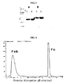


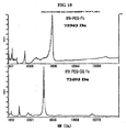
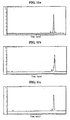

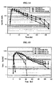
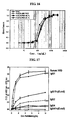

Classifications
-
- A—HUMAN NECESSITIES
- A61—MEDICAL OR VETERINARY SCIENCE; HYGIENE
- A61K—PREPARATIONS FOR MEDICAL, DENTAL OR TOILETRY PURPOSES
- A61K47/00—Medicinal preparations characterised by the non-active ingredients used, e.g. carriers or inert additives; Targeting or modifying agents chemically bound to the active ingredient
- A61K47/50—Medicinal preparations characterised by the non-active ingredients used, e.g. carriers or inert additives; Targeting or modifying agents chemically bound to the active ingredient the non-active ingredient being chemically bound to the active ingredient, e.g. polymer-drug conjugates
- A61K47/51—Medicinal preparations characterised by the non-active ingredients used, e.g. carriers or inert additives; Targeting or modifying agents chemically bound to the active ingredient the non-active ingredient being chemically bound to the active ingredient, e.g. polymer-drug conjugates the non-active ingredient being a modifying agent
- A61K47/68—Medicinal preparations characterised by the non-active ingredients used, e.g. carriers or inert additives; Targeting or modifying agents chemically bound to the active ingredient the non-active ingredient being chemically bound to the active ingredient, e.g. polymer-drug conjugates the non-active ingredient being a modifying agent the modifying agent being an antibody, an immunoglobulin or a fragment thereof, e.g. an Fc-fragment
-
- A—HUMAN NECESSITIES
- A61—MEDICAL OR VETERINARY SCIENCE; HYGIENE
- A61K—PREPARATIONS FOR MEDICAL, DENTAL OR TOILETRY PURPOSES
- A61K47/00—Medicinal preparations characterised by the non-active ingredients used, e.g. carriers or inert additives; Targeting or modifying agents chemically bound to the active ingredient
- A61K47/30—Macromolecular organic or inorganic compounds, e.g. inorganic polyphosphates
- A61K47/42—Proteins; Polypeptides; Degradation products thereof; Derivatives thereof, e.g. albumin, gelatin or zein
-
- A—HUMAN NECESSITIES
- A61—MEDICAL OR VETERINARY SCIENCE; HYGIENE
- A61K—PREPARATIONS FOR MEDICAL, DENTAL OR TOILETRY PURPOSES
- A61K47/00—Medicinal preparations characterised by the non-active ingredients used, e.g. carriers or inert additives; Targeting or modifying agents chemically bound to the active ingredient
- A61K47/50—Medicinal preparations characterised by the non-active ingredients used, e.g. carriers or inert additives; Targeting or modifying agents chemically bound to the active ingredient the non-active ingredient being chemically bound to the active ingredient, e.g. polymer-drug conjugates
- A61K47/51—Medicinal preparations characterised by the non-active ingredients used, e.g. carriers or inert additives; Targeting or modifying agents chemically bound to the active ingredient the non-active ingredient being chemically bound to the active ingredient, e.g. polymer-drug conjugates the non-active ingredient being a modifying agent
- A61K47/68—Medicinal preparations characterised by the non-active ingredients used, e.g. carriers or inert additives; Targeting or modifying agents chemically bound to the active ingredient the non-active ingredient being chemically bound to the active ingredient, e.g. polymer-drug conjugates the non-active ingredient being a modifying agent the modifying agent being an antibody, an immunoglobulin or a fragment thereof, e.g. an Fc-fragment
- A61K47/6801—Drug-antibody or immunoglobulin conjugates defined by the pharmacologically or therapeutically active agent
- A61K47/6803—Drugs conjugated to an antibody or immunoglobulin, e.g. cisplatin-antibody conjugates
- A61K47/6811—Drugs conjugated to an antibody or immunoglobulin, e.g. cisplatin-antibody conjugates the drug being a protein or peptide, e.g. transferrin or bleomycin
- A61K47/6813—Drugs conjugated to an antibody or immunoglobulin, e.g. cisplatin-antibody conjugates the drug being a protein or peptide, e.g. transferrin or bleomycin the drug being a peptidic cytokine, e.g. an interleukin or interferon
-
- A—HUMAN NECESSITIES
- A61—MEDICAL OR VETERINARY SCIENCE; HYGIENE
- A61K—PREPARATIONS FOR MEDICAL, DENTAL OR TOILETRY PURPOSES
- A61K47/00—Medicinal preparations characterised by the non-active ingredients used, e.g. carriers or inert additives; Targeting or modifying agents chemically bound to the active ingredient
- A61K47/50—Medicinal preparations characterised by the non-active ingredients used, e.g. carriers or inert additives; Targeting or modifying agents chemically bound to the active ingredient the non-active ingredient being chemically bound to the active ingredient, e.g. polymer-drug conjugates
- A61K47/51—Medicinal preparations characterised by the non-active ingredients used, e.g. carriers or inert additives; Targeting or modifying agents chemically bound to the active ingredient the non-active ingredient being chemically bound to the active ingredient, e.g. polymer-drug conjugates the non-active ingredient being a modifying agent
- A61K47/68—Medicinal preparations characterised by the non-active ingredients used, e.g. carriers or inert additives; Targeting or modifying agents chemically bound to the active ingredient the non-active ingredient being chemically bound to the active ingredient, e.g. polymer-drug conjugates the non-active ingredient being a modifying agent the modifying agent being an antibody, an immunoglobulin or a fragment thereof, e.g. an Fc-fragment
- A61K47/6835—Medicinal preparations characterised by the non-active ingredients used, e.g. carriers or inert additives; Targeting or modifying agents chemically bound to the active ingredient the non-active ingredient being chemically bound to the active ingredient, e.g. polymer-drug conjugates the non-active ingredient being a modifying agent the modifying agent being an antibody, an immunoglobulin or a fragment thereof, e.g. an Fc-fragment the modifying agent being an antibody or an immunoglobulin bearing at least one antigen-binding site
-
- A—HUMAN NECESSITIES
- A61—MEDICAL OR VETERINARY SCIENCE; HYGIENE
- A61P—SPECIFIC THERAPEUTIC ACTIVITY OF CHEMICAL COMPOUNDS OR MEDICINAL PREPARATIONS
- A61P15/00—Drugs for genital or sexual disorders; Contraceptives
-
- A—HUMAN NECESSITIES
- A61—MEDICAL OR VETERINARY SCIENCE; HYGIENE
- A61P—SPECIFIC THERAPEUTIC ACTIVITY OF CHEMICAL COMPOUNDS OR MEDICINAL PREPARATIONS
- A61P3/00—Drugs for disorders of the metabolism
- A61P3/04—Anorexiants; Antiobesity agents
-
- A—HUMAN NECESSITIES
- A61—MEDICAL OR VETERINARY SCIENCE; HYGIENE
- A61P—SPECIFIC THERAPEUTIC ACTIVITY OF CHEMICAL COMPOUNDS OR MEDICINAL PREPARATIONS
- A61P3/00—Drugs for disorders of the metabolism
- A61P3/08—Drugs for disorders of the metabolism for glucose homeostasis
- A61P3/10—Drugs for disorders of the metabolism for glucose homeostasis for hyperglycaemia, e.g. antidiabetics
-
- A—HUMAN NECESSITIES
- A61—MEDICAL OR VETERINARY SCIENCE; HYGIENE
- A61P—SPECIFIC THERAPEUTIC ACTIVITY OF CHEMICAL COMPOUNDS OR MEDICINAL PREPARATIONS
- A61P35/00—Antineoplastic agents
-
- A—HUMAN NECESSITIES
- A61—MEDICAL OR VETERINARY SCIENCE; HYGIENE
- A61P—SPECIFIC THERAPEUTIC ACTIVITY OF CHEMICAL COMPOUNDS OR MEDICINAL PREPARATIONS
- A61P37/00—Drugs for immunological or allergic disorders
-
- A—HUMAN NECESSITIES
- A61—MEDICAL OR VETERINARY SCIENCE; HYGIENE
- A61P—SPECIFIC THERAPEUTIC ACTIVITY OF CHEMICAL COMPOUNDS OR MEDICINAL PREPARATIONS
- A61P43/00—Drugs for specific purposes, not provided for in groups A61P1/00-A61P41/00
-
- A—HUMAN NECESSITIES
- A61—MEDICAL OR VETERINARY SCIENCE; HYGIENE
- A61P—SPECIFIC THERAPEUTIC ACTIVITY OF CHEMICAL COMPOUNDS OR MEDICINAL PREPARATIONS
- A61P5/00—Drugs for disorders of the endocrine system
- A61P5/06—Drugs for disorders of the endocrine system of the anterior pituitary hormones, e.g. TSH, ACTH, FSH, LH, PRL, GH
-
- A—HUMAN NECESSITIES
- A61—MEDICAL OR VETERINARY SCIENCE; HYGIENE
- A61P—SPECIFIC THERAPEUTIC ACTIVITY OF CHEMICAL COMPOUNDS OR MEDICINAL PREPARATIONS
- A61P5/00—Drugs for disorders of the endocrine system
- A61P5/10—Drugs for disorders of the endocrine system of the posterior pituitary hormones, e.g. oxytocin, ADH
-
- A—HUMAN NECESSITIES
- A61—MEDICAL OR VETERINARY SCIENCE; HYGIENE
- A61P—SPECIFIC THERAPEUTIC ACTIVITY OF CHEMICAL COMPOUNDS OR MEDICINAL PREPARATIONS
- A61P7/00—Drugs for disorders of the blood or the extracellular fluid
-
- A—HUMAN NECESSITIES
- A61—MEDICAL OR VETERINARY SCIENCE; HYGIENE
- A61P—SPECIFIC THERAPEUTIC ACTIVITY OF CHEMICAL COMPOUNDS OR MEDICINAL PREPARATIONS
- A61P7/00—Drugs for disorders of the blood or the extracellular fluid
- A61P7/06—Antianaemics
-
- A—HUMAN NECESSITIES
- A61—MEDICAL OR VETERINARY SCIENCE; HYGIENE
- A61P—SPECIFIC THERAPEUTIC ACTIVITY OF CHEMICAL COMPOUNDS OR MEDICINAL PREPARATIONS
- A61P9/00—Drugs for disorders of the cardiovascular system
-
- C—CHEMISTRY; METALLURGY
- C07—ORGANIC CHEMISTRY
- C07K—PEPTIDES
- C07K14/00—Peptides having more than 20 amino acids; Gastrins; Somatostatins; Melanotropins; Derivatives thereof
- C07K14/435—Peptides having more than 20 amino acids; Gastrins; Somatostatins; Melanotropins; Derivatives thereof from animals; from humans
- C07K14/475—Growth factors; Growth regulators
- C07K14/505—Erythropoietin [EPO]
-
- C—CHEMISTRY; METALLURGY
- C07—ORGANIC CHEMISTRY
- C07K—PEPTIDES
- C07K14/00—Peptides having more than 20 amino acids; Gastrins; Somatostatins; Melanotropins; Derivatives thereof
- C07K14/435—Peptides having more than 20 amino acids; Gastrins; Somatostatins; Melanotropins; Derivatives thereof from animals; from humans
- C07K14/52—Cytokines; Lymphokines; Interferons
- C07K14/53—Colony-stimulating factor [CSF]
- C07K14/535—Granulocyte CSF; Granulocyte-macrophage CSF
-
- C—CHEMISTRY; METALLURGY
- C07—ORGANIC CHEMISTRY
- C07K—PEPTIDES
- C07K14/00—Peptides having more than 20 amino acids; Gastrins; Somatostatins; Melanotropins; Derivatives thereof
- C07K14/435—Peptides having more than 20 amino acids; Gastrins; Somatostatins; Melanotropins; Derivatives thereof from animals; from humans
- C07K14/52—Cytokines; Lymphokines; Interferons
- C07K14/555—Interferons [IFN]
- C07K14/56—IFN-alpha
-
- C—CHEMISTRY; METALLURGY
- C07—ORGANIC CHEMISTRY
- C07K—PEPTIDES
- C07K14/00—Peptides having more than 20 amino acids; Gastrins; Somatostatins; Melanotropins; Derivatives thereof
- C07K14/435—Peptides having more than 20 amino acids; Gastrins; Somatostatins; Melanotropins; Derivatives thereof from animals; from humans
- C07K14/575—Hormones
- C07K14/61—Growth hormones [GH] (Somatotropin)
-
- C—CHEMISTRY; METALLURGY
- C07—ORGANIC CHEMISTRY
- C07K—PEPTIDES
- C07K16/00—Immunoglobulins [IGs], e.g. monoclonal or polyclonal antibodies
- C07K16/46—Hybrid immunoglobulins
-
- C—CHEMISTRY; METALLURGY
- C07—ORGANIC CHEMISTRY
- C07K—PEPTIDES
- C07K19/00—Hybrid peptides, i.e. peptides covalently bound to nucleic acids, or non-covalently bound protein-protein complexes
-
- C—CHEMISTRY; METALLURGY
- C07—ORGANIC CHEMISTRY
- C07K—PEPTIDES
- C07K2317/00—Immunoglobulins specific features
- C07K2317/50—Immunoglobulins specific features characterized by immunoglobulin fragments
- C07K2317/52—Constant or Fc region; Isotype
-
- C—CHEMISTRY; METALLURGY
- C07—ORGANIC CHEMISTRY
- C07K—PEPTIDES
- C07K2317/00—Immunoglobulins specific features
- C07K2317/70—Immunoglobulins specific features characterized by effect upon binding to a cell or to an antigen
- C07K2317/73—Inducing cell death, e.g. apoptosis, necrosis or inhibition of cell proliferation
- C07K2317/732—Antibody-dependent cellular cytotoxicity [ADCC]
-
- C—CHEMISTRY; METALLURGY
- C07—ORGANIC CHEMISTRY
- C07K—PEPTIDES
- C07K2317/00—Immunoglobulins specific features
- C07K2317/70—Immunoglobulins specific features characterized by effect upon binding to a cell or to an antigen
- C07K2317/73—Inducing cell death, e.g. apoptosis, necrosis or inhibition of cell proliferation
- C07K2317/734—Complement-dependent cytotoxicity [CDC]
-
- C—CHEMISTRY; METALLURGY
- C07—ORGANIC CHEMISTRY
- C07K—PEPTIDES
- C07K2319/00—Fusion polypeptide
- C07K2319/30—Non-immunoglobulin-derived peptide or protein having an immunoglobulin constant or Fc region, or a fragment thereof, attached thereto
Definitions
- the present invention relates to an IgG Fc fragment useful as a polypeptide drug carrier, and more particularly, to IgG2 Fc and IgG4 Fc fragments, combinations thereof and hybrids thereof.
- International Pat. Publication No. WO 01/93911 employs a polymer having a plurality of acid moieties as a drug carrier.
- International Pat. Publication No. WO 03/00778 discloses an anionic group-containing amphiphilic block copolymers that, when used as a drug carrier for a cationic drug, improve the stability of the drug.
- European Pat. No. 0 681 481 describes a method of improving the properties of basic drugs by using cyclodextrin and acids as carriers.
- hydrophobic drugs have low stability in vivo mainly due to their low aqueous solubility.
- International Pat. Publication No. WO 04/064731 employs a lipid as a carrier.
- an immunoglobulin Fc fragment as a drug carrier.
- polypeptides are relatively easily denatured due to their low stability, degraded by proteolytic enzymes in the blood and easily eliminated through the kidney or liver
- protein medicaments including polypeptides as pharmaceutically effective components
- this frequent administration of protein medicaments especially through injection causes pain for patients.
- Pharmaceutical compositions with sustained activity therefore, need to increase the stability of protein drugs and maintain the titers at sufficiently high levels without causing immune responses in patients.
- PEG polyethylene glycol
- a conjugate can be prepared by linking an identical protein drug to both ends of PEG to improve the activity of the protein drug.
- two different protein drugs can be linked to both ends of PEG to provide a conjugate having two different activities. The above methods, however, were not very successful in sustaining the activity of protein drugs.
- An alternative method for improving the in vivo stability of physiologically active proteins is by linking a gene of physiologically active protein to a gene encoding a protein having high serum stability by genetic recombination technology and culturing the cells transfected with the recombinant gene to produce a fusion protein.
- a fusion protein can be prepared by conjugating albumin, a protein known to be the most effective in enhancing protein stability, or its fragment to a physiologically active protein of interest by genetic recombination (International Pat. Publication Nos. WO 93/15199 and WO 93/15200 , European Pat. Publication No. 413,622 ).
- a fusion protein of interferon-alpha and albumin developed by the Human Genome Science Company and marketed under the trade name of 'Albuferon TM ', increased the half-life from 5 hours to 93 hours in monkeys, but it was known to be problematic because it decreased the in vivo activity to less than 5% of unmodified interferon-alpha ( Osborn et al., J. Phar. Exp. Ther. 303(2): 540-548, 2002 ). There has been no report of good technology that enhance both the in vivo duration of action and the stability of protein drugs as well as maintaining the in vivo physiological activity of the drugs.
- immunoglobulins and their fragments were employed to enhance the stability of protein drugs.
- U.S. Pat. No. 5,045,312 discloses a method of increasing the activity of .a growth hormone compared to an unmodified growth hormone by conjugating human growth hormone to serum albumin or rat immunoglobulin using a crosslinking agent.
- other attempts were made to fuse a protein drug to an immunoglobulin Fc fragment.
- interferon Kerean Pat. Laid-open Publication No. 2003-9464
- interleukin-4 receptor, interleukin-7 receptor or erythropoietin (EPO) receptor Kerean Pat. Registration No.
- Fc fusion proteins prepared by genetic recombination include a fusion protein of interferon-beta or its derivative and an immunoglobulin Fc fragment (International Pat. Publication NO. WO 00/23472 ), and a fusion protein of IL-5 receptor and an immunoglobulin Fc fragment ( U.S. Pat. NO. 5,712,121 ).
- WO 01/81415 discloses therapeutic PTH polypeptides linked via a peptide linker (fusion protein) to an Fc fragment and also describes the use of non-peptide polymer linkers, such as PEG to link an Fc fragment to a drug.
- U.S. Pat. NO. 5,605,690 discloses a TNFR-IgG1 Fc fusion protein, which is prepared by genetic recombination using an IgG1 Fc fragment having amino acid alterations in the complement binding region or receptor binding region.
- Fc fusion proteins produced by genetic recombination have the following disadvantages: protein fusion occurs only in a specific region of an immunoglobulin Fc fragment, which is at an amino- or carboxyl-terminal end; only homodimeric forms and not monomeric forms are produced; and a fusion could take place only between the glycosylated proteins or between the aglycosylated proteins, and it is impossible to make a fusion protein composed of a glycosylated protein and an aglycosylated protein. Further, a new amino acid sequence created by the fusion may trigger immune responses, and a linker region may become susceptible to proteolytic degradation.
- the inventors of the present application conducted a research, and came to a knowledge that, when an IgG Fc fragment, more particularly an IgG2 or IgG4 Fc fragment, is linked to a polypeptide drug, it could improve the in vivo duration of the drug and minimize a reduction in the in vivo activity.
- the present invention relates to the use of an immunoglobulin G Fc fragment useful as a polypeptide drug carrier, and more preferably IgG2 Fc and IgG4 Fc fragments.
- carrier refers to a substance linked with a drug, which typically increases, decreases or eliminates the physiological activity of the drug by binding to the drug.
- a carrier is employed in the present invention for minimizing a decrease in the physiological activity of a drug of interest, linked to the carrier, while enhancing the in vivo stability of the drug.
- the present invention is characterized by providing particularly an IgG Fc fragment among various substances available as carriers for improving the in vivo duration of action of a drug to which the carrier is conjugated and minimizing a decrease in the in vivo activity of the drug, and more preferably IgG2 Fc and IgG4 Fc fragments.
- Immunoglobulin G collectively means proteins that participate in the body's protective immunity by selectively acting against antigens, and may be derived from humans and animals.
- Immunoglobulins have the following general structure. Immunoglobulins are composed of two identical light chains and two identical heavy chains. The light and heavy chains comprise variable and constant regions.
- heavy chains There are five distinct types of heavy chains based on differences in the amino acid sequences of their constant regions: gamma ( ⁇ ), mu ( ⁇ ), alpha ( ⁇ ), delta ( ⁇ ) and epsilon ( ⁇ ), and the heavy chains include the following subclasses: gamma 1 ( ⁇ 1), gamma 2 ( ⁇ 2), gamma 3 ( ⁇ 3), gamma 4 ( ⁇ 4), alpha 1 ( ⁇ 1) and alpha 2 ( ⁇ 2). Also, there are two types of light chains based on differences in the amino acid sequences of their constant regions: kappa ( ⁇ ) and lambda ( ⁇ ) types ( Coleman et al., Fundamental Immunology, 2nd Ed., 1989, 55-73 ).
- immunoglobulins are classified into five isotypes: IgG, IgA, IgD, IgE and IgM.
- IgG is divided into IgG1, IgG2, IgG3 and IgG4 subclasses.
- immunoglobulins are known to generate several structurally different fragments, which include Fab, F (ab'), F (ab') 2, Fv, scFv, Fd and Fc.
- Fab contains the variable regions of the light chain and the heavy chain, the constant region of the light chain and the first constant region (C H 1) of the heavy chain, and has a single antigen-binding site.
- the Fab' fragments differ from the Fab fragments in terms of having the hinge region containing one or more cysteine residues at the C-terminus (carboxyl terminus) of the heavy chain C H 1 domain.
- the F (ab') 2 fragments are produced as a pair of the Fab' fragments by disulfide bonding formed between cysteine residues of the hinge regions of the Fab' fragments.
- Fv is the minimum antibody fragment that contains only the heavy-chain variable region and the light-chain variable region.
- the scFv (single-chain Fv) fragments comprise both the heavy-chain variable region and the light-chain variable region that are linked to each other by a peptide linker and thus are present in a single polypeptide chain.
- the Fd fragments comprise only the variable region and C H 1 domain of the heavy chain.
- the present invention is characterized by providing an IgG Fc fragment useful as a drug carrier, and more preferably IgG2 Fc and IgG4 Fc fragments.
- immunoglobulin G Fc fragment refers to a protein that contains the heavy-chain constant region 2 (C H 2) and the heavy-chain constant region 3 (C H 3) of an immunoglobulin G, and not the variable regions of the heavy and light chains, the heavy-chain constant region 1 (C H 1) and the light-chain constant region 1 (C L 1) of the immunoglobulin G. It may further include the hinge region at the heavy-chain constant region.
- the IgG Fc fragment of the present invention may contain a portion or the all the heavy-chain constant region 1 (C H 1) and/or the light-chain constant region 1 (C L 1), except for the variable regions of the heavy and light chains. Also, as long as it has a physiological function substantially similar to or better than the native protein the IgG Fc fragment may be a fragment having a deletion in a relatively long portion of the amino acid sequence of C H 2 and/or C H 3.
- the Fc fragment of the present invention includes a native amino acid sequence and sequence derivatives (mutants) thereof.
- An amino acid sequence derivative is a sequence that is different from the native amino acid sequence due to a deletion, an insertion, a non-conservative or conservative substitution or combinations thereof of one or more amino acid residues.
- amino acid residues known to be important in binding at positions 214 to 238, 297 to 299, 318 to 322, or 327 to 331, may be used as a suitable target for modification.
- a deletion may occur in a complement-binding site, such as a C1q-binding site and an ADCC site.
- the Fc fragment may be modified by phosphorylation, sulfation, acrylation, glycosylation, methylation, farnesylation, acetylation, amidation, and the like.
- Fc derivatives are derivatives that have a biological activity identical to the Fc fragment of the present invention or improved structural stability, for example, against heat, pH, or the like.
- these Fc fragments may be obtained from native forms isolated from humans and other animals including cows, goats, swine, mice, rabbits, hamsters, rats and guinea pigs, or may be recombinants or derivatives thereof, obtained from transformed animal cells or microorganisms.
- they may be obtained from a native immunoglobulin by isolating whole immunoglobulins from human or animal organisms and treating them with a proteolytic enzyme. Papain digests the native immunoglobulin into Fab and Fc fragments, and pepsin treatment results in the production of pF'c and F (ab') 2 fragments.
- a human-derived Fc fragment is a recombinant IgG Fc fragment that is obtained from a microorganism. That is, preferred are human-derived recombinant IgG2 Fc and IgG4 Fc fragments obtained from a microorganism.
- the Fc fragment of the present invention may be in the form of having native sugar chains, increased sugar chains compared to a native form or decreased sugar chains compared to the native form, or may be in a deglycosylated form.
- the increase, decrease or removal of sugar chains of the Fc fragment may be achieved by methods common in the art, such as a chemical method, an enzymatic method and a genetic engineering method using a microorganism.
- the removal of sugar chains from an Fc fragment results in a sharp decrease in binding affinity to the C1q part of the first complement component C1 and a decrease or loss in antibody-dependent cell-mediated cytotoxicity (ADCC) or complement-dependent cytotoxicity (CDC), thereby not inducing unnecessary immune responses in vivo.
- an immunoglobulin Fc fragment in a deglycosylated or aglycosylated form may be more suitable to the object of the present invention as a drug carrier.
- a glycosylated Fc has stronger CDC activity than an aglycosylated Fc and thus has a high risk of inducing immune responses.
- an aglycosylated or deglycosylated Fc fragment More preferred are aglycosylated IgG2 Fc and IgG4 Fc fragments, combinations thereof and hybrids thereof.
- deglycosylation refers to that sugar moieties are enzymatically removed from an Fc fragment
- amino acid sequence preferably E. coli
- a dimer or multimer may be formed from two or more fragments selected from the group consisting of IgG1 Fc, IgG2 Fc, IgG3 Fc and IgG4 Fc fragments.
- hybrid means that sequences encoding two or more immunoglobulin Fc fragments of different origin are present in a single-chain immunoglobulin Fc fragment.
- domain hybrids may be composed of one to four domains selected from the group consisting of CH1, CH2, CH3 and CH4 of IgG1 Fc, IgG2 Fc, IgG3 Fc and IgG4 Fc, and may include the hinge region.
- IgG4 has the lowest binding affinity to complement C1q.
- the decrease in binding affinity to the complement results in a decrease in or removal of antibody-dependent cell-mediated cytotoxicity (ADCC) and complement-dependent cytotoxicity (CDC), and thus, unnecessary immune responses are not induced in vivo.
- ADCC antibody-dependent cell-mediated cytotoxicity
- CDC complement-dependent cytotoxicity
- IgG2 and IgG4 Fc fragments have weaker binding affinity to C1q than IgG1, and the IgG4 Fc fragment has the weakest activity.
- the Fc fragment linked to a drug preferably has weaker effector function activities such as ADCC and CDC, with respect to the objects of the present invention, preferred are IgG2 Fc and IgG4 Fc fragments, more preferred is the IgG4 Fc fragment, and most preferred are Fc fragments having the amino acid sequences of SEQ ID Nos. 8, 10 and 23.
- the present invention describes a gene encoding an IgG Fc fragment, preferably genes encoding IgG2 Fc and IgG4 Fc fragments, and more preferably genes encoding the amino acid sequences of SEQ ID Nos. 8, 10 and 23.
- a gene encoding the Fc of the present invention includes a native nucleotide sequence and sequence derivatives thereof.
- a nucleotide sequence derivative means to have a sequence different by a deletion, an insertion, a non-conservative or conservative substitution in one or more nucleotide residues of a native nucleotide sequence, or combinations thereof.
- a gene encoding an Fc fragment having the amino acid sequence of SEQ ID No. 8 is preferably a gene having the nucleotide sequence of SEQ ID No. 4.
- a gene encoding an Fc fragment having the amino acid sequence of SEQ ID No. 10 is preferably a gene having the nucleotide sequence of SEQ ID No. 9.
- a gene encoding an Fc fragment having the amino acid sequence of SEQ ID No. 23 is preferably a gene having the nucleotide sequence of SEQ ID No. 22.
- the nucleotide sequences encoding the Fc fragments of the present invention may be altered by a substitution, a deletion or an insertion of one more bases, or combinations thereof.
- the nucleotide sequences may be naturally isolated or artificially synthesized, or may be prepared by a genetic recombination method.
- nucleotide sequences encoding the Fc fragments of the present invention are provided by vectors expressing the same.
- the present invention describes a recombinant vector comprising an IgG Fc fragment.
- vector means a vehicle for introducing a DNA molecule into a host cell to express an antibody or an antibody fragment.
- the vector useful in the present invention includes plasmid vectors, cosmid vectors, bacteriophage vectors, and viral vectors such as adenovirus vectors, retrovirus vectors and adeno-associated virus vectors.
- the plasmid vector is preferable.
- an expression vector may include expression regulatory elements, such as a promoter, an initiation codon, a stop codon, a polyadenylation signal and an enhancer, and a signal sequence for membrane targeting or secretion.
- signal sequence refers to a specific amino acid sequence that allows transport and secretion of a protein to the outside of the cytosol.
- the signal sequence of the present invention is preferably an E. coli -derived signal sequence, which an E. coli secretory protein possesses.
- E. coli -derived signal sequences include alkaline phosphatase, penicillinase, Ipp, heat-stable enterotoxin II, LamB, PhoE, PelB, OmpA and maltose binding protein. Most preferred is heat-stable enterotoxin II.
- initiation and stop codons are generally considered to be a portion of a nucleotide sequence coding for an immunogenic target protein, are necessary to be functional in an individual to whom a genetic construct has been administered, and must be in frame with the coding sequence.
- Promoters may be generally constitutive or inducible. Non-limiting examples of promoters available in prokaryotic cells include lac, tac, T3 and T7 promoters.
- Non-limiting examples of promoters available in eukaryotic cells include simian virus 40 (SV40) promoter, mouse mammary tumor virus (MMTV) promoter, human immunodeficiency virus (HIV) promoter such as the HIV Long Terminal Repeat (LTR) promoter, moloney virus promoter, cytomegalovirus (CMV) promoter, epstein barr virus (EBV) promoter, rous sarcoma virus (RSV) promoter, as well as promoters from human genes such as human ⁇ -actin, human hemoglobin, human muscle creatine and human metalothionein.
- expression vectors include a selectable marker that allows selection of host cells containing the vector, and replicable expression vectors include a replication origin.
- Genes coding for products that confer resistance to antibiotics or drugs are used as general selectable markers.
- ⁇ -latamase gene ampicillin resistance
- Tet gene tetracycline resistance
- neomycin G418 or Geneticin
- gpt mycophenolic acid
- ampicillin and hygromycin resistant genes may be used in eukaryotic cells.
- Dihydropholate reductase marker gene may be selected by methotrexate in a variety of hosts.
- Genes coding for gene products of auxotrophic markers of hosts, for example, LEU2, URA3 and HIS3, are often used as selectable markers in yeasts.
- viruses e.g., vaculovirus
- phage vectors e.g., phage vectors, and vectors that are able to integrate into the genome of host cells, such as retrovirus vectors.
- a vector which carries a gene coding for the amino acid sequence of SEQ ID No. 8, 10 or 23.
- the following two vectors are constructed: pSTIIdCG2Fc that carries a gene designated as SEQ ID No. 22 encoding the amino acid sequence of SEQ ID No. 23, and pSTIIdCG4Fc that carries a gene designated as SEQ ID No. 4 encoding the amino acid sequence of SEQ ID No. 8.
- a gene designated as SEQ ID No. 9 encoding the amino acid sequence of SEQ ID No. 10 is obtained, the gene having a deletion in the hinge region required for dimer formation from a gene amplified by the PCR, and a pSTIIG4Mo vector carrying the gene is then constructed.
- the present invention describes a transformant transformed with the recombinant vector.
- the most suitable host cell may be selected according to the intended use.
- Available host cells include, but are not limited to, prokaryotic cells such as Escherichia coli, Bacillus subtilis, Streptomyces, Pseudomonas, Proteus mirabilis or Staphylococcus.
- useful as host cells are lower eukaryotic cells, such as fungi (e.g., Aspergillus ) and yeasts (e.g., Pichia pastoris, Saccharomyces cerevisiae, Schizosaccharomyces, Neurospora crassa ),insect cells, plant cells, and cells derived from higher eukaryotes including mammals.
- the prokaryotic host cells are preferable, and in particular, E. coli is most preferable.
- transformation and/or “transfection” into host cells includes any methods by which nucleic acids can be introduced into organisms, cells, tissues or organs, and, as known in the art, may be performed by selecting suitable standard techniques according to host cells. These methods include, but are not limited to, electroporation, protoplast fusion, calcium phosphate (CaPO 4 ) precipitation, calcium chloride (CaCl 2 ) precipitation, and agitation with silicon carbide fiber, agrobacterium-mediated transformation, and PEG-, dextran sulfate-, lipofectamine- and desiccation/inhibition-mediated transformation.
- Transformation into yeasts is typically carried out according to the methods described by Van Solingen et al., J. Bact., 1977, 130:946 , and Hsiao et al., Proc. Natl. Acad. Sci. (USA), 1979, 76:3829 .
- HM10932 pSTIIdCG4Fc-introduced transformant
- HM10933 pSTIIG4Mo-introduced transformant
- HM10936 pSTIIdCG2Fc-introduced transformant
- the present invention describes a method of preparing an immunoglobulin fragment, comprising culturing a transformant transformed with a vector capable of expressing an IgG Fc fragment, and preferably IgG2 Fc or IgG4 Fc fragment, or a combination thereof or a hybrid thereof, under suitable conditions.
- the culturing of tranformant may be performed using suitable media under suitable culture conditions, which are known in the art. This culturing process may be easily adjusted according to the strains selected by those skilled in the art.
- the immunoglobulin fragment of the present invention obtained by culturing the transformant, may be used in an unpurified form, or may be used after being purified with high purities using various general methods, for example, dialysis, salt precipitation and chromatography. Among them, chromatography is most commonly used. As no rule is applicable to any case in selecting the type and sequence of used columns, chromatography may be selected according to the properties and culture method of target proteins of antibodies, for example, from ion exchange chromatography, size exclusion chromatography, affinity chromatography and protein-A affinity column chromatography. In preferred embodiments of the present invention, the immunoglobulin fragment is purified using a protein-A affinity column, a SP sepharose FF column, and the like.
- the Fc fragment thus obtained When the Fc fragment thus obtained is in the free form, it may be converted into a salt form by a per se known method or a modified method. In contrast, when it is obtained in a salt form, the salt may be converted into the free form or another salt by a per se known method or a modified method.
- the Fc fragment as produced by a transformant may be treated before or after purification with an appropriate protein-modifying enzyme for arbitrary modification or partial polypeptide removal.
- the protein-modifying enzyme useful in the present invention include trypsin, chymotrypsin, arginine endopeptidase, protein kinase and glycosidase.
- the Fc fragment of the present invention acts as a drug carrier and forms a conjugate with a drug.
- drug conjugate or “conjugate”, as used herein, means that one or more drugs are linked with one or more immunoglobulin Fc fragments.
- drug refers to a polypeptide substance displaying therapeutic activity when administered to humans or animals.
- physiologically active polypeptide physiologically active protein
- active polypeptide polypeptide drug
- protein drug protein drug
- the polypeptide drug has a disadvantage of being unable to sustain physiological action for a long period of time due to its property of being easily denatured or degraded by proteolytic enzymes present in the body.
- the polypeptide drug when the polypeptide drug is conjugated to the immunoglobulin Fc fragment of the present invention to form a conjugate, the drug has increased structural stability and degradation half-life.
- the polypeptide conjugated to the Fc fragment has a much smaller decrease in physiological activity than other known polypeptide drug formulations. Therefore, compared to the in vivo bioavailability of conventional polypeptide drugs, the conjugate of the polypeptide and the Fc fragment according to the present invention is characterized by having markedly improved in vivo bioavailability.
- IFN ⁇ , G-CSF, hGH and other protein drugs displayed an about two- to six-fold increase in vivo bioavailability compared to their conventional forms conjugated to PEG alone or both PEG and albumin (Tables 8, 9 and 10).
- the linkage of a protein and the Fc fragment of the present invention is featured in that it is not a fusion by a conventional recombination method.
- a fusion form of the immunoglobulin Fc fragment and an active polypeptide used as a drug by a recombination method is obtained in such a way that the polypeptide is linked to the N-terminus or C-terminus of the Fc fragment, and is thus expressed and folded as a single polypeptide from a nucleotide sequence encoding the fusion form.
- the present invention makes it possible to produce a conjugate of a glycosylated active polypeptide and an aglycosylated immunoglobulin Fc fragment, and overcomes all of the above problems, including improving protein production yield, because the two components of the complex are individually prepared and isolated by the best systems.
- Non-limiting examples of protein drugs capable of being conjugated to the immunoglobulin Fc fragment of the present invention include human growth hormone, growth hormone releasing hormone, growth hormone releasing peptide, interferons and interferon receptors (e.g., interferon- ⁇ , - ⁇ and - ⁇ , water-soluble type I interferon receptor, etc.), granulocyte colony stimulating factor (G-CSF), granulocyte-macrophage colony stimulating factor (GM-CSF), glucagon-like peptides (e.g., GLP-1, etc.), G-protein-coupled receptor, interleukins (e.g., IL-1 receptor, IL-4 receptor, etc.), enzymes (e.g., glucocerebrosidase, iduronate-2-sulfatase, alpha-galactosidase-A, agalsidase alpha and beta, alpha-L-iduronidase, butyrylcholinesterase, chi
- physiologically active polypeptides are those requiring frequent dosing upon administration to the body for therapy or prevention of diseases, which include human growth hormone, interferons (interferon- ⁇ , - ⁇ , - ⁇ , etc.), granulocyte colony stimulating factor, erythropoietin (EPO) and antibody fragments.
- interferons interferon- ⁇ , - ⁇ , - ⁇ , etc.
- granulocyte colony stimulating factor erythropoietin (EPO) and antibody fragments.
- EPO erythropoietin
- certain derivatives are included in the scope of the physiologically active polypeptides of the present invention as long as they have function, structure, activity or stability substantially identical to or improved compared over native forms of the physiologically active polypeptides.
- the most preferable polypeptide drug is interferon-alpha.
- the linker is a non-peptide linker.
- This linker is poly (ethylene glycol) (PEG).
- the conjugate of the present invention is made at various molar ratios. That is, the number of the Fc fragment and/or linker linked to a single polypeptide drug is not limited. However, preferably, in the drug conjugate of the present invention, the drug and the Fc fragment are conjugated to each other at a molar ratio of 1:1 to 10:1, and preferably 1:1 to 2:1.
- the Fc fragment of the present invention, a PEG linker and a certain drug may be linked to each other at a certain site of the drug.
- the Fc fragment of the present invention and a polypeptide drug may be linked to each other at an N-terminus or C-terminus, and preferably at a free group, and a covalent bond between the Fc fragment and the drug is easily formed especially at an amino terminal end, an amino group of a lysine residue, an amino group of a histidine residue, or a free cysteine residue.
- the linkage of the Fc fragment of the present invention, a PEG linker and a certain drug may be made in a certain direction. That is, the linker may be linked to the N-terminus, the C-terminus or a free group of the immunoglobulin Fc fragment, and may also be linked to the N-terminus, the C-terminus or a free group of the protein drug.
- the linker is a peptide linker, the linkage may take place at a certain linking site.
- the conjugate of the present invention may be prepared using any of a number of coupling agents known in the art.
- the coupling agents include 1,1-bis (diazoacetyl)-2-phenylethane, glutaradehyde, N-hydroxysuccinimide esters such as esters with 4-azidosalicylic acid, imidoesters including disuccinimidyl esters such as 3,3'-dithiobis (succinimidylpropionate), and bifunctional maleimides such as bis-N-maleimido-1,8-octane.
- conjugate of the novel Fc fragment of the present invention and a drug may offer a various number of pharmaceutical compositions.
- administration means introduction of a predetermined amount of a substance into a patient by a certain suitable method.
- the conjugate of the present invention may be administered via any of the common routes, as long as it is able to reach a desired tissue.
- modes of administration are contemplated, including intraperitoneally, intravenously, intramuscularly, subcutaneously, intradermally, orally, topically, intranasally, intrapulmonarily and intrarectally, but the present invention is not limited to these exemplified modes of administration.
- active ingredients of a composition for oral administration should be coated or formulated for protection against degradation in the stomach.
- the present composition may be administered in an injectable form.
- the pharmaceutical composition of the present invention may be administered using a certain apparatus capable of transporting the active ingredients into a target cell.
- the pharmaceutical composition comprising the conjugate according to the present invention may include a pharmaceutically acceptable carrier.
- the pharmaceutically acceptable carrier may include binders, lubricants, disintegrators, excipients, solubilizers, dispersing agents, stabilizers, suspending agents, coloring agents and perfumes.
- the pharmaceutically acceptable carrier may include buffering agents, preserving agents, analgesics, solubilizers, isotonic agents and stabilizers.
- the pharmaceutically acceptable carrier may include bases, excipients, lubricants and preserving agents.
- the pharmaceutical composition of the present invention may be formulated into a variety of dosage forms in combination with the aforementioned pharmaceutically acceptable carriers.
- the pharmaceutical composition may be formulated into tablets, troches, capsules, elixirs, suspensions, syrups or wafers.
- the pharmaceutical composition may be formulated into a unit dosage form, such as a multidose container or an ampule as a single-dose dosage form.
- the pharmaceutical composition may be also formulated into solutions, suspensions, tablets, capsules and long-acting preparations.
- examples of carriers, exipients and diluents suitable for the pharmaceutical formulations include lactose, dextrose, sucrose, sorbitol, mannitol, xylitol, erythritol, maltitol, starch, acacia rubber, alginate, gelatin, calcium phosphate, calcium silicate, cellulose, methylcellulose, microcrystalline cellulose, polyvinylpyrrolidone, water, methylhydroxybenzoate, propylhydroxybenzoate, talc, magnesium stearate and mineral oils.
- the pharmaceutical formulations may further include fillers, anti-coagulating agents, lubricants, humectants, perfumes, emulsifiers and antiseptics.
- a substantial dosage of a drug in combination with the Fc fragment of the present invention as a carrier may be determined by several related factors including the types of diseases to be treated, administration routes, the patient's age, gender, weight and severity of the illness, as well as by the types of the drug as an active component. Since the pharmaceutical composition of the present invention has a very long duration of action in vivo, it has an advantage of greatly reducing administration frequency of pharmaceutical drugs.
- EXAMPLE 1 Construction of human immunoglobulin IgG4 Fc expression vector
- RNA isolated from human blood cells was isolated from human blood cells as a template, as follows. First, total RNA was isolated from about 6 ml of blood using a Qiamp RNA blood kit (Qiagen), and gene amplification was performed using the total RNA as a template and a One-Step RT-PCR kit (Qiagen). To obtain a desired gene sequence, a pair of primers represented by SEQ ID Nos. 1 and 2 was used. SEQ ID No.
- SEQ ID No. 1 is a nucleotide sequence starting from the 10th residue, serine, of 12 amino acid residues of the hinge region of IgG4 (Glu Ser Lys Tyr Gly Pro Pro Cys Pro Ser Cys Pro: SEQ ID No. 3).
- SEQ ID No. 2 was designed to have a BamHI recognition site containing a stop codon.
- the gene amplified using the primer set was identified to have the nucleotide sequence represented by SEQ ID No. 4 and contain an amino terminal end, starting with the Ser-Cys-Pro sequence of the hinge region of a full-length IgG4 Fc gene sequence, and CH2 and CH3 domains. To clone the amplified IgG4 Fc gene fragment into an expression vector containing an E.
- an expression vector pT14S1SH-4T20V22Q (Korean Pat. No. 38061 ), previously developed by the present inventors, was used as a starting vector.
- This expression vector contains an E. coli heat-stable enterotoxin signal sequence derivative having the nucleotide sequence represented by SEQ ID No. 5.
- a StuI recognition site was inserted into an end of the E. coli heat-stable enterotoxin signal sequence derivative of the pT14S15H-4T20V22Q plasmid through site-directed mutagenesis using a pair of primers represented by SEQ ID Nos.
- pmSTII pmSTII
- the amplified IgG4 Fc gene fragment was digested with BamHI and ligated with the linearized expression vector, thus providing a pSTIIdCG4Fc plasmid.
- This vector expresses a protein that has the amino acid sequence of SEQ ID No. 8 and is present in a dimeric form by disulfide bonds between cysteine residues in the hinge region.
- the final expression vector was transformed into E. coli BL21 (DE3), and the resulting transformant was designated as "BL21/pSTIIdCG4Fc (HM10932)", which was deposited at the Korean Culture Center of Microorganisms (KCCM) on Sep. 15, 2004 and assigned accession number KCCM-10597.
- PCR was carried out using a pair of primers represented by SEQ ID Nos. 9 and 2 and the pSTIIdCG4Fc plasmid prepared in the above ⁇ 1-1> as a template.
- the PCR was designed for a PCR product to have a deletion in the hinge region required for dimer formation from the IgG4 Fc sequence, and thus, only the CH2 and CH3 domains of IgG4 Fc were amplified.
- the PCR product was cloned into an expression vector, pmSTII, according to the same procedure as in the above ⁇ 1-1>, thus providing a pSTIIG4Mo plasmid.
- This expression vector was transformed into E. coli BL21 (DE3), and the resulting transformant was designated as "BL21/pSTIIG4Mo (HM10933)", which was deposited at the Korean Culture Center of Microorganisms (KCCM) on Sep. 15, 2004 and assigned accession number KCCM-10598.
- a protein expressed by the expression vector has the amino acid sequence of SEQ ID No. 10, and is expressed from the CH2 domain and present in a monomeric form because it has no hinge region.
- RT-PCR was carried out according to the same method as in the ⁇ 1-1> of Example 1 using RNA isolated from human blood cells as a template using a One-Step RT-PCR kit (Qiagen). To obtain a desired gene sequence, a pair of primers represented by SEQ ID Nos. 11 and 12 was used.
- SEQ ID No. 11 is a nucleotide sequence starting from the 13th residue, proline, of 15 amino acid residues of the hinge region (Glu Pro Lys Ser Cys Asp Lys Thr His Thr Cys Pro Pro Cys Pro: SEQ ID No. 13).
- the gene amplified using the pair of primers represented by SEQ ID Nos. 11 and 12 was found to contain an amino terminal end starting with the Pro-Cys-Pro sequence of the hinge region and CH2 and CH3 domains, among a full-length IgG1 Fc gene sequence, and has the nucleotide sequence of SEQ ID No. 14.
- the aforementioned pmSTII vector was used.
- the pmSTII plasmid was treated with StuI and BamHI and subjected to agarose gel electrophoresis, and a large fragment (4.7 kb), which contained the E. coli heat-stable enterotoxin signal sequence derivative, was purified.
- the amplified IgG1 Fc gene was digested with BamHI and ligated with the linearized expression vector, thus providing pSTIIdCG1Fc.
- This vector expresses in a host cell a protein that has the amino acid sequence of SEQ ID No. 15 and is present in a dimeric form by disulfide bonds between cysteine residues in the hinge region.
- the final expression vector was transformed into E. coli BL21 (DE3), and the resulting transformant was designated as "BL21/pSTIIdCG1Fc (HM10927)", which was deposited at the Korean Culture. Center of Microorganisms (KCCM) on Sep. 15, 2004 and assigned accession number KCCM-10588.
- PCR was carried out using a pair of primers represented by SEQ ID Nos. 16 and 12 and the pSTIIdCG1Fc plasmid prepared in the ⁇ 2-1> of Example 2 as a template.
- the PCR product was cloned into an expression vector, pmSTII, according to the same procedure as in the ⁇ 2-1> of Example 2, thus providing a pSTIIG1Mo plasmid containing the nucleotide sequence represented by SEQ ID No. 17. This expression vector was transformed into E.
- BL21/pSTIIG1Mo HM10930
- KCCM Korean Culture Center of Microorganisms
- a protein expressed by the expression vector is expressed from the CH2 domain and presents in a monomeric form because it was deleted from the hinge region containing cysteine residues allowing dimer formation, and has the amino acid sequence of SEQ ID No. 18.
- RT-PCR was carried out according to the same method as in the ⁇ 1-1> of Example 1 using RNA isolated from human blood cells as a template using a One-Step RT-PCR kit (Qiagen).
- a pair of primers represented by SEQ ID Nos. 19 and 20 was used.
- SEQ ID No. 19 is a nucleotide sequence starting from the 10th residue, proline, of 12 amino acid residues of the hinge region (Glu Arg Lys Cys Cys Val Glu Cys Pro Pro Cys Pro: SEQ ID No. 21).
- SEQ ID Nos. 19 is a nucleotide sequence starting from the 10th residue, proline, of 12 amino acid residues of the hinge region (Glu Arg Lys Cys Cys Val Glu Cys Pro Pro Cys Pro: SEQ ID No. 21).
- coli heat-stable enterotoxin signal sequence derivative was purified. Then, the amplified IgG1 Fc gene fragment was digested with BamHI and ligated with the linearized expression vector fragment, thus providing pSTIIdCG2Fc.
- This vector expresses in a host cell a protein that has the amino acid sequence of SEQ ID No. 23 and is present in a dimeric form by disulfide bonds between cysteine residues in the hinge region.
- the final expression vector was transformed into E. coli BL21 (DE3), and the resulting transformant was designated as BL21/pSTIIdCG2Fc (HM10936).
- Bacterial transformants prepared in Examples 1, 2 and 3 were individually inoculated in a fermentor (Marubishi Company) and allowed to ferment, and were evaluated to determine whether they express immunoglobulin Fc fragments.
- each transformant was grown in 100 ml of LB medium with agitation overnight and inoculated in the fermentor for large-scale culture.
- the fermentor was maintained at 30°C or 35°C.
- the cultures were aerated with 20-vvm air and stirred at 500 rpm.
- the cultures were supplemented with glucose and yeast extracts according to the fermented states of bacteria.
- an inducer IPTG, was added to the cultures in an amount of 20 ⁇ M to 4 mM to induce protein expression.
- the cultures were further cultured for 40 to 45 hrs until the OD value at 600 nm increased to 100 to 120.
- immunoglobulin Fc in the E. coli transformants and the expressed sites, water solubility and dimer formation of the expressed Ig Fc were examined as follows. To determine whether an expressed product is secreted to the fermentation fluid or the periplasmic space of E. coli by the signal sequence fused to the expression vector, the fermentation fluid was centrifuged to obtain a cell-free fermentation fluid and collect cells. The cell-free fermentation fluid and a periplasmic space solution obtained by osmotic shock of the collected cells were subjected to Western blot analysis. As a result, a very small amount of immunoglobulin Fc was detected. To investigate intracellular expression of Ig Fc, cells were disrupted using an ultrasonicator (Misonix Company).
- the resulting cell lysate was centrifuged to separate water-soluble substances from water-insoluble substances, and the water-soluble substances were subjected to Western blot analysis, as follows.
- the water-soluble substances were mixed with a protein sample buffer not containing a reducing agent such as DTT or ⁇ -mercaptoethanol, and separated on a 15% SDS-PAGE gel (Criterion Gel, Bio-Rad).
- proteins were transferred onto a nitrocellulose membrane and detected with an HRP-conjugated anti-human Fc antibody (Sigma).
- HRP-conjugated anti-human Fc antibody Sigma.
- immunoglobulin Fc was overexpressed in a water-soluble form and located in the cytosol of E. coli.
- products expressed by transformants transformed with expression vectors expressing Ig Fc having a portion of a hinge region, were expressed as dimers.
- lanes 1, 2 and 3 show products expressed in HM10927, HM10932 and HM10936, respectively, and lane 4 shows Fc generated by papain treatment of immunoglobulins produced in animal cells, which showed a slightly larger size due to its sugar moieties on the SDS-PAGE gel than that produced in E. coli.
- the water-soluble dimeric Ig Fc fragments which were located in the cytosol of E. coli as demonstrated in the above ⁇ 4-1>, were designed to be translated in a fused form to a signal sequence.
- N-terminal amino acid sequences of the Ig Fc fragments were determined by the Basic Science Research Institute, Seoul, Korea. Samples used in the N-terminal amino acid sequence analysis were prepared as follows.
- a PVDF membrane (Bio-Rad) was immersed in methanol for about 2-3 sec to be activated, and was sufficiently wet with a blocking buffer (170 mM glycine, 25 mM Tris-HCl (pH 8.0), 20% methanol).
- the IgG1 Fc protein was found to have an N-terminal sequence of Pro-Cys-Pro-Ala-Pro-Glu-Leu-Leu-Gly-Gly
- the IgG4 Fc protein had an N-terminal sequence of Ser-Cys-Pro-Ala-Pro-Glu-Phe-Leu-Gly-Gly
- the IgG2 Fc protein had an N-terminal sequence of Pro-Cys-Pro-Ala-Pro-Pro-Val-Ala-Gly-Pro.
- the Fc fragments expressed by the E. coli transformants of the present invention were found to have an accurate N-terminal sequence.
- Immunoglobulin Fc was purified using a protein-A affinity column known to have strong affinity to immunoglobulins, as follows.
- E. coli cells collected by centrifuging fermentation fluids were disrupted by a microfluizer (Microfludics) to give cell lysates.
- the cell lysates were subjected to two-step column chromatography to purify recombinant immunoglobulin Fc fragments present in the cytosol.
- 5 ml of a protein-A affinity column (Pharmacia) was equilibrated with PBS, and the cell lysates were loaded onto the column at a flow rate of 5 ml/min. Unbound proteins were washed out with PBS, and bound proteins were eluted with 100 mM citrate (pH 3.0).
- the collected fractions were desalted using a HiPrep 26/10 desalting column (Pharmacia) with 10 mM Tris buffer (pH 8.0). Then, secondary anion exchange column chromatography was carried out using 50 ml of a Q HP 26/10 column (Pharmacia). The primary purified recombinant immunoglobulin Fc fractions were loaded onto the Q-Sepharose HP 26/10 column, and the column was eluted with a linear gradient of 0-0.2 M NaCl in 10 mM Tris buffer (pH 8.0), thus providing highly pure fractions. After being partially purified using the protein-A affinity column, expression levels of the recombinant Ig Fc fragments were determined as follows.
- immunoglobulin Fc proteins thus obtained are present in a dimeric or monomeric form of the heavy chain, they have different migration patterns on reduced SDS-PAGE and non-reduced SDS-PAGE.
- SDS-PAGE analysis performed to determine protein purities after expressed products were purified, are given in FIGS. 2 and 3 .
- FIGS. 2 and 3 show the results of SDS-PAGE analysis of the purified immunoglobulin Fc fragments in a dimeric or monomeric form under non-reduced and reduced conditions using a criterion gel (Bio-Rad), wherein the Fc fragments were evaluated for differential migration on reduced versus non-reduced gels.
- the A region shows proteins separated on a non-reduced SDS-PAGE gel
- the B region shows proteins on a reduced SDS-PAGE gel.
- Lane M indicates a prestained low-range standard protein marker (Bio-Rad)
- lanes 1 to 4 indicate protein samples for immunoglobulin Fc produced by E.
- HM10927, HM10928 (deposited at the Korean Culture Center of Microorganisms (KCCM) on Sep. 15, 2004 and assigned accession number KCCM-10589), HM10929 (deposited at KCCM on Sep. 15, 2004 and assigned accession number KCCM-10594) and HM10932, respectively.
- KCCM Korean Culture Center of Microorganisms
- HM10929 (deposited at KCCM on Sep. 15, 2004 and assigned accession number KCCM-10594) and HM10932, respectively.
- FIG. 2 on reduced SDS-PAGE, the Ig Fc fragments were present in a monomeric form because disulfide bonds formed between cysteine residues of the hinge region were reduced, and were thus migrated the monomer distance. In contrast, on non-reduced SDS-PAGE, the Ig Fc fragments were present in a dimeric form by disulfide bonds and thus had a migration distance of about 42 kDa.
- the A region shows proteins separated on a non-reduced SDS-PAGE gel
- the B region shows proteins on a reduced SDS-PAGE gel.
- Lane M indicates the standard protein marker
- lanes 1 and 2 indicate protein samples for immunoglobulin Fc produced by E. coli transformants, HM10930 and HM10933, respectively.
- the proteins did not show a large difference in migration on reduced versus non-reduced gels, and only displayed a slightly different migration due to the reduction of intramolecular disulfide bonds.
- An immunoglobulin Fc fragment was prepared as follows. 200 mg of 150-kDa immunoglobulin G (IgG) (Green Cross, Korea) dissolved in 10 mM phosphate buffer was treated with 2 mg of a proteolytic enzyme, papain (Sigma) at 37°C for 2 hrs with gentle agitation. After the enzyme reaction, the immunoglobulin Fc fragment regenerated thus was subjected to chromatography for purification using sequentially a Superdex column, a protein A column and a cation exchange column.
- IgG immunoglobulin G
- papain Sigma
- reaction solution was loaded onto a Superdex 200 column (Pharmacia) equilibrated with 10 mM sodium phosphate buffer (PBS, pH 7.3), and the column was eluted with the same buffer at a flow rate of 1 ml/min.
- Fab fragments having a molecular weight similar to the Ig Fc fragment were eliminated by protein A column chromatography ( FIG. 4 ).
- the resulting fractions containing the Ig Fc fragment eluted from the Superdex 200 column were loaded at a flow rate of 5 ml/min onto a protein A column (Pharmacia) equilibrated with 20 mM phosphate buffer (pH 7.0), and the column was washed with the same buffer to remove proteins unbound to the column. Then, the protein A column was eluted with 100 mM sodium citrate buffer (pH 3.0) to obtain highly pure immunoglobulin Fc fragment.
- the Fc fractions collected from the protein A column were finally purified using a cation exchange column (polyCAT, PolyLC Company), wherein this column loaded with the Fc fractions was eluted with a linear gradient of 0.15-0.4 M NaCl in 10 mM acetate buffer (pH 4.5), thus providing highly pure Fc fractions.
- the highly pure Fc fractions were analyzed by 12% SDS-PAGE (lane 2 in FIG. 5 ).
- ALD-PEG-ALD Shearwater
- human interferon alpha-2b hIFN ⁇ -2b, MW: 20 kDa
- a reducing agent sodium cyanoborohydride (NaCNBH 3 , Sigma)
- NaCNBH 3 sodium cyanoborohydride
- the reaction mixture was subjected to size exclusion chromatography using a Superdex R column (Pharmacia).
- the IFN ⁇ -PEG complex was eluted from the column using 10 mM potassium phosphate buffer (pH 6.0) as an elution buffer, and interferon alpha not linked to PEG, unreacted PEG and dimer byproducts where PEG was linked to two interferon alpha molecules were removed.
- the purified IFN ⁇ -PEG complex was concentrated to 5 mg/ml.
- the optimal reaction molar ratio for IFN ⁇ to PEG providing the highest reactivity and generating the smallest amount of byproducts such as dimers, was found to be 1:2.5 to 1:5.
- the immunoglobulin Fc fragment (about 53 kDa) prepared in the above step 1 was dissolved in 10 mM phosphate buffer and mixed with the IFN ⁇ -PEG complex at an IFN ⁇ -PEG complex: Fc molar ratio of 1:1, 1:2, 1:4 and 1:8.
- Fc molar ratio of 1:1, 1:2, 1:4 and 1:8.
- the reaction mixture was subjected to Superdex size exclusion chromatography so as to eliminate unreacted substances and byproducts and purify the IFN ⁇ -PEG-Fc protein conjugate produced.
- 10 mM phosphate buffer pH 7.3 was passed through the column at a flow rate of 2.5 ml/min to remove unbound Fc and unreacted substances, followed by column elution to collect IFN ⁇ -PEG-Fc protein conjugate fractions.
- IFN ⁇ -PEG-Fc protein conjugate fractions contained a small amount of impurities, unreacted Fc and interferon alpha dimers, cation exchange chromatography was carried out to remove the impurities.
- the IFN ⁇ -PEG-Fc protein conjugate fractions were loaded onto a PolyCAT LP column (PolyLC) equilibrated with 10 mM sodium acetate (pH 4.5), and the column was eluted with a linear gradient of 0-0.5 M NaCl in 10 mM sodium acetate buffer (pH 4.5) using 1 M NaCl. Finally, the IFN ⁇ -PEG-Fc protein conjugate was purified using an anion exchange column.
- the IFN ⁇ -PEG-Fc protein conjugate fractions were loaded onto a PolyWAX LP column (PolyLC) equilibrated with 10 mM Tris-HCl (pH 7.5), and the column was then eluted with a linear gradient of 0-0.3 M NaCl in 10 mM Tris-HCl (pH 7.5) using 1 M NaCl, thus isolating the IFN ⁇ -PEG-Fc protein conjugate in a highly pure form.
- ALD-PEG-ALD Shearwater
- the immunoglobulin Fc fragment prepared in the step 1 of Example 5 at Fc: PEG molar ratios of 1:1, 1:2.5, 1:5, 1:10 and 1:20, wherein the Ig Fc fragment had been dissolved in 100 mM phosphate buffer in an amount of 15 mg/ml.
- a reducing agent, NaCNBH 3 Sigma
- the reaction mixture was subjected to size exclusion chromatography using a Superdex R column (Pharmacia).
- the Fc-PEG complex was eluted from the column using 10 mM potassium phosphate buffer (pH 6.0) as an elution buffer, and immunoglobulin Fc fragment not linked to PEG, unreacted PEG and dimer byproducts where PEG was linked to two immunoglobulin Fc fragment molecules were removed.
- the purified Fc-PEG complex was concentrated to about 15 mg/ml.
- the Fc-PEG complex was mixed with IFN ⁇ dissolved in 10 mM phosphate buffer at Fc-PEG complex: IFN ⁇ molar ratios of 1:1, 1:1.5, 1:3 and 1:6.
- a reducing agent, NaCNBH 3 was added to the reaction solution at a final concentration of 20 mM and was allowed to react at 4°C for 20 hrs with gentle agitation.
- unreacted substances and byproducts were removed according to the same purification method as in the step 4 of Example 5, thus isolating the Fc-PEG-IFN ⁇ protein conjugate in a highly pure form.
- hGH-PEG-Fc conjugate was prepared and purified according to the same method as in Example 5, except that drug other than interferon alpha, human growth hormone (hGH, MW: 22 kDa) was used and a hGH:PEG molar ratio was 1:5.
- drug other than interferon alpha human growth hormone (hGH, MW: 22 kDa) was used and a hGH:PEG molar ratio was 1:5.
- G-CSF-PEG-Fc conjugate was prepared and purified according to the same method as in Example 5, except that drug other than interferon alpha, human granulocyte colony stimulating factor (G-CSF), was used and an G-CSF: PEG molar ratio was 1:5.
- G-CSF human granulocyte colony stimulating factor
- a 17 S-G-CSF-PEG-Fc protein conjugate was prepared and purified according to the same method as described above using a G-CSF derivative, 17 S-G-CSF, having a serine substitution at the seventeenth amino acid residue of the native G-CSF.
- EPO-PEG-Fc conjugate was prepared and purified according to the same method as in Example 5, except that drug other than interferon alpha, human erythropoietin (EPO), was used and an EPO: PEG molar ratio was 1:5.
- EPO human erythropoietin
- IFN ⁇ -PEG-Fc protein conjugate was prepared using PEG having a succinimidyl propionate (SPA) reactive group at both ends, as follows. 3.4-kDa polyethylene glycol, SPA-PEG-SPA (Shearwater), was mixed with 10 mg of interferon alpha dissolved in 100 mM phosphate buffer at IFN ⁇ :PEG molar ratios of 1:1, 1:2.5, 1:5, 1:10 and 1:20. The mixture was then allowed to react at room temperature with gentle agitation for 2 hrs.
- SPA succinimidyl propionate
- IFN ⁇ -PEG complex a 1:1 complex of PEG and interferon alpha
- the reaction mixture was subjected to Superdex size exclusion chromatography.
- the IFN ⁇ -PEG complex was eluted from the column using 10 mM potassium phosphate buffer (pH 6.0) as an elution buffer, and interferon alpha not linked to PEG, unreacted PEG and dimer byproducts in which two interferon alpha molecules were linked to both ends of PEG were removed.
- the purified IFN ⁇ -PEG complex was concentrated to about 5 mg/ml, and an IFN ⁇ -PEG-Fc conjugate was prepared and purified according to the same methods as in the steps 3 and 4 of Example 5.
- the optimal reaction molar ratio for IFN ⁇ to PEG providing the highest reactivity and generating the fewest byproducts such as dimers, was found to be 1:2.5 to 1:5.
- IFN ⁇ -PEG-Fc conjugate was prepared according to the same methods as described above using PEG) having an N-hydroxysuccinimidyl (NHS) reactive group at both ends, NHS-PEG-NHS (Shearwater), or PEG having a buthyl aldehyde reactive group at both ends, BUA-PEG-BUA (Shearwater).
- PEG having an N-hydroxysuccinimidyl (NHS) reactive group at both ends
- NHS-PEG-NHS Shearwater
- BUA-PEG-BUA Shearwater
- An IFN ⁇ -10K PEG complex was prepared using 10-kDa polyethylene glycol having an aldehyde reactive group at both ends, ALD-PEG-ALD (Shearwater). This complex was prepared and purified according to the same method as in the step 2 of Example 5. Through this experiment, the optimal reaction molar ratio for IFN ⁇ to 10-kDa PEG, providing the highest reactivity and generating the fewest byproducts such as dimers, was found to be 1:2.5 to 1:5. The purified IFN ⁇ -10K PEG complex was concentrated to about 5 mg/ml, and, using this concentrate, an IFN ⁇ -10K PEG-Fc conjugate was prepared and purified according to the same methods as in the steps 3 and 4 of Example 5.
- An E. coli transformant, BL21/poDLHF (accession number: KCCM-10511), expressing anti-tumor necrosis factor-alpha Fab' was grown in 100 ml of LB medium overnight with agitation, and was inoculated in a 5-L fermentor (Marubishi) and cultured at 30°C and 500 rpm and at an air flow rate of 20 vvm. To compensate for the insufficient nutrients for bacterial growth during fermentation, the cultures were supplemented with glucose and yeast extracts according to the fermented states of bacteria. When the cultures reached an OD 600 value of 80-100, an inducer, IPTG, was added to the cultures to induce protein expression. The cultures were further cultured for 40 to 45 hrs until the OD value at 600 nm increased to 120 to 140. The fermentation fluid thus obtained was centrifuged at 20,000xg for 30 min. The supernatant was collected, and the pellet was discarded.
- the supernatant was subjected to the following three-step column chromatography to purify anti-tumor necrosis factor-alpha Fab'.
- the supernatant was loaded onto a HiTrap protein G column (5 ml, Pharmacia) equilibrated with 20 mM phosphate buffer (pH 7.0), and the column was eluted with 100 mM glycine (pH 3.0).
- the collected Fab' fractions were then loaded onto a Superdex 200 column (Pharmacia) equilibrated with 10 mM sodium phosphate buffer (PBS, pH 7.3), and this column was eluted with the same buffer.
- the immunoglobulin Fc prepared according to the same method as in the step 1 of Example 5 was dissolved in 100 mM phosphate buffer (pH 6.0) at a concentration of 5 mg/ml, and was mixed with NHS-PEG-MAL (3.4 kDa, Shearwater) at an Fc: PEG molar ratio of 1:10, followed by incubation at 4°C for 12 hrs with gentle agitation.
- reaction buffer was exchanged with 20 mM sodium phosphate buffer (pH 6.0) to remove unbound NHS-PEG-MAL. Then, the reaction mixture was loaded onto a polyCAT column (PolyLC) . The column was eluted with a linear NaCl gradient of 0.15-0.5 M in 20 mM sodium phosphate buffer (pH 6.0). During this elution, the immunoglobulin Fc-PEG complex was eluted earlier than unreacted immunoglobulin Fc, and the unreacted Ig Fc was eluted later, thereby eliminating the unreacted Ig Fc molecules.
- the Fab' purified in the above step 1 was dissolved in 100 mM sodium phosphate buffer (pH 7.3) at a concentration of 2 mg/ml, and was mixed with the immunoglobulin Fc-PEG complex prepared in the above step 2 at a Fab': complex molar ratio of 1:5.
- the reaction mixture was concentrated to a final protein concentration of 50 mg/ml and incubated at 4°C for 24 hrs with gentle agitation.
- the reaction mixture was loaded onto a Superdex 200 column (Pharmacia) equilibrated with 10 mM sodium phosphate buffer (pH 7.3), and the column was eluted with the same buffer at a flow rate of 1 ml/min.
- the coupled Fab'-S-PEG-N-Fc conjugate was eluted relatively earlier due to its high molecular weight, and unreacted immunoglobulin Fc-PEG complex and Fab' were eluted later, thereby eliminating the unreacted molecules.
- the collected Fab'-S-PEG-N-Fc conjugate fractions were again loaded onto a polyCAT 21x250 column (PolyLC), and this column was eluted with a linear NaCl gradient of 0.15-0.5 M in 20 mM sodium phosphate buffer (pH 6.0), thus providing a pure Fab'-S-PEG-N-Fc conjugate comprising the Fc-PEG complex linked to an -SH group near the C-terminus of the Fab'.
- 40 mg of the Fab' purified in the step 1 of Example 12 was dissolved in 100 mM sodium phosphate buffer (pH 6.0) at a concentration of 5 mg/ml, and was mixed with butyl ALD-PEG-butyl ALD (3.4 kDa, Nektar) at a Fab': PEG molar ratio of 1:5.
- a reducing agent, NaCNBH 3 was added to the reaction mixture at a final concentration of 20 mM, and the reaction mixture was then allowed to react at 4°C for 2 hrs with gentle agitation.
- the reaction buffer was exchanged with 20 mM sodium phosphate buffer (pH 6.0). Then, the reaction mixture was loaded onto a polyCAT column (PolyLC). The column was eluted with a linear NaCl gradient of 0.15-0.4 M in 20 mM acetate buffer (pH 4.5). During this column elution, the Fab'-PEG complex comprising the PEG linker lined to the N-terminus of the Fab' was eluted earlier than unreacted. Fab', and the unreacted Fab' was eluted later, thereby eliminating the unreacted Fab' molecules.
- the Fab'-PEG complex was dissolved in 100 mM sodium phosphate buffer (pH 6.0) at a concentration of 10 mg/ml, and was mixed with the immunoglobulin Fc dissolved in the same buffer at a Fab'-PEG complex: Fc molar ratio of 1:5.
- a reducing agent, NaCNBH 3 was added to the reaction mixture at a final concentration of 20 mM, and the reaction mixture was then reacted at 4°C for 24 hrs with gentle agitation.
- the reaction mixture was loaded onto a Superdex 200 column (Pharmacia) equilibrated with 10 mM sodium phosphate buffer (pH 7.3), and the column was eluted with the same buffer at a flow rate of 1 ml/min.
- the coupled Fab'-N-PEG-N-Fc conjugate was eluted relatively earlier due to its high molecular weight, and unreacted immunoglobulin Fc and Fab'-PEG complex were eluted later, thereby eliminating the unreacted molecules.
- the collected Fab'-N-PEG-N-Fc conjugate fractions were again loaded onto a polyCAT 21x250 column (PolyLC), and this column was eluted with a linear NaCl gradient of 0.15-0.5 M in 20 mM sodium phosphate buffer (pH 6.0), thus providing a pure Fab'-N-PEG-N-Fc conjugate comprising the immunoglobulin Fc-PEG complex linked to the N-terminus of the Fab'.
- an immunoglobulin Fc prepared according to the same method as in Example 5 was dissolved in 100 mM phosphate buffer (pH 7.5) at a concentration of 2 mg/ml, and was mixed with 300 U/mg of a deglycosylase, PNGase F (NEB).
- the reaction mixture was allowed to react at 37°C for 24 hrs with gentle agitation.
- the reaction mixture was loaded onto a SP Sepharose FF column (Pharmacia), and the column was eluted with a linear NaCl gradient of 0.1-0.6 M in 10 mM acetate buffer (pH 4.5) using 1 M NaCl.
- the native immunoglobulin Fc was eluted earlier, and the deglycosylated immunoglobulin Fc (DG Fc) was eluted later.
- the IFN ⁇ -PEG complex was mixed with the DG Fc dissolved in 10 mM phosphate buffer at IFN ⁇ -PEG complex: DG Fc molar ratios of 1:1, 1:2, 1:4 and 1:8.
- a reducing agent, NaCNBH 3 was added to the reaction solution at a final concentration of 20 mM and was allowed to react at 4°C for 20 hrs with gentle agitation.
- the reaction mixture was subjected to size exclusion chromatography using a Superdex R column (Pharmacia) so as to eliminate unreacted substances and byproducts and purify the IFN ⁇ -PEG-DG Fc protein conjugate.
- a phosphate buffer pH 7.3 was passed through the column at a flow rate of 2.5 ml/min to remove unbound DG Fc and unreacted substances, followed by column elution to collect IFN ⁇ -PEG-DG Fc protein conjugate fractions.
- IFN ⁇ -PEG-DG Fc protein conjugate fractions contained a small amount of impurities, unreacted DG Fc and IFN ⁇ -PEG complex, cation exchange chromatography was carried out to remove the impurities.
- the IFN ⁇ -PEG-DG Fc protein conjugate fractions were loaded onto a PolyCAT LP column (PolyLC) equilibrated with 10 mM sodium acetate (pH 4.5), and the column was eluted with a linear gradient of 0-0.6 M NaCl in 10 mM sodium acetate buffer (pH 4.5) using 1 M NaCl. Finally, the IFN ⁇ -PEG-DG Fc protein conjugate was purified using an anion exchange column.
- the IFN ⁇ -PEG-Fc protein conjugate fractions were loaded onto a PolyWAX LP column (PolyLC) equilibrated with 10 mM Tris-HCl (pH 7.5), and the column was then eluted with a linear gradient of 0-0.3 M NaCl in 10 mM Tris-HCl (pH 7.5) using 1 M NaCl, thus isolating the IFN ⁇ -PEG-DG Fc protein conjugate in a highly pure form.
- the IFN ⁇ -PEG complex was linked to the N terminus of the IgG4 delta-Cys as an AG Fc derivative prepared in Example 1.
- the thus-produced IFN ⁇ -PEG-AG Fc protein conjugate (I) was primarily purified using 50 ml of a Q HP 26/10 column (Pharmacia) and further purified by a highpressure liquid chromatographic assay using a polyCAT 21.5x250 column (polyLC), thus purifying the conjugate to a high degree.
- the coupling reaction solution was desalted using a HiPrep 26/10 desalting column (Pharmacia) with 10 mM Tris buffer (pH 8.0). Then, the reaction solution was then loaded onto 50 ml of a Q HP 26/10 column (Pharmacia) at a flow rate of 8 ml/min, and this column was eluted with a linear NaCl gradient of 0-0.2 M to obtain desired fractions. The collected fractions were again loaded onto a polyCAT 21.5x250 column equilibrated with 10 mM acetate buffer (pH 5.2) at a flow rate of 15 ml/min, and this column was eluted with a linear NaCl gradient of 0.1-0.3 M, thus providing highly pure fractions. According to the same method as described above, another IFN ⁇ -PEG-AG Fc protein conjugate (II) was prepared using another AG Fc derivative prepared in Example 12, IgG4 monomer.
- an EPO-PEG-recombinant AG Fc derivative conjugate was prepared by linking an AG Fc derivative, IgG4 delta-Cys, to the EPO-PEG complex.
- interferon alpha 5 mg was dissolved in 100 mM phosphate buffer to obtain a final volume of 5 ml, and was mixed with 40-kDa activated methoxy-PEG-aldehyde (Shearwater), at an IFN ⁇ : 40-kDa PEG molar ratio of 1:4.
- a reducing agent NaCNBH 3 was added at a final concentration of 20 mM and was allowed to react at 4°C for 18 hrs with gentle agitation.
- Ethanolamine was added to the reaction mixture at a final concentration of 50mM.
- a Sephadex G-25 column (Pharmacia) was used to remove unreacted PEG and exchange the buffer with another buffer.
- this column was equilibrated with two column volumes (CV) of 10 mM Tris-HCl buffer (pH 7.5), and was loaded with the reaction mixture. Flow throughs were detected by measuring the absorbance at 260 nm using a UV spectrophotometer.
- CV column volumes
- interferon alpha modified by adding PEG having a higher molecular weight to its N-terminus was eluted earlier, and unreacted PEG was eluted later, thus allowing isolation of only IFN ⁇ -40K PEG.
- the following chromatography was carried out to further purify the IFN ⁇ -40K PEG complex from the collected fractions.
- 3 ml of a PolyWAX LP column (PolyLC) was equilibrated with 10 mM Tris-HCl (pH 7.5).
- the collected fractions containing the IFN ⁇ -40K PEG complex was loaded onto the column at a flow rate of 1 ml/min, and the column was washed with 15 ml of the equilibrium buffer. Then, the column was eluted with a linear NaCl gradient of 0-100% using 30 ml of 1 M NaCl, thus eluting interferon alpha conjugated to tri-, di- and mono-PEG, sequentially.
- the collected fractions containing the mono-PEG-conjugated interferon alpha were subjected to size exclusion chromatography.
- the fractions were concentrated and loaded onto a Superdex 200 column (Pharmacia) equilibrated with 10 mM sodium phosphate buffer (pH 7.0), and the column was eluted with the same buffer at a flow rate of 1 ml/min.
- the tri- and di-PEG-conjugated interferon alpha molecules were removed based on their property of being eluted earlier than the mono-PEG-conjugated interferon alpha, thus isolating the mono-PEG-conjugated interferon alpha in a highly pure form.
- 40-kDa PEG was conjugated to the N-terminus of human growth hormone, granulocyte colony stimulating factor (G-CSF), and a derivative of G-CSF, thus providing hGH-40K PEG, G-CSF-40K PEG and 40K PEG- 17 S-G-CSF derivative complexes.
- G-CSF granulocyte colony stimulating factor
- the IFN ⁇ -PEG complex was mixed with human serum albumin (HSA, about 67 kDa, Green Cross) dissolved in 10 mM phosphate buffer at an IFN ⁇ -PEG complex: albumin molar ratio of 1:1, 1:2, 1:4 and 1:8.
- HSA human serum albumin
- a reducing agent, NaCNBH 3 was added to the reaction solution at a final concentration of 20 mM and was allowed to react at 4°C for 20 hrs with gentle agitation.
- the reaction mixture was subjected to size exclusion chromatography using a Superdex R column (Pharmacia) so as to eliminate unreacted substances and byproducts and purify the IFN ⁇ -PEG-albumin protein conjugate produced.
- 10 mM sodium acetate buffer passed through the column at a flow rate of 2.5 ml/min to remove unbound albumin and unreacted substances, followed by column elution to purify only IFN ⁇ -PEG-albumin protein conjugate.
- IFN ⁇ -PEG-albumin protein conjugate fractions contained a small amount of impurities, unreacted albumin and interferon alpha dimers, cation exchange chromatography was carried out to remove the impurities.
- the IFN ⁇ -PEG-albumin protein conjugate fractions were loaded onto a SP5PW column (Waters) equilibrated with 10 mM sodium acetate (pH 4.5), and the column was eluted with a linear gradient of 0-0.5 M NaCl in 10 mM sodium acetate buffer (pH 4.5) using 1 M NaCl, thus isolating the IFN ⁇ -PEG-albumin protein conjugate in a highly pure form.
- albumin was conjugated to human growth hormone, G-CSF, and a derivative of G-CSF, thus providing hGH-PEG-albumin, G-CSF-PEG-albumin and 17 S-G-CSF-PEG-albumin conjugates.
- the free cysteine residue of the Fab' purified in the step 1 of Example 8 was activated by incubation in an activation buffer (20 mM sodium acetate (pH 4.0), 0.2 mM DTT) for 1 hr. After the buffer was exchanged by a PEG modification buffer, 50 mM potassium phosphate (pH 6.5), maleimide-PEG (MW: 40 kDa, Shearwater) was added thereto at a Fab':40-kDa PEG molar ratio of 1:10 and was reacted to react at 4°C for 24 hrs with gentle agitation.
- an activation buffer (20 mM sodium acetate (pH 4.0), 0.2 mM DTT) for 1 hr.
- maleimide-PEG MW: 40 kDa, Shearwater
- the reaction solution was loaded onto a Superdex 200 column (Pharmacia) equilibrated with 10 mM sodium phosphate buffer (pH 7.3), and the column was eluted with the same buffer at a flow rate of 1 ml/min.
- the Fab' conjugated 40-kDa PEG (Fab'-40K PEG) was eluted relatively earlier due to its high molecular weight, and unreacted Fab' was eluted later, thereby eliminating the unreacted Fab'.
- the collected Fab'-40K PEG complex fractions were again loaded onto a polyCAT 21x250 column (PolyLC), and this column was eluted with a linear NaCl gradient of 0.15-0.5 M in 20 mM sodium phosphate buffer (pH 4.5), thus providing a pure Fab'-S-40K PEG complex comprising 40-kDa PEG linked to an -SH group of the Fab'.
- the protein conjugates prepared in the above Examples were analyzed by non-reduced SDS-PAGE using a 4-20% gradient gel and a 12% gel and ELISA (R&D System).
- SDS-PAGE analysis As a result of SDS-PAGE analysis, as shown in FIG. 6 , a coupling reaction of a physiological polypeptide, a non-peptide polymer, PEG, and an immunoglobulin Fc fragment resulted in the successful production of an IFN ⁇ -PEG-Fc conjugate (A), a 17 Ser-G-CSF-PEG-Fc conjugate (B) and an EPO-PEG-Fc conjugate (C).
- Example 10 was analyzed by non-reduced 12% SDS-PAGE. As shown in FIG. 9b , a DG Fc band was detected at a position, which corresponds to the molecular weight of the native Fc lacking sugar moieties.
- the protein conjugates prepared in the above Examples were quantified by size exclusion chromatography using a HiLoad 26/60 Superdex 75 column (Pharmacia) and 10 mM potassium phosphate buffer (pH 6.0) as an elution buffer, wherein a peak area of each protein conjugate was compared to that of a control group.
- Previously quantitatively analyzed standards, IFN ⁇ , hGH, G-CSF, 17 S-G-CSF, EPO and Fc were individually subjected to size exclusion chromatography, and a conversion factor between a concentration and a peak was determined.
- a predetermined amount of each protein conjugate was subjected to the same size exclusion chromatography.
- FIG. 7 shows the result of size exclusion chromatography of the purified IFN ⁇ -PEG-Fc conjugate, wherein a single peak was observed. This result indicates that the purified protein conjugate does not contain multimeric impurities such as a dimer, a trimer or a higher number of monomers.
- the protein conjugates prepared in the above Examples were subjected to size exclusion chromatography, and absorbance was measured at 280 nm.
- the IFN ⁇ -PEG-Fc, hGH-PEG-Fc, G-CSF-PEG-Fc and 17 Ser-G-CSF-PEG-Fc conjugates displayed a single peak at the retention time of a 70 to 80-kDa substance.
- reverse phase HPLC was carried out to determine purities of the protein conjugates prepared in Examples 5, 15 and 16, IFN ⁇ -PEG-Fc, IFN ⁇ -PEG-DG Fc and IFN ⁇ -PEG-recombinant AG Fc derivative.
- a reverse phase column (259 VHP54 column, Vydac) was used. The column was eluted with a 40-100% acetonitrile gradient with 0.5% TFA, and purities were analyzed by measuring absorbance at 280 nm. As a result, as shown in FIG.
- the samples contain no unbound interferon or immunoglobulin Fc, and all of the protein conjugates, IFN ⁇ -PEG-Fc (A), IFN ⁇ -PEG-DG Fc (B) and IFN ⁇ -PEG-recombinant AG Fc derivative (C), were found to have a purity greater than 96%.
- mass for each conjugate was analyzed using a high-throughput MALDI-TOF mass spectrophotometer (Voyager DE-STR, Applied Biosystems). Sinapinic acid was used as a matrix. 0.5 ⁇ l of each test sample was coated onto a sample slide and air-dried, again mixed with the equal volume of a matrix solution and air-dried, and introduced into an ion source. Detection was carried out in a positive fashion using a linear mode TOF analyzer. Ions were accelerated with a split extraction source operated with delayed extraction (DE) using a delayed extraction time of 750 nsec to 1500 nsec at a total acceleration voltage of about 2.5 kV.
- DE delayed extraction
- FIG. 8 shows the result of MALDI-TOF mass spectrometry of the EPO-PEG-Fc conjugate
- FIG. 10 shows the results of MALDI-TOF mass spectrometry of the IFN ⁇ -PEG-Fc and IFN ⁇ -PEG-DG Fc conjugates.
- the EPO-PEG-Fc protein conjugate was found to have a purity of more than 95% and a molecular weight very close to a theoretical MW.
- EPO was found to couple to the immunoglobulin Fc fragment at a ratio of 1:1.
- the DG Fc was found to be 50 kDa, which is about 3-kDa less than native Fc ( FIG. 9a ). Since the 3-kDa MW corresponds to the theoretical size of sugar moieties, the results demonstrate that the sugar moieties are completely removed.
- Table 2 shows the results of MALDI-TOF mass spectrometry of the IFN ⁇ -PEG-DG Fc conjugate prepared in Example 11 and the IFN ⁇ -PEG-recombinant AG Fc derivative conjugates (I and II) prepared in Example 16.
- the IFN ⁇ -PEG-DG Fc conjugate was found to be 3 kDa lighter, and the IFN ⁇ -PEG-recombinant AG Fc derivative conjugate (I) to be about 3-4 kDa lighter, than the IFN ⁇ -PEG-Fc conjugate of 75.9 kDa.
- the IFN ⁇ -PEG-recombinant AG Fc derivative conjugate (II) coupled to an Fc monomer showed a molecular weight decreased by 24.5 kDa corresponding to the molecular weight of the Fc monomer.
- blood samples were collected at 0.5, 1, 2, 4, 6, 12, 24, 30, 48, 72 and 96 hrs in the control groups, and, in the test groups, at 1, 6, 12, 24, 30, 48, 72, 96, 120, 240 and 288 hrs.
- the blood samples were collected in tubes with an anticoagulant, heparin, and centrifuged for 5 min using an Eppendorf high-speed micro centrifugator to remove blood cells. Serum protein levels were measured by ELISA using antibodies specific to the physiologically active proteins.
- T max indicates the time taken to reach the maximal drug serum concentration
- T 1/2 indicates the serum half-life of a drug
- MRT mean residence time
- the IFN ⁇ -PEG-Fc protein conjugate had a serum half-life of 90.4 hrs, which was about 50 times higher than that of native IFN ⁇ and about 2.5 times higher than that of IFN ⁇ -40K PEG having a half-life of 35.8 hrs, prepared in Comparative Example 1. Also, the IFN ⁇ -PEG-Fc protein conjugate of the present invention was found to be superior in serum half-life to IFN ⁇ -PEG-albumin, which has a half-life of 17.1 hrs.
- the IFN ⁇ -PEG-DG Fc conjugate had a serum half-life of 71.0 hrs, which was almost the same as the IFN ⁇ -PEG-Fc conjugate, indicating that the deglycosylation of Fc does not greatly affect the in vivo stability of the IFN ⁇ -PEG-DG Fc conjugate.
- the conjugate prepared using the recombinant AG Fc derivative produced by a recombinant method was found to have an effect identical to that of the native form-derived DG Fc.
- the serum half-life of a complex coupled to an Fc monomer was about half that of a complex coupled to a normal Fc dimer.
- human growth hormone also showed an extended serum half-life when conjugated to the IgG Fc fragment according to the present invention. That is, compared to the native form (1.1 hrs), the hGH-40K PEG complex and hGH-PEG-albumin conjugate had slightly increased half-lives of 7.7 hrs and 5.9 hrs, respectively, whereas the hGH-PEG-Fc protein conjugate of the present invention displayed a greatly extended serum half-life of 11.8 hrs.
- the G-CSF-PEG-Fc and 17 S-G-CSF-PEG-Fc conjugates displayed a much longer serum half-life than the -40K PEG complex and -PEG-albumin conjugate.
- the immunoglobulin Fc fragment was found in the serum to prolong the duration of action of physiologically active proteins in native forms, as well as in their derivatives having alterations of certain amino acid residues in similar levels to the native forms. From these results, it is easily predictable that the method of the present invention will have a similar effect on other proteins and their derivatives.
- the conjugation of the native glycosylated EPO to the Fc fragment also resulted in an increase in serum half-life. That is, EPO had a serum half-life of 9.4 hrs in the native form, and a prolonged serum half-life of 18.4 hrs in the Darbepoetin ⁇ (Aranesp, Amgen), which is highly glycosylated to improve serum stability.
- the EPO-PEG-Fc conjugate comprising EPO coupled to the immunoglobulin Fc fragment according to the present invention, displayed a markedly prolonged serum half-life of 61.5 hrs. Also, when conjugated to the E .
- the protein conjugates covended-bonded to the immunoglobulin Fc fragment through a non-peptide polymer according to the present invention displayed serum half-lives increased several to several tens to that of the native form. Also, when the immunoglobulin Fc was aglycosylated by production in E. coli or deglycosylated by enzyme treatment, its effect of increasing the serum half-life of its protein conjugate was maintained at a similar level.
- the immunoglobulin Fc protein conjugates had much superior serum stability.
- the protein conjugates of the present invention displayed excellent serum stability, indicating that the protein conjugates of the present invention are effective in developing long-acting forms of protein drugs.
- the IFN ⁇ -10K PEG-Fc protein conjugate (Example 11) prepared using a non-peptide polymer, 10-kDa PEG was evaluated for its serum half-life according to the same method as described above, it showed a serum half-life of 48.8 hrs, which was somewhat shorter than the serum half-life (79.7 hrs) of a protein conjugate prepared using 3.4-kDa PEG.
- the serum half-lives of the protein conjugates decreases with increasing molecular weight of the non-peptide polymer PEG.
- the Fab'-S-PEG-N-Fc and Fab'-N-PEG-N-Fc conjugates displayed a serum half-life prolonged two or three times compared to the Fab' or Fab'-S-40K PEG complex.
- IFN ⁇ -PEG-Fc Example 5
- IFN ⁇ -PEG-DG Fc Example 15
- IFN ⁇ -PEG-recombinant AG Fc derivative Example 16
- IFN ⁇ -40K PEG Comparative Example 1
- IFN ⁇ -PEG-albumin Comparative Example 2
- MDBK Madin Darby Bovine Kidney
- MDBK cells were cultured in MEM (minimum essential medium, JBI) supplemented with 10% FBS and 1% penicillin/streptomycin at 37°C under 5% CO 2 condition. Samples to be analyzed and the standard material were diluted with the culture medium to predetermined concentrations, and 100- ⁇ l aliquots were placed onto each well of a 96-well plate. The cultured cells were detached, added to the plate containing the samples in a volume of 100 ⁇ l, and cultured for about 1 hr at 37°C under 5% CO 2 condition.
- MEM minimum essential medium, JBI
- Samples to be analyzed and the standard material were diluted with the culture medium to predetermined concentrations, and 100- ⁇ l aliquots were placed onto each well of a 96-well plate. The cultured cells were detached, added to the plate containing the samples in a volume of 100 ⁇ l, and cultured for about 1 hr at 37°C under 5% CO 2 condition.
- VSV vesicular stomatitis virus
- the IFN ⁇ -40K PEG decreased in activity to 4.8% of the native IFN ⁇ .
- a protein conjugate has improved serum stability but gradually decreased activity.
- Interferon alpha was reported to have in vitro activities of 25% when modified with 12-kDa PEG and about 7% when modified with 40-kDa PEG ( P. Bailon et al., Bioconjugate Chem. 12: 195-202, 2001 ). That is, since a protein conjugate has a longer half-life but sharply decreases in biological activity as the molecular weight of PEG moieties increase, there is a need for the development of a protein conjugate having a longer serum half-life and a stronger activity.
- the IFN ⁇ -PEG-albumin conjugate displayed a weak activity of about 5.2% compared to the native IFN ⁇ .
- the IFN ⁇ -PEG-Fc and IFN ⁇ -PEG-DG Fc conjugates of the present invention exhibited a markedly improved relative activity of 28.1% and 25.7% compared to the native IFN ⁇ .
- the conjugation of IFN ⁇ to the recombinant AG Fc derivative resulted in a similar increase in activity. From these results, it is expected that interferon alpha conjugated to the immunoglobulin Fc fragment has a markedly increased serum half-life and greatly improved pharmaceutical efficacy in vivo.
- the hGH-PEG-Fc, hGH-40K PEG and hGH-PEG-albumin were compared for intracellular activity.
- Intracellular activities of the hGH conjugates were measured by an in vitro assay using a rat node lymphoma cell line, Nb2 (European Collection of Cell Cultures (ECACC) #97041101), which develops human growth hormone-dependent mitogenesis.
- Nb2 European Collection of Cell Cultures (ECACC) #97041101
- Nb2 cells were cultured in Fisher's medium supplemented with 10% FBS (fetal bovine serum), 0.075% NaCO 3 , 0.05 mM 2-mercaptoethanol and 2 mM glutamin, and were further cultured in a similar medium not containing 10% FBS for 24 hrs. Then, the cultured cells were counted, and about 2 ⁇ 10 4 cells were aliquotted onto each well of a 96-well plate.
- FBS fetal bovine serum
- NaCO 3 fetal bovine serum
- 2-mercaptoethanol 0.05 mM 2-mercaptoethanol
- 2 mM glutamin 2 mM glutamin
- hGH-PEG-Fc The hGH-PEG-Fc, the hGH-40K PEG, the hGH-PEG-albumin, a standard available from the National Institute for Biological Standards and Controls (NIBSC) as a control, and native human growth hormone (HM-hGH) were diluted and added to each well at various concentrations, followed by incubation for 48 hrs at 37°C under 5% CO 2 condition. Thereafter, to measure cell proliferation activity by determining the cell number in each well, 25 ⁇ l of the Cell Titer 96 Aqueous One Solution Reagent (Promega) was added to each well, and the cells were further cultured for 4 hrs. Absorbance was measured at 490 nm, and a titer for each sample was calculated.
- NIBSC National Institute for Biological Standards and Controls
- HM-hGH native human growth hormone
- the increased activity of the immunoglobulin Fc protein conjugates of the present invention is due to the increased serum stability and preserved binding affinity to receptors due to the immunoglobulin Fc or due to the space formed by the non-peptide polymer. These effects are predicted to be applicable to immunoglobulin Fc protein conjugates coupled to other physiologically active proteins.
- the native G-CSF Fergrastim, Jeil Pharm. Co., Ltd.
- 17 Ser-G-CSF derivative 20K PEG-G-CSF (Neulasta)
- 40K PEG- 17 S-G-CSF, 17 Ser-G-CSF-PEG-albumin and 17 S-G-CSF-PEG-Fc were compared for intracellular activity.
- HL-60 human myeloid cell line
- ATCC CCL-240 promyelocytic leukemia patient/36 yr old Caucasian female
- DMSO dimethylsulfoxide, culture grade, Sigma
- Each sample whose protein concentration was determined using a G-CSF ELISA kit (R&D systems), was diluted with RPMI 1640 to an identical concentration of 10 ⁇ g/ml, and further diluted two-fold with RPMI 1640 nineteen times.
- the serial two-fold dilutions were individually added to each well containing HL-60 cells at a volume of 10 ⁇ l, so that the concentration of each sample started at 1 ⁇ g/ml. Then, the cells were cultured in an incubator at 37°C for 72 hrs.
- the immunoglobulin Fc protein conjugates coupled to a G-CSF derivative having an amino acid substitution, 17 Ser-G-CSF also displayed similar effects to native G-CSF-coupled protein conjugates.
- the 17 Ser-G-CSF-PEG was previously reported to have a relatively increased serum half-life but a decreased activity compared to nonpegylated 17 Ser-G-CSF (Korean Pat. Laid-open Publication No. 2004-83268 ).
- a protein conjugate had increased serum stability but gradually decreased activity.
- the 17 Ser-G-CSF-40K PEG showed a very low activity of less than about 10% compared to the native form.
- Fab'-S-PEG-N-Fc and Fab'-N-PEG-N-Fc conjugate and the Fab'-S-40K PEG complex were serially two-fold diluted, and 100- ⁇ l aliquots were placed onto wells of a 96-well plate.
- rhTNF- ⁇ (R&D systems) and actinomycin D (Sigma) used as an RNA synthesis inhibitor were added to each well at final concentrations of 10 ng/ml and 1 ⁇ g/ml, respectively, incubated for 30 min in an incubator at 37°C with 5% CO 2 and transferred to a microplate for assay.
- L929 cells were added to each well at a density of 5 ⁇ 10 4 cells/50 ⁇ l medium and cultured for 24 hrs in an incubator at 37°C with 5% CO 2 . After the culture medium was removed, 50 ⁇ l of MTT (Sigma) dissolved in PBS at a concentration of 5 mg/ml was added to each well, and the cells were further cultured for about 4 hrs in an incubator at 37°C with 5% CO 2 . 150 ⁇ l of DMSO was added to each well, and the degree of cytotoxicity neutralization was determined by measuring the absorbance at 540 nm. As a control, the Fab' purified in the step 1 of Example 8 was used.
- an enzyme linked immunosorbent assay was carried out as follows.
- test groups immunoglobulin constant regions produced by the HM10932 and HM10927 transformants and the derivatives prepared in the above Examples were used.
- standards a glycosylated immunoglobulin (IVIG-globulin S, Green Cross PBM) and several commercially available antibodies used as therapeutic antibodies were used.
- the test and standard samples were prepared in 10 mM carbonate buffer (pH 9.6) at a concentration of 1 ⁇ g/ml.
- the samples were aliquotted into a 96-well plate (Nunc) in an amount of 200 ng per well, and the plate was coated overnight at 4°C. Then, each well was washed with PBS-T (137 mM NaCl, 2 mM KCl, 10 mM Na 2 HPO 4 2 mM KH 2 PO 4 , 0.05% Tween 20) three times, blocked with 250 ⁇ l of a blocking buffer (1% bovine serum albumin in PBS-T) at room temperature for 1 hr, and washed again with the same PBS-T three times.
- PBS-T 137 mM NaCl, 2 mM KCl, 10 mM Na 2 HPO 4 2 mM KH 2 PO 4 , 0.05% Tween 20
- the standard and test samples were diluted in PBS-T to a predetermined concentration and added to antibody-coated wells, and the plate was incubated at room temperature for 1 hr and washed with PBS-T three times. Thereafter, 2 ⁇ /ml C1q (R&D Systems) was added to the plate and reacted at room temperature for 2 hrs, and the plate was washed with PBS-T six times. 200 ⁇ l of a 1:1000 dilution of a human antihuman C1q antibody-peroxidase conjugate (Biogenesis, USA) in the blocking buffer was added to each well and reacted at room temperature for 1 hr.
- IgG1 Fc having the strongest complement activity markedly decreased when aglycosylated.
- IFN alpha-Fc conjugates were prepared using glycosylated Fc, enzymatically deglycosylated Fc and aglycosylated recombinant Fc as carriers for IFN alpha and were evaluated for their binding affinity to C1q.
- a glycosylated Fc-coupled IFN alpha conjugate IFN ⁇ -PEG-Fc: Glycosylated IgG1Fc maintained a high binding affinity to C1q.
- the IgG Fc fragments of the present invention increase serum half-lives of drugs and sustain in vivo activity of drugs when used as carriers.
- the present IgG Fc fragments increase serum half-lives of polypeptide drugs to levels higher than any conventional modified proteins, and overcome the most significant disadvantage of conventional long-acting formulations, decreased titers, thus having blood circulation time and in vivo activity superior to albumin, previously known to be most effective.
- the present IgG Fc fragments have no risk of inducing immune responses. Due to these advantages, the present IgG Fc fragments are useful for developing long-acting formulations of protein drugs.
- the long-acting formulations of protein drugs according to the present invention are capable of reducing the patient's pain from frequent injections, and maintaining serum concentrations of active polypeptides for a prolonged period of time, thus stably providing pharmaceutical efficacy.
Abstract
Description
- The present invention relates to an IgG Fc fragment useful as a polypeptide drug carrier, and more particularly, to IgG2 Fc and IgG4 Fc fragments, combinations thereof and hybrids thereof.
- In the past, a large number of pharmacologists and chemists made efforts to chemically alter and/or modify the in vivo activity of naturally existing, physiologically active molecules. These efforts mainly focused on increasing or prolonging certain in vivo activity, reducing toxicity, eliminating or reducing side effects, or modifying specific physiological activities of the physiologically active substances. When a physiologically active substance is chemically modified, it loses some or most of its physiological activities in many cases. However, in some cases, the modification could result in an increase or change in physiological activity. In this regard, many studies have been focused on chemical modification capable of achieving desired physiological activity, and most of such studies have involved covalently bonding a physiologically active substance (drug) to a physiologically acceptable carrier.
- For example, International Pat. Publication No.
WO 01/93911WO 03/00778 0 681 481 describes a method of improving the properties of basic drugs by using cyclodextrin and acids as carriers. On the other hand, hydrophobic drugs have low stability in vivo mainly due to their low aqueous solubility. To improve the low aqueous solubility of hydrophobic drugs, International Pat. Publication No.WO 04/064731 - Typically, since polypeptides are relatively easily denatured due to their low stability, degraded by proteolytic enzymes in the blood and easily eliminated through the kidney or liver, protein medicaments, including polypeptides as pharmaceutically effective components, need to be frequently administered to patients to maintain desired blood level concentrations and titers. However, this frequent administration of protein medicaments, especially through injection causes pain for patients. To solve these problems, many efforts have been made to improve the serum stability of protein drugs and maintain the drugs in the blood at high levels for a prolonged period of time, and thus maximize the pharmaceutical efficacy of the drugs. Pharmaceutical compositions with sustained activity, therefore, need to increase the stability of protein drugs and maintain the titers at sufficiently high levels without causing immune responses in patients.
- To stabilize proteins and prevent enzymatic degradation and clearance by the kidneys, a polymer having high solubility, such as polyethylene glycol (hereinafter, referred to simply as "PEG"), was conventionally used to chemically modify the surface of a protein drug. By binding to specific or various regions of a target protein, PEG stabilizes the protein and prevents hydrolysis, without causing serious side effects (Sada et al., J. Fermentation Bioengineering 71: 137-139, 1991). However, despite its capability to enhance protein stability, this PEG coupling has problems such as greatly reducing the number titers of physiologically "active" proteins. Further the yield decreases with the increasing molecular weight of PEG due to the reduced reactivity with the proteins.
- Recently, polymer-protein drug conjugates have been suggested. For example, as described in
U.S. Pat. No. 5,738,846 , a conjugate can be prepared by linking an identical protein drug to both ends of PEG to improve the activity of the protein drug. Also, as described in International Pat. Publication No.WO 92/16221 - On the other hand, Kinstler et al. reported that a fusion protein prepared by coupling granulocyte-colony stimulating factor (G-CSF) to human albumin showed improved stability (Kinstler et al., Pharmaceutical Research 12(12): 1883-1888, 1995). In this publication, however, since the modified drug, having a G-CSF-PEG-albumin structure, only showed an approximately four-fold increase in residence time in the body and a slight increase in serum half-life compared to the single administration of the native G-CSF, it has not been industrialized as an effective long-acting formulation for protein drugs.
- An alternative method for improving the in vivo stability of physiologically active proteins is by linking a gene of physiologically active protein to a gene encoding a protein having high serum stability by genetic recombination technology and culturing the cells transfected with the recombinant gene to produce a fusion protein. For example, a fusion protein can be prepared by conjugating albumin, a protein known to be the most effective in enhancing protein stability, or its fragment to a physiologically active protein of interest by genetic recombination (International Pat. Publication Nos.
WO 93/15199 WO 93/15200 413,622 - On the other hand, immunoglobulins and their fragments were employed to enhance the stability of protein drugs. For example,
U.S. Pat. No. 5,045,312 discloses a method of increasing the activity of .a growth hormone compared to an unmodified growth hormone by conjugating human growth hormone to serum albumin or rat immunoglobulin using a crosslinking agent. Also, other attempts were made to fuse a protein drug to an immunoglobulin Fc fragment. For example, interferon (Korean Pat. Laid-open Publication No.2003-9464 249572 WO 01/03737U.S. Pat No. 5,116,964 discloses proteins fused to the amino- or carboxyl-terminal end of an immunoglobulin Fc fragment by genetic recombination.U.S. Pat. No. 5,349,053 discloses a fusion protein comprising IL-2 fused to an immunoglobulin Fc fragment through peptide linkage. - Other examples of Fc fusion proteins prepared by genetic recombination include a fusion protein of interferon-beta or its derivative and an immunoglobulin Fc fragment (International Pat. Publication NO.
WO 00/23472U.S. Pat. NO. 5,712,121 ).WO 01/81415 - Techniques involving the modification of amino acid residues of an immunoglobulin Fc fragment are also known. For example,
U.S. Pat. NO. 5,605,690 discloses a TNFR-IgG1 Fc fusion protein, which is prepared by genetic recombination using an IgG1 Fc fragment having amino acid alterations in the complement binding region or receptor binding region. - However, such Fc fusion proteins produced by genetic recombination have the following disadvantages: protein fusion occurs only in a specific region of an immunoglobulin Fc fragment, which is at an amino- or carboxyl-terminal end; only homodimeric forms and not monomeric forms are produced; and a fusion could take place only between the glycosylated proteins or between the aglycosylated proteins, and it is impossible to make a fusion protein composed of a glycosylated protein and an aglycosylated protein. Further, a new amino acid sequence created by the fusion may trigger immune responses, and a linker region may become susceptible to proteolytic degradation.
- To solve these problems, the inventors of the present application conducted a research, and came to a knowledge that, when an IgG Fc fragment, more particularly an IgG2 or IgG4 Fc fragment, is linked to a polypeptide drug, it could improve the in vivo duration of the drug and minimize a reduction in the in vivo activity.
- It is therefore an object of the present invention to provide an immunoglobulin G fragment that is useful as a polypeptide drug carrier.
- It is still another object of the present invention to provide a pharmaceutical composition comprising an immunoglobulin G fragment.
- The above and other objects, features and other advantages of the present invention will be more clearly understood from the following detailed description taken in conjunction with the accompanying drawings, in which:
-
FIG. 1 shows the results of Western blotting of immunoglobulin Fc fragments expressed in E. coli under non-reduced conditions; -
FIGS. 2 and3 show the results of SDS-PAGE of immunoglobulin Fc fragments under non-reduced and reduced conditions using a 15% criterion gel (Bio-Rad); -
FIG. 4 shows the results of chromatography of an immunoglobulin Fc fragment obtained by cleavage of an immunoglobulin with papain; -
FIG. 5 shows the results of SDS-PAGE of a purified immunoglobulin Fc fragment (M: molecular size marker, lane 1: IgG, lane 2: Fc); -
FIG. 6 shows the results of SDS-PAGE of IFNα-PEG-Fc (A), 17Ser-G-CSF-PEG-Fc (B) and EPO-PEG-Fc (C) conjugates, which are generated by a coupling reaction (M: molecular size marker, lane 1: Fc, lane 2: physiologically active protein, lane 3: physiologically active protein-PEG-Fc conjugate); -
FIG. 7 shows the results of size exclusion chromatography of an IFNα-PEG-Fc conjugate that is purified after a coupling reaction; -
FIG. 8 shows the results of MALDI-TOF mass spectrometry of an EPO-PEG-Fc conjugate; -
FIGS. 9a and 9b show the results of MALDI-TOF mass spectrometry and SDS-PAGE analysis, respectively, of a native immunoglobulin Fc and a deglycosylated immunoglobulin Fc (DG Fc); -
FIG. 10 shows the results of MALDI-TOF mass spectrometry of an IFNα-PEG-Fc conjugate and an IFNα-PEG-DG Fc conjugate; -
FIGS. 11a to 11c show the results of reverse phase HPLC of IFNα-PEG-Fc, IFNα-PEG-DG Fc and IFNα-PEG-recombinant AG Fc derivative conjugates; -
FIG. 12 is a graph showing the results of pharmacokinetic analysis of a native IFNα, an IFNα-40K PEG complex, an IFNα-PEG-albumin conjugate and an IFNα-PEG-Fc conjugate; -
FIG. 13 is a graph showing the results of pharmacokinetic analysis of a native EPO, a highly glycosylated EPO, an EPO-PEG-Fc conjugate and an EPO-PEG-AG Fc conjugate; -
FIG. 14 is a graph showing the results of pharmacokinetic analysis of IFNα-PEG-Fc, IFNα-PEG-DG Fc and IFNα-PEG-recombinant AG Fc conjugates; -
FIG. 15 is a graph showing the pharmacokinetics of a Fab', a Fab'-S-40K PEG complex, a Fab'-N-PEG-N-Fc conjugate and a Fab'-S-PEG-N-Fc conjugate; -
FIG. 16 is a graph showing the in vivo activities of Fab', a Fab'-S-40K PEG complex, a Fab'-N-PEG-N-Fc conjugate and a Fab'-S-PEG-N-Fc conjugate; -
FIG. 17 is a graph showing the results of comparison of human IgG subclasses for binding affinity to the C1q complement; and -
FIG. 18 is a graph showing the results of comparison of a glycosylated Fc, an enzymatically deglycosylated DG Fc and an interferon-PEG-carrier conjugate where the carrier is AG Fc produced by E. coli for binding affinity to the C1q complement. - In one aspect, the present invention relates to the use of an immunoglobulin G Fc fragment useful as a polypeptide drug carrier, and more preferably IgG2 Fc and IgG4 Fc fragments.
- The term "carrier", as used herein, refers to a substance linked with a drug, which typically increases, decreases or eliminates the physiological activity of the drug by binding to the drug. However, with respect to the objects of the present invention, a carrier is employed in the present invention for minimizing a decrease in the physiological activity of a drug of interest, linked to the carrier, while enhancing the in vivo stability of the drug.
- A large number of substances, such as lipids and polymers, were studied to determine their suitability as drug carriers. However, techniques employing an immunoglobulin Fc fragment as a drug carrier are unknown. That is, the present invention is characterized by providing particularly an IgG Fc fragment among various substances available as carriers for improving the in vivo duration of action of a drug to which the carrier is conjugated and minimizing a decrease in the in vivo activity of the drug, and more preferably IgG2 Fc and IgG4 Fc fragments.
- The term "immunoglobulin G (hereinafter, used interchangeably with "IgG")", as used herein, collectively means proteins that participate in the body's protective immunity by selectively acting against antigens, and may be derived from humans and animals. Immunoglobulins have the following general structure. Immunoglobulins are composed of two identical light chains and two identical heavy chains. The light and heavy chains comprise variable and constant regions. There are five distinct types of heavy chains based on differences in the amino acid sequences of their constant regions: gamma (γ), mu (µ), alpha (α), delta (δ) and epsilon (ε), and the heavy chains include the following subclasses: gamma 1 (γ1), gamma 2 (γ2), gamma 3 (γ3), gamma 4 (γ4), alpha 1 (α1) and alpha 2 (α2). Also, there are two types of light chains based on differences in the amino acid sequences of their constant regions: kappa (κ) and lambda (λ) types (Coleman et al., Fundamental Immunology, 2nd Ed., 1989, 55-73). According to the features of the constant regions of the heavy chains, immunoglobulins are classified into five isotypes: IgG, IgA, IgD, IgE and IgM. IgG is divided into IgG1, IgG2, IgG3 and IgG4 subclasses.
- In addition, immunoglobulins are known to generate several structurally different fragments, which include Fab, F (ab'), F (ab') 2, Fv, scFv, Fd and Fc. Among the immunoglobulin fragments, Fab contains the variable regions of the light chain and the heavy chain, the constant region of the light chain and the first constant region (CH1) of the heavy chain, and has a single antigen-binding site. The Fab' fragments differ from the Fab fragments in terms of having the hinge region containing one or more cysteine residues at the C-terminus (carboxyl terminus) of the
heavy chain C H1 domain. The F (ab')2 fragments are produced as a pair of the Fab' fragments by disulfide bonding formed between cysteine residues of the hinge regions of the Fab' fragments. Fv is the minimum antibody fragment that contains only the heavy-chain variable region and the light-chain variable region. The scFv (single-chain Fv) fragments comprise both the heavy-chain variable region and the light-chain variable region that are linked to each other by a peptide linker and thus are present in a single polypeptide chain. The Fd fragments comprise only the variable region andC H1 domain of the heavy chain. - Among the known various types of immunoglobulins and their functional and structural fragments, as described above, the present invention is characterized by providing an IgG Fc fragment useful as a drug carrier, and more preferably IgG2 Fc and IgG4 Fc fragments.
- The term "immunoglobulin G Fc fragment (hereinafter, used interchangeably with "IgG Fc fragment" or "Fc fragment of the present invention")", as used herein, refers to a protein that contains the heavy-chain constant region 2 (CH2) and the heavy-chain constant region 3 (CH3) of an immunoglobulin G, and not the variable regions of the heavy and light chains, the heavy-chain constant region 1 (CH1) and the light-chain constant region 1 (CL1) of the immunoglobulin G. It may further include the hinge region at the heavy-chain constant region. Also, the IgG Fc fragment of the present invention may contain a portion or the all the heavy-chain constant region 1 (CH1) and/or the light-chain constant region 1 (CL1), except for the variable regions of the heavy and light chains. Also, as long as it has a physiological function substantially similar to or better than the native protein the IgG Fc fragment may be a fragment having a deletion in a relatively long portion of the amino acid sequence of
C H2 and/orC H3. - The Fc fragment of the present invention includes a native amino acid sequence and sequence derivatives (mutants) thereof. An amino acid sequence derivative is a sequence that is different from the native amino acid sequence due to a deletion, an insertion, a non-conservative or conservative substitution or combinations thereof of one or more amino acid residues. For example, in an IgG Fc, amino acid residues known to be important in binding, at positions 214 to 238, 297 to 299, 318 to 322, or 327 to 331, may be used as a suitable target for modification. Also, other various derivatives are possible, including one in which a region capable of forming a disulfide bond is deleted, or certain amino acid residues are eliminated at the N-terminal end of a native Fc form or a methionine residue is added thereto. Further, to remove effector functions, a deletion may occur in a complement-binding site, such as a C1q-binding site and an ADCC site. Techniques of preparing such sequence derivatives of the immunoglobulin Fc fragment are disclosed in International Pat. Publication Nos.
WO 97/34631 WO 96/32478 - Amino acid exchanges in proteins and peptides, which do not generally alter the activity of the proteins, or peptides are known in the art (H. Neurath, R. L. Hill, The Proteins, Academic Press, New York, 1979). The most commonly occurring exchanges are Ala/Ser, Val/Ile, Asp/Glu, Thr/Ser, Ala/Gly, Ala/Thr, Ser/Asn, Ala/Val, Ser/Gly, Thy/Phe, Ala/Pro, Lys/Arg, Asp/Asn, Leu/Ile, Leu/Val, Ala/Glu and Asp/Gly, in both directions.
- In addition, the Fc fragment, if desired, may be modified by phosphorylation, sulfation, acrylation, glycosylation, methylation, farnesylation, acetylation, amidation, and the like.
- The aforementioned Fc derivatives are derivatives that have a biological activity identical to the Fc fragment of the present invention or improved structural stability, for example, against heat, pH, or the like.
- In addition, these Fc fragments may be obtained from native forms isolated from humans and other animals including cows, goats, swine, mice, rabbits, hamsters, rats and guinea pigs, or may be recombinants or derivatives thereof, obtained from transformed animal cells or microorganisms. Herein, they may be obtained from a native immunoglobulin by isolating whole immunoglobulins from human or animal organisms and treating them with a proteolytic enzyme. Papain digests the native immunoglobulin into Fab and Fc fragments, and pepsin treatment results in the production of pF'c and F (ab') 2 fragments. These fragments may be subjected, for example, to size exclusion chromatography to isolate Fc or pF'c. Preferably, a human-derived Fc fragment is a recombinant IgG Fc fragment that is obtained from a microorganism. That is, preferred are human-derived recombinant IgG2 Fc and IgG4 Fc fragments obtained from a microorganism.
- In addition, the Fc fragment of the present invention may be in the form of having native sugar chains, increased sugar chains compared to a native form or decreased sugar chains compared to the native form, or may be in a deglycosylated form. The increase, decrease or removal of sugar chains of the Fc fragment may be achieved by methods common in the art, such as a chemical method, an enzymatic method and a genetic engineering method using a microorganism. The removal of sugar chains from an Fc fragment results in a sharp decrease in binding affinity to the C1q part of the first complement component C1 and a decrease or loss in antibody-dependent cell-mediated cytotoxicity (ADCC) or complement-dependent cytotoxicity (CDC), thereby not inducing unnecessary immune responses in vivo. In this regard, an immunoglobulin Fc fragment in a deglycosylated or aglycosylated form may be more suitable to the object of the present invention as a drug carrier.
- As apparent in
FIG. 18 , a glycosylated Fc has stronger CDC activity than an aglycosylated Fc and thus has a high risk of inducing immune responses. Thus, with the objects of the present invention, preferred is an aglycosylated or deglycosylated Fc fragment. More preferred are aglycosylated IgG2 Fc and IgG4 Fc fragments, combinations thereof and hybrids thereof. - As used herein, the term "deglycosylation" refers to that sugar moieties are enzymatically removed from an Fc fragment, and the term "aglycosylation" means that an Fc fragment is produced in an unglycosylated form by a prokaryote, preferably E. coli.
- On the other hand, the term "combination", as used herein, means that polypeptides encoding single-chain immunoglobulin Fc fragments of the same origin are linked to a single-chain polypeptide of a different origin to form a dimer or multimer. That is, a dimer or multimer may be formed from two or more fragments selected from the group consisting of IgG1 Fc, IgG2 Fc, IgG3 Fc and IgG4 Fc fragments.
- The term "hybrid", as used herein, means that sequences encoding two or more immunoglobulin Fc fragments of different origin are present in a single-chain immunoglobulin Fc fragment. In the present invention, various types of hybrids are possible. That is, domain hybrids may be composed of one to four domains selected from the group consisting of CH1, CH2, CH3 and CH4 of IgG1 Fc, IgG2 Fc, IgG3 Fc and IgG4 Fc, and may include the hinge region.
- On the other hand, as shown in the accompanying drawings of the present invention,
Figs 17 and18 , among several subclasses of IgG, IgG4 has the lowest binding affinity to complement C1q. The decrease in binding affinity to the complement results in a decrease in or removal of antibody-dependent cell-mediated cytotoxicity (ADCC) and complement-dependent cytotoxicity (CDC), and thus, unnecessary immune responses are not induced in vivo. IgG2 and IgG4 Fc fragments have weaker binding affinity to C1q than IgG1, and the IgG4 Fc fragment has the weakest activity. Therefore, since, to be used as a drug carrier, the Fc fragment linked to a drug preferably has weaker effector function activities such as ADCC and CDC, with respect to the objects of the present invention, preferred are IgG2 Fc and IgG4 Fc fragments, more preferred is the IgG4 Fc fragment, and most preferred are Fc fragments having the amino acid sequences of SEQ ID Nos. 8, 10 and 23. - The present invention describes a gene encoding an IgG Fc fragment, preferably genes encoding IgG2 Fc and IgG4 Fc fragments, and more preferably genes encoding the amino acid sequences of SEQ ID Nos. 8, 10 and 23. Such a gene encoding the Fc of the present invention includes a native nucleotide sequence and sequence derivatives thereof. A nucleotide sequence derivative means to have a sequence different by a deletion, an insertion, a non-conservative or conservative substitution in one or more nucleotide residues of a native nucleotide sequence, or combinations thereof.
- In the present invention, a gene encoding an Fc fragment having the amino acid sequence of SEQ ID No. 8 is preferably a gene having the nucleotide sequence of SEQ ID No. 4. A gene encoding an Fc fragment having the amino acid sequence of SEQ ID No. 10 is preferably a gene having the nucleotide sequence of SEQ ID No. 9. A gene encoding an Fc fragment having the amino acid sequence of SEQ ID No. 23 is preferably a gene having the nucleotide sequence of SEQ ID No. 22. The nucleotide sequences encoding the Fc fragments of the present invention may be altered by a substitution, a deletion or an insertion of one more bases, or combinations thereof. The nucleotide sequences may be naturally isolated or artificially synthesized, or may be prepared by a genetic recombination method.
- The nucleotide sequences encoding the Fc fragments of the present invention are provided by vectors expressing the same.
- The present invention describes a recombinant vector comprising an IgG Fc fragment.
- The term "vector", as used herein, means a vehicle for introducing a DNA molecule into a host cell to express an antibody or an antibody fragment. The vector useful in the present invention includes plasmid vectors, cosmid vectors, bacteriophage vectors, and viral vectors such as adenovirus vectors, retrovirus vectors and adeno-associated virus vectors. The plasmid vector is preferable. With respect to the objects of the present invention, an expression vector may include expression regulatory elements, such as a promoter, an initiation codon, a stop codon, a polyadenylation signal and an enhancer, and a signal sequence for membrane targeting or secretion.
- The term "signal sequence", as used herein, refers to a specific amino acid sequence that allows transport and secretion of a protein to the outside of the cytosol. Various types of these signal sequences are known in the art, but, since the present invention preferably uses E. coli as a host cell, the signal sequence of the present invention is preferably an E. coli-derived signal sequence, which an E. coli secretory protein possesses. Examples of E. coli-derived signal sequences include alkaline phosphatase, penicillinase, Ipp, heat-stable enterotoxin II, LamB, PhoE, PelB, OmpA and maltose binding protein. Most preferred is heat-stable enterotoxin II.
- On the other hand, the initiation and stop codons are generally considered to be a portion of a nucleotide sequence coding for an immunogenic target protein, are necessary to be functional in an individual to whom a genetic construct has been administered, and must be in frame with the coding sequence. Promoters may be generally constitutive or inducible. Non-limiting examples of promoters available in prokaryotic cells include lac, tac, T3 and T7 promoters. Non-limiting examples of promoters available in eukaryotic cells include simian virus 40 (SV40) promoter, mouse mammary tumor virus (MMTV) promoter, human immunodeficiency virus (HIV) promoter such as the HIV Long Terminal Repeat (LTR) promoter, moloney virus promoter, cytomegalovirus (CMV) promoter, epstein barr virus (EBV) promoter, rous sarcoma virus (RSV) promoter, as well as promoters from human genes such as human β-actin, human hemoglobin, human muscle creatine and human metalothionein. In addition, expression vectors include a selectable marker that allows selection of host cells containing the vector, and replicable expression vectors include a replication origin. Genes coding for products that confer resistance to antibiotics or drugs are used as general selectable markers. β-latamase gene (ampicillin resistance) and Tet gene (tetracycline resistance) may be used in prokaryotic cells, and neomycin (G418 or Geneticin), gpt (mycophenolic acid), ampicillin and hygromycin resistant genes may be used in eukaryotic cells. Dihydropholate reductase marker gene may be selected by methotrexate in a variety of hosts. Genes coding for gene products of auxotrophic markers of hosts, for example, LEU2, URA3 and HIS3, are often used as selectable markers in yeasts. Also, available are viruses (e.g., vaculovirus) or phage vectors, and vectors that are able to integrate into the genome of host cells, such as retrovirus vectors.
- To prepare an IgG Fc fragment that coincide with the objects of the present invention, a vector is used, which carries a gene coding for the amino acid sequence of SEQ ID No. 8, 10 or 23. In the present invention, using pT14S1SH-4T20V22Q (Korean Pat. No.
38061 - The present invention describes a transformant transformed with the recombinant vector.
- Since expression levels and modification of proteins vary depending on host cells, the most suitable host cell may be selected according to the intended use. Available host cells include, but are not limited to, prokaryotic cells such as Escherichia coli, Bacillus subtilis, Streptomyces, Pseudomonas, Proteus mirabilis or Staphylococcus. In addition, useful as host cells are lower eukaryotic cells, such as fungi (e.g., Aspergillus) and yeasts (e.g., Pichia pastoris, Saccharomyces cerevisiae, Schizosaccharomyces, Neurospora crassa),insect cells, plant cells, and cells derived from higher eukaryotes including mammals. However, since the immunoglobulin Fc fragment is advantageously in an aglycosylated form with respect to the objects of the present invention, the prokaryotic host cells are preferable, and in particular, E. coli is most preferable.
- In the present invention, "transformation" and/or "transfection" into host cells includes any methods by which nucleic acids can be introduced into organisms, cells, tissues or organs, and, as known in the art, may be performed by selecting suitable standard techniques according to host cells. These methods include, but are not limited to, electroporation, protoplast fusion, calcium phosphate (CaPO4) precipitation, calcium chloride (CaCl2) precipitation, and agitation with silicon carbide fiber, agrobacterium-mediated transformation, and PEG-, dextran sulfate-, lipofectamine- and desiccation/inhibition-mediated transformation. For example, calcium treatment using calcium chloride or electroporation is generally used in prokaryotic cells (Sambrook et al., 1989, Molecular Cloning: A Laboratory Manual (New York: Cold Spring Harbor Laboratory Press)). Transfection using Agrobacterium tumefaciens is used for transformation of specific plant cells (Shaw et al., 1983, Gene, 23:315; International Pat. Publication No.
WO 89/05859 U.S. Pat. No. 4,399,216 . Transformation into yeasts is typically carried out according to the methods described by Van Solingen et al., J. Bact., 1977, 130:946, and Hsiao et al., Proc. Natl. Acad. Sci. (USA), 1979, 76:3829. - The expression vectors according to the present invention are transformed into host cells, and the resulting transformants of the present invention are designated as HM10932 (pSTIIdCG4Fc-introduced transformant), HM10933 (pSTIIG4Mo-introduced transformant) and HM10936 (pSTIIdCG2Fc-introduced transformant).
- The present invention describes a method of preparing an immunoglobulin fragment, comprising culturing a transformant transformed with a vector capable of expressing an IgG Fc fragment, and preferably IgG2 Fc or IgG4 Fc fragment, or a combination thereof or a hybrid thereof, under suitable conditions.
- In the method of preparing the immunoglobulin fragment, the culturing of tranformant may be performed using suitable media under suitable culture conditions, which are known in the art. This culturing process may be easily adjusted according to the strains selected by those skilled in the art.
- The immunoglobulin fragment of the present invention, obtained by culturing the transformant, may be used in an unpurified form, or may be used after being purified with high purities using various general methods, for example, dialysis, salt precipitation and chromatography. Among them, chromatography is most commonly used. As no rule is applicable to any case in selecting the type and sequence of used columns, chromatography may be selected according to the properties and culture method of target proteins of antibodies, for example, from ion exchange chromatography, size exclusion chromatography, affinity chromatography and protein-A affinity column chromatography. In preferred embodiments of the present invention, the immunoglobulin fragment is purified using a protein-A affinity column, a SP sepharose FF column, and the like.
- When the Fc fragment thus obtained is in the free form, it may be converted into a salt form by a per se known method or a modified method. In contrast, when it is obtained in a salt form, the salt may be converted into the free form or another salt by a per se known method or a modified method. Also, the Fc fragment as produced by a transformant may be treated before or after purification with an appropriate protein-modifying enzyme for arbitrary modification or partial polypeptide removal. Examples of the protein-modifying enzyme useful in the present invention include trypsin, chymotrypsin, arginine endopeptidase, protein kinase and glycosidase.
- The Fc fragment of the present invention, prepared as described above, acts as a drug carrier and forms a conjugate with a drug.
- The term "drug conjugate" or "conjugate", as used herein, means that one or more drugs are linked with one or more immunoglobulin Fc fragments.
- The term "drug", as used herein, refers to a polypeptide substance displaying therapeutic activity when administered to humans or animals.
- The terms "physiologically active polypeptide", "physiologically active protein", "active polypeptide" "polypeptide drug" and "protein drug", as used herein, are interchangeable in their meanings, and are featured in that they are in a physiologically active form exhibiting various in vivo physiological functions.
- The polypeptide drug has a disadvantage of being unable to sustain physiological action for a long period of time due to its property of being easily denatured or degraded by proteolytic enzymes present in the body. However, when the polypeptide drug is conjugated to the immunoglobulin Fc fragment of the present invention to form a conjugate, the drug has increased structural stability and degradation half-life. Also, the polypeptide conjugated to the Fc fragment has a much smaller decrease in physiological activity than other known polypeptide drug formulations. Therefore, compared to the in vivo bioavailability of conventional polypeptide drugs, the conjugate of the polypeptide and the Fc fragment according to the present invention is characterized by having markedly improved in vivo bioavailability. This is also clearly described through embodiments of the present invention. That is, when linked to the Fc fragment of the present invention, IFNα, G-CSF, hGH and other protein drugs displayed an about two- to six-fold increase in vivo bioavailability compared to their conventional forms conjugated to PEG alone or both PEG and albumin (Tables 8, 9 and 10).
- On the other hand, the linkage of a protein and the Fc fragment of the present invention is featured in that it is not a fusion by a conventional recombination method. A fusion form of the immunoglobulin Fc fragment and an active polypeptide used as a drug by a recombination method is obtained in such a way that the polypeptide is linked to the N-terminus or C-terminus of the Fc fragment, and is thus expressed and folded as a single polypeptide from a nucleotide sequence encoding the fusion form.
- This brings about a sharp decrease in the activity of the resulting fusion protein because the activity of a protein as a physiologically functional substance is determined by the conformation of the protein. Thus, when a polypeptide drug is fused with Fc by a recombination method, there is no effect with regard to in vivo bioavailability even when the fusion protein has increased structural stability. Also, since such a fusion protein is often misfolded and thus expressed as inclusion bodies, the fusion method is uneconomical in protein production and isolation yield. Further, when the active form of a polypeptide is in a glycosylated form, the polypeptide should be expressed in eukaryotic cells. In this case, Fc is also glycosylated, and this glycosylation may cause unsuitable immune responses in vivo.
- That is, only the present invention makes it possible to produce a conjugate of a glycosylated active polypeptide and an aglycosylated immunoglobulin Fc fragment, and overcomes all of the above problems, including improving protein production yield, because the two components of the complex are individually prepared and isolated by the best systems.
- Non-limiting examples of protein drugs capable of being conjugated to the immunoglobulin Fc fragment of the present invention include human growth hormone, growth hormone releasing hormone, growth hormone releasing peptide, interferons and interferon receptors (e.g., interferon-α, -β and -γ, water-soluble type I interferon receptor, etc.), granulocyte colony stimulating factor (G-CSF), granulocyte-macrophage colony stimulating factor (GM-CSF), glucagon-like peptides (e.g., GLP-1, etc.), G-protein-coupled receptor, interleukins (e.g., IL-1 receptor, IL-4 receptor, etc.), enzymes (e.g., glucocerebrosidase, iduronate-2-sulfatase, alpha-galactosidase-A, agalsidase alpha and beta, alpha-L-iduronidase, butyrylcholinesterase, chitinase, glutamate decarboxylase, imiglucerase, lipase, uricase, platelet-activating factor acetylhydrolase, neutral endopeptidase, myeloperoxidase, etc.), interleukin and cytokine binding proteins (e.g., IL-18bp, TNF-binding protein, etc.), macrophage activating factor, macrophage peptide, B cell factor, T cell factor, protein A, allergy inhibitor, cell necrosis glycoproteins, immunotoxin, lymphotoxin, tumor necrosis factor, tumor suppressors, metastasis growth factor, alpha-1 antitrypsin, albumin, alpha-lactalbumin, apolipoprotein-E, erythropoietin, highly glycosylated erythropoietin, angiopoietins, hemoglobin, thrombin, thrombin receptor activating peptide, thrombomodulin, factor VII, factor VIIa, factor VIII, factor IX, factor XIII, plasminogen activating factor, fibrin-binding peptide, urokinase, streptokinase, hirudin, protein C, C-reactive protein, renin inhibitor, collagenase inhibitor, superoxide dismutase, leptin, platelet-derived growth factor, epithelial growth factor, epidermal growth factor, angiostatin, angiotensin, bone growth factor, bone stimulating protein, calcitonin, insulin, atriopeptin, cartilage inducing factor, elcatonin, connective tissue activating factor, tissue factor pathway inhibitor, follicle stimulating hormone, luteinizing hormone, luteinizing hormone releasing hormone, nerve growth factors (e.g., nerve growth factor, ciliary neurotrophic factor, axogenesis factor-1, brain-natriuretic peptide, glial derived neurotrophic factor, netrin, neurophil inhibitor factor, neurotrophic factor, neuturin, etc.), parathyroid hormone, relaxin, secretin, somatomedin, insulin-like growth factor, adrenocortical hormone, glucagon, cholecystokinin, pancreatic polypeptide, gastrin releasing peptide, corticotropin releasing factor, thyroid stimulating hormone, autotaxin, lactoferrin, myostatin, receptors (e.g., TNFR(P75), TNFR(P55), IL-1 receptor, VEGF receptor, B cell activating factor receptor, etc.), receptor antagonists (e.g., IL1-Ra etc.), cell surface antigens (e.g., CD 2, 3, 4, 5, 7, 11a 11b, 18, 19, 20, 23, 25, 33, 38, 40, 45, 69, etc.), monoclonal antibodies, polyclonal antibodies, antibody fragments (e.g., scFv, Fab, Fab', F(ab')2 and Fd), and virus derived vaccine antigens. An antibody fragment may be Fab, Fab', F (ab') 2, Fd or scFv, which is capable of binding to a specific antigen, and preferably Fab'.
- In particular, preferred as physiologically active polypeptides are those requiring frequent dosing upon administration to the body for therapy or prevention of diseases, which include human growth hormone, interferons (interferon-α, -β, -γ, etc.), granulocyte colony stimulating factor, erythropoietin (EPO) and antibody fragments. In addition, certain derivatives are included in the scope of the physiologically active polypeptides of the present invention as long as they have function, structure, activity or stability substantially identical to or improved compared over native forms of the physiologically active polypeptides. In the present invention, the most preferable polypeptide drug is interferon-alpha.
- The linker is a non-peptide linker. This linker is poly (ethylene glycol) (PEG).
- The conjugate of the present invention, Fc fragment-drug or Fc fragment-linker-drug, is made at various molar ratios. That is, the number of the Fc fragment and/or linker linked to a single polypeptide drug is not limited. However, preferably, in the drug conjugate of the present invention, the drug and the Fc fragment are conjugated to each other at a molar ratio of 1:1 to 10:1, and preferably 1:1 to 2:1.
- In addition, the Fc fragment of the present invention, a PEG linker and a certain drug may be linked to each other at a certain site of the drug. On the other hand, the Fc fragment of the present invention and a polypeptide drug may be linked to each other at an N-terminus or C-terminus, and preferably at a free group, and a covalent bond between the Fc fragment and the drug is easily formed especially at an amino terminal end, an amino group of a lysine residue, an amino group of a histidine residue, or a free cysteine residue.
- On the other hand, the linkage of the Fc fragment of the present invention, a PEG linker and a certain drug may be made in a certain direction. That is, the linker may be linked to the N-terminus, the C-terminus or a free group of the immunoglobulin Fc fragment, and may also be linked to the N-terminus, the C-terminus or a free group of the protein drug. When the linker is a peptide linker, the linkage may take place at a certain linking site.
- Also, the conjugate of the present invention may be prepared using any of a number of coupling agents known in the art. Non-limiting examples of the coupling agents include 1,1-bis (diazoacetyl)-2-phenylethane, glutaradehyde, N-hydroxysuccinimide esters such as esters with 4-azidosalicylic acid, imidoesters including disuccinimidyl esters such as 3,3'-dithiobis (succinimidylpropionate), and bifunctional maleimides such as bis-N-maleimido-1,8-octane.
- On the other hand, the conjugate of the novel Fc fragment of the present invention and a drug may offer a various number of pharmaceutical compositions.
- The term "administration", as used herein, means introduction of a predetermined amount of a substance into a patient by a certain suitable method. The conjugate of the present invention may be administered via any of the common routes, as long as it is able to reach a desired tissue. A variety of modes of administration are contemplated, including intraperitoneally, intravenously, intramuscularly, subcutaneously, intradermally, orally, topically, intranasally, intrapulmonarily and intrarectally, but the present invention is not limited to these exemplified modes of administration. However, since peptides are digested upon oral administration, active ingredients of a composition for oral administration should be coated or formulated for protection against degradation in the stomach. Preferably, the present composition may be administered in an injectable form. In addition, the pharmaceutical composition of the present invention may be administered using a certain apparatus capable of transporting the active ingredients into a target cell.
- The pharmaceutical composition comprising the conjugate according to the present invention may include a pharmaceutically acceptable carrier. For oral administration, the pharmaceutically acceptable carrier may include binders, lubricants, disintegrators, excipients, solubilizers, dispersing agents, stabilizers, suspending agents, coloring agents and perfumes. For injectable preparations, the pharmaceutically acceptable carrier may include buffering agents, preserving agents, analgesics, solubilizers, isotonic agents and stabilizers. For preparations for topical administration, the pharmaceutically acceptable carrier may include bases, excipients, lubricants and preserving agents. The pharmaceutical composition of the present invention may be formulated into a variety of dosage forms in combination with the aforementioned pharmaceutically acceptable carriers. For example, for oral administration, the pharmaceutical composition may be formulated into tablets, troches, capsules, elixirs, suspensions, syrups or wafers. For injectable preparations, the pharmaceutical composition may be formulated into a unit dosage form, such as a multidose container or an ampule as a single-dose dosage form. The pharmaceutical composition may be also formulated into solutions, suspensions, tablets, capsules and long-acting preparations.
- On the other hand, examples of carriers, exipients and diluents suitable for the pharmaceutical formulations include lactose, dextrose, sucrose, sorbitol, mannitol, xylitol, erythritol, maltitol, starch, acacia rubber, alginate, gelatin, calcium phosphate, calcium silicate, cellulose, methylcellulose, microcrystalline cellulose, polyvinylpyrrolidone, water, methylhydroxybenzoate, propylhydroxybenzoate, talc, magnesium stearate and mineral oils. In addition, the pharmaceutical formulations may further include fillers, anti-coagulating agents, lubricants, humectants, perfumes, emulsifiers and antiseptics.
- A substantial dosage of a drug in combination with the Fc fragment of the present invention as a carrier may be determined by several related factors including the types of diseases to be treated, administration routes, the patient's age, gender, weight and severity of the illness, as well as by the types of the drug as an active component. Since the pharmaceutical composition of the present invention has a very long duration of action in vivo, it has an advantage of greatly reducing administration frequency of pharmaceutical drugs.
- A better understanding of the present invention may be obtained through the following examples which are set forth to illustrate, but are not to be construed as the limit of the present invention.
- To clone a gene encoding the Fc region of human immunoglobulin IgG4, RT-PCR was carried out using RNA isolated from human blood cells as a template, as follows. First, total RNA was isolated from about 6 ml of blood using a Qiamp RNA blood kit (Qiagen), and gene amplification was performed using the total RNA as a template and a One-Step RT-PCR kit (Qiagen). To obtain a desired gene sequence, a pair of primers represented by SEQ ID Nos. 1 and 2 was used. SEQ ID No. 1 is a nucleotide sequence starting from the 10th residue, serine, of 12 amino acid residues of the hinge region of IgG4 (Glu Ser Lys Tyr Gly Pro Pro Cys Pro Ser Cys Pro: SEQ ID No. 3). SEQ ID No. 2 was designed to have a BamHI recognition site containing a stop codon. The gene amplified using the primer set was identified to have the nucleotide sequence represented by SEQ ID No. 4 and contain an amino terminal end, starting with the Ser-Cys-Pro sequence of the hinge region of a full-length IgG4 Fc gene sequence, and CH2 and CH3 domains. To clone the amplified IgG4 Fc gene fragment into an expression vector containing an E. coli signal sequence, an expression vector pT14S1SH-4T20V22Q (Korean Pat. No.
38061 - To clone an IgG4 Fc fragment to be expressed in a monomeric form, PCR was carried out using a pair of primers represented by SEQ ID Nos. 9 and 2 and the pSTIIdCG4Fc plasmid prepared in the above <1-1> as a template. To allow an amplified gene to be expressed in a monomeric form, the PCR was designed for a PCR product to have a deletion in the hinge region required for dimer formation from the IgG4 Fc sequence, and thus, only the CH2 and CH3 domains of IgG4 Fc were amplified. The PCR product was cloned into an expression vector, pmSTII, according to the same procedure as in the above <1-1>, thus providing a pSTIIG4Mo plasmid. This expression vector was transformed into E. coli BL21 (DE3), and the resulting transformant was designated as "BL21/pSTIIG4Mo (HM10933)", which was deposited at the Korean Culture Center of Microorganisms (KCCM) on Sep. 15, 2004 and assigned accession number KCCM-10598. A protein expressed by the expression vector has the amino acid sequence of SEQ ID No. 10, and is expressed from the CH2 domain and present in a monomeric form because it has no hinge region.
- To clone a gene encoding the Fc region of human IgG1, RT-PCR was carried out according to the same method as in the <1-1> of Example 1 using RNA isolated from human blood cells as a template using a One-Step RT-PCR kit (Qiagen). To obtain a desired gene sequence, a pair of primers represented by SEQ ID Nos. 11 and 12 was used.
- SEQ ID No. 11 is a nucleotide sequence starting from the 13th residue, proline, of 15 amino acid residues of the hinge region (Glu Pro Lys Ser Cys Asp Lys Thr His Thr Cys Pro Pro Cys Pro: SEQ ID No. 13).
- The gene amplified using the pair of primers represented by SEQ ID Nos. 11 and 12 was found to contain an amino terminal end starting with the Pro-Cys-Pro sequence of the hinge region and CH2 and CH3 domains, among a full-length IgG1 Fc gene sequence, and has the nucleotide sequence of SEQ ID No. 14.
- To clone the amplified IgG1 Fc gene into an expression vector containing an E. coli signal sequence, the aforementioned pmSTII vector was used. According to a similar cloning procedure to that in the <1-1> of Example 1, the pmSTII plasmid was treated with StuI and BamHI and subjected to agarose gel electrophoresis, and a large fragment (4.7 kb), which contained the E. coli heat-stable enterotoxin signal sequence derivative, was purified. Then, the amplified IgG1 Fc gene was digested with BamHI and ligated with the linearized expression vector, thus providing pSTIIdCG1Fc. This vector expresses in a host cell a protein that has the amino acid sequence of SEQ ID No. 15 and is present in a dimeric form by disulfide bonds between cysteine residues in the hinge region. The final expression vector was transformed into E. coli BL21 (DE3), and the resulting transformant was designated as "BL21/pSTIIdCG1Fc (HM10927)", which was deposited at the Korean Culture. Center of Microorganisms (KCCM) on Sep. 15, 2004 and assigned accession number KCCM-10588.
- To prepare an IgG1 Fc fragment to be expressed in a monomeric form, PCR was carried out using a pair of primers represented by SEQ ID Nos. 16 and 12 and the pSTIIdCG1Fc plasmid prepared in the <2-1> of Example 2 as a template. The PCR product was cloned into an expression vector, pmSTII, according to the same procedure as in the <2-1> of Example 2, thus providing a pSTIIG1Mo plasmid containing the nucleotide sequence represented by SEQ ID No. 17. This expression vector was transformed into E. coli BL21 (DE3), and the resulting transformant was designated as "BL21/pSTIIG1Mo (HM10930)", which was deposited at the Korean Culture Center of Microorganisms (KCCM) on Sep. 15, 2004 and assigned accession number KCCM-10595. A protein expressed by the expression vector is expressed from the CH2 domain and presents in a monomeric form because it was deleted from the hinge region containing cysteine residues allowing dimer formation, and has the amino acid sequence of SEQ ID No. 18.
- To clone a gene encoding the Fc region of human IgG2, RT-PCR was carried out according to the same method as in the <1-1> of Example 1 using RNA isolated from human blood cells as a template using a One-Step RT-PCR kit (Qiagen). To obtain a desired gene sequence, a pair of primers represented by SEQ ID Nos. 19 and 20 was used. SEQ ID No. 19 is a nucleotide sequence starting from the 10th residue, proline, of 12 amino acid residues of the hinge region (Glu Arg Lys Cys Cys Val Glu Cys Pro Pro Cys Pro: SEQ ID No. 21). The gene amplified using the pair of primers represented by SEQ ID Nos. 19 and 20 was identified to contain an amino terminal end, starting with the Pro-Cys-Pro sequence of the hinge region of a full-length IgG2 Fc gene sequence, and CH2 and CH3 domains, and has the nucleotide sequence of SEQ ID No. 22. To clone the amplified IgG2 Fc gene fragment into an expression vector containing an E. coli signal sequence, the aforementioned pmSTII vector was used. According to a similar cloning procedure to that in the <1-1> of Example 1, the pmSTII plasmid was treated with StuI and BamHI and subjected to agarose gel electrophoresis, and a large fragment (4.7 kb), which contained the E. coli heat-stable enterotoxin signal sequence derivative, was purified. Then, the amplified IgG1 Fc gene fragment was digested with BamHI and ligated with the linearized expression vector fragment, thus providing pSTIIdCG2Fc. This vector expresses in a host cell a protein that has the amino acid sequence of SEQ ID No. 23 and is present in a dimeric form by disulfide bonds between cysteine residues in the hinge region. The final expression vector was transformed into E. coli BL21 (DE3), and the resulting transformant was designated as BL21/pSTIIdCG2Fc (HM10936).
- Bacterial transformants prepared in Examples 1, 2 and 3 were individually inoculated in a fermentor (Marubishi Company) and allowed to ferment, and were evaluated to determine whether they express immunoglobulin Fc fragments.
- First, each transformant was grown in 100 ml of LB medium with agitation overnight and inoculated in the fermentor for large-scale culture. The fermentor was maintained at 30°C or 35°C. To prevent conversion from an aerobic to an anaerobic environment, the cultures were aerated with 20-vvm air and stirred at 500 rpm. To compensate for the insufficient nutrients for bacterial growth during fermentation, the cultures were supplemented with glucose and yeast extracts according to the fermented states of bacteria. When the cultures reached an OD600 value of 80-100, an inducer, IPTG, was added to the cultures in an amount of 20 µM to 4 mM to induce protein expression. The cultures were further cultured for 40 to 45 hrs until the OD value at 600 nm increased to 100 to 120.
- The expression of immunoglobulin Fc in the E. coli transformants and the expressed sites, water solubility and dimer formation of the expressed Ig Fc were examined as follows. To determine whether an expressed product is secreted to the fermentation fluid or the periplasmic space of E. coli by the signal sequence fused to the expression vector, the fermentation fluid was centrifuged to obtain a cell-free fermentation fluid and collect cells. The cell-free fermentation fluid and a periplasmic space solution obtained by osmotic shock of the collected cells were subjected to Western blot analysis. As a result, a very small amount of immunoglobulin Fc was detected. To investigate intracellular expression of Ig Fc, cells were disrupted using an ultrasonicator (Misonix Company). The resulting cell lysate was centrifuged to separate water-soluble substances from water-insoluble substances, and the water-soluble substances were subjected to Western blot analysis, as follows. The water-soluble substances were mixed with a protein sample buffer not containing a reducing agent such as DTT or β-mercaptoethanol, and separated on a 15% SDS-PAGE gel (Criterion Gel, Bio-Rad). Then, proteins were transferred onto a nitrocellulose membrane and detected with an HRP-conjugated anti-human Fc antibody (Sigma). As shown in
FIG. 1 , immunoglobulin Fc was overexpressed in a water-soluble form and located in the cytosol of E. coli. Also, products, expressed by transformants transformed with expression vectors expressing Ig Fc having a portion of a hinge region, were expressed as dimers. InFIG. 1 ,lanes lane 4 shows Fc generated by papain treatment of immunoglobulins produced in animal cells, which showed a slightly larger size due to its sugar moieties on the SDS-PAGE gel than that produced in E. coli. - The water-soluble dimeric Ig Fc fragments, which were located in the cytosol of E. coli as demonstrated in the above <4-1>, were designed to be translated in a fused form to a signal sequence. Thus, to determine whether the Ig Fc fragments are located in E. coli cytosol in a form fused to the signal sequence when not secreted without signal sequence processing, N-terminal amino acid sequences of the Ig Fc fragments were determined by the Basic Science Research Institute, Seoul, Korea. Samples used in the N-terminal amino acid sequence analysis were prepared as follows.
- First, a PVDF membrane (Bio-Rad) was immersed in methanol for about 2-3 sec to be activated, and was sufficiently wet with a blocking buffer (170 mM glycine, 25 mM Tris-HCl (pH 8.0), 20% methanol). The protein samples separated on a non-reduced SDS-PAGE gel, prepared in the above <4-1>, were blotted onto a PVDF membrane for about one hour using a blotting kit (Hoefer Semi-Dry Transfer unit, Amersham). Proteins transferred onto the PVDF membrane were stained with a protein dye, Coomassie Blue R-250 (Amnesco), for 3-4 sec, and washed with a destaining solution (water: acetic acid: methanol = 5: 1: 4). Then, regions containing proteins from the membrane were cut out with scissors and subjected to N-terminal sequence analysis.
- As a result, the IgG1 Fc protein was found to have an N-terminal sequence of Pro-Cys-Pro-Ala-Pro-Glu-Leu-Leu-Gly-Gly, the IgG4 Fc protein had an N-terminal sequence of Ser-Cys-Pro-Ala-Pro-Glu-Phe-Leu-Gly-Gly, and the IgG2 Fc protein had an N-terminal sequence of Pro-Cys-Pro-Ala-Pro-Pro-Val-Ala-Gly-Pro. As apparent from these results, the Fc fragments expressed by the E. coli transformants of the present invention were found to have an accurate N-terminal sequence. These results indicate that, when expressed in a form fused to a signal sequence, the Fc fragments are not secreted to the extracellular membrane or periplasmic space, are accurately processed in the signal sequence even upon overexpression and are present in a water-soluble form in the cytosol.
- Immunoglobulin Fc was purified using a protein-A affinity column known to have strong affinity to immunoglobulins, as follows.
- E. coli cells collected by centrifuging fermentation fluids were disrupted by a microfluizer (Microfludics) to give cell lysates. The cell lysates were subjected to two-step column chromatography to purify recombinant immunoglobulin Fc fragments present in the cytosol. 5 ml of a protein-A affinity column (Pharmacia) was equilibrated with PBS, and the cell lysates were loaded onto the column at a flow rate of 5 ml/min. Unbound proteins were washed out with PBS, and bound proteins were eluted with 100 mM citrate (pH 3.0). The collected fractions were desalted using a HiPrep 26/10 desalting column (Pharmacia) with 10 mM Tris buffer (pH 8.0). Then, secondary anion exchange column chromatography was carried out using 50 ml of a Q HP 26/10 column (Pharmacia). The primary purified recombinant immunoglobulin Fc fractions were loaded onto the Q-Sepharose HP 26/10 column, and the column was eluted with a linear gradient of 0-0.2 M NaCl in 10 mM Tris buffer (pH 8.0), thus providing highly pure fractions. After being partially purified using the protein-A affinity column, expression levels of the recombinant Ig Fc fragments were determined as follows.
Expression vectors Transformants Protein expression levels after protein A purification (mg/l) pSTIIdCG1Fc HM10927 400 pSTIIG1Mo HM10930 500 pSTIIdCG4Fc HM10932 400 pSTIIG4Mo HM10933 600 pSTIIdCG2Fc HM10936 100 - Since the immunoglobulin Fc proteins thus obtained are present in a dimeric or monomeric form of the heavy chain, they have different migration patterns on reduced SDS-PAGE and non-reduced SDS-PAGE. The results of SDS-PAGE analysis, performed to determine protein purities after expressed products were purified, are given in
FIGS. 2 and3 . -
FIGS. 2 and3 show the results of SDS-PAGE analysis of the purified immunoglobulin Fc fragments in a dimeric or monomeric form under non-reduced and reduced conditions using a criterion gel (Bio-Rad), wherein the Fc fragments were evaluated for differential migration on reduced versus non-reduced gels. InFIG. 2 , the A region shows proteins separated on a non-reduced SDS-PAGE gel, and the B region shows proteins on a reduced SDS-PAGE gel. Lane M indicates a prestained low-range standard protein marker (Bio-Rad), andlanes 1 to 4 indicate protein samples for immunoglobulin Fc produced by E. coli transformants, HM10927, HM10928 (deposited at the Korean Culture Center of Microorganisms (KCCM) on Sep. 15, 2004 and assigned accession number KCCM-10589), HM10929 (deposited at KCCM on Sep. 15, 2004 and assigned accession number KCCM-10594) and HM10932, respectively. As shown inFIG. 2 , on reduced SDS-PAGE, the Ig Fc fragments were present in a monomeric form because disulfide bonds formed between cysteine residues of the hinge region were reduced, and were thus migrated the monomer distance. In contrast, on non-reduced SDS-PAGE, the Ig Fc fragments were present in a dimeric form by disulfide bonds and thus had a migration distance of about 42 kDa. - In
FIG. 3 , the A region shows proteins separated on a non-reduced SDS-PAGE gel, and the B region shows proteins on a reduced SDS-PAGE gel. Lane M indicates the standard protein marker, andlanes FIG. 3 , the proteins did not show a large difference in migration on reduced versus non-reduced gels, and only displayed a slightly different migration due to the reduction of intramolecular disulfide bonds. - An immunoglobulin Fc fragment was prepared as follows. 200 mg of 150-kDa immunoglobulin G (IgG) (Green Cross, Korea) dissolved in 10 mM phosphate buffer was treated with 2 mg of a proteolytic enzyme, papain (Sigma) at 37°C for 2 hrs with gentle agitation. After the enzyme reaction, the immunoglobulin Fc fragment regenerated thus was subjected to chromatography for purification using sequentially a Superdex column, a protein A column and a cation exchange column. In detail, the reaction solution was loaded onto a
Superdex 200 column (Pharmacia) equilibrated with 10 mM sodium phosphate buffer (PBS, pH 7.3), and the column was eluted with the same buffer at a flow rate of 1 ml/min. Unreacted immunoglobulin molecules (IgG) and F(ab')2, which had a relatively high molecular weight compared to the immunoglobulin Fc fragment, were removed using their property of being eluted earlier than the Ig Fc fragment. Fab fragments having a molecular weight similar to the Ig Fc fragment were eliminated by protein A column chromatography (FIG. 4 ). The resulting fractions containing the Ig Fc fragment eluted from theSuperdex 200 column were loaded at a flow rate of 5 ml/min onto a protein A column (Pharmacia) equilibrated with 20 mM phosphate buffer (pH 7.0), and the column was washed with the same buffer to remove proteins unbound to the column. Then, the protein A column was eluted with 100 mM sodium citrate buffer (pH 3.0) to obtain highly pure immunoglobulin Fc fragment. The Fc fractions collected from the protein A column were finally purified using a cation exchange column (polyCAT, PolyLC Company), wherein this column loaded with the Fc fractions was eluted with a linear gradient of 0.15-0.4 M NaCl in 10 mM acetate buffer (pH 4.5), thus providing highly pure Fc fractions. The highly pure Fc fractions were analyzed by 12% SDS-PAGE (lane 2 inFIG. 5 ). - 3.4-kDa polyethylene glycol having an aldehyde reactive group at both ends, ALD-PEG-ALD (Shearwater), was mixed with human interferon alpha-2b (hIFNα-2b, MW: 20 kDa) dissolved in 100 mM phosphate buffer in an amount of 5 mg/ml at an IFNα: PEG molar ratio of 1:1, 1:2.5, 1:5, 1:10 and 1:20. To this mixture, a reducing agent, sodium cyanoborohydride (NaCNBH3, Sigma), was added at a final concentration of 20 mM and was allowed to react at 4°C for 3 hrs with gentle agitation to allow PEG to link to the amino terminal end of interferon alpha. To obtain a 1:1 complex of PEG and interferon alpha, the reaction mixture was subjected to size exclusion chromatography using a SuperdexR column (Pharmacia). The IFNα-PEG complex was eluted from the column using 10 mM potassium phosphate buffer (pH 6.0) as an elution buffer, and interferon alpha not linked to PEG, unreacted PEG and dimer byproducts where PEG was linked to two interferon alpha molecules were removed. The purified IFNα-PEG complex was concentrated to 5 mg/ml. Through this experiment, the optimal reaction molar ratio for IFNα to PEG, providing the highest reactivity and generating the smallest amount of byproducts such as dimers, was found to be 1:2.5 to 1:5.
- To link the IFNα-PEG complex purified in the
above step 2 to the N-terminus of an immunoglobulin Fc fragment, the immunoglobulin Fc fragment (about 53 kDa) prepared in theabove step 1 was dissolved in 10 mM phosphate buffer and mixed with the IFNα-PEG complex at an IFNα-PEG complex: Fc molar ratio of 1:1, 1:2, 1:4 and 1:8. After the phosphate buffer concentration of the reaction solution was adjusted to 100 mM, a reducing agent, NaCNBH3, was added to the reaction solution at a final concentration of 20 mM and was allowed to react at 4°C for 20 hrs with gentle agitation. Through this experiment, the optimal reaction molar ratio for IFNα-PEG complex to Fc, providing the highest reactivity and generating the fewest byproducts such as dimers, was found to be 1:2. - After the reaction of the
above step 3, the reaction mixture was subjected to Superdex size exclusion chromatography so as to eliminate unreacted substances and byproducts and purify the IFNα-PEG-Fc protein conjugate produced. After the reaction mixture was concentrated and loaded onto a Superdex column, 10 mM phosphate buffer (pH 7.3) was passed through the column at a flow rate of 2.5 ml/min to remove unbound Fc and unreacted substances, followed by column elution to collect IFNα-PEG-Fc protein conjugate fractions. Since the collected IFNα-PEG-Fc protein conjugate fractions contained a small amount of impurities, unreacted Fc and interferon alpha dimers, cation exchange chromatography was carried out to remove the impurities. The IFNα-PEG-Fc protein conjugate fractions were loaded onto a PolyCAT LP column (PolyLC) equilibrated with 10 mM sodium acetate (pH 4.5), and the column was eluted with a linear gradient of 0-0.5 M NaCl in 10 mM sodium acetate buffer (pH 4.5) using 1 M NaCl. Finally, the IFNα-PEG-Fc protein conjugate was purified using an anion exchange column. The IFNα-PEG-Fc protein conjugate fractions were loaded onto a PolyWAX LP column (PolyLC) equilibrated with 10 mM Tris-HCl (pH 7.5), and the column was then eluted with a linear gradient of 0-0.3 M NaCl in 10 mM Tris-HCl (pH 7.5) using 1 M NaCl, thus isolating the IFNα-PEG-Fc protein conjugate in a highly pure form. - 3.4-kDa polyethylene glycol having an aldehyde reactive group at both ends, ALD-PEG-ALD (Shearwater), was mixed with the immunoglobulin Fc fragment prepared in the
step 1 of Example 5 at Fc: PEG molar ratios of 1:1, 1:2.5, 1:5, 1:10 and 1:20, wherein the Ig Fc fragment had been dissolved in 100 mM phosphate buffer in an amount of 15 mg/ml. To this mixture, a reducing agent, NaCNBH3 (Sigma), was added at a final concentration of 20 mM and was allowed to react at 4°C for 3 hrs with gentle agitation. To obtain a 1:1 complex of PEG and Fc, the reaction mixture was subjected to size exclusion chromatography using a SuperdexR column (Pharmacia). The Fc-PEG complex was eluted from the column using 10 mM potassium phosphate buffer (pH 6.0) as an elution buffer, and immunoglobulin Fc fragment not linked to PEG, unreacted PEG and dimer byproducts where PEG was linked to two immunoglobulin Fc fragment molecules were removed. The purified Fc-PEG complex was concentrated to about 15 mg/ml. Through this experiment, the optimal reaction molar ratio for Fc to PEG, providing the highest reactivity and generating the fewest byproducts such as dimers, was found to be 1:3 to 1:10. - To link the Fc-PEG complex purified in the
above step 1 to the N-terminus of IFNα, the Fc-PEG complex was mixed with IFNα dissolved in 10 mM phosphate buffer at Fc-PEG complex: IFNα molar ratios of 1:1, 1:1.5, 1:3 and 1:6. After the phosphate buffer concentration of the reaction solution was adjusted to 100 mM, a reducing agent, NaCNBH3, was added to the reaction solution at a final concentration of 20 mM and was allowed to react at 4°C for 20 hrs with gentle agitation. After the reaction was completed, unreacted substances and byproducts were removed according to the same purification method as in thestep 4 of Example 5, thus isolating the Fc-PEG-IFNα protein conjugate in a highly pure form. - An hGH-PEG-Fc conjugate was prepared and purified according to the same method as in Example 5, except that drug other than interferon alpha, human growth hormone (hGH, MW: 22 kDa) was used and a hGH:PEG molar ratio was 1:5.
- A (G-CSF)-PEG-Fc conjugate was prepared and purified according to the same method as in Example 5, except that drug other than interferon alpha, human granulocyte colony stimulating factor (G-CSF), was used and an G-CSF: PEG molar ratio was 1:5.
- On the other hand, a 17S-G-CSF-PEG-Fc protein conjugate was prepared and purified according to the same method as described above using a G-CSF derivative, 17S-G-CSF, having a serine substitution at the seventeenth amino acid residue of the native G-CSF.
- An EPO-PEG-Fc conjugate was prepared and purified according to the same method as in Example 5, except that drug other than interferon alpha, human erythropoietin (EPO), was used and an EPO: PEG molar ratio was 1:5.
- An IFNα-PEG-Fc protein conjugate was prepared using PEG having a succinimidyl propionate (SPA) reactive group at both ends, as follows. 3.4-kDa polyethylene glycol, SPA-PEG-SPA (Shearwater), was mixed with 10 mg of interferon alpha dissolved in 100 mM phosphate buffer at IFNα:PEG molar ratios of 1:1, 1:2.5, 1:5, 1:10 and 1:20. The mixture was then allowed to react at room temperature with gentle agitation for 2 hrs. To obtain a 1:1 complex of PEG and interferon alpha (IFNα-PEG complex), where PEG was linked selectively to the amino group of a lysine residue of interferon alpha, the reaction mixture was subjected to Superdex size exclusion chromatography. The IFNα-PEG complex was eluted from the column using 10 mM potassium phosphate buffer (pH 6.0) as an elution buffer, and interferon alpha not linked to PEG, unreacted PEG and dimer byproducts in which two interferon alpha molecules were linked to both ends of PEG were removed. To link the IFNα-PEG complex to the amino group of a lysine residue of immunoglobulin Fc, the purified IFNα-PEG complex was concentrated to about 5 mg/ml, and an IFNα-PEG-Fc conjugate was prepared and purified according to the same methods as in the
steps - On the other hand, another IFNα-PEG-Fc conjugate was prepared according to the same methods as described above using PEG) having an N-hydroxysuccinimidyl (NHS) reactive group at both ends, NHS-PEG-NHS (Shearwater), or PEG having a buthyl aldehyde reactive group at both ends, BUA-PEG-BUA (Shearwater).
- An IFNα-10K PEG complex was prepared using 10-kDa polyethylene glycol having an aldehyde reactive group at both ends, ALD-PEG-ALD (Shearwater). This complex was prepared and purified according to the same method as in the
step 2 of Example 5. Through this experiment, the optimal reaction molar ratio for IFNα to 10-kDa PEG, providing the highest reactivity and generating the fewest byproducts such as dimers, was found to be 1:2.5 to 1:5. The purified IFNα-10K PEG complex was concentrated to about 5 mg/ml, and, using this concentrate, an IFNα-10K PEG-Fc conjugate was prepared and purified according to the same methods as in thesteps - An E. coli transformant, BL21/poDLHF (accession number: KCCM-10511), expressing anti-tumor necrosis factor-alpha Fab', was grown in 100 ml of LB medium overnight with agitation, and was inoculated in a 5-L fermentor (Marubishi) and cultured at 30°C and 500 rpm and at an air flow rate of 20 vvm. To compensate for the insufficient nutrients for bacterial growth during fermentation, the cultures were supplemented with glucose and yeast extracts according to the fermented states of bacteria. When the cultures reached an OD600 value of 80-100, an inducer, IPTG, was added to the cultures to induce protein expression. The cultures were further cultured for 40 to 45 hrs until the OD value at 600 nm increased to 120 to 140. The fermentation fluid thus obtained was centrifuged at 20,000xg for 30 min. The supernatant was collected, and the pellet was discarded.
- The supernatant was subjected to the following three-step column chromatography to purify anti-tumor necrosis factor-alpha Fab'. The supernatant was loaded onto a HiTrap protein G column (5 ml, Pharmacia) equilibrated with 20 mM phosphate buffer (pH 7.0), and the column was eluted with 100 mM glycine (pH 3.0). The collected Fab' fractions were then loaded onto a
Superdex 200 column (Pharmacia) equilibrated with 10 mM sodium phosphate buffer (PBS, pH 7.3), and this column was eluted with the same buffer. Finally, the second Fab' fractions were loaded onto a polyCAT 21x250 column (PolyLC), and this column was eluted with a linear NaCl gradient of 0.15-0.4 M in 10 mM acetate buffer (pH 4.5), thus providing highly pure anti-tumor necrosis factor-alpha Fab' fractions. - To link a PEG linker to the N-terminus of an immunoglobulin Fc, the immunoglobulin Fc prepared according to the same method as in the
step 1 of Example 5 was dissolved in 100 mM phosphate buffer (pH 6.0) at a concentration of 5 mg/ml, and was mixed with NHS-PEG-MAL (3.4 kDa, Shearwater) at an Fc: PEG molar ratio of 1:10, followed by incubation at 4°C for 12 hrs with gentle agitation. - After the reaction was completed, the reaction buffer was exchanged with 20 mM sodium phosphate buffer (pH 6.0) to remove unbound NHS-PEG-MAL. Then, the reaction mixture was loaded onto a polyCAT column (PolyLC) . The column was eluted with a linear NaCl gradient of 0.15-0.5 M in 20 mM sodium phosphate buffer (pH 6.0). During this elution, the immunoglobulin Fc-PEG complex was eluted earlier than unreacted immunoglobulin Fc, and the unreacted Ig Fc was eluted later, thereby eliminating the unreacted Ig Fc molecules.
- To link the immunoglobulin Fc-PEG complex to a cysteine group of the Fab', the Fab' purified in the
above step 1 was dissolved in 100 mM sodium phosphate buffer (pH 7.3) at a concentration of 2 mg/ml, and was mixed with the immunoglobulin Fc-PEG complex prepared in theabove step 2 at a Fab': complex molar ratio of 1:5. The reaction mixture was concentrated to a final protein concentration of 50 mg/ml and incubated at 4°C for 24 hrs with gentle agitation. - After the reaction was completed, the reaction mixture was loaded onto a
Superdex 200 column (Pharmacia) equilibrated with 10 mM sodium phosphate buffer (pH 7.3), and the column was eluted with the same buffer at a flow rate of 1 ml/min. The coupled Fab'-S-PEG-N-Fc conjugate was eluted relatively earlier due to its high molecular weight, and unreacted immunoglobulin Fc-PEG complex and Fab' were eluted later, thereby eliminating the unreacted molecules. To completely eliminate unreacted immunoglobulin Fc-PEG, the collected Fab'-S-PEG-N-Fc conjugate fractions were again loaded onto a polyCAT 21x250 column (PolyLC), and this column was eluted with a linear NaCl gradient of 0.15-0.5 M in 20 mM sodium phosphate buffer (pH 6.0), thus providing a pure Fab'-S-PEG-N-Fc conjugate comprising the Fc-PEG complex linked to an -SH group near the C-terminus of the Fab'. - 40 mg of the Fab' purified in the
step 1 of Example 12 was dissolved in 100 mM sodium phosphate buffer (pH 6.0) at a concentration of 5 mg/ml, and was mixed with butyl ALD-PEG-butyl ALD (3.4 kDa, Nektar) at a Fab': PEG molar ratio of 1:5. A reducing agent, NaCNBH3, was added to the reaction mixture at a final concentration of 20 mM, and the reaction mixture was then allowed to react at 4°C for 2 hrs with gentle agitation. - After the reaction was completed, the reaction buffer was exchanged with 20 mM sodium phosphate buffer (pH 6.0). Then, the reaction mixture was loaded onto a polyCAT column (PolyLC). The column was eluted with a linear NaCl gradient of 0.15-0.4 M in 20 mM acetate buffer (pH 4.5). During this column elution, the Fab'-PEG complex comprising the PEG linker lined to the N-terminus of the Fab' was eluted earlier than unreacted. Fab', and the unreacted Fab' was eluted later, thereby eliminating the unreacted Fab' molecules.
- To link the Fab'-PEG complex purified in the
above step 1 to the N-terminus of an immunoglobulin Fc, the Fab'-PEG complex was dissolved in 100 mM sodium phosphate buffer (pH 6.0) at a concentration of 10 mg/ml, and was mixed with the immunoglobulin Fc dissolved in the same buffer at a Fab'-PEG complex: Fc molar ratio of 1:5. After the reaction mixture was concentrated to a final protein concentration of 50 mg/ml, a reducing agent, NaCNBH3, was added to the reaction mixture at a final concentration of 20 mM, and the reaction mixture was then reacted at 4°C for 24 hrs with gentle agitation. - After the reaction was completed, the reaction mixture was loaded onto a
Superdex 200 column (Pharmacia) equilibrated with 10 mM sodium phosphate buffer (pH 7.3), and the column was eluted with the same buffer at a flow rate of 1 ml/min. The coupled Fab'-N-PEG-N-Fc conjugate was eluted relatively earlier due to its high molecular weight, and unreacted immunoglobulin Fc and Fab'-PEG complex were eluted later, thereby eliminating the unreacted molecules. To completely eliminate the unreacted immunoglobulin Fc molecules, the collected Fab'-N-PEG-N-Fc conjugate fractions were again loaded onto a polyCAT 21x250 column (PolyLC), and this column was eluted with a linear NaCl gradient of 0.15-0.5 M in 20 mM sodium phosphate buffer (pH 6.0), thus providing a pure Fab'-N-PEG-N-Fc conjugate comprising the immunoglobulin Fc-PEG complex linked to the N-terminus of the Fab'. - 200 mg of an immunoglobulin Fc prepared according to the same method as in Example 5 was dissolved in 100 mM phosphate buffer (pH 7.5) at a concentration of 2 mg/ml, and was mixed with 300 U/mg of a deglycosylase, PNGase F (NEB). The reaction mixture was allowed to react at 37°C for 24 hrs with gentle agitation. Then, to purify the deglycosylated immunoglobulin Fc, the reaction mixture was loaded onto a SP Sepharose FF column (Pharmacia), and the column was eluted with a linear NaCl gradient of 0.1-0.6 M in 10 mM acetate buffer (pH 4.5) using 1 M NaCl. The native immunoglobulin Fc was eluted earlier, and the deglycosylated immunoglobulin Fc (DG Fc) was eluted later.
- To link the deglycosylated immunoglobulin Fc prepared in Example 14 to the IFNα-PEG complex purified in the
step 2 of Example 5, the IFNα-PEG complex was mixed with the DG Fc dissolved in 10 mM phosphate buffer at IFNα-PEG complex: DG Fc molar ratios of 1:1, 1:2, 1:4 and 1:8. After the phosphate buffer concentration of the reaction solution was adjusted to 100 mM, a reducing agent, NaCNBH3, was added to the reaction solution at a final concentration of 20 mM and was allowed to react at 4°C for 20 hrs with gentle agitation. Through this experiment, the optimal reaction molar ratio for IFNα-PEG complex to DG Fc, providing the highest reactivity and generating the fewest byproducts such as dimers, was found to be 1:2. - After the coupling reaction, the reaction mixture was subjected to size exclusion chromatography using a SuperdexR column (Pharmacia) so as to eliminate unreacted substances and byproducts and purify the IFNα-PEG-DG Fc protein conjugate. After the reaction mixture was loaded onto the column, a phosphate buffer (pH 7.3) was passed through the column at a flow rate of 2.5 ml/min to remove unbound DG Fc and unreacted substances, followed by column elution to collect IFNα-PEG-DG Fc protein conjugate fractions. Since the collected IFNα-PEG-DG Fc protein conjugate fractions contained a small amount of impurities, unreacted DG Fc and IFNα-PEG complex, cation exchange chromatography was carried out to remove the impurities. The IFNα-PEG-DG Fc protein conjugate fractions were loaded onto a PolyCAT LP column (PolyLC) equilibrated with 10 mM sodium acetate (pH 4.5), and the column was eluted with a linear gradient of 0-0.6 M NaCl in 10 mM sodium acetate buffer (pH 4.5) using 1 M NaCl. Finally, the IFNα-PEG-DG Fc protein conjugate was purified using an anion exchange column. The IFNα-PEG-Fc protein conjugate fractions were loaded onto a PolyWAX LP column (PolyLC) equilibrated with 10 mM Tris-HCl (pH 7.5), and the column was then eluted with a linear gradient of 0-0.3 M NaCl in 10 mM Tris-HCl (pH 7.5) using 1 M NaCl, thus isolating the IFNα-PEG-DG Fc protein conjugate in a highly pure form.
- According to the same methods as in Examples 5 and 15, the IFNα-PEG complex was linked to the N terminus of the IgG4 delta-Cys as an AG Fc derivative prepared in Example 1. After the coupling reaction, unreacted substances and byproducts were removed from the reaction mixture, and the thus-produced IFNα-PEG-AG Fc protein conjugate (I) was primarily purified using 50 ml of a Q HP 26/10 column (Pharmacia) and further purified by a highpressure liquid chromatographic assay using a polyCAT 21.5x250 column (polyLC), thus purifying the conjugate to a high degree. The coupling reaction solution was desalted using a HiPrep 26/10 desalting column (Pharmacia) with 10 mM Tris buffer (pH 8.0). Then, the reaction solution was then loaded onto 50 ml of a Q HP 26/10 column (Pharmacia) at a flow rate of 8 ml/min, and this column was eluted with a linear NaCl gradient of 0-0.2 M to obtain desired fractions. The collected fractions were again loaded onto a polyCAT 21.5x250 column equilibrated with 10 mM acetate buffer (pH 5.2) at a flow rate of 15 ml/min, and this column was eluted with a linear NaCl gradient of 0.1-0.3 M, thus providing highly pure fractions. According to the same method as described above, another IFNα-PEG-AG Fc protein conjugate (II) was prepared using another AG Fc derivative prepared in Example 12, IgG4 monomer.
- According to the same method as in Example 16, an EPO-PEG-recombinant AG Fc derivative conjugate was prepared by linking an AG Fc derivative, IgG4 delta-Cys, to the EPO-PEG complex.
- 5 mg of interferon alpha was dissolved in 100 mM phosphate buffer to obtain a final volume of 5 ml, and was mixed with 40-kDa activated methoxy-PEG-aldehyde (Shearwater), at an IFNα: 40-kDa PEG molar ratio of 1:4. To this mixture, a reducing agent, NaCNBH3 was added at a final concentration of 20 mM and was allowed to react at 4°C for 18 hrs with gentle agitation. To inactivate PEG, which did not react with IFNα, Ethanolamine was added to the reaction mixture at a final concentration of 50mM.
- A Sephadex G-25 column (Pharmacia) was used to remove unreacted PEG and exchange the buffer with another buffer. First, this column was equilibrated with two column volumes (CV) of 10 mM Tris-HCl buffer (pH 7.5), and was loaded with the reaction mixture. Flow throughs were detected by measuring the absorbance at 260 nm using a UV spectrophotometer. When the column was eluted with the same buffer, interferon alpha modified by adding PEG having a higher molecular weight to its N-terminus was eluted earlier, and unreacted PEG was eluted later, thus allowing isolation of only IFNα-40K PEG.
- The following chromatography was carried out to further purify the IFNα-40K PEG complex from the collected fractions. 3 ml of a PolyWAX LP column (PolyLC) was equilibrated with 10 mM Tris-HCl (pH 7.5). The collected fractions containing the IFNα-40K PEG complex was loaded onto the column at a flow rate of 1 ml/min, and the column was washed with 15 ml of the equilibrium buffer. Then, the column was eluted with a linear NaCl gradient of 0-100% using 30 ml of 1 M NaCl, thus eluting interferon alpha conjugated to tri-, di- and mono-PEG, sequentially. To further purify the mono-PEG-conjugated interferon alpha, the collected fractions containing the mono-PEG-conjugated interferon alpha were subjected to size exclusion chromatography. The fractions were concentrated and loaded onto a
Superdex 200 column (Pharmacia) equilibrated with 10 mM sodium phosphate buffer (pH 7.0), and the column was eluted with the same buffer at a flow rate of 1 ml/min. The tri- and di-PEG-conjugated interferon alpha molecules were removed based on their property of being eluted earlier than the mono-PEG-conjugated interferon alpha, thus isolating the mono-PEG-conjugated interferon alpha in a highly pure form. - According to the same method as described above, 40-kDa PEG was conjugated to the N-terminus of human growth hormone, granulocyte colony stimulating factor (G-CSF), and a derivative of G-CSF, thus providing hGH-40K PEG, G-CSF-40K PEG and 40K PEG-17S-G-CSF derivative complexes.
- To link the IFNα-PEG complex purified in the
step 2 of Example 1 to the N-terminus of albumin, the IFNα-PEG complex was mixed with human serum albumin (HSA, about 67 kDa, Green Cross) dissolved in 10 mM phosphate buffer at an IFNα-PEG complex: albumin molar ratio of 1:1, 1:2, 1:4 and 1:8. After the phosphate buffer concentration of the reaction solution was adjusted to 100 mM, a reducing agent, NaCNBH3, was added to the reaction solution at a final concentration of 20 mM and was allowed to react at 4°C for 20 hrs with gentle agitation. Through this experiment, the optimal reaction molar ratio for IFNα-PEG complex to albumin, providing the highest reactivity and generating the fewest byproducts such as dimers, was found to be 1:2. - After the coupling reaction, the reaction mixture was subjected to size exclusion chromatography using a SuperdexR column (Pharmacia) so as to eliminate unreacted substances and byproducts and purify the IFNα-PEG-albumin protein conjugate produced. After the reaction mixture was concentrated and loaded onto the column, 10 mM sodium acetate buffer passed through the column at a flow rate of 2.5 ml/min to remove unbound albumin and unreacted substances, followed by column elution to purify only IFNα-PEG-albumin protein conjugate. Since the collected IFNα-PEG-albumin protein conjugate fractions contained a small amount of impurities, unreacted albumin and interferon alpha dimers, cation exchange chromatography was carried out to remove the impurities. The IFNα-PEG-albumin protein conjugate fractions were loaded onto a SP5PW column (Waters) equilibrated with 10 mM sodium acetate (pH 4.5), and the column was eluted with a linear gradient of 0-0.5 M NaCl in 10 mM sodium acetate buffer (pH 4.5) using 1 M NaCl, thus isolating the IFNα-PEG-albumin protein conjugate in a highly pure form.
- According to the same method as described above, albumin was conjugated to human growth hormone, G-CSF, and a derivative of G-CSF, thus providing hGH-PEG-albumin, G-CSF-PEG-albumin and 17S-G-CSF-PEG-albumin conjugates.
- The free cysteine residue of the Fab' purified in the
step 1 of Example 8 was activated by incubation in an activation buffer (20 mM sodium acetate (pH 4.0), 0.2 mM DTT) for 1 hr. After the buffer was exchanged by a PEG modification buffer, 50 mM potassium phosphate (pH 6.5), maleimide-PEG (MW: 40 kDa, Shearwater) was added thereto at a Fab':40-kDa PEG molar ratio of 1:10 and was reacted to react at 4°C for 24 hrs with gentle agitation. - After the reaction was completed, the reaction solution was loaded onto a
Superdex 200 column (Pharmacia) equilibrated with 10 mM sodium phosphate buffer (pH 7.3), and the column was eluted with the same buffer at a flow rate of 1 ml/min. The Fab' conjugated 40-kDa PEG (Fab'-40K PEG) was eluted relatively earlier due to its high molecular weight, and unreacted Fab' was eluted later, thereby eliminating the unreacted Fab'. To completely eliminate the unreacted Fab', the collected Fab'-40K PEG complex fractions were again loaded onto a polyCAT 21x250 column (PolyLC), and this column was eluted with a linear NaCl gradient of 0.15-0.5 M in 20 mM sodium phosphate buffer (pH 4.5), thus providing a pure Fab'-S-40K PEG complex comprising 40-kDa PEG linked to an -SH group of the Fab'. - The protein conjugates prepared in the above Examples were analyzed by non-reduced SDS-PAGE using a 4-20% gradient gel and a 12% gel and ELISA (R&D System). As a result of SDS-PAGE analysis, as shown in
FIG. 6 , a coupling reaction of a physiological polypeptide, a non-peptide polymer, PEG, and an immunoglobulin Fc fragment resulted in the successful production of an IFNα-PEG-Fc conjugate (A), a 17Ser-G-CSF-PEG-Fc conjugate (B) and an EPO-PEG-Fc conjugate (C). - In addition, the DG Fc prepared in Example 10 was analyzed by non-reduced 12% SDS-PAGE. As shown in
FIG. 9b , a DG Fc band was detected at a position, which corresponds to the molecular weight of the native Fc lacking sugar moieties. - The protein conjugates prepared in the above Examples were quantified by size exclusion chromatography using a HiLoad 26/60 Superdex 75 column (Pharmacia) and 10 mM potassium phosphate buffer (pH 6.0) as an elution buffer, wherein a peak area of each protein conjugate was compared to that of a control group. Previously quantitatively analyzed standards, IFNα, hGH, G-CSF, 17S-G-CSF, EPO and Fc, were individually subjected to size exclusion chromatography, and a conversion factor between a concentration and a peak was determined. A predetermined amount of each protein conjugate was subjected to the same size exclusion chromatography. By subtracting a peak area corresponding to an immunoglobulin Fc fragment from the thus-obtained peak area, a quantitative value for a physiologically active protein present in each protein conjugate was determined.
FIG. 7 shows the result of size exclusion chromatography of the purified IFNα-PEG-Fc conjugate, wherein a single peak was observed. This result indicates that the purified protein conjugate does not contain multimeric impurities such as a dimer, a trimer or a higher number of monomers. - When a physiologically active polypeptide conjugated to Fc was quantitatively analyzed using an antibody specific to the physiologically active polypeptide, the antibody was prevented from binding to the polypeptide, resulting in a value lower than an actual value calculated by the chromatography. In the case of the IFNα-PEG-Fc conjugate, an ELISA resulted in an ELISA value corresponding to about 30% of an actual value.
- The protein conjugates prepared in the above Examples were subjected to size exclusion chromatography, and absorbance was measured at 280 nm. As a result, the IFNα-PEG-Fc, hGH-PEG-Fc, G-CSF-PEG-Fc and 17Ser-G-CSF-PEG-Fc conjugates displayed a single peak at the retention time of a 70 to 80-kDa substance.
- On the other hand, reverse phase HPLC was carried out to determine purities of the protein conjugates prepared in Examples 5, 15 and 16, IFNα-PEG-Fc, IFNα-PEG-DG Fc and IFNα-PEG-recombinant AG Fc derivative. A reverse phase column (259 VHP54 column, Vydac) was used. The column was eluted with a 40-100% acetonitrile gradient with 0.5% TFA, and purities were analyzed by measuring absorbance at 280 nm. As a result, as shown in
FIG. 11 , the samples contain no unbound interferon or immunoglobulin Fc, and all of the protein conjugates, IFNα-PEG-Fc (A), IFNα-PEG-DG Fc (B) and IFNα-PEG-recombinant AG Fc derivative (C), were found to have a purity greater than 96%. - To determine accurate molecular weights of the purified protein conjugates, mass for each conjugate was analyzed using a high-throughput MALDI-TOF mass spectrophotometer (Voyager DE-STR, Applied Biosystems). Sinapinic acid was used as a matrix. 0.5 µl of each test sample was coated onto a sample slide and air-dried, again mixed with the equal volume of a matrix solution and air-dried, and introduced into an ion source. Detection was carried out in a positive fashion using a linear mode TOF analyzer. Ions were accelerated with a split extraction source operated with delayed extraction (DE) using a delayed extraction time of 750 nsec to 1500 nsec at a total acceleration voltage of about 2.5 kV.
- The molecular weights observed by MALDI-TOF mass spectrometry for the Fc protein conjugates prepared in Examples are given in Table 1, below.
FIG. 8 shows the result of MALDI-TOF mass spectrometry of the EPO-PEG-Fc conjugate, andFIG. 10 shows the results of MALDI-TOF mass spectrometry of the IFNα-PEG-Fc and IFNα-PEG-DG Fc conjugates. As a result, the EPO-PEG-Fc protein conjugate was found to have a purity of more than 95% and a molecular weight very close to a theoretical MW. Also, EPO was found to couple to the immunoglobulin Fc fragment at a ratio of 1:1.TABLE 1 Theoretical MW (kDa) Measured MW (kDa) IFNα-PEG-Fc (E.1) 75.4 75.9 hGH-PEG-Fc (E.3) 78.4 78.6 G-CSF-PEG-Fc (E.4) 75.3 75.9 17S-G-CSF derivative-PEG-Fc (E.4) 75.0 75.9 EPO-PEG-Fc (E.5) 91.4 91.0 - In addition, when the Fc and DG Fc prepared in Example 14 were examined for their molecular weights by MALDI-TOF mass spectrometry, the DG Fc was found to be 50 kDa, which is about 3-kDa less than native Fc (
FIG. 9a ). Since the 3-kDa MW corresponds to the theoretical size of sugar moieties, the results demonstrate that the sugar moieties are completely removed. - Table 2, below, shows the results of MALDI-TOF mass spectrometry of the IFNα-PEG-DG Fc conjugate prepared in Example 11 and the IFNα-PEG-recombinant AG Fc derivative conjugates (I and II) prepared in Example 16. The IFNα-PEG-DG Fc conjugate was found to be 3 kDa lighter, and the IFNα-PEG-recombinant AG Fc derivative conjugate (I) to be about 3-4 kDa lighter, than the IFNα-PEG-Fc conjugate of 75.9 kDa. The IFNα-PEG-recombinant AG Fc derivative conjugate (II) coupled to an Fc monomer showed a molecular weight decreased by 24.5 kDa corresponding to the molecular weight of the Fc monomer.
TABLE 2 Theoretical MW (kDa) Measured MW (kDa) IFNα-PEG-DG Fc (E.11) 72.8 73.0 IFNα-PEG-recombinant AG Fc derivative (I) (E.13) 72.3 72.2 IFNα-PEG-recombinant AG Fc derivative (II) (E.13) 46.8 46.6 - Native forms of physiologically active proteins (controls) and the protein complexes prepared in Examples and Comparative Examples, -40K PEG complexes, -PEG-albumin conjugates, -PEG-Fc conjugates, -PEG-DG Fc conjugates and - PEG-recombinant AG Fc derivative conjugates, were evaluated for serum stability and pharmacokinetic parameters in SD rats (five rats per group). The controls, and the -40K PEG complexes, -PEG-albumin conjugates, -PEG-Fc conjugates, - PEG-DG Fc conjugates and -PEG-recombinant AG Fc derivative conjugates (test groups) were individually injected subcutaneously at a dose of 100 µg/kg. After the subcutaneous injection, blood samples were collected at 0.5, 1, 2, 4, 6, 12, 24, 30, 48, 72 and 96 hrs in the control groups, and, in the test groups, at 1, 6, 12, 24, 30, 48, 72, 96, 120, 240 and 288 hrs. The blood samples were collected in tubes with an anticoagulant, heparin, and centrifuged for 5 min using an Eppendorf high-speed micro centrifugator to remove blood cells. Serum protein levels were measured by ELISA using antibodies specific to the physiologically active proteins.
- The results of pharmacokinetic analyses of the native forms of IFNα, hGH, G-CSF and EPO, and -40K PEG complexes thereof, -PEG-albumin conjugates thereof, -PEG-Fc conjugates thereof and -PEG-DG Fc conjugates thereof, are given in Tables 3 to 7, below. In the following tables, Tmax indicates the time taken to reach the maximal drug serum concentration, T1/2 indicates the serum half-life of a drug, and MRT (mean residence time) indicates the mean time that a drug molecule resides in the body.
TABLE 3 Pharmacokinetics of interferon alpha Native IFNα IFNα-40K PEG (C.E.1) IFNα-PEG-albumin (C.E.2) IFNα-PEG-Fc (E.5) IFNα-PEG-DG Fc (E.15) IFNα-PEG-recombinant AG Fc derivative (I) (E.16) IFNα-PEG-recombinant AG Fc derivative (II) (E.16) Tmax (hr) 1.0 30 12 30 48 24 24 T1/2 (hr) 1.7 35.8 17.1 90.4 71.0 61.2 31.2 MRT (hr) 2.1 71.5 32.5 150.1 120.6 111.0 58.8 TABLE 4 Pharmacokinetics of human growth factor Native hGH hGH-40K PEG (C.E.1) hGH-PEG-albumin (C.E.2) hGH-PEG-Fc (E.7) Tmax (hr) 1.0 12 12 12 T1/2 (hr) 1.1 7.7 5.9 11.8 MRT (hr) 2.1 18.2 13.0 18.8 TABLE 5 Pharmacokinetics of G-CSF Native G-CSF G-CSF-40K PEG (C.E.1) G-CSF-PEG-albumin (C.E.2) G-CSF-PEG-Fc (E.8) Tmax (hr) 2.0 12 12 12 T1/2 (hr) 2.8 4.8 5.2 6.9 MRT (hr) 5.2 24.5 25.0 32.6 TABLE 6 Pharmacokinetics of 17S-G-CSF derivative Native 17S-G-CSF derivative 17S-G-CSF-40K PEG (C.E.1) 17S-G-CSF-PEG-albumin (C.E.2) 17S-G-CSF-PEG-Fc (E.8) Tmax (hr) 2.0 24 24 24 T1/2 (hr) 2.9 4.3 6.4 7.0 MRT (hr) 5.8 24.4 25.1 33.2 TABLE 7 Pharmacokinetics of EPO Native EPO Highly glycosylated EPO EPO-PEG-Fc (E.9) EPO-PEG-recombinant AG Fc derivative (E.17) Tmax (hr) 6.0 12 30 48 T1/2 (hr) 9.4 18.4 61.5 87.9 MRT (hr) 21.7 26.8 117.6 141.6 - As shown from the data of Table 13 and the pharmacokinetic graph of
FIG. 12 , the IFNα-PEG-Fc protein conjugate had a serum half-life of 90.4 hrs, which was about 50 times higher than that of native IFNα and about 2.5 times higher than that of IFNα-40K PEG having a half-life of 35.8 hrs, prepared in Comparative Example 1. Also, the IFNα-PEG-Fc protein conjugate of the present invention was found to be superior in serum half-life to IFNα-PEG-albumin, which has a half-life of 17.1 hrs. - On the other hand, as shown in Table 3 and
FIG. 14 , the IFNα-PEG-DG Fc conjugate had a serum half-life of 71.0 hrs, which was almost the same as the IFNα-PEG-Fc conjugate, indicating that the deglycosylation of Fc does not greatly affect the in vivo stability of the IFNα-PEG-DG Fc conjugate. Also, the conjugate prepared using the recombinant AG Fc derivative produced by a recombinant method was found to have an effect identical to that of the native form-derived DG Fc. However, the serum half-life of a complex coupled to an Fc monomer was about half that of a complex coupled to a normal Fc dimer. - As shown in Table 4, human growth hormone also showed an extended serum half-life when conjugated to the IgG Fc fragment according to the present invention. That is, compared to the native form (1.1 hrs), the hGH-40K PEG complex and hGH-PEG-albumin conjugate had slightly increased half-lives of 7.7 hrs and 5.9 hrs, respectively, whereas the hGH-PEG-Fc protein conjugate of the present invention displayed a greatly extended serum half-life of 11.8 hrs.
- As apparent from the pharmacokinetic data of G-CSF and its derivative in Table 5 and 6, the G-CSF-PEG-Fc and 17S-G-CSF-PEG-Fc conjugates displayed a much longer serum half-life than the -40K PEG complex and -PEG-albumin conjugate. The immunoglobulin Fc fragment was found in the serum to prolong the duration of action of physiologically active proteins in native forms, as well as in their derivatives having alterations of certain amino acid residues in similar levels to the native forms. From these results, it is easily predictable that the method of the present invention will have a similar effect on other proteins and their derivatives.
- As shown in Table 7 and
FIG. 13 , the conjugation of the native glycosylated EPO to the Fc fragment also resulted in an increase in serum half-life. That is, EPO had a serum half-life of 9.4 hrs in the native form, and a prolonged serum half-life of 18.4 hrs in the Darbepoetin α(Aranesp, Amgen), which is highly glycosylated to improve serum stability. The EPO-PEG-Fc conjugate, comprising EPO coupled to the immunoglobulin Fc fragment according to the present invention, displayed a markedly prolonged serum half-life of 61.5 hrs. Also, when conjugated to the E. coli-derived recombinant aglycosylated (AG) Fc derivative, the half-life of EPO increased to 87.9 hrs, indicating that the aglycosylation of the Fc fragment allows the preparation of a protein conjugate not affecting serum stability of the protein without antibody functions. - As apparent from the above results, the protein conjugates covaient-bonded to the immunoglobulin Fc fragment through a non-peptide polymer according to the present invention displayed serum half-lives increased several to several tens to that of the native form. Also, when the immunoglobulin Fc was aglycosylated by production in E. coli or deglycosylated by enzyme treatment, its effect of increasing the serum half-life of its protein conjugate was maintained at a similar level.
- In particular, compared to proteins modified with 40-kDa PEG having the longest duration of action among PEG molecules for increasing the duration of action of proteins in the serum, the immunoglobulin Fc protein conjugates had much superior serum stability. In addition, compared to protein conjugates coupled to albumin instead of the immunoglobulin Fc, the protein conjugates of the present invention displayed excellent serum stability, indicating that the protein conjugates of the present invention are effective in developing long-acting forms of protein drugs. These results, that the present protein conjugates have excellent effects on serum stability and MRT in a broad range of proteins including colony stimulating factor derivatives by point mutation compared to conventional PEG-or albumin-conjugated proteins, indicate that the stability and duration-extending effects of the present protein conjugates are applicable to other physiologically active polypeptides.
- On the other hand, when the IFNα-10K PEG-Fc protein conjugate (Example 11) prepared using a non-peptide polymer, 10-kDa PEG, was evaluated for its serum half-life according to the same method as described above, it showed a serum half-life of 48.8 hrs, which was somewhat shorter than the serum half-life (79.7 hrs) of a protein conjugate prepared using 3.4-kDa PEG.
- In addition, the serum half-lives of the protein conjugates decreases with increasing molecular weight of the non-peptide polymer PEG. These results indicate that the major factor increasing the serum stability and duration of the protein conjugates is the conjugated immunoglobulin Fc fragment rather than the non-peptide polymer.
- Even when the reactive group of PEG was exchanged with a reactive group other than the aldehyde group, protein conjugates with the PEG showed similar patterns in apparent molecular weight and serum half-life to those coupled to PEG having an aldehyde reactive group.
- To determine the serum half-lives of the Fab'-S-PEG-N-Fc and Fab'-N-PEG-N-Fc conjugates prepared in Example 12 and 13 and the Fab'-S-40K PEG complex prepared in Comparative Example 3, drug pharmacokinetic analysis was carried out according to the same method as in Experimental Example 2 using Fab' as a control, the conjugates and the complex. The results are given in
FIG. 15 . - As shown in
FIG. 15 , the Fab'-S-PEG-N-Fc and Fab'-N-PEG-N-Fc conjugates displayed a serum half-life prolonged two or three times compared to the Fab' or Fab'-S-40K PEG complex. - To compare the intracellular activity of the IFNα protein conjugates, the IFNα-PEG-Fc (Example 5), IFNα-PEG-DG Fc (Example 15), IFNα-PEG-recombinant AG Fc derivative (Example 16), IFNα-40K PEG (Comparative Example 1) and IFNα-PEG-albumin (Comparative Example 2) were evaluated for antiviral activity by a cell culture bioassay using Madin Darby Bovine Kidney (MDBK) cells (ATCC CCL-22) infected with vesicular stomatitis virus. Nonpegylated interferon alpha-2b, available from the National Institute for Biological Standards and Controls (NIBSC), was used as a standard material.
- MDBK cells were cultured in MEM (minimum essential medium, JBI) supplemented with 10% FBS and 1% penicillin/streptomycin at 37°C under 5% CO2 condition. Samples to be analyzed and the standard material were diluted with the culture medium to predetermined concentrations, and 100-µl aliquots were placed onto each well of a 96-well plate. The cultured cells were detached, added to the plate containing the samples in a volume of 100 µl, and cultured for about 1 hr at 37°C under 5% CO2 condition. Then, 50 µl of vesicular stomatitis virus (VSV) of 5-7×10³ PFU was added to each well of the plate, and the cells were further cultured for about 16 to 20 hrs at 37°C under 5% CO2 conditions. A well that did not contain the sample or standard material but contained only the virus was used as a negative control, and a well that contained only cells was used as a positive control.
- After the culture medium was removed, 100 µl of a neutral red solution was added to the plate to stain viable cells, followed by incubation for 2 hrs at 37°C under 5% CO2 condition. After the supernatants were removed, 100 µl of a 1:1 mixture of 100% ethanol and 1% acetic acid was added to each well of the plate. After thorough mixing to dissolve all neutral red crystals eluted from stained cells, absorbance was measured at 540 nm. The negative control was used as a blank, and ED50 values (doses causing 50% cell growth inhibition) were calculated, where the cell growth of the positive control was set at 100%.
TABLE 8 Conc. (ng/ml) Specific activity (IU/mg) Relative activity (%) for native IFNα Native IFNα 100 4.24E+08 100 IFNα- 40K PEG 100 2.04E+07 4.8 IFNα-PEG- albumin 100 2.21E+07 5.2 IFNα-PEG- Fc 100 1.19E+08 28.1 IFNα-PEG- DG Fc 100 1.09E+08 25.7 IFNα-PEG-recombinant AG Fc derivative 100 9.58E+07 22.6 - As shown in Table 8, the IFNα-40K PEG decreased in activity to 4.8% of the native IFNα. Especially, as the size of the PEG moieties increased, a protein conjugate has improved serum stability but gradually decreased activity. Interferon alpha was reported to have in vitro activities of 25% when modified with 12-kDa PEG and about 7% when modified with 40-kDa PEG (P. Bailon et al., Bioconjugate Chem. 12: 195-202, 2001). That is, since a protein conjugate has a longer half-life but sharply decreases in biological activity as the molecular weight of PEG moieties increase, there is a need for the development of a protein conjugate having a longer serum half-life and a stronger activity. In addition, the IFNα-PEG-albumin conjugate displayed a weak activity of about 5.2% compared to the native IFNα. In contrast, the IFNα-PEG-Fc and IFNα-PEG-DG Fc conjugates of the present invention exhibited a markedly improved relative activity of 28.1% and 25.7% compared to the native IFNα. Also, the conjugation of IFNα to the recombinant AG Fc derivative resulted in a similar increase in activity. From these results, it is expected that interferon alpha conjugated to the immunoglobulin Fc fragment has a markedly increased serum half-life and greatly improved pharmaceutical efficacy in vivo.
- To compare the intracellular activity of the human growth hormone protein conjugates, the hGH-PEG-Fc, hGH-40K PEG and hGH-PEG-albumin were compared for intracellular activity.
- Intracellular activities of the hGH conjugates were measured by an in vitro assay using a rat node lymphoma cell line, Nb2 (European Collection of Cell Cultures (ECACC) #97041101), which develops human growth hormone-dependent mitogenesis.
- Nb2 cells were cultured in Fisher's medium supplemented with 10% FBS (fetal bovine serum), 0.075% NaCO3, 0.05 mM 2-mercaptoethanol and 2 mM glutamin, and were further cultured in a similar medium not containing 10% FBS for 24 hrs. Then, the cultured cells were counted, and about 2×10⁴ cells were aliquotted onto each well of a 96-well plate. The hGH-PEG-Fc, the hGH-40K PEG, the hGH-PEG-albumin, a standard available from the National Institute for Biological Standards and Controls (NIBSC) as a control, and native human growth hormone (HM-hGH) were diluted and added to each well at various concentrations, followed by incubation for 48 hrs at 37°C under 5% CO2 condition. Thereafter, to measure cell proliferation activity by determining the cell number in each well, 25µl of the Cell Titer 96 Aqueous One Solution Reagent (Promega) was added to each well, and the cells were further cultured for 4 hrs. Absorbance was measured at 490 nm, and a titer for each sample was calculated. The results are given in Table 9, below.
TABLE 9 Conc. (ng/ml) Specific activity* (U/mg) Relative activity (%) for native HM-hGH Native hGH 100 2.71E+06 100 hGH (standard available from NIBSC) 100 2.58E+06 95.2 hGH- 40K PEG 100 0.206E+06 7.6 hGH-PEG- albumin 100 0.141E+06 5.2 hGH-PEG- Fc 100 0.76E+06 28.1 Specific activity*=1/ED50×106 (ED50: protein amount required for 50% of maximum cell growth - As shown in Table 9, also in the case of human growth hormone, the conjugation to 40-kDa PEG (hGH-40K PEG) resulted in a decrease in activity to about 7.6% of the native form, and the hGH-PEG-albumin conjugate displayed a low in vitro activity that was about 5.2% of the native hGH. However, the hGH-PEG-Fc conjugate of the present invention markedly increased in relative activity to greater than 28% compared to the native hGH. From these results, it is expected that human growth hormone linked to the immunoglobulin Fc fragment has a markedly increased serum half-life and a greatly improved in vivo pharmaceutical efficacy. In addition, it is believed that the increased activity of the immunoglobulin Fc protein conjugates of the present invention is due to the increased serum stability and preserved binding affinity to receptors due to the immunoglobulin Fc or due to the space formed by the non-peptide polymer. These effects are predicted to be applicable to immunoglobulin Fc protein conjugates coupled to other physiologically active proteins.
- To compare the intracellular activity of the protein conjugates with a G-CSF derivative, the native G-CSF (Filgrastim, Jeil Pharm. Co., Ltd.), 17Ser-G-CSF derivative, 20K PEG-G-CSF (Neulasta), 40K PEG-17S-G-CSF, 17Ser-G-CSF-PEG-albumin and 17S-G-CSF-PEG-Fc were compared for intracellular activity.
- First, a human myeloid cell line, HL-60 (ATCC CCL-240, promyelocytic leukemia patient/36 yr old Caucasian female), was cultured in RPMI 1640 medium supplemented with 10% FBS. The cultured cells were suspended at a density of about 2.2×10⁵ cells/ml, and DMSO (dimethylsulfoxide, culture grade, Sigma) was added thereto at a final concentration of 1.25%(v/v). Then, 90µl of the cell suspension was seeded onto each well of a 96-well plate (Corning/low evaporation 96 well plate), thus providing a density of about 2×10⁴ cells per well, and cultured in an incubator at 37°C with 5% CO2 for about 72 hrs.
- Each sample, whose protein concentration was determined using a G-CSF ELISA kit (R&D systems), was diluted with RPMI 1640 to an identical concentration of 10 µg/ml, and further diluted two-fold with RPMI 1640 nineteen times. The serial two-fold dilutions were individually added to each well containing HL-60 cells at a volume of 10 µl, so that the concentration of each sample started at 1 µg/ml. Then, the cells were cultured in an incubator at 37°C for 72 hrs.
- The proliferation of HL-60 cells was assayed using Cell Titer 96™ (Cat. NO. G4100, Promega), and the increased cell number was determined by measuring absorbance at 670 nm.
TABLE 10 ED50 (IU/mg) Relative activity (%) for native G-CSF Native G-CSF 0.30 100 17Ser-G-CSF 0.26 115 G-CSF-20K PEG (Neulasta) 1.20 25 17Ser-G-CSF-40K PEG 10.0 <10.0 17Ser-G-CSF-PEG-albumin 1.30 23.0 17Ser-G-CSF-PEG-Fc 0.58 51.7 - As shown in Table 10, the immunoglobulin Fc protein conjugates coupled to a G-CSF derivative having an amino acid substitution, 17Ser-G-CSF, also displayed similar effects to native G-CSF-coupled protein conjugates. The 17Ser-G-CSF-PEG was previously reported to have a relatively increased serum half-life but a decreased activity compared to nonpegylated 17Ser-G-CSF (Korean Pat. Laid-open Publication No.
2004-83268 - An in vitro activity assay was carried out using the Fab'-S-PEG-N-Fc and Fab'-N-PEG-N-Fc conjugates prepared in Example 8 and 9 and the Fab'-S-40K PEG complex prepared in Comparative Example 3. Through a cytotoxicity assay based on measuring TNFα-mediated cytotoxicity, the Fab' conjugates were evaluated to determine whether they neutralize TUFα-induced apoptosis in a mouse fibroblast cell line, L929 (ATCC CRL-2148).
- The Fab'-S-PEG-N-Fc and Fab'-N-PEG-N-Fc conjugate and the Fab'-S-40K PEG complex were serially two-fold diluted, and 100-µl aliquots were placed onto wells of a 96-well plate. rhTNF-α (R&D systems) and actinomycin D (Sigma) used as an RNA synthesis inhibitor were added to each well at final concentrations of 10 ng/ml and 1 µg/ml, respectively, incubated for 30 min in an incubator at 37°C with 5% CO2 and transferred to a microplate for assay. L929 cells were added to each well at a density of 5×10⁴ cells/50 µl medium and cultured for 24 hrs in an incubator at 37°C with 5% CO2. After the culture medium was removed, 50 µl of MTT (Sigma) dissolved in PBS at a concentration of 5 mg/ml was added to each well, and the cells were further cultured for about 4 hrs in an incubator at 37°C with 5% CO2. 150 µl of DMSO was added to each well, and the degree of cytotoxicity neutralization was determined by measuring the absorbance at 540 nm. As a control, the Fab' purified in the
step 1 of Example 8 was used. - As shown in
FIG. 16 , all of the protein conjugates used in this test had a similar titer to the Fab'. These results indicate that, when a protein conjugate is prepared by linking an immunoglobulin Fc to a free cysteine residue near the N-terminus or C-terminus of a Fab' through PEG, the Fab' exhibits a markedly increased serum half-life and a high in vivo activity. - To determine whether the derivatives prepared in Examples and proteins corresponding to the constant regions of immunoglobulins, expressed in the E. coli transformants and purified, bind to human C1q, an enzyme linked immunosorbent assay (ELISA) was carried out as follows. As test groups, immunoglobulin constant regions produced by the HM10932 and HM10927 transformants and the derivatives prepared in the above Examples were used. As standards, a glycosylated immunoglobulin (IVIG-globulin S, Green Cross PBM) and several commercially available antibodies used as therapeutic antibodies were used. The test and standard samples were prepared in 10 mM carbonate buffer (pH 9.6) at a concentration of 1 µg/ml. The samples were aliquotted into a 96-well plate (Nunc) in an amount of 200 ng per well, and the plate was coated overnight at 4°C. Then, each well was washed with PBS-T (137 mM NaCl, 2 mM KCl, 10 mM Na2HPO4 2 mM KH2PO4, 0.05% Tween 20) three times, blocked with 250 µl of a blocking buffer (1% bovine serum albumin in PBS-T) at room temperature for 1 hr, and washed again with the same PBS-T three times. The standard and test samples were diluted in PBS-T to a predetermined concentration and added to antibody-coated wells, and the plate was incubated at room temperature for 1 hr and washed with PBS-T three times. Thereafter, 2 µ/ml C1q (R&D Systems) was added to the plate and reacted at room temperature for 2 hrs, and the plate was washed with PBS-T six times. 200 µl of a 1:1000 dilution of a human antihuman C1q antibody-peroxidase conjugate (Biogenesis, USA) in the blocking buffer was added to each well and reacted at room temperature for 1 hr. After each well was washed with PBS-T three times, equal volumes of color reagents A and B (Color A: stabilized peroxide and Color B: stabilized chromogen; DY 999, R&D Systems) were mixed, and 200 µl of the mixture was added to each well, followed by incubation for 30 min. Then, 50 µl of a reaction termination solution, 2 M sulphuric acid, was added to each well. The plate was read using a microplate reader (Molecular Device). Absorbance of standard and test samples was measured at 450 nm, and the results are given in
FIGS. 17 and18 , respectively. - When immunoglobulin subclasses were compared with each other for complement activity in their immunoglobulin Fc fragment, the highest binding affinity to Clq was found in human immunoglobulin IgG1 (Fitzgerald), the next in IgG2 (Fitzgerald) and then IgG4 (Fitzgerald), indicating that there is a difference between subclasses in complement activity. The IVIG used in this test, which is a pool of IgG subclasses, exhibited a C1q binding affinity almost the same as the purified IgG1 because IgG1 amounts to most of the IVIG. Compared to these standards, with respect to changes in binding affinity to C1q by aglycosylation, IgG1 Fc having the strongest complement activity markedly decreased when aglycosylated. IgG4 Fc, known not to induce complement activation, rarely had binding affinity to C1q, indicating that the IgG4 Fc is used as an excellent recombinant carrier with no complement activity (
FIG. 17 ). - To determine whether the carrier maintains its property of having no binding affinity to C1q even after being conjugated to a physiologically active peptide, IFN alpha-Fc conjugates were prepared using glycosylated Fc, enzymatically deglycosylated Fc and aglycosylated recombinant Fc as carriers for IFN alpha and were evaluated for their binding affinity to C1q. A glycosylated Fc-coupled IFN alpha conjugate (IFNα-PEG-Fc: Glycosylated IgG1Fc) maintained a high binding affinity to C1q. In contrast, when interferon alpha was coupled to an Fc deglycosylated using PNGase F and other enzymes, the resulting conjugate (IFNα-PEG-DGFc: Deglycosylated IgG1Fc) displayed a markedly decreased binding affinity to C1q, which was similar to that of the E. coli-derived aglycosylated Fc conjugate. In addition, when the IgG1 moiety of the aglycosylated IgG1 Fc-coupled interferon alpha conjugate (IFNα-PEG-AGFcG1: Aglycosylated IgG1Fc) was exchanged with the IgG4 moiety, the resulting interferon conjugate (IFNα-PEG-FcG4 derivative 1: Aglycosylated IgG4Fc) was found to completely lose its binding affinity to C1q. When the IgG1 Fc moiety was exchanged with the IgG4 Fc monomer, the resulting conjugate (IFNα-PEG-FcG4 derivative 2: Aglycosylated IgG4Fc). These results indicate that such forms of the IgG4 Fc fragment are useful as excellent carriers not having the effector functions of antibody fragments (
FIG. 18 ). - As described hereinbefore, the IgG Fc fragments of the present invention increase serum half-lives of drugs and sustain in vivo activity of drugs when used as carriers. In particular, the present IgG Fc fragments increase serum half-lives of polypeptide drugs to levels higher than any conventional modified proteins, and overcome the most significant disadvantage of conventional long-acting formulations, decreased titers, thus having blood circulation time and in vivo activity superior to albumin, previously known to be most effective. In addition, the present IgG Fc fragments have no risk of inducing immune responses. Due to these advantages, the present IgG Fc fragments are useful for developing long-acting formulations of protein drugs. Further, the long-acting formulations of protein drugs according to the present invention are capable of reducing the patient's pain from frequent injections, and maintaining serum concentrations of active polypeptides for a prolonged period of time, thus stably providing pharmaceutical efficacy.
- <110> HANMI PHARM. IND. CO., LTD.
- <120> IgG Fc FRAGMENT FOR A DRUG CARRIER AND METHOD FOR THE PREPARATION THEREOF
- <150>
KR 10-2003-0080299
<151> 2003-11-13 - <160> 23
- <170> KopatentIn 1.71
- <210> 1
<211> 35
<212> DNA
<213> Artificial Sequence - <220>
<223> primer - <400> 1
cgtcatgccc agcacctgag ttcctggggg gacca 35 - <210> 2
<211> 42
<212> DNA
<213> Artificial Sequence - <220>
<223> primer - <400> 2
gggggatcct catttaccca gagacaggga gaggctcttc tg 42 - <210> 3
<211> 12
<212> PRT
<213> Homo sapiens - <400> 3

- <210> 4
<211> 663
<212> DNA
<213> homo sapiens - <400> 4


- <210> 5
<211> 69
<212> DNA
<213> homo sapiens - <400> 5

- <210> 6
<211> 45
<212> DNA
<213> Artificial Sequence - <220>
<223> primer - <400> 6
tctattgcta caaatgccca ggccttccca accattccct tatcc 45 - <210> 7
<211> 45
<212> DNA
<213> Artificial Sequence - <220>
<223> primer - <400> 7
agataacgat gtttacgggt ccggaagggt tggtaaggga atagg 45 - <210> 8
<211> 220
<212> PRT
<213> homo sapiens - <400> 8


- <210> 9
<211> 654
<212> DNA
<213> homo sapiens - <400> 9

- <210> 10
<211> 217
<212> PRT
<213> homo sapiens - <400> 10


- <210> 11
<211> 33
<212> DNA
<213> Artificial Sequence - <220>
<223> primer - <400> 11
cgccgtgccc agcacctgaa ctcctggggg gac 33 - <210> 12
<211> 33
<212> DNA
<213> Artificial Sequence - <220>
<223> primer - <400> 12
gggggatcct catttacccg gagacaggga gag 33 - <210> 13
<211> 15
<212> PRT
<213> homo sapiens - <400> 13

- <210> 14
<211> 660
<212> DNA
<213> homo sapiens - <400> 14


- <210> 15
<211> 220
<212> PRT
<213> homo sapiens - <400> 15

- <210> 16
<211> 26
<212> DNA
<213> Artificial Sequence - <220>
<223> primer - <400> 16
cggcacctga actcctgggg ggaccg 26 - <210> 17
<211> 651
<212> DNA
<213> homo sapiens - <400> 17

- <210> 18
<211> 217
<212> PRT
<213> homo sapiens - <400> 18

- <210> 19
<211> 29
<212> DNA
<213> Artificial Sequence - <220>
<223> primer - <400> 19
cgccgtgccc agcacctccg gtggcggga 29 - <210> 20
<211> 33
<212> DNA
<213> Artificial Sequence - <220>
<223> primer - <400> 20
gggggatcct catttacccg gagacaggga gag 33 - <210> 21
<211> 12
<212> PRT
<213> homo sapiens - <400> 21

- <210> 22
<211> 657
<212> PRT
<213> homo sapiens - <400> 22


- <210> 23
<211> 219
<212> PRT
<213> homo sapiens - <400> 23

Claims (8)
- Use of an Fc fragment as a drug carrier, wherein the Fc fragment is IgG Fc, which is covalently linked to a drug through a non-peptide polymer, wherein the non-peptide polymer is poly(ethylene glycol)(PEG), wherein the drug is a polypeptide drug, and the Fc fragment does not include an Fc fragment having an amino acid sequence represented by SEQ ID NO. 8.
- The use as set forth in claim 1, wherein the IgG is IgG2 or IgG4.
- The use as set forth in claim 1, wherein the Fc fragment is aglycosylated.
- The use as set forth in claim 4, wherein the Fc fragment is an aglycosylated IgG4 Fc fragment.
- The use as set forth in claim 5, wherein the Fc fragment is a human-derived aglycosylated IgG4 Fc fragment.
- The use as set forth in claim 1, wherein the Fc fragment has an amino acid sequence represented by SEQ ID NO. 10 or 23.
- The use as set forth in claim 6, wherein the Fc fragment is encoded by a gene having a nucleotide sequence represented by SEQ ID NO. 9 or 22.
- The use as set forth in claim 1, wherein the Fc fragment is contained in a pharmaceutical composition.
Priority Applications (1)
| Application Number | Priority Date | Filing Date | Title |
|---|---|---|---|
| PL10009129T PL2256134T3 (en) | 2003-11-13 | 2004-11-13 | IgG Fc fragment for a drug carrier and method for the preparation thereof |
Applications Claiming Priority (2)
| Application Number | Priority Date | Filing Date | Title |
|---|---|---|---|
| KR20030080299 | 2003-11-13 | ||
| EP04800089A EP1682581B1 (en) | 2003-11-13 | 2004-11-13 | Igg fc fragment for a drug carrier and method for the preparation thereof |
Related Parent Applications (1)
| Application Number | Title | Priority Date | Filing Date |
|---|---|---|---|
| EP04800089.7 Division | 2004-11-13 |
Publications (2)
| Publication Number | Publication Date |
|---|---|
| EP2256134A1 EP2256134A1 (en) | 2010-12-01 |
| EP2256134B1 true EP2256134B1 (en) | 2014-01-08 |
Family
ID=36589418
Family Applications (6)
| Application Number | Title | Priority Date | Filing Date |
|---|---|---|---|
| EP10009129.7A Active EP2256134B1 (en) | 2003-11-13 | 2004-11-13 | IgG Fc fragment for a drug carrier and method for the preparation thereof |
| EP04800091A Active EP1682583B1 (en) | 2003-11-13 | 2004-11-13 | Protein complex using immunoglobulin fragment and method for the preparation thereof |
| EP10006326.2A Active EP2239273B1 (en) | 2003-11-13 | 2004-11-13 | A pharmaceutical composition comprosing an immunoglobulin fc as a carrier |
| EP04800092.1A Active EP1682584B1 (en) | 2003-11-13 | 2004-11-13 | A pharmaceutical composition comprising an immunoglobulin fc region as a carrier |
| EP04800089A Active EP1682581B1 (en) | 2003-11-13 | 2004-11-13 | Igg fc fragment for a drug carrier and method for the preparation thereof |
| EP04800090A Active EP1682582B1 (en) | 2003-11-13 | 2004-11-13 | Method for the mass production of immunoglobulin constant region |
Family Applications After (5)
| Application Number | Title | Priority Date | Filing Date |
|---|---|---|---|
| EP04800091A Active EP1682583B1 (en) | 2003-11-13 | 2004-11-13 | Protein complex using immunoglobulin fragment and method for the preparation thereof |
| EP10006326.2A Active EP2239273B1 (en) | 2003-11-13 | 2004-11-13 | A pharmaceutical composition comprosing an immunoglobulin fc as a carrier |
| EP04800092.1A Active EP1682584B1 (en) | 2003-11-13 | 2004-11-13 | A pharmaceutical composition comprising an immunoglobulin fc region as a carrier |
| EP04800089A Active EP1682581B1 (en) | 2003-11-13 | 2004-11-13 | Igg fc fragment for a drug carrier and method for the preparation thereof |
| EP04800090A Active EP1682582B1 (en) | 2003-11-13 | 2004-11-13 | Method for the mass production of immunoglobulin constant region |
Country Status (17)
| Country | Link |
|---|---|
| US (13) | US7736653B2 (en) |
| EP (6) | EP2256134B1 (en) |
| JP (7) | JP4870569B2 (en) |
| KR (5) | KR20050047030A (en) |
| CN (4) | CN1723219B (en) |
| AT (3) | ATE522548T1 (en) |
| AU (2) | AU2004282985B8 (en) |
| BR (2) | BRPI0406605B8 (en) |
| CA (2) | CA2512933C (en) |
| DK (3) | DK2256134T3 (en) |
| ES (6) | ES2454666T3 (en) |
| HK (2) | HK1149570A1 (en) |
| MX (2) | MXPA05007211A (en) |
| PL (2) | PL2239273T3 (en) |
| PT (3) | PT2239273E (en) |
| RU (2) | RU2352583C2 (en) |
| WO (4) | WO2005047334A1 (en) |
Cited By (1)
| Publication number | Priority date | Publication date | Assignee | Title |
|---|---|---|---|---|
| WO2017107914A1 (en) * | 2015-12-21 | 2017-06-29 | 合肥立方制药股份有限公司 | Drug design method, obtained drug and application thereof |
Families Citing this family (254)
| Publication number | Priority date | Publication date | Assignee | Title |
|---|---|---|---|---|
| US7157555B1 (en) * | 1997-08-08 | 2007-01-02 | Amylin Pharmaceuticals, Inc. | Exendin agonist compounds |
| US7829084B2 (en) * | 2001-01-17 | 2010-11-09 | Trubion Pharmaceuticals, Inc. | Binding constructs and methods for use thereof |
| US7754208B2 (en) | 2001-01-17 | 2010-07-13 | Trubion Pharmaceuticals, Inc. | Binding domain-immunoglobulin fusion proteins |
| MEP57508A (en) | 2002-12-20 | 2011-05-10 | Amgen Inc | Binding agents which inhibit myostatin |
| TWI353991B (en) | 2003-05-06 | 2011-12-11 | Syntonix Pharmaceuticals Inc | Immunoglobulin chimeric monomer-dimer hybrids |
| ATE497783T1 (en) | 2003-05-06 | 2011-02-15 | Syntonix Pharmaceuticals Inc | CLOTTING FACTOR VII-FC CHIMERIC PROTEINS FOR THE TREATMENT OF HEMOSTATIC DISEASES |
| EP2520654B8 (en) | 2003-08-26 | 2017-04-19 | The Regents of the University of Colorado, a body corporate | Inhibitors of serine protease activity and their use in methods and compositions for treatment of bacterial infections |
| KR101135244B1 (en) * | 2007-11-29 | 2012-04-24 | 한미사이언스 주식회사 | A pharmaceutical composition for treating obesity-related disease comprising insulinotropic peptide conjugate |
| KR20050047030A (en) | 2003-11-13 | 2005-05-19 | 한미약품 주식회사 | Igg fc fragment for a drug carrier and method for the preparation thereof |
| US8263084B2 (en) | 2003-11-13 | 2012-09-11 | Hanmi Science Co., Ltd | Pharmaceutical composition for treating obesity-related disease comprising insulinotropic peptide conjugate |
| US8110665B2 (en) | 2003-11-13 | 2012-02-07 | Hanmi Holdings Co., Ltd. | Pharmaceutical composition comprising an immunoglobulin FC region as a carrier |
| EP2348114B1 (en) | 2004-04-21 | 2018-07-25 | Alexion Pharmaceuticals, Inc. | Bone delivery conjugates and method of using same to target proteins to bone |
| US20070081984A1 (en) | 2005-10-11 | 2007-04-12 | Shunji Tomatsu | Compositions and methods for treating hypophosphatasia |
| MX2007000216A (en) * | 2004-07-08 | 2007-03-15 | Amgen Inc | Therapeutic peptides. |
| KR100594607B1 (en) * | 2004-11-03 | 2006-06-30 | 재단법인서울대학교산학협력재단 | Probiotic microorganisms producing human growth hormone fused with Fc fragment of human IgG for oral delivery system and a method for producing the same |
| AU2005321974B2 (en) * | 2004-12-27 | 2011-11-17 | Progenics Pharmaceuticals (Nevada), Inc. | Orally deliverable and anti-toxin antibodies and methods for making and using them |
| JP2008536477A (en) * | 2005-02-14 | 2008-09-11 | アポロ ライフ サイエンシズ リミテッド | Molecule and its chimeric molecule |
| KR100754667B1 (en) * | 2005-04-08 | 2007-09-03 | 한미약품 주식회사 | Immunoglobulin Fc fragment modified by non-peptide polymer and pharmaceutical composition comprising the same |
| US7833979B2 (en) | 2005-04-22 | 2010-11-16 | Amgen Inc. | Toxin peptide therapeutic agents |
| SI2298815T1 (en) * | 2005-07-25 | 2015-08-31 | Emergent Product Development Seattle, Llc | B-cell reduction using CD37-specific and CD20-specific binding molecules |
| US8008453B2 (en) * | 2005-08-12 | 2011-08-30 | Amgen Inc. | Modified Fc molecules |
| KR100824505B1 (en) * | 2005-08-16 | 2008-04-22 | 한미약품 주식회사 | A METHOD FOR THE MASS PRODUCTION OF IMMUNOGLOBULIN Fc REGION DELETED INITIAL METHIONINE RESIDUES |
| UA97628C2 (en) * | 2005-08-16 | 2012-03-12 | Ханми Холдингз Ко., Лтд. | Method For the Mass Production of Immunoglobulin Fc Region Deleted Initial Methionine Residues |
| CN101534865A (en) * | 2005-10-19 | 2009-09-16 | Ibc药品公司 | Methods and compositions for generating bioactive assemblies of increased complexity and uses |
| US20070122408A1 (en) * | 2005-10-20 | 2007-05-31 | The Scripps Research Institute | Fc Labeling for Immunostaining and Immunotargeting |
| AR056142A1 (en) * | 2005-10-21 | 2007-09-19 | Amgen Inc | METHODS TO GENERATE THE MONOVALENT IGG ANTIBODY |
| US8067562B2 (en) | 2005-11-01 | 2011-11-29 | Amgen Inc. | Isolated nucleic acid molecule comprising the amino acid sequence of SEQ ID NO:1 |
| US10155816B2 (en) * | 2005-11-28 | 2018-12-18 | Genmab A/S | Recombinant monovalent antibodies and methods for production thereof |
| CA2856436A1 (en) * | 2005-12-06 | 2007-06-14 | Amgen Inc. | Uses of myostatin antagonists |
| CN100475270C (en) | 2006-01-20 | 2009-04-08 | 清华大学 | Medicine for treating tumor, and application thereof |
| CN101002945B (en) | 2006-01-20 | 2012-09-05 | 清华大学 | Novel complex used for treating tumor |
| NZ573646A (en) * | 2006-06-12 | 2012-04-27 | Wyeth Llc | Single-chain multivalent binding proteins with effector function |
| PT2061803T (en) | 2006-08-28 | 2019-11-21 | Ares Trading Sa | Process for the purification of fc-containing proteins |
| US20140147441A1 (en) * | 2006-09-12 | 2014-05-29 | The General Hospital Corporation | Compositions containing alpha-1-antitrypsin and methods for use |
| AR063384A1 (en) | 2006-10-25 | 2009-01-28 | Amgen Inc | THERAPEUTIC AGENTS BASED ON PEPTIDES DERIVED FROM TOXINS |
| US20080181903A1 (en) * | 2006-12-21 | 2008-07-31 | Pdl Biopharma, Inc. | Conjugate of natriuretic peptide and antibody constant region |
| JP2008169195A (en) * | 2007-01-05 | 2008-07-24 | Hanmi Pharmaceutical Co Ltd | Insulinotopic peptide drug combo using carrier material |
| CN101219219B (en) | 2007-01-10 | 2013-02-13 | 北京普罗吉生物科技发展有限公司 | Complex containing vascellum chalone or fragment, preparation method and application thereof |
| US20080238882A1 (en) * | 2007-02-21 | 2008-10-02 | Ramesh Sivarajan | Symmetric touch screen system with carbon nanotube-based transparent conductive electrode pairs |
| TWI454479B (en) | 2007-03-06 | 2014-10-01 | Amgen Inc | Variant activin receptor polypeptides and uses thereof |
| US8501678B2 (en) | 2007-03-06 | 2013-08-06 | Atara Biotherapeutics, Inc. | Variant activin receptor polypeptides and uses thereof |
| EP2162540A2 (en) | 2007-05-22 | 2010-03-17 | Amgen Inc. | Compositions and methods for producing bioactive fusion proteins |
| CA2687377C (en) * | 2007-05-30 | 2013-05-14 | Genexine Co., Ltd. | Immunoglobulin fusion proteins |
| CA2689695A1 (en) * | 2007-05-31 | 2008-12-04 | Genmab A/S | Transgenic animals producing monovalent human antibodies and antibodies obtainable from these animals |
| JP5600588B2 (en) | 2007-06-01 | 2014-10-01 | ユニバーシティー オブ メリーランド,ボルティモア | Immunoglobulin constant region Fc receptor binding factor |
| EP2162464A1 (en) * | 2007-06-06 | 2010-03-17 | Boehringer Ingelheim International GmbH | Natriuretic fusion proteins |
| CN101842389A (en) * | 2007-06-12 | 2010-09-22 | 惠氏有限责任公司 | Anti-cd20 therapeutic compositions and methods |
| AU2008287195A1 (en) * | 2007-07-06 | 2009-02-19 | Emergent Product Development Seattle, Llc | Binding peptides having a C-terminally disposed specific binding domain |
| WO2009052415A1 (en) * | 2007-10-17 | 2009-04-23 | The Regents Of The University Of California | Peptide for the induction of immune tolerance as treatment for systemic lupus erythematosus |
| WO2009059278A1 (en) * | 2007-11-02 | 2009-05-07 | Centocor, Inc. | Semi-synthetic glp-1 peptide-fc fusion constructs, methods and uses |
| WO2009126944A1 (en) * | 2008-04-11 | 2009-10-15 | Trubion Pharmaceuticals, Inc. | Cd37 immunotherapeutic and combination with bifunctional chemotherapeutic thereof |
| TWI388570B (en) * | 2008-07-23 | 2013-03-11 | Hanmi Science Co Ltd | A polypeptide complex comprising non-peptidyl polymer having three functional ends |
| RU2457856C2 (en) * | 2008-08-05 | 2012-08-10 | Виктор Владимирович Чалов | Antiviral and antimicrobial oral composition |
| CN102171244B (en) * | 2008-08-07 | 2015-05-13 | 益普生制药股份有限公司 | Analogues of glucose-dependent insulinotropic polypeptide |
| US9072703B2 (en) | 2008-08-07 | 2015-07-07 | Ipsen Pharma S.A.S. | Glucose-dependent insulinotropic polypeptide analogues |
| EP2318433A4 (en) * | 2008-08-07 | 2012-08-08 | Ipsen Pharma Sas | Analogues of glucose-dependent insulinotropic polypeptide (gip) modified at n-terminal |
| CA2737040C (en) | 2008-09-19 | 2017-05-02 | Nektar Therapeutics | Polymer conjugates of therapeutic peptides |
| US20110206633A1 (en) * | 2008-09-19 | 2011-08-25 | Nektar Therapectics | Polymer conjugates of cd-np peptides |
| AU2009308909A1 (en) * | 2008-10-31 | 2010-05-06 | Amgen Inc. | Materials and methods relating to stem cell mobilization by multi-PEGylated granulocyte colony stimulating factor |
| PE20150680A1 (en) | 2008-11-26 | 2015-05-16 | Amgen Inc | VARIANTS OF ACTIVIN IIB RECEPTOR POLYPEPTIDES AND APPLICATIONS OF THEM |
| WO2010065578A2 (en) * | 2008-12-04 | 2010-06-10 | Leukosight Inc. | POLYPEPTIDES COMPRISING Fc FRAGMENTS OF IMMUNOGLOBULIN G (IgG) AND METHODS OF USING THE SAME |
| US20100158893A1 (en) * | 2008-12-19 | 2010-06-24 | Baxter International Inc. | Systems and methods for obtaining immunoglobulin from blood |
| KR20100084996A (en) * | 2009-01-19 | 2010-07-28 | 한미홀딩스 주식회사 | Method for producing physiologically active protein or peptide using immunoglobulin fragment |
| WO2010096394A2 (en) | 2009-02-17 | 2010-08-26 | Redwood Biosciences, Inc. | Aldehyde-tagged protein-based drug carriers and methods of use |
| MX2011008913A (en) | 2009-02-25 | 2011-09-08 | Merck Sharp & Dohme | Metabolic engineering of a galactose assimilation pathway in the glycoengineered yeast pichia pastoris. |
| CA2756197A1 (en) * | 2009-03-24 | 2010-09-30 | Bayer Healthcare Llc | Factor viii variants and methods of use |
| JP5727459B2 (en) | 2009-04-22 | 2015-06-03 | アルテオゼン, インクAlteogen, Inc | Protein or peptide fusion with increased half-life in the body by maintaining body persistence |
| WO2011051916A2 (en) * | 2009-11-02 | 2011-05-05 | Universite De Geneve | Stabilized protein formulations and use thereof |
| EP2512460A4 (en) * | 2009-12-18 | 2015-01-21 | Exodos Life Sciences Ltd Partnership | Methods and compositions for stable liquid drug formulations |
| US8765432B2 (en) | 2009-12-18 | 2014-07-01 | Oligasis, Llc | Targeted drug phosphorylcholine polymer conjugates |
| AU2011207915B2 (en) * | 2010-01-19 | 2013-07-11 | Hanmi Science Co., Ltd. | Liquid formulations for long-acting G-CSF conjugate |
| RU2012135505A (en) * | 2010-01-19 | 2014-02-27 | Ханми Сайенс Ко., Лтд. | LIQUID COMPOSITIONS FOR LONG-TERM ETHYTROPOETIN CONJUGATE |
| WO2011093470A1 (en) * | 2010-01-28 | 2011-08-04 | 協和発酵キリン株式会社 | Pharmaceutical composition for treatment of bone diseases, which contains protein comprising bone morphogenetic protein receptor 1b (bmpr1b) extracellular domain or mutant thereof |
| AU2011220878A1 (en) | 2010-02-24 | 2012-08-23 | Merck Sharp & Dohme Corp. | Method for increasing N-glycosylation site occupancy on therapeutic glycoproteins produced in Pichia pastoris |
| EP2977088B1 (en) * | 2010-03-08 | 2017-08-02 | GE Healthcare BioProcess R&D AB | Immunoglobulin g fc region binding polypeptide |
| AR080993A1 (en) * | 2010-04-02 | 2012-05-30 | Hanmi Holdings Co Ltd | FORMULATION OF PROLONGED BETA INTERFERTION ACTION WHERE AN IMMUNOGLOBULIN FRAGMENT IS USED |
| AR081755A1 (en) * | 2010-04-02 | 2012-10-17 | Hanmi Holdings Co Ltd | FORMULATION OF PROLONGED ACTION OF THE FOLICULES STIMULATING HORMONE WHERE AN IMMUNOGLOBULIN FRAGMENT, PREPARATION METHOD AND METHOD TO TREAT A SUBJECT SUFFERING A REPRODUCTIVE DISORDER ARE USED |
| US9981017B2 (en) | 2010-04-02 | 2018-05-29 | Hanmi Science Co., Ltd. | Insulin conjugate using an immunoglobulin fragment |
| AR081066A1 (en) * | 2010-04-02 | 2012-06-06 | Hanmi Holdings Co Ltd | INSULIN CONJUGATE WHERE AN IMMUNOGLOBULIN FRAGMENT IS USED |
| KR20130118203A (en) | 2010-04-30 | 2013-10-29 | 알렉시온 파마 인터내셔널 에스에이알엘 | Methods, compositions, and kits for the treatment of matrix mineralization disorders |
| CN103124788B (en) | 2010-05-21 | 2016-01-13 | 梅里麦克制药股份有限公司 | Bispecific fusion protein |
| KR101330868B1 (en) * | 2010-06-08 | 2013-11-18 | 한미사이언스 주식회사 | Insulin derivative drug conjugate using an immunoglobulin fragment |
| KR20120002129A (en) * | 2010-06-30 | 2012-01-05 | 한미홀딩스 주식회사 | A FACTOR VIIa COMPLEX USING AN IMMUNOGLOBULIN FRAGMENT |
| KR101337797B1 (en) * | 2010-07-14 | 2013-12-06 | 한미사이언스 주식회사 | A liquid formulation of long acting human growth hormone conjugate |
| KR101382593B1 (en) * | 2010-07-21 | 2014-04-10 | 한미사이언스 주식회사 | Novel long-acting glucagon conjugate and pharmaceutical composition comprising the same for the prevention and treatment of obesity |
| CA2902942C (en) | 2010-07-28 | 2020-12-22 | David S. Block | Fusion proteins of natural human protein fragments to create orderly multimerized immunoglobulin fc compositions |
| WO2012053828A2 (en) * | 2010-10-20 | 2012-04-26 | 주식회사 한독약품 | Human interleukin-1 receptor antagonist - hybrid fc fusion protein |
| US8883134B2 (en) | 2010-10-20 | 2014-11-11 | Handok Pharmaceuticals, Inc. | Human interleukin-1 receptor antagonist—hybrid Fc fusion protein |
| KR101303388B1 (en) * | 2010-10-26 | 2013-09-03 | 한미사이언스 주식회사 | Liquid formulations of long acting interferon alpha conjugate |
| EP2465536A1 (en) | 2010-12-14 | 2012-06-20 | CSL Behring AG | CD89 activation in therapy |
| WO2012085111A1 (en) | 2010-12-23 | 2012-06-28 | F. Hoffmann-La Roche Ag | Polypeptide-polynucleotide-complex and its use in targeted effector moiety delivery |
| CA2823066A1 (en) | 2010-12-27 | 2012-07-05 | Alexion Pharma International Sarl | Compositions comprising natriuretic peptides and methods of use thereof |
| EP2663647A4 (en) | 2011-01-14 | 2015-08-19 | Redwood Bioscience Inc | Aldehyde-tagged immunoglobulin polypeptides and method of use thereof |
| CN102309765B (en) * | 2011-02-28 | 2013-10-16 | 北京韩美药品有限公司 | Long-acting anticoagulant polypeptide including immunoglobulin Fc segments as carriers and preparation method of same |
| MX343729B (en) | 2011-04-08 | 2016-11-18 | Amgen Inc | Method of treating or ameliorating metabolic disorders using growth differentiation factor 15 (gdf-15). |
| UA113626C2 (en) * | 2011-06-02 | 2017-02-27 | A COMPOSITION FOR THE TREATMENT OF DIABETES CONTAINING THE DURABLE INSULIN CON conjugate AND THE DUAL ACTION INSULINOTROPIC PIPIDE | |
| CN102807619B (en) * | 2011-06-03 | 2016-08-03 | 北京韩美药品有限公司 | Containing immunoglobulin Fc segments and the complex of granulocyte-macrophage colony stimutaing factor and pharmaceutical composition thereof |
| EP3434687B1 (en) | 2011-06-10 | 2021-03-10 | Hanmi Science Co., Ltd. | Novel oxyntomodulin derivatives and pharmaceutical composition for treating obesity comprising the same |
| RU2733544C2 (en) * | 2011-06-17 | 2020-10-05 | Ханми Сайенс Ко., Лтд. | Conjugate containing oxintomodulin and immunoglobulin fragment, and its application |
| EP2723370A4 (en) * | 2011-06-24 | 2015-06-03 | Univ Colorado Regents | Compositions, methods and uses for alpha-1 antitrypsin fusion molecules |
| IN2013MN02442A (en) * | 2011-06-28 | 2015-06-12 | Inhibrx Llc | |
| KR102231139B1 (en) * | 2011-06-28 | 2021-03-24 | 인히브릭스, 인크. | Serpin fusion polypeptides and methods of use thereof |
| US10400029B2 (en) | 2011-06-28 | 2019-09-03 | Inhibrx, Lp | Serpin fusion polypeptides and methods of use thereof |
| AU2012305062B2 (en) * | 2011-09-05 | 2017-06-22 | Beijing Hanmi Pharmaceutical Co., Ltd. | A pharmaceutical composition for treating cancer, comprising interferon alpha conjugate |
| UY34347A (en) * | 2011-09-26 | 2013-04-30 | Novartis Ag | DUAL FUNCTION PROTEINS TO TREAT METABOLIC DISORDERS |
| MY168754A (en) * | 2011-10-06 | 2018-11-30 | Hanmi Science Co Ltd | Blood coagulation factor vii and viia derivatives, conjugates and complexes comprising the same, and use thereof |
| EP2784080B1 (en) * | 2011-10-31 | 2019-12-18 | Shimadzu Corporation | Peptide-hinge-free flexible antibody-like molecule |
| KR20130049671A (en) | 2011-11-04 | 2013-05-14 | 한미사이언스 주식회사 | Method for preparation of biological active polypeptide conjugate |
| CA2859504A1 (en) | 2011-12-19 | 2013-07-18 | Amgen Inc. | Variant activin receptor polypeptides, alone or in combination with chemotherapy, and uses thereof |
| CN103172745A (en) * | 2011-12-21 | 2013-06-26 | 北京韩美药品有限公司 | Long-acting human endothelium chalone containing immune globulin Fc segment |
| KR102041412B1 (en) * | 2011-12-30 | 2019-11-11 | 한미사이언스 주식회사 | Derivatives of Immunglobulin Fc fragment |
| KR101895047B1 (en) * | 2011-12-30 | 2018-09-06 | 한미사이언스 주식회사 | A site-specific GLP-2 conjugate using an immunoglobulin fragment |
| US20150011431A1 (en) | 2012-01-09 | 2015-01-08 | The Scripps Research Institute | Humanized antibodies |
| CA2863224A1 (en) | 2012-01-09 | 2013-07-18 | The Scripps Research Institute | Ultralong complementarity determining regions and uses thereof |
| WO2013106589A1 (en) | 2012-01-10 | 2013-07-18 | The Regents Of The University Of Colorado, A Body Corporate | Compositions, methods and uses for alpha-1 antitrypsin fusion molecules |
| JP2015506373A (en) | 2012-01-26 | 2015-03-02 | アムジエン・インコーポレーテツド | Growth differentiation factor 15 (GDF-15) polypeptide |
| AR090281A1 (en) | 2012-03-08 | 2014-10-29 | Hanmi Science Co Ltd | IMPROVED PROCESS FOR THE PREPARATION OF A PHYSIOLOGICALLY ACTIVE POLYPEPTIDE COMPLEX |
| SG11201404958SA (en) | 2012-03-09 | 2014-11-27 | Csl Behring Ag | Compositions comprising secretory - like immunoglobulins |
| EP2636681A1 (en) | 2012-03-09 | 2013-09-11 | CSL Behring AG | Process for enriching IgA |
| EP2636684A1 (en) | 2012-03-09 | 2013-09-11 | CSL Behring AG | Prevention of infection |
| US10064951B2 (en) * | 2012-03-30 | 2018-09-04 | Hanmi Science Co., Ltd. | Liquid formulation of highly concentrated long-acting human growth hormone conjugate |
| CA2871468C (en) * | 2012-04-23 | 2021-09-21 | Nrl Pharma, Inc. | Lactoferrin fusion protein and method for preparation thereof |
| US10052366B2 (en) | 2012-05-21 | 2018-08-21 | Alexion Pharmaceuticsl, Inc. | Compositions comprising alkaline phosphatase and/or natriuretic peptide and methods of use thereof |
| US9738724B2 (en) | 2012-06-08 | 2017-08-22 | Sutro Biopharma, Inc. | Antibodies comprising site-specific non-natural amino acid residues, methods of their preparation and methods of their use |
| EP2863955B1 (en) | 2012-06-26 | 2016-11-23 | Sutro Biopharma, Inc. | Modified fc proteins comprising site-specific non-natural amino acid residues, conjugates of the same, methods of their preparation and methods of their use |
| BR112014032193A2 (en) | 2012-06-27 | 2017-06-27 | Hoffmann La Roche | bispecific antibody production and combination determination methods, bispecific antibody, formulation and use of bispecific antibody |
| CA2871882A1 (en) | 2012-06-27 | 2014-01-03 | F. Hoffmann-La Roche Ag | Method for making antibody fc-region conjugates comprising at least one binding entity that specifically binds to a target and uses thereof |
| US10441665B2 (en) | 2012-07-25 | 2019-10-15 | Hanmi Pharm. Co., Ltd. | Liquid formulation of long acting insulinotropic peptide conjugate |
| AR092862A1 (en) | 2012-07-25 | 2015-05-06 | Hanmi Pharm Ind Co Ltd | LIQUID FORMULATION OF PROLONGED ACTION INSULIN AND AN INSULINOTROPIC PEPTIDE AND PREPARATION METHOD |
| KR101968344B1 (en) | 2012-07-25 | 2019-04-12 | 한미약품 주식회사 | A composition for treating hyperlipidemia comprising oxyntomodulin analog |
| AR091902A1 (en) | 2012-07-25 | 2015-03-11 | Hanmi Pharm Ind Co Ltd | LIQUID FORMULATION OF A PROLONGED INSULIN CONJUGATE |
| AR094821A1 (en) * | 2012-07-25 | 2015-09-02 | Hanmi Pharm Ind Co Ltd | LIQUID FORMULATION OF AN INSULINOTROPIC PEPTIDE CONJUGATE OF PROLONGED ACTION |
| MX2015002269A (en) | 2012-08-20 | 2015-07-06 | Gliknik Inc | Molecules with antigen binding and polyvalent fc gamma receptor binding activity. |
| SI3584255T1 (en) | 2012-08-31 | 2022-05-31 | Sutro Biopharma, Inc | Modified amino acids comprising an azido group |
| CN104619715B (en) | 2012-09-14 | 2018-06-05 | 弗·哈夫曼-拉罗切有限公司 | The production of molecule comprising at least two different entities and selection method and application thereof |
| US9561275B2 (en) | 2012-09-14 | 2017-02-07 | The Johns Hopkins University | Compositions and methods for rendering tumor cells susceptible to CD8+ T cell-mediated killing |
| ES2748158T3 (en) | 2012-11-06 | 2020-03-13 | Hanmi Pharm Ind Co Ltd | Liquid formulation of protein conjugate comprising oxyntomodulin and an immunoglobulin fragment |
| KR101993393B1 (en) * | 2012-11-06 | 2019-10-01 | 한미약품 주식회사 | A composition for treating diabetes or diabesity comprising oxyntomodulin analog |
| CN105073781A (en) * | 2013-01-11 | 2015-11-18 | 加州生物医学研究所 | Bovine fusion antibodies |
| CN103217489B (en) * | 2013-01-15 | 2016-03-09 | 珠海市丽珠单抗生物技术有限公司 | A kind ofly measure the glycosylation of sample in protein purification technological process and the method for end modified situation |
| KR102073748B1 (en) * | 2013-01-31 | 2020-02-05 | 한미약품 주식회사 | Recombinant yeast transformant and process for preparing immunoglobulin Fc fragment employing the same |
| KR20150136061A (en) | 2013-02-01 | 2015-12-04 | 산타 마리아 바이오테라퓨틱스, 인코포레이티드 | Administration of an anti-activin-a compound to a subject |
| JP2016511768A (en) * | 2013-02-22 | 2016-04-21 | ゾエティス・サービシーズ・エルエルシー | In ovo administration of growth factors to improve poultry performance |
| RU2677796C9 (en) * | 2013-03-05 | 2019-03-15 | Ханми Фарм. Ко., Лтд. | Improved method for high-yield production of physiologically active polypeptide conjugate |
| EP2970506A1 (en) | 2013-03-11 | 2016-01-20 | Novo Nordisk Health Care AG | Growth hormone compounds |
| US20140271641A1 (en) * | 2013-03-14 | 2014-09-18 | University Of Guelph | Thrombospondin-1 polypeptides and methods of using same |
| CA2899089C (en) | 2013-03-15 | 2021-10-26 | Biogen Ma Inc. | Factor ix polypeptide formulations |
| US9587235B2 (en) | 2013-03-15 | 2017-03-07 | Atyr Pharma, Inc. | Histidyl-tRNA synthetase-Fc conjugates |
| KR101895634B1 (en) * | 2013-05-31 | 2018-09-05 | 한미약품 주식회사 | IgG4 Fc fragment comprising modified hinge region |
| ES2658039T3 (en) | 2013-07-10 | 2018-03-08 | Sutro Biopharma, Inc. | Antibodies comprising multiple site-specific non-natural amino acid residues, methods for their preparation and methods of use |
| AR096890A1 (en) * | 2013-07-12 | 2016-02-03 | Hanmi Pharm Ind Co Ltd | CONJUGATING FC OF IMMUNOGLOBULINA, THAT MAINTAINS THE UNION AFFINITY OF THE FC FRAGMENT OF THE IMMUNOGLOBULIN TO FCRN |
| AR096891A1 (en) | 2013-07-12 | 2016-02-03 | Hanmi Pharm Ind Co Ltd | CONJUGATE OF BIOMOLOGICALLY ACTIVE POLYPEPTIDE MONOMER AND CONJUGATE OF FRAGMENTO FC OF IMMUNOGLOBULINA, THAT SHOWS CLEARING THROUGH REDUCED RECEPTOR, AND THE METHOD FOR PREPARING THE SAME |
| CN105814074B (en) | 2013-07-18 | 2020-04-21 | 图鲁斯生物科学有限责任公司 | Humanized antibodies with ultralong complementarity determining regions |
| UA116665C2 (en) | 2013-07-31 | 2018-04-25 | Емджен Інк. | Growth differentiation factor 15 (gdf-15) constructs |
| AU2014312190A1 (en) | 2013-08-28 | 2016-02-18 | Bioasis Technologies Inc. | CNS-targeted conjugates of antibodies |
| JP6463361B2 (en) | 2013-09-08 | 2019-01-30 | コディアック サイエンシーズ インコーポレイテッドKodiak Sciences Inc. | Factor VIII zwitterionic polymer conjugate |
| US9657059B2 (en) | 2013-09-09 | 2017-05-23 | Canimguide Therapeutics Ab | Immune system modulators |
| AU2014329003A1 (en) | 2013-09-27 | 2016-05-12 | Hanmi Pharm. Co., Ltd. | A formulation of long-acting human growth hormone conjugate |
| WO2015047890A1 (en) * | 2013-09-30 | 2015-04-02 | 3M Innovative Properties Company | Fibers and wipes with epoxidized fatty ester disposed thereon, and methods |
| CA2930243A1 (en) | 2013-11-11 | 2015-05-14 | Wake Forest University Health Sciences | Epha3 and multi-valent targeting of tumors |
| EP3098235A4 (en) | 2014-01-20 | 2017-10-18 | Hanmi Pharm. Co., Ltd. | Long-acting insulin and use thereof |
| WO2015112597A1 (en) * | 2014-01-21 | 2015-07-30 | Belmont Biosciences, Inc. | Variants of igg fc with limited amine groups that retain functional properties |
| CN117065044A (en) * | 2014-03-31 | 2023-11-17 | 韩美药品株式会社 | Method for improving solubility of proteins and peptides by using immunoglobulin Fc fragment ligation |
| RU2711485C2 (en) * | 2014-04-11 | 2020-01-17 | МЕДИММЬЮН, ЭлЭлСи | Conjugated compounds containing cysteine-constructed antibodies |
| CA2947255A1 (en) * | 2014-04-28 | 2015-11-05 | The National Institute for Biotechnology in the Negev Ltd. | Variants of dr3 and use thereof |
| KR20150133576A (en) * | 2014-05-20 | 2015-11-30 | 삼성전자주식회사 | Chemically modified targeting protein and use thereof |
| AR100639A1 (en) | 2014-05-29 | 2016-10-19 | Hanmi Pharm Ind Co Ltd | COMPOSITION TO TREAT DIABETES THAT INCLUDES CONJUGATES OF PROLONGED INSULIN ANALOGS AND CONJUGATES OF PROLONGED INSULINOTROPIC PEPTIDES |
| TWI684458B (en) | 2014-05-30 | 2020-02-11 | 南韓商韓美藥品股份有限公司 | Composition for treating diabetes mellitus comprising insulin and a glp-1/glucagon dual agonist |
| WO2015197593A1 (en) | 2014-06-27 | 2015-12-30 | Innate Pharma | MULTISPECIFIC NKp46 BINDING PROTEINS |
| CA2952532A1 (en) * | 2014-06-27 | 2015-12-30 | Innate Pharma | Multispecific antigen binding proteins |
| US9840553B2 (en) | 2014-06-28 | 2017-12-12 | Kodiak Sciences Inc. | Dual PDGF/VEGF antagonists |
| US10822596B2 (en) | 2014-07-11 | 2020-11-03 | Alexion Pharmaceuticals, Inc. | Compositions and methods for treating craniosynostosis |
| SG11201700365TA (en) | 2014-07-24 | 2017-02-27 | Genentech Inc | Methods of conjugating an agent to a thiol moiety in a protein that contains at least one trisulfide bond |
| TWI802396B (en) | 2014-09-16 | 2023-05-11 | 南韓商韓美藥品股份有限公司 | Use of a long acting glp-1/glucagon receptor dual agonist for the treatment of non-alcoholic fatty liver disease |
| US9616114B1 (en) | 2014-09-18 | 2017-04-11 | David Gordon Bermudes | Modified bacteria having improved pharmacokinetics and tumor colonization enhancing antitumor activity |
| KR102210104B1 (en) | 2014-10-17 | 2021-02-01 | 코디악 사이언시스 인코포레이티드 | Butyrylcholinesterase zwitterionic polymer conjugates |
| EP3226891A1 (en) | 2014-12-05 | 2017-10-11 | Alexion Pharmaceuticals, Inc. | Treating seizure with recombinant alkaline phosphatase |
| KR102418477B1 (en) | 2014-12-30 | 2022-07-08 | 한미약품 주식회사 | Gluagon Derivatives |
| US11135271B2 (en) | 2014-12-30 | 2021-10-05 | Hanmi Pharm. Co., Ltd. | Glucagon derivatives with improved stability |
| US10603361B2 (en) | 2015-01-28 | 2020-03-31 | Alexion Pharmaceuticals, Inc. | Methods of treating a subject with an alkaline phosphatase deficiency |
| WO2016144650A1 (en) | 2015-03-06 | 2016-09-15 | Canimguide Therapeutics Ab | Immune system modulators and compositions |
| CN113683709A (en) | 2015-04-06 | 2021-11-23 | 阿塞勒隆制药公司 | Single-arm type I and type II receptor fusion proteins and uses thereof |
| EP3286206B1 (en) | 2015-04-22 | 2021-02-17 | Biogen MA Inc. | Novel hybrid actriib ligand trap proteins for treating muscle wasting diseases |
| PT3288965T (en) * | 2015-04-29 | 2019-10-15 | Mediolanum Farm S P A | A soluble chimeric interleukin-10 receptor and therapeutic use thereof |
| US11208452B2 (en) | 2015-06-02 | 2021-12-28 | Novo Nordisk A/S | Insulins with polar recombinant extensions |
| JP7136690B2 (en) * | 2015-06-12 | 2022-09-13 | ユービーアイ ファーマ インク | Immunoglobulin fusion proteins and uses thereof |
| US11820807B2 (en) * | 2015-06-12 | 2023-11-21 | Ubi Pharma Inc | Immunoglobulin fusion proteins and uses thereof |
| CA2990518A1 (en) | 2015-06-23 | 2016-12-29 | Innate Pharma | Multispecific nk engager proteins |
| MA49545B1 (en) * | 2015-06-30 | 2022-03-31 | Hanmi Pharm Ind Co Ltd | Glucagon derivative and composition comprising a long-acting conjugate thereof |
| CN108026143B (en) | 2015-07-24 | 2022-05-27 | 韩美药品株式会社 | Method for preparing physiologically active polypeptide conjugate |
| US11352612B2 (en) | 2015-08-17 | 2022-06-07 | Alexion Pharmaceuticals, Inc. | Manufacturing of alkaline phosphatases |
| UY36870A (en) | 2015-08-28 | 2017-03-31 | Hanmi Pharm Ind Co Ltd | NEW INSULIN ANALOGS |
| JP7002446B2 (en) | 2015-09-21 | 2022-03-04 | アプティーボ リサーチ アンド デベロップメント エルエルシー | CD3 binding polypeptide |
| EP3341025A4 (en) | 2015-09-24 | 2019-05-01 | Hanmi Pharm. Co., Ltd. | Protein complex by use of a specific site of an immunoglobulin fragment for linkage |
| TW201718629A (en) * | 2015-09-25 | 2017-06-01 | 韓美藥品股份有限公司 | A protein conjugate comprising multiple physiological polypeptides and an immunoglobulin Fc region |
| JP6868617B2 (en) | 2015-09-28 | 2021-05-12 | アレクシオン ファーマシューティカルズ, インコーポレイテッド | Identifying effective dosing regimens for tissue-nonspecific alkaline phosphatase (TNSALP) enzyme replacement therapy for hypophosphataseemia |
| MA43348A (en) | 2015-10-01 | 2018-08-08 | Novo Nordisk As | PROTEIN CONJUGATES |
| RU2022102624A (en) | 2015-10-01 | 2022-03-10 | Хит Байолоджикс, Инк. | COMPOSITIONS AND METHODS FOR CONNECTING TYPE I AND TYPE II EXTRACELLULAR DOMAIN AS HETEROLOGOUS CHIMERIC PROTEINS |
| JP7005019B2 (en) | 2015-10-02 | 2022-02-04 | シルバー・クリーク・ファーマシューティカルズ・インコーポレイテッド | Bispecific therapeutic protein for tissue repair |
| US20170095577A1 (en) * | 2015-10-06 | 2017-04-06 | Washington University | Noninvasive imaging of focal atherosclerotic lesions using fluorescence molecular tomography |
| JP2018533571A (en) | 2015-10-30 | 2018-11-15 | アレクシオン ファーマシューティカルズ, インコーポレイテッド | Methods for treating cranial suture early healing disease in a patient |
| BR112018013407A2 (en) | 2015-12-30 | 2018-12-18 | Kodiak Sciences Inc | antibodies and conjugates thereof |
| KR20170079409A (en) * | 2015-12-30 | 2017-07-10 | 한미약품 주식회사 | A novel liquid formulation of long acting human growth hormone conjugate |
| CN116063454A (en) | 2015-12-31 | 2023-05-05 | 韩美药品株式会社 | glucagon/GLP-1/GIP receptor triple agonists |
| CA3011455A1 (en) | 2016-01-27 | 2017-08-03 | Sutro Biopharma, Inc. | Anti-cd74 antibody conjugates, compositions comprising anti-cd74 antibody conjugates and methods of using anti-cd74 antibody conjugates |
| AR107483A1 (en) * | 2016-01-29 | 2018-05-02 | Hanmi Pharm Ind Co Ltd | CONJUGATE OF THERAPEUTIC ENZYMES |
| EP3426286A4 (en) | 2016-03-08 | 2019-12-04 | Alexion Pharmaceuticals, Inc. | Methods for treating hypophosphatasia in children |
| US11186832B2 (en) | 2016-04-01 | 2021-11-30 | Alexion Pharmaceuticals, Inc. | Treating muscle weakness with alkaline phosphatases |
| WO2017173395A1 (en) | 2016-04-01 | 2017-10-05 | Alexion Pharmaceuticals, Inc. | Methods for treating hypophosphatasia in adolescents and adults |
| JP2019515677A (en) | 2016-04-26 | 2019-06-13 | アール.ピー.シェーラー テクノロジーズ エルエルシー | Antibody conjugates and methods of making and using the same |
| WO2017191817A1 (en) | 2016-05-02 | 2017-11-09 | 味の素株式会社 | Azide group-containing fc protein |
| CN106008722B (en) * | 2016-05-13 | 2019-10-15 | 未名生物医药有限公司 | A kind of recombinant beta-hNGF-Fc fusion protein, Preparation method and use |
| US10988744B2 (en) | 2016-06-06 | 2021-04-27 | Alexion Pharmaceuticals, Inc. | Method of producing alkaline phosphatase |
| US11034775B2 (en) | 2016-06-07 | 2021-06-15 | Gliknik Inc. | Cysteine-optimized stradomers |
| UA126662C2 (en) | 2016-06-29 | 2023-01-11 | Ханмі Фарм. Ко., Лтд. | Glucagon derivative, conjugate thereof, composition comprising same and therapeutic use thereofgj[slyt uk. |
| US11116821B2 (en) | 2016-08-18 | 2021-09-14 | Alexion Pharmaceuticals, Inc. | Methods for treating tracheobronchomalacia |
| JOP20190019A1 (en) * | 2016-08-30 | 2019-02-12 | Genexine Inc | PHARMACEUTICAL COMPOSITION FOR TREATING GROWTH HORMONE DEFICIENCY CONTAINING hGH FUSION PROTEIN |
| EP3517544A4 (en) | 2016-09-23 | 2020-06-03 | Hanmi Pharm. Co., Ltd. | Insulin analog having reduced binding force to insulin receptor, and use thereof |
| WO2018105988A1 (en) * | 2016-12-05 | 2018-06-14 | 한미약품 주식회사 | Conjugate having attenuated immune response |
| US11180535B1 (en) | 2016-12-07 | 2021-11-23 | David Gordon Bermudes | Saccharide binding, tumor penetration, and cytotoxic antitumor chimeric peptides from therapeutic bacteria |
| US11129906B1 (en) | 2016-12-07 | 2021-09-28 | David Gordon Bermudes | Chimeric protein toxins for expression by therapeutic bacteria |
| WO2018107079A1 (en) | 2016-12-09 | 2018-06-14 | Gliknik Inc. | Manufacturing optimization of gl-2045, a multimerizing stradomer |
| TW201831207A (en) * | 2017-02-03 | 2018-09-01 | 南韓商韓美藥品股份有限公司 | Long-acting conjugate of a physiologically active material and use thereof |
| CN110381974A (en) | 2017-02-27 | 2019-10-25 | 沙塔克实验室有限公司 | Chimeric protein based on CSF1R |
| AU2018223821B2 (en) | 2017-02-27 | 2022-05-19 | Shattuck Labs, Inc. | TIGIT- and LIGHT-based chimeric proteins |
| JP2020511513A (en) | 2017-03-23 | 2020-04-16 | ハンミ ファーマシューティカル カンパニー リミテッド | Insulin analog conjugates having reduced binding to the insulin receptor and uses thereof |
| EP3600383A4 (en) | 2017-03-31 | 2020-10-28 | Alexion Pharmaceuticals, Inc. | Methods for treating hypophosphatasia (hpp) in adults and adolescents |
| EP3606560A2 (en) | 2017-04-05 | 2020-02-12 | Novo Nordisk A/S | Oligomer extended insulin-fc conjugates |
| MA50141A (en) * | 2017-04-20 | 2020-07-29 | Novo Nordisk As | ALBUMIN FUSION PROTEIN PURIFICATION PROCESSES |
| AU2018256435A1 (en) | 2017-04-20 | 2019-11-07 | Atyr Pharma, Inc. | Compositions and methods for treating lung inflammation |
| CN109206522B (en) * | 2017-07-07 | 2021-11-09 | 北京三有利和泽生物科技有限公司 | Long-acting anticoagulant fusion protein and application thereof |
| JP6566324B2 (en) * | 2017-09-29 | 2019-08-28 | サイデン化学株式会社 | Adhesive sheet |
| JP2020535199A (en) * | 2017-09-29 | 2020-12-03 | ハンミ ファーマシューティカル カンパニー リミテッド | Persistent protein conjugate with improved potency |
| WO2019070577A1 (en) | 2017-10-02 | 2019-04-11 | Denali Therapeutics Inc. | Fusion proteins comprising enzyme replacement therapy enzymes |
| CN108218998A (en) * | 2017-12-31 | 2018-06-29 | 武汉班科生物技术股份有限公司 | A kind of Fc segments of saltant type humanized IgG and preparation method and application |
| KR101974305B1 (en) * | 2018-02-14 | 2019-04-30 | 한미사이언스 주식회사 | Method for preparation of biological active polypeptide conjugate |
| AU2019242586A1 (en) * | 2018-03-26 | 2020-10-01 | Regeneron Pharmaceuticals, Inc. | Humanized rodents for testing therapeutic agents |
| KR102209108B1 (en) | 2018-03-27 | 2021-01-28 | 국립암센터 | A composition for inhibiting stemness comprising peptides for blocking the function of OCT4 |
| WO2019190752A1 (en) | 2018-03-30 | 2019-10-03 | Alexion Pharmaceuticals, Inc. | Manufacturing of glycoproteins |
| CA3096097A1 (en) | 2018-04-09 | 2019-10-17 | Amgen Inc. | Growth differentiation factor 15 fusion proteins |
| US10780121B2 (en) | 2018-08-29 | 2020-09-22 | Shattuck Labs, Inc. | FLT3L-based chimeric proteins |
| TW202100563A (en) * | 2019-04-23 | 2021-01-01 | 南韓商Lg化學股份有限公司 | Fusion polypeptide and use and preparation method thereof, nucleic acid molecule, recombinant vector, recombinant cell and method of enhancing in vivo stability of gdf15 or its functional variant |
| US11267858B2 (en) * | 2019-05-31 | 2022-03-08 | Spectrum Pharmaceuticals, Inc. | Methods of treatment using G-CSF protein complex |
| CN112142850A (en) * | 2019-06-27 | 2020-12-29 | 深圳市卫光生物制品股份有限公司 | Human nerve growth factor-lactoferrin recombinant protein and application |
| EP4041312A4 (en) | 2019-10-10 | 2023-12-20 | Kodiak Sciences Inc. | Methods of treating an eye disorder |
| CN110669134A (en) * | 2019-10-15 | 2020-01-10 | 广东菲鹏生物有限公司 | IgM-FC fragment, IgM-FC antibody, preparation method and application |
| US20230133672A1 (en) * | 2019-12-11 | 2023-05-04 | Lg Chem, Ltd. | Fusion polypeptide comprising gdf15 and polypeptide region capable of o-glycosylation |
| CN111153996B (en) * | 2020-01-10 | 2021-12-14 | 苏州睿瀛生物技术有限公司 | Antibody of G protein coupled receptor, preparation method thereof and G protein coupled receptor kit |
| CN111505291B (en) * | 2020-04-14 | 2023-04-25 | 山东省千佛山医院 | Method for eliminating interference of macroenzyme molecules on serum enzyme concentration detection |
| BR112022023738A2 (en) | 2020-05-22 | 2023-01-31 | Hanmi Pharm Ind Co Ltd | LIQUID FORMULATION |
| CN112098639B (en) * | 2020-09-21 | 2024-01-02 | 天津医科大学 | Synthesis and application of secondary antibody with graphene oxide as carrier |
| WO2023147489A2 (en) * | 2022-01-28 | 2023-08-03 | argenx BV | Anti-musk antibodies for use in treating neuromuscular disorders |
| WO2023168403A2 (en) * | 2022-03-03 | 2023-09-07 | The Trustees Of The University Of Pennsylvania | Viral vectors encoding parathyroid hormone fusions and uses thereof in treating hypoparathyroidism |
| CN114588315A (en) * | 2022-03-14 | 2022-06-07 | 东莞市人民医院 | Preparation method of anti-inflammatory protein coating, bioengineering functional material and application thereof |
Family Cites Families (120)
| Publication number | Priority date | Publication date | Assignee | Title |
|---|---|---|---|---|
| US4399216A (en) | 1980-02-25 | 1983-08-16 | The Trustees Of Columbia University | Processes for inserting DNA into eucaryotic cells and for producing proteinaceous materials |
| GB8504099D0 (en) | 1985-02-18 | 1985-03-20 | Wellcome Found | Physiologically active substances |
| JPS62153300A (en) * | 1985-12-26 | 1987-07-08 | Teijin Ltd | Fc region protein of human immunoglobulin g and production thereof |
| JPH0728746B2 (en) | 1986-02-28 | 1995-04-05 | 理化学研究所 | Novel plasmid, microbial cell, and method for producing human immunoglobulin G Fc region protein |
| US5643758A (en) * | 1987-03-10 | 1997-07-01 | New England Biolabs, Inc. | Production and purification of a protein fused to a binding protein |
| JPS63245691A (en) | 1987-03-31 | 1988-10-12 | Teijin Ltd | Human immunoglobulin gfc region protein and production thereof |
| JPS63290899A (en) | 1987-05-22 | 1988-11-28 | Takeda Chem Ind Ltd | Fragment of human igefc protein and production thereof |
| US6710169B2 (en) * | 1987-10-02 | 2004-03-23 | Genentech, Inc. | Adheson variants |
| KR0154872B1 (en) | 1987-12-21 | 1998-10-15 | 로버트 에이. 아미테이지 | Agrobacterium mediated transformation of germinating plant seeds |
| US5567584A (en) | 1988-01-22 | 1996-10-22 | Zymogenetics, Inc. | Methods of using biologically active dimerized polypeptide fusions to detect PDGF |
| US6004781A (en) | 1988-01-22 | 1999-12-21 | The General Hospital Corporation | Nucleic acid encoding Ig-CD4 fusion proteins |
| US6018026A (en) | 1988-01-22 | 2000-01-25 | Zymogenetics, Inc. | Biologically active dimerized and multimerized polypeptide fusions |
| GB8824869D0 (en) | 1988-10-24 | 1988-11-30 | Stevenson G T | Synthetic antibody |
| US5225538A (en) | 1989-02-23 | 1993-07-06 | Genentech, Inc. | Lymphocyte homing receptor/immunoglobulin fusion proteins |
| US5116964A (en) | 1989-02-23 | 1992-05-26 | Genentech, Inc. | Hybrid immunoglobulins |
| US6406697B1 (en) | 1989-02-23 | 2002-06-18 | Genentech, Inc. | Hybrid immunoglobulins |
| US6307025B1 (en) | 1989-04-28 | 2001-10-23 | Biogen, Inc. | VCAM fusion proteins and DNA coding therefor |
| FR2650598B1 (en) | 1989-08-03 | 1994-06-03 | Rhone Poulenc Sante | DERIVATIVES OF ALBUMIN WITH THERAPEUTIC FUNCTION |
| US6541610B1 (en) | 1989-09-05 | 2003-04-01 | Immunex Corporation | Fusion proteins comprising tumor necrosis factor receptor |
| US5605690A (en) | 1989-09-05 | 1997-02-25 | Immunex Corporation | Methods of lowering active TNF-α levels in mammals using tumor necrosis factor receptor |
| GB9009106D0 (en) | 1990-04-23 | 1990-06-20 | 3I Res Expl Ltd | Processes and intermediates for synthetic antibody derivatives |
| US5349053A (en) | 1990-06-01 | 1994-09-20 | Protein Design Labs, Inc. | Chimeric ligand/immunoglobulin molecules and their uses |
| US7253264B1 (en) | 1990-06-28 | 2007-08-07 | Sanofi-Arentideutschland GmbH | Immunoglobulin fusion proteins, their production and use |
| EP0464533B1 (en) | 1990-06-28 | 1998-07-29 | Hoechst Aktiengesellschaft | Fusionproteins with parts of immunoglobulins, their production and use |
| US5650150A (en) | 1990-11-09 | 1997-07-22 | Gillies; Stephen D. | Recombinant antibody cytokine fusion proteins |
| US5191066A (en) | 1990-12-07 | 1993-03-02 | Abbott Laboratories | Site-specific conjugation of immunoglobulins and detectable labels |
| JP3670276B2 (en) | 1991-02-08 | 2005-07-13 | プロゲニクス・ファーマスーティカルズ、インコーポレイテッド | CD4-Gamma-2 chimera and CD4-IgG2 chimera |
| EP0575545B1 (en) | 1991-03-15 | 2003-05-21 | Amgen Inc. | Pegylation of polypeptides |
| DE69131426T2 (en) * | 1991-04-17 | 2000-01-27 | Medisup Int Nv | NEW WATER-SOLUBLE POLYPEPTIDES WITH HIGH AFFINITY FOR ALPHA AND BETA INTERFERON |
| EP0533006A1 (en) | 1991-09-18 | 1993-03-24 | F.Hoffmann-La Roche & Co. Aktiengesellschaft | Chimaeric interleukin 5-receptor/immunoglobulin polypeptides |
| US20020037558A1 (en) | 1991-10-23 | 2002-03-28 | Kin-Ming Lo | E.coli produced immunoglobulin constructs |
| FR2686901A1 (en) | 1992-01-31 | 1993-08-06 | Rhone Poulenc Rorer Sa | NOVEL ANTITHROMBOTIC POLYPEPTIDES, THEIR PREPARATION AND PHARMACEUTICAL COMPOSITIONS CONTAINING THEM. |
| FR2686899B1 (en) | 1992-01-31 | 1995-09-01 | Rhone Poulenc Rorer Sa | NOVEL BIOLOGICALLY ACTIVE POLYPEPTIDES, THEIR PREPARATION AND PHARMACEUTICAL COMPOSITIONS CONTAINING THEM. |
| GB9206422D0 (en) | 1992-03-24 | 1992-05-06 | Bolt Sarah L | Antibody preparation |
| US5447851B1 (en) | 1992-04-02 | 1999-07-06 | Univ Texas System Board Of | Dna encoding a chimeric polypeptide comprising the extracellular domain of tnf receptor fused to igg vectors and host cells |
| JPH0640945A (en) | 1992-07-23 | 1994-02-15 | Kureha Chem Ind Co Ltd | Fc fragment binding antitumor agent |
| JPH08503125A (en) * | 1992-08-07 | 1996-04-09 | プロジェニクス・ファーマスーティカルス・インコーポレーテッド | CD4-gamma2 and CD4-IgG2 immunoconjugates complexed with non-peptidyl components and uses thereof |
| IT1263831B (en) | 1993-01-29 | 1996-09-04 | Paolo Chiesi | MULTI-COMPONENT INCLUSION COMPLEXES WITH HIGH SOLUBILITY CONSTITUTED BY A BASIC-TYPE DRUG, AN ACID AND A CYCLODEXTRINE |
| US6482919B2 (en) | 1993-02-01 | 2002-11-19 | Bristol-Myers Squibb Company | Expression vectors encoding bispecific fusion proteins and methods of producing biologically active bispecific fusion proteins in a mammalian cell |
| US5470952A (en) | 1993-10-20 | 1995-11-28 | Regeneron Pharmaceuticals, Inc. | CNTF and IL-6 antagonists |
| US5738846A (en) | 1994-11-10 | 1998-04-14 | Enzon, Inc. | Interferon polymer conjugates and process for preparing the same |
| US6410008B1 (en) | 1994-12-12 | 2002-06-25 | Beth Israel Hospital Association | Chimeric IL-10 proteins and uses thereof |
| US6030613A (en) | 1995-01-17 | 2000-02-29 | The Brigham And Women's Hospital, Inc. | Receptor specific transepithelial transport of therapeutics |
| WO1996028475A1 (en) * | 1995-03-10 | 1996-09-19 | Nakamura, Toshikazu | Peg-modified hgf |
| US6096871A (en) | 1995-04-14 | 2000-08-01 | Genentech, Inc. | Polypeptides altered to contain an epitope from the Fc region of an IgG molecule for increased half-life |
| GB9511935D0 (en) | 1995-06-13 | 1995-08-09 | Smithkline Beecham Plc | Novel compound |
| HUP9900017A3 (en) * | 1995-09-21 | 2001-08-28 | Andaris Ltd Ruddington | Transcytosis vehicles and enhancers for drug delivery |
| US6936439B2 (en) | 1995-11-22 | 2005-08-30 | Amgen Inc. | OB fusion protein compositions and methods |
| US6620413B1 (en) * | 1995-12-27 | 2003-09-16 | Genentech, Inc. | OB protein-polymer chimeras |
| US5723125A (en) | 1995-12-28 | 1998-03-03 | Tanox Biosystems, Inc. | Hybrid with interferon-alpha and an immunoglobulin Fc linked through a non-immunogenic peptide |
| US6750334B1 (en) * | 1996-02-02 | 2004-06-15 | Repligen Corporation | CTLA4-immunoglobulin fusion proteins having modified effector functions and uses therefor |
| CA2249195A1 (en) | 1996-03-18 | 1997-09-25 | Board Of Regents, The University Of Texas System | Immunoglobin-like domains with increased half lives |
| KR19980038061A (en) | 1996-11-23 | 1998-08-05 | 유우준 | Food Waste Water Separation Treatment Device |
| US7122636B1 (en) * | 1997-02-21 | 2006-10-17 | Genentech, Inc. | Antibody fragment-polymer conjugates and uses of same |
| US6277375B1 (en) | 1997-03-03 | 2001-08-21 | Board Of Regents, The University Of Texas System | Immunoglobulin-like domains with increased half-lives |
| AR012448A1 (en) * | 1997-04-18 | 2000-10-18 | Ipsen Pharma Biotech | COMPOSITION IN THE FORM OF MICROCAPSULES OR IMPLANTS COMPRISING A BIODEGRADABLE CONTAINER, POLYMER OR CO-POLYMER, OR A MIXTURE OF SUCH EXCIPIENTS, AND AN ACTIVE SUBSTANCE OR MIXTURE OF ACTIVE SUBSTANCES, PROCEDURE FOR THE PREPARATION OF A SUBSTANCE IN A SUBSTANCE |
| US5990237A (en) * | 1997-05-21 | 1999-11-23 | Shearwater Polymers, Inc. | Poly(ethylene glycol) aldehyde hydrates and related polymers and applications in modifying amines |
| AU8182298A (en) | 1997-07-10 | 1999-02-08 | Beth Israel Deaconess Medical Center | Recombinant erythropoietin / immunoglobulin fusion proteins |
| US6165476A (en) | 1997-07-10 | 2000-12-26 | Beth Israel Deaconess Medical Center | Fusion proteins with an immunoglobulin hinge region linker |
| US6451986B1 (en) * | 1998-06-22 | 2002-09-17 | Immunex Corporation | Site specific protein modification |
| US6703381B1 (en) * | 1998-08-14 | 2004-03-09 | Nobex Corporation | Methods for delivery therapeutic compounds across the blood-brain barrier |
| KR100316347B1 (en) * | 1998-09-15 | 2002-08-27 | 한미약품(주) | Recombinant microorganisms expressing a fusion protein of Escherichia coli enterotoxin II signal peptide and fusion protein of human growth hormone and a method of producing human growth hormone using the same |
| MXPA01003790A (en) | 1998-10-16 | 2002-09-18 | Biogen Inc | Interferon-beta fusion proteins and uses. |
| US6660843B1 (en) | 1998-10-23 | 2003-12-09 | Amgen Inc. | Modified peptides as therapeutic agents |
| BR0007414A (en) * | 1999-01-07 | 2001-10-16 | Lexigen Pharm Corp | Expression and export of anti-obesity proteins as fc fusion proteins |
| US6737056B1 (en) * | 1999-01-15 | 2004-05-18 | Genentech, Inc. | Polypeptide variants with altered effector function |
| AU3224700A (en) | 1999-02-08 | 2000-08-25 | Chiron Corporation | Fibroblast growth factor receptor-immunoglobulin fusion |
| US20040266673A1 (en) * | 2002-07-31 | 2004-12-30 | Peter Bakis | Long lasting natriuretic peptide derivatives |
| CZ20014123A3 (en) | 1999-05-19 | 2002-06-12 | Lexigen Pharmaceuticals Corp. | Expression and export of alpha interferons as Fc fusion proteins |
| US6833349B2 (en) * | 1999-06-08 | 2004-12-21 | Regeneron Pharmaceuticals, Inc. | Methods of treating inflammatory skin diseases |
| JO2291B1 (en) | 1999-07-02 | 2005-09-12 | اف . هوفمان لاروش ايه جي | Erythopintin derivatives |
| WO2001002440A1 (en) * | 1999-07-02 | 2001-01-11 | Genentech, Inc. | Fusion peptides comprising a peptide ligand domain and a multimerization domain |
| NZ517184A (en) | 1999-07-13 | 2004-02-27 | Bolder Biotechnology Inc | Immunoglobulin fusion proteins that are cytokines or growth factors joined to an Immunoglobulin domain |
| KR100360594B1 (en) | 2000-01-19 | 2002-11-13 | 한미약품공업 주식회사 | Expression and secretion vector for human interferon alpha and process for producing human interferon alpha by employing same |
| WO2001081376A2 (en) * | 2000-04-21 | 2001-11-01 | Amgen Inc. | Apo-ai/aii peptide derivatives |
| US6756480B2 (en) * | 2000-04-27 | 2004-06-29 | Amgen Inc. | Modulators of receptors for parathyroid hormone and parathyroid hormone-related protein |
| US20020168367A1 (en) * | 2000-04-28 | 2002-11-14 | Planet Biotechnology Incorporated | Novel immunoadhesins for treating and preventing viral and bacterial diseases |
| US6677136B2 (en) | 2000-05-03 | 2004-01-13 | Amgen Inc. | Glucagon antagonists |
| DE10021731B4 (en) | 2000-05-04 | 2005-12-08 | Aventis Pharma Deutschland Gmbh | Cyclipostins, process for their preparation and pharmaceutical preparation thereof |
| US6417237B1 (en) | 2000-06-08 | 2002-07-09 | The Board Of Trustees Of The University Of Illinois | Macromolecular drug complexes and compositions containing the same |
| CA2434237C (en) * | 2000-12-07 | 2012-05-15 | Eli Lilly And Company | Glp-1 fusion proteins |
| US6979556B2 (en) | 2000-12-14 | 2005-12-27 | Genentech, Inc. | Separate-cistron contructs for secretion of aglycosylated antibodies from prokaryotes |
| CZ304484B6 (en) | 2000-12-14 | 2014-05-21 | Genentech, Inc. | Bacterial host strains and production process of polypeptide |
| US7754208B2 (en) | 2001-01-17 | 2010-07-13 | Trubion Pharmaceuticals, Inc. | Binding domain-immunoglobulin fusion proteins |
| US7829084B2 (en) | 2001-01-17 | 2010-11-09 | Trubion Pharmaceuticals, Inc. | Binding constructs and methods for use thereof |
| MXPA03006294A (en) * | 2001-01-18 | 2003-09-16 | Merck Patent Gmbh | Bifunctional fusion proteins with glucocerebrosidase activity. |
| MXPA03007323A (en) * | 2001-02-19 | 2003-12-12 | Merck Patent Gmbh | Artificial proteins with reduced immunogenicity. |
| KR100566911B1 (en) | 2001-06-25 | 2006-04-03 | 주식회사 삼양사 | Negatively charged amphiphilic block copolymer as drug carrier and complex thereof with positively charged drug |
| US20040242851A1 (en) * | 2001-06-26 | 2004-12-02 | Zhenping Zhu | Bispecific antibodies that bind to vegf receptors |
| WO2003011342A2 (en) | 2001-07-31 | 2003-02-13 | Immunomedics, Inc. | Targeted polymeric delivery systems |
| US6900292B2 (en) | 2001-08-17 | 2005-05-31 | Lee-Hwei K. Sun | Fc fusion proteins of human erythropoietin with increased biological activities |
| US6797493B2 (en) | 2001-10-01 | 2004-09-28 | Lee-Hwei K. Sun | Fc fusion proteins of human granulocyte colony-stimulating factor with increased biological activities |
| US7125843B2 (en) * | 2001-10-19 | 2006-10-24 | Neose Technologies, Inc. | Glycoconjugates including more than one peptide |
| US7138370B2 (en) | 2001-10-11 | 2006-11-21 | Amgen Inc. | Specific binding agents of human angiopoietin-2 |
| KR100811138B1 (en) | 2001-11-13 | 2008-03-07 | 오리온피디피주식회사 | method of manufacturing a multilayer circuit board using low temperature cofired ceramic on metal, and a multilayer circuit board manufactured thereby |
| WO2003049684A2 (en) * | 2001-12-07 | 2003-06-19 | Centocor, Inc. | Pseudo-antibody constructs |
| AU2003217912A1 (en) * | 2002-03-01 | 2003-09-16 | Xencor | Antibody optimization |
| EP1572936A2 (en) | 2002-03-05 | 2005-09-14 | Eli Lilly And Company | Heterologous g-csf fusion proteins |
| CN101143221A (en) | 2002-03-15 | 2008-03-19 | 布赖汉姆妇女医院 | Central airway administration for systemic delivery of therapeutics |
| US20030191056A1 (en) * | 2002-04-04 | 2003-10-09 | Kenneth Walker | Use of transthyretin peptide/protein fusions to increase the serum half-life of pharmacologically active peptides/proteins |
| CA2499075A1 (en) * | 2002-09-16 | 2004-03-25 | Elusys Therapeutics, Inc. | Production of bispecific molecules using polyethylene glycol linkers |
| EP1578447A4 (en) * | 2002-10-31 | 2009-06-03 | Genentech Inc | Methods and compositions for increasing antibody production |
| MEP57508A (en) | 2002-12-20 | 2011-05-10 | Amgen Inc | Binding agents which inhibit myostatin |
| US20040208921A1 (en) | 2003-01-14 | 2004-10-21 | Ho Rodney J. Y. | Lipid-drug formulations and methods for targeted delivery of lipid-drug complexes to lymphoid tissues |
| US20050176108A1 (en) * | 2003-03-13 | 2005-08-11 | Young-Min Kim | Physiologically active polypeptide conjugate having prolonged in vivo half-life |
| KR20040083268A (en) | 2003-03-21 | 2004-10-01 | 한미약품 주식회사 | Human granulocyte-colony stimulating factor conjugate having enhanced stability in blood and process for the preparation thereof |
| TWI353991B (en) * | 2003-05-06 | 2011-12-11 | Syntonix Pharmaceuticals Inc | Immunoglobulin chimeric monomer-dimer hybrids |
| ATE497783T1 (en) | 2003-05-06 | 2011-02-15 | Syntonix Pharmaceuticals Inc | CLOTTING FACTOR VII-FC CHIMERIC PROTEINS FOR THE TREATMENT OF HEMOSTATIC DISEASES |
| US20050281829A1 (en) | 2003-05-06 | 2005-12-22 | Hehir Cristina A T | Fc chimeric proteins with anti-HIV drugs |
| AU2004257142A1 (en) * | 2003-05-30 | 2005-01-27 | Alexion Pharmaceuticals, Inc. | Antibodies and fusion proteins that include engineered constant regions |
| KR20050047030A (en) * | 2003-11-13 | 2005-05-19 | 한미약품 주식회사 | Igg fc fragment for a drug carrier and method for the preparation thereof |
| CN1926237A (en) | 2004-01-28 | 2007-03-07 | 森托尼克斯制药有限公司 | Heterodimeric follicle stimulating hormone-Fc (FSH-Fc) fusion proteins for the treatment of infertility |
| AU2005289594B2 (en) | 2004-09-27 | 2012-02-02 | Centocor, Inc. | Srage mimetibody, compositions, methods and uses |
| KR100594607B1 (en) | 2004-11-03 | 2006-06-30 | 재단법인서울대학교산학협력재단 | Probiotic microorganisms producing human growth hormone fused with Fc fragment of human IgG for oral delivery system and a method for producing the same |
| KR100754667B1 (en) * | 2005-04-08 | 2007-09-03 | 한미약품 주식회사 | Immunoglobulin Fc fragment modified by non-peptide polymer and pharmaceutical composition comprising the same |
| KR20100038061A (en) | 2008-10-02 | 2010-04-12 | 도쿠리츠다이가쿠호징 가나자와다이가쿠 | An agent for prevention, treatment and/or alleviation of symptoms of peripheral nerve disorders caused by cancer chemotherapy, comprising limaprost |
| AR081755A1 (en) * | 2010-04-02 | 2012-10-17 | Hanmi Holdings Co Ltd | FORMULATION OF PROLONGED ACTION OF THE FOLICULES STIMULATING HORMONE WHERE AN IMMUNOGLOBULIN FRAGMENT, PREPARATION METHOD AND METHOD TO TREAT A SUBJECT SUFFERING A REPRODUCTIVE DISORDER ARE USED |
| AR081066A1 (en) * | 2010-04-02 | 2012-06-06 | Hanmi Holdings Co Ltd | INSULIN CONJUGATE WHERE AN IMMUNOGLOBULIN FRAGMENT IS USED |
| AR080993A1 (en) * | 2010-04-02 | 2012-05-30 | Hanmi Holdings Co Ltd | FORMULATION OF PROLONGED BETA INTERFERTION ACTION WHERE AN IMMUNOGLOBULIN FRAGMENT IS USED |
| KR20120002129A (en) * | 2010-06-30 | 2012-01-05 | 한미홀딩스 주식회사 | A FACTOR VIIa COMPLEX USING AN IMMUNOGLOBULIN FRAGMENT |
-
2004
- 2004-11-13 KR KR1020040092780A patent/KR20050047030A/en not_active Application Discontinuation
- 2004-11-13 CN CN2004800017702A patent/CN1723219B/en active Active
- 2004-11-13 CA CA2512933A patent/CA2512933C/en not_active Expired - Fee Related
- 2004-11-13 PL PL10006326T patent/PL2239273T3/en unknown
- 2004-11-13 ES ES10009129.7T patent/ES2454666T3/en active Active
- 2004-11-13 ES ES04800090T patent/ES2372495T3/en active Active
- 2004-11-13 EP EP10009129.7A patent/EP2256134B1/en active Active
- 2004-11-13 EP EP04800091A patent/EP1682583B1/en active Active
- 2004-11-13 JP JP2006539398A patent/JP4870569B2/en active Active
- 2004-11-13 DK DK10009129.7T patent/DK2256134T3/en active
- 2004-11-13 US US10/535,231 patent/US7736653B2/en active Active
- 2004-11-13 CN CN201210190634.9A patent/CN103212084B/en active Active
- 2004-11-13 PT PT100063262T patent/PT2239273E/en unknown
- 2004-11-13 DK DK04800091.3T patent/DK1682583T3/en active
- 2004-11-13 AT AT04800090T patent/ATE522548T1/en not_active IP Right Cessation
- 2004-11-13 EP EP10006326.2A patent/EP2239273B1/en active Active
- 2004-11-13 CN CNA2004800017755A patent/CN1723220A/en active Pending
- 2004-11-13 WO PCT/KR2004/002942 patent/WO2005047334A1/en active Application Filing
- 2004-11-13 ES ES04800092T patent/ES2426169T3/en active Active
- 2004-11-13 BR BRPI0406605A patent/BRPI0406605B8/en active IP Right Grant
- 2004-11-13 US US10/535,232 patent/US7737260B2/en active Active
- 2004-11-13 WO PCT/KR2004/002944 patent/WO2005047336A1/en active Application Filing
- 2004-11-13 MX MXPA05007211A patent/MXPA05007211A/en active IP Right Grant
- 2004-11-13 PT PT04800091T patent/PT1682583E/en unknown
- 2004-11-13 WO PCT/KR2004/002943 patent/WO2005047335A1/en active Application Filing
- 2004-11-13 KR KR1020040092783A patent/KR100725314B1/en active IP Right Grant
- 2004-11-13 RU RU2005120239/13A patent/RU2352583C2/en active
- 2004-11-13 ES ES04800091T patent/ES2378167T3/en active Active
- 2004-11-13 US US10/535,341 patent/US8846874B2/en active Active
- 2004-11-13 KR KR1020040092781A patent/KR100775343B1/en active Protection Beyond IP Right Term
- 2004-11-13 AU AU2004282985A patent/AU2004282985B8/en active Active
- 2004-11-13 ES ES10006326.2T patent/ES2438098T3/en active Active
- 2004-11-13 DK DK10006326.2T patent/DK2239273T3/en active
- 2004-11-13 RU RU2005120240/13A patent/RU2356909C2/en active
- 2004-11-13 EP EP04800092.1A patent/EP1682584B1/en active Active
- 2004-11-13 AT AT04800089T patent/ATE555133T1/en active
- 2004-11-13 JP JP2006539399A patent/JP5216216B2/en active Active
- 2004-11-13 PL PL10009129T patent/PL2256134T3/en unknown
- 2004-11-13 CN CN201810616347.7A patent/CN108743967B/en active Active
- 2004-11-13 AT AT04800091T patent/ATE540980T1/en active
- 2004-11-13 EP EP04800089A patent/EP1682581B1/en active Active
- 2004-11-13 PT PT100091297T patent/PT2256134E/en unknown
- 2004-11-13 ES ES04800089T patent/ES2383300T3/en active Active
- 2004-11-13 WO PCT/KR2004/002945 patent/WO2005047337A1/en active Application Filing
- 2004-11-13 KR KR1020040092782A patent/KR100725315B1/en active Protection Beyond IP Right Term
- 2004-11-13 MX MXPA05007210A patent/MXPA05007210A/en active IP Right Grant
- 2004-11-13 BR BR0406606-5A patent/BRPI0406606A/en not_active Application Discontinuation
- 2004-11-13 CA CA2512657A patent/CA2512657C/en active Active
- 2004-11-13 JP JP2006539396A patent/JP2007531513A/en active Pending
- 2004-11-13 US US10/535,312 patent/US8029789B2/en active Active
- 2004-11-13 JP JP2006539397A patent/JP4762904B2/en active Active
- 2004-11-13 AU AU2004282984A patent/AU2004282984B2/en active Active
- 2004-11-13 EP EP04800090A patent/EP1682582B1/en active Active
-
2006
- 2006-04-24 KR KR1020060036697A patent/KR20060054252A/en active Search and Examination
-
2007
- 2007-05-03 US US11/744,162 patent/US20080085862A1/en not_active Abandoned
- 2007-05-10 US US11/747,153 patent/US20080124347A1/en not_active Abandoned
-
2010
- 2010-04-09 US US12/757,635 patent/US20100255014A1/en not_active Abandoned
-
2011
- 2011-04-13 HK HK11103711.9A patent/HK1149570A1/en unknown
- 2011-04-15 US US13/087,593 patent/US8822650B2/en active Active
- 2011-05-06 HK HK11104515.5A patent/HK1150612A1/en unknown
- 2011-09-15 JP JP2011202199A patent/JP5425150B2/en not_active Expired - Fee Related
- 2011-09-26 JP JP2011209594A patent/JP2011256211A/en not_active Abandoned
-
2012
- 2012-07-26 JP JP2012166208A patent/JP2012224635A/en not_active Withdrawn
-
2013
- 2013-03-12 US US13/796,135 patent/US10071166B2/en active Active
-
2014
- 2014-07-09 US US14/327,064 patent/US10272159B2/en active Active
- 2014-07-09 US US14/327,120 patent/US9750820B2/en active Active
-
2018
- 2018-07-20 US US16/041,263 patent/US20180326083A1/en active Pending
-
2019
- 2019-03-12 US US16/351,338 patent/US11058776B2/en active Active
Cited By (1)
| Publication number | Priority date | Publication date | Assignee | Title |
|---|---|---|---|---|
| WO2017107914A1 (en) * | 2015-12-21 | 2017-06-29 | 合肥立方制药股份有限公司 | Drug design method, obtained drug and application thereof |
Also Published As
Similar Documents
| Publication | Publication Date | Title |
|---|---|---|
| US11058776B2 (en) | IgG Fc fragment for a drug carrier and method for the preparation thereof | |
| EP1866340B1 (en) | Immunoglobulin fc fragment modified by non-peptide polymer and pharmaceutical composition comprising the same | |
| US8110665B2 (en) | Pharmaceutical composition comprising an immunoglobulin FC region as a carrier |
Legal Events
| Date | Code | Title | Description |
|---|---|---|---|
| PUAI | Public reference made under article 153(3) epc to a published international application that has entered the european phase |
Free format text: ORIGINAL CODE: 0009012 |
|
| 17P | Request for examination filed |
Effective date: 20100902 |
|
| AC | Divisional application: reference to earlier application |
Ref document number: 1682581 Country of ref document: EP Kind code of ref document: P |
|
| AK | Designated contracting states |
Kind code of ref document: A1 Designated state(s): AT BE BG CH CY CZ DE DK EE ES FI FR GB GR HU IE IS IT LI LU MC NL PL PT RO SE SI SK TR |
|
| RAP1 | Party data changed (applicant data changed or rights of an application transferred) |
Owner name: HANMI HOLDINGS CO., LTD |
|
| 17Q | First examination report despatched |
Effective date: 20110728 |
|
| REG | Reference to a national code |
Ref country code: HK Ref legal event code: DE Ref document number: 1150612 Country of ref document: HK |
|
| RAP1 | Party data changed (applicant data changed or rights of an application transferred) |
Owner name: HANMI SCIENCE CO., LTD. |
|
| GRAJ | Information related to disapproval of communication of intention to grant by the applicant or resumption of examination proceedings by the epo deleted |
Free format text: ORIGINAL CODE: EPIDOSDIGR1 |
|
| GRAP | Despatch of communication of intention to grant a patent |
Free format text: ORIGINAL CODE: EPIDOSNIGR1 |
|
| GRAP | Despatch of communication of intention to grant a patent |
Free format text: ORIGINAL CODE: EPIDOSNIGR1 |
|
| INTG | Intention to grant announced |
Effective date: 20130708 |
|
| GRAS | Grant fee paid |
Free format text: ORIGINAL CODE: EPIDOSNIGR3 |
|
| GRAA | (expected) grant |
Free format text: ORIGINAL CODE: 0009210 |
|
| AC | Divisional application: reference to earlier application |
Ref document number: 1682581 Country of ref document: EP Kind code of ref document: P |
|
| AK | Designated contracting states |
Kind code of ref document: B1 Designated state(s): AT BE BG CH CY CZ DE DK EE ES FI FR GB GR HU IE IS IT LI LU MC NL PL PT RO SE SI SK TR |
|
| REG | Reference to a national code |
Ref country code: GB Ref legal event code: FG4D |
|
| REG | Reference to a national code |
Ref country code: CH Ref legal event code: EP |
|
| REG | Reference to a national code |
Ref country code: IE Ref legal event code: FG4D |
|
| REG | Reference to a national code |
Ref country code: AT Ref legal event code: REF Ref document number: 648704 Country of ref document: AT Kind code of ref document: T Effective date: 20140215 |
|
| REG | Reference to a national code |
Ref country code: DK Ref legal event code: T3 Effective date: 20140221 |
|
| REG | Reference to a national code |
Ref country code: DE Ref legal event code: R096 Ref document number: 602004044225 Country of ref document: DE Effective date: 20140227 |
|
| REG | Reference to a national code |
Ref country code: PT Ref legal event code: SC4A Free format text: AVAILABILITY OF NATIONAL TRANSLATION Effective date: 20140226 |
|
| REG | Reference to a national code |
Ref country code: NL Ref legal event code: T3 |
|
| REG | Reference to a national code |
Ref country code: RO Ref legal event code: EPE |
|
| REG | Reference to a national code |
Ref country code: CH Ref legal event code: NV Representative=s name: BOHEST AG, CH |
|
| REG | Reference to a national code |
Ref country code: ES Ref legal event code: FG2A Ref document number: 2454666 Country of ref document: ES Kind code of ref document: T3 Effective date: 20140411 |
|
| REG | Reference to a national code |
Ref country code: SE Ref legal event code: TRGR |
|
| REG | Reference to a national code |
Ref country code: GR Ref legal event code: EP Ref document number: 20140400670 Country of ref document: GR Effective date: 20140515 |
|
| REG | Reference to a national code |
Ref country code: PL Ref legal event code: T3 |
|
| REG | Reference to a national code |
Ref country code: SK Ref legal event code: T3 Ref document number: E 16063 Country of ref document: SK |
|
| REG | Reference to a national code |
Ref country code: HK Ref legal event code: GR Ref document number: 1150612 Country of ref document: HK |
|
| PG25 | Lapsed in a contracting state [announced via postgrant information from national office to epo] |
Ref country code: IS Free format text: LAPSE BECAUSE OF FAILURE TO SUBMIT A TRANSLATION OF THE DESCRIPTION OR TO PAY THE FEE WITHIN THE PRESCRIBED TIME-LIMIT Effective date: 20140508 |
|
| REG | Reference to a national code |
Ref country code: CH Ref legal event code: PCAR Free format text: NEW ADDRESS: HOLBEINSTRASSE 36-38, 4051 BASEL (CH) |
|
| PG25 | Lapsed in a contracting state [announced via postgrant information from national office to epo] |
Ref country code: CY Free format text: LAPSE BECAUSE OF FAILURE TO SUBMIT A TRANSLATION OF THE DESCRIPTION OR TO PAY THE FEE WITHIN THE PRESCRIBED TIME-LIMIT Effective date: 20140108 |
|
| REG | Reference to a national code |
Ref country code: DE Ref legal event code: R097 Ref document number: 602004044225 Country of ref document: DE |
|
| PG25 | Lapsed in a contracting state [announced via postgrant information from national office to epo] |
Ref country code: EE Free format text: LAPSE BECAUSE OF FAILURE TO SUBMIT A TRANSLATION OF THE DESCRIPTION OR TO PAY THE FEE WITHIN THE PRESCRIBED TIME-LIMIT Effective date: 20140108 |
|
| PLBE | No opposition filed within time limit |
Free format text: ORIGINAL CODE: 0009261 |
|
| STAA | Information on the status of an ep patent application or granted ep patent |
Free format text: STATUS: NO OPPOSITION FILED WITHIN TIME LIMIT |
|
| 26N | No opposition filed |
Effective date: 20141009 |
|
| REG | Reference to a national code |
Ref country code: HU Ref legal event code: AG4A Ref document number: E021582 Country of ref document: HU |
|
| REG | Reference to a national code |
Ref country code: DE Ref legal event code: R097 Ref document number: 602004044225 Country of ref document: DE Effective date: 20141009 |
|
| PG25 | Lapsed in a contracting state [announced via postgrant information from national office to epo] |
Ref country code: SI Free format text: LAPSE BECAUSE OF FAILURE TO SUBMIT A TRANSLATION OF THE DESCRIPTION OR TO PAY THE FEE WITHIN THE PRESCRIBED TIME-LIMIT Effective date: 20140108 |
|
| PG25 | Lapsed in a contracting state [announced via postgrant information from national office to epo] |
Ref country code: LU Free format text: LAPSE BECAUSE OF FAILURE TO SUBMIT A TRANSLATION OF THE DESCRIPTION OR TO PAY THE FEE WITHIN THE PRESCRIBED TIME-LIMIT Effective date: 20141113 Ref country code: MC Free format text: LAPSE BECAUSE OF FAILURE TO SUBMIT A TRANSLATION OF THE DESCRIPTION OR TO PAY THE FEE WITHIN THE PRESCRIBED TIME-LIMIT Effective date: 20140108 |
|
| REG | Reference to a national code |
Ref country code: FR Ref legal event code: PLFP Year of fee payment: 12 |
|
| REG | Reference to a national code |
Ref country code: FR Ref legal event code: PLFP Year of fee payment: 13 |
|
| REG | Reference to a national code |
Ref country code: FR Ref legal event code: PLFP Year of fee payment: 14 |
|
| REG | Reference to a national code |
Ref country code: FR Ref legal event code: PLFP Year of fee payment: 15 |
|
| REG | Reference to a national code |
Ref country code: DE Ref legal event code: R082 Ref document number: 602004044225 Country of ref document: DE |
|
| P01 | Opt-out of the competence of the unified patent court (upc) registered |
Effective date: 20230602 |
|
| PGFP | Annual fee paid to national office [announced via postgrant information from national office to epo] |
Ref country code: TR Payment date: 20230922 Year of fee payment: 20 Ref country code: RO Payment date: 20230929 Year of fee payment: 20 Ref country code: NL Payment date: 20230921 Year of fee payment: 20 Ref country code: IE Payment date: 20230925 Year of fee payment: 20 Ref country code: GB Payment date: 20230920 Year of fee payment: 20 Ref country code: FI Payment date: 20230920 Year of fee payment: 20 Ref country code: BG Payment date: 20230925 Year of fee payment: 20 |
|
| PGFP | Annual fee paid to national office [announced via postgrant information from national office to epo] |
Ref country code: SK Payment date: 20230925 Year of fee payment: 20 Ref country code: SE Payment date: 20230921 Year of fee payment: 20 Ref country code: PT Payment date: 20230927 Year of fee payment: 20 Ref country code: PL Payment date: 20230921 Year of fee payment: 20 Ref country code: GR Payment date: 20230920 Year of fee payment: 20 Ref country code: FR Payment date: 20230922 Year of fee payment: 20 Ref country code: DK Payment date: 20230921 Year of fee payment: 20 Ref country code: BE Payment date: 20230921 Year of fee payment: 20 |
|
| PGFP | Annual fee paid to national office [announced via postgrant information from national office to epo] |
Ref country code: ES Payment date: 20231214 Year of fee payment: 20 |
|
| PGFP | Annual fee paid to national office [announced via postgrant information from national office to epo] |
Ref country code: IT Payment date: 20230922 Year of fee payment: 20 Ref country code: HU Payment date: 20231011 Year of fee payment: 20 Ref country code: DE Payment date: 20230920 Year of fee payment: 20 Ref country code: CZ Payment date: 20231113 Year of fee payment: 20 Ref country code: CH Payment date: 20231201 Year of fee payment: 20 Ref country code: AT Payment date: 20230920 Year of fee payment: 20 |