EP2192128A2 - Compounds and methods for treatment and diagnosis of chlamydial infection - Google Patents
Compounds and methods for treatment and diagnosis of chlamydial infection Download PDFInfo
- Publication number
- EP2192128A2 EP2192128A2 EP08152495A EP08152495A EP2192128A2 EP 2192128 A2 EP2192128 A2 EP 2192128A2 EP 08152495 A EP08152495 A EP 08152495A EP 08152495 A EP08152495 A EP 08152495A EP 2192128 A2 EP2192128 A2 EP 2192128A2
- Authority
- EP
- European Patent Office
- Prior art keywords
- sequence
- seq
- polypeptide
- sequences
- cells
- Prior art date
- Legal status (The legal status is an assumption and is not a legal conclusion. Google has not performed a legal analysis and makes no representation as to the accuracy of the status listed.)
- Withdrawn
Links
- 238000000034 method Methods 0.000 title claims abstract description 169
- 238000011282 treatment Methods 0.000 title claims abstract description 23
- 206010061041 Chlamydial infection Diseases 0.000 title abstract description 21
- 201000000902 chlamydia Diseases 0.000 title abstract description 20
- 208000012538 chlamydia trachomatis infectious disease Diseases 0.000 title abstract description 20
- 150000001875 compounds Chemical class 0.000 title abstract description 20
- 238000003745 diagnosis Methods 0.000 title abstract description 6
- 108090000765 processed proteins & peptides Proteins 0.000 claims abstract description 294
- 102000004196 processed proteins & peptides Human genes 0.000 claims abstract description 279
- 229920001184 polypeptide Polymers 0.000 claims abstract description 271
- 239000000427 antigen Substances 0.000 claims abstract description 75
- 108091007433 antigens Proteins 0.000 claims abstract description 75
- 102000036639 antigens Human genes 0.000 claims abstract description 75
- 241000606161 Chlamydia Species 0.000 claims abstract description 44
- 238000001514 detection method Methods 0.000 claims abstract description 28
- 239000003153 chemical reaction reagent Substances 0.000 claims abstract description 25
- 239000012472 biological sample Substances 0.000 claims abstract description 20
- 238000009007 Diagnostic Kit Methods 0.000 claims abstract description 6
- 108090000623 proteins and genes Proteins 0.000 claims description 311
- 210000004027 cell Anatomy 0.000 claims description 142
- 102000004169 proteins and genes Human genes 0.000 claims description 134
- 102000040430 polynucleotide Human genes 0.000 claims description 126
- 108091033319 polynucleotide Proteins 0.000 claims description 126
- 239000002157 polynucleotide Substances 0.000 claims description 126
- 230000014509 gene expression Effects 0.000 claims description 84
- 239000000523 sample Substances 0.000 claims description 68
- 210000001744 T-lymphocyte Anatomy 0.000 claims description 58
- 125000003275 alpha amino acid group Chemical group 0.000 claims description 57
- 239000013604 expression vector Substances 0.000 claims description 53
- 230000027455 binding Effects 0.000 claims description 45
- 239000000203 mixture Substances 0.000 claims description 44
- 239000012634 fragment Substances 0.000 claims description 37
- 102000037865 fusion proteins Human genes 0.000 claims description 33
- 108020001507 fusion proteins Proteins 0.000 claims description 33
- 108091034117 Oligonucleotide Proteins 0.000 claims description 30
- 210000000612 antigen-presenting cell Anatomy 0.000 claims description 27
- 230000002163 immunogen Effects 0.000 claims description 27
- 239000011230 binding agent Substances 0.000 claims description 18
- 125000006853 reporter group Chemical group 0.000 claims description 14
- 208000007190 Chlamydia Infections Diseases 0.000 claims description 12
- 230000028993 immune response Effects 0.000 claims description 11
- 230000003308 immunostimulating effect Effects 0.000 claims description 11
- 239000000969 carrier Substances 0.000 claims description 10
- 229960001438 immunostimulant agent Drugs 0.000 claims description 10
- 239000003022 immunostimulating agent Substances 0.000 claims description 10
- 230000004936 stimulating effect Effects 0.000 claims description 9
- 206010028980 Neoplasm Diseases 0.000 claims description 5
- 201000011510 cancer Diseases 0.000 claims description 5
- 230000000638 stimulation Effects 0.000 claims description 5
- 210000001266 CD8-positive T-lymphocyte Anatomy 0.000 claims 1
- 108091028043 Nucleic acid sequence Proteins 0.000 abstract description 143
- 229960005486 vaccine Drugs 0.000 abstract description 34
- 239000008194 pharmaceutical composition Substances 0.000 abstract description 21
- 230000000890 antigenic effect Effects 0.000 abstract description 4
- 108700026244 Open Reading Frames Proteins 0.000 description 177
- 235000018102 proteins Nutrition 0.000 description 127
- 241000606153 Chlamydia trachomatis Species 0.000 description 114
- 108020004414 DNA Proteins 0.000 description 110
- 230000036961 partial effect Effects 0.000 description 106
- 229940038705 chlamydia trachomatis Drugs 0.000 description 101
- 239000013598 vector Substances 0.000 description 87
- 108091032973 (ribonucleotides)n+m Proteins 0.000 description 68
- 108091092562 ribozyme Proteins 0.000 description 53
- 102000053642 Catalytic RNA Human genes 0.000 description 52
- 108090000994 Catalytic RNA Proteins 0.000 description 52
- 150000007523 nucleic acids Chemical class 0.000 description 52
- 230000000295 complement effect Effects 0.000 description 51
- 239000013615 primer Substances 0.000 description 44
- 241000701161 unidentified adenovirus Species 0.000 description 44
- 235000001014 amino acid Nutrition 0.000 description 43
- 102000039446 nucleic acids Human genes 0.000 description 42
- 108020004707 nucleic acids Proteins 0.000 description 42
- 229940024606 amino acid Drugs 0.000 description 41
- 150000001413 amino acids Chemical class 0.000 description 41
- 241001647372 Chlamydia pneumoniae Species 0.000 description 40
- 101001057156 Homo sapiens Melanoma-associated antigen C2 Proteins 0.000 description 40
- 102100027252 Melanoma-associated antigen C2 Human genes 0.000 description 40
- 108091093037 Peptide nucleic acid Proteins 0.000 description 37
- 241000700605 Viruses Species 0.000 description 36
- 230000003321 amplification Effects 0.000 description 36
- 238000003199 nucleic acid amplification method Methods 0.000 description 36
- 239000002773 nucleotide Substances 0.000 description 28
- 125000003729 nucleotide group Chemical group 0.000 description 28
- 210000004443 dendritic cell Anatomy 0.000 description 27
- 230000003612 virological effect Effects 0.000 description 26
- COLNVLDHVKWLRT-QMMMGPOBSA-N L-phenylalanine Chemical compound OC(=O)[C@@H](N)CC1=CC=CC=C1 COLNVLDHVKWLRT-QMMMGPOBSA-N 0.000 description 25
- 230000015572 biosynthetic process Effects 0.000 description 25
- 238000009396 hybridization Methods 0.000 description 25
- 102000053602 DNA Human genes 0.000 description 24
- 125000005647 linker group Chemical group 0.000 description 23
- 238000003786 synthesis reaction Methods 0.000 description 23
- 239000002671 adjuvant Substances 0.000 description 22
- 108020004999 messenger RNA Proteins 0.000 description 22
- 239000003795 chemical substances by application Substances 0.000 description 21
- 238000001727 in vivo Methods 0.000 description 21
- 239000013612 plasmid Substances 0.000 description 19
- 230000000694 effects Effects 0.000 description 18
- 230000014616 translation Effects 0.000 description 18
- -1 CL8 Proteins 0.000 description 17
- 241000498849 Chlamydiales Species 0.000 description 17
- DHMQDGOQFOQNFH-UHFFFAOYSA-N Glycine Chemical compound NCC(O)=O DHMQDGOQFOQNFH-UHFFFAOYSA-N 0.000 description 16
- 230000000692 anti-sense effect Effects 0.000 description 16
- 238000003556 assay Methods 0.000 description 16
- 230000002255 enzymatic effect Effects 0.000 description 16
- 238000000338 in vitro Methods 0.000 description 16
- 208000015181 infectious disease Diseases 0.000 description 16
- 238000003752 polymerase chain reaction Methods 0.000 description 16
- 239000007787 solid Substances 0.000 description 16
- 238000007792 addition Methods 0.000 description 15
- 201000010099 disease Diseases 0.000 description 15
- 208000037265 diseases, disorders, signs and symptoms Diseases 0.000 description 15
- 239000000047 product Substances 0.000 description 15
- 238000006467 substitution reaction Methods 0.000 description 15
- 238000013518 transcription Methods 0.000 description 15
- 230000035897 transcription Effects 0.000 description 15
- 238000013519 translation Methods 0.000 description 15
- 239000002299 complementary DNA Substances 0.000 description 14
- 230000004927 fusion Effects 0.000 description 14
- 238000000746 purification Methods 0.000 description 14
- 239000000126 substance Substances 0.000 description 14
- 238000003776 cleavage reaction Methods 0.000 description 13
- 238000012217 deletion Methods 0.000 description 13
- 230000037430 deletion Effects 0.000 description 13
- 238000004519 manufacturing process Methods 0.000 description 13
- 230000004048 modification Effects 0.000 description 13
- 238000012986 modification Methods 0.000 description 13
- 230000004044 response Effects 0.000 description 13
- 230000007017 scission Effects 0.000 description 13
- 238000002741 site-directed mutagenesis Methods 0.000 description 13
- 102000004127 Cytokines Human genes 0.000 description 12
- 108090000695 Cytokines Proteins 0.000 description 12
- 241000702421 Dependoparvovirus Species 0.000 description 12
- AYFVYJQAPQTCCC-GBXIJSLDSA-N L-threonine Chemical compound C[C@@H](O)[C@H](N)C(O)=O AYFVYJQAPQTCCC-GBXIJSLDSA-N 0.000 description 12
- 108010008281 Recombinant Fusion Proteins Proteins 0.000 description 12
- 102000007056 Recombinant Fusion Proteins Human genes 0.000 description 12
- 238000013459 approach Methods 0.000 description 12
- 239000012528 membrane Substances 0.000 description 12
- 210000004379 membrane Anatomy 0.000 description 12
- 230000035772 mutation Effects 0.000 description 12
- 230000008569 process Effects 0.000 description 12
- 102000004190 Enzymes Human genes 0.000 description 11
- 108090000790 Enzymes Proteins 0.000 description 11
- 241000282414 Homo sapiens Species 0.000 description 11
- 238000004458 analytical method Methods 0.000 description 11
- 230000001580 bacterial effect Effects 0.000 description 11
- 238000005516 engineering process Methods 0.000 description 11
- 229940088598 enzyme Drugs 0.000 description 11
- 238000009472 formulation Methods 0.000 description 11
- 210000004962 mammalian cell Anatomy 0.000 description 11
- 230000001177 retroviral effect Effects 0.000 description 11
- 239000000758 substrate Substances 0.000 description 11
- MTCFGRXMJLQNBG-REOHCLBHSA-N (2S)-2-Amino-3-hydroxypropansäure Chemical compound OC[C@H](N)C(O)=O MTCFGRXMJLQNBG-REOHCLBHSA-N 0.000 description 10
- 241000588724 Escherichia coli Species 0.000 description 10
- 238000006243 chemical reaction Methods 0.000 description 10
- 230000002068 genetic effect Effects 0.000 description 10
- 210000003819 peripheral blood mononuclear cell Anatomy 0.000 description 10
- 230000009257 reactivity Effects 0.000 description 10
- 238000012552 review Methods 0.000 description 10
- 230000001225 therapeutic effect Effects 0.000 description 10
- 238000002965 ELISA Methods 0.000 description 9
- 239000003814 drug Substances 0.000 description 9
- 230000001900 immune effect Effects 0.000 description 9
- 238000009169 immunotherapy Methods 0.000 description 9
- 230000010354 integration Effects 0.000 description 9
- 239000003550 marker Substances 0.000 description 9
- 239000004005 microsphere Substances 0.000 description 9
- 238000004806 packaging method and process Methods 0.000 description 9
- 125000002924 primary amino group Chemical group [H]N([H])* 0.000 description 9
- 150000003839 salts Chemical class 0.000 description 9
- 238000010561 standard procedure Methods 0.000 description 9
- 238000012360 testing method Methods 0.000 description 9
- 210000001519 tissue Anatomy 0.000 description 9
- 239000003981 vehicle Substances 0.000 description 9
- YBJHBAHKTGYVGT-ZKWXMUAHSA-N (+)-Biotin Chemical compound N1C(=O)N[C@@H]2[C@H](CCCCC(=O)O)SC[C@@H]21 YBJHBAHKTGYVGT-ZKWXMUAHSA-N 0.000 description 8
- DCXYFEDJOCDNAF-REOHCLBHSA-N L-asparagine Chemical compound OC(=O)[C@@H](N)CC(N)=O DCXYFEDJOCDNAF-REOHCLBHSA-N 0.000 description 8
- KDXKERNSBIXSRK-UHFFFAOYSA-N Lysine Natural products NCCCCC(N)C(O)=O KDXKERNSBIXSRK-UHFFFAOYSA-N 0.000 description 8
- JLCPHMBAVCMARE-UHFFFAOYSA-N [3-[[3-[[3-[[3-[[3-[[3-[[3-[[3-[[3-[[3-[[3-[[5-(2-amino-6-oxo-1H-purin-9-yl)-3-[[3-[[3-[[3-[[3-[[3-[[5-(2-amino-6-oxo-1H-purin-9-yl)-3-[[5-(2-amino-6-oxo-1H-purin-9-yl)-3-hydroxyoxolan-2-yl]methoxy-hydroxyphosphoryl]oxyoxolan-2-yl]methoxy-hydroxyphosphoryl]oxy-5-(5-methyl-2,4-dioxopyrimidin-1-yl)oxolan-2-yl]methoxy-hydroxyphosphoryl]oxy-5-(6-aminopurin-9-yl)oxolan-2-yl]methoxy-hydroxyphosphoryl]oxy-5-(6-aminopurin-9-yl)oxolan-2-yl]methoxy-hydroxyphosphoryl]oxy-5-(6-aminopurin-9-yl)oxolan-2-yl]methoxy-hydroxyphosphoryl]oxy-5-(6-aminopurin-9-yl)oxolan-2-yl]methoxy-hydroxyphosphoryl]oxyoxolan-2-yl]methoxy-hydroxyphosphoryl]oxy-5-(5-methyl-2,4-dioxopyrimidin-1-yl)oxolan-2-yl]methoxy-hydroxyphosphoryl]oxy-5-(4-amino-2-oxopyrimidin-1-yl)oxolan-2-yl]methoxy-hydroxyphosphoryl]oxy-5-(5-methyl-2,4-dioxopyrimidin-1-yl)oxolan-2-yl]methoxy-hydroxyphosphoryl]oxy-5-(5-methyl-2,4-dioxopyrimidin-1-yl)oxolan-2-yl]methoxy-hydroxyphosphoryl]oxy-5-(6-aminopurin-9-yl)oxolan-2-yl]methoxy-hydroxyphosphoryl]oxy-5-(6-aminopurin-9-yl)oxolan-2-yl]methoxy-hydroxyphosphoryl]oxy-5-(4-amino-2-oxopyrimidin-1-yl)oxolan-2-yl]methoxy-hydroxyphosphoryl]oxy-5-(4-amino-2-oxopyrimidin-1-yl)oxolan-2-yl]methoxy-hydroxyphosphoryl]oxy-5-(4-amino-2-oxopyrimidin-1-yl)oxolan-2-yl]methoxy-hydroxyphosphoryl]oxy-5-(6-aminopurin-9-yl)oxolan-2-yl]methoxy-hydroxyphosphoryl]oxy-5-(4-amino-2-oxopyrimidin-1-yl)oxolan-2-yl]methyl [5-(6-aminopurin-9-yl)-2-(hydroxymethyl)oxolan-3-yl] hydrogen phosphate Polymers Cc1cn(C2CC(OP(O)(=O)OCC3OC(CC3OP(O)(=O)OCC3OC(CC3O)n3cnc4c3nc(N)[nH]c4=O)n3cnc4c3nc(N)[nH]c4=O)C(COP(O)(=O)OC3CC(OC3COP(O)(=O)OC3CC(OC3COP(O)(=O)OC3CC(OC3COP(O)(=O)OC3CC(OC3COP(O)(=O)OC3CC(OC3COP(O)(=O)OC3CC(OC3COP(O)(=O)OC3CC(OC3COP(O)(=O)OC3CC(OC3COP(O)(=O)OC3CC(OC3COP(O)(=O)OC3CC(OC3COP(O)(=O)OC3CC(OC3COP(O)(=O)OC3CC(OC3COP(O)(=O)OC3CC(OC3COP(O)(=O)OC3CC(OC3COP(O)(=O)OC3CC(OC3COP(O)(=O)OC3CC(OC3COP(O)(=O)OC3CC(OC3CO)n3cnc4c(N)ncnc34)n3ccc(N)nc3=O)n3cnc4c(N)ncnc34)n3ccc(N)nc3=O)n3ccc(N)nc3=O)n3ccc(N)nc3=O)n3cnc4c(N)ncnc34)n3cnc4c(N)ncnc34)n3cc(C)c(=O)[nH]c3=O)n3cc(C)c(=O)[nH]c3=O)n3ccc(N)nc3=O)n3cc(C)c(=O)[nH]c3=O)n3cnc4c3nc(N)[nH]c4=O)n3cnc4c(N)ncnc34)n3cnc4c(N)ncnc34)n3cnc4c(N)ncnc34)n3cnc4c(N)ncnc34)O2)c(=O)[nH]c1=O JLCPHMBAVCMARE-UHFFFAOYSA-N 0.000 description 8
- 239000011324 bead Substances 0.000 description 8
- 210000004369 blood Anatomy 0.000 description 8
- 239000008280 blood Substances 0.000 description 8
- 210000004899 c-terminal region Anatomy 0.000 description 8
- 230000001419 dependent effect Effects 0.000 description 8
- 239000003623 enhancer Substances 0.000 description 8
- 230000006870 function Effects 0.000 description 8
- 239000002502 liposome Substances 0.000 description 8
- 230000007246 mechanism Effects 0.000 description 8
- 238000012216 screening Methods 0.000 description 8
- 210000002966 serum Anatomy 0.000 description 8
- 241000894007 species Species 0.000 description 8
- 238000012546 transfer Methods 0.000 description 8
- 241001430294 unidentified retrovirus Species 0.000 description 8
- 241000894006 Bacteria Species 0.000 description 7
- 108091026890 Coding region Proteins 0.000 description 7
- 108020004705 Codon Proteins 0.000 description 7
- 108090000626 DNA-directed RNA polymerases Proteins 0.000 description 7
- 102000004163 DNA-directed RNA polymerases Human genes 0.000 description 7
- CKLJMWTZIZZHCS-REOHCLBHSA-N L-aspartic acid Chemical compound OC(=O)[C@@H](N)CC(O)=O CKLJMWTZIZZHCS-REOHCLBHSA-N 0.000 description 7
- WHUUTDBJXJRKMK-VKHMYHEASA-N L-glutamic acid Chemical compound OC(=O)[C@@H](N)CCC(O)=O WHUUTDBJXJRKMK-VKHMYHEASA-N 0.000 description 7
- ZDXPYRJPNDTMRX-VKHMYHEASA-N L-glutamine Chemical compound OC(=O)[C@@H](N)CCC(N)=O ZDXPYRJPNDTMRX-VKHMYHEASA-N 0.000 description 7
- OUYCCCASQSFEME-QMMMGPOBSA-N L-tyrosine Chemical compound OC(=O)[C@@H](N)CC1=CC=C(O)C=C1 OUYCCCASQSFEME-QMMMGPOBSA-N 0.000 description 7
- 241001465754 Metazoa Species 0.000 description 7
- 208000029082 Pelvic Inflammatory Disease Diseases 0.000 description 7
- 239000000074 antisense oligonucleotide Substances 0.000 description 7
- 238000012230 antisense oligonucleotides Methods 0.000 description 7
- 230000008878 coupling Effects 0.000 description 7
- 238000010168 coupling process Methods 0.000 description 7
- 238000005859 coupling reaction Methods 0.000 description 7
- 230000002950 deficient Effects 0.000 description 7
- 238000011161 development Methods 0.000 description 7
- 230000001965 increasing effect Effects 0.000 description 7
- 238000003780 insertion Methods 0.000 description 7
- 230000037431 insertion Effects 0.000 description 7
- 101150106828 pmpD gene Proteins 0.000 description 7
- 238000002360 preparation method Methods 0.000 description 7
- 102000005962 receptors Human genes 0.000 description 7
- 108020003175 receptors Proteins 0.000 description 7
- 230000010076 replication Effects 0.000 description 7
- 238000001890 transfection Methods 0.000 description 7
- 101000874141 Homo sapiens Probable ATP-dependent RNA helicase DDX43 Proteins 0.000 description 6
- XUJNEKJLAYXESH-REOHCLBHSA-N L-Cysteine Chemical compound SC[C@H](N)C(O)=O XUJNEKJLAYXESH-REOHCLBHSA-N 0.000 description 6
- AGPKZVBTJJNPAG-WHFBIAKZSA-N L-isoleucine Chemical compound CC[C@H](C)[C@H](N)C(O)=O AGPKZVBTJJNPAG-WHFBIAKZSA-N 0.000 description 6
- ROHFNLRQFUQHCH-YFKPBYRVSA-N L-leucine Chemical compound CC(C)C[C@H](N)C(O)=O ROHFNLRQFUQHCH-YFKPBYRVSA-N 0.000 description 6
- 239000004472 Lysine Substances 0.000 description 6
- 102100035724 Probable ATP-dependent RNA helicase DDX43 Human genes 0.000 description 6
- 108090000792 Ribosomal protein L1 Proteins 0.000 description 6
- 240000004808 Saccharomyces cerevisiae Species 0.000 description 6
- 235000014680 Saccharomyces cerevisiae Nutrition 0.000 description 6
- 108091081024 Start codon Proteins 0.000 description 6
- KZSNJWFQEVHDMF-UHFFFAOYSA-N Valine Natural products CC(C)C(N)C(O)=O KZSNJWFQEVHDMF-UHFFFAOYSA-N 0.000 description 6
- 239000002253 acid Substances 0.000 description 6
- 230000008901 benefit Effects 0.000 description 6
- 230000001976 improved effect Effects 0.000 description 6
- 238000011534 incubation Methods 0.000 description 6
- 238000002347 injection Methods 0.000 description 6
- 239000007924 injection Substances 0.000 description 6
- 230000001404 mediated effect Effects 0.000 description 6
- 230000007935 neutral effect Effects 0.000 description 6
- 239000002245 particle Substances 0.000 description 6
- 239000002987 primer (paints) Substances 0.000 description 6
- 208000000143 urethritis Diseases 0.000 description 6
- 102000040650 (ribonucleotides)n+m Human genes 0.000 description 5
- 108020000948 Antisense Oligonucleotides Proteins 0.000 description 5
- 239000004475 Arginine Substances 0.000 description 5
- DCXYFEDJOCDNAF-UHFFFAOYSA-N Asparagine Natural products OC(=O)C(N)CC(N)=O DCXYFEDJOCDNAF-UHFFFAOYSA-N 0.000 description 5
- 108090000565 Capsid Proteins Proteins 0.000 description 5
- 102100023321 Ceruloplasmin Human genes 0.000 description 5
- 239000003155 DNA primer Substances 0.000 description 5
- 241000196324 Embryophyta Species 0.000 description 5
- 239000004471 Glycine Substances 0.000 description 5
- 241000713673 Human foamy virus Species 0.000 description 5
- 241000725303 Human immunodeficiency virus Species 0.000 description 5
- 102100034343 Integrase Human genes 0.000 description 5
- 241000714177 Murine leukemia virus Species 0.000 description 5
- 108020004511 Recombinant DNA Proteins 0.000 description 5
- 102000004191 Ribosomal protein L1 Human genes 0.000 description 5
- 241000714474 Rous sarcoma virus Species 0.000 description 5
- MTCFGRXMJLQNBG-UHFFFAOYSA-N Serine Natural products OCC(N)C(O)=O MTCFGRXMJLQNBG-UHFFFAOYSA-N 0.000 description 5
- 241000251131 Sphyrna Species 0.000 description 5
- AYFVYJQAPQTCCC-UHFFFAOYSA-N Threonine Natural products CC(O)C(N)C(O)=O AYFVYJQAPQTCCC-UHFFFAOYSA-N 0.000 description 5
- 239000004473 Threonine Substances 0.000 description 5
- 150000007513 acids Chemical class 0.000 description 5
- 238000001042 affinity chromatography Methods 0.000 description 5
- 125000000539 amino acid group Chemical group 0.000 description 5
- ODKSFYDXXFIFQN-UHFFFAOYSA-N arginine Natural products OC(=O)C(N)CCCNC(N)=N ODKSFYDXXFIFQN-UHFFFAOYSA-N 0.000 description 5
- 229960001230 asparagine Drugs 0.000 description 5
- 235000009582 asparagine Nutrition 0.000 description 5
- 210000003719 b-lymphocyte Anatomy 0.000 description 5
- 239000000872 buffer Substances 0.000 description 5
- 125000003178 carboxy group Chemical group [H]OC(*)=O 0.000 description 5
- 238000010367 cloning Methods 0.000 description 5
- ZDXPYRJPNDTMRX-UHFFFAOYSA-N glutamine Natural products OC(=O)C(N)CCC(N)=O ZDXPYRJPNDTMRX-UHFFFAOYSA-N 0.000 description 5
- 210000002443 helper t lymphocyte Anatomy 0.000 description 5
- HNDVDQJCIGZPNO-UHFFFAOYSA-N histidine Natural products OC(=O)C(N)CC1=CN=CN1 HNDVDQJCIGZPNO-UHFFFAOYSA-N 0.000 description 5
- 229940127121 immunoconjugate Drugs 0.000 description 5
- 230000005847 immunogenicity Effects 0.000 description 5
- 239000003112 inhibitor Substances 0.000 description 5
- 239000011159 matrix material Substances 0.000 description 5
- 210000001616 monocyte Anatomy 0.000 description 5
- 101150095517 pmpH gene Proteins 0.000 description 5
- 238000012545 processing Methods 0.000 description 5
- 230000001681 protective effect Effects 0.000 description 5
- 238000007920 subcutaneous administration Methods 0.000 description 5
- 230000008685 targeting Effects 0.000 description 5
- 238000002560 therapeutic procedure Methods 0.000 description 5
- 101710164309 56 kDa type-specific antigen Proteins 0.000 description 4
- 241000759568 Corixa Species 0.000 description 4
- 230000004543 DNA replication Effects 0.000 description 4
- 108091060211 Expressed sequence tag Proteins 0.000 description 4
- WHUUTDBJXJRKMK-UHFFFAOYSA-N Glutamic acid Natural products OC(=O)C(N)CCC(O)=O WHUUTDBJXJRKMK-UHFFFAOYSA-N 0.000 description 4
- 108060003951 Immunoglobulin Proteins 0.000 description 4
- 102100037850 Interferon gamma Human genes 0.000 description 4
- 108010074328 Interferon-gamma Proteins 0.000 description 4
- 108010002350 Interleukin-2 Proteins 0.000 description 4
- 102000000588 Interleukin-2 Human genes 0.000 description 4
- KZSNJWFQEVHDMF-BYPYZUCNSA-N L-valine Chemical compound CC(C)[C@H](N)C(O)=O KZSNJWFQEVHDMF-BYPYZUCNSA-N 0.000 description 4
- ROHFNLRQFUQHCH-UHFFFAOYSA-N Leucine Natural products CC(C)CC(N)C(O)=O ROHFNLRQFUQHCH-UHFFFAOYSA-N 0.000 description 4
- 108020005187 Oligonucleotide Probes Proteins 0.000 description 4
- 101710185562 Peroxiredoxin 2 Proteins 0.000 description 4
- 102100034763 Peroxiredoxin-2 Human genes 0.000 description 4
- 239000004365 Protease Substances 0.000 description 4
- 108090000878 Ribosomal protein S9 Proteins 0.000 description 4
- IQFYYKKMVGJFEH-XLPZGREQSA-N Thymidine Chemical compound O=C1NC(=O)C(C)=CN1[C@@H]1O[C@H](CO)[C@@H](O)C1 IQFYYKKMVGJFEH-XLPZGREQSA-N 0.000 description 4
- QIVBCDIJIAJPQS-UHFFFAOYSA-N Tryptophan Natural products C1=CC=C2C(CC(N)C(O)=O)=CNC2=C1 QIVBCDIJIAJPQS-UHFFFAOYSA-N 0.000 description 4
- DRTQHJPVMGBUCF-XVFCMESISA-N Uridine Chemical compound O[C@@H]1[C@H](O)[C@@H](CO)O[C@H]1N1C(=O)NC(=O)C=C1 DRTQHJPVMGBUCF-XVFCMESISA-N 0.000 description 4
- 108020005202 Viral DNA Proteins 0.000 description 4
- 229960003767 alanine Drugs 0.000 description 4
- 235000004279 alanine Nutrition 0.000 description 4
- 229960002685 biotin Drugs 0.000 description 4
- 235000020958 biotin Nutrition 0.000 description 4
- 239000011616 biotin Substances 0.000 description 4
- 230000001413 cellular effect Effects 0.000 description 4
- 230000008859 change Effects 0.000 description 4
- 230000001186 cumulative effect Effects 0.000 description 4
- 235000018417 cysteine Nutrition 0.000 description 4
- XUJNEKJLAYXESH-UHFFFAOYSA-N cysteine Natural products SCC(N)C(O)=O XUJNEKJLAYXESH-UHFFFAOYSA-N 0.000 description 4
- 230000004069 differentiation Effects 0.000 description 4
- 230000003292 diminished effect Effects 0.000 description 4
- LOKCTEFSRHRXRJ-UHFFFAOYSA-I dipotassium trisodium dihydrogen phosphate hydrogen phosphate dichloride Chemical compound P(=O)(O)(O)[O-].[K+].P(=O)(O)([O-])[O-].[Na+].[Na+].[Cl-].[K+].[Cl-].[Na+] LOKCTEFSRHRXRJ-UHFFFAOYSA-I 0.000 description 4
- 108700004025 env Genes Proteins 0.000 description 4
- 238000001400 expression cloning Methods 0.000 description 4
- 101150054895 ftsH gene Proteins 0.000 description 4
- RWSXRVCMGQZWBV-WDSKDSINSA-N glutathione Chemical compound OC(=O)[C@@H](N)CCC(=O)N[C@@H](CS)C(=O)NCC(O)=O RWSXRVCMGQZWBV-WDSKDSINSA-N 0.000 description 4
- 150000004676 glycans Chemical class 0.000 description 4
- 239000001963 growth medium Substances 0.000 description 4
- UYTPUPDQBNUYGX-UHFFFAOYSA-N guanine Chemical compound O=C1NC(N)=NC2=C1N=CN2 UYTPUPDQBNUYGX-UHFFFAOYSA-N 0.000 description 4
- 101150029374 hctA gene Proteins 0.000 description 4
- 230000036039 immunity Effects 0.000 description 4
- 238000003018 immunoassay Methods 0.000 description 4
- 102000018358 immunoglobulin Human genes 0.000 description 4
- 238000011065 in-situ storage Methods 0.000 description 4
- 230000006698 induction Effects 0.000 description 4
- 230000001939 inductive effect Effects 0.000 description 4
- 230000005764 inhibitory process Effects 0.000 description 4
- 230000002452 interceptive effect Effects 0.000 description 4
- 238000007918 intramuscular administration Methods 0.000 description 4
- 229960000310 isoleucine Drugs 0.000 description 4
- AGPKZVBTJJNPAG-UHFFFAOYSA-N isoleucine Natural products CCC(C)C(N)C(O)=O AGPKZVBTJJNPAG-UHFFFAOYSA-N 0.000 description 4
- 150000002632 lipids Chemical class 0.000 description 4
- 210000004185 liver Anatomy 0.000 description 4
- 210000002540 macrophage Anatomy 0.000 description 4
- HQKMJHAJHXVSDF-UHFFFAOYSA-L magnesium stearate Chemical compound [Mg+2].CCCCCCCCCCCCCCCCCC([O-])=O.CCCCCCCCCCCCCCCCCC([O-])=O HQKMJHAJHXVSDF-UHFFFAOYSA-L 0.000 description 4
- 239000002609 medium Substances 0.000 description 4
- 229940035032 monophosphoryl lipid a Drugs 0.000 description 4
- 101150101723 nrdA gene Proteins 0.000 description 4
- 239000002751 oligonucleotide probe Substances 0.000 description 4
- 230000001717 pathogenic effect Effects 0.000 description 4
- 238000010647 peptide synthesis reaction Methods 0.000 description 4
- 210000005259 peripheral blood Anatomy 0.000 description 4
- 239000011886 peripheral blood Substances 0.000 description 4
- COLNVLDHVKWLRT-UHFFFAOYSA-N phenylalanine Natural products OC(=O)C(N)CC1=CC=CC=C1 COLNVLDHVKWLRT-UHFFFAOYSA-N 0.000 description 4
- 239000002953 phosphate buffered saline Substances 0.000 description 4
- 101150050161 pmpC gene Proteins 0.000 description 4
- 101150053615 pmpG gene Proteins 0.000 description 4
- 101150037544 pnp gene Proteins 0.000 description 4
- 229920001282 polysaccharide Polymers 0.000 description 4
- 239000005017 polysaccharide Substances 0.000 description 4
- 230000001105 regulatory effect Effects 0.000 description 4
- 239000007790 solid phase Substances 0.000 description 4
- 239000000243 solution Substances 0.000 description 4
- 238000001179 sorption measurement Methods 0.000 description 4
- 229940124597 therapeutic agent Drugs 0.000 description 4
- OUYCCCASQSFEME-UHFFFAOYSA-N tyrosine Natural products OC(=O)C(N)CC1=CC=C(O)C=C1 OUYCCCASQSFEME-UHFFFAOYSA-N 0.000 description 4
- 238000011144 upstream manufacturing Methods 0.000 description 4
- 210000002700 urine Anatomy 0.000 description 4
- 239000004474 valine Substances 0.000 description 4
- 239000013603 viral vector Substances 0.000 description 4
- 210000002845 virion Anatomy 0.000 description 4
- XLYOFNOQVPJJNP-UHFFFAOYSA-N water Substances O XLYOFNOQVPJJNP-UHFFFAOYSA-N 0.000 description 4
- 108091006112 ATPases Proteins 0.000 description 3
- 102000057290 Adenosine Triphosphatases Human genes 0.000 description 3
- 108700028369 Alleles Proteins 0.000 description 3
- 108091003079 Bovine Serum Albumin Proteins 0.000 description 3
- OYPRJOBELJOOCE-UHFFFAOYSA-N Calcium Chemical compound [Ca] OYPRJOBELJOOCE-UHFFFAOYSA-N 0.000 description 3
- HEDRZPFGACZZDS-UHFFFAOYSA-N Chloroform Chemical compound ClC(Cl)Cl HEDRZPFGACZZDS-UHFFFAOYSA-N 0.000 description 3
- FBPFZTCFMRRESA-KVTDHHQDSA-N D-Mannitol Chemical compound OC[C@@H](O)[C@@H](O)[C@H](O)[C@H](O)CO FBPFZTCFMRRESA-KVTDHHQDSA-N 0.000 description 3
- WQZGKKKJIJFFOK-QTVWNMPRSA-N D-mannopyranose Chemical compound OC[C@H]1OC(O)[C@@H](O)[C@@H](O)[C@@H]1O WQZGKKKJIJFFOK-QTVWNMPRSA-N 0.000 description 3
- 101710177611 DNA polymerase II large subunit Proteins 0.000 description 3
- 101710184669 DNA polymerase II small subunit Proteins 0.000 description 3
- 102100037840 Dehydrogenase/reductase SDR family member 2, mitochondrial Human genes 0.000 description 3
- 102100038132 Endogenous retrovirus group K member 6 Pro protein Human genes 0.000 description 3
- 108010013369 Enteropeptidase Proteins 0.000 description 3
- 102100029727 Enteropeptidase Human genes 0.000 description 3
- WQZGKKKJIJFFOK-GASJEMHNSA-N Glucose Natural products OC[C@H]1OC(O)[C@H](O)[C@@H](O)[C@@H]1O WQZGKKKJIJFFOK-GASJEMHNSA-N 0.000 description 3
- 102000005720 Glutathione transferase Human genes 0.000 description 3
- 108010070675 Glutathione transferase Proteins 0.000 description 3
- 108010017213 Granulocyte-Macrophage Colony-Stimulating Factor Proteins 0.000 description 3
- 102100039620 Granulocyte-macrophage colony-stimulating factor Human genes 0.000 description 3
- 108091027305 Heteroduplex Proteins 0.000 description 3
- 241000238631 Hexapoda Species 0.000 description 3
- 241000282412 Homo Species 0.000 description 3
- 241001135569 Human adenovirus 5 Species 0.000 description 3
- 102000001706 Immunoglobulin Fab Fragments Human genes 0.000 description 3
- 108010054477 Immunoglobulin Fab Fragments Proteins 0.000 description 3
- 101710203526 Integrase Proteins 0.000 description 3
- 108020004684 Internal Ribosome Entry Sites Proteins 0.000 description 3
- 108091092195 Intron Proteins 0.000 description 3
- QNAYBMKLOCPYGJ-REOHCLBHSA-N L-alanine Chemical compound C[C@H](N)C(O)=O QNAYBMKLOCPYGJ-REOHCLBHSA-N 0.000 description 3
- FFEARJCKVFRZRR-BYPYZUCNSA-N L-methionine Chemical compound CSCC[C@H](N)C(O)=O FFEARJCKVFRZRR-BYPYZUCNSA-N 0.000 description 3
- QIVBCDIJIAJPQS-VIFPVBQESA-N L-tryptophane Chemical compound C1=CC=C2C(C[C@H](N)C(O)=O)=CNC2=C1 QIVBCDIJIAJPQS-VIFPVBQESA-N 0.000 description 3
- 241000713666 Lentivirus Species 0.000 description 3
- 102000003960 Ligases Human genes 0.000 description 3
- 108090000364 Ligases Proteins 0.000 description 3
- 101710164702 Major outer membrane protein Proteins 0.000 description 3
- 229930195725 Mannitol Natural products 0.000 description 3
- 241000699670 Mus sp. Species 0.000 description 3
- 241000283973 Oryctolagus cuniculus Species 0.000 description 3
- 108091005804 Peptidases Proteins 0.000 description 3
- 206010035226 Plasma cell myeloma Diseases 0.000 description 3
- 101710182846 Polyhedrin Proteins 0.000 description 3
- 206010036790 Productive cough Diseases 0.000 description 3
- ONIBWKKTOPOVIA-UHFFFAOYSA-N Proline Natural products OC(=O)C1CCCN1 ONIBWKKTOPOVIA-UHFFFAOYSA-N 0.000 description 3
- 101710188053 Protein D Proteins 0.000 description 3
- 108010092799 RNA-directed DNA polymerase Proteins 0.000 description 3
- 241000700159 Rattus Species 0.000 description 3
- 101710132893 Resolvase Proteins 0.000 description 3
- 102000006382 Ribonucleases Human genes 0.000 description 3
- 108010083644 Ribonucleases Proteins 0.000 description 3
- 241000710961 Semliki Forest virus Species 0.000 description 3
- 229920002472 Starch Polymers 0.000 description 3
- 229930006000 Sucrose Natural products 0.000 description 3
- CZMRCDWAGMRECN-UGDNZRGBSA-N Sucrose Chemical compound O[C@H]1[C@H](O)[C@@H](CO)O[C@@]1(CO)O[C@@H]1[C@H](O)[C@@H](O)[C@H](O)[C@@H](CO)O1 CZMRCDWAGMRECN-UGDNZRGBSA-N 0.000 description 3
- 230000005867 T cell response Effects 0.000 description 3
- 241000723873 Tobacco mosaic virus Species 0.000 description 3
- 108060008682 Tumor Necrosis Factor Proteins 0.000 description 3
- 102000000852 Tumor Necrosis Factor-alpha Human genes 0.000 description 3
- 241000700618 Vaccinia virus Species 0.000 description 3
- 206010046865 Vaccinia virus infection Diseases 0.000 description 3
- 230000009471 action Effects 0.000 description 3
- 125000003277 amino group Chemical group 0.000 description 3
- 229940009098 aspartate Drugs 0.000 description 3
- WQZGKKKJIJFFOK-VFUOTHLCSA-N beta-D-glucose Chemical compound OC[C@H]1O[C@@H](O)[C@H](O)[C@@H](O)[C@@H]1O WQZGKKKJIJFFOK-VFUOTHLCSA-N 0.000 description 3
- 108010005774 beta-Galactosidase Proteins 0.000 description 3
- 230000004071 biological effect Effects 0.000 description 3
- 229940098773 bovine serum albumin Drugs 0.000 description 3
- 239000011575 calcium Substances 0.000 description 3
- 229910052791 calcium Inorganic materials 0.000 description 3
- 230000015556 catabolic process Effects 0.000 description 3
- 210000000170 cell membrane Anatomy 0.000 description 3
- 239000001913 cellulose Substances 0.000 description 3
- 229920002678 cellulose Polymers 0.000 description 3
- 239000007795 chemical reaction product Substances 0.000 description 3
- 230000002759 chromosomal effect Effects 0.000 description 3
- 238000012875 competitive assay Methods 0.000 description 3
- 230000021615 conjugation Effects 0.000 description 3
- 238000010276 construction Methods 0.000 description 3
- 125000004122 cyclic group Chemical group 0.000 description 3
- 238000002405 diagnostic procedure Methods 0.000 description 3
- 101150052825 dnaK gene Proteins 0.000 description 3
- 229940079593 drug Drugs 0.000 description 3
- 239000012636 effector Substances 0.000 description 3
- 210000002919 epithelial cell Anatomy 0.000 description 3
- 210000003527 eukaryotic cell Anatomy 0.000 description 3
- 238000000605 extraction Methods 0.000 description 3
- 210000002950 fibroblast Anatomy 0.000 description 3
- 125000000524 functional group Chemical group 0.000 description 3
- 108700004026 gag Genes Proteins 0.000 description 3
- 238000001415 gene therapy Methods 0.000 description 3
- 239000008103 glucose Substances 0.000 description 3
- 229930195712 glutamate Natural products 0.000 description 3
- 210000005260 human cell Anatomy 0.000 description 3
- 210000004408 hybridoma Anatomy 0.000 description 3
- 230000007062 hydrolysis Effects 0.000 description 3
- 238000006460 hydrolysis reaction Methods 0.000 description 3
- 230000002209 hydrophobic effect Effects 0.000 description 3
- 210000000987 immune system Anatomy 0.000 description 3
- 238000002513 implantation Methods 0.000 description 3
- 230000003993 interaction Effects 0.000 description 3
- 230000003834 intracellular effect Effects 0.000 description 3
- 238000001990 intravenous administration Methods 0.000 description 3
- 230000009545 invasion Effects 0.000 description 3
- 210000003292 kidney cell Anatomy 0.000 description 3
- 238000002372 labelling Methods 0.000 description 3
- GUBGYTABKSRVRQ-QKKXKWKRSA-N lactose group Chemical group OC1[C@H](O)[C@@H](O)[C@H](O[C@H]2[C@H](O)[C@@H](O)[C@@H](O)[C@H](O2)CO)[C@H](O1)CO GUBGYTABKSRVRQ-QKKXKWKRSA-N 0.000 description 3
- 230000007774 longterm Effects 0.000 description 3
- 239000000594 mannitol Substances 0.000 description 3
- 235000010355 mannitol Nutrition 0.000 description 3
- 239000000463 material Substances 0.000 description 3
- 229910052751 metal Inorganic materials 0.000 description 3
- 239000002184 metal Substances 0.000 description 3
- 229930182817 methionine Natural products 0.000 description 3
- 210000003205 muscle Anatomy 0.000 description 3
- 238000002703 mutagenesis Methods 0.000 description 3
- 231100000350 mutagenesis Toxicity 0.000 description 3
- 201000000050 myeloid neoplasm Diseases 0.000 description 3
- 210000000056 organ Anatomy 0.000 description 3
- 239000012071 phase Substances 0.000 description 3
- 210000002381 plasma Anatomy 0.000 description 3
- 101150059608 pmpE gene Proteins 0.000 description 3
- 230000003389 potentiating effect Effects 0.000 description 3
- 230000035755 proliferation Effects 0.000 description 3
- 230000000069 prophylactic effect Effects 0.000 description 3
- 235000019419 proteases Nutrition 0.000 description 3
- 238000001243 protein synthesis Methods 0.000 description 3
- 150000003212 purines Chemical group 0.000 description 3
- 238000011084 recovery Methods 0.000 description 3
- 108091008146 restriction endonucleases Proteins 0.000 description 3
- 238000010839 reverse transcription Methods 0.000 description 3
- 230000035945 sensitivity Effects 0.000 description 3
- 150000003384 small molecules Chemical class 0.000 description 3
- 210000004989 spleen cell Anatomy 0.000 description 3
- 210000003802 sputum Anatomy 0.000 description 3
- 208000024794 sputum Diseases 0.000 description 3
- 239000008107 starch Substances 0.000 description 3
- 235000019698 starch Nutrition 0.000 description 3
- 239000005720 sucrose Substances 0.000 description 3
- 238000013268 sustained release Methods 0.000 description 3
- 239000012730 sustained-release form Substances 0.000 description 3
- 230000002194 synthesizing effect Effects 0.000 description 3
- 239000003053 toxin Substances 0.000 description 3
- 231100000765 toxin Toxicity 0.000 description 3
- 108700012359 toxins Proteins 0.000 description 3
- 230000002103 transcriptional effect Effects 0.000 description 3
- 241001515965 unidentified phage Species 0.000 description 3
- 208000007089 vaccinia Diseases 0.000 description 3
- 125000003088 (fluoren-9-ylmethoxy)carbonyl group Chemical group 0.000 description 2
- KDCGOANMDULRCW-UHFFFAOYSA-N 7H-purine Chemical compound N1=CNC2=NC=NC2=C1 KDCGOANMDULRCW-UHFFFAOYSA-N 0.000 description 2
- 101000621943 Acholeplasma phage L2 Probable integrase/recombinase Proteins 0.000 description 2
- 101000768957 Acholeplasma phage L2 Uncharacterized 37.2 kDa protein Proteins 0.000 description 2
- 101000823746 Acidianus ambivalens Uncharacterized 17.7 kDa protein in bps2 3'region Proteins 0.000 description 2
- 101000916369 Acidianus ambivalens Uncharacterized protein in sor 5'region Proteins 0.000 description 2
- 101000769342 Acinetobacter guillouiae Uncharacterized protein in rpoN-murA intergenic region Proteins 0.000 description 2
- 101000823696 Actinobacillus pleuropneumoniae Uncharacterized glycosyltransferase in aroQ 3'region Proteins 0.000 description 2
- 206010001258 Adenoviral infections Diseases 0.000 description 2
- 229920000936 Agarose Polymers 0.000 description 2
- 101000786513 Agrobacterium tumefaciens (strain 15955) Uncharacterized protein outside the virF region Proteins 0.000 description 2
- 101000618005 Alkalihalobacillus pseudofirmus (strain ATCC BAA-2126 / JCM 17055 / OF4) Uncharacterized protein BpOF4_00885 Proteins 0.000 description 2
- 101000618348 Allochromatium vinosum (strain ATCC 17899 / DSM 180 / NBRC 103801 / NCIMB 10441 / D) Uncharacterized protein Alvin_0065 Proteins 0.000 description 2
- GUBGYTABKSRVRQ-XLOQQCSPSA-N Alpha-Lactose Chemical compound O[C@@H]1[C@@H](O)[C@@H](O)[C@@H](CO)O[C@H]1O[C@@H]1[C@@H](CO)O[C@H](O)[C@H](O)[C@H]1O GUBGYTABKSRVRQ-XLOQQCSPSA-N 0.000 description 2
- 108700023418 Amidases Proteins 0.000 description 2
- 102100020724 Ankyrin repeat, SAM and basic leucine zipper domain-containing protein 1 Human genes 0.000 description 2
- IJGRMHOSHXDMSA-UHFFFAOYSA-N Atomic nitrogen Chemical compound N#N IJGRMHOSHXDMSA-UHFFFAOYSA-N 0.000 description 2
- 101000781117 Autographa californica nuclear polyhedrosis virus Uncharacterized 12.4 kDa protein in CTL-LEF2 intergenic region Proteins 0.000 description 2
- 101000967489 Azorhizobium caulinodans (strain ATCC 43989 / DSM 5975 / JCM 20966 / LMG 6465 / NBRC 14845 / NCIMB 13405 / ORS 571) Uncharacterized protein AZC_3924 Proteins 0.000 description 2
- 101000708323 Azospirillum brasilense Uncharacterized 28.8 kDa protein in nifR3-like 5'region Proteins 0.000 description 2
- 101000770311 Azotobacter chroococcum mcd 1 Uncharacterized 19.8 kDa protein in nifW 5'region Proteins 0.000 description 2
- 101000823761 Bacillus licheniformis Uncharacterized 9.4 kDa protein in flaL 3'region Proteins 0.000 description 2
- 101000819719 Bacillus methanolicus Uncharacterized N-acetyltransferase in lysA 3'region Proteins 0.000 description 2
- 101000789586 Bacillus subtilis (strain 168) UPF0702 transmembrane protein YkjA Proteins 0.000 description 2
- 101000748761 Bacillus subtilis (strain 168) Uncharacterized MFS-type transporter YcxA Proteins 0.000 description 2
- 101000792624 Bacillus subtilis (strain 168) Uncharacterized protein YbxH Proteins 0.000 description 2
- 101000790792 Bacillus subtilis (strain 168) Uncharacterized protein YckC Proteins 0.000 description 2
- 101000765620 Bacillus subtilis (strain 168) Uncharacterized protein YlxP Proteins 0.000 description 2
- 101000819705 Bacillus subtilis (strain 168) Uncharacterized protein YlxR Proteins 0.000 description 2
- 101000916134 Bacillus subtilis (strain 168) Uncharacterized protein YqxJ Proteins 0.000 description 2
- 101000948218 Bacillus subtilis (strain 168) Uncharacterized protein YtxJ Proteins 0.000 description 2
- 101100378010 Bacillus subtilis (strain 168) accC1 gene Proteins 0.000 description 2
- 101100322122 Bacillus subtilis (strain 168) accC2 gene Proteins 0.000 description 2
- 101100299496 Bacillus subtilis (strain 168) gmuC gene Proteins 0.000 description 2
- 101100087825 Bacillus subtilis (strain 168) nrdE gene Proteins 0.000 description 2
- 101100204159 Bacillus subtilis (strain 168) ssuD gene Proteins 0.000 description 2
- 101000718627 Bacillus thuringiensis subsp. kurstaki Putative RNA polymerase sigma-G factor Proteins 0.000 description 2
- DWRXFEITVBNRMK-UHFFFAOYSA-N Beta-D-1-Arabinofuranosylthymine Natural products O=C1NC(=O)C(C)=CN1C1C(O)C(O)C(CO)O1 DWRXFEITVBNRMK-UHFFFAOYSA-N 0.000 description 2
- 101000641200 Bombyx mori densovirus Putative non-structural protein Proteins 0.000 description 2
- 101000754349 Bordetella pertussis (strain Tohama I / ATCC BAA-589 / NCTC 13251) UPF0065 protein BP0148 Proteins 0.000 description 2
- 101100439426 Bradyrhizobium diazoefficiens (strain JCM 10833 / BCRC 13528 / IAM 13628 / NBRC 14792 / USDA 110) groEL4 gene Proteins 0.000 description 2
- FERIUCNNQQJTOY-UHFFFAOYSA-N Butyric acid Chemical compound CCCC(O)=O FERIUCNNQQJTOY-UHFFFAOYSA-N 0.000 description 2
- 101000827633 Caldicellulosiruptor sp. (strain Rt8B.4) Uncharacterized 23.9 kDa protein in xynA 3'region Proteins 0.000 description 2
- 101710132601 Capsid protein Proteins 0.000 description 2
- 241001227713 Chiron Species 0.000 description 2
- 201000005019 Chlamydia pneumonia Diseases 0.000 description 2
- 108010035563 Chloramphenicol O-acetyltransferase Proteins 0.000 description 2
- 101000947628 Claviceps purpurea Uncharacterized 11.8 kDa protein Proteins 0.000 description 2
- 101000947633 Claviceps purpurea Uncharacterized 13.8 kDa protein Proteins 0.000 description 2
- 101000686796 Clostridium perfringens Replication protein Proteins 0.000 description 2
- 108700010070 Codon Usage Proteins 0.000 description 2
- 108020004635 Complementary DNA Proteins 0.000 description 2
- 102100031725 Cortactin-binding protein 2 Human genes 0.000 description 2
- 108010017826 DNA Polymerase I Proteins 0.000 description 2
- 102000004594 DNA Polymerase I Human genes 0.000 description 2
- 108010014303 DNA-directed DNA polymerase Proteins 0.000 description 2
- 102000016928 DNA-directed DNA polymerase Human genes 0.000 description 2
- 229920002307 Dextran Polymers 0.000 description 2
- 102100034323 Disintegrin and metalloproteinase domain-containing protein 2 Human genes 0.000 description 2
- 101100108073 Drosophila melanogaster Actn gene Proteins 0.000 description 2
- KCXVZYZYPLLWCC-UHFFFAOYSA-N EDTA Chemical compound OC(=O)CN(CC(O)=O)CCN(CC(O)=O)CC(O)=O KCXVZYZYPLLWCC-UHFFFAOYSA-N 0.000 description 2
- 241000710188 Encephalomyocarditis virus Species 0.000 description 2
- 108010042407 Endonucleases Proteins 0.000 description 2
- 101000948901 Enterobacteria phage T4 Uncharacterized 16.0 kDa protein in segB-ipI intergenic region Proteins 0.000 description 2
- 101100480329 Enterococcus faecalis (strain TX4000 / JH2-2) tyrS1 gene Proteins 0.000 description 2
- 101710204837 Envelope small membrane protein Proteins 0.000 description 2
- 101000805958 Equine herpesvirus 4 (strain 1942) Virion protein US10 homolog Proteins 0.000 description 2
- 101100345097 Escherichia coli (strain K12) mepH gene Proteins 0.000 description 2
- 101100197976 Escherichia coli (strain K12) rlmD gene Proteins 0.000 description 2
- 101000790442 Escherichia coli Insertion element IS2 uncharacterized 11.1 kDa protein Proteins 0.000 description 2
- 101000788129 Escherichia coli Uncharacterized protein in sul1 3'region Proteins 0.000 description 2
- 101000788370 Escherichia phage P2 Uncharacterized 12.9 kDa protein in GpA 3'region Proteins 0.000 description 2
- 101000788354 Escherichia phage P2 Uncharacterized 8.2 kDa protein in gpA 5'region Proteins 0.000 description 2
- LFQSCWFLJHTTHZ-UHFFFAOYSA-N Ethanol Chemical compound CCO LFQSCWFLJHTTHZ-UHFFFAOYSA-N 0.000 description 2
- 108010074860 Factor Xa Proteins 0.000 description 2
- ZHNUHDYFZUAESO-UHFFFAOYSA-N Formamide Chemical compound NC=O ZHNUHDYFZUAESO-UHFFFAOYSA-N 0.000 description 2
- 101000770304 Frankia alni UPF0460 protein in nifX-nifW intergenic region Proteins 0.000 description 2
- 101000797344 Geobacillus stearothermophilus Putative tRNA (cytidine(34)-2'-O)-methyltransferase Proteins 0.000 description 2
- 101000748410 Geobacillus stearothermophilus Uncharacterized protein in fumA 3'region Proteins 0.000 description 2
- 101000787096 Geobacillus stearothermophilus Uncharacterized protein in gldA 3'region Proteins 0.000 description 2
- 108010024636 Glutathione Proteins 0.000 description 2
- 108010001483 Glycogen Synthase Proteins 0.000 description 2
- 108091027874 Group I catalytic intron Proteins 0.000 description 2
- 101000772675 Haemophilus influenzae (strain ATCC 51907 / DSM 11121 / KW20 / Rd) UPF0438 protein HI_0847 Proteins 0.000 description 2
- 101000631019 Haemophilus influenzae (strain ATCC 51907 / DSM 11121 / KW20 / Rd) Uncharacterized protein HI_0350 Proteins 0.000 description 2
- 101000976889 Haemophilus phage HP1 (strain HP1c1) Uncharacterized 19.2 kDa protein in cox-rep intergenic region Proteins 0.000 description 2
- 101000768938 Haemophilus phage HP1 (strain HP1c1) Uncharacterized 8.9 kDa protein in int-C1 intergenic region Proteins 0.000 description 2
- 241000724709 Hepatitis delta virus Species 0.000 description 2
- 108010033040 Histones Proteins 0.000 description 2
- 101000785414 Homo sapiens Ankyrin repeat, SAM and basic leucine zipper domain-containing protein 1 Proteins 0.000 description 2
- 101000780288 Homo sapiens Disintegrin and metalloproteinase domain-containing protein 2 Proteins 0.000 description 2
- 101000821981 Homo sapiens Sarcoma antigen 1 Proteins 0.000 description 2
- 241000598171 Human adenovirus sp. Species 0.000 description 2
- 102000018071 Immunoglobulin Fc Fragments Human genes 0.000 description 2
- 108010091135 Immunoglobulin Fc Fragments Proteins 0.000 description 2
- SIKJAQJRHWYJAI-UHFFFAOYSA-N Indole Chemical compound C1=CC=C2NC=CC2=C1 SIKJAQJRHWYJAI-UHFFFAOYSA-N 0.000 description 2
- 102100037877 Intercellular adhesion molecule 1 Human genes 0.000 description 2
- 108010065805 Interleukin-12 Proteins 0.000 description 2
- 102000013462 Interleukin-12 Human genes 0.000 description 2
- 108090000978 Interleukin-4 Proteins 0.000 description 2
- 108010002616 Interleukin-5 Proteins 0.000 description 2
- XEEYBQQBJWHFJM-UHFFFAOYSA-N Iron Chemical class [Fe] XEEYBQQBJWHFJM-UHFFFAOYSA-N 0.000 description 2
- 101000782488 Junonia coenia densovirus (isolate pBRJ/1990) Putative non-structural protein NS2 Proteins 0.000 description 2
- 101000827627 Klebsiella pneumoniae Putative low molecular weight protein-tyrosine-phosphatase Proteins 0.000 description 2
- 101000811523 Klebsiella pneumoniae Uncharacterized 55.8 kDa protein in cps region Proteins 0.000 description 2
- ONIBWKKTOPOVIA-BYPYZUCNSA-N L-Proline Chemical compound OC(=O)[C@@H]1CCCN1 ONIBWKKTOPOVIA-BYPYZUCNSA-N 0.000 description 2
- 125000000998 L-alanino group Chemical group [H]N([*])[C@](C([H])([H])[H])([H])C(=O)O[H] 0.000 description 2
- FBOZXECLQNJBKD-ZDUSSCGKSA-N L-methotrexate Chemical compound C=1N=C2N=C(N)N=C(N)C2=NC=1CN(C)C1=CC=C(C(=O)N[C@@H](CCC(O)=O)C(O)=O)C=C1 FBOZXECLQNJBKD-ZDUSSCGKSA-N 0.000 description 2
- 101100521429 Lactococcus lactis subsp. lactis (strain IL1403) prsA gene Proteins 0.000 description 2
- 101000818409 Lactococcus lactis subsp. lactis Uncharacterized HTH-type transcriptional regulator in lacX 3'region Proteins 0.000 description 2
- 101000878851 Leptolyngbya boryana Putative Fe(2+) transport protein A Proteins 0.000 description 2
- 101710145006 Lysis protein Proteins 0.000 description 2
- 108010047702 MPG peptide Proteins 0.000 description 2
- 102000013460 Malate Dehydrogenase Human genes 0.000 description 2
- 108010026217 Malate Dehydrogenase Proteins 0.000 description 2
- 101000758828 Methanosarcina barkeri (strain Fusaro / DSM 804) Uncharacterized protein Mbar_A1602 Proteins 0.000 description 2
- 101001122401 Middle East respiratory syndrome-related coronavirus (isolate United Kingdom/H123990006/2012) Non-structural protein ORF3 Proteins 0.000 description 2
- 101001130841 Middle East respiratory syndrome-related coronavirus (isolate United Kingdom/H123990006/2012) Non-structural protein ORF5 Proteins 0.000 description 2
- 101001055788 Mycolicibacterium smegmatis (strain ATCC 700084 / mc(2)155) Pentapeptide repeat protein MfpA Proteins 0.000 description 2
- 108091061960 Naked DNA Proteins 0.000 description 2
- 241000221960 Neurospora Species 0.000 description 2
- 239000000020 Nitrocellulose Substances 0.000 description 2
- 101100278084 Nostoc sp. (strain PCC 7120 / SAG 25.82 / UTEX 2576) dnaK1 gene Proteins 0.000 description 2
- 101710163270 Nuclease Proteins 0.000 description 2
- 101710087110 ORF6 protein Proteins 0.000 description 2
- 101000740670 Orgyia pseudotsugata multicapsid polyhedrosis virus Protein C42 Proteins 0.000 description 2
- 102000004316 Oxidoreductases Human genes 0.000 description 2
- 108090000854 Oxidoreductases Proteins 0.000 description 2
- ISWSIDIOOBJBQZ-UHFFFAOYSA-N Phenol Chemical compound OC1=CC=CC=C1 ISWSIDIOOBJBQZ-UHFFFAOYSA-N 0.000 description 2
- 102000004160 Phosphoric Monoester Hydrolases Human genes 0.000 description 2
- 108090000608 Phosphoric Monoester Hydrolases Proteins 0.000 description 2
- 101000769182 Photorhabdus luminescens Uncharacterized protein in pnp 3'region Proteins 0.000 description 2
- 239000004793 Polystyrene Substances 0.000 description 2
- 101710197985 Probable protein Rev Proteins 0.000 description 2
- 101000961392 Pseudescherichia vulneris Uncharacterized 29.9 kDa protein in crtE 3'region Proteins 0.000 description 2
- 101000731030 Pseudomonas oleovorans Poly(3-hydroxyalkanoate) polymerase 2 Proteins 0.000 description 2
- 101001065485 Pseudomonas putida Probable fatty acid methyltransferase Proteins 0.000 description 2
- 230000006819 RNA synthesis Effects 0.000 description 2
- 101000711023 Rhizobium leguminosarum bv. trifolii Uncharacterized protein in tfuA 3'region Proteins 0.000 description 2
- 101000974028 Rhizobium leguminosarum bv. viciae (strain 3841) Putative cystathionine beta-lyase Proteins 0.000 description 2
- 101000756519 Rhodobacter capsulatus (strain ATCC BAA-309 / NBRC 16581 / SB1003) Uncharacterized protein RCAP_rcc00048 Proteins 0.000 description 2
- 101000948219 Rhodococcus erythropolis Uncharacterized 11.5 kDa protein in thcD 3'region Proteins 0.000 description 2
- 101000948156 Rhodococcus erythropolis Uncharacterized 47.3 kDa protein in thcA 5'region Proteins 0.000 description 2
- 101000917565 Rhodococcus fascians Uncharacterized 33.6 kDa protein in fasciation locus Proteins 0.000 description 2
- 102000004282 Ribosomal protein S9 Human genes 0.000 description 2
- 241000220317 Rosa Species 0.000 description 2
- 101000790284 Saimiriine herpesvirus 2 (strain 488) Uncharacterized 9.5 kDa protein in DHFR 3'region Proteins 0.000 description 2
- 102100021466 Sarcoma antigen 1 Human genes 0.000 description 2
- 108020004682 Single-Stranded DNA Proteins 0.000 description 2
- FAPWRFPIFSIZLT-UHFFFAOYSA-M Sodium chloride Chemical compound [Na+].[Cl-] FAPWRFPIFSIZLT-UHFFFAOYSA-M 0.000 description 2
- 238000002105 Southern blotting Methods 0.000 description 2
- 241000256251 Spodoptera frugiperda Species 0.000 description 2
- 101000936719 Streptococcus gordonii Accessory Sec system protein Asp3 Proteins 0.000 description 2
- 101000936711 Streptococcus gordonii Accessory secretory protein Asp4 Proteins 0.000 description 2
- 101000929863 Streptomyces cinnamonensis Monensin polyketide synthase putative ketoacyl reductase Proteins 0.000 description 2
- 101000788499 Streptomyces coelicolor Uncharacterized oxidoreductase in mprA 5'region Proteins 0.000 description 2
- 101000788468 Streptomyces coelicolor Uncharacterized protein in mprR 3'region Proteins 0.000 description 2
- 101001102841 Streptomyces griseus Purine nucleoside phosphorylase ORF3 Proteins 0.000 description 2
- 101000708557 Streptomyces lincolnensis Uncharacterized 17.2 kDa protein in melC2-rnhH intergenic region Proteins 0.000 description 2
- 101000845085 Streptomyces violaceoruber Granaticin polyketide synthase putative ketoacyl reductase 1 Proteins 0.000 description 2
- 101100117145 Synechocystis sp. (strain PCC 6803 / Kazusa) dnaK2 gene Proteins 0.000 description 2
- 230000006044 T cell activation Effects 0.000 description 2
- 108091008874 T cell receptors Proteins 0.000 description 2
- 102000016266 T-Cell Antigen Receptors Human genes 0.000 description 2
- 101710137500 T7 RNA polymerase Proteins 0.000 description 2
- 101150109085 TSA gene Proteins 0.000 description 2
- 101000649826 Thermotoga neapolitana Putative anti-sigma factor antagonist TM1081 homolog Proteins 0.000 description 2
- 101000711771 Thiocystis violacea Uncharacterized 76.5 kDa protein in phbC 3'region Proteins 0.000 description 2
- 241000255985 Trichoplusia Species 0.000 description 2
- 101710095001 Uncharacterized protein in nifU 5'region Proteins 0.000 description 2
- 101000711318 Vibrio alginolyticus Uncharacterized 11.6 kDa protein in scrR 3'region Proteins 0.000 description 2
- 101000827562 Vibrio alginolyticus Uncharacterized protein in proC 3'region Proteins 0.000 description 2
- 101000778915 Vibrio parahaemolyticus serotype O3:K6 (strain RIMD 2210633) Uncharacterized membrane protein VP2115 Proteins 0.000 description 2
- 230000002159 abnormal effect Effects 0.000 description 2
- 101150013885 accB gene Proteins 0.000 description 2
- 101150070497 accC gene Proteins 0.000 description 2
- 230000001154 acute effect Effects 0.000 description 2
- 239000000443 aerosol Substances 0.000 description 2
- 230000004075 alteration Effects 0.000 description 2
- AZDRQVAHHNSJOQ-UHFFFAOYSA-N alumane Chemical class [AlH3] AZDRQVAHHNSJOQ-UHFFFAOYSA-N 0.000 description 2
- WNROFYMDJYEPJX-UHFFFAOYSA-K aluminium hydroxide Chemical compound [OH-].[OH-].[OH-].[Al+3] WNROFYMDJYEPJX-UHFFFAOYSA-K 0.000 description 2
- 229940024545 aluminum hydroxide Drugs 0.000 description 2
- 102000005922 amidase Human genes 0.000 description 2
- 238000000137 annealing Methods 0.000 description 2
- 235000003704 aspartic acid Nutrition 0.000 description 2
- WQZGKKKJIJFFOK-FPRJBGLDSA-N beta-D-galactose Chemical compound OC[C@H]1O[C@@H](O)[C@H](O)[C@@H](O)[C@H]1O WQZGKKKJIJFFOK-FPRJBGLDSA-N 0.000 description 2
- IQFYYKKMVGJFEH-UHFFFAOYSA-N beta-L-thymidine Natural products O=C1NC(=O)C(C)=CN1C1OC(CO)C(O)C1 IQFYYKKMVGJFEH-UHFFFAOYSA-N 0.000 description 2
- DRTQHJPVMGBUCF-PSQAKQOGSA-N beta-L-uridine Natural products O[C@H]1[C@@H](O)[C@H](CO)O[C@@H]1N1C(=O)NC(=O)C=C1 DRTQHJPVMGBUCF-PSQAKQOGSA-N 0.000 description 2
- OQFSQFPPLPISGP-UHFFFAOYSA-N beta-carboxyaspartic acid Natural products OC(=O)C(N)C(C(O)=O)C(O)=O OQFSQFPPLPISGP-UHFFFAOYSA-N 0.000 description 2
- 230000001588 bifunctional effect Effects 0.000 description 2
- 210000001185 bone marrow Anatomy 0.000 description 2
- 239000001506 calcium phosphate Substances 0.000 description 2
- 229910000389 calcium phosphate Inorganic materials 0.000 description 2
- 235000011010 calcium phosphates Nutrition 0.000 description 2
- 150000001720 carbohydrates Chemical class 0.000 description 2
- 230000003197 catalytic effect Effects 0.000 description 2
- 238000006555 catalytic reaction Methods 0.000 description 2
- 238000004113 cell culture Methods 0.000 description 2
- 230000022131 cell cycle Effects 0.000 description 2
- 210000001175 cerebrospinal fluid Anatomy 0.000 description 2
- 210000003756 cervix mucus Anatomy 0.000 description 2
- 238000007385 chemical modification Methods 0.000 description 2
- HVYWMOMLDIMFJA-DPAQBDIFSA-N cholesterol Chemical compound C1C=C2C[C@@H](O)CC[C@]2(C)[C@@H]2[C@@H]1[C@@H]1CC[C@H]([C@H](C)CCCC(C)C)[C@@]1(C)CC2 HVYWMOMLDIMFJA-DPAQBDIFSA-N 0.000 description 2
- 210000000349 chromosome Anatomy 0.000 description 2
- 101150036359 clpB gene Proteins 0.000 description 2
- 239000011248 coating agent Substances 0.000 description 2
- 238000000576 coating method Methods 0.000 description 2
- 238000007796 conventional method Methods 0.000 description 2
- 208000029078 coronary artery disease Diseases 0.000 description 2
- 230000001351 cycling effect Effects 0.000 description 2
- 231100000135 cytotoxicity Toxicity 0.000 description 2
- 230000003013 cytotoxicity Effects 0.000 description 2
- GVJHHUAWPYXKBD-UHFFFAOYSA-N d-alpha-tocopherol Natural products OC1=C(C)C(C)=C2OC(CCCC(C)CCCC(C)CCCC(C)C)(C)CCC2=C1C GVJHHUAWPYXKBD-UHFFFAOYSA-N 0.000 description 2
- 238000006731 degradation reaction Methods 0.000 description 2
- 238000013461 design Methods 0.000 description 2
- 238000006073 displacement reaction Methods 0.000 description 2
- 239000000975 dye Substances 0.000 description 2
- 206010013990 dysuria Diseases 0.000 description 2
- 238000005538 encapsulation Methods 0.000 description 2
- 101150030339 env gene Proteins 0.000 description 2
- 210000004700 fetal blood Anatomy 0.000 description 2
- 101150017109 fliA gene Proteins 0.000 description 2
- 239000012530 fluid Substances 0.000 description 2
- 238000001943 fluorescence-activated cell sorting Methods 0.000 description 2
- 239000000499 gel Substances 0.000 description 2
- 238000001502 gel electrophoresis Methods 0.000 description 2
- 238000001476 gene delivery Methods 0.000 description 2
- 238000007429 general method Methods 0.000 description 2
- 235000013922 glutamic acid Nutrition 0.000 description 2
- 239000004220 glutamic acid Substances 0.000 description 2
- 229960003180 glutathione Drugs 0.000 description 2
- 230000013595 glycosylation Effects 0.000 description 2
- 238000006206 glycosylation reaction Methods 0.000 description 2
- 101150077981 groEL gene Proteins 0.000 description 2
- 101150006844 groES gene Proteins 0.000 description 2
- 230000012010 growth Effects 0.000 description 2
- 150000004820 halides Chemical class 0.000 description 2
- 210000003494 hepatocyte Anatomy 0.000 description 2
- 238000004128 high performance liquid chromatography Methods 0.000 description 2
- 125000000487 histidyl group Chemical group [H]N([H])C(C(=O)O*)C([H])([H])C1=C([H])N([H])C([H])=N1 0.000 description 2
- 238000002744 homologous recombination Methods 0.000 description 2
- 230000006801 homologous recombination Effects 0.000 description 2
- 230000028996 humoral immune response Effects 0.000 description 2
- 239000001257 hydrogen Substances 0.000 description 2
- 229910052739 hydrogen Inorganic materials 0.000 description 2
- FDGQSTZJBFJUBT-UHFFFAOYSA-N hypoxanthine Chemical compound O=C1NC=NC2=C1NC=N2 FDGQSTZJBFJUBT-UHFFFAOYSA-N 0.000 description 2
- 230000003053 immunization Effects 0.000 description 2
- 238000002649 immunization Methods 0.000 description 2
- 238000010348 incorporation Methods 0.000 description 2
- 239000000411 inducer Substances 0.000 description 2
- 230000002458 infectious effect Effects 0.000 description 2
- 208000000509 infertility Diseases 0.000 description 2
- 230000036512 infertility Effects 0.000 description 2
- 231100000535 infertility Toxicity 0.000 description 2
- 238000001802 infusion Methods 0.000 description 2
- 238000007912 intraperitoneal administration Methods 0.000 description 2
- 238000002955 isolation Methods 0.000 description 2
- BPHPUYQFMNQIOC-NXRLNHOXSA-N isopropyl beta-D-thiogalactopyranoside Chemical compound CC(C)S[C@@H]1O[C@H](CO)[C@H](O)[C@H](O)[C@H]1O BPHPUYQFMNQIOC-NXRLNHOXSA-N 0.000 description 2
- 238000011901 isothermal amplification Methods 0.000 description 2
- 239000008101 lactose Substances 0.000 description 2
- 239000004816 latex Substances 0.000 description 2
- 229920000126 latex Polymers 0.000 description 2
- 239000003446 ligand Substances 0.000 description 2
- 238000007834 ligase chain reaction Methods 0.000 description 2
- 238000011068 loading method Methods 0.000 description 2
- 229920002521 macromolecule Polymers 0.000 description 2
- ZLNQQNXFFQJAID-UHFFFAOYSA-L magnesium carbonate Chemical compound [Mg+2].[O-]C([O-])=O ZLNQQNXFFQJAID-UHFFFAOYSA-L 0.000 description 2
- 239000001095 magnesium carbonate Substances 0.000 description 2
- 229910000021 magnesium carbonate Inorganic materials 0.000 description 2
- 235000019359 magnesium stearate Nutrition 0.000 description 2
- 239000006249 magnetic particle Substances 0.000 description 2
- 238000012423 maintenance Methods 0.000 description 2
- 230000014759 maintenance of location Effects 0.000 description 2
- 238000002844 melting Methods 0.000 description 2
- 230000008018 melting Effects 0.000 description 2
- 229960000485 methotrexate Drugs 0.000 description 2
- 239000000693 micelle Substances 0.000 description 2
- 238000002493 microarray Methods 0.000 description 2
- 238000010369 molecular cloning Methods 0.000 description 2
- 239000002088 nanocapsule Substances 0.000 description 2
- 229920001220 nitrocellulos Polymers 0.000 description 2
- 239000007764 o/w emulsion Substances 0.000 description 2
- 238000007911 parenteral administration Methods 0.000 description 2
- 239000013600 plasmid vector Substances 0.000 description 2
- 239000004033 plastic Substances 0.000 description 2
- 229920003023 plastic Polymers 0.000 description 2
- 101150010607 pmpA gene Proteins 0.000 description 2
- 101150067195 pmpB gene Proteins 0.000 description 2
- 101150033274 pmpI gene Proteins 0.000 description 2
- 208000030773 pneumonia caused by chlamydia Diseases 0.000 description 2
- 108700004029 pol Genes Proteins 0.000 description 2
- 238000006116 polymerization reaction Methods 0.000 description 2
- 229920000136 polysorbate Polymers 0.000 description 2
- 229920002223 polystyrene Polymers 0.000 description 2
- 239000004800 polyvinyl chloride Substances 0.000 description 2
- 229920000915 polyvinyl chloride Polymers 0.000 description 2
- 230000001323 posttranslational effect Effects 0.000 description 2
- 238000001556 precipitation Methods 0.000 description 2
- 108010042660 rRNA (adenosine-O-2'-)methyltransferase Proteins 0.000 description 2
- 101710139639 rRNA methyltransferase Proteins 0.000 description 2
- 230000002285 radioactive effect Effects 0.000 description 2
- 238000003127 radioimmunoassay Methods 0.000 description 2
- 239000011541 reaction mixture Substances 0.000 description 2
- 230000006798 recombination Effects 0.000 description 2
- 230000008439 repair process Effects 0.000 description 2
- 230000003362 replicative effect Effects 0.000 description 2
- 239000002342 ribonucleoside Substances 0.000 description 2
- CVHZOJJKTDOEJC-UHFFFAOYSA-N saccharin Chemical compound C1=CC=C2C(=O)NS(=O)(=O)C2=C1 CVHZOJJKTDOEJC-UHFFFAOYSA-N 0.000 description 2
- 210000003296 saliva Anatomy 0.000 description 2
- 101150038034 sctC gene Proteins 0.000 description 2
- 101150114545 secE gene Proteins 0.000 description 2
- 239000006152 selective media Substances 0.000 description 2
- 230000035939 shock Effects 0.000 description 2
- 210000003491 skin Anatomy 0.000 description 2
- 239000011780 sodium chloride Substances 0.000 description 2
- 238000010532 solid phase synthesis reaction Methods 0.000 description 2
- 210000000952 spleen Anatomy 0.000 description 2
- 230000010473 stable expression Effects 0.000 description 2
- 210000000130 stem cell Anatomy 0.000 description 2
- 238000010254 subcutaneous injection Methods 0.000 description 2
- 239000007929 subcutaneous injection Substances 0.000 description 2
- 125000001424 substituent group Chemical group 0.000 description 2
- 235000000346 sugar Nutrition 0.000 description 2
- 239000006228 supernatant Substances 0.000 description 2
- 239000000725 suspension Substances 0.000 description 2
- 239000000454 talc Substances 0.000 description 2
- 235000012222 talc Nutrition 0.000 description 2
- 229910052623 talc Inorganic materials 0.000 description 2
- 125000003396 thiol group Chemical group [H]S* 0.000 description 2
- RYYWUUFWQRZTIU-UHFFFAOYSA-K thiophosphate Chemical compound [O-]P([O-])([O-])=S RYYWUUFWQRZTIU-UHFFFAOYSA-K 0.000 description 2
- 229940104230 thymidine Drugs 0.000 description 2
- RWQNBRDOKXIBIV-UHFFFAOYSA-N thymine Chemical compound CC1=CNC(=O)NC1=O RWQNBRDOKXIBIV-UHFFFAOYSA-N 0.000 description 2
- 238000012090 tissue culture technique Methods 0.000 description 2
- 229960001295 tocopherol Drugs 0.000 description 2
- 229930003799 tocopherol Natural products 0.000 description 2
- 235000010384 tocopherol Nutrition 0.000 description 2
- 239000011732 tocopherol Substances 0.000 description 2
- 230000000699 topical effect Effects 0.000 description 2
- 230000001131 transforming effect Effects 0.000 description 2
- QORWJWZARLRLPR-UHFFFAOYSA-H tricalcium bis(phosphate) Chemical compound [Ca+2].[Ca+2].[Ca+2].[O-]P([O-])([O-])=O.[O-]P([O-])([O-])=O QORWJWZARLRLPR-UHFFFAOYSA-H 0.000 description 2
- 239000001226 triphosphate Substances 0.000 description 2
- 235000011178 triphosphate Nutrition 0.000 description 2
- 125000002264 triphosphate group Chemical class [H]OP(=O)(O[H])OP(=O)(O[H])OP(=O)(O[H])O* 0.000 description 2
- 101150101943 tyrS gene Proteins 0.000 description 2
- 241000701447 unidentified baculovirus Species 0.000 description 2
- DRTQHJPVMGBUCF-UHFFFAOYSA-N uracil arabinoside Natural products OC1C(O)C(CO)OC1N1C(=O)NC(=O)C=C1 DRTQHJPVMGBUCF-UHFFFAOYSA-N 0.000 description 2
- 229940045145 uridine Drugs 0.000 description 2
- 238000002255 vaccination Methods 0.000 description 2
- 206010046901 vaginal discharge Diseases 0.000 description 2
- 230000006648 viral gene expression Effects 0.000 description 2
- 230000029812 viral genome replication Effects 0.000 description 2
- 238000005406 washing Methods 0.000 description 2
- GVJHHUAWPYXKBD-IEOSBIPESA-N α-tocopherol Chemical compound OC1=C(C)C(C)=C2O[C@@](CCC[C@H](C)CCC[C@H](C)CCCC(C)C)(C)CCC2=C1C GVJHHUAWPYXKBD-IEOSBIPESA-N 0.000 description 2
- OCUSNPIJIZCRSZ-ZTZWCFDHSA-N (2s)-2-amino-3-methylbutanoic acid;(2s)-2-amino-4-methylpentanoic acid;(2s,3s)-2-amino-3-methylpentanoic acid Chemical compound CC(C)[C@H](N)C(O)=O.CC[C@H](C)[C@H](N)C(O)=O.CC(C)C[C@H](N)C(O)=O OCUSNPIJIZCRSZ-ZTZWCFDHSA-N 0.000 description 1
- NWUYHJFMYQTDRP-UHFFFAOYSA-N 1,2-bis(ethenyl)benzene;1-ethenyl-2-ethylbenzene;styrene Chemical compound C=CC1=CC=CC=C1.CCC1=CC=CC=C1C=C.C=CC1=CC=CC=C1C=C NWUYHJFMYQTDRP-UHFFFAOYSA-N 0.000 description 1
- AZQWKYJCGOJGHM-UHFFFAOYSA-N 1,4-benzoquinone Chemical compound O=C1C=CC(=O)C=C1 AZQWKYJCGOJGHM-UHFFFAOYSA-N 0.000 description 1
- UHDGCWIWMRVCDJ-UHFFFAOYSA-N 1-beta-D-Xylofuranosyl-NH-Cytosine Natural products O=C1N=C(N)C=CN1C1C(O)C(O)C(CO)O1 UHDGCWIWMRVCDJ-UHFFFAOYSA-N 0.000 description 1
- PIINGYXNCHTJTF-UHFFFAOYSA-N 2-(2-azaniumylethylamino)acetate Chemical compound NCCNCC(O)=O PIINGYXNCHTJTF-UHFFFAOYSA-N 0.000 description 1
- BFSVOASYOCHEOV-UHFFFAOYSA-N 2-diethylaminoethanol Chemical compound CCN(CC)CCO BFSVOASYOCHEOV-UHFFFAOYSA-N 0.000 description 1
- TVZGACDUOSZQKY-LBPRGKRZSA-N 4-aminofolic acid Chemical compound C1=NC2=NC(N)=NC(N)=C2N=C1CNC1=CC=C(C(=O)N[C@@H](CCC(O)=O)C(O)=O)C=C1 TVZGACDUOSZQKY-LBPRGKRZSA-N 0.000 description 1
- 101150094949 APRT gene Proteins 0.000 description 1
- 108010066676 Abrin Proteins 0.000 description 1
- 101000787132 Acidithiobacillus ferridurans Uncharacterized 8.2 kDa protein in mobL 3'region Proteins 0.000 description 1
- 101000827262 Acidithiobacillus ferrooxidans Uncharacterized 18.9 kDa protein in mobE 3'region Proteins 0.000 description 1
- 229930024421 Adenine Natural products 0.000 description 1
- GFFGJBXGBJISGV-UHFFFAOYSA-N Adenine Chemical compound NC1=NC=NC2=C1N=CN2 GFFGJBXGBJISGV-UHFFFAOYSA-N 0.000 description 1
- 102100029457 Adenine phosphoribosyltransferase Human genes 0.000 description 1
- 108010024223 Adenine phosphoribosyltransferase Proteins 0.000 description 1
- 108010088751 Albumins Proteins 0.000 description 1
- 102000009027 Albumins Human genes 0.000 description 1
- 108010025188 Alcohol oxidase Proteins 0.000 description 1
- QGZKDVFQNNGYKY-UHFFFAOYSA-O Ammonium Chemical compound [NH4+] QGZKDVFQNNGYKY-UHFFFAOYSA-O 0.000 description 1
- 241000024188 Andala Species 0.000 description 1
- 102000006306 Antigen Receptors Human genes 0.000 description 1
- 108010083359 Antigen Receptors Proteins 0.000 description 1
- 101000811747 Antithamnion sp. UPF0051 protein in atpA 3'region Proteins 0.000 description 1
- 241000272478 Aquila Species 0.000 description 1
- 206010003445 Ascites Diseases 0.000 description 1
- 201000001320 Atherosclerosis Diseases 0.000 description 1
- 108090000749 Aurora kinase B Proteins 0.000 description 1
- 102100032306 Aurora kinase B Human genes 0.000 description 1
- 241001367049 Autographa Species 0.000 description 1
- 241000271566 Aves Species 0.000 description 1
- 241000714235 Avian retrovirus Species 0.000 description 1
- 108090001008 Avidin Proteins 0.000 description 1
- 101000827607 Bacillus phage SPP1 Uncharacterized 8.5 kDa protein in GP2-GP6 intergenic region Proteins 0.000 description 1
- 101000961975 Bacillus thuringiensis Uncharacterized 13.4 kDa protein Proteins 0.000 description 1
- 208000004926 Bacterial Vaginosis Diseases 0.000 description 1
- 201000004569 Blindness Diseases 0.000 description 1
- 241000283690 Bos taurus Species 0.000 description 1
- 108010029697 CD40 Ligand Proteins 0.000 description 1
- 101150013553 CD40 gene Proteins 0.000 description 1
- 102100032937 CD40 ligand Human genes 0.000 description 1
- 101000909256 Caldicellulosiruptor bescii (strain ATCC BAA-1888 / DSM 6725 / Z-1320) DNA polymerase I Proteins 0.000 description 1
- 101000964407 Caldicellulosiruptor saccharolyticus Uncharacterized 10.7 kDa protein in xynB 3'region Proteins 0.000 description 1
- 241000283707 Capra Species 0.000 description 1
- 102000014914 Carrier Proteins Human genes 0.000 description 1
- 108010078791 Carrier Proteins Proteins 0.000 description 1
- 241000701489 Cauliflower mosaic virus Species 0.000 description 1
- 108010001857 Cell Surface Receptors Proteins 0.000 description 1
- 102000000844 Cell Surface Receptors Human genes 0.000 description 1
- 241000282693 Cercopithecidae Species 0.000 description 1
- 108010058432 Chaperonin 60 Proteins 0.000 description 1
- 241001185363 Chlamydiae Species 0.000 description 1
- 239000005496 Chlorsulfuron Substances 0.000 description 1
- 102000009016 Cholera Toxin Human genes 0.000 description 1
- 108010049048 Cholera Toxin Proteins 0.000 description 1
- 108010009685 Cholinergic Receptors Proteins 0.000 description 1
- 101710094648 Coat protein Proteins 0.000 description 1
- 108020004394 Complementary RNA Proteins 0.000 description 1
- 241000557626 Corvus corax Species 0.000 description 1
- 108091029430 CpG site Proteins 0.000 description 1
- 108050006400 Cyclin Proteins 0.000 description 1
- 229920000858 Cyclodextrin Polymers 0.000 description 1
- UHDGCWIWMRVCDJ-PSQAKQOGSA-N Cytidine Natural products O=C1N=C(N)C=CN1[C@@H]1[C@@H](O)[C@@H](O)[C@H](CO)O1 UHDGCWIWMRVCDJ-PSQAKQOGSA-N 0.000 description 1
- IGXWBGJHJZYPQS-SSDOTTSWSA-N D-Luciferin Chemical compound OC(=O)[C@H]1CSC(C=2SC3=CC=C(O)C=C3N=2)=N1 IGXWBGJHJZYPQS-SSDOTTSWSA-N 0.000 description 1
- LEVWYRKDKASIDU-QWWZWVQMSA-N D-cystine Chemical compound OC(=O)[C@H](N)CSSC[C@@H](N)C(O)=O LEVWYRKDKASIDU-QWWZWVQMSA-N 0.000 description 1
- YVGGHNCTFXOJCH-UHFFFAOYSA-N DDT Chemical compound C1=CC(Cl)=CC=C1C(C(Cl)(Cl)Cl)C1=CC=C(Cl)C=C1 YVGGHNCTFXOJCH-UHFFFAOYSA-N 0.000 description 1
- 101150074155 DHFR gene Proteins 0.000 description 1
- 108010076804 DNA Restriction Enzymes Proteins 0.000 description 1
- 238000007900 DNA-DNA hybridization Methods 0.000 description 1
- CYCGRDQQIOGCKX-UHFFFAOYSA-N Dehydro-luciferin Natural products OC(=O)C1=CSC(C=2SC3=CC(O)=CC=C3N=2)=N1 CYCGRDQQIOGCKX-UHFFFAOYSA-N 0.000 description 1
- 101100087840 Dictyostelium discoideum rnrB-2 gene Proteins 0.000 description 1
- 206010061818 Disease progression Diseases 0.000 description 1
- 241000725618 Duck hepatitis B virus Species 0.000 description 1
- 108010024212 E-Selectin Proteins 0.000 description 1
- 102100023471 E-selectin Human genes 0.000 description 1
- 102100031780 Endonuclease Human genes 0.000 description 1
- 102000004533 Endonucleases Human genes 0.000 description 1
- 241000991587 Enterovirus C Species 0.000 description 1
- 101710091045 Envelope protein Proteins 0.000 description 1
- 101100543430 Escherichia coli (strain K12) yaeI gene Proteins 0.000 description 1
- 241001524679 Escherichia virus M13 Species 0.000 description 1
- 208000010201 Exanthema Diseases 0.000 description 1
- 208000001860 Eye Infections Diseases 0.000 description 1
- 208000026351 Fallopian Tube disease Diseases 0.000 description 1
- 241000282324 Felis Species 0.000 description 1
- BJGNCJDXODQBOB-UHFFFAOYSA-N Fivefly Luciferin Natural products OC(=O)C1CSC(C=2SC3=CC(O)=CC=C3N=2)=N1 BJGNCJDXODQBOB-UHFFFAOYSA-N 0.000 description 1
- 208000000666 Fowlpox Diseases 0.000 description 1
- 108700004714 Gelonium multiflorum GEL Proteins 0.000 description 1
- 108700028146 Genetic Enhancer Elements Proteins 0.000 description 1
- 108700039691 Genetic Promoter Regions Proteins 0.000 description 1
- 102000053187 Glucuronidase Human genes 0.000 description 1
- 108010060309 Glucuronidase Proteins 0.000 description 1
- 102100021181 Golgi phosphoprotein 3 Human genes 0.000 description 1
- 206010018612 Gonorrhoea Diseases 0.000 description 1
- 108010043121 Green Fluorescent Proteins Proteins 0.000 description 1
- 102000004144 Green Fluorescent Proteins Human genes 0.000 description 1
- 241000606768 Haemophilus influenzae Species 0.000 description 1
- 101000768777 Haloferax lucentense (strain DSM 14919 / JCM 9276 / NCIMB 13854 / Aa 2.2) Uncharacterized 50.6 kDa protein in the 5'region of gyrA and gyrB Proteins 0.000 description 1
- 108010004889 Heat-Shock Proteins Proteins 0.000 description 1
- 102000002812 Heat-Shock Proteins Human genes 0.000 description 1
- 102100031573 Hematopoietic progenitor cell antigen CD34 Human genes 0.000 description 1
- HTTJABKRGRZYRN-UHFFFAOYSA-N Heparin Chemical compound OC1C(NC(=O)C)C(O)OC(COS(O)(=O)=O)C1OC1C(OS(O)(=O)=O)C(O)C(OC2C(C(OS(O)(=O)=O)C(OC3C(C(O)C(O)C(O3)C(O)=O)OS(O)(=O)=O)C(CO)O2)NS(O)(=O)=O)C(C(O)=O)O1 HTTJABKRGRZYRN-UHFFFAOYSA-N 0.000 description 1
- 241000700721 Hepatitis B virus Species 0.000 description 1
- 108010027412 Histocompatibility Antigens Class II Proteins 0.000 description 1
- 102000018713 Histocompatibility Antigens Class II Human genes 0.000 description 1
- 101000718313 Homo sapiens 40S ribosomal protein S13 Proteins 0.000 description 1
- 101000777663 Homo sapiens Hematopoietic progenitor cell antigen CD34 Proteins 0.000 description 1
- 101000599852 Homo sapiens Intercellular adhesion molecule 1 Proteins 0.000 description 1
- 101000582320 Homo sapiens Neurogenic differentiation factor 6 Proteins 0.000 description 1
- 101000851176 Homo sapiens Pro-epidermal growth factor Proteins 0.000 description 1
- 101000914484 Homo sapiens T-lymphocyte activation antigen CD80 Proteins 0.000 description 1
- 108010001336 Horseradish Peroxidase Proteins 0.000 description 1
- 241000701024 Human betaherpesvirus 5 Species 0.000 description 1
- 241000713772 Human immunodeficiency virus 1 Species 0.000 description 1
- UFHFLCQGNIYNRP-UHFFFAOYSA-N Hydrogen Chemical compound [H][H] UFHFLCQGNIYNRP-UHFFFAOYSA-N 0.000 description 1
- 102000004157 Hydrolases Human genes 0.000 description 1
- 108090000604 Hydrolases Proteins 0.000 description 1
- AVXURJPOCDRRFD-UHFFFAOYSA-N Hydroxylamine Chemical compound ON AVXURJPOCDRRFD-UHFFFAOYSA-N 0.000 description 1
- UGQMRVRMYYASKQ-UHFFFAOYSA-N Hypoxanthine nucleoside Natural products OC1C(O)C(CO)OC1N1C(NC=NC2=O)=C2N=C1 UGQMRVRMYYASKQ-UHFFFAOYSA-N 0.000 description 1
- 102000009490 IgG Receptors Human genes 0.000 description 1
- 108010073807 IgG Receptors Proteins 0.000 description 1
- DGAQECJNVWCQMB-PUAWFVPOSA-M Ilexoside XXIX Chemical compound C[C@@H]1CC[C@@]2(CC[C@@]3(C(=CC[C@H]4[C@]3(CC[C@@H]5[C@@]4(CC[C@@H](C5(C)C)OS(=O)(=O)[O-])C)C)[C@@H]2[C@]1(C)O)C)C(=O)O[C@H]6[C@@H]([C@H]([C@@H]([C@H](O6)CO)O)O)O.[Na+] DGAQECJNVWCQMB-PUAWFVPOSA-M 0.000 description 1
- 206010061598 Immunodeficiency Diseases 0.000 description 1
- 208000029462 Immunodeficiency disease Diseases 0.000 description 1
- 101000607404 Infectious laryngotracheitis virus (strain Thorne V882) Protein UL24 homolog Proteins 0.000 description 1
- 108020005350 Initiator Codon Proteins 0.000 description 1
- 102000009617 Inorganic Pyrophosphatase Human genes 0.000 description 1
- 108010009595 Inorganic Pyrophosphatase Proteins 0.000 description 1
- 229930010555 Inosine Natural products 0.000 description 1
- UGQMRVRMYYASKQ-KQYNXXCUSA-N Inosine Chemical compound O[C@@H]1[C@H](O)[C@@H](CO)O[C@H]1N1C2=NC=NC(O)=C2N=C1 UGQMRVRMYYASKQ-KQYNXXCUSA-N 0.000 description 1
- 102100034349 Integrase Human genes 0.000 description 1
- 108010064593 Intercellular Adhesion Molecule-1 Proteins 0.000 description 1
- 108090000174 Interleukin-10 Proteins 0.000 description 1
- 108090000176 Interleukin-13 Proteins 0.000 description 1
- 108010002386 Interleukin-3 Proteins 0.000 description 1
- 102000000646 Interleukin-3 Human genes 0.000 description 1
- 108090001005 Interleukin-6 Proteins 0.000 description 1
- 108010002586 Interleukin-7 Proteins 0.000 description 1
- 102000000704 Interleukin-7 Human genes 0.000 description 1
- 101000735632 Klebsiella pneumoniae Uncharacterized 8.8 kDa protein in aacA4 3'region Proteins 0.000 description 1
- 125000000393 L-methionino group Chemical group [H]OC(=O)[C@@]([H])(N([H])[*])C([H])([H])C(SC([H])([H])[H])([H])[H] 0.000 description 1
- 125000000174 L-prolyl group Chemical group [H]N1C([H])([H])C([H])([H])C([H])([H])[C@@]1([H])C(*)=O 0.000 description 1
- 125000000773 L-serino group Chemical group [H]OC(=O)[C@@]([H])(N([H])*)C([H])([H])O[H] 0.000 description 1
- 125000000510 L-tryptophano group Chemical group [H]C1=C([H])C([H])=C2N([H])C([H])=C(C([H])([H])[C@@]([H])(C(O[H])=O)N([H])[*])C2=C1[H] 0.000 description 1
- 108090001090 Lectins Proteins 0.000 description 1
- 102000004856 Lectins Human genes 0.000 description 1
- 108010028658 Leucine Dehydrogenase Proteins 0.000 description 1
- 108090001030 Lipoproteins Proteins 0.000 description 1
- 102000004895 Lipoproteins Human genes 0.000 description 1
- WHXSMMKQMYFTQS-UHFFFAOYSA-N Lithium Chemical compound [Li] WHXSMMKQMYFTQS-UHFFFAOYSA-N 0.000 description 1
- 108060001084 Luciferase Proteins 0.000 description 1
- 239000005089 Luciferase Substances 0.000 description 1
- DDWFXDSYGUXRAY-UHFFFAOYSA-N Luciferin Natural products CCc1c(C)c(CC2NC(=O)C(=C2C=C)C)[nH]c1Cc3[nH]c4C(=C5/NC(CC(=O)O)C(C)C5CC(=O)O)CC(=O)c4c3C DDWFXDSYGUXRAY-UHFFFAOYSA-N 0.000 description 1
- 102000043129 MHC class I family Human genes 0.000 description 1
- 108091054437 MHC class I family Proteins 0.000 description 1
- 101710125418 Major capsid protein Proteins 0.000 description 1
- 101710105759 Major outer membrane porin Proteins 0.000 description 1
- 208000007466 Male Infertility Diseases 0.000 description 1
- 241000124008 Mammalia Species 0.000 description 1
- 108010031099 Mannose Receptor Proteins 0.000 description 1
- 102000018697 Membrane Proteins Human genes 0.000 description 1
- 108010052285 Membrane Proteins Proteins 0.000 description 1
- 101100261636 Methanothermobacter marburgensis (strain ATCC BAA-927 / DSM 2133 / JCM 14651 / NBRC 100331 / OCM 82 / Marburg) trpB2 gene Proteins 0.000 description 1
- 241000713333 Mouse mammary tumor virus Species 0.000 description 1
- 206010048723 Multiple-drug resistance Diseases 0.000 description 1
- 241000204795 Muraena helena Species 0.000 description 1
- MSFSPUZXLOGKHJ-UHFFFAOYSA-N Muraminsaeure Natural products OC(=O)C(C)OC1C(N)C(O)OC(CO)C1O MSFSPUZXLOGKHJ-UHFFFAOYSA-N 0.000 description 1
- 241000699666 Mus <mouse, genus> Species 0.000 description 1
- 101100203658 Mus musculus Spag1 gene Proteins 0.000 description 1
- UQOFGTXDASPNLL-XHNCKOQMSA-N Muscarine Chemical compound C[C@@H]1O[C@H](C[N+](C)(C)C)C[C@H]1O UQOFGTXDASPNLL-XHNCKOQMSA-N 0.000 description 1
- 241000187479 Mycobacterium tuberculosis Species 0.000 description 1
- 108010062010 N-Acetylmuramoyl-L-alanine Amidase Proteins 0.000 description 1
- KTHDTJVBEPMMGL-VKHMYHEASA-N N-acetyl-L-alanine Chemical compound OC(=O)[C@H](C)NC(C)=O KTHDTJVBEPMMGL-VKHMYHEASA-N 0.000 description 1
- KTHDTJVBEPMMGL-UHFFFAOYSA-N N-acetyl-L-alanine Natural products OC(=O)C(C)NC(C)=O KTHDTJVBEPMMGL-UHFFFAOYSA-N 0.000 description 1
- 125000001429 N-terminal alpha-amino-acid group Chemical group 0.000 description 1
- 229930193140 Neomycin Natural products 0.000 description 1
- 102100030589 Neurogenic differentiation factor 6 Human genes 0.000 description 1
- 241001028048 Nicola Species 0.000 description 1
- 238000000636 Northern blotting Methods 0.000 description 1
- 108020004711 Nucleic Acid Probes Proteins 0.000 description 1
- 108700001237 Nucleic Acid-Based Vaccines Proteins 0.000 description 1
- 108091005461 Nucleic proteins Proteins 0.000 description 1
- 101710141454 Nucleoprotein Proteins 0.000 description 1
- XDMCWZFLLGVIID-SXPRBRBTSA-N O-(3-O-D-galactosyl-N-acetyl-beta-D-galactosaminyl)-L-serine Chemical compound CC(=O)N[C@H]1[C@H](OC[C@H]([NH3+])C([O-])=O)O[C@H](CO)[C@H](O)[C@@H]1OC1[C@H](O)[C@@H](O)[C@@H](O)[C@@H](CO)O1 XDMCWZFLLGVIID-SXPRBRBTSA-N 0.000 description 1
- 240000007817 Olea europaea Species 0.000 description 1
- 108700020796 Oncogene Proteins 0.000 description 1
- 102000043276 Oncogene Human genes 0.000 description 1
- 241001503524 Ovine adenovirus Species 0.000 description 1
- 238000012408 PCR amplification Methods 0.000 description 1
- 229910019142 PO4 Inorganic materials 0.000 description 1
- 108090000526 Papain Proteins 0.000 description 1
- 241001494479 Pecora Species 0.000 description 1
- 108010067902 Peptide Library Proteins 0.000 description 1
- 108010013639 Peptidoglycan Proteins 0.000 description 1
- 201000005702 Pertussis Diseases 0.000 description 1
- 102000004861 Phosphoric Diester Hydrolases Human genes 0.000 description 1
- 108090001050 Phosphoric Diester Hydrolases Proteins 0.000 description 1
- 101100124346 Photorhabdus laumondii subsp. laumondii (strain DSM 15139 / CIP 105565 / TT01) hisCD gene Proteins 0.000 description 1
- 241000276498 Pollachius virens Species 0.000 description 1
- 241001505332 Polyomavirus sp. Species 0.000 description 1
- 241000243142 Porifera Species 0.000 description 1
- ZLMJMSJWJFRBEC-UHFFFAOYSA-N Potassium Chemical compound [K] ZLMJMSJWJFRBEC-UHFFFAOYSA-N 0.000 description 1
- 101710083689 Probable capsid protein Proteins 0.000 description 1
- 108010029485 Protein Isoforms Proteins 0.000 description 1
- 102000001708 Protein Isoforms Human genes 0.000 description 1
- 108010076504 Protein Sorting Signals Proteins 0.000 description 1
- 101710188315 Protein X Proteins 0.000 description 1
- 108010071563 Proto-Oncogene Proteins c-fos Proteins 0.000 description 1
- 102000007568 Proto-Oncogene Proteins c-fos Human genes 0.000 description 1
- 208000003251 Pruritus Diseases 0.000 description 1
- 101000762949 Pseudomonas aeruginosa (strain ATCC 15692 / DSM 22644 / CIP 104116 / JCM 14847 / LMG 12228 / 1C / PRS 101 / PAO1) Exotoxin A Proteins 0.000 description 1
- 101710185720 Putative ethidium bromide resistance protein Proteins 0.000 description 1
- CZPWVGJYEJSRLH-UHFFFAOYSA-N Pyrimidine Chemical compound C1=CN=CN=C1 CZPWVGJYEJSRLH-UHFFFAOYSA-N 0.000 description 1
- 101000902592 Pyrococcus furiosus (strain ATCC 43587 / DSM 3638 / JCM 8422 / Vc1) DNA polymerase Proteins 0.000 description 1
- 108010066717 Q beta Replicase Proteins 0.000 description 1
- 102000017143 RNA Polymerase I Human genes 0.000 description 1
- 108010013845 RNA Polymerase I Proteins 0.000 description 1
- 102000009572 RNA Polymerase II Human genes 0.000 description 1
- 108010009460 RNA Polymerase II Proteins 0.000 description 1
- 102000014450 RNA Polymerase III Human genes 0.000 description 1
- 108010078067 RNA Polymerase III Proteins 0.000 description 1
- 108020004518 RNA Probes Proteins 0.000 description 1
- 108020005067 RNA Splice Sites Proteins 0.000 description 1
- 239000003391 RNA probe Substances 0.000 description 1
- 230000007022 RNA scission Effects 0.000 description 1
- 241001068263 Replication competent viruses Species 0.000 description 1
- 208000007313 Reproductive Tract Infections Diseases 0.000 description 1
- 206010057190 Respiratory tract infections Diseases 0.000 description 1
- 241000712907 Retroviridae Species 0.000 description 1
- 102000004389 Ribonucleoproteins Human genes 0.000 description 1
- 108010081734 Ribonucleoproteins Proteins 0.000 description 1
- 108020001027 Ribosomal DNA Proteins 0.000 description 1
- 102000004339 Ribosomal protein S2 Human genes 0.000 description 1
- 108090000904 Ribosomal protein S2 Proteins 0.000 description 1
- 108010003581 Ribulose-bisphosphate carboxylase Proteins 0.000 description 1
- 108010039491 Ricin Proteins 0.000 description 1
- 239000006146 Roswell Park Memorial Institute medium Substances 0.000 description 1
- 101000643078 Saccharomyces cerevisiae (strain ATCC 204508 / S288c) 40S ribosomal protein S9-A Proteins 0.000 description 1
- 101000729607 Saccharomyces cerevisiae (strain ATCC 204508 / S288c) 40S ribosomal protein S9-B Proteins 0.000 description 1
- 108091081021 Sense strand Proteins 0.000 description 1
- 206010070834 Sensitisation Diseases 0.000 description 1
- 208000019802 Sexually transmitted disease Diseases 0.000 description 1
- 108010079723 Shiga Toxin Proteins 0.000 description 1
- 108010032838 Sialoglycoproteins Proteins 0.000 description 1
- 102000007365 Sialoglycoproteins Human genes 0.000 description 1
- 241000700584 Simplexvirus Species 0.000 description 1
- 241000710960 Sindbis virus Species 0.000 description 1
- 101000818100 Spirochaeta aurantia Uncharacterized 12.7 kDa protein in trpE 5'region Proteins 0.000 description 1
- 108010090804 Streptavidin Proteins 0.000 description 1
- 241000193998 Streptococcus pneumoniae Species 0.000 description 1
- 101001037658 Streptomyces coelicolor (strain ATCC BAA-471 / A3(2) / M145) Glucokinase Proteins 0.000 description 1
- 101100370749 Streptomyces coelicolor (strain ATCC BAA-471 / A3(2) / M145) trpC1 gene Proteins 0.000 description 1
- 101001120268 Streptomyces griseus Protein Y Proteins 0.000 description 1
- 101710172711 Structural protein Proteins 0.000 description 1
- 108010008038 Synthetic Vaccines Proteins 0.000 description 1
- 230000024932 T cell mediated immunity Effects 0.000 description 1
- 102100027222 T-lymphocyte activation antigen CD80 Human genes 0.000 description 1
- 210000000662 T-lymphocyte subset Anatomy 0.000 description 1
- 108010006785 Taq Polymerase Proteins 0.000 description 1
- 108010017842 Telomerase Proteins 0.000 description 1
- 206010043376 Tetanus Diseases 0.000 description 1
- 241000906446 Theraps Species 0.000 description 1
- 102100036407 Thioredoxin Human genes 0.000 description 1
- 108090000190 Thrombin Proteins 0.000 description 1
- 102000006601 Thymidine Kinase Human genes 0.000 description 1
- 108020004440 Thymidine kinase Proteins 0.000 description 1
- 101710120037 Toxin CcdB Proteins 0.000 description 1
- 108091023040 Transcription factor Proteins 0.000 description 1
- 102000040945 Transcription factor Human genes 0.000 description 1
- 101800001690 Transmembrane protein gp41 Proteins 0.000 description 1
- GLNADSQYFUSGOU-GPTZEZBUSA-J Trypan blue Chemical compound [Na+].[Na+].[Na+].[Na+].C1=C(S([O-])(=O)=O)C=C2C=C(S([O-])(=O)=O)C(/N=N/C3=CC=C(C=C3C)C=3C=C(C(=CC=3)\N=N\C=3C(=CC4=CC(=CC(N)=C4C=3O)S([O-])(=O)=O)S([O-])(=O)=O)C)=C(O)C2=C1N GLNADSQYFUSGOU-GPTZEZBUSA-J 0.000 description 1
- 102100040245 Tumor necrosis factor receptor superfamily member 5 Human genes 0.000 description 1
- 208000025865 Ulcer Diseases 0.000 description 1
- 208000006374 Uterine Cervicitis Diseases 0.000 description 1
- 208000037009 Vaginitis bacterial Diseases 0.000 description 1
- 241000251539 Vertebrata <Metazoa> Species 0.000 description 1
- 108010003533 Viral Envelope Proteins Proteins 0.000 description 1
- 108700005077 Viral Genes Proteins 0.000 description 1
- 108010067390 Viral Proteins Proteins 0.000 description 1
- 108020000999 Viral RNA Proteins 0.000 description 1
- 208000036142 Viral infection Diseases 0.000 description 1
- IXKSXJFAGXLQOQ-XISFHERQSA-N WHWLQLKPGQPMY Chemical compound C([C@@H](C(=O)N[C@@H](CC=1C2=CC=CC=C2NC=1)C(=O)N[C@@H](CC(C)C)C(=O)N[C@@H](CCC(N)=O)C(=O)N[C@@H](CC(C)C)C(=O)N1CCC[C@H]1C(=O)NCC(=O)N[C@@H](CCC(N)=O)C(=O)N[C@@H](CC(O)=O)C(=O)N1CCC[C@H]1C(=O)N[C@@H](CCSC)C(=O)N[C@@H](CC=1C=CC(O)=CC=1)C(O)=O)NC(=O)[C@@H](N)CC=1C2=CC=CC=C2NC=1)C1=CNC=N1 IXKSXJFAGXLQOQ-XISFHERQSA-N 0.000 description 1
- HCHKCACWOHOZIP-UHFFFAOYSA-N Zinc Chemical class [Zn] HCHKCACWOHOZIP-UHFFFAOYSA-N 0.000 description 1
- UZQJVUCHXGYFLQ-AYDHOLPZSA-N [(2s,3r,4s,5r,6r)-4-[(2s,3r,4s,5r,6r)-4-[(2r,3r,4s,5r,6r)-4-[(2s,3r,4s,5r,6r)-3,5-dihydroxy-6-(hydroxymethyl)-4-[(2s,3r,4s,5s,6r)-3,4,5-trihydroxy-6-(hydroxymethyl)oxan-2-yl]oxyoxan-2-yl]oxy-3,5-dihydroxy-6-(hydroxymethyl)oxan-2-yl]oxy-3,5-dihydroxy-6-(hy Chemical compound O([C@H]1[C@H](O)[C@@H](CO)O[C@H]([C@@H]1O)O[C@H]1[C@H](O)[C@@H](CO)O[C@H]([C@@H]1O)O[C@H]1CC[C@]2(C)[C@H]3CC=C4[C@@]([C@@]3(CC[C@H]2[C@@]1(C=O)C)C)(C)CC(O)[C@]1(CCC(CC14)(C)C)C(=O)O[C@H]1[C@@H]([C@@H](O[C@H]2[C@@H]([C@@H](O[C@H]3[C@@H]([C@@H](O[C@H]4[C@@H]([C@@H](O[C@H]5[C@@H]([C@@H](O)[C@H](O)[C@@H](CO)O5)O)[C@H](O)[C@@H](CO)O4)O)[C@H](O)[C@@H](CO)O3)O)[C@H](O)[C@@H](CO)O2)O)[C@H](O)[C@@H](CO)O1)O)[C@@H]1O[C@H](CO)[C@@H](O)[C@H](O)[C@H]1O UZQJVUCHXGYFLQ-AYDHOLPZSA-N 0.000 description 1
- FHICGHSMIPIAPL-HDYAAECPSA-N [2-[3-[6-[3-[(5R,6aS,6bR,12aR)-10-[6-[2-[2-[4,5-dihydroxy-3-(3,4,5-trihydroxyoxan-2-yl)oxyoxan-2-yl]ethoxy]ethyl]-3,4,5-trihydroxyoxan-2-yl]oxy-5-hydroxy-2,2,6a,6b,9,9,12a-heptamethyl-1,3,4,5,6,6a,7,8,8a,10,11,12,13,14b-tetradecahydropicene-4a-carbonyl]peroxypropyl]-5-[[5-[8-[3,5-dihydroxy-4-(3,4,5-trihydroxyoxan-2-yl)oxyoxan-2-yl]octoxy]-3,4-dihydroxy-6-methyloxan-2-yl]methoxy]-3,4-dihydroxyoxan-2-yl]propoxymethyl]-5-hydroxy-3-[(6S)-6-hydroxy-2,6-dimethylocta-2,7-dienoyl]oxy-6-methyloxan-4-yl] (2E,6S)-6-hydroxy-2-(hydroxymethyl)-6-methylocta-2,7-dienoate Chemical compound C=C[C@@](C)(O)CCC=C(C)C(=O)OC1C(OC(=O)C(\CO)=C\CC[C@](C)(O)C=C)C(O)C(C)OC1COCCCC1C(O)C(O)C(OCC2C(C(O)C(OCCCCCCCCC3C(C(OC4C(C(O)C(O)CO4)O)C(O)CO3)O)C(C)O2)O)C(CCCOOC(=O)C23C(CC(C)(C)CC2)C=2[C@@]([C@]4(C)CCC5C(C)(C)C(OC6C(C(O)C(O)C(CCOCCC7C(C(O)C(O)CO7)OC7C(C(O)C(O)CO7)O)O6)O)CC[C@]5(C)C4CC=2)(C)C[C@H]3O)O1 FHICGHSMIPIAPL-HDYAAECPSA-N 0.000 description 1
- 238000009825 accumulation Methods 0.000 description 1
- 230000021736 acetylation Effects 0.000 description 1
- 238000006640 acetylation reaction Methods 0.000 description 1
- 102000034337 acetylcholine receptors Human genes 0.000 description 1
- 108020002494 acetyltransferase Proteins 0.000 description 1
- 102000005421 acetyltransferase Human genes 0.000 description 1
- 238000005903 acid hydrolysis reaction Methods 0.000 description 1
- 230000004913 activation Effects 0.000 description 1
- 230000010933 acylation Effects 0.000 description 1
- 238000005917 acylation reaction Methods 0.000 description 1
- 229960000643 adenine Drugs 0.000 description 1
- 230000002411 adverse Effects 0.000 description 1
- 238000001261 affinity purification Methods 0.000 description 1
- 238000013019 agitation Methods 0.000 description 1
- 125000003172 aldehyde group Chemical group 0.000 description 1
- 125000000217 alkyl group Chemical group 0.000 description 1
- 230000000735 allogeneic effect Effects 0.000 description 1
- 229940037003 alum Drugs 0.000 description 1
- ILRRQNADMUWWFW-UHFFFAOYSA-K aluminium phosphate Chemical compound O1[Al]2OP1(=O)O2 ILRRQNADMUWWFW-UHFFFAOYSA-K 0.000 description 1
- 229940024546 aluminum hydroxide gel Drugs 0.000 description 1
- SMYKVLBUSSNXMV-UHFFFAOYSA-K aluminum;trihydroxide;hydrate Chemical compound O.[OH-].[OH-].[OH-].[Al+3] SMYKVLBUSSNXMV-UHFFFAOYSA-K 0.000 description 1
- 150000001412 amines Chemical class 0.000 description 1
- 229940126575 aminoglycoside Drugs 0.000 description 1
- 229960003896 aminopterin Drugs 0.000 description 1
- 150000008064 anhydrides Chemical class 0.000 description 1
- 210000004102 animal cell Anatomy 0.000 description 1
- 235000010208 anthocyanin Nutrition 0.000 description 1
- 229930002877 anthocyanin Natural products 0.000 description 1
- 239000004410 anthocyanin Substances 0.000 description 1
- 150000004636 anthocyanins Chemical class 0.000 description 1
- 230000000340 anti-metabolite Effects 0.000 description 1
- 230000000840 anti-viral effect Effects 0.000 description 1
- 229940100197 antimetabolite Drugs 0.000 description 1
- 239000002256 antimetabolite Substances 0.000 description 1
- 239000003963 antioxidant agent Substances 0.000 description 1
- 235000006708 antioxidants Nutrition 0.000 description 1
- 239000000823 artificial membrane Substances 0.000 description 1
- 238000002820 assay format Methods 0.000 description 1
- 125000004429 atom Chemical group 0.000 description 1
- 230000003416 augmentation Effects 0.000 description 1
- 230000003190 augmentative effect Effects 0.000 description 1
- 244000052616 bacterial pathogen Species 0.000 description 1
- 230000037429 base substitution Effects 0.000 description 1
- 239000000227 bioadhesive Substances 0.000 description 1
- 238000004166 bioassay Methods 0.000 description 1
- 230000003115 biocidal effect Effects 0.000 description 1
- 239000013060 biological fluid Substances 0.000 description 1
- 230000008827 biological function Effects 0.000 description 1
- 229960000074 biopharmaceutical Drugs 0.000 description 1
- 229920001222 biopolymer Polymers 0.000 description 1
- 238000001574 biopsy Methods 0.000 description 1
- 239000002981 blocking agent Substances 0.000 description 1
- 230000000903 blocking effect Effects 0.000 description 1
- 210000004556 brain Anatomy 0.000 description 1
- 210000000481 breast Anatomy 0.000 description 1
- 239000007975 buffered saline Substances 0.000 description 1
- 238000010804 cDNA synthesis Methods 0.000 description 1
- 238000001818 capillary gel electrophoresis Methods 0.000 description 1
- 239000002775 capsule Substances 0.000 description 1
- 235000014633 carbohydrates Nutrition 0.000 description 1
- 125000002915 carbonyl group Chemical group [*:2]C([*:1])=O 0.000 description 1
- 230000021523 carboxylation Effects 0.000 description 1
- 238000006473 carboxylation reaction Methods 0.000 description 1
- 125000002843 carboxylic acid group Chemical group 0.000 description 1
- 101150055766 cat gene Proteins 0.000 description 1
- 108020001778 catalytic domains Proteins 0.000 description 1
- 125000002091 cationic group Chemical class 0.000 description 1
- 150000001768 cations Chemical class 0.000 description 1
- 238000012832 cell culture technique Methods 0.000 description 1
- 230000007910 cell fusion Effects 0.000 description 1
- 230000004663 cell proliferation Effects 0.000 description 1
- 230000003833 cell viability Effects 0.000 description 1
- 206010008323 cervicitis Diseases 0.000 description 1
- 238000012512 characterization method Methods 0.000 description 1
- 239000013522 chelant Substances 0.000 description 1
- 239000002738 chelating agent Substances 0.000 description 1
- 208000028512 chlamydia infectious disease Diseases 0.000 description 1
- VJYIFXVZLXQVHO-UHFFFAOYSA-N chlorsulfuron Chemical compound COC1=NC(C)=NC(NC(=O)NS(=O)(=O)C=2C(=CC=CC=2)Cl)=N1 VJYIFXVZLXQVHO-UHFFFAOYSA-N 0.000 description 1
- 235000012000 cholesterol Nutrition 0.000 description 1
- 229960001231 choline Drugs 0.000 description 1
- OEYIOHPDSNJKLS-UHFFFAOYSA-N choline Chemical compound C[N+](C)(C)CCO OEYIOHPDSNJKLS-UHFFFAOYSA-N 0.000 description 1
- 238000004587 chromatography analysis Methods 0.000 description 1
- 239000013611 chromosomal DNA Substances 0.000 description 1
- 238000001246 colloidal dispersion Methods 0.000 description 1
- 239000000084 colloidal system Substances 0.000 description 1
- 210000001072 colon Anatomy 0.000 description 1
- 239000003184 complementary RNA Substances 0.000 description 1
- 230000009918 complex formation Effects 0.000 description 1
- 239000012141 concentrate Substances 0.000 description 1
- 238000009833 condensation Methods 0.000 description 1
- 230000005494 condensation Effects 0.000 description 1
- 108091036078 conserved sequence Proteins 0.000 description 1
- 239000000356 contaminant Substances 0.000 description 1
- 238000011109 contamination Methods 0.000 description 1
- 230000001276 controlling effect Effects 0.000 description 1
- 239000013256 coordination polymer Substances 0.000 description 1
- 230000000139 costimulatory effect Effects 0.000 description 1
- 239000003431 cross linking reagent Substances 0.000 description 1
- 239000012228 culture supernatant Substances 0.000 description 1
- 238000012258 culturing Methods 0.000 description 1
- 229940097362 cyclodextrins Drugs 0.000 description 1
- 229960003067 cystine Drugs 0.000 description 1
- UHDGCWIWMRVCDJ-ZAKLUEHWSA-N cytidine Chemical compound O=C1N=C(N)C=CN1[C@H]1[C@H](O)[C@@H](O)[C@H](CO)O1 UHDGCWIWMRVCDJ-ZAKLUEHWSA-N 0.000 description 1
- 230000009089 cytolysis Effects 0.000 description 1
- 230000000120 cytopathologic effect Effects 0.000 description 1
- 230000001086 cytosolic effect Effects 0.000 description 1
- 231100000433 cytotoxic Toxicity 0.000 description 1
- 210000001151 cytotoxic T lymphocyte Anatomy 0.000 description 1
- 230000001472 cytotoxic effect Effects 0.000 description 1
- 230000006378 damage Effects 0.000 description 1
- 230000007547 defect Effects 0.000 description 1
- 230000007812 deficiency Effects 0.000 description 1
- 230000003111 delayed effect Effects 0.000 description 1
- 230000002939 deleterious effect Effects 0.000 description 1
- 238000002716 delivery method Methods 0.000 description 1
- 210000001787 dendrite Anatomy 0.000 description 1
- 239000003599 detergent Substances 0.000 description 1
- 230000029087 digestion Effects 0.000 description 1
- 239000000539 dimer Substances 0.000 description 1
- 230000005750 disease progression Effects 0.000 description 1
- 239000003937 drug carrier Substances 0.000 description 1
- 241001493065 dsRNA viruses Species 0.000 description 1
- 238000004520 electroporation Methods 0.000 description 1
- 108010063460 elongation factor T Proteins 0.000 description 1
- 238000010828 elution Methods 0.000 description 1
- 239000000839 emulsion Substances 0.000 description 1
- 230000002708 enhancing effect Effects 0.000 description 1
- 201000005884 exanthem Diseases 0.000 description 1
- 230000001747 exhibiting effect Effects 0.000 description 1
- 210000001808 exosome Anatomy 0.000 description 1
- 230000002349 favourable effect Effects 0.000 description 1
- 239000000835 fiber Substances 0.000 description 1
- 239000011152 fibreglass Substances 0.000 description 1
- 108700014844 flt3 ligand Proteins 0.000 description 1
- 210000003953 foreskin Anatomy 0.000 description 1
- 238000006062 fragmentation reaction Methods 0.000 description 1
- 239000012737 fresh medium Substances 0.000 description 1
- 230000005714 functional activity Effects 0.000 description 1
- 101150098622 gag gene Proteins 0.000 description 1
- 229940083124 ganglion-blocking antiadrenergic secondary and tertiary amines Drugs 0.000 description 1
- 238000002523 gelfiltration Methods 0.000 description 1
- 102000054766 genetic haplotypes Human genes 0.000 description 1
- 231100000025 genetic toxicology Toxicity 0.000 description 1
- 210000004392 genitalia Anatomy 0.000 description 1
- 230000001738 genotoxic effect Effects 0.000 description 1
- 231100000734 genotoxic potential Toxicity 0.000 description 1
- 239000011521 glass Substances 0.000 description 1
- PCHJSUWPFVWCPO-UHFFFAOYSA-N gold Chemical compound [Au] PCHJSUWPFVWCPO-UHFFFAOYSA-N 0.000 description 1
- 229910052737 gold Inorganic materials 0.000 description 1
- 239000010931 gold Substances 0.000 description 1
- 208000001786 gonorrhea Diseases 0.000 description 1
- 239000005090 green fluorescent protein Substances 0.000 description 1
- 101150053330 grpE gene Proteins 0.000 description 1
- PJJJBBJSCAKJQF-UHFFFAOYSA-N guanidinium chloride Chemical compound [Cl-].NC(N)=[NH2+] PJJJBBJSCAKJQF-UHFFFAOYSA-N 0.000 description 1
- 230000005802 health problem Effects 0.000 description 1
- 238000003505 heat denaturation Methods 0.000 description 1
- 210000003958 hematopoietic stem cell Anatomy 0.000 description 1
- 229960002897 heparin Drugs 0.000 description 1
- 229920000669 heparin Polymers 0.000 description 1
- 208000006454 hepatitis Diseases 0.000 description 1
- 231100000283 hepatitis Toxicity 0.000 description 1
- 206010073071 hepatocellular carcinoma Diseases 0.000 description 1
- 230000002363 herbicidal effect Effects 0.000 description 1
- 239000004009 herbicide Substances 0.000 description 1
- 101150113423 hisD gene Proteins 0.000 description 1
- 210000004754 hybrid cell Anatomy 0.000 description 1
- 239000000017 hydrogel Substances 0.000 description 1
- 125000002887 hydroxy group Chemical group [H]O* 0.000 description 1
- 230000008105 immune reaction Effects 0.000 description 1
- 230000007813 immunodeficiency Effects 0.000 description 1
- 229940072221 immunoglobulins Drugs 0.000 description 1
- 230000006054 immunological memory Effects 0.000 description 1
- 230000006872 improvement Effects 0.000 description 1
- 238000007901 in situ hybridization Methods 0.000 description 1
- 201000001371 inclusion conjunctivitis Diseases 0.000 description 1
- PZOUSPYUWWUPPK-UHFFFAOYSA-N indole Natural products CC1=CC=CC2=C1C=CN2 PZOUSPYUWWUPPK-UHFFFAOYSA-N 0.000 description 1
- RKJUIXBNRJVNHR-UHFFFAOYSA-N indolenine Natural products C1=CC=C2CC=NC2=C1 RKJUIXBNRJVNHR-UHFFFAOYSA-N 0.000 description 1
- 230000001524 infective effect Effects 0.000 description 1
- 230000028709 inflammatory response Effects 0.000 description 1
- 208000037798 influenza B Diseases 0.000 description 1
- 230000002401 inhibitory effect Effects 0.000 description 1
- 230000000977 initiatory effect Effects 0.000 description 1
- 238000011081 inoculation Methods 0.000 description 1
- 239000002054 inoculum Substances 0.000 description 1
- 150000007529 inorganic bases Chemical class 0.000 description 1
- 229960003786 inosine Drugs 0.000 description 1
- 238000007689 inspection Methods 0.000 description 1
- 238000007917 intracranial administration Methods 0.000 description 1
- 230000008863 intramolecular interaction Effects 0.000 description 1
- 239000007928 intraperitoneal injection Substances 0.000 description 1
- 238000007913 intrathecal administration Methods 0.000 description 1
- 238000010253 intravenous injection Methods 0.000 description 1
- 238000007852 inverse PCR Methods 0.000 description 1
- 239000003456 ion exchange resin Substances 0.000 description 1
- 229920003303 ion-exchange polymer Polymers 0.000 description 1
- 229910052742 iron Inorganic materials 0.000 description 1
- 230000002427 irreversible effect Effects 0.000 description 1
- 108010045069 keyhole-limpet hemocyanin Proteins 0.000 description 1
- 230000002147 killing effect Effects 0.000 description 1
- 101150066555 lacZ gene Proteins 0.000 description 1
- 239000002523 lectin Substances 0.000 description 1
- 230000021633 leukocyte mediated immunity Effects 0.000 description 1
- 230000000670 limiting effect Effects 0.000 description 1
- GZQKNULLWNGMCW-PWQABINMSA-N lipid A (E. coli) Chemical compound O1[C@H](CO)[C@@H](OP(O)(O)=O)[C@H](OC(=O)C[C@@H](CCCCCCCCCCC)OC(=O)CCCCCCCCCCCCC)[C@@H](NC(=O)C[C@@H](CCCCCCCCCCC)OC(=O)CCCCCCCCCCC)[C@@H]1OC[C@@H]1[C@@H](O)[C@H](OC(=O)C[C@H](O)CCCCCCCCCCC)[C@@H](NC(=O)C[C@H](O)CCCCCCCCCCC)[C@@H](OP(O)(O)=O)O1 GZQKNULLWNGMCW-PWQABINMSA-N 0.000 description 1
- 230000029226 lipidation Effects 0.000 description 1
- 239000007788 liquid Substances 0.000 description 1
- 229910052744 lithium Inorganic materials 0.000 description 1
- 101150003321 lpdA gene Proteins 0.000 description 1
- 101150017315 ltuA gene Proteins 0.000 description 1
- 210000004072 lung Anatomy 0.000 description 1
- 210000001165 lymph node Anatomy 0.000 description 1
- 210000003810 lymphokine-activated killer cell Anatomy 0.000 description 1
- 239000012139 lysis buffer Substances 0.000 description 1
- 159000000003 magnesium salts Chemical class 0.000 description 1
- 238000004949 mass spectrometry Methods 0.000 description 1
- 230000035800 maturation Effects 0.000 description 1
- 238000005259 measurement Methods 0.000 description 1
- 210000004779 membrane envelope Anatomy 0.000 description 1
- 229910021645 metal ion Inorganic materials 0.000 description 1
- 229910044991 metal oxide Inorganic materials 0.000 description 1
- 150000004706 metal oxides Chemical class 0.000 description 1
- 150000002739 metals Chemical class 0.000 description 1
- 244000005700 microbiome Species 0.000 description 1
- 239000011859 microparticle Substances 0.000 description 1
- 230000003278 mimic effect Effects 0.000 description 1
- 239000002480 mineral oil Substances 0.000 description 1
- 235000010446 mineral oil Nutrition 0.000 description 1
- ZAHQPTJLOCWVPG-UHFFFAOYSA-N mitoxantrone dihydrochloride Chemical compound Cl.Cl.O=C1C2=C(O)C=CC(O)=C2C(=O)C2=C1C(NCCNCCO)=CC=C2NCCNCCO ZAHQPTJLOCWVPG-UHFFFAOYSA-N 0.000 description 1
- 230000020654 modulation by virus of host translation Effects 0.000 description 1
- 239000000178 monomer Substances 0.000 description 1
- 238000010172 mouse model Methods 0.000 description 1
- 210000000663 muscle cell Anatomy 0.000 description 1
- 239000003471 mutagenic agent Substances 0.000 description 1
- 230000000869 mutational effect Effects 0.000 description 1
- 210000000822 natural killer cell Anatomy 0.000 description 1
- 229960004927 neomycin Drugs 0.000 description 1
- GVUGOAYIVIDWIO-UFWWTJHBSA-N nepidermin Chemical compound C([C@@H](C(=O)N[C@@H]([C@@H](C)CC)C(=O)NCC(=O)N[C@@H](CCC(O)=O)C(=O)N[C@@H](CCCNC(N)=N)C(=O)N[C@@H](CS)C(=O)N[C@@H](CCC(N)=O)C(=O)N[C@@H](CC=1C=CC(O)=CC=1)C(=O)N[C@@H](CCCNC(N)=N)C(=O)N[C@@H](CC(O)=O)C(=O)N[C@@H](CC(C)C)C(=O)N[C@@H](CCCCN)C(=O)N[C@@H](CC=1C2=CC=CC=C2NC=1)C(=O)N[C@@H](CC=1C2=CC=CC=C2NC=1)C(=O)N[C@@H](CCC(O)=O)C(=O)N[C@@H](CC(C)C)C(=O)N[C@@H](CCCNC(N)=N)C(O)=O)NC(=O)CNC(=O)[C@@H](NC(=O)[C@@H](NC(=O)[C@H](CS)NC(=O)[C@H](CC(N)=O)NC(=O)[C@H](CS)NC(=O)[C@H](C)NC(=O)[C@H](CC=1C=CC(O)=CC=1)NC(=O)[C@H](CCCCN)NC(=O)[C@H](CC(O)=O)NC(=O)[C@H](CC(C)C)NC(=O)[C@H](C)NC(=O)[C@H](CCC(O)=O)NC(=O)[C@@H](NC(=O)[C@H](CC=1C=CC(O)=CC=1)NC(=O)[C@H](CCSC)NC(=O)[C@H](CS)NC(=O)[C@@H](NC(=O)CNC(=O)[C@H](CC(O)=O)NC(=O)[C@H](CC=1NC=NC=1)NC(=O)[C@H](CC(C)C)NC(=O)[C@H](CS)NC(=O)[C@H](CC=1C=CC(O)=CC=1)NC(=O)CNC(=O)[C@H](CC(O)=O)NC(=O)[C@H](CC=1NC=NC=1)NC(=O)[C@H](CO)NC(=O)[C@H](CC(C)C)NC(=O)[C@H]1N(CCC1)C(=O)[C@H](CS)NC(=O)[C@H](CCC(O)=O)NC(=O)[C@H](CO)NC(=O)[C@H](CC(O)=O)NC(=O)[C@H](CO)NC(=O)[C@@H](N)CC(N)=O)C(C)C)[C@@H](C)CC)C(C)C)C(C)C)C1=CC=C(O)C=C1 GVUGOAYIVIDWIO-UFWWTJHBSA-N 0.000 description 1
- 229910052757 nitrogen Inorganic materials 0.000 description 1
- 231100000957 no side effect Toxicity 0.000 description 1
- 231100000252 nontoxic Toxicity 0.000 description 1
- 230000003000 nontoxic effect Effects 0.000 description 1
- 101150037566 nrdB gene Proteins 0.000 description 1
- 238000007899 nucleic acid hybridization Methods 0.000 description 1
- 239000002853 nucleic acid probe Substances 0.000 description 1
- 238000001821 nucleic acid purification Methods 0.000 description 1
- 238000001668 nucleic acid synthesis Methods 0.000 description 1
- 230000000269 nucleophilic effect Effects 0.000 description 1
- 230000030648 nucleus localization Effects 0.000 description 1
- 229920001542 oligosaccharide Polymers 0.000 description 1
- 150000002482 oligosaccharides Chemical class 0.000 description 1
- 101150115693 ompA gene Proteins 0.000 description 1
- 238000005457 optimization Methods 0.000 description 1
- 150000007530 organic bases Chemical class 0.000 description 1
- 230000008520 organization Effects 0.000 description 1
- 230000003204 osmotic effect Effects 0.000 description 1
- 101150090944 otomp gene Proteins 0.000 description 1
- 238000009595 pap smear Methods 0.000 description 1
- 229940055729 papain Drugs 0.000 description 1
- 235000019834 papain Nutrition 0.000 description 1
- 244000052769 pathogen Species 0.000 description 1
- 230000008506 pathogenesis Effects 0.000 description 1
- 230000002093 peripheral effect Effects 0.000 description 1
- 210000003200 peritoneal cavity Anatomy 0.000 description 1
- 230000002688 persistence Effects 0.000 description 1
- 239000000546 pharmaceutical excipient Substances 0.000 description 1
- 150000004633 phorbol derivatives Chemical class 0.000 description 1
- 239000002644 phorbol ester Substances 0.000 description 1
- 150000004713 phosphodiesters Chemical group 0.000 description 1
- 150000003904 phospholipids Chemical class 0.000 description 1
- 150000008300 phosphoramidites Chemical class 0.000 description 1
- 150000003013 phosphoric acid derivatives Chemical group 0.000 description 1
- 230000026731 phosphorylation Effects 0.000 description 1
- 238000006366 phosphorylation reaction Methods 0.000 description 1
- 230000004962 physiological condition Effects 0.000 description 1
- 239000006187 pill Substances 0.000 description 1
- 101150116202 pmpF gene Proteins 0.000 description 1
- 108700028325 pokeweed antiviral Proteins 0.000 description 1
- 229920001606 poly(lactic acid-co-glycolic acid) Polymers 0.000 description 1
- 229920002627 poly(phosphazenes) Polymers 0.000 description 1
- 229920000058 polyacrylate Polymers 0.000 description 1
- 230000008488 polyadenylation Effects 0.000 description 1
- 229920000642 polymer Polymers 0.000 description 1
- 230000000379 polymerizing effect Effects 0.000 description 1
- 239000011591 potassium Substances 0.000 description 1
- 229910052700 potassium Inorganic materials 0.000 description 1
- 239000002244 precipitate Substances 0.000 description 1
- 238000002953 preparative HPLC Methods 0.000 description 1
- 239000003755 preservative agent Substances 0.000 description 1
- 230000002265 prevention Effects 0.000 description 1
- 150000003141 primary amines Chemical class 0.000 description 1
- 230000001566 pro-viral effect Effects 0.000 description 1
- 210000001236 prokaryotic cell Anatomy 0.000 description 1
- 230000000644 propagated effect Effects 0.000 description 1
- 230000001902 propagating effect Effects 0.000 description 1
- 210000002307 prostate Anatomy 0.000 description 1
- 238000011002 quantification Methods 0.000 description 1
- 239000001397 quillaja saponaria molina bark Substances 0.000 description 1
- 150000003248 quinolines Chemical class 0.000 description 1
- 206010037844 rash Diseases 0.000 description 1
- 230000000601 reactogenic effect Effects 0.000 description 1
- 230000008707 rearrangement Effects 0.000 description 1
- 238000003259 recombinant expression Methods 0.000 description 1
- 229940124551 recombinant vaccine Drugs 0.000 description 1
- 238000005215 recombination Methods 0.000 description 1
- 230000009467 reduction Effects 0.000 description 1
- 230000002829 reductive effect Effects 0.000 description 1
- 230000037425 regulation of transcription Effects 0.000 description 1
- 101150066583 rep gene Proteins 0.000 description 1
- 238000011160 research Methods 0.000 description 1
- 208000023504 respiratory system disease Diseases 0.000 description 1
- 230000002207 retinal effect Effects 0.000 description 1
- 238000004007 reversed phase HPLC Methods 0.000 description 1
- 230000002441 reversible effect Effects 0.000 description 1
- 210000003705 ribosome Anatomy 0.000 description 1
- 101150011750 rpoN gene Proteins 0.000 description 1
- 229930182490 saponin Natural products 0.000 description 1
- 150000007949 saponins Chemical group 0.000 description 1
- 238000003345 scintillation counting Methods 0.000 description 1
- 230000028327 secretion Effects 0.000 description 1
- 230000009962 secretion pathway Effects 0.000 description 1
- 230000008313 sensitization Effects 0.000 description 1
- 238000000926 separation method Methods 0.000 description 1
- 238000012163 sequencing technique Methods 0.000 description 1
- 230000000405 serological effect Effects 0.000 description 1
- 239000013605 shuttle vector Substances 0.000 description 1
- 230000011664 signaling Effects 0.000 description 1
- 239000011734 sodium Substances 0.000 description 1
- 229910052708 sodium Inorganic materials 0.000 description 1
- 239000011343 solid material Substances 0.000 description 1
- 230000003381 solubilizing effect Effects 0.000 description 1
- 125000006850 spacer group Chemical group 0.000 description 1
- 230000009870 specific binding Effects 0.000 description 1
- 238000011895 specific detection Methods 0.000 description 1
- 238000004611 spectroscopical analysis Methods 0.000 description 1
- 238000012289 standard assay Methods 0.000 description 1
- 239000007858 starting material Substances 0.000 description 1
- 230000000707 stereoselective effect Effects 0.000 description 1
- 238000003756 stirring Methods 0.000 description 1
- 229940031000 streptococcus pneumoniae Drugs 0.000 description 1
- 150000008163 sugars Chemical class 0.000 description 1
- 125000004434 sulfur atom Chemical group 0.000 description 1
- 239000000375 suspending agent Substances 0.000 description 1
- 230000002459 sustained effect Effects 0.000 description 1
- 230000009885 systemic effect Effects 0.000 description 1
- 102000055501 telomere Human genes 0.000 description 1
- 108091035539 telomere Proteins 0.000 description 1
- 210000003411 telomere Anatomy 0.000 description 1
- 239000002562 thickening agent Substances 0.000 description 1
- 108060008226 thioredoxin Proteins 0.000 description 1
- 229940094937 thioredoxin Drugs 0.000 description 1
- 229960004072 thrombin Drugs 0.000 description 1
- 229940113082 thymine Drugs 0.000 description 1
- 230000036964 tight binding Effects 0.000 description 1
- 210000003437 trachea Anatomy 0.000 description 1
- 206010044325 trachoma Diseases 0.000 description 1
- 230000005030 transcription termination Effects 0.000 description 1
- 238000006276 transfer reaction Methods 0.000 description 1
- 230000009466 transformation Effects 0.000 description 1
- 230000001052 transient effect Effects 0.000 description 1
- 230000014621 translational initiation Effects 0.000 description 1
- 238000011269 treatment regimen Methods 0.000 description 1
- 101150081616 trpB gene Proteins 0.000 description 1
- 101150111232 trpB-1 gene Proteins 0.000 description 1
- 101150016309 trpC gene Proteins 0.000 description 1
- 201000008827 tuberculosis Diseases 0.000 description 1
- WFKWXMTUELFFGS-UHFFFAOYSA-N tungsten Chemical compound [W] WFKWXMTUELFFGS-UHFFFAOYSA-N 0.000 description 1
- 229910052721 tungsten Inorganic materials 0.000 description 1
- 239000010937 tungsten Substances 0.000 description 1
- 210000001944 turbinate Anatomy 0.000 description 1
- 150000003668 tyrosines Chemical class 0.000 description 1
- 231100000397 ulcer Toxicity 0.000 description 1
- 241001529453 unidentified herpesvirus Species 0.000 description 1
- 241000701366 unidentified nuclear polyhedrosis viruses Species 0.000 description 1
- 210000003501 vero cell Anatomy 0.000 description 1
- 108700001624 vesicular stomatitis virus G Proteins 0.000 description 1
- 230000009385 viral infection Effects 0.000 description 1
- 238000001262 western blot Methods 0.000 description 1
- QAOHCFGKCWTBGC-QHOAOGIMSA-N wybutosine Chemical compound C1=NC=2C(=O)N3C(CC[C@H](NC(=O)OC)C(=O)OC)=C(C)N=C3N(C)C=2N1[C@@H]1O[C@H](CO)[C@@H](O)[C@H]1O QAOHCFGKCWTBGC-QHOAOGIMSA-N 0.000 description 1
- QAOHCFGKCWTBGC-UHFFFAOYSA-N wybutosine Natural products C1=NC=2C(=O)N3C(CCC(NC(=O)OC)C(=O)OC)=C(C)N=C3N(C)C=2N1C1OC(CO)C(O)C1O QAOHCFGKCWTBGC-UHFFFAOYSA-N 0.000 description 1
- 101150067803 xseA gene Proteins 0.000 description 1
- 239000011701 zinc Chemical class 0.000 description 1
- 229910052725 zinc Chemical class 0.000 description 1
Classifications
-
- C—CHEMISTRY; METALLURGY
- C07—ORGANIC CHEMISTRY
- C07K—PEPTIDES
- C07K14/00—Peptides having more than 20 amino acids; Gastrins; Somatostatins; Melanotropins; Derivatives thereof
- C07K14/195—Peptides having more than 20 amino acids; Gastrins; Somatostatins; Melanotropins; Derivatives thereof from bacteria
- C07K14/295—Peptides having more than 20 amino acids; Gastrins; Somatostatins; Melanotropins; Derivatives thereof from bacteria from Chlamydiales (O)
-
- A—HUMAN NECESSITIES
- A61—MEDICAL OR VETERINARY SCIENCE; HYGIENE
- A61K—PREPARATIONS FOR MEDICAL, DENTAL OR TOILETRY PURPOSES
- A61K39/00—Medicinal preparations containing antigens or antibodies
- A61K39/46—Cellular immunotherapy
- A61K39/461—Cellular immunotherapy characterised by the cell type used
- A61K39/4611—T-cells, e.g. tumor infiltrating lymphocytes [TIL], lymphokine-activated killer cells [LAK] or regulatory T cells [Treg]
-
- A—HUMAN NECESSITIES
- A61—MEDICAL OR VETERINARY SCIENCE; HYGIENE
- A61K—PREPARATIONS FOR MEDICAL, DENTAL OR TOILETRY PURPOSES
- A61K39/00—Medicinal preparations containing antigens or antibodies
- A61K39/46—Cellular immunotherapy
- A61K39/462—Cellular immunotherapy characterized by the effect or the function of the cells
- A61K39/4622—Antigen presenting cells
-
- A—HUMAN NECESSITIES
- A61—MEDICAL OR VETERINARY SCIENCE; HYGIENE
- A61K—PREPARATIONS FOR MEDICAL, DENTAL OR TOILETRY PURPOSES
- A61K39/00—Medicinal preparations containing antigens or antibodies
- A61K39/46—Cellular immunotherapy
- A61K39/464—Cellular immunotherapy characterised by the antigen targeted or presented
- A61K39/4648—Bacterial antigens
- A61K39/464835—Chlamydiaceae, e.g. Chlamydia trachomatis or Chlamydia psittaci
-
- A—HUMAN NECESSITIES
- A61—MEDICAL OR VETERINARY SCIENCE; HYGIENE
- A61K—PREPARATIONS FOR MEDICAL, DENTAL OR TOILETRY PURPOSES
- A61K39/00—Medicinal preparations containing antigens or antibodies
- A61K2039/505—Medicinal preparations containing antigens or antibodies comprising antibodies
-
- A—HUMAN NECESSITIES
- A61—MEDICAL OR VETERINARY SCIENCE; HYGIENE
- A61K—PREPARATIONS FOR MEDICAL, DENTAL OR TOILETRY PURPOSES
- A61K39/00—Medicinal preparations containing antigens or antibodies
- A61K2039/51—Medicinal preparations containing antigens or antibodies comprising whole cells, viruses or DNA/RNA
- A61K2039/515—Animal cells
- A61K2039/5154—Antigen presenting cells [APCs], e.g. dendritic cells or macrophages
-
- A—HUMAN NECESSITIES
- A61—MEDICAL OR VETERINARY SCIENCE; HYGIENE
- A61K—PREPARATIONS FOR MEDICAL, DENTAL OR TOILETRY PURPOSES
- A61K39/00—Medicinal preparations containing antigens or antibodies
- A61K2039/51—Medicinal preparations containing antigens or antibodies comprising whole cells, viruses or DNA/RNA
- A61K2039/515—Animal cells
- A61K2039/5158—Antigen-pulsed cells, e.g. T-cells
-
- A—HUMAN NECESSITIES
- A61—MEDICAL OR VETERINARY SCIENCE; HYGIENE
- A61K—PREPARATIONS FOR MEDICAL, DENTAL OR TOILETRY PURPOSES
- A61K39/00—Medicinal preparations containing antigens or antibodies
- A61K2039/51—Medicinal preparations containing antigens or antibodies comprising whole cells, viruses or DNA/RNA
- A61K2039/53—DNA (RNA) vaccination
-
- A—HUMAN NECESSITIES
- A61—MEDICAL OR VETERINARY SCIENCE; HYGIENE
- A61K—PREPARATIONS FOR MEDICAL, DENTAL OR TOILETRY PURPOSES
- A61K39/00—Medicinal preparations containing antigens or antibodies
-
- C—CHEMISTRY; METALLURGY
- C07—ORGANIC CHEMISTRY
- C07K—PEPTIDES
- C07K2319/00—Fusion polypeptide
Definitions
- the present invention relates generally to the detection and treatment of Chlamydial infection.
- the invention is related to polypeptides comprising a Chlamydia antigen and the use of such polypeptides for the serodiagnosis and treatment of Chlamydial infection.
- Chlamydiae are intracellular bacterial pathogens that are responsible for a wide variety of important human and animal infections. Chlamydia trachomatis is one of the most common causes of sexually transmitted diseases and can lead to pelvic inflammatory disease (PID), resulting in tubal obstruction and infertility. Chlamydia trachomatis may also play a role in male infertility. In 1990, the cost of treating PID in the US was estimated to be $4 billion. Trachoma, due to ocular infection with Chlamydia trachomatis, is the leading cause of preventable blindness worldwide.
- PID pelvic inflammatory disease
- Chlamydia pneumonia is a major cause of acute respiratory tract infections in humans and is also believed to play a role in the pathogenesis of atherosclerosis and, in particular, coronary heart disease. Individuals with a high titer of antibodies to Chlamydia pneumonia have been shown to be at least twice as likely to suffer from coronary heart disease as seronegative individuals. Chlamydial infections thus constitute a significant health problem both in the US and worldwide.
- Chlamydial infection is often asymptomatic. For example, by the time a woman seeks medical attention for PID, irreversible damage may have already occurred resulting in infertility. There thus remains a need in the art for improved vaccines and pharmaceutical compositions for the prevention and treatment of Chlamydia infections.
- the present invention fulfills this need and further provides other related advantages.
- the present invention provides compositions and methods for the diagnosis and therapy of Chlamydia infection.
- the present invention provides polypeptides comprising an immunogenic portion of a Chlamydial antigen, or a variant of such an antigen. Certain portions and other variants are immunogenic, such that the ability of the variant to react with antigen-specific antisera is not substantially diminished.
- the polypeptide comprises an amino acid sequence encoded by a polynucleotide sequence selected from the group consisting of (a) a sequence of SEQ ID NO: 1-48, 114-121, and 125-138; (b) the complements of said sequences; and (c) sequences that hybridize to a sequence of (a) or (b) under moderately stringent conditions.
- the polypeptides of the present invention comprise at least a portion of a Chlamydial protein that includes an amino acid sequence selected from the group consisting of sequences recited in SEQ ID NO:122-124 and 139-140 and variants thereof.
- the present invention further provides polynucleotides that encode a polypeptide as described above, or a portion thereof (such as a portion encoding at least 15 amino acid residues of a Chlamydial protein), expression vectors comprising such polynucleotides and host cells transformed or transfected with such expression vectors.
- polynucleotide sequences encoding the above polypeptides encoding the above polypeptides, recombinant expression vectors comprising one or more of these polynucleotide sequences and host cells transformed or transfected with such expression vectors are also provided.
- the present invention provides fusion proteins comprising an inventive polypeptide, or, alternatively, an inventive polypeptide and a known Chlamydia antigen, as well as polynucleotides encoding such fusion proteins, in combination with a physiologically acceptable carrier or immunostimulant for use as pharmaceutical compositions and vaccines thereof.
- the present invention further provides pharmaceutical compositions that comprise: (a) an antibody, both polyclonal and monoclonal, or antigen-binding fragment thereof that specifically binds to a Chlamydial protein; and (b) a physiologically acceptable carrier.
- the present invention provides pharmaceutical compositions that comprise one or more Chlamydia polypeptides disclosed herein, for example, a polypeptide of SEQ ID NO: 95-109, 122-124 and 139-140, or a polynucleotide molecule encoding such a polypeptide, such as a polynucleotide sequence of SEQ ID NO: 1-48, 80-94, 114-121 and 125-138, and a physiologically acceptable carrier.
- the invention also provides compositions for prophylactic and therapeutic purposes comprising one or more of the disclosed polynucleotides and/or polypeptides and an immunostimulant, e.g., an adjuvant.
- methods for stimulating an immune response in a patient, e.g., for inducing protective immunity in a patient, comprising administering to a patient an effective amount of one or more of the above pharmaceutical compositions or vaccines.
- methods for the treatment of Chlamydia infection in a patient comprising obtaining peripheral blood mononuclear cells (PBMC) from the patient, incubating the PBMC with a polypeptide of the present invention (or a polynucleotide that encodes such a polypeptide) to provide incubated T cells and administering the incubated T cells to the patient.
- PBMC peripheral blood mononuclear cells
- the present invention additionally provides methods for the treatment of Chlamydia infection that comprise incubating antigen presenting cells with a polypeptide of the present invention (or a polynucleotide that encodes such a polypeptide) to provide incubated antigen presenting cells and administering the incubated antigen presenting cells to the patient.
- Proliferated cells may, but need not, be cloned prior to administration to the patient.
- the antigen presenting cells are selected from the group consisting of dendritic cells, macrophages, monocytes, B-cells, and fibroblasts.
- Compositions for the treatment of Chlamydia infection comprising T cells or antigen presenting cells that have been incubated with a polypeptide or polynucleotide of the present invention are also provided.
- vaccines are provided that comprise: (a) an antigen presenting cell that expresses a polypeptide as described above and (b) an immunostimulant.
- the present invention further provides, within other aspects, methods for removing Chlamvdial-infected cells from a biological sample, comprising contacting a biological sample with T cells that specifically react with a Chlamydial protein, wherein the step of contacting is performed under conditions and for a time sufficient to permit the removal of cells expressing the protein from the sample.
- methods for inhibiting the development of Chlamydial infection in a patient, comprising administering to a patient a biological sample treated as described above.
- methods and diagnostic kits are provided for detecting Chlamydia infection in a patient.
- the method comprises: (a) contacting a biological sample with at least one of the polypeptides or fusion proteins disclosed herein; and (b) detecting in the sample the presence of binding agents that bind to the polypeptide or fusion protein, thereby detecting Chlamydia infection in the biological sample.
- Suitable biological samples include whole blood, sputum, serum, plasma, saliva, cerebrospinal fluid and urine.
- the diagnostic kits comprise one or more of the polypeptides or fusion proteins disclosed herein in combination with a detection reagent. In yet another embodiment, the diagnostic kits comprise either a monoclonal antibody or a polyclonal antibody that binds with a polypeptide of the present invention.
- the present invention also provides methods for detecting Chlamydia infection comprising: (a) obtaining a biological sample from a patient; (b) contacting the sample with at least two oligonucleotide primers in a polymerase chain reaction, at least one of the oligonucleotide primers being specific for a polynucleotide sequence disclosed herein; and (c) detecting in the sample a polynucleotide sequence that amplifies in the presence of the oligonucleotide primers.
- the oligonucleotide primer comprises at least about 10 contiguous nucleotides of a polynucleotide sequence peptide disclosed herein, or of a sequence that hybridizes thereto.
- the present invention provides a method for detecting Chlamydia infection in a patient comprising: (a) obtaining a biological sample from the patient; (b) contacting the sample with an oligonucleotide probe specific for a polynucleotide sequence disclosed herein; and (c) detecting in the sample a polynucleotide sequence that hybridizes to the oligonucleotide probe.
- the oligonucleotide probe comprises at least about 15 contiguous nucleotides of a polynucleotide sequence disclosed herein, or a sequence that hybridizes thereto.
- compositions of the subject invention include polypeptides that comprise at least one immunogenic portion of a Chlamydia antigen, or a variant thereof.
- the subject invention discloses polypeptides comprising an immunogenic portion of a Chlamydia antigen, wherein the Chlamydia antigen comprises an amino acid sequence encoded by a polynucleotide molecule including a sequence selected from the group consisting of (a) nucleotide sequences recited in SEQ ID NO:1-48, 114-121, and 125-138 (b) the complements of said nucleotide sequences, and (c) variants of such sequences.
- DNA segment and “polynucleotide” refer to a DNA molecule that has been isolated free of total genomic DNA of a particular species. Therefore, a DNA segment encoding a polypeptide refers to a DNA segment that contains one or more coding sequences yet is substantially isolated away from, or purified free from, total genomic DNA of the species from which the DNA segment is obtained. Included within the terms “DNA segment” and “polynucleotide” are DNA segments and smaller fragments of such segments, and also recombinant vectors, including, for example, plasmids, cosmids, phagemids, phage, viruses, and the like.
- DNA segments of this invention can include genomic sequences, extra-genomic and plasmid-encoded sequences and smaller engineered gene segments that express, or may be adapted to express, proteins, polypeptides, peptides and the like. Such segments may be naturally isolated, or modified synthetically by the hand of man.
- isolated means that a polynucleotide is substantially away from other coding sequences, and that the DNA segment does not contain large portions of unrelated coding DNA, such as large chromosomal fragments or other functional genes or polypeptide coding regions. Of course, this refers to the DNA segment as originally isolated, and does not exclude genes or coding regions later added to the segment by the hand of man.
- polynucleotides may be single-stranded (coding or antisense) or double-stranded, and may be DNA (genomic, cDNA or synthetic) or RNA molecules.
- RNA molecules include HnRNA molecules, which contain introns and correspond to a DNA molecule in a one-to-one manner, and mRNA molecules, which do not contain introns. Additional coding or non-coding sequences may, but need not, be present within a polynucleotide of the present invention, and a polynucleotide may, but need not, be linked to other molecules and/or support materials.
- Polynucleotides may comprise a native Chlamydia sequence or may comprise a variant, or a biological or antigenic functional equivalent of such a sequence.
- Polynucleotide variants may contain one or more substitutions, additions, deletions and/or insertions, as further described below, preferably such that the immunogenicity of the encoded polypeptide is not diminished, relative to a native Chlamydia protein. The effect on the immunogenicity of the encoded polypeptide may generally be assessed as described herein.
- variants also encompasses homologous genes of xenogenic origin.
- two sequences are said to be “identical” if the sequence of nucleotides or amino acids in the two sequences is the same when aligned for maximum correspondence, as described below. Comparisons between two sequences are typically performed by comparing the sequences over a comparison window to identify and compare local regions of sequence similarity.
- a “comparison window” as used herein refers to a segment of at least about 20 contiguous positions, usually 30 to about 75, 40 to about 50, in which a sequence may be compared to a reference sequence of the same number of contiguous positions after the two sequences are optimally aligned.
- Optimal alignment of sequences for comparison may be conducted using the Megalign program in the Lasergene suite of bioinformatics software (DNASTAR, Inc., Madison, WI), using default parameters.
- This program embodies several alignment schemes described in the following references: Dayhoff, M.O. (1978) A model of evolutionary change in proteins - Matrices for detecting distant relationships. In Dayhoff, M.O. (ed.) Atlas of Protein Sequence and Structure, National Biomedical Research Foundation, Washington DC Vol. 5, Suppl. 3, pp. 345-358 ; Hein J. (1990) Unified Approach to Alignment and Phylogenes pp. 626-645 Methods in Enzymology vol.
- optimal alignment of sequences for comparison may be conducted by the local identity algorithm of Smith and Waterman (1981) Add APL. Math 2:482 , by the identity alignment algorithm of Needleman and Wunsch (1970) J. Mol. Biol. 48:443 , by the search for similarity methods of Pearson and Lipman (1988) Proc. Natl. Acad. Sci. USA 85: 2444 , by computerized implementations of these algorithms (GAP, BESTFIT, BLAST, FASTA, and TFASTA in the Wisconsin Genetics Software Package, Genetics Computer Group (GCG), 575 Science Dr., Madison, WI), or by inspection.
- BLAST and BLAST 2.0 are described in Altschul et al. (1977) Nucl. Acids Res. 25:3389-3402 and Altschul et al. (1990) J. Mol. Biol. 215:403-410 , respectively.
- BLAST and BLAST 2.0 can be used, for example with the parameters described herein, to determine percent sequence identity for the polynucleotides and polypeptides of the invention.
- Software for performing BLAST analyses is publicly available through the National Center for Biotechnology Information.
- cumulative scores can be calculated using, for nucleotide sequences, the parameters M (reward score for a pair of matching residues; always >0) and N (penalty score for mismatching residues; always ⁇ 0).
- M forward score for a pair of matching residues; always >0
- N penalty score for mismatching residues; always ⁇ 0
- a scoring matrix can be used to calculate the cumulative score. Extension of the word hits in each direction are halted when: the cumulative alignment score falls off by the quantity X from its maximum achieved value; the cumulative score goes to zero or below, due to the accumulation of one or more negative-scoring residue alignments; or the end of either sequence is reached.
- the BLAST algorithm parameters W, T and X determine the sensitivity and speed of the alignment.
- the "percentage of sequence identity” is determined by comparing two optimally aligned sequences over a window of comparison of at least 20 positions, wherein the portion of the polynucleotide or polypeptide sequence in the comparison window may comprise additions or deletions (i.e., gaps) of 20 percent or less, usually 5 to 15 percent, or 10 to 12 percent, as compared to the reference sequences (which does not comprise additions or deletions) for optimal alignment of the two sequences.
- the percentage is calculated by determining the number of positions at which the identical nucleic acid bases or amino acid residue occurs in both sequences to yield the number of matched positions, dividing the number of matched positions by the total number of positions in the reference sequence ( i . e ., the window size) and multiplying the results by 100 to yield the percentage of sequence identity.
- the present invention encompasses polynucleotide and polypeptide sequences having substantial identity to the sequences disclosed herein, for example those comprising at least 50% sequence identity, preferably at least 55%, 60%, 65%, 70%, 75%, 80%, 85%, 90%, 95%, 96%, 97%, 98%, or 99% or higher, sequence identity compared to a polynucleotide or polypeptide sequence of this invention using the methods described herein, (e.g., BLAST analysis using standard parameters, as described below).
- BLAST analysis using standard parameters, as described below.
- the present invention provides isolated polynucleotides and polypeptides comprising various lengths of contiguous stretches of sequence identical to or complementary to one or more of the sequences disclosed herein.
- polynucleotides are provided by this invention that comprise at least about 15, 20, 30, 40, 50, 75, 100, 150, 200, 300, 400, 500 or 1000 or more contiguous nucleotides of one or more of the sequences disclosed herein as well as all intermediate lengths there between.
- intermediate lengths means any length between the quoted values, such as 16, 17, 18, 19, etc.; 21, 22, 23, etc.; 30, 31, 32, etc.; 50, 51, 52, 53, etc.; 100, 101, 102, 103, etc.; 150, 151, 152, 153, etc.; including all integers through 200-500; 500-1,000, and the like.
- polynucleotides of the present invention may be combined with other DNA sequences, such as promoters, polyadenylation signals, additional restriction enzyme sites, multiple cloning sites, other coding segments, and the like, such that their overall length may vary considerably. It is therefore contemplated that a nucleic acid fragment of almost any length may be employed, with the total length preferably being limited by the ease of preparation and use in the intended recombinant DNA protocol.
- illustrative DNA segments with total lengths of about 10,000, about 5000, about 3000, about 2,000, about 1,000, about 500, about 200, about 100, about 50 base pairs in length, and the like, (including all intermediate lengths) are contemplated to be useful in many implementations of this invention.
- the present invention is directed to polynucleotides that are capable of hybridizing under moderately stringent conditions to a polynucleotide sequence provided herein, or a fragment thereof, or a complementary sequence thereof.
- Hybridization techniques are well known in the art of molecular biology.
- suitable moderately stringent conditions for testing the hybridization of a polynucleotide of this invention with other polynucleotides include prewashing in a solution of 5 X SSC, 0.5% SDS, 1.0 mM EDTA (pH 8.0); hybridizing at 50°C-65°C, 5 X SSC, overnight; followed by washing twice at 65°C for 20 minutes with each of 2X, 0.5X and 0.2X SSC containing 0.1% SDS.
- nucleotide sequences that encode a polypeptide as described herein. Some of these polynucleotides bear minimal homology to the nucleotide sequence of any native gene. Nonetheless, polynucleotides that vary due to differences in codon usage are specifically contemplated by the present invention. Further, alleles of the genes comprising the polynucleotide sequences provided herein are within the scope of the present invention. Alleles are endogenous genes that are altered as a result of one or more mutations, such as deletions, additions and/or substitutions of nucleotides. The resulting mRNA and protein may, but need not, have an altered structure or function. Alleles may be identified using standard techniques (such as hybridization, amplification and/or database sequence comparison).
- the polynucleotide sequences provided herein can be advantageously used as probes or primers for nucleic acid hybridization.
- nucleic acid segments that comprise a sequence region of at least about 15 nucleotide long contiguous sequence that has the same sequence as, or is complementary to, a 15 nucleotide long contiguous sequence disclosed herein will find particular utility.
- Longer contiguous identical or complementary sequences e . g ., those of about 20, 30, 40, 50, 100, 200, 500, 1000 (including all intermediate lengths) and even up to full length sequences will also be of use in certain embodiments.
- nucleic acid probes to specifically hybridize to a sequence of interest will enable them to be of use in detecting the presence of complementary sequences in a given sample.
- sequence information for the preparation of mutant species primers, or primers for use in preparing other genetic constructions.
- Polynucleotide molecules having sequence regions consisting of contiguous nucleotide stretches of 10-14, 15-20, 30, 50, or even of 100-200 nucleotides or so (including intermediate lengths as well), identical or complementary to a polynucleotide sequence disclosed herein, are particularly contemplated as hybridization probes for use in, e.g., Southern and Northern blotting. This would allow a gene product, or fragment thereof, to be analyzed, both in diverse cell types and also in various bacterial cells. The total size of fragment, as well as the size of the complementary stretch(es), will ultimately depend on the intended use or application of the particular nucleic acid segment.
- hybridization probe of about 15-25 nucleotides in length allows the formation of a duplex molecule that is both stable and selective.
- Molecules having contiguous complementary sequences over stretches greater than 15 bases in length are generally preferred, though, in order to increase stability and selectivity of the hybrid, and thereby improve the quality and degree of specific hybrid molecules obtained.
- Hybridization probes may be selected from any portion of any of the sequences disclosed herein. All that is required is to review the sequence set forth in SEQ ID NO:1-48, 114-121, and 125-138, or to any continuous portion of the sequence, from about 15-25 nucleotides in length up to and including the full length sequence, that one wishes to utilize as a probe or primer.
- the choice of probe and primer sequences may be governed by various factors. For example, one may wish to employ primers from towards the termini of the total sequence.
- Small polynucleotide segments or fragments may be readily prepared by, for example, directly synthesizing the fragment by chemical means, as is commonly practiced using an automated oligonucleotide synthesizer. Also, fragments may be obtained by application of nucleic acid reproduction technology, such as the PCRTM technology of U. S. Patent 4,683,202 (incorporated herein by reference), by introducing selected sequences into recombinant vectors for recombinant production, and by other recombinant DNA techniques generally known to those of skill in the art of molecular biology.
- the nucleotide sequences of the invention may be used for their ability to selectively form duplex molecules with complementary stretches of the entire gene or gene fragments of interest.
- relatively stringent conditions e.g., one will select relatively low salt and/or high temperature conditions, such as provided by a salt concentration of from about 0.02 M to about 0.15 M salt at temperatures of from about 50°C to about 70°C.
- Such selective conditions tolerate little, if any, mismatch between the probe and the template or target strand, and would be particularly suitable for isolating related sequences.
- Polynucleotides may be identified, prepared and/or manipulated using any of a variety of well established techniques. For example, a polynucleotide may be identified, by screening a microarray of cDNAs for Chlamydia expression. Such screens may be performed, for example, using a Synteni microarray (Palo Alto, CA) according to the manufacturer's instructions (and essentially as described by Schena et al., Proc. Natl. Acad. Sci. USA 93:10614-10619, 1996 and Heller et al., Proc. Natl. Acad. Sci. USA 94:2150-2155, 1997 ).
- polynucleotides may be amplified from cDNA prepared from cells expressing the proteins described herein. Such polynucleotides may be amplified via polymerase chain reaction (PCR).
- PCR polymerase chain reaction
- sequence-specific primers may be designed based on the sequences provided herein, and may be purchased or synthesized.
- An amplified portion of a polynucleotide of the present invention may be used to isolate a full length gene from a suitable library (e . g ., Chlamydia cDNA library) using well known techniques.
- a library cDNA or genomic
- a library is screened using one or more polynucleotide probes or primers suitable for amplification.
- a library is size-selected to include larger molecules. Random primed libraries may also be preferred for identifying 5' and upstream regions of genes. Genomic libraries are preferred for obtaining introns and extending 5' sequences.
- a partial sequence may be labeled (e.g., by nick-translation or end-labeling with 32 P) using well known techniques.
- a bacterial or bacteriophage library is then generally screened by hybridizing filters containing denatured bacterial colonies (or lawns containing phage plaques) with the labeled probe (see Sambrook et al., Molecular Cloning: A Laboratory Manual, Cold Spring Harbor Laboratories, Cold Spring Harbor, NY, 1989 ). Hybridizing colonies or plaques are selected and expanded, and the DNA is isolated for further analysis.
- cDNA clones may be analyzed to determine the amount of additional sequence by, for example, PCR using a primer from the partial sequence and a primer from the vector.
- Restriction maps and partial sequences may be generated to identify one or more overlapping clones.
- the complete sequence may then be determined using standard techniques, which may involve generating a series of deletion clones.
- the resulting overlapping sequences can then assembled into a single contiguous sequence.
- a full length cDNA molecule can be generated by ligating suitable fragments, using well known techniques.
- amplification techniques for obtaining a full length coding sequence from a partial cDNA sequence.
- amplification is generally performed via PCR. Any of a variety of commercially available kits may be used to perform the amplification step.
- Primers may be designed using, for example, software well known in the art. Primers are preferably 22-30 nucleotides in length, have a GC content of at least 50% and anneal to the target sequence at temperatures of about 68°C to 72°C.
- the amplified region may be sequenced as described above, and overlapping sequences assembled into a contiguous sequence.
- amplification technique is inverse PCR (see Triglia et al., Nucl. Acids Res. 16:8186, 1988 ), which uses restriction enzymes to generate a fragment in the known region of the gene. The fragment is then circularized by intramolecular ligation and used as a template for PCR with divergent primers derived from the known region.
- sequences adjacent to a partial sequence may be retrieved by amplification with a primer to a linker sequence and a primer specific to a known region.
- the amplified sequences are typically subjected to a second round of amplification with the same linker primer and a second primer specific to the known region.
- RACE Rapid amplification of cDNA ends
- This technique involves the use of an internal primer and an external primer, which hybridizes to a polyA region or vector sequence, to identify sequences that are 5' and 3' of a known sequence. Additional techniques include capture PCR ( Lagerstrom et al., PCR Methods Applic. 1:111-19, 1991 ) and walking PCR ( Parker et al., Nucl. Acids. Res. 19:3055-60, 1991 ). Other methods employing amplification may also be employed to obtain a full length cDNA sequence.
- EST expressed sequence tag
- Searches for overlapping ESTs may generally be performed using well known programs (e.g., NCBI BLAST searches), and such ESTs may be used to generate a contiguous full length sequence.
- Full length DNA sequences may also be obtained by analysis of genomic fragments.
- polynucleotide sequences or fragments thereof which encode polypeptides of the invention, or fusion proteins or functional equivalents thereof may be used in recombinant DNA molecules to direct expression of a polypeptide in appropriate host cells. Due to the inherent degeneracy of the genetic code, other DNA sequences that encode substantially the same or a functionally equivalent amino acid sequence may be produced and these sequences may be used to clone and express a given polypeptide.
- codons preferred by a particular prokaryotic or eukaryotic host can be selected to increase the rate of protein expression or to produce a recombinant RNA transcript having desirable properties, such as a half-life which is longer than that of a transcript generated from the naturally occurring sequence.
- polynucleotide sequences of the present invention can be engineered using methods generally known in the art in order to alter polypeptide encoding sequences for a variety of reasons, including but not limited to, alterations which modify the cloning, processing, and/or expression of the gene product.
- DNA shuffling by random fragmentation and PCR reassembly of gene fragments and synthetic oligonucleotides may be used to engineer the nucleotide sequences.
- site-directed mutagenesis may be used to insert new restriction sites, alter glycosylation patterns, change codon preference, produce splice variants, or introduce mutations, and so forth.
- natural, modified, or recombinant nucleic acid sequences may be ligated to a heterologous sequence to encode a fusion protein.
- a heterologous sequence to encode a fusion protein.
- a fusion protein may also be engineered to contain a cleavage site located between the polypeptide-encoding sequence and the heterologous protein sequence, so that the polypeptide may be cleaved and purified away from the heterologous moiety.
- Sequences encoding a desired polypeptide may be synthesized, in whole or in part, using chemical methods well known in the art (see Caruthers, M. H. et al. (1980) Nucl. Acids Res. Symp. Ser. 215-223 , Horn, T. et al. (1980) Nucl. Acids Res. Symp. Ser. 225-232 ).
- the protein itself may be produced using chemical methods to synthesize the amino acid sequence of a polypeptide, or a portion thereof.
- peptide synthesis can be performed using various solid-phase techniques ( Roberge, J. Y. et al. (1995) Science 269:202-204 ) and automated synthesis may be achieved, for example, using the ABI 431A Peptide Synthesizer (Perkin Elmer, Palo Alto, CA).
- a newly synthesized peptide may be substantially purified by preparative high performance liquid chromatography (e.g., Creighton, T. (1983) Proteins, Structures and Molecular Principles, WH Freeman and Co., New York, N.Y.) or other comparable techniques available in the art.
- the composition of the synthetic peptides may be confirmed by amino acid analysis or sequencing (e.g., the Edman degradation procedure). Additionally, the amino acid sequence of a polypeptide, or any part thereof, may be altered during direct synthesis and/or combined using chemical methods with sequences from other proteins, or any part thereof, to produce a variant polypeptide.
- the nucleotide sequences encoding the polypeptide, or functional equivalents may be inserted into appropriate expression vector, i.e., a vector which contains the necessary elements for the transcription and translation of the inserted coding sequence.
- appropriate expression vector i.e., a vector which contains the necessary elements for the transcription and translation of the inserted coding sequence.
- Methods which are well known to those skilled in the art may be used to construct expression vectors containing sequences encoding a polypeptide of interest and appropriate transcriptional and translational control elements. These methods include in vitro recombinant DNA techniques, synthetic techniques, and in vivo genetic recombination. Such techniques are described in Sambrook, J. et al. (1989) Molecular Cloning, A Laboratory Manual, Cold Spring Harbor Press, Plainview, N.Y ., and Ausubel, F. M. et al. (1989) Current Protocols in Molecular Biology, John Wiley & Sons, New York. N.Y .
- a variety of expression vector/host systems may be utilized to contain and express polynucleotide sequences. These include, but are not limited to, microorganisms such as bacteria transformed with recombinant bacteriophage, plasmid, or cosmid DNA expression vectors; yeast transformed with yeast expression vectors; insect cell systems infected with virus expression vectors (e.g., baculovirus); plant cell systems transformed with virus expression vectors (e.g., cauliflower mosaic virus, CaMV; tobacco mosaic virus, TMV) or with bacterial expression vectors (e.g., Ti or pBR322 plasmids); or animal cell systems.
- microorganisms such as bacteria transformed with recombinant bacteriophage, plasmid, or cosmid DNA expression vectors
- yeast transformed with yeast expression vectors insect cell systems infected with virus expression vectors (e.g., baculovirus)
- plant cell systems transformed with virus expression vectors e.g., cauliflower mosaic virus, CaMV; tobacco mosaic
- control elements or "regulatory sequences" present in an expression vector are those non-translated regions of the vector--enhancers, promoters, 5' and 3' untranslated regions--which interact with host cellular proteins to carry out transcription and translation. Such elements may vary in their strength and specificity. Depending on the vector system and host utilized, any number of suitable transcription and translation elements, including constitutive and inducible promoters, may be used. For example, when cloning in bacterial systems, inducible promoters such as the hybrid lacZ promoter of the PBLUESCRIPT phagemid (Stratagene, La Jolla, Calif.) or PSPORT1 plasmid (Gibco BRL, Gaithersburg, MD) and the like may be used.
- inducible promoters such as the hybrid lacZ promoter of the PBLUESCRIPT phagemid (Stratagene, La Jolla, Calif.) or PSPORT1 plasmid (Gibco BRL, Gaithersburg, MD) and the
- promoters from mammalian genes or from mammalian viruses are generally preferred. If it is necessary to generate a cell line that contains multiple copies of the sequence encoding a polypeptide, vectors based on SV40 or EBV may be advantageously used with an appropriate selectable marker.
- a number of expression vectors may be selected depending upon the use intended for the expressed polypeptide. For example, when large quantities are needed, for example for the induction of antibodies, vectors which direct high level expression of fusion proteins that are readily purified may be used.
- Such vectors include, but are not limited to, the multifunctional E . coli cloning and expression vectors such as BLUESCRIPT (Stratagene), in which the sequence encoding the polypeptide of interest may be ligated into the vector in frame with sequences for the amino-terminal Met and the subsequent 7 residues of .beta.-galactosidase so that a hybrid protein is produced; pIN vectors ( Van Heeke, G. and S. M.
- pGEX Vectors may also be used to express foreign polypeptides as fusion proteins with glutathione S-transferase (GST).
- GST glutathione S-transferase
- fusion proteins are soluble and can easily be purified from lysed cells by adsorption to glutathione-agarose beads followed by elution in the presence of free glutathione.
- Proteins made in such systems may be designed to include heparin, thrombin, or factor XA protease cleavage sites so that the cloned polypeptide of interest can be released from the GST moiety at will.
- yeast Saccharomyces cerevisiae
- a number of vectors containing constitutive or inducible promoters such as alpha factor, alcohol oxidase, and PGH may be used.
- constitutive or inducible promoters such as alpha factor, alcohol oxidase, and PGH.
- sequences encoding polypeptides may be driven by any of a number of promoters.
- viral promoters such as the 35S and 19S promoters of CaMV may be used alone or in combination with the omega leader sequence from TMV ( Takamatsu, N. (1987) EMBO J. 3:1671-1680 ; Broglie, R. et al. (1984) Science 224:838-843 ; and Winter, J. et al. (1991) Results Probl. Cell Differ.
- constructs can be introduced into plant cells by direct DNA transformation or pathogen-mediated transfection. Such techniques are described in a number of generally available reviews (see, for example, Hobbs, S. or Murry, L. E. in McGraw Hill Yearbook of Science and Technology (1992) McGraw Hill, New York, N.Y.; pp. 191-196 ).
- An insect system may also be used to express a polypeptide of interest.
- Autographa califomica nuclear polyhedrosis virus (AcNPV) is used as a vector to express foreign genes in Spodoptera frugiperda cells or in Trichoplusia larvae.
- the sequences encoding the polypeptide may be cloned into a non-essential region of the virus, such as the polyhedrin gene, and placed under control of the polyhedrin promoter. Successful insertion of the polypeptide-encoding sequence will render the polyhedrin gene inactive and produce recombinant virus lacking coat protein.
- the recombinant viruses may then be used to infect, for example, S.
- a number of viral-based expression systems are generally available.
- sequences encoding a polypeptide of interest may be ligated into an adenovirus transcription/translation complex consisting of the late promoter and tripartite leader sequence. Insertion in a non-essential E1 or E3 region of the viral genome may be used to obtain a viable virus which is capable of expressing the polypeptide in infected host cells ( Logan, J. and Shenk, T. (1984) Proc. Natl. Acad. Sci. 81:3655-3659 ).
- transcription enhancers such as the Rous sarcoma virus (RSV) enhancer, may be used to increase expression in mammalian host cells.
- RSV Rous sarcoma virus
- Specific initiation signals may also be used to achieve more efficient translation of sequences encoding a polypeptide of interest. Such signals include the ATG initiation codon and adjacent sequences. In cases where sequences encoding the polypeptide, its initiation codon, and upstream sequences are inserted into the appropriate expression vector, no additional transcriptional or translational control signals may be needed. However, in cases where only coding sequence, or a portion thereof, is inserted, exogenous translational control signals including the ATG initiation codon should be provided. Furthermore, the initiation codon should be in the correct reading frame to ensure translation of the entire insert. Exogenous translational elements and initiation codons may be of various origins, both natural and synthetic.
- Enhancers which are appropriate for the particular cell system which is used, such as those described in the literature ( Scharf, D. et al. (1994) Results Probl. Cell Differ. 20:125-162 ).
- a host cell strain may be chosen for its ability to modulate the expression of the inserted sequences or to process the expressed protein in the desired fashion.
- modifications of the polypeptide include, but are not limited to, acetylation, carboxylation. glycosylation, phosphorylation, lipidation, and acylation.
- Post-translational processing which cleaves a "prepro" form of the protein may also be used to facilitate correct insertion, folding and/or function.
- Different host cells such as CHO, HeLa, MDCK, HEK293, and WI38, which have specific cellular machinery and characteristic mechanisms for such post-translational activities, may be chosen to ensure the correct modification and processing of the foreign protein.
- cell lines which stably express a polynucleotide of interest may be transformed using expression vectors which may contain viral origins of replication and/or endogenous expression elements and a selectable marker gene on the same or on a separate vector. Following the introduction of the vector, cells may be allowed to grow for 1-2 days in an enriched media before they are switched to selective media.
- the purpose of the selectable marker is to confer resistance to selection, and its presence allows growth and recovery of cells which successfully express the introduced sequences.
- Resistant clones of stably transformed cells may be proliferated using tissue culture techniques appropriate to the cell type.
- any number of selection systems may be used to recover transformed cell lines. These include, but are not limited to, the herpes simplex virus thymidine kinase ( Wigler, M. et al. (1977) Cell 11:223-32 ) and adenine phosphoribosyltransferase ( Lowy, I. et al. (1990) Cell 22:817-23 ) genes which can be employed in tk.sup.- or aprt.sup.- cells, respectively. Also, antimetabolite, antibiotic or herbicide resistance can be used as the basis for selection; for example, dhfr which confers resistance to methotrexate ( Wigler, M. et al. (1980) Proc. Natl.
- npt which confers resistance to the aminoglycosides, neomycin and G-418 ( Colbere-Garapin, F. et al (1981) J. Mol. Biol. 150:1-14 ); and als or pat, which confer resistance to chlorsulfuron and phosphinotricin acetyltransferase, respectively (Murry, supra ). Additional selectable genes have been described, for example, trpB, which allows cells to utilize indole in place of tryptophan, or hisD, which allows cells to utilize histinol in place of histidine ( Hartman, S. C. and R. C. Mulligan (1988) Proc.
- marker gene expression suggests that the gene of interest is also present, its presence and expression may need to be confirmed.
- sequence encoding a polypeptide is inserted within a marker gene sequence, recombinant cells containing sequences can be identified by the absence of marker gene function.
- a marker gene can be placed in tandem with a polypeptide-encoding sequence under the control of a single promoter. Expression of the marker gene in response to induction or selection usually indicates expression of the tandem gene as well.
- host cells which contain and express a desired polynucleotide sequence may be identified by a variety of procedures known to those of skill in the art. These procedures include, but are not limited to, DNA-DNA or DNA-RNA hybridizations and protein bioassay or immunoassay techniques which include membrane, solution, or chip based technologies for the detection and/or quantification of nucleic acid or protein.
- a variety of protocols for detecting and measuring the expression of polynucleotide-encoded products, using either polyclonal or monoclonal antibodies specific for the product are known in the art. Examples include enzyme-linked immunosorbent assay (ELISA), radioimmunoassay (RIA), and fluorescence activated cell sorting (FACS).
- ELISA enzyme-linked immunosorbent assay
- RIA radioimmunoassay
- FACS fluorescence activated cell sorting
- a two-site, monoclonal-based immunoassay utilizing monoclonal antibodies reactive to two non-interfering epitopes on a given polypeptide may be preferred for some applications, but a competitive binding assay may also be employed. These and other assays are described, among other places, in Hampton, R. et al. (1990; Serological Methods, a Laboratory Manual, APS Press, St Paul. Minn .) and Maddox, D. E. et al. (1983; J. Exp. Med
- Means for producing labeled hybridization or PCR probes for detecting sequences related to polynucleotides include oligolabeling, nick translation, end-labeling or PCR amplification using a labeled nucleotide.
- the sequences, or any portions thereof may be cloned into a vector for the production of an mRNA probe.
- Such vectors are known in the art, are commercially available, and may be used to synthesize RNA probes in vitro by addition of an appropriate RNA polymerase such as T7, T3, or SP6 and labeled nucleotides.
- reporter molecules or labels include radionuclides, enzymes, fluorescent, chemiluminescent, or chromogenic agents as well as substrates, cofactors, inhibitors, magnetic particles, and the like.
- Host cells transformed with a polynucleotide sequence of interest may be cultured under conditions suitable for the expression and recovery of the protein from cell culture.
- the protein produced by a recombinant cell may be secreted or contained intracellularly depending on the sequence and/or the vector used.
- expression vectors containing polynucleotides of the invention may be designed to contain signal sequences which direct secretion of the encoded polypeptide through a prokaryotic or eukaryotic cell membrane.
- Other recombinant constructions may be used to join sequences encoding a polypeptide of interest to nucleotide sequence encoding a polypeptide domain which will facilitate purification of soluble proteins.
- Such purification facilitating domains include, but are not limited to, metal chelating peptides such as histidine-tryptophan modules that allow purification on immobilized metals, protein A domains that allow purification on immobilized immunoglobulin, and the domain utilized in the FLAGS extension/affinity purification system (Immunex Corp., Seattle, Wash.).
- metal chelating peptides such as histidine-tryptophan modules that allow purification on immobilized metals
- protein A domains that allow purification on immobilized immunoglobulin
- the domain utilized in the FLAGS extension/affinity purification system Immunex Corp., Seattle, Wash.
- cleavable linker sequences such as those specific for Factor XA or enterokinase (Invitrogen. San Diego, Calif.) between the purification domain and the encoded polypeptide may be used to facilitate purification.
- One such expression vector provides for expression of a fusion protein containing a polypeptide of interest and a nucleic acid encoding 6 histidine residues preceding a thioredoxin or an enterokinase cleavage site.
- the histidine residues facilitate purification on IMIAC (immobilized metal ion affinity chromatography) as described in Porath, J. et al. (1992, Prot. Exp. Purif. 3:263-281 ) while the enterokinase cleavage site provides a means for purifying the desired polypeptide from the fusion protein.
- IMIAC immobilized metal ion affinity chromatography
- polypeptides of the invention may be produced by direct peptide synthesis using solid-phase techniques ( Merrifield J. (1963) J. Am. Chem. Soc. 85:2149-2154 ). Protein synthesis may be performed using manual techniques or by automation. Automated synthesis may be achieved, for example, using Applied Biosystems 431A Peptide Synthesizer (Perkin Elmer). Alternatively, various fragments may be chemically synthesized separately and combined using chemical methods to produce the full length molecule.
- Site-specific mutagenesis is a technique useful in the preparation of individual peptides, or biologically functional equivalent polypeptides, through specific mutagenesis of the underlying polynucleotides that encode them.
- the technique well-known to those of skill in the art, further provides a ready ability to prepare and test sequence variants, for example, incorporating one or more of the foregoing considerations, by introducing one or more nucleotide sequence changes into the DNA.
- Site-specific mutagenesis allows the production of mutants through the use of specific oligonucleotide sequences which encode the DNA sequence of the desired mutation, as well as a sufficient number of adjacent nucleotides, to provide a primer sequence of sufficient size and sequence complexity to form a stable duplex on both sides of the deletion junction being traversed. Mutations may be employed in a selected polynucleotide sequence to improve, alter, decrease, modify, or otherwise change the properties of the polynucleotide itself, and/or alter the properties, activity, composition, stability, or primary sequence of the encoded polypeptide.
- the inventors contemplate the mutagenesis of the disclosed polynucleotide sequences to alter one or more properties of the encoded polypeptide, such as the antigenicity of a polypeptide vaccine.
- the techniques of site-specific mutagenesis are well-known in the art, and are widely used to create variants of both polypeptides and polynucleotides.
- site-specific mutagenesis is often used to alter a specific portion of a DNA molecule.
- a primer comprising typically about 14 to about 25 nucleotides or so in length is employed, with about 5 to about 10 residues on both sides of the junction of the sequence being altered.
- site-specific mutagenesis techniques have often employed a phage vector that exists in both a single stranded and double stranded form.
- Typical vectors useful in site-directed mutagenesis include vectors such as the M13 phage. These phage are readily commercially-available and their use is generally well-known to those skilled in the art.
- Double-stranded plasmids are also routinely employed in site directed mutagenesis that eliminates the step of transferring the gene of interest from a plasmid to a phage.
- site-directed mutagenesis in accordance herewith is performed by first obtaining a single-stranded vector or melting apart of two strands of a double-stranded vector that includes within its sequence a DNA sequence that encodes the desired peptide.
- An oligonucleotide primer bearing the desired mutated sequence is prepared, generally synthetically. This primer is then annealed with the single-stranded vector, and subjected to DNA polymerizing enzymes such as E. coli polymerase I Klenow fragment, in order to complete the synthesis of the mutation-bearing strand.
- DNA polymerizing enzymes such as E. coli polymerase I Klenow fragment
- sequence variants of the selected peptide-encoding DNA segments using site-directed mutagenesis provides a means of producing potentially useful species and is not meant to be limiting as there are other ways in which sequence variants of peptides and the DNA sequences encoding them may be obtained.
- recombinant vectors encoding the desired peptide sequence may be treated with mutagenic agents, such as hydroxylamine, to obtain sequence variants.
- mutagenic agents such as hydroxylamine
- oligonucleotide directed mutagenesis procedure refers to template-dependent processes and vector-mediated propagation which result in an increase in the concentration of a specific nucleic acid molecule relative to its initial concentration, or in an increase in the concentration of a detectable signal, such as amplification.
- oligonucleotide directed mutagenesis procedure is intended to refer to a process that involves the template-dependent extension of a primer molecule.
- template dependent process refers to nucleic acid synthesis of an RNA or a DNA molecule wherein the sequence of the newly synthesized strand of nucleic acid is dictated by the well-known rules of complementary base pairing (see, for example, Watson, 1987).
- vector mediated methodologies involve the introduction of the nucleic acid fragment into a DNA or RNA vector, the clonal amplification of the vector, and the recovery of the amplified nucleic acid fragment. Examples of such methodologies are provided by U. S. Patent No. 4,237,224 , specifically incorporated herein by reference in its entirety.
- PCRTM polymerase chain reaction
- the primers will bind to the target and the polymerase will cause the primers to be extended along the target sequence by adding on nucleotides.
- the extended primers will dissociate from the target to form reaction products, excess primers will bind to the target and to the reaction product and the process is repeated.
- reverse transcription and PCRTM amplification procedure may be performed in order to quantify the amount of mRNA amplified. Polymerase chain reaction methodologies are well known in the art.
- LCR ligase chain reaction
- Qbeta Replicase described in PCT Intl. Pat. Appl. Publ. No. PCT/US87/00880 , incorporated herein by reference in its entirety, may also be used as still another amplification method in the present invention.
- a replicative sequence of RNA that has a region complementary to that of a target is added to a sample in the presence of an RNA polymerase.
- the polymerase will copy the replicative sequence that can then be detected.
- An isothermal amplification method in which restriction endonucleases and ligases are used to achieve the amplification of target molecules that contain nucleotide 5'-[ ⁇ -thio]triphosphates in one strand of a restriction site (Walker et al., 1992, incorporated herein by reference in its entirety), may also be useful in the amplification of nucleic acids in the present invention.
- Strand Displacement Amplification is another method of carrying out isothermal amplification of nucleic acids which involves multiple rounds of strand displacement and synthesis, i.e. nick translation.
- a similar method, called Repair Chain Reaction (RCR) is another method of amplification which may be useful in the present invention and is involves annealing several probes throughout a region targeted for amplification, followed by a repair reaction in which only two of the four bases are present. The other two bases can be added as biotinylated derivatives for easy detection.
- RCR Repair Chain Reaction
- CPR cyclic probe reaction
- a probe having a 3' and 5' sequences of non-target DNA and an internal or “middle" sequence of the target protein specific RNA is hybridized to DNA which is present in a sample.
- the reaction is treated with RNaseH, and the products of the probe are identified as distinctive products by generating a signal that is released after digestion.
- the original template is annealed to another cycling probe and the reaction is repeated.
- CPR involves amplifying a signal generated by hybridization of a probe to a target gene specific expressed nucleic acid.
- nucleic acid amplification procedures include transcription-based amplification systems (TAS) (Kwoh et al ., 1989; PCT Intl. Pat. Appl. Publ. No. WO 88/10315 , incorporated herein by reference in its entirety), including nucleic acid sequence based amplification (NASBA) and 3SR.
- TAS transcription-based amplification systems
- NASBA nucleic acid sequence based amplification
- 3SR nucleic acid sequence based amplification
- the nucleic acids can be prepared for amplification by standard phenol/chloroform extraction, heat denaturation of a sample, treatment with lysis buffer and minispin columns for isolation of DNA and RNA or guanidinium chloride extraction of RNA.
- amplification techniques involve annealing a primer that has sequences specific to the target sequence.
- DNA/RNA hybrids are digested with RNase H while double stranded DNA molecules are heat-denatured again. In either case the single stranded DNA is made fully double stranded by addition of second target-specific primer, followed by polymerization. The double stranded DNA molecules are then multiply transcribed by a polymerase such as T7 or SP6. In an isothermal cyclic reaction, the RNAs are reverse transcribed into DNA, and transcribed once again with a polymerase such as T7 or SP6. The resulting products, whether truncated or complete, indicate target-specific sequences.
- a polymerase such as T7 or SP6
- ssRNA single-stranded RNA
- dsDNA double-stranded DNA
- the ssRNA is a first template for a first primer oligonucleotide, which is elongated by reverse transcriptase (RNA-dependent DNA polymerase).
- RNA-dependent DNA polymerase reverse transcriptase
- the RNA is then removed from resulting DNA:RNA duplex by the action of ribonuclease H (RNase H, an RNase specific for RNA in a duplex with either DNA or RNA).
- RNase H ribonuclease H
- the resultant ssDNA is a second template for a second primer, which also includes the sequences of an RNA polymerase promoter (exemplified by T7 RNA polymerase) 5' to its homology to its template.
- This primer is then extended by DNA polymerase (exemplified by the large "Klenow" fragment of E. coli DNA polymerase I), resulting as a double-stranded DNA (“dsDNA”) molecule, having a sequence identical to that of the original RNA between the primers and having additionally, at one end, a promoter sequence.
- This promoter sequence can be used by the appropriate RNA polymerase to make many RNA copies of the DNA. These copies can then re-enter the cycle leading to very swift amplification. With proper choice of enzymes, this amplification can be done isothermally without addition of enzymes at each cycle. Because of the cyclical nature of this process, the starting sequence can be chosen to be in the form of either DNA or RNA.
- Methods based on ligation of two (or more) oligonucleotides in the presence of nucleic acid having the sequence of the resulting "di-oligonucleotide", thereby amplifying the di-oligonucleotide may also be used in the amplification of DNA sequences of the present invention.
- Modification and changes may be made in the structure of the polynucleotides and polypeptides of the present invention and still obtain a functional molecule that encodes a polypeptide with desirable characteristics. As mentioned above, it is often desirable to introduce one or more mutations into a specific polynucleotide sequence. In certain circumstances, the resulting encoded polypeptide sequence is altered by this mutation. or in other cases, the sequence of the polypeptide is unchanged by one or more mutations in the encoding polynucleotide.
- the amino acid changes may be achieved by changing one or more of the codons of the encoding DNA sequence, according to Table 1.
- amino acids may be substituted for other amino acids in a protein structure without appreciable loss of interactive binding capacity with structures such as, for example, antigen-binding regions of antibodies or binding sites on substrate molecules. Since it is the interactive capacity and nature of a protein that defines that protein's biological functional activity, certain amino acid sequence substitutions can be made in a protein sequence, and, of course, its underlying DNA coding sequence, and nevertheless obtain a protein with like properties. It is thus contemplated by the inventors that various changes may be made in the peptide sequences of the disclosed compositions, or corresponding DNA sequences which encode said peptides without appreciable loss of their biological utility or activity.
- the hydropathic index of amino acids may be considered.
- the importance of the hydropathic amino acid index in conferring interactive biologic function on a protein is generally understood in the art (Kyte and Doolittle, 1982, incorporated herein by reference). It is accepted that the relative hydropathic character of the amino acid contributes to the secondary structure of the resultant protein, which in turn defines the interaction of the protein with other molecules, for example, enzymes, substrates, receptors, DNA, antibodies, antigens, and the like.
- Each amino acid has been assigned a hydropathic index on the basis of its hydrophobicity and charge characteristics (Kyte and Doolittle, 1982).
- hydrophilicity values have been assigned to amino acid residues: arginine (+3.0); lysine (+3.0); aspartate (+3.0 ⁇ 1); glutamate (+3.0 ⁇ 1); serine (+0.3); asparagine (+0.2); glutamine (+0.2); glycine (0); threonine (-0.4); proline (-0.5 ⁇ 1); alanine (-0.5); histidine (-0.5); cysteine (-1.0); methionine (-1.3); valine (-1.5); leucine (-1.8); isoleucine (-1.8); tyrosine (-2.3); phenylalanine (-2.5); tryptophan (-3.4).
- an amino acid can be substituted for another having a similar hydrophilicity value and still obtain a biologically equivalent, and in particular, an immunologically equivalent protein.
- substitution of amino acids whose hydrophilicity values are within ⁇ 2 is preferred, those within ⁇ 1 are particularly preferred, and those within ⁇ 0.5 are even more particularly preferred.
- amino acid substitutions are generally therefore based on the relative similarity of the amino acid side-chain substituents, for example, their hydrophobicity, hydrophilicity, charge, size, and the like.
- Exemplary substitutions that take various of the foregoing characteristics into consideration are well known to those of skill in the art and include: arginine and lysine; glutamate and aspartate; serine and threonine; glutamine and asparagine; and valine, leucine and isoleucine.
- any polynucleotide may be further modified to increase stability in vivo. Possible modifications include, but are not limited to, the addition of flanking sequences at the 5' and/or 3' ends; the use of phosphorothioate or 2' O-methyl rather than phosphodiesterase linkages in the backbone; and/or the inclusion of nontraditional bases such as inosine, queosine and wybutosine, as well as acetylmethyl-, thio- and other modified forms of adenine, cytidine, guanine, thymine and uridine.
- genetic constructs comprising one or more of the polynucleotides of the invention are introduced into cells in vivo. This may be achieved using any of a variety or well known approaches, several of which are outlined below for the purpose of illustration.
- adenovirus expression vector is meant to include those constructs containing adenovirus sequences sufficient to (a) support packaging of the construct and (b) to express a polynucleotide that has been cloned therein in a sense or antisense orientation.
- expression does not require that the gene product be synthesized.
- the expression vector comprises a genetically engineered form of an adenovirus.
- retrovirus the adenoviral infection of host cells does not result in chromosomal integration because adenoviral DNA can replicate in an episomal manner without potential genotoxicity.
- adenoviruses are structurally stable, and no genome rearrangement has been detected after extensive amplification. Adenovirus can infect virtually all epithelial cells regardless of their cell cycle stage. So far, adenoviral infection appears to be linked only to mild disease such as acute respiratory disease in humans.
- Adenovirus is particularly suitable for use as a gene transfer vector because of its mid-sized genome, ease of manipulation, high titer, wide target-cell range and high infectivity. Both ends of the viral genome contain 100-200 base pair inverted repeats (ITRs), which are cis elements necessary for viral DNA replication and packaging.
- ITRs inverted repeats
- the early (E) and late (L) regions of the genome contain different transcription units that are divided by the onset of viral DNA replication.
- the E1 region (E1A and E1B) encodes proteins responsible for the regulation of transcription of the viral genome and a few cellular genes.
- the expression of the E2 region results in the synthesis of the proteins for viral DNA replication.
- MLP major late promoter
- TPL 5'-tripartite leader
- recombinant adenovirus is generated from homologous recombination between shuttle vector and provirus vector. Due to the possible recombination between two proviral vectors, wild-type adenovirus may be generated from this process. Therefore, it is critical to isolate a single clone of virus from an individual plaque and examine its genomic structure.
- adenovirus generation and propagation of the current adenovirus vectors, which are replication deficient, depend on a unique helper cell line, designated 293, which was transformed from human embryonic kidney cells by Ad5 DNA fragments and constitutively expresses E1 proteins (Graham et al., 1977). Since the E3 region is dispensable from the adenovirus genome (Jones and Shenk, 1978), the current adenovirus vectors, with the help of 293 cells, carry foreign DNA in either the E1, the D3 or both regions (Graham and Prevec, 1991). In nature, adenovirus can package approximately 105% of the wild-type genome (Ghosh-Choudhury et al., 1987), providing capacity for about 2 extra kB of DNA.
- the maximum capacity of the current adenovirus vector is under 7.5 kB, or about 15% of the total length of the vector. More than 80% of the adenovirus viral genome remains in the vector backbone and is the source of vector-borne cytotoxicity. Also, the replication deficiency of the E1-deleted virus is incomplete. For example, leakage of viral gene expression has been observed with the currently available vectors at high multiplicities of infection (MOI) (Mulligan, 1993).
- MOI multiplicities of infection
- Helper cell lines may be derived from human cells such as human embryonic kidney cells, muscle cells, hematopoietic cells or other human embryonic mesenchymal or epithelial cells.
- the helper cells may be derived from the cells of other mammalian species that are permissive for human adenovirus. Such cells include, e . g ., Vero cells or other monkey embryonic mesenchymal or epithelial cells.
- the currently preferred helper cell line is 293.
- Racher et al. (1995) disclosed improved methods for culturing 293 cells and propagating adenovirus.
- natural cell aggregates are grown by inoculating individual cells into 1 liter siliconized spinner flasks (Techne, Cambridge, UK) containing 100-200 ml of medium. Following stirring at 40 rpm, the cell viability is estimated with trypan blue.
- Fibra-Cel microcarriers (Bibby Sterlin, Stone, UK) (5 g/l) is employed as follows.
- the adenovirus may be of any of the 42 different known serotypes or subgroups A-F.
- Adenovirus type 5 of subgroup C is the preferred starting material in order to obtain a conditional replication-defective adenovirus vector for use in the present invention, since Adenovirus type 5 is a human adenovirus about which a great deal of biochemical and genetic information is known, and it has historically been used for most constructions employing adenovirus as a vector.
- the typical vector according to the present invention is replication defective and will not have an adenovirus E1 region.
- the position of insertion of the construct within the adenovirus sequences is not critical to the invention.
- the polynucleotide encoding the gene of interest may also be inserted in lieu of the deleted E3 region in E3 replacement vectors as described by Karlsson et al. (1986) or in the E4 region where a helper cell line or helper virus complements the E4 defect.
- Adenovirus is easy to grow and manipulate and exhibits broad host range in vitro and in vivo. This group of viruses can be obtained in high titers, e . g ., 10 9 -10 11 plaque-forming units per ml, and they are highly infective. The life cycle of adenovirus does not require integration into the host cell genome. The foreign genes delivered by adenovirus vectors are episomal and, therefore, have low genotoxicity to host cells. No side effects have been reported in studies of vaccination with wild-type adenovirus (Couch et al., 1963; Top et al., 1971), demonstrating their safety and therapeutic potential as in vivo gene transfer vectors.
- Adenovirus vectors have been used in eukaryotic gene expression (Levrero et al., 1991; Gomez-Foix et al., 1992) and vaccine development (Grunhaus and Horwitz, 1992; Graham and Prevec, 1992). Recently, animal studies suggested that recombinant adenovirus could be used for gene therapy (Stratford-Perricaudet and Perricaudet, 1991; Stratford-Perricaudet et al., 1990; Rich et al., 1993).
- the retroviruses are a group of single-stranded RNA viruses characterized by an ability to convert their RNA to double-stranded DNA in infected cells by a process of reverse-transcription (Coffin, 1990).
- the resulting DNA then stably integrates into cellular chromosomes as a provirus and directs synthesis of viral proteins.
- the integration results in the retention of the viral gene sequences in the recipient cell and its descendants.
- the retroviral genome contains three genes, gag, pol, and env that code for capsid proteins, polymerase enzyme, and envelope components, respectively.
- a sequence found upstream from the gag gene contains a signal for packaging of the genome into virions.
- Two long terminal repeat (LTR) sequences are present at the 5' and 3' ends of the viral genome. These contain strong promoter and enhancer sequences and are also required for integration in the host cell genome (Coffin, 1990).
- a nucleic acid encoding one or more oligonucleotide or polynucleotide sequences of interest is inserted into the viral genome in the place of certain viral sequences to produce a virus that is replication-defective.
- a packaging cell line containing the gag, pol, and env genes but without the LTR and packaging components is constructed (Mann et al., 1983).
- Retroviral vectors are able to infect a broad variety of cell types. However, integration and stable expression require the division of host cells (Paskind et al., 1975).
- AAV (Ridgeway, 1988; Hermonat and Muzycska, 1984) is a parovirus, discovered as a contamination of adenoviral stocks. It is a ubiquitous virus (antibodies are present in 85% of the US human population) that has not been linked to any disease. It is also classified as a dependovirus, because its replications is dependent on the presence of a helper virus, such as adenovirus. Five serotypes have been isolated, of which AAV-2 is the best characterized.
- AAV has a single-stranded linear DNA that is encapsidated into capsid proteins VP1, VP2 and VP3 to form an icosahedral virion of 20 to 24 nm in diameter (Muzyczka and McLaughlin, 1988).
- the AAV DNA is approximately 4.7 kilobases long. It contains two open reading frames and is flanked by two ITRs (FIG. 2).
- rep and cap There are two major genes in the AAV genome: rep and cap.
- the rep gene codes for proteins responsible for viral replications, whereas cap codes for capsid protein VP1-3.
- Each ITR forms a T-shaped hairpin structure.
- These terminal repeats are the only essential cis components of the AAV for chromosomal integration. Therefore, the AAV can be used as a vector with all viral coding sequences removed and replaced by the cassette of genes for delivery.
- Three viral promoters have been identified and named p5, p19, and p40, according to their map position. Transcription from p5 and p19 results in production of rep proteins, and transcription from p40 produces the capsid proteins (Hermonat and Muzyczka, 1984).
- AAV is also a good choice of delivery vehicles due to its safety. There is a relatively complicated rescue mechanism: not only wild type adenovirus but also AAV genes are required to mobilize rAAV. Likewise, AAV is not pathogenic and not associated with any disease. The removal of viral coding sequences minimizes immune reactions to viral gene expression, and therefore, rAAV does not evoke an inflammatory response.
- viral vectors may be employed as expression constructs in the present invention for the delivery of oligonucleotide or polynucleotide sequences to a host cell.
- Vectors derived from viruses such as vaccinia virus (Ridgeway, 1988; Coupar et al., 1988), lentiviruses, polio viruses and herpes viruses may be employed. They offer several attractive features for various mammalian cells (Friedmann, 1989; Ridgeway, 1988; Coupar et al., 1988; Horwich et al., 1990).
- the expression construct In order to effect expression of the oligonucleotide or polynucleotide sequences of the present invention, the expression construct must be delivered into a cell. This delivery may be accomplished in vitro, as in laboratory procedures for transforming cells lines, or in vivo or ex vivo, as in the treatment of certain disease states. As described above, one preferred mechanism for delivery is via viral infection where the expression construct is encapsulated in an infectious viral particle.
- the nucleic acid encoding the desired oligonucleotide or polynucleotide sequences may be positioned and expressed at different sites.
- the nucleic acid encoding the construct may be stably integrated into the genome of the cell. This integration may be in the specific location and orientation via homologous recombination (gene replacement) or it may be integrated in a random, non-specific location (gene augmentation).
- the nucleic acid may be stably maintained in the cell as a separate, episomal segment of DNA. Such nucleic acid segments or "episomes" encode sequences sufficient to permit maintenance and replication independent of or in synchronization with the host cell cycle. How the expression construct is delivered to a cell and where in the cell the nucleic acid remains is dependent on the type of expression construct employed.
- the expression construct comprising one or more oligonucleotide or polynucleotide sequences may simply consist of naked recombinant DNA or plasmids. Transfer of the construct may be performed by any of the methods mentioned above which physically or chemically permeabilize the cell membrane. This is particularly applicable for transfer in vitro but it may be applied to in vivo use as well. Dubensky et al. (1984) successfully injected polyomavirus DNA in the form of calcium phosphate precipitates into liver and spleen of adult and newborn mice demonstrating active viral replication and acute infection.
- Benvenisty and Reshef (1986) also demonstrated that direct intraperitoneal injection of calcium phosphate-precipitated plasmids results in expression of the transfected genes. It is envisioned that DNA encoding a gene of interest may also be transferred in a similar manner in vivo and express the gene product.
- Another embodiment of the invention for transferring a naked DNA expression construct into cells may involve particle bombardment. This method depends on the ability to accelerate DNA-coated microprojectiles to a high velocity allowing them to pierce cell membranes and enter cells without killing them (Klein et al., 1987). Several devices for accelerating small particles have been developed. One such device relies on a high voltage discharge to generate an electrical current, which in turn provides the motive force (Yang et al., 1990). The microprojectiles used have consisted of biologically inert substances such as tungsten or gold beads.
- Selected organs including the liver, skin, and muscle tissue of rats and mice have been bombarded in vivo (Yang et al., 1990; Zelenin et al., 1991). This may require surgical exposure of the tissue or cells, to eliminate any intervening tissue between the gun and the target organ, i.e. ex vivo treatment. Again, DNA encoding a particular gene may be delivered via this method and still be incorporated by the present invention.
- the end result of the flow of genetic information is the synthesis of protein.
- DNA is transcribed by polymerases into messenger RNA and translated on the ribosome to yield a folded, functional protein.
- the native DNA segment coding for a polypeptide described herein, as all such mammalian DNA strands, has two strands: a sense strand and an antisense strand held together by hydrogen bonding.
- the messenger RNA coding for polypeptide has the same nucleotide sequence as the sense DNA strand except that the DNA thymidine is replaced by uridine.
- synthetic antisense nucleotide sequences will bind to a mRNA and inhibit expression of the protein encoded by that mRNA.
- antisense oligonucleotides to mRNA is thus one mechanism to shut down protein synthesis, and, consequently, represents a powerful and targeted therapeutic approach.
- the synthesis of polygalactauronase and the muscarine type 2 acetylcholine receptor are inhibited by antisense oligonucleotides directed to their respective mRNA sequences (U. S. Patent 5,739,119 and U. S. Patent 5,759,829 , each specifically incorporated herein by reference in its entirety).
- antisense inhibition have been demonstrated with the nuclear protein cyclin, the multiple drug resistance gene (MDG1), ICAM-1, E-selectin, STK-1, striatal GABA A receptor and human EGF (Jaskulski et al., 1988; Vasanthakumar and Ahmed, 1989; Peris et al., 1998; U. S. Patent 5,801,154 ; U. S. Patent 5,789,573 ; U. S. Patent 5,718,709 and U. S. Patent 5,610,288 , each specifically incorporated herein by reference in its entirety).
- Antisense constructs have also been described that inhibit and can be used to treat a variety of abnormal cellular proliferations, e . g. cancer (U. S. Patent 5,747,470 ; U. S. Patent 5,591,317 and U. S. Patent 5,783,683 , each specifically incorporated herein by reference in its entirety).
- the invention provides oligonucleotide sequences that comprise all, or a portion of, any sequence that is capable of specifically binding to polynucleotide sequence described herein, or a complement thereof.
- the antisense oligonucleotides comprise DNA or derivatives thereof.
- the oligonucleotides comprise RNA or derivatives thereof.
- the oligonucleotides are modified DNAs comprising a phosphorothioated modified backbone.
- the oligonucleotide sequences comprise peptide nucleic acids or derivatives thereof.
- preferred compositions comprise a sequence region that is complementary, and more preferably substantially-complementary, and even more preferably, completely complementary to one or more portions of polynucleotides disclosed herein.
- Selection of antisense compositions specific for a given gene sequence is based upon analysis of the chosen target sequence (i . e . in these illustrative examples the rat and human sequences) and determination of secondary structure, T m , binding energy, relative stability, and antisense compositions were selected based upon their relative inability to form dimers, hairpins, or other secondary structures that would reduce or prohibit specific binding to the target mRNA in a host cell.
- Highly preferred target regions of the mRNA are those which are at or near the AUG translation initiation codon, and those sequences which were substantially complementary to 5' regions of the mRNA.
- MPG short peptide vector
- the use of an antisense delivery method employing a short peptide vector, termed MPG (27 residues), is also contemplated.
- the MPG peptide contains a hydrophobic domain derived from the fusion sequence of HIV gp41 and a hydrophilic domain from the nuclear localization sequence of SV40 T-antigen (Morris et al., 1997). It has been demonstrated that several molecules of the MPG peptide coat the antisense oligonucleotides and can be delivered into cultured mammalian cells in less than 1 hour with relatively high efficiency (90%). Further, the interaction with MPG strongly increases both the stability of the oligonucleotide to nuclease and the ability to cross the plasma membrane (Morris et al., 1997).
- Ribozymes are RNA-protein complexes that cleave nucleic acids in a site-specific fashion. Ribozymes have specific catalytic domains that possess endonuclease activity (Kim and Cech, 1987; Gerlach et al., 1987; Forster and Symons, 1987).
- ribozymes accelerate phosphoester transfer reactions with a high degree of specificity, often cleaving only one of several phosphoesters in an oligonucleotide substrate (Cech et al., 1981; Michel and Westhof, 1990; Reinhold-Hurek and Shub, 1992).
- This specificity has been attributed to the requirement that the substrate bind via specific base-pairing interactions to the internal guide sequence ("IGS") of the ribozyme prior to chemical reaction.
- IGS internal guide sequence
- Ribozyme catalysis has primarily been observed as part of sequence-specific cleavage/ligation reactions involving nucleic acids (Joyce, 1989; Cech et al., 1981).
- U. S. Patent No. 5,354,855 reports that certain ribozymes can act as endonucleases with a sequence specificity greater than that of known ribonucleases and approaching that of the DNA restriction enzymes.
- sequence-specific ribozyme-mediated inhibition of gene expression may be particularly suited to therapeutic applications (Scanlon et al., 1991; Sarver et al., 1990).
- ribozymes elicited genetic changes in some cells lines to which they were applied; the altered genes included the oncogenes H- ras, c- fos and genes of HIV. Most of this work involved the modification of a target mRNA, based on a specific mutant codon that is cleaved by a specific ribozyme.
- enzymatic nucleic acids act by first binding to a target RNA. Such binding occurs through the target binding portion of a enzymatic nucleic acid which is held in close proximity to an enzymatic portion of the molecule that acts to cleave the target RNA. Thus, the enzymatic nucleic acid first recognizes and then binds a target RNA through complementary base-pairing, and once bound to the correct site, acts enzymatically to cut the target RNA.
- RNA Strategic cleavage of such a target RNA will destroy its ability to direct synthesis of an encoded protein. After an enzymatic nucleic acid has bound and cleaved its RNA target, it is released from that RNA to search for another target and can repeatedly bind and cleave new targets.
- ribozyme The enzymatic nature of a ribozyme is advantageous over many technologies, such as antisense technology (where a nucleic acid molecule simply binds to a nucleic acid target to block its translation) since the concentration of ribozyme necessary to affect a therapeutic treatment is lower than that of an antisense oligonucleotide.
- This advantage reflects the ability of the ribozyme to act enzymatically.
- a single ribozyme molecule is able to cleave many molecules of target RNA.
- the ribozyme is a highly specific inhibitor, with the specificity of inhibition depending not only on the base pairing mechanism of binding to the target RNA, but also on the mechanism of target RNA cleavage.
- the enzymatic nucleic acid molecule may be formed in a hammerhead, hairpin, a hepatitis ⁇ virus, group I intron or RNaseP RNA (in association with an RNA guide sequence) or Neurospora VS RNA motif.
- hammerhead motifs are described by Rossi et al. (1992).
- hairpin motifs are described by Hampel et al. (Eur. Pat. Appl. Publ. No. EP 0360257 ), Hampel and Tritz (1989). Hampel et al. (1990) and U. S. Patent 5,631,359 (specifically incorporated herein by reference).
- hepatitis ⁇ virus motif is described by Perrotta and Been (1992); an example of the RNaseP motif is described by Guerrier-Takada et al. (1983); Neurospora VS RNA ribozyme motif is described by Collins (Saville and Collins, 1990; Saville and Collins, 1991; Collins and Olive, 1993); and an example of the Group I intron is described in (U. S. Patent 4,987,071 , specifically incorporated herein by reference).
- ribozyme constructs need not be limited to specific motifs mentioned herein.
- enzymatic cleaving agents which exhibit a high degree of specificity for the RNA of a desired target, such as one of the sequences disclosed herein.
- the enzymatic nucleic acid molecule is preferably targeted to a highly conserved sequence region of a target mRNA.
- Such enzymatic nucleic acid molecules can be delivered exogenously to specific cells as required.
- the ribozymes can be expressed from DNA or RNA vectors that are delivered to specific cells.
- Small enzymatic nucleic acid motifs may also be used for exogenous delivery.
- the simple structure of these molecules increases the ability of the enzymatic nucleic acid to invade targeted regions of the mRNA structure.
- catalytic RNA molecules can be expressed within cells from eukaryotic promoters (e.g. , Scanlon et al ., 1991; Kashani-Sabet et al ., 1992; Dropulic et al ., 1992; Weerasinghe et al ., 1991; Ojwang et al., 1992; Chen et al ., 1992; Sarver et al ., 1990).
- any ribozyme can be expressed in eukaryotic cells from the appropriate DNA vector.
- the activity of such ribozymes can be augmented by their release from the primary transcript by a second ribozyme (Int. Pat. Appl. Publ. No. WO 93/23569 , and Int. Pat. Appl. Publ. No. WO 94/02595 , both hereby incorporated by reference; Ohkawa et al ., 1992; Taira et al ., 1991; and Ventura et al., 1993).
- Ribozymes may be added directly, or can be complexed with cationic lipids, lipid complexes, packaged within liposomes, or otherwise delivered to target cells.
- the RNA or RNA complexes can be locally administered to relevant tissues ex vivo , or in vivo through injection, aerosol inhalation, infusion pump or stent, with or without their incorporation in biopolymers.
- Ribozymes may be designed as described in Int. Pat. Appl. Publ. No. WO 93/23569 and Int. Pat. Appl. Publ. No. WO 94/02595 , each specifically incorporated herein by reference) and synthesized to be tested in vitro and in vivo, as described. Such ribozymes can also be optimized for delivery. While specific examples are provided, those in the art will recognize that equivalent RNA targets in other species can be utilized when necessary.
- Hammerhead or hairpin ribozymes may be individually analyzed by computer folding (Jaeger et al., 1989) to assess whether the ribozyme sequences fold into the appropriate secondary structure. Those ribozymes with unfavorable intramolecular interactions between the binding arms and the catalytic core are eliminated from consideration. Varying binding arm lengths can be chosen to optimize activity. Generally, at least 5 or so bases on each arm are able to bind to, or otherwise interact with, the target RNA.
- Ribozymes of the hammerhead or hairpin motif may be designed to anneal to various sites in the mRNA message, and can be chemically synthesized.
- the method of synthesis used follows the procedure for normal RNA synthesis as described in Usman et al. (1987) and in Scaringe et al. (1990) and makes use of common nucleic acid protecting and coupling groups, such as dimethoxytrityl at the 5'-end, and phosphoramidites at the 3'-end. Average stepwise coupling yields are typically >98%.
- Hairpin ribozymes may be synthesized in two parts and annealed to reconstruct an active ribozyme (Chowrira and Burke, 1992).
- Ribozymes may be modified extensively to enhance stability by modification with nuclease resistant groups, for example, 2'-amino, 2'-C-allyl, 2'-flouro, 2'-o-methyl, 2'-H (for a review see e.g., Usman and Cedergren, 1992). Ribozymes may be purified by gel electrophoresis using general methods or by high pressure liquid chromatography and resuspended in water.
- nuclease resistant groups for example, 2'-amino, 2'-C-allyl, 2'-flouro, 2'-o-methyl, 2'-H (for a review see e.g., Usman and Cedergren, 1992).
- Ribozymes may be purified by gel electrophoresis using general methods or by high pressure liquid chromatography and resuspended in water.
- Ribozyme activity can be optimized by altering the length of the ribozyme binding arms, or chemically synthesizing ribozymes with modifications that prevent their degradation by serum ribonucleases (see e.g. , Int. Pat. Appl. Publ. No. WO 92/07065 ; Perrault et al, 1990; Pieken et al., 1991; Usman and Cedergren, 1992; Int. Pat. Appl. Publ. No. WO 93/15187 ; Int. Pat. Appl. Publ. No. WO 91/03162 ; Eur. Pat. Appl. Publ. No. 92110298.4; U. S. Patent 5,334,711 ; and Int.
- Ribozymes may be administered to cells by a variety of methods known to those familiar to the art, including, but not restricted to, encapsulation in liposomes, by iontophoresis, or by incorporation into other vehicles, such as hydrogels, cyclodextrins, biodegradable nanocapsules, and bioadhesive microspheres.
- ribozymes may be directly delivered ex vivo to cells or tissues with or without the aforementioned vehicles.
- the RNA/vehicle combination may be locally delivered by direct inhalation, by direct injection or by use of a catheter, infusion pump or stent.
- routes of delivery include, but are not limited to, intravascular, intramuscular, subcutaneous or joint injection, aerosol inhalation, oral (tablet or pill form), topical, systemic, ocular, intraperitoneal and/or intrathecal delivery. More detailed descriptions of ribozyme delivery and administration are provided in Int. Pat. Appl. Publ. No. WO 94/02595 and Int. Pat. Appl. Publ. No. WO 93/23569 , each specifically incorporated herein by reference.
- RNA polymerase I RNA polymerase I
- RNA polymerase II RNA polymerase II
- RNA polymerase III RNA polymerase III
- Transcripts from pol II or pol III promoters will be expressed at high levels in all cells; the levels of a given pol II promoter in a given cell type will depend on the nature of the gene regulatory sequences (enhancers, silencers, etc. ) present nearby.
- Prokaryotic RNA polymerase promoters may also be used, providing that the prokaryotic RNA polymerase enzyme is expressed in the appropriate cells (Elroy-Stein and Moss, 1990; Gao and Huang, 1993; Lieber et al., 1993; Zhou et al., 1990). Ribozymes expressed from such promoters can function in mammalian cells ( e.g. Kashani-Saber et al., 1992; Ojwang et al., 1992; Chen et al., 1992; Yu et al., 1993; L'Huillier et al., 1992; Lisziewicz et al., 1993).
- transcription units can be incorporated into a variety of vectors for introduction into mammalian cells, including but not restricted to, plasmid DNA vectors, viral DNA vectors (such as adenovirus or adeno-associated vectors), or viral RNA vectors (such as retroviral, semliki forest virus, Sindbis virus vectors).
- plasmid DNA vectors such as adenovirus or adeno-associated vectors
- viral RNA vectors such as retroviral, semliki forest virus, Sindbis virus vectors.
- Ribozymes may be used as diagnostic tools to examine genetic drift and mutations within diseased cells. They can also be used to assess levels of the target RNA molecule. The close relationship between ribozyme activity and the structure of the target RNA allows the detection of mutations in any region of the molecule which alters the base-pairing and three-dimensional structure of the target RNA. By using multiple ribozymes, one may map nucleotide changes which are important to RNA structure and function in vitro, as well as in cells and tissues. Cleavage of target RNAs with ribozymes may be used to inhibit gene expression and define the role (essentially) of specified gene products in the progression of disease. In this manner, other genetic targets may be defined as important mediators of the disease.
- ribozymes targeted to different genes, ribozymes coupled with known small molecule inhibitors, or intermittent treatment with combinations of ribozymes and/or other chemical or biological molecules.
- Other in vitro uses of ribozymes are well known in the art, and include detection of the presence of mRNA associated with an IL-5 related condition. Such RNA is detected by determining the presence of a cleavage product after treatment with a ribozyme using standard methodology.
- PNA peptide nucleic acids
- PNA is a DNA mimic in which the nucleobases are attached to a pseudopeptide backbone (Good and Nielsen, 1997).
- PNA is able to be utilized in a number methods that traditionally have used RNA or DNA. Often PNA sequences perform better in techniques than the corresponding RNA or DNA sequences and have utilities that are not inherent to RNA or DNA.
- a review of PNA including methods of making, characteristics of, and methods of using, is provided by Corey (1997) and is incorporated herein by reference.
- PNAs have 2-aminoethyl-glycine linkages replacing the normal phosphodiester backbone of DNA (Nielsen et al., 1991; Hanvey et al., 1992; Hyrup and Nielsen, 1996; Neilsen, 1996).
- PNAs are neutral molecules
- PNAs are achiral, which avoids the need to develop a stereoselective synthesis
- PNA synthesis uses standard Boc (Dueholm et al., 1994) or Fmoc (Thomson et al ., 1995) protocols for solid-phase peptide synthesis, although other methods, including a modified Merrifield method, have been used (Christensen et al ., 1995).
- PNA monomers or ready-made oligomers are commercially available from PerSeptive Biosystems (Framingham, MA). PNA syntheses by either Boc or Fmoc protocols are straightforward using manual or automated protocols (Norton et al., 1995). The manual protocol lends itself to the production of chemically modified PNAs or the simultaneous synthesis of families of closely related PNAs.
- PNAs can incorporate any combination of nucleotide bases
- the presence of adjacent purines can lead to deletions of one or more residues in the product.
- Modifications of PNAs for a given application may be accomplished by coupling amino acids during solid-phase synthesis or by attaching compounds that contain a carboxylic acid group to the exposed N-terminal amine.
- PNAs can be modified after synthesis by coupling to an introduced lysine or cysteine. The ease with which PNAs can be modified facilitates optimization for better solubility or for specific functional requirements. Once synthesized, the identity of PNAs and their derivatives can be confirmed by mass spectrometry.
- PNAs In contrast to DNA and RNA, which contain negatively charged linkages, the PNA backbone is neutral. In spite of this dramatic alteration, PNAs recognize complementary DNA and RNA by Watson-Crick pairing (Egholm et al., 1993), validating the initial modeling by Nielsen et al. (1991). PNAs lack 3' to 5' polarity and can bind in either parallel or antiparallel fashion, with the antiparallel mode being preferred (Egholm et al ., 1993).
- Hybridization of DNA oligonucleotides to DNA and RNA is destabilized by electrostatic repulsion between the negatively charged phosphate backbones of the complementary strands.
- the absence of charge repulsion in PNA-DNA or PNA-RNA duplexes increases the melting temperature ( T m ) and reduces the dependence of T m on the concentration of mono- or divalent cations (Nielsen et al., 1991).
- T m melting temperature
- the enhanced rate and affinity of hybridization are significant because they are responsible for the surprising ability of PNAs to perform strand invasion of complementary sequences within relaxed double-stranded DNA.
- the efficient hybridization at inverted repeats suggests that PNAs can recognize secondary structure effectively within double-stranded DNA. Enhanced recognition also occurs with PNAs immobilized on surfaces, and Wang et al. have shown that support-bound PNAs can be used to detect hybridization events (Wang et al., 1996).
- telomere binding provides clear advantages for molecular recognition and the development of new applications for PNAs.
- 11-13 nucleotide PNAs inhibit the activity of telomerase, a ribonucleo-protein that extends telomere ends using an essential RNA template, while the analogous DNA oligomers do not (Norton et al ., 1996).
- Neutral PNAs are more hydrophobic than analogous DNA oligomers, and this can lead to difficulty solubilizing them at neutral pH, especially if the PNAs have a high purine content or if they have the potential to form secondary structures. Their solubility can be enhanced by attaching one or more positive charges to the PNA termini (Nielsen et al., 1991).
- Rose uses capillary gel electrophoresis to determine binding of PNAs to their complementary oligonucleotide, measuring the relative binding kinetics and stoichiometry. Similar types of measurements were made by Jensen et al. using BIAcoreTM technology.
- PNAs include use in DNA strand invasion (Nielsen et al., 1991), antisense inhibition (Hanvey et al., 1992), mutational analysis (Orum et al., 1993), enhancers of transcription (Mollegaard et al., 1994), nucleic acid purification (Orum et al., 1995), isolation of transcriptionally active genes (Boffa et al., 1995), blocking of transcription factor binding (Vickers et al., 1995), genome cleavage (Veselkov et al., 1996), biosensors (Wang et al., 1996), in situ hybridization (Thisted et al ., 1996), and in a alternative to Southern blotting (Perry-O'Keefe, 1996).
- a polypeptide of the invention will be an isolated polypeptide (or an epitope, variant, or active fragment thereof) derived from a mammalian species.
- the polypeptide is encoded by a polynucleotide sequence disclosed herein or a sequence which hybridizes under moderately stringent conditions to a polynucleotide sequence disclosed herein.
- the polypeptide may be defined as a polypeptide which comprises a contiguous amino acid sequence from an amino acid sequence disclosed herein, or which polypeptide comprises an entire amino acid sequence disclosed herein.
- a polypeptide composition of the present invention is understood to comprise one or more polypeptides that are capable of eliciting antibodies that are immunologically reactive with one or more polypeptides encoded by one or more contiguous nucleic acid sequences contained in SEQ ID NO:1-48, 114-121, and 125-138, or to active fragments, or to variants thereof, or to one or more nucleic acid sequences which hybridize to one or more of these sequences under conditions of moderate to high stringency.
- an active fragment of a polypeptide includes a whole or a portion of a polypeptide which is modified by conventional techniques, e.g. , mutagenesis, or by addition, deletion, or substitution, but which active fragment exhibits substantially the same structure function, antigenicity, etc., as a polypeptide as described herein.
- the polypeptides of the invention will comprise at least an immunogenic portion of a Chlamydia protein or a variant thereof, as described herein.
- Proteins that are Chlamydia proteins generally also react detectably within an immunoassay (such as an ELISA) with antisera from a patient with a Chlamydial infection.
- Polypeptides as described herein may be of any length. Additional sequences derived from the native protein and/or heterologous sequences may be present, and such sequences may (but need not) possess further immunogenic or antigenic properties.
- immunogenic portion is a portion of a protein that is recognized ( i.e., specifically bound) by a B-cell and/or T-cell surface antigen receptor.
- immunogenic portions generally comprise at least 5 amino acid residues, more preferably at least 10, and still more preferably at least 20 amino acid residues of a Chlamydia protein or a variant thereof.
- Certain preferred immunogenic portions include peptides in which an N-terminal leader sequence and/or transmembrane domain have been deleted.
- Other preferred immunogenic portions may contain a small N- and/or C-terminal deletion (e.g. , 1-30 amino acids, preferably 5-15 amino acids), relative to the mature protein.
- Immunogenic portions may generally be identified using well known techniques, such as those summarized in Paul, Fundamental Immunology, 3rd ed., 243-247 (Raven Press, 1993 ) and references cited therein. Such techniques include screening polypeptides for the ability to react with antigen-specific antibodies, antisera and/or T-cell lines or clones.
- antisera and antibodies are "antigen-specific” if they specifically bind to an antigen (i.e., they react with the protein in an ELISA or other immunoassay, and do not react detectably with unrelated proteins).
- antisera and antibodies may be prepared as described herein, and using well known techniques.
- An immunogenic portion of a native Chlamydia protein is a portion that reacts with such antisera and/or T-cells at a level that is not substantially less than the reactivity of the full length polypeptide (e.g., in an ELISA and/or T-cell reactivity assay). Such immunogenic portions may react within such assays at a level that is similar to or greater than the reactivity of the full length polypeptide.
- Such screens may generally be performed using methods well known to those of ordinary skill in the art, such as those described in Harlow and Lane, Antibodies: A Laboratory Manual, Cold Spring Harbor Laboratory, 1988 .
- a polypeptide may be immobilized on a solid support and contacted with patient sera to allow binding of antibodies within the sera to the immobilized polypeptide. Unbound sera may then be removed and bound antibodies detected using, for example, 125 I-labeled Protein A.
- a composition may comprise a variant of a native Chlamydia protein.
- a polypeptide "variant,” as used herein, is a polypeptide that differs from a native Chlamydia protein in one or more substitutions, deletions, additions and/or insertions, such that the immunogenicity of the polypeptide is not substantially diminished.
- the ability of a variant to react with antigen-specific antisera may be enhanced or unchanged, relative to the native protein, or may be diminished by less than 50%, and preferably less than 20%, relative to the native protein.
- variants may generally be identified by modifying one of the above polypeptide sequences and evaluating the reactivity of the modified polypeptide with antigen-specific antibodies or antisera as described herein.
- Preferred variants include those in which one or more portions, such as an N-terminal leader sequence or transmembrane domain, have been removed.
- Other preferred variants include variants in which a small portion (e.g ., 1-30 amino acids, preferably 5-15 amino acids) has been removed from the N- and/or C-terminal of the mature protein.
- Polypeptide variants encompassed by the present invention include those exhibiting at least about 70%, 75%, 80%, 85%, 90%, 91%, 92%, 93%, 94%, 95%, 96%, 97%, 98%, or 99% or more identity (determined as described above) to the polypeptides disclosed herein.
- a variant contains conservative substitutions.
- a "conservative substitution” is one in which an amino acid is substituted for another amino acid that has similar properties, such that one skilled in the art of peptide chemistry would expect the secondary structure and hydropathic nature of the polypeptide to be substantially unchanged.
- Amino acid substitutions may generally be made on the basis of similarity in polarity, charge, solubility, hydrophobicity, hydrophilicity and/or the amphipathic nature of the residues.
- negatively charged amino acids include aspartic acid and glutamic acid; positively charged amino acids include lysine and arginine; and amino acids with uncharged polar head groups having similar hydrophilicity values include leucine, isoleucine and valine; glycine and alanine; asparagine and glutamine; and serine, threonine, phenylalanine and tyrosine.
- variant polypeptides differ from a native sequence by substitution, deletion or addition of five amino acids or fewer.
- Variants may also (or alternatively) be modified by, for example, the deletion or addition of amino acids that have minimal influence on the immunogenicity, secondary structure and hydropathic nature of the polypeptide.
- polypeptides may comprise a signal (or leader) sequence at the N-terminal end of the protein, which co-translationally or post-translationally directs transfer of the protein.
- the polypeptide may also be conjugated to a linker or other sequence for ease of synthesis, purification or identification of the polypeptide (e.g ., poly-His), or to enhance binding of the polypeptide to a solid support.
- a polypeptide may be conjugated to an immunoglobulin Fc region.
- Polypeptides may be prepared using any of a variety of well known techniques. Recombinant polypeptides encoded by DNA sequences as described above may be readily prepared from the DNA sequences using any of a variety of expression vectors known to those of ordinary skill in the art. Expression may be achieved in any appropriate host cell that has been transformed or transfected with an expression vector containing a DNA molecule that encodes a recombinant polypeptide. Suitable host cells include prokaryotes, yeast, and higher eukaryotic cells, such as mammalian cells and plant cells. Preferably, the host cells employed are E. coli, yeast or a mammalian cell line such as COS or CHO.
- Supernatants from suitable host/vector systems which secrete recombinant protein or polypeptide into culture media may be first concentrated using a commercially available filter. Following concentration, the concentrate may be applied to a suitable purification matrix such as an affinity matrix or an ion exchange resin. Finally, one or more reverse phase HPLC steps can be employed to further purify a recombinant polypeptide.
- a suitable purification matrix such as an affinity matrix or an ion exchange resin.
- Portions and other variants having less than about 100 amino acids, and generally less than about 50 amino acids may also be generated by synthetic means, using techniques well known to those of ordinary skill in the art.
- polypeptides may be synthesized using any of the commercially available solid-phase techniques, such as the Merrifield solid-phase synthesis method, where amino acids are sequentially added to a growing amino acid chain. See Merrifield, J. Am. Chem. Soc. 85:2149-2146, 1963 .
- Equipment for automated synthesis of polypeptides is commercially available from suppliers such as Perkin Elmer/Applied BioSystems Division (Foster City, CA), and may be operated according to the manufacturer's instructions.
- a polypeptide may be a fusion protein that comprises multiple polypeptides as described herein, or that comprises at least one polypeptide as described herein and an unrelated sequence, such as a known Chlamydia protein.
- a fusion partner may, for example, assist in providing T helper epitopes (an immunological fusion partner), preferably T helper epitopes recognized by humans, or may assist in expressing the protein (an expression enhancer) at higher yields than the native recombinant protein.
- Certain preferred fusion partners are both immunological and expression enhancing fusion partners.
- Other fusion partners may be selected so as to increase the solubility of the protein or to enable the protein to be targeted to desired intracellular compartments.
- Still further fusion partners include affinity tags, which facilitate purification of the protein.
- Fusion proteins may generally be prepared using standard techniques, including chemical conjugation.
- a fusion protein is expressed as a recombinant protein, allowing the production of increased levels, relative to a non-fused protein, in an expression system.
- DNA sequences encoding the polypeptide components may be assembled separately, and ligated into an appropriate expression vector.
- the 3' end of the DNA sequence encoding one polypeptide component is ligated, with or without a peptide linker, to the 5' end of a DNA sequence encoding the second polypeptide component so that the reading frames of the sequences are in phase. This permits translation into a single fusion protein that retains the biological activity of both component polypeptides.
- a peptide linker sequence may be employed to separate the first and second polypeptide components by a distance sufficient to ensure that each polypeptide folds into its secondary and tertiary structures.
- Such a peptide linker sequence is incorporated into the fusion protein using standard techniques well known in the art.
- Suitable peptide linker sequences may be chosen based on the following factors: (1) their ability to adopt a flexible extended conformation; (2) their inability to adopt a secondary structure that could interact with functional epitopes on the first and second polypeptides; and (3) the lack of hydrophobic or charged residues that might react with the polypeptide functional epitopes.
- Preferred peptide linker sequences contain Gly, Asn and Ser residues.
- linker sequences which may be usefully employed as linkers include those disclosed in Maratea et al., Gene 40:39-46, 1985 ; Murphy et al., Proc. Natl. Acad. Sci. USA 83:8258-8262, 1986 ; U.S. Patent No. 4,935,233 and U.S. Patent No. 4,751,180 .
- the linker sequence may generally be from 1 to about 50 amino acids in length. Linker sequences are not required when the first and second polypeptides have non-essential N-terminal amino acid regions that can be used to separate the functional domains and prevent steric interference.
- the ligated DNA sequences are operably linked to suitable transcriptional or translational regulatory elements.
- the regulatory elements responsible for expression of DNA are located only 5' to the DNA sequence encoding the first polypeptides.
- stop codons required to end translation and transcription termination signals are only present 3' to the DNA sequence encoding the second polypeptide.
- Fusion proteins are also provided. Such proteins comprise a polypeptide as described herein together with an unrelated immunogenic protein. Preferably the immunogenic protein is capable of eliciting a recall response. Examples of such proteins include tetanus, tuberculosis and hepatitis proteins (see, for example, Stoute et al. New Engl. J. Med., 336:86-91, 1997 ).
- an immunological fusion partner is derived from protein D, a surface protein of the gram-negative bacterium Haemophilus influenza B ( WO 91/18926 ).
- a protein D derivative comprises approximately the first third of the protein (e.g ., the first N-terminal 100-110 amino acids), and a protein D derivative may be lipidated.
- the first 109 residues of a Lipoprotein D fusion partner is included on the N-terminus to provide the polypeptide with additional exogenous T-cell epitopes and to increase the expression level in E. coli (thus functioning as an expression enhancer).
- the lipid tail ensures optimal presentation of the antigen to antigen presenting cells.
- Other fusion partners include the non-structural protein from influenzae virus, NS1 (hemaglutinin). Typically, the N-terminal 81 amino acids are used, although different fragments that include T-helper epitopes may be used.
- the immunological fusion partner is the protein known as LYTA, or a portion thereof (preferably a C-terminal portion).
- LYTA is derived from Streptococcus pneumoniae , which synthesizes an N-acetyl-L-alanine amidase known as amidase LYTA (encoded by the LytA gene; Gene 43:265-292, 1986 ).
- LYTA is an autolysin that specifically degrades certain bonds in the peptidoglycan backbone.
- the C-terminal domain of the LYTA protein is responsible for the affinity to the choline or to some choline analogues such as DEAE. This property has been exploited for the development of E.
- coli C-LYTA expressing plasmids useful for expression of fusion proteins. Purification of hybrid proteins containing the C-LYTA fragment at the amino terminus has been described ( see Biotechnology 10:795-798, 1992 ). Within a preferred embodiment, a repeat portion of LYTA may be incorporated into a fusion protein. A repeat portion is found in the C-terminal region starting at residue 178. A particularly preferred repeat portion incorporates residues 188-305.
- polypeptides including fusion proteins and polynucleotides as described herein are isolated.
- An "isolated" polypeptide or polynucleotide is one that is removed from its original environment.
- a naturally-occurring protein is isolated if it is separated from some or all of the coexisting materials in the natural system.
- polypeptides are at least about 90% pure, more preferably at least about 95% pure and most preferably at least about 99% pure.
- a polynucleotide is considered to be isolated if, for example, it is cloned into a vector that is not a part of the natural environment.
- the present invention provides methods for using one or more of the above polypeptides or fusion proteins (or polynucleotides encoding such polypeptides or fusion proteins) to induce protective immunity against Chlamydial infection in a patient.
- a "patient” refers to any warm-blooded animal, preferably a human.
- a patient may be afflicted with a disease, or may be free of detectable disease and/or infection.
- protective immunity may be induced to prevent or treat Chlamydial infection.
- the polypeptide, fusion protein or polynucleotide molecule is generally present within a pharmaceutical composition or a vaccine.
- Pharmaceutical compositions may comprise one or more polypeptides, each of which may contain one or more of the above sequences (or variants thereof), and a physiologically acceptable carrier.
- Vaccines may comprise one or more of the above polypeptides and an immunostimulant, such as an adjuvant or a liposome (into which the polypeptide is incorporated).
- Such pharmaceutical compositions and vaccines may also contain other Chlamydia antigens, either incorporated into a combination polypeptide or present within a separate polypeptide.
- a vaccine may contain polynucleotides encoding one or more polypeptides or fusion proteins as described above, such that the polypeptide is generated in situ.
- the polynucleotides may be present within any of a variety of delivery systems known to those of ordinary skill in the art, including nucleic acid expression systems, bacterial and viral expression systems. Appropriate nucleic acid expression systems contain the necessary polynucleotide sequences for expression in the patient (such as a suitable promoter and terminating signal).
- Bacterial delivery systems involve the administration of a bacterium (such as Bacillus-Calmette-Guerrin ) that expresses an immunogenic portion of the polypeptide on its cell surface.
- the polynucleotides may be introduced using a viral expression system (e.g ., vaccinia or other pox virus, retrovirus, or adenovirus), which may involve the use of a non-pathogenic (defective) virus.
- a viral expression system e.g ., vaccinia or other pox virus, retrovirus, or adenovirus
- a non-pathogenic virus e.g., vaccinia or other pox virus, retrovirus, or adenovirus
- a non-pathogenic virus e.g., vaccinia or other pox virus, retrovirus, or adenovirus
- Techniques for incorporating polynucleotides into such expression systems are well known to those of ordinary skill in the art.
- the polynucleotides may also be administered as "naked" plasmid vectors as described, for example, in Ulmer et al., Science 259:1745-1749, 1993 and reviewed by Cohen, Science 259:16
- a retroviral vector may additionally transfer or incorporate a gene for a selectable marker (to aid in the identification or selection of transduced cells) and/or a targeting moiety, such as a gene that encodes a ligand for a receptor on a specific target cell, to render the vector target specific. Targeting may also be accomplished using an antibody, by methods known to those of ordinary skill in the art.
- colloidal dispersion systems such as macromolecule complexes, nanocapsules, microspheres, beads, and lipid-based systems including oil-in-water emulsions, micelles, mixed micelles, and liposomes.
- a preferred colloidal system for use as a delivery vehicle in vitro and in vivo is a liposome (i.e., an artificial membrane vesicle).
- the uptake of naked polynucleotides may be increased by incorporating the polynucleotides into and/or onto biodegradable beads, which are efficiently transported into the cells. The preparation and use of such systems is well known in the art.
- a polynucleotide vaccine as described above may be administered simultaneously with or sequentially to either a polypeptide of the present invention or a known Chlamydia antigen.
- administration of polynucleotides encoding a polypeptide of the present invention, either "naked” or in a delivery system as described above, may be followed by administration of an antigen in order to enhance the protective immune effect of the vaccine.
- Polypeptides and polynucleotides disclosed herein may also be employed in adoptive immunotherapy for the treatment of Chlamydial infection.
- Adoptive immunotherapy may be broadly classified into either active or passive immunotherapy.
- active immunotherapy treatment relies on the in vivo stimulation of the endogenous host immune system with the administration of immune response-modifying agents (for example, vaccines, bacterial adjuvants, and/or cytokines).
- immune response-modifying agents for example, vaccines, bacterial adjuvants, and/or cytokines.
- effector cells include T lymphocytes (for example, CD8+ cytotoxic T-lymphocyte, CD4+ T-helper), killer cells (such as Natural Killer cells, lymphokine-activated killer cells), B cells, or antigen presenting cells (such as dendritic cells and macrophages) expressing the disclosed antigens.
- T lymphocytes for example, CD8+ cytotoxic T-lymphocyte, CD4+ T-helper
- killer cells such as Natural Killer cells, lymphokine-activated killer cells
- B cells or antigen presenting cells (such as dendritic cells and macrophages) expressing the disclosed antigens.
- antigen presenting cells such as dendritic cells and macrophages
- the predominant method of procuring adequate numbers of T-cells for adoptive immunotherapy is to grow immune T-cells in vitro.
- Culture conditions for expanding single antigen-specific T-cells to several billion in number with retention of antigen recognition in vivo are well known in the art.
- These in vitro culture conditions typically utilize intermittent stimulation with antigen, often in the presence of cytokines, such as IL-2, and non-dividing feeder cells.
- cytokines such as IL-2
- the immunoreactive polypeptides described herein may be used to rapidly expand antigen-specific T cell cultures in order to generate sufficient number of cells for immunotherapy.
- antigen-presenting cells such as dendritic, macrophage, monocyte, fibroblast, or B-cells
- immunoreactive polypeptides such as dendritic, macrophage, monocyte, fibroblast, or B-cells
- polynucleotide sequence(s) may be introduced into antigen presenting cells, using a variety of standard techniques well known in the art.
- antigen presenting cells may be transfected or transduced with a polynucleotide sequence, wherein said sequence contains a promoter region appropriate for increasing expression, and can be expressed as part of a recombinant virus or other expression system.
- antigen presenting cell including pox virus, vaccinia virus, and adenovirus
- antigen presenting cells may be transfected with polynucleotide sequences disclosed herein by a variety of means, including gene-gun technology, lipid-mediated delivery, electroporation, osmotic shock, and particlate delivery mechanisms, resulting in efficient and acceptable expression levels as determined by one of ordinary skill in the art.
- the cultured T-cells For cultured T-cells to be effective in therapy, the cultured T-cells must be able to grow and distribute widely and to survive long term in vivo.
- the polypeptides disclosed herein may also be employed to generate and/or isolate chlamydial-reactive T-cells, which can then be administered to the patient.
- antigen-specific T-cell lines may be generated by in vivo immunization with short peptides corresponding to immunogenic portions of the disclosed polypeptides.
- the resulting antigen specific CD8+ or CD4+ T-cell clones may be isolated from the patient, expanded using standard tissue culture techniques, and returned to the patient.
- peptides corresponding to immunogenic portions of the polypeptides may be employed to generate Chlamydia reactive T cell subsets by selective in vitro stimulation and expansion of autologous T cells to provide antigen-specific T cells which may be subsequently transferred to the patient as described, for example, by Chang et al, (Crit. Rev. Oncol. Hematol., 22(3), 213, 1996 ).
- Cells of the immune system such as T cells, may be isolated from the peripheral blood of a patient, using a commercially available cell separation system, such as IsolexTM System, available from Nexell Therapeutics, Inc. Irvine, CA.
- the separated cells are stimulated with one or more of the immunoreactive polypeptides contained within a delivery vehicle, such as a microsphere, to provide antigen-specific T cells.
- a delivery vehicle such as a microsphere
- the population of antigen-specific T cells is then expanded using standard techniques and the cells are administered back to the patient.
- T-cell and/or antibody receptors specific for the polypeptides disclosed herein can be cloned, expanded, and transferred into other vectors or effector cells for use in adoptive immunotherapy.
- T cells may be transfected with the appropriate genes to express the variable domains from chlamydia specific monoclonal antibodies as the extracellular recognition elements and joined to the T cell receptor signaling chains, resulting in T cell activation, specific lysis, and cytokine release. This enables the T cell to redirect its specificity in an MHC-independent manner.
- Another embodiment may include the transfection of chlamydia antigen specific alpha and beta T cell receptor chains into alternate T cells, as in Cole, DJ, et al, Cancer Res, 55(4):748-52, 1995 .
- syngeneic or autologous dendritic cells may be pulsed with peptides corresponding to at least an immunogenic portion of a polypeptide disclosed herein.
- the resulting antigen-specific dendritic cells may either be transferred into a patient, or employed to stimulate T cells to provide antigen-specific T cells which may, in turn, be administered to a patient.
- the use of peptide-pulsed dendritic cells to generate antigen-specific T cells and the subsequent use of such antigen-specific T cells to eradicate disease in a murine model has been demonstrated by Cheever et al, Immunological Reviews, 157:177,1997 ).
- vectors expressing the disclosed polynucleotides may be introduced into stem cells taken from the patient and clonally propagated in vitro for autologous transplant back into the same patient.
- polypeptides, polynucleotides, T cells and/or binding agents disclosed herein may be incorporated into pharmaceutical compositions or immunogenic compositions (i.e., vaccines).
- a pharmaceutical composition may comprise an antigen-presenting cell (e.g. a dendritic cell) transfected with a Chlamydia polynucleotide such that the antigen presenting cell expresses a Chlamydial polypeptide.
- Pharmaceutical compositions comprise one or more such compounds and a physiologically acceptable carrier.
- Vaccines may comprise one or more such compounds and an immunostimulant.
- An immunostimulant may be any substance that enhances or potentiates an immune response to an exogenous antigen.
- immunostimulants include adjuvants, biodegradable microspheres (e.g., polylactic galactide) and liposomes (into which the compound is incorporated; see e.g., Fullerton, U.S. Patent No. 4,235,877 ).
- Vaccine preparation is generally described in, for example, M.F. Powell and M.J. Newman, eds., "Vaccine Design (the subunit and adjuvant approach),” Plenum Press (NY, 1995).
- Pharmaceutical compositions and vaccines within the scope of the present invention may also contain other compounds, which may be biologically active or inactive.
- one or more immunogenic portions of other Chlamydial antigens may be present, either incorporated into a fusion polypeptide or as a separate compound, within the composition or vaccine.
- a pharmaceutical composition or vaccine may contain DNA encoding one or more of the polypeptides as described above, such that the polypeptide is generated in situ.
- the DNA may be present within any of a variety of delivery systems known to those of ordinary skill in the art, including nucleic acid expression systems, bacteria and viral expression systems. Numerous gene delivery techniques are well known in the art, such as those described by Rolland, Crit. Rev. Therap. Drug Carrier Systems 15:143-198, 1998 , and references cited therein.
- Appropriate nucleic acid expression systems contain the necessary DNA sequences for expression in the patient (such as a suitable promoter and terminating signal).
- Bacterial delivery systems involve the administration of a bacterium (such as Bacillus-Calmette-Guerrin ) that expresses an immunogenic portion of the polypeptide on its cell surface or secretes such an epitope.
- the DNA may be introduced using a viral expression system (e.g., vaccinia or other pox virus, retrovirus, adenovirus, baculovirus, togavirus, bacteriophage, and the like), which often involves the use of a non-pathogenic (defective), replication competent virus.
- a viral expression system e.g., vaccinia or other pox virus, retrovirus, adenovirus, baculovirus, togavirus, bacteriophage, and the like
- viral expression vectors are derived from viruses of the retroviridae family. This family includes the murine leukemia viruses, the mouse mammary tumor viruses, the human foamy viruses, Rous sarcoma virus, and the immunodeficiency viruses, including human, simian, and feline. Considerations when designing retroviral expression vectors are discussed in Comstock et al. (1997).
- MLV murine leukemia virus
- Kim et al. found that the entire gag sequence, together with the immediate upstream region, could be deleted without significantly affecting viral packaging or gene expression. Further, it was found that nearly the entire U3 region could be replaced with the immediately-early promoter of human cytomegalovirus without deleterious effects. Additionally, MCR and internal ribosome entry sites (IRES) could be added without adverse effects. Based on their observations, Kim et al. have designed a series of MLV-based expression vectors comprising one or more of the features described above.
- HFV human foamy virus
- Murakami et al. describe a Rous sarcoma virus (RSV)-based replication-competent avian retrovirus vectors, IR1 and IR2 to express a heterologous gene at a high level.
- RSV Rous sarcoma virus
- IR1 and IR2 Rous sarcoma virus
- EMCV encephalomyocarditis virus
- the IR1 vector retains the splice-acceptor site that is present downstream of the env gene while the IR2 vector lacks it.
- Murakami et al. have shown high level expression of several different heterologous genes by these vectors.
- Kafri et al. (1997) have shown sustained expression of genes delivered directly into liver and muscle by a human immunodeficiency virus (HIV)-based expression vector.
- HIV human immunodeficiency virus
- One benefit of the system is the inherent ability of HIV to transduce non-dividing cells. Because the viruses of Kafri et al. are pseudotyped with vesicular stomatitis virus G glycoprotein (VSVG), they can transduce a broad range of tissues and cell types.
- VSVG vesicular stomatitis virus G glycoprotein
- a large number of adenovirus-based expression vectors have been developed, primarily due to the advantages offered by these vectors in gene therapy applications.
- Adenovirus expression vectors and methods of using such vectors are the subject of a number of United States patents, including United States Patent No. 5,698,202 , United States Patent No. 5,616,326 , United States Patent No. 5,585,362 , and United States Patent No. 5,518,913 , all incorporated herein by reference.
- adenoviral constructs are described in Khatri et al. (1997) and Tomanin et al. (1997).
- Khatri et al. describe novel ovine adenovirus expression vectors and their ability to infect bovine nasal turbinate and rabbit kidney cells as well as a range of human cell type, including lung and foreskin fibroblasts as well as liver, prostate, breast, colon and retinal lines.
- Tomanin et al. describe adenoviral expression vectors containing the T7 RNA polymerase gene. When introduced into cells containing a heterologous gene operably linked to a T7 promoter, the vectors were able to drive gene expression from the T7 promoter. The authors suggest that this system may be useful for the cloning and expression of genes encoding cytotoxic proteins.
- Poxviruses are widely used for the expression of heterologous genes in mammalian cells. Over the years, the vectors have been improved to allow high expression of the heterologous gene and simplify the integration of multiple heterologous genes into a single molecule. In an effort to diminish cytopathic effects and to increase safety, vaccinia virus mutant and other poxviruses that undergo abortive infection in mammalian cells are receiving special attention (Oertli et al., 1997). The use of poxviruses as expression vectors is reviewed in Carroll and Moss (1997).
- Togaviral expression vectors which includes alphaviral expression vectors have been used to study the structure and function of proteins and for protein production purposes. Attractive features of togaviral expression vectors are rapid and efficient gene expression, wide host range, and RNA genomes (Huang, 1996). Also, recombinant vaccines based on alphaviral expression vectors have been shown to induce a strong humoral and cellular immune response with good immunological memory and protective effects (Tubulekas et al., 1997). Alphaviral expression vectors and their use are discussed, for example, in Lundstrom (1997).
- Li and Garoff used Semliki Forest virus (SFV) expression vectors to express retroviral genes and to produce retroviral particles in BHK-21 cells.
- the particles produced by this method had protease and reverse transcriptase activity and were infectious. Furthermore, no helper virus could be detected in the virus stocks. Therefore, this system has features that are attractive for its use in gene therapy protocols.
- Baculoviral expression vectors have traditionally been used to express heterologous proteins in insect cells. Examples of proteins include mammalian cheinokine receptors (Wang et al., 1997), reporter proteins such as green fluorescent protein (Wu et al., 1997), and FLAG fusion proteins (Wu et al., 1997; Koh et al., 1997). Recent advances in baculoviral expression vector technology, including their use in virion display vectors and expression in mammalian cells is reviewed by Possee (1997). Other reviews on baculoviral expression vectors include Jones and Morikawa (1996) and O'Reilly (1997).
- the DNA may be introduced as "naked" DNA, as described, for example, in Ulmer et al., Science 259:1745-1749, 1993 and reviewed by Cohen, Science 259:1691-1692, 1993 .
- the uptake of naked DNA may be increased by coating the DNA onto biodegradable beads, which are efficiently transported into the cells.
- a vaccine may comprise a polynucleotide and/or a polypeptide component, as desired. It will also be apparent that a vaccine may contain pharmaceutically acceptable salts of the polynucleotides and/or polypeptides provided herein. Such salts may be prepared from pharmaceutically acceptable non-toxic bases, including organic bases (e.g ., salts of primary, secondary and tertiary amines and basic amino acids) and inorganic bases (e.g ., sodium, potassium, lithium, ammonium, calcium and magnesium salts). While any suitable carrier known to those of ordinary skill in the art may be employed in the pharmaceutical compositions of this invention, the type of carrier will vary depending on the mode of administration.
- compositions of the present invention may be formulated for any appropriate manner of administration, including for example, topical, oral, nasal, intravenous, intracranial, intraperitoneal, subcutaneous or intramuscular administration.
- the carrier preferably comprises water, saline, alcohol, a fat, a wax or a buffer.
- any of the above carriers or a solid carrier such as mannitol, lactose, starch, magnesium stearate, sodium saccharine, talcum, cellulose, glucose, sucrose, and magnesium carbonate, may be employed.
- Biodegradable microspheres e.g ., polylactate polyglycolate
- suitable biodegradable microspheres are disclosed, for example, in U.S. Patent Nos. 4,897,268 and 5,075,109 .
- compositions may also comprise buffers (e.g. , neutral buffered saline or phosphate buffered saline), carbohydrates (e.g. , glucose, mannose, sucrose or dextrans), mannitol, proteins, polypeptides or amino acids such as glycine, antioxidants, bacteriostats, chelating agents such as EDTA or glutathione, adjuvants (e.g. , aluminum hydroxide), solutes that render the formulation isotonic, hypotonic or weakly hypertonic with the blood of a recipient, suspending agents, thickening agents and/or preservatives.
- buffers e.g. , neutral buffered saline or phosphate buffered saline
- carbohydrates e.g. , glucose, mannose, sucrose or dextrans
- mannitol proteins
- proteins polypeptides or amino acids
- proteins e.glycine
- antioxidants e.g. , bacteriostat
- immunostimulants may be employed in the vaccines of this invention.
- an adjuvant may be included.
- Most adjuvants contain a substance designed to protect the antigen from rapid catabolism, such as aluminum hydroxide or mineral oil, and a stimulator of immune responses, such as lipid A, Bortadella pertussis or Mycobacterium tuberculosis derived proteins.
- Suitable adjuvants are commercially available as, for example, Freund's Incomplete Adjuvant and Complete Adjuvant (Difco Laboratories, Detroit, MI); Merck Adjuvant 65 (Merck and Company, Inc., Rahway, NJ); AS-2 (SmithKline Beecham, Philadelphia, PA); aluminum salts such as aluminum hydroxide gel (alum) or aluminum phosphate; salts of calcium, iron or zinc; an insoluble suspension of acylated tyrosine; acylated sugars; cationically or anionically derivatized polysaccharides; polyphosphazenes; biodegradable microspheres; monophosphoryl lipid A and quil A. Cytokines, such as GM-CSF or interleukin-2, -7, or -12, may also be used as adjuvants.
- Cytokines such as GM-CSF or interleukin-2, -7, or -12, may also be used as adjuvants.
- the adjuvant composition may be designed to induce an immune response predominantly of the Th1 type or Th2 type.
- High levels of Th1-type cytokines e.g., IFN- ⁇ , TNF ⁇ , IL-2 and IL-12
- Th2-type cytokines e.g., IL-4, IL-5, IL-6 and IL-10
- a patient will support an immune response that includes Th1- and Th2-type responses.
- Th1-type cytokines will increase to a greater extent than the level of Th2-type cytokines.
- the levels of these cytokines may be readily assessed using standard assays. For a review of the families of cytokines, see Mosmann and Coffman, Ann. Rev. Immunol. 7 :145-173, 1989.
- Preferred adjuvants for use in eliciting a predominantly Th1-type response include, for example, a combination of monophosphoryl lipid A, preferably 3-de-O-acylated monophosphoryl lipid A (3D-MPL), together with an aluminum salt.
- MPL adjuvants are available from Corixa Corporation (Seattle, WA; see US Patent Nos. 4,436,727 ; 4,877,611 ; 4,866,034 and 4,912,094 ).
- CpG-containing oligonucleotides in which the CpG dinucleotide is unmethylated also induce a predominantly Th1 response.
- oligonucleotides are well known and are described, for example, in WO 96/02555 and WO 99/33488 . Immunostimulatory DNA sequences are also described, for example, by Sato et al., Science 273:352, 1996 .
- Another preferred adjuvant is a saponin, preferably QS21 (Aquila Biopharmaceuticals Inc., Framingham, MA), which may be used alone or in combination with other adjuvants.
- an enhanced system involves the combination of a monophosphoryl lipid A and saponin derivative, such as the combination of QS21 and 3D-MPL as described in WO 94/00153 , or a less reactogenic composition where the QS21 is quenched with cholesterol, as described in WO 96/33739 .
- Other preferred formulations comprise an oil-in-water emulsion and tocopherol.
- a particularly potent adjuvant formulation involving QS21, 3D-MPL and tocopherol in an oil-in-water emulsion is described in WO 95/17210 .
- Advants include Montanide ISA 720 (Seppic, France), SAF (Chiron, California, United States), ISCOMS (CSL), MF-59 (Chiron), the SBAS series of adjuvants (e.g., SBAS-2 or SBAS-4, available from SmithKline Beecham, Rixensart, Belgium), Detox (Corixa Corporation; Seattle, WA), RC-529 (Corixa Corporation; Seattle, WA) and other aminoalkyl glucosaminide 4-phosphates (AGPs), such as those described in pending U.S. Patent Application Serial Nos. 08/853,826 and 09/074,720 , the disclosures of which are incorporated herein by reference in their entireties.
- AGPs aminoalkyl glucosaminide 4-phosphates
- compositions described herein may be administered as part of a sustained release formulation (i.e., a formulation such as a capsule, sponge or gel (composed of polysaccharides, for example) that effects a slow release of compound following administration).
- sustained release formulations i.e., a formulation such as a capsule, sponge or gel (composed of polysaccharides, for example) that effects a slow release of compound following administration.
- Such formulations may generally be prepared using well known technology ( see, e.g., Coombes et al., Vaccine 14 :1429-1438, 1996) and administered by, for example, oral, rectal or subcutaneous implantation, or by implantation at the desired target site.
- Sustained-release formulations may contain a polypeptide, polynucleotide or antibody dispersed in a carrier matrix and/or contained within a reservoir surrounded by a rate controlling membrane.
- Carriers for use within such formulations are biocompatible, and may also be biodegradable; preferably the formulation provides a relatively constant level of active component release.
- Such carriers include microparticles of poly(lactide-co-glycolide), as well as polyacrylate, latex, starch, cellulose and dextran.
- Other delayed-release carriers include supramolecular biovectors, which comprise a non-liquid hydrophilic core (e.g., a cross-linked polysaccharide or oligosaccharide) and, optionally, an external layer comprising an amphiphilic compound, such as a phospholipid ( see e.g., U.S. Patent No.
- Delivery vehicles include antigen presenting cells (APCs), such as dendritic cells, macrophages, B cells, monocytes and other cells that may be engineered to be efficient APCs.
- APCs antigen presenting cells
- Such cells may, but need not, be genetically modified to increase the capacity for presenting the antigen, to improve activation and/or maintenance of the T cell response, to have anti- Chlamydia effects per se and/or to be immunologically compatible with the receiver ( i.e. , matched HLA haplotype).
- APCs may generally be isolated from any of a variety of biological fluids and organs, and may be autologous, allogeneic, syngeneic or xenogeneic cells.
- Dendritic cells are highly potent APCs ( Banchereau and Steinman, Nature 392:245-251, 1998 ) and have been shown to be effective as a physiological adjuvant for eliciting prophylactic or therapeutic immunity ( see Timmerman and Levy, Ann. Rev. Med. 50:507-529, 1999 ).
- dendritic cells may be identified based on their typical shape (stellate in situ, with marked cytoplasmic processes (dendrites) visible in vitro ), their ability to take up, process and present antigens with high efficiency, and their ability to activate naive T cell responses.
- Dendritic cells may, of course, be engineered to express specific cell-surface receptors or ligands that are not commonly found on dendritic cells in vivo or ex vivo , and such modified dendritic cells are contemplated by the present invention.
- secreted vesicles antigen-loaded dendritic cells called exosomes
- exosomes antigen-loaded dendritic cells
- Dendritic cells and progenitors may be obtained from peripheral blood, bone marrow, lymph nodes, spleen, skin, umbilical cord blood or any other suitable tissue or fluid.
- dendritic cells may be differentiated ex vivo by adding a combination of cytokines such as GM-CSF, IL-4, IL-13 and/or TNF ⁇ to cultures of monocytes harvested from peripheral blood.
- CD34 positive cells harvested from peripheral blood, umbilical cord blood or bone marrow may be differentiated into dendritic cells by adding to the culture medium combinations of GM-CSF, IL-3, TNF ⁇ , CD40 ligand, LPS, flt3 ligand and/or other compound(s) that induce differentiation, maturation and proliferation of dendritic cells.
- Dendritic cells are conveniently categorized as "immature” and “mature” cells, which allows a simple way to discriminate between two well characterized phenotypes. However, this nomenclature should not be construed to exclude all possible intermediate stages of differentiation. Immature dendritic cells are characterized as APC with a high capacity for antigen uptake and processing, which correlates with the high expression of Fc ⁇ receptor and mannose receptor. The mature phenotype is typically characterized by a lower expression of these markers, but a high expression of cell surface molecules responsible for T cell activation such as class I and class II MHC, adhesion molecules ( e.g., CD54 and CD11) and costimulatory molecules (e.g. , CD40, CD80, CD86 and 4-IBB).
- class I and class II MHC adhesion molecules
- costimulatory molecules e.g., CD40, CD80, CD86 and 4-IBB.
- APCs may generally be transfected with a polynucleotide encoding a Chlamydial protein (or portion or other variant thereof) such that the Chlamydial polypeptide, or an immunogenic portion thereof, is expressed on the cell surface. Such transfection may take place ex vivo, and a composition or vaccine comprising such transfected cells may then be used for therapeutic purposes, as described herein. Alternatively, a gene delivery vehicle that targets a dendritic or other antigen presenting cell may be administered to a patient, resulting in transfection that occurs in vivo.
- In vivo and ex vivo transfection of dendritic cells may generally be performed using any methods known in the art, such as those described in WO 97/24447 , or the gene gun approach described by Mahvi et al., Immunology and cell Biology 75:456-460, 1997 .
- Antigen loading of dendritic cells may be achieved by incubating dendritic cells or progenitor cells with the Chlamydial polypeptide, DNA (naked or within a plasmid vector) or RNA; or with antigen-expressing recombinant bacterium or viruses (e.g. , vaccinia, fowlpox, adenovirus or lentivirus vectors).
- the polypeptide Prior to loading, the polypeptide may be covalently conjugated to an immunological partner that provides T cell help (e.g., a carrier molecule).
- an immunological partner that provides T cell help e.g., a carrier molecule.
- a dendritic cell may be pulsed with a nonconjugated immunological partner, separately or in the presence of the polypeptide.
- compositions and vaccines may be administered by injection (e.g. , intracutaneous, intramuscular, intravenous or subcutaneous), intranasally (e.g ., by aspiration) or orally. Between 1 and 3 doses may be administered for a 1-36 week period. Preferably, 3 doses are administered, at intervals of 3-4 months, and booster vaccinations may be given periodically thereafter. Alternate protocols may be appropriate for individual patients.
- a suitable dose is an amount of polypeptide or DNA that, when administered as described above, is capable of raising an immune response in an immunized patient sufficient to protect the patient from Chlamydial infection for at least 1-2 years.
- the amount of polypeptide present in a dose ranges from about 1 pg to about 100 mg per kg of host, typically from about 10 pg to about 1 mg, and preferably from about 100 pg to about 1 ⁇ g.
- Suitable dose sizes will vary with the size of the patient, but will typically range from about 0.1 mL to about 5 mL.
- the type of carrier will vary depending on the mode of administration.
- the carrier preferably comprises water, saline, alcohol, a fat, a wax or a buffer.
- any of the above carriers or a solid carrier such as mannitol, lactose, starch, magnesium stearate, sodium saccharine, talcum, cellulose, glucose, sucrose, and magnesium carbonate, may be employed.
- Biodegradable microspheres e.g. , polylactic galactide
- suitable biodegradable microspheres are disclosed, for example, in U.S. Patent Nos. 4,897,268 and 5,075,109 .
- an appropriate dosage and treatment regimen provides the active compound(s) in an amount sufficient to provide therapeutic and/or prophylactic benefit.
- a response can be monitored by establishing an improved clinical outcome in treated patients as compared to non-treated patients.
- Increases in preexisting immune responses to a Chlamydial protein generally correlate with an improved clinical outcome.
- Such immune responses may generally be evaluated using standard proliferation, cytotoxicity or cytokine assays, which may be performed using samples obtained from a patient before and after treatment.
- the present invention provides methods for using the polypeptides described above to diagnose Chlamydial infection.
- methods are provided for detecting Chlamydial infection in a biological sample, using one or more of the above polypeptides, either alone or in combination.
- polypeptide will be used when describing specific embodiments of the inventive diagnostic methods.
- the fusion proteins of the present invention may also be employed in such methods.
- a "biological sample” is any antibody-containing sample obtained from a patient.
- the sample is whole blood, sputum, serum, plasma, saliva, cerebrospinal fluid or urine. More preferably, the sample is a blood, serum or plasma sample obtained from a patient.
- the polypeptides are used in an assay, as described below, to determine the presence or absence of antibodies to the polypeptide(s) in the sample, relative to a predetermined cut-off value. The presence of such antibodies indicates previous sensitization to Chlamydia antigens which may be indicative of Chlamydia -infection.
- the polypeptides used are preferably complementary (i.e. , one component polypeptide will tend to detect infection in samples where the infection would not be detected by another component polypeptide).
- Complementary polypeptides may generally be identified by using each polypeptide individually to evaluate serum samples obtained from a series of patients known to be infected with Chlamydia. After determining which samples test positive (as described below) with each polypeptide, combinations of two or more polypeptides may be formulated that are capable of detecting infection in most, or all, of the samples tested.
- the assay involves the use of polypeptide immobilized on a solid support to bind to and remove the antibody from the sample. The bound antibody may then be detected using a detection reagent that contains a reporter group. Suitable detection reagents include antibodies that bind to the antibody/polypeptide complex and free polypeptide labeled with a reporter group ( e.g. , in a semi-competitive assay).
- a competitive assay may be utilized, in which an antibody that binds to the polypeptide is labeled with a reporter group and allowed to bind to the immobilized antigen after incubation of the antigen with the sample.
- the extent to which components of the sample inhibit the binding of the labeled antibody to the polypeptide is indicative of the reactivity of the sample with the immobilized polypeptide.
- the solid support may be any solid material known to those of ordinary skill in the art to which the antigen may be attached.
- the solid support may be a test well in a microtiter plate, or a nitrocellulose or other suitable membrane.
- the support may be a bead or disc, such as glass, fiberglass, latex or a plastic material such as polystyrene or polyvinylchloride.
- the support may also be a magnetic particle or a fiber optic sensor, such as those disclosed, for example, in U.S. Patent No. 5,359,681 .
- polypeptides may be bound to the solid support using a variety of techniques known to those of ordinary skill in the art.
- bound refers to both noncovalent association, such as adsorption, and covalent attachment (which may be a direct linkage between the antigen and functional groups on the support or may be a linkage by way of a cross-linking agent). Binding by adsorption to a well in a microtiter plate or to a membrane is preferred. In such cases, adsorption may be achieved by contacting the polypeptide, in a suitable buffer, with the solid support for a suitable amount of time. The contact time varies with temperature, but is typically between about 1 hour and 1 day.
- contacting a well of a plastic microtiter plate (such as polystyrene or polyvinylchloride) with an amount of polypeptide ranging from about 10 ng to about I ⁇ g, and preferably about 100 ng, is sufficient to bind an adequate amount of antigen.
- a plastic microtiter plate such as polystyrene or polyvinylchloride
- Covalent attachment of polypeptide to a solid support may generally be achieved by first reacting the support with a bifunctional reagent that will react with both the support and a functional group, such as a hydroxyl or amino group, on the polypeptide.
- a bifunctional reagent that will react with both the support and a functional group, such as a hydroxyl or amino group, on the polypeptide.
- the polypeptide may be bound to supports having an appropriate polymer coating using benzoquinone or by condensation of an aldehyde group on the support with an amine and an active hydrogen on the polypeptide ( see, e.g. , Pierce Immunotechnology Catalog and Handbook, 1991, at A12-A13 ).
- the assay is an enzyme linked immunosorbent assay (ELISA).
- ELISA enzyme linked immunosorbent assay
- This assay may be performed by first contacting a polypeptide antigen that has been immobilized on a solid support, commonly the well of a microtiter plate, with the sample, such that antibodies to the polypeptide within the sample are allowed to bind to the immobilized polypeptide. Unbound sample is then removed from the immobilized polypeptide and a detection reagent capable of binding to the immobilized antibody-polypeptide complex is added. The amount of detection reagent that remains bound to the solid support is then determined using a method appropriate for the specific detection reagent.
- the polypeptide is immobilized on the support as described above, the remaining protein binding sites on the support are typically blocked. Any suitable blocking agent known to those of ordinary skill in the art, such as bovine serum albumin (BSA) or Tween 20TM (Sigma Chemical Co., St. Louis, MO) may be employed.
- BSA bovine serum albumin
- Tween 20TM Sigma Chemical Co., St. Louis, MO
- the immobilized polypeptide is then incubated with the sample, and antibody is allowed to bind to the antigen.
- the sample may be diluted with a suitable dilutent, such as phosphate-buffered saline (PBS) prior to incubation.
- PBS phosphate-buffered saline
- an appropriate contact time i.e., incubation time
- incubation time is that period of time that is sufficient to detect the presence of antibody within an HGE-infected sample.
- the contact time is sufficient to achieve a level of binding that is at least 95% of that achieved at equilibrium between bound and unbound antibody.
- the time necessary to achieve equilibrium may be readily determined by assaying the level of binding that occurs over a period of time. At room temperature, an incubation time of about 30 minutes is generally sufficient.
- Unbound sample may then be removed by washing the solid support with an appropriate buffer, such as PBS containing 0.1% Tween 20TM.
- Detection reagent may then be added to the solid support.
- An appropriate detection reagent is any compound that binds to the immobilized antibody-polypeptide complex and that can be detected by any of a variety of means known to those in the art.
- the detection reagent contains a binding agent (such as, for example, Protein A, Protein G, immunoglobulin, lectin or free antigen) conjugated to a reporter group.
- Preferred reporter groups include enzymes (such as horseradish peroxidase), substrates, cofactors, inhibitors, dyes, radionuclides, luminescent groups, fluorescent groups and biotin.
- binding agent to reporter group may be achieved using standard methods known to those of ordinary skill in the art. Common binding agents may also be purchased conjugated to a variety of reporter groups from many commercial sources (e.g., Zymed Laboratories, San Francisco, CA, and Pierce, Rockford, IL).
- the detection reagent is then incubated with the immobilized antibody-polypeptide complex for an amount of time sufficient to detect the bound antibody.
- An appropriate amount of time may generally be determined from the manufacturer's instructions or by assaying the level of binding that occurs over a period of time.
- Unbound detection reagent is then removed and bound detection reagent is detected using the reporter group.
- the method employed for detecting the reporter group depends upon the nature of the reporter group. For radioactive groups, scintillation counting or autoradiographic methods are generally appropriate. Spectroscopic methods may be used to detect dyes, luminescent groups and fluorescent groups. Biotin may be detected using avidin, coupled to a different reporter group (commonly a radioactive or fluorescent group or an enzyme). Enzyme reporter groups may generally be detected by the addition of substrate (generally for a specific period of time), followed by spectroscopic or other analysis of the reaction products.
- the signal detected from the reporter group that remains bound to the solid support is generally compared to a signal that corresponds to a predetermined cut-off value.
- the cut-off value is the average mean signal obtained when the immobilized antigen is incubated with samples from an uninfected patient.
- a sample generating a signal that is three standard deviations above the predetermined cut-off value is considered positive for Chlamydia -infection.
- the cut-off value is determined using a Receiver Operator Curve, according to the method of Sackett et al., Clinical Epidemiology: A Basic Science for Clinical Medicine, Little Brown and Co., 1985, pp.
- the cut-off value may be determined from a plot of pairs of true positive rates (i.e., sensitivity) and false positive rates (100%-specificity) that correspond to each possible cut-off value for the diagnostic test result.
- the cut-off value on the plot that is the closest to the upper left-hand corner i.e. , the value that encloses the largest area
- a sample generating a signal that is higher than the cut-off value determined by this method may be considered positive.
- the cut-off value may be shifted to the left along the plot, to minimize the false positive rate, or to the right, to minimize the false negative rate.
- a sample generating a signal that is higher than the cut-off value determined by this method is considered positive for Chlamydial infection.
- the assay is performed in a rapid flow-through or strip test format, wherein the antigen is immobilized on a membrane, such as nitrocellulose.
- a membrane such as nitrocellulose.
- a detection reagent e.g ., protein A-colloidal gold
- a detection reagent then binds to the antibody-polypeptide complex as the solution containing the detection reagent flows through the membrane.
- the detection of bound detection reagent may then be performed as described above.
- the strip test format one end of the membrane to which polypeptide is bound is immersed in a solution containing the sample.
- the sample migrates along the membrane through a region containing detection reagent and to the area of immobilized polypeptide.
- Concentration of detection reagent at the polypeptide indicates the presence of anti- Chlamydia antibodies in the sample.
- concentration of detection reagent at that site generates a pattern, such as a line, that can be read visually. The absence of such a pattern indicates a negative result.
- the amount of polypeptide immobilized on the membrane is selected to generate a visually discernible pattern when the biological sample contains a level of antibodies that would be sufficient to generate a positive signal in an ELISA, as discussed above.
- the amount of polypeptide immobilized on the membrane ranges from about 25 ng to about 1 ⁇ g, and more preferably from about 50 ng to about 500 ng.
- Such tests can typically be performed with a very small amount (e.g ., one drop) of patient serum or blood.
- the present invention further provides agents, such as antibodies and antigen-binding fragments thereof, that specifically bind to a Chlamydial protein.
- an antibody, or antigen-binding fragment thereof is said to "specifically bind" to a Chlamydial protein if it reacts at a detectable level (within, for example, an ELISA) with a Chlamydial protein, and does not react detectably with unrelated proteins under similar conditions.
- binding refers to a noncovalent association between two separate molecules such that a complex is formed. The ability to bind may be evaluated by, for example, determining a binding constant for the formation of the complex.
- the binding constant is the value obtained when the concentration of the complex is divided by the product of the component concentrations.
- two compounds are said to "bind,” in the context of the present invention, when the binding constant for complex formation exceeds about 10 3 L/mol.
- the binding constant may be determined using methods well known in the art.
- Binding agents may be further capable of differentiating between patients with and without a Chlamydial infection using the representative assays provided herein.
- antibodies or other binding agents that bind to a Chlamydial protein will generate a signal indicating the presence of a Chlamydial infection in at least about 20% of patients with the disease, and will generate a negative signal indicating the absence of the disease in at least about 90% of individuals without infection.
- binding agent satisfies this requirement, biological samples (e.g., blood, sera, sputum urine and/or tissue biopsies ) from patients with and without Chlamydial infection (as determined using standard clinical tests) may be assayed as described herein for the presence of polypeptides that bind to the binding agent. It will be apparent that a statistically significant number of samples with and without the disease should be assayed. Each binding agent should satisfy the above criteria; however, those of ordinary skill in the art will recognize that binding agents may be used in combination to improve sensitivity.
- a binding agent may be a ribosome, with or without a peptide component, an RNA molecule or a polypeptide.
- a binding agent is an antibody or an antigen-binding fragment thereof.
- Antibodies may be prepared by any of a variety of techniques known to those of ordinary skill in the art. See, e.g ., Harlow and Lane, Antibodies: A Laboratory Manual, Cold Spring Harbor Laboratory, 1988 .
- antibodies can be produced by cell culture techniques, including the generation of monoclonal antibodies as described herein, or via transfection of antibody genes into suitable bacterial or mammalian cell hosts, in order to allow for the production of recombinant antibodies.
- an immunogen comprising the polypeptide is initially injected into any of a wide variety of mammals (e.g ., mice, rats, rabbits, sheep or goats).
- the polypeptides of this invention may serve as the immunogen without modification.
- a superior immune response may be elicited if the polypeptide is joined to a carrier protein, such as bovine serum albumin or keyhole limpet hemocyanin.
- the immunogen is injected into the animal host, preferably according to a predetermined schedule incorporating one or more booster immunizations, and the animals are bled periodically.
- Polyclonal antibodies specific for the polypeptide may then be purified from such antisera by, for example, affinity chromatography using the polypeptide coupled to a suitable solid support.
- Monoclonal antibodies specific for an antigenic polypeptide of interest may be prepared, for example, using the technique of Kohler and Milstein, Eur. J. Immunol. 6:511-519, 1976 , and improvements thereto. Briefly, these methods involve the preparation of immortal cell lines capable of producing antibodies having the desired specificity (i.e. , reactivity with the polypeptide of interest). Such cell lines may be produced, for example, from spleen cells obtained from an animal immunized as described above. The spleen cells are then immortalized by, for example, fusion with a myeloma cell fusion partner, preferably one that is syngeneic with the immunized animal. A variety of fusion techniques may be employed.
- the spleen cells and myeloma cells may be combined with a nonionic detergent for a few minutes and then plated at low density on a selective medium that supports the growth of hybrid cells, but not myeloma cells.
- a preferred selection technique uses HAT (hypoxanthine, aminopterin, thymidine) selection. After a sufficient time, usually about 1 to 2 weeks, colonies of hybrids are observed. Single colonies are selected and their culture supernatants tested for binding activity against the polypeptide. Hybridomas having high reactivity and specificity are preferred.
- Monoclonal antibodies may be isolated from the supernatants of growing hybridoma colonies.
- various techniques may be employed to enhance the yield, such as injection of the hybridoma cell line into the peritoneal cavity of a suitable vertebrate host, such as a mouse.
- Monoclonal antibodies may then be harvested from the ascites fluid or the blood.
- Contaminants may be removed from the antibodies by conventional techniques, such as chromatography, gel filtration, precipitation, and extraction.
- the polypeptides of this invention may be used in the purification process in, for example, an affinity chromatography step.
- antigen-binding fragments of antibodies may be preferred.
- Such fragments include Fab fragments, which may be prepared using standard techniques. Briefly, immunoglobulins may be purified from rabbit serum by affinity chromatography on Protein A bead columns ( Harlow and Lane, Antibodies: A Laboratory Manual, Cold Spring Harbor Laboratory, 1988 ) and digested by papain to yield Fab and Fc fragments. The Fab and Fc fragments may be separated by affinity chromatography on protein A bead columns.
- Monoclonal antibodies of the present invention may be coupled to one or more therapeutic agents.
- Suitable agents in this regard include radionuclides, differentiation inducers, drugs, toxins, and derivatives thereof.
- Preferred radionuclides include 90 Y, 123 I, 125 I, 131 I, 186 Re, 188 Re, 211 At, and 212 Bi.
- Preferred drugs include methotrexate, and pyrimidine and purine analogs.
- Preferred differentiation inducers include phorbol esters and butyric acid.
- Preferred toxins include ricin, abrin, diptheria toxin, cholera toxin, gelonin, Pseudomonas exotoxin, Shigella toxin, and pokeweed antiviral protein.
- a therapeutic agent may be coupled ( e.g ., covalently bonded) to a suitable monoclonal antibody either directly or indirectly ( e.g ., via a linker group).
- a direct reaction between an agent and an antibody is possible when each possesses a substituent capable of reacting with the other.
- a nucleophilic group such as an amino or sulfhydryl group
- on one may be capable of reacting with a carbonyl-containing group, such as an anhydride or an acid halide, or with an alkyl group containing a good leaving group ( e.g. , a halide) on the other.
- a linker group can function as a spacer to distance an antibody from an agent in order to avoid interference with binding capabilities.
- a linker group can also serve to increase the chemical reactivity of a substituent on an agent or an antibody, and thus increase the coupling efficiency. An increase in chemical reactivity may also facilitate the use of agents, or functional groups on agents, which otherwise would not be possible.
- a linker group which is cleavable during or upon internalization into a cell.
- a number of different cleavable linker groups have been described.
- the mechanisms for the intracellular release of an agent from these linker groups include cleavage by reduction of a disulfide bond (e.g. , U.S. Patent No. 4,489,710, to Spitler ), by irradiation of a photolabile bond ( e.g. , U.S. Patent No. 4,625,014, to Senter et al .), by hydrolysis of derivatized amino acid side chains (e.g.
- immunoconjugates with more than one agent may be prepared in a variety of ways. For example, more than one agent may be coupled directly to an antibody molecule, or linkers which provide multiple sites for attachment can be used. Alternatively, a carrier can be used.
- a carrier may bear the agents in a variety of ways, including covalent bonding either directly or via a linker group.
- Suitable carriers include proteins such as albumins ( e.g. , U.S. Patent No. 4,507,234, to Kato et al .), peptides and polysaccharides such as aminodextran ( e.g. , U.S. Patent No. 4,699.784, to Shih et al .).
- a carrier may also bear an agent by noncovalent bonding or by encapsulation, such as within a liposome vesicle ( e.g. , U.S. Patent Nos. 4,429,008 and 4,873,088 ).
- Carriers specific for radionuclide agents include radiohalogenated small molecules and chelating compounds.
- U.S. Patent No. 4,735,792 discloses representative radiohalogenated small molecules and their synthesis.
- a radionuclide chelate may be formed from chelating compounds that include those containing nitrogen and sulfur atoms as the donor atoms for binding the metal, or metal oxide, radionuclide.
- U.S. Patent No. 4,673,562 to Davison et al discloses representative chelating compounds and their synthesis.
- a variety of routes of administration for the antibodies and immunoconjugates may be used. Typically, administration will be intravenous, intramuscular, subcutaneous or in site-specific regions by appropriate methods. It will be evident that the precise dose of the antibody/immunoconjugate will vary depending upon the antibody used, the antigen density, and the rate of clearance of the antibody.
- Antibodies may be used in diagnostic tests to detect the presence of Chlamydia antigens using assays similar to those detailed above and other techniques well known to those of skill in the art, thereby providing a method for detecting Chlamydial infection in a patient.
- Diagnostic reagents of the present invention may also comprise DNA sequences encoding one or more of the above polypeptides, or one or more portions thereof.
- at least two oligonucleotide primers may be employed in a polymerase chain reaction (PCR) based assay to amplify Chlamydia -specific cDNA derived from a biological sample, wherein at least one of the oligonucleotide primers is specific for a DNA molecule encoding a polypeptide of the present invention.
- the presence of the amplified cDNA is then detected using techniques well known in the art, such as gel electrophoresis.
- oligonucleotide probes specific for a DNA molecule encoding a polypeptide of the present invention may be used in a hybridization assay to detect the presence of an inventive polypeptide in a biological sample.
- a CD4+ T cell expression cloning strategy was used to identify Chlamydia trachomatis antigens recognized by patients enrolled in Corixa Corporation's blood donor program.
- a genomic library of Chlamydia trachomatis serovar E was constructed and screened with Chlamydia specific T cell lines generated by stimulating PBMCs from these donors.
- Donor CT1 is a 27 yr. old male whose clinical manifestation was non-gonococcal urethritis and his urine was tested positive for Chlamydia by ligase chain reaction.
- Donor CT3 is a 43 yr. old male who is asymptomatic and infected with serovar J.
- Donor CT10 is a 24 yr. old female who is asymptomatic and was exposed to Chlamydia through her partner but did not develop the disease.
- Donor CT11 is a 24 yr. old female with multiple infections (serovar J, F and E).
- Chlamydia specific T-cell lines were generated from donors with chlamydial genital tract infection or donors exposed to chlamydia who did not develop the disease.
- T cell lines from donor CT-1, CT-3 and CT-10 were generated by stimulating PBMC's with reticulate bodies of C. trachomatis serovar E.
- T-cell lines from donor CT-11 were generated by stimulating PBMC's with either reticulate bodies or elementary bodies of C. trachomatis serovar E.
- a randomly sheared genomic library of C. trachomatis serovar E was constructed in lambda Zap II vector and an amplified library plated out in 96 well microtiter plates at a density of 25 clones/well.
- Bacteria were induced to express the recombinant protein in the presence of 2mM IPTG for 2hr, then pelleted and resuspended in 200ul RPMI/10% FBS. 10 ul of the induced bacterial suspension was transferred to 96 well plates containing autologous monocyte-derived dendritic cells. After a 2 hour incubation, dendritic cells were washed to remove E. coli and the T cells were added. Positive E. coli pools were identified by determining IFN gamma production and proliferation of T cells in the pools.
- each T-cell line is as follows: CT1 line : 30/480 pools; CT3 line : 91/960 pools; CT10 line : 40/480 pools; CT11 line : 51/480 pools.
- CT1 line 30/480 pools
- CT3 line 91/960 pools
- CT10 line 40/480 pools
- CT11 line 51/480 pools.
- Clone E5-E9-3 contains a 636 bp insert that encodes partially the ORF for dnaK like gene. Part of this sequence was also identified in clone E1-A5-53.
- Clone E4-H3-56 contains a partial ORF for the TSA gene (CT603) on the complementary strand. The insert for clone E2-G12-52 (1265 bp) was identified with the CT11 line.
- CT133 is a predicted rRNA methylase.
- Clone E4-D2-79 (CT3 positive) contains a 1181 bp insert that is a partial ORF for nrdA gene. The ORF for this gene was also identified in clone E2-B10-52 (CT10 positive).
- Clone E6-C8-95 contains a 731 bp insert that was identified using the donor lines CT3, CT1, and CT12.
- This insert has a carboxy terminal half for the gene for the 60 kDa ORF.
- Clone E7-H11-61 (CT3 positive-1135 bp) has partial inserts for fliA (CT061), tyrS (CT062), TSA (CT603) and a hypothetical protein (CT602).
- CT061 fliA
- CT062 tyrS
- CT603 TSA
- CT602 hypothetical protein
- the insert for clone E5-A11-8 contains the complete ORF for groES (CT111) and a majority of the ORF for groEL (CT110).
- Clone E3-F2-37 contains a partial ORF for gene tRNA-Trp (CT322) and a complete ORF for the gene secE (CT321).
- E4-G9-75 is another CT10 clone that contains a partial ORF (723 bp insert) for the amino terminal region of the pmpH gene (CT872).
- Clone E2-D5-89 (516bp) is also a CT10 positive clone that contains a partial ORF for pmpD gene (12).
- the insert for clone E5-E2-10 (CT10 positive) is 427 bp and contains a partial ORF for the major outer membrane protein omp 1.
- Clone E5-A8-85 (identified using the CT1 patient line) was found to contain a 1433 bp insert.
- This insert contains a large region of the C-terminal half of the CT875, a Chlamydia trachomatis hypothetical specific gene that is disclosed in SEQ ID NO:34.
- Also present in the clone is a partial open reading frame (ORF) of a hypothetical protein CT001 which is on the complementary strand.
- the clone E9-G2-93 (identified using the C 10 patient line) was shown to contain a 554 bp insert, the sequence of which is disclosed in SEQ ID NO:33. This sequence encodes a partial ORF for CT178, a hypothetical CT protein.
- Clone E7-B1-16 (identified using the patient lines CT10, CT3, CT5, CT11, CT13, and CHH037) has a 2577 bp insert, the sequence of which is disclosed in SEQ ID NO:32. This clone was found to contain three ORFs. The first ORF contains almost the entire ORF for CT694, a Chlamydia trachomatis (CT) specific hypothetical protein. The second ORF is a full length ORF for CT695, another hypothetical CT protein. The third ORF is the N-terminal portion of CT696.
- CT Chlamydia trachomatis
- Clone E9-D5-8 (identified using the patient lines CT10, CT1, CT4, and CT11) contains a 393 bp insert, which is disclosed in SEQ ID NO:31. It was found to encode a partial ORF for CT680, the S2 ribosomal protein.
- Clone E9-E10-51 (identified using the patient line CT10) contains an 883 bp insert, the sequence of which is disclosed in SEQ ID NO:30.
- This clone contains two partial ORF. The first of these is for the C-terminal half of CT680, which may show some overlap with the insert present in clone E9-D5-8.
- the second ORF is the N-terminal partial ORF for CT679, which is the elongation factor TS.
- Clone E3-B4-18 (identified using the CT1 patient line) contains a 1224 bp insert, the sequence of which is disclosed in SEQ ID NO:29.
- This clone contains 4 ORFs. At the N-terminal end of the clone is the complete ORF for CT772, coding for inorganic pyrophosphatase.
- the second ORF is a small portion of the C-terminal end of CT771, on the complementary frame.
- the third is a partial ORF of the hypothetical protein, CT191 and the fourth is a partial ORF for CT190, DNA gyrase-B.
- Clone E10-B2-57 (identified using the CT10 patient line) contains an 822 bp insert, the sequence of which is disclosed in SEQ ID NO:42. This clone contains the complete ORF for CT066, a hypothetical protein, on the complementary strand.
- Clone E3-F3-18 (identified using the CT1 patient line) contains an 1141 bp insert, the sequence of which is disclosed in SEQ ID NO:41. It contains a partial ORF for pmpG (CT871) in frame with the ⁇ -gal gene.
- Clone E4-D6-21 (identified using the CT3 patient line) contains a 1297 bp insert, the sequence of which is disclosed in SEQ ID NO:40.
- This clone contains a very small portion of xseA (CT329), the entire ORF for tpiS (CT328) on the complementary strand, and a partial amino terminal ORF for trpC (CT327) on the top frame.
- Clone E1-G9-23 (identified using the CT3 patient line) contains an 1180 bp insert, the sequence of which is disclosed in SEQ ID NO:39. This clone contains almost the entire ORF for glycogen synthase (CT798).
- Clone E3-A3-31 (identified using the CT1 patient line) contains an 1834 bp insert, the sequence of which is disclosed in SEQ ID NO:38. This clone contains a large region of the hypothetical gene CT622.
- Clone E2-F7-11 (identified using both the CT3 and CT10 patient lines) contains a 2093 bp insert, the sequence of which is disclosed in SEQ ID NO:37.
- This clone contains a large region of the rpoN gene (CT609) in frame with ⁇ -gal and the complete ORF for the hypothetical gene CT610 on the complementary strand. In addition, it also contains the carboxy-terminal end of CT611, another hypothetical gene.
- Clone E7-H11-10 (identified using the CT3 patient line) contains a 1990 bp insert, the sequence of which is disclosed in SEQ ID NO:36. This clone contains the amino terminal partial ORF for CT610, a complete ORF for CT611, another complete ORF for CT612, and a carboxy-terminal portion of CT613. All of these genes are hypothetical and all are present on the complementary strand.
- Clone E10-C6-45 (identified using the CT3 patient line) contains a 196 bp insert, the sequence of which is disclosed in SEQ ID NO:35.
- This clone contains a partial ORF for nrdA (CT827) in frame with ⁇ -gal.
- This clone contains a relatively small insert and has particular utility in determining the epitope of this gene that contributes to the immunogenicity of Serovar E.
- Clone E3-H6-10 (identified using the CT12 patient line) contains a 3734 bp insert, the sequence of which is disclosed in SEQ ID NO:48. This clone contains ORFs for a series of hypothetical proteins. It contains the partial ORFs for CT223 and CT229 and the complete ORFs for CT224, CT225, CT226, CT227, and CT228.
- Clone E4-C3-40 (identified using the CT10 patient line) contains a 2044 bp insert, the sequence of which is disclosed in SEQ ID NO:47. This clone contains a partial ORF for nrdA (CT827) and the complete ORF for nrdB (CT828).
- Clone E2-D8-19 (identified using the CT1 patient line) contains a 2010 bp insert, the sequence of which is disclosed in SEQ ID NO:46.
- This clone contains ORF from the Chlamydia trachomatis plasmid as well as containing partial ORFs for ORF3 and ORF6, and complete ORFs for ORF4 and ORF5.
- Clone E3-D10-46 (identified using the patient lines CT1, CT3, CT4, CT11, and CT12) contains a 1666 bp insert, the sequence of which is identified in SEQ ID NO: 45.
- This clone contains a partial ORF for CT770 (fab F), a complete ORF for CT771 (hydrolase/phosphatase homologue), a complete ORF for CT772 (ppa, inorganic phosphatase), and a partial ORF for CT773 (Idh, Leucine dehydrogenase).
- Clone E10-H8-1 (identified using both the CT3 and CT10 patient lines) contains an 1862 bp insert, the sequence of which is disclosed in SEQ ID NO:44. It contains the partial ORFs for CT871 (pmpG) as well as CT872 (pmpH).
- Clone E3-F3-7 (identified using the CT1 patient line) contains a 1643 bp insert, the sequence of which is identified in SEQ ID NO:43. It contains the partial ORFs for both CT869 (pmpE) and CT870 (pmpF).
- the T cell line CHH037 was generated from a 22 year-old healthy female sero-negative for Chlamydia. This line was used to screen the Chlamydia trachomatis serovar E library. Nineteen clones were identified from this screen, as described below.
- Clone E7-B12-65 contains an 1179 bp insert, the sequence of which is disclosed in SEQ ID NO:114. It contains the complete ORF of the gene for Malate dehydrogenase (CT376) on the complementary strand.
- CT376 Malate dehydrogenase
- Clone E4-H9-83 contains a 772 bp insert, the sequence of which is identified in SEQ ID NO:115. It contains the partial ORF for the heat shock protein Gro EL (CT110).
- Clone E9-B10-52 contains a 487 bp insert, the sequence of which is identified in SEQ ID NO:116. It contains a partial ORF for the gene yscC (CT674), a general secretion pathway protein.
- Clone E7-A7-79 contains a 1014 bp insert, the sequence of which is disclosed in SEQ ID NO:117. It contains the complete ORF for the histone like development gene, hctA (CT743) and a partial ORF for the rRNA methyltransferase gene ygcA (CT742).
- Clone E2-D11-18 contains a 287 bp insert, the sequence of which is disclosed in SEQ ID NO:118. It contains the partial ORF for hctA (CT743).
- Clone E9-H6-15 identified using the CT3 line, contains a 713 bp insert the sequence of which is disclosed in SEQ ID NO:125. It contains the partial ORF of the pmpB gene (CT413).
- Clone E3-D10-87 identified using the CT1 line, contains a 780 bp insert, the sequence of which is disclosed in SEQ ID NO:126. It contains the partial ORF for CT388, a hypothetical gene, on the complementary strand, and a partial ORF for CT389, another hypothetical protein.
- Clone E9-D6-43, identified using the CT3 line contains a 433 bp insert, the sequence of which is disclosed in SEQ ID NO:127. It contains a partial ORF for CT858.
- Clone E3-D10-4 identified using the CT1 line, contains an 803 bp insert, the sequence of which is disclosed in SEQ ID NO:128. It contains a partial ORF for pGP3-D, an ORF encoded on the plasmid pCHL1.
- Clone E3-G8-7 identified using the CT1 line, contains an 842 bp insert, the sequence of which is disclosed in SEQ ID NO:129. It contains partial ORFs for CT557 (Lpda) and CT558 (LipA).
- Clone E3-F11-32 identified using the CT1 line, contains an 813 bp insert, the sequence of which is disclosed in SEQ ID NO:130. It contains a partial ORF for pmpD (CT812).
- Clone E2-F8-5 identified using the CT12 line, contains a 1947 bp insert, the sequence of which is disclosed in SEQ ID NO:131. It contains a complete ORF for the 15 kDa ORF (CT442) and a partial ORF for the 60 kDa ORF (CT443).
- Clone E2-G4-39 identified using the CT12 line, contains a 1278 bp insert, the sequence of which is disclosed in SEQ ID NO:132. It contains the partial ORF of the 60kDa ORF (CT443).
- Clone E9-D1-16 identified using the CT10 line, contains a 916 bp insert, the sequence of which is disclosed in SEQ ID NO:133. It contains the partial ORF for the pmpH (CT872).
- Clone E3-F3-6 identified using the CT1 line, contains a 751 bp insert, the sequence of which is disclosed in SEQ ID NO:134. It contains the partial ORFs, all on he complementary strand, for genes accB (CT123), L13 ribosomal (CT125), and S9 ribosomal (CT126).
- Clone E2-D4-70 identified using the CT12 line, contains a 410 bp insert, the sequence of which is disclosed in SEQ ID NO:135. It contains the partial ORF for the pmpC gene (CT414).
- Clone E5-A1-79 identified using the CT1 line, contains a 2719 bp insert, the sequence of which is disclosed in SEQ ID NO:136. It contains a partial ORF for ydhO (CT127), a complete ORF for S9 ribosomal gene (CT126 on the complementary strand), a complete ORF for the L13 ribosomal gene (CT125 on the complementary strand) and a partial ORF for accC (CT124 on the complementary strand).
- Clone E1-F7-16 identified using the lines CT12, CT3, and CT11, contains a 2354 bp insert, the sequence of which is disclosed in SEQ ID NO:137. It contains a partial ORF of the ftsH gene (CT841) and the entire ORF for the pnp gene (CT842) on the complementary strand.
- Clone E1-D8-62 identified using the CT12 line, contains an 898 bp insert, the sequence of which is disclosed in SEQ ID NO:138. It contains partial ORFs for the ftsH gene (CT841) and for the pnp gene (CT842).
- Chlamydia trachomatis serovar E specific genes were cloned into pET17b. This plasmid incorporates a 6X histidine tag at the N-terminal to allow for expression and purification of recombinant protein.
- CT622 and CT875 Two full-length recombinant proteins, CT622 and CT875, were expressed in E. coli. Both of these genes were identified using CtLGVII expression screening, but the serovar E homologues were expressed. The primers used to amplify these genes were based on serovar D sequences. The genes were amplified using serovar E genomic DNA as the template. Once amplified, the fragments were cloned in pET-17b with a N-terminal 6X-His Tag. After transforming the recombinant plasmid in XL-I blue cells, the DNA was prepared and the clones fully sequenced.
- the DNA was then transformed into the expression host BL21-pLysS cells (Novagen) for production of the recombinant proteins.
- the proteins were induced with IPTG and purified on Ni-NTA agarose using standard methods.
- the DNA sequences for CTE622 and CTE875 are disclosed in SEQ ID NO:28 and 27 respectively, and their amino acid sequences are disclosed in SEQ ID NO: 140 and 139, respectively
- the Chalmydia trachomatis specific protein CT694, the protein CT695, and the L1 ribosomal protein the DNA sequences of which are disclosed in SEQ ID NO:119, 120 and 121 respectively.
- the protein sequences of these 6X-histidine recombinant proteins are disclosed in SEQ ID NO: 122 (CT694), 123 (CT695), and 124 (L1 ribosomal protein).
- CT875 and CT622, from serovar E were also cloned using pET17b as 6X-His fusion proteins. These recombinant proteins were expressed and purified and their the amino acid sequences disclosed in SEQ ID NO:139 and 140, respectively.
- Patient T cell lines were generated from the following donors: CT1. CT2, CT3, CT4, CT5, CT6, CT7, CT8, CT9, CT10, CT11, CT12, CT13, CT14, CT15, and CT16. A summary of their details is included in Table II. Table II: C.
- PBMC peripheral blood mononuclear cells
- T cell lines have been generated from a sub-set of these. A summary of the details for three such T cell lines is listed in the table below.
- Table III Normal Donors Donor Gender Age CT IgG Titer CP IgG Titer CHH011 F 49 1:64 1:16 CHH037 F 22 0 0 CHH042 F 25 0 1:16
- Donor CHH011 is a healthy 49 year old female donor sero-negaitve for C. trachomatis .
- PBMC produced higher quantities of IFN-gamma in response to C. trachomatis elementary bodies as compared to C. pneumoniae elementary bodies, indicating a C. trachomatis -specific response.
- Donor CHH037 is a 22 year old healthy female donor sero-negative for C. trachomatis.
- PBMC poruced higher quantities of IFN-gamma in response to C. trachomatis elementary bodies as compared to C. pneumoniae elementary bodies, indicating a C. trachomatis-specific response.
- CHH042 is a 25 year old healthy female donor with an IgG titer of 1:16 to C. pneumoniae.
- PBMC produced higher quantities of IFN-gamma in response to C. trachomatis elementary bodies as compared to C. pneumoniae elementary bodies, indicating a C. trachomatis-specific response.
- Recombinant proteins for several Chlamydia trachomatis genes were generated as described above. Sequences for MOMP was derived from serovar F. The genes CT875, CT622, pmp-B-2, pmpA, and CT529 were derived from serovar E and sequences for the genes gro-EL, Swib, pmpD, pmpG, TSA, CT610, pmpC, pmpE, S13, 1pdA, pmpI, and pmpH-C were derived from LII.
- Table IV summarizes the results of the T cell responses to the recombinant Chlamydia proteins.
- Table IV Recombinant Chlamydia Antigens Recognized By T Cell Lines Antigen Serovar #of hits CL 8 L2 CT 10 E CT 1 E CT 3 E CT 4 L2 CT 5 E CT 11 E CT 12 E CT 13 E CHH- 011 E CHH-037 E gro-EL (CT110) L2 10 - + + + + + + + + + + + + + + + + + + + + + + + + + + MompF (CT681) F 10 - + + + + + + + + + + + + + + + + + + + SWIB (CT460) L2 8 + + - + - + - + + + + + + pmpD (CT812) L2 5 - + + + + + + - - + + + - - pmpG (CT871) L2 6 - + + + +
Abstract
Description
- The present invention relates generally to the detection and treatment of Chlamydial infection. In particular, the invention is related to polypeptides comprising a Chlamydia antigen and the use of such polypeptides for the serodiagnosis and treatment of Chlamydial infection.
- Chlamydiae are intracellular bacterial pathogens that are responsible for a wide variety of important human and animal infections. Chlamydia trachomatis is one of the most common causes of sexually transmitted diseases and can lead to pelvic inflammatory disease (PID), resulting in tubal obstruction and infertility. Chlamydia trachomatis may also play a role in male infertility. In 1990, the cost of treating PID in the US was estimated to be $4 billion. Trachoma, due to ocular infection with Chlamydia trachomatis, is the leading cause of preventable blindness worldwide. Chlamydia pneumonia is a major cause of acute respiratory tract infections in humans and is also believed to play a role in the pathogenesis of atherosclerosis and, in particular, coronary heart disease. Individuals with a high titer of antibodies to Chlamydia pneumonia have been shown to be at least twice as likely to suffer from coronary heart disease as seronegative individuals. Chlamydial infections thus constitute a significant health problem both in the US and worldwide.
- Chlamydial infection is often asymptomatic. For example, by the time a woman seeks medical attention for PID, irreversible damage may have already occurred resulting in infertility. There thus remains a need in the art for improved vaccines and pharmaceutical compositions for the prevention and treatment of Chlamydia infections. The present invention fulfills this need and further provides other related advantages.
- The present invention provides compositions and methods for the diagnosis and therapy of Chlamydia infection. In one aspect, the present invention provides polypeptides comprising an immunogenic portion of a Chlamydial antigen, or a variant of such an antigen. Certain portions and other variants are immunogenic, such that the ability of the variant to react with antigen-specific antisera is not substantially diminished. Within certain embodiments, the polypeptide comprises an amino acid sequence encoded by a polynucleotide sequence selected from the group consisting of (a) a sequence of SEQ ID NO: 1-48, 114-121, and 125-138; (b) the complements of said sequences; and (c) sequences that hybridize to a sequence of (a) or (b) under moderately stringent conditions. In specific embodiments, the polypeptides of the present invention comprise at least a portion of a Chlamydial protein that includes an amino acid sequence selected from the group consisting of sequences recited in SEQ ID NO:122-124 and 139-140 and variants thereof.
- The present invention further provides polynucleotides that encode a polypeptide as described above, or a portion thereof (such as a portion encoding at least 15 amino acid residues of a Chlamydial protein), expression vectors comprising such polynucleotides and host cells transformed or transfected with such expression vectors.
- In a related aspect, polynucleotide sequences encoding the above polypeptides, recombinant expression vectors comprising one or more of these polynucleotide sequences and host cells transformed or transfected with such expression vectors are also provided.
- In another aspect, the present invention provides fusion proteins comprising an inventive polypeptide, or, alternatively, an inventive polypeptide and a known Chlamydia antigen, as well as polynucleotides encoding such fusion proteins, in combination with a physiologically acceptable carrier or immunostimulant for use as pharmaceutical compositions and vaccines thereof.
- The present invention further provides pharmaceutical compositions that comprise: (a) an antibody, both polyclonal and monoclonal, or antigen-binding fragment thereof that specifically binds to a Chlamydial protein; and (b) a physiologically acceptable carrier. Within other aspects, the present invention provides pharmaceutical compositions that comprise one or more Chlamydia polypeptides disclosed herein, for example, a polypeptide of SEQ ID NO: 95-109, 122-124 and 139-140, or a polynucleotide molecule encoding such a polypeptide, such as a polynucleotide sequence of SEQ ID NO: 1-48, 80-94, 114-121 and 125-138, and a physiologically acceptable carrier. The invention also provides compositions for prophylactic and therapeutic purposes comprising one or more of the disclosed polynucleotides and/or polypeptides and an immunostimulant, e.g., an adjuvant.
- In yet another aspect, methods are provided for stimulating an immune response in a patient, e.g., for inducing protective immunity in a patient, comprising administering to a patient an effective amount of one or more of the above pharmaceutical compositions or vaccines.
- In yet a further aspect, methods for the treatment of Chlamydia infection in a patient are provided, the methods comprising obtaining peripheral blood mononuclear cells (PBMC) from the patient, incubating the PBMC with a polypeptide of the present invention (or a polynucleotide that encodes such a polypeptide) to provide incubated T cells and administering the incubated T cells to the patient. The present invention additionally provides methods for the treatment of Chlamydia infection that comprise incubating antigen presenting cells with a polypeptide of the present invention (or a polynucleotide that encodes such a polypeptide) to provide incubated antigen presenting cells and administering the incubated antigen presenting cells to the patient. Proliferated cells may, but need not, be cloned prior to administration to the patient. In certain embodiments, the antigen presenting cells are selected from the group consisting of dendritic cells, macrophages, monocytes, B-cells, and fibroblasts. Compositions for the treatment of Chlamydia infection comprising T cells or antigen presenting cells that have been incubated with a polypeptide or polynucleotide of the present invention are also provided. Within related aspects, vaccines are provided that comprise: (a) an antigen presenting cell that expresses a polypeptide as described above and (b) an immunostimulant.
- The present invention further provides, within other aspects, methods for removing Chlamvdial-infected cells from a biological sample, comprising contacting a biological sample with T cells that specifically react with a Chlamydial protein, wherein the step of contacting is performed under conditions and for a time sufficient to permit the removal of cells expressing the protein from the sample.
- Within related aspects, methods are provided for inhibiting the development of Chlamydial infection in a patient, comprising administering to a patient a biological sample treated as described above. In further aspects of the subject invention, methods and diagnostic kits are provided for detecting Chlamydia infection in a patient. In one embodiment, the method comprises: (a) contacting a biological sample with at least one of the polypeptides or fusion proteins disclosed herein; and (b) detecting in the sample the presence of binding agents that bind to the polypeptide or fusion protein, thereby detecting Chlamydia infection in the biological sample. Suitable biological samples include whole blood, sputum, serum, plasma, saliva, cerebrospinal fluid and urine. In one embodiment, the diagnostic kits comprise one or more of the polypeptides or fusion proteins disclosed herein in combination with a detection reagent. In yet another embodiment, the diagnostic kits comprise either a monoclonal antibody or a polyclonal antibody that binds with a polypeptide of the present invention.
- The present invention also provides methods for detecting Chlamydia infection comprising: (a) obtaining a biological sample from a patient; (b) contacting the sample with at least two oligonucleotide primers in a polymerase chain reaction, at least one of the oligonucleotide primers being specific for a polynucleotide sequence disclosed herein; and (c) detecting in the sample a polynucleotide sequence that amplifies in the presence of the oligonucleotide primers. In one embodiment, the oligonucleotide primer comprises at least about 10 contiguous nucleotides of a polynucleotide sequence peptide disclosed herein, or of a sequence that hybridizes thereto.
- In a further aspect, the present invention provides a method for detecting Chlamydia infection in a patient comprising: (a) obtaining a biological sample from the patient; (b) contacting the sample with an oligonucleotide probe specific for a polynucleotide sequence disclosed herein; and (c) detecting in the sample a polynucleotide sequence that hybridizes to the oligonucleotide probe. In one embodiment, the oligonucleotide probe comprises at least about 15 contiguous nucleotides of a polynucleotide sequence disclosed herein, or a sequence that hybridizes thereto.
- These and other aspects of the present invention will become apparent upon reference to the following detailed description. All references disclosed herein are hereby incorporated by reference in their entirety as if each was incorporated individually.
-
- SEQ ID NO:1 sets forth a DNA sequence identified for clone E4-A2-39 (CT10 positive) that is 1311 bp and contains the entire ORF for CT460 (SWIB) and a partial ORF for CT461 (yaeI).
- SEQ ID NO:2 sets forth a DNA sequence for clone E2-B10-52 (CT10 positive) that has a 1516 bp insert that contains partial ORFs for genes CT827 (nrdA-ribonucleoside reductase large chain) and CT828 (ndrB-ribonucleoside reductase small chain). These genes as were not identified in a Ct L2 library screening.
- SEQ ID NO:3 sets forth a DNA sequence for clone E1-B1-80 (CT10 positive) (2397bp) that contains partial ORFs for several genes, CT812 (pmpD), CT015 (phoH ATPase), CT016 (hypothetical protein) and pGpl-D (C. trachomatis plasmid gene).
- SEQ ID NO:4 sets forth a DNA sequence for clone E4-F9-4 (CT10, CL8, CT1, CT5, CT13, and CHH037 positive) that contains a 1094 bp insert that has a partial ORF for the gene CT316 (L7/L12 ribosomal protein) as well as a partial ORF for gene CT315 (RNA polymerase beta).
- SEQ ID NO:5 sets forth a DNA sequence for clone E2-H6-40 (CT3 positive) that has a 2129 bp insert that contains the entire ORF for the gene CT288 and very small fragments of genes CT287 and CT289. Genes in this clone have not been identified in screening with a Ct L2 library.
- SEQ ID NO:6 sets forth a DNA sequence for clone E5-D4-2 (CT3, CT10, CT1, CT5, CT12, and CHH037 positive) that has a 1828 bp insert that contains a partial ORF for gene CT378 (pgi), complete ORF for gene CT377 (ltuA) and a complete ORF for the gene CT376 (malate dehydrogenase). In addition, the patient lines CT10, CT1, CT5, CT12, and CHH037 also identified this clone.
- SEQ ID NO:7 sets forth a DNA sequence for clone E6-C1-31 (CT3 positive) that has a 861 bp insert that contains a partial ORF for gene CT858.
- SEQ ID NO:8 sets forth a DNA sequence for clone E9-E11-76 (CT3 positive) that contains a 763 bp insert that is an amino terminal region of the gene for CT798 (Glycogen synthase). This gene was not identified in a previous screening with a Ct L2 library.
- SEQ ID NO:9 sets forth a DNA sequence for clone E2-A9-26 (CT1-positive) that contains part of the gene for ORF-3 which is found on the plasmid in Chlamydia trachomatis.
- SEQ ID NO:10 sets forth a DNA sequence for clone E2-G8-94 (CT1-positive) that has the carboxy terminal end of Lpda gene as well as a partial ORF for CT556.
- SEQ ID NO: 11 sets forth a DNA sequence for clone E1-H1-14 (CT1 positive) that has a 1474 bp insert that contains the amino terminal part of an Lpda ORF on the complementary strand.
- SEQ ID NO: 12 sets forth a DNA sequence for clone E1-A5-53 (CT1 positive) that contains a 2017 bp insert that has an amino terminal portion of the ORF for dnaK gene on the complementary strand, a partial ORF for the grpE gene (CT395) and a partial ORF for CT166.
- SEQ ID NO: 13 sets forth a DNA sequence for clone E3-A1-50 (positive on CT1 line) that is 1199 bp and contains a carboxy terminal portion of the ORF for CT622.
- SEQ ID NO: 14 sets forth a DNA sequence for clone E3-E2-22 that has 877 bp, containing a complete ORF for CT610 on the complementary strand, and was positive on both CT3 and CT10 lines.
- SEQ ID NO: 15 sets forth the DNA sequence for clone E5-E2-10 (CT10 positive) which is 427 bp and contains a partial ORF for the major outer membrane protein omp1.SEQ ID NO: 16 sets forth the DNA sequence for clone E2-D5-89 (516bp) which is a CT10 positive clone that contains a partial ORF for pmpD gene (CT812).
- SEQ ID NO: 17 sets forth the DNA sequence for clone E4-G9-75 (CT10 positive) which is 723 bp and contains a partial ORF for the amino terminal region of the pmpH gene (CT872).
- SEQ ID NO: 18 sets forth the DNA sequence for clone E3-F2-37 (CT10, CT3, CT11, and CT13 positive-1377bp insert) which contains a partial ORF for the tRNA-Trp (CT322) gene and a complete ORF for the gene secE (CT321).
- SEQ ID NO: 19 sets forth the DNA sequence for clone E5-A11-8 (CT10 positive-1736 bp) which contains the complete ORF for groES (CT111) and a majority of the ORF for groEL (CT110).
- SEQ ID NO: 20 sets forth the DNA sequence for clone E7-H11-61 (CT3 positive-1135 bp) which has partial inserts for fliA (CT061), tyrS (CT062), TSA (CT603) and a hypothetical protein (CT602).
- SEQ ID NO: 21 sets forth a DNA sequence for clone E6-C8-95 which contains a 731 bp insert that was identified using the donor lines CT3, CT1, and CT12 line. This insert has a carboxy terminal half for the gene for the 60 kDa ORF.
- SEQ ID NO: 22 sets forth the DNA sequence for clone E4-D2-79 (CT3 positive) which contains a 1181 bp insert that is a partial ORF for nrdA gene. The ORF for this gene was also identified from clone E2-B10-52 (CT10 positive).
- SEQ ID NO: 23 sets forth the DNA sequence for clone E1-F9-79 (167 bp; CT11 positive) which contains a partial ORF for the gene CT133 on the complementary strand. CT133 is a predicted rRNA methylase.
- SEQ ID NO: 24 sets forth the DNA sequence for clone E2-G12-52 (1265 bp; CT11 positive) which contains a partial ORF for clpB, a protease ATPase.
- SEQ ID NO: 25 sets forth the DNA sequence for clone E4-H3-56 (463 bp insert; CT1 positive) which contains a partial ORF for the TSA gene (CT603) on the complementary strand.
- SEQ ID NO: 26 sets forth the DNA sequence for clone E5-E9-3 (CT1 positive) that contains a 636 bp insert partially encoding the ORF for dnaK like gene. Part of this sequence was also identified in clone E1-A5-53.
- SEQ ID NO:27 sets forth the full-length serovar E DNA sequence of CT875.
- SEQ ID NO:28 sets for the full-length serovar E DNA sequence of CT622.
- SEQ ID NO:29 sets forth the DNA sequence for clone E3-B4-18 (CT1 positive) that contains a 1224 bp insert containing 4 ORFs. The complete ORF for CT772, and the partial ORFs of CT771, CT191, and CT190.
- SEQ ID NO:30 sets forth the DNA sequence for the clone E9-E10-51 (CT10 positive) that contains an 883 bp insert containing two partial ORF, CT680 and CT679.
- SEQ ID NO:31 sets forth the DNA sequence of the clone E9-D5-8 (CT10, CTCT1, CT4, and CT11 positive) that contains a393 bp insert containing the partial ORF for CT680.
- SEQ ID NO:32 sets forth the DNA sequence of the clone E7-B1-16 (CT10, CT3, CT5, CT11, CT13, and CHH037 positive) that contains a 2577 bp insert containing three ORFs, two full length ORFs for CT694 and CT695 and the third containing the N-terminal portion of CT969.
- SEQ ID NO:33 sets forth the DNA sequence of the clone E9-G2-93 (CT10 positive) that contains a 554 bp insert containing a partial ORF for CT178.
- SEQ ID NO:34 sets forth the DNA sequence of the clone E5-A8-85 (CT1 positive) that contains a 1433 bp insert containing two partial ORFs for CT875 and CT001.
- SEQ ID NO:35 sets forth the DNA sequence of the clone E10-C6-45 (CT3 positive) that contains a 196 bp insert containing a partial ORF for CT827.
- SEQ ID NO:36 sets forth the DNA sequence of the clone E7-H11-10 (CT3 positive) that contains a 1990 bp insert containing the partial ORFs of CT610 and CT613 and the complete ORFs of CT611 and CT612.
- SEQ ID NO:37 sets forth the DNA sequence of the clone E2-F7-11 (CT3 and CT10 positive) that contains a 2093 bp insert. It contains a large region of CT609, a complete ORF for CT610 and a partial ORF for CT611.
- SEQ ID NO:38 sets forth the DNA sequence of the cloneE3-A3-31 (CT1 positive) that contains an 1834 bp insert containing a large region of CT622.
- SEQ ID NO:39 sets forth the DNA sequence of the clone E1-G9-23 (CT3 positive) that contains an 1180 bp insert containing almost the entire ORF for CT798.
- SEQ ID NO:40 sets forth the DNA sequence of the clone E4-D6-21 (CT 3 positive) that contains a 1297 bp insert containing the partial ORFs of CT329 and CT327 and the complete ORF of CT328.
- SEQ ID NO:41 sets forth the DNA sequence of the clone E3-F3-18 (CT1 positive) that contains an 1141 bp insert containing the partial ORF of CT871.
- SEQ ID NO:42 sets forth the DNA sequence of the clone E10-B2-57 (CT10 positive) that contains an 822 bp insert containing the complete ORF of CT066.
- SEQ ID NO.43 sets forth the DNA sequence of the clone E3-F3-7 (CT1 positive) that contains a 1643 bp insert containing the partial ORFs of CT869 and CT870.
- SEQ ID NO:44 sets forth the DNA sequence of the clone E10-H8-1 (CT3 and CT10 positive) that contains an 1862 bp insert containing the partial ORFs of CT871 and CT872.
- SEQ ID NO:45 sets forth the DNA sequence of the clone E3-D10-46 (CT1, CT3, CT4, CT11, and CT12 positive) that contains a 1666 bp insert containing the partial ORFs for CT770 and CT773 and the complete ORFs for CT771 and CT722.
- SEQ ID NO:46 sets forth the DNA sequence of the clone E2-D8-19 (CT1 positive) that contains a 2010 bp insert containing partial ORFs, ORF3 and ORF6, and complete ORFs, ORF4 and ORF5.
- SEQ ID NO:47 sets forth the DNA sequence of the clone E4-C3-40 (CT10 positive) that contains a 2044 bp insert containing the partial ORF for CT827 and a complete ORF for CT828.
- SEQ ID NO:48 sets forth the DNA sequence of the clone E3-H6-10 (CT12 positive) that contains a 3743 bp insert containing the partial ORFs for CT223 and CT229 and the complete ORFs for CT224 and CT224, CT225, CT226, CT227, and CT228.
- SEQ ID NO:49 sets forth the DNA sequence for the Chlamydia pneumoniae homologue, CPn0454 of the Chlamydia trachomatis gene CT872.
- SEQ ID NO:50 sets forth the DNA sequence for the Chlamydia pneumoniae homologue, CPn0187, of the Chlamydia trachomatis gene CT133.
- SEQ ID NO:51 sets forth the DNA sequence for the Chlamydia pneumoniae homologue, CPn0075 of the Chlamydia trachomatis gene CT321.
- SEQ ID NO:52 sets forth the DNA sequence for the Chlamydia pneumoniae homologue, CPn0074, of the Chlamydia trachomatis gene CT322.
- SEQ ID NO:53 sets forth the DNA sequence for the Chlamydia pneumoniae homologue, CPn0948, of the Chlamydia trachomatis gene CT798.
- SEQ ID NO:54 sets forth the DNA sequence for the Chlamydia pneumoniae homologue, CPn0985, of the Chlamydia trachomatis gene CT828.
- SEQ ID NO:55 sets forth the DNA sequence for the Chlamydia pneumoniae homologue, CPn0984, of the Chlamydia trachomatis gene CT827.
- SEQ ID NO:56 sets forth the DNA sequence for the Chlamydia pneumoniae homologue, CPn0062, of the Chlamydia trachomatis gene CT289.
- SEQ ID NO:57 sets forth the DNA sequence for the Chlamydia pneumoniae homologue, CPn00065, of the Chlamydia trachomatis gene CT288.
- SEQ ID NO:58 sets forth the DNA sequence for the Chlamydia pneumoniae homologue, CPn0438, of the Chlamydia trachomatis gene CT287.
- SEQ ID NO:59 sets forth the DNA sequence for the Chlamydia pneumoniae homologue, CPn0963, of the Chlamydia trachomatis gene CT812.
- SEQ ID NO:60 sets forth the DNA sequence for the Chlamydia pneumoniae homologue, CPn0778, of the Chlamydia trachomatis gene CT603.
- SEQ ID NO:61 sets forth the DNA sequence for the Chlamydia pneumoniae homologue, CPn0503, of the Chlamydia trachomatis gene CT396.
- SEQ ID NO:62 sets forth the DNA sequence for the Chlamydia pneumoniae homologue, CPn1016, of the Chlamydia trachomatis gene CT858.
- SEQ ID NO:63 sets forth the DNA sequence for the Chlamydia pneumoniae homologue, CPn0728, of the Chlamydia trachomatis gene CT622.
- SEQ ID NO:64 sets forth the DNA sequence for the Chlamydia pneumoniae homologue, CPn0557, of the Chlamydia trachomatis gene CT460.
- SEQ ID NO:65 sets forth the amino acid sequence for the Chlamydia pneumoniae homologue, CPn0454, of the Chlamydia trachomatis gene CT872.
- SEQ ID NO:66 sets forth the amino acid sequence for the Chlamydia pneumoniae homologue, CPn0187, of the Chlamydia trachomatis gene CT133.
- SEQ ID NO:67 sets forth the amino acid sequence for the Chlamydia pneumoniae homologue, CPn0075, of the Chlamydia trachomatis gene CT321.
- SEQ ID NO:68 sets forth the amino acid sequence for the Chlamydia pneumoniae homologue, CPn0074, of the Chlamydia trachomatis gene CT322.
- SEQ ID NO:69 sets forth the amino acid sequence for the Chlamydia pneumoniae homologue, CPn0948, of the Chlamydia trachomatis gene CT798.
- SEQ ID NO:70 sets forth the amino acid sequence for the Chlamydia pneumoniae homologue, CPn0985, of the Chlamydia trachomatis gene CT828.
- SEQ ID NO:71 sets forth the amino acid sequence for the Chlamydia pneumoniae homologue, CPn0984, of the Chlamydia trachomatis gene CT827.
- SEQ ID NO:72 sets forth the amino acid sequence for the Chlamydia pneumoniae homologue, CPn0062, of the Chlamydia trachomatis gene CT289.
- SEQ ID NO:73 sets forth the amino acid sequence for the Chlamydia pneumoniae homologue, CPn0065, of the Chlamydia trachomatis gene CT288.
- SEQ ID NO:74 sets forth the amino acid sequence for the Chlamydia pneumoniae homologue, CPn0438, of the Chlamydia trachomatis gene CT287.
- SEQ ID NO:75 sets forth the amino acid sequence for the Chlamydia pneumoniae homologue, CPn0963, of the Chlamydia trachomatis gene CT812.
- SEQ ID NO:76 sets forth the amino acid sequence for the Chlamydia pneumoniae homologue, CPn0778, of the Chlamydia trachomatis gene CT603.
- SEQ ID NO:77 sets forth the amino acid sequence for the Chlamydia pneumoniae homologue, CPn1016, of the Chlamydia trachomatis gene CT858.
- SEQ ID NO:78 sets forth the amino acid sequence for the Chlamydia pneumoniae homologue, CPn0728, of the Chlamydia trachomatis gene CT622.
- SEQ ID NO:79 sets forth the amino acid sequence for the Chlamydia pneumoniae homologue, CPn0557, of the Chlamydia trachomatis gene CT460.
- SEQ ID NO:80 sets forth the full-length serovar D DNA sequence of the Chlamydia trachomatis gene CT872.
- SEQ ID NO:81 sets forth the full-length serovar D DNA sequence of the Chlamydia trachomatis gene CT828.
- SEQ ID NO:82 sets forth the full-length serovar D DNA sequence of the Chlamydia trachomatis gene CT827.
- SEQ ID NO:83 sets forth the full-length serovar D DNA sequence of the Chlamydia trachomatis gene CT812.
- SEQ ID NO:84 sets forth the full-length serovar D DNA sequence of the Chlamydia trachomatis gene CT798.
- SEQ ID NO:85 sets forth the full-length serovar D DNA sequence of the Chlamydia trachomatis gene CT681 (MompF).
- SEQ ID NO:86 sets forth the full-length serovar D DNA sequence of the Chlamydia trachomatis gene CT603.
- SEQ ID NO:87 sets forth the full-length serovar D DNA sequence of the Chlamydia trachomatis gene CT460.
- SEQ ID NO:88 sets forth the full-length serovar D DNA sequence of the Chlamydia trachomatis gene CT322.
- SEQ ID NO:89 sets forth the full-length serovar D DNA sequence of the Chlamydia trachomatis gene CT321.
- SEQ ID NO:90 sets forth the full-length serovar D DNA sequence of the Chlamydia trachomatis gene CT289.
- SEQ ID NO:91 sets forth the full-length serovar D DNA sequence of the Chlamydia trachomatis gene CT288.
- SEQ ID NO:92 sets forth the full-length serovar D DNA sequence of the Chlamydia trachomatis gene CT287.
- SEQ ID NO:93 sets forth the full-length serovar D DNA sequence of the Chlamydia trachomatis gene CT133.
- SEQ ID NO:94 sets forth the full-length serovar D DNA sequence of the Chlamydia trachomatis gene CT113.
- SEQ ID NO:95 sets forth the full-length serovar D amino acid sequence of the Chlamydia trachomatis gene CT872.
- SEQ ID NO:96 sets forth the full-length serovar D amino acid sequence of the Chlamydia trachomatis gene CT828.
- SEQ ID NO:97 sets forth the full-length serovar D amino acid sequence of the Chlamydia trachomatis gene CT827.
- SEQ ID NO:98 sets forth the full-length serovar D amino acid sequence of the Chlamydia trachomatis gene CT812.
- SEQ ID NO:99 sets forth the full-length serovar D amino acid sequence of the Chlamydia trachomatis gene CT798.
- SEQ ID NO:100 sets forth the full-length serovar D amino acid sequence of the Chlamydia trachomatis gene CT681.
- SEQ ID NO:101 sets forth the full-length serovar D amino acid sequence of the Chlamydia trachomatis gene CT603.
- SEQ ID NO:102 sets forth the full-length serovar D amino acid sequence of the Chlamydia trachomatis gene CT460.
- SEQ ID NO:103 sets forth the full-length serovar D amino acid sequence of the Chlamydia trachomatis gene CT322.
- SEQ ID NO:104 sets forth the full-length serovar D amino acid sequence of the Chlamydia trachomatis gene CT321.
- SEQ ID NO:105 sets forth the full-length serovar D amino acid sequence of the Chlamydia trachomatis gene CT289.
- SEQ ID NO:106 sets forth the full-length serovar D amino acid sequence of the Chlamydia trachomatis gene CT288.
- SEQ ID NO:107 sets forth the full-length serovar D amino acid sequence of the Chlamydia trachomatis gene CT287.
- SEQ ID NO:108 sets forth the full-length serovar D amino acid sequence of the Chlamydia trachomatis gene CT133.
- SEQ ID NO:109 sets forth the full-length serovar D amino acid sequence of the Chlamydia trachomatis gene CT113.
- SEQ ID NO:110 sets forth the DNA sequence for the Chlamydia pneumoniae homologue, CPn0695, of the Chlamydia trachomatis gene CT681.
- SEQ ID NO:111 sets forth the DNA sequence for the Chlamydia pneumoniae homologue, CPn0144, of the Chlamydia trachomatis gene CT113.
- SEQ ID NO:112 sets forth the amino acid sequence for the Chlamydia pneumoniae homologue, CPn0695, of the Chlamydia trachomatis gene CT681.
- SEQ ID NO:113 sets forth the amino acid sequence for the Chlamydia pneumoniae homologue, CPn0144, of the Chlamydia trachomatis gene CT113.
- SEQ ID NO:114 sets forth the DNA sequence of the clone E7-B12-65 (CHH037 positive) that contains a 1179 bp insert containing complete ORF for 376.
- SEQ ID NO:115 sets forth the DNA sequence of the clone E4-H9-83 (CHH037 positive) that contains the partial ORF for the heat shock protein GroEL (CT110).
- SEQ ID NO:116 sets forth the DNA sequence of the clone E9-B10-52 (CHH037 positive) that contains the partial ORF for the the gene yscC (CT674).
- SEQ ID NO:117 sets forth the DNA sequence of the clone E7-A7-79 (CHH037 positive) that contains the complete ORF for the histone like development gene hctA (CT743) and a partial ORF for the rRNA methyltransferase gene ygcA (CT742).
- SEQ ID NO:118 sets forth the DNA sequence of the clone E2-D11-18 (CHH037 positive) that contains the partial ORF for hctA (CT743).
- SEQ ID NO:119 sets forth the DNA sequence for the Chlamydia trachomatis serovar E hypothetical protein CT694.
- SEQ ID NO:120 sets forth the DNA sequence for the Chlamydia trachomatis serovar E hypothetical protein CT695.
- SEQ ID NO:121 sets forth the DNA sequence for the Chlamydia trachomatis serovar E L1 ribosomal protein.
- SEQ ID NO:122 sets forth the amino acid sequence for the Chlamydia trachomatis serovar E hypothetical protein CT694.
- SEQ ID NO:123 sets forth the amino acid sequence for the Chlamydia trachomatis serovar E hypothetical protein CT695.
- SEQ ID NO:124 sets forth the amino acid sequence for the Chlamydia trachomatis serovar E L1 ribosomal protein.
- SEQ ID NO:125 sets forth the DNA sequence of the clone E9-H6-15 (CT3 positive) that contains the partial ORF for the pmpB gene (CT413).
- SEQ ID NO:126 sets forth the DNA sequence of the clone E3-D10-87 (CT1 positive) that contains the partial ORFs for the hypothetical genes CT388 and CT389.
- SEQ ID NO:127 sets forth the DNA sequence of the clone E9-D6-43 (CT3 positive) that contains the partial ORF for the CT858.
- SEQ ID NO:128 sets forth the DNA sequence of the clone E3-D10-4 (CT1 positive) that contains the partial ORF for pGP3-D, an ORF encoded on the plasmid pCHL1.
- SEQ ID NO:129 sets forth the DNA sequence of the clone E3-G8-7 (CT1 positive) that contains the partial ORFs for the CT557 (LpdA) and CT558 (LipA).
- SEQ ID NO:130 sets forth the DNA sequence of the clone E3-F11-32 (CT1 positive) that contains the partial ORF for pmpD (CT812).
- SEQ ID NO:131 sets forth the DNA sequence of the clone E2-F8-5 (CT12 positive) that contains the complete ORF for the 15 kDa ORF (CT442) and a partial ORF for the 60kDa ORF (CT443).
- SEQ ID NO:132 sets forth the DNA sequence of the clone E2-G4-39 (CT12 positive) that contains the partial ORF for the 60kDa ORF (CT443).
- SEQ ID NO:133 sets forth the DNA sequence of the clone E9-D1-16 (CT10 positive) that contains the partial ORF for pmpH (CT872).
- SEQ ID NO:134 sets forth the DNA sequence of the clone E3-F3-6 (CT1 positive) that contains the partial ORFs for the genes accB (CT123), L1 ribosomal (CT125) and S9 ribosomal (CT126).
- SEQ ID NO:135 sets forth the DNA sequence of the clone E2-D4-70 (CT12 positive) that contains the partial ORF for the pmpC gene (CT414).
- SEQ ID NO:136 sets forth the DNA sequence of the clone E5-A1-79 (CT1 positive) that contains the partial ORF for ydhO (CT127), a complete ORF for S9 ribosomal gene (CT126), a complete ORF for the L1 ribosomal gene (CT125) and a partial ORF for accC (CT124).
- SEQ ID NO:137 sets forth the DNA sequence of the clone E1-F7-16 (CT12, CT3, and CT11 positive) that contains the partial ORF for the ftsH gene (CT841) and the entire ORF for the pnp gene (CT842).
- SEQ ID NO:138 sets forth the DNA sequence of the clone E1-D8-62 (CT12 positive) that contains the partial ORFs for the ftsH gene (CT841) and for the pnp gene (CT842).
- SEQ ID NO:139 sets forth the amino acid sequence for the serovar E protein CT875.
- SEQ ID NO:140 sets forth the amino acid sequence for the serovar E protein CT622.
- As noted above, the present invention is generally directed to compositions and methods for the diagnosis and treatment of Chlamydial infection. In one aspect, the compositions of the subject invention include polypeptides that comprise at least one immunogenic portion of a Chlamydia antigen, or a variant thereof.
- In specific embodiments, the subject invention discloses polypeptides comprising an immunogenic portion of a Chlamydia antigen, wherein the Chlamydia antigen comprises an amino acid sequence encoded by a polynucleotide molecule including a sequence selected from the group consisting of (a) nucleotide sequences recited in SEQ ID NO:1-48, 114-121, and 125-138 (b) the complements of said nucleotide sequences, and (c) variants of such sequences.
- As used herein, the terms "DNA segment" and "polynucleotide" refer to a DNA molecule that has been isolated free of total genomic DNA of a particular species. Therefore, a DNA segment encoding a polypeptide refers to a DNA segment that contains one or more coding sequences yet is substantially isolated away from, or purified free from, total genomic DNA of the species from which the DNA segment is obtained. Included within the terms "DNA segment" and "polynucleotide" are DNA segments and smaller fragments of such segments, and also recombinant vectors, including, for example, plasmids, cosmids, phagemids, phage, viruses, and the like.
- As will be understood by those skilled in the art, the DNA segments of this invention can include genomic sequences, extra-genomic and plasmid-encoded sequences and smaller engineered gene segments that express, or may be adapted to express, proteins, polypeptides, peptides and the like. Such segments may be naturally isolated, or modified synthetically by the hand of man.
- "Isolated," as used herein, means that a polynucleotide is substantially away from other coding sequences, and that the DNA segment does not contain large portions of unrelated coding DNA, such as large chromosomal fragments or other functional genes or polypeptide coding regions. Of course, this refers to the DNA segment as originally isolated, and does not exclude genes or coding regions later added to the segment by the hand of man.
- As will be recognized by the skilled artisan, polynucleotides may be single-stranded (coding or antisense) or double-stranded, and may be DNA (genomic, cDNA or synthetic) or RNA molecules. RNA molecules include HnRNA molecules, which contain introns and correspond to a DNA molecule in a one-to-one manner, and mRNA molecules, which do not contain introns. Additional coding or non-coding sequences may, but need not, be present within a polynucleotide of the present invention, and a polynucleotide may, but need not, be linked to other molecules and/or support materials.
- Polynucleotides may comprise a native Chlamydia sequence or may comprise a variant, or a biological or antigenic functional equivalent of such a sequence. Polynucleotide variants may contain one or more substitutions, additions, deletions and/or insertions, as further described below, preferably such that the immunogenicity of the encoded polypeptide is not diminished, relative to a native Chlamydia protein. The effect on the immunogenicity of the encoded polypeptide may generally be assessed as described herein. The term "variants" also encompasses homologous genes of xenogenic origin.
- When comparing polynucleotide or polypeptide sequences, two sequences are said to be "identical" if the sequence of nucleotides or amino acids in the two sequences is the same when aligned for maximum correspondence, as described below. Comparisons between two sequences are typically performed by comparing the sequences over a comparison window to identify and compare local regions of sequence similarity. A "comparison window" as used herein, refers to a segment of at least about 20 contiguous positions, usually 30 to about 75, 40 to about 50, in which a sequence may be compared to a reference sequence of the same number of contiguous positions after the two sequences are optimally aligned.
- Optimal alignment of sequences for comparison may be conducted using the Megalign program in the Lasergene suite of bioinformatics software (DNASTAR, Inc., Madison, WI), using default parameters. This program embodies several alignment schemes described in the following references: Dayhoff, M.O. (1978) A model of evolutionary change in proteins - Matrices for detecting distant relationships. In Dayhoff, M.O. (ed.) Atlas of Protein Sequence and Structure, National Biomedical Research Foundation, Washington DC Vol. 5, Suppl. 3, pp. 345-358; Hein J. (1990) Unified Approach to Alignment and Phylogenes pp. 626-645 Methods in Enzymology vol. 183, Academic Press, Inc., San Diego, CA; Higgins, D.G. and Sharp, P.M. (1989) CABIOS 5:151-153; Myers, E.W. and Muller W. (1988) CABIOS 4:11-17; Robinson, E.D. (1971) Comb. Theor 11:105; Santou, N. Nes, M. (1987) Mol. Biol. Evol. 4:406-425; Sneath, P.H.A. and Sokal, R.R. (1973) Numerical Taxonomy - the Principles and Practice of Numerical Taxonomy, Freeman Press, San Francisco, CA; Wilbur, W.J. and Lipman, D.J. (1983) Proc. Natl. Acad. Sci. USA 80:726-730.
- Alternatively, optimal alignment of sequences for comparison may be conducted by the local identity algorithm of Smith and Waterman (1981) Add APL. Math 2:482, by the identity alignment algorithm of Needleman and Wunsch (1970) J. Mol. Biol. 48:443, by the search for similarity methods of Pearson and Lipman (1988) Proc. Natl. Acad. Sci. USA 85: 2444, by computerized implementations of these algorithms (GAP, BESTFIT, BLAST, FASTA, and TFASTA in the Wisconsin Genetics Software Package, Genetics Computer Group (GCG), 575 Science Dr., Madison, WI), or by inspection.
- One preferred example of algorithms that are suitable for determining percent sequence identity and sequence similarity are the BLAST and BLAST 2.0 algorithms, which are described in Altschul et al. (1977) Nucl. Acids Res. 25:3389-3402 and Altschul et al. (1990) J. Mol. Biol. 215:403-410, respectively. BLAST and BLAST 2.0 can be used, for example with the parameters described herein, to determine percent sequence identity for the polynucleotides and polypeptides of the invention. Software for performing BLAST analyses is publicly available through the National Center for Biotechnology Information. In one illustrative example, cumulative scores can be calculated using, for nucleotide sequences, the parameters M (reward score for a pair of matching residues; always >0) and N (penalty score for mismatching residues; always <0). For amino acid sequences, a scoring matrix can be used to calculate the cumulative score. Extension of the word hits in each direction are halted when: the cumulative alignment score falls off by the quantity X from its maximum achieved value; the cumulative score goes to zero or below, due to the accumulation of one or more negative-scoring residue alignments; or the end of either sequence is reached. The BLAST algorithm parameters W, T and X determine the sensitivity and speed of the alignment. The BLASTN program (for nucleotide sequences) uses as defaults a wordlength (W) of 11, and expectation (E) of 10, and the BLOSUM62 scoring matrix (see Henikoff and Henikoff (1989) Proc. Natl. Acad. Sci. USA 89:10915) alignments, (B) of 50, expectation (E) of 10, M=5, N=-4 and a comparison of both strands.
- Preferably, the "percentage of sequence identity" is determined by comparing two optimally aligned sequences over a window of comparison of at least 20 positions, wherein the portion of the polynucleotide or polypeptide sequence in the comparison window may comprise additions or deletions (i.e., gaps) of 20 percent or less, usually 5 to 15 percent, or 10 to 12 percent, as compared to the reference sequences (which does not comprise additions or deletions) for optimal alignment of the two sequences. The percentage is calculated by determining the number of positions at which the identical nucleic acid bases or amino acid residue occurs in both sequences to yield the number of matched positions, dividing the number of matched positions by the total number of positions in the reference sequence (i.e., the window size) and multiplying the results by 100 to yield the percentage of sequence identity.
- Therefore, the present invention encompasses polynucleotide and polypeptide sequences having substantial identity to the sequences disclosed herein, for example those comprising at least 50% sequence identity, preferably at least 55%, 60%, 65%, 70%, 75%, 80%, 85%, 90%, 95%, 96%, 97%, 98%, or 99% or higher, sequence identity compared to a polynucleotide or polypeptide sequence of this invention using the methods described herein, (e.g., BLAST analysis using standard parameters, as described below). One skilled in this art will recognize that these values can be appropriately adjusted to determine corresponding identity of proteins encoded by two nucleotide sequences by taking into account codon degeneracy, amino acid similarity, reading frame positioning and the like.
- In additional embodiments, the present invention provides isolated polynucleotides and polypeptides comprising various lengths of contiguous stretches of sequence identical to or complementary to one or more of the sequences disclosed herein. For example, polynucleotides are provided by this invention that comprise at least about 15, 20, 30, 40, 50, 75, 100, 150, 200, 300, 400, 500 or 1000 or more contiguous nucleotides of one or more of the sequences disclosed herein as well as all intermediate lengths there between. It will be readily understood that "intermediate lengths", in this context, means any length between the quoted values, such as 16, 17, 18, 19, etc.; 21, 22, 23, etc.; 30, 31, 32, etc.; 50, 51, 52, 53, etc.; 100, 101, 102, 103, etc.; 150, 151, 152, 153, etc.; including all integers through 200-500; 500-1,000, and the like.
- The polynucleotides of the present invention, or fragments thereof, regardless of the length of the coding sequence itself, may be combined with other DNA sequences, such as promoters, polyadenylation signals, additional restriction enzyme sites, multiple cloning sites, other coding segments, and the like, such that their overall length may vary considerably. It is therefore contemplated that a nucleic acid fragment of almost any length may be employed, with the total length preferably being limited by the ease of preparation and use in the intended recombinant DNA protocol. For example, illustrative DNA segments with total lengths of about 10,000, about 5000, about 3000, about 2,000, about 1,000, about 500, about 200, about 100, about 50 base pairs in length, and the like, (including all intermediate lengths) are contemplated to be useful in many implementations of this invention.
- In other embodiments, the present invention is directed to polynucleotides that are capable of hybridizing under moderately stringent conditions to a polynucleotide sequence provided herein, or a fragment thereof, or a complementary sequence thereof. Hybridization techniques are well known in the art of molecular biology. For purposes of illustration, suitable moderately stringent conditions for testing the hybridization of a polynucleotide of this invention with other polynucleotides include prewashing in a solution of 5 X SSC, 0.5% SDS, 1.0 mM EDTA (pH 8.0); hybridizing at 50°C-65°C, 5 X SSC, overnight; followed by washing twice at 65°C for 20 minutes with each of 2X, 0.5X and 0.2X SSC containing 0.1% SDS.
- Moreover, it will be appreciated by those of ordinary skill in the art that, as a result of the degeneracy of the genetic code, there are many nucleotide sequences that encode a polypeptide as described herein. Some of these polynucleotides bear minimal homology to the nucleotide sequence of any native gene. Nonetheless, polynucleotides that vary due to differences in codon usage are specifically contemplated by the present invention. Further, alleles of the genes comprising the polynucleotide sequences provided herein are within the scope of the present invention. Alleles are endogenous genes that are altered as a result of one or more mutations, such as deletions, additions and/or substitutions of nucleotides. The resulting mRNA and protein may, but need not, have an altered structure or function. Alleles may be identified using standard techniques (such as hybridization, amplification and/or database sequence comparison).
- In other embodiments of the present invention, the polynucleotide sequences provided herein can be advantageously used as probes or primers for nucleic acid hybridization. As such, it is contemplated that nucleic acid segments that comprise a sequence region of at least about 15 nucleotide long contiguous sequence that has the same sequence as, or is complementary to, a 15 nucleotide long contiguous sequence disclosed herein will find particular utility. Longer contiguous identical or complementary sequences, e.g., those of about 20, 30, 40, 50, 100, 200, 500, 1000 (including all intermediate lengths) and even up to full length sequences will also be of use in certain embodiments.
- The ability of such nucleic acid probes to specifically hybridize to a sequence of interest will enable them to be of use in detecting the presence of complementary sequences in a given sample. However, other uses are also envisioned, such as the use of the sequence information for the preparation of mutant species primers, or primers for use in preparing other genetic constructions.
- Polynucleotide molecules having sequence regions consisting of contiguous nucleotide stretches of 10-14, 15-20, 30, 50, or even of 100-200 nucleotides or so (including intermediate lengths as well), identical or complementary to a polynucleotide sequence disclosed herein, are particularly contemplated as hybridization probes for use in, e.g., Southern and Northern blotting. This would allow a gene product, or fragment thereof, to be analyzed, both in diverse cell types and also in various bacterial cells. The total size of fragment, as well as the size of the complementary stretch(es), will ultimately depend on the intended use or application of the particular nucleic acid segment. Smaller fragments will generally find use in hybridization embodiments, wherein the length of the contiguous complementary region may be varied, such as between about 15 and about 100 nucleotides, but larger contiguous complementarity stretches may be used, according to the length complementary sequences one wishes to detect.
- The use of a hybridization probe of about 15-25 nucleotides in length allows the formation of a duplex molecule that is both stable and selective. Molecules having contiguous complementary sequences over stretches greater than 15 bases in length are generally preferred, though, in order to increase stability and selectivity of the hybrid, and thereby improve the quality and degree of specific hybrid molecules obtained. One will generally prefer to design nucleic acid molecules having gene-complementary stretches of 15 to 25 contiguous nucleotides, or even longer where desired.
- Hybridization probes may be selected from any portion of any of the sequences disclosed herein. All that is required is to review the sequence set forth in SEQ ID NO:1-48, 114-121, and 125-138, or to any continuous portion of the sequence, from about 15-25 nucleotides in length up to and including the full length sequence, that one wishes to utilize as a probe or primer. The choice of probe and primer sequences may be governed by various factors. For example, one may wish to employ primers from towards the termini of the total sequence.
- Small polynucleotide segments or fragments may be readily prepared by, for example, directly synthesizing the fragment by chemical means, as is commonly practiced using an automated oligonucleotide synthesizer. Also, fragments may be obtained by application of nucleic acid reproduction technology, such as the PCR™ technology of U. S. Patent
4,683,202 (incorporated herein by reference), by introducing selected sequences into recombinant vectors for recombinant production, and by other recombinant DNA techniques generally known to those of skill in the art of molecular biology. - The nucleotide sequences of the invention may be used for their ability to selectively form duplex molecules with complementary stretches of the entire gene or gene fragments of interest. Depending on the application envisioned, one will typically desire to employ varying conditions of hybridization to achieve varying degrees of selectivity of probe towards target sequence. For applications requiring high selectivity, one will typically desire to employ relatively stringent conditions to form the hybrids, e.g., one will select relatively low salt and/or high temperature conditions, such as provided by a salt concentration of from about 0.02 M to about 0.15 M salt at temperatures of from about 50°C to about 70°C. Such selective conditions tolerate little, if any, mismatch between the probe and the template or target strand, and would be particularly suitable for isolating related sequences.
- Of course, for some applications, for example, where one desires to prepare mutants employing a mutant primer strand hybridized to an underlying template, less stringent (reduced stringency) hybridization conditions will typically be needed in order to allow formation of the heteroduplex. In these circumstances, one may desire to employ salt conditions such as those of from about 0.15 M to about 0.9 M salt, at temperatures ranging from about 20°C to about 55°C. Cross-hybridizing species can thereby be readily identified as positively hybridizing signals with respect to control hybridizations. In any case, it is generally appreciated that conditions can be rendered more stringent by the addition of increasing amounts of formamide, which serves to destabilize the hybrid duplex in the same manner as increased temperature. Thus, hybridization conditions can be readily manipulated, and thus will generally be a method of choice depending on the desired results.
- Polynucleotides may be identified, prepared and/or manipulated using any of a variety of well established techniques. For example, a polynucleotide may be identified, by screening a microarray of cDNAs for Chlamydia expression. Such screens may be performed, for example, using a Synteni microarray (Palo Alto, CA) according to the manufacturer's instructions (and essentially as described by Schena et al., Proc. Natl. Acad. Sci. USA 93:10614-10619, 1996 and Heller et al., Proc. Natl. Acad. Sci. USA 94:2150-2155, 1997). Alternatively, polynucleotides may be amplified from cDNA prepared from cells expressing the proteins described herein. Such polynucleotides may be amplified via polymerase chain reaction (PCR). For this approach, sequence-specific primers may be designed based on the sequences provided herein, and may be purchased or synthesized.
- An amplified portion of a polynucleotide of the present invention may be used to isolate a full length gene from a suitable library (e.g., Chlamydia cDNA library) using well known techniques. Within such techniques, a library (cDNA or genomic) is screened using one or more polynucleotide probes or primers suitable for amplification. Preferably, a library is size-selected to include larger molecules. Random primed libraries may also be preferred for identifying 5' and upstream regions of genes. Genomic libraries are preferred for obtaining introns and extending 5' sequences.
- For hybridization techniques, a partial sequence may be labeled (e.g., by nick-translation or end-labeling with 32P) using well known techniques. A bacterial or bacteriophage library is then generally screened by hybridizing filters containing denatured bacterial colonies (or lawns containing phage plaques) with the labeled probe (see Sambrook et al., Molecular Cloning: A Laboratory Manual, Cold Spring Harbor Laboratories, Cold Spring Harbor, NY, 1989). Hybridizing colonies or plaques are selected and expanded, and the DNA is isolated for further analysis. cDNA clones may be analyzed to determine the amount of additional sequence by, for example, PCR using a primer from the partial sequence and a primer from the vector. Restriction maps and partial sequences may be generated to identify one or more overlapping clones. The complete sequence may then be determined using standard techniques, which may involve generating a series of deletion clones. The resulting overlapping sequences can then assembled into a single contiguous sequence. A full length cDNA molecule can be generated by ligating suitable fragments, using well known techniques.
- Alternatively, there are numerous amplification techniques for obtaining a full length coding sequence from a partial cDNA sequence. Within such techniques, amplification is generally performed via PCR. Any of a variety of commercially available kits may be used to perform the amplification step. Primers may be designed using, for example, software well known in the art. Primers are preferably 22-30 nucleotides in length, have a GC content of at least 50% and anneal to the target sequence at temperatures of about 68°C to 72°C. The amplified region may be sequenced as described above, and overlapping sequences assembled into a contiguous sequence.
- One such amplification technique is inverse PCR (see Triglia et al., Nucl. Acids Res. 16:8186, 1988), which uses restriction enzymes to generate a fragment in the known region of the gene. The fragment is then circularized by intramolecular ligation and used as a template for PCR with divergent primers derived from the known region. Within an alternative approach, sequences adjacent to a partial sequence may be retrieved by amplification with a primer to a linker sequence and a primer specific to a known region. The amplified sequences are typically subjected to a second round of amplification with the same linker primer and a second primer specific to the known region. A variation on this procedure, which employs two primers that initiate extension in opposite directions from the known sequence, is described in
WO 96/38591 - In certain instances, it is possible to obtain a full length cDNA sequence by analysis of sequences provided in an expressed sequence tag (EST) database, such as that available from GenBank. Searches for overlapping ESTs may generally be performed using well known programs (e.g., NCBI BLAST searches), and such ESTs may be used to generate a contiguous full length sequence. Full length DNA sequences may also be obtained by analysis of genomic fragments.
- In other embodiments of the invention, polynucleotide sequences or fragments thereof which encode polypeptides of the invention, or fusion proteins or functional equivalents thereof, may be used in recombinant DNA molecules to direct expression of a polypeptide in appropriate host cells. Due to the inherent degeneracy of the genetic code, other DNA sequences that encode substantially the same or a functionally equivalent amino acid sequence may be produced and these sequences may be used to clone and express a given polypeptide.
- As will be understood by those of skill in the art, it may be advantageous in some instances to produce polypeptide-encoding nucleotide sequences possessing non-naturally occurring codons. For example, codons preferred by a particular prokaryotic or eukaryotic host can be selected to increase the rate of protein expression or to produce a recombinant RNA transcript having desirable properties, such as a half-life which is longer than that of a transcript generated from the naturally occurring sequence.
- Moreover, the polynucleotide sequences of the present invention can be engineered using methods generally known in the art in order to alter polypeptide encoding sequences for a variety of reasons, including but not limited to, alterations which modify the cloning, processing, and/or expression of the gene product. For example, DNA shuffling by random fragmentation and PCR reassembly of gene fragments and synthetic oligonucleotides may be used to engineer the nucleotide sequences. In addition, site-directed mutagenesis may be used to insert new restriction sites, alter glycosylation patterns, change codon preference, produce splice variants, or introduce mutations, and so forth.
- In another embodiment of the invention, natural, modified, or recombinant nucleic acid sequences may be ligated to a heterologous sequence to encode a fusion protein. For example, to screen peptide libraries for inhibitors of polypeptide activity, it may be useful to encode a chimeric protein that can be recognized by a commercially available antibody. A fusion protein may also be engineered to contain a cleavage site located between the polypeptide-encoding sequence and the heterologous protein sequence, so that the polypeptide may be cleaved and purified away from the heterologous moiety.
- Sequences encoding a desired polypeptide may be synthesized, in whole or in part, using chemical methods well known in the art (see Caruthers, M. H. et al. (1980) Nucl. Acids Res. Symp. Ser. 215-223, Horn, T. et al. (1980) Nucl. Acids Res. Symp. Ser. 225-232). Alternatively, the protein itself may be produced using chemical methods to synthesize the amino acid sequence of a polypeptide, or a portion thereof. For example, peptide synthesis can be performed using various solid-phase techniques (Roberge, J. Y. et al. (1995) Science 269:202-204) and automated synthesis may be achieved, for example, using the ABI 431A Peptide Synthesizer (Perkin Elmer, Palo Alto, CA).
- A newly synthesized peptide may be substantially purified by preparative high performance liquid chromatography (e.g., Creighton, T. (1983) Proteins, Structures and Molecular Principles, WH Freeman and Co., New York, N.Y.) or other comparable techniques available in the art. The composition of the synthetic peptides may be confirmed by amino acid analysis or sequencing (e.g., the Edman degradation procedure). Additionally, the amino acid sequence of a polypeptide, or any part thereof, may be altered during direct synthesis and/or combined using chemical methods with sequences from other proteins, or any part thereof, to produce a variant polypeptide.
- In order to express a desired polypeptide, the nucleotide sequences encoding the polypeptide, or functional equivalents, may be inserted into appropriate expression vector, i.e., a vector which contains the necessary elements for the transcription and translation of the inserted coding sequence. Methods which are well known to those skilled in the art may be used to construct expression vectors containing sequences encoding a polypeptide of interest and appropriate transcriptional and translational control elements. These methods include in vitro recombinant DNA techniques, synthetic techniques, and in vivo genetic recombination. Such techniques are described in Sambrook, J. et al. (1989) Molecular Cloning, A Laboratory Manual, Cold Spring Harbor Press, Plainview, N.Y., and Ausubel, F. M. et al. (1989) Current Protocols in Molecular Biology, John Wiley & Sons, New York. N.Y.
- A variety of expression vector/host systems may be utilized to contain and express polynucleotide sequences. These include, but are not limited to, microorganisms such as bacteria transformed with recombinant bacteriophage, plasmid, or cosmid DNA expression vectors; yeast transformed with yeast expression vectors; insect cell systems infected with virus expression vectors (e.g., baculovirus); plant cell systems transformed with virus expression vectors (e.g., cauliflower mosaic virus, CaMV; tobacco mosaic virus, TMV) or with bacterial expression vectors (e.g., Ti or pBR322 plasmids); or animal cell systems.
- The "control elements" or "regulatory sequences" present in an expression vector are those non-translated regions of the vector--enhancers, promoters, 5' and 3' untranslated regions--which interact with host cellular proteins to carry out transcription and translation. Such elements may vary in their strength and specificity. Depending on the vector system and host utilized, any number of suitable transcription and translation elements, including constitutive and inducible promoters, may be used. For example, when cloning in bacterial systems, inducible promoters such as the hybrid lacZ promoter of the PBLUESCRIPT phagemid (Stratagene, La Jolla, Calif.) or PSPORT1 plasmid (Gibco BRL, Gaithersburg, MD) and the like may be used. In mammalian cell systems, promoters from mammalian genes or from mammalian viruses are generally preferred. If it is necessary to generate a cell line that contains multiple copies of the sequence encoding a polypeptide, vectors based on SV40 or EBV may be advantageously used with an appropriate selectable marker.
- In bacterial systems, a number of expression vectors may be selected depending upon the use intended for the expressed polypeptide. For example, when large quantities are needed, for example for the induction of antibodies, vectors which direct high level expression of fusion proteins that are readily purified may be used. Such vectors include, but are not limited to, the multifunctional E. coli cloning and expression vectors such as BLUESCRIPT (Stratagene), in which the sequence encoding the polypeptide of interest may be ligated into the vector in frame with sequences for the amino-terminal Met and the subsequent 7 residues of .beta.-galactosidase so that a hybrid protein is produced; pIN vectors (Van Heeke, G. and S. M. Schuster (1989) J Biol. Chem. 264:5503-5509); and the like. pGEX Vectors (Promega, Madison, Wis.) may also be used to express foreign polypeptides as fusion proteins with glutathione S-transferase (GST). In general, such fusion proteins are soluble and can easily be purified from lysed cells by adsorption to glutathione-agarose beads followed by elution in the presence of free glutathione. Proteins made in such systems may be designed to include heparin, thrombin, or factor XA protease cleavage sites so that the cloned polypeptide of interest can be released from the GST moiety at will.
- In the yeast, Saccharomyces cerevisiae, a number of vectors containing constitutive or inducible promoters such as alpha factor, alcohol oxidase, and PGH may be used. For reviews, see Ausubel et al. (supra) and Grant et al. (1987) Methods Enzymol. 153:516-544.
- In cases where plant expression vectors are used, the expression of sequences encoding polypeptides may be driven by any of a number of promoters. For example, viral promoters such as the 35S and 19S promoters of CaMV may be used alone or in combination with the omega leader sequence from TMV (Takamatsu, N. (1987) EMBO J. 6:307-311. Alternatively, plant promoters such as the small subunit of RUBISCO or heat shock promoters may be used (Coruzzi, G. et al. (1984) EMBO J 3:1671-1680; Broglie, R. et al. (1984) Science 224:838-843; and Winter, J. et al. (1991) Results Probl. Cell Differ. 17:85-105). These constructs can be introduced into plant cells by direct DNA transformation or pathogen-mediated transfection. Such techniques are described in a number of generally available reviews (see, for example, Hobbs, S. or Murry, L. E. in McGraw Hill Yearbook of Science and Technology (1992) McGraw Hill, New York, N.Y.; pp. 191-196).
- An insect system may also be used to express a polypeptide of interest. For example, in one such system, Autographa califomica nuclear polyhedrosis virus (AcNPV) is used as a vector to express foreign genes in Spodoptera frugiperda cells or in Trichoplusia larvae. The sequences encoding the polypeptide may be cloned into a non-essential region of the virus, such as the polyhedrin gene, and placed under control of the polyhedrin promoter. Successful insertion of the polypeptide-encoding sequence will render the polyhedrin gene inactive and produce recombinant virus lacking coat protein. The recombinant viruses may then be used to infect, for example, S. frugiperda cells or Trichoplusia larvae in which the polypeptide of interest may be expressed (Engelhard, E. K. et al. (1994) Proc. Natl. Acad. Sci. 91 :3224-3227).
- In mammalian host cells, a number of viral-based expression systems are generally available. For example, in cases where an adenovirus is used as an expression vector, sequences encoding a polypeptide of interest may be ligated into an adenovirus transcription/translation complex consisting of the late promoter and tripartite leader sequence. Insertion in a non-essential E1 or E3 region of the viral genome may be used to obtain a viable virus which is capable of expressing the polypeptide in infected host cells (Logan, J. and Shenk, T. (1984) Proc. Natl. Acad. Sci. 81:3655-3659). In addition, transcription enhancers, such as the Rous sarcoma virus (RSV) enhancer, may be used to increase expression in mammalian host cells.
- Specific initiation signals may also be used to achieve more efficient translation of sequences encoding a polypeptide of interest. Such signals include the ATG initiation codon and adjacent sequences. In cases where sequences encoding the polypeptide, its initiation codon, and upstream sequences are inserted into the appropriate expression vector, no additional transcriptional or translational control signals may be needed. However, in cases where only coding sequence, or a portion thereof, is inserted, exogenous translational control signals including the ATG initiation codon should be provided. Furthermore, the initiation codon should be in the correct reading frame to ensure translation of the entire insert. Exogenous translational elements and initiation codons may be of various origins, both natural and synthetic. The efficiency of expression may be enhanced by the inclusion of enhancers which are appropriate for the particular cell system which is used, such as those described in the literature (Scharf, D. et al. (1994) Results Probl. Cell Differ. 20:125-162).
- In addition, a host cell strain may be chosen for its ability to modulate the expression of the inserted sequences or to process the expressed protein in the desired fashion. Such modifications of the polypeptide include, but are not limited to, acetylation, carboxylation. glycosylation, phosphorylation, lipidation, and acylation. Post-translational processing which cleaves a "prepro" form of the protein may also be used to facilitate correct insertion, folding and/or function. Different host cells such as CHO, HeLa, MDCK, HEK293, and WI38, which have specific cellular machinery and characteristic mechanisms for such post-translational activities, may be chosen to ensure the correct modification and processing of the foreign protein.
- For long-term, high-yield production of recombinant proteins, stable expression is generally preferred. For example, cell lines which stably express a polynucleotide of interest may be transformed using expression vectors which may contain viral origins of replication and/or endogenous expression elements and a selectable marker gene on the same or on a separate vector. Following the introduction of the vector, cells may be allowed to grow for 1-2 days in an enriched media before they are switched to selective media. The purpose of the selectable marker is to confer resistance to selection, and its presence allows growth and recovery of cells which successfully express the introduced sequences. Resistant clones of stably transformed cells may be proliferated using tissue culture techniques appropriate to the cell type.
- Any number of selection systems may be used to recover transformed cell lines. These include, but are not limited to, the herpes simplex virus thymidine kinase (Wigler, M. et al. (1977) Cell 11:223-32) and adenine phosphoribosyltransferase (Lowy, I. et al. (1990) Cell 22:817-23) genes which can be employed in tk.sup.- or aprt.sup.- cells, respectively. Also, antimetabolite, antibiotic or herbicide resistance can be used as the basis for selection; for example, dhfr which confers resistance to methotrexate (Wigler, M. et al. (1980) Proc. Natl. Acad. Sci. 77:3567-70); npt, which confers resistance to the aminoglycosides, neomycin and G-418 (Colbere-Garapin, F. et al (1981) J. Mol. Biol. 150:1-14); and als or pat, which confer resistance to chlorsulfuron and phosphinotricin acetyltransferase, respectively (Murry, supra). Additional selectable genes have been described, for example, trpB, which allows cells to utilize indole in place of tryptophan, or hisD, which allows cells to utilize histinol in place of histidine (Hartman, S. C. and R. C. Mulligan (1988) Proc. Natl. Acad. Sci. 85:8047-51). Recently, the use of visible markers has gained popularity with such markers as anthocyanins, beta-glucuronidase and its substrate GUS, and luciferase and its substrate luciferin, being widely used not only to identify transformants, but also to quantify the amount of transient or stable protein expression attributable to a specific vector system (Rhodes, C. A. et al. (1995) Methods Mol. Biol. 55:121-131).
- Although the presence/absence of marker gene expression suggests that the gene of interest is also present, its presence and expression may need to be confirmed. For example, if the sequence encoding a polypeptide is inserted within a marker gene sequence, recombinant cells containing sequences can be identified by the absence of marker gene function. Alternatively, a marker gene can be placed in tandem with a polypeptide-encoding sequence under the control of a single promoter. Expression of the marker gene in response to induction or selection usually indicates expression of the tandem gene as well.
- Alternatively, host cells which contain and express a desired polynucleotide sequence may be identified by a variety of procedures known to those of skill in the art. These procedures include, but are not limited to, DNA-DNA or DNA-RNA hybridizations and protein bioassay or immunoassay techniques which include membrane, solution, or chip based technologies for the detection and/or quantification of nucleic acid or protein.
- A variety of protocols for detecting and measuring the expression of polynucleotide-encoded products, using either polyclonal or monoclonal antibodies specific for the product are known in the art. Examples include enzyme-linked immunosorbent assay (ELISA), radioimmunoassay (RIA), and fluorescence activated cell sorting (FACS). A two-site, monoclonal-based immunoassay utilizing monoclonal antibodies reactive to two non-interfering epitopes on a given polypeptide may be preferred for some applications, but a competitive binding assay may also be employed. These and other assays are described, among other places, in Hampton, R. et al. (1990; Serological Methods, a Laboratory Manual, APS Press, St Paul. Minn.) and Maddox, D. E. et al. (1983; J. Exp. Med. 158:1211-1216).
- A wide variety of labels and conjugation techniques are known by those skilled in the art and may be used in various nucleic acid and amino acid assays. Means for producing labeled hybridization or PCR probes for detecting sequences related to polynucleotides include oligolabeling, nick translation, end-labeling or PCR amplification using a labeled nucleotide. Alternatively, the sequences, or any portions thereof may be cloned into a vector for the production of an mRNA probe. Such vectors are known in the art, are commercially available, and may be used to synthesize RNA probes in vitro by addition of an appropriate RNA polymerase such as T7, T3, or SP6 and labeled nucleotides. These procedures may be conducted using a variety of commercially available kits. Suitable reporter molecules or labels, which may be used include radionuclides, enzymes, fluorescent, chemiluminescent, or chromogenic agents as well as substrates, cofactors, inhibitors, magnetic particles, and the like.
- Host cells transformed with a polynucleotide sequence of interest may be cultured under conditions suitable for the expression and recovery of the protein from cell culture. The protein produced by a recombinant cell may be secreted or contained intracellularly depending on the sequence and/or the vector used. As will be understood by those of skill in the art, expression vectors containing polynucleotides of the invention may be designed to contain signal sequences which direct secretion of the encoded polypeptide through a prokaryotic or eukaryotic cell membrane. Other recombinant constructions may be used to join sequences encoding a polypeptide of interest to nucleotide sequence encoding a polypeptide domain which will facilitate purification of soluble proteins. Such purification facilitating domains include, but are not limited to, metal chelating peptides such as histidine-tryptophan modules that allow purification on immobilized metals, protein A domains that allow purification on immobilized immunoglobulin, and the domain utilized in the FLAGS extension/affinity purification system (Immunex Corp., Seattle, Wash.). The inclusion of cleavable linker sequences such as those specific for Factor XA or enterokinase (Invitrogen. San Diego, Calif.) between the purification domain and the encoded polypeptide may be used to facilitate purification. One such expression vector provides for expression of a fusion protein containing a polypeptide of interest and a nucleic acid encoding 6 histidine residues preceding a thioredoxin or an enterokinase cleavage site. The histidine residues facilitate purification on IMIAC (immobilized metal ion affinity chromatography) as described in Porath, J. et al. (1992, Prot. Exp. Purif. 3:263-281) while the enterokinase cleavage site provides a means for purifying the desired polypeptide from the fusion protein. A discussion of vectors which contain fusion proteins is provided in Kroll, D. J. et al. (1993; DNA Cell Biol. 12:441-453 ).
- In addition to recombinant production methods, polypeptides of the invention, and fragments thereof, may be produced by direct peptide synthesis using solid-phase techniques (Merrifield J. (1963) J. Am. Chem. Soc. 85:2149-2154). Protein synthesis may be performed using manual techniques or by automation. Automated synthesis may be achieved, for example, using Applied Biosystems 431A Peptide Synthesizer (Perkin Elmer). Alternatively, various fragments may be chemically synthesized separately and combined using chemical methods to produce the full length molecule.
- Site-specific mutagenesis is a technique useful in the preparation of individual peptides, or biologically functional equivalent polypeptides, through specific mutagenesis of the underlying polynucleotides that encode them. The technique, well-known to those of skill in the art, further provides a ready ability to prepare and test sequence variants, for example, incorporating one or more of the foregoing considerations, by introducing one or more nucleotide sequence changes into the DNA. Site-specific mutagenesis allows the production of mutants through the use of specific oligonucleotide sequences which encode the DNA sequence of the desired mutation, as well as a sufficient number of adjacent nucleotides, to provide a primer sequence of sufficient size and sequence complexity to form a stable duplex on both sides of the deletion junction being traversed. Mutations may be employed in a selected polynucleotide sequence to improve, alter, decrease, modify, or otherwise change the properties of the polynucleotide itself, and/or alter the properties, activity, composition, stability, or primary sequence of the encoded polypeptide.
- In certain embodiments of the present invention, the inventors contemplate the mutagenesis of the disclosed polynucleotide sequences to alter one or more properties of the encoded polypeptide, such as the antigenicity of a polypeptide vaccine. The techniques of site-specific mutagenesis are well-known in the art, and are widely used to create variants of both polypeptides and polynucleotides. For example, site-specific mutagenesis is often used to alter a specific portion of a DNA molecule. In such embodiments, a primer comprising typically about 14 to about 25 nucleotides or so in length is employed, with about 5 to about 10 residues on both sides of the junction of the sequence being altered.
- As will be appreciated by those of skill in the art, site-specific mutagenesis techniques have often employed a phage vector that exists in both a single stranded and double stranded form. Typical vectors useful in site-directed mutagenesis include vectors such as the M13 phage. These phage are readily commercially-available and their use is generally well-known to those skilled in the art. Double-stranded plasmids are also routinely employed in site directed mutagenesis that eliminates the step of transferring the gene of interest from a plasmid to a phage.
- In general, site-directed mutagenesis in accordance herewith is performed by first obtaining a single-stranded vector or melting apart of two strands of a double-stranded vector that includes within its sequence a DNA sequence that encodes the desired peptide. An oligonucleotide primer bearing the desired mutated sequence is prepared, generally synthetically. This primer is then annealed with the single-stranded vector, and subjected to DNA polymerizing enzymes such as E. coli polymerase I Klenow fragment, in order to complete the synthesis of the mutation-bearing strand. Thus, a heteroduplex is formed wherein one strand encodes the original non-mutated sequence and the second strand bears the desired mutation. This heteroduplex vector is then used to transform appropriate cells, such as E. coli cells, and clones are selected which include recombinant vectors bearing the mutated sequence arrangement.
- The preparation of sequence variants of the selected peptide-encoding DNA segments using site-directed mutagenesis provides a means of producing potentially useful species and is not meant to be limiting as there are other ways in which sequence variants of peptides and the DNA sequences encoding them may be obtained. For example, recombinant vectors encoding the desired peptide sequence may be treated with mutagenic agents, such as hydroxylamine, to obtain sequence variants. Specific details regarding these methods and protocols are found in the teachings of Maloy et al., 1994; Segal, 1976; Prokop and Bajpai, 1991; Kuby, 1994; and Maniatis et al., 1982, each incorporated herein by reference, for that purpose.
- As used herein, the term "oligonucleotide directed mutagenesis procedure" refers to template-dependent processes and vector-mediated propagation which result in an increase in the concentration of a specific nucleic acid molecule relative to its initial concentration, or in an increase in the concentration of a detectable signal, such as amplification. As used herein, the term "oligonucleotide directed mutagenesis procedure" is intended to refer to a process that involves the template-dependent extension of a primer molecule. The term template dependent process refers to nucleic acid synthesis of an RNA or a DNA molecule wherein the sequence of the newly synthesized strand of nucleic acid is dictated by the well-known rules of complementary base pairing (see, for example, Watson, 1987). Typically, vector mediated methodologies involve the introduction of the nucleic acid fragment into a DNA or RNA vector, the clonal amplification of the vector, and the recovery of the amplified nucleic acid fragment. Examples of such methodologies are provided by U. S. Patent No.
4,237,224 , specifically incorporated herein by reference in its entirety. - A number of template dependent processes are available to amplify the target sequences of interest present in a sample. One of the best known amplification methods is the polymerase chain reaction (PCR™) which is described in detail in
U.S. Patent Nos. 4,683,195 ,4,683,202 and4,800,159 , each of which is incorporated herein by reference in its entirety. Briefly, in PCR™, two primer sequences are prepared which are complementary to regions on opposite complementary strands of the target sequence. An excess of deoxynucleoside triphosphates is added to a reaction mixture along with a DNA polymerase (e.g., Taq polymerase). If the target sequence is present in a sample, the primers will bind to the target and the polymerase will cause the primers to be extended along the target sequence by adding on nucleotides. By raising and lowering the temperature of the reaction mixture, the extended primers will dissociate from the target to form reaction products, excess primers will bind to the target and to the reaction product and the process is repeated. Preferably reverse transcription and PCR™ amplification procedure may be performed in order to quantify the amount of mRNA amplified. Polymerase chain reaction methodologies are well known in the art. - Another method for amplification is the ligase chain reaction (referred to as LCR), disclosed in Eur. Pat. Appl. Publ. No. 320,308 (specifically incorporated herein by reference in its entirety). In LCR, two complementary probe pairs are prepared, and in the presence of the target sequence, each pair will bind to opposite complementary strands of the target such that they abut. In the presence of a ligase, the two probe pairs will link to form a single unit. By temperature cycling, as in PCR™, bound ligated units dissociate from the target and then serve as "target sequences" for ligation of excess probe pairs.
U.S. Patent No. 4,883,750 , incorporated herein by reference in its entirety, describes an alternative method of amplification similar to LCR for binding probe pairs to a target sequence. - Qbeta Replicase, described in PCT Intl. Pat. Appl. Publ. No.
PCT/US87/00880 , incorporated herein by reference in its entirety, may also be used as still another amplification method in the present invention. In this method, a replicative sequence of RNA that has a region complementary to that of a target is added to a sample in the presence of an RNA polymerase. The polymerase will copy the replicative sequence that can then be detected. - An isothermal amplification method, in which restriction endonucleases and ligases are used to achieve the amplification of target molecules that contain nucleotide 5'-[α-thio]triphosphates in one strand of a restriction site (Walker et al., 1992, incorporated herein by reference in its entirety), may also be useful in the amplification of nucleic acids in the present invention.
- Strand Displacement Amplification (SDA) is another method of carrying out isothermal amplification of nucleic acids which involves multiple rounds of strand displacement and synthesis, i.e. nick translation. A similar method, called Repair Chain Reaction (RCR) is another method of amplification which may be useful in the present invention and is involves annealing several probes throughout a region targeted for amplification, followed by a repair reaction in which only two of the four bases are present. The other two bases can be added as biotinylated derivatives for easy detection. A similar approach is used in SDA.
- Sequences can also be detected using a cyclic probe reaction (CPR). In CPR, a probe having a 3' and 5' sequences of non-target DNA and an internal or "middle" sequence of the target protein specific RNA is hybridized to DNA which is present in a sample. Upon hybridization, the reaction is treated with RNaseH, and the products of the probe are identified as distinctive products by generating a signal that is released after digestion. The original template is annealed to another cycling probe and the reaction is repeated. Thus, CPR involves amplifying a signal generated by hybridization of a probe to a target gene specific expressed nucleic acid.
- Still other amplification methods described in Great Britain Pat. Appl. No.
2 202 328 PCT/US89/01025 , each of which is incorporated herein by reference in its entirety, may be used in accordance with the present invention. In the former application, "modified" primers are used in a PCR-like, template and enzyme dependent synthesis. The primers may be modified by labeling with a capture moiety (e.g., biotin) and/or a detector moiety (e.g., enzyme). In the latter application, an excess of labeled probes is added to a sample. In the presence of the target sequence, the probe binds and is cleaved catalytically. After cleavage, the target sequence is released intact to be bound by excess probe. Cleavage of the labeled probe signals the presence of the target sequence. - Other nucleic acid amplification procedures include transcription-based amplification systems (TAS) (Kwoh et al., 1989; PCT Intl. Pat. Appl. Publ. No.
WO 88/10315 - Eur. Pat. Appl. Publ. No. 329,822, incorporated herein by reference in its entirety, disclose a nucleic acid amplification process involving cyclically synthesizing single-stranded RNA ("ssRNA"), ssDNA, and double-stranded DNA (dsDNA), which may be used in accordance with the present invention. The ssRNA is a first template for a first primer oligonucleotide, which is elongated by reverse transcriptase (RNA-dependent DNA polymerase). The RNA is then removed from resulting DNA:RNA duplex by the action of ribonuclease H (RNase H, an RNase specific for RNA in a duplex with either DNA or RNA). The resultant ssDNA is a second template for a second primer, which also includes the sequences of an RNA polymerase promoter (exemplified by T7 RNA polymerase) 5' to its homology to its template. This primer is then extended by DNA polymerase (exemplified by the large "Klenow" fragment of E. coli DNA polymerase I), resulting as a double-stranded DNA ("dsDNA") molecule, having a sequence identical to that of the original RNA between the primers and having additionally, at one end, a promoter sequence. This promoter sequence can be used by the appropriate RNA polymerase to make many RNA copies of the DNA. These copies can then re-enter the cycle leading to very swift amplification. With proper choice of enzymes, this amplification can be done isothermally without addition of enzymes at each cycle. Because of the cyclical nature of this process, the starting sequence can be chosen to be in the form of either DNA or RNA.
- PCT Intl. Pat. Appl. Publ. No.
WO 89/06700 - Methods based on ligation of two (or more) oligonucleotides in the presence of nucleic acid having the sequence of the resulting "di-oligonucleotide", thereby amplifying the di-oligonucleotide (Wu and Dean, 1996, incorporated herein by reference in its entirety), may also be used in the amplification of DNA sequences of the present invention.
- Modification and changes may be made in the structure of the polynucleotides and polypeptides of the present invention and still obtain a functional molecule that encodes a polypeptide with desirable characteristics. As mentioned above, it is often desirable to introduce one or more mutations into a specific polynucleotide sequence. In certain circumstances, the resulting encoded polypeptide sequence is altered by this mutation. or in other cases, the sequence of the polypeptide is unchanged by one or more mutations in the encoding polynucleotide.
- When it is desirable to alter the amino acid sequence of a polypeptide to create an equivalent, or even an improved, second-generation molecule, the amino acid changes may be achieved by changing one or more of the codons of the encoding DNA sequence, according to Table 1.
- For example, certain amino acids may be substituted for other amino acids in a protein structure without appreciable loss of interactive binding capacity with structures such as, for example, antigen-binding regions of antibodies or binding sites on substrate molecules. Since it is the interactive capacity and nature of a protein that defines that protein's biological functional activity, certain amino acid sequence substitutions can be made in a protein sequence, and, of course, its underlying DNA coding sequence, and nevertheless obtain a protein with like properties. It is thus contemplated by the inventors that various changes may be made in the peptide sequences of the disclosed compositions, or corresponding DNA sequences which encode said peptides without appreciable loss of their biological utility or activity.
TABLE 1 Amino Acids Codons Alanine Ala A GCA GCC GCG GCU Cysteine Cys C UGC UGU Aspartic acid Asp D GAC GAU Glutamic acid Glu E GAA GAG Phenylalanine Phe F UUC UUU Glycine Gly G GGA GGC GGG GGU Histidine His H CAC CAU Isoleucine Ile I AUA AUC AUU Lysine Lys K AAA AAG Leucine Leu L UUA UUG CUA CUC CUG CUU Methionine Met M AUG Asparagine Asn N AAC AAU Proline Pro P CCA CCC CCG CCU Glutamine Gln Q CAA CAG Arginine Arg R AGA AGG CGA CGC CGG CGU Serine Ser S AGC AGU UCA UCC UCG UCU Threonine Thr T ACA ACC ACG ACU Valine Val V GUA GUC GUG GUU Tryptophan Trp W UGG Tyrosine Tyr Y UAC UAU - In making such changes, the hydropathic index of amino acids may be considered. The importance of the hydropathic amino acid index in conferring interactive biologic function on a protein is generally understood in the art (Kyte and Doolittle, 1982, incorporated herein by reference). It is accepted that the relative hydropathic character of the amino acid contributes to the secondary structure of the resultant protein, which in turn defines the interaction of the protein with other molecules, for example, enzymes, substrates, receptors, DNA, antibodies, antigens, and the like. Each amino acid has been assigned a hydropathic index on the basis of its hydrophobicity and charge characteristics (Kyte and Doolittle, 1982). These values are: isoleucine (+4.5); valine (+4.2); leucine (+3.8); phenylalanine (+2.8); cysteine/cystine (+2.5); methionine (+1.9); alanine (+1.8); glycine (-0.4); threonine (-0.7); serine (-0.8); tryptophan (-0.9); tyrosine (-1.3); proline (-1.6); histidine (-3.2); glutamate (-3.5); glutamine (-3.5); aspartate (-3.5); asparagine (-3.5); lysine (-3.9); and arginine (-4.5).
- It is known in the art that certain amino acids may be substituted by other amino acids having a similar hydropathic index or score and still result in a protein with similar biological activity, i. e. still obtain a biological functionally equivalent protein. In making such changes, the substitution of amino acids whose hydropathic indices are within ±2 is preferred, those within ±1 are particularly preferred, and those within ±0.5 are even more particularly preferred. It is also understood in the art that the substitution of like amino acids can be made effectively on the basis of hydrophilicity. U. S. Patent
4,554,101 (specifically incorporated herein by reference in its entirety), states that the greatest local average hydrophilicity of a protein, as governed by the hydrophilicity of its adjacent amino acids, correlates with a biological property of the protein. - As detailed in U. S. Patent
4,554,101 , the following hydrophilicity values have been assigned to amino acid residues: arginine (+3.0); lysine (+3.0); aspartate (+3.0 ± 1); glutamate (+3.0 ± 1); serine (+0.3); asparagine (+0.2); glutamine (+0.2); glycine (0); threonine (-0.4); proline (-0.5 ± 1); alanine (-0.5); histidine (-0.5); cysteine (-1.0); methionine (-1.3); valine (-1.5); leucine (-1.8); isoleucine (-1.8); tyrosine (-2.3); phenylalanine (-2.5); tryptophan (-3.4). It is understood that an amino acid can be substituted for another having a similar hydrophilicity value and still obtain a biologically equivalent, and in particular, an immunologically equivalent protein. In such changes, the substitution of amino acids whose hydrophilicity values are within ±2 is preferred, those within ±1 are particularly preferred, and those within ±0.5 are even more particularly preferred. - As outlined above, amino acid substitutions are generally therefore based on the relative similarity of the amino acid side-chain substituents, for example, their hydrophobicity, hydrophilicity, charge, size, and the like. Exemplary substitutions that take various of the foregoing characteristics into consideration are well known to those of skill in the art and include: arginine and lysine; glutamate and aspartate; serine and threonine; glutamine and asparagine; and valine, leucine and isoleucine.
- In addition, any polynucleotide may be further modified to increase stability in vivo. Possible modifications include, but are not limited to, the addition of flanking sequences at the 5' and/or 3' ends; the use of phosphorothioate or 2' O-methyl rather than phosphodiesterase linkages in the backbone; and/or the inclusion of nontraditional bases such as inosine, queosine and wybutosine, as well as acetylmethyl-, thio- and other modified forms of adenine, cytidine, guanine, thymine and uridine.
- In additional embodiments, genetic constructs comprising one or more of the polynucleotides of the invention are introduced into cells in vivo. This may be achieved using any of a variety or well known approaches, several of which are outlined below for the purpose of illustration.
- One of the preferred methods for in vivo delivery of one or more nucleic acid sequences involves the use of an adenovirus expression vector. "Adenovirus expression vector" is meant to include those constructs containing adenovirus sequences sufficient to (a) support packaging of the construct and (b) to express a polynucleotide that has been cloned therein in a sense or antisense orientation. Of course, in the context of an antisense construct, expression does not require that the gene product be synthesized.
- The expression vector comprises a genetically engineered form of an adenovirus. Knowledge of the genetic organization of adenovirus, a 36 kb, linear, double-stranded DNA virus, allows substitution of large pieces of adenoviral DNA with foreign sequences up to 7 kb (Grunhaus and Horwitz, 1992). In contrast to retrovirus, the adenoviral infection of host cells does not result in chromosomal integration because adenoviral DNA can replicate in an episomal manner without potential genotoxicity. Also, adenoviruses are structurally stable, and no genome rearrangement has been detected after extensive amplification. Adenovirus can infect virtually all epithelial cells regardless of their cell cycle stage. So far, adenoviral infection appears to be linked only to mild disease such as acute respiratory disease in humans.
- Adenovirus is particularly suitable for use as a gene transfer vector because of its mid-sized genome, ease of manipulation, high titer, wide target-cell range and high infectivity. Both ends of the viral genome contain 100-200 base pair inverted repeats (ITRs), which are cis elements necessary for viral DNA replication and packaging. The early (E) and late (L) regions of the genome contain different transcription units that are divided by the onset of viral DNA replication. The E1 region (E1A and E1B) encodes proteins responsible for the regulation of transcription of the viral genome and a few cellular genes. The expression of the E2 region (E2A and E2B) results in the synthesis of the proteins for viral DNA replication. These proteins are involved in DNA replication, late gene expression and host cell shut-off (Renan, 1990). The products of the late genes, including the majority of the viral capsid proteins, are expressed only after significant processing of a single primary transcript issued by the major late promoter (MLP). The MLP. (located at 16.8 m.u.) is particularly efficient during the late phase of infection, and all the mRNA's issued from this promoter possess a 5'-tripartite leader (TPL) sequence which makes them preferred mRNA's for translation.
- In a current system, recombinant adenovirus is generated from homologous recombination between shuttle vector and provirus vector. Due to the possible recombination between two proviral vectors, wild-type adenovirus may be generated from this process. Therefore, it is critical to isolate a single clone of virus from an individual plaque and examine its genomic structure.
- Generation and propagation of the current adenovirus vectors, which are replication deficient, depend on a unique helper cell line, designated 293, which was transformed from human embryonic kidney cells by Ad5 DNA fragments and constitutively expresses E1 proteins (Graham et al., 1977). Since the E3 region is dispensable from the adenovirus genome (Jones and Shenk, 1978), the current adenovirus vectors, with the help of 293 cells, carry foreign DNA in either the E1, the D3 or both regions (Graham and Prevec, 1991). In nature, adenovirus can package approximately 105% of the wild-type genome (Ghosh-Choudhury et al., 1987), providing capacity for about 2 extra kB of DNA. Combined with the approximately 5.5 kB of DNA that is replaceable in the E1 and E3 regions, the maximum capacity of the current adenovirus vector is under 7.5 kB, or about 15% of the total length of the vector. More than 80% of the adenovirus viral genome remains in the vector backbone and is the source of vector-borne cytotoxicity. Also, the replication deficiency of the E1-deleted virus is incomplete. For example, leakage of viral gene expression has been observed with the currently available vectors at high multiplicities of infection (MOI) (Mulligan, 1993).
- Helper cell lines may be derived from human cells such as human embryonic kidney cells, muscle cells, hematopoietic cells or other human embryonic mesenchymal or epithelial cells. Alternatively, the helper cells may be derived from the cells of other mammalian species that are permissive for human adenovirus. Such cells include, e.g., Vero cells or other monkey embryonic mesenchymal or epithelial cells. As stated above, the currently preferred helper cell line is 293.
- Recently, Racher et al. (1995) disclosed improved methods for culturing 293 cells and propagating adenovirus. In one format, natural cell aggregates are grown by inoculating individual cells into 1 liter siliconized spinner flasks (Techne, Cambridge, UK) containing 100-200 ml of medium. Following stirring at 40 rpm, the cell viability is estimated with trypan blue. In another format, Fibra-Cel microcarriers (Bibby Sterlin, Stone, UK) (5 g/l) is employed as follows. A cell inoculum, resuspended in 5 ml of medium, is added to the carrier (50 ml) in a 250 ml Erlenmeyer flask and left stationary, with occasional agitation, for 1 to 4 h. The medium is then replaced with 50 ml of fresh medium and shaking initiated. For virus production, cells are allowed to grow to about 80% confluence, after which time the medium is replaced (to 25% of the final volume) and adenovirus added at an MOI of 0.05. Cultures are left stationary overnight, following which the volume is increased to 100% and shaking commenced for another 72 h.
- Other than the requirement that the adenovirus vector be replication defective, or at least conditionally defective, the nature of the adenovirus vector is not believed to be crucial to the successful practice of the invention. The adenovirus may be of any of the 42 different known serotypes or subgroups A-F. Adenovirus type 5 of subgroup C is the preferred starting material in order to obtain a conditional replication-defective adenovirus vector for use in the present invention, since Adenovirus type 5 is a human adenovirus about which a great deal of biochemical and genetic information is known, and it has historically been used for most constructions employing adenovirus as a vector. As stated above, the typical vector according to the present invention is replication defective and will not have an adenovirus E1 region. Thus, it will be most convenient to introduce the polynucleotide encoding the gene of interest at the position from which the El-coding sequences have been removed. However, the position of insertion of the construct within the adenovirus sequences is not critical to the invention. The polynucleotide encoding the gene of interest may also be inserted in lieu of the deleted E3 region in E3 replacement vectors as described by Karlsson et al. (1986) or in the E4 region where a helper cell line or helper virus complements the E4 defect.
- Adenovirus is easy to grow and manipulate and exhibits broad host range in vitro and in vivo. This group of viruses can be obtained in high titers, e.g., 109-1011 plaque-forming units per ml, and they are highly infective. The life cycle of adenovirus does not require integration into the host cell genome. The foreign genes delivered by adenovirus vectors are episomal and, therefore, have low genotoxicity to host cells. No side effects have been reported in studies of vaccination with wild-type adenovirus (Couch et al., 1963; Top et al., 1971), demonstrating their safety and therapeutic potential as in vivo gene transfer vectors.
- Adenovirus vectors have been used in eukaryotic gene expression (Levrero et al., 1991; Gomez-Foix et al., 1992) and vaccine development (Grunhaus and Horwitz, 1992; Graham and Prevec, 1992). Recently, animal studies suggested that recombinant adenovirus could be used for gene therapy (Stratford-Perricaudet and Perricaudet, 1991; Stratford-Perricaudet et al., 1990; Rich et al., 1993). Studies in administering recombinant adenovirus to different tissues include trachea instillation (Rosenfeld et al., 1991; Rosenfeld et al., 1992), muscle injection (Ragot et al., 1993), peripheral intravenous injections (Herz and Gerard, 1993) and stereotactic inoculation into the brain (Le Gal La Salle et al., 1993).
- The retroviruses are a group of single-stranded RNA viruses characterized by an ability to convert their RNA to double-stranded DNA in infected cells by a process of reverse-transcription (Coffin, 1990). The resulting DNA then stably integrates into cellular chromosomes as a provirus and directs synthesis of viral proteins. The integration results in the retention of the viral gene sequences in the recipient cell and its descendants. The retroviral genome contains three genes, gag, pol, and env that code for capsid proteins, polymerase enzyme, and envelope components, respectively. A sequence found upstream from the gag gene contains a signal for packaging of the genome into virions. Two long terminal repeat (LTR) sequences are present at the 5' and 3' ends of the viral genome. These contain strong promoter and enhancer sequences and are also required for integration in the host cell genome (Coffin, 1990).
- In order to construct a retroviral vector, a nucleic acid encoding one or more oligonucleotide or polynucleotide sequences of interest is inserted into the viral genome in the place of certain viral sequences to produce a virus that is replication-defective. In order to produce virions, a packaging cell line containing the gag, pol, and env genes but without the LTR and packaging components is constructed (Mann et al., 1983). When a recombinant plasmid containing a cDNA, together with the retroviral LTR and packaging sequences is introduced into this cell line (by calcium phosphate precipitation for example), the packaging sequence allows the RNA transcript of the recombinant plasmid to be packaged into viral particles, which are then secreted into the culture media (Nicolas and Rubenstein, 1988; Temin, 1986; Mann et al., 1983). The media containing the recombinant retroviruses is then collected, optionally concentrated, and used for gene transfer. Retroviral vectors are able to infect a broad variety of cell types. However, integration and stable expression require the division of host cells (Paskind et al., 1975).
- A novel approach designed to allow specific targeting of retrovirus vectors was recently developed based on the chemical modification of a retrovirus by the chemical addition of lactose residues to the viral envelope. This modification could permit the specific infection of hepatocytes via sialoglycoprotein receptors.
- A different approach to targeting of recombinant retroviruses was designed in which biotinylated antibodies against a retroviral envelope protein and against a specific cell receptor were used. The antibodies were coupled via the biotin components by using streptavidin (Roux et al., 1989). Using antibodies against major histocompatibility complex class I and class II antigens, they demonstrated the infection of a variety of human cells that bore those surface antigens with an ecotropic virus in vitro (Roux et al., 1989).
- AAV (Ridgeway, 1988; Hermonat and Muzycska, 1984) is a parovirus, discovered as a contamination of adenoviral stocks. It is a ubiquitous virus (antibodies are present in 85% of the US human population) that has not been linked to any disease. It is also classified as a dependovirus, because its replications is dependent on the presence of a helper virus, such as adenovirus. Five serotypes have been isolated, of which AAV-2 is the best characterized. AAV has a single-stranded linear DNA that is encapsidated into capsid proteins VP1, VP2 and VP3 to form an icosahedral virion of 20 to 24 nm in diameter (Muzyczka and McLaughlin, 1988).
- The AAV DNA is approximately 4.7 kilobases long. It contains two open reading frames and is flanked by two ITRs (FIG. 2). There are two major genes in the AAV genome: rep and cap. The rep gene codes for proteins responsible for viral replications, whereas cap codes for capsid protein VP1-3. Each ITR forms a T-shaped hairpin structure. These terminal repeats are the only essential cis components of the AAV for chromosomal integration. Therefore, the AAV can be used as a vector with all viral coding sequences removed and replaced by the cassette of genes for delivery. Three viral promoters have been identified and named p5, p19, and p40, according to their map position. Transcription from p5 and p19 results in production of rep proteins, and transcription from p40 produces the capsid proteins (Hermonat and Muzyczka, 1984).
- There are several factors that prompted researchers to study the possibility of using rAAV as an expression vector One is that the requirements for delivering a gene to integrate into the host chromosome are surprisingly few. It is necessary to have the 145-bp ITRs, which are only 6% of the AAV genome. This leaves room in the vector to assemble a 4.5-kb DNA insertion. While this carrying capacity may prevent the AAV from delivering large genes, it is amply suited for delivering the antisense constructs of the present invention.
- AAV is also a good choice of delivery vehicles due to its safety. There is a relatively complicated rescue mechanism: not only wild type adenovirus but also AAV genes are required to mobilize rAAV. Likewise, AAV is not pathogenic and not associated with any disease. The removal of viral coding sequences minimizes immune reactions to viral gene expression, and therefore, rAAV does not evoke an inflammatory response.
- Other viral vectors may be employed as expression constructs in the present invention for the delivery of oligonucleotide or polynucleotide sequences to a host cell. Vectors derived from viruses such as vaccinia virus (Ridgeway, 1988; Coupar et al., 1988), lentiviruses, polio viruses and herpes viruses may be employed. They offer several attractive features for various mammalian cells (Friedmann, 1989; Ridgeway, 1988; Coupar et al., 1988; Horwich et al., 1990).
- With the recent recognition of defective hepatitis B viruses, new insight was gained into the structure-function relationship of different viral sequences. In vitro studies showed that the virus could retain the ability for helper-dependent packaging and reverse transcription despite the deletion of up to 80% of its genome (Horwich et al., 1990). This suggested that large portions of the genome could be replaced with foreign genetic material. The hepatotropism and persistence (integration) were particularly attractive properties for liver-directed gene transfer. Chang et al. (1991) introduced the chloramphenicol acetyltransferase (CAT) gene into duck hepatitis B virus genome in the place of the polymerase, surface, and pre-surface coding sequences. It was cotransfected with wild-type virus into an avian hepatoma cell line. Culture media containing high titers of the recombinant virus were used to infect primary duckling hepatocytes. Stable CAT gene expression was detected for at least 24 days after transfection (Chang et al., 1991).
- In order to effect expression of the oligonucleotide or polynucleotide sequences of the present invention, the expression construct must be delivered into a cell. This delivery may be accomplished in vitro, as in laboratory procedures for transforming cells lines, or in vivo or ex vivo, as in the treatment of certain disease states. As described above, one preferred mechanism for delivery is via viral infection where the expression construct is encapsulated in an infectious viral particle.
- Once the expression construct has been delivered into the cell the nucleic acid encoding the desired oligonucleotide or polynucleotide sequences may be positioned and expressed at different sites. In certain embodiments, the nucleic acid encoding the construct may be stably integrated into the genome of the cell. This integration may be in the specific location and orientation via homologous recombination (gene replacement) or it may be integrated in a random, non-specific location (gene augmentation). In yet further embodiments, the nucleic acid may be stably maintained in the cell as a separate, episomal segment of DNA. Such nucleic acid segments or "episomes" encode sequences sufficient to permit maintenance and replication independent of or in synchronization with the host cell cycle. How the expression construct is delivered to a cell and where in the cell the nucleic acid remains is dependent on the type of expression construct employed.
- In certain embodiments of the invention, the expression construct comprising one or more oligonucleotide or polynucleotide sequences may simply consist of naked recombinant DNA or plasmids. Transfer of the construct may be performed by any of the methods mentioned above which physically or chemically permeabilize the cell membrane. This is particularly applicable for transfer in vitro but it may be applied to in vivo use as well. Dubensky et al. (1984) successfully injected polyomavirus DNA in the form of calcium phosphate precipitates into liver and spleen of adult and newborn mice demonstrating active viral replication and acute infection. Benvenisty and Reshef (1986) also demonstrated that direct intraperitoneal injection of calcium phosphate-precipitated plasmids results in expression of the transfected genes. It is envisioned that DNA encoding a gene of interest may also be transferred in a similar manner in vivo and express the gene product.
- Another embodiment of the invention for transferring a naked DNA expression construct into cells may involve particle bombardment. This method depends on the ability to accelerate DNA-coated microprojectiles to a high velocity allowing them to pierce cell membranes and enter cells without killing them (Klein et al., 1987). Several devices for accelerating small particles have been developed. One such device relies on a high voltage discharge to generate an electrical current, which in turn provides the motive force (Yang et al., 1990). The microprojectiles used have consisted of biologically inert substances such as tungsten or gold beads.
- Selected organs including the liver, skin, and muscle tissue of rats and mice have been bombarded in vivo (Yang et al., 1990; Zelenin et al., 1991). This may require surgical exposure of the tissue or cells, to eliminate any intervening tissue between the gun and the target organ, i.e. ex vivo treatment. Again, DNA encoding a particular gene may be delivered via this method and still be incorporated by the present invention.
- The end result of the flow of genetic information is the synthesis of protein. DNA is transcribed by polymerases into messenger RNA and translated on the ribosome to yield a folded, functional protein. Thus there are several steps along the route where protein synthesis can be inhibited. The native DNA segment coding for a polypeptide described herein, as all such mammalian DNA strands, has two strands: a sense strand and an antisense strand held together by hydrogen bonding. The messenger RNA coding for polypeptide has the same nucleotide sequence as the sense DNA strand except that the DNA thymidine is replaced by uridine. Thus, synthetic antisense nucleotide sequences will bind to a mRNA and inhibit expression of the protein encoded by that mRNA.
- The targeting of antisense oligonucleotides to mRNA is thus one mechanism to shut down protein synthesis, and, consequently, represents a powerful and targeted therapeutic approach. For example, the synthesis of polygalactauronase and the muscarine type 2 acetylcholine receptor are inhibited by antisense oligonucleotides directed to their respective mRNA sequences (U. S. Patent
5,739,119 and U. S. Patent5,759,829 , each specifically incorporated herein by reference in its entirety). Further, examples of antisense inhibition have been demonstrated with the nuclear protein cyclin, the multiple drug resistance gene (MDG1), ICAM-1, E-selectin, STK-1, striatal GABAA receptor and human EGF (Jaskulski et al., 1988; Vasanthakumar and Ahmed, 1989; Peris et al., 1998; U. S. Patent5,801,154 ; U. S. Patent5,789,573 ; U. S. Patent5,718,709 and U. S. Patent5,610,288 , each specifically incorporated herein by reference in its entirety). Antisense constructs have also been described that inhibit and can be used to treat a variety of abnormal cellular proliferations, e.g. cancer (U. S. Patent5,747,470 ; U. S. Patent5,591,317 and U. S. Patent5,783,683 , each specifically incorporated herein by reference in its entirety). - Therefore, in exemplary embodiments, the invention provides oligonucleotide sequences that comprise all, or a portion of, any sequence that is capable of specifically binding to polynucleotide sequence described herein, or a complement thereof. In one embodiment, the antisense oligonucleotides comprise DNA or derivatives thereof. In another embodiment, the oligonucleotides comprise RNA or derivatives thereof. In a third embodiment, the oligonucleotides are modified DNAs comprising a phosphorothioated modified backbone. In a fourth embodiment, the oligonucleotide sequences comprise peptide nucleic acids or derivatives thereof. In each case, preferred compositions comprise a sequence region that is complementary, and more preferably substantially-complementary, and even more preferably, completely complementary to one or more portions of polynucleotides disclosed herein.
- Selection of antisense compositions specific for a given gene sequence is based upon analysis of the chosen target sequence (i.e. in these illustrative examples the rat and human sequences) and determination of secondary structure, Tm, binding energy, relative stability, and antisense compositions were selected based upon their relative inability to form dimers, hairpins, or other secondary structures that would reduce or prohibit specific binding to the target mRNA in a host cell.
- Highly preferred target regions of the mRNA, are those which are at or near the AUG translation initiation codon, and those sequences which were substantially complementary to 5' regions of the mRNA. These secondary structure analyses and target site selection considerations were performed using v.4 of the OLIGO primer analysis software (Rychlik, 1997) and the BLASTN 2.0.5 algorithm software (Altschul et al., 1997).
- The use of an antisense delivery method employing a short peptide vector, termed MPG (27 residues), is also contemplated. The MPG peptide contains a hydrophobic domain derived from the fusion sequence of HIV gp41 and a hydrophilic domain from the nuclear localization sequence of SV40 T-antigen (Morris et al., 1997). It has been demonstrated that several molecules of the MPG peptide coat the antisense oligonucleotides and can be delivered into cultured mammalian cells in less than 1 hour with relatively high efficiency (90%). Further, the interaction with MPG strongly increases both the stability of the oligonucleotide to nuclease and the ability to cross the plasma membrane (Morris et al., 1997).
- Although proteins traditionally have been used for catalysis of nucleic acids, another class of macromolecules has emerged as useful in this endeavor. Ribozymes are RNA-protein complexes that cleave nucleic acids in a site-specific fashion. Ribozymes have specific catalytic domains that possess endonuclease activity (Kim and Cech, 1987; Gerlach et al., 1987; Forster and Symons, 1987). For example, a large number of ribozymes accelerate phosphoester transfer reactions with a high degree of specificity, often cleaving only one of several phosphoesters in an oligonucleotide substrate (Cech et al., 1981; Michel and Westhof, 1990; Reinhold-Hurek and Shub, 1992). This specificity has been attributed to the requirement that the substrate bind via specific base-pairing interactions to the internal guide sequence ("IGS") of the ribozyme prior to chemical reaction.
- Ribozyme catalysis has primarily been observed as part of sequence-specific cleavage/ligation reactions involving nucleic acids (Joyce, 1989; Cech et al., 1981). For example, U. S. Patent No.
5,354,855 (specifically incorporated herein by reference) reports that certain ribozymes can act as endonucleases with a sequence specificity greater than that of known ribonucleases and approaching that of the DNA restriction enzymes. Thus, sequence-specific ribozyme-mediated inhibition of gene expression may be particularly suited to therapeutic applications (Scanlon et al., 1991; Sarver et al., 1990). Recently, it was reported that ribozymes elicited genetic changes in some cells lines to which they were applied; the altered genes included the oncogenes H-ras, c-fos and genes of HIV. Most of this work involved the modification of a target mRNA, based on a specific mutant codon that is cleaved by a specific ribozyme. - Six basic varieties of naturally-occurring enzymatic RNAs are known presently. Each can catalyze the hydrolysis of RNA phosphodiester bonds in trans (and thus can cleave other RNA molecules) under physiological conditions. In general, enzymatic nucleic acids act by first binding to a target RNA. Such binding occurs through the target binding portion of a enzymatic nucleic acid which is held in close proximity to an enzymatic portion of the molecule that acts to cleave the target RNA. Thus, the enzymatic nucleic acid first recognizes and then binds a target RNA through complementary base-pairing, and once bound to the correct site, acts enzymatically to cut the target RNA. Strategic cleavage of such a target RNA will destroy its ability to direct synthesis of an encoded protein. After an enzymatic nucleic acid has bound and cleaved its RNA target, it is released from that RNA to search for another target and can repeatedly bind and cleave new targets.
- The enzymatic nature of a ribozyme is advantageous over many technologies, such as antisense technology (where a nucleic acid molecule simply binds to a nucleic acid target to block its translation) since the concentration of ribozyme necessary to affect a therapeutic treatment is lower than that of an antisense oligonucleotide. This advantage reflects the ability of the ribozyme to act enzymatically. Thus, a single ribozyme molecule is able to cleave many molecules of target RNA. In addition, the ribozyme is a highly specific inhibitor, with the specificity of inhibition depending not only on the base pairing mechanism of binding to the target RNA, but also on the mechanism of target RNA cleavage. Single mismatches, or base-substitutions, near the site of cleavage can completely eliminate catalytic activity of a ribozyme. Similar mismatches in antisense molecules do not prevent their action (Woolf et al., 1992). Thus, the specificity of action of a ribozyme is greater than that of an antisense oligonucleotide binding the same RNA site.
- The enzymatic nucleic acid molecule may be formed in a hammerhead, hairpin, a hepatitis δ virus, group I intron or RNaseP RNA (in association with an RNA guide sequence) or Neurospora VS RNA motif. Examples of hammerhead motifs are described by Rossi et al. (1992). Examples of hairpin motifs are described by Hampel et al. (Eur. Pat. Appl. Publ. No.
EP 0360257 ), Hampel and Tritz (1989). Hampel et al. (1990) and U. S. Patent5,631,359 (specifically incorporated herein by reference). An example of the hepatitis δ virus motif is described by Perrotta and Been (1992); an example of the RNaseP motif is described by Guerrier-Takada et al. (1983); Neurospora VS RNA ribozyme motif is described by Collins (Saville and Collins, 1990; Saville and Collins, 1991; Collins and Olive, 1993); and an example of the Group I intron is described in (U. S. Patent4,987,071 , specifically incorporated herein by reference). All that is important in an enzymatic nucleic acid molecule of this invention is that it has a specific substrate binding site which is complementary to one or more of the target gene RNA regions, and that it have nucleotide sequences within or surrounding that substrate binding site which impart an RNA cleaving activity to the molecule. Thus the ribozyme constructs need not be limited to specific motifs mentioned herein. - In certain embodiments, it may be important to produce enzymatic cleaving agents which exhibit a high degree of specificity for the RNA of a desired target, such as one of the sequences disclosed herein. The enzymatic nucleic acid molecule is preferably targeted to a highly conserved sequence region of a target mRNA. Such enzymatic nucleic acid molecules can be delivered exogenously to specific cells as required. Alternatively, the ribozymes can be expressed from DNA or RNA vectors that are delivered to specific cells.
- Small enzymatic nucleic acid motifs (e.g., of the hammerhead or the hairpin structure) may also be used for exogenous delivery. The simple structure of these molecules increases the ability of the enzymatic nucleic acid to invade targeted regions of the mRNA structure. Alternatively, catalytic RNA molecules can be expressed within cells from eukaryotic promoters (e.g., Scanlon et al., 1991; Kashani-Sabet et al., 1992; Dropulic et al., 1992; Weerasinghe et al., 1991; Ojwang et al., 1992; Chen et al., 1992; Sarver et al., 1990). Those skilled in the art realize that any ribozyme can be expressed in eukaryotic cells from the appropriate DNA vector. The activity of such ribozymes can be augmented by their release from the primary transcript by a second ribozyme (Int. Pat. Appl. Publ. No.
WO 93/23569 WO 94/02595 - Ribozymes may be added directly, or can be complexed with cationic lipids, lipid complexes, packaged within liposomes, or otherwise delivered to target cells. The RNA or RNA complexes can be locally administered to relevant tissues ex vivo, or in vivo through injection, aerosol inhalation, infusion pump or stent, with or without their incorporation in biopolymers.
- Ribozymes may be designed as described in Int. Pat. Appl. Publ. No.
WO 93/23569 WO 94/02595 - Hammerhead or hairpin ribozymes may be individually analyzed by computer folding (Jaeger et al., 1989) to assess whether the ribozyme sequences fold into the appropriate secondary structure. Those ribozymes with unfavorable intramolecular interactions between the binding arms and the catalytic core are eliminated from consideration. Varying binding arm lengths can be chosen to optimize activity. Generally, at least 5 or so bases on each arm are able to bind to, or otherwise interact with, the target RNA.
- Ribozymes of the hammerhead or hairpin motif may be designed to anneal to various sites in the mRNA message, and can be chemically synthesized. The method of synthesis used follows the procedure for normal RNA synthesis as described in Usman et al. (1987) and in Scaringe et al. (1990) and makes use of common nucleic acid protecting and coupling groups, such as dimethoxytrityl at the 5'-end, and phosphoramidites at the 3'-end. Average stepwise coupling yields are typically >98%. Hairpin ribozymes may be synthesized in two parts and annealed to reconstruct an active ribozyme (Chowrira and Burke, 1992). Ribozymes may be modified extensively to enhance stability by modification with nuclease resistant groups, for example, 2'-amino, 2'-C-allyl, 2'-flouro, 2'-o-methyl, 2'-H (for a review see e.g., Usman and Cedergren, 1992). Ribozymes may be purified by gel electrophoresis using general methods or by high pressure liquid chromatography and resuspended in water.
- Ribozyme activity can be optimized by altering the length of the ribozyme binding arms, or chemically synthesizing ribozymes with modifications that prevent their degradation by serum ribonucleases (see e.g., Int. Pat. Appl. Publ. No.
WO 92/07065 WO 93/15187 WO 91/03162 5,334,711 ; and Int. Pat. Appl. Publ. No.WO 94/13688 - Sullivan et al. (Int. Pat. Appl. Publ. No.
WO 94/02595 WO 94/02595 WO 93/23569 - Another means of accumulating high concentrations of a ribozyme(s) within cells is to incorporate the ribozyme-encoding sequences into a DNA expression vector. Transcription of the ribozyme sequences are driven from a promoter for eukaryotic RNA polymerase I (pol I), RNA polymerase II (pol II), or RNA polymerase III (pol III). Transcripts from pol II or pol III promoters will be expressed at high levels in all cells; the levels of a given pol II promoter in a given cell type will depend on the nature of the gene regulatory sequences (enhancers, silencers, etc.) present nearby. Prokaryotic RNA polymerase promoters may also be used, providing that the prokaryotic RNA polymerase enzyme is expressed in the appropriate cells (Elroy-Stein and Moss, 1990; Gao and Huang, 1993; Lieber et al., 1993; Zhou et al., 1990). Ribozymes expressed from such promoters can function in mammalian cells (e.g. Kashani-Saber et al., 1992; Ojwang et al., 1992; Chen et al., 1992; Yu et al., 1993; L'Huillier et al., 1992; Lisziewicz et al., 1993). Such transcription units can be incorporated into a variety of vectors for introduction into mammalian cells, including but not restricted to, plasmid DNA vectors, viral DNA vectors (such as adenovirus or adeno-associated vectors), or viral RNA vectors (such as retroviral, semliki forest virus, sindbis virus vectors).
- Ribozymes may be used as diagnostic tools to examine genetic drift and mutations within diseased cells. They can also be used to assess levels of the target RNA molecule. The close relationship between ribozyme activity and the structure of the target RNA allows the detection of mutations in any region of the molecule which alters the base-pairing and three-dimensional structure of the target RNA. By using multiple ribozymes, one may map nucleotide changes which are important to RNA structure and function in vitro, as well as in cells and tissues. Cleavage of target RNAs with ribozymes may be used to inhibit gene expression and define the role (essentially) of specified gene products in the progression of disease. In this manner, other genetic targets may be defined as important mediators of the disease. These studies will lead to better treatment of the disease progression by affording the possibility of combinational therapies (e.g., multiple ribozymes targeted to different genes, ribozymes coupled with known small molecule inhibitors, or intermittent treatment with combinations of ribozymes and/or other chemical or biological molecules). Other in vitro uses of ribozymes are well known in the art, and include detection of the presence of mRNA associated with an IL-5 related condition. Such RNA is detected by determining the presence of a cleavage product after treatment with a ribozyme using standard methodology.
- In certain embodiments, the inventors contemplate the use of peptide nucleic acids (PNAs) in the practice of the methods of the invention. PNA is a DNA mimic in which the nucleobases are attached to a pseudopeptide backbone (Good and Nielsen, 1997). PNA is able to be utilized in a number methods that traditionally have used RNA or DNA. Often PNA sequences perform better in techniques than the corresponding RNA or DNA sequences and have utilities that are not inherent to RNA or DNA. A review of PNA including methods of making, characteristics of, and methods of using, is provided by Corey (1997) and is incorporated herein by reference. As such, in certain embodiments, one may prepare PNA sequences that are complementary to one or more portions of the ACE mRNA sequence, and such PNA compositions may be used to regulate, alter, decrease, or reduce the translation of ACE-specific mRNA, and thereby alter the level of ACE activity in a host cell to which such PNA compositions have been administered.
- PNAs have 2-aminoethyl-glycine linkages replacing the normal phosphodiester backbone of DNA (Nielsen et al., 1991; Hanvey et al., 1992; Hyrup and Nielsen, 1996; Neilsen, 1996). This chemistry has three important consequences: firstly, in contrast to DNA or phosphorothioate oligonucleotides, PNAs are neutral molecules; secondly, PNAs are achiral, which avoids the need to develop a stereoselective synthesis; and thirdly, PNA synthesis uses standard Boc (Dueholm et al., 1994) or Fmoc (Thomson et al., 1995) protocols for solid-phase peptide synthesis, although other methods, including a modified Merrifield method, have been used (Christensen et al., 1995).
- PNA monomers or ready-made oligomers are commercially available from PerSeptive Biosystems (Framingham, MA). PNA syntheses by either Boc or Fmoc protocols are straightforward using manual or automated protocols (Norton et al., 1995). The manual protocol lends itself to the production of chemically modified PNAs or the simultaneous synthesis of families of closely related PNAs.
- As with peptide synthesis, the success of a particular PNA synthesis will depend on the properties of the chosen sequence. For example, while in theory PNAs can incorporate any combination of nucleotide bases, the presence of adjacent purines can lead to deletions of one or more residues in the product. In expectation of this difficulty, it is suggested that, in producing PNAs with adjacent purines, one should repeat the coupling of residues likely to be added inefficiently. This should be followed by the purification of PNAs by reverse-phase high-pressure liquid chromatography (Norton et al., 1995) providing yields and purity of product similar to those observed during the synthesis of peptides.
- Modifications of PNAs for a given application may be accomplished by coupling amino acids during solid-phase synthesis or by attaching compounds that contain a carboxylic acid group to the exposed N-terminal amine. Alternatively, PNAs can be modified after synthesis by coupling to an introduced lysine or cysteine. The ease with which PNAs can be modified facilitates optimization for better solubility or for specific functional requirements. Once synthesized, the identity of PNAs and their derivatives can be confirmed by mass spectrometry. Several studies have made and utilized modifications of PNAs (Norton et al., 1995; Haaima et al., 1996; Stetsenko et al., 1996; Petersen et al., 1995; Ulmann et al., 1996; Koch et al., 1995; Orum et al., 1995; Footer et al., 1996; Griffith et al., 1995; Kremsky et al., 1996; Pardridge et al., 1995; Boffa et al., 1995; Landsdorp et al., 1996; Gambacorti-Passerini et al., 1996; Armitage et al., 1997; Seeger et al., 1997; Ruskowski et al., 1997).
U.S. Patent No. 5,700,922 discusses PNA-DNA-PNA chimeric molecules and their uses in diagnostics, modulating protein in organisms, and treatment of conditions susceptible to therapeutics. - In contrast to DNA and RNA, which contain negatively charged linkages, the PNA backbone is neutral. In spite of this dramatic alteration, PNAs recognize complementary DNA and RNA by Watson-Crick pairing (Egholm et al., 1993), validating the initial modeling by Nielsen et al. (1991). PNAs lack 3' to 5' polarity and can bind in either parallel or antiparallel fashion, with the antiparallel mode being preferred (Egholm et al., 1993).
- Hybridization of DNA oligonucleotides to DNA and RNA is destabilized by electrostatic repulsion between the negatively charged phosphate backbones of the complementary strands. By contrast, the absence of charge repulsion in PNA-DNA or PNA-RNA duplexes increases the melting temperature (T m) and reduces the dependence of T m on the concentration of mono- or divalent cations (Nielsen et al., 1991). The enhanced rate and affinity of hybridization are significant because they are responsible for the surprising ability of PNAs to perform strand invasion of complementary sequences within relaxed double-stranded DNA. In addition, the efficient hybridization at inverted repeats suggests that PNAs can recognize secondary structure effectively within double-stranded DNA. Enhanced recognition also occurs with PNAs immobilized on surfaces, and Wang et al. have shown that support-bound PNAs can be used to detect hybridization events (Wang et al., 1996).
- One might expect that tight binding of PNAs to complementary sequences would also increase binding to similar (but not identical) sequences, reducing the sequence specificity of PNA recognition. As with DNA hybridization, however, selective recognition can be achieved by balancing oligomer length and incubation temperature. Moreover, selective hybridization of PNAs is encouraged by PNA-DNA hybridization being less tolerant of base mismatches than DNA-DNA hybridization. For example, a single mismatch within a 16 bp PNA-DNA duplex can reduce the T m by up to 15°C (Egholm et al., 1993). This high level of discrimination has allowed the development of several PNA-based strategies for the analysis of point mutations (Wang et al., 1996; Carlsson et al., 1996; Thiede et al., 1996; Webb and Hurskainen, 1996; Perry-O'Keefe et al., 1996).
- High-affinity binding provides clear advantages for molecular recognition and the development of new applications for PNAs. For example, 11-13 nucleotide PNAs inhibit the activity of telomerase, a ribonucleo-protein that extends telomere ends using an essential RNA template, while the analogous DNA oligomers do not (Norton et al., 1996).
- Neutral PNAs are more hydrophobic than analogous DNA oligomers, and this can lead to difficulty solubilizing them at neutral pH, especially if the PNAs have a high purine content or if they have the potential to form secondary structures. Their solubility can be enhanced by attaching one or more positive charges to the PNA termini (Nielsen et al., 1991).
- Findings by Allfrey and colleagues suggest that strand invasion will occur spontaneously at sequences within chromosomal DNA (Boffa et al., 1995; Boffa et al., 1996). These studies targeted PNAs to triplet repeats of the nucleotides CAG and used this recognition to purify transcriptionally active DNA (Boffa et al., 1995) and to inhibit transcription (Boffa et al., 1996). This result suggests that if PNAs can be delivered within cells then they will have the potential to be general sequence-specific regulators of gene expression. Studies and reviews concerning the use of PNAs as antisense and anti-gene agents include Nielsen et al. (1993b), Hanvey et al. (1992), and Good and Nielsen (1997). Koppelhus et al. (1997) have used PNAs to inhibit HIV-1 inverse transcription, showing that PNAs may be used for antiviral therapies.
- Methods of characterizing the antisense binding properties of PNAs are discussed in Rose (1993) and Jensen et al. (1997). Rose uses capillary gel electrophoresis to determine binding of PNAs to their complementary oligonucleotide, measuring the relative binding kinetics and stoichiometry. Similar types of measurements were made by Jensen et al. using BIAcore™ technology.
- Other applications of PNAs include use in DNA strand invasion (Nielsen et al., 1991), antisense inhibition (Hanvey et al., 1992), mutational analysis (Orum et al., 1993), enhancers of transcription (Mollegaard et al., 1994), nucleic acid purification (Orum et al., 1995), isolation of transcriptionally active genes (Boffa et al., 1995), blocking of transcription factor binding (Vickers et al., 1995), genome cleavage (Veselkov et al., 1996), biosensors (Wang et al., 1996), in situ hybridization (Thisted et al., 1996), and in a alternative to Southern blotting (Perry-O'Keefe, 1996).
- The present invention, in other aspects, provides polypeptide compositions. Generally, a polypeptide of the invention will be an isolated polypeptide (or an epitope, variant, or active fragment thereof) derived from a mammalian species. Preferably, the polypeptide is encoded by a polynucleotide sequence disclosed herein or a sequence which hybridizes under moderately stringent conditions to a polynucleotide sequence disclosed herein. Alternatively, the polypeptide may be defined as a polypeptide which comprises a contiguous amino acid sequence from an amino acid sequence disclosed herein, or which polypeptide comprises an entire amino acid sequence disclosed herein.
- Likewise, a polypeptide composition of the present invention is understood to comprise one or more polypeptides that are capable of eliciting antibodies that are immunologically reactive with one or more polypeptides encoded by one or more contiguous nucleic acid sequences contained in SEQ ID NO:1-48, 114-121, and 125-138, or to active fragments, or to variants thereof, or to one or more nucleic acid sequences which hybridize to one or more of these sequences under conditions of moderate to high stringency.
- As used herein, an active fragment of a polypeptide includes a whole or a portion of a polypeptide which is modified by conventional techniques, e.g., mutagenesis, or by addition, deletion, or substitution, but which active fragment exhibits substantially the same structure function, antigenicity, etc., as a polypeptide as described herein.
- In certain illustrative embodiments, the polypeptides of the invention will comprise at least an immunogenic portion of a Chlamydia protein or a variant thereof, as described herein. Proteins that are Chlamydia proteins generally also react detectably within an immunoassay (such as an ELISA) with antisera from a patient with a Chlamydial infection. Polypeptides as described herein may be of any length. Additional sequences derived from the native protein and/or heterologous sequences may be present, and such sequences may (but need not) possess further immunogenic or antigenic properties.
- An "immunogenic portion," as used herein is a portion of a protein that is recognized (i.e., specifically bound) by a B-cell and/or T-cell surface antigen receptor. Such immunogenic portions generally comprise at least 5 amino acid residues, more preferably at least 10, and still more preferably at least 20 amino acid residues of a Chlamydia protein or a variant thereof. Certain preferred immunogenic portions include peptides in which an N-terminal leader sequence and/or transmembrane domain have been deleted. Other preferred immunogenic portions may contain a small N- and/or C-terminal deletion (e.g., 1-30 amino acids, preferably 5-15 amino acids), relative to the mature protein.
- Immunogenic portions may generally be identified using well known techniques, such as those summarized in Paul, Fundamental Immunology, 3rd ed., 243-247 (Raven Press, 1993) and references cited therein. Such techniques include screening polypeptides for the ability to react with antigen-specific antibodies, antisera and/or T-cell lines or clones. As used herein, antisera and antibodies are "antigen-specific" if they specifically bind to an antigen (i.e., they react with the protein in an ELISA or other immunoassay, and do not react detectably with unrelated proteins). Such antisera and antibodies may be prepared as described herein, and using well known techniques. An immunogenic portion of a native Chlamydia protein is a portion that reacts with such antisera and/or T-cells at a level that is not substantially less than the reactivity of the full length polypeptide (e.g., in an ELISA and/or T-cell reactivity assay). Such immunogenic portions may react within such assays at a level that is similar to or greater than the reactivity of the full length polypeptide. Such screens may generally be performed using methods well known to those of ordinary skill in the art, such as those described in Harlow and Lane, Antibodies: A Laboratory Manual, Cold Spring Harbor Laboratory, 1988. For example, a polypeptide may be immobilized on a solid support and contacted with patient sera to allow binding of antibodies within the sera to the immobilized polypeptide. Unbound sera may then be removed and bound antibodies detected using, for example, 125I-labeled Protein A.
- As noted above, a composition may comprise a variant of a native Chlamydia protein. A polypeptide "variant," as used herein, is a polypeptide that differs from a native Chlamydia protein in one or more substitutions, deletions, additions and/or insertions, such that the immunogenicity of the polypeptide is not substantially diminished. In other words, the ability of a variant to react with antigen-specific antisera may be enhanced or unchanged, relative to the native protein, or may be diminished by less than 50%, and preferably less than 20%, relative to the native protein. Such variants may generally be identified by modifying one of the above polypeptide sequences and evaluating the reactivity of the modified polypeptide with antigen-specific antibodies or antisera as described herein. Preferred variants include those in which one or more portions, such as an N-terminal leader sequence or transmembrane domain, have been removed. Other preferred variants include variants in which a small portion (e.g., 1-30 amino acids, preferably 5-15 amino acids) has been removed from the N- and/or C-terminal of the mature protein.
- Polypeptide variants encompassed by the present invention include those exhibiting at least about 70%, 75%, 80%, 85%, 90%, 91%, 92%, 93%, 94%, 95%, 96%, 97%, 98%, or 99% or more identity (determined as described above) to the polypeptides disclosed herein.
- Preferably, a variant contains conservative substitutions. A "conservative substitution" is one in which an amino acid is substituted for another amino acid that has similar properties, such that one skilled in the art of peptide chemistry would expect the secondary structure and hydropathic nature of the polypeptide to be substantially unchanged. Amino acid substitutions may generally be made on the basis of similarity in polarity, charge, solubility, hydrophobicity, hydrophilicity and/or the amphipathic nature of the residues. For example, negatively charged amino acids include aspartic acid and glutamic acid; positively charged amino acids include lysine and arginine; and amino acids with uncharged polar head groups having similar hydrophilicity values include leucine, isoleucine and valine; glycine and alanine; asparagine and glutamine; and serine, threonine, phenylalanine and tyrosine. Other groups of amino acids that may represent conservative changes include: (1) ala, pro, gly, glu, asp, gln, asn, ser, thr; (2) cys, ser, tyr, thr; (3) val, ile, leu, met, ala, phe; (4) lys, arg, his; and (5) phe, tyr, trp, his. A variant may also, or alternatively, contain nonconservative changes. In a preferred embodiment, variant polypeptides differ from a native sequence by substitution, deletion or addition of five amino acids or fewer. Variants may also (or alternatively) be modified by, for example, the deletion or addition of amino acids that have minimal influence on the immunogenicity, secondary structure and hydropathic nature of the polypeptide.
- As noted above, polypeptides may comprise a signal (or leader) sequence at the N-terminal end of the protein, which co-translationally or post-translationally directs transfer of the protein. The polypeptide may also be conjugated to a linker or other sequence for ease of synthesis, purification or identification of the polypeptide (e.g., poly-His), or to enhance binding of the polypeptide to a solid support. For example, a polypeptide may be conjugated to an immunoglobulin Fc region.
- Polypeptides may be prepared using any of a variety of well known techniques. Recombinant polypeptides encoded by DNA sequences as described above may be readily prepared from the DNA sequences using any of a variety of expression vectors known to those of ordinary skill in the art. Expression may be achieved in any appropriate host cell that has been transformed or transfected with an expression vector containing a DNA molecule that encodes a recombinant polypeptide. Suitable host cells include prokaryotes, yeast, and higher eukaryotic cells, such as mammalian cells and plant cells. Preferably, the host cells employed are E. coli, yeast or a mammalian cell line such as COS or CHO. Supernatants from suitable host/vector systems which secrete recombinant protein or polypeptide into culture media may be first concentrated using a commercially available filter. Following concentration, the concentrate may be applied to a suitable purification matrix such as an affinity matrix or an ion exchange resin. Finally, one or more reverse phase HPLC steps can be employed to further purify a recombinant polypeptide.
- Portions and other variants having less than about 100 amino acids, and generally less than about 50 amino acids, may also be generated by synthetic means, using techniques well known to those of ordinary skill in the art. For example, such polypeptides may be synthesized using any of the commercially available solid-phase techniques, such as the Merrifield solid-phase synthesis method, where amino acids are sequentially added to a growing amino acid chain. See Merrifield, J. Am. Chem. Soc. 85:2149-2146, 1963. Equipment for automated synthesis of polypeptides is commercially available from suppliers such as Perkin Elmer/Applied BioSystems Division (Foster City, CA), and may be operated according to the manufacturer's instructions.
- Within certain specific embodiments, a polypeptide may be a fusion protein that comprises multiple polypeptides as described herein, or that comprises at least one polypeptide as described herein and an unrelated sequence, such as a known Chlamydia protein. A fusion partner may, for example, assist in providing T helper epitopes (an immunological fusion partner), preferably T helper epitopes recognized by humans, or may assist in expressing the protein (an expression enhancer) at higher yields than the native recombinant protein. Certain preferred fusion partners are both immunological and expression enhancing fusion partners. Other fusion partners may be selected so as to increase the solubility of the protein or to enable the protein to be targeted to desired intracellular compartments. Still further fusion partners include affinity tags, which facilitate purification of the protein.
- Fusion proteins may generally be prepared using standard techniques, including chemical conjugation. Preferably, a fusion protein is expressed as a recombinant protein, allowing the production of increased levels, relative to a non-fused protein, in an expression system. Briefly, DNA sequences encoding the polypeptide components may be assembled separately, and ligated into an appropriate expression vector. The 3' end of the DNA sequence encoding one polypeptide component is ligated, with or without a peptide linker, to the 5' end of a DNA sequence encoding the second polypeptide component so that the reading frames of the sequences are in phase. This permits translation into a single fusion protein that retains the biological activity of both component polypeptides.
- A peptide linker sequence may be employed to separate the first and second polypeptide components by a distance sufficient to ensure that each polypeptide folds into its secondary and tertiary structures. Such a peptide linker sequence is incorporated into the fusion protein using standard techniques well known in the art. Suitable peptide linker sequences may be chosen based on the following factors: (1) their ability to adopt a flexible extended conformation; (2) their inability to adopt a secondary structure that could interact with functional epitopes on the first and second polypeptides; and (3) the lack of hydrophobic or charged residues that might react with the polypeptide functional epitopes. Preferred peptide linker sequences contain Gly, Asn and Ser residues. Other near neutral amino acids, such as Thr and Ala may also be used in the linker sequence. Amino acid sequences which may be usefully employed as linkers include those disclosed in Maratea et al., Gene 40:39-46, 1985; Murphy et al., Proc. Natl. Acad. Sci. USA 83:8258-8262, 1986;
U.S. Patent No. 4,935,233 andU.S. Patent No. 4,751,180 . The linker sequence may generally be from 1 to about 50 amino acids in length. Linker sequences are not required when the first and second polypeptides have non-essential N-terminal amino acid regions that can be used to separate the functional domains and prevent steric interference. - The ligated DNA sequences are operably linked to suitable transcriptional or translational regulatory elements. The regulatory elements responsible for expression of DNA are located only 5' to the DNA sequence encoding the first polypeptides. Similarly, stop codons required to end translation and transcription termination signals are only present 3' to the DNA sequence encoding the second polypeptide.
- Fusion proteins are also provided. Such proteins comprise a polypeptide as described herein together with an unrelated immunogenic protein. Preferably the immunogenic protein is capable of eliciting a recall response. Examples of such proteins include tetanus, tuberculosis and hepatitis proteins (see, for example, Stoute et al. New Engl. J. Med., 336:86-91, 1997).
- Within preferred embodiments, an immunological fusion partner is derived from protein D, a surface protein of the gram-negative bacterium Haemophilus influenza B (
WO 91/18926 - In another embodiment, the immunological fusion partner is the protein known as LYTA, or a portion thereof (preferably a C-terminal portion). LYTA is derived from Streptococcus pneumoniae, which synthesizes an N-acetyl-L-alanine amidase known as amidase LYTA (encoded by the LytA gene; Gene 43:265-292, 1986). LYTA is an autolysin that specifically degrades certain bonds in the peptidoglycan backbone. The C-terminal domain of the LYTA protein is responsible for the affinity to the choline or to some choline analogues such as DEAE. This property has been exploited for the development of E. coli C-LYTA expressing plasmids useful for expression of fusion proteins. Purification of hybrid proteins containing the C-LYTA fragment at the amino terminus has been described (see Biotechnology 10:795-798, 1992). Within a preferred embodiment, a repeat portion of LYTA may be incorporated into a fusion protein. A repeat portion is found in the C-terminal region starting at residue 178. A particularly preferred repeat portion incorporates residues 188-305.
- In general, polypeptides (including fusion proteins) and polynucleotides as described herein are isolated. An "isolated" polypeptide or polynucleotide is one that is removed from its original environment. For example, a naturally-occurring protein is isolated if it is separated from some or all of the coexisting materials in the natural system. Preferably, such polypeptides are at least about 90% pure, more preferably at least about 95% pure and most preferably at least about 99% pure. A polynucleotide is considered to be isolated if, for example, it is cloned into a vector that is not a part of the natural environment.
- In another aspect, the present invention provides methods for using one or more of the above polypeptides or fusion proteins (or polynucleotides encoding such polypeptides or fusion proteins) to induce protective immunity against Chlamydial infection in a patient. As used herein, a "patient" refers to any warm-blooded animal, preferably a human. A patient may be afflicted with a disease, or may be free of detectable disease and/or infection. In other words, protective immunity may be induced to prevent or treat Chlamydial infection.
- In this aspect, the polypeptide, fusion protein or polynucleotide molecule is generally present within a pharmaceutical composition or a vaccine. Pharmaceutical compositions may comprise one or more polypeptides, each of which may contain one or more of the above sequences (or variants thereof), and a physiologically acceptable carrier. Vaccines may comprise one or more of the above polypeptides and an immunostimulant, such as an adjuvant or a liposome (into which the polypeptide is incorporated). Such pharmaceutical compositions and vaccines may also contain other Chlamydia antigens, either incorporated into a combination polypeptide or present within a separate polypeptide.
- Alternatively, a vaccine may contain polynucleotides encoding one or more polypeptides or fusion proteins as described above, such that the polypeptide is generated in situ. In such vaccines, the polynucleotides may be present within any of a variety of delivery systems known to those of ordinary skill in the art, including nucleic acid expression systems, bacterial and viral expression systems. Appropriate nucleic acid expression systems contain the necessary polynucleotide sequences for expression in the patient (such as a suitable promoter and terminating signal). Bacterial delivery systems involve the administration of a bacterium (such as Bacillus-Calmette-Guerrin) that expresses an immunogenic portion of the polypeptide on its cell surface. In a preferred embodiment, the polynucleotides may be introduced using a viral expression system (e.g., vaccinia or other pox virus, retrovirus, or adenovirus), which may involve the use of a non-pathogenic (defective) virus. Techniques for incorporating polynucleotides into such expression systems are well known to those of ordinary skill in the art. The polynucleotides may also be administered as "naked" plasmid vectors as described, for example, in Ulmer et al., Science 259:1745-1749, 1993 and reviewed by Cohen, Science 259:1691-1692, 1993.Techniques for incorporating DNA into such vectors are well known to those of ordinary skill in the art. A retroviral vector may additionally transfer or incorporate a gene for a selectable marker (to aid in the identification or selection of transduced cells) and/or a targeting moiety, such as a gene that encodes a ligand for a receptor on a specific target cell, to render the vector target specific. Targeting may also be accomplished using an antibody, by methods known to those of ordinary skill in the art.
- Other formulations for therapeutic purposes include colloidal dispersion systems, such as macromolecule complexes, nanocapsules, microspheres, beads, and lipid-based systems including oil-in-water emulsions, micelles, mixed micelles, and liposomes. A preferred colloidal system for use as a delivery vehicle in vitro and in vivo is a liposome (i.e., an artificial membrane vesicle). The uptake of naked polynucleotides may be increased by incorporating the polynucleotides into and/or onto biodegradable beads, which are efficiently transported into the cells. The preparation and use of such systems is well known in the art.
- In a related aspect, a polynucleotide vaccine as described above may be administered simultaneously with or sequentially to either a polypeptide of the present invention or a known Chlamydia antigen. For example, administration of polynucleotides encoding a polypeptide of the present invention, either "naked" or in a delivery system as described above, may be followed by administration of an antigen in order to enhance the protective immune effect of the vaccine.
- Polypeptides and polynucleotides disclosed herein may also be employed in adoptive immunotherapy for the treatment of Chlamydial infection. Adoptive immunotherapy may be broadly classified into either active or passive immunotherapy. In active immunotherapy, treatment relies on the in vivo stimulation of the endogenous host immune system with the administration of immune response-modifying agents (for example, vaccines, bacterial adjuvants, and/or cytokines).
- In passive immunotherapy, treatment involves the delivery of biologic reagents with established immune reactivity (such as effector cells or antibodies) that can directly or indirectly mediate anti-Chlamydia effects and does not necessarily depend on an intact host immune system. Examples of effector cells include T lymphocytes (for example, CD8+ cytotoxic T-lymphocyte, CD4+ T-helper), killer cells (such as Natural Killer cells, lymphokine-activated killer cells), B cells, or antigen presenting cells (such as dendritic cells and macrophages) expressing the disclosed antigens. The polypeptides disclosed herein may also be used to generate antibodies or anti-idiotypic antibodies (as in
U.S. Patent No. 4,918,164 ), for passive immunotherapy. - The predominant method of procuring adequate numbers of T-cells for adoptive immunotherapy is to grow immune T-cells in vitro. Culture conditions for expanding single antigen-specific T-cells to several billion in number with retention of antigen recognition in vivo are well known in the art. These in vitro culture conditions typically utilize intermittent stimulation with antigen, often in the presence of cytokines, such as IL-2, and non-dividing feeder cells. As noted above, the immunoreactive polypeptides described herein may be used to rapidly expand antigen-specific T cell cultures in order to generate sufficient number of cells for immunotherapy. In particular, antigen-presenting cells, such as dendritic, macrophage, monocyte, fibroblast, or B-cells, may be pulsed with immunoreactive polypeptides, or polynucleotide sequence(s) may be introduced into antigen presenting cells, using a variety of standard techniques well known in the art. For example, antigen presenting cells may be transfected or transduced with a polynucleotide sequence, wherein said sequence contains a promoter region appropriate for increasing expression, and can be expressed as part of a recombinant virus or other expression system. Several viral vectors may be used to transduce an antigen presenting cell, including pox virus, vaccinia virus, and adenovirus; also, antigen presenting cells may be transfected with polynucleotide sequences disclosed herein by a variety of means, including gene-gun technology, lipid-mediated delivery, electroporation, osmotic shock, and particlate delivery mechanisms, resulting in efficient and acceptable expression levels as determined by one of ordinary skill in the art. For cultured T-cells to be effective in therapy, the cultured T-cells must be able to grow and distribute widely and to survive long term in vivo. Studies have demonstrated that cultured T-cells can be induced to grow in vivo and to survive long term in substantial numbers by repeated stimulation with antigen supplemented with IL-2 (see, for example, Cheever, M., et al, "Therapy With Cultured T Cells: Principles Revisited, " Immunological Reviews, 157:177, 1997).
- The polypeptides disclosed herein may also be employed to generate and/or isolate chlamydial-reactive T-cells, which can then be administered to the patient. In one technique, antigen-specific T-cell lines may be generated by in vivo immunization with short peptides corresponding to immunogenic portions of the disclosed polypeptides. The resulting antigen specific CD8+ or CD4+ T-cell clones may be isolated from the patient, expanded using standard tissue culture techniques, and returned to the patient.
- Alternatively, peptides corresponding to immunogenic portions of the polypeptides may be employed to generate Chlamydia reactive T cell subsets by selective in vitro stimulation and expansion of autologous T cells to provide antigen-specific T cells which may be subsequently transferred to the patient as described, for example, by Chang et al, (Crit. Rev. Oncol. Hematol., 22(3), 213, 1996). Cells of the immune system, such as T cells, may be isolated from the peripheral blood of a patient, using a commercially available cell separation system, such as Isolex™ System, available from Nexell Therapeutics, Inc. Irvine, CA. The separated cells are stimulated with one or more of the immunoreactive polypeptides contained within a delivery vehicle, such as a microsphere, to provide antigen-specific T cells. The population of antigen-specific T cells is then expanded using standard techniques and the cells are administered back to the patient.
- In other embodiments, T-cell and/or antibody receptors specific for the polypeptides disclosed herein can be cloned, expanded, and transferred into other vectors or effector cells for use in adoptive immunotherapy. In particular, T cells may be transfected with the appropriate genes to express the variable domains from chlamydia specific monoclonal antibodies as the extracellular recognition elements and joined to the T cell receptor signaling chains, resulting in T cell activation, specific lysis, and cytokine release. This enables the T cell to redirect its specificity in an MHC-independent manner. See for example, Eshhar, Z., Cancer Immunol Immunother, 45(3-4):131-6, 1997 and Hwu, P., et al, Cancer Res, 55(15):3369-73, 1995. Another embodiment may include the transfection of chlamydia antigen specific alpha and beta T cell receptor chains into alternate T cells, as in Cole, DJ, et al, Cancer Res, 55(4):748-52, 1995.
- In a further embodiment, syngeneic or autologous dendritic cells may be pulsed with peptides corresponding to at least an immunogenic portion of a polypeptide disclosed herein. The resulting antigen-specific dendritic cells may either be transferred into a patient, or employed to stimulate T cells to provide antigen-specific T cells which may, in turn, be administered to a patient. The use of peptide-pulsed dendritic cells to generate antigen-specific T cells and the subsequent use of such antigen-specific T cells to eradicate disease in a murine model has been demonstrated by Cheever et al, Immunological Reviews, 157:177,1997). Additionally, vectors expressing the disclosed polynucleotides may be introduced into stem cells taken from the patient and clonally propagated in vitro for autologous transplant back into the same patient.
- Within certain aspects, polypeptides, polynucleotides, T cells and/or binding agents disclosed herein may be incorporated into pharmaceutical compositions or immunogenic compositions (i.e., vaccines). Alternatively, a pharmaceutical composition may comprise an antigen-presenting cell (e.g. a dendritic cell) transfected with a Chlamydia polynucleotide such that the antigen presenting cell expresses a Chlamydial polypeptide. Pharmaceutical compositions comprise one or more such compounds and a physiologically acceptable carrier. Vaccines may comprise one or more such compounds and an immunostimulant. An immunostimulant may be any substance that enhances or potentiates an immune response to an exogenous antigen. Examples of immunostimulants include adjuvants, biodegradable microspheres (e.g., polylactic galactide) and liposomes (into which the compound is incorporated; see e.g., Fullerton,
U.S. Patent No. 4,235,877 ). Vaccine preparation is generally described in, for example, M.F. Powell and M.J. Newman, eds., "Vaccine Design (the subunit and adjuvant approach)," Plenum Press (NY, 1995). Pharmaceutical compositions and vaccines within the scope of the present invention may also contain other compounds, which may be biologically active or inactive. For example, one or more immunogenic portions of other Chlamydial antigens may be present, either incorporated into a fusion polypeptide or as a separate compound, within the composition or vaccine. - A pharmaceutical composition or vaccine may contain DNA encoding one or more of the polypeptides as described above, such that the polypeptide is generated in situ. As noted above, the DNA may be present within any of a variety of delivery systems known to those of ordinary skill in the art, including nucleic acid expression systems, bacteria and viral expression systems. Numerous gene delivery techniques are well known in the art, such as those described by Rolland, Crit. Rev. Therap. Drug Carrier Systems 15:143-198, 1998, and references cited therein. Appropriate nucleic acid expression systems contain the necessary DNA sequences for expression in the patient (such as a suitable promoter and terminating signal). Bacterial delivery systems involve the administration of a bacterium (such as Bacillus-Calmette-Guerrin) that expresses an immunogenic portion of the polypeptide on its cell surface or secretes such an epitope.
- In a preferred embodiment, the DNA may be introduced using a viral expression system (e.g., vaccinia or other pox virus, retrovirus, adenovirus, baculovirus, togavirus, bacteriophage, and the like), which often involves the use of a non-pathogenic (defective), replication competent virus.
- For example, many viral expression vectors are derived from viruses of the retroviridae family. This family includes the murine leukemia viruses, the mouse mammary tumor viruses, the human foamy viruses, Rous sarcoma virus, and the immunodeficiency viruses, including human, simian, and feline. Considerations when designing retroviral expression vectors are discussed in Comstock et al. (1997).
- Excellent murine leukemia virus (MLV)-based viral expression vectors have been developed by Kim et al. (1998). In creating the MLV vectors, Kim et al. found that the entire gag sequence, together with the immediate upstream region, could be deleted without significantly affecting viral packaging or gene expression. Further, it was found that nearly the entire U3 region could be replaced with the immediately-early promoter of human cytomegalovirus without deleterious effects. Additionally, MCR and internal ribosome entry sites (IRES) could be added without adverse effects. Based on their observations, Kim et al. have designed a series of MLV-based expression vectors comprising one or more of the features described above.
- As more has been learned about human foamy virus (HFV), characteristics of HFV that are favorable for its use as an expression vector have been discovered. These characteristics include the expression of pol by splicing and start of translation at a defined initiation codon. Other aspects of HFV viral expression vectors are reviewed in Bodem et al. (1997).
- Murakami et al. (1997) describe a Rous sarcoma virus (RSV)-based replication-competent avian retrovirus vectors, IR1 and IR2 to express a heterologous gene at a high level. In these vectors, the IRES derived from encephalomyocarditis virus (EMCV) was inserted between the env gene and the heterologous gene. The IR1 vector retains the splice-acceptor site that is present downstream of the env gene while the IR2 vector lacks it. Murakami et al. have shown high level expression of several different heterologous genes by these vectors.
- Recently, a number of lentivirus-based retroviral expression vectors have been developed. Kafri et al. (1997) have shown sustained expression of genes delivered directly into liver and muscle by a human immunodeficiency virus (HIV)-based expression vector. One benefit of the system is the inherent ability of HIV to transduce non-dividing cells. Because the viruses of Kafri et al. are pseudotyped with vesicular stomatitis virus G glycoprotein (VSVG), they can transduce a broad range of tissues and cell types.
- A large number of adenovirus-based expression vectors have been developed, primarily due to the advantages offered by these vectors in gene therapy applications. Adenovirus expression vectors and methods of using such vectors are the subject of a number of United States patents, including United States Patent No.
5,698,202 , United States Patent No.5,616,326 , United States Patent No.5,585,362 , and United States Patent No.5,518,913 , all incorporated herein by reference. - Additional adenoviral constructs are described in Khatri et al. (1997) and Tomanin et al. (1997). Khatri et al. describe novel ovine adenovirus expression vectors and their ability to infect bovine nasal turbinate and rabbit kidney cells as well as a range of human cell type, including lung and foreskin fibroblasts as well as liver, prostate, breast, colon and retinal lines. Tomanin et al. describe adenoviral expression vectors containing the T7 RNA polymerase gene. When introduced into cells containing a heterologous gene operably linked to a T7 promoter, the vectors were able to drive gene expression from the T7 promoter. The authors suggest that this system may be useful for the cloning and expression of genes encoding cytotoxic proteins.
- Poxviruses are widely used for the expression of heterologous genes in mammalian cells. Over the years, the vectors have been improved to allow high expression of the heterologous gene and simplify the integration of multiple heterologous genes into a single molecule. In an effort to diminish cytopathic effects and to increase safety, vaccinia virus mutant and other poxviruses that undergo abortive infection in mammalian cells are receiving special attention (Oertli et al., 1997). The use of poxviruses as expression vectors is reviewed in Carroll and Moss (1997).
- Togaviral expression vectors, which includes alphaviral expression vectors have been used to study the structure and function of proteins and for protein production purposes. Attractive features of togaviral expression vectors are rapid and efficient gene expression, wide host range, and RNA genomes (Huang, 1996). Also, recombinant vaccines based on alphaviral expression vectors have been shown to induce a strong humoral and cellular immune response with good immunological memory and protective effects (Tubulekas et al., 1997). Alphaviral expression vectors and their use are discussed, for example, in Lundstrom (1997).
- In one study, Li and Garoff (1996) used Semliki Forest virus (SFV) expression vectors to express retroviral genes and to produce retroviral particles in BHK-21 cells. The particles produced by this method had protease and reverse transcriptase activity and were infectious. Furthermore, no helper virus could be detected in the virus stocks. Therefore, this system has features that are attractive for its use in gene therapy protocols.
- Baculoviral expression vectors have traditionally been used to express heterologous proteins in insect cells. Examples of proteins include mammalian cheinokine receptors (Wang et al., 1997), reporter proteins such as green fluorescent protein (Wu et al., 1997), and FLAG fusion proteins (Wu et al., 1997; Koh et al., 1997). Recent advances in baculoviral expression vector technology, including their use in virion display vectors and expression in mammalian cells is reviewed by Possee (1997). Other reviews on baculoviral expression vectors include Jones and Morikawa (1996) and O'Reilly (1997).
- Other suitable viral expression systems are disclosed, for example, in Fisher-Hoch et al., Proc. Natl. Acad. Sci. USA 86:317-321, 1989; Flexner et al., Ann. N.Y. Acad. Sci. 569:86-103, 1989; Flexner et al., Vaccine 8:17-21, 1990;
U.S. Patent Nos. 4,603,112 ,4,769,330 , and5,017,487 ;WO 89/01973 U.S. Patent No. 4,777,127 ;GB 2,200,651 EP 0,345,242 ;WO 91/02805 - It will be apparent that a vaccine may comprise a polynucleotide and/or a polypeptide component, as desired. It will also be apparent that a vaccine may contain pharmaceutically acceptable salts of the polynucleotides and/or polypeptides provided herein. Such salts may be prepared from pharmaceutically acceptable non-toxic bases, including organic bases (e.g., salts of primary, secondary and tertiary amines and basic amino acids) and inorganic bases (e.g., sodium, potassium, lithium, ammonium, calcium and magnesium salts). While any suitable carrier known to those of ordinary skill in the art may be employed in the pharmaceutical compositions of this invention, the type of carrier will vary depending on the mode of administration. Compositions of the present invention may be formulated for any appropriate manner of administration, including for example, topical, oral, nasal, intravenous, intracranial, intraperitoneal, subcutaneous or intramuscular administration. For parenteral administration, such as subcutaneous injection, the carrier preferably comprises water, saline, alcohol, a fat, a wax or a buffer. For oral administration, any of the above carriers or a solid carrier, such as mannitol, lactose, starch, magnesium stearate, sodium saccharine, talcum, cellulose, glucose, sucrose, and magnesium carbonate, may be employed. Biodegradable microspheres (e.g., polylactate polyglycolate) may also be employed as carriers for the pharmaceutical compositions of this invention. Suitable biodegradable microspheres are disclosed, for example, in
U.S. Patent Nos. 4,897,268 and5,075,109 . - Such compositions may also comprise buffers (e.g., neutral buffered saline or phosphate buffered saline), carbohydrates (e.g., glucose, mannose, sucrose or dextrans), mannitol, proteins, polypeptides or amino acids such as glycine, antioxidants, bacteriostats, chelating agents such as EDTA or glutathione, adjuvants (e.g., aluminum hydroxide), solutes that render the formulation isotonic, hypotonic or weakly hypertonic with the blood of a recipient, suspending agents, thickening agents and/or preservatives. Alternatively, compositions of the present invention may be formulated as a lyophilizate. Compounds may also be encapsulated within liposomes using well known technology.
- Any of a variety of immunostimulants may be employed in the vaccines of this invention. For example, an adjuvant may be included. Most adjuvants contain a substance designed to protect the antigen from rapid catabolism, such as aluminum hydroxide or mineral oil, and a stimulator of immune responses, such as lipid A, Bortadella pertussis or Mycobacterium tuberculosis derived proteins. Suitable adjuvants are commercially available as, for example, Freund's Incomplete Adjuvant and Complete Adjuvant (Difco Laboratories, Detroit, MI); Merck Adjuvant 65 (Merck and Company, Inc., Rahway, NJ); AS-2 (SmithKline Beecham, Philadelphia, PA); aluminum salts such as aluminum hydroxide gel (alum) or aluminum phosphate; salts of calcium, iron or zinc; an insoluble suspension of acylated tyrosine; acylated sugars; cationically or anionically derivatized polysaccharides; polyphosphazenes; biodegradable microspheres; monophosphoryl lipid A and quil A. Cytokines, such as GM-CSF or interleukin-2, -7, or -12, may also be used as adjuvants.
- Within the vaccines provided herein, under select circumstances, the adjuvant composition may be designed to induce an immune response predominantly of the Th1 type or Th2 type. High levels of Th1-type cytokines (e.g., IFN-γ, TNFα, IL-2 and IL-12) tend to favor the induction of cell mediated immune responses to an administered antigen. In contrast, high levels of Th2-type cytokines (e.g., IL-4, IL-5, IL-6 and IL-10) tend to favor the induction of humoral immune responses. Following application of a vaccine as provided herein, a patient will support an immune response that includes Th1- and Th2-type responses. Within a preferred embodiment, in which a response is predominantly Th1-type, the level of Th1-type cytokines will increase to a greater extent than the level of Th2-type cytokines. The levels of these cytokines may be readily assessed using standard assays. For a review of the families of cytokines, see Mosmann and Coffman, Ann. Rev. Immunol. 7:145-173, 1989.
- Preferred adjuvants for use in eliciting a predominantly Th1-type response include, for example, a combination of monophosphoryl lipid A, preferably 3-de-O-acylated monophosphoryl lipid A (3D-MPL), together with an aluminum salt. MPL adjuvants are available from Corixa Corporation (Seattle, WA; see
US Patent Nos. 4,436,727 ;4,877,611 ;4,866,034 and4,912,094 ). CpG-containing oligonucleotides (in which the CpG dinucleotide is unmethylated) also induce a predominantly Th1 response. Such oligonucleotides are well known and are described, for example, inWO 96/02555 WO 99/33488 WO 94/00153 WO 96/33739 WO 95/17210 - Other preferred adjuvants include Montanide ISA 720 (Seppic, France), SAF (Chiron, California, United States), ISCOMS (CSL), MF-59 (Chiron), the SBAS series of adjuvants (e.g., SBAS-2 or SBAS-4, available from SmithKline Beecham, Rixensart, Belgium), Detox (Corixa Corporation; Seattle, WA), RC-529 (Corixa Corporation; Seattle, WA) and other aminoalkyl glucosaminide 4-phosphates (AGPs), such as those described in pending
U.S. Patent Application Serial Nos. 08/853,826 and09/074,720 - Any vaccine provided herein may be prepared using well known methods that result in a combination of antigen, immunostimulant and a suitable carrier or excipient. The compositions described herein may be administered as part of a sustained release formulation (i.e., a formulation such as a capsule, sponge or gel (composed of polysaccharides, for example) that effects a slow release of compound following administration). Such formulations may generally be prepared using well known technology (see, e.g., Coombes et al., Vaccine 14:1429-1438, 1996) and administered by, for example, oral, rectal or subcutaneous implantation, or by implantation at the desired target site. Sustained-release formulations may contain a polypeptide, polynucleotide or antibody dispersed in a carrier matrix and/or contained within a reservoir surrounded by a rate controlling membrane.
- Carriers for use within such formulations are biocompatible, and may also be biodegradable; preferably the formulation provides a relatively constant level of active component release. Such carriers include microparticles of poly(lactide-co-glycolide), as well as polyacrylate, latex, starch, cellulose and dextran. Other delayed-release carriers include supramolecular biovectors, which comprise a non-liquid hydrophilic core (e.g., a cross-linked polysaccharide or oligosaccharide) and, optionally, an external layer comprising an amphiphilic compound, such as a phospholipid (see e.g.,
U.S. Patent No. 5,151,254 andPCT applications WO 94/20078 WO/94/23701 WO 96/06638 - Any of a variety of delivery vehicles may be employed within pharmaceutical compositions and vaccines to facilitate production of an antigen-specific immune response that targets Chlamydia-infected cells. Delivery vehicles include antigen presenting cells (APCs), such as dendritic cells, macrophages, B cells, monocytes and other cells that may be engineered to be efficient APCs. Such cells may, but need not, be genetically modified to increase the capacity for presenting the antigen, to improve activation and/or maintenance of the T cell response, to have anti-Chlamydia effects per se and/or to be immunologically compatible with the receiver (i.e., matched HLA haplotype). APCs may generally be isolated from any of a variety of biological fluids and organs, and may be autologous, allogeneic, syngeneic or xenogeneic cells.
- Certain preferred embodiments of the present invention use dendritic cells or progenitors thereof as antigen-presenting cells. Dendritic cells are highly potent APCs (Banchereau and Steinman, Nature 392:245-251, 1998) and have been shown to be effective as a physiological adjuvant for eliciting prophylactic or therapeutic immunity (see Timmerman and Levy, Ann. Rev. Med. 50:507-529, 1999). In general, dendritic cells may be identified based on their typical shape (stellate in situ, with marked cytoplasmic processes (dendrites) visible in vitro), their ability to take up, process and present antigens with high efficiency, and their ability to activate naive T cell responses. Dendritic cells may, of course, be engineered to express specific cell-surface receptors or ligands that are not commonly found on dendritic cells in vivo or ex vivo, and such modified dendritic cells are contemplated by the present invention. As an alternative to dendritic cells, secreted vesicles antigen-loaded dendritic cells (called exosomes) may be used within a vaccine (see Zitvogel et al., Nature Med. 4:594-600, 1998).
- Dendritic cells and progenitors may be obtained from peripheral blood, bone marrow, lymph nodes, spleen, skin, umbilical cord blood or any other suitable tissue or fluid. For example, dendritic cells may be differentiated ex vivo by adding a combination of cytokines such as GM-CSF, IL-4, IL-13 and/or TNFα to cultures of monocytes harvested from peripheral blood. Alternatively, CD34 positive cells harvested from peripheral blood, umbilical cord blood or bone marrow may be differentiated into dendritic cells by adding to the culture medium combinations of GM-CSF, IL-3, TNFα, CD40 ligand, LPS, flt3 ligand and/or other compound(s) that induce differentiation, maturation and proliferation of dendritic cells.
- Dendritic cells are conveniently categorized as "immature" and "mature" cells, which allows a simple way to discriminate between two well characterized phenotypes. However, this nomenclature should not be construed to exclude all possible intermediate stages of differentiation. Immature dendritic cells are characterized as APC with a high capacity for antigen uptake and processing, which correlates with the high expression of Fcγ receptor and mannose receptor. The mature phenotype is typically characterized by a lower expression of these markers, but a high expression of cell surface molecules responsible for T cell activation such as class I and class II MHC, adhesion molecules (e.g., CD54 and CD11) and costimulatory molecules (e.g., CD40, CD80, CD86 and 4-IBB).
- APCs may generally be transfected with a polynucleotide encoding a Chlamydial protein (or portion or other variant thereof) such that the Chlamydial polypeptide, or an immunogenic portion thereof, is expressed on the cell surface. Such transfection may take place ex vivo, and a composition or vaccine comprising such transfected cells may then be used for therapeutic purposes, as described herein. Alternatively, a gene delivery vehicle that targets a dendritic or other antigen presenting cell may be administered to a patient, resulting in transfection that occurs in vivo. In vivo and ex vivo transfection of dendritic cells, for example, may generally be performed using any methods known in the art, such as those described in
WO 97/24447 - Routes and frequency of administration of pharmaceutical compositions and vaccines, as well as dosage, will vary from individual to individual. In general, the pharmaceutical compositions and vaccines may be administered by injection (e.g., intracutaneous, intramuscular, intravenous or subcutaneous), intranasally (e.g., by aspiration) or orally. Between 1 and 3 doses may be administered for a 1-36 week period. Preferably, 3 doses are administered, at intervals of 3-4 months, and booster vaccinations may be given periodically thereafter. Alternate protocols may be appropriate for individual patients. A suitable dose is an amount of polypeptide or DNA that, when administered as described above, is capable of raising an immune response in an immunized patient sufficient to protect the patient from Chlamydial infection for at least 1-2 years. In general, the amount of polypeptide present in a dose (or produced in situ by the DNA in a dose) ranges from about 1 pg to about 100 mg per kg of host, typically from about 10 pg to about 1 mg, and preferably from about 100 pg to about 1 µg. Suitable dose sizes will vary with the size of the patient, but will typically range from about 0.1 mL to about 5 mL.
- While any suitable carrier known to those of ordinary skill in the art may be employed in the pharmaceutical compositions of this invention, the type of carrier will vary depending on the mode of administration. For parenteral administration, such as subcutaneous injection, the carrier preferably comprises water, saline, alcohol, a fat, a wax or a buffer. For oral administration, any of the above carriers or a solid carrier, such as mannitol, lactose, starch, magnesium stearate, sodium saccharine, talcum, cellulose, glucose, sucrose, and magnesium carbonate, may be employed. Biodegradable microspheres (e.g., polylactic galactide) may also be employed as carriers for the pharmaceutical compositions of this invention. Suitable biodegradable microspheres are disclosed, for example, in
U.S. Patent Nos. 4,897,268 and5,075,109 . - In general, an appropriate dosage and treatment regimen provides the active compound(s) in an amount sufficient to provide therapeutic and/or prophylactic benefit. Such a response can be monitored by establishing an improved clinical outcome in treated patients as compared to non-treated patients. Increases in preexisting immune responses to a Chlamydial protein generally correlate with an improved clinical outcome. Such immune responses may generally be evaluated using standard proliferation, cytotoxicity or cytokine assays, which may be performed using samples obtained from a patient before and after treatment.
- In another aspect, the present invention provides methods for using the polypeptides described above to diagnose Chlamydial infection. In this aspect, methods are provided for detecting Chlamydial infection in a biological sample, using one or more of the above polypeptides, either alone or in combination. For clarity, the term "polypeptide" will be used when describing specific embodiments of the inventive diagnostic methods. However, it will be clear to one of skill in the art that the fusion proteins of the present invention may also be employed in such methods.
- As used herein, a "biological sample" is any antibody-containing sample obtained from a patient. Preferably, the sample is whole blood, sputum, serum, plasma, saliva, cerebrospinal fluid or urine. More preferably, the sample is a blood, serum or plasma sample obtained from a patient. The polypeptides are used in an assay, as described below, to determine the presence or absence of antibodies to the polypeptide(s) in the sample, relative to a predetermined cut-off value. The presence of such antibodies indicates previous sensitization to Chlamydia antigens which may be indicative of Chlamydia-infection.
- In embodiments in which more than one polypeptide is employed, the polypeptides used are preferably complementary (i.e., one component polypeptide will tend to detect infection in samples where the infection would not be detected by another component polypeptide). Complementary polypeptides may generally be identified by using each polypeptide individually to evaluate serum samples obtained from a series of patients known to be infected with Chlamydia. After determining which samples test positive (as described below) with each polypeptide, combinations of two or more polypeptides may be formulated that are capable of detecting infection in most, or all, of the samples tested.
- A variety of assay formats are known to those of ordinary skill in the art for using one or more polypeptides to detect antibodies in a sample. See, e.g., Harlow and Lane, Antibodies: A Laboratory Manual, Cold Spring Harbor Laboratory, 1988, which is incorporated herein by reference. In a preferred embodiment, the assay involves the use of polypeptide immobilized on a solid support to bind to and remove the antibody from the sample. The bound antibody may then be detected using a detection reagent that contains a reporter group. Suitable detection reagents include antibodies that bind to the antibody/polypeptide complex and free polypeptide labeled with a reporter group (e.g., in a semi-competitive assay). Alternatively, a competitive assay may be utilized, in which an antibody that binds to the polypeptide is labeled with a reporter group and allowed to bind to the immobilized antigen after incubation of the antigen with the sample. The extent to which components of the sample inhibit the binding of the labeled antibody to the polypeptide is indicative of the reactivity of the sample with the immobilized polypeptide.
- The solid support may be any solid material known to those of ordinary skill in the art to which the antigen may be attached. For example, the solid support may be a test well in a microtiter plate, or a nitrocellulose or other suitable membrane. Alternatively, the support may be a bead or disc, such as glass, fiberglass, latex or a plastic material such as polystyrene or polyvinylchloride. The support may also be a magnetic particle or a fiber optic sensor, such as those disclosed, for example, in
U.S. Patent No. 5,359,681 . - The polypeptides may be bound to the solid support using a variety of techniques known to those of ordinary skill in the art. In the context of the present invention, the term "bound" refers to both noncovalent association, such as adsorption, and covalent attachment (which may be a direct linkage between the antigen and functional groups on the support or may be a linkage by way of a cross-linking agent). Binding by adsorption to a well in a microtiter plate or to a membrane is preferred. In such cases, adsorption may be achieved by contacting the polypeptide, in a suitable buffer, with the solid support for a suitable amount of time. The contact time varies with temperature, but is typically between about 1 hour and 1 day. In general, contacting a well of a plastic microtiter plate (such as polystyrene or polyvinylchloride) with an amount of polypeptide ranging from about 10 ng to about I µg, and preferably about 100 ng, is sufficient to bind an adequate amount of antigen.
- Covalent attachment of polypeptide to a solid support may generally be achieved by first reacting the support with a bifunctional reagent that will react with both the support and a functional group, such as a hydroxyl or amino group, on the polypeptide. For example, the polypeptide may be bound to supports having an appropriate polymer coating using benzoquinone or by condensation of an aldehyde group on the support with an amine and an active hydrogen on the polypeptide (see, e.g., Pierce Immunotechnology Catalog and Handbook, 1991, at A12-A13).
- In certain embodiments, the assay is an enzyme linked immunosorbent assay (ELISA). This assay may be performed by first contacting a polypeptide antigen that has been immobilized on a solid support, commonly the well of a microtiter plate, with the sample, such that antibodies to the polypeptide within the sample are allowed to bind to the immobilized polypeptide. Unbound sample is then removed from the immobilized polypeptide and a detection reagent capable of binding to the immobilized antibody-polypeptide complex is added. The amount of detection reagent that remains bound to the solid support is then determined using a method appropriate for the specific detection reagent.
- More specifically, once the polypeptide is immobilized on the support as described above, the remaining protein binding sites on the support are typically blocked. Any suitable blocking agent known to those of ordinary skill in the art, such as bovine serum albumin (BSA) or Tween 20™ (Sigma Chemical Co., St. Louis, MO) may be employed. The immobilized polypeptide is then incubated with the sample, and antibody is allowed to bind to the antigen. The sample may be diluted with a suitable dilutent, such as phosphate-buffered saline (PBS) prior to incubation. In general, an appropriate contact time (i.e., incubation time) is that period of time that is sufficient to detect the presence of antibody within an HGE-infected sample. Preferably, the contact time is sufficient to achieve a level of binding that is at least 95% of that achieved at equilibrium between bound and unbound antibody. Those of ordinary skill in the art will recognize that the time necessary to achieve equilibrium may be readily determined by assaying the level of binding that occurs over a period of time. At room temperature, an incubation time of about 30 minutes is generally sufficient.
- Unbound sample may then be removed by washing the solid support with an appropriate buffer, such as PBS containing 0.1% Tween 20™. Detection reagent may then be added to the solid support. An appropriate detection reagent is any compound that binds to the immobilized antibody-polypeptide complex and that can be detected by any of a variety of means known to those in the art. Preferably, the detection reagent contains a binding agent (such as, for example, Protein A, Protein G, immunoglobulin, lectin or free antigen) conjugated to a reporter group. Preferred reporter groups include enzymes (such as horseradish peroxidase), substrates, cofactors, inhibitors, dyes, radionuclides, luminescent groups, fluorescent groups and biotin. The conjugation of binding agent to reporter group may be achieved using standard methods known to those of ordinary skill in the art. Common binding agents may also be purchased conjugated to a variety of reporter groups from many commercial sources (e.g., Zymed Laboratories, San Francisco, CA, and Pierce, Rockford, IL).
- The detection reagent is then incubated with the immobilized antibody-polypeptide complex for an amount of time sufficient to detect the bound antibody. An appropriate amount of time may generally be determined from the manufacturer's instructions or by assaying the level of binding that occurs over a period of time. Unbound detection reagent is then removed and bound detection reagent is detected using the reporter group. The method employed for detecting the reporter group depends upon the nature of the reporter group. For radioactive groups, scintillation counting or autoradiographic methods are generally appropriate. Spectroscopic methods may be used to detect dyes, luminescent groups and fluorescent groups. Biotin may be detected using avidin, coupled to a different reporter group (commonly a radioactive or fluorescent group or an enzyme). Enzyme reporter groups may generally be detected by the addition of substrate (generally for a specific period of time), followed by spectroscopic or other analysis of the reaction products.
- To determine the presence or absence of anti-Chlamydia antibodies in the sample, the signal detected from the reporter group that remains bound to the solid support is generally compared to a signal that corresponds to a predetermined cut-off value. In one preferred embodiment, the cut-off value is the average mean signal obtained when the immobilized antigen is incubated with samples from an uninfected patient. In general, a sample generating a signal that is three standard deviations above the predetermined cut-off value is considered positive for Chlamydia-infection. In an alternate preferred embodiment, the cut-off value is determined using a Receiver Operator Curve, according to the method of Sackett et al., Clinical Epidemiology: A Basic Science for Clinical Medicine, Little Brown and Co., 1985, pp. 106-107. Briefly, in this embodiment, the cut-off value may be determined from a plot of pairs of true positive rates (i.e., sensitivity) and false positive rates (100%-specificity) that correspond to each possible cut-off value for the diagnostic test result. The cut-off value on the plot that is the closest to the upper left-hand corner (i.e., the value that encloses the largest area) is the most accurate cut-off value, and a sample generating a signal that is higher than the cut-off value determined by this method may be considered positive. Alternatively, the cut-off value may be shifted to the left along the plot, to minimize the false positive rate, or to the right, to minimize the false negative rate. In general, a sample generating a signal that is higher than the cut-off value determined by this method is considered positive for Chlamydial infection.
- In a related embodiment, the assay is performed in a rapid flow-through or strip test format, wherein the antigen is immobilized on a membrane, such as nitrocellulose. In the flow-through test, antibodies within the sample bind to the immobilized polypeptide as the sample passes through the membrane. A detection reagent (e.g., protein A-colloidal gold) then binds to the antibody-polypeptide complex as the solution containing the detection reagent flows through the membrane. The detection of bound detection reagent may then be performed as described above. In the strip test format, one end of the membrane to which polypeptide is bound is immersed in a solution containing the sample. The sample migrates along the membrane through a region containing detection reagent and to the area of immobilized polypeptide. Concentration of detection reagent at the polypeptide indicates the presence of anti-Chlamydia antibodies in the sample. Typically, the concentration of detection reagent at that site generates a pattern, such as a line, that can be read visually. The absence of such a pattern indicates a negative result. In general, the amount of polypeptide immobilized on the membrane is selected to generate a visually discernible pattern when the biological sample contains a level of antibodies that would be sufficient to generate a positive signal in an ELISA, as discussed above. Preferably, the amount of polypeptide immobilized on the membrane ranges from about 25 ng to about 1 µg, and more preferably from about 50 ng to about 500 ng. Such tests can typically be performed with a very small amount (e.g., one drop) of patient serum or blood.
- Of course, numerous other assay protocols exist that are suitable for use with the polypeptides of the present invention. The above descriptions are intended to be exemplary only. One example of an alternative assay protocol which may be usefully employed in such methods is a Western blot, wherein the proteins present in a biological sample are separated on a gel, prior to exposure to a binding agent. Such techniques are well known to those of skill in the art.
- The present invention further provides agents, such as antibodies and antigen-binding fragments thereof, that specifically bind to a Chlamydial protein. As used herein, an antibody, or antigen-binding fragment thereof, is said to "specifically bind" to a Chlamydial protein if it reacts at a detectable level (within, for example, an ELISA) with a Chlamydial protein, and does not react detectably with unrelated proteins under similar conditions. As used herein, "binding" refers to a noncovalent association between two separate molecules such that a complex is formed. The ability to bind may be evaluated by, for example, determining a binding constant for the formation of the complex. The binding constant is the value obtained when the concentration of the complex is divided by the product of the component concentrations. In general, two compounds are said to "bind," in the context of the present invention, when the binding constant for complex formation exceeds about 103 L/mol. The binding constant may be determined using methods well known in the art.
- Binding agents may be further capable of differentiating between patients with and without a Chlamydial infection using the representative assays provided herein. In other words, antibodies or other binding agents that bind to a Chlamydial protein will generate a signal indicating the presence of a Chlamydial infection in at least about 20% of patients with the disease, and will generate a negative signal indicating the absence of the disease in at least about 90% of individuals without infection. To determine whether a binding agent satisfies this requirement, biological samples (e.g., blood, sera, sputum urine and/or tissue biopsies ) from patients with and without Chlamydial infection (as determined using standard clinical tests) may be assayed as described herein for the presence of polypeptides that bind to the binding agent. It will be apparent that a statistically significant number of samples with and without the disease should be assayed. Each binding agent should satisfy the above criteria; however, those of ordinary skill in the art will recognize that binding agents may be used in combination to improve sensitivity.
- Any agent that satisfies the above requirements may be a binding agent. For example, a binding agent may be a ribosome, with or without a peptide component, an RNA molecule or a polypeptide. In a preferred embodiment, a binding agent is an antibody or an antigen-binding fragment thereof. Antibodies may be prepared by any of a variety of techniques known to those of ordinary skill in the art. See, e.g., Harlow and Lane, Antibodies: A Laboratory Manual, Cold Spring Harbor Laboratory, 1988. In general, antibodies can be produced by cell culture techniques, including the generation of monoclonal antibodies as described herein, or via transfection of antibody genes into suitable bacterial or mammalian cell hosts, in order to allow for the production of recombinant antibodies. In one technique, an immunogen comprising the polypeptide is initially injected into any of a wide variety of mammals (e.g., mice, rats, rabbits, sheep or goats). In this step, the polypeptides of this invention may serve as the immunogen without modification. Alternatively, particularly for relatively short polypeptides, a superior immune response may be elicited if the polypeptide is joined to a carrier protein, such as bovine serum albumin or keyhole limpet hemocyanin. The immunogen is injected into the animal host, preferably according to a predetermined schedule incorporating one or more booster immunizations, and the animals are bled periodically. Polyclonal antibodies specific for the polypeptide may then be purified from such antisera by, for example, affinity chromatography using the polypeptide coupled to a suitable solid support.
- Monoclonal antibodies specific for an antigenic polypeptide of interest may be prepared, for example, using the technique of Kohler and Milstein, Eur. J. Immunol. 6:511-519, 1976, and improvements thereto. Briefly, these methods involve the preparation of immortal cell lines capable of producing antibodies having the desired specificity (i.e., reactivity with the polypeptide of interest). Such cell lines may be produced, for example, from spleen cells obtained from an animal immunized as described above. The spleen cells are then immortalized by, for example, fusion with a myeloma cell fusion partner, preferably one that is syngeneic with the immunized animal. A variety of fusion techniques may be employed. For example, the spleen cells and myeloma cells may be combined with a nonionic detergent for a few minutes and then plated at low density on a selective medium that supports the growth of hybrid cells, but not myeloma cells. A preferred selection technique uses HAT (hypoxanthine, aminopterin, thymidine) selection. After a sufficient time, usually about 1 to 2 weeks, colonies of hybrids are observed. Single colonies are selected and their culture supernatants tested for binding activity against the polypeptide. Hybridomas having high reactivity and specificity are preferred.
- Monoclonal antibodies may be isolated from the supernatants of growing hybridoma colonies. In addition, various techniques may be employed to enhance the yield, such as injection of the hybridoma cell line into the peritoneal cavity of a suitable vertebrate host, such as a mouse. Monoclonal antibodies may then be harvested from the ascites fluid or the blood. Contaminants may be removed from the antibodies by conventional techniques, such as chromatography, gel filtration, precipitation, and extraction. The polypeptides of this invention may be used in the purification process in, for example, an affinity chromatography step.
- Within certain embodiments, the use of antigen-binding fragments of antibodies may be preferred. Such fragments include Fab fragments, which may be prepared using standard techniques. Briefly, immunoglobulins may be purified from rabbit serum by affinity chromatography on Protein A bead columns (Harlow and Lane, Antibodies: A Laboratory Manual, Cold Spring Harbor Laboratory, 1988) and digested by papain to yield Fab and Fc fragments. The Fab and Fc fragments may be separated by affinity chromatography on protein A bead columns.
- Monoclonal antibodies of the present invention may be coupled to one or more therapeutic agents. Suitable agents in this regard include radionuclides, differentiation inducers, drugs, toxins, and derivatives thereof. Preferred radionuclides include 90Y, 123I, 125I, 131I, 186Re, 188Re, 211At, and 212 Bi. Preferred drugs include methotrexate, and pyrimidine and purine analogs. Preferred differentiation inducers include phorbol esters and butyric acid. Preferred toxins include ricin, abrin, diptheria toxin, cholera toxin, gelonin, Pseudomonas exotoxin, Shigella toxin, and pokeweed antiviral protein.
- A therapeutic agent may be coupled (e.g., covalently bonded) to a suitable monoclonal antibody either directly or indirectly (e.g., via a linker group). A direct reaction between an agent and an antibody is possible when each possesses a substituent capable of reacting with the other. For example, a nucleophilic group, such as an amino or sulfhydryl group, on one may be capable of reacting with a carbonyl-containing group, such as an anhydride or an acid halide, or with an alkyl group containing a good leaving group (e.g., a halide) on the other.
- Alternatively, it may be desirable to couple a therapeutic agent and an antibody via a linker group. A linker group can function as a spacer to distance an antibody from an agent in order to avoid interference with binding capabilities. A linker group can also serve to increase the chemical reactivity of a substituent on an agent or an antibody, and thus increase the coupling efficiency. An increase in chemical reactivity may also facilitate the use of agents, or functional groups on agents, which otherwise would not be possible.
- It will be evident to those skilled in the art that a variety of bifunctional or polyfunctional reagents, both homo- and hetero-functional (such as those described in the catalog of the Pierce Chemical Co., Rockford, IL), may be employed as the linker group. Coupling may be effected, for example, through amino groups, carboxyl groups, sulfhydryl groups or oxidized carbohydrate residues. There are numerous references describing such methodology, e.g.,
U.S. Patent No. 4.671,958, to Rodwell et al . - Where a therapeutic agent is more potent when free from the antibody portion of the immunoconjugates of the present invention, it may be desirable to use a linker group which is cleavable during or upon internalization into a cell. A number of different cleavable linker groups have been described. The mechanisms for the intracellular release of an agent from these linker groups include cleavage by reduction of a disulfide bond (e.g.,
U.S. Patent No. 4,489,710, to Spitler ), by irradiation of a photolabile bond (e.g.,U.S. Patent No. 4,625,014, to Senter et al .), by hydrolysis of derivatized amino acid side chains (e.g.,U.S. Patent No. 4,638,045, to Kohn et al .), by serum complement-mediated hydrolysis (e.g.,U.S. Patent No. 4,671,958, to Rodwell et al .), and acid-catalyzed hydrolysis (e.g.,U.S. Patent No. 4,569,789, to Blattler et al .). - It may be desirable to couple more than one agent to an antibody. In one embodiment, multiple molecules of an agent are coupled to one antibody molecule. In another embodiment, more than one type of agent may be coupled to one antibody. Regardless of the particular embodiment, immunoconjugates with more than one agent may be prepared in a variety of ways. For example, more than one agent may be coupled directly to an antibody molecule, or linkers which provide multiple sites for attachment can be used. Alternatively, a carrier can be used.
- A carrier may bear the agents in a variety of ways, including covalent bonding either directly or via a linker group. Suitable carriers include proteins such as albumins (e.g.,
U.S. Patent No. 4,507,234, to Kato et al .), peptides and polysaccharides such as aminodextran (e.g.,U.S. Patent No. 4,699.784, to Shih et al .). A carrier may also bear an agent by noncovalent bonding or by encapsulation, such as within a liposome vesicle (e.g.,U.S. Patent Nos. 4,429,008 and4,873,088 ). Carriers specific for radionuclide agents include radiohalogenated small molecules and chelating compounds. For example,U.S. Patent No. 4,735,792 discloses representative radiohalogenated small molecules and their synthesis. A radionuclide chelate may be formed from chelating compounds that include those containing nitrogen and sulfur atoms as the donor atoms for binding the metal, or metal oxide, radionuclide. For example,U.S. Patent No. 4,673,562, to Davison et al . discloses representative chelating compounds and their synthesis. - A variety of routes of administration for the antibodies and immunoconjugates may be used. Typically, administration will be intravenous, intramuscular, subcutaneous or in site-specific regions by appropriate methods. It will be evident that the precise dose of the antibody/immunoconjugate will vary depending upon the antibody used, the antigen density, and the rate of clearance of the antibody.
- Antibodies may be used in diagnostic tests to detect the presence of Chlamydia antigens using assays similar to those detailed above and other techniques well known to those of skill in the art, thereby providing a method for detecting Chlamydial infection in a patient.
- Diagnostic reagents of the present invention may also comprise DNA sequences encoding one or more of the above polypeptides, or one or more portions thereof. For example, at least two oligonucleotide primers may be employed in a polymerase chain reaction (PCR) based assay to amplify Chlamydia-specific cDNA derived from a biological sample, wherein at least one of the oligonucleotide primers is specific for a DNA molecule encoding a polypeptide of the present invention. The presence of the amplified cDNA is then detected using techniques well known in the art, such as gel electrophoresis. Similarly, oligonucleotide probes specific for a DNA molecule encoding a polypeptide of the present invention may be used in a hybridization assay to detect the presence of an inventive polypeptide in a biological sample.
- The following Examples are offered by way of illustration and not by way of limitation.
- In this example, a CD4+ T cell expression cloning strategy was used to identify Chlamydia trachomatis antigens recognized by patients enrolled in Corixa Corporation's blood donor program. A genomic library of Chlamydia trachomatis serovar E was constructed and screened with Chlamydia specific T cell lines generated by stimulating PBMCs from these donors. Donor CT1 is a 27 yr. old male whose clinical manifestation was non-gonococcal urethritis and his urine was tested positive for Chlamydia by ligase chain reaction. Donor CT3 is a 43 yr. old male who is asymptomatic and infected with serovar J. Donor CT10 is a 24 yr. old female who is asymptomatic and was exposed to Chlamydia through her partner but did not develop the disease. Donor CT11 is a 24 yr. old female with multiple infections (serovar J, F and E).
- Chlamydia specific T-cell lines were generated from donors with chlamydial genital tract infection or donors exposed to chlamydia who did not develop the disease. T cell lines from donor CT-1, CT-3 and CT-10 were generated by stimulating PBMC's with reticulate bodies of C. trachomatis serovar E. T-cell lines from donor CT-11 were generated by stimulating PBMC's with either reticulate bodies or elementary bodies of C. trachomatis serovar E. A randomly sheared genomic library of C. trachomatis serovar E was constructed in lambda Zap II vector and an amplified library plated out in 96 well microtiter plates at a density of 25 clones/well. Bacteria were induced to express the recombinant protein in the presence of 2mM IPTG for 2hr, then pelleted and resuspended in 200ul RPMI/10% FBS. 10 ul of the induced bacterial suspension was transferred to 96 well plates containing autologous monocyte-derived dendritic cells. After a 2 hour incubation, dendritic cells were washed to remove E. coli and the T cells were added. Positive E. coli pools were identified by determining IFN gamma production and proliferation of T cells in the pools. The number of pools identified by each T-cell line is as follows: CT1 line : 30/480 pools; CT3 line : 91/960 pools; CT10 line : 40/480 pools; CT11 line : 51/480 pools. The clones identified using this approach are set forth in SEQ ID NO:1-14.
- In another example using substantially the same approach described above, we identified 12 additional T-cell reactive clones from Chlamydia trachomatis serovar E expression screening. Clone E5-E9-3 (CT1 positive) contains a 636 bp insert that encodes partially the ORF for dnaK like gene. Part of this sequence was also identified in clone E1-A5-53. Clone E4-H3-56 (CT1 positive, 463 bp insert) contains a partial ORF for the TSA gene (CT603) on the complementary strand. The insert for clone E2-G12-52 (1265 bp) was identified with the CT11 line. It contains a partial ORF for clpB, a protease ATPase. Another clone identified with the CT11 line, E1-F9-79 (167 bp), contains a partial ORF for the gene CT133 on the complementary strand. CT133 is a predicted rRNA methylase. Clone E4-D2-79 (CT3 positive) contains a 1181 bp insert that is a partial ORF for nrdA gene. The ORF for this gene was also identified in clone E2-B10-52 (CT10 positive). Clone E6-C8-95 contains a 731 bp insert that was identified using the donor lines CT3, CT1, and CT12. This insert has a carboxy terminal half for the gene for the 60 kDa ORF. Clone E7-H11-61 (CT3 positive-1135 bp) has partial inserts for fliA (CT061), tyrS (CT062), TSA (CT603) and a hypothetical protein (CT602). The insert for clone E5-A11-8 (CT10 positive-1736 bp) contains the complete ORF for groES (CT111) and a majority of the ORF for groEL (CT110). Clone E3-F2-37 (CT10, CT3, CT11, and CT12 positive-1377bp insert) contains a partial ORF for gene tRNA-Trp (CT322) and a complete ORF for the gene secE (CT321). E4-G9-75 is another CT10 clone that contains a partial ORF (723 bp insert) for the amino terminal region of the pmpH gene (CT872). Clone E2-D5-89 (516bp) is also a CT10 positive clone that contains a partial ORF for pmpD gene (12). The insert for clone E5-E2-10 (CT10 positive) is 427 bp and contains a partial ORF for the major outer membrane protein omp 1.
- Twenty sequences were isolated from single clones using a Chlamydia trachomatis serovar E (Ct E) library expression screening method. Descriptions of how the clones and lines were generated are provided in Example 1.
- Clone E5-A8-85 (identified using the CT1 patient line) was found to contain a 1433 bp insert. This insert contains a large region of the C-terminal half of the CT875, a Chlamydia trachomatis hypothetical specific gene that is disclosed in SEQ ID NO:34. Also present in the clone is a partial open reading frame (ORF) of a hypothetical protein CT001 which is on the complementary strand.
- The clone E9-G2-93 (identified using the C 10 patient line) was shown to contain a 554 bp insert, the sequence of which is disclosed in SEQ ID NO:33. This sequence encodes a partial ORF for CT178, a hypothetical CT protein.
- Clone E7-B1-16 (identified using the patient lines CT10, CT3, CT5, CT11, CT13, and CHH037) has a 2577 bp insert, the sequence of which is disclosed in SEQ ID NO:32. This clone was found to contain three ORFs. The first ORF contains almost the entire ORF for CT694, a Chlamydia trachomatis (CT) specific hypothetical protein. The second ORF is a full length ORF for CT695, another hypothetical CT protein. The third ORF is the N-terminal portion of CT696.
- Clone E9-D5-8 (identified using the patient lines CT10, CT1, CT4, and CT11) contains a 393 bp insert, which is disclosed in SEQ ID NO:31. It was found to encode a partial ORF for CT680, the S2 ribosomal protein.
- Clone E9-E10-51 (identified using the patient line CT10) contains an 883 bp insert, the sequence of which is disclosed in SEQ ID NO:30. This clone contains two partial ORF. The first of these is for the C-terminal half of CT680, which may show some overlap with the insert present in clone E9-D5-8. The second ORF is the N-terminal partial ORF for CT679, which is the elongation factor TS.
- Clone E3-B4-18 (identified using the CT1 patient line) contains a 1224 bp insert, the sequence of which is disclosed in SEQ ID NO:29. This clone contains 4 ORFs. At the N-terminal end of the clone is the complete ORF for CT772, coding for inorganic pyrophosphatase. The second ORF is a small portion of the C-terminal end of CT771, on the complementary frame. The third is a partial ORF of the hypothetical protein, CT191 and the fourth is a partial ORF for CT190, DNA gyrase-B.
- Clone E10-B2-57 (identified using the CT10 patient line) contains an 822 bp insert, the sequence of which is disclosed in SEQ ID NO:42. This clone contains the complete ORF for CT066, a hypothetical protein, on the complementary strand.
- Clone E3-F3-18 (identified using the CT1 patient line) contains an 1141 bp insert, the sequence of which is disclosed in SEQ ID NO:41. It contains a partial ORF for pmpG (CT871) in frame with the β-gal gene.
- Clone E4-D6-21 (identified using the CT3 patient line) contains a 1297 bp insert, the sequence of which is disclosed in SEQ ID NO:40. This clone contains a very small portion of xseA (CT329), the entire ORF for tpiS (CT328) on the complementary strand, and a partial amino terminal ORF for trpC (CT327) on the top frame.
- Clone E1-G9-23 (identified using the CT3 patient line) contains an 1180 bp insert, the sequence of which is disclosed in SEQ ID NO:39. This clone contains almost the entire ORF for glycogen synthase (CT798).
- Clone E3-A3-31 (identified using the CT1 patient line) contains an 1834 bp insert, the sequence of which is disclosed in SEQ ID NO:38. This clone contains a large region of the hypothetical gene CT622.
- Clone E2-F7-11 (identified using both the CT3 and CT10 patient lines) contains a 2093 bp insert, the sequence of which is disclosed in SEQ ID NO:37. This clone contains a large region of the rpoN gene (CT609) in frame with β-gal and the complete ORF for the hypothetical gene CT610 on the complementary strand. In addition, it also contains the carboxy-terminal end of CT611, another hypothetical gene.
- Clone E7-H11-10 (identified using the CT3 patient line) contains a 1990 bp insert, the sequence of which is disclosed in SEQ ID NO:36. This clone contains the amino terminal partial ORF for CT610, a complete ORF for CT611, another complete ORF for CT612, and a carboxy-terminal portion of CT613. All of these genes are hypothetical and all are present on the complementary strand.
- Clone E10-C6-45 (identified using the CT3 patient line) contains a 196 bp insert, the sequence of which is disclosed in SEQ ID NO:35. This clone contains a partial ORF for nrdA (CT827) in frame with β-gal. This clone contains a relatively small insert and has particular utility in determining the epitope of this gene that contributes to the immunogenicity of Serovar E.
- Clone E3-H6-10 (identified using the CT12 patient line) contains a 3734 bp insert, the sequence of which is disclosed in SEQ ID NO:48. This clone contains ORFs for a series of hypothetical proteins. It contains the partial ORFs for CT223 and CT229 and the complete ORFs for CT224, CT225, CT226, CT227, and CT228.
- Clone E4-C3-40 (identified using the CT10 patient line) contains a 2044 bp insert, the sequence of which is disclosed in SEQ ID NO:47. This clone contains a partial ORF for nrdA (CT827) and the complete ORF for nrdB (CT828).
- Clone E2-D8-19 (identified using the CT1 patient line) contains a 2010 bp insert, the sequence of which is disclosed in SEQ ID NO:46. This clone contains ORF from the Chlamydia trachomatis plasmid as well as containing partial ORFs for ORF3 and ORF6, and complete ORFs for ORF4 and ORF5.
- Clone E3-D10-46 (identified using the patient lines CT1, CT3, CT4, CT11, and CT12) contains a 1666 bp insert, the sequence of which is identified in SEQ ID NO: 45. This clone contains a partial ORF for CT770 (fab F), a complete ORF for CT771 (hydrolase/phosphatase homologue), a complete ORF for CT772 (ppa, inorganic phosphatase), and a partial ORF for CT773 (Idh, Leucine dehydrogenase).
- Clone E10-H8-1 (identified using both the CT3 and CT10 patient lines) contains an 1862 bp insert, the sequence of which is disclosed in SEQ ID NO:44. It contains the partial ORFs for CT871 (pmpG) as well as CT872 (pmpH).
- Clone E3-F3-7 (identified using the CT1 patient line) contains a 1643 bp insert, the sequence of which is identified in SEQ ID NO:43. It contains the partial ORFs for both CT869 (pmpE) and CT870 (pmpF).
- The T cell line CHH037 was generated from a 22 year-old healthy female sero-negative for Chlamydia. This line was used to screen the Chlamydia trachomatis serovar E library. Nineteen clones were identified from this screen, as described below.
- Clone E7-B12-65, contains an 1179 bp insert, the sequence of which is disclosed in SEQ ID NO:114. It contains the complete ORF of the gene for Malate dehydrogenase (CT376) on the complementary strand.
- Clone E4-H9-83 contains a 772 bp insert, the sequence of which is identified in SEQ ID NO:115. It contains the partial ORF for the heat shock protein Gro EL (CT110).
- Clone E9-B10-52 contains a 487 bp insert, the sequence of which is identified in SEQ ID NO:116. It contains a partial ORF for the gene yscC (CT674), a general secretion pathway protein.
- Clone E7-A7-79 contains a 1014 bp insert, the sequence of which is disclosed in SEQ ID NO:117. It contains the complete ORF for the histone like development gene, hctA (CT743) and a partial ORF for the rRNA methyltransferase gene ygcA (CT742).
- Clone E2-D11-18 contains a 287 bp insert, the sequence of which is disclosed in SEQ ID NO:118. It contains the partial ORF for hctA (CT743).
- Clone E9-H6-15, identified using the CT3 line, contains a 713 bp insert the sequence of which is disclosed in SEQ ID NO:125. It contains the partial ORF of the pmpB gene (CT413).
- Clone E3-D10-87, identified using the CT1 line, contains a 780 bp insert, the sequence of which is disclosed in SEQ ID NO:126. It contains the partial ORF for CT388, a hypothetical gene, on the complementary strand, and a partial ORF for CT389, another hypothetical protein.
- Clone E9-D6-43, identified using the CT3 line, contains a 433 bp insert, the sequence of which is disclosed in SEQ ID NO:127. It contains a partial ORF for CT858.
- Clone E3-D10-4, identified using the CT1 line, contains an 803 bp insert, the sequence of which is disclosed in SEQ ID NO:128. It contains a partial ORF for pGP3-D, an ORF encoded on the plasmid pCHL1.
- Clone E3-G8-7, identified using the CT1 line, contains an 842 bp insert, the sequence of which is disclosed in SEQ ID NO:129. It contains partial ORFs for CT557 (Lpda) and CT558 (LipA).
- Clone E3-F11-32, identified using the CT1 line, contains an 813 bp insert, the sequence of which is disclosed in SEQ ID NO:130. It contains a partial ORF for pmpD (CT812).
- Clone E2-F8-5, identified using the CT12 line, contains a 1947 bp insert, the sequence of which is disclosed in SEQ ID NO:131. It contains a complete ORF for the 15 kDa ORF (CT442) and a partial ORF for the 60 kDa ORF (CT443).
- Clone E2-G4-39, identified using the CT12 line, contains a 1278 bp insert, the sequence of which is disclosed in SEQ ID NO:132. It contains the partial ORF of the 60kDa ORF (CT443).
- Clone E9-D1-16, identified using the CT10 line, contains a 916 bp insert, the sequence of which is disclosed in SEQ ID NO:133. It contains the partial ORF for the pmpH (CT872).
- Clone E3-F3-6, identified using the CT1 line, contains a 751 bp insert, the sequence of which is disclosed in SEQ ID NO:134. It contains the partial ORFs, all on he complementary strand, for genes accB (CT123), L13 ribosomal (CT125), and S9 ribosomal (CT126).
- Clone E2-D4-70, identified using the CT12 line, contains a 410 bp insert, the sequence of which is disclosed in SEQ ID NO:135. It contains the partial ORF for the pmpC gene (CT414).
- Clone E5-A1-79, identified using the CT1 line, contains a 2719 bp insert, the sequence of which is disclosed in SEQ ID NO:136. It contains a partial ORF for ydhO (CT127), a complete ORF for S9 ribosomal gene (CT126 on the complementary strand), a complete ORF for the L13 ribosomal gene (CT125 on the complementary strand) and a partial ORF for accC (CT124 on the complementary strand).
- Clone E1-F7-16, identified using the lines CT12, CT3, and CT11, contains a 2354 bp insert, the sequence of which is disclosed in SEQ ID NO:137. It contains a partial ORF of the ftsH gene (CT841) and the entire ORF for the pnp gene (CT842) on the complementary strand.
- Clone E1-D8-62, identified using the CT12 line, contains an 898 bp insert, the sequence of which is disclosed in SEQ ID NO:138. It contains partial ORFs for the ftsH gene (CT841) and for the pnp gene (CT842).
- Several Chlamydia trachomatis serovar E specific genes were cloned into pET17b. This plasmid incorporates a 6X histidine tag at the N-terminal to allow for expression and purification of recombinant protein.
- Two full-length recombinant proteins, CT622 and CT875, were expressed in E. coli. Both of these genes were identified using CtLGVII expression screening, but the serovar E homologues were expressed. The primers used to amplify these genes were based on serovar D sequences. The genes were amplified using serovar E genomic DNA as the template. Once amplified, the fragments were cloned in pET-17b with a N-terminal 6X-His Tag. After transforming the recombinant plasmid in XL-I blue cells, the DNA was prepared and the clones fully sequenced. The DNA was then transformed into the expression host BL21-pLysS cells (Novagen) for production of the recombinant proteins. The proteins were induced with IPTG and purified on Ni-NTA agarose using standard methods. The DNA sequences for CTE622 and CTE875 are disclosed in SEQ ID NO:28 and 27 respectively, and their amino acid sequences are disclosed in SEQ ID NO: 140 and 139, respectively
- Five additional Chlamydia trachomatis genes were cloned. The Chalmydia trachomatis specific protein CT694, the protein CT695, and the L1 ribosomal protein, the DNA sequences of which are disclosed in SEQ ID NO:119, 120 and 121 respectively. The protein sequences of these 6X-histidine recombinant proteins are disclosed in SEQ ID NO: 122 (CT694), 123 (CT695), and 124 (L1 ribosomal protein). The genes CT875 and CT622, from serovar E were also cloned using pET17b as 6X-His fusion proteins. These recombinant proteins were expressed and purified and their the amino acid sequences disclosed in SEQ ID NO:139 and 140, respectively.
- Patient T cell lines were generated from the following donors: CT1. CT2, CT3, CT4, CT5, CT6, CT7, CT8, CT9, CT10, CT11, CT12, CT13, CT14, CT15, and CT16. A summary of their details is included in Table II.
Table II: C. trachomatis patients Patients Gender Age Clinical Manifestation Serovar IgG titer Multiple Infections CT1 M 27 NGU LCR Negative No CT2 M 24 NGU D Negative E CT3 M 43 Asymptomatic J Ct 1:512 No Shed Eb Cp Dx was HPV 1:1024 Cps 1:256 CT4 F 25 Asymptomatic Shed Eb J Ct 1:1024 Y CT5 F 27 BV LCR Ct 1:256 Cp 1:256 F/F CT6 M 26 Perinial rash Discharge, dysuria G Cp 1:1024 N CT7 F 29 BV E Ct 1:512 N Genital ulcer Cp 1:1024 CT8 F 24 Not Known LCR Not tested NA CT9 M 24 asymptomatic LCR Ct 1:128 Cp 1:128 N CT10 F 20 Mild itch vulvar negative negative 12/1/98 CT11 F 21 BV Abnormal pap smear J Ct 1:512 F/F/J/E/E PID 6/96 CT12 M 20 asymptomatic LCR Cp 1:512 N CT13 F 18 BV, gonorrhea, Ct vaginal discharge, dysuria G Ct 1:1024 N CT14 M 24 NGU LCR Ct 1:256 Cp 1:256 N CT15 F 21 Muco-purulint cervicitis Vaginal discharge culture Ct 1:256 Ct IgM 1:320 Cp 1:64 N CT16 M 26 Asymptomatic/ contact LCR NA N CL8 M 38 No clinical history of disease negative negative No NGU=Non-Gonococcal Urethritis; BV=Bacterial Vaginosis; CT=Chlamydia trachomatis; Cp=Chlamydia pneumoniae; Eb=Chlamydia elementary bodies; HPV=human papiloma virus; Dx=diagnosis; PID=pelvic inflammatory disease; LCR=Ligase change reaction. - PBMC were collected from a second series of donors and T cell lines have been generated from a sub-set of these. A summary of the details for three such T cell lines is listed in the table below.
Table III: Normal Donors Donor Gender Age CT IgG Titer CP IgG Titer CHH011 F 49 1:64 1:16 CHH037 F 22 0 0 CHH042 F 25 0 1:16 - Donor CHH011 is a healthy 49 year old female donor sero-negaitve for C. trachomatis. PBMC produced higher quantities of IFN-gamma in response to C. trachomatis elementary bodies as compared to C. pneumoniae elementary bodies, indicating a C. trachomatis-specific response. Donor CHH037 is a 22 year old healthy female donor sero-negative for C. trachomatis. PBMC poruced higher quantities of IFN-gamma in response to C. trachomatis elementary bodies as compared to C. pneumoniae elementary bodies, indicating a C. trachomatis-specific response. CHH042 is a 25 year old healthy female donor with an IgG titer of 1:16 to C. pneumoniae. PBMC produced higher quantities of IFN-gamma in response to C. trachomatis elementary bodies as compared to C. pneumoniae elementary bodies, indicating a C. trachomatis-specific response.
- Recombinant proteins for several Chlamydia trachomatis genes were generated as described above. Sequences for MOMP was derived from serovar F. The genes CT875, CT622, pmp-B-2, pmpA, and CT529 were derived from serovar E and sequences for the genes gro-EL, Swib, pmpD, pmpG, TSA, CT610, pmpC, pmpE, S13, 1pdA, pmpI, and pmpH-C were derived from LII.
- Several of the patient and donor lines described above were tested against the recombinant Chlamydia proteins. Table IV summarizes the results of the T cell responses to the recombinant Chlamydia proteins.
Table IV: Recombinant Chlamydia Antigens Recognized By T Cell Lines Antigen Serovar #of hits CL 8 L2 CT 10 E CT 1 E CT 3 E CT 4 L2 CT 5 E CT 11 E CT 12 E CT 13 E CHH- 011 E CHH-037 E gro-EL (CT110) L2 10 - + + + + + + + + + + MompF (CT681) F 10 - + + + + + + + + + + CT875 E 8 - + + - + + + + + - + SWIB (CT460) L2 8 + + - + - + - + + + + pmpD (CT812) L2 5 - + + + + - - + + - - pmpG (CT871) L2 6 - + + - + + nt - + + - TSA (CT603) L2 6 - - + + + + - - + - + CT622 E 3 - - + - + - - - + - - CT610 L2 3 - + - + - - - + - - - pmpB-2 (CT413) E 3 - - + + + - - - - - - pmpC (CT414) L2 4 - - - + - + - + - - + pmpE (CT869) L2 3 - + + - - - - - + - - S13 (CT509) L2 2 + - - - + - - - - - - lpdA (CT557) L2 3 - - + + - - - - - + - pmpI (CT874) L2 2 - - + - - - - - - + - pmpH-C (CT872) L2 1 - - - - - - - + - - - pmpA (CT412) E 0 - - - - - - - - - - - CT529 E 0 - - - - - - - - - - - - Although the present invention has been described in some detail by way of illustration and example for purposes of clarity of understanding, changes and modifications can be carried out without departing from the scope of the invention which is intended to be limited only by the scope of the appended claims.


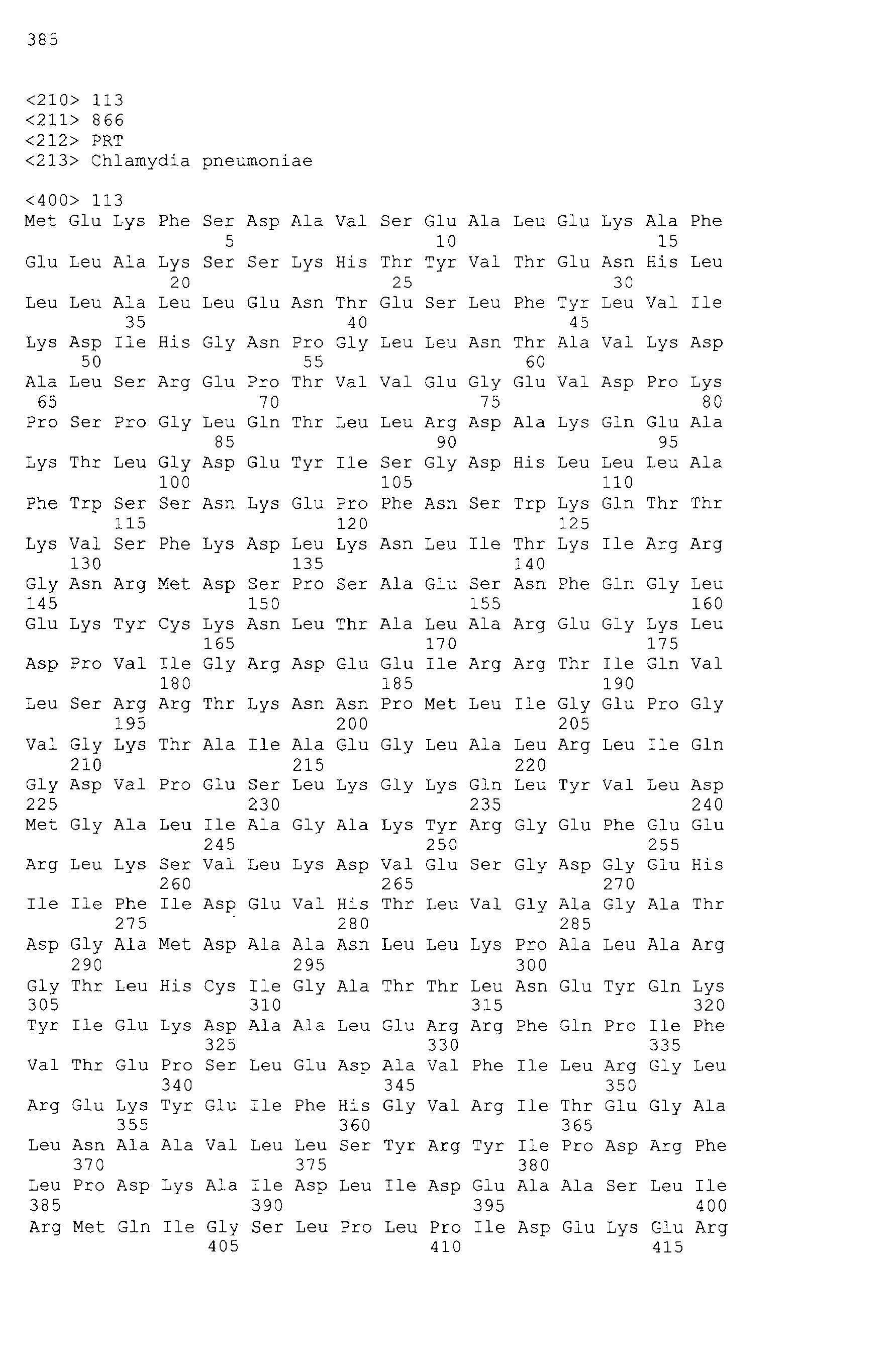

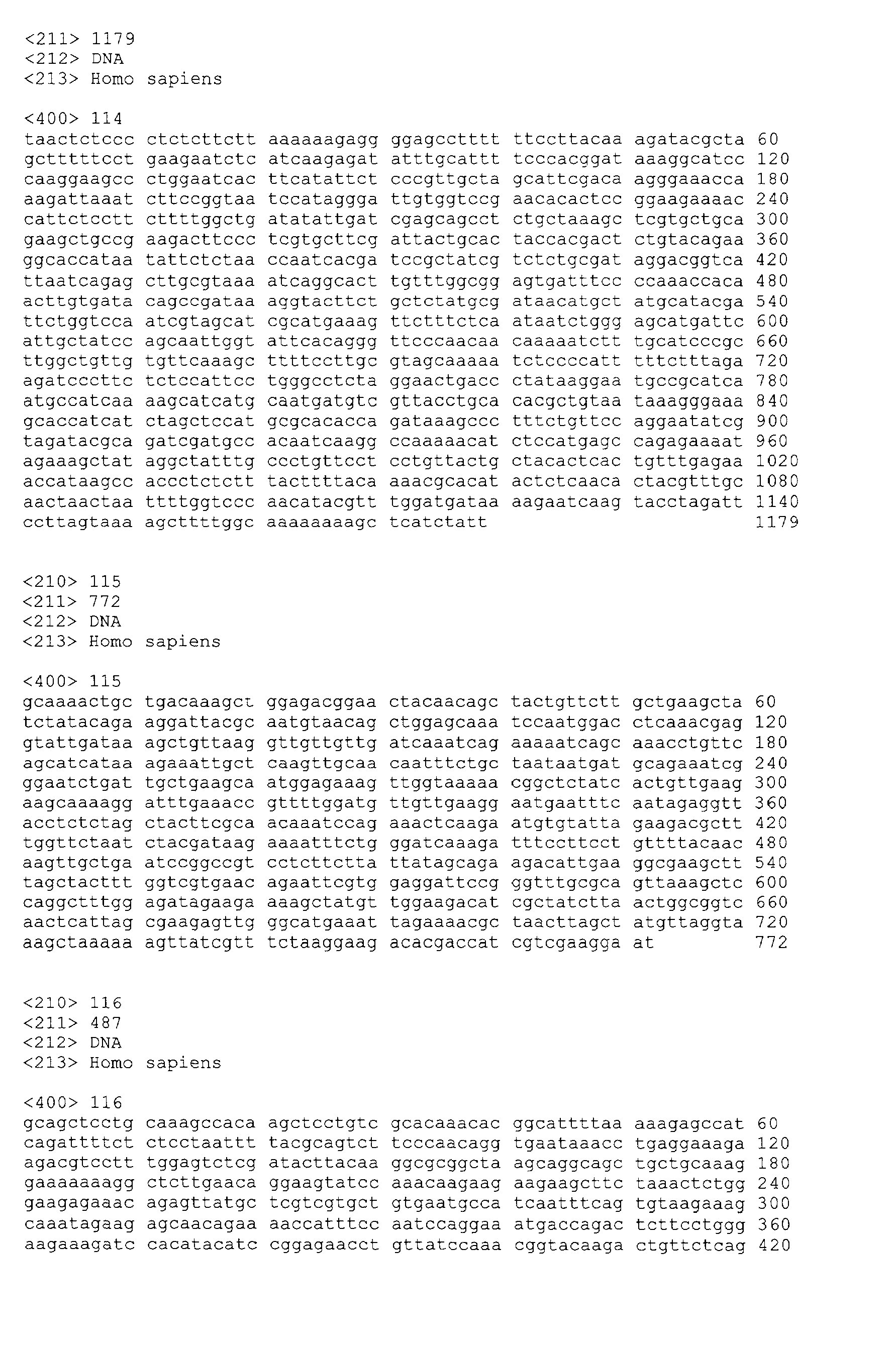
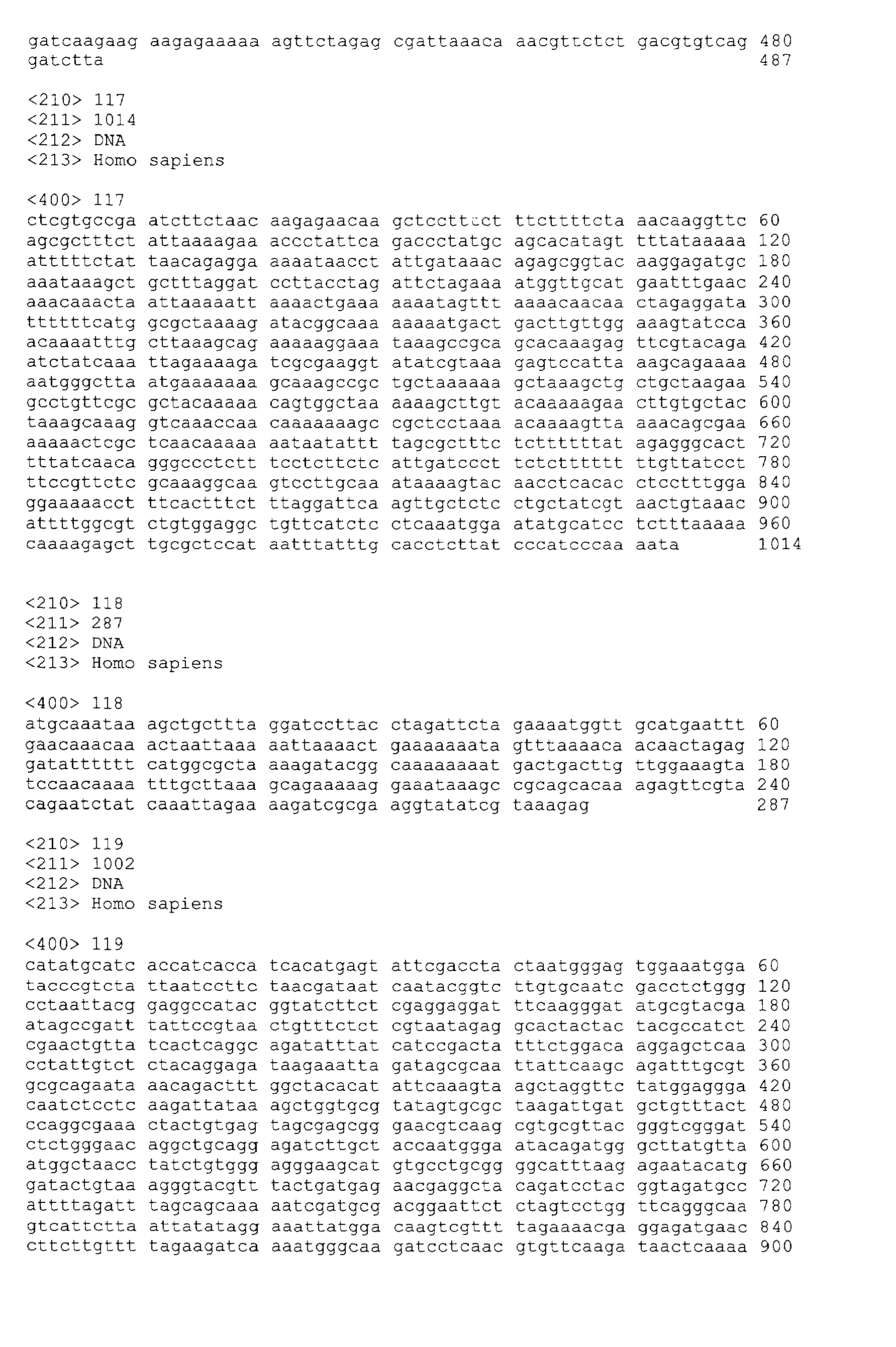


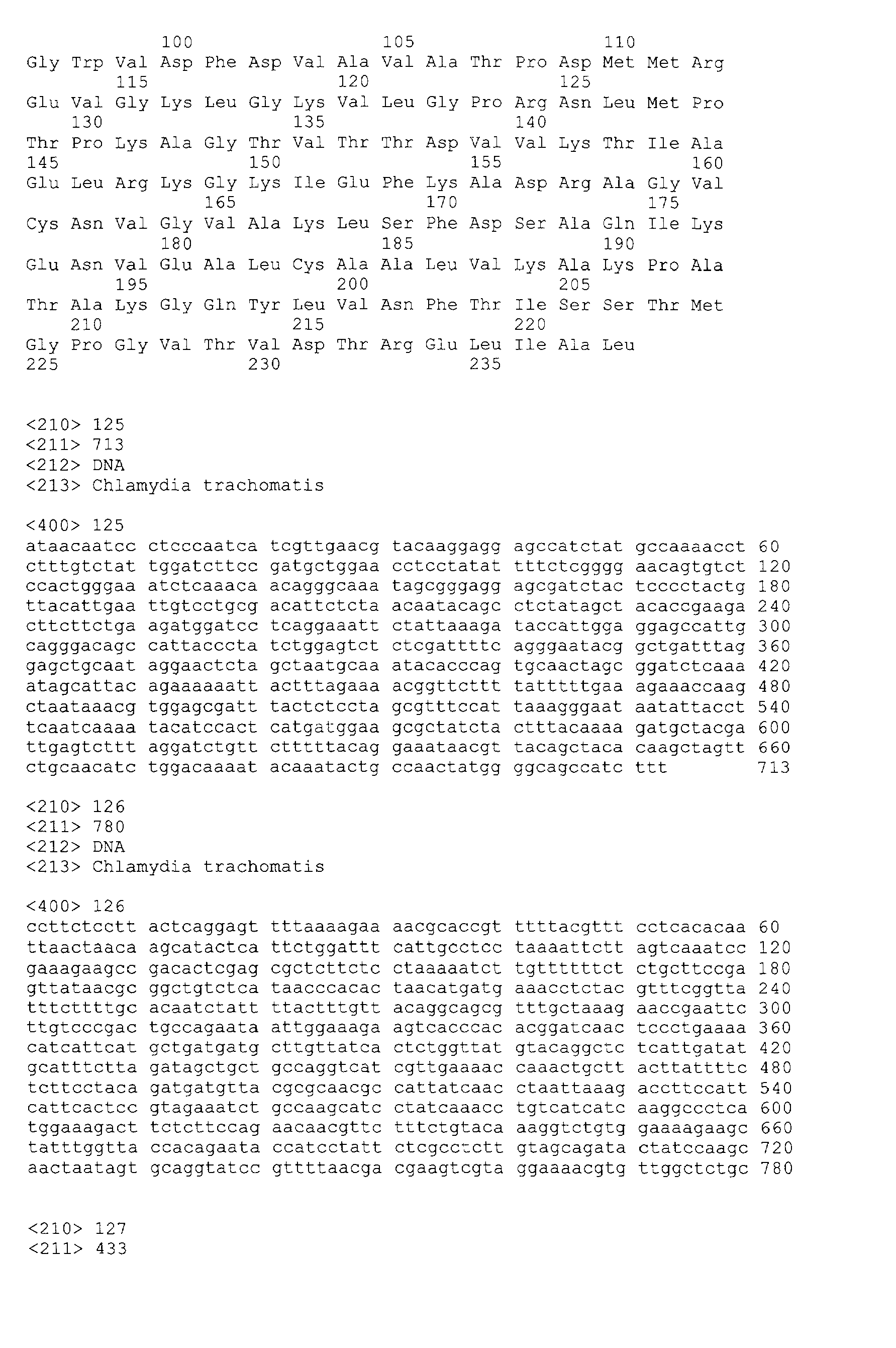


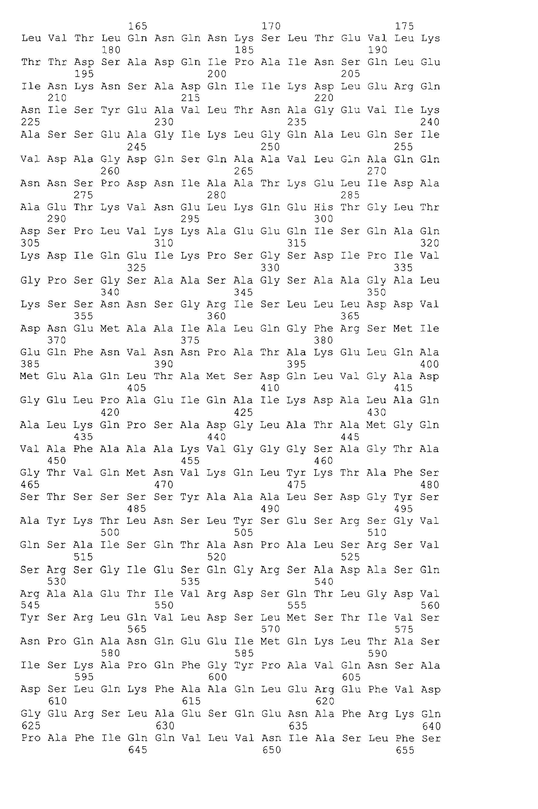


Claims (18)
- An isolated polynucleotide comprising a sequence selected from the group consisting of:(a) sequences provided in SEQ ID NO:1-48, 114-121, and 125-138;(b) complements of the sequences provided in SEQ ID NO: 1-48, 114-121, and 125-138;(c) sequences consisting of at least 20 contiguous residues of a sequence provided in SEQ ID NO:1-48, 114-121, 125-138;(d) sequences that hybridize to a sequence provided in SEQ ID NO:1-48, 114-121, and 125-138, under highly stringent conditions;(e) sequences having at least 95% identity to a sequence of SEQ ID NO:1-48, 114-121, and 125-138;(f) sequences having at least 99% identity to a sequence of SEQ ID NO: 1-48, 114-121, and 125-138; and(g) degenerate variants of a sequence provided in SEQ ID NO: 1-48, 114-121, and 125-138.
- An isolated polypeptide comprising an amino acid sequence selected from the group consisting of:(a) sequences encoded by a polynucleotide of claim 1;(b) sequences having at least 95% identity to a sequence encoded by a polynucleotide of claim 1; and(c) sequences having at least 99% identity to a sequence encoded by a polynucleotide of claim 1.
- An isolated polypeptide comprising at least an immunogenic fragment of a polypeptide sequence selected from the group consisting of:(a) a polypeptide sequence set forth in SEQ ID NO: 122-124 and 139-140,(b) a polypeptide sequence having at least 95% identity with a sequence set forth in SEQ ID NO: 122-124 and 139-140, and(c) a polypeptide sequence having at least 99% identity with a sequence set forth in SEQ ID NO: 122-124 and 139-140.
- An expression vector comprising a polynucleotide of claim 1 operably linked to an expression control sequence.
- A host cell transformed or transfected with an expression vector according to claim 4.
- An isolated antibody, or antigen-binding fragment thereof, that specifically binds to a polypeptide of claim 2 or claim 3.
- A method for detecting the presence of Chlamydia in a patient, comprising the steps of:(a) obtaining a biological sample from the patient;(b) contacting the biological sample with a binding agent that binds to a polypeptide of claim 2 or claim 3;(c) detecting in the sample an amount of polypeptide that binds to the binding agent; and(d) comparing the amount of polypeptide to a predetermined cut-off value and therefrom determining the presence of Chlamydia in the patient.
- A fusion protein comprising at least one polypeptide according to claim 2 or claim 3.
- An oligonucleotide that hybridizes to a sequence recited in any one of SEQ ID NO: 1-48, 114-121, and 125-138 under highly stringent conditions.
- A method for stimulating and/or expanding T cells specific for a Chlamydia protein, comprising contacting T cells with at least one component selected from the group consisting of:(a) a polypeptide according to claim 2 or claim 3;(b) a polynucleotide according to claim 1; and(c) an antigen-presenting cell that expresses a polynucleotide according to claim 1,
under conditions and for a time sufficient to permit the stimulation and/or expansion of T cells. - An isolated T cell population, comprising T cells prepared according to the method of claim 10.
- A composition comprising a first component selected from the group consisting of physiologically acceptable carriers and immunostimulants, and a second component selected from the group consisting of:(a) a polypeptide according to claim 2 or claim 3;(b) a polynucleotide according to claim 1;(c) an antibody according to claim 6;(d) a fusion protein according to claim 8;(e) a T cell population according to claim 11; and(f) an antigen presenting cell that expresses a polypeptide according to claim 2 or claim 3.
- A method for stimulating an immune response in a patient, comprising administering to the patient a composition selected from the group consisting of:(a) a composition of claim 12;(b) a polynucleotide sequence of any one of SEQ ID NO:80-94; and(c) a polypeptide sequence of any one of SEQ ID NO:95-109.
- A method for the treatment of Chlamydia infection in a patient, comprising administering to the patient a composition selected from the group consisting of:(a) a composition of claim 12;(b) a polynucleotide sequence of any one of SEQ ID NO:80-94; and(d) a polypeptide sequence of any one of SEQ ID NO:95-109.
- A method for determining the presence of Chlamydia in a patient, comprising the steps of:(a) obtaining a biological sample from the patient;(b) contacting the biological sample with an oligonucleotide according to claim 9;(c) detecting in the sample an amount of a polynucleotide that hybridizes to the oligonucleotide; and(d) comparing the amount of polynucleotide that hybridizes to the oligonucleotide to a predetermined cut-off value, and therefore determining the presence of the cancer in the patient.
- A diagnostic kit comprising at least one oligonucleotide according to claim 9.
- A diagnostic kit comprising at least one antibody according to claim 6 and a detection reagent, wherein the detection reagent comprises a reporter group.
- A method for the treatment of Chlamydia in a patient, comprising the steps of:(a) incubating CD4+ and/or CD8+ T cells isolated from a patient with at least one component selected from the group consisting of:(i) a polypeptide according to any one of claims 2 or 3;(ii) a polypeptide sequence of any one of SEQ ID NO: 95-109;(iii) a polynucleotide according to claim 1;(iv) a polynucleotide sequence of any one of SEQ ID NO:80-94;(v) an antigen presenting cell that expresses a polypeptide sequence set forth in any one of claims 2 or 3;(vi) an antigen presenting cell that expresses a polypeptide sequence of any one of SEQ ID NO:95-109, such that the T cells proliferate; and(b) administering to the patient an effective amount of the proliferated T cells.
Applications Claiming Priority (3)
| Application Number | Priority Date | Filing Date | Title |
|---|---|---|---|
| US19885300P | 2000-04-21 | 2000-04-21 | |
| US21975200P | 2000-07-20 | 2000-07-20 | |
| EP01928775A EP1278855B1 (en) | 2000-04-21 | 2001-04-23 | Compounds and methods for treatment and diagnosis of chlamydial infection |
Related Parent Applications (2)
| Application Number | Title | Priority Date | Filing Date |
|---|---|---|---|
| EP01928775.4 Division | 2001-04-23 | ||
| WOPCT/US01/13081 Previously-Filed-Application | 2001-04-23 |
Publications (2)
| Publication Number | Publication Date |
|---|---|
| EP2192128A2 true EP2192128A2 (en) | 2010-06-02 |
| EP2192128A3 EP2192128A3 (en) | 2010-09-22 |
Family
ID=26894210
Family Applications (2)
| Application Number | Title | Priority Date | Filing Date |
|---|---|---|---|
| EP08152495A Withdrawn EP2192128A3 (en) | 2000-04-21 | 2001-04-23 | Compounds and methods for treatment and diagnosis of chlamydial infection |
| EP01928775A Expired - Lifetime EP1278855B1 (en) | 2000-04-21 | 2001-04-23 | Compounds and methods for treatment and diagnosis of chlamydial infection |
Family Applications After (1)
| Application Number | Title | Priority Date | Filing Date |
|---|---|---|---|
| EP01928775A Expired - Lifetime EP1278855B1 (en) | 2000-04-21 | 2001-04-23 | Compounds and methods for treatment and diagnosis of chlamydial infection |
Country Status (8)
| Country | Link |
|---|---|
| US (2) | US20030175700A1 (en) |
| EP (2) | EP2192128A3 (en) |
| AT (1) | ATE389020T1 (en) |
| AU (1) | AU2001255596A1 (en) |
| CA (1) | CA2407114A1 (en) |
| DE (1) | DE60133190T2 (en) |
| ES (1) | ES2303525T3 (en) |
| WO (1) | WO2001081379A2 (en) |
Families Citing this family (36)
| Publication number | Priority date | Publication date | Assignee | Title |
|---|---|---|---|---|
| US5629167A (en) * | 1994-04-19 | 1997-05-13 | Biocine S.P.A. | Detection of antibodies against Chlamydia trachomatis pgp3 antigen in patient sera by enzyme-linked immunosorbent assay |
| CA2340330A1 (en) | 1998-08-20 | 2000-03-02 | Aventis Pasteur Limited | Nucleic acid molecules encoding inclusion membrane protein c of chlamydia |
| US6686339B1 (en) | 1998-08-20 | 2004-02-03 | Aventis Pasteur Limited | Nucleic acid molecules encoding inclusion membrane protein C of Chlamydia |
| CA2340283A1 (en) | 1998-08-20 | 2000-03-02 | Aventis Pasteur Limited | Nucleic acid molecules encoding pomp91a protein of chlamydia |
| US6649370B1 (en) | 1998-10-28 | 2003-11-18 | Aventis Pasteur Limited | Chlamydia antigens and corresponding DNA fragments and uses thereof |
| US6607730B1 (en) | 1998-11-02 | 2003-08-19 | Aventis Pasteur Limited/Aventis Pasteur Limitee | Chlamydia antigens and corresponding DNA fragments and uses thereof |
| JP2002531093A (en) | 1998-12-01 | 2002-09-24 | アベンティス、パストゥール、リミテッド | Chlamydia antigens and corresponding DNA fragments and uses thereof |
| US20080213264A1 (en) * | 1998-12-08 | 2008-09-04 | Corixa Corporation | Compounds and methods for treatment and diagnosis of chlamydial infection |
| US20020061848A1 (en) * | 2000-07-20 | 2002-05-23 | Ajay Bhatia | Compounds and methods for treatment and diagnosis of chlamydial infection |
| NZ512246A (en) | 1998-12-08 | 2003-12-19 | Corixa Corp | Compounds and methods for treatment and diagnosis of chlamydial infection |
| GB9828000D0 (en) | 1998-12-18 | 1999-02-10 | Chiron Spa | Antigens |
| US7297341B1 (en) | 1998-12-23 | 2007-11-20 | Sanofi Pasteur Limited | Chlamydia antigens and corresponding DNA fragments and uses thereof |
| US6808713B1 (en) | 1998-12-28 | 2004-10-26 | Aventis Pasteur Limited | Chlamydia antigens and corresponding DNA fragments and uses thereof |
| EP1140998B1 (en) | 1998-12-28 | 2008-01-23 | Aventis Pasteur Limited | Chlamydia antigens and corresponding dna fragments and uses thereof |
| GB9902555D0 (en) | 1999-02-05 | 1999-03-24 | Neutec Pharma Plc | Medicament |
| NZ514140A (en) | 1999-03-12 | 2001-09-28 | Aventis Pasteur | Chlamydia antigens and corresponding DNA fragments and uses thereof |
| US20030095973A1 (en) | 1999-05-03 | 2003-05-22 | Murdin Andrew D. | Chlamydia antigens and corresponding DNA fragments and uses thereof |
| ATE384793T1 (en) | 1999-09-20 | 2008-02-15 | Aventis Pasteur | CHLAMYDIA ANTIGENS AND CORRESPONDING DNA FRAGMENTS AND THEIR USES |
| US6632663B1 (en) | 1999-09-22 | 2003-10-14 | Aventis Pasteur Limited | DNA immunization against chlamydia infection |
| AU784193B2 (en) | 1999-12-22 | 2006-02-16 | Sanofi Pasteur Limited | Chlamydia antigens and corresponding DNA fragments and uses thereof |
| US6919187B2 (en) * | 2000-04-21 | 2005-07-19 | Corixa Corporation | Compounds and methods for treatment and diagnosis of chlamydial infection |
| EP2192128A3 (en) | 2000-04-21 | 2010-09-22 | Corixa Corporation | Compounds and methods for treatment and diagnosis of chlamydial infection |
| CA2408199A1 (en) | 2000-05-08 | 2001-11-15 | Aventis Pasteur Limited | Chlamydia antigens and corresponding dna fragments and uses thereof |
| AU2001276619A1 (en) | 2000-07-03 | 2002-01-14 | Chiron S.P.A. | Immunisation against chlamydia pneumoniae |
| DE60228758D1 (en) | 2001-12-12 | 2008-10-16 | Novartis Vaccines & Diagnostic | IMMUNIZATION AGAINST CHLAMYDIA TRACHEOMATIS |
| GB0203403D0 (en) | 2002-02-13 | 2002-04-03 | Chiron Spa | Chlamydia cytotoxic-T cell epitopes |
| WO2004073492A2 (en) * | 2003-02-14 | 2004-09-02 | Massachusetts Eye And Ear Infirmary | Chlamydia pneumoniae associated chronic intraocular disorders and treatment thereof |
| US8541007B2 (en) | 2005-03-31 | 2013-09-24 | Glaxosmithkline Biologicals S.A. | Vaccines against chlamydial infection |
| EP2386314A1 (en) | 2005-03-31 | 2011-11-16 | GlaxoSmithKline Biologicals SA | Vaccines against chlamydial infection |
| EP1981905B1 (en) * | 2006-01-16 | 2016-08-31 | THE GOVERNMENT OF THE UNITED STATES OF AMERICA as represented by THE SECRETARY OF THE DEPARTMENT OF HEALTH AND HUMAN SERVICES | Chlamydia vaccine |
| CA2744739A1 (en) * | 2007-12-03 | 2009-06-11 | President And Fellows Of Harvard College | Chlamydia antigens |
| US10131693B2 (en) | 2008-10-20 | 2018-11-20 | Gwangju Institute Of Science And Technology | Bipodal-peptide binder |
| EP3549602A1 (en) | 2009-03-06 | 2019-10-09 | GlaxoSmithKline Biologicals S.A. | Chlamydia antigens |
| CN102918155B (en) * | 2010-03-23 | 2015-12-16 | 和光纯药工业株式会社 | Chlamydia trachomatis detection primer and probe and use the detection method of chlamydia trachomatis of this primer and probe |
| US8892184B2 (en) | 2010-10-18 | 2014-11-18 | Siemens Medical Solutions Usa, Inc. | Systems and methods for reducing interference in a dual modality imaging system |
| KR20140088108A (en) * | 2011-09-30 | 2014-07-09 | 더 유니버시티 오브 브리티시 콜롬비아 | Chlamydia antigen compositions and uses thereof |
Citations (46)
| Publication number | Priority date | Publication date | Assignee | Title |
|---|---|---|---|---|
| US4235877A (en) | 1979-06-27 | 1980-11-25 | Merck & Co., Inc. | Liposome particle containing viral or bacterial antigenic subunit |
| US4237224A (en) | 1974-11-04 | 1980-12-02 | Board Of Trustees Of The Leland Stanford Jr. University | Process for producing biologically functional molecular chimeras |
| US4436727A (en) | 1982-05-26 | 1984-03-13 | Ribi Immunochem Research, Inc. | Refined detoxified endotoxin product |
| US4554101A (en) | 1981-01-09 | 1985-11-19 | New York Blood Center, Inc. | Identification and preparation of epitopes on antigens and allergens on the basis of hydrophilicity |
| US4603112A (en) | 1981-12-24 | 1986-07-29 | Health Research, Incorporated | Modified vaccinia virus |
| US4683202A (en) | 1985-03-28 | 1987-07-28 | Cetus Corporation | Process for amplifying nucleic acid sequences |
| US4683195A (en) | 1986-01-30 | 1987-07-28 | Cetus Corporation | Process for amplifying, detecting, and/or-cloning nucleic acid sequences |
| US4751180A (en) | 1985-03-28 | 1988-06-14 | Chiron Corporation | Expression using fused genes providing for protein product |
| GB2200651A (en) | 1987-02-07 | 1988-08-10 | Al Sumidaie Ayad Mohamed Khala | A method of obtaining a retrovirus-containing fraction from retrovirus-containing cells |
| US4769330A (en) | 1981-12-24 | 1988-09-06 | Health Research, Incorporated | Modified vaccinia virus and methods for making and using the same |
| GB2202328A (en) | 1987-03-11 | 1988-09-21 | Orion Yhtymae Oy | An improved method for assaying of nucleic acids, a reagent combination and a kit therefore |
| US4777127A (en) | 1985-09-30 | 1988-10-11 | Labsystems Oy | Human retrovirus-related products and methods of diagnosing and treating conditions associated with said retrovirus |
| WO1988010315A1 (en) | 1987-06-19 | 1988-12-29 | Siska Diagnostics, Inc. | Transcription-based nucleic acid amplification/detection systems |
| US4800159A (en) | 1986-02-07 | 1989-01-24 | Cetus Corporation | Process for amplifying, detecting, and/or cloning nucleic acid sequences |
| WO1989001973A2 (en) | 1987-09-02 | 1989-03-09 | Applied Biotechnology, Inc. | Recombinant pox virus for immunization against tumor-associated antigens |
| WO1989006700A1 (en) | 1988-01-21 | 1989-07-27 | Genentech, Inc. | Amplification and detection of nucleic acid sequences |
| EP0329822A2 (en) | 1988-02-24 | 1989-08-30 | Cangene Corporation | Nucleic acid amplification process |
| US4866034A (en) | 1982-05-26 | 1989-09-12 | Ribi Immunochem Research Inc. | Refined detoxified endotoxin |
| US4877611A (en) | 1986-04-15 | 1989-10-31 | Ribi Immunochem Research Inc. | Vaccine containing tumor antigens and adjuvants |
| US4883750A (en) | 1984-12-13 | 1989-11-28 | Applied Biosystems, Inc. | Detection of specific sequences in nucleic acids |
| EP0345242A2 (en) | 1988-06-03 | 1989-12-06 | Smithkline Biologicals S.A. | Expression of gag proteins from retroviruses in eucaryotic cells |
| US4897268A (en) | 1987-08-03 | 1990-01-30 | Southern Research Institute | Drug delivery system and method of making the same |
| US4912094A (en) | 1988-06-29 | 1990-03-27 | Ribi Immunochem Research, Inc. | Modified lipopolysaccharides and process of preparation |
| US4935233A (en) | 1985-12-02 | 1990-06-19 | G. D. Searle And Company | Covalently linked polypeptide cell modulators |
| WO1991002805A2 (en) | 1989-08-18 | 1991-03-07 | Viagene, Inc. | Recombinant retroviruses delivering vector constructs to target cells |
| US5017487A (en) | 1985-04-04 | 1991-05-21 | Hoffmann-La Roche Inc. | Vaccinia DNA |
| WO1991018926A1 (en) | 1990-05-31 | 1991-12-12 | Arne Forsgren | PROTEIN D - AN IgD-BINDING PROTEIN OF HAEMOPHILUS INFLUENZAE |
| US5075109A (en) | 1986-10-24 | 1991-12-24 | Southern Research Institute | Method of potentiating an immune response |
| US5151254A (en) | 1989-08-29 | 1992-09-29 | Suzuki Jidosha Kogyo Kabushiki Kaisha | Exhaust gas cleaning device for internal combustion engine |
| WO1994000153A1 (en) | 1992-06-25 | 1994-01-06 | Smithkline Beecham Biologicals (S.A.) | Vaccine composition containing adjuvants |
| WO1994020078A1 (en) | 1993-03-02 | 1994-09-15 | Biovector Therapeutics Sa | Synthetic particulate vectors and method for preparing same |
| WO1994023701A1 (en) | 1993-04-21 | 1994-10-27 | Institut Pasteur | Particulate vector and pharmaceutical composition containing such vector |
| WO1995017210A1 (en) | 1993-12-23 | 1995-06-29 | Smithkline Beecham Biologicals (S.A.) | Vaccines |
| WO1996002555A1 (en) | 1994-07-15 | 1996-02-01 | The University Of Iowa Research Foundation | Immunomodulatory oligonucleotides |
| WO1996006638A1 (en) | 1994-08-31 | 1996-03-07 | Biovector Therapeutics S.A. | Method for enhancing immunogenicity, product obtained and pharmaceutical composition |
| US5518913A (en) | 1991-10-10 | 1996-05-21 | National Research Council Of Canada | High level recombinant protein production using conditional helper-free adenovirus vector |
| WO1996033739A1 (en) | 1995-04-25 | 1996-10-31 | Smithkline Beecham Biologicals S.A. | Vaccines containing a saponin and a sterol |
| WO1996038591A1 (en) | 1995-06-02 | 1996-12-05 | Incyte Pharmaceuticals, Inc. | IMPROVED METHOD FOR OBTAINING FULL-LENGTH cDNA SEQUENCES |
| US5585362A (en) | 1989-08-22 | 1996-12-17 | The Regents Of The University Of Michigan | Adenovirus vectors for gene therapy |
| US5616326A (en) | 1990-01-25 | 1997-04-01 | The University Court Of The University Of Glasgow | Recombinant canine adenovirus 2 (CAV-2) |
| US5698202A (en) | 1995-06-05 | 1997-12-16 | The Wistar Institute Of Anatomy & Biology | Replication-defective adenovirus human type 5 recombinant as a rabies vaccine carrier |
| WO1999028475A2 (en) | 1997-11-28 | 1999-06-10 | Genset | Chlamydia trachomatis genomic sequence and polypeptides, fragments thereof and uses thereof, in particular for the diagnosis, prevention and treatment of infection |
| WO1999033488A2 (en) | 1997-12-24 | 1999-07-08 | Smithkline Beecham Biologicals S.A. | Adjuvanted vaccine formulation |
| US7472098B2 (en) | 2000-02-14 | 2008-12-30 | Ubs Financial Services, Inc. | System and method for execution of trades made pursuant to stock option and purchase plans |
| US8700880B2 (en) | 2008-10-10 | 2014-04-15 | Apple Inc. | Dynamic trampoline and structured code generation in a signed code environment |
| US8901025B2 (en) | 2010-03-24 | 2014-12-02 | IFP Energies Nouvelles | Catalyst regeneration zone divided into sectors for regenerative catalytic units |
Family Cites Families (31)
| Publication number | Priority date | Publication date | Assignee | Title |
|---|---|---|---|---|
| US4118469A (en) * | 1976-04-27 | 1978-10-03 | Research Corporation | Antigen for trachoma lymphogranuloma venereum (LGV) and non-gonococcal urethritis (NGU) |
| US4497899A (en) * | 1982-04-12 | 1985-02-05 | Abbott Laboratories | Immunoassay for Chlamydia trachomatis antigens |
| EP0348725A3 (en) | 1988-06-29 | 1990-10-24 | Miles Inc. | Species-specific epitode of chlamydia trachomatis and antibodies that recognize the same |
| US5869608A (en) * | 1989-03-17 | 1999-02-09 | The United States Of America As Represented By The Department Of Health And Human Services | Nucleotide and amino acid sequences of the four variable domains of the major outer membrane proteins of Chlamydia trachomatis |
| US5166053A (en) * | 1990-02-23 | 1992-11-24 | Miles Inc. | Specimen adequacy control for chlamydia assays |
| JP3018553B2 (en) * | 1990-05-08 | 2000-03-13 | 日立化成工業株式会社 | Chlamydia trachomatis antibody measurement method and preparation for diagnosing chlamydia trachomatis infection |
| US5725863A (en) * | 1991-09-06 | 1998-03-10 | The United States Of America As Represented By The Secretary Of Agriculture | Polypeptides useful in prevention of chlamydia infection |
| JPH08504188A (en) | 1992-09-18 | 1996-05-07 | アメリカ合衆国 | Trachoma chlamydia synthetic peptide vaccine |
| EP2045261A1 (en) | 1994-09-20 | 2009-04-08 | Hitachi Chemical Co., Ltd. | Chlamydia pneumoniae antigenic polypeptides |
| GB9516293D0 (en) | 1995-08-09 | 1995-10-11 | Immunova Ltee | Novel peptides and their use as vaccines |
| CA2259595C (en) | 1996-07-12 | 2010-11-30 | University Of Manitoba | Dna immunization against chlamydia infection |
| US6464979B1 (en) | 1996-09-12 | 2002-10-15 | Aventis Pasteur Limited | Chlamydial vaccines and methods of preparation thereof |
| US7459524B1 (en) | 1997-10-02 | 2008-12-02 | Emergent Product Development Gaithersburg Inc. | Chlamydia protein, sequence and uses thereof |
| ES2278420T3 (en) | 1997-11-21 | 2007-08-01 | Serono Genetics Institute S.A. | GENOMICAL SEQUENCE AND POLYPEPTIDES OF CHLAMYDIA PNEUMONIAE, FRAGMENTS OF THE SAME AND USE OF THE SAME, IN PARTICULAR FOR DIAGNOSIS, PREVENTION AND TREATMENT OF INFECTION. |
| AU753995B2 (en) | 1998-04-07 | 2002-10-31 | Corixa Corporation | Fusion proteins of mycobacterium tuberculosis antigens and their uses |
| CA2350775A1 (en) | 1998-11-12 | 2000-05-18 | The Regents Of The University Of California | Chlamydia pneumoniae genome sequence |
| US20020061848A1 (en) | 2000-07-20 | 2002-05-23 | Ajay Bhatia | Compounds and methods for treatment and diagnosis of chlamydial infection |
| US6448234B1 (en) * | 1998-12-08 | 2002-09-10 | Corixa Corporation | Compounds and methods for treatment and diagnosis of chlamydial infection |
| NZ512246A (en) | 1998-12-08 | 2003-12-19 | Corixa Corp | Compounds and methods for treatment and diagnosis of chlamydial infection |
| US6166177A (en) * | 1998-12-08 | 2000-12-26 | Corixa Corporation | Compounds and methods for the treatment and diagnosis of chlamydial infection |
| US6565856B1 (en) * | 1998-12-08 | 2003-05-20 | Corixa Corporation | Compounds and methods for treatment and diagnosis of chlamydial infection |
| US6432916B1 (en) * | 1998-12-08 | 2002-08-13 | Corixa Corporation | Compounds and methods for treatment and diagnosis of chlamydial infection |
| GB9828000D0 (en) | 1998-12-18 | 1999-02-10 | Chiron Spa | Antigens |
| GB9902555D0 (en) | 1999-02-05 | 1999-03-24 | Neutec Pharma Plc | Medicament |
| US20030095973A1 (en) | 1999-05-03 | 2003-05-22 | Murdin Andrew D. | Chlamydia antigens and corresponding DNA fragments and uses thereof |
| ATE384793T1 (en) | 1999-09-20 | 2008-02-15 | Aventis Pasteur | CHLAMYDIA ANTIGENS AND CORRESPONDING DNA FRAGMENTS AND THEIR USES |
| US6632663B1 (en) | 1999-09-22 | 2003-10-14 | Aventis Pasteur Limited | DNA immunization against chlamydia infection |
| AU784193B2 (en) | 1999-12-22 | 2006-02-16 | Sanofi Pasteur Limited | Chlamydia antigens and corresponding DNA fragments and uses thereof |
| EP2192128A3 (en) | 2000-04-21 | 2010-09-22 | Corixa Corporation | Compounds and methods for treatment and diagnosis of chlamydial infection |
| CA2408199A1 (en) | 2000-05-08 | 2001-11-15 | Aventis Pasteur Limited | Chlamydia antigens and corresponding dna fragments and uses thereof |
| AU2001276619A1 (en) | 2000-07-03 | 2002-01-14 | Chiron S.P.A. | Immunisation against chlamydia pneumoniae |
-
2001
- 2001-04-23 EP EP08152495A patent/EP2192128A3/en not_active Withdrawn
- 2001-04-23 US US09/841,260 patent/US20030175700A1/en not_active Abandoned
- 2001-04-23 CA CA002407114A patent/CA2407114A1/en not_active Abandoned
- 2001-04-23 AU AU2001255596A patent/AU2001255596A1/en not_active Abandoned
- 2001-04-23 ES ES01928775T patent/ES2303525T3/en not_active Expired - Lifetime
- 2001-04-23 DE DE60133190T patent/DE60133190T2/en not_active Expired - Fee Related
- 2001-04-23 AT AT01928775T patent/ATE389020T1/en not_active IP Right Cessation
- 2001-04-23 EP EP01928775A patent/EP1278855B1/en not_active Expired - Lifetime
- 2001-04-23 WO PCT/US2001/013081 patent/WO2001081379A2/en active Application Filing
-
2004
- 2004-01-15 US US10/762,058 patent/US7384638B2/en not_active Expired - Fee Related
Patent Citations (49)
| Publication number | Priority date | Publication date | Assignee | Title |
|---|---|---|---|---|
| US4237224A (en) | 1974-11-04 | 1980-12-02 | Board Of Trustees Of The Leland Stanford Jr. University | Process for producing biologically functional molecular chimeras |
| US4235877A (en) | 1979-06-27 | 1980-11-25 | Merck & Co., Inc. | Liposome particle containing viral or bacterial antigenic subunit |
| US4554101A (en) | 1981-01-09 | 1985-11-19 | New York Blood Center, Inc. | Identification and preparation of epitopes on antigens and allergens on the basis of hydrophilicity |
| US4603112A (en) | 1981-12-24 | 1986-07-29 | Health Research, Incorporated | Modified vaccinia virus |
| US4769330A (en) | 1981-12-24 | 1988-09-06 | Health Research, Incorporated | Modified vaccinia virus and methods for making and using the same |
| US4436727A (en) | 1982-05-26 | 1984-03-13 | Ribi Immunochem Research, Inc. | Refined detoxified endotoxin product |
| US4866034A (en) | 1982-05-26 | 1989-09-12 | Ribi Immunochem Research Inc. | Refined detoxified endotoxin |
| US4883750A (en) | 1984-12-13 | 1989-11-28 | Applied Biosystems, Inc. | Detection of specific sequences in nucleic acids |
| US4683202A (en) | 1985-03-28 | 1987-07-28 | Cetus Corporation | Process for amplifying nucleic acid sequences |
| US4683202B1 (en) | 1985-03-28 | 1990-11-27 | Cetus Corp | |
| US4751180A (en) | 1985-03-28 | 1988-06-14 | Chiron Corporation | Expression using fused genes providing for protein product |
| US5017487A (en) | 1985-04-04 | 1991-05-21 | Hoffmann-La Roche Inc. | Vaccinia DNA |
| US4777127A (en) | 1985-09-30 | 1988-10-11 | Labsystems Oy | Human retrovirus-related products and methods of diagnosing and treating conditions associated with said retrovirus |
| US4935233A (en) | 1985-12-02 | 1990-06-19 | G. D. Searle And Company | Covalently linked polypeptide cell modulators |
| US4683195B1 (en) | 1986-01-30 | 1990-11-27 | Cetus Corp | |
| US4683195A (en) | 1986-01-30 | 1987-07-28 | Cetus Corporation | Process for amplifying, detecting, and/or-cloning nucleic acid sequences |
| US4800159A (en) | 1986-02-07 | 1989-01-24 | Cetus Corporation | Process for amplifying, detecting, and/or cloning nucleic acid sequences |
| US4877611A (en) | 1986-04-15 | 1989-10-31 | Ribi Immunochem Research Inc. | Vaccine containing tumor antigens and adjuvants |
| US5075109A (en) | 1986-10-24 | 1991-12-24 | Southern Research Institute | Method of potentiating an immune response |
| GB2200651A (en) | 1987-02-07 | 1988-08-10 | Al Sumidaie Ayad Mohamed Khala | A method of obtaining a retrovirus-containing fraction from retrovirus-containing cells |
| GB2202328A (en) | 1987-03-11 | 1988-09-21 | Orion Yhtymae Oy | An improved method for assaying of nucleic acids, a reagent combination and a kit therefore |
| WO1988010315A1 (en) | 1987-06-19 | 1988-12-29 | Siska Diagnostics, Inc. | Transcription-based nucleic acid amplification/detection systems |
| US4897268A (en) | 1987-08-03 | 1990-01-30 | Southern Research Institute | Drug delivery system and method of making the same |
| WO1989001973A2 (en) | 1987-09-02 | 1989-03-09 | Applied Biotechnology, Inc. | Recombinant pox virus for immunization against tumor-associated antigens |
| WO1989006700A1 (en) | 1988-01-21 | 1989-07-27 | Genentech, Inc. | Amplification and detection of nucleic acid sequences |
| EP0329822A2 (en) | 1988-02-24 | 1989-08-30 | Cangene Corporation | Nucleic acid amplification process |
| EP0345242A2 (en) | 1988-06-03 | 1989-12-06 | Smithkline Biologicals S.A. | Expression of gag proteins from retroviruses in eucaryotic cells |
| US4912094A (en) | 1988-06-29 | 1990-03-27 | Ribi Immunochem Research, Inc. | Modified lipopolysaccharides and process of preparation |
| US4912094B1 (en) | 1988-06-29 | 1994-02-15 | Ribi Immunochem Research Inc. | Modified lipopolysaccharides and process of preparation |
| WO1991002805A2 (en) | 1989-08-18 | 1991-03-07 | Viagene, Inc. | Recombinant retroviruses delivering vector constructs to target cells |
| US5585362A (en) | 1989-08-22 | 1996-12-17 | The Regents Of The University Of Michigan | Adenovirus vectors for gene therapy |
| US5151254A (en) | 1989-08-29 | 1992-09-29 | Suzuki Jidosha Kogyo Kabushiki Kaisha | Exhaust gas cleaning device for internal combustion engine |
| US5616326A (en) | 1990-01-25 | 1997-04-01 | The University Court Of The University Of Glasgow | Recombinant canine adenovirus 2 (CAV-2) |
| WO1991018926A1 (en) | 1990-05-31 | 1991-12-12 | Arne Forsgren | PROTEIN D - AN IgD-BINDING PROTEIN OF HAEMOPHILUS INFLUENZAE |
| US5518913A (en) | 1991-10-10 | 1996-05-21 | National Research Council Of Canada | High level recombinant protein production using conditional helper-free adenovirus vector |
| WO1994000153A1 (en) | 1992-06-25 | 1994-01-06 | Smithkline Beecham Biologicals (S.A.) | Vaccine composition containing adjuvants |
| WO1994020078A1 (en) | 1993-03-02 | 1994-09-15 | Biovector Therapeutics Sa | Synthetic particulate vectors and method for preparing same |
| WO1994023701A1 (en) | 1993-04-21 | 1994-10-27 | Institut Pasteur | Particulate vector and pharmaceutical composition containing such vector |
| WO1995017210A1 (en) | 1993-12-23 | 1995-06-29 | Smithkline Beecham Biologicals (S.A.) | Vaccines |
| WO1996002555A1 (en) | 1994-07-15 | 1996-02-01 | The University Of Iowa Research Foundation | Immunomodulatory oligonucleotides |
| WO1996006638A1 (en) | 1994-08-31 | 1996-03-07 | Biovector Therapeutics S.A. | Method for enhancing immunogenicity, product obtained and pharmaceutical composition |
| WO1996033739A1 (en) | 1995-04-25 | 1996-10-31 | Smithkline Beecham Biologicals S.A. | Vaccines containing a saponin and a sterol |
| WO1996038591A1 (en) | 1995-06-02 | 1996-12-05 | Incyte Pharmaceuticals, Inc. | IMPROVED METHOD FOR OBTAINING FULL-LENGTH cDNA SEQUENCES |
| US5698202A (en) | 1995-06-05 | 1997-12-16 | The Wistar Institute Of Anatomy & Biology | Replication-defective adenovirus human type 5 recombinant as a rabies vaccine carrier |
| WO1999028475A2 (en) | 1997-11-28 | 1999-06-10 | Genset | Chlamydia trachomatis genomic sequence and polypeptides, fragments thereof and uses thereof, in particular for the diagnosis, prevention and treatment of infection |
| WO1999033488A2 (en) | 1997-12-24 | 1999-07-08 | Smithkline Beecham Biologicals S.A. | Adjuvanted vaccine formulation |
| US7472098B2 (en) | 2000-02-14 | 2008-12-30 | Ubs Financial Services, Inc. | System and method for execution of trades made pursuant to stock option and purchase plans |
| US8700880B2 (en) | 2008-10-10 | 2014-04-15 | Apple Inc. | Dynamic trampoline and structured code generation in a signed code environment |
| US8901025B2 (en) | 2010-03-24 | 2014-12-02 | IFP Energies Nouvelles | Catalyst regeneration zone divided into sectors for regenerative catalytic units |
Non-Patent Citations (70)
| Title |
|---|
| ALTSCHUL ET AL., J MOL. BIOL., vol. 215, 1990, pages 403 - 410 |
| ALTSCHUL ET AL., NUCL. ACIDS RES., vol. 25, 1977, pages 3389 - 3402 |
| AUSUBEL, F. M. ET AL.: "Current Protocols in Molecular Biology", 1989, JOHN WILEY & SONS |
| BERKNER, BIOTECHNIQUES, vol. 6, 1988, pages 616 - 627 |
| BIOTECHNOLOGY, vol. 10, 1992, pages 795 - 798 |
| BROGLIE, R. ET AL., SCIENCE, vol. 224, 1984, pages 838 - 843 |
| COHEN, SCIENCE, vol. 259, 1993, pages 1691 - 1692 |
| COLBERE-GARAPIN, F. ET AL., J MOL. BIOL., vol. 150, 1981, pages 1 - 14 |
| COLER RHEA N ET AL: "Identification and characterization of novel recombinant vaccine antigens for immunization against genital Chlamydia trachomatis", FEMS IMMUNOLOGY AND MEDICAL MICROBIOLOGY, vol. 55, no. 2, March 2009 (2009-03-01), pages 258 - 270, XP055071769, ISSN: 0928-8244, DOI: 10.1111/j.1574-695X.2008.00527.x * |
| COOMBES ET AL., VACCINE, vol. 14, 1996, pages 1429 - 1438 |
| CORUZZI, G. ET AL., EMBO J, vol. 3, 1984, pages 1671 - 1680 |
| CREIGHTON, T.: "Proteins, Structures and Molecular Principles", 1983, WH FREEMAN AND CO. |
| DAYHOFF, M.O.: "Atlas of Protein Sequence and Structure", vol. 5, 1978, NATIONAL BIOMEDICAL RESEARCH FOUNDATION, article "A model of evolutionary change in proteins - Matrices for detecting distant relationships", pages: 345 - 358 |
| ENGELHARD, E. K. ET AL., PROC. NATL. ACAD. SCI., vol. 91, 1994, pages 3224 - 3227 |
| FISHER-HOCH ET AL., PROC. NATL. ACAD. SCI. USA, vol. 86, 1989, pages 317 - 321 |
| FLEXNER ET AL., ANN. N.Y. ACAD. SCI., vol. 569, 1989, pages 86 - 103 |
| FLEXNER ET AL., VACCINE, vol. 8, 1990, pages 17 - 21 |
| GENE, vol. 43, 1986, pages 265 - 292 |
| GRANT ET AL., METHODS ENZYMOL., vol. 153, 1987, pages 516 - 544 |
| GUZMAN ET AL., CIR. RES., vol. 73, 1993, pages 1202 - 1207 |
| GUZMAN ET AL., CIRCULATION, vol. 88, 1993, pages 2838 - 2848 |
| HAMPTON, R. ET AL.: "a Laboratory Manual", 1990, APS PRESS, article "Serological Methods" |
| HARTMAN, S. C., R. C. MULLIGAN, PROC. NATL. ACAD. SCI., vol. 85, 1988, pages 8047 - 51 |
| HEIN J.: "Methods in Enzymology", vol. 183, 1990, ACADEMIC PRESS, INC., article "Unified Approach to Alignment and Phylogenes", pages: 626 - 645 |
| HELLER ET AL., PROC. NATL. ACAD SCI. USA, vol. 94, 1997, pages 2150 - 2155 |
| HENIKOFF, HENIKOFF, PROC. NATL. ACAD. SCI. USA, vol. 89, 1989, pages 10915 |
| HIGGINS, D.G., SHARP, P.M.: "CABIOS", vol. 5, 1989, pages: 151 - 153 |
| HOBBS, S., MURRY, L. E.: "Yearbook of Science and Technology", 1992, MCGRAW HILL, pages: 191 - 196 |
| HORN, T. ET AL., NUCL. ACIDS RES. SYMP. SER., 1980, pages 225 - 232 |
| KASS-EISLER ET AL., PROC. NATL. ACAD. SCI. USA, vol. 90, 1993, pages 11498 - 11502 |
| KOLLS ET AL., PROC. NATL. ACAD. SCI. USA, vol. 91, 1994, pages 215 - 219 |
| KROLL, D. J. ET AL., DNA CELL BIOL., vol. 12, 1993, pages 441 - 453 |
| LAGERSTROM ET AL., PCR METHODS APPLIC., vol. I, 1991, pages 111 - 19 |
| LOGAN, J., SHENK, T., PROC. NATL. ACAD. SET, vol. 81, 1984, pages 3655 - 3659 |
| LOWY, 1. ET AL., CELL, vol. 22, 1990, pages 817 - 23 |
| M. H. ET AL., NUCL. ACIDS RES. SYMP. SER., 1980, pages 215 - 223 |
| M.F. POWELL AND M.J. NEWMAN: "Vaccine Design (the subunit and adjuvant approach", 1995, PLENUM PRESS |
| MADDOX, D. E. ET AL., J. EXP. MED., vol. 158, 1983, pages 1211 - 1216 |
| MARATEA ET AL., GENE, vol. 40, 1985, pages 39 - 46 |
| MERRIFIELD J., J. AM. CHEM. SOC., vol. 85, 1963, pages 2149 - 2154 |
| MERRIFIELD, J AM. CHEM. SOC., vol. 85, 1963, pages 2149 - 2146 |
| MOSMANN, COFFMAN, ANN. REV. IMMUNOL., vol. 7, 1989, pages 145 - 173 |
| MURPHY ET AL., PROC. NATL. ACAD. SET USA, vol. 83, 1986, pages 8258 - 8262 |
| MYERS, E.W., MULLER W., CABIOS, vol. 4, 1988, pages 11 - 17 |
| NEEDLEMAN, WUNSCH, J MOL. BIOL., vol. 48, 1970, pages 443 |
| PARKER ET AL., NUCL. ACIDS. RES., vol. 1, no. 9, 1991, pages 3055 - 60 |
| PEARSON, LIPMAN, PROC. NATL. ACAD SCI. USA, vol. 85, 1988, pages 2444 |
| PORATH, J. ET AL., PROT. EXP. PURIF., vol. 3, 1992, pages 263 - 281 |
| RHODES, C. A. ET AL., METHODSMOL. BIOL., vol. 55, 1995, pages 121 - 131 |
| ROBERGE, J. Y. ET AL., SCIENCE, vol. 269, 1995, pages 202 - 204 |
| ROBINSON, E.D., COMB. THEOR, vol. 11, 1971, pages 105 |
| ROLLAND, CRIT. REV. THERAP. DRUG CARRIER SYSTEMS, vol. 15, 1998, pages 143 - 198 |
| ROSENFELD ET AL., SCIENCE, vol. 252, 1991, pages 431 - 434 |
| SAMBROOK ET AL.: "Molecular Cloning: A Laboratory Manual", 1989, COLD SPRING HARBOR LABORATORIES |
| SAMBROOK, J. ET AL.: "Molecular Cloning, A Laboratory Manual", 1989, COLD SPRING HARBOR PRESS |
| SANTOU, N., NES, M., MOL. BIOL. EVOL., vol. 4, 1987, pages 406 - 425 |
| SATO ET AL., SCIENCE, vol. 273, 1996, pages 352 |
| SCHARF, D. ET AL., RESULTS PROBL. CELL DIFFER., vol. 20, 1994, pages 125 - 162 |
| SCHENA ET AL., PROC. NATL. ACAD SCI. USA, vol. 93, 1996, pages 10614 - 10619 |
| SMITH, WATERMAN, ADD. APL. MATH, vol. 2, 1981, pages 482 |
| SNEATH, P.H.A., SOKAL, R.R.: "Numerical Taxonomy - the Principles and Practice of Numerical Taxonomy", 1973, FREEMAN PRESS |
| STOUTE ET AL., NEW ENGL. J. MED., vol. 336, 1997, pages 86 - 91 |
| TAKAMATSU, N., EMBO J., vol. 6, 1987, pages 307 - 311 |
| TRIGLIA ET AL., NUCL. ACIDS RES., vol. 16, 1988, pages 8186 |
| ULMER ET AL., SCIENCE, vol. 259, 1993, pages 1745 - 1749 |
| VAN HEEKE, G., S. M. SCHUSTER, J. BIOL. CHEM., vol. 264, 1989, pages 5503 - 5509 |
| WIGLER, M. ET AL., CELL, vol. 11, 1977, pages 223 - 32 |
| WIGLER, M. ET AL., PROC. NATL. ACAD. SET, vol. 77, 1980, pages 3567 - 70 |
| WILBUR, W.J., LIPMAN, D.J., PROC. NATL. ACAD, SCI. USA, vol. 80, 1983, pages 726 - 730 |
| WINTER, J. ET AL., RESULTS PROBL. CELL DIFFER., vol. 17, 1991, pages 85 - 105 |
Also Published As
| Publication number | Publication date |
|---|---|
| WO2001081379A3 (en) | 2002-09-19 |
| EP1278855B1 (en) | 2008-03-12 |
| CA2407114A1 (en) | 2001-11-01 |
| US20030175700A1 (en) | 2003-09-18 |
| US20040137007A1 (en) | 2004-07-15 |
| WO2001081379A2 (en) | 2001-11-01 |
| EP1278855A2 (en) | 2003-01-29 |
| US7384638B2 (en) | 2008-06-10 |
| ATE389020T1 (en) | 2008-03-15 |
| EP2192128A3 (en) | 2010-09-22 |
| ES2303525T3 (en) | 2008-08-16 |
| AU2001255596A1 (en) | 2001-11-07 |
| DE60133190T2 (en) | 2009-04-02 |
| DE60133190D1 (en) | 2008-04-24 |
Similar Documents
| Publication | Publication Date | Title |
|---|---|---|
| US7384638B2 (en) | Compounds and methods for treatment and diagnosis of Chlamydial infection | |
| US20080176797A1 (en) | Compounds and methods for treatment and diagnosis of chlamydial infection | |
| US6630574B1 (en) | Compositions and methods for the therapy and diagnosis of lung cancer | |
| CA2407352A1 (en) | Compositions and methods for the therapy and diagnosis of acne vulgaris | |
| WO2001040269A2 (en) | Compositions and methods for therapy and diagnosis of breast cancer | |
| EP1299417A2 (en) | Compositions and methods for the therapy and diagnosis of breast cancer | |
| AU2001273128C1 (en) | Compositions and methods for the diagnosis and treatment of herpes simplex virus infection | |
| AU2001273128A1 (en) | Compositions and methods for the diagnosis and treatment of herpes simplex virus infection | |
| US20020146776A1 (en) | Compounds and methods for treatment and diagnosis of chlamydial infection | |
| US6844325B2 (en) | Compositions for the treatment and diagnosis of breast cancer and methods for their use | |
| WO2000078960A2 (en) | Compositions and methods for the therapy and diagnosis of breast cancer | |
| WO2001057207A2 (en) | Compositions and methods for the therapy and diagnosis of ovarian cancer | |
| WO2001070976A2 (en) | Compositions and methods for the therapy and diagnosis of ovarian and endometrial cancer | |
| WO2001073031A2 (en) | Compositions and methods for the therapy and diagnosis of ovarian cancer | |
| WO2002002623A2 (en) | Compositions and methods for the therapy and diagnosis of lung cancer | |
| WO2003020108A2 (en) | Compositions and methods for the diagnosis and treatment of herpes simplex virus infection | |
| US6686447B1 (en) | Compositions and methods for the therapy and diagnosis of lung cancer | |
| WO2002012332A2 (en) | Compositions and methods for the therapy and diagnosis of colon cancer | |
| US20020111308A1 (en) | Compositions and methods for the therapy and diagnosis of pancreatic cancer | |
| US20020137911A1 (en) | Compositions and methods for the therapy and diagnosis of pancreatic cancer | |
| WO2002012329A2 (en) | Compositions and methods for the therapy and diagnosis of head and neck cancer | |
| ZA200208365B (en) | Compositions and methods for the therapy and diagnosis of breast cancer. |
Legal Events
| Date | Code | Title | Description |
|---|---|---|---|
| PUAI | Public reference made under article 153(3) epc to a published international application that has entered the european phase |
Free format text: ORIGINAL CODE: 0009012 |
|
| 17P | Request for examination filed |
Effective date: 20080307 |
|
| AC | Divisional application: reference to earlier application |
Ref document number: 1278855 Country of ref document: EP Kind code of ref document: P |
|
| AK | Designated contracting states |
Kind code of ref document: A2 Designated state(s): AT BE CH CY DE DK ES FI FR GB GR IE IT LI LU MC NL PT SE TR |
|
| PUAL | Search report despatched |
Free format text: ORIGINAL CODE: 0009013 |
|
| AK | Designated contracting states |
Kind code of ref document: A3 Designated state(s): AT BE CH CY DE DK ES FI FR GB GR IE IT LI LU MC NL PT SE TR |
|
| REG | Reference to a national code |
Ref country code: HK Ref legal event code: DE Ref document number: 1143598 Country of ref document: HK |
|
| 17Q | First examination report despatched |
Effective date: 20110421 |
|
| AKX | Designation fees paid |
Designated state(s): AT BE CH CY DE DK ES FI FR GB GR IE IT LI LU MC NL PT SE TR |
|
| GRAP | Despatch of communication of intention to grant a patent |
Free format text: ORIGINAL CODE: EPIDOSNIGR1 |
|
| STAA | Information on the status of an ep patent application or granted ep patent |
Free format text: STATUS: THE APPLICATION IS DEEMED TO BE WITHDRAWN |
|
| 18D | Application deemed to be withdrawn |
Effective date: 20121127 |
|
| REG | Reference to a national code |
Ref country code: HK Ref legal event code: WD Ref document number: 1143598 Country of ref document: HK |