EP2179030B1 - Human disc tissue - Google Patents
Human disc tissue Download PDFInfo
- Publication number
- EP2179030B1 EP2179030B1 EP08779996.1A EP08779996A EP2179030B1 EP 2179030 B1 EP2179030 B1 EP 2179030B1 EP 08779996 A EP08779996 A EP 08779996A EP 2179030 B1 EP2179030 B1 EP 2179030B1
- Authority
- EP
- European Patent Office
- Prior art keywords
- disc
- another embodiment
- media
- composition
- cells
- Prior art date
- Legal status (The legal status is an assumption and is not a legal conclusion. Google has not performed a legal analysis and makes no representation as to the accuracy of the status listed.)
- Active
Links
- 210000000130 stem cell Anatomy 0.000 claims description 402
- 239000000203 mixture Substances 0.000 claims description 284
- 210000004027 cell Anatomy 0.000 claims description 167
- 238000000034 method Methods 0.000 claims description 116
- 102000008186 Collagen Human genes 0.000 claims description 66
- 108010035532 Collagen Proteins 0.000 claims description 66
- 229920001436 collagen Polymers 0.000 claims description 66
- 239000004017 serum-free culture medium Substances 0.000 claims description 37
- 238000000338 in vitro Methods 0.000 claims description 27
- 102000003974 Fibroblast growth factor 2 Human genes 0.000 claims description 22
- 108090000379 Fibroblast growth factor 2 Proteins 0.000 claims description 22
- 239000003814 drug Substances 0.000 claims description 14
- 238000007747 plating Methods 0.000 claims description 12
- 108090001012 Transforming Growth Factor beta Proteins 0.000 claims description 11
- 102000004887 Transforming Growth Factor beta Human genes 0.000 claims description 11
- ZRKFYGHZFMAOKI-QMGMOQQFSA-N tgfbeta Chemical compound C([C@H](NC(=O)[C@H](C(C)C)NC(=O)CNC(=O)[C@H](CCC(O)=O)NC(=O)[C@H](CCCNC(N)=N)NC(=O)[C@H](CC(N)=O)NC(=O)[C@H](CC(C)C)NC(=O)[C@H]([C@@H](C)O)NC(=O)[C@H](CCC(O)=O)NC(=O)[C@H]([C@@H](C)O)NC(=O)[C@H](CC(C)C)NC(=O)CNC(=O)[C@H](C)NC(=O)[C@H](CO)NC(=O)[C@H](CCC(N)=O)NC(=O)[C@@H](NC(=O)[C@H](C)NC(=O)[C@H](C)NC(=O)[C@@H](NC(=O)[C@H](CC(C)C)NC(=O)[C@@H](N)CCSC)C(C)C)[C@@H](C)CC)C(=O)N[C@@H]([C@@H](C)O)C(=O)N[C@@H](C(C)C)C(=O)N[C@@H](CC=1C=CC=CC=1)C(=O)N[C@@H](C)C(=O)N1[C@@H](CCC1)C(=O)N[C@@H]([C@@H](C)O)C(=O)N[C@@H](CC(N)=O)C(=O)N[C@@H](CCC(O)=O)C(=O)N[C@@H](C)C(=O)N[C@@H](CC=1C=CC=CC=1)C(=O)N[C@@H](CCCNC(N)=N)C(=O)N[C@@H](C)C(=O)N[C@@H](CC(C)C)C(=O)N1[C@@H](CCC1)C(=O)N1[C@@H](CCC1)C(=O)N[C@@H](CCCNC(N)=N)C(=O)N[C@@H](CCC(O)=O)C(=O)N[C@@H](CCCNC(N)=N)C(=O)N[C@@H](CO)C(=O)N[C@@H](CCCNC(N)=N)C(=O)N[C@@H](CC(C)C)C(=O)N[C@@H](CC(C)C)C(O)=O)C1=CC=C(O)C=C1 ZRKFYGHZFMAOKI-QMGMOQQFSA-N 0.000 claims description 11
- 108090000581 Leukemia inhibitory factor Proteins 0.000 claims description 6
- 108090001005 Interleukin-6 Proteins 0.000 claims description 5
- 108010002350 Interleukin-2 Proteins 0.000 claims description 4
- 208000003618 Intervertebral Disc Displacement Diseases 0.000 claims description 4
- 206010050296 Intervertebral disc protrusion Diseases 0.000 claims description 4
- 238000004519 manufacturing process Methods 0.000 claims 2
- 101710177504 Kit ligand Proteins 0.000 claims 1
- 210000001519 tissue Anatomy 0.000 description 67
- 239000002609 medium Substances 0.000 description 39
- KIDHWZJUCRJVML-UHFFFAOYSA-N putrescine Chemical compound NCCCCN KIDHWZJUCRJVML-UHFFFAOYSA-N 0.000 description 38
- RJKFOVLPORLFTN-LEKSSAKUSA-N Progesterone Chemical compound C1CC2=CC(=O)CC[C@]2(C)[C@@H]2[C@@H]1[C@@H]1CC[C@H](C(=O)C)[C@@]1(C)CC2 RJKFOVLPORLFTN-LEKSSAKUSA-N 0.000 description 34
- 239000006285 cell suspension Substances 0.000 description 34
- 210000002966 serum Anatomy 0.000 description 34
- NOESYZHRGYRDHS-UHFFFAOYSA-N insulin Chemical compound N1C(=O)C(NC(=O)C(CCC(N)=O)NC(=O)C(CCC(O)=O)NC(=O)C(C(C)C)NC(=O)C(NC(=O)CN)C(C)CC)CSSCC(C(NC(CO)C(=O)NC(CC(C)C)C(=O)NC(CC=2C=CC(O)=CC=2)C(=O)NC(CCC(N)=O)C(=O)NC(CC(C)C)C(=O)NC(CCC(O)=O)C(=O)NC(CC(N)=O)C(=O)NC(CC=2C=CC(O)=CC=2)C(=O)NC(CSSCC(NC(=O)C(C(C)C)NC(=O)C(CC(C)C)NC(=O)C(CC=2C=CC(O)=CC=2)NC(=O)C(CC(C)C)NC(=O)C(C)NC(=O)C(CCC(O)=O)NC(=O)C(C(C)C)NC(=O)C(CC(C)C)NC(=O)C(CC=2NC=NC=2)NC(=O)C(CO)NC(=O)CNC2=O)C(=O)NCC(=O)NC(CCC(O)=O)C(=O)NC(CCCNC(N)=N)C(=O)NCC(=O)NC(CC=3C=CC=CC=3)C(=O)NC(CC=3C=CC=CC=3)C(=O)NC(CC=3C=CC(O)=CC=3)C(=O)NC(C(C)O)C(=O)N3C(CCC3)C(=O)NC(CCCCN)C(=O)NC(C)C(O)=O)C(=O)NC(CC(N)=O)C(O)=O)=O)NC(=O)C(C(C)CC)NC(=O)C(CO)NC(=O)C(C(C)O)NC(=O)C1CSSCC2NC(=O)C(CC(C)C)NC(=O)C(NC(=O)C(CCC(N)=O)NC(=O)C(CC(N)=O)NC(=O)C(NC(=O)C(N)CC=1C=CC=CC=1)C(C)C)CC1=CN=CN1 NOESYZHRGYRDHS-UHFFFAOYSA-N 0.000 description 30
- 229920000609 methyl cellulose Polymers 0.000 description 30
- 239000001923 methylcellulose Substances 0.000 description 30
- VBEQCZHXXJYVRD-GACYYNSASA-N uroanthelone Chemical compound C([C@@H](C(=O)N[C@H](C(=O)N[C@@H](CS)C(=O)N[C@@H](CC(N)=O)C(=O)N[C@@H](CS)C(=O)N[C@H](C(=O)N[C@@H]([C@@H](C)CC)C(=O)NCC(=O)N[C@@H](CC=1C=CC(O)=CC=1)C(=O)N[C@@H](CO)C(=O)NCC(=O)N[C@@H](CC(O)=O)C(=O)N[C@@H](CCCNC(N)=N)C(=O)N[C@@H](CS)C(=O)N[C@@H](CCC(N)=O)C(=O)N[C@@H]([C@@H](C)O)C(=O)N[C@@H](CCCNC(N)=N)C(=O)N[C@@H](CC(O)=O)C(=O)N[C@@H](CC(C)C)C(=O)N[C@@H](CCCNC(N)=N)C(=O)N[C@@H](CC=1C2=CC=CC=C2NC=1)C(=O)N[C@@H](CC=1C2=CC=CC=C2NC=1)C(=O)N[C@@H](CCC(O)=O)C(=O)N[C@@H](CC(C)C)C(=O)N[C@@H](CCCNC(N)=N)C(O)=O)C(C)C)[C@@H](C)O)NC(=O)[C@H](CO)NC(=O)[C@H](CC(O)=O)NC(=O)[C@H](CC(C)C)NC(=O)[C@H](CO)NC(=O)[C@H](CCC(O)=O)NC(=O)[C@@H](NC(=O)[C@H](CC=1NC=NC=1)NC(=O)[C@H](CCSC)NC(=O)[C@H](CS)NC(=O)[C@@H](NC(=O)CNC(=O)CNC(=O)[C@H](CC(N)=O)NC(=O)[C@H](CC(C)C)NC(=O)[C@H](CS)NC(=O)[C@H](CC=1C=CC(O)=CC=1)NC(=O)CNC(=O)[C@H](CC(O)=O)NC(=O)[C@H](CC=1C=CC(O)=CC=1)NC(=O)[C@H](CO)NC(=O)[C@H](CO)NC(=O)[C@H]1N(CCC1)C(=O)[C@H](CS)NC(=O)CNC(=O)[C@H]1N(CCC1)C(=O)[C@H](CC=1C=CC(O)=CC=1)NC(=O)[C@H](CO)NC(=O)[C@@H](N)CC(N)=O)C(C)C)[C@@H](C)CC)C1=CC=C(O)C=C1 VBEQCZHXXJYVRD-GACYYNSASA-N 0.000 description 28
- 239000006144 Dulbecco’s modified Eagle's medium Substances 0.000 description 27
- 101800003838 Epidermal growth factor Proteins 0.000 description 26
- 102400001368 Epidermal growth factor Human genes 0.000 description 26
- 229940116977 epidermal growth factor Drugs 0.000 description 26
- 239000000463 material Substances 0.000 description 22
- 239000005700 Putrescine Substances 0.000 description 19
- 102000004338 Transferrin Human genes 0.000 description 19
- 108090000901 Transferrin Proteins 0.000 description 19
- 239000006143 cell culture medium Substances 0.000 description 19
- BVTBRVFYZUCAKH-UHFFFAOYSA-L disodium selenite Chemical compound [Na+].[Na+].[O-][Se]([O-])=O BVTBRVFYZUCAKH-UHFFFAOYSA-L 0.000 description 19
- 229960001471 sodium selenite Drugs 0.000 description 19
- 239000011781 sodium selenite Substances 0.000 description 19
- 235000015921 sodium selenite Nutrition 0.000 description 19
- 239000012581 transferrin Substances 0.000 description 19
- ZDXPYRJPNDTMRX-VKHMYHEASA-N L-glutamine Chemical compound OC(=O)[C@@H](N)CCC(N)=O ZDXPYRJPNDTMRX-VKHMYHEASA-N 0.000 description 18
- 229930182816 L-glutamine Natural products 0.000 description 18
- 239000003797 essential amino acid Substances 0.000 description 18
- 235000020776 essential amino acid Nutrition 0.000 description 18
- DGVVWUTYPXICAM-UHFFFAOYSA-N β‐Mercaptoethanol Chemical compound OCCS DGVVWUTYPXICAM-UHFFFAOYSA-N 0.000 description 18
- 229960003387 progesterone Drugs 0.000 description 17
- 239000000186 progesterone Substances 0.000 description 17
- 241000283973 Oryctolagus cuniculus Species 0.000 description 16
- 239000000243 solution Substances 0.000 description 16
- 229960005322 streptomycin Drugs 0.000 description 16
- 102000004877 Insulin Human genes 0.000 description 15
- 108090001061 Insulin Proteins 0.000 description 15
- 229940125396 insulin Drugs 0.000 description 15
- 238000010186 staining Methods 0.000 description 15
- 108091003079 Bovine Serum Albumin Proteins 0.000 description 14
- 230000004069 differentiation Effects 0.000 description 14
- 238000001000 micrograph Methods 0.000 description 14
- 102000012422 Collagen Type I Human genes 0.000 description 12
- 108010022452 Collagen Type I Proteins 0.000 description 12
- 102000010834 Extracellular Matrix Proteins Human genes 0.000 description 12
- 108010037362 Extracellular Matrix Proteins Proteins 0.000 description 12
- 238000011882 arthroplasty Methods 0.000 description 12
- 238000001574 biopsy Methods 0.000 description 12
- 229940079593 drug Drugs 0.000 description 12
- 239000000126 substance Substances 0.000 description 12
- 102000016611 Proteoglycans Human genes 0.000 description 11
- 108010067787 Proteoglycans Proteins 0.000 description 11
- 238000002483 medication Methods 0.000 description 11
- 102000000503 Collagen Type II Human genes 0.000 description 10
- 108010041390 Collagen Type II Proteins 0.000 description 10
- 239000000017 hydrogel Substances 0.000 description 10
- XLYOFNOQVPJJNP-UHFFFAOYSA-N water Substances O XLYOFNOQVPJJNP-UHFFFAOYSA-N 0.000 description 10
- WZUVPPKBWHMQCE-UHFFFAOYSA-N Haematoxylin Natural products C12=CC(O)=C(O)C=C2CC2(O)C1C1=CC=C(O)C(O)=C1OC2 WZUVPPKBWHMQCE-UHFFFAOYSA-N 0.000 description 9
- 238000011161 development Methods 0.000 description 9
- 230000018109 developmental process Effects 0.000 description 9
- -1 polyethylene Polymers 0.000 description 9
- 239000003242 anti bacterial agent Substances 0.000 description 8
- 210000000845 cartilage Anatomy 0.000 description 8
- 229940096422 collagen type i Drugs 0.000 description 8
- 239000012091 fetal bovine serum Substances 0.000 description 8
- 230000001506 immunosuppresive effect Effects 0.000 description 8
- 238000002360 preparation method Methods 0.000 description 8
- 108090000623 proteins and genes Proteins 0.000 description 8
- LFQSCWFLJHTTHZ-UHFFFAOYSA-N Ethanol Chemical compound CCO LFQSCWFLJHTTHZ-UHFFFAOYSA-N 0.000 description 7
- 206010061246 Intervertebral disc degeneration Diseases 0.000 description 7
- 208000018180 degenerative disc disease Diseases 0.000 description 7
- LOKCTEFSRHRXRJ-UHFFFAOYSA-I dipotassium trisodium dihydrogen phosphate hydrogen phosphate dichloride Chemical compound P(=O)(O)(O)[O-].[K+].P(=O)(O)([O-])[O-].[Na+].[Na+].[Cl-].[K+].[Cl-].[Na+] LOKCTEFSRHRXRJ-UHFFFAOYSA-I 0.000 description 7
- 210000002744 extracellular matrix Anatomy 0.000 description 7
- 230000006870 function Effects 0.000 description 7
- 239000003102 growth factor Substances 0.000 description 7
- 239000001963 growth medium Substances 0.000 description 7
- 239000007943 implant Substances 0.000 description 7
- 208000021600 intervertebral disc degenerative disease Diseases 0.000 description 7
- 239000002953 phosphate buffered saline Substances 0.000 description 7
- 102000004169 proteins and genes Human genes 0.000 description 7
- OARRHUQTFTUEOS-UHFFFAOYSA-N safranin Chemical compound [Cl-].C=12C=C(N)C(C)=CC2=NC2=CC(C)=C(N)C=C2[N+]=1C1=CC=CC=C1 OARRHUQTFTUEOS-UHFFFAOYSA-N 0.000 description 7
- 102000029816 Collagenase Human genes 0.000 description 6
- 108060005980 Collagenase Proteins 0.000 description 6
- WSFSSNUMVMOOMR-UHFFFAOYSA-N Formaldehyde Chemical compound O=C WSFSSNUMVMOOMR-UHFFFAOYSA-N 0.000 description 6
- 108090000723 Insulin-Like Growth Factor I Proteins 0.000 description 6
- 102000004218 Insulin-Like Growth Factor I Human genes 0.000 description 6
- XEEYBQQBJWHFJM-UHFFFAOYSA-N Iron Chemical compound [Fe] XEEYBQQBJWHFJM-UHFFFAOYSA-N 0.000 description 6
- 101710167839 Morphogenetic protein Proteins 0.000 description 6
- 229920000954 Polyglycolide Polymers 0.000 description 6
- 230000003110 anti-inflammatory effect Effects 0.000 description 6
- 229940088710 antibiotic agent Drugs 0.000 description 6
- 229960002424 collagenase Drugs 0.000 description 6
- 239000012153 distilled water Substances 0.000 description 6
- 235000019441 ethanol Nutrition 0.000 description 6
- 239000012894 fetal calf serum Substances 0.000 description 6
- 239000011159 matrix material Substances 0.000 description 6
- 229920005615 natural polymer Polymers 0.000 description 6
- 239000004633 polyglycolic acid Substances 0.000 description 6
- 239000004810 polytetrafluoroethylene Substances 0.000 description 6
- 229920001343 polytetrafluoroethylene Polymers 0.000 description 6
- 229920001059 synthetic polymer Polymers 0.000 description 6
- 230000007704 transition Effects 0.000 description 6
- 102000002734 Collagen Type VI Human genes 0.000 description 5
- 108010043741 Collagen Type VI Proteins 0.000 description 5
- 102000009123 Fibrin Human genes 0.000 description 5
- BWGVNKXGVNDBDI-UHFFFAOYSA-N Fibrin monomer Chemical compound CNC(=O)CNC(=O)CN BWGVNKXGVNDBDI-UHFFFAOYSA-N 0.000 description 5
- 241000446313 Lamella Species 0.000 description 5
- 102400001320 Transforming growth factor alpha Human genes 0.000 description 5
- 101800004564 Transforming growth factor alpha Proteins 0.000 description 5
- 230000025611 cell-substrate adhesion Effects 0.000 description 5
- 229950003499 fibrin Drugs 0.000 description 5
- 230000033001 locomotion Effects 0.000 description 5
- 229910052751 metal Inorganic materials 0.000 description 5
- 239000002184 metal Substances 0.000 description 5
- 230000004083 survival effect Effects 0.000 description 5
- 230000008467 tissue growth Effects 0.000 description 5
- 102000004427 Collagen Type IX Human genes 0.000 description 4
- 108010042106 Collagen Type IX Proteins 0.000 description 4
- 102000004510 Collagen Type VII Human genes 0.000 description 4
- 108010017377 Collagen Type VII Proteins 0.000 description 4
- 108090000790 Enzymes Proteins 0.000 description 4
- 102000004190 Enzymes Human genes 0.000 description 4
- 108010073385 Fibrin Proteins 0.000 description 4
- 108010067306 Fibronectins Proteins 0.000 description 4
- 102000016359 Fibronectins Human genes 0.000 description 4
- VEXZGXHMUGYJMC-UHFFFAOYSA-N Hydrochloric acid Chemical compound Cl VEXZGXHMUGYJMC-UHFFFAOYSA-N 0.000 description 4
- 102000004058 Leukemia inhibitory factor Human genes 0.000 description 4
- 241001465754 Metazoa Species 0.000 description 4
- ISWSIDIOOBJBQZ-UHFFFAOYSA-N Phenol Chemical compound OC1=CC=CC=C1 ISWSIDIOOBJBQZ-UHFFFAOYSA-N 0.000 description 4
- 230000000181 anti-adherent effect Effects 0.000 description 4
- 230000003115 biocidal effect Effects 0.000 description 4
- 210000000988 bone and bone Anatomy 0.000 description 4
- 239000000919 ceramic Substances 0.000 description 4
- 239000011248 coating agent Substances 0.000 description 4
- 238000000576 coating method Methods 0.000 description 4
- 150000001875 compounds Chemical class 0.000 description 4
- 229940088598 enzyme Drugs 0.000 description 4
- 239000000835 fiber Substances 0.000 description 4
- 238000012744 immunostaining Methods 0.000 description 4
- 238000011534 incubation Methods 0.000 description 4
- 230000035755 proliferation Effects 0.000 description 4
- 239000011550 stock solution Substances 0.000 description 4
- 238000001356 surgical procedure Methods 0.000 description 4
- 239000008399 tap water Substances 0.000 description 4
- 235000020679 tap water Nutrition 0.000 description 4
- 238000002054 transplantation Methods 0.000 description 4
- QTBSBXVTEAMEQO-UHFFFAOYSA-N Acetic acid Chemical compound CC(O)=O QTBSBXVTEAMEQO-UHFFFAOYSA-N 0.000 description 3
- 241000283690 Bos taurus Species 0.000 description 3
- 102000004266 Collagen Type IV Human genes 0.000 description 3
- 108010042086 Collagen Type IV Proteins 0.000 description 3
- 102000012432 Collagen Type V Human genes 0.000 description 3
- 108010022514 Collagen Type V Proteins 0.000 description 3
- 102000030746 Collagen Type X Human genes 0.000 description 3
- 108010022510 Collagen Type X Proteins 0.000 description 3
- 102000009736 Collagen Type XI Human genes 0.000 description 3
- 108010034789 Collagen Type XI Proteins 0.000 description 3
- 102000014870 Collagen Type XII Human genes 0.000 description 3
- 108010039001 Collagen Type XII Proteins 0.000 description 3
- 108010010803 Gelatin Proteins 0.000 description 3
- 229920002971 Heparan sulfate Polymers 0.000 description 3
- 102000004889 Interleukin-6 Human genes 0.000 description 3
- 241000124008 Mammalia Species 0.000 description 3
- CTQNGGLPUBDAKN-UHFFFAOYSA-N O-Xylene Chemical compound CC1=CC=CC=C1C CTQNGGLPUBDAKN-UHFFFAOYSA-N 0.000 description 3
- 102000001732 Small Leucine-Rich Proteoglycans Human genes 0.000 description 3
- 108010040068 Small Leucine-Rich Proteoglycans Proteins 0.000 description 3
- 208000007103 Spondylolisthesis Diseases 0.000 description 3
- 238000004458 analytical method Methods 0.000 description 3
- 238000004113 cell culture Methods 0.000 description 3
- 230000004663 cell proliferation Effects 0.000 description 3
- 230000000694 effects Effects 0.000 description 3
- 210000002950 fibroblast Anatomy 0.000 description 3
- 230000004927 fusion Effects 0.000 description 3
- 239000008273 gelatin Substances 0.000 description 3
- 229920000159 gelatin Polymers 0.000 description 3
- 235000019322 gelatine Nutrition 0.000 description 3
- 235000011852 gelatine desserts Nutrition 0.000 description 3
- 230000002401 inhibitory effect Effects 0.000 description 3
- 229910052742 iron Inorganic materials 0.000 description 3
- 210000004705 lumbosacral region Anatomy 0.000 description 3
- 239000012188 paraffin wax Substances 0.000 description 3
- 229910001285 shape-memory alloy Inorganic materials 0.000 description 3
- 230000001225 therapeutic effect Effects 0.000 description 3
- 238000011282 treatment Methods 0.000 description 3
- 238000011277 treatment modality Methods 0.000 description 3
- 239000008096 xylene Substances 0.000 description 3
- YBJHBAHKTGYVGT-ZKWXMUAHSA-N (+)-Biotin Chemical compound N1C(=O)N[C@@H]2[C@H](CCCCC(=O)O)SC[C@@H]21 YBJHBAHKTGYVGT-ZKWXMUAHSA-N 0.000 description 2
- 102000016284 Aggrecans Human genes 0.000 description 2
- 108010067219 Aggrecans Proteins 0.000 description 2
- 208000008035 Back Pain Diseases 0.000 description 2
- 102000001191 Collagen Type VIII Human genes 0.000 description 2
- 108010069526 Collagen Type VIII Proteins 0.000 description 2
- KCXVZYZYPLLWCC-UHFFFAOYSA-N EDTA Chemical compound OC(=O)CN(CC(O)=O)CCN(CC(O)=O)CC(O)=O KCXVZYZYPLLWCC-UHFFFAOYSA-N 0.000 description 2
- 206010016654 Fibrosis Diseases 0.000 description 2
- 229920002683 Glycosaminoglycan Polymers 0.000 description 2
- ZRALSGWEFCBTJO-UHFFFAOYSA-N Guanidine Chemical compound NC(N)=N ZRALSGWEFCBTJO-UHFFFAOYSA-N 0.000 description 2
- WOBHKFSMXKNTIM-UHFFFAOYSA-N Hydroxyethyl methacrylate Chemical compound CC(=C)C(=O)OCCO WOBHKFSMXKNTIM-UHFFFAOYSA-N 0.000 description 2
- 102000000588 Interleukin-2 Human genes 0.000 description 2
- 208000002193 Pain Diseases 0.000 description 2
- 239000004698 Polyethylene Substances 0.000 description 2
- 101710172711 Structural protein Proteins 0.000 description 2
- 239000012620 biological material Substances 0.000 description 2
- 230000011748 cell maturation Effects 0.000 description 2
- 230000003366 colagenolytic effect Effects 0.000 description 2
- 230000000052 comparative effect Effects 0.000 description 2
- 230000003412 degenerative effect Effects 0.000 description 2
- 201000010099 disease Diseases 0.000 description 2
- 208000037265 diseases, disorders, signs and symptoms Diseases 0.000 description 2
- 230000007613 environmental effect Effects 0.000 description 2
- 230000004761 fibrosis Effects 0.000 description 2
- 108020001507 fusion proteins Proteins 0.000 description 2
- 102000037865 fusion proteins Human genes 0.000 description 2
- 230000012010 growth Effects 0.000 description 2
- 238000011532 immunohistochemical staining Methods 0.000 description 2
- 238000001727 in vivo Methods 0.000 description 2
- 208000015181 infectious disease Diseases 0.000 description 2
- 229940100601 interleukin-6 Drugs 0.000 description 2
- 239000007788 liquid Substances 0.000 description 2
- 230000035800 maturation Effects 0.000 description 2
- 150000002739 metals Chemical class 0.000 description 2
- 235000015097 nutrients Nutrition 0.000 description 2
- 230000036407 pain Effects 0.000 description 2
- 229920003023 plastic Polymers 0.000 description 2
- 239000004033 plastic Substances 0.000 description 2
- 229920000573 polyethylene Polymers 0.000 description 2
- 239000002243 precursor Substances 0.000 description 2
- 230000002062 proliferating effect Effects 0.000 description 2
- 230000002797 proteolythic effect Effects 0.000 description 2
- 108010023714 recombinant human bone morphogenetic protein-2 Proteins 0.000 description 2
- SQGYOTSLMSWVJD-UHFFFAOYSA-N silver(1+) nitrate Chemical compound [Ag+].[O-]N(=O)=O SQGYOTSLMSWVJD-UHFFFAOYSA-N 0.000 description 2
- 239000007787 solid Substances 0.000 description 2
- 238000003786 synthesis reaction Methods 0.000 description 2
- 239000012859 tissue stain Substances 0.000 description 2
- WZUVPPKBWHMQCE-XJKSGUPXSA-N (+)-haematoxylin Chemical compound C12=CC(O)=C(O)C=C2C[C@]2(O)[C@H]1C1=CC=C(O)C(O)=C1OC2 WZUVPPKBWHMQCE-XJKSGUPXSA-N 0.000 description 1
- KIUKXJAPPMFGSW-DNGZLQJQSA-N (2S,3S,4S,5R,6R)-6-[(2S,3R,4R,5S,6R)-3-Acetamido-2-[(2S,3S,4R,5R,6R)-6-[(2R,3R,4R,5S,6R)-3-acetamido-2,5-dihydroxy-6-(hydroxymethyl)oxan-4-yl]oxy-2-carboxy-4,5-dihydroxyoxan-3-yl]oxy-5-hydroxy-6-(hydroxymethyl)oxan-4-yl]oxy-3,4,5-trihydroxyoxane-2-carboxylic acid Chemical compound CC(=O)N[C@H]1[C@H](O)O[C@H](CO)[C@@H](O)[C@@H]1O[C@H]1[C@H](O)[C@@H](O)[C@H](O[C@H]2[C@@H]([C@@H](O[C@H]3[C@@H]([C@@H](O)[C@H](O)[C@H](O3)C(O)=O)O)[C@H](O)[C@@H](CO)O2)NC(C)=O)[C@@H](C(O)=O)O1 KIUKXJAPPMFGSW-DNGZLQJQSA-N 0.000 description 1
- RBSXHDIPCIWOMG-UHFFFAOYSA-N 1-(4,6-dimethoxypyrimidin-2-yl)-3-(2-ethylsulfonylimidazo[1,2-a]pyridin-3-yl)sulfonylurea Chemical compound CCS(=O)(=O)C=1N=C2C=CC=CN2C=1S(=O)(=O)NC(=O)NC1=NC(OC)=CC(OC)=N1 RBSXHDIPCIWOMG-UHFFFAOYSA-N 0.000 description 1
- HSTOKWSFWGCZMH-UHFFFAOYSA-N 3,3'-diaminobenzidine Chemical compound C1=C(N)C(N)=CC=C1C1=CC=C(N)C(N)=C1 HSTOKWSFWGCZMH-UHFFFAOYSA-N 0.000 description 1
- SQDAZGGFXASXDW-UHFFFAOYSA-N 5-bromo-2-(trifluoromethoxy)pyridine Chemical compound FC(F)(F)OC1=CC=C(Br)C=N1 SQDAZGGFXASXDW-UHFFFAOYSA-N 0.000 description 1
- 206010002091 Anaesthesia Diseases 0.000 description 1
- 102100036597 Basement membrane-specific heparan sulfate proteoglycan core protein Human genes 0.000 description 1
- 102000004954 Biglycan Human genes 0.000 description 1
- 108090001138 Biglycan Proteins 0.000 description 1
- 241000283707 Capra Species 0.000 description 1
- 108010067225 Cell Adhesion Molecules Proteins 0.000 description 1
- 102000016289 Cell Adhesion Molecules Human genes 0.000 description 1
- 108010001857 Cell Surface Receptors Proteins 0.000 description 1
- 241000282693 Cercopithecidae Species 0.000 description 1
- 229920001287 Chondroitin sulfate Polymers 0.000 description 1
- 108090001069 Chymopapain Proteins 0.000 description 1
- 241001112695 Clostridiales Species 0.000 description 1
- 229910000684 Cobalt-chrome Inorganic materials 0.000 description 1
- 102000001187 Collagen Type III Human genes 0.000 description 1
- 108010069502 Collagen Type III Proteins 0.000 description 1
- 208000035473 Communicable disease Diseases 0.000 description 1
- 102000004237 Decorin Human genes 0.000 description 1
- 108090000738 Decorin Proteins 0.000 description 1
- 102000016942 Elastin Human genes 0.000 description 1
- 108010014258 Elastin Proteins 0.000 description 1
- 101150021185 FGF gene Proteins 0.000 description 1
- RZSYLLSAWYUBPE-UHFFFAOYSA-L Fast green FCF Chemical compound [Na+].[Na+].C=1C=C(C(=C2C=CC(C=C2)=[N+](CC)CC=2C=C(C=CC=2)S([O-])(=O)=O)C=2C(=CC(O)=CC=2)S([O-])(=O)=O)C=CC=1N(CC)CC1=CC=CC(S([O-])(=O)=O)=C1 RZSYLLSAWYUBPE-UHFFFAOYSA-L 0.000 description 1
- 102000004864 Fibroblast growth factor 10 Human genes 0.000 description 1
- 108090001047 Fibroblast growth factor 10 Proteins 0.000 description 1
- 102000017177 Fibromodulin Human genes 0.000 description 1
- 108010013996 Fibromodulin Proteins 0.000 description 1
- HTTJABKRGRZYRN-UHFFFAOYSA-N Heparin Chemical group OC1C(NC(=O)C)C(O)OC(COS(O)(=O)=O)C1OC1C(OS(O)(=O)=O)C(O)C(OC2C(C(OS(O)(=O)=O)C(OC3C(C(O)C(O)C(O3)C(O)=O)OS(O)(=O)=O)C(CO)O2)NS(O)(=O)=O)C(C(O)=O)O1 HTTJABKRGRZYRN-UHFFFAOYSA-N 0.000 description 1
- 208000034970 Heterotopic Ossification Diseases 0.000 description 1
- 241000221931 Hypomyces rosellus Species 0.000 description 1
- 206010062016 Immunosuppression Diseases 0.000 description 1
- 229910021578 Iron(III) chloride Inorganic materials 0.000 description 1
- 208000008930 Low Back Pain Diseases 0.000 description 1
- 102000011681 Lumican Human genes 0.000 description 1
- 108010076371 Lumican Proteins 0.000 description 1
- 241001082241 Lythrum hyssopifolia Species 0.000 description 1
- 102000055008 Matrilin Proteins Human genes 0.000 description 1
- 108010072582 Matrilin Proteins Proteins 0.000 description 1
- CHJJGSNFBQVOTG-UHFFFAOYSA-N N-methyl-guanidine Natural products CNC(N)=N CHJJGSNFBQVOTG-UHFFFAOYSA-N 0.000 description 1
- 102100037369 Nidogen-1 Human genes 0.000 description 1
- 239000004677 Nylon Substances 0.000 description 1
- 229930182555 Penicillin Natural products 0.000 description 1
- JGSARLDLIJGVTE-MBNYWOFBSA-N Penicillin G Chemical compound N([C@H]1[C@H]2SC([C@@H](N2C1=O)C(O)=O)(C)C)C(=O)CC1=CC=CC=C1 JGSARLDLIJGVTE-MBNYWOFBSA-N 0.000 description 1
- 102000003992 Peroxidases Human genes 0.000 description 1
- 239000004372 Polyvinyl alcohol Substances 0.000 description 1
- 208000020307 Spinal disease Diseases 0.000 description 1
- QTENRWWVYAAPBI-YZTFXSNBSA-N Streptomycin sulfate Chemical compound OS(O)(=O)=O.OS(O)(=O)=O.OS(O)(=O)=O.CN[C@H]1[C@H](O)[C@@H](O)[C@H](CO)O[C@H]1O[C@@H]1[C@](C=O)(O)[C@H](C)O[C@H]1O[C@H]1[C@H](N=C(N)N)[C@@H](O)[C@H](N=C(N)N)[C@@H](O)[C@@H]1O.CN[C@H]1[C@H](O)[C@@H](O)[C@H](CO)O[C@H]1O[C@@H]1[C@](C=O)(O)[C@H](C)O[C@H]1O[C@H]1[C@H](N=C(N)N)[C@@H](O)[C@H](N=C(N)N)[C@@H](O)[C@@H]1O QTENRWWVYAAPBI-YZTFXSNBSA-N 0.000 description 1
- ISWQCIVKKSOKNN-UHFFFAOYSA-L Tiron Chemical compound [Na+].[Na+].OC1=CC(S([O-])(=O)=O)=CC(S([O-])(=O)=O)=C1O ISWQCIVKKSOKNN-UHFFFAOYSA-L 0.000 description 1
- 108010077465 Tropocollagen Proteins 0.000 description 1
- 241000251539 Vertebrata <Metazoa> Species 0.000 description 1
- 239000002253 acid Substances 0.000 description 1
- 238000004115 adherent culture Methods 0.000 description 1
- 229910045601 alloy Inorganic materials 0.000 description 1
- 239000000956 alloy Substances 0.000 description 1
- 230000037005 anaesthesia Effects 0.000 description 1
- 230000001857 anti-mycotic effect Effects 0.000 description 1
- 239000000427 antigen Substances 0.000 description 1
- 108091007433 antigens Proteins 0.000 description 1
- 102000036639 antigens Human genes 0.000 description 1
- 239000002543 antimycotic Substances 0.000 description 1
- 229910001566 austenite Inorganic materials 0.000 description 1
- 210000002469 basement membrane Anatomy 0.000 description 1
- 239000011324 bead Substances 0.000 description 1
- 230000008901 benefit Effects 0.000 description 1
- 229960002685 biotin Drugs 0.000 description 1
- 235000020958 biotin Nutrition 0.000 description 1
- 239000011616 biotin Substances 0.000 description 1
- 210000004204 blood vessel Anatomy 0.000 description 1
- 230000036760 body temperature Effects 0.000 description 1
- 230000008468 bone growth Effects 0.000 description 1
- 230000024245 cell differentiation Effects 0.000 description 1
- 230000010261 cell growth Effects 0.000 description 1
- 230000003833 cell viability Effects 0.000 description 1
- 230000001413 cellular effect Effects 0.000 description 1
- 210000001612 chondrocyte Anatomy 0.000 description 1
- 229940059329 chondroitin sulfate Drugs 0.000 description 1
- 230000001684 chronic effect Effects 0.000 description 1
- 229960002976 chymopapain Drugs 0.000 description 1
- 239000010952 cobalt-chrome Substances 0.000 description 1
- 230000034994 death Effects 0.000 description 1
- AVJBPWGFOQAPRH-FWMKGIEWSA-L dermatan sulfate Chemical group CC(=O)N[C@H]1[C@H](O)O[C@H](CO)[C@H](OS([O-])(=O)=O)[C@@H]1O[C@H]1[C@H](O)[C@@H](O)[C@H](O)[C@H](C([O-])=O)O1 AVJBPWGFOQAPRH-FWMKGIEWSA-L 0.000 description 1
- 238000013461 design Methods 0.000 description 1
- 238000001514 detection method Methods 0.000 description 1
- 238000009792 diffusion process Methods 0.000 description 1
- 238000010790 dilution Methods 0.000 description 1
- 239000012895 dilution Substances 0.000 description 1
- 239000000539 dimer Substances 0.000 description 1
- SWSQBOPZIKWTGO-UHFFFAOYSA-N dimethylaminoamidine Natural products CN(C)C(N)=N SWSQBOPZIKWTGO-UHFFFAOYSA-N 0.000 description 1
- 125000000600 disaccharide group Chemical group 0.000 description 1
- 238000002224 dissection Methods 0.000 description 1
- 229920002549 elastin Polymers 0.000 description 1
- 210000002889 endothelial cell Anatomy 0.000 description 1
- 230000002255 enzymatic effect Effects 0.000 description 1
- 230000009144 enzymatic modification Effects 0.000 description 1
- YQGOJNYOYNNSMM-UHFFFAOYSA-N eosin Chemical compound [Na+].OC(=O)C1=CC=CC=C1C1=C2C=C(Br)C(=O)C(Br)=C2OC2=C(Br)C(O)=C(Br)C=C21 YQGOJNYOYNNSMM-UHFFFAOYSA-N 0.000 description 1
- 230000001605 fetal effect Effects 0.000 description 1
- 108060002895 fibrillin Proteins 0.000 description 1
- 102000013370 fibrillin Human genes 0.000 description 1
- 108010020275 fibrin receptor Proteins 0.000 description 1
- 239000012530 fluid Substances 0.000 description 1
- 210000003953 foreskin Anatomy 0.000 description 1
- 239000012634 fragment Substances 0.000 description 1
- 239000000499 gel Substances 0.000 description 1
- 238000002695 general anesthesia Methods 0.000 description 1
- 239000011521 glass Substances 0.000 description 1
- 230000036541 health Effects 0.000 description 1
- 229960002897 heparin Drugs 0.000 description 1
- 230000007941 heterotopic ossification Effects 0.000 description 1
- 229920002674 hyaluronan Polymers 0.000 description 1
- 229960003160 hyaluronic acid Drugs 0.000 description 1
- 230000001969 hypertrophic effect Effects 0.000 description 1
- 230000002055 immunohistochemical effect Effects 0.000 description 1
- 238000003364 immunohistochemistry Methods 0.000 description 1
- 230000006872 improvement Effects 0.000 description 1
- 238000002347 injection Methods 0.000 description 1
- 239000007924 injection Substances 0.000 description 1
- 230000003993 interaction Effects 0.000 description 1
- RBTARNINKXHZNM-UHFFFAOYSA-K iron trichloride Chemical compound Cl[Fe](Cl)Cl RBTARNINKXHZNM-UHFFFAOYSA-K 0.000 description 1
- 238000002955 isolation Methods 0.000 description 1
- KXCLCNHUUKTANI-RBIYJLQWSA-N keratan Chemical group CC(=O)N[C@@H]1[C@@H](O)C[C@@H](COS(O)(=O)=O)O[C@H]1O[C@@H]1[C@@H](O)[C@H](O[C@@H]2[C@H](O[C@@H](O[C@H]3[C@H]([C@@H](COS(O)(=O)=O)O[C@@H](O)[C@@H]3O)O)[C@H](NC(C)=O)[C@H]2O)COS(O)(=O)=O)O[C@H](COS(O)(=O)=O)[C@@H]1O KXCLCNHUUKTANI-RBIYJLQWSA-N 0.000 description 1
- 238000011068 loading method Methods 0.000 description 1
- 229920002521 macromolecule Polymers 0.000 description 1
- 238000012423 maintenance Methods 0.000 description 1
- 239000003550 marker Substances 0.000 description 1
- 229910000734 martensite Inorganic materials 0.000 description 1
- 102000006240 membrane receptors Human genes 0.000 description 1
- 230000001089 mineralizing effect Effects 0.000 description 1
- 238000002156 mixing Methods 0.000 description 1
- 230000004048 modification Effects 0.000 description 1
- 238000012986 modification Methods 0.000 description 1
- 210000003205 muscle Anatomy 0.000 description 1
- 108010008217 nidogen Proteins 0.000 description 1
- 238000012148 non-surgical treatment Methods 0.000 description 1
- 229920001778 nylon Polymers 0.000 description 1
- 230000008520 organization Effects 0.000 description 1
- 201000008482 osteoarthritis Diseases 0.000 description 1
- 230000009818 osteogenic differentiation Effects 0.000 description 1
- 239000002245 particle Substances 0.000 description 1
- 229940049954 penicillin Drugs 0.000 description 1
- 108010049224 perlecan Proteins 0.000 description 1
- 108040007629 peroxidase activity proteins Proteins 0.000 description 1
- NTGBUUXKGAZMSE-UHFFFAOYSA-N phenyl n-[4-[4-(4-methoxyphenyl)piperazin-1-yl]phenyl]carbamate Chemical compound C1=CC(OC)=CC=C1N1CCN(C=2C=CC(NC(=O)OC=3C=CC=CC=3)=CC=2)CC1 NTGBUUXKGAZMSE-UHFFFAOYSA-N 0.000 description 1
- 230000035790 physiological processes and functions Effects 0.000 description 1
- 239000004014 plasticizer Substances 0.000 description 1
- 229920000747 poly(lactic acid) Polymers 0.000 description 1
- 229920001692 polycarbonate urethane Polymers 0.000 description 1
- 239000004626 polylactic acid Substances 0.000 description 1
- 229920000642 polymer Polymers 0.000 description 1
- 229920002635 polyurethane Polymers 0.000 description 1
- 239000004814 polyurethane Substances 0.000 description 1
- 229920002451 polyvinyl alcohol Polymers 0.000 description 1
- 230000003334 potential effect Effects 0.000 description 1
- 239000000843 powder Substances 0.000 description 1
- 102000004196 processed proteins & peptides Human genes 0.000 description 1
- 108090000765 processed proteins & peptides Proteins 0.000 description 1
- 239000000047 product Substances 0.000 description 1
- 230000001737 promoting effect Effects 0.000 description 1
- 238000011536 re-plating Methods 0.000 description 1
- 230000001105 regulatory effect Effects 0.000 description 1
- 230000008439 repair process Effects 0.000 description 1
- 230000000717 retained effect Effects 0.000 description 1
- 238000007790 scraping Methods 0.000 description 1
- 238000007789 sealing Methods 0.000 description 1
- 229910052709 silver Inorganic materials 0.000 description 1
- 239000004332 silver Substances 0.000 description 1
- 229910001961 silver nitrate Inorganic materials 0.000 description 1
- 210000003491 skin Anatomy 0.000 description 1
- AKHNMLFCWUSKQB-UHFFFAOYSA-L sodium thiosulfate Chemical compound [Na+].[Na+].[O-]S([O-])(=O)=S AKHNMLFCWUSKQB-UHFFFAOYSA-L 0.000 description 1
- 235000019345 sodium thiosulphate Nutrition 0.000 description 1
- PVGBHEUCHKGFQP-UHFFFAOYSA-M sodium;(1z)-n-[5-amino-2-(4-aminophenyl)sulfonylphenyl]sulfonylethanimidate Chemical compound [Na+].CC(=O)[N-]S(=O)(=O)C1=CC(N)=CC=C1S(=O)(=O)C1=CC=C(N)C=C1 PVGBHEUCHKGFQP-UHFFFAOYSA-M 0.000 description 1
- 210000000278 spinal cord Anatomy 0.000 description 1
- 238000010561 standard procedure Methods 0.000 description 1
- 238000007920 subcutaneous administration Methods 0.000 description 1
- 239000000758 substrate Substances 0.000 description 1
- QAOWNCQODCNURD-UHFFFAOYSA-L sulfate group Chemical group S(=O)(=O)([O-])[O-] QAOWNCQODCNURD-UHFFFAOYSA-L 0.000 description 1
- 239000006228 supernatant Substances 0.000 description 1
- 239000000725 suspension Substances 0.000 description 1
- 208000024891 symptom Diseases 0.000 description 1
- 210000002435 tendon Anatomy 0.000 description 1
- 238000012360 testing method Methods 0.000 description 1
- 230000035899 viability Effects 0.000 description 1
- 210000004127 vitreous body Anatomy 0.000 description 1
- 238000003260 vortexing Methods 0.000 description 1
- 238000005406 washing Methods 0.000 description 1
- 239000002699 waste material Substances 0.000 description 1
- 239000012224 working solution Substances 0.000 description 1
- 210000002517 zygapophyseal joint Anatomy 0.000 description 1
Images


Classifications
-
- C—CHEMISTRY; METALLURGY
- C12—BIOCHEMISTRY; BEER; SPIRITS; WINE; VINEGAR; MICROBIOLOGY; ENZYMOLOGY; MUTATION OR GENETIC ENGINEERING
- C12N—MICROORGANISMS OR ENZYMES; COMPOSITIONS THEREOF; PROPAGATING, PRESERVING, OR MAINTAINING MICROORGANISMS; MUTATION OR GENETIC ENGINEERING; CULTURE MEDIA
- C12N5/00—Undifferentiated human, animal or plant cells, e.g. cell lines; Tissues; Cultivation or maintenance thereof; Culture media therefor
- C12N5/06—Animal cells or tissues; Human cells or tissues
- C12N5/0602—Vertebrate cells
- C12N5/0652—Cells of skeletal and connective tissues; Mesenchyme
- C12N5/0655—Chondrocytes; Cartilage
-
- A—HUMAN NECESSITIES
- A61—MEDICAL OR VETERINARY SCIENCE; HYGIENE
- A61K—PREPARATIONS FOR MEDICAL, DENTAL OR TOILETRY PURPOSES
- A61K35/00—Medicinal preparations containing materials or reaction products thereof with undetermined constitution
- A61K35/12—Materials from mammals; Compositions comprising non-specified tissues or cells; Compositions comprising non-embryonic stem cells; Genetically modified cells
- A61K35/32—Bones; Osteocytes; Osteoblasts; Tendons; Tenocytes; Teeth; Odontoblasts; Cartilage; Chondrocytes; Synovial membrane
-
- A—HUMAN NECESSITIES
- A61—MEDICAL OR VETERINARY SCIENCE; HYGIENE
- A61P—SPECIFIC THERAPEUTIC ACTIVITY OF CHEMICAL COMPOUNDS OR MEDICINAL PREPARATIONS
- A61P19/00—Drugs for skeletal disorders
-
- A—HUMAN NECESSITIES
- A61—MEDICAL OR VETERINARY SCIENCE; HYGIENE
- A61K—PREPARATIONS FOR MEDICAL, DENTAL OR TOILETRY PURPOSES
- A61K35/00—Medicinal preparations containing materials or reaction products thereof with undetermined constitution
- A61K35/12—Materials from mammals; Compositions comprising non-specified tissues or cells; Compositions comprising non-embryonic stem cells; Genetically modified cells
-
- C—CHEMISTRY; METALLURGY
- C12—BIOCHEMISTRY; BEER; SPIRITS; WINE; VINEGAR; MICROBIOLOGY; ENZYMOLOGY; MUTATION OR GENETIC ENGINEERING
- C12N—MICROORGANISMS OR ENZYMES; COMPOSITIONS THEREOF; PROPAGATING, PRESERVING, OR MAINTAINING MICROORGANISMS; MUTATION OR GENETIC ENGINEERING; CULTURE MEDIA
- C12N2500/00—Specific components of cell culture medium
- C12N2500/05—Inorganic components
- C12N2500/10—Metals; Metal chelators
- C12N2500/20—Transition metals
- C12N2500/24—Iron; Fe chelators; Transferrin
- C12N2500/25—Insulin-transferrin; Insulin-transferrin-selenium
-
- C—CHEMISTRY; METALLURGY
- C12—BIOCHEMISTRY; BEER; SPIRITS; WINE; VINEGAR; MICROBIOLOGY; ENZYMOLOGY; MUTATION OR GENETIC ENGINEERING
- C12N—MICROORGANISMS OR ENZYMES; COMPOSITIONS THEREOF; PROPAGATING, PRESERVING, OR MAINTAINING MICROORGANISMS; MUTATION OR GENETIC ENGINEERING; CULTURE MEDIA
- C12N2500/00—Specific components of cell culture medium
- C12N2500/30—Organic components
- C12N2500/46—Amines, e.g. putrescine
-
- C—CHEMISTRY; METALLURGY
- C12—BIOCHEMISTRY; BEER; SPIRITS; WINE; VINEGAR; MICROBIOLOGY; ENZYMOLOGY; MUTATION OR GENETIC ENGINEERING
- C12N—MICROORGANISMS OR ENZYMES; COMPOSITIONS THEREOF; PROPAGATING, PRESERVING, OR MAINTAINING MICROORGANISMS; MUTATION OR GENETIC ENGINEERING; CULTURE MEDIA
- C12N2500/00—Specific components of cell culture medium
- C12N2500/90—Serum-free medium, which may still contain naturally-sourced components
-
- C—CHEMISTRY; METALLURGY
- C12—BIOCHEMISTRY; BEER; SPIRITS; WINE; VINEGAR; MICROBIOLOGY; ENZYMOLOGY; MUTATION OR GENETIC ENGINEERING
- C12N—MICROORGANISMS OR ENZYMES; COMPOSITIONS THEREOF; PROPAGATING, PRESERVING, OR MAINTAINING MICROORGANISMS; MUTATION OR GENETIC ENGINEERING; CULTURE MEDIA
- C12N2501/00—Active agents used in cell culture processes, e.g. differentation
- C12N2501/10—Growth factors
- C12N2501/11—Epidermal growth factor [EGF]
-
- C—CHEMISTRY; METALLURGY
- C12—BIOCHEMISTRY; BEER; SPIRITS; WINE; VINEGAR; MICROBIOLOGY; ENZYMOLOGY; MUTATION OR GENETIC ENGINEERING
- C12N—MICROORGANISMS OR ENZYMES; COMPOSITIONS THEREOF; PROPAGATING, PRESERVING, OR MAINTAINING MICROORGANISMS; MUTATION OR GENETIC ENGINEERING; CULTURE MEDIA
- C12N2501/00—Active agents used in cell culture processes, e.g. differentation
- C12N2501/10—Growth factors
- C12N2501/115—Basic fibroblast growth factor (bFGF, FGF-2)
-
- C—CHEMISTRY; METALLURGY
- C12—BIOCHEMISTRY; BEER; SPIRITS; WINE; VINEGAR; MICROBIOLOGY; ENZYMOLOGY; MUTATION OR GENETIC ENGINEERING
- C12N—MICROORGANISMS OR ENZYMES; COMPOSITIONS THEREOF; PROPAGATING, PRESERVING, OR MAINTAINING MICROORGANISMS; MUTATION OR GENETIC ENGINEERING; CULTURE MEDIA
- C12N2501/00—Active agents used in cell culture processes, e.g. differentation
- C12N2501/10—Growth factors
- C12N2501/125—Stem cell factor [SCF], c-kit ligand [KL]
-
- C—CHEMISTRY; METALLURGY
- C12—BIOCHEMISTRY; BEER; SPIRITS; WINE; VINEGAR; MICROBIOLOGY; ENZYMOLOGY; MUTATION OR GENETIC ENGINEERING
- C12N—MICROORGANISMS OR ENZYMES; COMPOSITIONS THEREOF; PROPAGATING, PRESERVING, OR MAINTAINING MICROORGANISMS; MUTATION OR GENETIC ENGINEERING; CULTURE MEDIA
- C12N2501/00—Active agents used in cell culture processes, e.g. differentation
- C12N2501/10—Growth factors
- C12N2501/15—Transforming growth factor beta (TGF-β)
-
- C—CHEMISTRY; METALLURGY
- C12—BIOCHEMISTRY; BEER; SPIRITS; WINE; VINEGAR; MICROBIOLOGY; ENZYMOLOGY; MUTATION OR GENETIC ENGINEERING
- C12N—MICROORGANISMS OR ENZYMES; COMPOSITIONS THEREOF; PROPAGATING, PRESERVING, OR MAINTAINING MICROORGANISMS; MUTATION OR GENETIC ENGINEERING; CULTURE MEDIA
- C12N2501/00—Active agents used in cell culture processes, e.g. differentation
- C12N2501/20—Cytokines; Chemokines
- C12N2501/23—Interleukins [IL]
- C12N2501/2302—Interleukin-2 (IL-2)
-
- C—CHEMISTRY; METALLURGY
- C12—BIOCHEMISTRY; BEER; SPIRITS; WINE; VINEGAR; MICROBIOLOGY; ENZYMOLOGY; MUTATION OR GENETIC ENGINEERING
- C12N—MICROORGANISMS OR ENZYMES; COMPOSITIONS THEREOF; PROPAGATING, PRESERVING, OR MAINTAINING MICROORGANISMS; MUTATION OR GENETIC ENGINEERING; CULTURE MEDIA
- C12N2501/00—Active agents used in cell culture processes, e.g. differentation
- C12N2501/20—Cytokines; Chemokines
- C12N2501/23—Interleukins [IL]
- C12N2501/2306—Interleukin-6 (IL-6)
-
- C—CHEMISTRY; METALLURGY
- C12—BIOCHEMISTRY; BEER; SPIRITS; WINE; VINEGAR; MICROBIOLOGY; ENZYMOLOGY; MUTATION OR GENETIC ENGINEERING
- C12N—MICROORGANISMS OR ENZYMES; COMPOSITIONS THEREOF; PROPAGATING, PRESERVING, OR MAINTAINING MICROORGANISMS; MUTATION OR GENETIC ENGINEERING; CULTURE MEDIA
- C12N2501/00—Active agents used in cell culture processes, e.g. differentation
- C12N2501/20—Cytokines; Chemokines
- C12N2501/23—Interleukins [IL]
- C12N2501/235—Leukemia inhibitory factor [LIF]
-
- C—CHEMISTRY; METALLURGY
- C12—BIOCHEMISTRY; BEER; SPIRITS; WINE; VINEGAR; MICROBIOLOGY; ENZYMOLOGY; MUTATION OR GENETIC ENGINEERING
- C12N—MICROORGANISMS OR ENZYMES; COMPOSITIONS THEREOF; PROPAGATING, PRESERVING, OR MAINTAINING MICROORGANISMS; MUTATION OR GENETIC ENGINEERING; CULTURE MEDIA
- C12N2501/00—Active agents used in cell culture processes, e.g. differentation
- C12N2501/30—Hormones
- C12N2501/38—Hormones with nuclear receptors
- C12N2501/39—Steroid hormones
- C12N2501/392—Sexual steroids
-
- C—CHEMISTRY; METALLURGY
- C12—BIOCHEMISTRY; BEER; SPIRITS; WINE; VINEGAR; MICROBIOLOGY; ENZYMOLOGY; MUTATION OR GENETIC ENGINEERING
- C12N—MICROORGANISMS OR ENZYMES; COMPOSITIONS THEREOF; PROPAGATING, PRESERVING, OR MAINTAINING MICROORGANISMS; MUTATION OR GENETIC ENGINEERING; CULTURE MEDIA
- C12N2533/00—Supports or coatings for cell culture, characterised by material
- C12N2533/50—Proteins
- C12N2533/54—Collagen; Gelatin
-
- C—CHEMISTRY; METALLURGY
- C12—BIOCHEMISTRY; BEER; SPIRITS; WINE; VINEGAR; MICROBIOLOGY; ENZYMOLOGY; MUTATION OR GENETIC ENGINEERING
- C12N—MICROORGANISMS OR ENZYMES; COMPOSITIONS THEREOF; PROPAGATING, PRESERVING, OR MAINTAINING MICROORGANISMS; MUTATION OR GENETIC ENGINEERING; CULTURE MEDIA
- C12N2533/00—Supports or coatings for cell culture, characterised by material
- C12N2533/70—Polysaccharides
- C12N2533/78—Cellulose
Definitions
- This invention provides: nucleus pulposus stem cells and methods of obtaining and growing the same.
- Back pain resulting from degenerative disc disease is a major cause of morbidity, disability, and lost productivity.
- Back pain is the most frequent cause of activity limitation in people under the age of 45, the second most frequent reason for physician visits, the fifth-ranking reason for hospitalization, and the third most common reason for surgical procedures.
- chronic back conditions that are both common and debilitating occur in 15 to 45 percent of people each year, and in 70 to 85 percent of people at some time in their lives.
- the financial impact in terms of health care dollars and lost work hours to society is between $20 billion and $50 billion per year in the United States alone.
- the first total disc arthroplasty was performed by Fernstorm in the late 1950's. Although initially there was a short period of symptom relief, the prosthesis ultimately failed secondary to subsidence of the implant within the spine verebra. Although total disc arthroplasty for the lumbar spine has been performed in Europe since the late 1980's, its use in the United States did not begin until March of 2000 with the introduction of the SB Charotti III (DePuy Spine, Raynham, MA).10,11 Several other lumbar spine prostheses have since been introduced, including the Maverick (Medtronic Sofamor Danek, Memphis, TN), the ProDisc-L (Spine Solutions/Synthes, Paoli, PA), and FlexiCore (Stryker Spine, Allendale, NJ).
- Maverick Medtronic Sofamor Danek, Memphis, TN
- ProDisc-L Spine Solutions/Synthes, Paoli, PA
- FlexiCore Stryker Spine, Allendale, NJ.
- Each of these prostheses differs in design with respect to bearing surface, fixation to bone, number of articulations, material, constraint, and mobility of the center of rotation.
- Models of cervical disc arthroplasty include the Bryan Cervical Disk (Medtronic Sofamor Danek), the Prestige ST (Medtronic Sofamor Danek), the Porous Coated Motion artificial cervical disk (Cervitech, Rockaway, NJ), and the ProDisc-C (Spine Solutions/Synthes).
- Nucleus arthroplasty or nucleus replacement devices for degenerative spine disease such as the PDN® Prosthetic Disc Nucleus are similar in concept to TDA and have shown successful results.
- the PDN® device consists of a hydrogel core center encased in a polyethylene sleeve which shrinks and swells during normal loading and unloading allowing for restoration of disc space height and thus mimicking healthy human disc.
- the total disc arthroplasty and nucleoplasty may serve as an alternative to interbody spinal fusion, the procedure is not without its complications.
- the most common complications include adjacent level spinal disease, subsidence, and facet joint arthrosis.
- recent studies from clinical trials have demonstrated incidences of infection, vertebral body fracture, implant malposition, subsidence, mechanical failure, and paravertebral heterotopic ossification. More serious complications, including anterior dislocation of the implant, have been reported.
- the issue of wear particles from the total disc arthroplasty (TDA) and the potential effects on the spinal cord are still not known. It is therefore evident that although the development of the total disc arthroplasty is a step forward in the treatment of degenerative disc disease, the ultimate goal should be the development and replacement of a degenerative disc with a new biologic disc which does not have the complications associated with mechanical parts.
- the present invention provides a method of producing a disc stem cell population, comprising the steps of growing nucleus pulposus cells harvested from a subject in a serum-free media comprising FGF2, EGF or a combination thereof and producing a discosphere comprising disc stem cells, wherein said discosphere is a free-floating in vitro spherical structure comprising disc stem cells, disc progenitor cells, or a mixture thereof, thereby producing a disc stem cell population.
- the present invention provides a composition comprising an enriched disc stem cell population produced by the method described above, wherein said the composition further comprises a serum-free media comprising FGF2, EGF, or a combination thereof.
- the present invention provides an isolated discosphere, wherein said discosphere is a free-floating in vitro spherical structure, comprising disc stem cells, disc progenitor cells or a mixture thereof.
- the present invention provides a composition comprising a discosphere, wherein said discosphere is a free-floating in vitro spherical structure comprising disc stem cells, disc progenitor cells, or a mixture thereof.
- the present invention provides an artificial disc replacement device comprising a disc scaffold and a discosphere, wherein the discosphere is a free-floating in vitro spherical structure comprising disc stem cells, disc progenitor cells, or a mixture thereof.
- the present invention provides a method of producing an artificial disc replacement device, comprising the step of growing a discosphere, wherein the discosphere is a free-floating in vitro spherical structure comprising disc stem cells, disc progenitor cells, or a mixture thereof in a disc scaffold, thereby producing a spinal disc replacement device.
- the present invention provides the use of a discosphere in a method of treating a subject having a herniated disc, comprising the step of administering to the subject an artificial disc replacement device comprising a disc scaffold and a discosphere, wherein said discosphere is a free-floating in vitro spherical structure comprising disc stem cells, disc progenitor cells, or a mixture thereof.
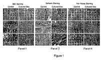
- Fig. 1 shows a microscopic histomorphological assessment with various tissue stains of 3 month intervertebral disc cultures after in vitro transplantation of human disc stem cells into evacuated rabbit nucleus pulposus with bony end plates.
- Panel 1 shows photomicrographs of hematoxylin-eosin staining of rabbit disc tissue in micrographs A, C, and E (control) and cultured intervertebral disc in micrographs B, D, and F. Magnification: 1.25X (A and B), 10X (C and D), and 20X (E and F).
- Photomicrographs C and D show the transition zone between the inner nucleus pulposus and outer annulus.
- Photomicrograph E and F show the inner zone of the nucleus pulposus and individual nucleus pulposus cells.
- Panel 2 shows photomicrographs of safranin staining of rabbit disc tissue in micrographs G, I, and K (control) and cultured intervertebral disc in micrographs H, J, and L. Magnification: 1.25X (G and H), 10X (I and J), and 20X (K and L).
- Photomicrograph I and J demonstrate the transition zone between the inner nucleus pulposus and outer annulus.
- Photomicrograph K and L demonstrate the inner zone of the nucleus pulposus and individual nucleus pulposus cells.
- Panel 3 shows photomicrographs of Von Kossa staining of rabbit disc tissue in micrographs M, O, and Q (control) and cultured intervertebral disc in micrographs H, J, and L. Magnification: 1.25X (M and N), 10X (O and P), and 20X (Q and R).
- Micrographs O and P demonstrate the transition zone between the inner nucleus pulposus and outer annulus.
- Micrographs Q and R demonstrate the inner zone of the nucleus pulposus and individual nucleus pulposus cells.
- Fig. 2 shows a microscopic histomorphological assessment of the expression of collagen type II in 3-month intervertebral disc cultures after in vitro transplantation of human disc stem cells into evacuated rabbit nucleus pulposus with bony end plates.
- Micrographs C and D demonstrate the transition zone between the inner nucleus pulposus and outer annulus.
- Micrographs E and F demonstrate the inner zone of the nucleus pulposus and individual nucleus pulposus cells.
- Fig. 3 shows a microscopic histomorphological assessment of the expression of collagen type I in 3 month intervertebral disc cultures after in vitro transplantation of human disc stem cells into evacuated rabbit nucleus pulposus with bony end plates.
- Micrographs C and D demonstrate the transition zone between the inner nucleus pulposus and outer annulus.
- Micrographs E and F demonstrate the inner zone of the nucleus pulposus and individual nucleus pulposus cells.
- Fig. 4 shows a microscopic histomorphological assessment of the expression of Ki-67 in 3 month intervertebral disc cultures after in vitro transplantation of human disc stem cells into evacuated rabbit nucleus pulposus with bony end plates.
- Micrographs C and D demonstrate the transition zone between the inner nucleus pulposus and outer annulus.
- Micrographs E and F demonstrate the inner zone of the nucleus pulposus and individual nucleus pulposus cells.
- Fig. 5 depicts a schematic of the intervertebral disc culture system with bony end plates.
- A- Single cell cultures are prepared in media and conditions that promote growth of discospheres (disc stem cell clusters). Discospheres are then prepared and injected into the annulus of a healthy rabbit in which all cells and nucleus pulposus tissue are removed from the disc.
- B- Intervertebral disc annulus with bony end plates are then put into a culture vessel with media and growth factors. At the end of 3 months, disc stem cells fill the previously empty annulus with a disc like structure.
- US 2003 220692 (Shapiro ) and US 2003/0165473 (Masuda ) relate to mixed population of cells rather than an isolated disc stem cell population according to the present invention.
- Shapiro and Masuda also relate to preparations of cells from the nucleus pulposus for which no manipulation was done or described which would isolate, or enrich, the disc stem cell population relative to normal distribution of disc stem cells found in the nucleus pulposus. In short, they do not teach an isolated disc stem cell population (only a mixed population), nor one having at least 60% disc stem cells. The required serum-free media and discosphere are also not disclosed.
- US 2004 034427 (Goel ) does not describe a free-floating in vitro sphere of disc stem cells either alone or on a scaffold.
- a disc stem cells enriched population of cells may give rise to disc progenitor cells.
- the isolated disc stem cell population may comprise a human disc stem cell population, disc stem cell population a non-human disc stem cell population or a mammal disc stem cell population.
- An isolated disc stem cell may be derived from a nucleus pulposus of a subject. The nucleus pulposus cells may comprise disc stem cells.
- the stem cells enriched cell population may comprise a human disc stem cell population, a non-human disc stem cell population or a mammal disc stem cell population.
- the stem cells enriched cell population may be derived from a nucleus pulposus of a subject.
- the nucleus pulposus cells may comprise disc stem cells.
- a nucleus pulposus is a jelly-like substance in the middle of the spinal disc.
- the nucleus pulposus may comprise chondrocytes, collagen fibrils, and proteoglycan aggrecans that have hyaluronic long chains which attract water.
- Nucleus pulposus cells may comprise autograft nucleus pulposus cells, allograft nucleus pulposus cells or xenograft nucleus pulposus cells.
- Nucleus pulposus cells may comprise disc stem cells, disc progenitor cells. mature disc cells or terminally differentiated disc cells.
- the present invention provides a method of producing a disc stem cell, population comprising the steps of growing nucleous pulposus cells harvested from a subject in a serum-free media comprising FGF2, EGF, or a combination thereof and producing a discosphere comprising disc stem cells, wherein said discosphere is a free-floating in vitro spherical structure comprising disc stem cells, disc progenitor cells or a mixture thereof, thereby producing a disc stem cell population.
- the present invention provides a method of producing disc stem cells, comprising the step of plating nucleus pulposus cells in a serum free media.
- the present invention provides a method of producing a sphere comprising nucleus pulposus cells, comprising the step of growing a culture of nucleus pulposus cells in a serum free media, thereby producing a discosphere.
- a discosphere comprising nucleus pulposus cells is a free-floating structure generated by nucleus pulposus stem cells in vitro.
- the present invention provides that a discosphere is a free-floating structure generated by nucleus pulposus progenitor cells in vitro.
- the present invention provides that a discosphere is a free-floating structure generated by nucleus pulposus stem and progenitor cells in vitro.
- a disc stem cell may be defined by its ability or capacity to form a discosphere. These disc stem cells when grown in adherent culture may have the capability to differentiate, under appropriate differentiating conditions, to mature or fully differentiate. Fully differentiated nucleus pulposus cells secrete extra cellular matrix components.
- the terms “differentiate” or “differentiation” can be intended to refer to the development of cells with specialized structure and function from unspecialized or less specialized precursor cells, and includes the development of cells that possess the structure and function of nucleus pulposus cells from precursor cells.
- the terms “differentiate” or “differentiation” can be intended to refer to the development of cells with specialized structure and function from disc stem cells.
- the terms “differentiate” or “differentiation” can be intended to refer to the development of cells with specialized structure and function from disc progenitor cells.
- Appropriate differentiating conditions may comprise a media comprising serum.
- the methods of the present invention may provide that disc material is obtained from the nucleus pulposus of a subject.
- the methods of the present invention may provide that disc material is obtained surgically and processed in the lab to create a single cell suspension of nucleus pulposus cells (Example 1).
- the methods of the present invention may provide that human disc material is obtained surgically and processed in the lab to create a single cell suspension of nucleus pulposus cells.
- the methods of the present invention may provide that human nucleus pulposus is obtained surgically and processed in the lab to create a single cell suspension of nucleus pulposus cells.
- a heterogeneous population of nucleus pulposus cells is obtained by scraping a nucleus pulposus of a subject.
- heterogeneous population of nucleus pulposus cells comprises disc stem cells, disc progenitor cells, and differentiated nucleus pulposus cells.
- a heterogeneous population of nucleus pulposus cells is scraped from a nucleus pulposus of a human subject.
- the present invention provides that plating a heterogeneous population of nucleus pulposus cells in a serum free media at low cell density results in the survival of nucleus pulposus stem cells.
- nucleus pulposus stem cell mayrefer to nucleus pulposus stem cells ability to maintain viability under conditions which include a serum-free cell culture media.
- the nucleus pulposus cells majority of the cells in the tissue die away because they cannot tolerate serum-free conditions, but the disc stem cells (or nucleus pulposus stem cells, minority of the cells in the tissue) grow into discospheres under these conditions.
- the present invention provides that plating a heterogeneous population of nucleus pulposus cells in a serum free media at low cell density results in isolation of nucleus pulposus stem cells. In another embodiment, the present invention provides that plating a heterogeneous population of nucleus pulposus cells in a serum free media at low cell density results in enriching a nucleus pulposus cell population for disc stem cells. In another embodiment, the present invention provides that plating a heterogeneous population of nucleus pulposus cells at low cell density in a serum free media, comprising a substance that interferes with cell attachment results in the survival of nucleus pulposus stem cells.
- the present invention provides that plating a heterogeneous population of nucleus pulposus cells at low cell density in a serum free media, comprising methylcellulose which interferes with cell attachment, results in the survival of nucleus pulposus stem cells.
- a heterogeneous population of nucleus pulposus cells is obtained from a biopsy specimen of nucleus pulposus minced in pieces.
- the pieces may be 0.5-10 mm, 0.5-20 mm, 0.5-3 mm, 3-6 mm, 6-12 mm, 12-20 mm, 1-6 mm, or 3-5 mm in size.
- the pieces may also be 3-4 mm in size (Example 1).
- a heterogeneous population of nucleus pulposus cells is obtained from a biopsy specimen of nucleus pulposus by treating nucleus pulposus with a collagenase II solution (Example 1).
- a heterogeneous population of nucleus pulposus cells is obtained from a biopsy specimen of nucleus pulposus by treating nucleus pulposus with a 0.1 %- 1 % clostridial collagenase (Worthington CLS II, 140u/mg).
- a heterogeneous population of nucleus pulposus cells is obtained from a biopsy specimen of nucleus pulposus by treating nucleus pulposus with a collagenase II solution followed by placing the specimen in a shaker thus obtaining a heterogeneous population of nucleus pulposus cells.
- a heterogeneous population of nucleus pulposus cells is obtained from a biopsy specimen of nucleus pulposus by aspiration of a disc of a patient.
- a heterogeneous population of nucleus pulposus cells is obtained from a biopsy specimen of nucleus pulposus by aspiration of a disc of a donor animal.
- a heterogeneous population of nucleus pulposus cells is obtained from a biopsy specimen of nucleus pulposus by aspiration of a nucleus pulposus of a donor mammal.
- a heterogeneous population of nucleus pulposus cells is obtained from a biopsy specimen of nucleus pulposus by aspiration of a healthy disc of a patient.
- the present method of producing a disc stem cell population can be used to produce a discosphere.
- the present method of producing a disc stem cell population results in the selection of nucleus pulposus stem cells.
- the surviving isolated culture of nucleus pulposus stem cells gives rise to the discospheres.
- the surviving disc stem cells enriched culture of nucleus pulposus stem cells gives rise to the discospheres.
- the supplemented serum free media of the present invention enables only nucleus pulposus stem cells to grow.
- the methods of the present invention provide that an enriched nucleus pulposus stem cell population is produced when grown in a growth factor supplemented serum free media of the present invention.
- the methods of the present invention provide that an enriched nucleus pulposus stem cell population of the present invention comprises at least 60% nucleus pulposus stem cells.
- the methods of the present invention provide that an enriched nucleus pulposus stem cell population of the present invention comprises at least 70% nucleus pulposus stem cells.
- an enriched nucleus pulposus stem cell population of the present invention comprises at least 80% nucleus pulposus stem cells. In another embodiment, the methods of the present invention provide that an enriched nucleus pulposus stem cell population of the present invention comprises at least 85% nucleus pulposus stem cells. In another embodiment, the methods of the present invention provide that an enriched nucleus pulposus stem cell population of the present invention comprises at least 90% nucleus pulposus stem cells. In another embodiment, the methods of the present invention provide that an enriched nucleus pulposus stem cell population of the present invention comprises at least 95% nucleus pulposus stem cells.
- a discosphere is derived from a single nucleus pulposus stem cell.
- only disc stem cells grow when nucleus pulposus cells are plated in a serum free media.
- only disc stem cells grow when nucleus pulposus cells are plated at low cell density.
- only disc stem cells grow when nucleus pulposus cells are plated at low cell density in a serum free media.
- only nucleus pulposus stem cells can grow as free floating solitary cells in the absence of serum.
- the disc stem cells are grown in a serum free media comprising a compound which inhibits cell maturation. In another embodiment, the disc stem cells are grown in a serum free media comprising FGF which inhibits cell maturation. In another embodiment, the disc stem cells are grown in a serum free media comprising a compound that maintains cell juvenility.
- the disc stem cells are grown in a media comprising a TGF- ⁇ superfamily member. In another embodiment, the disc stem cells are grown in a media comprising a BMP. In another embodiment, a BMP of the invention inhibits differentiation (Id) genes.
- disc stem cells are grown in a media comprising an IL6 cytokine family member. In another embodiment, the disc stem cells are grown in a media comprising leukemia inhibitory factor (LIF).
- LIF leukemia inhibitory factor
- the disc stem cells are grown in a serum free media comprising a compound which promotes cell proliferation.
- the disc stem cells are grown in a serum free media comprising EGF which promotes cell proliferation.
- the disc stem cells are grown in a serum free media comprising interleukin-2 (IL-2).
- the disc stem cells are grown in a serum free media comprising interleukin-6 (IL-6).
- disc stem cells are grown in a serum free media comprising a stem cell factor (SCF).
- the disc stem cells are grown in a serum free media comprising leukemia inhibitory factor (LIF).
- the disc stem cells are grown in a serum free media comprising transforming growth factor- ⁇ (TGF- ⁇ ).
- the disc stem cells are grown in a serum free media comprising a compound that inhibits cell differentiation (Example 1 and materials and methods).
- disc stem cells proliferate and give rise to additional stem cells.
- disc stem cells proliferate and give rise to disc progenitor cells.
- disc stem cells proliferate thus forming a discosphere.
- a discosphere comprises nucleus pulposus stem cells and nucleus pulposus progenitor cells arranged in a circular-spherical structure.
- a discosphere is a ball of cells in which a single disc stem cell gives rise to clones of itself (symmetric division) and to progenitor cells.
- a discosphere comprises free floating nucleus pulposus stem cells and nucleus pulposus progenitor cells arranged in a circular-spherical structure.
- the nucleus pulposus cells comprising a discosphere are attached to each other.
- nucleus pulposus stem cells and “disc stem cells” may be used interchangeably.
- nucleus pulposus progenitor cells and “disc progenitor cells” may be used interchangeably.
- discosphere may comprise a ball of cells in which a single disc stem cell gives rise to clones of itself (symmetric division) and to progenitor cells.
- progenitor cells may refer to immature stem-like cells with plastic potential and high proliferation rates, which can give rise to most if not all terminally differentiated tissue cells, but is not by definition a disc stem cell.
- the methods of the present invention may provide that a single cell suspension is prepared for isolating a disc stem cell by creating certain environmental conditions. In another embodiment, the methods of the present invention provide that a single cell suspension is prepared for producing a discosphere by creating certain environmental conditions.
- the methods of the present invention provide that a single cell suspension is incubated in a humidified Incubator at 37°C. In another embodiment, the methods of the present invention provide that a single cell suspension is incubated in a humidified Incubator at 35°C. In another embodiment, the methods of the present invention provide that a single cell suspension is incubated in a humidified Incubator at 36°C. In another embodiment, the methods of the present invention provide that a single cell suspension is incubated in a humidified Incubator at 38°C. In another embodiment, the methods of the present invention provide that a single cell suspension is incubated in a humidified Incubator at 39°C.
- the methods of the present invention provide that a single cell suspension is incubated in a humidified Incubator at 40°C. In another embodiment, the methods of the present invention provide that a single cell suspension is incubated in a humidified Incubator at 41°C. In another embodiment, the methods of the present invention provide that a single cell suspension is incubated in a humidified Incubator at 42°C.
- the methods of the present invention provide that a single cell suspension is incubated in an incubator further maintaining 3-8% CO2. In another embodiment, the methods of the present invention provide that a single cell suspension is incubated in an incubator further maintaining 4% CO2. In another embodiment, the methods of the present invention provide that a single cell suspension is incubated in an incubator further maintaining 5% CO2. In another embodiment, the methods of the present invention provide that a single cell suspension is incubated in an incubator further maintaining 6% CO2.
- the methods of the present invention provide that a single cell suspension is incubated in an incubator further maintaining 60-100% humidity. In another embodiment, the methods of the present invention provide that a single cell suspension is incubated in an incubator further maintaining 70-100% humidity. In another embodiment, the methods of the present invention provide that a single cell suspension is incubated in an incubator further maintaining 80-100% humidity. In another embodiment, the methods of the present invention provide that a single cell suspension is incubated in an incubator further maintaining 90-100% humidity. In another embodiment, the methods of the present invention provide that a single cell suspension is incubated in an incubator further maintaining 95-100% humidity.
- the methods of the present invention provide that a single cell suspension is plated at a final density of less than 1.10 6 cells/ml. In another embodiment, the methods of the present invention provide that a single cell suspension is plated at a final density of less than 5.10 5 cells/ml. In another embodiment, the methods of the present invention provide that a single cell suspension is plated at a final density of less than 1.10 5 cells/ml. In another embodiment, the methods of the present invention provide that a single cell suspension is plated at a final density of less than 8.10 4 cells/ml. In another embodiment, the methods of the present invention provide that a single cell suspension is plated at a final density of about 6x10⁴ cells/ml (Example 1).
- the present invention provides a composition comprising an enriched disc stem cell population produced by the method described herein further comprising a serum-free media comprising FGF2, Epidermal Growth Factor (EGF), or a combination thereof.
- the composition may comprise a population of nucleus pulposus cells enriched for nucleus pulposus stem cells.
- the composition further comprises an appropriate environment, such as those described herein, wherein, a disc stem cell can be induced to proliferate and generate disc stem cells progeny.
- the term environment in which disc stem cells progeny are placed preferably refers to the combination of external or extrinsic physical and/or chemical conditions that affect and influence the growth and development of disc stem cells.
- the environment can be ex- vivo or in-vivo.
- a disc scaffold may serve as an in-vivo environment that induces disc stem cells to generate progeny.
- the environment is ex-vivo and comprises disc stem cells placed in cell culture medium in an incubator (Example 1).
- the composition comprising disc stem cells comprises Epidermal Growth Factor (EGF) supplemented to the media.
- EGF Epidermal Growth Factor
- the composition comprising disc stem cells comprises 1-10 ng/ml EGF supplemented to the media.
- the composition comprising disc stem cells comprises 1-100 ng/ml EGF supplemented to the media.
- the composition comprising disc stem cells comprises 20-50 ng/ml EGF supplemented to the media.
- the composition comprising disc stem cells comprises 50-100 ng/ml EGF supplemented to the media.
- the composition comprising disc stem cells comprises 5-15 ng/ml EGF supplemented to the media.
- the composition comprising disc stem cells comprises 8-12 ng/ml EGF supplemented to the media.
- the composition comprising disc stem cells comprises FGF supplemented to the media.
- the composition comprising disc stem cells comprises Fibroblast Growth Factor 2 (FGF2) supplemented to the media.
- FGF2 Fibroblast Growth Factor 2
- the composition comprising disc stem cells comprises 1-100 ng/ml FGF2 supplemented to the media.
- the composition comprising disc stem cells comprises 20-50 ng/ml FGF2 supplemented to the media.
- the composition comprising disc stem cells comprises 50-100 ng/ml FGF2 supplemented to the media.
- the composition comprising disc stem cells comprises 5-15 ng/ml FGF2 supplemented to the media.
- the composition comprising disc stem cells comprises 8-12 ng/ml FGF2 supplemented to the media.
- the composition comprising disc stem cells further comprises insulin supplemented to the media.
- the composition comprising disc stem cells further comprises 1-100 ⁇ g/ml insulin supplemented to the media (Example 1).
- the composition comprising disc stem cells further comprises 20-50 ⁇ g/ml insulin supplemented to the media.
- the composition comprising disc stem cells further comprises 50-100 ⁇ g/ml insulin supplemented to the media.
- the composition comprising disc stem cells further comprises 5-15 ⁇ g/ml insulin supplemented to the media.
- the composition comprising disc stem cells further comprises 8-12 ⁇ g/ml insulin supplemented to the media.
- the composition comprising disc stem cells further comprises progesterone supplemented to the media (Example 1). In another embodiment, the composition comprising disc stem cells further comprises 1-200 ng/ml progesterone supplemented to the media. In another embodiment, the composition comprising disc stem cells further comprises 20-200 ng/ml progesterone supplemented to the media. In another embodiment, the composition comprising disc stem cells further comprises 50-150 ng/ml progesterone supplemented to the media. In another embodiment, the composition comprising disc stem cells further comprises 10-100 ng/ml progesterone supplemented to the media. In another embodiment, the composition comprising disc stem cells further comprises 20-80 ng/ml progesterone supplemented to the media. In another embodiment, the composition comprising disc stem cells further comprises 30-50 ng/ml progesterone supplemented to the media.
- the composition comprising disc stem cells further comprises putrescine supplemented to the media (Example 1). In another embodiment, the composition comprising disc stem cells further comprises 1-800 ng/ml putrescine supplemented to the media. In another embodiment, the composition comprising disc stem cells further comprises 1-100 ng/ml putrescine supplemented to the media. In another embodiment, the composition comprising disc stem cells further comprises 100-300 ng/ml putrescine supplemented to the media. In another embodiment, the composition comprising disc stem cells further comprises 300-500 ng/ml putrescine supplemented to the media. In another embodiment, the composition comprising disc stem cells further comprises 500-800 ng/ml putrescine supplemented to the media.
- composition comprising disc stem cells further comprises 150-250 ng/ml putrescine supplemented to the media. In another embodiment, the composition comprising disc stem cells further comprises 180-220 ng/ml putrescine supplemented to the media.
- the composition comprising disc stem cells further comprises transferrin supplemented to the media (Example 1). In another embodiment, the composition comprising disc stem cells further comprises 1-400 ng/ml transferrin supplemented to the media. In another embodiment, the composition comprising disc stem cells further comprises 1-100 ng/ml transferrin supplemented to the media. In another embodiment, the composition comprising disc stem cells further comprises 100-200 ng/ml transferrin supplemented to the media. In another embodiment, the composition comprising disc stem cells further comprises 200-400 ng/ml transferrin supplemented to the media. In another embodiment, the composition comprising disc stem cells further comprises 20-150 ng/ml transferrin supplemented to the media. In another embodiment, the composition comprising disc stem cells further comprises 80-200 ng/ml transferrin supplemented to the media. In another embodiment, the composition comprising disc stem cells further comprises 80-120 ng/ml transferrin supplemented to the media.
- the composition comprising disc stem cells further comprises sodium selenite supplemented to the media (Example 1). In another embodiment, the composition comprising disc stem cells further comprises 1-400 ng/ml sodium selenite supplemented to the media. In another embodiment, the composition comprising disc stem cells further comprises 1-100 ng/ml sodium selenite supplemented to the media. In another embodiment, the composition comprising disc stem cells further comprises 100-200 ng/ml sodium selenite supplemented to the media. In another embodiment, the composition comprising disc stem cells further comprises 200-400 ng/ml sodium selenite supplemented to the media.
- the composition comprising disc stem cells further comprises 20-150 ng/ml sodium selenite supplemented to the media. In another embodiment, the composition comprising disc stem cells further comprises 40-180 ng/ml sodium selenite supplemented to the media. In another embodiment, the composition comprising disc stem cells further comprises 40-80 ng/ml sodium selenite supplemented to the media.
- the composition comprising disc stem cells further comprises methylcellulose supplemented to the media (Example 1). In another embodiment, the composition comprising disc stem cells further comprises 0.5-10% methylcellulose supplemented to the media. In another embodiment, the composition comprising disc stem cells further comprises 0.5-3% methylcellulose supplemented to the media. In another embodiment, the composition comprising disc stem cells further comprises 3-5% methylcellulose supplemented to the media. In another embodiment, the composition comprising disc stem cells further comprises 5-8% methylcellulose supplemented to the media. In another embodiment, the composition comprising disc stem cells further comprises 7-10% methylcellulose supplemented to the media. In another embodiment, the composition comprising disc stem cells further comprises 0.5-2.5% methylcellulose supplemented to the media. In another embodiment, the composition comprising disc stem cells further comprises 1-2.5% methylcellulose supplemented to the media. In another embodiment, the composition comprising disc stem cells further comprises 1.5-2.5% methylcellulose supplemented to the media.
- the composition comprising disc stem cells further comprises an antibiotic supplemented to the media (Example 1).
- the antibiotic supplemented to the media is penicillin-streptomycin.
- the composition comprising disc stem cells further comprises 1000-10000 U/ml penicillin-streptomycin supplemented to the media.
- the composition comprising disc stem cells further comprises 1000-3000 U/ml penicillin-streptomycin supplemented to the media.
- the composition comprising disc stem cells further comprises 3000-6000 U/ml penicillin-streptomycin supplemented to the media.
- the composition comprising disc stem cells further comprises 6000-10000 U/ml penicillin-streptomycin supplemented to the media.
- composition comprising disc stem cells further comprises 3000-8000 U/ml penicillin-streptomycin supplemented to the media. In another embodiment, the composition comprising disc stem cells further comprises 4000-6000 U/ml penicillin-streptomycin supplemented to the media. In another embodiment, the composition comprising disc stem cells further comprises 5000 U/ml penicillin-streptomycin supplemented to the media.
- the composition comprising disc stem cells further comprises KO serum replacer supplemented to the media. In another embodiment, the composition comprising disc stem cells further comprises 0.5-30% KO serum replacer supplemented to the media. In another embodiment, the composition comprising disc stem cells further comprises 5-30% knockout (KO) serum replacer supplemented to the media. In another embodiment, the composition comprising disc stem cells further comprises 3-5% KO serum replacer supplemented to the media. In another embodiment, the composition comprising disc stem cells further comprises 5-15% KO serum replacer supplemented to the media. In another embodiment, the composition comprising disc stem cells further comprises 15-30% KO serum replacer supplemented to the media.
- KO serum replacer supplemented to the media In another embodiment, the composition comprising disc stem cells further comprises 0.5-30% KO serum replacer supplemented to the media. In another embodiment, the composition comprising disc stem cells further comprises 5-30% knockout (KO) serum replacer supplemented to the media. In another embodiment, the composition comprising disc stem cells further comprises 3-5% KO
- composition comprising disc stem cells further comprises 10-20% KO serum replacer supplemented to the media. In another embodiment, the composition comprising disc stem cells further comprises 15-25% KO serum replacer supplemented to the media. In another embodiment, the composition comprising disc stem cells further comprises 20% KO serum replacer supplemented to the media.
- the composition comprising disc stem cells further comprises non-essential Amino Acids supplemented to the media. In another embodiment, the composition comprising disc stem cells further comprises 0.1-10% non-essential Amino Acids supplemented to the media. In another embodiment, the composition comprising disc stem cells further comprises 0.1-1% non-essential Amino Acids supplemented to the media. In another embodiment, the composition comprising disc stem cells further comprises 1-5% non-essential Amino Acids supplemented to the media. In another embodiment, the composition comprising disc stem cells further comprises 5-10% non-essential Amino Acids supplemented to the media. In another embodiment, the composition comprising disc stem cells further comprises 15-30% non-essential Amino Acids supplemented to the media.
- composition comprising disc stem cells further comprises 0.5-1% non-essential Amino Acids supplemented to the media. In another embodiment, the composition comprising disc stem cells further comprises 0.8-1.2% non-essential Amino Acids supplemented to the media. In another embodiment, the composition comprising disc stem cells further comprises 1 % non-essential Amino Acids supplemented to the media.
- the composition comprising disc stem cells further comprises L-glutamine supplemented to the media. In another embodiment, the composition comprising disc stem cells further comprises 0.1-10mM L-glutamine supplemented to the media. In another embodiment, the composition comprising disc stem cells further comprises 0.1-5mM L-glutamine supplemented to the media. In another embodiment, the composition comprising disc stem cells further comprises 5-10mM L-glutamine supplemented to the media. In another embodiment, the composition comprising disc stem cells further comprises 5-8mM L-glutamine supplemented to the media. In another embodiment, the composition comprising disc stem cells further comprises 0.5-2.5mM L-glutamine supplemented to the media.
- composition comprising disc stem cells further comprises 1.5-3mM L-glutamine supplemented to the media. In another embodiment, the composition comprising disc stem cells further comprises 0.5-1.5mM L-glutamine supplemented to the media. In another embodiment, the composition comprising disc stem cells further comprises 0.8-1.2 mM L-glutamine supplemented to the media.
- the composition comprising disc stem cells further comprises b- mercaptoethanol supplemented to the media. In another embodiment, the composition comprising disc stem cells further comprises 0.01-1 mM b-mercaptoethanol supplemented to the media. In another embodiment, the composition comprising disc stem cells further comprises 0.01-0.5 mM b- mercaptoethanol supplemented to the media. In another embodiment, the composition comprising disc stem cells further comprises 0.5-1 mM b-mercaptoethanol supplemented to the media. In another embodiment, the composition comprising disc stem cells further comprises 0.5-0.8 mM b-mercaptoethanol supplemented to the media.
- the composition comprising disc stem cells further comprises 00.5-0.25 mM b-mercaptoethanol supplemented to the media. In another embodiment, the composition comprising disc stem cells further comprises 0.15-0.3 mM b-mercaptoethanol supplemented to the media. In another embodiment, the composition comprising disc stem cells further comprises 0.05-0.15 mM b-mercaptoethanol supplemented to the media. In another embodiment, the composition comprising disc stem cells further comprises 0.08-0.12 mM b-mercaptoethanol supplemented to the media.
- the composition comprising disc stem cells further comprises a media comprising Dulbecco's Modified Eagle's Medium (DMEM).
- DMEM Dulbecco's Modified Eagle's Medium
- the composition comprising a discosphere further comprises a media comprising DMEM/F12.
- the composition comprising disc stem cells further comprises a media comprising Hamm's culture media.
- the composition comprising disc stem cells further comprises a media comprising Hamm's/F12 culture media.
- the composition comprising disc stem cells further comprises a media comprising ESGRO Complete TM Accutase TM .
- ESGRO Complete TM Accutase TM is a cell detachment solution of proteolytic and collagenolytic enzymes qualified for use for the detachment of stem cells cultured in serum-free conditions with ESGRO Complete TM Clonal Grade Medium.
- 1X Accutase TM enzymes in Dulbecco's PBS comprises 0.5 mM EDTA•4Na and 3 mg/L Phenol.
- the composition comprising disc stem cells further comprises a media comprising HEScGRO hES cell medium (Chemicon Temecula, CA).
- the composition comprising disc stem cells may be used for plating disc stem cells in ultra low attachment plates.
- the composition comprising disc stem cells may be used for plating disc stem cells in ultra low attachment plates precoated with an anti-adhesive substance.
- the anti-adhesive substance may be poly 2-hydroxyethyl methacrylate.
- the present invention provides an isolated discosphere wherein said discosphere is a free-floating in vitro spherical structure comprising disc stem cells, disc progenitor cells or a mixture thereof.
- a disc stem cell produced by the methods of the present invention gives rise to an isolated discosphere.
- a discosphere of the present invention is the result of stem cell proliferation which gives rise to additional stem cells and progenitor cells.
- a discosphere is formed as a result of disc stem proliferation.
- an isolated discosphere of the present invention comprises nucleus pulposus stem cells and nucleus pulposus progenitor cells arranged in a circular-spherical structure.
- an isolated discosphere is a ball of cells in which a single disc stem cell gives rise to clones of itself (symmetric division) and to progenitor cells.
- a discosphere of the present invention is a free floating conglomerate of nucleus pulposus stem cells and nucleus pulposus progenitor cells arranged in a circular-spherical structure.
- a discosphere culture of the present invention comprises solitary free floating discospheres.
- the methods of the present invention may provide that isolating a discosphere of the present invention can be readily performed by one skilled in the art under a light microscope.
- the methods of the present invention provide that a single cell suspension is prepared for producing a disc stem cell by plating and incubating a disc stem cell in a serum free media. In another embodiment, the methods of the present invention provide that a single cell suspension is prepared for producing a discosphere by plating and incubating a disc stem cell in a serum free media.
- the present invention provides a method of producing a discosphere, comprising the step of growing a culture of nucleus pulposus cells in a serum free media, thereby producing a discosphere.
- Growing a primary culture of nucleus pulposus cells in a serum free media may result in selecting nucleus pulposus stem cells
- the remaining isolated culture of nucleus pulposus stem cells may give rise to discospheres of the present invention.
- the remaining enriched culture of nucleus pulposus stem cells may give rise to discospheres of the present invention.
- discospheres according to the methods of the present invention grow in the compositions of the present invention.
- discospheres according to the methods of the present invention grow in the supplemented media of the present invention. In another embodiment, discospheres according to the methods of the present invention grow under conditions which do not permit cell-substrate adhesion. In another embodiment, conditions which do not permit cell-substrate adhesion comprise for example the addition of about 0.2-2% methylcellulose to the cell culture media of the present invention.
- the present invention provides a composition comprising a discosphere, wherein said discosphere is a free-floating in vitro spherical structure comprising disc stem cells, disc progenitor cells, or a mixture thereof.
- a composition of the present invention comprises a single discosphere.
- a composition of the present invention comprises at least 1x10 2 discospheres.
- a composition of the present invention comprises at least 1x10 3 discospheres.
- a composition of the present invention comprises at least 1x10 4 discospheres.
- a composition of the present invention comprises at least 1x10 5 discospheres.
- a composition of the present invention comprises at least 1x10 6 discospheres.
- the composition of the present invention comprises a discosphere and media.
- the media is a serum free media.
- the composition comprising a discosphere further comprises EGF supplemented to the media.
- the composition comprising a discosphere further comprises 1-10 ng/ml EGF supplemented to the media.
- the composition comprising a discosphere further comprises 1-100 ng/ml EGF supplemented to the media.
- the composition comprising a discosphere further comprises 20-50 ng/ml EGF supplemented to the media.
- the composition comprising a discosphere further comprises 50-100 ng/ml EGF supplemented to the media.
- the composition comprising a discosphere further comprises 5-15 ng/ml EGF supplemented to the media.
- the composition comprising a discosphere further comprises 8-12 ng/ml EGF supplemented to the media.
- the composition comprising a discosphere further comprises FGF supplemented to the media. In another embodiment, the composition comprising a discosphere further comprises FGF2 supplemented to the media. In another embodiment, the composition comprising a discosphere further comprises 1-100 ng/ml FGF2 supplemented to the media. In another embodiment, the composition comprising a discosphere further comprises 20-50 ng/ml FGF2 supplemented to the media. In another embodiment, the composition comprising a discosphere further comprises 50-100 ng/ml FGF2 supplemented to the media. In another embodiment, the composition comprising a discosphere further comprises 5-15 ng/ml FGF2 supplemented to the media. In another embodiment, the composition comprising a discosphere further comprises 8-12 ng/ml FGF2 supplemented to the media.
- the composition comprising a discosphere further comprises insulin supplemented to the media (Example 2 and materials and methods).
- the composition comprising a discosphere further comprises 1-100 ⁇ g/ml insulin supplemented to the media.
- the composition comprising a discosphere further comprises 20-50 ⁇ g/ml insulin supplemented to the media.
- the composition comprising a discosphere further comprises 50-100 ⁇ g/ml insulin supplemented to the media.
- the composition comprising a discosphere further comprises 5-15 ⁇ g/ml insulin supplemented to the media.
- the composition comprising a discosphere further comprises 3-7 ⁇ g/ml insulin supplemented to the media.
- the composition comprising a discosphere further comprises progesterone supplemented to the media.
- the composition comprising a discosphere further comprises 1-200 ng/ml progesterone supplemented to the media.
- the composition comprising a discosphere further comprises 20-200 ng/ml progesterone supplemented to the media.
- the composition comprising a discosphere further comprises 50-150 ng/ml progesterone supplemented to the media.
- the composition comprising disc stem cells further comprises 10-100 ng/ml progesterone supplemented to the media.
- the composition comprising disc stem cells further comprises 20-80 ng/ml progesterone supplemented to the media.
- the composition comprising disc stem cells further comprises 15-25 ng/ml progesterone supplemented to the media.
- the composition comprising a discosphere further comprises putrescine supplemented to the media. In another embodiment, the composition comprising a discosphere further comprises 1-800 ng/ml putrescine supplemented to the media. In another embodiment, the composition comprising a discosphere further comprises 1-100 ng/ml putrescine supplemented to the media. In another embodiment, the composition comprising a discosphere further comprises 100-300 ng/ml putrescine supplemented to the media. In another embodiment, the composition comprising a discosphere further comprises 300-500 ng/ml putrescine supplemented to the media.
- the composition comprising a discosphere further comprises 500-800 ng/ml putrescine supplemented to the media. In another embodiment, the composition comprising a discosphere further comprises 150-250 ng/ml putrescine supplemented to the media. In another embodiment, the composition comprising a discosphere further comprises 140-160 ng/ml putrescine supplemented to the media.
- the composition comprising a discosphere further comprises transferrin supplemented to the media. In another embodiment, the composition comprising a discosphere further comprises 1-400 ng/ml transferrin supplemented to the media. In another embodiment, the composition comprising a discosphere further comprises 1-100 ng/ml transferrin supplemented to the media. In another embodiment, the composition comprising a discosphere further comprises 100-200 ng/ml transferrin supplemented to the media. In another embodiment, the composition comprising a discosphere further comprises 200-400 ng/ml transferrin supplemented to the media. In another embodiment, the composition comprising a discosphere further comprises 20-150 ng/ml transferrin supplemented to the media.
- composition comprising a discosphere further comprises 80-200 ng/ml transferrin supplemented to the media. In another embodiment, the composition comprising a discosphere further comprises 30-70 ng/ml transferrin supplemented to the media.
- the composition comprising a discosphere further comprises sodium selenite supplemented to the media. In another embodiment, the composition comprising a discosphere further comprises 1-400 ng/ml sodium selenite supplemented to the media. In another embodiment, the composition comprising a discosphere further comprises 1-100 ng/ml sodium selenite supplemented to the media. In another embodiment, the composition comprising a discosphere further comprises 100-200 ng/ml sodium selenite supplemented to the media. In another embodiment, the composition comprising a discosphere further comprises 200-400 ng/ml sodium selenite supplemented to the media.
- the composition comprising a discosphere further comprises 20-150 ng/ml sodium selenite supplemented to the media. In another embodiment, the composition comprising a discosphere further comprises 40-180 ng/ml sodium selenite supplemented to the media. In another embodiment, the composition comprising a discosphere further comprises 20-40 ng/ml sodium selenite supplemented to the media.
- the composition comprising a discosphere further comprises methylcellulose supplemented to the media. In another embodiment, the composition comprising a discosphere further comprises 0.5-10% methylcellulose supplemented to the media. In another embodiment, the composition comprising a discosphere further comprises 0.5-3% methylcellulose supplemented to the media. In another embodiment, the composition comprising a discosphere further comprises 3-5% methylcellulose supplemented to the media. In another embodiment, the composition comprising a discosphere further comprises 5-8% methylcellulose supplemented to the media. In another embodiment, the composition comprising a discosphere further comprises 7-10% methylcellulose supplemented to the media.
- composition comprising a discosphere further comprises 0.5-2.5% methylcellulose supplemented to the media. In another embodiment, the composition comprising a discosphere further comprises 1-2.5% methylcellulose supplemented to the media. In another embodiment, the composition comprising a discosphere further comprises 0.6-1% methylcellulose supplemented to the media.
- the composition comprising a discosphere further comprises an antibiotic supplemented to the media.
- the antibiotic supplemented to the media is penicillin-streptomycin.
- the composition comprising a discosphere further comprises 1000-10000 U/ml penicillin-streptomycin supplemented to the media.
- the composition comprising a discosphere further comprises 1000-3000 U/ml penicillin-streptomycin supplemented to the media.
- the composition comprising a discosphere further comprises 3000-6000 U/ml penicillin-streptomycin supplemented to the media.
- the composition comprising a discosphere further comprises 6000-10000 U/ml penicillin-streptomycin supplemented to the media. In another embodiment, the composition comprising a discosphere further comprises 3000-8000 U/ml penicillin-streptomycin supplemented to the media. In another embodiment, the composition comprising a discosphere further comprises 4000-6000 U/ml penicillin-streptomycin supplemented to the media. In another embodiment, the composition comprising a discosphere further comprises 5000 U/ml penicillin-streptomycin supplemented to the media.
- the composition comprising a discosphere further comprises KO serum replacer supplemented to the media. In another embodiment, the composition comprising a discosphere further comprises 0.5-30% KO serum replacer supplemented to the media. In another embodiment, the composition comprising a discosphere further comprises 5-30% KO serum replacer supplemented to the media. In another embodiment, the composition comprising a discosphere further comprises 3-5% KO serum replacer supplemented to the media. In another embodiment, the composition comprising a discosphere further comprises 5-15% KO serum replacer supplemented to the media. In another embodiment, the composition comprising a discosphere further comprises 15-30% KO serum replacer supplemented to the media.
- composition comprising a discosphere further comprises 10-20% KO serum replacer supplemented to the media. In another embodiment, the composition comprising a discosphere further comprises 15-25% KO serum replacer supplemented to the media. In another embodiment, the composition comprising a discosphere further comprises 20% KO serum replacer supplemented to the media.
- the composition comprising a discosphere further comprises non-essential Amino Acids supplemented to the media. In another embodiment, the composition comprising a discosphere further comprises 0.1-10% non-essential Amino Acids supplemented to the media. In another embodiment, the composition comprising a discosphere further comprises 0.1-1 % non-essential Amino Acids supplemented to the media. In another embodiment, the composition comprising a discosphere further comprises 1-5% non-essential Amino Acids supplemented to the media. In another embodiment, the composition comprising a discosphere further comprises 5-10% non-essential Amino Acids supplemented to the media.
- the composition comprising a discosphere further comprises 15-30% non-essential Amino Acids supplemented to the media. In another embodiment, the composition comprising a discosphere further comprises 0.5-1 % non-essential Amino Acids supplemented to the media. In another embodiment, the composition comprising a discosphere further comprises 0.8-1.2% non-essential Amino Acids supplemented to the media. In another embodiment, the composition comprising a discosphere further comprises 1 % non-essential Amino Acids supplemented to the media.
- the composition comprising a discosphere further comprises L-glutamine supplemented to the media. In another embodiment, the composition comprising a discosphere further comprises 0.1-10 mM L-glutamine supplemented to the media. In another embodiment, the composition comprising a discosphere further comprises 0.1-5 mM L-glutamine supplemented to the media. In another embodiment, the composition comprising a discosphere further comprises 5-10 mM L-glutamine supplemented to the media. In another embodiment, the composition comprising a discosphere further comprises 5-8 mM L-glutamine supplemented to the media. In another embodiment, the composition comprising a discosphere further comprises 0.5-2.5 mM L-glutamine supplemented to the media.
- the composition comprising a discosphere further comprises 1.5-3 mM L-glutamine supplemented to the media. In another embodiment, the composition comprising a discosphere further comprises 0.5-1.5 mM L-glutamine supplemented to the media. In another embodiment, the composition comprising a discosphere further comprises 0.8-1.2 mM L-glutamine supplemented to the media.
- the composition comprising a discosphere further comprises b-mercaptoethanol supplemented to the media. In another embodiment, the composition comprising a discosphere further comprises 0.01-1 mM b-mercaptoethanol supplemented to the media. In another embodiment, the composition comprising a discosphere further comprises 0.01-0.5 mM b-mercaptoethanol supplemented to the media. In another embodiment, the composition comprising a discosphere further comprises 0.5-1 mM b-mercaptoethanol supplemented to the media. In another embodiment, the composition comprising a discosphere further comprises 0.5-0.8 mM b-mercaptoethanol supplemented to the media.
- the composition comprising a discosphere further comprises 00.5-0.25 mM b-mercaptoethanol supplemented to the media. In another embodiment, the composition comprising a discosphere further comprises 0.15-0.3 mM b-mercaptoethanol supplemented to the media. In another embodiment, the composition comprising a discosphere further comprises 0.05-0.15 mM b-mercaptoethanol supplemented to the media. In another embodiment, the composition comprising a discosphere further comprises 0.08-0.12 mM b-mercaptoethanol supplemented to the media.
- the composition comprising a discosphere comprises a media comprising Dulbecco's Modified Eagle's Medium (DMEM). In another embodiment, the composition comprising a discosphere comprises a media further comprising DMEM/F12. In another embodiment, the composition comprising a discosphere comprises a media further comprising ESGRO CompleteTM AccutaseTM. In another embodiment, ESGRO CompleteTM AccutaseTM is a cell detachment solution of proteolytic and collagenolytic enzymes qualified for use for the detachment of stem cells cultured in serum-free conditions with ESGRO CompleteTM Clonal Grade Medium.
- DMEM Dulbecco's Modified Eagle's Medium
- the composition comprising a discosphere comprises a media further comprising DMEM/F12. In another embodiment, the composition comprising a discosphere comprises a media further comprising ESGRO CompleteTM AccutaseTM. In another embodiment, ESGRO CompleteTM AccutaseTM is a cell detachment solution of prote
- 1X AccutaseTM enzymes in Dulbecco's PBS comprises 0.5 mM EDTA•4Na and 3 mg/L Phenol.
- the composition comprising a discosphere comprises a media comprising HEScGRO hES cell medium (Chemicon Temecula, CA).
- the present invention provides that discospheres obtained by the methods of the present invention are further expanded.
- the present invention provides that discospheres are dissociated by incubation at 37°C in DMEM/F12 medium supplemented with collagenase.
- the present invention provides that the dissociated cells are expanded by replating the same into methylcellulose-based medium.
- the methods of the present invention provide that the medium is supplemented with 8-20% fetal bovine serum (FBS) when growing a disc tissue. In another embodiment, the methods of the present invention provide that the medium is supplemented with 8- 20% fetal bovine serum (FBS) when growing a disc tissue in a scaffold. In another embodiment, the medium comprises 30-70% media derived from cultures of primary human foreskin fibroblasts, or a combination thereof. In another embodiment, the methods of the present invention provide that the medium is free of serum when growing disc stem cells, disc progenitor cells, or a combination thereof.
- FBS fetal bovine serum
- the methods of the present invention provide that the medium is free of serum when growing disc stem cells, disc progenitor cells, or a combination thereof.
- the methods of the present invention provide that the medium is free of a serum replacer when growing disc stem cells, disc progenitor cells, or a combination thereof. In another embodiment, the methods of the present invention provide that the medium is free of FBS when growing disc stem cells. In another embodiment, the methods of the present invention provide that the medium is free of FBS when growing nucleus pulposus stem cells. In another embodiment, the methods of the present invention provide that the medium is free of FBS when growing progenitor cells. In another embodiment, the methods of the present invention provide that the medium is free of FBS when growing nucleus pulposus progenitor cells.
- the methods of the present invention provide that the medium is free of serum or a serum replacer when isolating disc stem cells or disc progenitor cells. In another embodiment, the methods of the present invention provide that the medium is free of serum or a serum replacer when enriching a heterogeneous population of cells for disc stem cells, disc progenitor cells, or a combination thereof. In another embodiment, the methods of the present invention provide that the medium is free of serum or a serum replacer when growing discospheres comprising disc stem cells, disc progenitor cells, or a combination thereof. In another embodiment, the methods of the present invention provide that the medium is free of serum or a serum replacer when expanding a culture comprising disc stem cells, disc progenitor cells, or a combination thereof. In another embodiment, the methods of the present invention provide that the medium is free of serum or a serum replacer when expanding a culture enriched for disc stem cells, disc progenitor cells, or a combination thereof.
- the present invention provides an artificial disc replacement device comprising a disc scaffold and a discosphere, wherein the discosphere is a free-floating in vitro spherical structure comprising disc stem cells, disc progenitor cells, or a mixture thereof.
- the present invention provides an artificial disc comprising nucleus pulposus cells.
- the disc replacement device may be an intervertebral disc replacement device.
- An intervertebral disc may be located between the concave articular surfaces of the adjacent vertebral body endplates.
- the disc replacement device of the present invention permits movements such as flexion, extension, lateral flexion, and rotation.
- the disc replacement device of the present invention is used to repair and/or replace injured or damaged intervertebral discs.
- the disc replacement device of the present invention provides a prosthetic disc that combines both stability to support the high loads, of the patient's vertebrae and flexibility to provide the patient with sufficient mobility and proper spinal column load distribution.
- the disc replacement device comprises a disc scaffold.
- the scaffold comprises a shape memory alloy.
- a shape memory alloy may be deformed during its martensitic phase, but will regain its original shape when it is heated above a certain temperature, such as an austenite phase temperature.
- a shape memory alloy of the present invention exhibits a superelastic property, thereby able to absorb large deformations without damaging its structure.
- the disc replacement device may comprise a rigid body that fits between the vertebrates with a protuberance extending from a vertebral contacting surface and extends into the vertebral body.
- the disc replacement device may comprise a disc arthroplasty device for replacement of the spinal disk.
- the disc replacement device may comprise a ball-and-socket to enable rotation.
- the disc replacement device may comprise an intermediate layer allowing for movement between the upper joint piece and the lower joint piece.
- the disc replacement device may comprise two endplates that are anchored to the top and bottom surfaces of the spinal bones.
- the disc replacement device may comprise two metal endplates that are anchored to the top and bottom surfaces of the spinal bones.
- the metal may be cobalt-chrome alloy.
- the endplates may be coated with nucleus pulposus cell adhesion molecules.
- the endplates may be coated with molecules promoting nucleus pulposus cell growth.
- the disc replacement device may comprise ceramics.
- the disc replacement device may comprise injectable fluids.
- the disc replacement device may comprise hydrogels, the disc replacement device may comprise a hydrogel core in a flexible, inelastic, woven polyethylene jacket.
- the disc replacement device may comprise a polyvinyl alcohol material.
- the disc replacement device may comprise inflatables.
- the disc replacement device may comprise elastic coils.
- the disc replacement device may comprise an elongated elastic memory-coiling spiraland the elongated elastic memory-coiling spiral may be made of polycarbonate urethane.
- the disc replacement device may comprise a one-piece convex surfaced ceramic or metal implant that anchors to the inferior vertebral body as a hemiarthroplasty.
- the disc replacement device may comprise a balloon-like implant made of polyurethane.
- the disc replacement device comprises a protein hydrogel or a thermopolymer.
- the disc scaffold comprises an ECM component.
- the ECM component is a structural protein.
- the disc scaffold comprises collagen.
- the structural protein is elastin.
- the ECM component is a specialized protein.
- the specialized protein is fibrillin.
- the specialized protein is fibronectin.
- the specialized protein is laminin.
- the ECM component is a proteoglycan.
- proteoglycans are composed of a protein core to which is attached long chains of repeating disaccharide units termed of glycosaminoglycans (GAGs) forming extremely complex high molecular weight component.
- collagen is collagen type I.
- collagen type I comprises [al(I)]2[a(I)] chains.
- collagen type I is derived from skin, tendon, or bone.
- collagen is collagen type II.
- collagen type [pi] comprises [al(II)]3 chains.
- collagen type II is derived from cartilage or vitreous humor.
- type II collagen fibrils are cross-linked to proteoglycans in the matrix by type IX collagen.
- collagen is collagen type IE.
- collagen type El comprises [al(III)] 3 chains.
- collagen type HI is derived from skin or muscle, and is frequently found with type I.
- collagen is collagen type IV.
- collagen type rv comprises [al(IV)2[a2(IV)] chains.
- collagen type IV is derived from basal lamina.
- collagen is collagen type V.
- collagen type V comprises [a1(V)] [a2(V)] [a3(V)] chains.
- collagen type V is derived from an interstitial tissue associated with type I collagen.
- collagen is collagen type VI.
- collagen type VI comprises [a1(VI)][a2(VI)][a3(VI)] chains.
- collagen type VI is derived from an interstitial tissue associated with type I collagen.
- type VI collagen consists of relatively short triple-helical regions about 60 nm long separated by globular domains about 40 nm long. In some embodiments, fibrils of pure type VI collagen form a structure similar to beads on a string.
- collagen is collagen type VII.
- collagen type VII comprises [a1(VII)]3 chains.
- collagen type VII is derived from epithelia.
- collagen is collagen type VIII.
- collagen type VIII comprises [a1(VIII)]3 chains.
- collagen type VII is derived from endothelial cells.
- collagen is collagen type IX.
- collagen type IX comprises [a1(IX)][a2(IX)][a3(IX)] chains.
- collagen type IX is derived from cartilage associated with type II collagen.
- collagen is collagen type X.
- collagen type X comprises [a1(X)]3 chains.
- collagen type X is derived from hypertrophic and mineralizing cartilage.
- collagen is collagen type XI.
- collagen type XI comprises [a1(XI)][a2(XI)][a3(XI)] chains.
- collagen type XI is derived from cartilage.
- collagen is collagen type XII.
- collagen type XII comprises a1(XII) chains.
- collagen type XII is derived from sites wherein types I and III collagens are present.
- type I collagen molecules pack together side-by-side, forming fibrils with a diameter of 50-200 nm.
- fibrils, adjacent collagen molecules are displaced from one another by 67 nm, about another -quarter of their length.
- collagens types I, II, III, and V form rodlike triple helices to via side-by-side interactions.
- the collagen of the present invention is derived from cows. In another embodiment, collagen of the present invention is derived from patient's own fat or hyaluronic acid.
- collagen is a collagen-like substance which has been modified by dissolving collagen in water and modifying the thusly dissolved collagen to render its surface charge effectively more positive than prior to modification.
- this material is well known and is disclosed, e.g., in U.S. Pat. No. 4,238,480 .
- modified collagen is freeze-dried to form a solid mass of gelatin.
- the mass of gelatin may be formed in the shape of a rod, strip, film or flake.
- Semed F a collagen preparation manufactured in native fiber form without any chemical or enzymatic modifications
- Semed S a lyophilized collagen powder extracted from fresh bovine hides.
- the Semed F material is a Type I collagen (greater than 95%)
- the Semed S is a mixture of Type I and Type III collagen macro-molecules in which the shape and dimension of tropocollagen in its natural helical orientation is retained.
- the concentration of the collagen in the liquid which is to be freeze-dried can range from 0.5-10% and preferably 1-5%, with the lower concentrations forming less dense or discontinuous solids. In another embodiment, at lower concentrations of 0.5 to 1%, the Semed F forms a structure which approximates dense cobwebs.
- native collagen film wherein the film strength is preserved and the triple-helix structure of the collagen polymer is maintained intact, can also be used, either alone or with a plasticizer incorporated therewith.
- gelatin or other water soluble forms of collagen are utilized.
- soluble forms of collagen will readily polymerize at body temperatures to form a stable subcutaneous gel.
- the polymerized material when soluble forms of collagen are implanted into the body, the polymerized material will become rapidly populated by nucleus pulposus cells implanted therein and host fibroblasts.
- the material becomes vascularized and can remain histologically stable.
- the material becomes vascularized and can remain histologically stable for at least 4 months.
- the material becomes vascularized and can remain histologically stable for at least 6 months.
- the material becomes vascularized and can remain histologically stable for at least 8 months.
- the material becomes vascularized and can remain histologically stable for at least 10 months. In another embodiment, the material becomes vascularized and can remain histologically stable for at least 12 months. In another embodiment, the material becomes vascularized and can remain histologically stable for at least 15 months. In another embodiment, the material becomes vascularized and can remain histologically stable for at least 18 months.
- the present invention provides mixtures of the various types of collagen of the invention to obtain the most desirable features of each grade.
- fibronectins are dimers of 2 similar peptides.
- each chain of a fibronectins is 60-70 nm long and 2-3 nm thick.
- fibronectins contain at least 6 tightly folded domains each with a high affinity for a different substrate such as heparan sulfate, collagen (separate domains for types I, II and III collagens), and fibrin and cell-surface receptors.
- laminin molecule is a heterotrimer assembled from ⁇ , ⁇ , and y- chains.
- laminins form independent networks and are associated with type IV collagen networks via entactin, and perlecan.
- laminins contribute to cell viability, attachment, and differentiation, cell shape and movement, maintenance of tissue phenotype, and promotion of tissue survival.
- proteoglycans comprise chondroitin sulfate and dermatan sulfate chains. In another embodiment, proteoglycans comprise heparin and heparan sulfate chains. In another embodiment, proteoglycans comprise keratan sulfate chains. In another embodiment, proteoglycans are aggrecans, the major proteoglycan in cartilage. In another embodiment, proteoglycans are versican, present in many adult tissues including blood vessels and skin. In another embodiment, proteoglycans are small leucine rich repeat proteoglycans (SLRPs). In another embodiment, SLRPs include decorin, biglycan, fibromodulin, and lumican.
- SLRPs small leucine rich repeat proteoglycans
- the extracellular matrix components may be morselized. Morselization of the extracellular matrix proteins may increase the surface area for nucleus pulposus cells attachment and discospheres attachment. Morselization of the extracellular matrix proteins may increase the surface area for disc stem cells attachment and disc progenitor cells attachment. Morselization of the extracellular matrix proteins may increase the surface area thus aiding diffusion of nutrients and waste products to the implant and from the implant. Morselization of the extracellular matrix proteins may allow the introduction of nucleus pulposus cells into the disc scaffold through a needle or a small cannula. Morselization of the extracellular matrix proteins may allow the introduction of discospheres into the disc scaffold through a needle or a small cannula. Small holes could be drilled into the disc scaffold for cell attachment.
- the present invention provides that the disc scaffold is obtained from an animal or human. In another embodiment, the present invention provides that the disc scaffold is an intervertebral disc with the vertebral endplates left intact. In another embodiment, the present invention provides that the disc scaffold is a rabbit intervertebral disc with the vertebral endplates left intact (Example 3). In another embodiment, the present invention provides that the disc scaffold is a dog intervertebral disc with the vertebral endplates left intact. In another embodiment, the present invention provides that the disc scaffold is a horse intervertebral disc with the vertebral endplates left intact. In another embodiment, the present invention provides that the disc scaffold is a monkey intervertebral disc with the vertebral endplates left intact.
- the present invention provides that the disc scaffold is a pig intervertebral disc with the vertebral endplates left intact. In another embodiment, the present invention provides that the disc scaffold is a cow intervertebral disc with the vertebral endplates left intact. In another embodiment, the present invention provides that the disc scaffold comprising collagen further comprises additional material such as ceramics or metals.
- the present invention provides that the disc replacement device comprises nucleus pulposus cells. In another embodiment, the present invention provides that the disc replacement device comprises human nucleus pulposus cells. In another embodiment, the present invention provides that the disc replacement device comprises nucleus pulposus stem cells. In another embodiment, the present invention provides that the disc replacement device comprises nucleus pulposus progenitor cells. In another embodiment, the present invention provides that the disc replacement device comprises discospheres of the present invention.
- the disc replacement device further comprises media.
- the media comprises cell culture media of the present invention (Example 3).
- the present invention provides a method of producing an artificial disc replacement device, comprising the step of growing a discosphere, wherein said discosphere is a free-floating in vitro spherical structure comprising disc stem cells, disc progenitor cells or a mixture thereof in a disc scaffold, thereby producing an artificial disc replacement device.
- the present invention provides a method of producing an intervertebral disc replacement device, comprising the step of growing discospheres in a disc scaffold.
- discospheres are administered onto a disc scaffold.
- discospheres are administered into a layer comprising collagen in the disc scaffold.
- discospheres are administered onto a layer comprising collagen in the disc scaffold.
- discospheres are injected into a disc scaffold (Example 4). In another embodiment, discospheres are injected onto a disc scaffold. In another embodiment, discospheres are injected into a layer comprising collagen in the disc scaffold. In another embodiment, discospheres are injected onto a layer comprising collagen in the disc scaffold. In another embodiment, the discospheres of the present invention are applied or injected into or onto the disc scaffold together with a composition of the present invention. In another embodiment, the discospheres of the present invention are applied or injected into or onto the disc scaffold together with a DMEM/F12 medium supplemented with 10% FCS.
- an intervertebral disc replacement device is produced by said method of producing an artificial disc replacement device, comprising the step of growing nucleus pulposus cells in a disc scaffold.
- the method of producing a spinal disc tissue comprises the step of growing discospheres in a disc scaffold, thereby producing a spinal disc tissue.
- a spinal disc tissue may comprise a disc scaffold of the present invention.
- the spinal disc tissue may comprise nucleus pulposus cells of the present invention.
- a spinal disc tissue may comprise a disc scaffold of the present invention.
- a spinal disc tissue may comprise nucleus pulposus cells of the present invention grown on a disc scaffold of the present invention.
- a spinal disc tissue may comprise a disc scaffold of the present invention.
- a spinal disc tissue may comprise matured nucleus pulposus cells derived from discospheres attached to a disc scaffold of the present invention.
- a spinal disc tissue may comprise matured nucleus pulposus cells derived from disc stem cells of the present invention attached to a disc scaffold of the present invention.
- a spinal disc tissue of the present invention may comprise fibroblasts and matured nucleus pulposus cells derived from discospheres of the present invention attached to a disc scaffold of the present invention.
- the method may produce an intervertebral disc replacement device, and may comprising coating the disc scaffold of the present invention with nucleus pulposus cells growth factors.
- This method of producing an intervertebral disc replacement device may comprise coating the disc scaffold of the present invention with nucleus pulposus cells adhesion factors.
- the method of producing an intervertebral disc replacement device may comprise coating the disc scaffold of the present invention with nucleus pulposus cells differentiation factors.
- the method of producing an intervertebral disc replacement device may comprise placing the disc scaffold of the present invention in a media comprising nucleus pulposus cells growth factors, adhesion factors, and differentiation factors.
- the method of producing an intervertebral disc replacement device may comprise placing the disc scaffold of the present invention in a cell culture media comprising nucleus pulposus cells growth factors, adhesion factors, and differentiation factors.
- the method of producing an intervertebral disc replacement device may comprise placing the disc scaffold of the present invention in a media comprising DMEM/F12 medium.
- the method of producing an intervertebral disc replacement device may comprise placing the disc scaffold of the present invention in a media comprising DMEM/F12 medium and 10% fetal calf serum (FCS) (Example 3).
- the present method may produce an intervertebral disc replacement device, and may comprise incubating the disc scaffold of the present invention in a media of the invention at 35-42°C.
- the method may comprise incubating the disc scaffold of the present invention in a media of the invention at 36-38°C, or at 37°C.
- the method may comprise incubating the disc scaffold of the present invention in a media of the invention while maintaining 4-10% CO2.
- the method may comprise incubating the disc scaffold of the present invention in a media of the invention while maintaining 4-8% CO2 or 5% CO2.
- the method of producing an intervertebral disc replacement device may comprise incubating the disc scaffold of the present invention in a media of the invention for 2-12 hours in an incubator.
- the method may comprise incubating the disc scaffold of the present invention in a media of the invention for 3-10 hours, 6-10 hours, or for 8 hours in an incubator.
- Incubating the disc scaffold of the present invention in a media of the invention in an incubator may comprise a step for preparing the disc scaffold before nucleus pulposus cells are applied into or onto the disc scaffold.
- Incubating the disc scaffold of the present invention in a media of the invention in an incubator may enable nucleus pulposus cells of the invention to adhere, grow and differentiate on the disc scaffold.
- Incubating the disc scaffold of the present invention in a media of the invention in an incubator may enable discospheres of the invention to adhere, grow and differentiate on the disc scaffold.
- Incubating the disc progenitor cells of the present invention in a media of the invention in an incubator enable nucleus pulposus cells of the invention to adhere, grow and differentiate on the disc scaffold.
- Incubating the disc scaffold of the present invention in a media of the invention in an incubator may enable disc stem cells of the invention to adhere, grow and differentiate on the disc scaffold.
- Incubating the disc scaffold of the present invention in a media of the invention in an incubator may enable nucleus pulposus cells, disc stem cells, disc progenitor cells, discospheres, or any combination thereof to adhere, grow and differentiate on the disc scaffold.
- Autograft nucleus pulposus cells may be harvested, cultured, and injected to the center of a disc scaffold (Example 4).
- the present invention provides that alloograft nucleus pulposus cells are harvested, cultured, and injected to the center of a disc scaffold.
- the present invention provides that xenograft nucleus pulposus cells are harvested, cultured, and injected to the center of a disc scaffold.
- the present invention provides that nucleus pulposus stem cells, nucleus pulposus progenitor cells, discospheres, or a combination thereof are implanted into the disc scaffold to form a living nucleus pulposus.
- nucleus pulposus stem cells, nucleus pulposus progenitor cells, discospheres, or a combination thereof obtained from a cell culture are implanted into the disc scaffold to form a living nucleus pulposus.
- disc stem cells are administered onto a disc scaffold. In another embodiment, disc stem cells are administered into a layer comprising collagen in the disc scaffold. In another embodiment, disc stem cells are administered onto a layer comprising collagen in the disc scaffold. In another embodiment, disc stem cells are injected into a disc scaffold. In another embodiment, disc stem cells are injected onto a disc scaffold, in another embodiment, disc stem cells are injected into a layer comprising collagen in the disc scaffold. In another embodiment, disc stem cells are injected onto a layer comprising collagen in the disc scaffold. In another embodiment, the disc stem cells of the present invention are applied or injected into or onto the disc scaffold together with a composition of the present invention. In another embodiment, the disc stem cells of the present invention are applied or injected into or onto the disc scaffold together with a DMEM/F12 medium with 10% FCS.
- disc primary cells are administered onto a disc scaffold.
- disc primary cells are administered into a layer comprising collagen in the disc scaffold.
- disc primary cells are administered onto a layer comprising collagen in the disc scaffold.
- disc primary cells are injected into a disc scaffold.
- disc primary cells are injected onto a disc scaffold.
- disc primary cells are injected into a layer comprising collagen in the disc scaffold.
- disc primary cells are injected onto a layer comprising collagen in the disc scaffold.
- the disc primary cells of the present invention are applied or injected into or onto the disc scaffold together with a composition of the present invention. In another embodiment, the disc primary cells of the present invention are applied or injected into or onto the disc scaffold together with a DMEM/F12 medium with 10% FCS.
- the present invention provides a method of producing a disc replacement device which is an intervertebral disc replacement device, the method comprising the step of collecting the discospheres, disc stem cells, disc progenitor cells, or a mixture thereof from cell culture media of the present invention by methods known to a person with skill in the art and placing the cells in DMEM/F12.
- Discospheres, disc stem cells, disc progenitor cells, or a mixture thereof may be first washed free of cell-substrate adhesion inhibitory factor.
- Discospheres, disc stem cells, disc progenitor cells, or a mixture thereof may be first washed free of methylcellulose.
- the washed discospheres, disc stem cells, disc progenitor cells, or a mixture thereof substantially free of cell-substrate adhesion inhibitory factors may be placed in a cell culture media.
- the washed discospheres, disc stem cells, disc progenitor cells, or a mixture thereof substantially free of cell-substrate adhesion inhibitory factors may be placed in DMEM, DMEM/F12 or DMEM/F12 comprising serum.
- discospheres, disc stem cells, disc progenitor cells, or a mixture thereof are introduced to the disc scaffold in a cell culture media of the invention.
- discospheres, disc stem cells, disc progenitor cells, or a mixture thereof are introduced to the disc scaffold in a cell culture media comprising recombinant generated morphogenetic proteins.
- discospheres, disc stem cells, disc progenitor cells, or a mixture thereof are introduced to the disc scaffold in a cell culture media comprising PDGF.
- discospheres, disc stem cells, disc progenitor cells, or a mixture thereof are introduced to the disc scaffold in a cell culture media comprising TGF- ⁇ .
- discospheres, disc stem cells, disc progenitor cells, or a mixture thereof are introduced to the disc scaffold in a cell culture media comprising EGF/TGF- ⁇ .
- discospheres, disc stem cells, disc progenitor cells, or a mixture thereof are introduced to the disc scaffold in a cell culture media comprising IGF-I.
- discospheres, disc stem cells, disc progenitor cells, or a mixture thereof are introduced to the disc scaffold in a cell culture media comprising ⁇ FGF.
- discospheres, disc stem cells, disc progenitor cells, or a mixture thereof are introduced to the disc scaffold in a cell culture media comprising hydrogels.
- discospheres, disc stem cells, disc progenitor cells, or a mixture thereof are introduced to the disc scaffold in a cell culture media comprising absorbable or non-resorbable synthetic or natural polymers such as but not limited to collagen, fibrin, polyglycolic acid, polylactic acid, or polytetrafluoroethylene.
- discospheres, disc stem cells, disc progenitor cells, or a mixture thereof are introduced to the disc scaffold in a cell culture media comprising antibiotics.
- discospheres, disc stem cells, disc progenitor cells, or a mixture thereof are introduced to the disc scaffold in a cell culture media comprising anti-inflammatory medication.
- discospheres, disc stem cells, disc progenitor cells, or a mixture thereof are introduced to the disc scaffold in a cell culture media comprising immunosuppressive medications.
- the collagen fibers of the annulus fibrosis may be arranged in 5-50 layers or lamella, 10-40 layers or lamella or 20-30 layers or lamella.
- the fibers of the lamella may alternate direction between layers.
- a blunt tipped needle or cannula can be forced through the annulus.
- the separated fibers of the lamella would return to their normal position, sealing the annulus.
- the needle would be inserted into the anterior or lateral portion of the disc scaffold.
- the needle can be directed into the lateral portion of the disc percutaneously with fluourscopic guidance and into the anterior portion of the disc laparoscopically.
- the recipient of the nucleus pulposus cells of the present invention may be the donor.
- the recipient of the nucleus pulposus cells of the present invention may function at least in part as a donor.
- the donor of nucleus pulposus cells of the present invention may be a single donor. Multiple donors may provide nucleus pulposus cells of the present invention to a single recipient. Multiple donors may provide nucleus pulposus cells of the present invention to multiple recipients. Fetal sources may be used.
- Donor or donors of the nucleus pulposus cells of the present invention preferably has or have a familial relationship to the recipient in order to minimize or avoid immunosuppression and to minimize or avoid the need for immunosuppressive substances. Guidelines for tissue procurement including surgical techniques of removal, number of hours between death of the donor and tissue procurement, and testing of the donor for infectious disease, are well known to one of skill in the art.
- the present invention provides that nucleus pulposus cells injected into or onto the disc scaffold deposit extracellular matrix components.
- the present invention provides that discospheres injected into or onto the disc scaffold deposit extracellular matrix components of the disc.
- the extracellular matrix components may shape the discs' subsequent physiological functions and biomechanical functions.
- the disc tissue may demonstrate resistance to pressure force.
- the disc tissue may demonstrate resistance to pressure force.
- the resistance to pressure force may indicate that the disc is matured.
- the resistance to pressure force may also indicate that the disc acquired tensile properties.
- the disc tissue may demonstrate maximal thickness and resistance to compressive forces.
- the disc tissue may demonstrate maximal thickness and resistance to compressive forces.
- the 10th week the disc tissue may demonstrate maximal thickness and resistance to compressive forces.
- Partial disc replacement may comprise replacement of the nucleus pulposus.
- a ruptured disc may be removed in a minimally invasive manner through a 10-25mm paraspinal incision.
- a ruptured disc may be removed in a minimally invasive manner through a 10-20mm, 15-18mm or 16-20mm paraspinal incision.
- a pre-prepared scaffold may be inserted into the disc space.
- the pre-prepared scaffold comprising collagen may be inserted into the disc space.
- the pre-prepared scaffold may be inserted into the disc space and expanded to fill the space.
- the recipient may receive the disc replacement device of the present invention.
- the recipient may receive nucleus pulposus cells of the present invention.
- the recipient may receive local anesthesiaor a general anesthesia. The precise anesthesia protocol will be determined by one of skill in the art.
- a damaged disc may be removed from the recipient by methods known to one of skill in the art.
- the disc replacement device of the present invention may replace the damaged disc.
- a pre-treated disc scaffold of the present invention may replace the damaged disc.
- a pre-treated disc scaffold of the present invention comprising collagen may replace the damaged disc.
- a pre-treated disc scaffold of the present invention comprising various collagens of the invention may replace the damaged disc.
- a pre-treated disc scaffold of the present invention comprising varios ECM components may replace the damaged disc.
- nucleus pulposus cells are administered to a disc scaffold of the present invention after the disc scaffold is surgically placed in the recipient.
- the term "nucleus pulposus cells” comprise disc stem cells, disc progenitor cells, discospheres, or a combination thereof.
- nucleus pulposus cells are administered via a blunt tipped needle.
- nucleus pulposus cells are administered via a cannula.
- nucleus pulposus cells are forced through the annulus.
- nucleus pulposus cells are administered via a needle inserted into the anterior or lateral portion of the disc.
- the needle could be directed into the lateral portion of the disc percutaneously with fluourscopic guidance and into the anterior portion of the disc laparoscopically.
- Nucleus pulposus cells may be added to the patient's nucleus pulposus.
- the patient's disc may be removed with standard techniques.
- the patient's disc nucleus can be removed with standard enzymatic techniques.
- the patient's disc nucleus can be removed with chymopapain.
- the patient's disc nucleus can be removed with the aid of a laser, with the aid of a suction device, with the aid of a shaver or with the aid of any other useful surgical instrument. If the nucleus is removed the hole in the annulus must be small and closed at the end of the procedure.
- Additional therapeutic substances may be added to the transplanted nucleus. Additional therapeutic substances may be added to the transplanted disc scaffold. Additional therapeutic substances may be added to the transplanted disc replacement device of the present invention.
- Additional resorbable culture medium may be added to the transplanted nucleus.
- Additional tissue growth or factors may be added to the transplanted nucleus.
- Additional tissue differentiation factors may be added to the transplanted nucleus.
- Additional recombinant generated morphogenetic proteins, additional PDGF, additional TGF- ⁇ , additional EGF/TGF- ⁇ additional IGF-I, additional FGF, additional hydrogels, additional non-resorbable synthetic or natural polymers, additional collagen, additional, additional polyglycolic acid, additional polytetrafluoroethylene, additional antibiotics, additional anti-inflammatory medications or additional immunosuppressive medications may be added to the transplanted nucleus.
- Additional resorbable culture medium may be added to the transplanted disc scaffold. Additional tissue growth or factors, additional tissue differentiation factors, additional recombinant generated morphogenetic proteins, additional PDGF, additional TGF- ⁇ , additional EGF/TGF- ⁇ , additional IGF-I, additional FGF, additional hydrogels, additional non-resorbable synthetic or natural polymers, additional, additional fibrin, additional polyglycolic acid, additional polytetrafluoroethylene or additional antibiotics, additional anti-inflammatory medications or additional immunosuppressive medications may be added to the transplanted disc scaffold.
- Additional resorbable culture medium may be added to the transplanted disc replacement device. Additional tissue growth or factors additional tissue differentiation factors, additional recombinant generated morphogenetic proteins, additional PDGF, additional TGF- ⁇ , additional EGF/TGF- ⁇ , additional IGF-I, additional FGF, additional hydrogels t, additional non-resorbable synthetic or natural polymers, additional collagen, additional, additional polyglycolic acid, additional polytetrafluoroethylene, additional antibiotics, additional anti-inflammatory medications or additional immunosuppressive medications may be added to the transplanted disc replacement device.
- tissue growth or factors additional tissue differentiation factors, additional recombinant generated morphogenetic proteins, additional PDGF, additional TGF- ⁇ , additional EGF/TGF- ⁇ , additional IGF-I, additional FGF, additional hydrogels t, additional non-resorbable synthetic or natural polymers, additional collagen, additional, additional polyglycolic acid, additional polytetrafluoroethylene, additional antibiotics, additional anti-inflammatory medications or additional immunosuppressive medications
- Additional resorbable culture medium may be added to the transplanted discospheres, disc stem cells, disc progenitor cells, or a mixture thereof of the present invention.
- Additional tissue growth or factors, additional tissue differentiation factors, additional recombinant generated morphogenetic proteins another embodiment, additional PDGF, additional TGF- ⁇ , additional EGF/TGF- ⁇ , additional IGF-I, additional FGF, additional hydrogels, additional non-resorbable synthetic or natural polymers, additional collagen, additional fibrin, additional polyglycolic acid, additional polytetrafluoroethylene, additional antibiotics, additional anti- inflammatory medications or, additional immunosuppressive medications may be added to the transplanted discospheres, disc stem cells, disc progenitor cells, or a mixture thereof of the present invention.
- a matrix formulated disc stem cell preparation loaded with key nutrients may be injected into the disc space and to grow into a disc tissue structure over time restoring the damaged disc (Example 4).
- a matrix formulated disc stem cell preparation may further comprise resorbable culture medium.
- a matrix formulated disc stem cell preparation may further comprise tissue growth or factors, tissue differentiation factors, recombinant generated morphogenetic proteins, PDGF, TGF- ⁇ , EGF/TGF- ⁇ , IGF-I, FGF, hydrogels, non-resorbable synthetic or natural polymers, collagen, fibrin, polyglycolic acid, polytetrafluoroethylene, anti-inflammatory medications, antibioticsor immunosuppressive medications.
- the present invention provides the use of a discosphere in a method of treating a subject having a herniated disc, comprising the step of administering to a subject an artificial disc replacement device comprising a disc scaffold and a discosphere, wherein said discosphere is a free-floating in vitro spherical structure comprising disc stem cells, disc progenitor cells, or a mixture thereof.
- the subject is a human subject.
- the subject is a farm animal.
- the subject is a pet animal.
- administering to a subject an artificial disc comprises transplanting to a subject an artificial disc.
- the replacement device comprises processed biological tissues from a single donor.
- the replacement device comprises processed biological tissues from a single donor which is the patient in need of an artificial disc.
- the replacement device comprises processed biological tissues in combination with men made materials.
- that the replacement device comprises processed biological tissues in combination with plastic based materials.
- the replacement device comprises processed biological tissues in combination with ceramics.
- the replacement device comprises processed biological tissues in combination with metals.
- the use is for treating a subject having a degenerative disc disease (DDD).
- the use is treating a subject having a DDD at one level in the lumbar spine (from L3-S 1).
- the use is treating a subject having no more than Grade 1 spondylolisthesis. In another embodiment, the use is for treating a subject having more than Grade 1 spondylolisthesis. In another embodiment, the use is for treating a subject having no more than Grade 1 spondylolisthesis that have had no relief from pain after at least six months of non-surgical treatment.
- Administering to a subject an artificial disc may restore disc height. Administering to a subject an artificial disc may reduce pain. Administering to a subject an artificial disc may restore movement at the level where it is implanted.
- the present invention may provide posterolateral annulotomy after discectomy.
- Methylcellulose-based medium for expanding disc stem/progenitor cells into discospheres comprising
- the methylcellulose-based medium for expanding discospheres comprising disc stem/progenitor cells contained a base DMEM/F12 medium supplemented with 2% Methylcellulose, 10 ⁇ g/ml insulin, 40 nM progesterone, 200 ⁇ M putrescine, 100 ⁇ g/ml transferrin, 60 nM sodium selenite, 10 ng/ml recombinant FGF-2, and 10 ng/ml recombinant EGF.
- Methylcellulose-based medium for expanding discospheres comprising disc stem/progenitor cells
- the methylcellulose-based medium for expanding discospheres comprising disc stem/progenitor cells contained a base DMEM/F12 medium supplemented with 0.8% Methylcellulose, 5 ⁇ g/ml insulin, 20 nM progesterone, 100 ⁇ M putrescine, 50 ⁇ g/ml transferrin, and 30 nM sodium selenite. 10 ng/ml FGF2 and 10 ng/ml EGF were added every 3 rd day.
- Hematoxilin-Eosin staining on disc biopsies obtained from the discs produced by the procedures disclosed in Example 3 were preformed as follows: Formalin fixed paraffin embedded tissue sections (5 ⁇ m) were sequentially deparaffinized and rehydrated. Then slides were stained with Harris' haematoxylin for 10 minutes, washed and blue in running tap water for 1 minute, differentiated in acid alcohol (1% hydrochloric acid in 70% alcohol) for 10 seconds, washed and blue in running tap water for 5 minutes, stained with eosin for 4 minutes, and finally washed in tap water, dehydrated through graded alcohol and cleared in xylene.
- Immunohistochemical staining for collagen type I, collagen type II, or Ki67 on disc biopsies from Example 3 were preformed as follows: Formalin fixed paraffin embedded tissue sections (5 ⁇ m) were sequentially deparaffinized, rehydrated, and blocked for endogenous peroxidase activity following a 95°C degree, 25 minutes antigen retrieval in Trilogy unmasking solution (Cell Marque, Hot Springs AR). Slides were biotin blocked, serum blocked and immunostained using a goat ABC Elite Kit (Vector Labs, Burlingame, CA) Antibodies to collagen type I (cat. #: 63170, MP Biomedicals, Solon, OH), collagen type II (cat.
- 0.1% Safranin O Solution was prepared by mixing 0.1 g Safranin O, C.I. 50240 and 100 ml distilled water. Then slides were deparaffinized and hydrated to distilled water followed by staining the slides with Weigert's iron hematoxylin working solution for 10 minutes. Followinged by washing the slides in running tap water for 10 minutes and staining with fast green (FCF) solution for 5 minutes, rinsing quickly with 1 % acetic acid solution for 10 seconds, and staining in 0.1 % safranin O solution for 5 minutes. Slides were then dehydrated and cleared with 95% ethyl alcohol, absolute ethyl alcohol, and xylene, using 2 changes each, 2 minutes each. Finally slides were mounted using resinous medium.
- FCF fast green
- a biopsy specimen of human nucleus pulposus was minced into pieces approximately 2-3 millimeters in size and transferred to a 50 ml falcon tube containing 30 ml of Phosphate buffered saline (PBS) supplemented with standard antibiotics and antimycotics (standard penicillin/streptomycin solution (GIBCO BRL) in concentration 1:100).
- PBS Phosphate buffered saline
- standard antibiotics and antimycotics standard penicillin/streptomycin solution (GIBCO BRL) in concentration 1:100).
- PBS was aspirated and 30 ml of Dulbecco's Modified Eagle Media with F12 (DMEM/F12) medium containing 300 U/ml of Collagenase II solution was added to the 50 ml tube.
- DMEM/F12 Dulbecco's Modified Eagle Media with F12
- the tube was placed in a horizontal position in a shaker incubator at 37°C at 100 RPM for 2-3 hours until fragments were completely dissociated.
- the cell suspension was filtered through a nylon mesh into a 50 ml falcon tube and triturated with a fire-polished pasteur pipette to form a single-cell suspension. A cell count was performed at this point to determine the cell concentration.
- the cell suspension was then centrifuged at room temperature for 4 minutes (min.) at 400 g, followed by the removal of the supernatant by aspiration.
- Cells were resuspended in DMEM/F12 medium supplemented with insulin (10 ug/ml), progesterone (40 nM), putrescine (200 uM), transferrin (100 ug/ml), sodium selenite (60 nM) to a final density of 120,000 cells/ml.
- insulin 10 ug/ml
- progesterone 40 nM
- putrescine 200 uM
- transferrin 100 ug/ml
- sodium selenite 60 nM
- EGF and FGF2 were added to final concentration 10 ng/ml and mixed again.
- the cell/media suspension was added to 6-well plates at approximately 2 ml/well comprising about 120,000 cells per well, and incubated at 37°C in 5% CO2.
- an anti-adhesive substance e.g. poly 2-hydroxyethyl methacrylate (#P-3932 Sigma) anti-adhesive coating
- an anti-adhesive substance e.g. poly 2-hydroxyethyl methacrylate (#P-3932 Sigma) anti-adhesive coating
- EXAMPLE 2 A METHOD OF EXPANDING DISCOSPHERES CELL CULTURE
- Discospheres obtained by the method disclosed in Example 1 were dissociated by incubation at 37°C in DMEM/F12 medium supplemented by collagenase II (300 U/ml).
- Dissociated cells were expanded in 6-well plates according by passaging the cells using the same plating and culture techniques as described in Example 1.
- EXAMPLE 3 A METHOD OF OBTAINING A SPINAL DISC COLLAGEN SCAFFOLD (ANNULUS)
- a postmortem (rabbit cadaver) intervertebral disc was removed by dissection with the vertebral endplates left intact.
- the intervertebral disc sample was soaked in 4 M guanidine thyocyonate for 24 hours at room temperature to remove intradisc biomaterial. After 24 hours, the intradisc biomaterial was liquidfied.
- the liquid was aspirated, and the remaining disc scaffold was washed 3 times with room temperature PBS.
- the disc scaffold can be stored in PBS at 4°C up to one year.
- EXAMPLE 4 A METHOD OF OBTAINING AN ARTIFICIAL DISC
- the disc scaffold obtained according to the method disclosed in Example 3 was placed in tissue culture vessel and washed 3 times with DMEM/F12 medium with 10% FCS and incubated at 37°C in 5% CO2 for 8 hours.
- Discospheres were pooled from culture and collected in DMEM/F12. Then discospheres were washed free of methylcellulose with DMEM/F12, and suspended in 200 ⁇ l DMEM/F12 medium.
- the suspended discosphere were injected into the center of the scaffold incubated at 37°C in 5% CO2 for 8 hours.
- the disc tissue culture vessel was then filled with DMEM/F12 medium and incubated at 37°C in 5% CO2. The media was changed every 3 rd day.
- Nucleus pulposus cells were harvested from a donor patient and prepared as a single cell suspension as described in Example 1. After approximately 2 weeks, discospheres were collected and prepared for injection into the pre-processed rabbit annulus fibrosis. This disc scaffold containing the disc stem cell preparation was then placed in a tissue culture vessel for 3 months. The media was changed every third day. Each day, a downward pressure was applied to each disc tissue to induce biomechanical regulated differentiation programs.
- Disc cells laid down extracellular matrix components of the disc, which in turn, shaped the discs' subsequent physiological and biomechanical functions.
- the disc tissue began to demonstrate resistance to pressure force, indicating its maturation and acquisition of tensile properties.
- the disc tissue demonstrated maximal thickness and resistance to compressive forces.
- the disc tissues were removed from culture and sectioned with a cryostat. Basic histological analyses were completed using selected tissue stains and immunohistochemistry.
- Ki67 marker of proliferation immunostaining was performed on the tissues. As shown in figure 4 , no proliferating cells were noted in the control tissues or the disc tissue grown from human disc stem cells.
Description
- This invention provides: nucleus pulposus stem cells and methods of obtaining and growing the same.
- Back pain resulting from degenerative disc disease is a major cause of morbidity, disability, and lost productivity. Back pain is the most frequent cause of activity limitation in people under the age of 45, the second most frequent reason for physician visits, the fifth-ranking reason for hospitalization, and the third most common reason for surgical procedures. Additionally, chronic back conditions that are both common and debilitating occur in 15 to 45 percent of people each year, and in 70 to 85 percent of people at some time in their lives. The financial impact in terms of health care dollars and lost work hours to society is between $20 billion and $50 billion per year in the United States alone.
- Despite the continued improvements in non-operative and operative treatment options for patients with lower back pain secondary to degenerative disc disease, no treatment modalities have served as the "magic bullet" to eliminate or consistently improve this condition. Today, however, there are new and exciting opportunities for the development of treatment modalities derived from the merging of biomedical engineering and molecular science. We are closer today then ever before to creating new treatment modalities and devices for the treatment of degenerative disc disease. Recent examples of advancements in bioscience and the effect on clinical spine disease include the development of fusion proteins, total disc arthroplasty and more recently nucleus arthroplasty. Fusion proteins, such as recombinant human bone morphogenetic protein-2 (rhBMP-2), are genetically produced proteins that have the ability to stimulate new bone growth to allow for a more reliable and rapid fusion of spinal vertebrae in the context of surgical reconstruction.
- The first total disc arthroplasty was performed by Fernstorm in the late 1950's. Although initially there was a short period of symptom relief, the prosthesis ultimately failed secondary to subsidence of the implant within the spine verebra. Although total disc arthroplasty for the lumbar spine has been performed in Europe since the late 1980's, its use in the United States did not begin until March of 2000 with the introduction of the SB Charité III (DePuy Spine, Raynham, MA).10,11 Several other lumbar spine prostheses have since been introduced, including the Maverick (Medtronic Sofamor Danek, Memphis, TN), the ProDisc-L (Spine Solutions/Synthes, Paoli, PA), and FlexiCore (Stryker Spine, Allendale, NJ). Each of these prostheses differs in design with respect to bearing surface, fixation to bone, number of articulations, material, constraint, and mobility of the center of rotation. In addition to the lumbar disc arthroplasty, as of last year trials for cervical disc arthroplasty have begun in the United States. Models of cervical disc arthroplasty include the Bryan Cervical Disk (Medtronic Sofamor Danek), the Prestige ST (Medtronic Sofamor Danek), the Porous Coated Motion artificial cervical disk (Cervitech, Rockaway, NJ), and the ProDisc-C (Spine Solutions/Synthes).
- Nucleus arthroplasty or nucleus replacement devices for degenerative spine disease such as the PDN® Prosthetic Disc Nucleus are similar in concept to TDA and have shown successful results. The PDN® device consists of a hydrogel core center encased in a polyethylene sleeve which shrinks and swells during normal loading and unloading allowing for restoration of disc space height and thus mimicking healthy human disc.
- Although the total disc arthroplasty and nucleoplasty may serve as an alternative to interbody spinal fusion, the procedure is not without its complications. The most common complications include adjacent level spinal disease, subsidence, and facet joint arthrosis. Furthermore, recent studies from clinical trials have demonstrated incidences of infection, vertebral body fracture, implant malposition, subsidence, mechanical failure, and paravertebral heterotopic ossification. More serious complications, including anterior dislocation of the implant, have been reported. Also, the issue of wear particles from the total disc arthroplasty (TDA) and the potential effects on the spinal cord are still not known. It is therefore evident that although the development of the total disc arthroplasty is a step forward in the treatment of degenerative disc disease, the ultimate goal should be the development and replacement of a degenerative disc with a new biologic disc which does not have the complications associated with mechanical parts.
- More than one million spine surgery procedures are performed annually in United States. Furthermore, the lumbar fusion segment of the spine surgery market is estimated at well over $1 billion in annual revenue.
- In one aspect, the present invention provides a method of producing a disc stem cell population, comprising the steps of growing nucleus pulposus cells harvested from a subject in a serum-free media comprising FGF2, EGF or a combination thereof and producing a discosphere comprising disc stem cells, wherein said discosphere is a free-floating in vitro spherical structure comprising disc stem cells, disc progenitor cells, or a mixture thereof, thereby producing a disc stem cell population.
- In a further aspect, the present invention provides a composition comprising an enriched disc stem cell population produced by the method described above, wherein said the composition further comprises a serum-free media comprising FGF2, EGF, or a combination thereof.
- In yet another aspect, the present invention provides an isolated discosphere, wherein said discosphere is a free-floating in vitro spherical structure, comprising disc stem cells, disc progenitor cells or a mixture thereof.
- In a further aspect, the present invention provides a composition comprising a discosphere, wherein said discosphere is a free-floating in vitro spherical structure comprising disc stem cells, disc progenitor cells, or a mixture thereof.
- In a further aspect, the present invention provides an artificial disc replacement device comprising a disc scaffold and a discosphere, wherein the discosphere is a free-floating in vitro spherical structure comprising disc stem cells, disc progenitor cells, or a mixture thereof.
- In another aspect, the present invention provides a method of producing an artificial disc replacement device, comprising the step of growing a discosphere, wherein the discosphere is a free-floating in vitro spherical structure comprising disc stem cells, disc progenitor cells, or a mixture thereof in a disc scaffold, thereby producing a spinal disc replacement device.
- In yet another aspect, the present invention provides the use of a discosphere in a method of treating a subject having a herniated disc, comprising the step of administering to the subject an artificial disc replacement device comprising a disc scaffold and a discosphere, wherein said discosphere is a free-floating in vitro spherical structure comprising disc stem cells, disc progenitor cells, or a mixture thereof.
- The subject matter regarded as the invention is particularly pointed out and distinctly claimed in the concluding portion of the specification. The invention, however, both as to organization and method of operation, together with objects, features, and advantages thereof, may best be understood by reference to the following detailed description when read with the accompanying drawings in which:
-
Fig. 1 shows a microscopic histomorphological assessment with various tissue stains of 3 month intervertebral disc cultures after in vitro transplantation of human disc stem cells into evacuated rabbit nucleus pulposus with bony end plates.Panel 1 shows photomicrographs of hematoxylin-eosin staining of rabbit disc tissue in micrographs A, C, and E (control) and cultured intervertebral disc in micrographs B, D, and F. Magnification: 1.25X (A and B), 10X (C and D), and 20X (E and F). Photomicrographs C and D show the transition zone between the inner nucleus pulposus and outer annulus. Photomicrograph E and F show the inner zone of the nucleus pulposus and individual nucleus pulposus cells.Panel 2 shows photomicrographs of safranin staining of rabbit disc tissue in micrographs G, I, and K (control) and cultured intervertebral disc in micrographs H, J, and L. Magnification: 1.25X (G and H), 10X (I and J), and 20X (K and L). Photomicrograph I and J demonstrate the transition zone between the inner nucleus pulposus and outer annulus. Photomicrograph K and L demonstrate the inner zone of the nucleus pulposus and individual nucleus pulposus cells. Panel 3shows photomicrographs of Von Kossa staining of rabbit disc tissue in micrographs M, O, and Q (control) and cultured intervertebral disc in micrographs H, J, and L. Magnification: 1.25X (M and N), 10X (O and P), and 20X (Q and R). Micrographs O and P demonstrate the transition zone between the inner nucleus pulposus and outer annulus. Micrographs Q and R demonstrate the inner zone of the nucleus pulposus and individual nucleus pulposus cells. -
Fig. 2 shows a microscopic histomorphological assessment of the expression of collagen type II in 3-month intervertebral disc cultures after in vitro transplantation of human disc stem cells into evacuated rabbit nucleus pulposus with bony end plates. Photomicrographs of immunostaining forcollagen type 2 of (A, C, E) control (rabbit disc tissue) and (B, D, F) cultured intervertebral disc. Magnification: 1.25X (A and B), 10X (C and D), and 20X (E and F). Micrographs C and D demonstrate the transition zone between the inner nucleus pulposus and outer annulus. Micrographs E and F demonstrate the inner zone of the nucleus pulposus and individual nucleus pulposus cells. -
Fig. 3 shows a microscopic histomorphological assessment of the expression of collagen type I in 3 month intervertebral disc cultures after in vitro transplantation of human disc stem cells into evacuated rabbit nucleus pulposus with bony end plates. Photomicrographs of immunostaining for collagen type I of (A, C, E) control (rabbit disc tissue) and (B, D, F) cultured intervertebral disc (annulus matrix prepared in which the nucleus pulposus has been chemically removed and human disc stem cell preparations have been introduced). Magnification: 1.25X (A and B), 10X (C and D), and 20X (E and F). Micrographs C and D demonstrate the transition zone between the inner nucleus pulposus and outer annulus. Micrographs E and F demonstrate the inner zone of the nucleus pulposus and individual nucleus pulposus cells. -
Fig. 4 shows a microscopic histomorphological assessment of the expression of Ki-67 in 3 month intervertebral disc cultures after in vitro transplantation of human disc stem cells into evacuated rabbit nucleus pulposus with bony end plates. Photomicrographs of immunostaining for Ki-67 of (A, C, E) control (rabbit disc tissue) and (B, D, F) cultured intervertebral disc (annulus matrix prepared in which the nucleus pulposus has been chemically removed and human disc stem cell preparations have been introduced). Magnification: 1.25X (A and B), 10X (C and D), and 20X (E and F). Micrographs C and D demonstrate the transition zone between the inner nucleus pulposus and outer annulus. Micrographs E and F demonstrate the inner zone of the nucleus pulposus and individual nucleus pulposus cells. -
Fig. 5 . depicts a schematic of the intervertebral disc culture system with bony end plates. A- Single cell cultures are prepared in media and conditions that promote growth of discospheres (disc stem cell clusters). Discospheres are then prepared and injected into the annulus of a healthy rabbit in which all cells and nucleus pulposus tissue are removed from the disc. B- Intervertebral disc annulus with bony end plates are then put into a culture vessel with media and growth factors. At the end of 3 months, disc stem cells fill the previously empty annulus with a disc like structure. -
US 2003 220692 (Shapiro ) andUS 2003/0165473 (Masuda ) relate to mixed population of cells rather than an isolated disc stem cell population according to the present invention. Shapiro and Masuda also relate to preparations of cells from the nucleus pulposus for which no manipulation was done or described which would isolate, or enrich, the disc stem cell population relative to normal distribution of disc stem cells found in the nucleus pulposus. In short, they do not teach an isolated disc stem cell population (only a mixed population), nor one having at least 60% disc stem cells. The required serum-free media and discosphere are also not disclosed.US 2004 034427 (Goel ) does not describe a free-floating in vitro sphere of disc stem cells either alone or on a scaffold. - Described herein is an isolated disc stem cell population and a disc stem cells enriched population of cells that can form discospheres. A disc stem cells enriched population of cells may give rise to disc progenitor cells. The isolated disc stem cell population may comprise a human disc stem cell population, disc stem cell population a non-human disc stem cell population or a mammal disc stem cell population. An isolated disc stem cell may be derived from a nucleus pulposus of a subject. The nucleus pulposus cells may comprise disc stem cells.
- The stem cells enriched cell population may comprise a human disc stem cell population, a non-human disc stem cell population or a mammal disc stem cell population. The stem cells enriched cell population may be derived from a nucleus pulposus of a subject. The nucleus pulposus cells may comprise disc stem cells.
- A nucleus pulposus is a jelly-like substance in the middle of the spinal disc. The nucleus pulposus may comprise chondrocytes, collagen fibrils, and proteoglycan aggrecans that have hyaluronic long chains which attract water.
- Nucleus pulposus cells may comprise autograft nucleus pulposus cells, allograft nucleus pulposus cells or xenograft nucleus pulposus cells.
- Nucleus pulposus cells may comprise disc stem cells, disc progenitor cells. mature disc cells or terminally differentiated disc cells.
- In one aspect, the present invention provides a method of producing a disc stem cell, population comprising the steps of growing nucleous pulposus cells harvested from a subject in a serum-free media comprising FGF2, EGF, or a combination thereof and producing a discosphere comprising disc stem cells, wherein said discosphere is a free-floating in vitro spherical structure comprising disc stem cells, disc progenitor cells or a mixture thereof, thereby producing a disc stem cell population. In one embodiment, the present invention provides a method of producing disc stem cells, comprising the step of plating nucleus pulposus cells in a serum free media. In another embodiment, the present invention provides a method of producing a sphere comprising nucleus pulposus cells, comprising the step of growing a culture of nucleus pulposus cells in a serum free media, thereby producing a discosphere. In another embodiment, the present invention provides that a discosphere comprising nucleus pulposus cells is a free-floating structure generated by nucleus pulposus stem cells in vitro. In another embodiment, the present invention provides that a discosphere is a free-floating structure generated by nucleus pulposus progenitor cells in vitro. In another embodiment, the present invention provides that a discosphere is a free-floating structure generated by nucleus pulposus stem and progenitor cells in vitro.
- A disc stem cell may be defined by its ability or capacity to form a discosphere. These disc stem cells when grown in adherent culture may have the capability to differentiate, under appropriate differentiating conditions, to mature or fully differentiate. Fully differentiated nucleus pulposus cells secrete extra cellular matrix components. The terms "differentiate" or "differentiation" can be intended to refer to the development of cells with specialized structure and function from unspecialized or less specialized precursor cells, and includes the development of cells that possess the structure and function of nucleus pulposus cells from precursor cells. The terms "differentiate" or "differentiation" can be intended to refer to the development of cells with specialized structure and function from disc stem cells. The terms "differentiate" or "differentiation" can be intended to refer to the development of cells with specialized structure and function from disc progenitor cells. Appropriate differentiating conditions may comprise a media comprising serum.
- The methods of the present invention may provide that disc material is obtained from the nucleus pulposus of a subject. The methods of the present invention may provide that disc material is obtained surgically and processed in the lab to create a single cell suspension of nucleus pulposus cells (Example 1). The methods of the present invention may provide that human disc material is obtained surgically and processed in the lab to create a single cell suspension of nucleus pulposus cells. The methods of the present invention may provide that human nucleus pulposus is obtained surgically and processed in the lab to create a single cell suspension of nucleus pulposus cells.
- In another embodiment, a heterogeneous population of nucleus pulposus cells is obtained by scraping a nucleus pulposus of a subject. In another embodiment, heterogeneous population of nucleus pulposus cells comprises disc stem cells, disc progenitor cells, and differentiated nucleus pulposus cells. In another embodiment, a heterogeneous population of nucleus pulposus cells is scraped from a nucleus pulposus of a human subject. In another embodiment, the present invention provides that plating a heterogeneous population of nucleus pulposus cells in a serum free media at low cell density results in the survival of nucleus pulposus stem cells. The term survival of nucleus pulposus stem cell mayrefer to nucleus pulposus stem cells ability to maintain viability under conditions which include a serum-free cell culture media. The nucleus pulposus cells (majority of the cells in the tissue) die away because they cannot tolerate serum-free conditions, but the disc stem cells (or nucleus pulposus stem cells, minority of the cells in the tissue) grow into discospheres under these conditions.
- In another embodiment, the present invention provides that plating a heterogeneous population of nucleus pulposus cells in a serum free media at low cell density results in isolation of nucleus pulposus stem cells. In another embodiment, the present invention provides that plating a heterogeneous population of nucleus pulposus cells in a serum free media at low cell density results in enriching a nucleus pulposus cell population for disc stem cells. In another embodiment, the present invention provides that plating a heterogeneous population of nucleus pulposus cells at low cell density in a serum free media, comprising a substance that interferes with cell attachment results in the survival of nucleus pulposus stem cells. In another embodiment, the present invention provides that plating a heterogeneous population of nucleus pulposus cells at low cell density in a serum free media, comprising methylcellulose which interferes with cell attachment, results in the survival of nucleus pulposus stem cells.
- In another embodiment, a heterogeneous population of nucleus pulposus cells is obtained from a biopsy specimen of nucleus pulposus minced in pieces. The pieces may be 0.5-10 mm, 0.5-20 mm, 0.5-3 mm, 3-6 mm, 6-12 mm, 12-20 mm, 1-6 mm, or 3-5 mm in size. The pieces may also be 3-4 mm in size (Example 1).
- In another embodiment, a heterogeneous population of nucleus pulposus cells is obtained from a biopsy specimen of nucleus pulposus by treating nucleus pulposus with a collagenase II solution (Example 1). In another embodiment, a heterogeneous population of nucleus pulposus cells is obtained from a biopsy specimen of nucleus pulposus by treating nucleus pulposus with a 0.1 %- 1 % clostridial collagenase (Worthington CLS II, 140u/mg). In another embodiment, a heterogeneous population of nucleus pulposus cells is obtained from a biopsy specimen of nucleus pulposus by treating nucleus pulposus with a collagenase II solution followed by placing the specimen in a shaker thus obtaining a heterogeneous population of nucleus pulposus cells.
- In another embodiment, a heterogeneous population of nucleus pulposus cells is obtained from a biopsy specimen of nucleus pulposus by aspiration of a disc of a patient. In another embodiment, a heterogeneous population of nucleus pulposus cells is obtained from a biopsy specimen of nucleus pulposus by aspiration of a disc of a donor animal. In another embodiment, a heterogeneous population of nucleus pulposus cells is obtained from a biopsy specimen of nucleus pulposus by aspiration of a nucleus pulposus of a donor mammal. In another embodiment, a heterogeneous population of nucleus pulposus cells is obtained from a biopsy specimen of nucleus pulposus by aspiration of a healthy disc of a patient.
- In another embodiment, the present method of producing a disc stem cell population can be used to produce a discosphere. In another embodiment, the present method of producing a disc stem cell population results in the selection of nucleus pulposus stem cells. In another embodiment, the surviving isolated culture of nucleus pulposus stem cells gives rise to the discospheres. In another embodiment, the surviving disc stem cells enriched culture of nucleus pulposus stem cells gives rise to the discospheres.
- In another embodiment, the supplemented serum free media of the present invention enables only nucleus pulposus stem cells to grow. In another embodiment, the methods of the present invention provide that an enriched nucleus pulposus stem cell population is produced when grown in a growth factor supplemented serum free media of the present invention. In another embodiment, the methods of the present invention provide that an enriched nucleus pulposus stem cell population of the present invention comprises at least 60% nucleus pulposus stem cells. In another embodiment, the methods of the present invention provide that an enriched nucleus pulposus stem cell population of the present invention comprises at least 70% nucleus pulposus stem cells. In another embodiment, the methods of the present invention provide that an enriched nucleus pulposus stem cell population of the present invention comprises at least 80% nucleus pulposus stem cells. In another embodiment, the methods of the present invention provide that an enriched nucleus pulposus stem cell population of the present invention comprises at least 85% nucleus pulposus stem cells. In another embodiment, the methods of the present invention provide that an enriched nucleus pulposus stem cell population of the present invention comprises at least 90% nucleus pulposus stem cells. In another embodiment, the methods of the present invention provide that an enriched nucleus pulposus stem cell population of the present invention comprises at least 95% nucleus pulposus stem cells.
- In another embodiment, a discosphere is derived from a single nucleus pulposus stem cell. In another embodiment, only disc stem cells grow when nucleus pulposus cells are plated in a serum free media. In another embodiment, only disc stem cells grow when nucleus pulposus cells are plated at low cell density. In another embodiment, only disc stem cells grow when nucleus pulposus cells are plated at low cell density in a serum free media. In another embodiment, only nucleus pulposus stem cells can grow as free floating solitary cells in the absence of serum.
- In another embodiment, the disc stem cells are grown in a serum free media comprising a compound which inhibits cell maturation. In another embodiment, the disc stem cells are grown in a serum free media comprising FGF which inhibits cell maturation. In another embodiment, the disc stem cells are grown in a serum free media comprising a compound that maintains cell juvenility.
- In another embodiment, the disc stem cells are grown in a media comprising a TGF-β superfamily member. In another embodiment, the disc stem cells are grown in a media comprising a BMP. In another embodiment, a BMP of the invention inhibits differentiation (Id) genes.
- In another embodiment, disc stem cells are grown in a media comprising an IL6 cytokine family member. In another embodiment, the disc stem cells are grown in a media comprising leukemia inhibitory factor (LIF).
- In another embodiment, the disc stem cells are grown in a serum free media comprising a compound which promotes cell proliferation. In another embodiment, the disc stem cells are grown in a serum free media comprising EGF which promotes cell proliferation. In another embodiment, the disc stem cells are grown in a serum free media comprising interleukin-2 (IL-2). In another embodiment, the disc stem cells are grown in a serum free media comprising interleukin-6 (IL-6). In another embodiment, disc stem cells are grown in a serum free media comprising a stem cell factor (SCF). In another embodiment, the disc stem cells are grown in a serum free media comprising leukemia inhibitory factor (LIF). In another embodiment, the disc stem cells are grown in a serum free media comprising transforming growth factor-β (TGF-β). In another embodiment, the disc stem cells are grown in a serum free media comprising a compound that inhibits cell differentiation (Example 1 and materials and methods).
- In another embodiment, disc stem cells proliferate and give rise to additional stem cells. In another embodiment, disc stem cells proliferate and give rise to disc progenitor cells. In another embodiment, disc stem cells proliferate thus forming a discosphere. In another embodiment, a discosphere comprises nucleus pulposus stem cells and nucleus pulposus progenitor cells arranged in a circular-spherical structure. In another embodiment, a discosphere is a ball of cells in which a single disc stem cell gives rise to clones of itself (symmetric division) and to progenitor cells. In another embodiment, a discosphere comprises free floating nucleus pulposus stem cells and nucleus pulposus progenitor cells arranged in a circular-spherical structure. In another embodiment, the nucleus pulposus cells comprising a discosphere are attached to each other.
- The terms "nucleus pulposus stem cells" and "disc stem cells" may be used interchangeably. The terms "nucleus pulposus progenitor cells" and "disc progenitor cells" may be used interchangeably.
- The term "discosphere" may comprise a ball of cells in which a single disc stem cell gives rise to clones of itself (symmetric division) and to progenitor cells. The term "progenitor cells" may refer to immature stem-like cells with plastic potential and high proliferation rates, which can give rise to most if not all terminally differentiated tissue cells, but is not by definition a disc stem cell.
- The methods of the present invention may provide that a single cell suspension is prepared for isolating a disc stem cell by creating certain environmental conditions. In another embodiment, the methods of the present invention provide that a single cell suspension is prepared for producing a discosphere by creating certain environmental conditions.
- In another embodiment, the methods of the present invention provide that a single cell suspension is incubated in a humidified Incubator at 37°C. In another embodiment, the methods of the present invention provide that a single cell suspension is incubated in a humidified Incubator at 35°C. In another embodiment, the methods of the present invention provide that a single cell suspension is incubated in a humidified Incubator at 36°C. In another embodiment, the methods of the present invention provide that a single cell suspension is incubated in a humidified Incubator at 38°C. In another embodiment, the methods of the present invention provide that a single cell suspension is incubated in a humidified Incubator at 39°C. In another embodiment, the methods of the present invention provide that a single cell suspension is incubated in a humidified Incubator at 40°C. In another embodiment, the methods of the present invention provide that a single cell suspension is incubated in a humidified Incubator at 41°C. In another embodiment, the methods of the present invention provide that a single cell suspension is incubated in a humidified Incubator at 42°C.
- In another embodiment, the methods of the present invention provide that a single cell suspension is incubated in an incubator further maintaining 3-8% CO2. In another embodiment, the methods of the present invention provide that a single cell suspension is incubated in an incubator further maintaining 4% CO2. In another embodiment, the methods of the present invention provide that a single cell suspension is incubated in an incubator further maintaining 5% CO2. In another embodiment, the methods of the present invention provide that a single cell suspension is incubated in an incubator further maintaining 6% CO2.
- In another embodiment, the methods of the present invention provide that a single cell suspension is incubated in an incubator further maintaining 60-100% humidity. In another embodiment, the methods of the present invention provide that a single cell suspension is incubated in an incubator further maintaining 70-100% humidity. In another embodiment, the methods of the present invention provide that a single cell suspension is incubated in an incubator further maintaining 80-100% humidity. In another embodiment, the methods of the present invention provide that a single cell suspension is incubated in an incubator further maintaining 90-100% humidity. In another embodiment, the methods of the present invention provide that a single cell suspension is incubated in an incubator further maintaining 95-100% humidity.
- In another embodiment, the methods of the present invention provide that a single cell suspension is plated at a final density of less than 1.106 cells/ml. In another embodiment, the methods of the present invention provide that a single cell suspension is plated at a final density of less than 5.105 cells/ml. In another embodiment, the methods of the present invention provide that a single cell suspension is plated at a final density of less than 1.105 cells/ml. In another embodiment, the methods of the present invention provide that a single cell suspension is plated at a final density of less than 8.104 cells/ml. In another embodiment, the methods of the present invention provide that a single cell suspension is plated at a final density of about 6x10⁴ cells/ml (Example 1).
- In another aspect, the present invention provides a composition comprising an enriched disc stem cell population produced by the method described herein further comprising a serum-free media comprising FGF2, Epidermal Growth Factor (EGF), or a combination thereof. The composition may comprise a population of nucleus pulposus cells enriched for nucleus pulposus stem cells. In another embodiment, the composition further comprises an appropriate environment, such as those described herein, wherein, a disc stem cell can be induced to proliferate and generate disc stem cells progeny. The term environment in which disc stem cells progeny are placed, preferably refers to the combination of external or extrinsic physical and/or chemical conditions that affect and influence the growth and development of disc stem cells. The environment can be ex- vivo or in-vivo. A disc scaffold may serve as an in-vivo environment that induces disc stem cells to generate progeny. In another embodiment, the environment is ex-vivo and comprises disc stem cells placed in cell culture medium in an incubator (Example 1).
- In another embodiment, the composition comprising disc stem cells comprises Epidermal Growth Factor (EGF) supplemented to the media. In another embodiment, the composition comprising disc stem cells comprises 1-10 ng/ml EGF supplemented to the media. In another embodiment, the composition comprising disc stem cells comprises 1-100 ng/ml EGF supplemented to the media. In another embodiment, the composition comprising disc stem cells comprises 20-50 ng/ml EGF supplemented to the media. In another embodiment, the composition comprising disc stem cells comprises 50-100 ng/ml EGF supplemented to the media. In another embodiment, the composition comprising disc stem cells comprises 5-15 ng/ml EGF supplemented to the media. In another embodiment, the composition comprising disc stem cells comprises 8-12 ng/ml EGF supplemented to the media.
- In another embodiment, the composition comprising disc stem cells comprises FGF supplemented to the media. In another embodiment, the composition comprising disc stem cells comprises Fibroblast Growth Factor 2 (FGF2) supplemented to the media. In another embodiment, the composition comprising disc stem cells comprises 1-100 ng/ml FGF2 supplemented to the media. In another embodiment, the composition comprising disc stem cells comprises 20-50 ng/ml FGF2 supplemented to the media. In another embodiment, the composition comprising disc stem cells comprises 50-100 ng/ml FGF2 supplemented to the media. In another embodiment, the composition comprising disc stem cells comprises 5-15 ng/ml FGF2 supplemented to the media. In another embodiment, the composition comprising disc stem cells comprises 8-12 ng/ml FGF2 supplemented to the media.
- In another embodiment, the composition comprising disc stem cells further comprises insulin supplemented to the media. In another embodiment, the composition comprising disc stem cells further comprises 1-100 µg/ml insulin supplemented to the media (Example 1). In another embodiment, the composition comprising disc stem cells further comprises 20-50 µg/ml insulin supplemented to the media. In another embodiment, the composition comprising disc stem cells further comprises 50-100 µg/ml insulin supplemented to the media. In another embodiment, the composition comprising disc stem cells further comprises 5-15 µg/ml insulin supplemented to the media. In another embodiment, the composition comprising disc stem cells further comprises 8-12 µg/ml insulin supplemented to the media.
- In another embodiment, the composition comprising disc stem cells further comprises progesterone supplemented to the media (Example 1). In another embodiment, the composition comprising disc stem cells further comprises 1-200 ng/ml progesterone supplemented to the media. In another embodiment, the composition comprising disc stem cells further comprises 20-200 ng/ml progesterone supplemented to the media. In another embodiment, the composition comprising disc stem cells further comprises 50-150 ng/ml progesterone supplemented to the media. In another embodiment, the composition comprising disc stem cells further comprises 10-100 ng/ml progesterone supplemented to the media. In another embodiment, the composition comprising disc stem cells further comprises 20-80 ng/ml progesterone supplemented to the media. In another embodiment, the composition comprising disc stem cells further comprises 30-50 ng/ml progesterone supplemented to the media.
- In another embodiment, the composition comprising disc stem cells further comprises putrescine supplemented to the media (Example 1). In another embodiment, the composition comprising disc stem cells further comprises 1-800 ng/ml putrescine supplemented to the media. In another embodiment, the composition comprising disc stem cells further comprises 1-100 ng/ml putrescine supplemented to the media. In another embodiment, the composition comprising disc stem cells further comprises 100-300 ng/ml putrescine supplemented to the media. In another embodiment, the composition comprising disc stem cells further comprises 300-500 ng/ml putrescine supplemented to the media. In another embodiment, the composition comprising disc stem cells further comprises 500-800 ng/ml putrescine supplemented to the media. In another embodiment, the composition comprising disc stem cells further comprises 150-250 ng/ml putrescine supplemented to the media. In another embodiment, the composition comprising disc stem cells further comprises 180-220 ng/ml putrescine supplemented to the media.
- In another embodiment, the composition comprising disc stem cells further comprises transferrin supplemented to the media (Example 1). In another embodiment, the composition comprising disc stem cells further comprises 1-400 ng/ml transferrin supplemented to the media. In another embodiment, the composition comprising disc stem cells further comprises 1-100 ng/ml transferrin supplemented to the media. In another embodiment, the composition comprising disc stem cells further comprises 100-200 ng/ml transferrin supplemented to the media. In another embodiment, the composition comprising disc stem cells further comprises 200-400 ng/ml transferrin supplemented to the media. In another embodiment, the composition comprising disc stem cells further comprises 20-150 ng/ml transferrin supplemented to the media. In another embodiment, the composition comprising disc stem cells further comprises 80-200 ng/ml transferrin supplemented to the media. In another embodiment, the composition comprising disc stem cells further comprises 80-120 ng/ml transferrin supplemented to the media.
- In another embodiment, the composition comprising disc stem cells further comprises sodium selenite supplemented to the media (Example 1). In another embodiment, the composition comprising disc stem cells further comprises 1-400 ng/ml sodium selenite supplemented to the media. In another embodiment, the composition comprising disc stem cells further comprises 1-100 ng/ml sodium selenite supplemented to the media. In another embodiment, the composition comprising disc stem cells further comprises 100-200 ng/ml sodium selenite supplemented to the media. In another embodiment, the composition comprising disc stem cells further comprises 200-400 ng/ml sodium selenite supplemented to the media. In another embodiment, the composition comprising disc stem cells further comprises 20-150 ng/ml sodium selenite supplemented to the media. In another embodiment, the composition comprising disc stem cells further comprises 40-180 ng/ml sodium selenite supplemented to the media. In another embodiment, the composition comprising disc stem cells further comprises 40-80 ng/ml sodium selenite supplemented to the media.
- In another embodiment, the composition comprising disc stem cells further comprises methylcellulose supplemented to the media (Example 1). In another embodiment, the composition comprising disc stem cells further comprises 0.5-10% methylcellulose supplemented to the media. In another embodiment, the composition comprising disc stem cells further comprises 0.5-3% methylcellulose supplemented to the media. In another embodiment, the composition comprising disc stem cells further comprises 3-5% methylcellulose supplemented to the media. In another embodiment, the composition comprising disc stem cells further comprises 5-8% methylcellulose supplemented to the media. In another embodiment, the composition comprising disc stem cells further comprises 7-10% methylcellulose supplemented to the media. In another embodiment, the composition comprising disc stem cells further comprises 0.5-2.5% methylcellulose supplemented to the media. In another embodiment, the composition comprising disc stem cells further comprises 1-2.5% methylcellulose supplemented to the media. In another embodiment, the composition comprising disc stem cells further comprises 1.5-2.5% methylcellulose supplemented to the media.
- In another embodiment, the composition comprising disc stem cells further comprises an antibiotic supplemented to the media (Example 1). In another embodiment, the antibiotic supplemented to the media is penicillin-streptomycin. In another embodiment, the composition comprising disc stem cells further comprises 1000-10000 U/ml penicillin-streptomycin supplemented to the media. In another embodiment, the composition comprising disc stem cells further comprises 1000-3000 U/ml penicillin-streptomycin supplemented to the media. In another embodiment, the composition comprising disc stem cells further comprises 3000-6000 U/ml penicillin-streptomycin supplemented to the media. In another embodiment, the composition comprising disc stem cells further comprises 6000-10000 U/ml penicillin-streptomycin supplemented to the media. In another embodiment, the composition comprising disc stem cells further comprises 3000-8000 U/ml penicillin-streptomycin supplemented to the media. In another embodiment, the composition comprising disc stem cells further comprises 4000-6000 U/ml penicillin-streptomycin supplemented to the media. In another embodiment, the composition comprising disc stem cells further comprises 5000 U/ml penicillin-streptomycin supplemented to the media.
- In another embodiment, the composition comprising disc stem cells further comprises KO serum replacer supplemented to the media. In another embodiment, the composition comprising disc stem cells further comprises 0.5-30% KO serum replacer supplemented to the media. In another embodiment, the composition comprising disc stem cells further comprises 5-30% knockout (KO) serum replacer supplemented to the media. In another embodiment, the composition comprising disc stem cells further comprises 3-5% KO serum replacer supplemented to the media. In another embodiment, the composition comprising disc stem cells further comprises 5-15% KO serum replacer supplemented to the media. In another embodiment, the composition comprising disc stem cells further comprises 15-30% KO serum replacer supplemented to the media. In another embodiment, the composition comprising disc stem cells further comprises 10-20% KO serum replacer supplemented to the media. In another embodiment, the composition comprising disc stem cells further comprises 15-25% KO serum replacer supplemented to the media. In another embodiment, the composition comprising disc stem cells further comprises 20% KO serum replacer supplemented to the media.
- In another embodiment, the composition comprising disc stem cells further comprises non-essential Amino Acids supplemented to the media. In another embodiment, the composition comprising disc stem cells further comprises 0.1-10% non-essential Amino Acids supplemented to the media. In another embodiment, the composition comprising disc stem cells further comprises 0.1-1% non-essential Amino Acids supplemented to the media. In another embodiment, the composition comprising disc stem cells further comprises 1-5% non-essential Amino Acids supplemented to the media. In another embodiment, the composition comprising disc stem cells further comprises 5-10% non-essential Amino Acids supplemented to the media. In another embodiment, the composition comprising disc stem cells further comprises 15-30% non-essential Amino Acids supplemented to the media. In another embodiment, the composition comprising disc stem cells further comprises 0.5-1% non-essential Amino Acids supplemented to the media. In another embodiment, the composition comprising disc stem cells further comprises 0.8-1.2% non-essential Amino Acids supplemented to the media. In another embodiment, the composition comprising disc stem cells further comprises 1 % non-essential Amino Acids supplemented to the media.
- In another embodiment, the composition comprising disc stem cells further comprises L-glutamine supplemented to the media. In another embodiment, the composition comprising disc stem cells further comprises 0.1-10mM L-glutamine supplemented to the media. In another embodiment, the composition comprising disc stem cells further comprises 0.1-5mM L-glutamine supplemented to the media. In another embodiment, the composition comprising disc stem cells further comprises 5-10mM L-glutamine supplemented to the media. In another embodiment, the composition comprising disc stem cells further comprises 5-8mM L-glutamine supplemented to the media. In another embodiment, the composition comprising disc stem cells further comprises 0.5-2.5mM L-glutamine supplemented to the media. In another embodiment, the composition comprising disc stem cells further comprises 1.5-3mM L-glutamine supplemented to the media. In another embodiment, the composition comprising disc stem cells further comprises 0.5-1.5mM L-glutamine supplemented to the media. In another embodiment, the composition comprising disc stem cells further comprises 0.8-1.2 mM L-glutamine supplemented to the media.
- In another embodiment, the composition comprising disc stem cells further comprises b- mercaptoethanol supplemented to the media. In another embodiment, the composition comprising disc stem cells further comprises 0.01-1 mM b-mercaptoethanol supplemented to the media. In another embodiment, the composition comprising disc stem cells further comprises 0.01-0.5 mM b- mercaptoethanol supplemented to the media. In another embodiment, the composition comprising disc stem cells further comprises 0.5-1 mM b-mercaptoethanol supplemented to the media. In another embodiment, the composition comprising disc stem cells further comprises 0.5-0.8 mM b-mercaptoethanol supplemented to the media. In another embodiment, the composition comprising disc stem cells further comprises 00.5-0.25 mM b-mercaptoethanol supplemented to the media. In another embodiment, the composition comprising disc stem cells further comprises 0.15-0.3 mM b-mercaptoethanol supplemented to the media. In another embodiment, the composition comprising disc stem cells further comprises 0.05-0.15 mM b-mercaptoethanol supplemented to the media. In another embodiment, the composition comprising disc stem cells further comprises 0.08-0.12 mM b-mercaptoethanol supplemented to the media.
- In another embodiment, the composition comprising disc stem cells further comprises a media comprising Dulbecco's Modified Eagle's Medium (DMEM). In another embodiment, the composition comprising a discosphere further comprises a media comprising DMEM/F12. In another embodiment, the composition comprising disc stem cells further comprises a media comprising Hamm's culture media. In another embodiment, the composition comprising disc stem cells further comprises a media comprising Hamm's/F12 culture media. In another embodiment, the composition comprising disc stem cells further comprises a media comprising ESGRO Complete™ Accutase™. In another embodiment, ESGRO Complete™ Accutase™ is a cell detachment solution of proteolytic and collagenolytic enzymes qualified for use for the detachment of stem cells cultured in serum-free conditions with ESGRO Complete™ Clonal Grade Medium. In another embodiment, 1X Accutase™ enzymes in Dulbecco's PBS comprises 0.5 mM EDTA•4Na and 3 mg/L Phenol. In another embodiment, the composition comprising disc stem cells further comprises a media comprising HEScGRO hES cell medium (Chemicon Temecula, CA).
- The composition comprising disc stem cells may be used for plating disc stem cells in ultra low attachment plates. The composition comprising disc stem cells may be used for plating disc stem cells in ultra low attachment plates precoated with an anti-adhesive substance. The anti-adhesive substance may be poly 2-hydroxyethyl methacrylate.
- In another aspect, the present invention provides an isolated discosphere wherein said discosphere is a free-floating in vitro spherical structure comprising disc stem cells, disc progenitor cells or a mixture thereof. In one embodiment, a disc stem cell produced by the methods of the present invention gives rise to an isolated discosphere. In another embodiment, a discosphere of the present invention is the result of stem cell proliferation which gives rise to additional stem cells and progenitor cells. In another embodiment, a discosphere is formed as a result of disc stem proliferation.
- In another embodiment, an isolated discosphere of the present invention comprises nucleus pulposus stem cells and nucleus pulposus progenitor cells arranged in a circular-spherical structure. In another embodiment, an isolated discosphere is a ball of cells in which a single disc stem cell gives rise to clones of itself (symmetric division) and to progenitor cells. In another embodiment, a discosphere of the present invention is a free floating conglomerate of nucleus pulposus stem cells and nucleus pulposus progenitor cells arranged in a circular-spherical structure. In another embodiment, a discosphere culture of the present invention comprises solitary free floating discospheres.
- The methods of the present invention may provide that isolating a discosphere of the present invention can be readily performed by one skilled in the art under a light microscope.
- In another embodiment, the methods of the present invention provide that a single cell suspension is prepared for producing a disc stem cell by plating and incubating a disc stem cell in a serum free media. In another embodiment, the methods of the present invention provide that a single cell suspension is prepared for producing a discosphere by plating and incubating a disc stem cell in a serum free media.
- In another embodiment, the present invention provides a method of producing a discosphere, comprising the step of growing a culture of nucleus pulposus cells in a serum free media, thereby producing a discosphere. Growing a primary culture of nucleus pulposus cells in a serum free media may result in selecting nucleus pulposus stem cellsThe remaining isolated culture of nucleus pulposus stem cells may give rise to discospheres of the present invention. The remaining enriched culture of nucleus pulposus stem cells may give rise to discospheres of the present invention. In another embodiment, discospheres according to the methods of the present invention grow in the compositions of the present invention. In another embodiment, discospheres according to the methods of the present invention grow in the supplemented media of the present invention. In another embodiment, discospheres according to the methods of the present invention grow under conditions which do not permit cell-substrate adhesion. In another embodiment, conditions which do not permit cell-substrate adhesion comprise for example the addition of about 0.2-2% methylcellulose to the cell culture media of the present invention.
- In another aspect, the present invention provides a composition comprising a discosphere, wherein said discosphere is a free-floating in vitro spherical structure comprising disc stem cells, disc progenitor cells, or a mixture thereof. In another embodiment, a composition of the present invention comprises a single discosphere. In another embodiment, a composition of the present invention comprises at least 1x10² discospheres. In another embodiment, a composition of the present invention comprises at least 1x10³ discospheres. In another embodiment, a composition of the present invention comprises at least 1x10⁴ discospheres. In another embodiment, a composition of the present invention comprises at least 1x10⁵ discospheres. In another embodiment, a composition of the present invention comprises at least 1x10⁶ discospheres.
- In another embodiment, the composition of the present invention comprises a discosphere and media. In another embodiment, the media is a serum free media. In another embodiment, the composition comprising a discosphere further comprises EGF supplemented to the media. In another embodiment, the composition comprising a discosphere further comprises 1-10 ng/ml EGF supplemented to the media. In another embodiment, the composition comprising a discosphere further comprises 1-100 ng/ml EGF supplemented to the media. In another embodiment, the composition comprising a discosphere further comprises 20-50 ng/ml EGF supplemented to the media. In another embodiment, the composition comprising a discosphere further comprises 50-100 ng/ml EGF supplemented to the media. In another embodiment, the composition comprising a discosphere further comprises 5-15 ng/ml EGF supplemented to the media. In another embodiment, the composition comprising a discosphere further comprises 8-12 ng/ml EGF supplemented to the media.
- In another embodiment, the composition comprising a discosphere further comprises FGF supplemented to the media. In another embodiment, the composition comprising a discosphere further comprises FGF2 supplemented to the media. In another embodiment, the composition comprising a discosphere further comprises 1-100 ng/ml FGF2 supplemented to the media. In another embodiment, the composition comprising a discosphere further comprises 20-50 ng/ml FGF2 supplemented to the media. In another embodiment, the composition comprising a discosphere further comprises 50-100 ng/ml FGF2 supplemented to the media. In another embodiment, the composition comprising a discosphere further comprises 5-15 ng/ml FGF2 supplemented to the media. In another embodiment, the composition comprising a discosphere further comprises 8-12 ng/ml FGF2 supplemented to the media.
- In another embodiment, the composition comprising a discosphere further comprises insulin supplemented to the media (Example 2 and materials and methods). In another embodiment, the composition comprising a discosphere further comprises 1-100 µg/ml insulin supplemented to the media. In another embodiment, the composition comprising a discosphere further comprises 20-50 µg/ml insulin supplemented to the media. In another embodiment, the composition comprising a discosphere further comprises 50-100 µg/ml insulin supplemented to the media. In another embodiment, the composition comprising a discosphere further comprises 5-15 µg/ml insulin supplemented to the media. In another embodiment, the composition comprising a discosphere further comprises 3-7 µg/ml insulin supplemented to the media.
- In another embodiment, the composition comprising a discosphere further comprises progesterone supplemented to the media. In another embodiment, the composition comprising a discosphere further comprises 1-200 ng/ml progesterone supplemented to the media. In another embodiment, the composition comprising a discosphere further comprises 20-200 ng/ml progesterone supplemented to the media. In another embodiment, the composition comprising a discosphere further comprises 50-150 ng/ml progesterone supplemented to the media. In another embodiment, the composition comprising disc stem cells further comprises 10-100 ng/ml progesterone supplemented to the media. In another embodiment, the composition comprising disc stem cells further comprises 20-80 ng/ml progesterone supplemented to the media. In another embodiment, the composition comprising disc stem cells further comprises 15-25 ng/ml progesterone supplemented to the media.
- In another embodiment, the composition comprising a discosphere further comprises putrescine supplemented to the media. In another embodiment, the composition comprising a discosphere further comprises 1-800 ng/ml putrescine supplemented to the media. In another embodiment, the composition comprising a discosphere further comprises 1-100 ng/ml putrescine supplemented to the media. In another embodiment, the composition comprising a discosphere further comprises 100-300 ng/ml putrescine supplemented to the media. In another embodiment, the composition comprising a discosphere further comprises 300-500 ng/ml putrescine supplemented to the media. In another embodiment, the composition comprising a discosphere further comprises 500-800 ng/ml putrescine supplemented to the media. In another embodiment, the composition comprising a discosphere further comprises 150-250 ng/ml putrescine supplemented to the media. In another embodiment, the composition comprising a discosphere further comprises 140-160 ng/ml putrescine supplemented to the media.
- In another embodiment, the composition comprising a discosphere further comprises transferrin supplemented to the media. In another embodiment, the composition comprising a discosphere further comprises 1-400 ng/ml transferrin supplemented to the media. In another embodiment, the composition comprising a discosphere further comprises 1-100 ng/ml transferrin supplemented to the media. In another embodiment, the composition comprising a discosphere further comprises 100-200 ng/ml transferrin supplemented to the media. In another embodiment, the composition comprising a discosphere further comprises 200-400 ng/ml transferrin supplemented to the media. In another embodiment, the composition comprising a discosphere further comprises 20-150 ng/ml transferrin supplemented to the media. In another embodiment, the composition comprising a discosphere further comprises 80-200 ng/ml transferrin supplemented to the media. In another embodiment, the composition comprising a discosphere further comprises 30-70 ng/ml transferrin supplemented to the media.
- In another embodiment, the composition comprising a discosphere further comprises sodium selenite supplemented to the media. In another embodiment, the composition comprising a discosphere further comprises 1-400 ng/ml sodium selenite supplemented to the media. In another embodiment, the composition comprising a discosphere further comprises 1-100 ng/ml sodium selenite supplemented to the media. In another embodiment, the composition comprising a discosphere further comprises 100-200 ng/ml sodium selenite supplemented to the media. In another embodiment, the composition comprising a discosphere further comprises 200-400 ng/ml sodium selenite supplemented to the media. In another embodiment, the composition comprising a discosphere further comprises 20-150 ng/ml sodium selenite supplemented to the media. In another embodiment, the composition comprising a discosphere further comprises 40-180 ng/ml sodium selenite supplemented to the media. In another embodiment, the composition comprising a discosphere further comprises 20-40 ng/ml sodium selenite supplemented to the media.
- In another embodiment, the composition comprising a discosphere further comprises methylcellulose supplemented to the media. In another embodiment, the composition comprising a discosphere further comprises 0.5-10% methylcellulose supplemented to the media. In another embodiment, the composition comprising a discosphere further comprises 0.5-3% methylcellulose supplemented to the media. In another embodiment, the composition comprising a discosphere further comprises 3-5% methylcellulose supplemented to the media. In another embodiment, the composition comprising a discosphere further comprises 5-8% methylcellulose supplemented to the media. In another embodiment, the composition comprising a discosphere further comprises 7-10% methylcellulose supplemented to the media. In another embodiment, the composition comprising a discosphere further comprises 0.5-2.5% methylcellulose supplemented to the media. In another embodiment, the composition comprising a discosphere further comprises 1-2.5% methylcellulose supplemented to the media. In another embodiment, the composition comprising a discosphere further comprises 0.6-1% methylcellulose supplemented to the media.
- In another embodiment, the composition comprising a discosphere further comprises an antibiotic supplemented to the media. In another embodiment, the antibiotic supplemented to the media is penicillin-streptomycin. In another embodiment, the composition comprising a discosphere further comprises 1000-10000 U/ml penicillin-streptomycin supplemented to the media. In another embodiment, the composition comprising a discosphere further comprises 1000-3000 U/ml penicillin-streptomycin supplemented to the media. In another embodiment, the composition comprising a discosphere further comprises 3000-6000 U/ml penicillin-streptomycin supplemented to the media. In another embodiment, the composition comprising a discosphere further comprises 6000-10000 U/ml penicillin-streptomycin supplemented to the media. In another embodiment, the composition comprising a discosphere further comprises 3000-8000 U/ml penicillin-streptomycin supplemented to the media. In another embodiment, the composition comprising a discosphere further comprises 4000-6000 U/ml penicillin-streptomycin supplemented to the media. In another embodiment, the composition comprising a discosphere further comprises 5000 U/ml penicillin-streptomycin supplemented to the media.
- In another embodiment, the composition comprising a discosphere further comprises KO serum replacer supplemented to the media. In another embodiment, the composition comprising a discosphere further comprises 0.5-30% KO serum replacer supplemented to the media. In another embodiment, the composition comprising a discosphere further comprises 5-30% KO serum replacer supplemented to the media. In another embodiment, the composition comprising a discosphere further comprises 3-5% KO serum replacer supplemented to the media. In another embodiment, the composition comprising a discosphere further comprises 5-15% KO serum replacer supplemented to the media. In another embodiment, the composition comprising a discosphere further comprises 15-30% KO serum replacer supplemented to the media. In another embodiment, the composition comprising a discosphere further comprises 10-20% KO serum replacer supplemented to the media. In another embodiment, the composition comprising a discosphere further comprises 15-25% KO serum replacer supplemented to the media. In another embodiment, the composition comprising a discosphere further comprises 20% KO serum replacer supplemented to the media.
- In another embodiment, the composition comprising a discosphere further comprises non-essential Amino Acids supplemented to the media. In another embodiment, the composition comprising a discosphere further comprises 0.1-10% non-essential Amino Acids supplemented to the media. In another embodiment, the composition comprising a discosphere further comprises 0.1-1 % non-essential Amino Acids supplemented to the media. In another embodiment, the composition comprising a discosphere further comprises 1-5% non-essential Amino Acids supplemented to the media. In another embodiment, the composition comprising a discosphere further comprises 5-10% non-essential Amino Acids supplemented to the media. In another embodiment, the composition comprising a discosphere further comprises 15-30% non-essential Amino Acids supplemented to the media. In another embodiment, the composition comprising a discosphere further comprises 0.5-1 % non-essential Amino Acids supplemented to the media. In another embodiment, the composition comprising a discosphere further comprises 0.8-1.2% non-essential Amino Acids supplemented to the media. In another embodiment, the composition comprising a discosphere further comprises 1 % non-essential Amino Acids supplemented to the media.
- In another embodiment, the composition comprising a discosphere further comprises L-glutamine supplemented to the media. In another embodiment, the composition comprising a discosphere further comprises 0.1-10 mM L-glutamine supplemented to the media. In another embodiment, the composition comprising a discosphere further comprises 0.1-5 mM L-glutamine supplemented to the media. In another embodiment, the composition comprising a discosphere further comprises 5-10 mM L-glutamine supplemented to the media. In another embodiment, the composition comprising a discosphere further comprises 5-8 mM L-glutamine supplemented to the media. In another embodiment, the composition comprising a discosphere further comprises 0.5-2.5 mM L-glutamine supplemented to the media. In another embodiment, the composition comprising a discosphere further comprises 1.5-3 mM L-glutamine supplemented to the media. In another embodiment, the composition comprising a discosphere further comprises 0.5-1.5 mM L-glutamine supplemented to the media. In another embodiment, the composition comprising a discosphere further comprises 0.8-1.2 mM L-glutamine supplemented to the media.
- In another embodiment, the composition comprising a discosphere further comprises b-mercaptoethanol supplemented to the media. In another embodiment, the composition comprising a discosphere further comprises 0.01-1 mM b-mercaptoethanol supplemented to the media. In another embodiment, the composition comprising a discosphere further comprises 0.01-0.5 mM b-mercaptoethanol supplemented to the media. In another embodiment, the composition comprising a discosphere further comprises 0.5-1 mM b-mercaptoethanol supplemented to the media. In another embodiment, the composition comprising a discosphere further comprises 0.5-0.8 mM b-mercaptoethanol supplemented to the media. In another embodiment, the composition comprising a discosphere further comprises 00.5-0.25 mM b-mercaptoethanol supplemented to the media. In another embodiment, the composition comprising a discosphere further comprises 0.15-0.3 mM b-mercaptoethanol supplemented to the media. In another embodiment, the composition comprising a discosphere further comprises 0.05-0.15 mM b-mercaptoethanol supplemented to the media. In another embodiment, the composition comprising a discosphere further comprises 0.08-0.12 mM b-mercaptoethanol supplemented to the media.
- In another embodiment, the composition comprising a discosphere comprises a media comprising Dulbecco's Modified Eagle's Medium (DMEM). In another embodiment, the composition comprising a discosphere comprises a media further comprising DMEM/F12. In another embodiment, the composition comprising a discosphere comprises a media further comprising ESGRO Complete™ Accutase™. In another embodiment, ESGRO Complete™ Accutase™ is a cell detachment solution of proteolytic and collagenolytic enzymes qualified for use for the detachment of stem cells cultured in serum-free conditions with ESGRO Complete™ Clonal Grade Medium. In another embodiment, 1X Accutase™ enzymes in Dulbecco's PBS comprises 0.5 mM EDTA•4Na and 3 mg/L Phenol. In another embodiment, the composition comprising a discosphere comprises a media comprising HEScGRO hES cell medium (Chemicon Temecula, CA).
- In another embodiment, the present invention provides that discospheres obtained by the methods of the present invention are further expanded. In another embodiment, the present invention provides that discospheres are dissociated by incubation at 37°C in DMEM/F12 medium supplemented with collagenase. In another embodiment, the present invention provides that the dissociated cells are expanded by replating the same into methylcellulose-based medium.
- In another embodiment, the methods of the present invention provide that the medium is supplemented with 8-20% fetal bovine serum (FBS) when growing a disc tissue. In another embodiment, the methods of the present invention provide that the medium is supplemented with 8- 20% fetal bovine serum (FBS) when growing a disc tissue in a scaffold. In another embodiment, the medium comprises 30-70% media derived from cultures of primary human foreskin fibroblasts, or a combination thereof. In another embodiment, the methods of the present invention provide that the medium is free of serum when growing disc stem cells, disc progenitor cells, or a combination thereof. In another embodiment, the methods of the present invention provide that the medium is free of a serum replacer when growing disc stem cells, disc progenitor cells, or a combination thereof. In another embodiment, the methods of the present invention provide that the medium is free of FBS when growing disc stem cells. In another embodiment, the methods of the present invention provide that the medium is free of FBS when growing nucleus pulposus stem cells. In another embodiment, the methods of the present invention provide that the medium is free of FBS when growing progenitor cells. In another embodiment, the methods of the present invention provide that the medium is free of FBS when growing nucleus pulposus progenitor cells.
- In another embodiment, the methods of the present invention provide that the medium is free of serum or a serum replacer when isolating disc stem cells or disc progenitor cells. In another embodiment, the methods of the present invention provide that the medium is free of serum or a serum replacer when enriching a heterogeneous population of cells for disc stem cells, disc progenitor cells, or a combination thereof. In another embodiment, the methods of the present invention provide that the medium is free of serum or a serum replacer when growing discospheres comprising disc stem cells, disc progenitor cells, or a combination thereof. In another embodiment, the methods of the present invention provide that the medium is free of serum or a serum replacer when expanding a culture comprising disc stem cells, disc progenitor cells, or a combination thereof. In another embodiment, the methods of the present invention provide that the medium is free of serum or a serum replacer when expanding a culture enriched for disc stem cells, disc progenitor cells, or a combination thereof.
- In another aspect, the present invention provides an artificial disc replacement device comprising a disc scaffold and a discosphere, wherein the discosphere is a free-floating in vitro spherical structure comprising disc stem cells, disc progenitor cells, or a mixture thereof. In another embodiment, the present invention provides an artificial disc comprising nucleus pulposus cells. The disc replacement device may be an intervertebral disc replacement device. An intervertebral disc may be located between the concave articular surfaces of the adjacent vertebral body endplates. In another embodiment, the disc replacement device of the present invention permits movements such as flexion, extension, lateral flexion, and rotation. In another embodiment, the disc replacement device of the present invention is used to repair and/or replace injured or damaged intervertebral discs. In another embodiment, the disc replacement device of the present invention provides a prosthetic disc that combines both stability to support the high loads, of the patient's vertebrae and flexibility to provide the patient with sufficient mobility and proper spinal column load distribution.
- In another embodiment, the disc replacement device comprises a disc scaffold. In another embodiment, the scaffold comprises a shape memory alloy. In another embodiment, a shape memory alloy may be deformed during its martensitic phase, but will regain its original shape when it is heated above a certain temperature, such as an austenite phase temperature. In another embodiment, a shape memory alloy of the present invention exhibits a superelastic property, thereby able to absorb large deformations without damaging its structure.
- The disc replacement device may comprise a rigid body that fits between the vertebrates with a protuberance extending from a vertebral contacting surface and extends into the vertebral body. The disc replacement device may comprise a disc arthroplasty device for replacement of the spinal disk. The disc replacement device may comprise a ball-and-socket to enable rotation. The disc replacement device may comprise an intermediate layer allowing for movement between the upper joint piece and the lower joint piece.
- The disc replacement device may comprise two endplates that are anchored to the top and bottom surfaces of the spinal bones. The disc replacement device may comprise two metal endplates that are anchored to the top and bottom surfaces of the spinal bones. The metal may be cobalt-chrome alloy. The endplates may be coated with nucleus pulposus cell adhesion molecules. The endplates may be coated with molecules promoting nucleus pulposus cell growth.
- The disc replacement device may comprise ceramics. The disc replacement device may comprise injectable fluids. The disc replacement device may comprise hydrogels, the disc replacement device may comprise a hydrogel core in a flexible, inelastic, woven polyethylene jacket. The disc replacement device may comprise a polyvinyl alcohol material. The disc replacement device may comprise inflatables. The disc replacement device may comprise elastic coils. The disc replacement device may comprise an elongated elastic memory-coiling spiraland the elongated elastic memory-coiling spiral may be made of polycarbonate urethane. The disc replacement device may comprise a one-piece convex surfaced ceramic or metal implant that anchors to the inferior vertebral body as a hemiarthroplasty. The disc replacement device may comprise a balloon-like implant made of polyurethane. The disc replacement device comprises a protein hydrogel or a thermopolymer.
- In another embodiment, the disc scaffold comprises an ECM component. In another embodiment, the ECM component is a structural protein. In another embodiment, the disc scaffold comprises collagen. In another embodiment, the structural protein is elastin. In some embodiments, the ECM component is a specialized protein. In another embodiment, the specialized protein is fibrillin. In another embodiment, the specialized protein is fibronectin. In another embodiment, the specialized protein is laminin. In some embodiments, the ECM component is a proteoglycan. In one embodiment, proteoglycans are composed of a protein core to which is attached long chains of repeating disaccharide units termed of glycosaminoglycans (GAGs) forming extremely complex high molecular weight component.
- In another embodiment, collagen is collagen type I. In another embodiment, collagen type I comprises [al(I)]2[a(I)] chains. In another embodiment, collagen type I is derived from skin, tendon, or bone.
- In another embodiment, collagen is collagen type II. In another embodiment, collagen type [pi] comprises [al(II)]3 chains. In another embodiment, collagen type II is derived from cartilage or vitreous humor. In another embodiment, type II collagen fibrils are cross-linked to proteoglycans in the matrix by type IX collagen.
- In another embodiment, collagen is collagen type IE. In another embodiment, collagen type El comprises [al(III)] 3 chains. In another embodiment, collagen type HI is derived from skin or muscle, and is frequently found with type I.
- In another embodiment, collagen is collagen type IV. In another embodiment, collagen type rv comprises [al(IV)2[a2(IV)] chains. In another embodiment, collagen type IV is derived from basal lamina.
- In another embodiment, collagen is collagen type V. In another embodiment, collagen type V comprises [a1(V)] [a2(V)] [a3(V)] chains. In another embodiment, collagen type V is derived from an interstitial tissue associated with type I collagen.
- In another embodiment, collagen is collagen type VI. In another embodiment, collagen type VI comprises [a1(VI)][a2(VI)][a3(VI)] chains. In another embodiment, collagen type VI is derived from an interstitial tissue associated with type I collagen. In another embodiment, type VI collagen consists of relatively short triple-helical regions about 60 nm long separated by globular domains about 40 nm long. In some embodiments, fibrils of pure type VI collagen form a structure similar to beads on a string.
- In one embodiment, collagen is collagen type VII. In one embodiment, collagen type VII comprises [a1(VII)]3 chains. In another embodiment, collagen type VII is derived from epithelia.
- In another embodiment, collagen is collagen type VIII. In another embodiment, collagen type VIII comprises [a1(VIII)]3 chains. In another embodiment, collagen type VII is derived from endothelial cells.
- In another embodiment, collagen is collagen type IX. In another embodiment, collagen type IX comprises [a1(IX)][a2(IX)][a3(IX)] chains. In another embodiment, collagen type IX is derived from cartilage associated with type II collagen.
- In another embodiment, collagen is collagen type X. In another embodiment, collagen type X comprises [a1(X)]3 chains. In another embodiment, collagen type X is derived from hypertrophic and mineralizing cartilage.
- In another embodiment, collagen is collagen type XI. In another embodiment, collagen type XI comprises [a1(XI)][a2(XI)][a3(XI)] chains. In another embodiment, collagen type XI is derived from cartilage.
- In another embodiment, collagen is collagen type XII. In another embodiment, collagen type XII comprises a1(XII) chains. In another embodiment, collagen type XII is derived from sites wherein types I and III collagens are present.
- In another embodiment, type I collagen molecules pack together side-by-side, forming fibrils with a diameter of 50-200 nm. In some embodiments, fibrils, adjacent collagen molecules are displaced from one another by 67 nm, about another -quarter of their length. In some embodiments, collagens types I, II, III, and V form rodlike triple helices to via side-by-side interactions.
- In another embodiment, the collagen of the present invention is derived from cows. In another embodiment, collagen of the present invention is derived from patient's own fat or hyaluronic acid.
- In another embodiment, collagen is a collagen-like substance which has been modified by dissolving collagen in water and modifying the thusly dissolved collagen to render its surface charge effectively more positive than prior to modification. In another embodiment, this material is well known and is disclosed, e.g., in
U.S. Pat. No. 4,238,480 . In another embodiment, modified collagen is freeze-dried to form a solid mass of gelatin. In some embodiments, the mass of gelatin may be formed in the shape of a rod, strip, film or flake. - In another embodiment, other forms of collagen which are suitable for use in the present invention include Semed F, a collagen preparation manufactured in native fiber form without any chemical or enzymatic modifications, and Semed S, a lyophilized collagen powder extracted from fresh bovine hides. In another embodiment, the Semed F material is a Type I collagen (greater than 95%), while the Semed S is a mixture of Type I and Type III collagen macro-molecules in which the shape and dimension of tropocollagen in its natural helical orientation is retained.
- In another embodiment, the concentration of the collagen in the liquid which is to be freeze-dried can range from 0.5-10% and preferably 1-5%, with the lower concentrations forming less dense or discontinuous solids. In another embodiment, at lower concentrations of 0.5 to 1%, the Semed F forms a structure which approximates dense cobwebs.
- In another embodiment, native collagen film, wherein the film strength is preserved and the triple-helix structure of the collagen polymer is maintained intact, can also be used, either alone or with a plasticizer incorporated therewith.
- In another embodiment, gelatin or other water soluble forms of collagen are utilized. In another embodiment, soluble forms of collagen will readily polymerize at body temperatures to form a stable subcutaneous gel. In another embodiment, when soluble forms of collagen are implanted into the body, the polymerized material will become rapidly populated by nucleus pulposus cells implanted therein and host fibroblasts. In some embodiments, the material becomes vascularized and can remain histologically stable. In another embodiment, the material becomes vascularized and can remain histologically stable for at least 4 months. In another embodiment, the material becomes vascularized and can remain histologically stable for at least 6 months. In another embodiment, the material becomes vascularized and can remain histologically stable for at least 8 months. In another embodiment, the material becomes vascularized and can remain histologically stable for at least 10 months. In another embodiment, the material becomes vascularized and can remain histologically stable for at least 12 months. In another embodiment, the material becomes vascularized and can remain histologically stable for at least 15 months. In another embodiment, the material becomes vascularized and can remain histologically stable for at least 18 months.
- In another embodiment, the present invention provides mixtures of the various types of collagen of the invention to obtain the most desirable features of each grade.
- In another embodiment, fibronectins are dimers of 2 similar peptides. In another embodiment, each chain of a fibronectins is 60-70 nm long and 2-3 nm thick. In another embodiment, fibronectins contain at least 6 tightly folded domains each with a high affinity for a different substrate such as heparan sulfate, collagen (separate domains for types I, II and III collagens), and fibrin and cell-surface receptors.
- In another embodiment, laminin molecule is a heterotrimer assembled from α, β, and y- chains. In some embodiments, laminins form independent networks and are associated with type IV collagen networks via entactin, and perlecan. In some embodiments, laminins contribute to cell viability, attachment, and differentiation, cell shape and movement, maintenance of tissue phenotype, and promotion of tissue survival.
- In another embodiment, proteoglycans comprise chondroitin sulfate and dermatan sulfate chains. In another embodiment, proteoglycans comprise heparin and heparan sulfate chains. In another embodiment, proteoglycans comprise keratan sulfate chains. In another embodiment, proteoglycans are aggrecans, the major proteoglycan in cartilage. In another embodiment, proteoglycans are versican, present in many adult tissues including blood vessels and skin. In another embodiment, proteoglycans are small leucine rich repeat proteoglycans (SLRPs). In another embodiment, SLRPs include decorin, biglycan, fibromodulin, and lumican.
- The extracellular matrix components may be morselized. Morselization of the extracellular matrix proteins may increase the surface area for nucleus pulposus cells attachment and discospheres attachment. Morselization of the extracellular matrix proteins may increase the surface area for disc stem cells attachment and disc progenitor cells attachment. Morselization of the extracellular matrix proteins may increase the surface area thus aiding diffusion of nutrients and waste products to the implant and from the implant. Morselization of the extracellular matrix proteins may allow the introduction of nucleus pulposus cells into the disc scaffold through a needle or a small cannula. Morselization of the extracellular matrix proteins may allow the introduction of discospheres into the disc scaffold through a needle or a small cannula. Small holes could be drilled into the disc scaffold for cell attachment.
- In another embodiment, the present invention provides that the disc scaffold is obtained from an animal or human. In another embodiment, the present invention provides that the disc scaffold is an intervertebral disc with the vertebral endplates left intact. In another embodiment, the present invention provides that the disc scaffold is a rabbit intervertebral disc with the vertebral endplates left intact (Example 3). In another embodiment, the present invention provides that the disc scaffold is a dog intervertebral disc with the vertebral endplates left intact. In another embodiment, the present invention provides that the disc scaffold is a horse intervertebral disc with the vertebral endplates left intact. In another embodiment, the present invention provides that the disc scaffold is a monkey intervertebral disc with the vertebral endplates left intact. In another embodiment, the present invention provides that the disc scaffold is a pig intervertebral disc with the vertebral endplates left intact. In another embodiment, the present invention provides that the disc scaffold is a cow intervertebral disc with the vertebral endplates left intact. In another embodiment, the present invention provides that the disc scaffold comprising collagen further comprises additional material such as ceramics or metals.
- In another embodiment, the present invention provides that the disc replacement device comprises nucleus pulposus cells. In another embodiment, the present invention provides that the disc replacement device comprises human nucleus pulposus cells. In another embodiment, the present invention provides that the disc replacement device comprises nucleus pulposus stem cells. In another embodiment, the present invention provides that the disc replacement device comprises nucleus pulposus progenitor cells. In another embodiment, the present invention provides that the disc replacement device comprises discospheres of the present invention.
- In another embodiment, the disc replacement device further comprises media. In another embodiment, the media comprises cell culture media of the present invention (Example 3).
- In another aspect, the present invention provides a method of producing an artificial disc replacement device, comprising the step of growing a discosphere, wherein said discosphere is a free-floating in vitro spherical structure comprising disc stem cells, disc progenitor cells or a mixture thereof in a disc scaffold, thereby producing an artificial disc replacement device. In another embodiment, the present invention provides a method of producing an intervertebral disc replacement device, comprising the step of growing discospheres in a disc scaffold. In another embodiment, discospheres are administered onto a disc scaffold. In another embodiment, discospheres are administered into a layer comprising collagen in the disc scaffold. In another embodiment, discospheres are administered onto a layer comprising collagen in the disc scaffold. In another embodiment, discospheres are injected into a disc scaffold (Example 4). In another embodiment, discospheres are injected onto a disc scaffold. In another embodiment, discospheres are injected into a layer comprising collagen in the disc scaffold. In another embodiment, discospheres are injected onto a layer comprising collagen in the disc scaffold. In another embodiment, the discospheres of the present invention are applied or injected into or onto the disc scaffold together with a composition of the present invention. In another embodiment, the discospheres of the present invention are applied or injected into or onto the disc scaffold together with a DMEM/F12 medium supplemented with 10% FCS.
- In another embodiment, an intervertebral disc replacement device is produced by said method of producing an artificial disc replacement device, comprising the step of growing nucleus pulposus cells in a disc scaffold. In another embodiment, the method of producing a spinal disc tissue, comprises the step of growing discospheres in a disc scaffold, thereby producing a spinal disc tissue. A spinal disc tissue may comprise a disc scaffold of the present invention. The spinal disc tissue may comprise nucleus pulposus cells of the present invention. A spinal disc tissue may comprise a disc scaffold of the present invention. A spinal disc tissue may comprise nucleus pulposus cells of the present invention grown on a disc scaffold of the present invention. A spinal disc tissue may comprise a disc scaffold of the present invention. A spinal disc tissue may comprise matured nucleus pulposus cells derived from discospheres attached to a disc scaffold of the present invention. A spinal disc tissue may comprise matured nucleus pulposus cells derived from disc stem cells of the present invention attached to a disc scaffold of the present invention. A spinal disc tissue of the present invention may comprise fibroblasts and matured nucleus pulposus cells derived from discospheres of the present invention attached to a disc scaffold of the present invention.
- In another embodiment, the method may produce an intervertebral disc replacement device, and may comprising coating the disc scaffold of the present invention with nucleus pulposus cells growth factors. This method of producing an intervertebral disc replacement device, may comprise coating the disc scaffold of the present invention with nucleus pulposus cells adhesion factors. The method of producing an intervertebral disc replacement device may comprise coating the disc scaffold of the present invention with nucleus pulposus cells differentiation factors. The method of producing an intervertebral disc replacement device may comprise placing the disc scaffold of the present invention in a media comprising nucleus pulposus cells growth factors, adhesion factors, and differentiation factors. The method of producing an intervertebral disc replacement device may comprise placing the disc scaffold of the present invention in a cell culture media comprising nucleus pulposus cells growth factors, adhesion factors, and differentiation factors. The method of producing an intervertebral disc replacement device may comprise placing the disc scaffold of the present invention in a media comprising DMEM/F12 medium. The method of producing an intervertebral disc replacement device may comprise placing the disc scaffold of the present invention in a media comprising DMEM/F12 medium and 10% fetal calf serum (FCS) (Example 3).
- In another embodiment, the present method may produce an intervertebral disc replacement device, and may comprise incubating the disc scaffold of the present invention in a media of the invention at 35-42°C. The method may comprise incubating the disc scaffold of the present invention in a media of the invention at 36-38°C, or at 37°C.
- The method may comprise incubating the disc scaffold of the present invention in a media of the invention while maintaining 4-10% CO2. The method may comprise incubating the disc scaffold of the present invention in a media of the invention while maintaining 4-8% CO2 or 5% CO2.
- The method of producing an intervertebral disc replacement device, may comprise incubating the disc scaffold of the present invention in a media of the invention for 2-12 hours in an incubator. The method may comprise incubating the disc scaffold of the present invention in a media of the invention for 3-10 hours, 6-10 hours, or for 8 hours in an incubator.
- Incubating the disc scaffold of the present invention in a media of the invention in an incubator may comprise a step for preparing the disc scaffold before nucleus pulposus cells are applied into or onto the disc scaffold. Incubating the disc scaffold of the present invention in a media of the invention in an incubator may enable nucleus pulposus cells of the invention to adhere, grow and differentiate on the disc scaffold. Incubating the disc scaffold of the present invention in a media of the invention in an incubator may enable discospheres of the invention to adhere, grow and differentiate on the disc scaffold. Incubating the disc progenitor cells of the present invention in a media of the invention in an incubator enable nucleus pulposus cells of the invention to adhere, grow and differentiate on the disc scaffold. Incubating the disc scaffold of the present invention in a media of the invention in an incubator may enable disc stem cells of the invention to adhere, grow and differentiate on the disc scaffold. Incubating the disc scaffold of the present invention in a media of the invention in an incubator may enable nucleus pulposus cells, disc stem cells, disc progenitor cells, discospheres, or any combination thereof to adhere, grow and differentiate on the disc scaffold.
- Autograft nucleus pulposus cells may be harvested, cultured, and injected to the center of a disc scaffold (Example 4). In another embodiment, the present invention provides that alloograft nucleus pulposus cells are harvested, cultured, and injected to the center of a disc scaffold. In another embodiment, the present invention provides that xenograft nucleus pulposus cells are harvested, cultured, and injected to the center of a disc scaffold.
- In another embodiment, the present invention provides that nucleus pulposus stem cells, nucleus pulposus progenitor cells, discospheres, or a combination thereof are implanted into the disc scaffold to form a living nucleus pulposus. In another embodiment, nucleus pulposus stem cells, nucleus pulposus progenitor cells, discospheres, or a combination thereof obtained from a cell culture are implanted into the disc scaffold to form a living nucleus pulposus.
- In another embodiment, disc stem cells are administered onto a disc scaffold. In another embodiment, disc stem cells are administered into a layer comprising collagen in the disc scaffold. In another embodiment, disc stem cells are administered onto a layer comprising collagen in the disc scaffold. In another embodiment, disc stem cells are injected into a disc scaffold. In another embodiment, disc stem cells are injected onto a disc scaffold, in another embodiment, disc stem cells are injected into a layer comprising collagen in the disc scaffold. In another embodiment, disc stem cells are injected onto a layer comprising collagen in the disc scaffold. In another embodiment, the disc stem cells of the present invention are applied or injected into or onto the disc scaffold together with a composition of the present invention. In another embodiment, the disc stem cells of the present invention are applied or injected into or onto the disc scaffold together with a DMEM/F12 medium with 10% FCS.
- Discussed herein is a method of producing an intervertebral disc replacement device, comprising the step of growing nucleus pulposus primary cells in a disc scaffold. In another embodiment, disc primary cells are administered onto a disc scaffold. In another embodiment, disc primary cells are administered into a layer comprising collagen in the disc scaffold. In another embodiment, disc primary cells are administered onto a layer comprising collagen in the disc scaffold. In another embodiment, disc primary cells are injected into a disc scaffold. In another embodiment, disc primary cells are injected onto a disc scaffold. In another embodiment, disc primary cells are injected into a layer comprising collagen in the disc scaffold. In another embodiment, disc primary cells are injected onto a layer comprising collagen in the disc scaffold. In another embodiment, the disc primary cells of the present invention are applied or injected into or onto the disc scaffold together with a composition of the present invention. In another embodiment, the disc primary cells of the present invention are applied or injected into or onto the disc scaffold together with a DMEM/F12 medium with 10% FCS.
- In another embodiment, the present invention provides a method of producing a disc replacement device which is an intervertebral disc replacement device, the method comprising the step of collecting the discospheres, disc stem cells, disc progenitor cells, or a mixture thereof from cell culture media of the present invention by methods known to a person with skill in the art and placing the cells in DMEM/F12. Discospheres, disc stem cells, disc progenitor cells, or a mixture thereof may be first washed free of cell-substrate adhesion inhibitory factor. Discospheres, disc stem cells, disc progenitor cells, or a mixture thereof may be first washed free of methylcellulose.
- The washed discospheres, disc stem cells, disc progenitor cells, or a mixture thereof substantially free of cell-substrate adhesion inhibitory factors may be placed in a cell culture media. The washed discospheres, disc stem cells, disc progenitor cells, or a mixture thereof substantially free of cell-substrate adhesion inhibitory factors may be placed in DMEM, DMEM/F12 or DMEM/F12 comprising serum.
- In another embodiment, discospheres, disc stem cells, disc progenitor cells, or a mixture thereof are introduced to the disc scaffold in a cell culture media of the invention. In another embodiment, discospheres, disc stem cells, disc progenitor cells, or a mixture thereof are introduced to the disc scaffold in a cell culture media comprising recombinant generated morphogenetic proteins. In another embodiment, discospheres, disc stem cells, disc progenitor cells, or a mixture thereof are introduced to the disc scaffold in a cell culture media comprising PDGF. In another embodiment, discospheres, disc stem cells, disc progenitor cells, or a mixture thereof are introduced to the disc scaffold in a cell culture media comprising TGF-β. In another embodiment, discospheres, disc stem cells, disc progenitor cells, or a mixture thereof are introduced to the disc scaffold in a cell culture media comprising EGF/TGF-α. In another embodiment, discospheres, disc stem cells, disc progenitor cells, or a mixture thereof are introduced to the disc scaffold in a cell culture media comprising IGF-I. In another embodiment, discospheres, disc stem cells, disc progenitor cells, or a mixture thereof are introduced to the disc scaffold in a cell culture media comprising βFGF. In another embodiment, discospheres, disc stem cells, disc progenitor cells, or a mixture thereof are introduced to the disc scaffold in a cell culture media comprising hydrogels. In another embodiment, discospheres, disc stem cells, disc progenitor cells, or a mixture thereof are introduced to the disc scaffold in a cell culture media comprising absorbable or non-resorbable synthetic or natural polymers such as but not limited to collagen, fibrin, polyglycolic acid, polylactic acid, or polytetrafluoroethylene. In another embodiment, discospheres, disc stem cells, disc progenitor cells, or a mixture thereof are introduced to the disc scaffold in a cell culture media comprising antibiotics. In another embodiment, discospheres, disc stem cells, disc progenitor cells, or a mixture thereof are introduced to the disc scaffold in a cell culture media comprising anti-inflammatory medication. In another embodiment, discospheres, disc stem cells, disc progenitor cells, or a mixture thereof are introduced to the disc scaffold in a cell culture media comprising immunosuppressive medications.
- The collagen fibers of the annulus fibrosis may be arranged in 5-50 layers or lamella, 10-40 layers or lamella or 20-30 layers or lamella.
- The fibers of the lamella may alternate direction between layers. A blunt tipped needle or cannula can be forced through the annulus. Upon withdraw of the needle, after injecting the transplanted nucleus pulposus cells or discospheres, the separated fibers of the lamella would return to their normal position, sealing the annulus. The needle would be inserted into the anterior or lateral portion of the disc scaffold. Those skilled in the art will realize that the needle can be directed into the lateral portion of the disc percutaneously with fluourscopic guidance and into the anterior portion of the disc laparoscopically.
- The recipient of the nucleus pulposus cells of the present invention may be the donor. The recipient of the nucleus pulposus cells of the present invention may function at least in part as a donor. The donor of nucleus pulposus cells of the present invention may be a single donor. Multiple donors may provide nucleus pulposus cells of the present invention to a single recipient. Multiple donors may provide nucleus pulposus cells of the present invention to multiple recipients. Fetal sources may be used. Donor or donors of the nucleus pulposus cells of the present invention preferably has or have a familial relationship to the recipient in order to minimize or avoid immunosuppression and to minimize or avoid the need for immunosuppressive substances. Guidelines for tissue procurement including surgical techniques of removal, number of hours between death of the donor and tissue procurement, and testing of the donor for infectious disease, are well known to one of skill in the art.
- In another embodiment, the present invention provides that nucleus pulposus cells injected into or onto the disc scaffold deposit extracellular matrix components. In another embodiment, the present invention provides that discospheres injected into or onto the disc scaffold deposit extracellular matrix components of the disc. The extracellular matrix components may shape the discs' subsequent physiological functions and biomechanical functions. By the 2nd week of incubation, the disc tissue may demonstrate resistance to pressure force. By the 3rd week of incubation, the disc tissue may demonstrate resistance to pressure force. The resistance to pressure force may indicate that the disc is matured. The resistance to pressure force may also indicate that the disc acquired tensile properties. By the 8th week, the disc tissue may demonstrate maximal thickness and resistance to compressive forces. By the 9th week, the disc tissue may demonstrate maximal thickness and resistance to compressive forces. By the 10th week, the disc tissue may demonstrate maximal thickness and resistance to compressive forces.
- The various aspects of the present invention may be used for total disc replacement or for partial disc replacement. Partial disc replacement may comprise replacement of the nucleus pulposus.
- A ruptured disc may be removed in a minimally invasive manner through a 10-25mm paraspinal incision. A ruptured disc may be removed in a minimally invasive manner through a 10-20mm, 15-18mm or 16-20mm paraspinal incision.
- A pre-prepared scaffold may be inserted into the disc space. The pre-prepared scaffold comprising collagen may be inserted into the disc space. The pre-prepared scaffold may be inserted into the disc space and expanded to fill the space.
- The recipient may receive the disc replacement device of the present invention. The recipient may receive nucleus pulposus cells of the present invention. The recipient may receive local anesthesiaor a general anesthesia. The precise anesthesia protocol will be determined by one of skill in the art.
- A damaged disc may be removed from the recipient by methods known to one of skill in the art. The disc replacement device of the present invention may replace the damaged disc. A pre-treated disc scaffold of the present invention may replace the damaged disc. A pre-treated disc scaffold of the present invention comprising collagen may replace the damaged disc. A pre- treated disc scaffold of the present invention comprising various collagens of the invention may replace the damaged disc. A pre-treated disc scaffold of the present invention comprising varios ECM components may replace the damaged disc.
- In another embodiment, nucleus pulposus cells are administered to a disc scaffold of the present invention after the disc scaffold is surgically placed in the recipient. In another embodiment, the term "nucleus pulposus cells" comprise disc stem cells, disc progenitor cells, discospheres, or a combination thereof. In another embodiment, nucleus pulposus cells are administered via a blunt tipped needle. In another embodiment, nucleus pulposus cells are administered via a cannula. In another embodiment, nucleus pulposus cells are forced through the annulus. In another embodiment, nucleus pulposus cells are administered via a needle inserted into the anterior or lateral portion of the disc. In another embodiment, one skilled in the art will realize the needle could be directed into the lateral portion of the disc percutaneously with fluourscopic guidance and into the anterior portion of the disc laparoscopically.
- Nucleus pulposus cells may be added to the patient's nucleus pulposus. The patient's disc may be removed with standard techniques. The patient's disc nucleus can be removed with standard enzymatic techniques. The patient's disc nucleus can be removed with chymopapain. The patient's disc nucleus can be removed with the aid of a laser, with the aid of a suction device, with the aid of a shaver or with the aid of any other useful surgical instrument. If the nucleus is removed the hole in the annulus must be small and closed at the end of the procedure.
- Additional therapeutic substances may be added to the transplanted nucleus. Additional therapeutic substances may be added to the transplanted disc scaffold. Additional therapeutic substances may be added to the transplanted disc replacement device of the present invention.
- Additional resorbable culture medium may be added to the transplanted nucleus. Additional tissue growth or factors may be added to the transplanted nucleus. Additional tissue differentiation factors may be added to the transplanted nucleus. Additional recombinant generated morphogenetic proteins, additional PDGF, additional TGF-β, additional EGF/TGF-αadditional IGF-I, additional FGF, additional hydrogels, additional non-resorbable synthetic or natural polymers, additional collagen, additional, additional polyglycolic acid, additional polytetrafluoroethylene, additional antibiotics, additional anti-inflammatory medications or additional immunosuppressive medications may be added to the transplanted nucleus.
- Additional resorbable culture medium may be added to the transplanted disc scaffold. Additional tissue growth or factors, additional tissue differentiation factors, additional recombinant generated morphogenetic proteins, additional PDGF, additional TGF-β, additional EGF/TGF-α, additional IGF-I, additional FGF, additional hydrogels, additional non-resorbable synthetic or natural polymers, additional, additional fibrin, additional polyglycolic acid, additional polytetrafluoroethylene or additional antibiotics, additional anti-inflammatory medications or additional immunosuppressive medications may be added to the transplanted disc scaffold.
- Additional resorbable culture medium may be added to the transplanted disc replacement device. Additional tissue growth or factors additional tissue differentiation factors, additional recombinant generated morphogenetic proteins, additional PDGF, additional TGF-β, additional EGF/TGF-α, additional IGF-I, additional FGF, additional hydrogels t, additional non-resorbable synthetic or natural polymers, additional collagen, additional, additional polyglycolic acid, additional polytetrafluoroethylene, additional antibiotics, additional anti-inflammatory medications or additional immunosuppressive medications may be added to the transplanted disc replacement device.
- Additional resorbable culture medium may be added to the transplanted discospheres, disc stem cells, disc progenitor cells, or a mixture thereof of the present invention. Additional tissue growth or factors, additional tissue differentiation factors, additional recombinant generated morphogenetic proteins another embodiment, additional PDGF, additional TGF-β, additional EGF/TGF-α, additional IGF-I, additional FGF, additional hydrogels, additional non-resorbable synthetic or natural polymers, additional collagen, additional fibrin, additional polyglycolic acid, additional polytetrafluoroethylene, additional antibiotics, additional anti- inflammatory medications or, additional immunosuppressive medications may be added to the transplanted discospheres, disc stem cells, disc progenitor cells, or a mixture thereof of the present invention.
- A matrix formulated disc stem cell preparation loaded with key nutrients may be injected into the disc space and to grow into a disc tissue structure over time restoring the damaged disc (Example 4). A matrix formulated disc stem cell preparation may further comprise resorbable culture medium. A matrix formulated disc stem cell preparation may further comprise tissue growth or factors, tissue differentiation factors, recombinant generated morphogenetic proteins, PDGF, TGF-β, EGF/TGF-α, IGF-I, FGF, hydrogels, non-resorbable synthetic or natural polymers, collagen, fibrin, polyglycolic acid, polytetrafluoroethylene, anti-inflammatory medications, antibioticsor immunosuppressive medications.
- In another aspect, the present invention provides the use of a discosphere in a method of treating a subject having a herniated disc, comprising the step of administering to a subject an artificial disc replacement device comprising a disc scaffold and a discosphere, wherein said discosphere is a free-floating in vitro spherical structure comprising disc stem cells, disc progenitor cells, or a mixture thereof. In another embodiment, the subject is a human subject. In another embodiment, the subject is a farm animal. In another embodiment, the subject is a pet animal.
- In another embodiment, administering to a subject an artificial disc comprises transplanting to a subject an artificial disc. In another embodiment, the replacement device comprises processed biological tissues from a single donor. In another embodiment, the replacement device comprises processed biological tissues from a single donor which is the patient in need of an artificial disc. In another embodiment, the replacement device comprises processed biological tissues in combination with men made materials. In another embodiment, that the replacement device comprises processed biological tissues in combination with plastic based materials. In another embodiment, the replacement device comprises processed biological tissues in combination with ceramics. In another embodiment, the replacement device comprises processed biological tissues in combination with metals.
- In another embodiment, the use is for treating a subject having a degenerative disc disease (DDD). In another embodiment, the use is treating a subject having a DDD at one level in the lumbar spine (from L3-S 1).
- In another embodiment, the use is treating a subject having no more than
Grade 1 spondylolisthesis. In another embodiment, the use is for treating a subject having more thanGrade 1 spondylolisthesis. In another embodiment, the use is for treating a subject having no more thanGrade 1 spondylolisthesis that have had no relief from pain after at least six months of non-surgical treatment. - Administering to a subject an artificial disc may restore disc height. Administering to a subject an artificial disc may reduce pain. Administering to a subject an artificial disc may restore movement at the level where it is implanted. The present invention may provide posterolateral annulotomy after discectomy.
- The methylcellulose-based medium for expanding discospheres comprising disc stem/progenitor cells contained a base DMEM/F12 medium supplemented with 2% Methylcellulose, 10 µg/ml insulin, 40 nM progesterone, 200 µM putrescine, 100 µg/ml transferrin, 60 nM sodium selenite, 10 ng/ml recombinant FGF-2, and 10 ng/ml recombinant EGF.
- The methylcellulose-based medium for expanding discospheres comprising disc stem/progenitor cells contained a base DMEM/F12 medium supplemented with 0.8% Methylcellulose, 5 µg/ml insulin, 20 nM progesterone, 100 µM putrescine, 50 µg/ml transferrin, and 30 nM sodium selenite. 10 ng/ml FGF2 and 10 ng/ml EGF were added every 3rd day.
- Hematoxilin-Eosin staining on disc biopsies obtained from the discs produced by the procedures disclosed in Example 3 were preformed as follows: Formalin fixed paraffin embedded tissue sections (5µm) were sequentially deparaffinized and rehydrated. Then slides were stained with Harris' haematoxylin for 10 minutes, washed and blue in running tap water for 1 minute, differentiated in acid alcohol (1% hydrochloric acid in 70% alcohol) for 10 seconds, washed and blue in running tap water for 5 minutes, stained with eosin for 4 minutes, and finally washed in tap water, dehydrated through graded alcohol and cleared in xylene.
- von Kossa Staining on disc biopsies obtained from the discs produced by the procedures disclosed in Example 3 were preformed as follows: Formalin fixed paraffin embedded tissue sections (5µm) were sequentially deparaffinized and rehydrated. Sections were incubated with 1% silver nitrate solution in a clear glass coplin jar placed under ultraviolet light for 20 minutes. Then sections were rinsed in several changes of distilled water followed by the removal of un-reacted silver with 5% sodium thiosulfate for 5 minutes. Then sections were rinsed in several changes of distilled water and counterstained with nuclear fast red for 5 minutes. Finally, sections were rinsed in several changes of distilled water, dehydrated through graded alcohol and cleared in xylene.
- Immunohistochemical staining for collagen type I, collagen type II, or Ki67 on disc biopsies from Example 3 were preformed as follows: Formalin fixed paraffin embedded tissue sections (5µm) were sequentially deparaffinized, rehydrated, and blocked for endogenous peroxidase activity following a 95°C degree, 25 minutes antigen retrieval in Trilogy unmasking solution (Cell Marque, Hot Springs AR). Slides were biotin blocked, serum blocked and immunostained using a goat ABC Elite Kit (Vector Labs, Burlingame, CA) Antibodies to collagen type I (cat. #: 63170, MP Biomedicals, Solon, OH), collagen type II (cat. #: MAB1330, Chemicon, Billerica, MA), or Ki67 (cat. #: MAB4062, Chemicon, Billerica, MA) were applied at 1:100 dilution for one hour at room temperature. Positive staining was detected with DAB (3,3'-Diaminobenzidene). Immuno-reactivity was visualized with a Bio-Rad confocal microscope and images collected on a computer for later analysis.
- This method was used for the detection of cartilage on formalin-fixed, paraffin-embedded tissue sections. The cartilage was stained orange to red, and the nuclei will were stained black. The background was stained green. Weigert's Iron Hematoxylin Solution was prepared from two stock solutions. Stock Solution A: 1 g Hematoxylin, 100 ml 95% alcohol. Stock Solution B: 4 ml 29% Ferric chloride in water, 95 ml distilled water, 1 ml Hydrochloric acid. Equal parts of stock solution were mixed resulting in Weigert's Iron Hematoxylin Solution.
- 0.1% Safranin O Solution was prepared by mixing 0.1 g Safranin O, C.I. 50240 and 100 ml distilled water. Then slides were deparaffinized and hydrated to distilled water followed by staining the slides with Weigert's iron hematoxylin working solution for 10 minutes. Followed by washing the slides in running tap water for 10 minutes and staining with fast green (FCF) solution for 5 minutes, rinsing quickly with 1 % acetic acid solution for 10 seconds, and staining in 0.1 % safranin O solution for 5 minutes. Slides were then dehydrated and cleared with 95% ethyl alcohol, absolute ethyl alcohol, and xylene, using 2 changes each, 2 minutes each. Finally slides were mounted using resinous medium.
- A biopsy specimen of human nucleus pulposus was minced into pieces approximately 2-3 millimeters in size and transferred to a 50 ml falcon tube containing 30 ml of Phosphate buffered saline (PBS) supplemented with standard antibiotics and antimycotics (standard penicillin/streptomycin solution (GIBCO BRL) in concentration 1:100).
- PBS was aspirated and 30 ml of Dulbecco's Modified Eagle Media with F12 (DMEM/F12) medium containing 300 U/ml of Collagenase II solution was added to the 50 ml tube.
- The tube was placed in a horizontal position in a shaker incubator at 37°C at 100 RPM for 2-3 hours until fragments were completely dissociated.
- The cell suspension was filtered through a nylon mesh into a 50 ml falcon tube and triturated with a fire-polished pasteur pipette to form a single-cell suspension. A cell count was performed at this point to determine the cell concentration.
- The cell suspension was then centrifuged at room temperature for 4 minutes (min.) at 400 g, followed by the removal of the supernatant by aspiration.
- Cells were resuspended in DMEM/F12 medium supplemented with insulin (10 ug/ml), progesterone (40 nM), putrescine (200 uM), transferrin (100 ug/ml), sodium selenite (60 nM) to a final density of 120,000 cells/ml.
- A volume of a 2% solution of methylcellulose in DMEM/F12 medium equal to final volume obtained previously was added to the cell suspension and mixed by vortexing.
- Growth factors EGF and FGF2 were added to final concentration 10 ng/ml and mixed again.
- Finally, the cell/media suspension was added to 6-well plates at approximately 2 ml/well comprising about 120,000 cells per well, and incubated at 37°C in 5% CO2. Each well was precoated with an anti-adhesive substance (e.g. poly 2-hydroxyethyl methacrylate (#P-3932 Sigma) anti-adhesive coating) according to manufacturer's recommendations.
- Growth factors were added every 3rd day.
- After approximately 2 weeks, discospheres had formed in the culture.
- Discospheres obtained by the method disclosed in Example 1 were dissociated by incubation at 37°C in DMEM/F12 medium supplemented by collagenase II (300 U/ml).
- Dissociated cells were expanded in 6-well plates according by passaging the cells using the same plating and culture techniques as described in Example 1.
- A postmortem (rabbit cadaver) intervertebral disc was removed by dissection with the vertebral endplates left intact. The intervertebral disc sample was soaked in 4 M guanidine thyocyonate for 24 hours at room temperature to remove intradisc biomaterial. After 24 hours, the intradisc biomaterial was liquidfied.
- The liquid was aspirated, and the remaining disc scaffold was washed 3 times with room temperature PBS.
- At this stage the disc scaffold can be stored in PBS at 4°C up to one year.
- The disc scaffold obtained according to the method disclosed in Example 3 was placed in tissue culture vessel and washed 3 times with DMEM/F12 medium with 10% FCS and incubated at 37°C in 5% CO2 for 8 hours.
- Discospheres were pooled from culture and collected in DMEM/F12. Then discospheres were washed free of methylcellulose with DMEM/F12, and suspended in 200 µl DMEM/F12 medium.
- The suspended discosphere were injected into the center of the scaffold incubated at 37°C in 5% CO2 for 8 hours.
- The disc tissue culture vessel was then filled with DMEM/F12 medium and incubated at 37°C in 5% CO2.
The media was changed every 3rd day. - Nucleus pulposus cells were harvested from a donor patient and prepared as a single cell suspension as described in Example 1. After approximately 2 weeks, discospheres were collected and prepared for injection into the pre-processed rabbit annulus fibrosis. This disc scaffold containing the disc stem cell preparation was then placed in a tissue culture vessel for 3 months. The media was changed every third day. Each day, a downward pressure was applied to each disc tissue to induce biomechanical regulated differentiation programs.
- Disc cells laid down extracellular matrix components of the disc, which in turn, shaped the discs' subsequent physiological and biomechanical functions. By the 3rd week, the disc tissue began to demonstrate resistance to pressure force, indicating its maturation and acquisition of tensile properties. By the 10th week, the disc tissue demonstrated maximal thickness and resistance to compressive forces.
- After 3 months of culture, the disc tissues were removed from culture and sectioned with a cryostat. Basic histological analyses were completed using selected tissue stains and immunohistochemistry.
- As shown in
Figure 1 (Panel 1), Hematoxilin-Eosin staining revealed that the gross structure and cellular morphology of the human disc tissue grown from disc stem cells was comparative to that derived from healthy rabbit disc tissue. Additionally, safranin staining (Figure 1 , Panel 2) demonstrated that a rich cartilage matrix of sulfated proteoglycans was secreted into the extracellular matrix by the disc stem cells and was comparable to healthy rabbit disc tissue at the time of analysis. Von Kossa staining (Figure 1 , Panel 3) demonstrated the absence of any osteogenic differentiation of in vitro disc stem cells in this culture system. Finally, immunohistochemical staining with collagen type II (Figure 2 ) and type 1 (Figure 3 ) demonstrated high and low expression respectively indicating maturation of the disc tissue and again was found to be comparable to healthy controls. - As a further indicator that the disc tissue was mature and thus did not contain any immature and/or proliferating cells, Ki67 (marker of proliferation) immunostaining was performed on the tissues. As shown in
figure 4 , no proliferating cells were noted in the control tissues or the disc tissue grown from human disc stem cells.
Claims (16)
- A method of producing a disc stem cell population comprising the steps of:(a) growing nucleus pulposus cells harvested from a subject in a serum-free media comprising FGF2, EGF, or a combination thereof; and(b) producing a discosphere comprising disc stem cells, wherein said discosphere is a free-floating in vitro spherical structure comprising disc stem cells, disc progenitor cells, or a mixture thereof;thereby producing a disc stem cell population.
- The method of claim 1, wherein the serum free media further comprises SCF, IL-6, IL-2, TGF-β, LIF, or a combination thereof.
- The method of claim 1, wherein the step of growing the nucleus pulposus cells comprises the step of plating nucleus pulposus cells in a serum-free media at a density of less than 1x10⁶ cells/ml.
- The method of claim 1, wherein the step of growing the nucleus pulposus cells comprises growing unattached nucleus pulposus cells.
- A composition comprising an enriched disc stem cell population produced by the method of claim 1, wherein said composition further comprises a serum-free media comprising FGF2, EGF, or a combination thereof.
- The composition of claim 5, wherein the disc stem cell population is a human disc stem cell population.
- The composition of claim 5, wherein the composition further comprises disc progenitor cells.
- The composition of claim 5, wherein the serum-free media further comprises-SCF, IL-6, IL-2, TGF-β, LIF, or a combination thereof.
- An isolated discosphere wherein said discosphere is a free-floating in vitro spherical structure comprising disc stem cells, disc progenitor cells or a mixture thereof
- A composition comprising a discosphere, wherein said discosphere is a free-floating in vitro spherical structure comprising disc stem cells, disc progenitor cells, or a mixture thereof.
- An artificial disc replacement device comprising a disc scaffold and a discosphere, wherein said discosphere is a free-floating in vitro spherical structure comprising disc stem cells, disc progenitor cells, or a mixture thereof.
- The artificial disc replacement device of claim 11, wherein the disc scaffold is an intervertebral disc scaffold.
- A method of producing an artificial disc replacement device comprising the step of growing a discosphere, wherein said discosphere is a free-floating in vitro spherical structure comprising disc stem cells, disc progenitor cells, or a mixture thereof in a disc scaffold, thereby producing an artificial disc replacement device.
- The method of claim 13, wherein the step of growing the discospheres in the disc scaffold comprises the step of injecting the discospheres into the disc scaffold, and wherein the disc scaffold is an intervertebral disc scaffold comprising collagen.
- Use of a discosphere in the manufacture of a medicament for treating a subject having a herniated disc comprising the step of administering to the subject an artificial disc replacement device comprising a disc scaffold and a discosphere, wherein said discosphere is a free-floating in vitro spherical structure comprising disc stem cells, disc progenitor cells, or a mixture thereof.
- Use of a discosphere in the manufacture of a medicament for treating a subject having a herniated disc according to claim 15, wherein the disc scaffold is an intervertebral disc scaffold comprising collagen.
Priority Applications (1)
| Application Number | Priority Date | Filing Date | Title |
|---|---|---|---|
| EP13185755.9A EP2679672B1 (en) | 2007-07-12 | 2008-07-07 | Human disc tissue |
Applications Claiming Priority (2)
| Application Number | Priority Date | Filing Date | Title |
|---|---|---|---|
| US92979207P | 2007-07-12 | 2007-07-12 | |
| PCT/US2008/008316 WO2009009020A1 (en) | 2007-07-12 | 2008-07-07 | Human disc tissue |
Related Child Applications (1)
| Application Number | Title | Priority Date | Filing Date |
|---|---|---|---|
| EP13185755.9A Division EP2679672B1 (en) | 2007-07-12 | 2008-07-07 | Human disc tissue |
Publications (3)
| Publication Number | Publication Date |
|---|---|
| EP2179030A1 EP2179030A1 (en) | 2010-04-28 |
| EP2179030A4 EP2179030A4 (en) | 2011-09-14 |
| EP2179030B1 true EP2179030B1 (en) | 2013-09-25 |
Family
ID=40228901
Family Applications (2)
| Application Number | Title | Priority Date | Filing Date |
|---|---|---|---|
| EP08779996.1A Active EP2179030B1 (en) | 2007-07-12 | 2008-07-07 | Human disc tissue |
| EP13185755.9A Active EP2679672B1 (en) | 2007-07-12 | 2008-07-07 | Human disc tissue |
Family Applications After (1)
| Application Number | Title | Priority Date | Filing Date |
|---|---|---|---|
| EP13185755.9A Active EP2679672B1 (en) | 2007-07-12 | 2008-07-07 | Human disc tissue |
Country Status (9)
| Country | Link |
|---|---|
| US (3) | US8227246B2 (en) |
| EP (2) | EP2179030B1 (en) |
| JP (1) | JP5509073B2 (en) |
| CN (1) | CN101802175B (en) |
| AU (1) | AU2008275679B2 (en) |
| CA (1) | CA2693767C (en) |
| HK (1) | HK1143605A1 (en) |
| IL (1) | IL203247A (en) |
| WO (1) | WO2009009020A1 (en) |
Families Citing this family (13)
| Publication number | Priority date | Publication date | Assignee | Title |
|---|---|---|---|---|
| US20120100607A1 (en) * | 2007-07-12 | 2012-04-26 | Christopher Duntsch | Compositions of adult disc stem cells and methods for the treatment of degenerative disc disease |
| US10633632B2 (en) | 2007-07-12 | 2020-04-28 | Discgenics, Inc. | Human disc tissue |
| EP2179030B1 (en) | 2007-07-12 | 2013-09-25 | Discgenics | Human disc tissue |
| US11891626B2 (en) * | 2013-03-15 | 2024-02-06 | Discgenics, Inc. | Isolated discogenic cells, methods of using, and methods of preparing same from mammalian tissue |
| US10136958B2 (en) | 2014-10-28 | 2018-11-27 | Amendia, Inc. | Tissue protector and method of use |
| US10645921B2 (en) | 2016-12-20 | 2020-05-12 | Vivex Biologics Group, Inc. | Viable disc regenerative composition and method of manufacture and use |
| US11285177B2 (en) | 2018-01-03 | 2022-03-29 | Globus Medical, Inc. | Allografts containing viable cells and methods thereof |
| JP6712740B1 (en) | 2019-03-25 | 2020-06-24 | 学校法人東海大学 | Method for culturing cell population containing Tie2-positive stem/progenitor cells and use thereof |
| JPWO2021039882A1 (en) * | 2019-08-28 | 2021-03-04 | ||
| JP6905234B2 (en) * | 2019-09-09 | 2021-07-21 | 学校法人東海大学 | Method for culturing a cell population containing Tie2-positive stem / progenitor cells and its utilization |
| JP6779492B2 (en) * | 2019-09-09 | 2020-11-04 | 学校法人東海大学 | Method for culturing a cell population containing Tie2-positive stem / progenitor cells and its utilization |
| JP7421182B2 (en) | 2020-04-28 | 2024-01-24 | 学校法人東海大学 | Method for culturing a cell population containing Tie2-positive stem/progenitor cells and its use |
| CN112410293A (en) * | 2020-12-17 | 2021-02-26 | 北京中润天泓生物科技有限公司 | Culture medium for separating and extracting human intervertebral disc stem cells, preparation method and culture method of intervertebral disc stem cells |
Family Cites Families (56)
| Publication number | Priority date | Publication date | Assignee | Title |
|---|---|---|---|---|
| US4238480A (en) | 1978-05-19 | 1980-12-09 | Sawyer Philip Nicholas | Method for preparing an improved hemostatic agent and method of employing the same |
| US4655777A (en) | 1983-12-19 | 1987-04-07 | Southern Research Institute | Method of producing biodegradable prosthesis and products therefrom |
| CA1264674A (en) | 1984-10-17 | 1990-01-23 | Paul Ducheyne | Porous flexible metal fiber material for surgical implantation |
| US5133755A (en) | 1986-01-28 | 1992-07-28 | Thm Biomedical, Inc. | Method and apparatus for diodegradable, osteogenic, bone graft substitute device |
| FI80605C (en) | 1986-11-03 | 1990-07-10 | Biocon Oy | Bone surgical biocomposite material |
| US5041138A (en) | 1986-11-20 | 1991-08-20 | Massachusetts Institute Of Technology | Neomorphogenesis of cartilage in vivo from cell culture |
| US5258043A (en) | 1987-07-20 | 1993-11-02 | Regen Corporation | Method for making a prosthetic intervertebral disc |
| US5108438A (en) | 1989-03-02 | 1992-04-28 | Regen Corporation | Prosthetic intervertebral disc |
| US4772287A (en) | 1987-08-20 | 1988-09-20 | Cedar Surgical, Inc. | Prosthetic disc and method of implanting |
| FR2623402B1 (en) | 1987-11-19 | 1994-04-29 | Solvay | ARTICLE OF LACTIC ACID POLYMER FOR USE IN PARTICULAR AS A BIODEGRADABLE PROSTHESIS AND METHOD FOR THE PRODUCTION THEREOF |
| US5290552A (en) | 1988-05-02 | 1994-03-01 | Matrix Pharmaceutical, Inc./Project Hear | Surgical adhesive material |
| DE8807485U1 (en) | 1988-06-06 | 1989-08-10 | Mecron Medizinische Produkte Gmbh, 1000 Berlin, De | |
| US4911718A (en) | 1988-06-10 | 1990-03-27 | University Of Medicine & Dentistry Of N.J. | Functional and biocompatible intervertebral disc spacer |
| US4990163A (en) | 1989-02-06 | 1991-02-05 | Trustees Of The University Of Pennsylvania | Method of depositing calcium phosphate cermamics for bone tissue calcification enhancement |
| US5376118A (en) | 1989-05-10 | 1994-12-27 | United States Surgical Corporation | Support material for cell impregnation |
| US5035713A (en) | 1990-02-12 | 1991-07-30 | Orthopaedic Research Institute, Inc. | Surgical implants incorporating re-entrant material |
| US5074916A (en) | 1990-05-18 | 1991-12-24 | Geltech, Inc. | Alkali-free bioactive sol-gel compositions |
| US5204104A (en) | 1991-10-07 | 1993-04-20 | Pre Pak Products | Physical therapy massage stick and process |
| JP3310291B2 (en) | 1992-08-13 | 2002-08-05 | ザ・トラスティーズ・オブ・ザ・ユニバーシティ・オブ・ペンシルバニア | Bioactive substance template for in vitro synthesis of bone tissue |
| US5376120A (en) | 1992-10-21 | 1994-12-27 | Biomet, Inc. | Biocompatible implant and method of using same |
| US5478739A (en) | 1992-10-23 | 1995-12-26 | Advanced Tissue Sciences, Inc. | Three-dimensional stromal cell and tissue culture system |
| EP0713364A4 (en) | 1993-08-13 | 1996-12-27 | Shalaby W Shalaby | Microporous polymeric foams and microtextured surfaces |
| US5721049A (en) | 1993-11-15 | 1998-02-24 | Trustees Of The University Of Pennsylvania | Composite materials using bone bioactive glass and ceramic fibers |
| US6121172A (en) | 1993-11-15 | 2000-09-19 | The Trustees Of The University Of Pennsylvania | Composite materials using bone bioactive glass and ceramic fibers |
| US5468544A (en) | 1993-11-15 | 1995-11-21 | The Trustees Of The University Of Pennsylvania | Composite materials using bone bioactive glass and ceramic fibers |
| US5458642A (en) | 1994-01-18 | 1995-10-17 | Beer; John C. | Synthetic intervertebral disc |
| US5686091A (en) | 1994-03-28 | 1997-11-11 | The Johns Hopkins University School Of Medicine | Biodegradable foams for cell transplantation |
| US5591453A (en) | 1994-07-27 | 1997-01-07 | The Trustees Of The University Of Pennsylvania | Incorporation of biologically active molecules into bioactive glasses |
| US5817327A (en) | 1994-07-27 | 1998-10-06 | The Trustees Of The University Of Pennsylvania | Incorporation of biologically active molecules into bioactive glasses |
| AU2326997A (en) | 1996-03-18 | 1997-10-10 | Trustees Of The University Of Pennsylvania, The | Bioactive material substrate for enhanced cellular attachment and function |
| US6224913B1 (en) | 1996-05-09 | 2001-05-01 | The Trustees Of The University Of Pennsylvania | Conditioning of bioactive glass surfaces in protein containing solutions |
| US5830480A (en) | 1996-05-09 | 1998-11-03 | The Trustees Of The University Of Pennsylvania | Stabilization of sol-gel derived silica-based glass |
| US5964807A (en) | 1996-08-08 | 1999-10-12 | Trustees Of The University Of Pennsylvania | Compositions and methods for intervertebral disc reformation |
| US6080579A (en) * | 1997-11-26 | 2000-06-27 | Charlotte-Mecklenburg Hospital Authority | Method for producing human intervertebral disc cells |
| US7101545B1 (en) | 1997-11-26 | 2006-09-05 | Charlotte-Mecklenburg Hospital Authority | Method for using human intervertebral disc cells |
| US6197586B1 (en) | 1997-12-12 | 2001-03-06 | The Regents Of The University Of California | Chondrocyte-like cells useful for tissue engineering and methods |
| US6919879B2 (en) | 1998-06-26 | 2005-07-19 | Research In Motion Limited | Hand-held electronic device with a keyboard optimized for use with the thumbs |
| US6340369B1 (en) | 1999-08-13 | 2002-01-22 | Bret A. Ferree | Treating degenerative disc disease with harvested disc cells and analogues of the extracellular matrix |
| US6328990B1 (en) | 1999-11-12 | 2001-12-11 | The Trustees Of The University Of Pennsylvania | Bioactive, degradable composite for tissue engineering |
| US6723335B1 (en) * | 2000-04-07 | 2004-04-20 | Jeffrey William Moehlenbruck | Methods and compositions for treating intervertebral disc degeneration |
| AU2001286506A1 (en) * | 2000-08-16 | 2002-02-25 | The University Of Tennessee Research Corporation | Isolated mammalian neural stem cells, methods of making such cells, and methods of using such cells |
| US20030165473A1 (en) | 2001-11-09 | 2003-09-04 | Rush-Presbyterian-St. Luke's Medical Center | Engineered intervertebral disc tissue |
| US20030220692A1 (en) * | 2002-02-09 | 2003-11-27 | Shapiro Irving M. | Preparations of nucleus pulposus cells and methods for their generation, identification, and use |
| US6958078B2 (en) | 2002-08-19 | 2005-10-25 | The University Of Toledo | Bioartificial intervertebral disc |
| US20040193274A1 (en) * | 2003-03-28 | 2004-09-30 | Trieu Hai H. | Materials and methods for augmenting and/or repairing intervertebral discs |
| US20040241839A1 (en) | 2003-04-11 | 2004-12-02 | Svetlov Stanislav I. | Culturing neural stem cells |
| US20050266556A1 (en) * | 2004-02-09 | 2005-12-01 | Yoder Mervin C | Isolation, expansion and use of clonogenic endothelial progenitor cells |
| KR100679642B1 (en) | 2005-11-16 | 2007-02-06 | 주식회사 알앤엘바이오 | Multipotent stem cell derived from human adipose tissue and cellular therapeutic agents comprising the same |
| WO2007062080A2 (en) * | 2005-11-21 | 2007-05-31 | Philipp Lang | Intervetebral devices and methods |
| US8592209B2 (en) | 2006-01-30 | 2013-11-26 | University Of Virginia Patent Foundation | Methods of preparing and characterizing mesenchymal stem cell aggregates and uses thereof |
| AU2007258744A1 (en) | 2006-06-06 | 2007-12-21 | University Of Tennessee Research Foundation | Compositions enriched in neoplastic stem cells and methods comprising same |
| EP1987804B1 (en) * | 2007-05-02 | 2010-12-29 | Klinikum Mannheim GmbH | Implantable system for an intervertebral disc and intervertebral disc implant |
| JP5714896B2 (en) * | 2007-05-04 | 2015-05-07 | アセンディア アーベー | Osteoblast culture method and means |
| US20120100607A1 (en) | 2007-07-12 | 2012-04-26 | Christopher Duntsch | Compositions of adult disc stem cells and methods for the treatment of degenerative disc disease |
| EP2179030B1 (en) | 2007-07-12 | 2013-09-25 | Discgenics | Human disc tissue |
| US20130078222A1 (en) | 2010-03-30 | 2013-03-28 | Tokai University Educational System | Intervertebral Disc Nucleus Pulposus Stem Cell/Progenitor Cell, The Cultivation Method And Intended Use Thereof |
-
2008
- 2008-07-07 EP EP08779996.1A patent/EP2179030B1/en active Active
- 2008-07-07 CN CN200880106682.7A patent/CN101802175B/en active Active
- 2008-07-07 US US12/216,544 patent/US8227246B2/en active Active
- 2008-07-07 EP EP13185755.9A patent/EP2679672B1/en active Active
- 2008-07-07 CA CA2693767A patent/CA2693767C/en active Active
- 2008-07-07 JP JP2010516034A patent/JP5509073B2/en active Active
- 2008-07-07 AU AU2008275679A patent/AU2008275679B2/en active Active
- 2008-07-07 WO PCT/US2008/008316 patent/WO2009009020A1/en active Application Filing
-
2010
- 2010-01-11 IL IL203247A patent/IL203247A/en active IP Right Grant
- 2010-10-15 HK HK10109770.5A patent/HK1143605A1/en unknown
-
2011
- 2011-12-21 US US13/333,549 patent/US9487753B2/en active Active
-
2016
- 2016-11-08 US US15/345,640 patent/US11168305B2/en active Active
Also Published As
| Publication number | Publication date |
|---|---|
| IL203247A (en) | 2013-06-27 |
| US20090074835A1 (en) | 2009-03-19 |
| CA2693767C (en) | 2023-10-17 |
| CN101802175B (en) | 2014-03-12 |
| AU2008275679A2 (en) | 2010-03-11 |
| US11168305B2 (en) | 2021-11-09 |
| JP2010532994A (en) | 2010-10-21 |
| EP2179030A1 (en) | 2010-04-28 |
| EP2179030A4 (en) | 2011-09-14 |
| CA2693767A1 (en) | 2009-01-15 |
| HK1143605A1 (en) | 2011-01-07 |
| CN101802175A (en) | 2010-08-11 |
| US20170073640A1 (en) | 2017-03-16 |
| US8227246B2 (en) | 2012-07-24 |
| EP2679672A1 (en) | 2014-01-01 |
| JP5509073B2 (en) | 2014-06-04 |
| AU2008275679A1 (en) | 2009-01-15 |
| EP2679672B1 (en) | 2016-11-30 |
| WO2009009020A1 (en) | 2009-01-15 |
| US9487753B2 (en) | 2016-11-08 |
| US20120114713A1 (en) | 2012-05-10 |
| AU2008275679B2 (en) | 2014-10-09 |
Similar Documents
| Publication | Publication Date | Title |
|---|---|---|
| US11168305B2 (en) | Methods for the treatment of degenerative disc disease | |
| US20120100607A1 (en) | Compositions of adult disc stem cells and methods for the treatment of degenerative disc disease | |
| Hui et al. | In vitro chondrogenic differentiation of human mesenchymal stem cells in collagen microspheres: influence of cell seeding density and collagen concentration | |
| Malda et al. | Expansion of human nasal chondrocytes on macroporous microcarriers enhances redifferentiation | |
| US6761887B1 (en) | Alginate layer system for chondrogenic differentiation of human mesenchymal stem cells | |
| US6482231B1 (en) | Biological material for the repair of connective tissue defects comprising mesenchymal stem cells and hyaluronic acid derivative | |
| EP1112348B1 (en) | Use of growth factors and hormones for expansion of mammalian cells and tissue engineering | |
| Chen et al. | Chondrocyte transplantation to articular cartilage explants in vitro | |
| JP2003510108A (en) | Biological joint structures | |
| AU2004261899A1 (en) | Scaffold-free self-organized 3D synthetic tissue | |
| WO2000029552A1 (en) | Alginate layer system for chondrogenic differentiation of human mesenchymal stem cells | |
| Schulz et al. | Cartilage tissue engineering by collagen matrix associated bone marrow derived mesenchymal stem cells | |
| US20050002982A1 (en) | Hybrid tissues for tissue engineering | |
| Deng et al. | Cartilage regeneration using a novel gelatin–chondroitin–hyaluronan hybrid scaffold containing bFGF-impregnated microspheres | |
| EP2582410B1 (en) | Methods for complex tissue engineering | |
| KR102479530B1 (en) | Method of Preparing Pellets of Chondrocytes differentiated from human induced pluripotent stem cell and use of the same | |
| US8431399B2 (en) | Method to restore cartilaginous phenotype of chondrocytes after cultured and expanded in vitro | |
| Yeh et al. | Placental Stem Cells for Cartilage Tissue Engineering |
Legal Events
| Date | Code | Title | Description |
|---|---|---|---|
| PUAI | Public reference made under article 153(3) epc to a published international application that has entered the european phase |
Free format text: ORIGINAL CODE: 0009012 |
|
| 17P | Request for examination filed |
Effective date: 20100211 |
|
| AK | Designated contracting states |
Kind code of ref document: A1 Designated state(s): AT BE BG CH CY CZ DE DK EE ES FI FR GB GR HR HU IE IS IT LI LT LU LV MC MT NL NO PL PT RO SE SI SK TR |
|
| AX | Request for extension of the european patent |
Extension state: AL BA MK RS |
|
| RIN1 | Information on inventor provided before grant (corrected) |
Inventor name: KUKEKOV, VALERY Inventor name: DUNTSCH, CHRISTOPHER Inventor name: IGNATOVA, TATYANA |
|
| DAX | Request for extension of the european patent (deleted) | ||
| REG | Reference to a national code |
Ref country code: HK Ref legal event code: DE Ref document number: 1143605 Country of ref document: HK |
|
| A4 | Supplementary search report drawn up and despatched |
Effective date: 20110816 |
|
| 17Q | First examination report despatched |
Effective date: 20120801 |
|
| REG | Reference to a national code |
Ref country code: DE Ref legal event code: R079 Ref document number: 602008027801 Country of ref document: DE Free format text: PREVIOUS MAIN CLASS: C12N0005060000 Ipc: C12N0005077000 |
|
| RIC1 | Information provided on ipc code assigned before grant |
Ipc: A61K 35/32 20060101ALI20130218BHEP Ipc: A61P 19/00 20060101ALI20130218BHEP Ipc: C12N 5/077 20100101AFI20130218BHEP Ipc: C12N 5/02 20060101ALI20130218BHEP |
|
| GRAP | Despatch of communication of intention to grant a patent |
Free format text: ORIGINAL CODE: EPIDOSNIGR1 |
|
| INTG | Intention to grant announced |
Effective date: 20130404 |
|
| GRAS | Grant fee paid |
Free format text: ORIGINAL CODE: EPIDOSNIGR3 |
|
| GRAA | (expected) grant |
Free format text: ORIGINAL CODE: 0009210 |
|
| AK | Designated contracting states |
Kind code of ref document: B1 Designated state(s): AT BE BG CH CY CZ DE DK EE ES FI FR GB GR HR HU IE IS IT LI LT LU LV MC MT NL NO PL PT RO SE SI SK TR |
|
| REG | Reference to a national code |
Ref country code: GB Ref legal event code: FG4D |
|
| REG | Reference to a national code |
Ref country code: CH Ref legal event code: EP |
|
| REG | Reference to a national code |
Ref country code: AT Ref legal event code: REF Ref document number: 633721 Country of ref document: AT Kind code of ref document: T Effective date: 20131015 |
|
| REG | Reference to a national code |
Ref country code: IE Ref legal event code: FG4D |
|
| REG | Reference to a national code |
Ref country code: DE Ref legal event code: R096 Ref document number: 602008027801 Country of ref document: DE Effective date: 20131121 |
|
| REG | Reference to a national code |
Ref country code: NL Ref legal event code: T3 |
|
| PG25 | Lapsed in a contracting state [announced via postgrant information from national office to epo] |
Ref country code: LT Free format text: LAPSE BECAUSE OF FAILURE TO SUBMIT A TRANSLATION OF THE DESCRIPTION OR TO PAY THE FEE WITHIN THE PRESCRIBED TIME-LIMIT Effective date: 20130925 Ref country code: HR Free format text: LAPSE BECAUSE OF FAILURE TO SUBMIT A TRANSLATION OF THE DESCRIPTION OR TO PAY THE FEE WITHIN THE PRESCRIBED TIME-LIMIT Effective date: 20130925 Ref country code: NO Free format text: LAPSE BECAUSE OF FAILURE TO SUBMIT A TRANSLATION OF THE DESCRIPTION OR TO PAY THE FEE WITHIN THE PRESCRIBED TIME-LIMIT Effective date: 20131225 Ref country code: SE Free format text: LAPSE BECAUSE OF FAILURE TO SUBMIT A TRANSLATION OF THE DESCRIPTION OR TO PAY THE FEE WITHIN THE PRESCRIBED TIME-LIMIT Effective date: 20130925 |
|
| REG | Reference to a national code |
Ref country code: AT Ref legal event code: MK05 Ref document number: 633721 Country of ref document: AT Kind code of ref document: T Effective date: 20130925 |
|
| REG | Reference to a national code |
Ref country code: LT Ref legal event code: MG4D |
|
| PG25 | Lapsed in a contracting state [announced via postgrant information from national office to epo] |
Ref country code: SI Free format text: LAPSE BECAUSE OF FAILURE TO SUBMIT A TRANSLATION OF THE DESCRIPTION OR TO PAY THE FEE WITHIN THE PRESCRIBED TIME-LIMIT Effective date: 20130925 Ref country code: LV Free format text: LAPSE BECAUSE OF FAILURE TO SUBMIT A TRANSLATION OF THE DESCRIPTION OR TO PAY THE FEE WITHIN THE PRESCRIBED TIME-LIMIT Effective date: 20130925 Ref country code: GR Free format text: LAPSE BECAUSE OF FAILURE TO SUBMIT A TRANSLATION OF THE DESCRIPTION OR TO PAY THE FEE WITHIN THE PRESCRIBED TIME-LIMIT Effective date: 20131226 Ref country code: FI Free format text: LAPSE BECAUSE OF FAILURE TO SUBMIT A TRANSLATION OF THE DESCRIPTION OR TO PAY THE FEE WITHIN THE PRESCRIBED TIME-LIMIT Effective date: 20130925 |
|
| PG25 | Lapsed in a contracting state [announced via postgrant information from national office to epo] |
Ref country code: BE Free format text: LAPSE BECAUSE OF FAILURE TO SUBMIT A TRANSLATION OF THE DESCRIPTION OR TO PAY THE FEE WITHIN THE PRESCRIBED TIME-LIMIT Effective date: 20130925 |
|
| REG | Reference to a national code |
Ref country code: HK Ref legal event code: GR Ref document number: 1143605 Country of ref document: HK |
|
| PG25 | Lapsed in a contracting state [announced via postgrant information from national office to epo] |
Ref country code: SK Free format text: LAPSE BECAUSE OF FAILURE TO SUBMIT A TRANSLATION OF THE DESCRIPTION OR TO PAY THE FEE WITHIN THE PRESCRIBED TIME-LIMIT Effective date: 20130925 Ref country code: RO Free format text: LAPSE BECAUSE OF FAILURE TO SUBMIT A TRANSLATION OF THE DESCRIPTION OR TO PAY THE FEE WITHIN THE PRESCRIBED TIME-LIMIT Effective date: 20130925 Ref country code: EE Free format text: LAPSE BECAUSE OF FAILURE TO SUBMIT A TRANSLATION OF THE DESCRIPTION OR TO PAY THE FEE WITHIN THE PRESCRIBED TIME-LIMIT Effective date: 20130925 Ref country code: IS Free format text: LAPSE BECAUSE OF FAILURE TO SUBMIT A TRANSLATION OF THE DESCRIPTION OR TO PAY THE FEE WITHIN THE PRESCRIBED TIME-LIMIT Effective date: 20140125 Ref country code: CZ Free format text: LAPSE BECAUSE OF FAILURE TO SUBMIT A TRANSLATION OF THE DESCRIPTION OR TO PAY THE FEE WITHIN THE PRESCRIBED TIME-LIMIT Effective date: 20130925 |
|
| PG25 | Lapsed in a contracting state [announced via postgrant information from national office to epo] |
Ref country code: PL Free format text: LAPSE BECAUSE OF FAILURE TO SUBMIT A TRANSLATION OF THE DESCRIPTION OR TO PAY THE FEE WITHIN THE PRESCRIBED TIME-LIMIT Effective date: 20130925 Ref country code: ES Free format text: LAPSE BECAUSE OF FAILURE TO SUBMIT A TRANSLATION OF THE DESCRIPTION OR TO PAY THE FEE WITHIN THE PRESCRIBED TIME-LIMIT Effective date: 20130925 Ref country code: AT Free format text: LAPSE BECAUSE OF FAILURE TO SUBMIT A TRANSLATION OF THE DESCRIPTION OR TO PAY THE FEE WITHIN THE PRESCRIBED TIME-LIMIT Effective date: 20130925 Ref country code: CY Free format text: LAPSE BECAUSE OF FAILURE TO SUBMIT A TRANSLATION OF THE DESCRIPTION OR TO PAY THE FEE WITHIN THE PRESCRIBED TIME-LIMIT Effective date: 20130925 |
|
| REG | Reference to a national code |
Ref country code: DE Ref legal event code: R097 Ref document number: 602008027801 Country of ref document: DE |
|
| PG25 | Lapsed in a contracting state [announced via postgrant information from national office to epo] |
Ref country code: PT Free format text: LAPSE BECAUSE OF FAILURE TO SUBMIT A TRANSLATION OF THE DESCRIPTION OR TO PAY THE FEE WITHIN THE PRESCRIBED TIME-LIMIT Effective date: 20140127 |
|
| PLBE | No opposition filed within time limit |
Free format text: ORIGINAL CODE: 0009261 |
|
| STAA | Information on the status of an ep patent application or granted ep patent |
Free format text: STATUS: NO OPPOSITION FILED WITHIN TIME LIMIT |
|
| 26N | No opposition filed |
Effective date: 20140626 |
|
| PG25 | Lapsed in a contracting state [announced via postgrant information from national office to epo] |
Ref country code: DK Free format text: LAPSE BECAUSE OF FAILURE TO SUBMIT A TRANSLATION OF THE DESCRIPTION OR TO PAY THE FEE WITHIN THE PRESCRIBED TIME-LIMIT Effective date: 20130925 |
|
| REG | Reference to a national code |
Ref country code: DE Ref legal event code: R097 Ref document number: 602008027801 Country of ref document: DE Effective date: 20140626 |
|
| PG25 | Lapsed in a contracting state [announced via postgrant information from national office to epo] |
Ref country code: LU Free format text: LAPSE BECAUSE OF FAILURE TO SUBMIT A TRANSLATION OF THE DESCRIPTION OR TO PAY THE FEE WITHIN THE PRESCRIBED TIME-LIMIT Effective date: 20140707 |
|
| REG | Reference to a national code |
Ref country code: FR Ref legal event code: PLFP Year of fee payment: 8 |
|
| PG25 | Lapsed in a contracting state [announced via postgrant information from national office to epo] |
Ref country code: MC Free format text: LAPSE BECAUSE OF FAILURE TO SUBMIT A TRANSLATION OF THE DESCRIPTION OR TO PAY THE FEE WITHIN THE PRESCRIBED TIME-LIMIT Effective date: 20130925 |
|
| PG25 | Lapsed in a contracting state [announced via postgrant information from national office to epo] |
Ref country code: BG Free format text: LAPSE BECAUSE OF FAILURE TO SUBMIT A TRANSLATION OF THE DESCRIPTION OR TO PAY THE FEE WITHIN THE PRESCRIBED TIME-LIMIT Effective date: 20130925 |
|
| REG | Reference to a national code |
Ref country code: FR Ref legal event code: PLFP Year of fee payment: 9 |
|
| PG25 | Lapsed in a contracting state [announced via postgrant information from national office to epo] |
Ref country code: MT Free format text: LAPSE BECAUSE OF FAILURE TO SUBMIT A TRANSLATION OF THE DESCRIPTION OR TO PAY THE FEE WITHIN THE PRESCRIBED TIME-LIMIT Effective date: 20130925 |
|
| PG25 | Lapsed in a contracting state [announced via postgrant information from national office to epo] |
Ref country code: HU Free format text: LAPSE BECAUSE OF FAILURE TO SUBMIT A TRANSLATION OF THE DESCRIPTION OR TO PAY THE FEE WITHIN THE PRESCRIBED TIME-LIMIT; INVALID AB INITIO Effective date: 20080707 Ref country code: TR Free format text: LAPSE BECAUSE OF FAILURE TO SUBMIT A TRANSLATION OF THE DESCRIPTION OR TO PAY THE FEE WITHIN THE PRESCRIBED TIME-LIMIT Effective date: 20130925 |
|
| REG | Reference to a national code |
Ref country code: FR Ref legal event code: PLFP Year of fee payment: 10 |
|
| REG | Reference to a national code |
Ref country code: FR Ref legal event code: PLFP Year of fee payment: 11 |
|
| P01 | Opt-out of the competence of the unified patent court (upc) registered |
Effective date: 20230511 |
|
| PGFP | Annual fee paid to national office [announced via postgrant information from national office to epo] |
Ref country code: NL Payment date: 20230614 Year of fee payment: 16 Ref country code: IT Payment date: 20230612 Year of fee payment: 16 Ref country code: IE Payment date: 20230606 Year of fee payment: 16 Ref country code: FR Payment date: 20230620 Year of fee payment: 16 |
|
| PGFP | Annual fee paid to national office [announced via postgrant information from national office to epo] |
Ref country code: GB Payment date: 20230601 Year of fee payment: 16 Ref country code: CH Payment date: 20230801 Year of fee payment: 16 |
|
| PGFP | Annual fee paid to national office [announced via postgrant information from national office to epo] |
Ref country code: DE Payment date: 20230531 Year of fee payment: 16 |