EP2117422B1 - A method and system for determining a cerebrovascular autoregulation state of a patient - Google Patents
A method and system for determining a cerebrovascular autoregulation state of a patient Download PDFInfo
- Publication number
- EP2117422B1 EP2117422B1 EP08713011.8A EP08713011A EP2117422B1 EP 2117422 B1 EP2117422 B1 EP 2117422B1 EP 08713011 A EP08713011 A EP 08713011A EP 2117422 B1 EP2117422 B1 EP 2117422B1
- Authority
- EP
- European Patent Office
- Prior art keywords
- signal
- blood pressure
- oxygen content
- patient
- cerebral
- Prior art date
- Legal status (The legal status is an assumption and is not a legal conclusion. Google has not performed a legal analysis and makes no representation as to the accuracy of the status listed.)
- Active
Links
- 230000010456 cerebrovascular autoregulation Effects 0.000 title claims description 30
- 238000000034 method Methods 0.000 title claims description 27
- 230000036772 blood pressure Effects 0.000 claims description 59
- 230000002490 cerebral effect Effects 0.000 claims description 56
- QVGXLLKOCUKJST-UHFFFAOYSA-N atomic oxygen Chemical compound [O] QVGXLLKOCUKJST-UHFFFAOYSA-N 0.000 claims description 50
- 239000001301 oxygen Substances 0.000 claims description 50
- 229910052760 oxygen Inorganic materials 0.000 claims description 50
- 238000012545 processing Methods 0.000 claims description 41
- 230000010455 autoregulation Effects 0.000 claims description 40
- 210000004556 brain Anatomy 0.000 claims description 19
- 238000012806 monitoring device Methods 0.000 claims description 19
- 238000005259 measurement Methods 0.000 claims description 17
- 238000001914 filtration Methods 0.000 claims description 13
- 230000008569 process Effects 0.000 claims description 9
- 210000004369 blood Anatomy 0.000 claims description 8
- 239000008280 blood Substances 0.000 claims description 8
- 238000009530 blood pressure measurement Methods 0.000 claims description 7
- 230000003728 cerebral autoregulation Effects 0.000 claims description 7
- 238000004891 communication Methods 0.000 claims description 5
- 230000002596 correlated effect Effects 0.000 claims description 3
- 230000004872 arterial blood pressure Effects 0.000 description 51
- 230000001042 autoregulative effect Effects 0.000 description 23
- 238000002496 oximetry Methods 0.000 description 17
- 230000003727 cerebral blood flow Effects 0.000 description 15
- 238000012544 monitoring process Methods 0.000 description 15
- 238000004458 analytical method Methods 0.000 description 14
- 208000001953 Hypotension Diseases 0.000 description 13
- 230000004907 flux Effects 0.000 description 13
- 238000007917 intracranial administration Methods 0.000 description 12
- 238000004497 NIR spectroscopy Methods 0.000 description 10
- 230000003788 cerebral perfusion Effects 0.000 description 9
- 230000036543 hypotension Effects 0.000 description 9
- 238000005070 sampling Methods 0.000 description 9
- 208000030886 Traumatic Brain injury Diseases 0.000 description 8
- 239000000523 sample Substances 0.000 description 8
- 230000035945 sensitivity Effects 0.000 description 8
- 230000009529 traumatic brain injury Effects 0.000 description 8
- 206010019196 Head injury Diseases 0.000 description 7
- 241001465754 Metazoa Species 0.000 description 7
- 238000004364 calculation method Methods 0.000 description 7
- 230000009257 reactivity Effects 0.000 description 7
- GQPLMRYTRLFLPF-UHFFFAOYSA-N Nitrous Oxide Chemical compound [O-][N+]#N GQPLMRYTRLFLPF-UHFFFAOYSA-N 0.000 description 6
- 230000001965 increasing effect Effects 0.000 description 6
- 230000002269 spontaneous effect Effects 0.000 description 6
- 230000003068 static effect Effects 0.000 description 6
- 238000007428 craniotomy Methods 0.000 description 5
- 230000007423 decrease Effects 0.000 description 5
- 230000006870 function Effects 0.000 description 5
- 210000001519 tissue Anatomy 0.000 description 5
- 238000012935 Averaging Methods 0.000 description 4
- 238000010989 Bland-Altman Methods 0.000 description 4
- PIWKPBJCKXDKJR-UHFFFAOYSA-N Isoflurane Chemical compound FC(F)OC(Cl)C(F)(F)F PIWKPBJCKXDKJR-UHFFFAOYSA-N 0.000 description 4
- 208000006011 Stroke Diseases 0.000 description 4
- 230000000694 effects Effects 0.000 description 4
- PJMPHNIQZUBGLI-UHFFFAOYSA-N fentanyl Chemical compound C=1C=CC=CC=1N(C(=O)CC)C(CC1)CCN1CCC1=CC=CC=C1 PJMPHNIQZUBGLI-UHFFFAOYSA-N 0.000 description 4
- 229960002428 fentanyl Drugs 0.000 description 4
- 208000021822 hypotensive Diseases 0.000 description 4
- 230000001077 hypotensive effect Effects 0.000 description 4
- 238000001802 infusion Methods 0.000 description 4
- 229960002725 isoflurane Drugs 0.000 description 4
- 238000006213 oxygenation reaction Methods 0.000 description 4
- 238000001356 surgical procedure Methods 0.000 description 4
- 208000028399 Critical Illness Diseases 0.000 description 3
- 108010064719 Oxyhemoglobins Proteins 0.000 description 3
- 230000001154 acute effect Effects 0.000 description 3
- 230000002612 cardiopulmonary effect Effects 0.000 description 3
- 230000008859 change Effects 0.000 description 3
- 230000003247 decreasing effect Effects 0.000 description 3
- 239000007789 gas Substances 0.000 description 3
- 230000001771 impaired effect Effects 0.000 description 3
- 239000001272 nitrous oxide Substances 0.000 description 3
- 230000003287 optical effect Effects 0.000 description 3
- 230000002028 premature Effects 0.000 description 3
- 210000003625 skull Anatomy 0.000 description 3
- 230000001457 vasomotor Effects 0.000 description 3
- 206010002091 Anaesthesia Diseases 0.000 description 2
- 208000010496 Heart Arrest Diseases 0.000 description 2
- MWUXSHHQAYIFBG-UHFFFAOYSA-N Nitric oxide Chemical compound O=[N] MWUXSHHQAYIFBG-UHFFFAOYSA-N 0.000 description 2
- 208000027418 Wounds and injury Diseases 0.000 description 2
- 230000037005 anaesthesia Effects 0.000 description 2
- 230000003444 anaesthetic effect Effects 0.000 description 2
- 230000006735 deficit Effects 0.000 description 2
- 238000001514 detection method Methods 0.000 description 2
- 230000003292 diminished effect Effects 0.000 description 2
- 229940079593 drug Drugs 0.000 description 2
- 239000003814 drug Substances 0.000 description 2
- 210000003743 erythrocyte Anatomy 0.000 description 2
- 230000036541 health Effects 0.000 description 2
- 230000002631 hypothermal effect Effects 0.000 description 2
- 230000006872 improvement Effects 0.000 description 2
- 238000007726 management method Methods 0.000 description 2
- 230000004060 metabolic process Effects 0.000 description 2
- 230000005855 radiation Effects 0.000 description 2
- 230000009467 reduction Effects 0.000 description 2
- 230000000241 respiratory effect Effects 0.000 description 2
- 230000003595 spectral effect Effects 0.000 description 2
- 238000012360 testing method Methods 0.000 description 2
- 238000012546 transfer Methods 0.000 description 2
- 230000001052 transient effect Effects 0.000 description 2
- 238000009423 ventilation Methods 0.000 description 2
- 0 CCCC(C1)[C@](C)*(CC2)CCC12C1CC(*)CCC1 Chemical compound CCCC(C1)[C@](C)*(CC2)CCC12C1CC(*)CCC1 0.000 description 1
- 241000606768 Haemophilus influenzae Species 0.000 description 1
- 108010054147 Hemoglobins Proteins 0.000 description 1
- 102000001554 Hemoglobins Human genes 0.000 description 1
- 206010020565 Hyperaemia Diseases 0.000 description 1
- 206010021143 Hypoxia Diseases 0.000 description 1
- 206010022840 Intraventricular haemorrhage Diseases 0.000 description 1
- 208000032382 Ischaemic stroke Diseases 0.000 description 1
- 201000009906 Meningitis Diseases 0.000 description 1
- 206010027202 Meningitis bacterial Diseases 0.000 description 1
- 206010058780 Meningitis neonatal Diseases 0.000 description 1
- 206010030113 Oedema Diseases 0.000 description 1
- 241000283973 Oryctolagus cuniculus Species 0.000 description 1
- 241000700159 Rattus Species 0.000 description 1
- 238000012952 Resampling Methods 0.000 description 1
- 206010039897 Sedation Diseases 0.000 description 1
- FAPWRFPIFSIZLT-UHFFFAOYSA-M Sodium chloride Chemical compound [Na+].[Cl-] FAPWRFPIFSIZLT-UHFFFAOYSA-M 0.000 description 1
- 208000001871 Tachycardia Diseases 0.000 description 1
- 230000002159 abnormal effect Effects 0.000 description 1
- 238000010521 absorption reaction Methods 0.000 description 1
- 206010051895 acute chest syndrome Diseases 0.000 description 1
- 238000000540 analysis of variance Methods 0.000 description 1
- 201000009904 bacterial meningitis Diseases 0.000 description 1
- 230000008901 benefit Effects 0.000 description 1
- 230000033228 biological regulation Effects 0.000 description 1
- 230000017531 blood circulation Effects 0.000 description 1
- 230000008344 brain blood flow Effects 0.000 description 1
- 210000005013 brain tissue Anatomy 0.000 description 1
- 230000000747 cardiac effect Effects 0.000 description 1
- 239000003795 chemical substances by application Substances 0.000 description 1
- 230000004087 circulation Effects 0.000 description 1
- 238000012790 confirmation Methods 0.000 description 1
- 238000010276 construction Methods 0.000 description 1
- 230000006378 damage Effects 0.000 description 1
- 230000007812 deficiency Effects 0.000 description 1
- 238000011161 development Methods 0.000 description 1
- 238000010586 diagram Methods 0.000 description 1
- 210000001951 dura mater Anatomy 0.000 description 1
- 230000001667 episodic effect Effects 0.000 description 1
- 238000002146 exchange transfusion Methods 0.000 description 1
- 230000007717 exclusion Effects 0.000 description 1
- 238000013401 experimental design Methods 0.000 description 1
- 238000002474 experimental method Methods 0.000 description 1
- 210000001105 femoral artery Anatomy 0.000 description 1
- 210000003191 femoral vein Anatomy 0.000 description 1
- 238000009499 grossing Methods 0.000 description 1
- 229940045808 haemophilus influenzae type b Drugs 0.000 description 1
- 230000004217 heart function Effects 0.000 description 1
- 238000005534 hematocrit Methods 0.000 description 1
- 230000000004 hemodynamic effect Effects 0.000 description 1
- 230000002008 hemorrhagic effect Effects 0.000 description 1
- 230000007954 hypoxia Effects 0.000 description 1
- 238000001727 in vivo Methods 0.000 description 1
- 230000001939 inductive effect Effects 0.000 description 1
- 208000014674 injury Diseases 0.000 description 1
- 238000001990 intravenous administration Methods 0.000 description 1
- 210000003734 kidney Anatomy 0.000 description 1
- 210000000265 leukocyte Anatomy 0.000 description 1
- 238000012417 linear regression Methods 0.000 description 1
- 238000012423 maintenance Methods 0.000 description 1
- 239000003550 marker Substances 0.000 description 1
- 239000000463 material Substances 0.000 description 1
- 238000000691 measurement method Methods 0.000 description 1
- 238000005399 mechanical ventilation Methods 0.000 description 1
- 230000002503 metabolic effect Effects 0.000 description 1
- 230000003533 narcotic effect Effects 0.000 description 1
- 230000003705 neurological process Effects 0.000 description 1
- 229960001730 nitrous oxide Drugs 0.000 description 1
- 230000010355 oscillation Effects 0.000 description 1
- 229940094443 oxytocics prostaglandins Drugs 0.000 description 1
- 230000001936 parietal effect Effects 0.000 description 1
- 230000000737 periodic effect Effects 0.000 description 1
- 230000002093 peripheral effect Effects 0.000 description 1
- 230000010363 phase shift Effects 0.000 description 1
- 230000036316 preload Effects 0.000 description 1
- 230000000750 progressive effect Effects 0.000 description 1
- 230000002035 prolonged effect Effects 0.000 description 1
- 150000003180 prostaglandins Chemical class 0.000 description 1
- 238000011084 recovery Methods 0.000 description 1
- 238000000611 regression analysis Methods 0.000 description 1
- 230000036387 respiratory rate Effects 0.000 description 1
- 230000004044 response Effects 0.000 description 1
- 230000004043 responsiveness Effects 0.000 description 1
- 238000012552 review Methods 0.000 description 1
- 230000036280 sedation Effects 0.000 description 1
- 239000011780 sodium chloride Substances 0.000 description 1
- 239000007787 solid Substances 0.000 description 1
- 238000004611 spectroscopical analysis Methods 0.000 description 1
- 238000010183 spectrum analysis Methods 0.000 description 1
- 230000009469 supplementation Effects 0.000 description 1
- 230000004083 survival effect Effects 0.000 description 1
- 230000006794 tachycardia Effects 0.000 description 1
- 238000004448 titration Methods 0.000 description 1
- 238000003325 tomography Methods 0.000 description 1
- 210000000689 upper leg Anatomy 0.000 description 1
- BGSZAXLLHYERSY-XQIGCQGXSA-N vecuronium Chemical compound N1([C@@H]2[C@@H](OC(C)=O)C[C@@H]3CC[C@H]4[C@@H]5C[C@@H]([C@@H]([C@]5(CC[C@@H]4[C@@]3(C)C2)C)OC(=O)C)[N+]2(C)CCCCC2)CCCCC1 BGSZAXLLHYERSY-XQIGCQGXSA-N 0.000 description 1
- 229960003819 vecuronium Drugs 0.000 description 1
- 210000001631 vena cava inferior Anatomy 0.000 description 1
- 230000002861 ventricular Effects 0.000 description 1
- 238000010792 warming Methods 0.000 description 1
- 238000005303 weighing Methods 0.000 description 1
Images


Classifications
-
- A—HUMAN NECESSITIES
- A61—MEDICAL OR VETERINARY SCIENCE; HYGIENE
- A61B—DIAGNOSIS; SURGERY; IDENTIFICATION
- A61B5/00—Measuring for diagnostic purposes; Identification of persons
- A61B5/02—Detecting, measuring or recording pulse, heart rate, blood pressure or blood flow; Combined pulse/heart-rate/blood pressure determination; Evaluating a cardiovascular condition not otherwise provided for, e.g. using combinations of techniques provided for in this group with electrocardiography or electroauscultation; Heart catheters for measuring blood pressure
-
- A—HUMAN NECESSITIES
- A61—MEDICAL OR VETERINARY SCIENCE; HYGIENE
- A61B—DIAGNOSIS; SURGERY; IDENTIFICATION
- A61B5/00—Measuring for diagnostic purposes; Identification of persons
- A61B5/40—Detecting, measuring or recording for evaluating the nervous system
- A61B5/4058—Detecting, measuring or recording for evaluating the nervous system for evaluating the central nervous system
- A61B5/4064—Evaluating the brain
-
- A—HUMAN NECESSITIES
- A61—MEDICAL OR VETERINARY SCIENCE; HYGIENE
- A61B—DIAGNOSIS; SURGERY; IDENTIFICATION
- A61B5/00—Measuring for diagnostic purposes; Identification of persons
- A61B5/02—Detecting, measuring or recording pulse, heart rate, blood pressure or blood flow; Combined pulse/heart-rate/blood pressure determination; Evaluating a cardiovascular condition not otherwise provided for, e.g. using combinations of techniques provided for in this group with electrocardiography or electroauscultation; Heart catheters for measuring blood pressure
- A61B5/02028—Determining haemodynamic parameters not otherwise provided for, e.g. cardiac contractility or left ventricular ejection fraction
-
- A—HUMAN NECESSITIES
- A61—MEDICAL OR VETERINARY SCIENCE; HYGIENE
- A61B—DIAGNOSIS; SURGERY; IDENTIFICATION
- A61B5/00—Measuring for diagnostic purposes; Identification of persons
- A61B5/02—Detecting, measuring or recording pulse, heart rate, blood pressure or blood flow; Combined pulse/heart-rate/blood pressure determination; Evaluating a cardiovascular condition not otherwise provided for, e.g. using combinations of techniques provided for in this group with electrocardiography or electroauscultation; Heart catheters for measuring blood pressure
- A61B5/021—Measuring pressure in heart or blood vessels
-
- A—HUMAN NECESSITIES
- A61—MEDICAL OR VETERINARY SCIENCE; HYGIENE
- A61B—DIAGNOSIS; SURGERY; IDENTIFICATION
- A61B5/00—Measuring for diagnostic purposes; Identification of persons
- A61B5/02—Detecting, measuring or recording pulse, heart rate, blood pressure or blood flow; Combined pulse/heart-rate/blood pressure determination; Evaluating a cardiovascular condition not otherwise provided for, e.g. using combinations of techniques provided for in this group with electrocardiography or electroauscultation; Heart catheters for measuring blood pressure
- A61B5/026—Measuring blood flow
- A61B5/0261—Measuring blood flow using optical means, e.g. infrared light
-
- A—HUMAN NECESSITIES
- A61—MEDICAL OR VETERINARY SCIENCE; HYGIENE
- A61B—DIAGNOSIS; SURGERY; IDENTIFICATION
- A61B5/00—Measuring for diagnostic purposes; Identification of persons
- A61B5/14—Devices for taking samples of blood ; Measuring characteristics of blood in vivo, e.g. gas concentration within the blood, pH-value of blood
-
- A—HUMAN NECESSITIES
- A61—MEDICAL OR VETERINARY SCIENCE; HYGIENE
- A61B—DIAGNOSIS; SURGERY; IDENTIFICATION
- A61B5/00—Measuring for diagnostic purposes; Identification of persons
- A61B5/145—Measuring characteristics of blood in vivo, e.g. gas concentration, pH value; Measuring characteristics of body fluids or tissues, e.g. interstitial fluid, cerebral tissue
- A61B5/1455—Measuring characteristics of blood in vivo, e.g. gas concentration, pH value; Measuring characteristics of body fluids or tissues, e.g. interstitial fluid, cerebral tissue using optical sensors, e.g. spectral photometrical oximeters
- A61B5/14551—Measuring characteristics of blood in vivo, e.g. gas concentration, pH value; Measuring characteristics of body fluids or tissues, e.g. interstitial fluid, cerebral tissue using optical sensors, e.g. spectral photometrical oximeters for measuring blood gases
- A61B5/14553—Measuring characteristics of blood in vivo, e.g. gas concentration, pH value; Measuring characteristics of body fluids or tissues, e.g. interstitial fluid, cerebral tissue using optical sensors, e.g. spectral photometrical oximeters for measuring blood gases specially adapted for cerebral tissue
-
- A—HUMAN NECESSITIES
- A61—MEDICAL OR VETERINARY SCIENCE; HYGIENE
- A61B—DIAGNOSIS; SURGERY; IDENTIFICATION
- A61B5/00—Measuring for diagnostic purposes; Identification of persons
- A61B5/02—Detecting, measuring or recording pulse, heart rate, blood pressure or blood flow; Combined pulse/heart-rate/blood pressure determination; Evaluating a cardiovascular condition not otherwise provided for, e.g. using combinations of techniques provided for in this group with electrocardiography or electroauscultation; Heart catheters for measuring blood pressure
- A61B5/021—Measuring pressure in heart or blood vessels
- A61B5/022—Measuring pressure in heart or blood vessels by applying pressure to close blood vessels, e.g. against the skin; Ophthalmodynamometers
-
- A—HUMAN NECESSITIES
- A61—MEDICAL OR VETERINARY SCIENCE; HYGIENE
- A61B—DIAGNOSIS; SURGERY; IDENTIFICATION
- A61B5/00—Measuring for diagnostic purposes; Identification of persons
- A61B5/03—Detecting, measuring or recording fluid pressure within the body other than blood pressure, e.g. cerebral pressure; Measuring pressure in body tissues or organs
- A61B5/031—Intracranial pressure
-
- A—HUMAN NECESSITIES
- A61—MEDICAL OR VETERINARY SCIENCE; HYGIENE
- A61B—DIAGNOSIS; SURGERY; IDENTIFICATION
- A61B5/00—Measuring for diagnostic purposes; Identification of persons
- A61B5/145—Measuring characteristics of blood in vivo, e.g. gas concentration, pH value; Measuring characteristics of body fluids or tissues, e.g. interstitial fluid, cerebral tissue
- A61B5/1455—Measuring characteristics of blood in vivo, e.g. gas concentration, pH value; Measuring characteristics of body fluids or tissues, e.g. interstitial fluid, cerebral tissue using optical sensors, e.g. spectral photometrical oximeters
- A61B5/1459—Measuring characteristics of blood in vivo, e.g. gas concentration, pH value; Measuring characteristics of body fluids or tissues, e.g. interstitial fluid, cerebral tissue using optical sensors, e.g. spectral photometrical oximeters invasive, e.g. introduced into the body by a catheter
Definitions
- This application relates to cerebral blood pressure autoregulation and more particularly to devices and methods to diagnose and/or treat cerebrovascular autoregulation in a patient.
- Cerebral pressure autoregulation is defined as the maintenance of a constant cerebral blood flow (CBF) in the face of changing cerebral perfusion pressure (CPP). In health, this process protects the brain during transient changes in the arterial blood pressure (ABP) from diminished or excessive blood flow. Traumatic brain injury (TBI)( Muizelaar JP, Marmarou A, DeSalles AA, et al. Cerebral blood flow and metabolism in severely head-injured children. part 1: Relationship with GCS score, outcome, ICP, and PVI. J Neurosurg. 1989; 71(1):63-71 ; Muizelaar JP, Ward JD, Marmarou A, Newlon PG, Wachi A.
- Changes in ABP can be induced via drugs, tilt-table, or thigh cuff ( Aaslid R, Lindegaard KF, Sorteberg W, Nornes H. Cerebral autoregulation dynamics in humans. Stroke. 1989; 20(1):45-52 ), or they can occur spontaneously.
- Using spontaneous changes in ABP is preferable to inducing ABP changes in an unstable patient with an acute intracranial process.
- spontaneous and often subtle ABP fluctuations for this measurement results in an inferior signal-to-noise ratio.
- CBF cerebrospinal fluid
- flow velocity measured by transcranial Doppler ( Czosnyka M, Smielewski P, Kirkpatrick P, Menon DK, Pickard JD. Monitoring of cerebral autoregulation in head-injured patients. Stroke. 1996; 27(10): 1829-1834 ); red blood cell flux, measured by laser-Doppler ( Lam JM, Hsiang JN, Poon WS. Monitoring of autoregulation using laser doppler flowmetry in patients with head injury. J Neurosurg. 1997; 86(3):438-445 ); parenchymal oxygen tension, measured using a Licox monitor ( Lang EW, Czosnyka M, Mehdorn HM.
- a system for determining a cerebrovascular autoregulation state of a patient has a cerebral oximeter for external arrangement on the patient's head, a blood pressure monitoring device for attachement to the patient, and a signal processing unit in communication with the cerebral oximeter and the blood pressure monitoring device.
- the cerebral oximeter is configured to obtain oxygen content measurements of blood within the patient's brain taken at a plurality of times and to output an oxygen content signal to the signal processing unit
- the blood pressure monitoring device is configured to obtain arterial blood pressure measurements of the patient at a plurality of times substantially synchronously with the oxygen content measurements and to output an arterial blood pressure signal to the signal processing unit
- the signal processing unit is configured to calculate a linear correlation coefficient based on the oxygen content signal and the arterial blood pressure signal in the time domain for a plurality of times.
- a data processing unit for use with a system for determining a cerebrovascular autoregulation state of a patient has at least one signal input port adapted to receive a blood pressure signal from measured blood pressure data from the patient and to receive a venous oxygen content signal from externally measured venous oxygen content data of the patient's brain, a signal correlation component adapted to receive and correlate the blood pressure signal with the venous oxygen content signal to provide a correlation coefficient indicative of a cerebrovascular autoregulation state of the patient, and a signal output port to output the correlation coefficient to indicate the cerebrovascular autoregulation state of the patient based on the correlation coefficient.
- a computer readable medium programmed to process data for a system for determining a cerebrovascular autoregulation state of a patient includes at least one signal receiving component adapted to receive a blood pressure signal from measured blood pressure data from the patient and to receive a venous oxygen content signal from externally measured venous oxygen content data of the patient's brain, a signal correlation component adapted to receive and correlate the blood pressure signal with the venous oxygen content signal to provide a correlation coefficient indicative of a cerebrovascular autoregulation state of the patient, and a signal output component adapted to output the correlation coefficient to indicate the cerebrovascular autoregulation state of the patient based on the correlation coefficient.
- Transcranial monitors of cerebral oxygenation using NIRS have attractive features.
- COx cerebral oximeter index
- Continuous assessment of autoregulation is a promising monitoring method for actively optimizing cerebral perfusion pressure (CPP) in critically ill patients.
- CPP cerebral perfusion pressure
- this correlation is performed continuously on overlapping epochs of 300 seconds, updated every 60 seconds, and does not require induced changes in ABP to detect autoregulatory failure.
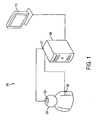
- a system for diagnosing cerebrovascular autoregulation of a patient 100 is illustrated schematically in Figure 1 .
- the system for diagnosing cerebrovascular autoregulation 100 includes a cerebral oximeter 102 that is arranged proximate an external position of the patient's head 104.
- a blood pressure monitoring device 106 is attached to the patient.
- a signal processing unit 108 is in communication with the cerebral oximeter 102 and with the blood pressure monitoring device 106.
- the cerebral oximeter obtains oxygen content measurements of blood within the patient's brain. Signals from the cerebral oximeter 102 may be processed internally within the cerebral oximeter 102 and/or processed by the signal processing unit 108.
- the oxygen content measurements of blood within the patient's brain is taken a plurality of times by the cerebral oximeter 102 to input an oxygen content signal to the signal processing unit 108.
- a blood pressure monitoring device 106 obtains arterial blood pressure measurements of the patient at a plurality of times substantially synchronously with the oxygen content measurements and outputs an arterial blood pressure signal to the signal processing unit 108.
- the signal processing unit 108 calculates a linear correlation coefficient based on the oxygen content signal and the arterial blood pressure signal in a time domain for a plurality of times. This linear correlation coefficient may be referred to as the cerebral oximeter index (COx) according to some embodiments of the current invention.
- the oxygen content signals transmitted from the cerebral oximeter 102 to the signal processor 108 are low pass filtered by any one of the cerebral oximeter itself, the signal processing unit 108 or by an intermediate low pass filter in the signal line between the cerebral oximeter 102 and the signal processing unit 108.
- the blood pressure monitoring device 106, the signal processing unit 108 or an intermediate device in the signal line between blood pressure monitoring device 106 and signal processor 108 provide low pass filtering of the measured blood pressure signal.
- the blood pressure monitoring device 106 may include an intracranial pressure monitoring device (not shown).
- An intracranial pressure monitoring device may include a catheter-based device which is surgically inserted into the patient to directly measure intracranial pressure within the patient's brain.
- the blood pressure monitoring device 106 may include an arterial blood pressure monitoring device that can be selected from available arterial blood pressure monitoring devices.
- the cerebral oximeter 102 can be a near-infrared spectrometer.
- the system for diagnosing cerebrovascular autoregulation 100 may also include a display unit 110 that is in communication with the signal processing unit 108 to display the linear correlation coefficient values calculated by the signal processing unit with respect to other biophysical data of the patient.
- the display unit may display the linear correlation coefficients calculated as a function of arterial blood pressure.
- the signal processing unit 108 may determine the cerebral perfusion pressure based on the difference between the arterial blood pressure and the intracranial pressure and provide signals to the display unit 110 to display the calculated linear correlation coefficients as a function of the cerebral perfusion pressure.
- the cerebral oximeter 102, the blood pressure monitoring device 106, the display unit 110 and the signal processing unit 108 may be connected by physical wires or other suitable means such as optical or wireless data communications.
- the signal processing unit 108 can be a stand alone physical component, or may be added as a component to other systems such as to a rack system.
- the signal processing unit 108 is not necessarily limited to processing only signal data. It may include generally data processing capabilities.
- the signal processing operations of the signal processing unit 108 may be hard-wired or may be implemented by programming the signal processing unit.
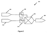
- Figure 2 is a schematic illustration that facilitates the description of a method of diagnosing cerebrovascular autoregulation in a patient 200.
- the method of diagnosing cerebrovascular autoregulation 200 includes measuring blood pressure of a patient 202, measuring, non-invasively, venous oxygen content of the patient's brain 204 substantially simultaneously with the measuring arterial blood pressure 202, and correlating the blood pressure and the venous oxygen content measurements in a time domain 205.
- a cerebrovascular autoregulation state of the patient is determined 206 based on the correlating of the blood pressure 202 and venous oxygen content 204 measurements.
- the blood pressure signals 202 are low pass filtered 208 according to an embodiment of the current invention.
- the low pass filtering 208 allows slow variations of blood pressure signals to pass through the filter while filtering out the more rapid variations in blood pressure signals.
- the low pass filtering 208 may be implemented with either hardware or software according to various embodiments of the current invention.
- the low pass filtering can be analog low pass filtering or digital low pass filtering, depending on whether an analog or digital signal is being processed.
- the blood pressure signal may be sampled to provide a digital signal and the low pass filtering can be accomplished by selecting a desired sampling frequency.
- the venous oxygen content measurements may be low pass filtered 210 prior to being correlated 205 with the blood pressure signals.
- the venous oxygen content data may be obtained by sampling substantially synchronously with sampling of a blood pressure data to provide a digital signal.
- the low pass filtering 210 may be achieved by selecting the sampling frequency at a desired sampling frequency.
- the blood pressure measurement data 202 may correspond to arterial blood pressure or may correspond to cerebral perfusion pressure determined by also measuring intracranial pressure.
- the venous oxygen content data may be obtained, for example, by measuring differential absorption of near-infrared radiation directed into the patient's brain from a source of near-infrared radiation disposed proximate an external position of the patient's head.
- a method of treating a patient may include measuring blood pressure of the patient, measuring, non-invasively, oxygen content of the patient's brain substantially simultaneously with measuring blood pressure, and correlating the blood pressure measurements and oxygen content measurements in a time domain.
- the blood pressure measurement data may correspond to arterial blood pressure or may correspond to cerebral perfusion pressure determined by also measuring intracranial pressure.
- a cerebrovascular autoregulation state of the patient is determined based on the correlating of the blood pressure and venous oxygen content measurements and a change of blood pressure or cerebral perfusion pressure is effected based on the determined cerebrovascular autoregulation state of the patient.
- the data processing unit 108 includes at least one signal input port 112 that is adapted to receive blood pressure signals from measured blood pressure data from the patient and to receive venous oxygen content signals from externally measured venous oxygen content data of the patient's brain.
- the data processing unit 108 also has a signal correlation component adapted to receive and correlate the blood pressure signal and venous oxygen content signal to provide a linear correlation coefficient indicative of a cerebrovascular autoregulation state of the patient.
- the data processing unit 108 also includes a signal output port 114 to output the linear correlation coefficient to be further processed, stored and/or displayed.
- the data processing unit 108 may include a low-pass filter to filter the blood pressure data and may include a low-pass filter to filter the venous oxygen content data in an embodiment of the current invention.
- the blood pressure data and/or the venous oxygen content data may have already been filtered prior to being received by the data processing unit.
- the blood pressure data may include arterial blood pressure in some embodiments of the current invention.
- the data processing unit 108 may also be adapted to receive intracranial pressure signals from measured intracranial pressure of the patient. This may be received through the same input port 112, or through an additional data input port.
- the arterial blood pressure signal may be transmitted to the data processing unit 108 through the same signal input port 112 as the venous oxygen content signals or may be provided through a separate port.
- the broad concepts of the invention are not limited to any particular number of data input and output ports or whether data is multiplexed for input and/or output over any of the data ports.
- the signal input/output ports may be electrical, optical, or wireless data input/output ports.
- a computer readable medium is programmed to process data from a system for diagnosing cerebrovascular autoregulation in a patient.
- the computer readable medium is programmed to receive and process at least one signal from blood pressure measurements and a signal from venous oxygen content measurements and to calculate a linear correlation coefficient based on the correlation between the arterial blood pressure data and the venous oxygen content data in a time domain.
- the computer readable medium is programmed to output the linear correlation coefficient to provide information upon which cerebrovascular autoregulation of the patient can be determined.
- the COx according to an embodiment of the current invention would be sensitive for autoregulatory failure due to hypotension in a piglet model of the infant brain and measured the COx continuously in piglets, while slowly lowering their ABP below the breakpoint of autoregulation, as determined by laser-Doppler flowmetry.
- LDx laser-Doppler index
- Piglets (n 6), aged 3-8 days old and weighing 2.2-3.9 kg, were anesthetized with inhalation of 5% isoflurane, 50% nitrous oxide, and balance of oxygen. A tracheostomy was performed and mechanical ventilation was instituted. Peripheral intravenous access was obtained for the administration of vecuronium (5-mg bolus and 2-mg/hr infusion) and fentanyl (25- ⁇ g bolus and 25- ⁇ g/hr infusion). Isoflurane was decreased to 0.5% for the duration of the experiment, and the fentanyl was titrated between 10-50 ⁇ g/hr for a target heart rate lower than 190 and normotension during surgery.
- vecuronium 5-mg bolus and 2-mg/hr infusion
- fentanyl 25- ⁇ g bolus and 25- ⁇ g/hr infusion
- the anesthetic for the recording period was primarily narcotic based, with a sub-anesthetic supplementation of inhalational agent. This combination was chosen to ensure the comfort of the animal and reduce the effect of inhaled anesthetic on cerebrovascular responsiveness. Piglets were kept on a warming pad to maintain brain and rectal temperature at 38.5-39.5°C. Ventilation was adjusted to keep pH at 7.35-7.45 and P a O 2 at 200-300 mmHg.
- the femoral veins were cannulated bilaterally for placement of a central venous line for drug infusion and pressure monitoring and a 5 Fr esophageal balloon catheter (Cooper Surgical, Trundall, CT), which was used for interruption of venous return to the heart to produce hypotension.
- the femoral artery was cannulated for placement of a pressure and blood gas monitoring line.
- a craniotomy was performed 4 mm lateral and rostral to the bregma at midline for placement of an external ventricular drain catheter, which was transduced for ICP monitoring.
- the INVOS (in vivo optical spectroscopy) pediatric cerebral oximeter probe (Somanetics, Troy, MI) was placed above the eye, across the frontal and parietal cortex, opposite the side of craniotomies, with the emitting diode situated 1 cm lateral to midline to avoid the sagittal sinus.
- the cerebral specificity of the probe was then tested with a CO 2 challenge: ventilation was increased to reduce end-tidal CO 2 by at least 10 mmHg.
- Cerebral oximetry was compared with oximetry obtained from a probe that was placed over the kidney. Cerebral oximetry values decreased (1.2 ⁇ 0.1%/mmHg; ⁇ SD), whereas the renal oximetry values were static (0.0 ⁇ 0.1 %/mmHg).
- Waveforms from the pressure transducers (ABP, ICP), the laser-Doppler probe, and the INVOS cerebral oximeter were sampled from an analog-to-digital converter by ICM+ software (Cambridge University, Cambridge, UK) at 60 Hz.
- the time resolution of INVOS oximetry is 4 seconds.
- These signals were then time-integrated as non-overlapping 10-second mean values, which is equivalent to applying a moving average filter with a 10-second time window and resampling at 0.1 Hz. This operation eliminates high-frequency noise from the respiratory and pulse frequencies of the animals but, according to the Nyquist theorem, allows detection of oscillations and transients that occur below 0.05 Hz.
- CPP was calculated as the difference between the 10-second mean values of ABP and ICP.
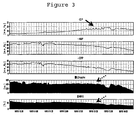
- the balloon catheter in the inferior vena cava was gradually inflated by infusion of saline from a syringe pump to slowly lower ABP to ⁇ 10 mmHg over 4-5 hours ( Figure 3 ).
- Cerebral oximetry, laser-Doppler flux, COx, and LDx values were recorded every 60 seconds in real time and simultaneously sorted according to the CPP at which they were collected. Hypotension was induced over a prolonged period to permit sufficient time for spontaneous changes in CPP to occur over each range of quasi-steady state CPP and thus provide an adequate signal/noise ratio for calculating COx.
- Regression analysis and linear correlation of the COx against the LDx was performed with Prism software and with Bland-Altman plots, using LDx - COx and COx/LDx against the mean. This analysis was performed for all paired indices collected and again for averaged values collected on the same piglet at the same CPP.
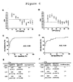
- FIG. 4 An example of the autoregulatory assessment for a single piglet is shown in Figure 4 .
- the lower limit of autoregulation of laser-Doppler flow was easily identified from the intersection of two regression lines that minimized the overall sum of the residual squared errors ( Figure 4A ).
- the plot of cerebral oximetry as a function of CPP was not as well characterized by an inflection point ( Figure 4B ).
- the LDx and COx both showed a sharp increase at the autoregulatory threshold in the animal presented ( Figures 4C and 4D ).
- the present results show that time-domain correlation of ABP and cerebral oximetry can quantify spontaneous autoregulatory vasoreactivity, and the resultant index is sensitive for loss of autoregulation caused by hypotension in a piglet model.
- This method has several features that are attractive for clinical application.
- the COx output is continuous and updated every 60 seconds, as configured in the animals presented.
- the COx can be displayed at the bedside as a function of clinical parameters, such as CPP, showing the effect of changes in management on the autoregulatory process.
- the COx requires no intracranial surgery for calculation and can use spontaneous changes in ABP, obviating the need to induce rapid changes in ABP in an unstable patient.
- Associative relationships between ABP and CBF surrogates can be dynamically assessed by methods that fall into two broad categories: analysis in the frequency domain and analysis in the time domain.
- Frequency-domain analysis (based on coherence, transfer function, or phase shifts) is well suited for regular, periodic waves or induced changes in ABP in an otherwise static system. This analysis has assumptions of linearity and stationarity that are not always strictly present in a biologic system ( Giller CA, Mueller M. Linearity and non-linearity in cerebral hemodynamics. Med Eng Phys. 2003; 25(8):633-646 ).
- Time-domain analysis can be performed as a linear correlation between low-pass filtered ABP and CBF waves, as presented here with the COx and LDx, but this filtering limits the spectral range of the test.
- the clinically relevant wavelength periods that encompass CPP and oximetry correlations caused by autoregulatory failure must be known.
- Physiologic Parameters (mean ⁇ SEM) Measured during Progressive Hypotension Physiologic parameter CPP >50 mmHg CPP 30-50 mmHg CPP ⁇ 30 mmHg Arterial pH 7.42 ⁇ 0.02 7.35 ⁇ 0.06 7.39 ⁇ 0.02 P a CO 2 (mmHg) 37.0 ⁇ 4.9 34.5 ⁇ 3.5 33.0 ⁇ 1.6 P a O2 (mmHg) 229 ⁇ 29 208 ⁇ 39 231 ⁇ 35 Hematocrit (%) 25 ⁇ 5 23 ⁇ 3 22 ⁇ 3 Brain Temperature (°C) 38.7 ⁇ 0.8 38.6 ⁇ 0.8 38.6 ⁇ 0.7
Description
- This application claims priority to
U.S. Provisional Application No. 60/899,146, filed February 12, 2007 - This application relates to cerebral blood pressure autoregulation and more particularly to devices and methods to diagnose and/or treat cerebrovascular autoregulation in a patient.
- Cerebral pressure autoregulation is defined as the maintenance of a constant cerebral blood flow (CBF) in the face of changing cerebral perfusion pressure (CPP). In health, this process protects the brain during transient changes in the arterial blood pressure (ABP) from diminished or excessive blood flow. Traumatic brain injury (TBI)( Muizelaar JP, Marmarou A, DeSalles AA, et al. Cerebral blood flow and metabolism in severely head-injured children. part 1: Relationship with GCS score, outcome, ICP, and PVI. J Neurosurg. 1989; 71(1):63-71; Muizelaar JP, Ward JD, Marmarou A, Newlon PG, Wachi A. Cerebral blood flow and metabolism in severely head-injured children. part 2: Autoregulation. J Neurosurg. 1989; 71(1):72-76; Vavilala MS, Muangman S, Tontisirin N, et al. Impaired cerebral autoregulation and 6-month outcome in children with severe traumatic brain injury: Preliminary findings. Dev Neurosci. 2006; 28(4-5):348-353), stroke (Dawson SL, Panerai RB, Potter JF. Serial changes in static and dynamic cerebral autoregulation after acute ischaemic stroke. Cerebrovasc Dis. 2003; 16(1):69-75), meningitis (Berkowitz ID, Hayden WR, Traystman RJ, Jones MD, Jr. Haemophilus influenzae type B impairment of pial vessel autoregulation in rats. Pediatr Res. 1993; 33(1):48-51; Slater AJ, Berkowitz ID, Wilson DA, Traystman RJ. Role of leukocytes in cerebral autoregulation and hyperemia in bacterial meningitis in rabbits. Am J Physiol. 1997; 273(1 Pt 2):H380-6), cardiopulmonary bypass, and deep hypothermic circulatory arrest (O'Rourke MM, Nork KM, Kurth CD. Neonatal cerebral oxygen regulation after hypothermic cardiopulmonary bypass and circulatory arrest. Crit Care Med. 2000; 28(1):157-162) are examples of insults that have been shown to impair pressure autoregulation and have large-scale clinical impact. An impairment of autoregulation narrows the range of blood pressures at which flow is matched to metabolic needs. Optimal management of CPP for limiting tissue hypoxia at low CPP or edema at high CPP in these patients is critical but difficult to achieve because of limited monitoring capabilities. Despite the recent surge of multimodal neuromonitoring, optimal ABP and CPP have not been defined.
- It has been postulated that continuous monitoring of autoregulatory vasoreactivity allows detection of an "optimal CPP" and titration of blood pressure into a range that maximizes vasoreactivity to perturbations in CPP (Steiner LA, Czosnyka M, Piechnik SK, et al. Continuous monitoring of cerebrovascular pressure reactivity allows determination of optimal cerebral perfusion pressure in patients with traumatic brain injury. Crit Care Med. 2002; 30(4):733-738). Autoregulation is measured by quantifying the consequence of changing blood pressure on CBF or its surrogate, and the methods have been extensively reviewed (Panerai RB. Assessment of cerebral pressure autoregulation in humans--a review of measurement methods. Physiol Meas. 1998; 19(3):305-338). Changes in ABP can be induced via drugs, tilt-table, or thigh cuff (Aaslid R, Lindegaard KF, Sorteberg W, Nornes H. Cerebral autoregulation dynamics in humans. Stroke. 1989; 20(1):45-52), or they can occur spontaneously. Using spontaneous changes in ABP is preferable to inducing ABP changes in an unstable patient with an acute intracranial process. However, relying on spontaneous and often subtle ABP fluctuations for this measurement results in an inferior signal-to-noise ratio.
- Diverse surrogates of CBF are suitable for continuous monitoring of autoregulation and include flow velocity, measured by transcranial Doppler (Czosnyka M, Smielewski P, Kirkpatrick P, Menon DK, Pickard JD. Monitoring of cerebral autoregulation in head-injured patients. Stroke. 1996; 27(10): 1829-1834); red blood cell flux, measured by laser-Doppler (Lam JM, Hsiang JN, Poon WS. Monitoring of autoregulation using laser doppler flowmetry in patients with head injury. J Neurosurg. 1997; 86(3):438-445); parenchymal oxygen tension, measured using a Licox monitor (Lang EW, Czosnyka M, Mehdorn HM. Tissue oxygen reactivity and cerebral autoregulation after severe traumatic brain injury. Crit Care Med. 2003; 31(1):267-271 ; Jaeger M, Schuhmann MU, Soehle M, Meixensberger J. Continuous assessment of cerebrovascular autoregulation after traumatic brain injury using brain tissue oxygen pressure reactivity. Crit Care Med. 2006: 34(6): 1783- 1788); and cerebral tissue oxyhemoglobin saturation, measured by transcranial near-infrared spectroscopy (NIRS)(Tsuji M, Saul JP, du Plessis A, et al. Cerebral intravascular oxygenation correlates with mean arterial pressure in critically ill premature infants. Pediatrics. 2000; 106(4):625-632). Slow waves of intracranial pressure (ICP) reflecting vessel diameter changes in the autoregulatory process have also been correlated to ABP for an index describing autoregulation (Czosnyka M, Smielewski P, Kirkpatrick P, Laing RJ, Menon D, Pickard JD. Continuous assessment of the cerebral vasomotor reactivity in head injury. Neurosurgery. 1997; 41 (1): 1 1-7; discussion 17-9). An ideal CBF surrogate for an index of autoregulation would be noninvasive and require minimal caregiver attention. It would provide a continuous signal with time resolution sufficiently fine to discriminate changes in frequencies relevant to autoregulation, and that signal would be a close proxy for CBF. There is thus a need for improved methods and devices for diagnosing cerebrovascular autoregulation in patients. Other prior art methods can be found disclosed in
US6802812B1 andDE10331027A1 . - Further objectives and advantages will become apparent from a consideration of the description, drawings, and examples.
- A system for determining a cerebrovascular autoregulation state of a patient according to an embodiment of the current invention has a cerebral oximeter for external arrangement on the patient's head, a blood pressure monitoring device for attachement to the patient, and a signal processing unit in communication with the cerebral oximeter and the blood pressure monitoring device. The cerebral oximeter is configured to obtain oxygen content measurements of blood within the patient's brain taken at a plurality of times and to output an oxygen content signal to the signal processing unit, the blood pressure monitoring device is configured to obtain arterial blood pressure measurements of the patient at a plurality of times substantially synchronously with the oxygen content measurements and to output an arterial blood pressure signal to the signal processing unit, and the signal processing unit is configured to calculate a linear correlation coefficient based on the oxygen content signal and the arterial blood pressure signal in the time domain for a plurality of times.
- A data processing unit for use with a system for determining a cerebrovascular autoregulation state of a patient according to an embodiment of the current invention has at least one signal input port adapted to receive a blood pressure signal from measured blood pressure data from the patient and to receive a venous oxygen content signal from externally measured venous oxygen content data of the patient's brain, a signal correlation component adapted to receive and correlate the blood pressure signal with the venous oxygen content signal to provide a correlation coefficient indicative of a cerebrovascular autoregulation state of the patient, and a signal output port to output the correlation coefficient to indicate the cerebrovascular autoregulation state of the patient based on the correlation coefficient.
- A computer readable medium programmed to process data for a system for determining a cerebrovascular autoregulation state of a patient according to an embodiment of the current invention includes at least one signal receiving component adapted to receive a blood pressure signal from measured blood pressure data from the patient and to receive a venous oxygen content signal from externally measured venous oxygen content data of the patient's brain, a signal correlation component adapted to receive and correlate the blood pressure signal with the venous oxygen content signal to provide a correlation coefficient indicative of a cerebrovascular autoregulation state of the patient, and a signal output component adapted to output the correlation coefficient to indicate the cerebrovascular autoregulation state of the patient based on the correlation coefficient.
- The invention is better understood by reading the following detailed description with reference to the accompanying figures in which:
-
Figure 1 is a schematic illustration of a system for diagnosing cerebrovascular autoregulation according to an embodiment of the current invention; -
Figure 2 is a schematic diagram to help explain a method of diagnosing and/or treating cerebrovascular autoregulation in a patient. -
Figure 3 shows time trends of recordings from a single piglet. ICP, ABP, and CPP are shown in mmHg; laser-Doppler red blood cell flux is in arbitrary units; and cerebral oximetry (NIRS) is expressed as a percent saturation of hemoglobin. Time on the x-axis covers a spread of 4 hours and 10 minutes. Slow "B" waves of ICP are seen in the top tracing at low ABP prior to failure of autoregulation (solid arrow). The oximeter readout showed a more gradual decline relative to the laser-Doppler flux, which had a pattern more indicative of autoregulation (dashed arrows). A similar trend was observed in all 6 piglets; -
Figure 4A shows a steady-state autoregulatory graph of laser-Doppler flux versus CPP in a single piglet. The breakpoint was defined as the division that resulted in regression lines with the lowest combined residual squared error (34 mmHg in this piglet).Figure 4B shows near-infrared spectroscopy (NIRS)-derived cerebral oximetry versus CPP. This relationship did not have the obvious plateau seen with laser-Doppler flux. However, the laser-Doppler index (LDx, ±SE,Figure 4C ) and the cerebral oximetry index (COx,Figure 4D ) were concordant, showing low values above a CPP of 35 mmHg and high values below a CPP of 35 mmHg (arrows); -
Figures 5A-5C show static autoregulation curves derived from 6 piglets (±SE).Figure 5A is Laser-Doppler flux as a percent of baseline flux at 60 mmHg.Figure 5B is Cerebrovascular resistance (CVR), calculated as CPP/CBF from the same data set and expressed as a percentage of CVR at CPP of 60 mmHg.Figure 5C is Cerebral oximetry, measured by NIRS, shown as a percentage of baseline tissue oxyhemoglobin saturation. P <0.0001 by ANOVA for both laser-Doppler flux and oximetry curves. The average breakpoint of autoregulation, determined for individual piglets, was 29.7 ± 5.5 mmHg (vertical dashed line); -
Figure 6A shows average LDx andFigure 6B shows COx for the six piglets (±SE) stratified by the CPP at which they were measured. The horizontal dashed line shows the 90% sensitivity cutoff for detecting autoregulatory failure. The receiver-operator characteristics are compared between the LDx (Figure 6C ) and COx (Figure 6D ) calculations of 6 piglets, averaged for each 5 mmHg increment of CPP. AUC is area under the curve. Confidence intervals for sensitivity and specificity and likelihood ratios are tabulated for different sensitivity levels for each index; and -
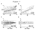
Figures 7A-7D show linear regression (7A, 7B) and Bland Altman plots (7C,7D) comparing LDx and COx for all data points (7A,7D) and averaged data points taken at the same CPP for each piglet (7B,7D). Agreement improves substantially by averaging, which implies a low signal-to-noise ratio for individual index measurements. Dashed lines are 95% confidence intervals (regression) and 95% limits of agreement (Bland-Altman). - In describing embodiments of the present invention illustrated in the drawings, specific terminology is employed for the sake of clarity. However, the invention is not intended to be limited to the specific terminology so selected. It is to be understood that each specific element includes all technical equivalents which operate in a similar manner to accomplish a similar purpose.
- Transcranial monitors of cerebral oxygenation using NIRS have attractive features. According to some embodiments of the current invention, we present a novel index of autoregulatory vasoreactivity, the cerebral oximeter index (COx), which is derived from a time-domain analysis that correlates changes in ABP to the output of an NIRS-based monitor of cerebral tissue oxyhemoglobin saturation. Continuous assessment of autoregulation is a promising monitoring method for actively optimizing cerebral perfusion pressure (CPP) in critically ill patients. In one embodiment, this correlation is performed continuously on overlapping epochs of 300 seconds, updated every 60 seconds, and does not require induced changes in ABP to detect autoregulatory failure.
- A system for diagnosing cerebrovascular autoregulation of a
patient 100 according to an embodiment of the current invention is illustrated schematically inFigure 1 . The system for diagnosingcerebrovascular autoregulation 100 includes acerebral oximeter 102 that is arranged proximate an external position of the patient'shead 104. A bloodpressure monitoring device 106 is attached to the patient. Asignal processing unit 108 is in communication with thecerebral oximeter 102 and with the bloodpressure monitoring device 106. In an embodiment of the invention, the cerebral oximeter obtains oxygen content measurements of blood within the patient's brain. Signals from thecerebral oximeter 102 may be processed internally within thecerebral oximeter 102 and/or processed by thesignal processing unit 108. According to an embodiment of the current invention, the oxygen content measurements of blood within the patient's brain is taken a plurality of times by thecerebral oximeter 102 to input an oxygen content signal to thesignal processing unit 108. - A blood
pressure monitoring device 106 obtains arterial blood pressure measurements of the patient at a plurality of times substantially synchronously with the oxygen content measurements and outputs an arterial blood pressure signal to thesignal processing unit 108. Thesignal processing unit 108 calculates a linear correlation coefficient based on the oxygen content signal and the arterial blood pressure signal in a time domain for a plurality of times. This linear correlation coefficient may be referred to as the cerebral oximeter index (COx) according to some embodiments of the current invention. The oxygen content signals transmitted from thecerebral oximeter 102 to thesignal processor 108 are low pass filtered by any one of the cerebral oximeter itself, thesignal processing unit 108 or by an intermediate low pass filter in the signal line between thecerebral oximeter 102 and thesignal processing unit 108. The bloodpressure monitoring device 106, thesignal processing unit 108 or an intermediate device in the signal line between bloodpressure monitoring device 106 andsignal processor 108 provide low pass filtering of the measured blood pressure signal. The bloodpressure monitoring device 106 may include an intracranial pressure monitoring device (not shown). An intracranial pressure monitoring device may include a catheter-based device which is surgically inserted into the patient to directly measure intracranial pressure within the patient's brain. The bloodpressure monitoring device 106 may include an arterial blood pressure monitoring device that can be selected from available arterial blood pressure monitoring devices. In an embodiment of the current invention, thecerebral oximeter 102 can be a near-infrared spectrometer. - The system for diagnosing
cerebrovascular autoregulation 100 may also include adisplay unit 110 that is in communication with thesignal processing unit 108 to display the linear correlation coefficient values calculated by the signal processing unit with respect to other biophysical data of the patient. For example, the display unit may display the linear correlation coefficients calculated as a function of arterial blood pressure. Alternatively, thesignal processing unit 108 may determine the cerebral perfusion pressure based on the difference between the arterial blood pressure and the intracranial pressure and provide signals to thedisplay unit 110 to display the calculated linear correlation coefficients as a function of the cerebral perfusion pressure. - The
cerebral oximeter 102, the bloodpressure monitoring device 106, thedisplay unit 110 and thesignal processing unit 108 may be connected by physical wires or other suitable means such as optical or wireless data communications. Thesignal processing unit 108 can be a stand alone physical component, or may be added as a component to other systems such as to a rack system. Thesignal processing unit 108 is not necessarily limited to processing only signal data. It may include generally data processing capabilities. In addition, the signal processing operations of thesignal processing unit 108 may be hard-wired or may be implemented by programming the signal processing unit. -
Figure 2 is a schematic illustration that facilitates the description of a method of diagnosing cerebrovascular autoregulation in apatient 200. The method of diagnosingcerebrovascular autoregulation 200 includes measuring blood pressure of apatient 202, measuring, non-invasively, venous oxygen content of the patient'sbrain 204 substantially simultaneously with the measuringarterial blood pressure 202, and correlating the blood pressure and the venous oxygen content measurements in atime domain 205. In an embodiment of the current invention, a cerebrovascular autoregulation state of the patient is determined 206 based on the correlating of theblood pressure 202 andvenous oxygen content 204 measurements. The blood pressure signals 202 are low pass filtered 208 according to an embodiment of the current invention. Thelow pass filtering 208 allows slow variations of blood pressure signals to pass through the filter while filtering out the more rapid variations in blood pressure signals. Thelow pass filtering 208 may be implemented with either hardware or software according to various embodiments of the current invention. Furthermore, the low pass filtering can be analog low pass filtering or digital low pass filtering, depending on whether an analog or digital signal is being processed. In one embodiment of the current invention, the blood pressure signal may be sampled to provide a digital signal and the low pass filtering can be accomplished by selecting a desired sampling frequency. - In an embodiment of the current invention, the venous oxygen content measurements may be low pass filtered 210 prior to being correlated 205 with the blood pressure signals. In one embodiment of the current invention, the venous oxygen content data may be obtained by sampling substantially synchronously with sampling of a blood pressure data to provide a digital signal. In this case, the
low pass filtering 210 may be achieved by selecting the sampling frequency at a desired sampling frequency. However, the general aspects of this invention are not limited to only digital signal processing and are not limited to only digital low pass filtering. The bloodpressure measurement data 202 may correspond to arterial blood pressure or may correspond to cerebral perfusion pressure determined by also measuring intracranial pressure. The venous oxygen content data may be obtained, for example, by measuring differential absorption of near-infrared radiation directed into the patient's brain from a source of near-infrared radiation disposed proximate an external position of the patient's head. - A method of treating a patient may include measuring blood pressure of the patient, measuring, non-invasively, oxygen content of the patient's brain substantially simultaneously with measuring blood pressure, and correlating the blood pressure measurements and oxygen content measurements in a time domain. The blood pressure measurement data may correspond to arterial blood pressure or may correspond to cerebral perfusion pressure determined by also measuring intracranial pressure. A cerebrovascular autoregulation state of the patient is determined based on the correlating of the blood pressure and venous oxygen content measurements and a change of blood pressure or cerebral perfusion pressure is effected based on the determined cerebrovascular autoregulation state of the patient.
- Another embodiment of the current invention is directed to a data processing unit for use with a system for diagnosing cerebrovascular autoregulation in a patient. For example, the data processing unit may be similar to or the same as the
data processing 108 described with reference to the system for diagnosingcerebrovascular autoregulation 100 inFigure 1 . Thedata processing unit 108 includes at least onesignal input port 112 that is adapted to receive blood pressure signals from measured blood pressure data from the patient and to receive venous oxygen content signals from externally measured venous oxygen content data of the patient's brain. Thedata processing unit 108 also has a signal correlation component adapted to receive and correlate the blood pressure signal and venous oxygen content signal to provide a linear correlation coefficient indicative of a cerebrovascular autoregulation state of the patient. Thedata processing unit 108 also includes a signal output port 114 to output the linear correlation coefficient to be further processed, stored and/or displayed. Thedata processing unit 108 may include a low-pass filter to filter the blood pressure data and may include a low-pass filter to filter the venous oxygen content data in an embodiment of the current invention. In alternative embodiments, the blood pressure data and/or the venous oxygen content data may have already been filtered prior to being received by the data processing unit. The blood pressure data may include arterial blood pressure in some embodiments of the current invention. Thedata processing unit 108 may also be adapted to receive intracranial pressure signals from measured intracranial pressure of the patient. This may be received through thesame input port 112, or through an additional data input port. Similarly, the arterial blood pressure signal may be transmitted to thedata processing unit 108 through the samesignal input port 112 as the venous oxygen content signals or may be provided through a separate port. The broad concepts of the invention are not limited to any particular number of data input and output ports or whether data is multiplexed for input and/or output over any of the data ports. In addition, the signal input/output ports may be electrical, optical, or wireless data input/output ports. - In another embodiment of the current invention, a computer readable medium is programmed to process data from a system for diagnosing cerebrovascular autoregulation in a patient. The computer readable medium is programmed to receive and process at least one signal from blood pressure measurements and a signal from venous oxygen content measurements and to calculate a linear correlation coefficient based on the correlation between the arterial blood pressure data and the venous oxygen content data in a time domain. The computer readable medium is programmed to output the linear correlation coefficient to provide information upon which cerebrovascular autoregulation of the patient can be determined.
- We hypothesized that the COx according to an embodiment of the current invention would be sensitive for autoregulatory failure due to hypotension in a piglet model of the infant brain and measured the COx continuously in piglets, while slowly lowering their ABP below the breakpoint of autoregulation, as determined by laser-Doppler flowmetry. We determined the sensitivity and specificity of the COx for detecting the loss of autoregulation caused by hypotension. We also tested the COx against a similar, but invasive method, the laser-Doppler index (LDx), which utilizes a linear correlation coefficient between ABP and laser-Doppler flux measured in the frontoparietal cortex. We hypothesized that the COx and LDx would show agreement as measurements of autoregulatory vasoreactivity despite their distinct origins.
- All procedures were approved by the Johns Hopkins University Animal Care and Use Committee and conformed to the standards of animal experimentation of the National Institutes of Health.
- Piglets (n = 6), aged 3-8 days old and weighing 2.2-3.9 kg, were anesthetized with inhalation of 5% isoflurane, 50% nitrous oxide, and balance of oxygen. A tracheostomy was performed and mechanical ventilation was instituted. Peripheral intravenous access was obtained for the administration of vecuronium (5-mg bolus and 2-mg/hr infusion) and fentanyl (25-µg bolus and 25-µg/hr infusion). Isoflurane was decreased to 0.5% for the duration of the experiment, and the fentanyl was titrated between 10-50 µg/hr for a target heart rate lower than 190 and normotension during surgery. During the recording period, when blood pressure was actively lowered, fentanyl was infused at 50 µg/hr (20 µg/kg/hr for most of the piglets) and tachycardia was permitted as a response to the preload reduction. Isoflurane remained at 0.5%, and the nitrous oxide remained at 50% of the inspired gas. Thus, the anesthetic for the recording period was primarily narcotic based, with a sub-anesthetic supplementation of inhalational agent. This combination was chosen to ensure the comfort of the animal and reduce the effect of inhaled anesthetic on cerebrovascular responsiveness. Piglets were kept on a warming pad to maintain brain and rectal temperature at 38.5-39.5°C. Ventilation was adjusted to keep pH at 7.35-7.45 and PaO2 at 200-300 mmHg.
- The femoral veins were cannulated bilaterally for placement of a central venous line for drug infusion and pressure monitoring and a 5 Fr esophageal balloon catheter (Cooper Surgical, Trundall, CT), which was used for interruption of venous return to the heart to produce hypotension. The femoral artery was cannulated for placement of a pressure and blood gas monitoring line. A craniotomy was performed 4 mm lateral and rostral to the bregma at midline for placement of an external ventricular drain catheter, which was transduced for ICP monitoring. An additional craniotomy was performed 4 mm lateral and rostral to the first craniotomy for placement of a laser-Doppler probe (Moor Instruments, Devon, U.K.), which was advanced across the incised dura mater to contact the surface of the frontoparietal cortex. The probe was positioned to avoid high baseline flux values associated with placement over large vessels and was secured in place by a rubber washer cemented to the skull. A third craniotomy in the occipital skull lateral to the midline was used to place a brain temperature probe. Skin was reapplied to the skull, and the wound was sutured closed for heat retention and to create conditions for which the cerebral oximeter had been calibrated.
- The INVOS (in vivo optical spectroscopy) pediatric cerebral oximeter probe (Somanetics, Troy, MI) was placed above the eye, across the frontal and parietal cortex, opposite the side of craniotomies, with the emitting diode situated 1 cm lateral to midline to avoid the sagittal sinus. The cerebral specificity of the probe was then tested with a CO2 challenge: ventilation was increased to reduce end-tidal CO2 by at least 10 mmHg. Cerebral oximetry was compared with oximetry obtained from a probe that was placed over the kidney. Cerebral oximetry values decreased (1.2 ± 0.1%/mmHg; ± SD), whereas the renal oximetry values were static (0.0 ± 0.1 %/mmHg).
- Waveforms from the pressure transducers (ABP, ICP), the laser-Doppler probe, and the INVOS cerebral oximeter were sampled from an analog-to-digital converter by ICM+ software (Cambridge University, Cambridge, UK) at 60 Hz. The time resolution of INVOS oximetry is 4 seconds. These signals were then time-integrated as non-overlapping 10-second mean values, which is equivalent to applying a moving average filter with a 10-second time window and resampling at 0.1 Hz. This operation eliminates high-frequency noise from the respiratory and pulse frequencies of the animals but, according to the Nyquist theorem, allows detection of oscillations and transients that occur below 0.05 Hz. CPP was calculated as the difference between the 10-second mean values of ABP and ICP.
- A continuous, moving Pearson's correlation coefficient was performed between the CPP and laser-Doppler to render the LDx or between the CPP and the cerebral oximeter output to render the COx. Consecutive, paired, 10-second averaged values from 300-second duration were used for each calculation, incorporating 30 data points for each index. These indices were calculated and recorded every 60 seconds from overlapping time periods.
- With the above-mentioned monitors in place, the balloon catheter in the inferior vena cava was gradually inflated by infusion of saline from a syringe pump to slowly lower ABP to ~10 mmHg over 4-5 hours (
Figure 3 ). Cerebral oximetry, laser-Doppler flux, COx, and LDx values were recorded every 60 seconds in real time and simultaneously sorted according to the CPP at which they were collected. Hypotension was induced over a prolonged period to permit sufficient time for spontaneous changes in CPP to occur over each range of quasi-steady state CPP and thus provide an adequate signal/noise ratio for calculating COx. - A scatter plot of laser-Doppler flow versus CPP was made for all of the data for each piglet using SigmaStat software (Systat, San Jose, CA). The CPP that demarcated two regression lines with the lowest combined residual squared error was determined and defined as the autoregulatory breakpoint. In addition, relative changes in cerebrovascular resistance (CVR) were calculated as a percent of the baseline CPP/laser-Doppler flux ratio.
- Prism software (GraphPad, San Diego, CA) was used to determine the receiver-operator characteristics (ROC) of the COx and LDx. To do so, the averaged index values at each CPP for each piglet were dichotomized above and below the CPP breakpoint, as derived from the laser-Doppler flow autoregulatory relationship for each piglet.
- Regression analysis and linear correlation of the COx against the LDx was performed with Prism software and with Bland-Altman plots, using LDx - COx and COx/LDx against the mean. This analysis was performed for all paired indices collected and again for averaged values collected on the same piglet at the same CPP.
- Using ICM Plus software, a cross-spectral analysis of coherence was performed, using ABP as input and either laser-Doppler flux or cerebral oximetry as output. Coherence at frequencies that ranged from 1 Hz to 0.001 Hz was compared between the hypotensive and normotensive states. These data are not presented formally but were used to structure the sampling and calculation parameters for the time-domain analysis presented (see Discussion).
- Arterial pH, PaCO2, and brain temperature were within the normal physiologic range during normotension (CPP >50 mmHg), moderate hypotension above the autoregulatory breakpoint (CPP 30-50 mmHg), and severe hypotension below the autoregulatory breakpoint (CPP <30 mmHg), as shown in Table 1. To prevent CO2-reactivity from affecting the oximeter readings, we sought to keep a constant PaCO2, but a small decrement was noted in each piglet as cardiac output fell to critical levels. It is unlikely that this small decrement introduced a bias into the autoregulatory indices, as they evaluate pressure passivity over discrete 300-second intervals that are relatively stationary with respect to the PaCO2.
- An example of the autoregulatory assessment for a single piglet is shown in
Figure 4 . The lower limit of autoregulation of laser-Doppler flow was easily identified from the intersection of two regression lines that minimized the overall sum of the residual squared errors (Figure 4A ). Interestingly, the plot of cerebral oximetry as a function of CPP was not as well characterized by an inflection point (Figure 4B ). However, the LDx and COx both showed a sharp increase at the autoregulatory threshold in the animal presented (Figures 4C and 4D ). - Data combined from 6 piglets for laser-Doppler flow, relative CVR, and cerebral oximetry are shown in
Figure 5 . The average breakpoint was 29.7 ± 5.5 mmHg, which compares well with previous reports of piglet autoregulatory curves (Laptook AR, Stonestreet BS, Oh W. Brain blood flow and 02 delivery during hemorrhagic hypotension in the piglet. Pediatr Res. 1983; 17(1):77-80; Mertineit C, Samlalsingh-Parker J, Glibetic M, Ricard G, Noya FJ, Aranda JV. Nitric oxide, prostaglandins, and impaired cerebral blood flow autoregulation in group B streptococcal neonatal meningitis. Can J Physiol Pharmacol. 2000; 78(3):217-227). Graded decreases in relative CVR were evident as CPP decreased to 30 mmHg, and further decreases were diminished at CPP values below 30 mmHg. The average LDx and COx increased when CPP was below 30 mmHg (Figure 6A and 6B ). Knowing the steady-state autoregulatory breakpoint for each piglet permitted determination of the ROC for LDx and COx. Not surprisingly, because the LDx is a derivative of the laser-Doppler flow, the LDx performed better than the COx, but both accurately described the breakpoint well. The areas under the ROC curves were 0.95 for the LDx (Figure 6C ) and 0.89 for the COx (Figure 6D ). Summaries of the sensitivity, specificity, and likelihood ratios for cutoff values of the two indices are shown inFigure 6 . In general, sensitivity was superior to specificity for both indices: all piglets showed abnormal autoregulatory vasoreactivity by both the COx and the LDx when hypotensive, but many also showed episodic disruptions of one or both indices in the normotensive or moderately hypotensive range. - The linear correlation and Bland-Altman comparison of the COx and LDx are shown in
Figure 7 . Agreement between the indices was limited when evaluated on a minute-to-minute basis (Pearson's r = 0.36). Agreement improved greatly with averaging of the values stratified according to the 5-mmHg incremental bins of CPP at which they were collected (Pearson's r = 0.67). The Bland-Altman method showed no bias across the range of measurements (bias -0.06 for all values measured, 0.03 for averaged values) and showed the improvement in agreement when values were averaged at the same CPP. - The present results show that time-domain correlation of ABP and cerebral oximetry can quantify spontaneous autoregulatory vasoreactivity, and the resultant index is sensitive for loss of autoregulation caused by hypotension in a piglet model. This method has several features that are attractive for clinical application. The COx output is continuous and updated every 60 seconds, as configured in the animals presented. The COx can be displayed at the bedside as a function of clinical parameters, such as CPP, showing the effect of changes in management on the autoregulatory process. The COx requires no intracranial surgery for calculation and can use spontaneous changes in ABP, obviating the need to induce rapid changes in ABP in an unstable patient.
- An important task in the development of the COx was the determination of relevant periods for waveform sampling. Our rationale for this determination, a discussion of the limitations of the COx, and a description of the potential clinical application of the COx are presented below.
- Associative relationships between ABP and CBF surrogates can be dynamically assessed by methods that fall into two broad categories: analysis in the frequency domain and analysis in the time domain. Frequency-domain analysis (based on coherence, transfer function, or phase shifts) is well suited for regular, periodic waves or induced changes in ABP in an otherwise static system. This analysis has assumptions of linearity and stationarity that are not always strictly present in a biologic system (Giller CA, Mueller M. Linearity and non-linearity in cerebral hemodynamics. Med Eng Phys. 2003; 25(8):633-646). Time-domain analysis can be performed as a linear correlation between low-pass filtered ABP and CBF waves, as presented here with the COx and LDx, but this filtering limits the spectral range of the test. For such an analysis to describe autoregulation, the clinically relevant wavelength periods that encompass CPP and oximetry correlations caused by autoregulatory failure must be known.
- Our focus on frequencies between 0 and 0.04 Hz is based on three suppositions. First, and most important, is the work of Tsuji et al., who used a frequency-domain analysis of coherence between NIRS and ABP in premature infants (Tsuji M, Saul JP, du Plessis A, et al. Cerebral intravascular oxygenation correlates with mean arterial pressure in critically ill premature infants. Pediatrics. 2000; 106(4):625-632). They identified a subgroup with a high coherence at frequencies lower than 0.01 Hz and found an increased incidence of intraventricular hemorrhage in this group, which was hypothesized to have been the result of impaired autoregulation. This finding suggests that these low frequencies are useful in describing correlations of ABP and CBF that can be clinically relevant. A second argument for the chosen frequencies comes from the ICP-derived index of autoregulation (PRx), which correlates slow "B" waves of ICP with ABP. The PRx has been shown to associate with outcome in head-injured patients and is thought to be a marker of the autoregulatory process (Czosnyka M, Smielewski P, Kirkpatrick P, Laing RJ, Menon D, Pickard JD. Continuous assessment of the cerebral vasomotor reactivity in head injury. Neurosurgery. 1997; 41(1):11-7; discussion 17-9). In our database, these slow ICP waves were too sporadic to appear with clarity in a Fourier transfer analysis, but they were identified in the raw waveforms obtained from the piglets and their duration range was measured to be 65-300 seconds, which would correspond to frequencies between 0.015 and 0.003 Hz. The final rationale comes from a coherence analysis of the ABP and NIRS waveforms in the piglets used in this study. In waveforms obtained at blood pressures below the lower limit of autoregulation, we found coherence at frequencies lower than 0.04 Hz, and especially at frequencies lower than 0.02 Hz. This coherence was absent from waveforms obtained during normotension.
- Given the above findings, we desired to resolve waveform relationships that occurred at frequencies lower than 0.04 Hz (periods >25 seconds). At the same time, we wished to prevent the aliasing of noise from the high-frequency range, which included the respiratory and heart rate frequencies. The respiratory rate was ~0.3 Hz (3-second periodicity). Thus, time averaging of 10-second periods suppressed this noise and preserved resolution at the chosen frequencies.
- Understanding the sources of error in the sensitivity and specificity of the COx can lead to strategies for improvement. Using transient and spontaneous changes in ABP decreases the signal-to-noise ratio, when compared to methods that induce large changes in blood pressure over brief periods of time. Two obvious solutions can be chosen for increasing the signal/noise ratio: (a) increasing the sampling time for calculating each index, or (b) averaging multiple discreet calculations of the indices together. We chose the second option because it has the same data smoothing effect but is more useful, as it allows for sorting according to clinically relevant variables (CPP, temperature, blood gases, sedation states, etc.). These variables are likely to be more stationary over a 5-minute period than over 20- or 60-minute periods. Our experimental design sought to control these variables and thereby isolate the effect of changing CPP, but minor deviations in PaCO2 did occur. Dynamic changes in cerebral O2 consumption could affect COx. We assume that the fentanyl, nitrous oxide, and isoflurane anesthesia provided a stable O2 consumption over each 300-second period used to calculate COx.
- Others have dealt with the signal-to-noise ratio problem by incorporating exclusion rules in the index calculation that require a specific range of CPP. For instance, epochs of time with less than 10 mmHg change in ABP could be excluded from analysis (Lam JM, Hsiang JN, Poon WS. Monitoring of autoregulation using laser doppler flowmetry in patients with head injury. J Neurosurg. 1997; 86(3):438-445). The introduction of bias caused by excluding periods with stable blood pressure has not been determined, and this method was not practical for our experimental model because of the slow stable reduction in ABP that was achieved. Deficiencies of sensitivity that occurred with either the LDx or the COx were largely limited to the extreme hypotensive state, just prior to the death of the animal, as can be seen with the increased variability at the CPP of 10 in
Figure 4 . The data set was incomplete in this range, consisting of a limited recording time and only 3 animals due to difficulties encountered in sustaining cardiac function. It is possible that ABP lower than the critical closing pressure caused low and static CBF and cerebral oxygenation that did not change with small ABP fluctuations (Panerai RB. The critical closing pressure of the cerebral circulation. Med Eng Phys. 2003; 25(8):621-632). Such a static CBF state could give the false appearance of intact autoregulation by the COx or LDx assessments. Dynamic decreases in cerebral O2 consumption could also add to the variability of these indices. Blood pressure in this range is not important for the clinical questions targeted. - An important goal of clinical monitoring of autoregulation is the delineation of care parameters that improve autoregulation. Patients with intact autoregulation are more likely to survive neurologic injury, and commutative logic would suggest that improving autoregulation would improve neurologic recovery and survival (Steiner LA, Czosnyka M, Piechnik SK, et al. Continuous monitoring of cerebrovascular pressure reactivity allows determination of optimal cerebral perfusion pressure in patients with traumatic brain injury. Crit Care Med. 2002; 30(4):733-738; Czosnyka M, Smielewski P, Kirkpatrick P, Laing RJ, Menon D, Pickard JD. Continuous assessment of the cerebral vasomotor reactivity in head injury. Neurosurgery. 1997; 41(1):11-7; discussion 17-9; Hiler M, Czosnyka M, Hutchinson P, et al. Predictive value of initial computerized tomography scan, intracranial pressure, and state of autoregulation in patients with traumatic brain injury. J Neurosurg. 2006; 104(5):731-737). Tools that can quantify autoregulation at the clinical bedside will allow for testing of this hypothesis. Because the COx is not invasive, it can be used for patients with acute neurologic processes who do not or cannot undergo neurosurgical intervention, including patients with moderate head-trauma, stroke and meningitis, and patients undergoing cardiopulmonary bypass for corrective heart surgery or exchange transfusion for acute chest syndrome. In addition, the COx could be a valuable adjunct to the monitoring of pressure autoregulation in the setting of severe head injury when added to other indices derived from invasive monitoring.
- The embodiments illustrated and discussed in this specification are intended only to teach those skilled in the art the best way known to the inventors to make and use the invention. Nothing in this specification should be considered as limiting the scope of the present invention. The above-described embodiments of the invention may be modified or varied, and elements added or omitted, without departing from the invention, as appreciated by those skilled in the art in light of the above teachings. It is therefore to be understood that, within the scope of the claims and their equivalents, the invention may be practiced otherwise than as specifically described.
TABLE 1. Physiologic Parameters (mean ±SEM) Measured during Progressive Hypotension Physiologic parameter CPP >50 mmHg CPP 30-50 mmHg CPP <30 mmHg Arterial pH 7.42 ± 0.02 7.35 ± 0.06 7.39 ± 0.02 PaCO2 (mmHg) 37.0 ± 4.9 34.5 ± 3.5 33.0 ± 1.6 PaO2 (mmHg) 229 ± 29 208 ± 39 231 ± 35 Hematocrit (%) 25 ± 5 23 ± 3 22 ± 3 Brain Temperature (°C) 38.7 ± 0.8 38.6 ± 0.8 38.6 ± 0.7
Claims (8)
- A system for determining a cerebrovascular autoregulation state of a patient, comprising:a cerebral oximeter for external arrangement on the head of the patient;a blood pressure monitoring device for attachement to said patient; anda signal processing unit in communication with said cerebral oximeter and said blood pressure monitoring device,wherein said cerebral oximeter is configured to obtain oxygen content measurements of blood within said patient's brain taken at a plurality of times and to output an oxygen content signal to said signal processing unit,wherein said blood pressure monitoring device is configured to obtain blood pressure measurements at a plurality of times substantially synchronously with said oxygen content measurements and to output a blood pressure signal to said signal processing unit, andcharacterized in thatsaid signal processing unit is configured to calculate a linear correlation coefficient based on said oxygen content signal and said blood pressure signal in a time domain for a plurality of times.
- A system according to claim 1, wherein at least one of said cerebral oximeter or said signal processor is configured to provide low-pass filtering of said oxygen content signal.
- A system according to claim 1 or 2, wherein at least one of said blood pressure monitoring device or said signal processor is configured to provide low-pass filtering of said pressure signal.
- A system according to any one of claims 1 to 3, wherein said oximeter is a near-infra-red spectrometer.
- A system according to any one of the preceding claims, further comprising a display unit to display said linear correlation coefficient values with respect to other biophysical data.
- A data processing unit for use with a system for determining a cerebrovascular autoregulation state of a patient, comprising:at least one signal input port adapted to receive a pressure signal from measured blood pressure data and to receive an oxygen content signal from externally measured cerebral oxygen content data of blood within the brain of the patient;a signal correlation component adapted to receive and correlate said blood pressure signal with said oxygen content signal to provide a correlation coefficient indicative of said cerebrovascular autoregulation state; anda signal output port to output said correlation coefficient to indicate said cerebrovascular autoregulation state based on said correlation coefficient.
- A data processing unit according to claim 6, further comprising a low pass filter adapted to filter at least one of said measured blood pressure data and said externally measured oxygen content data prior to being correlated by said signal correlation component.
- A computer readable medium programmed to process data for a system for determining a cerebral autoregulation state of a patient, said program comprising:at least one signal receiving component adapted to receive a blood pressure signal from measured blood pressure data and to receive an oxygen content signal from externally measured cerebral oxygen content data of blood within the brain of a patient;a signal correlation component adapted to receive and correlate said blood pressure signal with said oxygen content signal to provide a correlation coefficient indicative of an autoregulation state; anda signal output component adapted to output said correlation coefficient to indicate said autoregulation state based on said correlation coefficient.
Applications Claiming Priority (2)
| Application Number | Priority Date | Filing Date | Title |
|---|---|---|---|
| US89914607P | 2007-02-02 | 2007-02-02 | |
| PCT/US2008/000170 WO2008097411A1 (en) | 2007-02-02 | 2008-01-04 | A method and system for determining a cerebrovascular autoregulation state of a patient |
Publications (3)
| Publication Number | Publication Date |
|---|---|
| EP2117422A1 EP2117422A1 (en) | 2009-11-18 |
| EP2117422A4 EP2117422A4 (en) | 2012-09-12 |
| EP2117422B1 true EP2117422B1 (en) | 2015-12-09 |
Family
ID=39682000
Family Applications (1)
| Application Number | Title | Priority Date | Filing Date |
|---|---|---|---|
| EP08713011.8A Active EP2117422B1 (en) | 2007-02-02 | 2008-01-04 | A method and system for determining a cerebrovascular autoregulation state of a patient |
Country Status (7)
| Country | Link |
|---|---|
| US (1) | US8556811B2 (en) |
| EP (1) | EP2117422B1 (en) |
| JP (1) | JP5598838B2 (en) |
| KR (1) | KR20090104917A (en) |
| AU (1) | AU2008214471A1 (en) |
| CA (1) | CA2677257A1 (en) |
| WO (1) | WO2008097411A1 (en) |
Families Citing this family (47)
| Publication number | Priority date | Publication date | Assignee | Title |
|---|---|---|---|---|
| US9042952B2 (en) | 1997-01-27 | 2015-05-26 | Lawrence A. Lynn | System and method for automatic detection of a plurality of SPO2 time series pattern types |
| US9468378B2 (en) | 1997-01-27 | 2016-10-18 | Lawrence A. Lynn | Airway instability detection system and method |
| US8932227B2 (en) | 2000-07-28 | 2015-01-13 | Lawrence A. Lynn | System and method for CO2 and oximetry integration |
| US20070191697A1 (en) | 2006-02-10 | 2007-08-16 | Lynn Lawrence A | System and method for SPO2 instability detection and quantification |
| US9053222B2 (en) | 2002-05-17 | 2015-06-09 | Lawrence A. Lynn | Patient safety processor |
| US7668579B2 (en) | 2006-02-10 | 2010-02-23 | Lynn Lawrence A | System and method for the detection of physiologic response to stimulation |
| AU2008214471A1 (en) * | 2007-02-02 | 2008-08-14 | The Johns Hopkins University | A method and system for determining a cerebrovascular autoregulation state of a patient |
| US20080319528A1 (en) * | 2007-06-25 | 2008-12-25 | Abbott Laboratories | Modular endoprosthesis with flexible interconnectors between modules |
| US8275553B2 (en) | 2008-02-19 | 2012-09-25 | Nellcor Puritan Bennett Llc | System and method for evaluating physiological parameter data |
| JP5474937B2 (en) | 2008-05-07 | 2014-04-16 | ローレンス エー. リン, | Medical disorder pattern search engine |
| US8926515B2 (en) * | 2008-05-15 | 2015-01-06 | Uab Vittamed | Method and apparatus for continuously monitoring intracranial pressure |
| US7744541B2 (en) * | 2008-07-29 | 2010-06-29 | Raba Equity Partners Ii, Llc | Cerebral vascular reactivity monitoring |
| US8216136B2 (en) | 2009-03-05 | 2012-07-10 | Nellcor Puritan Bennett Llc | Systems and methods for monitoring heart rate and blood pressure correlation |
| WO2011057084A2 (en) | 2009-11-05 | 2011-05-12 | Somanetics Corporation | Cerebral autoregulation indices |
| US9000914B2 (en) * | 2010-03-15 | 2015-04-07 | Welch Allyn, Inc. | Personal area network pairing |
| US8957777B2 (en) | 2010-06-30 | 2015-02-17 | Welch Allyn, Inc. | Body area network pairing improvements for clinical workflows |
| US8907782B2 (en) | 2010-06-30 | 2014-12-09 | Welch Allyn, Inc. | Medical devices with proximity detection |
| US9474451B2 (en) * | 2011-04-01 | 2016-10-25 | Raba Equity Partners Ii, Llc | Systems and methods for varying blood flow to identify autoregulatory ranges in a patient |
| KR101456938B1 (en) | 2012-04-27 | 2014-10-31 | 아주대학교산학협력단 | Noninvasive cerebral perfusion enhancing device |
| JP6345689B2 (en) * | 2012-12-18 | 2018-06-20 | オル−ニム メディカル エルティーディー.Or−Nim Medical Ltd. | System and method for monitoring blood flow in a region of interest within a patient's body |
| US20140278285A1 (en) * | 2013-03-13 | 2014-09-18 | Vasilis Z. Marmarelis | Method for Quantitative Diagnosis of Cerebrovascular, Neurovascular and Neurodegenerative Diseases via Computation of a CO2 Vasomotor Reactivity Index based on a Nonlinear Predictive Model |
| RU2529379C1 (en) * | 2013-03-15 | 2014-09-27 | Федеральное государственное бюджетное учреждение "Российский научно-исследовательский нейрохирургический институт имени профессора А.Л. Поленова" Министерства здравоохранения Российской Федерации | Diagnostic technique for functional value of internal carotid artery |
| FR3009491B1 (en) * | 2013-08-07 | 2015-09-04 | Assist Publ Hopitaux De Paris | INTEGRATED SOLUTION FOR MONITORING AND REAL-TIME MONITORING OF THE PATHOLOGICAL STATUS OF A CEREBRO-LESE PATIENT |
| US9888871B2 (en) | 2014-01-28 | 2018-02-13 | Covidien Lp | Methods and systems for determining a venous signal using a physiological monitor |
| US10383579B2 (en) | 2014-10-16 | 2019-08-20 | Covidien Lp | System and method for monitoring autoregulation |
| US10219705B2 (en) | 2015-05-08 | 2019-03-05 | Covidien Lp | System and method for identifying autoregulation zones |
| US10194870B2 (en) | 2015-05-27 | 2019-02-05 | Covidien Lp | Systems and methods for optimizing autoregulation measurements |
| US10932724B2 (en) * | 2015-06-17 | 2021-03-02 | Covidien Lp | Systems and methods for monitoring autoregulation using a confidence level |
| US10271779B2 (en) | 2015-06-30 | 2019-04-30 | Covidien Lp | System and method of monitoring autoregulation |
| US10292663B2 (en) * | 2015-06-30 | 2019-05-21 | Covidien Lp | System and method of monitoring autoregulation |
| US11096588B2 (en) | 2015-10-06 | 2021-08-24 | Covidien Lp | System and method for monitoring autoregulation utilizing normalized regional oxygen saturation values |
| EP3352655B1 (en) * | 2015-10-16 | 2022-08-10 | Covidien LP | System and method for identifying autoregulation zones |
| US10244978B2 (en) | 2015-10-19 | 2019-04-02 | Brain Check Medical, LLC | Method for assessment of cerebrovascular regulation |
| CN108348174B (en) | 2015-10-19 | 2020-12-25 | 柯惠有限合伙公司 | System and method for providing blood pressure safety zone indication during autoregulation monitoring |
| US10736578B2 (en) | 2016-07-14 | 2020-08-11 | Covidien Lp | Systems and methods of monitoring autoregulation |
| US11419506B2 (en) | 2016-08-22 | 2022-08-23 | Covidien Lp | System and method for identifying blood pressure zones during autoregulation monitoring |
| WO2018096631A1 (en) | 2016-11-24 | 2018-05-31 | オリンパス株式会社 | Data processing device, computer readable medium, data processing method, and program |
| US11419558B2 (en) * | 2017-05-24 | 2022-08-23 | Covidien Lp | Determining a limit of autoregulation |
| WO2019126484A1 (en) * | 2017-12-20 | 2019-06-27 | Cas Medical Systems, Inc. | Autoregulation system and method using tissue oximetry and blood pressure |
| US11026586B2 (en) | 2018-04-25 | 2021-06-08 | Covidien Lp | Determining changes to autoregulation |
| US10660530B2 (en) | 2018-04-25 | 2020-05-26 | Covidien Lp | Determining changes to autoregulation |
| US10674964B2 (en) * | 2018-04-25 | 2020-06-09 | Covidien Lp | Determining changes to autoregulation |
| US10610164B2 (en) | 2018-04-25 | 2020-04-07 | Covidien Lp | Determining changes to autoregulation |
| EP3968854A4 (en) * | 2019-05-16 | 2023-10-18 | University of Pittsburgh - Of the Commonwealth System of Higher Education | Systems and methods for monitoring intracranial compliance |
| CN110960226B (en) * | 2019-12-09 | 2022-07-12 | 海脉医疗科技(天津)有限公司 | Tissue blood oxygen data monitoring circuit, method and device |
| US20230109678A1 (en) * | 2021-10-13 | 2023-04-13 | Kaunas University Of Technology | Method and apparatus for human brain neuroprotection during surgery |
| KR20240003232A (en) * | 2022-06-30 | 2024-01-08 | 서울대학교병원 | Apparatus and method for evaluating cerebral autoregulation |
Family Cites Families (7)
| Publication number | Priority date | Publication date | Assignee | Title |
|---|---|---|---|---|
| US6033366A (en) * | 1997-10-14 | 2000-03-07 | Data Sciences International, Inc. | Pressure measurement device |
| JP3797454B2 (en) * | 1998-03-03 | 2006-07-19 | 富士写真フイルム株式会社 | Brain oxygen saturation measuring device |
| WO2002043564A2 (en) * | 2000-11-28 | 2002-06-06 | Allez Physionix Limited | Systems and methods for making non-invasive physiological assessments |
| US6802812B1 (en) | 2001-07-27 | 2004-10-12 | Nostix Llc | Noninvasive optical sensor for measuring near infrared light absorbing analytes |
| DE10331027B4 (en) | 2003-07-09 | 2017-06-14 | Seleon Gmbh | Method for continuous cerebral autoregulation monitoring and diagnostic device |
| US8062224B2 (en) * | 2004-10-28 | 2011-11-22 | Uab Vittamed | Method and apparatus for non-invasive continuous monitoring of cerebrovascular autoregulation state |
| AU2008214471A1 (en) * | 2007-02-02 | 2008-08-14 | The Johns Hopkins University | A method and system for determining a cerebrovascular autoregulation state of a patient |
-
2008
- 2008-01-04 AU AU2008214471A patent/AU2008214471A1/en not_active Abandoned
- 2008-01-04 WO PCT/US2008/000170 patent/WO2008097411A1/en active Search and Examination
- 2008-01-04 KR KR1020097018275A patent/KR20090104917A/en not_active Application Discontinuation
- 2008-01-04 EP EP08713011.8A patent/EP2117422B1/en active Active
- 2008-01-04 JP JP2009548246A patent/JP5598838B2/en active Active
- 2008-01-04 CA CA002677257A patent/CA2677257A1/en not_active Abandoned
- 2008-01-04 US US12/525,029 patent/US8556811B2/en active Active
Also Published As
| Publication number | Publication date |
|---|---|
| US8556811B2 (en) | 2013-10-15 |
| JP5598838B2 (en) | 2014-10-01 |
| CA2677257A1 (en) | 2008-08-14 |
| WO2008097411A1 (en) | 2008-08-14 |
| AU2008214471A1 (en) | 2008-08-14 |
| EP2117422A4 (en) | 2012-09-12 |
| EP2117422A1 (en) | 2009-11-18 |
| KR20090104917A (en) | 2009-10-06 |
| JP2010517618A (en) | 2010-05-27 |
| US20100010322A1 (en) | 2010-01-14 |
Similar Documents
| Publication | Publication Date | Title |
|---|---|---|
| EP2117422B1 (en) | A method and system for determining a cerebrovascular autoregulation state of a patient | |
| Brady et al. | Continuous measurement of autoregulation by spontaneous fluctuations in cerebral perfusion pressure: comparison of 3 methods | |
| Pellicer et al. | Cardiovascular support for low birth weight infants and cerebral hemodynamics: a randomized, blinded, clinical trial | |
| Tisdall et al. | Increase in cerebral aerobic metabolism by normobaric hyperoxia after traumatic brain injury | |
| US20120271130A1 (en) | Method and apparatus for determining an oxygen desaturation event | |
| Hakan et al. | Reference values of perfusion indices in hemodynamically stable newborns during the early neonatal period | |
| Brown et al. | A comparison of two systems for assessing cerebral venous oxyhaemoglobin saturation during cardiopulmonary bypass in humans | |
| Pellicer et al. | Postnatal adaptation of brain circulation in preterm infants | |
| Said et al. | Validation of near infrared spectroscopy to measure abdominal somatic tissue oxygen saturation in neonates | |
| Milne et al. | Neuromonitoring and neurocognitive outcomes in cardiac surgery: a narrative review | |
| Murata et al. | An abnormal relationship between blood pressure and pulse rate in amyotrophic ateral sclerosis | |
| Fraser Jr et al. | Neurologic monitoring for special cardiopulmonary bypass techniques | |
| Pichler et al. | Body position-dependent changes in cerebral hemodynamics during apnea in preterm infants | |
| Ricci et al. | Near-infrared spectroscopy to monitor cerebral oxygen saturation in single-ventricle physiology | |
| Marchionni et al. | Effect of limbs' motion on pulse oximetry in preterm infants | |
| Frost | Cerebral oximetry: a replacement for pulse oximetry | |
| Tsang | Hemodynamic monitoring in the cardiac intensive care unit | |
| Lynn et al. | Pulse oximetry during cardiac catheterization in children with congenital heart disease | |
| Donnelly et al. | How to use near-infrared spectroscopy | |
| Paarmann et al. | Agreement of noninvasive cerebral oxygenation with mixed venous oxygen saturation in patients undergoing ECMO-therapy | |
| Li et al. | Functional near infrared spectroscopy in the noninvasive assessment of brain death | |
| Højlund Rasmussen et al. | Influence of upper body position on middle cerebral artery blood velocity during continuous positive airway pressure breathing | |
| Itosu et al. | Changes of cerebral oxygenation indices measured by near infrared time‐resolved spectroscopy during spinal anesthesia for cesarean section: Simultaneous measurement with cerebral blood flow | |
| Nicklin et al. | Neonatal intensive care monitoring | |
| Reinhart et al. | Indications and limitations of SvO2 and ScvO2 monitoring |
Legal Events
| Date | Code | Title | Description |
|---|---|---|---|
| PUAI | Public reference made under article 153(3) epc to a published international application that has entered the european phase |
Free format text: ORIGINAL CODE: 0009012 |
|
| 17P | Request for examination filed |
Effective date: 20090902 |
|
| AK | Designated contracting states |
Kind code of ref document: A1 Designated state(s): AT BE BG CH CY CZ DE DK EE ES FI FR GB GR HR HU IE IS IT LI LT LU LV MC MT NL NO PL PT RO SE SI SK TR |
|
| RAP1 | Party data changed (applicant data changed or rights of an application transferred) |
Owner name: THE JOHNS HOPKINS UNIVERSITY |
|
| DAX | Request for extension of the european patent (deleted) | ||
| A4 | Supplementary search report drawn up and despatched |
Effective date: 20120813 |
|
| RIC1 | Information provided on ipc code assigned before grant |
Ipc: G06Q 50/00 20120101ALI20150507BHEP Ipc: A61B 5/021 20060101ALI20150507BHEP Ipc: G01N 33/49 20060101ALI20150507BHEP Ipc: A61B 5/03 20060101ALN20150507BHEP Ipc: A61B 5/026 20060101ALN20150507BHEP Ipc: A61B 5/02 20060101AFI20150507BHEP Ipc: A61B 5/145 20060101ALI20150507BHEP |
|
| GRAP | Despatch of communication of intention to grant a patent |
Free format text: ORIGINAL CODE: EPIDOSNIGR1 |
|
| INTG | Intention to grant announced |
Effective date: 20150617 |
|
| RIC1 | Information provided on ipc code assigned before grant |
Ipc: G01N 33/49 20060101ALI20150609BHEP Ipc: A61B 5/145 20060101ALI20150609BHEP Ipc: A61B 5/03 20060101ALN20150609BHEP Ipc: A61B 5/02 20060101AFI20150609BHEP Ipc: G06Q 50/00 20120101ALI20150609BHEP Ipc: A61B 5/026 20060101ALN20150609BHEP Ipc: A61B 5/021 20060101ALI20150609BHEP |
|
| GRAS | Grant fee paid |
Free format text: ORIGINAL CODE: EPIDOSNIGR3 |
|
| GRAA | (expected) grant |
Free format text: ORIGINAL CODE: 0009210 |
|
| AK | Designated contracting states |
Kind code of ref document: B1 Designated state(s): AT BE BG CH CY CZ DE DK EE ES FI FR GB GR HR HU IE IS IT LI LT LU LV MC MT NL NO PL PT RO SE SI SK TR |
|
| REG | Reference to a national code |
Ref country code: GB Ref legal event code: FG4D |
|
| REG | Reference to a national code |
Ref country code: AT Ref legal event code: REF Ref document number: 764169 Country of ref document: AT Kind code of ref document: T Effective date: 20151215 Ref country code: CH Ref legal event code: EP |
|
| REG | Reference to a national code |
Ref country code: IE Ref legal event code: FG4D |
|
| REG | Reference to a national code |
Ref country code: DE Ref legal event code: R096 Ref document number: 602008041528 Country of ref document: DE |
|
| REG | Reference to a national code |
Ref country code: FR Ref legal event code: PLFP Year of fee payment: 9 |
|
| REG | Reference to a national code |
Ref country code: LT Ref legal event code: MG4D |
|
| REG | Reference to a national code |
Ref country code: NL Ref legal event code: MP Effective date: 20151209 |
|
| PG25 | Lapsed in a contracting state [announced via postgrant information from national office to epo] |
Ref country code: ES Free format text: LAPSE BECAUSE OF FAILURE TO SUBMIT A TRANSLATION OF THE DESCRIPTION OR TO PAY THE FEE WITHIN THE PRESCRIBED TIME-LIMIT Effective date: 20151209 Ref country code: LT Free format text: LAPSE BECAUSE OF FAILURE TO SUBMIT A TRANSLATION OF THE DESCRIPTION OR TO PAY THE FEE WITHIN THE PRESCRIBED TIME-LIMIT Effective date: 20151209 Ref country code: NO Free format text: LAPSE BECAUSE OF FAILURE TO SUBMIT A TRANSLATION OF THE DESCRIPTION OR TO PAY THE FEE WITHIN THE PRESCRIBED TIME-LIMIT Effective date: 20160309 |
|
| REG | Reference to a national code |
Ref country code: AT Ref legal event code: MK05 Ref document number: 764169 Country of ref document: AT Kind code of ref document: T Effective date: 20151209 |
|
| PG25 | Lapsed in a contracting state [announced via postgrant information from national office to epo] |
Ref country code: SE Free format text: LAPSE BECAUSE OF FAILURE TO SUBMIT A TRANSLATION OF THE DESCRIPTION OR TO PAY THE FEE WITHIN THE PRESCRIBED TIME-LIMIT Effective date: 20151209 Ref country code: LV Free format text: LAPSE BECAUSE OF FAILURE TO SUBMIT A TRANSLATION OF THE DESCRIPTION OR TO PAY THE FEE WITHIN THE PRESCRIBED TIME-LIMIT Effective date: 20151209 Ref country code: FI Free format text: LAPSE BECAUSE OF FAILURE TO SUBMIT A TRANSLATION OF THE DESCRIPTION OR TO PAY THE FEE WITHIN THE PRESCRIBED TIME-LIMIT Effective date: 20151209 Ref country code: GR Free format text: LAPSE BECAUSE OF FAILURE TO SUBMIT A TRANSLATION OF THE DESCRIPTION OR TO PAY THE FEE WITHIN THE PRESCRIBED TIME-LIMIT Effective date: 20160310 Ref country code: BE Free format text: LAPSE BECAUSE OF NON-PAYMENT OF DUE FEES Effective date: 20160131 Ref country code: NL Free format text: LAPSE BECAUSE OF FAILURE TO SUBMIT A TRANSLATION OF THE DESCRIPTION OR TO PAY THE FEE WITHIN THE PRESCRIBED TIME-LIMIT Effective date: 20151209 |
|
| PG25 | Lapsed in a contracting state [announced via postgrant information from national office to epo] |
Ref country code: IS Free format text: LAPSE BECAUSE OF FAILURE TO SUBMIT A TRANSLATION OF THE DESCRIPTION OR TO PAY THE FEE WITHIN THE PRESCRIBED TIME-LIMIT Effective date: 20151209 |
|
| PG25 | Lapsed in a contracting state [announced via postgrant information from national office to epo] |
Ref country code: IT Free format text: LAPSE BECAUSE OF FAILURE TO SUBMIT A TRANSLATION OF THE DESCRIPTION OR TO PAY THE FEE WITHIN THE PRESCRIBED TIME-LIMIT Effective date: 20151209 Ref country code: CZ Free format text: LAPSE BECAUSE OF FAILURE TO SUBMIT A TRANSLATION OF THE DESCRIPTION OR TO PAY THE FEE WITHIN THE PRESCRIBED TIME-LIMIT Effective date: 20151209 |
|
| PG25 | Lapsed in a contracting state [announced via postgrant information from national office to epo] |
Ref country code: PT Free format text: LAPSE BECAUSE OF FAILURE TO SUBMIT A TRANSLATION OF THE DESCRIPTION OR TO PAY THE FEE WITHIN THE PRESCRIBED TIME-LIMIT Effective date: 20160411 Ref country code: RO Free format text: LAPSE BECAUSE OF FAILURE TO SUBMIT A TRANSLATION OF THE DESCRIPTION OR TO PAY THE FEE WITHIN THE PRESCRIBED TIME-LIMIT Effective date: 20151209 Ref country code: IS Free format text: LAPSE BECAUSE OF FAILURE TO SUBMIT A TRANSLATION OF THE DESCRIPTION OR TO PAY THE FEE WITHIN THE PRESCRIBED TIME-LIMIT Effective date: 20160409 Ref country code: EE Free format text: LAPSE BECAUSE OF FAILURE TO SUBMIT A TRANSLATION OF THE DESCRIPTION OR TO PAY THE FEE WITHIN THE PRESCRIBED TIME-LIMIT Effective date: 20151209 Ref country code: AT Free format text: LAPSE BECAUSE OF FAILURE TO SUBMIT A TRANSLATION OF THE DESCRIPTION OR TO PAY THE FEE WITHIN THE PRESCRIBED TIME-LIMIT Effective date: 20151209 Ref country code: LU Free format text: LAPSE BECAUSE OF FAILURE TO SUBMIT A TRANSLATION OF THE DESCRIPTION OR TO PAY THE FEE WITHIN THE PRESCRIBED TIME-LIMIT Effective date: 20160104 Ref country code: SK Free format text: LAPSE BECAUSE OF FAILURE TO SUBMIT A TRANSLATION OF THE DESCRIPTION OR TO PAY THE FEE WITHIN THE PRESCRIBED TIME-LIMIT Effective date: 20151209 |
|
| REG | Reference to a national code |
Ref country code: CH Ref legal event code: PL |
|
| REG | Reference to a national code |
Ref country code: DE Ref legal event code: R097 Ref document number: 602008041528 Country of ref document: DE |
|
| PG25 | Lapsed in a contracting state [announced via postgrant information from national office to epo] |
Ref country code: MC Free format text: LAPSE BECAUSE OF FAILURE TO SUBMIT A TRANSLATION OF THE DESCRIPTION OR TO PAY THE FEE WITHIN THE PRESCRIBED TIME-LIMIT Effective date: 20151209 |
|
| PLBE | No opposition filed within time limit |
Free format text: ORIGINAL CODE: 0009261 |
|
| STAA | Information on the status of an ep patent application or granted ep patent |
Free format text: STATUS: NO OPPOSITION FILED WITHIN TIME LIMIT |
|
| PG25 | Lapsed in a contracting state [announced via postgrant information from national office to epo] |
Ref country code: CH Free format text: LAPSE BECAUSE OF NON-PAYMENT OF DUE FEES Effective date: 20160131 Ref country code: PL Free format text: LAPSE BECAUSE OF FAILURE TO SUBMIT A TRANSLATION OF THE DESCRIPTION OR TO PAY THE FEE WITHIN THE PRESCRIBED TIME-LIMIT Effective date: 20151209 Ref country code: LI Free format text: LAPSE BECAUSE OF NON-PAYMENT OF DUE FEES Effective date: 20160131 Ref country code: DK Free format text: LAPSE BECAUSE OF FAILURE TO SUBMIT A TRANSLATION OF THE DESCRIPTION OR TO PAY THE FEE WITHIN THE PRESCRIBED TIME-LIMIT Effective date: 20151209 |
|
| REG | Reference to a national code |
Ref country code: IE Ref legal event code: MM4A |
|
| 26N | No opposition filed |
Effective date: 20160912 |
|
| PG25 | Lapsed in a contracting state [announced via postgrant information from national office to epo] |
Ref country code: SI Free format text: LAPSE BECAUSE OF FAILURE TO SUBMIT A TRANSLATION OF THE DESCRIPTION OR TO PAY THE FEE WITHIN THE PRESCRIBED TIME-LIMIT Effective date: 20151209 |
|
| PG25 | Lapsed in a contracting state [announced via postgrant information from national office to epo] |
Ref country code: BE Free format text: LAPSE BECAUSE OF FAILURE TO SUBMIT A TRANSLATION OF THE DESCRIPTION OR TO PAY THE FEE WITHIN THE PRESCRIBED TIME-LIMIT Effective date: 20151209 |
|
| REG | Reference to a national code |
Ref country code: FR Ref legal event code: PLFP Year of fee payment: 10 |
|
| PG25 | Lapsed in a contracting state [announced via postgrant information from national office to epo] |
Ref country code: IE Free format text: LAPSE BECAUSE OF NON-PAYMENT OF DUE FEES Effective date: 20160104 |
|
| PG25 | Lapsed in a contracting state [announced via postgrant information from national office to epo] |
Ref country code: MT Free format text: LAPSE BECAUSE OF FAILURE TO SUBMIT A TRANSLATION OF THE DESCRIPTION OR TO PAY THE FEE WITHIN THE PRESCRIBED TIME-LIMIT Effective date: 20151209 |
|
| REG | Reference to a national code |
Ref country code: FR Ref legal event code: PLFP Year of fee payment: 11 |
|
| PG25 | Lapsed in a contracting state [announced via postgrant information from national office to epo] |
Ref country code: HU Free format text: LAPSE BECAUSE OF FAILURE TO SUBMIT A TRANSLATION OF THE DESCRIPTION OR TO PAY THE FEE WITHIN THE PRESCRIBED TIME-LIMIT; INVALID AB INITIO Effective date: 20080104 Ref country code: CY Free format text: LAPSE BECAUSE OF FAILURE TO SUBMIT A TRANSLATION OF THE DESCRIPTION OR TO PAY THE FEE WITHIN THE PRESCRIBED TIME-LIMIT Effective date: 20151209 |
|
| PG25 | Lapsed in a contracting state [announced via postgrant information from national office to epo] |
Ref country code: HR Free format text: LAPSE BECAUSE OF FAILURE TO SUBMIT A TRANSLATION OF THE DESCRIPTION OR TO PAY THE FEE WITHIN THE PRESCRIBED TIME-LIMIT Effective date: 20151209 Ref country code: TR Free format text: LAPSE BECAUSE OF FAILURE TO SUBMIT A TRANSLATION OF THE DESCRIPTION OR TO PAY THE FEE WITHIN THE PRESCRIBED TIME-LIMIT Effective date: 20151209 Ref country code: MT Free format text: LAPSE BECAUSE OF FAILURE TO SUBMIT A TRANSLATION OF THE DESCRIPTION OR TO PAY THE FEE WITHIN THE PRESCRIBED TIME-LIMIT Effective date: 20160131 |
|
| PG25 | Lapsed in a contracting state [announced via postgrant information from national office to epo] |
Ref country code: BG Free format text: LAPSE BECAUSE OF FAILURE TO SUBMIT A TRANSLATION OF THE DESCRIPTION OR TO PAY THE FEE WITHIN THE PRESCRIBED TIME-LIMIT Effective date: 20151209 |
|
| PGFP | Annual fee paid to national office [announced via postgrant information from national office to epo] |
Ref country code: DE Payment date: 20220127 Year of fee payment: 15 |
|
| PGFP | Annual fee paid to national office [announced via postgrant information from national office to epo] |
Ref country code: FR Payment date: 20220125 Year of fee payment: 15 |
|
| PGFP | Annual fee paid to national office [announced via postgrant information from national office to epo] |
Ref country code: GB Payment date: 20230127 Year of fee payment: 16 |
|
| P01 | Opt-out of the competence of the unified patent court (upc) registered |
Effective date: 20230528 |
|
| REG | Reference to a national code |
Ref country code: DE Ref legal event code: R119 Ref document number: 602008041528 Country of ref document: DE |
|
| PG25 | Lapsed in a contracting state [announced via postgrant information from national office to epo] |
Ref country code: DE Free format text: LAPSE BECAUSE OF NON-PAYMENT OF DUE FEES Effective date: 20230801 |
|
| PG25 | Lapsed in a contracting state [announced via postgrant information from national office to epo] |
Ref country code: FR Free format text: LAPSE BECAUSE OF NON-PAYMENT OF DUE FEES Effective date: 20230131 |