EP2071021A2 - Bone marrow-derived neuronal cells - Google Patents
Bone marrow-derived neuronal cells Download PDFInfo
- Publication number
- EP2071021A2 EP2071021A2 EP08171547A EP08171547A EP2071021A2 EP 2071021 A2 EP2071021 A2 EP 2071021A2 EP 08171547 A EP08171547 A EP 08171547A EP 08171547 A EP08171547 A EP 08171547A EP 2071021 A2 EP2071021 A2 EP 2071021A2
- Authority
- EP
- European Patent Office
- Prior art keywords
- cells
- bmsc
- bone marrow
- cell
- neuronal
- Prior art date
- Legal status (The legal status is an assumption and is not a legal conclusion. Google has not performed a legal analysis and makes no representation as to the accuracy of the status listed.)
- Withdrawn
Links
Images


Classifications
-
- C—CHEMISTRY; METALLURGY
- C12—BIOCHEMISTRY; BEER; SPIRITS; WINE; VINEGAR; MICROBIOLOGY; ENZYMOLOGY; MUTATION OR GENETIC ENGINEERING
- C12N—MICROORGANISMS OR ENZYMES; COMPOSITIONS THEREOF; PROPAGATING, PRESERVING, OR MAINTAINING MICROORGANISMS; MUTATION OR GENETIC ENGINEERING; CULTURE MEDIA
- C12N5/00—Undifferentiated human, animal or plant cells, e.g. cell lines; Tissues; Cultivation or maintenance thereof; Culture media therefor
- C12N5/06—Animal cells or tissues; Human cells or tissues
- C12N5/0602—Vertebrate cells
- C12N5/0618—Cells of the nervous system
- C12N5/0619—Neurons
-
- A—HUMAN NECESSITIES
- A61—MEDICAL OR VETERINARY SCIENCE; HYGIENE
- A61P—SPECIFIC THERAPEUTIC ACTIVITY OF CHEMICAL COMPOUNDS OR MEDICINAL PREPARATIONS
- A61P25/00—Drugs for disorders of the nervous system
- A61P25/14—Drugs for disorders of the nervous system for treating abnormal movements, e.g. chorea, dyskinesia
- A61P25/16—Anti-Parkinson drugs
-
- A—HUMAN NECESSITIES
- A61—MEDICAL OR VETERINARY SCIENCE; HYGIENE
- A61P—SPECIFIC THERAPEUTIC ACTIVITY OF CHEMICAL COMPOUNDS OR MEDICINAL PREPARATIONS
- A61P25/00—Drugs for disorders of the nervous system
- A61P25/28—Drugs for disorders of the nervous system for treating neurodegenerative disorders of the central nervous system, e.g. nootropic agents, cognition enhancers, drugs for treating Alzheimer's disease or other forms of dementia
-
- C—CHEMISTRY; METALLURGY
- C12—BIOCHEMISTRY; BEER; SPIRITS; WINE; VINEGAR; MICROBIOLOGY; ENZYMOLOGY; MUTATION OR GENETIC ENGINEERING
- C12N—MICROORGANISMS OR ENZYMES; COMPOSITIONS THEREOF; PROPAGATING, PRESERVING, OR MAINTAINING MICROORGANISMS; MUTATION OR GENETIC ENGINEERING; CULTURE MEDIA
- C12N5/00—Undifferentiated human, animal or plant cells, e.g. cell lines; Tissues; Cultivation or maintenance thereof; Culture media therefor
- C12N5/06—Animal cells or tissues; Human cells or tissues
- C12N5/0602—Vertebrate cells
- C12N5/0618—Cells of the nervous system
- C12N5/0622—Glial cells, e.g. astrocytes, oligodendrocytes; Schwann cells
-
- A—HUMAN NECESSITIES
- A61—MEDICAL OR VETERINARY SCIENCE; HYGIENE
- A61K—PREPARATIONS FOR MEDICAL, DENTAL OR TOILETRY PURPOSES
- A61K35/00—Medicinal preparations containing materials or reaction products thereof with undetermined constitution
- A61K35/12—Materials from mammals; Compositions comprising non-specified tissues or cells; Compositions comprising non-embryonic stem cells; Genetically modified cells
- A61K2035/124—Materials from mammals; Compositions comprising non-specified tissues or cells; Compositions comprising non-embryonic stem cells; Genetically modified cells the cells being hematopoietic, bone marrow derived or blood cells
-
- A—HUMAN NECESSITIES
- A61—MEDICAL OR VETERINARY SCIENCE; HYGIENE
- A61K—PREPARATIONS FOR MEDICAL, DENTAL OR TOILETRY PURPOSES
- A61K35/00—Medicinal preparations containing materials or reaction products thereof with undetermined constitution
- A61K35/12—Materials from mammals; Compositions comprising non-specified tissues or cells; Compositions comprising non-embryonic stem cells; Genetically modified cells
-
- C—CHEMISTRY; METALLURGY
- C12—BIOCHEMISTRY; BEER; SPIRITS; WINE; VINEGAR; MICROBIOLOGY; ENZYMOLOGY; MUTATION OR GENETIC ENGINEERING
- C12N—MICROORGANISMS OR ENZYMES; COMPOSITIONS THEREOF; PROPAGATING, PRESERVING, OR MAINTAINING MICROORGANISMS; MUTATION OR GENETIC ENGINEERING; CULTURE MEDIA
- C12N2500/00—Specific components of cell culture medium
- C12N2500/30—Organic components
- C12N2500/46—Amines, e.g. putrescine
-
- C—CHEMISTRY; METALLURGY
- C12—BIOCHEMISTRY; BEER; SPIRITS; WINE; VINEGAR; MICROBIOLOGY; ENZYMOLOGY; MUTATION OR GENETIC ENGINEERING
- C12N—MICROORGANISMS OR ENZYMES; COMPOSITIONS THEREOF; PROPAGATING, PRESERVING, OR MAINTAINING MICROORGANISMS; MUTATION OR GENETIC ENGINEERING; CULTURE MEDIA
- C12N2500/00—Specific components of cell culture medium
- C12N2500/90—Serum-free medium, which may still contain naturally-sourced components
-
- C—CHEMISTRY; METALLURGY
- C12—BIOCHEMISTRY; BEER; SPIRITS; WINE; VINEGAR; MICROBIOLOGY; ENZYMOLOGY; MUTATION OR GENETIC ENGINEERING
- C12N—MICROORGANISMS OR ENZYMES; COMPOSITIONS THEREOF; PROPAGATING, PRESERVING, OR MAINTAINING MICROORGANISMS; MUTATION OR GENETIC ENGINEERING; CULTURE MEDIA
- C12N2501/00—Active agents used in cell culture processes, e.g. differentation
- C12N2501/10—Growth factors
- C12N2501/11—Epidermal growth factor [EGF]
-
- C—CHEMISTRY; METALLURGY
- C12—BIOCHEMISTRY; BEER; SPIRITS; WINE; VINEGAR; MICROBIOLOGY; ENZYMOLOGY; MUTATION OR GENETIC ENGINEERING
- C12N—MICROORGANISMS OR ENZYMES; COMPOSITIONS THEREOF; PROPAGATING, PRESERVING, OR MAINTAINING MICROORGANISMS; MUTATION OR GENETIC ENGINEERING; CULTURE MEDIA
- C12N2501/00—Active agents used in cell culture processes, e.g. differentation
- C12N2501/10—Growth factors
- C12N2501/115—Basic fibroblast growth factor (bFGF, FGF-2)
-
- C—CHEMISTRY; METALLURGY
- C12—BIOCHEMISTRY; BEER; SPIRITS; WINE; VINEGAR; MICROBIOLOGY; ENZYMOLOGY; MUTATION OR GENETIC ENGINEERING
- C12N—MICROORGANISMS OR ENZYMES; COMPOSITIONS THEREOF; PROPAGATING, PRESERVING, OR MAINTAINING MICROORGANISMS; MUTATION OR GENETIC ENGINEERING; CULTURE MEDIA
- C12N2501/00—Active agents used in cell culture processes, e.g. differentation
- C12N2501/10—Growth factors
- C12N2501/13—Nerve growth factor [NGF]; Brain-derived neurotrophic factor [BDNF]; Cilliary neurotrophic factor [CNTF]; Glial-derived neurotrophic factor [GDNF]; Neurotrophins [NT]; Neuregulins
-
- C—CHEMISTRY; METALLURGY
- C12—BIOCHEMISTRY; BEER; SPIRITS; WINE; VINEGAR; MICROBIOLOGY; ENZYMOLOGY; MUTATION OR GENETIC ENGINEERING
- C12N—MICROORGANISMS OR ENZYMES; COMPOSITIONS THEREOF; PROPAGATING, PRESERVING, OR MAINTAINING MICROORGANISMS; MUTATION OR GENETIC ENGINEERING; CULTURE MEDIA
- C12N2501/00—Active agents used in cell culture processes, e.g. differentation
- C12N2501/30—Hormones
- C12N2501/33—Insulin
-
- C—CHEMISTRY; METALLURGY
- C12—BIOCHEMISTRY; BEER; SPIRITS; WINE; VINEGAR; MICROBIOLOGY; ENZYMOLOGY; MUTATION OR GENETIC ENGINEERING
- C12N—MICROORGANISMS OR ENZYMES; COMPOSITIONS THEREOF; PROPAGATING, PRESERVING, OR MAINTAINING MICROORGANISMS; MUTATION OR GENETIC ENGINEERING; CULTURE MEDIA
- C12N2501/00—Active agents used in cell culture processes, e.g. differentation
- C12N2501/30—Hormones
- C12N2501/38—Hormones with nuclear receptors
- C12N2501/385—Hormones with nuclear receptors of the family of the retinoic acid recptor, e.g. RAR, RXR; Peroxisome proliferator-activated receptor [PPAR]
-
- C—CHEMISTRY; METALLURGY
- C12—BIOCHEMISTRY; BEER; SPIRITS; WINE; VINEGAR; MICROBIOLOGY; ENZYMOLOGY; MUTATION OR GENETIC ENGINEERING
- C12N—MICROORGANISMS OR ENZYMES; COMPOSITIONS THEREOF; PROPAGATING, PRESERVING, OR MAINTAINING MICROORGANISMS; MUTATION OR GENETIC ENGINEERING; CULTURE MEDIA
- C12N2501/00—Active agents used in cell culture processes, e.g. differentation
- C12N2501/30—Hormones
- C12N2501/38—Hormones with nuclear receptors
- C12N2501/39—Steroid hormones
- C12N2501/392—Sexual steroids
-
- C—CHEMISTRY; METALLURGY
- C12—BIOCHEMISTRY; BEER; SPIRITS; WINE; VINEGAR; MICROBIOLOGY; ENZYMOLOGY; MUTATION OR GENETIC ENGINEERING
- C12N—MICROORGANISMS OR ENZYMES; COMPOSITIONS THEREOF; PROPAGATING, PRESERVING, OR MAINTAINING MICROORGANISMS; MUTATION OR GENETIC ENGINEERING; CULTURE MEDIA
- C12N2506/00—Differentiation of animal cells from one lineage to another; Differentiation of pluripotent cells
- C12N2506/13—Differentiation of animal cells from one lineage to another; Differentiation of pluripotent cells from connective tissue cells, from mesenchymal cells
- C12N2506/1346—Differentiation of animal cells from one lineage to another; Differentiation of pluripotent cells from connective tissue cells, from mesenchymal cells from mesenchymal stem cells
- C12N2506/1353—Differentiation of animal cells from one lineage to another; Differentiation of pluripotent cells from connective tissue cells, from mesenchymal cells from mesenchymal stem cells from bone marrow mesenchymal stem cells (BM-MSC)
Definitions
- This application relates to methods of culturing bone marrow cells such that they express neuronal phenotype for use in transplantation.
- Parkinson's and Alzheimer's are examples of neurodegenerative diseases whose cures await scientists overcoming the difficulty of rebuilding neurons in the human adult brain.
- Parkinson's disease (PD) is a disorder of middle or late life, with very gradual progression and a prolonged course.
- PD In its fully developed form, PD is easily recognized.
- the stooped posture, the stiffness and slowness of movement, the fixity of facial expression, the rhythmic tremor of the limbs, which subsides on active willed movement or complete relaxation, are familiar to every clinician.
- the festinating gait whereby the patient, prevented by the abnormality of postural tone from making the appropriate reflex adjustments required for effective walking, progresses with quick shuffling steps at an accelerating pace as if to catch up with the body's center of gravity. (Id. at 2276).
- Alzheimer's Disease is due to a degenerative process characterized by progressive loss of cells from the basal forebrain, cerebral cortex and other brain areas. Acetylcholine-transmitting neurons and their target nerves are particularly affected. Senile plagues and neurofibrillary tangles are present. Pick's disease has a similar clinical picture to Alzheimer's disease but a somewhat slower clinical course and circumscribed atrophy, mainly affecting the frontal and temporal lobes.
- One animal model for Alzheimer's disease and other dementias displays hereditary tendency toward the formation of such plaques. It is thought that if a drug has an effect in the model, it also may be beneficial in at least some forms of Alzheimer's and Pick's diseases. At present there are palliative treatments but no means to restore function.
- the spinocerebellar degenerations are logically placed in three groups: predominantly spinal ataxias, cerebellar ataxias and multiple-system degenerations. To date there are no treatments.
- Friedrich's ataxia is the prototypical spinal ataxia whose inheritance is autosomal recessive. The responsible gene has been found on Chromosome 9. Symptoms begin between ages of 5 and 15 with unsteady gait, followed by upper extremity ataxia and dysarthria.
- Cerebellar cortical degenerations generally occur between ages 30 and 50. Clinically only signs of cerebellar dysfunction can be detected, with pathologic changes restricted to the cerebellum and occasionally the inferior olives. Inherited and sporadic cases have been reported. Similar degeneration may also be associated with chronic alcoholism.
- Ataxia occurs in young to middle adult life in varying combinations with spasticity and extrapyramidal, sensory, lower motor neuron and autonomic dysfunction. In some families, there may also be optic atrophy, retinitis pigmentosa, opthalmoplegia and dementia.
- cerebellar degeneration is paraneoplastic cerebellar degeneration that occurs with certain cancers, such as oat cell lung cancer, breast cancer and ovarian cancer.
- cancers such as oat cell lung cancer, breast cancer and ovarian cancer.
- the ataxia may precede the discovery of the cancer by weeks to years.
- Purkinje cells are permanently lost, resulting in ataxia. Even if the patient is permanently cured of the cancer, their ability to function may be profoundly disabled by the loss of Purkinje cells. There is no specific treatment.
- Strokes also result in neuronal degeneration and loss of functional synapses. Currently there is no repair, and only palliation and rehabilitation are undertaken.
- Neurotransplantation has been used to explore the development of the central nervous system and for repair of diseased tissue in conditions such as Parkinson's and other neurodegenerative diseases.
- the experimental replacement of neurons by direct grafting of fetal tissue into the brain has been accomplished in small numbers of patients in several research universities (including our University of South Florida); but so far, the experimental grafting of human fetal neurons has been limited by scarcity of appropriate tissue sources, logistic problems, legal and ethical constraints, and poor survival of grafted neurons in the human host brain.
- Marrow stromal cells can be isolated from other cells in marrow by their tendency to adhere to tissue culture plastic.
- the cells have many of the characteristics of stem cells for tissues that can roughly be defined as mesenchymal, because they can be differentiated in culture into osteoblasts, chondrocytes, adipocytes, and even myoblasts. Therefore, marrow stromal cells present an intriguing model for examining the differentiation of stem cells. Also, they have several characteristics that make them potentially useful for cell and gene therapy. Prockop, D. J. Science: 26: 71-74 (1997 ).
- BMSC bone marrow cells
- K. Inaba, et al., J. Experimental Med. 176: 1693-1702 (1992 ) which, as the name implies, have a morphology which might be confused for neurons.
- Dendritic cells comprise a system of antigen-presenting cells involved in the initiation of T cell responses.
- the specific growth factor which stimulates production of dendritic cells has been reported to be granulocyte/macrophage colony-stimulating factor (GM-CSF).
- GM-CSF granulocyte/macrophage colony-stimulating factor
- bone marrow may be the source of fibroblasts that deposit collagen fibers as part of the normal process of wound repair.
- the source of fibroblasts in wound repair has been examined in more than 40 publications since Cohnheim's report of 1867. See, for example, R. Ross, N. B. Everett, R. Tyler, J. Cell Biol. 44: 645 (1970 ); J. K. Moen. J. Exp. Med. 61: 247 (1935 ); N. L. Petrakis, M. Davis, S. P. Lusia, Blood 17: 109 (1961 ); S. R. S. Rangan, Exp. Cell Res. 46: 477 (1967 ); J. M.
- the cultured MSCs synthesize an extracellular matrix that includes interstitial type I collagen, fibronectin, and the type IV collagen and laminin of basement membranes ( M. E. Owen and A. J. Friedenstein, in Cell and Molecular Biology of Vertebrate Hard Tissues, Ciba Foundation Symp. 136, Wiley, Chichester, UK, 1988, pp. 42-60 ; S. L Cheng et al., Endocrinology 134: 277 (1994 ); A. I. Caplan, J. Orthop. Res. 9: 641 (1991 ); D. J. Richard et al., Dev. Biol. 161: 218 (1994 ); A. Keating, W. Horsfall, R. G. Hawley, F.
- MSCs from mouse, rat, rabbit, and human readily differentiate into colonies of osteoblasts (depositing mineral in the form of hydroxyapatite), chondrocytes (synthesizing cartilage matrix), and adipocytes in response to dexamethasone, 1,25-dihydroxyvitamin D.sub 3, or cytokines such as BMP-2 ( A. J. Friedenstein, U. Gorskaja, N. N. Kulagina, Exp. Hematol. 4: 276 (1976); 5-11 ).
- cytokines such as BMP-2
- BMP-2 A. J. Friedenstein, U. Gorskaja, N. N. Kulagina, Exp. Hematol. 4: 276 (1976); 5-11 .
- 5-azacytidine with amphotericin B Fungasome, Gibco
- amphotericin B alone S. Wakitani, T. Saito, and A. J.
- U.S. Pat. No. 5,087,570 issued Feb. 11, 1992 , discloses how to isolate homogeneous mammalian hematopoietic stem cell compositions.
- Concentrated hematopoietic stem cell compositions were substantially free of differentiated or dedicated hematopoietic cells.
- the desired cells are obtained by subtraction of other cells having particular markers.
- the resulting composition may be used to provide for individual or groups of hematopoietic lineages, to reconstitute stem cells of the host, and to identify an assay for a wide variety of hematopoietic growth factors.
- U.S. Pat. No. 5,665,557 issued Sep. 9, 1997 relates to methods of obtaining concentrated hematopoietic stem cells by separating out an enriched fraction of cells expressing the marker CDw109. Methods of obtaining compositions enriched in hematopoietic megakaryocyte progenitor cells are also provided. Compositions enriched for stem cells and populations of cells obtained therefrom are also provided by the invention. Methods of use of the cells are also included.
- U.S. Pat. No. 5,753,505 issued on Jun. 5, 1995 is yet another method of differentiation.
- Primordial tissue is introduced into immunodeficient hosts, where the primordial tissue develops and differentiates.
- the chimeric host allows for investigation of the processes and development of the xenogeneic tissue, testing for the effects of various agents on the growth and differentiation of the tissue, as well as identification of agents involved with the growth and differentiation.
- U.S. Pat. No. 5,753,505 issued May 19, 1998 , provides an isolated cellular composition comprising greater than about 90% mammalian, non-tumor-derived, neuronal progenitor cells which express a neuron-specific marker and which can give rise to progeny which can differentiate into neuronal cells. Also provided are methods of treating neuronal disorders utilizing this cellular composition.
- U.S. Pat. No. 5,759,793 issued Aug. 6, 1996 provides a method for both the positive and negative selection of at least one mammalian cell population from a mixture of cell populations utilizing a magnetically stabilized fluidized bed.
- One application of this method is the separation and purification of hematopoietic cells.
- Target cell populations include human stem cells.
- U.S. Pat. No. 5,789,148 issued Aug. 4, 1998 discloses a kit, composition and method for cell separation.
- the kit includes a centrifugable container and an organosilanized silica particle-based cell separation suspension suitable for density gradient separation, containing a polylactam and sterilized by treatment with ionizing radiation.
- the composition includes a silanized silica particle-based suspension for cell separation which contains at least 0.05% of a polylactam, and preferably treated by ionizing radiation. Also disclosed is a method of isolating rare blood cells from a blood cell mixture.
- MSCs have been explored as vehicles for both cell therapy and gene therapy.
- the cells are relatively easy to isolate from the small aspirates of bone marrow that can be obtained under local anesthesia; they are also relatively easy to expand in culture and to transfect with exogenous genes.
- HMCs hematopoietic stem cells
- a human hematopoietic system is provided in an immunocompromised mammalian host, where the hematopoietic system is functional for extended periods of time.
- human fetal liver tissue and human fetal thymus are introduced into a young immunocompromised mouse at a site supplied with a vascular system, whereby the fetal tissue results in formation of functional human bone marrow tissue.
- a source of implantable neurons that is the most ethically controversial is that of human fetal tissue.
- U.S. Pat. No. 5,690,927 issued Nov. 25, 1997 also utilizes human fetal tissue.
- Human fetal neuro-derived cell lines are implanted into host tissues. The methods allow for treatment of a variety of neurological disorders and other diseases.
- a preferred cell line is SVG.
- U.S. Pat. No. 5,753,491 issued May 19, 1998 discloses methods for treating a host by implanting genetically unrelated cells in the host. More particularly, the present invention provides human fetal neuro-derived cell lines, and methods of treating a host by implantation of these immortalized human fetal neuro-derived cells into the host.
- One source is the mouse, which is included in the U.S. Pat. No. 5,580,777 issued Dec. 3, 1996 .
- This patent features a method for the in vitro production of lines of immortalized neural precursor cells, including cell lines having neuronal and/or glial cell characteristics, comprises the step of infecting neuroepithelium or neural crest cells with a retroviral vector carrying a member of the myc family of oncogenes.
- Another source of stem cells is that of primate embryonic stem cells.
- U.S. Pat. No. 5,843,780 issued Dec. 1, 1998 utilizes these stem cells.
- a purified preparation of stem cells is disclosed. This preparation is characterized by the following cell surface markers: SSEA-1 (-); SSEA-3 (+); TRA-1-60 (+); TRA-1-81 (+); and alkaline phosphatase (+).
- the cells of the preparation have normal karyotypes and continue to proliferate in an undifferentiated state after continuous culture for eleven months.
- the embryonic stem cells lines are also described as retaining the ability to form trophoblasts and to differentiate into tissues derived from all three embryonic germ layers (endoderm, mesoderm and ectoderm).
- a method for isolating a primate embryonic stem cell line is also disclosed in the patent.
- neural transplantation is a scientifically feasible and clinically promising approach to the treatment of neurodegenerative diseases and stroke as well as for repair of traumatic injuries to brain and spinal cord.
- alternative cell sources and novel strategies for differentiation are needed to circumvent the numerous ethical and technical constraints that now limit the widespread use of neural transplantation.
- This invention provides a novel source of neuronal tissue that can be used for grafting into the patient's own brain or spinal cord (autografting). This invention thus bypasses the delicate issue of using pooled fetal tissue and also obviates the need for immunosuppression.
- This invention will also be used for allografting (transplantation of bone marrow-derived neuronal cells from one individual to another) and xenografting (transplantation of bone marrow-derived neuronal cells from one species to another).
- allografting transplantation of bone marrow-derived neuronal cells from one individual to another
- xenografting transplantation of bone marrow-derived neuronal cells from one species to another.
- bone marrow cells can be induced to become neurons in vitro and in vivo. Neither the concept nor the technique has been previously reported or accomplished.
- non-fetal, non-tumorigenic, bone-marrow derived neuronal cells suitable for grafting into a mammal's brain or spinal cord.
- a novel method for obtaining neurons from bone marrow comprising the steps of a) providing bone marrow cells; b) selecting for bone marrow stromal cells; and c) incubating the stromal cells with a differentiating agent, for a time sufficient to change the cell phenotype to neuronal.
- the method may include an additional step of claim 3 wherein step b further comprises separating out hematopoietic stem cells.
- the method can also include placing the bone marrow cells and suitable culture medium in a plastic culture medium container, allowing the bone marrow stromal cells to adhere to the plastic, and removing the other cells by replacing the medium.
- the method can use the differentiating agentsuch as retinoic acid, growth factors, fetal neuronal cells or a combination thereof.
- the growth factors can include BDNF, GDNF and NGF.
- the method can use 9-cis retinoic acid, all-transretinoic or a combination thereof.
- the method for obtaining neurons for auto-transplant from an individual's own bone marrow includes the following steps: a) harvesting the bone marrow; b) selecting for bone marrow stromal cells; c) incubating the bone marrow stromal cells in a medium including a mitogen until there are sufficient cells for transplantation; d) incubating the stromal cells of step c) with a differentiating agent for a time sufficient to change the cell phenotype to neuronal and/or glial.
- the mitogen can be EGF, PDGF or a combination thereof.
- the step b can further include separating out hematopoietic stem cells.
- the step b optionally further includes placing the bone marrow cells and suitable culture medium in a plastic culture medium container, allowing the bone marrow stromal cells to adhere to the plastic, and removing the other cells by replacing the medium.
- the differentiating can be retinoic acid, growth factors, fetal neuronal cells or a combination thereof.
- the growth factors can include BDNF, GDNF and NGF, or a combination thereof.
- the retinoic acid can be 9-cis retinoic acid, all-transretinoic or a combination thereof.
- a cell line of bone marrow stromal cells developed by the method above, wherein the cells have the ability to migrate and localize to specific neuroanatomical regions where they appear to differentiate into neurons typical of the region and to integrate in characteristic architectonic patterns.
- kits for neuronal cell auto-transplant that includes a) a syringe suitable for obtaining bone marrow b) a plastic flask with dehydrated culture medium; c) bone marrow stem cell; and d) a mixture of Retinoic Acid A and BDNF.
- a method of treating a neurodegenerative disorder comprising administering a sufficient quantity of the cells produced as disclosed above to treat an individual with said neurodegenerative disorder. The method can be used in Parkinson's disease, Alzheimer's disease, ischemia, spinal cord damage, ataxia or alcoholism.
- non-hematopoietic stem cells in bone marrow was suggested over 100 years ago, but the isolation and differentiation of marrow stromal cells into osteoblasts, chondroblasts, adipocytes and myoblasts was only recently demonstrated.
- D. J. Prockop Science 276: 71-74 (1997 ).
- non-hematopoietic precursors from bone marrow stroma have been referred to as colony-forming-unit fibroblasts, mesenchymal stem cells or bone marrow stromal cells (BMSC).
- BMSC can naturally be expected to be a source of surrounding tissue of bone, cartilage and fat
- several recent reports demonstrate that these cells, under specific experimental conditions, can migrate and differentiate into muscle or glial cells.
- Systemic infusion of BMSC into irradiated 3-week-old mice has resulted in the appearance of progeny of the donor cells in a variety of non-hematopoietic tissues including the brain.
- Transplantation of genetically-labeled bone marrow cells into immunodeficient mice has been reported to result in migration of marrow cells into a region of chemically-induced muscle degeneration.
- BMSC developed many of the characteristics of astrocytes, and their engraftment and migration markedly contrasts with fibroblasts that continue to produce collagen and undergo gliosis after implantation.
- bone marrow-derived cells were found to replace between 60 and 80% of the host macrophages in sensory and autonomic ganglia as well as in peripheral nerves.
- BMSC neuron-like phenotype
- our laboratory has recently succeeded in differentiating BMSC into a neuron-like phenotype, using a combination of retinoic acid, growth factors, and fetal neuronal environments.
- BMSC grafted BMSC into denervated rat striatum and have found that BMSC migrate into specific neuroanatomical regions where they differentiate into cells which expressing markers of neighboring cells. In those regions, the cells integrate in to characteristic arthitectonic patterns.
- Neuronal cells are those having at least an indication of neuronal phenotype, such as staining for one or more neuronal markers.
- neuronal markers include, but are not limited to, neuron-specific nuclear protein, tyrosine hydroxylase, microtubule associated protein, and calbindin.
- Non-tumorigenic refers to the fact that the cells do not give rise to a neoplasm or tumor.
- non-fetal refers to the fact that the source has been born. It does not exclude umbilical cord blood.
- Selecting for bone marrow stromal cells can be done in a number of ways.
- the stromal cells are disaggregated and cultured inside a plastic container.
- the stromal cells are separated by their survival in specific media and adherence to the plastic.
- Bone-marrow cells can be induced to adopt a number of different neuronal phenotypes, as proven below by in vitro and in vivo observations. Additional in vitro differentiation techniques can be adapted through the use of various cell growth factors and co-culturing techniques known in the art. Besides co-culturing with fetal mesencephalic or striatal cells (as successfully demonstrated below), a variety of other cells can be used, including but not limited to accessory cells, Sertoli cells and cells from other portions of the fetal and mature central nervous system.
- immunoassays are employed to assess a specimen such as for cell surface markers or the like.
- Immunocytochemical assays are well known to those skilled in the art. Both polyclonal and monoclonal antibodies can be used in the assays. Where appropriate other immunoassays, such as enzyme-linked immunosorbent assays (ELISAs) and radioimmunoassays (RIA), can be used as are known to those in the art. Available immunoassays are extensively described in the patent and scientific literature. See, for example, U.S. Pat. Nos.
- Antibodies may be monoclonal, polyclonal or recombinant. Conveniently, the antibodies may be prepared against the immunogen or portion thereof, for example, a synthetic peptide based on the sequence, or prepared recombinantly by cloning techniques or the natural gene product and/or portions thereof may be isolated and used as the immunogen.
- Immunogens can be used to produce antibodies by standard antibody production technology well known to those skilled in the art as described generally in Harlow and Lane, ANTIBODIES: A LABORATORY MANUAL, Cold Spring Harbor Laboratory, Cold Springs Harbor, N.Y. (1988 ) and Borrebaeck, ANTIBODY ENGINEERING-A PRACTICAL GUIDE, W. H. Freeman and Co. (1992 ).
- Antibody fragments may also be prepared from the antibodies and include Fab and F(ab') 2 by methods known to those skilled in the art.
- a host such as a rabbit or goat
- the immunogen or immunogen fragment generally with an adjuvant and, if necessary, coupled to a carrier
- antibodies to the immunogen are collected from the serum.
- the polyclonal antibody can be absorbed such that it is monospecific. That is, the serum can be exposed to related immunogens so that cross-reactive antibodies are removed from the serum rendering it monospecific.
- an appropriate donor is hyperimmunized with the immunogen, generally a mouse, and splenic antibody-producing cells are isolated. These cells are fused to immortal cells, such as myeloma cells, to provide a fused cell hybrid which is immortal and secretes the required antibody. The cells are then cultured, and the monoclonal antibodies harvested from the culture media.
- the immunogen generally a mouse
- splenic antibody-producing cells are isolated. These cells are fused to immortal cells, such as myeloma cells, to provide a fused cell hybrid which is immortal and secretes the required antibody.
- immortal cells such as myeloma cells
- RNA from antibody-producing B-lymphocytes of animals or hybridoma is reverse-transcribed to obtain complementary DNAs (cDNAs).
- cDNAs complementary DNAs
- Antibody cDNA which can be full or partial length, is amplified and cloned into a phage or a plasmid.
- the cDNA can be a partial length of heavy and light chain cDNA, separated or connected by a linker.
- the antibody, or antibody fragment is expressed using a suitable expression system.
- Antibody cDNA can also be obtained by screening pertinent expression libraries.
- the antibody can be bound to a solid support substrate or conjugated with a detectable moiety or be both bound and conjugated as is well known in the art.
- a solid support substrate for a general discussion of conjugation of fluorescent or enzymatic moieties see Johnstone & Thorpe, IMMUNOCYTOCHEMISTRY IN PRACTICE, Blackwell Scientific Publications, Oxford (1982 )).
- the binding of antibodies to a solid support substrate is also well known in the art. (see for a general discussion Harlow & Lane ANTIBODIES: A LABORATORY MANUAL, Cold Spring Harbor Laboratory Publications, New York, 1988 and Borrebaeck, ANTIBODY ENGINEERING-A PRACTICAL GUIDE, W. H. Freeman and Co. (1992 )).
- the detectable moieties contemplated with the present invention can include, but are not limited to, fluorescent, metallic, enzymatic and radioactive markers. Examples include biotin, gold, ferritin, alkaline phosphates, galactosidase, peroxidase, urease, fluorescein, rhodamine, tritium, 14 C, iodination and green fluorescent protein.
- Gene therapy refers to the transfer of genetic material (e.g., DNA or RNA) of interest into a host to treat or prevent a genetic or acquired disease or condition.
- the genetic material of interest encodes a product (e.g., a protein, polypeptide, peptide, functional RNA, antisense) whose in vivo production is desired.
- the genetic material of interest encodes a hormone, receptor, enzyme, polypeptide or peptide of therapeutic value.
- the genetic material of interest encodes a suicide gene.
- the cells of the present invention are administered and dosed in accordance with good medical practice, taking into account the clinical condition of the individual patient, the site and method of administration, scheduling of administration, patient age, sex, body weight and other factors known to medical practitioners.
- the pharmaceutically "effective amount" for purposes herein is thus determined by such considerations as are known in the art. The amount must be effective to achieve improvement, including but not limited to improved survival rate or more rapid recovery, or improvement or elimination of symptoms and other indicators as are selected as appropriate measures by those skilled in the art.
- the cells of the present invention can be administered in various ways as would be appropriate to implant in the central nervous system, including but not limited to parenteral administration, intrathecal administration, intraventricular administration and intranigral administration.
- Bone marrow was obtained from either mouse femurs or from human bone marrow aspirates. Human bone marrow was diluted 1:1 with Dulbecco's Minimal Essential Media (DMEM), (GIBCO/BRL) and 10% fetal bovine serum (FBS) and centrifuged through a density gradient (Ficoll-Paque Plus, 1.077 g/ml, Pharmacia) for 30 min at 1,000 g. The supernatant and interface were combined, diluted to approximately 20 ml with MEM with 10% fetal calf serum (FCS) and plated in polyethylene-imine coated plastic flasks. Mouse bone marrow was placed in 10 ml PBS and 5% bovine albumin.
- DMEM Dulbecco's Minimal Essential Media
- FBS fetal bovine serum
- FCS fetal calf serum
- Cells were washed in this medium and centrifuged at 2000 rpm for 5 min. Cells were resuspended in growth medium consisting of DMEM supplemented with 2 mM glutamine, 0.001% beta-mercaptoethanol, non-essential amino acids and 10% horse serum. The cells were incubated in the flasks for two days after which non-adherent cells were removed by replacing the medium. After the cultures reached confluency, the cells were lifted by applying trypsin (0.25%) and 1 mM EDTA and incubating at 37° C. for 3-4 min. Some of the cells were frozen for later use.
- EGF epidermal growth factor
- PDGF platelet-derived growth factor
- BMSC cells Differentiation of BMSC cells into neuron-like cells was the next step.
- Cells which have been removed from the plastic flask bottom as described above were replated in 35 mm culture dishes in the presence of a neuronal growth medium (N5) ( Kaufman & Barrett, Science 220:1394, 1983 ), supplemented with 5% BSA, 1% FBS, transferrin (100 g/ml), putrescine (60 M), insulin (25 g/ml), progesterone (0.02 M), selenium (0.03 M), 9-cis retinoic acid or all-trans retinoic acid (0.5 M), and any one of several neuronal growth factors at a concentration of 1-10 ng/ml.
- N5 neuronal growth medium
- FBS transferrin
- putrescine 60 M
- insulin 25 g/ml
- progesterone 0.02 M
- selenium 0.03 M
- BMSC brain-derived growth factor
- GDNF glial-derived neurotrophic factor
- NGF nerve growth factor
- BDNF brain-derived growth factor
- GDNF glial-derived neurotrophic factor
- NGF nerve growth factor
- BMSC BMSC changed morphology and expressed proteins such as nestin (a marker of cells destined to become neural cells), neuron-specific nuclear antigen (NeuN), tyrosine hydroxylase (TH) and glial fibrillary acidic protein (GFAP).
- nestin a marker of cells destined to become neural cells
- Neuron-specific nuclear antigen Neuron-specific nuclear antigen (NeuN)
- TH tyrosine hydroxylase
- GFAP glial fibrillary acidic protein
- Bone marrow cells were induced to become neuron-like cells in vitro employing methods similar to those used to promote differentiation of embryonic stem (ES) cells. J. Dinsmore, et al., Cell Transplantation 5:131-143 (1996 ). Bone marrow was obtained from adult mice and divided into two fractions using magnetic cell sorting. First, bone marrow cells were collected from mouse femur and tibias by flushing the shaft with buffer (PBS supplemented with 0.5% BSA, pH 7.2) using a syringe with a #26 G needle. Cells were disaggregated by gentle pipetting several times. Cells were passed through 30 microm nylon mesh to remove remaining clumps of tissue.
- buffer PBS supplemented with 0.5% BSA, pH 7.2
- hematopoietic bone marrow cells were labeled with Sca1+ microbeads. The labeled bone marrow cells were placed on an MS+ column for positive selection of Sca1+ cells.
- Sca1 Multi-sort microbeads 200 microL of Sca1 Multi-sort microbeads was added per 10 8 total cells, mixed and incubated for 15 min at 6-12° C.
- One fraction was enriched with cells bearing the marker for mouse hematopoietic stem cell antigen (Sca1), and the second fraction was comprised of all other marrow cells (including the BMSC fraction).
- Cells were washed by adding 5-10x the labeling volume of buffer and centrifuged for 10 min at 200xg, and supernatant was removed. The cell pellet was resuspended in 500 microL buffer.
- the MS+/RS+ column was washed with 500 microL of buffer.
- the cell suspension was applied to the column and the negative cells passed through. The column was then rinsed with 500 microL of buffer three times.
- the column was removed from the separator (which contains the magnet), and placed on a suitable collection tube. One ml of buffer was pipetted onto the column and the positive fraction was flushed out with the plunger provided with the column.
- Sca1-labeled cells were placed in 10 ml PBS and 5% BSA. Cells were washed in this medium and centrifuged (2000 rpm for 5 min). Cells were resuspended in growth medium consisting of DMEM supplemented with 2 mM glutamine, 0.001% beta-mercaptoethanol, non-essential amino acids, and 10% horse serum. The cells were incubated in polyethylene flasks for 2 days, and non-adherent cells were removed by replacing the medium.
- the cells were loosened by incubation with trypsin (0.25%) and 1 mM EDTA at 37 C. for 34 min. They were then frozen for later use or replated after 1:2 or 1:3 dilution with the addition of Epidermal Growth Factor (EGF), 10 ng/ml.
- EGF Epidermal Growth Factor
- These cells have been referred to as either mesenchymal stem cells, because of their mesenchymal or fibroblastic appearance, or as bone marrow stromal cells (BMSC) because they appear to arise from the complex array of supporting structures found in the marrow.
- BMSC bone marrow stromal cells
- BMSC fetal mesencephalic cells
- the BMSC were obtained from transgenic lacZ mice (Jackson Labs.) which express beta-galactosidase (beta-gal) in bone marrow cells.
- the lacZ BMSC were plated in equal proportion with fetal mesencephalic cells, prepared as previously described ( J. R. Sanchez-Ramos, P. Michel, W. J. Weiner, F.
- mice of another strain C57 b16; Jackson Labs
- culture medium containing cis-9 retinoic acid (0.5 microM) and BDNF (10 ng/ml).
- beta-gal histochemistry and immunocytochemistry were clearly identified by a blue reaction product visualized under bright field microscopy ( FIGS. 2A, 2C and 2F ), and neurons were identified by neuron-specific nuclear protein immunoreactivity (NeuN-ir) using a fluoresceinlinked secondary antibody.
- FIGS. 2A, 2C and 2F neuron-specific nuclear protein immunoreactivity
- FIG. 2A cells numbered 1, 2 and 3 are beta-gal+ cells and are co-labeled with Neu-ir, as shown in FIG. 2B .
- Many spindle-shaped cells with neuron-like processes exhibited both beta-gal staining and NeuN-ir but the neurons from the fetal mesencephalic cells were distinguishable because they were larger and not beta-gal+.
- FIG. 2E cell numbered 1 does not show beta-gal staining does show Neur-N-ir in FIG. 2F , and hence is a fetal mesencephalic neuron.
- FIG. 2E cell numbered 1 does not show beta-gal staining does show Neur-N-ir in FIG. 2F , and hence is a fetal mesencephalic neuron.
- FIG. 2E cell numbered 1 does not show beta-gal staining does show Neur-N-ir in FIG. 2F , and hence is a fetal mesencephalic neuron.
- cells numbered 2 through 6 were beta-gal+ BMSC-derived cells which exhibit NeuN-ir ( FIG. 2F ) and thus have become neuron-like cells.
- BMSC from lacZ mice were grafted into denervated or normal rat striatum. Prior to grafting, BMSC were treated for 2 days with cis-9 retinoic acid (0.5 microm) and BDNF (10 ng/ml). Three weeks prior to grafting, the graft site of the caudate/putamen of rats was treated with unilateral intranigral injections of 6-hydroxydopamine (6-OH DA).
- 6-hydroxydopamine 6-hydroxydopamine
- 6-OH DA (Sigma; 2.5 microL, 3.6 microg/microL for a total dose of 9 microg in 0.2% ascorbic acid) was made into the right ascending mesostriatal dopaminergic system (4.4 mm posterior to bregma, -1.2 mm laterally and -7.8 mm ventral to dura with the toothbar set at -2.4 mm).
- the 6-OH DA was delivered at a rate of 1 microL/min. The syringes were held in place for an additional 5 min before slow withdrawal.
- BMSC suspension aliquots were deposited in the striatum along a single needle tract on the same side as the 6-OH DA nigral lesion in 6 animals.
- the rats were anesthetized with Na pentobarbital and placed in a stereotaxic frame.
- Cell suspension aliquots were deposited into two separate sites in the striatum along a single needle tract.
- the coordinates for the injections were 1.2 mm anterior to bregma, +2.7 mm laterally, and -5.2 mm and -4.7 mm ventral to dura with the toothbar set at zero. Each injection of 2 microl was delivered at a rate of 1 microl/min.
- the number of stem cells injected into the striatum was 20,000 cells/microl for a total of 80,000 cells. In two rats, a second injection of cells was made into the striatum in the unlesioned side resulting in a total of 160,000 cells per rat.
- the animals behaved and ambulated normally up to the time of sacrifice. Since this experiment was specifically designed to determine whether grafts survived and differentiated, the animals were not challenged with apomorphine or amphetamine to determine whether the 6-OH DA lesion and subsequent grafting altered rotational behavior.
- Examination of brain tissue sections revealed BMSC-derived beta-gal+ cells in multiple brain regions distant from the site of implantation in the corpus striatum. Orderly accumulation of beta-gal+ cells was found in the following specific brain regions: mitral cell zone of the olfactory bulb, paraventricular and supraoptic nuclei of hypothalamus, hippocampus, habenula, oculomotor nucleus, red nucleus, s.
- beta-gal+ cells were distributed in the cerebellum in a laminar pattern identical to that of Purkinje cells ( FIG. 4A, 4B, 4C ).
- a similar, though less dense laminar distribution of BMSC was seen in the mitral cell layer of the olfactory bulb. Cerebral cortex and corpus striatum, even at the site of grafting, did not accumulate significant numbers of beta-gal+ cells, although these cells were commonly seen in capillary channels in those regions.
- the BMSC beta-gal+ cells assumed multiple phenotypes including epithelial-like cuboidal cells in choroid plexus, small oligodendroglial-like cells in optic chiasm and sub-cortical white matter, large neuron-like cells in red nucleus, s. nigra, and in brainstem motor nuclei.
- the cerebellar beta-gal+ cells like true Purkinje cells, expressed calbindin but did not express the neuronal marker NeuN.
- normal (non-beta-gal+) Purkinje cells also do not express NeuN.
- Many beta-gal+ Purkinje cells were also co-labeled with GAD-ir ( FIG. 4D ).
- GAD is a marker for gamma-aminobutryic acid (GABA), which is normally synthesized in Purkinje cells and released in the deep cerebellar nuclei. GABA is also the neurotransmitter utilized by local circuit neurons in the cerebellar cortex. Deep cerebellar nuclei also contained beta-gal+ cells, many of which were co-labeled with NeuN-ir ( FIG. 4F ). In the hippocampus a faint beta-gal+ staining of cells was noted, but these cells did not appear to express NeuN-ir.
- GABA gamma-aminobutryic acid
- the beta-gal+ cells with neuronal phenotype in the red nucleus were enmeshed in MAP-2 immunoreactive fibers ( FIG. 3 ). It was not possible to determine with light microscopy whether these fibers were efferents from or afferents to beta-gal+ cells. Similarly, the beta-gal+ cells in the Purkinje cell layer of the cerebellum appeared to be enmeshed in MAP-2-ir fibers ( FIG. 4E ) giving the impression that these cells had integrated into the neuronal circuitry of the cerebellum. Lack of immunocytochemical staining for fibronectin, a marker of bone marrow stromal cells within the marrow, indicated loss of native BMSC phenotype in the regions of site-specific differentiation at one and three months after grafting.
- the total number of beta-gal+ cells and the proportion of beta-gal+ cells that expressed the neuronal marker NeuN were estimated using stereologic techniques in three discrete nuclei of the midbrain (red nucleus, oculomotor nuclei and the s. nigra each) in 4 animals (See Table 1).
- Estimates of BMSC-derived neurons were determined using the Optical Disector method in 30 microm cryopreserved sections.
- BMSC-derived (beta-gal+) cells were directly counted in a small number of sections at predetermined uniform intervals for the entire set of sections encompassing the red nucleus, s. nigra and oculomotor nucleus.
- .SIGMA.Q is the total number of neurons counted in all dissectors in the reference volume
- v(dis) is the volume of the disector which is equal to the area of the test frame multiplied by the height of the dissector (9.25 microm) which is the distance between the focal planes.
- the numbers of beta-gal+ cells in all regions were distributed symmetrically, despite a unilateral right nigral lesion and ipsilateral grafting into the striatum.
- the total number of beta-gal+ cells in s. nigra and VTA on the right side was lower than on the unlesioned left side at both one and three months after grafting.
- the percentage of beta-gal+ cells that co-expressed NeuN-ir ranged from 58.6% in the oculomotor nucleus to 89% in the red nucleus.
- nigra and VTA were made in only one animal 3 months after grafting.
- the percentage of beta-gal+ cells that co-expressed TH in the s. nigra was 12.9% on the side of the 6-OH DA lesion and 69.7% on the unlesioned side.
- a similar pattern was seen in the VTA where the percentage of beta-gal+ cells co-labeled with TH was 19.8% on the side of the lesion and 50% on the unlesioned side.
- Estimates of the total number of cells in the midbrain were increased slightly at three months when compared to cell counts one month after grafting.
- BMSC BMSC contain pluripotential cells which differentiate into neurons, as indicated by several neuronal markers, morphological characteristics, and integration into specific architectonic layers or regions.
- the nigral lesion had a negative influence on local engraftment at the site of implantation.
- the beta-gal+ BMSC migrated extensively and differentiated in a site-dependent bilateral symmetrical distribution in all parts of the brain, with the exception of the s. nigra and VTA.
- pretreating BMSC with retinoic acid and BDNF induces expression of cell surface proteins or receptors with an affinity for corresponding trophic factors, cytokines or cell adhesion molecules normally produced by specific neuronal populations.
- This affinity of pretreated BMSC for specific brain regions will permit targeting of neuronal populations for cellular therapies ranging from gene therapeutics to neural reconstruction in neurodegenerative diseases, stroke and trauma.
- beta-gal+ cells are truly derived from BMSC
- two other experiments were undertaken using BMSC labeled with other markers (Examples 7 and 8 below). This was necessary because some populations of normal ungrafted rat neurons express an endogenous beta-galactosidase activity. With the presence of several other markers of mouse BMSC, we determined that the beta-gal+ cell were indeed derived from mouse and were not endogenous rat neurons.
- Wilder mice (JAX labs) express a mutation in which the Purkinje cells of the cerebellum degenerate rapidly at 3 to 4 weeks of age.
- the loss of Purkinje cells appears to be the predominant effect of the mutation, and no other neurons degenerate other than mitral cells of the olfactory bulb and a few thalamic neurons.
- the loss of Purkinje cells correlates to the beginning of an ataxic "wobbly" gait which persists for life but which does not otherwise affect the health of the animals.
- Pre-conditioned BMSC are grafted into the brain of these animals at age 6 weeks. A total of 12 mutant animals and 12 normal animals are studied for each experiment. Half of the animals receive pre-conditioned BMSC from LacZ mice and half undergo sham surgery. Although the aim is to replace cerebellar Purkinje cells, the graft is placed in the striatum since we have demonstrated (Example 3) that cerebellum appears to be a preferred region for migration and site-specific differentiation into Purkinje-like neurons. An alternative graft site is injection into the lateral ventricle. Direct injection of the cells into the cerebellum is not performed, as it may cause ataxia.
- the pre-surgical base-line spontaneous gait are quantified by recording the inked paw prints left on a narrow 36 inch-long strip of paper on the bottom of an enclosed runway (36" long, 3" wide and 6" high).
- performance on a balance beam is measured (ability and time required to cross a rod suspended between two platforms).
- the animals are sacrificed, and the brains are examined for the distribution and degree of differentiation of the grafted BMSC.
- the BMSC origin of the cells is determined by beta-gal staining.
- the marker for Purkinje cells is calbindin immunoreactivity.
- the proportion of beta-gal+ cells which are co-labeled with calbindin are determined using stereologic technique.
- BMSC were separated from whole bone marrow of the mouse, calf and humans.
- BMSC cultures were incubated with a) epidermal growth factor (EGF), b) Retinoic acid (RA), c) N5 neuronal medium and d) brain-derived neurotrophic factor (BDNF) plus RA.
- EGF epidermal growth factor
- RA Retinoic acid
- BDNF brain-derived neurotrophic factor
- BMSC bone marrow hematopoietic elements that is then processed for human bone marrow replacement.
- the filters were back washed five times with PBS. The PBS solution was centrifuged to deposit the heavier bone chips. The supernatant was resuspended in culture medium and plated in tissue culture flasks (as in Example 1).
- BMSC BMSC were separated by their adherence to plastic culture flasks, whereas the smaller hematopoietic stem cells remained suspended in the media and were removed when the culture media was replaced with fresh media.
- Human bone marrow fraction for plating was diluted 1:1 with Dulbecco's Minimal Essential Media (DMEM, GIBCO/BRL) and 10% FBS, centrifuged through a density gradient (Ficoll-Paque Plus, 1.077 g/ml, Pharmacia) for 30 min at 1,000 ⁇ g. The supernatant and interface were combined, diluted to approximately 20 ml with MEM with 10% fetal calf serum (FCS), and plated in polyethylene-imine coated plastic flasks.
- DMEM Dulbecco's Minimal Essential Media
- FBS FBS
- FCS fetal calf serum
- Mouse bone marrow was placed in 10 ml PBS and 5% BSA. Cells were washed in this medium, centrifuged (2000 rpm for 5 min). Cells were resuspended in growth medium consisting of DMEM supplemented with 2 mM glutamine, 0.001% beta-mercaptoethanol, non-essential amino acids, and 10% horse serum. The cells were incubated in the flasks for 2 days and non-adherent cells were removed by replacing the medium. After the cultures reach confluency, the cells were lifted by incubation with trypsin (0.25%) and 1 mM EDTA at 37° C. for 3-4 min.
- EGF Epidermal Growth Factor
- PDGF Platelet-Derived Growth Factor
- Human whole bone marrow cells were allowed to adhere to the culture flask bottom for 2 days, and after removal from the flasks, were replated in medium containing the mitogen, epidermal growth factor (EGF). This resulted in proliferation of cells, without significant induction of differentiation. Cells were then removed from the flask bottom and replated (or frozen for later use) in 35 mm culture dishes in the presence of a neuronal growth medium (N5) supplemented with fetal calf serum (FCS) 10%, retinoic acid (RA) and one of several growth factors (BDNF, GDNF or NGF).
- N5 neuronal growth medium
- FCS fetal calf serum
- RA retinoic acid
- BDNF fetal calf serum
- a small proportion ( ⁇ 2%) of the human BMSC developed a phenotype and markers distinct from the usual marrow stromal cells which were rich in fibronectin (See FIG. 6 ).
- expression of a neuronal marker (Neuron Specific Nuclear protein or NeuN) and a bone marrow stromal cell marker (fibronectin) were sought by Western blot analysis and immunocytochemistry.
- BMSC incubated with RA and BDNF resulted in expression of the neuronal marker NeuN while the amount of fibronectin expressed was decreased compared to cultures conditioned with EGF alone.
- BMSC neuronal growth medium
- N5 neuronal growth medium
- transferrin 100 microg/ml
- putrescine 60 microM
- insulin 25 microg/ml
- progesterone 0.02 microM
- selenium 0.03 microM
- 9-cis retinoic acid or all trans retinoic acid RA
- any one of several neuronal growth factors at a concentration of 1-10 ng/ml
- Brain Derived Growth Factor-BDNF Glial-derived Neurotrophic Factor-GDNF, or Nerve Growth Factor-NGF
- Mouse BMSC conditioned with RA and BDNF were co-cultured vith primary neuronal mesencephalic cells prepared from E15 fetal rats.
- Human BMSC were established using the methods described above. Cells were labelled with 10 microM concentration of fluorescent green "cell tracker" (5-chloromethyl fluorescein diacetate, Molecular Probes, Inc.), and plated on a cell bed of rat fetal striatal cells prepared three days earlier. Cultures were fed with RA (0.5 microM)+BDNF (10 ng/ml) in N5 medium. After 10 days in culture, cells were processed for immunocytofluorescence using primary antibodies against NeuN, GFAP, nestin and fibronectin followed by Texas red fluorescence-labeled secondary antibody.
- FIGS. 7A-7F show cell immunoreactive for the neuronal marker (NeuN).
- FIG. 7B shows the glial cell marker (GFAP).
- FIGS. 7C and 7D show the corresponding area with green-labeled cells (Cell Tracker-labeled BMSC).
- FIGS. 7E and 7F show cells that were doubly labelled (yellow color).
- two of the NeuN-ir cells were of BMSC origin and one of the GFAP-ir cells was of BMSC origin.
- Mouse lacZ BMSC were grafted into denervated corpus striatum of rats previously lesioned with 6-OH-dopamine and into unlesioned rats. One and three months later, rats were sacrificed, and brains were processed for histochemistry and immunocytochemistry. BMSC-derived cells were visualized by beta-gal+ staining or by anti-mouse antibodies directed against mouse major histocompatibility antigen type 1. Expression of neuronal markers in BMSC-derived cells was determined by their immunoreactivity to antibodies against neuron-specific nuclear antigen (NeuN-ir), tyrosine hydroxylase (TH), microtubule associated protein (MAP2), neurofilament (NF), and calbindin (CB).
- Neuron-specific nuclear antigen Neuron-specific nuclear antigen
- TH tyrosine hydroxylase
- MAP2 microtubule associated protein
- NF neurofilament
- CB calbindin
- BMSC-derived cells grafted into striatum did not remain localized to the striatum where they were grafted, but were found in midbrain, thalamus and rarely in cerebellum when identified by anti-mouse antibody. Some of these mouse BMSC co-expressed NeuN-ir or TH-ir. Both grafted and control ungrafted rats exhibited beta-gal+ cells in similar and distinct distributions. beta-gal+ cells were found in olfactory bulb, supraoptic and paraventricular hypothalamus, the red nucleus, third nerve nucleus, s. nigra, and were distributed in the cerebellum in a pattern identical to Purkinje cells in ungrafted rats.
- BMSC conditioned with RA and BDNF, expressed neuronal markers and decreased expression of the marker for stromal and fibroblastic cells.
- BMSC-derived cells co-expressed NeurN-ir and beta-gal.
- conditioned BMSC expressed several neuronal markers and migrated from the site of grafting to both sides of thalamus, and to a lesser extent hippocampus, and to the unlesioned s. nigra.
- Positive staining for beta-gal activity in ungrafted rats gave rise to an artefact that could easily be mistaken for beta-gal BMSC.
- a second marker for BMSC confirmed that these cells migrated and differentiated into a neuron-like cell.
- mice Previous examples used beta-gal+ BMSC from mice, and antibodies specific to mice.
- mouse BMSC wvere pre-labeled with red fluorescent PKH26 (Sigma, Inc.) or Cell Tracker Orange (Molecular Probes, Inc.) before grafting into denervated rat striatum as described above.
- fluorescent BMSC had migrated from the site of the graft (some to the cerebral cortex on the contralateral side).
- a small proportion of grafted cells had begun expressing neural markers such NeuN-ir and GFAP-ir ( FIGS. 6 and 7 ).
- Mouse BMSC labelled with red PKH26 also express the neuron marker NeuN (green fluorescence).
- the morphology of the doubly labeled cells is that of neurons. Image was produced with a Zeiss LSM510 confocal scanning microscope. In this picture the slide shows a doubly labelled glial cell with the red fluorescent tracer identifying it as a BMSC-derived cell and the green fluorescence is due to GFAP-ir. Note the morphology is that of a glial cell.
- EXAMPLE 9 Evidence that Bone Marrow-Derived Nestin-Enriched Neural Precursors Enhance Recovery from MPTP-Induced Lesion of the Nigro-Striatal DA System.
- GFP+ (or BrdU+) BM cells grafted into lateral ventricle or striatum of MPTP-treated old mice.
- Nestin-enriched BMSC pre-labeled with BrdU
- mice were grafted into the right lateral ventricle of 16-month-old mice, one week after MPTP treatment (20 mg/kgx4).
- Mice were euthanatized at 1 to 3 wks after grafting.
- the BMSC were grafted into the right striatum.
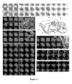
- the distribution of the cells is summarized in FIG. 10 and illustrated in FIG. 11 .
- the grafted cells After lateral ventricular grafting the grafted cells could be seen in the choroids plexus, cortex, caudate/putamen, olfactory bulb, hippocampus and cerebellum. In contrast, after intrastriatal grafting, the implanted cells could be found in the caudate/putamen and substantia nigra. By both routes, the grafted cells survived and spread from the implantation site.
- FIG. 13 summarizes the data.
- the left panel shows DA levels
- the middle panel shows HVA levels
- the right panel shows DOPAC levels.
- Dopamine concentrations remained significantly below baseline levels up to 22 days and remained so for up to 22 days.
- Asterisks indicate significant differences from control values (p ⁇ 0.01) using the one-way ANOVA followed by Dunnet's multiple comparison test.
- mice Striatal DA levels after grafting BMSC into lateral ventricles of MPTP-treated Mice. Two old mice (14-month-old) were first treated with MPTP. Six mice were euthanized at 5 days after treatment and six mice received an infusion of nestin-enriched BMSC two days later. These mice were euthanatized at 19 days after grafting (21 days after MPTP). Both striatal DA and HVA levels were significantly increased in the grafted animals from MPTP treatment. Thus, grafted BMSC helped return function to the stria.
- bone marrow cells were collected from adult male GFP transgenic mice femurs and tibias. Cells were disaggregated by gentle pipetting several times. Cells were passed through a 40 micron nylon cell strainer to remove remaining clumps of tissue. Cells were washed by adding buffer, centrifuging for 10 min at 200 g and discarding supernatant. The cell pellet was resuspended in 1 ml of buffer for every 10 million cells. The BMSC were separated by adhesion to polystyrene flasks. After cultures reached confluency, the cells were lifted by incubation with trypsin. This procedure was repeated for 3-5 passages.
- cells were replated on bacteriological Petri dishes in the presence of serum free medium (N2), supplemented with transferring 100 micrograms/ml, putrescine 60 microM, insulin 25 microg/ml, progesterone 0.02 microM, selenium 0.03 microM, and containing EGF and bFGF 20 ng/ml. After 5-7 days, the cells continued to proliferate and were passaged when 70-80% confluent. At this stage the cells expressed a high proportion (60-70%) of nestin+ cells.
- N2 serum free medium
- the nestin+ cells were differentiated into neural lineages by replating onto polylysine-coated cell culture slides and plated in N2 medium (supplemented with all-trans retinoic acid 0.5 microM (Sigma), brain-derived neurotrophic factor (BDNF, 10 ng/ml, Invitrogen), transferring 100 microg/ml, putrescine 60 micro M, insulin 25 microg/ml, progesterone 0.02 microM, and selenium 0.03 microM). After 7-14 days, BMSC and brain-derived cells changed morphology and developed markers of neurons, astrocytes, and oligodendroglia.
- N2 medium supplemented with all-trans retinoic acid 0.5 microM (Sigma), brain-derived neurotrophic factor (BDNF, 10 ng/ml, Invitrogen), transferring 100 microg/ml, putrescine 60 micro M, insulin 25 microg/ml, progesterone 0.02 microM, and selenium 0.03 microM.
- mouse anti-nestin monoclonal, BD Biosciences
- mouse anti-beta-tubulin III Teuj1, monoclonal, Sigma
- rabbit anti-tyrosine hydroxylase TH, polyclonal, Chemicon
- TH polyclonal, Chemicon
- GAD rabbit anti-Glial Fibrillary Acidic Protein
- GFAP polyclonal, BioGenex
- rabbit anti-galactocerebroside Gal-C, polyclonal, Chemicon
- the cells After being washed, the cells were incubated for 1 hour with Alexa Fluor Goat anti-mouse IgG or Goat anti-rabbit IgG (Molecular Probes) at room temperature, the Alexa Fluor 488 being diluted 1:500 in PBS, and the Alexa Fluor 546 being diluted 1:1000 in PBS. Cultures were viewed with an inverted fluorescence microscope (Olympus IX70) using appropriate individual filters for visualizing green (Alexa Fluor 488), red (Alexa Fluor 546) and blue (DAPI 350 nm excitation, 470 nm emission) fluorescence.
- Alexa Fluor Goat anti-mouse IgG or Goat anti-rabbit IgG Molecular Probes
- FIG. 15 is a table that shows the immunophenotype of cells after one week in differentiation media, with the percentage of cells expressing specific markers. The percentages total more than 100 because cells express more than one marker during their maturation (e.g., nestini and TUJ1 simultaneously).
- the electrical properties of neuron-like cells derived from bone marrow and brain were assessed using whole-cell patch-clamp recording techniques under current-clamp and voltage-clamp modes.
- Cells were plated on 18 mm glass cover slips, transferred to a recording chamber mounted on a Zeiss Axiovert 2 microscope, aid putative neuron-like cells were identified at 40x magnification using differential inference contrast (DIC) optics.
- DIC differential inference contrast
- the pipettes were loaded with (in mM) 140 KCI, 2 Mg 2 ATP, 0.1 guanosine 5'-triphosphte (GTP) disodium salt, 10 glucose, 2 EDTA, and 10 HEPES, with pH adjusted to 7.2 with KOH.
- the extracellular solution contained (in mM): 140 NaCl, 3 KCI, 2.5 CaCl 2 , 1.2 MgCl 2 , 7.7 glucose, and 10 HEPES with pH adjusted to 7.2 with NaOH.
- 3% (w/v) Lucifer yellow-CH dipotassium salt (Sigma) were added to the intracellular patch solution as a fluorescent tracer for subsequent experiments testing these cells for the expression of the neuronal market beta-III-tubulin.
- Membrane current-voltage relationships were also determined under voltage-clamp mode using slow voltage ramps, whereby the membrane potential was increased linearly from -150 mV to +50 mV at 50 mV/s. Permeability of the cell membrane to K+, Na+ and Cl- was assessed by changing the extracellular concentration of each ion and comparing sdhifts in the net current to that predicted for the ion with the Goldman-Hodgkin-Katz equation.
- Electrophysiological characteristics of differentiating neurons derived from brain neurospheres were compared to differentiating neurons from nestin-enriched BM-derived neurons. Action potentials were recorded from both the brain-derived neurosphere cells and bone marrow-derived neurons in FIG. 16 . After differentiating one week, 2.5% of the patched cells from the BM cultures and 60% of the patched cells from brain neural stem cells exhibited action potentials. The BM-derived neurons exhibited the following: phasic, no overshoot and a small after-hyperpolarization. An additional characteristic was the normal reversible inhibition of the action potential by blocking the Na+ channel with tetrodotoxin (data not shown). The neural brain stem cells also exhibited phasic, no overshoot and a bin after-hyperpolarization, but neither tetrodotoxin (TTX) nor Cd 2+ blocked after-hyperpolarizations.
- TTX tetrodotoxin
- the differentiated BMSC exhibited normal neuronal electrophysiologic characteristics and acted more maturely than did the neural stem cells.
- the migration of the treated BMSCs includes, but is not limited to, the following brain regions: mitral cell zone of the olfactory bulb, paraventricular and supraoptic nuclei of hypothalamus, hippocampus, habenula, oculomotor nucleus, red nucleus, s. nigra, abducens, facial and hyoglossal cranial nerve nuclei, inferior olive of medulla, ventral horn of the spinal cord and the cerebellar Purkinje cell layer.
- the BMSC beta-gal+ cells assumed multiple phenotypes including epithelial-like cuboidal cells in choroid plexus; small oligodendroglial-like cells in optic chiasm and sub-cortical white matter; large neuron-like cells in the red nucleus, s. nigra, and brainstem motor nuclei.
- This novel method of neural transplantation is a scientifically feasible and clinically promising approach to the treatment of neurodegenerative diseases and stroke, as well as for repair of traumatic injuries to brain and spinal cord. Feasibility was demonstrated by implantation into mice whose rotometer activity and production of dopamine, HVA and DOPAC were impaired by MPTP. After implantation, rotometer activity returned, as did production of DA and DVA, indicating that the implantation had overcome the effect of MPTP.
- BMSCs migratory and regenerative nature of the differentiated BMSCs
- the pretreated BMSCs can replace fetal neurons which have been used for transplantation in that condition.
- the BMSCs treated with retinoic acid and BCNF have been shown to migrate to the substantia nigra, where a dopaminergic neuron deficit causes PD.
- the delivery of BMSCs to the substantia nigra could regenerate the nigral neurons and return the flow of dopamine to that region.
- mice a localized effect on the substantia nigra and the cortex was observed after implantation in the striatum, indicating that treatment of Parkinson's disease is feasible. More DA also was produced, an indication of improved neuron function.
- the regeneration of the cerebellar Purkinje cell layer can help improve coordinated movement of the patient.
- Implantation of GFP+ BM cells into the lateral ventricle resulted in cells migrating to the cerebellum in the three of the three mice tested.
- Restoration of the cerebellum would be useful in cerebellar tumors, typically a medulloblastoma that occurs in childhood. In adults, a similar syndrome may be seen in chronic alcoholism, which causes degeneration of the vermis.
- the patient has an unsteady, staggering ataxic gait; he or she walks on a wide base and sways from side to side. Barr, M. L. and Kiernan, J. A. THE HUMAN NERVOUS SYSTEM, AN ANATOMICAL VIEWPIINT, 6th ed., J. B. Lippincott Company, Philadelphia (1993 ).
- BMSCs Another use for the migration and regeneration of BMSCs is in the mitral cell zone of the olfactory bulb. Implantation of GFP+ BM cells into the lateral ventricle resulted in cells migrating to the olfactory bulb in two of the three mice tested. Impulses from the olfactory bulb are conveyed to olfactory areas for subjective appreciation of odors and aromas. Barr, M. L. and Kiernan (ibid.) The migration and regeneration of BMSCs to this brain region could treat loss of taste from toxic chemicals, aging and injury.
- Administering the BMSCs can be used in the regeneration of facial and hyoglossal cranial nerve nuclei and restoration of movement of facial muscles after a stroke or other injury.
- Administering the BMSCs could aid in the regeneration of the ventral horn of the spinal cord and restoration of motor skills lost from trauma to the spine.
- Administering the BMSCs could aid in the regeneration of an injured hippocampal region. Implantation of GFP+ BM cells into the lateral ventricle resulted in cells migrating to the hippocampus in three of three mice tested.
Abstract
Description
- This application is a continuation in part of co-pending application Ser. No.
10/353,742 filed January 27, 2003 09/307,824 filed May 7, 1999 provisional application number 60/084,533, filed May 7, 1998 provisional application number 60/112,979, filed Dec. 17, 1998 provisional application number 60/129,684 filed Apr. 16, 1999 - This application relates to methods of culturing bone marrow cells such that they express neuronal phenotype for use in transplantation.
- Neurobiologists have long considered the neurons in the adult brain to be like a precious nest egg: a legacy that dwindles with time and illness and is difficult if not impossible to rebuild. Parkinson's and Alzheimer's are examples of neurodegenerative diseases whose cures await scientists overcoming the difficulty of rebuilding neurons in the human adult brain. Parkinson's disease (PD), is a disorder of middle or late life, with very gradual progression and a prolonged course. HARRISON'S PRINCIPLES OF INTERNAL MEDICINE, Vol. 2, 23d ed., Ed by Isselbacher, Braunwald, Wilson, Martin, Fauci and Kasper, McGraw-Hill Inc., New York City, 1994, pg. 2275. The most regularly observed changes have been in the aggregates of melanin-containing nerve cells in the brainstem (substantia nigra, locus coeruleus), where there are varying degrees of nerve cell loss with reactive gliosis (most pronounced in the substantia nigra) along with distinctive eosinophilic intracytoplasmic inclusions. (Id. at 2276).
- In its fully developed form, PD is easily recognized. The stooped posture, the stiffness and slowness of movement, the fixity of facial expression, the rhythmic tremor of the limbs, which subsides on active willed movement or complete relaxation, are familiar to every clinician. Generally, accompanying the other characteristics of the fully developed disorder is the festinating gait, whereby the patient, prevented by the abnormality of postural tone from making the appropriate reflex adjustments required for effective walking, progresses with quick shuffling steps at an accelerating pace as if to catch up with the body's center of gravity. (Id. at 2276).
- Although the modern treatment of PD is more successful than any that was available before the introduction of levodopa, including stereotactic surgery, there are still many problems. (Id. at 2277). Underlying much of the difficulty undoubtedly is the fact that none of these therapeutic measures has an effect on the underlying disease process, which consists of neuronal degeneration. Ultimately, a point seems to be reached where pharmacology can no longer compensate for the loss of basal ganglia dopamine. (Id.).
- Alzheimer's Disease (AD) is due to a degenerative process characterized by progressive loss of cells from the basal forebrain, cerebral cortex and other brain areas. Acetylcholine-transmitting neurons and their target nerves are particularly affected. Senile plagues and neurofibrillary tangles are present. Pick's disease has a similar clinical picture to Alzheimer's disease but a somewhat slower clinical course and circumscribed atrophy, mainly affecting the frontal and temporal lobes. One animal model for Alzheimer's disease and other dementias displays hereditary tendency toward the formation of such plaques. It is thought that if a drug has an effect in the model, it also may be beneficial in at least some forms of Alzheimer's and Pick's diseases. At present there are palliative treatments but no means to restore function.
- A group of degenerative disorders characterized by progressive ataxia due to degeneration of the cerebellum, brainstem, spinal cord and peripheral nerves, and occasionally the basal ganglia. Many of these syndromes are hereditary; others occur sporadically. The spinocerebellar degenerations are logically placed in three groups: predominantly spinal ataxias, cerebellar ataxias and multiple-system degenerations. To date there are no treatments. Friedrich's ataxia is the prototypical spinal ataxia whose inheritance is autosomal recessive. The responsible gene has been found on Chromosome 9. Symptoms begin between ages of 5 and 15 with unsteady gait, followed by upper extremity ataxia and dysarthria. Patients are areflexic and lose large-fiber sensory modalities (vibration and position sense). Two other diseases have similar symptoms: Bassen-Kornzweig syndrome (abeta-lipoproteinemia and vitamin E deficiency) and Refsom's disease (phytanic acid storage disease). Cerebellar cortical degenerations generally occur between
ages - In multiple-system degenerations, ataxia occurs in young to middle adult life in varying combinations with spasticity and extrapyramidal, sensory, lower motor neuron and autonomic dysfunction. In some families, there may also be optic atrophy, retinitis pigmentosa, opthalmoplegia and dementia.
- Another form of cerebellar degeneration is paraneoplastic cerebellar degeneration that occurs with certain cancers, such as oat cell lung cancer, breast cancer and ovarian cancer. In some cases, the ataxia may precede the discovery of the cancer by weeks to years. Purkinje cells are permanently lost, resulting in ataxia. Even if the patient is permanently cured of the cancer, their ability to function may be profoundly disabled by the loss of Purkinje cells. There is no specific treatment.
- Strokes also result in neuronal degeneration and loss of functional synapses. Currently there is no repair, and only palliation and rehabilitation are undertaken.
- Neurotransplantation has been used to explore the development of the central nervous system and for repair of diseased tissue in conditions such as Parkinson's and other neurodegenerative diseases. The experimental replacement of neurons by direct grafting of fetal tissue into the brain has been accomplished in small numbers of patients in several research universities (including our University of South Florida); but so far, the experimental grafting of human fetal neurons has been limited by scarcity of appropriate tissue sources, logistic problems, legal and ethical constraints, and poor survival of grafted neurons in the human host brain.
- One method replaces neurons by using marrow stromal cells as stem cells for non-hematopoietic tissues. Marrow stromal cells can be isolated from other cells in marrow by their tendency to adhere to tissue culture plastic. The cells have many of the characteristics of stem cells for tissues that can roughly be defined as mesenchymal, because they can be differentiated in culture into osteoblasts, chondrocytes, adipocytes, and even myoblasts. Therefore, marrow stromal cells present an intriguing model for examining the differentiation of stem cells. Also, they have several characteristics that make them potentially useful for cell and gene therapy. Prockop, D. J. Science: 26: 71-74 (1997). This population of bone marrow cells (BMSC) has also been used to prepare dendritic cells, (K. Inaba, et al., J. Experimental Med. 176: 1693-1702 (1992)) which, as the name implies, have a morphology which might be confused for neurons. Dendritic cells comprise a system of antigen-presenting cells involved in the initiation of T cell responses. The specific growth factor which stimulates production of dendritic cells has been reported to be granulocyte/macrophage colony-stimulating factor (GM-CSF). K. Inaba, et al., J. Experimental Med. 176: 1693-1702 (1992).
- The presence of stem cells for non-hematopoietic cells in bone marrow was first suggested by the observations of the German pathologist Cohnheim 130 years ago. J. Cohnheim, Arch. Path. Anat. Physiol. Klin. Med. 40: 1 (1867). Cohnheim studied wound repair by injecting an insoluble aniline dye into the veins of animals and then looking for the appearance of dye-containing cells in wounds he created at a distal site. He concluded that most, if not all, of the cells appearing in the wounds came from the bloodstream, and, by implication, from bone marrow. The stained cells included not only inflammatory cells but also cells that had a fibroblast-like morphology and were associated with thin fibrils. Therefore, Cohnheim's work raised the possibility that bone marrow may be the source of fibroblasts that deposit collagen fibers as part of the normal process of wound repair. The source of fibroblasts in wound repair has been examined in more than 40 publications since Cohnheim's report of 1867. See, for example, R. Ross, N. B. Everett, R. Tyler, J. Cell Biol. 44: 645 (1970); J. K. Moen. J. Exp. Med. 61: 247 (1935); N. L. Petrakis, M. Davis, S. P. Lusia, Blood 17: 109 (1961); S. R. S. Rangan, Exp. Cell Res. 46: 477 (1967); J. M. Davidson, in INFLAMMATION: BASIC PRINCIPLES AND CLINICAL CORRELATES, J. I. Gallin, I. M. Goldstein, R. Snyderman, Eds. (Raven, New York City, ed. 2, 1992, pp. 809-19; R. Bucala, L. A. Spiegel, J. Chesney, N. Hogan, A. Cerami, Mol. Med. 1: 71 (1994). Most of the data suggest that the fibroblasts are of local origin, but the issue has not been resolved and is still being examined. Prockop, D. J. Science 26: 71-74 (1997).
- Although Cohnheim's thesis has not yet been substantiated, definitive evidence that bone marrow contains cells that can differentiate into fibroblasts as well as other mesenchymal cells has been available since the pioneering work of Friedenstein, beginning in the mid-1970's. A. J. Friedenstein, U. Gorskaja, N. N. Kulagina, Exp. Hematol. 4: 276 (1976). Friedenstein placed samples of whole bone marrow in plastic culture dishes and poured off the cells that were nonadherent. The most striking feature of the adherent cells was that they had the ability to differentiate into colonies that resembled small deposits of bone or cartilage. Freidenstein's initial observations were extended by a number of investigators during the 1980s, particularly by Piersma and associates (A. H. Piersma, R. E. Ploemacher, k. G. M. Brockbank, Br. J. Haematol. 54: 285 (1983); A. H. Piersma et al., Exp. Hematol. 13: 237 (1985)) and Owen and associates. C. R. Howlett et al., Clin. Orthop. Relat. Res. 213: 251 (1986); H. J. Mardon, J. Bee, k. von der Mark, M. E. Owen, Cell Tissue Res. 250: 157 (1987); J. N. Beresford, J. H. Bennett, C. Devlin, P. S. Leboy, M. E. Owen, J. Cell Sci. 102: 341 (1992). These and other studies (M. E. Owen and A. J. Friedenstein, in Cell and Molecular Biology of Vertebrate Hard Tissues, Ciba Foundation Symp. 136, Wiley, Chichester, UK, 1988, pp. 42-60; S. L Cheng et al., Endocrinology 134: 277 (1994); A. I. Caplan, J. Orthop. Res. 9: 641 (1991); D. J. Richard et al., Dev. Biol. 161: 218 (1994). S. Wakitani, T. Saito, and A. J. Caplan (Muscle Nerve 18: 1412 (1995)) demonstrated that MSCs differentiated into myoblasts and myotubes by treatment with 5-azacytidine and amphotericin B (Fungasome, Gibco). D. Phinney (Prockop, D. J. Science .26: 71-74 (1997)), recently observed that the cells differentiate into myoblasts and myotubes after treatment with amphotericin B (1 microg/ml) alone; A. J. Friedenstein, R. K. Chailakahyan, U. V. Gerasimov, Cell Tissue Kinet. 20: 263 (1987); A. Keating, W. Horsfall, R. G. Hawley, F. Toneguzzo, Exp. Hematol. 18: 99 (1990); B. R. Clark and A. Keating, Ann. N. Y. Acad. Sci 770: 70 (1995)) established that the Marrow Stromal Cells (MSCs) isolated by the relatively crude procedure of Friedenstein were multi-potential and readily differentiated into osteoblasts, chondroblasts, adipocytes, and even myoblasts.
- Even though the multi potential properties of MSCs have been recognized for several decades, there are surprisingly large gaps in our information about the cells themselves. The cells, isolated by their adherence to plastic as described by Friedenstein (A. J. Friedenstein, U. Gorskaja, N. N. Kulagina, Exp. Hematol. 4: 276 (1976)), initially are heterogeneous and difficult to clone. The fraction of the hematopoietic cells is relatively high in initial cultures of mouse marrow but is less than 30% with human marrow (M. E. Owen and A. J. Friedenstein, in Cell and Molecular Biology of Vertebrate Hard Tissues, Ciba Foundation Symp. 136, Wiley, Chichester, UK, 1988, pp. 42-60; S. L Cheng et al., Endocrinology 134: 277 (1994); A. I. Caplan, J. Orthop. Res. 9: 641 (1991); D. J. Richard et al., Dev. Biol. 161: 218 (1994); A. Keating, W. Horsfall, R. G. Hawley, F. Toneguzzo, Exp. Hematol. 18: 99 (1990); B. R. Clark and A. Keating, Ann. N. Y. Acad. Sci. 770: 70 (1995)). Most of the readily identifiable hematopoietic cells are lost as the cells are maintained as primary cultures for 2 or 3 weeks. The cultured MSCs synthesize an extracellular matrix that includes interstitial type I collagen, fibronectin, and the type IV collagen and laminin of basement membranes (M. E. Owen and A. J. Friedenstein, in Cell and Molecular Biology of Vertebrate Hard Tissues, Ciba Foundation Symp. 136, Wiley, Chichester, UK, 1988, pp. 42-60; S. L Cheng et al., Endocrinology 134: 277 (1994); A. I. Caplan, J. Orthop. Res. 9: 641 (1991); D. J. Richard et al., Dev. Biol. 161: 218 (1994); A. Keating, W. Horsfall, R. G. Hawley, F. Toneguzzo, Exp. Hematol. 18: 99 (1990); B. R. Clark and A. Keating, Ann. N. Y. Acad. Sci. 770: 70 (1995)). A small fraction of the cultured cells synthesize factor VII-associated antigen and therefore are probably endothelial. The cells secrete cytokines, the most important of which appear to be interleukin-7 (IL-7), IL-8, IL-11, and stem cell factor (c-kit ligand). Conditions for differentiating the cells are somewhat species-dependent and are influenced by incompletely defined variables, such as the lot of fetal calf serum used. However, MSCs from mouse, rat, rabbit, and human readily differentiate into colonies of osteoblasts (depositing mineral in the form of hydroxyapatite), chondrocytes (synthesizing cartilage matrix), and adipocytes in response to dexamethasone, 1,25-
dihydroxyvitamin D.sub 3, or cytokines such as BMP-2 (A. J. Friedenstein, U. Gorskaja, N. N. Kulagina, Exp. Hematol. 4: 276 (1976); 5-11). In response to 5-azacytidine with amphotericin B (Fungasome, Gibco) or amphotericin B alone, S. Wakitani, T. Saito, and A. J. Caplan (Muscle Nerve 18: 1412 (1995)) demonstrated that MSCs differentiated into myoblasts and myotubes by treatment with 5-azacytidine and amphotericin B. D. Phinney (unpublished data) recently observed that the cells differentiate into myoblasts and myotubes after treatment with amphotericin B (1 microg/ml) alone, and they differentiated into myoblasts that fuse into rhythmically beating myotubes. - Most experiments on the differentiation of MSCs have been carried out with cultures of MSCs as described by Friedenstein (A. J. Friedenstein, U. Gorskaja, N. N. Kulagina, Exp. Hematol. 4: 276 (1976)). For example,
U.S. Pat. No. 4,714,680 issued Dec. 22, 1987 , discloses a method of harvesting marrow from donors. Monoclonal antibodies that recognize a stage-specific antigen or immature human marrow cells are provided. These antibodies are useful in methods of isolating cell suspension from human blood and marrow that can be employed in bone marrow transplantation. Cell suspensions containing human pluripotent lymphohematopoietic stem cells are also provided, as well as therapeutic methods employing the cell suspensions. - Several groups of investigators since 1990 have attempted to prepare more homogenous populations. For example,
U.S. Pat. No. 5,087,570, issued Feb. 11, 1992 , discloses how to isolate homogeneous mammalian hematopoietic stem cell compositions. Concentrated hematopoietic stem cell compositions were substantially free of differentiated or dedicated hematopoietic cells. The desired cells are obtained by subtraction of other cells having particular markers. The resulting composition may be used to provide for individual or groups of hematopoietic lineages, to reconstitute stem cells of the host, and to identify an assay for a wide variety of hematopoietic growth factors. -
U.S. Pat. No. 5,633,426 issued May 27, 1997 , is another example of the differentiation and production of hematopoietic cells. Chimeric immunocompromised mice are given human bone marrow of at least 4 weeks from the time of implantation. The bone marrow assumed the normal population of bone marrow except for erythrocytes. These mice with human bone marrow may be used to study the effect of various agents on the proliferation and differentiation of human hematopoietic cells. -
U.S. Pat. No. 5,665,557 issued Sep. 9, 1997 , relates to methods of obtaining concentrated hematopoietic stem cells by separating out an enriched fraction of cells expressing the marker CDw109. Methods of obtaining compositions enriched in hematopoietic megakaryocyte progenitor cells are also provided. Compositions enriched for stem cells and populations of cells obtained therefrom are also provided by the invention. Methods of use of the cells are also included. -
U.S. Pat. No. 5,753,505 issued on Jun. 5, 1995 , is yet another method of differentiation. Primordial tissue is introduced into immunodeficient hosts, where the primordial tissue develops and differentiates. The chimeric host allows for investigation of the processes and development of the xenogeneic tissue, testing for the effects of various agents on the growth and differentiation of the tissue, as well as identification of agents involved with the growth and differentiation. -
U.S. Pat. No. 5,753,505 issued May 19, 1998 , provides an isolated cellular composition comprising greater than about 90% mammalian, non-tumor-derived, neuronal progenitor cells which express a neuron-specific marker and which can give rise to progeny which can differentiate into neuronal cells. Also provided are methods of treating neuronal disorders utilizing this cellular composition. -
U.S. Pat. No. 5,759,793 issued Aug. 6, 1996 , provides a method for both the positive and negative selection of at least one mammalian cell population from a mixture of cell populations utilizing a magnetically stabilized fluidized bed. One application of this method is the separation and purification of hematopoietic cells. Target cell populations include human stem cells. -
U.S. Pat. No. 5,789,148 issued Aug. 4, 1998 , discloses a kit, composition and method for cell separation. The kit includes a centrifugable container and an organosilanized silica particle-based cell separation suspension suitable for density gradient separation, containing a polylactam and sterilized by treatment with ionizing radiation. The composition includes a silanized silica particle-based suspension for cell separation which contains at least 0.05% of a polylactam, and preferably treated by ionizing radiation. Also disclosed is a method of isolating rare blood cells from a blood cell mixture. - Within the past several years, MSCs have been explored as vehicles for both cell therapy and gene therapy. The cells are relatively easy to isolate from the small aspirates of bone marrow that can be obtained under local anesthesia; they are also relatively easy to expand in culture and to transfect with exogenous genes. Prockop, D. J. Science 26: 71-74 (1997). Therefore, MSCs appear to have several advantages over hematopoietic stem cells (HMCs) for use in gene therapy. The isolation of adequate numbers of HSCs requires large volumes of marrow (1 liter or more), and the cells are difficult to expand in culture. (Prockop, D. J. (ibid.)).
- There are several sources for bone marrow tissue, including the patient's own bone marrow, that of blood relatives or others with MHC matches, bone marrow banks, and umbilical cord blood banks. There are several patents that encompass this source.
U.S. Pat. No. 5,476,997 issued May 17, 1994 , discloses a method of producing human bone marrow equivalent. A human hematopoietic system is provided in an immunocompromised mammalian host, where the hematopoietic system is functional for extended periods of time. Particularly, human fetal liver tissue and human fetal thymus are introduced into a young immunocompromised mouse at a site supplied with a vascular system, whereby the fetal tissue results in formation of functional human bone marrow tissue. - A source of implantable neurons that is the most ethically controversial is that of human fetal tissue.
U.S. Pat. No. 5,690,927 issued Nov. 25, 1997 , also utilizes human fetal tissue. Human fetal neuro-derived cell lines are implanted into host tissues. The methods allow for treatment of a variety of neurological disorders and other diseases. A preferred cell line is SVG. -
U.S. Pat. No. 5,753,491 issued May 19, 1998 , discloses methods for treating a host by implanting genetically unrelated cells in the host. More particularly, the present invention provides human fetal neuro-derived cell lines, and methods of treating a host by implantation of these immortalized human fetal neuro-derived cells into the host. One source is the mouse, which is included in theU.S. Pat. No. 5,580,777 issued Dec. 3, 1996 . This patent features a method for the in vitro production of lines of immortalized neural precursor cells, including cell lines having neuronal and/or glial cell characteristics, comprises the step of infecting neuroepithelium or neural crest cells with a retroviral vector carrying a member of the myc family of oncogenes. -
U.S. Pat. No. 5,753,506 issued May 19, 1998 , reveals an in vitro procedure by which a homogeneous population of multipotential precursor cells from mammalian embryonic neuroepithelium (CNS stem cells) was expanded up to 10.sup.9 fold in culture while maintaining their multipotential capacity to differentiate into neurons, oligodendrocytes, and astrocytes. Chemical conditions are presented for expanding a large number of neurons from the stem cells. In addition, four factors-PDGF, CNTF, LIF, and T3-have been identified which, individually, generate significantly higher proportions of neurons, astrocytes, or oligodendrocytes. These procedures are intended to permit a large-scale preparation of the mammalian CNS stem cells, neurons, astrocytes, and oligodendrocytes. These cells are proposed as an important tool for many cell- and gene-based therapies for neurological disorders. - Another source of stem cells is that of primate embryonic stem cells.
U.S. Pat. No. 5,843,780 issued Dec. 1, 1998 , utilizes these stem cells. A purified preparation of stem cells is disclosed. This preparation is characterized by the following cell surface markers: SSEA-1 (-); SSEA-3 (+); TRA-1-60 (+); TRA-1-81 (+); and alkaline phosphatase (+). In one embodiment, the cells of the preparation have normal karyotypes and continue to proliferate in an undifferentiated state after continuous culture for eleven months. The embryonic stem cells lines are also described as retaining the ability to form trophoblasts and to differentiate into tissues derived from all three embryonic germ layers (endoderm, mesoderm and ectoderm). A method for isolating a primate embryonic stem cell line is also disclosed in the patent. - In summary, there is substantial evidence in both animal models and human patients that neural transplantation is a scientifically feasible and clinically promising approach to the treatment of neurodegenerative diseases and stroke as well as for repair of traumatic injuries to brain and spinal cord. Nevertheless, alternative cell sources and novel strategies for differentiation are needed to circumvent the numerous ethical and technical constraints that now limit the widespread use of neural transplantation.
- This invention provides a novel source of neuronal tissue that can be used for grafting into the patient's own brain or spinal cord (autografting). This invention thus bypasses the delicate issue of using pooled fetal tissue and also obviates the need for immunosuppression. This invention will also be used for allografting (transplantation of bone marrow-derived neuronal cells from one individual to another) and xenografting (transplantation of bone marrow-derived neuronal cells from one species to another). Simply stated, bone marrow cells can be induced to become neurons in vitro and in vivo. Neither the concept nor the technique has been previously reported or accomplished.
- In one embodiment, there are disclosed non-fetal, non-tumorigenic, bone-marrow derived neuronal cells suitable for grafting into a mammal's brain or spinal cord.
- In another embodiment, there is disclosed a novel method for obtaining neurons from bone marrow, said method comprising the steps of a) providing bone marrow cells; b) selecting for bone marrow stromal cells; and c) incubating the stromal cells with a differentiating agent, for a time sufficient to change the cell phenotype to neuronal. The method may include an additional step of
claim 3 wherein step b further comprises separating out hematopoietic stem cells. The method can also include placing the bone marrow cells and suitable culture medium in a plastic culture medium container, allowing the bone marrow stromal cells to adhere to the plastic, and removing the other cells by replacing the medium. The method can use the differentiating agentsuch as retinoic acid, growth factors, fetal neuronal cells or a combination thereof. The growth factors can include BDNF, GDNF and NGF. The method can use 9-cis retinoic acid, all-transretinoic or a combination thereof. - In another embodiment, the method for obtaining neurons for auto-transplant from an individual's own bone marrow includes the following steps: a) harvesting the bone marrow; b) selecting for bone marrow stromal cells; c) incubating the bone marrow stromal cells in a medium including a mitogen until there are sufficient cells for transplantation; d) incubating the stromal cells of step c) with a differentiating agent for a time sufficient to change the cell phenotype to neuronal and/or glial. The mitogen can be EGF, PDGF or a combination thereof. The step b can further include separating out hematopoietic stem cells. The step b optionally further includes placing the bone marrow cells and suitable culture medium in a plastic culture medium container, allowing the bone marrow stromal cells to adhere to the plastic, and removing the other cells by replacing the medium. The differentiating can be retinoic acid, growth factors, fetal neuronal cells or a combination thereof. The growth factors can include BDNF, GDNF and NGF, or a combination thereof. The retinoic acid can be 9-cis retinoic acid, all-transretinoic or a combination thereof.
- In another embodiment, a cell line of bone marrow stromal cells developed by the method above, wherein the cells have the ability to migrate and localize to specific neuroanatomical regions where they appear to differentiate into neurons typical of the region and to integrate in characteristic architectonic patterns.
- In another embodiment, there is provided a kit for neuronal cell auto-transplant that includes a) a syringe suitable for obtaining bone marrow b) a plastic flask with dehydrated culture medium; c) bone marrow stem cell; and d) a mixture of Retinoic Acid A and BDNF. In yet another embodiment, there is provided a method of treating a neurodegenerative disorder comprising administering a sufficient quantity of the cells produced as disclosed above to treat an individual with said neurodegenerative disorder. The method can be used in Parkinson's disease, Alzheimer's disease, ischemia, spinal cord damage, ataxia or alcoholism.
-
-
Fig. 1 is a bar graph. BMSC adherent to culture dishes were treated with EGF (10 ng/ml), RA (0.5 microM) or RA plus BDNF (10 ng/ml) for 7 days. Each bar represents the mean number (.±-SEM) of fibronectin immunoreactive cells per visual field (20X objective) determined in 20 fields per dish in 4 culture dishes. *=p<0.05, two-tailed t-test. -
Figs. 2A through 2F are photomicrographs of BMSC from lacZ mice that have been cocultured with mouse fetal midbrain cells for 2 weeks in N5 medium supplemented with cis-9 retinoic acid (0.5 microM) and BDNF (10 ng/ml). -
Fig. 3A and 3B are photomicrographs, which show beta-gal+ cells accumulated in paraventricular and supraoptic nuclei of thehypothalamus 3 months after grafting.Fig. 3A (scale bar = 500 microm) shows symmetrical distribution despite unilateral grafting into the striatum.Fig 3B is a region of the paraventricular nucleus (scale bar = 100 microm). None of the beta-gal+ cells are labeled with the red-brown stain (TH-ir).Figs. 3A (Scale bar = 500 microm),Fig. 3B (Scale bar = 100 microm) andFig. 3C (Scale bar = 50 microm) depict cells doubly stained for beta-gal and TH-ir.Figs. 3D (Scale bar = 50 microm) andFig. 3E (Scale bar = 25 microm) illustrate sections from the red nucleus that have doubly stained for beta-gal and NeurN-ir.Fig. 3F (Scale bar = 25 microm) illustrated beta-gal+ cells from the red nucleus also doubly stained for MAP2-ir. -
Fig. 4 is a photomicrograph, which illustrates the migration and integration of BMSC into rat midbrain. The rat was injected with 6-OH DA into the right s.nigra, and grafted 3 weeks later with conditioned lacZ BMSC into the right corpus striatum. Three months after grafting the rat was sacrificed and perfused for immunohistochemistry. Serial sections (30 microm thick) were cut through the midbrain. -
Fig. 5A through 5F are photomicrographs of a section from rat cerebellar lobule illustrating laminar distribution of beta-gal+ cells in a distribution of Purkinje cells. Beta-gal+ are co-labeled with calbindin immunoreactivity inFigs. 5A, 5B, and 5C . (Scale bar = 100 microm inFig. 5A , 50 microm inFig. 5B and 25 microm inFig. 5C). Fig. 5D shows beta-gal+ Purkinje cells co-labeled with GAD-ir (Scale bar = 50 microm). Fig. 5E illustrated dense MAP2-ir fibers enveloping beta-gal+ Purkinje cells (Scale bar = 25 microm).Fig. 5F illustrates beta-gal+ cells co-labeled with NeuN-ir in the deep cerebellar nucleus (Scale bar = 25 microm). -
Fig. 6 is a Western blot of the lysates of BMSC conditioned with four different treatments and labeled with GFAP-ir, nestin and NeuN. BDNF+RA+N5 induced the strongest expression of nerve cell markers while glial cell markers was most strongly expressed after N5 alone. -
Figs. 7A through 7F are photomicrographs of human BMSC which were co-cultured with fetal rat striatal cells in N5 formulation with BDNF+RA. These figures show that human BMSC (green labeled inFigs. 7C and 7D and yellow inFigs. 7E and 7F ) can be induced to express neural markers NeuN (Figs. 7A and 7E ) and GFAP (Figs. 7B and 7F ). -
Fig. 8 is a photomicrograph of rat brain, showing the mouse BMSC labeled with red PKH26 also express the neuron marker NeuN-ir (green fluorescence). In addition, the morphology of the doubly labeled cells is that of neurons. -
Fig. 9 is a photomicrograph of rat brain, showing a doubly labeled glial cell. The red fluorescent tracer indentifies it as derived from a BMSC, and the green fluorescence is due to GFAP-ir. Note the morphology is that of a glial cell. -
Fig. 10 is a table displaying the distribution of grafted GFP+ bone marrow cells in MPTP-treated mice administered the cells to the ventricle and to the striatum. Surviving cells were observed after the administration to both locations. -
Figs. 11A through 11D illustrate the effects of grafting nestin-enriched BMSC into the right lateral ventricle of 16 month old mice, one week after MPTP treatment (20 mg/kg X 4). Mice were euthanized at 1 and 2 weeks.Figs. 11E and 11F illustrate the effects of intrastriatal grafting. -
Fig. 11A . 1 week after graft of BrdU pre-labeled BMSC: Sagittal section of hippocampus showing BrdU+ cells in the fimbria of the hippocampus (mag 4X). Insert box shows higher magnification of the BrdU+ cells. Bright field microscopy, counterstained with H&E. -
Fig. 11B . 1 week after graft of BrdU pre-labeled BMSC. Sagittal section through cerebellum showing BrdU+ cells. Bright field microscopy, counterstained with H&E. -
Fig. 11A' . Same as A, but GFP+ BMSC are visualized in the fimbria of the hippocampus. Panel with blue DAPI stain show cell nuclei. -
Fig. 11 B'. Same as B, but with GFP+ BMSC visualized in cerebellum (fluorescence microscopy). -
Fig. 11C .2 weeks after graft of GFP+ BMSC. Olfactory bulb. Panel on right shows DAPI+ nuclei. (fluorescence microscopy). -
Fig. 11D .2 weeks after graft of GFP+ BMSC. Rostral subventricular zone. D-1 shows cells expressing GFP and nestin (orange). D-2 is the magnified insert from D-1 showing two BMSC co-expressing GFP and nestin. D-3 shows nestin and D4 GFP expression using confocal fluorescence microscopy. -
Fig. 11E shows three panels from a confocal microscope image of striatum, displaying GFP+ nestin-enriched BMSC near the graft. CD 11b+ cells (microglia and monocytes) are red. Yellow indicates BM derived cells that coexpress the microglial marker. -
Fig. 11F . Three panels from a confocal microscope image of substantia nigra. GFP+ bone marrow-derived cells infiltrate the substantia nigra, even though BMSC were grated in the striatum. Neurons of the SN are stained with antibodies to NewN (red). -
Fig. 12 displays three grafts summarizing latencies to fall from an accelerating rotometer. Panel on the left (Contol mice) show that latencies (in seconds) remain stable from baseline to day 26 after saline injections. Middle panel illustrates significantly decreased latencies up through day 26 after MPTP (20 mg/kg X 4) alone. Right panel shows significant improvement in latencies evident 12 days after MPTP (and 1 week after BMSC infusion) with sustained improvement up to 26 days. One-way ANOVA indicates that the mean latencies are significantly different (p<0.05) using the Bonferroni multiple comparison test. -
Fig. 13 displays three graphs summarizing the effects of MPTP on striatal DA and metabolites in 14 month-old mice. Panel on the left shows DA levels, middle panel shows HVA and right panel shows DOPAC. All were significantly decreased for up to 22 days after MPTP treatment. Astericks indicate significant difference from control values (p<0.01) using one-way ANOVA followed by Dunnet's multiple comparison test. -
Fig. 14 graphically displays the effects of BMSC infusion (at MPTP = 5 days) into the lateral ventricle on striatal DA and HVA (19 days after BMSC graft). HVA levels were significantly elevated in the grafted MPTP-treated mice compared to the control MPTP-treated mice (that received infusion of vehicle). -
Fig. 15 is a table summarizing the numbers of different phenotypes of the neuron family that are present in retinoic acid- and bFGF-BMSC cells and in cultured brain cells. Note that this method produced a much higher proportion of neuronal cells that the preceding methods. For example, the treated BMSC cells had 19.4% labeled with TUJ1 and 20.6% labeled with NeuN. -
Fig. 16 displays tracings from treated BMSC (left) and treated neural stem cells (right). The tracing from the treated BMSC appeared more normal and mature. - In the following detailed description of the preferred embodiments, reference is made to the accompanying drawings that form a part hereof, and in which are shown by way of illustrating specific embodiments in which the invention may be practiced. The embodiments illustrated are described in sufficient detail to enable those skilled in the art to practice the teachings disclosed herein. Other embodiments may be utilized and derived therefrom, such that structural and logical substitutions and changes may be made without departing from the scope of present inventions. The following detailed description, therefore, is not to be taken in a limiting sense, and the scope of various embodiments of the invention is defined only by the appended claims, along with the full range of equivalents to which such claims are entitled.
- The ancient Chinese medical maxim "brain is a sea of marrow" (W. R. Morse, Chinese Medicine (Paul Hoeber, Inc., New York, 1938).) resonates with recent research findings regarding the capacity of a population of bone marrow cells to differentiate into neurons under experimental conditions.
- In Western medicine, the existence of non-hematopoietic stem cells in bone marrow was suggested over 100 years ago, but the isolation and differentiation of marrow stromal cells into osteoblasts, chondroblasts, adipocytes and myoblasts was only recently demonstrated. D. J. Prockop, Science 276: 71-74 (1997). In the medical literature, non-hematopoietic precursors from bone marrow stroma have been referred to as colony-forming-unit fibroblasts, mesenchymal stem cells or bone marrow stromal cells (BMSC). Although BMSC can naturally be expected to be a source of surrounding tissue of bone, cartilage and fat, several recent reports demonstrate that these cells, under specific experimental conditions, can migrate and differentiate into muscle or glial cells. Systemic infusion of BMSC into irradiated 3-week-old mice has resulted in the appearance of progeny of the donor cells in a variety of non-hematopoietic tissues including the brain. R. F. Pereira, et al., Proc. Natl. Acad. Med. (U.S.A.) 95: 1142-1147 (1998). Transplantation of genetically-labeled bone marrow cells into immunodeficient mice has been reported to result in migration of marrow cells into a region of chemically-induced muscle degeneration. G. Ferrari, et al., Science 279: 1528-1530 (1998). These marrow-derived cells underwent myogenic differentiation and participated in the regeneration of the damaged muscle fibers. Moreover, infusion of human BMSC into rat striatum resulted in engrafting, migration and survival of cells. S. A. Azizi, et al., Proc. Nat. Acad. Sci. (U.S.A.) 95: 3908-3913 (1998). After engraftment, these cells lost typical BMSC markers, such as immunoreactivity to antibodies against collagen and fibronectin. BMSC developed many of the characteristics of astrocytes, and their engraftment and migration markedly contrasts with fibroblasts that continue to produce collagen and undergo gliosis after implantation. Following transplantation of bone marrow into lethally irradiated rats, bone marrow-derived cells were found to replace between 60 and 80% of the host macrophages in sensory and autonomic ganglia as well as in peripheral nerves. K. Vass, W. F. Hickey, R. E. Schmidt, H. Lassman, Laboratory Investigation 69: 275-282 (1993). In that study, no attempt was made to determine whether marrow cells differentiated into glial or neuronal cells.
- Our laboratory has recently succeeded in differentiating BMSC into a neuron-like phenotype, using a combination of retinoic acid, growth factors, and fetal neuronal environments. Moreover, we have grafted BMSC into denervated rat striatum and have found that BMSC migrate into specific neuroanatomical regions where they differentiate into cells which expressing markers of neighboring cells. In those regions, the cells integrate in to characteristic arthitectonic patterns.
- "Neuronal cells" are those having at least an indication of neuronal phenotype, such as staining for one or more neuronal markers. Examples of neuronal markers include, but are not limited to, neuron-specific nuclear protein, tyrosine hydroxylase, microtubule associated protein, and calbindin.
- "Non-tumorigenic" refers to the fact that the cells do not give rise to a neoplasm or tumor.
- As used herein, "non-fetal" refers to the fact that the source has been born. It does not exclude umbilical cord blood.
- Selecting for bone marrow stromal cells can be done in a number of ways. Classically, the stromal cells are disaggregated and cultured inside a plastic container. The stromal cells are separated by their survival in specific media and adherence to the plastic.
- Bone-marrow cells can be induced to adopt a number of different neuronal phenotypes, as proven below by in vitro and in vivo observations. Additional in vitro differentiation techniques can be adapted through the use of various cell growth factors and co-culturing techniques known in the art. Besides co-culturing with fetal mesencephalic or striatal cells (as successfully demonstrated below), a variety of other cells can be used, including but not limited to accessory cells, Sertoli cells and cells from other portions of the fetal and mature central nervous system.
- Standard molecular biology techniques known in the art and not specifically described are generally followed as in Sambrook et al., MOLECULAR CLONING: A LABORATORY MANUAL, Cold Springs Harbor Laboratory, New York (1989, 1992), and in Ausubel et al., CURRENT PROTOCOLS IN MOLECULAR BIOLOGY, John Wiley and Sons. Baltimore, Md. (1989). Polymerase chain reaction (PCR) is carried out generally as in PCR PROTOCOLS: A GUIDE TO METHODS AND APPLICATIONS, Academic Press, San Diego, Calif. (1990). Reactions and manipulations involving other nucleic acid techniques, unless stated otherwise, are performed as generally described in Sambrook, et al., 1989, MOLECULAR CLONING: A LABORATORY MANUAL, Cold Springs Harbor Laboratory Press, and methodology as set forth in
U.S. Pat. Nos. 4,666,828 ;4,683,202 ,4,801,531 ;5,192,659 ; and5,272,057 and incorporated herein by reference. In-situ PCR in combination with Flow Cytometry can be used for detection of cells containing specific DNA and mRNA sequences (Testoni et al. Blood 87:3822 (1996)). - Standard methods in immunology known in the art and not specifically described are generally followed as in Stites et al. (eds), BASIC AND CLINICAL IMMUNOLOGY, 8th Ed., Appleton & Lange, Norwalk, Conn. (1994); and Mishell and Shigi (eds), SELECTED METHODS IN CELLULAR IMMUNOLOGY, W. H. Freeman and Co., New York (1980).
- In general immunoassays are employed to assess a specimen such as for cell surface markers or the like. Immunocytochemical assays are well known to those skilled in the art. Both polyclonal and monoclonal antibodies can be used in the assays. Where appropriate other immunoassays, such as enzyme-linked immunosorbent assays (ELISAs) and radioimmunoassays (RIA), can be used as are known to those in the art. Available immunoassays are extensively described in the patent and scientific literature. See, for example,
U.S. Pat. Nos. 3,791,932 ;3,839,153 ;3,850,752 ;3,850,578 ;3,853,987 ;3,867,517 ;3,879,262 ;3,901,654 ;3,935,074 ;3,984,533 ;3,996,345 ;4,034,074 ;4,098,876 ;4,879,219 ;5,011,771 and5,281,521 as well as Sambrook et al., MOLECULAR CLONING: A LABORATORY MANUAL, Cold Springs Harbor, N.Y. (1989). - Antibodies may be monoclonal, polyclonal or recombinant. Conveniently, the antibodies may be prepared against the immunogen or portion thereof, for example, a synthetic peptide based on the sequence, or prepared recombinantly by cloning techniques or the natural gene product and/or portions thereof may be isolated and used as the immunogen.
- Immunogens can be used to produce antibodies by standard antibody production technology well known to those skilled in the art as described generally in Harlow and Lane, ANTIBODIES: A LABORATORY MANUAL, Cold Spring Harbor Laboratory, Cold Springs Harbor, N.Y. (1988) and Borrebaeck, ANTIBODY ENGINEERING-A PRACTICAL GUIDE, W. H. Freeman and Co. (1992). Antibody fragments may also be prepared from the antibodies and include Fab and F(ab')2 by methods known to those skilled in the art.
- For producing polyclonal antibodies a host, such as a rabbit or goat, is immunized with the immunogen or immunogen fragment, generally with an adjuvant and, if necessary, coupled to a carrier; antibodies to the immunogen are collected from the serum. Further, the polyclonal antibody can be absorbed such that it is monospecific. That is, the serum can be exposed to related immunogens so that cross-reactive antibodies are removed from the serum rendering it monospecific.
- For producing monoclonal antibodies, an appropriate donor is hyperimmunized with the immunogen, generally a mouse, and splenic antibody-producing cells are isolated. These cells are fused to immortal cells, such as myeloma cells, to provide a fused cell hybrid which is immortal and secretes the required antibody. The cells are then cultured, and the monoclonal antibodies harvested from the culture media.
- For producing recombinant antibodies (see generally Huston et al. (1991); Johnson and Bird, (1991); Mernaugh and Mernaugh, 1995), messenger RNA from antibody-producing B-lymphocytes of animals or hybridoma is reverse-transcribed to obtain complementary DNAs (cDNAs). Antibody cDNA, which can be full or partial length, is amplified and cloned into a phage or a plasmid. The cDNA can be a partial length of heavy and light chain cDNA, separated or connected by a linker. The antibody, or antibody fragment, is expressed using a suitable expression system. Antibody cDNA can also be obtained by screening pertinent expression libraries.
- The antibody can be bound to a solid support substrate or conjugated with a detectable moiety or be both bound and conjugated as is well known in the art. (For a general discussion of conjugation of fluorescent or enzymatic moieties see Johnstone & Thorpe, IMMUNOCYTOCHEMISTRY IN PRACTICE, Blackwell Scientific Publications, Oxford (1982)). The binding of antibodies to a solid support substrate is also well known in the art. (see for a general discussion Harlow & Lane ANTIBODIES: A LABORATORY MANUAL, Cold Spring Harbor Laboratory Publications, New York, 1988 and Borrebaeck, ANTIBODY ENGINEERING-A PRACTICAL GUIDE, W. H. Freeman and Co. (1992)). The detectable moieties contemplated with the present invention can include, but are not limited to, fluorescent, metallic, enzymatic and radioactive markers. Examples include biotin, gold, ferritin, alkaline phosphates, galactosidase, peroxidase, urease, fluorescein, rhodamine, tritium, 14C, iodination and green fluorescent protein.
- Gene therapy as used herein refers to the transfer of genetic material (e.g., DNA or RNA) of interest into a host to treat or prevent a genetic or acquired disease or condition. The genetic material of interest encodes a product (e.g., a protein, polypeptide, peptide, functional RNA, antisense) whose in vivo production is desired. For example, the genetic material of interest encodes a hormone, receptor, enzyme, polypeptide or peptide of therapeutic value. Alternatively, the genetic material of interest encodes a suicide gene. For a review see "Gene Therapy" in ADVANCES IN PHARMACOLOGY 40, Academic Press, San Diego, Calif., 1997.
- The cells of the present invention are administered and dosed in accordance with good medical practice, taking into account the clinical condition of the individual patient, the site and method of administration, scheduling of administration, patient age, sex, body weight and other factors known to medical practitioners. The pharmaceutically "effective amount" for purposes herein is thus determined by such considerations as are known in the art. The amount must be effective to achieve improvement, including but not limited to improved survival rate or more rapid recovery, or improvement or elimination of symptoms and other indicators as are selected as appropriate measures by those skilled in the art.
- In the method of the present invention, the cells of the present invention can be administered in various ways as would be appropriate to implant in the central nervous system, including but not limited to parenteral administration, intrathecal administration, intraventricular administration and intranigral administration.
- Bone marrow was obtained from either mouse femurs or from human bone marrow aspirates. Human bone marrow was diluted 1:1 with Dulbecco's Minimal Essential Media (DMEM), (GIBCO/BRL) and 10% fetal bovine serum (FBS) and centrifuged through a density gradient (Ficoll-Paque Plus, 1.077 g/ml, Pharmacia) for 30 min at 1,000 g. The supernatant and interface were combined, diluted to approximately 20 ml with MEM with 10% fetal calf serum (FCS) and plated in polyethylene-imine coated plastic flasks. Mouse bone marrow was placed in 10 ml PBS and 5% bovine albumin. Cells were washed in this medium and centrifuged at 2000 rpm for 5 min. Cells were resuspended in growth medium consisting of DMEM supplemented with 2 mM glutamine, 0.001% beta-mercaptoethanol, non-essential amino acids and 10% horse serum. The cells were incubated in the flasks for two days after which non-adherent cells were removed by replacing the medium. After the cultures reached confluency, the cells were lifted by applying trypsin (0.25%) and 1 mM EDTA and incubating at 37° C. for 3-4 min. Some of the cells were frozen for later use. To expand the cell population, other cells were replated after 1:2 or 1:3 dilution with the addition of a mitogen such as epidermal growth factor (EGF) at a concentration of 10 ng/ml or platelet-derived growth factor (PDGF) at a concentration of 5 ng/ml. This procedure has been repeated for 3-5 passages.
- Differentiation of BMSC cells into neuron-like cells was the next step. Cells which have been removed from the plastic flask bottom as described above were replated in 35 mm culture dishes in the presence of a neuronal growth medium (N5) (Kaufman & Barrett, Science 220:1394, 1983), supplemented with 5% BSA, 1% FBS, transferrin (100 g/ml), putrescine (60 M), insulin (25 g/ml), progesterone (0.02 M), selenium (0.03 M), 9-cis retinoic acid or all-trans retinoic acid (0.5 M), and any one of several neuronal growth factors at a concentration of 1-10 ng/ml. The growth factors tested were brain-derived growth factor (BDNF), glial-derived neurotrophic factor (GDNF) and nerve growth factor (NGF). After 7-14 days, a small proportion of BMSC changed morphology and expressed proteins such as nestin (a marker of cells destined to become neural cells), neuron-specific nuclear antigen (NeuN), tyrosine hydroxylase (TH) and glial fibrillary acidic protein (GFAP). As many as 4% of the cells developed a neuron-like or glial cell morphology when visualized immunohistochemically. These cells had not differentiated into "dendritic cells", as evidenced by the lack of staining for CD40.
TABLE - Staining Characteristics of BMSC-Derived Cells NeuN GFAP TH CD40 BDNF +++ + ++ - GDNF ++ + + - NGF + + + - - Bone marrow cells were induced to become neuron-like cells in vitro employing methods similar to those used to promote differentiation of embryonic stem (ES) cells. J. Dinsmore, et al., Cell Transplantation 5:131-143 (1996). Bone marrow was obtained from adult mice and divided into two fractions using magnetic cell sorting. First, bone marrow cells were collected from mouse femur and tibias by flushing the shaft with buffer (PBS supplemented with 0.5% BSA, pH 7.2) using a syringe with a #26 G needle. Cells were disaggregated by gentle pipetting several times. Cells were passed through 30 microm nylon mesh to remove remaining clumps of tissue. Cells were washed by adding buffer, centrifuging for 10 mm at 200xg and removing supernatant. The cell pellet was resuspended in 800 microL of buffer for each 108 cells. With a magnetic cell sorting kit (Milteny Biotec, Inc, Auburn Tex.), hematopoietic bone marrow cells were labeled with Sca1+ microbeads. The labeled bone marrow cells were placed on an MS+ column for positive selection of Sca1+ cells.
- Specifically, 200 microL of Sca1 Multi-sort microbeads was added per 108 total cells, mixed and incubated for 15 min at 6-12° C. One fraction was enriched with cells bearing the marker for mouse hematopoietic stem cell antigen (Sca1), and the second fraction was comprised of all other marrow cells (including the BMSC fraction). Cells were washed by adding 5-10x the labeling volume of buffer and centrifuged for 10 min at 200xg, and supernatant was removed. The cell pellet was resuspended in 500 microL buffer. The MS+/RS+ column was washed with 500 microL of buffer. The cell suspension was applied to the column and the negative cells passed through. The column was then rinsed with 500 microL of buffer three times. The column was removed from the separator (which contains the magnet), and placed on a suitable collection tube. One ml of buffer was pipetted onto the column and the positive fraction was flushed out with the plunger provided with the column. Sca1-labeled cells were placed in 10 ml PBS and 5% BSA. Cells were washed in this medium and centrifuged (2000 rpm for 5 min). Cells were resuspended in growth medium consisting of DMEM supplemented with 2 mM glutamine, 0.001% beta-mercaptoethanol, non-essential amino acids, and 10% horse serum. The cells were incubated in polyethylene flasks for 2 days, and non-adherent cells were removed by replacing the medium. After the cultures reached confluency, the cells were loosened by incubation with trypsin (0.25%) and 1 mM EDTA at 37 C. for 34 min. They were then frozen for later use or replated after 1:2 or 1:3 dilution with the addition of Epidermal Growth Factor (EGF), 10 ng/ml. The population of cells that adhered tightly to the bottom of the culture flasks appeared primarily fibroblastic. The majority of these cells stained with antibodies to fibronectin. These cells have been referred to as either mesenchymal stem cells, because of their mesenchymal or fibroblastic appearance, or as bone marrow stromal cells (BMSC) because they appear to arise from the complex array of supporting structures found in the marrow. D. J. Prockop, Science 276: 71-74 (1997).
- Preliminary findings in our laboratory failed to find evidence for dendritic cells in BMSC cultured with retinoic acid (RA) and growth factor (BDNF, GDNF or NGF) (Example 1). Moreover, the number of fibronectin immunoreactive cells decreased as a function of treatment with RA and growth factor, demonstrating that retinoic acid and growth factor induced differentiation of BMSC away from a fibroblastic phenotype (See
FIG. 1 ). - To determine if the cellular environment influences differentiation of these cells, BMSC were co-cultured with fetal mesencephalic cells. The BMSC were obtained from transgenic lacZ mice (Jackson Labs.) which express beta-galactosidase (beta-gal) in bone marrow cells. The lacZ BMSC were plated in equal proportion with fetal mesencephalic cells, prepared as previously described (J. R. Sanchez-Ramos, P. Michel, W. J. Weiner, F. Hefti, Journal of Neurochemistry 50: 1934-1944 (1988)) from mice of another strain (C57 b16; Jackson Labs) in culture medium containing cis-9 retinoic acid (0.5 microM) and BDNF (10 ng/ml). After 2 wks, cultures were fixed and processed for beta-gal histochemistry and immunocytochemistry for neuronal markers. beta-gal+ from lacZ BMSC were clearly identified by a blue reaction product visualized under bright field microscopy (
FIGS. 2A, 2C and 2F ), and neurons were identified by neuron-specific nuclear protein immunoreactivity (NeuN-ir) using a fluoresceinlinked secondary antibody.FIGS. 2B, 2D and 2F are the corresponding NeuN-ir images of 2A, 2C and 2E, respectively. InFIG. 2A , cells numbered 1, 2 and 3 are beta-gal+ cells and are co-labeled with Neu-ir, as shown inFIG. 2B . Many spindle-shaped cells with neuron-like processes exhibited both beta-gal staining and NeuN-ir but the neurons from the fetal mesencephalic cells were distinguishable because they were larger and not beta-gal+. For example, inFIG. 2E , cell numbered 1 does not show beta-gal staining does show Neur-N-ir inFIG. 2F , and hence is a fetal mesencephalic neuron. In contrast, inFIG. 2E , cells numbered 2 through 6 were beta-gal+ BMSC-derived cells which exhibit NeuN-ir (FIG. 2F ) and thus have become neuron-like cells. These results indicated that neuronal environment as well as the appropriate differentiation/growth factors induced transformation of the majority of BMSC into a neuronal phenotype. - To determine whether differentiation of BMSC cells into neurons would occur in vivo, BMSC from lacZ mice were grafted into denervated or normal rat striatum. Prior to grafting, BMSC were treated for 2 days with cis-9 retinoic acid (0.5 microm) and BDNF (10 ng/ml). Three weeks prior to grafting, the graft site of the caudate/putamen of rats was treated with unilateral intranigral injections of 6-hydroxydopamine (6-OH DA). A single injection of 6-OH DA (Sigma; 2.5 microL, 3.6 microg/microL for a total dose of 9 microg in 0.2% ascorbic acid) was made into the right ascending mesostriatal dopaminergic system (4.4 mm posterior to bregma, -1.2 mm laterally and -7.8 mm ventral to dura with the toothbar set at -2.4 mm). The 6-OH DA was delivered at a rate of 1 microL/min. The syringes were held in place for an additional 5 min before slow withdrawal.
- BMSC suspension aliquots were deposited in the striatum along a single needle tract on the same side as the 6-OH DA nigral lesion in 6 animals. The rats were anesthetized with Na pentobarbital and placed in a stereotaxic frame. Cell suspension aliquots were deposited into two separate sites in the striatum along a single needle tract. The coordinates for the injections were 1.2 mm anterior to bregma, +2.7 mm laterally, and -5.2 mm and -4.7 mm ventral to dura with the toothbar set at zero. Each injection of 2 microl was delivered at a rate of 1 microl/min. The number of stem cells injected into the striatum was 20,000 cells/microl for a total of 80,000 cells. In two rats, a second injection of cells was made into the striatum in the unlesioned side resulting in a total of 160,000 cells per rat.
- One, two and three months after grafting, pairs of animals were sacrificed, perfused with heparinized saline and phosphate-buffered paraformaldehyde.
Serial cryostat sections 30 microm thick were cut through the entire length of the brain. Sections were first stained for beta-gal activity followed by immunohistochemical processing with antibodies to neuron-specific nuclear protein (NeuN), several blocking agents to decrease non-specific immunolabeling and a second antibody coupled with peroxidase and a red-brown chromagen (Histomouse Kit, Zymed). Other neuronal markers assessed immunohistochemically included tyrosine hydroxylase (TH), glutamate decarboxylase (GAD), calbindin, and microtubule associated protein (MAP2). - The animals behaved and ambulated normally up to the time of sacrifice. Since this experiment was specifically designed to determine whether grafts survived and differentiated, the animals were not challenged with apomorphine or amphetamine to determine whether the 6-OH DA lesion and subsequent grafting altered rotational behavior. Examination of brain tissue sections revealed BMSC-derived beta-gal+ cells in multiple brain regions distant from the site of implantation in the corpus striatum. Orderly accumulation of beta-gal+ cells was found in the following specific brain regions: mitral cell zone of the olfactory bulb, paraventricular and supraoptic nuclei of hypothalamus, hippocampus, habenula, oculomotor nucleus, red nucleus, s. nigra, abducens, facial and hyoglossal cranial nerve nuclei, inferior olive of medulla, ventral horn of the spinal cord and the cerebellar Purkinje cell layer. The distribution of beta-gal+ cells was bilateral and nearly symmetrical in all regions except the s. nigra, in which the unlesioned side exhibited a greater number of beta-gal+ cells (
FIG. 3A , and Table 1). The beta-gal+ BMSC cells were distributed in the cerebellum in a laminar pattern identical to that of Purkinje cells (FIG. 4A, 4B, 4C ). A similar, though less dense laminar distribution of BMSC was seen in the mitral cell layer of the olfactory bulb. Cerebral cortex and corpus striatum, even at the site of grafting, did not accumulate significant numbers of beta-gal+ cells, although these cells were commonly seen in capillary channels in those regions. - The BMSC beta-gal+ cells assumed multiple phenotypes including epithelial-like cuboidal cells in choroid plexus, small oligodendroglial-like cells in optic chiasm and sub-cortical white matter, large neuron-like cells in red nucleus, s. nigra, and in brainstem motor nuclei. The cerebellar beta-gal+ cells, like true Purkinje cells, expressed calbindin but did not express the neuronal marker NeuN. Interestingly, normal (non-beta-gal+) Purkinje cells also do not express NeuN. Many beta-gal+ Purkinje cells were also co-labeled with GAD-ir (
FIG. 4D ). GAD is a marker for gamma-aminobutryic acid (GABA), which is normally synthesized in Purkinje cells and released in the deep cerebellar nuclei. GABA is also the neurotransmitter utilized by local circuit neurons in the cerebellar cortex. Deep cerebellar nuclei also contained beta-gal+ cells, many of which were co-labeled with NeuN-ir (FIG. 4F ). In the hippocampus a faint beta-gal+ staining of cells was noted, but these cells did not appear to express NeuN-ir. - The beta-gal+ cells with neuronal phenotype in the red nucleus were enmeshed in MAP-2 immunoreactive fibers (
FIG. 3 ). It was not possible to determine with light microscopy whether these fibers were efferents from or afferents to beta-gal+ cells. Similarly, the beta-gal+ cells in the Purkinje cell layer of the cerebellum appeared to be enmeshed in MAP-2-ir fibers (FIG. 4E ) giving the impression that these cells had integrated into the neuronal circuitry of the cerebellum. Lack of immunocytochemical staining for fibronectin, a marker of bone marrow stromal cells within the marrow, indicated loss of native BMSC phenotype in the regions of site-specific differentiation at one and three months after grafting. - The total number of beta-gal+ cells and the proportion of beta-gal+ cells that expressed the neuronal marker NeuN were estimated using stereologic techniques in three discrete nuclei of the midbrain (red nucleus, oculomotor nuclei and the s. nigra each) in 4 animals (See Table 1). Estimates of BMSC-derived neurons were determined using the Optical Disector method in 30 microm cryopreserved sections. BMSC-derived (beta-gal+) cells were directly counted in a small number of sections at predetermined uniform intervals for the entire set of sections encompassing the red nucleus, s. nigra and oculomotor nucleus. Within each section to be counted, the field of view was focused at the top of the section using a 40× objective. The focus was then shifted through the section, and the number of BMSC-derived neuronal profiles in focus at the top (height of dissector=9.25 microm) were counted. The total lacZ cell count and the total double labeled cells (NeuN+lacZ) in each region was determined using the calculation: N=Nv×V (ref)=.SIGMA.Q/(.SIGMA.P×v(dis))×V (ref); where .SIGMA.Q is the total number of neurons counted in all dissectors in the reference volume, and v(dis) is the volume of the disector which is equal to the area of the test frame multiplied by the height of the dissector (9.25 microm) which is the distance between the focal planes.
- With the exception of the s. nigra and ventral tegmental area (VTA), the numbers of beta-gal+ cells in all regions were distributed symmetrically, despite a unilateral right nigral lesion and ipsilateral grafting into the striatum. The total number of beta-gal+ cells in s. nigra and VTA on the right side was lower than on the unlesioned left side at both one and three months after grafting. The percentage of beta-gal+ cells that co-expressed NeuN-ir ranged from 58.6% in the oculomotor nucleus to 89% in the red nucleus. Estimates of beta-gal+ cells co-labeled with TH-ir in the s. nigra and VTA were made in only one
animal 3 months after grafting. The percentage of beta-gal+ cells that co-expressed TH in the s. nigra was 12.9% on the side of the 6-OH DA lesion and 69.7% on the unlesioned side. A similar pattern was seen in the VTA where the percentage of beta-gal+ cells co-labeled with TH was 19.8% on the side of the lesion and 50% on the unlesioned side. Estimates of the total number of cells in the midbrain were increased slightly at three months when compared to cell counts one month after grafting. The sum of all beta-gal+ cells in the midbrain regions counted at 1 month averaged 15,067 which represents 18% of the original 80,000 cells grafted into the striatum. At 3 months the total number of beta-gal+ cells in the midbrain regions averaged 18,635 (23% of the total grafted). The slight increase in numbers of beta-gal+ cells is unlikely to represent continued cell division since proliferating cell nuclear antigen immunoreactivity (PCNA-ir) was not detected in those regions where BMSC had assumed neuronal morphology. Analysis of the two animals that had received bilateral grafts revealed similar bilateral distributions and patterns of differentiation of BMSC two months after grafting. However, stereologic estimates were not done in these animals. - These results indicate that our treated BMSC contain pluripotential cells which differentiate into neurons, as indicated by several neuronal markers, morphological characteristics, and integration into specific architectonic layers or regions. We provided a stimulus (lesioning the nigro-striatal system) with which we intended to induce BMSC differentiation. However, the nigral lesion had a negative influence on local engraftment at the site of implantation. Instead, the beta-gal+ BMSC migrated extensively and differentiated in a site-dependent bilateral symmetrical distribution in all parts of the brain, with the exception of the s. nigra and VTA. At the site of the 6-OH DA nigral lesion, there were fewer beta-gal+ BMSCs and a smaller proportion of them co-expressed TH when compared to the unlesioned side. This may have been affected by the age of the lesion (three weeks). Ordinarily, there is an optimal time for grafting cells into models of neurodegeneration and hypoxia-ischemia. Other neural "stem-like" cells grafted into brain have been shown to migrate preferentially to the site of ischemia in the injured hemisphere of an hypoxic-ischemic rat model, but optimal migration and engraftment was achieved when cells were implanted 3-7 days after the lesion. E. Y. Snyder et al., ADVANCES IN NEUROLOGY VOL. 72: NEURONAL REGENERATION, REORGANIZATION, AND REPAIR. (ed. F. J. Seil) Lippincott Raven, Philadelphia, 1997.
- Since the lesion was unlikely to have affected site-dependent differentiation in regions remote from the nigro-striatal system, the most likely explanation for the neurotropism or affinity of BMSC for specific neuronal populations was due to pre-treating BMSC with retinoic acid and BDNF prior to grafting. In contrast, it has been shown that untreated human BMSC transplanted into normal rat striatum have not differentially distributed nor differentiated by site, despite their widespread migration. S. A. Azizi, et al., Proc. Nat. Acad. Sci. (U.S.A.) 95: 3908-3913 (1998).
- Not wishing to be bound by a theory, we nevertheless propose that pretreating BMSC with retinoic acid and BDNF induces expression of cell surface proteins or receptors with an affinity for corresponding trophic factors, cytokines or cell adhesion molecules normally produced by specific neuronal populations. This appears to be a useful model for studying the mechanism for the affinity of BMSC for specific brain regions. This affinity of pretreated BMSC for specific brain regions will permit targeting of neuronal populations for cellular therapies ranging from gene therapeutics to neural reconstruction in neurodegenerative diseases, stroke and trauma.
- To be sure that the beta-gal+ cells are truly derived from BMSC, two other experiments were undertaken using BMSC labeled with other markers (Examples 7 and 8 below). This was necessary because some populations of normal ungrafted rat neurons express an endogenous beta-galactosidase activity. With the presence of several other markers of mouse BMSC, we determined that the beta-gal+ cell were indeed derived from mouse and were not endogenous rat neurons.
- "Wobbler" mice (JAX labs) express a mutation in which the Purkinje cells of the cerebellum degenerate rapidly at 3 to 4 weeks of age. The loss of Purkinje cells appears to be the predominant effect of the mutation, and no other neurons degenerate other than mitral cells of the olfactory bulb and a few thalamic neurons. The loss of Purkinje cells correlates to the beginning of an ataxic "wobbly" gait which persists for life but which does not otherwise affect the health of the animals.
- Pre-conditioned BMSC (as described above) are grafted into the brain of these animals at
age 6 weeks. A total of 12 mutant animals and 12 normal animals are studied for each experiment. Half of the animals receive pre-conditioned BMSC from LacZ mice and half undergo sham surgery. Although the aim is to replace cerebellar Purkinje cells, the graft is placed in the striatum since we have demonstrated (Example 3) that cerebellum appears to be a preferred region for migration and site-specific differentiation into Purkinje-like neurons. An alternative graft site is injection into the lateral ventricle. Direct injection of the cells into the cerebellum is not performed, as it may cause ataxia. - To determine whether the grafts result in functional improvement of the neurologic deficit, the pre-surgical base-line spontaneous gait are quantified by recording the inked paw prints left on a narrow 36 inch-long strip of paper on the bottom of an enclosed runway (36" long, 3" wide and 6" high). In addition, performance on a balance beam is measured (ability and time required to cross a rod suspended between two platforms). These measurements of locomotor activity and coordination are done in both the mutant mice and in the control group of normal mice with the same genetic background at the following time points: 1 week before surgery, and 1 week, 1 month, 2 months and 3 months after surgery.
- At a time point when there is significant improvement in locomotor activity and coordination, the animals are sacrificed, and the brains are examined for the distribution and degree of differentiation of the grafted BMSC. The BMSC origin of the cells is determined by beta-gal staining. The marker for Purkinje cells is calbindin immunoreactivity. The proportion of beta-gal+ cells which are co-labeled with calbindin are determined using stereologic technique.
- To briefly summarize, BMSC were separated from whole bone marrow of the mouse, calf and humans. BMSC cultures were incubated with a) epidermal growth factor (EGF), b) Retinoic acid (RA), c) N5 neuronal medium and d) brain-derived neurotrophic factor (BDNF) plus RA.
- BMSC were isolated from residual bone marrow material (bone chips with adherent stromal cells, fatty tissue and debris) which was retained on the nylon mesh filters routinely used in filtering freshly procured human marrow to be used for bone marrow transplantation. The filtrate contains the bulk of the bone marrow hematopoietic elements that is then processed for human bone marrow replacement. We use the filters that were usually discarded. The filters were back washed five times with PBS. The PBS solution was centrifuged to deposit the heavier bone chips. The supernatant was resuspended in culture medium and plated in tissue culture flasks (as in Example 1). BMSC were separated by their adherence to plastic culture flasks, whereas the smaller hematopoietic stem cells remained suspended in the media and were removed when the culture media was replaced with fresh media. Human bone marrow fraction for plating was diluted 1:1 with Dulbecco's Minimal Essential Media (DMEM, GIBCO/BRL) and 10% FBS, centrifuged through a density gradient (Ficoll-Paque Plus, 1.077 g/ml, Pharmacia) for 30 min at 1,000×g. The supernatant and interface were combined, diluted to approximately 20 ml with MEM with 10% fetal calf serum (FCS), and plated in polyethylene-imine coated plastic flasks.
- Mouse bone marrow was placed in 10 ml PBS and 5% BSA. Cells were washed in this medium, centrifuged (2000 rpm for 5 min). Cells were resuspended in growth medium consisting of DMEM supplemented with 2 mM glutamine, 0.001% beta-mercaptoethanol, non-essential amino acids, and 10% horse serum. The cells were incubated in the flasks for 2 days and non-adherent cells were removed by replacing the medium. After the cultures reach confluency, the cells were lifted by incubation with trypsin (0.25%) and 1 mM EDTA at 37° C. for 3-4 min. They were then frozen for later use or replated after 1:2 or 1:3 dilution with the addition of Epidermal Growth Factor (EGF), 10 ng/ml or Platelet-Derived Growth Factor (PDGF) 5 ng/ml. This procedure can be repeated for 3-5 passages.
- Human whole bone marrow cells were allowed to adhere to the culture flask bottom for 2 days, and after removal from the flasks, were replated in medium containing the mitogen, epidermal growth factor (EGF). This resulted in proliferation of cells, without significant induction of differentiation. Cells were then removed from the flask bottom and replated (or frozen for later use) in 35 mm culture dishes in the presence of a neuronal growth medium (N5) supplemented with fetal calf serum (FCS) 10%, retinoic acid (RA) and one of several growth factors (BDNF, GDNF or NGF). After 7 to 14 days, a small proportion (<2%) of the human BMSC developed a phenotype and markers distinct from the usual marrow stromal cells which were rich in fibronectin (See
FIG. 6 ). After 7 days, expression of a neuronal marker (Neuron Specific Nuclear protein or NeuN) and a bone marrow stromal cell marker (fibronectin) were sought by Western blot analysis and immunocytochemistry. For the Western blot analysis, cultures were washed 3 times in cold PBS, scraped into ice-cold PBS, and lysed in an ice-cold lysis buffer containing 20 nM Tris/HCl (pH=8.0), 0.2 mM EDTA, 3% Nonidet P-40, 2 mM orthovanadate, 50 mM NaF, 10 mM sodium pyrophosphate, 100 mM NaCl, and 10 microg each of aprotinin and leupeptin per ml. After incubation on ice for 10 min, the samples were centrifuged at 14,000xg for 15 min and the supernatants were collected. An aliquot was removed for total protein estimation (Bio-Rad assay). An aliquot corresponding to 30 microg of total protein of each sample was separated by SDA/PAGE under reducing conditions and transferred electophoretically to nitrocellulose filters. Unspecific binding of antibody was blocked by incubating with 3% BSA for 2 hrs. Immunoblotting was carried out with rabbit anti-trk A receptor (or the appropriate antibody for the growth factor receptor of interest) followed by peroxidase conjugated secondary anti-immunoglobulin antibodies, and the blots were developed by enhanced chemiluminescence method (ECL, Amersham). Western blots of cell culture lysates provided preliminary evidence that BDNF+RA+N5 resulted in the highest expression of nestin-ir and NeuN-ir in human BMSC. - Immunocytochemical analysis of cultures revealed the presence of nestin-ir cells, NeuN-ir cells and GFAP-ir cells. BMSC incubated with RA and BDNF resulted in expression of the neuronal marker NeuN while the amount of fibronectin expressed was decreased compared to cultures conditioned with EGF alone.
- Moreover, cells removed from the flask bottom as above (BMSC), or separated by magnetic cell sorting, were replated in 35 mm culture dishes in the presence of a neuronal growth medium (N5) [Kaufman and Barret, 19831 supplemented with 5% horse serum, 1% FEBS, transferrin (100 microg/ml), putrescine (60 microM), insulin (25 microg/ml), progesterone (0.02 microM), selenium (0.03 microM), 9-cis retinoic acid or all trans retinoic acid (RA) (0.5 microM), and any one of several neuronal growth factors at a concentration of 1-10 ng/ml (Brain Derived Growth Factor-BDNF, Glial-derived Neurotrophic Factor-GDNF, or Nerve Growth Factor-NGF). After 7 to 14 days, some of the BMSC had changed morphology and developed the phenotype of neurons, glia and fibroblasts.
- Mouse BMSC conditioned with RA and BDNF were co-cultured vith primary neuronal mesencephalic cells prepared from E15 fetal rats. Human BMSC were established using the methods described above. Cells were labelled with 10 microM concentration of fluorescent green "cell tracker" (5-chloromethyl fluorescein diacetate, Molecular Probes, Inc.), and plated on a cell bed of rat fetal striatal cells prepared three days earlier. Cultures were fed with RA (0.5 microM)+BDNF (10 ng/ml) in N5 medium. After 10 days in culture, cells were processed for immunocytofluorescence using primary antibodies against NeuN, GFAP, nestin and fibronectin followed by Texas red fluorescence-labeled secondary antibody. This permitted a dual labelling of cells to determine whether a cell which exhibited specific markers for neurons, glia or neuroectoderm also had a bone marrow origin. See
FIGS. 7A-7F. FIG. 7A shows cell immunoreactive for the neuronal marker (NeuN).FIG. 7B shows the glial cell marker (GFAP).FIGS. 7C and 7D show the corresponding area with green-labeled cells (Cell Tracker-labeled BMSC).FIGS. 7E and 7F show cells that were doubly labelled (yellow color). Interestingly, two of the NeuN-ir cells were of BMSC origin and one of the GFAP-ir cells was of BMSC origin. These data provide additional evidence that BMSC of human as well as of mouse origin were capable of differentiating into cells with neural phenotypes. - LacZ-labeled BMSC co-cultured with primary rat mesencephalic cultures differentiated into neuron-like cells co-labeled with NeuN-ir and beta-gal.
- Mouse lacZ BMSC were grafted into denervated corpus striatum of rats previously lesioned with 6-OH-dopamine and into unlesioned rats. One and three months later, rats were sacrificed, and brains were processed for histochemistry and immunocytochemistry. BMSC-derived cells were visualized by beta-gal+ staining or by anti-mouse antibodies directed against mouse major
histocompatibility antigen type 1. Expression of neuronal markers in BMSC-derived cells was determined by their immunoreactivity to antibodies against neuron-specific nuclear antigen (NeuN-ir), tyrosine hydroxylase (TH), microtubule associated protein (MAP2), neurofilament (NF), and calbindin (CB). - BMSC-derived cells grafted into striatum did not remain localized to the striatum where they were grafted, but were found in midbrain, thalamus and rarely in cerebellum when identified by anti-mouse antibody. Some of these mouse BMSC co-expressed NeuN-ir or TH-ir. Both grafted and control ungrafted rats exhibited beta-gal+ cells in similar and distinct distributions. beta-gal+ cells were found in olfactory bulb, supraoptic and paraventricular hypothalamus, the red nucleus, third nerve nucleus, s. nigra, and were distributed in the cerebellum in a pattern identical to Purkinje cells in ungrafted rats.
- BMSC, conditioned with RA and BDNF, expressed neuronal markers and decreased expression of the marker for stromal and fibroblastic cells. When conditioned lacZ BMSC were co-cultured with primary mesencephalic neurons, many BMSC-derived cells co-expressed NeurN-ir and beta-gal. When grafted into denervated striatum, conditioned BMSC expressed several neuronal markers and migrated from the site of grafting to both sides of thalamus, and to a lesser extent hippocampus, and to the unlesioned s. nigra. Positive staining for beta-gal activity in ungrafted rats gave rise to an artefact that could easily be mistaken for beta-gal BMSC. However, a second marker for BMSC confirmed that these cells migrated and differentiated into a neuron-like cell.
- Previous examples used beta-gal+ BMSC from mice, and antibodies specific to mice. In this experiment, mouse BMSC wvere pre-labeled with red fluorescent PKH26 (Sigma, Inc.) or Cell Tracker Orange (Molecular Probes, Inc.) before grafting into denervated rat striatum as described above. Two weeks after grafting, fluorescent BMSC had migrated from the site of the graft (some to the cerebral cortex on the contralateral side). After only two weeks, a small proportion of grafted cells had begun expressing neural markers such NeuN-ir and GFAP-ir (
FIGS. 6 and7 ). Mouse BMSC labelled with red PKH26 also express the neuron marker NeuN (green fluorescence). In addition the morphology of the doubly labeled cells is that of neurons. Image was produced with a Zeiss LSM510 confocal scanning microscope. In this picture the slide shows a doubly labelled glial cell with the red fluorescent tracer identifying it as a BMSC-derived cell and the green fluorescence is due to GFAP-ir. Note the morphology is that of a glial cell. - Survival and Distribution of GFP+ (or BrdU+) BM cells grafted into lateral ventricle or striatum of MPTP-treated old mice. Nestin-enriched BMSC (pre-labeled with BrdU) were grafted into the right lateral ventricle of 16-month-old mice, one week after MPTP treatment (20 mg/kgx4). Mice were euthanatized at 1 to 3 wks after grafting. In two mice, the BMSC were grafted into the right striatum. The distribution of the cells is summarized in
FIG. 10 and illustrated inFIG. 11 . After lateral ventricular grafting the grafted cells could be seen in the choroids plexus, cortex, caudate/putamen, olfactory bulb, hippocampus and cerebellum. In contrast, after intrastriatal grafting, the implanted cells could be found in the caudate/putamen and substantia nigra. By both routes, the grafted cells survived and spread from the implantation site. - Effects of MPTP followed by grafting of BMSC on motor performance in old mice Rotometer Test. Motor performance was measured by assessing latency to fall from an accelerating rotometer. In earlier preliminary studies, young mice were found to spontaneously recover from MPTP too quickly (by 2 weeks). However, more earlier work with older mice demonstrated the utility of the MPTP model for the purpose of studying interventions to enhance recovery of the nigro-striatal system. The following experiments were performed to determine rate of recovery of motor function and DA levels after MPTP treatment. Old (14 months) C57BL/6J mice underwent 4 consecutive training sessions (1 session per day) on the rotometer before receiving a set of MPTP injections (
FIG. 12 , left graft). The fourth test session served as the baseline latency. On the 5th day the mice were injected with MPTP (20 mg/kg i.p.x4 over 24 hrs), and a control set of mice were injected with saline. Five days after the last injection of MPTP, latencies were significantly reduced to 34% of the baseline latencies (from 57.4±6.86 sec to 19.6±4.54 sec) By the 8th test session (26 days after MPTP), the latencies on the rotometer improved to 33.7±4.8 sec but remained significantly below baseline latencies (58% of baseline) (FIG. 12 middle). By contrast, the control mouse latencies remained stable for all test sessions (FIG. 12 left).FIG. 12 (right graph) shows the results for a set of 6 mice undergoing an intraventricular infusion with nestin-enrichedBMSC 7 days after MPTP. Latencies on the rotometer were decreased to 28% of baseline (from 67±12 to 19±9.6) onday 5 after MPTP but by day 12 (1 wk after grafting), the latencies had significantly improved to 68% of baseline (FIG. 2 ) and remained improved up to 26 days after MPTP. These results indicate that the administration of nestin-enriched BMSC improve the ability of mice to stay on the rotometer after that ability has been diminished by MPTP. - Effects of MPTP on Striatal DA and Metabolites in old Mice. A subset of six 14 month-old mice were euthanatized at 6, 12 and 22 days after MPTP injection for determination of the effect on striatal DA, HVA and DOPAC levels.
FIG. 13 summarizes the data. The left panel shows DA levels, the middle panel shows HVA levels, and the right panel shows DOPAC levels. Dopamine concentrations remained significantly below baseline levels up to 22 days and remained so for up to 22 days. Asterisks indicate significant differences from control values (p<0.01) using the one-way ANOVA followed by Dunnet's multiple comparison test. - Striatal DA levels after grafting BMSC into lateral ventricles of MPTP-treated Mice. Two old mice (14-month-old) were first treated with MPTP. Six mice were euthanized at 5 days after treatment and six mice received an infusion of nestin-enriched BMSC two days later. These mice were euthanatized at 19 days after grafting (21 days after MPTP). Both striatal DA and HVA levels were significantly increased in the grafted animals from MPTP treatment. Thus, grafted BMSC helped return function to the stria.
- As described above, bone marrow cells were collected from adult male GFP transgenic mice femurs and tibias. Cells were disaggregated by gentle pipetting several times. Cells were passed through a 40 micron nylon cell strainer to remove remaining clumps of tissue. Cells were washed by adding buffer, centrifuging for 10 min at 200 g and discarding supernatant. The cell pellet was resuspended in 1 ml of buffer for every 10 million cells. The BMSC were separated by adhesion to polystyrene flasks. After cultures reached confluency, the cells were lifted by incubation with trypsin. This procedure was repeated for 3-5 passages. After the 5th passage, cells were replated on bacteriological Petri dishes in the presence of serum free medium (N2), supplemented with transferring 100 micrograms/ml,
putrescine 60 microM,insulin 25 microg/ml, progesterone 0.02 microM, selenium 0.03 microM, and containing EGF andbFGF 20 ng/ml. After 5-7 days, the cells continued to proliferate and were passaged when 70-80% confluent. At this stage the cells expressed a high proportion (60-70%) of nestin+ cells. - The nestin+ cells were differentiated into neural lineages by replating onto polylysine-coated cell culture slides and plated in N2 medium (supplemented with all-trans retinoic acid 0.5 microM (Sigma), brain-derived neurotrophic factor (BDNF, 10 ng/ml, Invitrogen), transferring 100 microg/ml,
putrescine 60 micro M,insulin 25 microg/ml, progesterone 0.02 microM, and selenium 0.03 microM). After 7-14 days, BMSC and brain-derived cells changed morphology and developed markers of neurons, astrocytes, and oligodendroglia. - These treated cultured BMSC and brain cells wvere washed with PBS, fixed with 4% paraformaldehyde for 30 minutes, and washed two times with PBS. The cells were incubated with block solution (PBS+10% normal serum) for 30 minutes. They were then incubated overnight at 4° C. with a primary antibody: mouse anti-nestin (monoclonal, BD Biosciences) 1:50 in PBS containing 1:100 normal serum and 0.3% triton X-100; mouse anti-beta-tubulin III (Tuj1, monoclonal, Sigma) 1:400; rabbit anti-tyrosine hydroxylase (TH, polyclonal, Chemicon) 1:500; rabbit anti-glutamate decarboxylase 65 and 67 (GAD, polyclonal, Chemicon) 1:200; rabbit anti-Glial Fibrillary Acidic Protein (GFAP, polyclonal, BioGenex) 1:50 in PBS containing 1:100 normal serum without Triton X-100; and rabbit anti-galactocerebroside (Gal-C, polyclonal, Chemicon) 1:25 same as GFAP. After being washed, the cells were incubated for 1 hour with Alexa Fluor Goat anti-mouse IgG or Goat anti-rabbit IgG (Molecular Probes) at room temperature, the Alexa Fluor 488 being diluted 1:500 in PBS, and the Alexa Fluor 546 being diluted 1:1000 in PBS. Cultures were viewed with an inverted fluorescence microscope (Olympus IX70) using appropriate individual filters for visualizing green (Alexa Fluor 488), red (Alexa Fluor 546) and blue (DAPI 350 nm excitation, 470 nm emission) fluorescence. For determination of cell counts and percentage of cells that differentiated into specific phenotypes, five random visual fields (20x objective) in three culture dishes were assessed after 7 days in differentiation media. The total number of cells stained with 3',6-dimidino-2'-phenylindole dihydrochloride (DAPI) and the number of positively labeled cells were counted in each field. The proportion of each specific immunophenotype in the differentiated cell cultures was expressed as the mean percentage (±SEM) of the total DAPI-stained nuclei. Two-way ANOVA with Bonferroni corrections for multiple comparisons was performed on data collected from the immunophenotypic analysis and functional studies of [3H]-DA and [3H]-GABA uptake.
-
FIG. 15 is a table that shows the immunophenotype of cells after one week in differentiation media, with the percentage of cells expressing specific markers. The percentages total more than 100 because cells express more than one marker during their maturation (e.g., nestini and TUJ1 simultaneously). - Two-way ANOVA revealed that both the cell culture origin (BM-derived vs. brain-derived) and the immunophenotype proportion contributed significantly to the total variance (p<0.001). There was also a significant interaction (p<0.05 between the two variables. With the exception of the GALC+ and TH+ cells, the percentage of all the immunophenotypes in the BMSC-derived cells were significantly different from the percentages of specific immunophenotypes in the brain-derived cultures with a significant of p<0.05 (post-hoc, T-tests with Bonferroni correction for multiple comparisons). Importantly, the BMSC treated by this method resulted in a higher proportion of neurons with neuronal phenotype.
- The electrical properties of neuron-like cells derived from bone marrow and brain were assessed using whole-cell patch-clamp recording techniques under current-clamp and voltage-clamp modes. Cells were plated on 18 mm glass cover slips, transferred to a recording chamber mounted on a
Zeiss Axiovert 2 microscope, aid putative neuron-like cells were identified at 40x magnification using differential inference contrast (DIC) optics. Experimental procedures similar to those previously used to assess the passive and active electrical properties of neurons were performed on these cells. As a control, identical experiments were also conducted on cells that do not exhibit neuron-like morphology. Patch pipettes (1-3 MΩ) were pulled from thin-0walled borosilicate glass using a P-87 electrode puller (Sutter instrument Co., USA). The pipettes were loaded with (in mM) 140 KCI, 2 Mg2ATP, 0.1 guanosine 5'-triphosphte (GTP) disodium salt, 10 glucose, 2 EDTA, and 10 HEPES, with pH adjusted to 7.2 with KOH. The extracellular solution contained (in mM): 140 NaCl, 3 KCI, 2.5 CaCl2, 1.2 MgCl2, 7.7 glucose, and 10 HEPES with pH adjusted to 7.2 with NaOH. In some experiments, 3% (w/v) Lucifer yellow-CH dipotassium salt (Sigma) were added to the intracellular patch solution as a fluorescent tracer for subsequent experiments testing these cells for the expression of the neuronal market beta-III-tubulin. Recording was then carried out using Axopatch 200B and pCLAMP 7.0 software (Axon Inst. Co.). Active and passive membrane properties were assessed under current-clamp mode by examining voltage responses to hyperpolarizing and depolarizing current pulses (-200 pA to +350 pA). Current-voltage (I-V) relationships obtained for both peak and steady-state voltage responses were constructed and input resistance determined. Particular emphasis was placed on testing the ability of the cells to fire action potentials and determining if these action potentials were mediated by NA+ and/or Ca2+ channels. Membrane current-voltage relationships were also determined under voltage-clamp mode using slow voltage ramps, whereby the membrane potential was increased linearly from -150 mV to +50 mV at 50 mV/s. Permeability of the cell membrane to K+, Na+ and Cl- was assessed by changing the extracellular concentration of each ion and comparing sdhifts in the net current to that predicted for the ion with the Goldman-Hodgkin-Katz equation. - Electrophysiological characteristics of differentiating neurons derived from brain neurospheres were compared to differentiating neurons from nestin-enriched BM-derived neurons. Action potentials were recorded from both the brain-derived neurosphere cells and bone marrow-derived neurons in
FIG. 16 . After differentiating one week, 2.5% of the patched cells from the BM cultures and 60% of the patched cells from brain neural stem cells exhibited action potentials. The BM-derived neurons exhibited the following: phasic, no overshoot and a small after-hyperpolarization. An additional characteristic was the normal reversible inhibition of the action potential by blocking the Na+ channel with tetrodotoxin (data not shown). The neural brain stem cells also exhibited phasic, no overshoot and a bin after-hyperpolarization, but neither tetrodotoxin (TTX) nor Cd2+ blocked after-hyperpolarizations. - In conclusion, the differentiated BMSC exhibited normal neuronal electrophysiologic characteristics and acted more maturely than did the neural stem cells.
- Although specific embodiments have been illustrated and described herein, those of ordinary skill in the art will appreciate that any arrangement calculated to achieve the same purpose can be substituted for the specific embodiments shown. This disclosure is intended to cover any and all adaptations or variations of various embodiments of the invention. It is to be understood that the above description has been made in an illustrative fashion, and not a restrictive one. Combinations of the above embodiments, and other embodiments not specifically described herein will be apparent to those of skill in the art upon reviewing the above description. The scope of various embodiments of the invention includes any other applications in which the above structures and methods are used. Therefore, the scope of various embodiments of the invention should be determined with reference to the appended claims, along with the full range of equivalents to which such claims are entitled.
- In the foregoing Detailed Description, various features are grouped together in a single embodiment for the purpose of streamlining the disclosure. This method of disclosure is not to be interpreted as reflecting an intention that the claimed embodiments of the invention require more features than are expressly recited in each claim. Rather, as the following claims reflect, inventive subject matter lies in less than all features of a single disclosed embodiment. Thus the following claims are hereby incorporated into the Description of Embodiments of the Invention, with each claim standing on its own as a separate preferred embodiment.
- Examination of brain tissue sections in the above experiments revealed regenerating BMSC-derived beta-gal+ cells in multiple brain regions distant from the site of implantation in the corpus striatum. The migration of the treated BMSCs includes, but is not limited to, the following brain regions: mitral cell zone of the olfactory bulb, paraventricular and supraoptic nuclei of hypothalamus, hippocampus, habenula, oculomotor nucleus, red nucleus, s. nigra, abducens, facial and hyoglossal cranial nerve nuclei, inferior olive of medulla, ventral horn of the spinal cord and the cerebellar Purkinje cell layer. The BMSC beta-gal+ cells assumed multiple phenotypes including epithelial-like cuboidal cells in choroid plexus; small oligodendroglial-like cells in optic chiasm and sub-cortical white matter; large neuron-like cells in the red nucleus, s. nigra, and brainstem motor nuclei. This novel method of neural transplantation is a scientifically feasible and clinically promising approach to the treatment of neurodegenerative diseases and stroke, as well as for repair of traumatic injuries to brain and spinal cord. Feasibility was demonstrated by implantation into mice whose rotometer activity and production of dopamine, HVA and DOPAC were impaired by MPTP. After implantation, rotometer activity returned, as did production of DA and DVA, indicating that the implantation had overcome the effect of MPTP.
- One use of the migratory and regenerative nature of the differentiated BMSCs is treatment of Parkinson's disease. The pretreated BMSCs can replace fetal neurons which have been used for transplantation in that condition. The BMSCs treated with retinoic acid and BCNF have been shown to migrate to the substantia nigra, where a dopaminergic neuron deficit causes PD. The delivery of BMSCs to the substantia nigra could regenerate the nigral neurons and return the flow of dopamine to that region. In mice, a localized effect on the substantia nigra and the cortex was observed after implantation in the striatum, indicating that treatment of Parkinson's disease is feasible. More DA also was produced, an indication of improved neuron function.
- The regeneration of the cerebellar Purkinje cell layer can help improve coordinated movement of the patient. Implantation of GFP+ BM cells into the lateral ventricle resulted in cells migrating to the cerebellum in the three of the three mice tested. Restoration of the cerebellum would be useful in cerebellar tumors, typically a medulloblastoma that occurs in childhood. In adults, a similar syndrome may be seen in chronic alcoholism, which causes degeneration of the vermis. The patient has an unsteady, staggering ataxic gait; he or she walks on a wide base and sways from side to side. Barr, M. L. and Kiernan, J. A. THE HUMAN NERVOUS SYSTEM, AN ANATOMICAL VIEWPIINT, 6th ed., J. B. Lippincott Company, Philadelphia (1993).
- Another use for the migration and regeneration of BMSCs is in the mitral cell zone of the olfactory bulb. Implantation of GFP+ BM cells into the lateral ventricle resulted in cells migrating to the olfactory bulb in two of the three mice tested. Impulses from the olfactory bulb are conveyed to olfactory areas for subjective appreciation of odors and aromas. Barr, M. L. and Kiernan (ibid.) The migration and regeneration of BMSCs to this brain region could treat loss of taste from toxic chemicals, aging and injury.
- Administering the BMSCs can be used in the regeneration of facial and hyoglossal cranial nerve nuclei and restoration of movement of facial muscles after a stroke or other injury.
- Administering the BMSCs could aid in the regeneration of the ventral horn of the spinal cord and restoration of motor skills lost from trauma to the spine.
- Administering the BMSCs could aid in the regeneration of an injured hippocampal region. Implantation of GFP+ BM cells into the lateral ventricle resulted in cells migrating to the hippocampus in three of three mice tested.
- It is emphasized that the Abstract is provided to comply with 37 C.F.R §1.72(b) requiring an Abstract that will allow the reader to quickly ascertain the nature and gist of the technical disclosure. It is submitted with the understanding that it will not be used to interpret or limit the scope or meaning of the claims.
Claims (7)
- A mixture of human mesenchymal stem cells and about 1% to about 4% differentiated neurons, glia and fibroblasts, the latter three cell types staining for NeuN, GFAP and TH but not CD40.
- A method of preparing neuronal cells from bone marrow stromal cells, the method comprising:providing a solution of disaggregated bone marrow cells in buffer;suspending the bone marrow cell solution in additional medium;centrifuging the diluted bone marrow cells to a pellet;collecting from the pellet the interface mononuclear cells;washing twice with medium;suspending the pellet in culture and culturing in polystyrene flasks for more than one week;lifting the confluent, adherent cells;replating the cells in the presence of serum-free medium for 5-7 days until 70-80% confluent to produce nestin+ cells.
- The method of claim 2, further comprising the step of replating the Nestin+ cells onto polylysine-coated cell culture slides in N2 medium containing 0.5 microM all-trans retinoic acid (RA) and brain-derived neurotrophic factor (BDNF).
- A cell line of neuronal cells arising from bone marrow stromal cells having been incubated with retinoic acid A and BDNF for a time sufficient to modify at least a portion to neuronal cells, wherein the neuronal cells have the ability to migrate and localize to specific neuroanatomical regions where the neuronal cells differentiate into neurons typical of the region and integrate in characteristic architectonic patterns.
- A kit for neuronal cell auto-transplant comprising a) a syringe suitable for obtaining bone marrow, b) a plastic flask with dehydrated culture medium, c) bone marrow stem cell, and d) a mixture of Retinoic Acid A and BDNF.
- A method of treating a neurodegenerative disorder comprising:providing the cells of claim 4; andadministering a sufficient quantity of the cells 4 to treat an individual with the neurodegenerative disorder.
- The method of claim 6, wherein the disorder is Parkinson's Disease.
Applications Claiming Priority (1)
| Application Number | Priority Date | Filing Date | Title |
|---|---|---|---|
| US11/954,473 US20080206206A1 (en) | 1998-05-07 | 2007-12-12 | Bone marrow-derived neuronal cells |
Publications (2)
| Publication Number | Publication Date |
|---|---|
| EP2071021A2 true EP2071021A2 (en) | 2009-06-17 |
| EP2071021A3 EP2071021A3 (en) | 2010-06-02 |
Family
ID=40513727
Family Applications (1)
| Application Number | Title | Priority Date | Filing Date |
|---|---|---|---|
| EP08171547A Withdrawn EP2071021A3 (en) | 2007-12-12 | 2008-12-12 | Bone marrow-derived neuronal cells |
Country Status (3)
| Country | Link |
|---|---|
| US (1) | US20080206206A1 (en) |
| EP (1) | EP2071021A3 (en) |
| CA (1) | CA2646208A1 (en) |
Families Citing this family (1)
| Publication number | Priority date | Publication date | Assignee | Title |
|---|---|---|---|---|
| CN111471654B (en) * | 2020-04-22 | 2024-03-22 | 广东横琴粤澳深度合作区齐美国际干细胞医院有限公司 | Method for differentiating mesenchymal stem cells into DAergic neurons and molecular mechanism thereof |
Citations (34)
| Publication number | Priority date | Publication date | Assignee | Title |
|---|---|---|---|---|
| US3791932A (en) | 1971-02-10 | 1974-02-12 | Akzona Inc | Process for the demonstration and determination of reaction components having specific binding affinity for each other |
| US3839153A (en) | 1970-12-28 | 1974-10-01 | Akzona Inc | Process for the detection and determination of specific binding proteins and their corresponding bindable substances |
| US3850578A (en) | 1973-03-12 | 1974-11-26 | H Mcconnell | Process for assaying for biologically active molecules |
| US3850752A (en) | 1970-11-10 | 1974-11-26 | Akzona Inc | Process for the demonstration and determination of low molecular compounds and of proteins capable of binding these compounds specifically |
| US3853987A (en) | 1971-09-01 | 1974-12-10 | W Dreyer | Immunological reagent and radioimmuno assay |
| US3867517A (en) | 1971-12-21 | 1975-02-18 | Abbott Lab | Direct radioimmunoassay for antigens and their antibodies |
| US3879262A (en) | 1972-05-11 | 1975-04-22 | Akzona Inc | Detection and determination of haptens |
| US3901654A (en) | 1971-06-21 | 1975-08-26 | Biological Developments | Receptor assays of biologically active compounds employing biologically specific receptors |
| US3935074A (en) | 1973-12-17 | 1976-01-27 | Syva Company | Antibody steric hindrance immunoassay with two antibodies |
| US3984533A (en) | 1975-11-13 | 1976-10-05 | General Electric Company | Electrophoretic method of detecting antigen-antibody reaction |
| US3996345A (en) | 1974-08-12 | 1976-12-07 | Syva Company | Fluorescence quenching with immunological pairs in immunoassays |
| US4034074A (en) | 1974-09-19 | 1977-07-05 | The Board Of Trustees Of Leland Stanford Junior University | Universal reagent 2-site immunoradiometric assay using labelled anti (IgG) |
| US4098876A (en) | 1976-10-26 | 1978-07-04 | Corning Glass Works | Reverse sandwich immunoassay |
| US4666828A (en) | 1984-08-15 | 1987-05-19 | The General Hospital Corporation | Test for Huntington's disease |
| US4683202A (en) | 1985-03-28 | 1987-07-28 | Cetus Corporation | Process for amplifying nucleic acid sequences |
| US4714680A (en) | 1984-02-06 | 1987-12-22 | The Johns Hopkins University | Human stem cells |
| US4801531A (en) | 1985-04-17 | 1989-01-31 | Biotechnology Research Partners, Ltd. | Apo AI/CIII genomic polymorphisms predictive of atherosclerosis |
| US4879219A (en) | 1980-09-19 | 1989-11-07 | General Hospital Corporation | Immunoassay utilizing monoclonal high affinity IgM antibodies |
| US5011771A (en) | 1984-04-12 | 1991-04-30 | The General Hospital Corporation | Multiepitopic immunometric assay |
| US5087570A (en) | 1988-05-10 | 1992-02-11 | Weissman Irving L | Homogeneous mammalian hematopoietic stem cell composition |
| US5192659A (en) | 1989-08-25 | 1993-03-09 | Genetype Ag | Intron sequence analysis method for detection of adjacent and remote locus alleles as haplotypes |
| US5272057A (en) | 1988-10-14 | 1993-12-21 | Georgetown University | Method of detecting a predisposition to cancer by the use of restriction fragment length polymorphism of the gene for human poly (ADP-ribose) polymerase |
| US5281521A (en) | 1992-07-20 | 1994-01-25 | The Trustees Of The University Of Pennsylvania | Modified avidin-biotin technique |
| US5476997A (en) | 1989-05-05 | 1995-12-19 | Systemix, Inc. | Extended human hematopoiesis in a heterologous host |
| US5580777A (en) | 1987-10-29 | 1996-12-03 | Amrad Corporation Limited | Generation of neural precursor cell lines |
| US5633426A (en) | 1990-05-25 | 1997-05-27 | Systemix, Inc. | In vivo use of human bone marrow for investigation and production |
| US5665557A (en) | 1994-11-14 | 1997-09-09 | Systemix, Inc. | Method of purifying a population of cells enriched for hematopoietic stem cells populations of cells obtained thereby and methods of use thereof |
| US5690927A (en) | 1993-04-13 | 1997-11-25 | The United States Of America As Represented By The Department Of Health And Human Services | Use of neuro-glial fetal cell lines for transplantation therapy |
| US5753491A (en) | 1993-04-13 | 1998-05-19 | Us Health | Use of neuro-derived fetal cell lines for transplantation therapy |
| US5753505A (en) | 1995-07-06 | 1998-05-19 | Emory University | Neuronal progenitor cells and uses thereof |
| US5753506A (en) | 1996-05-23 | 1998-05-19 | Cns Stem Cell Technology, Inc. | Isolation propagation and directed differentiation of stem cells from embryonic and adult central nervous system of mammals |
| US5759793A (en) | 1993-09-30 | 1998-06-02 | Systemix, Inc. | Method for mammalian cell separation from a mixture of cell populations |
| US5789148A (en) | 1994-08-31 | 1998-08-04 | Dendreon Corporation | Cell separation composition |
| US5843780A (en) | 1995-01-20 | 1998-12-01 | Wisconsin Alumni Research Foundation | Primate embryonic stem cells |
Family Cites Families (3)
| Publication number | Priority date | Publication date | Assignee | Title |
|---|---|---|---|---|
| US5486359A (en) * | 1990-11-16 | 1996-01-23 | Osiris Therapeutics, Inc. | Human mesenchymal stem cells |
| JP2002513545A (en) * | 1998-05-07 | 2002-05-14 | ザ ユニヴァーシティー オブ サウス フロリダ | Bone marrow cells as a source of neurons for brain and spinal cord repair |
| AU4346401A (en) * | 2000-03-09 | 2001-09-17 | Cryo Cell Int | Human cord blood as a source of neural tissue for repair of the brain and spinalcord |
-
2007
- 2007-12-12 US US11/954,473 patent/US20080206206A1/en not_active Abandoned
-
2008
- 2008-12-11 CA CA002646208A patent/CA2646208A1/en not_active Abandoned
- 2008-12-12 EP EP08171547A patent/EP2071021A3/en not_active Withdrawn
Patent Citations (36)
| Publication number | Priority date | Publication date | Assignee | Title |
|---|---|---|---|---|
| US3850752A (en) | 1970-11-10 | 1974-11-26 | Akzona Inc | Process for the demonstration and determination of low molecular compounds and of proteins capable of binding these compounds specifically |
| US3839153A (en) | 1970-12-28 | 1974-10-01 | Akzona Inc | Process for the detection and determination of specific binding proteins and their corresponding bindable substances |
| US3791932A (en) | 1971-02-10 | 1974-02-12 | Akzona Inc | Process for the demonstration and determination of reaction components having specific binding affinity for each other |
| US3901654A (en) | 1971-06-21 | 1975-08-26 | Biological Developments | Receptor assays of biologically active compounds employing biologically specific receptors |
| US3853987A (en) | 1971-09-01 | 1974-12-10 | W Dreyer | Immunological reagent and radioimmuno assay |
| US3867517A (en) | 1971-12-21 | 1975-02-18 | Abbott Lab | Direct radioimmunoassay for antigens and their antibodies |
| US3879262A (en) | 1972-05-11 | 1975-04-22 | Akzona Inc | Detection and determination of haptens |
| US3850578A (en) | 1973-03-12 | 1974-11-26 | H Mcconnell | Process for assaying for biologically active molecules |
| US3935074A (en) | 1973-12-17 | 1976-01-27 | Syva Company | Antibody steric hindrance immunoassay with two antibodies |
| US3996345A (en) | 1974-08-12 | 1976-12-07 | Syva Company | Fluorescence quenching with immunological pairs in immunoassays |
| US4034074A (en) | 1974-09-19 | 1977-07-05 | The Board Of Trustees Of Leland Stanford Junior University | Universal reagent 2-site immunoradiometric assay using labelled anti (IgG) |
| US3984533A (en) | 1975-11-13 | 1976-10-05 | General Electric Company | Electrophoretic method of detecting antigen-antibody reaction |
| US4098876A (en) | 1976-10-26 | 1978-07-04 | Corning Glass Works | Reverse sandwich immunoassay |
| US4879219A (en) | 1980-09-19 | 1989-11-07 | General Hospital Corporation | Immunoassay utilizing monoclonal high affinity IgM antibodies |
| US4714680B1 (en) | 1984-02-06 | 1995-06-27 | Univ Johns Hopkins | Human stem cells |
| US4714680A (en) | 1984-02-06 | 1987-12-22 | The Johns Hopkins University | Human stem cells |
| US5011771A (en) | 1984-04-12 | 1991-04-30 | The General Hospital Corporation | Multiepitopic immunometric assay |
| US4666828A (en) | 1984-08-15 | 1987-05-19 | The General Hospital Corporation | Test for Huntington's disease |
| US4683202B1 (en) | 1985-03-28 | 1990-11-27 | Cetus Corp | |
| US4683202A (en) | 1985-03-28 | 1987-07-28 | Cetus Corporation | Process for amplifying nucleic acid sequences |
| US4801531A (en) | 1985-04-17 | 1989-01-31 | Biotechnology Research Partners, Ltd. | Apo AI/CIII genomic polymorphisms predictive of atherosclerosis |
| US5580777A (en) | 1987-10-29 | 1996-12-03 | Amrad Corporation Limited | Generation of neural precursor cell lines |
| US5087570A (en) | 1988-05-10 | 1992-02-11 | Weissman Irving L | Homogeneous mammalian hematopoietic stem cell composition |
| US5272057A (en) | 1988-10-14 | 1993-12-21 | Georgetown University | Method of detecting a predisposition to cancer by the use of restriction fragment length polymorphism of the gene for human poly (ADP-ribose) polymerase |
| US5476997A (en) | 1989-05-05 | 1995-12-19 | Systemix, Inc. | Extended human hematopoiesis in a heterologous host |
| US5192659A (en) | 1989-08-25 | 1993-03-09 | Genetype Ag | Intron sequence analysis method for detection of adjacent and remote locus alleles as haplotypes |
| US5633426A (en) | 1990-05-25 | 1997-05-27 | Systemix, Inc. | In vivo use of human bone marrow for investigation and production |
| US5281521A (en) | 1992-07-20 | 1994-01-25 | The Trustees Of The University Of Pennsylvania | Modified avidin-biotin technique |
| US5690927A (en) | 1993-04-13 | 1997-11-25 | The United States Of America As Represented By The Department Of Health And Human Services | Use of neuro-glial fetal cell lines for transplantation therapy |
| US5753491A (en) | 1993-04-13 | 1998-05-19 | Us Health | Use of neuro-derived fetal cell lines for transplantation therapy |
| US5759793A (en) | 1993-09-30 | 1998-06-02 | Systemix, Inc. | Method for mammalian cell separation from a mixture of cell populations |
| US5789148A (en) | 1994-08-31 | 1998-08-04 | Dendreon Corporation | Cell separation composition |
| US5665557A (en) | 1994-11-14 | 1997-09-09 | Systemix, Inc. | Method of purifying a population of cells enriched for hematopoietic stem cells populations of cells obtained thereby and methods of use thereof |
| US5843780A (en) | 1995-01-20 | 1998-12-01 | Wisconsin Alumni Research Foundation | Primate embryonic stem cells |
| US5753505A (en) | 1995-07-06 | 1998-05-19 | Emory University | Neuronal progenitor cells and uses thereof |
| US5753506A (en) | 1996-05-23 | 1998-05-19 | Cns Stem Cell Technology, Inc. | Isolation propagation and directed differentiation of stem cells from embryonic and adult central nervous system of mammals |
Non-Patent Citations (49)
| Title |
|---|
| "ADVANCES IN PHARMACOLOGY", 1997, ACADEMIC PRESS, article "Gene Therapy", pages: 40 |
| "BASIC AND CLINICAL IMMUNOLOGY", 1994, APPLETON & LANGE |
| "HARRISON'S PRINCIPLES OF INTERNAL MEDICINE", vol. 2, 1994, MCGRAW-HILL INC., pages: 2275 |
| "PCR PROTOCOLS: A GUIDE TO METHODS AND APPLICATIONS", 1990, ACADEMIC PRESS |
| "SELECTED METHODS IN CELLULAR IMMUNOLOGY", 1980, W. H. FREEMAN AND CO. |
| A. . CAPLAN, J. ORTHOP. RES., vol. 9, 1991, pages 641 |
| A. H. PIERSMA ET AL., EXP. HEMATOL., vol. 13, 1985, pages 237 |
| A. H. PIERSMA; R. E. PLOEMACHER; K. G. M. BROCKBANK, BR. J. HAEMATOL., vol. 54, 1983, pages 285 |
| A. J. FRIEDENSTEIN; R. K. CHAILAKAHYAN; U. V. GERASIMOV, CELL TISSUE KINET., vol. 20, 1987, pages 263 |
| A. J. FRIEDENSTEIN; U. GORSKAJA; N. N. KULAGINA, EXP. HEMATOL., vol. 4, 1976, pages 276 |
| A. J. FRIEDENSTEIN; U. GORSKAJA; N. N. KULAGINA, EXP. HEMATOL., vol. 4, no. 276, 1976, pages 5 - 11 |
| A. KEATING ET AL., EXP. HEMATOL., vol. 18, 1990, pages 99 |
| AUSUBEL ET AL.: "CURRENT PROTOCOLS IN MOLECULAR BIOLOGY", 1989, JOHN WILEY AND SONS |
| B. R. CLARK; A. KEATING, ANN. N. Y. ACAD. SCI, vol. 770, 1995, pages 70 |
| B. R. CLARK; A. KEATING, ANN. N. Y. ACAD. SCI., vol. 770, 1995, pages 70 |
| BORREBAECK: "ANTIBODY ENGINEERING-A PRACTICAL GUIDE", 1992, W. H. FREEMAN AND CO. |
| C. R. HOWLETT ET AL., CLIN. ORTHOP. RELAT. RES., vol. 213, 1986, pages 251 |
| D. J. PROCKOP, SCIENCE, vol. 276, 1997, pages 71 - 74 |
| D. J. RICHARD ET AL., DEV. BIOL., vol. 161, 1994, pages 218 |
| E. Y. SNYDER ET AL.: "ADVANCES IN NEUROLOGY VOL. 72: NEURONAL REGENERATION, REORGANIZATION, AND REPAIR", vol. 72, 1997, LIPPINCOTT RAVEN |
| G. FERRARI ET AL., SCIENCE, vol. 279, 1998, pages 1528 - 1530 |
| H. J. MARDON ET AL., CELL TISSUE RES., vol. 250, 1987, pages 157 |
| HARLOW; LANE: "ANTIBODIES: A LABORATORY MANUAL", 1988, COLD SPRING HARBOR LABORATORY |
| J. COHNHEIM, ARCH. PATH. ANAT. PHYSIOL. KLIN. MED., vol. 40, pages 1 |
| J. DINSMORE ET AL., CELL TRANSPLANTATION, vol. 5, 1996, pages 131 - 143 |
| J. K. MOEN, J. EXP. MED., vol. 61, 1935, pages 247 |
| J. M. DAVIDSON: "INFLAMMATION: BASIC PRINCIPLES AND CLINICAL CORRELATES", vol. 2, 1992, pages: 809 - 19 |
| J. N. BERESFORD ET AL., CELL SCI., vol. 102, 1992, pages 341 |
| J. R. SANCHEZ-RAMOS ET AL., JOURNAL OF NEUROCHEMISTRY, vol. 50, 1988, pages 1934 - 1944 |
| JOHNSTONE; THORPE: "IMMUNOCYTOCHEMISTRY IN PRACTICE", 1982, BLACKWELL SCIENTIFIC PUBLICATIONS |
| K. INABA ET AL., J. EXPERIMENTAL MED., vol. 176, 1992, pages 1693 - 1702 |
| K. VASS ET AL., LABORATORY INVESTIGATION, vol. 69, 1993, pages 275 - 282 |
| KAUFMAN; BARRETT, SCIENCE, vol. 220, 1983, pages 1394 |
| M. E. OWEN; A. J. FRIEDENSTEIN: "Cell and Molecular Biology of Vertebrate Hard Tissues", 1988, WILEY, pages: 42 - 60 |
| N. L. PETRAKIS; M. DAVIS; S. P. LUSIA, BLOOD, vol. 17, 1961, pages 109 |
| PROCKOP, D. J., SCIENCE, vol. 26, 1997, pages 71 - 74 |
| R. BUCALA, L. ET AL., MOL. MED., vol. 1, 1994, pages 71 |
| R. F. PEREIRA ET AL., PROC. NATL. ACAD. MED. (U.S.A., vol. 95, 1998, pages 1142 - 1147 |
| R. ROSS; N. B. EVERETT; R. TYLER, J. CELL BIOL., vol. 44, 1970, pages 645 |
| S. A. AZIZI ET AL., PROC. NAT. ACAD. SCI. (U.S.A., vol. 95, 1998, pages 3908 - 3913 |
| S. L CHENG ET AL., ENDOCRINOLOGY, vol. 134, 1994, pages 277 |
| S. R. S. RANGAN, EXP. CELL RES., vol. 46, 1967, pages 477 |
| S. WAKITANI; T. SAITO; A. J. CAPLAN, MUSCLE NERVE, vol. 18, 1995, pages 1412 |
| S. WAKITANI; T. SAITO; A. J. CAPLAN, MUSCLE NERVE, vol. 18, no. 1412, 1995 |
| SAMBROOK ET AL.: "MOLECULAR CLONING: A LABORATORY MANUAL", 1989, COLD SPRINGS HARBOR |
| SAMBROOK ET AL.: "MOLECULAR CLONING: A LABORATORY MANUAL", 1989, COLD SPRINGS HARBOR LABORATORY |
| SAMBROOK ET AL.: "MOLECULAR CLONING: A LABORATORY MANUAL", 1989, COLD SPRINGS HARBOR LABORATORY PRESS |
| TESTONI ET AL., BLOOD, vol. 87, 1996, pages 3822 |
| W. R. MORSE: "Chinese Medicine", 1938, PAUL HOEBER, INC. |
Also Published As
| Publication number | Publication date |
|---|---|
| CA2646208A1 (en) | 2009-06-12 |
| US20080206206A1 (en) | 2008-08-28 |
| EP2071021A3 (en) | 2010-06-02 |
Similar Documents
| Publication | Publication Date | Title |
|---|---|---|
| US6528245B2 (en) | Bone marrow cells as a source of neurons for brain and spinal cord repair | |
| US20180042968A1 (en) | Human cord blood as a source of neural tissue repair of the brain and spinal cord | |
| JP6673966B2 (en) | Methods and compositions for treating neurodegeneration | |
| Zhao et al. | Human bone marrow stem cells exhibit neural phenotypes and ameliorate neurological deficits after grafting into the ischemic brain of rats | |
| JP3976190B2 (en) | Methods for differentiating mesenchymal stem cells into neurons | |
| AU2002324645C1 (en) | Compositions and methods for isolation, propagation, and differentiation of human stem cells and uses thereof | |
| Gu et al. | Transplantation of bone marrow mesenchymal stem cells reduces lesion volume and induces axonal regrowth of injured spinal cord | |
| US7582292B2 (en) | Adipose tissue derived stromal cells for the treatment of neurological disorders | |
| AU2001243464A1 (en) | Human cord blood as a source of neural tissue for repair of the brain and spinal cord | |
| AU2002324645A1 (en) | Compositions and methods for isolation, propagation, and differentiation of human stem cells and uses thereof | |
| JP2003525034A (en) | Pluripotent neural stem cells derived from peripheral tissues and uses thereof | |
| KR20060037346A (en) | Oligodendrocyte precursor cells and methods of obtaining and culturing the same | |
| Shetty et al. | Neurite outgrowth from progeny of epidermal growth factor–responsive hippocampal stem cells is significantly less robust than from fetal hippocampal cells following grafting onto organotypic hippocampal slice cultures: Effect of brain‐derived neurotrophic factor | |
| Schumm et al. | Enhanced viability and neuronal differentiation of neural progenitors by chromaffin cell co-culture | |
| EP2071021A2 (en) | Bone marrow-derived neuronal cells | |
| WO2010030199A1 (en) | Stem cell culture | |
| KR101229536B1 (en) | A method for Regenerating Dopaminergic neurons in Substantia nigra pars compacta using adult stem cells or progenitor cells | |
| Sanberg et al. | Human cord blood as a source of neural tissue repair of the brain and spinal cord | |
| WO2007138577A2 (en) | Methods of generating a neural tissue using muscle-derived cells | |
| Van der Kooy | Presenting authors are underlined | |
| Cords | 1.1. Ventral Horn Implants of hNT Neurons Improve Motor Function in a Transgenic Mouse Model of ALS. AE |
Legal Events
| Date | Code | Title | Description |
|---|---|---|---|
| PUAI | Public reference made under article 153(3) epc to a published international application that has entered the european phase |
Free format text: ORIGINAL CODE: 0009012 |
|
| AK | Designated contracting states |
Kind code of ref document: A2 Designated state(s): AT BE BG CH CY CZ DE DK EE ES FI FR GB GR HR HU IE IS IT LI LT LU LV MC MT NL NO PL PT RO SE SI SK TR |
|
| AX | Request for extension of the european patent |
Extension state: AL BA MK RS |
|
| PUAL | Search report despatched |
Free format text: ORIGINAL CODE: 0009013 |
|
| AK | Designated contracting states |
Kind code of ref document: A3 Designated state(s): AT BE BG CH CY CZ DE DK EE ES FI FR GB GR HR HU IE IS IT LI LT LU LV MC MT NL NO PL PT RO SE SI SK TR |
|
| AX | Request for extension of the european patent |
Extension state: AL BA MK RS |
|
| 17P | Request for examination filed |
Effective date: 20101202 |
|
| AKX | Designation fees paid |
Designated state(s): AT BE BG CH CY CZ DE DK EE ES FI FR GB GR HR HU IE IS IT LI LT LU LV MC MT NL NO PL PT RO SE SI SK TR |
|
| STAA | Information on the status of an ep patent application or granted ep patent |
Free format text: STATUS: THE APPLICATION IS DEEMED TO BE WITHDRAWN |
|
| 18D | Application deemed to be withdrawn |
Effective date: 20130702 |
|
| REG | Reference to a national code |
Ref country code: DE Ref legal event code: R079 Free format text: PREVIOUS MAIN CLASS: C12N0005060000 Ipc: C12N0005071000 |
|
| REG | Reference to a national code |
Ref country code: DE Ref legal event code: R079 Free format text: PREVIOUS MAIN CLASS: C12N0005060000 Ipc: C12N0005071000 Effective date: 20140602 |