EP2067867A1 - Process for concentrating nucleic acid molecules - Google Patents
Process for concentrating nucleic acid molecules Download PDFInfo
- Publication number
- EP2067867A1 EP2067867A1 EP07023377A EP07023377A EP2067867A1 EP 2067867 A1 EP2067867 A1 EP 2067867A1 EP 07023377 A EP07023377 A EP 07023377A EP 07023377 A EP07023377 A EP 07023377A EP 2067867 A1 EP2067867 A1 EP 2067867A1
- Authority
- EP
- European Patent Office
- Prior art keywords
- nucleic acid
- molecules
- support material
- capture
- molecule
- Prior art date
- Legal status (The legal status is an assumption and is not a legal conclusion. Google has not performed a legal analysis and makes no representation as to the accuracy of the status listed.)
- Withdrawn
Links
Images


Classifications
-
- C—CHEMISTRY; METALLURGY
- C12—BIOCHEMISTRY; BEER; SPIRITS; WINE; VINEGAR; MICROBIOLOGY; ENZYMOLOGY; MUTATION OR GENETIC ENGINEERING
- C12Q—MEASURING OR TESTING PROCESSES INVOLVING ENZYMES, NUCLEIC ACIDS OR MICROORGANISMS; COMPOSITIONS OR TEST PAPERS THEREFOR; PROCESSES OF PREPARING SUCH COMPOSITIONS; CONDITION-RESPONSIVE CONTROL IN MICROBIOLOGICAL OR ENZYMOLOGICAL PROCESSES
- C12Q1/00—Measuring or testing processes involving enzymes, nucleic acids or microorganisms; Compositions therefor; Processes of preparing such compositions
- C12Q1/68—Measuring or testing processes involving enzymes, nucleic acids or microorganisms; Compositions therefor; Processes of preparing such compositions involving nucleic acids
- C12Q1/6806—Preparing nucleic acids for analysis, e.g. for polymerase chain reaction [PCR] assay
-
- C—CHEMISTRY; METALLURGY
- C12—BIOCHEMISTRY; BEER; SPIRITS; WINE; VINEGAR; MICROBIOLOGY; ENZYMOLOGY; MUTATION OR GENETIC ENGINEERING
- C12Q—MEASURING OR TESTING PROCESSES INVOLVING ENZYMES, NUCLEIC ACIDS OR MICROORGANISMS; COMPOSITIONS OR TEST PAPERS THEREFOR; PROCESSES OF PREPARING SUCH COMPOSITIONS; CONDITION-RESPONSIVE CONTROL IN MICROBIOLOGICAL OR ENZYMOLOGICAL PROCESSES
- C12Q1/00—Measuring or testing processes involving enzymes, nucleic acids or microorganisms; Compositions therefor; Processes of preparing such compositions
- C12Q1/68—Measuring or testing processes involving enzymes, nucleic acids or microorganisms; Compositions therefor; Processes of preparing such compositions involving nucleic acids
- C12Q1/6813—Hybridisation assays
- C12Q1/6816—Hybridisation assays characterised by the detection means
- C12Q1/6825—Nucleic acid detection involving sensors
-
- C—CHEMISTRY; METALLURGY
- C12—BIOCHEMISTRY; BEER; SPIRITS; WINE; VINEGAR; MICROBIOLOGY; ENZYMOLOGY; MUTATION OR GENETIC ENGINEERING
- C12Q—MEASURING OR TESTING PROCESSES INVOLVING ENZYMES, NUCLEIC ACIDS OR MICROORGANISMS; COMPOSITIONS OR TEST PAPERS THEREFOR; PROCESSES OF PREPARING SUCH COMPOSITIONS; CONDITION-RESPONSIVE CONTROL IN MICROBIOLOGICAL OR ENZYMOLOGICAL PROCESSES
- C12Q1/00—Measuring or testing processes involving enzymes, nucleic acids or microorganisms; Compositions therefor; Processes of preparing such compositions
- C12Q1/68—Measuring or testing processes involving enzymes, nucleic acids or microorganisms; Compositions therefor; Processes of preparing such compositions involving nucleic acids
- C12Q1/6813—Hybridisation assays
- C12Q1/6834—Enzymatic or biochemical coupling of nucleic acids to a solid phase
- C12Q1/6837—Enzymatic or biochemical coupling of nucleic acids to a solid phase using probe arrays or probe chips
-
- Y—GENERAL TAGGING OF NEW TECHNOLOGICAL DEVELOPMENTS; GENERAL TAGGING OF CROSS-SECTIONAL TECHNOLOGIES SPANNING OVER SEVERAL SECTIONS OF THE IPC; TECHNICAL SUBJECTS COVERED BY FORMER USPC CROSS-REFERENCE ART COLLECTIONS [XRACs] AND DIGESTS
- Y10—TECHNICAL SUBJECTS COVERED BY FORMER USPC
- Y10T—TECHNICAL SUBJECTS COVERED BY FORMER US CLASSIFICATION
- Y10T436/00—Chemistry: analytical and immunological testing
- Y10T436/14—Heterocyclic carbon compound [i.e., O, S, N, Se, Te, as only ring hetero atom]
- Y10T436/142222—Hetero-O [e.g., ascorbic acid, etc.]
- Y10T436/143333—Saccharide [e.g., DNA, etc.]
Definitions
- the present invention relates to a process for concentrating nucleic acid molecules to be detected of a sample on a surface.
- Nucleic acid diagnostics increasingly makes use of "biochips". They are used, for example, for detecting various types and species of nucleic acids, which may be DNA, RNA, cDNA or other nucleic acids.
- the biochip-based analytical methods usually aim at detecting special nucleic acids in a sample.
- a patient's DNA may be examined for the presence of a particular sequence which indicates a predisposition to a disorder.
- pathogens such as viruses and bacteria (e.g. HIV, HPV, HCV) in a patient's blood sample by demonstrating the presence of their DNA or RNA in said sample.
- the biochips used are based in their simplest form on a glass substrate on which capture molecules (e.g. oligonucleotides) are immobilized which can specifically bind to the nucleic acid to be detected.
- capture molecules e.g. oligonucleotides
- These short nucleic acid fragments which are frequently produced synthetically are, with respect to their sequence, at least partially complementary to the sequence of the nucleic acid to be detected, resulting in highly specific binding. Binding of nucleic acids other than the one to be detected must be avoided in any case in order not to obtain a false-positive result.
- actual detection of the nucleic acid takes place by way of fluorescence processes in which a fluorescent dye is attached to the nucleic acid to be detected, for example via biotin-streptavidin binding.
- the biochip After specific hybridization of the nucleic acid with the capture molecules, the biochip is rinsed to remove unbound material. Consequently, the solution does no longer contain any fluorescent dyes. By exciting the dyes, fluorescence can be observed using a CCD camera, making detection possible.
- biochips are multiplex detection capability. Thus it is possible to immobilize on various positions of the biochip various kinds of capture molecules targeting various nucleic acids to be detected. Detection can then be carried out by way of space-resolved fluorescence measurement. It is therefore also possible to carry out a plurality of detections of nucleic acids in a single process.
- the nucleic acids are copied (amplified) prior to detection, since the number of nucleic acid copies present is usually not sufficient for making direct detection possible.
- Said amplification may be carried out, for example, by means of a polymerase chain reaction (PCR).
- PCR is based on replicating nucleic acids with the aid of thermostable DNA polymerases. This involves contacting a pair of oligonucleotide primers (single-stranded oligonucleotides) with the nucleic acid to be amplified.
- the primers are chosen so as to bind at both ends of a fragment to be amplified on the complementary strands.
- forward and reverse primers are then elongated in the 3' direction along the particular target nucleic acid strand.
- Forward and reverse primers are alternatively also referred to as sense or antisense primers.
- sense or antisense primers In this way it is possible to amplify the piece located between the sites on the target nucleic acid that are complementary to the primers.
- the PCR products are advantageously removed from primers, nucleotides and other interfering components of the PCR mixture.
- the PCR process comprises a plurality of thermocycles, each of which comprises three steps: the sample is first heated (e.g. to 94°C) in order to separate the strands of the double-stranded target DNA present in the sample (denaturation).
- the primers bound to the single strand are extended by DNA polymerase in the 3' direction according to the information of the DNA template strand, with the corresponding nucleoside triphosphates in the solution being used as building blocks (elongation, e.g. at 72°C).
- This cycle is typically run approx. 15-50 times during a PCR.
- the above temperatures are given merely by way of example and should be adjusted to the PCR to be carried out specifically in each case.
- PCR cycles typically taking 20 to 40 minutes
- PCR enables a label which makes detection possible to be incorporated into the resulting PCR product.
- biotin-labeled PCR primers or nucleoside triphosphates resulting in the synthesized PCR products being biotinylated.
- biotin is, as a result, likewise immobilized on the biochip at the corresponding position.
- streptavidin-linked fluorescent dyes can then be bound to said biotin, which allow the nucleic acid or its PCR product to be detected.
- Other detection systems based on electrochemical detection for example, can be used as an alternative to the fluorescent dye.
- a central point in biochip-based detection methods is hybridization, i.e. binding of the nucleic acid to be detected (this term is intended herein to be synonymous with the PCR products produced from the nucleic acids).
- Sensitivity of nucleic acid detection depends on hybridization efficiency.
- the nucleic acid is usually incubated with the biochip, thereby enabling hybridization to take place. The latter can be improved by choosing a suitable temperature and buffer medium. It is also important that the nucleic acids are moved to the biochip surface occupied with capture molecules for hybridization to be able to take place. It is therefore not optimal, if movement of the nucleic acid is caused merely by thermal diffusion.
- the rate constant is only about 8 ⁇ 10 -8 cm 2 /s for a 25mer oligonucleotide (" Observation of hybridization and dehybridization of Thiol-tethered DNA using two-color surface plasmon resonance spectroscopy" Peterlinz et al. (1997), J. Am. Chem. Soc. 119 3401-3402 ), resulting merely in an average migration of 0.096 cm within a hybridization time of sixteen hours.
- the surface is 4 cm 2 in size, passive diffusion alone renders only about 1.5% of the sample nucleic acid accessible to hybridization to the capture molecules immobilized on the biochip. A number of active processes can increase hybridization efficiency.
- C.F. Erdmann et al. disclose in "Electric field directed nucleic acid hybridization on microchips", Nucleic Acids Research (1997) 25, 4907-4914 , nucleic acids being moved electrophoretically to the biochip by applying an electric field. Hybridization is rendered substantially faster by concentrating the nucleic acid close to the surface of the biochip than by thermal movement of the nucleic acids alone. It is moreover possible to remove non-hybridized nucleic acids from the biochip surface by reversing the electric potential.
- the method described requires the electrodes to be specially prepared with a protective layer in order to prevent free radicals and changes in pH occurring during the electrode reaction from damaging the concentrated nucleic acids.
- M.J. Heller et al. disclose a comparable method in " An Active Microelectronics Device for Multiplex DNA Analysis", IEEE Engineering in Medicine and Biology (1996) March/April, 100-104 .
- Magnetic bead-shaped or spherical polymeric particles based on polyvinyl alcohol with a narrow particle size distribution within a range of less than 10 ⁇ m have proved particularly suitable for such separation methods ( WO 97104862 ).
- nucleic acids and proteins can be isolated from their natural environment only with increased efforts. This is especially due to the fact that mechanical, chemical and biological cell lysis processes must be utilized for isolating the nucleic acids and proteins from the nucleus or the cell membrane or organelles.
- the corresponding biological samples usually comprise further, solid and/or dissolved compounds such as other proteins and components of the cytoskeleton, which can impair isolation.
- An additional difficulty is the fact that very often only small concentrations of the nucleic acids or proteins are present in the biological sample to be studied.
- Known diagnostic processes for example from nucleic acid and protein diagnostics, usually require a multiplicity of manual operations in order to arrive at an analytical result. This requires inter alia separation of the components to be detected from the rest of the sample.
- separation methods are filtration, centrifugation, chromatography and extraction. All of these are chemical and physical separation processes which are usually not suitable for sequence-specific isolation of DNA or proteins from the sample.
- Use is made, for example, of resins whose surfaces are functionalized so as to be able to bind DNA or proteins. These target molecules are purified by binding to the solid phase of the resin, followed by a plurality of washing steps and subsequent detachment of the target molecule from the solid phase under suitable buffer conditions.
- the target molecule must be bound tightly, while contaminating components of the sample are dissolved in a different, liquid phase. After various washing procedures, the target molecule must then be detached again from the solid phase, for example by changing the liquid phase.
- the repeated change of medium is firstly very costly with regard to material, and secondly product yields fluctuate with each additional process step, making quantitative calibration difficult.
- checking the individual process steps is often not possible, and as a result deviations in the individual process steps amplify each other and may lead to large deviations in the analytical result.
- Magnetic beads are provided with affinity ligands or other surface modifications and are therefore suitable for binding particular biomolecules, for example DNA, from a solution to their surface.
- a purification process typically involves adding a suspension of magnetic beads to the sample to be separated in a test tube. After an incubation time of several minutes to enable the affinity ligand to bind to the desired biological molecule, a magnetic field is applied, which removes the particles by accumulating them on one tube wall. The supernatant is discarded and the particles are then washed at least once.
- the magnetic field is removed first and the particles are suspended in a fresh buffer solution which contains mostly chaotropic salts which prevent the biomolecules from detaching from the support material.
- the magnetic beads are then deposited on the vessel wall by reapplying the magnetic field.
- a low salt buffer solution which removes the bound biomolecules from the magnetic beads, in a solution which is free of interfering components, in contrast to the crude extract.
- the magnetic beads are deposited again on the vessel wall, making available the biomolecules in the supernatant solution.
- a disadvantage of the process described is the large amount of liquid required in each case, in the range of several hundred microliters for each individual process step.
- Eukaryotic or prokaryotic cells or viruses are known to be isolated, by way of example, by coupling specific antibodies to a fluorescent marker or magnetic beads.
- the antibody is usually monoclonal and directed to specific binding sites, for example to a surface receptor molecule of a corresponding antigen of the cell or the virus.
- the desired cells or viruses are labeled by coupling the antibodies to the particular binding site and sorted, for example, by means of FACS or a permanent magnet.
- the sorting process can be carried out firstly by way of "positive selection", involving further processing of the labeled cells or viruses.
- a "negative selection” may be carried out, involving removal of the labeled cells and further processing of the remaining cells. Both methods enable the cells or viruses to be quantified and, as a result, the amounts of reagents required for said further processing to be calculated.
- DE 101 11 520 B4 discloses a process for purifying biomolecules with the aid of magnetic particles, which enables in particular relatively small amounts of liquid to be purified in an essentially automated manner. It describes the suspension with magnetic particles being passed through a pipeline which passes by a strong magnetic field. With suitable settings of diameter, flow rate and magnetic field strength, the magnetic particles are deposited on the wall of the pipeline when passing through. The supernatant is discarded by emptying the pipeline or is collected in a receptacle. The arrested particles can then be washed by rinsing with washing solutions. During the washing procedure, the magnetic particles may be held in the pipeline or be suspended and deposited again. The biomolecules are detached from the magnetic particles of the suspension by rinsing with a suitable buffer solution.
- the pipeline here should be designed in such a way that small amounts of liquid of less than 50 ⁇ l can also be handled.
- the described process is particularly suitable for purifying DNA or RNA.
- the DNA or RNA available in solution at the end of the process may be introduced automatically to a corresponding analytical system. Automation may be carried out by way of a pipetting robot. If the DNA is to be detected via sequence-specific hybridization, it is moreover suggested to run the pipeline additionally over a heating element to achieve denaturation of the DNA double strand. However, in order to analyze DNA using the described process, it is still necessary to extract said DNA from the sample by means of process steps which have not been described.
- Magnetic beads are not only suitable for purifying samples but can also be used for other purposes.
- US 2004/0219066 A1 describes a device, by means of which various particles can be sorted. Said particles are bound to different magnetic beads having different magnetic momentums. A magnetic field gradient which moves the magnetic beads, due to their different magnetic momentums, into different collecting boxes is generated in a process chamber. The various particles can thus be distinguished by the differently designed magnetic beads.
- WO 00/47983 describes an electrochemical biosensor in which magnetic beads are linked via affinity ligands to components of a sample. An enzyme is coupled to the bound components of the sample and an added substrate is cleaved by said enzyme. Said substrate gives rise to a molecule which can be subjected to a redox cycling process. The particular component of the sample can be detected in this way.
- paramagnetic magnetic beads are known to be used for detecting DNA.
- capture molecules complementary to the DNA to be detected are located on a magnetorestrictive sensor. If the sample studied contains the DNA to be detected, hybridization between said DNA to be detected and the capture molecules takes place.
- the hybridized DNA has been or is labeled with biotin to which streptavidin-coated magnetic beads couple.
- the biotin label is usually introduced into the DNA to be detected by means of an upstream PCR utilizing biotin-labeled primers or nucleotides. After coupling to the paramagnetic beads, the latter are magnetized by an applied magnetic field and their stray field is measured by the magnetoresistive sensor. This results indirectly in quantitative detection of the DNA in the sample.
- magnetic particles can be prepared with a very narrow particle size distribution and with particle sizes of from 1 to 4 ⁇ m, as used in particular for isolating biosubstances in suspension and for diagnostic medicine.
- the polyvinyl alcohol particles are prepared by adding particular emulsifier mixtures to the oil phase of the water-in-oil emulsion.
- Suitable emulsifiers which are added as additives to the oil phase are propylene oxide-ethylene oxide block copolymers, sorbitan fatty esters, mixed complex esters of pentaerythrite fatty esters with citric acid, polyethylene glycol-castor oil derivatives, block copolymers of castor oil derivatives, polyethylene glycols, modified polyesters, polyoxyethylene sorbitan fatty esters, polyoxyethylene-polyoxypropylene-ethylenediamine block copolymers, polyglyceryl derivatives, polyoxyethylene alcohol derivatives, alkylphenyl-polyethylene glycol derivatives, polyhydroxy fatty acid-polyethylene glycol block copolymers, polyethylene glycol ether derivatives.
- Substances of this kind are commercially known inter alia under the trade name: Pluronic ® , Synperonic ® , Tetronic ® , Triton ® , Arlacel ® , Span ® , Tween ® , BrijOR, ReneXOR, Hyperme ® , Lameform ® , Dehymuls ® or Eumulgin ® .
- a mixture of at least two, preferably three to four, of said surfactants is added to the oil phase.
- a lipophilic emulsifier component with at least one emulsifier which has semihydrophilic properties, i.e. which is soluble in both water and oil.
- emulsifiers which meet the latter properties are: ethylene oxide-propylene oxide block copolymer derivatives with a predominant ethylene oxide proportion, polyethylene glycol hexadecyl ethers, shorter-chain polyoxyethylene sorbitan fatty esters, polyethylene glycols or shorter-chain sorbitan fatty esters.
- the concentration of the emulsifiers in the oil phase is usually 2-6% by volume, preferably 3.5-5.0% by volume.
- Advantageous with respect to fineness and narrow particle size distribution of polymer droplets are those emulsifier mixtures which comprise at least two lipophilic components and one semihydrophilic emulsifier.
- the concentration of the semihydrophilic emulsifier is usually between 15 and 30% by volume, based on the total amount of emulsifier. In addition to fineness of the particles, said particles show a bead-like shape.
- emulsifiers for the oil phase special surfactants which are soluble in the aqueous polymer phase also contribute to improving the quality of the emulsion, especially of polyvinyl alcohol solutions with low molecular weight (Mowiol, Clariant GmbH, Frankfurt am Main, DE).
- the magnetic colloids added in solid form are successfully finely dispersed by adding ionic emulsifiers.
- emulsifiers which can also be used as binary mixtures are: serum albumin, gelatin, aliphatic and aromatic sulfonic acid derivatives, polyethylene glycols, poly N-vinylpyrrolidone or cellulose acetate butyrate.
- the amounts of emulsifiers used are usually 0.01-2% by weight, based on the polymer phase, with the concentration of the ionic emulsifiers always being between 0.01 and 0.05% by weight.
- the skilled worker is familiar with influences of stirrer speeds and of concentrations and viscosities of the two phases on particle size. To obtain the preferred particle sizes of 0.5-10 ⁇ m, stirrer speeds of 1500-2000 revolutions per minute are required, with conventional two blade propeller stirrers being used.
- those ferro- or superparamagnetic colloids which have an appropriate particle size and normally a magnetic saturation of from 50 to 400 Gauss may be used as magnetic particles which are encapsulated into the polyvinyl alcohol matrix during the process.
- Another requirement to be met by the magnetic particles is dispersibility in the aqueous polymer phase containing the polyvinyl alcohol. During subsequent emulsion in the organic phase, the magnetic colloids are then simultaneously enclosed in the polymer droplets.
- Suitable magnetic colloids are preferably magnetites with particle sizes of 10-200 nm. Such substances can be obtained commercially, for example, under the trade name Bayferrox or Ferrofluidics. Since preparing such colloids is general prior art, the magnetic particles may also be prepared by the disclosed processes, as described, for example, by Shinkai et al., Biocatalysis, Vol. 5, 1991, 61 , or Kondo et al., Appl. Microbiol. Biotechnol., Vol. 41, 1994, 99 . The concentrations of the colloids in the polymer phase are, in each case based on this phase, usually between 4 and 14% by volume for colloids which are already aqueous colloids due to their preparation, and 0.3-2% by weight for solid substances.
- Preparation involves admixing the magnetic colloids directly with the polymer phase.
- aqueous dispersion by means of a high revolution dispersing tool (Ultra-Turrax) with subsequent ultrasound treatment is beneficial.
- the polymer phase required for preparing the magnetic particles usually consists of a 2.5-10% by weight polyvinyl alcohol solution.
- the magnetic polyvinyl alcohol support material can then be obtained from the suspension by methods known per se to the skilled worker, for example by filtration and washing.
- a known process for functionalization comprises equipping the support material with affinity ligands on the surface. This usually requires attaching chemically reactive groups on said surface, to which said affinity ligands are then bound. These groups may be, for example, tosyl, hydroxyl, aldehyde or carboxyl, amino, thiol or epoxy groups.
- They may generally be provided by treating uncoated monodisperse superparamagnetic particles in order to provide them with a surface layer of a polymer carrying such a functional group, for example a cellulose derivative or a polyurethane together with a polyglycol for providing hydroxyl groups, a polymer or copolymer of acrylic acid or methacrylic acid for providing carboxyl groups or an amino-alkylated polymer for providing amino groups.
- a surface layer of a polymer carrying such a functional group for example a cellulose derivative or a polyurethane together with a polyglycol for providing hydroxyl groups, a polymer or copolymer of acrylic acid or methacrylic acid for providing carboxyl groups or an amino-alkylated polymer for providing amino groups.
- a polymer carrying such a functional group for example a cellulose derivative or a polyurethane together with a polyglycol for providing hydroxyl groups, a polymer or copolymer of acrylic acid or methacrylic acid
- DE 100 13 995 A1 discloses magnetic support materials based on polyvinyl alcohol, whose surface is at least partially silanized and, where appropriate, equipped with biomolecule-coupling affinity ligands.
- the described support materials may be designed as filter, membrane or particle. Preference is given to the magnetic support material being bead-shaped or spherical particles, said particles having a particle size preferably of from 0.2 to 50 ⁇ m, particularly preferably from 0.5 to 5 ⁇ m. Aside from the preferably bead-shaped and spherical design of the particles, their particle size distribution should be within as narrow a range as possible.
- the support materials are preferably prepared in particle form by reacting the polyvinyl alcohol support material with an organic silane compound. The silanized particles are then reacted with affinity ligands.
- Affinity ligands which may be coupled are in principle any ligands used in affinity chromatography. Examples of these are: protein A, protein G, protein L, streptavidin, biotin, heparin, antibodies, serum albumin, gelatin, lysine, concanavaline A, oligosaccharides, oligonucleotides, polynucleotides, protein-binding metal ions, lectins, aptamers or enzymes.
- the special fractionations which can be carried out using such affinity matrices are general prior art.
- the described advantages of the magnetic beads can also be used for concentrating nucleic acids on biochips, as disclosed by US 2002/0166764 A1 .
- the device described there comprises a biochip which is arranged in a chamber. A liquid flow can be adjusted through the chamber. Outside the chamber, there is a magnetic field generator which enables magnetic beads inside the chamber to be moved to the surface of the biochip.
- the nucleic acids are specifically linked to the magnetic beads before introduction into the chamber. Said linkage is provided by oligonucleotides on the magnetic beads.
- the magnetic beads, and therefore also the nucleic acids are then moved in the chamber by means of the magnetic field generator to the surface of the biochip and held there.
- the presence of the nucleic acids on the magnetic beads is detected by a redox cycling process.
- the described process is not capable of multiplexing, since the magnetic beads cannot be addressed to individual positions, as required in microarrays.
- the magnetic beads are held unspecifically close to the surface of the biochip.
- WO 98/4584 discloses a process for detecting proteins (immunoassays), which likewise makes use of magnetic beads.
- a capture molecule capable of coupling to a target binding molecule is immobilized on a magnetic bead.
- the target binding molecule in turn is linked to the protein to be detected.
- “mild” conditions which are not specified in any detail, the bond between the capture molecule and the target binding molecule can be dissolved, without the target binding molecule losing its binding ability.
- the magnetic beads with the capture molecules remaining thereon are removed.
- the proteins are bound by means of immobilized antibodies on a surface and are therefore likewise immobilized.
- Detection molecules for example fluorescent dyes, are bound to the target binding molecules for detection. Thereby the proteins in the sample can be detected.
- Claim 1 proposes a process for concentrating nucleic acid molecules to be detected of a sample on a surface, with capture molecules that specifically bind a capture probe being immobilized on said surface. The process comprises the following steps:
- the process has the advantage of using the capture probe and linking it with the immobilized capture molecules on the surface of the biochip enabling the magnetic beads to be efficiently linked to the capture molecules.
- the nucleic acid to be detected is immobilized close to the surface and can be detected by known processes.
- the biochip is not specific for nucleic acids and can therefore be used universally. Specificity is caused only by the design of the support material which firstly comprises the sample-independent capture probe for selective (generically selective (addressable)) binding to the surface and which secondly is capable of binding specifically the nucleic acid to be detected.
- sample preparation it is possible in sample preparation to mix a nucleic acid to be detected with appropriate designed support materials and to incubate it with the biochip.
- the biochip itself can be mass-produced always in an identical manner and used without modification for any detection of various nucleic acids. While nucleic acids form the generically active part (which binds to the capture molecules immobilized on the chip surface) of the support material, the target molecule-specific part may comprise nucleic acids, proteins or aptamers.
- the process can be parallelized in a simple manner, i.e. is capable of multiplexing.
- This requires a plurality of types of support materials with various capture probes which can in each case be linked to different nucleic acids to be detected to give various complexes.
- Said various nucleic acids can therefore be arrested at various sites of the biochip by capture molecules which are immobilized over various sites of the biochip and which are directed against said various capture probes.
- Known detection processes enable the nucleic acids to be detected in a space-resolved manner.
- unbound complexes are moved away from the surface and then moved again to the surface. This is followed by binding of at least one further portion of the complexes to the capture molecules.
- the support material has a support material surface on which the capture probes and transfer molecules which bind the nucleic acid molecules are immobilized.
- a support material of this kind can readily be produced by the processes described previously. Highly specific binding of only the selected nucleic acid for each species of the support material provides a high degree of error reliability.
- the capture molecules, the capture probe and the transfer molecules are nucleic acids. In this way it is possible to generate in a simple manner highly specific binding events required by the process.
- Claim 8 provides an alternative solution to the problem.
- Claim 8 claims a process for concentrating nucleic acid molecules to be detected of a sample on a surface, with capture molecules that specifically bind the nucleic acid molecule being immobilized on said surface, which process comprises the following steps:
- a particular advantage of this process is the fact that said portion of the nucleic acid molecules is bound to the surface with high efficiency, since moving the complexes to the surface results in their concentration. Moreover, in comparison with the process described in claim 1, the additional capture probe can be dispensed with. However, the abovementioned advantages over known processes are identical.
- the specificity of the detection process in this embodiment is in the biochip itself due to the capture molecules immobilized on the surface.
- the nucleic acid molecules to be detected are separated from the support material and the latter is removed from the surface. This can prevent a possible influence of the support material on subsequent process steps such as, for example, detection of the nucleic acid.
- the nucleic acid itself here remains immobilized to the capture molecule on the surface of the biochip.
- the support material is in the form of magnetic beads whose surface has transfer molecules for binding the nucleic acid. It is also possible for the chip-bound capture molecules and the transfer molecules to be in the form of nucleic acids.
- the process further comprises the following steps:
- This embodiment of the capture and transfer molecules provides a simple possibility of separating the complex.
- the partially complementary nucleic acid sequences are usually contiguous. Different melting temperatures of the hybrids can be achieved in a simple manner via the length of the complementary nucleic acid sequences. This ensures that when the linkage between transfer molecule and nucleic acid is dissolved, the linkage between the nucleic acid molecule and the capture molecule remains intact and the nucleic acid molecule is still immobilized.
- the support material can thus be removed in a simple manner.
- any modifications of stringency conditions can be used for separation. Thus it is possible, for example, to modify the stringency conditions by changing the salt concentration or the formamide content.
- an advantageous embodiment of the process comprises incubating under stringency conditions which are below melting (denaturation) of the nucleic acid molecule-transfer molecule hybrids.
- the complexes are moved to the surface under stringency conditions which are below melting of the nucleic acid molecule-transfer molecule hybrids.
- An advantageous embodiment of the process comprises incubating at a first temperature which is below the melting temperature of the nucleic acid molecule-transfer molecule hybrids.
- the complexes are moved to the surface at a second temperature which is below the melting temperature of the nucleic acid molecule-transfer molecule hybrids.
- the temperature is increased to a third temperature which is between the melting temperatures of the nucleic acid molecule-transfer molecule hybrids and the nucleic acid molecule-capture molecule hybrids. This ensures that the linkage between the nucleic acid and the capture molecules remains intact, while the linkage between the nucleic acid molecule and the transfer molecule is severed.
- Claim 21 provides another solution to the object.
- Said claim claims a process for concentrating different nucleic acid molecules to be detected of a sample on a surface, with capture molecules that specifically bind said nucleic acid molecules being immobilized in groups and at different sites on the surface (201), which process comprises the following steps:
- This multiplex process can be implemented particularly readily, since only one type of support materials with binding properties for the different nucleic acids is required.
- the exemplary embodiments described hereinbelow refer to processes for concentrating DNA molecules. They can be applied in principle in the same manner to any nucleic acids. It is likewise possible to concentrate other biomolecules for which specific binding partners are available by means of the processes described.
- the processes are depicted by way of example for a few or only one specimen of nucleic acids. Usually a large number of specimens is used in order to enable the nucleic acids to be detected subsequently.
- Figure 1 depicts diagrammatically a magnetic bead 1 which has three different oligonucleotides, 3a, 3b, 3c, as transfer molecules. Two different DNA molecules to be detected, 5a and 5b, are likewise depicted.
- the oligonucleotides 3a and 3b have sequences that are partially complementary to the DNA molecules 5a and 5b.
- the DNA molecules 5a and 5b bear in each case a biotin molecule 7.
- the situation in figure 1 represents the start of an incubation phase of the process.
- the DNA molecules 5a and 5b have been isolated, purified and, where appropriate, amplified by known processes, for example, from a blood sample of a patient prior to the incubation phase. Isolation and purification, however, are also possible by the process below itself. Only a possibly necessary lysis is required beforehand in order to release the DNA molecules 5a and 5b from the corresponding cells or viruses.
- FIG 3 depicts diagrammatically the surface of a biochip 101.
- the biochip 101 comprises three sensor fields, 103a, 103b and 103c, which are equipped for detecting various DNA molecules.
- a magnetic field generator 105 is provided for (depicted only diagrammatically here). It may be designed, for example, as a movable permanent magnet or as a coil.
- Figure 4 depicts the biochip 101 after applying a magnetic field through the magnetic field generator 105.
- the concentration C 2 is higher than C 1 because V 2 « V 1 .
- the magnetic beads 1 have been concentrated close to the surface of the biochip 101.
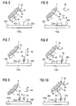
- Figure 5 depicts a section of the situation on the surface 201 of the sensor field 103a.
- Capture molecules which are oligonucleotides 203 are immobilized on the sensor field 103a. Like the oligonucleotides 3a, they are directed to the DNA molecule 5a, albeit to a different subsequence.
- the oligonucleotide 203 has a subsequence which is complementary to the DNA molecule 5a. This subsequence, however, is longer than the complementary subsequence of the DNA molecule 5a and of the oligonucleotide 3a.
- the DNA molecule 5a bound to the magnetic bead 1 is incubated with the surface 201 of the sensor field 103a at a temperature T 1 which is below the melting temperature of the hybrid of the DNA molecule 5a and the oligonucleotide 3a. This hybrid thus will remain stable at the present temperature T 1 .
- the DNA molecule 5a tends to form a hybrid with the oligonucleotide 203 at the temperature T 1 .
- the melting temperature of the hybrids formed thereby is above the melting temperature of the hybrid of the DNA molecule 5a and the oligonucleotide 3a.
- Figure 6 depicts the situation on the surface 201 of the sensor field 103a after splitting of the complexes of the magnetic bead 1 and the DNA molecule 5a and 5b, respectively.
- Splitting of the complexes has been achieved by increasing the temperature to a temperature T 2 .
- the chosen temperature T 2 is above the melting temperature of the hybrids of the DNA molecules 5a and 5b and the oligonucleotides 3a and 3b, respectively.
- Increasing the temperature to the temperature T 2 results in the hybrids being dissolved, so that the magnetic bead 1 with the oligonucleotides 3a immobilized thereto is dissolved from the DNA molecule 5a.
- the temperature T 2 is below the melting temperature of the hybrids of the DNA molecule 5a and the oligonucleotide 203, and said hybrid is therefore stable at the chosen temperature T 2 .
- Figure 7 depicts the surface 201 after hybridization of the DNA molecule 5a with the oligonucleotide 203. The linkages of the DNA molecule 5a to the oligonucleotide 203 are symbolized by four connecting lines.
- the DNA molecule 5b which was likewise immobilized on the magnetic bead 1 does not find any binding partner in the oligonucleotides 203 on the surface 201 of the sensor field 103a, resulting in no linkage in this case.
- DNA molecule 5b If the DNA molecule 5b is intended to be detected likewise, corresponding oligonucleotides must be immobilized on a separate sensor field (for example on the sensor field 103b), where the DNA molecule 5b can then be bound by a complementary subsequence of the oligonucleotides immobilized there. Accordingly, the DNA molecule 5a does not find a binding partner in the sensor field 103b.
- the magnetic beads 1 will be free in solution again, so that the surface 201 of the sensor field 103a is no longer occupied with magnetic beads 1. This situation corresponds to that of figure 3 , apart from the previously hybridized DNA molecule 5a. Interference with the subsequent detection reaction of the DNA molecule 5a is therefore ruled out.
- Figure 8 depicts this situation.
- the DNA molecule 5a which has been moved to the surface 201 of the sensor field 103a in the previous procedure is still linked to one of the oligonucleotides 203 and therefore immobilized on the surface 201.
- a DNA molecule 5a' which is identical to the DNA molecule 5a is now again bound to the oligonucleotide 3a and can now hybridize with another one of the oligonucleotides 203.
- this process is efficient because the DNA molecule 5a' is concentrated close to the surface 201.
- the temperature is again raised to a temperature T 2 above the melting temperature of the hybrids of the DNA molecule 5a' and the oligonucleotide 3a, and as a result the magnetic beads detach from the DNA molecule 5a.
- T 2 a temperature above the melting temperature of the hybrids of the DNA molecule 5a' and the oligonucleotide 3a
- FIG 11 depicts an alternative exemplary embodiment of the invention.
- Two magnetic beads, 501a and 501b are depicted which in each case carry one species of oligonucleotides 3a and 3b.
- the oligonucleotide 3a is directed to a DNA molecule 5a which is present in the solution.
- the oligonucleotide 3b is directed to a DNA molecule which is not present in the solution.
- further oligonucleotides 503a and 503b are immobilized on the surfaces of the magnetic beads 501a and 501b.
- Figure 13 illustrates the function of the oligonucleotides 503a and 503b.
- Figure 12 depicts the situation after the magnetic beads 501a and 501b have been incubated with the solution containing the present DNA molecules 5a. Hybrids of the DNA molecules 5a and the oligonucleotide 3a have formed. Owing to the absence of the binding partner of corresponding oligonucleotides, no hybridization has occurred on the magnetic bead 501b.
- the magnetic beads 501a and 501b are subsequently moved by means of a magnetic field to a surface of a biochip and held there. This is depicted in figure 13 .
- the biochip 505 comprises two sensor fields, 507a and 507b.
- An oligonucleotide 509a which has a nucleic acid sequence which is partially complementary to the oligonucleotide 503a is immobilized on the sensor field 507a.
- an oligonucleotide 509b which has a nucleic acid sequence which is partially complementary to the oligonucleotide 503b is immobilized on the sensor field 507b.
- concentrating the magnetic beads 501a and 501b and therefore the oligonucleotides 503a and 503b supports hybridization of said oligonucleotides 503a and 503b with the oligonucleotides 509a and 509b on the sensor fields 507a and 507b.
- the magnetic field is switched off in order to remove non-immobilized magnetic beads 501a and 501b from the surface.
- the labeled DNA molecules 5a are bound merely by immobilization by way of the hybrid of oligonucleotides 503a and 509a above the sensor field 507a.
- Immobilization merely by the magnetic field above the sensor 507b is therefore ruled out.
- the DNA molecule 5a can then be detected by way of a label 511 at the site of the sensor field 507a by means of known detection processes.
- Assignment of the DNA molecule 5a to the sensor field 507a is unambiguous by way of specific binding of the DNA molecule 5a to the oligonucleotide 3a on the magnetic bead 501a and hybridization thereof via the oligonucleotide 503a with the oligonucleotide 509a at the sensor field 507a. This requires the removal of non-immobilized specimen of the magnetic beads 501a and 501b at the sensor surfaces. Because the supplied sample did not contain a binding partner for the oligonucleotide 3b, a DNA molecule will not be detected at the sensor field 507b, and the assay is therefore correspondingly negative.
- this process can also be repeated cyclically, thereby enabling more species of the magnetic bead 501a to get close to the sensor surface 507a and immobilize there. This contributes to increasing the efficiency of concentration, in particular if few copies of the DNA molecule 5a are present.
- the process described can be used universally because immobilization of the magnetic beads 501a and 501b is unspecific with regard to the DNA molecule to be detected, 5a. Detection specificity is achieved merely by forming the magnetic beads 501a and 501b and the oligonucleotides 3a and 3b immobilized thereon.
- the corresponding biochip 505 with the different sensor fields 507a and 507b is therefore suitable for detecting any kind of DNA molecules. Detection merely requires differently designed magnetic beads which can bind the DNA molecule to be detected.
- the exemplary embodiments described have been limited by way of example to detection of a DNA molecule. They can, however, be used in a similar manner for any nucleic acid molecules or other biomolecules which can be bound specifically. Thus it is likewise possible to immobilize antibodies which are directed to proteins present in the sample for detection on the magnetic beads of the described processes.
- the magnetic beads can correspondingly be immobilized here optionally via the proteins themselves (exemplary embodiment 1) or via another protein with antibody or even a nucleic acid (exemplary embodiment 2).
Abstract
Process for concentrating nucleic acid molecules to be detected of a sample on a surface, with capture molecules that specifically bind a capture probe being immobilized on said surface, which process comprises the following steps:
- providing a support material which has the capture probe and can specifically be linked to the nucleic acid molecules to be detected to give complexes which comprise said support material and said nucleic acid molecules,
- incubating the support material with the sample and forming the complexes,
- moving the complexes to the surface,
- binding at least one portion of the complexes to the capture molecules by means of the capture probe.
Description
- The present invention relates to a process for concentrating nucleic acid molecules to be detected of a sample on a surface.
- Nucleic acid diagnostics increasingly makes use of "biochips". They are used, for example, for detecting various types and species of nucleic acids, which may be DNA, RNA, cDNA or other nucleic acids. The biochip-based analytical methods usually aim at detecting special nucleic acids in a sample. Thus, for example, a patient's DNA may be examined for the presence of a particular sequence which indicates a predisposition to a disorder. It is likewise possible to detect pathogens such as viruses and bacteria (e.g. HIV, HPV, HCV) in a patient's blood sample by demonstrating the presence of their DNA or RNA in said sample. These analyses have the advantage of increased accuracy over classical immunoassays, since the pathogen is detected directly rather than via antibodies produced against it.
- The biochips used are based in their simplest form on a glass substrate on which capture molecules (e.g. oligonucleotides) are immobilized which can specifically bind to the nucleic acid to be detected. These short nucleic acid fragments which are frequently produced synthetically are, with respect to their sequence, at least partially complementary to the sequence of the nucleic acid to be detected, resulting in highly specific binding. Binding of nucleic acids other than the one to be detected must be avoided in any case in order not to obtain a false-positive result. In many methods, actual detection of the nucleic acid takes place by way of fluorescence processes in which a fluorescent dye is attached to the nucleic acid to be detected, for example via biotin-streptavidin binding. After specific hybridization of the nucleic acid with the capture molecules, the biochip is rinsed to remove unbound material. Consequently, the solution does no longer contain any fluorescent dyes. By exciting the dyes, fluorescence can be observed using a CCD camera, making detection possible.
- One advantage of biochips is multiplex detection capability. Thus it is possible to immobilize on various positions of the biochip various kinds of capture molecules targeting various nucleic acids to be detected. Detection can then be carried out by way of space-resolved fluorescence measurement. It is therefore also possible to carry out a plurality of detections of nucleic acids in a single process.
- The nucleic acids are copied (amplified) prior to detection, since the number of nucleic acid copies present is usually not sufficient for making direct detection possible. Said amplification may be carried out, for example, by means of a polymerase chain reaction (PCR). PCR is based on replicating nucleic acids with the aid of thermostable DNA polymerases. This involves contacting a pair of oligonucleotide primers (single-stranded oligonucleotides) with the nucleic acid to be amplified. The primers are chosen so as to bind at both ends of a fragment to be amplified on the complementary strands. During elongation the primers are then elongated in the 3' direction along the particular target nucleic acid strand (forward and reverse primers). Forward and reverse primers are alternatively also referred to as sense or antisense primers. In this way it is possible to amplify the piece located between the sites on the target nucleic acid that are complementary to the primers. For subsequent detection reactions, the PCR products are advantageously removed from primers, nucleotides and other interfering components of the PCR mixture.
- The PCR process comprises a plurality of thermocycles, each of which comprises three steps: the sample is first heated (e.g. to 94°C) in order to separate the strands of the double-stranded target DNA present in the sample (denaturation).
- This is followed by lowering the temperature (e.g. to 45-60°C) for the primers to be able to attach to the complementary regions of the now single-stranded DNA (annealing). In the last step, the primers bound to the single strand are extended by DNA polymerase in the 3' direction according to the information of the DNA template strand, with the corresponding nucleoside triphosphates in the solution being used as building blocks (elongation, e.g. at 72°C). This cycle is typically run approx. 15-50 times during a PCR. The above temperatures are given merely by way of example and should be adjusted to the PCR to be carried out specifically in each case.
- It is thus possible to prepare from a few specimens present of a DNA (or generally of a nucleic acid) a multiplicity of copies within a very short time whose concentration is sufficient for subsequent qualitative detection of said DNA or nucleic acid in the sample. For example, 20 PCR cycles (typically taking 20 to 40 minutes) produce theoretically 220 times, i.e. about 106 times the amount of the nucleic acid originally used. At the same time, PCR enables a label which makes detection possible to be incorporated into the resulting PCR product. Thus it is possible to use, for example, biotin-labeled PCR primers or nucleoside triphosphates, resulting in the synthesized PCR products being biotinylated. After the PCR product has attached to the immobilized capture molecules, biotin is, as a result, likewise immobilized on the biochip at the corresponding position. In a further step streptavidin-linked fluorescent dyes can then be bound to said biotin, which allow the nucleic acid or its PCR product to be detected. Other detection systems, based on electrochemical detection for example, can be used as an alternative to the fluorescent dye.
- A central point in biochip-based detection methods is hybridization, i.e. binding of the nucleic acid to be detected (this term is intended herein to be synonymous with the PCR products produced from the nucleic acids). Sensitivity of nucleic acid detection depends on hybridization efficiency. The nucleic acid is usually incubated with the biochip, thereby enabling hybridization to take place. The latter can be improved by choosing a suitable temperature and buffer medium. It is also important that the nucleic acids are moved to the biochip surface occupied with capture molecules for hybridization to be able to take place. It is therefore not optimal, if movement of the nucleic acid is caused merely by thermal diffusion. Thus, for example, the rate constant is only about 8 × 10⁻⁸ cm2/s for a 25mer oligonucleotide ("Observation of hybridization and dehybridization of Thiol-tethered DNA using two-color surface plasmon resonance spectroscopy" Peterlinz et al. (1997), J. Am. Chem. Soc. 119 3401-3402), resulting merely in an average migration of 0.096 cm within a hybridization time of sixteen hours. Thus, assuming that the surface is 4 cm2 in size, passive diffusion alone renders only about 1.5% of the sample nucleic acid accessible to hybridization to the capture molecules immobilized on the biochip. A number of active processes can increase hybridization efficiency.
- Thus, C.F. Erdmann et al. disclose in "Electric field directed nucleic acid hybridization on microchips", Nucleic Acids Research (1997) 25, 4907-4914, nucleic acids being moved electrophoretically to the biochip by applying an electric field. Hybridization is rendered substantially faster by concentrating the nucleic acid close to the surface of the biochip than by thermal movement of the nucleic acids alone. It is moreover possible to remove non-hybridized nucleic acids from the biochip surface by reversing the electric potential. The method described requires the electrodes to be specially prepared with a protective layer in order to prevent free radicals and changes in pH occurring during the electrode reaction from damaging the concentrated nucleic acids.
M.J. Heller et al. disclose a comparable method in "An Active Microelectronics Device for Multiplex DNA Analysis", IEEE Engineering in Medicine and Biology (1996) March/April, 100-104. - A multiplicity of methods in biochemistry and medical diagnostics now make use of magnetic, polymeric support materials, in particular polymeric particles, for simplifying the removal of cells, proteins and nucleic acids. Compared with conventional separation methods, using magnetic support materials is advantageous in that the loaded support materials can readily and rapidly be removed from the other components of a sample with the aid of magnetic forces. Magnetic bead-shaped or spherical polymeric particles based on polyvinyl alcohol with a narrow particle size distribution within a range of less than 10 µm have proved particularly suitable for such separation methods (
WO 97104862 - It is also known that particular biological materials, in particular nucleic acids and proteins, can be isolated from their natural environment only with increased efforts. This is especially due to the fact that mechanical, chemical and biological cell lysis processes must be utilized for isolating the nucleic acids and proteins from the nucleus or the cell membrane or organelles. In addition, the corresponding biological samples usually comprise further, solid and/or dissolved compounds such as other proteins and components of the cytoskeleton, which can impair isolation. An additional difficulty is the fact that very often only small concentrations of the nucleic acids or proteins are present in the biological sample to be studied.
- In order to be able nevertheless to utilize the advantages of using magnetic particles for isolating nucleic acids from biological samples, it has been proposed inter alia to isolate nucleic acids with the aid of magnetic particles having a glass surface which is essentially pore-free (
WO 96141811 - Known diagnostic processes, for example from nucleic acid and protein diagnostics, usually require a multiplicity of manual operations in order to arrive at an analytical result. This requires inter alia separation of the components to be detected from the rest of the sample. Known examples of separation methods are filtration, centrifugation, chromatography and extraction. All of these are chemical and physical separation processes which are usually not suitable for sequence-specific isolation of DNA or proteins from the sample. Use is made, for example, of resins whose surfaces are functionalized so as to be able to bind DNA or proteins. These target molecules are purified by binding to the solid phase of the resin, followed by a plurality of washing steps and subsequent detachment of the target molecule from the solid phase under suitable buffer conditions. The target molecule must be bound tightly, while contaminating components of the sample are dissolved in a different, liquid phase. After various washing procedures, the target molecule must then be detached again from the solid phase, for example by changing the liquid phase. The repeated change of medium is firstly very costly with regard to material, and secondly product yields fluctuate with each additional process step, making quantitative calibration difficult. In particular in integrated analytical processes, for example lab-on-a-chip systems in which the samples are prepared and analyzed essentially automatically, checking the individual process steps is often not possible, and as a result deviations in the individual process steps amplify each other and may lead to large deviations in the analytical result.
- Individual steps can be simplified or even automated completely by means of the described support particles, also referred to as magnetic beads. Said magnetic beads are provided with affinity ligands or other surface modifications and are therefore suitable for binding particular biomolecules, for example DNA, from a solution to their surface. A purification process typically involves adding a suspension of magnetic beads to the sample to be separated in a test tube. After an incubation time of several minutes to enable the affinity ligand to bind to the desired biological molecule, a magnetic field is applied, which removes the particles by accumulating them on one tube wall. The supernatant is discarded and the particles are then washed at least once. For this purpose, the magnetic field is removed first and the particles are suspended in a fresh buffer solution which contains mostly chaotropic salts which prevent the biomolecules from detaching from the support material. The magnetic beads are then deposited on the vessel wall by reapplying the magnetic field. Thus it is possible, after several washing steps, to eluate the molecules by means of a low salt buffer solution which removes the bound biomolecules from the magnetic beads, in a solution which is free of interfering components, in contrast to the crude extract. The magnetic beads are deposited again on the vessel wall, making available the biomolecules in the supernatant solution. A disadvantage of the process described is the large amount of liquid required in each case, in the range of several hundred microliters for each individual process step.
- Eukaryotic or prokaryotic cells or viruses are known to be isolated, by way of example, by coupling specific antibodies to a fluorescent marker or magnetic beads. The antibody is usually monoclonal and directed to specific binding sites, for example to a surface receptor molecule of a corresponding antigen of the cell or the virus. The desired cells or viruses are labeled by coupling the antibodies to the particular binding site and sorted, for example, by means of FACS or a permanent magnet. The sorting process can be carried out firstly by way of "positive selection", involving further processing of the labeled cells or viruses. Secondly, a "negative selection" may be carried out, involving removal of the labeled cells and further processing of the remaining cells. Both methods enable the cells or viruses to be quantified and, as a result, the amounts of reagents required for said further processing to be calculated.
-
DE 101 11 520 B4 - Magnetic beads are not only suitable for purifying samples but can also be used for other purposes. Thus,
US 2004/0219066 A1 describes a device, by means of which various particles can be sorted. Said particles are bound to different magnetic beads having different magnetic momentums. A magnetic field gradient which moves the magnetic beads, due to their different magnetic momentums, into different collecting boxes is generated in a process chamber. The various particles can thus be distinguished by the differently designed magnetic beads. -
WO 00/47983 - Moreover, paramagnetic magnetic beads are known to be used for detecting DNA. Here, capture molecules complementary to the DNA to be detected are located on a magnetorestrictive sensor. If the sample studied contains the DNA to be detected, hybridization between said DNA to be detected and the capture molecules takes place. The hybridized DNA has been or is labeled with biotin to which streptavidin-coated magnetic beads couple. The biotin label is usually introduced into the DNA to be detected by means of an upstream PCR utilizing biotin-labeled primers or nucleotides. After coupling to the paramagnetic beads, the latter are magnetized by an applied magnetic field and their stray field is measured by the magnetoresistive sensor. This results indirectly in quantitative detection of the DNA in the sample.
-
DE 41 27 657 andWO 97104862 - The polyvinyl alcohol particles are prepared by adding particular emulsifier mixtures to the oil phase of the water-in-oil emulsion. Suitable emulsifiers which are added as additives to the oil phase are propylene oxide-ethylene oxide block copolymers, sorbitan fatty esters, mixed complex esters of pentaerythrite fatty esters with citric acid, polyethylene glycol-castor oil derivatives, block copolymers of castor oil derivatives, polyethylene glycols, modified polyesters, polyoxyethylene sorbitan fatty esters, polyoxyethylene-polyoxypropylene-ethylenediamine block copolymers, polyglyceryl derivatives, polyoxyethylene alcohol derivatives, alkylphenyl-polyethylene glycol derivatives, polyhydroxy fatty acid-polyethylene glycol block copolymers, polyethylene glycol ether derivatives. Substances of this kind are commercially known inter alia under the trade name: Pluronic®, Synperonic®, Tetronic®, Triton®, Arlacel®, Span®, Tween®, BrijOR, ReneXOR, Hyperme®, Lameform®, Dehymuls® or Eumulgin®.
- To obtain uniform, bead-shaped polymeric particles, preferably with particle sizes of 0.5-10 µm, a mixture of at least two, preferably three to four, of said surfactants is added to the oil phase. Preference is given to mixing a lipophilic emulsifier component with at least one emulsifier which has semihydrophilic properties, i.e. which is soluble in both water and oil. Examples of emulsifiers which meet the latter properties are: ethylene oxide-propylene oxide block copolymer derivatives with a predominant ethylene oxide proportion, polyethylene glycol hexadecyl ethers, shorter-chain polyoxyethylene sorbitan fatty esters, polyethylene glycols or shorter-chain sorbitan fatty esters. The concentration of the emulsifiers in the oil phase is usually 2-6% by volume, preferably 3.5-5.0% by volume. Advantageous with respect to fineness and narrow particle size distribution of polymer droplets are those emulsifier mixtures which comprise at least two lipophilic components and one semihydrophilic emulsifier. The concentration of the semihydrophilic emulsifier is usually between 15 and 30% by volume, based on the total amount of emulsifier. In addition to fineness of the particles, said particles show a bead-like shape.
- Apart from the emulsifiers for the oil phase, special surfactants which are soluble in the aqueous polymer phase also contribute to improving the quality of the emulsion, especially of polyvinyl alcohol solutions with low molecular weight (Mowiol, Clariant GmbH, Frankfurt am Main, DE). In addition, the magnetic colloids added in solid form are successfully finely dispersed by adding ionic emulsifiers. Examples of such emulsifiers which can also be used as binary mixtures are: serum albumin, gelatin, aliphatic and aromatic sulfonic acid derivatives, polyethylene glycols, poly N-vinylpyrrolidone or cellulose acetate butyrate. The amounts of emulsifiers used are usually 0.01-2% by weight, based on the polymer phase, with the concentration of the ionic emulsifiers always being between 0.01 and 0.05% by weight. The skilled worker is familiar with influences of stirrer speeds and of concentrations and viscosities of the two phases on particle size. To obtain the preferred particle sizes of 0.5-10 µm, stirrer speeds of 1500-2000 revolutions per minute are required, with conventional two blade propeller stirrers being used.
- In principle, those ferro- or superparamagnetic colloids which have an appropriate particle size and normally a magnetic saturation of from 50 to 400 Gauss may be used as magnetic particles which are encapsulated into the polyvinyl alcohol matrix during the process. Another requirement to be met by the magnetic particles is dispersibility in the aqueous polymer phase containing the polyvinyl alcohol. During subsequent emulsion in the organic phase, the magnetic colloids are then simultaneously enclosed in the polymer droplets.
- Suitable magnetic colloids are preferably magnetites with particle sizes of 10-200 nm. Such substances can be obtained commercially, for example, under the trade name Bayferrox or Ferrofluidics. Since preparing such colloids is general prior art, the magnetic particles may also be prepared by the disclosed processes, as described, for example, by Shinkai et al., Biocatalysis, Vol. 5, 1991, 61, or Kondo et al., Appl. Microbiol. Biotechnol., Vol. 41, 1994, 99. The concentrations of the colloids in the polymer phase are, in each case based on this phase, usually between 4 and 14% by volume for colloids which are already aqueous colloids due to their preparation, and 0.3-2% by weight for solid substances. Preparation involves admixing the magnetic colloids directly with the polymer phase. In order to ensure a finely dispersed, even distribution of the particles, brief mixing of the aqueous dispersion by means of a high revolution dispersing tool (Ultra-Turrax) with subsequent ultrasound treatment is beneficial. The polymer phase required for preparing the magnetic particles usually consists of a 2.5-10% by weight polyvinyl alcohol solution.
- The magnetic polyvinyl alcohol support material can then be obtained from the suspension by methods known per se to the skilled worker, for example by filtration and washing.
- A known process for functionalization comprises equipping the support material with affinity ligands on the surface. This usually requires attaching chemically reactive groups on said surface, to which said affinity ligands are then bound. These groups may be, for example, tosyl, hydroxyl, aldehyde or carboxyl, amino, thiol or epoxy groups. They may generally be provided by treating uncoated monodisperse superparamagnetic particles in order to provide them with a surface layer of a polymer carrying such a functional group, for example a cellulose derivative or a polyurethane together with a polyglycol for providing hydroxyl groups, a polymer or copolymer of acrylic acid or methacrylic acid for providing carboxyl groups or an amino-alkylated polymer for providing amino groups.
US 4,654,267 discloses a plurality of surface coatings. -
DE 100 13 995 A1 discloses magnetic support materials based on polyvinyl alcohol, whose surface is at least partially silanized and, where appropriate, equipped with biomolecule-coupling affinity ligands. The described support materials may be designed as filter, membrane or particle. Preference is given to the magnetic support material being bead-shaped or spherical particles, said particles having a particle size preferably of from 0.2 to 50 µm, particularly preferably from 0.5 to 5 µm. Aside from the preferably bead-shaped and spherical design of the particles, their particle size distribution should be within as narrow a range as possible. The support materials are preferably prepared in particle form by reacting the polyvinyl alcohol support material with an organic silane compound. The silanized particles are then reacted with affinity ligands. - Affinity ligands which may be coupled are in principle any ligands used in affinity chromatography. Examples of these are: protein A, protein G, protein L, streptavidin, biotin, heparin, antibodies, serum albumin, gelatin, lysine, concanavaline A, oligosaccharides, oligonucleotides, polynucleotides, protein-binding metal ions, lectins, aptamers or enzymes. The special fractionations which can be carried out using such affinity matrices are general prior art.
- The described advantages of the magnetic beads can also be used for concentrating nucleic acids on biochips, as disclosed by
US 2002/0166764 A1 . The device described there comprises a biochip which is arranged in a chamber. A liquid flow can be adjusted through the chamber. Outside the chamber, there is a magnetic field generator which enables magnetic beads inside the chamber to be moved to the surface of the biochip. The nucleic acids are specifically linked to the magnetic beads before introduction into the chamber. Said linkage is provided by oligonucleotides on the magnetic beads. The magnetic beads, and therefore also the nucleic acids, are then moved in the chamber by means of the magnetic field generator to the surface of the biochip and held there. The presence of the nucleic acids on the magnetic beads is detected by a redox cycling process. The described process is not capable of multiplexing, since the magnetic beads cannot be addressed to individual positions, as required in microarrays. The magnetic beads are held unspecifically close to the surface of the biochip. -
WO 98/4584 - It is the object of the present invention to provide an improved process for concentrating nucleic acids on biochips.
- This object is achieved by processes with process steps of
claims 1, 8 and 21. -
Claim 1 proposes a process for concentrating nucleic acid molecules to be detected of a sample on a surface, with capture molecules that specifically bind a capture probe being immobilized on said surface. The process comprises the following steps: - providing a support material which has the capture probe and can specifically be linked to the nucleic acid molecules to be detected to give complexes which comprise said support material and said nucleic acid molecules,
- incubating the support material with the sample and forming the complexes,
- moving the complexes to the surface,
- binding at least one portion of the complexes to the capture molecules by means of the capture probe.
- The process has the advantage of using the capture probe and linking it with the immobilized capture molecules on the surface of the biochip enabling the magnetic beads to be efficiently linked to the capture molecules. As a result, the nucleic acid to be detected is immobilized close to the surface and can be detected by known processes. In addition, the biochip is not specific for nucleic acids and can therefore be used universally. Specificity is caused only by the design of the support material which firstly comprises the sample-independent capture probe for selective (generically selective (addressable)) binding to the surface and which secondly is capable of binding specifically the nucleic acid to be detected. Thus it is possible in sample preparation to mix a nucleic acid to be detected with appropriate designed support materials and to incubate it with the biochip. The biochip itself can be mass-produced always in an identical manner and used without modification for any detection of various nucleic acids. While nucleic acids form the generically active part (which binds to the capture molecules immobilized on the chip surface) of the support material, the target molecule-specific part may comprise nucleic acids, proteins or aptamers.
- The process can be parallelized in a simple manner, i.e. is capable of multiplexing. This requires a plurality of types of support materials with various capture probes which can in each case be linked to different nucleic acids to be detected to give various complexes. Said various nucleic acids can therefore be arrested at various sites of the biochip by capture molecules which are immobilized over various sites of the biochip and which are directed against said various capture probes. Known detection processes enable the nucleic acids to be detected in a space-resolved manner.
- In an advantageous embodiment of the invention, unbound complexes are moved away from the surface and then moved again to the surface. This is followed by binding of at least one further portion of the complexes to the capture molecules. This addresses the problem of the support material with the immobilized nucleic acids usually being distributed evenly on the surface of the biochip, due to moving by means of the magnetic field. It is thus possible, in particular in multiplex processes, for nucleic acids at measurement locations (spots) of the biochip, at which they do not find any immobilizing partner in the form of capture molecules, to be redistributed. The dimensions of a spot are usually small compared to the entire surface of the biochip. By repeating the process steps several times, hybridization efficiency can be increased, since the complexes which have not yet hybridized are again moved to the surface and partly bound there.
- In an advantageous development of the process, the support material has a support material surface on which the capture probes and transfer molecules which bind the nucleic acid molecules are immobilized. A support material of this kind can readily be produced by the processes described previously. Highly specific binding of only the selected nucleic acid for each species of the support material provides a high degree of error reliability.
- In an advantageous development of the process, the capture molecules, the capture probe and the transfer molecules are nucleic acids. In this way it is possible to generate in a simple manner highly specific binding events required by the process.
- Claim 8 provides an alternative solution to the problem. Claim 8 claims a process for concentrating nucleic acid molecules to be detected of a sample on a surface, with capture molecules that specifically bind the nucleic acid molecule being immobilized on said surface, which process comprises the following steps:
- providing a support material which can specifically be linked to the nucleic acid molecules to be detected to give complexes which comprise said support material and said nucleic acid molecules,
- incubating the support material with the sample and forming the complexes,
- moving the complexes to the surface,
- separating the nucleic acid molecules from the support material, and
- binding at least one portion of the nucleic acid molecules to the capture molecules.
- A particular advantage of this process is the fact that said portion of the nucleic acid molecules is bound to the surface with high efficiency, since moving the complexes to the surface results in their concentration. Moreover, in comparison with the process described in
claim 1, the additional capture probe can be dispensed with. However, the abovementioned advantages over known processes are identical. The specificity of the detection process in this embodiment is in the biochip itself due to the capture molecules immobilized on the surface. - In an advantageous embodiment of the process, the nucleic acid molecules to be detected are separated from the support material and the latter is removed from the surface. This can prevent a possible influence of the support material on subsequent process steps such as, for example, detection of the nucleic acid. The nucleic acid itself here remains immobilized to the capture molecule on the surface of the biochip.
- In advantageous embodiments of the process, the support material is in the form of magnetic beads whose surface has transfer molecules for binding the nucleic acid. It is also possible for the chip-bound capture molecules and the transfer molecules to be in the form of nucleic acids.
- In an advantageous embodiment, the process further comprises the following steps:
- the capture molecules in each case comprise a nucleic acid sequence which is partially complementary to the nucleic acid sequence of the nucleic acid molecule to be detected,
- the transfer molecules comprise in each case a nucleic acid sequence which is partially complementary to the nucleic acid sequence of the nucleic acid molecule to be detected, and
- the nucleic acid sequences of capture molecules and transfer molecules, which are complementary to the nucleic acid molecules to be detected, are chosen in such a way that the melting temperature of a nucleic acid molecule-transfer molecule hybrid is lower than the melting temperature of a nucleic acid molecule-capture molecule hybrid.
- This embodiment of the capture and transfer molecules provides a simple possibility of separating the complex. The partially complementary nucleic acid sequences are usually contiguous. Different melting temperatures of the hybrids can be achieved in a simple manner via the length of the complementary nucleic acid sequences. This ensures that when the linkage between transfer molecule and nucleic acid is dissolved, the linkage between the nucleic acid molecule and the capture molecule remains intact and the nucleic acid molecule is still immobilized. The support material can thus be removed in a simple manner. In general, any modifications of stringency conditions can be used for separation. Thus it is possible, for example, to modify the stringency conditions by changing the salt concentration or the formamide content.
- Accordingly, an advantageous embodiment of the process comprises incubating under stringency conditions which are below melting (denaturation) of the nucleic acid molecule-transfer molecule hybrids. The complexes are moved to the surface under stringency conditions which are below melting of the nucleic acid molecule-transfer molecule hybrids.
- An advantageous embodiment of the process comprises incubating at a first temperature which is below the melting temperature of the nucleic acid molecule-transfer molecule hybrids. The complexes are moved to the surface at a second temperature which is below the melting temperature of the nucleic acid molecule-transfer molecule hybrids. After binding of the complexes to the capture molecules, the temperature is increased to a third temperature which is between the melting temperatures of the nucleic acid molecule-transfer molecule hybrids and the nucleic acid molecule-capture molecule hybrids. This ensures that the linkage between the nucleic acid and the capture molecules remains intact, while the linkage between the nucleic acid molecule and the transfer molecule is severed.
- The described process and the advantageous developments can readily be parallelized. It is merely necessary to equip various sites of the biochip with various capture molecules which are partially complementary to various nucleic acids to be detected. It is also necessary to provide various support materials having different transfer molecules which then transport said various nucleic acids to the surface. Depending on the nucleic acids to be detected it is alternatively also possible to use magnetic beads of the same kind, all of which are capable of binding any of the nucleic acids to be detected. This is possible because immobilization to the surface of the biochip is highly specific due to the capture molecules located there.
- Claim 21 provides another solution to the object. Said claim claims a process for concentrating different nucleic acid molecules to be detected of a sample on a surface, with capture molecules that specifically bind said nucleic acid molecules being immobilized in groups and at different sites on the surface (201), which process comprises the following steps:
- providing a support material which can specifically be linked to the nucleic acid molecules to be detected to give complexes which comprise said support material and said nucleic acid molecules,
- incubating the support material with the sample and forming the complexes,
- moving the complexes to the surface,
- separating the nucleic acid molecules from the support material, and
- binding at least one portion of the nucleic acid molecules to the capture molecules.
- This multiplex process can be implemented particularly readily, since only one type of support materials with binding properties for the different nucleic acids is required.
- Otherwise the same advantageous developments of the process, as described above, can be implemented.
- Further advantages and developments of the processes arise from the exemplary embodiments described hereinbelow and illustrated on the basis of the accompanying drawings in which:
-
Figures 1 to 10 depict various process steps of a hybridization process, and -
Figures 11 to 13 depict various process steps of an alternative hybridization process. - The exemplary embodiments described hereinbelow refer to processes for concentrating DNA molecules. They can be applied in principle in the same manner to any nucleic acids. It is likewise possible to concentrate other biomolecules for which specific binding partners are available by means of the processes described. The processes are depicted by way of example for a few or only one specimen of nucleic acids. Usually a large number of specimens is used in order to enable the nucleic acids to be detected subsequently.
-
Figure 1 depicts diagrammatically amagnetic bead 1 which has three different oligonucleotides, 3a, 3b, 3c, as transfer molecules. Two different DNA molecules to be detected, 5a and 5b, are likewise depicted. Theoligonucleotides DNA molecules DNA molecules biotin molecule 7. The situation infigure 1 represents the start of an incubation phase of the process. - The
DNA molecules DNA molecules - In
figure 2 , incubation of themagnetic bead 1 with theDNA molecules DNA molecules oligonucleotides magnetic bead 1. Binding of theDNA molecule 5a with theoligonucleotide 3a is indicated symbolically by two connecting lines. Said connecting lines symbolize an overlapping complementary nucleic acid sequence of theDNA molecule 5a and theoligonucleotide 3a. Binding between theDNA molecule 5b and theoligonucleotide 3b is depicted in a similar manner. -
Figure 3 depicts diagrammatically the surface of abiochip 101. Thebiochip 101 comprises three sensor fields, 103a, 103b and 103c, which are equipped for detecting various DNA molecules. Amagnetic field generator 105 is provided for (depicted only diagrammatically here). It may be designed, for example, as a movable permanent magnet or as a coil. Thebiochip 101 is located in a reaction chamber (not shown) which is filled with a solution. Said solution comprises the incubatedmagnetic beads 1 in a concentration C1=n1/V1, where n1 is the number ofmagnetic beads 1 and V1 is the volume of the solution. -
Figure 4 depicts thebiochip 101 after applying a magnetic field through themagnetic field generator 105. Themagnetic beads 1 have been moved through the magnetic field to the surface of thebiochip 101 and are present there in a concentration C2=n1/V2, wherein V2 is the volume above thebiochip 101 in which themagnetic beads 1 are now concentrated. The concentration C2 is higher than C1 because V2 « V1. Thus themagnetic beads 1 have been concentrated close to the surface of thebiochip 101. -
Figure 5 depicts a section of the situation on thesurface 201 of thesensor field 103a. Capture molecules which areoligonucleotides 203 are immobilized on thesensor field 103a. Like theoligonucleotides 3a, they are directed to theDNA molecule 5a, albeit to a different subsequence. - Like the
oligonucleotide 3a, theoligonucleotide 203 has a subsequence which is complementary to theDNA molecule 5a. This subsequence, however, is longer than the complementary subsequence of theDNA molecule 5a and of theoligonucleotide 3a. TheDNA molecule 5a bound to themagnetic bead 1 is incubated with thesurface 201 of thesensor field 103a at a temperature T1 which is below the melting temperature of the hybrid of theDNA molecule 5a and theoligonucleotide 3a. This hybrid thus will remain stable at the present temperature T1. Since the complementary subsequences have different lengths, theDNA molecule 5a tends to form a hybrid with theoligonucleotide 203 at the temperature T1. The melting temperature of the hybrids formed thereby is above the melting temperature of the hybrid of theDNA molecule 5a and theoligonucleotide 3a. -
Figure 6 depicts the situation on thesurface 201 of thesensor field 103a after splitting of the complexes of themagnetic bead 1 and theDNA molecule - Splitting of the complexes has been achieved by increasing the temperature to a temperature T2. The chosen temperature T2 is above the melting temperature of the hybrids of the
DNA molecules oligonucleotides magnetic bead 1 with theoligonucleotides 3a immobilized thereto is dissolved from theDNA molecule 5a. - Increasing the temperature to the temperature T2 has likewise dissolved the hybrid between the
DNA molecule 5b and theoligonucleotide 3b, and as a result theDNA molecule 5b is again free in solution, independently of themagnetic bead 1. - The temperature T2 is below the melting temperature of the hybrids of the
DNA molecule 5a and theoligonucleotide 203, and said hybrid is therefore stable at the chosen temperature T2.Figure 7 depicts thesurface 201 after hybridization of theDNA molecule 5a with theoligonucleotide 203. The linkages of theDNA molecule 5a to theoligonucleotide 203 are symbolized by four connecting lines. TheDNA molecule 5b which was likewise immobilized on themagnetic bead 1 does not find any binding partner in theoligonucleotides 203 on thesurface 201 of thesensor field 103a, resulting in no linkage in this case. - If the
DNA molecule 5b is intended to be detected likewise, corresponding oligonucleotides must be immobilized on a separate sensor field (for example on the sensor field 103b), where theDNA molecule 5b can then be bound by a complementary subsequence of the oligonucleotides immobilized there. Accordingly, theDNA molecule 5a does not find a binding partner in the sensor field 103b. - After the magnetic field has been switched off, the
magnetic beads 1 will be free in solution again, so that thesurface 201 of thesensor field 103a is no longer occupied withmagnetic beads 1. This situation corresponds to that offigure 3 , apart from the previously hybridizedDNA molecule 5a. Interference with the subsequent detection reaction of theDNA molecule 5a is therefore ruled out. - It can be assumed that random uniform distribution of the
magnetic beads 1 on the entire surface of thebiochip 101 did not succeed in immobilizing enough specimen of theDNA molecule 5a to thesurface 201 of thesensor field 103a. According to uniform distribution, themagnetic beads 1 with theDNA molecules 5a are evenly distributed on the surface after the magnetic field has been switched on.Figure 7 demonstrates this by the fact that here theDNA molecule 5b is present above thesensor field 103a and not, as ultimately desired, at the sensor field 103b. Accordingly,magnetic beads 1 with further copies of theDNA molecule 5a will be present on the surfaces of other sensor fields of the biochip or in between. By repeating the abovementioned process steps, further specimen of theDNA molecule 5a can be moved to thesurface 201 of thesensor field 103a. At the temperature T2 and with the magnetic field switched off, the magnetic beads and theDNA molecules DNA molecule 5a andoligonucleotide 3a,DNA molecules magnetic beads 1. This process is efficient, since a large concentration ofmagnetic beads 1 has been added and therefore a sufficient number of hybridization partners for theDNA molecules DNA molecules magnetic beads 1 with theDNA molecules figure 4 . -
Figure 8 depicts this situation. TheDNA molecule 5a which has been moved to thesurface 201 of thesensor field 103a in the previous procedure is still linked to one of theoligonucleotides 203 and therefore immobilized on thesurface 201. On themagnetic bead 1, aDNA molecule 5a' which is identical to theDNA molecule 5a is now again bound to theoligonucleotide 3a and can now hybridize with another one of theoligonucleotides 203. As in the first process cycle, this process is efficient because theDNA molecule 5a' is concentrated close to thesurface 201. - After hybridization of the
DNA molecule 5a' with theoligonucleotide 203, the temperature is again raised to a temperature T2 above the melting temperature of the hybrids of theDNA molecule 5a' and theoligonucleotide 3a, and as a result the magnetic beads detach from theDNA molecule 5a. This is shown infigure 9 . The process described can be repeated any number of times so that concentration of theDNA molecules - It is likewise possible, in the situation in
figure 5 , that theDNA molecule 5a hybridizes with theoligonucleotide 203 before theDNA molecule 5a is separated from themagnetic bead 1. This is depicted infigure 10 . Increasing the temperature to the temperature T2 results in the same situation as infigure 7 . The hybrids of theDNA molecule 5a and theoligonucleotide 3a have melted, while the hybrid of theDNA molecule 5a and theoligonucleotide 203 is stable. A corresponding process has the following steps: - binding at least one portion of the nucleic acid molecules to the capture molecules
- providing a support material which can specifically be linked to the nucleic acid molecules to be detected to give complexes which comprise said support material and said nucleic acid molecules,
- incubating the support material with the sample and forming the complexes,
- moving the complexes to the surface, and
- binding at least one part of the complexes to the capture molecules.
The process described on the basis offigures 1 to 10 usesmagnetic beads 1 with in each case different oligonucleotides, 3a, 3b and 3c, on the surface. This has the advantage that only one species ofmagnetic beads 1 needs to be used, while it is still possible to carry out a multiplex process. In an alternative exemplary embodiment it is possible to immobilize in each case one species ofoligonucleotides DNA molecule magnetic bead 1, thereby generating different species ofmagnetic beads 1. This is firstly required, if only one species of DNA molecules is to be detected. Here too, however, the process can be parallelized by adding different species ofmagnetic beads 1 having on their surface in each case one type ofoligonucleotide magnetic beads 1 which are in each case coated with one type of oligonucleotides, 3a, 3b or 3c, and to mix them in a reaction mixture according to the DNA molecules to be detected, 5a or 5b. -
Figure 11 depicts an alternative exemplary embodiment of the invention. Two magnetic beads, 501a and 501b, are depicted which in each case carry one species ofoligonucleotides oligonucleotide 3a is directed to aDNA molecule 5a which is present in the solution. Theoligonucleotide 3b is directed to a DNA molecule which is not present in the solution. Moreover,further oligonucleotides magnetic beads Figure 13 illustrates the function of theoligonucleotides -
Figure 12 depicts the situation after themagnetic beads present DNA molecules 5a. Hybrids of theDNA molecules 5a and theoligonucleotide 3a have formed. Owing to the absence of the binding partner of corresponding oligonucleotides, no hybridization has occurred on themagnetic bead 501b. - The
magnetic beads figure 13 . Thebiochip 505 comprises two sensor fields, 507a and 507b. Anoligonucleotide 509a which has a nucleic acid sequence which is partially complementary to theoligonucleotide 503a is immobilized on thesensor field 507a. Correspondingly, anoligonucleotide 509b which has a nucleic acid sequence which is partially complementary to theoligonucleotide 503b is immobilized on thesensor field 507b. In a manner similar to the process described above, concentrating themagnetic beads oligonucleotides oligonucleotides oligonucleotides sensor fields magnetic beads DNA molecules 5a are bound merely by immobilization by way of the hybrid ofoligonucleotides sensor field 507a. Immobilization merely by the magnetic field above thesensor 507b is therefore ruled out. TheDNA molecule 5a can then be detected by way of alabel 511 at the site of thesensor field 507a by means of known detection processes. Assignment of theDNA molecule 5a to thesensor field 507a is unambiguous by way of specific binding of theDNA molecule 5a to theoligonucleotide 3a on themagnetic bead 501a and hybridization thereof via theoligonucleotide 503a with theoligonucleotide 509a at thesensor field 507a. This requires the removal of non-immobilized specimen of themagnetic beads oligonucleotide 3b, a DNA molecule will not be detected at thesensor field 507b, and the assay is therefore correspondingly negative. - In a manner similar to the abovementioned exemplary embodiment, this process can also be repeated cyclically, thereby enabling more species of the
magnetic bead 501a to get close to thesensor surface 507a and immobilize there. This contributes to increasing the efficiency of concentration, in particular if few copies of theDNA molecule 5a are present. - The process described can be used universally because immobilization of the
magnetic beads magnetic beads oligonucleotides biochip 505 with thedifferent sensor fields - The exemplary embodiments described have been limited by way of example to detection of a DNA molecule. They can, however, be used in a similar manner for any nucleic acid molecules or other biomolecules which can be bound specifically. Thus it is likewise possible to immobilize antibodies which are directed to proteins present in the sample for detection on the magnetic beads of the described processes. The magnetic beads can correspondingly be immobilized here optionally via the proteins themselves (exemplary embodiment 1) or via another protein with antibody or even a nucleic acid (exemplary embodiment 2).
Claims (33)
- A process for concentrating nucleic acid molecules to be detected of a sample on a surface (201), with capture molecules that specifically bind a capture probe being immobilized on said surface (201), which process comprises the following steps:- providing a support material which has the capture probe and can specifically be linked to the nucleic acid molecules to give complexes which comprise said support material and said nucleic acid molecules,- incubating the support material with the sample and forming the complexes,- moving the complexes to the surface (201), and- binding at least one portion of the complexes to the capture molecules by means of the capture probe.
- The process as claimed in claim 1, further comprising the following steps:- moving unbound complexes away from the surface (201),- moving again the unbound complexes to the surface (201), and- binding at least one further portion of the complexes to the capture molecules.
- The process as claimed in claim 2, in which the steps of moving away, moving again and binding are carried out several times.
- The process as claimed in any of claims 1 to 3, in which the support material has a support material surface on which the capture probes and transfer molecules which bind the nucleic acid molecules are immobilized.
- The process as claimed in any of claims 1 to 4, in which the capture molecules, the capture probe and the transfer molecules are nucleic acids.
- The process as claimed in any of claims 1 to 5, in which the support material comprises magnetic beads (501a, 501b).
- The process as claimed in any of the above claims, in which the magnetic beads (501a, 501b) are moved by applying a magnetic field.
- A process for concentrating nucleic acid molecules to be detected of a sample on a surface (201), with capture molecules that specifically bind the nucleic acid molecule being immobilized on said surface (201), which process comprises the following steps:- providing a support material which can specifically be linked to the nucleic acid molecules to be detected to give complexes which comprise said support material and said nucleic acid molecules,- incubating the support material with the sample and forming the complexes,- moving the complexes to the surface (201),- separating the nucleic acid molecules from the support material, and- binding at least one portion of the nucleic acid molecules to the capture molecules.
- The process as claimed in claim 8, in which the support material comprises a support material surface on which transfer molecules are immobilized, which are capable of binding the nucleic acid molecules.
- The process as claimed in claim 8 or 9, in which the capture molecules and the transfer molecules are nucleic acids.
- The process as claimed in any of claims 8 to 10, in which the support material is moved away from the surface (201).
- The process as claimed in any of claims 8 to 11, in which the support material is separated by changing the stringency conditions.
- The process as claimed in claim 12, in which the stringency conditions are changed by increasing the temperature.
- The process as claimed in any of claims 8 to 13, in which- the capture molecules in each case comprise a nucleic acid sequence which is partially contiguously complementary to the nucleic acid sequence of the nucleic acid molecule to be detected,- the transfer molecules comprise in each case a nucleic acid sequence which is partially contiguously complementary to the nucleic acid sequence of the nucleic acid molecule to be detected, and- the nucleic acid sequences of capture molecules and transfer molecules, which are complementary to the nucleic acid molecules to be detected, are chosen in such a way that the melting temperature of a nucleic acid molecule-transfer molecule hybrid is lower than the melting temperature of a nucleic acid molecule-capture molecule hybrid.
- The process as claimed in claim 14, in which- incubating takes place at a first temperature which is below the melting temperature of the nucleic acid molecule-transfer molecule hybrids, and- the complexes are moved to the surface (201) at a second temperature which is below the melting temperature of the nucleic acid molecule-transfer molecule hybrids.
- The process as claimed in claim 14 or 15, in which the nucleic acid molecules are separated from the support material by increasing the temperature to a third temperature which is between the melting temperatures of the nucleic acid molecule-transfer molecule hybrids and the nucleic acid molecule-capture molecule hybrids.
- The process as claimed in any of claims 8 to 16, further comprising the following steps:- moving unbound complexes away from the surface (201),- moving again the unbound complexes to the surface (201),- separating the nucleic acid molecules from the support material, and- binding at least one further portion of the nucleic acid molecules to the capture molecules.
- The process as claimed in claim 17, in which the steps of moving away, moving again, separating and binding are carried out several times.
- The process as claimed in any of claims 8 to 18, in which the support material comprises magnetic beads (1) which are equipped with oligonucleotides (3a, 3b).
- The process as claimed in claim 18, in which the magnetic beads (1) are moved by applying a magnetic field.
- A process for concentrating different nucleic acid molecules to be detected of a sample on a surface (201), with capture molecules that specifically bind said nucleic acid molecules being immobilized in groups and at different sites on the surface (201), which process comprises the following steps:- providing a support material which can specifically be linked to the nucleic acid molecules to be detected to give complexes which comprise said support material and said nucleic acid molecules,- incubating the support material with the sample and forming the complexes,- moving the complexes to the surface (201),- separating the nucleic acid molecules from the support material, and- binding at least one portion of the nucleic acid molecules to the capture molecules.
- The process as claimed in claim 21, in which the support material has a support material surface on which different transfer molecules binding the different nucleic acid molecules are immobilized.
- The process as claimed in claim 21 or 22, in which the capture molecules and the transfer molecules are nucleic acids.
- The process as claimed in any of claims 21 to 23, in which the support material is moved away from the surface (201).
- The process as claimed in any of claims 21 to 24, in which the support material is separated by changing the stringency conditions.
- The process as claimed in claim 25, in which the stringency conditions are changed by increasing the temperature.
- The process as claimed in any of claims 21 to 26, in which- the different capture molecules in each case comprise a nucleic acid sequence which is partially complementary to the particular nucleic acid sequence of the particular nucleic acid molecule to be detected,- the different transfer molecules comprise in each case a nucleic acid sequence which is partially complementary to the particular nucleic acid sequence of the particular nucleic acid molecule to be detected, and- the nucleic acid sequences of capture molecules and transfer molecules, which are complementary to the nucleic acid molecules to be detected, are in each case chosen in such a way that the melting temperature of a nucleic acid molecule-transfer molecule hybrid is lower than the melting temperature of a nucleic acid molecule-capture molecule hybrid.
- The process as claimed in claim 27, in which- incubating takes place at a first temperature which is below the melting temperature of the nucleic acid molecule-transfer molecule hybrids,- the complexes are moved to the surface (201) at a second temperature which is below the melting temperature of the nucleic acid molecule-transfer molecule hybrids.
- The process as claimed in claim 27 or 28, in which, after binding of the complexes to the capture molecules, the temperature is increased to a third temperature between the melting temperatures of the nucleic acid molecule-transfer molecule hybrids and the nucleic acid molecule-capture molecule hybrids.
- The process as claimed in any of claims 21 to 29, further comprising the following steps:- moving unbound complexes away from the surface (201),- moving again the unbound complexes to the surface (201), and- binding at least one further part of the complexes to the capture molecules.
- The process as claimed in claim 30, in which the steps of moving away, moving again and binding are carried out several times.
- The process as claimed in any of claims 21 to 31, in which the support material comprises magnetic beads (1) which are equipped with oligonucleotides.
- The process as claimed in claim 32, in which the magnetic beads (1) are moved by applying a magnetic field.
Priority Applications (7)
| Application Number | Priority Date | Filing Date | Title |
|---|---|---|---|
| EP07023377A EP2067867A1 (en) | 2007-12-03 | 2007-12-03 | Process for concentrating nucleic acid molecules |
| CN200880126122.8A CN101932730B (en) | 2007-12-03 | 2008-11-03 | Process for concentrating nucleic acid molecules |
| PCT/EP2008/064876 WO2009071404A1 (en) | 2007-12-03 | 2008-11-03 | Process for concentrating nucleic acid molecules |
| EP08857258.1A EP2217729B1 (en) | 2007-12-03 | 2008-11-03 | Process for concentrating nucleic acid molecules |
| DK08857258.1T DK2217729T3 (en) | 2007-12-03 | 2008-11-03 | Method of concentrating nucleic acid molecules |
| ES08857258.1T ES2545584T3 (en) | 2007-12-03 | 2008-11-03 | Concentration process of nucleic acid molecules |
| US12/734,934 US8975017B2 (en) | 2007-12-03 | 2008-11-03 | Process for concentrating nucleic acid molecules |
Applications Claiming Priority (1)
| Application Number | Priority Date | Filing Date | Title |
|---|---|---|---|
| EP07023377A EP2067867A1 (en) | 2007-12-03 | 2007-12-03 | Process for concentrating nucleic acid molecules |
Publications (1)
| Publication Number | Publication Date |
|---|---|
| EP2067867A1 true EP2067867A1 (en) | 2009-06-10 |
Family
ID=39027540
Family Applications (2)
| Application Number | Title | Priority Date | Filing Date |
|---|---|---|---|
| EP07023377A Withdrawn EP2067867A1 (en) | 2007-12-03 | 2007-12-03 | Process for concentrating nucleic acid molecules |
| EP08857258.1A Active EP2217729B1 (en) | 2007-12-03 | 2008-11-03 | Process for concentrating nucleic acid molecules |
Family Applications After (1)
| Application Number | Title | Priority Date | Filing Date |
|---|---|---|---|
| EP08857258.1A Active EP2217729B1 (en) | 2007-12-03 | 2008-11-03 | Process for concentrating nucleic acid molecules |
Country Status (6)
| Country | Link |
|---|---|
| US (1) | US8975017B2 (en) |
| EP (2) | EP2067867A1 (en) |
| CN (1) | CN101932730B (en) |
| DK (1) | DK2217729T3 (en) |
| ES (1) | ES2545584T3 (en) |
| WO (1) | WO2009071404A1 (en) |
Families Citing this family (3)
| Publication number | Priority date | Publication date | Assignee | Title |
|---|---|---|---|---|
| US8815610B2 (en) * | 2010-10-15 | 2014-08-26 | International Business Machines Corporation | Magnetic nanoparticle detection across a membrane |
| US10519487B2 (en) | 2016-08-18 | 2019-12-31 | City University Of Hong Kong | Kit and a method for determining the presence or amount of a target nucleic acid sequence in a sample |
| WO2019157191A1 (en) * | 2018-02-07 | 2019-08-15 | Georgia Tech Research Corporation | Methods for multiplexed cell isolation using dna gates |
Citations (15)
| Publication number | Priority date | Publication date | Assignee | Title |
|---|---|---|---|---|
| US4654267A (en) | 1982-04-23 | 1987-03-31 | Sintef | Magnetic polymer particles and process for the preparation thereof |
| DE4127657A1 (en) | 1991-08-21 | 1993-02-25 | Mueller Schulte Detlef Dr | Polyvinyl alcohol bead gel for ion exchange chromatography and haemo perfusion carrier - prepd. by suspending aq. PVA soln. in oil phase avoiding emulsifier and crosslinking with di:aldehyde by acid catalysed reaction |
| WO1996041811A1 (en) | 1995-06-08 | 1996-12-27 | Boehringer Mannheim Gmbh | Magnetic pigment |
| WO1997004862A1 (en) | 1995-07-31 | 1997-02-13 | Mueller Schulte Detlef | Polyvinyl alcohol-based magnetic polymer particles, methods for their preparation and their use |
| WO1998004584A1 (en) | 1996-07-26 | 1998-02-05 | Seatek Marine Biotechnology, Inc. | Cyclic decapeptide antibiotics |
| WO2000047983A1 (en) | 1999-02-11 | 2000-08-17 | University Of Southern California | Enzyme-linked immuno-magnetic electrochemical biosensor |
| DE10013995A1 (en) | 2000-03-22 | 2001-09-27 | Chemagen Biopolymer Technologi | Magnetic polyvinyl alcohol carrier material in particle, filter or membrane form for use in isolation of e.g. DNA or in automatic diagnoses is at least partly surface silanized |
| WO2002033125A1 (en) * | 2000-09-20 | 2002-04-25 | Datascope Investment Corp. | Nucleic acid detection by dendrimeric labeling |
| US20020166764A1 (en) | 1997-08-12 | 2002-11-14 | University Of Southern California | Electrochemical sensor devices and methods for fast, reliable, and sensitive detection and quantitation of analytes |
| DE10111520B4 (en) | 2001-03-09 | 2004-01-15 | Chemagen Biopolymer-Technologie Aktiengesellschaft | Process for the purification of biomolecules using magnetic particles |
| US20040110222A1 (en) * | 2002-12-05 | 2004-06-10 | Yokogawa Electric Corporation | Biopolymer detecting method and biochip |
| US20040219066A1 (en) | 2001-11-27 | 2004-11-04 | Kraus Robert H | Apparatus used in identification, sorting and collection methods using magnetic microspheres and magnetic microsphere kits |
| US20050100930A1 (en) * | 2003-11-12 | 2005-05-12 | Wang Shan X. | Magnetic nanoparticles, magnetic detector arrays, and methods for their use in detecting biological molecules |
| WO2006122002A2 (en) * | 2005-05-09 | 2006-11-16 | Panomics, Inc. | Multiplex capture of nucleic acids |
| WO2007002567A2 (en) * | 2005-06-23 | 2007-01-04 | Nanosphere, Inc. | Selective isolation and concentration of nucleic acids from complex samples |
Family Cites Families (4)
| Publication number | Priority date | Publication date | Assignee | Title |
|---|---|---|---|---|
| US5445971A (en) * | 1992-03-20 | 1995-08-29 | Abbott Laboratories | Magnetically assisted binding assays using magnetically labeled binding members |
| AU738708B2 (en) * | 1997-05-02 | 2001-09-27 | Gen-Probe Incorporated | Two-step hybridization and capture of a polynucleotide |
| US20060040286A1 (en) * | 2001-03-28 | 2006-02-23 | Nanosphere, Inc. | Bio-barcode based detection of target analytes |
| US20030059823A1 (en) * | 2001-09-21 | 2003-03-27 | Juki Corporation | Hybridization apparatus and method for detecting nucleic acid in sample using the same |
-
2007
- 2007-12-03 EP EP07023377A patent/EP2067867A1/en not_active Withdrawn
-
2008
- 2008-11-03 DK DK08857258.1T patent/DK2217729T3/en active
- 2008-11-03 WO PCT/EP2008/064876 patent/WO2009071404A1/en active Application Filing
- 2008-11-03 EP EP08857258.1A patent/EP2217729B1/en active Active
- 2008-11-03 ES ES08857258.1T patent/ES2545584T3/en active Active
- 2008-11-03 US US12/734,934 patent/US8975017B2/en not_active Expired - Fee Related
- 2008-11-03 CN CN200880126122.8A patent/CN101932730B/en not_active Expired - Fee Related
Patent Citations (15)
| Publication number | Priority date | Publication date | Assignee | Title |
|---|---|---|---|---|
| US4654267A (en) | 1982-04-23 | 1987-03-31 | Sintef | Magnetic polymer particles and process for the preparation thereof |
| DE4127657A1 (en) | 1991-08-21 | 1993-02-25 | Mueller Schulte Detlef Dr | Polyvinyl alcohol bead gel for ion exchange chromatography and haemo perfusion carrier - prepd. by suspending aq. PVA soln. in oil phase avoiding emulsifier and crosslinking with di:aldehyde by acid catalysed reaction |
| WO1996041811A1 (en) | 1995-06-08 | 1996-12-27 | Boehringer Mannheim Gmbh | Magnetic pigment |
| WO1997004862A1 (en) | 1995-07-31 | 1997-02-13 | Mueller Schulte Detlef | Polyvinyl alcohol-based magnetic polymer particles, methods for their preparation and their use |
| WO1998004584A1 (en) | 1996-07-26 | 1998-02-05 | Seatek Marine Biotechnology, Inc. | Cyclic decapeptide antibiotics |
| US20020166764A1 (en) | 1997-08-12 | 2002-11-14 | University Of Southern California | Electrochemical sensor devices and methods for fast, reliable, and sensitive detection and quantitation of analytes |
| WO2000047983A1 (en) | 1999-02-11 | 2000-08-17 | University Of Southern California | Enzyme-linked immuno-magnetic electrochemical biosensor |
| DE10013995A1 (en) | 2000-03-22 | 2001-09-27 | Chemagen Biopolymer Technologi | Magnetic polyvinyl alcohol carrier material in particle, filter or membrane form for use in isolation of e.g. DNA or in automatic diagnoses is at least partly surface silanized |
| WO2002033125A1 (en) * | 2000-09-20 | 2002-04-25 | Datascope Investment Corp. | Nucleic acid detection by dendrimeric labeling |
| DE10111520B4 (en) | 2001-03-09 | 2004-01-15 | Chemagen Biopolymer-Technologie Aktiengesellschaft | Process for the purification of biomolecules using magnetic particles |
| US20040219066A1 (en) | 2001-11-27 | 2004-11-04 | Kraus Robert H | Apparatus used in identification, sorting and collection methods using magnetic microspheres and magnetic microsphere kits |
| US20040110222A1 (en) * | 2002-12-05 | 2004-06-10 | Yokogawa Electric Corporation | Biopolymer detecting method and biochip |
| US20050100930A1 (en) * | 2003-11-12 | 2005-05-12 | Wang Shan X. | Magnetic nanoparticles, magnetic detector arrays, and methods for their use in detecting biological molecules |
| WO2006122002A2 (en) * | 2005-05-09 | 2006-11-16 | Panomics, Inc. | Multiplex capture of nucleic acids |
| WO2007002567A2 (en) * | 2005-06-23 | 2007-01-04 | Nanosphere, Inc. | Selective isolation and concentration of nucleic acids from complex samples |
Non-Patent Citations (5)
| Title |
|---|
| KONDO ET AL., APPL. MICROBIOL. BIOTECHNOL., vol. 41, 1994, pages 99 |
| M.J. HELLER ET AL.: "An Active Microelectronics Device for Multiplex DNA Analysis", IEEE ENGINEERING IN MEDICINE AND BIOLOGY, March 1996 (1996-03-01), pages 100 - 104, XP000558497, DOI: doi:10.1109/51.486725 |
| PETERLINZ ET AL.: "Observation of hybridization and dehybridization of Thiol-tethered DNA using two-color surface plasmon resonance spectroscopy", J. AM. CHEM. SOC., vol. 119, 1997, pages 3401 - 3402 |
| SHINKAI ET AL., BIOCATALYSIS, vol. 5, 1991, pages 61 |
| THUS, C.F.; ERDMANN ET AL.: "Electric field directed nucleic acid hybridization on microchips", NUCLEIC ACIDS RESEARCH, vol. 25, 1997, pages 4907 - 4914, XP002928927, DOI: doi:10.1093/nar/25.24.4907 |
Also Published As
| Publication number | Publication date |
|---|---|
| CN101932730B (en) | 2014-03-26 |
| US20100248242A1 (en) | 2010-09-30 |
| WO2009071404A1 (en) | 2009-06-11 |
| EP2217729A1 (en) | 2010-08-18 |
| EP2217729B1 (en) | 2015-06-03 |
| CN101932730A (en) | 2010-12-29 |
| US8975017B2 (en) | 2015-03-10 |
| DK2217729T3 (en) | 2015-08-24 |
| ES2545584T3 (en) | 2015-09-14 |
Similar Documents
| Publication | Publication Date | Title |
|---|---|---|
| JP7408229B2 (en) | Polynucleotide capture materials and methods for their use | |
| US6100079A (en) | Method for treating biopolymers, microorganisms or materials by using more than one type of magnetic particles | |
| US10626446B2 (en) | Analyte enrichment methods and compositions | |
| WO1990002205A1 (en) | Detection of nucleic acid sequences using particle agglutination | |
| CN211595645U (en) | System for preparing sequencing equipment | |
| EP2217729B1 (en) | Process for concentrating nucleic acid molecules | |
| US20230126528A1 (en) | A library of prefabricated microparticles and precursors thereof | |
| WO2010005444A1 (en) | Polynucleotide capture materials, and methods of using same | |
| WO2011080068A1 (en) | Method for polynucleotide hybridization | |
| JPH03123500A (en) | Fractionation of nucleic acid | |
| US20100068823A1 (en) | Carrier Material, Method for the Production and Use Thereof |
Legal Events
| Date | Code | Title | Description |
|---|---|---|---|
| PUAI | Public reference made under article 153(3) epc to a published international application that has entered the european phase |
Free format text: ORIGINAL CODE: 0009012 |
|
| AK | Designated contracting states |
Kind code of ref document: A1 Designated state(s): AT BE BG CH CY CZ DE DK EE ES FI FR GB GR HU IE IS IT LI LT LU LV MC MT NL PL PT RO SE SI SK TR |
|
| AX | Request for extension of the european patent |
Extension state: AL BA HR MK RS |
|
| AKX | Designation fees paid | ||
| STAA | Information on the status of an ep patent application or granted ep patent |
Free format text: STATUS: THE APPLICATION IS DEEMED TO BE WITHDRAWN |
|
| 18D | Application deemed to be withdrawn |
Effective date: 20091211 |
|
| REG | Reference to a national code |
Ref country code: DE Ref legal event code: 8566 |